IAA stimulates pollen tube growth and mediates the modification of ...
15
Journal of Experimental Botany, Vol. 59, No. 9, pp. 2529–2543, 2008 doi:10.1093/jxb/ern119 This paper is available online free of all access charges (see http://jxb.oxfordjournals.org/open_access.html for further details) RESEARCH PAPER IAA stimulates pollen tube growth and mediates the modification of its wall composition and structure in Torenia fournieri Juan-Zi Wu 1 , Yi Lin 2 , Xue-Lian Zhang 1 , Dai-Wen Pang 2 and Jie Zhao 1, * 1 Key Laboratory of the Ministry of Education for Plant Developmental Biology, College of Life Sciences, Wuhan University, Wuhan, 430072, China 2 College of Chemistry and Molecular Sciences, and State Key Laboratory of Virology, Wuhan University, Wuhan 430072, China Received 24 January 2008; Revised 27 March 2008; Accepted 31 March 2008 Abstract The effects of several hormones on pollen tube growth were compared in Torenia fournieri and it was found that IAA was the most effective, stimulating pollen tube growth and causing the shank part of pollen tubes to be slender and straighter. The role of IAA was investigated by studying the changes in ultrastructure and PM H + -ATPase distribution in the pollen tubes and the modification of the tube wall. Using the fluorescent marker FM4-64, together with transmission electron microscopy, it was shown that secretory vesicles and mitochondria increased in IAA-treated tubes. Immuno- localization and fluorescence labelling, together with Fourier-transform infrared analysis, detected that IAA enhanced the level of PM H + -ATPase and the synthesis of pectins, and reduced the cellulose density in pollen tubes. Importantly, to observe the orientation of cellu- lose microfibrils in pollen tubes in situ, atomic force microscopy was used to examine the ‘intact’ tube wall. Atomic force microscopy images showed that cellu- lose microfibrils were parallel to each other in the subapical region of IAA-treated tubes, but disorga- nized in control tubes. All results provided new insights into the functions of cellulose microfibrils in pollen tube growth and direction, and revealed that the IAA-induced changes of pollen tubes were attributed to the increase in secretory vesicles, mitochondria, and PM H + -ATPase, and the modification of pectin and cellulose microfibrils in the tube wall. Key words: AFM, cellulose microfibrils, FTIR, IAA, PM H + - ATPase, pollen tube, secretory vesicles, Torenia fournieri. Introduction The pollen tube is a highly polarized, rapidly tip-growing cell which provides an ideal model system for understand- ing the regulatory mechanism involved in tip-growing (Hepler et al., 2001). The pivotal problem of pollen tube growth is control of its growth rate and direction, which depends on the processes of polarized delivery and secre- tion of membrane and cell wall materials at the tube apex (Parton et al., 2001). Indole-3-acetic acid (IAA) has very important roles in plant sexual reproduction: controlling the development of stamens, gynoecia, and ovary, promoting the maturation of egg cells, inducing the axial polarity and polar development of the embryo (Nemhauser et al., 2000; Mol et al., 2004; Aloni et al., 2006). Some researches also reported the increase of free IAA in the pistil after pollination and suggested that IAA probably promotes pollen tube growth in the pistils (Kovaleva and Zakharova, 2003; Aloni et al., 2006). A previous study also confirmed that free IAA was present throughout the style tissue and its content increased after pollination in T. fournieri L. (Wu et al., 2008). In an in vitro culture system, IAA stimulated T. fournieri pollen tubes to grow into a long, straight shape compared with a short, kinked control tube. The mecha- nisms by which IAA regulates pollen tube growth and shape are still poorly understood. * To whom correspondence should be addressed. E-mail: [email protected] Abbreviations: AFM, atomic force microscopy; CMFs, cellulose microfibrils; CW, calcofluor white; FTIR, Fourier-transform infrared microscopy; GA 3 , gibberellin; IAA, indole-3-acetic acid; PMEs, pectin methylesterases; SVs, secretory vesicles; TEM, transmission electron microscopy; ZT, zeatin. ª 2008 The Author(s). This is an Open Access article distributed under the terms of the Creative Commons Attribution Non-Commercial License (http://creativecommons.org/licenses/by-nc/2.0/uk/) which permits unrestricted non-commercial use, distribution, and reproduction in any medium, provided the original work is properly cited. Downloaded from https://academic.oup.com/jxb/article/59/9/2529/465373 by guest on 18 March 2022
-
Upload
khangminh22 -
Category
Documents
-
view
1 -
download
0
Transcript of IAA stimulates pollen tube growth and mediates the modification of ...
Journal of Experimental Botany, Vol. 59, No. 9, pp. 2529–2543, 2008
doi:10.1093/jxb/ern119This paper is available online free of all access charges (see http://jxb.oxfordjournals.org/open_access.html for further details)
RESEARCH PAPER
IAA stimulates pollen tube growth and mediates themodification of its wall composition and structure in Toreniafournieri
Juan-Zi Wu1, Yi Lin2, Xue-Lian Zhang1, Dai-Wen Pang2 and Jie Zhao1,*
1 Key Laboratory of the Ministry of Education for Plant Developmental Biology, College of Life Sciences, WuhanUniversity, Wuhan, 430072, China2 College of Chemistry and Molecular Sciences, and State Key Laboratory of Virology, Wuhan University, Wuhan430072, China
Received 24 January 2008; Revised 27 March 2008; Accepted 31 March 2008
Abstract
The effects of several hormones on pollen tube growth
were compared in Torenia fournieri and it was found
that IAA was the most effective, stimulating pollen
tube growth and causing the shank part of pollen
tubes to be slender and straighter. The role of IAA was
investigated by studying the changes in ultrastructure
and PM H+-ATPase distribution in the pollen tubes and
the modification of the tube wall. Using the fluorescent
marker FM4-64, together with transmission electron
microscopy, it was shown that secretory vesicles and
mitochondria increased in IAA-treated tubes. Immuno-
localization and fluorescence labelling, together with
Fourier-transform infrared analysis, detected that IAA
enhanced the level of PM H+-ATPase and the synthesis
of pectins, and reduced the cellulose density in pollen
tubes. Importantly, to observe the orientation of cellu-
lose microfibrils in pollen tubes in situ, atomic force
microscopy was used to examine the ‘intact’ tube wall.
Atomic force microscopy images showed that cellu-
lose microfibrils were parallel to each other in the
subapical region of IAA-treated tubes, but disorga-
nized in control tubes. All results provided new
insights into the functions of cellulose microfibrils in
pollen tube growth and direction, and revealed that the
IAA-induced changes of pollen tubes were attributed
to the increase in secretory vesicles, mitochondria,
and PM H+-ATPase, and the modification of pectin and
cellulose microfibrils in the tube wall.
Key words: AFM, cellulose microfibrils, FTIR, IAA, PM H+-
ATPase, pollen tube, secretory vesicles, Torenia fournieri.
Introduction
The pollen tube is a highly polarized, rapidly tip-growingcell which provides an ideal model system for understand-ing the regulatory mechanism involved in tip-growing(Hepler et al., 2001). The pivotal problem of pollen tubegrowth is control of its growth rate and direction, whichdepends on the processes of polarized delivery and secre-tion of membrane and cell wall materials at the tube apex(Parton et al., 2001). Indole-3-acetic acid (IAA) has veryimportant roles in plant sexual reproduction: controlling thedevelopment of stamens, gynoecia, and ovary, promotingthe maturation of egg cells, inducing the axial polarity andpolar development of the embryo (Nemhauser et al., 2000;Mol et al., 2004; Aloni et al., 2006). Some researches alsoreported the increase of free IAA in the pistil afterpollination and suggested that IAA probably promotespollen tube growth in the pistils (Kovaleva and Zakharova,2003; Aloni et al., 2006). A previous study also confirmedthat free IAA was present throughout the style tissue and itscontent increased after pollination in T. fournieri L. (Wuet al., 2008). In an in vitro culture system, IAA stimulatedT. fournieri pollen tubes to grow into a long, straight shapecompared with a short, kinked control tube. The mecha-nisms by which IAA regulates pollen tube growth andshape are still poorly understood.
* To whom correspondence should be addressed. E-mail: [email protected]: AFM, atomic force microscopy; CMFs, cellulose microfibrils; CW, calcofluor white; FTIR, Fourier-transform infrared microscopy; GA3,gibberellin; IAA, indole-3-acetic acid; PMEs, pectin methylesterases; SVs, secretory vesicles; TEM, transmission electron microscopy; ZT, zeatin.
ª 2008 The Author(s).This is an Open Access article distributed under the terms of the Creative Commons Attribution Non-Commercial License (http://creativecommons.org/licenses/by-nc/2.0/uk/) whichpermits unrestricted non-commercial use, distribution, and reproduction in any medium, provided the original work is properly cited.
Dow
nloaded from https://academ
ic.oup.com/jxb/article/59/9/2529/465373 by guest on 18 M
arch 2022
Plant PM H+-ATPase is known as a ‘master enzyme’which, using ATP as the energy source, pumps protonsfrom the cytoplasm to the cell exterior, thus creating anelectrochemical gradient across the plasma membrane(Rober-Kleber et al., 2003). Several studies have shownthat PM H+-ATPase is involved in auxin-mediated cellelongation (Frias et al., 1996; Rober-Kleber et al., 2003).Furthermore, PM H+-ATPase was suggested to playa major role in pollen tube growth (Fricker et al., 1997;Pertl et al., 2001). However, little information about therole of PM H+-ATPase in IAA-induced pollen tubegrowth has been reported.The plant cell wall, especially the cellulose microfibrils
(CMFs), is likely to be the main regulator of cell growthand orientation (Roudier et al., 2005). Although therehave been many reports of multiple functions for the cellwall in pollen tube growth, such as controlling cell shape,resisting mechanical damage and the turgor pressure,and adhering to the transmitting tissue (Geitmann andSteer, 2006), the precise mechanism by which the cellwall controls the tube shape is still unclear. An attemptwas made to investigate the role of the tube wall inIAA-induced pollen tube growth in T. fournieri. Pollentubes are long tubular cells whose cell wall materialsare different in composition and configuration alongtheir longitudinal axes (Geitman and Parre, 2004).Most traditional chemical analyses of plant cell wallsare destructive, since they require disintegration of planttissue, and difficulties of sample isolation arise whensmall cell wall areas are of interest. The characteristics ofthe tube wall compelled the use of methods which analysethe cell wall in situ. Fourier-transform infrared micros-copy (FTIR) and atomic force microscopy (AFM) offerthis opportunity (Toole et al., 2004; Ding and Himmel,2006). Reports on the pollen tube wall (Zhang et al.,2005; Sheng et al., 2006) showed that FTIR was a reliableand highly reproducible spectroscopic technique thatcould provide in situ information of cell wall components.AFM offers high-resolution imaging for biological surfa-ces, which has been used to observe the walls of some‘intact’ cells, such as grapevine cells and maize paren-chyma cells (Lesniewska et al., 2004; Ding and Himmel,2006).In the present study, the fluorescent dye FM4-64 and
transmission electron microscopy (TEM) were used todetect secretory vesicles (SVs) and ultrastructural changesand immunofluorescent labelling was used to investigatethe PM H+-ATPase distribution changes and fluorescencelabelling of wall components (pectins and cellulose) which,together with FTIR, analysed the distribution and contentchanges of wall materials in pollen tubes of T. fournieri.Furthermore, AFM was used to observe the morphologyof the pollen tube surface and the arrangement of CMFs.All these studies attempted to reveal the potentialmechanisms by which IAA regulates pollen tube growth.
Materials and methods
Plant materials
Torenia fournieri plants were grown in a greenhouse at WuhanUniversity. Pollen was taken fresh from dehisced anthers 2 d afterflower opening.
Pollen tube growth
In a preliminary study it was found that 4 mg l�1 (22.8 lM) IAA,2 mg l�1 zeatin (ZT) (9.1 lM), or 4 mg l�1 (11.5 lM) gibberellin(GA3) could notably stimulate pollen tube growth of T. fournieri invitro. In this study, the work was continued to investigate the effectsof mixed hormones on pollen tube growth. IAA, ZT, and GA3 wereadded alone at a concentration of 4, 2, and 4 mg l�1 to the basalmedium, respectively. Mixed hormones in the medium were asfollows: (i) 4 mg l�1 IAA/2 mg l�1 ZT; (ii) 4 mg l�1 IAA/4 mg l�1
GA3; (iii) 4 mg l�1 GA3/2 mg l�1 ZT; (iv) 4 mg l�1 IAA/4 mg l�1
GA3/2 mg l�1 ZT. The medium described by Higashiyama et al.(1998) was modified to be the basal medium for growth ofT. fournieri pollen tubes. The concentration of Ca(NO3)2.4H2Owas reduced to 50 mg l�1. The pH of the medium was not changedby the addition of hormones. In other experiments, pollen was cul-tured in fresh basal medium supplemented with 4 mg l�1 IAA orwithout (control test) at 25 �C in the dark. The length and thediameter of the shank part of pollen tubes were measured by celiangsoftware (http://ninghan.cn) and the diameter was detected at 15 lmfrom the tube tip. Experiments were repeated at least three timeswith 27–70 replicates in each group each time.
FM 4-64 labelling
After culture for 2 h, pollen tubes were stained with 3 lM FM4-64(Molecular Probes) for 12 min, and then washed three times withbasal medium. The tubes were observed under a microscope(DMIRE2, Leica, Solms, Germany) using green light excitation.Experiments were repeated three times.
SVs observed by TEM
Pollen tubes cultured for 2 h were fixed with 2% glutaraldehyde in10 mM PBS, pH 7.2 for 2 h, then embedded into 2% agar and fixedin fresh fixatives under vacuum for 3 h. The preparation of pollentubes for TEM observation followed the methods described by Zhaoet al. (2002). Ultrathin sections were cut with an ultramicrotome(Sorvall MT-6000) and stained with uranyl acetate/lead citrate. TheTEM micrographs were taken at 75 kV with a JEM 100/IItransmission electron microscope. In each treatment 10–12 pollentubes were taken for sectioning.
Immunofluorescent labelling of PM H+-ATPases in pollen
tubes
To detect PM H+-ATPases, pollen tubes cultured for 2 h were fixedwith 2.5% formaldehyde freshly prepared from paraformaldehyde in10 mM PBS, pH 7.2, with 5% sucrose for 2 h, rinsed three timeswith 10 mM PBS, and incubated with the anti-PM H+-ATPasesantibodies (Maudoux et al., 2000) diluted at 1/100 in 10 mM PBSovernight at 4 �C. The anti-PM H+-ATPases antibodies were gen-erously provided by Professor Marc Boutry (Universite Catholique deLouvain-la-Neuve). After three washes in the same buffer, sampleswere incubated with the secondary antibody, anti-rabbit-IgG–FITC conjugate (Sigma) diluted at 1/100 with PBS buffer for 2 h at25 �C in dark. After several washes, the samples were observed undera Leica DMIRE2 microscope using blue light excitation. In order toconfirm PM H+-ATPase is plasma membrane-associated, plasmolysis
2530 Wu et al.
Dow
nloaded from https://academ
ic.oup.com/jxb/article/59/9/2529/465373 by guest on 18 M
arch 2022
was induced in pollen tubes with 0.3 M sorbitol treatment andimmunofluorescent labelling was performed as mentioned above. Thecontrol tests were performed by omitting the primary or thesecondary antibody. Experiments were repeated three times.
Immunofluorescent labelling of pectins
To detect pectins, pollen tubes cultured for 2 h were fixed with 2.5%formaldehyde freshly prepared from paraformaldehyde in 10 mMPBS, pH 7.2, with 5% sucrose for 2 h, rinsed three times with 10 mMPBS, and incubated with the primary monoclonal antibodies JIM5(recognizing acid pectin) or JIM7 (recognizing esterified pectin)(diluted at 1/10) and the secondary antibody, anti-rat-IgG–FITCconjugate (Sigma) (diluted at 1/100). Monoclonal antibodies JIM5and JIM7 were generously provided by Dr Paul Knox (Universityof Leeds, UK). Samples were observed under a confocal laserscanning microscope (TCS-SP, Germany). Excitation was at 488 nm,and fluorescence was detected at 510–560 nm. A 403 (0.75 NA) dryPL APO objective was used. All images were taken under the sameconditions. The control tests were performed by omitting the primaryor the secondary antibody. Experiments were repeated at least threetimes with 5–10 replicates in each group each time.
Detection of cellulose
To visualize cellulose, the fixed pollen tubes were incubated with0.1% calcofluor white (CW) for 15 min. After three washes,samples were observed under a Leica DMIRE2 microscope usingUV light excitation. To quantify the fluorescence, the capturedimages were first converted to 256 gray levels, then the entire tubeswere traced and the total and average gray levels of pixels fallingwithin the tubes were measured. Experiments were repeated threetimes with 9–15 replicates in each group each time.
FTIR analysis
After being cultured for 2 h, the pollen tubes were collected andwashed three times with deionized water and then dried on a silver-gilt slide at room temperature. Infrared spectra were obtained from20320 lm areas in the apical region (clear zone) and sub-apical(;130–150 lm from the tube tip) region (organelle and nuclearzone) of the pollen tubes, respectively, under a Nicolet continuumFTIR microscope (Thermo Electron Corporation, USA) equippedwith a mercury–cadmium–telluride detector. The spectra wererecorded at a resolution of 8 cm�1 with 64 coadded interferegrams.The spectrum between 800 and 1800 cm�1, which containsinformation of polysaccharides, was selected in order to monitorcell wall modifications. Assignments of the main bands in FTIRspectra were taken from these sources (Synytsya et al., 2003; Tooleet al., 2004; Barron et al., 2005; Jones et al., 2005; Zhang et al.,2005; Sheng et al., 2006). Experiments were repeated at least threetimes with three or four replicates in each group each time.
AFM imaging
The hydrated pollen was placed on a cover slide previously coatedwith 0.1% (w/v) poly-L-lysine (Sigma, USA) to immobilize thepollen. After culture, fixation, and rinsing as described in themethod of pectin labelling, the samples were washed four timeswith deionized water and lyophilized with a freeze dryer (Maxi DryLyo, Heto-Holten, USA). The AFM images were acquired in air(relative humidity ¼ 50–60%, T¼;20 �C) using the tapping modeof a NanoScope IV Extended MultiMode AFM (Digital Instru-ments, Inc., Santa Barbara, CA, USA) and a ‘J’ scanner.Commercially etched Si cantilevers (resonance frequency ¼ ;300kHz, spring constant ¼ ;42 N m�1) were used. All images werereproducible and rastered at 2563256 pixels. The software Nano-
scope 5.12 was used for AFM operating and subsequent imageprocessing. Experiments were repeated three times.
Results
Effects of various exogenous hormones on pollen tubegrowth
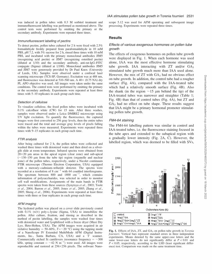
The effects of exogenous hormones on pollen tube growthwere displayed in Fig. 1. When each hormone was usedalone, IAA was the most effective hormone stimulatingtube growth. IAA interacting with ZT and/or GA3
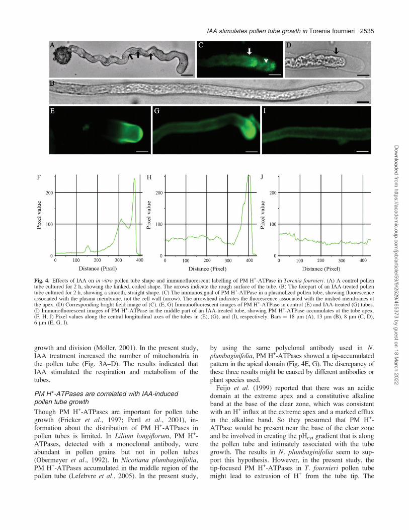
stimulated tube growth much more than IAA used alone.However, the mix of ZT with GA3 had no obvious effecton tube growth. In addition, the control tube had a roughersurface (Fig. 4A), compared with the IAA-treated tubewhich had a relatively smooth surface (Fig. 4B). Alsothe shank (in the region ;15 lm behind the tip) of theIAA-treated tubes was narrower and straighter (Table 1;Fig. 4B) than that of control tubes (Fig. 4A), but ZT andGA3 had no effect on tube shape. These results suggestthat IAA might be a primary hormonal promoter stimulat-ing pollen tube growth.
FM4-64 staining
The FM4-64 labelling pattern was similar in control andIAA-treated tubes, i.e. the fluorescence staining focused inthe tube apex and extended to the subapical region witha gradually lower intensity (Fig. 2A, C). However, thelabelled region, which was deemed to be filled with SVs,
Fig. 1. Effects of IAA, ZT, and GA3 on pollen tube growth in Toreniafournieri. Vertical bars represent standard errors in three independentexperiments. Means denoted by the same upper-case letters and thesame lower-case letters do not significantly differ at P < 0.01 andP < 0.05, respectively, according to the LSD (least significant differ-ence) test. Comparison was made on the same treatment time.
IAA stimulates pollen tube growth in Torenia fournieri 2531
Dow
nloaded from https://academ
ic.oup.com/jxb/article/59/9/2529/465373 by guest on 18 M
arch 2022

was longer and the intensity of staining was stronger inIAA-treated tubes than in control tubes (Fig. 2).
Ultrastructure of pollen tubes observed by TEM
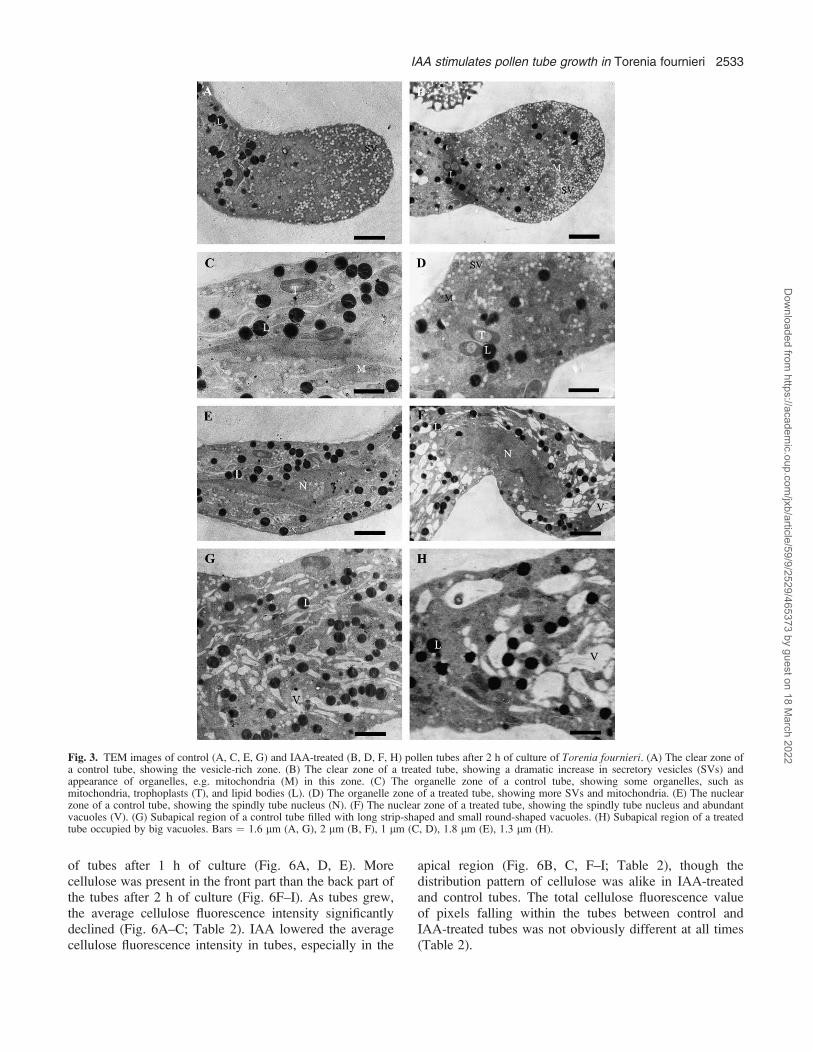
Pollen tubes have a marked polarity with four distinctzones along the tubes, i.e. clear zone, organelle zone,nuclear zone, and vacuolar zone (Cresti et al., 1985). TEMobservation showed that the clear zone was abundant inSVs (Fig. 3A, B). SVs were evenly distributed in this zoneof the control tubes, with an average density about 22/lm2
(Fig. 3A). Both the middle sections and other deflectivesections of pollen tube tip showed that more SVs (with anaverage density about 42/lm2) and organelles, especiallymitochondria, appeared in the clear zone after IAAtreatment (Fig. 3B). In the organelle zone (10–150 lmfrom tube tip), more SVs and organelles, such asmitochondria, occurred in the IAA-treated tubes than inthe control tubes (Fig. 3C, D). Also, in both the nuclear
and the organelle zone, there were bigger vacuoles inIAA-treated tubes (Fig. 3E–H).
PM H+-ATPase distribution in pollen tubes
After plasmolysis, the immunosignal of PM H+-ATPaseappeared in the plasma membrane, not in the cell wall ofthe pollen tubes (Fig. 4C, D). This enzyme was concen-trated in the apical region of control tubes (Fig. 4E, F),while it was distributed throughout the tubes and wasfocused at the apical region after IAA treatment (Fig.4G, I). IAA significantly increased the level of PM H+-ATPases in pollen tubes (Fig. 4G–J). No labelling wasobserved in the absence of primary or secondary anti-bodies (pictures not shown).
Pectin distribution in pollen tubes
In mature pollen grains, the weak labelling of esterifiedpectin and acid pectin was present in all three germinationapertures (Fig. 5A, H). When pollen grains had justgerminated (after 0.5 h of culture), both pectins weremainly localized at the extruded tube tips (Fig. 5B, I).Esterified pectin was distributed along the entire tube after1 h of culture (Fig. 5C, E) and concentrated at tube tipsafter 2 h of culture (Fig. 5D, F), while acid pectin wasdistributed throughout the whole tube at all times (Fig.5J–N). Although the distribution pattern of pectins wassimilar in IAA-treated and control tubes, there wasstronger labelling of both pectins in the former (Fig. 5C–F, J–N).
Cellulose distribution in pollen tubes
Cellulose fluorescent signals were distributed along theentire pollen tubes and increased towards the distal parts
Table 1. Influence of IAA on the diameter of in vitro pollentubes in Torenia fournieri
Values given in the table are means and standard errors of threeexperiments. The analysis of significant variance was performedbetween the control group and IAA treatment according to Student’sunpaired t-test. Comparison was made on the same treatment time.
Culture time (h) Treatment No. of pollentubes
Diameter (lm)a
1 Control 210 11.6161.83IAA 174 10.9861.43**
2 Control 94 11.5360.38IAA 82 8.0760.03**
4 Control 115 8.8360.25IAA 110 7.3660.06**
a** 0.001 < P < 0.01 indicates distinct difference.
Fig. 2. The FM4-64 staining in pollen tubes of Torenia fournieri. (A, C) FM4-64 fluorescence images of pollen tubes cultured without and with IAAtreatment for 2 h, respectively, showing the FM4-64 staining signal focused at the tube apex and extended to the subapical region with graduallylower intensity. (B, D) Pixel values along the longitudinal axes of the tubes in (A) and (C), respectively, showing the significant increase offluorescence intensity in IAA-treated tubes. Bars ¼ 15 lm (A), 12 lm (C).
2532 Wu et al.
Dow
nloaded from https://academ
ic.oup.com/jxb/article/59/9/2529/465373 by guest on 18 M
arch 2022
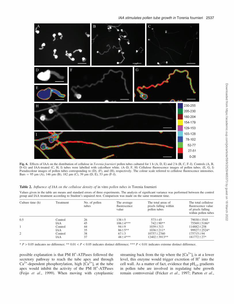
of tubes after 1 h of culture (Fig. 6A, D, E). Morecellulose was present in the front part than the back part ofthe tubes after 2 h of culture (Fig. 6F–I). As tubes grew,the average cellulose fluorescence intensity significantlydeclined (Fig. 6A–C; Table 2). IAA lowered the averagecellulose fluorescence intensity in tubes, especially in the
apical region (Fig. 6B, C, F–I; Table 2), though thedistribution pattern of cellulose was alike in IAA-treatedand control tubes. The total cellulose fluorescence valueof pixels falling within the tubes between control andIAA-treated tubes was not obviously different at all times(Table 2).
Fig. 3. TEM images of control (A, C, E, G) and IAA-treated (B, D, F, H) pollen tubes after 2 h of culture of Torenia fournieri. (A) The clear zone ofa control tube, showing the vesicle-rich zone. (B) The clear zone of a treated tube, showing a dramatic increase in secretory vesicles (SVs) andappearance of organelles, e.g. mitochondria (M) in this zone. (C) The organelle zone of a control tube, showing some organelles, such asmitochondria, trophoplasts (T), and lipid bodies (L). (D) The organelle zone of a treated tube, showing more SVs and mitochondria. (E) The nuclearzone of a control tube, showing the spindly tube nucleus (N). (F) The nuclear zone of a treated tube, showing the spindly tube nucleus and abundantvacuoles (V). (G) Subapical region of a control tube filled with long strip-shaped and small round-shaped vacuoles. (H) Subapical region of a treatedtube occupied by big vacuoles. Bars ¼ 1.6 lm (A, G), 2 lm (B, F), 1 lm (C, D), 1.8 lm (E), 1.3 lm (H).
IAA stimulates pollen tube growth in Torenia fournieri 2533
Dow
nloaded from https://academ
ic.oup.com/jxb/article/59/9/2529/465373 by guest on 18 M
arch 2022

Changes in chemical components of the cell wall ofpollen tubes
FTIR spectra were analysed in apical and subapical regionsof pollen tubes, which showed similar patterns in these tworegions. The absorption bands which occurred at 1626 and1525 cm�1 corresponded to amide I and amide II ofproteins, while the peaks at 1734 cm�1 were associatedwith esterified pectin and polysaccharides absorbed at1200–900 cm�1 (Fig. 7A, B). A single principal compo-nent score that accounted for over 60.7% in the apicalregion (Fig. 7C) and 81.6% in the subapical region (Fig.7D) of the total spectral variation could separate IAA-treated tubes from control tubes. IAA induced displace-ments of the peaks and changes in absorbance (Fig. 7A,B). The difference spectra generated by digital subtractionof control spectra from IAA-treated spectra showed thatprotein (peaks at 1653 and 1645 cm�1, corresponding toamide I of proteins; 1552 and 1533 cm�1 correspondingto amide II of proteins) and pectin (peaks at 1745 cm�1,corresponding to carboxylic ester in esterified pectin; 1263cm�1, corresponding to esterified pectin; 1105 cm�1,corresponding to the backbone vibrations of polygalactur-onic acid; 1024 cm�1, corresponding to C-O stretchingvibrations in pectin; 967 cm�1, corresponding to pectate;954 cm�1, C-O bending vibration in pectin; 930 cm�1,corresponding to pectin O-acetyl band) increased whilecellulose (troughs at 1363, 1132, 1093, 1055, and 1024cm�1) decreased in tubes after IAA treatment (Fig. 7E, F).
Ultrastructure of pollen tubes observed by AFM
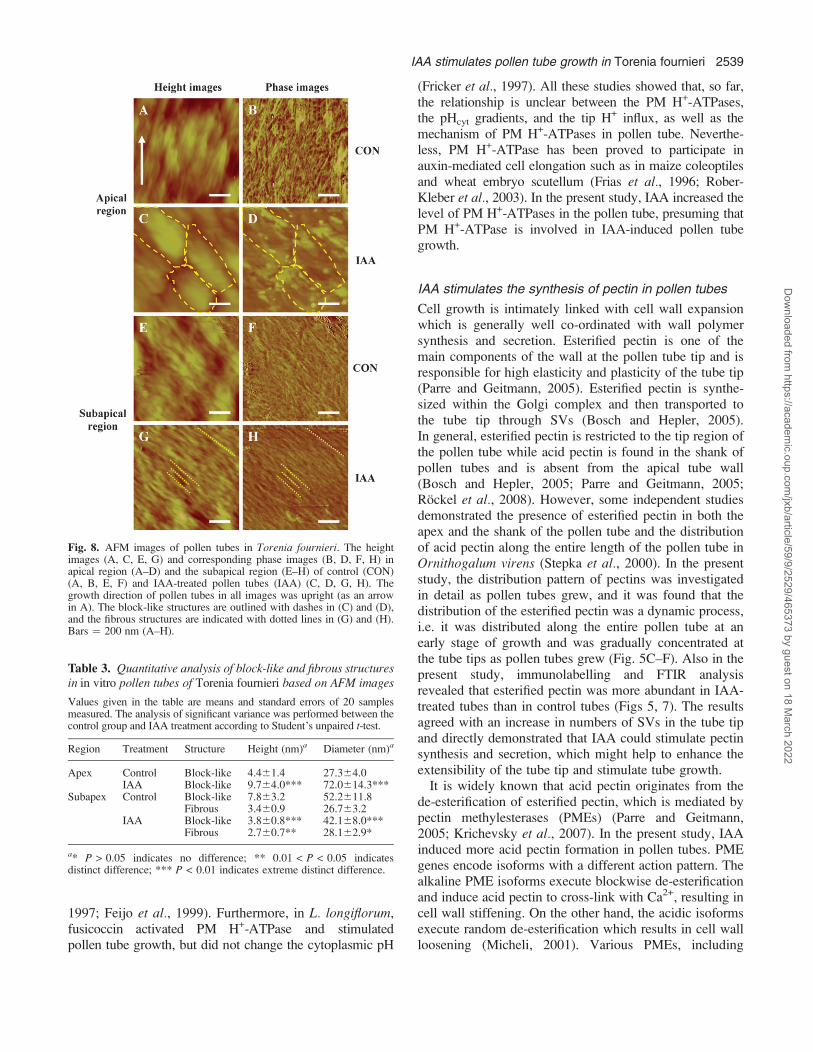
In AFM imaging, the AFM tip was positioned at theapical and sub-apical regions of the pollen tubes with theaid of a Nikon optical microscope and the scanned areaswere gradually reduced (20, 5, 3, 2, and 1 lm2). Also, thesame areas of the control tubes and IAA-treated tubeswere compared. To do this, the regions of the pollen tubesin focus were placed in the same direction as theirlongitudinal axes (Fig. 8). The images of the ‘intact’ tubewalls obtained with the same probe showed that the tubesurfaces were covered with block-like structures and/or fibrous structures (Fig. 8). The height and diameterof each structure, as well as the angle between thefibrous structure and the longitudinal axis, were measured(Table 3). The fibrous structures can be considered asCMFs according to their shapes and sizes, while theblock-like structures were probably the outer tube wallmatrix which was mainly composed of pectins. Thearrangement of CMFs in the pollen tubes of T. fournieriwas discerned in the phase images. The apical region ofthe control tube surface was rich in disorganized CMFsand block-like structures (Fig. 8A, B), while the IAA-treated tube surface was covered with both block-likestructures approximately parallel to each other and a fewCMFs (Fig. 8C, D). CMFs formed sinuous networks in
the subapical region of control tube surface (Fig. 8E, F).However, numerous CMFs in the subapical region ofIAA-treated tubes were parallel to each other and showeda predominant orientation at approximately 46.163.4�(n¼12) to the longitudinal axes of the tubes (Fig. 8G, H).For IAA-treated tubes, the density of CMFs in the apicalregion was lower (Fig. 8D) than that in the subapicalregion (Fig. 8H).
Discussion
IAA stimulates pollen tube growthand changes tube shape
IAA promotes cell elongation in various plants, and theprevailing model of auxin for stimulating cell elongationis the ‘acid-growth theory’ which postulates that auxinincreases the activity or amount of PM H+-ATPase whichtriggers excretion of H+ into the cell wall, and thisacidification loosens the wall, allowing the cell to expand(Rayle and Cleland, 1992). Though some studies revealedthat IAA affected pollen tube growth (Addicott, 1943;Raghavan and Baruah, 1956, 1959; Chauhan and Katiyar,1998), the knowledge of its mechanism is still verylimited. In the present study, IAA was the most effectivehormone prompting tube growth (Fig. 1). Moreover, IAAled to a longer, straighter, and slender tube shapecompared with the short, kinked control tubes (Fig. 4A,B). It seems reasonable to assume that IAA is one of themost important hormones regulating pollen tube growth.
IAA leads to an increase in the number of SVsand mitochondria in pollen tubes
The elongation of the pollen tube is determined by thecontinuous transportation and fusion of SVs in the tipregion, which provide abundant new membrane and cellwall precursors for the growing pollen tube (Hepler et al.,2001; Parton et al., 2001). The fluorescent dye FM4-64 isan important tool in studying SVs and has been widelyused as an authentic marker of vesicle accumulation inpollen tubes (Parton et al., 2001). The experiment withFM4-64 in T. fournieri pollen tubes revealed a longerlabelled region and a stronger labelling intensity in IAA-treated tubes (Fig. 2), indicating that IAA increased thenumber of SVs in pollen tubes. This result was consistentwith the observation that IAA treatment caused a sub-stantial increase of SVs in pollen tubes in TEM images(Fig. 3A–D). It was also found that IAA enhanced thecontent of esterified pectin (Fig. 5) which was secretedinto the tube cell wall by exocytosis. So it can be con-cluded that the quicker growth rate induced by IAA waslargely caused by the promotion of the SVs and thestimulation of exocytosis in pollen tubes. Mitochondriaare power centres of cells and provide ATP for cell
2534 Wu et al.
Dow
nloaded from https://academ
ic.oup.com/jxb/article/59/9/2529/465373 by guest on 18 M
arch 2022
growth and division (Moller, 2001). In the present study,IAA treatment increased the number of mitochondria inthe pollen tube (Fig. 3A–D). The results indicated thatIAA stimulated the respiration and metabolism of thetubes.
PM H+-ATPases are correlated with IAA-inducedpollen tube growth
Though PM H+-ATPases are important for pollen tubegrowth (Fricker et al., 1997; Pertl et al., 2001), in-formation about the distribution of PM H+-ATPases inpollen tubes is limited. In Lilium longiflorum, PM H+-ATPases, detected with a monoclonal antibody, wereabundant in pollen grains but not in pollen tubes(Obermeyer et al., 1992). In Nicotiana plumbaginifolia,PM H+-ATPases accumulated in the middle region of thepollen tube (Lefebvre et al., 2005). In the present study,
by using the same polyclonal antibody used in N.plumbaginifolia, PM H+-ATPases showed a tip-accumulatedpattern in the apical domain (Fig. 4E, G). The discrepancy ofthese three results might be caused by different antibodies orplant species used.Feijo et al. (1999) reported that there was an acidic
domain at the extreme apex and a constitutive alkalineband at the base of the clear zone, which was consistentwith an H+ influx at the extreme apex and a marked effluxin the alkaline band. So they presumed that PM H+-ATPase would be present near the base of the clear zoneand be involved in creating the pHcyt gradient that is alongthe pollen tube and intimately associated with the tubegrowth. The results in N. plumbaginifolia seem to sup-port this hypothesis. However, in the present study, thetip-focused PM H+-ATPases in T. fournieri pollen tubemight lead to extrusion of H+ from the tube tip. The
Fig. 4. Effects of IAA on in vitro pollen tube shape and immunofluorescent labelling of PM H+-ATPase in Torenia fournieri. (A) A control pollentube cultured for 2 h, showing the kinked, coiled shape. The arrows indicate the rough surface of the tube. (B) The forepart of an IAA-treated pollentube cultured for 2 h, showing a smooth, straight shape. (C) The immunosignal of PM H+-ATPase in a plasmolized pollen tube, showing fluorescenceassociated with the plasma membrane, not the cell wall (arrow). The arrowhead indicates the fluorescence associated with the unshed membranes atthe apex. (D) Corresponding bright field image of (C). (E, G) Immunofluorescent images of PM H+-ATPase in control (E) and IAA-treated (G) tubes.(I) Immunofluorescent images of PM H+-ATPase in the middle part of an IAA-treated tube, showing PM H+-ATPase accumulates at the tube apex.(F, H, J) Pixel values along the central longitudinal axes of the tubes in (E), (G), and (I), respectively. Bars ¼ 18 lm (A), 13 lm (B), 8 lm (C, D),6 lm (E, G, I).
IAA stimulates pollen tube growth in Torenia fournieri 2535
Dow
nloaded from https://academ
ic.oup.com/jxb/article/59/9/2529/465373 by guest on 18 M
arch 2022
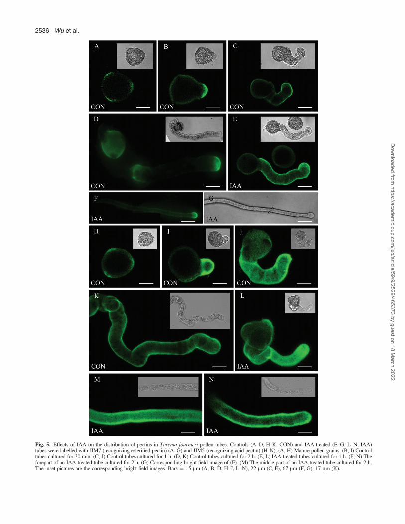
Fig. 5. Effects of IAA on the distribution of pectins in Torenia fournieri pollen tubes. Controls (A–D, H–K, CON) and IAA-treated (E–G, L–N, IAA)tubes were labelled with JIM7 (recognizing esterified pectin) (A–G) and JIM5 (recognizing acid pectin) (H–N). (A, H) Mature pollen grains. (B, I) Controltubes cultured for 30 min. (C, J) Control tubes cultured for 1 h. (D, K) Control tubes cultured for 2 h. (E, L) IAA-treated tubes cultured for 1 h. (F, N) Theforepart of an IAA-treated tube cultured for 2 h. (G) Corresponding bright field image of (F). (M) The middle part of an IAA-treated tube cultured for 2 h.The inset pictures are the corresponding bright field images. Bars ¼ 15 lm (A, B, D, H–J, L–N), 22 lm (C, E), 67 lm (F, G), 17 lm (K).
2536 Wu et al.
Dow
nloaded from https://academ
ic.oup.com/jxb/article/59/9/2529/465373 by guest on 18 M
arch 2022
possible explanation is that PM H+-ATPases followed thesecretory pathway to reach the tube apex and throughCa2+-dependent phosphorylation, high [Ca2+]i at the tubeapex would inhibit the activity of the PM H+-ATPases(Feijo et al., 1999). When moving with cytoplasmic
streaming back from the tip where the [Ca2+]i is at a lowerlevel, this enzyme would trigger excretion of H+ into thecell wall. As a matter of fact, evidence that pHcyt gradientsin pollen tube are involved in regulating tube growthremain controversial (Fricker et al., 1997; Parton et al.,
Fig. 6. Effects of IAA on the distribution of cellulose in Torenia fournieri pollen tubes cultured for 1 h (A, D, E) and 2 h (B, C, F–I). Controls (A, B,D–G) and IAA-treated (C, H, I) tubes were labelled with calcofluor white. (A–D, F, H) Cellulose fluorescence images of pollen tubes. (E, G, I)Pseudocolour images of pollen tubes corresponding to (D), (F), and (H), respectively. The colour scale referred to cellulose fluorescence intensities.Bars ¼ 95 lm (A), 146 lm (B), 182 lm (C), 39 lm (D, E), 53 lm (F–I).
Table 2. Influence of IAA on the cellulose density of in vitro pollen tubes in Torenia fournieri
Values given in the table are means and standard errors of three experiments. The analysis of significant variance was performed between the controlgroup and IAA treatment according to Student’s unpaired ttest. Comparison was made on the same treatment time.
Culture time (h) Treatment No. of pollentubes
The averagefluorescencevalue
The total areas ofpixels falling withinpollen tubes
The total cellulosefluorescence valueof pixels fallingwithin pollen tubes
0.5 Control 26 13865 573645 7903063545IAA 45 10664*** 742690** 7556965186*
1 Control 44 9469 10396313 1148826258IAA 35 8665** 10366211* 9992762524*
2 Control 38 6763 1075762760 175715694IAA 37 4864*** 1240263913** 181772627*
* P > 0.05 indicates no difference; ** 0.01 < P < 0.05 indicates distinct difference; *** P < 0.01 indicates extreme distinct difference.
IAA stimulates pollen tube growth in Torenia fournieri 2537
Dow
nloaded from https://academ
ic.oup.com/jxb/article/59/9/2529/465373 by guest on 18 M
arch 2022
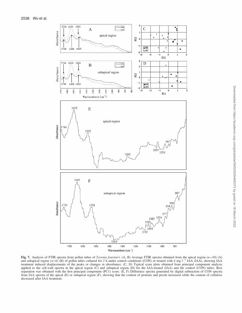
Fig. 7. Analysis of FTIR spectra from pollen tubes of Torenia fournieri. (A, B) Average FTIR spectra obtained from the apical region (n¼10) (A)and subapical region (n¼6) (B) of pollen tubes cultured for 2 h under control conditions (CON) or treated with 4 mg l�1 IAA (IAA), showing IAAtreatment induced displacements of the peaks or changes in absorbance. (C, D) Typical score plots obtained from principal component analysisapplied to the cell-wall spectra in the apical region (C) and subapical region (D) for the IAA-treated (IAA) and the control (CON) tubes. Bestseparation was obtained with the first principal component (PC1) score. (E, F) Difference spectra generated by digital subtraction of CON spectrafrom IAA spectra of the apical (E) or subapical region (F), showing that the content of proteins and pectin increased while the content of cellulosedecreased after IAA treatment.
2538 Wu et al.
Dow
nloaded from https://academ
ic.oup.com/jxb/article/59/9/2529/465373 by guest on 18 M
arch 2022
1997; Feijo et al., 1999). Furthermore, in L. longiflorum,fusicoccin activated PM H+-ATPase and stimulatedpollen tube growth, but did not change the cytoplasmic pH
(Fricker et al., 1997). All these studies showed that, so far,the relationship is unclear between the PM H+-ATPases,the pHcyt gradients, and the tip H+ influx, as well as themechanism of PM H+-ATPases in pollen tube. Neverthe-less, PM H+-ATPase has been proved to participate inauxin-mediated cell elongation such as in maize coleoptilesand wheat embryo scutellum (Frias et al., 1996; Rober-Kleber et al., 2003). In the present study, IAA increased thelevel of PM H+-ATPases in the pollen tube, presuming thatPM H+-ATPase is involved in IAA-induced pollen tubegrowth.
IAA stimulates the synthesis of pectin in pollen tubes
Cell growth is intimately linked with cell wall expansionwhich is generally well co-ordinated with wall polymersynthesis and secretion. Esterified pectin is one of themain components of the wall at the pollen tube tip and isresponsible for high elasticity and plasticity of the tube tip(Parre and Geitmann, 2005). Esterified pectin is synthe-sized within the Golgi complex and then transported tothe tube tip through SVs (Bosch and Hepler, 2005).In general, esterified pectin is restricted to the tip region ofthe pollen tube while acid pectin is found in the shank ofpollen tubes and is absent from the apical tube wall(Bosch and Hepler, 2005; Parre and Geitmann, 2005;Rockel et al., 2008). However, some independent studiesdemonstrated the presence of esterified pectin in both theapex and the shank of the pollen tube and the distributionof acid pectin along the entire length of the pollen tube inOrnithogalum virens (Stepka et al., 2000). In the presentstudy, the distribution pattern of pectins was investigatedin detail as pollen tubes grew, and it was found that thedistribution of the esterified pectin was a dynamic process,i.e. it was distributed along the entire pollen tube at anearly stage of growth and was gradually concentrated atthe tube tips as pollen tubes grew (Fig. 5C–F). Also in thepresent study, immunolabelling and FTIR analysisrevealed that esterified pectin was more abundant in IAA-treated tubes than in control tubes (Figs 5, 7). The resultsagreed with an increase in numbers of SVs in the tube tipand directly demonstrated that IAA could stimulate pectinsynthesis and secretion, which might help to enhance theextensibility of the tube tip and stimulate tube growth.It is widely known that acid pectin originates from the
de-esterification of esterified pectin, which is mediated bypectin methylesterases (PMEs) (Parre and Geitmann,2005; Krichevsky et al., 2007). In the present study, IAAinduced more acid pectin formation in pollen tubes. PMEgenes encode isoforms with a different action pattern. Thealkaline PME isoforms execute blockwise de-esterificationand induce acid pectin to cross-link with Ca2+, resulting incell wall stiffening. On the other hand, the acidic isoformsexecute random de-esterification which results in cell wallloosening (Micheli, 2001). Various PMEs, including
Fig. 8. AFM images of pollen tubes in Torenia fournieri. The heightimages (A, C, E, G) and corresponding phase images (B, D, F, H) inapical region (A–D) and the subapical region (E–H) of control (CON)(A, B, E, F) and IAA-treated pollen tubes (IAA) (C, D, G, H). Thegrowth direction of pollen tubes in all images was upright (as an arrowin A). The block-like structures are outlined with dashes in (C) and (D),and the fibrous structures are indicated with dotted lines in (G) and (H).Bars ¼ 200 nm (A–H).
Table 3. Quantitative analysis of block-like and fibrous structuresin in vitro pollen tubes of Torenia fournieri based on AFM images
Values given in the table are means and standard errors of 20 samplesmeasured. The analysis of significant variance was performed between thecontrol group and IAA treatment according to Student’s unpaired t-test.
Region Treatment Structure Height (nm)a Diameter (nm)a
Apex Control Block-like 4.461.4 27.364.0IAA Block-like 9.764.0*** 72.0614.3***
Subapex Control Block-like 7.863.2 52.2611.8Fibrous 3.460.9 26.763.2
IAA Block-like 3.860.8*** 42.168.0***Fibrous 2.760.7** 28.162.9*
a* P > 0.05 indicates no difference; ** 0.01 < P < 0.05 indicatesdistinct difference; *** P < 0.01 indicates extreme distinct difference.
IAA stimulates pollen tube growth in Torenia fournieri 2539
Dow
nloaded from https://academ
ic.oup.com/jxb/article/59/9/2529/465373 by guest on 18 M
arch 2022

acidic, neutral, and alkaline isoforms, were detected inpollen tubes (Li et al., 2002) and the inhibition of PMEactivity significantly retarded tube growth (Jiang et al.,2005). Furthermore, auxin is believed to increase PMEactivity and stimulate cell wall extension (Micheli, 2001).It was considered that the increased acid pectin in IAA-treated tubes might be attributed to the increased PMEactivity which may not only control tube growth rate butalso tube wall architecture.
CMF orientation is correlated with growth rateand growth direction of pollen tubes
The distribution pattern of cellulose in pollen tubes hasbeen investigated for years without a verdict. Forexample, CMFs are detected in regions 5–15 lm awayfrom the tube tips of tobacco (Ferguson et al., 1998),while they occur close to the tube tip of conifer (Lazzaroet al., 2003). In the present study, CW-labelling experi-ments showed that the cellulose distribution patternchanged temporally and spatially; namely, cellulose wasdistributed throughout the freshly germinated pollen tubesand was absent from the tube tip as the pollen tube grewlong enough (Fig. 6). CW labelling and FTIR analysisalso revealed that the relative cellulose density becamelower in the pollen tube, especially in the tube tip afterIAA treatment (Figs 6, 7; Table 2), which was consistentwith the AFM observation that abundant CMFs formeda network in the apical region of slow-growing controltubes (Fig. 8B); however, few CMFs were observed in theIAA-treated tube tip (Fig. 8D). The results indicateda significant correlation between cellulose and pollen tubegrowth in response to IAA. Lazzaro et al. (2003) reportedthat conifer pollen tubes with more cellulose depositing inthe tube tip grew much slower than angiosperm pollentubes. They speculated that cellulose inserted in the tubetip limited conifer pollen tube expansion. The presentobservation of accelerated tube growth induced by IAAaccompanied by lowered cellulose density in the tube tip,was consistent with this hypothesis. Therefore, it issuggested that the interlaced CMFs in the pollen tube tipof T. fournieri lead to loss of the wall’s ability to growand that cellulose is involved in IAA-induced tubegrowth.However, no significant difference was found in the
total cellulose fluorescence value of pixels falling withinthe tubes between control and IAA-treated tubes (Table2), which indicated that cellulose synthesis was notinfluenced by IAA. A similar result was reported in IAA-induced elongations of Pellia setae that the synthesis ofother wall polysaccharides, but not cellulose, was en-hanced (Thomas et al., 1984). Furthermore, inhibitor testsalso showed that the synthesis of cellulose is not requiredfor IAA-induced elongation of rice coleoptile segments(Hoson and Masuda, 1992). Thus, it was supposed that
cellulose synthesis is also not affected by IAA in pollentube growth.Deposition and rearrangement of CMFs within cell-wall
matrices is considered to be one of the most critical stepsin the regulation of cell shape and expansion direction(Roudier et al., 2005). The orientation of CMFs wasextensively investigated in diffuse growth cells, such asroot cells, and it indicated that CMF orientation wasnearly perpendicular to the main axis of cell expansion,especially in rapidly elongating cells (Sugimoto et al.,2000; Baskin, 2001). However, in the case of highlypolarized tip-growing pollen tubes, little attention waspaid to the relationship of the tube shape and growthdirection with the organization of CMFs, and knowledgeabout the orientation of CMFs is scarce. In early reports,CMFs prepared from the extracted pollen tube wallshowed a preferential angle of 45� to the longitudinal axesin the shank of petunia and lily pollen tubes, whereas theyseemed to be random at the tip of the pollen tube inpetunia (Sassen, 1964; Derksen et al., 1999). Theseobservations indicated that the mechanism by whichCMFs control growth direction may be different betweentip growth and diffuse growth to a certain extent. Theorientation of CMFs in diffuse growth can be preciselycontrolled by hormones. In growing tissues of higherplants, auxin induced a shift of CMF arrangement fromlongitudinal to transverse, which was correlated withpromotion of elongation (Shibaoka, 1991). In the presentstudy, AFM images can offer in situ information on CMForientation of the very outer surface of pollen tubes, andshowed that CMFs formed random networks in the apicaland subapical regions of control tubes (Fig. 8B, F), whileCMFs were parallel to each other and presented pre-dominant orientation about 45� to the longitudinal axis ofIAA-treated tubes in the subapical region (Fig. 8H). Theresults suggested that IAA can control the orientation ofCMFs in the tip-growing pollen tube. There was a similarphenomenon reported in tobacco mesophyll protoplaststhat CMFs changed from a ‘random’ arrangement into anordered orientation in the auxin-induced long tubularcells. It indicated that auxin could induce CMF reorienta-tion and determine the axis of cell growth (Vissenberget al., 2000). Moreover, it suggested that a parallelorientation of the newly synthesized CMFs permitted cellgrowth restricted to one direction (Green, 1980). It wasspeculated that in IAA-treated tubes the parallel CMFslead to a predominant growth direction along longitudinalaxes of the tubes with narrowed diameter, and that incontrol tubes, a sinuous CMF network results in multi-direction growth which is concomitant with a wavy shapeand a wide diameter pollen tube.However, in IAA-treated tubes it was still inexplicable
that few CMFs were observed in the apical region, whileparallel-orientated CMFs occur in the subapical region.Recently, it was reported that microtubes are generally
2540 Wu et al.
Dow
nloaded from https://academ
ic.oup.com/jxb/article/59/9/2529/465373 by guest on 18 M
arch 2022

long and axially orientated in the distal part of the pollentube and absent in the very tip of Papaver rhoeas. It wassuggested that microtubes played an important role incontrolling the direction of pollen tube growth in the distalpart of the tube, but not in the very tube tip (Gossot andGeitmann, 2007). Furthermore, it is well known thatmicrotubes control the deposition of CMFs (Giddings andStaehelin, 1991). Therefore, it was thought that CMFs inthe subapical region of the tubes are a crucial factor inregulating the tube growth rate and direction.
Tapping mode AFM is a useful technique to discernthe CMF arrangement in situ in pollen tube walls
For such samples as pollen tubes, the in situ imaging forthe region of interest is crucial because of the compositiveand configurational difference along the longitudinal axis.Tapping mode AFM is a non-destructive technique forhigh-resolution surface imaging, with the advantages ofeasy sample preparation and in situ observation (Ding andHimmel, 2006). In the present experiments, the AFM tip
was located precisely over the midpoint region of the veryapex of the tube tip (apical region) and the subapicalregions (;130 lm away from the tube tip) of the ‘intact’tube wall and tapping mode AFM was used to observe theorientation of the CMFs in situ. From the height images, itcan obviously be seen that the IAA-treated apical regionwas different from that of the control (Fig. 8A, C),indicating that IAA changed the ultrastructure of the tubesurfaces. AFM has been used to observe directly theCMFs which were reticulated by the matrix constituents ingrapevine suspension cells (Lesniewska et al., 2004). It iswell known that the difference in elasticity of samplesurfaces can be discerned in phase images of tappingmode AFM (Ding and Himmel, 2006) and that pectin hashigher elasticity than cellulose (Morris et al., 1997). Thepresent phase images showed that there were morefilaceous structures in the apical region of the control thanin the same region of IAA-treated tubes. As shown inCW-labelling experiments, there was more cellulose in theapical region of the control tube than in the same region
Fig. 9. A hypothetical model for the effects of IAA on pollen tube growth in Torenia fournieri. IAA stimulates the amount and activity ofmitochondria and the secretion and fusion of SVs in the pollen tube, which transports more cell wall materials (such as pectin) and various enzymes(such as PM H+-ATPase and PME) into the tube tip. The increase in pectin indicates the enhancement of cell wall synthesis. The increased numbersof mitochondria support more energy (ATP) for various cellular activities such as the execution of PM H+-ATPase function. The increase in PM H+-ATPase results in a change in the pH condition of the cell wall, which may lead to the modification of cell wall architecture. The increase of PMEactivity stimulates the pectin de-esterification process and consequently causes the alteration in pectin architecture. IAA also induces reorientation ofcellulose microfibrils in the tube cell wall, which results in modification of the cell wall architecture. Simultaneous cell wall synthesis enhancementand cell wall architecture modification lead to the promotion of pollen tube growth, accompanied by a narrower tube diameter and a straighter tube.
IAA stimulates pollen tube growth in Torenia fournieri 2541
Dow
nloaded from https://academ
ic.oup.com/jxb/article/59/9/2529/465373 by guest on 18 M
arch 2022

of the IAA-treated tube (Fig. 6G, I). Thus, it can bededuced that the difference in phase images of the apicalregion may result from the different contents of CMFs.Therefore, the directions of CMFs can be easily discernedin phase images. Tapping mode AFM is a powerfultechnique for examining the orientation of CMFs in situand analysing the ‘intact’ pollen tube wall.
Conclusion
In summary, the present investigations clearly confirmedthat IAA stimulated the growth of T. fournieri pollentubes by increasing the secretion of SVs and PM H+-ATPase, and modifying the tube wall composition andstructure, especially CMF orientation, which is summa-rized in Fig. 9. This study highlighted the role of the cellwall, especially CMF orientation, in pollen tube growthand direction.
Acknowledgements
The authors thank Dr JP Knox (Centre for Plant Sciences,University of Leeds, UK) for the generous gifts of the antibodiesJIM5 and JIM7, Professor Jinxing Lin (Institute of Botany, ChineseAcademy of Sciences, Key Laboratory of Photosynthesis andMolecular Environment Physiology, China) for the generous gift ofFM4-64, and Professor Marc Boutry (Unite de Biochimie Physi-ologique, Institut des Sciences de la Vie, Universite Catholique deLouvain-la-Neuve) for the generous gift of the anti-PM-H+-ATPaseantibody. The authors also thank Dr Jun’e Qu (the Analysis andTesting Center, Hubei University, China) for her collaboration inAFM experiments. This work was supported by the NationalNatural Science Foundation of China (30521004), the SpecialDoctorial Program Funds of the Ministry of Education of China(20050486015), and the National Natural Science Foundation ofChina (20621502).
References
Addicott FT. 1943. Pollen germination and pollen tube growth, asinfluenced by pure growth substances. Plant Physiology 18, 270–279.
Aloni R, Aloni E, Langhans M, Ullrich CI. 2006. Role of auxinin regulating Arabidopsis flower development. Planta 223, 315–328.
Barron C, Parker ML, Mills EN, Rouau X, Wilson RH. 2005.FTIR imaging of wheat endosperm cell walls in situ revealscompositional and architectural heterogeneity related to grainhardness. Planta 220, 667–677.
Baskin TI. 2001. On the alignment of cellulose microfibrils by corticalmicrotubules: a review and a model. Protoplasma 215, 150–171.
Bosch M, Hepler PK. 2005. Pectin methylesterases and pectindynamics in pollen tubes. The Plant Cell 17, 3219–3226.
Chauhan YS, Katiyar SR. 1998. Effects of radiation and growthhormones on pollen germination, pollen tube growth andmodulation of radiation responses of Pinus kesiya Royle ex Gord.Cytologia 63, 341–348.
Cresti M, Ciampolini F, Mulcahy DLM, Mulcahy G. 1985.Ultrastructure of Nicotiana alata pollen, its germination and earlytube formation. American Journal of Botany 72, 719–727.
Derksen J, van Amstel ANM, Rutten ALM, Knuiman B,Li YQ, Pierson ES. 1999. Pollen tubes: cellular organizationand control of growth. In: Clement C, Pacini E, Audran JC, eds.Anther and pollen. Berlin: Springer, 161–174.
Ding SY, Himmel ME. 2006. The maize primary cell wallmicrofibril: a new model derived from direct visualization.Journal of Agriculture and Food Chemistry 54, 597–606.
Feijo JA, Sainhas J, Hackett GR, Kunkel JG, Hepler PK. 1999.Growing pollen tubes possess a constitutive alkaline band in theclear zone and a growth-dependent acidic tip. Journal of CellBiology 144, 483–496.
Ferguson C, Teeri TT, Siika-aho M, Read SM, Basic A. 1998.Location of cellulose and callose in pollen tubes and grains ofNicotiana tabacum. Planta 206, 452–460.
Frias I, Caldeira MT, Perez-Castineira JR, Navarro-Avino JP,Culianez-Macia FA, Kuppinger O, Stransky H, Pages M,Hager A, Serrano R. 1996. A major isoform of the maizeplasma membrane H (+)-ATPase: characterization and inductionby auxin in coleoptiles. The Plant Cell 8, 1533–1544.
Fricker MD, White NS, Obermeyer G. 1997. pH gradients arenot associated with tip growth in pollen tubes of Liliumlongiflorum. Journal of Cell Science 110, 1729–1740.
Geitmann A, Parre E. 2004. The local cytomechanical proper-ties of growing pollen tubes correspond to the axial distributionof structural cellular elements. Sexual Plant Reproduction 17,9–16.
Geitmann A, Steer M. 2006. The architecture and properties of thepollen tube cell wall. In: Malho R, ed. The pollen tube. Plant CellMonographs, Vol. 3. Berlin: Springer Verlag, 177–200.
Giddings TH, Staehelin LA. 1991. Microtubule-mediated controlof microfibril deposition: a re-examination of the hypothesis. In:Lloyd CW, ed. The cytoskeletal basis of plant growth and form.San Diego, CA: Academic Press, 85–100.
Gossot O, Geitmann A. 2007. Pollen tube growth: coping withmechanical obstacles involves the cytoskeleton. Planta 226,405–416.
Green PB. 1980. Organogenesis – a biophysical view. AnnualReview of Plant Physiology 31, 51–82.
Hepler PK, Vidali L, Cheung AY. 2001. Polarized cell growth inhigher plants. Annual Review of Cell and Developmental Biology17, 159–187.
Higashiyama T, Haruko Kuroiwa H, Kawano S, Kuroiwa T.1998. Guidance in vitro of the pollen tube to the naked embryosac of Torenia fournieri. The Plant Cell 10, 2019–2031.
Hoson T, Masuda Y. 1992. Relationship between polysaccharidesynthesis and cell wall loosening in auxin-induced elongation ofrice coleoptile segments. Plant Science 83, 149–154.
Jiang LX, Yang SL, Xie LF, Puah CS, Zhang XQ, Yang WC,Sundaresan V, Ye D. 2005. VANGUARD1 encodes a pectinmethylesterase that enhances pollen tube growth in the Arabidop-sis style and transmitting tract. The Plant Cell 17, 584–596.
Jones L, Milne JL, Ashford D, McCann MC, McQueen-Mason SJ. 2005. A conserved functional role of pectic polymersin stomatal guard cells from a range of plant species. Planta 221,255–264.
Kovaleva L, Zakharova E. 2003. Hormonal status of the pollen-pistil system at the progamic phase of fertilization aftercompatible and incompatible pollination in Petunia hybrida L.Sexual Plant Reproduction 16, 191–196.
Krichevsky A, Kozlovsky SV, Tian GW, Chen MH, Zaltsman A,Vitaly Citovsky V. 2007. How pollen tubes grow. DevelopmentalBiology 303, 405–420.
Lazzaro MD, Donohue JM, Soodavar FM. 2003. Disruption ofcellulose synthesis by isoxaben causes tip swelling and disorga-
2542 Wu et al.
Dow
nloaded from https://academ
ic.oup.com/jxb/article/59/9/2529/465373 by guest on 18 M
arch 2022

nizes cortical microtubules in elongating conifer pollen tubes.Protoplasma 220, 201–207.
Lefebvre B, Arango M, Oufattole M, Crouzet J, Purnelle B,Boutry M. 2005. Identification of a Nicotiana plumbaginifoliaplasma membrane H (+)-ATPase gene expressed in the pollentube. Plant Molecular Biology 58, 775–787.
Lesniewska E, Adrian M, Klinguer A, Pugin A. 2004. Cell wallmodification in grapevine cells in response to UV stressinvestigated by atomic force microscopy. Ultramicroscopy 100,171–178.
Li YQ, Mareck A, Faleri C, Moscatelli A, Liu Q, Cresti M.2002. Detection and localization of pectin methylesterase iso-forms in pollen tubes of Nicotiana tabacum L. Planta 214,734–740.
Maudoux O, Batoko H, Oecking C, Gevaert K,Vandekerckhove J, Boutry M, Morsomme P. 2000. A plantplasma membrane H+-ATPase expressed in yeast is activated byphosphorylation at its penultimate residue and binding of 14-3-3regulatory proteins in the absence of fusicoccin. Journal ofBiological Chemistry 275, 17762–17770.
Micheli F. 2001. Pectin methylesterases: cell wall enzymes withimportant roles in plant physiology. Trends in Plant Science 6,414–419.
Mol R, Filek M, Machackova I, Matthys-Rochon E. 2004.Ethylene synthesis and auxin augmentation in pistil tissues areimportant for egg cell differentiation after pollination in maize.Plant and Cell Physiology 45, 1396–1405.
Moller IM. 2001. Plant mitochondria and oxidative stress: electrontransport, NADPH turnover, and metabolism of reactive oxygenspecies. Annual Review of Plant Physiology & Plant MolecularBiology 52, 561–591.
Morris VJ, Gunning AP, Kirby AR, Round A, Waldron K,Ng A. 1997. Atomic force microscopy of plant cell walls, plantcell wall polysaccharides and gels. International Journal ofBiological Macromolecules 21, 61–66.
Nemhauser JL, Feldman LJ, Zambryski PC. 2000. Auxin andETTIN in Arabidopsis gynoecium morphogenesis. Development127, 3877–3888.
Obermeyer G, Lutzelschwab M, Heumann HG,Weisenseel MH. 1992. Immunolocalization of H+-ATPases inthe plasma membrane of pollen grains and pollen tubes of Liliumlongiflorum. Protoplasma 171, 55–63.
Parre E, Geitmann A. 2005. Pectin and the role of the physicalproperties of the cell wall in pollen tube growth of Solanumchacoense. Planta 220, 582–592.
Parton RM, Fischer S, Malho R, Papasouliotis O, Jelitto TC,Leonard T, Read ND. 1997. Pronounced cytoplasmic pHgradients are not required for tip growth in plant and fungal cells.Journal of Cell Science 110, 1187–1198.
Parton RM, Fischer-Parton S, Watahiki MK, Trewavas AJ.2001. Dynamics of the apical vesicle accumulation and the rate ofgrowth are related in individual pollen tubes. Journal of CellScience 114, 2685–2695.
Pertl H, Himly M, Gehwolf R, Kriechbaumer R, Strasser D,Michalke W, Richter K, Ferreira F, Obermeyer G. 2001.Molecular and physiological characterisation of a 14-3-3 proteinfrom lily pollen grains regulating the activity of the plasmamembrane H+-ATPase during pollen grain germination and tubegrowth. Planta 213, 132–141.
Raghavan V, Baruah HK. 1956. On factors influencing fruit-setand sterility in arecanut (Areca catechu Linn.). II. Germination ofpollen grains and growth of pollen tube under the influence ofcertain auxin, vitamins and trace elements. Phyton 7, 77–82.
Raghavan V, Baruah HK. 1959. Effect of time factor on thestimulation of pollen germination and pollen tube growth by
certain auxins, vitamins, and trace elements. PhysiologiaPlantarum 12, 441–451.
Rayle DL, Cleland RE. 1992. The acid growth theory of auxin-induced cell elongation is alive and well. Plant Physiology 99,1271–1274.
Rober-Kleber N, Albrechtova JT, Fleig S, Huck N,Michalke W, Wagner E, Speth V, Neuhaus G, Fischer-Iglesias C. 2003. Plasma membrane H+-ATPase is involved inauxin-mediated cell elongation during wheat embryo develop-ment. Plant Physiology 131, 1302–1312.
Rockel N, Wolf S, Kost B, Rausch T, Greiner S. 2008. Elaboratespatial patterning of cell-wall PME and PMEI at the pollen tube tipinvolves PMEI endocytosis, and reflects the distribution ofesterified and de-esterified pectins. The Plant Journal 53, 133–143.
Roudier F, Fernandez AG, Fujita M, Himmelspach R,Borner GHH, Schindelman G, Song S, Baskin TI, Dupree P,Wasteneys GO, Benfey PN. 2005. COBRA, an Arabidopsisextracellular glycosyl-phosphatidyl inositol-anchored protein,specifically controls highly anisotropic expansion through itsinvolvement in cellulose microfibril orientation. The Plant Cell17, 1749–1763.
Sassen MMA. 1964. Fine structure of Petunia pollen grain andpollen tube. Acta Botanica Neerlandica 13, 175–181.
Sheng X, Hu Z, Lu H, Wang X, Baluska F, Samaj J, Lin J.2006. Roles of the ubiquitin/proteasome pathway in pollen tubegrowth with emphasis on MG132-induced alterations in ultra-structure, cytoskeleton, and cell wall components. Plant Physiol-ogy 141, 1578–1590.
Shibaoka H. 1991. Microtubules and the regulation of cellmorphogenesis by plant hormones. In: Lloyd CW, ed. Thecytoskeletal basis of plant growth and form. London: AcademicPress, 159–168.
Stepka M, Ciampolimi F, Cresti M. 2000. Localization of pectinsin the pollen tube wall of Ornithogalum virens: does the patternof pectin distribution depend on the growth rate of the pollentube? Planta 210, 630–635.
Sugimoto K, Williamson RE, Wasteneys GO. 2000. Newtechniques enable comparative analysis of microtubule orienta-tion, wall texture and growth rate in intact roots of Arabidopsis.Plant Physiology 124, 1493–1506.
Synytsya A, Copikova J, Matejka P, Machovic V. 2003. Fouriertransform raman and infrared spectroscopy of pectins. Carbohy-drate Polymer 54, 97–106.
Thomas RJ, Behringer FJ, Lombard CS, Sparkowski JJ. 1984.Effects of auxin on wall polysaccharide composition and enzymeactivity during extension-growth of Pellia (Bryophyta). Physiolo-gia Plantarum 60, 502–506.
Toole GA, Kakurakova M, Smith AC, Waldron KW,Wilson RH. 2004. FT-IR study of the Chara corallina cell wallunder deformation. Carbohydrate Research 339, 629–635.
Vissenberg K, Quelo AH, Van Gestel K, Olyslaegers G,Verbelen JP. 2000. From hormone signal, via the cytoskeleton,to cell growth in single cells of tobacco. Cell Biology In-ternational 24, 343–349.
Wu JZ, Qin Y, Zhao J. 2008. Pollen tube growth is affected byexogenous hormones and correlated with hormone changes instyles before and after pollination in Torenia fournieri L. PlantGrowth Regulation DOI: 10.1007/s10725-008-9268-5.
Zhang LY, Fang KF, Lin JX. 2005. Heterotrimeric G proteina-subunit is localized in the plasma membrane of Pinus bungeanapollen tubes. Plant Science 169, 1066–1073.
Zhao J, Yu FL, Liang SP, Zhou C, Yang HY. 2002. Changes ofcalcium distribution in egg cells, zygotes and two-celledproembryos of rice (Oryza sativa L.). Sexual Plant Reproduction14, 331–337.
IAA stimulates pollen tube growth in Torenia fournieri 2543
Dow
nloaded from https://academ
ic.oup.com/jxb/article/59/9/2529/465373 by guest on 18 M
arch 2022