Muscle Contractility and Cell Motility - Hindawi.com
220
Muscle Contractility and Cell Motility Guest Editors: J.-P. Jin, Robert J. Bloch, Xupei Huang, and Lars Larsson Journal of Biomedicine and Biotechnology
-
Upload
khangminh22 -
Category
Documents
-
view
3 -
download
0
Transcript of Muscle Contractility and Cell Motility - Hindawi.com

Muscle Contractility and Cell Motility
Guest Editors: J.-P. Jin, Robert J. Bloch, Xupei Huang, and Lars Larsson
Journal of Biomedicine and Biotechnology

Muscle Contractility and Cell Motility

Journal of Biomedicine and Biotechnology
Muscle Contractility and Cell Motility
Guest Editors: J.-P. Jin, Robert J. Bloch, Xupei Huang,and Lars Larsson

Copyright © 2012 Hindawi Publishing Corporation. All rights reserved.
This is a special issue published in “Journal of Biomedicine and Biotechnology.” All articles are open access articles distributed underthe Creative Commons Attribution License, which permits unrestricted use, distribution, and reproduction in any medium, providedthe original work is properly cited.

Editorial BoardThe editorial board of the journal is organized into sections that correspond to
the subject areas covered by the journal.
Agricultural Biotechnology
Guihua H. Bai, USAChristopher P. Chanway, CanadaRavindra N. Chibbar, CanadaIan Godwin, Australia
Hari B. Krishnan, USACarol A. Mallory-Smith, USADennis P. Murr, CanadaRodomiro Ortiz, Sweden
B. C. Saha, USAMariam B. Sticklen, USAChiu-Chung Young, Taiwan
Animal Biotechnology
E. S. Chang, USABhanu P. Chowdhary, USANoelle E. Cockett, USAPeter Dovc, SloveniaScott C. Fahrenkrug, USADorian J. Garrick, USAThomas A. Hoagland, USA
Tosso Leeb, SwitzerlandJames D. Murray, USAAnita M. Oberbauer, USAJorge A. Piedrahita, USADaniel Pomp, USAKent M. Reed, USALawrence Reynolds, USA
Lawrence B. Schook, USAMari A. Smits, The NetherlandsLeon Spicer, USAJ. Verstegen, USAMatthew B. Wheeler, USAKenneth L. White, USA
Biochemistry
Robert Blumenthal, USADavid Ronald Brown, UKSaulius Butenas, USAVittorio Calabrese, ItalyMiguel Castanho, PortugalFrancis J. Castellino, USARoberta Chiaraluce, ItalyD. M. Clarke, CanadaFrancesca Cutruzzola, Italy
Paul W. Doetsch, USAHicham Fenniri, CanadaNick V. Grishin, USAJ. Guy Guillemette, CanadaPaul W. Huber, USAChen-Hsiung Hung, TaiwanMichael Kalafatis, USAB. E. Kemp, AustraliaPhillip E. Klebba, USA
Wen-Hwa Lee, USARichard D. Ludescher, USAGeorge Makhatadze, USALeonid Medved, USASusan A. Rotenberg, USAJason Shearer, USAAndrei Surguchov, USAJohn B. Vincent, USAY. George Zheng, USA
Bioinformatics
T. Akutsu, JapanMiguel A. Andrade, GermanyMark Y. Borodovsky, USARita Casadio, ItalyArtem Cherkasov, CanadaDavid Corne, UKSorin Draghici, USA
Stavros J. Hamodrakas, GreecePaul Harrison, USAGeorge Karypis, USAJack A. Leunissen, The NetherlandsGuohui Lin, CanadaSatoru Miyano, JapanZoran Obradovic, USA
Florencio Pazos, SpainZhirong Sun, ChinaYing Xu, USAAlexander Zelikovsky, USAAlbert Zomaya, Australia

Biophysics
Miguel Castanho, PortugalP. Bryant Chase, USAKuo-Chen Chou, USARizwan Khan, India
Ali A. Khraibi, Saudi ArabiaRumiana Koynova, USASerdar Kuyucak, AustraliaJianjie Ma, USA
S. B. Petersen, DenmarkPeter Schuck, USAClaudio M. Soares, Portugal
Cell Biology
Omar Benzakour, FranceSanford I. Bernstein, USAPhillip I. Bird, AustraliaEric Bouhassira, USAMohamed Boutjdir, USAChung-Liang Chien, TaiwanRichard Gomer, USAPaul J. Higgins, USAPavel Hozak, Czech Republic
Xudong Huang, USAAnton M. Jetten, USASeamus J. Martin, IrelandManuela Martins-Green, USAShoichiro Ono, USAGeorge Perry, USAM. Piacentini, ItalyGeorge E. Plopper, USALawrence Rothblum, USA
Michael Sheetz, USAJames L. Sherley, USAG. S. Stein, USARichard Tucker, USAThomas van Groen, USAAndre Van Wijnen, USASteve Winder, UKChuanyue Wu, USABin-Xian Zhang, USA
Genetics
Adewale Adeyinka, USAClaude Bagnis, FranceJ. Birchler, USASusan Blanton, USABarry J. Byrne, USAR. Chakraborty, USADomenico Coviello, ItalySarah H. Elsea, USACelina Janion, Poland
J. Spencer Johnston, USAM. Ilyas Kamboh, USAFeige Kaplan, CanadaManfred Kayser, The NetherlandsBrynn Levy, USAXiao Jiang Li, USAThomas Liehr, GermanyJames M. Mason, USAMohammed Rachidi, France
Raj S. Ramesar, South AfricaElliot D. Rosen, USADharambir K. Sanghera, USAMichael Schmid, GermanyMarkus Schuelke, GermanyWolfgang Arthur Schulz, GermanyJorge Sequeiros, PortugalMouldy Sioud, NorwayRongjia Zhou, China
Genomics
Vladimir Bajic, Saudi ArabiaMargit Burmeister, USASettara Chandrasekharappa, USAYataro Daigo, Japan
J. Spencer Johnston, USAVladimir Larionov, USAThomas Lufkin, SingaporeJohn L. McGregor, France
John V. Moran, USAYasushi Okazaki, JapanGopi K. Podila, USAMomiao Xiong, USA

Immunology
Hassan Alizadeh, USAPeter Bretscher, CanadaRobert E. Cone, USATerry L. Delovitch, CanadaAnthony L. DeVico, USANick Di Girolamo, AustraliaDon Mark Estes, USASoldano Ferrone, USAJeffrey A. Frelinger, USAJohn Robert Gordon, Canada
James D. Gorham, USASilvia Gregori, ItalyThomas Griffith, USAYoung S. Hahn, USADorothy E. Lewis, USABradley W. McIntyre, USAR. Lee Mosley, USAMarija Mostarica-Stojkovic, SerbiaHans Konrad Muller, AustraliaAli Ouaissi, France
Kanury V. S. Rao, IndiaYair Reisner, IsraelHarry W. Schroeder, USAWilhelm Schwaeble, UKNilabh Shastri, USAYufang Shi, ChinaPiet Stinissen, BelgiumHannes Stockinger, AustriaJ. W. Tervaert, The NetherlandsGraham R. Wallace, UK
Microbial Biotechnology
Jozef Anne, BelgiumYoav Bashan, MexicoMarco Bazzicalupo, ItalyNico Boon, Belgium
Luca Simone Cocolin, ItalyPeter Coloe, AustraliaDaniele Daffonchio, ItalyHan de Winde, The Netherlands
Yanhe Ma, ChinaBernd H. A. Rehm, New ZealandAngela Sessitsch, Austria
Microbiology
D. Beighton, UKSteven R. Blanke, USAStanley Brul, The NetherlandsIsaac K. O. Cann, USAStephen K. Farrand, USAAlain Filloux, UK
Gad Frankel, UKRoy Gross, GermanyHans-Peter Klenk, GermanyTanya Parish, UKGopi K. Podila, USAFrederick D. Quinn, USA
Didier A. Raoult, FranceIsabel Sa-Correia, PortugalP. L. C. Small, USAMichael Thomm, GermanyH. C. van der Mei, The NetherlandsSchwan William, USA
Molecular Biology
Rudi Beyaert, BelgiumMichael Bustin, USADouglas Cyr, USAK. Iatrou, GreeceLokesh Joshi, Ireland
David W. Litchfield, CanadaWuyuan Lu, USAPatrick Matthias, SwitzerlandJohn L. McGregor, FranceS. L. Mowbray, Sweden
Elena Orlova, UKYeon-Kyun Shin, USAWilliam S. Trimble, CanadaLisa Wiesmuller, GermanyMasamitsu Yamaguchi, Japan

Oncology
Colin Cooper, UKF. M. J. Debruyne, The NetherlandsNathan Ames Ellis, USADominic Fan, USAGary E. Gallick, USADaila S. Gridley, USAXin-yuan Guan, Hong KongAnne Hamburger, USAManoor Prakash Hande, SingaporeBeric Henderson, Australia
Steve B. Jiang, USADaehee Kang, Republic of KoreaAbdul R. Khokhar, USARakesh Kumar, USAMacus Tien Kuo, USAEric W. Lam, UKSue-Hwa Lin, USAKapil Mehta, USAOrhan Nalcioglu, USAP. J. Oefner, Germany
Allal Ouhtit, OmanFrank Pajonk, USAWaldemar Priebe, USAF. C. Schmitt, PortugalSonshin Takao, JapanAna Maria Tari, USAHenk G. Van Der Poel, The NetherlandsHaodong Xu, USADavid J. Yang, USA
Pharmacology
Abdel A. Abdel-Rahman, USAM. Badr, USAStelvio M. Bandiera, CanadaRonald E. Baynes, USAR. Keith Campbell, USAHak-Kim Chan, AustraliaMichael D. Coleman, UKJ. Descotes, FranceDobromir Dobrev, Germany
Ayman El-Kadi, CanadaJeffrey Hughes, USAKazim Husain, USAFarhad Kamali, UKMichael Kassiou, AustraliaJoseph J. McArdle, USAMark J. McKeage, New ZealandDaniel T. Monaghan, USAT. Narahashi, USA
Kennerly S. Patrick, USAVickram Ramkumar, USAMichael J. Spinella, USAQuadiri Timour, FranceTodd W. Vanderah, USAVal J. Watts, USADavid J. Waxman, USA
Plant Biotechnology
Prem L. Bhalla, AustraliaJ. R. Botella, AustraliaElvira G. De Mejia, USAH. M. Haggman, Finland
Liwen Jiang, Hong KongPulugurtha B. Kirti, IndiaYong Pyo Lim, Republic of KoreaGopi K. Podila, USA
Ralf Reski, GermanySudhir Sopory, India
Toxicology
Michael Aschner, USAMichael L. Cunningham, USALaurence D. Fechter, USAHartmut Jaeschke, USA
Youmin James Kang, USAM. Firoze Khan, USAPascal Kintz, France
R. S. Tjeerdema, USAKenneth Turteltaub, USABrad Upham, USA

Virology
Nafees Ahmad, USAEdouard Cantin, USAEllen Collisson, USAKevin M. Coombs, CanadaNorbert K. Herzog, USATom Hobman, CanadaShahid Jameel, India
Fred Kibenge, CanadaFenyong Liu, USAEric Rassart, CanadaGerald G. Schumann, GermanyY.-C. Sung, Republic of KoreaGregory Tannock, Australia
Ralf Wagner, GermanyJianguo Wu, ChinaDecheng Yang, CanadaJiing-Kuan Yee, USAXueping Zhou, ChinaWen-Quan Zou, USA

Contents
Muscle Contractility and Cell Motility, J.-P. Jin, Robert J. Bloch, Xupei Huang, and Lars LarssonVolume 2012, Article ID 257812, 3 pages
Various Jobs of Proteolytic Enzymes in Skeletal Muscle during Unloading: Facts and Speculations,E. V. Kachaeva and B. S. ShenkmanVolume 2012, Article ID 493618, 15 pages
Influences of Desmin and Keratin 19 on Passive Biomechanical Properties of Mouse Skeletal Muscle,Sameer B. Shah, James M. Love, Andrea O’Neill, Richard M. Lovering, and Robert J. BlochVolume 2012, Article ID 704061, 12 pages
Physiology and Pathophysiology of CLC-1: Mechanisms of a Chloride Channel Disease, Myotonia,Chih-Yung Tang and Tsung-Yu ChenVolume 2011, Article ID 685328, 10 pages
Obscurin Depletion Impairs Organization of Skeletal Muscle in Developing Zebrafish Embryos,Maide O. Raeker and Mark W. RussellVolume 2011, Article ID 479135, 15 pages
Diffusion Tensor MRI to Assess Damage in Healthy and Dystrophic Skeletal Muscle after LengtheningContractions, Alan B. McMillan, Da Shi, Stephen J. P. Pratt, and Richard M. LoveringVolume 2011, Article ID 970726, 10 pages
Tissue Expression and Actin Binding of a Novel N-Terminal Utrophin Isoform, Richard A. Zuellig,Beat C. Bornhauser, Ralf Amstutz, Bruno Constantin, and Marcus C. SchaubVolume 2011, Article ID 904547, 18 pages
A High-Throughput Solid-Phase Microplate Protein-Binding Assay to Investigate Interactions betweenMyofilament Proteins, Brandon J. Biesiadecki and J.-P. JinVolume 2011, Article ID 421701, 8 pages
Dynamic Changes in Sarcoplasmic Reticulum Structure in Ventricular Myocytes, Amanda L. Vega,Can Yuan, V. Scott Votaw, and Luis F. SantanaVolume 2011, Article ID 382586, 14 pages
Planar Cell Polarity Signaling Pathway in Congenital Heart Diseases, Gang Wu, Jiao Ge, Xupei Huang,Yimin Hua, and Dezhi MuVolume 2011, Article ID 589414, 8 pages
Effects of Theophylline on Anesthetized Malignant Hyperthermia-Susceptible Pigs, Marko Fiege,Ralf Weisshorn, Kerstin Kolodzie, Frank Wappler, and Mark U. GerbershagenVolume 2011, Article ID 937479, 4 pages
Myosin Binding Protein-C: A Regulator of Actomyosin Interaction in Striated Muscle,Maegen A. Ackermann and Aikaterini Kontrogianni-KonstantopoulosVolume 2011, Article ID 636403, 9 pages

The Combined Effect of Electrical Stimulation and High-Load Isometric Contraction on ProteinDegradation Pathways in Muscle Atrophy Induced by Hindlimb Unloading, Naoto Fujita,Shinichiro Murakami, and Hidemi FujinoVolume 2011, Article ID 401493, 8 pages
The Dystrophin-Glycoprotein Complex in the Prevention of Muscle Damage, Jessica D. Gumerson andDaniel E. MicheleVolume 2011, Article ID 210797, 13 pages
Use of BODIPY (493/503) to Visualize Intramuscular Lipid Droplets in Skeletal Muscle,Espen E. Spangenburg, Stephen J. P. Pratt, Lindsay M. Wohlers, and Richard M. LoveringVolume 2011, Article ID 598358, 8 pages
Transversal Stiffness and Beta-Actin and Alpha-Actinin-4 Content of the M. Soleus Fibers in theConditions of a 3-Day Reloading after 14-Day Gravitational Unloading, I. V. OgnevaVolume 2011, Article ID 393405, 7 pages
On Benzofuroindole Analogues as Smooth Muscle Relaxants, Ike dela Pena and Jae Hoon CheongVolume 2011, Article ID 389056, 7 pages
Equal Force Recovery in Dysferlin-Deficient and Wild-Type Muscles Following Saponin Exposure,Piming Zhao, Li Xu, Younss Ait-Mou, Pieter P. de Tombe, and Renzhi HanVolume 2011, Article ID 235216, 9 pages
Differential Effect of Calsequestrin Ablation on Structure and Function of Fast and Slow Skeletal MuscleFibers, Cecilia Paolini, Marco Quarta, Laura D’Onofrio, Carlo Reggiani, and Feliciano ProtasiVolume 2011, Article ID 634075, 10 pages
Synergistic Effects between Phosphorylation of Phospholamban and Troponin I Promote Relaxation atHigher Heart Rate, Lin Zhang, Yuan Yu, Zhen Song, Yun-Ying Wang, and Zhi-Bin YuVolume 2011, Article ID 651627, 10 pages
Adherent Primary Cultures of Mouse Intercostal Muscle Fibers for Isolated Fiber Studies,Patrick Robison, Erick O. Hernandez-Ochoa, and Martin F. SchneiderVolume 2011, Article ID 393740, 11 pages

Hindawi Publishing CorporationJournal of Biomedicine and BiotechnologyVolume 2012, Article ID 257812, 3 pagesdoi:10.1155/2012/257812
Editorial
Muscle Contractility and Cell Motility
J.-P. Jin,1 Robert J. Bloch,2 Xupei Huang,3 and Lars Larsson4
1 Department of Physiology, Wayne State University School of Medicine, Detroit, MI 48201, USA2 Department of Physiology, University of Maryland School of Medicine, Baltimore, MD 21201, USA3 Department of Biomedical Sciences, Florida Atlantic University Charles E. Schmidt College of Medicine,Boca Raton, FL 33431, USA
4 Department of Neuroscience, Clinical Neurophysiology, Uppsala University, 751 85 Uppsala, Sweden
Correspondence should be addressed to J.-P. Jin, [email protected]
Received 30 November 2011; Accepted 30 November 2011
Copyright © 2012 J.-P. Jin et al. This is an open access article distributed under the Creative Commons Attribution License, whichpermits unrestricted use, distribution, and reproduction in any medium, provided the original work is properly cited.
Muscle contractility and cell motility play a vital role in var-ious physiological and pathological processes, ranging fromanimal locomotion to cardiovascular function to myopathiesand heart failure. Actin-activated myosin II motors powermuscle contraction and non-muscle-cell motility. Actin-myosin-derived contractility has been most extensively stud-ied in striated muscle (skeletal and cardiac muscles), but itis also the foundation for smooth muscle contraction andnon-muscle-cell motility. Recent advances in biology andmedicine have significantly enriched our understanding ofthe molecular mechanisms underlying muscle contractilityand cell motility.
The generation and regulation of contractility present anatural topic for a special issue of this Journal. Three cen-turies of scientific investigation since Leeuwenhoek andCroone has led to a detailed understanding of muscle func-tion and the broader field of cell motility, which allows usto understand biological movements, their biochemical andmechanical underpinnings, and their effects on the integrityof muscle tissue in health and disease. Therefore, a challengeof the seemingly straightforward task of editing this specialissue is the coverage of the vast areas of research on bio-logical movement. With the enthusiasm of our outstandingcontributing authors, this special issue comes with a nice col-lection of six review articles, 13 original research papers, andone methodology report, covering a broad range of topics inthe current research of contractility and muscle function.
Among the six review articles, C.-Y. Tang and T.-Y.Chen discussed the CLC-1 chloride channel, a member ofthe CLC channel/transporter family, for its roles in the
physiological function and diseases of skeletal muscle. Theobservation that mutations scattered throughout the entireCLC-1 protein result in myotonia congenita, with no clearrelationship to the inheritance pattern or channel function,suggests that the pathophysiology of myotonia mutationsmay involve multiple molecular and cellular mechanismsincluding channel function or protein synthesis and mem-brane trafficking mechanisms.
G. Wu and colleagues presented a summary of the planarcell polarity signaling pathway in congenital heart diseases.This pathway, responsible for tissue polarity in Drosophilaand gastrulation movements and cardiogenesis in verte-brates, plays multiple roles during cardiac differentiation.Knowledge gained in studies of planar cell polarity signalinghas helped investigators understand the pathogenesis of con-genital heart diseases.
The review by I. dela Pena and J. H. Cheong, on ben-zofuroindole analogues as smooth muscle relaxants, focusedon the potential therapeutic implications of benzofuroindolecompounds in disease states linked to smooth muscle hyper-activity caused by the potentiation of large conductanceCa2+-activated K+ channels. The authors also discussed thevalue of benzofuroindole compounds and the challenges thatneed to be overcome if they are to be considered as smoothmuscle relaxant drugs.
J. Gumerson and D. Michele reviewed the role of the dys-trophin-glycoprotein complex in the prevention of muscledamage. Although normally benign in healthy muscle, con-tractions in dystrophic muscle may contribute to a higherdegree of muscle damage, which eventually overwhelms

2 Journal of Biomedicine and Biotechnology
the capacity of muscle to regenerate. The dystrophin-glyco-protein complex plays an important function in preventingmuscle damage and, when impaired, may contribute to thepathogenesis of muscular dystrophy. It also plays potentiallysignificant roles in signaling pathways that are essential tomuscle health.
The review by M. Ackermann and A. Kontrogianni-Konstantopoulos on myosin-binding protein-C focuses onits role as a regulator of actomyosin interaction in striatedmuscle. The cardiac, slow skeletal, and fast skeletal muscleisoforms of myosin-binding protein-C contribute to the ass-embly and stabilization of thick filaments and regulate theformation of actomyosin cross-bridges via direct interactionswith both thick myosin and thin actin filaments. Studies onthe mutations in cardiac myosin-binding protein-C associ-ated with familial hypertrophic cardiomyopathy are sum-marized.
E. Kachaeva and B. S. Shenkman present a review on thefunctions of proteolytic enzymes in skeletal muscle duringunloading. A shift of protein synthesis/proteolysis balancetowards increased protein degradation is associated withdisuse muscle atrophy. Disuse atrophy of postural musclescaused by unloading differs from that induced by dener-vation or other conditions. Maintaining of the balance, inwhich approaches lead to muscle adaptation to disuse ratherthan muscle degeneration, is discussed with recent data onthe activity of related signaling systems.
The thirteen research papers collected in this special issuealso covered a broad range of topics. For example, A. McMil-lan and colleagues studied diffusion tensor magnetic reso-nance imaging to assess damage in healthy and dystrophicskeletal muscle after injuries caused by lengthening contrac-tions. The aim was to determine if diffusion tensor imagingwould serve as a reliable marker of damage after a musclestrain injury in dystrophic and control mice. The study foundgreater increases in apparent diffusion coefficient and axialand radial diffusivity in injured mdx muscles versus controls.These changes were paralleled by decreases in fractional ani-sotropy. The data suggest that diffusion tensor magnetic res-onance imaging is an accurate and early indicator of muscleinjury.
The paper by P. Robison et al. presents a study of adherentprimary cultures of mouse intercostal muscle fibers for phy-siological studies. They described a system that allows formonitoring functional properties of living muscle fibers inculture, with or without electrical-field-stimulated fiber con-traction, at physiological or pathological respiratory firingpatterns. They also report initial characterization of thesefibers, demonstrating several common techniques in this newmodel system in the context of the established flexor digito-rum brevis muscle primary culture model.
I. Ogneva present a study on the transversal stiffness andbeta-actin and alpha-actinin-4 content of rat soleus musclefibers in 3-day reloading after 14-day gravitational unloadingby hindlimb suspension. Analyzing the structural changes indifferent parts of the sarcolemma and contractile apparatusof muscle fibers by measuring their transversal stiffness byatomic force microscopy, the author showed that 14 daysof disuse reduced the transverse stiffness of all points of
the sarcolemma and contractile apparatus. Readaptation for3 days led to complete or partial recovery of the values of thetransverse stiffness of the sarcolemma and contractile appa-ratus, respectively.
P. Zhao and colleagues studied the equal force recoveryin dysferlin-deficient and wild-type mouse muscles followingsaponin exposure. Their study found that dysferlin-deficientmuscles recovered tetanic force production to the same ex-tent as wild-type controls following a 5-min saponin expo-sure. The slow soleus muscle recovered significantly betterthan the fast extensor digitorum longus muscle. The resultssuggest that it is unlikely that dysferlin is involved in re-pairing saponin-induced membrane damage and that slowmuscle is more efficient than fast muscle in repairing suchdamage.
A. L. Vega et al. report the dynamic changes in sarco-plasmic reticulum structure in ventricular cardiomyocytes.They tested the hypothesis that the sarcoplasmic reticulumis a structurally inert organelle in ventricular myocytes. Theresults suggest that, rather than being static, the sarcoplasmicreticulum undergoes frequent dynamic structural changes,which occurred in the absence of changes in [Ca2+]i. Micro-tubules and the molecular motors, dynein and kinesin 1(Kif5b), were important regulators of the sarcoplasmic reti-culum motility.
E. Spangenburg and colleagues studied the use of BOD-IPY (493/503) dye to visualize intramuscular lipid dropletsin skeletal muscle. They found that this dye was a convenientand simple reagent to visualize lipid droplets in both sec-tioned skeletal muscle and cultured adult single fibers. Thedye was effective in both fixed and unfixed cells and wasunaffected by permeabilization and thus provides a simplemethod for visualizing lipid droplets in skeletal muscle.
M. Fiege et al. investigated the in vivo effects of theophy-lline on anesthetized malignant hyperthermia-susceptiblepigs. The results showed that theophylline induced com-parable clinical alterations in the anesthetized malignanthyperthermia-susceptible and control swine. No pig devel-oped hypermetabolism and/or malignant hyperthermia.Even at cumulative doses much higher than the doses usedtherapeutically in humans, theophylline was thus not a trig-ger of malignant hyperthermia in genetically determinedswine.
C. Paolini and coworkers studied the differential effectof calsequestrin ablation on the structure and function offast and slow skeletal muscle fibers. They found that, inextensor digitorum longus and soleus muscles of adult micelacking calsequestrin, the main sarcoplasmic reticulum Ca2+-binding proteins induced ultrastructural alterations in∼30%of soleus fibers but not in extensor digitorum longus fibers.Twitch time parameters were prolonged in both muscles,although tension was not reduced. Calsequestrin is essen-tial in extensor digitorum longus muscle to provide largeamount of Ca2+ released from the sarcoplasmic reticulumduring tetanic stimulation, whereas soleus muscle toleratedbetter in the absence of calsequestrin.
L. Zhang and colleagues present a study of the synergisticeffects between phosphorylation of phospholamban andcardiac troponin I on promoting cardiac muscle relaxation.

Journal of Biomedicine and Biotechnology 3
In isolated working rat hearts, less increases in cardiac outputand relaxation rate were found in rapid pacing than thatin isoproterenol treatment at the same increments of heartrates. Rapid pacing induced phosphorylation of phospho-lamban at Thr17, whereas isoproterenol induced phosphory-lation of cardiac troponin I and phospholamban at Ser16 andThr17. The results suggest synergistic effects of phospholam-ban and cardiac troponin I on diastolic cardiac function tofacilitate filling at high heart rate.
The study presented by R. A. Zuellig et al. investigatedthe tissue expression and actin binding of a novel N-termi-nal utrophin isoform. Utrophin and dystrophin are two largeproteins that link the intracellular actin cytoskeleton to theextracellular matrix via the C-terminal-associated proteincomplex. The authors report a novel short N-terminal iso-form of utrophin and its expression in various rat tissues.The short N-terminal utrophin bound actin with pronoun-ced negative cooperativity and was Ca2+ insensitive. Recom-binant short N-terminal utrophin associated to cell mem-branes, suggesting that it may be responsible for the anchor-ing of the cortical actin cytoskeleton to the membranes inmuscle and other tissues.
S. B. Shah and colleagues present a paper on the influ-ences of desmin and keratin 19 on passive biomechanicalproperties of mouse skeletal muscle. The authors examinedthe passive load-bearing response of single fibers from theextensor digitorum longus muscle of 3- and 10-month wild-type, desmin-null, K19-null, and desmin/K19 double-nullmice. Though fibers are more compliant in all mutant geno-types compared to wildtype, the structural response of eachgenotype is distinct, suggesting multiple mechanisms bywhich desmin and keratin influence the biomechanical pro-perties of myofibers and providing insights into the influ-ences of intermediate filaments on the structure and functionof skeletal muscle.
M. Raeker and M. Russell report that obscurin deple-tion impairs organization of skeletal muscle in developingzebrafish embryos. Using the developing zebrafish embryo asa model, they examined the relationship between new myo-fibril assembly and the organization of the membrane do-mains involved in cell-extracellular matrix interactions. Theresults showed that depletion of obscurin, a giant muscleprotein, resulted in irregular cell morphology and disturbedextracellular matrix organization during skeletal muscledevelopment. The impairment suggests that obscurin par-ticipates in organizing the internal structure of the myocyteand in regulating the ability of muscle cells to organize thesurrounding matrix.
N. Fujita et al. studied the combined effect of electricalstimulation and high-load isometric contraction on proteindegradation pathways in muscle atrophy induced by hind-limb unloading. Electrical stimulation alone resulted in slightattenuation of muscle atrophy. Combining electrical stim-ulation with high-load isometric contraction enhanced thiseffect. Electrical stimulation alone inhibited the overexpres-sion of calpain 1, calpain 2, and MuRF-1 mRNA. Electricalstimulation with high-load isometric contraction furtherinhibited the overexpression of cathepsin L and atrogin-1.These findings suggest that the combination of electrical
stimulation and high-load isometric contraction may be ef-fective as a countermeasure against muscle atrophy.
To conclude this special issue, the methodology report byB. Biesiadecki and J.-P. Jin describes a high-throughput solid-phase microplate protein-binding assay with examples inves-tigating interactions between myofilament proteins. Thisnovel method assesses protein-protein interactions at phys-iological conditions, utilizes relatively small amounts of pro-tein, is free of protein modification, and does not require spe-cialized instrumentation. Detailed methodology is presentedfor quantitative studies. Optimization of the assay condi-tions and its broader application in studies of other pro-tein-protein interactions are also presented.
Acknowledgments
On behalf of all contributing authors, we would like to thankthe JBB Editorial Office for the opportunity of producing thisspecial issue. Our sincerest thanks also go to the outstandingexternal reviewers for their expert assistance.
J.-P. JinRobert J. Bloch
Xupei HuangLars Larsson

Hindawi Publishing CorporationJournal of Biomedicine and BiotechnologyVolume 2012, Article ID 493618, 15 pagesdoi:10.1155/2012/493618
Review Article
Various Jobs of Proteolytic Enzymes in Skeletal Muscle duringUnloading: Facts and Speculations
E. V. Kachaeva and B. S. Shenkman
SSC RF Institute for Biomedical Problems, RAS, Moscow, Russia
Correspondence should be addressed to E. V. Kachaeva, e [email protected]
Received 12 May 2011; Revised 11 October 2011; Accepted 3 November 2011
Academic Editor: Lars Larsson
Copyright © 2012 E. V. Kachaeva and B. S. Shenkman. This is an open access article distributed under the Creative CommonsAttribution License, which permits unrestricted use, distribution, and reproduction in any medium, provided the original work isproperly cited.
Skeletal muscles, namely, postural muscles, as soleus, suffer from atrophy under disuse. Muscle atrophy development caused byunloading differs from that induced by denervation or other stimuli. Disuse atrophy is supposed to be the result of shift of proteinsynthesis/proteolysis balance towards protein degradation increase. Maintaining of the balance involves many systems of synthesisand proteolysis, whose activation leads to muscle adaptation to disuse rather than muscle degeneration. Here, we review recentdata on activity of signaling systems involved in muscle atrophy development under unloading and muscle adaptation to the lackof support.
1. Introduction
Functional unloading caused by prolonged weightlessness orbed rest (after trauma, etc.) leads to sufficient physiologicalalterations of skeletal muscles, mainly postural, as soleus.Studies on animals [1] and volunteers [2] showed that un-loading leads to changes at the molecular level, which mani-fest in muscle mass loss and deterioration of its function.
In this paper, we paid attention mainly to the atrophycaused by hindlimb suspension, since this is the most correctexperimental disuse mode, while muscle denervation, forinstance, leads not only to disuse, but also to impaired tro-phic regulation of the muscle, which complicates atrophydata interpretation.
To study molecular processes during disuse, special mod-els are used for animals (hindlimb unloading) [3] and vol-unteers (bed rest, dry immersion). Each of the models leadsto development of a complex of structural and functionalchanges, as decrease of cross-sectional area and contractileactivity of muscle itself and its single fibers [4–7]. There arealso changes in myosin phenotype observed [8, 9] as wellas enzyme activity [1]. Such hypogravitational syndrome[10] is supposed to be caused by changes in concentrationand activity of factors circulating in blood (glucocorticoidsand anabolic steroids) [11]. At the same time, cortisol was
shown to affect muscle under disuse indirectly [12]; whilein muscle cytosol, quite high concentration of glucocorticoidreceptors was observed [13]. Consequently, there are othermechanisms involved in muscle atrophy stimulation anddevelopment during unloading.
Muscle atrophy development under hypokinesia/hypogravity involves different signaling systems directed towardsproteolysis activation and protein balance maintaining.Thus, an increase in content of components of the TGF-β/Smads (transforming growth factor-β) signaling pathway[14] and decrease of activity of phosphatidyl inositol-3-kin-ase (PI3 K/Akt) cascade were shown. These changes reflectactivation of both proteolytic and synthetic signaling path-ways in skeletal muscle [15]. Therefore, we aimed at the anal-ysis of the known pathways of proteolysis in postural musclesduring functional unloading.
2. Main Systems of Proteolysis inSkeletal Muscles
Three catabolic pathways are known to be involved inthe atrophy process during hypokinesia/hypogravity: Ca2+-dependent, lysosomal, and ATP-ubiquitin-dependent prote-olytic pathways. However, these systems participate differ-

2 Journal of Biomedicine and Biotechnology
ently in muscle atrophy development caused by denervationand disuse. Thus, under disuse, activity of lysosomal pro-teases does not increase significantly [16].
Animals hindlimb suspension (HS) and human head-down bed-rest lead to an increase in mRNA of 14 kDa ubiq-uitin-binding enzyme, and 20S proteasome subunits [17, 18]and to an increment of ubiquitinated proteins [19], whichproves significant contribution of ubiquitin proteasome sys-tem (UPS) in muscle atrophy under unloading.
Besides, Ca2+-dependent proteases (calpains) also play animportant role in skeletal muscle atrophy under disuse [17,20, 21], being a system of primary protein degradation, sincethey do not degrade proteins to amino acids or small peptides[22].
Complex effect of activation of different proteolytic sys-tems results in loss of muscle structural proteins and, thus,in decrease of muscle functional properties. A number ofresults obtained showed that atrophic changes during a spaceflight or under head-down bed-rest are accompanied by de-crease of total muscle protein [23] and myofibril proteinsdegradation [24].
We suppose that first stage of muscle protein degradationcan be Ca2+-dependent proteases, since Ca2+ overloadingis the first event observed in unloaded muscles, and it iscalcium ions which stimulate primary myofibrils degrada-tion. Stimuli inducing Ca2+ accumulation are not studiedwell. Thus, we start our review of proteolytic systems fromcalcium-dependent system.
Further, we will discuss proteolytic systems in details.
3. Calcium-Dependent Pathways of Proteolysis
Looking for a trigger of proteolysis researchers paid atten-tion to Ca2+-dependent proteases. Calcium redistributionbetween cytoplasm and sarcoplasmic reticulum (SR) duringmuscle atrophy caused by different diseases was first demon-strated about 30 years ago [25, 26]. Later, it was shownthat 14 days of rat HS significantly increased induced bycaffeine Ca2+ efflux from soleus SR [27]. However, myoplasmoverloading with calcium was demonstrated only in 2001 byIngalls et al. [28] who registered Ca2+ quantity increase atthe 3rd day of rats functional unloading. Dihydropyridine-sensitive channels (DHPCs) and ryanodine receptors (RYRs)are main Ca2+ channels. DHPCs are the L-type Ca2+ chan-nels, m.w. 165 kDa, specifically blocked by dihydropyridine[29]. They are localized predominantly in T-tubes of musclefibers and are activated by depolarization of fiber membrane.At that, Ca2+ enters myoplasm without consumption of ATPhydrolysis energy.
We demonstrated that DHPCs specific inhibitor, nifed-ipin, caused significant decrease of Ca2+ in soleus fibers dur-ing unloading [30]. Since DHPCs are the voltage-dependentstructures, they should be activated by changes of electro-chemical potential at the myofiber membrane under disuse.Some authors showed membrane potential decrease in dis-used rat soleus [31, 32]. Six percent of decrease of membranepotential was observed at the 3rd day of animal hind limbsuspension (HS) [32]. However, potential alteration was not
significant, and there were no direct evidences of membranepotential influence to Ca2+ accumulation, so the question ofpossible trigger of Ca2+ accumulation remains unclear.
RYR is another source of Ca2+, localized in terminalcisternae membrane near DHPCs. These channels efficacysignificantly exceeds that of DHPCs. RYR main functionis fast Ca2+ influx to cytosol, where calcium ions interactwith troponin C stimulating mechanism of fiber contraction.RYR and DHPC are known to be connected with each otherstructurally and functionally in fiber T-system. RYRs areactivated when interact with Ca2+-activated DHPC [29].
Ca2+ accumulation in myoplasm stimulates Ca2+-de-pendent proteolytic processes. Experiments with activatorsof Ca2+ transport demonstrated the increase in protein deg-radation [33], while dantrolene (specific blocker of Ca2+ exitfrom SR) decreased proteolysis rate [34].
However, Ca2+-dependent proteolysis localization is notclear, since there are a number of data supporting [35] andcontradicting [33] its lysosomal localization. Though, cal-cium can affect indirectly lysosomal proteases like cathepsinB [35], and nonlysosomal ones localized in cytosol of myo-fibers [33].
In myoplasm, Ca2+ activates cystein proteases, calpains,which are divided into two groups according to their sensi-tivity to Ca2+: μ- and m-calpains (calpain 1 and calpain 2);they are activated by micromolar and millimolar calciumconcentrations, correspondingly. Skeletal muscles containalso calpain 3 (p94), which has common structure withcalpains 1 and 2. Ca2+ dependence of p94 was proved onlyin 2006 [36]. At the early stages of functional unloading,calpains are activated and redistributed from cytoplasm frac-tion to the membrane in slow and fast muscles [37].
The fact that activity of soluble and membrane fractionssignificantly increased after 12 hours of animal hindlimbsuspension and remains high till 9th day of disuse provesthat calpains take part in muscle atrophy development underhypokinesia/hypogravity [37]. Moreover, we found twofoldincrease in soluble calpain fraction activity and fivefold in-crement of membrane fraction activity after 3 days of animaldisuse [38] and were first to show directly that increase in[Ca2+] stimulates μ-calpain activity, which in its turn acti-vates m-calpains [20, 36]. Local application of Ca2+ chela-tor 1 ,2-bis(2-aminophenoxi)ethan-N,N,N′,N′-tetraacetate(BAPTA-AM) to soleus during unloading allowed us to de-monstrate directly dependence of calpain activity on [Ca2+]in vivo [38]. Importantly, techniques for determination ofcalpain activity used in the works [37] and [38] registeredthe enzymes activity at high millimolar concentration ofcalcium in the incubation medium, where higher activity ofall calpain molecules was observed. Increase in calpains ac-tivity under disuse can be caused by their expression intensi-fication or by other Ca2+-independent regulators, as calpas-tatin. Thus, hindlimb suspension leads to pronounced dec-rement of calpastatin expression in soleus, while calpain 1expression increases slightly [39]. Activity of calpains de-creased in unloaded muscle under local application of cal-cium chelator [38] can be evidence of Ca2+-dependence ofregulation of calpastatin expression.

Journal of Biomedicine and Biotechnology 3
Titin
Titin
Calpains activation
Postproteasome
proteolysis
+
Calcium T-tubule
L-type channels
Sarcoplasmic reticulum
UbUb
Ub Ub
Ub
Figure 1: Interaction between proteolytic systems in mammals skeletal muscles under disuse atrophy. Preliminary disorganization of my-ofibrils by calpains helps to degrade myofibril proteins by UPS. Ca2+ accumulation can activate calpains localized near Z-disk. Main calpainstarget is titin, whose cleavage leads to E3-ubiquitin ligases activation.
Calpains contribute about 10% to disuse muscle atrophy.Their key role was demonstrated in HS experiments withmice overexpressing calpastatin, in which no reduction ofmuscle fiber size was observed [21]. These results are inagreement with recent data on calpastatin expression decre-ment under HS [39]. Interestingly, hypokinesia/hypogravitycaused decrease of NO content leading to calpains inhibition[40]. At that, content of neuronal NO-synthase in soleusfibers under functional unloading decreased [41, 42].
Calpains cannot degrade proteins to small peptides oramino acids and cause only withdrawal of proteins frommyofibrils, making them accessible to ubiquitination. Re-cently, titin was found to bind calpain-3 (p94) through a p94-specific region, suggesting that titin can regulate calpain-3 activity [43]. Calpain-3 was shown to activate expressionof components of ubiquitin-proteasome system (UPS) [44–46]. P94 blocked binding of ankyrin repeats with titinmolecule [47]. Ankyrin repeats normally are translocated tothe nucleus, where they activate NFkB binding with DNA,hence stimulating ubiquitin ligase expression [47].
Thus, calpains and UPS work jointly (Figure 1) [23]. Atthat UPS, most probably, realizes final protein degradation.Close interrelation between Ca2+-dependent proteases andUPS complicates studies of their individual contribution intoatrophy development under disuse.
Intracellular localization of calpains in association withtitin molecule makes possible the interrelation between UPSand calpains. Calcium accumulation under disuse activatescalpains, which partly degrade titin molecule or change itsconformation [41, 48]. Titin not only is involved in fibercontractility, but also realizes its signaling function throughphosphorylation of ubiquitin ligases (MuRF-1 and MuRF-2) localized near titin kinase domain (in M-line), which canaffect the titin molecule itself [49, 50]. 14-day HS is knownto significantly decrease relative content of titin in rat soleus(Table 1) [51–53]. The same decrease of titin content wasobserved in human soleus after 7 days of dry immersionexperiment [10], interestingly, that titin molecule remainsintact at the 3rd day of animal HS [7, 51]. These resultsprove that, at the early stage of disuse, titin remains intactin spite of myoplasm overloading with calcium [7, 28] andincrease in calpain activity. Consequently, phosphorylationof titin bound UPS components and their low activity ismaintained. Meanwhile, calpastatin overexpression in miceunder disuse was shown to prevent slow-to-fast shift inmyosin heavy chains (MHCs) [21]. Probably, degradation ofone of the calpain targets under disuse impairs regulation ofslow MHC expression, which determines MHC phenotypetransformation. Moreover, calpain 2 under HS translocatesinto the nucleus, where it initiates apoptosis [54]. Thus,

4 Journal of Biomedicine and Biotechnology
Table 1: Changes of titin and nebulin quantity in rat soleus under disuse [7].
Group Titin 1/MHC Titin 2/MHC Titin 2/titin 1 Nebulin/MHC
Control 0 .162±0 .022 0 .033±0 .011 0 .190±0 .048 0 .040± 0 .008
7 days of HS 0 .090±0 .027 0 .158±0 .037 1 .890±1 .200 0 .019± 0 .005
14 days of HS 0 .081±0 .014 0 .084±0 .010 1 .020±0 .035 0 .020± 0 .004
Figure 2: Scheme of UPS activity.
calpains in skeletal muscles besides their direct proteolyticactivity possess also signaling properties, which are realizedpartly through the E3-ubiquitin ligases [50].
4. Ubiquitin Proteasome System
Components of ubiquitin proteasome system (UPS) [19,55, 56] were shown to be actively synthesized under disuse[15, 17, 19, 57] and under muscle atrophy of other nature[55, 58]. Intensification of proteolytic activity is caused byincrease in quantity of mRNA encoding main UPS mem-bers (polyubiquitin, ubiquitin binding enzymes, ubiqui-tin ligases, 20 S proteasome subunits) and following syn-thesis of corresponding proteins [59]. The scheme ofUPS-dependent degradation of proteins is shown at theFigure 2. Ubiquitination process needs activation of threeUPS enzymes: ubiquitin-activating enzyme (E1), ubiquitin-binding enzyme (E2), and ubiquitin ligases (E3). At firstubiquitin binds to E1 (ATP-dependent process) and thentranslocates to E2. E3 ligases covalently bind protein sub-strate and then interact with E2, which carries activatedubiquitin. Ubiquitin in its turn translocates from E2 to thetarget protein. The process repeats till target protein binds
a chain of 4-5 ubiquitin molecules. Then, the ubiquitinatedprotein degrades into peptides inside proteasome [60]. E3ligases play an important role in recognition of proteins tobe degraded. E2 enzyme and E3 ligase are tissue specific,individual E2 interacts with particular E3 ligase. Usuallytwo main markers of UPS activity are studied, atrogin-1 ormuscle atrophy F-box (MAFbx) and MuRF1 (muscle RINGfinger protein 1) [60]. MAFbx participates in formationof functional ligase complex. MuRF-1 binds conservativedomain of titin molecule localized between titin kinasedomain and titin C-terminal (in M-line) [48, 61]. This inter-action is supposed to regulate metabolism of myofibrils, theirtrophic state, and maintains entirety of M-line region [62].
MAFbx is supposed to ubiquitinate and degrade MyoD[63] and eukaryotic factor of translation initiation 3 (eIF3)[64], thus playing role in muscle protein synthesis inhibitionrather than in proteolysis activation in wasting muscle.MuRF-1 ubiquitinates and degrades troponin I [65] andmyosin heavy chains [66].
4.1. E3 Ubiquitin Ligases under Functional Unloading. Ex-pression rate of E3 ubiquitin ligases genes increased afterdenervation, immobilization, hindlimb suspension, and

Journal of Biomedicine and Biotechnology 5
Nucleus
Normalconditions
MURF-2
p62
nbr1
Ig IgFn3 K
Titin
Thick filaments Thin filaments
Z-line Z-lineM-line
I-band I-bandA-band
Sarcomere
SRF
Figure 3: MuRF-2 localization on titin molecule [73].
after 11 days of animal space flight, which demonstratedMAFbx- and MuRF-1-dependent proteolysis under disuse[15, 58, 67]. Generally, MAFbx and MuRF-1 are universalproteases participating in skeletal muscle atrophy of differentkinds [15].
Results of studies on volunteers using biopsy of vastuslateralis revealed difference between muscle atrophy devel-opment in animal and in human, because, in human biopsysignificant decrement of protein synthesis and lack of prote-olysis increase were observed [68]. At the same time, studieson volunteers during antiorthostatic head-down bed-restshowed increased MuRF-1 quantity in soleus (slow muscle),rather than in vastus lateralis (containing predominantlyfast fibers) [69]. Later, expression intensification of MAFbxand ubiquitin ligase cbl-b was found in vastus lateralis after20 days of disuse [70]. After 48 hours of unilateral lowerlimb suspension of volunteers, complete genomic analysisrevealed increase in expression rate of mRNA of E3 ubiquitinligases [71], also accumulation of 3-metilhistidine, productof degradation of myofibril proteins (actin and some myosin)was observed [72]. Ten days of immobilization causedthreefold increase in MuRF-1 mRNA content in quadricepsfemoris, which, however, was diminished to the controllevel to the 21st day of unloading [73]. We also observedchanges in expression rate of ubiquitin ligases under HS ofanimals. Thus, we found that expression rate of MuRF-1 andMAFbx mRNA in rat soleus increased 3 .3 and 2 .1 times,correspondingly, at the 3rd day of disuse. To the 7th day ofHS, this parameter decreased but was 1 .27 and 1 .52 timeshigher than in control (Figure 3) [74]. The 37% increase in
total level of protein ubiquitination at the 4th day of func-tional unloading [75] confirms our data.
4.2. MuRF-2 and Signaling Role of E3 Ubiquitin Ligases.Recently, information appeared about functions of anotherE3 ubiquitin ligase, MuRF-2, which is splice variant ofMuRF-1. MuRF-2 is usually found in embryonic muscle,while, in adult animals, its quantity decreased. In spite ofits predominant localization in cytoplasm, partly MuRF-2 is bound to nbr1 and p62 proteins, which localize attitin molecule near M-line (Figure 4) [49, 76]. Normally,one of the main functions of this protein is stabilization ofmicrotubules population and several proteins of sarcomericcytoskeleton (desmin, vimentin) during myofibrillogenesis[62]. Besides, along with MuRF-1, MuRF-2 participatesin ubiquitination of myofibril proteins [77]. It should benoticed that MuRF-2 is regulated, at least partly, by titin-dependent mechanism [62]. Changes in spatial arrangementof titin kinase domain after denervation can cause loss ofits main function. Consequently, dephosphorylated MuRF-2 dissociates from titin and translocates into the nucleus,where it forces out serum response factor protein (SRF),which is responded for c-fos-mediated stimulation of proteinsynthesis, cytoskeletal molecules expression, and expressionof several growth factors. Our preliminary data showedMuRF-2 translocation to the nucleus and its increasedexpression under disuse. Thus, 7 days of HS caused increasein MuRF-2 concentration as in myoplasm (from 32.2+5.2 r.u.to 291.9 + 58.5 r.u., according to western blot densitometry

6 Journal of Biomedicine and Biotechnology
(a) P ≤ 0.05 as compared to control in groups 3 h, 7 h, 14 h (b) P ≤ 0.05 as compared to control in groups 3 h, 14 h
Figure 4: Changes in expression rate of MuRF-1 and MAFbx after different periods of unloading. HS3, HS7, HS14-hindlimb suspension for3, 7, and 14 days, respectively. GAPDH and beta-actin were used as reference genes.
data), so in nucleus (from 56.1 + 6.1 to 769.4 + 62.2 r.u.)(unpublished data). Increment of MuRF-2 protein quantityincreased further being almost twice as higher at the 14thday of disuse in myoplasm and nucleus. Thus, disuse inducessynthesis and translocation of MuRF-2 into the nucleus.According to the data of Lange et al. [49], it seems thattitin conformational changes under unloading should causedegradation of signaling complex, associated with titinkinase domain, and allow MuRF-2 migration to the nucleus.Such change in protein conformation was observed after 2days of unloading [53], while data of other authors did notreveal any changes in titin content at the 3rd day of disuse [7,53]. MuRF-2 concentration in nucleus fraction of rat soleusdid not change, as compared to control, at the 3rd day of HS.However, data exist, which demonstrate titin stability at the3rd day of HS; only after 7 days of HS, titin was degradednoticeably (Table 1). These data confirm that at the earlystage of unloading myoplasm overloading with calcium [7,28] and calpains activation cannot stimulate titin proteolysisand, thus, ubiquitin ligases dephosphorylation. Therefore,two triggers of MuRF-1 and MuRF-2 dephosphorylationcan exist: Ca2+-dependent calpain activation, which initiatesmyofibril proteins disorganization, and titin conformationalchanges caused by titin sensitivity to mechanical strain.Further studies are necessary to answer the question.
Normally, MuRF-1 and MuRF-2 expression is morepronounced in fast fibers (II type), while, in mice MuRF-1and MuRF-2 knockouts, and especially in double knockouts(MuRF-1 and MuRF-2), number of fast soleus fibers issignificantly decreased. At that, lack of MuRF-1 noticeablyprevented atrophy of II type fibers of tibialis anterior [79].In double knockouts expression of myozenin-1 (calsarcin-1), an endogenous inhibitor of calcineurin/NFAT signalingpathway was blocked. Obviously, nucleus localization ofMuRF-1 and MuRF-2 stimulates calsarcin expression leadingto stabilization of fast phenotype of muscle fiber. MiceMuRF-1 knockouts did not show significant atrophy ofsoleus after 10 days of HS, while fatigue characteristicswere more pronounced, posttetanic potentiation was not as
increased as in mice of wild type [80]. The authors supposepossible MuRF-1 influence on intensified processes of phos-phorylation of regulatory myosin light chains [81, 82].
Signaling roles of E3 ligases and some of their targetsare known now. Thus, MAFbx ubiquitinates MyoD andeIF3 [63, 64]. Taking part in eIF3 ubiquitination MAFbxplays important role in regulation of reciprocal interactionbetween anabolic and catabolic signaling pathways. TroponinI [65] and myosin heavy chains [66] are the targets of MuRF-1, playing an important role in deterioration of musclecontractility.
Interestingly, ubiquitination of histone deacetylase of IItype is supposed to be one of the mechanisms of fast-to-slowtransformation of myosin phenotype, which is supportedby decrease of slow myosin expression under applicationof proteasome inhibitor MG132 [83]. At that, exact E3ubiquitin ligase participating in this phenomenon remainsunknown.
Data discussed above showed that direct ubiquitinationand indirect participation of E3 ubiquitin ligases in signalingprocesses makes them important components of centralsignaling mechanisms in muscle fiber. Unfortunately, today,there is not enough information to understand completelyrole of E3 ligases in processes of transformation or stabiliza-tion of myosin phenotype and concomitant events.
4.3. Regulation of Ubiquitin Ligases Expression. In spite ofsome progress in understanding of activation and possiblefunctions of ubiquitin proteasome system, trigger mech-anism stimulating its activity, particularly activity of E3ubiquitin ligases, remains to be determined.
5. NFkB
Transcription regulation factor NFkB, which is a mediatorof cytokine TNFα (tumor necrosis factor α) during cachexiaand inflammation, plays important role in skeletal muscleatrophy. TNFα, in its turn, induces muscle fibers apoptosis

Journal of Biomedicine and Biotechnology 7
and specific transcription mechanism, which blocks IGF-1-induced anabolic process [84]. Inactivated NFkB formscomplex with IkB in myoplasm. TNFα stimulates IkB kinase,which phosphorylates IkB leading to ubiquitination anddegradation of proteins of this family. NFkB then movesto the nucleus and binds to sense sequence of DNA, thusregulating transcription of NFkB-dependent genes. TNFα-induced NFkB activation is known to suppress regulatorymuscle factor MyoD at posttranscriptional level [85]. In2004, Cai and colleagues demonstrated at least partial rela-tion of muscle atrophy caused by cachexia, with activationof MuRF-1, but not with MAFbx [86]. This discoverystimulated study of action mechanism of NFkB system inmuscles under disuse.
It was found that atrophy caused by functional unloadingin rodents can be partly explained by TNFα-independentactivation of NFkB. Seven days of HS in soleus stimulatedDNA-binding activity of NFkB and led to an increase ofreporter proteins p-50, c-Rel, and nuclear IkB protein Bcl-3 [86, 87]. Akt activation and TNFα expression did notincrease as it happened during cachexia. These facts provedmechanism of atypical activation of NFkB during atrophycaused by functional unloading. This mechanism was calledtrans-regulation [87]. Importantly, NFkB pathway was notactivated under disuse in fast muscles [87]. Possible targetsof trans-activation of NFkB under hypokinesia were foundrecently [88]. Disuse was shown to induce NFkB-dependentincrease in expression rate of ubiquitin ligases MAFbx andNedd4. Analysis of 5′-flanking sites of genes of these ligasesallowed finding numerous potential binding sites of NFkB.Moreover, 4EBP1, FoxO3a, and cathepsin L (lysosomalenzyme, which degrades membrane proteins) are also pos-sible targets of NFkB; their expression was increased underdisuse atrophy [88]. At the same time, in mice overexpressingIKKβ, specific component of muscle signaling pathway,15-fold increase in NFkB activity was shown to stimulateMuRF-1 activity. In the experiments with C2 C12 cultureof myotubes, 4.6-fold rise of MuRF-1 promoter activitywas found, which was blocked by IkBα-SR transfection.Thus, as opposed to disuse-stimulated atrophy, during TNF-α-dependent atrophy, induction of MuRF-1 transcriptionwas observed [87]. These data reveal predominant NFkB-mediated activation of MAFbx under disuse, while MuRF-1is stimulated by NFkB during other kinds of atrophy. SinceMuRF-1 is known to be activated under muscle wasting,there must be other mechanisms which activate MuRF-1expression. Thus, transcription regulators FoxO1 and FoxO3were shown to activate MuRF-1 expression [89, 90].
6. FoxO and Myostatin
Fox factors of transcription regulation have got their nameaccording to their structure (forkhead box) [91], and, inmammals, they are called FoxO (other), because of differentstructure of their DNA-binding domains; among themFoxO1, FoxO3 a, FoxO4, and FoxO6 [92].
FoxO factors participate in a number of physiological andpathological processes, aging and cancer, for instance [92].
Proteins of this family being phosphorylated by Akt can bindchaperone 14-3-3 in cytoplasm, which leads to loss of theircapacity for expression regulation [92]. Model of atrophyon myotube culture revealed decrease of activity of IGF-1/insulin/phosphatidilinositol-3-kinase (PI3 K/Akt) signal-ing pathway, which caused initiation of FoxO transcriptionfactors and MAFbx induction (Figure 5) [93]. Importantly,FoxO3 directly interacts with MAFbx promoter stimulatingexpression of the proteases genes, which leads to atrophy ofmyotubes in culture and animal muscle fibers [93]. SincePI3 K/Akt is known to suppress MuRF-1 expression, soFoxO participates also in regulation of MuRF-1 activity [90].Recently, FoxO1 was shown to negatively affect expression ofmyosin heavy chain of type I [94], which confirms importantrole of FoxO factors in skeletal muscle atrophy.
Balance between protein synthesis and proteolysis invol-ves not only Akt signaling pathway, but also FoxO-dependentcascade, which includes activation of 4EBP1 (protein bind-ing eukaryotic factors 4 E) and inhibition of mTOR. Atthat, FoxO is regulated by different posttranslational mo-difications, as phosphorylation, acetylation, mono- and pol-yubiquitination [94]. Some of these modifications are inde-pendent on Akt; hence, they also can be involved into muscleatrophy development under stress.
Akt-FoxO signaling pathway is known to interact withIKK-NFkB system during muscle atrophy development.Thus, inflammatory cytokine TNFα activating NFkB path-way blocks sensitivity to insulin and inhibits IGF-1 pathway[95–97]. Moreover, interaction between these two signalingsystems was confirmed by the results of the study on IKKβknockout mice, whose were insensitive to muscle atrophyinduced by denervation, while demonstrated excessive Aktphosphorylation [98].
Search for triggering mechanism initiating dephospho-rylation of Akt and FoxO and stimulating ubiquitin ligasesexpression under disuse allowed to find process of degra-dation of IRS-I (insulin receptor-I), which is intermediatein IGF-1/PI3/Akt signaling cascade. IRS-I content and itsphosphorylation level in Ser636–639 and Ser789 sites decreasedsignificantly after 14 days of HS [99]. Later, it was found thatIRS-I is degraded by ubiquitin ligase cbl-b, which expressionis increased noticeably under disuse [100]. Cbl-b expressionis regulated during membrane process of lipid peroxidation.
As was shown previously, FoxO factors mediate ubiquitinligases activation also through myostatin [101]. The latterblocks IGF-1/PI3 K/Akt pathway, which activates FoxO1,thus stimulating MAFbx expression [101]. Data on reg-ulation of myostatin expression by FoxO1 showed thatmyostatin signaling pathway is related to Akt-FoxO cascade[102]. Mature myostatin protein forms active dimers, whichbind activin receptors on the cell surface stimulating phos-phorylation of Smad2 and Smad3 and their interaction withSmad4. Such complex is a transcription regulation factor,which can penetrate the cell and induce expression of anygene flanked with corresponding sequence. In other words,myostatin blocks myoblasts growth in the cell, inhibiting thusthe expression of myogenic factors as MyoD and p21 [102].Myostatin negatively regulates activation and self-renewal ofcells and, probably, participates in process of the satellite

8 Journal of Biomedicine and Biotechnology
β-adrenergic Local IGF-1 TNFα Myostatin
Follistation
?
AMPK
Akt
FoxO
FoxO P
IKKPI3K
NO nNOS
mTOR
mTORC1mTORC2
S6K1 4EBP1AMP
ADP
ADP+
ATP
Proteinsynthesis
Proteindegradation
Cellularturnover
SR
Fiber size
Ca2+
Nucleus
Smad2Smad3
PGC1α
Atrogin-1 MuRF-1
ActRIIBDGC
NFκB
Figure 5: Net of signaling systems participating in regulation of fiber size of skeletal muscle under disuse atrophy. Pathways whose molecularfunction in the cell is not known are dashed [78].
cells silencing [103]. Thus, myostatin activity maintenanceunder disuse, possibly, promotes maintenance of low level ofmyofibers renewal and prevents potential increase in satellitecells proliferation and fusion.
At the same time, Smad proteins can recognize CAGACDNA sequence but possess low affinity to it, so to interactwith DNA Smad proteins need some DNA binding co-factors, which can help to recognize and regulate target genes[104]. Such mediators are proteins of the FoxO family [105].Besides, Smads inhibitor, TGIF, was shown to be activatedearlier than muscle mass loss becomes noticeable.
7. Autophagy: Lysosomal Proteins Degradation
Lysosomes are the cell organelles responsible for removal ofother organelles and aggregated proteins. Autophagy is anintegral property of muscle cells, which is confirmed by hugenumber of autophagosomes in humans with myopathiescaused by different diseases or during pharmacological inhi-bition of lysosomal function by chloroquine, for instance[106]. However, lysosomal enzymes are differently activatedduring atrophy induced by denervation [107] and unloading[108]. According to biochemical and electron microscopedata, lysosomal degradation of proteins is responsiblemainly for denervation-induced atrophy [78, 107–109].
These results are in accordance with the data that chloro-quine practically cannot block proteolysis during hypoki-nesia/hypogravity but inhibits it during soleus denervation[110]. Moreover, significant activation of Ca2+-dependentcarbothiolic proteases and decrease of total cathepsin Band D activity were observed under disuse stimulatedby hindlimb unloading, while denervation-induced disusecaused the highest activity of the cathepsins [17]. At the sametime, recently, catepsin L quantity was shown to be increasedduring HS [58, 111]. Role of catepsin L is unknown. Thus,increment of certain catepsins concentration proves somelysosomal system activation during denervation-inducedatrophy but does not have significant contribution of lyso-somes to HS-induced atrophy [111]. Catepsins are knownto be inactivated at neutral pH in cytoplasm. Proteolysiswith catepsins is carried out inside the lysosomes; thus, theycannot degrade myofibril proteins despite of their activation.Therefore, lysosomal system contribution into catabolism ofmyofibril proteins during atrophy is not sufficient [112].
mTOR and PI3 K/Akt signaling systems are also involvedin microtubules autophagy [113]. Recently, the results wereobtained demonstrating coordination between lysosomalsystem and UPS during atrophy [114, 115]. Therefore, somegenes related with autophagy, as well as MAFbx gene, arecontrolled by FoxO3 regulatory factor, and thus expression ofFoxO1 and FoxO3 can be necessary for lysosomal proteolysis

Journal of Biomedicine and Biotechnology 9
induction mediated by activation of ubiquitin ligases MAFbxand MuRF-1 in cell culture and in vivo [91, 93]. At that, it isstill unknown whether such interaction of these proteolyticsystems takes place in skeletal muscles of animals underdisuse.
8. Caspases
Caspases are known to cleave actomyosin to 14 kDa actinfragments, as was shown in culture of skeletal muscle cellson the model of serum deprivation [116]. Caspase-3 contri-bution to protein degradation by UPS in culture was 125%.The observed by Du et al. actomyosin cleavage was blockedby specific caspase-3 inhibitor. Cleaved actin fragments werealso found in rat muscles after diabetes and chronic uremia[116]. Caspases were shown to be activated during rathindlimb unloading [117, 118]. Interestingly, at the 5th dayof HS, only caspase-3 content increased, while, at the 10thday of disuse, caspases-6 and 9 were activated. These dataconfirm that caspase-3 activates as through mitochondria-independent, so through mitochondria-dependent pathwayduring unloading caused by HS. However, the entire mecha-nism of mitochondria-independent caspase regulation needsfurther investigation. Physiological role of caspase contentincrement can be the myonuclei number regulation throughapoptosis, which leads to synthesis activity decrease. Though,it was found that caspase-3 increase caused decrease inmyonuclei number [119] and stimulated DNA fragmen-tation at the 14th day of HS [120]. Mechanisms of thisphenomenon are also remained unclear.
9. Muscle Atrophy Is the Balance betweenSignaling Systems Involved in Regulation ofProtein Synthesis and Proteolysis
Alterations of structure and function of skeletal muscleunder functional unloading, so called, muscle plasticity,are caused not only by increment of muscle proteinsdegradation. It is better to say that disuse atrophy is theresult of shift of the balance between protein synthesis andproteolysis towards increase in proteolysis and decrease ofsynthesis intensity. Main signaling system regulating proteinsynthesis in muscles is the Akt/mTOR pathway, which isactivated when IGF-1 (insulin-like growth factor-1) bindswith its receptor on myofiber membrane. This signalingcascade is responsible for stimulation of protein synthesis inskeletal muscle fibers realizing its effect particularly throughstimulation of proliferation and fusion of satellite cells(Figure 5) [111]. mTOR (mammalian target of rapamycin)is a part of two multiprotein complexes, one of which ismTORC1 (sensitive to rapamycin). mTORC1 activates S6 Kand 4EBP, through which Akt-FoxO signal is realized. mTOReffect on translation process and protein synthesis is realizedthrough TORC1-dependent phosphorylation of ribosomalS6 kinases (S6 K1 and S6 K2) and 4EBP, a repressor of a cap-binding protein eIF4 E. S6 K1 is an important componentof Akt cascade, which is confirmed by experiments on miceS6 K1 knockouts. Those mice had very small fibers, and
could not respond to activated Akt and IGF-1 [121]. Thus,protein synthesis intensification through PI3 K/Akt/mTORmechanism is realized by means of activation of S6 K1,eukaryotic factor of translation initiation 4 E (eIF4 E), andinhibition of translation regulator 4EBP1. At the same time,IGF-1/PI3 K/Akt pathway prevents atrophy development byinhibition (dephosphorylation by Akt) of transcription reg-ulation factors FoxO1 -3, stimulating their transition fromnucleus to cytoplasm [93]. Activity of IGF-1 plays an impor-tant role during functional unloading, since ability of musclecells to bind insulin increased noticeably under disuse. Atthat, total quantity of insulin receptors did not change [122].At the same time, activity of PI3 K/Akt signaling cascade,which plays role of central regulator between insulin andIGF-1 receptor, and activated synthetic pathways in muscle,were shown to be diminished significantly [123, 124]. More-over, it was clearly demonstrated that c-Jun NH2-terminalkinase (JNK) significantly increased after 10 days of rat HS,as in predominantly fast [125, 126], so in predominantly slowmuscles [127], on the models of animal HS [125, 127], castimmobilization [126], and denervation [128]. Independentlyof the model used increment in JNK level evidences that IRS-1 protein is phosphorylated, Akt activity is suppressed, andinsulin resistance develops in the wasting muscle. Therefore,activation of PI3 K/Akt signaling pathway is important fordisused muscle reloading. Nevertheless, it is not the onlyway of protein synthesis intensification. Eukaryotic initiationfactor 4 E (eIF4 E), one of the components of eIF4 F complexinteracts with eIF3 complex, which regulates assembling of43 S preinitiation complex (PIC) [129]. eIF3 directly captures40 S ribosomal subunit stimulating its interaction with Met-tRNA-eIF2-GTP complex and with eIF1. Due to interactionof eIF3 and eIF4 E-mRNA, mRNA binds 43 S ribosomalcomplex forming 48 S complex, which initiates synthesis ofmuscle proteins. Studies revealed 13 different subunits ofeIF3 complex with molecular mass varying from 170 to25 kDa. Five of these subunits form the main nucleus ofthe complex, and others have regulatory function. Role ofone of these subunits, eIF3-f, is not known well, though inyeasts and coronaviruses, decrease of eIF3-f quantity leads tosignificant reduction of total protein content in dividing cellsand to decrement of cytokines IL-6 and IL-8 [130]. IL-6, in itsturn, is an essential factor of development of skeletal musclehypertrophy mediated by proliferating satellite cells [131].In humans, eIF3-f decrease is related to tumor development,while its overexpression suppresses cell growth and leads toapoptosis stimulation [65].
Small quantity of eIF3-f is found in myoblasts of skeletalmuscles. Its concentration increases significantly duringterminal differentiation and remains at the same level inadult muscle. Interestingly, eIF3-f binds MAFbx in skeletalmuscle, which explains, probably, protein synthesis decreaseduring muscle atrophy of different nature [129]. MAFbxis known to stimulate polyubiquitination of eIF3-f withits following degradation by 26 S proteasome. Therefore,decrease of MAFbx will lead to maintaining of eIF3-f level.
eIF3-f directly interacts with mTOR and S6 K1 stim-ulating assembling of preinitiation complex of translationof specific mRNA encoding proteins, which participate in

10 Journal of Biomedicine and Biotechnology
muscle hypertrophy. According to Csibi’s data [129], inactivehypophosphorylated form of S6 K1 is physically boundto eIF3-f in the site of MAFbx and ubiquitin binding.Hence, MAFbx-initiated eIF3-f degradation must lead toS6 K1 inhibition during atrophy. Though it is still unknownwhether MAFbx interacts with free-eIF3-f molecule or withthe molecule bound to S6 K1. Thus, it is difficult to supposein which direction shifts the balance between eIF3-f bindingwith the two ligands under disuse and which factors canaffect this balance.
MAFbx has been considered as active proteolytic systemmember under functional unloading, while recent data con-firm predominant role of MAFbx in suppression of proteinsynthesis at the stage of mRNA translation in case of its inter-action with eIF3-f, or at the level of FoxO and MyoD activityinhibition [64, 129]. Thus, MAFbx should be considered asmuscle atrophy marker rather than an index of proteolysis.
10. Ways to Suppress Expression of UbiquitinLigases under Functional Unloading
Chronic passive stretch of the muscle is supposed to be one ofthe most effective experimental approaches to prevent mus-cle atrophy [132]. First experiments with stretch combinedwith HS revealed marked protein synthesis intensification,while proteolysis, at least, during first 7 days, remainedunchanged [132]. Meanwhile, our study of stretch combinedwith HS for 7 days showed twofold (and after 14 daysof disuse with stretch threefold) increase in MuRF-1 andMAFbx expression in soleus [75]. At the same time, theseeffects were accompanied by significant rise of IGF-1 expres-sion in soleus, which allows maintaining of proteolysis-synthesis balance in the stretched unloaded muscle withhigh UPS expression level. IGF-1 expression activation itselfcan promote decrease in the expression rate of ubiquitinligases through the induction of Akt phosphorylation, as wasdescribed in [90].
Moreover, it was shown also that the stretch-induceddynamic redistribution of p94 is dependent on its proteaseactivity and essential to protect muscle from degeneration,particularly under physical stress [133]. Though, we do notknow whether the same phenomenon takes place during theunloaded muscle stretch.
Another possible way of protein synthesis maintaining inunloaded muscle can be injection of amino acids. Recently,it was shown that per oral administration of leucine aminoacid during rat soleus immobilization significantly decreasedexpression rate of E3 ubiquitin ligases [134]. Mechanism ofthis phenomenon remains unclear.
Dependence of FoxO3 migration to the nucleus andinhibition of expression of E3 ubiquitin ligases on protectivesystems of myofibers was found in the experiments with plas-mid transfection encoding heat shock protein 70 (HSP70).Four days of immobilization after such transfection revealeddecrement of E3 ubiquitin ligases expression mediated byFoxO3 dephosphorylation [89]. We observed decrease ofMuRF-1 and MAFbx expression in rat soleus under HS com-bined with application of NO donor, L-arginine. Evidently,
experimental intensification of NO production inhibitedubiquitin ligases expression [40].
11. Conclusion: Hypogravitational Atrophy ofSkeletal Muscle Is a SustainableAdaptation of Signaling Systems
Signaling systems in the cell and in fibers of skeletal muscle,in particular, are joined into extremely complex net. Changeof one of its components will certainly lead to alteration ofmany others. At that, activity of all chains involved in thenet is directed differently; therefore, any imbalance of thesystem should initiate processes of homeostasis maintaining.Functional unloading leads to atrophy of skeletal muscles,which manifests in increased proteolysis leading to decreaseof fiber size, muscle mass loss, function deterioration, lossof Ca2+ sensitivity, and so forth. However, under disuse,balance between protein synthesis and proteolysis shiftsso that muscle adapts to the altered circumstances. Thisconclusion is supported by accumulated knowledge aboutactivity of signaling systems, which take part in proteolysisand synthesis of muscle proteins, as NFkB, for instance.Thus, under disuse, NFkB is activated independently ofTNFα (decreasing IGF-1-dependent pathway), which may beimportant, because, in this case, a number of factors blockingPI3 K/Akt pathway and protein synthesis are activated. Thus,unchanged Akt level allows keeping phosphorylation level ofFoxO3 and restraining the latter in myoplasm.
At the same time, NFkB does not stimulate MuRF-1activity during hypokinesia/hypogravity [89], which can lim-it in some way total level of ubiquitin ligases activity. Con-sequently, predominant role of MAFbx in suppression ofsynthetic processes in muscle and NFkB transactivation un-der disuse prove adaptation of the muscle to functional un-loading, rather than its negative reaction to lack of basement.
Thus, it is clear now that skeletal muscles do not degradeunder disuse, they adapt to altered circumstances in a waydifferent from that of atrophy development caused by otherstimuli. Mechanism of development of skeletal muscle adap-tation is extremely complicated and needs further studiesto reveal key regulators, which can be affected further inorder to prevent development or decrease of muscle atrophyintensity.
Acknowledgments
This work was supported by RFBR Grant 11 -04-01769 a.
References
[1] D. B. Thomason and F. W. Booth, “Atrophy of the soleus mus-cle by hindlimb unweighting,” Journal of Applied Physiology,vol. 68, no. 1, pp. 1–12, 1990.
[2] V. J. Caiozzo, “Plasticity of skeletal muscle phenotype:mechanical consequences,” Muscle and Nerve, vol. 26, no. 6,pp. 740–768, 2002.
[3] E. R. Morey-Holton and R. K. Globus, “Hindlimb unloadingrodent model: technical aspects,” Journal of Applied Physiol-ogy, vol. 92, no. 4, pp. 1367–1377, 2002.

Journal of Biomedicine and Biotechnology 11
[4] K. Yamashita-Goto, R. Okuyama, M. Honda et al., “Maximaland submaximal forces of slow fibers in human soleus afterbed rest,” Journal of Applied Physiology, vol. 91, no. 1, pp. 417–424, 2001.
[5] L. Stevens, Y. Mounier, X. Holy, and M. Falempin, “Contrac-tile properties of rat soleus muscle after 15 days of hindlimbsuspension,” Journal of Applied Physiology, vol. 68, no. 1, pp.334–340, 1990.
[6] A. I. Grigoriev and B. S. Shenkman, “Skeletal muscle duringunloading,” Vestn. Russ. Acad. Sci. In Russian, vol. 78, no. 4,pp. 337–345, 2008.
[7] E. V. Ponomareva, V. V. Kravtsova, E. V. Kachaeva etal., “Contractile properties of the isolated rat musculussoleus and single skinned soleus fibers at the early stage ofgravitational unloading: facts and hypotheses,” Biofizika, vol.53, no. 6, pp. 1087–1094, 2008 (Russian).
[8] G. H. Templeton, M. Padalino, and J. Manton, “Influenceof suspension hypokinesia on rat soleus muscle,” Journal ofApplied Physiology Respiratory Environmental and ExercisePhysiology, vol. 56, no. 2, pp. 278–286, 1984.
[9] F. Haddad, G. R. Adams, P. W. Bodell, and K. M. Baldwin,“Isometric resistance exercise fails to counteract skeletal mus-cle atrophy processes during the initial stages of unloading,”Journal of Applied Physiology, vol. 100, no. 2, pp. 433–441,2006.
[10] A. I. Grigoriev, I. B. Kozlovskaia, and B. S. Shenkman,“The role of support afferents in organisation of the tonicmuscle system,” Rossiiskii Fiziologicheskii Zhurnal Imeni I.M.Sechenova, vol. 90, no. 5, pp. 508–521, 2004.
[11] H. Vandenburgh, J. Chromiak, J. Shansky, M. del Tatto, andJ. Lemaire, “Space travel directly induces skeletal muscle at-rophy,” The FASEB Journal, vol. 13, no. 9, pp. 1031–1038,1999.
[12] J. M. Steffen and X. J. Musacchia, “Spaceflight effects on adultrat muscle protein, nucleic acids, and amino acids,” AmericanJournal of Physiology, vol. 247, no. 16, pp. R728–R732, 1984.
[13] S. R. Jaspers and M. E. Tischler, “Metabolism of amino acidsby the atrophied soleus of tail-casted, suspended rats,” Muscle& Nerve, vol. 9, pp. 554–561, 1986.
[14] P. Zhang, M. Yang, H. Liu, L. Li, and X. Chen, Book of Abs-tracts of the 16th IAA Humans in Space Symposium, pp.53,2007.
[15] S. C. Bodine, T. N. Stitt, M. Gonzalez et al., “Akt/mTORpathway is a crucial regulator of skeletal muscle hypertrophyand can prevent muscle atrophy in vivo,” Nature Cell Biology,vol. 3, no. 11, pp. 1014–1019, 2001.
[16] D. F. Goldspink, A. J. Morton, P. Loughna, and G. Goldspink,“The effect of hypokinesia and hypodynamia on proteinturnover and the growth of four skeletal muscles of the rat,”Pflugers Archiv European Journal of Physiology, vol. 407, no. 3,pp. 333–340, 1986.
[17] D. Taillandier, E. Aurousseau, D. Meynial-Denis et al.,“Coordinate activation of lysosomal, Ca2+-activated andATP-ubiquitin-dependent proteinases in the unweighted ratsoleus muscle,” Biochemical Journal, vol. 316, no. 1, pp. 65–72, 1996.
[18] M. L. Urso, A. G. Scrimgeour, Y. W. Chen, P. D. Thompson,and P. M. Clarkson, “Analysis of human skeletal muscle after48 h immobilization reveals alterations in mRNA and proteinfor extracellular matrix components,” Journal of AppliedPhysiology, vol. 101, no. 4, pp. 1136–1148, 2006.
[19] M. Ikemoto, T. Nikawa, S. Takeda et al., “Space shuttleflight (STS-90) enhances degradation of rat myosin heavychain in association with activation of ubiquitin-proteasome
pathway,” The FASEB Journal, vol. 15, no. 7, pp. 1279–1281,2001.
[20] D. L. Enns, T. Raastad, I. Ugelstad, and A. N. Belcastro, “Cal-pain/calpastatin activities and substrate depletion patternsduring hindlimb unweighting and reweighting in skeletalmuscle,” European Journal of Applied Physiology, vol. 100, no.4, pp. 445–455, 2007.
[21] J. G. Tidball and M. J. Spencer, “Expression of a calpastatintransgene slows muscle wasting and obviates changes inmyosin isoform expression during murine muscle disuse,”Journal of Physiology, vol. 545, no. 3, pp. 819–828, 2002.
[22] D. E. Goll, V. F. Thompson, H. Li, W. Wei, and J. Cong, “Thecalpain system,” Physiological Reviews, vol. 83, no. 3, pp. 731–801, 2003.
[23] J. M. Steffen and X. J. Musacchia, “Disuse atrophy, plasmacorticosterone, and muscle glucocorticoid receptor levels,”Aviation Space and Environmental Medicine, vol. 58, no. 10,pp. 996–1000, 1987.
[24] D. B. Thomason, R. E. Herrick, D. Surdyka, and K. M.Baldwin, “Time course of soleus muscle myosin expressionduring hindlimb suspension and recovery,” Journal of AppliedPhysiology, vol. 63, no. 1, pp. 130–137, 1987.
[25] A. Martonosi, A. R. de Boland, R. Boland, J. M. Vanderkooi,and R. A. Halpin, “The mechanism of Ca transport and thepermeability of sarcoplasmic reticulum membranes,” RecentAdvances in Studies on Cardiac Structure and Metabolism, vol.4, pp. 473–494, 1974.
[26] S. Verjovski-Almeida and G. Inesi, “Rapid kinetics of calciumion transport and ATPase activity in the sarcoplasmic reti-culum of dystrophic muscle,” Biochimica et Biophysica Acta,vol. 558, no. 1, pp. 119–125, 1979.
[27] L. Stevens and Y. Mounier, “Ca2+ movements in sarcoplasmicreticulum of rat soleus fibers after hindlimb suspension,”Journal of Applied Physiology, vol. 72, no. 5, pp. 1735–1740,1992.
[28] C. P. Ingalls, G. L. Warren, and R. B. Armstrong, “Intracel-lular Ca2+ transients in mouse soleus muscle after hindlimbunloading and reloading,” Journal of Applied Physiology, vol.87, no. 1, pp. 386–390, 1999.
[29] B. Macintosh, A. McComas, and P. Gardiner, Skeletal Muscle:Form and Function, Human Kinetics, Champaign, Ill, USA,2005.
[30] A. M. Mukhina, A. V. Zhelezniakova, I. N. Kitina, B. S.Shenkman, and T. L. Nemirovskaia, “NFATc1 and slow-to-fast shift of myosin heavy chain isoforms under functionalunloading of the rat m. soleus,” Biofizika, vol. 51, no. 5, pp.918–923, 2006.
[31] S. Pierno, J. F. Desaphy, A. Liantonio et al., “Change of chlo-ride ion channel conductance is an early event of slow-to-fast fibre type transition during unloading-induced muscledisuse,” Brain, vol. 125, no. 7, pp. 1510–1521, 2002.
[32] I. I. Krivoı, V. V. Kravtsova, E. G. Altaeva et al., “Decreasein the electrogenic contribution of Na,K-ATPase and restingmembrane potential as a possible mechanism of calciumion accumulation in filaments of the rat musculus soleussubjected to the short-term gravity unloading,” Biofizika, vol.53, no. 6, pp. 1051–1057, 2008 (Russian).
[33] R. J. Zeman, T. Kameyama, and K. Matsumoto, “Regulationof protein degradation in muscle by calcium. Evidence forenhanced nonlysosomal proteolysis associated with elevatedcytosolic calcium,” The Journal of Biological Chemistry, vol.260, no. 25, pp. 13619–13624, 1985.
[34] J. E. Desmedt and K. Hainaut, “Regulation of ionized calciumin the cytosol of muscle cells during rest: action of dantrolene

12 Journal of Biomedicine and Biotechnology
on the sarcoplasmic reticulum,” The Journal of Physiology,vol. 257, pp. 87–107, 1976.
[35] H. P. Rodemann, L. Waxman, and A. L. Goldberg, “The stim-ulation of protein degradation in muscle by Ca2+ is mediatedby prostaglandin E2 and does not require the calcium-activated protease,” The Journal of Biological Chemistry, vol.257, no. 15, pp. 8716–8723, 1982.
[36] B. E. Garcıa Dıaz, S. Gauthier, and P. L. Davies, “Ca2+ de-pendency of calpain 3 (p94) activation,” Biochemistry, vol. 45,no. 11, pp. 3714–3722, 2006.
[37] D. L. Enns and A. N. Belcastro, “Early activation andredistribution of calpain activity in skeletal muscle duringhindlimb unweighting and reweighting,” Canadian Journalof Physiology and Pharmacology, vol. 84, no. 6, pp. 601–609,2006.
[38] B. S. Shenkman and T. L. Nemirovskaya, “Calcium-de-pendent signaling mechanisms and soleus fiber remodelingunder gravitational unloading,” Journal of Muscle Researchand Cell Motility, vol. 29, no. 6–8, pp. 221–230, 2008.
[39] X.-W. Ma, Q. Li, P.-T. Xu, L. Zhang, H. Li, and Z.-B. Yu,“Tetanic contractions impair sarcomeric Z-disk of atrophicsoleus muscle via calpain pathway,” Molecular and CellularBiochemistry, vol. 354, no. 1-2, pp. 171–180, 2011.
[40] Y. Lomonosova, G. R. Kalamkarov, A. E. Bugrova et al.,“Protective effect of L-arginin on soleus proteins under func-tional unloading,” Biochemistry, vol. 76, no. 5, pp. 571–580,2011.
[41] J. G. Tidball, E. Lavergne, K. S. Lau, M. J. Spencer, J. T.Stull, and M. Wehling, “Mechanical loading regulates NOSexpression and activity in developing and adult skeletal mus-cle,” American Journal of Physiology, vol. 275, no. 1, pp. C260–C266, 1998.
[42] A. Moukhina, B. Shenkman, D. Blottner et al., “Effects ofsupport stimulation on human soleus fiber characteristicsduring exposure to “dry” immersion,” Journal of Gravitation-al Physiology, vol. 11, no. 2, pp. P137–P138, 2004.
[43] H. Sorimachi, K. Kinbara, S. Kimura et al., “Muscle-specificcalpain, p94, responsible for limb girdle muscular dystrophytype 2A, associates with connectin through IS2, a p94-spe-cific sequence,” The Journal of Biological Chemistry, vol. 270,no. 52, pp. 31158–31162, 1995.
[44] R. M. Murphy, E. Verburg, and G. D. Lamb, “Ca2+ activationof diffusible and bound pools of μ-calpain in rat skeletalmuscle,” The Journal of Physiology, vol. 576, no. 2, pp. 595–612, 2006.
[45] I. J. Smith and S. L. Dodd, “Calpain activation causes aproteasome-dependent increase in protein degradation andinhibits the Akt signalling pathway in rat diaphragm muscle,”Experimental Physiology, vol. 92, no. 3, pp. 561–573, 2007.
[46] L. Laure, N. Daniele, L. Suel et al., “A new pathwayencompassing calpain 3 and its newly identified substratecardiac ankyrin repeat protein is involved in the regulationof the nuclear factor-B pathway in skeletal muscle,” The FEBSJournal, vol. 277, no. 20, pp. 4322–4337, 2010.
[47] M. K. Miller, M. L. Bang, C. C. Witt et al., “The muscleankyrin repeat proteins: CARP, ankrd2/Arpp and DARP asa family of titin filament-based stress response molecules,”Journal of Molecular Biology, vol. 333, no. 5, pp. 951–964,2003.
[48] K. S. Litvinova, I. M. Vikhlyantsev, I. B. Kozlovskaya, Z.A. Podlubnaya, and B. S. Shenkman, “Effects of artificialsupport stimulation on fiber and molecular characteristicsof soleus muscle in men exposed to 7-day dry immersion,”
Journal of Gravitational Physiology, vol. 11, no. 2, pp. P131–P132, 2004.
[49] S. Lange, F. Xiang, A. Yakovenko et al., “Cell biology: thekinase domain of titin controls muscle gene expression andprotein turnover,” Science, vol. 308, no. 5728, pp. 1599–1603,2005.
[50] T. Centner, J. Yano, E. Kimura et al., “Identification of musclespecific ring finger proteins as potential regulators of the titinkinase domain,” Journal of Molecular Biology, vol. 306, no. 4,pp. 717–726, 2001.
[51] S. C. Kandarian and E. J. Stevenson, “Molecular events inskeletal muscle during disuse atrophy,” Exercise and SportSciences Reviews, vol. 30, no. 3, pp. 111–116, 2002.
[52] B. S. Shenkman, T. L. Nemirovskaya, I. N. Belozerova etal., “Effects of Ca2+-binding agent on unloaded rat soleus:muscle morphology and sarcomeric titin content,” EuropeanSpace Agency, no. 501, pp. 107–108, 2002.
[53] B. S. Shenkman, K. S. Litvinova, T. L. Nemirovskaya, Z. A.Podlubnaya, I. M. Vikhlyantsev, and I. B. Kozlovskaya, “Affer-ent and peripheral control of muscle fiber properties dur-ing gravitational unloading,” Journal of Gravitational Phys-iology, vol. 11, no. 2, pp. P111–P114, 2004.
[54] K. Goto, R. Okuyama, M. Honda et al., “Profiles of connectin(titin) in atrophied soleus muscle induced by unloading ofrats,” Journal of Applied Physiology, vol. 94, no. 3, pp. 897–902, 2003.
[55] H. Chang, L. Zhang, P.-T. Xu et al., “Nuclear translocationof calpain-2 regulates propensity toward apoptosis in car-diomyocytes of tail-suspended rats,” Journal of Cellular Bio-chemistry, vol. 112, no. 2, pp. 571–580, 2011.
[56] P. O. Hasselgren and J. E. Fischer, “Counter-regulatoryhormones and mechanisms in amino acid metabolism withspecial reference to the catabolic response in skeletal muscle,”Current Opinion in Clinical Nutrition and Metabolic Care, vol.2, no. 1, pp. 9–14, 1999.
[57] R. T. Jagoe and A. L. Goldberg, “What do we really knowabout the ubiquitin-proteasome pathway in muscle atro-phy?” Current Opinion in Clinical Nutrition and MetabolicCare, vol. 4, no. 3, pp. 183–190, 2001.
[58] S. H. Lecker, R. T. Jagoe, A. Gilbert et al., “Multiple types ofskeletal muscle atrophy involve a common program of chan-ges in gene expression,” The FASEB Journal, vol. 18, no. 1, pp.39–51, 2004.
[59] M. D. Gomes, S. H. Lecker, R. T. Jagoe, A. Navon, and A. L.Goldberg, “Atrogin-1, a muscle-specific F-box protein highlyexpressed during muscle atrophy,” Proceedings of the NationalAcademy of Sciences of the United States of America, vol. 98,no. 25, pp. 14440–14445, 2001.
[60] R. Medina, S. S. Wing, A. Haas, and A. L. Goldberg, “Acti-vation of the ubiquitin-ATP-dependent proteolytic systemin skeletal muscle during fasting and denervation atrophy,”Biomedica Biochimica Acta, vol. 50, no. 4–6, pp. 347–356,1991.
[61] A. S. McElhinny, C. N. Perry, C. C. Witt, S. Labeit, andC. C. Gregorio, “Muscle-specific RING finger-2 (MURF-2) is important for microtubule, intermediate filament andsarcomeric M-line maintenance in striated muscle develop-ment,” Journal of Cell Science, vol. 117, no. 15, pp. 3175–3188,2004.
[62] C. C. Gregorio, C. N. Perry, and A. S. Mcelhinny, “Functionalproperties of the titin/connectin-associated proteins, themuscle-specific RING finger proteins (MURFs), in striatedmuscle,” Journal of Muscle Research and Cell Motility, vol. 26,no. 6–8, pp. 389–400, 2005.

Journal of Biomedicine and Biotechnology 13
[63] S. W. Jones, R. J. Hill, P. A. Krasney, B. O’Conner, N.Peirce, and P. L. Greenhaff, “Disuse atrophy and exerciserehabilitation in humans profoundly affects the expression ofgenes associated with the regulation of skeletal muscle mass,”The FASEB Journal, vol. 18, no. 9, pp. 1025–1027, 2004.
[64] L. A. Tintignac, J. Lagirand, S. Batonnet, V. Sirri, M. P.Leibovitch, and S. A. Leibovitch, “Degradation of MyoDmediated by the SCF (MAFbx) ubiquitin ligase,” The Journalof Biological Chemistry, vol. 280, no. 4, pp. 2847–2856, 2005.
[65] J. Lagirand-Cantaloube, N. Offner, A. Csibi et al., “Theinitiation factor eIF3-f is a major target for Atrogin1/MAFbxfunction in skeletal muscle atrophy,” The EMBO Journal, vol.27, no. 8, pp. 1266–1276, 2008.
[66] V. Kedar, H. McDonough, R. Arya, H. H. Li, H. A. Rockman,and C. Patterson, “Muscle-specific RING finger 1 is a bonafide ubiquitin ligase that degrades cardiac troponin I,” Pro-ceedings of the National Academy of Sciences of the UnitedStates of America, vol. 101, no. 52, pp. 18135–18140, 2004.
[67] B. A. Clarke, D. Drujan, M. S. Willis et al., “The E3Ligase MuRF1 degrades myosin heavy chain protein in dexa-methasone-treated skeletal muscle,” Cell Metabolism, vol. 6,no. 5, pp. 376–385, 2007.
[68] D. L. Allen, E. R. Bandstra, B. C. Harrison et al., “Effects ofspaceflight on murine skeletal muscle gene expression,” Jour-nal of Applied Physiology, vol. 106, no. 2, pp. 582–592, 2009.
[69] S. M. Phillips, E. I. Glover, and M. J. Rennie, “Alterationsof protein turnover underlying disuse atrophy in humanskeletal muscle,” Journal of Applied Physiology, vol. 107, no.3, pp. 645–654, 2009.
[70] M. Salanova, G. Schiffl, B. Puttmann, B. G. Schoser, and D.Blottner, “Molecular biomarkers monitoring human skeletalmuscle fibres and microvasculature following long-term bedrest with and without countermeasures,” Journal of Anatomy,vol. 212, no. 3, pp. 306–318, 2008.
[71] T. Ogawa, H. Furochi, M. Mameoka et al., “Ubiquitin ligasegene expression in healthy volunteers with 20-day bedrest,”Muscle and Nerve, vol. 34, no. 4, pp. 463–469, 2006.
[72] K. A. Reich, Y. W. Chen, P. D. Thompson, E. P. Hoffman, andP. M. Clarkson, “Forty-eight hours of unloading and 24 h ofreloading lead to changes in global gene expression patternsrelated to ubiquitination and oxidative stress in humans,”Journal of Applied Physiology, vol. 109, no. 5, pp. 1404–1415,2010.
[73] P. A. Tesch, F. von Walden, T. Gustafsson, R. M. Linnehan,and T. A. Trappe, “Skeletal muscle proteolysis in response toshort-term unloading in humans,” Journal of Applied Phys-iology, vol. 105, no. 3, pp. 902–906, 2008.
[74] M. D. de Boer, A. Selby, P. Atherton et al., “The temporalresponses of protein synthesis, gene expression and cell sig-nalling in human quadriceps muscle and patellar tendon todisuse,” The Journal of Physiology, vol. 585, no. 1, pp. 241–251, 2007.
[75] E. V. Kachaeva, O. V. Turtikova, T. A. Leınsoo, and B. S.Shenkman, “Insulin-like growth factor 1 and the key markersof proteolysis during the acute period of readaptation of themuscle atrophied as a result of unloading,” Biofizika, vol. 55,no. 6, pp. 1108–1116, 2010.
[76] M. Vermaelen, J.-F. Marini, A. Chopard, Y. Benyamin, J.Mercier, and C. Astier, “Ubiquitin targeting of rat muscleproteins during short periods of unloading,” Acta Physiolog-ica Scandinavica, vol. 185, no. 1, pp. 33–40, 2005.
[77] L. Tskhovrebova and J. Trinick, “Muscle disease: a giant feelsthe strain,” Nature Medicine, vol. 11, no. 5, pp. 478–479, 2005.
[78] S. Schiaffino and HanzlıkovaVera, “Studies on the effect ofdenervation in developing muscle. II. The lysosomal system,”Journal of Ultrasructure Research, vol. 39, no. 1-2, pp. 1–14,1972.
[79] S. H. Witt, H. Granzier, C. C. Witt, and S. Labeit, “MURF-1 and MURF-2 target a specific subset of myofibrillar pro-teins redundantly: towards understanding MURF-dependentmuscle ubiquitination,” Journal of Molecular Biology, vol. 350,no. 4, pp. 713–722, 2005.
[80] A. S. Moriscot, I. L. Baptista, J. Bogomolovas et al., “MuRF1is a muscle fiber-type II associated factor and together withMuRF2 regulates type-II fiber trophicity and maintenance,”Journal of Structural Biology, vol. 170, no. 2, pp. 344–353,2010.
[81] S. Labeit, C. H. Kohl, C. C. Witt, D. Labeit, J. Jung, and H.Granzier, “Modulation of muscle atrophy, fatigue and MLCphosphorylation by MuRF1 as indicated by hindlimb suspen-sion studies on MuRF1-KO mice,” Journal of Biomedicine andBiotechnology, vol. 2010, Article ID 693741, 9 pages, 2010.
[82] C. Bozzo, L. Stevens, L. Toniolo, Y. Mounier, and C. Reggiani,“Increased phosphorylation of myosin light chain associatedwith slow-to-fast transition in rat soleus,” American Journalof Physiology, vol. 285, no. 3, pp. C575–C583, 2003.
[83] M. J. Potthoff, H. Wu, M. A. Arnold et al., “Histonedeacetylase degradation and MEF2 activation promote theformation of slow-twitch myofibers,” The Journal of ClinicalInvestigation, vol. 117, no. 9, pp. 2459–2467, 2007.
[84] U. Spate and P. C. Schulze, “Proinflammatory cytokines andskeletal muscle,” Current Opinion in Clinical Nutrition andMetabolic Care, vol. 7, no. 3, pp. 265–269, 2004.
[85] I. W. McKinnell and M. A. Rudnicki, “Molecular mechanismsof muscle atrophy,” Cell, vol. 119, no. 7, pp. 907–910, 2004.
[86] D. Cai, J. D. Frantz, N. E. Tawa et al., “IKKβ/NF-κB activationcauses severe muscle wasting in mice,” Cell, vol. 119, no. 2, pp.285–298, 2004.
[87] R. Bridge Hunter, E. J. Stevenson, C. Alan Koncarevi, H.Mitchell-Felton, D. A. Essig, and S. C. Kandarian, “Activationof an alternative NF-κB pathway in skeletal muscle duringdisuse atrophy,” The FASEB Journal, vol. 16, no. 6, pp. 529–538, 2002.
[88] A. R. Judge, A. Koncarevic, R. B. Hunter, H. C. Liou, R. W.Jackman, and S. C. Kandarian, “Role for IκBα, but not c-Rel,in skeletal muscle atrophy,” American Journal of Physiology,vol. 292, no. 1, pp. C372–C382, 2007.
[89] S. M. Senf, S. L. Dodd, J. M. McClung, and A. R. Judge,“Hsp70 overexpression inhibits NF-κB and Foxo3a transcrip-tional activities and prevents skeletal muscle atrophy,” TheFASEB Journal, vol. 22, no. 11, pp. 3836–3845, 2008.
[90] T. N. Stitt, D. Drujan, B. A. Clarke et al., “The IGF-1/PI3K/Akt pathway prevents expression of muscle atrophy-induced ubiquitin ligases by inhibiting FOXO transcriptionfactors,” Molecular Cell, vol. 14, no. 3, pp. 395–403, 2004.
[91] K. H. Kaestner, W. Knochel, and D. E. Martınez, “Unifiednomenclature for the winged helix/forkhead transcriptionfactors,” Genes and Development, vol. 14, no. 2, pp. 142–146,2000.
[92] H. Huang and D. J. Tindall, “Regulation of FOXO proteinstability via ubiquitination and proteasome degradation,”Biochimica et Biophysica Acta, vol. 1813, no. 11, pp. 1961–1964, 2011.
[93] M. Sandri, C. Sandri, A. Gilbert et al., “Foxo transcriptionfactors induce the atrophy-related ubiquitin ligase atrogin-1and cause skeletal muscle atrophy,” Cell, vol. 117, no. 3, pp.399–412, 2004.

14 Journal of Biomedicine and Biotechnology
[94] Y. Kamei, S. Miura, M. Suzuki et al., “Skeletal muscle FOXO1(FKHR) transgenic mice have less skeletal muscle mass,down-regulated type I (slow twitch/red muscle) fiber genes,and impaired glycemic control,” The Journal of BiologicalChemistry, vol. 279, no. 39, pp. 41114–41123, 2004.
[95] C. De Alvaro, T. Teruel, R. Hernandez, and M. Lorenzo,“Tumor necrosis factor alpha produces insulin resistance inskeletal muscle by activation of inhibitor kappaB kinase ina p38 MAPK-dependent manner,” The Journal of BiologicalChemistry, vol. 279, no. 17, pp. 17070–17078, 2004.
[96] C. Dogra, H. Changotra, N. Wedhas, X. Qin, J. E. Wergedal,and A. Kumar, “TNF-related weak inducer of apoptosis(TWEAK) is a potent skeletal muscle-wasting cytokine,” TheFASEB Journal, vol. 21, no. 8, pp. 1857–1869, 2007.
[97] J. Hirosumi, G. Tuncman, L. Chang et al., “A central, role forJNK in obesity and insulin resistance,” Nature, vol. 420, no.6913, pp. 333–336, 2002.
[98] F. Mourkioti, P. Kratsios, T. Luedde et al., “Targeted ablationof IKK2 improves skeletal muscle strength, maintains mass,and promotes regeneration,” The Journal of Clinical Investi-gation, vol. 116, no. 11, pp. 2945–2954, 2006.
[99] B. Han, M. J. Zhu, C. Ma, and M. Du, “Rat hindlimbunloading down-regulates insulin like growth factor-1 sig-naling and AMP-activated protein kinase, and leads to severeatrophy of the soleus muscle,” Applied Physiology, Nutritionand Metabolism, vol. 32, no. 6, pp. 1115–1123, 2007.
[100] R. Nakao, K. Hirasaka, J. Goto et al., “Ubiquitin ligase Cbl-b is a negative regulator for insulin-like growth factor 1signaling during muscle atrophy caused by unloading,” Mole-cular and Cellular Biology, vol. 29, no. 17, pp. 4798–4811,2009.
[101] C. McFarlane, A. Hennebry, M. Thomas et al., “Myostatinsignals through Pax7 to regulate satellite cell self-renewal,”Experimental Cell Research, vol. 314, no. 2, pp. 317–329, 2008.
[102] D. L. Allen and T. G. Unterman, “Regulation of myostatinexpression and myoblast differentiation by FoxO and SMADtranscription factors,” American Journal of Physiology, vol.292, no. 1, pp. C188–C199, 2007.
[103] S. McCroskery, M. Thomas, L. Maxwell, M. Sharma, andR. Kambadur, “Myostatin negatively regulates satellite cellactivation and self-renewal,” Journal of Cell Biology, vol. 162,no. 6, pp. 1135–1147, 2003.
[104] J. Massague, J. Seoane, and D. Wotton, “Smad transcriptionfactors,” Genes and Development, vol. 19, no. 23, pp. 2783–2810, 2005.
[105] R. R. Gomis, C. Alarcon, W. He et al., “A FoxO-Smad syn-expression group in human keratinocytes,” Proceedings of theNational Academy of Sciences of the United States of Amer- ica,vol. 103, no. 34, pp. 12747–12752, 2006.
[106] T. Shintani and D. J. Klionsky, “Autophagy in health anddisease: a double-edged sword,” Science, vol. 306, no. 5698,pp. 990–995, 2004.
[107] M. F. N. O’Leary and D. A. Hood, “Denervation-in-duced oxidative stress and autophagy signaling in muscle,”Autophagy, vol. 5, no. 2, pp. 230–231, 2009.
[108] M. E. Tischler, S. Rosenberg, S. Satarug et al., “Differentmechanisms of increased proteolysis in atrophy induced bydenervation or unweighting of rat soleus muscle,” Metabo-lism, vol. 39, no. 7, pp. 756–763, 1990.
[109] K. Furuno, M. N. Goodman, and A. L. Goldberg, “Role ofdifferent proteolytic systems in the degradation of muscleproteins during denervation atrophy,” The Journal of Biologi-cal Chemistry, vol. 265, no. 15, pp. 8550–8557, 1990.
[110] C. Deval, S. Mordier, C. Obled et al., “Identification ofcathepsin L as a differentially expressed message associatedwith skeletal muscle wasting,” Biochemical Journal, vol. 360,no. 1, pp. 143–150, 2001.
[111] M. Sandri, “Signaling in muscle atrophy and hypertrophy,”Physiology, vol. 23, no. 3, pp. 160–170, 2008.
[112] D. E. Goll, G. Neti, S. W. Mares, and V. F. Thompson,“Myofibrillar protein turnover: the proteasome and the cal-pains,” Journal of Animal Science, vol. 86, no. 14, pp. E19–E35,2008.
[113] A. Tassa, M. P. Roux, D. Attaix, and D. M. Bechet, “ClassIII phosphoinositide 3-kinase-Beclin1 complex mediates theamino acid-dependent regulation of autophagy in C2C2 my-otubes,” Biochemical Journal, vol. 376, no. 3, pp. 577–586,2003.
[114] C. Mammucari, G. Milan, V. Romanello et al., “FoxO3 con-trols autophagy in skeletal muscle in vivo,” Cell Metabolism,vol. 6, no. 6, pp. 458–471, 2007.
[115] J. Zhao, J. J. Brault, A. Schild et al., “FoxO3 coordinatelyactivates protein degradation by the autophagic/lysosomaland proteasomal pathways in atrophying muscle cells,” CellMetabolism, vol. 6, no. 6, pp. 472–483, 2007.
[116] J. Du, X. Wang, C. Miereles et al., “Activation of caspase-3 is an initial step triggering accelerated muscle proteolysisin catabolic conditions,” The Journal of Clinical Investigation,vol. 113, no. 1, pp. 115–123, 2004.
[117] P. Berthon, S. Duguez, F. B. Favier et al., “Regulation ofubiquitin-proteasome system, caspase enzyme activities, andextracellular proteinases in rat soleus muscle in response tounloading,” Pflugers Archiv European Journal of Physiology,vol. 454, no. 4, pp. 625–633, 2007.
[118] E. Vazeille, A. Codran, A. Claustre et al., “The ubiqui-tin-proteasome and the mitochondria-associated apoptoticpathways are sequentially downregulated during recovery af-ter immobilization-induced muscle atrophy,” American Jour-nal of Physiology, vol. 295, no. 5, pp. E1181–E1190, 2008.
[119] P. M. Siu, E. E. Pistilli, and S. E. Alway, “Apoptotic respons-es to hindlimb suspension in gastrocnemius muscles fromyoung adult and aged rats,” American Journal of Physiology,vol. 289, no. 4, pp. R1015–R1026, 2005.
[120] C. Leeuwenburgh, C. M. Gurley, B. A. Strotman, and E. E.Dupont-Versteegden, “Age-related differences in apoptosiswith disuse atrophy in soleus muscle,” American Journal ofPhysiology, vol. 288, no. 5, pp. R1288–R1296, 2005.
[121] M. Ohanna, A. K. Sobering, T. Lapointe et al., “Atrophy ofS6K1-/- skeletal muscle cells reveals distinct mTOR effectorsfor cell cycle and size control,” Nature Cell Biology, vol. 7, no.3, pp. 286–294, 2005.
[122] E. J. Henriksen, M. E. Tischler, and D. G. Johnson, “Increasedresponse to insulin of glucose metabolism in the 6-day un-loaded rat soleus muscle,” The Journal of Biological Chemistry,vol. 261, no. 23, pp. 10707–10712, 1986.
[123] S. C. Bodine, E. Latres, S. Baumhueter et al., “Identificationof ubiquitin ligases required for skeletal Muscle Atrophy,”Science, vol. 294, no. 5547, pp. 1704–1708, 2001.
[124] W. H. Shen, D. W. Boyle, P. Wisniowski, A. Bade, and E. A.Liechty, “Insulin and IGF-I stimulate the formation of theeukaryotic initiation factor 4F complex and protein synthesisin C2C12 myotubes independent of availability of externalamino acids,” Journal of Endocrinology, vol. 185, no. 2, pp.275–289, 2005.
[125] T. L. Hilder, J. C. L. Tou, R. E. Grindeland, C. E. Wade, and L.M. Graves, “Phosphorylation of insulin receptor substrate-1

Journal of Biomedicine and Biotechnology 15
serine 307 correlates with JNK activity in atrophic skeletalmuscle,” FEBS Letters, vol. 553, no. 1-2, pp. 63–67, 2003.
[126] S. Machida and F. W. Booth, “Changes in signalling moleculelevels in 10-day hindlimb immobilized rat muscles,” ActaPhysiologica Scandinavica, vol. 183, no. 2, pp. 171–179, 2005.
[127] K. Csukly, T. Marqueste, and P. Gardiner, “Sensitivity of ratsoleus muscle to a mechanical stimulus is decreased followinghindlimb unweighting,” European Journal of Applied Physiol-ogy, vol. 95, no. 2-3, pp. 243–249, 2005.
[128] P. K. Paul, S. K. Gupta, S. Bhatnagar et al., “Targeted ablationof TRAF6 inhibits skeletal muscle wasting in mice,” Journal ofCell Biology, vol. 191, no. 7, pp. 1395–1411, 2010.
[129] A. Csibi, L. A. Tintignac, M. P. Leibovitch, and S. A.Leibovitch, “eIF3-f function in skeletal muscles: to stand atthe crossroads of atrophy and hypertrophy,” Cell Cycle, vol. 7,no. 12, pp. 1698–1701, 2008.
[130] C. Zhou, F. Arslan, S. Wee et al., “PCI proteins eIF3e andeIF3m define distinct translation initiation factor 3 com-plexes,” BMC Biology, vol. 3, article 14, 2005.
[131] A. L. Serrano, B. Baeza-Raja, E. Perdiguero, M. Jardı, and P.Munoz-Canoves, “Interleukin-6 is an essential regulator ofsatellite cell-mediated skeletal muscle hypertrophy,” Cell Me-tabolism, vol. 7, no. 1, pp. 33–44, 2008.
[132] P. Loughna, G. Goldspink, and D. F. Goldspink, “Effect ofinactivity and passive stretch on protein turnover in phasicand postural rat muscles,” Journal of Applied Physiology, vol.61, no. 1, pp. 173–179, 1986.
[133] K. Ojima, Y. Kawabata, H. Nakao et al., “Dynamic distribu-tion of muscle-specific calpain in mice has a key role inphysical-stress adaptation and is impaired in muscular dys-trophy,” The Journal of Clinical Investigation, vol. 120, no. 8,pp. 2672–2683, 2010.
[134] I. L. Baptista, M. L. Leal, G. G. Artioli et al., “Leucineattenuates skeletal muscle wasting via inhibition of ubiquitinligases,” Muscle and Nerve, vol. 41, no. 6, pp. 800–808, 2010.

Hindawi Publishing CorporationJournal of Biomedicine and BiotechnologyVolume 2012, Article ID 704061, 12 pagesdoi:10.1155/2012/704061
Research Article
Influences of Desmin and Keratin 19 on Passive BiomechanicalProperties of Mouse Skeletal Muscle
Sameer B. Shah,1, 2 James M. Love,1 Andrea O’Neill,3 Richard M. Lovering,4
and Robert J. Bloch3
1 Fischell Department of Bioengineering, University of Maryland, College Park, MD 20742, USA2 Departments of Orthopaedic Surgery and Bioengineering, University of California, San Diego, La Jolla, CA 92093, USA3 Department of Physiology, University of Maryland School of Medicine, Baltimore, MD 21201, USA4 Department of Orthopaedics, University of Maryland School of Medicine, Baltimore, MD 21201, USA
Correspondence should be addressed to Sameer B. Shah, [email protected]
Received 28 May 2011; Accepted 10 September 2011
Academic Editor: J.-P. Jin
Copyright © 2012 Sameer B. Shah et al. This is an open access article distributed under the Creative Commons AttributionLicense, which permits unrestricted use, distribution, and reproduction in any medium, provided the original work is properlycited.
In skeletal muscle fibers, forces must be transmitted between the plasma membrane and the intracellular contractile lattice, andwithin this lattice between adjacent myofibrils. Based on their prevalence, biomechanical properties and localization, desmin andkeratin intermediate filaments (IFs) are likely to participate in structural connectivity and force transmission. We examined thepassive load-bearing response of single fibers from the extensor digitorum longus (EDL) muscles of young (3 months) and aged(10 months) wild-type, desmin-null, K19-null, and desmin/K19 double-null mice. Though fibers are more compliant in all mutantgenotypes compared to wild-type, the structural response of each genotype is distinct, suggesting multiple mechanisms by whichdesmin and keratin influence the biomechanical properties of myofibers. This work provides additional insight into the influencesof IFs on structure-function relationships in skeletal muscle. It may also have implications for understanding the progression ofdesminopathies and other IF-related myopathies.
1. Introduction
Lateral force transmission, both across the sarcolemma andwithin a single myofiber, is believed to be an importantcomponent of the biomechanical function of muscle [1–4]. Forces must be transmitted both between the plasmamembrane and the intracellular myofibrillar lattice, and alsowithin this lattice, from myofibril to myofibril. Based ontheir prevalence, biomechanical properties and localization,intermediate filaments (IFs) are strong candidates to regulatesuch lateral transmission. IFs composed of desmin andkeratins 8 and 19 have been identified as key componentsof mature skeletal muscle [5–7]. Considerable evidence linksthese IFs to roles in structural connectivity and mechanicalfunction.
Desmin, a type III IF expressed exclusively in striatedmuscle, localizes around Z-disks in the extramyofibrillarspace, around mitochondria, at the periphery of cell nuclei,
and at costameres, which are periodic sarcolemmal pro-tein complexes lying over the Z-disks and M-bands ofperipheral myofibrils [8–14]. Structurally, the loss of desminresults in misaligned myofibrils and disrupted costameres[15, 16]. Mechanically, muscles lacking desmin displayreduced maximal isometric force, increased fatigability, poortransverse and longitudinal coupling, and slight increasesin passive stiffness, particularly in older animals [17–19].Although there are discrepancies regarding the susceptibilityof desmin-null muscles to injury, these may be attributableto differences in the strains of desmin-null mice probed,the identity of muscles tested, or injury protocols imposed[17, 18].
Keratin 19 (K19) and keratin 8 (K8), type I and typeII IF proteins, respectively, are the best characterized ofthe keratins expressed in mature striated muscle. At thesarcolemma, K8 and K19 colocalize with and specificallyinteract with costameric proteins, including dystrophin,

2 Journal of Biomedicine and Biotechnology
overlying both the Z-disks and M-bands of adjacent myofib-rils [20, 21]. Within the myofibrillar lattice, K19 and K8concentrate primarily around Z-disks [21]. Structurally, theabsence of K19 results in the partial disruption of costameresand the dramatic separation of the sarcolemma from adja-cent myofibrils by a large gap within which mitochondriaaccumulate [18, 22]. A small but significant shift in thelateral spacing between myofibrils is also observed in theabsence of K19; however, myofibrillar desmin localizationis undisturbed [18, 22]. Physiologically, the absence of K19results in decreased maximal isometric force and increasedplasma creatine kinase levels [18, 22]. Double-null muscles,lacking both K19 and desmin, display increased susceptibilityto injury and significant increases in membrane instability.Decreases in specific tension are greater than that of K19-nullmuscles, but equal to those in desmin-null muscles [18].
Given the possibility that extracellular matrix remodelingcould influence the mechanical properties of whole muscle[23–25], experiments on single fibers have been performed toexamine structural connectivity during passive mechanicalstretch [26, 27]. Analysis of desmin-null fibers confirmed thenecessity of desmin for myofibrillar and nuclear alignmentduring mechanical loading. Unlike observations in wholemuscle, however, isolated desmin-null fibers do not appearto display misaligned costameres, even in the presence ofconsiderable shear force. In addition, desmin-null fibers aremore compliant than their wild-type counterparts.
In this study, we have extended the examination ofbiomechanical roles for IFs in single fibers. We investigatedthe structural changes and tensile load-bearing responseof passively loaded fibers harvested from the extensordigitorum longus (EDL) muscles of young (3 months) andmiddle-aged (10 months) wild-type, desmin-null, K19-null,and desmin/K19 double-null FVB mice. We observed that,although fibers are more compliant in both desmin and K19-null knockout mice, the structural changes that occur withinthe two types of fibers during loading are different. Our datasuggest that IFs influence force transmission in myofibersthrough multiple biomechanical pathways.
2. Methods
2.1. Animals. Female FVB mice homozygous for the desmin-null (des −/−), K19-null (K19 −/−), and desmin/K19double-null genotypes were bred and genotyped as describedpreviously [18] and compared to age- and sex-matchedwild-type FVB mice (Taconic, Hudson, NY, USA). Micewere studied at two ages, 3 months and 10 months. Theywere euthanized by carbon dioxide inhalation immediatelyprior to tissue harvest. Institutional Animal Care and Usage(IACUC) Committees of the University of Maryland Schoolof Medicine and the University of Maryland, College Park,approved all animal use protocols.
2.2. Fiber Dissection and Attachment to Mechanical Apparatus.Tissue harvest, muscle storage, and fiber dissection protocolshave been described previously [27]. Briefly, EDL muscleswere dissected from mouse hindlimbs, bathed in relaxing
solution for 60 min and placed in a glycerol-based storagesolution for a minimum of 24 h and a maximum of oneweek at −20◦C. All solutions were made in the presence ofthe protease inhibitor leupeptin. Segments of single intactfibers were carefully dissected and transferred in relaxingsolution to a chamber housed in a custom-made mechanicalapparatus. This apparatus is similar to that previously used[26, 27], with the only difference being the modificationof the structural frame and stage insert to be mountedon an inverted wide-field fluorescence microscope (NikonTE-2000PFS, Melville, NY, USA). Within the chamber, thesegment was securely tied with 10-0 monofilament sutureto pins projecting from a force transducer (Aurora Scientific405A, Aurora, Ontario, Canada) and a rotational bearing(Newport MTRS, Irvine, Calif, USA). This configurationenabled the high-resolution imaging of fiber and sarcomeregeometry during passive loading. To minimize any experi-mental artifacts associated with glycerol-mediated skinningof fibers during storage, segments displaying abnormal dis-coloration, localized swelling, or otherwise irregular geome-try or appearance were discarded.
2.3. Passive Mechanical Protocol and Imaging. The fibersegment was brought to its resting length, determined as theknot-to-knot segment length at which passive tension wasjust measurable above the noise level of the force transducer.The segment was preconditioned by three loading cycles of15% fiber strain, resulting in a change in resting length of<3% from the resting length before preconditioning. Thesegment was then loaded in increments of ∼10% of thesegment length at a rate of 0.05 mm/s. At each length, aftertwo minutes of stress relaxation, images of the fiber werecaptured in a middle region of the fiber, and steady-stateforce (within 5% of the final force value) was recorded.During the 2 min of stress relaxation, the imaging field wasadjusted to visualize the same region and focal plane of thefiber at each strain. Phase-contrast imaging was performedon an inverted microscope (Nikon TE-2000PFS, Melville,NY), using 10x and 20x CFI Plan Apo objectives (Nikon).Exposure times were 5–20 ms; image acquisition was con-trolled by Elements software (Nikon, Melville, NY, USA).A custom built environmental chamber (Precision Plastics,Beltsville, Md, USA) maintained temperature (25◦C) andhumidity during imaging.
2.4. Calculation of Biomechanical Parameters. Image analysiswas performed using Elements software and Micron software(Westover Scientific, Mill Creek, Wash, USA). Fiber cross-sectional area was calculated using a circular approximation,based on diameters measured from a phase contrast image ofthe fiber at each strain. Area was multiplied by fiber length toestimate fiber volume, assuming a cylindrical fiber. Cauchy(true) stress (σC) was calculated by dividing force by thecurrent cross-sectional area at each strain, and LaGrangianstress (σLG) was calculated by dividing force by the cross-sectional area of the undeformed fiber. Fiber strain (εF)was calculated as the ratio of the imposed deformation tothe resting knot-to-knot fiber length. Sarcomere length (SL)

Journal of Biomedicine and Biotechnology 3
was calculated based on the average spacing between 20consecutive Z-disks, as measured from phase images of fibersat each fiber strain. SL in different regions of the fiber variedby less than 0.1 μm, and therefore SLs were measured at threelocations per image and averaged. Sarcomere strain (εSL) wascalculated as the ratio of the change in SL to the SL at restingfiber length. Strain in the sarcolemmal plasma membrane inthe vicinity of the imaged sarcomeres, or local membranestrain, was calculated based on changes in the spacingbetween membrane markers, or pieces of residual debrison the external surface of the fiber; local membrane strain(εM) was calculated as the ratio of the change in spacingbetween membrane markers to marker spacing at restingfiber length. Radial strain (εR) was calculated as the ratio ofthe change in fiber diameter to the fiber diameter at restingfiber length. Poisson’s ratio (ν) was calculated based onthe power-law relationship between longitudinal and radialstrain. Biomechanical parameters and their relationshipswith each other are summarized in Table 1.
2.5. Statistics. Regression analysis was performed using Excel(Microsoft, Seattle, Wash, USA). One-way ANOVAs exam-ining the effect of genotype were performed independentlyon mechanical data from young and aged mice, since anadditional double-null group was evaluated in aged mice.Data from double-null mice were not considered for subse-quent two-way ANOVA analysis, which examined effects andinteractions of age and genotype on mechanical parameters.ANOVA and post hoc two-tailed t-tests assuming unequalvariance were performed using Prism (Graph Pad Software,Inc., La Jolla, Calif, USA). Values are given as mean ± SEM.
3. Results
3.1. Intermediate Filament Influences on Passive Propertiesof Fibers from Aged Animals. We examined the responseof EDL fibers harvested from 10-month-old wild-type,desmin-null, K19-null, and desmin/K19 double-null miceto tensile loading to examine the relative contributionsof each intermediate filament to fiber passive mechanicalproperties. A summary and a brief description of measuredand calculated parameters are provided in Table 1.
For stress-strain curves, regression coefficients were usedto compare the relative compliance of fiber populations.Cauchy stress was plotted against fiber strain for each exper-iment and fit using quadratic regression (Figure 1(a), cf.[26]). Fibers from all genotypes displayed a strong quadraticrelationship between stress and fiber strain (r2 > 0.78 forall experiments), indicating an elastic modulus that varieslinearly with strain. ANOVA revealed a significant effectof genotype on the quadratic regression coefficient (P <0.0001). Post hoc comparison of the quadratic coefficientsrevealed that wild-type fibers were significantly stiffer thandesmin-null (P < 0.002), K19-null (P < 0.006), anddouble-null (P < 0.002) fibers (Figure 1(b)). There were nosignificant differences in the quadratic regression coefficientsof K19-null and desmin-null or desmin-null and double-null fibers. Interestingly, however, a significant difference
(P < 0.03) was observed between K19-null and double-nullfibers for Cauchy stress-strain curves.
To determine whether these differences in fiber popula-tions were strictly a result of changes in fiber geometry or alsoinherent load-bearing ability, LaGrangian stress was plottedagainst fiber strain (Figure 1(c)). Use of the undeformed areato calculate stress had the effect of flattening the curves, solinear regression was used (r2 > 0.81 for all experiments)to describe the relationship between stress and fiber strain.ANOVA again revealed a significant effect of genotype onthe regression coefficient (P < 0.0001). Comparison of linearcoefficients confirmed that wild-type fibers were significantlystiffer than desmin-null (P < 0.005), K19-null (P < 0.007),and double-null (P < 0.004) fibers (Figure 1(d)). There wereno significant differences in the linear regression coefficientsof K19-null and desmin-null, desmin-null and double-null,or K19-null and double-null fibers. Consistent with resultsfrom Cauchy stress-strain relationships, though, a strong butnonsignificant trend (P < 0.09) was observed for the latter.
Analysis of radial strain revealed additional differences inthe mechanical response of fibers from different genotypes(Figure 1(e)). First-order exponential functions were usedto describe the relationship between radial and longitudinalstrains (r2 > 0.83 for all experiments). Although, as expected,ANOVA revealed a significant effect of genotype on theexponential decay constant (P < 0.04), post hoc compar-ison (Figure 1(f)) revealed that wild-type fibers exhibitedtighter coupling between longitudinal and radial strains thandesmin-null (P < 0.04), but not K19-null (P < 0.24)fibers. Wild-type fibers also trended towards a higher timeconstant (tighter coupling) than double-null fibers, but werenot significantly different (P < 0.06). As was the case forstress-strain relationships, there was no difference betweenthe relationship between radial and longitudinal strain fordesmin-null and double-null fibers.
We further examined the coupling of longitudinal andradial strains in the context of fiber anisotropy, by calculatingPoisson’s ratio. A power-law was used to fit the relationshipbetween longitudinal and radial strain, with the exponentdenoting Poisson’s ratio for each fiber. R2 values for eachexperiment were reasonably high (r2 > 0.62), though notas strong as an exponential regression fit. For all geno-types, Poisson’s ratios exceeded 0.5, indicating significantanisotropy. There were no significant differences in Poisson’sratio between any of the genotypes (Mean ± SEM for wild-type: 0.81 ± 0.10; des −/−: 0.63 ± 0.08; ker −/−: 0.71 ±0.05; double KO: 0.61 ± 0.04), though a trend towards aslightly higher Poisson’s ratio was observed for wild-typefibers compared to double-null fibers (P < 0.09).
Consistent with the high Poisson’s ratios, when changesin fiber volume were plotted against longitudinal strain, eachgenotype displayed a decrease in volume with increasingstrain, indicating that loading was not isovolumic. Thisdecrease was usually linear, and therefore linear regressionwas used to fit this data (r2 > 0.68). Wild-type fibersdisplayed the most dramatic decrease in volume (Mean slope± SEM: −0.38 ± 0.11), though there were no significantdifferences in first-order regression slopes between any of thegenotypes (des −/−: −0.16 ± 0.05; ker −/−: −0.22 ± 0.04;

4 Journal of Biomedicine and Biotechnology
Fiber strain
0 0.5 1 1.5 2
Cau
chy
stre
ss(P
a)
×105
12
9
6
3
0
(a)
∗
∗
∗
×105
0+/+ des −/− K19 −/− Double −/−
8
6
4
2
Qu
adra
tic
mod
ulu
s
(b)
0
Fiber strain
0 0.5 1 1.5 2
LaG
ran
gian
str
ess
(Pa)
×105
4
3
2
1
(c)
§∗∗ ∗
2
×105
3
1
0+/+ des −/− K19 −/− Double −/−
Lin
ear
mod
ulu
s
(d)
Fiber strain
0 0.5 1 1.5 2
Rad
ials
trai
n
0
−0.2
−0.4
−0.6
+/+
des −/−K19 −/−Double −/−
(e)
∗
+/+ des −/− K19 −/− Double −/−
Dec
ayco
nst
ant
0.75
0.5
0.25
0
+/+
des −/−K19 −/−Double −/−
(f)
Figure 1: Mechanical response of fibers from aged animals. Raw data (a, c, e) and regression coefficients (b, d, f) for (a-b) Cauchystress-fiber strain (quadratic regression); (c-d) LaGrangian stress-fiber strain (linear regression); (e-f) radial strain-fiber strain (exponentialregression) for fibers from 10-month-old wild-type (blue), desmin-null (red), K19-null (orange), and double-null (green) animals (n = 7fibers/genotype). Different symbols within a color group represent data from individual experiments. Displayed regression lines in (a), (c),and (e) represent averaged coefficients for individual experiments reported in (b), (d), and (f). ∗ Indicates significant difference from wild-type; § indicates significant difference from K19-null; � indicates significant difference from corresponding genotype in younger animals. Pvalues are reported in the text.

Journal of Biomedicine and Biotechnology 5
Table 1: Summary and explanation of biomechanical parameters.
Parameter (units) Symbol Formula Lay description of parameter and relevance
Force (N) F — Nonnormalized force
Undeformed fiber length (m) Lo — Initial knot-knot fiber length
Undeformed diameter (m) do — Initial fiber diameter
Undeformed area (m2) Ao — Initial fiber cross-sectional area
Deformed fiber length (m) Lf —Fiber length following longitudinaldeformation
Deformed diameter (m) d f —Fiber diameter following longitudinaldeformation
Deformed area (m2) Af —Fiber cross-sectional area followinglongitudinal deformation
Longitudinal fiber strain(dimensionless)
εL (Lf − Lo)/LoNormalized longitudinaldeformation—percentage fiber extension
Fiber volume Vf L f ∗ AdFiber volume after deformation, assuminga cylindrical geometry
Radial strain (dimensionless) εR d f − do/do
Normalized lateraldeformation—percentage reduction infiber diameter following longitudinaldeformation; increased radial strainrepresents stronger coupling betweenlongitudinal and lateral responses.
Cauchy (true) stress (Pa) σC F/Ad
Force normalized to deformedarea—couples force generation to changesin 3D-fiber geometry
LaGrangian stress (Pa) σLG F/Ao
Force normalized to undeformedarea—measure of inherent load-bearingability, decoupled from changes in3D-fiber geometry
Poisson’s ratio(dimensionless)
ν (1 + εL) = (1 + εL)−νMeasure of relation between longitudinalstrain and radial strain—lateral straintransmission
Table 2: Summary of parameters that display significant post hocdifferences (P < 0.05) between genotypes for fibers harvested fromaged mice. Raw data, means, and standard errors are provided inFigure 1. Parameters that display non-significant trends (P < 0.09)are provided in parentheses. NS: No significant difference.
Genotype des −/− K19 −/− Double −/−+/+ σLG, σC , εR σLG, σC σLG, σC , (εR), (ν), (Vf )
des −/− NS NS
K19 −/− σC , (σLG), (εR)
double KO: −0.13 ± 0.04). As for Poisson’s ratio, a trendtowards a slightly higher magnitude of regression slope wasobserved for wild-type fibers compared to double-null fibers(P < 0.08). Significant differences between parameters aresummarized in Table 2.
3.2. Intermediate Filament Influences on Passive Properties ofFibers from Young Animals. Passive biomechanical analysiswas then performed on EDL fibers harvested from 3-month-old wild-type, desmin-null, and K19-null mice, to probewhether phenotypes observed in the older 10-month-oldanimals originate at earlier time points. As was observed for
older animals, fibers from all genotypes displayed a strongquadratic relationship between Cauchy stress and fiber strain(r2 > 0.82 for all experiments; Figure 2(a)) and a linearrelationship between LaGrangian stress and fiber strain (r2 >0.86 for all experiments; Figure 2(c)). For both Cauchyand LaGrangian stress-strain, ANOVA revealed a significanteffect of genotype on regression coefficients (quadratic: P <0.002; linear: P < 0.0002, resp.). Post hoc comparison ofthe quadratic coefficients revealed that wild-type fibers weresignificantly stiffer than desmin-null (P < 0.01) and K19-null (P < 0.01) fibers (Figure 2(b)). Similarly, comparisonof linear coefficients confirmed that wild-type fibers weresignificantly stiffer than desmin-null (P < 0.01) and K19-null (P < 0.01) fibers (Figure 2(d)).
Analysis of radial strain in fibers from younger animals,in contrast to observations in fibers from older animals,also did not reveal any differences between desmin-null andK19-null genotypes (Figure 2(e)). First-order exponentialfunctions were again used to describe the relationshipbetween radial and longitudinal strain (r2 > 0.78 forall experiments). ANOVA revealed a significant effect ofgenotype on the exponential decay constant (P < 0.0003).Post hoc comparison of the time constants revealed thatwild-type fibers exhibited a stronger inverse relationship, and

6 Journal of Biomedicine and Biotechnology
0 0.5 1 1.5
Fiber strain
Cau
chy
stre
ss(P
a)
×105
8
6
4
2
0
(a)
×105
+/+ des −/− K19 −/−
8
6
4
2
0
Qu
adra
tic
mod
ulu
s
∗∗
(b)
0 0.5 1 1.5
Fiber strain
×105
2
1
0.5
0
LaG
ran
gian
str
ess
(Pa)
(c)
×105
+/+ des −/− K19 −/−
Lin
ear
mod
ulu
s
3
2
1
0
∗∗
(d)
0 0.5 1 1.5
Fiber strain
Rad
ials
trai
n
0
−0.25
−0.50
−0.75
−1
+/+des −/−
K19 −/−
(e)
+/+ des −/− K19 −/−
Dec
ayco
nst
ant
3
2
1
0
∗∗
+/+des −/−
K19 −/−
(f)
Figure 2: Mechanical response of fibers from young animals. Raw data (a, c, e) and regression coefficients (b, d, f) for (a-b) Cauchystress-fiber strain (quadratic regression); (c-d) LaGrangian stress-fiber strain (linear regression); (e-f) radial strain-fiber strain (exponentialregression) for fibers from 3-month-old wild-type (blue), desmin-null (red), and K19-null (orange) animals (n = 5 fibers/genotype).Different symbols within a color group represent data from individual experiments. Displayed regression lines in (a), (c), and (e) representaveraged coefficients for individual experiments reported in (b), (d), and (f). ∗ indicates significant difference from wild-type. P values arereported in the text.

Journal of Biomedicine and Biotechnology 7
Table 3: Summary of parameters that display significant post hocdifferences (P < 0.05) between genotypes for fibers harvested fromyoung mice. Raw data, means, and standard errors are provided inFigure 2. NS: no significant difference.
Genotype des −/− K19 −/−+/+
σLG, σC , εR, ν,Vf
σLG, σC , εR, ν, Vf
Des −/− NS
thus tighter coupling, between longitudinal and radial strainsthan desmin-null (P < 0.01) and K19-null (P < 0.01) fibers(Figure 1(f)).
This finding was consistent with our analysis of Poisson’sratio. As for fibers from older animals, regression revealeda power-law relationship between longitudinal and radialstrains (r2 > 0.61), and Poisson’s ratios for all genotypes farexceeded 0.5, indicating considerable anisotropy. There wereno significant differences in Poisson’s ratio between desmin-null and K19-null genotypes (Mean ± SEM for des −/−:1.08 ± 0.12; ker −/−: 1.19 ± 0.08), but the Poisson’s ratio ofwild-type fibers (2.43 ± 0.33) was significantly higher thanthose of both desmin-null and K19-null fibers (P < 0.02).With respect to changes in volume in response to loading,as for older animals, all genotypes decreased in volume withincreased deformation, and wild-type fibers displayed themost dramatic decrease in volume (Mean slope ± SEM:−1.08 ± 0.19). This decrease was significantly greater (P <0.01) than that in both desmin-null and K19-null fibers(des −/−: 0.24 ± 0.05; ker −/−: 0.24 ± 0.02). Significantdifferences between parameters are summarized in Table 3.
Though we have not yet compared the properties offibers in 10-month-old to 3-month-old double-null mice,we confirmed differences in passive mechanical parametersbetween young and aged wild-type, desmin-null, and K19-null mice using two-way ANOVA. Effects of genotype (P <0.0001), but no effect of age and no interaction were foundon stress-strain and volume-strain relationships. However,a significant effect of genotype (P < 0.0001) and age (P <0.0001) and an interaction between genotype and age (P <0.0001) were observed for the coupling between radial strainand longitudinal strain as indicated by both exponentialand power-law relationships. Post hoc analysis revealed asignificant reduction in the exponential time constant andPoisson’s ratio of longitudinal and radial strain for wild-type(exp: P < 0.008; Poisson’s ratio: P < 0.006), desmin-null(exp: P < 0.005; Poisson’s ratio: P < 0.02), and K19-null(exp: P < 0.001; Poisson’s ratio: P < 0.001) fibers from agedmice compared to their younger counterparts (Figures 1(e)and 1(f) versus Figures 2(e) and 2(f)).
3.3. Structural Bases for Differences in Biomechanical Prop-erties. In an effort to elucidate potential structural basesfor differences in passive mechanical function betweengenotypes, we used phase-contrast microscopy to imagethe response of fibers from 10-month-old animals of eachgenotype at higher resolution (Figures 3–7). Wild-type fibersdisplayed well aligned Z-disks, even at high fiber strains
(Figure 4). In contrast, desmin-null fibers displayed thecharacteristic “basket-weave” pattern of misaligned myofib-rils (Figure 5). Despite the misalignment of myofibrils indesmin-null mice, local sarcomere strain scaled consistentlywith fiber strain in both wild-type and desmin-null fibers(Figures 3(a), 3(b), 4, and 5). Additionally, membranestrain was also tightly correlated with sarcomere strain inboth wild-type and desmin-null fibers, indicating continuitybetween the sarcolemma and myofibrillar lattice (Figures3(c) and 3(d)).
In contrast, K19-null fibers appeared to have wellaligned myofibrils, even in the presence of considerableshear (Figure 6). Though differences did not reach statisticalsignificance due to the small sample size, there was consider-able discontinuity between fiber strain and sarcomere strainfor K19-null fibers compared to wild-type or desmin-nullfibers (variability of each genotype in Figures 3(a) and 3(b);examples in Figures 4–6). Examination of the entire lengthof the fiber at multiple time points during experimentationsuggested that this was not simply an artifact of fiber slippageat the knots or visible damage outside of the imagingwindow (Figures 6(b) and 6(c)). Additionally, similar tofindings in desmin-null fibers, the absence of K19 did notappear to disrupt coupling between the sarcolemma andthe contractile apparatus significantly, based on the strongcorrelation between local membrane strain and sarcomerestrain (Figures 3(c) and 3(d)).
Double-null fibers, as expected, displayed phenotypesfrom both desmin-null and K19-null fibers. Visually, a lackof Z-disk alignment similar to that in desmin-null fibers wasobserved (Figure 7). As in K19-null fibers, however, there wasalso considerable discontinuity between fiber strain and sar-comere strain in double-null fibers (Figures 3(a), 3(b), and7). As for all genotypes, there was no apparent decouplingbetween the sarcolemma and underlying myofibrils (Figures3(c) and 3(d)).
4. Discussion
We characterized the mechanical response of skeletal musclefibers from wild-type, desmin-null, K19-null, and desmin/K19 double-null mice to passive tensile loading. Using amechanical testing device that also enabled high-resolutionimaging, we also characterized structural changes in fibersfrom each genotype during deformation. Our study extendsprevious work describing the roles of IFs in skeletal musclemorphology and function [15, 17–22, 26] by showing thatIFs comprised of desmin and of keratins each contribute tothe biomechanical integrity of myofibrils in the sarcoplasmand their links to the sarcolemma. Our work also demon-strates that the weakening of these links in myofibers null foreither of these proteins becomes more pronounced in oldermice.
Experiments on wild-type and desmin-null fibers from3-month-old and 10-month-old FVB mice confirmed andextended our previous characterization of fibers of thesegenotypes from 129/SVJ mice [26]. Reported regressioncoefficients and magnitudes for Cauchy stresses, LaGrangian

8 Journal of Biomedicine and Biotechnology
00
0.3
0.6
0.9
1.2
0.5 1 1.5
Sarcomere strain
Fibe
rst
rain
(a)
+/+ des −/− K19 −/− Double −/−0
0.5
1
Reg
ress
ion
coe
cien
t
(b)
0
0.3
0.6
0.9
1.2
0 0.5 1 1.5
Sarcomere strain
Mem
bran
est
rain
+/+
des −/−K19 −/−Double −/−
(c)
+/+ des −/− K19 −/− Double −/−0
0.5
1
Reg
ress
ion
coe
cien
t
+/+
des −/−K19 −/−Double −/−
(d)
Figure 3: Relationships between global fiber strain and local membrane and sarcomere strain. Raw data (a, c) and regression coefficients (b,d) for (a-b) fiber strain-sarcomere strain; (c-d) membrane strain-sarcomere strain for fibers from 10-month-old wild-type (blue), desmin-null (red), K19-null (orange), and double-null (green) animals (n = 3 fibers/genotype). Different symbols within a color group representdata from individual experiments. Displayed regression lines in (a) and (c) represent averaged coefficients for individual experimentsreported in (b) and (d).
stresses, and radial strains are consistent with those previ-ously reported, suggesting that the murine strain does notinfluence passive mechanical properties in skeletal muscle.Differences in elastic moduli between wild-type and desmin-null fibers, reported for both LaGrangian and Cauchystress-strain curves, indicate a role for desmin in bothdefining inherent load-bearing capabilities within the musclefiber and regulating changes in fiber geometry that furtherinfluence force transmission (Figures 1(a)–1(d), and 2(a)–2(d)). The latter finding is also consistent with significantlydecreased coupling between longitudinal and radial strainsand reduced changes in volume following loading in desmin-null fibers compared to wild-type (Figures 1(e), 1(f), 2(e),and 2(f), Poisson’s ratios).
Given the misalignment of Z-disks observed in singlefibers and in whole muscle (Figure 5; [15, 16, 18, 26]), itis likely that a major site of desmin’s influence on forcetransmission is within the myofibrillar lattice. Within thisregion, desmin enhances the effectiveness in transmissionof shear loads and coordinates tensile load-bearing betweenmyofibrils. However, it remains an open question whetherdesmin also influences force transmission at the sarcolemma.On one hand, this study reports a strong correlation ofmembrane strain and sarcomere strain (Figures 3(c) and3(d)). This would suggest limited influence of desmin at thesarcolemma, consistent with the tight coupling between Z-disks and costameric proteins observed in single desmin-nullEDL fibers [26], and intact costameres in quadriceps muscle

Journal of Biomedicine and Biotechnology 9
Wild-type
εF = 0SL = 2 μmεR = 0
εF = 0.3SL = 2.3 μmεR = −0.09
εF = 0.5SL = 2.5 μmεR = −0.13
εF = 0.8SL = 3 μmεR = −0.24
εF = 1SL = 3.3 μmεR = −0.36
Figure 4: Geometric and structural response of a 10-month-oldwild-type fiber to tensile loading. The same region of the fiber,imaged using phase-contrast microscopy, is shown at five differentdeformations. Fiber strains, radial strains, and sarcomere lengthsare reported to the right of the fiber for each deformation. Thefiber displays well aligned Z-lines (dark vertical stripes), consistentlyincreasing sarcomere strain, and decreasing fiber diameter withincreased fiber deformation. A sample region of the fiber thatdisplays particularly well aligned Z-lines that are experiencing someshear, inferred through the slanting of Z-lines, is marked with awhite box. The boxed region has been computationally enlarged 2xfor clarity. At high strains (εF > 0.8), artifactual thickening of Z-lines occurs due to shear-induced offsets of Z-lines in deeper regionsof the fiber. Bar = 50 μm.
from desmin-null mice [15]. On the other hand, significantcostamere disruption is seen in tibialis anterior and EDLmuscle [15], suggesting that desmin does, in fact, influencecostamere, and thus membrane, coupling to the underlyingmyofibrillar lattice.
Examination of K19-null fibers from three-month-oldFVB mice indicated, for the first time, a role for keratinsin regulating the passive mechanical properties of muscle.Similar to desmin-null fibers, K19-null fibers are morecompliant than wild-type fibers, based on LaGrangian andCauchy stress-strain curves (Figures 1(a)–1(d), and 2(a)–2(d)). In addition, K19-null fibers from younger mice exhibitpoor coupling between longitudinal and radial strain (Fig-ures 2(e) and 2(f), Poisson’s ratios). Interestingly, couplingbetween longitudinal and radial strain, and thus perhapsthe efficiency of lateral force transmission, decreases for allgenotypes with aging (Figures 1(e), 1(f), 2(e), and 2(f),Poisson’s ratios). This may be a result of myofibril loss anda consequent increase in intramyofibrillar space [28, 29].In contrast to desmin-null fibers, though, coupling betweenlongitudinal and radial strain was not significantly decreasedin 10-month-old K19-null fibers compared to wild-typefibers (Figures 1(e) and 1(f)), and Z-disks appeared to
SL = 2.2 μm
SL = 2.4 μm
SL = 3 μm
SL = 3.5 μm
SL = 4 μm
Desmin-null
εF = 0
εR = 0
εF = 0.3
εR = −0.1
εF = 0.6
εR = −0.24
εF = 0.8
εR = −0.32
εF = 1.1
εR = −0.33
Figure 5: Geometric and structural response of a 10-month-old desmin-null fiber to tensile loading. The same region of thefiber, imaged using phase-contrast microscopy, is shown at fivedifferent deformations. Fiber strains, radial strains, and sarcomerelengths are reported to the right of the fiber for each deformation.The fiber displays misaligned Z-lines, but consistently increasingsarcomere strain and decreasing fiber diameter with increased fiberdeformation with increased fiber deformation. Misalignment ischaracterized by a striking basketweave pattern where no clear Z-line orientation may be inferred. A sample region of the fiberthat displays this pattern is marked with a white box, and maybe compared to the well aligned region shown in Figure 4. Theboxed region has been computationally enlarged 2x for clarity.Desmin-null fibers display dramatically more debris on theirexternal membrane compared to wild-type fibers. This observationis consistent with the increased “stickiness” experienced duringfiber dissection, possibly resulting from differences in extracellularmatrix composition. Bar = 50 μm.
remain well aligned, suggesting different mechanisms bywhich K19 and desmin influence passive biomechanicalproperties. Taken together, these results raise the possibilitythat genotypic differences in longitudinal and radial couplingmay in part be a result of differences in Z-disk connectivity,both longitudinally and laterally [16, 18, 22, 26].
Also unlike desmin-null fibers, K19-null fibers reveal apoor correlation between fiber strain and sarcomere strain(Figures 3(a), 3(b), 5, and 6). The reason for this lack ofcorrelation remains unclear. Based on disrupted costameresand the dramatic accumulation of mitochondria in somesubsarcolemmal regions of K19-null muscle [22], decouplingbetween the membrane and myofibrillar response to defor-mation was predicted; however, coupling between the plasmamembrane and the contractile apparatus does not appearto be dramatically disrupted, at least over a length scaleof 100–200 μm (Figures 3(c) and 3(d)). There are at leasttwo reasonable explanations for this paradox, which are alsoapplicable to understanding desmin-mediated connectivityat the costameres. First, when disrupted costameres havebeen observed in whole muscle sections [15, 22], thereappear to be at least a few costameres in a given fiber thatare intact. This limited connectivity may be sufficient to

10 Journal of Biomedicine and Biotechnology
SL = 2 μm
SL = 2.1 μm
SL = 2.4 μm
SL = 2.8 μm
SL = 3 μm
Keratin 19-null
εF = 0
εR = 0
εF = 0.3
εR = −0.08
εF = 0.5
εR = −0.11
εF = 0.7
εR = −0.2
εF = 0.9
εR = −0.26
(a)
∗
∗
Left knot (εF = 0.5)
(b)
∗
Right knot (εF = 0.5)
(c)
Figure 6: Geometric and structural response of a 10-month-oldK19-null fiber to tensile loading. (a) The same region of thefiber, imaged using phase-contrast microscopy, is shown at fivedifferent deformations. Fiber strains, radial strains, and sarcomerelengths are reported to the right of the fiber for each deformation.The fiber displays aligned Z-lines, but discontinuity betweenfiber deformation and sarcomere strain. This fiber also displaysconsiderable shear, as seen by the slanting of Z-lines (dark lines).A sample region of the fiber that displays particularly well alignedZ-lines that are experiencing some shear is marked with a white box.The boxed region has been computationally enlarged 2x for clarity.There is no slippage or damage of the fiber at the (b) left edge or (c)right edge, imaged in the vicinity of the knot and pins (starred blackregions in the periphery of the image) at a moderate fiber strain of0.5. This may be concluded from the gradual reduction in of theslant of Z-disks away from the knot, which is located to the left in(b) and to the right in (c). Bar = 50 μm.
tether the membrane to the underlying cytoskeleton, thoughnot necessarily strongly or effectively. The fraction of intactcostameres is likely to be even higher in successfully teased(i.e., more robust) single fibers from IF-null muscles (thisstudy, [26]), further enhancing apparent connectivity. Thesecond explanation arises from the fact that multiple struc-tural proteins, including other intermediate filaments suchas K8, localize to costameres and thus likely contribute to themechanical properties of the connection between individualcostameres and underlying Z-disks (cf. [30, 31]). In thiscase, costameres may still be weakly coupled to myofibrilsvia remaining or compensatory structural proteins. Exam-ination of more subtle differences in connectivity at thescale of individual or a few sarcomeres under conditions ofradial tensile loading (e.g., [30]) may reveal differences in
SL = 1.9 μm
SL = 2.2 μm
SL = 2.4 μm
SL = 2.8 μm
SL = 3.1 μm
Desmin/keratin 19 double-null
εF = 0
εR = 0
εF = 0.2
εR = −0.06
εF = 0.4
εR = −0.14
εF = 0.6
εR = −0.16
εF = 0.9
εR = −0.2
Figure 7: Geometric and structural response of a 10-month-old double-null fiber to tensile loading. The same region of thefiber, imaged using phase-contrast microscopy, is shown at fivedifferent deformations. Fiber strains, radial strains, and sarcomerelengths are reported to the right of the fiber for each deformation.Fiber displays both misaligned Z-lines as well as discontinuitybetween fiber, sarcomere, and radial strains. A sample region ofthe fiber that displays the basketweave pattern characteristic ofmisalignment is marked with a white box. The boxed region hasbeen also computationally enlarged 2x for clarity. As for desmin-null fibers, double-null fibers display dramatically more debrison their external membrane compared to wild-type fibers. Bar =50 μm.
local compliance that could then underlie differences in theefficiency of lateral force transmission.
Additional insight into the different roles of desminand keratin emerged from the examination of desmin/K19double-null fibers in 10-month-old mice. The similar fibercompliance of double-null fibers and desmin-null fibers,but increased compliance compared to K19-null fibers (Fig-ures 1(a)–1(d)) parallels decreased myofibrillar organizationwithin desmin-null and double-null fibers compared to K19-null fibers (Figures 5–7). Our findings in single myofibersare also consistent with biomechanical results in wholemuscle, where deficits in specific tension are similar andmore severe in desmin-null and double-null tibialis anteriormuscle than in K19-null muscle [18]. These data support thehypothesis that myofibrils must be engaged and coordinatedto maximally support mechanical loads. It will be interest-ing to continue the characterization of double-null fibersin younger mice, where phenotypic differences betweendesmin-null and K19-null fibers were not significant, toaddress developmental differences in IF function.
Collectively, integration of mechanical and structuraldata from this work and previous studies support a modelin which membrane-myofibril connectivity, guided by twosets of IFs, composed of keratins and of desmin, dictatesthe radial transmission of tensile and shear loads into andout of the cell. In an outside-in model, this connectivitywould enable the recruitment of longitudinal load-bearing

Journal of Biomedicine and Biotechnology 11
structures, predominantly coordinated by desmin, withinthe myofibrillar lattice. Conversely, in an inside-out model,connectivity between myofibrils and the membrane wouldenable the reliable transmission of myofibrillar forces, againcoordinated by desmin, to the extracellular matrix.
Finally, this work illustrates the importance of studyingsingle muscle fibers to the interpretation of biomechanicaldata from knockout models. Increased passive compliancemeasured in desmin-null fibers (Figures 1 and 2; [26])is contrary to the increased passive stiffness reported indesmin-null muscles [17, 32]. These differences are mostlikely due to secondary remodeling of extracellular matrix,though this remains to be tested directly. Given the increas-ing relevance of extracellular matrix remodeling to ourunderstanding of myopathies and muscular dystrophies [23–25], comparative studies between single fibers and wholemuscle or fiber bundles may reveal interesting and importantdetails regarding roles of intermediate filaments in initiatingphysiologically relevant extracellular signaling pathways.
5. Conclusions
We have characterized the structural and mechanical res-ponse of single fibers from wild-type, desmin-null, K19-null,and desmin/K19 double-null mice to passive tensile loading.Through this work, we provide the first direct evidence, in anexperimental system free from influences of the extracellularmatrix, that desmin and keratin 19 differentially influencepassive force transmission in single muscle fibers. Our workhas strong implications for our understanding of the role ofintermediate filaments on structure-function relationships inskeletal muscle. In addition, our work is important for inter-preting structural and biomechanical changes resulting fromdesminopathies and other myopathies linked to intermediatefilaments and their associated proteins [33–37].
Acknowledgments
The authors acknowledge contributions to device modifica-tion by Jiemin Wu. This research was supported by grant1RO1AR055928 to RJB and SBS, and K01AR053235 and1R01AR059179 to RML, from NIAMS/NIH, and grant 69236to RJB from the Muscular Dystrophy Association.
References
[1] R. J. Monti, R. R. Roy, J. A. Hodgson, and V. R. Edgerton,“Transmission of forces within mammalian skeletal muscles,”Journal of Biomechanics, vol. 32, no. 4, pp. 371–380, 1999.
[2] J. G. Tidball, “Force transmission across muscle cell mem-branes,” Journal of Biomechanics, vol. 24, supplement 1, pp.43–52, 1991.
[3] R. J. Bloch and H. Gonzalez-Serratos, “Lateral force transmis-sion across costameres in skeletal muscle,” Exercise and SportSciences Reviews, vol. 31, no. 2, pp. 73–78, 2003.
[4] S. F. Street, “Lateral transmission of tension in frog myofibers:a myofibrillar network and transverse cytoskeletal connectionsare possible transmitters,” Journal of Cellular Physiology, vol.114, no. 3, pp. 346–364, 1983.
[5] R. J. Bloch, Y. Capetanaki, A. O’Neill et al., “Costameres:repeating structures at the sarcolemma of skeletal muscle,”Clinical Orthopaedics and Related Research, no. 403, pp. S203–S210, 2002.
[6] Y. Capetanaki, R. J. Bloch, A. Kouloumenta, M. Mavroidis, andS. Psarras, “Muscle intermediate filaments and their links tomembranes and membranous organelles,” Experimental CellResearch, vol. 313, no. 10, pp. 2063–2076, 2007.
[7] D. Paulin and Z. Li, “Desmin: a major intermediate filamentprotein essential for the structural integrity and function ofmuscle,” Experimental Cell Research, vol. 301, no. 1, pp. 1–7,2004.
[8] B. L. Granger and E. Lazarides, “Desmin and vimentin coexistat the periphery of the myofibril Z disc,” Cell, vol. 18, no. 4, pp.1053–1063, 1979.
[9] F. L. Richardson, M. H. Stromer, T. W. Huiatt, and R. M. Rob-son, “Immunoelectron and immunofluorescence localizationof desmin in mature avian muscles,” European Journal of CellBiology, vol. 26, no. 1, pp. 91–101, 1981.
[10] K. T. Tokuyasu, A. H. Dutton, and S. J. Singer, “Immuno-electron microscopic studies of desmin (skeletin) localizationand intermediate filament organization in chicken skeletalmuscle,” Journal of Cell Biology, vol. 96, no. 6, pp. 1727–1735,1983.
[11] K. Wang and R. Ramirez-Mitchell, “A network of transverseand longitudinal intermediate filaments is associated withsarcomeres of adult vertebrate skeletal muscle,” Journal of CellBiology, vol. 96, no. 2, pp. 562–570, 1983.
[12] J. V. Pardo, J. D. Siliciano, and S. W. Craig, “Vinculin is acomponent of an extensive network of myofibril-sarcolemmaattachment regions in cardiac muscle fibers,” Journal of CellBiology, vol. 97, no. 4, pp. 1081–1088, 1983.
[13] J. V. Pardo, J. D. Siliciano, and S. W. Craig, “A vinculin-containing cortical lattice in skeletal muscle: transverse latticeelements (“costameres”) mark sites of attachment betweenmyofibrils and sarcolemma,” Proceedings of the NationalAcademy of Sciences of the United States of America, vol. 80, no.4, pp. 1008–1012, 1983.
[14] D. J. Milner, M. Mavroidis, N. Weisleder, and Y. Capetanaki,“Desmin cytoskeleton linked to muscle mitochondrial distri-bution and respiratory function,” Journal of Cell Biology, vol.150, no. 6, pp. 1283–1297, 2000.
[15] A. O’Neill, M. W. Williams, W. G. Resneck, D. J. Milner, Y.Capetanaki, and R. J. Bloch, “Sarcolemmal organization inskeletal muscle lacking desmin: evidence for cytokeratins asso-ciated with the membrane skeleton at costameres,” MolecularBiology of the Cell, vol. 13, no. 7, pp. 2347–2359, 2002.
[16] S. B. Shah, F. C. Su, K. Jordan et al., “Evidence for increasedmyofibrillar mobility in desmin-null mouse skeletal muscle,”Journal of Experimental Biology, vol. 205, part 3, pp. 321–325,2002.
[17] M. Sam, S. Shah, J. Friden, D. J. Milner, Y. Capetanaki, andR. L. Lieber, “Desmin knockout muscles generate lower stressand are less vulnerable to injury compared with wild-typemuscles,” American Journal of Physiology—Cell Physiology, vol.279, no. 4, pp. C1116–C1122, 2000.
[18] R. M. Lovering, A. O’Neill, J. M. Muriel, B. L. Prosser, J. Strong,and R. J. Bloch, “Physiology, structure, and susceptibility toinjury of skeletal muscle in mice lacking keratin 19-basedand desmin-based intermediate filaments,” American Journalof Physiology—Cell Physiology, vol. 300, no. 4, pp. C803–C813,2011.

12 Journal of Biomedicine and Biotechnology
[19] A. M. Boriek, Y. Capetanaki, W. Hwang et al., “Desminintegrates the three-dimensional mechanical properties ofmuscles,” American Journal of Physiology—Cell Physiology, vol.280, no. 1, pp. C46–C52, 2001.
[20] M. R. Stone, A. O’Neill, D. Catino, and R. J. Bloch, “Specificinteraction of the actin-binding domain of dystrophin withintermediate filaments containing keratin 19,” Molecular Biol-ogy of the Cell, vol. 16, no. 9, pp. 4280–4293, 2005.
[21] J. A. Ursitti, P. C. Lee, W. G. Resneck et al., “Cloningand characterization of cytokeratins 8 and 19 in adult ratstriated muscle: interaction with the dystrophin glycoproteincomplex,” Journal of Biological Chemistry, vol. 279, no. 40, pp.41830–41838, 2004.
[22] M. R. Stone, A. O’Neill, R. M. Lovering et al., “Absence of ker-atin 19 in mice causes skeletal myopathy with mitochondrialand sarcolemmal reorganization,” Journal of Cell Science, vol.120, part 22, pp. 3999–4008, 2007.
[23] J. M. Boer, E. J. de Meijer, E. M. Mank, G. B. van Ommen, andJ. T. den Dunnen, “Expression profiling in stably regeneratingskeletal muscle of dystrophin-deficient mdx mice,” Neuromus-cular Disorders, vol. 12, supplement 1, pp. S118–S124, 2002.
[24] M. Marotta, C. Ruiz-Roig, Y. Sarria et al., “Muscle genome-wide expression profiling during disease evolution in mdxmice,” Physiological Genomics, vol. 37, no. 2, pp. 119–132,2009.
[25] G. A. Meyer and R. L. Lieber, “Elucidation of extracellularmatrix mechanics from muscle fibers and fiber bundles,” Jour-nal of Biomechanics, vol. 44, no. 4, pp. 771–773, 2011.
[26] S. B. Shah, J. Davis, N. Weisleder et al., “Structural andfunctional roles of desmin in mouse skeletal muscle duringpassive deformation,” Biophysical Journal, vol. 86, no. 5, pp.2993–3008, 2004.
[27] S. B. Shah and R. L. Lieber, “Simultaneous imaging andfunctional assessment of cytoskeletal protein connections inpassively loaded single muscle cells,” Journal of Histochemistryand Cytochemistry, vol. 51, no. 1, pp. 19–29, 2003.
[28] S. Bensamoun, L. Stevens, M. J. Fleury, G. Bellon, F. Goubel,and M. C. H. B. Tho, “Macroscopic-microscopic characteriza-tion of the passive mechanical properties in rat soleus muscle,”Journal of Biomechanics, vol. 39, no. 3, pp. 568–578, 2006.
[29] T. Ansved and L. Edstrom, “Effects of age on fibre structure,ultrastructure and expression of desmin and spectrin in fast-and slow-twitch rat muscles,” Journal of Anatomy, vol. 174, pp.61–79, 1991.
[30] K. P. Garcıa-Pelagio, R. J. Bloch, A. Ortega, and H. Gonzalez-Serratos, “Biomechanics of the sarcolemma and costameres insingle skeletal muscle fibers from normal and dystrophin-nullmice,” Journal of Muscle Research and Cell Motility, vol. 31, no.5-6, pp. 323–336, 2011.
[31] G. A. Meyer, B. Kiss, S. R. Ward, D. L. Morgan, M. S. Z.Kellermayer, and R. L. Lieber, “Theoretical predictions of theeffects of force transmission by desmin on intersarcomeredynamics,” Biophysical Journal, vol. 98, no. 2, pp. 258–266,2010.
[32] J. Anderson, Z. Li, and F. Goubel, “Passive stiffness is increasedin soleus muscle of desmin knockout mouse,” Muscle andNerve, vol. 24, no. 8, pp. 1090–1092, 2001.
[33] B. L. Banwell, “Intermediate filament-related myopathies,”Pediatric Neurology, vol. 24, no. 4, pp. 257–263, 2001.
[34] K. Nowak, K. McCullagh, E. Poon, and K. E. Davies, “Muscu-lar dystrophies related to the cytoskeleton/nuclear envelope,”Novartis Foundation Symposium, vol. 264, pp. 98–111, 2005.
[35] D. Paulin, A. Huet, L. Khanamyrian, and Z. Xue, “Des-minopathies in muscle disease,” Journal of Pathology, vol. 204,no. 4, pp. 418–427, 2004.
[36] D. J. Blake and E. Martin-Rendon, “Intermediate filamentsand the function of the dystrophin-protein complex,” Trendsin Cardiovascular Medicine, vol. 12, no. 5, pp. 224–228, 2002.
[37] H. H. Goebel and I. A. P. Warlo, “Progress in desmin-relatedmyopathies,” Journal of Child Neurology, vol. 15, no. 9, pp.565–572, 2000.

Hindawi Publishing CorporationJournal of Biomedicine and BiotechnologyVolume 2011, Article ID 685328, 10 pagesdoi:10.1155/2011/685328
Review Article
Physiology and Pathophysiology of CLC-1:Mechanisms of a Chloride Channel Disease, Myotonia
Chih-Yung Tang1 and Tsung-Yu Chen2
1 Department of Physiology, College of Medicine, National Taiwan University, Taipei 10051, Taiwan2 Center for Neuroscience and Department of Neurology, University of California, Davis, CA 95618, USA
Correspondence should be addressed to Chih-Yung Tang, [email protected]
Received 26 April 2011; Revised 18 July 2011; Accepted 10 September 2011
Academic Editor: Lars Larsson
Copyright © 2011 C.-Y. Tang and T.-Y. Chen. This is an open access article distributed under the Creative Commons AttributionLicense, which permits unrestricted use, distribution, and reproduction in any medium, provided the original work is properlycited.
The CLC-1 chloride channel, a member of the CLC-channel/transporter family, plays important roles for the physiologicalfunctions of skeletal muscles. The opening of this chloride channel is voltage dependent and is also regulated by protons andchloride ions. Mutations of the gene encoding CLC-1 result in a genetic disease, myotonia congenita, which can be inherited as anautosmal dominant (Thomsen type) or an autosomal recessive (Becker type) pattern. These mutations are scattered throughoutthe entire protein sequence, and no clear relationship exists between the inheritance pattern of the mutation and the locationof the mutation in the channel protein. The inheritance pattern of some but not all myotonia mutants can be explained by aworking hypothesis that these mutations may exert a “dominant negative” effect on the gating function of the channel. However,other mutations may be due to different pathophysiological mechanisms, such as the defect of protein trafficking to membranes.Thus, the underlying mechanisms of myotonia are likely to be quite diverse, and elucidating the pathophysiology of myotoniamutations will require the understanding of multiple molecular/cellular mechanisms of CLC-1 channels in skeletal muscles,including molecular operation, protein synthesis, and membrane trafficking mechanisms.
1. Introduction
Chloride (Cl−) channels are membrane proteins that consistof Cl−-permeation pores. Different types of human cellsexpress Cl− channels in the cell membrane for various phys-iological purposes. In the last several decades, ion channelsthat conduct cations, such as sodium (Na+), potassium(K+) or calcium (Ca2+) channels, have been studied moreextensively than anion channels. Nonetheless, Cl− chan-nels are as abundant as cation channels, and they also par-ticipate in many important physiological tasks, including themaintainence of normal cellular excitability, the control ofneurotransmitter release, and the transport of ions acrossepithelial cells, to name a few. The aim of this paper is to pro-vide an up-to-date overview of the mechanism and the con-sequence of the disruption of Cl− channel function. We willfocus on the physiology and pathophysiology of a Cl− chan-nel critical for the function of skeletal muscles, the CLC-1Cl− channel.
Cl− is the most abundant anion in most organisms. Inadult mammalian cells, the extracellular concentration ofCl− is significantly higher than its intracellular counterpart,resulting in a negative Cl− equilibrium potential (ECl) that isexquisitely determined by an intracellular Cl− concentration.Two secondary active transport systems contribute the mostto the regulation of the cytoplasmic Cl− level. The Na+-K+-Cl− cotransporter (NKCC) normally accumulates Cl− in thecell [1], whereas the K+-Cl− co-transporter (KCC) usuallytransports Cl− out of the cell [2]. Most epithelial cells expresspredominantly NKCC and display an ECl positive to theresting potential [3, 4]. In contrast, the majority of matureneurons have an enhanced expression of KCC, and there-fore manifest a quite negative ECl—sometimes even morenegative than the resting potential [3, 5]. In skeletal muscle,despite the presence of both NKCC and KCC, the contribu-tion of secondary active transporters to ECl is rather small,mainly due to the presence of an extraordinarily high Cl−
permeability that is virtually impossible to be counteracted
2 Journal of Biomedicine and Biotechnology
by active transporters [6–9]. The ECl in skeletal muscle isthus mainly set by passive electrochemical equilibrium of Cl−
according to the resting membrane potential which is pri-marily determined by K+ equilibrium potential (EK). Recentevidence, however, supports the idea that the secondaryactive transporter NKCC may modulate the membranepotential of skeletal muscle via its Cl− import function [10–12]. Indeed, the muscle resting potential is never the same asthe value of EK while the ECl has been shown to be slightlymore positive than the resting membrane potential [13].Furthermore, the value of the intracellular Cl− activity hasbeen shown to be slightly higher than would be expected for apassive Cl− distribution [13]. These observations underscorethe contribution of the chloride conductance in determiningthe resting membrane potential of skeletal muscles.
2. Physiological Roles of CLC-1 Channels inSkeletal Muscles
Because of a large membrane Cl− conductance, up to 80%of the resting sarcolemmal conductance [14–16], a relativelynegative ECl explains the physiological role of Cl− channelsin the cell membrane of skeletal muscles (sarcolemma). Forexample, activation of Cl− channels is essential for ensuringthe electrical stability of skeletal muscle by resetting itsmembrane excitability to the resting state after firing anaction potential. Furthermore, a significant Cl− conductanceis also located in the transverse-tubule system [15, 17–19],indicating that the presence of an effective Cl− homeostasissystem is crucial for the generation and propagation of actionpotential in both the sarcolemmal and the transverse-tubulesystem. Finally, emerging evidence suggests that disruptionsof the balance of ion channel functions in sarcolemmamay contribute to skeletal muscle fatigue [19–21]. Duringintensive firing of muscle action potentials, K+ ions tendto accumulate in the extracellular space up to 10 mMas the extracellular volume of skeletal muscles is limited.The increase of extracellular K+ concentration results indepolarization of membrane potential, and, consequently, apartial inactivation of voltage-gated Na+ channels. If the Na+
channels that remain active fail to generate a sufficient Na+
inward current to overcome the shunting currents mediatedby the resting sarcolemmal Cl− conductance, the firing ofaction potential is not possible, thereby leading to musclefatigue.
Although various types of Cl− channels are expressedin skeletal muscles, the most abundant Cl− channel inthe sarcolemma is CLC-1 [4, 22], which is a member ofthe CLC-channel/transporter family. The mammalian CLCfamily consists of nine members: CLC-1, CLC-2, CLC-3,CLC-4, CLC-5, CLC-6, CLC-7, CLC-Ka, and CLC-Kb [22–24]. Among these members, CLC-1, CLC-2, CLC-Ka, andCLC-Kb are Cl− channels, predominantly residing in theplasma membrane. The rest of the CLC members (CLC-3to CLC-7) are thought to be transporters, mostly locatedin intracellular organelles. Like bacterial CLC proteins [25],these mammalian CLC transporters are thought to mediate
the counter transport of H+ and Cl−; that is, they are Cl−/H+
antiporters [26, 27].Among the four plasma membrane CLC-channels, CLC-
Ka and CLC-Kb channels are involved in transepithelialtransport in the kidney and the inner ear [4, 28]. CLC-2channels can be activated by hyperpolarization, cell swelling,and extracellular acidification [29, 30]. Northern analysisindicates that brain, kidney, and intestine express relativelyhigh levels of CLC-2 channels, although these channel arebroadly expressed in various tissues as well [31]. In contrast,CLC-1 channels are most abundantly expressed in theskeletal muscle [32]. A very low level of CLC-1 expression,however, has been reported in kidney, heart, smooth muscle,and, more recently, glial cells [32, 33]. Since the CLC-1 channel is the major sarcolemmal Cl− conductance,mutations of the gene encoding this Cl− channel lead toa significant muscle hyperexcitability in humans, mice, andother animals [34–38], a situation known as “myotonia” [32,39]. Myotonia is a muscle disease due to hyper-excitability ofskeletal muscles. Therefore, this disease can be caused eitherby the gain of function of Na+ channels or a loss of functionof Cl− channels in the sarcolemma of skeletal muscles. Inthe following sections we will focus on the defect of CLC-1,starting with the CLC-1’s molecular properties.
3. Molecular Biophysics of CLC-1 Channels
CLC-1 is a voltage-gated channel, and the open probabilityof CLC-1 channels increases with membrane depolarization.The functional study of CLC-1 at the single-channel levelis challenging due to the small single-channel conductanceof this channel. Therefore, many functional properties ofCLC-1 are inferred from those found in its fish homologue,the Torpedo CLC-0 Cl− channel [23]. One unique functionalfeature in CLC-channels is that the channel opens to twocurrent levels separated by equidistance in the single-channelrecording trace (Figure 1). This feature has been identifiedas the consequence of a “double-barreled” channel opening,first found in CLC-0 in early 1980s [40, 41]. Later single-channel recordings confirmed that the opening of CLC-1channels also fluctuates between three different conductancelevels (Figure 1(a)), corresponding to the three functionalstates: two pores closed; one open and one closed; and,finally, both pores open [42]. These functional recordingsof CLC-0 and CLC-1 channels foretold the recent structuralfindings from bacterial CLC proteins in which two identicalCl−-transport pathways were found in one CLC functionalunit [43, 44].
The opening and closure of the two pores in CLC-0and CLC-1 channels are controlled by two distinct gatingmechanisms [23]. One of these gating mechanisms controlsthe opening and closure of two pores simultaneously, andis therefore called “common gate”. In addition, each pore isalso controlled by a “fast gate” that operates independentlyfrom the partner fast-gate. Thus, the activation of the Cl−
conducting pathway of CLC-1 channels requires the openingof both the common gate and the fast gate. The open-close transition of the fast gate operates at a time scale of

Journal of Biomedicine and Biotechnology 3
CO1O2
C
O1
O2
C
O1
O2
−140 mV
−120 mV
−100 mV
(a)
−200 −150 −100 −500
1
0.5
0 50 100
Po
150
60
15
4
10.22
0.35
0.58
0.82
0.83
[Cl−
] ex
(mM
)
Po
2 pA
250 ms V (mV)
(b)
Figure 1: Molecular functions of CLC-channels. (a) Single-channel recordings of CLC-1 showing the “double-barreled” behavior. Dottedlines depict the three current levels: C: closed state, O1: one protopore open, and O2: both protopores open. Horizontal and vertical scalebars represent 200 ms and 0.2 pA, respectively. Notice that the three current levels are separated in equi-distance. Figure, taken from Savianeet al. [42] (© Rockefeller University Press, 1999). (b) Effects of extracellular Cl− on the fast-gate open probability of the Torpedo CLC-0Cl− channel. Left panel shows single-channel recordings of CLC-0 at different extracellular Cl− concentrations indicated on the left. Thecalculated open probabilities of the fast gate in each Cl− concentration are shown on the right. Membrane potentials in all recordings are−60 mV. Right panel shows a summarized result for the Cl− effect on the fast-gate Po-V curve. The extracellular Cl− concentrations are300 mM and those indicated in the left panel. As the extracellular Cl− concentration is reduced (from 300 mM to 1 mM), the fast-gate Po-Vcurve is shifted to the more depolarized membrane potential. A similar Cl− effect on the fast-gate Po-V curve has been observed in CLC-1.Figures, taken from Chen and Miller [45] (© Rockefeller University Press, 1996).
milliseconds at negative membrane potentials. At the peakof the action potential, the opening kinetics of CLC-1 canbe in the submillisecond range. Thus, the opening of CLC-1’s fast-gate can counteract the depolarization generated bythe opening of Na+ channels during an action potential. Thisgating mechanism is thus important for CLC-1 channels tocontrol the action potential in skeletal muscles. Mutationsthat reverse the voltage dependence of CLC-1 channelsresult in certain forms of myotonia (see below) becausethese mutant channels are unable to open after membranedepolarization. In addition to the control by membranepotentials, the fast-gating is also regulated by Cl− and H+
[46, 47]. The regulation of CLC-1 and CLC-0 channels byCl− and H+ is thought to bear an evolutional relationshipto the Cl−/H+ counter transport function of their CLCtransporter counterparts [48], although the exact link
between the channel-gating mechanism and the Cl−/H+-antiporter mechanism is not known. Interestingly, a recentcrystallographic study of a prokaryotic CLC protein provideda potential mechanism for the exchange stoichiometry of2 Cl− for 1 H+ [49]. The voltage dependence of the
fast gating is similar to that found in voltage-gated cationchannels; namely, the open probability (Po) is higher atmore depolarized membrane potentials [40, 41, 45, 50, 51].However, unlike voltage-gated cation channels with the “S4”transmembrane segment serving as the “voltage sensor” [52],there is no such structure in CLC-0 and CLC-1 channels.The voltage-dependent activation of the fast gate of CLC-0 and CLC-1 is likely to arise from the coupling of Cl−
transport with the gating process [45, 46, 53]. This gating-permeation coupling mechanism was first proposed by Puschand his colleague [53], who demonstrated that the Po-V

4 Journal of Biomedicine and Biotechnology
(a) (b)
Figure 2: Molecular architecture of mammalian CLC molecules. (a) The composite structure of a generic CLC molecule consists of twoparts: the membrane region, represented by the crystal structure of E. coli CLC molecule (CLC-ec1) (top), and the cytoplasmic domainrepresented by the crystal structure of the cytoplasmic domain of CLC-5. The two subunits are colored in green and blue, respectively. Thetwo curve lines in the membrane portion roughly depict the transport pathways of Cl− ions (purple spheres). Red residues are Glu 148 ofCLC-ec1, which correspond to Glu 232 of CLC-1. The negatively charged side chain of this residue obstructs the ion-transport pathway, andtherefore is hypothesized to be the fast gate of CLC-channels. The two space-filled molecules in orange color in the cytoplasmic domains(one in each subunit) are ATP molecules seen in the crystal structure of the CLC-5’s cytoplasmic domain. Binding of ATP to CLC-1 inhibitsthe common gating of CLC-1. (b) X-ray crystal structure of CmCLC, a CLC protein from a thermophilic red alga Cyanidioschyzon merolae.Orange arrows point to the ATP-binding sites.
curve of the fast gating of CLC-0 channels was shifted towarda more depolarized membrane potential by reducing theextracellular Cl− concentration. At a constant voltage, there-fore, reducing the extracellular Cl− concentration inhibitsthe opening of the fast gate. More detailed experiments atthe single-channel level (Figure 1(b)) further showed thatextracellular Cl− increases the open probability of the fast-gate by increasing the opening rate of the fast gate [45].Later experiments also revealed the same dependent fastgating mechanism for CLC-1 channels [46]. To explain thisgating effect, investigators have proposed a model in whichthe binding of Cl− to the channel pore opens the fast gateand the movement of Cl− across membrane electric fieldconstitutes the fundamental mechanism for the observedvoltage dependence [23]. This hypothesis was formulatedbefore the crystal structure of CLC molecules becameavailable. Later structural information from crystallographicstudies of bacterial CLC proteins revealed that the transportpathway of CLC molecules appear to be obstructed by thenegatively charged side chain of a glutamate residue, andCl− in the pore may compete with this glutamate side chain[43, 44].
4. Structural/Functional Relationship ofCLC-1 Channels
The gene of the human CLC-1 channel encodes a transmem-brane protein consisting of 991 amino acids (AA). Theprotein can be roughly divided into two parts, the amino(N)-terminal transmembrane portion (up to∼ 590 AA.) andthe carboxyl (C)-terminal cytoplasmic portion (Figure 2).
Although the molecular structure of CLC-1 has notbeen solved, recent breakthroughs in obtaining the crystalstructure of bacteria CLC proteins [43, 44] and the crystalstructures of the C-terminal cytoplasmic domain of severalvertebrate CLC molecules, such as CLC-0 [54], CLC-5[55] and CLC-K [56], have provided insightful structuralinformation for other homologous CLC molecules. The CLCprotein from E. coli (CLC-ec1) consists of only ∼460 AA,which form a structure corresponding to the N-terminaltransmembrane portion of CLC-1 [44] (Figure 2(a), upperpanel). This part of the channel protein is composed ofeighteen α-helices (helices 1 to 18, or helices A to R),seventeen of which being membrane associated (helix Ais not inserted into the membrane). Most of these helicesare not perpendicular to the membrane, but severely tilted.Moreover, many of these helices do not span the entire widthof the lipid membrane (Figure 2(a), upper panel). The mostinteresting feature of the transmembrane portion of CLCmolecules is that a glutamate residue located at the beginningof helix N (helix 14) protrudes its negatively charged sidechain into the Cl−-permeation pathway (Figure 2(a), redresidues in the upper panel). With the glutamate side chainblocking the Cl−-permeation pore, Cl− permeation is notpossible [44]. Mutation of this glutamate residue to a neutralamino acid in CLC-channels results in channels that appearto have a fully open pore. The side chain of this glutamateresidue is therefore thought to be the gate that controls eachindividual protopore. It is also thought that the competitionof Cl− with this glutamate side-chain may underlie theaforementioned gating permeation mechanism thoroughlycharacterized for CLC-0 and CLC-1 channels [23, 57–59].

Journal of Biomedicine and Biotechnology 5
The C-terminal half of CLC-1 (from ∼AA 591 to the C-terminus) is believed to be entirely located in the cytoplasmicside of the membrane (Figure 2(a), lower panel). Thisstructure of the C-terminal cytoplasmic domain was initiallysolved in several mammalian CLC molecules independent ofthe transmembrane domain. A most recent crystallographicstudy revealed the structure of CmCLC, a CLC protein fromthermophilic alga that consists of a transmembrane regionand a C-terminal cytoplasmic domain [49]. The transmem-brane and cytoplasmic domains of the CmCLC structure aresimilar to those solved previously in other CLC proteins,including the extensive helical architecture in the transmem-brane region and the characteristic cystathionine β-synthase(CBS) domains in the cytoplasmic region (Figure 2(b)).Thus, it is very likely that the C-terminal cytoplasmic domainof CLC-1, like those in other mammalian CLC molecules,also contains two tandem CBS domains that are foldedinto a potential ATP-binding site. It has been shown thatcytoplasmic ATP can inhibit the current of CLC-1 channelsin acidic pH conditions [60–62], and the crystal structure ofthe C-terminal region of the CLC-5 protein revealed an ATPmolecule bound to the predicted ATP-binding site formedby the two tandem CBS domains [55] (Figure 2(a), lowerpanel). The inhibition of CLC-1 channels by ATP is thereforethought to be due to a direct ATP binding to the C-terminusof protonated CLC-1 channels. Experimental evidence showsthat the effect of intracellular ATP is to make the openingof the common gate more difficult [61]. The mechanism ofcommon gating of CLC-channels is not clear, but it has beenproposed that this gating mechanism may involve the relativemotion of the two channel subunits, including the movementof the C-terminal cytoplasmic domain [63]. The inhibitionof the common gating by cytoplasmic ATP is consistent withthe structural feature that the ATP-binding site is located inthe C-terminal cytoplasmic region of CLC-1 channels.
During vigorous muscle activities, ATP level in fast-twitch muscle fibers is significantly lowered [64], whichin turn reduces ATP inhibition of CLC-1 channels. Thisenhanced activation of CLC-1 channels is expected todecrease muscle excitability, a potential cell protectionmechanism during metabolic stress that may contribute tothe development of muscle fatigue [65]. As discussed above,muscle fatigue may also be caused by partial Na+ channelinactivation as a result of the accumulation of extracellulark+ ions after multiple action potentials. Moreover, intensiveexercise leads to muscle acidosis [66, 67], which in thepresence of ATP, may result in CLC-1 channel inhibition[60]. This down regulation of membrane Cl− conductancewill notably reduce the input conductance of sarcolemmaand consequently increase the likelihood of spike inductionfor the Na+ channels that remain active, and may thereforeserve as a physiological response to prevent the developmentof muscle fatigue.
5. Pathophysiology of Myotonia Congenita
In humans, mutations in the skeletal muscle CLC-1 gene(CLCN1) on chromosome 7 have been linked to a hereditary
muscle disease, myotonia congenita [68]. Myotonia canbe defined as a hyperexcitability of the plasma membraneof skeletal muscle fibers. Myotonia is due to an electricalinstability of the muscle membrane itself, leading to repet-itive action potentials with a single stimulus (“myotonicruns”). Myotonia congenita was one of the first humandiseases proven to be caused by an ion-channel defect(channelopathy). This discovery was based on studies ingoats with hereditary myotonia that closely resembledmyotonia congenita in humans [69–71]. Subsequent studiesalso demonstrated that there was indeed a reduced Cl−
conductance in goat and human myotonic muscle fibers andthat normal muscle fibers exhibited myotonic features whenCl− was replaced with an impermeant anion [14, 72]. Thismyotonia-like phenomenon induced by low concentrationsof Cl− can be accounted for by the aforementioned Cl−-dependent gating mechanism for CLC-1 channels.
Human myotonia congenita can be inherited in anautosomal recessive (Becker type) or autosomal dominant(Thomsen type) manner [73]. By now, more than 100different mutations in the CLCN1 gene have been identifiedin patients with myotonia congenita [74–76]. Myotonia-causing mutations are scattered over the entire sequenceof the channel protein, both in the transmembrane regionand in the cytosolic N-terminal and C-terminal parts. Theyinclude nonsense, splice-site, and frameshift mutations thattruncate the channel protein. Truncation mutations arealways associated with recessive myotonia, except when theyare very close to the C-terminus. Missense mutations canbe associated with either recessive or dominant inheritance.Mutations with dominant inheritance are less frequent.With the exception of truncations very close to the C-terminus of CLC-1 channels, all dominant mutations aremissense mutations. Recessive mutations are more diverse;they can be associated with truncations, insertions, splicedefects, missense, or nonsense/stop errors. Therefore, it is notpossible to predict on the basis of the mutation location orthe mutation type whether the inheritance will be dominantor recessive.
For the autosomal dominant form of myotonia, patientsare expected to carry the heterozygous CLCN1 genotype: onecopy each of the wild-type and the mutant allele. Regardingthe molecular mechanism of myotonia congenita, a loss-of-function phenotype of the mutant CLCN1 gene certainlysupports haploinsufficiency as a reasonable mechanismcausing the malfunction of muscles. For many other cases ofdisease-related mutations, however, a total loss of functionalCLC-1 channels on only one allele does not lead to myotonia[32, 36]. It has been suggested that dominant myotoniais due to dominant-negative effects of the mutant subuniton the wild-type subunit coexpressed in the muscles ofheterozygous patients. On the other hand, lots of CLCN1gene mutations result in recessive myotonia, and the mutantproteins do not have dominant negative effects. A likelyreason for a lack of dominant negative effects for theserecessive mutants is the inability of truncated proteins toassociate with the wild-type subunit [74].
Therefore, a current working hypothesis on the molec-ular basis for the inheritance of myotonia congenita is that

6 Journal of Biomedicine and Biotechnology
the inheritance pattern of a mutation is predominantlydetermined by the functional consequence of the mutationon the gating of CLC-1 channels: those mutations thataffect the common gate lead to an autosomal-dominantinheritance pattern, whereas those affecting individual pro-topores only result in a recessive pattern [42, 77]. Asdescribed above, a functional CLC-1 channel is a homo-dimer. With the exception of truncations very close to theC-terminus of CLC-1 channels, all dominant mutationsare missense mutations. Almost all these mutations shiftthe voltage-dependence of gating of the channel towardspositive voltages so that the activity of the mutant channelsis insufficient to cause membrane repolarization [53]. Thedominant-negative effect of these mutations on the hetero-dimeric channel is due to the fact that the common-gatecontrolling both protopores is affected by the mutation in themutant subunit [42]. Indeed, many, but not all, mutationsin dominant myotonia are due to mutations of residuesclose to the subunit interface [74, 75, 78]. Consistent withthis observation, site-directed mutagenesis of residues liningsubunit interface affects the common gate of CLC-1 [79, 80].On the other hand, as the ion-conducting pore of CLC-1 is entirely contained within each subunit of the dimer[43, 81], mutations affecting the function of one proto-pore are unlikely to affect the conductance of the secondsubunit in wild-type/mutant hetero dimeric channels andtherefore will generally lack dominantnegative effects. Forexample, the equivalent residue of CLC-1’s M485 in bacterialCLC proteins is located in the ion-transport pathway. Inheterologous expression systems, the mutation M485 V inCLC-1 drastically changed the single-channel conductanceand the voltage-dependent gating of homodimeric mutantCLC-1 channels [82]. This mutation, however, displays arecessive inheritance pattern. As both alleles are mutatedin patients with recessive myotonia, a total loss of CLC-1channel function may ensue. In contrast, by assuming equalassociation affinity for both wild-type and mutant subunitsin the dimeric channel architecture, at least 25% of wild-typecurrents will still remain in heterozygous patients carryingdominant-negative mutations. Accordingly, recessive myoto-nia is clinically more severe than the dominant Thomsenform.
In addition to faulty channel gating, other mechanismsmay also contribute to the pathophysiology of myotoniacongenita [83]. For example, several recessive CLCN1 muta-tions (e.g., Y150C, V165G, F167L, V236L, Y261C, V327I, andF413C) have been shown to yield functional CLC-1 channelswith biophysical properties either only slightly different orvirtually indistinguishable from those of wild-type channels[75]. Similarly, some dominant CLCN1 mutations (e.g.,R338Q, F428S, and T550M) have been shown to display nodetectable gating defects upon forming heterodimers withtheir wild-type counterparts [84, 85]. By no means can theforegoing dominant negative mechanism explain the inher-itance patterns of these mutations. These examples clearlydemonstrate that the effect of myotonia-related mutationscannot be simply attributed to the disruption of the gatingof CLC-1 channels. Several novel mutations in the CLCN1gene have recently been identified in Taiwanese patients
suffering from myotonia congenita [86, 87]. Interestingly,one of the detected mutations, fs793X, was found in aTaiwanese family with dominant inheritance pattern [87].However, the same mutation was previously found in anrecessive Italian pedigree [88]. This is only one of the severalexamples showing that the same mutations are associatedwith recessive myotonia in some families, but with dominantmyotonia in others [77, 89, 90]. This dual inheritance patternagain demonstrates the inadequacy of the gating hypothesisand further highlights the importance of other pathophysi-ological mechanisms of myotonia congenita. It is likely thatsome myotonia congenita-related mutations may (i) resultin aberrant biogenesis and subunit assembly and/or (ii) leadto a defective membrane targeting subcellular localizationpatterns of CLC-1 channels. Indeed, three myotonia-relatedmutations in the distal C-terminus of CLC-1 channels havebeen shown to have a reduction of protein expression in thesurface membrane [83]. Thus, the underlying mechanisms ofmyotonia due to CLC-1 channelopathy are likely to be quitediverse.
6. Clinical Correlation
Myotonia is characterized by the impaired relaxation ofskeletal muscle following sudden voluntary contraction. Asdiscussed above, CLC-1 conductance contributes up to 80%of the resting membrane conductance in normal skeletalmuscle. Myotonia-causing mutations in the CLCN1 genetherefore lead to a significant reduction in resting membraneconductance, thereby increasing the input resistance ofskeletal muscle [72, 91]. Consequently, a smaller membranedepolarization (threshold potential) will be sufficient totrigger an action potential, that is, muscle excitability will beenhanced. This scenario explains why a single nerve stimuluselicited a train of action potentials in muscle fibers frommyotonic goats; in contrast, the same stimulus only induceda single action potential in normal muscle fibers [70].
Another important role of CLC-1 conductance in muscleis to counteract the depolarizing effect of tubular K+ accu-mulation during intensive firing of action potentials [70].As the extracellular volume in the transverse-tubule systemof skeletal muscles is limited, K+ ions tend to accumulatein the extracellular space during intensive firing of muscleaction potentials. This increase in extracellular K+ normallyhas little effect on the membrane potential due to thepresence of high CLC-1 conductance. In myotonic muscle,however, a small accumulation of tubular K+ will resultin a significant membrane depolarization. In the presenceof rapid successions of action potentials, summation ofthese K+ accumulation-induced membrane depolarizationmay trigger spontaneous muscle action potentials, therebymanifesting myotonia symptoms such as muscle stiffnessafter voluntary contraction. Hence, the medication of choicefor myotonic patients usually involves drugs that suppressmuscle excitability via inhibition of voltage-gated Na+ chan-nels [92].
The muscle stiffness of myotonia can gradually be re-lieved by exercise (the so-called “warm-up” phenomenon)[93]. One plausible mechanism of the warm-up is the

Journal of Biomedicine and Biotechnology 7
enhanced activity of the muscle Na+/K+-ATPase induced byexercise, which facilitates the clearance of extracellular K+
from transverse-tubules. A recent study in myotonic patients,however, failed to support this hypothesis [94], indicatingthat the precise mechanism of warm-up is still unclear.
Also elusive are the rationales for several other clinicalmanifestations of myotonia congenita. For example, therecessive myotonia is usually more common in men than inwomen, and for female patients of dominant myotonia, thesymptoms become worse during pregnancy [75]. It has beensuggested that the observed gender difference may arise fromthe modulation of CLC-1 channel function by sex hormones[95]. In addition, recessive but not dominant myotoniais often associated with transient muscle weakness on theinitiation of movement [75, 96], a defect that is not predictedby enhanced muscle excitability. Obviously, a combination ofbiophysical and cell biological studies in both in vitro and invivo models will be required for better understanding of theclinical symptoms of myotonia congenita.
7. Concluding Remarks
ClC-1 channels play a crucial role in setting membraneexcitability in skeletal muscle. Despite of the numerousdocumentations of the association between CLCN1 muta-tions and myotonia congenita, elucidation of the mechanisticlink between genetic defects and pathogenesis is still atthe primitive stage. One major limitation to our betterunderstanding of this issue lies in the fact that proteinbiosynthetic pathways as well as subcellular localizationpatterns of CLC-1 channels in situ remain obscure. Forexample, even though a significant Cl− conductance has beenidentified in the transverse-tubule system, it remains incon-clusive whether CLC-1 channels are actually expressed in thetransverse-tubule system. In the ADR (arrested developmentof righting response) mouse that has been used as a modelfor recessive myotonia [34, 69], immuonhistochemistry ofmuscle cryosection located CLC-1 channels primarily in theouter, sarcolemmal membrane, but not in the transverse-tubule of skeletal muscle [97]. A similar conclusion onsarcolemmal localization of CLC-1 channels was recentlyreported in flexor digitorum brevis muscle fibers of wild-typemice as well [98]. Biophysical and pharmacological studies ina skinned rat skeletal muscle, however, demonstrated that thetransverse-tubule Cl− channel conductance was blocked by9-AC, low intracellular pH, and protein kinase C activators[18, 19, 96], all of which are known to affect the propertiesof CLC-1 channels observed in the heterologous expressionsystem [32, 60, 61, 99]. As has been previously proposed[19, 100], this apparent discrepancy may arise from thepossibility that the transverse-tubule system expresses certainsplice variants of CLC-1 channels that lack the epitope forthe antibody used in the immunofluorescence study, or thatthe surrounding microenvironment in the transverse-tubulesystem prevents the antibody from properly recognizing theepitope in CLC-1 channels in situ. The field thus requiresmore extensive studies on not only the gating mechanismsbut also the biosynthetic process and subcellular localizationof CLC-1 channels.
Myotonia congenita, therefore, is still in lack of astandard, effective treatment. The field thus begs for furtherresearch efforts in multiple directions. At the molecularlevel, the mechanistic principles underlying the operationof CLC-1 need to be further examined. At the cellularlevel, the protein biosynthesis mechanisms of CLC-1 (proteinbiogenesis, membrane trafficking, as well as subcellular local-ization patterns in situ), although drawing much researchattention recently, remain obscure and need more in-depth investigations. At the clinical level, many myotonia-associated symptoms require better understanding of theirpathophysiological mechanisms. Through elucidations of thephysiological roles of CLC-1 and the pathophysiologicalmechanisms of the CLC-1 channelopathy, the therapeuticstrategies for myotonia congenita will eventually be illumi-nated.
Acknowledgments
The authors thank Dr. Tzyh-Chang Hwang for criticalreadings of the paper. The research in Dr. Chih-Yung Tang’slaboratory is supported by a Grant (NSC 96-2320-B-002-069-MY3) from National Science Council of Taiwan, whilethe work of Dr. Tsung-Yu Chen’s laboratory is supported bya Grant (R01GM065447) from National Institutes of Healthof USA.
References
[1] J. M. Russell, “Sodium-potassium-chloride cotransport,” Ph-ysiological Reviews, vol. 80, no. 1, pp. 211–276, 2000.
[2] N. C. Adragna, M. di Fulvio, and P. K. Lauf, “Regulation of K-Cl cotransport: from function to genes,” Journal of MembraneBiology, vol. 201, no. 3, pp. 109–137, 2004.
[3] G. Gamba, “Molecular physiology and pathophysiology ofelectroneutral cation-chloride cotransporters,” PhysiologicalReviews, vol. 85, no. 2, pp. 423–493, 2005.
[4] T. J. Jentsch, T. Maritzen, and A. A. Zdebik, “Chloride chan-nel diseases resulting from impaired transepithelial transportor vesicular function,” Journal of Clinical Investigation, vol.115, no. 8, pp. 2039–2046, 2005.
[5] J. A. Payne, C. Rivera, J. Voipio, and K. Kaila, “Cation-chlo-ride co-transporters in neuronal communication, develop-ment and trauma,” Trends in Neurosciences, vol. 26, no. 4, pp.199–206, 2003.
[6] A. L. Hodgkin and P. Horowicz, “The influence of potassiumand chloride ions on the membrane potential of single mus-cle fibres,” Journal of Physiology, vol. 148, pp. 127–160, 1959.
[7] R. H. Adrian, “Potassium chloride movement and the mem-brane potential of frog muscle,” Journal of Physiology, vol.151, pp. 154–185, 1960.
[8] R. H. Adrian, “Internal chloride concentration and chlorideefflux of frog muscle,” Journal of Physiology, vol. 156, pp. 623–632, 1961.
[9] A. F. Dulhunty, “The dependence of membrane potential onextracellular chloride concentration in mammalian skeletalmuscle fibres,” Journal of Physiology, vol. 276, pp. 67–82,1978.
[10] R. J. G. Foppen, H. G. J. van Mil, and J. S. van Heukelom,“Effects of chloride transport on bistable behaviour of the

8 Journal of Biomedicine and Biotechnology
membrane potential in mouse skeletal muscle,” Journal ofPhysiology, vol. 542, no. 1, pp. 181–191, 2002.
[11] R. J. G. Foppen, “In skeletal muscle the relaxation of theresting membrane potential induced by K(+) permeabilitychanges depends on Cl(-) transport,” Pflugers Archiv, vol.447, no. 4, pp. 416–425, 2004.
[12] J. Gallaher, M. Bier, and J. S. van Heukelom, “The role ofchloride transport in the control of the membrane potentialin skeletal muscle—theory and experiment,” BiophysicalChemistry, vol. 143, no. 1-2, pp. 18–25, 2009.
[13] C. C. Aickin, W. J. Betz, and G. L. Harris, “Intracellular chlo-ride and the mechanism for its accumulation in rat lumbricalmuscle,” Journal of Physiology, vol. 411, pp. 437–455, 1989.
[14] S. H. Bryant and A. Morales-Aguilera, “Chloride conduct-ance in normal and myotonic muscle fibres and the action ofmonocarboxylic aromatic acids,” Journal of Physiology, vol.219, no. 2, pp. 367–383, 1971.
[15] A. F. Dulhunty, “Distribution of potassium and chloridepermeability over the surface and T-tubule membranes ofmammalian skeletal muscle,” Journal of Membrane Biology,vol. 45, no. 3-4, pp. 293–310, 1979.
[16] A. H. Bretag, “Muscle chloride channels,” PhysiologicalReviews, vol. 67, no. 2, pp. 618–724, 1987.
[17] P. T. Palade and R. L. Barchi, “Characteristics of the chlorideconductance in muscle fibers of the rat diaphragm,” Journalof General Physiology, vol. 69, no. 3, pp. 325–342, 1977.
[18] J. R. Coonan and G. D. Lamb, “Effect of transverse-tubularchloride conductance on excitability in skinned skeletalmuscle fibres of rat and toad,” Journal of Physiology, vol. 509,part 2, pp. 551–564, 1998.
[19] T. L. Dutka, R. M. Murphy, D. G. Stephenson, and G. D.Lamb, “Chloride conductance in the transverse tubularsystem of rat skeletal muscle fibres: importance in excitation-contraction coupling and fatigue,” Journal of Physiology, vol.586, no. 3, pp. 875–887, 2008.
[20] T. H. Pedersen, O. B. Nielsen, G. D. Lamb, and D. G. Stephen-son, “Intracellular acidosis enhances the excitability of work-ing muscle,” Science, vol. 305, no. 5687, pp. 1144–1147, 2004.
[21] S. P. Cairns and M. I. Lindinger, “Do multiple ionic inter-actions contribute to skeletal muscle fatigue?” Journal ofPhysiology, vol. 586, no. 17, pp. 4039–4054, 2008.
[22] T. J. Jentsch, V. Stein, F. Weinreich, and A. A. Zdebik, “Molec-ular structure and physiological function of chloride chan-nels,” Physiological Reviews, vol. 82, no. 2, pp. 503–568, 2002.
[23] T. Y. Chen, “Structure and function of CLC channels,”Annual Review of Physiology, vol. 67, pp. 809–839, 2005.
[24] T. J. Jentsch, M. Poet, J. C. Fuhrmann, and A. A. Zdebik,“Physiological functions of CLC Cl- channels gleaned fromhuman genetic disease and mouse models,” Annual Reviewof Physiology, vol. 67, pp. 779–807, 2005.
[25] A. Accardi and C. Miller, “Secondary active transportmediated by a prokaryotic homologue of ClC Cl- channels,”Nature, vol. 427, no. 6977, pp. 803–807, 2004.
[26] A. Picollo and M. Pusch, “Chloride/proton antiporteractivity of mammalian CLC proteins ClC-4 and ClC-5,”Nature, vol. 436, no. 7049, pp. 420–423, 2005.
[27] O. Scheel, A. A. Zdebik, S. Lourdel, and T. J. Jentsch,“Voltage-dependent electrogenic chloride/proton exchangeby endosomal CLC proteins,” Nature, vol. 436, no. 7049, pp.424–427, 2005.
[28] O. Devuyst and W. B. Guggino, “Chloride channels in thekidney: lessons learned from knockout animals,” AmericanJournal of Physiology—Renal Physiology, vol. 283, no. 6, pp.F1176–F1191, 2002.
[29] J. Cuppoletti, K. P. Tewari, A. M. Sherry, and D. H. Mali-nowska, “Activation of human ClC-2 Cl- channels: implica-tions for cystic fibrosis,” Clinical and Experimental Pharma-cology and Physiology, vol. 27, no. 11, pp. 896–900, 2000.
[30] K. Strange, “Of mice and worms: novel insights into CIC-2anion channel physiology,” News in Physiological Sciences,vol. 17, no. 1, pp. 11–16, 2002.
[31] A. Thiemann, S. Grunder, M. Pusch, and T. J. Jentsch, “Achloride channel widely expressed in epithelial and non-epithelial cells,” Nature, vol. 356, no. 6364, pp. 57–60, 1992.
[32] K. Steinmeyer, C. Ortland, and T. J. Jentsch, “Primarystructure and functional expression of a developmentallyregulated skeletal muscle chloride channel,” Nature, vol. 354,no. 6351, pp. 301–304, 1991.
[33] X. D. Zhang, S. Morishima, Y. Ando-Akatsuka et al.,“Expression of novel isoforms of the ClC-1 chloride channel,in astrocytic glial cells in vitro,” GLIA, vol. 47, no. 1, pp.46–57, 2004.
[34] K. Steinmeyer, R. Klocke, C. Ortland et al., “Inactivation ofmuscle chloride channel by transposon insertion in myotonicmice,” Nature, vol. 354, no. 6351, pp. 304–308, 1991.
[35] M. C. Koch, K. Steinmeyer, C. Lorenz et al., “The skeletalmuscle chloride channel in dominant and recessive humanmyotonia,” Science, vol. 257, no. 5071, pp. 797–800, 1992.
[36] M. Gronemeier, A. Condie, J. Prosser, K. Steinmeyer,T. J. Jentsch, and H. Jockusch, “Nonsense and missensemutations in the muscular chloride channel gene Clc- 1 ofmyotonic mice,” Journal of Biological Chemistry, vol. 269, no.8, pp. 5963–5967, 1994.
[37] C. L. Beck, C. Fahlke, and A. L. George Jr., “Molecular basisfor decreased muscle chloride conductance in the myotonicgoat,” Proceedings of the National Academy of Sciences of theUnited States of America, vol. 93, no. 20, pp. 11248–11252,1996.
[38] T. H. Rhodes, C. H. Vite, U. Giger, D. F. Patterson, C. Fahlke,and A. L. George Jr., “A missense mutation in canine ClC-1causes recessive myotonia congenita in the dog,” FEBSLetters, vol. 456, no. 1, pp. 54–58, 1999.
[39] C. Fahlke, R. Rudel, N. Mitrovic, M. Zhou, and A. L.George Jr., “An aspartic acid residue important for voltage-dependent gating of human muscle chloride channels,”Neuron, vol. 15, no. 2, pp. 463–472, 1995.
[40] C. Miller, “Open-state substructure of single chloridechannels from Torpedo electroplax,” PhilosophicalTransactions of the Royal Society of London B, vol. 299,no. 1097, pp. 401–411, 1982.
[41] C. Miller and M. M. White, “Dimeric structure of singlechloride channels from Torpedo electroplax,” Proceedingsof the National Academy of Sciences of the United States ofAmerica, vol. 81, no. 9, pp. 2772–2775, 1984.
[42] C. Saviane, F. Conti, and M. Pusch, “The muscle chloridechannel ClC-1 has a double-barreled appearance that isdifferentially affected in dominant and recessive myotonia,”Journal of General Physiology, vol. 113, no. 3, pp. 457–468,1999.
[43] R. Dutzler, E. B. Campbell, M. Cadene, B. T. Chait, and R.MacKinnon, “X-ray structure of a CIC chloride channel at3.0 A reveals the molecular basis of anion selectivity,” Nature,vol. 415, no. 6869, pp. 287–294, 2002.
[44] R. Dutzler, E. B. Campbell, and R. MacKinnon, “Gating theselectivity filter in ClC chloride channels,” Science, vol. 300,no. 5616, pp. 108–112, 2003.

Journal of Biomedicine and Biotechnology 9
[45] T. Y. Chen and C. Miller, “Nonequilibrium gating andvoltage dependence of the ClC-0 Cl- channel,” Journal ofGeneral Physiology, vol. 108, no. 4, pp. 237–250, 1996.
[46] G. Y. Rychkov, M. Pusch, D. S. J. Astill, M. L. Roberts,T. J. Jentsch, and A. H. Bretag, “Concentration and pHdependence of skeletal muscle chloride channel C1C-1,”Journal of Physiology, vol. 497, part 2, pp. 423–435, 1996.
[47] M. F. Chen and T. Y. Chen, “Different fast-gate regulationby external Cl(-) and H(+) of the muscle-type ClC chloridechannels,” Journal of General Physiology, vol. 118, no. 1, pp.23–32, 2001.
[48] C. Miller, “ClC chloride channels viewed through a trans-porter lens,” Nature, vol. 440, no. 7083, pp. 484–489, 2006.
[49] L. Feng, E. B. Campbell, Y. Hsiung, and R. MacKinnon,“Structure of a eukaryotic CLC transporter defines anintermediate state in the transport cycle,” Science, vol. 330,no. 6004, pp. 635–641, 2010.
[50] W. Hanke and C. Miller, “Single chloride channels fromTorpedo electroplax. Activation by protons,” Journal ofGeneral Physiology, vol. 82, no. 1, pp. 25–45, 1983.
[51] Y. W. Lin, C. W. Lin, and T. Y. Chen, “Elimination of theslow gating of C1C-0 chloride channel by a point mutation,”Journal of General Physiology, vol. 114, no. 1, pp. 1–12, 1999.
[52] B. Hille, Ion Channels of Excitable Membranes, SinauerAssociates, Inc., Sunderland, Mass, USA, 2001.
[53] M. Pusch, K. Steinmeyer, M. C. Koch, and T. J. Jentsch,“Mutations in dominant human myotonia congenitadrastically alter the voltage dependence of the CIC-1 chloridechannel,” Neuron, vol. 15, no. 6, pp. 1455–1463, 1995.
[54] S. Meyer and R. Dutzler, “Crystal structure of the cytoplasmicdomain of the chloride channel ClC-0,” Structure, vol. 14,no. 2, pp. 299–307, 2006.
[55] S. Meyer, S. Savaresi, I. C. Forster, and R. Dutzler,“Nucleotide recognition by the cytoplasmic domain ofthe human chloride transporter ClC-5,” Nature Structuraland Molecular Biology, vol. 14, no. 1, pp. 60–67, 2007.
[56] S. Markovic and R. Dutzler, “The structure of thecytoplasmic domain of the chloride channel ClC-Kareveals a conserved interaction interface,” Structure, vol. 15,no. 6, pp. 715–725, 2007.
[57] T. Y. Chen, “Coupling gating with ion permeation in ClCchannels,” Science’s STKE, vol. 2003, no. 188, p. e23, 2003.
[58] R. Dutzler, “The structural basis of ClC chloride channelfunction,” Trends in Neurosciences, vol. 27, no. 6, pp.315–320, 2004.
[59] R. Dutzler, “Structural basis for ion conduction and gatingin ClC chloride channels,” FEBS Letters, vol. 564, no. 3, pp.229–233, 2004.
[60] B. Bennetts, M. W. Parker, and B. A. Cromer, “Inhibition ofskeletal muscle ClC-1 chloride channels by low intracellularpH and ATP,” Journal of Biological Chemistry, vol. 282, no.45, pp. 32780–32791, 2007.
[61] P. Y. Tseng, B. Bennetts, and T. Y. Chen, “Cytoplasmic ATPinhibition of CLC-1 is enhanced by low pH,” Journal ofGeneral Physiology, vol. 130, no. 2, pp. 217–221, 2007.
[62] X. D. Zhang, P. Y. Tseng, and T. Y. Chen, “ATP inhibition ofCLC-1 is controlled by oxidation and reduction,” Journal ofGeneral Physiology, vol. 132, no. 4, pp. 421–428, 2008.
[63] E. A. Bykova, X. D. Zhang, T. Y. Chen, and J. Zheng, “Largemovement in the C terminus of CLC-0 chloride channelduring slow gating,” Nature Structural and Molecular Biology,vol. 13, no. 12, pp. 1115–1119, 2006.
[64] C. Karatzaferi, A. de Haan, R. A. Ferguson, W. van Mechelen,and A. J. Sargeant, “Phosphocreatine and ATP content in
human single muscle fibres before and after maximumdynamic exercise,” Pflugers Archiv, vol. 442, no. 3, pp.467–474, 2001.
[65] B. Bennetts, G. Y. Rychkov, H. L. Ng et al., “CytoplasmicATP-sensing domains regulate gating of skeletal muscleClC-1 chloride channels,” Journal of Biological Chemistry,vol. 280, no. 37, pp. 32452–32458, 2005.
[66] A. Roos and W. F. Boron, “Intracellular pH transients in ratdiaphragm muscle measured with DMO,” American Journalof Physiology, vol. 235, no. 1, pp. C49–C54, 1978.
[67] J. R. Wilson, K. K. McCully, D. M. Mancini, B. Boden, andB. Chance, “Relationship of muscular fatigue to pH anddiprotonated P(i) in humans: a 31P-NMR study,” Journal ofApplied Physiology, vol. 64, no. 6, pp. 2333–2339, 1988.
[68] K. Jurkat-Rott, H. Lerche, and F. Lehmann-Horn, “Skeletalmuscle channelopathies,” Journal of Neurology, vol. 249, no.11, pp. 1493–1502, 2002.
[69] R. J. Lipicky and S. H. Bryant, “Sodium, potassium, andchloride fluxes in intercostal muscle from normal goatsand goats with hereditary myotonia,” Journal of GeneralPhysiology, vol. 50, no. 1, pp. 89–111, 1966.
[70] R. H. Adrian and S. H. Bryant, “On the repetitive dischargein myotonic muscle fibres,” Journal of Physiology, vol. 240,no. 2, pp. 505–515, 1974.
[71] R. H. Adrian and M. W. Marshall, “Action potentials re-constructed in normal and myotonic muscle fibres,” Journalof Physiology, vol. 258, no. 1, pp. 125–143, 1976.
[72] R. J. Lipicky, S. H. Bryant, and J. H. Salmon, “Cableparameters, sodium, potassium, chloride, and water content,and potassium efflux in isolated external intercostal muscleof normal volunteers and patients with myotonia congenita,”Journal of Clinical Investigation, vol. 50, no. 10, pp. 2091–2103, 1971.
[73] L. J. Ptacek, K. J. Johnson, and R. C. Griggs, “Genetics andphysiology of the myotonic muscle disorders,” The New Eng-land Journal of Medicine, vol. 328, no. 7, pp. 482–489, 1993.
[74] M. Pusch, “Myotonia caused by mutations in the musclechloride channel gene CLCN1,” Human Mutation, vol. 19,no. 4, pp. 423–434, 2002.
[75] E. Colding-Jørgensen, “Phenotypic variability in myotoniacongenita,” Muscle and Nerve, vol. 32, no. 1, pp. 19–34, 2005.
[76] C. Lossin and A. L. George Jr., “Myotonia congenita,”Advances in Genetics, vol. 63, pp. 25–55, 2008.
[77] C. Kubisch, T. Schmidt-Rose, B. Fontaine, A. H. Bretag, andT. J. Jentsch, “CIC-1 chloride channel mutations in myotoniacongenita: variable penetrance of mutations shifting thevoltage dependence,” Human Molecular Genetics, vol. 7, no.11, pp. 1753–1760, 1998.
[78] D. Fialho, S. Schorge, U. Pucovska et al., “Chloridechannel myotonia: exon 8 hot-spot for dominant-negativeinteractions,” Brain, vol. 130, no. 12, pp. 3265–3274, 2007.
[79] A. Accardi, L. Ferrera, and M. Pusch, “Drastic reduction ofthe slow gate of human muscle chloride channel (CIC-1) bymutation C277S,” Journal of Physiology, vol. 534, no. 3, pp.745–752, 2001.
[80] M. Duffield, G. Rychkov, A. Bretag, and M. Roberts, “In-volvement of helices at the dimer interface in ClC-1 commongating,” Journal of General Physiology, vol. 121, no. 2, pp.149–161, 2003.
[81] F. Weinreich and T. J. Jentsch, “Pores formed by singlesubunits in mixed dimers of different CLC chloridechannels,” Journal of Biological Chemistry, vol. 276, no. 4, pp.2347–2353, 2001.

10 Journal of Biomedicine and Biotechnology
[82] B. Wollnik, C. Kubisch, K. Steinmeyer, and M. Pusch,“Identification of functionally important regions of themuscular chloride channel CIC-1 by analysis of recessive anddominant myotonic mutations,” Human Molecular Genetics,vol. 6, no. 5, pp. 805–811, 1997.
[83] M. J. Macıas, O. Teijido, G. Zifarelli et al., “Myotonia-relatedmutations in the distal C-terminus of ClC-1 and ClC-0chloride channels affect the structure of a poly-prolinehelix,” Biochemical Journal, vol. 403, no. 1, pp. 79–87, 2007.
[84] F. F. Wu, A. Ryan, J. Devaney et al., “Novel CLCN1 mutationswith unique clinical and electrophysiological consequences,”Brain, vol. 125, no. 11, pp. 2392–2407, 2002.
[85] J. Zhang, S. Bendahhou, M. C. Sanguinetti, and L. J. Ptacek,“Functional consequences of chloride channel gene (CLCN1)mutations causing myotonia congenita,” Neurology, vol. 54,no. 4, pp. 937–942, 2000.
[86] S. B. Jou, L. I. Chang, H. Pan, P. R. Chen, and K. M.Hsiao, “Novel CLCN1 mutations in Taiwanese patients withmyotonia congenita,” Journal of Neurology, vol. 251, no. 6,pp. 666–670, 2004.
[87] H. C. Kuo, K. M. Hsiao, L. I. Chang, T. H. You, T. H. Yeh,and C. C. Huang, “Novel mutations at carboxyl terminusof CIC-1 channel in myotonia congenita,” Acta NeurologicaScandinavica, vol. 113, no. 5, pp. 342–346, 2006.
[88] F. Sangiuolo, A. Botta, A. Mesoraca et al., “Identification offive new mutations and three novel polymorphisms in themuscle chloride channel gene (CLCN1) in 20 Italian patientswith dominant and recessive myotonia congenita. Mutationsin brief no. 118. Online,” Human Mutation, vol. 11, no. 4, p.331, 1998.
[89] A. L. George Jr., K. Sloan-Brown, G. M. Fenichel, G. A.Mitchell, R. Spiegel, and R. M. Pascuzzi, “Nonsense andmissense mutations of the muscle chloride channel genein patients with myotonia congenita,” Human MolecularGenetics, vol. 3, no. 11, pp. 2071–2072, 1994.
[90] C. Meyer-Kleine, K. Steinmeyer, K. Ricker, T. J. Jentsch, andM. C. Koch, “Spectrum of mutations in the major humanskeletal muscle chloride channel gene (CLCN1) leading tomyotonia,” American Journal of Human Genetics, vol. 57, no.6, pp. 1325–1334, 1995.
[91] S. H. Bryant, “Cable properties of external intercostal musclefibres from myotonic and nonmyotonic goats,” Journal ofPhysiology, vol. 204, no. 3, pp. 539–550, 1969.
[92] G. Meola and V. Sansone, “Therapy in myotonic disordersand in muscle channelopathies,” Neurological Sciences, vol.21, no. 5, pp. S953–S961, 2000.
[93] R. Rudel and F. Lehmann-Horn, “Membrane changes in cellsfrom myotonia patients,” Physiological Reviews, vol. 65, no.2, pp. 310–356, 1985.
[94] M. C. P. van Beekvelt, G. Drost, G. Rongen, D. F. Stegeman,B. G. M. van Engelen, and M. J. Zwarts, “Na+-K+-ATPase isnot involved in the warming-up phenomenon in generalizedmyotonia,” Muscle and Nerve, vol. 33, no. 4, pp. 514–523,2006.
[95] D. Fialho, D. M. Kullmann, M. G. Hanna, and S. Schorge,“Non-genomic effects of sex hormones on CLC-1 maycontribute to gender differences in myotonia congenita,”Neuromuscular Disorders, vol. 18, no. 11, pp. 869–872, 2008.
[96] D. L. R. Rayan and M. G. Hanna, “Skeletal muscle channel-opathies: nondystrophic myotonias and periodic paralysis,”Current Opinion in Neurology, vol. 23, no. 5, pp. 466–476,2010.
[97] C. A. Gurnett, S. D. Kahl, R. D. Anderson, and K. P. Camp-bell, “Absence of the skeletal muscle sarcolemma chloridechannel ClC-1 in myotonic mice,” Journal of BiologicalChemistry, vol. 270, no. 16, pp. 9035–9038, 1995.
[98] J. D. Lueck, A. E. Rossi, C. A. Thornton, K. P. Campbell,and R. T. Dirksen, “Sarcolemmal-restricted localization offunctional ClC-1 channels in mouse skeletal muscle,” Journalof General Physiology, vol. 136, no. 6, pp. 597–613, 2010.
[99] A. Rosenbohm, R. Rudel, and C. Fahlke, “Regulation of thehuman skeletal muscle chloride channel hClC-1 by proteinkinase C,” Journal of Physiology, vol. 514, part 3, pp. 677–685,1999.
[100] E. C. Aromataris and G. Y. Rychkov, “ClC-1 chloride chan-nel: matching its properties to a role in skeletal muscle,”Clinical and Experimental Pharmacology and Physiology, vol.33, no. 11, pp. 1118–1123, 2006.

Hindawi Publishing CorporationJournal of Biomedicine and BiotechnologyVolume 2011, Article ID 479135, 15 pagesdoi:10.1155/2011/479135
Research Article
Obscurin Depletion Impairs Organization of Skeletal Muscle inDeveloping Zebrafish Embryos
Maide O. Raeker1 and Mark W. Russell1, 2
1 Department of Pediatrics and Communicable Diseases, University of Michigan, Ann Arbor, MI 48109, USA2 Cellular and Molecular Biology Graduate Program, University of Michigan, Ann Arbor, MI 48109, USA
Correspondence should be addressed to Mark W. Russell, [email protected]
Received 2 May 2011; Revised 10 July 2011; Accepted 27 July 2011
Academic Editor: Robert J. Bloch
Copyright © 2011 M. O. Raeker and M. W. Russell. This is an open access article distributed under the Creative CommonsAttribution License, which permits unrestricted use, distribution, and reproduction in any medium, provided the original work isproperly cited.
During development, skeletal myoblasts differentiate into myocytes and skeletal myotubes with mature contractile structuresthat are precisely oriented with respect to surrounding cells and tissues. Establishment of this highly ordered structure requiresreciprocal interactions between the differentiating myocytes and the surrounding extracellular matrix to form correctly positionedand well-organized attachments from the skeletal muscle to the bony skeleton. Using the developing zebrafish embryo as amodel, we examined the relationship between new myofibril assembly and the organization of the membrane domains involvedin cell-extracellular matrix interactions. We determined that depletion of obscurin, a giant muscle protein, resulted in irregularcell morphology and disturbed extracellular matrix organization during skeletal muscle development. The resulting impairmentof myocyte organization was associated with disturbance of the internal architecture of the myocyte suggesting that obscurinparticipates in organizing the internal structure of the myocyte and translating those structural cues to surrounding cells andtissues.
1. Introduction
Skeletal muscle function is dependent upon stable connec-tions from the contractile elements that generate force tothe bony skeleton that translates that force into movement.Efficient transmission of that contractile force is enabledby a highly organized arrangement of support structuresboth within and around the actin-myosin array. Throughthese supporting elements, the actin-myosin array within thecell is directly connected to the extracellular matrix (ECM).These connections occur both along the axis of contraction atthe myotendinous junction (MTJ), where the muscle insertsinto a fibrous tendon that attaches to the bony skeleton,and radially at specialized membrane domains like thecostameres [1, 2], where the muscle attaches to surroundingfibrous sheaths called the endomysium and the perimysium.The importance of muscle-ECM connections to musclestructure and function is evident from the number ofmyopathies and muscular dystrophies, including Duchennemuscular dystrophy, that result from disturbance of themultiprotein complexes that support the linkage [3].
The primary site of force transmission from the muscleto the ECM, and ultimately to the bony skeleton, occurs atthe myotendinous junctions. Despite the importance of theMTJ to muscle function, relatively little is known about howit is formed. What is known has been derived from studiesin zebrafish where visualization of embryos during develop-ment has allowed direct observation of the dynamic processof MTJ formation and skeletal muscle development [4].As with other vertebrates, truncal skeletal muscle in thezebrafish is primarily derived from repeating structurescalled somites. The mesenchymal cells of the somite giverise to the myotome, which will differentiate into skeletalmyocytes that span the length of the somite and insert intothe fibrous sheaths that form at the anterior and posteriorsomite boundaries [5]. These muscle-matrix connections arethe structural and functional equivalents of the MTJs thatform in higher vertebrates [6].
Previous studies have determined that Eph/Ephrin sig-naling initiates integrin clustering along the nascent somiteborder concomitant with the earliest indication of border

2 Journal of Biomedicine and Biotechnology
formation and shortly after that fibronectin matrix is assem-bled. Integrins are cell-matrix adhesion molecules that formheterodimeric transmembrane units which bind to com-ponents of the extracellular matrix including collagen andfibronectin. Laminin is secondarily deposited at the somiteboundary where it serves as a ligand for α-dystroglycanof the dystrophin-associated glycoprotein complex duringprogressive maturation of the MTJ [7]. A zebrafish model ofDuchenne muscular dystrophy, the sapje mutant which hasa truncating mutation of dystrophin, forms MTJs at thesomite boundary but these attachments are unstable, leadingto muscle detachment and myocyte death [8].
The finding of MTJ instability in the zebrafish model ofDMD suggested that this may be an important mechanismof muscle injury in patients with this disorder. This was asomewhat unexpected finding as MTJs had been difficult tostudy in DMD patients or mammalian animal models whichhad focused on the effects of dystrophin loss on the com-position and function of lateral muscle-ECM attachmentswithin the costameres [9]. It also indicated that the forma-tion and composition of myofibril-membrane attachmentsmay be similar whether they form along the longitudinal axisof the cell at the MTJ or along the lateral aspect of the myo-cyte, where dystrophin and the dystrophin-associated glyco-protein complex are enriched. The lateral connections occurprimarily at the costameres overlying the sarcomere at thelevel of the Z disk [2]. As with the MTJ, the transmembraneadhesive contacts at the costameres include the integrin-based and dystrophin-associated glycoprotein complexes.These transmembrane systems are linked to each other andto the actin cytoskeleton by an array of cytoskeletal adaptorsincluding vinculin, talin, ILK, and filamin. The adaptorproteins regulate the adhesive properties of the connectionsand allow the scaffolding of signaling molecules at these sites.
What is not well understood is how these myocyte-ECMattachments are positioned and assembled relative to theunderlying myofibrils, and whether or not these processes aresimilar for axial (MTJ) and radial (costameric) connections.In this paper, we used a zebrafish morphant phenotype withimpaired MTJ formation and skeletal muscle disorganization[10] to examine the elements required for muscle-ECMattachment and for the coordination of myofibril organiza-tion across adjacent myocytes. We determined that the giantstructural and signaling protein obscurin has an importantrole in promoting the organization of the extracellular matrixat the terminal ends of the myocyte and in supportingmyofibril-membrane interactions that lead to alignment ofmyofibrils both within and across myocytes. As myocyte-extracellular matrix connections have critical roles in forcetransduction defining the processes that promote and sustainthese connections will have important implications for theunderstanding the pathogenesis of, and developing newtreatment strategies for, a range of myopathies and musculardystrophies.
2. Methods
2.1. Zebrafish Maintenance and Breeding. Wild-type adultzebrafish were maintained as described previously [10, 11].
2.2. Morpholino Oligonucleotide Microinjection. The ob-scurin A antisense morpholino (MO2 from [12]) targetingthe 5′UTR site and a standard control morpholino (5′-CCTCTTACCTCAGTTACAATTTATA-3′) were designed byGENE TOOLS, LLC (Philomath, Ore, USA). The MOs weredissolved to a stock concentration of 3 mM in distilled waterand diluted to 1.5 and 3 ng/nL. A 2.5 nL aliquot of dilutedmorpholinos were injected into 1–4-cell-stage embryos usinga nanoliter 2000 injector (World Precision Instruments). ALeica MZ16F dissecting microscope equipped with DFC340FX camera was used to visualize the embryos.
2.3. mRNA Injections. A lyn-GFP (pCS-memb-mCherry +lyn-eGFP) construct, kindly provided by Dr. Phil Elks(University of Sheffield), was linearized with NotI andcapped mRNA was generated using the mMESSAGE mMA-CHINE SP6 kit (Ambion, Austin, Tex, USA). The lyn-GFPmRNA was injected at an amount of 62.5 pg/embryo aloneand together with obscurin A morpholino. A pCS2 +itga5emGFP construct, kindly provided by Dr. Scott Holley(Yale University), was linearized with NotI and transcribedwith SP6 polymerase using the mMESSAGE mMACHINESP6 kit from Ambion. The itga5-GFP mRNA was injected ata 250 pg/embryo amount alone and together with obscurinA morpholino.
2.4. Wholemount Immunostaining and In Situ Hybridization.Embryos were fixed for 6 hr with 4% paraformaldehyde(PFA) in PBS and labeled with the following antibodies:obscurin (directed at the C terminal region where the smallankyrins bind (kindly provided by Dr. Robert Bloch, Univer-sity of Maryland)) 1 : 10, fibronectin (Sigma) 1 : 50, laminin(Sigma) 1 : 50, F59 (Developmental Hybridoma Bank, Uni-versity of Iowa) 1 : 10, α-actinin (Sigma) 1 : 100, vinculin(Sigma) 1 : 50, talin (Sigma) 1 : 50, dystrophin (Clone MAN-DRA1, Sigma) 1 : 50, and α-dystroglycan (Millipore) 1 : 20.Secondary antibodies Alexa 640, Alexa 594, and Alexa 488from Molecular Probes were diluted to 1 : 150 to reveal stain-ing. Embryos were mounted onto slides using ProLong GoldAntifade Reagent (Molecular Probes) and visualized on anOlympus FV-500 confocal microscope. Digoxigenin-labeledantisense RNA probes were synthesized from cDNA clones ofMyoD, Notc5, Notch6, and obscurin A by in vitro transcrip-tion using T3 and T7 polymerases and DIG RNA labelingkit (Boehringer-Mannheim). Embryos were fixed in 4% PFAfor 5 hours at room temperature and subjected to in situhybridization with the above RNA probes. Stained embryoswere visualized on an Olympus BX-51 light microscope.
2.5. RNA Isolation and RT-PCR. To determine the onsetof obscurin A expression, total RNA was isolated fromuninjected embryos at 3, 4.5, 6, 8.5, 10, 11.5, 13, 15, 24, 33,and 72 hours after fertilization (hpf) using Qiagen RNeasyMini Kit (Qiagen Inc., USA). RNA concentration and puritywas determined by optical density reading at 260 nm andthe ratio of 260/280 nm absorbance, respectively. For all timepoints, with the exception of 8.5 hpf, a total of 75 ngRNA was used in a 25-μL reaction volume to synthesize

Journal of Biomedicine and Biotechnology 3
Control
(a)
ObsMO
(b)
Control
Lyn-GFP
(c)
Lyn-GFP
ObsMO
(d)
Control
Myo
D
(e)
ObsMO
(f)
Not
ch5
(g) (h)
Not
ch6
(i) (j)
Figure 1: Obscurin A depletion results in abnormalities ofmyotome development. (a, b) Compared to control embryos at20 hpf, an obscurin-A-depleted embryo (ObsMO) demonstratesshortening of the tail and indistinct somite boundaries. The skeletalmuscle abnormalities are associated with irregularities of cellmorphology as noted by cell membrane localization of lyn-GFP(c, d). Note that in control embryos, (c) lyn-GFP accumulatesat the somite boundary (arrowheads). In morphant embryos (d),distinct somite boundaries are difficult to identify. (e–j) RNA insitu hybridization using riboprobes for MyoD, Notch5, and Notch6in control (e)–(i) and obscurin A (f), (h), (j) morphant embryosdemonstrate maintained expression but diminished periodicity ofthese somite markers suggesting preserved myocyte commitmentbut impaired somite organization in the setting of obscurindepletion. Scale bars are 50 μm (a)–(d) and 500 μm (e)–(j).
cDNA using the iScript One-Step RT-PCR kit with SYBRgreen (Bio-Rad Laboratories, Hercules, Calif, USA). Dueto a very low RNA concentration in the isolate, about 15 ngof RNA was used in the reaction mix for 8 hpf timepoint. A 350 bp (forward primer, 5′-GCCTGACCTGCG-CGATTGTGA; reverse primer, 5′-GAGCGCTGGGAGAGG-GAGGAC) and a 250 bp (forward primer, 5′GCACCAAAC-CCACCAAATCCA; reverse primer, 5′GCGCCCTCCTCA-TCCACACG), zebrafish obscurin A cDNA fragments wereamplified by PCR. As a control, two reactions were prepared:one without RNA and one without reverse transcriptase.Zebrafish β-Actin (forward primer, 5′-CCGTGAACATCA-AGGAGAAGCT; reverse primer, 5′-TCGTGGATACCG-CAAGATTCC) was used as an internal control.
2.6. Electron Microscopy. Electron microscopy was per-formed as previously described [10]. Briefly, embryos at72 hpf were fixed with 2.5% glutaraldehyde and 2.0%paraformaldehyde and postfixed in 1% osmium tetroxide.After washing, they were stained with saturated uranylacetate and embedded in Epon 812. Ultrathin sections wereprepared and stained with saturated uranyl acetate and leadcitrate. The sections were examined with a Philips CM100transmission electron microscope, at an accelerating voltageof 60 kV. The distance between the myofibrils in adjacentmyocytes was measured from the transverse sections at theminimal distance between the two myofibrils. Z disk offsetwas measured in superficial myofibrils from two adjacentmyocytes. In both control- and obscurin-depleted embryos,a minimum of 30 myocyte pairs were measured at 24 and72 hpf.
3. Results
3.1. Obscurin A Depletion Phenotype. To examine the rela-tionship between the myofibril and the organization ofspecialized membrane compartments during muscle devel-opment, we used a previously described morphant model[10] in which depletion of a myofibrillar protein, obscurin[13, 14], led to the impairment of the organization of themyotendinous junction, which is formed through the inter-actions of specialized membrane domain of the myocyte withthe extracellular matrix. As previously noted, obscurin Amorphant embryos were shorter, with marked abnormalitiesin tail and skeletal muscle morphology (Figure 1). Theseincluded both abnormalities of tissue architecture (irregularand deficient somite boundaries) and cellular organization(impaired SR organization, increased incidence of breaksin the transverse alignment of adjacent myofibrils, andirregularities of myofibril bundling) [10].
In obscurin morphant embryos, defects were noted in thedeveloping myotome prior to the onset of myofibrillogenesis.Somite markers MyoD, Notch5, and Notch6 were expressedin both the control and obscurin-A-depleted embryos, butthe periodicity of their expression, namely, the absence ofexpression at the future somite boundaries, was impaired inthe obscurin A morphants (Figure 1). Since this defect inmyotome organization would suggest an important role

4 Journal of Biomedicine and Biotechnology
for obscurin A very early in skeletal muscle development,prior to the onset of myofibrillogenesis, we sought toprecisely define the onset and localization of obscurin Aexpression very early in embryogenesis. Using RT-PCR, RNAin situ hybridization and whole mount immunostaining, wedetermined that obscurin A is expressed very early duringsomitogenesis and that it first localizes to the newly formingsomite boundaries, see supplementary materials avaliable atdoi:10.1155/2011/479135 (Supplemental Figure 1).
To examine the role of obscurin A in promoting thechanges in cell morphology that permit the formation ofthe somite boundary, control and obscurin A morphantembryos were injected with lyn-GFP mRNA and fixed at 18,24, and 48 hpf. Lyn is a member of the Src family of receptortyrosine kinases that is associated with cell surface receptors[15] and has been used to help track changes in cell mor-phology during development [16]. Lyn localized to the cellmembranes of the developing myoblasts and accumulated atthe nascent somite boundaries (Figure 2(a)). In the youngersomites, myoblasts (future fast-twitch skeletal muscle) wererounded and began to elongate after the development ofthe somite boundary (Figure 2(b)), eventually spanning thedistance between the anterior and posterior boundaries andinserting into the fibrous matrix forming at the boundary.In obscurin-A-depleted embryos, fewer nascent boundariescould be identified (Figure 2(d)). In the more severelyaffected embryos, the cells remained rounded and no definedboundaries were noted (Figure 2(g)). In the mildly affectedembryos, irregular boundaries did form (Figure 2(e)) andwere associated with local elongation of the adjacent cells. Inthe absence of a defined boundary, cells initially remainedrounded (Figures 2(e) and 2(h)), eventually elongating inthe more mature somites (Figures 2(e) and 2(f)). Thissuggests that somite boundary formation (and the associatedextracellular matrix organization) promotes cell elongationbut that cell elongation can progress in the absence of thesomite boundaries potentially as a result of positional cuesfrom surrounding cells.
3.2. Clustering of Integrins. Previous studies have noted thatintegrin clustering precedes fibronectin matrix accumulationat the nascent somite boundary [17] and helps facilitatetissue remodeling during development by translating cellcontact-mediated signals into cellular cytoskeletal reorga-nization. Therefore, we examined the localization of α5-integrin, which participates in somite boundary formation[18, 19] during early skeletal muscle differentiation. Usinga GFP-tagged α5-integrin expression construct, we notedthat α5-integrin clustering at the nascent somite boundaryclosely preceded organization of the fibronectin matrix andelongation of the cells at the developing boundary (Figure 3).As noted previously, depletion of the giant protein obscurinwith a morpholino targeting the 5′ untranslated region ofthe obscurin A isoform resulted in marked abnormalities ofsomite boundary and MTJ formation [10]. This providedunique opportunity to observe the progression of MTJmaturation and define the elements required for the process.Depletion of obscurin revealed that integrin clustering,fibronectin organization, and cellular remodeling are closely
interrelated processes. In response to obscurin depletion,integrin clustering was impaired resulting in diminishedfibronectin matrix organization and abnormalities of cellmorphology. In those regions with preserved integrin clus-tering, a fibronectin matrix was organized and cells beganto elongate. In the absence of integrin clustering, there wasno organization of a fibronectin matrix and cells remainedrounded. This is consistent with prior studies which haveproposed that cytoskeletal actin reorganization and cellularremodeling during myoblast differentiation are dependenton the organization of the fibronectin matrix [20]. Thefindings also suggest that obscurin, which is expressed atvery low levels prior to myoblast differentiation, may have arole in supporting the early cellular events that lead to apicalclustering of integrins.
3.3. Organization of the Fibronectin Matrix and Its Rela-tionship to Nascent Myofibrils. Given the potential roleof fibronectin in determining myocyte morphology andskeletal muscle organization [20], the relationship betweenfibronectin organization and new myofibril assembly wasexamined during early skeletal muscle differentiation.Fibronectin was organized first immediately adjacent to thenotochord, prior to the appearance of a clear striated patternof myosin heavy chain or α-actinin immunostaining inassembling myofibrils of slow-twitch skeletal muscle. Alpha-actinin appeared first as closely and irregularly spacedpunctuate spots that were gradually organized into distinctZ disk structures in the more mature somites (data notshown). In contrast, very early in myofibrillogenesis, myosinheavy chain II (sarcomeric myosin) was diffusely distributedalong the length of the myofibril (Figure 4). As myofibrilassembly progressed, myosin becomes restricted to nascent Abands before demonstrating the double-banded pattern withmid-A-band clearing characteristic of mature F59 antibody-staining pattern which recognizes an epitope within themyosin motor domain. In response to obscurin depletion,there was a normal progression of M band organiza-tion despite the noted irregularities of fibronectin matrixdeposition and myocyte morphology. Of note, in responseto obscurin depletion, there was delayed bundling of nascentmyofibrils into larger units even at relatively advanced stagesof myofibrillogenesis, A bands from adjacent myofibrilsremained spatially distinct. The delayed structural integra-tion of adjacent myofibrils is consistent with the abnormali-ties in the lateral alignment of myofibrils that was previouslynoted in response to obscurin depletion [10]. Therefore,fibronectin matrix organization precedes the formationof mature, striated myofibrils, and despite the effects ofobscurin on the fibronectin matrix, the progression of thickfilament organization proceeds to structural maturity.
3.4. Deposition of Laminin. Laminin deposition at the MTJclosely followed fibronectin organization and persisted afterfibronectin downregulation (Figure 5). In embryos subjectto obscurin depletion, fibronectin matrix organization wasseverely reduced. In contrast, while laminin deposition wasirregular, it did accumulate normally in those regions wherefibronectin was detected. This indicates that fibronectin

Journal of Biomedicine and Biotechnology 5
Control Control Control
(a) (b) (c)
(d)
(f)
(g)
(e) (h)
ObsMOObsMO
ObsMOObsMO
CCCCCCCCCCCCCCCoooooooooonnnnnnnttrrolControl
ObsMO
Figure 2: Obscurin A depletion results in abnormalities in the tissue architecture of skeletal muscle. Control (a)–(c) and obscurin Amorphant (d)–(h) embryos were injected with a mRNA encoding for lyn-GFP and fixed at 18 (a), (d), (g), 24 (b), (e)–(h), and 48 (c) hpf.(a) Lyn is incorporated into the membrane and accumulates along the somite boundaries as they form (arrowheads). (b) As the myocytesmature, they transition from rounded morphology (arrowhead) and elongate (arrow) to span the length of the developing myotome to insertinto the ECM at the MTJ. (c) By 48 hpf, all the fast-twitch skeletal myocytes have elongated. (d) In response to obscurin depletion, only raresomite boundaries (arrowhead) were noted. (e) In a more severely affected obscurin morphant embryo, no nascent somite boundariesare detected and the myoblasts remain rounded. (f) As development proceeds in a mildly affected obscurin morphant embryo, patchydevelopment of nascent MTJs reveals a close relationship between apical lyn accumulation and cell elongation (arrowhead). (g) In themore mature somites, myocyte elongation proceeds in the absence of apical lyn clustering (arrowheads). (h) In a younger somite, myoblastelongation (arrowhead) preferentially occurs at the site of apical lyn clustering while other cells remain rounded (arrow). Scale bars are50 μm (a)–(f) and 10 μm (g), (h).
matrix organization supports and may be required for lami-nin deposition as has been previously suggested [5]. Fur-thermore, although obscurin participates in integrin clus-tering and fibronectin matrix organization, the subsequent
recruitment of laminin to areas of fibronectin accumulationis likely to be independent of obscurin A function. Laminindeposition preceded formation of striated myofibrils infast-twitch skeletal muscle. In the regions of reduced,

6 Journal of Biomedicine and Biotechnology
Integ-GFP
Control
50μm
(a)
Fibro
Control
50μm
(b)
ObsMO
Integ-GFP
(c)
ObsMO
Fibro
(d)
ObsMO
Integ-GFP
(e)
Fibro
ObsMO
(f)
Figure 3: Integrin clustering is impaired in zebrafish embryos depleted of obscurin A. Zebrafish embryos were injected with GFP-α5-integrin mRNA and then immunolabeled with anti-GFP (green: (a), (c), (e)) and antifibronectin (red: (b), (d), (f)) antibodies at 18 hoursafter fertilization (hpf). (a), (b) In control embryos, fibronectin matrix is organized at sites where α5-integrin has clustered along a nascentsomite boundaries (arrows). (c)–(e) In mildly affected (c), (d) and more markedly affected (e), (f) obscurin-A-depleted embryos, there isreduced apical clustering of integrins along nascent boundaries, although in the few instances where there is significant integrin clustering,fibronectin is organized ((c), (d): arrows). Scale bars are 50 μm.
fibronectin/laminin deposition, myofibrillogenesis appearedto be delayed, as regions with more abundant laminindeposition demonstrated more rapid development of maturemyofibrils. However, by 72 hours, even those regions withmarkedly reduced laminin deposition demonstrated maturemyofibril development.
3.5. Recruitment of Intracellular Adaptors to the Cell-ECMAdhesion Complex. To determine the order of incorporation
of other components of the adhesion complex and examinetheir relative distribution to MTJ and costameric sites,immunolocalization of talin and vinculin, components ofthe adherens junctions, was performed. Both talin and vin-culin localized to the MTJ nearly concurrently with thetiming of laminin deposition (Figure 6). However, as skeletalmuscle differentiation proceeded their localization, patternswere distinct. Talin was noted to localize very early to mem-brane domains, and potentially to cytoskeletal structures,

Journal of Biomedicine and Biotechnology 7
Control
NC
ObsMO
∗
(a)
(b)
(c)
(d)
(e)
(f)
(g)
(h)
(i)
(j)
Figure 4: Organization of the fibronectin matrix precedes new myofibril assembly. Zebrafish embryos at 14 (a), 20 (b)–(e), (g), 24 (f), (h)–(j) hpf were immunolabeled for fibronectin (red) and slow-twitch myosin heavy chain (F59 antibody) (green). (a) Fibronectin organizesnext to the notochord (NC) at nascent somite boundaries (arrowheads). Myosin heavy chain is still diffusely localized (arrows: (a) andinset) as the fibronectin matrix is organizing. (b) In a control embryo, the fibronectin matrix (arrowhead) is organized at the somiteboundary before assembly of mature myofibrils in slow-twitch skeletal muscle of the younger somites at the right of the panel. (c) At highermagnification, continuous bands of myosin staining (arrow) are noted extending between the organized fibronectin matrix at either end ofthe myocytes before organizing into a striated pattern (arrowhead). (d)–(f) As development proceeds, MHC localization transitions froma diffuse distribution (arrows) along new myofibrils to a distinct restriction to the A band (arrowheads) and finally to a mature “double-banded” pattern ((f): arrowhead) with central clearing in the region of the future M band. By comparison, obscurin A depletion (g)–(j)affects fibronectin matrix organization and results in reduced organization of the MHC filaments. (g)–(j) Myofibril maturation can progressfrom immature myofibrils ((g), (h): arrowheads) to mature myofibrils ((h): arrow) even with a reduced and poorly organized fibronectinmatrix (i). However, myofibrillogenesis proceeds more rapidly in regions with preserved fibronectin deposition ((j): arrowhead). Note thefibronectin organization at the terminal ends of the myofibrils (j). Lateral bundling of myofibrils appeared to be delayed in response toobscurin depletion as noted by the gaps between the A bands of neighboring but not yet fused myofibrils (inset of (j)). Scale bars are 50 μm(b), (h), 20 μm (a), and 10 μm (c), (d), (e), (f), (i), (j). Insets in (a) and (j) are 3x magnification of the larger image.

8 Journal of Biomedicine and Biotechnology
Control
Laminin/actinin
(a)
Control
Laminin/actinin
(b)
Control
Laminin/actinin
(c)
ObsMO
Laminin/actinin
(d)
Figure 5: Laminin deposition precedes but is not a requirement for fast-twitch skeletal muscle myofibrillogenesis. Zebrafish embryos wereimmunolabeled for laminin ((a): green; (b), (c), (d): red) and α-actinin ((a): red; (b), (c), (d): green) at 20 (a), 24 (b), (d), and 48 (c) hpf.(a) A mature laminin matrix has formed at the somite/myotome boundaries (arrowheads) and along the surface of the myotome prior tofast-twitch skeletal muscle myofibrillogenesis. By this time, slow-twitch skeletal muscle has migrated to its superficial location within themyotome (arrow). As fast-twitch skeletal muscle myofibrillogenesis progresses, laminin persists at the somite boundary at 24 (b) and 48 (c)hpf. In response to obscurin A depletion (d), laminin accumulates normally at the irregularly localized and incomplete MTJs (arrowheads).Fast-twitch skeletal muscle myofibrillogenesis is delayed in the regions lacking laminin deposition but does eventually proceed. Scale barsare 50 μm (a), (b), (d) and 30 μm (c).
overlying the Z disk. In contrast, vinculin did not demon-strate significant striated patterning by 72 hours after-fertilization and instead became progressively more con-centrated at the MTJ. Vinculin patterning in the obscurinmorphant embryos was irregular in distribution but normalin its accumulation at the MTJ. Likewise, talin localizednormally to domains overlying the Z disk and to the MTJin obscurin morphant embryos. However, there were areas oflaminin deposition that lacked significant talin accumulationsuggesting a potential delay or irregularity in adhesioncomplex assembly in response to obscurin A depletion.
3.6. Localization of Dystrophin and α-Dystroglycan Is Obscur-in A Dependent. Dystrophin is a cytoskeletal protein thatlinks the actin cytoskeleton to the membrane-bounddystrophin-associated glycoprotein complex (DGC). α-DG,a member of DGC, is a cell surface receptor for variousECM molecules including laminins, agrin, and perlecan.Previous studies indicate that the DGC participates in theorganization of, and attachment of the myocyte to, lamininon the cell surface. Since dystrophin and α-DG are impor-
tant components of the costameres and the myotendinousjunctions, we examined their localization in control andobscurin A morphant embryos at 24 hpf (Figure 7). Incontrol morpholino-injected embryos, both dystrophin andα-DG localized to the MTJ as has been previously noted[21, 22]. In obscurin-A-depleted embryos, however, thelocalization of dystrophin is less distinct and α-DG couldnot be detected at the somite boundaries. Since even themost severely affected obscurin A morphant embryos formirregular somite boundaries, the marked decrease in α-DGstaining suggested a specific destabilization of the DGC inthe obscurin morphants.
3.7. Obscurin A Depletion Results in Impaired Relationshipsbetween the Myofibril and Overlying Sarcolemma. It has beenpostulated that ankyrin within the sarcolemma and thesubsarcolemmal spectrin cytoskeleton are involved in estab-lishing the membrane microdomains overlying the super-ficial myofibrils [23]. Since obscurin A interacts with bothankyrin within the sarcolemma [12, 24, 25] and titin withinthe sarcomere [14, 26], it was a good candidate to link

Journal of Biomedicine and Biotechnology 9
Talin Laminin/talin
Talin Laminin/talin
Vinculin Vinculin Vinculin
Vinculin Vinculin Vinculin
ObsMO
ObsMO
ObsMO ObsMO
ObsMO
ObsMO
ControlControl Control
Control Control Control
(a) (b)
(c) (d)
(e)
(f)
(g)
(h) (i) (j)
(k) (l) (m)
∗
ObsMO
Figure 6: Overlapping localization of talin and vinculin during early skeletal muscle development. Control (a), (b), (e), (h)–(j) and obscurinA morphant (c), (d), (f), (g), (k)–(m) embryos were immunostained for talin ((a)–(g): green) and laminin ((b), (d): red) or vinculin ((h)–(m): green) at 24 (a)–(h), (k), 48 (i), (l), or 72 (j), (m) hpf. (a) In a control embryo, talin localizes to the MTJ and overlying the myofibrils at24 hpf. (b) At the MTJ, talin localizes immediately adjacent to laminin. (c), (d) In obscurin A morphant embryos at 24 hpf, talin does localizeto the irregularly organized MTJs but there are regions in which laminin has been deposited where talin has not yet accumulated (arrow). (e),(f) In control and obscurin A morphant embryos, talin localizes overlying the Z disk (arrowheads) although there are regions of diffuse talinlocalization in the morphant (asterisk) that were not noted in the control. (g) In newly forming myofibrils talin localizes along the myofibril(arrowhead) before achieving a striated pattern overlying the Z disk. No talin localization was noted overlying the mid-A-band. (h), (k) Liketalin, vinculin localized to the somite boundary/MTJ ((h): arrowhead) at 24 hpf in both control (h) and morphant (k) embryos. (i), (l),(j), (m) By 48 hpf, there is progressive accumulation of vinculin at the MTJ in control (i) and morphant (l) embryos with nearly exclusivelocalization to the MTJ by 72 hpf in both control (j) and morphant (m) embryos. Scale bars are 50 μm. Panels (e)–(g) are 3x magnificationsof the corresponding regions of (a), (c), and (d).
these two cellular compartments during early myofibrilassembly. If obscurin A was required for this process, thenits depletion might result in aberrant patterning of thesarcolemma overlying the myofibril and reduced associationof the myofibril with the membrane. Examination of trans-verse sections of zebrafish skeletal muscle by transmissionelectron microscopy revealed that, as in control embryos(Figure 8(a)), new myofibril assembly in obscurin-A-depleted embryos occurred adjacent to the sarcolemma,preferentially at sites where three cells were in direct contact
(Figure 8(b)). However, there appeared to be reduced associ-ation of the myofibrils with the membrane, as new myofibrilbundles were noted at a distance from the sarcolemma(Figures 8(c) and 8(d)). Patterning of internal membraneswas also affected, as the sarcoplasmic reticulum was deficient,irregularly patterned and not consistently closely associatedwith the myofibrils in obscurin-depleted embryos.
Similar findings were noted on longitudinal sections. Incontrol embryos, new myofibril assembly occurred in thesubsarcolemmal space. Interestingly, there was a relatively

10 Journal of Biomedicine and Biotechnology
Control MO
α-dystroglycan, 24 hpf
(a)
Obscurin morphant
α-dystroglycan, 24 hpf
(b)
Dystrophin, 24hpf
20μm
(c)
Dystrophin, 24hpf
(d)
Figure 7: Obscurin depletion leads to marked reduction in localization of α-dystroglycan and dystrophin to the myotendinous junction. α-dystroglycan (α-DG) (a), (b) and dystrophin (c), (d) are abundant at the myotendinous junctions (MTJ) in control (a), (c) but not obscurinmorphant embryos (b), (d). Note that in the obscurin-depleted embryos, α-DG and dystrophin are not noted even in the disorganized MTJsof the targeted embryos suggesting a specific destabilization of dystrophin-associated glycoprotein complex (DGC) in response to decreasedobscurin expression.
Ctrl
(a)
ObsMO
(b)
ObsMO
(c)
ObsMO
(d)
ObsMO
(e)
Figure 8: Obscurin depletion affects membrane-myofibril interactions. Skeletal muscle from 24 hpf zebrafish embryos were evaluatedby transmission electron microscopy. (a) New myofibril assembly (arrowheads) occurs along the sarcolemma (arrows), preferentially atthe intersection of three myocytes. Note the well-formed sarcoplasmic reticulum that encircles the new myofibrils. (b)–(e) In obscurin Amorphant embryos, the sarcoplasmic reticulum is less well developed and less closely associated with the myofibril (b). Myofibril assemblystill preferentially occurs at the sarcolemmal intersections ((c): arrowheads). However, frequent “internal myofibrils” form ((d): arrowheads)and distances between superficial myofibrils and the membrane increase ((e): arrowheads). Scale bars are 100 nm (a), (b) and 500 nm (c),(d).
consistent relationship between the superficial myofibrils oftwo adjacent cells. Although the distance from the myofib-ril to the membrane was variable, the distance betweenmyofibrils in adjoining myocytes was within a relativelynarrow range in control embryos (Figures 9(a) and 10(a)). Inobscurin-depleted embryos, the relationship was much morevariable (Figures 9(b)–9(d)). By 72 hpf, myofibrils filled
the sarcoplasm and the distance between the superficialmyofibrils in adjoining cells had decreased in both controland obscurin-depleted embryos, although remained morevariable in obscurin-depleted embryos (Figure 10(b)).
The coordination of myofibril assembly across myocytesincluded a consistent relationship between the level of theZ disks. While the myofibrils were not aligned precisely in

Journal of Biomedicine and Biotechnology 11
Ctrl
(a)
ObsMO
(b)
ObsMO
(c)
ObsMO
(d)
Figure 9: New myofibril assembly occurs along the sarcolemma with a consistent relationship between myofibrils in adjacent myocytes.Skeletal muscles from 20 hpf zebrafish embryos were evaluated by transmission electron microscopy. (a) In control (Ctrl) embryos, newmyofibril assembly occurs along the sarcolemma (white line) with a slight offset in the alignment of the Z disks (arrowheads). (b)–(d) Inobscurin morphant (ObsMO) embryos, myofibril assembly continues to occur adjacent to the sarcolemma (white lines) but there is a greaterdistance and a greater variability in the distance between superficial myofibrils in adjacent cells. There was also a greater variability in thealignment of Z disks across adjacent myocytes, ranging from very offset ((c): arrowheads) to nearly precisely aligned ((d): arrowheads). Scalebars are 500 nm.
register, the offset in the level of Z disks was within a relativelynarrow range in control embryos but much more variablein obscurin-depleted muscle (Figures 9(a) and 10(c)). By72 hpf, the Z disk offset had decreased in both control andobscurin-depleted embryos but remained more variable inthe setting of diminished obscurin expression (Figure 10(d)).
Impairment of the coordination of myofibrillar architec-ture across myocytes also resulted in an inconsistent relation-ship between the axis of contraction in adjacent myocytes(Figure 11). In control embryos, the axis of contraction inadjacent myocytes was essentially parallel; the differencein axis of orientation between two adjacent myocytes (amyocyte pair) was 1.7 ± 1.8◦ in control embryos and 7.6± 7.7◦ in obscurin depleted embryos (t-test: P < 0.01).The impaired myocyte-myocyte relationships resulted insignificant myocyte and myofibril disorganization across themuscle unit (Figures 11(b) and 11(c)).
4. Discussion
The assembly of new myofibrils within myocytes and theorganization of the extracellular matrix surrounding themyocyte are dynamic and closely interrelated processes.Using a morphant phenotype with impaired MTJ forma-tion and myofibril disarray, we examined the relationshipbetween these processes during skeletal muscle development.We demonstrated that the organization of skeletal muscleduring development is a highly orchestrated process thatinvolves both outside-in and inside-out signals between thecell and the extracellular environment. Outside-in signalsfrom the extracellular environment result in changes tomyocyte morphology and promote new myofibril assembly;in return, inside-out signals organize the extracellular matrixand coordinate the assembly and organization of myofibrilsacross cells and tissues. Obscurin A appears to have animportant role in these processes in zebrafish skeletal muscle,potentially mediating interactions between integrins andthe ECM that regulate transmembrane communication andtranslate internal cellular structure into tissue organization.
Obscurin A is expressed early in the development, beforesomitogenesis begins, and our data indicate that it is requiredfor the initial somite boundary morphogenesis. In thedeveloping myotome, it initially localizes to the future somiteboundary before localizing to newly assembling myofibrilsthat is form in the subsarcolemmal space. Expressionpatterns of MyoD, Notch5, and Notch6 indicate that loss ofobscurin A disrupts somite segmentation but does not affectmyotome specification or myoblast commitment. Co-localization of integrins and obscurin A at the future somiteboundary, where myofibril assembly initiates, and along thesarcolemma, where the initiation and propagation of newmyofibril assembly occurs, suggests that there may be animportant relationships between integrins and obscurin Ain promoting new myofibril assembly and organization. Ithas been proposed that integrin adhesion complexes alongthe lateral sarcolemma help nucleate new myofibril assembly.These early integrin-myofibril connections have been termed“protocostameres,” since they will ultimately develop intothe multicomponent linkage complexes characteristic ofmature costameres [27]. Integrin signaling may be capableof coordinating the assembly process in neighboring cells,as suggested by the consistent relationship of Z disks oneither side of apposed sarcolemmal membranes very earlyin myofibrillogenesis. A coordinated patterning of the newlyassembling myofibrils in neighboring cells mediated by inte-grin transmembrane contacts within the sarcolemma wouldaccount for consistent relationship of the sarcomeres fromone myocyte to the next. The offset of the Z disks in adjacentmyocytes was more evident early in development and couldnot be correlated with distance to the MTJ indicating that itwas more likely to be an inherent property of the myocyte.The reason for the offset and the molecular mechanism thatsupports it is not known but integrins may have a rolesince transinhibition by integrins have been demonstratedto inhibit integrin clustering on neighboring cells [17]. Thistransinhibition may inhibit integrin clustering overlying theZ disk if it is at exactly the same level as the superficial Z diskof the myofibril in the neighboring myocyte.

12 Journal of Biomedicine and Biotechnology
<10
0
100–
150
150–
200
200–
250
250–
300
300–
350
350–
400
400–
450
450–
500
500–
550
550–
600
600–
650
650–
700
700–
750
750–
800
800–
850
850–
900
>90
0
35
30
25
20
15
10
5
0
Myo
cyte
(%)
Z disk o set
24 hpf
Distance (nm)
(a)
Z disk o set
Pair
s
72 hpf
<10
0
100–
150
150–
200
200–
250
250–
300
300–
350
350–
400
400–
450
450–
500
500–
550
550–
600
600–
650
650–
700
700–
750
750–
800
800–
850
850–
900
>90
0
30
25
20
15
10
5
0
Distance (nm)
(b)
ControlObsMO
<10
0
100–
150
150–
200
200–
250
250–
300
300–
350
350–
400
400–
450
450–
500
500–
550
550–
600
600–
650
650–
700
700–
750
750–
800
800–
850
850–
900
>90
0
35
30
25
20
15
10
5
0
Myo
cyte
(%)
Myofibril-myofibril distance
24 hpf
Distance (nm)
(c)
72 hpfPa
irs
<10
0
100–
150
150–
200
200–
250
250–
300
300–
350
350–
400
400–
450
450–
500
500–
550
550–
600
600–
650
650–
700
700–
750
750–
800
800–
850
850–
900
>90
0
30
25
20
15
10
5
0
Distance (nm)
Myofibril-myofibril distance
ControlObsMO
(d)
Figure 10: Obscurin depletion results in greater variability in the relationship between superficial myofibrils in adjacent myocytes. (a) At24 hpf, obscurin morphant embryos (red bars) demonstrate a greater variability in Z disk alignment than control embryos (blue bars).Note that in the majority of myocyte pairs evaluated, the superficial myofibrils demonstrate a Z disk offset of approximately 250–500 nm incontrol embryos. (b) By 72 hpf, the Z disk offset has been significantly reduced in control embryos but was still quite variable in obscurinmorphant embryos. (c) Similarly, at 24 hpf in control embryos (blue bars), there was a consistent distance between the superficial myofibrilsin adjacent myocytes. However, in obscurin morphant embryos, the distance is significantly greater and more variable. (d) By 72 hpf, thedistance between superficial myofibrils has been reduced in control and obscurin morphant embryos but remains more variable in theobscurin morphants.
The potential role of obscurin A in regulating integrin-based adhesion complexes is supported by studies on Unc-89. C. elegans mutants lacking Unc-89 have defects in Mband assembly, thick filament organization and locomotion(leading to its classification as one of the uncoordinatedfamily of proteins) [28, 29]. It has been noted to participatein the recruitment of actopaxin, a binding partner of theadherens junction adapter protein paxillin [30], to the
sarcolemma [31]. More recently, the kinase domains of Unc-89 were determined to interact with SCPL-1 [32, 33] whichin turn has been demonstrated to complex with Lim-9 tointeract with Unc-96 and Unc-97 (PINCH) [34], compo-nents of a cytoskeletal network that links integrin adhesioncomplex assembly [35] to thick filament organization [34]in C. elegans. That these integrin-based adhesion complexesparticipate in the transmission of force from the sarcomere

Journal of Biomedicine and Biotechnology 13
Control
(a)
ObsMO
(b)
ObsMO
(c)
Figure 11: Obscurin depletion results in diminished alignment of myofibrils in adjacent myocytes. (a) At 48 hpf, myofibrils in adjacentmyocytes are aligned (arrows) along a nearly identical axis of contraction. (b), (c) In obscurin morphant embryos, there is a loss of theprecise parallel alignment (arrows) of myofibrils in adjacent myocytes. Scale bars are 50 μm.
to the extracellular environment to elicit movement hasbeen supported by the genetic characterization of C.elegansmutant embryos, known as pat (for paralyzed arrestedat twofold) mutants, with a paralyzed phenotype [36].Mutations of beta integrin (pat-3), alpha integrin (pat-2),integrin-linked kinase (pat-4), and actopaxin (pat-6) allimpair the assembly of integrin adhesion complexes and allresult in a paralyzed phenotype [37]. In C.elegans, integrincomplexes are found at the cytoplasmic interface of thedense bodies (corresponding to vertebrate Z disks) andthe M bands [38]. In vertebrates, integrins are localized tothe myotendinous junctions and to the costameres, sites offorce transmission and signaling between the myocyte andthe ECM. It is important to note that, although obscurinis primarily considered an M band protein, it localizesoverlying the Z disk early in myofibril assembly [14] andsome isoforms are noted at the Z disk or Z/I junction inmature myofibrils [39].
The importance of the integrin-fibronectin attachmentsto myofibril organization was demonstrated in two zebrafishmodels. In zebrafish embryos depleted of TGFBI, an extracel-lular matrix protein that binds to integrins and is expressedin response to TGF-beta signaling [16], skeletal muscleformed normal appearing MTJs and myofibrils assembledin the subsarcolemmal space but the myofibrils did notremain closely attached to the sarcolemma, and there wasmarkedly reduced myofibril content. Similarly, zebrafishdepleted of fibronectin demonstrated reduced myofibrilcontent and organization [20] further supporting the roleof integrin-mediated ECM adhesion as an important reg-ulator of myofibril organization. The obscurin-A-depletedembryos demonstrated very similar findings, consistent withdisturbance of the fibronectin-integrin-TGFBI axis. As withthe TGFBI embryos, obscurin-depleted myofibrils formedbut did not consistently remain closely associated with themembrane, and the resulting myocyte/myofibril disorder wasreminiscent of embryos lacking fibronectin.
5. Conclusions
Our studies are consistent with the previously describedmodels of new myofibril assembly which have proposed amultistep process by which myofibrils organize along thesarcolemma [40–42]. Our studies suggest that formationof new myofibrils in the subsarcolemmal space promotescoordinated organization of the myofibril, the overlyingsarcolemma, and surrounding extracellular matrix. Theseinteractions, which depend in part on obscurin, facilitateorganization of myofibrils and myocytes across the musclepermitting efficient muscle contraction and striated musclefunction. Such coordinate changes in myofibrillar and ECMremodeling will be important not only during developmentbut in the adaptation of muscle (both cardiac and skeletal) totissue injury or increases in physiologic demand. Therefore,the role of obscurin in facilitating interactions between themyofibril and the ECM through the sarcolemma indicatesthat it may be an important therapeutic target in preventingmuscle injury and promoting muscle repair.
Acknowledgments
The authors would like to acknowledge Ashley Bieniek forher assistance with the care of the zebrafish. The work wassupported by funds from the Aaron Stern Professorship(MWR) and the NIH (HL075093). We would like to thankDr. Scott Holley and Dr. Robert Bloch for reagents andhelpful comments.
References
[1] R. J. Bloch, Y. Capetanaki, A. O’Neill et al., “Costameres: re-peating structures at the sarcolemma of skeletal muscle,”Clinical Orthopaedics and Related Research, no. 403, pp. S203–S210, 2002.
[2] J. V. Pardo, J. D. Siliciano, and S. W. Craig, “A vinculin-containing cortical lattice in skeletal muscle: transverse lattice

14 Journal of Biomedicine and Biotechnology
elements (“costameres”) mark sites of attachment betweenmyofibrils and sarcolemma,” Proceedings of the National Acad-emy of Sciences of the United States of America, vol. 80, no. 4 I,pp. 1008–1012, 1983.
[3] E. M. McNally and P. Pytel, “Muscle diseases: the musculardystrophies,” Annual Review of Pathology, vol. 2, pp. 87–109,2007.
[4] L. Durbin, C. Brennan, K. Shiomi et al., “Eph signaling is re-quired for segmentation and differentiation of the somites,”Genes and Development, vol. 12, no. 19, pp. 3096–3109, 1998.
[5] C. J. Snow and C. A. Henry, “Dynamic formation of microen-vironments at the myotendinous junction correlates withmuscle fiber morphogenesis in zebrafish,” Gene ExpressionPatterns, vol. 9, no. 1, pp. 37–42, 2009.
[6] C. A. Henry, I. M. McNulty, W. A. Durst, S. E. Munchel, and S.L. Amacher, “Interactions between muscle fibers and segmentboundaries in zebrafish,” Developmental Biology, vol. 287, no.2, pp. 346–360, 2005.
[7] B. D. Crawford, C. A. Henry, T. A. Clason, A. L. Becker,and M. B. Hille, “Activity and distribution of paxillin, focaladhesion kinase, and cadherin indicate cooperative rolesduring zebrafish morphogenesis,” Molecular Biology of the Cell,vol. 14, no. 8, pp. 3065–3081, 2003.
[8] D. I. Bassett, R. J. Bryson-Richardson, D. F. Daggett, P. Gautier,D. G. Keenan, and P. D. Currie, “Dystrophin is required forthe formation of stable muscle attachments in the zebrafishembryo,” Development, vol. 130, no. 23, pp. 5851–5860, 2003.
[9] S. Ehmer, R. Herrmann, R. Bittner, and T. Voit, “Spatial distri-bution of β-spectrin in normal and dystrophic human skeletalmuscle,” Acta Neuropathologica, vol. 94, no. 3, pp. 240–246,1997.
[10] M. O. Raeker, F. Su, S. B. Geisler et al., “Obscurin is requiredfor the lateral alignment of striated myofibrils in zebrafish,”Developmental Dynamics, vol. 235, no. 8, pp. 2018–2029, 2006.
[11] M. O. Raeker, A. N. Bieniek, A. S. Ryan, H. J. Tsai, K. M. Zahn,and M. W. Russell, “Targeted deletion of the zebrafishobscurin A RhoGEF domain affects heart, skeletal muscle andbrain development,” Developmental Biology, vol. 337, no. 2, pp.432–443, 2010.
[12] P. Bagnato, V. Barone, E. Giacomello, D. Rossi, and V.Sorrentino, “Binding of an ankyrin-1 isoform to obscurinsuggests a molecular link between the sarcoplasmic reticulumand myofibrils in striated muscles,” Journal of Cell Biology, vol.160, no. 2, pp. 245–253, 2003.
[13] M. W. Russell, M. O. Raeker, K. A. Korytkowski, and K. J.Sonneman, “Identification, tissue expression and chromoso-mal localization of human Obscurin-MLCK, a member of thetitin and Dbl families of myosin light chain kinases,” Gene, vol.282, no. 1-2, pp. 237–246, 2002.
[14] P. Young, E. Ehler, and M. Gautel, “Obscurin, a giant sarcom-eric Rho guanine nucleotide exchange factor protein involvedin sarcomere assembly,” Journal of Cell Biology, vol. 154, no. 1,pp. 123–136, 2001.
[15] Y. Yamanashi, S.-I. Fukushige, and K. Semba, “The yes-relatedcellular gene lyn encodes a possible tyrosine kinase similar top56(lck),” Molecular and Cellular Biology, vol. 7, no. 1, pp.237–243, 1987.
[16] H. R. Kim and P. W. Ingham, “The extracellular matrix proteinTGFBI promotes myofibril bundling and muscle fibre growthin the zebrafish embryo,” Developmental Dynamics, vol. 238,no. 1, pp. 56–65, 2009.
[17] D. Julich, A. P. Mould, E. Koper, and S. A. Holley, “Controlof extracellular matrix assembly along tissue boundaries via
integrin and Eph/Ephrin signaling,” Development, vol. 136, no.17, pp. 2913–2921, 2009.
[18] S. Koshida, Y. Kishimoto, H. Ustumi et al., “Integrinα5-de-pendent fibronectin accumulation for maintenance of somiteboundaries in zebrafish embryos,” Developmental Cell, vol. 8,no. 4, pp. 587–598, 2005.
[19] D. Julich, R. Geisler, S. A. Holley et al., “Integrinα5 and delta/notch signaling have complementary spatiotemporal require-ments during zebrafish somitogenesis,” Developmental Cell,vol. 8, no. 4, pp. 575–586, 2005.
[20] C. J. Snow, M. T. Peterson, A. Khalil, and C. A. Henry, “Muscledevelopment is disrupted in zebrafish embryos deficient forfibronectin,” Developmental Dynamics, vol. 237, no. 9, pp.2542–2553, 2008.
[21] M. J. Parsons, I. Campos, E. M. A. Hirst, and D. L. Stemple,“Removal of dystroglycan causes severe muscular dystrophy inzebrafish embryos,” Development, vol. 129, no. 14, pp. 3505–3512, 2002.
[22] F. Bolanos-Jimenez, A. Bordais, M. Behra et al., “Molecularcloning and characterization of dystrophin and Dp71, twoproducts of the Duchenne Muscular Dystrophy gene, inzebrafish,” Gene, vol. 274, pp. 217–226, 2001.
[23] P. M. Bennett, A. J. Baines, M. C. Lecomte, A. M. Maggs, andJ. C. Pinder, “Not just a plasma membrane protein: in cardiacmuscle cells alpha-II spectrin also shows a close associationwith myofibrils,” Journal of Muscle Research and Cell Motility,vol. 25, no. 2, pp. 119–126, 2004.
[24] A. A. Hopitzan, A. J. Baines, and E. Kordeli, “Molecular evolu-tion of ankyrin: gain of function in vertebrates by acquisitionof an obscurin/titin-binding-related domain,” Molecular Biol-ogy and Evolution, vol. 23, no. 1, pp. 46–55, 2006.
[25] A. Kontrogianni-Konstantopoulos, E. M. Jones, D. B. VanRossum, and R. J. Bloch, “Obscurin is a ligand for smallankyrin 1 in skeletal muscle,” Molecular Biology of the Cell, vol.14, no. 3, pp. 1138–1148, 2003.
[26] M. L. Bang, T. Centner, F. Fornoff et al., “The complete genesequence of titin, expression of an unusual ≈700-kDa titinisoform, and its interaction with obscurin identify a novel Z-line to I-band linking system,” Circulation Research, vol. 89, no.11, pp. 1065–1072, 2001.
[27] J. C. Sparrow and F. Schock, “The initial steps of myofibrilassembly: integrins pave the way,” Nature Reviews MolecularCell Biology, vol. 10, no. 4, pp. 293–298, 2009.
[28] G. M. Benian, T. L. Tinley, X. Tang, and M. Borodovsky, “TheCaenorhabditis elegans gene unc-89, required for muscle M-line assembly, encodes a giant modular protein composed ofIg and signal transduction domains,” Journal of Cell Biology,vol. 132, no. 5, pp. 835–848, 1996.
[29] R. H. Waterston, J. N. Thomson, and S. Brenner, “Mutantswith altered muscle structure in Caenorhabditis elegans,”Developmental Biology, vol. 77, no. 2, pp. 271–302, 1980.
[30] S. N. Nikolopoulos and C. E. Turner, “Actopaxin, a new focaladhesion protein that binds paxillin LD motifs and actin andregulates cell adhesion,” Journal of Cell Biology, vol. 151, no. 7,pp. 1435–1447, 2000.
[31] X. Lin, H. Qadota, D. G. Moerman, and B. D. Williams, “C.elegans PAT-6/actopaxin plays a critical role in the assembly ofintegrin adhesion complexes in vivo,” Current Biology, vol. 13,no. 11, pp. 922–932, 2003.
[32] G. Xiong, H. Qadota, K. B. Mercer, L. A. McGaha, A. F. Ober-hauser, and G. M. Benian, “A LIM-9 (FHL)/SCPL-1 (SCP)complex interacts with the C-terminal protein kinase regionsof UNC-89 (obscurin) in Caenorhabditis elegans muscle,”Journal of Molecular Biology, vol. 386, no. 4, pp. 976–988, 2009.

Journal of Biomedicine and Biotechnology 15
[33] H. Qadota, L. A. McGaha, K. B. Mercer, T. J. Stark, T. M.Ferrara, and G. M. Benian, “A novel protein phosphatase isa binding partner for the protein kinase domains of UNC-89(obscurin) in Caenorhabditis elegans,” Molecular Biology of theCell, vol. 19, no. 6, pp. 2424–2432, 2008.
[34] H. Qadota, K. B. Mercer, R. K. Miller, K. Kaibuchi, and G.M. Benian, “Two LIM domain proteins and UNC-96 linkUNC-97/PINCH to myosin thick filaments in Caenorhabditiselegans muscle,” Molecular Biology of the Cell, vol. 18, no. 11,pp. 4317–4326, 2007.
[35] K. R. Norman, S. Cordes, H. Qadota, P. Rahmani, and D. G.Moerman, “UNC-97/PINCH is involved in the assembly ofintegrin cell adhesion complexes in Caenorhabditis elegansbody wall muscle,” Developmental Biology, vol. 309, no. 1, pp.45–55, 2007.
[36] B. D. Williams and R. H. Waterston, “Genes critical for muscledevelopment and function in Caenorhabditis elegans iden-tified through lethal mutations,” Journal of Cell Biology, vol.124, no. 4, pp. 475–490, 1994.
[37] H. Qadota and G. M. Benian, “Molecular structure of sarco-mere-to-membrane attachment at M-lines in C. elegansmuscle,” Journal of Biomedicine and Biotechnology, vol. 2010,Article ID 864749, 9 pages, 2010.
[38] D. G. Moerman and B. D. Williams, “Sarcomere assembly inC. elegans muscle,” WormBook, pp. 1–16, 2006.
[39] A. L. Bowman, A. Kontrogianni-Konstantopoulos, S. S. Hirschet al., “Different obscurin isoforms localize to distinct sites atsarcomeres,” FEBS Letters, vol. 581, no. 8, pp. 1549–1554, 2007.
[40] J. W. Sanger, J. Wang, B. Holloway, A. Du, and J. M. Sanger,“Myofibrillogenesis in skeletal muscle cells in zebrafish,” CellMotility and the Cytoskeleton, vol. 66, no. 8, pp. 556–566, 2009.
[41] A. Du, J. M. Sanger, and J. W. Sanger, “Cardiac myofibrilloge-nesis inside intact embryonic hearts,” Developmental Biology,vol. 318, no. 2, pp. 236–246, 2008.
[42] J. W. Sanger, S. Kang, C. C. Siebrands et al., “How to build amyofibril,” Journal of Muscle Research and Cell Motility, vol. 26,no. 6–8, pp. 343–354, 2005.

Hindawi Publishing CorporationJournal of Biomedicine and BiotechnologyVolume 2011, Article ID 970726, 10 pagesdoi:10.1155/2011/970726
Research Article
Diffusion Tensor MRI to Assess Damage in Healthy andDystrophic Skeletal Muscle after Lengthening Contractions
Alan B. McMillan,1 Da Shi,1 Stephen J. P. Pratt,2 and Richard M. Lovering2
1 Department of Diagnostic Radiology, University of Maryland School of Medicine, Baltimore, MD 21201, USA2 Department of Orthopaedics, University of Maryland School of Medicine, Baltimore, MD 21201, USA
Correspondence should be addressed to Richard M. Lovering, [email protected]
Received 13 May 2011; Revised 2 July 2011; Accepted 4 August 2011
Academic Editor: Robert J. Bloch
Copyright © 2011 Alan B. McMillan et al. This is an open access article distributed under the Creative Commons AttributionLicense, which permits unrestricted use, distribution, and reproduction in any medium, provided the original work is properlycited.
The purpose of this study was to determine if variables calculated from diffusion tensor imaging (DTI) would serve as a reliablemarker of damage after a muscle strain injury in dystrophic (mdx) and wild type (WT) mice. Unilateral injury to the tibialisanterior muscle (TA) was induced in vivo by 10 maximal lengthening contractions. High resolution T1- and T2-weighted structuralMRI, including T2 mapping and spin echo DTI was acquired on a 7T small animal MRI system. Injury was confirmed by asignificant loss of isometric torque (85% in mdx versus 42% in WT). Greater increases in apparent diffusion coefficient (ADC),axial, and radial diffusivity (AD and RD) of the injured muscle were present in the mdx mice versus controls. These changes wereparalleled by decreases in fractional anisotropy (FA). Additionally, T2 was increased in the mdx mice, but the spatial extent of thechanges was less than those in the DTI parameters. The data suggest that DTI is an accurate indicator of muscle injury, even atearly time points where the MR signal changes are dominated by local edema.
1. Introduction
The muscular dystrophies (MDs) are a heterogeneous groupof inherited disorders characterized by progressive weaknessand degeneration of skeletal muscles. Duchenne musculardystrophy (DMD), the most common form of MD, is an X-linked disorder that was first described over a century ago[1]. DMD is caused by the absence of dystrophin, a 427 kDaprotein found on the cytoplasmic surface of the plasmamembrane of muscle fibers (the sarcolemma) in skeletaland cardiac muscle. It is well established that the absenceof dystrophin leads to the impaired linkage between thecytoskeleton and the extracellular matrix protein laminin,which is thought to result in fragility of the sarcolemmaand a disruption in the proper transmission of force duringcontraction.
Muscle strains are one of the most common complaintstreated by physicians [2]. When an activated muscle length-ens because the external load exceeds the tension generatedby the muscle contraction, this is termed a lengthening(“eccentric”) contraction. The force generated during a max-
imal lengthening contraction is at least twofold the forcedeveloped during a maximal isometric contraction; thereforelengthening contractions are more likely to produce damagethan either isometric or concentric contractions [3]. Eccen-tric contractions are especially harmful to dystrophic muscle[4, 5].
Plain films, or X-rays, are not very useful for imagingmuscle pathology, unless heterotopic bone formation has oc-curred within the muscle. Diagnosis of acute muscle strainsis still typically made based on physical examination andpatient history, but muscle injuries can be detected with MRimaging methods [6]. As noninvasive technology continuesto improve and imaging, such as magnetic resonance im-aging (MRI), becomes more commonplace, these tools willplay a greater role in diagnosis, prognosis, and in re-habilitation planning [7]. Unlike X-rays, MRI offers superbtissue contrast and has high sensitivity to the hemorrhageand edema that follow muscle injuries. This, together withthe capability to evaluate multiple arbitrary anatomic planes,make it the ideal technique to evaluate muscle injures.Because of the similarities between muscle injury and muscle

2 Journal of Biomedicine and Biotechnology
disease, MRI may yield useful information for both of theseconditions.
Conventionally, muscle strains are revealed best by T2-weighted MRI images, which optimize contrast betweeninjured muscles with edema (increased signal intensity) andnormal uninjured muscles. More recently, diffusion tensorimaging (DTI) has been explored as a more accurate markerfor muscle damage compared to T2-weighted MRI [8]. Thevariables obtained with DTI, at least in other tissues such asthe brain [9], show a strong and rapid response to damage,whereas the T2 signal can take a prolonged period to change.DTI is based on measurement of the apparent diffusion ofwater in tissues. Self-diffusion of water in tissue is restrictedby membranes, resulting in an apparent diffusion coefficient(ADC) that is lower than the free diffusion coefficientand is orientation dependent for elongated structures. Inshort, DTI can be used to determine the three-dimensionalarchitecture of skeletal muscle [10, 11] as well as provideuseful information regarding muscle damage [12].
The purpose of this study was to determine if measure-ments obtained from DTI would serve as a reliable marker ofdamage after a muscle strain injury in dystrophic (mdx) andcontrol mice. We hypothesize that DTI biomarkers wouldprovide a more informative assessment of muscle injurythan T2 and that these measures will further elucidate theincreased susceptibility to injury of the mdx model.
2. Materials and Methods
2.1. Injury. All protocols were approved by the Universityof Maryland Institutional Animal Care & Use Committee(IACUC). The injury model results in a significant andreproducible injury and has been described in detail pre-viously [13–16]. Briefly, 5 adult healthy (C57BL/10ScSn)and 5 dystrophic (C57BLScSn-DMDmdx) male mice werepurchased (The Jackson Laboratory, Bar Harbor, Me) andwere approximately 8 weeks old at the start of experiments.Before each injury protocol, mice were anesthetized (∼4-5%isoflurane in an induction chamber, then ∼2% isofluranevia a nosecone for maintenance) using a precision vaporizer(cat no. 91103, Vet Equip, Inc, Pleasanton, Calif). Sterileophthalmic cream (Paralube Vet Ointment, PharmaDerm,Floham Park, NJ) was applied to each eye to protect thecorneas from drying. During the procedure, the animal waskept warm by use of a heat lamp.
With the animal supine, the hindlimb was stabilized,and the foot was secured onto a plate, the axis of whichwas attached to a stepper motor (model T8904, NMB Tech-nologies, Chatsworth, Calif) and a torque sensor (QWFK-8M, Sensotec) to measure torque. The fibular nerve wasstimulated via subcutaneous needle electrodes (J05 NeedleElectrode Needles, 36BTP, Jari Electrode Supply, Gilroy,Calif), and proper electrode position was determined by aseries of isometric twitches. Impulses generated by an S48square pulse stimulator (Grass Instruments, West Warwick,RI) were 1 ms in duration and passed through a PSIU6 stim-ulator isolation unit (Grass Instruments, West Warwick, RI).In addition to visual confirmation of isolated dorsiflexion,
an increase in twitch torque in response to increasing voltageindicated that opposing plantarflexor muscles were not beingsimultaneously stimulated [17].
The majority of torque produced by the dorsiflexors isfrom the tibialis anterior muscle (TA) [18], and we haveshown previously that this model results in injury to thismuscle [15, 16, 19, 20]. A custom program based on com-mercial software (Labview version 8.5, National Instruments,Austin, Tex) was used to synchronize contractile activationand the onset of ankle rotation. Injury resulted from 10forced lengthening (plantarflexion) contractions through a0◦–70◦ arc of motion (with the foot orthogonal to thetibia, considered 0◦). A sham procedure was performedwhereby the identical protocol was performed (includingstimulation), but without lengthening.
2.2. Outcome Measures. A maximal isometric contraction(200 ms duration) of the dorsiflexors was used to measuremaximal torque before injury. For each animal, maximalisometric torque was also measured 5 minutes after injury (tomeasure force lost due to injury). All isometric contractionswere performed with the ankle at 20◦ into plantarflexion, aposition that results in optimal force production.
2.3. In Vivo Imaging. High resolution MRI (magnetic res-onance imaging) was performed on a Bruker Biospin (Bill-erica, Mass) 7.0 Tesla MR system equipped with a 12 cmgradient insert (660 mT/m maximum gradient, 4570 T/m/smaximum slew rate) running Paravision 5.0 software toassess muscle damage on the day of injury. An MR-com-patible small-animal monitoring and gating system (SAInstruments, Inc.) was used to monitor respiration rate andbody temperature. Body temperature was maintained at 36-37◦C using a warm water circulator. A custom-made holderwas used to position the mouse in the supine position withboth legs parallel to the bore of the magnet from knee to foot.Two hours after injury [21], animals were placed inside thescanner, with a four-channel receive-only surface coil placedanterior to the TA. Anesthesia was maintained using 2% iso-fluorane and was adjusted to maintain a safe respiration rate.
After imaging localizers, the following MR scans wereperformed: T1-weighted rapid acquisition with relaxationenhancement (RARE) with the following parameters: TE =9.52 ms, TR = 1800, echo train length = 4, NA = 8, in-planeresolution 100 × 100μm, and slice thickness = 750 μm; dualecho PD/T2 RARE: TE = 19.0/57.1 ms, TR = 5000 ms, echotrain length = 4, NA = 1, in-plane resolution 100 × 100μm,and slice thickness = 750 μm. Spin echo (SE) diffusion tensorimage data was acquired using 12 noncolinear directions: b-value = 350 s/mm−2, TE = 26 ms, TR = 4500 ms, NA = 1, in-plane resolution 150× 150μm, and slice thickness = 750 μm.Multislice multiecho (MSME) T2 mapping image data wasacquired in the same slice positions as the DTI data using16 TEs = 11.4 ms to 182.5 ms with ΔTE = 11.4 ms, TR =10000 ms, NA = 1, in-plane resolution 150×150μm, and slicethickness = 750 μm.
T2 mapping was performed using custom softwarewritten in MATLAB (The Mathworks; Natick, Mass) using

Journal of Biomedicine and Biotechnology 3
nonlinear least squares to fit the measured data at each pixelto the canonical T2 signal equation. Diffusion tensor recon-struction and tractography was performed using TrackVis(Martinos Center for Biomedical Imaging; MassachusettsGeneral Hospital; Boston, Mass) and MATLAB to calculatemean diffusivity (MD), fractional anisotropy (FA), radialdiffusivity (RD), and axial diffusivity (AD) images. A regionof interest (ROI) in the TA and tractography was used toguide bilateral segmentation of the TA. Diffusion tensortractography was calculated using the fiber assignment bycontinuous tracking (FACT) method [22] with a terminationcriteria of an angle greater than 35◦, followed by subsequentspline filtering. Tracts were restricted to those travelingthrough several transverse slices of manually traced regionsof interest within each of the left and right TAs. This wasused to create an image mask for each muscle, which wasthen divided into proximal, middle, and distal sections ofapproximately equal length. Finally, the masks were used tocalculate average measurements of MD, FA, RD, AD, and T2within each section of the injured and uninjured muscle.These measurements were compared between normal anddystrophic mice for both the uninjured and the injured sideusing a Wilcoxon Rank Sum test.
2.4. Histology. After MR and functional data were collected,anesthetized animals were fixed with 4% paraformaldehydevia perfusion through the left ventricle to preserve tissuemorphology. TAs were harvested, weighed, snap frozen inliquid nitrogen, and then stored at −80◦C. Animals notperfused fixed were euthanized by carbon dioxide inhalationfollowed by decapitation. To study the integrity of themuscle fiber membrane, additional mice (n = 3 eachgroup) received an intraperitoneal injection of 1% Evansblue dye (EBD; Sigma, St. Louis, Mo) (wt/vol) in phosphate-buffered saline (PBS, pH 7.4) at a volume of 1% bodymass (BM) (1 mg EBD/0.1 mL PBS/10 g BM). This solutionwas sterilized by passage through a Millex-GP 0.22 μm filter(Millipore, Bedford, Mass) and administered 24 h beforedeath of the animal to assure a good signal [23]. EBD bindsto albumin and is detected by fluorescence microscopy (at568 nm) in the extracellular space. Presence of the protein-bound dye inside the muscle fiber indicates damage to thesarcolemma. Here, the sections were assessed blindly, andthe myofibers were judged in a binary fashion, as positive ornegative for intracellular EBD.
Transverse sections were cut on a cryostat (10 μm thick-ness) and collected onto glass slides (Superfrost Plus; VWR,West Chester, Pa). For EBD-injected tissue, myofibers withdye-labeled sarcoplasm were quantified in cross-sectionsunder fluorescence optics [16, 24]. At least 400 fibers in 10optical fields were assessed in 3 mice; results are expressed aspercent labeled fibers. In sections from muscles without EBD,sections were stained with hematoxylin and eosin (H&E) fortissue evaluation. Sections were randomized and viewed at100x magnification in a Zeiss Axioskop light microscope,and pictures were taken with a digital camera (AxioCamHR using AxioVision 3.0). Each optical field contained an
10
8
6
4
2
0
Torq
ue
(Nm
m)
WT mdx
200 ms 200 ms
Repetition 1Repetition 5Repetition 10
(a)
10
8
6
4
2
0To
rqu
e(N
mm
)
WT mdx
300 ms 300 ms
Before injuryAfter injury
(b)
Figure 1: Mice lacking dystrophin are more susceptible to injury.Producing the injury, the fibular (aka peroneal) nerve was usedto stimulate the dorsiflexor muscles supramaximally while movingthe plate forced the foot into plantar flexion. Injury was inducedby 10 large strain lengthening (“eccentric”) contractions through a70◦ arc of motion. Maximal isometric torque was measured beforeand after injury in wild type (WT) and mice lacking dystrophin(mdx). The dorsiflexors were maximally activated isometrically for200 ms prior to movement and then forcibly stretched through a70◦ arc of plantarflexion at 900◦/s. (a) Trace recordings of torquelengthening contractions (superimposed on a maximal lengtheningcontraction for 200 ms) for repetitions 1, 5, and 10 (black, pink,and red lines, resp.). (b) Maximal isometric torque was recorded atoptimal length (L0) before (black line) and after (red line) injury.Note that mdx muscles generate at least the same absolute force,but they consistently showed a significant drop in torque (yellowarrow) compared to the wild-type muscles, with an average loss of85% compared to 32% (P < 0.01) in normal mice. Not only do themdx muscles sustain more force loss, but this usually occurs veryearly in the protocol (after the first few, resp.). ∗=P < 0.05.
average of 38±7 fibers, and more than 45 fields were countedper muscle.
3. Results
To induce injury in the tibialis anterior muscle (TA), wesuperimposed a lengthening contraction onto a maximalisometric contraction (Figure 1(a)). To assess the amountof injury, we measured maximal torque before and after

4 Journal of Biomedicine and Biotechnology
(a) (c)
(b) (d)
Figure 2: Histopathology related to injury. (a) cross-section of normal healthy TA muscle from a wild-type mouse. Skeletal muscle fibers aremultinucleated and the nuclei stain blue; the sarcoplasm of each cell stains pink. (b) cross-section from a wild-type TA after injury. Therewas only minimal evidence of perivascular inflammation (arrows) in the wild-type tissue after injury. (c) cross-section from TA muscle ofan mdx mouse. Even without injury, there is mild inflammation, slight increase in endomysial connective tissue, heterogeneity in fiber size,and many centrally nucleated fibers (CNFs, open arrows), all indicative of ongoing degeneration/regeneration within the muscle. (d) cross-section from an mdx TA after injury. Even with a protocol that produces mild changes in morphology to healthy muscle, the mdx musclesuffers much more damage, such as myonecrosis, myophagocytosis, and foci of inflammation surrounding individual muscle fibers (closedarrows). Scale bar = 40 μm.
injury (Figure 1(b)). While the mechanism of injury wasidentical between animals, the injury was much more severein dystrophic mice, with an average force loss of 85%,compared to 42% (P < 0.05) in normal mice.
Histological changes paralleled functional changes tosome extent. For H&E staining, there was only minimalevidence of perivascular inflammation in the wild-typetissue after injury (Figure 2(b)) compared to undisturbedhealthy muscle (Figure 2(a)). Even without injury, centrallynucleated fibers, fiber size variation, mild inflammation,and slight increases in endomysial connective tissue in theuninjured mdx mice are all consistent with ongoing muscledamage and regeneration (Figure 2(c)). In contrast, themyonecrosis, myophagocytosis, and foci of inflammationsurrounding individual muscle fibers seen in the injured mdxmuscle (Figure 2(d)) are all consistent with acute muscleinjury. The amount of membrane damage, as evidenced bythe number of fibers with intracellular EBD, also reflectedthe magnitude of functional injury (Figures 3(a)–3(e)). Inwild-type tissue sections (Figure 3(a)), there were few fibersthat could be found to have intracellular EBD (1%); howeverafter the relatively mild injury protocol (Figure 3(b)), thenumber of fibers with membrane damage did not increasesignificantly (13 ± 2%, P < 0.06). The uninjured mdx tissue(Figure 3(c)) already had a small amount of EBD-positivefibers (11 ± 4%, P < 0.05), but there was almost a 5-fold
increase in the number of fibers with membrane damageafter injury (49± 8%, P < 0.05, Figure 3(d)).
MRI revealed a clear increase on the T2 signal afterinjury (Figures 4(a) and 4(b)). Quantitative changes in T2are apparent in Figures 4(c) and 4(d), where the expectedT2 signal was fit across multiple echo times in T2-weightedimages [25]. There was no significant difference in param-eters between the injured and uninjured leg in the normalmice. Further, there were no significant differences in anyof the measured parameters for the TA between normal anddystrophic mice in the uninjured side. However, there weremarked differences in parameters in the injured side betweennormal and dystrophic mice and between the injured anduninjured leg in the dystrophic mice (Table 1). As in priorstudies [14], the sham animals did not have an increase in T2signal by the time imaging was performed.
Figure 5 shows a representative picture of 3D tractogra-phy. When comparing parameter of diffusion on the injuredside, dystrophic mice showed significantly increased MD andAD and decreased FA (P < 0.05) in the proximal and middleTA compared to wild-type uninjured mice. These differenceswere much more marked than the more limited differenceswe measured with T2. While not significant, RD trendedtoward increased values. These data are plotted against forceloss in Figure 6. DTI tractography (see Figure 5, e.g.) wasused to visualize the TAs.

Journal of Biomedicine and Biotechnology 5
(a)
(b)
(c)
(d) (e)
0
20
40
60
80
100
Injured
EB
D-p
osit
ive
fibe
rs(%
)
∗
mdx
Noninjured
WT
Figure 3: Membrane stability in muscle fibers before and after injury. Evans Blue Dye (EBD) was used to evaluate sarcolemmal integritywithin the TA muscles of mice after injury. EBD is detected under fluorescence microscopy (568 nm, insets show high magnification) andthe presence of the protein-bound dye inside the muscle fiber indicates damage to the sarcolemma. (a) cryosection of an uninjured wild-type TA muscle from animals injected with EBD. (b) cryosection of an injured wild-type TA; the injury protocol used here was not enoughto cause significant membrane damage in wild-type muscle. (c) cryosection of an uninjured mdx TA from animals injected with EBD. (d)cryosection of an injured wild-type TA; intracellular EBD indicates damage to the sarcolemma, which occurred frequently in the mdx injuredmuscles. (e) Histogram showing quantification of EBD-positive fibers. Without injury, the number of EBD-positive (EBD+) fibers was notsignificantly different between wild type and mdx. Only the mdx animals showed a significant increase in the number of positively labeledfibers after injury. Scale bar = 200 μm. ∗: significant difference from noninjured, P < 0.05.
WTT2
(a)
mdxT2
(b)
WTT2-p
(c)
T2-pmdx
(d)
Figure 4: Representative T2-weighted and T2-parametric images. (a) Example of a T2-weighted image (T2) after injury in the wild-type (WT) mouse. The injured TA (dotted red circle) is easily discerned from the TA on the control side (dotted white circle) based onthe increased T2 signal, presumably due to edema. (b) Even in noninjured mdx muscle, there are regions of hyperintensity (arrow), acharacteristic finding in dystrophic muscle. (c-d) Example of a T2 parametric image (T2-p) after injury in the wild-type and mdx mouse.
4. Discussion
Since X-rays reveal little regarding skeletal muscle anatomyor function, muscle injury is still typically diagnosed bytaking a history and performing a physical examination.However, modalities such as MRI are becoming more af-fordable and more commonplace, and there is a surge ofinterest in using them to learn more about both muscleinjury and muscle disease. Acute muscle strain injury is astochastic event, and that would not be ethical or desirable
to induce in human subjects; therefore most human studiesinvolve imaging after “eccentric exercise” or “exertionalmuscle injury” (i.e., overuse type injuries). By using anestablished animal model to induce an acute contraction-induced injury, we were able to compare changes in musclefunction, in vivo imaging, and histology shortly after injury.Our injury method clearly resulted in muscle damage, asindicated by the measurable loss of force, changes in H&Estaining, and an uptake of EBD by injured muscle fibers, aswell as marked changes in MRI parameters, especially DTI.

6 Journal of Biomedicine and Biotechnology
Table 1: Quantitative data between control (wild-type) and dystrophic (mdx) TA muscles without and with injury. Except for RD, all thecalculated water diffusion characteristics were significantly altered (P < 0.05) after injury, and to a greater extent in mdx muscles. There wasa trend toward increased RD, but this was not significant (NS) at P < 0.05.
mdx noninjured versuswt noninjured
wt injured versusnoninjured
mdx injured versuswt injured
mdx injured versusnoninjured
Whole MD (mm2/s) NS NS↑ (0.00163± 0.00012 versus
0.00143± 0.000087)
↑ (0.00163± 0.00012versus
0.00140± 0.00013)
Proximal MD (mm2/s) NS NS↑ (0.00165± 0.00011 versus
0.00143± 0.00012)NS
Middle MD (mm2/s) NS NS↑ (0.00161± 0.00013 versus
0.00140± 0.00011)NS
Distal MD (mm2/s) NS NS NS NS
Whole AD (mm2/s) NS NS↑ (0.00139± 0.00010 versus
0.00118± 0.000056)
↑ (0.00139± 0.00010versus
0.00113± 0.000011)
Proximal AD (mm2/s) NS NS↑ (0.00140± 0.000082
versus 0.00117± 0.000097)
↑ (0.00140± 0.000082versus
0.00115± 0.00014)
Middle AD (mm2/s) NS NS↑ (0.00137± 0.00011 versus
0.00116± 0.000076)
↑ (0.00137± 0.00011versus
0.00113± 0.00010)
Distal AD (mm2/s) NS NS NS NS
Whole FA NS NS↑ (0.287± 0.013 versus
0.329± 0.031)↑ (0.287± 0.013
versus 0.349± 0.021)
Proximal FA NS NS↑ (0.282± 0.017 versus
0.333± 0.015)↑ (0.282± 0.017
versus 0.348± 0.040)
Middle FA NS NS↑ (0.287± 0.018 versus
0.330± 0.027)↑ (0.287± 0.018
versus 0.341± 0.022)
Distal FA NS NS NS NS
Whole RD (mm2/s) NS NS NS NS
Proximal RD (mm2/s) NS NS NS NS
Middle RD (mm2/s) NS NS NS NS
Distal RD (mm2/s) NS NS NS NS
Whole T2 (ms) NS NS NS NS
Proximal T2 (ms) NS NS↑ (38.93± 2.57 versus
29.93± 5.35)NS
Middle T2 (ms) NS NS NS NS
Distal T2 (ms) NS NS NS NS
Explanation of DTI parameters (apply to all but the last 4 lines, which report data for T2): MD: mean diffusivity; measure of total diffusion within a voxel;AD: axial diffusivity; diffusion along the longitudinal axis; FA: fractional anisotropy; the fraction of unidirectional diffusion; RD: radial diffusivity; diffusionoccurring perpendicular to a track; T2: a time constant describing the exponential decay of signal.
There is a growing number of animal studies thatuse MRI to assess muscle injury; however, these includesuch methods as overuse (running downhill), myotoxins,denervation, hindlimb suspension reloading, and ischemia-reperfusion [8, 12, 26–31], rather than by acute muscle straininjury. This can make reproducibility and even relevanceto functional outcome difficult to determine. Our in vivoanimal model of injury provides several advantages, suchas the ability to study exclusively one type of contraction(eccentric), to measure force directly from an individualmuscle, and to characterize the biochemical and morpho-logical state of the muscle at defined times following injuryand during recovery. Because the animal is anesthetized, we
can reliably induce damage to a single muscle group with aknown velocity, arc of motion, and contractile level. Effort,such as in a maximal voluntary contraction (MVC), andpain are not confounding factors when assessing changes infunction. Downhill treadmill running is an exercise that issometimes used as an injury model, but there are problemswith compliance [32]. The in vivo injury model providesa known dose of injury to a specific muscle group in allanimals.
Regardless of the method used to induce damage, theT2 signal in MRI is typically increased significantly aftermuscle injury, but after some protocols there is a long-termpersistence of this signal [33, 34], with a time scale that

Journal of Biomedicine and Biotechnology 7
Injured side
WT
(a)
Injured side
mdx
(b)
Figure 5: Muscle fiber tracking of the tibialis anterior (TA) musclein vivo. Examples of “fiber tracking” from processed diffusiontensor imaging (DTI). The image shows modeling of fiber tracksbased on the DTI data in the TA muscles of a wild-type (wt)mouse and an mdx mouse in the noninjured and injured sides. Evenwith this mild injury protocol that shows only minimal change inthe T2 signal, the injured TA is readily identified by an apparentinterruption in the vertical orientation of the tracks (arrows).Tractography is only a visual depiction of the DTI parameters. Itis useful in providing accurate representation of the true anatomyof muscles but less so after injury. The colored bands are just wherethe regions of interest were drawn to outline the TA. Note that themdx T2 image shows regions of hyperintensity, as seen in Figure 4.
is not in line with the temporal changes of inflammationand edema [28]. Although some studies have shown thatT2 values gradually peak hours or days after injury [26, 30],these employed a myotoxin to injure the muscle. Myotoxinsprovide a model to study necrosis, inflammation, andmassive degeneration and regeneration but do not providea physiological model of injury. In a recent paper, Mathuret al. [32] used downhill running to study the changes inT2 after eccentric exercise of mdx mice. They used olderanimals (5–15 months) but still found that mdx mice weremore susceptible to damage, with T2 returning to preexerciselevels 10 days after exertion. Interestingly, they noted astrong relationship between the percent of EBD-positivearea and the percentage of pixels with elevated T2 (r =0.79). EBD binds to albumin, a blood plasma protein, andis detected by fluorescence microscopy in the extracellularspace. Presence of the protein-bound dye inside a musclefiber indicates damage to the sarcolemma. Since albumin is arelatively large protein (∼67 kDa), their findings suggest thatdownhill running results in relatively large sarcolemmal tearsof damaged myofibers.
McIntosh et al. [35] provided one of the first studies touse MRI to assess skeletal muscle in mdx mice. These authorsnoted heterogeneous signal intensity on T2-weighted images
in mdx hindlimb muscles. These foci of high intensity weresubsequently confirmed by Walter et al. [36], who used moreadvanced techniques to obtain higher resolution images aswell to quantify the changes in the T2 signal that occurred inthe mdx (and γsg−/−) mice compared to controls. Walter etal. also showed that delivery of γsg by AAV (adenoassociatedvirus) into hindlimb muscles was not only able to rescue tophenotype (i.e., return of normal histology) but also restoredthe T2 properties of skeletal muscle.
DTI parameters have been compared to longitudinalsections of the rat TA, the results of which show that DTIdirections actually represent the local orientation of fibersin the rat TA muscle [37]. Damon et al. have used DTIfiber tracking successfully to measure pennation angles ofmyofibers in human skeletal muscle [10, 11, 38, 39], andsubsequent studies have also used DTI in mice, both forthree-dimensional reconstruction of fiber tracks [40] and asa measure of damage induced by ischemia reperfusion [8].Heemskerk et al. provide an excellent description of mea-sured changes in DTI parameters after ischemia-reperfusiontype of injury in mouse skeletal muscle [12]. They found thatDTI indices changed in response to ischemia-reperfusionand that the indices correlated with histopathology, althoughthey caution against overinterpretation since their study wasnot designed to achieve exact correlation. Such correlationrequires optimal MRI protocols, an exact spatial corre-spondence between histology and MRI slices, and specificimmunohistochemical labeling to identify specific cell types.
One can use DTI to compare muscle architecture of dys-trophic and healthy hindlimb muscles of mice in vivo.This could be extremely valuable to muscle physiologistswho need to obtain the cross-sectional area (CSA) of amuscle to calculate the “specific force” (force normalizedto CSA) without removing the muscles. In addition to thecomputer-generated models of fiber tracks, we were ableto collect quantifiable data. Using our injury model, thediffusion parameters (decrease in FA and increases in MDand AD) and T2 findings are consistent with increasededema. However, the lack of significant changes in RD maysuggest increased diffusion along the sarcolemma as a resultof cell swelling. Interestingly, significant changes in DTIparameters were evident in the middle and proximal sectionsof the TA, and significant T2 changes were evident in theproximal TA. Such findings support earlier work suggestingthat damage to the myofibers is not limited to the muscle-tendon junction, where soreness often occurs [14].
There are certain limitations to any study of muscleinjury and disease. In addition to technical issues involvedwith imaging [41], position of the limb [42], and thelack of any single biological marker to explain the lossof force after injury [43, 44], one must still use cautionwhen interpreting T2 intensity or DTI findings. The exactmeaning of T2 changes are still to be elucidated [28, 32,45]. While tractography corresponds to normal anatomy,there is a tendency after injury for the more superficialfascicles to appear abnormal (Figure 5, arrows). This three-dimensional reconstruction can appear to have fibers that“punch through” the strong connective tissue (epimysium)that surrounds that muscle and enter into the space occupied

8 Journal of Biomedicine and Biotechnology
20 30 40 50 60 70 80 90 100
Force loss (%)
MD
(mm
2/s
)
0
0.0002
0.0004
0.0006
0.0008
0.001
0.0012
0.0016
0.0014
0.0018
0.002
(a)
20 30 40 50 60 70 80 90 100
Force loss (%)
AD
(mm
2/s
)
0
0.02
0.04
0.06
0.08
0.1
0.12
0.14
0.16
(b)
20 30 40 50 60 70 80 90 100
Force loss (%)
FA(m
m2/s
)
0
0.05
0.1
0.15
0.2
0.25
0.3
0.35
0.4
(c)
0
0.0005
0.001
0.0015
0.002
0.0025
WT
20 30 40 50 60 70 80 90 100
Force loss (%)
RD
(mm
2/s
)
mdx
(d)
20 30 40 50 60 70 80 90 100
Force loss (%)
0
5
10
15
20
25
30
35
40
45
mdx
T2
(ms)
WT
(e)
Figure 6: Diffusion tensor imaging (DTI) parameters after injury. Parameters (y-axis, in mm2/s) for the proximal section of TA muscles fromwild-type (yellow triangles) and mdx (blue squares) mice are plotted against force loss (x-axis, in % loss compared to pre-injury torque).Parameters include (a) mean diffusivity (MD), (b) axial diffusivity (AD), (c) fractional anisotropy (FA), (d) radial diffusivity (RD). (e) showsT2 (ms) also presented plotted against force loss. Presenting the data in this way, one can see that as there is a loss in force, there is a greaterchange in the diffusion parameters for the mdx mice than with the wild-type mice.
by another muscle, when indeed this is not the case. Thisis likely to be even more of a problem when using DTIon patients with DMD, as they have significant progressivedamage over time and a gradual replacement of muscle tissueby fat, something that does not occur in the mdx mousemodel of DMD. Such issues are still being worked on andcan hopefully be resolved. However, this change in normaldirection of myofibers is clearly indicative of damage, andtractography in healthy muscle is still a useful tool to obtainvolume in order to calculate “specific force” (force per unitcross-sectional area).
In summary, we have used MRI to study injured muscleby observing edema and muscle fiber diffusion changes withT2 and DTI sequences, respectively. DTI exhibited significantchanges in both the middle and proximal sections of the TA,whereas T2 changes were only significant in the proximalsection. This is, to the best of our knowledge, the first animalstudy to use DTI in a reliable and valid animal model ofeccentric injury. The results suggest that DTI is a reliable
marker in the assessment of acute muscle injury, even at earlytime points where the MR signal changes are dominated bylocal edema.
Acknowledgment
This work was supported by Grants to RML fromthe National Institutes of Health (K01AR053235 and1R01AR059179).
References
[1] G. Duchenne, De l’electrisation localisee et de son applicationa la pathologie et a la therapeutique, Bailliere et Fils, Paris,France, 1861.
[2] W. E. Garrett Jr., “Muscle strain injuries,” American Journal ofSports Medicine, vol. 24, pp. S2–S8, 1996.
[3] K. D. Hunter and J. A. Faulkner, “Pliometric contraction-induced injury of mouse skeletal muscle: effect of initial

Journal of Biomedicine and Biotechnology 9
length,” Journal of Applied Physiology, vol. 82, no. 1, pp. 278–283, 1997.
[4] C. Dellorusso, R. W. Crawford, J. S. Chamberlain, and S. V.Brooks, “Tibialis anterior muscles in mdx mice are highlysusceptible to contraction-induced injury,” Journal of MuscleResearch and Cell Motility, vol. 22, no. 5, pp. 467–475, 2001.
[5] B. J. Petrof, J. B. Shrager, H. H. Stedman, A. M. Kelly, andH. L. Sweeney, “Dystrophin protects the sarcolemma fromstresses developed during muscle contraction,” Proceedingsof the National Academy of Sciences of the United States ofAmerica, vol. 90, no. 8, pp. 3710–3714, 1993.
[6] K. P. Speer, J. Lohnes, W. E. Garrett Jr., and E. Eriksson, “Ra-diographic imaging of muscle strain injury,” American Journalof Sports Medicine, vol. 21, no. 1, pp. 89–96, 1993.
[7] D. G. Blankenbaker and A. A. De Smet, “MR imaging ofmuscle injuries,” Applied Radiology, vol. 33, no. 4, pp. 14–26,2004.
[8] A. M. Heemskerk, M. R. Drost, G. S. Van Bochove, M. F. M.Van Oosterhout, K. Nicolay, and G. J. Strijkers, “DTI-basedassessment of ischemia-reperfusion in mouse skeletal muscle,”Magnetic Resonance in Medicine, vol. 56, no. 2, pp. 272–281,2006.
[9] C. H. Sotak, “The role of diffusion tensor imaging in theevaluation of ischemic brain—a review,” NMR in Biomedicine,vol. 15, no. 7-8, pp. 561–569, 2002.
[10] B. M. Damon, Z. Ding, A. W. Anderson, A. S. Freyer, and J. C.Gore, “Validation of diffusion tensor MRI-based muscle fibertracking,” Magnetic Resonance in Medicine, vol. 48, no. 1, pp.97–104, 2002.
[11] A. M. Heemskerk, T. K. Sinha, K. J. Wilson, Z. Ding, and B.M. Damon, “Quantitative assessment of DTI-based musclefiber tracking and optimal tracking parameters,” MagneticResonance in Medicine, vol. 61, no. 2, pp. 467–472, 2009.
[12] A. M. Heemskerk, G. J. Strijkers, M. R. Drost, G. S. VanBochove, and K. Nicolay, “Skeletal muscle degeneration andregeneration after femoral artery ligation in mice: monitoringwith diffusion MR imaging,” Radiology, vol. 243, no. 2, pp.413–421, 2007.
[13] R. M. Lovering, A. O’Neill, J. M. Muriel, B. L. Prosser, J. Strong,and R. J. Bloch, “Physiology, structure, and susceptibility toinjury of skeletal muscle in mice lacking keratin 19-basedand desmin-based intermediate filaments,” American Journalof Physiology, vol. 300, no. 4, pp. C803–C813, 2011.
[14] R. M. Lovering, A. B. Mcmillan, and R. P. Gullapalli, “Locationof myofiber damage in skeletal muscle after lengtheningcontractions,” Muscle and Nerve, vol. 40, no. 4, pp. 589–594,2009.
[15] R. M. Lovering, J. A. Roche, R. J. Bloch, and P. G. De Deyne,“Recovery of function in skeletal muscle following 2 differentcontraction-induced injuries,” Archives of Physical Medicineand Rehabilitation, vol. 88, no. 5, pp. 617–625, 2007.
[16] R. M. Lovering and P. G. De Deyne, “Contractile function,muscle eccentric contraction-induced injury,” American Jour-nal of Physiology, vol. 286, no. 2, pp. C230–C238, 2004.
[17] I. A. Barash, L. Mathew, A. F. Ryan, J. Chen, and R. L. Lieber,“Rapid muscle-specific gene expression changes after a singlebout of eccentric contractions in the mouse,” American Journalof Physiology, vol. 286, no. 2, pp. C355–C364, 2004.
[18] C. P. Ingalls, G. L. Warren, J. Z. Zhang, S. L. Hamilton, andR. B. Armstrong, “Dihydropyridine and ryanodine receptorbinding after eccentric contractions in mouse skeletal muscle,”Journal of Applied Physiology, vol. 96, no. 5, pp. 1619–1625,2004.
[19] M. Hakim, W. Hage, R. M. Lovering, C. T. Moorman, L. A.Curl, and P. G. De Deyne, “Dexamethasone and recovery ofcontractile tension after a muscle injury,” Clinical Orthopaedicsand Related Research, no. 439, pp. 235–242, 2005.
[20] R. M. Lovering, M. Hakim, C. T. Moorman, and P. G. DeDeyne, “The contribution of contractile pre-activation to lossof function after a single lengthening contraction,” Journal ofBiomechanics, vol. 38, no. 7, pp. 1501–1507, 2005.
[21] R. M. Lovering, J. A. Roche, M. H. Goodall, B. B. Clark, and A.Mcmillan, “An in vivo rodent model of contraction-inducedinjury and non-invasive monitoring of recovery,” Journal ofVisualized Experiments, no. 51, 2011.
[22] S. Mori, B. J. Crain, V. P. Chacko, and P. C. M. Van Zijl, “Three-dimensional tracking of axonal projections in the brain bymagnetic resonance imaging,” Annals of Neurology, vol. 45, no.2, pp. 265–269, 1999.
[23] P. W. Hamer, J. M. McGeachie, M. J. Davies, and M. D.Grounds, “Evans Blue Dye as an in vivo marker of myofibredamage: optimising parameters for detecting initial myofibremembrane permeability,” Journal of Anatomy, vol. 200, no. 1,pp. 69–79, 2002.
[24] M. R. Stone, A. O’Neill, R. M. Lovering et al., “Absence of ker-atin 19 in mice causes skeletal myopathy with mitochondrialand sarcolemmal reorganization,” Journal of Cell Science, vol.120, no. 22, pp. 3999–4008, 2007.
[25] H. K. Kim, T. Laor, P. S. Horn, J. M. Racadio, B. Wong, and B.J. Dardzinski, “T2 mapping in Duchenne muscular dystrophy:distribution of disease activity and correlation with clinicalassessments,” Radiology, vol. 255, no. 3, pp. 899–908, 2010.
[26] K. T. Mattila, R. Lukka, T. Hurme, M. Komu, A. Alanen, andH. Kalimo, “Magnetic resonance imaging and magnetizationtransfer in experimental myonecrosis in the rat,” MagneticResonance in Medicine, vol. 33, no. 2, pp. 185–192, 1995.
[27] C. Sebrie, B. Gillet, J. P. Lefaucheur, A. Sebille, and J. C. Beloeil,“Mouse muscle regeneration: an in vivo 2D 1H magneticresonance spectroscopy (MRS) study,” FEBS Letters, vol. 423,no. 1, pp. 71–74, 1998.
[28] T. Marqueste, B. Giannesini, Y. Le Fur, P. J. Cozzone, and D.Bendahan, “Comparative MRI analysis of T2 changes associ-ated with single and repeated bouts of downhill running lead-ing to eccentric-induced muscle damage,” Journal of AppliedPhysiology, vol. 105, no. 1, pp. 299–307, 2008.
[29] J. Zhang, G. Zhang, B. Morrison, S. Mori, and K. A. Sheikh,“Magnetic resonance imaging of mouse skeletal muscle tomeasure denervation atrophy,” Experimental Neurology, vol.212, no. 2, pp. 448–457, 2008.
[30] A. Wishnia, H. Alameddine, S. Tardif de Gery, and A. Leroy-Willig, “Use of magnetic resonance imaging for noninvasivecharacterization and follow-up of an experimental injury tonormal mouse muscles,” Neuromuscular Disorders, vol. 11, no.1, pp. 50–55, 2001.
[31] T. N. Frimel, G. A. Walter, J. D. Gibbs, G. S. Gaidosh, andK. Vandenborne, “Noninvasive monitoring of muscle damageduring reloading following limb disuse,” Muscle and Nerve,vol. 32, no. 5, pp. 605–612, 2005.
[32] S. Mathur, R. S. Vohra, S. A. Germain et al., “Changes in mus-cle T2 and tissue damage after downhill running in mdx mice,”Muscle and Nerve, vol. 43, no. 6, pp. 878–886, 2011.
[33] J. L. Fleckenstein, P. T. Weatherall, R. W. Parkey, J. Payne, andR. M. Peschock, “Sports-related muscle injuries: evaluationwith MR imaging,” Radiology, vol. 172, no. 3, pp. 793–798,1989.
[34] F. G. Shellock, T. Fukunaga, J. H. Mink, and V. R. Edgerton,“Exertional muscle injury: evaluation of concentric versus

10 Journal of Biomedicine and Biotechnology
eccentric actions with serial MR imaging,” Radiology, vol. 179,no. 3, pp. 659–664, 1991.
[35] L. M. McIntosh, R. E. Baker, and J. E. Anderson, “Magneticresonance imaging of regenerating and dystrophic mousemuscle,” Biochemistry and Cell Biology, vol. 76, no. 2-3, pp.532–541, 1998.
[36] G. Walter, L. Cordier, D. Bloy, and H. L. Sweeney, “Noninvasivemonitoring of gene correction in dystrophic muscle,” MagneticResonance in Medicine, vol. 54, no. 6, pp. 1369–1376, 2005.
[37] C. C. Van Donkelaar, L. J. G. Kretzers, P. H. M. Bovendeerdet al., “Diffusion tensor imaging in biomechanical studies ofskeletal muscle function,” Journal of Anatomy, vol. 194, no. 1,pp. 79–88, 1999.
[38] J. H. Kan, A. M. Heemskerk, Z. Ding et al., “DTI-based musclefiber tracking of the quadriceps mechanism in lateral patellardislocation,” Journal of Magnetic Resonance Imaging, vol. 29,no. 3, pp. 663–670, 2009.
[39] D. A. Lansdown, Z. Ding, M. Wadington, J. L. Hornberger,and B. M. Damon, “Quantitative diffusion tensor MRI-basedfiber tracking of human skeletal muscle,” Journal of AppliedPhysiology, vol. 103, no. 2, pp. 673–681, 2007.
[40] A. M. Heemskerk, G. J. Strijkers, A. Vilanova, M. R. Drost, andK. Nicolay, “Determination of mouse skeletal muscle archi-tecture using three-dimensional diffusion tensor imaging,”Magnetic Resonance in Medicine, vol. 53, no. 6, pp. 1333–1340,2005.
[41] B. M. Damon, “Effects of image noise in muscle diffusiontensor (DT)-MRI assessed using numerical simulations,”Magnetic Resonance in Medicine, vol. 60, no. 4, pp. 934–944,2008.
[42] U. Sinha, S. Sinha, J. A. Hodgson, and R. V. Edgerton,“Human soleus muscle architecture at different ankle jointangles from magnetic resonance diffusion tensor imaging,”Journal of Applied Physiology, vol. 110, no. 3, pp. 807–819,2011.
[43] G. L. Warren, C. P. Ingalls, D. A. Lowe, and R. B. Armstrong,“What mechanisms contribute to the strength loss that occursduring and in the recovery from skeletal muscle injury?”Journal of Orthopaedic and Sports Physical Therapy, vol. 32, no.2, pp. 58–64, 2002.
[44] D. L. Morgan and D. G. Allen, “Early events in stretch-inducedmuscle damage,” Journal of Applied Physiology, vol. 87, no. 6,pp. 2007–2015, 1999.
[45] R. A. Meyer and B. M. Prior, “Functional magnetic resonanceimaging of muscle,” Exercise and Sport Sciences Reviews, vol.28, no. 2, pp. 89–92, 2000.

Hindawi Publishing CorporationJournal of Biomedicine and BiotechnologyVolume 2011, Article ID 904547, 18 pagesdoi:10.1155/2011/904547
Research Article
Tissue Expression and Actin Binding ofa Novel N-Terminal Utrophin Isoform
Richard A. Zuellig,1 Beat C. Bornhauser,2 Ralf Amstutz,2
Bruno Constantin,3 and Marcus C. Schaub4
1 Department of Endocrinology and Diabetes, University Hospital Zurich, 8091 Zurich, Switzerland2 Department of Oncology, University Children’s Hospital, University of Zurich, 8032 Zurich, Switzerland3 Institut de Physiologie et Biologie Cellulaire, UMR CNRS/Universite de Poitiers 6187, 86022 Poitiers Cedex, France4 Institute of Pharmacology and Toxicology, University of Zurich, Winterthurerstrasse 190, 8057 Zurich, Switzerland
Correspondence should be addressed to Marcus C. Schaub, [email protected]
Received 1 June 2011; Revised 13 July 2011; Accepted 14 July 2011
Academic Editor: J.-P. Jin
Copyright © 2011 Richard A. Zuellig et al. This is an open access article distributed under the Creative Commons AttributionLicense, which permits unrestricted use, distribution, and reproduction in any medium, provided the original work is properlycited.
Utrophin and dystrophin present two large proteins that link the intracellular actin cytoskeleton to the extracellular matrix viathe C-terminal-associated protein complex. Here we describe a novel short N-terminal isoform of utrophin and its proteinproduct in various rat tissues (N-utro, 62 kDa, amino acids 1–539, comprising the actin-binding domain plus the first two spectrinrepeats). Using different N-terminal recombinant utrophin fragments, we show that actin binding exhibits pronounced negativecooperativity (affinity constants K1 = ∼ 5 × 10⁶ and K2 = ∼ 1 × 10⁵ M−1) and is Ca2+-insensitive. Expression of the differentfragments in COS7 cells and in myotubes indicates that the actin-binding domain alone binds exlusively to actin filaments. Therecombinant N-utro analogue binds in vitro to actin and in the cells associates to the membranes. The results indicate that N-utromay be responsible for the anchoring of the cortical actin cytoskeleton to the membranes in muscle and other tissues.
1. Introduction
Utrophin and dystrophin are large (395 and 427 kDa, resp.)modular proteins that link the cytoskeletal F-actin filamentsto the plasmalemma. The C-terminal portion including thecysteine-rich domain associates with over a dozen of differentscaffold and signaling proteins (utrophin-/dystrophin-as-sociated protein complexes, UAPC and DAPC). These com-plexes are coupled via the transmembranous beta-dystro-glycan to alpha-dystroglycan and to the extracellular matrix(ECM) proteins. At their N-terminus, both proteins containa homologous actin-binding domain composed of a pair ofcalponin homology motifs (CH1 and CH2 in tandem) thatare connected to the long sequence of spectrin-like triplehelical repeats, 22 in utrophin and 24 in dystrophin. Thespectrin repeat sequences are interspersed by hinges (H1–H5) giving these structural molecules flexibility (Figure 1).Thus, these two cytoskeletal proteins directly link the intra-
cellular cytoskeleton to the ECM in muscle and nonmuscletissues.
In striated muscle, full-length dystrophin (Dp427) local-ises to the inner side of the plasma membrane (sarcolemma)with its N-terminus binding to nonmuscle beta- and gamma-actin in the costameres which physically couple to the Z-discof force-generating myofibres [1]. Dp427 is thought to pro-vide mechanical stability for the muscle fibre and the neces-sary flexibility of its anchoring to the surrounding ECM dur-ing contraction and extension; in fact, it may act as acellular “shock-absorber” [2, 3]. Loss of functional Dp427causes severe muscle wasting in the fatal Duchenne’smuscular dystrophy (DMD). The muscle symptoms canbe improved, at least in the dystrophic mdx mouse model,by transgenic expression of Dp427 or by expression ofengineered shorter versions of dystrophin comprising theessential constituents for binding to actin and the sarco-lemma [4–6]. In addition, overexpression of full-length

2 Journal of Biomedicine and Biotechnology
2 594
1
2 261
262
UT11
N-Utrophin
UT12
UT21 543
R141754 2091 UT31
DYS12
AB H1 H2 H3 H4 H5 CR
CT
3419
R1 R4 R14 R18 R22
539
AB H1 H2 H3 H4 H5 CR
CT
3685
R1 R4 R16 R20 R24
Dystrophin 1
1
1
DYS11 617
Utrophin 1
246
Figure 1: Domain structure of utrophin (rat) and dystrophin (human). Numbers denote amino acid (aa) residues according to rat utrophin.The short N-terminal utrophin isoform (N-utro) extends from aa 1 to 539. The black sections UT11, UT12, UT21, UT31, and DYS12 wereused for heterologous expression in bacteria and eukaryotic cells. The recombinant proteins were used for production of antibodies, for invitro actin-binding studies, and for eukaryotic cell transfection. AB, actin-binding domain; H1–H5, hinge regions; R1–R24, spectrin-liketriple helical repeats; CR, cystein-rich domain; CT, C-terminal domain.
utrophin (Up395) in Fiona mice lacking Dp427 localisesalong the sarcolemma resulting in complete recovery ofnormal mechanical functions and prevents the occurrenceof muscular dystrophy [7, 8]. The Fiona mouse line overex-presses Up395 under a human alpha-actin promoter inskeletal muscle, but not in the heart, over 20 times comparedto control mice.
Based on the homologous domain structure (Figure 1)and the high amino acid (aa) sequence similarity, utrophinand dystrophin are expected to serve comparable functions.In fact, in fetal animals utrophin prevails and localises to thesarcolemma like dystrophin in the adult. During postnatalmaturation of striated muscle, Up395 disappears from thesarcolemma and becomes restricted to the neuromuscular(NMJ) and myotendinous junctions [9]. Thus, utrophin canfunctionally replace dystrophin. Nevertheless, there are sig-nificant differences between the two during spaciotemporaldevelopment, in tissue and subcellular localisation, in iso-form complement, and, on a molecular level, in actin-binding properties [4, 10–12]. DMD (X-linked recessive dis-order) is the most common lethal disease in childhood con-cerning 1 in 3500 boys. In addition, about one-third of DMDpatients display mental retardation related to alterations inintegrated brain circuits [13]. This points to vital functions ofthese skeletal proteins other than muscle membrane stability.
The DMD gene localises to human chromosome Xp21and comprises 79 exons and at least 7 internal promoters[14]. The protein Dp427 is mainly expressed in skeletal andcardiac muscle and to a lesser extent in the nervous system.It derives from three independent promoters (M: muscle;B: brain, P: cerebellar Purkinje cells) consisting of splicedunique first exons that regulate specific expression. In adultskeletal muscle, Dp427 is located at the sarcolemma and inthe troughs of the postsynaptic membrane. Its N-terminal
actin-binding domain contains aa 1–246 (Figure 1). Fourshorter nonmuscle products harboring the cysteine-richand C-terminal domains, but lacking the N-terminal actinbinding domain, are expressed from downstream promotersand have been named according to their molecular weights,Dp260, Dp140, Dp116, and Dp71 [14, 15]. Dp71 has beendetected in cardiac muscle and most nonmuscle tissuesincluding brain, retina, kidney, liver, and lung [11, 12, 16,17].
The gene of utrophin, paralogous to DMD, is locatedon human chromosome 6q24 containing 75 exons and 6internal promoters [18, 19]. Up395 is named utrophin be-cause of its ubiquitous tissue distribution in comparison toDp427. Expression of Up395 is driven by two independentpromoters UtrnA and UtrnB [20]. The UtrnA protein isthe main isoform in adult skeletal muscle and appears inthe NMJ at the crests of the postsynaptic membrane foldsin association with the nicotinic acetylcholine receptors(AchR). The UtrnB isoform is found enriched in vascularendothelium. Both Up395 isoforms are, however, found inbrain structures and in many other tissues as well [11, 20].As for the DMD gene, the internal utrophin promoters giverise to several shorter C-terminal isoforms with preservationof the cysteine-rich and the C-terminal domains, whichcorrespond to the similar short isoforms from dystrophinand thus also lack the N-terminal actin-binding domain[14, 19, 21]. Up140 corresponds to DP140, Up113 (alsocalled G-utrophin) to Dp116, and Up71 to Dp71.
While no short N-terminal dystrophin isoform is known,we have described the cloning of a transcript from theutrophin locus in rat C6 glioma cells which codes for ashort N-terminal utrophin isoform (N-utro). N-Utro aa 1–539 comprises the actin-binding domain (Ch1 and CH2)plus the first two spectrin-like repeats (Figure 1) [22].

Journal of Biomedicine and Biotechnology 3
By immunoblotting with monoclonal antibodies (mABs)against aa 1–261 (N-terminal actin-binding domain) of utro-phin, a 62 kDa fragment was earlier detected in rat C6 gliomacells [23]. This finding suggested the existence of a truncatedN-terminal form of utrophin that was confined to theglioma cells. Indeed, its apparent molecular mass in sodiumdodecyl sulfate polyacrylamide gel electrophoresis (SDS-PAGE) precisely matches that of N-utro. Here we confirm theexpression of mRNA for N-utro in all rat tissues examinedincluding skeletal and cardiac muscle, brain, kidney, andliver. The expression of the N-utro protein could be verifiedin cardiac muscle and kidney.
Recombinant utrophin fragments aa 2–594 (correspond-ing to N-utro), aa 2–261 (actin-binding domain), aa 262–543(first two spectrin repeats), aa 1754–2091 (spectrin repeatsR14–R16), and the dystrophin fragment aa 1–246 (actin-binding domain) were heterologously expressed in E. coliand in eukaryotic cells (Figure 1, black domains). Thesefragments were used for production of polyclonal antibodies(pABs) and for testing actin-binding function. Transfectionof COS7 cells and myoblasts allowed to follow intracellularlocalisation by immunostaining. As this novel N-terminalfragment (N-utro) is added to the panoply of the short utro-phin isoform family, it is important to characterise its genet-ics and functional potential.
2. Material and Methods
2.1. Recombinant Protein Expression. Three fragments fromrat utrophin (UT11, UT12, and UT31) and two from humanmuscular dystrophin (DYS11 and DYS12) were cloned intopQE vectors (Qiagen) for expression in Escherichia coliM15[pREP4] (Figure 1). This provides a MRGSH6 tag atthe Nterminus of the proteins. In order to maintain thecorrect reading frame, one to two additional amino acids(aa) appeared between the tag and the in-frame proteins(see below). For DYS12, the DNA coding for aa 1–246was cloned into the blunted BamHI site of pQE-32 afterPCR amplification from a human dystrophin minigene withthe following sense and antisense primers 5′-ATGCTT-TGGTGGGAAGAAGTA-3′ and 5′-TATTCAATGCTCACT-TGTTGAGGC-3′, respectively. The dystrophin minigene inpUC18 (44) was kindly provided by Dr. S. J. Winder. Theother four pQE expression plasmids contain DNA fragmentsobtained by restriction digestion. For the DYS11 plasmidwhich codes for aa 1–617, the minigene was digested withNcoI, blunted, and digested with NarI, and the resultingNarI-NcoI-fragment was ligated into the NarI HincII-site ofthe DYS12 plasmid; the UT11 plasmid coding for aa 2–594is a blunted EaeI-EaeI-fragment of clone α-213 ligated intothe blunted BamHI site of pQE-30; for production of theUT12 plasmid coding for aa 2–261, clone α-213 was digestedwith BsaHI, blunted, digested again with StuI, and the thusresulting StuI-BsaHI fragment was then ligated into the StuI-HincII site of the UT11 plasmid; for the UT31 plasmidcoding for aa 1754–2091, the clone α-215 was first digestedwith BamHI to produce a 2.7 kb fragment that was subclonedinto a pBluescript II SK vector (Stratagene Ltd) from which aBamHI-HincII fragment was ligated into the BamHI-HincII
site of pBluescript II SK again from which a BamHI-KpnIfragment was ligated into the BamHI-KpnI site of pQE-31.
All ligation junctions were sequenced for checking thecorrect reading frame. Due to restriction cloning, a variablenumber of vector-derived aa were attached to the C-terminusof the proteins. The following final expression constructswere obtained (numbers in brackets refer to the rat utrophinor human dystrophin primary structure):
UT11: MRGSH6GS-(2–594)-RSACELGTPGRPAAKLN,
UT12: MRGSH6GS-(2–261)-GPAAKLN,
UT31: MRGSH6T-(1754–2091)-SRGGPVPRVDLQPSLIS,
DYS11: MRGSH6GI-(1–617)-DLQPSLIS,
DYS12: MRGSH6GI-(1–246).
All five described plasmids could be expressed inEscherichia coli by induction with isopropyl-β-D-1-thiogal-actopyranoside (IPTG) and purified by affinity chromatog-raphy on Ni-nitrilotriacetic acid agarose (Qiagen). UT11,UT12, and DYS12 were purified under native conditions byelution with 200 mM imidazole (example given in Figure 2).UT31 and DYS11 could only be extracted and purifiedunder denaturing condition in 8 M urea with elution fromthe affinity column at pH 4.5. All protein preparationswere tested by SDS-PAGE. Protein concentrations weredetermined by the Bradford test [24] with BSA as standardand by UV absorption at E280 with extinction coefficientsof 87′580 M−1cm−1 for UT11, 39′620 for UT12, 52′040for DYS12, and 106′700 for DYS11 as derived from GCGsoftware.
2.2. RNA Isolation. RNA was isolated from different rattissues, brain, heart, kidney, liver, skeletal muscle, and C6glioma cells [25] and passed twice over an oligo(dT) cellulosecolumn (New England Biolabs) for preparation of poly(A)+
RNA [26].
2.3. RT-PCR. 1 μg poly(A)+ RNA was mixed with 1 μgpd(N)6 or with 0.86 μg oligo(dT)12−18 primers, in waterand incubated for 5 min at 70◦C. The reverse transcriptionwas done according to manufacturer manual; in brief, theRNA-primer complex was mixed with 200 U M-MLV reversetranscriptase (Promega, USA), 500 μM dNTPs each, 50 mMTris-Cl pH 8.3, 75 mM KCl, 3 mM MgCl2, 10 mM DTT, 25 μLreaction volume. First strand cDNA was synthesised at 37◦Cfor 2 h, stopped with 45 μL 77 mM EDTA/0.23 M NaOH, andheated to 95◦C for 5 min. After mixing with 18 μL 1 M Tris-Cl pH 8.0, the first strand cDNA was extracted with equalvolumes of phenol, phenol/chloroform, and chloroform,precipitated with ethanol/NH4Ac and resolved in 50 μL TE.
PCR was performed in a volume of 50 μL containing1 U Taq polymerase (HotStar, Qiagen), 200 μM dNTPs each,0.3 μM primer each, 1x PCR buffer (Qiagen), 1 μL first strandcDNA. Cycles were 95◦C 10 min, followed by 40 cycles 30 sec94◦C, 30 sec 60◦C, 60 sec 72◦C. PCR products were analysedon a 1% agarose gel (see Table 1).

4 Journal of Biomedicine and Biotechnology
N-UtrophinA E F GB C D
Intron-14
Utrophin
cDNA
cDNA
Exon-12
Intron-13
H JPrimers for utrophin
Exon-14
Exon-15
Primers for N-utrophinNondiscriminating primers
Exon-13
2112 nt
1803 nt1400 nt 1600 nt 2000 nt
1.2 kb
(a)
M M M
EF
EG
CDCF
CG
ABAD
AF
AG
EF
EG
CDCF
CG
ABAD
AF
AG
Oligo dT
primedpdN6
primed
(b)
M M
AG
AF
EG
1.7 kb
1.5 kb
0.28 kb
(c)
MM
M M
Liver
Skeletalmuscle
adult
Skeletalmuscle
P8
Heart
Brain Heart Kidney
276 bp EG
276 bp EG
104 bp HJ
104 bp HJ
(d)
Figure 2: Detection of the mRNA for the N-utrophin isoform and for full-length Up395 in rat tissues by RT-PCR and electrophoresisin 1% agarose with ethidium bromide. (a) Organisation of mRNA for full-length utrophin (upper line) and for N-utrophin (lower line)with direction of primers (lettered arrows) and exon borders (dashed lines). Sequence homolgy breaks down at nucleotide (nt) 1803. Non-discriminating primers (A, B, C, D) recognise both isoforms, while primers (E, F, G) are specific for N-utrophin, and primers (H, J) arespecific for full-length utrophin. The genomic DNA contains intron-13 of 1.2 kilobases (kb) between exon-13 and 14. Exon numbering isin homology to that of dystrophin. (b) RT-PCR (40 cycles) products ranging in size from 100 to 500 base pairs (bp) from poly(A)+ RNA ofrat kidney primed with either oligo(dT) or pd(N)6. (c) RT-PCR (40 cycles) products (AF and AG) from genomic rat liver DNA display sizesof 1.5 and 1.7 kb containing intron-13 which adds 1.2 kb to each fragment. Product (EG) of 0.28 kb is specific for N-utrophin as control.The three black lanes between EG and the markers (M) are controls with the same primer pairs but without DNA. (d) RT-PCR (30 cycles)products (EG specific for N-utrophin and HJ specific for full-length utrophin) in different tissues. Black lanes are negative controls withoutreverse transcriptase. Note the larger amount of N-utrophin message in skeletal muscle of young rats (postnatal day 8) as compared to adult(arrow). Pairs of letters denote primer pairs; M stands for DNA markers.
2.4. Western Blots. Rat tissues from liver, brain, kidney,heart, skeletal muscle, aorta, and uterus were collected, im-mediately frozen in liquid nitrogen, and crushed to powderusing a mortar. 100 μL tissue lysis buffer per 10 mg tissue wasadded (62.5 mM Tris-Cl pH 6.8, 5% sucrose, 5 mM EDTA,2% SDS and protease inhibitor (Complete Mini, Roche)),and the lysate was homogenised three times for 20 sec with apolytron (Kinematica) and centrifuged at 20,000 g for 5 minat 4◦C. The supernatant was collected and used for analysis.
Lysates were boiled in 50 mM Tris-Cl pH 6.8, 2% SDS,5% glycerol, 10 mM DTT and run on 4–15% gradient SDS-PAGE [27]. Blotting of the gel was performed in 7 mM Tris,87.5 mM glycine, pH 8.3 (without methanol) using a MiniTrans-Blot Cell (BioRad). After staining with Ponceau-S,the membranes were cut into appropriate lanes for incuba-tions with different antibodies and antisera (see legends ofFigure 3). After incubation with the primary antibodies and ahorseradish-peroxidase- (HRP-) labelled secondary antibody

Journal of Biomedicine and Biotechnology 5
Table 1: Primer used for PCR (for localisation of primers along the cDNA, see Figure 2(a)).
Forward primer Backward primer
A Rut13F B Rut23B
C Rut14F D Rut24B
E Rut18F F Rut25B
G Rut28B
H Rut19F J Rut41B
PCR product size
A-B: 110 bp
A–D: 215 bp
A–F: 326 bp
A–G: 499 bp
C-D: 96 bp
C–F: 207 bp
C–G: 380 bp
E-F: 103 bp
E–G: 276 bp
H-J: 104 bp
Primer sequnce
Forward primer
RUT 13F 5′ CAA AGT GAC CTC GAA GCT GAG C 3′ rat utrophin 1587−→1608
RUT 14F 5′ GGG TGA GCG CTG GAC AGC TG 3′ rat utrophin 1706−→1725
RUT 18F 5′ GTG CAA CGA CCA TCC ATC AAG G 3′ rat N-utrophin 1810−→1831
RUT 19F 5′ GTC TTT TGG AAG CTT GGC TCA CC 3′ rat utrophin 1804−→1826
Backward primer
RUT 23B 5′ AAC TGA TCT TCC AAA ACA GCT GTG 3′ rat utrophin 1673←−1696
RUT 24B5′ TGT TCT TCC AAT AAT TCC TGC CAC 3′ rat N-utrophin1778←−1801
RUT 25B 5′ TTC CAG AGC TCT CAC TAC AGA AG 3′ rat N-utrophin 1890←−1912
RUT 28B 5′ CCC CAG AAT TAC GAA GGT CAC AC 3′ rat N-utrophin 2063←−2085
RUT 41B 5′ CAG ACG CCG GAC ACT GAC ACC 3′ rat utrophin 1887←−1907
detection was performed by SuperSignal chemiluminescence(Pierce). The different membrane lanes were precisely re-joined together and exposed to Fuji’s medical X-ray film(Fuji Photo Film). Controls were done by omission of thefirst antibody, by using preimmune serum (not shown)and by competition with the appropriate antigen, 10 μg/mL(Figure 3). Molecular weight markers spanning the rangefrom 14.4 kDa lysozyme up to 200 kDa myosin were used(BioRad, broad-range markers).
2.5. Antibodies and Antisera. Antibodies used were mon-oclonal Abs (mAb) NCL-DYS1 (Novocastra Laboratories),anti-actin monoclonal IgM (Amersham Life Science), goatanti-rabbit IgG-HRP (Pierce) and goat anti-mouse IgM-HRP (Pierce). New Zealand white rabbits and guinea pigswere immunised with purified recombinant UT11 and UT31together with Freund’s adjuvant [22]. Prior to immunisationthe 26 N-terminal aa of recombinant UT11 and UT31, weresequenced for confirmation. The recombinant proteins wererun on 10% SDS-PAGE and eluted for sequencing afterelectroblotting onto polyvinylidene difluoride membranes.
2.6. Actin-Binding Assay. Actin-binding assays were doneaccording to Zuellig et al. [22]. Binding results are givenas affinity constant K which corresponds to the reciprocalvalue of the dissociation constant KD. In brief, varyingconcentrations of the recombinant protein fragments UT11,UT12, DYS12, or BSA in actin-binding buffer (50 mM Tris-maleate pH 7.4, 0.1 M NaCl, 1 mM ATP, 1 mM DTT, andeither 0.5 mM CaCl2, 2 mM MgCl2 or 5 mM EGTA, 6 mMMgCl2 were dialysed for 15 hours with one buffer change at4◦C. Purified F-actin (6 μM) [28] from rabbit fast skeletalmuscle was added and the samples incubated for 30 min at25◦C in 100 μL volume. Cosedimentation was carried outin a Beckman TLA-100 tabletop ultracentrifuge for 30 minat 100,000 g at 25◦C. Equal amounts of supernatant andpellet were run on 10% SDS-PAGE, stained with CoomassieBlue R250, and analysed with the Image Analysis SystemMCID/M2 (Imaging Research) integrating the area of theprotein bands.
For the EGTA studies, the fragments (all 10 μM) weredialysed at 4◦C for 40 h with three buffer changes into abuffer containing 50 mM HEPES/NaOH pH 7.4, 0.1 M NaCl,

6 Journal of Biomedicine and Biotechnology
Brain
1x 2x C 1x 2x C 1x 2x C 1x 2x C
Liver Heart Kidney
Utrophin
N-utro
Actin
200116
66
45
31
(kDa)
(a)
Utrophin
N-Utro
Actin
200116
66
45
(kDa)Dystrophin
P8 P8Adult Adult Muscle
C6 H C H C M C M C a b
(b)
Figure 3: Immunoblots of rat tissues for full-length utrophin and N-utrophin (4–10% SDS-PAGE). (a) Adult tissues with protein loads of∼45 μg (1x), ∼90 μg (2x), and ∼90 μg for C. All lanes stem from one gel blotted onto nitrocellulose membrane. After staining with Ponceau-S, the membrane was cut for separate immunostaining (1x and 2x together plus lane C separate). Lanes 1x and 2x were incubated withanti-UT11. Anti-UT11 recognises full-length (∼395 kDa) and N-utrophin (∼65 kDa). C Lanes present competition by preincubation of theserum with recombinant UT11 antigen (10 μL/mL serum) before loading onto the blot. In addition, actin (∼44 kDa) was visualised in braintissue by mixing anti-actin antibody with anit-UT11. Finally, the differently incubated membrane lanes were precisely joined together forexposure on X-ray films. Full-length utrophin is seen in all tissues and N-utrophin in kidney and heart. Competition with UT11 antigenremoves full-length and N-utrophin. The second lower band underneath the full-length utrophin may represent a degradation productwhich is, however, only partially removed by antigen competition. (b) The same approach was employed with the gel comprising H, M, andC, except that anti-actin antibody was added to all lanes for loading control and as internal molecular size marker. Protein load was ∼90 μgprotein of heart (H) and muscle (M) from young (8 days postnatal) and adult tissues. N-Utrophin can only be seen as a faint band in youngheart tissue (arrow). The band of full-length utrophin is greatly reduced in adult muscle (double arrow) and abolished by competition in allsamples labeled with C. For comparison, C6 rat glioma cells stained with anti-UT11 displaying full-length and N-utrophin as well as adultmuscle stained with anti-DYS12 (a) and NCL-DYS1 (b) derived from different gels are included.
1 mM ATP, 1 mM DTT, 6 mM MgCl2, and the correspondingcombination of EGTA/CaCl2 (in mM) 0/0.5, 0.5/1.0, 1.0/1.5,5.0/6.0. Purified F-actin (10 μM) was added and processed asdescribed above.
For the calmodulin (Sigma-Aldrich) studies, the pro-cedure was as described above except the actin and UT11or UT12 concentrations were kept constant at 10 μM, andthe buffer for dialysis and binding contained the followingcombinations of EGTA/CaCl2/calmodulin (all in mM) 0/1/0,1/0/0.2, 0/1/0.2, 1/2/0.2.
2.7. Circular Dichroism. Five mM dithiothreitol was added tothe purified recombinant proteins before dialysis into 10 mMTris-Cl, pH 7.4, plus either 0.5 mM CaCl2 or 1 mM EGTAplus 1.5 mM CaCl2 for 40 hours with three buffer changes at4◦C. Samples were analysed by circular dichroism at 25◦C ina Jasco J 715 spectropolarimeter with a path length of 1 mm.Samples from two different preparations were analysed at aconcentration range of 1.5–3.1 μM with scanning from 260to 190 nm. Data was processed according to [29] and thealpha-helix content derived by infinite analysis.
2.8. Eukaryotic Expression Vectors. pBSGFP was constructedby cutting pEGFP-N1 (Clontech Laboratories, Inc.) withEcoRI and NotI, isolating the 0.7 kb EGFP-coding sequence,and ligating it into he EcoRI-NotI site of pBluescriptII SK− (Stratagene Ltd). pBKΔlacI was generated by cuttingpBK-CMV (Stratagene Ltd) with the restriction enzymesNheI-XbaI and religating the 4.27 kb fragment. pEUT11-Flag contains the rat utrophin sequence from aa 1 to 543followed by the Flag peptide (DYKDDDDK); the rat utro-phin sequence from clone α213 [22] was amplified by PCRwith the primers Rut15F: 5′-TACCATGGACGGGCACCT-TG-3′ (NcoI, nt189–>210) and Rut26B: 5′-ATGCGGC-CGCTACTTATCGTCGTCATCCTTGTAATC-AGCTTCCA-AAAGACACTGTTC-3′ (NotI, FlagTag, nt1817–>1797);the resulting product was ligated into the NcoI-NotI site ofpBSGFP resulting in pBSxEUT11-Flag. This vector was cutwith Bsp120I and NotI, the resulting 1.7 kb fragment withpart of the 5′UTR, the utrophin sequence, and the FlagTagwas ligated into the Bsp120I and NotI site of pBKΔlacI vectorpEUT12 Flag contains the rat utrophin sequence 1–261followed by the Flag peptide and was constructed the same

Journal of Biomedicine and Biotechnology 7
way as pEUT11 Flag with the primer Rut15F and Rut27B:5′-ATGCGGCCGCTACTTATCGTCGTCATCCTTGTAATC-GTCTATAGTGACTTGCTGAGG-3′(NotI, FlagTag, nt 971–>951) pEUT21 Flag contains the rat utrophin sequence 262–543 followed by the Flag peptide and was constructedthe same way as pEUT11-Flag with the primer Rut16F:5′-TACCATGGCCATTCGAGAGGTGGAGACG-3′ (NcoI,nt 972–>992) and Rut26B.
2.9. Cell Culture. COS7 cells (ATCC-Nr: CRL 1651) werecultivated at 37◦C/5%CO2 in Dulbecco’s modified Eagle’smedium (DMEM, Sigma-Aldrich) supplemented with 10%FCS (Sera-Tech), nonessential amino acids (Sigma-Aldrich),and freshly added L-glutamine (complete DMEM).
Primary cultures of mammalian skeletal muscle cellswere initiated from neonatal myogenic cells obtained bytrypsinisation of muscle pieces from hind limbs of 1- to 3-day-old neonatal rats. For three days following plating, cellswere maintained in growth medium (300 μM Ca2+), consist-ing of HAM F12 (Invitrogen SARL, Cergy Pontoise/France)with 10% heat-inactivated horse serum (Invitrogen SARL),10% fetal calf serum (Invitrogen SARL), and 1% antibiotics.Myoblasts underwent myogenesis in differentiation medium(1.8 mM Ca2+), containing DMEM (Invitrogen SARL) sup-plemented with 5% heat-inactivated horse serum. After 48 hof culture, this control medium (DMEM + serum) was usedto promote the formation of myotubes, which occur within15 to 18 h. The fusion-promoting conditions were providedby the presence of a higher calcium concentration (1,8 mM)and horse serum. This medium exchange was used as timezero for the differentiation.
2.10. Transfections. COS7 cells were seeded at 2 × 10⁵ cellsper dish (4 cm diameter) and grown over night. The nextday, they were used for transfections. 3 μg plasmid DNA in75 μL DMEM were mixed with 6 μL Superfect TransfectionReagent (Qiagen) vortexed and incubated for 7 min at 37◦C.After washing the cells once with PBS, 0.5 mL DMEMmixed with the DNA-Superfect complex was added andthe cells incubated for 3 h at 37◦C/5%CO2. The cell werewashed twice with PBS, and 2 mL complete DMEM per dishwas added and the cells cultivated for 24–48 h. After 48 hof culture, myotube formation was induced which occurswithin 15–18 h. Two days later, the cells were used forimmunocytochemistry.
Proliferating myoblasts were transfected with the plasmidcDNA by the Effectene Reagent kit (Qiagen, Courtaboeuf,France). Cells were cultured for 36 hours on glass coverslips(50 × 104 cells) in proliferating medium. Cells were rinsedtwice in fresh culture medium, and transfection of 1 μg ofplasmid cDNA per 35 mm plastic dish was performed in thepresence of 8 μL enhancer for compacting cDNA and 10 μL ofthe effectene cationic lipid. Following a 16 hour incubation,the transfection mixture was replaced with fresh completeproliferating medium.
2.11. Immunocytochemistry. COS7 cells were washed 3 timesfor 2 min with PBS and fixed in 4% paraformaldehyde,0.15 M sodium phosphate buffer pH 7.4 for 15 min, washed
3 times 2 min with PBS, and permeabilised with 0.1%Triton-X-100 in 10% normal goat serum (Sera-Tech) inPBS for 10 min. Primary antibodies were applied in PBS,10% normal goat serum for 1 h at RT. Cells were washed3 times 2 min with PBS, then the second antibodies wereadded in PBS, 10% normal goat serum for 1 h at RT.Cells were washed 3 times 2 min with PBS, then they werecovered with glycerol gelatine (Merck) and viewed with aCarl Zeiss Axioplan2 microscope. Antibodies used were anti-FLAG-M2 mouse monoclonal antibody (Stratagene Ltd.),rabbit anti-UT11, rabbit anti-UT31, goat anti-mouse IgMconjugated to Oregon green (Molecular Probes Inc.) goatanti-rabbit conjugated to Cy3 (Jackson ImmunoresearchLab.) F-actin was visualised with rhodamine-phalloidin(Molecular Probes Inc.).
Myoblasts and myotubes were fixed with 4% parafor-maldehyde in TBS (20 mM Tris-HCl, pH 7.5, 150 mM NaCl,2 mM EGTA, 2 mM MgCl2) for 20 min at room temperature,washed three times with TBS, and incubated with 0.5%Triton X-100/TBS for 10 min to improve permeability to thereagents. After 10 min of exposure to a blocking solution(TBS containing 1% bovine serum albumin; Sigma), fixedcells were incubated 1 h with an anti-Flag-M2 monoclonalmouse antibody (1 : 1000). Samples were then exposed 1 hin the dark to a 1 : 200 diluted FITC-conjugated goat anti-mouse antibody (Jackson Immunoresearch, West Grove, Pa,USA) altogether with TRITC-conjugated phalloidin (Sigma)for directly staining F-actin microfilaments. Samples weremounted using Vectashield mounting medium (Vector,Burlingame, Calif, USA). The immunolabelled samples wereexamined by confocal laser scanning microscopy (CLSM)using a BioRad MRC 1024 ES (BioRad, Hemel Hempstead,UK) equipped with an argon-krypton gas laser. The TRITCfluorochrome was excited with the 568 nm yellow line, andthe emission of the dye was collected via a photomultiplierthrough a 585 nm long pass filter. The FITC fluorochromewas excited with the 488 nm blue line, and the emissionof the dye was collected via a photomultiplier through a522 nm band pass filter. Data were acquired using an invertedmicroscope (Olympus IX70, Tokyo, Japan) through a×60 oilimmersion lens and processed with the Laser Sharp software,version 3.0 (BioRad). All the images were performed at equalexcitation intensities (10% of the laser power) with a variableconfocal aperture, a gain of 1500 and a black level of −3.
2.12. Statistics. Data evaluation was done using nonlinearregression analyses with GraphPad Prism version 2 (Graph-Pad Software). Values are mean ± standard error of themean. Statistical analysis was performed using ANOVA andunpaired Student’s t-test. Significance was accepted at P <0.05.
3. Results
3.1. mRNA of N-Terminal Utrophin Isoform (N-Utro, aa1–539) in Different Tissues. We previously described themessage and protein of the short isoform N-utro in C6rat glioma cells [22]. Here, we identify its presence indifferent rat tissues. 1st strand cDNA prepared from tissue

8 Journal of Biomedicine and Biotechnology
poly(A)+ RNA served as template for specific probing forthe occurrence of N-utro mRNA by PCR. The schematiccDNA structures for full-length utrophin (Up395) andN-utro in the region of interest around nucleotide 1803are given in Figure 2(a). The two sequences are identicaldown to nt 1802 where the deviation from Up395 begins inN-utro with GTA, immediately followed by the stop codonTGA. The protein sequence of N-utro is thus identical tothat of Up395 except for the last residue which in N-utrois Val instead of Cys in Up395. N-Utro comprises the twocalponin homology domains, CH1 and CH2, followed bythe first two spectrin-like repeats and ends with amino acid(aa) residue 539 (Figure 1). The GT motif at the start ofthe sequence diversion could represent an unused splicedonor site, followed by intron material that is completelydifferent from the sequence in Up395 [22]. This allowed theconstruction of forward and backward primers specific foreither the N-utro or the Up395 sequence in order to identifythe respective molecular species by PCR (Figure 2(a)). Puta-tive exon-intron boundaries (dashed lines) are given in anal-ogy to their positions in the dystrophin gene.
The 1st strand cDNA was obtained from rat kidneypoly(A)+ RNA, primed with either oligo(dT) or pd(N)6.As oligo(dT) priming starts at the 3′ end and might sometimes stop within the sequence, we also employed pd(N)6random hexadeoxynucleotide for priming which starts atcorresponding sites along the sequence [30]. On agarosegels, all primer pairs yielded the same distinct bands withboth methods in RT-PCR (Figure 2(b)). The AB product isa 110 bp fragment encoded by exon-13, AD comprises exon-13 plus exon-14, and CD only exon-14. All three fragmentsare shared by Up395 and N-utro. In contrast, the productsAF, AG, CF, and CG comprising exon-13 and/or exon-14plus different lengths of intron-14 are specific for N-utro. EFand EG derive from the N-utro-specific intron-14. To ruleout possible transcription artefacts and to confirm correctlyspliced N-utro RNA, genomic rat liver DNA was directly sub-jected to PCR with the following primer pairs, AF, AG, andEG, (Figure 2(c)). Both the AF and AG products comprisenow the 1.2 kb intron-13 (see Figure 2(a)) increasing theirsize with a corresponding slower electrophoretic migrationin the agarose gel (from 0.3 kb to 1.5 kb for AF and from0.5 kb to 1.7 for AG). The size of EG remains unchanged sinceit derives entirely from intron-14. Thus, splicing N-utro fromgenomic DNA does not alter fragment size.
For detection of the N-utro splice variant poly(A)+ RNAwas isolated from different tissues and pd(N)6-primed forproduction of 1st strand cDNAs. These were subjected to RT-PCR with primers yielding the products EG (276 bp) specificfor N-utro and HJ (104 bp) specific for full-length Up395(Figure 2(d)). Both species are seen in all tissues examined,brain, heart, kidney, liver, and skeletal muscle. The messagefor N-utro (EG) appeared in all tissues lower than for thoseUp395 (HJ). Different primer pairs do, however, not allowcomparative quantification since expression efficacy mayvary between different primer pairs. On the other hand, theN-utro message (primer pair EG) was clearly fainter in adultskeletal muscle than in muscle from 8 days old rats or in adultheart. The same holds for the full-length-utrophin Up395-
specific primer pair HJ. Quantitative comparison betweenone primer pair from the same gel under identical conditionsis valid.
3.2. Utrophin and N-Utro Protein in Different Tissues. West-ern blots on 4–15% gradient SDS-PAGE were performedwith all tissues where the mRNAs for Up395 and N-utro havebeen analysed (Figure 3). The polyclonal antibodies (pABs)raised against the recombinant utrophin fragments andDYS12 proved specific and did not cross-react. Probing withanti-UT11 clearly revealed Up395 in all tissues (Figures 3(a)and 3(b)) including C6 rat glioma cells which was addedfor comparison (Figure 3(b)). As expected, Up395 wasdrastically reduced in adult (double arrowhead) skeletalmuscle when compared to muscle from 8 days old rats(P8). The protein bands migrating at ∼62 kDa indicatedby arrows in heart and kidney may represent the N-utroprotein as they coincide in position with the N-utro bandin C6 cells. Furthermore, the disappearance of the bands ofUp395 and N-utro by competition with an excess of UT11antigen (columns headed by C) supports the suggestion thatthe band at ∼62 kDa indeed represents the N-utro pro-tein. Together with the fact that the message for the N-utro isoform is well expressed in these tissues (kidney andheart in Figure 2(d)), the corresponding protein we observedseems to represent N-utro. The higher molecular weightbands between Up395 and N-utro in heart and kidney areonly partially outcompeted by UT11 antigen and may thusrepresent utrophin degradation products comprising the N-terminal portion or nonspecific cross-reactions by the pri-mary antiserum or the secondary antibodies. For position-ing of full-length dystrophin (Dp427) in relation to Up395adult muscle, samples were immunestained by anti-DYS12(a, in Figure 3(b)) and by the commercial monoclonalNCL-DYS1 antibody stemming from aa 1181–1388 in thespectrin repeats 8 and 9 of the rod (b, in Figure 3(b)). Theactin stained with a monoclonal IgM antibody in brain(Figure 3(a)), muscle, and heart (Figure 3(b)), was used forloading control and as intrinsic molecular weight marker.
In general, the levels of mRNAs need not necessarily cor-respond to the amount of protein expression. Nevertheless,it was reported that in human NCl-60 cancer cells 65% ofthe genes showed statistically significant transcript-proteincorrelation [31]. We, thus, estimated the relative contentof Up395 by semiquantitative immunoblot densitometry inthe various tissues from 2 to 8 days young and 6 to 8weeks adult rats (Table 2). Staining intensities (±SEM) wereall expressed in relation to that of adult liver tissue whichwas taken as 100. For calibration, liver and/or adult hearttissue was included in all electrophoretic runs. The resultsindicate that young and adult liver and brain tissue comprisesimilar amounts of Up395 protein, while young and adultkidney and heart as well as young skeletal muscle displaysignificantly higher values. In adult muscle, Up395 is downto 10% of that found in young muscle as expected. The highUp395 content in adult aortic and uterine tissue is given forcomparison. The content of N-utro was generally too low for

Journal of Biomedicine and Biotechnology 9
Table 2: Content of full-length utrophin in tissues from young (2–8 days of age) and adult (6–8 weeks old) rats. Averages of den-sitometric evaluation of immunoblots (SEM is given for averagesfrom 5 experiments or more; number n of experiments in brackets).Values of adult liver are set at 100. For comparison, averages of 3experiments are given for aorta and uterus. Relative band intensitiesare always determined in runs together with adult liver and/or heartfor calibration. For experimental details, see the Methods section.
Tissue Young Adult
Liver (5) 117 ± 24∗ (17) 100
Brain (5) 139 ± 22∗ (5) 105 ± 13∗
Kidney (5) 229 ± 29 (6) 278 ± 27
Heart (8) 362 ± 38 (11) 299 ± 14
Skeletal muscle (12) 211 ± 18 (20) 21.0 ± 1.5∗∗
Aorta — (3) 569
Uterus — (3) 539
(∗) Significantly lower (P < 0.002) than young and adult heart and adultkidney. (∗∗) Significantly lower (P < 0.0001) than any other tissue with nof 5 or higher.
quantitative assessment (Figure 3). The relatively high Up395content in adult heart and kidney and in young musclecoincides with the brightest transcript bands for Up395 inthe agarose gels (HJ product in Figure 2(d)) pointing toa correlation between message and protein expression. Anabsolute protein content for Up395 of 0.0006% of totalprotein was determined in adult mouse skeletal muscle [2,32]. This is around 30 times less than dystrophin in adultmuscle (∼0.02%) and would allow to translate the relativevalues from Table 2 into approximate protein content in thedifferent tissues.
3.3. Actin Binding of N-Terminal Fragments of Utrophinand Dystrophin. Two types of recombinant N-terminalfragments were prepared from utrophin and dystrophin forin vitro actin-binding studies (Figure 1). UT12 (coding foraa 2–261 = 31.6 kDa) and DYS12 (aa 1–246 = 29.9 kDa)comprise the actin-binding domain (CH1 and CH2) alone.Second, UT11 (aa 2–594 = 71.1 kDa) contains in additionthe first hinge region followed by two spectrin-like repeatsplus 68 aa running into spectrin repeat-3. This fragment istaken as analogue to the N-terminal utrophin isoform (N-utro) earlier isolated from C6 rat glioma cells which com-prises the two spectrin-like repeats plus 13 aa of repeat-3. As mentioned above, its primary sequence is identicalto that of rat utrophin except for the last residue of Cysinstead of Val. All three recombinant fragments bearinga His-tag (MRGSH6GS-) at their N-terminus for affinitypurification on Ni-nitrilotriacetic acid (Ni-NTA) agarosecolumn could be eluted with 200 mM imidazole under nativeconditions. The affinity purification from expression in E.coli is shown in SDS-PAGE (Figure 4). The electrophoreticmobility of the purified protein fragments is also revealedby immunoreaction with a mAB against the His-tag. Despiteits calculated molecular mass of 71.1 kDa, UT11 persis-tently migrated at a somewhat lower position (with anapparent molecular mass of ∼62 kDa) in SDS-PAGE. The
1 2 1 2 1 2 DY
S12
UT
11
UT
12
DYS12UT11 UT12
976645
31
21.5
14.4
His-tag
(kDa)
Figure 4: Expression and purification of the recombinant utrophin(UT11 and UT12) and dystrophin (DYS12) fragments used for invitro actin-binding studies. Fragments were expressed in E. coliby induction with isopropyl-β-D-1-thiogalactopyranoside (IPTG),subsequently purified by Ni-NTA (Ni-nitrilotriacetic acid) affinitychromatography, and finally checked on 10% SDS-PAGE. Proteinstaining with Coomassie brilliant blue R250 of bacterial expression(lanes 1) and purified fragments (lanes 2). The fragments were alsoimmunostained with an anti-His-tag mAB.
DYS11 plasmid coding for aa 1–617 (74.0 kDa) presentsthe homologue to UT11 in utrophin. Unfortunately, theprotein could only be extracted and eluted from Ni-NTAagarose under denaturing conditions in 8 M urea and pH4.5. DYS11 could, therefore, not be used for actin-bindingstudies. Renaturation by dialysis gradually approaching“native” conditions without urea and pH 7 was unsuccessful.Actin binding was assessed for UT11 (aa 2–594), UT12
(aa 2–261), and DYS12 (1–246) by high-speed (30 min at1000,000 g and 25◦C) cosedimentation with fast rabbit skel-etal muscle actin filaments (see Material and Methods sec-tion). SDS-PAGE was performed by loading equal volumesof supernatant and resuspended pellet. Densitometric eval-uation was executed after rigorously controlled staining anddestaining conditions yielding a linear relationship from 0.2to 10 μg of protein under the assumption that all proteinsstained with equal intensity. Actin concentration was alwayskept constant and thus served as internal loading standard,while the potential ligand was varied. BSA (∼67 kDa) andtropomyosin (subunit chain ∼33 kDa) were run with allseries in parallel for negative and positive controls of thebinding as well as control sedimentation of the ligandsin the absence of actin. The electrophoretic runs of anexample of UT12 binding to actin together with BSA as acontrol are given in Figure 5. While BSA at all concentra-tions occurs unbound in the supernatant (Figure 5(b)), thebound as well as the unbound portion of UT12 increasewith ascending ligand concentration (Figure 5(a)). Traces ofprobably\linebreak non-polymerised actin were occasionallyseen in the supernatants, never, however, exceeding 2% oftotal actin.
Binding data and Scatchard plots of all experiments withUT11, UT12, and DYS12 are given in Figure 6. All threeprotein fragments exhibit saturation binding independentof Ca2+ with UT11 and UT12, while Dys12 binding wasCa2+-sensitive (inhibition with EGTA). UT11 and UT12 bothdisplay a concave curve in the Scatchard plot indicating a

10 Journal of Biomedicine and Biotechnology
Table 3: Actin binding of recombinant protein fragments of utrophin and dystrophin. Parameters are derived from the binding data givenin Figure 6.
Recombinantprotein
Aminoacids
Affinity constants (M−1) Binding sites per actin monomerCorrelationcoefficient
(r)
Total actinmonomers
per ligand athigh affinity
Total actinmonomerper ligand
atsaturation
K1 K2 n1 n2
UT11 2–594 6.3 × 10⁶ 9.3 × 10⁴ 0.04 0.28 0.93 25 3.1
UT12 2–261 4.6 × 10⁶ 2.5 × 10⁵ 0.14 0.62 0.98 7 1.3
DYS12 1–246 1.3 × 10⁵ 3.7 × 10⁵ 1.90 (n1 plus n2) 0.98 — 1.9
Origin
of gel
Actin
UT12
0.1 0.2 0.4 1.0 3.3
SP SP SP SP SP
(a)
Molar ratio of UT12 to actin (actin 6 μM)
0.1 0.2 0.4 2.0
Originof gel
Actin
BSA
SP SP SP SP
(b)
Figure 5: Binding assay by cosedimentation of F-actin with UT12and BSA for control. 10% SDS-PAGE with protein staining byCoomassie brilliant blue R250. Pellet (P) and supernatant (S)fractions with different molar ratios of UT12 (a) or BSA (b) to actinin the presence of 2 mM MgCl2 and 0.5 mM CaCl2. UT12 and BSAvaried from 0.6 up to 19 μM. Actin was held constant at 6 μM. Thepositions of UT12, BSA, and actin are indicated at the left. Arrowspoint to unbound UT12 in the supernatant. At all concentrations,no BSA was bound to actin.
high affinity and a second lower affinity (Figures 6(b) and6(d)). The binding parameters derived from Figure 6 aresummarised in Table 2. The affinity constant K correspondsto the reciprocal value of the dissociation constant KD. Thehigher affinity constant K1 is for both UT fragments around5-6 × 10⁶ M−1. The second lower K2 is for UT11 about70 times lower and for UT12 about 20 times lower. Con-cave Scatchard curves imply either the existence of twotypes of binding sites on filamentous actin (F-actin), orligand-induced negative cooperativity. The two parts of theScatchard curves for UT11 and UT12 are sufficiently distinctto allow separate evaluation. One molecule of UT11 bindswith high affinity per 25 actin monomers, while UT12
binds per 7 actin monomers with high affinity (Table 3).This high stoichiometric relation is greatly reduced at fullsaturation binding involving the two affinities together. Thenone molecule of UT11 binds per 3.1 actin monomers,and UT12 binds per ∼1.3 actin monomers. The lowerbinding stoichiometry for UT12 conforms to that reportedfor the recombinant utrophin fragment (aa 1–261) whichcorresponds to UT12 [33]. Winder and coworkers [34] havereported an affinity of ∼5 × 10⁴ M−1 for the binding of theN-terminal utrophin fragment (aa 1–261) to muscle F-actinwith a stoichiometry of almost 1 : 2. Its affinity is, however,∼100 times lower than that reported here for K1 and still 5times lower than that of K2.
In contrast to the utrophin fragments, DYS12 (aa 1–246 = actin binding domain) binding to F-actin in the ab-sence of EGTA displays a sigmoidal saturation curve witha lower slope at the beginning that increases with risingligand concentration before saturation sets (Figure 6(e)).Such a sigmoidal binding curve is characteristic for positivecooperativity and yields a typical convex Scatchard’s plot(Figure 6(f)) with a Hill coefficient of 1.52. The combinedbinding constant for DYS12 is 2-3× 10⁵ M−1, and one DYS12molecule binds per 2 actin monomers. Reported stoichiome-tries for recombinant N-terminal protein fragments fromutrophin (aa 1–261) and dystrophin (aa 1–246) are 1 : 1 and2 : 1, respectively, [10, 33] which agrees with our results givenhere. Affinities around 8 × 10⁴ M−1 have been published forseveral in vitro actin-binding studies with fragments fromutrophin and dystrophin [33]. Our K1 values for UT11and UT12 are definitely higher by more than an order ofmagnitude (Table 3). On the other hand, the binding affinityof our DYS12 with a His-tag at its N-terminus is close to7.3 × 104 M−1 reported [35] for a corresponding N-terminaldystrophin peptide (aa 1–246) with the His-tag at its C-terminus and also close to 5.3 × 10⁴ M−1 published for anuntagged N-terminal peptide (aa 1–246) [36]. It may, there-fore, be concluded that an attached His-tag does not gross-ly affect actin binding.
The apparent Ca2+ sensitivity of DYS12 binding to actin(inhibited by EGTA) concealed a peculiarity we first were notaware of. In order to evaluate the free Ca2+ ion concentrationcritical for the actin binding of DYS12, cosedimentationassays were performed in a Ca2+-EGTA buffer system with5 mM EGTA and varying amounts of Ca2+. Surprisingly, atall free Ca2+ ion concentrations up to 1 mM, the binding of

Journal of Biomedicine and Biotechnology 11
0.2
0.16
0.12
0.08
0.04
0 2 4 6 8 10
Free UT11 (μM)
Bou
nd
UT
11ov
erto
tala
ctin
(a)
UT
11bo
un
dpe
rac
tin
over
free
acti
n 0.12
0.1
0.08
0.06
0.04
0.02
00.05 0.1 0.15 0.2
UT11 bound per actin
(b)
Free UT12 (μM)
Bou
nd
UT
12ov
erto
tala
ctin
0.6
0.5
0.4
0.3
0.2
0 4 8 12 16
0.1
(c)
UT
12bo
un
dpe
r
acti
nov
erfr
eeac
tin
UT12 bound per actin0 0.2 0.4 0.6
0.6
0.5
0.4
0.3
0.2
0.1
(d)
0.2
0.4
0.6
0.8
1
4 8 12 160
1.2
Free DYS12 (μM)
Bou
nd
DY
S12
over
tota
lact
in
(e)
DYS12 bound per actin
0.03
0.06
0.09
0.12
0.15
0
0.18
0.4 0.8 1.2
DY
S12
bou
nd
per
acti
nov
erfr
eeac
tin
(f)
0.500
1
20
5
40
60
80
100
EGTA concentration (mM)
18 7
7
22
Bin
din
gof
DY
S12
toac
tin
(%
)
(g)
Figure 6: Binding of utrophin and dystrophin fragments to actin by cosedimentation. Assays were carried out in 2 mM MgCl2 and 0.5 mMCaCl2 (O) or 6 mM MgCl2 and 5 mM EGTA (•). Saturation curves for binding of UT11 (a), UT12 (c), and DYS12 (e) at the left side withthe corresponding Scatchard plots at the right (b, d, f). The binding curves resulted from 7 separate experiments each. All data points (•) areincorporated in the Scatchard plots except for DYS12 in the presence of EGTA. The concave Scatchard curves (b, d) indicate the presence ofa higher and a lower affinity due to either two classes of binding sites or, alternatively, negative cooperativity. The convex curve (f) suggestspositive cooperativity of DYS12 binding. Binding of DYS12 to actin was also measured with increasing concentrations of EGTA (g) in thepresence of 1 mM CaCl2 in excess over the EGTA. Numbers at data points refer to the number of experiments. Error bars indicate SEM withP < 0.001 for 0 and 0.5 versus 1.0 and 5.0 mM EGTA. The small differences between 0 versus 0.5 and 1.0 versus 5.0 mM EGTA were notsignificant. For further explanation, see the text.
DYS12 never exceeded 30% of the maximal level obtainedin the absence of EGTA (data not shown). Therefore, infurther series of experiments, the EGTA concentration wasvaried with always 0.5 mM CaCl2 in excess over EGTA. Theresults in Figure 6(g) indicate that the actin binding decreasessignificantly at EGTA concentrations higher than 0.5 mM.No recovery of the actin binding was observed after removalof EGTA by dialysis in the presence of 0.5 mM CaCl2. Actinsedimentation was not affected by the presence of EGTAat any concentration used. EGTA up to 5 mM never had
an affect on actin binding either of UT11 nor UT12. Fora rough estimation of possible protein structural changes,circular dichroism (CD) measurements were performed onUT12 and DYS12 in the absence of EGTA but with 0.5 mMCa2+ and in the presence of 1.0 mM EGTA plus 1.5 mMCa2+ (spectra not shown). Both fragments exhibited a doubleminimum under both conditions typical for proteins with ahigh alpha-helical content. The results revealed a decrease ofalpha helix content by 16% for UT12 and by 18% for DYS12in the presence of EGTA with CaCl2 in excess (Table 4).

12 Journal of Biomedicine and Biotechnology
Table 4: Circular dichroism parameters of UT12 and DYS12 in the presence and the absence of EGTA each with an excess of 0.5 mM CaCl2
(spectrum analysis from 190 to 260 nm, for details see the Method section).
UT12 UT12/EGTA DYS12 DYS12/EGTA
Alpha helix 60.0 50.9 62.4 51.1
Beta Sheet 0.3 0 0 0
Turn 12.7 19.2 14.0 23.2
Random 26.4 29.9 23.6 25.7
Total 100 100 100 100
RMS∗ 7.406 19.824 10.238 16.711∗RMS: root mean square for performance quality.
Bou
nd
UT
11
over
tota
lact
in
Free UT11 (μM)
0.12
0.1
0.08
0.06
0.04
0.02
0 2 4 6
(a)
Bou
nd
UT
12
over
tota
lact
in
Free UT12 (μM)
0 2 4 6
0.5
0.4
0.3
0.2
0.1
(b)
Figure 7: Effects of calmodulin and calcium on binding of UT11 and UT12 to actin. Calmodulin plus EGTA does not inhibit actin binding(open symbols). Calmodulin plus Ca2+ (black symbols) does not inhibit actin binding of UT11 (a), but binding of UT12 is inhibited (b).Incubation conditions: (O) Ca2+ 1 mM; (�) calmodulin 0.2 mM plus EGTA 1 mM; (•) calmodulin 0.2 mM plus Ca2+ 1 mM; (�) calmodulin0.2 mM, Ca2+ 2 mM plus EGTA 1 mM (for further explanation see the text).
The reduction of alpha-helix was compensated for by anincrease in turns. The decrease in alpha-helix is of similarmagnitude in the two proteins and does not explain theselective impairment of EGTA on the actin-binding functionof DYS12.
Winder and Kendrick-Jones [37] reported that actinbinding of recombinant UTR261 (aa 1–261) was inhibitedby calmodulin in the presence of Ca2+, but not in its absence.We reproduced this result with the corresponding UT12 (aa2–261) without and with additional EGTA (Figure 7(b)).Surprisingly, calmodulin plus Ca2+ and also calmodulin,2 mM Ca2+ plus 1 mM EGTA, which both inhibited bindingof UT12 did not affect actin binding of UT11 bearing twospectrin repeats (Figure 7(a)).
3.4. Immunolocalisation of UT11, UT12, and UT21 in COS7Cells and Myotubes. For evaluation of intracellular localisa-tion of the actin binding domain alone (EUT12, aa 1–261),the actin-binding domain plus the first two spectrin repeats(EUT11, aa 1–543) and the two first spectrin repeats R1 andR2 alone (EUT21, aa 262–543) were inserted into eukaryoticplasmids for transfection to COS7 cells (Figure 8) as wellas to myoblasts (Figure 9). For unambiguous recognition,all three recombinant fragments were fused to the “FLAG”
peptide (-DYKDDDDK) at their C-terminus which can bedetected by anti-Flag M2 mAB. Actin was visualised indouble staining with rhodamine-phalloidin. Transfectionefficiency was directly assessed by fluorescence microscopyin a number of experiments in which a GFP containingsecond plasmid was included. The transfected cells displayedunaltered shape and growth compared to nontransfectedcells in phase contrast analysis.
COS7 cells are derived from monkey’s kidney and im-mortalised by an origin-defective mutant of SV40 [38]. Thesecells are often used for transfection with recombinant plas-mids, and during growth they readily adhere to glass andplastic surfaces. Spread on the substratum the cells display afibroblast-like appearance (Figures 8(a), 8(c), and 8(e) at theright). The cytoskeletal actin (probably beta- and gamma-actin) appears in stress fibre structures and in densely organ-ised cortical cytoskeleton along the cell surface membrane.In the left vertical row (a–f), anti-FLAG staining revealsthe recombinant utrophin proteins. EUT12 (actin-bindingdomain) follows the same pattern by staining all actin struc-tures outside the cell nucleus (Figures 8(c) and 8(d)). EUT21(first two spectrin repeats) localises in the cell nucleus anddoes not associate with actin (Figures 8(e) and 8(f)). Anti-UT31 (against the rod) faintly stains endogenous full-length

Journal of Biomedicine and Biotechnology 13
EUT11
EUT21
EUT12
30 μm
Anti-UT11
Actin-
Anti-UT31
Anti-UT31
Actin-phall
phall
Actin-phall
(b)
(a)
(c)
(d)
(e)
(f)
Figure 8: Intracellular sorting of utrophin fragments by expression of eukaryotic vectors in COS7 cells (EUT11, EUT12, and EUT21, allcarrying the FLAG-tag at the C-terminus). Cells in the vertical row at the left are all stained with the anti-Flag M2 mAB ((a), (b), (c), (d),(e), (f)). Rhodamine-phalloidin staining for actin displays stress fibres and a dense cortical actin cytoskeleton along the cell membranes inthe COS7 cells spreading on the substratum ((a), (c), (e) at the right). Anti-UT11 recognises full-length utrophin and N-utrophin givingintensive staining in and around the nucleus that results from the heterologously expressed EUT11 plus the endogenous utrophin ((b) atthe right). Anti-UT31 (spectrin 14–16 in the rod) stains COS7 cells diffusely throughout the cytoplasm without marking any defined actinstructures ((d), (f) at the right). EUT21 (first two spectrin domains) exclusively concentrates in the nuclei ((e), (f)). EUT12 (actin-bindingdomain alone) precisely traces the actin microfilaments and the cortical cytoskeletal structures ((c), (d)). For further explanation see thetext.
utrophin (Up395) in the entire cell (d the at right). EUT11(actin-binding plus two spectrin domains) is found in andaround the cell nucleus but also throughout the cell bodyand faintly along the membrane (Figures 8(a) and 8(b)).This distribution of the FLAG-labeled EUT11 is underlinedby anti-UT11 (b the at right) that intensely stains EUT11plus the endogenous utrophin. Taken together, only theisolated actin-binding domain of utrophin (EUT12) clearlyassociates with stress fibres and submembranous corticalactin structures. The relatively diffuse staining of Up395 withanti-UT11 and anti-UT31 throughout the cells lets assumethat additional portions of the intact molecule other than the
actin and the first two spectrin domains must be involved inspecific target sorting.
Transfection with the cDNA plasmids encoding theutrophin fragments EUT11, EUT12, and EUT21 fused to theFLAG Tag was performed on myoblasts from neonatal rathindleg muscle in primary cell cultures. After 48 h of culture,myotube formation was induced, and two days later the fixedcells were immunostained for the utrophin fragments withFITC-conjugated AB and with TRITC-conjugated phalloidinfor F-actin microfilaments. Staining patterns were assessedby confocal laser scanning microscopy (Figure 9). The anti-Flag stained utrophin fragments are displayed in the left

14 Journal of Biomedicine and Biotechnology
20 μm
EUT21
myoblast
20 μm
EUT21myotube
EUT11
20 μm
20 μm
20 μm
20 μm
EUT12
EUT12
EUT11
myoblast
myotube
myotube
myotube
(a)
(b)
(c)
(d)
(e)
(f)
Figure 9: Intracellular sorting of utrophin fragments by expression of eukaryotic vectors in myoblast and myotube primary cultures (EUT11,EUT12, and EUT21, all carrying the FLAG-tag at the C-terminus). In each picture in the left vertical row ((a)–(f)), one transfected cell (green)is present stained with anti-FLAG for the corresponding utrophin fragment. Staining with rhodamine-phalloidin for actin reveals a variablenumber of red cells in each picture ((b)–(e) in the right vertical row) indicating that the transfected cells often fuse with nontransfectedcells when forming myotubes. EUT11 and EUT21 solidly stain nucleus and cytoplasm in single spindle-shaped myoblasts. EUT12 (actin-binding domain) sharply stains actin microfilaments ((c) and (d)) superimposable to the red pattern of actin stain ((c) and (d) at the right).EUT12 also concentrates in regions where actin may attach to the substratum (d). EUT11 distributes over the cytoplasm in a diffuse andpunctate manner (b) leaving the myotube nuclei unstained. In contrast, EUT21 is almost exclusively confined to the cell nuclei. For furtherexplanations, see the Methods section and the text.
vertical row in green, while F-actin is stained in red in theright vertical row. In all cases there appears only one trans-fcted green cell (Figures 9(b)–9(e)). Evidently the transfectedmyoblasts often fuse to myotubes together with nontrans-fected cells. In the spindle-shaped myoblasts (Figures 9(a)and 9(f)), staining for EUT11 and EUT21 fills the entirecell including the nucleus. The myotubes (Figures 9(b)–9(e))display a rich filamentous actin structural network, probablymostly nonmuscle beta- and gamma-isoforms of the cortical
cytoskeleton. Sarcomere structures are not yet visible exceptfor just a beginning in the nontransfected myotube in red atthe top of Figure 9(b). EUT12 (actin-binding domain alone)staining precisely follows the actin structures throughoutthe myotubes and becomes especially dense at the endswhere the cells adhere to the substratum (Figures 9(c) and9(d)). EUT11 staining does not follow the actin filamentsbut remains diffuse and punctate, sparing out the nuclei.Its staining pattern suggests that EUT11 may attach to the

Journal of Biomedicine and Biotechnology 15
myotube surface membrane. EUT21 seems almost entirelyconfined to a row of nuclei in Figure 9(e).
The general staining patterns of the three recombinantfragments from the N-terminal utrophin region in myotubesand in COS7 cells are similar in kind. The isolated actin-binding domain (EUT12) clearly associates with F-actinstructures in both cell types, while the other two protein frag-ments more variably appear in the cytoplasm, along themembranes or in the cell nuclei.
4. Discussion
4.1. mRNA and N-Utro Protein in Rat Tissues. The occur-rence of message and protein of a novel N-terminal shortutrophin isoform (N-utro) in different rat tissues is reportedhere for the first time. In addition, its intracellular sorting aswell as in vitro actin-binding properties was examined. Themessage for full-length utrophin (Up395) is the highest incardiac muscle, in skeletal muscle of early postnatal animals,and in kidney. It is apparently lower in adult skeletal muscle,brain, and liver (product HJ in Figure 2(d)). Although aquantitative correlation between protein and message canoften not be demonstrated, in this case the Up395 content(expressed relative to that in adult liver) was significantlyhigher in those tissues where the message was also higher(Table 1). This correlation between message and protein isremarkable in view of the notorious difficulties encounteredwith quantitative immunoblotting involving transfer of largeproteins such as Up395 to nitrocellulose membranes forelectrophoresis. Correspondingly, the protein N-utro couldbe only visualised by immunoblots in those tissues withthe highest message levels for kidney and cardiac muscle(Figure 3). In the other, tissues its content was too low fordetection.
4.2. Actin Binding. Since N-utro is the only N-terminal shortisoform possessing the actin-binding domain known so far,extensive binding studies were done with the recombinantanalogue UT11 (aa 2–594, actin-binding domain plus twospectrin repeats) in comparison to UT12 (aa 2–261) andDYS12 (1–246), the latter two comprising the actin-bindingdomain alone (Figures 6 and 7 plus Table 3). Several of thefindings presented here shed light on fundamental differ-ences in actin-binding function between these N-terminalfragments of utrophin and dystrophin.
First, the high- and low-binding affinities for UT11 andUT12 greatly affect the stoichiometric relation of ligand toactin. With the high affinity, UT11 binds to every 25th actinand UT12 to every 7th actin. The two additional spectrinrepeats in UT11 differentiate the two utrophin fragmentsfrom one another. These two motifs of UT11 must be respon-sible for the more extended spacing of this ligand as com-pared to UT12. With saturation binding the second loweraffinity reduces the spacing of the ligands along the F-actinfilament to ∼3 actin per UT11 and close to one actin perUT12. In our experiments, there were no additional proteinspresent such a tropomyosin or troponin components thatcould impart a defined spacing periodicity. Thus, the highaffinity binding results suggest that UT11 with two spectrin
repeats affects the actin filament binding properties over adistance of 25 actin monomers. But on increasing ligandsaturation, additional UT11 molecules seem able to asso-ciate with actin at lower affinity in between those alreadybound with high affinity. The overall stoichiometry is, thus,reduced to three actin monomers per UT11. A similar delib-eration applies to the shorter UT12 without spectrin repeats.With high affinity, the actin-binding domain alone affectsthe accessibility of 7 actins in the filament, while on satura-tion including low-affinity binding, the stoichiometry dropsto ∼1 : 1. Taken together, our results suggest, under the as-sumption of indistinguishable binding sites of the individualactin monomers in the filament, that the distinct lower af-finity is induced by the binding of the ligands UT11 andUT12 with high affinity. Furthermore, in view of the differentspacing induced by UT11 or UT12 binding with highaffinity, it seems most unlikely that a second class of definedbinding sites with lower intrinsic affinity exists along theactin filament. Several reports on N-terminal utrophin anddystrophin fragments mention similar actin-binding affini-ties in the range of ∼105 M−1 and similar stoichiometriesfor binding saturation as given here [33–35, 39]. However,these published binding affinities may routinely represent thelower values at full saturation, while the higher values at lowligand concentrations of utrophin fragments may have beenoverlooked. Actin-binding studies have mostly been per-formed with skeletal muscle alpha-actin. Yet the cytoskeletalUp395 and Dp427 primarily interact in vivo with cytosolicbeta- and gamma-actin which are ubiquitously expressed.Even in striated muscle, the cortical gamma cytoskeletalactin in the costameres between the sarcolemma and the Z-disks presents the interaction partner for Up395 and Dp427[40]. Nevertheless, the different actin isoforms share over93% sequence identity and can replace each other to a largeextent [40]. Reassuringly, it was reported that the bind-ing characteristics of utrophin and dystrophin and their frag-ments to nonmuscle actin are almost identical to those withskeletal muscle actin [8, 33–35]. The binding affinities are insome cases slightly higher for cytoplasmic than for skeletalmuscle actin.
Second, in stark contrast to the negative cooperativity inthe binding of the utrophin N-terminal fragments, DYS12binds to actin filaments with moderately positive cooperativ-ity (Hill coefficient of 1.5) and a stoichiometry at saturationof one ligand per two actin monomers. Positive cooperativitywith a Hill coefficient of 3.5 was also reported for actin bind-ing of the dystrophin fragment (aa 1–246) that correspondsto UT12 [6, 36]. Tropomyosin was reported to affect neitheraffinity nor stoichiometry in the binding of dystrophin invitro [39, 41]. Curious enough, Up395 and Dp427 both bindto actin by lateral association along the actin filament but donot compete in their binding with one another [2]. Dp427comprises 5 basic spectrin repeats within the stretch of R11to R17 in the middle of the rod. This spectrin repeat stretchfirmly associates with F-actin and reinforces the binding ofthe N-terminal domain to actin. The actin interaction ofthe spectrin repeat region is salt dependent pointing to itselectrostatic nature. The two actin-binding regions in dystro-phin are separated by ∼1200 aa. Ervasti has presented a

16 Journal of Biomedicine and Biotechnology
model for dystrophin as molecular shock absorber duringmuscle contraction. On stretch the electrostatic interactionof the positively charged spectrin repeats would slide alongthe negative surface of the actin filament and, thus, dampenelastic recoil [2]. This is not the case with Up395. Up395lacks the basic nature of the corresponding spectrin repeatsin the middle of the rod, and, consequently, this stretch doesnot interact with actin [42]. Instead, the 10 spectrin repeatsimmediately following the N-terminal actin-binding domainfirmly interact with actin and stabilise the binding. Thisinteraction is not salt dependent and, thus, may be of hydro-phobic nature. The N-terminal domain and the followingspectrin repeats function as a single contiguous unit [43].This is compatible with the notion that UT11 with thetwo first spectrin repeats occupies more actin monomersthan UT12. Consequently, it may be speculated that intactUp395, because of its contiguous binding region from the N-terminus through to spectrin repeat R10, does not functionas molecular shock absorber but rather as stabiliser of cor-tical actin filaments in costameres and in the postsynapticmembranes of NMJs [2, 4].
Finally, we assessed potential regulation by Ca2+ andcalmodulin of fragment binding to actin. The actin bindingof UT11 and UT12 proved to be independent of Ca2+ andnot affected by EGTA. However, the interaction of DYS12with actin is virtually abolished in the presence of more than0.5 mM EGTA even in the simultaneous presence of 0.5 mMCaCl2 in excess. It was not possible to restore the bindingfunction to DYS12 by gradually removing EGTA in dialy-sis. Molecular parameters derived from circular dichroismmeasurements indicated a reduction of alpha-helix contentby around 17% for both fragments in the presence of 5 mMEGTA plus 6 mM CaCl2. This does not explain the selectiveabolition of DYS12 actin binding by EGTA. The inhibitionof actin binding of UTR261 (or UT12) by calmodulin in thepresence of Ca2+ was suggested to be due to its competitivebinding to the CH1 domain [37]. Immediately upstream ofthe actin-binding sequence-2 (ABS2) near the C-terminus ofCH1 localises a highly hydrophobic stretch of 15 aa that mayaccommodate calmodulin complexed to Ca2+. It is specu-lated that this represents a mode of regulation in vivo forthe interaction of the N-terminal utrophin with actin. Ourresults indicate, however, that the two spectrin-like repeats inUT12 are sufficient to prevent such a calmodulin/Ca2+ modeof regulation. In vivo with Up395 running along the actinfilament firmly bound from its N-terminal actin-bindingdomain through to spectrin repeat R10, it is unlikely thatthis part of the molecule responds to subtle Ca-calmodulinregulation. Furthermore, these findings are of particularinterest in relation to the novel N-utro isoform.
4.3. Intracellular Sorting of N-Terminal Utrophin Fragments.To define their intracellular localization, COS7 cells andskeletal muscle myoblasts were transfected with the eukary-otic vectors of N-terminal utrophin fragments bearing theFLAG-Tag at their C-terminus for immunodetection:
EUT11 (aa 1–543, CH1-CH2 actin-binding domainplus first two spectrin repeats);
EUT12 (aa 1–261, CH1-CH2 actin-binding domainalone);
EUT21 (aa 262–543, the first two spectrin repeatsalone).
After spreading on the substratum, the COS7 cells dis-play a fibroblast-like appearance as revealed by staining ofthe actin cytoskeleton with rhodamine-phalloidin (Figure 8).Differentiation and myotube formation were induced inthe myoblasts by adding 1.8 mM CaCl2 and horse serum(Figure 9). As soon as the spindle-shaped diffusely stainingmyoblasts fuse to myotubes, an intensely stained cytoskele-ton develops with cytoplasmic actin filaments filling theentire tubes. Only in one nontransfected myotube an earlyexpression of sarcomeric striation can be seen (Figure 9(b)).In both types of cells, the transfected EUT12 neatly tracksthe actin cytoskeleton and accumulates at the edges wherethe myotubes attach to the substratum. In the myotubes, theactin fibres could serve as scaffold for the nascent myofi-brils as we have described earlier for the remodeling ofrat cardiomyocytes in long-term culture [44]. RecombinantUTR261-GST (glutathione S-transferase) fusion-protein mi-croinjected into chick embryo fibroblasts was shown to labelstress fibres and focal contacts [34]. The diffuse staining forEUT11 comes as a surprise. Its repartition extends through-out the cells. In the myotube, it leaves the nuclei unstainedand probably lines the membranes. Intact Up395 recognisedby anti-UT31 against the rod domain (spectrin repeats R14–R16) presents a discrete, diffuse staining throughout theCOS7 cells (Figures 8(d) and 8(f)). This indicates that Up395probably also lines the cell membrane attached to the corticalactin cytoskeleton, but it does not mark any stress fibre-like structures. Yet another feature holds for EUT21 whichdistinctly marks the nuclei, probably invading them.
Taken together, the staining pattern of EUT12 for actinfilaments and of EUT11 for membranes may be interpretedas follows. The actin-binding domain alone (UT12 andEUT12) binds to actin filaments with high affinity in aCa2+-independent manner. The actin binding plus first twospectrin domains (UT11 and EUT11) associate with mem-branes preventing the N-terminus to bind to actin stressfibre-like structures, though the N-terminus may still bindstrongly to the cortical actin network lining the membranes.In other words, the two spectrin repeats associated withmembranes, prevent the N-terminus from going astray byfollowing the actin filaments throughout the cell body.UT11 and EUT11 correspond to the N-utro isoform, whosegenomic derivation and protein we have described here.Thus, N-utro (1–539) may function as an ultrashort linkerbetween cortical actin and the membranes. These propertiesmay gain functional significance when N-utro was presentin sufficiently high concentration. In molecular terms, N-utro is ∼6 times smaller than full-length Up395 and couldrepresent a significant molecular fraction. Moreover, itsrepartition in different tissues could be concentrated atspecific subcellular structures such as neural synapses or inepithelial and endothelial cell systems involved in barrier,secretory, and resorptive function as in kidney nephrons,

Journal of Biomedicine and Biotechnology 17
vasculature, or blood-brain barrier. In all these cases, scaffoldand signaling platforms require highly specialised subcellularlocalisation for interaction with myriads of proteins andother components [11, 12, 16, 17, 45].
5. Conclusions
We describe here a novel type of utrophin isoform thatderives from its N-terminus (N-utro). The various C-terminal isoforms of utrophin and dystrophin correspondto each other except for Dp260 which has no analogue inutrophin. N-Utro also presents an exception as no analogousN-terminal isoform from dystrophin is known so far. N-utro has no relation to the utrophin/dystrophin-associatedprotein complex. Consequently, the function of N-utroseems to differ from that of the full-length or C-terminal iso-forms. Our immunocytochemical results indicate that N-utro could be responsible for the anchoring of the corticalactin cytoskeleton to the membranes. Possible association ofN-utro with additional proteins other than actin needs fur-ther exploration.
Acknowledgments
The authors thank Dr. M. Vasak and M. Knipp (Departmentof Biochemistry, University of Zurich, Switzerland) for helpwith the CD analysis, Dr. G. Frank (Institute of Molecu-lar Biology and Biophysics, ETH Zurich, Switzerland) formicrosequencing the purified recombinant proteins, Dr. S.J. Winder (Department of Biomedical Science, University ofSheffield, UK) for providing the human dystrophin mini-gene, and T. Grampp for skilled technical assistance. They aregrateful for financial support by the Roche Research Foun-dation, Basel, Switzerland; Swiss Foundation for Researchon Muscle Diseases, Basel, Switzerland; Hartmann MuellerFoundation, Zurich, Switzerland.
References
[1] J. M. Ervasti, “Costameres: the Achilles’ heel of Herculeanmuscle,” Journal of Biological Chemistry, vol. 278, no. 16, pp.13591–13594, 2003.
[2] I. N. Rybakova, J. L. Humston, K. J. Sonnemann, and J. M.Ervasti, “Dystrophin and utrophin bind actin through distinctmodes of contact,” Journal of Biological Chemistry, vol. 281, no.15, pp. 9996–10001, 2006.
[3] D. G. Allen and N. P. Whitehead, “Duchenne musculardystrophy—what causes the increased membrane permeabil-ity in skeletal muscle?” International Journal of Biochemistryand Cell Biology, vol. 43, no. 3, pp. 290–294, 2010.
[4] J. M. Ervasti, “Dystrophin, its interactions with other proteins,and implications for muscular dystrophy,” Biochimica etBiophysica Acta, vol. 1772, no. 2, pp. 108–117, 2007.
[5] M. Guglieri and K. Bushby, “Molecular treatments inDuchenne muscular dystrophy,” Current Opinion in Pharma-cology, vol. 10, no. 3, pp. 331–337, 2010.
[6] E. Le Rumeur, S. J. Winder, and J. -F. Hubert, “Dystrophin:more than just the sum of its parts,” Biochimica et BiophysicaActa, vol. 1804, no. 9, pp. 1713–1722, 2010.
[7] J. Tinsley, N. Deconinck, R. Fisher et al., “Expression of full-length utrophin prevents muscular dystrophy in mdx mice,”Nature Medicine, vol. 4, no. 12, pp. 1441–1444, 1998.
[8] I. N. Rybakova, J. R. Patel, K. E. Davies, P. D. Yurchenco, andJ. M. Ervasti, “Utrophin binds laterally along actin filamentsand can couple costameric actin with sarcolemma when over-expressed in dystrophin-deficient muscle,” Molecular Biologyof the Cell, vol. 13, no. 5, pp. 1512–1521, 2002.
[9] A. Clerk, G. E. Morris, V. Dubowitz, K. E. Davies, and C.A. Sewry, “Dystrophin-related protein, utrophin, in normaland dystrophic human fetal skeletal muscle,” HistochemicalJournal, vol. 25, no. 8, pp. 554–561, 1993.
[10] A. J. Sutherland-Smith, C. A. Moores, F. L. M. Norwood et al.,“An atomic model for actin binding by the CH domains andspectrin-repeat modules of utrophin and dystrophin,” Journalof Molecular Biology, vol. 329, no. 1, pp. 15–33, 2003.
[11] C. Perronnet and C. Vaillend, “Dystrophins, utrophins,and associated scaffolding complexes: role in mammalianbrain and implications for therapeutic strategies,” Journal ofBiomedicine and Biotechnology, vol. 2010, Article ID 849426,19 pages, 2010.
[12] T. Haenggi and J. M. Fritschy, “Role of dystrophin andutrophin for assembly and function of the dystrophin glyco-protein complex in non-muscle tissue,” Cellular and MolecularLife Sciences, vol. 63, no. 14, pp. 1614–1631, 2006.
[13] J. Donders and C. Taneja, “Neurobehavioral characteristics ofchildren with duchenne muscular dystrophy,” Child Neuropsy-chology, vol. 15, no. 3, pp. 295–304, 2009.
[14] T. S. Khurana and K. E. Davies, “Pharmacological strategiesfor muscular dystrophy,” Nature Reviews Drug Discovery, vol.2, no. 5, pp. 379–390, 2003.
[15] S. J. Winder, “The membrane-cytoskeleton interface: the roleof dystrophin and utrophin,” Journal of Muscle Research andCell Motility, vol. 18, no. 6, pp. 617–629, 1997.
[16] T. Haenggi, A. Soontornmalai, M. C. Schaub, and J. M.Fritschy, “The role of utrophin and Dp71 for assembly ofdifferent dystrophin-associated protein complexes (DPCs)in the choroid plexus and microvasculature of the brain,”Neuroscience, vol. 129, no. 2, pp. 403–413, 2004.
[17] T. Haenggi, M. C. Schaub, and J. M. Fritschy, “Molecularheterogeneity of the dystrophin-associated protein complexin the mouse kidney nephron: differential alterations in theabsence of utrophin and dystrophin,” Cell and Tissue Research,vol. 319, no. 2, pp. 299–313, 2005.
[18] T. M. Nguyen, T. T. Le, D. J. Blake, K. E. Davies, and G. E.Morris, “Utrophin, the autosomal homologue of dystrophin,is widely-expressed and membrane-associated in cultured celllines,” FEBS Letters, vol. 313, no. 1, pp. 19–22, 1992.
[19] D. J. Blake, J. N. Schofield, R. A. Zuellig et al., “G-utrophin,the autosomal homologue of dystrophin Dp116, is expressedin sensory ganglia and brain,” Proceedings of the NationalAcademy of Sciences of the United States of America, vol. 92, no.9, pp. 3697–3701, 1995.
[20] A. P. Weir, E. A. Burton, G. Harrod, and K. E. Davies,“A- and B-utrophin have different expression patterns andare differentially up-regulated in mdx muscle,” Journal ofBiological Chemistry, vol. 277, no. 47, pp. 45285–45290, 2002.
[21] J. Wilson, W. Putt, C. Jimenez, and Y. H. Edwards, “Up71 andUp140, two novel transcripts of utrophin that are homologuesof short forms of dystrophin,” Human Molecular Genetics, vol.8, no. 7, pp. 1271–1278, 1999.
[22] R. A. Zuellig, B. C. Bornhauser, I. Knuesel, F. Heller, J. M.Fritschy, and M. C. Schaub, “Identification and characteri-sation of transcript and protein of a new short N-terminalutrophin isoform,” Journal of Cellular Biochemistry, vol. 77, no.3, pp. 418–431, 2000.
[23] T. M. Nguyen, T. R. Helliwell, C. Simmons et al., “Full-lengthand short forms of utrophin, the dystrophin-related protein,”FEBS Letters, vol. 358, no. 3, pp. 262–266, 1995.

18 Journal of Biomedicine and Biotechnology
[24] M. M. Bradford, “A rapid and sensitive method for thequantitation of microgram quantities of protein utilizing theprinciple of protein dye binding,” Analytical Biochemistry, vol.72, no. 1-2, pp. 248–254, 1976.
[25] J. H. Han, C. Stratowa, and W. J. Rutter, “Isolation of full-length putative rat lysophospholipase cDNA using improvedmethods for mRNA isolation and cDNA cloning,” Biochem-istry, vol. 26, no. 6, pp. 1617–1625, 1987.
[26] J. Sambrook, E. F. Fritsch, and T. Maniatis, Molecular Cloning:A Laboratory Manual, Cold Spring Harbor Laboratory, ColdSpring Harbor, NY, USA, 1989.
[27] U. K. Laemmli, “Cleavage of structural proteins during theassembly of the head of bacteriophage T4,” Nature, vol. 227,no. 5259, pp. 680–685, 1970.
[28] J. D. Pardee and J. A. Spudich, “Purification of muscle actin,”Methods in Cell Biology, vol. 24, pp. 271–289, 1982.
[29] J. T. Yang, C. S. C. Wu, and H. M. Martinez, “Calculation ofprotein conformation from circular dichroism,” Methods inEnzymology, vol. 130, no. C, pp. 208–269, 1986.
[30] D. K. Nam, S. Lee, G. Zhou et al., “Oligo(dT) primer generatesa high frequency of truncated cDNAs through internal poly(A)priming during reverse transcription,” Proceedings of theNational Academy of Sciences of the United States of America,vol. 99, no. 9, pp. 6152–6156, 2002.
[31] U. T. Shankavaram, W. C. Reinhold, S. Nishizuka et al.,“Transcript and protein expression profiles of the NCI-60cancer cell panel: an integromic microarray study,” MolecularCancer Therapeutics, vol. 6, no. 3, pp. 820–832, 2007.
[32] K. Ohlendieck and K. P. Campbell, “Dystrophin constitutes5% of membrane cytoskeleton in skeletal muscle,” FEBSLetters, vol. 283, no. 2, pp. 230–234, 1991.
[33] C. A. Moores and J. Kendrick-Jones, “Biochemical charac-terisation of the actin-binding properties of utrophin,” CellMotility and the Cytoskeleton, vol. 46, no. 2, pp. 116–128, 2000.
[34] S. J. Winder, L. Hemmings, S. K. Maciver et al., “Utrophinactin binding domain: analysis of actin binding and cellulartargeting,” Journal of Cell Science, vol. 108, no. 1, pp. 63–71,1995.
[35] B. A. Renley, I. N. Rybakova, K. J. Amann, and J. M. Ervasti,“Dystrophin binding to nonmuscle actin,” Cell Motility and theCytoskeleton, vol. 41, no. 3, pp. 264–270, 1998.
[36] M. Way, B. Pope, R. A. Cross, J. Kendrick-Jones, and A. G.Weeds, “Expression of the N-terminal domain of dystrophinin E. coli and demonstration of binding to F-actin,” FEBSLetters, vol. 301, no. 3, pp. 243–245, 1992.
[37] S. J. Winder and J. Kendrick-Jones, “Calcium/calmodulin-dependent regulation of the NH2-terminal F-actin bindingdomain of utrophin,” FEBS Letters, vol. 357, no. 2, pp. 125–128, 1995.
[38] Y. Gluzman, “SV40-transformed simian cells support the rep-lication of early SV40 mutants,” Cell, vol. 23, no. 1, pp. 175–182, 1981.
[39] E. Fabbrizio, A. Bonet-Kerrache, J. J. Leger, and D. Mornet,“Actin-dystrophin interface,” Biochemistry, vol. 32, no. 39, pp.10457–10463, 1993.
[40] B. J. Perrin and J. M. Ervasti, “The actin gene family: functionfollows isoform,” Cytoskeleton, vol. 67, no. 10, pp. 630–634,2010.
[41] I. N. Rybakova and J. M. Ervasti, “Dystrophin-glycoproteincomplex is monomeric and stabilizes actin filaments in vitrothrough a lateral association,” Journal of Biological Chemistry,vol. 272, no. 45, pp. 28771–28778, 1997.
[42] K. J. Amann, A. W. X. Guo, and J. M. Ervasti, “Utrophin lacksthe rod domain actin binding activity of dystrophin,” Journalof Biological Chemistry, vol. 274, no. 50, pp. 35375–35380,1999.
[43] I. N. Rybakova and J. M. Ervasti, “Identification of spectrin-like repeats required for high affinity utrophin-actin inter-action,” Journal of Biological Chemistry, vol. 280, no. 24, pp.23018–23023, 2005.
[44] M. E. Eppenberger, I. Hauser, T. Baechi et al., “Immunocyto-chemical analysis of the regeneration of myofibrils in long-term cultures of adult cardiomyocytes of the rat,” Developmen-tal Biology, vol. 130, no. 1, pp. 1–15, 1988.
[45] I. Knuesel, B. C. Bornhauser, R. A. Zuellig, F. Heller, M. C.Schaub, and J. M. Fritschy, “Differential expression of utro-phin and dystrophin in CNS neurons: an in situ hybridizationand immunohistochemical study,” Journal of ComparativeNeurology, vol. 422, no. 4, pp. 594–611, 2000.

Hindawi Publishing CorporationJournal of Biomedicine and BiotechnologyVolume 2011, Article ID 421701, 8 pagesdoi:10.1155/2011/421701
Methodology Report
A High-Throughput Solid-Phase Microplate Protein-BindingAssay to Investigate Interactions between Myofilament Proteins
Brandon J. Biesiadecki1 and J.-P. Jin2
1 Department of Physiology and Cell Biology, The Ohio State University, Columbus, OH 43210, USA2 Department of Physiology, Wayne State University School of Medicine, Detroit, MI 48201, USA
Correspondence should be addressed to Brandon J. Biesiadecki, [email protected]
Received 25 May 2011; Accepted 24 August 2011
Academic Editor: Xupei Huang
Copyright © 2011 B. J. Biesiadecki and J.-P. Jin. This is an open access article distributed under the Creative Commons AttributionLicense, which permits unrestricted use, distribution, and reproduction in any medium, provided the original work is properlycited.
To understand the structure-function relationship of muscle-regulatory-protein isoforms, mutations, and posttranslational mod-ifications, it is necessary to probe functional effects at the level of the protein-protein interaction. Traditional methodologiesassessing such protein-protein interactions are laborious and require significant amounts of purified protein, while many currentmethodologies require costly and specialized equipment or modification of the proteins, which may affect their interaction. Toaddress these issues, we developed a novel method of microplate-based solid-phase protein-binding assay over the recent years.This method assesses specific protein-protein interactions at physiological conditions, utilizes relatively small amounts of protein,is free of protein modification, and does not require specialized instrumentation. Here we present detailed methodology forthe solid-phase protein-binding assay with examples that we have successfully applied to quantify interactions of myofilament-regulatory proteins. We further provide considerations for optimization of the assay conditions and its broader application instudies of other protein-protein interactions.
1. Introduction
To ultimately understand the structure-function relationshipresulting from protein isoform variation, mutation, andposttranslational modification, one must be able to quantifythe functional effect of the structural alteration on the inter-action of the protein with its binding proteins. Traditionalmethodologies used to investigate these interactions, suchas equilibrium dialysis and affinity chromatography, rely onlarge amounts of proteins, are time consuming, and arelabor intensive. While newer methodologies such as Forsterresonance energy transfer or surface plasmon resonanceutilize less protein and can be of high throughput, theyrely on specialized, costly equipment and/or modification ofthe target protein with labeling that by itself may alter theprotein-protein interaction to be investigated.
Over the past number of years, we have developed anovel microplate-based solid-phase protein-protein bindingassay. This assay requires no specialized equipment, usesa minimal amount of protein, is rapid throughput, does
not rely on modification of the target protein, and resultsin quantitative measurements. In this assay one of theproteins of interest is noncovalently immobilized to a solidphase followed by incubation with a soluble binding partnerprotein dissolved in a physiological solution. Binding is thendetected via an antibody against the soluble partner proteinusing enzyme-linked immunosorbent assay (ELISA). Herewe present the detailed methodology for this novel high-throughout protein-binding assay that we have successfullyemployed for investigating myofilament protein binding,including troponin T to tropomyosin [1–3] and troponinT to troponin I [1, 2, 4]. The assay is also highly effectivein revealing the functional effects of muscle myofilamentprotein alternative splicing variants [1, 2], phosphorylation[5], restrictive proteolysis [4, 6], point mutations [7], aswell as the effects of solution salt, metal ions, or pHon myofilament protein binding [8–11]. In addition, thismethodology has also been used to study the binding ofcalponin [12, 13] and titin motifs [14] to F-actin. Beyondthese applications this assay can readily be extended to study

2 Journal of Biomedicine and Biotechnology
the interactions of nonmuscle proteins. Any protein bindingpair can be analyzed provided that a specific antibody againstone of the proteins is available.
In this paper we first discuss traditional protein-bindingassays and use this background to present the generalconcepts of the microplate ELISA-based solid-phase protein-binding assay. We then provide detailed methodology toconduct a simple binary solid-phase binding assay. Finally,we will discuss modifications expanding on the simplebinary binding experiment and optimization of the assayconditions.
2. Traditional Protein-Protein Binding Assays
Classical assays to measure the interaction and bindingof one protein to another largely consist of two mainmethodologies: (1) equilibrium dialysis and (2) affinitychromatography. These two methodologies rely on differentprincipals to separate bound from nonbound interactingproteins.
To determine the affinity of two proteins for each otherby equilibrium dialysis, the experimental proteins of knownconcentration are placed in two chambers separated fromeach other by a membrane permeable to only one of theproteins. The permeable protein is then allowed to diffuseacross the membrane and bind the nonpermeable protein.Once the permeable protein achieves equilibrium betweenthe two chambers, its free concentration is determined in thechamber lacking the impermeable protein. Following dialysisof the protein pair at appropriate concentrations, the bindingaffinity of the pair can be determined. The dialysis can beconducted with one or more variants of one of the twoproteins for comparison. Equilibrium dialysis, thus, providesthe affinity of one protein for another at equilibriumbetween association and disassociation in solution. Althoughthe data generated by equilibrium dialysis is quantitativelyinformative, a major limitation of this method is that itrequires a size difference between the two binding proteinsto be distinguishable by the dialysis membrane. The downfallof this method also includes its labor-intensive nature and itsrequirement for large amounts of the proteins.
The other commonly used traditional protein bindingassay is affinity chromatography. Affinity chromatographyrequires immobilization of one protein to a support resinthat is usually packed into a column for chromatographicanalysis. The binding partner protein in solution is thenincubated with the protein-resin conjugate at sufficientconcentration and contact time to saturate its binding tothe immobilized protein. The binding affinity between thetwo proteins is then assessed by step or continuous gradientelution with a buffer condition that weakens the protein-protein interaction and the strength of elution necessary toachieve peak dissociation of the bound protein determined.Once the bound protein is completely eluted, the columncan be reequilibrated and a similar measurement conductedfor another or variant partner protein. Unlike the solutionsteady-state binding measured in the equilibrium dialysismethod, affinity chromatography measures a nonequilib-rium disassociation rate from a maximally bound state.
Therefore, by its nature this assay only investigates thedisassociation characteristic of the two proteins. Furtherdrawbacks of this methodology include the measurement ofonly relative affinity and the necessity of chemically couplingone of the proteins to a support resin, which by itself mayaffect the structure and function of the immobilized protein.
3. The Microplate Solid-PhaseProtein-Binding Assay
The solid-phase protein-binding assay was developed asan alternate methodology to asses association and dis-association of protein-protein interactions using relativelysmall amounts of protein without specialized equipment[9]. The basis for the solid-phase protein-binding assayis derived from ELISA-based methodology. The rationalfor designing the protein-binding assay based on ELISA, awell-established immunological assay, is that the antibody-antigen interaction measured by various immunologicaldetection methods is basically a protein-protein interaction.The assay consists of three main components. The first isimmobilization of a protein to the wells of an assay plateby noncovalent coating. The second is the protein-proteininteraction. The third is detection of the bound protein.As a basis for our discussion, we will first describe detailsof a standard solid-phase protein-binding assay followed byvariants of the assay for extending its applications.
The simplest and most widely used format is the binarybinding assay that measures the interaction between twoproteins, one immobilized and the other free in solution.This assay format is commonly used to compare the bindinginteractions between an immobilized protein to two or morevariants of a soluble partner protein. However, the assay canbe designed such that the variant proteins are either in thesolution or immobilized position.
Although simple in its design, the binary assay format(Figure 1(a)) demonstrates the basis of the solid phaseprotein binding assay. In this experimental system a singleprotein is noncovalently coated through absorption to thewell surface of a 96-well polystyrene (not polypropylene)assay plate. Excess free protein in solution is subsequentlyremoved and the wells are washed. The remaining availablebinding surfaces of the wells are blocked with a non-interacting protein such as bovine serum albumin (BSA)often together with a nonionic detergent. The plate is thenwashed again followed by adding serial dilutions of thebinding partner protein to the wells and incubated allowingequilibrium binding with the immobilized protein to bereached. Remaining free protein in solution is then removedfollowed by washes, and the portion of the partner proteinbound to the immobilized protein is quantified throughdetection via an antibody specific to the binding partnerprotein.
The detection employs an enzyme-linked antibodyapproach. The enzyme, for example, horse radish peroxidase(HRP), can be directly conjugated to the detecting antibody;however, it is more convenient to use an enzyme-conjugatedsecond antibody. In this setting a primary antibody specifi-cally recognizing the bound protein is used as the detecting

Journal of Biomedicine and Biotechnology 3
(1) Immobilizedproteincoating
(1) Immobilizedproteincoating
(1) Immobilizedproteincoating
(2) Blocking(2) Blocking
(2) Blocking
(3) Solubleproteinbinding
Wash
Wash
Wash Wash
Wash
Wash
Wash
(4) ELISAdetection
(4) ELISAdetection
(4) ELISAdetection
(4b) Secondaryantibodybinding
( )c( )a ( )b
(4c) Development
Immobilizedprotein
Plate well BSA
Solubleproteins
Substrate
(3) Multilayersoluble proteinbinding(3a) First
proteinbinding
(3b) Secondproteinbinding
(3) Competitivesoluble proteinbinding
ro
Binary bindingassay
Multilayerbinding assay
Competitionbinding assay
A405 nm
HRP
HRP
Secondantibody
HRP
Solid-phase matrix(assay plate)
H2O2 + ABTSABCDEFGH
1 2 3 4 5 6 7 8 9 10 11 12
ABCDEFGH
1 2 3 4 5 6 7 8 9 10 11 12
(4a) Primaryantibodybinding
Primaryantibody
Figure 1: Solid-phase protein-binding assay method. (a) Binary binding assay to quantify the interaction between an immobilized proteinand an interacting soluble protein. (1) The immobilized protein of interest is maximally coated into the wells of a 96 well assay plate. (2)Unbound protein is removed by washing, and remaining surfaces blocked with a nonspecific protein. (3) The plate is washed, and bindingis evaluated by incubation with serial dilutions of a soluble interacting protein. (4) Following another wash to remove unbound interactingprotein, bound protein is detected by ELISA with a primary antibody against the interacting protein, a HRP-conjugated second antibodyand substrate development to quantify bound protein by absorbance. (b) Multilayer binding assay to quantify the interaction between animmobilized protein and two related interacting soluble proteins. The multilayer protein binding is conducted similar to the binary bindingassay in (a) with the exception that, following binding of the first soluble protein and washes, the wells are incubated with another proteinthat binds the first protein bound to the immobilized protein followed by ELISA quantification. (c) Competitive binding assay to quantifythe interaction between an immobilized protein and an interacting soluble binding protein in the presence of varied amounts of a secondbinding protein. Protein coating and blocking are conducted as in (a) and (b) with the exception that during the soluble protein bindingstep, addition of the first interacting protein of interest is conducted at a constant concentration in the presence of serial concentrations ofa competitive binding protein. Resultant binding of the constant concentration of the first binding protein of interest to the immobilizedprotein is then quantified by ELISA with a specific antibody. ELISA quantification of the degree of competition will be detected by decreasesin the final reading of absorbance.
antibody, and excess antibody is removed by washes. Subse-quently an enzyme-conjugated second antibody recognizingthe detecting primary antibody is added for another step ofincubation followed by washes to remove excess second anti-body. Finally, a substrate development reaction is conducted
and quantified using a microplate reader. The amount of thebound enzyme-linked second antibody is in direct propor-tion to the amount of bound primary detecting antibodythat is determined by the amount of the soluble partnerprotein remaining bound to the immobilized protein. As the

4 Journal of Biomedicine and Biotechnology
result of this direct relationship between the bound proteinand enzyme-linked secondary antibody, the intensity of thecolor or fluorescence product produced from the substratereaction is quantitatively dependent on the amount of thesoluble protein remaining bound to the immobilized protein.By plotting the microplate readings against the concentrationof the free protein used in the equilibrium binding step, theassociation AND disassociation features can be comparedbetween two or more variants of the proteins studied.
4. Experimental Procedure
As outlined above, Figure 1(a) shows the basic design ofthe solid-phase protein-protein binding assay consisting of abinary protein-binding experiment. The design of the binarybinding experiment investigates the binding characteristicsbetween two proteins, one of which is immobilized in thewells of microplate and allowed to incubate with the bindingpartner protein in solution. A thorough understanding ofthis basic experimental setting will provide the foundationfor understanding more sophisticated designs derived fromit. For this reason we will first present the binary bindingassay protocol and then the application of this methodologyto other designs.
As routinely employed in our laboratories, the binaryprotein-binding experiment consists of coating the immobi-lized protein through noncovalent absorption, binding of thepartner protein, and indirect ELISA detection of the proteinbinding. This section will describe the detailed protocol toconduct the binary binding assay similar to that described inour previous publications [2, 9, 15].
4.1. Specific Materials
(i) 96-well polystyrene microtitering plates.
(ii) Multichannel pipette in the 100–200 μL range.
(iii) Squishing wash bottle. A standard 500 mL laboratorywash bottle that can be used to provide a consistentstream of wash buffer from a spout to fill the wells ofthe assay plate.
(iv) Buffer A (Buf A; 100 mM KCl, 3 mM MgCl2, and10 mM PIPES, pH 7.0) can be made as a 5x stock,stable at room temperature. This buffer is used for thestudy of myofilament proteins. It should be modifiedaccording to the proteins to be studied. Neutral oralkaline pH is required for proper coating of theimmobilized protein to the plate. A concentrationof 3–6 M urea can be included for the coating ofless soluble proteins. Neutral pH should be used forthe urea-coating buffer to avoid carbamylation ofthe protein. Most proteins will refold well followingimmobilization during the blocking step after urea isremoved.
(v) Buffer T (Buf T; buffer A + 0.1% Tween-20).
(vi) Buffer B (Buf B; buffer T + 0.1% BSA).
(vii) Substrate solution (2,2′-azino-bis(3-ethylbenzo-thi-azoline-6-sulfonic acid (ABTS)/H2O2 is used for
horseradish peroxidase-conjugated first or secondantibody). 0.04% ABTS diammonium is dissolvedin 65.7 mM citric acid monohydrate, 34 mM sodiumcitrate dihydrate, pH 4.0, adjusted with sodium cit-rate dihydrate. The substrate stock should be made inautoclaved, deionized water with autoclaved tools toavoid peroxidase contamination and stored at −20◦Cin aliquots of the volume for individual experiments.Before use the substrate solution is brought to roomtemperature and immediately prior to its application,0.03% H2O2 added.
(viii) Microplate reader capable of reading absorbance at415 nm or 405 nm (for the ABTS substrate).
(ix) Primary antibody against the soluble protein to bestudied. Although it is necessary to have a specificantibody against one of the proteins to be studied,current methods of generating recombinant fusionproteins with a tag, such as FLAG or His6−8, and theavailability of antibodies against such tags allow thisapproach to be applied to a wide range of proteininteractions, overcoming the restriction from theavailability of a specific antibody.
(x) Horseradish peroxidase-conjugated second antibodythat recognizes the primary antibody species (e.g.,goat anti-mouse IgG horseradish peroxidase conju-gated secondary antibody) was used in our studiesemploying mouse monoclonal primary antibodies.
4.2. Assay Design. A practical binary protein-binding assaydesign consists of eight dilutions of the soluble bindingprotein in triplicate. The 8 × 12 well format of the 96-well microplate should be considered in the assay design.For example, the immobilized protein can be arrangedin columns (A to H) and incubated with eight serialdilutions of the soluble variant proteins. The assay designshould also include control columns for each variant proteinconsisting of the serial diluted soluble protein incubatedin triplicate wells without immobilized protein. Figure 2illustrates the 96-well microplate layout of a typical binarybinding experiment conducted in triplicate for two variantsoluble proteins.
4.3. Immobilization of Protein to the Microplate. It is pre-ferred that the single partner protein is selected to bethe immobilized protein with the two or more variantbinding proteins incubated in the soluble phase. This strategywill ensure a uniform level of the coated protein for thecomparison among the variants of the binding protein.However, multiple variants of the proteins to be studied maybe coated under similar conditions for incubation with asingle soluble partner protein as well. Standard coating of theimmobilized protein occurs through absorption to the wellsof the assay plate based on hydrophobic interactions betweenthe plastic surface of the assay plate and the nonpolar aminoacid residues of the protein to be immobilized.
The coating of the immobilized protein onto the wells ofa microtiter plate is performed at 100 μL/well with 2–5 μg/mL

Journal of Biomedicine and Biotechnology 5
A
B
C
D
E
F
G
H
Soluble protein binding
Variant B
Wells coated withimmobilized protein
Solubleproteindilution
Undiluted
1 : 3
1 : 9
1 : 27
1 : 81
1 : 243
1 : 729
Variant A
1 : 2187
Control wells(not coated)
Immobilized protein coating
Control wells(not coated)
1 2 3 4 5 6 7 8 9 10 11 12
Figure 2: Representative design of a 96-well microtitering plateassay for binary binding consisting of an immobilized proteinincubated with two variant soluble interacting proteins in 8 serialconcentrations of triplicate wells. Immobilized protein is equallyand maximally coated in the experimental wells, rows A to H andcolumns 4 to 9. Columns 1 to 3 and 10 to 12 are left uncoated(incubated with coating buffer only) as controls. Following coatingall wells are blocked, and the wells in columns 1 to 6 are incubatedwith serial dilutions, one dilution per row, of one of the solubleproteins, and wells in columns 7 to 12 are incubated with serialdilutions of the other soluble protein variant. All wells are thenprocessed uniformly for ELISA detection.
protein in Buf A by incubation at 4◦C sealed overnight.Coating conducted at these protein concentrations is inexcess of the amount able to bind the plate well for mostproteins of 10–200 kDa in size. The use of excess amount ofprotein for coating exploits conditions to ensure saturatedcoating to minimize variation of the assay.
Most small proteins or peptide fragments are sufficientlyimmobilized to the plate well to allow a robust assay. Inthe case that simple hydrophobic absorption does not resultin a sufficient amount and strength of coating; microplateswith added reactive groups for covalent conjugation may beconsidered (see Section 4.10).
4.4. Blocking of the Coated Plate. To begin protein binding,the well contents of the microplate are emptied by shakinginto a sink, and the remaining solution removed by tappingonto a stack of paper towels. The plate is then washed oncerapidly to avoid protein drying by the addition of a volumeof Buf T sufficient to fill each well using a squishing bottle(see Section 4.6). Following filling of the wells, the washingbuffer is emptied as above, tapped dry, and the wells areblocked with 150 μL/well 1% BSA in Buf T by incubationat room temperature for 1 hr sealed. When dealing withlimited protein material, the coating protein solution may berecovered for reuse, although the resulting coating capacitymay decrease.
4.5. Protein Binding. Following empting of the blockingsolution from the wells and tapping, the plate is washed threetimes with Buf T to remove free protein (see Section 4.6).Serial dilutions of the binding partner protein solution in BufB are then added to the wells at 100 μL/well and incubatedwith the immobilized protein at room temperature for 2 hrssealed. A typical binding assay designed for myofilamentproteins consists of free protein concentrations starting from0.5–1 μM and eight 3-fold serial dilutions (Figure 2).
4.6. Washing. Following protein-binding incubation, theplate is washed as follows. The well contents are emptied byshaking into the sink, the remaining solution is removed bytapping onto paper towels, and the plate is washed a total ofthree times with Buf T in a total period of 10 min. The lagtime between emptying the well to filling with Buf T shouldbe as short as possible to avoid drying of the binding proteinon the plate resulting in high background.
Washing is conducted using a squishing bottle by ap-plying a consistent stream of Buf T to sequentially fillthe wells without overfilling. Accurate filling is critical forthe first wash to avoid spilling over trace amount of thebinding protein between wells, especially from high to lowconcentration and from positive to negative control wells.It is useful to start the wash stream outside of the plateand then move sequentially through the wells deliveringa continuing stream of wash solution while moving fromwell to well. It is helpful to hold the plate slightly tilted,beginning filling from the lower-side rows. This will helpavoid spillover from entering into empty wells withoutfurther dilution. After the first filling, the washing bufferis immediately removed followed by the second wash. Thiswill limit the time for any spillover protein to interact ata significant concentration with the immobilized proteinin unwanted wells. The plate is then allowed to incubateat room temperature for approximately 3 min. The washprocedure is repeated for the 3rd time and allowed toincubate approximately 4 min before final removal of thewashing buffer as above.
The washing condition is an important factor in deter-mining the effectiveness and stringency of the assay by elim-inating nonspecific binding while quantitatively evaluatingthe dissociation of specific binding between the protein pair.Using appropriate washing conditions will allow evaluatingthe coupling strength of the two proteins studied. Morestringent separation by increasing the number of bufferchanges, duration of the washes, and/or the detergent con-centration will differentially affect weak and strong bindingsas a sensitive method to reveal differences among proteinvariants of interest. It is worth noting that no single washingcondition fits all protein-protein interactions, and the washstrength should be evaluated empirically.
4.7. ELISA Detection. The partner protein specifically boundto the immobilized protein is detected using an ELISAprocedure in which an antibody recognizing the partner

6 Journal of Biomedicine and Biotechnology
protein is the primary reagent (Figure 1(a)). Following thefinal wash to remove unbound partner protein a predeter-mined, constant dilution of the primary antibody in BufB is added to all the wells at 100 μL/well using a multi-channel pipette and incubated sealed at room temperaturefor 1 hr. The working concentration of the primary antibodyshould be predetermined to give a final absorbance ofaround 1.0 by ELISA titration against the partner proteindirectly coated to a microplate in the same buffer system,incubation, and washing conditions. The antibody solutionshould be added quickly such that the well contents donot dry out. The plate is then washed 3 times in 10 minas described in the Washing Section. A constant dilutionof HRP-conjugated second antibody against the primaryantibody in Buf B is added to all the wells at 100 μL/welland incubated at room temperature for 45 min. Similar tothat of the primary antibody, the second antibody’s workingconcentration should be determined empirically. The plate isthen washed 3 times in 10 min as described in the WashingSection (see Section 4.10) prior to adding 100 μL/well of theABTS-H2O2 substrate solution.
The substrate reaction is incubated at room temperaturewith periodic mixing (e.g., 2-3 seconds shaking in the platereader) and the color development monitored by absorbancereading at 415 nm or 405 nm using a microplate reader atseveral time intervals (e.g., 5, 10, 15, 20, 25, and 30 min).The well of highest absorbance is then plotted against timeand a time point chosen for analysis before deviation fromlinear color development. Under typical assay conditions,this will be between 10 to 20 minutes of development. Thetime of development should also be such that the absorbancereading remains within the reliable reading range of themicroplate reader, less than 2.0 for most instruments. If thecolor development is too quick and the absorbance valuestoo high, the concentration of the primary and/or secondantibody should be decreased.
4.8. Data Analysis. Background absorbance for each serialdilution of the binding protein is determined from 3 controlwells not coated with immobilized protein but processed thesame as the assays wells. Absorbance of the 3 control wellsis averaged, and this value is subtracted from data readingsfor that dilution. Resultant absorbance values at the highestconcentration of each binding protein are averaged as themaximum (100%) binding for that protein. It is importantto only compare the absolute values of the maximal bindingfrom experiments conducted at the same time to avoidthe effect of day-to-day variations. Background-subtractedabsorbance values for each dilution of the binding proteinare then normalized to the average maximal binding valueand the titration curve plotted against a log scale of thedilution concentrations. The curve is then fit to determinethe soluble protein concentration required to reach 50%maximal binding. Although it is best to compare the 50%maximal binding values of protein variants from the sameassay, it is possible to compare day-to-day results as long asthe reagents, incubation times, washing stringency, and roomtemperature remain consistent (Figure 3).
1 10 100 1000
0
20
40
60
80
100
WT-TnTExon 8 excluded- TnT
WT-TnT Exon 8excluded
-TnT
A40
5
0.10.20.30.40.50.6 Maximal binding
[Troponin I] (nM)
Perc
enta
geof
max
imu
mbi
ndi
ng
Figure 3: A set of typical, original protein-protein binding curvesdemonstrating the altered binding of an exon 8-excluded cardiactroponin T (exon 8-excluded TnT, white square) to the interactingprotein troponin I in comparison with wild-type cardiac troponinT (WT-TnT, black circle). The troponin T variants are configuredas the immobilized protein and troponin I the soluble protein.The normalized binding curves demonstrate exon 8-excluded TnTbound with higher affinity to troponin I compared to WT-TnTas evident by the lower concentration required for 50% maximalbinding. Absolute absorbance of the maximal binding is shown inthe inset demonstrating that both exon 8-excluded TnT and WT-TnT exhibited similar coupling strength to troponin I.
The above procedure provides relative comparisonsbetween two or more binding protein variants for theirinteraction with the immobilized protein. With a standardcurve produced with known concentrations of the bindingpartner protein, this approach can be employed to gainquantification of the partner in an unknown sample ofpurified protein, body fluid, or tissue homogenate.
4.9. Data Interpretation. The molar concentration requiredfor 50% maximal binding of the soluble partner proteinto the immobilized protein reflects the on-rate of theirassociation at equilibrium and is a representative of thebinding affinity (Ka). Although the strength and time ofwashing will affect the nonequilibrium disassociation of thebinding protein, these conditions affect all samples similarly.Thus, the initial binding between the immobilized and freeproteins during equilibrium incubation is determined by theintrinsic affinity between the protein pair.
On the other hand, the level of maximal binding of thesoluble partner protein to a given amount of the immobilizedprotein represents their coupling strength or resistanceto the washes. In contrast to the maximally saturatedbinding achieved during the equilibrium incubation step,during washes the absence of free soluble binding proteinproduces nonequilibrium dissociation. Thus, this couplingstrength determined in the solid-phase protein-binding assayemploying stringent washing-separation steps reflects theoff-rate of the protein pair’s binding. In the assay system,

Journal of Biomedicine and Biotechnology 7
immobilized protein coating is conditioned to a maximum,and binding of the partner protein at high concentrationreflects saturated binding. Therefore, similar to that inaffinity chromatography, the disassociation resulting fromwash separation conditions is a highly sensitive measureto provide information of the disassociation rate (Kd) ofthe protein pair studied. Washing strength can, therefore,be altered to exploit coupling strength differences betweenprotein variants.
4.10. Optimizing Assay Conditions. A number of conditionscan be modified to optimize the solid-phase protein-bindingassay allowing investigation of a number of specific protein-protein interactions, including nonmuscle proteins. Unlikemost traditional protein binding assays, a unique advantageof the microplat-based protein binding assay is that itallows different buffer conditions during the incubation andwashing steps. For example, one may coat a low-solubilityprotein to the plate by dissolving it in a modified coatingbuffer containing high salt and/or 3–6 M urea. The saltand urea will then be washed away, and the followingbinding assay can be performed in physiological buffers.Another example is that reducing agents may be included forcoating and protein-binding steps but removed before theantibody incubation steps (reducing agents will dissociatethe quaternary structure of immunoglobulins). Furtherexamples include the use of different buffer conditions forthe equilibration protein-binding step and the washing stepsthat determine the stringency for separately investigatingassociation and dissociation rates.
Other optimizations include adaptations for small pep-tides. Small peptides that are weak in hydrophobic absorp-tion may be immobilized as a conjugate with a carrierprotein. Small peptides may also be immobilized through aspecific interaction such as the use of biotin-streptavidin sys-tem. Microtiter assay plates of varied surface chemistry canbe used to enhance the coating of the immobilized protein.For example, covalent bonding of peptides can be achievedvia precoating the plate with a hydrophilic polymer such assoluble dextran treated with 2,2,2-triflouroethanesulphonylchloride (tresyl chloride) to activate hydroxyl groups [16].
As discussed above, separation strength can be variedby altering the number, time, or stringency of the washesto exploit the properties of the interaction between theprotein pair studied. Issues of low amounts of proteinbinding can be overcome by employing detection methodsof increased sensitivity. For example, higher concentrationsof the primary and/or secondary antibodies may be used.Alternatively, instead of ABTS, o-phenylenediamine (OPD)or 3,3′,5′,-tetramethylbenzidine (TMB) can be employedas the substrate to increase detection sensitivity of weakerbinding protein pairs. If labeling a protein does not interferewith its binding, the binding partner protein can directly belabeled by conjugation to a fluorescent tag and its binding tothe immobilized protein detected by measuring fluorescenceof the bound protein.
Taken together the adaptability of the solid-phaseprotein-binding assay offers significant flexibility to develop
specifically optimized experiments allowing for the investi-gation of a wide range of proteins under varied conditions.
5. Extended Assay Configurations
As presented above, the simple binary protein-binding assaycan provide significant information regarding differences inthe interaction of one protein with another. Additionally thesolid-phase protein-binding assay is amendable to provideinformation on more than binary protein-protein interac-tions. With appropriate optimization, other protein-proteininteractions of increased complexity can be studied using thisapproach. One or both of the binding partners may be aprotein complex as long as a suitable antibody is availablefor the ELISA detection [17]. This methodology can also bereadily applied to multilayer binding assays and competitionassays. In the multilayer binding assay an additional proteinbinding step can be added to evaluate the binding amongthe subunits of a multiprotein complex (Figure 1(b)). Wehave successfully employed a similar multilayer assay in ourlaboratory investigating the binding of tropomyosin andtroponin T in the presence of filamentous actin (unpublisheddata). Likewise, other multi-protein complexes or bindingcofactors could similarly be investigated.
In the competition assay, binding of a partner protein at aconstant concentration to the immobilized protein is carriedout in the presence of serial concentrations of a competitiveprotein (Figure 1(c)). Using a specific primary antibody todetect the partner protein, its affinity to the immobilized pro-tein relative to that of the competitive protein can be assessed.The competition assay design can be modified to study theblocking effect of a protein, a peptide, or a small-moleculeligand on the binding of the partner protein to the immo-bilized protein. The competition assay is also applicable notonly to two proteins but also to two isoforms of the sameprotein [18] or different posttranslational modifications [19]to asses the effect of the modification on binding.
The competition assay is also amendable to study thespatial relationship between a protein-binding site and anantibody-binding site [19]. Using a monoclonal or anti-peptide antibody against a known epitope of structuraland/or functional interests, the assay can be extended toidentify binding partners of the protein in a complex mixturethrough competitive blocking of the antibody epitope byprotein binding. Likewise, substrate-enzyme interactionscould be investigated using a similar competitive approachby employing an antibody against the catalytic site. When anantibody probe is available, the competitive assay configura-tion is likewise applicable to study the interactions betweenproteins and cofactors.
6. Special Cautions
Although the solid-phase protein-binding assay is readilyamendable to a number of different situations, a few keypoints must be observed to ensure success. Importantly,reducing agents must be excluded from all buffers for theprimary antibody incubation and subsequent steps. The

8 Journal of Biomedicine and Biotechnology
inclusion of reducing agents in these steps will disruptimmunoglobulin disulfide bonds destroying the ability of theantibody to detect the bound protein and, thus, renderingno signal. Furthermore, although employing methods ofincreased sensitivity can help with detection of low-affinitybinding, this methodology, applying high stringent washes,is most effective in studying high-affinity binding events.
7. Conclusions
We have presented detailed methodology of a novel solid-phase binding assay to assess protein-protein interactionsemphasizing examples using muscle regulatory proteins.This assay expands on previous methodologies to provide asimple and high-throughput assay to assess protein-proteininteractions in solution. In contrast to many other currentmethods that process a few samples a time, this approachreadily handles hundreds of assay wells by a single operatorin a days time using common laboratory equipment. Byemploying the adaptations and optimizations discussed,this assay should be readily applicable to the quantitativeassessment of other nonmuscle proteins interactions.
Abbreviations
ELISA: Enzyme-linked immunosorbent assayHRP: Horse radish peroxidaseABTS: 2,2′-Azino-bis(3-ethylbenzo-thiazoline-6-
sulfonic acidBSA: Bovine serum albumin.
Acknowledgment
This work was supported by the National Institutes of HealthGrants HL-091056 (to B. J. Biesiadecki), AR048816, HL-078773, HL-086720, and HL-098945 (to J.-P. Jin).
References
[1] B. J. Biesiadecki, B. D. Elder, Z. B. Yu, and J. P. Jin,“Cardiac troponin T variants produced by aberrant splicingof multiple exons in animals with high instances of dilatedcardiomyopathy,” Journal of Biological Chemistry, vol. 277, no.52, pp. 50275–50285, 2002.
[2] B. J. Biesiadecki and J. P. Jin, “Exon skipping in cardiactroponin T of turkeys with inherited dilated cardiomyopathy,”Journal of Biological Chemistry, vol. 277, no. 21, pp. 18459–18468, 2002.
[3] J. P. Jin and S. M. Chong, “Localization of the two tropom-yosin-binding sites of troponin T,” Archives of Biochemistryand Biophysics, vol. 500, no. 2, pp. 144–150, 2010.
[4] B. J. Biesiadecki, K. L. Schneider, Z. B. Yu, S. M. Chong,and J. P. Jin, “An R111C polymorphism in wild turkeycardiac troponin I accompanying the dilated cardiomyopathy-related abnormal splicing variant of cardiac troponin Twith potentially compensatory effects,” Journal of BiologicalChemistry, vol. 279, no. 14, pp. 13825–13832, 2004.
[5] B. You, G. Yan, Z. Zhang et al., “Phosphorylation of cardiactroponin I by mammalian sterile 20-like kinase 1,” BiochemicalJournal, vol. 418, no. 1, pp. 93–101, 2009.
[6] Z. Zhang, B. J. Biesiadecki, and J. P. Jin, “Selective deletionof the NH2-terminal variable region of cardiac troponinT in ischemia reperfusion by myofibril-associated μ-calpaincleavage,” Biochemistry, vol. 45, no. 38, pp. 11681–11694, 2006.
[7] P. L. Engel, T. Kobayashi, B. Biesiadecki et al., “Identificationof a region of troponin I important in signaling cross-bridge-dependent activation of cardiac myofilaments,” Journal ofBiological Chemistry, vol. 282, no. 1, pp. 183–193, 2007.
[8] J. P. Jin, S. M. Chong, and M. M. Hossain, “Microtiter platemonoclonal antibody epitope analysis of Ca2+- and Mg2+-induced conformational changes in troponin C,” Archives ofBiochemistry and Biophysics, vol. 466, no. 1, pp. 1–7, 2007.
[9] J. P. Jin and K. Wang, “Nebulin as a giant actin-bindingtemplate protein in skeletal muscle sarcomere. Interaction ofactin and cloned human nebulin fragments,” FEBS Letters, vol.281, no. 1-2, pp. 93–96, 1991.
[10] O. Ogut and J. P. Jin, “Developmentally regulated, alternativeRNA splicing-generated pectoral muscle-specific troponin Tisoforms and role of the NH2-terminal hypervariable regionin the tolerance to acidosis,” Journal of Biological Chemistry,vol. 273, no. 43, pp. 27858–27866, 1998.
[11] O. Ogut and J. P. Jin, “Expression, zinc-affinity purification,and characterization of a novel metal-binding cluster introponin T metal-stabilized alpha-helical structure and effectsof the NH2-terminal variable region on the conformationof intact troponin T and its association with tropomyosin,”Biochemistry, vol. 35, no. 51, pp. 16581–16590, 1996.
[12] J. P. Jin, M. P. Walsh, C. Sutherl, and W. Chen, “A rolefor serine-175 in modulating the molecular conformation ofcalponin,” Biochemical Journal, vol. 350, no. 2, pp. 579–588,2000.
[13] J. P. Jin, D. Wu, J. Gao, R. Nigam, and S. Kwong, “Expressionand purification of the h1 and h2 isoforms of calponin,”Protein Expression and Purification, vol. 31, no. 2, pp. 231–239,2003.
[14] J. P. Jin, “Cloned rat cardiac titin class I and class II motifs.Expression, purification, characterization, and interactionwith F-actin,” Journal of Biological Chemistry, vol. 270, no. 12,pp. 6908–6916, 1995.
[15] J. Wang and J. P. Jin, “Conformational modulation of troponinT by configuration of the NH2- terminal variable region andfunctional effects,” Biochemistry, vol. 37, no. 41, pp. 14519–14528, 1998.
[16] K. Gregorius, S. Mouritsen, and H. I. Elsner, “Hydrocoating:a new method for coupling biomolecules to solid phases,”Journal of Immunological Methods, vol. 181, no. 1, pp. 65–73,1995.
[17] Z. Zhang, S. Akhter, S. Mottl, and J. P. Jin, “Calcium-regulatedconformational change in the C-terminal end segment oftroponin I and its binding to tropomyosin,” FEBS Journal, vol.278, no. 18, pp. 3348–3359, 2011.
[18] O. Ogut and J. P. Jin, “Cooperative interaction between devel-opmentally regulated troponin T and tropomyosin isoformsin the absence of F-actin,” Journal of Biological Chemistry, vol.275, no. 34, pp. 26089–26095, 2000.
[19] S. Akhter, Z. Zhang, and J. P. Jin, “The NH2-terminalextension regulates molecular conformation and functionof cardiac troponin I,” Biophysical Journal, vol. 100, no. 3,supplement, pp. 370a–371a, 2011.

Hindawi Publishing CorporationJournal of Biomedicine and BiotechnologyVolume 2011, Article ID 382586, 14 pagesdoi:10.1155/2011/382586
Research Article
Dynamic Changes in Sarcoplasmic Reticulum Structure inVentricular Myocytes
Amanda L. Vega, Can Yuan, V. Scott Votaw, and Luis F. Santana
Department of Physiology & Biophysics, University of Washington, Box 357290, Seattle, WA 98195, USA
Correspondence should be addressed to Luis F. Santana, [email protected]
Received 29 April 2011; Accepted 9 August 2011
Academic Editor: Xupei Huang
Copyright © 2011 Amanda L. Vega et al. This is an open access article distributed under the Creative Commons AttributionLicense, which permits unrestricted use, distribution, and reproduction in any medium, provided the original work is properlycited.
The fidelity of excitation-contraction (EC) coupling in ventricular myocytes is remarkable, with each action potential evoking a[Ca2+]i transient. The prevalent model is that the consistency in EC coupling in ventricular myocytes is due to the formation offixed, tight junctions between the sarcoplasmic reticulum (SR) and the sarcolemma where Ca2+ release is activated. Here, we testedthe hypothesis that the SR is a structurally inert organelle in ventricular myocytes. Our data suggest that rather than being static,the SR undergoes frequent dynamic structural changes. SR boutons expressing functional ryanodine receptors moved throughoutthe cell, approaching or moving away from the sarcolemma of ventricular myocytes. These changes in SR structure occurred inthe absence of changes in [Ca2+]i during EC coupling. Microtubules and the molecular motors dynein and kinesin 1(Kif5b) wereimportant regulators of SR motility. These findings support a model in which the SR is a motile organelle capable of molecularmotor protein-driven structural changes.
1. Introduction
The sarcoplasmic reticulum (SR) is an intracellular organellethat forms an intricate tubular network throughout ventric-ular myocytes. A critical function of the SR is to store andrelease Ca2+. The SR is a structurally diverse organelle thatconsists of junctional, corbular, and network SR. The net-work SR is formed by a series of interconnected tubules thatspan the region between the transverse-tubules (T-tubules).The junctional SR is where this organelle forms specializedjunctions with the sarcolemmal T-tubules, which result inthe close juxtaposition (≈15 nm) of ryanodine-sensitive Ca2+
channels (RyRs) in the SR and sarcolemmal voltage-gatedL-type Ca2+ channels [1, 2]. Corbular SR is a protrusionfrom the SR network that expresses RyRs but does not makejunctions with the sarcolemma.
During excitation-contraction (EC) coupling, mem-brane depolarization opens L-type Ca2+ channels near thejunctional SR. This allows a small amount of Ca2+ to enterthe small cytosolic volume that separates the SR and theT-tubule sarcolemma, rising local [Ca2+]i beyond a threshold
for RyRs activation via the mechanism of Ca2+-inducedCa2+ release (CICR) [3]. Local [Ca2+]i elevations resultingfrom the activation of a small cluster of RyRs are called“Ca2+ sparks” [4]. Synchronous activation of multiple Ca2+
sparks by L-type Ca2+ channels produces a global rise in[Ca2+]i that initiates myocardial contraction. The couplingstrength between L-type Ca2+ channels and RyRs is crit-ically dependent on the proximity and stability of the T-tubule/sarcolemmal-SR junction [5, 6].
EC coupling in ventricular myocytes is remarkably re-liable. That is, an action potential invariably evokes awhole-cell [Ca2+]i transient that results in contraction.Under steady-state conditions, the amplitude of the whole-cell [Ca2+]i transient and contraction remains virtuallyunchanged for long periods of time. A recent study examinedthe biophysical basis for the fidelity of action potential-evoked [Ca2+]i transient and contraction [7]. This studysuggested that the probability of Ca2+ spark activation (Ps)by nearby L-type Ca2+ channels is very high (≈1) during theplateau of the ventricular action potential. Because the Ps
during EC coupling is dependent on the separation between

2 Journal of Biomedicine and Biotechnology
L-type Ca2+ channels and RyRs, these findings suggest thatthe T-tubule sarcolemma-SR junction is a functionally stablestructure at least over relatively short periods of time (i.e.,minutes). At present, however, the SR structural dynamics inliving ventricular myocytes have not been examined.
In this study, we used electron, confocal, and total inter-nal reflection fluorescence (TIRF) microscopy to address thisfundamental issue. Our data suggest that the SR in neonataland adult cardiac myocytes forms a vast, yet dynamic net-work with mobile SR elements that contain functionalRyRs. The microtubule network and the molecular motorsdynein and kinesin 1(referred as Kif5b) regulate SR motility.Interestingly, rearrangements in SR structure were more pro-nounced in neonatal than in adult myocytes. These findingssupport a model in which the SR is a motile organelle capa-ble of undergoing rapid microtubule-associated molecularmotor protein-driven structural reorganization.
2. Methods
2.1. Isolation of Ventricular Myocytes. Animals were handledaccording to the guidelines of the University of WashingtonInstitutional Animal Care and Use Committee. Hearts wereobtained from adult male Sprague Dawley rats or neona-tal mice (<72 hours postpartum) euthanized by a lethalintraperitoneal injection of pentobarbital or decapitation,respectively. Single adult and neonatal ventricular myocyteswere enzymatically isolated and kept in short-term culture aspreviously described [8–10].
2.2. Plasmid Construction and Production of AdenovirusExpressing tRFP-SR and Kif5b. The tagged red fluorescentprotein (tRFP) was genetically modified to contain av N-terminal calreticulin signal sequence and a C-terminal ERretention signal, KDEL [11]. A cartoon of our tRFP-SRconstruct is presented in Figure 2(a). Human Kif5b was agenerous gift of Dr. Ronald Vale (University of San Francisco,CA, USA). We constructed a dominant version of Kif5b aspreviously described [12] and fused blue fluorescent protein(BFP) to the N-terminus of Kif5b-WT and Kif5b-DN. Ad-enoviruses expressing tRFP-SR, Kif5b-WT, and Kif5b-DNwere generated using the ViraPower Adenoviral ExpressionSystem kit in accordance with manufacturer’s instructions(Invitrogen, Carlsbad, CA, USA). Isolated neonatal and adultcardiac ventricular myocytes were infected with tRFP-SR,Kif5b-WT, or Kif5b-DN, and experiments were conducted48 hours postinfection unless otherwise indicated.
2.3. Confocal, Total Internal Reflection Fluorescence Imagingand Analysis. For all experiments, cells were perfused witha physiological saline solution containing in mM: KCl 5,NaCl 140, MgCl2 1, Glucose 10, HEPES, 10 and CaCl2 2,pH 7.4 with NaOH. All experiments were performed atroom temperature (22–25◦C). Images were acquired usinga confocal or total internal reflection fluorescence (TIRF)microscope. Confocal imaging was performed using a Bio-Rad Radiance 2000, Nikon Swept Field, or Nikon A1R-S1confocal microscopes with a Nikon 60× oil immersion lens
(NA = 1.4). The TIRF microscope has a PlanApo (60×; NA =1.45) oil immersion lens, an Andor iXON CCD camera, anda TILL photonics TIRF illuminator. Images were collectedevery 10 seconds, unless otherwise indicated. The potentialfor focus drift during time-lapse microscopy was minimizedusing Nikon’s Perfect Focus System, which provides contin-uous, real-time focus drift correction caused by thermal andmechanical conditions. Analysis was performed on cells thatshowed no signs of focus drift.
Confocal and TIRF images were quantitatively analyzedusing National Institutes of Health ImageJ software. Cor-bular SR bouton distances, velocities, and trajectory map-ping were performed using the Manualtracking plug-in.To measure change in tRFP-SR fluorescence, we identifiedprominent peaks using a top-hat filter with a 5×5 pixelwindow. Thresholds for the top-hat filter were determinedper image to generate a full cell profile. A site was countedas having changed if at least one of the frames of a traceexceeded 3 standard deviations (SD) above or below themean. To measure SR structural change of adult ventricularmyocytes, we used an analysis for quantifying change intransverse-tubule structure [13]. Briefly, tRFP-SR myocyteswere processed using fast Fourier transform (FFT) to converttwo-dimensional images into frequency domain and itscorresponding power spectrum (Figure 7(b)) allowing us todetermine the spatial regularity of SR structure and theabsolute value of the magnitude of the first harmonic(SRPower) via Gaussian fitting with pCLAMP 10 software(Axon Instruments). The graphs shown in Figure 7(c) werefit with either a single Gaussian or sum of two Gaussianfunctions using Graphpad Prism v5.0a (GraphPad, La Jolla,CA, USA).
To measure changes in [Ca2+]i, ventricular myocyteswere loaded with the free-acid or AM form of the fluorescentCa2+ indicator Fluo-4 as previously described [4]. [Ca2+]isignals were imaged using confocal or TIRF microscopy.For the experiments in which a TIRF microscope was used,the perforated patch-clamp technique was used to controlmembrane potential. The patch pipette solution was com-posed in mM: amphotericin B (30 μg/mL), penta-potassiumform of fluo-4 (100 μM), CsCl 20, Aspartic acid 110, MgCl21, HEPES 10, MgATP5, CaCl2 2.94, EGTA 10, and pH 7.2with CsOH. TIRF images were acquired at 30–60 Hz usingTILL Image software. Confocal images were acquired using aNikon swept field or Olympus FV1000 confocal microscope.
TIRF and confocal images were analyzed with customsoftware written in IDL language (Research Systems, Boul-der, CO, USA). Briefly, background-subtracted fluorescencesignals were normalized by dividing the fluorescence (F)intensity at each time point by the resting fluorescence(F0) intensity. Fluorescent signals were calibrated using the“pseudo-ratio” equation [4]:
[Ca2+
]i= Kd(F/F0)(
Kd/[
Ca2+]i,rest
+ 1− (F/F0)) , (1)
where F is the fluorescence intensity, F0 is the resting fluo-rescence, Kd is the dissociation constant of Fluo4 (1100 nM),and [Ca2+]i, rest is resting Ca2+ concentration (150 nM).

Journal of Biomedicine and Biotechnology 3
2.4. Ryanodine Labeling. Live-cell labeling of ryanodinereceptors was accomplished by incubating neonatal myocyteswith 100 nM BODIPY-FL-X ryanodine (Inivtrogen) for 10minutes at room temperature in Hanks Balanced Salt Solu-tion (HBSS; Invitrogen). Excess BODIPY-FL-X ryanodinewas removed by washing cells 3 times, 10 minutes each, withHBSS prior to imaging. For adult ventricular myocytes, theconcentration of BODIPY-FL-X-ryanodine was reduced to50 nM, and the incubation period was reduced to 5 minutesat room temperature.
2.4.1. Electron Microscopy and Immunogold Labeling. Neona-tal ventricular myocytes infected with tRFP-SR were fixedwith 2.5% glutaraldehyde in PBS (10 mM phosphatebuffer/150 mM NaCl, pH 7.4) for 30 minutes followed with0.1% NaBH4 in 0.1 M PB (phosphate buffer, pH 7.4) for15 minutes to inactivate aldehydes. Cells were then fixedwith 0.5% osmium tetroxide in PB for 15 minutes, subjectedto graded dehydration and flat embedded in a mixture offSpur resin and Eponate 12 resin (Ted Pella, Redding, CA,USA). Ultrathin sections (70 nm) were poststained in 4%uranyl acetate and lead citrate. The samples were viewedon a JEOL JSM 6300F scanning electron microscope withdigital image capture. Immunogold labeling of tRFP-SR wasperformed on tRFP-SR-infected neonatal mouse myocytesgrown on coverslips using silver enhancement as previouslydescribed [14, 15] and prepared for electron microscopy asdescribed above. The primary antibody, rabbit anti-tRFP(Evrogen, Joint Stock, Russia), was used in 1 : 5000 dilutionand the secondary antibody, ultrasmall gold-conjugated F(ab)’2 fragment of goat anti-rabbit IgG (Aurion, ElectronMicroscopy Services, Hatfield, PA, USA) was used in a 1 : 100dilution.
2.4.2. Statistics. Statistical significance was assessed byANOVA analysis of all data points with pairwise comparisonscompleted as a posttest, with significance defined as P valueless than 0.05. Data are presented as mean ± SEM. Theasterisk (∗) symbol is used in the figures to illustrate a sig-nificant difference between groups.
3. Results
3.1. SR Ca2+ Release Is the Principal Contributor to [Ca2+]iVariance during EC Coupling. We imaged action potential-evoked [Ca2+]i transients in adult and neonatal ventricularmyocytes loaded with the fluorescent indicator fluo-4 AM(Figure 1). Action potentials were evoked via field stimu-lation at a frequency of 1 Hz. The mean [Ca2+]i transientamplitude was 508 ± 28 and 705 ± 67 nM in adult (n = 9)and neonatal (n = 12) ventricular myocytes, respectively.The amplitude of these [Ca2+]i transients did not changeover a 5-minute period (Figure 1(a), left panel). Indeed, thecoefficient of variation (i.e., standard deviation ÷ mean)of the [Ca2+]i transient peak amplitude over this period oftime was 0.12 ± 0.2 and 0.18 ± 0.03 in adult and neonatalventricular myocytes, respectively (P < 0.05) (Figure 1(a),right panel).
To determine the time course and origin of beat-to-beatvariations in [Ca2+]i, we generated signal-averaged [Ca2+]irecords (Figure 1(b)) and their associated variance (σ2)(Figure 1(c)) from neonatal and adult ventricular myocytesbefore and after the application of the SERCA pump in-hibitor thapsigargin (1 μM). The variance of the [Ca2+]itransient under control conditions (σ2
transient) is defined by thefollowing equation:
σ2transient = σ2
SR + σ2influx, (2)
where σ2SR and σ2
influx are the variance of SR Ca2+ release andCa2+ influx, respectively. σ2
transient and σ2influx can be experi-
mentally determined from the [Ca2+]i transients before andafter application of thapsigargin. Thus, σ2
SR = σ2transient −
σ2influx. Our analysis indicate that σ2
transient increased duringthe onset of the [Ca2+]i transient. Consistent with thecoefficient of variation data above, peak σ2
transient was largerin neonatal (20,111 ± 234 nM2) than in adult (5066 ±74 nM2) ventricular myocytes. Thapsigargin significantlyreduced mean peak [Ca2+]i transient amplitude to 225± 9 and 420 ± 39 nM of adult and neonatal ventricularmyocytes. Furthermore, σ2
influx was 8963 ± 39 nM2 and509 ± 9.01 nM2 in neonatal and adult ventricular myocytes.Thus, σ2
SR is 4557 nM2 in adult myocytes and 11148 nM2
in neonatal myocytes. These data suggest that the majorityof the σ2
transient in adult (≈90%) and neonatal myocytes(≈55%) originates from variations in Ca2+ release from theSR.
3.2. Network, Junctional, and SR Boutons in Living VentricularMyocytes. Changes in the spatial organization of the SRcould be a potential contributor to variations in Ca2+ releasefrom this organelle during EC coupling. To investigate thisissue, we infected ventricular myocytes with an adenoviralvector expressing the tag red fluorescent protein (tRFP-SR)[16] with a calreticulin entrance sequence in its N-terminaland a four-amino-acid sequence (KDEL) in its C-terminalthat signals its retention in the SR lumen [11] (Figure 2(a)).
Confocal imaging revealed that tRFP-SR fluorescencewas confined to the nuclear envelope and a vast, seeminglyinterconnected tubular SR network that extended through-out neonatal and adult ventricular myocytes (Figures 2(b)-2(c)). In neonatal ventricular myocytes, the width of theseSR tubules at 50% fluorescence amplitude ranged from 0.6to 3 μm with a mean of 0.8 ± 0.1 μm (n = 120 sites)(Figure 2(b), green). In contrast, adult ventricular myocytesexpressing tRFP-SR did not show the convoluted, irregularreticular pattern observed in neonatal myocytes (Figures2(b)-2(c)). Instead, as previously reported by others [1, 17],the SR forms terminals (i.e., junctional SR) with a periodicdistribution of about 1.8 μm along the longitudinal axis ofthe cell. The diameter of these terminals ranged from 0.8 to1.2 μM (Figure 2(c), green). The junctional SR at oppositeends of each sarcomere was interconnected by “network” SR.
We observed multiple bright structures protruding fromthe SR network of neonatal and adult ventricular myocytes(Figures 2(b)-2(c)). We refer to these structures as corbularSR boutons [1, 18]. Corbular SR boutons varied in length

4 Journal of Biomedicine and Biotechnology
(a)
(b)
(c)
200 nM5000 nM2
200 ms
Adult
Control
Thapsigargin
Thapsigargin
Thapsigargin
Control Thapsigargin0
2000
4000
6000
8000
0 1 2 3 4 5 6
100
300
500
700
900
1100
Coe
cien
tof
vari
atio
n
Am
plit
ude
of
glob
alC
a2+tr
ansi
ent
(nM
)
Adult
Adult
Adult
0
0.05
0.1
0.15
0.2
0.25 ∗
Adult
0
5000
10000
15000
20000
25000
Time (min)
Am
plit
ude
of
[Ca2+
] iva
rian
ce(n
M2)
Am
plit
ude
of
[Ca2+
] iva
rian
ce(n
M2)
Control[Ca2+]i
Neonatal
Neonatal
Neonatal
Neonatal
Neonatal
[Ca2+]i
ControlVar
Figure 1: Beat-to-beat fidelity of EC coupling in ventricular myocytes. (a) Average peak amplitude of action potential evoked (1 Hz) globalCa2+ transients (nM) from adult (closed circles) and neonatal (open circles) ventricular myocytes measured at 1 minute interval for fiveminutes and corresponding coefficient of variation among the adult and neonatal myocyte population. Dashed line represents average peak[Ca2+]i signal over 5 minutes. (b) Representative [Ca2+]i transient of adult and neonatal myocytes. (c) Representative [Ca2+]i signal varianceof adult and neonatal ventricular myocytes and the distribution of peak amplitude [Ca2+]i variance (nM2) of adult ventricular myocytes inthe presence and absence of the SR Ca2+ pump inhibitor thapsigargin (1 μM).
from 1 to 2 μm (mean = 1.2 ± 0.1 μm; n = 79) and from0.5 to 0.7 μm in width (mean = 0.63 ± 0.07 μm, n = 79).The spatial distribution of tRFP-SR and structure of corbularSR boutons was examined with ultrahigh resolution, usingconventional and immunogold electron microscopy (EM)(Figures 2(d)-2(e)). The SR/ER was identified in EM imagesusing two generally accepted criteria. First, it is an intracel-lular membranous organelle that forms a cell-wide networkthat is continuous with the nuclear membrane. Second, it
has multiple electron-dense particles (likely ribosomes andRyRs) embedded in its membrane.
Consistent with our confocal data, EM images showedmultiple Ω-shaped boutons within the SR and rough ER ofnon-permeabilized myocytes (boxes in Figure 2(d)). tRFP-SR-associated immunogold particles were also found tobe broadly distributed through the perinuclear, network,and junctional, corbular SR of permeabilized myocytes(Figure 2(e) inset; arrows indicate immunogold particles).

Journal of Biomedicine and Biotechnology 5
Corbular
SR bouton
Corbular
SR bouton
1 μm
1 μm
1 μm 1 μm10 μM
SR network
SR network
ii
N
N
i
ii
ii
ii
(a)
(b)
(c)
(d) (e)
20 μM
Width
Neck
Height
i
i
i
tRFPMLLPVPLLLGLLGLAAA KDEL
(a.u
.)(a
.u.)
(a.u
.)
(a.u
.)
Calreticulin entrance ER retention
Figure 2: Imaging of network, junctional, and corbular SR boutons in living and fixed ventricular myocytes. (a) A cartoon of thetRFP-SR construct. (b) Confocal image of representative living neonatal ventricular myocyte expressing tRFP-SR. (c) Confocal images ofrepresentative living adult ventricular myocyte. Insets show zooms of a corbular SR bouton (red box) and SR network (green box) in thesecells. Graphs show fluorescence intensity profile in the region in the image marked by arrows. (d) Electron microscopy image of non-permeabilized neonatal ventricular myocytes infected with tRFP-SR (scale bar = 1 μm; N = nucleus). Insets depict invaginations of theendoplasmic/sarcoplasmic reticulum (scale bar = 100 nm). Panel Dii shows an example of how the dimensions of SR boutons were made.(e) Immunogold labeling of tRFP-SR (arrows of insets) from a permeabilized neonatal ventricular myocyte infected with tRFP-SR depictinginvaginations of the endoplasmic/sarcoplasmic reticulum (scale bar = 500 nm and 100 nm (insets); N = nucleus).

6 Journal of Biomedicine and Biotechnology
Analysis of these EM images indicated that corbular SR hada height and maximum width of 25± 5 nm and 1.1± 0.1 μm(n = 50), respectively. The diameter at the “neck” of thecorbular SR, where they connect to the SR network, was 156± 5 nm. Figure 2(d)ii shows the location of these structuresin an electron micrograph from a representative myocyte.
3.3. The SR of Ventricular Myocytes Is Motile. Having founda strategy to monitor SR ultrastructure in living ventricularmyocytes, we tested the hypothesis that the SR of neonataland adult ventricular myocytes is a structurally inert or-ganelle. To do this, time-lapse confocal microscopy was per-formed using neonatal and adult ventricular myocytes ex-pressing tRFP-SR (Figures 3 and 4). Contrary to our hy-pothesis, we found a significant level of mobility and reorga-nization within the SR of ventricular myocytes. Corbular SRboutons moved frequently and for relatively long distances(see Movie 1 in Supplementary Material available online atdoi: 10.1155/2011/382586).
Figures 3(a)–3(d) shows two-dimensional confocal im-ages from representative neonatal and adult ventricular my-ocytes expressing tRFP-SR at various time points after the be-ginning of imaging. Motile corbular SR boutons are identi-fied in each image with circles of varied colors. We trackedthe position of the epicenter of each corbular SR bouton overtime. The lines connected to each circle illustrate the trajec-tories of each of the corbular SR boutons in these images.We found that corbular SR boutons moved exclusively withinthe SR network of neonatal and adult ventricular myocytes.Furthermore, our analysis suggests that the trajectories andvelocities of corbular SR boutons were highly variable.Accordingly, individual corbular SR boutons did not moveat a constant speed, with periods of rapid mobility oftenfollowed by periods of slowed movements or even stasis(Figures 3(b)–3(d)). For example, while the corbular SRbouton i (green) in the neonatal myocyte in Figures 3(a)-3(b) only moved 0.8 μm at a maximum velocity of 0.06 μm/s,corbular SR bouton ii (orange) in the same cell moved a totalof ≈10 μm at a maximum velocity of 0.18 μm/s over a periodof 300 seconds.
Motile corbular SR boutons were also observed in the SRof adult ventricular myocytes, albeit with a lower frequencythan in neonatal myocytes (Figures 3(c)-3(d)). An exampleof a motile corbular SR bouton in an adult myocyte isshown in Figure 3(c). The green line shows the trajectoryof this specific corbular SR bouton. Like neonatal corbularSR boutons, the velocity and distance traveled by this adultcorbular SR bouton was variable. It moved with a maximumvelocity of 0.06 μm/s and traveled≈8 μm over a period of 300seconds (Figure 3(d)).
Motile corbular SR boutons moved faster in neonatal(0.045 ± 0.01μm/s, n = 250) than in adult ventricular my-ocytes (0.020 ± 0.004μm/s, n = 50; P < 0.05) (Figure 3(e)).The maximum velocity reached by corbular SR boutonswas 0.6 μm/s. Neonatal and adult neonatal SR boutonstraveled distances of up to 30 μm in a period of 300 seconds(Figure 3(f)). The distance traveled for a corbular SR boutonin a continuous movement event (called “step distance”)
in adult and neonatal myocytes ranged from 0.3 to 4.2 μm(Figure 3(g)).
We found that changes in SR structure were not limitedto motile SR boutons but included changes in the structureof the SR network of neonatal and ventricular myocytes. Anexample of this is shown in Figures 4(a)-4(b) and Movie 2in Supplemental Material. Site i in Figure 4(a) is an area ofthe SR network of a neonatal ventricular myocyte, where anSR tubule bifurcates into two branches. Time-lapse confocalmicroscopy of this site showed the formation of a newconnection between two adjacent SR branches. In site ii, wewere able to capture the sequence of events that likely leadto the formation of these SR connections. The corbular SRbouton marked by the red arrow elongated finally fusing witha neighboring section of SR network. Figure 4(b) shows asimilar set of events, but in an adult ventricular myocyte.
We also detected fluctuations of tRFP-SR fluorescencepresumably resulting from changes in SR structure in thesecells. An example of this type of phenomenon is shown inFigure 4(c) (see also Movie 3 in Supplementary Material).Surface plots of sites i and ii in this adult ventricular myocyterevealed dynamic fluctuations in tRFP-SR fluorescent sug-gesting structural changes in the junctional SR.
We used TIRF microscopy to resolve subtle changes inSR morphology within the subsarcolemmal space of neonataland adult ventricular myocytes (Figure 5). In our TIRFmicroscope, excitation is limited to an evanescent fieldof about 100 nm from the coverslip. Thus, the axial res-olution of the TIRF microscope (≈0.1 μm) is nearly anorder of magnitude higher than our confocal microscope(≈1.1 μm). In neonatal myocytes, we observed multiplemotile SR projections into the subsarcolemmal space per cell(Figure 5(a); see also Movie 4 in Supplementary Material).Interestingly, these SR structures moved laterally as well as inand out of the evanescent field, suggesting that in neonatalmyocytes the SR is forming and dissolving SR-sarcolemmajunctions on a regular basis. A similar observation was madeusing adult ventricular myocytes (Figure 5(b)). Collectively,these confocal and TIRF data suggest that the SR is a dy-namic organelle that frequently forms and dissolves SR-sarcolemmal junctions in neonatal and adult ventricularmyocytes.
3.4. Motile Corbular SR Boutons Express Functional Ryan-odine Receptors. We investigated whether the corbular SRboutons and junctional SR observed with confocal and TIRFmicroscopy contained functional RyRs (Figure 6). To do this,we labeled living neonatal and adult ventricular myocytesexpressing tRFP-SR with BODIPY-Ryanodine (100 nMneonatal, 50 nM adult) (Figures 6(a) and 6(c)). The Pearson’scoefficient between tRFP-SR with BODIPY-Ryanodine sig-nals in neonatal and adult ventricular myocytes was 0.69 and0.80, respectively. We preformed simultaneous time-lapseconfocal microscopy imaging of our neonatal ventricularmyocytes labeled with BODIPY-RyR. The representativetwo-dimensional confocal images of the cell presented inFigure 6(a) are shown at various time points (Figure 6(b),see also Movie 5 in Supplementary Material). We found

Journal of Biomedicine and Biotechnology 7
(a)
(c)
0
0.05
0.1
0.15
0.2
Time (s)
Vel
ocit
y(μ
m/s
)
(d)
iii5 μm
0
6
12
Total distance traveled (μm)
Nu
mbe
rof
corb
ula
rSR
bou
ton
s
Nu
mbe
rof
corb
ula
rSR
bou
ton
s
0 4 8 12 16 20 24 28
Step distance (μm)
0
200
400
600
Nu
mbe
rof
corb
ula
rSR
bou
ton
s
Step distance (μm)1.8 4.2
0
10
20
(b)
iii
iii
(f) (g)
Time = 0 s Time = 150 s Time = 300 s
Time = 0 s Time = 150 s Time = 300 s
Vel
ocit
y(μ
m/s
)V
eloc
ity
(μm
/s)
0
0.02
0.04
0.06
25 μm
(e)
Neonatal ventricular myocyte
Adult ventricular myocyte
NeonatalVM
AdultVM
∗
0 100 200 300
Time (s)
0 100 200 300
0.3 0.9 1.5 2.1 2.7 3.3 3.9
Corbular SR bouton iiCorbular SR bouton i
Corbular SR bouton i
0
0.02
0.04
0.06
3.0
Figure 3: Motile corbular SR boutons in neonatal and adult ventricular myocytes. (a) Time-lapse confocal images of a neonatal ventricularmyocytes expressing tRFP-SR. Images shown were obtained 0, 150, and 300 seconds after the initiation of the experiment. Motile corbularSR boutons are labeled with colored circles. (b) Plot of the velocity of corbular SR boutons i and ii as a function of time. (c) Time-course ofcorbular SR bouton motility in a representative adult ventricular myocyte expressing tRFP-SR. The insets in the lower right corner of eachimage show an expansion of the region in the cell marked by the red box. (d) Plot of velocity of this corbular SR bouton as a function oftime. (e) Bar plot of the mean velocity of corbular SR boutons in neonatal and adult ventricular myocytes (∗P < 0.05). (f) Histogram of thetotal distance traveled over a period of 300 seconds of 79 corbular SR boutons in adult and neonatal ventricular myocytes. (g) Histogram ofthe step distance traveled by corbular SR boutons. The inset shows an expanded view of the region of the histogram from 1.8 to 4.2 μm.
that BODIPY-RyR labeled structures moved with corbularSR boutons and along the tracks of the tRFP-SR labeled SRnetwork at varying speeds over time. As described previously,we tracked the position and distance traveled of the epicenterof each corbular SR bouton labeled with BODIPY-RyRand compared them to unlabeled control cells. Labeledand unlabeled SR boutons traveled similar distances overa period of 300 seconds. These data suggest that tRFP-SRwith BODIPY-Ryanodine colocalize in neonatal and adultventricular myocytes and ryanodine receptors are expressedin diverse structures (i.e., network, junctional, and boutons)and are mobile within the SR of ventricular myocytes.
Next, we tested the hypothesis that motile SR boutonsexpress functional RyRs. Consistent with this, we recorded
spontaneous Ca2+ sparks from motile SR boutons (Figures6(d)-6(e)). For example, the corbular SR bouton markedwith a green dot in Figure 6(d) moved at a velocity of upto 0.004 μm/s and multiple Ca2+ sparks were recorded fromthis site. As shown in Figure 6(e), Ca2+ sparks were alsorecorded from corbular SR boutons in adult myocytes. Incombination with the data above, these findings suggest thatmobile corbular SR boutons express functional ryanodinereceptors in ventricular myocytes.
3.5. The Microtubule Network and Microtubule-AssociatedMotors Dynein and Kif5b Regulate SR Motility in VentricularMyocytes. We investigated the mechanisms underlying SR

8 Journal of Biomedicine and Biotechnology
10 μm
2 μm
ii
ii
i
i
Site
iSi
tei
Site
iiSi
teii
1 minutes 2 minutes 3 minutes 4 minutes 5 minutes
1 minutes 2 minutes 3 minutes 4 minutes 5 minutes
6 minutes 7 minutes 8 minutes 9 minutes 10 minutes
6 minutes 7 minutes 8 minutes 9 minutes 10 minutes
(a)
(c)
(b)
Time = 0 s
Time = 0 s
90 s 160 s
5 μm
2 μm
2 μm
Time = 0 s 10 s
10 s20 s
20 s30 s
Neonatal ventricular myocyte
Adult ventricular myocyte
Adult ventricular myocyte
1 F/F0
Figure 4: Dynamic remodeling of the SR network of neonatal and adult ventricular myocyte. (a) Confocal image of a neonatal ventricularmyocytes expressing tRFP-SR. Images to the left show expanded views of sites i and ii at various time points. The red arrow in site ii marksa corbular SR bouton. Redlines delineate a portion of the SR network and shows elongation and subsequent fusion of this SR bouton to anadjacent SR tubule. (b) Time-lapse confocal imaging of a region of an adult ventricular myocyte expressing tRFP-SR. Redlines delineate aportion of the SR network and shows elongation and subsequent fusion of SR tubule to an adjacent SR tubule. (c) Confocal image of anadult ventricular myocyte. Surface plots at multiple time points of sites i and ii are shown to the right of this image.
motility in ventricular myocytes. Previous studies have im-plicated the microtubule network and the molecular motordynein in the regulation of ER structure and motility innonmuscle cells [19–22]. Thus, we examined the effects ofthe microtubule disruptor nocodazole (10 μM) on the SR ofneonatal and adult ventricular myocytes expressing tRFP-SR.We found that incubation with 10 μM nocodazole for 2 hoursinduced significant SR fragmentation in neonatal ventricularmyocytes (Figure 7(a)).
Nocodazole also seemed to alter SR organization in adultventricular myocytes (Figures 7(b) and 7(c)). To quantifychanges in SR-structure in adult ventricular myocytes, wherethe SR is organized in a well-defined periodic pattern, weimplemented an analysis similar to the one used by Wei et al.[13], to monitor transverse tubule periodicity in ventricularmyocytes. Briefly, this analysis involves a Fast Fourier Trans-form (FFT) analysis of tRFP-SR images, which are convertedinto a frequency domain that quantifies periodicity. Thefirst harmonic, which centers at 1.7 μm, is the strongestperiodicity of the SR-network based upon the spacingbetween each harmonic (Figure 7(b)). The magnitude of
the 1st harmonic, or SRPower, provides a quantitative indexfor SR structural periodicity. In this analysis, a decrease inSRpower would represent a decrease in SR periodicity andhence its structural integrity. We found that under controlconditions the histogram of SRPower values could be fit witha single Gaussian function with a center at 1.70 μm (n = 17).However, in nocodazole-treated cells the SRPower was shiftedtowards lower SRPower values than under control conditions.Indeed, the SRPower of nocodazole treated cells could be fitwith the sum of two Gaussian functions with centers at 0.88and 1.53 μm (n = 30) (Figure 7(c)). These data suggestthat microtubules play a critical role in the maintenanceof the structural integrity of the SR network in ventricularmyocytes.
Next, we tested the hypothesis that inhibition of the mi-crotubule-associated molecular motor proteins dynein andKif5b would decrease SR motility. We investigated whetherinhibition of the retrograde molecular motor, dynein witherythro-9-(2-hydroxy-3-nonyl) adenine (EHNA) [23] woulddecrease SR motility in neonatal ventricular myocytes (Fig-ures 8(a) and 8(c)). Confocal images were collected at

Journal of Biomedicine and Biotechnology 9
(a)Neonatal ventricular myocyte
(b)
5 μm
5 μm
170 s 610 s 1500 s 2000 s
25 s 50 s 75 s 100 s
Adult ventricular myocyte
2 μm
2 μm
2 μm
2 μm
Time = 0 s
Time = 0 s
1 F/F0
1 F/F0
Figure 5: TIRF imaging of submembrane SR motility in neonatal and adult ventricular myocytes. (a) TIRF images of representative neonatalventricular myocyte expressing tRFP-SR. (b) TIRF image of representative adult ventricular myocyte expressing tRFP-SR. Images to the rightare two dimensional images (top row) and surface plots of the region delineated by the white.
various time points over a period of 120 minutes in thepresence or absence of 30 μM EHNA. Figure 8(a) shows arepresentative neonatal myocytes before (control) and afterthe application of EHNA. We mapped the trajectories ofcorbular SR boutons under control conditions and in thepresence of EHNA with colored lines. Note that althoughcorbular SR boutons were observed after incubation withEHNA, motility was significantly reduced (Figure 8(a)).Indeed, EHNA significantly decreased the total distancetraveled by neonatal corbular SR boutons by ≈57% from6.27 ± 0.44 μm (control, n = 6) to 2.69 ± 0.34 μm (EHNA,n = 16) (P < 0.05) (Figure 8(c)).
To examine whether the anterograde microtubule-as-sociated molecular motor protein Kif5b alters SR dynamics,we generated two adenovirus constructs of wild type and adominant-negative form of Kif5b lacking the motor domain[12] fused to BFP at the N-terminus (Kif5b-WT and Kif5b-DN) and infected ventricular myocytes simultaneously withKif5b-BFP and tRPF-SR adenoviruses (Figure 8(b)). ThePearson’s coefficient between tRFP-SR and Kif5b-WT-BFP orKif5b-DN-BFP was 0.82 and 0.81, respectively (Figure 8(b)).These data suggest that the molecular motor Kif5b asso-ciates with the diverse structures of the SR network (i.e.,network, junctional, and boutons) in ventricular myocytes.Importantly, we found that expression of Kif5b-DN-BFPsignificantly decreased the total distance traveled by neonatalcorbular SR boutons by≈60% from 7.01 + 0.31 μm (control,n = 6) to 2.78 + 0.2 μm (Kif5b-DN-BFP, n = 6) (P < 0.05)(Figure 8(c)).
Finally, we investigated the effects of the dynein inhibitorEHNA on junctional SR in adult ventricular myocytes(Figure 8(d)). We focused on the junctional SR and not onSR boutons because, as noted above, the latter are less fre-quently observed in adult than in neonatal myocytes. Forthis analysis, we calculated the percent of junctional SR sitesthroughout the cell that, over time, underwent fluorescencefluctuations that exceeded 3 standard deviations above orbelow the mean local tRFP-SR fluorescence. Junctional SRsites were identified automatically with the use of a 5× 5 top-hat detection filter. Using this analysis, we found that, undercontrol conditions, 4.7± 0.2% (n = 20, 000 sites from 6 cells)of isolated sites experienced change in tRFP-SR fluorescencein the junctional SR that exceeded the detection threshold.Consistent with the neonatal data above, application of30 μM EHNA decreased the number of fluctuating junctionalSR sites by 45 ± 0.1% (P < 0.05). Collectively, these datasuggest that the microtubule network and the molecularmotors dynein and Kif5b play a critical role in SR motilityof ventricular myocytes.
4. Discussion
This study provides the first characterization of SR structurein living neonatal and adult ventricular myocytes. The mainfindings of this study were fourfold. First, the SR forms a vastnetwork that is structurally diverse and dynamic. Second,we discovered that corbular SR boutons move throughout

10 Journal of Biomedicine and Biotechnology
(a)
tRFP-SR BODIPY-Ryanodine
Merged image
(c)
5 μm
5 μm
tRFP-SR BODIPY-Ryanodine
Merged imageNeonatal ventricular myocyte
Neonatal ventricular myocyte
Adult ventricular myocyte
Adult ventricular myocyte Time (s)
Vel
ocit
y(μ
m/s
)
0 200 600400 800 1000200 s 0
0.001
0.002
0.003
0.004
1 ms
20 μm
20 μm
(d)
(e)
(b)
Time = 0 s Time = 150 s Time = 300 s
i
ii
Corbular SR bouton i Corbular SR bouton ii
0.5F/F
0
0.5F/F
0Figure 6: Expression of functional ryanodine receptors in motile corbular SR boutons. (a) Confocal image of a neonatal ventricular myocyteexpressing tRFP-SR (left), labeled with BODIPY FL-X Ryanodine (center) and merged image (right). (b) Time-lapse confocal images ofneonatal ventricular myocyte from panel A expressing tRFP-SR and labeled with BODIPY FL-X Ryanodine. Images shown were obtained0, 150, and 300 seconds after the initiation of the experiment. Motile corbular SR boutons are labeled with colored circles. (c) Confocalimage of adult ventricular myocyte expressing tRFP-SR (left), labeled with BODIPY FL-X Ryanodine (center) and merged image (right). (d)TIRF image (left) of a representative neonatal myocyte expressing tRFP-SR loaded with fluo-4. Green dot represents corbular SR bouton ofinterest. The plot in the center shows the time course of Ca2+ spark activity in this corbular SR bouton. The plot to the left shows the velocityof the corbular SR bouton marked by the green dot as a function of time. (e) Confocal image (left) of a representative adult ventricularmyocyte expressing tRFP-SR loaded with fluo-4 AM. Colored dots mark corbular SR boutons in this cell. The two plots in this panel showthe time-course of spontaneous Ca2+ sparks in corbular SR boutons i and ii.
the cell and express functional RyR. Third, by simultane-ously imaging [Ca2+]i and tRFP-SR, we discovered that theamplitude of the evoked whole-cell [Ca2+]i transient remainsunchanged even though the SR is undergoing significantremodeling. These data suggest that a static SR network isnot required for stable EC coupling in ventricular myocytes.Fourth, SR structure and motility are dependent on an intactmicrotubule network and the retrograde and anterogrademicrotubule-associated motors dynein and Kif5b.
Electron microscopy and immunoflourescence ap-proaches have been extensively employed to study SR struc-ture in cardiac muscle [1, 24–26]. These studies have led tothe formulation of a model of the SR and its involvementin EC coupling. In this model, the SR is comprised of in-terconnected longitudinal tubules that terminate in dilated
sacs that come into close apposition (≈15 nm) to thesarcolemma. RyRs are expressed in this junctional SR.Together, the junctional SR, RyRs, and nearby sarcolemmalL-type Ca2+ channel form a signaling unit called a “couplon”.The formation of SR-sarcolemmal junctions that underliesthe couplon is proposed to be a multistep process [27, 28].The first event in development of the junctional SR is the“docking” of the SR membrane to the sarcolemma via theprotein junctophilin 2. Following the establishment of theSR-sarcolemmal junctions in embryonic myocytes, the pro-teins calsequestrin, junction, and triadin are targeted andorganized within the SR. The arrival of the RyRs and L-typeCa2+ channels and final organization of the macromolecularcomplex that comprises a Ca2+ release unit within the junc-tional SR lead to a couplon.

Journal of Biomedicine and Biotechnology 11
(a) (b)
Control Control
NocodazoleNocodazole
(c)
1st harmonic
2nd harmonic
0 2 4−3
−2
−1
0
1
2
0 0.6 1.2 1.8 2.4 30
10
20
30
40
50
0
10
20
30
40
50
Freq
uen
cy(%
)Fr
equ
ency
(%)
Spatial frequency (μm)
0 0.6 1.2 1.8 2.4 3
5 μm
5 μmSRPower
SRPowerSRPo
wer
Figure 7: Microtubules regulate SR structure in neonatal and adult ventricular myocytes. (a) Confocal images of a neonatal ventricularmyocyte expressing tRFP-SR in the absence (top; control) or presence of 10 μM nocodazole (bottom). (b) Confocal image of an adultventricular myocyte expressing tRFP-SR. The inset shows a zoomed-in region of the cell delineated by the white box. The image to the leftis a 2D Fourier transformation of the confocal image. (c) Histograms of SRPower values in control and nocodazole-treated adult ventricularmyocytes. Dashed green line shows global shift in SRPower distributions in nocodazole-treated myocytes from the center (1.7 μm) of controlmyocytes. Solid red lines represent fits to the data with single (control; center = 1.7 μm) and dual (nocodazole; center peak 1 = 0.88 μm,center peak 2 = 1.53 μm) Gaussian functions.
Our data suggest that these SR structures are highlydynamic. This is analogous to what has been described forthe endoplasmic reticulum (ER) in nonmuscle cells [20, 29].The SR is considered to be a specialized derivation of theendoplasmic reticulum (ER) that is dedicated to Ca2+ storageand release for the regulation of muscle contraction and ECcoupling. The ER of nonmuscle cells is known to have aconvoluted structure that is continually reorganizing itsstructure and moving toward and away from the plasmamembrane over time in a variety of living nonmuscle cellmodels [20, 29]. Our data suggest that like the ER, the SRhas the capacity to remodel.
Our results demonstrate that the mean [Ca2+]i transientamplitude achieves high fidelity yet also displays beat-to-beatvariation in [Ca2+]i. Interestingly, the variance of the [Ca2+]iattributable to SR Ca2+ release was nearly 4-fold larger thanadult myocytes. This coincides with a higher degree of SRmotility in neonatal than in adult myocytes. Although theexact sources for [Ca2+]i variance during EC coupling areunclear, it is intriguing to speculate that variations in SRmotility of neonatal and adult ventricular myocytes could bea contributing factor.
Similar to studies of the regulation of ER motility [19–22, 29], we demonstrate that the microtubule networkregulates SR motility. Although a direct interaction betweenthe microtubule network and the SR has not been shown, ithas been postulated to be necessary to mechanically regulatethe SR and T-tubule sarcolemma for proper regulation ofCa2+ spark activation and termination [30]. Indeed, per-turbation of the microtubule network has been shown to
alter L-type Ca2+ current, [Ca2+]i transients [30], Ca2+ sparks[31], and action potential waveform [32]. Our results suggestintact microtubules are necessary for maintenance of SRultrastructure.
Kif5b has been implicated in ER motility in nonmusclecells [29] and in the trafficking of Kv1.5 channels in heterol-ogous cells and rat ventricular myocytes [12]. Likewise, theretrograde motor protein, dynein, has been previously shownto affect the trafficking of Kv1.5 channels in heterologouscells and rat cardiac myocytes [33, 34] and ClC-2 chloridechannel in hippocampus [35]. Interestingly, we found thatexpression of a dominant-negative Kif5b and inhibition ofdynein decreased. At present, however, the specific role ofKif5b and dynein in SR motility, couplon formation, andEC coupling is unknown. Future studies should examine therole of microtubules and molecular motor proteins in theseimportant issues.
On the basis of these findings, we propose a new modelfor the role and regulation of SR structure and motility inEC coupling in ventricular myocytes (Figure 8(e)). In thismodel, the SR forms a vast interconnected network withdistinct microdomains (i.e., junctional domains, corbulardomains) throughout the ventricular myocyte. The positionof SR structures likely depends on the relative activi-ties of microtubule-associated Kif5b (anterograde motor),and dynein (retrograde motor) regulate SR structure andmotility. We propose that the function of these structuralrearrangements would include formation of new intercon-nections with neighboring SR network, formation of newjunctions between SR and sarcolemma (i.e., couplons), and

12 Journal of Biomedicine and Biotechnology
1F/F
01F/F
0EHNA
(a) (b)
(c)
(e)
SR
Microtubules
Dynein/Kif5b
Sarcolemma
Sarcolemma
RyR+
+
+
LTCC
EHNA
EH
NA
Control
5 μm
5 μm
5 μm
tRFP-SR
tRFP-SR Kif5b-WT
Merged image
Merged image
Control Control
0 s
0 s
5 s
5 s
10 s
10 s
Con
trol
(d)
10 μm
1 μm
1 μm
1 μm
1 μm
Kif5b-DN
Kif5b-DN0
2
4
6
8 ∗∗
Tota
ldis
tan
cetr
avel
edby
aco
rbu
lar
SRbo
uto
n(μ
m)
−
−−
−
Figure 8: The microtubule-associated motors dynein and Kif5b regulate SR motility in ventricular myocytes. (a) Confocal image of neonatalventricular myocyte expressing tRFP-SR under control conditions and after 120 minutes of 30 μM EHNA perfusion. Corbular SR boutonsare identified with colored circles. (b) Confocal images of neonatal ventricular myocyte expressing tRFP-SR (left) and Kif5b-WT-BFP(top/center) or Kif5b-DN-BFP (bottom/center). The images to the right were generated by merging the tRFP-SR and Kif5b-BFP images.(c) Bar plot of the mean total distance traveled by corbular SR boutons before and after EHNA perfusion and myocytes infected withtRFP-SR (control) and myocytes infected with tRFP-SR and Kif5B-DN-BFP. (d) Confocal images showing tRFP-SR fluorescence of adultventricular myocytes under control conditions (top) and incubated in the presence of 30 μM EHNA (bottom). Surface plots show the time-course of fluorescence intensity in the region delineated by the white box. (e) Cartoon depicting a hypothetical model for the regulation ofSR structure and motility in ventricular myocytes (LTCC = L-type Ca2+ channel; RyR = ryanodine receptor).
possibly regulation of trafficking of important EC couplingproteins through the SR bouton. An important implicationof this model is that the exquisite fidelity of the [Ca2+]itransient in ventricular myocytes is not likely produced bythe activation of Ca2+ release from a permanently static SRstructure. Rather, it may result from the activation of a tem-porally averaged number of SR Ca2+ release units from theformation and dissolution of SR-sarcolemmal junctions thatare sensitive to perturbations in the microtubule networksuch as those observed in cardiac pathology.
5. Limitations
All studies that use cultured ventricular myocytes andpharmacological agents should be interpreted cautiously.For example, cultured adult ventricular myocytes undergodeterioration of their T-tubules and experience electrophys-iological changes [36, 37]. Furthermore, it is important tonote that although EHNA is known to disrupt the ATPaseactivity of dynein [23, 35, 38], it also inhibits phosphodi-esterases in cardiac myocytes [39]. Thus, we cannot rule

Journal of Biomedicine and Biotechnology 13
out the possibility that a nondynein molecular motor thatis modulated by cAMP levels is involved in SR motility. Inspite of these limitations, we contend that novel insights intothe bases of SR structure in ventricular myocytes have beenobtained in the present experiments. These insights shouldform the bases of new hypotheses that can be best tested inappropriate animal models.
6. Summary and Conclusions
The present study suggests that normal cardiovascular func-tion is accomplished via complex regulation of SR ultra-structure, [Ca2+]i, and contraction. Our study offers a novelconceptual model on the regulation of SR dynamics inventricular myocytes. Based on this model, we propose thatthe fidelity of the [Ca2+]i transient is not likely producedby the activation of Ca2+ release from a permanently staticSR but rather may result from the activation of temporallyaveraged number of SR Ca2+ release units forming anddissolving SR-sarcolemmal junctions that are sensitive toperturbations of the microtubule network. Our results andproposal merit further investigation.
Acknowledgments
The authors thank Mr. Edward Cheng and Drs. Rose Dixon,Carmen A. Ufret-Vincenty, Manuel F. Navedo, and MadelineNieves-Cintron for reading this paper. Ms. Stephanie Larahelped with electron microscopy. The study was supportedby Grants from NIH (HL85686 and T32-HL07828) and theAHA (0840094N).
References
[1] C. Franzini-Armstrong, F. Protasi, and V. Ramesh, “Shape,size, and distribution of Ca2+ release units and couplons inskeletal and cardiac muscles,” Biophysical Journal, vol. 77, no.3, pp. 1528–1539, 1999.
[2] C. Franzini-Armstrong and F. Protasi, “Ryanodine receptorsof striated muscles: a complex channel capable of multipleinteractions,” Physiological Reviews, vol. 77, no. 3, pp. 699–729,1997.
[3] A. Fabiato, “Calcium-induced release of calcium from the car-diac sarcoplasmic reticulum,” American journal of physiology,vol. 245, no. 1, pp. C1–C14, 1983.
[4] H. Cheng, W. J. Lederer, and M. B. Cannell, “Calcium sparks:elementary events underlying excitation-contraction couplingin heart muscle,” Science, vol. 262, no. 5134, pp. 740–744, 1993.
[5] A. M. Gomez, H. H. Valdivia, H. Cheng et al., “Defectiveexcitation-contraction coupling in experimental cardiac hy-pertrophy and heart failure,” Science, vol. 276, no. 5313, pp.800–806, 1997.
[6] C. Soeller and M. B. Cannell, “Numerical simulation of localcalcium movements during L-type calcium channel gating inthe cardiac diad,” Biophysical Journal, vol. 73, no. 1, pp. 97–111, 1997.
[7] M. Inoue and J. H. B. Bridge, “Ca2+ sparks in rabbit ven-tricular myocytes evoked by action potentials: involvement ofclusters of L-type Ca2+ channels,” Circulation Research, vol.92, no. 5, pp. 532–538, 2003.
[8] E. Devic, Y. Xiang, D. Gould, and B. Kobilka, “β-Adrenergicreceptor subtype-specific signaling in cardiac myocytes fromβ1 and β2 adrenoceptor knockout mice,” Molecular Pharma-cology, vol. 60, no. 3, pp. 577–583, 2001.
[9] T. Shioya, “A simple technique for isolating healthy heart cellsfrom mouse models,” Journal of Physiological Sciences, vol. 57,no. 6, pp. 327–335, 2007.
[10] Y. Y. Zhou, S. Q. Wang, W. Z. Zhu et al., “Culture and aden-oviral infection of adult mouse cardiac myocytes: methods forcellular genetic physiology,” American Journal of Physiology—Heart and Circulatory Physiology, vol. 279, no. 1, pp. H429–H436, 2000.
[11] L. Fliegel, K. Burns, D. H. MacLennan, R. A. F. Reithmeier, andM. Michalak, “Molecular cloning of the high affinity calcium-binding protein (calreticulin) of skeletal muscle sarcoplasmicreticulum,” Journal of Biological Chemistry, vol. 264, no. 36, pp.21522–21528, 1989.
[12] A. D. Zadeh, Y. Cheng, H. Xu et al., “Kif5b is an essentialforward trafficking motor for the Kv1.5 cardiac potassiumchannel,” Journal of Physiology, vol. 587, no. 19, pp. 4565–4574, 2009.
[13] S. Wei, A. Guo, B. Chen et al., “T-tubule remodeling duringtransition from hypertrophy to heart failure,” CirculationResearch, vol. 107, no. 4, pp. 520–531, 2010.
[14] R. C. Balijepalli, J. D. Foell, D. D. Hall, J. W. Hell, and T.J. Kamp, “Localization of cardiac L-type Ca2+ channels to acaveolar macromolecular signaling complex is required for β2-adrenergic regulation,” Proceedings of the National Academy ofSciences of the United States of America, vol. 103, no. 19, pp.7500–7505, 2006.
[15] H. Yi, J. L. M. Leunissen, G. M. Shi, C. A. Gutekunst, andS. M. Hersch, “A novel procedure for pre-embedding doubleimmunogold-silver labeling at the ultrastructural level,” Jour-nal of Histochemistry and Cytochemistry, vol. 49, no. 3, pp.279–284, 2001.
[16] E. M. Merzlyak, J. Goedhart, D. Shcherbo et al., “Brightmonomeric red fluorescent protein with an extended fluores-cence lifetime,” Nature Methods, vol. 4, no. 7, pp. 555–557,2007.
[17] P. Asghari, M. Schulson, D. R. L. Scriven, G. Martens, andE. D. W. Moore, “Axial tubules of rat ventricular myocytesform multiple junctions with the sarcoplasmic reticulum,”Biophysical Journal, vol. 96, no. 11, pp. 4651–4660, 2009.
[18] M. S. Forbes and N. Sperelakis, “The membrane systems andcytoskeletal elements of mammalian myocardial cells,” Celland Muscle Motility, vol. 3, pp. 89–155, 1983.
[19] C. M. Waterman-Storer and E. D. Salmon, “Endoplasmic ret-iculum membrane tubules are distributed by microtubules inliving cells using three distinct mechanisms,” Current Biology,vol. 8, no. 14, pp. 798–806, 1998.
[20] C. Lee and L. B. Chen, “Dynamic behavior of endoplasmicreticulum in living cells,” Cell, vol. 54, no. 1, pp. 37–46, 1988.
[21] M. Terasaki, L. B. Chen, and K. Fujiwara, “Microtubulesand the endoplasmic reticulum are highly interdependentstructures,” Journal of Cell Biology, vol. 103, no. 4, pp. 1557–1568, 1986.
[22] M. J. Wozniak, B. Bola, K. Brownhill, Y. C. Yang, V. Levakova,and V. J. Allan, “Role of kinesin-1 and cytoplasmic dynein inendoplasmic reticulum movement in VERO cells,” Journal ofCell Science, vol. 122, no. 12, pp. 1979–1989, 2009.
[23] P. Bouchard, S. M. Penningroth, A. Cheung, C. Gagnon,and C. W. Bardin, “Erythro-9-[3-(2-hydroxynonyl)]adenineis an inhibitor of sperm motility that blocks dynein ATPaseand protein carboxylmethylase activities,” Proceedings of the

14 Journal of Biomedicine and Biotechnology
National Academy of Sciences of the United States of America,vol. 78, no. 2, pp. 1033–1036, 1981.
[24] B. E. Flucher and C. Franzini-Armstrong, “Formation of junc-tions involved in excitation-contraction coupling in skeletaland cardiac muscle,” Proceedings of the National Academy ofSciences of the United States of America, vol. 93, no. 15, pp.8101–8106, 1996.
[25] D. R. L. Scriven, P. Dan, and E. D. W. Moore, “Distribution ofproteins implicated in excitation-contraction coupling in ratventricular myocytes,” Biophysical Journal, vol. 79, no. 5, pp.2682–2691, 2000.
[26] I. D. Jayasinghe, M. B. Cannell, and C. Soeller, “Organizationof ryanodine receptors, transverse tubules, and sodium-calcium exchanger in rat myocytes,” Biophysical Journal, vol.97, no. 10, pp. 2664–2673, 2009.
[27] N. Chopra, T. Yang, P. Asghari et al., “Ablation of triadincauses loss of cardiac Ca2+ release units, impaired excitation-contraction coupling, and cardiac arrhythmias,” Proceedingsof the National Academy of Sciences of the United States ofAmerica, vol. 106, no. 18, pp. 7636–7641, 2009.
[28] C. Franzini-Armstrong, F. Protasi, and P. Tijskens, “Theassembly of calcium release units in cardiac muscle,” Annals ofthe New York Academy of Sciences, vol. 1047, pp. 76–85, 2005.
[29] B. Bola and V. Allan, “How and why does the endosplasmicreticulum move?” Biochemical Society Transactions, vol. 37, no.5, pp. 961–965, 2009.
[30] A. M. Gomez, B. G. Kerfant, and G. Vassort, “Microtubuledisruption modulates Ca2+ signaling in rat cardiac myocytes,”Circulation Research, vol. 86, no. 1, pp. 30–36, 2000.
[31] G. Iribe, C. W. Ward, P. Camelliti et al., “Axial stretch ofrat single ventricular cardiomyocytes causes an acute andtransient increase in Ca2+ spark rate,” Circulation Research,vol. 104, no. 6, pp. 787–795, 2009.
[32] B. G. Kerfant, G. Vassort, and A. M. Gomez, “Microtubuledisruption by colchicine reversibly enhances calcium signalingin intact rat cardiac myocytes,” Circulation Research, vol. 88,no. 7, pp. E59–E65, 2001.
[33] M. E. Loewen, Z. Wang, J. Eldstrom et al., “Shared require-ment for dynein function and intact microtubule cytoskeletonfor normal surface expression of cardiac potassium channels,”American Journal of Physiology—Heart and Circulatory Physi-ology, vol. 296, no. 1, pp. H71–H83, 2009.
[34] W. S. Choi, A. Khurana, R. Mathur, V. Viswanathan, D. F.Steele, and D. Fedida, “Kv1.5 surface expression is modulatedby retrograde trafficking of newly endocytosed channels by thedynein motor,” Circulation Research, vol. 97, no. 4, pp. 363–371, 2005.
[35] S. U. Dhani, R. Mohammad-Panah, N. Ahmed, C. Ackerley,M. Ramjeesingh, and C. E. Bear, “Evidence for a functionalinteraction between the ClC-2 chloride channel and the retro-grade motor dynein complex,” Journal of Biological Chemistry,vol. 278, no. 18, pp. 16262–16270, 2003.
[36] J. S. Mitcheson, J. C. Hancox, and A. J. Levi, “Culturedadult cardiac myocytes: future applications, culture methods,morphological and electrophysiological properties,” Cardio-vascular Research, vol. 39, no. 2, pp. 280–300, 1998.
[37] R. N. Leach, J. C. Desai, and C. H. Orchard, “Effect of cy-toskeleton disruptors on L-type Ca channel distribution in ratventricular myocytes,” Cell Calcium, vol. 38, no. 5, pp. 515–526, 2005.
[38] S. M. Penningroth, “Erythro-9-[3-(2-hydroxynonyl)]adenineand Vanadate as probes for microtubule-based cytoskeletalmechanochemistry,” Methods in Enzymology, vol. 134, pp.477–487, 1986.
[39] P. F. Mery, C. Pavoine, F. Pecker, and R. Fischmeister, “Eryth-ro-9-(2-hydroxy-3-nonyl)adenine inhibits cyclic GMP-stim-ulated phosphodiesterase in isolated cardiac myocytes,” Molec-ular Pharmacology, vol. 48, no. 1, pp. 121–130, 1995.

Hindawi Publishing CorporationJournal of Biomedicine and BiotechnologyVolume 2011, Article ID 589414, 8 pagesdoi:10.1155/2011/589414
Review Article
Planar Cell Polarity Signaling Pathway inCongenital Heart Diseases
Gang Wu,1, 2 Jiao Ge,1 Xupei Huang,2 Yimin Hua,1 and Dezhi Mu1, 3
1 Department of Pediatrics, West China Second University Hospital, Sichuan University, Chengdu, Sichuan 610041, China2 Department of Biomedical Science, College of Medicine, Florida Atlantic University, Boca Raton, FL 33431, USA3 Department of Neurology, Newborn Brain Research Institute, University of California, San Francisco, CA 94143, USA
Correspondence should be addressed to Yimin Hua, [email protected] and Dezhi Mu, [email protected]
Received 21 July 2011; Accepted 31 August 2011
Academic Editor: J.-P. Jin
Copyright © 2011 Gang Wu et al. This is an open access article distributed under the Creative Commons Attribution License,which permits unrestricted use, distribution, and reproduction in any medium, provided the original work is properly cited.
Congenital heart disease (CHD) is a common cardiac disorder in humans. Despite many advances in the understanding of CHDand the identification of many associated genes, the fundamental etiology for the majority of cases remains unclear. The planar cellpolarity (PCP) signaling pathway, responsible for tissue polarity in Drosophila and gastrulation movements and cardiogenesis invertebrates, has been shown to play multiple roles during cardiac differentiation and development. The disrupted function of PCPsignaling is connected to some CHDs. Here, we summarize our current understanding of how PCP factors affect the pathogenesisof CHD.
1. Introduction
Congenital heart disease (CHD), the most common disorderof congenital disease in humans, occurs in approximately 1%of live births [1, 2]. Among many types of CHDs, septationand alignment defects make up the largest group of CHDs,including ventricular and atrial septal defects, tetralogy ofFallot, and double-outlet right ventricle defects [3]. In par-ticular, congenital defects that involve the outflow tract areespecially prevalent, including defects of the transpositionof the great arteries (TGA), double outlet right ventricle(DORV), and persistent truncus arteriosus (PTA), where asingle outflow tract vessel is observed in place of the normalaorta and pulmonary artery [4].
The prognosis, morbidity, and mortality are dependenton the type, size, location, number of defects, and the associ-ated anomalies [5]. CHD represents the cause of one-tenth ofall infant deaths worldwide and is the leading noninfectiouscause of death in the first year of life [6]. Of great concern topediatricians and cardiac surgeons are outflow tract defects,because babies that suffer from these problems typicallyrequire urgent and complex surgeries shortly after birth.
In recent years, a correlation has been made between dys-regulation of the planar cell polarity signaling pathway andCHD.
2. Cardiac Development
2.1. Early Heart Development. In vertebrates, the heart is thefirst organ to form and has a vital role in the distribution ofnutrients and oxygen in the embryo [7]. Formation of thevertebrate heart can be subdivided into distinct but partiallyoverlapping phases, such as specification of cardiac progeni-tors and the formation of the linear heart tube by cell migra-tion and morphogenetic movements, followed by cardiaclooping, chamber formation, septation, and maturation [8].
Myocardial cells are derived from the mesoderm, whichemerge from the primitive streak during gastrulation. Later,these cells migrate from the streak in an anterior-lateraldirection to positions under the headfolds forming twogroups of cells on either side of the midline [9]. The cells thenextend across the midline to develop a crescent-shaped epi-thelium called the cardiac crescent, which fuses at the mid-line to form the early heart tube [7] called the primary heartfield or the first heart field (FHF). These cells will form theleft ventricle.
During the formation of a mature heart, the linear hearttube subsequently expands. This is achieved by two mecha-nisms: cell proliferation and recruitment of additional cells.The latter cells originate in the second heart field (SHF) acardiac precursor cell population distinct from the first heart

2 Journal of Biomedicine and Biotechnology
field [10]. SHF will mainly develop into the outflow tract(OFT) and the right ventricle, but also into both atria [11].Also, as demonstrated in the chicken system, cells of the SHFpopulate the right ventricle [12]. Frog hearts contain a singleventricle; therefore, cells of the SHF exclusively end up in theOFT [13].
2.2. Outflow Tract Formation. OFT formation involves inter-actions between diverse cell types in the region of the pharyn-geal splanchnic mesenchyme and in SHF that gives rise to themyocardium of the OFT and its endothelial lining [14, 15].Cardiac neural crest (CNC) cell-derived mesenchyme alsoplays an important role [16]. These cells form the greaterpart of the outflow tract cushions, and if they are removedphysically or genetically, then outflow tract septation fails[17, 18].
OFT is normally divided by the fusion of a series of ridgesor cushions within itself. There are two mechanisms. In theearly stages, the dominant mechanism is myocardialization[19]. First during myocardialization, the cells within the thinlayer of OFT myocardium fail to adhere to one anothertightly. Then, the cells stop behaving like an epithelium and,instead, show protrusive activity and move into the adjacentoutflow tract cushions. Thus, the cushions become direct-ly populated by cardiomyocytes [20]. It is thought thatmyocardialization shares characteristics with the convergentand extension (CE) process, at least with respect to the polar-ized migration of cells [20]. Direct invasion, the secondmechanism, may be complemented by the recruitment ofcushion mesenchymal cells into the muscle lineage [21].
3. The Planar Cell Polarity Signaling Pathway
Signaling by ligands of the Wnt family, which controls cellproliferation and patterning, is important for a variety ofcrucial cell changes and morphogenetic events in develop-ment. Two branches of the Wnt pathway exist: a β-catenin-dependent canonical pathway and β-catenin-independentnoncanonical pathways [22, 23]. Noncanonical Wnt signal-ing has been shown to be inhibitory for canonical Wnt sig-naling through multiple mechanisms [24]. In zebrafish,Wnt genes that activate the noncanonical Wnt pathway arepipetail/Wnt5 [25] and silberblick/Wnt11 [26].
Noncanonical Wnt pathways, also called the planar cellpolarity (PCP) signaling pathway, work on planar cell polar-ity in Drosophila and gastrulation movements and cardio-genesis in vertebrates [27–29].
PCP signaling involves a multiprotein complex that asso-ciates at the cell membrane. This complex involves the coreproteins Frizzled (Fz), Dishevelled (Dvl), Prickle (Pk), Vangl/Strabismus (Vangl), Celsr/Flamingo (Celsr), and Diego(Dgo). In addition, Scribble (Scrib) [30, 31] and Ptk7 [32]are also regarded as the PCP proteins. Formation of the mul-tiprotein complex is thought to modulate the pathway, ratherthan the components being members of a linear pathway.Vang/Pk are thought to antagonize Fz/Dvl signaling [20].PCP pathways are important in polarized cell migrationand organ morphogenesis through activation of cytoskeletalpathways, such as those involving the small GTPases RhoA
and cdc42, Rho kinase (ROCK), protein kinase C (PKC), andJun N-terminal kinase (JNK) [4, 22].
Molecules involved in the PCP pathway commonly fallinto 1 of these 3 categories: (1) the “upstream” factors incharge of the coordination of the planar polarity across thewhole tissue (i.e., some atypical cadherins like Dachsous andFat [33]); (2) the so-called “core polarity genes” that provideintrinsic polarization cues within single cells through theiruneven subcellular localization; and (3) the tissue-specificfactors needed for the emergence of the polarized structurescharacteristic of each cell type [34, 35].
4. Planar Cell Polarity Signaling Pathwayin CHD
Normal cardiac development is dependent on PCP signaling[3, 20, 36, 37], which contributes to correct cardiac spec-ification in the mesodermal germ layer [8]. If the correctexpression of proteins in PCP signaling is disrupted, thendefects may be seen in the heart.
4.1. Vang-Like 2. Vertebrates have two Vang-like (Vangl)genes, Vangl1 and Vangl2, which are homologs of theDrosophila gene Van Gogh/Strabismus (Vang/Stbm). Vangmutations disrupt the organization of various epithelialstructures, causing characteristic swirled patterns of hairs onwing cells and disorientation of eye ommatidia [38]. Vangl1and Vangl2 proteins share ∼70% sequence similarity, whichunderlies their conserved functions: Vangl1 and Vangl2 pro-teins bind to three mammalian Dvl proteins, and Lp muta-tions engineered in Vangl1 or Vangl2 abrogate interactionwith Dvl [39]. The Vangl2 gene encodes a membrane pro-tein comprising four transmembrane domains and a largeintracellular domain with a PDZ-domain-binding motif atits carboxy terminus [40]. Mutations in Vangl2 can causeneural tube defects and cardiac abnormalities [41]. Loop-tail(Lp), a naturally occurring mouse mutant, develops severecardiovascular defects in association with abnormal midlinedevelopment [40, 42], and is often used when studying ofVangl2.
4.2. Outflow Tract. Vangl2 plays an important role in thedevelopment of the outflow tract for the following reasons:Vangl2 is strongly expressed in the outflow tract myocar-dium, including the cells that migrate into the outflow tractcushions [36]. Conspicuous abnormalities are found in theoutflow trace of Lp mutants [3], and similarities exist bet-ween myocardialization of the OFT and the CE movementduring gastrulation, in which Vangl2 is generally thought tobe correlated [20].
Complex cardiovascular defects can be found in Lp ho-mozygotes, including double-outlet right ventricle defects,with obligatory peri-membrane ventricular septal defects,and double-sided aortic arch defects, with associated abnor-malities in the aortic arch arteries [3]. During the myocar-dialization of the Lp mice, both the extension of polarizedmembrane protrusions and the reorganization of the actincytoskeleton are inhibited in myocardial cells at the muscle-cushion tissue, strongly suggesting that this is a defect in

Journal of Biomedicine and Biotechnology 3
cell polarity and/or cell movement, rather than some otheraspect of cell behavior [20]. RhoA and ROCK1 [43], thedownstream mediators of the PCP signaling pathway, arerequired as well for Vangl2 function [36]. However, theTetralogy of Fallot (ToF) abnormality, resulting from dis-turbances of morphogenetic processes in OFT development[44], does not show any specific mutations in Vangl2 genethat are responsible for the ToF phenotype [45]. Furtherresearch could include the possible role of other PCP compo-nents expressed in the development of the outflow tract [45].Furthermore, a typical PCP phenotype in a mouse mutantfor the Sec24b gene has been reported, including abnormallysmall ventricles and abnormally arranged OFT vessels [46].Sec24b is a component of the coat protein complex II(COPII) that is essential for intracellular endoplasmic-reticulum- (ER-) to-Golgi protein transport [47]. In Sec24bmutation mice, both abnormal Vangl2 expression or local-ization were detected; however, this abnormal expression orlocalization of Vangl2 may only be partially responsible forthe Sec24b mutation caused cardiac defects [46].
As for Vangl1, its role in OFT is much smaller thanVangl2. No cardiac outflow abnormalities were detected inVangl1gt/+, Vangl2lp/+ double heterozygotes, or in Vanglgt/gt
homozygotes, only an aberrant right subclavian artery wasfound in the former, suggesting that the two genes geneticallyinteract to regulate the proper development of the extra-cardiac structures [41].
4.3. Coronary Circulation. The coronary arteries that chan-nel oxygen rich blood throughout the ventricular myocar-dium are formed from cells that originally derive from aregion of the splanchnic mesoderm known as the proepicar-dium [48, 49]. Vangl2-PCP signaling can play a noncell auto-nomous role in coronary artery formation [50]. For example,in the animals with Lp/Lp hearts, the coronary vessels failto develop a normal smooth muscle cell layer and insteaddevelop enlarged ectopic vessels in the subepicardium. Re-duced fibronectin deposition, because of loss of functionalvangl2 in the subepicardial space, is associated with limitedmigration of epicardially derived cells (EPDCs) into the ven-tricular myocardium and likely contributes to those defects[50]. Fibronectin deposition has also been shown to be de-ficient at tissue boundaries in Xenopus embryos in whichVangl2 is disrupted and PCP signaling is abnormal [51, 52].These defects were associated with defects in the polarizedcell movements during the process of CE, which resemblesthe Lp/Lp heart, where fibronectin deposition is reducedat the epicardial-myocardial boundary and cell migration isimpaired [50].
Similarly, mice deficient in connexin 43 also have defectsin epicardial cell polarization, migration, and early remodel-ing of the coronary vascular plexus [53, 54]. However, aber-rant expression of planar cell polarity pathway componentswas not detected in the connexin 43 knockout hearts, so itis currently unclear whether and how connexin 43 interactswith the planar cell polarity pathway [55].
4.4. Diversin/Inversin. The vertebrate ankyrin repeat protein,Diversin, is related to the Drosophila protein Diego, which
controls PCP during fly development [56]. Diversin also actsin the canonical Wnt signaling pathway, where its centroso-mal localization is crucial for its function in Wnt signaling[57]. Diversin is a modular protein containing N-terminalankyrin repeats, a central casein kinase-binding domain, anda C-terminal domain that binds axin/conductin [58].
Early zebrafish embryos injected with Diversin mRNAthat encodes a protein lacking the ankyrin repeat domain,Div-ΔANK, were found to develop cardiac bifida [59]; how-ever, those cardiac bifida can be rescued by coinjection ofan activated form of RhoA (RhoA-V14), suggesting thatDiversin controls heart formation through RhoA [59]. Car-diac bifida in fish is generated when the bilateral heart anla-gen fail to fuse because of defective migration of myocardialprecursors to the dorsal midline [60, 61], which is regulatedby PCP signaling [28]. These results suggest that Diversin canplay a role in heart through PCP in the downstream of RhoA.
Meanwhile, Diversin and Dishevelled are accepted asmutually dependent players within the PCP signaling path-way. The Diversin orthologue of Drosophila, Diego, geneti-cally interacts with and physically binds to Dishevelled [62].However, during cardiogenesis, coinjection of both dom-inant-negative molecules, Div-ΔANK and Dvl-ΔDEP, didnot synergize, suggesting that both Diversin and Dishevelledcontrol heart formation and PCP signaling in zebrafishembryogenesis by similar mechanisms [59].
In the inv/inv mouse, carrying an insertional mutationin the inversin gene, some cardiopulmonary malformationswere found, which are not rare in the mutant mice. Theinv/inv mice have a propensity for defects in the developmentof the right ventricular OFT and the interventricular septum.
4.5. Dishevelled. Dishevelled (dsh in Drosophila or Dvl inmice) proteins, of which three have been identified in hu-mans and mice, are highly conserved components of boththe canonical Wnt pathway [63], and the PCP pathway [64].They function as essential scaffolding proteins that interactwith diverse proteins, including kinases, phosphatases, andadaptor proteins [65, 66].
In zebrafish, it is reported that injection of the dominant-negative Dishevelled lacking the DEP domain (Dvl-ΔDEP)into zebrafish embryos induced cardiac bifida phenotypesand CE defects. Dishevelled regulates heart formation via theactivation of RhoA. Meanwhile, during cardiogenesis, Div-ΔANK and Dvl-ΔDEP, the frequency of cardiac bifida wasnot increased [28].
In mice, mutations in the Dvl2 gene, one of three verte-brate homologues of Drosophila Dishevelled, developed OFTdefects similar to those seen in Lp mice, including double-outlet right ventricle and ventricular septal defects [37].Dvl1/Dvl2 double mutants develop the neural tube defectcraniorachischisis [37], which is associated with disruptionof PCP signaling in mice [40]. Since both Vangl2 and Dvl2are expressed in the OFT myocardium [36], Dvl2 is suggestedto act in PCP pathway in OFT. The PCP signaling pathwaycan regulate cell migration processes during gastrulation andneural crest cell migration in vertebrates [22, 67]. Dvl2 is acore PCP member in canonical Wnt signaling. But Dvl2 is

4 Journal of Biomedicine and Biotechnology
also suggested to influence OFT formation through neuralcrest cell migration in PCP signaling since a defect is foundin cardiac neural crest development during OFT formationin Dvl2 null mutants [37].
Dvl3−/− mice died p erinatally with cardiac OFT abnor-malities, and the mutants displayed a misorientated stere-ocilia in the organ of Corti, suggesting that Dvl3 is requiredfor cardiac OFT development in the PCP pathway [64]. How-ever, OFT in Dvl3−/− mice were not due to an absence ofCNC or SHF Cells. Moreover, Dvl2+/−; Dvl3+/− mice can sur-vive to adulthood and are fertile. In an inbred background,conotruncal abnormalities were seen, while Dvl2+/−; Dvl3−/−
hearts had similar morphologies to Dvl3−/− hearts, suggest-ing Dvls are functionally redundant [64].
4.6. Wnt5a and Wnt11. In vertebrates, Wnt5a and Wnt11can activate the Wnt/JNK pathway, which resembles the PCPpathway in Drosophila [68]. Wnt5a [69, 70] and Wnt11[4, 29, 69–72] represent the PCP pathway that have beenimplicated in cardiogenesis.
4.6.1. Wnt5a. Wnt5a primarily signals through the PCP sig-naling pathway, although it also has the potential to activatethe canonical Wnt signaling pathway [73], by functioningas an antagonist of the canonical Wnts [74]. A mutation inWnt5a in mice can lead to PTA. In the model, Wnt5a pro-duced in the OFT, by cells originating from the pharyngealmesoderm, signals adjacent CNC cells during the formationof the aortopulmonary septum through a PCP pathway vialocalized intracellular increases in Ca2+ [16].
Wnt5a is thought to be supportive but not required forcardiogenesis [8] because in discussing Notch signaling incardiac progenitors, Wnt5a synergizes with BMP6 andSfrp1 to promote formation of troponin-positive cells, likelythrough noncanonical Wnt signaling activities. However,adding individually sFRP1, Wnt5a, and BMP6 does not sig-nificantly increase cardiac development [75].
4.6.2. Wnt11. Wnt11 is a secreted protein that signalsthrough the PCP pathway and is a potent modulator of cellbehavior and movement. In human, mouse, and chicken,there is a single Wnt11 gene, and in Xenopus and zebrafish,there are two, Wnt-11 and Wnt-11R [71]. Wnt11 can activatePCP signaling and at the same time inhibit Wnt signaling[24]. Wnt-11 shows a spatiotemporal pattern of expressionthat correlates with cardiac specification [76, 77] and loss-of-function experiments also demonstrated that Wnt11 isrequired for normal heart development and cardiac markergene expression [29, 78]. Wnt11 leads to cardiac specificationin the PCP pathway [68] and is conserved [29].
The mouse Wnt11 gene is expressed within or in closeproximity to the precardiac mesoderm, and later in the myo-cardium of the primitive heart tube [79]. Thus, the Wnt-11expression domain overlaps with the first and secondaryheart fields that contribute to the majority of the tissuesestablishing the heart [69–71, 79]. At later stages, Wnt11 isexpressed in OFT, where both Wnt5a and Wnt11 signalinghave morphogenetic roles [4, 16]. Interestingly, human
Wnt11 has been reported to be expressed in the adult heart[80].
In cell culture models, Wnt11 signaling determines thefate of the cardiomyocytes and promotes differentiation ofthe already committed cardiomyocytes, a conclusion basedin part on its capacity to induce the expression of certaincardiac transcription factor genes [29, 81–85], suggestingthat Wnt11 signaling may have a broader role in the controlof mammalian heart development [86].
5. Heart Morphogenesis
In Xenopus, Wnt11-R is expressed in neural tissue, dorsalmesenchyme derived from the dermatome region of thesomites, the brachial arches, and the muscle layer of the heart,similar to the expression patterns reported for mouse andchicken Wnt11 [72]. Inhibition of Wnt11-R function usingmorpholino oligomers causes defects in heart morphogen-esis, in fact, 10% of the hearts exhibit a pronounced car-diac bifida phenotype, suggesting Wnt11-R functions in reg-ulation of cardiac morphogenesis [72].
In cardiocytes of Xenopus, Wnt11 is required for heartformation and is sufficient to induce a contractile tissue inembryonic explants by PCP signaling which involves proteinkinase C and Jun amino-terminal kinase [29].
When embryos injected with Wnt11-R MO on one sideonly were examined in section, it was clear that the myocar-dial layer on the injected side was thicker than on the controlside. This is also visible in double-sided MO1-injected em-bryos. Quantitation of differentiated myocardium showedthat the area of the Wnt11-R-depleted side was 26% largerthan the control side [72].
However, in a mouse model in which Wnt-11 functionhas been inactivated, Wnt-11 signaling serves as a critical celladhesion cue for the organization of the cardiomyocytes inthe developing ventricular wall. In the absence of Wnt-11,the coordinated organization, intercellular contacts, colocal-ized expression of the cell adhesion components N-cadherinand b-catenin, and the cytoskeleton of the differentiatingventricular cardiomyocytes are all disturbed. Moreover, theventricular wall lacking Wnt-11 signaling is thinner [86].
5.1. Outflow Tract. Wnt11 signaling can affect extracellularmatrix composition, cytoskeletal rearrangements and polar-ized cell movement required for morphogenesis of the car-diac OFT [4]. In fact, Wnt11 plays this role in the integrationand crosstalk between three major signaling pathways: Wntpathway, PCP pathway, and TGFb signaling. In Wnt11 mu-tants, penetrance of the outflow tract phenotype was 100%accompanied by ventricular septal defects (VSD) [4].
5.2. Ptk7. Protein tyrosine kinase 7 (Ptk7) is a transmem-brane protein containing seven extracellular immunoglob-ulin domains and a kinase homology domain [87]. Ptk7 isregarded as a regulator of PCP signaling that could modulatethe dsh localization as well as the interaction with pathway-specific effector proteins [87], while someone regarded it asan essential component of PCP pathway [32]. In Xenopus,Ptk7 is required for neural convergent extension [88] and

Journal of Biomedicine and Biotechnology 5
can regulate neural crest migration by recruiting dishevelled(dsh) to the plasma membrane [89].
In chick, disruption of off-track (the chick Ptk7 homo-logue) causes abnormal heart development [90]. In mouse,chuzhoi (chz) mutants, carrying a splice site mutation inPtk7, exhibit several defects in cardiovascular development,including OFT defects with VSD, while they exhibit minordefects in neural crest cell distribution [89]. A genetic inter-action between chuzhoi mutants and both Vangl2Lp andCelsr1Crsh mutants was demonstrated, strengthening thehypothesis that chuzhoi is involved in regulating the PCPpathway [89].
5.3. Scribble. Scrib (also known as Scrb1) is orthologous tothe Drosophila scribble gene, which regulates apical-basalpolarity and functions as a tumor suppressor, regulating cellgrowth; scribble mutants exhibit disrupted cellular architec-ture [91–93]. Scrib is a putative cytoplasmic protein and isa member of the LAP protein family that is characterized bythe presence of four PDZ domains. Scrib plays essential rolesin cell-cell adhesion [94]. In human, hScrib protein displayshighly polarized localization in mammalian epithelial cellsand could play an important role in the suppression ofhuman tumors [93]. Scrib has not been implicated in planarcell polarity [95], although some scholars identify it as PCPprotein [30, 31].
In mutations in mouse Scrib (circle tail mutant, Crc),cardiac looping and chamber expansion are disrupted andabnormal development of the arterial wall and early abnor-malities in myocardial organization are found. Finally, spec-trum of congenital heart defects are developed, such assmaller and abnormally shaped ventricular chambers, car-diovascular defects, and atrioventricular septal defects [96],suggesting that Crc can develop heart malformations andcardiomyopathy attributable to abnormalities in cardiomy-ocyte organization within the early heart tube [96].
The mechanism of Scrib involved in heart developmentmay refer to N-cadherin. The integrity of the heart tube isdependent on N-cadherin, which is tightly localized to thelateral membranes of cardiomyocytes from the earliest timeof heart tube formation [97]. The N-cadherin zebrafish mu-tant can develop a disorganized myocardium with abnor-mally shaped and loosely-aggregated cardiomyocytes [98],which is very similar to Crc. Scrib is required for the correctlocalization of N-cadherin and β-catenin at the lateral cellmembrane in the primitive myocardium [96].
Moreover, Scrib and Vangl2 can interact in heart devel-opment. Scrib is required for the correct localization ofVangl2 within the membrane compartments of cardiomy-ocytes and that Scrib is acting to direct the PCP pathway inthe developing myocardium. Double heterozygosity for mu-tations in both Scrib and Vangl2 can cause cardiac defectssimilar to those found in homozygous mutants for each genebut without other major defects. Those are in accord withthe fact [31] that proteins interact physically through discretePDZ-binding domains, observed in yeast 2 hybrid and coim-munoprecipitation studies [99], in addition, heterozygotes(Lp/+,Crc/+) also exhibit craniorachischisis, which is equal in
severity to either Crc/Crc or Lp/Lp mice [100], suggesting theoverlapping expression of Scrb1 with Vangl2.
6. Conclusions
The planar cell polarity (PCP) pathway is a highly conservedsignaling pathway that mediates changes in cell polarity andcell motility during cardiogenesis, through activation of cy-toskeletal pathways, such as RhoA and Rho kinase (ROCK).Several components of the pathway are expressed within thedeveloping heart, and the disrupted function of pathwaymembers in chick, Xenopus, zebrafish, and mouse are associ-ated with some heart defect, which leads to congenital heartdisease (CHD). The interaction of proteins within the PCPpathway and the intercross of the PCP pathway with theother pathways such as the Wnt signaling pathway are alsodemonstrated. No genes within the PCP pathway that causecardiovascular defects in humans have been described thusfar.
Abbreviations
PCP: Planar cell polarityCHD: Congenital heart diseaseFHF: First heart fieldSHF: Second heart fieldCNC: Cardiac neural crestOFT: Outflow tractCE: Convergent and extensionEPDCs: Epicardially derived cells.
Acknowledgments
This work was supported by National Science Foundation ofChina (no. 30825039, no. 31171020, and no. 30973236 toDezhi Mu; no. 30872545 and no. 81070136 to Yimin Hua),and Program of Changjiang Scholars and Innovative Re-search Team in University (IRT0935). Gang Wu and Jiao Gecontributed equally to the study.
References
[1] S. M. Reamon-Buettner, K. Spanel-Borowski, and J. Borlak,“Bridging the gap between anatomy and molecular geneticsfor an improved understanding of congenital heart disease,”Annals of Anatomy, vol. 188, no. 3, pp. 213–220, 2006.
[2] M. D. Reller, M. J. Strickland, T. Riehle-Colarusso, W. T.Mahle, and A. Correa, “Prevalence of congenital heart defectsin metropolitan atlanta, 1998–2005,” Journal of Pediatrics,vol. 153, no. 6, pp. 807–813, 2008.
[3] D. J. Henderson, S. J. Conway, N. D. E. Greene et al., “Car-diovascular defects associated with abnormalities in midlinedevelopment in the Loop-tail mouse mutant,” CirculationResearch, vol. 89, no. 1, pp. 6–12, 2001.
[4] W. Zhou, L. Lin, A. Majumdar et al., “Modulation ofmorphogenesis by noncanonical Wnt signaling requires ATF/CREB family-mediated transcriptional activation of TGFβ2,”Nature Genetics, vol. 39, no. 10, pp. 1225–1234, 2007.
[5] H. S. Khalil, A. M. Saleh, and S. N. Subhani, “Maternal obe-sity and neonatal congenital cardiovascular defects,” Interna-tional Journal of Gynecology and Obstetrics, vol. 102, no. 3, pp.232–236, 2008.

6 Journal of Biomedicine and Biotechnology
[6] J.-B. Huang, Y.-L. Liu, and X.-D. Lv, “Pathogenic mecha-nisms of congenital heart disease,” Fetal and Pediatric Pathol-ogy, vol. 29, no. 5, pp. 359–372, 2010.
[7] M. Buckingham, S. Meilhac, and S. Zaffran, “Building themammalian heart from two sources of myocardial cells,”Nature Reviews Genetics, vol. 6, no. 11, pp. 826–837, 2005.
[8] S. Gessert and M. Kuhl, “The multiple phases and faces ofWnt signaling during cardiac differentiation and develop-ment,” Circulation Research, vol. 107, no. 2, pp. 186–199,2010.
[9] P. P. L. Tam, M. Parameswaran, S. J. Kinder, and R. P. Wein-berger, “The allocation of epiblast cells to the embryonicheart and other mesodermal lineages: the role of ingressionand tissue movement during gastrulation,” Development, vol.124, no. 9, pp. 1631–1642, 1997.
[10] A. F. M. Moorman, V. M. Christoffels, R. H. Anderson, andM. J. B. van den Hoff, “The heart-forming fields: one ormultiple?” Philosophical Transactions of the Royal Society B,vol. 362, no. 1484, pp. 1257–1265, 2007.
[11] C. L. Cai, X. Liang, Y. Shi et al., “Isl1 identifies a cardiacprogenitor population that proliferates prior to differentia-tion and contributes a majority of cells to the heart,” Devel-opmental Cell, vol. 5, no. 6, pp. 877–889, 2003.
[12] M. S. Rana, N. C. A. Horsten, S. Tesink-Taekema, W. H.Lamers, A. F. M. Moorman, and M. J. B. van den Hoff,“Trabeculated right ventricular free wall in the chicken heartforms by ventricularization of the myocardium initiallyforming the outflow tract,” Circulation Research, vol. 100, no.7, pp. 1000–1007, 2007.
[13] S. Gessert and M. Kuhl, “Comparative gene expression anal-ysis and fate mapping studies suggest an early segregation ofcardiogenic lineages in Xenopus laevis,” Developmental Biol-ogy, vol. 334, no. 2, pp. 395–408, 2009.
[14] D. Srivastava and E. N. Olson, “A genetic blueprint for car-diac development,” Nature, vol. 407, no. 6801, pp. 221–226,2000.
[15] K. R. Chien, “Myocyte survival pathways and cardiomyopa-thy: implications for trastuzumab cardiotoxicity,” Seminarsin Oncology, vol. 27, no. 6, pp. 9–14, 2000.
[16] J. R. Schleiffarth, A. D. Person, B. J. Martinsen et al., “Wnt5ais required for cardiac outflow tract septation in mice,” Pedi-atric Research, vol. 61, no. 4, pp. 386–391, 2007.
[17] M. L. Kirby, T. F. Gale, and D. E. Stewart, “Neural crest cellscontribute to normal aorticopulmonary septation,” Science,vol. 220, no. 4601, pp. 1059–1061, 1983.
[18] V. Kaartinen, M. Dudas, A. Nagy, S. Sridurongrit, M. M.Lu, and J. A. Epstein, “Cardiac outflow tract defects in micelacking ALK2 in neural crest cells,” Development, vol. 131, no.14, pp. 3481–3490, 2004.
[19] B. P. T. Kruithof, M. J. B. van den Hoff, A. Wessels, and A. F.M. Moorman, “Cardiac muscle cell formation after develop-ment of the linear heart tube,” Developmental Dynamics, vol.227, no. 1, pp. 1–13, 2003.
[20] D. J. Henderson, H. M. Phillips, and B. Chaudhry, “Vang-like2 and noncanonical Wnt signaling in outflow tract develop-ment,” Trends in Cardiovascular Medicine, vol. 16, no. 2, pp.38–45, 2006.
[21] B. P. T. Kruithof, M. J. B. van den Hoff, S. Tesink-Taekema, and A. F. M. Moorman, “Recruitment of intra- andextracardiac cells into the myocardial lineage during mousedevelopment,” Anatomical Record, Part A, vol. 271, no. 2, pp.303–314, 2003.
[22] M. T. Veeman, J. D. Axelrod, and R. T. Moon, “A secondcanon: functions and mechanisms of β-catenin-independentWnt signaling,” Developmental Cell, vol. 5, no. 3, pp. 367–377, 2003.
[23] C. Y. Logan and R. Nusse, “The Wnt signaling pathway indevelopment and disease,” Annual Review of Cell and Devel-opmental Biology, vol. 20, pp. 781–810, 2004.
[24] P. Maye, J. Zheng, L. Li, and D. Wu, “Multiple mechanismsfor Wnt11-mediated repression of the canonical Wnt signal-ing pathway,” Journal of Biological Chemistry, vol. 279, no. 23,pp. 24659–24665, 2004.
[25] B. Kilian, H. Mansukoski, F. C. Barbosa, F. Ulrich, M. Tada,and C. P. Heisenberg, “The role of Ppt/Wnt5 in regulating cellshape and movement during zebrafish gastrulation,” Mecha-nisms of Development, vol. 120, no. 4, pp. 467–476, 2003.
[26] C.-P. Heisenberg, M. Tada, G. J. Rauch et al., “Silberblick/Wnt11 mediates convergent extension movements duringzebrafish gastrulation,” Nature, vol. 405, no. 6782, pp. 76–81,2000.
[27] T. J. Klein and M. Mlodzik, “Planar cell polarization: anemerging model points in the right direction,” Annual Reviewof Cell and Developmental Biology, vol. 21, pp. 155–176, 2005.
[28] T. Matsui, A. Raya, Y. Kawakami et al., “Noncanonical Wntsignaling regulates midline convergence of organ primordiaduring zebrafisn development,” Genes and Development, vol.19, no. 1, pp. 164–175, 2005.
[29] P. Pandur, M. Lasche, L. M. Eisenberg, and M. Kuhl, “Wnt-11 activation of a non-canonical Wnt signalling pathway isrequired for cardiogenesis,” Nature, vol. 418, no. 6898, pp.636–641, 2002.
[30] M. Montcouquiol, R. A. Rachel, P. J. Lanford, N. G.Copeland, N. A. Jenkins, and M. W. Kelley, “Identificationof Vangl2 and Scrb1 as planar polarity genes in mammals,”Nature, vol. 423, no. 6936, pp. 173–177, 2003.
[31] E. E. Davis and N. Katsanis, “Cell polarization defects in earlyheart development,” Circulation Research, vol. 101, no. 2, pp.122–124, 2007.
[32] V. S. Golubkov, A. V. Chekanov, P. Cieplak et al., “TheWnt/planar cell polarity protein-tyrosine kinase-7 (PTK7) isa highly efficient proteolytic target of membrane type-1matrix metalloproteinase: implications in cancer and em-bryogenesis,” Journal of Biological Chemistry, vol. 285, no. 46,pp. 35740–35749, 2010.
[33] M. Simons and M. Mlodzik, “Planar cell polarity signaling:from fly development to human disease,” Annual Review ofGenetics, vol. 42, pp. 517–540, 2008.
[34] J. M. Perez-Pomares, “Myocardial-coronary interactions:against the canon,” Circulation Research, vol. 102, no. 5, pp.513–515, 2008.
[35] H. Strutt and D. Strutt, “Long-range coordination of planarpolarity in Drosophila,” BioEssays, vol. 27, no. 12, pp. 1218–1227, 2005.
[36] H. M. Phillips, J. N. Murdoch, B. Chaudhry, A. J. Copp,and D. J. Henderson, “Vangl2 acts via RhoA signaling toregulate polarized cell movements during development of theproximal outflow tract,” Circulation Research, vol. 96, no. 3,pp. 292–299, 2005.
[37] N. S. Hamblet, N. Lijam, P. Ruiz-Lozano et al., “Dishevelled 2is essential for cardiac outflow tract development, somite seg-mentation and neural tube closure,” Development, vol. 129,no. 24, pp. 5827–5838, 2002.

Journal of Biomedicine and Biotechnology 7
[38] E. Torban, C. Kor, and P. Gros, “Van Gogh-like2 (Strabismus)and its role in planar cell polarity and convergent extension invertebrates,” Trends in Genetics, vol. 20, no. 11, pp. 570–577,2004.
[39] E. Torban, H. J. Wang, N. Groulx, and P. Gros, “Independentmutations in mouse Vangl2 that cause neural tube defectsin Looptail mice impair interaction with members of theDishevelled family,” Journal of Biological Chemistry, vol. 279,no. 50, pp. 52703–52713, 2004.
[40] Z. Kibar, K. J. Vogan, N. Groulx, M. J. Justice, D. A. Underhill,and P. Gros, “Ltap, a mammalian homolog of DrosophilaStrabismus/Van Gogh, is altered in the mouse neural tubemutant Loop-tail,” Nature Genetics, vol. 28, no. 3, pp. 251–255, 2001.
[41] E. Torban, A. M. Patenaude, S. Leclerc et al., “Genetic inter-action between members of the Vangl family causes neuraltube defects in mice,” Proceedings of the National Academy ofSciences of the United States of America, vol. 105, no. 9, pp.3449–3454, 2008.
[42] Z. Kibar, S. Salem, C. Bosoi et al., “Contribution of VANGL2mutations to isolated neural tube defects,” Clinical Genetics,vol. 80, no. 1, pp. 76–82, 2011.
[43] J. Shi, L. Zhang, and L. Wei, “Rho-kinase in development andheart failure: insights from genetic models,” Pediatric Cardi-ology, vol. 32, no. 3, pp. 297–304, 2011.
[44] J. P. Starr, “Tetralogy of Fallot: yesterday and today,” WorldJournal of Surgery, vol. 34, no. 4, pp. 658–668, 2010.
[45] E. Erdal, C. Erdal, G. Bulut et al., “Mutation analysis ofthe Vangl2 coding region revealed no common cause fortetralogy of fallot,” Journal of International Medical Research,vol. 35, no. 6, pp. 867–872, 2007.
[46] C. Wansleeben, H. Feitsma, M. Montcouquiol, C. Kroon,E. Cuppen, and F. Meijlink, “Planar cell polarity defectsand defective Vangl2 trafficking in mutants for the COPIIgene Sec24b,” Development, vol. 137, no. 7, pp. 1067–1073,2010.
[47] K. Sato and A. Nakano, “Mechanisms of COPII vesicle for-mation and protein sorting,” FEBS Letters, vol. 581, no. 11,pp. 2076–2082, 2007.
[48] A. Wessels and J. M. Perez-Pomares, “The epicardium andepicardially derived cells (EPDCs) as cardiac stem cells,” Ana-tomical Record, Part A, vol. 276, no. 1, pp. 43–57, 2004.
[49] H. E. Olivey, L. A. Compton, and J. V. Barnett, “Coronaryvessel development: the epicardium delivers,” Trends in Car-diovascular Medicine, vol. 14, no. 6, pp. 247–251, 2004.
[50] H. M. Phillips, V. Hildreth, J. D. Peat et al., “Non-cell-autonomous roles for the planar cell polarity gene vangl2 indevelopment of the coronary circulation,” Circulation Re-search, vol. 102, no. 5, pp. 615–623, 2008.
[51] T. Goto, L. Davidson, M. Asashima, and R. Keller, “Planar cellpolarity genes regulate polarized extracellular matrix deposi-tion during frog gastrulation,” Current Biology, vol. 15, no. 8,pp. 787–793, 2005.
[52] J. B. Wallingford, “Vertebrate gastrulation: polarity genescontrol the matrix,” Current Biology, vol. 15, no. 11, pp.R414–R416, 2005.
[53] D. Y. Rhee, X. Q. Zhao, R. J. B. Francis, G. Y. Huang, J. D.Mably, and C. W. Lo, “Connexin 43 regulates epicardial cellpolarity and migration in coronary vascular development,”Development, vol. 136, no. 18, pp. 3185–3193, 2009.
[54] D. L. Walker, S. J. Vacha, M. L. Kirby, and C. W. Lo, “Con-nexin43 deficiency causes dysregulation of coronary vasculo-genesis,” Developmental Biology, vol. 284, no. 2, pp. 479–498,2005.
[55] H. E. Olivey and E. C. Svensson, “Epicardial-myocardial sig-naling directing coronary vasculogenesis,” Circulation Re-search, vol. 106, no. 5, pp. 818–832, 2010.
[56] F. Feiguin, M. Hannus, M. Mlodzik, and S. Eaton, “Theankyrin repeat protein diego mediates frizzled-dependentplanar polarization,” Developmental Cell, vol. 1, no. 1, pp. 93–101, 2001.
[57] K. Itoh, A. Jenny, M. Mlodzik, and S. Y. Sokol, “Centrosomallocalization of Diversin and its relevance to Wnt signaling,”Journal of Cell Science, vol. 122, no. 20, pp. 3791–3798, 2009.
[58] T. Schwarz-Romond, C. Asbrand, J. Bakkers et al., “Theankyrin repeat protein diversin recruits casein kinase Iε to theβ-catenin degradation complex and acts in both canonicalWnt and Wnt/JNK signaling,” Genes and Development, vol.16, no. 16, pp. 2073–2084, 2002.
[59] H. Moeller, A. Jenny, H. J. Schaeffer et al., “Diversin regulatesheart formation and gastrulation movements in develop-ment,” Proceedings of the National Academy of Sciences of theUnited States of America, vol. 103, no. 43, pp. 15900–15905,2006.
[60] D. Y. R. Stainier, “Zebrafish genetics and vertebrate heart for-mation,” Nature Reviews Genetics, vol. 2, no. 1, pp. 39–48,2001.
[61] C. Thisse and L. I. Zon, “Organogenesis—heart and bloodformation from the zebrafish point of view,” Science, vol. 295,no. 5554, pp. 457–462, 2002.
[62] A. Jenny, J. Reynolds-Kenneally, G. Das, M. Burnett, andM. Mlodzik, “Diego and Prickle regulate frizzled planar cellpolarity signalling by competing for Dishevelled binding,”Nature Cell Biology, vol. 7, no. 7, pp. 691–697, 2005.
[63] C. Gao and Y.-G. Chen, “Dishevelled: the hub of Wnt signal-ing.,” Cellular Signalling, vol. 22, no. 5, pp. 717–727, 2010.
[64] S. L. Etheridge, S. Ray, S. Li et al., “Murine dishevelled 3functions in redundant pathways with dishevelled 1 and 2in normal cardiac outflow tract, cochlea, and neural tubedevelopment,” PLoS Genetics, vol. 4, no. 11, Article IDe1000259, 2008.
[65] C. C. Malbon and H.-Y. Wang, “Dishevelled: a mobile scaf-fold catalyzing development,” Current Topics in Developmen-tal Biology, vol. 72, pp. 153–166, 2005.
[66] J. B. Wallingford and R. Habas, “The developmental biologyof Dishevelled: an enigmatic protein governing cell fate andcell polarity,” Development, vol. 132, no. 20, pp. 4421–4436,2005.
[67] J. de Calisto, C. Araya, L. Marchant, C. F. Riaz, and R. Mayor,“Essential role of non-canonical Wnt signalling in neuralcrest migration,” Development, vol. 132, no. 11, pp. 2587–2597, 2005.
[68] P. Pandur, D. Maurus, and M. Kuhl, “Increasingly complex:new players enter the Wnt signaling network,” BioEssays, vol.24, no. 10, pp. 881–884, 2002.
[69] L. M. Eisenberg and C. A. Eisenberg, “Wnt signal transduc-tion and the formation of the myocardium,” DevelopmentalBiology, vol. 293, no. 2, pp. 305–315, 2006.
[70] E. D. Cohen, Y. Tian, and E. E. Morrisey, “Wnt signaling: anessential regulator of cardiovascular differentiation, morpho-genesis and progenitor self-renewal,” Development, vol. 135,no. 5, pp. 789–798, 2008.
[71] T. Brade, J. Manner, and M. Kuhl, “The role of Wnt signallingin cardiac development and tissue remodelling in the matureheart,” Cardiovascular Research, vol. 72, no. 2, pp. 198–209,2006.

8 Journal of Biomedicine and Biotechnology
[72] R. J. Garriock, S. L. D’Agostino, K. C. Pilcher, and P. A. Krieg,“Wnt11-R, a protein closely related to mammalian Wnt11, isrequired for heart morphogenesis in Xenopus,” Developmen-tal Biology, vol. 279, no. 1, pp. 179–192, 2005.
[73] A. J. Mikels and R. Nusse, “Purified Wnt5a protein activatesor inhibits beta-catenin-TCF signaling depending on recep-tor context.,” PLoS Biology, vol. 4, no. 4, article e115, 2006.
[74] M. A. Torres, J. A. Yang-Snyder, S. M. Purcell, A. A. DeMarais,L. L. McGrew, and R. T. Moon, “Activities of the Wnt-1 classof secreted signaling factors are antagonized by the Wnt-5Aclass and by a dominant negative cadherin in early Xenopusdevelopment,” Journal of Cell Biology, vol. 133, no. 5, pp.1123–1137, 1996.
[75] V. C. Chen, R. Stull, D. Joo, X. Cheng, and G. Keller, “Notchsignaling respecifies the hemangioblast to a cardiac fate,”Nature Biotechnology, vol. 26, no. 10, pp. 1169–1178, 2008.
[76] K. E. Schroeder, M. L. Condic, L. M. Eisenberg, and H. J.Yost, “Spatially regulated translation in embryos: asymmetricexpression of maternal Wnt-11 along the dorsal-ventral axisin Xenopus,” Developmental Biology, vol. 214, no. 2, pp. 288–297, 1999.
[77] M. Ku and D. A. Melton, “Xwnt-11: a maternally expressedXenopus wnt gene,” Development, vol. 119, no. 4, pp. 1161–1173, 1993.
[78] B. A. Afouda, J. Martin, F. Liu, A. Ciau-Uitz, R. Patient, andS. Hoppler, “GATA transcription factors integrate Wnt sig-nalling during heart development,” Development, vol. 135,no. 19, pp. 3185–3190, 2008.
[79] A. Kispert, S. Vainio, L. Shen, D. H. Rowitch, and A. P.McMahon, “Proteoglycans are required for maintenance ofWnt-11 expression in the ureter tips,” Development, vol. 122,no. 11, pp. 3627–3637, 1996.
[80] H. Kirikoshi, H. Sekihara, and M. Katoh, “Molecular cloningand characterization of human WNT11,” International Jour-nal of Molecular Medicine, vol. 8, no. 6, pp. 651–656, 2001.
[81] S. Ueno, G. Weidinger, T. Osugi et al., “Biphasic role for Wnt/β-catenin signaling in cardiac specification in zebrafish andembryonic stem cells,” Proceedings of the National Academyof Sciences of the United States of America, vol. 104, no. 23, pp.9685–9690, 2007.
[82] C. A. Eisenberg and L. M. Eisenberg, “WNT11 promotescardiac tissue formation of early mesoderm,” DevelopmentalDynamics, vol. 216, no. 1, pp. 45–58, 1999.
[83] M. P. Flaherty, A. Abdel-Latif, Q. Li et al., “NoncanonicalWnt11 signaling is sufficient to induce cardiomyogenic dif-ferentiation in unfractionated bone marrow mononuclearcells,” Circulation, vol. 117, no. 17, pp. 2241–2252, 2008.
[84] M. Koyanagi, J. Haendeler, C. Badorff et al., “Non-canonicalWnt signaling enhances differentiation of human circulatingprogenitor cells to cardiomyogenic cells,” Journal of BiologicalChemistry, vol. 280, no. 17, pp. 16838–16842, 2005.
[85] H. Terami, K. Hidaka, T. Katsumata, A. Iio, and T. Morisaki,“Wnt11 facilitates embryonic stem cell differentiation toNkx2.5-positive cardiomyocytes,” Biochemical and Biophys-ical Research Communications, vol. 325, no. 3, pp. 968–975,2004.
[86] I. I. Nagy, A. Railo, R. Rapila et al., “Wnt-11 signalling con-trols ventricular myocardium development by patterning N-cadherin and β-catenin expression,” Cardiovascular Research,vol. 85, no. 1, pp. 100–109, 2010.
[87] I. Shnitsar and A. Borchers, “PTK7 recruits dsh to regulateneural crest migration,” Development, vol. 135, no. 24, pp.4015–4024, 2008.
[88] X. Lu, A. G. M. Borchers, C. Jolicoeur, H. Rayburn, J. C.Baker, and M. Tessier-Lavigne, “PTK7/CCK-4 is a novel reg-ulator of planar cell polarity in vertebrates,” Nature, vol. 430,no. 6995, pp. 93–98, 2004.
[89] A. Paudyal, C. Damrau, V. L. Patterson et al., “The novelmouse mutant, chuzhoi, has disruption of Ptk7 protein andexhibits defects in neural tube, heart and lung developmentand abnormal planar cell polarity in the ear,” BMC Develop-mental Biology, vol. 10, article 87, 2010.
[90] T. Toyofuku, H. Zhang, A. Kumanogoh et al., “Dual roles ofSema6D in cardiac morphogenesis through region-specificassociation of its receptor, Plexin-A1, with off-track and vas-cular endothelial growth factor receptor type 2,” Genes andDevelopment, vol. 18, no. 4, pp. 435–447, 2004.
[91] D. Bilder and N. Perrimon, “Localization of apical epithelialdeterminants by the basolateral PDZ protein Scribble,” Na-ture, vol. 403, no. 6770, pp. 676–680, 2000.
[92] D. Bilder, M. Li, and N. Perrimon, “Cooperative regulationof cell polarity and growth by Drosophila tumor suppressors,”Science, vol. 289, no. 5476, pp. 113–116, 2000.
[93] L. E. Dow, A. M. Brumby, R. Muratore et al., “hScrib is afunctional homologue of the Drosophila tumour suppressorScribble,” Oncogene, vol. 22, no. 58, pp. 9225–9230, 2003.
[94] Y. Qin, C. Capaldo, B. M. Gumbiner, and I. G. Macara, “Themammalian Scribble polarity protein regulates epithelial celladhesion and migration through E-cadherin,” Journal of CellBiology, vol. 171, no. 6, pp. 1061–1071, 2005.
[95] J. N. Murdoch, D. J. Henderson, K. Doudney et al., “Disrup-tion of scribble (Scrb1) causes severe neural tube defects inthe circletail mouse,” Human Molecular Genetics, vol. 12, no.2, pp. 87–98, 2003.
[96] H. M. Phillips, H. J. Rhee, J. N. Murdoch et al., “Disruptionof planar cell polarity signaling results in congenital heartdefects and cardiomyopathy attributable to early cardiomy-ocyte disorganization,” Circulation Research, vol. 101, no. 2,pp. 137–145, 2007.
[97] L.-L. Ong, N. Kim, T. Mima, L. Cohen-Gould, and T.Mikawa, “Trabecular myocytes of the embryonic heart re-quire N-cadherin for migratory unit identity,” DevelopmentalBiology, vol. 193, no. 1, pp. 1–9, 1998.
[98] B. Bagatto, J. Francl, B. Liu, and Q. Liu, “Cadherin2 (N-cadherin) plays an essential role in zebrafish cardiovasculardevelopment,” BMC Developmental Biology, vol. 6, article 23,2006.
[99] M. Montcouquiol, N. Sans, D. Huss et al., “Asymmetriclocalization of Vangl2 and Fz3 indicate novel mechanisms forplanar cell polarity in mammals,” Journal of Neuroscience, vol.26, no. 19, pp. 5265–5275, 2006.
[100] J. N. Murdoch, R. A. Rachel, S. Shah et al., “Circletail, a newmouse mutant with severe neural tube defects: chromosomallocalization and interaction with the loop-tail mutation,”Genomics, vol. 78, no. 1-2, pp. 55–63, 2001.

Hindawi Publishing CorporationJournal of Biomedicine and BiotechnologyVolume 2011, Article ID 937479, 4 pagesdoi:10.1155/2011/937479
Research Article
Effects of Theophylline on Anesthetized MalignantHyperthermia-Susceptible Pigs
Marko Fiege, Ralf Weisshorn, Kerstin Kolodzie, Frank Wappler, and Mark U. Gerbershagen
Department of Anesthesiology, Hospital Itzehoe, Robert-Koch-Straβe 2, 25524 Itzehoe, Germany
Correspondence should be addressed to Marko Fiege, [email protected]
Received 12 May 2011; Accepted 23 August 2011
Academic Editor: Lars Larsson
Copyright © 2011 Marko Fiege et al. This is an open access article distributed under the Creative Commons Attribution License,which permits unrestricted use, distribution, and reproduction in any medium, provided the original work is properly cited.
Background. Theophylline was shown to induce contracture development in porcine malignant hyperthermia (MH) susceptible(MHS) skeletal muscles in vitro. The purpose of the current study was to investigate the in vivo effects of theophylline inMHS and MH normal (MHN) swine. Methods. MH-trigger-free general anesthesia was performed in MHS and MHN swine.Theophylline was administered intravenously in cumulative doses up to 93.5 mg·kg−1. The clinical occurrence of MH wasdefined by changes of central-venous pCO2, central-venous pH, and body core temperature. Results. Theophylline inducedcomparable clinical alterations in the anesthetized MHS and MHN swine, especially in regard to hemodynamic data. No pigdeveloped hypermetabolism and/or MH according to defined criteria. All animals died with tachycardia followed by ventricularfibrillation. Conclusions. The cumulative theophylline doses used in this study were much higher than doses used therapeutically inhumans, as demonstrated by measured blood concentrations. Theophylline is thus not a trigger of MH in genetically determinedswine.
1. Introduction
Malignant hyperthermia (MH) is an autosomally inherited,potentially lethal myopathy with a heterogeneous etiologythat is usually triggered by volatile anesthetics and depo-larizing muscle relaxants [1]. It is widely accepted thatsusceptibility to MH is caused by abnormal Ca2+ metabolismwithin the skeletal-muscle fiber. The site of the defect inMH appears to lie in the Ca2+ release mechanism of thesarcoplasmic reticulum (SR) in skeletal muscle, namely, inthe complex of the dihydropyridine (DHPR) and ryanodinereceptors (RyR).
Ca2+ homeostasis in skeletal muscle is regulated by a vari-ety of intracellular second-messenger systems. Alterationsin some second-messenger systems have been found to beassociated with MH [2, 3]; the cyclic AMP (cAMP) systemalso seems to be affected in MH. In skeletal-muscle cells fromMH-susceptible (MHS) patients and animals, higher cAMPlevels were measured compared to normal (MHN) subjects[4–7].
The mode of action of methylxanthines such as caffeineand theophylline is concentration dependent [8]. While
they act by adenosine-receptor antagonism at lower concen-trations, at higher concentrations they are also unspecificphosphodiesterase (PDE) inhibitors. PDE inhibitors producea receptor-independent increase in Ca2+ release from theSR via the RyR in cardiac muscle by decreasing the rate ofcAMP degradation. This effect could also be demonstrated inskeletal muscle as an increase in isometric contraction afterapplication of PDE inhibitors [9, 10].
Theophylline (1,3-dimethylxanthine) was shown toinduce contracture development in porcine skeletal mus-cle in vitro [11]. Furthermore, clear differences could bedemonstrated between MHN and MHS muscle: MHS muscledeveloped significantly greater contractures compared toMHN specimens after bolus administration of 3 mmol·L−1
or 5 mmol·L−1 theophylline. These results were comparablewith in vitro contracture tests (IVCTs) with caffeine andcould be explained by the chemical analogies of bothsubstances. In contrast to caffeine, however, theophylline isa widely used pharmaceutical substance. The purpose ofthe current study was to investigate the in vivo effects oftheophylline in MHS and MHN swine.

2 Journal of Biomedicine and Biotechnology
2. Methods
After approval by the animal research committee of theUniversity Hospital Hamburg-Eppendorf, 6 MHN GermanLandrace pigs weighing 39–48 kg, aged 3–5 months, and 6MHS Pietrain pigs weighing 26–33 kg, aged 3–5 months,from a special breeding farm (Research Station Thalhausen,Technical University Munich, Germany) were investigated.Prior to the study, in all animals genomic DNA was isolatedfrom blood preserved in ethylenediaminetetraacetic acid tocheck for the presence of the Arg615-Cys point mutation onchromosome 6, indicating MH susceptibility [12].
Swine were fasted overnight with free access to water.General anesthesia was induced by administration ofketamine 10 mg·kg−1 intramuscularly (Ketavet, Pharmacia &Upjohn, Erlangen, Germany). After insertion of a venouscannula into an ear vein, anesthesia was deepened withpropofol 10 mg·kg−1 (Diprivan 2%, Astra-Zeneca, Plank-stadt, Germany) and fentanyl 10 μg·kg−1 (Fentanyl-Janssen,Janssen-Cilag, Neuss, Germany) intravenously (i.v.). Aftertracheotomy and intubation, the lungs were mechanicallyventilated with an air/oxygen mixture (fraction of inspiredoxygen (FiO2) 0.4). Anesthesia was maintained with propo-fol 10 mg·kg−1·h−1 and fentanyl 50 μg·kg−1·h−1. Neuro-muscular blocking drugs were not administered. A multilu-men central-venous line was inserted into the right internal-jugular vein. One lumen was used for withdrawal of bloodsamples and measurement of central-venous pressure (CVP),the second for administration of theophylline and fluid infu-sion (5–10 mL·kg−1·h−1 Ringer’s solution), and the thirdfor administration of anesthetics. Cannulas were insertedinto both femoral arteries: one was used for withdrawal ofblood samples, the other for continuous measurement ofarterial pressure and body core temperature (PiCCO, PulsionMedical Systems, Munich, Germany). Normothermia wasmaintained by forced-air warming, and rectal and intravas-cular body temperatures were measured continuously.
A blood-gas analyzer (ABL625, Radiometer, Copen-hagen, Denmark) was used for monitoring arterial andvenous oxygen saturation, oxygen partial pressure (pO2),carbon dioxide partial pressure (pCO2), pH, and potassiumand lactate levels. Mechanical ventilation was adjusted tomaintain venous pCO2 at 46± 4 mmHg; the body core tem-perature was adjusted to 37.5± 0.2◦C before the experimentwas started. Once a steady state was achieved for at least 30minutes, baseline values were recorded for all variables.
Theophylline (Bronchoparat, Klinge Pharma, Munich,Germany) 1.0 mg·kg−1 was administered as an i.v. bolus.Subsequent doses were given every 10 minutes to reach cu-mulative doses of 3.5, 8.5, 18.5, 33.5, 53.5, and 93.5 mg·kg−1.The clinical occurrence of MH was defined by thedevelopment of two of three conditions: central-venouspCO2 ≥75 mmHg, central-venous pH ≤ 7.20, and anincrease of intravascular body core temperature by ≥2.0◦Cas measured using the PiCCO system.
During the experiments, hemodynamic variables (heartrate (HR), mean arterial pressure (MAP), CVP, cardiacoutput), end-tidal CO2 concentration (etCO2), rectal and
intravascular body temperature (◦C), blood-gas concentra-tions (O2 saturation (SaO2), pCO2, pH), and lactate levelswere measured every 5 minutes. Every 10 minutes, bloodsamples were taken for gas-chromatographic measurementof theophylline concentrations. After all experiments werecompleted, the pigs were killed using 10% magnesiumchloride solution i.v.
Statistical evaluation was performed using a computer-based program (StatView 4.57, Abacus Concepts, Berkeley,CA). All data are presented as median and range. Inter-group variations were calculated with the Mann-Whitneytest; intragroup differences were calculated with analysisof variance for repeated measures. If appropriate, subse-quent comparisons were performed using Scheffe’s posthocmethod. Results were considered significant if P values wereless than 0.05.
3. Results
Theophylline induced comparable clinical alterations in theanesthetized MHS and MHN swine, especially in regard tohemodynamic data. However, significant differences couldnot be found between MHS and MHN swine. Moreover,no pig developed hypermetabolism and/or MH according tothe defined criteria. Furthermore, clinical signs of muscularalterations, for example, general muscle rigidity, were notobserved in any swine. After cumulative administrationof 93.5 mg·kg−1, all animals died with tachycardia andhypotension followed by ventricular fibrillation. The mainresults are summarized in Figure 1.
The pCO2 and venous pH did not change after admin-istration of theophylline (Figures 1(a) and 1(b)). Thebaseline body temperatures of the MHS (37.5◦C (36.6–38.6◦C)) and MHN swine (37.4◦C (35.8–37.8◦C)) as well aslactate levels of 1.8 mmol·L−1 (0.4–3.4 mmol·L−1) in MHSand 1.8 mmol·L−1 (0.8–6.5 mmol·L−1) in MHN animalsremained unchanged. The HR of all swine increased atcumulative doses of 93.5 mg·kg−1 theophylline (Figure 1(c)),while the MAP decreased. In MHS swine MAP decreasedfrom 84.5 mmHg (60–104 mmHg) to 65.5 mmHg (48–67 mmHg) after administration of 93.5 mg·kg−1 theo-phylline. In MHN swine the decrease was more marked, from84 mmHg (67–98 mmHg) to 53.5 mmHg (50–68 mmHg).
There were no differences in blood theophylline con-centrations between MHS and MHN swine. A mean bloodconcentration of 13.9 mg·mL−1 (12.3–16.2 mg·mL−1) theo-phylline was found after 30 minutes and a cumulative dose of8.5 mg·kg−1 (Figure 1(d)). These concentrations lie withinthe therapeutic range for humans (8–20 mg·kg−1). Tenminutes after the last dose (93.5 mg·kg−1) the mean bloodconcentration was 122.4 mg·mL−1 (99.3–154.4 mg·mL−1).
4. Discussion
The cytoplasmic Ca2+ concentration is regulated by a vari-ety of intracellular second-messenger systems. Alterationsin second-messenger systems, for example, the serotoninsystem or inositol polyphosphates, have been found to beassociated with MH [2, 3]. The cAMP system also appears

Journal of Biomedicine and Biotechnology 3
20
30
40
50
60
70
0 10 20 30 40 50 60 70Time (min)
1 3.5 8.5 18.5 33.5 53.5 93.5Theophylline(mg/kg)
pCO
2(m
mH
g)
MHS (n = 6)MHN (n = 6)
(a)
0 10 20 30 40 50 60 70Time (min)
1 3.5 8.5 18.5 33.5 53.5 93.5Theophylline(mg/kg)
MHS (n = 6)MHN (n = 6)
7.2
7.3
7.4
7.5
7.6
7.7
pH
(b)
0 10 20 30 40 50 60 70Time (min)
1 3.5 8.5 18.5 33.5 53.5 93.5Theophylline(mg/kg)
MHS (n = 6)MHN (n = 6)
0
50
100
150
200† ∗†‡
Hea
rtra
te(m
in−1
)
(c)
0 10 20 30 40 50 60 70Time (min)
1 3.5 8.5 18.5 33.5 53.5 93.5Theophylline(mg/kg)
0
25
50
75
100
125
150
Th
eoph
yllin
epl
asm
a(m
g/m
L)
Therapeutic range
(d)
Figure 1: Central-venous pCO2 (a), central-venous pH (b), heart rate (c), and plasma theophylline concentration (d) following cumulativeintravenous administration of theophylline in malignant hyperthermia-susceptible (MHS) and normal (MHN) swine. Shown are median ±range. ∗P < 0.05 MHS versus MHN; †P < 0.05 MHS versus 0 minutes; ‡P < 0.05 MHN versus 0 minutes.
to be altered in MH [4–7]. Higher cytoplasmic cAMPcontent and adenyl-cyclase activity have been found in MHScompared to MHN patients. Additionally, serum cAMPconcentrations during and after physical exercise increasedmore and for longer time periods in MHS compared to MHNpatients [6]. One mode of action of methylxanthines is theunspecific inhibition of PDEs, which leads to enhancementof cytoplasmic cAMP. Furthermore, a MH-specific effect ofthe methylxanthine caffeine is well known and is used as agold standard in the IVCTs for the diagnosis of MH [10, 13].
There are several case reports describing patients whodeveloped MH-like symptoms without receiving the classictrigger substances such as succinylcholine or volatile anes-thetics [1]. However, MH symptoms are largely unspecific.The clinical presentation of theophylline intoxication canbe very similar to MH, for example, with tachycardia,hyperthermia, dehydration, convulsions, and coma as wellas skeletal-muscle cramps and rhabdomyolysis. In indi-vidual cases, hypermetabolic theophylline overdoses havebeen treated successfully with dantrolene [14]. Therefore,a possible trigger mechanism of theophylline for MH wassuggested [15].
The MH trigger potential of methylxanthines has beentested in several animal studies. Aminophylline induced MH-like symptoms in anesthetized rabbits, but a genetic basis
of MH susceptibility in these animals was not examinedin this study [16]. In MHS swine, i.v. and oral admin-istration of caffeine potentiated the development of MHcrises induced by halothane and succinylcholine [17]. It wastherefore suggested that caffeine may reduce the thresholdof MH trigger agents. Furthermore, the detection of specificin vitro effects of theophylline in porcine skeletal-musclepreparations supported the hypothesis of an MH triggerpotential for methylxanthines [11].
In this study, theophylline was not a trigger of MH insusceptible swine. No significant differences between MHSand MHN swine or symptoms of hypermetabolism couldbe found during cumulative administration of theophylline.The therapeutic blood level of theophylline in humans isestimated at between 8.0 and 20.0 mg·mL−1. After cumu-lative administration of 93.5 mg theophylline, the swine inour study died in hemodynamic decompensation. The meanblood level measured at this time was 122.4 mg·mL−1(99.3–154.4 mg·mL−1) or 0.68 mmol·L−1 (0.55–0.86 mmol·L−1)theophylline. A direct comparison between in vivo bloodconcentrations and concentrations in IVCTs is impossible.However, in vitro contracture development in isolatedporcine MHS muscle specimens started at concentrations of3.0 mmol·L−1 theophylline. Therefore, it could be speculatedthat the in vivo theophylline concentrations in this study

4 Journal of Biomedicine and Biotechnology
were high enough to kill the animals, but too low to triggerMH.
Theophylline induces a sympathoadrenergic responsecomparable with a strong stress reaction. Stress is a well-known trigger of MH in swine, whereas the role of stress inhuman MH is still unclear [9, 18]. Considering the results ofthis study, theophylline might be a cofactor in the inductionof MH in humans, but does not appear to be a primarytrigger of MH.
References
[1] F. Wappler, “Anesthesia for patients with a history of malig-nant hyperthermia,” Current Opinion in Anaesthesiology, vol.23, no. 3, pp. 417–422, 2010.
[2] J. Scholz, U. Troll, J. Schulte Am Esch et al., “Inositol-1,4,5-trisphosphate and malignant hyperthermia,” The Lancet, vol.337, no. 8753, article 1361, 1991.
[3] F. Wappler, M. Fiege, and J. Schulte am Esch, “Pathophysiolog-ical role of the serotonin system in malignant hyperthermia,”British Journal of Anaesthesia, vol. 87, no. 5, pp. 794–798, 2001.
[4] J. H. Willner, C. C. Cerri, and D. S. Wood, “High skeletalmuscle adenylate cyclase in malignant hyperthermia,” TheJournal of Clinical Investigation, vol. 68, no. 5, pp. 1119–1124,1981.
[5] F. R. Ellis, P. J. Halsall, P. Allam, and E. Hay, “A biochemicalabnormality found in muscle from unstressed malignant-hyperpyrexia-susceptible humans,” Biochemical Society Trans-actions, vol. 12, no. 3, pp. 357–358, 1984.
[6] A. Stanec and G. Stefano, “Cyclic AMP in normal and malig-nant hyperpyrexia susceptible individuals following exercise,”British Journal of Anaesthesia, vol. 56, no. 11, pp. 1243–1246,1984.
[7] J. Scholz, M. Steinfath, N. Roewer et al., “Biochemical changesin malignant hyperthermia susceptible swine cyclic AMP,inositol phosphates, α1, β1- and β2-adrenoceptors in skeletaland cardiac muscle,” Acta Anaesthesiologica Scandinavica, vol.37, no. 6, pp. 575–583, 1993.
[8] H. Scholz, “Inhibition of phosphodiesterases,” Zeitschrift furKardiologie, vol. 83, no. 2, pp. 1–5, 1994.
[9] J. Sawynok and T. L. Yaksh, “Caffeine as an analgesic adjuvant:a review of pharmacology and mechanisms of action,” Phar-macological Reviews, vol. 45, no. 1, pp. 43–85, 1993.
[10] H. Ørding, V. Brancadoro, S. Cozzolino et al., “In vitro con-tracture test for diagnosis of malignant hyperthermia fol-lowing the protocol of the European MH group: results oftesting patients surviving fulminant MH and unrelated low-risk subjects,” Acta Anaesthesiologica Scandinavica, vol. 41, no.8, pp. 955–966, 1997.
[11] M. U. Gerbershagen, M. Fiege, R. Weißhorn, K. Kolodzie,and F. Wappler, “Theophylline induces contractures in porcineskeletal muscle preparations with the disposition to malignanthyperthermia,” Anasthesiologie Intensivmedizin NotfallmedizinSchmerztherapie, vol. 39, no. 3, pp. 147–152, 2004.
[12] J. Fujii, K. Otsu, F. Zorzato et al., “Identification of a mutationin porcine ryanodine receptor associated with malignanthyperthermia,” Science, vol. 253, no. 5018, pp. 448–451, 1991.
[13] M. G. Larach, J. R. Landis, J. S. Bunn, and M. Diaz, “Predictionof malignant hyperthermia susceptibility in low-risk subjects:an epidemiologic investigation of caffeine halothane contrac-ture responses,” Anesthesiology, vol. 76, no. 1, pp. 16–27, 1992.
[14] M. J. A. Parr and S. M. Willatts, “Fatal theophylline poisoningwith rhabdomyolysis. A potential role for dantrolene treat-ment,” Anaesthesia, vol. 46, no. 7, pp. 557–559, 1991.
[15] E. H. Flewellen and T. E. Nelson, “Is theophylline, amino-phylline, or caffeine (methylxanthines) contraindicated inmalignant hyperthermia susceptible patients?” Anesthesia andAnalgesia, vol. 62, no. 1, pp. 115–118, 1983.
[16] G. S. LeFever and H. Rosenberg, “Aminophylline, halothane,and malignant hyperpyrexia in the rabbit,” Fed Proc, vol. 39,article 295, 1980.
[17] J. W. Chapin, G. L. Chang, and D. W. Wingard, “Coffee (caf-feine) and porcine malignant hyperthermia,” Anesthesiology,vol. 55, no. 3, article A292, 1981.
[18] R. Grinberg, G. Edelist, and A. Gordon, “Postoperativemalignant hyperthermia episodes in patients who received’safe’ anaesthetics,” Canadian Anaesthetists Society Journal, vol.30, no. 3, pp. 273–276, 1983.

Hindawi Publishing CorporationJournal of Biomedicine and BiotechnologyVolume 2011, Article ID 636403, 9 pagesdoi:10.1155/2011/636403
Review Article
Myosin Binding Protein-C: A Regulator of Actomyosin Interactionin Striated Muscle
Maegen A. Ackermann and Aikaterini Kontrogianni-Konstantopoulos
Department of Biochemistry and Molecular Biology, School of Medicine, University of Maryland, Baltimore, MD 21201, USA
Correspondence should be addressed to Aikaterini Kontrogianni-Konstantopoulos, [email protected]
Received 19 June 2011; Accepted 25 July 2011
Academic Editor: Robert J. Bloch
Copyright © 2011 M. A. Ackermann and A. Kontrogianni-Konstantopoulos. This is an open access article distributed under theCreative Commons Attribution License, which permits unrestricted use, distribution, and reproduction in any medium, providedthe original work is properly cited.
Myosin-Binding protein-C (MyBP-C) is a family of accessory proteins of striated muscles that contributes to the assembly andstabilization of thick filaments, and regulates the formation of actomyosin cross-bridges, via direct interactions with both thickmyosin and thin actin filaments. Three distinct MyBP-C isoforms have been characterized; cardiac, slow skeletal, and fast skeletal.Numerous mutations in the gene for cardiac MyBP-C (cMyBP-C) have been associated with familial hypertrophic cardiomyopathy(FHC) and have led to increased interest in the regulation and roles of the cardiac isoform. This review will summarize our currentknowledge on MyBP-C and its role in modulating contractility, focusing on its interactions with both myosin and actin filamentsin cardiac and skeletal muscles.
1. Introduction
Striated muscle cells contain a complex, highly orderedcytoskeleton, mainly composed of interdigitating thickmyosin and thin actin filaments [1]. Muscle contractionand relaxation occurs by the sliding of myosin filamentspast actin filaments under the strict regulation of theiraccessory proteins, which are responsible for their assemblyand maintenance as well as the regulation of contractileactivity [1].
Myosin-Binding protein-C (MyBP-C) comprises a familyof accessory proteins of the thick myosin filaments thatencompasses ∼2% of the total myofibrillar protein [2]. Todate, two major roles have been attributed to MyBP-C; itcontributes to the regular organization and stabilization ofthick filaments and modulates the formation of cross-bridgesbetween myosin and actin, via direct interactions with bothfilamentous systems (as reviewed in [3, 4]). Within thesarcomere, MyBP-C localizes to the C-zone, the cross-bridgescontaining region of the A-band, in 7–9 transverse stripesthat are ∼43 nm apart [5–7].
Three MyBP-C isoforms have been identified; cardiac,slow skeletal, and fast skeletal (cMyBP-C, sMyBP-C, andfMyBP-C), encoded by different genes localizing to human
chromosomes 11, 12, and 19, respectively [8, 9]. The corestructure of MyBP-C is composed of seven immunoglobulin(Ig) domains and three fibronectin type III (Fn-III) repeats,numbered from the NH2-terminus as C1–C10 (Figure 1;[10]). A proline/alanine- (pro/ala-) rich motif and a con-served linker region, termed MyBP-C or M-motif, flank thefirst Ig domain, C1. Notably, both the cardiac and slowisoforms differ slightly from the core structure. cMyBP-C possesses three unique features, including an additionalIg domain at the extreme NH2-terminus (termed C0), a9-residue long insertion within the M-motif containingconsensus phosphorylation sites and a 28-amino acid longloop in the middle of domain C5 [8, 11]. Conversely, sMyBP-C comprises a subfamily of four alternatively spliced variants(v), v1–v4, differing from one another due to the retentionor exclusion of select exons encoding three novel insertions[12]. These are located at the very NH2-terminus within thepro/ala- rich motif, the middle of domain C7, and the veryCOOH-terminus of the molecule, following the C10 domain(Figure 1; [12]). The presence of these insertions may resultin different topographies and possibly functions of thesMyBP-C variants that contain them [12, 13]. Consistentwith this, v1, which carries the COOH-terminal insertionpreferentially localizes to the periphery of the M-band,

2 Journal of Biomedicine and Biotechnology
C1C0 C2 C3 C4 C5 C6 C7 C8 C9 C10
C1 C2 C3 C4 C5 C6 C7 C8 C9 C10
C1 C2 C3 C4 C5 C6 C7 C8 C9 C10
C1 C2 C3 C4 C5 C6 C7 C8 C9 C10
C1 C2 C3 C4 C5 C6 C7 C8 C9 C10
C1 C2 C3 C4 C5 C6 C7 C8 C9 C10
Cardiac MyBP-C
Slow MyBP-C
Variant 1
Variant 2
Variant 3
Variant 4
Fast MyBP-C
Figure 1: Schematic representation of the three MyBP-C isoforms. White and light grey ovals represent immunoglobulin and fibronectintype-III domains, respectively. The M-motif, flanked by domains C1 and C2 is denoted as a thick black line. Insertions specific to the cardiacisoform and slow variants are shown as dark grey rectangles.
rather than the C-zone, where it codistributes with itsbinding partners obscurin [13] and titin (our unpublishedobservations) and plays key roles in the structural integrityof M- and A-bands. The last Ig domain (C10) of sMyBP-C v1 interacts with the second Ig domain (Ig2) of obscurin[13]. Although the presence of the novel COOH-terminalinsertion further strengthens this interaction, it is notrequired for binding. However, this novel motif is bothnecessary and sufficient to support binding of sMyBP-C v1to the last Ig domain of titin, M10, which is a hotspot formutations associated with tibial muscular dystrophy [14,15]. Moreover, our laboratory has recently identified novelphosphorylation sites within the pro/ala- rich motif of theNH2-terminus of sMyBP-C, specifically Ser-59, located inthe novel NH2-terminal insertion and Ser-62 are targets ofPKA, while Ser-83 and Thr-84 are targets of PKC; Ser-204 isa substrate for both PKA and PKC [16]. Similar to its cardiaccounterpart, phosphorylation of these sites may be essentialin regulating the activities of sMyBP-C. Nevertheless, despitethe differences in the core structure of the cardiac, slow, andfast MyBP-C proteins, they share a ∼50% homology and a∼65% identity [12].
Although MyBP-C was identified over forty years ago as acontaminant of purified myosin [17], a better understandingof its physiology has emerged in the last two decades,paralleling the discovery of mutations in cMyBP-C that resultin the development of familial hypertrophic cardiomyopathy(FHC; [18, 19]). Consequently, most of our knowledgeoriginates from studies focusing on the cardiac form of theprotein. This review will focus on the role of MyBP-C inregulating the formation of actomyosin cross-bridges via itsdirect interaction with both myosin and actin filaments.
2. Interactions of cMyBP-C withthe Myosin Filament
Myosin is a hexameric protein consisting of two heavy chains(MHC) and two light chains (regulatory and essential, RLCand ELC, resp.). A dimer of heavy chains forms a coiled-coil helix that constitutes the rod or light meromyosin(LMM) segment of myosin [20]. Toward its NH2-terminus,the helix unwinds and each MHC gives rise to a catalytichead, referred to as subfragment 1 (S1) that has ATPaseactivity and participates in the formation of cross-bridgeswith filamentous actin [21, 22]. The lever arm, referred toas subfragment 2 (S2), separates the S1 segment from therod portion, and transduces the chemical energy from thehydrolysis of ATP into mechanical movement along the thinfilament [23]. A pair of RLC and ELC binds in tandem toeach head of the S1 segment modulating the speed and forceof contraction [24, 25].
In addition to myosin light chains and actin, myosin isintimately associated with MyBP-C (Table 1 and Figure 2;[26]). The COOH-terminal C10 domain of all three MyBP-C isoforms harbors binding sites for the LMM portion ofmyosin [27]. Charged residues R1064, E1079, N1083, R1100,and R1101 present on the surface of the C10 domain ofMyBP-C support an electrostatic interaction with residues1554–1581 of the rod domain of myosin that has a modestaffinity of∼3.5 μM [28, 29]. Moreover, through domains C8–C10, MyBP-C interacts with titin, a giant sarcomeric proteinspanning half a sarcomere, which is also tightly bound to thethick myosin filament [30]. Specifically, MyBP-C binds to thefirst Ig domain of an 11-domain superrepeat present in theA-band portion of titin [31]. Thus, it has been speculated

Journal of Biomedicine and Biotechnology 3
Table 1: Ligands of the cardiac isoform of MyBP-C.
Cardiac MyBP-C
MyBP-C Domain Ligand Method Affinity Reference
C0 Myosin RLC NMR, ITC 3.2 μM [32]
C1 Myosin S1-S2 hinge region NMR [33]
M-motif Myosin S2 Cosedimentation assay, ITC 4.3 μM [34]
C2 Myosin S2 ITC, NMR 1.1 mM [35]
C10 Myosin LMM Cosedimentation 3.5 μM [28, 29, 36]
C0 F-actin Co-IP, cosedimentation [37]
C0 and C1 F-actin Small angle neutron scattering [38]
C0–C2 F-actinNegative staining electron
microscopy[39]
C1-C2 F-actin Cosedimentation 10 μM [40]
M-motif F-actin Cosedimentation [41]
C5–C10 F-actin Cosedimentation 4.3 μM [42]
C5 MyBP-C C8 Y2H, SPR 10 μM [43]
C7 MyBP-C C10 Y2H [43]
C8–C10 Titin C-Zone 11-repeat super repeat Dot-blot [31]
Abbreviations used: RLC, regulatory light chain; NMR, nuclear magnetic resonance; ITC, isothermal calorimetry; LMM, light meromyosin; Co-IP,coimmunoprecipitation; Y2H, yeast 2 hybrid; SPR, surface plasmon resonance.
Cardiac MyBP-CMyosinS1-S2 hinge
Myosin RLC
Myosin S2
Myosin S2
F-actin F-actinF-actin
F-actinF-actin
F-actin
MyBP-C C8 MyBP-C C10
A-band titin
MyBP-C C7MyBP-C C5
Myosin LMMMyosin filaments
C1C0 C2 C3 C4 C5 C6 C7 C8 C9 C10
Figure 2: Schematic representation of cardiac MyBP-C, illustrating its domain architecture and known binding partners. Immunoglobulinand fibronectin type III domains are shown as white and grey ovals, respectively. The M-motif is denoted as a dark black line flanked bydomains C1 and C2. The cardiac specific insertion within domain C5 is shown as a grey rectangle. Ligands for cMyBP-C are indicated attheir sites of interaction.
that the repetitive binding of MyBP-C to the A-band portionof titin likely contributes to its periodicity within the C-zone [31]. Consistent with these findings, a number ofstudies have further shown that the COOH-terminal C8–C10domains are necessary and sufficient to target MyBP-C to theA-band [44, 45].
While binding of the COOH-terminus of MyBP-C to therod domain of myosin may contribute to the maintenanceand stability of the thick filament, (reviewed in [3, 4]),binding of the NH2-terminus of MyBP-C to myosin maymediate contractile regulation [3, 4]. Accordingly, a numberof biochemical and structural studies have implicated theNH2-terminal portion of cMyBP-C, containing domains C0–C2, in binding to the S2 region of myosin. Specifically,the M-motif located between the C1 and C2 Ig domainshas been shown to bind directly to the NH2-terminal 126residues of the S2 fragment with an affinity of ∼4.3 μM[34]; this interaction is abolished by phosphorylation of
specific Ser residues within the M-motif of cMyBP-C viacAMP-dependent protein kinase [46–49]. Similarly, the C2domain has been also shown to interact with the same 126residues of the NH2-terminus of the S2 fragment, albeit withconsiderably lower affinity (∼1.1 mM), compared to the M-motif [35]. Interestingly, molecular modeling has suggestedthat the interaction between the S2 portion of myosin andthe C2 domain of cMyBP-C is mediated by polar residues[35]. On the other hand, the C1 domain of cMyBP-C hasbeen reported to interact with the hinge region between theS1 heads and the S2 fragment [33], in close proximity to themyosin light chains, while a recent study reported that thefirst Ig domain of cMyBP-C, C0, binds to the RLCs with anaffinity of ∼3.2 μM [32]. In support of this, overexpressedC0 targets to the A-band independently of the rest of themolecule, likely through its interaction with the RLCs [32].
Taken together, these findings strongly suggest thatcMyBP-C may regulate the position and thus proximity of

4 Journal of Biomedicine and Biotechnology
the myosin S1 heads relative to the actin filament, throughits interaction with the S2 region and the RLCs, thereforeaffecting the formation of actomyosin cross-bridges.
3. Interactions of cMyBP-C withthe Actin Filament
In addition to the mounting experimental support indicatingthe intimate association of the NH2-terminus of cMyBP-C with the S2 region of myosin, there is also significantevidence supporting its direct interaction with actin thinfilaments. Early studies have shown that purified full-lengthcMyBP-C binds to filamentous actin (F-actin) [36, 50].However, reconstituted thin filaments (i.e. preincubated withtroponin and tropomyosin) fail to bind purified full lengthcMyBP-C in the presence of EDTA, a Ca2+ chelating agent,but binding is restored upon addition of Ca2+ [36, 50].
Further studies have recently begun to identify the actin-binding domain(s) of cMyBP-C through biochemical andstructural approaches. Neutron scattering experiments haveindicated that recombinant C0–C2 binds to F-actin in arepetitive manner, stabilizing it [38], while biochemicalstudies have shown that recombinant C0 is capable ofdirectly interacting with F-actin [37]. Moreover, bacteriallyexpressed C1-C2 associates with naked F-actin as well asreconstituted thin filaments, likely through the M-motif,exhibiting a binding affinity of ∼10 μM to naked F-actin[40, 41]. This finding has been also substantiated by arecent study that used negative stain electron microscopy andthree-dimensional reconstruction to show that bacteriallyexpressed C0–C3 is well ordered on actin filaments nearsubdomain 1 [51].
Noticeably, some of the above studies report linear,nonsaturating binding, suggesting the presence of weakelectrostatic interactions, as exemplified by the calculatedmicromolar affinity [41]. Consistent with this, these bindingreactions depend largely on pH and ionic strength and areregulated by phosphorylation of cMyBP-C [42]. Thus, anincrease in pH or ionic strength significantly decreases thecapacity of the aforementioned cMyBP-C peptides to bindto F-actin [40–42]. Similarly, pretreatment of these recom-binant peptides with PKA results in a dramatic reduction oftheir ability to associate with F-actin [41].
Importantly, a recent study by Rybakova and colleaguesinvestigated the actin-binding capabilities of cMyBP-C acrossits entire length [42]. In support of previously publishedstudies discussed above, the authors found that the NH2-terminal domains of cMyBP-C bind to F-actin in a linear,nonsaturating manner, likely mediated by weak electrostaticinteractions. However, recombinant full-length cMyBP-C,expressed in the baculovirus system, supported a direct andsaturating interaction with F-actin with a calculated KD
of ∼4.3 μM. Moreover, constructs lacking domains C0–C5exhibited similar binding properties as full-length cMyBP-C[42]; this observation prompted the authors to conclude thatthe weak actin binding mediated by the NH2-terminus ofthe molecule does not contribute significantly to the overallaffinity of cMyBP-C for F-actin. Moreover, the authors
further showed that the actin binding supported by theCOOH-terminal C6–C10 domains, unlike the NH2-terminalC0–C2 domains, is independent of the regulatory elements,troponin and tropomyosin, of thin filaments, the levels ofCa2+, and the phosphorylation status of the M-motif [42].Although the COOH-terminus of cMyBP-C exhibits a higheraffinity for F-actin, the minimal binding region and its phys-iological relevance are still elusive. These open questions areespecially important because the COOH-terminal domainsC6–C10 also harbor binding sites for myosin and titinfilaments (discussed above). Indeed, space constraints wouldfavor dynamic rather than simultaneous interactions of theCOOH-terminus of cMyBP-C with the actin, myosin, andtitin filaments. However, three-dimensional reconstructionof thick filaments demonstrated that domains C7–C10 ofcMyBP-C run along the length of the myosin rod [52],suggesting that its interaction with the LMM portion ofmyosin is not of dynamic nature.
Collectively these studies suggest the presence of variousbut weak binding sites for F-actin in the NH2-terminus ofcMyBP-C as well as the presence of a high affinity, saturat-ing binding site in the COOH-terminus of the molecule.Obviously, more work is required before we can obtain aclear understanding of the physiological relevance of theseinteractions during muscle contraction and relaxation.
4. Interactions of Myosin and Actin Filamentswith Skeletal Muscle MyBP-C
Early work has shown that the skeletal isoforms of MyBP-C also interact directly with both thick and thin filaments;however, their binding is much less characterized (Figure 3and Table 2). Similar to cMyBP-C, the skeletal isoforms bindto the rod portion of sarcomeric myosin through their C10domain and to the S2 region through their NH2-terminus[27]. Further characterization of the NH2-terminal bindinghas shown that the first two Ig domains of sMyBP-C (C1-C2)bind to the S2 region with an affinity of ∼2.2 μM [26, 34].In addition, native skeletal MyBP-C (presumably containinga mixture of the slow and fast isoforms) coaggregates withF-actin in a Ca2+ sensitive manner [50].
It becomes apparent from the above studies that ourknowledge on the interaction of slow or fast MyBP-C withmyosin and actin filaments is limited. There are severalreasons for this. The identification of multiple mutationsin the cardiac isoform that have been causally linked tothe development of FHC is a major contributing factorthat has shifted the interest of researchers towards cMyBP-C; however, this has recently changed, as mutations insMyBP-C were identified in patients suffering from distalarthrogryposis type 1 (DA1), a disorder characterized bycongenital contractures of the hands and feet ([53]; pleasesee below). More importantly, the molecular diversity of theskeletal isoforms further complicates the relevant studies asthere are at least five different skeletal forms of MyBP-C(one fast and four slow variants) that share homologous orcommon sequences, contain novel insertions in the NH2-and COOH-termini, and may coexist in the same muscle,

Journal of Biomedicine and Biotechnology 5
Table 2: Ligands of the skeletal isoforms of MyBP-C.
Skeletal MyBP-C
MyBP-C Domain Ligand Method Affinity Reference
C1-C2 Myosin S2 Cosedimentation assay, ITC 2.2 μM [26, 34]
fast C10 Myosin filaments Cosedimentation assay 0.5 μM [27]
Full length F-actin Cosedimentation [36, 50]
Full length Titin 11-domain super repeat Dot-blot [31, 54]
slow variant 1C10 + insert
Obscurin Ig2 Y2H, pull down, overlay [13]
slow variant 1C10 + insert
FHL1 Y2H, pull down, Co-IP [55]
Abbreviations used: RLC, regulatory light chain; NMR, nuclear magnetic resonance; ITC, isothermal calorimetry; LMM, light meromyosin; Co-IP,coimmunoprecipitation; Y2H, yeast 2 hybrid; SPR, surface plasmon resonance.
Skeletal MyBP-C
F-actinand
A-band titin
Obscurin Ig2and
FHL1
C1 C2 C3 C4 C5 C6 C7 C8 C9 C10
Myosin LMMMyosin S2
Figure 3: Schematic representation of the core structure of the skeletal isoforms of MyBP-C, depicting their identified binding partners.For ease of representation, the three novel insertions present in MyBP-C slow are not included in the schematic (for detailed description,please see Figure 1 and [12]). Immunoglobulin and fibronectin type III domains are presented as white and grey ovals, respectively. TheM-motif is shown as a dark black line flanked by domains C1 and C2. Ligands for skeletal MyBP-C are denoted at their sites of interaction.Notably, obscurin and FHL1 are specific binding partners of v1, since their interactions depend on the presence of the novel COOH-terminalinsertion, which is exclusively carried by v1, in addition to the C10 domain.
fiber, or even sarcomere [12]. Further detailed investigationis, therefore, necessary to evaluate the ability of each isoformto bind to myosin and actin filaments as well as thephysiological significance of these interactions.
5. MyBP-C in Disease
During the last decade, an overwhelming number ofmutations (∼200) have been identified in the MYBPC3gene, which encodes the human cardiac MyBP-C protein.These include missense, nonsense, deletion/insertion, andframe-shift mutations and have been causally linked to thedevelopment of modest and late onset familial hypertrophiccardiomyopathy (FCH) [56–58]. The majority of the non-sense and frame-shift mutations result in truncated formsof the protein, that lack the COOH-terminus harboringbinding sites for LMM, actin and titin, and thus negatethe ability of cMyBP-C to associate with these filamentsand regulate contractile activity [59]. Interestingly, thesetruncated peptides are often undetectable in patient biop-sies, possibly due to transcriptional misregulation or rapiddegradation [60]. Contrary to non-sense and frame shiftmutations, missense mutations are generally associated witha less severe cardiomyopathic phenotype [58] and do notaffect the structure or stability of the protein although someof them have been suggested to weaken myosin binding [33].
Moreover, recent genome wide linkage analysis revealedthat mutations in the MYBPC1 gene that encodes the human
skeletal MyBP-C slow protein lead to the developmentof distal arthrogryposis type 1, an autosomal dominantdisorder characterized by congenital contractures of thehands and feet [53]. Two missense mutations have beenidentified to date, W236R and Y856H, which are present inthe M-motif and C8 domain, respectively [53]. Evaluation ofskeletal muscle biopsies obtained from affected individualsrevealed that slow twitch fibers were significantly smallerthan fast twitch fibers. Importantly, the locations of thesetwo mutations indicate possible alterations in the abilityof sMyBP-C to interact with myosin or actin via its NH2-terminus and to associate with the thick and titin filamentsthrough its COOH-terminus.
The causal involvement of the MyBP-C family of proteinsin the development of cardiac and skeletal myopathies, asexemplified by the aforementioned studies, clearly indicatesthat the members of this multifaceted and complex family areessential components and key regulators of muscle structureand function.
6. The Regulatory Activities ofMyBP-C-Models and Perspectives
Through its dynamic interactions with myosin and actinfilaments, MyBP-C affects the formation and cycling of cross-bridges in three distinct ways: (i) by maintaining the normalstructure of myosin and actin filaments, (ii) by regulatingthe rate at which myosin and actin interact, and (iii) by

6 Journal of Biomedicine and Biotechnology
modulating the ATPase activity of myosin. Consistent withthis, the addition of skeletal MyBP-C reduces the criticalconcentration necessary for myosin polymerization in vitroand results in the formation of longer and more uniformthick filaments [61, 62]. In addition, the NH2-terminalregion C0–C2 of cMyBP-C supports actin bundling in vitro,as evidenced by the increased turbidity of F-actin in thesolution and the formation of significantly thicker actinfilaments, as visualized by electron microscopy [41]. Notably,the effect of cMyBP-C on actin bundling is dependent on pHand regulated by phosphorylation events mediated by PKA[41]. Moreover, in vitro motility assays, examining the slidingof actin filaments over myosin heads in the presence of full-length or truncated forms of cMyBP-C revealed a significantreduction in their sliding velocity [40, 63, 64]. Notably, thedegree of reduction was also dependent on the presence ofthe actin regulatory proteins, troponin, and tropomyosin;in their presence, there was a lesser effect on the decreaseof actin motility over full-length myosin [63, 65], HMM[40] or S1 heads [64]. MyBP-C also modulates the ATPaseactivity of myosin, though in an indirect way, via interactionswith both myosin and actin filaments supported by itsNH2-terminus. In agreement with this, purified sMyBP-C inhibited the ATPase activity of actin-activated myosin;however, native cMyBP-C moderately enhanced the actin-activated ATPase activity of myosin [66, 67]. Surprisinglythough, recombinant cMyBP-C containing the C0–C2 regioninhibited the ATPase activity of myosin in the presence ofactin [40].
The majority of the aforementioned studies have beenperformed with native proteins obtained from differentspecies or types of muscles, presumably due to ease ofpurification. However, Alyonycheva et al. have convincinglyshown that the strength of the interaction between myosinand MyBP-C is considerably increased when both proteinsare purified from the same source [68]. As such, cMyBP-Cand skeletal MyBP-C bound with higher affinity to cardiacand skeletal myosin, respectively [68].
As our knowledge about the molecular and functionalproperties of MyBP-C broadens, it becomes apparent thatthrough interactions supported by its COOH and NH2
termini, MyBP-C contributes to the maintenance of thenormal structure of thick filaments, and the regulation ofcross-bridges formation and cycling. It has, therefore, beensuggested that MyBP-C may serve as a linker, whereby itsCOOH-terminus is necessary for anchoring it to LMM inthe A-band, leaving the NH2-terminus available to modulatecontractility through dynamic interactions with the headregion of myosin and actin [4]. Consequently, Calaghanand colleagues proposed the “tether model” to explain howMyBP-C modulates the formation of actomyosin cross-bridges [69]. This model suggests that both myosin bindingsites on MyBP-C act together by limiting the movement ofthe S1 heads relative to LMM and actin, thereby restrictingthe formation of actomyosin cross-bridges [69]. Phosphory-lation of select Ser residues within the M-motif of cMyBP-C,mediated mainly by PKA, regulates the interaction betweencMyBP-C and myosin by acting as an “on-off” switch.Consequently, when in the “on” or dephosphorylated state,
the NH2-terminus of cMyBP-C binds to myosin and limits itsinteraction with actin, when in the “off” or phosphorylatedstate, myosin is free to interact with actin and form cross-bridges [70].
The tether model is centered on the dynamic andhighly regulated interaction of MyBP-C with myosin andactin filaments. However, it is necessary to understand theorientation of MyBP-C within the sarcomere, as well. Twoproposed models for the topology of MyBP-C have beensuggested: a “collar model” in which a trimer of MyBP-Cmolecules wraps around the rod domain of myosin [43],and an “axial model”, where the COOH-terminus of MyBP-Cruns along the length of the rod domain [71]. A stipulationof the collar model is that MyBP-C is required to dimerize[43]. In support of this, it has been shown that MyBP-Cdimerizes through interactions mediated by domains C5 andC7 with C8 and C10, respectively [43, 72]. On the otherhand, X-ray diffraction in combination with molecular andcomputational modeling studies support the axial model,suggesting that the last three domains of MyBP-C lay alongthe length of the myosin rod [71, 73]. Recently, a study usingelectron microscopy and three-dimensional reconstructionsof cardiac thick filaments further demonstrated that theCOOH-terminus of cMyBP-C runs along the rod domainsurface of thick filaments [52]. Although MyBP-C dimeriza-tion is not essential for the arrangement of MyBP-C alongthe length of the myosin rod, the axial model does not negateits dimerization. Nevertheless, in both models the NH2-terminus of MyBP-C extends into the interfilament spacewhere it is accessible to bind to the S2 portion of myosinand actin. Physiological dynamic interactions of MyBP-Cwith myosin and actin are possible as the distance betweenthick and thin filaments is ∼9–16 nm [74] and the lengthof extended MyBP-C is ∼40 nm [75]. Therefore, MyBP-C iscapable to span the interfilament space, enabling interactionswith both actin and myosin filaments.
It is not without precedence that regulation of con-tractility occurs through dynamic interactions with bothactin and myosin filaments, as the essential and regulatorylight chains bind tightly to myosin, but also interact withactin [25, 76, 77]. Additionally, twitchin, a protein similarto titin, characterized in C. elegans as a thick filamentregulatory protein, interacts with both myosin and actin ina phosphorylation-dependent manner to modulate cross-bridges formation [78, 79]. However, current models (asdescribed above) to explain the mechanism of contractileregulation through dynamic binding to myosin and actinare lacking. Undoubtedly, more work is necessary in orderto understand the precise mechanisms by which MyBP-Cmodulates contractility.
Acknowledgments
Our research has been supported by grants to A. Kon-trogianni Konstantopoulos from the National Institutes ofHealth (R01 AR52768) and to M. A. Ackermann from theNational Institutes of Health (F32 AR058079).

Journal of Biomedicine and Biotechnology 7
References
[1] K. A. Clark, A. S. McElhinny, M. C. Beckerle, and C.C. Gregorio, “Striated muscle cytoarchitecture: an intricateweb of form and function,” Annual Review of Cell andDevelopmental Biology, vol. 18, pp. 637–706, 2002.
[2] G. Offer, C. Moos, and R. Starr, “A new protein of the thickfilaments of vertebrate skeletal myofibrils. Extraction, purifi-cation and characterization,” Journal of Molecular Biology, vol.74, no. 4, pp. 653–676, 1973.
[3] E. Flashman, C. Redwood, J. Moolman-Smook, and H.Watkins, “Cardiac myosin binding protein C: its role inphysiology and disease,” Circulation Research, vol. 94, no. 10,pp. 1279–1289, 2004.
[4] C. E. Oakley, J. Chamoun, L. J. Brown, and B. D. Hambly,“Myosin binding protein-C: enigmatic regulator of cardiaccontraction,” International Journal of Biochemistry and CellBiology, vol. 39, no. 12, pp. 2161–2166, 2007.
[5] P. Bennett, R. Craig, R. Starr, and G. Offer, “The ultrastruc-tural location of C-protein, X-protein and H-protein in rabbitmuscle,” Journal of Muscle Research and Cell Motility, vol. 7,no. 6, pp. 550–567, 1986.
[6] F. A. Pepe, F. T. Ashton, C. Street, and J. Weisel, “The myosinfilament. X. Observation of nine subfilaments in transversesections,” Tissue and Cell, vol. 18, no. 4, pp. 499–508, 1986.
[7] F. A. Pepe and B. Drucker, “The myosin filament. III. Cprotein,” Journal of Molecular Biology, vol. 99, no. 4, pp. 609–617, 1975.
[8] M. Gautel, O. Zuffardi, P. Freiburg, and S. Labeit, “Phospho-rylation switches specific for the cardiac isoform of myosinbinding protein-C: a modulator of cardiac contraction?”EMBO Journal, vol. 14, no. 9, pp. 1952–1960, 1995.
[9] F. E. Weber, K. T. Vaughan, F. C. Reinach, and D. A. Fischman,“Complete sequence of human fast-type and slow-type musclemyosin-binding-protein C (MyBP-C). Differential expression,conserved domain structure and chromosome assignment,”European Journal of Biochemistry, vol. 216, no. 2, pp. 661–669,1993.
[10] S. Einheber and D. A. Fischman, “Isolation and characteri-zation of a cDNA clone encoding avian skeletal muscle C-protein: an intracellular member of the immunoglobulinsuperfamily,” Proceedings of the National Academy of Sciencesof the United States of America, vol. 87, no. 6, pp. 2157–2161,1990.
[11] M. Yasuda, S. Koshida, N. Sato, and T. Obinata, “Completeprimary structure of chicken cardiac C-protein (MyBP-C)and its expression in developing striated muscles,” Journal ofMolecular and Cellular Cardiology, vol. 27, no. 10, pp. 2275–2286, 1995.
[12] M. A. Ackermann and A. Kontrogianni-Konstantopoulos,“Myosin binding protein-C slow: an intricate subfamily ofproteins,” Journal of Biomedicine and Biotechnology, vol. 2010,Article ID 652065, 10 pages, 2010.
[13] M. A. Ackermann, L. Y. R. Hu, A. L. Bowman, R. J. Bloch,and A. Kontrogianni-Konstantopoulos, “Obscurin interactswith a novel isoform of MyBP-C slow at the periphery of thesarcomeric M-band and regulates thick filament assembly,”Molecular Biology of the Cell, vol. 20, no. 12, pp. 2963–2978,2009.
[14] B. Udd, “Third filament diseases,” Advances in ExperimentalMedicine and Biology, vol. 642, pp. 99–115, 2008.
[15] B. Udd, H. Haravuori, H. Kalimo et al., “Tibial muscular dys-trophy - from clinical description to linkage on chromosome2q31,” Neuromuscular Disorders, vol. 8, no. 5, pp. 327–332,1998.
[16] M. A. Ackermann and A. Kontrogianni-Konstantopoulos,“Myosin Binding Protein-C Slow is a Novel Substrate forProtein Kinase A (PKA) and C (PKC) in Skeletal Muscle.,”Journal of proteome research, vol. 10, no. 10, Article ID 652065,pp. 4547–4555, 2011.
[17] R. Starr and G. Offer, “Polypeptide chains of intermediatemolecular weight in myosin preparations,” FEBS Letters, vol.15, no. 1, pp. 40–44, 1971.
[18] G. Bonne, L. Carrier, J. Bercovici et al., “Cardiac myosin bind-ing protein-C gene splice acceptor site mutation is associatedwith familial hypertrophic cardiomyopathy,” Nature Genetics,vol. 11, no. 4, pp. 438–440, 1995.
[19] H. Watkins, D. Conner, L. Thierfelder et al., “Mutations in thecardiac myosin binding protein-C gene on chromosome 11cause familial hypertrophic cardiomyopathy,” Nature Genetics,vol. 11, no. 4, pp. 434–437, 1995.
[20] T. Chakrabarty, M. Xiao, R. Cooke, and P. R. Selvin, “Holdingtwo heads together: stability of the myosin II rod measuredby resonance energy transfer between the heads,” Proceedingsof the National Academy of Sciences of the United States ofAmerica, vol. 99, no. 9, pp. 6011–6016, 2002.
[21] I. Rayment, W. R. Rypniewski, K. Schmidt-Base et al., “Three-dimensional structure of myosin subfragment-1: a molecularmotor,” Science, vol. 261, no. 5117, pp. 50–58, 1993.
[22] J. L. Woodhead, F. Q. Zhao, R. Craig, E. H. Egelman, L. Alamo,and R. Padron, “Atomic model of a myosin filament in therelaxed state,” Nature, vol. 436, no. 7054, pp. 1195–1199, 2005.
[23] I. Rayment, H. M. Holden, M. Whittaker et al., “Structureof the actin-myosin complex and its implications for musclecontraction,” Science, vol. 261, no. 5117, pp. 58–65, 1993.
[24] B. A. Colson, M. R. Locher, T. Bekyarova et al., “Differentialroles of regulatory light chain and myosin binding protein-C phosphorylations in the modulation of cardiac forcedevelopment,” Journal of Physiology, vol. 588, no. 6, pp. 981–993, 2010.
[25] I. Morano, “Tuning the human heart molecular motors bymyosin light chairs,” Journal of Molecular Medicine, vol. 77, no.7, pp. 544–555, 1999.
[26] R. Starr and G. Offer, “The interaction of C-protein withheavy meromyosin and subfragment-2,” Biochemical Journal,vol. 171, no. 3, pp. 813–816, 1978.
[27] T. Okagaki, F. E. Weber, D. A. Fischman, K. T. Vaughan,T. Mikawa, and F. C. Reinach, “The major myosin-bindingdomain of skeletal muscle MyBP-C (C protein) resides in theCOOH-terminal, immunoglobulin C2 motif,” Journal of CellBiology, vol. 123, no. 3, pp. 619–626, 1993.
[28] E. Flashman, H. Watkins, and C. Redwood, “Localization ofthe binding site of the C-terminal domain of cardiac myosin-binding protein-C on the myosin rod,” Biochemical Journal,vol. 401, no. 1, pp. 97–102, 2007.
[29] C. A. Miyamoto, D. A. Fischman, and F. C. Reinach, “Theinterface between MyBP-C and myosin: site-directed mutage-nesis of the CX myosin-binding domain of MyBP-C,” Journalof Muscle Research and Cell Motility, vol. 20, no. 7, pp. 703–715, 1999.
[30] A. Kontrogianni-Konstantopoulos, M. A. Ackermann, A. L.Bowman, S. V. Yap, and R. J. Bloch, “Muscle giants: molecularscaffolds in sarcomerogenesis,” Physiological Reviews, vol. 89,no. 4, pp. 1217–1267, 2009.
[31] A. Freiburg and M. Gautel, “A molecular map of theinteractions between titin and myosin-binding protein CImplications for sarcomeric assembly in familial hypertrophiccardiomyopathy,” European Journal of Biochemistry, vol. 235,no. 1-2, pp. 317–323, 1996.

8 Journal of Biomedicine and Biotechnology
[32] J. Ratti, “Structure and interactions of myosin-binding proteinc domain c0: cardiac-specific regulation of myosin at its neck?”Journal of Biological Chemistry, vol. 286, no. 14, pp. 12650–12658, 2011.
[33] A. Ababou, E. Rostkova, S. Mistry, C. L. Masurier, M. Gautel,and M. Pfuhl, “Myosin binding protein C positioned to playa key role in regulation of muscle contraction: structure andinteractions of domain C1,” Journal of Molecular Biology, vol.384, no. 3, pp. 615–630, 2008.
[34] M. Gruen and M. Gautel, “Mutations in β-myosin S2 thatcause familial hypertrophic cardiomyopathy (FHC) abolishthe interaction with the regulatory domain of myosin-bindingprotein-C,” Journal of Molecular Biology, vol. 286, no. 3, pp.933–949, 1999.
[35] A. Ababou, M. Gautel, and M. Pfuhl, “Dissecting the N-terminal myosin binding site of human cardiac myosin-binding protein C: structure and myosin binding of domainC2,” Journal of Biological Chemistry, vol. 282, no. 12, pp. 9204–9215, 2007.
[36] C. Moos, C. M. Mason, and J. M. Besterman, “The bindingof skeletal muscle C-protein to F-actin, and its relation to theinteraction of actin with myosin subfragment-1,” Journal ofMolecular Biology, vol. 124, no. 5, pp. 571–586, 1978.
[37] I. Kulikovskaya, G. McClellan, J. Flavigny, L. Carrier, and S.Winegrad, “Effect of MyBP-C binding to actin on contractilityin heart muscle,” Journal of General Physiology, vol. 122, no. 6,pp. 761–774, 2003.
[38] A. E. Whitten, C. M. Jeffries, S. P. Harris, and J. Trewhella,“Cardiac myosin-binding protein C decorates F-actin: impli-cations for cardiac function,” Proceedings of the NationalAcademy of Sciences of the United States of America, vol. 105,no. 47, pp. 18360–18365, 2008.
[39] R. W. Kensler, J. F. Shaffer, and S. P. Harris, “Binding of theN-terminal fragment C0-C2 of cardiac MyBP-C to cardiac F-actin,” Journal of Structural Biology, vol. 174, no. 1, pp. 44–51,2011.
[40] M. V. Razumova, J. F. Shaffer, A. Y. Tu, G. V. Flint, M. Regnier,and S. P. Harris, “Effects of the N-terminal domains of myosinbinding protein-C in an in vitro motility assay: evidence forlong-lived cross-bridges,” Journal of Biological Chemistry, vol.281, no. 47, pp. 35846–35854, 2006.
[41] J. F. Shaffer, R. W. Kensler, and S. P. Harris, “Themyosin-binding protein C motif binds to F-actin in aphosphorylation-sensitive manner,” Journal of BiologicalChemistry, vol. 284, no. 18, pp. 12318–12327, 2009.
[42] I. N. Rybakova, M. L. Greaser, and R. L. Moss, “Myosinbinding protein C interaction with actin: characterizationand mapping of the binding site,” The Journal of BiologicalChemistry, vol. 286, no. 3, pp. 2008–2016, 2011.
[43] J. Moolman-Smook, E. Flashman, W. De Lange et al.,“Identification of novel interactions between domains ofmyosin binding protein-C that are modulated by hypertrophiccardiomyopathy missense mutations,” Circulation Research,vol. 91, no. 8, pp. 704–711, 2002.
[44] R. Gilbert, J. A. Cohen, S. Pardo, A. Basu, and D. A. Fischman,“Identification of the A-band localization domain of myosinbinding proteins C and H (MyBP-C, MyBP-H) in skeletalmuscle,” Journal of Cell Science, vol. 112, no. 1, pp. 69–79, 1999.
[45] R. Gilbert, M. G. Kelly, T. Mikawa, and D. A. Fischman, “Thecarboxyl terminus of myosin binding protein C (MyBP-C, C-protein) specifies incorporation into the A-band of striatedmuscle,” Journal of Cell Science, vol. 109, no. 1, pp. 101–111,1996.
[46] M. Gruen, H. Prinz, and M. Gautel, “cAPK-phosphorylationcontrols the interaction of the regulatory domain of cardiac
myosin binding protein C with myosin-S2 in an on-offfashion,” FEBS Letters, vol. 453, no. 3, pp. 254–259, 1999.
[47] S. Sadayappan, J. Gulick, R. Klevitsky et al., “Cardiac myosinbinding protein-C phosphorylation in a β-myosin heavy chainbackground,” Circulation, vol. 119, no. 9, pp. 1253–1262, 2009.
[48] S. Sadayappan, J. Gulick, H. Osinska et al., “Cardiac myosin-binding protein-C phosphorylation and cardiac function,”Circulation Research, vol. 97, no. 11, pp. 1156–1163, 2005.
[49] S. Sadayappan, H. Osinska, R. Klevitsky et al., “Cardiac myosinbinding protein c phosphorylation is cardioprotective,” Pro-ceedings of the National Academy of Sciences of the United Statesof America, vol. 103, no. 45, pp. 16918–16923, 2006.
[50] K. Yamamoto, “The binding of skeletal muscle C-protein toregulated actin,” FEBS Letters, vol. 208, no. 1, pp. 123–127,1986.
[51] J. Y. Mun et al., “Electron microscopy and 3D reconstructionof F-actin decorated with cardiac myosin-binding protein C(cMyBP-C),” Journal of Molecular Biology, vol. 410, no. 2, pp.214–225, 2011.
[52] M. E. Zoghbi, J. L. Woodhead, R. L. Moss, and R. Craig,“Three-dimensional structure of vertebrate cardiac musclemyosin filaments,” Proceedings of the National Academy ofSciences of the United States of America, vol. 105, no. 7, pp.2386–2390, 2008.
[53] C. A. Gurnett, D. M. Desruisseau, K. McCall et al., “Myosinbinding protein C1: a novel gene for autosomal dominantdistal arthrogryposis type 1,” Human Molecular Genetics, vol.19, no. 7, pp. 1165–1173, 2010.
[54] D. O. Furst, U. Vinkemeier, and K. Weber, “Mammalianskeletal muscle C-protein: purification from bovine muscle,binding to titin and the characterization of a full-lengthhuman cDNA,” Journal of Cell Science, vol. 102, no. 4, pp. 769–778, 1992.
[55] M. J. McGrath, D. L. Cottle, M. A. Nguyen et al., “Four anda half LIM protein 1 binds myosin-binding protein C andregulates myosin filament formation and sarcomere assembly,”Journal of Biological Chemistry, vol. 281, no. 11, pp. 7666–7683, 2006.
[56] S. P. Harris, R. G. Lyons, and K. L. Bezold, “In the thick ofit: HCM-causing mutations in myosin binding proteins of thethick filament,” Circulation Research, vol. 108, no. 6, pp. 751–764, 2011.
[57] T. Konno, S. Chang, J. G. Seidman, and C. E. Seidman,“Genetics of hypertrophic cardiomyopathy,” Current Opinionin Cardiology, vol. 25, no. 3, pp. 205–209, 2010.
[58] Q. Xu, S. Dewey, S. Nguyen, and A. V. Gomes, “Malignantand benign mutations in familial cardiomyopathies: insightsinto mutations linked to complex cardiovascular phenotypes,”Journal of Molecular and Cellular Cardiology, vol. 48, no. 5, pp.899–909, 2010.
[59] C. E. Oakley, B. D. Hambly, P. M. Curmi, and L. J. Brown,“Myosin binding protein C: structural abnormalities in famil-ial hypertrophic cardiomyopathy,” Cell Research, vol. 14, no. 2,pp. 95–110, 2004.
[60] W. Rottbauer, M. Gautel, J. Zehelein et al., “Novel splice donorsite mutation in the cardiac myosin-binding protein- C genein familial hypertrophic cardiomyopathy. Characterization ofcardiac transcript and protein,” Journal of Clinical Investiga-tion, vol. 100, no. 2, pp. 475–482, 1997.
[61] J. S. Davis, “Interaction of C-protein with pH 8.0 syntheticthick filaments prepared from the myosin of vertebrate skeletalmuscle,” Journal of Muscle Research and Cell Motility, vol. 9, no.2, pp. 174–183, 1988.

Journal of Biomedicine and Biotechnology 9
[62] J. F. Koretz, “Effects of C-protein on synthetic myosin filamentstructure,” Biophysical Journal, vol. 27, no. 3, pp. 433–446,1979.
[63] W. Saber, K. J. Begin, D. M. Warshaw, and P. VanBuren,“Cardiac myosin binding protein-C modulates actomyosinbinding and kinetics in the in vitro motility assay,” Journalof Molecular and Cellular Cardiology, vol. 44, no. 6, pp. 1053–1061, 2008.
[64] J. F. Shaffer, M. V. Razumova, A. Y. Tu, M. Regnier, and S.P. Harris, “Myosin S2 is not required for effects of myosinbinding protein-C on motility,” FEBS Letters, vol. 581, no. 7,pp. 1501–1504, 2007.
[65] D. V. Shchepkin, G. V. Kopylova, L. V. Nikitina, L. B.Katsnelson, and S. Y. Bershitsky, “Effects of cardiac myosinbinding protein-C on the regulation of interaction of cardiacmyosin with thin filament in an in vitro motility assay,”Biochemical and Biophysical Research Communications, vol.401, no. 1, pp. 159–163, 2010.
[66] K. Yamamoto and C. Moos, “The C-proteins of rabbit red,white, and cardiac muscles,” Journal of Biological Chemistry,vol. 258, no. 13, pp. 8395–8401, 1983.
[67] S. S. Margossian, “Reversible dissociation of dog cardiacmyosin regulatory light chain 2 and its influence on ATPhydrolysis,” Journal of Biological Chemistry, vol. 260, no. 25,pp. 13747–13754, 1985.
[68] T. N. Alyonycheva, T. Mikawa, F. C. Reinach, and D.A. Fischman, “Isoform-specific interaction of the myosin-binding proteins (MyBPs) with skeletal and cardiac myosin is aproperty of the C-terminal immunoglobulin domain,” Journalof Biological Chemistry, vol. 272, no. 33, pp. 20866–20872,1997.
[69] S. C. Calaghan, J. Trinick, P. J. Knight, and E. White, “A rolefor C-protein in the regulation of contraction and intracellularCa2+ in intact rat ventricular myocytes,” Journal of Physiology,vol. 528, no. 1, pp. 151–156, 2000.
[70] L. Carrier, “Cardiac myosin-binding protein C in the heart,”Archives des Maladies du Coeur et des Vaisseaux, vol. 100, no. 3,pp. 238–243, 2007.
[71] J. M. Squire, P. K. Luther, and C. Knupp, “Structural evidencefor the interaction of C-protein (MyBP-C) with actin andsequence identification of a possible actin-binding domain,”Journal of Molecular Biology, vol. 331, no. 3, pp. 713–724, 2003.
[72] E. Flashman, L. Korkie, H. Watkins, C. Redwood, and J. C.Moolman-Smook, “Support for a trimeric collar of myosinbinding protein C in cardiac and fast skeletal muscle, but notin slow skeletal muscle,” FEBS Letters, vol. 582, no. 3, pp. 434–438, 2008.
[73] E. Rome, G. Offer, and F. A. Pepe, “X ray diffraction of musclelabelled with antibody to C protein,” Nature New Biology, vol.244, no. 135, pp. 152–154, 1973.
[74] F. J. Julian, R. L. Moss, and G. S. Waller, “Mechanicalproperties and myosin light chain composition of skinnedmuscle fibres from adult and new-born rabbits,” Journal ofPhysiology, vol. 311, pp. 201–218, 1981.
[75] H. C. Hartzell and W. S. Sale, “Structure of C protein purifiedfrom cardiac muscle,” Journal of Cell Biology, vol. 100, no. 1,pp. 208–215, 1985.
[76] D. J. Timson, “Fine tuning the myosin motor: the role of theessential light chain in striated muscle myosin,” Biochimie, vol.85, no. 7, pp. 639–645, 2003.
[77] G. P. Farman, M. S. Miller, M. C. Reedy et al., “Phosphoryla-tion and the N-terminal extension of the regulatory light chainhelp orient and align the myosin heads in Drosophila flight
muscle,” Journal of Structural Biology, vol. 168, no. 2, pp. 240–249, 2009.
[78] T. M. Butler, S. U. Mooers, S. R. Narayan, and M. J. Siegman,“The N-terminal region of twitchin binds thick and thincontractile filaments: redundant mechanisms of catch forcemaintenance,” Journal of Biological Chemistry, vol. 285, no. 52,pp. 40654–40665, 2010.
[79] D. Funabara, C. Hamamoto, K. Yamamoto et al., “Unphos-phorylated twitchin forms a complex with actin and myosinthat may contribute to tension maintenance in catch,” Journalof Experimental Biology, vol. 210, no. 24, pp. 4399–4410, 2007.

Hindawi Publishing CorporationJournal of Biomedicine and BiotechnologyVolume 2011, Article ID 401493, 8 pagesdoi:10.1155/2011/401493
Research Article
The Combined Effect of Electrical Stimulation and High-LoadIsometric Contraction on Protein Degradation Pathways inMuscle Atrophy Induced by Hindlimb Unloading
Naoto Fujita,1 Shinichiro Murakami,1, 2 and Hidemi Fujino1
1 Department of Rehabilitation Science, Graduate School of Health Sciences Kobe University, 7-10-2 Tomogaoka, Suma-Ku,Kobe 654-0142, Japan
2 Department of Physical Therapy, Himeji Dokkyo University, 7-2-1 Kamiono, Himeji 670-8524, Japan
Correspondence should be addressed to Hidemi Fujino, [email protected]
Received 20 May 2011; Revised 1 August 2011; Accepted 1 August 2011
Academic Editor: J.-P. Jin
Copyright © 2011 Naoto Fujita et al. This is an open access article distributed under the Creative Commons Attribution License,which permits unrestricted use, distribution, and reproduction in any medium, provided the original work is properly cited.
High-load isometric exercise is considered an effective countermeasure against muscle atrophy, but therapeutic electricalstimulation for muscle atrophy is often performed without loading. In the present study, we investigated the combinedeffectiveness of electrical stimulation and high-load isometric contraction in preventing muscle atrophy induced by hindlimbunloading. Electrical stimulation without loading resulted in slight attenuation of muscle atrophy. Moreover, combining electricalstimulation with high-load isometric contraction enhanced this effect. In electrical stimulation without loading, inhibition of theoverexpression of calpain 1, calpain 2, and MuRF-1 mRNA was confirmed. On the other hand, in electrical stimulation with high-load isometric contraction, inhibition of the overexpression of cathepsin L and atrogin-1 mRNA in addition to calpain 1, calpain 2,and MuRF-1 mRNA was confirmed. These findings suggest that the combination of electrical stimulation and high-load isometriccontraction is effective as a countermeasure against muscle atrophy.
1. Introduction
Skeletal muscle atrophy results from a variety of conditionssuch as hindlimb unloading [1, 2], joint immobilization [3,4], denervation [5, 6], and spinal cord injury [7–9]. Generallyresistance exercise training is performed as an effectivetherapeutic intervention to prevent muscle atrophy inducedby these diverse conditions. The effect of resistance exerciseis known to be dependent on the intensity of muscle loading[10]. Isometric contraction exercise confers greater protec-tion against muscle atrophy because isometric contraction isa higher-intensity activity than isotonic contraction [11, 12].Additionally, muscle contraction using electrical stimulationhas also been performed in cases in which it was impossibleto perform voluntary limb movement such as denervationand spinal cord injury. Similarly to resistance exercise, theeffectiveness of electrically stimulated muscle contractionis influenced by the intensity, including frequency [13,14], number of contractions [5, 15], and chronaxie [6].In performing different types of muscle contraction, it is
reported that electrical stimulation in isometric contractionis more effective in preventing muscle atrophy than that inisotonic contraction [7, 16].
We have investigated the effect of electrical stimulationon muscle atrophy and confirmed that electrical stimulationin isometric contraction can attenuate the decreases inmuscle mass and muscle fiber cross-sectional area comparedwith electrical stimulation in isotonic contraction [17].Although electrical stimulation in isometric contraction waseffective in preventing muscle atrophy, it is still unclearwhat mechanisms underlie the beneficial effects of electricalstimulation. Three major protein degradation pathways areknown to be implicated in muscle atrophy [18]: (1) thelysosomal protease pathway involving cathepsins, which werefound to have increased activity in atrophied muscle [19];(2) the calpain pathway involving calpain 1 and calpain2, which are cytosolic calcium-dependent protease thatare known to be increased in atrophied muscle [19]; (3)the ubiquitin-proteasome pathway, which involves 2 steps:conjugation of multiubiquitin moieties to the substrate and

2 Journal of Biomedicine and Biotechnology
degradation of the tagged protein by the 26S proteasome[20]. Muscle-specific ubiquitin ligases, atrogin-1/MAFbx(atrogin-1) and muscle RING finger 1 (MuRF-1), were foundto be overexpressed in atrophied muscle [18]. If the relativecontributions of the above-mentioned 3 major pathways toenhanced effectiveness of electrical stimulation on muscleatrophy by combined high-load isometric contraction can beverified, this will help establish muscle-specific therapeuticsystem. The purpose of the present study was to investigatethe combined effect of electrical stimulation and high-loadisometric contraction to prevent muscle atrophy with a focuson the 3 major protein degradation pathways.
2. Materials and Methods
2.1. Experimental Groups. Twenty-one adult male Wistarrats (Japan SLC, Shizuoka, Japan), weighing 282–301 g, wereused in the present study. The animals were randomlydivided into 4 groups: (1) control (Cont, n = 7), (2) hind-limb unloading (HU, n = 7), (3) hindlimb unloading pluselectrical stimulation (ES, n = 7), and (4) hindlimb unload-ing plus the combination of electrical stimulation and high-load isometric contraction (ES + IM, n = 7) groups. Thisstudy was approved by the Institutional Animal Care and UseCommittee and carried out according to the Kobe UniversityAnimal Experimentation Regulation. All experiments wereconducted in accordance with the National Institute ofHealth (NIH) Guidelines for the Care and Use of LaboratoryAnimals (National Research Council, 1996).
2.2. Hindlimb Unloading. Hindlimb unloading was inducedin animals by suspending their tails for 14 days, accordingto the method described by Morey et al. [21]. Briefly, eachanimal in the HU, ES, and ES + IM groups was fitted with atail harness and was suspended by a string just high enoughto prevent the hindlimbs from bearing weight on the flooror sides of the cage. The forelimbs were allowed to maintaincontact with the floor of the cage, and the animals had fullaccess to food and water. The animals in each group werehoused in an isolated and environmental controlled room at22 ± 2◦C in a 12 h : 12 h light-dark cycle.
2.3. Electrical Stimulation Protocol. Electrical stimulationbegan the day after hindlimb unloading and continued for13 consecutive days. The animals in the ES and ES + IMgroups were anesthetized by an intraperitoneal injectionof pentobarbital sodium, 40 mg/kg body weight, duringelectrical stimulation. The animals in the Cont and HUgroups were anesthetized at the same frequency as theES and ES + IM groups to exclude the influence of theanesthetic. Electrical stimulation equipment (SEN-3301;Nihon Kohden, Tokyo, Japan) that permitted changes in theelectrical parameters was used to treat the tibialis anteriormuscle percutaneously. Two surface electrodes were used tostimulate the tibialis anterior muscle. One electrode (5 cmin diameter) was positioned on the animal’s back, and theother active electrode (3 mm in diameter) was adhered onthe motor point of the tibialis anterior muscle. During the
electrical stimulation, the active electrode was maintained incontact with the skin overlying the tibialis anterior muscleperpendicular to the muscle fibers. The stimulation wasa positive square wave with a pulse width of 0.1 ms, andthe stimulation pulse amplitude was maintained at 4 mA.Each pulse was delivered at a frequency of 100 Hz. Duringthe electrical stimulation, 1 s pulses were delivered every 3 s(time on: 1 s; time off: 2 s) for 1 min, followed by 5 min ofrest. Six consecutive stimulation sessions were performedtwice in a day, separated by a 9 h interval. This resulted ina total stimulation duration of 240 s in a day. In animalsthat underwent electrical stimulation, that is, ES group, theright tibialis anterior muscle was stimulated without anklejoint fixation to cause an isotonic contraction. Conversely,in the ES + IM group, the left ankle joint was fixed at 90◦
in a removable plaster cast during the electrical stimulationto cause high-load isometric contraction. In the rest timebetween sessions, the plaster cast was removed from the limb.
2.4. Sample Preparation and Histological Analysis. Twelvehours after the last stimulation, all animals were deeplyanesthetized by an intraperitoneal injection of sodiumpentobarbital, 50 mg/kg body weight, and then the tibialisanterior muscle was removed and weighed. Thereafter, theanimals were sacrificed by an overdose of sodium pento-barbital. Isolated parts from the muscle sample (∼15 mg)were kept in RNAlater solution (Ambion, Austin, Tex, USA)for total RNA isolation. The remaining muscle samples wereimmediately frozen in acetone, cooled in dry ice, and storedat −80◦C until histological and western blot analysis.
Serial transverse sections of 10 μm thickness were cuton a cryostat (CM-1510S; Leica Microsystems, Mannheim,Germany) from the middle part of the muscle belly inthe tibialis anterior muscle at −25◦C and mounted onglass slides. The sections were then stained for myofibrillaradenosine triphosphatase (ATPase) at pH 4.4 preincubationto categorize the muscle fiber as type I, IIA, or IIB onthe basis of a previous study [22]. The sections stained byATPase were used to determine the composition of musclefiber types and to measure cross-sectional areas of eachmuscle fiber type. The sections were also stained for succinatedehydrogenase (SDH). Each muscle fiber was matched forATPase and SDH stains, and fibers found to be lightlystained with ATPase and those darkly stained with SDHwere omitted from the measurement of muscle fiber cross-sectional area. A measuring field was set over the entiremuscle cross-section for the determination of muscle fibertype composition. At least 100 randomly selected cross-sectional areas of each muscle fiber type were investigated.The sections were measured using the ImageJ softwareprogram (NIH, Maryland, USA).
2.5. Western Blot Analysis. The frozen muscle samples werehomogenized in ice-cold homogenizing buffer containing20 mM Tris-HCl, pH 7.4, 25 mM KCl, 5 mM EDTA, 5 mMEGTA, 1 mM Dithiothreitol, and protease inhibitor cocktail(Nacalai tesque, Kyoto, Japan). The homogenates werecentrifuged at 15,000 g for 25 min at 4◦C. Total protein

Journal of Biomedicine and Biotechnology 3
Table 1: Body weight (g), wet weight (mg) of the tibialis anterior muscle, and ratio of wet weight to body weight (mg/g).
Cont HU ES ES + IM
Body weight, g 308 ± 3 246 ± 5∗ 238 ± 3∗ 238 ± 3∗
Muscle wet weight, mg 539 ± 6 418 ± 4∗ 429 ± 12∗ 478 ± 12∗†‡
Muscle wet weight to body ratio, mg/g 1.74 ± 0.02 1.70 ± 0.02 1.79 ± 0.03 2.00 ± 0.02∗†‡
Values are the mean ± SEM. Cont: control group (n = 14); HU: hindlimb unloading for 14 days group (n = 14); ES: hindlimb unloading plus electricalstimulation group (n = 7); ES + IM: hindlimb unloading plus the combination of electrical stimulation and isometric contraction group (n = 7). ∗, †, and‡ are significantly different from the Cont, HU, and ES groups, respectively, at P < 0.05.
concentration was determined by the use of a proteindetermination kit (BioRad, Calif, USA). The homogenateswere solubilized in sample loading buffer containing 50 mMTris-HCl, pH 6.8, 2% sodium dodecyl sulfate, 10% glycerol,5% β-mercaptoethanol, and 0.005% bromophenol blue. Thesamples were boiled for 5 min at 60◦C. Twenty microgramsof sample protein was subjected to SDS-PAGE and thentransferred to PVDF membrane. Following overnight block-ing step in 0.3% skimmed milk in PBST, the membranes wereincubated with anticathepsin L (Abcam, Tokyo, Japan), anti-calpain 1 (Cell Signaling, Danvers, Mass, USA), anticalpain2 (Cell Signaling, Danvers, Mass, USA), and antiubiquitin(Stressgen, Plymouth Meeting, Pa, USA) at 4◦C. Followingovernight incubation, the membranes were incubated for60 min at room temperature with anti-rabbit or anti-mouseIgG conjugated to horseradish peroxidase (GE Healthcare,Amersham, NJ, USA). The signals were detected using achemiluminescent (ECL plus, GE Healthcare, Amersham,NJ, USA) and analyzed with an image reader (LAS-1000,Fujifilm, Tokyo, Japan).
2.6. Real-Time Quantitative Polymerase Chain Reaction(qPCR) Analysis. Total RNA was extracted from ∼10 mgof each muscle using an extraction kit (QuickGene RNAtissue kit SII; Fujifilm, Tokyo, Japan). Reverse transcriptionwas carried out using the High Capacity cDNA ArchiveKit (Applied Biosystems, Foster City, Calif, USA), and thencDNA samples were stored at −20◦C.
Expression levels of cathepsin L (Rn00565793 m1), cal-pain 1 (Rn00569689 m1), calpain 2 (Rn00567422 m1), atro-gin-1 (Rn00591730 m1), and MuRF-1 (Rn00590197 m1)mRNA were quantified by TaqMan Gene Expression Assays(Applied Biosystems). Each TaqMan probe and primerset was validated by performing qPCR with a series ofcDNA template dilutions to obtain standard curves ofthreshold cycle time against relative concentration usingthe normalization gene 18S. qPCR was performed usingPCR Fast Universal Master Mix (Applied Biosystems) in aMicroAmp 96-well reaction plate. Each well contained 1 μLcDNA template, 10 μL PCR Fast Master Mix, 8 μL RNase-free water, and 1 μL TaqMan Gene Expression Assays ina reaction volume of 20 μL. All samples and nontemplatecontrol reactions were performed in a 7500 Fast SequenceDetection System (Applied Biosystems) at 50◦C for 2 min,95◦C for 10 min, followed by 40 cycles at 95◦C for 15 s and60◦C for 1 min.
2.7. Statistical Analysis. The data are expressed as mean ±SEM. Significant differences between the 4 experimental
groups were analyzed using one-way analysis of variancefollowed by Tukey HSD post hoc test. Statistical significancewas set at P < 0.05.
3. Results
3.1. Muscle Wet Weight. Body weight, muscle wet weight,and ratio of muscle wet weight to body weight are shownin Table 1. The values of body weight in the HU, ES, andHU+IM groups were significantly lower than that in the Contgroup. The values of muscle wet weight in the HU, ES, andES+IM groups were significantly lower than that in the Contgroup. The value of muscle wet weight in the ES + IM groupwas significantly larger than that in the HU group, whereasthere was no significant difference between the ES and HUgroups. The value of muscle wet weight in the ES + IM groupwas significantly larger than that in the ES group. For theratio of muscle wet weight to body weight, the value in theES + IM group was significantly larger than that in the HUand ES groups.
3.2. Muscle Fiber Cross-Sectional Area. ATPase stainingrevealed that the tibialis anterior muscles were composedof type I (0.3–2.2%), type IIA (3.4–5.8%), and type IIB(92.4–96.2%) fibers (Figure 1), and there were no significantdifferences between the 4 groups. The sections in the ES andES + IM groups did not have any histological signs of skeletalmuscle injury.
The mean values of the cross-sectional area of thetype I fibers in the HU and ES groups were significantlyless than that in the Cont group, whereas there were nosignificant differences between the Cont and ES + IM groups(Figure 2(a)). The values in the ES and ES + IM groupswere significantly larger than that in the HU group. Thevalue in the ES + IM group was significantly larger thanthat in the ES group. In both the type IIA (Figure 2(b)) andIIB (Figure 2(c)) fibers, the mean fiber cross-sectional areavalues in the HU, ES, and ES + IM groups were significantlyless than that in the Cont group. The mean values in the ESand ES + IM groups were significantly larger than that in theHU group, and the mean value in the ES + IM group wassignificantly larger than in the ES group.
3.3. Lysosomal Protease Pathway. For the expression level ofcathepsin L protein, there were no significant differencesbetween the 4 groups (Figure 3(a)). However, the expres-sion level of cathepsin L mRNA in the HU group wasincreased by 2.25 ± 0.19-fold compared with the Cont group

4 Journal of Biomedicine and Biotechnology
12
3
(a) (b)
(c) (d)
Figure 1: Transverse sections of the tibialis anterior muscle pretreated at pH 4.4 were assayed for myofibrillar ATPase staining. (a) Contgroup; (b) HU group; (c) ES group; (d)ES + IM group. 1: type I; 2: type IIA; 3: type IIB. Bar = 50 μm.
0
1000
2000
3000
4000
Cont HU ES
∗∗†
†‡
ES + IM
Mu
scle
fibe
rcr
oss
sect
ion
alar
ea(μ
m2)
(a)
0
1000
2000
3000
4000
Cont HU ES
∗ ∗† ∗ † ‡
ES + IM
Mu
scle
fibe
rcr
oss
sect
ion
alar
ea(μ
m2)
(b)
∗∗†
∗ † ‡
0
1000
2000
3000
4000
Cont HU ES ES + IM
Mu
scle
fibe
rcr
oss
sect
ion
alar
ea(μ
m2)
(c)
Figure 2: Cross-sectional area (μm2) of type I (a), type IIA (b), and type IIB (c) fibers in the tibialis anterior muscle. Values are presentedas the mean ± SEM. ∗, †, and ‡ are significantly different from the Cont, HU, and ES groups, respectively, at P < 0.05. In each muscle fibertype, over 100 muscle fibers were measured.
(Figure 4(a)). There were no significant differences betweenthe ES and HU groups. In contrast, the value in the ES + IMgroup was significantly less than that in the HU and ESgroups and was maintained at the control group level.
3.4. Calpain Pathway. For the expression level of calpain 1(Figure 3(b)) and calpain 2 (Figure 3(c)) proteins, the values
in the HU group were significantly higher than that in theCont group. The values in the ES and ES + IM groups weresignificantly less than that in the HU group. However, therewere no significant differences between the ES and ES + IMgroups.
The expression levels of calpain 1 (Figure 4(b)) andcalpain 2 (Figure 4(c)) mRNA in the HU group increased by

Journal of Biomedicine and Biotechnology 5
0
2
4
Cont HU ES ES + IM
Cat
hep
sin
L(a
.u.)
(a)
0
2
4
Cont HU ES
∗
† †
ES + IM
Cal
pain
1(a
.u.)
(b)
∗
∗† ∗†
0
2
4
Cont HU E
Cal
pain
2(a
.u.)
ES + IM
(c)
229
13694
71
45
32
∗ †
0
2
4
Cont HU ES ES + IM
Ubi
quit
inat
edpr
otei
ns
(a.u
.)(kDa)
(d)
Figure 3: Expression levels of cathepsin L (a), calpain 1 (b), calpain 2 (c), and ubiquitinated proteins (d) in the tibialis anterior muscle andrepresentative western blot. Values were calculated as the fold changes relative to the Cont and presented as the mean ± SEM. ∗ and † aresignificantly different from the Cont and HU groups, respectively, at P < 0.05. N = 14 in the Cont and HU groups, n = 7 in the ES andES + IM groups.
1.28± 0.09-fold and 2.5± 0.21-fold compared with the Contgroup, respectively. The levels of both calpain 1 and calpain2 mRNA in the ES and ES + IM groups were significantly lessthan that in the HU. There were no significant differencesbetween the ES and ES + IM groups.
3.5. Ubiquitin-Proteasome Pathway. For the expression levelof ubiquitinated proteins, the value in the HU group was sig-nificantly higher than that in the Cont group (Figure 3(d)).Although there were no significant differences between theES and HU groups, the value in the ES + IM group wassignificantly less than that in the HU group.
The expression level of atrogin-1 mRNA in the HU groupincreased by 2.71± 0.37-fold compared with the Cont group(Figure 4(d)). Although there were no significant differencesbetween the ES and HU groups, the value in the ES + IMgroup was significantly less than that in the HU group.The value in the ES + IM group appeared to be less thanthat in the ES group (P = 0.0514) and was maintained atthe control group level. The expression level of MuRF-1(Figure 4(e)) mRNA in the HU group increased by 1.83 ±0.29-fold compared with the Cont group. The values in theES and ES + IM groups were significantly less than that in theHU group. There were no significant differences among theCont, ES, and ES + IM groups.

6 Journal of Biomedicine and Biotechnology
0
2
4
Cont HU
†‡
∗ ∗
ES + IM
Cat
hep
sin
L(a
.u.)
ES
(a)
0
2
4
Cont HU
∗
††
ES + IMC
alpa
in1
(a.u
.)ES
(b)
∗
†∗†
0
2
4
Cont HU ES ES + IM
Cal
pain
2(a
.u.)
(c)
0
2
4
Cont HU ES
∗
†
∗
ES + IM
Atr
ogin
-1(a
.u.)
(d)
††
∗
0
2
4
Cont HU ES ES + IM
Mu
RF-
1(a
.u.)
(e)
Figure 4: Expression levels of cathepsin L (a), calpain 1 (b), calpain 2 (c), atrogin-1/MAFbx (atrogin-1; d), and muscle RING finger-1(MuRF-1; e) mRNA in the tibialis anterior muscle. Values were calculated as the fold changes relative to the Cont and presented as the mean± SEM. ∗, †, and ‡ are significantly different from the Cont, HU, and ES groups, respectively, at P < 0.05. N = 14 in the Cont and HUgroups, n = 7 in the ES and ES + IM groups.
4. Discussion
In the present study, hindlimb unloading resulted in muscleatrophy of the tibialis anterior. Skeletal muscle atrophy ischaracterized by decreased protein synthesis and increasedprotein degradation. The decrease in protein synthesisreaches a peak within a few days after the start of unloading,whereas the increase in protein degradation reaches a peak 14days after unloading [23]. Because hindlimb unloading wasapplied for 14 days in the present study, it was hypothesizedthat the increase in protein degradation was related closelyrather than the decrease in protein synthesis to the atrophiedtibialis anterior muscle. Three major protein degradationpathways are implicated in skeletal muscle atrophy resultingfrom a variety of disuse conditions (e.g., hindlimb unload-ing, immobilization, denervation, and spinal cord injury):the lysosomal protease pathway, the cytosolic calcium-dependent calpain pathway, and the ubiquitin-proteasomepathway [18]. In the present study, the expression level of
cathepsin L mRNA, one of the lysosomal proteases inducedby hindlimb unloading [19], was increased by 2.25-fold inthe HU group. Furthermore, hindlimb unloading evokedthe overexpression of calpain 1 (1.28-fold) and calpain 2(2.5-fold) mRNA. As for skeletal muscle atrophy-relatedubiquitin ligases, the levels of atrogin-1 and MuRF-1 mRNAwere increased by 2.71-fold and 1.83-fold, respectively, withhindlimb unloading. Therefore, it can be postulated that thedecreases in muscle mass and muscle fiber cross-sectionalareas in all types in the HU group were due to the activationof the 3 major protein degradation pathways.
Electrical stimulation in isotonic contraction in the ESgroup attenuated the decrease in the cross-sectional areasin all muscle fiber types induced by hindlimb unloading.Almost all results in the present study were consistent withthose of Boonyarom et al. [14]. In the present study, theelectrical stimulation in isotonic contraction was performedpercutaneously during hindlimb unloading for 14 days. Theinhibition of overexpression of calpain 1, calpain 2, and

Journal of Biomedicine and Biotechnology 7
MuRF-1 mRNA in the ES group may have contributed tothe attenuation of muscle atrophy observed. However, thepreventive effect of electrical stimulation on muscle atrophyin the ES group was not enough compared with the ES + IMgroup. Taillandier et al. [19] have reported that the inhibitionof the calpain pathway failed to suppress protein degradationin atrophied muscle by unloading. In addition, the assemblyand scaffolding of myofibrillar proteins such as nebulin,titin, and vinculin is known as calpains substrates [24], andthe calpains are unable to degrade actin and myosin [25].Because the most abundant proteins in muscle fiber areactin and myosin [18], the inhibition of calpains in atro-phied muscle may have only a very little effect on the cross-sectional area of muscle fiber. Therefore, though this is justour speculation, the effect of electrical stimulation on muscleatrophy in the ES group was likely not based on the dimin-ished expression of calpains mRNA, but that of MuRF-1.
The combination of electrical stimulation and high-loadisometric contraction in the ES + IM group was moreeffective in preventing muscle atrophy than electricalstimulation in isotonic contraction in the ES group.Although the expression of ubiquitinated proteins in theES group was of the same level as the HU group, the valuein the ES + IM group was significantly less than that in theHU group. In the ES + IM group, there was inhibition ofoverexpression of cathepsin L and atrogin-1 mRNA in ad-dition to calpain 1, calpain 2, and MuRF-1. Collectively, theseresults were contributive to the enhanced effect of electricalstimulation on muscle atrophy. However, it is reported thatcathepsins are unable to degrade myofibrillar proteins; theirmajor substrates are membrane proteins such as receptors,ligands, channels, and transporters [26]. Addition-ally, some studies have reported that, when lysosomalactivity is inhibited, myofibrillar protein degradation ratesare only slightly reduced [27, 28]. Therefore, the major factorunderlying the enhanced preventive effect on muscle atrophyin the ES + IM group might not be diminished cathepsin LmRNA expression. Many previous studies have indicated thatprotein degradation during unloading occurs principallythrough the ubiquitin-proteasome pathway [18, 29, 30];with respect to the ubiquitin-proteasome pathway in the ES+IM group, not only overexpression of MuRF-1 mRNA butalso that of atrogin-1 was inhibited. Previous studies withMuRF-1 knockout mice have shown that the decrease inthe muscle fiber cross-sectional area induced by hindlimbunloading [31] and denervation [32] were unable to preventcompletely. Furthermore, Bodine et al. [33] compared atro-gin-1 knockout and MuRF-1 knockout mice to show that thepreventive effect on muscle atrophy is prominent in atro-gin-1 knockout rather than in MuRF-1 knockout [33].Therefore, there is a possibility that the differences betweenthe ES and ES+IM groups are due to whether overexpressionof both atrogin-1 and MuRF-1 mRNA was inhibited.However, this is just speculation, so there is a need for furtherstudy to show the evidence supporting this speculation.
5. Conclusion
Although electrical stimulation in isotonic contractionslightly attenuated muscle atrophy induced by hindlimb
unloading, the combination of electrical stimulation andhigh-load isometric contraction inhibited increases in all3 protein degradation pathways linked to muscle atrophyand enhanced the effect of electrical stimulation to preventmuscle atrophy. Electrical stimulation in isotonic contractionattenuated only MuRF-1 mRNA overexpression, whereas thecombination of electrical stimulation and high-load isomet-ric contraction inhibited both atrogin-1 and MuRF-1 mRNAoverexpression. The result of the electrical stimulation inisotonic contraction in this study is consistent with that ofa previous study [34]. Macpherson et al. [34] investigatedthe effect of electrical stimulation in isotonic contraction onexpression of the ubiquitin ligases in denervated muscle andshowed that overexpression of MuRF-1 mRNA is relativelymore inhibited than that of atrogin-1. Therefore, the findingsof the present study suggest that when high-load isometriccontraction is added to electrical stimulation, overexpressionof not only MuRF-1 mRNA but also atrogin-1 is inhibitedduring unloading. It is still unknown what the differencesare between the roles of atrogin-1 and MuRF-1 in theubiquitin-proteasome pathway, and this requires furtherstudy. However, the combination of electrical stimulationand high-load isometric contraction is promising as aneffective therapeutic intervention.
Acknowledgments
This study was supported by The Japanese Society of PhysioTherapeutics and Grants-in-Aid for Scientific Research fromthe Japanese Ministry of Education, Culture, Sports, Science,and Technology.
References
[1] H. Fujino, A. Ishihara, S. Murakami et al., “Protective effectsof exercise preconditioning on hindlimb unloading-inducedatrophy of rat soleus muscle,” Acta Physiologica, vol. 197, no.1, pp. 65–74, 2009.
[2] I. Takeda, H. Fujino, S. Murakami, H. Kondo, F. Nagatomo,and A. Ishihara, “Thermal preconditioning prevents fiber typetransformation of the unloading induced-atrophied muscle inrats,” Journal of Muscle Research and Cell Motility, vol. 30, no.3-4, pp. 145–152, 2009.
[3] N. Fujita, T. Fujimoto, H. Tasaki, T. Arakawa, T. Matsubara,and A. Miki, “Influence of muscle length on muscle atrophyin the mouse tibialis anterior and soleus muscles,” BiomedicalResearch, vol. 30, no. 1, pp. 39–45, 2009.
[4] N. Fujita, T. Arakawa, T. Matsubara, H. Ando, and A. Miki,“Influence of fixed muscle length and contractile propertieson atrophy and subsequent recovery in the rat soleus andplantaris muscles,” Archives of Histology and Cytology, vol. 72,no. 3, pp. 151–163, 2009.
[5] D. E. Dow, P. S. Cederna, C. A. Hassett, T. Y. Kostrominova,J. A. Faulkner, and R. G. Dennis, “Number of contractions tomaintain mass and force of a denervated rat muscle,” Muscleand Nerve, vol. 30, no. 1, pp. 77–86, 2004.
[6] T. L. Russo, S. M. Peviani, C. M. Freria, D. Gigo-Benato,S. Geuna, and T. F. Salvini, “Electrical stimulation based onchronaxie reduces atrogin-1 and myod gene expressions in

8 Journal of Biomedicine and Biotechnology
denervated rat muscle,” Muscle and Nerve, vol. 35, no. 1, pp.87–97, 2007.
[7] R. R. Roy, H. Zhong, J. A. Hodgson et al., “Influences of elec-tromechanical events in defining skeletal muscle properties,”Muscle and Nerve, vol. 26, no. 2, pp. 238–251, 2002.
[8] S. J. Kim, R. R. Roy, H. Zhong et al., “Electromechanicalstimulation ameliorates inactivity-induced adaptations inthe medial gastrocnemius of adult rats,” Journal of AppliedPhysiology, vol. 103, no. 1, pp. 195–205, 2007.
[9] S. J. Kim, R. R. Roy, J. A. Kim et al., “Gene expression duringinactivity-induced muscle atrophy: effects of brief bouts ofa forceful contraction countermeasure,” Journal of AppliedPhysiology, vol. 105, no. 4, pp. 1246–1254, 2008.
[10] K. J. Milne and E. G. Noble, “Exercise-induced elevation ofHSP70 is intensity dependent,” Journal of Applied Physiology,vol. 93, no. 2, pp. 561–568, 2002.
[11] R. H. Fitts, “Effects of regular exercise training on skeletalmuscle contractile function,” American Journal of PhysicalMedicine and Rehabilitation, vol. 82, no. 4, pp. 320–331, 2003.
[12] J. E. Hurst and R. H. Fitts, “Hindlimb unloading-inducedmuscle atrophy and loss of function: protective effect ofisometric exercise,” Journal of Applied Physiology, vol. 95, no.4, pp. 1405–1417, 2003.
[13] A. Misawa, Y. Shimada, T. Matsunaga, and K. Sato, “The effectsof therapeutic electric stimulation on acute muscle atrophy inrats after spinal cord injury,” Archives of Physical Medicine andRehabilitation, vol. 82, no. 11, pp. 1596–1603, 2001.
[14] O. Boonyarom, N. Kozuka, K. Matsuyama, and S. Murakami,“Effect of electrical stimulation to prevent muscle atrophy onmorphologic and histologic properties of hindlimb suspendedrat hindlimb muscles,” American Journal of Physical Medicineand Rehabilitation, vol. 88, no. 9, pp. 719–726, 2009.
[15] R. G. Dennis, D. E. Dow, and J. A. Faulkner, “An implantabledevice for stimulation of denervated muscles in rats,” MedicalEngineering and Physics, vol. 25, no. 3, pp. 239–253, 2003.
[16] F. Haddad, G. R. Adams, P. W. Bodell, and K. M. Baldwin,“Isometric resistance exercise fails to counteract skeletal mus-cle atrophy processes during the initial stages of unloading,”Journal of Applied Physiology, vol. 100, no. 2, pp. 433–441,2006.
[17] N. Fujita, S. Murakami, T. Arakawa, A. Miki, and H. Fujino,“The combined effect of electrical stimulation and resistanceisometric contraction on muscle atrophy in rat tibialis anteriormuscle,” Bosnian Journal of Basic Medical Sciences, vol. 11, no.2, pp. 74–79, 2011.
[18] R. W. Jackman and S. C. Kandarian, “The molecular basis ofskeletal muscle atrophy,” American Journal of Physiology—CellPhysiology, vol. 287, no. 4, pp. C834–C843, 2004.
[19] D. Taillandier, E. Aurousseau, D. Meynial-Denis et al., “Coor-dinate activation of lysosomal, Ca2+-activated and ATP-ubiquitin-dependent proteinases in the unweighted rat soleusmuscle,” Biochemical Journal, vol. 316, part 1, pp. 65–72, 1996.
[20] M. H. Glickman and A. Ciechanover, “The ubiquitin-proteasome proteolytic pathway: destruction for the sake ofconstruction,” Physiological Reviews, vol. 82, no. 2, pp. 373–428, 2002.
[21] E. R. Morey, E. E. Sabelman, R. T. Turner, and D. J. Baylink,“A new rat model simulating some aspects of space flight,”Physiologist, vol. 22, no. 6, pp. S23–S24, 1979.
[22] K. Punkt, A. Naupert, and G. Asmussen, “Differentiation of ratskeletal muscle fibres during development and ageing,” ActaHistochemica, vol. 106, no. 2, pp. 145–154, 2004.
[23] D. B. Thomason and F. W. Booth, “Atrophy of the soleus mus-cle by hindlimb unweighting,” Journal of Applied Physiology,vol. 68, no. 1, pp. 1–12, 1990.
[24] J. Huang and N. E. Forsberg, “Role of calpain in skeletal-muscle protein degradation,” Proceedings of the NationalAcademy of Sciences of the United States of America, vol. 95, no.21, pp. 12100–12105, 1998.
[25] M. G. Thompson and R. M. Palmer, “Signalling pathwaysregulating protein turnover in skeletal muscle,” Cellular Sig-nalling, vol. 10, no. 1, pp. 1–11, 1998.
[26] R. J. Mayer, “The meteoric rise of regulated intracellularproteolysis,” Nature Reviews Molecular Cell Biology, vol. 1, no.2, pp. 145–148, 2000.
[27] K. Furuno and A. L. Goldberg, “The activation of proteindegradation in muscle by Ca2+ or muscle injury does notinvolve a lysosomal mechanism,” Biochemical Journal, vol. 237,no. 3, pp. 859–864, 1986.
[28] M. E. Tischler, S. Rosenberg, S. Satarug et al., “Differentmechanisms of increased proteolysis in atrophy induced bydenervation or unweighting of rat soleus muscle,” Metabolism,vol. 39, no. 7, pp. 756–763, 1990.
[29] S. H. Lecker, V. Solomon, W. E. Mitch, and A. L. Goldberg,“Muscle protein breakdown and the critical role of theubiquitin-proteasome pathway in normal and disease states,”Journal of Nutrition, vol. 129, supplement 1, pp. 227S–237S,1999.
[30] R. T. Jagoe and A. L. Goldberg, “What do we really knowabout the ubiquitin-proteasome pathway in muscle atrophy?”Current Opinion in Clinical Nutrition and Metabolic Care, vol.4, no. 3, pp. 183–190, 2001.
[31] S. Labeit, C. H. Kohl, C. C. Witt, D. Labeit, J. Jung, and H.Granzier, “Modulation of muscle atrophy, fatigue and MLCphosphorylation by MuRF1 as indicated by hindlimb suspen-sion studies on MuRF1-KO mice,” Journal of Biomedicine andBiotechnology, vol. 2010, Article ID 693741, 2010.
[32] A. S. Moriscot, I. L. Baptista, J. Bogomolovas et al., “MuRF1is a muscle fiber-type II associated factor and together withMuRF2 regulates type-II fiber trophicity and maintenance,”Journal of Structural Biology, vol. 170, no. 2, pp. 344–353, 2010.
[33] S. C. Bodine, E. Latres, S. Baumhueter et al., “Identification ofubiquitin ligases required for skeletal muscle atrophy,” Science,vol. 294, no. 5547, pp. 1704–1708, 2001.
[34] P. C. D. Macpherson, X. Wang, and D. Goldman, “Myogeninregulates denervation-dependent muscle atrophy in mousesoleus muscle,” Journal of Cellular Biochemistry, vol. 112, no.8, pp. 2149–2159, 2011.

Hindawi Publishing CorporationJournal of Biomedicine and BiotechnologyVolume 2011, Article ID 210797, 13 pagesdoi:10.1155/2011/210797
Review Article
The Dystrophin-Glycoprotein Complex in the Prevention ofMuscle Damage
Jessica D. Gumerson1 and Daniel E. Michele1, 2
1 Department of Molecular & Integrative Physiology, University of Michigan, Ann Arbor, MI 48109, USA2 Department of Internal Medicine, University of Michigan, 7732C Medical Science II, 1301 E Catherine Street, Ann Arbor,MI 48109-0622, USA
Correspondence should be addressed to Daniel E. Michele, [email protected]
Received 13 May 2011; Accepted 3 July 2011
Academic Editor: Robert J. Bloch
Copyright © 2011 J. D. Gumerson and D. E. Michele. This is an open access article distributed under the Creative CommonsAttribution License, which permits unrestricted use, distribution, and reproduction in any medium, provided the original work isproperly cited.
Muscular dystrophies are genetically diverse but share common phenotypic features of muscle weakness, degeneration, andprogressive decline in muscle function. Previous work has focused on understanding how disruptions in the dystrophin-glycoprotein complex result in muscular dystrophy, supporting a hypothesis that the muscle sarcolemma is fragile and susceptibleto contraction-induced injury in multiple forms of dystrophy. Although benign in healthy muscle, contractions in dystrophicmuscle may contribute to a higher degree of muscle damage which eventually overwhelms muscle regeneration capacity. Whileincreased susceptibility of muscle to mechanical injury is thought to be an important contributor to disease pathology, it isbecoming clear that not all DGC-associated diseases share this supposed hallmark feature. This paper outlines experimentalsupport for a function of the DGC in preventing muscle damage and examines the evidence that supports novel functions forthis complex in muscle that when impaired, may contribute to the pathogenesis of muscular dystrophy.
1. Introduction
Skeletal muscle is a dynamic tissue that routinely under-goes a significant degree of mechanical strain and cellulardeformation with each contraction. In order to preservenormal skeletal muscle function throughout the lifetime ofan individual, this complex tissue must be able to routinelyundergo cell shortening and generate forces required formovement while at the same time limiting mechanicalcellular injury and adapting to changing workloads. Inmuscular dystrophy, an imbalance between muscle damageor degeneration and muscle repair through stem-cell medi-ated regeneration is thought to contribute to the diseasepathology and consequently results in a progressive declinein muscle function [1]. Mutations in a number of distinctgenes can cause muscular dystrophy with varying degrees ofseverity, but precisely how each can negatively affect normalmuscle function is unclear [2]. A significant number ofmuscular dystrophies result from mutations that affect thenormal assembly of the dystrophin-glycoprotein complex atthe sarcolemma in muscle. A longstanding hypothesis to
explain the high degree of muscle damage and degenerationobserved in dystrophic muscle is that mutations affect thefunction of critical structural proteins in muscle and insome way compromises the mechanical stability of themuscle fiber and/or its sarcolemma. This exacerbates thedamage that occurs during normal muscle contractions andinitiates a lethal cascade of events that can cause deathof the myofiber [3–5]. In support of this, early studiesdemonstrated an increase in the number of necrotic fibers inmuscles from muscular dystrophy patients that likely resultedfrom irreparable membrane damage as a consequence ofnormal muscle activity [6]. However, rather than dystrophicmuscle suffering from an increased susceptibility to externaldamage, alternative hypotheses exist that could account forincreased cell death. For example, that causative mutationsmay affect either the resting homeostasis of muscle cells oralter the ability of the muscle to adapt and repair followinga normal “dose” of muscle injury. Although the importanceof the dystrophin-glycoprotein complex in maintainingsarcolemma integrity is well supported, alternative functions

2 Journal of Biomedicine and Biotechnology
for this complex have been proposed through the years andare now gaining significant experimental support [7, 8].
2. The Dystrophin-Glycoprotein Complex
The dystrophin-glycoprotein complex (DGC) is composedof several transmembrane and peripheral components andis highly expressed in the sarcolemma of skeletal muscle[9–11]. Mutations in genes that encode DGC componentslead to the loss of either expression and/or function of theDGC in muscle. Dystroglycan is a protein central to thiscomplex that spans the sarcolemma and binds to ligandsin the surrounding basal lamina through α-dystroglycanand to dystrophin inside the cell through β-dystroglycan[12]. Dystrophin in turn binds to the submembrane actinand intermediate filament cytoskeleton within fibers, therebycompleting a link between the cytoskeleton and the extra-cellular matrix [13, 14]. Costameres are concentrations ofextracellular matrix receptor complexes that reside at themembrane in register with the Z-lines of sarcomeres withinmuscle fibers. The location of the DGC at costameres andthe identification of its function as a link between the matrixand the cytoskeleton has led to the hypothesis that the DGCmight be critical in mechanically stabilizing muscle or thesarcolemma during muscle contraction [12, 15].
In addition to dystroglycan and dystrophin, the coreof this complex in skeletal muscle is also formed by foursarcoglycans (α, β, γ, and δ) and sarcospan, which arethought to contribute to stabilization of the complex withinthe sarcolemma. Mutations in either dystrophin or the sarco-glycans are associated with reduced expression or incompleteformation of the DGC [16–18] and are hypothesized to resultin muscular dystrophy through a common mechanism whichincludes a reduction in dystroglycan function. Therefore,reduction in the connections between the cytoskeletonand the extracellular matrix appears critical to musculardystrophies associated with the DGC.
In addition to primary mutations in DGC components,several causative mutations have also been identified ina group of glycosyltransferases, which have been shownto function in a common pathway to glycosylate α-dystroglycan. This O-linked glycosylation of α-dystroglycanis essential for enabling α-dystroglycan to function as anextracellular matrix receptor [19]. In these glycosylation-deficient muscular dystrophies, dystroglycan and the DGCare expressed and intact at the sarcolemma, but the loss ofthe ability of dystroglycan to bind laminin is sufficient tocause muscular dystrophy [20, 21]. These findings highlightspecifically the interaction of the DGC with extracellularmatrix as a critical function of the DGC in preventingmuscular dystrophy.
In the dystroglycan glycosylation-deficient mice(LARGEmyd) electron microscopic analysis showed thatthe interaction of dystroglycan with laminin in theextracellular matrix appeared to tightly anchor the basallamina to the sarcolemma [21]. This tight and regularinteraction of dystroglycan with the basal lamina is proposedto protect the sarcolemma from expansion of small rupturesduring mechanical activity. Mutations in dystroglycan
itself appear to be quite rare in humans, perhaps relatedto an essential role of dystroglycan in early development[22]. Only recently has a mutation in the dystroglycangene been identified in muscular dystrophy patients andlike previous glycosyltransferase mutations, the mutationappears to impair dystroglycan glycosylation and causes lossof function without impacting dystroglycan expression [23].
The heterotrimeric protein laminin-211 is a majorcomponent of the basal lamina surrounding adult musclefibers that is bound by glycosylated α-dystroglycan (α-DG).Mutations in the LAMA2 gene result in loss of lamininα2 expression and the most common form of congenitalmuscular dystrophy [24–26]. The identification of lamininα2 mutations only further reinforces the notion that anydisruption of the connection between the muscle fibercytoskeleton to the extracellular matrix through the DGC,whether it be reduced expression of the DGC, reduced abilityof dystroglycan to interact with laminin, or loss of lamininitself from the basal lamina, is sufficient to cause musculardystrophy.
Despite the first genetic identification of dystrophin asa causal gene in Duchenne muscular dystrophy more thantwo decades ago [27] and the identification of the DGC inyears following [16], whether the DGC contributes purely amechanical role in stabilizing the sarcolemma during normalcontractions or imparts other significant functions in musclestill remains an area of active investigation. The identificationof additional components of the DGC, such as sarcospan,dystrobrevins, and syntrophins, that do not appear to have adirect or essential role in the mechanical function of the DGCbut instead appear to be docking sites for other intracellularsignaling proteins [28], has fueled considerable interest inwhat other intracellular pathways may be affected in DGC-associated muscular dystrophies.
3. The DGC, Sarcolemma Integrity,and Contraction-Induced Muscle Injury
Even normal skeletal muscle is susceptible to mechanicaldamage, particularly during lengthening contractions, andimmediate defects at the level of the sarcolemma [29], thet-tubules [30], or the contractile machinery [31] contributeto a transient decrease in the isometric force. Followingrepetitive lengthening contractions, there can also be con-siderable, prolonged injury and muscle dysfunction thatresults from muscle degeneration, swelling, and infiltrationof inflammatory cells [32]. In most cases, muscle dysfunctioncaused by prolonged injury can be fully repaired over timeby active muscle fiber regeneration from resident stemcells, known as satellite cells. This suggests that occasionalsarcolemmal injuries, muscle damage, and their repair mightbe a critical component of the homeostasis of normal muscle.
Many of the early experiments that sought to identifythe mechanism by which mutations affecting the DGC causemuscular dystrophy have utilized the mdx mouse model [33].These mice harbor a null mutation in the dystrophin geneand although they do exhibit the clinical features observedin human patients, the severity is milder [34]. A role for

Journal of Biomedicine and Biotechnology 3
the function of dystrophin in maintaining the integrity ofthe sarcolemma during muscle contraction was supportedby studies showing that mdx muscle was highly susceptibleto lengthening contraction-induced injury performed invitro, as compared to healthy control muscle. Muscle frommdx animals demonstrated an increased tendency to takeup Procion orange dye from the bathing medium, whichsuggested an increase in sarcolemma permeability followingcontraction [4, 35]. When injected into mdx animals, themembrane-impermeant Evans blue dye was also selectivelytaken up by muscle fibers that appeared necrotic and werehypercontracted, which suggested that increased membranepermeability eventually resulted in cell death [36]. Thisfurther supported the hypothesis that muscle contractionsproduce membrane tears, which lead to an increase inpermeability of the sarcolemma to calcium and smallmolecules and results in a greater degree of cell death indystrophic muscle. Muscle from mdx mice also demonstrateda measurable deficit in force generation following an invivo lengthening contraction protocol [37, 38]. These datasupport a function for dystrophin and the dystrophin-glycoprotein complex, at least partially, in protecting thesarcolemma during muscle contraction and suggest that, inits absence, the sarcolemma is more susceptible to damage bycontractile forces, resulting in increased permeability of ionsand small molecules, and eventual cell necrosis and muscledegeneration.
Further support for a critical role of membrane integrityin muscular dystrophies was gained from a different classof muscular dystrophies associated with mutations in thedysferlin gene. Mutations in dysferlin are associated withMiyoshi myopathy and limb girdle muscular dystrophy 2Bin humans [39, 40]. Dysferlin is not associated with theDGC but appears to have homology to the vesicle protein,synaptotagmin, and therefore has been predicted to beimportant for mediating vesicle-mediated membrane repair.While the complete functions of dysferlin are still underinvestigation, dysferlin has been shown to be required forrapid resealing of the sarcolemma in a calcium-dependentmanner following focal membrane damage [41]. Althoughmuscle from dysferlin-null animals is not particularly sus-ceptible to contraction-induced damage any more so thanhealthy muscle [42], a recent study demonstrated thatrecovery of muscle following damage required an immediateand transient membrane resealing event and that dysferlin-deficient muscle consequently took longer to recover [42,43]. The identification of dysferlin as a potential mediatorof membrane repair in muscle underlies the importanceof sarcolemmal integrity and its maintenance by repairpathways as important mechanisms in which defects mayresult in muscle degeneration.
4. Does Contraction-Induced Injury Playa Causal Role in DGC-Associated MuscularDegeneration and Dystrophy?
While the generally accepted dogma is that mutationsaffecting the DGC render muscle more susceptible to
contraction-induced damage during mechanical stress [44],recent evidence from our laboratory suggests that notall muscles of dystrophic animals are equally affected.We reported that, in the LARGEmyd animal model ofglycosyltransferase-deficient muscular dystrophy, typicalfast-twitch muscles such as the EDL were weaker than controlWT EDL muscles. Additionally, LARGEmyd EDL musclesdemonstrated the increased susceptibility to contraction-induced injury typical of EDL muscles of mdx mice, asindicated by a dramatic loss of force following lengtheningcontraction that was significantly greater than controls [45].However, a remarkably different phenotype was measuredin soleus muscles, which are composed of mixtures of fastand slow fibers [45]. Although the contraction protocolresulted in a 26% force deficit in WT soleus muscle, therewas no greater increase in force deficit observed in thesoleus of LARGEmyd mice. Force deficits measured in WTsoleus muscle were also higher than those measured in EDLmuscle after two lengthening contractions, which suggestedthat the observed defect was not due to inherent differencesin susceptibility to injury between the two muscle groups,nor to differences in dystroglycan glycosylation or lamininbinding activity. However, the soleus of LARGEmyd animalswas still weaker than WT muscle and dystrophic, as evi-denced by an increase in degenerating fibers, accumulationof inflammatory cells, and the presence of centrally nucleatedfibers. An explanation that accounts for the dystrophicfeatures and fiber degeneration in the soleus muscle inthe absence of any detectable increased susceptibility tocontraction-induced injury has not been addressed.
A similar lack of increased susceptibility of muscle tocontraction-induced muscle damage was previously demon-strated in the soleus of mdx mice, and the authors speculatedthat dystrophin was not essential for maintaining structuralstability in the soleus muscle [37]. Utrophin, a homologueof dystrophin, has been shown to be upregulated in theabsence of dystrophin [46] and is also more highly expressedin slow-twitch fibers [47] which may therefore confer adegree of stability in the soleus muscle of mdx mice thatmight explain this lack of susceptibility to contraction-induced injury. However, upregulation of utrophin can-not explain the results in LARGEmyd mice because, otherthan the loss of dystroglycan glycosylation, the DGC isassembled normally in LARGEmyd muscle [20, 21]. Wefound that another important laminin receptor in muscle,α7β1 integrin, is expressed at much higher levels in thesarcolemma of soleus muscle as compared to other fastmuscles [45]. Several reports have demonstrated that whenthe DGC is impaired, α7 or β1 integrin is upregulated,which suggests that the two receptors may have at leastsome overlapping functions in muscle [48]. Susceptibility tocontraction-induced injury was directly compared betweenα7 integrin-null and LARGEmyd mice using EDL muscle andonly LARGEmyd muscle was shown to be more susceptibleto injury [21]. This might suggest that interactions withlaminin and dystroglycan are more important for mechanicalstability than are the interactions between α7β1 integrinand laminin. However, α7β1 integrin expression in fastmuscle is very low, and the comparison was only shown

4 Journal of Biomedicine and Biotechnology
in fast-twitch muscle, and not in the soleus muscle, whichremains to be studied. Transgenic overexpression of α7integrin in dystrophin/utrophin double knockout mice cansignificantly improve muscle disease [49], but whether thisbeneficial effect is due to prevention of mechanical damageor effects on cell signaling has not been fully addressed.The potential role of integrins and the DGC in laminin-dependent signaling in muscle is discussed in greater detail inSection 6.
Muscle physiological studies have also been performedin an animal model of congenital muscular dystrophy, thedy/dy mouse, that harbors a null mutation in the LAMA2gene [24]. In contrast to the results demonstrated in mdxmice, laminin-deficient muscle does not exhibit a defect insarcolemmal stability [50]. Both the EDL and soleus muscleswere isolated from laminin-deficient mice and subjectedto a moderate lengthening contraction protocol in vitroand neither demonstrated an increased susceptibility toinjury over that observed in control muscle. As a meansto amplify potentially subtle defects in these mice, theanesthetic halothane was used to increase fluidity of thelipid bilayer. Although this did cause an increase in forcedeficit following a lengthening contraction protocol, thisdeficit was still not any greater in laminin-deficient muscle[50]. Because contraction protocols can vary, sarcolemmalpermeability was directly compared between three differ-ent mouse models of DGC-related muscular dystrophy,mdx [33] and two deficient in laminin, dy/dy [24] anddy2 j/dy2 j [25]. Animals were injected intravenously withEvans blue dye and muscles were analyzed several hoursafter injection. Muscles from mdx demonstrated an increasein dye uptake while dye uptake in laminin-deficient musclewas not different than control animals [51]. Additionally,positively stained fibers in mdx mice often appeared ingroups of neighboring fibers, in contrast to the few indi-vidual fibers stained in laminin-deficient muscle, whichappeared necrotic. However, laminin-deficient muscle stillhad dystrophic pathological features, which further supportsthe notion that, although disruption of the sarcolemma maycontribute to the pathology of muscle disease, it is notessential.
Together, these studies demonstrate that membranedamage is not required for muscle degeneration and mus-cular dystrophy. Although mutations that compromise DGCfunction can leave muscle vulnerable to membrane damage,this is not true for all muscle groups, since the soleusmuscle, despite demonstrating a susceptibility to injuryin healthy muscle, does not show increased susceptibilityin the absence of a functional DGC. Additionally, thenumber of fibers that may be damaged, as evidenced bydye uptake in mdx fast muscles, is not sufficient to explainthe dramatic loss in force generation following injury [37].Therefore, the DGC likely possesses other cellular functionsin skeletal muscle that, when impaired, contribute to muscledegeneration and the dystrophic pathology. Potential alter-native pathways and their experimental support are reviewedbelow.
5. Role of the DGC inAltered Calcium Homeostasis
Intracellular calcium is a critical mediator of severalregulatory processes in skeletal muscle [52]. In dystrophicmuscle, the concentration of intracellular calcium is elevated,and several potential sources for calcium entry have nowbeen identified (Figure 1). Early studies of mdx muscledemonstrated that individual fibers that showed an elevationin intracellular calcium were also necrotic, which suggestedthat calcium entered through membrane tears as a directresult of dystrophin loss [53, 54]. Increased intracellularcalcium in mdx muscle was later explained by an increasein sarcolemmal permeability attributed to calcium leakand stretched-activated channels [55, 56]. Stretch-activatedchannels can be blocked in mdx mice via oral deliveryof streptomycin and results in a reduction in intracellularcalcium and an improvement in force production [57]. Inthe same study, a decrease in intracellular calcium was notobserved in streptomycin-treated control animals, whichsuggested that the activity of these channels was somehowenhanced in the absence of dystrophin.
The transient receptor potential (TRP) channels are adiverse family of ion channels composed of multiple sub-units, that have also been identified as potential mediatorsof altered calcium homeostasis in dystrophic muscle [58].Several TRP channels in the canonical subfamily (TRPC) areexpressed in mouse skeletal muscle and TRPC1, TRPC4, andTRPC6 were initially identified as being potentially impairedin mdx muscle [59]. In a later study, expression of TRPC1was shown to be increased in mdx muscle and the authorsspeculated that its activity may be increased due to additionalupregulation of caveolin-3 and src, which contribute tothe translocation of this channel to the sarcolemma [60].Similarly, a stretch-activated TRP channel in the vanilloidreceptor subfamily, TRPV2, has also been shown to bemore highly expressed at the sarcolemma in mdx muscle.Inhibition of TRPV2 using a dominant negative geneticapproach resulted in a restoration of normal intracellularcalcium levels and an amelioration of dystrophic pathologyin mdx mice [61].
Although the increased resting calcium concentrationobserved in the cytoplasm of dystrophic muscle may bethe result of increased expression or activity of calciumchannels at the sarcolemma, a recent study has demonstratedthat an additional source of calcium entry may be dueto defects at the level of the sarcoplasmic reticulum [62].RyR1 channels isolated from mdx muscle were shown tobe hypernitrosylated as a potential consequence of alterednitric oxide signaling downstream of dystrophin loss. Thisresulted in an increased leak of calcium ions from thesarcoplasmic reticulum, and pharmacological inhibition ofRyR1 was shown to reduce muscle damage in mdx muscle.
While several of these studies noted an improvementin muscle health following inhibition of calcium entry inanimal models of muscular dystrophy, an important studyrecently demonstrated that elevated calcium was sufficientto cause muscle damage in the absence of a genetic basisfor muscular dystrophy [63]. The overexpression of TRPC3

Journal of Biomedicine and Biotechnology 5
Elevated [Ca2+]i
RyR
TRP/stretch-activated channels
2
1
3
Sarcoplasmic reticulumN
Stretch
Myofibrils
Sarcolemma
Basal lamina
DHPR?
Dystroglycan
Dystrophin
αβ
Laminin
Figure 1: Sources of calcium entry in dystrophic muscle. (1) Disruptions to the DGC can result in instability of the sarcolemma that permitscalcium entry through membrane tears when the sarcolemma is stretched during lengthening muscle contractions. (2) Activity of calciumleak, stretch-activated, and various TRP channels has been shown to be increased in dystrophin-deficient muscle and their inhibition in vivocan improve dystrophic pathology. (3) The ryanodine receptor has recently been shown to be hypernitrosylated in mdx muscle which mayresult in an increased leak of calcium from the sarcoplasmic reticulum.
in skeletal muscle resulted in a muscle wasting phenotypewith defects similar to those observed in laminin-deficientmuscle. Using gene expression profiling, this phenotypewas also shown to be associated with altered expressionof many genes, in a pattern that was strikingly similar togene expression changes in δ-sarcoglycan deficient muscle.However, the muscle did not demonstrate any changes inexpression of the DGC at the sarcolemma, which suggeststhat much of the muscle damage observed in DGC-relatedmuscular dystrophies may be attributed to downstreamdefects in calcium homeostasis.
Elevated intracellular calcium can result in cellulardamage in a number of ways that may underlie many ofthe defects observed in dystrophic muscle. When sustained,abnormal elevations in intracellular calcium can cause mito-chondria to undergo permeability transition which can even-tually lead to mitochondrially mediated apoptosis, involvingswelling of the mitochondria, release of cytochrome c,and the activation of caspases [64]. Cyclophilin D is amitochondrial enzyme that is important for regulating themitochondrial permeability transition and genetic strategieshave demonstrated that its absence renders mitochondriainsensitive to calcium-induced cell death [65, 66]. Both δ-sarcoglycan-null and laminin-null mice have abnormallyswollen mitochondria and the muscular dystrophy seen inboth models can be partially alleviated by altering the poresmediating the mitochondrial permeability transition [67].
Another important downstream consequence of mito-chondrial dysfunction is an increased production of reactiveoxygen species (ROS), which can further exacerbate cellulardamage [68]. Several studies have identified ROS as apotential mediator of muscle damage in the musculardystrophies [69–71]. Antioxidant treatment of mdx mice hasdemonstrated mixed results but in some cases has lessenedthe symptoms of muscular dystrophy [71, 72]. In a modelof muscular dystrophy not associated with DGC defectsbut instead caused by defective collagen IV, a componentof the extracellular matrix, mitochondrial dysfunction wasshown to be a major source of ROS. Additionally, whenROS production was suppressed, oxidation of myofibrillarproteins was reduced and improved contractile perfor-mance [73]. Therefore, cellular mechanisms downstream
of mitochondrial defects may be an important step inthe process by which DGC mutations eventually result inmyofiber damage and cause muscle disease.
Normal excitation-contraction coupling has been sug-gested to be affected by an increase in cellular calciumconcentration and may also be an important contributor tomuscle weakness in these diseases [74, 75]. Elevated calciumcan also directly impair muscle function by increasing theactivity of calcium-dependent proteases like calpains whichcan cleave myofibrillar proteins [76–78]. While the geneticand pharmacological inhibition of calpains may alleviateseveral pathological features in mdx muscle [79–81], suchexperiments have yielded inconsistent results and may be inpart due to compensatory increases in calpain activity and/ora lack of efficacy of the proposed inhibitors [82].
Several of these studies have demonstrated that intra-cellular calcium is elevated in dystrophic muscle, and thatthis can lead to a number of deleterious effects to the celland contribute to a decline in contractile function. Moreimportantly, these studies have provided several alternativesby which calcium permeability and/or intracellular calciummay be increasing that are independent from direct entry ofcalcium through sarcolemma tears. However, it is still unclearjust how distinct genetic mutations that affect the functionof the DGC can alter activity of the various calcium channelsthat have been proposed. While inhibition of calcium entryhas been shown to be beneficial in the animal modelsaddressed, it is important to note that muscle disease wasstill present in these animals, albeit it to a lesser degree.This suggests that altered calcium homeostasis, whether it isthrough membrane tears or from altered activity of calciumchannels, may not be the only mechanism downstream ofgenetic mutations that results in muscle damage.
6. Laminin-Dependent Intracellular Signalingthrough the DGC
Laminins exist as heterotrimeric proteins composed ofspecific combinations of α, β, and γ subunits and aredifferentially expressed in multiple cell types. Laminin-211(α2/β1/γ1) is expressed in the extracellular matrix of adultskeletal muscle and is bound with high affinity by α-DG

6 Journal of Biomedicine and Biotechnology
and α7β1 integrin. Mutations that affect α2 laminin, as isthe case in the dy/dy mouse and in congenital musculardystrophy 1A, result in muscular dystrophy as a result oflost interactions with either or both laminin receptors [24–26]. Although laminin is the major ligand of dystroglycanby which a connection is forged between the DGC andthe extracellular matrix in skeletal muscle, laminin-211deficient muscle is not susceptible to contraction-induceddamage and does not typically show increased uptake ofcell impermeant dye (reviewed in Section 4). Despite thelack of evidence for increased muscle damage, many fibersof laminin-deficient muscle are apoptotic [83], which isthought to significantly contribute to muscle disease. Bothpharmacological and genetic inhibition of apoptosis (e.g.,Bcl-2 overexpression, Bax inactivation) have been shown toameliorate dystrophy in laminin-α2-deficient animal models[84–87]. Because there appears to be a lack of sarcolemmaldamage, the mechanism by which apoptosis occurs in thisdisease may be independent of elevations in intracellularcalcium. Alternatively, increases in apoptosis may be dueto disruptions in downstream cell survival signaling as aresult of lost interactions between laminin and its two majorreceptors in skeletal muscle.
Potential increases in survival signaling though lamininreceptors in muscle could be the basis for results of a recentstudy that demonstrated an improvement of dystrophyin mdx mice upon injection of soluble laminin-111, alaminin isoform not normally expressed in skeletal muscle[88]. While the mechanism of this effect is unclear, thebenefit of laminin-111 injections may be due to eitherupregulation of α7-integrin-mediated signaling or integrin-mediated stabilization of the sarcolemma [88]. However, atransgenic approach to deliver laminin-111 to skeletal musclefailed to benefit mdx mice [89] despite its rescue of musculardystrophy in laminin-α2-deficient muscle [90, 91]. So whilelaminin α1 and α2 are normally expressed in different tissues,they appear to be functionally similar in promoting musclecell survival. Whether this important function of laminin inmuscle can be targeted therapeutically in all forms of DGC-deficient muscular dystrophy is still debatable.
Integrins are formed as heterodimers of α and β subunitsand the predominant alpha isoform expressed in differenti-ated skeletal muscle, α7 integrin, forms dimers with β1 toform a laminin receptor. Mutations in α7 integrin result inmuscular dystrophy in patients and in animal models, anda loss of α7 integrin in muscle has been shown to predomi-nantly affect the structure and function of the myotendinousjunction, where α7β1 integrin is highly expressed [92–94].α7β1 integrin is also expressed at costameres and, similar toα-dystroglycan, can associate intracellularly with cytoskeletalproteins and may contribute to mechanical stability of thesarcolemma [48]. Animals lacking both dystrophin and α7integrin display a much more severe form of musculardystrophy than animals lacking either protein alone, whichsuggests that both laminin receptors may be required at thesarcolemma and can potentially compensate for one another[95]. Transgenic overexpression of α7 integrin can par-tially alleviate muscle disease in dystrophin/utrophin doubleknock-out mice independently of any change in expression of
DGC components [49, 96]. However, integrins are associatedwith a number of signaling pathways and can alter AKTand MAP kinase activity in a contraction-dependent manner[97]. Therefore, some of the improvement observed whenintegrins are overexpressed in dystrophic muscle may bein part due to changes in cell signaling rather than directprevention of sarcolemma damage.
While the integrins may function as laminin-dependentsignaling receptors in skeletal muscle [98], whether dystro-glycan and the DGC may similarly participate in downstreamsignal transduction cascades is less clear. Rather than simplyserving as a membrane “stabilizer”, dystrophin has also beenproposed to serve as a sensor for membrane tension andfunction early in the development of skeletal muscle [99].The authors of this early study proposed that this function ofthe DGC may be achieved through either interactions withstretch-activated cation channels or through regulation ofa downstream signaling mechanism analogous to integrinsignaling. Such signaling would likely be important for eithergrowth of differentiated fibers or for mediating prolifer-ation or fusion of satellite cells with regenerating fibers.Interestingly, when the dystroglycan gene was specificallytargeted in differentiated skeletal muscle using cre-loxP-mediated recombination, the phenotype was surprisinglymild compared to other models of DGC-related musculardystrophy [100]. The residual expression of dystroglycanin satellite cells suggested that dystroglycan might havean unappreciated role in this cell type, which could beimportant for either cell signaling within satellite cells orfor interactions with the basal lamina [100]. However, howthe DGC may be functioning in muscle regeneration is notknown.
Given the critical role of laminin in promoting cellsurvival signaling in muscle and the existence of twopossible receptors that might mediate its effects, dissectingthe molecular mechanisms of each would certainly be akey advance towards understanding how disruptions canresult in muscle disease. A truncated form of laminin-α1was recently generated that lacks LG domains 4-5 and canprevent dystroglycan binding while retaining the LG domainsnecessary for laminin interactions with integrins. In contrastto full length laminin-α1, when this truncated laminin-α1was transgenically expressed in laminin-α2-deficient muscle,several fibers in select muscle groups were still apoptotic[101]. Because transgenic expression of full length laminin-α1 can fully rescue the dy/dy phenotype, this suggests thatinteractions between dystroglycan and laminin are also likelyimportant for cell survival signaling. This is the first studyto our knowledge that directly implicates dystroglycan inlaminin dependent survival signaling in muscle in vivo andsuggests that disruption of dystroglycan-dependent signalingmay also contribute to the pathology of muscular dystrophy.
Several studies using primarily cell culture systems haveshown that the DGC may be capable of participating as ascaffold for various signal transduction cascades (Figure 2).β-Dystroglycan is capable of binding multiple signalingand adaptor proteins known to be important for myoblastdifferentiation and cell survival signaling [102–105]. The c-terminus of β-dystroglycan contains a proline-rich region

Journal of Biomedicine and Biotechnology 7
PI3K
AKT
GSK3β
Grb3
Dystroglycan
Syntrophin
c-src/fyn
PP
PP
P
Cell proliferation
Laminin
α
βGβ/γ
GTP
JNKp46
Rac1
c-jun
Dystrophin
?
Basal lamina
Sarcolemma
SarcoglycansGα
Apoptosis
Figure 2: The dystrophin-glycoprotein complex may participate in laminin-dependent signaling in skeletal muscle. Interactions shown inblue indicate interactions that have been shown to be increased when laminin is bound to dystroglycan. Interactions in pink indicate thosethat have been shown to be increased following a muscle contraction protocol. Since the phosphorylation of β-dystroglycan can bind anumber of other SH2-domain containing proteins and can also interact with Grb2, it may participate in additional signal transductioncascades that have not yet been identified. It is important to remember that in many cases, these interactions have been studied in cell culturesystems and the relevance of these interactions in muscle in vivo has not been extensively studied.
that can bind Grb2, a well known adaptor protein [102,106] and may be important for recruiting additional com-ponents of the MAP kinase pathway [104]. Dystroglycancan also be phosphorylated at tyrosine892 near the c-terminus of β-DG [103, 107] and may function to regulateinteractions between dystrophin and caveolin-3 [107, 108].This phosphorylation has also been shown to be adhesiondependent and enables dystroglycan to recruit several SH2-domain containing proteins, including c-Src and Fyn, tothe sarcolemma [109]. In an unrelated study, these twokinases were also shown to be associated with the DGC andfunctioned to phosphorylate the DGC protein syntrophin[105]. In the presence of laminin, syntrophin was shownto associate with the DGC and mediate downstream Rac1signaling that led to increased activity of c-jun. This resultwas suggested to explain how increased doses of laminin invitro led to a dose-dependent increase in cell proliferation inC2C12 myoblasts [110]. An increase in Rac1 signaling wasalso observed following muscle contraction, which suggestedthat interactions between laminin and dystroglycan areimportant for enabling the DGC to function as a laminin-dependent mechanoreceptor [105].
The PI3K/AKT pathway is an important signaling path-way essential for muscle cell survival, growth, and hyper-trophy that has been suspected to function downstreamof the DGC. Disruption of laminin/dystroglycan bindingin vitro by antibody blockade results in a decrease inPI3K-mediated phosphorylation of AKT and is associatedwith an increase in apoptosis [111]. This result may bemediated in part through interactions of the DGC withheterotrimeric G-proteins, which has also been shown to belaminin-dependent. In the presence of laminin, dystroglycancan be immunoprecipitated in a complex with Gβγ andPI3K and leads to an increase in AKT activation [112].Therefore, the authors of this study concluded that, in
muscular dystrophies where the DGC is disrupted, loss ofan interaction with Gβγ can impair PI3K signaling andmay contribute to disease pathology. Several studies havedemonstrated perturbations in AKT signaling in mdx musclebut generally have demonstrated an increase in AKT activityrather than a decrease that would be predicted by these cellculture studies [112, 113]. This potentially could be dueto increased AKT signaling downstream of α7β1 integrin,which is upregulated in mdx muscle [114] and has beenshown to be beneficial when overexpressed in dystrophicmuscle either directly [115] or downstream of IGF-1 [116–118].
While the potential loss of PI3K/AKT signaling down-stream of laminin binding may impact muscle functionthrough its effect on cell survival or growth, skeletal musclefunction may also be compromised due to increases inactivity of the ubiquitin-proteasome system (UPS) [119].MuRF1 and MAFbx/atrogin-1 are important mediators ofskeletal muscle atrophy that function to ubiquitinate targetproteins which subsequently results in their destructionby the proteasome [119]. In a recent study, decreasedphosphorylation of AKT was demonstrated in laminin-deficient muscle and was associated with an increase in totalamount of ubiquitinated protein [120]. Additionally, phar-macological inhibition of the ubiquitin-proteasome pathwayin laminin-deficient animals resulted in an ameliorationof several pathological features of the disease. This ledthe authors to conclude that impaired laminin-dependentsignaling in dystrophic muscle may also be impactingthe UPS and contributing to muscle disease. Although anumber of signaling pathways are known to be impor-tant for skeletal muscle growth, the exact contributionsof disrupted dystroglycan-dependent or integrin-dependentsignaling in dystrophic muscle still needs to be formallyaddressed.

8 Journal of Biomedicine and Biotechnology
Sarcolemma
Sarcolemma
Z-line
Longitudinal forcetransmission tomyotendinous
junction
EC
M
Lateral forcetransmission to
ECM and adjacentfibers
Sarcomere
Mu
scle
fibe
rM
usc
lefi
ber
Costamere/DGC
Basal lamina
Fibrillar collagenBasal lamina
Figure 3: The dystrophin-glycoprotein complex transmits forces laterally at costameres in muscle. Longitudinal forces generated insarcomeres are transmitted down myofibrils in muscle and forces are also transmitted laterally to the ECM and neighboring muscle fibersat costameres, at least in part through the dystrophin-glycoprotein complex (DGC). Recent data showing a loss of lateral force transmissionin dystrophin-deficient muscle explains how the reduction or improper assembly of the DGC at the lateral membrane may contribute tooverall muscle weakness and fragility.
7. Direct Role of the Dystrophin-GlycoproteinComplex in Force Transmission
While muscle damage is hypothesized to be importantin the pathogenesis and progression of DGC-associatedmuscular dystrophy, dystrophic muscle displays considerablemuscle weakness even in very early stages of the disease.This weakness, expressed as a reduction in specific forcenormalized to the cross sectional area of the muscle, occursprior to muscular atrophy and can even be measured in thepresence of pseudohypertrophy in early phases of the disease[121, 122]. Our studies and the studies of others, in soleusmuscle of DGC-deficient muscular dystrophies, demonstratethat the soleus muscle is also weak and this weakness iscompletely independent of an increased susceptibility ofthe muscle to damage [38, 45]. Therefore, muscle damagethat results as a consequence of contraction-induced injurycannot fully explain the muscle weakness in DGC-associatedmuscular dystrophy.
Given the location of the DGC at costameres in muscle,several investigators have hypothesized that the DGC mightcontribute to “lateral transmission of force” from the sar-comere to the lateral extracellular matrix [123–126]. Whilethe concept of longitudinal force generated in sarcomeresand transmitted down myofibrils in muscle to the tendonis well studied, the concept of lateral force transmission isless well appreciated. This concept of lateral transmissionof force in muscle was first described in frog muscle byStreet in the early 1980’s [127]. In these studies, force wasshown to be transmitted laterally from a single dissectedfiber to the fibers flanking it in a muscle fiber bundle, withlittle or no decrement. Formal proof that the DGC wasimportant in lateral transmission of force in muscle waslacking. Recently, we developed a novel yoke apparatus todirectly measure the transmission of force from the musclelaterally to the extracellular matrix and the epimysium [128].Applying this approach to mdx muscle, we showed for thefirst time that loss of DGC function in tibialis anteriormuscles of mdx mice was sufficient to cause an approximately
40% loss in lateral force transmission in the muscles. Whilethe precise contribution of each of the other components ofthe cytoskeleton, the costamere, and the extracellular matrixto the lateral transmission of force in muscle remains tobe elucidated, the loss of lateral transmission of force mayhelp explain how loss of the DGC at the lateral membranecontributes muscle weakness and fragility. Furthermore, thelateral transmission of force might be critical in transmittingforce around the sites of focal myofiber injury in wholemuscle and may help explain the markedly enhanced forcedeficits caused by lengthening contractions observed in fastmuscles of DGC deficient animals (Figure 3).
8. Conclusions
Several studies have suggested that mutations affecting theDGC result in muscular dystrophy due to the importanceof this complex in preserving sarcolemmal integrity. Thisrole of the sarcolemma in muscular dystrophy is furthersupported by the observation that mutations that affectmembrane repair can also result in muscle disease [40].While several reports have demonstrated deficits in forceproduction in dystrophic muscle in response to contraction-induced damage, it is not clear whether these defectsare directly due to changes in sarcolemma integrity [37].Additionally, mutations that affect the DGC have beenshown to result in a muscle disease that variably affectsthe diaphragm, EDL, and soleus muscles. Therefore, anincreased susceptibility to mechanical injury does not appearto be an essential step in the dystrophic process that leadsto muscle weakness and the progressive decline in musclefunction, and incidentally, cannot always account for otherobserved cellular disruptions such as differences in apoptosis.
An important downstream event that has been attributedto increased sarcolemma permeability through membranetears is an increase in intracellular calcium. This rise incalcium is capable of negatively impacting skeletal musclein a multitude of ways and may in fact be an impor-tant contributor to the pathology in these diseases. The

Journal of Biomedicine and Biotechnology 9
mechanism by which DGC mutations can alter the activityof calcium channels is not clear but would support ahypothesis that membrane damage is not an essential eventthat precedes changes in calcium permeability. Althoughchanges in calcium permeability alone can result in muscledisease, it is unlikely that this is the sole mechanism bywhich DGC mutations result in muscular dystrophy sinceinterventions that have restored calcium homeostasis havenot completely eliminated the disease.
Another consequence of gene mutations that have beenshown to be relevant to disease pathology in musculardystrophy are disruptions in signaling pathways that may beessential to enable skeletal muscle to adapt and respond toongoing cycles of contraction and relaxation. Laminin muta-tions result in muscle disease concomitant with increases inapoptosis and a recent study suggests that this defect maybe mediated in part through the DGC. In light of emergingevidence that membrane damage is not essential, this givescredence to several in vitro studies that have suggested thatdystroglycan and the DGC may be participating in cellsurvival signaling. Therefore, dystroglycan in combinationwith other components in the DGC may be serving afunction similar to that of α7β1 integrin, which has beenmore extensively demonstrated in mechano-related signaling[48, 49].
Many recent studies have demonstrated pharmacologicaland genetic interventions that have resulted in a dramaticimprovement of the dystrophic pathology without any obvi-ous protection of the sarcolemma from mechanical dam-age [129–131]. Discovery of additional signaling pathwaysdirectly downstream of the DGC, either those important forsurvival signaling or related to altered calcium homeostasis,may also serve as potential targets for pharmacologicalinterventions to reduce disease pathology.
Acknowledgments
The authors would like to thank Zhyldyz Kabaeva forthoughtful discussion. J. D. Gumerson is currently supportedby the University of Michigan Rackham Predoctoral Fellow-ship. This work was supported by NIH HL-080388 to D. E.Michele.
References
[1] G. Q. Wallace and E. M. McNally, “Mechanisms of muscledegeneration, regeneration, and repair in the musculardystrophies,” Annual Review of Physiology, vol. 71, pp. 37–57,2009.
[2] E. M. McNally and P. Pytel, “Muscle diseases: the musculardystrophies,” Annual Review of Pathology, vol. 2, pp. 87–109,2007.
[3] R. D. Cohn and K. P. Campbell, “Molecular basis of musculardystrophies,” Muscle and Nerve, vol. 23, no. 10, pp. 1456–1471, 2000.
[4] B. J. Petrof, J. B. Shrager, H. H. Stedman, A. M. Kelly, andH. L. Sweeney, “Dystrophin protects the sarcolemma fromstresses developed during muscle contraction,” Proceedingsof the National Academy of Sciences of the United States ofAmerica, vol. 90, no. 8, pp. 3710–3714, 1993.
[5] B. J. Petrof, “The molecular basis of activity-induced muscleinjury in Duchenne muscular dystrophy,” Molecular andCellular Biochemistry, vol. 179, no. 1-2, pp. 111–123, 1998.
[6] H. Moser, “Duchenne muscular dystrophy: pathogeneticaspects and genetic prevention,” Human Genetics, vol. 66, no.1, pp. 17–40, 1984.
[7] D. G. Allen and N. P. Whitehead, “Duchenne musculardystrophy—what causes the increased membrane permeabil-ity in skeletal muscle?” International Journal of Biochemistryand Cell Biology, vol. 43, no. 3, pp. 290–294, 2011.
[8] C. L. Batchelor and S. J. Winder, “Sparks, signals and shockabsorbers: how dystrophin loss causes muscular dystrophy,”Trends in Cell Biology, vol. 16, no. 4, pp. 198–205, 2006.
[9] M. Yoshida and E. Ozawa, “Glycoprotein complex anchoringdystrophin to sarcolemma,” Journal of Biochemistry, vol. 108,no. 5, pp. 748–752, 1990.
[10] K. P. Campbell and S. D. Kahl, “Association of dystrophinand an integral membrane glycoprotein,” Nature, vol. 338,no. 6212, pp. 259–262, 1989.
[11] C. Minetti, F. Beltrame, G. Marcenaro, and E. Bonilla, “Dys-trophin at the plasma membrane of human muscle fibersshows a costameric localization,” Neuromuscular Disorders,vol. 2, no. 2, pp. 99–109, 1992.
[12] J. M. Ervasti and K. P. Campbell, “A role for the dystrophin-glycoprotein complex as a transmembrane linker betweenlaminin and actin,” Journal of Cell Biology, vol. 122, no. 4,pp. 809–823, 1993.
[13] K. P. Campbell, “Three muscular dystrophies: loss ofcytoskeleton-extracellular matrix linkage,” Cell, vol. 80, no.5, pp. 675–679, 1995.
[14] M. R. Stone, A. O’Neill, D. Catino, and R. J. Bloch, “Specificinteraction of the actin-binding domain of dystrophin withintermediate filaments containing keratin 19,” MolecularBiology of the Cell, vol. 16, no. 9, pp. 4280–4293, 2005.
[15] I. N. Rybakova, J. R. Patel, and J. M. Ervasti, “The dystrophincomplex forms a mechanically strong link between thesarcolemma and costameric actin,” Journal of Cell Biology,vol. 150, no. 5, pp. 1209–1214, 2000.
[16] J. M. Ervasti and K. P. Campbell, “Membrane organizationof the dystrophin-glycoprotein complex,” Cell, vol. 66, no. 6,pp. 1121–1131, 1991.
[17] E. Ozawa, Y. Mizuno, Y. Hagiwara, T. Sasaoka, and M.Yoshida, “Molecular and cell biology of the sarcoglycancomplex,” Muscle and Nerve, vol. 32, no. 5, pp. 563–576, 2005.
[18] K. Ohlendieck and K. P. Campbell, “Dystrophin-associatedproteins are greatly reduced in skeletal muscle from mdxmice,” Journal of Cell Biology, vol. 115, no. 6, pp. 1685–1694,1991.
[19] F. Muntoni, S. Torelli, and M. Brockington, “Musculardystrophies due to glycosylation defects,” Neurotherapeutics,vol. 5, no. 4, pp. 627–632, 2008.
[20] D. E. Michele, R. Barresi, M. Kanagawa et al., “Post-translational disruption of dystroglycan-ligand interactionsin congenital muscular dystrophies,” Nature, vol. 418, no.6896, pp. 417–422, 2002.
[21] R. Han, M. Kanagawa, T. Yoshida-Moriguchi et al., “Basallamina strengthens cell membrane integrity via the laminin Gdomain-binding motif of α-dystroglycan,” Proceedings of theNational Academy of Sciences of the United States of America,vol. 106, no. 31, pp. 12573–12579, 2009.
[22] R. A. Williamson, M. D. Henry, K. J. Daniels et al.,“Dystroglycan is essential for early embryonic development:disruption of Reichert’s membrane in Dag1-null mice,”Human Molecular Genetics, vol. 6, no. 6, pp. 831–841, 1997.

10 Journal of Biomedicine and Biotechnology
[23] Y. Hara, B. Balci-Hayta, T. Yoshida-Moriguchi et al., “Adystroglycan mutation associated with limb-girdle musculardystrophy,” New England Journal of Medicine, vol. 364, no. 10,pp. 939–946, 2011.
[24] H. Xu, X. R. Wu, U. M. Wewer, and E. Engvall, “Murinemuscular dystrophy caused by a mutation in the laminin α2(Lama2) gene,” Nature Genetics, vol. 8, no. 3, pp. 297–302,1994.
[25] Y. Sunada, S. M. Bernier, A. Utani, Y. Yamada, and K. P.Campbell, “Identification of a novel mutant transcript oflaminin α2 chain gene responsible for muscular dystrophyand dysmyelination in dy2J mice,” Human Molecular Genet-ics, vol. 4, no. 6, pp. 1055–1061, 1995.
[26] A. Helbling-Leclerc, X. Zhang, H. Topaloglu et al., “Muta-tions in the laminin α2-chain gene (LAMA2) cause merosin-deficient congenital muscular dystrophy,” Nature Genetics,vol. 11, no. 2, pp. 216–218, 1995.
[27] E. P. Hoffman, R. H. Brown Jr., and L. M. Kunkel,“Dystrophin: the protein product of the duchenne musculardystrophy locus,” Cell, vol. 51, no. 6, pp. 919–928, 1987.
[28] T. Haenggi and J. M. Fritschy, “Role of dystrophin andutrophin for assembly and function of the dystrophinglycoprotein complex in non-muscle tissue,” Cellular andMolecular Life Sciences, vol. 63, no. 14, pp. 1614–1631, 2006.
[29] P. L. McNeil and R. Khakee, “Disruptions of muscle fiberplasma membranes: role in exercise-induced damage,” Amer-ican Journal of Pathology, vol. 140, no. 5, pp. 1097–1109, 1992.
[30] C. P. Ingalls, G. L. Warren, J. H. Williams, C. W. Ward, and R.B. Armstrong, “E-C coupling failure in mouse EDL muscleafter in vivo eccentric contractions,” Journal of AppliedPhysiology, vol. 85, no. 1, pp. 58–67, 1998.
[31] D. J. Newham, G. McPhail, K. R. Mills, and R. H. T. Edwards,“Ultrastructural changes after concentric and eccentric con-tractions of human muscle,” Journal of the NeurologicalSciences, vol. 61, no. 1, pp. 109–122, 1983.
[32] P. M. Clarkson and M. J. Hubal, “Exercise-induced muscledamage in humans,” American Journal of Physical Medicineand Rehabilitation, vol. 81, supplement 11, pp. S52–S69,2002.
[33] G. Bulfield, W. G. Siller, P. A. L. Wight, and K. J. Moore,“X chromosome-linked muscular dystrophy (mdx) in themouse,” Proceedings of the National Academy of Sciences of theUnited States of America, vol. 81, no. 4, pp. 1189–1192, 1984.
[34] C. Pastoret and A. Sebille, “mdx mice show progressiveweakness and muscle deterioration with age,” Journal of theNeurological Sciences, vol. 129, no. 2, pp. 97–105, 1995.
[35] P. Moens, P. H. Baatsen, and G. Marechal, “Increasedsusceptibility of EDL muscles from mdx mice to damageinduced by contractions with stretch,” Journal of MuscleResearch and Cell Motility, vol. 14, no. 4, pp. 446–451, 1993.
[36] R. Matsuda, A. Nishikawa, and H. Tanaka, “Visualization ofdystrophic muscle fibers in Mdx mouse by vital staining withEvans blue: evidence of apoptosis in dystrophin-deficientmuscle,” Journal of Biochemistry, vol. 118, no. 5, pp. 959–964,1995.
[37] C. M. Consolino and S. V. Brooks, “Susceptibility to sarcom-ere injury induced by single stretches of maximally activatedmuscles of mdx mice,” Journal of Applied Physiology, vol. 96,no. 2, pp. 633–638, 2004.
[38] C. Dellorusso, R. W. Crawford, J. S. Chamberlain, and S. V.Brooks, “Tibialis anterior muscles in mdx mice are highlysusceptible to contraction-induced injury,” Journal of MuscleResearch and Cell Motility, vol. 22, no. 5, pp. 467–475, 2001.
[39] R. Bashir, S. Britton, T. Strachan et al., “A gene relatedto Caenorhabditis elegans spermatogenesis factor fer-1 ismutated in limb-girdle muscular dystrophy type 2B,” NatureGenetics, vol. 20, no. 1, pp. 37–42, 1998.
[40] J. Liu, M. Aoki, I. Illa et al., “Dysferlin, a novel skeletalmuscle gene, is mutated in Miyoshi myopathy and limb girdlemuscular dystrophy,” Nature Genetics, vol. 20, no. 1, pp. 31–36, 1998.
[41] D. Bansal, K. Miyake, S. S. Vogel et al., “Defective membranerepair in dysferlin-deficient muscular dystrophy,” Nature, vol.423, no. 6936, pp. 168–172, 2003.
[42] J. A. Roche, R. M. Lovering, and R. J. Bloch, “Impairedrecovery of dysferlin-null skeletal muscle after contraction-induced injury in vivo,” NeuroReport, vol. 19, no. 16, pp.1579–1584, 2008.
[43] J. A. Roche, R. M. Lovering, R. Roche, L. W. Ru, P. W. Reed,and R. J. Bloch, “Extensive mononuclear infiltration andmyogenesis characterize recovery of dysferlin-null skeletalmuscle from contraction-induced injuries,” American Journalof Physiology, vol. 298, no. 2, pp. C298–C312, 2010.
[44] K. E. Davies and K. J. Nowak, “Molecular mechanisms ofmuscular dystrophies: old and new players,” Nature ReviewsMolecular Cell Biology, vol. 7, no. 10, pp. 762–773, 2006.
[45] J. D. Gumerson, Z. T. Kabaeva, C. S. Davis, J. A. Faulkner,and D. E. Michele, “Soleus muscle in glycosylation-deficientmuscular dystrophy is protected from contraction-inducedinjury,” American Journal of Physiology, vol. 299, no. 6, pp.C1430–C1440, 2010.
[46] H. Tanaka, T. Ishiguro, C. Eguchi, K. Saito, and E. Ozawa,“Expression of a dystrophin-related protein associated withthe skeletal muscle cell membrane,” Histochemistry, vol. 96,no. 1, pp. 1–5, 1991.
[47] A. O. Gramolini, G. Belanger, J. M. Thompson, J. V.Chakkalakal, and B. J. Jasmin, “Increased expression ofutrophin in a slow vs. a fast muscle involves posttranscrip-tional events,” American Journal of Physiology, vol. 281, no. 4,pp. C1300–C1309, 2001.
[48] B. L. Hodges, Y. K. Hayashi, I. Nonaka, W. Wang, K. Arahata,and S. J. Kaufman, “Altered expression of the α7β1 integrinin human and murine muscular dystrophies,” Journal of CellScience, vol. 110, no. 22, pp. 2873–2881, 1997.
[49] D. J. Burkin, G. Q. Wallace, K. J. Nicol, D. J. Kaufman,and S. J. Kaufman, “Enhanced expression of the α7β1integrin reduces muscular dystrophy and restores viability indystrophic mice,” Journal of Cell Biology, vol. 152, no. 6, pp.1207–1218, 2001.
[50] S. I. Head, A. J. Bakker, and G. Liangas, “EDL and soleusmuscles of the C57BL6J/dy2j laminin-α 2-deficient dys-trophic mouse are not vulnerable to eccentric contractions,”Experimental Physiology, vol. 89, no. 5, pp. 531–539, 2004.
[51] V. Straub, J. A. Rafael, J. S. Chamberlain, and K. P. Campbell,“Animal models for muscular dystrophy show differentpatterns of sarcolemmal disruption,” Journal of Cell Biology,vol. 139, no. 2, pp. 375–385, 1997.
[52] E. R. Chin, “Intracellular Ca2+ signaling in skeletal muscle:decoding a complex message,” Exercise and Sport SciencesReviews, vol. 38, no. 2, pp. 76–85, 2010.
[53] P. R. Turner, T. Westwood, C. M. Regen, and R. A. Steinhardt,“Increased protein degradation results from elevated freecalcium levels found in muscle from mdx mice,” Nature, vol.335, no. 6192, pp. 735–738, 1988.
[54] B. A. Valentine, B. J. Cooper, and E. A. Gallagher, “Intra-cellular calcium in canine muscle biopsies,” Journal ofComparative Pathology, vol. 100, no. 3, pp. 223–230, 1989.

Journal of Biomedicine and Biotechnology 11
[55] A. Franco Jr. and J. B. Lansman, “Calcium entry throughstretch-inactivated ion channels in mdx myotubes,” Nature,vol. 344, no. 6267, pp. 670–673, 1990.
[56] P. Y. Fong, P. R. Turner, W. F. Denetclaw, and R. A. Steinhardt,“Increased activity of calcium leak channels in myotubes ofDuchenne human and mdx mouse origin,” Science, vol. 250,no. 4981, pp. 673–676, 1990.
[57] E. W. Yeung, N. P. Whitehead, T. M. Suchyna, P. A. Gottlieb,F. Sachs, and D. G. Allen, “Effects of stretch-activated channelblockers on [Ca2+]i and muscle damage in the mdx mouse,”Journal of Physiology, vol. 562, no. 2, pp. 367–380, 2005.
[58] D. E. Clapham, “TRP channels as cellular sensors,” Nature,vol. 426, no. 6966, pp. 517–524, 2003.
[59] C. Vandebrouck, D. Martin, M. C. V. Schoor, H. Debaix, andP. Gailly, “Involvement of TRPC in the abnormal calciuminflux observed in dystrophic (mdx) mouse skeletal musclefibers,” Journal of Cell Biology, vol. 158, no. 6, pp. 1089–1096,2002.
[60] O. L. Gervasio, N. R. Whitehead, E. W. Yeung, W. D.Phillips, and D. G. Allen, “TRPC1 binds to caveolin-3 andis regulated by Src kinase—role in Duchenne musculardystrophy,” Journal of Cell Science, vol. 121, no. 13, pp. 2246–2255, 2008.
[61] Y. Iwata, Y. Katanosaka, Y. Arai, M. Shigekawa, and S. Wak-abayashi, “Dominant-negative inhibition of Ca2+ influx viaTRPV2 ameliorates muscular dystrophy in animal models,”Human Molecular Genetics, vol. 18, no. 5, pp. 824–834, 2009.
[62] A. M. Bellinger, S. Reiken, C. Carlson et al., “Hypernitrosy-lated ryanodine receptor calcium release channels are leakyin dystrophic muscle,” Nature Medicine, vol. 15, no. 3, pp.325–330, 2009.
[63] D. P. Millay, S. A. Goonasekera, M. A. Sargent, M. Maillet, B.J. Aronow, and J. D. Molkentin, “Calcium influx is sufficientto induce muscular dystrophy through a TRPC-dependentmechanism,” Proceedings of the National Academy of Sciencesof the United States of America, vol. 106, no. 45, pp. 19023–19028, 2009.
[64] S. Orrenius, B. Zhivotovsky, and P. Nicotera, “Regulationof cell death: the calcium-apoptosis link,” Nature ReviewsMolecular Cell Biology, vol. 4, no. 7, pp. 552–565, 2003.
[65] C. P. Baines, R. A. Kaiser, N. H. Purcell et al., “Lossof cyclophilin D reveals a critical role for mitochondrialpermeability transition in cell death,” Nature, vol. 434, no.7033, pp. 658–662, 2005.
[66] T. Nakagawa, S. Shimizu, T. Watanabe et al., “Cyclophilin D-dependent mitochondrial permeability transition regulatessome necrotic but not apoptotic cell death,” Nature, vol. 434,no. 7033, pp. 652–658, 2005.
[67] D. P. Millay, M. A. Sargent, H. Osinska et al., “Geneticand pharmacologic inhibition of mitochondrial-dependentnecrosis attenuates muscular dystrophy,” Nature Medicine,vol. 14, no. 4, pp. 442–447, 2008.
[68] V. M. Shkryl, A. S. Martins, N. D. Ullrich, M. C. Nowycky, E.Niggli, and N. Shirokova, “Reciprocal amplification of ROSand Ca2+ signals in stressed mdx dystrophic skeletal musclefibers,” Pflugers Archiv European Journal of Physiology, vol.458, no. 5, pp. 915–928, 2009.
[69] T. A. Rando, “Oxidative stress and the pathogenesis of mus-cular dystrophies,” American Journal of Physical Medicine andRehabilitation, vol. 81, supplement 11, pp. S175–S186, 2002.
[70] N. P. Whitehead, E. W. Yeung, and D. G. Allen, “Muscle dam-age in mdx (dystrophic) mice: role of calcium and reactiveoxygen species,” Clinical and Experimental Pharmacology andPhysiology, vol. 33, no. 7, pp. 657–662, 2006.
[71] J. G. Tidball and M. Wehling-Henricks, “The role of freeradicals in the pathophysiology of muscular dystrophy,” Jour-nal of Applied Physiology, vol. 102, no. 4, pp. 1677–1686, 2007.
[72] T. M. Buetler, M. Renard, E. A. Offord, H. Schneider, andU. T. Ruegg, “Green tea extract decreases muscle necrosisin mdx mice and protects against reactive oxygen species,”American Journal of Clinical Nutrition, vol. 75, no. 4, pp. 749–753, 2002.
[73] S. Menazza, B. Blaauw, T. Tiepolo et al., “Oxidative stress bymonoamine oxidases is causally involved in myofiber damagein muscular dystrophy,” Human Molecular Genetics, vol. 19,no. 21, pp. 4207–4215, 2010.
[74] R. M. Lovering, L. Michaelson, and C. W. Ward, “Malformedmdx myofibers have normal cytoskeletal architecture yetaltered EC coupling and stress-induced Ca2+ signaling,”American Journal of Physiology, vol. 297, no. 3, pp. C571–C580, 2009.
[75] C. E. Woods, D. Novo, M. DiFranco, and J. L. Vergara,“The action potential-evoked sarcoplasmic reticulum cal-cium release is impaired in mdx mouse muscle fibres,”Journal of Physiology, vol. 557, no. 1, pp. 59–75, 2004.
[76] B. T. Zhang, S. S. Yeung, D. G. Allen, L. Qin, and E. W. Yeung,“Role of the calcium-calpain pathway in cytoskeletal damageafter eccentric contractions,” Journal of Applied Physiology,vol. 105, no. 1, pp. 352–357, 2008.
[77] M. J. Spencer, D. E. Croall, and J. G. Tidball, “Calpainsare activated in necrotic fibers from mdx dystrophic mice,”Journal of Biological Chemistry, vol. 270, no. 18, pp. 10909–10914, 1995.
[78] J. M. Alderton and R. A. Steinhardt, “Calcium influx throughcalcium leak channels is responsible for the elevated levelsof calcium-dependent proteolysis in dystrophic myotubes,”Journal of Biological Chemistry, vol. 275, no. 13, pp. 9452–9460, 2000.
[79] M. J. Spencer and R. L. Mellgren, “Overexpression of acalpastatin transgene in mdx muscle reduces dystrophicpathology,” Human Molecular Genetics, vol. 11, no. 21, pp.2645–2655, 2002.
[80] A. Briguet, M. Erb, I. Courdier-Fruh et al., “Effect of calpainand proteasome inhibition on Ca2+-dependent proteolysisand muscle histopathology in the mdx mouse,” FASEBJournal, vol. 22, no. 12, pp. 4190–4200, 2008.
[81] M. A. Badalamente and A. Stracher, “Delay of muscle degen-eration and necrosis in mdx mice by calpain inhibition,”Muscle and Nerve, vol. 23, no. 1, pp. 106–111, 2000.
[82] J. Selsby, K. Pendrak, M. Zadel et al., “Leupeptin-basedinhibitors do not improve the mdx phenotype,” AmericanJournal of Physiology, vol. 299, no. 5, pp. R1192–R1201, 2010.
[83] T. Mukasa, T. Momoi, and M. Y. Momoi, “Activation ofcaspase-3 apoptotic pathways in skeletal muscle fibers inlaminin alpha 2-deficient mice,” Biochemical and BiophysicalResearch Communications, vol. 260, no. 1, pp. 139–142, 1999.
[84] M. Erb, S. Meinen, P. Barzaghi et al., “Omigapil amelioratesthe pathology of muscle dystrophy caused by laminin-α2 deficiency,” Journal of Pharmacology and ExperimentalTherapeutics, vol. 331, no. 3, pp. 787–795, 2009.
[85] M. Girgenrath, M. L. Beermann, V. K. Vishnudas, S. Homma,and J. B. Miller, “Pathology is alleviated by doxycycline ina laminin-α2-null model of congenital muscular dystrophy,”Annals of Neurology, vol. 65, no. 1, pp. 47–56, 2009.
[86] M. Girgenrath, J. A. Dominov, C. A. Kostek, and J. B.Miller, “Inhibition of apoptosis improves outcome in amodel of congenital muscular dystrophy,” Journal of ClinicalInvestigation, vol. 114, no. 11, pp. 1635–1639, 2004.

12 Journal of Biomedicine and Biotechnology
[87] P. H. Vachon, H. Xu, L. Liu et al., “Integrins (α7β1) in musclefunction and survival disrupted expression in merosin-deficient congenital muscular dystrophy,” Journal of ClinicalInvestigation, vol. 100, no. 7, pp. 1870–1881, 1997.
[88] J. E. Rooney, P. B. Gurpur, and D. J. Burkin, “Laminin-111protein therapy prevents muscle disease in the mdx mousemodel for Duchenne muscular dystrophy,” Proceedings of theNational Academy of Sciences of the United States of America,vol. 106, no. 19, pp. 7991–7996, 2009.
[89] K. I. Gawlik, B. M. Oliveira, and M. Durbeej, “Transgenicexpression of laminin alpha 1 chain does not prevent muscledisease in the mdx mouse model for Duchenne musculardystrophy,” American Journal of Pathology, vol. 178, no. 4, pp.1728–1737, 2011.
[90] K. Gawlik, Y. Miyagoe-Suzuki, P. Ekblom, S. Takeda, and M.Durbeej, “Laminin α1 chain reduces muscular dystrophy inlaminin chain deficient mice,” Human Molecular Genetics,vol. 13, no. 16, pp. 1775–1784, 2004.
[91] K. I. Gawlik and M. Durbeej, “Transgenic overexpression oflaminin α1 chain in laminin α2 chain-deficient mice rescuesthe disease throughout the lifespan,” Muscle and Nerve, vol.42, no. 1, pp. 30–37, 2010.
[92] Y. K. Hayashi, F. L. Chou, E. Engvall et al., “Mutations inthe integrin α7 gene cause congenital myopathy,” NatureGenetics, vol. 19, no. 1, pp. 94–97, 1998.
[93] U. Mayer, G. Saher, R. Fassler et al., “Absence of integrin α7causes a novel form of muscular dystrophy,” Nature Genetics,vol. 17, no. 3, pp. 318–323, 1997.
[94] H. Nakashima, T. Kibe, and K. Yokochi, “’Congenital mus-cular dystrophy caused by integrin α7 deficiency’,” Develop-mental Medicine and Child Neurology, vol. 51, no. 3, p. 245,2009.
[95] J. E. Rooney, J. V. Welser, M. A. Dechert, N. L. Flintoff-Dye,S. J. Kaufman, and D. J. Burkin, “Severe muscular dystrophyin mice that lack dystrophin and α7 integrin,” Journal of CellScience, vol. 119, no. 11, pp. 2185–2195, 2006.
[96] D. J. Burkin, G. Q. Wallace, D. J. Milner, E. J. Chaney, J. A.Mulligan, and S. J. Kaufman, “Transgenic expression of α7β1integrin maintains muscle integrity, increases regenerativecapacity, promotes hypertrophy, and reduces cardiomyopa-thy in dystrophic mice,” American Journal of Pathology, vol.166, no. 1, pp. 253–263, 2005.
[97] M. D. Boppart, D. J. Burkin, and S. J. Kaufman, “α7β1-Integrin regulates mechanotransduction and prevents skele-tal muscle injury,” American Journal of Physiology, vol. 290,no. 6, pp. C1660–C1665, 2006.
[98] M. A. Schwartz, “Integrins and extracellular matrix inmechanotransduction,” Cold Spring Harbor Perspectives inBiology, vol. 2, no. 12, p. a005066, 2010.
[99] S. C. Brown and J. A. Lucy, “Dystrophin as a mechanochem-ical transducer in skeletal muscle,” BioEssays, vol. 15, no. 6,pp. 413–419, 1993.
[100] R. D. Cohn, M. D. Henry, D. E. Michele et al., “Disruptionof Dag1 in differentiated skeletal muscle reveals a role fordystroglycan in muscle regeneration,” Cell, vol. 110, no. 5, pp.639–648, 2002.
[101] K. I. Gawlik, M. Akerlund, V. Carmignac, H. Elamaa, andM. Durbeej, “Distinct roles for laminin globular domains inlaminin α1 chain mediated rescue of murine laminin alpha 2chain deficiency,” PLoS One, vol. 5, no. 7, Article ID e11549,2010.
[102] B. Yang, D. Jung, D. Motto, J. Meyer, G. Koretzky, andK. P. Campbell, “SH3 domain-mediated interaction of
dystroglycan and Grb2,” Journal of Biological Chemistry, vol.270, no. 20, pp. 11711–11714, 1995.
[103] M. James, A. Nuttall, J. L. Ilsley et al., “Adhesion-dependenttyrosine phosphorylation of β-dystroglycan regulates itsinteraction with utrophin,” Journal of Cell Science, vol. 113,no. 10, pp. 1717–1726, 2000.
[104] H. J. Spence, A. S. Dhillon, M. James, and S. J. Winder,“Dystroglycan, a scaffold for the ERK-MAP kinase cascade,”EMBO Reports, vol. 5, no. 5, pp. 484–489, 2004.
[105] Y. Zhou, D. Jiang, D. B. Thomason, and H. W. Jarrett,“Laminin-induced activation of Rac1 and JNKp46 is initiatedby Src family kinases and mimics the effects of skeletal musclecontraction,” Biochemistry, vol. 46, no. 51, pp. 14907–14916,2007.
[106] K. Russo, E. Di Stasio, G. Macchia, and G. Rosa, “Char-acterization of the beta-dystroglycan-growth factor receptor2 (Grb2) interaction,” Biochemical and Biophysical ResearchCommunications, vol. 274, no. 1, pp. 93–98, 2000.
[107] J. L. Ilsley, M. Sudol, and S. J. Winder, “The interactionof dystrophin with β-dystroglycan is regulated by tyrosinephosphorylation,” Cellular Signalling, vol. 13, no. 9, pp. 625–632, 2001.
[108] J. L. Ilsley, M. Sudol, and S. J. Winder, “The WW domain:linking cell signalling to the membrane cytoskeleton,” Cellu-lar Signalling, vol. 14, no. 3, pp. 183–189, 2002.
[109] F. Sotgia, H. Lee, M. T. Bedford, T. Petrucci, M. Sudol, andM. P. Lisanti, “Tyrosine phosphorylation of β-dystroglycanat its WW domain binding motif, PPxY, recruits SH2 domaincontaining proteins,” Biochemistry, vol. 40, no. 48, pp. 14585–14592, 2001.
[110] S. A. Oak, Y. W. Zhou, and H. W. Jarrett, “Skeletal musclesignaling pathway through the dystrophin glycoprotein com-plex and Rac1,” Journal of Biological Chemistry, vol. 278, no.41, pp. 39287–39295, 2003.
[111] K. J. Langenbach and T. A. Rando, “Inhibition of dystrogly-can binding to laminin disrupts the PI3K/AKT pathway andsurvival signaling in muscle cells,” Muscle and Nerve, vol. 26,no. 5, pp. 644–653, 2002.
[112] Y. Xiong, Y. Zhou, and H. W. Jarrett, “Dystrophinglycoprotein complex-associated Gβγsubunits activatephosphatidylinositol-3-kinase/akt signaling in skeletalmuscle in a laminin-dependent manner,” Journal of CellularPhysiology, vol. 219, no. 2, pp. 402–414, 2009.
[113] C. Dogra, H. Changotra, J. E. Wergedal, and A. Kumar,“Regulation of phosphatidylinositol 3-kinase (PI3K)/Akt andnuclear factor-kappa B signaling pathways in dystrophin-deficient skeletal muscle in response to mechanical stretch,”Journal of Cellular Physiology, vol. 208, no. 3, pp. 575–585,2006.
[114] M. D. Boppart, D. J. Burkin, and S. J. Kaufman, “Activationof AKT signaling promotes cell growth and survival inα7β1 integrin-mediated alleviation of muscular dystrophy,”Biochimica et Biophysica Acta, vol. 1812, no. 4, pp. 439–446,2011.
[115] B. Blaauw, C. Mammucari, L. Toniolo et al., “Akt activationprevents the force drop induced by eccentric contractionsin dystrophin-deficient skeletal muscle,” Human MolecularGenetics, vol. 17, no. 23, pp. 3686–3696, 2008.
[116] A. Kumar, J. Yamauchi, T. Girgenrath, and M. Girgenrath,“Muscle-specific expression of insulin-like growth factor1 improves outcome in Lama2Dy−w mice, a model forcongenital muscular dystrophy type 1A,” Human MolecularGenetics, vol. 20, no. 12, pp. 2333–2343, 2011.

Journal of Biomedicine and Biotechnology 13
[117] E. R. Barton, L. Morris, A. Musaro, N. Rosenthal, and H. LeeSweeney, “Muscle-specific expression of insulin-like growthfactor I counters muscle decline in mdx mice,” Journal of CellBiology, vol. 157, no. 1, pp. 137–147, 2002.
[118] T. Shavlakadze, J. White, J. F. Hoh, N. Rosenthal, and M. D.Grounds, “Targeted expression of insulin-like growth factor-I reduces early myofiber necrosis in dystrophic mdx mice,”Molecular Therapy, vol. 10, no. 5, pp. 829–843, 2004.
[119] D. J. Glass, “Signalling pathways that mediate skeletal musclehypertrophy and atrophy,” Nature Cell Biology, vol. 5, no. 2,pp. 87–90, 2003.
[120] V. Carmignac, R. Quere, and M. Durbeej, “Proteasomeinhibition improves the muscle of laminin α2 chain-deficientmice,” Human Molecular Genetics, vol. 20, no. 3, pp. 541–552,2011.
[121] R. W. Grange, T. G. Gainer, K. M. Marschner, R. J. Talmadge,and J. T. Stull, “Fast-twitch skeletal muscles of dystrophicmouse pups are resistant to injury from acute mechanicalstress,” American Journal of Physiology, vol. 283, no. 4, pp.C1090–C1101, 2002.
[122] D. A. Lowe, B. O. Williams, D. D. Thomas, and R. W. Grange,“Molecular and cellular contractile dysfunction of dystrophicmuscle from young mice,” Muscle and Nerve, vol. 34, no. 1,pp. 92–100, 2006.
[123] R. J. Bloch, P. Reed, A. O’Neill et al., “Costameres mediateforce transduction in healthy skeletal muscle and are alteredin muscular dystrophies,” Journal of Muscle Research and CellMotility, vol. 25, no. 8, pp. 590–592, 2004.
[124] J. M. Ervasti, “Costameres: the Achilles’ heel of Herculeanmuscle,” Journal of Biological Chemistry, vol. 278, no. 16, pp.13591–13594, 2003.
[125] R. J. Bloch and H. Gonzalez-Serratos, “Lateral force trans-mission across costameres in skeletal muscle,” Exercise andSport Sciences Reviews, vol. 31, no. 2, pp. 73–78, 2003.
[126] A. C. Paul, P. W. Sheard, S. J. Kaufman, and M. J. Duxson,“Localization of α7 integrins and dystrophin suggests poten-tial for both lateral and longitudinal transmission of tensionin large mammalian muscles,” Cell and Tissue Research, vol.308, no. 2, pp. 255–265, 2002.
[127] S. F. Street, “Lateral transmission of tension in frogmyofibers: a myofibrillar network and transverse cytoskeletalconnections are possible transmitters,” Journal of CellularPhysiology, vol. 114, no. 3, pp. 346–364, 1983.
[128] K. S. Ramaswamy, M. L. Palmer, J. H. van der Meulen et al.,“Lateral transmission of force is impaired in skeletal musclesof dystrophic mice and very old rats,” Journal of Physiology,vol. 589, no. 5, pp. 1195–1208, 2011.
[129] C. M. Adamo, D. F. Dai, J. M. Percival et al., “Sildenafilreverses cardiac dysfunction in the mdx mouse model ofDuchenne muscular dystrophy,” Proceedings of the NationalAcademy of Sciences of the United States of America, vol. 107,no. 44, pp. 19079–19083, 2010.
[130] N. P. Evans, J. A. Call, J. Bassaganya-Riera, J. L. Robertson,and R. W. Grange, “Green tea extract decreases musclepathology and NF-κB immunostaining in regenerating mus-cle fibers of mdx mice,” Clinical Nutrition, vol. 29, no. 3, pp.391–398, 2010.
[131] N. P. Whitehead, C. Pham, O. L. Gervasio, and D. G. Allen,“N-Acetylcysteine ameliorates skeletal muscle pathophysiol-ogy in mdx mice,” Journal of Physiology, vol. 586, no. 7, pp.2003–2014, 2008.

Hindawi Publishing CorporationJournal of Biomedicine and BiotechnologyVolume 2011, Article ID 598358, 8 pagesdoi:10.1155/2011/598358
Methodology Report
Use of BODIPY (493/503) to Visualize Intramuscular LipidDroplets in Skeletal Muscle
Espen E. Spangenburg,1 Stephen J. P. Pratt,2
Lindsay M. Wohlers,1 and Richard M. Lovering2
1 Department of Kinesiology, University of Maryland, School of Public Health, College Park, MD 20742, USA2 Department of Orthopaedics, University of Maryland, School of Medicine, Baltimore, MD 21201, USA
Correspondence should be addressed to Richard M. Lovering, [email protected]
Received 1 May 2011; Revised 21 July 2011; Accepted 2 August 2011
Academic Editor: Robert J. Bloch
Copyright © 2011 Espen E. Spangenburg et al. This is an open access article distributed under the Creative Commons AttributionLicense, which permits unrestricted use, distribution, and reproduction in any medium, provided the original work is properlycited.
Triglyceride storage is altered across various chronic health conditions necessitating various techniques to visualize and quantifylipid droplets (LDs). Here, we describe the utilization of the BODIPY (493/503) dye in skeletal muscle as a means to analyze LDs.We found that the dye was a convenient and simple approach to visualize LDs in both sectioned skeletal muscle and culturedadult single fibers. Furthermore, the dye was effective in both fixed and nonfixed cells, and the staining seemed unaffected bypermeabilization. We believe that the use of the BODIPY (493/503) dye is an acceptable alternative and, under certain conditions,a simpler method for visualizing LDs stored within skeletal muscle.
1. Introduction
Triacylglycerol (aka triglyceride, or TAG) storage is typicallyat its highest appreciable amounts in white adipose tissue.TAGs are stored within the organelles termed neutral lipiddroplets (LDs). Previous studies have shown that under vari-ous conditions, appreciable LD accumulation can also occurin other tissues, such as skeletal muscle. The appearance ofLDs in nonadipose depots is often referred to as “ectopicfat”. Increasing amounts of ectopic fat are associated witha number of clinical conditions, such as peripheral insulinresistance and muscular dystrophies [1–3]. Thus, the abilityto measure or visualize the accumulation of stored TAG inthe form of LDs in skeletal muscle is important.
Measurement of lipid accumulation within skeletal mus-cle has been performed using numerous different method-ologies, each with their own strengths and weaknesses. TotalTAG concentration can be quantified through biochemicalextraction, magnetic spectroscopy, or visually analyzed withOil Red O (ORO) or Nile Red histochemical staining oftissue sections [4]. Each approach has unique advantagesand disadvantages. The use of ORO has become the mostcommonly used approach to visualize LDs within a section
of skeletal muscle [4–7]. Since LD frequency is correlatedwith the oxidative potential of the muscle fiber, there oftenis an uneven distribution of fibers that contain a significantamount of LDs within a whole muscle. Specifically, Van Loonet al. demonstrated that LD frequency is significantly higherin type I fibers compared to type II fibers, albeit there wasno distinction between the subtypes of the type II fibersin this study [8]. Thus, it is often necessary to considerthe amount of LDs in a fiber-by-fiber approach. Stainingof the fibers allows the investigator to consider multiplelabeling approaches for colocalization studies. This approachprecludes experimental bias from extracellular TAG that isoften found in skeletal muscle, but not within the musclefiber itself.
The use of ORO as a means to measure LDs in a fiber-specific manner is a very powerful and useful techniquefor muscle biologists. However, the use of ORO is notwithout experimental disadvantages. For example, the tissueof interest requires fixation, preventing its use on live cells.In addition, completely dissolving ORO into solution canbe challenging. It is typically dissolved in isopropanol, mayrequire heating to encourage the process, and must be filteredafterwards, which often reduces the final concentration of

2 Journal of Biomedicine and Biotechnology
the solution. Once in solution, the ORO has a limited shelflife, thus requiring the solution to be remade frequently.Finally, the ORO staining process requires the handling anddisposal of a significant amount of hazardous chemicals. Fora number of years, adipocyte biologists have used an alter-native approach to ORO to visualize LDs within adipocytes,specifically a cell permeable lipophilic fluorescence dye,BODIPY (493/503), which emits bright green fluorescence[9]. The dye stains neutral LDs in live or fixed cells and canbe successfully coupled with other staining and/or labelingapproaches. An advantage of the dye is that it requires littleeffort to place into solution and, unlike ORO, does not needto be made “fresh” for each use.
The purpose of this paper is to demonstrate the useof BODIPY (493/503) to image LDs within skeletal musclefibers as an alternative to ORO. In order to determine itsflexibility of use, we examined the use of BODIPY (493/503)under a variety of conditions that included frozen muscle andfreshly cultured single muscle fibers.
2. Methods
2.1. Animals. A variety of skeletal muscles were removedfrom male Sprague-Dawley rats (∼275 g) and male C57BL/10ScSn mice (∼32 g) while under inhalation anesthesia (∼3–5% isoflurane via a nosecone using a precision vaporizer, cat# 91103, Vet Equip, Inc, Pleasanton, Calif, USA) just prior tosacrifice. All animal procedures were approved by the AnimalCare and Use Committee at the University of Maryland.
2.2. Muscle Fiber Sections. Rat tibialis anterior (TA) muscleswere isolated and frozen in liquid nitrogen-cooled isopen-tane. They were stored at −80◦C, and transverse sectionswere cut on a cryostat (10 μm thick) and collected onto glassslides (Superfrost Plus; VWR, Westchester, Pa, USA).
2.3. Single Fiber Isolation. Intact single muscle fibers wereisolated from the flexor digitorium brevis (FDB) muscleof adult mice according to previously described techniques[10]. Briefly, surgically excised FDB muscles were incubatedin dissociation media (DM) containing DMEM, 1 μL/mLgentamycin, 0.2% BSA, and 4 mg/mL of collagenase A(Sigma, C0130). FDB muscles were placed in a 35 mm dishwith 4 mLs DM and then in an incubator (37◦, 5% CO2)for 2 hours. Following the dissociation, muscles were placedin a new 35 mm plate with fresh media and triturated witha small bore (∼1 mm) fire polished glass transfer pipetteto yield single FDB myofibers. Following the triturationprocess, fibers were plated onto extracellular matrix- (ECM,Sigma E1270) coated dishes (Matek, P35G-1.0-14-C). Fiberswere allowed to incubate overnight before staining and/orimmunolabeling. Some dishes were incubated overnight ina high-fat media containing equal molar concentrations ofpalmitate (PA) and oleate (OA) (0.5 mM of each thus a finalconcentration 1.0 mM) to induce LD formation in the fibers.The palmitate and oleate were conjugated to 2% fatty acid-free bovine serum albumin which would be expected to resultin a final ratio of NEFA to BSA of 2.5 : 1.0, as previously
described [11]. We have found no evidence of toxicity whencells are treated with both palmitate and oleate, althoughwe have found evidence of toxicity if cells are treated withpalmitate alone.
2.4. BODIPY (493/503) Staining and Visualization. BODIPY(D3922, Molecular Probes, Carlsbad, Calif, USA) (excita-tion wavelength 480 nm, emission maximum 515 nm), wasdiluted in PBS or DMSO at a concentration of 1 mg/mLand applied to cross sections or FDB fibers, respectively,for 30 mins. We assessed both fixed (4% paraformalde-hyde for 5 mins) and unfixed sections, as well as fixed(4% paraformaldehyde for 5 mins) and live FDB fibers;4,6-diamidino-2-phenylindole (DAPI) was used to identifynuclei. Following fixation, samples were washed 3 times inphosphate-buffered saline (PBS) for 10 mins. Some crosssections were stained with 5 ug/mL rhodamine-labeled suc-cinylated wheat germ agglutinin (WGA, RL1022S; VectorLaboratories, Burlingame, Calif, USA), and some werelabeled with antibodies (monoclonal slow myosin, M4276,Sigma, St Louis, Mo, USA) to identify slow (Type I) musclefibers. For immunolabeling, sections were washed for 10minutes in 100 mmol glycine phosphate-buffered saline(glycine PBS), blocked for 1 hour in 1% bovine serum albu-min PBS (BSA-PBS), and then incubated for 2 hours withprimary antibodies diluted to 2 mg/mL in PBS. The tissuesections were then washed 3 times with 1% BSA/PBS for 10minutes before incubation with species-specific secondaryantibodies coupled to Alexa dye 568 (Invitrogen, Carlsbad,Calif, USA) (dilution of 1 : 100). All samples were mountedin VECTASHIELD (Vector Laboratories) and covered withglass cover slips (No. 1, VWR). We examined samples underepifluorescent optics, and digital images were obtained witha Zeiss 510 confocal laser scanning microscope.
2.5. Oil Red O Staining. Serial sections (10 μM) from frozenrat TA muscle were used. The first half of the serialsections were stained with BODIPY as described above.The remaining samples were air dried onto a glass slideand then fixed in formalin for 5 minutes, followed by a 1minute wash with tap water. Next, slides were rinsed with60% isopropanol then stained with a filtered 5% Oil Red Osolution for 30 minutes. After Oil Red O staining, slides wererinsed with 60% isopropanol. All samples were mountedin VECTASHIELD (Vector Laboratories), covered with glasscover slips (No. 1, VWR), and mounted with Permount(Biomeda, Burlingame, Calif, USA).
3. Results
BODIPY (493/503) has become the standard dye utilizedby scientists to study LDs within adipocytes. In this study,we sought to demonstrate that this compound can beused to visualize LDs in skeletal muscle tissue. Using theBODIPY (493/503), we were able to detect visible LDsthrough standard epifluorescence and confocal microscopyin sections of whole rat muscle and live cultured mousesingle fibers. Using cross sections of a soleus muscle taken

Journal of Biomedicine and Biotechnology 3
(a) (b)
(c)
50 μm
(d)
Figure 1: Staining of neutral lipid droplets (LDs) with BODIPY (493/503) in cross section of rat soleus muscle demonstrates stainingwithin the fiber, but especially under the surrounding sarcolemma. (a) Fibers stained with BODIPY (green) demonstrate the appearance ofspherical LDs within the muscle fibers as previously described with Oil Red O staining procedures. (b) To provide anatomical localization,the membrane is labeled with wheat germ agglutinin (WGA)-Alexa Fluor 594 (red). (c) Nuclei were stained with DAPI (Blue). (d) Mergedtriple label image of the muscle section. All images were taken at 63x, and a scale bar is provided for reference.
(a)
50 μm
(b)
Figure 2: Comparison of serial cross sections from rat tibialis anterior (TA) muscle demonstrates that ORO and BODIPY staining are thestrongest in the same fibers. (a) Fibers stained with ORO (red) demonstrate the appearance of spherical LDs within the muscle fibers. (b)Fibers stained with BODIPY (green) demonstrate the appearance of spherical LDs within the muscle fibers.

4 Journal of Biomedicine and Biotechnology
(a) (b)
(c)
50 μm
(d)
Figure 3: Staining of LDs with BODIPY (493/503) in cross section of rat tibialis anterior (TA) muscle demonstrates that LDs are not limitedto muscle fibers expressing slow MHC. (a) Fibers stained with BODIPY (green) demonstrate the appearance of spherical LDs within themuscle fibers as previously described with Oil Red O staining procedures. (b) Slow fibers were labeled using an antibody specific for theslow form of MHC (red). (c) Nuclei were stained with DAPI (Blue). (d) Merged triple label image of the muscle section demonstrates fiberspresenting with substantial BODIPY signal that do not label for slow MHC. All images were taken at 10x, and a scale bar is provided forreference.
from a rat, we found visible LDs (green, Figure 1(a)) situatedwithin the muscle and along the sarcolemma, identified bywheat germ agglutinin staining (red). These staining patternsare very similar to our ORO images (Figures 2(a) and 2(b))and also to previously published ORO images taken frommuscle cross sections [6, 7].
Previous publications have suggested that LDs are typ-ically more abundant in the type I MHC-positive fibers[2, 12]. Thus, we attempted to determine if LDs stainedby BODIPY (493/503, green) were selectively found in onlytype I muscle fibers (red, Figures 3 and 4). Surprisingly, wefound with regular frequency that a number of fibers thatcontained substantial LD stain (bright fibers in Figures 3 and4) were not necessarily type I (Figure 3). To explore this ingreater detail, we examined additional sections at increasedmagnification. We found that LDs are clearly not restricted totype I fibers, but are also abundant in fast fibers (presumablytype IIa) (Figure 4).
To assess the use of BODIPY in live cells, we isolated andcultured single muscle fibers from the FDB of adult mice.Fibers were either placed in standard culture condition media(Figures 5(a) and 5(c)) or placed in media supplementedwith equimolar concentrations of fatty acids (Figures 5(b)and 5(d)). The fibers were stained with BODIPY (493/503)and DAPI (blue). In the fibers maintained under standardculture conditions, we found a heterogeneous mix of fibers
with easily detectable LDs (Figure 5(a)) and fibers withvirtually no detectable LDs (Figure 5(c)). However, when thefibers were treated overnight with the 1.0 mM FA mixture,the majority of the fibers exhibited an obvious increase inthe amount of visible LDs that were found throughout theentire length of all fibers (Figures 5(b) and 5(d)). We havedetected increases in LD frequency across a range of 0.5–1.0 mM FA in cultured cells (data not shown). Furthermore,using 3D confocal reconstruction of the treated fibers, it wasclear that LDs were located throughout the volume of the FA-treated fibers. In agreement with previous publications, thepresented Z-stack images (1 μM thick, Figure 6) allow for theappreciation of the size of the LD within the muscle fiber. TheLDs in muscle are quite small (<2 μM), particularly whencompared to what is found in cultured adipocytes (>10 μM)or even LDs within hepatic tissue (∼10 μM).
4. Discussion
The purpose of this paper is to demonstrate the use ofBODIPY (493/503) as an alternative approach to image LDswithin skeletal muscle fibers. Currently, the most commonapproach to visualize LDs in skeletal muscle is through OROstaining, which requires tissue fixation and is a more time-consuming process. From an experimental point of view,BODIPY is a more flexible approach when compared to

Journal of Biomedicine and Biotechnology 5
(a) (b)
(c)
50 μm
(d)
Figure 4: High-magnification image demonstrates the presence of LDs within nonslow muscle fibers in the cross section of the rat TA. (a)Fibers stained with BODIPY (green) demonstrate the appearance of spherical lipid droplets within the muscle fibers. (b) Slow fibers wereidentified using an antibody specific for the slow form of MHC (red). (c) Nuclei were stained with DAPI (Blue). (d) Merged triple labelimage of the muscle section demonstrates fibers presenting with substantial BODIPY signal that do not label for slow MHC. All images weretaken at 63x, and a scale bar is provided for reference.
ORO. Specifically, we were able to use BODIPY on live cellsand fixed cells with no appreciable differences. We also foundthat permeabilization did not affect staining, although othershave noted that saponin treatment can reduce the numberof LDs [13]. We also used BODIPY on fixed and unfixedfrozen muscle sections with equal success. We found thatORO and BODIPY (493/503) resulted in comparable resultsbetween serial cross sections of the same muscle, indicatingthat BODIPY (493/503) is an effective alternative for LDstaining in muscle. To date, only a few studies have usedBODIPY as a means for visualizing LDs in muscle. Pratset al. used BODIPY (493/503) to show very clear staining ofLDs in the soleus muscle [13], and, in a more recent paper,Kostominova used BODIPY to identify LDs in extraocularand peripheral muscle [14, 15]. Unfortunately, these papershave not resulted in increased utilization of this approach inskeletal muscle. Thus, for whatever reason, we believe musclebiologists may not be aware of BODIPY (493/503) as a meansfor examining LD presence in skeletal muscle. To the bestof our knowledge, our current work represents the first useof BODIPY to identify LDs in fast-twitch skeletal muscleisolated myofibers and whole-muscle cross sections. Ourimages clearly demonstrate the ease with which LDs can be
visualized using the BODIPY stain in various experimentalapproaches in skeletal muscle.
A number of fluorescent dyes can be employed tovisualize LD within skeletal muscle. ORO and Nile Redhave both been successfully used in skeletal muscle [5–7];however, each has limitations that reduce their functionalityin a laboratory setting. For example, Nile Red is difficult touse in experiments that require multiple labeling, becauseit emits a powerful signal spanning a large wavelength[9]. ORO has been used with more regularity; however,since ORO is dependent on the use of isopropanol, it cancause the artificial fusion of LDs, making it inappropriatefor analysis of LD size or number [9]. Furthermore, OROmust be filtered; tissue requires fixation and can be difficultdissolve. Scientists that study LD function in nonmusclecells have adopted the use of BODIPY (493/503) as a meansto visualize LDs within cells. BODIPY (493/503) containsa nonpolar structure that, upon binding to neutral lipid,emits a green fluorescence signal with a narrow wavelengthrange, making it an ideal approach for using in multi-labeling (or dye) experiments [9]. The hydrophobic nature ofthese dye molecules promotes rapid entry into the nonpolarenvironment of LDs [9]. Gocze and Freeman showed that the

6 Journal of Biomedicine and Biotechnology
(a) (b)
(c)
10 μm
(d)
Figure 5: Exposure of single fibers to a palmitate-oleate mixture substantially increases the size and frequency of LDs within cultured mousesingle muscle fibers. (a, c) Single muscle fibers isolated from the FDB were doubly stained with BODIPY (493/503-green) and DAPI (blue).The fibers were cultured overnight in standard media conditions. A heterogeneous mix of fibers presenting with LDs was found, in whichsome fibers presented with a visible number of LDs (a), while others presented with virtually no LDs (c). (b, d) Single muscle fibers isolatedfrom the FDB were cultured overnight in standard media conditions supplemented with albumin-conjugated palmitate-oleate mixture(0.5 mM : 0.5 mM ratio). A substantially higher number and larger LDs were found consistently across all fibers that were imaged. All imageswere taken at 63x, and a scale bar is provided for reference.
lipid fluorescent variability is significantly lower when usingBODIPY (493/503) compared to Nile Red, suggesting that itmay be more specific for the LD [16].
Here, we visually detected the appearance of sphericalLDs within individual skeletal muscle fibers consistent withprevious descriptions [6]. In muscle cross sections, the LDswere often in close proximity to the sarcolemma, whichsuggests that sarcolemmal identification may be critical todetermine if the LDs are located in intramuscular or inter-muscular regions. This would be particularly critical if thegoal is quantification of LDs within muscle fibers. LDs werefound scattered throughout the individual fibers (in some,but not all) in a punctuate fashion. The punctuate patternand the inability to find LDs in all fibers agree with previousdescriptions and our findings of LD distribution based onORO staining of muscle cross sections [6]. We also foundthat when staining isolated fibers in standard cell culturemedia, LDs were not consistently visible in all of the fiberswithin the culture dish. These data suggest that LDs are notfound in great frequency across all fibers. However, when wetreated the isolated single fibers with media containing high
concentrations of fatty acids, we found a substantial increasein the frequency of LDs and found LD present within allfibers. As expected, we found that BODIPY (493/503) worksremarkably well in multiple-labeling approaches using bothsectioned muscle and cultured single fibers. For example, wefound that muscle fibers that contained a higher frequency ofLDs were not always positive for type I MHC. Previously, vanLoon et al. demonstrated a high correlation of ORO stainingwith human type I muscle fibers compared to lower stainingin type II fibers [8]. Our data suggest at least a subpopulationof likely type II (e.g., perhaps IIa) fibers can contain a visibleamount of LD in skeletal muscle taken from rodents. Overall,our data indicate that BODIPY (493/503) can be successfullyused to image LDs within skeletal muscle using multipledifferent approaches. In addition, in much the same fashionas it is currently done for ORO staining, it would be possibleto quantify LD in sections [4, 12] or as it was previously donein single fibers [13].
In summary, we found that BODIPY (493/503) workswell as a means to visualize LD in skeletal muscle. This versa-tile dye works well in sectioned muscle and live culture under

Journal of Biomedicine and Biotechnology 7
(a) (b) (c)
(d) (e)
10 μm
(f)
Figure 6: Z-stack analysis of a mouse single muscle fiber treated overnight with palmitate-oleate-supplemented media demonstratesdistribution of LDs throughout the entire muscle fiber. Single muscle fibers isolated from the FDB were doublly stained with BODIPY(493/503-green) and DAPI (blue). Consecutive 1-μm-thick confocal images are shown from Z-stacks taken near the middle of the fiber(63x), and scale bar is provided for reference.
different types of conditions. BODIPY (493/503) offers avaluable and simple alternative for investigators interested inLD function in skeletal muscle.
Acknowledgment
This work was supported by grants to R. M. Loveringfrom the National Institutes of Health (K01AR053235 and1R01AR059179).
References
[1] V. B. Schrauwen-Hinderling, M. K. C. Hesselink, P.Schrauwen, and M. E. Kooi, “Intramyocellular lipid content inhuman skeletal muscle,” Obesity, vol. 14, no. 3, pp. 357–367,2006.
[2] L. J. C. van Loon and B. H. Goodpaster, “Increased intra-muscular lipid storage in the insulin-resistant and endurance-trained state,” Pflugers Archiv, vol. 451, no. 5, pp. 606–616,2006.
[3] T. A. Wren, S. Bluml, L. Tseng-Ong, and V. Gilsanz, “Three-point technique of fat quantification of muscle tissue asa marker of disease progression in Duchenne muscular dys-trophy: preliminary study,” American Journal of Roentgenology,vol. 190, no. 1, pp. W8–W12, 2008.
[4] K. De Bock, T. Dresselaers, B. Kiens, E. A. Richter, P.Van Hecke, and P. Hespel, “Evaluation of intramyocellularlipid breakdown during exercise by biochemical assay, NMRspectroscopy, and Oil Red O staining,” American Journal ofPhysiology, vol. 293, no. 1, pp. E428–E434, 2007.
[5] E. Bonilla and A. Prelle, “Application of nile blue and nilered, two fluorescent probes, for detection of lipid droplets inhuman skeletal muscle,” Journal of Histochemistry and Cyto-chemistry, vol. 35, no. 5, pp. 619–621, 1987.

8 Journal of Biomedicine and Biotechnology
[6] C. S. Shaw, D. A. Jones, and A. J. M. Wagenmakers, “Networkdistribution of mitochondria and lipid droplets in humanmuscle fibres,” Histochemistry and Cell Biology, vol. 129, no.1, pp. 65–72, 2008.
[7] C. S. Shaw, M. Sherlock, P. M. Stewart, and A. J. M.Wagenmakers, “Adipophilin distribution and colocalisationwith lipid droplets in skeletal muscle,” Histochemistry and CellBiology, vol. 131, no. 5, pp. 575–581, 2009.
[8] L. J. C. Van Loon, R. Koopman, R. Manders, W. van derWeegen, G. P. van Kranenburg, and H. A. Keizer, “Intramy-ocellular lipid content in type 2 diabetes patients comparedwith overweight sedentary men and highly trained enduranceathletes,” American Journal of Physiology, vol. 287, no. 3, pp.E558–E565, 2004.
[9] L. L. Listenberger and D. A. Brown, “Fluorescent detectionof lipid droplets and associated proteins,” Current Protocols inCell Biology, chapter 24, unit 24 22, 2007.
[10] R. M. Lovering, L. Michaelson, and C. W. Ward, “Mal-formed mdx myofibers have normal cytoskeletal architectureyet altered EC coupling and stress-induced Ca2+ signaling,”American Journal of Physiology, vol. 297, no. 3, pp. C571–C580,2009.
[11] S. K. Pinnamaneni, R. J. Southgate, M. A. Febbraio, and M.J. Watt, “Stearoyl CoA desaturase 1 is elevated in obesitybut protects against fatty acid-induced skeletal muscle insulinresistance in vitro,” Diabetologia, vol. 49, no. 12, pp. 3027–3037, 2006.
[12] J. He, S. Watkins, and D. E. Kelley, “Skeletal muscle lipidcontent and oxidative enzyme activity in relation to musclefiber type in type 2 diabetes and obesity,” Diabetes, vol. 50, no.4, pp. 817–823, 2001.
[13] C. Prats, M. Donsmark, K. Qvortrup et al., “Decrease inintramuscular lipid droplets and translocation of HSL inresponse to muscle contraction and epinephrine,” Journal ofLipid Research, vol. 47, no. 11, pp. 2392–2399, 2006.
[14] B. A. Clark, M. Alloosh, J. W. Wenze, M. Sturek, and T. Y.Kostrominova, “Effect of diet-induced obesity and metabolicsyndrome on skeletal muscles of Ossabaw miniature swine,”American Journal of Physiology, vol. 300, no. 5, pp. E848–E857,2011.
[15] T. Y. Kostrominova, “Application of WGA lectin stainingfor visualization of the connective tissue in skeletal muscle,bone, and ligament/tendon studies,” Microscopy Research andTechnique, vol. 74, no. 1, pp. 18–22, 2011.
[16] P. M. Gocze and D. A. Freeman, “Factors underlying thevariability of lipid droplet fluorescence in MA-10 Leydigtumor cells,” Cytometry, vol. 17, no. 2, pp. 151–158, 1994.

Hindawi Publishing CorporationJournal of Biomedicine and BiotechnologyVolume 2011, Article ID 393405, 7 pagesdoi:10.1155/2011/393405
Research Article
Transversal Stiffness and Beta-Actin and Alpha-Actinin-4Content of the M. Soleus Fibers in the Conditions ofa 3-Day Reloading after 14-Day Gravitational Unloading
I. V. Ogneva
State Scientific Center of Russian Federation-Institute of Biomedical Problems, Russian Academy of Sciences, 76-a,Khoroshevskoye shosse, Moscow 123 007, Russia
Correspondence should be addressed to I. V. Ogneva, [email protected]
Received 2 April 2011; Revised 7 July 2011; Accepted 22 July 2011
Academic Editor: J.-P. Jin
Copyright © 2011 I. V. Ogneva. This is an open access article distributed under the Creative Commons Attribution License, whichpermits unrestricted use, distribution, and reproduction in any medium, provided the original work is properly cited.
The aim of the work was to analyze the structural changes in different parts of the sarcolemma and contractile apparatus ofmuscle fibers by measuring their transversal stiffness by atomic force microscopy in a three-day reloading after a 14-day gravitydisuse, which was carried out by hind-limbs suspension. The object of the study was the soleus muscle of the Wistar rat. It wasshown that after 14 days of disuse, there was a reduction of transversal stiffness of all points of the sarcolemma and contractileapparatus. Readaptation for 3 days leads to complete recovery of the values of the transversal stiffness of the sarcolemma and topartial value recovery of the contractile apparatus. The changes in transversal stiffness of sarcolemma correlate with beta-actin andalpha-actinin-4 in membrane protein fractions.
1. Introduction
Results of a large number of studies in terms of real andsimulated gravitational unloading suggest that in differentorgans and tissues negative changes are formed as a resultof microgravity. Skeletal muscle is particularly exposed toweightlessness as a specialized organ that performs posturaland motor functions. The subject of many studies is thesoleus muscle, for which it was shown that prolonged ex-posure to microgravity conditions leads to a significant re-duction of its mass, atrophic changes of the fibers [1–3].Under the conditions of gravitational unloading, synthesis ofmyosin heavy chain of fast type is increased in the musclefibers [3–6]. Atrophic changes and reduction of the numberof slow fibers lead to a rapid transformation [7]. As a result,contraction time decreases, and soleus muscle becomes lessresistant to prolonged stress [8, 9], and there is a decrease infunctionality both of the whole muscle [10, 11] and singlefibers [12].
But equally significant problem facing the human explo-ration of outer space, including missions to Mars, is the earlyperiod of readaptation to gravity. Particularly important is
to restore the functionality of the muscles, the acceleration ofwhich is not possible without understanding the mechanismof readaptation changes.
It was shown that the readaptation to gravity of atrophiedmuscles leads to more pronounced negative changes thanin microgravity. It became clear after SLS-2 mission (1993),during which tissues were taken in conditions of weightless-ness. Serious damage to the fibers of m. soleus and m. adduc-tor longus in these samples was absent, as well as after 3hours of readaptation, and became prominent after 4.5 hoursof stay [13]. Analyzing the results of this experiment, Rileyet al. [13] concluded that the contractile activity in the earlyperiod of readaptation of atrophied muscle is perceived asan eccentric load, as sarcomere structure damages, observedafter 4.5 hours of reloading, are similar to those after eccen-tric contractions [14–17].
After 8–11 hours in conditions of Earth’s gravity after a14-day space flight of the biosatellite “Kosmos 2044,” Rileyet al. [14] showed that there were extensive damages tothe sarcomere of m. adductor longus fibers, minimal necro-sis in the tissue, and a slight activation of macrophages

2 Journal of Biomedicine and Biotechnology
and satellite cells. However, a 2-day reloading after 12.5-day stay in weightless conditions on board the biosatellite“Kosmos 1887” leads to more extensive necrosis, a significantactivation of macrophages and satellite cells [15].
At the same time, after 3 days of reloading after a 10-day antiorthostatic weighing of the rats, the levels of phos-phorylated PKB/Akt and p70S6K were significantly increased(by 56% and 26%, resp.) in relation to control, to which theyreturned after 10 days of readaptation [18]. This may indicatean intensification of protein synthesis in muscle fibers duringthe period of 3–10 days of readaptation to gravity in 1 G.Perhaps due to the intensification of protein synthesis after7 days of reloading after 7- and 14-day antiorthostaticweighing of the rats, an increase in cross-sectional area ofsoleus fibers to the level of control was observed [19].
After 14 days of readaptation after 21-day weighing ofthe rat, the maximum force of contraction of the soleusmuscle was increased compared to that immediately aftergravitational unloading [11], whereas after 9 days of stay in1 G after a 15-day antiorthostatic weighing of the rat, it wasnot improved [10]. However, according to Litvinova et al.[20], the maximum force of contraction of single fibers ofthe soleus muscle after 3- and 7-day reloading after a 14-daygravity discharge did not differ from the level immediatelyafter the weighing. Nonetheless, after 14 days of readaptationafter a 21-day discharge, almost complete restoration of thestructure of the sarcomere was observed [11].
At the same time, the muscle fiber is a three-dimensionalstructure in which the tension transmission occurs not onlylongitudinally, but also in the transverse direction [21].Changes in the structure of muscle fibers, observed in agravity disuse and follow-up readaptation period, naturallylead to a change in mechanical properties that depend on theintegrity of the structure, such as stiffness. In turn, changesin the longitudinal and transversal stiffness are directlyrelated to the changes in contractile properties of musclefibers [12, 22, 23]. The results of the study of transversalstiffness of contractile apparatus suggest that gravitationalunloading caused different changes in slow and fast muscles.It was shown that hindlimb suspension led to decrease oftransversal stiffness of soleus fibers and increase of transver-sal stiffness of tibialis anterior fibers [23]. At the sametime, visual observations of the structure of individual com-partments of muscle fibers, such as sarcolemma, are virtuallyimpossible, and to assess its structural changes, transversalstiffness may be useful. Structural basis of the transversalstiffness of the sarcolemma with cortical cytoskeleton is anetwork of filaments formed by nonmuscle actin-beta-actin.To form such a network, a host of capping proteins is needed,one of which is alpha-actinin-4, attention to which in agravity disuse is due primarily to the fact that its bind-ingto beta-actin depends on the content of calcium ions, theincrease of which in these conditions has been shown pre-viously [22, 24, 25].
Thus, in spite of some data on the dynamics of reloadingof the various parameters characterizing the structural andfunctional properties of the soleus muscle, the changes atthe cellular level have hardly been studied. However, the im-plementation of readaptation process, as a special case of
the reaction to changes in external mechanical conditions,at the cellular level is a key element in restoring the health ofthe muscles after a long stay in weightlessness.
Previously proposed method [26] of determination ofthe structure condition, based on measuring the mechanicalcharacteristics of the different compartments of muscle fibersby atomic force microscopy, can assist in assessing thechanges being implemented not only at the level of thecontractile apparatus, but also at the level of the sarcolemmain readaptation to gravity after being in microgravity condi-tions.
2. Materials and Methods
Experiments were performed on the soleus muscle fibers(Sol) of a Wistar rat weighing 225–255 g. In order to simulatethe conditions of gravitational unloading in rodents, anti-orthostatic weighing of their hind limbs was conducted bythe method of Ilyin-Novikov in the modification by Morey-Holton [27]. The duration of gravitational unloading in ratswas 14 days. Reloading within 3 days was performed in thesame conditions as Vivary control. The choice of this periodof recovery was due, on the one hand, to the data [15] oneven more significant damage to the sarcomere comparedwith the state immediately after unloading, and on the otherhand, the data [18] on the beginning of the intensificationof protein synthesis. Thus, reloading within 3 days after 14-day gravity disuse is of fundamental importance for under-standing the cellular mechanisms of the acute period ofreadaptation. The following groups were formed: “Control,”“14-HS,” and “14-HS + 3-R” of 7 animals each.
All procedures with animals were approved by theCommission on Biomedical Ethics SSC RF Institute for Bio-medical Problems, RAS.
2.1. Atomic Force Microscopy. To determine the transversalstiffness of the different compartments of muscle fibers,atomic force microscopy was used. The method of obtainingpreparations for research and images of the surface of musclefibers that allow for local measurements of the transversalstiffness we described in detail earlier [23, 26].
The muscle was excised from tendon to tendon and, inorder to carry out a partial destruction of the cell membrane,it was treated according to the method of chemical skinning,which was previously described by Stevens et al. [28]. Beforethe time of the experiments, the samples were stored at−20◦C in a buffer-containing relaxing solution in equal partsby volume of R (20 mM MOPS, 170 mM potassium pro-pionate, and 2.5 mM magnesium acetate, 5 mM K2EGTA,2.5 mM ATP) and glycerol.
On the day of the experiment, the samples were trans-ferred into solution R, in which single-permeabilized musclefibers were isolated.
To obtain demembranated fibers, single-permeabilizedmuscle fibers, which were in solution R, were incubated withdetergent Triton X-100, the final concentration of which was2% by volume for 12 hours at a temperature of +4◦C. After

Journal of Biomedicine and Biotechnology 3
treatment with detergent, the obtained fibers were washed inthe solution R.
To measure the transversal stiffness, the isolated fiberswere attached to the bottom of the liquid cell of atomicforce microscope, fixing their ends with a special glue Flukashellac wax-free (Sigma, Germany). Depending on the seriesof experiments, a cell was filled either with a relaxing solutionR or an activating solution A (20 mM MOPS, 172 mMpotassium propionate, 2.38 mM magnesium acetate, 5 mMCaEGTA, and 2.5 mM ATP), or rigor solution Rg (20 mMMOPS, 170 mM potassium propionate, 2.5 mM magnesiumacetate, and 5 mM K2EGTA).
The measurements of transversal stiffness of both perme-abilized and Triton-treated fibers were performed using theplatform Solver-P47-Pro (NT-MDT, Russia). A preliminaryscan of the surface allowed determining the length of thesarcomere as described previously [26], which was 2.48 ±0.03μm. The indentation depth was 150 nm. To obtain eachaverage value, at least 35 measurements were conducted.
Processing of the results was carried out in a speciallydesigned program in MatLab 6.5 environment.
2.2. Western Blotting. To determine protein content, themuscle was frozen at liquid nitrogen temperatures. Prepara-tion of tissue extracts and generation membrane and cyto-plasmic fraction were carried out by the method of Vitorinoet al. [29]. Denaturing polyacrylamide gel electrophoresiswas carried out by the method of Laemmli on Bio-Radsystem (USA), as previously mentioned in [23]. Based onthe measurements of concentration, each well was appliedthe same amount of protein. Transfer to nitrocellulose mem-brane was carried out by the method of Towbin et al. [30].
In order to determine each protein, the specific primarymonoclonal antibodies based on mouse immunoglobulin(Sigma, Germany) were used in the dilutions recommendedby the manufacturer: for beta-actin—in a dilution of 1 : 500,for alpha-actinin-4—1 : 400. As secondary antibodies, bio-tinylated goat antibodies were used against mice IgG (Sigma,Germany) in a dilution of 1 : 6000. Next, the membraneswere treated with a solution of streptavidin conjugated withhorseradish peroxidase (Sigma, Germany) in a dilution of1 : 10000. Protein bands were detected using 3,3′-diamino-benzidine (Merck, USA).
2.3. Statistical Analysis. The results obtained in the exper-iments results were statistically processed using ANOVA,using for the validation of the differences between the groupspost hoc t-test with a significance level of P < 0.05. The datawere presented as M± SE, where M is the mean value of theestimated values, and SE is the standard error of mean value.
3. Results
3.1. Transversal Stiffness of Various Regions of the ContractileApparatus and the Sarcolemma of Sol Single Fibers. Inthe “Control” group, the transversal stiffness of Triton-treated fibers (Figure 1(a)) in semisarcomere area, that is,between the Z-disk and M-line, significantly increases from
3
2
1
0
2.5
5
7.5
10
12.5
15
Z-disk Z-diskHalf-sarcomere
Half-sarcomere
M-band
Tran
sver
sals
tin
ess
(pN
/nm
)
(a)
3
2
1
0
2.5
5
7.5
10
12.5
15
Z-disk Z-diskHalf-sarcomere
Half-sarcomere
M-band
Tran
sver
sals
tin
ess
(pN
/nm
)
(b)
3
2
1
0
2.5
5
7.5
10
12.5
15
Z-disk Z-diskHalf-sarcomere
Half-sarcomere
M-band
Tran
sver
sals
tin
ess
(pN
/nm
)
(c)
Figure 1: Regional distribution of the transversal stiffness along thesarcomere of isolated Triton-treated Sol muscle fibers in liquid inrelaxed (1), calcium activated (2), and rigor (3) states after 14-daygravitational unloading and 3-day reloading; A: group “Control,” B:group “14-HS,” C: group “14-HS + 3-R.”
the relaxed state to the activated and from activated to rigor.After a 14-day gravitational unloading, transversal stiffness ofall Triton-treated fibers (Figure 1(b)) is significantly reduced,relative to similar measurement points in the “Control”group, especially in the Z-disk area; however, a significantincrease in stiffness, typical of all sections of the fibersat contraction activation and transition to the rigor, ispreserved. After a 3-day reloading (Figure 1(c)), values of thetransversal stiffness of Triton-treated fibers in semisarcomerearea in all the states are still significantly below the levelof control, but higher than those in the “14-HS” group. Inaddition, the transversal stiffness of Z-disk of fibers in the

4 Journal of Biomedicine and Biotechnology
∗
0
20
40
60
80
100
120
Control
Control
14-HS 14-HS + 3-R
14-HS + 3-R14-HS
Control 14-HS + 3-R14-HS
$
Cytoplasmic fraction
Cytoplasmicfraction
Membrane fraction
Membranefraction
(%)
ofco
ntr
ol42 kDa
42 kDa
β-a
ctin
con
ten
tin
Sol,
(a)
∗
0
20
40
60
80
100
120
Control
Control
14-HS 14-HS + 3-R
14-HS + 3-R14-HS
Control 14-HS + 3-R14-HS
Cytoplasmic fraction
Cytoplasmicfraction
Membrane fraction
Membranefraction∗/$
95 kDa
95 kDa(%)
ofco
ntr
olα
-act
inin
-4co
nte
nt
inSo
l,
(b)
Figure 2: Protein content in the rat’s Sol after 14-day gravitational unloading and 3-day reloading; (a) beta-actin, (b) alpha-actinin-4;∗P < 0.05 as compared to the group “Control,” $P < 0.05 as compared to the group “14-HS.”
“14-HS + 3-R” group in the relaxed state is also reduced withrespect to “Control” group.
Other trends in gravity disuse and subsequent reloadingwere observed in the study of permeabilized fibers (Table 1).Transversal stiffness of the sarcolemma between the pro-jections of Z-disk and M-line in an activated state in the“Control” group did not increase compared to the relaxedstate. At the same time, the stiffness in the projection ofZ-disk and M-lines increased with contraction activationand transition to the rigor. After a 14-day of antiorthostaticweighing, transversal stiffness of all sections of permeabilizedfibers was reduced, but the increase of this parameter inthe membrane projections of Z-disk and M-line in activatedand rigor state remained. After a 3-day reloading, thesarcolemma stiffness was not different from the values insimilar measurement points and conditions in the “Control”group.
3.2. Protein Content. The content of beta-actin (Figure 2(a))in cytoplasmic protein fractions did not change after the 14-day gravity disuse and subsequent three-day reloading. Inthe “14-HS” group, there was a reduction of beta-actin inthe membrane fraction by 68% and reloading to the level ofcontrol in the “14-HS + 3-R” group.
The content of alpha-actinin-4 (Figure 2(b)), as well asbeta-actin, in the cytoplasmic fraction was not changed. Inthe membrane fraction in the “14-HS” group, there wasa decrease of alpha-actinin-4 by 82% compared with the“Control” group, and in the “14-HS + 3-R” group, its contentsignificantly increased compared with the group “14-HS,”but did not reach the level of control and was reduced by30%.
4. Discussion
Previously, it was shown that the transversal stiffness ofvarious sections of muscle fibers during the gravity disusevaries in different ways [23]. Thus, the transversal stiffnessof various sections of the sarcolemma has been reducingthrough the day of gravity disuse, while changes in the con-tractile apparatus are observed after three days. The results,obtained in this paper, indicate that the reloading of thetransversal stiffness also occurs in many ways.
After 14 days of hind-limbs suspension, the transversalstiffness of various sections of the sarcolemma and contra-ctile apparatus is significantly reduced, apparently due todisassembly of the cortical cytoskeleton and calcium-de-pendent proteolysis of the contractile apparatus proteins,

Journal of Biomedicine and Biotechnology 5
Table 1: Transversal stiffness (pN/nm) of isolated permeabilized Sol muscle fibers in liquid in relaxed, calcium-activated (pCa = 4.2), andrigor states under 14-day gravitational unloading and 3-day reloading.
State group Relaxed Activated (pCa = 4.2) Rigor
Transversal stiffness of sarcolemma between the M-band and Z-disk projections k⊥sControl 2.94± 0.14 3.09± 0.17 3.72± 0.15%/#
14-HS 1.11± 0.06∗ 1.28± 0.09∗ 1.42± 0.11∗/%
14-HS + 3-R 2.92± 0.10$ 2.82± 0.19$ 3.13± 0.12∗/$
Transversal stiffness of sarcolemma at the M-band projection k⊥Mls
Control 2.15± 0.14 2.73± 0.13% 3.25± 0.19%/#
14-HS 0.99± 0.11∗ 1.36± 0.12∗/% 1.81± 0.14∗/%/#
14-HS + 3-R 2.11± 0.12$ 2.76± 0.22$/% 3.36± 0.21$/%/#
Transversal stiffness of sarcolemma at the Z-disk projection k⊥ZdsControl 3.25± 0.18 3.58± 0.16% 4.15± 0.17%/#
14-HS 1.32± 0.12∗ 1.83± 0.13∗/% 2.01± 0.12∗/%/#
14-HS + 3-R 3.01± 0.13$ 3.33± 0.11$/% 3.96± 0.17$/%/#
∗P < 0.05 as compared to the analogous state of the group “Control,” $P < 0.05 as compared to the analogous state of the group “14-HS,” %P < 0.05 as
compared to the relaxed state at the same group, #P < 0.05 as compared to the activated state at the same group.
which was discussed earlier [22, 23]. Decrease of stiffness Z-disk is especially pronounced, as shown directly by Akiyamaet al. [31], may be due to the action of calpain. In addition,damages to the sarcomere, typical for type I fibers, as shownfor the first time by Riley et al. [15], may also lead toa decrease in stiffness in semisarcomere area and reducedtransversal stiffness. At the same time, in conditions ofgravitational unloading, an increase in the proportion oftype II fibers is observed [3]. It seems that gravitationalunloading caused different changes in transversal stiffness oftype I and type II muscle fibers. In the study of m. soleus,which predominantly consists of slow fibers, we observed thedecrease of the transversal stiffness of contractile apparatusunder conditions of hindlimb suspension, possibly due tosarcomere damage [15]. At the same time, the hindlimbsuspension led to increase of the contractile apparatus’stransversal stiffness of tibialis anterior fibers, among whichpredominate fast fibers [23]. Different responses of thetransversal stiffness of the contractile apparatus of type I andtype II fibers on gravity disuse could be dependent on thelevel of EMG activity. However, the performance of single-fiber electrophoresis, which is necessary for determination offiber phenotype, connects with experimental problems dueto insufficient dimension of fiber fragments used for stiffnessmeasurements.
Stiffness of the contractile apparatus after 3-day reload-ing, though significantly increased in comparison with valuesimmediately after the unloading, does not reach the levelof control. Riley et al. [13, 17] showed that the contractileactivity in the early period of readaptation (after 4.5 hours)is perceived by an atrophied muscle as eccentric load, whichleads to a marked destruction of the structure of the sar-comere. At the same time after 3-day reloading after 10-dayunloading of the rats, the levels of phosphorylated PKB/Aktand p70S6K significantly increased relative to control, whichmay be indicative of an intensification of protein synthesis[18]. Taking these data into account, we can assume that inthe first few hours after unloading, there is an even greater
reduction of transversal stiffness of the contractile apparatuscompared to the state immediately after unloading. Next, theintensification of protein synthesis leads to the repair of thesarcomere structure, but after 3 days of readaptation, thisprocess is not completed, and that is reflected in the partialrecovery of the values of the transversal stiffness of contractileapparatus.
At the same time, the transversal stiffness of differentparts of the sarcolemma does not differ from the controllevel after 3 days of reloading after a 14-day antiortho-static suspension. At the same time in the early stages ofgravitational unloading, reduced transversal stiffness of thesarcolemma was seen in a day after gravity disuse, whilechanges in stiffness of the contractile apparatus took placejust in three days [23]. Therefore, we can assume thatchange in the transversal stiffness of sarcolemma is moresensitive to changes in external mechanical conditions, whichis of interest in connection with the ongoing search formechanosensitive cell structures.
By analogy with the results of Costa et al. [32], obtainedon human aorta cells, we can assume that changes in trans-versal stiffness of permeabilized fibers are associated withchanges in the submembrane actin cytoskeleton, which isconsistent with the data of Collinsworth et al. [33]. Reduc-tion of the transversal stiffness of permeabilized fibers can beconnected with the destruction of filaments formed by non-sarcomeric actin (beta-actin) and changes in the content ofthe actin-binding proteins, including alpha-actinin-4.
To test this hypothesis, we have analyzed the relativecontent of beta-actin in the cytoplasmic and membrane frac-tion of the total protein. The data indicate that the content ofbeta-actin in the cytoplasmic fraction did not change eitheras a result of a 14-day unloading, or in the subsequent three-day reloading. However, the content of beta-actin protein inthe membrane fraction after disuse was reduced by morethan 3 times compared to the control, but after 3 daysof reloading did not differ from control, which correlatedwith changes in transversal stiffness of the sarcolemma.

6 Journal of Biomedicine and Biotechnology
The destruction of the cortical cytoskeleton naturally leadsto a transient increase in the content of G-actin, which inits turn can trigger several signaling pathways, such as SRFdependent [34].
However, the cause of the destruction of the corticalcytoskeleton can be dissociation from the actin of actin-bind-ing proteins, including alpha-actinin-4. Our data suggest thatthe content of alpha-actinin-4 in the protein cytoplasmicfraction does not change. The dynamics of change of thecontent of alpha-actinin-4 in the membrane fraction issimilar to that for beta-actin although full reloading of itsrelative content after 3-day readaptation does not occur.Alpha-actinin-4 is a nonmuscle isoform of alpha-actinin, aprotein belonging to the family of spectrin [35]. It functionsas an antiparallel homodimer, binding the ends of actinfilaments with each other [36]. In addition, alpha-actinin-4 connects the actin cytoskeleton to the membrane, andcortical cytoskeleton interacts with the cytoplasmic signal-ing proteins [37]. Alpha-actinin-4 can bind calcium ionsat micromolar concentrations, and calcium concentrationgreater than 10−7 M completely inhibits its binding to actin[38], as well as tyrosine phosphorylation of focal adhesivekinase within actin-binding domain [39]. We have previouslyshown that within a one day of gravitational unloading, thebasal content of calcium ions increases in the fibers of soleusmuscle of a rat [22]. In addition, we know that already by the2nd day of gravitational unloading, accumulation of calciumions in the fibers of mouse soleus muscle occurs [24, 25].
5. Conclusion
We can assume that as a result of gravitational unloading dueto changes in tension, there is deformation of the corticalcytoskeleton, which leads to the dissociation of alpha-actinin-4 from subsarcolemmal actin and, consequently, tothe cortical cytoskeleton destruction and reduction of thestiffness of the membrane. However, on the other hand,changes in membrane tension can modulate ion channelswork and lead to an accumulation of calcium ions. Theincrease of calcium ions may contribute to the binding ofalpha-actinin-4, the dissociation of the latter from corticalactin, and subsequent degradation of submembrane cyto-skeleton. And in fact, in both cases, the accumulation ofcalcium and G-actin as a result of the cortical cytoskeletondegradation will lead to the launch of a number of signalingpathways, proteolysis ones in the first place, and the forma-tion of adaptive response to microgravity conditions.
Conflict of Interests
No conflict of interests is declared by the author.
Acknowledgments
The author expresses gratitude to V. A. Kurushin, E. V. Pono-mareva, and E. G. Altaeva for their assistance in carryingout the experiments. The financial support of the RussianFoundation for Basic Research (RFBR Grant no. 10-04-00
106-a) and program of fundamental research of SSC RF-IBMP RAS is greatly acknowledged.
References
[1] F. W. Booth and J. R. Kelso, “Effect of hind limb immobiliza-tion on contractile and histochemical properties of skeletalmuscle,” Pflugers Archiv European Journal of Physiology, vol.342, no. 3, pp. 231–238, 1973.
[2] D. Desplanches, M. H. Mayet, E. I. Ilyina-Kakueva, B.Sempore, and R. Flandrois, “Skeletal muscle adaptation in ratsflown on Cosmos 1667,” Journal of Applied Physiology, vol. 68,no. 1, pp. 48–52, 1990.
[3] V. J. Caiozzo, F. Haddad, M. J. Baker, R. E. Herrick, N. Prietto,and K. M. Baldwin, “Microgravity-induced transformations ofmyosin isoforms and contractile properties of skeletal muscle,”Journal of Applied Physiology, vol. 81, no. 1, pp. 123–132, 1996.
[4] R. R. Roy, H. Zhong, S. C. Bodine et al., “Fiber size and myosinphenotypes of selected rhesus lower limb muscles after a 14-day spaceflight,” Journal of Gravitational Physiology, vol. 7, no.1, p. S45, 2000.
[5] K. C. Darr and E. Schultz, “Hindlimb suspension suppressesmuscle growth and satellite cell proliferation,” Journal ofApplied Physiology, vol. 67, no. 5, pp. 1827–1834, 1989.
[6] R. J. Talmadge, R. R. Roy, and V. R. Edgerton, “Distribution ofmyosin heavy chain isoforms in non-weight-bearing rat soleusmuscle fibers,” Journal of Applied Physiology, vol. 81, no. 6, pp.2540–2546, 1996.
[7] D. Pette and R. S. Staron, “Transitions of muscle fiberphenotypic profiles,” Histochemistry and Cell Biology, vol. 115,no. 5, pp. 359–372, 2001.
[8] R. J. Talmadge, R. R. Roy, V. J. Caiozzo, and V. ReggieEdgerton, “Mechanical properties of rat soleus after long-termspinal cord transection,” Journal of Applied Physiology, vol. 93,no. 4, pp. 1487–1497, 2002.
[9] G. R. Adams, V. J. Caiozzo, and K. M. Baldwin, “Skeletalmuscle unweighting: spaceflight and ground-based models,”Journal of Applied Physiology, vol. 95, no. 6, pp. 2185–2201,2003.
[10] J. J. Widrick, G. F. Maddalozzo, H. Hu, J. C. Herron, U. T.Iwaniec, and R. T. Turner, “Detrimental effects of reloadingrecovery on force, shortening velocity, and power of soleusmuscles from hindlimb-unloaded rats,” American Journal ofPhysiology, Regulatory Integrative and Comparative Physiology,vol. 295, no. 5, pp. R1585–R1592, 2008.
[11] K. Lee, Y. S. Lee, M. Lee, M. Yamashita, and I. Choi,“Mechanics and fatigability of the rat soleus muscle duringearly reloading,” Yonsei Medical Journal, vol. 45, no. 4, pp. 690–702, 2004.
[12] K. S. McDonald and R. H. Fitts, “Effect of hindlimb unloadingon rat soleus fiber force, stiffness, and calcium sensitivity,”Journal of Applied Physiology, vol. 79, no. 5, pp. 1796–1802,1995.
[13] D. A. Riley, S. Ellis, G. R. Slocum et al., “In-flight and postflightchanges in skeletal muscles of SLS-1 and SLS-2 spaceflownrats,” Journal of Applied Physiology, vol. 81, no. 1, pp. 133–144,1996.
[14] D. A. Riley, S. Ellis, C. S. Giometti et al., “Muscle sarcomerelesions and thrombosis after spaceflight and suspensionunloading,” Journal of Applied Physiology, vol. 73, pp. 33S–43S,1992.
[15] D. A. Riley, E. I. Ilyina-Kakueva, S. Ellis, J. L. W. Bain, G. R.Slocum, and F. R. Sedlak, “Skeletal muscle fiber, nerve, and

Journal of Biomedicine and Biotechnology 7
blood vessel breakdown in space-flown rats,” FASEB Journal,vol. 4, no. 1, pp. 84–91, 1990.
[16] D. A. Riley, J. L. Thompson, B. B. Krippendorf, and G.R. Slocum, “Review of spaceflight and hindlimb suspensionunloading induced sarcomere damage and repair,” Basic andApplied Myology, vol. 5, no. 2, pp. 139–145, 1995.
[17] D. A. Riley, J. L. W. Bain, J. L. Thompson et al., “Decreased thinfilament density and length in human atrophic soleus musclefibers after spaceflight,” Journal of Applied Physiology, vol. 88,no. 2, pp. 567–572, 2000.
[18] T. Sugiura, N. Abe, M. Nagano et al., “Changes in PKB/Aktand calcineurin signaling during recovery in atrophied soleusmuscle induced by unloading,” American Journal of Physiology,Regulatory Integrative and Comparative Physiology, vol. 288,no. 5, pp. R1273–R1278, 2005.
[19] X. J. Musacchia, J. M. Steffen, R. D. Fell, and M. J.Dombrowski, “Skeletal muscle response to spaceflight, wholebody suspension, and recovery in rats,” Journal of AppliedPhysiology, vol. 69, no. 6, pp. 2248–2253, 1990.
[20] K. S. Litvinova, P. P. Tarakin, N. M. Fokina, V. E. Istomina, I.M. Larina, and B. S. Shenkman, “Reloading of rat soleus afterhindlimb unloading and serum insulin-like growth factor 1,”Ross Fiziol Zh Im I M Sechenova, vol. 93, no. 10, pp. 1143–1155,2007.
[21] R. J. Bloch and H. Gonzalez-Serratos, “Lateral force transmis-sion across costameres in skeletal muscle,” Exercise and SportSciences Reviews, vol. 31, no. 2, pp. 73–78, 2003.
[22] I. V. Ogneva, V. A. Kurushin, E. G. Altaeva, E. V. Ponomareva,and B. S. Shenkman, “Effect of short-term gravitationalunloading on rat and mongolian gerbil muscles,” Journal ofMuscle Research and Cell Motility, vol. 30, no. 7-8, pp. 261–265, 2009.
[23] I. V. Ogneva, “Transversal stiffness of fibers and desmincontent in leg muscles of rats under gravitational unloadingof various durations,” Journal of Applied Physiology, vol. 109,no. 6, pp. 1702–1709, 2010.
[24] C. P. Ingalls, G. L. Warren, and R. B. Armstrong, “IntracellularCa2+ transients in mouse soleus muscle after hindlimb un-loading and reloading,” Journal of Applied Physiology, vol. 87,no. 1, pp. 386–390, 1999.
[25] C. P. Ingalls, J. C. Wenke, and R. B. Armstrong, “Time coursechanges in [Ca2+]i, force, and protein content in hind-limb-suspended mouse soleus muscles,” Aviation Space and Enviro-nmental Medicine, vol. 72, no. 5, pp. 471–476, 2001.
[26] I. V. Ogneva, D. V. Lebedev, and B. S. Shenkman, “Transversalstiffness and young’s modulus of single fibers from rat soleusmuscle probed by atomic force microscopy,” Biophysical Jour-nal, vol. 98, no. 3, pp. 418–424, 2010.
[27] E. Morey-Holton, R. K. Globus, A. Kaplansky, and G.Durnova, “The hindlimb unloading rat model: literatureoverview, technique update and comparison with space flightData,” Advances in Space Biology and Medicine, vol. 10, pp. 7–40, 2005.
[28] L. Stevens, Y. Mounier, and X. Holy, “Functional adaptationof different rat skeletal muscles to weightlessness,” AmericanJournal of Physiology, vol. 264, no. 4, pp. R770–R776, 1993.
[29] R. Vitorino, R. Ferreira, M. Neuparth et al., “Subcellular pro-teomics of mice gastrocnemius and soleus muscles,” AnalyticalBiochemistry, vol. 366, no. 2, pp. 156–169, 2007.
[30] H. Towbin, T. Staehelin, and J. Gordon, “Electrophoretictransfer of proteins from polyacrylamide gels to nitrocellulosesheets: procedure and some applications,” Proceedings of theNational Academy of Sciences of the United States of America,vol. 76, no. 9, pp. 4350–4354, 1970.
[31] N. Akiyama, Y. Ohnuki, Y. Kunioka, Y. Saeki, and T. Yamada,“Transverse stiffness of myofibrils of skeletal and cardiac mus-cles studied by atomic force microscopy,” Journal of Phys-iological Sciences, vol. 56, no. 2, pp. 145–151, 2006.
[32] K. D. Costa, A. J. Sim, and F. C. P. Yin, “Non-Hertzianapproach to analyzing mechanical properties of endothelialcells probed by atomic force microscopy,” Journal of Biome-chanical Engineering, vol. 128, no. 2, pp. 176–184, 2006.
[33] A. M. Collinsworth, S. Zhang, W. E. Kraus, and G. A. Trus-key,“Apparent elastic modulus and hysteresis of skeletal musclecells throughout differentiation,” American Journal of Phys-iology, vol. 283, no. 4, pp. 1219–1227, 2002.
[34] K. Kuwahara, T. Barrientos, G. C. T. Pipes, S. Li, and E. N.Olson, “Muscle-specific signaling mechanism that links actindynamics to serum response factor,” Molecular and CellularBiology, vol. 25, no. 8, pp. 3173–3181, 2005.
[35] M. J. F. Broderick and S. J. Winder, “Towards a completeatomic structure of spectrin family proteins,” Journal of Stru-ctural Biology, vol. 137, no. 1-2, pp. 184–193, 2002.
[36] H. Youssoufian, M. McAfee, and D. J. Kwiatkowski, “Cloningand chromosomal localization of the human cytoskeletal α-actinin gene reveals linkage to the β-spectrin gene,” AmericanJournal of Human Genetics, vol. 47, no. 1, pp. 62–72, 1990.
[37] M. D. Baron, M. D. Davison, P. Jones, and D. R. Critchley, “Thestructure and function of alpha-actinin,” Biochemical SocietyTransactions, vol. 15, no. 5, pp. 796–798, 1987.
[38] T. Parr, G. T. Waites, B. Patel, D. B. Millake, and D. R.Critchley, “A chick skeletal-muscle α-actinin gene gives rise totwo alternatively spliced isoforms which differ in the EF-handCa2+-binding domain,” European Journal of Biochemistry, vol.210, no. 3, pp. 801–809, 1992.
[39] G. T. Waites, I. R. Graham, P. Jackson et al., “Mutuallyexclusive splicing of calcium-binding domain exons in chickα- actinin,” Journal of Biological Chemistry, vol. 267, no. 9, pp.6263–6271, 1992.

Hindawi Publishing CorporationJournal of Biomedicine and BiotechnologyVolume 2011, Article ID 389056, 7 pagesdoi:10.1155/2011/389056
Review Article
On Benzofuroindole Analogues as Smooth Muscle Relaxants
Ike dela Pena and Jae Hoon Cheong
Uimyung Research Institute for Neuroscience and Department of Pharmacology, Sahmyook University, 26-21 Kongkreung-dong,Nowon-gu, 139-742 Seoul, Republic of Korea
Correspondence should be addressed to Jae Hoon Cheong, [email protected]
Received 3 May 2011; Accepted 14 July 2011
Academic Editor: J.-P. Jin
Copyright © 2011 I. dela Pena and J. H. Cheong. This is an open access article distributed under the Creative CommonsAttribution License, which permits unrestricted use, distribution, and reproduction in any medium, provided the original work isproperly cited.
At least two laboratories have independently reported the synthesis of benzofuroindole compounds having potential therapeuticimplications in many disease states including those that involve smooth muscle hyperactivity. Through a series of in vitroscreenings, they demonstrated the efficacy (and selectivity) of these compounds to potentiate large conductance calcium-(Ca2+-) activated K+ (BKCa) channels, by far, the most characterized of all Ca2+-dependent K+ channels. Interestingly,promising benzofuroindole derivatives such as compound 7 (10H-benzo[4,5]furo[3,2-b]indole) and compound 22 (4-chloro-7-trifluoromethyl-10H-benzo[4,5]furo[3,2-b]indole-1-carboxylic acid) both exhibited high bladder (versus aorta) selectivity,making them attractive alternative treatments for bladder overactivity. In recent reports, compound 22 (LDD175 or TBIC) alsoshowed inhibition of ileum and uterine contractions, indicating multiple target tissues, which is not surprising as BKCa channels areubiquitously expressed in the animal and human tissues. In this paper, the authors discuss the value of benzofuroindole compoundsand the challenges that need to be overcome if they were considered as smooth muscle relaxants.
1. Introduction
Smooth muscle contraction plays a fundamental role inregulating the functions of the hollow organs in the bodysuch as the blood vessels, intestines, bladder, airways, anduterus. Dysfunctional contraction of the smooth musclesis a key pathological feature of many diseases includinghypertension, bladder overactivity (OAB), irritable bowelsyndrome, erectile dysfunction, and asthma. Abnormaluterine contractions may also lead to preterm labor, the latterbeing responsible for prenatal mortality, neonatal morbidity,and childhood developmental disorders [1]. By and large,smooth muscle hyperactivity disorders involve immensesocial cost and financial burden to the health services, thus,a considerable effort has been made to understand theiretiologies and also to develop drugs with potent smoothmuscle relaxant activities.
Contractions of all smooth muscles absolutely dependon the presence of Ca2+ which activates the contractilemachineries [2, 3]. Elevating intracellular Ca2+, which canbe achieved by agonists (e.g., acetylcholine, oxytocin, andprostaglandin F2α), causes smooth muscle contractions.Acetylcholine (ACh), the main contractile transmitter in
many smooth muscle tissues (e.g., the urinary bladderand gastrointestinal tract), activates muscarinic (M3)receptors and raises intracellular Ca2+ levels by activatingthe Gq-phospholipase C- (PLC-) inositol triphosphate(IP3) pathway [4]. It is also believed that extracellular Ca2+
facilitates an increase in intracellular Ca2+ via opening ofthe voltage-gated Ca2+ channels [5]. M2 receptor bindingof ACh also elevates intracellular Ca2+ through a numberof controversial mechanisms including inhibition of theproduction of cyclic AMP (cAMP) [6]. In the bladdersmooth muscles, where muscarinic receptor is the primaryefferent receptor, agonist-induced contraction is largelydependent on Ca2+ entry through nifedipine-sensitivechannels and activation of the Rho-kinase pathway [7].On the other hand, oxytocin and prostaglandin F2α acton oxytocin and prostaglandin receptors, respectively, andrelease Ca2+ from intracellular stores through stimulation ofthe Gq-PLC-IP3 system [8–10]. They may also propagate theinflux of extracellular Ca2+ through voltage-gated L-channels[11]. In addition, oxytocin may activate nonselective cationchannels and Ca2+-activated Cl− channels leading to depo-larization of myometrial cells and, eventually, the openingof voltage-dependent Ca2+ channels [2, 11]. In light of these

2 Journal of Biomedicine and Biotechnology
observations, drugs that could block the effects of theseagonists induce smooth muscle relaxation through somemechanisms that could block or interfere with Ca2+ entry.Antimuscarinic agents, those that oppose the effects of ACh,are effective bladder and intestinal smooth muscle relaxantsand are well-known standard therapies for OAB and in someforms of gastrointestinal motility disorders. In addition, Ca2+
channel blockers (CCBs) are effective OAB interventionsalthough they are more commonly used for hypertensionand other cardiovascular diseases. CCBs block Ca2+ entry bybinding to the L-type Ca2+ channels in the heart and smoothmuscles of the peripheral vasculature, thereby generatingvasodilation and eventually lowering blood pressure[12]. Oxytocin antagonists, CCBs, prostaglandin synthaseinhibitors, and β-adrenergic agonists are used as tocolytics(medications to suppress premature labor) by virtue of theirinfluence on lowering intracellular Ca2+ levels. β-adrenergicagonists, alike some nonsteroidal anti-inflammatory drugs,increase the level of cAMP which results in the decrease inintracellular Ca2+ by stimulating efflux of Ca2+ from the celland also uptake by the sarcoplasmic reticulum [13].
However impressive the above-named agents are in man-aging abnormal smooth muscle contractions, their efficacyand application are limited due to some reported drug-induced side effects. In fact, the application of these com-pounds may exacerbate the diseases albeit only in extremecases [14]. Antimuscarinic drugs, although effective ininhibiting bladder and intestinal contractility, also influencenormal contractility thus affecting normal voiding and excre-tion functions. Moreover, muscarinic M3 receptors are foundin the salivary glands thus severe dry mouth is expectedwith the use of antimuscarinic agents [14]. The standardtocolytics, although efficacious in arresting preterm labor,also produce serious maternal and cardiovascular or adversefetal side effects [15, 16]. Altogether, these findings indicatethat there is a necessity to develop other smooth musclerelaxants, preferably those that act on a different mechanism.
Another way to counteract defective smooth musclecontractility is to enhance repolarizing (potassium [K+]) cur-rents [17]. K+ channels are abundantly expressed in smoothmuscles where they play an important role in determiningand regulating the excitability of the cell by acting as anexcitability “brake.” A number of K+ channel openers havebeen developed, and they showed promise in preclinical andclinical studies for a variety of smooth muscle hyperactivitydisorders. Among them are the openers of the ATP sensitiveK+ (KATP) channels and Ca2+-activated K+ (KCa) channels.However, as K+ channels are ubiquitously expressed invirtually all cell types, it has been thought that K+ channelopeners may not show tissue selectivity. Indeed, the mostinvestigated KATP channel openers pinacidil and cromakalimeffectively abolished unwanted bladder contractions withoutaffecting normal voiding [18]. However, they also exhibitedlimited bladder selectivity and influenced cardiovascularfunctions [19–23].
At least two noncollaborating laboratories have reportedthe synthesis of novel K+ channel openers which theythought as effective interventions for smooth muscle hyper-activity disorders, particularly in OAB. Butera and colleagues
[24] first reported the production of benzofuroindole ana-logues in their continued effort to develop potent bladderrelaxants with minimal hemodynamic effects. These benzo-furoindole compounds were produced by manipulating thestructure of the benzopyran-based antihypertensive and pro-totype KATP channel opener celikalim. Initial structural mod-ifications of celikalim accidentally led to the production ofthe Fisher-indole product 10H-benzo[4,5]furo[3,2-b]indole(compound 7) (Figure 1), a derivative which displayed notonly potent bladder relaxant effects in in vitro screenings,but also high bladder (versus aorta) selectivity. On the otherhand, another group produced benzofuroindole compoundsby overlaying compound 7 (see above), with BMS-204352,a prototypical opener of one type of KCa channels, thelarge conductance Ca2+-activated K+ (BKCa) channel [25].One of the derivatives, compound 22, (4-chloro-7-trifluoro-methyl-10H-benzo[4,5]furo[3,2-b]indole-1-carboxylic acid)(Figure 1) demonstrated in vitro inhibition of bladdercontractions without influencing contractility of the bloodvessels [26]. Multiple analyses showed that the above-mentioned benzofuroindole compounds were potent activa-tors of the BKCa channels [24–26]. BKCa channels, relativeto other K+ channel types, have more superior biophysical,molecular, and pharmacological properties making themmore appealing targets to achieve smooth muscle relaxation(see below). In the succeeding sections, the attractive featuresof benzofuroindole compounds as smooth muscle relaxantsare described, as well as some of the concerns that need tobe addressed if they were used clinically as antispasmodics ortocolytics.
2. Benzofuroindole Compounds,BKCa Channels, and Their Activators
As stated above, the first ever synthesized benzofuroindoleanalogue (compound 7) was derived from a KATP channelopener. Thus, it came as a surprise when the compoundpotently activated the BKCa channels. Accordingly, the effectsof compound 7 were readily reversed by the specific BKCa
channel blocker iberiotoxin, but not by glyburide (a selectiveKATP channel blocker). Furthermore, voltage clamp studieson isolated rat bladder myocytes showed that the compoundcaused an iberiotoxin-sensitive increase in hyperpolarizingcurrent, further intensifying its BKCa channel-potentiatingproperties [24]. On the other hand, Gormemis et al. [25]reported another set of benzofuroindole compounds that arealso potent BKCa channel openers. Compound 22, along withother novel benzofuroindole derivatives, was shown to effec-tively potentiate cloned BKCa channels expressed in Xenopuslaevis oocytes. The ionic currents caused by compound 22were blocked by the peptide BKCa channel blocker charyb-dotoxin indicating selective activation of the BKCa channels[25]. Further electrophysiological characterizations of onethe potent derivatives, compound 8 (7-trifluoromethyl-10H-benzo[4,5]furo[3,2-b]indole-1-carboxylic acid), showed thatit highly activated cloned BKCa channels from the extracel-lular side independent of β subunits and regardless of thepresence of intracellular Ca2+ (for a review on BKCa channelstructure, see Figure 2). In addition, it activated native BKCa

Journal of Biomedicine and Biotechnology 3
Compound 7 Compound 22
Br
O
OH
Cl
COOH
F3C
HN
O
O
HN
Figure 1: Structures of compound 7 (10H-benzo[4,5]furo[3,2-b]indole) and compound 22 (4-chloro-7-trifluoromethyl-10H-benzo[4,5]furo[3,2-b]indole-1-carboxylic acid).
Extracellular
IntracellularNH3+
COO−
Splice site
β1 subunitα subunit
S0 S1 S2 S3 S4 S5 S6+
COO−
NH3+
+
(a)
Extracellular
Intracellular
Ca2+
SR
RR
Ca2+ spark
VDCC
K+
Ca2+
Ca2+
Ca2+
SR
RR
BKCa channel
(b)
Figure 2: Structure and physiology of BKCa channels. (a) BKCa channels are composed of two different subunits: the pore-forming α subunitand the auxiliary β subunits. A functional channel is made up by the association of four α and four β1 subunits. Although a single genecodes for α, splicing leads to variants that are different in biophysical properties and/or intracellular localization. (b) In smooth muscles,membrane depolarization and/or intracellular Ca2+ cause the influx of Ca2+ through voltage-dependent Ca2+ channels (VDCCs). This inturn causes a rise in intracellular Ca2+ levels and smooth muscle contraction. Increases in Ca2+ levels facilitate Ca2+ binding to ryanodinereceptors (RRs) in the sarcoplasmic reticulum (SR), which produces a localized Ca2+ release (Ca2+spark) that activates the BKCa channels.Activation of BKCachannels causes efflux of K+, hyperpolarization of the cell membrane, closure of VDCC, prevention of Ca2+ entry, andeventually smooth muscle relaxation. Adapted from Garcia et al. [27].
channels from rat hippocampus pyramidal neurons [28], afinding which might have important clinical roles. But justhow remarkable is it when a compound is an opener of theBKCa channels?
Some excellent reviews on the structure, pharmacology,functions (Figure 2), and the potentiality of BKCa channelsas novel therapeutic targets have been made [29, 30].Structurally, BKCa channels are composed of two differentsubunits: the pore-forming α subunit and the auxiliaryβ subunits. Although channels formed only by four αsubunits can be functional, β subunits alter the biophysicaland pharmacological properties of homomeric channels,including Ca2+ and voltage sensitivity and gating kinetics[28, 31–34]. These characteristics of BKCa channels makethem appealing targets, and their activators potent therapies
for many diseases: (1) abundant distribution like other K+
channel types, (2) high conductance (∼200 pS) even at lowprobability of opening, thus facilitating more efficient K+
efflux and membrane hyperpolarization (relaxation), (3)high sensitivity to both intracellular Ca2+ concentrationsand voltage, (4) Ca2+ independence, that is, BKCa channelscan open even in the absence of Ca2+ and the Ca2+ andmembrane potential dependence of the channels areindependent of each other [29, 30].
A number of BKCa channel openers, derived from nat-ural products and from synthetic chemistry, have been de-veloped and reported (e.g., dehydrosoyasaponin-I, maxik-diol, NS1619, BMS-204352, 17β-estradiol, ethylbromideta-moxifen, pimaric acid, and epoxyeicosatrienoic acids [35–38]. These substances, however, differ in properties and

4 Journal of Biomedicine and Biotechnology
O
OH
HN
O
R
X
Y
7 R = Br
14 R = I
15 R = Cl
22 R = Br, X = CH2, Y = NH
23 R = NO2, X = C(CH3)2, Y = NH
24 R = Br, X = NH, Y = CH2
OHO
R
Figure 3: Structures of the highly bladder (versus aorta) selective benzofuroindole compounds synthesized by Butera et al. [24].
in some respects, mechanisms of action. For instance,dehydrosoyasaponin-I and 17β-estradiol may require β sub-units for optimum channel potentiation [31, 39], while somecompounds (e.g., dehydrosoyasaponin-I and 17β-estradiol)may act only on the intracellular side of the channel bybeing highly impermeable [40]. Unexpectedly, given thepotentiality of BKCa channel openers as future interventionsin many disease states, it is surprising that only four BKCa
channel openers have entered clinical development (NS-8,TA-1702, BMS-223131, and BMS-204352) [27]. To the bestof our knowledge, clinical trials for the BKCa channel openersNS8, BMS204352, and TA-1702 have been discontinued,while only one drug candidate, andolast (for the treatment ofasthma), remains in the early phase of clinical development[41]. In the view of Garcia et al. [27], there is still muchvalidation required for BKCa channel openers to progress asfuture smooth muscle relaxants. Novel BKCa channel openersmust show appropriate potency and selectivity, efficacy inpreclinical disease models, and, most of all, lesser toxicity[27].
3. Benzofuroindole Compounds,BKCa Channel Openers with Bladder(versus Aorta) Selectivity
Aside from remarkably potentiating BKCa channels from ratbladder myocytes, benzofuroindole compounds developedby Butera and colleagues were also shown to be highlybladder selective with aorta/bladder IC50 ratios ranging from8- to 46-fold. This was ascertained through organ bathstudies with isolated rat bladder and aortic rings. In theirstudies, compound 15 showed to be the most bladder selec-tive (IC50 ratio aorta/bladder = 46). The structure-activityrelationships for these compounds have been reportedand reviewed [14, 24]. By looking at the structures ofcompounds 7, 14, 15 and those of the other highly bladderselective derivatives (compounds 22, 23, and 24), bladderspecificity could be attributed to the imbedded 5,5 ring
system that is fairly tolerant of structural modifications[14] (Figure 3). Meanwhile, dela Pena et al. [26] alsodisclosed the bladder (versus aorta) selectivity profile ofcompound 22, the benzofuroindole analogue synthesizedby Gormemis and colleagues. In their multiple screenings,compound 22 or LDD175 displayed 20-fold selectivity for therat bladder compared with the aorta (when Emax values arecompared) [26]. What is more, compound 22 did not haveany significant vasorelaxant activity. In vivo screenings in theSpontaneously Hypertensive rat (SHR), an animal model ofhypertension also showed that compound 22 did not alter therat’s hemodynamic activities. In addition, the same groupdemonstrated that oral administration of compound 22reduced voiding frequency and lengthened void intervals inSHR, a putative animal model of OAB [42]. It is noteworthythat these effects were seen only in the SHR and not in thenormotensive strain, the Wistar Kyoto rats, a finding thatmight have significant clinical implications.
In certain disease states such as OAB, a major drawbackof current pharmacotherapies as well as those drugs in devel-opment, is their ability to affect cardiovascular activities.KATP channel openers, compounds first developed for OAB,also activated KATP channels in the heart and peripheralblood vessels and brought hemodynamic side effects. Forthis reason, the development of KATP drugs for OAB hasbeen abandoned in recent years [14]. The focus has beenshifted to other K+ channel openers, such as BKCa and therecently identified KCNQ channels openers [14]. Comparedwith KATP channels, BKCa channels are less expressed inthe heart tissue [29, 30] but are abundant in the bladdersmooth muscles and also in neuronal tissues. With regardto their expression in neuronal tissues, BKCa activatorscould then impact OAB whether the underlying etiologyis either neurogenic or myogenic in nature [14]. In fact,it is proposed that targeting the neuronal channels couldminimize cardiovascular side effects, although it might alsolead to the emergence of unwanted neuronal side effects[14].

Journal of Biomedicine and Biotechnology 5
Table 1: EC50 values (concentration producing 50% of maximuminhibition of spontaneous or agonist-induced contractions) ofcompound 22 in the isolated bladder, ileum and uterus.
EC50 values (μM)
Effects of compound 22in
Bladder[26]
Ileum[43]
Uterus[44]
Spontaneouscontractions
n.e. 1.25 4.63
Agonist-inducedcontractions
ACh (1 μM) 1.25 5.01 4.37
EFS n.e. 3.16 n.t.
High k+(20 mM KCI) 2.51 0.79 3.04
n.e.: no effect, n.t.: not tested.
4. Compound 22 and Its Effects inOther Smooth Muscles
BKCa channels are abundantly distributed in many smoothmuscle types such as gastrointestinal smooth muscles [41]and the uterus [45–47]. As not much is known about theeffects of benzofuroindole compounds in other tissues, anongoing effort has been made to investigate this matter.Compound 22 also displayed potent inhibition of bothspontaneous and agonist-induced contractions of the ileum[43] and uterus [44]. EC50 values of the relaxant effects ofcompound 22 are shown in Table 1. While compound 22did not have any intrinsic relaxant activity in the bladder(suggesting its lack of effect on myogenic contractions),it significantly inhibited spontaneous contractions of theileum and the uterus. However, the effect of compound22 was more demonstrated in ACh-induced contractionsof the bladder versus ileum and uterus. The detrusor wasnot responsive to the effects of compound 22 in electricfield stimulation- (EFS-) induced contractions. Conversely,the relaxative effect of compound 22 in EFS-induced ilealcontraction was comparable to that induced by ACh. Inbladder and ileum strips, however, compound 22 was notas potent as atropine, an antimuscarinic drug, in inhibitingACh-induced contractions [26, 43]. In the uterus, althoughcompound 22 relaxed oxytocin, prostaglandin F2α, andACh-induced contractions, its effects were not as potent asthose of standard tocolytics (note, however, the similarityin the degree of potency between diclofenac and compound22 in inhibiting PGF2α-induced contractions) [44]. Never-theless, the effects of compound 22 were all consistent withthe activation of BKCa channels, as evidenced by the countereffects of BKCa channel blockers iberiotoxin or penitrem A[26, 43, 44] on the relaxative effects of compound 22 in highK+- (20 mM KCl-) induced contractions (Table 1). Otherion channels or contractile machineries may also be at playconsidering the effects of compound 22 in high-K+- (60–80 mM KCl-) induced contractions [26, 43, 44, 48, 49]. Takentogether, these findings indicate that in addition to bladder-relaxant activities, compound 22 also influences intestinaland uterine contractions.
5. Concluding Remarks
Although K+ channel openers hold promise as effective andsafer alternatives to many smooth muscle relaxants that areused today, most K+ channel openers in development havenot lived up to our expectation [14]. A majority of KATP
channel openers lacked selectivity and brought unwantedside effects (e.g., cardiovascular) considered worse thanthose wrought by standard antispasmodics or tocolytics.Thus, the development of KATP channel openers has beendiscontinued, and the interest was shifted to developingother K+ channel openers that are as efficacious as standardsmooth muscle relaxants, but with significantly better sideeffect profile. Recent years have seen great advances in ourunderstanding of the structure and function of existing K+
channels. Due to unmet expectations with KATP channelopeners, newer channels or associated channel proteins havebeen characterized as potential drug targets. As stated above,among those explored were the BKCa channels. While therole of BKCa channels in the CNS is complex and stillan area of active academic research, there appears to bea consensus on the contribution of these channels to theregulation of smooth muscle tone [41]. In this paper, wereviewed the contribution of benzofuroindole derivatives inthe field that searches for ideal compounds for OAB (orfor other pathophysiologic conditions). We have stated theremarkable profiles of these compounds if considered asfuture OAB drug treatment. Some investigators, however,showed the potency of a certain benzofuroindole analogue(compound 22) to relax ileum and uterine contractions,indicating that like other K+ channel openers, selectivity isstill an area of concern with benzofuroindole compounds.However, it is still too early to conclude that benzofuroin-dole analogues have no place in the roll of alternativeor safer smooth muscle relaxants. More investigations arerequired to better understand their mechanics and charac-teristics and ultimately to address their lack of selectivity.The potency of various benzofuroindole compounds insmooth muscle types can be compared [48] and fromthere we may discover the drugs’ most appropriate clinicalapplication.
Finally, the worth of BKCa channel openers as “better”smooth muscle relaxants is just proven theoretically but notin clinical practice. Not much is known about the physiologyof BKCa channels and their activators in the disease state,thus, whether or not a BKCa channel opener will be foundto have therapeutic utility will depend on the appropriatecounterbalance of BKCa channel activation versus otherexcitatory inputs [14]. Moreover, some reported BKCa
channel openers do not satisfy some of the criteria set inclinical tests to prove their worth as effective smooth musclerelaxants (for review see [41]). This explains, in part, the slowpace in the development of BKCa channel openers as smoothmuscle hyperactivity interventions. However, as molecularbiology and drug development techniques are getting moreand more advanced, it is plausible that, in the next fewyears, concerns that limit the potential use of BKCa channelopeners (especially those that are recently characterized) willbe resolved. Therefore, the road ahead may be tedious for

6 Journal of Biomedicine and Biotechnology
benzofuroindole compounds, but there is still optimismwith regard to their potential use as effective smooth musclerelaxants.
Conflict of Interests
The authors declare no conflicts of interest.
Acknowledgment
This work was supported by funds from Sahmyook Univer-sity.
References
[1] J. J. Morrison and J. M. Rennie, “Clinical, scientific and ethicalaspects of fetal and neonatal care at extremely preterm periodsof gestation,” British Journal of Obstetrics and Gynaecology, vol.104, no. 12, pp. 1341–1350, 1997.
[2] H. Karaki, H. Ozaki, M. Hori et al., “Calcium movements, dis-tribution, and functions in smooth muscle,” PharmacologicalReviews, vol. 49, no. 2, pp. 157–230, 1997.
[3] A. Vander, J. Sherman, and D. Laciano, Human Physiology:The Mechanism of Body Function, McGraw-Hill, Boston, Mass,USA, 8th edition, 2001.
[4] A. J. Pappano, “Cholinoceptor-activating and cholinesterase-inhibiting drugs,” in Basic and Clinical Pharmacology, B. G.Katzung, Ed., pp. 93–107, McGraw-Hill, Singapore, 2007.
[5] T. Godfraind, R. Miller, and M. Wibo, “Calcium antagonismand calcium entry blockade,” Pharmacological Reviews, vol. 38,no. 4, pp. 321–416, 1986.
[6] T. Uchiyama and R. Chess-Williams, “Muscarinic receptorsubtypes of the bladder and gastrointestinal tract,” Journal ofSmooth Muscle Research, vol. 40, no. 6, pp. 237–247, 2004.
[7] E. P. Frazier, S. L. Peters, A. S. Braverman, M. R. Ruggieri, andM. C. Michel, “Signal transduction underlying the control ofurinary bladder smooth muscle tone by muscarinic receptorsand β-adrenoreceptors,” Naunyn-Schmiedeberg’s Archives ofPharmacology, vol. 377, pp. 449–462, 2007.
[8] H. H. Zingg and S. A. Laporte, “The oxytocin receptor,” Trendsin Endocrinology and Metabolism, vol. 14, no. 5, pp. 222–227,2003.
[9] L. Myatt and S. J. Lye, “Expression, localization and functionof prostaglandin receptors in myometrium,” ProstaglandinsLeukotrienes and Essential Fatty Acids, vol. 70, no. 2, pp. 137–148, 2004.
[10] A. Shmygol, J. Gullam, A. Blanks, and S. Thornton, “Multiplemechanisms involved in oxytocin-induced modulation ofmyometrial contractility,” Acta Pharmacologica Sinica, vol. 27,no. 7, pp. 827–832, 2006.
[11] A. Carl, H. K. Lee, and K. M. Sanders, “Regulation of ionchannels in smooth muscles by calcium,” American Journal ofPhysiology, vol. 271, no. 1, pp. C9–C34, 1996.
[12] P. R. Conlin and G. H. Williams, “Use of calcium channelblockers in hypertension,” Advances in Internal Medicine, vol.43, pp. 533–562, 1998.
[13] B. Cantabrana, J. R. Perez Vallina, L. Menendez, and A.Hidalgo, “Spasmolytic and calmodulin inhibitory effect ofnon-steroidal anti-inflammatory drugs in vitro,” Life Sciences,vol. 57, no. 14, pp. 1333–1341, 1995.
[14] T. M. Argentieri and J. A. Butera, “An overview of potassiumchannel activators for the treatment of overactive bladder:a survey of new structures 2000–2005,” Expert Opinion onTherapeutic Patents, vol. 16, no. 5, pp. 573–585, 2006.
[15] K. Gyetvai, M. E. Hannah, E. D. Hodnett, and A. Ohlsson,“Tocolytics for preterm labor: a systematic review,” Obstetricsand Gynecology, vol. 94, no. 5, pp. 869–877, 1999.
[16] N. D. Berkman, J. M. Thorp Jr., K. N. Lohr et al., “Tocolytic,treatment for the management of preterm labor: a review ofthe evidence,” American Journal of Obstetrics and Gynecology,vol. 188, no. 6, pp. 1648–1659, 2003.
[17] J. H. Sheldon, N. W. Norton, and T. M. Argentieri, “Inhibitionof guinea pig detrusor contraction by NS-1619 is associatedwith activation of BKCa and inhibition of calcium currents,”Journal of Pharmacology and Experimental Therapeutics, vol.283, no. 3, pp. 1193–1200, 1997.
[18] C. D. Foster, M. J. Speakman, K. Fujii, and A. F. Brading, “Theeffects of cromakalim on the detrusor muscle of human andpig urinary bladder,” British Journal of Urology, vol. 63, no. 3,pp. 284–294, 1989.
[19] G. Edwards, M. Henshaw, M. Miller, and A. H. Weston,“Comparison of the effects of several potassium-channelopeners on rat bladder and rat portal vein in vitro,” BritishJournal of Pharmacology, vol. 102, no. 3, pp. 679–686,1991.
[20] R. Chess-Williams, S. W. Martin, C. Korstanje, and C.R. Chapple, “In vitro investigation of the bladder-vascularselectivity of levcromakalim and YM934 in human tissues,”British Journal of Urology International, vol. 83, no. 9, pp.1050–1054, 1999.
[21] M. E. Brune, T. A. Fey, J. D. Brioni et al., “(-)-(9S)-9-(3-bromo-4-fluorophenyl)-2,3,5,6,7,9-hexahydrothieno[3,2-b]quinolin-8(4H)-one 1-dioxide (A-278637): a novel ATP-sensitive potassium channel opener efficacious in suppressingurinary bladder contractions. II. In vivo characterization,”Journal of Pharmacology and Experimental Therapeutics, vol.303, no. 1, pp. 387–394, 2002.
[22] A. C. Fabiyi, M. Gopalakrishnan, J. J. Lynch, J. D. Brioni,M. J. Coghlan, and M. E. Brune, “In vivo evaluation of thepotency and bladder-vascular selectivity of the ATP-sensitivepotassium channel openers (-)-cromakalim, ZD6169 andWAY-133537 in rats,” British Journal of Urology International,vol. 91, no. 3, pp. 284–290, 2003.
[23] K. Komersova, J. W. Rogerson, E. L. Conway et al., “Theeffect of levcromakalim (BRL 38227) on bladder functionin patients with high spinal cord lesions,” British Journal ofClinical Pharmacology, vol. 39, no. 2, pp. 207–209, 1995.
[24] J. A. Butera, S. A. Antane, B. Hirth et al., “Synthesis and potas-sium channel opening activity of substituted 10H-benzo[4,5]furo[3,2-b]indole- and 50-dihydro-indeno[1,2-b]indole-1-carboxylic acids,” Bioorganic and Medicinal Chemistry Letters,vol. 11, no. 16, pp. 2093–2097, 2001.
[25] A. E. Gormemis, T. S. Ha, I. Im et al., “Benzofuroindoleanalogues as potent BKCa channel openers,” ChemBioChem,vol. 6, no. 10, pp. 1745–1748, 2005.
[26] I. C. Dela Pena, S. Y. Yoon, S. M. Kim et al., “Bladder-relaxantproperties of the novel benzofuroindole analogue LDD175,”Pharmacology, vol. 83, no. 6, pp. 367–378, 2009.
[27] M. L. Garcia, D. M. Shen, and G. J. Kaczorowski, “High-conductance calcium-activated potassium channels: validatedtargets for smooth muscle relaxants?” Expert Opinion onTherapeutic Patents, vol. 17, no. 7, pp. 831–842, 2007.

Journal of Biomedicine and Biotechnology 7
[28] S. H. Tal, H. H. Lim, E. L. Ga, Y. C. Kim, and C. S. Park,“Electrophysiological characterization of benzofuroindole-induced potentiation of large-conductance Ca2+-activated K+
channels,” Molecular Pharmacology, vol. 69, no. 3, pp. 1007–1014, 2006.
[29] G. J. Kaczorowski, H. G. Knaus, R. J. Leonard, O. B.McManus, and M. L. Garcia, “High-conductance calcium-activated potassium channels: structure, pharmacology, andfunction,” Journal of Bioenergetics and Biomembranes, vol. 28,no. 3, pp. 255–267, 1996.
[30] S. Ghatta, D. Nimmagadda, X. Xu, and S. T. O’Rourke,“Large-conductance, calcium-activated potassium channels:structural and functional implications,” Pharmacology andTherapeutics, vol. 110, no. 1, pp. 103–116, 2006.
[31] M. A. Valverde, P. Rojas, J. Amigo et al., “Acute activation ofMaxi-K channels (hSlo) by estradiol binding to the β subunit,”Science, vol. 285, no. 5435, pp. 1929–1931, 1999.
[32] M. Wallner, P. Meera, and L. Toro, “Molecular basis offast inactivation in voltage and Ca2+-activated K+ channels:a transmembrane β-subunit homolog,” Proceedings of theNational Academy of Sciences of the United States of America,vol. 96, no. 7, pp. 4137–4142, 1999.
[33] X. M. Xia, J. P. Ding, and C. J. Lingle, “Molecular basis forthe inactivation of Ca2+- and voltage-dependent BK channelsin adrenal chromaffin cells and rat insulinoma tumor cells,”Journal of Neuroscience, vol. 19, no. 13, pp. 5255–5264,1999.
[34] X. Qian, C. M. Nimigean, X. Niu, B. L. Moss, and K. L.Magleby, “Slo1 tail domains, but not the Ca2+ bowl, arerequired for the β1 subunit to increase the apparent Ca2+
sensitivity of BK channels,” Journal of General Physiology, vol.120, no. 6, pp. 829–843, 2002.
[35] C. Vergara, R. Latorre, N. V. Marrion, and J. P. Adelman,“Calcium-activated potassium channels,” Current Opinion inNeurobiology, vol. 8, no. 3, pp. 321–329, 1998.
[36] M. J. Coghlan, W. A. Carroll, and M. Gopalakrishnan, “Recentdevelopments in the biology and medicinal chemistry ofpotassium channel modulators: update from a decade ofprogress,” Journal of Medicinal Chemistry, vol. 44, no. 11, pp.1627–1653, 2001.
[37] G. M. Dick, A. C. Hunter, and K. M. Sanders, “Ethyl-bromide tamoxifen, a membrane-impermeant antiestrogen,activates smooth muscle calcium-activated large-conductancepotassium channels from the extracellular side,” MolecularPharmacology, vol. 61, no. 5, pp. 1105–1113, 2002.
[38] Y. Imaizumi, K. Sakamoto, A. Yamada et al., “Molecularbasis of pimarane compounds as novel activators of large-conductance Ca2+-activated K+ channel α-subunit,” MolecularPharmacology, vol. 62, no. 4, pp. 836–846, 2002.
[39] K. M. Giangiacomo, A. Kamassah, G. Harris, and O. B.Mcmanus, “Mechanism of maxi-K channel activation bydehydrosoyasaponin-I,” Journal of General Physiology, vol. 112,no. 4, pp. 485–501, 1998.
[40] G. J. Kaczorowski and M. L. Garcia, “Pharmacology of voltage-gated and calcium-activated potassium channels,” CurrentOpinion in Chemical Biology, vol. 3, no. 4, pp. 448–458, 1999.
[41] A. Nardi and S. P. Olesen, “BKCa channel modulators: acomprehensive overview,” Current Medicinal Chemistry, vol.15, no. 11, pp. 1126–1146, 2008.
[42] G. McMurray, J. H. Casey, and A. M. Naylor, “Animal modelsin urological disease and sexual dysfunction,” British Journalof Pharmacology, vol. 147, no. 2, pp. S62–S79, 2006.
[43] I. C. Dela Pena, S. Y. Yoon, S. M. Kim et al., “Inhibitionof intestinal motility by the putative BKCa channel openerLDD175,” Archives of Pharmacal Research, vol. 32, no. 3, pp.413–420, 2009.
[44] H. S. Ahn, I. dela Pena, J. H. Ryu, Y. C. Kim, and J. H. Cheong,“4-chloro-7-trifluoromethyl-10H-benzo[4,5]furo[3,2-b]indole-1-carboxylic acid (TBIC), a putative BKCa channelopener with uterine-relaxant activities,” Pharmacology, vol. 1,no. 87(5-6), pp. 331–340, 2011.
[45] R. N. Khan, B. Matharoo-Ball, S. Arulkumaran, and M. L. J.Ashford, “Potassium channels in the human myometrium,”Experimental Physiology, vol. 86, no. 2, pp. 255–264, 2001.
[46] B. Chanrachakul, B. Matharoo-Ball, A. Turner et al.,“Immunolocalization and protein expression of the α subunitof the large-conductance calcium-activated potassium chan-nel in human myometrium,” Reproduction, vol. 126, no. 1, pp.43–48, 2003.
[47] R. Schubert and M. T. Nelson, “Protein kinases: tuners of theBKCa channel in smooth muscle,” Trends in PharmacologicalSciences, vol. 22, no. 10, pp. 505–512, 2001.
[48] J. Malysz, S. A. Buckner, A. V. Daza, I. Milicic, A. Perez-Medrano, and M. Gopalakrishnan, “Functional characteri-zation of large conductance calcium-activated K+ channelopeners in bladder and vascular smooth muscle,” Naunyn-Schmiedeberg’s Archives of Pharmacology, vol. 369, no. 5, pp.481–489, 2004.
[49] H. Kobayashi, S. Adachi-Akahane, and T. Nagao, “Involve-ment of BKCa channels in the relaxation of detrusor musclevia β-adrenoceptors,” European Journal of Pharmacology, vol.404, no. 1-2, pp. 231–238, 2000.

Hindawi Publishing CorporationJournal of Biomedicine and BiotechnologyVolume 2011, Article ID 235216, 9 pagesdoi:10.1155/2011/235216
Research Article
Equal Force Recovery in Dysferlin-Deficient and Wild-TypeMuscles Following Saponin Exposure
Piming Zhao, Li Xu, Younss Ait-Mou, Pieter P. de Tombe, and Renzhi Han
Department of Cell and Molecular Physiology, Stritch School of Medicine, Loyola University Medical Center, Maywood,IL 60153, USA
Correspondence should be addressed to Renzhi Han, [email protected]
Received 28 April 2011; Revised 24 June 2011; Accepted 19 July 2011
Academic Editor: Robert J. Bloch
Copyright © 2011 Piming Zhao et al. This is an open access article distributed under the Creative Commons Attribution License,which permits unrestricted use, distribution, and reproduction in any medium, provided the original work is properly cited.
Dysferlin plays an important role in repairing membrane damage elicited by laser irradiation, and dysferlin deficiency causesmuscular dystrophy and associated cardiomyopathy. Proteins such as perforin, complement component C9, and bacteria-derivedcytolysins, as well as the natural detergent saponin, can form large pores on the cell membrane via complexation with cholesterol.However, it is not clear whether dysferlin plays a role in repairing membrane damage induced by pore-forming reagents. Inthis study, we observed that dysferlin-deficient muscles recovered the tetanic force production to the same extent as their WTcounterparts following a 5-min saponin exposure (50 μg/mL). Interestingly, the slow soleus muscles recovered significantly betterthan the fast extensor digitorum longus (EDL) muscles. Our data suggest that dysferlin is unlikely involved in repairing saponin-induced membrane damage and that the slow muscle is more efficient than the fast muscle in repairing such damage.
1. Introduction
Mutations in the dysferlin gene lead to the developmentof several types of muscle wasting diseases including limb-girdle muscular dystrophy type 2B (LGMD2B) [1, 2],Miyoshi myopathy (MM) [2], and a distal anterior compart-ment myopathy (DACM) [3], together termed dysferlinopa-thy. Dysferlinopathy has also been shown to develop a late-onset cardiomyopathy [4, 5]. Dysferlin is a 230 kDa proteincontaining multiple-tandem C2 domains, and it is widelyexpressed in different tissues and cells including skeletal andcardiac muscle [6]. Previous work has established a role fordysferlin in muscle membrane repair since the dysferlin-deficient skeletal [7] and cardiac [5] muscle cells fail toefficiently reseal the membrane disruptions elicited by laserirradiation. Although the exact action of dysferlin in themembrane repair system of striated muscle remains poorlyunderstood, it has been suggested that dysferlin may play arole in the membrane vesicle fusion step based on its primarystructure properties [8, 9].
It is not very clear how laser irradiation inducesmembrane damage, but there is evidence showing thatperoxidation of the plasma membrane lipids may be involved
in the photo-induced cell lysis [10, 11]. Such membranedamage may be similar to mechanical membrane tears asboth require dysferlin and MG53 for repair at least inmuscle cells [5, 7, 12]. Interestingly, in addition to oxidation-induced membrane damage, various pore-forming reagentscan also cause a loss of membrane integrity and cell deathin vivo. For example, perforin is released upon degranulationof cytotoxic T cells (CTLs) and forms transmembrane poresthat promote translocation of granzymes into the cytoplasmof pathogen-infected cells, leading to their apoptotic death[13]. Previous studies have shown that cytotoxic T cellscontribute to muscle pathology in dystrophin-deficientmuscular dystrophy through perforin-dependent as well asperforin-independent mechanisms [14]. Cytotoxic T cellswere also found in other inflammatory myopathies wherethey secreted perforin and invaded muscle fibers [15]. Inaddition, the 5th to 9th components (C5 to C9) of thecomplement system is involved in terminal activation ofcomplement that leads to the formation of a large mem-brane lytic pore complex—the membrane attack complex(MAC) on the target cell membrane [16–18]. Interestingly,the membrane inserting part of both perforin and C9 is

2 Journal of Biomedicine and Biotechnology
mediated by the MACPF domain [19] which surprisinglyshares structural similarity to the bacterial cholesterol-dependent cytolysins (CDCs) [20, 21]. Our recent study hasshown that the complement activation contributes to musclepathology in dysferlinopathy [22]. Moreover, the beta-amyloid can also form ring-shaped structures reminiscentof bacterial pore-forming toxins (PFTs) [23], which havemembrane-perforating activity [24]. Interestingly, increasedsarcolemmal and interstitial amyloid deposits were observedin dysferlinopathy patient muscles, and mutant dysferlinwas found to be present in the amyloid deposits [25]. Allthe above-mentioned pore-forming proteins (PFPs) inducepore formation with diameters up to 50 nm through avery similar cholesterol-dependent mechanism [26]. Sucha mechanism, through complexation with cholesterol, alsoapplies to saponins which are natural detergents found inmany plants [27, 28].
Despite the devastating consequences of membrane per-foration, mammalian cells can recover the plasma membraneintegrity depending on the severity and duration of themembrane damage. For example, it has been shown thatstriated myocytes can recover within seconds followingmembrane damage induced by laser irradiation and micro-electrode penetration in the presence of Ca2+, dysferlin,and MG53 [5, 7, 12]. In addition, cells can restore plasmamembrane integrity at the minute time scale (<1 h) followingthe formation of pores (30–50 nm in diameter) by CDCsor perforin [13, 29, 30]. Removal of the pores fromthe cell surface by calcium-dependent endocytosis and/orshedding of vesicles appears to be a critical step to repairmembrane damage induced by PFPs [13, 30–32]. However,it remains unclear whether dysferlin is involved in repairingmembrane damage induced by PFPs. In the present study, wedesigned experiments to address this question and providedevidence to show that dysferlin appears to be dispensablefor membrane repair following saponin-induced membranedamage.
2. Materials and Methods
2.1. Mice. All animal studies were reviewed and approvedby the Institutional Animal Care and Use Committee ofLoyola University Chicago (LU no. 202288 and LU no.202769). The wild-type C57BL/6J (stock number: 000664),A/J (stock number: 000646), and C57BL/6J-Chr6A/J/NaJ(stock number: 004384) mice were obtained from theJackson Laboratory. Mice were maintained at Loyola Uni-versity Medical Center Animal Facility in accordance withanimal usage guidelines. The A/J and C57BL/6J-Chr6A/J/NaJwere genotyped by tail PCR using the following primers:WT-F, 5′-CAAACCATTCCCAGTCCTCCT-3′; AJ-R, 5′-AAGAGAAGGCAACAGGTGACA-3′; AJ-F, 5′-CAGAGT-TCCAACGCCCTACT-3′. The WT allele will produce a 449-bp band, and the mutant allele will produce a 1-kb band. The20 μL PCR reaction contained 4 pmol WT-F, 4 pmol AJ-F,8 pmol AJ-R primers, and 50 ng of DNA extracted from micetails. PCR amplification was conducted for 40 cycles understandard conditions (denaturation at 95◦C for 30 seconds,annealing at 58◦C for 30 seconds, and elongation at 72◦C for
1 minute). The male and female mice at 12 to 24 weeks of agewere used unless otherwise specified in the text.
2.2. Antibodies. The mouse monoclonal anti-dysferlin anti-body Hamlet (Novocastra) was used for the western blottingand immunofluorescence analyses. A monoclonal antibodyagainst caveolin-3 (Transduction Laboratories) and an anti-laminin α2-chain antibody (Alexis Biochemicals) were usedfor immunofluorescence analysis. DAPI was used to stainthe nuclei. Peroxidase-conjugated secondary antibodies wereobtained from Roche. Alexa Fluor 488- or 555-conjugatedsecondary antibodies were obtained from Invitrogen.
2.3. Serum Creatine Kinase Analysis. Serum samples frommice were obtained by tail bleeding, using a Sarstedtmicrovette CB 300, from nonanesthetized restrained miceaccording to institutional guidelines. Red cells were pelletedby centrifugation at 10,000 rpm for 4 minutes, and serumwas separated, collected, and analyzed immediately withoutfreezing. Serum creatine kinase assays were performed withan enzyme-coupled assay reagent kit (Stanbio Laboratory)according to the manufacturer’s instructions. Absorbance at340 nm was measured every 30 sec for 2 min at 37◦C so thatchanges in enzyme activity could be calculated.
2.4. Western Blotting. Muscle protein samples were separatedby SDS-PAGE and transferred onto PVDF membranes.Membranes were blocked with 5% nonfat dry milk in TBS-T buffer (Tris-HCl 50 mM, NaCl 150 mM, pH 7.4, Tween-20 0.1%) and incubated with primary antibodies overnightat 4◦C. Blots were washed three times with TBS-T for 10minutes each and incubated with Alexa Fluor 488- or 555-conjugated secondary antibodies (1 : 500 dilution in TBS-T;Invitrogen) for 1 hour at room temperature. After washing3 × 10 minutes with TBS-T buffer, the membranes weredirectly imaged for fluorescence using the Typhoon Trio plusimager (GE Healthcare Life Sciences, Inc.).
2.5. H&E Staining, Immunofluorescence Analysis, and CentralNucleated Fiber Counting. Histopathology studies were per-formed as described previously [33, 34]. Several hematoxylinand eosin stained sections (7 μm) at the belly of the quadri-ceps, iliopsoas, EDL, and soleus muscles were prepared tocharacterize skeletal muscle pathology. Immunofluorescencestaining was performed as described previously [33, 34].Anti-dysferlin antibody Hamlet was used at 1 : 40 dilution.Anti-caveolin-3 antibody was used at 1 : 1000 dilution. Anti-laminin α2 antibody was used at 1 : 400 dilution. All thesecondary antibodies were used at 1 : 400 dilution. Thenumbers of central nucleated muscle fibers and the totalmuscle fibers were counted on muscle sections costainedwith caveolin-3 and DAPI or laminin α2 and DAPI usingImage Pro Plus 6.
2.6. Contractile Force Measurement. Contractile force wasmeasured in vitro on extensor digitorum longus (EDL) andsoleus muscles from C57BL/6J and dysferlin-deficient miceas described previously [35]. Mice were anesthetized by

Journal of Biomedicine and Biotechnology 3
an intraperitoneal (I.P.) injection of a mixture of ketamine(87.5 mg/kg) and xylazine (12.5 mg/kg). Intact muscles wereremoved from each mouse after the mice were euthanizedby cervical dislocation after deep anesthesia. Muscles wereimmersed in an oxygenated bath (95% O2, 5% CO2) thatcontained Tyrode solution (pH 7.4) at room temperature.For each muscle, one tendon was tied securely with a 6-0suture (Surgical Specialties Corporation) to a force trans-ducer/servo motor (Aurora Scientific Inc., Model: 300C-LR),and the other tendon to a fixed pin. Using twitches with pulseduration of 0.2 ms, the voltage of stimulation was increasedto achieve a maximum twitch. Twitches were then used toadjust the muscle length to the optimum length for forcedevelopment (L0). The muscle length was set at L0, andmuscles were stimulated for 300 ms. Stimulation frequencywas increased until the force reached a plateau at maximumisometric tetanic force (P0).
2.7. Saponin Treatment. After three consecutive tetanic con-tractions with 3 minutes intervals in between, the musclewas incubated with saponin (Sigma) at 50 μg/mL in Tyrodesolution for 5 minutes. To assess the force recovery follow-ing saponin-induced membrane injury, tetanic contractionswere recorded starting at 10 minutes following saponinwashout, once every 10 minutes for one hour in total. Thedata were digitized, collected, and analyzed using the ASIDynamic Muscle Control software (DMC V5.294).
2.8. Statistical Analysis. Data were presented as mean± SEM,and statistical significance (P < 0.05) was determined bythe Student’s t-test for two groups or two-way ANOVA formultiple groups using Prism 5.02 (GraphPad). When theoverall F-ratio for the ANOVA was significant, the differencesbetween individual group means were determined by a singleStudent’s t-test.
3. Results
3.1. Characterization of C57BL/6J-Chr6A/J Mice. In thisstudy, we employed a new dysferlin-deficient mouse strain(C57BL/6J-Chr6A/J/NaJ) from Jackson Laboratory. Thisstrain is an inbred C57BL/6J strain with its chromosome 6replaced with another inbred A/J strain, originally developedby Nadeau et al. from the Case Western Reserve University[36]. Since the mouse dysferlin gene is located on thechromosome 6, the C57BL/6J-Chr6A/J/NaJ mice carry theretrotransposon insertion in the dysferlin gene that is presentin the parental A/J strain [37]. This strain is very similar tothe B6.A/J-Dysfprmd strain used by Lostal et al. [38] exceptthat the entire chromosome 6 in our strain is from A/J micewhile the B6.A/J-Dysfprmd strain was derived by backcrossingthe A/J mice with C57BL/6J mice for 4 generations. TheC57BL/6J-Chr6A/J/NaJ mice were genotyped by a PCRmethod of the tail snips as described in Section 2. A singleband of 449 bp and ∼1.0 kb were produced in the WT andthe homozygous mutant samples, respectively, while theheterozygous mice yielded two major bands (449 bp and1.0 kb) with additional faint nonspecific bands (Figure 1(a)).
Both western blotting and immunofluorescence staininganalyses confirmed that the dysferlin protein is completelyabsent in the skeletal muscle of the homozygous mutantmice (Figures 1(b) and 1(c)). H&E staining showed that themice developed dystrophic features (Figure 2(a)) similar toother dysferlin-deficient mice [7, 37, 38], but the dystrophicphenotype progressed earlier and faster than the parental A/Jstrain. We observed that the quadriceps muscles had sparselydistributed necrotic muscle fibers and centrally nucleatedmuscle fibers starting from 2 months of age and that thenumbers of such muscle fibers progressively increased. Thequadriceps muscle from the male C57BL/6J-Chr6A/J mice at4 months of age showed a slightly increased percentage ofcentral nucleated muscle fibers compared to age- and sex-matched A/J mice (7.3 ± 0.9% versus 4.5 ± 0.3%; N =14 and 16; resp., P = 0.009). Compared to the quadricepsmuscle, the iliopsoas muscle from either A/J or C57BL/6J-Chr6A/J mice had more central nucleated muscle fibers (11.9± 3.2% and 27.4 ± 4.8%, resp.) (Figures 2(a) and 2(b)). Theserum creatine kinase level in the C57BL/6J-Chr6A/J mice at4 months of age was significantly elevated compared to thatin the WT controls (157 ± 42 versus 541 ± 65 IU/L; N = 6and 10 for WT and mutant; resp., P < 0.001) (Figure 2(c)).Thus, the C57BL/6J-Chr6A/J/NaJ strain is a true dysferlinmutant mouse model in the C57BL/6J background (exceptthe chromosome 6) and was used in the following studies(referred to as “dysferlin-deficient”).
3.2. Muscle Force Recovery Following Saponin Exposure. Sap-onins are a diverse class of natural detergents found in manyplants. Through complexation with cholesterol, saponinperforates the cell membrane. We used saponin to inducemembrane pore formation in WT or dysferlin-deficientextensor digitorum longus (EDL) and soleus muscles andexamined force recovery following saponin treatment. Bothdysferlin-deficient EDL and soleus muscles displayed alimited overt pathology with a few centrally nucleated fibers(Figure 3), which allowed us to study the consequence ofdysferlin deficiency on force recovery following saponinexposure without much preexisting dystrophic alterations.We performed preliminary experiments to test several dosesof saponin (from 10 to 500 μg/mL) and treated the WTEDL muscle for either 1 min or 5 min at room temperature(RT). We then selected 50 μg/mL of saponin for 5-minincubation, as this treatment resulted in a tetanic forceloss of ∼85% in WT EDL muscle at 10 minutes afterthe treatment (Figures 4(a)–4(d)). We also tested a tetaniccontraction at one minute after saponin washout; however,this tetanic contraction greatly decreased the final forcerecovery (data not shown). This data indicates that, byone minute after saponin exposure, a tetanic contractionstimulus would cause further muscle force loss. Thus, in ourstandard protocol, we started the tetanic force measurement10 minutes after saponin washout unless otherwise specified.
Upon application of saponin, the baseline tension grad-ually increased and reached the maximum level at around1.5 minutes after that it slowly returned to near the pre-exposure baseline (Figure 4(b)). The increase in the baseline

4 Journal of Biomedicine and Biotechnology
M w n h
1 kb
449 bp
(a)
WT 1 2 3
Dysf
P. S
B6.Chr6A/J
(b)
WT
B6.Chr6A/J
(c)
Figure 1: Disrupted expression of dysferlin in skeletal muscle of C57BL/6J-Chr6A/J/NaJ mice. (a) PCR genotyping of WT (w), C57BL/6J-Chr6A/J/NaJ (n), and heterozygous (h) mice. (b) Western blotting analysis of the quadriceps muscles from WT and C57BL/6J-Chr6A/J/NaJ(B6.Chr6A/J) mice using the monoclonal anti-dysferlin antibody (Dysf). The bottom panel is the same blot stained with Ponceau-S (P.S).(c) Immunofluorescence labeling of muscle sections from WT and dysferlin-deficient mice with anti-dysferlin antibody.
tension indicates Ca2+/Na+ entry through saponin-inducedmembrane pores, while the subsequent decrease in themuscle tension is most likely due to secondary damage insidethe muscle cell after membrane permeabilization. The tetanicforce gradually recovered from 15.5 ± 2.5% at 10 minutes to42.1 ± 4.7% at 40 minutes of the initial force prior to thetreatment and did not significantly increase further after 40minutes (Table 1 and Figures 4(c)-4(d)). These data suggestthat the WT muscle can gradually repair the membranedamage induced by saponin and that this repair process maytake up to 30–40 minutes. The force did not recover to 100%,indicating that some irreversible damage occurred withinthe muscle. Interestingly, the tetanic force also recovered tothe same level within the same time frame in the dysferlin-deficient EDL muscle following the saponin treatment(N = 5 for either WT or mutant) (Figure 4(d) and Table 1).
These data suggest that, unlike the laser-induced membranedamage, saponin-induced membrane damage does not seemto require dysferlin for repair.
Next, we examined the effect of saponin exposure onthe tetanic force recovery in the soleus muscle, which arecomposed of predominantly slow-fiber type. The baselinetension developed in soleus muscles during saponin expo-sure was similar to that in EDL muscles (data not shown).Surprisingly, the tetanic force recovered to ∼80% at 10minutes after the 5-minute saponin exposure regardless ofdysferlin genotype (Figure 4(e) and Table 2). We observedonly a small but significant increase in the tetanic forceat 20 minutes compared to that at 10 minutes, and therewas no further force recovery in the following 40 minutes(Figure 4(e) and Table 2). To further confirm that thetetanic force was indeed rapidly recovering within the first 10

Journal of Biomedicine and Biotechnology 5
A/J quad B6.Chr6A/J quad B6.Chr6A/J psoas
200 μm 200 μm 200 μm
(a)
WT
-qu
ad
A/J
-qu
ad
B6.
Ch
r6A
/J-q
uad
A/J
-pso
as
B6.
Ch
r6A
/J-p
soas
∗∗
0
10
20
30
40
CN
F(%
)
(b)
∗∗∗
KOWT0
200
400
600
800
1000
Seru
mC
K(I
U/L
)
(c)
Figure 2: Characterization of C57BL/6J-Chr6A/J/NaJ mice. (a) H&E-stained quadriceps (or iliopsoas) muscle sections of A/J, B6.Chr6A/Jmice. Scale bar: 200 μm. (b) Central nucleated muscle fiber percentage (CNF) from the quadriceps or iliopsoas muscles of WT, A/J, andB6.Chr6A/J mice at 16 weeks of age. (c) Serum creatine kinase (CK) levels from WT (N = 6) and B6.Chr6A/J (KO) (N = 10) mice at 16 weeksof age. ∗∗indicates P < 0.05, and ∗∗∗indicates P < 0.001.
Table 1: Tetanic forces (kN/m2) of WT and dysferlin-null EDL muscles at room temperature before and after saponin exposure.
Initial 10 min 20 min 30 min 40 min 50 min 60 min
WT, N = 5 273.6 ± 4.9 41.9 ± 6.175.7 ± 10.2(a)P = 0.002
101.7 ± 11.5P = 0.0007
114.5 ± 11.8P = 0.18
117.1 ± 11.8P = 0.18
117.0 ± 11.1P = 0.92
KO, N = 5 251.3 ± 13.5 47.5 ± 15.077.4 ± 14.8P = 0.016
97.1 ± 18.3P = 0.042
102.1 ± 19.0P = 0.048
102.2 ± 18.7P = 0.81
102.4 ± 18.6P = 0.83
(b)P 0.09 0.74 0.93 0.84 0.59 0.52 0.52
Note: (a) the P values under the force data are the paired t-test of the forces at the specific time point compared to that measured at the prior time point (e.g.,WT force at 20 min compared to that at 10 min); (b) the P values in the last row are the unpaired t-test of forces between WT and dysferlin-null groups ateach time point.
Table 2: Tetanic forces (kN/m2) of WT and dysferlin-null soleus muscles at room temperature before and after saponin exposure.
Initial 10 min 20 min 30 min 40 min 50 min 60 min
WT, N = 5 166.5 ± 6.7 130.8 ± 4.1139.8 ± 3.6(a)P = 0.002
137.7 ± 3.1P = 0.32
135.0 ± 2.8P = 0.04
133.4 ± 2.9P = 0.75
132.2 ± 3.9P = 0.94
KO, N = 7 171.0 ± 11.9 138.3 ± 8.4145.6 ± 8.3P = 0.003
142.6 ± 7.8P = 0.15
139.7 ± 7.6P = 0.13
138.5 ± 7.6P = 0.34
136.9 ± 7.8P = 0.42
(b)P 0.77 0.49 0.59 0.62 0.63 0.60 0.64
Note: (a) the P values under the force data are the paired t-test of the forces at the specific time point compared to that measured at the prior time point (e.g.,WT force at 20 min compared to that at 10 min); (b) the P values in the last row are the unpaired t-test of forces between WT and dysferlin-null groups ateach time point.

6 Journal of Biomedicine and Biotechnology
WT
ED
LSo
leu
s
B6.Chr6A/J
Figure 3: Histology examination of EDL and soleus muscles from C57BL/6J-Chr6A/J/NaJ mice. H&E-stained EDL (top panel) and soleus(bottom panel) muscle sections of C57BL/6J (WT) and C57BL/6J-Chr6A/J/NaJ mice. Scale bar: 100 μm.
minutes, we measured tetanic force at 5 minutes followingsaponin washout in four soleus muscles (two WT and twomutants). As shown in Figure 4(e), the soleus musclesdeveloped 69% of maximum force at 5 minutes. By fittingthe data with a one-phase association equation, we estimatedthe maximum recovery percentage to be 45.9 ± 4.4% in EDLand 84.4± 1.2%, and the rate of recovery to be 0.057± 0.025(min−1) in EDL and 0.268 ± 0.097 (min−1) in soleus. Thisdata suggests that the soleus muscles recovered much fasterthan the EDL muscles after saponin washout.
4. Discussion
The plasma membrane defines the boundary of every singleliving cell, and its integrity is essential for life. However, theplasma membrane may be challenged by mechanical stressduring normal physiological activities or PFPs produced bythe organism itself and invading pathogens. For example,pore formation by perforin is involved in counteractinginfection by microorganisms [19], while pore formationby the Bcl2 family member Bax triggers apoptosis [39].Furthermore, it has been shown that the causative agentsof Alzheimer’s or Parkinson’s disease are proteins capable ofadopting pore-forming configurations [23, 40], and pore-forming amyloid deposits are also found in muscle diseases
[25]. Pore formation through complexation with cholesteroland subsequent oligomerization leads to permeabilizationof ions and small molecules such as ATP and, in the caseof CDCs, leakage of proteins [26]. Repairing the membranedamage induced by PFPs is thus a critical step for the inflictedcell to survive.
Mammalian cells have been shown to be capable ofrepairing breaches in their plasma membrane. Dysferlin andMG53 were two previously identified proteins that playcritical roles in repairing laser irradiation- and microneedle-induced membrane damage in striated muscles [5, 7, 12,41]. Our present study demonstrated that skeletal muscleis also capable of repairing (at least partially) saponin-induced membrane damage. In the case of EDL muscles,the maximum recovery of tetanic force was around 44.6%after a 5-min exposure to 50 μg/mL saponin. The force deficitindicates “permanent” damage had occurred by saponinexposure. It is likely that the muscle fibers in the outerlayers were more severely damaged because they were moreaccessible to saponin. However, there were no differencesin the maximum recovery levels in the WT and dysferlin-deficient groups, suggesting that the levels of “permanent”damage in the two groups were the same. Thus, the samerecovery rate in both WT and dysferlin-deficient EDL

Journal of Biomedicine and Biotechnology 7
0.4 s
0
100
200
300
Teta
nic
forc
e(k
N/m
2)
(a)
1 min0
7
14
21
Bas
elin
ete
nsi
on(k
N/m
2) Saponin (5 min)
(b)
// // // // // //
// 10 min
0.4 s0
100
200
Teta
nic
forc
e(k
N/m
2)
(c)
WTKO
6040200
Time (min)
0
20
40
60
Forc
ere
cove
ry(%
)
(d)
EDLSoleus
6040200
Time (min)
0
20
40
60
80
100
Forc
ere
cove
ry(%
)
(e)
Figure 4: Tetanic force recovery in WT and C57BL/6J-Chr6A/J/NaJ EDL and soleus muscles following saponin exposure. (a) Representativetetanic force measurement in a WT EDL muscle before saponin exposure. (b) Representative tension trace during saponin (50 μg/mL)exposure at room temperature (RT). (c) Representative tetanic force measurements within one hour after saponin washout at RT. Doubleslash: 10 min. (d) Quantitative analysis of the muscle force recovery in EDL muscles of WT and dysferlin-deficient mice at RT. (e) Tetanicforce recovery in soleus muscles and EDL muscles (N = 10 and 13 for EDL and soleus, resp.) after saponin (50 μg/mL) exposure at roomtemperature. Note: we pooled the WT and mutant muscle data in panel (e) since there was no significant difference between the two groups(see Tables 1-2).
muscles suggests that dysferlin may not be involved in thisprocess. We do not think this data is against previous reportsshowing dysferlin is required to repair laser-induced injury.Repairing laser-induced membrane damage is dysferlin-dependent, and this has been independently confirmed byseveral groups [12, 38, 42, 43]. Interestingly, the laser-induced membrane damage is around 5 μm in diameter atonly a single spot of the muscle fiber [5, 7, 22]. However,the membrane pores induced by pore-forming reagents areup to 50 nm in diameter, but spread along the entire surfaceof the cells in contact with the reagents. Repairing thesetwo different types of membrane injuries may use different
mechanisms [26]. Consistent with this notion, repairinglaser-induced membrane damage takes seconds (e.g., within1-2 minutes) [5, 7, 9], but repairing the pores induced bystreptolysin O and perforin takes minutes (<1 hour) [13,29], and it takes even longer (>6 hours) to recover frompore formation induced by toxins such as Staphylococcalα-toxin [31, 44]. In our experiments, it took up to 30–40 minutes for the EDL muscles to recover from saponin-induced membrane damage. We, thus, interpret this data tomean that skeletal muscle cells may use different mechanismto repair membrane damage induced by PFPs as comparedto laser irradiation. The detailed mechanisms leading to

8 Journal of Biomedicine and Biotechnology
recovery of membrane integrity following pore formationremain to be explored. But it has been shown that thisprocess involves a Ca2+-dependent and dynamin-mediatedendocyotosis coupled with exocytosis which rapidly removesthe PFPs from the cell membrane [13, 31, 32, 45].
Another interesting observation in our study is thatthe slow soleus muscle recovers significantly greater forcecompared to fast EDL muscle suggesting that the slow musclehas a more efficient way to recover from saponin-inducedmembrane injury. Compared to the fast EDL muscle, theslow soleus muscle has many different properties in additionto the difference in their contraction velocity. For example,it was shown that the soleus muscle from a glycosylation-deficient muscular dystrophy mouse model—Largemyd—exhibits no greater force deficit compared with WT soleusmuscle [46] although the EDL muscle from the same mousemodel is highly susceptible to contraction-induced injury[35]. This difference was attributed to the increased expres-sion of sarcolemmal β1-integrin in the soleus muscle thanthe EDL muscle [46]. The difference in the force recoveryrate upon saponin-induced membrane injury between soleusand EDL muscles remains unclear and warrants furtherinvestigation.
In summary, we demonstrated that the skeletal musclepossesses the ability to repair membrane damages induced bypore-forming reagents such as saponin. This repair processtakes 30–40 minutes for EDL muscles and up to 20 minutesfor soleus muscles. Finally, dysferlin deficiency has little effecton the recovery.
Conflict of Interests
The authors declare that no conflict of interests exists.
Acknowledgments
The authors thank Guogen Wu for technical assistance.Research support was provided by the American HeartAssociation Scientist Development Award 10SDG4140138(to R. Han), Muscular Dystrophy Association research GrantMDA171667 (to R. Han), American Heart Associationpostdoctoral fellowship 11POST5260038 (to Y. Ait-Mou),National Institute of Health Grants HL62426 and HL75494(to P. de Tombe). Support for the Department of Celland Molecular Physiology Multiphoton Imaging Facility wasprovided by the McCormick Foundation. The funders hadno role in study design, data collection and analysis, decisionto publish, or preparation of the manuscript.
References
[1] R. Bashir, S. Britton, T. Strachan et al., “A gene related tocaenorhabditis elegans spermatogenesis factor fer-1 is mutatedin limb-girdle muscular dystrophy type 2B,” Nature Genetics,vol. 20, no. 1, pp. 37–42, 1998.
[2] J. Liu, M. Aoki, I. Illa et al., “Dysferlin, a novel skeletalmuscle gene, is mutated in Miyoshi myopathy and limb girdlemuscular dystrophy,” Nature Genetics, vol. 20, no. 1, pp. 31–36,1998.
[3] I. Illa, C. Serrano-Munuera, E. Gallardo et al., “Distal anteriorcompartment myopathy: a dysferlin mutation causing a newmuscular dystrophy phenotype,” Annals of Neurology, vol. 49,no. 1, pp. 130–134, 2001.
[4] S. Kuru, F. Yasuma, T. Wakayama et al., “A patient with limbgirdle muscular dystrophy type 2B (LGMD2B) manifestingcardiomyopathy,” Clinical Neurology, vol. 44, no. 6, pp. 375–378, 2004.
[5] R. Han, D. Bansal, K. Miyake et al., “Dysferlin-mediatedmembrane repair protects the heart from stress-induced leftventricular injury,” Journal of Clinical Investigation, vol. 117,no. 7, pp. 1805–1813, 2007.
[6] L. V. B. Anderson, K. Davison, J. A. Moss et al., “Dysferlin isa plasma membrane protein and is expressed early in humandevelopment,” Human Molecular Genetics, vol. 8, no. 5, pp.855–861, 1999.
[7] D. Bansal, K. Miyake, S. S. Vogel et al., “Defective membranerepair in dysferlin-deficient muscular dystrophy,” Nature, vol.423, no. 6936, pp. 168–172, 2003.
[8] D. Bansal and K. P. Campbell, “Dysferlin and the plasmamembrane repair in muscular dystrophy,” Trends in CellBiology, vol. 14, no. 4, pp. 206–213, 2004.
[9] R. Han and K. P. Campbell, “Dysferlin and muscle membranerepair,” Current Opinion in Cell Biology, vol. 19, no. 4, pp. 409–416, 2007.
[10] W. P. Thorpe, M. Toner, R. M. Ezzell, R. G. Tompkins,and M. L. Yarmush, “Dynamics of photoinduced cell plasmamembrane injury,” Biophysical Journal, vol. 68, no. 5, pp.2198–2206, 1995.
[11] A. W. Girotti and M. R. Deziel, “Photodynamic action ofprotoporphyrin on resealed erythrocyte membranes: mecha-nisms of release of trapped markers,” Advances in ExperimentalMedicine and Biology, vol. 160, pp. 213–225, 1983.
[12] C. Cai, H. Masumiya, N. Weisleder et al., “MG53 nucleatesassembly of cell membrane repair machinery,” Nature CellBiology, vol. 11, no. 1, pp. 56–64, 2009.
[13] D. Keefe, L. Shi, S. Feske et al., “Perforin triggers a plasmamembrane-repair response that facilitates CTL induction ofapoptosis,” Immunity, vol. 23, no. 3, pp. 249–262, 2005.
[14] M. J. Spencer, C. M. Walsh, K. A. Dorshkind, E. M. Rodriguez,and J. G. Tidball, “Myonuclear apoptosis in dystrophic mdxmuscle occurs by perforin-mediated cytotoxicity,” Journal ofClinical Investigation, vol. 99, no. 11, pp. 2745–2751, 1997.
[15] T. Sugihara, N. Okiyama, M. Suzuki et al., “Definitive engage-ment of cytotoxic CD8 T cells in C protein-induced myositis,a murine model of polymyositis,” Arthritis and Rheumatism,vol. 62, no. 10, pp. 3088–3092, 2010.
[16] L. E. Ramm, M. B. Whitlow, and M. M. Mayer, “Transmem-brane channel formation by complement: functional analysisof the number of C5b6, C7, C8, and C9 molecules requiredfor a single channel,” Proceedings of the National Academy ofSciences of the United States of America, vol. 79, no. 15, pp.4751–4755, 1982.
[17] L. E. Ramm, M. B. Whitlow, and M. M. Mayer, “Size of thetransmembrane channels produced by complement proteinsC5b-8,” Journal of Immunology, vol. 129, no. 3, pp. 1143–1146,1982.
[18] L. E. Ramm and M. M. Mayer, “Life-span and size of the trans-membrane channel formed by large doses of complement,”Journal of Immunology, vol. 124, no. 5, pp. 2281–2287, 1980.
[19] M. E. Pipkin and J. Lieberman, “Delivering the kiss of death:progress on understanding how perforin works,” CurrentOpinion in Immunology, vol. 19, no. 3, pp. 301–308, 2007.

Journal of Biomedicine and Biotechnology 9
[20] C. J. Rosado, A. M. Buckle, R. H. P. Law et al., “A common foldmediates vertebrate defense and bacterial attack,” Science, vol.317, no. 5844, pp. 1548–1551, 2007.
[21] M. A. Hadders, D. X. Beringer, and P. Gros, “Structureof C8α-MACPF reveals mechanism of membrane attack incomplement immune defense,” Science, vol. 317, no. 5844, pp.1552–1554, 2007.
[22] R. Han, E. M. Frett, J. R. Levy et al., “Genetic ablation ofcomplement C3 attenuates muscle pathology in dysferlin-deficient mice,” Journal of Clinical Investigation, vol. 120, no.12, pp. 4366–4374, 2010.
[23] H. A. Lashuel and P. T. Lansbury, “Are amyloid diseases causedby protein aggregates that mimic bacterial pore-formingtoxins?” Quarterly Reviews of Biophysics, vol. 39, no. 2, pp.167–201, 2006.
[24] Y. Yoshiike, R. Kayed, S. C. Milton, A. Takashima, and C. G.Glabe, “Pore-forming proteins share structural and functionalhomology with amyloid oligomers,” NeuroMolecular Medicine,vol. 9, no. 3, pp. 270–275, 2007.
[25] S. Spuler, M. Carl, J. Zabojszcza et al., “Dysferlin-deficientmuscular dystrophy features amyloidosis,” Annals of Neurol-ogy, vol. 63, no. 3, pp. 323–328, 2008.
[26] M. Bischofberger, M. R. Gonzalez, and F. G. van der Goot,“Membrane injury by pore-forming proteins,” Current Opin-ion in Cell Biology, vol. 21, no. 4, pp. 589–595, 2009.
[27] J. A. Johnson, M. O. Gray, J. S. Karliner, C. H. Chen, andD. Mochly-Rosen, “An improved permeabilization protocolfor the introduction of peptides into cardiac myocytes:application to protein kinase C research,” Circulation Research,vol. 79, no. 6, pp. 1086–1099, 1996.
[28] S. Shany, A. W. Bernheimer, P. S. Grushoff, and K. S. Kim,“Evidence for membrane cholesterol as the common bindingsite for cereolysin, streptolysin O and saponin,” Molecular andCellular Biochemistry, vol. 3, no. 3, pp. 179–186, 1974.
[29] I. Walev, S. C. Bhakdi, F. Hofmann et al., “Delivery of proteinsinto living cells by reversible membrane permeabilizationwith streptolysin-O,” Proceedings of the National Academy ofSciences of the United States of America, vol. 98, no. 6, pp. 3185–3190, 2001.
[30] O. Moskovich and Z. Fishelson, “Live cell imaging of outwardand inward vesiculation induced by the complement C5b-9complex,” Journal of Biological Chemistry, vol. 282, no. 41, pp.29977–29986, 2007.
[31] M. Husmann, E. Beckmann, K. Boller et al., “Elimination ofa bacterial pore-forming toxin by sequential endocytosis andexocytosis,” FEBS Letters, vol. 583, no. 2, pp. 337–344, 2009.
[32] V. Idone, C. Tam, J. W. Goss, D. Toomre, M. Pypaert, and N.W. Andrews, “Repair of injured plasma membrane by rapidCa2+ dependent endocytosis,” Journal of Cell Biology, vol. 180,no. 5, pp. 905–914, 2008.
[33] R. Coral-Vazquez, R. D. Cohn, S. A. Moore et al., “Disruptionof the sarcoglycan-sarcospan complex in vascular smoothmuscle: a novel mechanism for cardiomyopathy and musculardystrophy,” Cell, vol. 98, no. 4, pp. 465–474, 1999.
[34] M. Durbeej, R. D. Conn, R. F. Hrstka et al., “Disruption of theβ-sarcoglycan gene reveals pathogenetic complexity of limb-girdle muscular dystrophy type 2E,” Molecular Cell, vol. 5, no.1, pp. 141–151, 2000.
[35] R. Han, M. Kanagawa, T. Yoshida-Moriguchi et al., “Basallamina strengthens cell membrane integrity via the laminin Gdomain-binding motif of α-dystroglycan,” Proceedings of theNational Academy of Sciences of the United States of America,vol. 106, no. 31, pp. 12573–12579, 2009.
[36] J. H. Nadeau, J. B. Singer, A. Matin, and E. S. Lander,“Analysing complex genetic traits with chromosome substitu-tion strains,” Nature Genetics, vol. 24, no. 3, pp. 221–225, 2000.
[37] M. Ho, C. M. Post, L. R. Donahue et al., “Disruption of musclemembrane and phenotype divergence in two novel mousemodels of dysferlin deficiency,” Human Molecular Genetics,vol. 13, no. 18, pp. 1999–2010, 2004.
[38] W. Lostal, M. Bartoli, N. Bourg et al., “Efficient recovery ofdysferlin deficiency by dual adeno-associated vector-mediatedgene transfer,” Human Molecular Genetics, vol. 19, no. 10,Article ID ddq065, pp. 1897–1907, 2010.
[39] G. Kroemer, L. Galluzzi, and C. Brenner, “Mitochondrialmembrane permeabilization in cell death,” PhysiologicalReviews, vol. 87, no. 1, pp. 99–163, 2007.
[40] M. F. M. Engel, L. Khemtemourian, C. C. Kleijer et al.,“Membrane damage by human islet amyloid polypeptidethrough fibril growth at the membrane,” Proceedings of theNational Academy of Sciences of the United States of America,vol. 105, no. 16, pp. 6033–6038, 2008.
[41] X. Wang, W. Xie, Y. Zhang et al., “Cardioprotection ofischemia/reperfusion injury by cholesterol-dependent MG53-mediated membrane repair,” Circulation Research, vol. 107, no.1, pp. 76–83, 2010.
[42] J. Dıaz-Manera, T. Touvier, A. Dellavalle et al., “Partialdysferlin reconstitution by adult murine mesoangioblasts issufficient for full functional recovery in a murine model ofdysferlinopathy,” Cell Death and Disease, vol. 1, no. 8, articlee61, 2010.
[43] M. Krahn, N. Wein, M. Bartoli et al., “A naturally occurringhuman minidysferlin protein repairs sarcolemmal lesions ina mouse model of dysferlinopathy,” Science TranslationalMedicine, vol. 2, no. 50, Article ID 50ra69, 2010.
[44] M. Husmann, K. Dersch, W. Bobkiewicz, E. Beckmann, G.Veerachato, and S. Bhakdi, “Differential role of p38 mitogenactivated protein kinase for cellular recovery from attack bypore-forming S. aureus α-toxin or streptolysin O,” Biochemicaland Biophysical Research Communications, vol. 344, no. 4, pp.1128–1134, 2006.
[45] L. E. Ramm, M. B. Whitlow, and C. L. Koski, “Elimination ofcomplement channels from the plasma membranes of U937,a nucleated mammalian cell line: temperature dependence ofthe elimination rate,” Journal of Immunology, vol. 131, no. 3,pp. 1411–1415, 1983.
[46] J. D. Gumerson, Z. T. Kabaeva, C. S. Davis, J. A. Faulkner,and D. E. Michele, “Soleus muscle in glycosylation-deficientmuscular dystrophy is protected from contraction-inducedinjury,” American Journal of Physiology, vol. 299, no. 6, pp.C1430–C1440, 2010.

Hindawi Publishing CorporationJournal of Biomedicine and BiotechnologyVolume 2011, Article ID 634075, 10 pagesdoi:10.1155/2011/634075
Research Article
Differential Effect of Calsequestrin Ablation on Structure andFunction of Fast and Slow Skeletal Muscle Fibers
Cecilia Paolini,1 Marco Quarta,2, 3 Laura D’Onofrio,1 Carlo Reggiani,2, 4
and Feliciano Protasi1
1 CeSI-Center for Research on Ageing & DNI-Department of Neuroscience and Imaging,Universita Gabriele d’Annunzio of Chieti, 66100 Chieti, Italy
2 Department of Anatomy and Physiology, University of Padova, 35131 Padova, Italy3 Department of Neurology and Neurological Sciences, Stanford University, Palo Alto, CA 94304, USA4 CNR Institute of Neurosciences, 56724 Pisa, Italy
Correspondence should be addressed to Carlo Reggiani, [email protected]
Received 30 April 2011; Accepted 12 July 2011
Academic Editor: Lars Larsson
Copyright © 2011 Cecilia Paolini et al. This is an open access article distributed under the Creative Commons Attribution License,which permits unrestricted use, distribution, and reproduction in any medium, provided the original work is properly cited.
We compared structure and function of EDL and Soleus muscles in adult (4–6 m) mice lacking both Calsequestrin (CASQ)isoforms, the main SR Ca2+-binding proteins. Lack of CASQ induced ultrastructural alterations in ∼30% of Soleus fibers, but notin EDL. Twitch time parameters were prolonged in both muscles, although tension was not reduced. However, when stimulatedfor 2 sec at 100 hz, Soleus was able to sustain contraction, while in EDL active tension declined by 70–80%. The results presentedin this paper unmask a differential effect of CASQ1&2 ablation in fast versus slow fibers. CASQ is essential in EDL to provide largeamount of Ca2+ released from the SR during tetanic stimulation. In contrast, Soleus deals much better with lack of CASQ becauseslow fibers require lower Ca2+ amounts and slower cycling to function properly. Nevertheless, Soleus suffers more severe structuraldamage, possibly because SR Ca2+ leak is more pronounced.
1. Introduction
Skeletal muscles are composed of a variety of fibers whichare traditionally classified as fast and slow depending on theircontractile parameters, such as time to peak in the isometrictwitch or maximum shortening velocity, or as glycolytic andoxidative depending on their metabolic properties [1]. Thecurrent nomenclature, based on myosin heavy chain (MHC)isoform composition, includes 4 major fiber types, calledtype 1 or slow and type 2A, 2X, and 2B or fast, respectively,each of them with specific contractile properties [2].
The differences between fast and slow fibers, however,are not restricted only to myofibrillar proteins (myosinisoforms) and to metabolic enzymes (predominance ofglycolitic versus oxidative activities), but also involvesother subcellular systems [3]. Importantly the kinetics ofCa2+ mobilization are profoundly different in slow versusfast twitch fibers [4–7]. Intracellular Ca2+ concentrations([Ca2+]i) and Ca2+ release/reuptake from intracellular stores
(i.e., the sarcoplasmic reticulum, SR) are controlled bythe sarcotubular system, a highly organized system ofmembranes formed by the association of invaginations ofthe sarcolemma, i.e., the transverse (T)-tubules, with theterminal cisternae of the SR [8, 9]. T-tubules contain voltage-gated L-type Ca2+ channel (or dihydropyridine receptors,DHPRs) which are mechanically coupled to Ca2+ releasechannels of the SR, the ryanodine receptors type-1 (RYR1)[10, 11]. Interaction between DHPR and RYR1 occurs atintracellular junctions called Ca2+ release units (CRUs) ortriads, which mediate excitation-contraction (EC) coupling[12, 13]. CRUs contain several other proteins beside DHPRsand RYR1s: among them Calsequestrin (CASQ), the mainintraluminal Ca2+ binding protein of the SR [14, 15],which is located in terminal cisternae of the junctional SRin close proximity to RYRs [16, 17]. In skeletal muscles,CASQ exists in two isoforms known as CASQ1 (or skeletal)and CASQ2 (or cardiac). Both isoforms can be foundin slow fibers, whereas only CASQ1 is expressed in fast

2 Journal of Biomedicine and Biotechnology
fibers [18, 19]. It has been reported that the total CASQcontent is greater in fast than in slow fibers [20]. A recentquantitative analysis on single fibers from rat points to aconcentration of 36 μM in fast fibers (only CASQ1) versus10 μM in slow fibers (CASQ1 and CASQ2) [21]. Owingto its properties (medium-low affinity, but high capacity),CASQ provides a large SR pool of releasable Ca2+, whilemaintaining SR intraluminal concentrations of free Ca2+
low enough to facilitate the work of sarcoendoplasmicreticulum Ca2+ ATP-ase (SERCA) pumps. CASQ in skeletalmuscle fibers plays an important dual role: (i) to bufferCa2+ inside the SR thanks to the large number of acidicresidues which allows each CASQ molecule to bind upto 60–80 Ca2+ ions [22]; (ii) to modulate Ca2+ releasefrom the SR via a tradin/junctin-mediated interactionwith RYR1 [23–25]. There is evidence that CASQ1 hasdifferent polymerization rate and Ca2+ buffering propertiesthan CASQ2 [22], suggesting the possibility that RYR1 ismodulated differently by CASQ1 and 2 in fast- and slow-twitch fibers [26]. Murphy and colleagues [21] recentlysuggested that CASQ2 is more efficient than CASQ1 inreducing SR Ca2+ leak, a property also shown in cardiacmuscle [27].
Mice lacking CASQ1 were viable and fertile [28]. Nev-ertheless, lack of CASQ1 induced significant functionaland structural modifications in skeletal fibers: CASQ1ablation reduced dramatically the total SR Ca2+ content(of about 70%) in fast twitch from flexor digitorum brevis(FDB) muscles. However, Ca2+ transients evoked by asingle stimulus was surprisingly not dramatically reducedso that twitch peak force was preserved [28]. This apparentminor functional impairment can be in part explained bymorphological adaptations taking place in fast fibers, that is,profound remodelling of CRUs which forms multiple layersof junctional SR and T-tubules bearing an approximatelydoubled number of RYRs [28].
Both functional and structural changes were moreevident in extensor digitorum longus (EDL) and in FDB,containing predominantly fast twitch fibers, than in Soleusmuscle, a predominantly slow twitch muscle. There arereasons to believe that the impact of CASQ1 ablation ismore evident in EDL and in FDB than in Soleus, becauseCASQ2 is expressed in CASQ1-null mice and quite abundantin slow twitch fibers. To explain the differential impact ofCASQ1 ablation in fast versus slow fibers, we may alsohave to consider important functional differences: in fastfibers greater amounts of Ca2+ are released after each actionpotential [5], SR volume is greater, and SR is filled only to35% of its maximal capacity [21]. In contrast SR of slowfibers is filled to its maximal capacity [21]. In view of thepossible diversity in Ca2+ handling between slow and fastmuscles and to investigate how complete ablation of CASQwill affect the different muscle types we studied EDL andSoleus muscle in mice lacking both CASQ isoforms [29, 30],generated by cross-breeding preexisting CASQ1-null andCASQ2-null mice [28, 31].
Interestingly, our results show that EDL and Soleus aredifferently affected by complete ablation of CASQ, as EDL,but not Soleus, becomes unable to maintain tension during
prolonged tetanic contractions, while Soleus, but not EDL,displays the early onset of a myopathic phenotype.
2. Materials and Methods
2.1. CASQ-Null Mice. CASQ1-null and CASQ2-null micewere generated as previously described [28, 31]. Double(d)CASQ-null mice lacking both CASQ isoforms weregenerated by cross-breeding the preexisting CASQ1-null andCASQ2-null mice. C57BL/6J mice were used as wild-type(WT) controls and obtained from Charles River Italia. Micewere housed in microisolator cages, temperature 22◦C, 12 hrlight/dark cycle, with free access to water and food. Mice werekilled by an overdose of the anaesthetic ethylic ether, andtheir muscles were rapidly dissected. All experiments wereconducted according to the National Institutes of HealthGuide for the Care and Use of Laboratory Animals and wereapproved by the ethical committee of the University ofChieti and of the Department of Anatomy and Physiology,University of Padova.
2.2. Preparation of Homogenates, Electrophoresis, Western BlotAnalysis (Figure 1). Preparation of total homogenates fromWT, CASQ1-null and dCASQ-null muscles (hind limb, EDL,and Soleus), and western blot analysis were performed aspreviously described in 4–6 -month- old mice [28]. Theantibody used was a rabbit polyclonal antibody reactive withboth isoforms of CASQ (Affinity Bioreagents, USA).
2.3. Spontaneous Mortality Rate (Figure 2). The rate ofspontaneous mortality under standard housing conditionswas assessed during the entire life span using the Kaplan-Meier method in a subpopulation of mice which werenot utilized for other experiments. Age- and sex-dependentprobability of survival is shown in Figure 2.
2.4. Grip Strength Test (Figure 3). Strength developed byWT, CASQ1-null, and dCASQ-null male mice of 6 monthsof age was measured during instinctive grasp with a grip-strength-test protocol [33]. The mouse was held by thetail in proximity to a trapeze bar connected with the shaftof a Shimpo Fgv 0.5x force transducer (Metrotec Group,San Sebastian Spain). Once the mouse had firmly grabbedthe trapeze, a gentle pull was exerted on the tail. Themeasurement of the peak force generated by each mousewith fore and hind limbs was repeated three times withappropriate intervals to avoid fatigue, and the average of thehighest peak force values was normalized to the body mass[33].
2.5. Preparation and Analysis of Samples by Light and ElectronMicroscopy (EM) (Figure 4 and Table 1). EDL and Soleusmuscles were carefully dissected from WT, CASQ1-null,and dCASQ-null at 4–6 months of age. Muscles were fixedat RT in 3.5% glutaraldehyde in 0.1 M sodium cacodylatebuffer, pH 7.2 for 2 h and kept in fixative before further use.Small bundles of fixed fibers were postfixed in 2% OsO4 in0.1 M sodium cacodylate buffer for 2 h and block stained

Journal of Biomedicine and Biotechnology 3
Hind limb
CASQ1
CASQ2
WT CASQ1- dCASQ-nullnull nullnull
(a)
EDL muscle
CASQ1
CASQ2
WT CASQ1- dCASQ-nullnull nullnull
(b)
CASQ1
CASQ2
WT CASQ1- dCASQ-nullnull
Soleus muscle
nullnull
(c)
Figure 1: dCASQ-null mice did not express any of the two CASQ isoforms. Western blot analysis of total homogenates prepared from hindlimb (a), EDL, and Soleus (b and c) muscles showed that (i) in CASQ1-null muscles CASQ1 was missing, whereas CASQ2 was still present,more in Soleus (slow twitch) and less in EDL (fast twitch); (ii) in dCASQ-null muscles both isoforms were absent.
Surv
ival
fun
ctio
n
(months)
WT maleWT femaleCASQ1-null male
CASQ1-null femaledCASQ-null maledCASQ-null female
0
0.1
0.2
0.3
0.4
0.5
0.6
0.7
0.8
0.9
1
0 3 6 9 12 15 18 21 24 27 30 33
Figure 2: dCASQ-null male mice displayed a rate of spontaneousmortality even higher than that of CASQ1-null mice (see also [32]).Age-dependent survival analysis of male and female WT, CASQ1-null, and dCASQ-null mice housed under standard conditionsevaluated using the Kaplan-Meier method. Number of animalsincluded in the study: WT: male n = 321, female n = 318; CASQ1-null: male n = 649, female n = 632; dCASQ-null: male n = 200,female n = 222. As shown in Dainese et al. [32] male CASQ1-nullmice were affected by a significantly increased rate of spontaneousmortality, particularly after 3 months of age: the additional ablationof CASQ2 worsened the phenotype.
in aqueous saturated uranyl acetate. After dehydration,specimens were embedded in an epoxy resin (Epon 812).For histological analysis, longitudinal and cross-orientedsemithin sections (300 nm) were cut with a Leica UltracutR microtome (Leica Microsystem, Vienna, Austria) usinga Diatome diamond knife (DiatomeLtd. CH-2501 Biel,Switzerland). After staining with Toluidine Blue dye, thesections were viewed on a Leica DMLB fluorescence micro-scope (Leica Microsystem, Vienna, Austria). Quantitativeanalysis of damaged fibers (data in Table 1) was performedon histology images. For EM, ultrathin sections (35 nm)were cut and, after staining in 4% uranyl acetate and leadcitrate, examined with a Morgagni Series 268D electronmicroscope (FEI Company, Brno, Czech Republic), equippedwith Megaview III digital camera.
2.6. Force and Contraction Kinetics of Isolated Intact Muscles(Figures 5 and 6). EDL and Soleus muscles were dissectedfrom the hind limbs of WT and knock-out male mice(4 months old) in warm oxygenated Krebs solution andmounted between a force transducer (SI-H Force TransducerWorld Precision Instruments, Inc., Sarasota, FL, USA) and amicromanipulator-controlled shaft in a small chamber whereoxygenated Krebs solution was continuously circulated. Thetemperature was kept constant at 25◦C. The stimulationconditions were optimized, and muscle length was increaseduntil force development during tetanus was maximal. Theresponses to a single stimulus (twitch) or to a series of stimuliat various rates producing unfused or fused tetani wererecorded. Time-to-peak tension, time-to-half relaxation,time-to-base tension, and peak-tension were measured insingle twitches. Tension was measured in completely fusedmaximal tetani of different duration (0.5–2 s) at the peak andjust after the last stimulus.
2.7. Statistical Analysis. Data were expressed as mean ±standard errors. For analysis of force and contraction kineticscomparison between the three groups (WT, CASQ1-null,and dCASQ-null mice) was carried out using ANOVAfollowed by a post hoc test (Newman-keuls test), while forweight and grip test the statistical significance was assessedusing the unpaired Student’s t-test.
3. Results
3.1. Phenotype of dCASQ-Null Mice. The expression of thetwo CASQ isoforms in WT, CASQ1-null, and dCASQ-nullmice was assessed by western blots of homogenates preparedfrom either all hind limb muscles (Figure 1(a)) or separatelyfrom EDL and Soleus (Figures 1(b) and 1(c), resp.). Analysisshowed that: (a) in CASQ1-null muscles CASQ1 is missing,whereas CASQ2 is still present, more in Soleus (slow twitch)and less in EDL (fast twitch); (b) in dCASQ-null musclesboth isoforms are absent. dCASQ-null mice were viableand fertile, appeared to develop and breed normally, anddid not present a clear overt phenotype. However, malemice carrying the double-null mutation displayed a greatlyincreased mortality rate compared to WT animals, even morepronounced than that of CASQ1-null male mice (Figure 2;see also [32, 34] for more detail): more than 50% of

4 Journal of Biomedicine and Biotechnology
grWeight
∗
∗
WT
CASQ1-null
dCASQ-null
30
2510
5
0
(a)
N/g
r
Grip strength test
∗ ∗
0
2
4
6
8
WT
CASQ1-null
dCASQ-null
(b)
Figure 3: Ablation of CASQ1 and of both CASQ isoforms resulted in a significant reduction in body weight and grip strength test. (a)Ablation of CASQ1 and/or of both CASQ isoforms resulted in a significant reduction of body weight, which was more pronounced indCASQ-null than in CASQ1-null mice (n = 86, 65, and 33 animals for WT, CASQ1-null, and dCASQ-null, resp.). (b) The evaluationof the maximal force that mice produced while grasping a bar (grip strength test, [33]) showed that the force output of CASQ1-null anddCASQ-null mice was significantly lower than that of WT animals, but not different from each other (n = 26, 39, and 33 animals for WT,CASQ1-null, and dCASQ-null, resp.). Both body weight and grip strength test were measured in male mice. ∗Significantly different fromWT.
Table 1: Histological examination of adult Soleus fibers: fibers presenting structural damage were absent in WT, rare in CASQ1-null, butquite frequent in dCASQ-null. We classified abnormal fibers in two main classes: (a) fibers presenting large areas loosing cross striation, orunstructured cores, but no contractures (see Figure 4(f)); (b) fibers containing (also) contracture cores (see Figure 4(e), asterisk). (c) About35% of Soleus fibers from dCASQ-null mice present severe structural alterations.
Soleus Age (months) Total no. of fibers analyzedNo. of fiber with alterations
(c) Total % of altered fiber(a) Unstructured cores (b) Contracture cores
WT5 29 — — —
6 15 — — —
CASQ1-null5 18 1 (6%) — 6
6.2 27 1 (4%) — 4
Average: 5%
dCASQ-null
4.8 25 7 (28%) 5 (20%) 48
4.2 23 3 (13%) 4 (17%) 30
4.8 47 2 (4%) 6 (13%) 17
4.0 32 4 (13%) 9 (28%) 41
Average: 34%
male mice died before reaching the age of 6 months. Rateof spontaneous mortality of CASQ1-null and dCASQ-nullfemales was not significantly different from that of male andfemale WT (Figure 2).
3.2. dCASQ-Null Mice Presented a Reduced Body Weightand Produced Less Grip-Strength Force Than WT Mice. Wemeasured the average body weight of WT, CASQ1-null, anddCASQ-null adult male mice (4–6 months of age): dCASQ-null mice were on the average significantly smaller than WTand slightly smaller than CASQ1-null mice (Figure 3(a)).To assess basic neuromuscular function of living mice, we
used the grip strength test, which provides a simple wayto test the global muscle performance during maximalisometric contraction of short duration [33]. Figure 3(b)shows that mice lacking either CASQ1 or both CASQisoforms developed a significantly lower force output thanWT. No significant difference was detectable between micelacking only CASQ1 and both CASQ isoforms using thismethod.
3.3. In dCASQ-Null Soleus Muscle 30% of Soleus FibersDisplayed Severe Ultrastructural Damage. We performedstructural analysis of EDL and Soleus muscles from adult

Journal of Biomedicine and Biotechnology 5
∗
(a)
(b)
(c)
(d)
(e)
(f)
WT, Soleus CASQ1-null, Soleus dCASQ-null, Soleus
Figure 4: In dCASQ-null animals (4–6 months of age)∼30% of Soleus fibers presented severe structural damage. (a), (c), and (e): histology;(b), (d), and (f): EM. (a) and (b) WT Soleus fibers from adult mice (4–6 months of age) always showed a well-defined cross-striation andwell-organized EC coupling (not shown) and mitochondrial apparatus. (c)–(f) At 4–6 months of age, ∼30% of Soleus fibers (see Table 1)from dCASQ-null mice were affected by severe structural alterations, which were clearly visible both at the histological examination (e) andin EM (f). In (e) arrowheads point to three contracture cores within the same severely damaged fiber (asterisk), while in (f) is shown a smallportion of a fiber presenting degeneration of the contractile elements and initial Z line streaming (arrow). Fibers with similar alterations arevery rare in CASQ1-null fibers ((c) and (d)) and never seen in WT. See Table 1 for quantitative analysis.
male (4–6 months of age) WT, CASQ1-null, and dCASQ-null mice using a combination of sectioning for histol-ogy and EM (Figure 4). This examination revealed thata significant percentage of fibers in dCASQ-null Soleusexhibit severe morphological alterations (Table 1). Thisstructural damage, which was not found in EDL at thisage (4–6 months), disrupts the regular cross-striation ofskeletal fibers and affects large portions of the fiber interior(Figure 4(e), asterisk). Whereas structural alterations arequite variable in appearance, we classified abnormal fibersin two main classes (Table 1): (a) those presenting largeareas loosing cross-striation, or unstructured cores, but nocontractures (Figure 4(f)); (b) those containing also areasof contracture (Figure 4(e), asterisk). Fibers containingunstructured cores were frequent in Soleus from dCASQ-null mice, but rare in CASQ1-null Soleus and totally absentin WT (Table 1, Column (a)). Fiber presenting contracturecores, on the other hand, were never found in Soleusmuscles from WT and CASQ1-null mice, whereas againthey were quite frequent in Soleus fibers from dCASQ-null(Table 1, Column (b)). Overall, ∼35% of Soleus fibers from
dCASQnull mice showed structural alterations (Table 1,Column (c)).
3.4. Ablation of Both CASQ Isoforms Results in an Alteration ofTwitch Contractile Kinetics in Both EDL and Soleus. In orderto assess the effects of the complete removal of CASQ on thecontractile performance of fast- and slow-twitch muscles, wedissected EDL and Soleus muscles from WT, CASQ1-null,and dCASQ-null mice and studied their function ex vivo.The altered kinetics profile of the contractile cycle previouslydescribed in CASQ1-null muscle [28] was also evident indCASQ-null muscles. The changes included a significantprolongation of time-to-peak tension (i.e. from the stimulusto the tension peak) in EDL compared to WT, but also toCASQ1-null (Figure 5(a)). No prolongation of time-to-peaktension was seen in Soleus (Figure 5(b)). In addition, time tobase (i.e., time elapsing from stimulus to the return to baseline at the end of relaxation) was significantly prolonged inboth EDL and Soleus muscles of knock-out mice comparedto WT. The latter effect was more pronounced in dCASQ-null than in CASQ1-null mice (Figures 5(c) and 5(d)).

6 Journal of Biomedicine and Biotechnology
∗
Time to peak
0
0.01
0.02
0.03
∗ #
EDL(s
)
(a)
(s)
Time to peak
0
0.01
0.02
0.03
0.04
0.05 Soleus
(b)
WTCASQ1-nulldCASQ-null
0
0.1
0.2
0.3
0.4
(s)
Time to base
(c)
Time to base∗
0
0.1
0.2
0.3
0.4
(s)
WTCASQ1-nulldCASQ-null
(d)
Figure 5: Ablation of CASQ isoforms resulted in prolongation of the twitch parameters in both EDL and Soleus. (a) and (b) Time to peak(i.e., from the stimulus to the tension peak) was prolonged in EDL from CASQ1-null, and even more in dCASQ-null, compared to WT. Thiseffect is not seen in Soleus. (c) and (d) On the other hand, time to base (i.e., from stimulus back to base line at the end of relaxation) wasprolonged compared to WT in both EDL and Soleus muscles, more in dCASQ-null than in CASQ1-null mice (EDL: WT n = 8, CASQ1-nulln = 10, dCASQ null n = 9, Soleus: WT n = 8, CASQ1-null n = 10, dCASQ null n = 10). ∗Significantly different from WT; #significantlydifferent from CASQ1-null.
3.5. Ablation of CASQ Impaired the Ability of EDL, But Notof Soleus, to Maintain Tension during Tetanic Contraction.Isometric tension was determined in short fused isometrictetani with a duration just sufficient to reach peak tension(500 ms in EDL and 1 s in Soleus) and found significantlyreduced in EDL, but not in Soleus muscles of CASQ1-null and dCASQ-null mice (not shown). However, the mostinteresting result was obtained when the duration of thetetanus was prolonged up to 2 seconds (Figure 6): in bothCASQ1- and dCASQ-null EDL the residual tension declineddramatically (by about 80%) (Figures 6(a) and 6(b)), whilein Soleus only a minor decline of developed tension occurredin the absence of CASQ (Figures 6(c) and 6(d)). Specifically,the tension decline of Soleus during a 2 s tetanus wasapproximately 15% in CASQ1-null and 25% in dCASQ-null.A careful inspection of the traces showed in Figures 6(a)and 6(c) revealed that the kinetics of the rising phase of theisometric tetani were faster when CASQ is missing both inEDL and in Soleus. An increased early phase of Ca2+ release
has been previously reported in fibers lacking CASQ isolatedfrom dCASQ-null mice [29]. This effect can be related tothe alterations of the twitch response. In agreement with theabove observations, the amplitude of the twitch (measuredas peak tension, not shown) was preserved, and the durationof the twitch (measured as time-to-peak and time-to-baseline, Figure 5) was prolonged, thus allowing a faster tensiondevelopment during repeated high frequency stimulation.
4. Discussion
Two isoforms, CASQ1 and CASQ2, are expressed in skeletalmuscle fibers [18, 19], with CASQ1 and CASQ2 beingmore abundant in fast- and slow-twitch fibers, respectively[21]. The impact of their removal in different fiber typeshas not been investigated yet. In view of the possiblediversity in Ca2+ handling between slow and fast musclesand to investigate how complete ablation of CASQ willaffect the two different muscle types, we studied EDL and

Journal of Biomedicine and Biotechnology 7
Peak
ten
sion
(%)
0
25
50
75
100
0 1000 2000
(ms)
EDL
Peak
teta
nic
ten
sion
(%)
WTCASQ1-nulldCASQ-null
0
10
20
30
40
50
60
70
80
90
∗∗
0
50
100
Soleus
0 1000 2000
(ms)
Peak
ten
sion
(%)
WTCASQ1-nulldCASQ-null
0
25
50
75
100
Peak
teta
nic
ten
sion
(%)
∗
(a)
(b)
(c)
(d)
∗#
Figure 6: Tension decline and residual tension after 2 seconds of high-frequency stimulation in EDL and Soleus of CASQ1-null and dCASQ-null mice. (a) and (b) When stimulated at high frequency (2 s, 140 Hz) EDL muscles of both CASQ1-null and dCASQ-null displayed astrong drop in tension (by about 80%) compared to WT. (c) and (d) Conversely, in Soleus muscle (stimulated at 90 Hz for 2 s) this effect wasconsiderably less: compared to WT, tension was decreased by ∼15% in CASQ1-null and 25% in dCASQ-null (EDL: WT n = 8, CASQ1-nulln = 10, dCASQ null n = 9, Soleus: WT n = 8, CASQ1-null n = 10, dCASQ null n = 10). ∗Significantly different from WT; #significantlydifferent from CASQ1-null.
Soleus muscle in mice lacking both CASQ isoforms. Thecomparison of the structural and functional effect of thecomplete ablation of CASQ revealed two main distinctivefeatures: (1) Soleus, but not EDL, shows a number of fiberswith signs of structural degeneration (Figure 4); (2) EDLis unable to maintain active tension during a prolongedtetanus, while Soleus is only marginally affected (Figure 6).Both aspects require careful consideration.
4.1. Phenotype of dCASQ-Null Mice. Previous studies haveshown that CASQ1-null mice are susceptible to spontaneousmortality and trigger MH-like episodes when exposed toeither heat or anesthesia [32, 34]. CASQ2-null mice dis-play frequent episodes of catecholaminergic polymorphicventricular tachycardia (CPVT), which can be triggered bya catecholamine challenge with the β-adrenergic agonist
isoproterenol [31]. dCASQ-null mice are viable, but presenthigh frequency of spontaneous death in male mice, evenhigher than that previously registered in CASQ1-null ani-mals [32] (Figure 2). The specific reason for the increasedspontaneous mortality rate of dCASQ-null mice is still underinvestigation, since CPVT in mice lacking CASQ2 is notlethal [31]. Grip strength test confirmed a significant impair-ment of the overall neuromuscular function (Figure 3(b)),similar to that of CASQ1-null mice. The lack of a significantdifference in grip strength between CASQ1- and dCASQ-nullmice probably reflects the fact that murine muscles showa great predominance of fast fibers expressing exclusivelyCASQ1 [2]. Finally, dCASQ-null mice show a reducedbody weight which can be likely ascribed to a decreasein skeletal muscle mass due to a myopathic phenotype[35].

8 Journal of Biomedicine and Biotechnology
4.2. Early Onset of a Myopathy in Soleus Fibers: A Pos-sible Explanation. We have recently reported that isolatedmuscles (EDL), muscle fibers (from FDBs), and myotubeslacking CASQ1 present elevated basal cytosolic Ca2+ atbody temperature [30, 32]. This abnormally high restingcytosolic Ca2+ causes abnormal development of muscletension (contracture) when body temperature is raisedabove physiological values (39–41◦C) [32]. This reactionto heat explains why CASQ1-null mice are susceptible totrigger lethal malignant hyperthermia (MH) like episodeswhen exposed to either high environmental temperatures orhalogenated anesthetics [34]. In the present study we showthat ∼30% of Soleus fibers from dCASQ-null mice presentclear evidence of the early onset of a myopathic phenotype,which is not as evident in EDL fibers at the same age (4–6months, Figure 4). These alterations, which resemble thosedescribed in other murine models of MH and central coredisease (CCD) [36, 37], were not seen in Soleus fibers ofCASQ1-null mice, suggesting that the abundant expressionof CASQ2 in slow fibers (Figure 1) prevents the onset ofthe pathology. Preliminary data from our laboratory showsthat also EDL of CASQ1-null mice (where a minor amountof CASQ2 is expressed) will eventually develop a similarmyopathy with increasing age [38]. This considered, thedifference between the two muscles is that Soleus developsa myopathy at an earlier stage. The reason can be tentativelyfound in the different capabilities of EDL and Soleus to dealwith removal of Ca2+ from cytosol and with SR Ca2+ leak.
In support to this view, it has been recently suggestedthat CASQ2 in slow muscle fibers may be important toprevent Ca2+ leakage [21]. The SR of slow fibers is saturatedwith Ca2+ at resting myoplasmic Ca2+ concentration, whilethe SR of fast fibers is only about one-third saturated withCa2+ under equivalent conditions [21, 39]. Such differenceimplies that the rate of SR Ca2+ uptake in fast fibers ispredominantly controlled by myoplasmic Ca2+, while in slowfibers is more likely limited by the Ca2+ concentration withinthe SR lumen [39]. The intraluminal Ca2+ concentration islikely increased when the buffering action of CASQ is missingin the Soleus of dCASQ-null mice, causing serious challengeto reuptake. This would in turn result in excessive SR Ca2+
leak and [Ca2+]i, which will ultimately lead to the structuraldecay. The dramatic prolongation of the time to base (i.e.,relaxation duration) of Soleus lacking both CASQ isoformsis in agreement with the impaired Ca2+ reuptake.
4.3. The Capability to Sustain Tetanic Tension Is Impaired inEDL, But Not in Soleus. The complete removal of CASQin double-null mice decreases the ability of the SR to storeCa2+ both in fast and in slow muscle fibers. Slow Soleusfibers, but not fast EDL fibers, are able to sustain tensionduring a prolonged contractions (Figure 6), due to specificfeature of the Ca2+ kinetics as discussed here below. TheRYR-mediated Ca2+ release from the SR is approximatelytwo times greater in fast compared to slow fibers. The largersize of CRUs and the higher density of RYR1 and DHPR infast fibers is instrumental to this [40, 41]. A greater Ca2+
release is needed in EDL fast fibers compared to Soleusslow fibers as the number of cytosolic Ca2+ binding sites
is greater. In the first place, the troponin-C (TnC) isoformexpressed in fast-twitch fibers presents two low affinityCa2+ binding sites, whereas there is only one in the slow-twitch TnC isoform and, in addition, other cytosolic Ca2+
binding proteins must be saturated during the contractilecycle. Among them, there are parvalbumin, present at aconcentration of 400–500 μmol/liter in fast fibers but notin slow fibers [42] and containing two Ca2+ binding sites,and SERCA1, with a concentration of 120 μmol/liter and twoCa2+ binding sites [40]. During a twitch (or the initial phaseof a tetanus) the Ca2+ released can efficiently saturate TnCand SERCA and other minor Ca2+ buffers like Calmodulin,and the amount of 300–400 μmoles is likely to be sufficient,in agreement with published evidence [40, 43]. During atetanic train of stimuli, more Ca2+ enters the cytosol throughrepeated releases, although the amplitude of the subsequentreleases progressively decreases with the fifth release beingonly 10% of the first release [40, 43]. In order to sustaintension for more than one second, it is necessary to saturatealso parvalbumin, a process occurring with a slower kineticsrelated to calcium replacement for magnesium [44]: fastfibers deprived of CASQ fails to maintain a prolongedtetanic tension, probably because the Ca2+ released is notsufficient to saturate all cytosolic calcium-binding proteins,parvalbumin among them, with the result of reducing theCa2+ available for TnC, and therefore for tension generation.Conversely, a quite different scenario takes place in a typicalslow fibers of Soleus where the lower SERCA density, thesingle low binding site of slow TnC, and, above all, theabsence of parvalbumin [42] would prevent the tensiondecline observed in EDL. In this contest, very likely theminor reduction (∼25%) in tetanic tension recorded indCASQ-null Soleus (Figures 6(c) and 6(d)) could be almostcompletely ascribed to the presence of damaged fibers, whichlikely have compromised contractile function, and not to aninsufficient amount of Ca2+ to activate contraction.
4.4. Conclusion Remarks. CASQ ablation has a differentialeffect in fast versus slow skeletal muscle fibers. Whereas inEDL CASQ is necessary to provide the large amount ofCa2+ required for a maximal sustained contraction, slow-twitch fibers are only moderately affected by the absence ofCASQ during prolonged tetani. However, CASQ presence inSoleus seems necessary to help Ca2+ reuptake, reduce Ca2+
leakage, and control myoplasmic Ca2+, which otherwise willeventually lead to the early onset of a myopathy. In thisaspect, slow fibers are reminiscent of cardiac myocytes whereCASQ role is essential to control diastolic Ca2+ leakage [31].These findings add novel information, which may help tobetter understand the differences in Ca2+ handling of fast andslow fibers and also offer new insights to unlock mechanismsleading to myopathies such as MH and CCD.
Abbreviations Used in the Paper
Ca2+: Calcium ionsCASQ1 and CASQ2: Skeletal and cardiac isoform of
CalsequestrinCCD: Central core disease

Journal of Biomedicine and Biotechnology 9
CRUs: Calcium release unitsDHPR: Dihydropyridine receptorsEC Coupling: Excitation-contraction couplingEDL: Extensor digitorum longusEM: Electron microscopyMH: Malignant hyperthermiaMHC: Myosin heavy-chainRYR1: Ryanodine receptor type-1SERCA: Sarcoendoplasmic reticulum Ca2+
ATP-aseSR: Sarcoplasmic reticulumTnC: Troponin-CT-Tubule: Transverse tubuleWT: Wild type.
Acknowledgments
The authors thank Drs. P.D. Allen (Brigham & Women’sHospital, Boston, MA) and B.C. Knollmann (VanderbiltUniversity, Nashville, TN) for providing the doubleCASQ-null mouse colony used in the experiments. The authors alsothank Dante Tatone, Cosmo Rossi, and Marco Dainese fortechnical assistance with equipment and mice housing. Thisstudy was supported by Research Grant no. GGP08153 fromthe Italian Telethon ONLUS Foundation to F. Protasi and C.Reggiani. Paolini and M. Quarta contributed equally to thework.
References
[1] S. Schiaffino and C. Reggiani, “Molecular diversity of myofib-rillar proteins: gene regulation and functional significance,”Physiological Reviews, vol. 76, no. 2, pp. 371–423, 1996.
[2] M. A. Pellegrino, M. Canepari, R. Rossi, G. D’Antona, C.Reggiani, and R. Bottinelli, “Orthologous myosin isoform andscaling of shortening velocity with body size in mouse, rat,rabbit and human muscles,” Journal of Physiology, vol. 546, no.3, pp. 677–689, 2003.
[3] C. Reggiani and T. T. Kronnie, “RyR isoforms and fibretype-specific expression of proteins controlling intracellularcalcium concentration in skeletal muscles,” Journal of MuscleResearch and Cell Motility, vol. 27, no. 5-7, pp. 327–335, 2006.
[4] S. Carroll, P. Nicotera, and D. Pette, “Calcium transients insingle fibers of low-frequency stimulated fast- twitch muscle ofrat,” American Journal of Physiology, vol. 277, no. 6, pp. C1122–C1129, 1999.
[5] S. L. Carroll, M. G. Klein, and M. F. Schneider, “Decay ofcalcium transients after electrical stimulation in rat fast- andslow-twitch skeletal muscle fibres,” Journal of Physiology, vol.501, no. 3, pp. 573–588, 1997.
[6] O. Delbono, K. S. O’Rourke, and W. H. Ettinger, “Excitation-calcium release uncoupling in aged single human skeletalmuscle fibers,” Journal of Membrane Biology, vol. 148, no. 3,pp. 211–222, 1995.
[7] M. W. Fryer and I. R. Neering, “Actions of caffeine on fast- andslow-twitch muscles of the rat,” Journal of Physiology, vol. 416,pp. 435–454, 1989.
[8] C. Franzini-Armstrong, “Studies of the triad. 3. Structure ofthe junction in fast twitch fibers,” Tissue and Cell, vol. 4, no. 3,pp. 469–478, 1972.
[9] C. Franzini Armstrong, L. Landmesser, and G. Pilar, “Sizeand shape of transverse tubule openings in frog twitch musclefibers,” Journal of Cell Biology, vol. 64, no. 2, pp. 493–497, 1975.
[10] C. Paolini, J. D. Fessenden, I. N. Pessah, and C. Franzini-Armstrong, “Evidence for conformational coupling betweentwo calcium channels,” Proceedings of the National Academy ofSciences of the United States of America, vol. 101, no. 34, pp.12748–12752, 2004.
[11] F. Protasi, C. Franzini-Armstrong, and P. D. Allen, “Role ofryanodine receptors in the assembly of calcium release unitsin skeletal muscle,” Journal of Cell Biology, vol. 140, no. 4, pp.831–842, 1998.
[12] E. Rios, J. Ma, and A. Gonzalez, “The mechanical hypothesisof excitation-contraction (EC) coupling in skeletal muscle,”Journal of Muscle Research and Cell Motility, vol. 12, no. 2, pp.127–135, 1991.
[13] M. F. Schneider, “Control of calcium release in functioningskeletal muscle fibers,” Annual Review of Physiology, vol. 56,pp. 463–484, 1994.
[14] K. P. Campbell, D. H. MacLennan, A. O. Jorgensen, and M.C. Mintzer, “Purification and characterization of calsequestrinfrom canine cardiac sarcoplasmic reticulum and identificationof the 53,000 dalton glycoprotein,” Journal of BiologicalChemistry, vol. 258, no. 2, pp. 1197–1204, 1983.
[15] D. H. MacLennan, V. Shoshan, and D. S. Wood, “Studies ofCa2+ release from sarcoplasmic reticulum,” Annals of the NewYork Academy of Sciences, vol. 402, pp. 470–477, 1982.
[16] C. Franzini-Armstrong, L. J. Kenney, and E. Varriano-Marston, “The structure of calsequestrin in triads of vertebrateskeletal muscle: a deep-etch study,” Journal of Cell Biology, vol.105, no. 1, pp. 49–56, 1987.
[17] A. O. Jorgensen, A. C. Y. Shen, K. P. Campbell, and D. H.MacLennan, “Ultrastructural localization of calsequestrin inrat skeletal muscle by immunoferritin labeling of ultrathinfrozen sections,” Journal of Cell Biology, vol. 97, no. 5 I, pp.1573–1581, 1983.
[18] E. Damiani, G. Tobaldin, P. Volpe, and A. Margreth, “Quan-titation of ryanodine receptor of rabbit skeletal muscle, heartand brain,” Biochemical and Biophysical Research Communica-tions, vol. 175, no. 3, pp. 858–865, 1991.
[19] R. Sacchetto, P. Volpe, E. Damiani, and A. Margreth,“Postnatal development of rabbit fast-twitch skeletal muscle:accumulation, isoform transition and fibre distribution ofcalsequestrin,” Journal of Muscle Research and Cell Motility,vol. 14, no. 6, pp. 646–653, 1993.
[20] E. Leberer, K. T. Hartner, and D. Pette, “Postnatal developmentof Ca2+-sequestration by the sarcoplasmic reticulum of fastand slow muscles in normal and dystrophic mice,” EuropeanJournal of Biochemistry, vol. 174, no. 2, pp. 247–253, 1988.
[21] R. M. Murphy, N. T. Larkins, J. P. Mollica, N. A. Beard, andG. D. Lamb, “Calsequestrin content and SERCA determinenormal and maximal Ca2+ storage levels in sarcoplasmicreticulum of fast- and slow-twitch fibres of rat,” Journal ofPhysiology, vol. 587, no. 2, pp. 443–460, 2009.
[22] H. Park, Y. I. Park, E. Kim et al., “Comparing skeletal andcardiac calsequestrin structures and their calcium binding:a proposed mechanism for coupled calcium binding andprotein polymerization,” Journal of Biological Chemistry, vol.279, no. 17, pp. 18026–18033, 2004.
[23] N. A. Beard, D. R. Laver, and A. F. Dulhunty, “Calsequestrinand the calcium release channel of skeletal and cardiacmuscle,” Progress in Biophysics and Molecular Biology, vol. 85,no. 1, pp. 33–69, 2004.

10 Journal of Biomedicine and Biotechnology
[24] N. A. Beard, M. M. Sakowska, A. F. Dulhunty, and D. R. Laver,“Calsequestrin is an inhibitor of skeletal muscle ryanodinereceptor calcium release channels,” Biophysical Journal, vol. 82,no. 1, pp. 310–320, 2002.
[25] N. Ikemoto, M. Ronjat, L. G. Meszaros, and M. Koshita,“Postulated role of calsequestrin in the regulation of calciumrelease from sarcoplasmic reticulum,” Biochemistry, vol. 28,no. 16, pp. 6764–6771, 1989.
[26] N. A. Beard, L. Wei, and A. F. Dulhunty, “Ca2+ signalingin striated muscle: the elusive roles of triadin, junctin, andcalsequestrin,” European Biophysics Journal, vol. 39, no. 1, pp.27–36, 2009.
[27] N. Chopra, P. J. Kannankeril, T. Yang et al., “Modest reductionsof cardiac calsequestrin increase sarcoplasmic reticulum Ca2+leak independent of luminal Ca2+ and trigger ventriculararrhythmias in mice,” Circulation Research, vol. 101, no. 6, pp.617–626, 2007.
[28] C. Paolini, M. Quarta, A. Nori et al., “Reorganized stores andimpaired calcium handling in skeletal muscle of mice lackingcalsequestrin-1,” Journal of Physiology, vol. 583, no. 2, pp. 767–784, 2007.
[29] L. Royer, M. Sztretye, C. Manno et al., “Paradoxical bufferingof calcium by calsequestrin demonstrated for the calcium storeof skeletal muscle,” Journal of General Physiology, vol. 136, no.3, pp. 325–338, 2010.
[30] M. Canato, M. Scorzeto, M. Giacomello, F. Protasi, C.Reggiani, and G. J. M. Stienen, “Massive alterations ofsarcoplasmic reticulum free calcium in skeletal muscle fiberslacking calsequestrin revealed by a genetically encoded probe,”Proceedings of the National Academy of Sciences of the UnitedStates of America, vol. 107, no. 51, pp. 22326–22331, 2010.
[31] B. C. Knollmann, N. Chopra, T. Hlaing et al., “Casq2 deletioncauses sarcoplasmic reticulum volume increase, prematureCa2+ release, and catecholaminergic polymorphic ventriculartachycardia,” Journal of Clinical Investigation, vol. 116, no. 9,pp. 2510–2520, 2006.
[32] M. Dainese, M. Quarta, A. D. Lyfenko et al., “Anesthetic-and heat-induced sudden death in calsequestrin-1-knockoutmice,” FASEB Journal, vol. 23, no. 6, pp. 1710–1720, 2009.
[33] A. M. Connolly, R. M. Keeling, S. Metha, A. Pestronk, andJ. R. Sanes, “Three mouse models of muscular dystrophy:the natural history of strength and fatigue in dystrophin-dystrophin/utrophin-, and laminin alpha2-deficient mice,”Neuromuscular Disorders, vol. 11, pp. 703–712, 2001.
[34] F. Protasi, C. Paolini, and M. Dainese, “Calsequestrin-1: anew candidate gene for malignant hyperthermia and exer-tional/environmental heat stroke,” Journal of Physiology, vol.587, no. 13, pp. 3095–3100, 2009.
[35] C. Paolini, M. Quarta, A. Nori et al., “Lack of Calsequestrin-1 causes impaired maturation and a progressive myopathy inskeletal fibers,” Journal of Muscle Research and Cell Motility,vol. 30, p. A322, 2009.
[36] S. Boncompagni, R. E. Loy, R. T. Dirksen, and C. Franzini-Armstrong, “The I4895T mutation in the type 1 ryanodinereceptor induces fiber-type specific alterations in skeletalmuscle that mimic premature aging,” Aging Cell, vol. 9, no. 6,pp. 958–970, 2010.
[37] S. Boncompagni, A. E. Rossi, M. Micaroni et al., “Charac-terization and temporal development of cores in a mousemodel of malignant hyperthermia,” Proceedings of the NationalAcademy of Sciences of the United States of America, vol. 106,no. 51, pp. 21996–22001, 2009.
[38] C. Paolini, M. Quarta, M. Dainese et al., “Initial characteriza-tion of CASQ1/CASQ2 knockout (double CASQ-null) mice,”Biophysical Journal, vol. 98, no. 3, supplement 1, pp. 546a–547a, 2010.
[39] M. W. Fryer and D. G. Stephenson, “Total and sarcoplasmicreticulum calcium contents of skinned fibres from rat skeletalmuscle,” Journal of Physiology, vol. 493, no. 2, pp. 357–370,1996.
[40] S. M. Baylor and S. Hollingworth, “Sarcoplasmic reticulumcalcium release compared in slow-twitch and fast-twitch fibresof mouse muscle,” Journal of Physiology, vol. 551, no. 1, pp.125–138, 2003.
[41] C. Franzini-Armstrong, F. Protasi, and V. Ramesh, “Shape,size, and distribution of Ca2+ release units and couplons inskeletal and cardiac muscles,” Biophysical Journal, vol. 77, no.3, pp. 1528–1539, 1999.
[42] M. R. Celio and C. W. Heizmann, “Calcium-binding proteinparvalbumin is associated with fast contracting muscle fibres,”Nature, vol. 297, no. 5866, pp. 504–506, 1982.
[43] G. S. Posterino and G. D. Lamb, “Effect of sarcoplasmicreticulum Ca2+ content on action potential-induced Ca2+release in rat skeletal muscle fibres,” Journal of Physiology, vol.551, no. 1, pp. 219–237, 2003.
[44] T. T. Hou, J. D. Johnson, and J. A. Rall, “Parvalbumincontent and Ca2+ and Mg2+ dissociation rates correlated withchanges in relaxation rate of frog muscle fibres,” Journal ofPhysiology, vol. 441, pp. 285–304, 1991.

Hindawi Publishing CorporationJournal of Biomedicine and BiotechnologyVolume 2011, Article ID 651627, 10 pagesdoi:10.1155/2011/651627
Research Article
Synergistic Effects between Phosphorylation ofPhospholamban and Troponin I Promote Relaxation atHigher Heart Rate
Lin Zhang, Yuan Yu, Zhen Song, Yun-Ying Wang, and Zhi-Bin Yu
Department of Aerospace Physiology, Fourth Military Medical University, 169# Changlexi Road, Xi’an 710032, China
Correspondence should be addressed to Zhi-Bin Yu, [email protected]
Received 13 February 2011; Accepted 17 June 2011
Academic Editor: Xupei Huang
Copyright © 2011 Lin Zhang et al. This is an open access article distributed under the Creative Commons Attribution License,which permits unrestricted use, distribution, and reproduction in any medium, provided the original work is properly cited.
We hypothesized that the extent of frequency-dependent acceleration of relaxation (FDAR) would be less than that of isoprot-erenol-(ISO-)dependent acceleration of relaxation (IDAR) at the same increment of heart rates, and ISO may improve FDAR.Cardiac function and phosphorylation of PLB and cTnI were compared in pacing, ISO treatment, and combined pacing andISO treatment in isolated working heart. The increase in cardiac output and the degree of relaxation was less in pacing thanin ISO treatment at the same increment of heart rates. The increasing stimulation frequency induced more significant relaxanteffect in ISO perfusion than that in physiological salt perfusion. The pacing only phosphorylated PLB at Thr17, but ISO inducedphosphorylation of cTnI and PLB at Ser16 and Thr17. Those results suggest that the synergistic effects of PLB and cTnI inducehigher degree of relaxation which makes a sufficient diastolic filling of the ventricle at higher heart rate.
1. Introduction
Heart rate is a fundamental physiological modulator of car-diac function in all mammalian hearts. Regardless of whetherthe force-frequency relationship is positive or negative, anincrease in heart rate is always associated with an accelerationof relaxation, named as frequency-dependent acceleration ofrelaxation (FDAR) [1]. This FDAR ensures the proper relax-ation and diastolic filling of the ventricle at higher heart rates.Previous studies indicate that Ca2+/Calmodulin-dependentkinase II (CaMKII) is a prime candidate in sensing andmediating frequency-dependent effects [2]. The CaMKII hasat least four substrates involved in excitation-contractioncoupling in the heart [3]. Published studies demonstratethat CaMKII can phosphorylate ryanodine receptor (RyR)[4], sarcoplasmic reticulum Ca2+-ATPase (SERCA) [5],phospholamban (PLB) [6], and the L-type Ca2+ channels(LTCC) [7]. The phosphorylation of LTCC and RyR is relatedto modulation to systolic function. It has been reported thatLTCC activation by CaMKII is involved in the developmentof early afterdepolarizations and arrhythmias in vitro andin vivo [8]. Huke and Bers found that FDAR did not rely
on phosphorylation of RyR [1]. Valverde et al. reported thatSERCA phosphorylation by CaMKII was not involved inFDAR [9]. Thus, a very likely target of FDAR is PLB, aninhibitor of SERCA [10]. When PLB becomes phosphory-lated at Thr-17 by CaMKII, the inhibition is relieved, andCa2+ uptake of SR is accelerated [10]. However, increasingstimulation frequency still induces FDAR in PLB knock-out mouse hearts [11]. Therefore, the molecular mecha-nism of FDAR is still controversial.
Activation of the sympathetic nervous system leads to thesecretion of catecholamines, which stimulate β-adrenergicreceptors in the heart. Isoproterenol-(ISO-)dependent accel-eration of relaxation (IDAR) is another important intrinsicregulator to cardiac function. Stimulation of β-adrenergicreceptor (β-AR) by ISO activates Gs, which in turn activatesadenylate cyclase and increases the formation of cAMPwithin cardiomyocytes. Elevated levels of cAMP increasesthe activation of protein kinase A (PKA) which phos-phorylates intracellular targets such as LTCC, RyR, PLB,cardiac troponin I (cTnI), and myosin binding protein C(MyBPC). Phosphorylation of LTCCs enhances Ca2+ influxwhich increases shortening amplitude or force of contraction

2 Journal of Biomedicine and Biotechnology
[7]. Recently, MacDonnell et al. reported that adrenergicregulation of cardiac contractility did not involve phospho-rylation of the cardiac RyR at serine-2808 residue [12].Phosphorylation of MyBPC does not appear to have anyeffect on myofibrillar Ca2+ sensitivity which modulatesrelaxation but accelerated the kinetics of force develop-ment [13]. Phosphorylated MyBPC may link to myocardialprotection in ischemia [14, 15]. The elevated intracellu-lar Ca2+ concentration increases the activity of SERCAwhich indirectly accelerates relaxation of cardiomyocytes.The phosphorylated PLB at Ser-16 and Thr-17 by PKAalso enhanced the activity of SERCA. The phosphorylationof cTnI reduced the Ca2+ sensitivity of myofibrils whichfacilitates Ca2+ dissociation from myofibrils [16]. Therefore,phosphorylation of PLB and cTnI may be two synergisticmodulators to accelerate relaxation in ISO stimulation.
The increasing frequency of stimulation only phospho-rylates one site of PLB, but not cTnI. Due to lack of thesynergistic effect between PLB and cTnI, we hypothesizedthat the extent of FDAR would be less than that of IDAR atthe same increment of heart rate, the less extent of FDARmay have a limitation on the sufficient diastolic filling of theventricle with increasing frequency of stimulation, and ISOmay improve FDAR. Therefore, the aim of the present studywas to compare the cardiac function and extent of relaxationbetween FDAR and IDAR at the same increment of heartrate and further to observe the extent of FDAR during ISOstimulation.
2. Materials and Methods
2.1. Preparation of Isolated Working Heart. Sprague-Dawleyrats weighing 220 ± 10 g were used. The animal proceduredescribed here has been approved by the Animal Care andUse Committee at the Fourth Military Medical University.
The isolated crystalloid-perfused rat heart has previouslybeen described [17]. Rats were heparinized (500 U i.p.) andanesthetized (sodium pentobarbital, 40 mg/kg i.p.). Heartswere excised, immediately mounted, and perfused on a Lan-gendorff apparatus (Radnoti, USA) at a constant pressure of60 mmHg with Krebs-Henseleit solution (in mM: 118 NaCl,4.7 KCl, 2.25 MgSO4, 2.25 CaCl2, 23.8 NaCO3, 1.2 NaH2PO4,0.32 EDTA, and 11.5 D-glucose) in a nonrecirculating retro-grade mode. The time from excision to perfusion was lessthan 60 seconds. The perfusate was oxygenated with a gasmixture of 95% O2-5% CO2, achieving pH of 7.39 to 7.41.Left atrium was cannulated through the pulmonary vein withsteel cannula (inside diameter: 1.8 mm; outside diameter:2.0 mm). In order to detect the intraventricular pressure,an ultraminiature pressure catheter transducer (model SPR-671; Millar Instruments, Houston, USA) was put into theleft ventricle through the left atrium. The preload was setup at 10 mmHg. The heart was then switched from theLangendorff to a working mode. The aortic and coronaryflow was recorded at the 1st, 5th, 10th, and 15th min atthe working heart mode. Electrodes placed on the aorticcannula and the apex of the ventricle were connected to asquare wave stimulator (Aurora Scientific Inc., Canada, 5 mspulse duration, 20 V pulse amplitude). Hearts were paced at
300, 360, and 420 beats/min in the absence and the presenceof isoproterenol (ISO, Sigma Chemical Co.). The heartswere superfused with 1.0, 10.0, and 20.0 nM of ISO for atleast 5 min. The aortic flow, coronary flow, heart rate, leftventricular end-systolic (LVESP) and end-diastolic (LVEDP)pressure, and maximal rates of left ventricular pressuredevelopment (+dP/dtmax) and relaxation (−dP/dtmax) weremeasured to evaluate the contractile function of isolatedworking heart. Cardiac output is equal to aortic flow pluscoronary flow. Data was acquired and analyzed using aPowerLab system and Chart software (ADInstruments Inc.,Sydney, Australia).
2.2. Intracellular Ca2+ Measurement of the Single Cardiomy-ocyte. The ventricular myocyte isolation was the same asthat described previously [18]. The cannulated hearts weremounted on a Langendorff perfusion apparatus and perfusedwith Ca2+-free Joklik’s modified minimum essential medium(Sigma-Aldrich, St. Louis, Mo, USA) containing 10 mMHEPES, 0.1% bovine serum albumin (BSA). After 5 min,the perfusate was switched to a circulating enzyme solutioncontaining 0.08% collagenase I (Sigma-Aldrich) for 30 min.All the perfusion procedure was performed at 37◦C ina constant flow, and the perfusion pressure was monitored.Then the ventricular tissues were chopped, and the car-diomyocytes were dispersed gently by a wide tipped pipette.The cell suspension was filtered through 200-μm nylon mesh.After 30 min, the cells were resuspended in Joklik’s mediumcontaining 1% BSA and slowly recovered Ca2+ concentrationto 1.25 mM.
The intracellular Ca2+ concentration of cardiomyocyteswas measured within 6 hours after isolation. Ventricularmyocytes were loaded with fura-2 AM (5 μM) for 30 min inthe dark at room temperature. Cells were then transferredinto a 0.2 mL chamber which was on the stage of an invertedmicroscope (OlympusX71, Tokyo, Japan). Cells were super-fused with Tyrode solution (in mM: 132 NaCl, 4.8 KCl, 1.2MgCl2, 1.8 CaCl2, 5.0 sodium pyruvate, 10 HEPES, and 10D-glucose, pH 7.40) at a flow rate of 0.2 mL/min at 37◦C.Electric field stimulus (rectangle wave, 15 V, 5 ms, 1.0 Hz)was given by the stimulator. Then the stimulation frequencyincreased to 2 Hz, 5 Hz, and 10 Hz with or without ISOperfusion (1, 10, and 20 nM).
Intracellular Ca2+ concentration was measured by wholecell photometry (DeltaRam, Photon Technology Interna-tional, USA). The emission ratio at 510 nm, during alternateexcitation at 340 nm and 380 nm was used to determineintracellular Ca2+ concentration. Fluorescence emission wasmeasured at 240 points/s for each of the excitation wave-lengths. Background fluorescence values were determined ateach excitation wavelength. These background values weresubtracted from the recordings made at each wavelengthduring each experiment. Data were acquired and analyzedby Felix software (Version 1.4, Photon Technology Inter-national). Ca2+ transient amplitudes were the differencebetween systolic and diastolic Ca2+.
2.3. Western Blotting. The expressions of cTnI, total and Ser-16-phosphorylated and Thr-17-phosphorylated PLB were

Journal of Biomedicine and Biotechnology 3
25
30
35
420360300BaselinePacing rate (beats/min)
∗
∗Car
diac
outp
ut
(mL/
min
)
(a)
25
30
35
20101BaselineISO concentration (nM)
∗
Car
diac
outp
ut
(mL/
min
)
∗∗∗∗
(b)
0
5
10
15
20
ISOPacing
Heart rate increment of 60 beats/min
∗∗
Incr
ease
inca
rdia
cou
tpu
t(%
)
(c)
0
5
10
15
20
25
30
ISO concentration (nM)Baseline 1 10 20
∗∗
∗∗
∗
Incr
ease
inca
rdia
cou
tpu
t(%
)
(d)
Figure 1: Cardiac output of isolated working hearts in the pacing and ISO treatment. All data are mean± SEM. (a) Cardiac output of isolatedworking hearts in different rates of pacing. The n = 6 hearts. ∗P < 0.05 versus baseline. (b) Cardiac output in different concentrations ofISO treatment. The n = 6 hearts in each ISO concentration. ∗P < 0.05 or ∗∗P < 0.01 versus baseline. (c) The percentage increases in cardiacoutput in the heart rate increment of 60 beats/min. The n = 6 hearts for each group. ∗P < 0.05 versus the pacing group. (d) The percentageincreases in cardiac output at the pacing of 420 beats/min without and with ISO treatment. The n = 6 hearts in each ISO concentration.∗P < 0.05 or ∗∗P < 0.01 versus baseline.
detected by Western blotting. Left ventricular myocardiumwas homogenized in a buffer containing 50 mM potassiumphosphate buffer (pH 7.0), 0.5 mM DTT, 1 mM EDTA,0.3 mM PMSF, and phosphatase inhibitor cocktail (1 : 100,Sigma-Aldrich). Samples were subjected to SDS-PAGE inpolyacrylamide gels (12% or 14% depending on proteinmolecular weight). After electrophoresis, protein was elec-trically transferred to nitrocellulose membrane (0.45 μmpore size) using a Bio-Rad semidry transfer apparatus.The blotted nitrocellulose membranes were blocked with1% bovine serum albumin in Tris-buffered saline (150 mMNaCl, 50 mM Tris–HCl, and pH 7.5) and incubated withmouse monoclonal anti-TnI (TnI-1, 1 : 4,000; Courtesy ofDr. J.-P. Jin), mouse monoclonal anti-PLB (1 : 1000; CellSignaling Technology, Inc. (CST), Danvers, Mass, USA), rab-bit polyclonal anti-Ser-16-phosphorylated phospholamban
(Phospho16-PLB, 1 : 1000; CST) and rabbit polyclonal anti-Thr-17-phosphorylated phospholamban (Phospho17-PLB,1 : 1000; Santa Cruz Biotechnology, Inc., Santa Cruz, Calif,USA) in TBS containing 0.1% BSA at 4◦C overnight. Themembranes were visualized with IRDye 680CW goat anti-mouse or IRDye 800CW goat antirabbit secondary antibod-ies (1 : 10,000, for 90 min at room temperature) using anOdyssey scanner (LI-COR Biosciences, Lincoln, Neb, USA).Quantification analysis of blots was performed with theScion Image software.
2.4. Statistical Analysis. Data are expressed as mean ± SEM.Differences between groups were evaluated using one-wayanalysis of variance (ANOVA). When ANOVA indicated sig-nificance, a Dunnett post-hoc test was performed. P < 0.05was considered statistically significant.

4 Journal of Biomedicine and Biotechnology
70
75
80
85
90
95
Pacing (beats/min)Baseline 300 360 420
LVES
P(m
mHg)
∗∗
∗
(a)
80
85
90
95
100
Baseline 1 10 20
LVES
P(m
mHg)
ISO concentration (nM)
∗
∗∗
(b)
1
2
3
4
5
6
7
Baseline 300 360 420Pacing (beats/min)
LVED
P(m
mHg)
∗
∗
∗
(c)
1
2
3
4
5
6
ISO concentration (nM)Baseline 1 2010
LVED
P(m
mHg)
∗
∗
∗
(d)
Figure 2: Left ventricular end-systolic pressure (LVESP) and end-diastolic pressure (LVEDP) of working hearts in the pacing and ISOtreatment. (a) and (c) LVESP and LVEDP during the pacing. (b) and (d) LVESP and LVEDP in ISO treatment. Data are mean ± SEM, n = 6hearts for each ISO concentration in each group. ∗P < 0.05 versus baseline.
3. Results
3.1. Systolic and Diastolic Function of Isolated Working Heartsduring the Pacing and ISO Treatment. At the preload of10 mmHg and afterload of 60 mmHg, the intrinsic heart rateof working heart was 236.4 ± 6.4 beats/min in the baselinegroup. Heart rates increased to 259.9 ± 7.3, 288.1 ± 4.1,and 316.7 ± 6.3 beat/min in 1, 10, and 20 nM ISO stimu-lation, respectively. At baseline, cardiac output was 27.3 ±0.5 mL/min. Cardiac output decreased to 24.7 ± 0.9 mL/minat the pacing of 300 beats/min, then increased to 27.5 ±0.7 mL/min and 29.3 ± 0.9 mL/min at the pacing of 360and 420 beats/min (Figure 1(a)). Cardiac output increasedto 28.7 ± 0.9, 32.2 ± 0.4, and 33.5 ± 0.3 mL/min in 1,10, and 20 nM ISO treatment, respectively, (Figure 1(b)).While the increment of heart rate was similar at 60 beats/min,the percentage increase in cardiac output was higher inISO-treated group than in the pacing group (Figure 1(c)).Cardiac output had about 7%, 10%, 15%, and 24% increases,
respectively, at the pacing of 420 beats/min without and with1, 10, and 20 nM ISO treatment compared to the baseline atthe intrinsic heart rate (Figure 1(d)).
LVESP decreased with the increasing pacing rate(Figure 2(a)). In the opposite, LVESP increased with theincreasing concentrations of ISO at the intrinsic heartrate (Figure 2(b)). LVEDP showed a significant increaseat 300 beats/min but decreased progressively at 360 and420 beats/min (Figure 2(c)). LVEDP decreased progressivelywith the increasing concentrations of ISO at the intrinsicheart rate (Figure 2(d)).
The ±dP/dtmax decreased significantly at the pacing of300 beats/min and increased at the pacing of 360 and420 beats/min (Figure 3(a)). In contrast, ±dP/dtmax elevatedprogressively with the increasing concentrations of ISO(Figure 3(b)). TR75, time from peak to 75% relaxation ofleft ventricular pressure, is another index of cardiac diastolicfunction. Both the pacing and ISO stimulation reduced TR75
(Figures 3(c) and 3(d)).

Journal of Biomedicine and Biotechnology 5
2400
2500
2600
2700
Pacing (beats/min)
Baseline 420360300
−3500
−3000
−2500
−2000
∗
dP/d
t max
(mm
Hg/
s) ∗
∗
(a)
2000
3000
4000
5000
Baseline 201 10
ISO concentration (nM)−4000
−3000
−2000
dP/d
t max
(mm
Hg/
s)
∗∗
∗∗
∗∗
∗∗
(b)
0
10
20
30
40
50
60
70
Baseline 300 420360Pacing (beats/min)
∗∗∗
TR
75(m
s)
(c)
0
10
20
30
40
50
60
70
ISO concentration (nM)Baseline 1 10 20
∗
∗
TR
75(m
s)
∗
(d)
Figure 3: Maximal rates of left ventricular pressure development and relaxation (±dP/dtmax) and time from peak to 75% relaxation (TR75)of left ventricular pressure. (a) and (c) ±dP/dtmax and TR75 under different rates of pacing. (b) and (d) ±dP/dtmax and TR75 in differentconcentrations of ISO treatment. Data are mean ± SEM, n = 6 hearts for each ISO concentration in each group. ∗P < 0.05 or ∗∗P <0.01 versus baseline.
The percentage increases in ±dP/dtmax between the pac-ing of 420 beats/min and intrinsic heart rate groups with-out ISO stimulation were 7.1 ± 1.0% and 9.7 ± 1.1%,respectively, (the baseline values in Figure 4(a)). Under ISOstimulation, the pacing of 420 beats/min induced an addi-tional enhancement. The percentage increases in ±dP/dtmax
between the pacing of 420 beats/min and intrinsic heart rategroups increased progressively with the increasing concen-trations of ISO (Figure 4(a)). The pacing of 420 beats/minled about 13% shortening in TR75 without ISO stimulation.Under ISO treatment, the pacing of 420 beats/min showedstronger relaxant effects than that of the baseline group(Figure 4(b)).
3.2. Intracellular Ca2+ Concentrations of the Cardiomyocyte inthe Pacing and ISO Treatment. The resting intracellular cal-cium concentration ([Ca2+]i) increased with the increasingfrequency of stimulation; higher frequency of stimulation(i.e., >5 Hz) induced a significant elevation in resting [Ca2+]i
of cardiomyocytes (Figure 5(a)). The peak [Ca2+]i increasedto a maximal level at 5 Hz, then decreased slightly at 10 Hz. Incontrast, the resting [Ca2+]i decreased slightly, but the peak[Ca2+]i kept elevating with the increasing concentrationsof ISO (Figure 5(b)). Under ISO perfusion, the increasingdegrees of resting [Ca2+]i were less compared with non ISO-treated group while the frequency of stimulation increasedfrom 300 to 420 beats/min (Figure 5(a)). The peak [Ca2+]istill elevated with the increasing frequency of stimulationduring different concentrations of ISO treatment. The peak[Ca2+]i reached the maximal level at 10 Hz under 20 nM ISOtreatment (Figure 5(a)).
TD75, the time from peak to 75% decay in calcium tran-sient, reduced both under increasing frequency of stimula-tion and ISO perfusion. The degrees of reduction in TD75
were lower in stimulation group than in ISO-treated group(Figures 5(c) and 5(d)).
3.3. The Phosphorylation of cTnI at Ser23/24 and PLB at Ser16and Thr17 in the Pacing and ISO Treatment. Figure 6(a)

6 Journal of Biomedicine and Biotechnology
0
20
40
60
80
100
120
ISO concentration (nM)Baseline 1 10 20
+dP/dtmax
−dP/dtmax
∗∗
∗∗
∗∗
∗∗∗∗
∗Incr
ease
in±d
P/d
t max
(%)
(a)
0
5
10
15
20
25
30
35
40
45
ISO concentration (nM)Baseline 20101
∗∗
∗∗
Shor
ten
ing
inT
R75
(%)
(b)
Figure 4: Percentage increases in ±dP/dtmax and TR75 under combined effect of the pacing and ISO. (a) The percentage increases in±dP/dtmax between the pacing of 420 beats/min and intrinsic heart rate without and with ISO treatment. (b) The percentage shorteningsin TR75 between the pacing of 420 beats/min and intrinsic heart rate without and with ISO treatment. Data are mean ± SEM, n = 6 heartsfor each ISO concentration in each group. ∗P < 0.05 or ∗∗P < 0.01 versus baseline.
shows the representative Western blots of cTnI and phos-phorylated cTnI at Ser23/24. There were no differences inexpression of cTnI in the pacing, ISO treatment, and pacingplus ISO treatment groups (Figure 6(b)). Phosphorylation ofcTnI at Ser23/24 showed a dose-dependent increase in ISOperfusion. But the pacing had no effect on phosphorylationof cTnI at Ser23/24 in basic and ISO-treated condition(Figure 6(c)).
Figure 7(a) shows the representative Western blots ofPLB, phosphorylated PLB at Ser16, and Thr17 in isolatedworking hearts under the pacing and different concentra-tions of ISO perfusion. Total PLB, which is the sum ofpentameric and monomeric PLB, did not change underthe pacing and ISO perfusion (Figure 7(b)). The phospho-rylation of PLB at Ser16 and Thr17 also showed a dose-dependent increase in ISO stimulation. The degrees ofphosphorylated PLB at Ser16 elevated from 39.3 ± 1.4%in baseline to 50.2 ± 1.7% in 20 nM ISO treatment, andthe degrees of phosphorylated PLB at Thr17 increased from13.9 ± 0.6% in baseline to 22.5 ± 0.6% in 20 nM ISOtreatments (Figures 7(c) and 7(d)). The phosphorylation ofPLB at Thr17 increased with the increasing pacing rate, butthe pacing did not alter the phosphorylation of PLB at Ser16.During 10 nM and 20 nM ISO perfusion, the pacing inducedan additional increase in the phosphorylation of PLB at Ser16in a frequency-dependent manner (Figure 7(d)).
4. Discussion
4.1. Coordinated Activation of Ca2+ Handing Proteins inExcitation-Contraction Coupling Is Important to ConsiderablyAccelerate Relaxation in ISO Stimulation. In the presentstudy, the increase in maximal rate of relaxation and the
shortening in TR75 were lower in the pacing than in ISOstimulation (Figure 4(a)). It indicates that the extent ofFDAR is less than that of IDAR. FDAR is an importantintrinsic regulator to cardiac function in physiological condi-tion. However, the mechanism of FDAR is still controversial.Some studies reported that FDAR may be related to theactivation of CaMKII, phosphorylation of PLB, and theincrease of SERCA activity. There are evidences to indicatethat inhibition of CaMKII by KN-93 [9], CaMKII and PLBknock-out [11, 19], or inhibition of SERCA also shows anacceleration of relaxation in the increasing stimulationfrequency [11]. We observed that the pacing only increasedthe phosphorylation of PLB at Thr-17 site (Figure 7). Theincrease in Thr-17 phosphorylation of PLB was associatedwith FDAR, but the relaxation effect was weak. The Thr-17phosphorylation of PLB can relieve the inhibition to SERCA.Since the activation of SERCA by Thr-17 phosphorylationof PLB is low, the resting intracellular Ca2+ concentrationslightly elevated with an increasing stimulation frequency(Figure 5(a)). In contrast, ISO treatment showed not only thephosphorylation of PLB at Ser-16 and Thr-17 sites, but alsothe cTnI phosphorylation (Figure 7(a)). Several researcheshave indicated that PLB phosphorylation by ISO stimulationis a dominant mediator to the acceleration of relaxation inPLB knock-out mouse [20]. But it should not be ignoredthat the effect of cTnI phosphorylation was induced by ISOtreatment on the acceleration of relaxation.
Early investigations indicate that phosphorylation ofcTnI may not be important in the relaxant effect of ISO stim-ulation. Johns et al. found no effect of cTnI phosphorylationby PKA on the relaxation in skinned guinea pig ventriculartrabeculae [21], and the precise experimental conditionsmay be important in this regard. Recently, many studies

Journal of Biomedicine and Biotechnology 7
1
1
2 3 4 5 6 7 8 9 10Stimulation frequency (Hz)
Flu
ores
cen
cera
tio
(360
/380
nm
)
Without ISO1 nM ISO10 nM ISO20 nM ISO
1.2
1.1
0.9
0.8
0.7
0.6
0.5
Resting/peak [Ca2+]i
(a)
5 201510ISO concentration (nM)
Baseline
Flu
ores
ence
rati
o(3
60/3
80n
m) 1
1.1
0.9
0.8
0.7
0.6
0.5
Resting [Ca2+]iPeak[Ca2+]i
(b)
0
20
40
60
80
100
120
140
160
180
10521Stimulation frequency (Hz)
Tim
efr
ompe
akto
75%
deca
y(m
s)
∗∗
∗
∗∗
(c)
0
20
40
60
80
100
120
140
160
180
20101BaselineISO concentration (nM)
∗∗∗∗
∗
Tim
efr
ompe
akto
75%
deca
y(m
s)
(d)
Figure 5: Resting and peak calcium concentrations in cardiomyocytes and time from peak to 75% decay (TD75) under different frequenciesof stimulations with and without ISO treatment. Data are mean ± SEM, n = at least 30 myocytes from six hearts for each concentration ineach group. At least 5 myocytes were from each heart. ∗P < 0.05 or ∗∗P < 0.01 versus baseline.
indicate that phosphorylation of cTnI by PKA may be partic-ularly important in modulating diastolic function of hearts.Fentzke et al. constructed transgenic mice (TG-ssTnI); themyocardium expressed slow skeletal TnI (ssTnI) which lacksN-terminal extension containing two PKA phosphorylationsites. They reported that in situ relaxation of left ventricularpressure was significantly slowed in TG-ssTnI hearts com-pared with the wild-type mice [22]. Kentish et al. also studiedtransgenic mice with ssTnI replacing cTnI and used flashphotolysis of diazo-2 to rapidly remove Ca2+ from skinnedcardiac muscle fibers. Myocardium expressing ssTnI hadno acceleration of relaxation in β-adrenergic stimulation,suggesting that cTnI phosphorylation played an importantrole in intrinsic relaxation [23]. Li et al. reported the first
quantitative estimates of the relative contributions of PLBand TnI phosphorylation to the ISO induced acceleration ofrelaxation in mouse hearts. The TnI phosphorylation maycontribute up to 14–18% of the acceleration of relaxationin response to ISO during isometric contractions in mousemyocardium [20]. Wolska et al. observed four lines ofmice expressed PLB/cTnI, PLB/ssTnI, PLBKO (PLB knock-out)/cTnI, and PLBKO/ssTnI in the heart, respectively. Theydemonstrated that expression of ssTnI in hearts of PLBknock-out mice altered the relaxant effect of β-adrenergicstimulation [24]. The underlying mechanisms of phospho-rylation of cTnI by PKA regulating relaxation of hearts maybe involved in myofibril Ca2+ sensitivity [16], an increasein crossbridge cycling [23], and increased binding of cTnI

8 Journal of Biomedicine and Biotechnology
Intrinsic HR Without ISO
Bas
elin
e
5H
zpa
cin
g
6H
zpa
cin
g
7H
zpa
cin
g
5H
zpa
cin
g
6H
zpa
cin
g
7H
zpa
cin
g
5H
zpa
cin
g
6H
zpa
cin
g
7H
zpa
cin
g
β-actin
TnI-1 cTnIcTnI-ND
28 kDa
42 kDa
Phospho-cTnI(Ser23/24)
10 nM ISO 20 nM ISO
1n
MIS
O
10n
MIS
O
20n
MIS
O
(a)
PacingIntrinsic HR
1 10 20Without ISOISO (nM) 10 nM ISO
cTn
I/β
-act
in
1
0.8
0.6
0.4
0.2
0
20 nM ISO
(b)
PacingIntrinsic HR
1 10 20Without ISOISO (nM)
1
0.8
0.6
0.4
0.2
0
∗∗∗∗
∗P
hos
pho-
cTn
I/cT
nI
360 beats/min420 beats/min300 beats/min
Baseline
∗∗∗∗∗∗∗∗
10 nM ISO 20 nM ISO
(c)
Figure 6: Expressions of cTnI and phosphorylated cTnI at Ser23/24 in the myocardium after the pacing and ISO treatment. (a) Therepresentative Western blots of cTnI and phosphorylated cTnI at Ser23/24. (b) Expression of total cTnI which is the sum of cTnI andNH2-terminal deleted fragment of cTnI (cTnI-ND). (c) The phosphorylation of cTnI. Data are mean ± SEM, n = 6 hearts in each group.∗P < 0.05 or ∗∗P < 0.01 versus baseline.
to the thin filament. The phosphorylation of cTnI reducesCa2+ sensitivity of myofibrils which accelerates the rate ofCa2+ dissociation from cardiac troponin C. Taken together,phosphorylation of PLB and cTnI induces a synergistic effectto accelerate relaxation in ISO administration. Therefore,IDAR is stronger than FDAR. The less extent of FDARelevates LVEDP which induces an insufficient diastolic fillingat higher heart rate and further may reduce cardiac output.
4.2. The Phosphorylation of cTnI Causes a Synergistic Effectbetween PLB and cTnI and Further Enhances the Relaxationin Increasing Frequency of Stimulation. Although PKA phos-phorylates many sites in the cardiomyocyte, only phospho-rylated PLB and cTnI are directly related to the accelerationof relaxation in ISO stimulation. Due to the synergistic effectof PLB and cTnI, the degree of relaxation in IDAR is higherthan that in FDAR. In this study, the degrees of relaxationin increasing rate of pacing showed the additional increasesduring ISO perfusion. Varian and Janssen reported thatFDAR involved decreased myofilament calcium sensitivity
[25]. Phosphorylation of cTnI can reduce myofilament cal-cium sensitivity. It suggests that phosphorylation of cTnIwould be helpful to FDAR. Takimoto et al. generated trans-genic mice that overexpress cTnI in which the serine residuesnormally targeted by PKA were mutated to aspartic acidto mimic constitutive phosphorylation (cTnI-P22P23). ThecTnI-P22P23 hearts showed a frequency-dependent enhance-ment of systolic and diastolic in vivo and afterload sensitivityof relaxation [26]. Using similar transgenic mice, Sakthivelet al. also demonstrated that the cTnI-P22P23 hearts showedaugmented relaxation and contraction at higher heart ratescompared with the nontransgenic controls [16].
The 10 nM of ISO induced about 50% phosphorylationof cTnI, 45% phosphorylation of PLB at Ser-16 site, and14% phosphorylation of PLB at Thr-17 site in the presentstudy. These results are in agreement with the experi-ments of Kuschel and colleagues [27]. The phosphorylationof cTnI by ISO stimulation showed an enhancement inthe extent of FDAR compared with that in the absenceof ISO. While 90% of the cTnI was phosphorylated in

Journal of Biomedicine and Biotechnology 9
Bas
elin
e
5H
zpa
cin
g
6H
zpa
cin
g
7H
zpa
cin
g
5H
zpa
cin
g
6H
zpa
cin
g
7H
zpa
cin
g
5H
zpa
cin
g
6H
zpa
cin
g
7H
zpa
cin
g
PLB
Phospho-Thr17 PLB
β-actin
Pentameric form
Monomeric form
Monomeric form
Pentameric form
Monomeric form
42 kDa
Intrinsic HR Without ISO 10 nM ISO 20 nM ISO
1n
MIS
O
10n
MIS
O
20n
MIS
OPhospho-Ser16 PLB
(a)
PacingIntrinsic HR
1 10 20Without ISOISO (nM)
1
0.8
0.6
0.4
0.2
0
PLB
/β-a
ctin
10 nM ISO 20 nM ISO
(b)
PacingIntrinsic HR
1 10 20Without ISOISO (nM)
∗∗∗∗∗∗
∗∗
∗
Ph
osph
o-Se
r16
PLB
/PLB
0.7
0.6
0.5
0.4
0.3
0.2
0.1
0
360 beats/min420 beats/min300 beats/min
Baseline
10 nM ISO 20 nM ISO
(c)
360 beats/min420 beats/min300 beats/min
Baseline
PacingIntrinsic HR
1 10 20Without ISOISO (nM)
0.5
0.4
0.3
0.2
0.1
0
∗∗
∗∗
∗∗
∗
∗
∗∗∗∗∗∗∗∗
Ph
osph
o-T
hr1
7P
LB/P
LB
10 nM ISO 20 nM ISO
(d)
Figure 7: Expression of PLB, phosphorylated PLB at Ser16 (phospho Ser16-PLB) and at Thr17 (phospho Thr17-PLB) in the myocardiumafter the pacing and ISO treatment. (a) The representative Western blots of PLB, phosphorylated PLB at Ser16 and Thr17. (b) Expression oftotal PLB measured by pentametric plus monomeric forms. (c) Expression of phospho Ser16-PLB. (d), expression of phospho Thr17-PLB.Data are mean ± SEM, n = 6 hearts in each group. ∗P < 0.05 or ∗∗P < 0.01 versus baseline.
20 nM of ISO treatment, the PLB Ser-16 and Thr-17 had50% and 22% phosphorylation, respectively. The extent ofFDAR in the presence of 20 nM ISO further increased. Theadditional enhancement in phosphorylation of PLB Thr-17 may form a synergistic effect with phosphorylation ofcTnI by PKA with increasing stimulation frequency, andhence, the extent of FDAR increased. Finally, cardiac outputincreased in the presence of ISO rather than decreased in theabsence of ISO.
In summary, this study provides evidence supportingan important role for cardiac TnI phosphorylation byPKA in the enhancement of diastolic function. And thesynergistic effects between the PLB and cTnI are neces-sary in promoting relaxation, especially at higher heartrate.
Conflicts of Interests
The authors declare that there is no conflict of interests.
Acknowledgments
This work was supported by the National Natural ScienceFoundation of China Grant no. 31071044 in part to Z.-B. Yu.L. Zhang and Y. Yu contributed equally to this study.
References
[1] S. Huke and D. M. Bers, “Temporal dissociation of fre-quency-dependent acceleration of relaxation and protein pho-sphorylation by CaMKII,” Journal of Molecular and Cellu-lar Cardiology, vol. 42, no. 3, pp. 590–599, 2007.

10 Journal of Biomedicine and Biotechnology
[2] P. de Koninck and H. Schulman, “Sensitivity of CaM kinaseII to the frequency of Ca2+ oscillations,” Science, vol. 279, no.5348, pp. 227–230, 1998.
[3] T. Zhang, S. Miyamoto, and J. H. Brown, “Cardiomyocyte cal-cium and calcium/calmodulin-dependent protein kinase II:friends or foes?” Recent Progress in Hormone Research, vol. 59,pp. 141–168, 2004.
[4] D. R. Witcher, R. J. Kovacs, H. Schulman, D. C. Cefali, andL. R. Jones, “Unique phosphorylation site on the cardiacryanodine receptor regulates calcium channel activity,” Journalof Biological Chemistry, vol. 266, no. 17, pp. 11144–11152,1991.
[5] A. Xu, C. Hawkins, and N. Narayanan, “Phosphorylationand activation of the Ca(2+)-pumping ATPase of cardiac sar-coplasmic reticulum by Ca2+/calmodulin-dependent proteinkinase,” Journal of Biological Chemistry, vol. 268, no. 12, pp.8394–8397, 1993.
[6] H. K. Simmerman, J. H. Collins, and J. L. Theibert, “Sequenceanalysis of phospholamban. Identification of phosphorylationsites and two major structural domains,” Journal of BiologicalChemistry, vol. 261, no. 28, pp. 13333–13341, 1986.
[7] I. Dzhura, Y. Wu, R. J. Colbran, J. R. Balser, and M. E. Ander-son, “Calmodulin kinase determines calcium-dependent facil-itation of L-type calcium channels,” Nature Cell Biology, vol. 2,no. 3, pp. 173–177, 2000.
[8] Y. Wu, J. Temple, R. Zhang et al., “Calmodulin kinase II andarrhythmias in a mouse model of cardiac hypertrophy,” Circu-lation, vol. 106, no. 10, pp. 1288–1293, 2002.
[9] C. A. Valverde, C. Mundina-Weilenmann, M. Said et al., “Fre-quency-dependent acceleration of relaxation in mammalianheart: a property not relying on phospholamban andSERCA2a phosphorylation,” Journal of Physiology, vol. 562, no.3, pp. 801–813, 2005.
[10] D. Catalucci, M. V. Latronico, M. Ceci et al., “Akt increases sar-coplasmic reticulum Ca2+ cycling by direct phosphorylation ofphospholamban at Thr17,” Journal of Biological Chemistry, vol.284, no. 41, pp. 28180–28187, 2009.
[11] A. A. Werdich, E. A. Lima, I. Dzhura et al., “Differential effectsof phospholamban and Ca2+/calmodulin- dependent kinaseII on [Ca2+]i transients in cardiac myocytes at physiologicalstimulation frequencies,” American Journal of Physiology, vol.294, no. 5, pp. H2352–H2362, 2008.
[12] S. M. MacDonnell, G. Garcia-Rivas, J. A. Scherman et al.,“Adrenergic regulation of cardiac contractility does not involvephosphorylation of the cardiac ryanodine receptor at serine2808,” Circulation Research, vol. 102, no. 8, pp. E65–E72, 2008.
[13] J. E. Stelzer, J. R. Patel, J. W. Walker, and R. L. Moss, “Differ-ential roles of cardiac myosin-binding protein C and cardiactroponin I in the myofibrillar force responses to protein kinaseA phosphorylation,” Circulation Research, vol. 101, no. 5, pp.503–511, 2007.
[14] S. Sadayappan, J. Gulick, R. Klevitsky et al., “Cardiac myosinbinding protein-C phosphorylation in a β-myosin heavy chainbackground,” Circulation, vol. 119, no. 9, pp. 1253–1262,2009.
[15] S. Sadayappan, H. Osinska, R. Klevitsky et al., “Cardiac myosinbinding protein c phosphorylation is cardioprotective,” Pro-ceedings of the National Academy of Sciences of the United Statesof America, vol. 103, no. 45, pp. 16918–16923, 2006.
[16] S. Sakthivel, N. L. Finley, P. R. Rosevear et al., “In vivo and invitro analysis of cardiac troponin I phosphorylation,” Journalof Biological Chemistry, vol. 280, no. 1, pp. 703–714, 2005.
[17] J. C. Barbato, Q. Q. Huang, M. M. Hossain, M. Bond, and J.P. Jin, “Proteolytic N-terminal truncation of cardiac troponin
I enhances ventricular diastolic function,” Journal of BiologicalChemistry, vol. 280, no. 8, pp. 6602–6609, 2005.
[18] Y. Y. Wang, B. Jiao, W. G. Guo, H. L. Che, and Z. B. Yu,“Excessive thyroxine enhances susceptibility to apoptosis anddecreases contractility of cardiomyocytes,” Molecular and Cel-lular Endocrinology, vol. 320, no. 1-2, pp. 67–75, 2010.
[19] J. Desantiago, L. S. Maier, and D. M. Bers, “Frequency-de-pendent acceleration of relaxation in the heart depends onCamKII, but not phospholamban,” Journal of Molecular andCellular Cardiology, vol. 34, no. 8, pp. 975–984, 2002.
[20] L. Li, J. Desantiago, G. Chu, E. G. Kranias, and D. M. Bers,“Phosphorylation of phospholamban and troponin I in β-ad-renergic-induced acceleration of cardiac relaxation,” AmericanJournal of Physiology, vol. 278, no. 3, pp. H769–H779, 2000.
[21] E. C. Johns, S. J. Simnett, I. P. Mulligan, and C. C. Ashley,“Troponin I phosphorylation does not increase the rate ofrelaxation following laser flash photolysis of diazo-2 in guinea-pig skinned trabeculae,” Pflugers Archiv, vol. 433, no. 6, pp.842–844, 1997.
[22] R. C. Fentzke, S. H. Buck, J. R. Patel et al., “Impaired cardiomy-ocyte relaxation and diastolic function in transgenic miceexpressing slow skeletal troponin I in the heart,” Journal ofPhysiology, vol. 517, no. 1, pp. 143–157, 1999.
[23] J. C. Kentish, D. T. McCloskey, J. Layland et al., “Phosphory-lation of troponin I by protein kinase A accelerates relaxationand crossbridge cycle kinetics in mouse ventricular muscle,”Circulation Research, vol. 88, no. 10, pp. 1059–1065, 2001.
[24] B. M. Wolska, G. M. Arteaga, J. R. Pena et al., “Expression ofslow skeletal troponin I in hearts of phospholamban knockoutmice alters the relaxant effect of β-adrenergic stimulation,”Circulation Research, vol. 90, no. 8, pp. 882–888, 2002.
[25] K. D. Varian and P. M. L. Janssen, “Frequency-dependentacceleration of relaxation involves decreased myofilament cal-cium sensitivity,” American Journal of Physiology, vol. 292, no.5, pp. H2212–H2219, 2007.
[26] E. Takimoto, D. G. Soergel, P. M. L. Janssen, L. B. Stull, D.A. Kass, and A. M. Murphy, “Frequency- and afterload-depe-ndent cardiac modulation in vivo by troponin I with consti-tutively active protein kinase a phosphorylation sites,” Circu-lation Research, vol. 94, no. 4, pp. 496–504, 2004.
[27] M. Kuschel, P. Karczewski, P. Hempel, W. -P. Schlegel, E. -G.Krause, and S. Bartel, “Ser16 prevails over Thr17 phospholam-ban phosphorylation in the β- adrenergic regulation of ardiacrelaxation,” American Journal of Physiology, vol. 276, no. 5, pp.H1625–H1633, 1999.

Hindawi Publishing CorporationJournal of Biomedicine and BiotechnologyVolume 2011, Article ID 393740, 11 pagesdoi:10.1155/2011/393740
Methodology Report
Adherent Primary Cultures of Mouse Intercostal Muscle Fibersfor Isolated Fiber Studies
Patrick Robison, Erick O. Hernandez-Ochoa, and Martin F. Schneider
Department of Biochemistry and Molecular Biology, University of Maryland School of Medicine, 108 N. Greene Street,Baltimore, MD 21201, USA
Correspondence should be addressed to Erick O. Hernandez-Ochoa, [email protected]
Received 26 April 2011; Accepted 24 May 2011
Academic Editor: Robert J. Bloch
Copyright © 2011 Patrick Robison et al. This is an open access article distributed under the Creative Commons AttributionLicense, which permits unrestricted use, distribution, and reproduction in any medium, provided the original work is properlycited.
Primary culture models of single adult skeletal muscle fibers dissociated from locomotor muscles adhered to glass coverslips areroutine and allow monitoring of functional processes in living cultured fibers. To date, such isolated fiber cultures have not beenestablished for respiratory muscles, despite the fact that dysfunction of core respiratory muscles leading to respiratory arrest isthe most common cause of death in many muscular diseases. Here we present the first description of an adherent culture systemfor single adult intercostal muscle fibers from the adult mouse. This system allows for monitoring functional properties of theseliving muscle fibers in culture with or without electrical field stimulation to drive muscle fiber contraction at physiological orpathological respiratory firing patterns. We also provide initial characterization of these fibers, demonstrating several commontechniques in this new model system in the context of the established Flexor Digitorum Brevis muscle primary culture model.
1. Introduction
The bodies of vertebrates include hundreds of skeletal mus-cles, each involved in performing different motor tasks atspecific anatomical locations. This variety in skeletal musclesand their functions implies genotypic and phenotypic diver-sity among skeletal muscle fibers. In fact, the preferentialexpression of different muscular protein isoforms and there-fore the existence of distinct muscle fiber phenotypes is oneof the main determinants of the muscle performance [1, 2].Not only does this diversity manifest in multiple musclegroups but also a single muscle expresses heterogeneous pop-ulations of slow and fast-type muscle fibers [1, 3]. Excitation-contraction coupling properties, the sensitivity of the con-tractile apparatus to Ca2+, mechanical power output, short-ening velocity, and rate of ATP hydrolysis are also knownto vary greatly from fiber to fiber [1, 4–7]. The intercostalmuscles are an exemplary case of such a heterogeneous mus-cle population, making them an attractive model for com-parative studies.
The mechanical functions of the intercostal musclesduring ventilation are highly complex. There are two major
division criteria for intercostal fibers: anatomical and func-tional. Anatomical divisions are based on location on theinternal or external side of the ribs; functional divisions arebased on participation in expiratory or inspiratory respi-ration [8]. The external intercostals and the parasternalintercostals function in inspiratory breathing, whereas theinternal intercostals have an expiratory function [9].Although the function of the diaphragm appears to dominatebreathing at rest [10], the intercostal muscles may contributec.a. 40% of the volume shift by movement of the thoracicwall [11]. During intensified breathing, the contributionof the intercostals becomes more prominent [12, 13]. Thelateral internal intercostals are also recruited for a varietyof nonventilatory functions including the cough reflex andspeech as well as for postural support [10].
Previous work estimates that about 60% of the intercostalfibers are slow-type and 40% fast-type fibers [14, 15]. As aresult of this diversity, the same preparation can be used toaddress many aspects of muscle fiber-type physiology. Also,due to their involvement in the mechanics of ventilationand the fact that respiratory pathology is common in sev-eral diseases (e.g., muscular dystrophy, chronic obstructive

2 Journal of Biomedicine and Biotechnology
pulmonary disease, amyotrophic lateral sclerosis, hereditarypolyneuropathies, autoimmune conditions like myastheniagravis, muscular paralysis, etc.), intercostal muscles representa model of high clinical relevance.
Indeed, in cases where diagnosis and treatment for mus-cular dystrophy is available, respiratory arrest has become thecritical factor in the majority of fatalities arising from mus-cular dystrophy [16, 17]. While the vast majority of studiesevaluating the function of the respiratory muscles have usedthe diaphragm muscle as model of study, information onthe intercostal muscles, in particular at cellular level, is morefragmented [8].
These muscles are also of relevance in Chronic Obstruc-tive Pulmonary Disorder (COPD), given their critical role inmanipulating the configuration of the ribs [9] and thereforethe overall morphology of the thorax. In addition to the me-chanical reorganization of the intercostal muscles that resultsfrom the inflated thorax common to COPD patients, clearchanges in the expression of myosin heavy chain isoformshave been documented [18].
Here we describe in detail the isolation and preliminarycharacterization of isolated adherent intercostals musclefibers in the common laboratory mouse based on techniquesand methods previously developed by our laboratory for useon other muscles [19–25]. The loading of fluorescent Ca2+
indicators combined with electrophysiological approaches,including electrical field stimulation of the cultured musclefibers using any stimulation pattern desired, and the transfec-tion and overexpression of fluorescent fusion proteins permitthe study of spatiotemporal aspects of excitation-contractioncoupling and biological processes such as excitation-tran-scription coupling. Due to the difficulty encountered inachieving consistent adhesion of the intercostal fibers to com-mercial glass-bottom culture dishes, we also include detailedmethods for the construction and preparation of glass-bot-tom dishes optimized for such muscle fiber studies. Thesedishes have improved the reliability and yield of attachedfibers in our preparations several fold. The methods pre-sented here can be modified to allow culture of intercostalfibers from animal at various ages or from animal modelswith specific genetic background. The protocol describedhere is intended more generally to provide a flexible newprimary cell culture of the as yet poorly characterized, butclinically relevant muscular group, the intercostals.
2. Methods
2.1. Animal Use. All animals were euthanized by CO2 expo-sure followed by cervical dislocation before removal of themuscles according to protocols approved by the University ofMaryland Institutional Animal Care and Use Committee.
2.2. Dish Construction. Dishes are constructed from plasticPetri dishes with glass coverslips and sealed using parafilm.Wells are drilled with an 11/32′′ bit, cleaned with a razor, andwashed with 70% ethanol (Supplemental Figures 1(a)–1(e)available online at doi:10.1155/2011/393740). Multiple wellsmay be cut per dish to increase plating area (Supplemental
Figure 1(f)); drilling larger holes is not recommended as wehave observed that small wells improve adhesion of the fibers.
Parafilm folded to double thickness is cut using the cover-slip as a template (Supplemental Figure 2). An unused pencileraser is useful to hold the coverslip in place, but shouldbe covered with a piece of scotch tape to minimize transferof oils to the glass. Coverglass is then heated lightly ona hotplate with the parafilm facing up until the parafilmbecomes transparent but not liquid. Dishes are pressed firmlyover coverslips to seal each well and then parafilm is allowedto cool (Supplemental Figures 2(e)–2(g)). Parafilm from thecenter of the well is cut out with a scalpel and removed. Theseal should be tested with water or media prior to use.
Prior to use, dishes are sterilized by UV irradiation andspotted with approximately 6 μL of cold mouse laminin(1 mg/mL). Laminin is immediately covered with 100 μLMEM and the bottom of each well is lightly scratchedwith the pipette tip to ensure full coverage (SupplementalFigures 2(k) and 2(l)). After at least 1 hour incubation, liquidis removed from the wells and dishes are rinsed twice withMEM and covered with 1 mL MEM plus 100 μL MEM perwell.
2.3. Dissection. Following cervical dislocation (see animaluse), 70% ethanol is used to clean the mouse and dissection iscarried out in a sterile laminar flow hood to prevent contam-ination of the culture. Hindfeet are pinned at maximal poste-rior extension, and forepaws are pinned at maximum lateralextension with the ventral face of the mouse up (Figure1(b)). Skin is cut, posterior to anterior, from the lowerabdomen to the throat (Figure 1(c)), and a lateral incision ismade with a scalpel at the posterior end of the cut to pin backthe skin (Figure 1(d)). Skin is removed by gently raking thescalpel against the interface between skin and subcutaneoustissue (Figures 1(e) and 1(f)) until the vertebral column isnearly exposed. An incision is made at the anterior end ofthe abdominal cavity, exposing the liver (Figure 1(g)). Whenthe liver is pulled back, the diaphragm is exposed, althoughdifficult to visualize. The thoracic cavity may be puncturedby following the dorsal face of the sternum with a scalpel,causing the thoracic cavity to inflate (Figure 1(h)). Thismakes the diaphragm easier to see and remove by cuttingalong the posterior internal edge of the rib cage (Figure 1(i)).Once the diaphragm is free, an incision is made along theventral face of the sternum and the superficial muscles of thethorax are removed in a similar fashion to the skin (Figures1(j)–1(l)). This exposes the intercostal muscles which arethen immediately bathed in MEM to prevent drying. Onehalf of the rib cage is then excised by using scissors tocut along the sternum (Figure 1(m)) and thoracic vertebrae(Figure 1(n)). The half rib cage is then placed in MEM torinse away excess debris (Figure 1(o)).
2.4. Digestion and Fiber Isolation. The media used to rinsethe ribs is removed and replaced with MEM containing2 mg/mL collagenase and 5 mg/mL dispase which has beenpassed through a 0.2 μm filter. The ribs are then incubatedat 37◦C and 5% CO2 for 3 hours (older mice may require

Journal of Biomedicine and Biotechnology 3
(a) (b) (c) (d) (e) (f)
(g) (h) (i) (j)
(k) (l) (m) (n) (o)
Figure 1: Critical Steps in Gross Dissection of Intercostal Muscles. (a) Cervical dislocation following CO2 asphyxiation and 70% ethanolwash. (b) Pinning mouse, ventral face up. (c) First incision from posterior abdomen to throat. (d) Skinning: pinning back initial flap. (e)Skinning: cutting subcutaneous connective tissue. (f) Skinning: continue removal to vertebral column. (g) Opening incision at anterior endof abdominal cavity. (h) Exposed posterior face of thoracic cavity (after puncture). (i) Removal of diaphragm. (j) Incision along ventral faceof sternum. (k) Removal of pectoral and other muscle groups. (l) Full exposure of ribs. (m) Severing ribs (ventral face). (n) Severing ribs(dorsal face). (o) Isolated ribs transferred to HBSS for imaging. Scale is centimeter ruler.
longer incubation). Following digestion, the ribs are trans-ferred into MEM supplemented with 10% FBS and the pro-cedure is continued under a dissection microscope. Excessvasculature and fragments of nonintercostal muscle frag-ments are removed with forceps. A single band of intercostalmuscle is isolated with its flanking ribs (Figure 2(a), whitearrows) by pulling the flanking ribs away from the nextnearest (Figure 2(a), black arrows).
The isolated rib pair and the intercostal band are trans-ferred to a clean 60 mm dish containing 7 mL of MEM sup-plemented with 10% FBS (Figure 2(b)). Fibers are trituratedusing a large bore flame polished pipette (Figure 2(c),Supplemental Figure 3) until a significant population ofisolated fibers is obtained (Figure 2(d)). If muscle tissuedetaches completely from ribs, a smaller bore pipette may beused. The dish is gently swirled to aggregate fibers (Figure2(e)). If large amounts of debris are floating on the surfaceof the media, it may be removed. A wide bore micropipettetip is used to gather 8–12 healthy fibers per well from theperiphery of the fiber cluster. Fibers are plated directly ontothe glass surface in each well of the dishes prepared earlier.Fibers may be damaged or stick to the interior of the tip.These are removed and replaced; however, overplating can
cause fibers to mutually interfere with attachment and is tobe avoided. Large pieces of debris or dying fibers are removedwith forceps if it is possible to do so without disturbinghealthy fibers. After several minutes to allow initial attach-ment, dishes are transferred to the incubator overnight tocomplete attachment. We note that fibers often attach nearthe periphery of the dish even when plated initially in thecenter.
In all cases, fibers are maintained in media without serumfollowing dissociation. The removal of serum from the mediafollowing dissociation of the fibers mitigates gross morpho-logical changes, proliferation of secondary cells, and somechanges in calcium signaling [26, 27].
2.5. Digestion Conditions of Nonintercostal Fibers. Dispase isomitted from the digestion of both soleus and FDB muscles.FDBs (but not soleus) may be fully digested in as little as 2.5hours, particularly if the mouse used is young. Apart fromremoval of bone (not required for either soleus or FDB),plating may proceed exactly as with the intercostal fibers,although a smaller bore pipette may be used for trituration ifdesired.
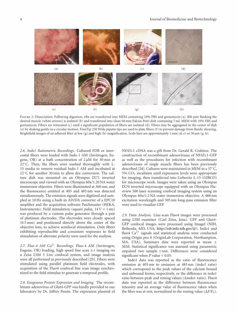
4 Journal of Biomedicine and Biotechnology
(a) (b) (c) (d)
(e) (f) (g) (h)
Figure 2: Dissociation. Following digestion, ribs are transferred into MEM containing 10% FBS and gentamycin (a). Rib pair flanking thedesired muscle (white arrows) is isolated (b) and transferred into clean 60 mm Falcon Petri dish containing 7 mL MEM with 10% FBS andgentamycin. Fibers are triturated (c) until a significant population of fibers are isolated (d). Fibers may be aggregated in the center of dish(e) by shaking gently in a circular motion. FinnTip 250 Wide pipette tips are used to plate fibers (f) to prevent damage from fluidic shearing.Brightfield images of an adhered fiber at low (g) and high (h) magnification. Scale bars are approximately 1 mm (d, e) or 50 μm (g, h).
2.6. Indo1 Ratiometric Recordings. Cultured FDB or inter-costal fibers were loaded with Indo-1 AM (Invitrogen, Eu-gene, OR) at a bath concentration of 2 μM for 30 min at22◦C. Then, the fibers were washed thoroughly with L-15 media to remove residual Indo-1 AM and incubated at22◦C for another 30 min to allow dye conversion. The cul-ture dish was mounted on an Olympus IX71 invertedmicroscope and viewed with an Olympus 60x/1.20 NA waterimmersion objective. Fibers were illuminated at 360 nm, andthe fluorescence emitted at 405 and 485 nm was detectedsimultaneously. The emission signals were digitized and sam-pled at 10 Hz using a built-in AD/DA converter of a EPC10amplifier and the acquisition software Patchmaster (HEKA,Instruments). Field stimulation (square pulse, 14 V × 1 ms)was produced by a custom pulse generator through a pairof platinum electrodes. The electrodes were closely spaced(0.5 mm) and positioned directly above the center of theobjective lens, to achieve semilocal stimulation. Only fibersexhibiting reproducible and consistent responses to fieldstimulation of alternate polarity were used for the analysis.
2.7. Fluo-4 AM Ca2+ Recordings. Fluo-4 AM (Invitrogen,Eugene, OR) loading, high speed line scan x-t imaging ona Zeiss LSM 5 Live confocal system, and image analysiswere all performed as previously described [25]. Fibers werestimulated using parallel platinum field electrodes, withacquisition of the Fluo4 confocal line scan image synchro-nized to the field stimulus to generate a temporal profile.
2.8. Exogenous Protein Expression and Imaging. The recom-binant adenovirus of Glut4-GFP was kindly provided to ourlaboratory by Dr. Jeffrey Pessin. The expression plasmid of
NFATc1 cDNA was a gift from Dr. Gerald R. Crabtree. Theconstruction of recombinant adenoviruses of NFATc1-GFPas well as the procedures for infection with recombinantadenoviruses of single muscle fibers has been previouslydescribed [24]. Cultures were maintained in MEM in a 37◦C,5% CO2 incubator until expression levels were appropriatefor imaging, then transferred into Liebovitz L-15 (GIBCO)for microscope work. Images were taken using an OlympusIX70 inverted microscope equipped with an Olympus Flu-oview 500 laser scanning confocal imaging system using anOlympus 60x/1.2 NA water immersion objective. A 488 nmexcitation wavelength and 505 nm long-pass emission filterwere used to visualize GFP.
2.9. Data Analysis. Line-scan Fluo4 images were processedusing LSM examiner (Carl Zeiss, Jena). GFP and Glut4-GFP confocal images were processed using ImageJ (NIH,Bethesda, MD, USA; http://rsb.info.nih.gov/ij/). Indo1 andfluo4 Ca2+ signals and statistical analysis were conductedusing Origin pro 8 (OriginLab Corporation, Northampton,MA, USA). Summary data were reported as mean ±SEM. Statistical significance was assessed using parametricunpaired two sample t-test. Differences were consideredsignificant when P value < 0.05.
Indo1 data was reported as the ratio of fluorescenceemission at 405 nm to emission at 485 nm (indo1 ratio)which correspond to the peak values of the calcium boundand unbound forms, respectively, or the difference in indo1ratio between peak and resting values (Δindo1 ratio). Fluo4data was reported as the difference between fluorescenceintensity and an average value of fluorescence taken whenthe fiber was at rest, normalized to the resting value (ΔF/F0).

Journal of Biomedicine and Biotechnology 5
0 200 4000
0.2
0.4
0.6
0.8
1
1.2
1.4
Time (ms)
Indo
1ra
tio
atre
st
ITCFDB
(a)
Indo
1ra
tio
atre
st
ITC
FDB
0
0.2
0.4
0.6
0.8
1
(b)
0 200 400
0
0.2
0.4
0.6
0.8
1
Time (ms)
ITCFDB
Δin
do1
rati
o
(c)
0
0.5
ITC
FDB
∗
1
Δin
do1
rati
oat
pea
k
(d)
Figure 3: Intercostal fibers exhibit decreased peak amplitude of the Indo-1 Ca2+ transient following a single action potential. (a) AverageIndo-1 ratio Ca2+ transients from FDB (red trace; n = 9) and Intercostal (ITC; green trace; n = 11) fibers. Isolated fibers were stimulatedwith field electrodes at time zero and emission ratio was examined. (b) Bar plot summarizing resting Indo-1 ratio averages of FDB and ITCfibers. No significant differences in resting indo1 ratio were detected between groups (P = 0.12). (c) Traces from panel (a), expressed asΔindo-1 ratio (Indo1 ratio—Indo1 ratio at rest). (d) Bar plot representation of peak ΔIndo-1 ratio shown in (c), FDB = 0.61 ± 0.04, n = 9;ITC = 0.40 ± 0.04, n = 11; ∗P = 0.0015. Error bars in (d) are the SE values. ITC fibers displayed significantly reduced Ca2+ transients whencompared with FDB fibers.
Data from the NFATc1-GFP fusion protein was reportedas the ratio of fluorescence intensity in the nucleus to theintensity in the cytoplasm (N/C) in order to correct forvariations in expression level between fibers. Raw data forboth Fluo4 and NFATc1 was corrected for background beforeanalysis by subtracting the intensity measured in a regionoutside the muscle fiber.
3. Results
3.1. Gross Morphology. The intercostal muscles are composedof two overlapping sheets (inner and outer) of muscle fiberswith roughly perpendicular orientations. Each muscle spansdiagonally between two ribs from the vertebral column tothe sternum. Following dissection connective tissues between

6 Journal of Biomedicine and Biotechnology
0
0
2
1 2 3 4 5
4
6
8
x
Time
ΔF/F
0
Time (s)
10
FDB
(a)
0
0
2
1 2 3 4 5
4
6
8
ITC
x
Time
ΔF/F
0
Time (s)
10
(b)
Figure 4: Fluo4 Ca2+ transients elicited by field stimulation in intercostal and FDB fibers. Representative line-scan images of Ca2+ transientresponses in FDB (a) and intercostal (b) fibers to a 5 sec 10 Hz train of field stimulation applied at time zero, performed with nonratiometricfluo-4 AM loaded fibers and assayed with high-speed confocal line scan microscopy (100 μs/line). Images in top of panels (a) and (b) show xyimages of FDB and intercostals fibers to indicate the location of the scan line, scale bar is 20 μm. Note that fluo-4 signal at rest in these fibersis very low. Traces under the images are the time courses of the fluo4 Ca2+ transients. The white lines in the time domain images indicatethe region of interest drawn at the center of the fiber and used to calculate the change in fluorescence in response to field stimulation.Data presented as ΔF/F0. Fluorescence was detected with a Zeiss LSM 5 Live ultrafast confocal system based on an Axiovert 200M invertedmicroscope. Fibers were imaged with a 63x/NA 1.2 water immersion objective lens. Excitation for fluo-4 was provided by the 488 nm line ofa 100 mW diode laser, and emitted light was collected at >510 nm.
muscle and ribs are not readily appreciable at the macro-scopic scale (Figure 1).
3.2. Isolating Intercostal Fibers. The initial attempt to isolateand culture intercostal muscle fibers was based on ourexisting protocols for isolating and culturing mouse FlexorDigitorum Brevis (FDB) fibers [21–25], which are main-tained in commercially available glass-bottomed dishes. Afteran initial development and optimization of dissection andenzymatic treatment methods, we achieved a high fractionof viable isolated intercostal fibers. These fibers maintainedclear striations and twitch response for several days, butdid not reliably form adherent cultures, even after extensive
experimentation with a variety of substrates derived fromextracellular matrix. During the course of unrelated work, wenoted that customized glass-bottomed dishes which we haddesigned with smaller diameter wells using a different glasssubstrate seemed to facilitate attachment in our FDB model(see Supplemental Figures 1 and 2). Plating intercostal fibersin these dishes dramatically improved the attachment rateand reliability of the culture.
After fiber isolation, our procedure yields a heteroge-neous mix of internal and external intercostal fibers witha successful attachment rate around 50%. Isolated intercos-tal fibers (Figure 2) are similar to FDBs in width, but 4-5times the length. This additional length makes the fibers

Journal of Biomedicine and Biotechnology 7
0 1 2 3 4 5
Time (s)
FDB
0
2
4
6
8ΔF/F
0
(a)
0 1 2 3 4 5
Time (s)
0
2
4
6
8
ΔF/F
0
ITC
(b)
ITCFDB
0
0.2
0.4
0.6
0.8
1
1.2
0 1 2 3 4 5
Time (s)
Rel
ativ
eF/F
0
(c)
Figure 5: Comparison of electrically evoked Ca2+ transients between intercostal and FDB fibers. Average fluo4 Ca2+ transient from FDB((a), red trace; n = 9) or intercostals fibers ((b), ITC, green trace; n = 9) in response to 5 sec 10 Hz train field stimulation. Traces in (c)were normalized to the amplitude of the initial response. At the end of the 5 sec train, ITC fibers demonstrated significantly smaller relativedecrease of Ca2+ transient amplitude compared to FDB counterparts (P < 0.05). ITC muscle fibers exhibit decreased peak amplitude of thefluo-4 Ca2+ transients in response to a single field stimulus; however, the intercostals are able to maintain consistent levels of calcium duringrepetitive stimulation. Fluorescence was detected as in Figure 4.
susceptible to bends when they are fully attached (see Figure1(g)). Almost all of the whole fibers that attach to the glassdisplay clear striations (Figure 6) and respond to externalelectrical field stimulation administered either as singlepulses or repetitive trains with twitching similar to that ofFDBs (see supplemental videos 1 and 2).
In order to examine the general applicability of thesedishes, we also used them to plate fibers from the slow-typesoleus muscle, which also displays poor adherence charac-teristics [22]. Although we used a less stringent digestionprocedure, plating was carried out exactly as for intercostals.
The exceptional length of the soleus fibers causes them tointerfere with each other and permits the formation of manybends and kinks which prevent complete attachment. None-theless, the fibers were responsive to electrical stimulationand adhered securely enough to withstand repeated twitch-ing (see supplemental video 3) more than 24 hours afterplating. Successful attachment rate was variable between thewells plated, but overall comparable to the intercostals.
3.3. Ca2+ Homeostasis and Electrically Evoked Ca2+ Transientsin Intercostal Fibers. Primary cultured intercostal fibers

8 Journal of Biomedicine and Biotechnology
(a) (b) (c) (d)
(e) (f) (g) (h)
Figure 6: Overexpression of foreign proteins in intercostal fibers in culture. FDB (a, b, e, f) and intercostal (c, d, g, h) muscle fibers expressingeither GFP (a–d) or a Glut4-GFP fusion construct (e–h). Scale bars are 50 μm (b, d) or 25 μm (f, h). Images were acquired as in Figure 4for panels (a–d) or with an Olympus FluoView 500 confocal system, built on an Olympus IX71 inverted microscope for panels (e–h). Fiberswere imaged viewed with a UPlan Apo 60x/NA 1.20 water-immersion objective lens using 488 nm excitation (Multiline Argon laser) for GFPwith emission detected with 505 nm long-pass filter. Photobleaching and photodamage were reduced by operating the lasers at the lowestpossible power that still provided images with satisfactory signal to noise ratio. Brightfield images in panels (b, d, f, h) were adjusted forimproved contrast using ImageJ.
retain many properties of native fibers, including contrac-tions and Ca2+ transients in response to electrical fieldstimuli (Figures 3, 4, and 5; see Supplemental Video 1). Abrief comparative study between intercostal fibers and theestablished FDB isolated fiber model was also conducted(Figures 3, 4, and 5). First, we sought to evaluate Ca2+
handling properties in single, intact indo1-loaded intercostal(n = 8) and FDB (n = 16) muscle fibers stimulated by anaction potential (AP) elicited by a 1 ms electric field stimulus(Figure 3). While resting indo1 ratio prior to stimulation wasnot significantly different between intercostal (0.45 ± 0.02;n = 11) and FDB fibers (0.49 ± 0.01; Figures 3(a) and 3(b);n = 9, P = 0.12), the peak of the indo1 Ca2+ transientfollowing a single AP (Δindo1 ratio) was significantly smallerin intercostal fibers than in the FDB fibers (intercostal = 0.40± 0.04, FDB = 0.61 ± 0.04, P = 0.0015, Figures 3(c) and3(d)). These results suggest that intercostal fibers exhibit adecreased AP-evoked Ca2+ release.
To allow better temporal resolution of the Ca2+ tran-sients, we next monitored fluo-4 transients during repetitivestimulation of intercostal and FDB fibers using an ultra high-speed (100 μs/line) confocal microscope in line scan mode.Figure 4 illustrates representative x-t confocal images andcorresponding fluo4 Ca2+ transients, expressed as ΔF/F0,from an FDB (Figure 4(a)) and an intercostal (Figure 4(b))fiber during a 5-second 10 Hz train. Line-scan fluorescenceimages were obtained along a line perpendicular to thelong axis of the fiber (Figures 4(a) and 4(b), top panels).Successive vertical lines in each image reveal the time course
(left to right; middle panels) at 100 μs resolution of the flu-orescence signal along the scanned line before and duringthe repetitive stimulation. Bottom panels in Figures 4(a) and4(b) show the time course of the fluo4 fluorescence. Figure 5shows average responses from FDB (red trace; Figure 5(a))and intercostal (green trace; Figure 5(b)) fibers stimulatedby a 5-second 10 Hz train. Consistent with the Indo-1data (Figure 3), fluo4 Ca2+ transients appear suppressedin intercostal fibers (Figure 5(b)) in comparison to thoseobserved in FDB fibers (Figure 5(a)).
However, the intercostals are able to maintain a moreconsistent level of calcium release during repetitive stimu-lation (Figure 5(b)). By normalizing the traces elicited bythe 10 Hz trains to the amplitude of the first fluo4 transientin their respective train, the relative differences in Ca2+
transient summation as well as the properties of decayingphase during the train can be appreciated (Figure 5(c)). Bothintercostal and FDB fibers showed an initial increase in thepeak amplitude during the 10 Hz train protocol, exceedingthe peak value of the initial stimulation until 2 and 0.5seconds, respectively (Figure 5(c)). Additionally, the relativeamplitude of the Ca2+ transients during the 10 Hz traindeclines more slowly in intercostal fibers (Figure 5(c)).
Unexpectedly, subpopulations reflecting an anticipateddifference between fiber types were not observed in theintercostal fibers. However, we suspect that this is not due togenuine homogeneity, but rather a limitation of the calciumion equilibration kinetics of fluo-4 in distinguishing suchsub-populations, which may be quite similar.

Journal of Biomedicine and Biotechnology 9
(a) (b) (c)
(d) (e) (f)
ITC FDB
0
0.25
0.5
0.75
1
1.25
1.5
N/C
(g)
Figure 7: NFATc1-GFP distribution in intercostal and FDB fibers. FDB (a–c) and intercostal (d–f) fibers expressing NFATc1-GFP imagedat rest after 3 days in culture. Scale bar, 5 μm. Nuclei in intercostal fibers exhibit significantly elevated levels of NFATc1 when compared toFDBs cultured under the same conditions (g), N/C ratio of NFATc1-GFP in ITC = 1.38 ± 0.05, n = 102 nuclei versus 0.51 ± 0.01, n = 42nuclei, in FDB fibers, P < 0.05). Fluorescence was detected as in Figure 6.
These results lead us to conclude that adherent cultures ofintercostal fibers are a new and unique model for the study ofCa2+-signaling pathways at the cellular and subcellular levelsby using ultrafast confocal imaging combined with Ca2+-sensitive fluorescent probes.
3.4. Expression of Exogenous Proteins. In order to assessthe viability of the intercostal cultures for imaging studiesutilizing fluorescent proteins, fibers in culture were exposedto crude lysates of recombinant adenoviral vectors expressingGFP. Exposure to virus was carried out overnight, beginningimmediately after plating. Vector dosage was calibrated byexpression levels in similarly treated FDB fibers. Cultureswere maintained in an incubator to allow expression priorto imaging. Expression of GFP was robust after 48 hours anddid not appear to damage the fibers, which maintained clearstriations (Figures 6(b) and 6(d)).
We also exposed other cultures to a viral vector express-ing a Glut4-GFP fusion protein. Expression continuedthrough 96 hours (Figure 6(g)). Glut4-GFP was expressed inintercostal fibers in puncta both around the nucleus and inlines parallel to the axis of the fiber (Figures 6(e) and 6(g)).This distribution is similar to what we observe in FDB fibersinfected with the same construct, as well as the distribu-tion of the native transporter characterized by immunofluorescence [28], although we observe weaker striations,presumably due to lack of exposure to insulin during the cul-ture period reducing the Glut4 present in the transversetubule system.
Finally, we exposed the fibers to a viral construct ex-pressing a NFATc1-GFP fusion construct. NFATc1 is atranscription factor canonically regulated by the calcium-dependent phosphatase calcineurin [29, 30]. Typicallyresting FDB fibers exhibit low translocation of NFATc1 into

10 Journal of Biomedicine and Biotechnology
the nucleus (Figures 7(a), 7(b), and 7(c)) [24]. Unexpectedly,ITC fibers exhibited a higher nuclear distribution of NFATc1-GFP at rest (Figures 7(d), 7(e), and 7(f)) when comparedto FDB fibers (Figures 7(a), 7(b), and 7(c)). The restingnuclear/cytosolic (N/C) ratio of NFATc1 was significantlyelevated in intercostal fibers (Figure 7(g)) compared to FDB(1.38 ± 0.05, n = 102 nuclei versus 0.51 ± 0.01, n = 42nuclei, resp.; P < 0.05).
4. Discussion
Adult skeletal muscle fiber culture currently focuses on onlya handful of the myriad available muscles in the body, ex-trapolating from the few established culture systems like theFDB to the body as a whole. While it is generally true thatvarious skeletal muscles resemble one another, there is sig-nificant divergence among them with respect to their physi-ology and the expression levels and isoforms of many musclespecific and metabolic proteins [1, 5–7]. This is dramaticallyillustrated by the high specificity of pathology in manyskeletal muscle disease states resulting from defects in specificgenes (e.g., the disproportionate targeting of facial, shoul-der, and limb muscles in facioscapulohumeral musculardystrophy or the extremities in distal muscular dystrophy[17]).
The intercostal fiber cultures introduced here themselvesappear to be nearly as versatile as the more established FDBcultures. While we have not yet used intercostal culturesin applications requiring large numbers of adherent fibers,they have proven viable for a wide variety of single fiberexperiments, even after several days in culture. Previouswork related to the physiology of intercostal muscles comesfrom studies utilizing organ culture of intact muscle fiberbundles [31–35]. Whereas intercostal muscle fiber bundlesoffer the advantage of preserving nerve-muscle interactions,as with many organ culture systems it may not be well suitedfor single cell electrophysiology and high spatial resolutionimaging. Furthermore, the ability to mimic stimulationpatterns that may be experienced by the muscle duringstressed states in vivo (such as hyperventilation or coughing)at the single muscle fiber level offers the opportunity toisolate the response of the muscle from that of the sur-rounding tissue in a manner not possible with organ culturemethods. A variant of the single intercostal fiber isolationwas described previously and was used to study satellitecell differentiation cocultured with nonadherent intercostalfibers derived from goats [36]. While these methods arevaluable, the availability of adherent single fiber modelsfor imaging and electrophysiology substantially expands ourability to study these muscle types.
The value of studying each skeletal muscle as a separatetissue is demonstrated by our unexpected observation ofspontaneous accumulation of NFATc1 in the nuclei of restingfibers. It is clear that studies using only a few particularskeletal muscles risk overlooking interesting phenomena.Given the complexity of transcriptional regulation, the het-erogeneous nature of the physiology and molecular biologyof various skeletal muscles and the diverse tissues whichsurround them, we suspect that examining a wider variety
of muscles specifically relevant to individual diseases incontrolled in-vitro settings will prove fruitful.
Our culturing of intercostal muscles aims to do thisnot only by providing a particular model system specificallyrelevant to the pathology of COPD and the respiratorycomplications of the muscular dystrophies [16, 17], but alsoby improving the general techniques of adherent isolatedmuscle fiber culture. If these techniques are optimized forpoorly adhering muscle types, generating cultures from anymuscle in the body may become substantially easier, andusing the most relevant tissue in a given disease model maybecome routine.
Acknowledgment
This work was supported by NIH-NIAMS Grants R01-AR055099, R01-AR056477. P. Robison was supported bythe Interdisciplinary Training Program Muscle Biology T32-AR007592.
References
[1] R. Bottinelli and C. Reggiani, “Human skeletal muscle fibres:molecular and functional diversity,” Progress in Biophysics andMolecular Biology, vol. 73, no. 2–4, pp. 195–262, 2000.
[2] R. I. Close, “Dynamic properties of mammalian skeletalmuscles,” Physiological Reviews, vol. 52, no. 1, pp. 129–197,1972.
[3] J. C. Calderon, P. Bolanos, S. H. Torres, G. Rodrıguez-Arroyo,and C. Caputo, “Different fibre populations distinguished bytheir calcium transient characteristics in enzymatically disso-ciated murine flexor digitorum brevis and soleus muscles,”Journal of Muscle Research and Cell Motility, vol. 30, no. 3-4,pp. 125–137, 2009.
[4] J. C. Calderon, P. Bolanos, and C. Caputo, “Myosin heavychain isoform composition and Ca2+ transients in fibresfrom enzymatically dissociated murine soleus and extensordigitorum longus muscles,” Journal of Physiology, vol. 588, no.1, pp. 267–279, 2010.
[5] R. L. Moss, G. M. Diffee, and M. L. Greaser, “Contractileproperties of skeletal muscle fibers in relation to myofibrillarprotein isoforms,” Reviews of Physiology, Biochemistry &Pharmacology, vol. 126, pp. 1–63, 1995.
[6] D. Pette and R. S. Staron, “Cellular and molecular diversitiesof mammalian skeletal muscle fibers,” Reviews of PhysiologyBiochemistry and Pharmacology, vol. 116, pp. 1–76, 1990.
[7] S. Schiaffino and C. Reggiani, “Molecular diversity of myofib-rillar proteins: gene regulation and functional significance,”Physiological Reviews, vol. 76, no. 2, pp. 371–423, 1996.
[8] B. Polla, G. D’Antona, R. Bottinelli, and C. Reggiani, “Respi-ratory muscle fibres: specialisation and plasticity,” Thorax, vol.59, no. 9, pp. 808–817, 2004.
[9] A. De Troyer, P. A. Kirkwood, and T. A. Wilson, “Respiratoryaction of the intercostal muscles,” Physiological Reviews, vol.85, no. 2, pp. 717–756, 2005.
[10] A. Taylor, “The contribution of the intercostal muscles to theeffort of respiration in man,” The Journal of Physiology, vol.151, pp. 390–402, 1960.
[11] D. Faithfull, J. G. Jones, and C. Jordan, “Measurement of therelative contributions of rib cage and abdomen/diaphragm totidal breathing in man,” British Journal of Anaesthesia, vol. 51,no. 5, pp. 391–398, 1979.

Journal of Biomedicine and Biotechnology 11
[12] G. H. Koepke, E. M. Smith, A. J. Murphy, and D. G. Dick-inson, “Sequence of action of the diaphragm and intercostalmuscles during respiration. I. Inspiration,” Archives of PhysicalMedicine and Rehabilitation, vol. 39, no. 7, pp. 426–430, 1958.
[13] A. J. Murphy, G. H. Koepke, E. M. Smith, and D. G. Dick-inson, “Sequence of action of the diaphragm and intercostalmuscles during respiration. II. Expiration,” Archives of PhysicalMedicine and Rehabilitation, vol. 40, pp. 337–342, 1959.
[14] M. Mizuno and N. H. Secher, “Histochemical characteristicsof human expiratory and inspiratory intercostal muscles,”Journal of Applied Physiology, vol. 67, no. 2, pp. 592–598, 1989.
[15] M. Mizuno, N. H. Secher, and B. Saltin, “Fibre types, capillarysupply and enzyme activities in human intercostal muscles,”Clinical Physiology, vol. 5, no. 2, pp. 121–135, 1985.
[16] L. D. Calvert, T. M. McKeever, W. J. Kinnear, and J. R. Britton,“Trends in survival from muscular dystrophy in Englandand Wales and impact on respiratory services,” RespiratoryMedicine, vol. 100, no. 6, pp. 1058–1063, 2006.
[17] A. E. Emery, “The muscular dystrophies,” Lancet, vol. 359, no.9307, pp. 687–695, 2002.
[18] J. G. Gea, “Myosin gene expression in the respiratory muscles,”European Respiratory Journal, vol. 10, no. 10, pp. 2404–2410,1997.
[19] M. G. Klein, H. Cheng, L. F. Santana, Y. H. Jiang, W. J. Lederer,and M. F. Schneider, “Two mechanisms of quantized calciumrelease in skeletal muscle,” Nature, vol. 379, no. 6564, pp. 455–458, 1996.
[20] L. Kovacs, E. Rios, and M. F. Schneider, “Calcium transientsand intramembrane charge movement in skeletal musclefibres,” Nature, vol. 279, no. 5712, pp. 391–396, 1979.
[21] S. L. Carroll, M. G. Klein, and M. F. Schneider, “Calciumtransients in intact rat skeletal muscle fibers in agarose gel,”American Journal of Physiology, vol. 269, no. 1, pp. C28–C34,1995.
[22] S. L. Carroll, M. G. Klein, and M. F. Schneider, “Decay ofcalcium transients after electrical stimulation in rat fast- andslow-twitch skeletal muscle fibres,” Journal of Physiology, vol.501, no. 3, pp. 573–588, 1997.
[23] Y. Liu, S. L. Carroll, M. G. Klein, and M. F. Schneider,“Calcium transients and calcium homeostasis in adult mousefast-twitch skeletal muscle fibers in culture,” American Journalof Physiology, vol. 272, no. 6, pp. C1919–C1927, 1997.
[24] Y. Liu, Z. Cseresnyes, W. R. Randall, and M. F. Schneider,“Activity-dependent nuclear translocation and intranucleardistribution of NFATc in adult skeletal muscle fibers,” Journalof Cell Biology, vol. 155, no. 1, pp. 27–39, 2001.
[25] B. L. Prosser, N. T. Wright, E. O. Hernandez-Ochoa et al.,“S100A1 binds to the calmodulin-binding site of ryanodinereceptor and modulates skeletal muscle excitation-contractioncoupling,” Journal of Biological Chemistry, vol. 283, no. 8, pp.5046–5057, 2008.
[26] L. D. Brown, G. G. Rodney, E. Hernandez-Ochoa, C. W. Ward,and M. F. Schneider, “Ca2+ sparks and T tubule reorganizationin dedifferentiating adult mouse skeletal muscle fibers,” Amer-ican Journal of Physiology. Cell Physiology, vol. 292, no. 3, pp.C1156–C1166, 2007.
[27] L. D. Brown and M. F. Schneider, “Delayed dedifferentiationand retention of properties in dissociated adult skeletal musclefibers in vitro,” In Vitro Cellular and Developmental Biology,vol. 38, no. 7, pp. 411–422, 2002.
[28] T. Ploug, B. van Deurs, H. Ai, S. W. Cushman, and E. Ralston,“Analysis of GLUT4 distribution in whole skeletal musclefibers: identification of distinct storage compartments that are
recruited by insulin and muscle contractions,” Journal of CellBiology, vol. 142, no. 6, pp. 1429–1446, 1998.
[29] G. R. Crabtree and E. N. Olson, “NFAT signaling: chore-ographing the social lives of cells,” Cell, vol. 109, no. 2,supplement 1, pp. S67–S79, 2002.
[30] A. Rao, C. Luo, and P. G. Hogan, “Transcription factors ofthe NFAT family: regulation and function,” Annual Review ofImmunology, vol. 15, pp. 707–747, 1997.
[31] S. M. Cardoso, P. Mutch, P. J. Scotting, and P. M. Wigmore,“Gene transfer into intact fetal skeletal muscle grown in vitro,”Muscle and Nerve, vol. 30, no. 1, pp. 87–94, 2004.
[32] S. G. Cull-Candy, R. Miledi, and O. D. Uchitel, “Denervationchanges in normal and myasthenia gravis human muscle fibresduring organ culture,” Journal of Physiology, vol. 333, pp. 227–249, 1982.
[33] R. L. Ruff and D. Whittlesey, “Na+ current densities andvoltage dependence in human intercostal muscle fibres,”Journal of Physiology, vol. 458, pp. 85–97, 1992.
[34] L. Ziskind-Conhaim and M. J. Dennis, “Development of ratneuromuscular junctions in organ culture,” DevelopmentalBiology, vol. 85, no. 1, pp. 243–251, 1981.
[35] L. Ziskind-Conhaim, I. Geffen, and Z. W. Hall, “Redistribu-tion of acetylcholine receptors on developing rat myotubes,”Journal of Neuroscience, vol. 4, no. 9, pp. 2346–2349, 1984.
[36] K. Yamanouchi, T. Hosoyama, Y. Murakami, S. Nakano, andM. Nishihara, “Satellite cell differentiation in goat skeletalmuscle single fiber culture,” Journal of Reproduction andDevelopment, vol. 55, no. 3, pp. 252–255, 2009.