Shared responsibility for ongoing rehabilitation: a new approach to home-based therapy after stroke
Upload
khangminh22Category
view
3download
0
Stroke Research and Treatment
Guest Editors: Ching-yi Wu, Keh-chung Lin, Steven L. Wolf, and Agnès Roby-Brami
Motor Rehabilitation after Stroke

Motor Rehabilitation after Stroke

Stroke Research and Treatment
Motor Rehabilitation after Stroke
Guest Editors: Ching-yi Wu, Keh-chung Lin,Steven L. Wolf, and Agnes Roby-Brami

Copyright © 2012 Hindawi Publishing Corporation. All rights reserved.
This is a special issue published in “Stroke Research and Treatment.” All articles are open access articles distributed under the CreativeCommons Attribution License, which permits unrestricted use, distribution, and reproduction in any medium, provided the originalwork is properly cited.

Editorial Board
Alison Baird, USADaniel Bereczki, HungaryNatan M. Bornstein, IsraelRaymond T. Cheung, Hong KongG. A. Donnan, AustraliaV. L. Feıgin, New ZealandMasayuki Fujioka, JapanG. J. Hankey, Australia
Cathy Helgason, USACarlos S. Kase, USAScott E. Kasner, USAChelsea S. Kidwell, USADavid S. Liebeskind, USAChristopher S. Ogilvy, USABruce Ovbiagele, USADavid Reutens, Australia
David Russell, NorwayStefan Schwab, GermanyVeronika Skvortsova, RussiaMichael A. Sloan, USAHelmuth Steinmetz, GermanyGraham S. Venables, UKDaniel Woo, USAOsama O. Zaidat, USA

Contents
Motor Rehabilitation after Stroke, Ching-yi Wu, Keh-chung Lin, Steven L. Wolf, and Agnes Roby-BramiVolume 2012, Article ID 810706, 2 pages
Quantitative Mechanical Properties of the Relaxed Biceps and Triceps Brachii Muscles in Patients withSubacute Stroke: A Reliability Study of the Myoton-3 Myometer, Li-ling Chuang, Ching-yi Wu,Keh-chung Lin, and Shih-yu LurVolume 2012, Article ID 617694, 7 pages
The Neurorehabilitation Training Toolkit (NTT): A Novel Worldwide Accessible Motor TrainingApproach for At-Home Rehabilitation after Stroke, Sergi Bermudez i Badia and M. S. CameiraoVolume 2012, Article ID 802157, 13 pages
ENGAGE: Guided Activity-Based Gaming in Neurorehabilitation after Stroke: A Pilot Study,Ann Reinthal, Kathy Szirony, Cindy Clark, Jeffrey Swiers, Michelle Kellicker, and Susan LinderVolume 2012, Article ID 784232, 10 pages
Motor Imagery Experiences and Use: Asking Patients after Stroke Where, When, What, Why, and HowThey Use Imagery: A Qualitative Investigation, Corina Schuster, Andrea Glassel, Anne Scheidhauer,Thierry Ettlin, and Jenny ButlerVolume 2012, Article ID 503190, 18 pages
Constraint-Induced Movement Therapy (CIMT): Current Perspectives and Future Directions,Aimee P. Reiss, Steven L. Wolf, Elizabeth A. Hammel, Erin L. McLeod, and Erin A. WilliamsVolume 2012, Article ID 159391, 8 pages
How Physically Active Are People with Stroke in Physiotherapy Sessions Aimed at Improving MotorFunction? A Systematic Review, Gurpreet Kaur, Coralie English, and Susan HillierVolume 2012, Article ID 820673, 9 pages
Slowing of Motor Imagery after a Right Hemispheric Stroke, Francine Malouin, Carol L. Richards,and Anne DurandVolume 2012, Article ID 297217, 10 pages
Functional Exercise and Physical Fitness Post Stroke: The Importance of Exercise Maintenance for MotorControl and Physical Fitness after Stroke, Birgitta Langhammer and Birgitta LindmarkVolume 2012, Article ID 864835, 9 pages

Hindawi Publishing CorporationStroke Research and TreatmentVolume 2012, Article ID 810706, 2 pagesdoi:10.1155/2012/810706
Editorial
Motor Rehabilitation after Stroke
Ching-yi Wu,1 Keh-chung Lin,2 Steven L. Wolf,3, 4, 5, 6 and Agnes Roby-Brami7
1 Department of Occupational Therapy and Graduate Institute of Behavioral Sciences, Chang Gung University College of Medicine,Taoyuan 33302, Taiwan
2 School of Occupational Therapy, National Taiwan University College of Medicine, Taipei 10051, Taiwan3 Department of Rehabilitation Medicine, Center for Rehabilitation Medicine, Emory University School of Medicine, Atlanta,GA 30322, USA
4 Department of Cell Biology, Center for Rehabilitation Medicine, Emory University School of Medicine, Atlanta, GA 30322, USA5 Health and Elder Care, Nell Hodgson Woodruff School of Nursing, Emory University, Atlanta, GA 30322, USA6 VA Rehab R&D Brain Rehabilitation Research Center, Gainesville, FL 32608, USA7 Department of Neurological Rehabilitation, Raymond Poincare Hospital and Laboratoy of Neurophysics and Physiology,Paris Descartes University, 75270 Paris, France
Correspondence should be addressed to Ching-yi Wu, [email protected]
Received 18 April 2012; Accepted 18 April 2012
Copyright © 2012 Ching-yi Wu et al. This is an open access article distributed under the Creative Commons Attribution License,which permits unrestricted use, distribution, and reproduction in any medium, provided the original work is properly cited.
Stroke is one of the most common conditions in adults thatmay cause impairment and disability. Motor impairmentsoften persistently affect the daily function and quality of lifeamong stroke survivors. Recently, a few novel interventionsto improve motor impairment have evolved that make use ofadvances in neuroscience and neurobehavioral knowledge.These theory-driven approaches attempt to translate basicscience research into novel clinical practice and build onmethods to manipulate favorable plasticity changes withinthe brain in response to task-related training to augmentmotor recovery.
Developing a new clinical intervention may consist offour phases [1]: consideration of concept, development ofconcept, demonstration of concept, and proof of concept.The value of a phase I study is to learn how well anintervention can be applied, how patients respond to theintervention, to whom it ought to be given, what outcomesshould be measured, and how its safety can be assured.Phase II studies aim to standardize the new treatment,compare efficacy against alternative treatments, and ensurethat outcomes can be measured objectively and reliably.Phase III studies aim to optimize the treatment (dosageoptimization) and enhance its practicality. Most of thepapers published in this special issue are Phase I studies.An increase in Phase II and III studies is needed to improvestroke rehabilitation research and practice.
Among the five Phase I studies presented here, twoassessed how well the intervention could be applied inpatients with stroke. A. Reinthal and colleagues evaluatedthe effects of a novel activity-based gaming exercise thatinvolved highly repetitive practice in stroke rehabilitation. S.B. Badia and M. S. Cameirao used 10 healthy individuals totest a neurorehabilitation training toolkit designed for strokesurvivors in the home environment.
Three papers in this special issue addressed appropriatetreatments for specific types of patients, for example, thedegree of impairments or type of symptoms. This infor-mation can be used to maximize the benefits of specificinterventions. Among these studies, C. Schuster and col-leagues’ study involved a qualitative, patient-centered studyto address where, when, what, how, and why motor imagerycan be used for stroke rehabilitation. F. Malouin andcolleagues investigated the effect of the side of hemisphericlesion on temporal congruence between real and imaginedmovements and its link with working memory deficits inpersons with chronic stroke. B. Langhammer and B. Lind-mark demonstrated immediate and follow-up improvementsafter functional exercise between groups with high and lowbaseline functional level for a period of 36 months afterstroke.
L. Chuang and colleagues’ study of instrument evaluationexamined the reliability of Myoton-3 myometer, a tool used
2 Stroke Research and Treatment
to quantify muscle tone, elasticity, and stiffness in strokepatients. This study provided evidence for the psychometricsoundness of myotonometric measurement. Also included inthis special issue are two systematic reviews of optimal dosageof stroke motor rehabilitation.
To advance stroke motor rehabilitation, future researchmay translate concepts from basic sciences to clinical trialsand heath care of patients with stroke. Issues relevant forstudy may include, but are not limited to, the following.
(i) What are the most beneficial interventions for spe-cific types of patients under specific circumstances[2]?
(ii) What are the factors (e.g., the time for deliveringa specific intervention, patient characteristics) thatmay affect treatment outcomes [3, 4]?
(iii) What are the fundamental mechanisms (neurophys-iologic and biomechanical) that may underlie motorimprovements?
(iv) How can bioengineered or robotic devices be opti-mally used to effectively manage motor impairmentand disabilities?
(v) What are the optimal dosages for specific rehabilita-tion regimens [5]?
Continued research on these issues will lead to improvedknowledge and practice guidelines for stroke rehabilitation.
Ching-yi WuKeh-chung LinSteven L. Wolf
Agnes Roby-Brami
References
[1] B. H. Dobkin, “Progressive staging of pilot studies to improvephase III trials for motor interventions,” Neurorehabilitationand Neural Repair, vol. 23, no. 3, pp. 197–206, 2009.
[2] D. Bensmail, J. Robertson, C. Fermanian, and A. Roby-Brami,“Botulinum toxin to treat upper-limb spasticity in hemipareticpatients: grasp strategies and kinematics of reach-to-graspmovements,” Neurorehabilitation and Neural Repair, vol. 24, no.2, pp. 141–151, 2010.
[3] S. L. Wolf, P. A. Thompson, E. Estes, T. Lonergan, R. Merchant,and N. Richardson, “The EXCITE trial: analysis of “noncom-pleted” Wolf Motor Function Test items,” Neurorehabilitationand Neural Repair, vol. 26, no. 2, pp. 178–187, 2012.
[4] R. Nijland, G. Kwakkel, J. Bakers, and E. van Wegen, “Con-straint-induced movement therapy for the upper paretic limbin acute or sub-acute stroke: a systematic review,” InternationalJournal of Stroke, vol. 6, no. 5, pp. 425–433, 2011.
[5] Y.-W. Hsieh, C.-Y. Wu, W.-W. Liao, K.-C. Lin, K.-Y. Wu, andC.-Y. Lee, “Effects of treatment intensity in upper limb robot-assisted therapy for chronic stroke: a pilot randomized con-trolled trial,” Neurorehabilitation and Neural Repair, vol. 25, no.6, pp. 503–511, 2011.

Hindawi Publishing CorporationStroke Research and TreatmentVolume 2012, Article ID 617694, 7 pagesdoi:10.1155/2012/617694
Clinical Study
Quantitative Mechanical Properties of the RelaxedBiceps and Triceps Brachii Muscles in Patients with SubacuteStroke: A Reliability Study of the Myoton-3 Myometer
Li-ling Chuang,1 Ching-yi Wu,2 Keh-chung Lin,1, 3 and Shih-yu Lur2
1 School of Occupational Therapy, College of Medicine, National Taiwan University, Taipei, Taiwan2 Department of Occupational Therapy and Graduate Institute of Clinical Behavioral Science, College of Medicine,Chang Gung University, Taoyuan, Taiwan
3 Division of Occupational Therapy, Department of Physical Medicine and Rehabilitation, National Taiwan University Hospital,Taipei, Taiwan
Correspondence should be addressed to Keh-chung Lin, [email protected]
Received 9 November 2011; Revised 18 January 2012; Accepted 13 February 2012
Academic Editor: Steven L. Wolf
Copyright © 2012 Li-ling Chuang et al. This is an open access article distributed under the Creative Commons Attribution License,which permits unrestricted use, distribution, and reproduction in any medium, provided the original work is properly cited.
Objective. Test-retest reliability of the myotonometer was investigated in patients with subacute stroke. Methods. Twelve patientswith substroke (3 to 9 months poststroke) were examined in standardized testing position twice, 60 minutes apart, with theMyoton-3 myometer to measure tone, elasticity, and stiffness of relaxed bilateral biceps and triceps brachii muscles. Intraraterreliability of muscle properties was determined using intraclass correlation coefficient (ICC), the standard error of measurement(SEM), and the minimal detectable change (MDC). Results. Intrarater reliability of muscle properties of bilateral biceps and tricepsbrachii muscles were good (ICCs = 0.79–0.96) except for unaffected biceps tone (ICC = 0.72). The SEM and MDC of bilateralbiceps and triceps brachii muscles indicated small measurement error (SEM% < 10%, MDC% < 25%). Conclusion. The Myoton-3myometer is a reliable tool for quantifying muscle tone, elasticity, and stiffness of the biceps and triceps brachii in patients withsubacute stroke.
1. Introduction
Abnormalities in muscle structure and properties are a com-mon feature after stroke [1–3] and lead to poor controlledmovement and functional disability [4]. Examining themechanical properties of muscle is important in monitoringthe stage of the pathologic processes of muscles [5, 6] andfor assessing efficacy of therapeutic interventions [7]. Themost widely used clinical assessment of muscle tone is themodified Ashworth scale (MAS), which assesses muscularresistance to passive movement [8, 9]. Nevertheless, the MASuses subjective grading [9, 10], has poor reliability [9] andclustering of scores [11, 12], and lacks significant correlationwith muscular stiffness after stroke [13, 14]. Therefore,an objective measurement tool with an excellent reliabilityand small measurement error for assessing the mechanicalproperties of muscle is necessary. Researchers have reporteda new approach, the myotonometric measure, which was
more sensitive and precise than the MAS to quantify muscleproperties [15].
The prerequisites of a proper measurement are validityand reliability. Validity ensures that a measurement actuallyevaluates what it is intended to measure, and reliability is theextent of a consistent measurement outside of measurementerror [16]. The validity of the myotonometer has been estab-lished in healthy individuals [17, 18], in patients with chronicpain in the anterior leg or dorsal forearm [19], in patientswith upper motoneuron disorders [12], and in stroke sur-vivors [20]. Myotonometric measurements of muscle stiff-ness showed an approximately linear increase with increasingelectromyographic measurements of muscle activation andcontractile force during voluntary isometric contraction,indicating tissue displacement during contracted conditionsprovided an indirect measure of muscle strength [17–19].The linear relationship between muscle stiffness and forceoutput suggested that the myotonometer was giving a valid

2 Stroke Research and Treatment
recording of the muscle stiffness rather than that of the sub-cutaneous tissue [18]. The myotonometer quantified spas-ticity of the biceps brachii muscle and correlations betweenthe myotonometric measurements of muscle tone and MASwere moderate to high in subjects with upper motoneurondisorders [12]. Differences of myotonometric measurementsin relaxed and active muscle contraction were significantlyrelated to total ankle stiffness quantified using a torquemotor [20]. The significance of the association between theseoutcomes indicates that they measure similar constructs.
Previous studies have shown that myotonometry isreliable for healthy adults [18, 21, 22] and for variouspatient populations, including those with Parkinson’s disease[23, 24], cerebral palsy [25, 26], musculoskeletal disorders[27, 28], and chronic stroke [29]. To date, however, onlyone study has examined the test-retest reliability of themyotonometer on the forearm muscles in patients withchronic stroke [29], which limits its use in patients withstroke. Pathologic progressions in muscles may differ acrossvarious diseases and stage of disease; thus, the reliability ofthe myotonometer should be established for patients withsubacute stroke.
Patients with stroke have increased passive biceps brachiitone [12] and stiffness [14]. Biceps and triceps brachii muscleparesis and biceps brachii cocontraction during voluntaryreaching have shown significant correlations to decreasedmotor performance, indicating that these two muscles aregood predictors of the motor performance of the upperextremity [14]. Therefore, it is important to explore thereliability of the myotonometer on the biceps brachii andtriceps brachii muscles.
The present pilot study investigated the intrarater relia-bility of a hand-held myotonometry device (Myoton-3) formeasuring muscle properties of bilateral biceps brachii andtriceps brachii muscles in patients who had experienced afirst-ever stroke within 3 to 9 months before enrollment. Thistime window is the period in which most available standardtherapeutic interventions have been completed and theopportunity for spontaneous recovery to occur is attenuated[30]. Findings from the present study can contribute toa better understanding of mechanical properties of elbowmuscles in patients with subacute stroke and may alsoprovide diagnostic and therapeutic implications.
2. Methods
2.1. Participants. We recruited 12 participants (8 men and 4women) with a mean age of 51.19 years. Table 1 summarizesparticipant characteristics. Inclusion criteria were (1) afirst-ever stroke of 3 to 9 months before recruitment, (2)Brunnstrom stage III or above in the proximal and distal partof the arm [31], (3) no severe spasticity in the paretic arm(MAS ≤ 2) [8], (4) no cognitive deficits (Mini-Mental StateExamination score ≥ 24) [32], and (5) no other neurologic,neuromuscular, or orthopedic disease. Institutional ReviewBoard approval was obtained from the participating sites andwritten informed consent was obtained from all participantsbefore data collection.
Table 1: Characteristics of the participants (n = 12).
Characteristic
Sex, n
Male 8
Female 4
Age, mean (SD), year 51.19 (11.02)
Side of hemiplegia, n
Right 7
Left 5
Months after stroke onset, mean (SD) 6.58 (1.38)
Brunnstrom stage of upper limb, median (range)
Proximal part 4.5 (3.5–5)
Distal part 4.5 (3.5–5.5)
Fugl-Meyer Assessment for upper limb, mean (SD) 47.92 (6.33)
Mini Mental State Exam scores, mean (SD) 27.50 (3.26)
SD: standard deviation.
2.2. Testing Procedures. Myotonometric measurements inbilateral biceps and triceps brachii muscles were per-formed at rest, using the Myoton-3 myometer (MuomeetriaAS, Tallinn, Estonia) by a senior occupational therapist(Figure 1) [33]. Before measurement, participants wereinformed standard measurement procedure. Measurementswere done with the participant lying supine for biceps brachiiand side lying for triceps brachii in a relaxed manner,with the participants’ arms at their sides and forearmsbetween pronation and supination. The location of themeasured muscles was first determined on the unafffectedside, thereafter on the affected side. The participant wasrequested to make an effort by applying resistance with thebiceps brachii or triceps brachii to the therapist’s hand andat the same time the measuring points for the biceps brachiiand triceps brachii were identified by the therapist accordingto bone prominence and palpation. The middle part of themuscle belly is suggested as the particular measuring point[18, 34], which was marked with a marker in order toreplicate the positioning for the subsequent hour used for thereliability measures. For example, the measuring point forthe biceps brachii was at the long head, lateral part of muscle,in the middle of arm; and that for the triceps brachii was atthe medial head of muscle, in the middle part of arm [35].The muscles of the unaffected side of the body were measuredfirst. After participants were instructed to relax their musclesmaximally, the testing end of the Myoton-3 was placedperpendicular on the skin surface overlying the measuringpoints of the respective bilateral biceps brachii and tricepsbrachii. Three consecutive measurements with roughly 1second in between were taken in each muscle, and the averagevalue was used for later analysis. The entire test session wasrepeated 60 minutes after the first session with the sameprocedure, same position, and same measuring point.
The Myoton-3 myometer exerts a short mechanical pulseon the tested muscle, which causes muscle to be deformedfor a short interval. The muscle responds to the mechanicalstimulus in the form of damped oscillations recorded by anacceleration transducer on the testing end, and 3 parameters

Stroke Research and Treatment 3
Figure 1: The Myoton-3 myometer.
s
v
a
a
t
t
t
T
Δl
amax
Figure 2: Damped oscillations of the muscle show displacement (s),velocity (v), and acceleration (a) in myotonometric measurements.T is the oscillation period, amax is the maximal amplitude ofoscillation, and Δl is the maximal deformation depth of the muscle.
are calculated from the curve (Figure 2). Three mechanicalproperties of the muscle tissue are (1) the natural oscillationfrequency (Hz), (2) the logarithmic decrement of dampingoscillations, and (3) the stiffness (N/m) [15, 27, 34].
The frequency of the damped oscillations characterizesthe muscle tone, the mechanical tension in a relaxed muscle.The higher the value, the more tense is the muscle. Thefrequency of the damping was calculated as (Frequency(Hz) = 1/T), where T is the oscillation period in seconds(Figure 2). The range of values of the oscillation frequencyis usually 11 to 16 Hz in the functional state of relaxationand 18 to 40 Hz in contraction, depending on the muscle[34]. The logarithmic decrement of the damping oscillationscharacterizes muscle elasticity. The logarithmic decrement ofdamping was calculated as (Decrement = ln(amax/a)), whereamax is the maximal amplitude of oscillation and a is theoscillation amplitude (Figure 2). The decrement values areusually 1.0 to 1.2, depending on the muscle. At the pointof maximum compression of the muscle being measured,the corresponding acceleration (amax) characterizes the resis-tance of the muscle to the force deforming the muscle [28].Stiffness was calculated as (Stiffness = amax×m/Δl), where mis the mass of the testing end of myometer (kg); amax is themaximal acceleration of oscillation (m/s2); Δl is the defor-mation depth of the muscle mass (Figure 2) [18]. The usual
range of stiffness values is 150 to 300 N/m for resting muscleand may exceed 1000 N/m for contracted muscles [34].
Operational Definition and Functional Role of Muscle Tone,Elasticity, and Stiffness. Muscle tone, elasticity, and stiffnessquantify the functional state of the muscle [27, 28]. Muscletone involves active nervous-system-stimulated tone andpassive (resting) intrinsic viscoelastic tone [21, 36, 37]. Fromthe biomechanics perspective, muscle tone is a mechanicaltension in the relaxed muscle [34]. Passive muscle tone isdefined as the passive muscle tonus or tension that derivesfrom its intrinsic viscoelastic properties without contractileactivity [36, 37]. The functional roles of passive muscle toneare maintaining balance, stability, and posture, providingadequate blood circulation to the muscle and achievingenergy-efficient costs for prolonged duration without fatigue[34, 36]. Increased muscle tone disturbs the blood supply inthe muscle to diminish oxygen transportation, which mightrelate to pain, lowered motor performance, overload, and soon [34].
Muscle elasticity is the ability of the muscle to restoreits initial shape after contraction, which is inversely propor-tional to the decrement [34]. Muscle elasticity increases asthe decrement decreases. Muscle elasticity is important inusing muscle energy and increasing blood circulation volumeduring the effort. Decreased muscle elasticity brings on easierfatigability and limited speed of movement [34].
Muscle stiffness is a muscle’s ability to resist the defor-mation caused by external forces [36–38]. The speed andease of the movement performed by the agonist muscle isassociated with the stiffness of the antagonist muscle. When amuscle becomes more stiff, greater force is required from theantagonist, which decreases the energy expenditure economyof movement [34].
2.3. Data Analysis. Results of myotonometric measure-ments are presented as mean and standard deviation (SD).Intrarater reliability was analyzed through the intraclasscorrelation coefficient (ICC), standard error of measure-ment (SEM), SEM%, minmal detectable change (MDC),and MDC%. The Kolmogorov-Smirnov test was used toexamine whether the tested parameters satisfied conditionsfor normal distribution. The ICC was calculated using a 2-way mixed-effect model, with an agreement coefficient andaverage measure. The ICC determines the degree of consis-tency and agreement between repeated measurements [39],with an ICC exceeding 0.75 indicating excellent reliability[16]. The SEM represents the smallest change between 2time points that provides an indication of within-subjectvariability in repeated tests and determines the extent ofmeasurement error. The MDC represents the magnitude ofchange necessary to exceed the measurement error of test-retest measures that indicates a true statistical change at acertain confidence interval (CI) level for a single individual[39–41]. The SEM was calculated as (SEM = SDpooled ×√
(1− ICC)), where SDpooled is the SD for all observationsfrom test sessions 1 and 2, and ICC is the test-retest reliabilitycoefficient [42]. The SEM% indicates the relative amount ofmeasurement error independent of the units of measurement
4 Stroke Research and Treatment
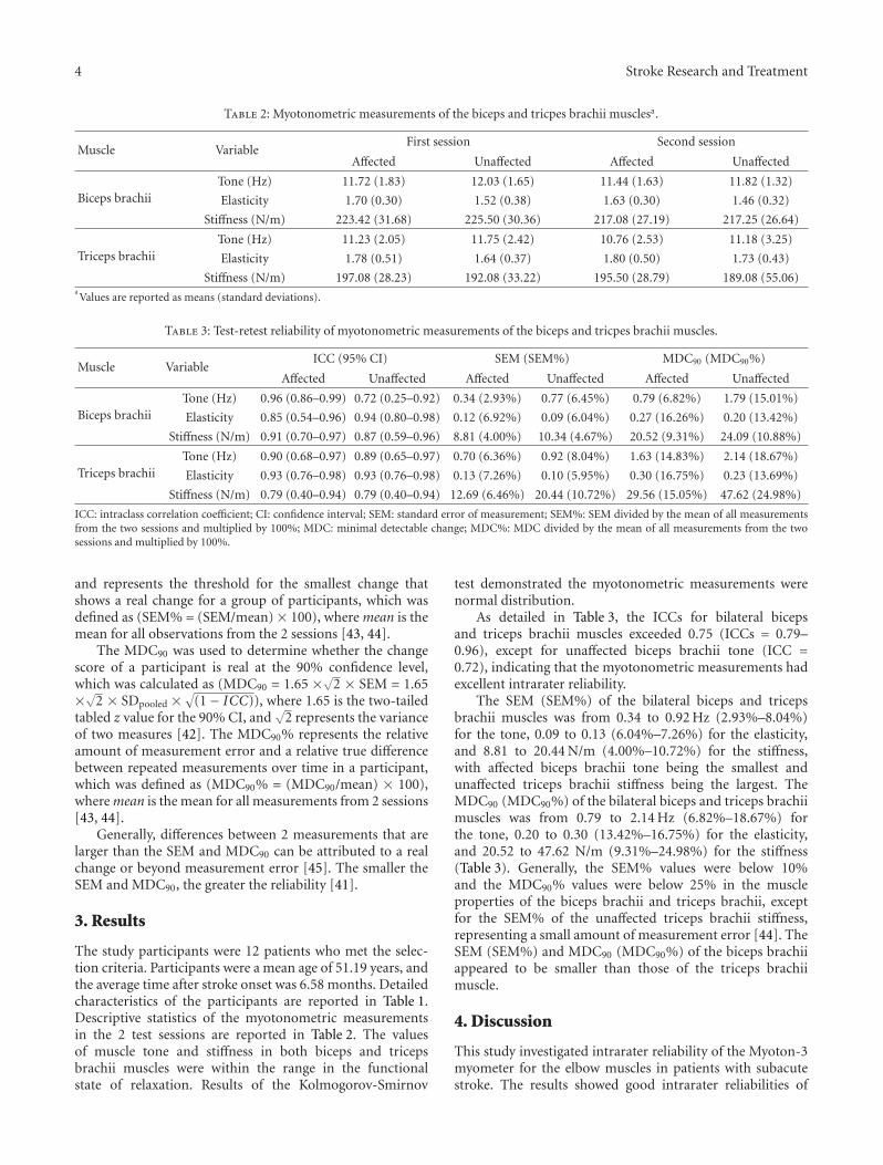
Table 2: Myotonometric measurements of the biceps and tricpes brachii musclesa.
Muscle VariableFirst session Second session
Affected Unaffected Affected Unaffected
Biceps brachiiTone (Hz) 11.72 (1.83) 12.03 (1.65) 11.44 (1.63) 11.82 (1.32)
Elasticity 1.70 (0.30) 1.52 (0.38) 1.63 (0.30) 1.46 (0.32)
Stiffness (N/m) 223.42 (31.68) 225.50 (30.36) 217.08 (27.19) 217.25 (26.64)
Triceps brachiiTone (Hz) 11.23 (2.05) 11.75 (2.42) 10.76 (2.53) 11.18 (3.25)
Elasticity 1.78 (0.51) 1.64 (0.37) 1.80 (0.50) 1.73 (0.43)
Stiffness (N/m) 197.08 (28.23) 192.08 (33.22) 195.50 (28.79) 189.08 (55.06)aValues are reported as means (standard deviations).
Table 3: Test-retest reliability of myotonometric measurements of the biceps and tricpes brachii muscles.
Muscle VariableICC (95% CI) SEM (SEM%) MDC90 (MDC90%)
Affected Unaffected Affected Unaffected Affected Unaffected
Biceps brachiiTone (Hz) 0.96 (0.86–0.99) 0.72 (0.25–0.92) 0.34 (2.93%) 0.77 (6.45%) 0.79 (6.82%) 1.79 (15.01%)
Elasticity 0.85 (0.54–0.96) 0.94 (0.80–0.98) 0.12 (6.92%) 0.09 (6.04%) 0.27 (16.26%) 0.20 (13.42%)
Stiffness (N/m) 0.91 (0.70–0.97) 0.87 (0.59–0.96) 8.81 (4.00%) 10.34 (4.67%) 20.52 (9.31%) 24.09 (10.88%)
Triceps brachiiTone (Hz) 0.90 (0.68–0.97) 0.89 (0.65–0.97) 0.70 (6.36%) 0.92 (8.04%) 1.63 (14.83%) 2.14 (18.67%)
Elasticity 0.93 (0.76–0.98) 0.93 (0.76–0.98) 0.13 (7.26%) 0.10 (5.95%) 0.30 (16.75%) 0.23 (13.69%)
Stiffness (N/m) 0.79 (0.40–0.94) 0.79 (0.40–0.94) 12.69 (6.46%) 20.44 (10.72%) 29.56 (15.05%) 47.62 (24.98%)
ICC: intraclass correlation coefficient; CI: confidence interval; SEM: standard error of measurement; SEM%: SEM divided by the mean of all measurementsfrom the two sessions and multiplied by 100%; MDC: minimal detectable change; MDC%: MDC divided by the mean of all measurements from the twosessions and multiplied by 100%.
and represents the threshold for the smallest change thatshows a real change for a group of participants, which wasdefined as (SEM% = (SEM/mean) × 100), where mean is themean for all observations from the 2 sessions [43, 44].
The MDC90 was used to determine whether the changescore of a participant is real at the 90% confidence level,which was calculated as (MDC90 = 1.65 ×√2 × SEM = 1.65×√2 × SDpooled ×
√(1− ICC)), where 1.65 is the two-tailed
tabled z value for the 90% CI, and√
2 represents the varianceof two measures [42]. The MDC90% represents the relativeamount of measurement error and a relative true differencebetween repeated measurements over time in a participant,which was defined as (MDC90% = (MDC90/mean) × 100),where mean is the mean for all measurements from 2 sessions[43, 44].
Generally, differences between 2 measurements that arelarger than the SEM and MDC90 can be attributed to a realchange or beyond measurement error [45]. The smaller theSEM and MDC90, the greater the reliability [41].
3. Results
The study participants were 12 patients who met the selec-tion criteria. Participants were a mean age of 51.19 years, andthe average time after stroke onset was 6.58 months. Detailedcharacteristics of the participants are reported in Table 1.Descriptive statistics of the myotonometric measurementsin the 2 test sessions are reported in Table 2. The valuesof muscle tone and stiffness in both biceps and tricepsbrachii muscles were within the range in the functionalstate of relaxation. Results of the Kolmogorov-Smirnov
test demonstrated the myotonometric measurements werenormal distribution.
As detailed in Table 3, the ICCs for bilateral bicepsand triceps brachii muscles exceeded 0.75 (ICCs = 0.79–0.96), except for unaffected biceps brachii tone (ICC =0.72), indicating that the myotonometric measurements hadexcellent intrarater reliability.
The SEM (SEM%) of the bilateral biceps and tricepsbrachii muscles was from 0.34 to 0.92 Hz (2.93%–8.04%)for the tone, 0.09 to 0.13 (6.04%–7.26%) for the elasticity,and 8.81 to 20.44 N/m (4.00%–10.72%) for the stiffness,with affected biceps brachii tone being the smallest andunaffected triceps brachii stiffness being the largest. TheMDC90 (MDC90%) of the bilateral biceps and triceps brachiimuscles was from 0.79 to 2.14 Hz (6.82%–18.67%) forthe tone, 0.20 to 0.30 (13.42%–16.75%) for the elasticity,and 20.52 to 47.62 N/m (9.31%–24.98%) for the stiffness(Table 3). Generally, the SEM% values were below 10%and the MDC90% values were below 25% in the muscleproperties of the biceps brachii and triceps brachii, exceptfor the SEM% of the unaffected triceps brachii stiffness,representing a small amount of measurement error [44]. TheSEM (SEM%) and MDC90 (MDC90%) of the biceps brachiiappeared to be smaller than those of the triceps brachiimuscle.
4. Discussion
This study investigated intrarater reliability of the Myoton-3myometer for the elbow muscles in patients with subacutestroke. The results showed good intrarater reliabilities of

Stroke Research and Treatment 5
the myotonometric measurements, with high agreement andsmall measurement error in repeated tests.
This pilot study showed that the myotonometer washighly reliable for measuring biceps and triceps brachiimuscles in patients with subacute stroke. The ICC val-ues were high, indicating excellent reproducibility of theMyoton-3 between successive sessions of assessment. Thiswas in agreement with results of previous interday reliabilitystudies in different muscle groups and study populations[18, 21, 22, 24–27]. In a previous study, looking at theinterday reliability of the Myoton-3 myometer in 10 healthyyoung volunteers who were retested after 2 days, the relaxedbiceps femoris muscle exhibited a moderate ICC score (0.54–0.73) [21]. Another interday reliability study by Bizzini andMannion repeated the same tests of day 1 on day 2 formeasuring relaxed muscle stiffness of the rectus femoris,vastus lateralis, biceps femoris, and gastrocnemius using theMyoton-2 myometer in 10 healthy volunteers. The resultsshowed good to excellent test-retest reliability for all muscles(ICCs 0.80–0.93), except for the vastus lateralis (ICC 0.40)[18].
The ICC cannot detect systematic errors [39], however,and assessments of within-subject variability in the test-retest measurements are necessary to evaluate reliabilitycomprehensively [26]. A good myotonometric measureshould present small measurement errors and be sensitive toidentify the smallest real changes in repeated measurements.Establishing reliability of measures is important not onlyfor repeated measurements with sound stability but also toidentify changes over time [46].
The SEM and MDC90 provide the values of the measure-ment error between repeated tests for a group and for anindividual, respectively. Clinicians and researchers can usethe SEM and MDC90 values to determine whether a changein a group or in an individual is statistically significantly real[47, 48]. That is, the real change in a patient should exceedthe MDC90 of the measure. The SEM (SEM%) and MDC90
(MDC90%) of the bilateral biceps and triceps brachii musclesin this study were small, indicating small measurement error[44]. It should be noted that the SEMs of tone and stiffnessin the biceps and triceps brachii muscles were consistentlyhigher in unaffected side compared to affected side. The ICCsof tone and stiffness in the unaffected biceps and tricepsmuscles were lower than those in the affected ones. Thiswas similar to lower intrarater reliability of measurements ofthe unaffected biceps than that of the affected biceps brachiiin children with spastic-type cerebral palsy [26] and lowerintrarater reliability of the relaxed biceps brachii than theisometrically contracted biceps brachii in healthy adults [22].The reasons for this were not clear, but might be that theparticipants had difficulty in remaining relaxed when thetesting end of the Myoton-3 myometer was first placed onthe unaffected muscles.
The SEM% and MDC90% are independent of the unitsof measurement, which are more easily interpreted andappropriately compares the amount of random error amongmuscle groups and properties [44]. The results of SEM andMDC in the present study can be used as a reference for theMyoton-3 to help clinicians and researchers identify small,
real changes of muscle properties of the biceps and tricepsbrachii muscles between repeated measurements for patientswith substroke.
This study needs to account for the following limitations.First, a variety of factors that may affect resting muscle tonein patients with subacute stroke include the location of strokelesion, the severity and type of stroke, body positioning,level of tension in synergic and antagonist muscles, level oftest anxiety, and time when the test was administered. Only12 patients with subacute stroke who demonstrated a lowlevel of spasticity and were without cognitive impairmentwere included in this pilot study, which may limit thegeneralizability of our findings. Future studies that considerpossible factors that may affect test performance using alarger and more diverse group of patients with stroke areneeded to validate our findings and to promote the clinicalutility of the myotonometer.
Second, passive muscle tone as measured by the elec-tromyography or isokinetic dynamometer has not beenassessed and this is an acknowledged limitation of thepresent study. Additionally, passive muscle properties inthe relaxed state cannot represent functional evaluationduring contracted state. The concurrent measurement ofmuscle properties in relaxation and under contraction witha myotonometer and electromyography or dynamometer issuggested for future studies.
Third, the myotonometry method is not applicable forthe following conditions: thin muscle, muscle with smallmass, obese persons (BMI > 30 kg·m−2), patients sufferingfrom severe pain, muscle which are palpable in small volume,and muscles which are located under other muscles [34]. Inthis pilot study, we did not record the arm girth, BMI, andfatty tissue to consider the obesity; therefore, future study isrecommended to take this issue into account.
Finally, to enhance the applicability and interpretabilityof the myotonometric measurements, future studies to esti-mate minimal clinical important differences are warranted todetermine the degree of meaningful change to patients withstroke.
5. Conclusion
Our pilot study showed that the Myoton-3 myometer hasgood intrarater reliability in measuring the mechanicalproperties of bilateral biceps brachii and triceps brachiimuscles with high agreement and low thresholds to detectreal changes in patients with stroke. The findings indicatethat the Myoton-3 myometer is a reliable tool for quantifyingthe muscle tone, elasticity, and stiffness of elbow flexor andextensor muscles in patients with subacute stroke. Furtherresearch with larger and divergent groups of patients withstroke is needed to confirm the findings of our study.
Acknowledgments
This project was supported in part by the National ScienceCouncil (NSC 97-2314-B-002-008-MY3 and NSC 99-2314-B-182-014-MY3), the National Health Research Institutes(NHRI-EX100-10010PI and NHRI-EX100-9920PI), and the

6 Stroke Research and Treatment
Healthy Aging Research Center at Chang Gung University(EMRPD1A0891) in Taiwan.
References
[1] M. Sjostrom, A. R. Fugl-Meyer, G. Nordin, and L. Wahlby,“Post-stroke hemiplegia; crural muscle strength and struc-ture,” Scandinavian Journal of Rehabilitation Medicine, vol. 7,supplement, pp. 53–67, 1980.
[2] U. Svantesson, H. Takahashi, U. Carlsson, A. Danielsson, andK. S. Sunnerhagen, “Muscle and tendon stiffness in patientswith upper motor neuron lesion following a stroke,” EuropeanJournal of Applied Physiology, vol. 82, no. 4, pp. 275–279, 2000.
[3] E. de Vlugt, J. H. de Groot, K. E. Schenkeveld, J. H. Arendzen,F. C. Van Der Helm, and C. G. Meskers, “The relation betweenneuromechanical parameters and Ashworth score in strokepatients,” Journal of Neuro Engineering and Rehabilitation, vol.7, no. 1, article 35, 2010.
[4] S. Naghdi, N. N. Ansari, K. Mansouri, and S. Hasson, “Aneurophysiological and clinical study of Brunnstrom recoverystages in the upper limb following stroke,” Brain Injury, vol.24, no. 11, pp. 1372–1378, 2010.
[5] B. M. Haas and J. L. Crow, “Towards a clinical measurementof spasticity?” Physiotherapy, vol. 81, no. 8, pp. 474–479, 1995.
[6] T. Ratsep and T. Asser, “Changes in viscoelastic propertiesof skeletal muscles induced by subthalamic stimulation inpatients with Parkinson’s disease,” Clinical Biomechanics, vol.26, no. 2, pp. 213–217, 2011.
[7] K. L. Harburn, K. M. Hill, A. A. Vandervoort et al., “Spasticitymeasurement in stroke: a pilot study,” Canadian Journal ofPublic Health, vol. 83, supplement 2, pp. S41–S45, 1992.
[8] R. W. Bohannon and M. B. Smith, “Interrater reliabilityof a modified Ashworth scale of muscle spasticity,” PhysicalTherapy, vol. 67, no. 2, pp. 206–207, 1987.
[9] A. D. Pandyan, G. R. Johnson, C. I. Price, R. H. Curless, M.P. Barnes, and H. Rodgers, “A review of the properties andlimitations of the Ashworth and modified Ashworth Scales asmeasures of spasticity,” Clinical Rehabilitation, vol. 13, no. 5,pp. 373–383, 1999.
[10] A. Brashear, R. Zafonte, M. Corcoran et al., “Inter- andintrarater reliability of the Ashworth scale and the disabilityassessment scale in patients with upper-limb poststrokespasticity,” Archives of Physical Medicine and Rehabilitation,vol. 83, no. 10, pp. 1349–1354, 2002.
[11] R. T. Katz and W. Z. Rymer, “Spastic hypertonia: mechanismsand measurement,” Archives of Physical Medicine and Rehabil-itation, vol. 70, no. 2, pp. 144–155, 1989.
[12] C. T. Leonard, J. U. Stephens, and S. L. Stroppel, “Assessingthe spastic condition of individuals with upper motoneuroninvolvement: validity of the myotonometer,” Archives of Physi-cal Medicine and Rehabilitation, vol. 82, no. 10, pp. 1416–1420,2001.
[13] L. Alibiglou, W. Z. Rymer, R. L. Harvey, and M. M.Mirbagheri, “The relation between Ashworth scores and neu-romechanical measurements of spasticity following stroke,”Journal of Neuro Engineering and Rehabilitation, vol. 5, article18, 2008.
[14] C. T. Leonard, K. A. Gardipee, J. R. Koontz, J. H. Anderson,and S. A. Wilkins, “Correlation between impairment andmotor performance during reaching tasks in subjects withspastic hemiparesis,” Journal of Rehabilitation Medicine, vol.38, no. 4, pp. 243–249, 2006.
[15] G. Ianieri, R. Saggini, R. Marvulli et al., “New approachin the assessment of the tone, elasticity and the muscularresistance: nominal scales vs MYOTON,” International Journalof Immunopathology and Pharmacology, vol. 22, supplement 3,pp. 21–24, 2009.
[16] L. G. Portney and M. P. Watkins, Foundations of Clini-cal Research: Applications to Practice, Pearson/Prentice Hall,Upper Saddle River, NJ, USA, 3rd edition, 2009.
[17] C. Gubler-Hanna, J. Laskin, B. J. Marx, and C. T. Leonard,“Construct validity of myotonometric measurements of mus-cle compliance as a measure of strength,” Physiological Mea-surement, vol. 28, no. 8, pp. 913–924, 2007.
[18] M. Bizzini and A. F. Mannion, “Reliability of a new, hand-held device for assessing skeletal muscle stiffness,” ClinicalBiomechanics, vol. 18, no. 5, pp. 459–461, 2003.
[19] R. K. Korhonen, A. Vain, E. Vanninen, R. Viir, and J. S.Jurvelin, “Can mechanical myotonometry or electromyogra-phy be used for the prediction of intramuscular pressure?”Physiological Measurement, vol. 26, no. 6, pp. 951–963, 2005.
[20] S. J. Rydahl and B. J. Brouwer, “Ankle stiffness and tissuecompliance in stroke survivors: a validation of Myotonometermeasurements,” Archives of Physical Medicine and Rehabilita-tion, vol. 85, no. 10, pp. 1631–1637, 2004.
[21] M. Ditroilo, A. M. Hunter, S. Haslam, and G. De Vito,“The effectiveness of two novel techniques in establishingthe mechanical and contractile responses of biceps femoris,”Physiological Measurement, vol. 32, no. 8, pp. 1315–1326, 2011.
[22] C. T. Leonard, W. P. Deshner, J. W. Romo, E. S. Suoja,S. C. Fehrer, and E. L. Mikhailenok, “Myotonometer intra-and interrater reliabilities,” Archives of Physical Medicine andRehabilitation, vol. 84, no. 6, pp. 928–932, 2003.
[23] J. Marusiak, A. Jaskolska, S. Budrewicz, M. Koszewicz, andA. Jaskolski, “Increased muscle belly and tendon stiffness inpatients with Parkinson’s disease, as measured by myotonome-try,” Movement Disorders, vol. 26, no. 11, pp. 2119–2122, 2011.
[24] J. Marusiak, K. Kisiel-Sajewicz, A. Jaskolska, and A. Jaskolski,“Higher muscle passive stiffness in Parkinson’s disease patientsthan in controls measured by myotonometry,” Archives ofPhysical Medicine and Rehabilitation, vol. 91, no. 5, pp. 800–802, 2010.
[25] A. Lidstrom, G. Ahlsten, H. Hirchfeld, and S. Norrlin,“Intrarater and interrater reliability of myotonometer mea-surements of muscle tone in children,” Journal of ChildNeurology, vol. 24, no. 3, pp. 267–274, 2009.
[26] D. D. Aarestad, M. D. Williams, S. C. Fehrer, E. Mikhailenok,and C. T. Leonard, “Intra- and interrater reliabilities ofthe myotonometer when assessing the spastic condition ofchildren with cerebral palsy,” Journal of Child Neurology, vol.19, no. 11, pp. 894–901, 2004.
[27] R. Viir, K. Laiho, J. Kramarenko, and M. Mikkelson, “Repeata-bility of trapezius muscle tone assessment by a myometricmethod,” Journal of Mechanics in Medicine and Biology, vol.6, no. 2, pp. 215–228, 2006.
[28] Z. Roja, V. Kalkis, A. Vain, H. Kalkis, and M. Eglite,“Assessment of skeletal muscle fatigue of road maintenanceworkers based on heart rate monitoring and myotonometry,”Journal of Occupational Medicine and Toxicology, vol. 1, no. 1,article 20, 2006.
[29] L. L. Chuang, C. Y. Wu, and K. C. Lin, “Reliability, validity,and responsiveness of Myotonometric measurement of muscletone, elasticity, and stiffness in patients with stroke,” Archivesof Physical Medicine and Rehabilitation, vol. 93, no. 3, pp. 532–540, 2012.

Stroke Research and Treatment 7
[30] G. Kwakkel, B. J. Kollen, J. van der Grond, and A. J. Prevo,“Probability of regaining dexterity in the flaccid upper limb:impact of severity of paresis and time since onset in acutestroke,” Stroke, vol. 34, no. 9, pp. 2181–2186, 2003.
[31] S. Brunnstrom, Movement Therapy in Hemiplegia, Harper &Row, New York, NY, USA, 1970.
[32] M. F. Folstein, S. E. Folstein, and P. R. McHugh, ““Mini mentalstate”. A practical method for grading the cognitive state ofpatients for the clinician,” Journal of Psychiatric Research, vol.12, no. 3, pp. 189–198, 1975.
[33] A. Vain, “Estimation of the functional state of skeletal muscle,”in Control of Ambulation Using Functional NeuromuscularStimulation, P. H. Veltink and H. B. K. Boom, Eds., pp. 51–55,University of Twente Press, Enschede, The Netherlands, 1995.
[34] H. Gapeyeva and A. Vain, Methodical Guide: Principles ofApplying Myoton in Physical Medicine and Rehabilitation,Muomeetria, Tartu, Estonia, 2008.
[35] A. Vain and H. Gapeyeva, Myoton-3: Examples of Patterns,Reports and Results’ Visualization for Studies of Muscle Tone,Elasticity and Stiffness, Muomeetria, Tartu, Estonia, 2007.
[36] A. T. Masi and J. C. Hannon, “Human resting muscletone (HRMT): narrative introduction and modern concepts,”Journal of Bodywork and Movement Therapies, vol. 12, no. 4,pp. 320–332, 2008.
[37] D. G. Simons and S. Mense, “Understanding and measure-ment of muscle tone as related to clinical muscle pain,” Pain,vol. 75, no. 1, pp. 1–17, 1998.
[38] M. M. Panjabi, “The stabilizing system of the spine. PartI. Function, dysfunction, adaptation, and enhancement,”Journal of Spinal Disorders, vol. 5, no. 4, pp. 383–389, 1992.
[39] A. Bruton, J. H. Conway, and S. T. Holgate, “Reliability: whatis it, and how is it measured?” Physiotherapy, vol. 86, no. 2, pp.94–99, 2000.
[40] C. H. Goldsmith, M. Boers, C. Bombardier, and P. Tug-well, “Criteria for clinically important changes in outcomes:development, scoring and evaluation of rheumatoid arthritispatient and trial profiles,” Journal of Rheumatology, vol. 20, no.3, pp. 561–565, 1993.
[41] G. Atkinson and A. M. Nevill, “Statistical methods forassessing measurement error (reliability) in variables relevantto sports medicine,” Sports Medicine, vol. 26, no. 4, pp. 217–238, 1998.
[42] J. S. Schmitt and R. P. Di Fabio, “Reliable change andminimum important difference (MID) proportions facilitatedgroup responsiveness comparisons using individual thresholdcriteria,” Journal of Clinical Epidemiology, vol. 57, no. 10, pp.1008–1018, 2004.
[43] J. M. Wagner, J. A. Rhodes, and C. Patten, “Reproducibilityand minimal detectable change of three-dimensional kine-matic analysis of reaching tasks in people with hemiparesisafter stroke,” Physical Therapy, vol. 88, no. 5, pp. 652–663,2008.
[44] U. B. Flansbjer, A. M. Holmback, D. Downham, C. Patten,and J. Lexell, “Reliability of gait performance tests in men andwomen with hemiparesis after stroke,” Journal of Rehabilita-tion Medicine, vol. 37, no. 2, pp. 75–82, 2005.
[45] S. M. Haley and M. A. Fragala-Pinkham, “Interpreting changescores of tests and measures used in physical therapy,” PhysicalTherapy, vol. 86, no. 5, pp. 735–743, 2006.
[46] H. Beckerman, M. E. Roebroeck, G. J. Lankhorst, J. G. Becher,P. D. Bezemer, and A. L. M. Verbeek, “Smallest real difference,a link between reproducibility and responsiveness,” Quality ofLife Research, vol. 10, no. 7, pp. 571–578, 2001.
[47] W. G. Hopkins, “Measures of reliability in sports medicine andscience,” Sports Medicine, vol. 30, no. 1, pp. 1–15, 2000.
[48] J. E. Lexell and D. Y. Downham, “How to assess the reliabilityof measurements in rehabilitation,” American Journal ofPhysical Medicine and Rehabilitation, vol. 84, no. 9, pp. 719–723, 2005.

Hindawi Publishing CorporationStroke Research and TreatmentVolume 2012, Article ID 802157, 13 pagesdoi:10.1155/2012/802157
Research Article
The Neurorehabilitation Training Toolkit (NTT):A Novel Worldwide Accessible Motor Training Approach forAt-Home Rehabilitation after Stroke
Sergi Bermudez i Badia and M. S. Cameirao
Madeira Interactive Technologies Institute, Universidade da Madeira, Campus Universitario da Penteada, Madeira,9020-105 Funchal, Portugal
Correspondence should be addressed to Sergi Bermudez i Badia, [email protected]
Received 16 November 2011; Revised 6 February 2012; Accepted 13 February 2012
Academic Editor: Agnes Roby-Brami
Copyright © 2012 S. Bermudez i Badia and M. S. Cameirao. This is an open access article distributed under the CreativeCommons Attribution License, which permits unrestricted use, distribution, and reproduction in any medium, provided theoriginal work is properly cited.
After stroke, enduring rehabilitation is required for maximum recovery, and ideally throughout life to prevent functionaldeterioration. Hence we developed a new concept for at-home low-cost motor rehabilitation, the NTT, an Internet-basedinteractive system for upper-limb rehabilitation. In this paper we present the NTT design concepts, its implementation and aproof of concept study with 10 healthy participants. The NTT brings together concepts of optimal learning, engagement, andstorytelling to deliver a personalized training to its users. In this study we evaluate the feasibility of NTT as a tool capable ofautomatically assessing and adapting to its user. This is achieved by means of a psychometric study where we show that the NTTis able to assess movement kinematics—movement smoothness, range of motion, arm displacement and arm coordination—in healthy users. Subsequently, a modeling approach is presented to understand how the measured movement kinematics relateto training parameters, and how these can be modified to adapt the training to meet the needs of patients. Finally, an adaptivealgorithm for the personalization of training considering motivational and performance aspects is proposed. In the next phase wewill deploy and evaluate the NTT with stroke patients at their homes.
1. Introduction
There are about 16 million new strokes per year worldwide[1], and about 5 million of the survivors will sustain motorand/or cognitive impairments for the rest of their lives [2].This situation leads to high societal costs in medical care andrehabilitation expenses, with annual costs above C38 billionin Europe [3]. Collaterally, there is decreasing participationof these patients in professional and social life since strokesurvivors frequently suffer from mood disorders or depres-sion [4, 5]. Therefore, due to its direct and indirect effects,stroke is one of the main contributors for the worldwideburden of disease [6, 7].
Following stroke, enduring rehabilitation is needed formaximum recovery. This requires long-term hospitalizationor outpatient rehabilitation, a situation that is extremely de-manding both for patients and national health systems. Infact, there is a growing interest towards early supported
discharge from hospitals and at-home rehabilitation [8, 9].Nonetheless, despite outpatient rehabilitation programs, itis generally assumed that the full potential for recovery isreached in the first 6 months after stroke, with patients thenbeing discharged from rehabilitation [10]. This might beproblematic as there is evidence that the brain remains plasticat later stages post stroke, meaning that there can still beplace for additional recovery [11, 12]. Ideally, stroke patientsshould undergo maintenance rehabilitation throughout lifeto prevent functional deterioration. Indeed, a significantdecline in mobility after rehabilitation discharge is expectedin one fifth of chronic patients, having a direct impact inperforming activities of daily living (ADL) [13]. Thus, thereis a need to find solutions to provide patients with tools thatallow them to have enduring rehabilitation at their homes.
In recent years, novel technology-based systems havebeen developed for at-home stroke rehabilitation [14–23].Although the price of these systems is lower than standard

2 Stroke Research and Treatment
technology-based rehabilitation devices, most of them arestill unaffordable for the majority of patients. Moreover,these systems usually rely on particular hardware compo-nents, require elaborate setups, and/or need remote guid-ance, which makes them difficult to use particularly if we takeinto account that most of the stroke patients are elderly.
To provide patients with effective, uncomplicated, andinexpensive rehabilitation at their homes, we have developedthe Neurorehabilitation Training Toolkit (NTT), a PC-basedinteractive system for upper-limb rehabilitation. The NTTmakes use of well-established state of the art requirementsfor effective rehabilitation after stroke, providing trainingthat is frequent, reiterative, and task specific [24–26], andthat presents feedback on performance and outcomes [27–29]. These characteristics are achieved through the use ofgame-like tasks displayed on a standard computer, designedto address the specific upper-limb deficits of stroke patients.
In the last few years, a number of standard commercialvideogames have been used for stroke rehabilitation [30, 31].Although the concept is valid, these are ad hoc solutions sincethese games were designed for a different target population,therefore not fully addressing the needs and capabilities ofthe patients. In previous work we developed and evaluateda game for rehabilitation that provides personalized trainingthat is adjusted to the individual capacities of the patients[32]. In this way, patients can undergo a challenging trainingtask designed towards their specific motor deficits (thusavoiding boredom), without falling beyond the patients ca-pabilities which could lead to frustration. We have shownthat the use of such an approach as a complement to stand-ard rehabilitation leads to improved and accelerated recoveryin acute stroke patients [18]. With the NTT, we go one stepfurther because we are able to assess movement kinematicsand psychometrics that allow the personalization of a num-ber of training objectives. These objectives include the rangeof motion and movement smoothness.
The NTT consists of widely available home-based tech-nologies, namely, a PC or laptop, two mice, and an Internetconnection. This configuration makes the system uncompli-cated and rather inexpensive, and therefore accessible andaffordable for most patients. The use of an Internet con-nection is twofold. On one hand, the game “lives” online—for an easy upgradeability of the software—and requires noinstallation. On the other hand, it can be used to monitorand assess the progress of patients remotely. Additionally,the NTT encompasses automatic questionnaires to assess theusability, engagement, and acceptance of the training tasks,as well as to measure the training impact on the performanceof ADLs. Here we present the design concepts behind theNTT, the results of a pilot study where we developed anovel personalization algorithm based on functional motoroutcomes, and demonstrate the NTT capabilities as an as-sessment and monitoring tool tested with healthy users. Dueto its simplicity and innovative features, we believe that theNTT is a powerful tool that will give patients access to anaffordable, effective, and long-term rehabilitation at theirhomes, allowing them to maximize and sustain recoverywhile increasing their overall quality of life.
2. Materials and Methods
The NTT is a software-based motor training toolkit thatis accessed and executed from Internet as a web applet bymeans of an Internet browser (Figure 1(A)). NTT has beenverified to work with the most commonly used browsers suchas Internet Explorer, Google Chrome, Safari, Firefox, andOpera. In this context, the web browser serves as the inter-face to the training toolkit, allowing its execution withoutneeding local installation or additional software besides theNTT applet itself. In addition to a PC with an Internet con-nection, the NTT user needs a working email account forcommunication and user support if needed.
On the hardware side, the NTT can run on any moderncomputer with Windows O.S. (Windows XP, Vista, or7) (Figure 1(B)). The only requirement for the PC is tohave two mice connected to it. Computer mice are wide-spread, reliable, and extremely low-cost devices that willserve to track the physical movements of the arms ofthe patients, which in turn will be used to interact withthe NTT. Technically, the main advantage of the NTT isthe ability to capitalize on existing common hardware todeliver personalized and continued training. This is achievedby the low hardware requirements and the accessibilityof our online system (Figure 1(C)). Internet serves as thedistribution channel for the NTT, offering its training andother advantages to any patient anywhere in the world withaccess to a PC and an Internet connection. Furthermore,NTT upgrades are immediately deployed to the user’s homewithout requiring action from their side. The NTT can beaccessed at http://neurorehabilitation.m-iti.org/NTT/.
Our web services serve a threefold mission: instruction,training and feedback. The NTT site provides detailed in-struction on how to use and operate the NTT. After givinginformed consent, access to the NTT training is granted.The NTT runs as an embedded application on our siteand delivers the training, logs data, and communicates withpatients (Figure 1(D)). A remote data server is used to storelog files after each training session containing relevant infor-mation such as the training date, user ID, data on the phys-ical movements during training, game events, perform-ance measures, and hardware configuration. Email servicesare used to communicate access codes and relevant userquestionnaires after specific NTT training sessions.
2.1. NTT Game Training Scenario. The NTT web applicationwas developed with the Python programming languageusing the open source game engine Panda3D (http://www.panda3d.org), which is maintained by Carnegie MellonUniversity. NTT is designed along neuroscientific and thera-peutic guidelines for stroke rehabilitation based on a numberof concepts: relevance of training to ADLs, neuroscientificprinciples of recovery, narrative, personalization or individ-ualization, augmented feedback, and engagement.
2.1.1. Training Rational. After stroke, the recovery of theupper-limb functionality is essential for the recovery ofthe capacity of performing activities of daily living (ADL).

Stroke Research and Treatment 3
Web browser
VR web appletEmail notifications
Onlinequestionnaires
HTML protocolSMTP protocol
VR training scenario (SW)
A
Internet
C
HTTP server and database
DPC running windows2 miceInternet connectionValid email address
Data loggingFile and web serverEmail server
Remote client (HW)
B
Em
q
i d
W b b
A
C
BBBBB
Web browser
VR VV web appletmmam il notifications
Onlinequeqq stionnaires
HTML protocolSMTP protocol
VR training scenario (SW)
C
DPC running w dindows2 miceInternet connectionValid email addrVV ess
Remote client (HW)
EEEmmmmmmm
qqqqq
i di d
EEE
Figure 1: Neurorehabilitation Training Toolkit system architecture. (A) A web browser is used to access the application and its instruction athttp://neurorehabilitation.m-iti.org/NTT/ (B) NTT can be executed on any modern PC equipped with two mice and an internet connection.(C) The NTT software is accessed freely from Internet, where the NTT servers are located. (D) A number of remote servers host the NTTsite, the application itself, and are used to log user data.
Y
X
Mov
emen
t
Mov
emen
t
Distance
Figure 2: Sketch of the NTT-bilateral training task. Two micetrack the position of right and left hands. The training consists ofbilateral and coordinated displacement of the arms in the Y axis tochange the heading direction of the NTT-virtual glider. The distancebetween hands in the Y axis defines the direction and the turningspeed. See text for further information.
The ability of a stroke survivor to perform ADLs determineshis/her level of independence to a great extent and thereforethe need of specialized care, dependence on relatives, andsocietal burden [33]. Hence, the improvement of upper-limb function is a priority after stroke. Effective therapiesfor motor rehabilitation support the notion of trainingintensity, frequency, and task-specificity as being determi-nant outcome factors [25, 34, 35]. However, patients donot always have access to the ideal training frequency andintensity. In addition, the training specificity towards the
needs of the patient appears to be of special relevancesince it optimizes the training outcome [36]. Consequently,an effective upper-limb training paradigm should be basedon training intensity, frequency, iteration, task specificity,and personalization of training to the patient’s needs. NTTexploits the above-mentioned principles in a game-likeexperience where patients are confronted with a virtualscenario that requires them to repeatedly perform physicalmovements of varying intensity in order to complete a task.The NTT training task is performed on a tabletop (Figure 2).
With continuous research, more data about the post-stroke brain mechanisms are becoming available whichsupport the claim that neuronal plasticity is the maincontributor for recovery [37–40]. Plasticity is a life-longproperty of the brain that allows cortical networks toreorganize and regain lost functionality, even many yearsafter stroke [11, 41]. Recent findings show how the reorga-nization of perilesional and contralesional cortical networkscontributes to the recruitment of functional corticospinalfibers, which is vital for recovery [39, 42]. Therefore, novelrehabilitation approaches should be designed to capitalizeon brain plasticity to regain motor function by meansof a functional reorganization of the cortical networks. Itis widely established in the rehabilitation literature thattraining frequency and intensity are effective drivers forcortical reorganization [34, 35, 43]. However, more recentneuroscientific findings should also be integrated in currentrehabilitation praxis. This is the case of approaches basedon the Mirror Neuron System (MNS), a population ofneurons located in premotor and parietal cortical areas thathave particular properties that make them good candidatesto mobilize plasticity, and for motor learning in general

4 Stroke Research and Treatment
[44, 45]. The MNS is also known as the action recognitionsystem because of its properties. These neurons are activeboth during the performance of meaningful and goal-oriented motor actions and the passive observation of thoseactions [46, 47]. Thus, this is a convenient neural system thatcan be exploited to activate motor-related areas and thereforeto mobilize cortical plasticity by means of the observationof motor actions [48, 49]. Several motor rehabilitationapproaches capitalize on this system to enhance or speed uprecovery [32, 50, 51]. In a previous randomized controlledtrial, we have shown that a VR system based on this principlespeeds up recovery [18] in terms of motor function asmeasured by the Fugl-Meyer scale [52] and in ADLs asassessed by the Chedoke Arm and Hand Activity Inventory(CAHAI) [53]. Consequently, and in line with our previouswork, we have integrated those neuroscientific aspects in theNTT by means of the presentation of a virtual characterwhose limbs perform meaningful goal-oriented actions thatare triggered by and correlated with the physical movementsof the arms of the patient. The combination of VR and goal-oriented actions has already been shown to activate the MNSand related motor areas, therefore facilitating functionalcortical reorganization [50].
2.1.2. Training Task. In its current form, the NTT trainingtask requires patients to be able to read, and not to havemajor cognitive deficits and seizures, sensory aphasias, orother perceptual problems that could impede the under-standing of the task. Our training consists of a bilateraltask that requires practicing range of motion and movementcoordination. Several reasons support the choice of this par-ticular task. First, a home-based training has to allow for agradual functional integration and use of the paretic armin the completion of tasks. Thus, our bilateral training taskallows the patient to support the paretic arm with thenonparetic one when the task is too demanding. As such,our training task is also appropriate for hemiplegic patientswith severely reduced mobility or motor control. Second,although evidence on the advantage of bilateral trainingwhen compared to unilateral training is limited [54], thereare a number of findings that support its use [55, 56].For instance, nowadays it is widely accepted that bimanualcoordination is a largely distributed brain process, andtherefore its training engages motor areas to a larger extent[57]. Moreover, since most patients manifest some degreeof bimanual coordination deficit, bimanual coordinationtraining is recommended to be part of any rehabilitation pro-gram [58]. Further, there is increasing evidence that bilateraltraining has a particularly beneficial effect in patients withlow Fugl-Meyer motor scores, whereas it is as effective asunilateral training in well-recovered patients [55, 59].
The NTT bimanual training exercise is performed on atabletop, providing arm support against gravity, what widensthe spectrum of patients who can use it. The patient’s handmovements are tracked by means of two computer micethat the patient is manipulating (Figure 2). The interactionwith real objects has been shown to elicit better kinematics[60]. The physical movements of the patient are then used
to control the movements of the arms of an avatar that isdisplayed on the computer’s screen. The avatar arms con-trol the steering direction of a glider that flies forward at aconstant speed, accumulating collectable objects. The turn-ing speed of the glider (α deg/sec) is defined as a function ofthe distance between mice (Figure 2):
α = gain ·(YRight − YLeft
), (1)
where YRight and YLeft indicate the Y position of right andleft mice, respectively, and gain is a factor that modulates theturning speed of the avatar.
2.1.3. Gaming Concepts. The glider control task was chosendue to the bimanual and intuitive nature of the controlmechanism, the built-in control system that allows support-ing the paretic arm actions with the nonparetic arm, theslow movement dynamics, and the pleasantness of a flyingexperience.
Improving on previous work, we decided to add anarrative element to the NTT since a flat and static virtualtraining task can make training monotonous and eventuallylimit the patient’s engagement. Consequently, NTT exploitsa simple narrative structure to build a story around thetraining task to increase the engagement of patients, facilitatethe comprehension of the training objectives, and, mostimportantly, to deliver a clear sense of progress. This simplenarrative is based on Freytag’s classic concept of play andcounter-play [61]. Play in our case describes how our playeraffects the world (an exciting force or rise), and counter-playhow the world affects the player (a tragic force or decay). Wecreated a sequence of plays and counter-plays that narratethe journey of our virtual character through an unknownenvironment that culminates with him finding home. Thegame structure includes a tutorial of the game, and a seriesof game levels/environments that lead the way to a narrativeclimax when home is reached (Figure 3(a)). In order toreach home, the avatar has to collect flying objects thatenable him to complete the different game levels. Differentgame elements are used to shape the narrative curve; theseinclude the task difficulty, presence of control disturbances,the choice of the color palette, the design of the environmentitself, the choice of music and its rhythm, and the usage ofthe view/camera perspective.
In addition to the narrative component, the NTT makesextensive use of feedback using multiple and redundant feed-back modalities to ensure that the patient understands thetask and is positively rewarded for his/her accomplishments[28, 62]. For instance, on-screen pictorial instructions anda compass are used to indicate the direction and physicalmovements that are needed in order to get to the target. Thecurrent item to be collected is clearly indicated by a largemoving arrow. To help the patient in the task, both the targetarrow and the compass head turn from red into green whenthe avatar is correctly aligned in direction to the target. Everycollected item is rewarded with a positive visual and auditoryfeedback. Collectable items are divided into easy (balloons)and hard (stars) ones, which are counted as separate scores.Finally, the total distance moved by the arms during training
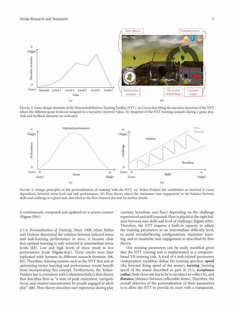
Stroke Research and Treatment 5
0
(Low)
∞(High)
Nar
rati
ve s
tru
ctu
re
Tutorial Level 1 Level 2 Level 3 Level 4 Level 5
Time
(a)
Task objects Training scores
Orientationcompass
On-screeninstructions
Currenttarget
(b)
Figure 3: Game design elements of the Neurorehabilitation Training Toolkit (NTT). (a) Curve describing the narrative structure of the NTTwhere the different game levels are assigned to a narrative (interest) value. (b) Snapshot of the NTT training scenario during a game play.Task and feedback elements are indicated.
0
(Low)
0
(Low) ∞(High)
∞(High)
Stress
Perf
orm
ance
Optimal performance
(a)
0
(Low)
∞(High)
Skills
0
(Low)
∞(High)
Ch
alle
nge
Anxiety
Boredom
Flowchannel
(b)
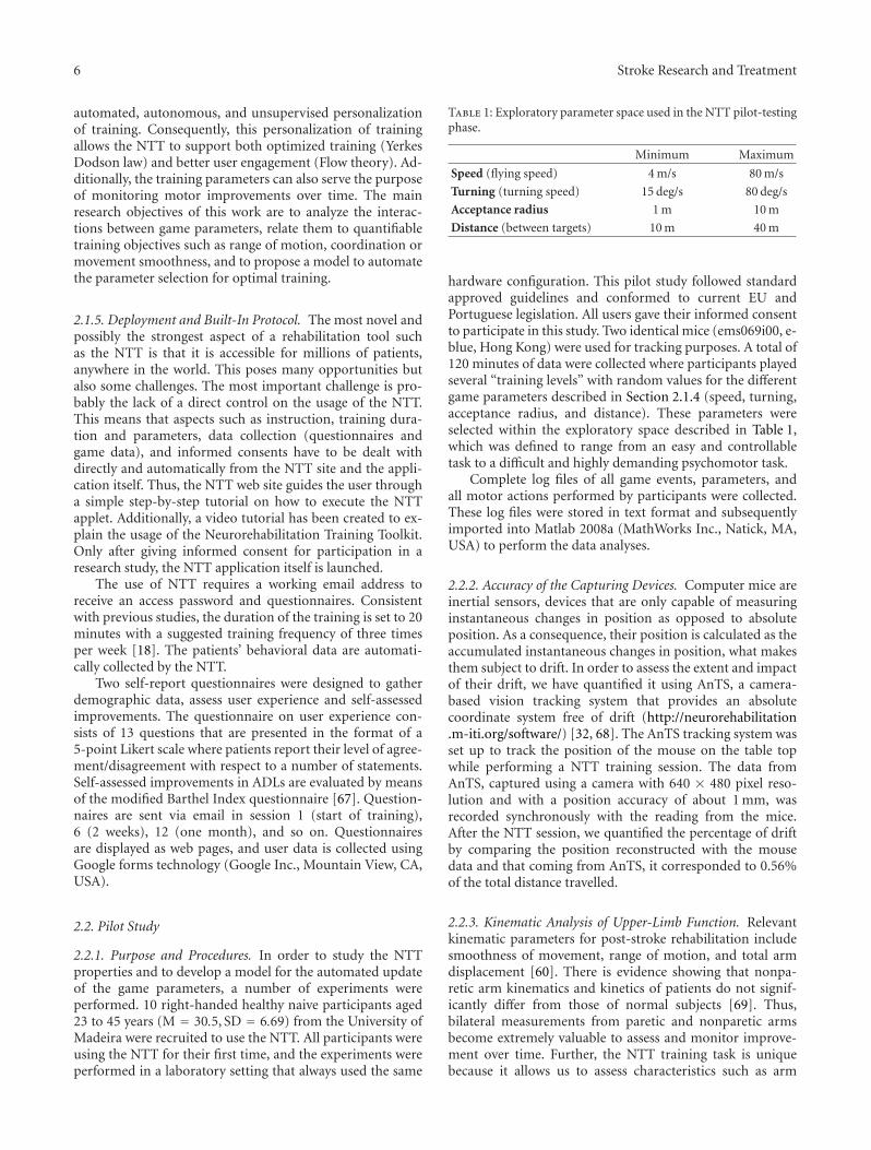
Figure 4: Design principles of the personalization of training with the NTT. (a) Yerkes-Dodson law established an inverted U-curvedependence between stress level and task performance. (b) Flow theory places the maximum user engagement in the balance betweenskills and challenge in a given task, described as the flow channel. See text for further details.
is continuously computed and updated on a screen counter(Figure 3(b)).
2.1.4. Personalization of Training. Since 1908, when Yerkesand Dodson discovered the relation between induced stressand task-learning performance in mice, it became clearthat optimal learning is only achieved at intermediate stresslevels [63]. Low and high levels of stress result in lowperformance levels (Figure 4(a)). These results were laterreplicated with humans in different research domains [64,65]. Therefore, training systems such as the NTT that aim atoptimizing motor learning and performance would benefitfrom incorporating this concept. Furthermore, the Yerkes-Dodson law is consistent with Csikszentmihalyi’s flow theorythat describes flow as “a state of peak enjoyment, energeticfocus, and creative concentration by people engaged in adultplay” [66]. Flow theory describes user experience during play
(anxiety, boredom, and flow) depending on the challengeexperienced and skill required. Flow is placed at the right bal-ance between user skills and level of challenge (Figure 4(b)).Therefore, the NTT requires a built-in capacity to adjustthe training parameters to an intermediate difficulty level,to avoid stressful/boring configurations, maximize learn-ing, and to maximize user engagement as described by flowtheory.
Our training parameters can be easily modified giventhat the NTT training task is implemented as a computer-based VR training task. A total of 4 task-related parameters(independent variables) define the training specifics: speed(the forward flying speed of the avatar), turning (turningspeed of the avatar described as gain in (1)), acceptanceradius (how close one has to be to an object to collect it), anddistance (distance between collectable items). Therefore, theoverall objective of the personalization of these parametersis to allow the NTT to provide its users with a transparent,
6 Stroke Research and Treatment
automated, autonomous, and unsupervised personalizationof training. Consequently, this personalization of trainingallows the NTT to support both optimized training (YerkesDodson law) and better user engagement (Flow theory). Ad-ditionally, the training parameters can also serve the purposeof monitoring motor improvements over time. The mainresearch objectives of this work are to analyze the interac-tions between game parameters, relate them to quantifiabletraining objectives such as range of motion, coordination ormovement smoothness, and to propose a model to automatethe parameter selection for optimal training.
2.1.5. Deployment and Built-In Protocol. The most novel andpossibly the strongest aspect of a rehabilitation tool suchas the NTT is that it is accessible for millions of patients,anywhere in the world. This poses many opportunities butalso some challenges. The most important challenge is pro-bably the lack of a direct control on the usage of the NTT.This means that aspects such as instruction, training dura-tion and parameters, data collection (questionnaires andgame data), and informed consents have to be dealt withdirectly and automatically from the NTT site and the appli-cation itself. Thus, the NTT web site guides the user througha simple step-by-step tutorial on how to execute the NTTapplet. Additionally, a video tutorial has been created to ex-plain the usage of the Neurorehabilitation Training Toolkit.Only after giving informed consent for participation in aresearch study, the NTT application itself is launched.
The use of NTT requires a working email address toreceive an access password and questionnaires. Consistentwith previous studies, the duration of the training is set to 20minutes with a suggested training frequency of three timesper week [18]. The patients’ behavioral data are automati-cally collected by the NTT.
Two self-report questionnaires were designed to gatherdemographic data, assess user experience and self-assessedimprovements. The questionnaire on user experience con-sists of 13 questions that are presented in the format of a5-point Likert scale where patients report their level of agree-ment/disagreement with respect to a number of statements.Self-assessed improvements in ADLs are evaluated by meansof the modified Barthel Index questionnaire [67]. Question-naires are sent via email in session 1 (start of training),6 (2 weeks), 12 (one month), and so on. Questionnairesare displayed as web pages, and user data is collected usingGoogle forms technology (Google Inc., Mountain View, CA,USA).
2.2. Pilot Study
2.2.1. Purpose and Procedures. In order to study the NTTproperties and to develop a model for the automated updateof the game parameters, a number of experiments wereperformed. 10 right-handed healthy naive participants aged23 to 45 years (M = 30.5, SD = 6.69) from the University ofMadeira were recruited to use the NTT. All participants wereusing the NTT for their first time, and the experiments wereperformed in a laboratory setting that always used the same
Table 1: Exploratory parameter space used in the NTT pilot-testingphase.
Minimum Maximum
Speed (flying speed) 4 m/s 80 m/s
Turning (turning speed) 15 deg/s 80 deg/s
Acceptance radius 1 m 10 m
Distance (between targets) 10 m 40 m
hardware configuration. This pilot study followed standardapproved guidelines and conformed to current EU andPortuguese legislation. All users gave their informed consentto participate in this study. Two identical mice (ems069i00, e-blue, Hong Kong) were used for tracking purposes. A total of120 minutes of data were collected where participants playedseveral “training levels” with random values for the differentgame parameters described in Section 2.1.4 (speed, turning,acceptance radius, and distance). These parameters wereselected within the exploratory space described in Table 1,which was defined to range from an easy and controllabletask to a difficult and highly demanding psychomotor task.
Complete log files of all game events, parameters, andall motor actions performed by participants were collected.These log files were stored in text format and subsequentlyimported into Matlab 2008a (MathWorks Inc., Natick, MA,USA) to perform the data analyses.
2.2.2. Accuracy of the Capturing Devices. Computer mice areinertial sensors, devices that are only capable of measuringinstantaneous changes in position as opposed to absoluteposition. As a consequence, their position is calculated as theaccumulated instantaneous changes in position, what makesthem subject to drift. In order to assess the extent and impactof their drift, we have quantified it using AnTS, a camera-based vision tracking system that provides an absolutecoordinate system free of drift (http://neurorehabilitation.m-iti.org/software/) [32, 68]. The AnTS tracking system wasset up to track the position of the mouse on the table topwhile performing a NTT training session. The data fromAnTS, captured using a camera with 640 × 480 pixel reso-lution and with a position accuracy of about 1 mm, wasrecorded synchronously with the reading from the mice.After the NTT session, we quantified the percentage of driftby comparing the position reconstructed with the mousedata and that coming from AnTS, it corresponded to 0.56%of the total distance travelled.
2.2.3. Kinematic Analysis of Upper-Limb Function. Relevantkinematic parameters for post-stroke rehabilitation includesmoothness of movement, range of motion, and total armdisplacement [60]. There is evidence showing that nonpa-retic arm kinematics and kinetics of patients do not signif-icantly differ from those of normal subjects [69]. Thus,bilateral measurements from paretic and nonparetic armsbecome extremely valuable to assess and monitor improve-ment over time. Further, the NTT training task is uniquebecause it allows us to assess characteristics such as arm

Stroke Research and Treatment 7
coordination and contribution of each arm. Thus, datagathered by the NTT could allow for a kinematic analysisof upper-limb function, providing comparative data on eachpatient’s paretic versus nonparetic arm. We performed afirst pilot evaluation with healthy participants to assess theadequacy and validity of the NTT as a tool to assess theabove-mentioned kinematic measures in healthy users.
Smoothness Index. Consistent with previous research, wedefine a Smoothness Index (SI) that quantifies the regularityand smoothness of a particular movement trajectory [69].This measure allows us to assess the level of fine movementcontrol of the NTT users for each arm independently. Thesmoothness of a movement depends on the number of seg-ments in which it can be divided. Here we consider move-ment segments as those segments in between movement ac-celerations and decelerations. Therefore, the SI is computedfrom an analysis of the speed profile of a movement. We de-fine SI as the count of the number of local minima in thespeed profile for a specific action. Thus, lower SI values in-dicate movements with a smoother velocity profile, whereashigher values indicate those movements with more accelera-tions changes. Before the calculation of the SI, mice data areprefiltered with a Gaussian window filter of 1 sec length toreduce the amount of noise.
Arm Displacement. In order to display feedback about theactual work that has been realized, NTT also computes theaccumulated distance that each hand has been displacedduring training (Figure 3, training scores). This is a measureof actual physical exercise and allows us to assess the exactamount of physical movement that was needed by each userto complete the training tasks.
Range of Motion. Another relevant kinematic measure,related to the actual amount of physical movement, is Rangeof Motion (ROM). There is evidence that suggests that ROMcan be used as a predictor of functional outcome in ADL[70]. As such, being able to automatically assess and monitorROM throughout the training process can become a rathervaluable feature of the NTT, particularly when comple-mented with the Barthel Index assessment for performancein ADLs [67]. The ROM that is measured by the NTT re-sults from the combined action of shoulder and elbow (ex-tension and flexion) on the end effector, the hand, whichis tracked with the mouse. ROM captures aspects of themovement dynamics, providing information on functionalarm aperture that other measures cannot provide. In thecase of stroke patients, a limited ROM does not necessarilyindicate limited movement within the working ROM. Ouroperational measure of functional ROM—the ROM beingeffectively used during the NTT training—is computed in cmfrom consecutive arm extensions and flexions as measured bymice.
Arm Coordination and Arm Contribution. As opposed toother rehabilitation approaches, NTT allows for an auto-mated and objective quantification of functional outcomes,
some of which are not possible to quantify in real-world taskswithout the aid of interactive technologies. One particularexample of this unique capability of the NTT is the abilityto quantify arm coordination. During NTT training, andbecause of the bilateral nature of its training, physicalmovements are expected to be symmetrical movements per-formed simultaneously with both arms. We therefore quan-tify arm coordination as the absolute value of the correlationcoefficient between the data from both mice. Consequently,the computation of the correlation coefficient of perfectlycoordinated movements would result in a coefficient equalto 1. Our quantification of movement coordination is par-ticularly advantageous for our purpose since the correlationoperation is insensitive to the actual gain of the movement.Namely, perfectly correlated (coordinated) movements arenot required to be of the same amount but should followthe same temporal profile. This means that the NTT ena-bles us to separately assess movement coordination and armcontribution to the training task. The later—separate armcontribution—is computed by assessing the particular in-volvement of each arm in the steering of the avatar. Sincethe avatar’s heading direction is driven by the differential inposition of the two mice, the training can be performed withdissimilar involvement of the paretic and nonparetic arms(Figure 2). Thus, the contribution of each arm is computedas the correlation coefficient between the arm movementtrajectories over time and the effective heading directionchanges as described by (1). So, a low correlation coefficientwill be obtained for an arm with reduced contribution to thebilateral control task.
Modeling. If successful, the ability of the NTT to provideus with relevant behavioral information about the patient’smotor capabilities will allow us to assess the impact ofthe NTT on different aspects of motor recovery. Here weintroduce how the NTT can be tailored to automatically self-adapt depending on the user’s needs. In order to do so, wefirst have to quantify the relationship between our kinematicanalysis of upper-limb function and the NTT trainingparameters. For that reason we use a multiple regressionmodeling approach. Consistent with previous work onpsychometrics, our model considers all game parameters, itssecond-order terms, and first-order interactions [32]. In ourcase, we propose to model each of the previously describedkinematic measures (Section 2.2.3) in the following way:
Kinematic measure = c0 + c1 ∗ speed + c2 ∗ turning
+ c3 ∗ acceptance + c4 ∗ distance
+ c5 ∗ speed∗ turning
+ c6 ∗ speed∗ acceptance
+ c7 ∗ speed∗ distance
+ c8 ∗ turning ∗ acceptance
+ c9 ∗ turning ∗ distance

8 Stroke Research and Treatment
+ c10 ∗ distance∗ acceptance
+ c11 ∗ speed2 + c12 ∗ turning2
+c13 ∗ acceptance2 +c14∗distance2,
(2)
where kinematic measure is the dependent variable (eithermovement smoothness, range of motion, arm displacement,or arm coordination); speed, turning, acceptance, anddistance are the independent variables set by the NTT duringour pilot-testing experiment, and the ci constants are thecoefficients resulting from the multiple regression analysis.
Besides the kinematic modeling approach, a parallelsystem is established to maximize challenge and engagementand therefore potentiate the highest performance level andflow. This is achieved by means of a time race [62, 71] duringwhich NTT monitors the efficiency of the patient’s actions byassessing his/her performance at every task completion (itemcollection) (Figure 5, efficiency assessment). Given that theflight speed and item distance are known training parametersto the NTT, it is possible to define efficiency in the followingway:
Efficiency = actual task completion timeideal task completion time
. (3)
Efficiency is computed at every task completion/object col-lection, which enables estimating how much time is neededby the patient on each task. Thus, the time race is imple-mented by providing every object with a timer that indicateshow much time is left to collect it. The timer is based onthe patient’s efficiency data from previously collected items,being modulated by a Gaussian noise function (M = 1, SD =0.2) (Figure 5, task time). Targets that allow more than theaverage time needed by the patient are presented as balloons(easy), and targets requiring less time are presented as stars(difficult). If the task is not completed within the timewindow, the target disappears and the timer is reset to alonger time as determined by the patient’s mean efficiency(3). On one hand, this time race strategy allows personalizingthe challenge of the task to the particular skills of thepatient [62]. This design allows us to change task difficultywithout interfering with the training parameters, which inour case determine the training objectives. On the otherhand, training parameters (speed, turning, acceptance, anddistance) are only updated when the patient’s efficiency in-creases or decreases (3). Hence, the more efficient the pa-tient is on the task, the higher the demands in terms oftraining objectives. Consequently, our personalization algo-rithm consists of two parallel loops that shape challengeand training parameters to provide optimized and enjoyabletraining.
3. Results
As described above, the NTT was evaluated with 10 right-handed healthy naive participants. As expected, healthy par-ticipants present a low Smoothness Index SI (MD = 5), with
Kinematic modelParameter adjustment
Reset timeto mean efficiency
No
Kinematic modelParameter adjustment
completion? Task
Task timeGaussian randomization
1
Efficiencyassessment
Yes
Figure 5: NTT personalization process. The adaptation of thetraining parameters is realized according to the patient’s efficiencyin the task. A kinematic model maps training objectives toindividual NTT parameters. In parallel, a timer is used to create atime-race task. See text for further information.
no significant difference between dominant and nondomi-nant arms (Wilcoxon signed-ranks test, Z = −.0212,P =.9831) (Figure 6). A similar SI interquartile range (IQR) fordominant and nondominant arms is found (6.5 and 6, resp.).
Example data on arm displacement from participantnumber 1 is shown in Figure 7. A group analysis showsa median hand displacement of 23.25 cm per item col-lected, moving the dominant arm significantly less thanthe nondominant arm (Wilcoxon Signed-ranks test, Z =13.5642,P < .0001).
Consistent with the arm displacement data, a pairedsample t-test analysis revealed that the dominant arm usesa lower functional ROM (M = 23.38, SD = 16.14 cm) thanthe nondominant arm for healthy participants (M = 27.18,SD = 18.12 cm), t(403) = −4.6924,P < .0001 (Figure 8).
The investigation of arm coordination in healthy partic-ipants reveals a high degree of coordination, that is, highcorrelation values between the movement trajectories of thearms during the training task (M = .8143, SD = .2433).This is consistent with the high degree of motor controlof this population of users (Figure 9). Interestingly, thearm contribution analysis of the NTT training task—whichassesses the impact of the movement of each arm in thesteering of the avatar—reveals larger contributions (t(9) =−8.5925,P < .0001) for dominant arms (M = .5631, SD =.1872) than for nondominant arms (M = .5095, SD =.2333) (see Section 2.2.3 for the details on the quantificationof arm coordination and contribution). While this resultis consistent with what is expected from dominant arms,in previous measures, we observed lower displacementand ROM for the dominant arm. This indicates that thedominant arm does not only contribute more, but it is also

Stroke Research and Treatment 9
350 400 450 500 550
0
5
Spee
d (m
m/s
)Left mouse
350 400 450 500 550
Time (s)
Right mouse
−5
0
5
Spee
d (m
m/s
)
−5
(a)
Nondominant arm Dominant arm
0
10
20
30
40
50
60
Smoo
thn
ess
inde
x (S
I) p
er it
em
(b)
Figure 6: Analysis of movement smoothness by means of the Smoothness Index (SI). (a) Sample speed profiles of the movements trackedby both NTT mice. The number of movement segments contained in a specific movement trajectory is computed by locating the speed localminima (red and green dots). (b) SI is computed for each arm and for each item collected in the virtual training environment. Boxplot from10 healthy participants. For more information see text.
Nondominant arm Dominant arm
0
10
20
30
40
50
60
70
80
Dis
plac
emen
t pe
r it
em (
cm)
Figure 7: Arm displacement on the tabletop (cm) per item collectedduring a NTT training session. Data from healthy participantnumber 1.
more precise in its actions, requiring less movement andROM to accomplish the task.
Figure 9 shows that the NTT is capable of assessingdifferent aspects of the movement kinematics when used byhealthy users. Consequently, if we can assess how the NTTtraining parameters relate to the actual resulting physicalexercise and movement kinematics on the tabletop, wecan optimize the NTT training parameters accordingly todeliver appropriate and personalized training. Subsequently,we used these quantitative data on movement kinematicscombined with modeling techniques to establish statisticalrelationships between the training parameters used and theactual physical exercise associated with them. We performedfour separate multiple regression analyses to find out how
Nondominant arm Dominant arm0
10
20
30
40
50
Ran
ge o
f m
otio
n (
RO
M)
(cm
)
Figure 8: Range of Motion (ROM) usage during NTT training.Data consist of 404 data samples from the 10 healthy participants.Bar indicates mean ROM and error bars SD.
the NTT training parameters contribute to the changesobserved in the previously determined kinematic measures.Using the data from our healthy participants, we have iden-tified the training parameters, the first-order interactions,and the second-order parameters that contribute to themovement smoothness, range of motion, arm displacement,and arm coordination (Table 2). The exact model coef-ficients described in (2) and related statistics are shownin Table 3 (see supplementary material available online atdoi:10.1155/2012/802157).
From the multiple regression analysis we can concludethat not all training parameters contribute to all movementkinematics. This means that, depending on our training

10 Stroke Research and Treatment
Table 2: Table of significances of the multiple regression analyses. Crosses indicate terms that have a significant contribution to the kinematicmeasure and thus are taken into account in our modeling purposes. s, t, a, and d refer to the training variables flying speed, turning speed,acceptance radius, and distance, respectively. See Table 3 for further details on the model coefficients and related statistics.
s t a d s∗t s∗a s∗d t∗a t∗d a∗d s2 t2 a2 d2
Movement smoothness × ×Range of motion × × × × ×Arm displacement ×Arm coordination × × ×
Arm coordination Nondominant arm Dominant arm0
0.2
0.4
0.6
0.8
1
1.2
1.4
Cor
rela
tion
coe
ffici
ent
Figure 9: Arm coordination and contribution analysis for the NTTtraining task. Data from 10 healthy participants. Bars indicate themean and error bars the SD.
objectives, a different set of parameters need to be changedto affect the user’s movement kinematics. Thus, accordingto the relations established in Table 2, we have implementeda personalization training algorithm that can adjust thetraining parameters depending on the training objectives setby the user (Figure 5). In this algorithm for the adaptationof training, only a subset of the training parameters willbe adjusted during training depending on whether theuser needs to train movement smoothness, ROM, armdisplacement, arm coordination, or any combination ofthese. The selection of the training objectives will thereforedetermine the kinematic models to be used during an NTTsession (Figure 5, kinematic model).
4. Conclusions and Discussion
In this paper we presented the Neurorehabilitation TrainingToolkit (NTT), a low-cost neurorehabilitation system thataims at offering an alternative for continued at-homerehabilitation after stroke. The NTT is designed alongneuroscientific guidelines as well as game design principlesto drive motor recovery, while maximizing engagementand motivational factors. Additionally, it is designed to becompatible with any modern PC that runs Windows O.S.and has an Internet connection. Because of its architectureand design, the NTT is a unique rehabilitation system. By
capitalizing on broadly available home technology, the NTTis a free rehabilitation tool that is accessible worldwide viaa web browser. This feature makes it immediately availablefor large-scale deployment, having an enormous potential asa motor training solution for patients in remote areas, withlow income, low mobility, or without access to rehabilitationfacilities. Furthermore, it is designed to serve as an at-home complement to other training or simply for continuedrehabilitation after hospital discharge. Since NTT is accessedvia a web browser, it does not have to be installed on thepatient’s computer and it is easily upgradeable, allowingpatients to increase their portfolio of training games. NTTis a completely self-contained training system that does notrequire the intervention of a therapist or specialist. Further,NTT is a safe system completely based on noninvasivetechnologies, making it a suitable solution for unsupervisedand at-home use. Based on the psychometric analysispresented here, the NTT is designed to automatically adjustits training parameters to the training. More concretely, NTTallows training movement smoothness, range of motion, armdisplacement, arm coordination, or any combination of thesebased on the presented kinematic models.
As opposed to other approaches using technologiessuch as robotic devices, custom-made tracking technologies,brain-computer interfaces, data gloves, or expensive com-mercial hardware [16, 32, 51, 72–75], the NTT only requiresa PC, a pair of computer mice and an Internet connection.This advantage of the NTT, which lowers the accessibilitythreshold for at-home rehabilitation training technologies,also represents one of its most important challenges. Forinstance, the lack of a supervised setting and the absenceof complete upper-body tracking represent a limitation thatmore sophisticated systems do not suffer [16, 18, 75–77].More specifically, the NTT kinematic analyses are based onthe end effector. However, despite this caveat, the NTT isable to contribute with a detailed and functional analysisof training task performance. As reported in this paper,outcomes such as ROM, arm displacement, arm coordina-tion, and individual arm contribution to the task objectivesare unique assessment properties of the NTT. Our studywith healthy participants has shown a precise quantificationof ROM, coordination, arm contribution, and smoothnessof movement, while also being able to detect handedness.Interestingly, the data revealed differences in ROM and armcontribution as result of handedness but no differences inmovement smoothness. Compared to the nondominant arm,the dominant arm displayed a significant reduction of ROMwhile simultaneously showing an increased contribution in

Stroke Research and Treatment 11
task performance. In the particular case of patients, ROMand accumulated displacement are expected to be moreinformative since they reveal different aspects of movementkinematics (arm aperture and amount of movement), whichare not necessarily dependent. Although we have shown ahigh accuracy assessing functional outcome in healthy users,more research is needed to understand the limitations of thissystem when used by elderly and stroke patients. Given thenature of the bilateral training task and the possibility ofsupporting the actions of the paretic arm with the nonpareticarm, the use of compensatory movements is expected tobe reduced. Nevertheless, we need to understand the extentof the use of compensatory movements and its effect inthe assessed movement kinematics, since combined trunk,shoulder and elbow movements can contribute to effectivehand movements. Similarly, the presented kinematic modelsgenerated with healthy user data need to be further validatedin a trial with patients.
Despite the limitations of the presented psychometricmodeling, the NTT goes beyond the adaptive and per-sonalized training of previous work, allowing for a novelindividualized and parametric training of several functionaloutcomes [18]. Finally, the automatic data-collection capa-bility of the NTT and its questionnaire system enable alarge-scale deployment to quantify its impact. We believethat rehabilitation technologies will continue to move in thisdirection, from custom and more expensive hardware-basedsolutions to low-cost systems that can be used at the patient’shome [15, 16, 31]. The presented study is the first step thatvalidates the design and technological aspects of the NTTwith healthy users. In the next phase, it will be deployedand validated with stroke patients at their homes. The NTT,with its novel design principles and assessment capabilities,represents another step towards consolidation and at-homedeployment of these technologies.
Acknowledgments
Support for this research was provided by the Fundacao paraa Ciencia e Tecnologia (Portuguese Foundation for Scienceand Technology) through the Carnegie Mellon Portugal Pro-gram under Grant CMU-Pt/0004/2007.
References
[1] K. Strong, C. Mathers, and R. Bonita, “Preventing stroke: sav-ing lives around the world,” The Lancet Neurology, vol. 6, no.2, pp. 182–187, 2007.
[2] World Health Organization, “The global burden of disease:2004 update,” Tech. Rep., World Health Organization, Geneva,Switzerland, 2008.
[3] A. di Carlo, “Human and economic burden of stroke,” Age andAgeing, vol. 38, no. 1, pp. 4–5, 2009.
[4] L. Ayerbe, S. Ayis, A. G. Rudd, P. U. Heuschmann, and C.D. A. Wolfe, “Natural history, predictors, and associationsof depression 5 years after stroke: the South London StrokeRegister,” Stroke, vol. 42, no. 7, pp. 1907–1911, 2011.
[5] S. A. Thomas and N. B. Lincoln, “Factors relating to depres-sion after stroke,” British Journal of Clinical Psychology, vol. 45,no. 1, pp. 49–61, 2006.
[6] C. D. Mathers and D. Loncar, “Projections of global mortalityand burden of disease from 2002 to 2030,” PLoS Medicine, vol.3, no. 11, p. e442, 2006.
[7] A. S. Kim and S. C. Johnston, “Global variation in the relativeburden of stroke and ischemic heart disease,” Circulation, vol.124, no. 3, pp. 314–323, 2011.
[8] P. Langhorne, L. Widen-Holmqvist, G. Taylor et al., “Earlysupported discharge after stroke,” Journal of RehabilitationMedicine, vol. 39, no. 2, pp. 103–108, 2007.
[9] T. Larsen, T. S. Olsen, and J. Sorensen, “Early home-supporteddischarge of stroke patients: a health technology assessment,”International Journal of Technology Assessment in Health Care,vol. 22, no. 3, pp. 313–320, 2006.
[10] H. S. Jorgensen, H. Nakayama, H. O. Raaschou, J. Vive-Larsen,M. Stoier, and T. S. Olsen, “Outcome and time course of re-covery in stroke. Part II: time course of recovery. The Copen-hagen Stroke study,” Archives of Physical Medicine and Rehabil-itation, vol. 76, no. 5, pp. 406–412, 1995.
[11] T. Hodics, L. G. Cohen, and S. C. Cramer, “Functional imagingof intervention effects in stroke motor rehabilitation,” Archivesof Physical Medicine and Rehabilitation, vol. 87, no. 12, sup-plement 2, pp. 36–42, 2006.
[12] S. J. Page, D. R. Gater, and P. Bach-Y-Rita, “Reconsidering themotor recovery plateau in stroke rehabilitation,” Archives ofPhysical Medicine and Rehabilitation, vol. 85, no. 8, pp. 1377–1381, 2004.
[13] I. G. L. van de Port, G. Kwakkel, I. van Wijk, and E. Lindeman,“Susceptibility to deterioration of mobility long-term afterstroke: a prospective cohort study,” Stroke, vol. 37, no. 1, pp.167–171, 2006.
[14] L. Piron, A. Turolla, M. Agostini et al., “Exercises for pareticupper limb after stroke: a combined virtual-reality and tel-emedicine approach,” Journal of Rehabilitation Medicine, vol.41, no. 12, pp. 1016–1020, 2009.
[15] M. Sivak, C. Mavroidis, and M. K. Holden, “Design of a lowcost multiple user virtual environment for rehabilitation(MUVER) of patients with stroke,” Studies in Health Technol-ogy and Informatics, vol. 142, pp. 319–324, 2009.
[16] M. J. Johnson, Y. Shakya, E. Strachota, and S. I. Ahamed,“Low-cost monitoring of patients during unsupervised ro-bot/computer assisted motivating stroke rehabilitation,” Bi-omedizinische Technik, vol. 56, no. 1, pp. 5–9, 2011.
[17] E. B. Brokaw, I. Black, R. J. Holley, and P. S. Lum, “HandSpring Operated Movement Enhancer (HandSOME): a port-able, passive hand exoskeleton for stroke rehabilitation,” IEEETransactions on Neural Systems and Rehabilitation Engineering,vol. 19, no. 4, pp. 391–399, 2011.
[18] M. S. Cameirao, S. B. Badia, E. Duarte, and P. F. Verschure,“Virtual reality based rehabilitation speeds up functional re-covery of the upper extremities after stroke: a randomized con-trolled pilot study in the acute phase of stroke using the Reha-bilitation Gaming System,” Restorative Neurology and Neuro-science, vol. 29, no. 5, pp. 287–298, 2011.
[19] M. K. Holden, T. A. Dyar, and L. Dayan-Cimadoro, “Tel-erehabilitation using a virtual environment improves upperextremity function in patients with stroke,” IEEE Transactionson Neural Systems and Rehabilitation Engineering, vol. 15, no.1, pp. 36–42, 2007.
[20] R. C. Loureiro, W. S. Harwin, K. Nagai, and M. Johnson,“Advances in upper limb stroke rehabilitation: a technologypush,” Medical & Biological Engineering & Computing, vol. 49,no. 10, pp. 1103–1118, 2011.
[21] S. V. Adamovich, A. Mathai, G. G. Fluet, Q. Qiu, J. Lewis, andA. S. Merians, “Design of a complex virtual reality simulation
12 Stroke Research and Treatment
to train finger motion for persons with hemiparesis: a proofof concept study,” Journal of NeuroEngineering and Rehabilita-tion, vol. 6, no. 1, p. 28, 2009.
[22] R. Colombo, F. Pisano, A. Mazzone et al., “Design strategiesto improve patient motivation during robot-aided rehabilita-tion,” Journal of NeuroEngineering and Rehabilitation, vol. 4, p.3, 2007.
[23] C. R. Carignan and H. I. Krebs, “Telerehabilitation robotics:bright lights, big future?” Journal of Rehabilitation Researchand Development, vol. 43, no. 5, pp. 695–710, 2006.
[24] P. Langhorne, F. Coupar, and A. Pollock, “Motor recovery afterstroke: a systematic review,” The Lancet Neurology, vol. 8, no.8, pp. 741–754, 2009.
[25] L. Richards, C. Hanson, M. Wellborn, and A. Sethi, “Drivingmotor recovery after stroke,” Topics in Stroke Rehabilitation,vol. 15, no. 5, pp. 397–411, 2008.
[26] P. Langhorne, J. Bernhardt, and G. Kwakkel, “Stroke rehabili-tation,” The Lancet, vol. 377, no. 9778, pp. 1693–1702, 2011.
[27] B. M. Quaney, J. He, G. Timberlake, K. Dodd, and C. Carr,“Visuomotor training improves stroke-related ipsilesionalupper extremity impairments,” Neurorehabilitation and Neu-ral Repair, vol. 24, no. 1, pp. 52–61, 2010.
[28] B. I. Molier, E. H. F. van Asseldonk, H. J. Hermens, and M.J. A. Jannink, “Nature, timing, frequency and type of aug-mented feedback; does it influence motor relearning of thehemiparetic arm after stroke? A systematic review,” Disabilityand Rehabilitation, vol. 32, no. 22, pp. 1799–1809, 2010.
[29] N. J. Seo, H. W. Fischer, R. A. Bogey, W. Z. Rymer, and D. G.Kamper, “Use of visual force feedback to improve digit forcedirection during pinch grip in persons with stroke: a pilotstudy,” Archives of Physical Medicine and Rehabilitation, vol. 92,no. 1, pp. 24–30, 2011.
[30] K. E. Laver, S. George, S. Thomas, J. E. Deutsch, and M.Deutsch, “Virtual reality for stroke rehabilitation,” The Co-chrane Database of Systematic Reviews, vol. 9, Article IDCD008349, 2011.
[31] M. R. Mouawad, C. G. Doust, M. D. Max, and P. A. McNulty,“Wii-based movement therapy to promote improved upperextremity function post-stroke: a pilot study,” Journal ofRehabilitation Medicine, vol. 43, no. 6, pp. 527–533, 2011.
[32] M. S. Cameirao, S. B. I. Badia, E. D. Oller, and P. F. M.J. Verschure, “Neurorehabilitation using the virtual realitybased Rehabilitation Gaming System: methodology, design,psychometrics, usability and validation,” Journal of NeuroEngineering and Rehabilitation, vol. 7, no. 1, p. 48, 2010.
[33] C. Vincent, I. Deaudelin, L. Robichaud et al., “Rehabilitationneeds for older adults with stroke living at home: perceptionsof four populations,” BMC Geriatrics, vol. 7, article 20, 2007.
[34] G. Kwakkel, B. Kollen, and E. Lindeman, “Understanding thepattern of functional recovery after stroke: facts and theories,”Restorative Neurology and Neuroscience, vol. 22, no. 3–5, pp.281–299, 2004.
[35] G. Kwakkel, R. van Peppen, R. C. Wagenaar et al., “Effects ofaugmented exercise therapy time after stroke: a meta-analysis,”Stroke, vol. 35, no. 11, pp. 2529–2536, 2004.
[36] J. W. Krakauer, “Motor learning: Its relevance to stroke recov-ery and neurorehabilitation,” Current Opinion in Neurology,vol. 19, no. 1, pp. 84–90, 2006.
[37] B. H. Dobkin, “Training and exercise to drive poststrokerecovery,” Nature Clinical Practice Neurology, vol. 4, no. 2, pp.76–85, 2008.
[38] U. Sabatini, D. Toni, P. Pantano et al., “Motor recovery afterearly brain damage: a case of brain plasticity,” Stroke, vol. 25,no. 2, pp. 514–517, 1994.
[39] R. J. Seitz, C. M. Butefisch, R. Kleiser, and V. Homberg, “Re-organisation of cerebral circuits in human ischemic brain dis-ease,” Restorative Neurology and Neuroscience, vol. 22, no. 3–5,pp. 207–229, 2004.
[40] T. H. Murphy and D. Corbett, “Plasticity during stroke recov-ery: from synapse to behaviour,” Nature Reviews Neuroscience,vol. 10, no. 12, pp. 861–872, 2009.
[41] S. C. Cramer, “Repairing the human brain after stroke: I.Mechanisms of spontaneous recovery,” Annals of Neurology,vol. 63, no. 3, pp. 272–287, 2008.
[42] C. M. Butefisch, R. Kleiser, and R. J. Seitz, “Post-lesional cere-bral reorganisation: evidence from functional neuroimagingand transcranial magnetic stimulation,” Journal of PhysiologyParis, vol. 99, no. 4–6, pp. 437–454, 2006.
[43] S. Sonoda, E. Saitoh, S. Nagai, M. Kawakita, and Y. Kanada,“Full-time integrated treatment program, a new system forstroke rehabilitation in Japan: comparison with conventionalrehabilitation,” American Journal of Physical Medicine andRehabilitation, vol. 83, no. 2, pp. 88–93, 2004.
[44] G. Rizzolatti and L. Craighero, “The mirror-neuron system,”Annual Review of Neuroscience, vol. 27, pp. 169–192, 2004.
[45] G. Rizzolatti, M. Fabbri-Destro, and L. Cattaneo, “Mirrorneurons and their clinical relevance,” Nature Clinical PracticeNeurology, vol. 5, no. 1, pp. 24–34, 2009.
[46] V. Gallese, L. Fadiga, L. Fogassi, and G. Rizzolatti, “Actionrecognition in the premotor cortex,” Brain, vol. 119, no. 2, pp.593–609, 1996.
[47] G. di Pellegrino, L. Fadiga, L. Fogassi, V. Gallese, and G.Rizzolatti, “Understanding motor events: a neurophysiologicalstudy,” Experimental Brain Research, vol. 91, no. 1, pp. 176–180, 1992.
[48] D. Ertelt, S. Small, A. Solodkin et al., “Action observationhas a positive impact on rehabilitation of motor deficits afterstroke,” NeuroImage, vol. 36, supplement 2, pp. T164–T173,2007.
[49] M. E. Michielsen, R. W. Selles, J. N van der Geest et al., “Motorrecovery and cortical reorganization after mirror therapy inchronic stroke patients: a phase II randomized controlledtrial,” Neurorehabilitation and Neural Repair, vol. 25, no. 3, pp.223–233, 2011.
[50] S. V. Adamovich, K. August, A. Merians, and E. Tunik, “Avirtual reality-based system integrated with fmri to studyneural mechanisms of action observation-execution: a proofof concept study,” Restorative Neurology and Neuroscience, vol.27, no. 3, pp. 209–223, 2009.
[51] A. S. Merians, E. Tunik, G. G. Fluet, Q. Qiu, and V. S.Adamovich, “Innovative approaches to the rehabilitation ofupper extremity hemiparesis using virtual environments,”European Journal of Physical and Rehabilitation Medicine, vol.45, no. 1, pp. 123–133, 2009.
[52] A. R. Fugl Meyer, L. Jaasko, and I. Leyman, “The post strokehemiplegic patient. I. A method for evaluation of physicalperformance,” Scandinavian Journal of Rehabilitation Medi-cine, vol. 7, no. 1, pp. 13–31, 1975.
[53] S. Barreca, C. Gowland, P. Stratford et al., “Developmentof the chedoke arm and hand activity inventory: theoreticalconstructs, item generation, and selection,” Topics in Stroke Re-habilitation, vol. 11, no. 4, pp. 31–42, 2004.
[54] S. M. Waller and J. Whitall, “Bilateral arm training: why andwho benefits?” NeuroRehabilitation, vol. 23, no. 1, pp. 29–41,2008.
[55] G. N. Lewis and W. D. Byblow, “Neurophysiological andbehavioral adaptations to a bilateral training intervention in

Stroke Research and Treatment 13
individuals following stroke,” Clinical Rehabilitation, vol. 18,no. 1, pp. 48–59, 2004.
[56] J. H. Cauraugh, S. A. Coombes, N. Lodha, S. K. Naik, andJ. J. Summers, “Upper extremity improvements in chronicstroke: coupled bilateral load training,” Restorative Neurologyand Neuroscience, vol. 27, no. 1, pp. 17–25, 2009.
[57] A. R. Luft, S. McCombe-Waller, J. Whitall et al., “Repetitivebilateral arm training and motor cortex activation in chronicstroke: a randomized controlled trial,” Journal of the AmericanMedical Association, vol. 292, no. 15, pp. 1853–1861, 2004.
[58] M. Wiesendanger and D. J. Serrien, “The quest to understandbimanual coordination,” Progress in Brain Research, vol. 143,pp. 491–505, 2004.
[59] D. K. Rose and C. J. Winstein, “Bimanual training after stroke:are two hands better than one?” Topics in Stroke Rehabilitation,vol. 11, no. 4, pp. 20–30, 2004.
[60] C. Y. Wu, C. A. Trombly, K. C. Lin, and L. Tickle-Degnen, “Akinematic study of contextual effects on reaching performancein persons with and without stroke: influences of objectavailability,” Archives of Physical Medicine and Rehabilitation,vol. 81, no. 1, pp. 95–101, 2000.
[61] G. Freytag and E. J. MacEwan, Freytag’s Technique of the Dra-ma: An Exposition of Dramatic Composition and Art, SC Griggs,Chicago, Ill, USA, 1894.
[62] L. von Ahn and L. Dabbish, “Designing games with a pur-pose,” Communications of the ACM, vol. 51, no. 8, pp. 58–67,2008.
[63] R. M. Yerkes and J. D. Dodson, “The relation of strength ofstimulus to rapidity of habit-formation,” Journal of Compar-ative Neurology and Psychology, vol. 18, no. 5, pp. 459–482,1908.
[64] E. Duffy, Activation and Behavior, Wiley, New York, NY, USA,1962.
[65] R. B. Malmo, “Activation: a neuropsychological dimension,”Psychological Review, vol. 66, no. 6, pp. 367–386, 1959.
[66] M. Csikszentmihalyi, Beyond Boredom and Anxiety: Experienc-ing Flow in Work and Play, Behavioral Science Series, chapter14, Jossey-Bass, San Francisco, Calif, USA, 1st edition, 1975.
[67] R. H. Fortinsky, C. V. Granger, and G. B. Seltzer, “The use offunctional assessment in understanding home care needs,”Medical Care, vol. 19, no. 5, pp. 489–497, 1981.
[68] Z. Mathews, S. B. I. Badia, and P. F. M. J. Verschure, “A novelbrain-based approach for multi-modal multi-target trackingin a mixed reality space,” in Proceedings of the 4th InternationalConference and Workshop on Virtual Reality (INTUITION ’07),IEEE, Athens, Greece, October 2007.
[69] C. C. Tsao and M. M. Mirbagheri, “Upper limb impairmentsassociated with spasticity in neurological disorders,” Journal ofNeuroEngineering and Rehabilitation, vol. 4, no. 1, p. 5, 2007.
[70] K. L. Beissner, J. E. Collins, and H. Holmes, “Muscle forceand range of motion as predictors of function in older adults,”Physical Therapy, vol. 80, no. 6, pp. 556–563, 2000.
[71] T. W. Malone, Heuristics for Designing Enjoyable User Inter-faces: Lessons from Computer Games, Ablex Publishing, Nor-wood, NJ, USA, 1984.
[72] A. Caria, C. Weber, D. Brotz et al., “Chronic stroke recoveryafter combined BCI training and physiotherapy: a case report,”Psychophysiology, vol. 48, no. 4, pp. 578–582, 2010.
[73] A. C. Lo, P. D. Guarino, L. G. Richards et al., “Robot-assistedtherapy for long-term upper-limb impairment after stroke,”The New England Journal of Medicine, vol. 362, no. 19, pp.1772–1783, 2010.
[74] X. L. Hu, K. Y. Tong, R. Song, X. J. Zheng, and W. W. F. Leung,“A comparison between electromyography-driven robot and
passive motion device on wrist rehabilitation for chronicstroke,” Neurorehabilitation and Neural Repair, vol. 23, no. 8,pp. 837–846, 2009.
[75] A. Montagner, A. Frisoli, L. Borelli et al., “A pilot clinical studyon robotic assissted rehabilitation in VR with an arm exoskele-ton device,” in Proceedings of the Virtual Rehabilitation, pp. 57–64, Venice, Italy, September 2007.
[76] M. Guidali, A. Duschau-Wicke, S. Broggi, V. Klamroth-Marganska, T. Nef, and R. Riener, “A robotic system to trainactivities of daily living in a virtual environment,” Medical &Biological Engineering & Computing, vol. 49, no. 10, pp. 1213–1223, 2011.
[77] R. J. Sanchez, J. Liu, S. Rao et al., “Automating arm movementtraining following severe stroke: functional exercises withquantitative feedback in a gravity-reduced environment,” IEEETransactions on Neural Systems and Rehabilitation Engineering,vol. 14, no. 3, pp. 378–389, 2006.

Hindawi Publishing CorporationStroke Research and TreatmentVolume 2012, Article ID 784232, 10 pagesdoi:10.1155/2012/784232
Research Article
ENGAGE: Guided Activity-Based Gaming in Neurorehabilitationafter Stroke: A Pilot Study
Ann Reinthal,1 Kathy Szirony,2 Cindy Clark,3 Jeffrey Swiers,1
Michelle Kellicker,1, 3 and Susan Linder1, 4
1 School of Health Sciences, Cleveland State University, 2121 Euclid Avenue, Cleveland, OH 44115, USA2 Rehabilitation and Sports Therapy, Cleveland Clinic, 9500 Euclid Avenue, Cleveland, OH 44195, USA3 Rehabilitation and Sports Therapy, Lakewood Hospital, 14519 Detroit Road, Lakewood, OH 44107, USA4 Department of Biomedical Engineering, Cleveland Clinic, 9500 Euclid Avenue, Cleveland, OH 44195, USA
Correspondence should be addressed to Ann Reinthal, [email protected]
Received 16 October 2011; Revised 12 January 2012; Accepted 4 February 2012
Academic Editor: Keh-Chung Lin
Copyright © 2012 Ann Reinthal et al. This is an open access article distributed under the Creative Commons Attribution License,which permits unrestricted use, distribution, and reproduction in any medium, provided the original work is properly cited.
Introduction. Stroke is a leading cause of disability in healthy adults. The purpose of this pilot study was to assess the feasibility andoutcomes of a novel video gaming repetitive practice paradigm, (ENGAGE) enhanced neurorehabilitation: guided activity-basedgaming exercise. Methods. Sixteen individuals at least three months after stroke served as participants. All participants receivedconcurrent outpatient therapy or took part in a stroke exercise class and completed at least 500 minutes of gaming. Primarybaseline and posttest outcome measures included the Wolf motor function test (WMFT) and the Fugl-Meyer assessment (FMA).ENGAGE uses a game selection algorithm providing focused, graded activity-based repetitive practice that is highly individualizedand directed. The Wilcoxon signed ranks test was used to determine statistical significance. Results. There were improvements inthe WMFT (P = 0.003) and the FMA (P = 0.002) that exceeded established values of minimal clinically important difference.Conclusions. ENGAGE was feasible and an effective adjunct to concurrent therapy after stroke.
1. Introduction
Stroke is one of the leading causes of disability in theUnited States with approximately 795,000 people experi-encing stroke each year [1]. Many of these individualsare left with residual upper extremity deficits that limitindependence with activities of daily living [2] and havebeen correlated with decreased self-reported quality of life[3]. Current research suggests that task-specific practiceresults in more functional improvement than traditionalneuromuscular rehabilitation (TNR) [4–6]. TNR after strokehas been defined as a typical contemporary practice thatincludes a combination of active, impairment-based exer-cises, functional task practice, passive exercise, and activitiesto enhance sensory inputs [2, 7]. However, based on anobservational study by Lang and colleagues [7], only 25% ofa typical 36-minute upper extremity therapy session involvedfunctional task practice. While the number of practicerepetitions required in task-specific practice is not yet fully
delineated, it is thought that individuals after stroke mustpractice for a minimum of several thousand repetitions inorder to relearn a task [5].
Several obstacles exist to obtaining adequate practice.First, there are a large number of tasks to be practiced,and while there is some transfer of learning between similartasks, such as picking up a coffee mug and a can of soda,practicing these tasks would not necessarily be helpful forlearning to write with a pencil [5]. Second, repetitive practiceof a single task may be boring both to the patient and theclinician [7–10]. Third, the learner must actively engagein task practice; it is not adequate to be passively movedthrough the motion or activity [7, 11]. Finally, to ensureoptimal movement quality and therapeutic benefit duringtask practice, a skilled professional must often supervise [8–10], making it expensive to provide the necessary hours oftraining.
Current approaches that provide the opportunity forrepetitive practice include constraint-induced therapy (CIT),

2 Stroke Research and Treatment
robotics, and virtual reality (VR). In CIT, the individual isforced to practice various tasks repetitively with the hemi-paretic arm for six hours a day in a structured environmentand for 90% of waking hours, while the nonhemiplegicarm is constrained. Successful candidates for CIT havehigher functioning hands, with some active control of wristand finger extension [6, 12]. Both the minimal movementrequirement and the intensive structured therapy protocolpreclude many patients after stroke from benefiting fromthis intervention. Robotic instrumentation actively guidesmovement during repetitive practice, providing partial assis-tance while also constraining the movement to occur ina specific “correct” movement pattern. These devices havebeen successfully utilized in people after stroke with bothhigh- and low-level functioning upper extremities primarilyin research environments [13, 14], but have not been widelyadopted in traditional clinical settings. Finally, VR envi-ronments provide a three-dimensional computer-generatedimmersion experience where the “player” completes the tasksimilarly to doing so in the real world. The experience istypically engaging, realistic and has been shown to transferto a comparable real world activity. VR has been successfullyused after stroke and can provide upper extremity repetitivetask practice to both lower and higher functioning upperextremities [15, 16], but its scalability has been limited dueto both size and expense of the equipment requirements.
Video gaming systems such as the Wii and PlayStation IIwith Eye Toy are commercially available, inexpensive virtualreality-type systems that have been used as an effectiveadjunct to TNR for individuals with stroke [8–10, 17, 18].Gaming provides an engaging interaction which is criticalin optimizing motor learning [5] and can be easily tailoredto individuals with varying degrees of paresis, similarly tosome robotic and VR systems, but unlike CIT interventionsthat are only appropriate for higher functioning arms. Threemechanisms are hypothesized to account for the effectivenessof video gaming in inducing neuroplasticity. First, gamingfrequently utilizes more energy than sedentary activities [19]which is important since individuals after stroke are typicallyless active [20], and enhanced cardiovascular fitness has beenshown to improve cortical plasticity [21, 22]. Second, gamingoffers significant bilateral practice opportunities, hypothe-sized to effectively induce reorganization in contralesionalmotor networks [23–28]. Finally, during play, the gamer seesan avatar concurrently performing the activity on screenproviding visual biofeedback [18, 29], similar to techniquesusing the mirror neuron network after stroke [30, 31]. Thisnetwork involves the activation of neurons in the parietaland premotor cortex during actual movement and whenobserving others moving [32].
For this study, we developed a novel method of inte-grating commercially available video gaming into TNRafter stroke called ENGAGE, enhanced neurorehabilitation:guided activity-based gaming exercise. The aims of this studywere to (1) examine the feasibility of adding ENGAGE as anadjunct to TNR in a natural clinical practice environmentwithout additional skilled therapy personnel costs usingcommercially available gaming equipment; (2) examinewhether practice completed through ENGAGE gaming leads
to improvement in upper extremity function in individualsafter stroke; (3) examine effects of covariates on the efficacyof ENGAGE gaming, including concurrent rehabilitation,quantity of gaming time, initial motor and sensory function,and chronicity after stroke; (4) examine whether studyparticipants found gaming to be motivating as compared tohome exercise program practice.
2. Methods
2.1. Study Design. The study was approved by the Insti-tutional Review Boards for Human Subjects Research atthe Cleveland Clinic and Cleveland State University, andinformed consent was obtained from each participant. Eachparticipant consented to participating in this study. Thisstudy utilized a mixed-methods design, with qualitativemeasures used to address aim no. 1: feasibility and aimno. 3: covariate analyses. A within-subject pretest posttestdesign, consistent with the principles of community basedparticipatory research [33] was used to address aim no.2: improvement in upper extremity function and aim no. 4:motivation. Outcomes were assessed without a control group,evaluating practice in a natural clinical practice environment.Two sites within a regional health system were utilized,representing urban and suburban clinical settings.
2.2. Participants. Patients enrolled in outpatient physicaland/or occupational therapy or those participating in acommunity-based stroke exercise class were eligible toparticipate. In this pilot study, there was no attempt tocontrol what occurred in concurrent outpatient physicaland/or occupational therapy, but activities were consistentwith contemporary practice as described by Lang et al.[7] and Miller et al. [2], and included adjuncts such asfunctional electrical stimulation. Participants in the exerciseclass completed cardiovascular exercises in addition to basicgroup stretching and strengthening activities. Inclusion cri-teria included the following: (1) hemiparesis in the involvedupper extremity between Brunnstrom stages 2–6; (2) ≥threemonths after stroke; (3) sufficient balance to sit safely inunsupported sitting while participating in video gaming;(4) adequate cognitive and communication skills to learnvideo game within three sessions; (5) the ability to playindependently or with support staff assist after set up.Participants were excluded if they had confounding medicalinterventions during the course of the study, such as upperextremity botox injections. A convenience sample of the 16participants who completed a minimum of 500 minutes ofgaming was included in the outcomes analysis. For this pilotinvestigation, a minimum of 500 minutes of gaming timewas chosen based on the expectation that 12–16 sessionsof outpatient rehabilitation is typical before significantimprovement occurs, and that each session typically includesa mean of 36 minutes of actual activity [7], resulting in432–576 minutes of practice. Participant demographics aresummarized in Table 1.
2.3. Outcomes. Baseline and posttesting were completedusing the upper extremity portion of the Fugl-Meyer

Stroke Research and Treatment 3
Table 1: Participant demographics.
Gender 8 males; 8 females
Side of hemiparesis 10 right; 6 left
Age (years) 63± 13.9 (38–86)
Chronicity (months) 26± 25 (3–77) (5 subacute; 11 chronic∗)
Exercise class oroutpatient therapy
6 exercise class∗∗; 10 outpatient therapy
Gaming time (minutes) 1089± 619 (594–2816)
Concurrentoccupational therapy
7
∗Subacute 3–9 months after stroke; chronic >9 months after stroke.
∗∗All exercise class members chronic.
assessment and the Wolf motor function test as the primaryoutcome measures in this pilot investigation. Secondaryclinical outcome measures included portions of the strokeimpact scale and intrinsic motivation inventory. Detailedtraining and administration manuals were developed andused by all evaluators for the FMA and WMFT. Duringposttesting, evaluators were blinded to baseline results.
2.3.1. Fugl-Meyer Assessment [34]. The upper extremitymotor portion of the Fugl-Meyer assessment (FMA) is a 33-item test that assesses reflex integrity and active movement.The FMA is measured on an ordinal scale from 0–2(0 = cannot perform, 1 = performs partially, 2 = performsflawlessly) with a maximum possible score of 66. A studyby Lin et al. determined that the FMA has high concurrentvalidity with P = 0.82–0.96 [35], high interrater reliability(ICC = 0.96), and high test-retest reliability (ICC = 0.99)[36]. The minimal clinically important difference (MCID)for the upper extremity portion of the FMA is 6.6 points, or10% of the total score of 66 [37]. An additional section of theFMA assesses upper extremity sensation, with two items forlight touch and 4 items for proprioception. This section usesthe same 0–2 point ordinal scale for a total of 12 points fornormal sensation [34].
2.3.2. Wolf Motor Function Test [38]. The timed tasks of theWolf motor function test (WMFT) include 15 items thatassess gross and fine motor tasks. Psychometric propertieshave been established, with an interrater reliability of r =0.97 (P < 0.05) and an internal consistency of 92.4% [38].Lin and colleagues have determined the MCID to be 1.5 to 2seconds in subacute stroke [39].
2.3.3. Stroke Impact Scale [40]. The stroke impact scaleversion 3.0 (SIS) is a self-reported quality of life measurethat includes 59 questions over eight domains (strength,hand function, activities of daily living (ADL), mobility,communication, emotion, memory and thinking, and par-ticipation). The SIS utilizes a 5-point scale where the personrates the difficulty of each item. For this pilot investigation,the ADL (no. 5) and hand function (no. 7) subsections werechosen as most reflective of change expected as a result of theintervention. Lin and colleagues established MCID levels for
the ADL and hand function subscales at 5.9 and 17.8 points,respectively [41].
2.3.4. Intrinsic Motivation Inventory [42]. The interest/enjoyment subscale of the intrinsic motivation inventory(IMI) is a self-report measure consisting of seven items.Each item is rated on a scale of 1 to 7 according to howtrue the statement is for the participant. The IMI was usedat posttesting, to compare interest/enjoyment in gamingrelative to a home exercise program. Validity has beenestablished for this measure [42].
2.4. Intervention. ENGAGE can be distinguished from stan-dard video gaming in the following ways. First, it uses a gameselection algorithm that provides focused, carefully gradedactivity-based repetitive practice of cognitive-perceptual-motor tasks. It offers highly individualized and directedexercise in the form of playing video games in orderto specifically address the unique constellation of eachindividual’s neurological impairments after stroke. Second,it uses a limited number of gaming system platforms andgames. Third, it is guided by the neuromuscular rehabilita-tion clinician, yet carried out under the direct supervisionof support personnel or, in this study, students, therebyreducing cost while maintaining the intensity and qualityof practice. All clinicians who participated as coinvestigatorswere experienced neurorehabilitation specialists (17–32 yearsof experience each).
2.4.1. Game Selection Algorithm. A game selection algorithmwas developed and used in this pilot study. The first stepinvolved determining the specific and unique needs ofeach individual after stroke. After completion of an initialexamination/evaluation by the clinician, the participant andclinician developed a realistic collaborative functional goal,an important factor in obtaining an engaged commitment topractice [36, 43, 44]. Next, the clinician completed functionaltask analysis of the goal in conjunction with an assess-ment of current impairments of body structure/functionsuch as limitations in strength, passive range of motion(PROM), motor control, and sensation that might limitthat function/activity. Limiting motor components suchas decreased antigravity shoulder flexion were prioritized,as well as critical and interacting nonmotor impairments,such as diminished ability to attend or hemineglect. Gameswere then selected to bridge the gap between the patient’simpairments and the movement required to complete thefunctional goal. The algorithm allowed for preparatory workon impairments and task components before moving on topractice the actual task, but the gaming itself was always partof a meaningful activity.
In order to efficiently select the appropriate game for agiven participant, games utilized in the study were analyzedusing a taxonomy representing multiple continua of motorskill difficulty [45] with tables developed to identify thesemotor components for each game. The first set of factorsrelating to the environmental demands of playing the gameidentified whether the environment was stationary (i.e.,

4 Stroke Research and Treatment
golfing) or in motion (i.e., batting), and if successful playrequired force modulation and/or accuracy (i.e., archery).The second set of factors involving the body’s interactionwith the task demands determined which upper extremityjoints needed to move in what planes of motion in relationto gravity (i.e., antigravity shoulder flexion with elbowextension during bowling), whether the task needed specifictypes of grasp and hand/finger manipulation (i.e., drummingversus playing the guitar), if the task itself was continuous(i.e., boxing) or discrete (i.e., golf swing), and whether itrequired unilateral or bimanual dexterity (i.e., tennis versuscanoeing). The ability to complete bimanual task trainingwas another important feature considered in this model,since bimanual function is variable, utilized in more than54% of hand activities [46], and has been shown to bean efficacious rehabilitation tool [23, 24, 26–28]. Bimanualskills were further subdivided into four categories: bimanualassisted (i.e., two-handed activity such as golfing with hemi-plegic hand ace wrapped to controller); yoked (i.e., baseballbatting); reciprocal (i.e., driving); differing (i.e., playingthe guitar). In addition to a game’s specific upper extrem-ity motor demands, other critical nonmotor factors wereconsidered. For example, cognitive demands were matchedwith the patient’s ability to understand and learn the game.Associated cardiovascular demands were assessed; in general,continuous tasks (i.e., boxing) that require the player toperform a movement without rest had higher demandsthan games composed of discrete tasks (i.e., bowling) wherethere is rest between repetitions. Other nonmotor domainsincluded sensation/perception and trunk control/balance.Changes in these nonmotor areas were not specificallymonitored outside of the sensory section of the FMA.
2.4.2. Gaming System Platforms and Games. Therapists’knowledge of the different game options was essential forappropriate game selection. While an adequate numberof games was necessary to meet the varied needs of thedifferent individuals post stroke, this was balanced by theneed to know the various game features and requirements.Therefore, games were chosen from six different discs usingone of two gaming system platforms, the Sony PlayStation IIwith EyeToy (games: Play 1, Play 2, and Rock Band) or theNintendo Wii (games: Wii Sports, Wii Resort, and Wii Play),providing a wide array of options with varying degrees/typesof difficulty. The Wii controller contains accelerometers thatare actuated by multiplanar tilting, rotation, and accelerationmovements, as well as through various control buttons. TheEyeToy is a motion-sensitive USB camera that utilizes visualbiofeedback by projecting a real time image of the playeronto the TV screen. Players see themselves moving withina virtual environment that is superimposed on an imageof the actual background [9]. The EyeToy provides moreoptions for individuals with lower functioning arms and/ormore cognitive deficits because it is not necessary to activatethe accelerometer-based hand controller or manage buttoncontrols. Numerous modifications and props were developedto make games more task specific, graded for difficulty,and individualized for the heterogeneous population (i.e.,
Figure 1: Playing Bubblepop on the PlayStation II with EyeToy.
enlarging the wheel during driving, covering unused buttonson the Wii controller). Figure 1 shows an individual playingthe game Bubblepop on the PlayStation II with EyeToy,utilizing a precision pinch grasp while holding a pen to popthe bubbles on the screen.
2.4.3. Delegation to Support Personnel. After completing theabove analysis, the treating clinician used a goal recordingform as a communication tool for the students completingthe intervention with all participants. This form listed thecollaborative goal, precautions, the primary motor andsecondary nonmotor goals, and a list of possible gamesaddressing these goals along with any required modifications.The assisting students were health sciences undergraduate,graduate, and doctor of physical therapy students. Thestudents were required to know how to use each gamingsystem and play all the games. Training manuals weredeveloped for this purpose and to serve as a quick referencefor trouble shooting during gaming sessions. The supervisingclinician was always present during gaming, but was treatingother patients. Standardized intervention recording logs wereused to track what games were played, how much time wasspent playing each game, and modifications to the game.
3. Analysis
The Wilcoxon signed ranks test was used to calculate dif-ferences in the Fugl-Meyer assessment, Wolf motor functiontest, stroke impact scale, and intrinsic motivation inventory.Data were analyzed using SPSS Version 18 statistical softwarepackage. MCIDs were compared to group means for theFMA, WMFT, and SIS (MCID not available for IMI). Inaddition, for this pilot study, the following covariates wereexamined: (1) initial level of arm/hand function, as measuredby the initial FMA score, (2) time after stroke (subacuteversus chronic), (3) concurrent TNR, (4) the amount ofgaming time, and (5) sensation.
4. Results
4.1. Feasibility. The algorithm developed for game selectionbased on individualized goals and deficits as presented in
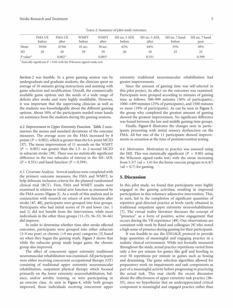
Stroke Research and Treatment 5
Table 2: Summary of pilot study outcomes.
FMA UEbafore
FMA UEafter
WMFTbefore
WMFTafter
SIS no. 5 ADLbefore
SIS no. 5 ADLafter
SIS no. 7 handbefore
SIS no. 7 handpost
Mean 39/66 47/66 41 sec. 30 sec. 62% 64% 33% 38%
SD 20 20 39 39 20 18 25 25
P value∗ 0.002∗ 0.003∗ 0.551 0.599∗
Statically significant P < 0.05 with the Wilcoxon signed-ranks test.
Section 2 was feasible. In a given gaming session run byundergraduate and graduate students, the clinician spent anaverage of 10 minutes giving instructions and assisting withgame selection and modification. Overall, the commerciallyavailable game options met the needs of a wide range ofdeficits after stroke and were highly modifiable. However,it was important that the supervising clinician as well asthe students was knowledgeable about the different gamingoptions. About 50% of the participants needed some handson assistance from the students during the gaming sessions.
4.2. Improvement in Upper Extremity Function. Table 2 sum-marizes the means and standard deviations of the outcomemeasures. The average score on the FMA increased by 8points (P = 0.002), which is greater than the 6.6-point MCID[37]. The mean improvement of 11 seconds on the WMFT(P = 0.003) was greater than the 1.5- to 2-second MCIDin subacute stroke [39]. There was no statistically significantdifference in the two subscales of interest in the SIS : ADL(P = 0.551) and hand function (P = 0.599).
4.3. Covariate Analysis. Several analyses were completed withthe primary outcome measures, the FMA and WMFT, tohelp delineate inclusion criteria for the planned randomizedclinical trial (RCT). First, FMA and WMFT results wereexamined in relation to initial arm function as measured bythe FMA scores (Figure 2). As a result of this analysis and inconjunction with research on return of arm function afterstroke [47, 48], participants were grouped into four groups.Participants who had initial scores of 10 and lower (no. 1and 2) did not benefit from the intervention, while mostindividuals in the other three groups (11–35; 36–55; 56–66)did improve.
In order to determine whether time after stroke affectedoutcomes, participants were grouped into either subacute(3–9 mo post) or chronic (>9 mo post) categories [3] basedon when they began the gaming study. Figure 3 shows thatwhile the subacute group made larger gains, the chronicgroup also improved.
The effect of concurrent upper extremity traditionalneuromuscular rehabilitation was examined. All participantswere either receiving concurrent occupational therapy (OT)consisting of traditional upper extremity neuromuscularrehabilitation, outpatient physical therapy which focusedprimarily on the lower extremity neurorehabilitation, bal-ance, and/or aerobic training, or were participating inan exercise class. As seen in Figure 4, while both groupsimproved, those individuals receiving concurrent upper
extremity traditional neuromuscular rehabilitation hadgreater improvements.
Since the amount of gaming time was self-selected inthis pilot project, its affect on the outcomes was examined.Participants were grouped according to minutes of gamingtime as follows: 500–999 minutes (56% of participants),1000–1499 minutes (25% of participants), and 1500 minutesor more (19% of participants). As can be seen in Figure 5,the group who completed the greatest amount of gamingshowed the greatest improvement. No significant differencewas found between the low and middle gaming time groups.
Finally, Figure 6 illustrates the changes seen in partic-ipants presenting with initial sensory dysfunction on theFMA. All but one of the 11 participants showed improve-ments in sensation at the time of postintervention testing.
4.4. Motivation. Motivation to practice was assessed usingthe IMI. This was statistically significant (P = 0.001 usingthe Wilcoxon signed-ranks test) with the mean increasingfrom 5.3/7 (sd = 1.0) for the home exercise program to 6.4/7(sd = 0.7) for gaming.
5. Discussion
In this pilot study, we found that participants were highlyengaged in the gaming activities, resulting in improvedparticipation in this voluntary adjunctive intervention. This,in turn, led to the completion of significant quantities ofrepetitive goal-directed practice at levels rarely obtained intraditional outpatient upper extremity neurorehabilitation[7]. The virtual reality literature discusses the concept of“presence” as a form of positive, active engagement thatoccurs during the VR experience [49]. Our observations areconsistent with work by Rand and colleagues [9] who noteda high sense of presence during gaming for their participants.
It was feasible to use the ENGAGE protocol to providelarge quantities of meaningful and engaging practice in arealistic clinical environment. While not formally measuredthroughout the study, actual practice repetitions varied fromonly a few per minute for games like golf and bowling toover 50 repetitions per minute in games such as boxingand drumming. The game selection algorithm allowed forpreparatory work on impairments and task components aspart of a meaningful activity before progressing to practicingthe actual task. This may clarify the recent discussionabout the effectiveness of upper extremity task practice [50–53], since we hypothesize that an underappreciated criticalcomponent is meaningful and engaged practice rather than
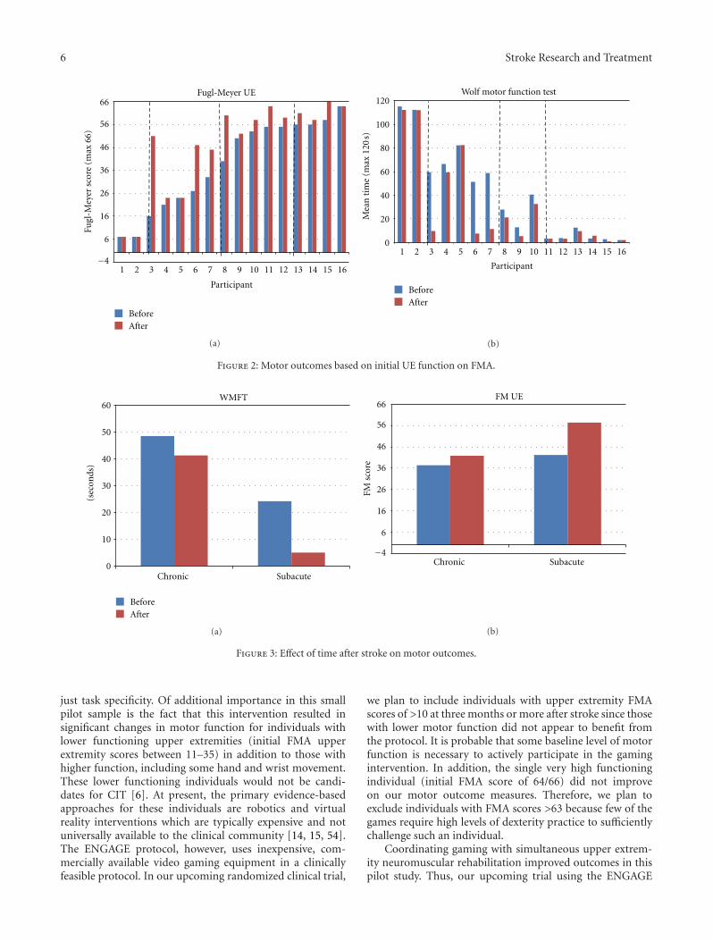
6 Stroke Research and Treatment
Fugl
-Mey
er s
core
(m
ax 6
6)
Participant
BeforeAfter
Fugl-Meyer UE
−4
6
16
26
36
46
56
66
1 2 3 4 5 6 7 8 9 10 11 12 13 14 15 16
(a)
0
20
40
60
80
100
120
1 2 3 4 5 6 7 8 9 10 11 12 13 14 15 16
Participant
Mea
n t
ime
(max
120
s)
Wolf motor function test
BeforeAfter
(b)
Figure 2: Motor outcomes based on initial UE function on FMA.
(sec
onds
)
Chronic Subacute
WMFT
0
10
20
30
40
50
60
BeforeAfter
(a)
FM s
core
FM UE
Chronic Subacute−4
6
16
26
36
46
56
66
(b)
Figure 3: Effect of time after stroke on motor outcomes.
just task specificity. Of additional importance in this smallpilot sample is the fact that this intervention resulted insignificant changes in motor function for individuals withlower functioning upper extremities (initial FMA upperextremity scores between 11–35) in addition to those withhigher function, including some hand and wrist movement.These lower functioning individuals would not be candi-dates for CIT [6]. At present, the primary evidence-basedapproaches for these individuals are robotics and virtualreality interventions which are typically expensive and notuniversally available to the clinical community [14, 15, 54].The ENGAGE protocol, however, uses inexpensive, com-mercially available video gaming equipment in a clinicallyfeasible protocol. In our upcoming randomized clinical trial,
we plan to include individuals with upper extremity FMAscores of >10 at three months or more after stroke since thosewith lower motor function did not appear to benefit fromthe protocol. It is probable that some baseline level of motorfunction is necessary to actively participate in the gamingintervention. In addition, the single very high functioningindividual (initial FMA score of 64/66) did not improveon our motor outcome measures. Therefore, we plan toexclude individuals with FMA scores >63 because few of thegames require high levels of dexterity practice to sufficientlychallenge such an individual.
Coordinating gaming with simultaneous upper extrem-ity neuromuscular rehabilitation improved outcomes in thispilot study. Thus, our upcoming trial using the ENGAGE

Stroke Research and Treatment 7
(sec
onds
)WMFT
Concurrent OT No OT0
10
20
30
40
50
60
BeforeAfter
(a)
Concurrent OT No OT
FM s
core
FM UE
0
10
20
30
40
50
60
(b)
Figure 4: Effect of concurrent upper extremity traditional neuromuscular rehabilitation (OT) on motor outcomes.
(sec
onds
)
500–1000 1001–1500 >1500
Minutes
WMFT
0
10
20
30
40
50
60
BeforeAfter
(a)
FM s
core
FM UE
−4
6
16
26
36
46
56
66
500–1000 1001–1500 >1500
Minutes
(b)
Figure 5: Effect of gaming time on motor outcomes.
protocol will include two intervention arms, each with a dif-ferent percentage of concurrent upper extremity rehabilita-tion, to better quantify the dosage of skilled versus adjunctivetherapy necessary to result in clinically significant changesin motor function. It offers a novel way to use gaming tosubstantially increase repetitive practice opportunities at areduced cost compared to conventional practice [7], whileremaining highly individualized and targeted.
The ENGAGE protocol was found to be a cost-effectiveintervention, due in large part to the ability to utilizestudents and support staff. In addition to the time requiredto train clinicians and staff, expense of equipment must beconsidered. We were able to use only two different gamingsystem platforms, the Wii and PlayStation II, along with sixdifferent game discs on the two systems. This provided a
wide selection of gaming options, allowing us to meet theneeds of individuals at various levels of arm function, toprovide variability of practice opportunities, and to preventthe boredom that often occurs when completing one taskrepetitively. While it was critical for treating clinicians tobe familiar with all game options, limiting game selectionsmeant that busy clinicians could learn the different gamealternatives in a reasonable amount of time.
Our data analysis revealed several unexpected results.First, we measured upper extremity sensation using the FMAas a potential covariate. We found that 11 out of the 12participants with sensory loss improved on this section ofthe FMA postgaming. While we know that sensory functionis a critical component in motor learning [55], there islittle consensus in our research literature about interventions

8 Stroke Research and Treatment
Fugl
-Mey
er s
core
(m
ax 1
2)
Participant
0
2
4
6
8
10
12
1 2 3 4 5 6 7 8 9 10 11
Fugl-Meyer sensory assessment
BeforeAfter
Figure 6: Changes in FM sensory assessment for participantspresenting with initial dysfunction.
to improve sensation after stroke [56]. These preliminaryresults warrant more systematic evaluation of sensoryfunction in our upcoming trial. Second, we hypothesizedthat the subacute group would improve significantly morethan the chronic group. Although this pattern did emerge,the difference was small, supporting the theory that manyindividuals after stroke with upper extremity dysfunctionlearn not to use the hemiplegic arm and hand [57] andthat at present, we do not provide adequate interventions inthis area [7]. Based on these pilot results, we will continueto enroll individuals with both subacute and chronic strokesince both groups demonstrated clinically significant levelsof change. In this convenience sample representative of theoutpatient population of the two participating facilities, halfof the outpatient participants were at least six months afterstroke. Finally, the one area where participants did notchange was the SIS, which is a reflection of the participant’sperception of their function, rather than an actual measureof function [40]. Examining the results of only the subacutegroup, we measured changes beyond the MCID of 5.9 inthe ADL subsection (increased from 58.5 to 71), but notbeyond the MCID of 17.8 for the hand subsection (increasedfrom 42 to 44) [41]. This can be explained by the fact thatseveral of these individuals did not regain functional use ofthe hemiplegic hand. Also, it is hypothesized that individualsin the subacute group may have become more aware of thedegree of deficit over time after discharge home. The lack ofchange in the chronic group is potentially a reflection of amore stable perception in their function during the longertime since the stroke event.
This study has a number of limitations. As a pilot clinicaloutcomes study, there was no control group and no controlfor concurrent therapy. Additionally, the amount of gamingtime was self-selected by each individual participant. Thesefactors will be controlled in our upcoming trial. Dosagequestions are unanswered by this study, especially sincethe 500–999-minute and 1000–1499-minute practice groupsmade similar gains. The use of gaming time rather than
actual practice repetitions may account for this finding,since there is a large variation in repetitions between slow-paced games such as golf and bowling and fast-paced gamessuch as boxing and sword fighting. An accelerometer-basedsystem has been designed as part of this pilot study andwill be used in our upcoming trial to quantify the amountof practice required for clinically relevant and functionalchange [58]. Based on these preliminary data, the protocolfor our upcoming trial has been designed to include twointervention arms examining a dosage of one additional hourof gaming for each of twelve weeks, as compared to an hourof TNR. This will provide a total of 720 additional minutesof gaming time or TNR in conjunction with two weeklysessions of TNR. A power analysis has been completed onthese preliminary data for this noninferiority hypothesis. Afollowup testing session one month after completion of thegaming intervention will also be included.
Second, although all raters were trained and used thedetailed administration manual developed for the FMAand WMFT, there were multiple evaluators during thisstudy, and interrater reliability was not specifically assessed.The protocol for our upcoming trial includes a blindedevaluator who has been standardized in the administrationof the outcome measures to complete all baseline andpostintervention testing.
Third, postural stability was not formally assessed inthis investigation although improvements were observedthat were potentially aided by the gaming intervention.These observations warrant a more systematic evaluation ofpostural stability in the planned subsequent RCT.
Finally, more objective measures of engagement areneeded, as a ceiling effect might have occurred with the IMI.Anecdotally, we, as a group of experienced clinicians, rarelyobserved a level of engagement in traditional neurologicrehabilitation as witnessed during gaming. We feel the IMIresults may have been confounded by the socialization thatoccurred during gaming but not during home exercise.
6. Conclusion
In this pilot investigation, we found that the use of ENGAGEprotocol was feasible in a clinical environment. There wasa statistically significant improvement in upper extremityfunction as measured by the upper extremity portion of theFMA and by the WMFT, and participants were motivated touse this gaming protocol. Dosage of gaming time remainsunclear, but individuals who completed at least 500 minutesof the intervention demonstrated improvements in function.In future work, the ENGAGE protocol will be used as anadjunct to concurrent upper extremity rehabilitation.
Acknowledgments
This work was supported by a Faculty Research DevelopmentGrant and a Summer Engaged Learning Grant, both fromCleveland State University. In addition, we would liketo acknowledge the assistance of multiple undergraduate,graduate, and doctor of physical therapy students from
Stroke Research and Treatment 9
Cleveland State University who assisted in the actual gamingwork with the participants. Finally, the project could nothave been completed without the collaboration, assistance,and support of the clinicians at the Cleveland Clinic MainCampus and Lakewood Hospital.
References
[1] V. L. Roger, A. S. Go, D. M. Lloyd-Jones et al., “Heartdisease and stroke statistics—2011 update: a report from theAmerican Heart Association,” Circulation, vol. 123, no. 4, pp.e18–e19, 2011.
[2] E. L. Miller, L. Murray, L. Richards et al., “Comprehensiveoverview of nursing and interdisciplinary rehabilitation careof the stroke patient: a scientific statement from the Americanheart association,” Stroke, vol. 41, no. 10, pp. 2402–2448, 2010.
[3] D. S. Nichols-Larsen, P. C. Clark, A. Zeringue, A. Greenspan,and S. Blanton, “Factors influencing stroke survivors’ qualityof life during subacute recovery,” Stroke, vol. 36, no. 7, pp.1480–1484, 2005.
[4] L. Boyd and C. Winstein, “Explicit information interfereswith implicit motor learning of both continuous and discretemovement tasks after stroke,” Journal of Neurologic PhysicalTherapy, vol. 30, no. 2, pp. 46–59, 2006.
[5] J. A. Kleim and T. A. Jones, “Principles of experience-dependent neural plasticity: implications for rehabilitationafter brain damage,” Journal of Speech, Language, and HearingResearch, vol. 51, no. 1, pp. S225–S239, 2008.
[6] S. L. Wolf, C. J. Winstein, J. P. Miller et al., “Effect ofconstraint-induced movement therapy on upper extremityfunction 3 to 9 months after stroke: the EXCITE randomizedclinical trial,” Journal of the American Medical Association, vol.296, no. 17, pp. 2095–2104, 2006.
[7] C. E. Lang, J. R. MacDonald, D. S. Reisman et al., “Observationof amounts of movement practice provided during strokerehabilitation,” Archives of Physical Medicine and Rehabilita-tion, vol. 90, no. 10, pp. 1692–1698, 2009.
[8] S. Flynn, P. Palma, and A. Bender, “Feasibility of usingthe Sony PlayStation 2 gaming platform for an individualpoststroke: a case report,” Journal of Neurologic PhysicalTherapy, vol. 31, no. 4, pp. 180–189, 2007.
[9] D. Rand, R. Kizony, and P. T. L. Weiss, “The sony playStationII eye toy: low-cost virtual reality for use in rehabilitation,”Journal of Neurologic Physical Therapy, vol. 32, no. 4, pp. 155–163, 2008.
[10] G. Yavuzer, A. Senel, M. B. Atay, and H. J. Stam, “‘Playsta-tion eyetoy games’ improve upper extremity-related motorfunctioning in subacute stroke: a randomized controlledclinical trial,” European Journal of Physical and RehabilitationMedicine, vol. 44, no. 3, pp. 237–244, 2008.
[11] M. Lotze, C. Braun, N. Birbaumer, S. Anders, and L. G. Cohen,“Motor learning elicited by voluntary drive,” Brain, vol. 126,no. 4, pp. 866–872, 2003.
[12] S. L. Wolf, “Revisiting constraint-induced movement therapy:are we too smitten with the mitten? Is all nonuse “learned”?And other quandaries,” Physical Therapy, vol. 87, no. 9, pp.1212–1223, 2007.
[13] R. N. Barker, S. G. Brauer, and R. G. Carson, “Training ofreaching in stroke survivors with severe and chronic upperlimb paresis using a novel nonrobotic device: a randomizedclinical trial,” Stroke, vol. 39, no. 6, pp. 1800–1807, 2008.
[14] E. M. Frick and J. L. Alberts, “Combined use of repetitivetask practice and an assistive robotic device in a patient with
subacute stroke,” Physical Therapy, vol. 86, no. 10, pp. 1378–1386, 2006.
[15] J. H. Crosbie, S. Lennon, M. D. J. McNeill, and S. M.McDonough, “Virtual reality in the rehabilitation of the upperlimb after stroke: the user’s perspective,” Cyberpsychology andBehavior, vol. 9, no. 2, pp. 137–141, 2006.
[16] H. Sveistrup, “Motor rehabilitation using virtual reality,”Journal of NeuroEngineering and Rehabilitation, vol. 1, article10, 2004.
[17] L. Y. Joo, T. S. Yin, D. Xu et al., “A feasibility study usinginteractive commercial off-the-shelf computer gaming inupper limb rehabilitation in patients after stroke,” Journal ofRehabilitation Medicine, vol. 42, no. 5, pp. 437–441, 2010.
[18] G. Saposnik, R. Teasell, M. Mamdani et al., “Effectivenessof virtual reality using wii gaming technology in strokerehabilitation: a pilot randomized clinical trial and proof ofprinciple,” Stroke, vol. 41, no. 7, pp. 1477–1484, 2010.
[19] L. Graves, G. Stratton, N. D. Ridgers, and N. T. Cable, “Energyexpenditure in adolescents playing new generation computergames,” British Journal of Sports Medicine, vol. 42, no. 7, pp.592–594, 2008.
[20] D. Rand, J. J. Eng, P. F. Tang, J. S. Jeng, and C. Hung, “Howactive are people with stroke?: use of accelerometers to assessphysical activity,” Stroke; a journal of cerebral circulation, vol.40, no. 1, pp. 163–168, 2009.
[21] S. J. Colcombe, A. F. Kramer, K. I. Erickson et al., “Cardiovas-cular fitness, cortical plasticity, and aging,” Proceedings of theNational Academy of Sciences of the United States of America,vol. 101, no. 9, pp. 3316–3321, 2004.
[22] A. F. Kramer and K. I. Erickson, “Capitalizing on corticalplasticity: influence of physical activity on cognition and brainfunction,” Trends in Cognitive Sciences, vol. 11, no. 8, pp. 342–348, 2007.
[23] J. H. Cauraugh, S. A. Coombes, N. Lodha, S. K. Naik, andJ. J. Summers, “Upper extremity improvements in chronicstroke: coupled bilateral load training,” Restorative Neurologyand Neuroscience, vol. 27, no. 1, pp. 17–25, 2009.
[24] F. Coupar, A. Pollock, F. van Wijck, J. Morris, and P.Langhorne, “Simultaneous bilateral training for improvingarm function after stroke,” Cochrane Database of SystematicReviews, vol. 4, Article ID CD006432, 2010.
[25] L. Piron, A. Turolla, M. Agostini et al., “Exercises for pareticupper limb after stroke: a combined virtual-reality andtelemedicine approach,” Journal of Rehabilitation Medicine,vol. 41, no. 12, pp. 1016–1020, 2009.
[26] J. Whitall, S. McCombe Waller, J. D. Sorkin et al., “Bilateraland unilateral arm training improve motor function throughdiffering neuroplastic mechanisms: a single-blinded random-ized controlled trial,” Neurorehabilitation and Neural Repair,vol. 25, no. 2, pp. 118–129, 2011.
[27] C.-Y. Wu, L.-L. Chuang, K.-C. Lin, H.-C. Chen, and P.-K. Tsay, “Randomized trial of distributed constraint-inducedtherapy versus bilateral arm training for the rehabilitationof upper-limb motor control and function after stroke,”Neurorehabilitation and Neural Repair, vol. 25, no. 2, pp. 130–139, 2011.
[28] A. R. Luft, S. McCombe-Waller, J. Whitall et al., “Repetitivebilateral arm training and motor cortex activation in chronicstroke: a randomized controlled trial,” Journal of the AmericanMedical Association, vol. 292, no. 15, pp. 1853–1861, 2004.
[29] G. Saposnik, M. Mamdani, M. Bayley et al., “Effectivenessof virtual reality exercises in stroke rehabilitation (EVREST):rationale, design, and protocol of a pilot randomized clinical

10 Stroke Research and Treatment
trial assessing the wii gaming system,” International Journal ofStroke, vol. 5, no. 1, pp. 47–51, 2010.
[30] M. E. Michielsen, R. W. Selles, J. N. Van Der Geest etal., “Motor recovery and cortical reorganization after mirrortherapy in chronic stroke patients: a phase II randomizedcontrolled trial,” Neurorehabilitation and Neural Repair, vol.25, no. 3, pp. 223–233, 2011.
[31] D. Ertelt, S. Small, A. Solodkin et al., “Action observationhas a positive impact on rehabilitation of motor deficits afterstroke,” NeuroImage, vol. 36, no. 2, pp. T164–T173, 2007.
[32] K. A. Garrison, C. J. Winstein, and L. Aziz-Zadeh, “Themirror neuron system: a neural substrate for methods in strokerehabilitation,” Neurorehabilitation and Neural Repair, vol. 24,no. 5, pp. 404–412, 2010.
[33] M. Viswanathan et al., Community-Based ParticipatoryResearch: Assessing the Evidence. Evidence Report/TechnologyAssessment No. 99 A.f.H.R.a. Quality, Editor, RTI-Universityof North Carolina Evidence-based Practice Center, Rockville,Md, USA, 2004.
[34] A. R. Fugl Meyer, L. Jaasko, and I. Leyman, “The poststroke hemiplegic patient. I. A method for evaluation ofphysical performance,” Scandinavian Journal of RehabilitationMedicine, vol. 7, no. 1, pp. 13–31, 1975.
[35] J. H. Lin, M. J. Hsu, C. F. Sheu et al., “Psychometriccomparisons of 4 measures for assessing upper-extremityfunction in people with stroke,” Physical Therapy, vol. 89, no.8, pp. 840–850, 2009.
[36] K. Shepherd and G. Jensen, Handbook of Teaching for PhysicalTherapists, Butterworth-Heinemann, Boston, Mass, USA, 2ndedition, 2002.
[37] D. J. Gladstone, C. J. Danells, and S. E. Black, “The Fugl-meyerassessment of motor recovery after stroke: a critical review ofits measurement properties,” Neurorehabilitation and NeuralRepair, vol. 16, no. 3, pp. 232–240, 2002.
[38] S. L. Wolf, P. A. Catlin, M. Ellis, A. L. Archer, B. Morgan, and A.Piacentino, “Assessing Wolf Motor Function Test as outcomemeasure for research in patients after stroke,” Stroke, vol. 32,no. 7, pp. 1635–1639, 2001.
[39] K. C. Lin, Y. W. Hsieh, C. Y. Wu, C. L. Chen, Y. Jang, and J.S. Liu, “Minimal detectable change and clinically importantdifference of the wolf motor function test in stroke patients,”Neurorehabilitation and Neural Repair, vol. 23, no. 5, pp. 429–434, 2009.
[40] P. W. Duncan, R. K. Bode, S. M. Lai, and S. Perera, “Raschanalysis of a new stroke-specific outcome scale: the strokeimpact scale,” Archives of Physical Medicine and Rehabilitation,vol. 84, no. 7, pp. 950–963, 2003.
[41] K. C. Lin, T. Fu, C. Y. Wu et al., “Minimal detectable changeand clinically important difference of the stroke impact scalein stroke patients,” Neurorehabilitation and Neural Repair, vol.24, no. 5, pp. 486–492, 2010.
[42] E. McAuley, T. Duncan, and V. V. Tammen, “Psychometricproperties of the intrinsic motivation inventory in a compet-itive sport setting: a confirmatory factor analysis,” ResearchQuarterly for Exercise and Sport, vol. 60, no. 1, pp. 48–58, 1989.
[43] P. Pound, P. Gompertz, and S. Ebrahim, “A patient-centredstudy of the consequences of stroke,” Clinical Rehabilitation,vol. 12, no. 4, pp. 338–347, 1998.
[44] S. Rosewilliam, C. A. Roskell, and A. D. Pandyan, “A systematicreview and synthesis of the quantitative and qualitativeevidence behind patient-centred goal setting in stroke reha-bilitation,” Clinical Rehabilitation, vol. 25, no. 6, pp. 501–514,2011.
[45] A. Gentile, “Skill acquisition: action, movement, and neu-romotor processes,” in Movement Science: Foundations forPhysical Therapy in Rehabilitation, R. Carr and R. Shepherd,Eds., Aspen Publishers, Gaithersburg, Md, USA, 2000.
[46] S. L. Kilbreath and R. C. Heard, “Frequency of hand use inhealthy older persons,” Australian Journal of Physiotherapy,vol. 51, no. 2, pp. 119–122, 2005.
[47] G. Kwakkel and B. Kollen, “Predicting improvement in theupper paretic limb after stroke: a longitudinal prospectivestudy,” Restorative Neurology and Neuroscience, vol. 25, no. 5-6,pp. 453–460, 2007.
[48] R. H. M. Nijland, E. E. H. Van Wegen, B. C. Harmeling-Van Der Wel, and G. Kwakkel, “Presence of finger extensionand shoulder abduction within 72 hours after stroke predictsfunctional recovery: early prediction of functional outcomeafter stroke: The EPOS cohort study,” Stroke, vol. 41, no. 4, pp.745–750, 2010.
[49] G. Riva, F. Mantovani, and A. Gaggioli, “Presence andrehabilitation: toward second-generation virtual reality appli-cations in neuropsychology,” Journal of NeuroEngineering andRehabilitation, vol. 1, article 9, 2004.
[50] R. L. Birkenmeier, E. M. Prager, and C. E. Lang, “Translatinganimal doses of task-specific training to people with chronicstroke in 1-hour therapy sessions: a proof-of-concept study,”Neurorehabilitation and Neural Repair, vol. 24, no. 7, pp. 620–635, 2010.
[51] B. French, L. H. Thomas, M. J. Leathley et al., “Repetitivetask training for improving functional ability after stroke,”Cochrane Database of Systematic Reviews, no. 4, Article IDCD006073, 2007.
[52] B. French, L. Thomas, M. Leathley et al., “Does repetitive tasktraining improve functional activity after stroke? A Cochranesystematic review and meta-analysis,” Journal of RehabilitationMedicine, vol. 42, no. 1, pp. 9–15, 2010.
[53] A. A. A. Timmermans, A. I. F. Spooren, H. Kingma, and H.A. M. Seelen, “Influence of task-oriented training content onskilled arm-hand performance in stroke: a systematic review,”Neurorehabilitation and Neural Repair, vol. 24, no. 9, pp. 858–870, 2010.
[54] A. Mirelman, P. Bonato, and J. E. Deutsch, “Effects of trainingwith a robot-virtual reality system compared with a robotalone on the gait of individuals after stroke,” Stroke, vol. 40,no. 1, pp. 169–174, 2009.
[55] A. Shumway-Cook and M. H. Woolacott, Motor Control:Translating Research into Clinical Practice, Lippincott Williams& Wilkins, Philadelphia, Pa, USA, 3rd edition, 2006.
[56] S. Doyle, S. Bennett, S. E. Fasoli, and K. T. McKenna,“Interventions for sensory impairment in the upper limbafter stroke,” Cochrane Database of Systematic Reviews, vol. 6,Article ID CD006331, 2010.
[57] E. Taub, “Somatosensory deafferentation research with mon-keys: implications for rehabilitation medicine,” in BehavioralPsychology in Rehabilitation Medicine: Clinical Applications, L.Ince, Ed., pp. 371–401, Williams & Wilkins, Baltimore, Md,USA, 1980.
[58] S. Sanka et al., “Utilization of a wrist-mounted accelerometerto count movement repetitions,” in Proceedings of the 4th Inter-national Conference on Communication Systems and Networks(COMSNETS ’12), Bangalore, India, January 2012.

Hindawi Publishing CorporationStroke Research and TreatmentVolume 2012, Article ID 503190, 18 pagesdoi:10.1155/2012/503190
Research Article
Motor Imagery Experiences and Use: Asking Patientsafter Stroke Where, When, What, Why, and How They UseImagery: A Qualitative Investigation
Corina Schuster,1, 2 Andrea Glassel,3, 4 Anne Scheidhauer,1 Thierry Ettlin,1, 5
and Jenny Butler2
1 Research Department Reha Rheinfelden, Salinenstrasse 98, 4310 Rheinfelden, Switzerland2 Department of Sport and Health Sceinces, Faculty of Health and Life Sciences, Oxford Brookes University, Oxford OX3 0BP, UK3 Institute of Health and Rehabilitation Sciences, Ludwig-Maximilians-University Munich, 80539 Munich, Germany4 Faculty of Social Work and Health, University of Applied Sciences and Arts, 31134 Hildesheim, Germany5 Department of Behavioural Neurology, Medical faculty, University of Basel, 4053 Basel, Switzerland
Correspondence should be addressed to Corina Schuster, [email protected]
Received 3 October 2011; Revised 11 January 2012; Accepted 16 January 2012
Academic Editor: Steven L. Wolf
Copyright © 2012 Corina Schuster et al. This is an open access article distributed under the Creative Commons AttributionLicense, which permits unrestricted use, distribution, and reproduction in any medium, provided the original work is properlycited.
Background. A framework on where, when, what, why, and how to use imagery from sports psychology was explored whether itcan be applied in patients after stroke in their chronic stage. Methods. Eleven patients (ages 31–85, 3 females, 1.3–6.4 years afterstroke) were interviewed. Semistructured interviews were conducted before and after a two-week MI intervention period with sixMI sessions. Information was obtained regarding experiences and knowledge of MI, and the evaluation of an MI practical example.The coding scheme was based on the framework and a hierarchical categorisation. Results. Information regarding domains where,when, what, why, and how to use imagery was addressed. Patients imagined themselves as healthy individuals, did not focus onsurroundings during MI practice,and reported to use positive imagery only. After MI training, patients became more flexibleregarding their location and position during MI practice. Conclusions. MI became an automatic process, and patients did not needspecific concentration and quietness as mentioned in the first interview. Patients recommended daily MI training and began totransfer MI to practice movements that were affected by the stroke. In contrast to sports, patients did not talk about how MI wastriggered rather than how MI was designed.
1. Introduction
Motor imagery (MI) is a recognised and established methodfor different individuals (children, students, athletes, and pa-tients) and for different purposes (cognitive, strength, andmotor-related tasks). During recent years, MI has receivedincreasing attention as a training approach in rehabilitationof stroke patients [1–4]. However, designing effective image-ry interventions depends on patients’ typical normal use, in-tentions, and imagery content when using the technique.Currently, patient experiences and meanings attributed toMI are widely unknown so far.
After adopting MI in stroke rehabilitation in the late1990s, it has been shown to be beneficial for motor function
recovery when added to physical practice [1, 2]. Decety andGrezes have defined MI as “...a dynamic state during whichthe representation of a given motor act is internally rehearsedwithin working memory without any overt motor output”[5]. Research has been performed investigating differenttechniques of MI and their effect on both motor function andpsychological parameters [6–8].
In psychology, the influential work of Kosslyn et al. mov-ed the research perspective from applying MI in interven-tions with individuals to research in the natural occurrencesand use of MI in everyday life [9]. The authors asked twelveundergraduate student volunteers to keep an imagery diaryfor one week and note down when and where imagery hadbeen used. Students had to describe the situation they were in

2 Stroke Research and Treatment
when imagery occurred, the content of what has been imagi-ned, and the vividness of the image. Furthermore, authorswanted to know why individuals had used imagery and whatthe intention to use imagery in the current situation was. Ad-ditionally, students had to define their understanding of a“mental image.” Study results were analysed regarding thecategories modality, purpose, and content of imagery. Itcould be shown that visual and auditory imagery modes weremostly reported. The category purpose included six types:more than half of the study population reported associative(e.g., day dreaming), problem solving (e.g., mental maps tonavigate), mental practice (e.g., imagine swimming stroke),memory recall (e.g., imagine notes during test), compre-hending descriptions (e.g., image based on verbal descrip-tion), and emotional or motivational stimulus (e.g., to calmdown), as imagery content coloured and concrete (vivid)images were the most frequent reported ones. In the samepaper, they described a second study on imagery every dayuse that confirmed the findings of the first experiment for allthree categories: modality, purpose, and content [9].
Ten years later, Munroe et al. used semistructured in-depth interviews in 14 athletes from various disciplines tofind answers on where, when, whym and what imageryhas been used. Obtained information had been structuredaccording to these 4-Wquestions. In addition, informationon why using imagery had been structured according toPaivio’s framework on effects of imagery on human motorperformance [10, 11]. In this framework, Paivio [11] suggest-ed two main functions of imagery (cognitive, motivational)that work on a general level, for example, cognitive generaland motivational general, or specific level, comprising cog-nitive specific and motivational specific types. Motivationalfunctions are used to reduce anxiety or increase self-conf-idence. Cognitive functions, for example, attention, enableathletes to use different imagery perspectives and to transferthe imagine tasks to competitive situations. However, for thecurrent investigation, it will be interesting if patients afterstroke will address both aspects from their personal experi-ence. In the patients’ case, “competitive situations” could berelated to movements, tasks, or settings that are challengingdue to motor function limitations. Furthermore, Paivio’sfindings influenced Munroe et al.’s results classificationfor athletes imagery use: during and outside practice, forprecompetition, competition, and postcompetition. Further-more, their analysis on imagery content included type (vis-ual, kinaesthetic) and perspective (internal, external) of MI.
In 2005, Nordin and Cumming interviewed 14 profes-sional dancers to describe their imagery use [12]. Again, in-formation was categorised according to the 4-W-questions:where, when, why, and what have been imagined. In theiranalysis, a new domain has emerged on how images were ob-tained. This domain included three categories: externalstimuli, retrieving memories, and creating triggers.
Moving away from healthy students and athletes, Driedi-ger et al. investigated athletes with musculoskeletal injuriesand their use of imagery during the recovery process [13].Ten athletes of various disciplines with different severity ofinjury had been interviewed once. Imagery was used for cog-nitive (e.g., learn rehabilitation exercise, motivational (e.g.,
goal setting) and healing reasons (e.g., distraction frompain)). Imagery was mainly performed during physiotherapysessions prior to physical practice of an exercise. The authorsdescribed the content of athletes’ imagery as vivid with bothmodes: visual and kinaesthetic, and with a positive perfor-mance. That included imagination of the athlete her/himselfas a healthy individual performing in her/his sports disciplineas successful as before the injury.
To create an international perspective of imagery use insupraelite athletes, MacIntyre and Moran interviewed twelvecanoe-slalom competitors from Germany, Ireland, UnitedStates, and the United Kingdom [14]. Their purpose has beento investigate where, when, what, why, and how athletes useimagery. In addition, the authors were interested in the meta-cognitive control of imagery and asked athletes about theirimportance of imagery and why they have used this method.Results revealed different imagery directions (facilitative anddebilitative) and strategies to overcome problems in activitysequences, for example, talk through a slalom course with thetrainer.
So far, the existing framework on where, when, what,why, and how to use imagery from sports psychology has notbeen explored in brain-damaged individuals. The objectiveof this study was to investigate whether the imagery frame-work questions (where, when, what, why, and how) for MIcan be applied in chronic-stage patients after stroke. The spe-cific aims were (1) to evaluate whether the imagery frame-work questions (where, when, what, why, and how) estab-lished in mainly healthy and young students and athletes canbe adopted by an older stroke population and (2) if new in-sights for the design of MI interventions can be obtained.
To address the aims mentioned above, a qualitativeresearch method approach was used. This well-establishedresearch method originates from the social sciences. Obtain-ed information from semistructured interviews, focusgroups, or observations helps to understand and explore be-havioural processes and generate hypotheses rather thantesting experimental hypotheses. The participant’s perspec-tive and perceptions on unexplored concepts, diseases, orphenomena are the main focus of the investigation gatheringtextual information rather than numbers [15–18]. Applyingsemistructured interviews in the current investigation ismost appropriate given the nature of the topic under investi-gation. In this approach, the interviewer could ask andexplain the abstract construct of MI and giving the patientsthe opportunity to ask questions and check their correctunderstanding. The applied procedure offered the opportu-nity to acquire detailed information on the nature of patients’MI experiences, associations, MI training session elements(e.g., MI mode and state of eyes), and temporal parameters(e.g., duration of MI and number of MI trials).
2. Methodology
The goals of this work were approached using a framework-based qualitative research method, using semistructuredinterviews. Information reported in this paper, including
Stroke Research and Treatment 3
reflexivity, study design, and data analysis, followed the qual-itative research review guidelines (RATS) and the 32-itemconsolidated criteria list for reporting qualitative research(COREQ) [19, 20].
2.1. Study Design and Procedure. This qualitative investiga-tion based on an explorative content analysis and was nestedinto a randomised control pilot trial (RCT) investigating twoMI training approaches in patients after stroke. After givingwritten informed consent for the MI intervention trial, pa-tients underwent two baseline measurement events, includ-ing MI ability assessments, balance evaluation, and assess-ment on independence of living. Following the baselineassessments, patients were randomised to one of three studygroups. Patients allocated to either of the experimentalgroups (EG1, EG2) were asked whether they would like toparticipate in two semistructured interviews, one before andone after the two-week MI intervention period. Patients wereinformed about the procedure by the interviewer (CS) andreceived a second patient information sheet specificallydeveloped for the interviews. The third group served as acontrol group. All three groups received standardised physio-therapy treatment focussing on balance. Patients MI trainingmethods in EG1 and EG2 did not differ regarding content,duration, or frequency. The main difference can be seen inthe integration approach of MI. One experimental group(EG1) embedded MI into physiotherapy. The second exper-imental group (EG2) added MI after the physiotherapysession. Patients of all groups had to practice one motor taskphysically “Going down, laying on the floor, and getting upagain” in a standardised order. In EG1 and EG2, the motortask has also been practiced mentally. The control group lis-tened to audio tapes with information related to stroke afterphysiotherapy. In total, treatment time was about 45 to 50minutes for each group. A detailed study protocol is describ-ed elsewhere [21]. The study was implemented according tothe Declaration of Helsinki and was approved by the ethicscommittee of the School of Health and Social Care of theOxford Brookes University, Oxford (UK) and the responsibleSwiss ethics committee (Aarau, Kanton Aargau, Switzerland,reference number 2008/077).
2.2. Interviewee Sampling and Data Saturation. Patients wererecruited from both experimental groups (EG1, EG2) of theongoing MI intervention trial with the following selectioncriteria: first ischemic or hemorrhagic stroke at least threemonths before study entry, being able to stand with orwithout a cane for at least 30 seconds on a normal hard floor,being able to walk 20 metres with or without a cane or anorthosis, older than 18 years, and score at least 20 in theMini-Mental State Examination (MMSE), given written in-formed consent. Patients were excluded if they had joint re-placements (knee, hip, and shoulder), movement-limitingpain in the upper or lower body evaluated with the 11-pointvisual analogue scale (VAS), limited range of motion in hip,knee, ankle joints, or toes, bodyweight exceeding 100 kg,or having a compromised mental capacity to give writteninformed consent. Further restrictions resulted from the
patients’ ability to communicate and express thoughts, ex-periences, and feelings. Patients with severe speaking prob-lems were not included. During the recruitment periodbetween April 2009 and May 2010, eleven out of 26 patientsfulfilled the selection criteria and agreed to participate in thesemistructured individual interviews. The MI interventionstudy, which was closely connected to the semistructuredinterviews, determined the bounds for data saturation in pa-tient interviews of this work. Saturation refers to the pointat which the investigator has obtained sufficient informationfrom the field [22].
2.3. Interview Setting. Interviews were conducted face to facebetween the patients and the first author (CS). Followingwritten informed consent, interviews were conducted one totwo hours before the first of six MI intervention sessions. Thesecond interview took place after the last MI interventionsession. To provide a high level of privacy and to avoid distur-bances, all interviews took place in a separate treatment roomof the rehabilitation centre. Patients were offered refresh-ments during the interview. Furthermore, patients coulddecide to not answer a question and to have breaks at anytime during the interviews, if they wished to do so. Interviewswere recorded with two redundant digital voice recorders toavoid loss of data. Patients and interviewer did not know eachother before oral and written study information was given.After the first interview, CS worked with the patients dur-ing all six MI intervention sessions, which could have had aninfluence on the patients’ reporting during the second inter-view.
2.4. Interview Guide and Procedure. A generic interviewguide was developed for the first interview to account for dif-ferent symptoms after stroke, for example, paresis level andpsychological capacity. The interview guide was developedbased on the interview guide published by MacIntyre andwas divided into three parts [14]. Bearing in mind that MIcould be very abstract to patients, the first part of the inter-view was not related to MI, but focused on the stroke event,patients’ rehabilitation process including therapy experien-ces, and occurrence of falls. In the second interview part, pa-tients were asked about their previous experiences and know-ledge of MI. In the third interview part, a practical exampleof MI was performed, which included a sit-to-stand task.After performing this task twice physically, it was twicementally practiced and again twice physically practiced. Theaims of the simple and quick to perform practical examplewere (1) to provide patients, regardless of their previousMI experience, with MI experience, which helped them todescribe MI content elements, for example, used MI mode(kinaesthetic, visual), perspective (internal, external), andimagined environment, and (2) to control for actual MI per-formance by recording the time (sec) needed to perform theexample mentally and practically. The guide for the secondinterview only included questions regarding patients’ experi-ences with MI during the previous two interventions weeksand a repetition of the practical example. In the finalphase of both interviews, patients were asked to evaluate

4 Stroke Research and Treatment
the interview, including questions on whether it was exhaust-ing and how they liked it. Furthermore, patients’ answerswere summarised by the interviewer as a means of memberchecking, and patients had the opportunity to comment on,add to, or omit statements. The interview guides are providedin Table 1.
Three pilot interviews were conducted to familiarise withthe interview procedure, test the interview guide, and prac-tice the technical setup. No changes to the interview guidewere deemed necessary after the pilots.
During and after the interviews, field notes and an inter-view report were written by the interviewer to describe im-portant aspects of the interview (e.g., patients’ mood, un-expected disturbances) as well as patients’ gestures and be-haviours. If leading questions emerged during the interviewsituation, answers were not included in the analysis. To verifythe patient “yes” or “no” answers, another question with theopposite meaning was offered to the patient.
2.5. Data Analysis, Credibility, and Trustworthiness Proce-dures. Figure 1 provides an overview on all methodical stepsfrom data recording to final analysis. All recorded interviewswere transcribed verbatim (computer text document) by AS.Two researchers (KL, AH) involved in the assessments of therelated MI intervention study checked the transcripts for asecond time. Afterwards, all transcripts were checked for athird time by CS and copied into computer spread sheets forfurther analysis [23]. All verbatim-transcribed interviewswere mailed to the patients for verification. Five of all mailedpatients gave feedback and agreed with the reproduction.
The questions where, when, what, why, and how to per-form MI were not considered for developing the codingscheme in the first line. With this approach, all emergingthemes from the raw data could be captured. To address thequality item credibility, CS and AG developed the codingscheme jointly, based on three interviews before MI interven-tion and three interviews after MI intervention. The schemewas extended during further interview coding by CS. Eachpatient phrase or sentence was coded, where one sentencecould contain information mapped to more than one code.At maximum, one sentence or phrase was matched to fivecodes. After coding was completed for all interviews by CS,AG applied the final coding scheme to code the MI-relatedpart of one interview. This peercheck conformed to codecross-checking and evaluated the established codes [18].Coding agreement was 80% (84 of 105 phrases), which wasobtained before a consensus discussion. If both researchers(AG, CS) had not been able to agree on a decision (whichwas not the case in this investigation), a third researcher (JB)would have been consulted. In the summary and analysisstep, only information related to MI was used. Patients’information was then transferred into a matrix to providean overview on all patient statements matching one code.Based on this matrix, data were interpreted and allocated tothe domains, categories, and subcategories of a hierarchy tree(FreeMind, version 0.8.1). The imagery framework questions(where, when, what, why, and how) represented the highestanalysis level. Category names were derived from MI training
session elements extracted in a systematic literature reviewon MI techniques [24]. Further domains and categories wereadded to the hierarchy tree to represent the complete rangeof patient information. Codes used in the coding scheme andtheir allocation to the respective domain are listed in Table 2.
To further increase the rigour of the analysis, two metho-dological details need to be considered. Firstly, all patientsunderwent two baseline measurement events of the MI inter-vention trial. The assessments on MI ability performed dur-ing the baseline measurement events supported patients’knowledge and interpretation of the MI construct. Secondly,during the interviews’ practical MI example (sit-to-stand andback), patients and interviewer had to record the time neededwith a stopwatch. These measurements helped to ensure abasic understanding and knowledge on the interview topic ofinterest for all patients. Furthermore, the triangulation of pa-tients’ interview data and assessment data helped to gain abroader picture of patients’ MI understanding, but also tolimit misinterpretation of interview data [18]. In general, atno time during the analysis were data modified or made to fitwith the considered imagery framework (where, when, what,why, and how questions).
3. Results
3.1. Patients and Interviews. Important patient characteris-tics are displayed in Table 3. During all interviews, only pa-tient and interviewer were present, except during the firstinterview where the patient’s wife was present too (Pat. 32).In total, 20 interviews were conducted. Nine patients wereinterviewed twice, and two patients were interviewed beforethe MI intervention only due to patients’ time constraints.Once during the first interview (Pat. 32), both voice recordersdid not work properly, and about one-third of the interviewcould not be recorded. The duration of the interviews wasaveraged 38.4 min for the first and 22.3 min for the secondinterview. Patients’ responses produced 216 lines of text(average) during the first interview, with only half (averageof 106 lines) related to MI. During the second interview,patients’ responses produced 124 lines of text (average) with107 lines (average) that were MI related.
3.2. General Data Analysis Results. Information contributingto the patients’ statements was based on experiences andknowledge acquired before the study, the practical exampleof MI performed during the interview, and on assumptionspatients made after exercising the MI example. Examplequotes were translated from German into English by CS andwere double-checked by KL, who is a German native speakerbut has proficient level in the use of English. The first twolevels (domains and categories) of the hierarchy tree used inthe data analysis are displayed in Figure 2 and Table 2.
3.2.1. Where Using MI?
The domain WHERE comprises two categories: (a) locationand (b) position of the individual during MI. Both categoriescould be related to preparation for MI practice and consist

Stroke Research and Treatment 5
Table 1: Interview guides.
Interview Interview part Interview questions
1st interview
The first focused on the stroke event,patients’ rehabilitation process includingtherapy experiences and occurrence of falls.
When did your stroke occur?
How did you recognize that you had a stroke? What symptoms did youhave?
How did you feel when the stroke happened?
How do you feel today?
How was your recovery process?
What is your main problem at the moment?
How do you feel when you are walking?
Have you been falling since you had the stroke?
The second interview part focussed onpatients’ previous experiences andknowledge of MI.
How does it feel to you to do motor imagery?
What do you associate with motor imagery?
(a) Did you know motor imagery before you have done the assessments?
(b) How did you get to know about motor imagery?
(c) When do you do motor imagery?
(d) How do you do motor imagery?
(e) What do you imagine?
(f) What kind of experiences have you made with motor imagery?
In the third interview part, a practicalexample of MI was performed, whichincluded a sit-to-stand task. Afterperforming this task twice physically, it wastwice mentally practiced and again twicephysically practiced. Patients were asked todescribe the content of the MI example.
Was it easy to imagine the movement (standing up, sitting down)? If yes,why was it easy? If not, why was it not easy?
What exactly have you imagined?
How detailed was the movement?
What kind of surroundings/environment have you imagined?
What kind of perspective have you used (internal, external)?
What comes to your mind if you remember the imagination of themovement?
(a) What have you seen?
(b) What have you felt?
(c) What have you heard?
(d) What have you smelled?
Do you think that motor imagery could help patients after stroke duringthe recovery process? If yes, why?
When and how often would you do motor imagery?
Where would you do imagery?
What expectations do you have regarding the MI intervention duringthe next weeks?
In the final phase, patients were asked toevaluate the interview, including questionson whether it was exhausting and how theyliked it. Patients had the opportunity tocomment on, add to, or omit statements.
What do you think about the interview?
Was the interview exhausting?
How do you feel now after the interview?
Would you like to add something?
2nd interview
The first part focused on questionsregarding patients’ experiences with MIduring the previous two interventionsweeks.
How did you like the motor imagery intervention?
What do you think about motor imagery now?
Do you think motor imagery can help during the recovery process aftera stroke?
How can motor imagery help during the recovery process?
Would you use motor imagery in the future to learn or improve a motortask?
(a) Why?
(b) When-how often?
(c) Where?
(d) What?

6 Stroke Research and Treatment
Table 1: Continued.
Interview Interview part Interview questions
In the second part, the practical examplefrom the first interview was repeated.Afterwards, patients were asked to describethe content of the MI example.
Was it easy to imagine the movement?
Why was it easy/not easy?
What exactly have you imagined?
How detailed was the movement?
What kind of surroundings/environment have you imagined?
What kind of perspective have you used?
What comes to your mind if you remember the imagination of themovement?
What have you seen?
What have you felt?
What have you heard?
What have you smelled?
Did you think that motor imagery helped you to improve the motortask?
Final phase Please see first interview
• 20 semistructured interviews
•Writing field notes + interview reports
• Only first author (CS) involved
• Verbatim transcription of all interviews in word by AS
• Export of all transcripts into Excel
•Double + triple transcript check by two researchers
• Transcripts sent to all interviewees for member check
• Development of categorisation scheme based on six interviews by CS and AG
• Further interview categorisation and scheme adaptation by CS
• Evaluation of final categorisation scheme by AG based on one interview
• Data extraction (Excel) of all patient statements for each category by CS and AS
• Built hierarchy tree with mind map
• Data interpretation and allocation to domains, categories, and subcategories
Interviews
Transcripts
Coding
Summary
Analysis
Final data analysis tree
CS = Corina SchusterAS = Anne Scheidhauer
AG = Andrea Glassel
Figure 1: Data conduction, preparation, and analysis process.
of three subcategories each. In relation to location, three pa-tients (Pat. 19, Pat. 38, and Pat. 6) talked about their home(living room, basement, garage,and garden), and one patientalso specified the brightness of the location as beingnecessary (Pat. 19). He explicitly denied practising in
the basement. Another male patient (Pat. 37), who usedimagery during his marathon preparation before strokeonset, explained that he could practice MI anywhere, forexample, in the train. For him, it was not necessary tocalm down, neither to have a focussing moment before MI
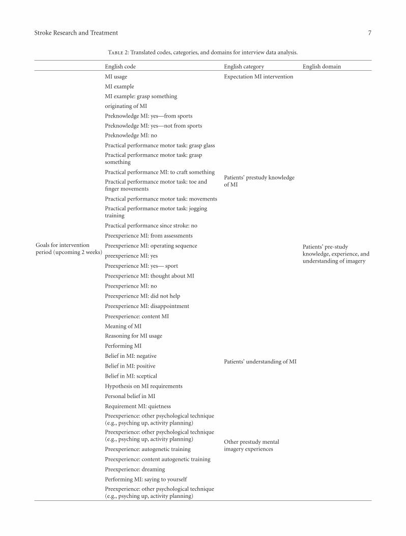
Stroke Research and Treatment 7
Table 2: Translated codes, categories, and domains for interview data analysis.
English code English category English domain
Goals for interventionperiod (upcoming 2 weeks)
MI usage Expectation MI intervention
MI example
Patients’ prestudy knowledgeof MI
Patients’ pre-studyknowledge, experience, andunderstanding of imagery
MI example: grasp something
originating of MI
Preknowledge MI: yes—from sports
Preknowledge MI: yes—not from sports
Preknowledge MI: no
Practical performance motor task: grasp glass
Practical performance motor task: graspsomething
Practical performance MI: to craft something
Practical performance motor task: toe andfinger movements
Practical performance motor task: movements
Practical performance motor task: joggingtraining
Practical performance since stroke: no
Preexperience MI: from assessments
Preexperience MI: operating sequence
preexperience MI: yes
Preexperience MI: yes— sport
Preexperience MI: thought about MI
Preexperience MI: no
Preexperience MI: did not help
Preexperience MI: disappointment
Preexperience: content MI
Meaning of MI
Patients’ understanding of MI
Reasoning for MI usage
Performing MI
Belief in MI: negative
Belief in MI: positive
Belief in MI: sceptical
Hypothesis on MI requirements
Personal belief in MI
Requirement MI: quietness
Preexperience: other psychological technique(e.g., psyching up, activity planning)
Other prestudy mentalimagery experiences
Preexperience: other psychological technique(e.g., psyching up, activity planning)
Preexperience: autogenetic training
Preexperience: content autogenetic training
Preexperience: dreaming
Performing MI: saying to yourself
Preexperience: other psychological technique(e.g., psyching up, activity planning)

8 Stroke Research and Treatment
Table 2: Continued.
English code English category English domain
Assumption MI intervention period: nothelpful
Belief in MI after MIintervention regardingprestudy MI experiencesAssumption MI intervention period
MI content/reflecting onsit-to-stand example
Performing MI: location Location
WhereStarting position MI Position
Assumption: starting position MI Position
Assumption: location MI Location
Performing MI: concentrating Concentration
When
Performing MI: concentrating no Concentration
MI: concentration difficulties Concentration
Duration MI Duration
Performing MI: simultaneously with otheractivity
Situation
Assumption: time of the day Time of the day
Reasoning for MI: MI content surroundings Content MI of MI example
What
Assumption: MI contentContent of MI during MIexample
MI content: stand up Content of MI example
MI content: motor task incomplete Movement completeness
MI content: motor task complete Movement completeness
MI content: motor task unclear Movement completeness
MI content: motor task like healthy people Content of MI example
MI content: imagine always the same Content of MI example
MI content: walking Content of MI example
MI content: person Content of MI example
MI content: see yourself Content of MI on MI example
MI content: sensation yes Senses
MI content: sensation no Senses
MI content: sit-to-stand Content of MI on MI example
MI content: sit-to-stand (SS) complete Movement completeness
MI content: SS surroundings Content of MI example
MI content: SS surroundings unclear Content of MI example
MI content: SS unclear Content of MI example
MI content: surroundings Content of MI example
MI content: surroundings light Content of MI example
MI content: surroundings nothing Content of MI example
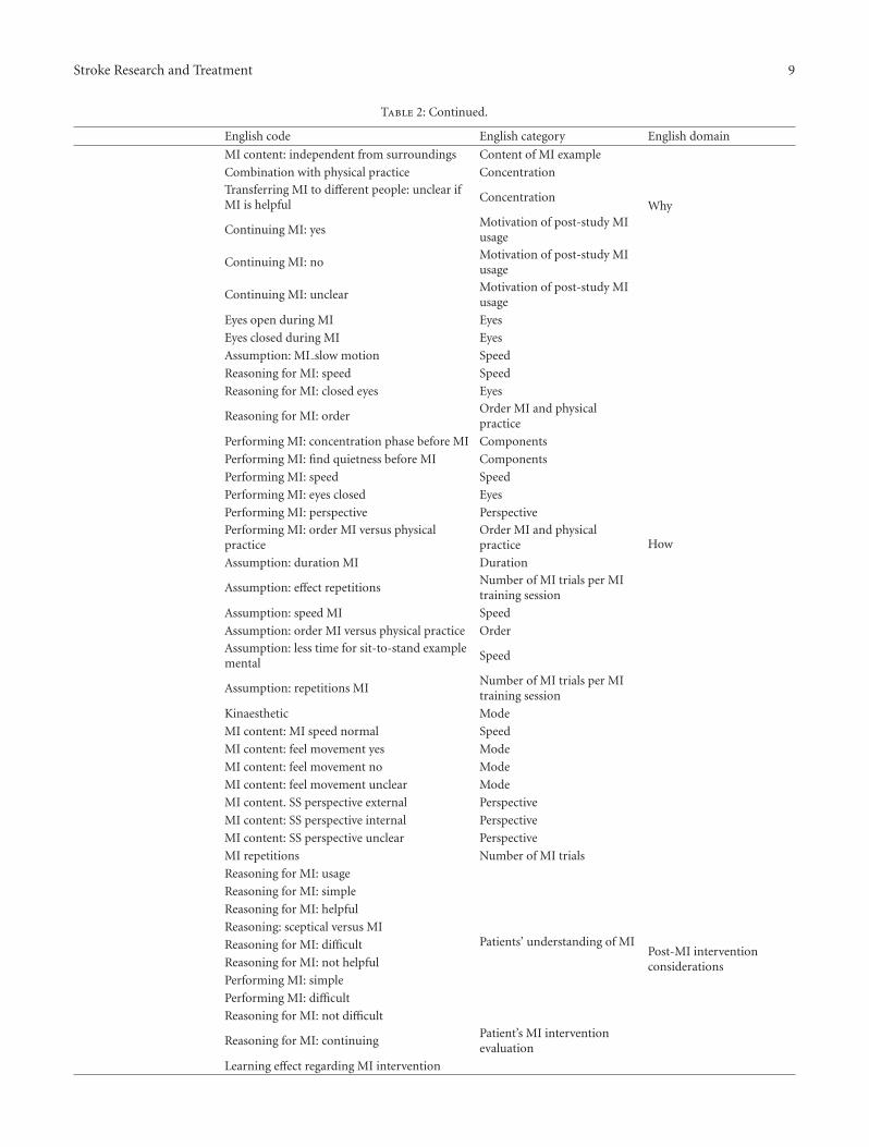
Stroke Research and Treatment 9
Table 2: Continued.
English code English category English domain
MI content: independent from surroundings Content of MI example
Combination with physical practice Concentration
WhyTransferring MI to different people: unclear ifMI is helpful
Concentration
Continuing MI: yesMotivation of post-study MIusage
Continuing MI: noMotivation of post-study MIusage
Continuing MI: unclearMotivation of post-study MIusage
Eyes open during MI Eyes
How
Eyes closed during MI Eyes
Assumption: MI slow motion Speed
Reasoning for MI: speed Speed
Reasoning for MI: closed eyes Eyes
Reasoning for MI: orderOrder MI and physicalpractice
Performing MI: concentration phase before MI Components
Performing MI: find quietness before MI Components
Performing MI: speed Speed
Performing MI: eyes closed Eyes
Performing MI: perspective Perspective
Performing MI: order MI versus physicalpractice
Order MI and physicalpractice
Assumption: duration MI Duration
Assumption: effect repetitionsNumber of MI trials per MItraining session
Assumption: speed MI Speed
Assumption: order MI versus physical practice Order
Assumption: less time for sit-to-stand examplemental
Speed
Assumption: repetitions MINumber of MI trials per MItraining session
Kinaesthetic Mode
MI content: MI speed normal Speed
MI content: feel movement yes Mode
MI content: feel movement no Mode
MI content: feel movement unclear Mode
MI content. SS perspective external Perspective
MI content: SS perspective internal Perspective
MI content: SS perspective unclear Perspective
MI repetitions Number of MI trials
Reasoning for MI: usage
Patients’ understanding of MIPost-MI interventionconsiderations
Reasoning for MI: simple
Reasoning for MI: helpful
Reasoning: sceptical versus MI
Reasoning for MI: difficult
Reasoning for MI: not helpful
Performing MI: simple
Performing MI: difficult
Reasoning for MI: not difficult
Reasoning for MI: continuingPatient’s MI interventionevaluation
Learning effect regarding MI intervention

10 Stroke Research and Treatment
Table 2: Continued.
English code English category English domain
Learning effect regarding MI intervention: newmovement sequence
Patients’ learning effect due toMI intervention
Learning MI
Transferring MI to different movementsequence: general
Transferring MI to different movementsequence: walking
Transferring MI to different movementsequence: fingers/hand
Transferring MI to different movementsequence: no
Transferring MI to different movementsequence: difficult
MI content: get up from the floor
Transferring MI to different people: MI ishelpful MI attributes/qualities
Transferring MI to different people: MI is nothelpful
Table 3: Patient study characteristics.
Int.number
Patientnumber
Age Gender StrokeAffected
brainarea
Time sincestroke(yrs)
MMSE EBI BBS
KVIQvis.1st
inter.
KVIQkin.1stint.
Lines oftext 1st
int.
Lines oftext 2nd
int.
Durationin min1st int.
Durationin min
2nd int.
1 6 61 Female CVI Right 1.3 28 62 43 48 48 234 (102) 163 (147) 39.1 23.4
2 13 53 Male CVI Left 1.7 27 62 55 29 27 251 (130) 141 (116) 44.3 23.3
3 15 31 Female ICB Left 2.6 27 62 48 35 25 230 (73) 58 (56) 30.0 12.0
4 16 51 Male ICB Right 2.9 30 62 55 45 40 232 (128) 149 (104) 33.5 27.4
5 19 63 Male CVI Bilateral 2.6 25 63 55 43 46 258 (123) 97 (89) 38.6 15.4
6 21 82 Female CVI Right 6.4 27 63 45 30 50 131 (53) 113 (108) 37.2 22.2
7 32 85 Male CVI left 3.8 26 62 55 45 43 110 (53)∗ 121 (89) N/A∗ 33.5
8 33 54 Male CVI Left 3.3 28 64 56 50 10 223 (129) 186 (178) 46.4 28.1
9 37 45 Male CVI Right 3.4 23 57 49 50 50 362 (193) N/A 47.1 N/A
10 38 71 Male CVI Left 3.7 29 62 56 34 21 192 (109) 91 (78) 33.1 15.2
11 43 64 Male CVI Right 6.2 23 54 43 33 21 149 (72) N/A 34.5 N/A
Numbers in bold indicate patients who practiced a type of imagery before study participation. Numbers in brackets indicate the amount of lines of thecomplete interview particularly related to MI. Int. = interview, yrs = years, MMSE = Mini-Mental State Examination, EBI = extended Barthel index, KVIQ =Kinaesthetic and Visual Imagery Questionnaire, vis. = visual, kin = kinaesthetic, min = minutes, N/A = not applicable, ∗= the first interview of this patientcould not be recorded completely due to technical problems.
practice, the imagery just started in his head without atrigger:
“Just like that. While sitting in a train, or tram,everywhere.” (Pat. 37, 1st interview).
In relation to position taken for MI practice, seven pa-tients identified a specific preferred position, two talkedabout a position related to the specific task to be imagined,and one patient spoke of other positions. The preferred posi-tion identified by patients can be related to the functionalstatus of the individuals so that they felt safe and secure, forexample, sitting or lying, or it might be related to the need
to relax or “shut down” their thinking before starting MIpractice. This contrasts with the changing task-specific posi-tion being adopted as preference, where the position mirrorsthe task starting position, for example, turning from supinelying to side lying.
“Basically, you could do that everywhere, don’tyou? In every, every position for sure. It dependson what you are going to do..... Sitting is a goodstarting position.” (Pat. 16, 1st interview).
During the second interview, after the two-week MIintervention period and the experience of MI, location and

Stroke Research and Treatment 11
Domain Category
Motor imagery
Where using MI?
When using MI?
What to imagine?
Why using MI?
How is MI performed?
Location
Position
Situation
MITS per week
Training times
Focus
Content of MI example
Movement completeness
Senses
Motivation
Belief and reasoning
Eyes
Order MI and PP
Perspective
Mode
Speed
MITS components
Progress
Duration
Number of MI trials
level∗ level#
Figure 2: Final data analysis hierarchy tree. To remain readability, the level of the subcategories was not displayed but described in the textof the results section. §Patient information based on assumptions and on experiences after one practical MI example: sit-to-stand and back,MI = motor imagery. ∗Domain level refers to the English domain column in Table 2. #Category level refers to the English category columnin Table 2.
position were again identified as the sole categories of theWhere domain. However, brightness had not been identifiedas important this time, although a known environment wasan additional idea voiced by patients as being useful. Aknown environment was preferred by patients as they wouldnot be distracted by new things or having to explore a newenvironment first. They could focus solely on the task to beimagined.
“But,... in your usual environment, not where youhave to absorb 100′000 other things, and you haveto remember them, haven’t you?” (Pat. 16, 2ndinterview).
The second interview data retained the subcategory iden-tification of preferred position and task-specific position,indicating that after exposure to the MI technique, thesedomains remained important.
3.2.2. When Using MI?
Interview data relating to timing (WHEN) elicited three cate-gory themes: (a) situation (4 patients), (b) MI training sessionper week (2 patients), and (c) times of the day (4 patients).
In identifying when they carried out MI practice, 4 pa-tients related this timing element to the situation or loca-tion (Where) category, showing that these domains were

12 Stroke Research and Treatment
inter-linked. They talked about the need to concentrate onthe MI task and therefore did not want to be disturbed byother tasks or noises (Pat. 32, Pat. 13, and Pat. 43). Some pre-ferred a quiet environment or even wanted to be alone (Pat.43, Pat. 6).
“No, no, before [that] I have to sit down, close theeyes and simply, ehm, yes, calm myself down andactually concentrate on: now I stand up.” (Pat. 13,1st interview).
“Actually, when I’m alone at home and can fullyconcentrate.” (Pat. 6, 1st interview).
For three male patients, the When element correspondedto the number of MI training sessions per week indicating thatMI training sessions should take place daily with MI repeti-tions at least 3 to maximum 10 times per day (Pat. 16, Pat. 43,and Pat. 13). Patients recommended short and frequent MItraining sessions most likely as a consequence of their stroke,which let concentration emerge as a theme.
Relating to time of the day, some patients identified a pre-ferred time of the day, which could be morning, afternoon, orevening, and for some the time of day related to tiredness, forexample, when he/she cannot sleep during nights (Pat. 13,Pat. 6, and Pat. 38). A male patient, who used MI as mara-thon preparation before stroke onset, articulated that MIcould be done at anytime of the day (Pat. 37). Furthermore,patients’ diary entries from the MI intervention study link tothe morning and evening times of the day for MI practice, inparticular, when lying in bed before getting up and beforefalling asleep.
“Actually, [I can do it] always, when I take thetime. But I think, it occupies the brain. And then,it does not make sense [to continue practicing MI]if you’re too tired and you do it again.” (Pat. 6, 1stinterview).
During the second interview after the experience of usingMI, two additional areas emerged in relation to timing ofpractice. Two patients considered that MI could be practicedwhile doing other activities that are automatic and which didnot need focussed concentration, for example, while mow-ing, raking leafs, watching television,and cycling (Pat. 13; Pat.33). This could obviously relate to increased confidence andcompetence in the MI technique, but also to the patient’smotor function level. Furthermore, one male patient men-tioned the necessity of a stable emotional and mental condi-tion to practice MI (Pat. 13). After the second interview thecategory MI training sessions per week included the subcate-gory sporadic MI training in addition. The subcategory diff-erent time of the day reported anytime and spontaneously astwo new MI training times.
3.2.3. What to Imagine?
In relation to WHAT patients imagined, interview data re-vealed four categories: (a) the related focus of a task to be im-agined with subcategories motor related (four patients)
and cognitive-related (four patients), (b) content of the im-agined task with subcategories imagined surroundings (tenpatients) and the imagined person (seven patients), (c) taskcompleteness, concentrated on the accuracy and the completeperformance of the task to be imagined (eleven patients), and(d) senses during MI (nine patients).
MI had a cognitive related focus in imagined tasks beforeand after stroke onset. Before stroke onset, patients used im-agery for marathon preparation, to organise staff, and workprocedures (Pat. 37, Pat. 38, Pat. 33, and Pat. 19). After strokeonset, MI had mainly a motor related focus. Patients imagi-ned simple single movements of fingers and toes, or trials tograsp and manipulate objects with the affected hand (Pat. 37,Pat. 19, and Pat. 16).
“And nothing happened, when you watched.Nothing happened. Only in my head I thought,yes, now I’m moving the toes. And the same withthe fingers, did not it?” (Pat. 16, 1st interview).
Experiences gained through the practical example of MIduring the interview allowed patients to generalise the MItechnique for further use. Two patients mentioned a possibleapplication of the MI technique in different motor tasks andparticular, to stair climbing (Pat. 32, Pat. 37). In the patient’scase, stair climbing is one of the most aspired movements.During the second interview, there was an increase inpatients’ perceptions of the possibility of options to apply theMI technique to other motor tasks. Furthermore, three pa-tients had already started using MI to imagine problemmovements as simple single motions, for example, moveaffected arm and hand, and as complex movements, for ex-ample, walking.
Concerning the category content of the imagined task, thedescribed imagined surroundings (subcategory) related tothe respective task (five patients), seeing things of the actualroom (two patients), described a bright or dark environment(six patients), or simply nothing (two patients). The secondsubcategory person emerged from describing seeing them-selves doing the task (five patients), or seeing an additionalperson in their imagery, for example, the therapist, who wasin the room where they practised MI (Pat. 37, Pat. 21). Inter-estingly, patients always saw themselves as a healthy personswithout any paresis or abnormality in their movements, sup-porting literature reports on athletes with musculoskeletalinjuries. Similar to dreaming, MI might be understood asallowing individuals to be the “whole” self and may havetherapeutic benefit in that psychological domain, but this re-mains untested.
The movement completeness category yielded subcategor-ies complete and incomplete, where ten patients imaginedthe complete motor task with all phases. Differences werementioned for individual phases of the imagining task. Whilestart and finish positions were sharp and vivid (Pat. 38, Pat.33), more unfocussed images of the movements were des-cribed in between. This was particularly evident in the pa-tient, who used imagery as marathon preparation prior tostroke.

Stroke Research and Treatment 13
“But the standing up, standing upright and sittingdown again, that’s what’s inside my head. It stayedin my head better than the rest. The other phaseswere a little bit just a glimpse. Yes, where I got upand sat down, so the things inbetween it only wasa glimpse.” (Pat. 38, 1st interview).
The interview data revealed aspects of the individuals’awareness of their senses during MI practice. Whilst threepatients had no sensory awareness during imagining, whichcould be related to the concentration needed for the motortask performance, many patients identified no awareness ofsmell or taste sensations (seven patients). However, two pa-tients described awareness of the sounds related to the motortask, for example, creaking of the chair or shoes (Pat. 37, Pat.38).
3.2.4. Why Using MI?
Patient data showed two major themes in relation to WHYpatients were interested in MI: (a) motivation (4 patients)and (b) belief and reasoning. Both themes are based onpatients’ prestudy beliefs and after having performed a prac-tical example of MI during the interview. Three major “moti-vations” were identified by patients (4 patients): (1) beingable to integrate an affected limb in daily routines (Pat. 19),(2) ease physical practice with MI preparation (Pat. 19), and(3) practice difficult or problematic movements in general(Pat. 37, Pat. 38, and Pat. 33).
Clearly, patients hoped that this technique might im-prove their physical function. In relation to belief andreasoning, patients identified that it might help them to gainconfidence in performing a movement (Pat. 21), that previ-ous exposure to MI had shown that it is an effective technique(Pat. 33, Pat. 38, and Pat. 37), and that the rationale to learnMI is that they can use it on their own to practice impairedmovements or action procedures (Pat. 16, Pat. 43, Pat. 15,and Pat. 19).
“Then you can learn it [MI], then you can do it[MI], and you realise that something is changing.”(Pat. 43, 1st interview).
“It wasn’t by far that way, but, ehm, the happinesswas very great, wasn’t it. And, yes, I tried it a lot,to move the fingers and all and watching and all,and when it only trembled a little bit, I was alreadyhappy, wasn’t it.” (Pat. 16, 1st interview).
“That I can do it [MI] myself at home.” (Pat. 15,1st interview).
Some patients, however, had opposing views and dis-belief in the technique considered. One patient (Pat. 37)was very sceptical to MI use with paralysis, or on its ownwithout physical practice. One patient went so far as to say“. . .because it is something stupid.” (Pat. 32, 1st interview)
“You know, . . ., you have to, yes, you have to do thetricks. You have to try, shift the weight. I always
imagined it too. But it did not work because of it[MI]. If your limb is feebly, then it’s feebly. Thenyou imagine it as long as you like.” (Pat. 37, 1stinterview).
There were three patients, who had no real opinion aboutMI. One of those patients mentioned that someone wouldhave to remind him permanently to practice MI. Otherwise,he would forget to do so due to his memory problems sincestroke onset.
Following exposure to the MI technique, new ideas emer-ged from patients in relation to its usage (3 patients).
“I think I will continue to do this [MI]. If I recog-nise that this [MI] will make me more flexible(agil). I will benefit from MI and I will benefit forsure. Now, I would say, it [MI] gives me more con-fidence. Now, it’s inside the brain.” (Pat. 21, 2ndinterview).
3.2.5. How Is MI Performed?
The analysis on HOW patients conducted MI practice re-vealed a wide variety of elements.
(1) State of Eyes during MI. Six patients mentioned thatthey close their eyes during MI practice, some ofthem with the aim to improve concentration.
(2) Order of Physical Practice and MI Practice. Patientsfavoured MI trials “before” physical practice trials (2patients). The advantage would be a better prepara-tion for physical practice and therefore an easierphysical practice performance.
(3) Perspective of Imagination. Patients used two perspec-tive options during MI practice: internal perspective(4 patients) and external perspective (4 patients). Ingeneral, it is difficult for patients to describe the usedperspective. Statements as “as I would stand in the cor-ner over there” (Pat. 16, 1st interview) or “I saw myselfas a person, as a whole person” (Pat. 16, 1st interview)indicated the external perspective. Here, the themesidentified from the data support each other. The Howand the What domains are linked together with theircategories mode and senses.
(4) MI Mode. After performing the MI practice exampleduring the first interview, five patients mentionedthat they really felt the movement during imagina-tion. This could be categorised as kinaesthetic MI andthus links with the What theme as described above.
(5) Duration of MI Trials Compared to Physical PracticeTrials. The duration of MI trials was compared tophysical practice trials in three subcategories: MI witha longer or shorter duration, and same duration likephysical practice trials. A reason for these discrepan-cies regarding the trial duration of MI versus physicalpractice could be attributed to different levels of de-tails imagined.

14 Stroke Research and Treatment
(6) Components of an MI Training Session. During inter-views, it became clear that some patients can startimmediately with an MI trial, whereas others neededa short moment of concentration and focussing be-forehand. Therefore, a category titled components ofMI was introduced.
(7) Progress in Performing MI. The oldest male patient,aged 85 (Pat. 32), mentioned that it would be helpfulfor him to start with a simple and short part of amotor task to imagine and add further steps aftersome consolidation. As this thought was confirmedby a second male patient during the second interview(Pat. 33), the category progress in performing MI wasbrought up.
(8) Duration of MI Practice. Related to the category timeof the day when MI training is performed (domainWhen), it is the category duration. It describes theoccasions where this category emerged with thenumber of MI trials below.
(9) Number of MI Trials. In general, patients reckonedthat the training duration depends on the effortneeded to practice MI (Pat. 6), but once a day seemednot enough. Patients suggested practicing MI severaltimes (3-4) per day, with at least 2-3 subsequent MItrials (Pat. 13, Pat. 15, and Pat. 21). Both categoriesremained important in the second interviews. Inparticular, duration of MI practice was extended toof 4–6 minutes for 5–7 times per day (Pat. 13),organised in small breaks between working tasks.Furthermore, one male patient suggested a total MIintervention duration of up to six months to reallylearn the MI technique, aiming at using it for everydaily movement or routine practice (Pat. 33).
3.2.6. Patients’ Prestudy Knowledge, Experience, and Under-standing of Imagery. A wide range of patients’ prior exposureto MI was observed. Two patients reported active usage ofMI, for example, during marathon preparation before strokeonset (Pat. 37), and to practice finger and toe movementsafter stroke onset (Pat. 16). Three patients had heard of MIor had seen athletes doing something what they interpretedas MI, in particular in ski racers just before their competition(Pat. 13, Pat. 6, and Pat. 33).
Four patients reported prestudy experiences with a typeof mental imagery practice other than MI. One young femalepatient mentioned that she would dream, seeing herselfwalking without any impairment as she walked when she washealthy (Pat. 15). One middle-aged patient mentioned usingautogenic training as stress-reducing technique (Pat. 13).Two males reported using a kind of self-talk or psyching-uptechnique to organise working procedures or for motivationpurposes (Pat. 33, Pat. 13).
During the first interviews, patients offered various des-criptions of what they understood or associated with theterm MI, ranging from clear explanations to broad com-ments in both ways: positive and negative.
“Well, it’s something normal. Like I would thinkabout something different.” (Pat. 33, 1st inter-view).
“Surely close the eyes, imagine something and thenpractice.” (Pat. 16, 1st interview).
“Well, something I cannot do at the moment, thatI imagine it, like I’m able to do it. Then it’s work-ing actively. That I picture or visualise the wholeprocedure in my head.” (Pat. 6, 1st interview).
“Well, one thinks it’s something stupid.” (Pat. 32,1st interview).
3.2.7. Post-MI Intervention Considerations. After the MIintervention, patients still found it difficult to describe whatthey were doing during MI (Pat. 19, Pat. 32). However, mostpatients found that it was simple and easy to perform, notexhausting (Pat. 15, Pat. 6, Pat. 19, and Pat. 16), and becameautomatic and natural after a while. Additionally, patientshad started using MI for movements that were problematicfor them, for example, climbing stairs, using affected hand/arm. Furthermore, six patients would recommend MI toother patients to practice depending on their mental capacityand their paresis level.
A minority of patients experienced MI during the inter-vention as difficult. Reasons were that they could not concen-trate enough (Pat. 21), that they missed parts of the motortask to imagine and had to think about it/control all im-agined details (Pat. 13, Pat. 32), and that physical practicetrials were more easy to perform than MI trials of the sametask (Pat. 32).
3.3. Patients’ Learning Effects. The observed learning effectsamong patients were related to the interviews, practical ex-ample of MI, and the two- week MI intervention. Effects wererelated to the motor task, to MI, and to mental abilities. Somepatients identified an increased confidence in performingthe motor task due to the several mental rehearsals. Theimproved MI ability was recognised by patients (Pat. 32, Pat.13, Pat. 21, Pat. 33, and Pat. 15), including the ability to applyMI to other motor tasks (Pat. 13), and not needing quietenvironments anymore (Pat. 13). Four patients attributedimproved cognitive abilities to the MI practice, for instance,being able to concentrate faster and for a longer period (Pat.33, Pat. 38), being more relaxed (Pat. 21), being more awakeor alert (Pat. 33), doing less day dreaming, and being moremotivated to do other activities (Pat. 13).
“Because I had the impression, it works my braina little. I had the impression, if I’m doing this, thenI’m more awake somehow. Had the impression,not only my circulation is trained, my brain also.And that’s just how it is, I have the feeling, it worksmy brain too.” (Pat. 33, 2nd interview).
3.4. Implications for Practice. Results from this researchsuggests that therapists may not expect patients to have

Stroke Research and Treatment 15
experience with MI, but probably half of patients may haveexperience with some kind of mental practice, for example,autogenic training. Therefore, patients’ previous mentalpractice knowledge should be evaluated before using MI inassociation with therapy sessions.
3.5. Interview Observations
Emotional Aspect. It is worth mentioning that patientsactively reported on their personal and emotional situationduring the interview. Even though, the patients’ stroke eventoccurred some time ago, the description of the event, time inhospital, rehabilitation process, and retrospect became veryemotional for several patients, for example, one female pa-tient started crying.
MI-Related Aspect. It should be emphasised that patients sawthemselves performing the motor task during MI. No pa-tient described seeing herself/himself as a patient with im-pairments. All saw themselves as healthy as before strokeonset. Furthermore, related to the automatic MI, observed bysome patients, performing MI of the motor task used in theintervention study started to occur suddenly in patients with-out conscious trigger.
Dissonance of Reporting. It is notable that one-third of thepatients showed a dissonance in reporting. Their answers atthe end of the MI-related part in the interviews could contra-dict answers from the beginning. This could be related to theinsights on the abstract topic of MI gained during the inter-view and after having performed the practical example of MI.
4. Discussion
The established framework from sport psychology of where,when, what, why, and how MI is used was explored in pa-tients after stroke in their chronic stage by conducting semi-structured interviews. The applied procedure offered the op-portunity to acquire detailed information on the nature ofpatients’ MI experiences, associations, MI training sessionelements (e.g., MI mode and state of eyes), and temporalparameters (e.g., duration of MI and number of MI trials).Patients imagined themselves as healthy individuals withoutimpairment. They did not focus on surroundings during MIpractice, and they reported to use positive imagery only. MIbecame automatic after the two-week MI intervention, andpatients did not need focussed concentration and quietnessas mentioned before the MI intervention.
Munroe and colleagues used a hierarchy with “where” asfirst level, “when” as second level, and “why” and “what” asthird level [10]. In contrast, this investigation allocated thequestions “where,” “when,” “what,” “why,” and “how” to thesame hierarchy level, which was also used in the publicationsof Driediger et al. [13] and Nordin and Cumming [12].
Based on the results of Kosslyn et al., one may expectmore patients having reporting the prior use of an imagerytype used [9]. Two arguments could explain the differentresult: study patients in this current work were explicitly
asked about knowledge and experiences of MI, not mentalpractice in general. Furthermore, we did not interviewyoung, healthy students, but patients after stroke in thechronic stage of the disease.
4.1. Where Using MI?
Patients after stroke liked to practice MI at home or whenalone, which is in line with athletes with musculoskeletal in-juries in Driediger et al.’s investigation [13]. Furthermore,athletes practiced MI mainly during physiotherapy sessionsbefore finishing an exercise, whereas stroke patients did notmention using MI during therapy at al. In the current anal-ysis, location and position were identified as WHERE cate-gories, whereas Nordin and Cumming and Driediger et al.did only focus on “location” [12, 13].
4.2. When Using MI?
Similar to athletes with musculoskeletal injuries, patientsconsidered that MI could be practiced while doing otheractivities that are automatic, and which did not need theirfocussed concentration, for example, watching television,driving a car [13]. This insight was reported by patientsduring the second interview only. We assume that the delayedinsight could be caused by the MI training during the inter-vention period, where patients gained MI knowledge, prac-tice, and experience. A categorisation of the WHEN domainby situation, MI training session per week, and training time ofthe day was not reported by Driediger et al. for athletes.
4.3. What to Imagine?
Driediger and colleagues did not describe the domain HOWas MI performed [13]. Therefore, their domain WHATincorporated five categories including session, effectiveness,nature, surroundings, and imagery type. The latter one wasclassified to the domain How in the current investigation.Driediger et al.’s category session described the length ofthe MI practice between 5 and 30 seconds for athleteswith musculoskeletal injuries. In contrast, patients afterstroke several MI practice sessions per day with a lengthof up to 15 minutes were accounted in the category dura-tion (domain HOW). Whereas athletes in category naturereported “positive” and “negative” imagery, patients men-tioned after stroke “positive imagery” only. Both groupsindicated seeing themselves as healthy individuals or athletespossessing their preinjury motor function and competitionlevel. Additionally, surroundings during MI practice did notplay an important role for athletes with musculoskeletalinjuries and patients after stroke, which could be due tofocusing on the healing process in athletes and the MIlearning process in patients.
“Type” in the study of Driediger et al. included reportingon MI perspective and vividness of imagery [13]. Bothcategories were classified to the domain HOW in the currentinvestigation.
16 Stroke Research and Treatment
4.4. Why Using MI?
Paivio’s framework on effects of imagery on human motorperformance influenced Driediger et al.’s classification ofWHY doing athletes with musculoskeletal injuries MI [11,13]. Driediger et al., [13] devised cognitive-specific imageryas a help to perform rehabilitation exercises. MI was beforeand simultaneously executed with physical practice aim-ing at skill enhancement in athletes with musculoskeletalinjuries. “Cognitive general” imagery was used to maintainthe ability to carry out activities associated with their sport.“Motivational-specific” imagery was used to visualise goaland related activities and to maintain motivation by imag-ining full recovery and future competitions. “Motivationalgeneral” (arousal and mastery) was used to continue reha-bilitation, even in painful conditions and to keep mentallytough. In athletes with musculoskeletal injuries, motivationalimagery was not used to enhance confidence. In the currentinvestigation, patients after stroke rarely used MI before theMI intervention study. Therefore, an allocation to “Motiva-tional general” based on the traditional framework is dif-ficult. Patients’ reasons to use imagery included gaining self-confidence to perform the motor task, easier physical prac-tice performance after MI, and being more alert. These indi-cations are in line with the results of the review from Feltzand Landers [25]. The authors emphasised that with thehelp of imagery, the performer is better prepared for thefollowing action, through shorter reaction times, lower sen-sory threshold, appropriate tension levels, focused attention,and an increase of muscle action potentials. Driediger andcolleagues also mentioned three more categories of thedomain WHY: healing with images of the physiologicalrecovery process, pain management, and injury prevention.None of them could be explored in patients after stroke [13].
4.5. How Is MI Performed?
Munroe et al.’s and Driediger et al.’s reports did not use theHOW domain [10, 13]. In the current investigation, nine cat-egories were identified in this domain, from which two wereincluded in the What domain by Driediger et al. (MI type,MI perspective) [13]. In this current work, “HOW” aimsto describe characteristics for the MI intervention design,which is contradictory to the “HOW” domain identified byNordin and Cumming [12]. Their “HOW” domain includedthree categories related to triggers that help to obtain images,to relate MI to symbolic images, and to design gradedlevels of images. The latter can be interpreted as compound-ing a basic visualisation and adding up more details of theimage and sensation. However, we believe that the MI experi-ence in patients after stroke cannot be compared to athletes’MI experience. Therefore, an in-depth description of theirMI trigger and MI acquirement cannot be expected. Oftenpatients reported that they had to start MI of the motor taskby themselves. After the MI intervention, MI started auto-matically regardless the patients’ current situation, location,or activity.
4.6. Linkage of Domains
During the data analysis, it became clear that several consid-ered domains are related.
WHAT and HOW. Although How comprises categories des-cribing the design of an MI training, it also contains the cate-gories perspective and MI mode that could be associated withthe category senses in the domain What. Munroe et al. andDriediger et al. did not describe a How domain [10, 13].However, Driediger and colleagues did define “perspective”and “mode” under the domain What, but termed mode astype.
WHEN and WHERE. Patients information for the domainWhen was closely related to the situation or location in thedomain Where
WHEN, WHAT, and HOW. For the domain When patientsreported the number of MI training sessions per week, numberof MI trials. Agreements for concentration (What) werementioned and short but frequent MI training sessions re-commended (How).
4.7. Limitations.
It could be argued that the nested study design limited thediversity of patient information gained and biased patientselection. However, all eleven patients showed a wide agerange from 31 to 85 years, different motor function level, andtime periods from stroke onset to study entry (1.3 to 6.2years). Furthermore, Guest et al. showed in their study with60 in-depth interviews that data saturation occurred withinthe first twelve interviews already, suggesting that the sampleconsidered in this investigation is sufficient [26]. Moreover,participating in the related pilot RCT provided more detaileddata on patients MI ability and offered the opportunity toconfirm patients’ statements from the first interview, forexample, regarding MI content, with responses from thesecond interview.
The interviewer’s MI knowledge and her function astherapist in the related MI intervention study could haveinfluenced formulating and asking questions in an attempt tocue certain answers and information. We assume that patientresponses were not influenced by the interviewer as patientsexpressed their negative MI experiences honestly during thesecond interviews.
No category related to MI ability or vividness of imagerycould be confirmed based on the patients’ interview data.Nevertheless, a link to the category movement completenesscould be drawn, which classifies incompletely or completelyseen movements. However, we believe that the interviewedpatients did not have the detailed knowledge on this parti-cular MI characteristic after the short MI intervention com-pared to continuously MI-trained athletes.
Despite constrains in qualitative research involving pa-tients with different levels of speaking impairments, whichcould limit the richness and depth of responses, it is

Stroke Research and Treatment 17
important to apply qualitative techniques, as confirmed bythe results obtained in this investigation.
5. Conclusion
Until now the imagery framework has been explored in vari-ous sports disciplines, but not in patients after stroke, whoare not trained in using MI routinely. MI understanding andusage differs between athletes and patients after stroke. It isessential to determine MI understanding and ability in strokepatients, in order to design tailored MI interventions. In thiswork, the framework on where, when, what, why, and howMI is used has been adopted from sports psychology andapplied to organise information gained in semistructuredpatient interviews. It became clear that MI is not establishedin patients after stroke. MI was not used to support motorfunction recovery. In rare cases, MI appeared spontaneouslyand was used for simple movements, for example, grasping.Patients related MI to a mental technique aiming at prac-ticing movements that are not possible at the moment orgain confidence in difficult movements. Furthermore, pa-tients imagined themselves as healthy individuals withoutimpairment. They did not focus on surroundings during MIpractice, and they reported to use positive imagery only. AfterMI training, patients became more flexible regarding theirlocation and position during MI practice. In most cases, MIbecame automatic after the intervention, and patients didnot need focussed concentration and quietness as mentionedin the first interview. MI use is clearly underresearchedin stroke rehabilitation. Future MI intervention studies forpatients after stroke should include an evaluation of patients’MI ability and an MI familiarisation session to learn essentialMI training session elements and their characteristics, forexample, MI perspective, MI mode. Furthermore, MI inter-ventions should start with simple motor tasks and less MItrials repetitions. After a consolidation phase, MI trial rep-etitions could be increased, and more complex motor taskscould be imagined.
Funding
The research project was partially funded by the Gottfriedund Julia Bangerter-Rhyner Foundation.
Acknowledgments
The authors would like to thank all patients for participatingin both interviews and reviewing all interview transcripts.Furthermore, they would like to thank Karin Lutz andAndrea Heinrichs for double and triple check of the trans-cribed data and patients’ quote translation. They are thankfulto Dr. Oliver Amft, who critically reviewed and commentedon an earlier version of the paper.
References
[1] S. M. Braun, A. J. Beurskens, P. J. Borm, T. Schack, and D. T.Wade, “The effects of mental practice in stroke rehabilitation:
a systematic review,” Archives of Physical Medicine and Reha-bilitation, vol. 87, no. 6, pp. 842–852, 2006.
[2] A. Zimmermann-Schlatter, C. Schuster, M. A. Puhan, E.Siekierka, and J. Steurer, “Efficacy of motor imagery in post-stroke rehabilitation: a systematic review,” Journal of Neuro-Engineering and Rehabilitation, vol. 5, article 8, 2008.
[3] H. Ziemainz, S. Hendrich, M. Schleinkofer, and K. Pfeifer,“The application of mental training in the rehabilitation ofstroke patients—review and calculating effect sizes,” Physika-lische Medizin Rehabilitationsmedizin Kurortmedizin, vol. 18,no. 4, pp. 198–202, 2008.
[4] L. S. M. Calayan and J. M. R. Dizon, “A systematic review onthe effectiveness of mental practice with motor imagery in theneurologic rehabilitation of stroke patients,” The Internet Jour-nal of Allied Health Sciences and Practice, vol. 7, no. 2, 2009.
[5] J. Decety and J. Grezes, “Neural mechanisms subserving theperception of human actions,” Trends in Cognitive Sciences,vol. 3, no. 5, pp. 172–178, 1999.
[6] E. Y. Yoo and B. I. Chung, “The effect of visual feedback plusmental practice on symmetrical weight-bearing training inpeople with hemiparesis,” Clinical Rehabilitation, vol. 20, no.5, pp. 388–397, 2006.
[7] G. Yue and K. J. Cole, “Strength increases from the motorprogram: comparison of training with maximal voluntary andimagined muscle contractions,” Journal of Neurophysiology,vol. 67, no. 5, pp. 1114–1123, 1992.
[8] K. A. Martin and C. R. Hall, “Using mental imagery to enhanceintrinsic motivation,” Journal of Sport & Exercise Psychology,vol. 17, no. 1, pp. 54–69, 1995.
[9] S. M. Kosslyn, C. Seger, J. R. Pani, and L. A. Hillger, “When isimagery used in everyday life? A diary study,” Journal of MentalImagery, vol. 14, pp. 131–152, 1990.
[10] K. J. Munroe, P. R. Giacobbi, C. Hall, and R. S. Weinberg, “Thefour Ws of imagery use: where, when, why, and what,” SportPsychologist, vol. 14, no. 2, pp. 119–137, 2000.
[11] A. Paivio, “Cognitive and motivational functions of imageryin human performance,” Canadian Journal of Applied SportSciences, vol. 10, no. 4, pp. 22S–28S, 1985.
[12] S. M. Nordin and J. Cumming, “Professional dancers describetheir imagery: where, when, what, why, and how,” SportPsychologist, vol. 19, no. 4, pp. 395–416, 2005.
[13] M. Driediger, C. Hall, and N. Callow, “Imagery use by injuredathletes: a qualitative analysis,” Journal of Sports Sciences, vol.24, no. 3, pp. 261–271, 2006.
[14] T. E. MacIntyre and A. P. Moran, “A qualitative investigation ofimagery use and meta-imagery processes among elite canoe-slalom competitors,” Journal of Imagery Research in Sport andPhysical Activity, vol. 2, no. 1, pp. 1–23, 2007.
[15] C. McKevitt, J. Redfern, F. Mold, and C. Wolfe, “Qualitativestudies of stroke: a systematic review,” Stroke, vol. 35, no. 6,pp. 1499–1505, 2004.
[16] T. K. Kvien and T. Heiberg, “Patient perspective in outcomeassessments—perceptions or something more?” Journal ofRheumatology, vol. 30, no. 4, pp. 873–876, 2003.
[17] U. Flick, Designing Qualitative Research, vol. 1 of The SAGEQualitative Research Kit, Sage, London, UK, 2007.
[18] G. R. Gibbs, Analysing Qualitative Data, vol. 6 of The SAGEQualitative Research Kit, Sage, London, UK, 2007.
[19] J. P. Clark, How to Peer Review a Qualitative Manuscript, BMJBooks, London, UK, 2nd edition, 2003.
[20] A. Tong, P. Sainsbury, and J. Craig, “Consolidated criteria forreporting qualitative research (COREQ): a 32-item checklistfor interviews and focus groups,” International Journal forQuality in Health Care, vol. 19, no. 6, pp. 349–357, 2007.
18 Stroke Research and Treatment
[21] C. Schuster, J. Butler, B. Andrews, U. Kischka, and T. Ettlin,“Comparison of embedded and added motor imagery trainingin patients after stroke: study protocol of a randomised con-trolled pilot trial using a mixed methods approach,” Trials, vol.10, article 97, 2009.
[22] R. A. Krueger and M. A. Casey, Focus Groups: A Practical Guidefor Applied Research, Sage, Thousand Oaks, Calif, USA, 3rdedition, 2000.
[23] D. Z. Meyer and L. M. Avery, “Excel as a qualitative dataanalysis tool,” Field Methods, vol. 21, no. 1, pp. 91–112, 2009.
[24] C. Schuster, R. Hilfiker, O. Amft et al., “Best practice for motorimagery: a systematic literature review on motor imagerytraining elements in five different disciplines,” BMC Medicine,vol. 9, article 75, 2011.
[25] D. L. Feltz and D. M. Landers, “The effects of mental practiceon motor skill learning and performance: a meta-analysis,”Journal of Sport & Exercise Psychology, vol. 5, pp. 25–57, 1983.
[26] G. Guest, A. Bunce, and L. Johnson, “How many interviewsare enough?” Field Methods, vol. 18, no. 1, pp. 59–82, 2006.

Hindawi Publishing CorporationStroke Research and TreatmentVolume 2012, Article ID 159391, 8 pagesdoi:10.1155/2012/159391
Research Article
Constraint-Induced Movement Therapy (CIMT):Current Perspectives and Future Directions
Aimee P. Reiss, Steven L. Wolf, Elizabeth A. Hammel, Erin L. McLeod,and Erin A. Williams
Division of Physical Therapy, Department of Rehabilitation Medicine, Emory University School of Medicine,Atlanta, GA, USA
Correspondence should be addressed to Aimee P. Reiss, [email protected]
Received 7 November 2011; Revised 14 February 2012; Accepted 15 February 2012
Academic Editor: Agnes Roby-Brami
Copyright © 2012 Aimee P. Reiss et al. This is an open access article distributed under the Creative Commons Attribution License,which permits unrestricted use, distribution, and reproduction in any medium, provided the original work is properly cited.
Constraint-induced movement therapy (CIMT) has gained considerable popularity as a treatment technique for upper extremityrehabilitation among patients with mild-to-moderate stroke. While substantial evidence has emerged to support its applicability,issues remain unanswered regarding the best and most practical approach. Following the establishment of what can be calledthe “signature” CIMT approach characterized by intense clinic/laboratory-based practice, several distributed forms of training,collectively known as modified constraint therapy (mCIMT), have emerged. There is a need to examine the strengths andlimitations of such approaches, and based upon such information, develop the components of a study that would compare thesignature approach to the best elements of mCIMT, referred to here as “alternative” CIMT. Based upon a PEDro review of literature,limitations in mCIMT studies for meeting criteria were identified and discussed. A suggestion for a “first effort” at a comparativestudy that would both address such limitations while taking practical considerations into account is provided.
1. IntroductionRecent advances in stroke rehabilitation research, includingconstraint-induced movement therapy (CIMT), have thepotential to change traditional therapeutic approaches inclinical practice. However, communicating best practices andimplementing change remain significant challenges for bothresearchers and practicing clinicians to overcome. In a recentCanadian study, Menon et al. [1] found that, despite theevidence of best practice in stroke rehabilitation, the imple-mentation of best practice in the clinic was inconsistent andunderutilized, as clinicians continued to apply traditionalneurodevelopmental techniques in the treatment of individ-uals after stroke. Barriers to incorporating evidence-basedpractice (EBP) into clinical settings are well documented andfall under two general categories: individual clinician barriersand organization barriers [1–3]. Lack of time and resourcesis often cited by clinicians as primary limitations to readingor searching the literature [1–3] as well as self-efficacy. In thiscontext, self-efficacy means the degree to which an individualclinician feels capable of searching, reading, analyzing, and
implementing evidence-based practice into her daily clinicalpractice and thought processing. Low self-efficacy maybe more prevalent in clinicians who have been workingfor 15 or more years because of lack of formal educationin EBP skills [1, 2]. Organizational barriers can includelack of computers or access to research databases, the absenceof active research in clinical settings and limited encourage-ment of professional development, with a growing emphasison productivity [1, 2]. The combination of time constraints,productivity pressures, volume of research available, anda dearth in EBP skills may, over time, create an evengreater gap between best evidence and current practice [1].National and international guidelines for evidence-basedpractice following stroke can assist clinicians in determiningbest practice by compiling recent advances in stroke research.These guidelines are easily accessible and decrease the burdenon clinicians to search and assess the literature themselves.However, they may not always provide sufficient evidenceor information regarding a particular intervention, andthere may not be a shared consensus among guidelines.
2 Stroke Research and Treatment
In the case of constraint-induced movement therapy,drawing a solid conclusion after reviewing prominent strokeguidelines is difficult. For instance, in a scientific statementfrom the American Heart Association [4], CIMT is recom-mended for chronic stroke patients with greater than 10degrees finger extension, but no parameters of treatment areprovided. Australian stroke guidelines paint a less conclusivepicture of CIMT, concluding that CIMT is effective withmore than 20 hours of training, but possibly detrimentalwhen provided very early in the rehabilitation process [5].Here again no treatment parameters were offered for thedelivery of CIMT and the lack of methodological consistencyin the literature was noted. New Zealand guidelines brieflymention CIMT as a possible rehabilitation approach, butmake no recommendations regarding patient criteria orthe parameters to deliver CIMT [6]. Canadian guidelines,updated in 2010, offer the most specific details for patientcriteria and protocol parameters for signature or modifiedCIMT (mCIMT) in early stroke rehabilitation [7]. Thesedifferences yield a lack of clarity regarding recommendationsfor CIMT and may in part be precipitated by the lack ofmethodological quality in the modified CIMT literature.The purpose of this paper is therefore multi-fold: (1)present current perspectives of signature CIMT and theevolution of mCIMT; (2) discuss limitations in currentCIMT literature by using PEDro criteria as a basis to; (3)suggest an “alternative” form of modified CIMT, which cansubsequently be compared to the signature form in a “head-to-head” comparison, first in preliminary studies and then asa formal clinical trial. This consideration is important sincesuch an effort has not been formally undertaken.
1.1. Current Perspectives of Signature CIMT and the Evolutionof mCIMT. Constraint-induced movement therapy (CIMT)is a widely explored treatment protocol to increase functionaluse of the more impaired upper extremity (UE) for personswith hemiparetic stroke. The theoretical basis for CIMT wasdeveloped through early research in nonhuman primatesfrom which the concept of learned nonuse following limbsomatosensory deafferentation emerged [8, 9]. Taub andcolleagues described the learned nonuse phenomenon as abehavioural adaptation that occurs in response to the lossof sensory feedback due to a decrease in coordinated move-ments, negative reinforcement from unsuccessful attemptswith the impaired limb, and positive reinforcement resultingfrom success in compensatory movement patterns with theunimpaired limb. The result of this sequelae is a decreasein functional use of the impaired UE [10]. This undesirableadaptation was overcome in monkeys by restraining theinsensate limb, thus forcing them to use the impaired UE[11].
According to Taub and colleagues [12], learned nonuseoccurring after somatosensory deafferentation in nonhumanprimates is comparable to that seen following central nervoussystem (CNS) insult in humans, such as after stroke. Inthe first application of a treatment technique to overcomelearned nonuse in humans, a patient with hemipareticstroke was asked to perform functional tasks with themore impaired UE with restraint of the less affected UE
[13]. Restraint, in addition to encouraging without formallytraining massed practice of the impaired UE, resulted in whatWolf et al. called “forced use” [14].
The protocol has evolved to specifically include repet-itive and adaptive task practice under clinical supervision.These additional structured elements collectively define thecurrent concept of CIMT. Repetitive task practice (RTP) iscontinuous blocked practice of a specific functional task,usually for a period of 15–20 minutes. Adaptive task practice(ATP), or shaping, uses a step-wise approximation method,breaking down tasks into successive manageable compo-nents to improve overall proficiency [15, 16]. Based uponthe underlying operant conditioning through provision oftherapist feedback, shaping fosters patient problem solving,resulting in self-motivation to use the affected limb [16].This intensive practice fosters motor relearning and hasbeen shown to promote neural plasticity in the CNS [15].Successful application of CIMT is thought to induce a use-dependent increase in cortical reorganization of the areas ofthe brain controlling the most affected limb [17, 18].
Several studies, primarily in mild to moderately impairedsurvivors of stroke, have demonstrated clinically relevantresults [12, 19–21]. However, limitations, such as smallsample sizes within trials, variations in time since stroke,and alternative CIMT protocols, weakened the ability to drawsignificant conclusions about pertinent findings [22, 23]. In2006, results from the first Phase III, multisite, randomizedclinical study, the EXtremity Constraint-Induced TherapyEvaluation (EXCITE) trial, were published and showed sta-tistically and clinically relevant confirmation of the efficacyof CIMT in patients three-to-nine months posthemipareticstroke [24].
Signature CIMT, developed by Taub [12, 25] and laterused in the EXCITE [24] trial, included restraint of the lessimpaired upper extremity by donning a protective safetymitt for 90% of waking hours over a two-week interventionperiod. Subjects were also required to participate in sixhours/day (five days/week) of ATP and RTP [10, 24]. Someresearchers have criticized this signature protocol as beingimpractical in clinical settings [26–31]. Patient tolerance,mitt wearing adherence, feasibility in clinics, and reimburse-ment issues have been emphasized as key weaknesses of thesignature CIMT protocol, thus potentially acting as barriersto more pervasive clinical implementation [32, 33].
In response to these critiques, a number of “modified”versions have arisen to address the issues presented bythe signature form of CIMT. Some investigators alteredintensity protocols by distributing total treatment time overa longer duration. For example, Wu et al. [29, 34, 35]decreased the intensity to two hours/day (five days/week) butincreased duration to three weeks, which required only sixhours/day of upper extremity restraint. Page et al. [26, 33, 36]increased treatment duration to 10 weeks, with 30 minutesof intervention per day, three days/week and reduced mittrestraint to five hours/day. Other investigators expandedthe inclusion criterion for relative chronicity. Ro et al. [37]included subjects 14 days after stroke, and Miltner et al.[38] included subjects up to 17 years after stroke. While allof these modifications claim to have shown effectiveness in

Stroke Research and Treatment 3
improving motor function, these studies, like early signatureCIMT research, are also limited by small sample sizes. Of alltrials included in previous reviews of CIMT, only two studieshad greater than 30 participants [22, 23].
Despite significant variability in protocols, each formof treatment delivery claims the name “modified” CIMT[35, 39]. The issue therein is the lack of standardization bywhich to establish a consistent reference point for monitoringtreatment dosage, creating confusion among the researcher,therapist and reimbursement communities. Therefore, ananalysis of existing modified CIMT protocols, to devisea reasonable synthesis of approaches called “alternative”CIMT, should be performed and becomes a necessaryprecursor before a best model alternative intervention thatincorporates key elements, such as intensity, duration, andsubject chronicity [40, 41], can be constructed. This bestmodel alternative protocol can then be formally compared tosignature CIMT to determine if an alternative, more feasibleapproach is equally effective.
An important first step, intended to increase the veracityof these future studies, is the analysis of the methodologicalquality of current CIMT literature using a valid and reliabletool to determine worthy inclusion in development of thealternative treatment protocol. To build a strong study at anylevel, including only the highest quality literature as a foun-dation for study design and in support of findings, becomesa critical consideration. Not only do these factors increasethe integrity of any immediate study, but also that of futurestudies. Unlike previous reviews which explored only CIMTefficacy [22, 23], we sought to critically analyze existingCIMT methodologies in order to systematically determinespecific deficiencies within articles. Worthy articles can thenbe selected for use in determining an alternative form CIMT,which can then compared to the signature form in a “head-to-head” comparison.
The evaluation tool chosen for analyzing methodologicalquality of CIMT research was the Physiotherapy EvidenceDatabase (PEDro). PEDro applies 11 criteria, 10 of whichare scored (see Appendix A) to assign a quantitative measureof study strength [42]. This particular set of criteria waschosen for its specificity to physical therapy literature; use bynovice and expert researchers alike; reliability [43]; strengthin assessing methodological quality specifically in strokeliterature [44].
Although the authors did not intend to perform asystematic review of the CIMT literature, several articleswere selected from 1998–2008 as a comprehensive samplingto use as the basis for this discussion. The list of CIMT liter-ature was compiled using the following electronic databases:PubMed, Cochrane Library, MEDLINE, and Ovid. Key termsincluded constraint-induced movement therapy, modifiedCIMT, hemiparetic stroke, and constraint-induced therapy.
Approximately 75 studies were deemed relevant forfurther review, based upon title and abstract containingCIMT or “modified” CIMT. Studies were excluded if allthree components of CIMT (ATP, RTP, and restraint) werenot administered in at least one group; they containedpaediatric participants (<18 years of age), used subjects other
than patients with stroke, were systematic reviews/meta-analyses, or were nonexperimental literature (case studies,letters to the editor, or perspectives). Experimental literatureaccepted for use in this review included Sackett’s levelsof evidence: 1b, 2b, and 3b (see Appendix B) [45]. Afteremploying this procedure, 27 articles, published between1999 and 2008, were chosen for inclusion in this discussion[24, 26–31, 33, 34, 37, 38, 46–61].
Therapist raters with comparable experience in researchdesign, independently rated each of 27 CIMT articles usingPEDro criteria. A series of detailed discussions regardingthe nature and interpretation of these criteria preceded theactual rating exercise. All raters were blinded to each other’sassessments. A “yes” was given for each criterion the raterbelieved was satisfied by the article. Total scores reflectedthe total number of “yes” answers given on eligible criteria(2–11 are considered “eligible” for scoring). One reviewercompiled the results of the three raters. Scores were notdiscussed among raters prior to compilation or statisticalanalysis. A post-hoc analysis showed that raters could notagree upon whether some studies met three particularPEDro criteria: baseline similarity between groups, outcomemeasures obtained for at least 85% of participants, andclarity regarding successfully meeting the “intention to treat”directive (see Appendix A).
There may be several explanations for interrater discrep-ancies. Lack of clarity in some PEDro operational definitionsmay have contributed to varying interpretations amongreviewers. Hence there may have been some uncertaintyabout whether studies clearly articulated and implementedan intention to treat strategy. A discussion amongst raterssuggested that disagreement between raters may have beenprecipitated by alternative interpretations or poorly definedaspects of methodologies or outcomes. Such misinterpre-tations could be overcome through clearer precision inthe delineation of methods. For criterion four (baselinesimilarity), some articles calculated significant differencesbetween groups for only a few characteristics, but claimedoverall “baseline similarity.” This discrepancy in approachleft the reader unsure if the criterion was completely fulfilled.Raters were commonly uncertain if outcomes measures werecollected in at least 85% of subjects (criteria eight). Finally,report of attrition (criteria nine) was another source ofreader confusion due to unclear documentation. Subjectdropout had to be inferred in some cases from tables, ratherthan explicitly stated in the text.
Additionally, raters agreed that blinding of evaluatorsto subject allocation and intervention received were notadequately addressed in the current review. Collectively,these methodological weaknesses present a challenge forclinicians to confidently interpret and apply the principles ofthese alternative forms of CIMT. These discrepancies becamethe basis for the selection of elements that needed greaterclarification and inclusion in an alternative form of CIMTagainst which one can then undertake a comparison with thesignature CIMT.
1.2. Efforts to Create a Standardized Alternative Form ofCIMT. A recent article attempted to compare a version

4 Stroke Research and Treatment
of modified CIMT to signature CIMT. Barzel and col-leagues [62] compared signature CIMT to a 4-week home-based modified CIMT program (CIMThome). CIMThomepatients and a family member received an initial day oftraining from a physiotherapist regarding the two primarycomponents of signature CIMT: shaping and constraint ofthe nonaffected upper extremity. CIMThome patients andtheir caregivers then performed a self-managed programat home for four weeks, with weekly visits by the phys-iotherapist to supervise and advance therapeutic exercisesas appropriate. Despite the home-based program, patientsstill received nearly 15 hours of supervision from a phys-iotherapist. Results showed CIMThome to be as effectiveas signature CIMT; however, the sample size was smallwith just seven chronic stroke patients in the CIMThomegroup.
Hosomi and colleagues also developed a self-trainingprotocol using elements of signature CIMT [63]. Fortypatients were recruited based on signature CIMT criteria.Patients were then instructed in a self-training protocol thatincluded instruction in shaping tasks to address individuallimitations. The protocol consisted of 20 minutes of self-training per shaping task, culminating in 10–15 differenttraining tasks per day (five hours a day for 10 consecutiveweekdays). Direct supervision by a physiotherapist occurredevery 20 minutes to evaluate patient performance andadvance therapeutic exercise as appropriate. Results showedsignificant improvements on the Fugl-Meyer Assessment,the Wolf Motor Function Test, and The Motricity Index.However, a major limitation of this study was having nocomparison to a control group or other modified CIMT pro-tocol. Therefore, the weaknesses of methodology discussedin this paper have still not been addressed in recent studieseither, and a need exists to generate a best-model alterna-tive CIMT option that incorporates weaknesses extractedfrom the literature.
1.3. Implications. In designing future studies, methodologiesmust be clearly defined and controlled to improve clarity forreaders and replication for clinical researchers. Specifically,discrepancies were noted for attrition and diffusion ofintervention, and report of baseline statistics.
To provide evidence-based practice, increased numbersof therapists are accessing and reading clinical trials in orderto choose appropriate interventions. When attempting toqualify an article’s strength, clinicians and researchers mayuse a system like PEDro. Therapists may be assumed touse high- rather than low-scoring articles when choosinginterventions. However, if criterion satisfaction is unclear,interpretation is left to the reader, leading to discrepancies inperception of the value of a clinical trial that can ultimatelyimpact clinician interest in utilization of treatment protocols.A more appropriate course of action would be to placeresponsibility upon researchers to give greater attention toclarity when describing methodological considerations.
Recently, within the field of neurorehabilitation, therehas been a call to improve the value of studies [64, 65].Dobkin [65] proposed performing a thorough qualitative
assessment of current literature as a key to accomplishinga more systematic approach to method design, to increasethe integrity of multisite randomized clinical trials for motorinterventions. In the case of CIMT, we have performeda comprehensive literature search and assessed the method-ological quality of several articles to determine specificdeficiencies within the articles.
Secondarily, the most substantial elements that mightserve as the foundation of a “best model” alternative CIMTprotocol can also be proposed. Three articles in our mCIMTreview scored 8/10 on the PEDro scale [29, 31, 34], thehighest scores in our review. The frequency and intensityof the modified CIMT interventions were very similaracross these studies: 2 h/d, 5d/wk for 3 wk. This treatmentintensity may be more feasible and practical than signatureCIMT. In addition, we propose further elements drawnfrom signature CIMT, including two hours of home taskpractice for three days per week for three weeks includingspecification and rationalization for this practice. Partici-pants in this alternative form of CIMT would also undergo30 minutes of a nonspecific but documented functionalactivity six days per week and wear the restraining device90% of waking hours. This dosing approximates 75 hoursover a three-week interval using a distributed practicemodel that would be comparable to almost 80 hours (upto six hours per day, five days per week for two weeksin a clinic/laboratory environment in addition to abouttwo hours per day for 10 days of home-based activities)of signature CIMT. For both interventions, restriction of bet-ter limb use would occur for 90% of waking hours through-out the intervention period. Inclusion criteria would matchthat established for the EXCITE Trial [24]. The alternativeCIMT plan would include therapist guided on-sight trainingfor two hours the first day of each of three weeks with homebased assignment of mutually agreed tasks throughout theremainder of the week for each of three weeks. Participantsin both groups would be evaluated before and after theintervention with subsequent followups at 3 and 6 monthsafter intervention. The evaluator would be blinded to groupand the selected outcome measure would be standardized.Dropouts would have last values carried forward (impu-tation), a typical procedure in intention to treat studies.The study would be powered based upon the selectedoutcome measure. For example, if the outcome was theWMFT, a reasonable change could be a 30 percent reductionin median time to complete tasks associated with a patientimpression of percent improvement in function as suggestedby Fritz et al. [53]. Collectively, such a comparison wouldaddress several issues. All the limitations observed inthe present PEDro review would be overcome, doseequivalency would be established, a reasonable home based,patient driven program could be standardized and compli-ance measured, and immediate and intermediate end pointswould optimize difficulties often encountered in trackingparticipants over a longer period of time. A future pilot studywill assess the feasibility and efficacy of this proposed “bestmodel” alternative CIMT protocol to signature CIMT withthe intent to develop a signature mCIMT protocol.
Stroke Research and Treatment 5
2. Possible Limitations
The exclusion of meta-analyses and case-studies presents apossible limitation to the extensiveness of the PEDro review.The PEDro system used in this review pertained only tosingle clinical trials. Moreover, while there were only 27articles comprising this review, such a number is relativelyhigh compared to previous systematic CIMT reviews. Threeexisting systematic reviews in CIMT research, conductedby Hakkennes and keating [22], Bonaiuti et al. [23], andCorbetta et al. [66], included 14, nine, and 18 studiesrespectively, and each review used only two reviewers. In theHakkenes and Keating [22] and Bonaiuti et al. [23] reviewsdisagreements were discussed until scores were agreedupon. Unlike these studies, disagreements between ratersin this study were viewed as valuable, and utilized to notelimitations in current research methods in hopes that thedesign of future studies might address these considerations.In addition, the Corbetta review [66] described a majorityof the articles chosen as “underpowered and imprecise,”making the case for a larger RCT and underscoring the weakmethodology of the CIMT literature.
3. Conclusion
The model proposed in this paper is not only useful foranalysis of CIMT literature, but may serve as a template forfuture studies in any genre of scientific inquiry. The qualityof CIMT literature, specifically modified and signaturemethods, was examined closely in an attempt to increase theintegrity of a future pilot study comparing a “best model”alternative CIMT protocol with the signature form of CIMT.A direct comparison approach that addressed limitationsin the literature extracted from the PEDro review was sug-gested. Efforts by researchers to improve methodology andstandardization of protocols can greatly assist the practicingclinician in analyzing EBP and incorporating best practicesinto clinical practice. Establishing a standardized best-modelalternative CIMT protocol would also allow stroke guidelinesto make clearer, more definitive recommendations regardingCIMT.
Appendices
A. PEDro Criteria
(1) Eligibility criteria were specified.
(2) Subjects were randomly allocated to groups (in acrossover study, subjects were randomly allocated anorder in which treatments were received).
(3) Allocation was concealed.
(4) The groups were similar at baseline regarding themost important prognostic indicators.
(5) There was blinding of all subjects.
(6) There was blinding of all therapists who administeredthe therapy.
(7) There was blinding of all assessors who measured atleast one key outcome.
(8) Measures of at least one key outcome were obtainedfrom more than 85% of the subjects.
(9) All subjects for whom outcome measures were avail-able received the treatment or control condition asallocated or, where this was not the case, data for atleast one key outcome was analyzed by “intention totreat.”
(10) The results of between-group statistical comparisonsare reported for at least one key outcome.
(11) The study provides both point measures and mea-sures of variability for at least one key outcome [42].
B. Sackett’s Levels of Evidence
Levels of evidence for interventions [67]:
(1a) systematic reviews of randomized controlled trials(RCTs),
(1b) individual RCTs with narrow confidence interval,
(2a) systematic reviews of cohort studies,
(2b) individual cohort studies and low-quality RCTs,
(3a) systematic reviews of case-control studies,
(3b) case-controlled studies,
(4) case series and poor-quality cohort and case-controlstudies,
(5) expert opinion.
Conflict of Interests
The authors report no of interests. The authors alone areresponsible for the content and writing of the paper.
Acknowledgments
Portions of this work were funded by NIH Grant HD37606from the National Center for Medical Rehabilitation Re-search and the National Institute of Neurological Diseasesand Stroke.
References
[1] A. Menon, N. K. Bitensky, and S. Straus, “Best practiseuse in stroke rehabilitation: from trials and tribulations tosolutions!,” Disability and Rehabilitation, vol. 32, no. 8, pp.646–649, 2010.
[2] N. M. Salbach, S. J. T. Guilcher, S. B. Jaglal, and D. A.Davis, “Factors influencing information seeking by physicaltherapists providing stroke management,” Physical Therapy,vol. 89, no. 10, pp. 1039–1050, 2009.
[3] D. U. Jette, K. Bacon, C. Batty et al., “Evidence-based practice:beliefs, attitudes, knowledge, and behaviors of physical thera-pists,” Physical Therapy, vol. 83, no. 9, pp. 786–805, 2003.

6 Stroke Research and Treatment
[4] E. L. Miller, L. Murray, L. Richards et al., “Comprehensiveoverview of nursing and interdisciplinary rehabilitation careof the stroke patient: a scientific statement from the Americanheart association,” Stroke, vol. 41, no. 10, pp. 2402–2448, 2010.
[5] National Stroke Foundation, “Clinical Guidelines for StrokeManagement,” Melbourne, Australia, 2010.
[6] Stroke Foundation of New Zealand and New Zealand Guide-lines Group, “Clinical Guidelines for Stroke Management2010,” Stroke Foundation of New Zealand, Wellington, NewZealand, 2010.
[7] M. P. Linsday, M. Bayley, M. D. Hill, C. Davies-Schinkel,S. Singh, and S. Phillips, “Canadian Best Practice Recem-mendations for Stroke Care (Update 2010),” On behalf ofthe Canadian Stroke Strategy Best Practices and StandardsWriting Group, Ottawa, Ontario Canada: Canadian StrokeNetwork, 2010.
[8] E. Taub and I. A. Goldberg, “Use of sensory recombinationand somatosensory deafferentation techniques in the investi-gation of sensory-motor integration,” Perception, vol. 3, no. 4,pp. 393–405, 1974.
[9] E. Taub, R. D. Heitmann, and G. Barro, “Alertness, level ofactivity, and purposive movement following somatosensorydeafferentation in monkeys,” Annals of the New York Academyof Sciences, vol. 290, pp. 348–364, 1977.
[10] E. Taub, G. Uswatte, and T. Elbert, “New treatments in neu-rorehabilitation founded on basic research,” Nature ReviewsNeuroscience, vol. 3, no. 3, pp. 228–236, 2002.
[11] E. Taub, “Somatosensory deafferentation research with mon-keys: implications for rehabilitation medicine,” in BehavioralPsychology in Rehabilitation Medicine, L. Ince, Ed., pp. 371–401, Williams & Wilkins, Baltimore, Md, USA, 1980.
[12] E. Taub, N. E. Miller, T. A. Novack et al., “Techniqueto improve chronic motor deficit after stroke,” Archives ofPhysical Medicine and Rehabilitation, vol. 74, no. 4, pp. 347–354, 1993.
[13] C. G. Ostendorf and S. L. Wolf, “Effect of forced use ofthe upper extremity of a hemiplegic patient on changes infunction. A single-case design,” Physical Therapy, vol. 61, no.7, pp. 1022–1028, 1981.
[14] S. L. Wolf, E. Lecraw, L. A. Barton, and B. B. Jann, “Forced useof hemiplegic upper extremities to reverse the effect of learnednonuse among chronic stroke and head-injured patients,”Experimental Neurology, vol. 104, no. 2, pp. 125–132, 1989.
[15] S. L. Wolf, S. Blanton, H. Baer, J. Breshears, and A. J.Butler, “Repetitive task practice: a critical review of constraint-induced movement therapy in stroke,” Neurologist, vol. 8, no.6, pp. 325–338, 2002.
[16] C. J. Winstein and S. L. Wolf, “Task-oriented training topromote upper extremity recovery,” in Stroke Recovery &Rehabilitation, J. Stein, R. L. Harvey, and R. F. Macko, Eds.,pp. 267–290, Demos Medical Publishing, New York, NY, USA,1st edition, 2009.
[17] E. Taub and G. Uswatte, “Constraint-induced movementtherapy: bridging from the primate laboratory to the strokerehabilitation laboratory,” Journal of Rehabilitation Medicine,no. 41, supplement, pp. 34–40, 2003.
[18] J. Liepert, W. H. R. Miltner, H. Bauder et al., “Motor cortexplasticity during constraint,induced movement therapy instroke patients,” Neuroscience Letters, vol. 250, no. 1, pp. 5–8,1998.
[19] S. J. Page, S. Sisto, P. Levine, M. V. Johnston, and M. Hughes,“Modified constraint induced therapy: a randomized feasibil-ity and efficacy study,” Journal of Rehabilitation Research andDevelopment, vol. 38, no. 5, pp. 583–590, 2001.
[20] S. J. Page, S. Sisto, M. V. Johnston, and P. Levine, “Modifiedconstraint-induced therapy after subacute stroke: a prelimi-nary study,” Neurorehabilitation and Neural Repair, vol. 16, no.3, pp. 290–295, 2002.
[21] G. F. Wittenberg, R. Chen, K. Ishii et al., “Constraint-inducedtherapy in stroke: magnetic-stimulation motor maps andcerebral activation,” Neurorehabilitation and Neural Repair,vol. 17, no. 1, pp. 48–57, 2003.
[22] S. Hakkennes and J. L. Keating, “Constraint-induced move-ment therapy following stroke: a systematic review of ran-domised controlled trials,” Australian Journal of Physiotherapy,vol. 51, no. 4, pp. 221–231, 2005.
[23] D. Bonaiuti, L. Rebasti, and P. Sioli, “The constraint inducedmovement therapy: a systematic review of randomised con-trolled trials on the adult stroke patients,” Europa Medicophys-ica, vol. 43, no. 2, pp. 139–146, 2007.
[24] S. L. Wolf, C. J. Winstein, J. P. Miller et al., “Effect ofconstraint-induced movement therapy on upper extremityfunction 3 to 9 months after stroke: the EXCITE randomizedclinical trial,” Journal of the American Medical Association, vol.296, no. 17, pp. 2095–2104, 2006.
[25] E. Taub and D. M. Morris, “Constraint-induced movementtherapy to enhance recovery after stroke,” Current Atheroscle-rosis Reports, vol. 3, no. 4, pp. 279–286, 2001.
[26] S. J. Page, S. Sisto, P. Levine, and R. E. McGrath, “Efficacyof modified constraint-induced movement therapy in chronicstroke: a single-blinded randomized controlled trial,” Archivesof Physical Medicine and Rehabilitation, vol. 85, no. 1, pp. 14–18, 2004.
[27] C. Dettmers, U. Teske, F. Hamzei, G. Uswatte, E. Taub, and C.Weiller, “Distributed form of constraint-induced movementtherapy improves functional outcome and quality of life afterstroke,” Archives of Physical Medicine and Rehabilitation, vol.86, no. 2, pp. 204–209, 2005.
[28] E. Taub, P. S. Lum, P. Hardin, V. W. Mark, and G. Uswatte,“AutoCITE: automated delivery of CI therapy with reducedeffort by therapists,” Stroke, vol. 36, no. 6, pp. 1301–1304, 2005.
[29] C. Y. Wu, C. L. Chen, S. F. Tang, K. C. Lin, and Y. Y.Huang, “Kinematic and clinical analyses of upper-extremitymovements after constraint-induced movement therapy inpatients with stroke: a randomized controlled trial,” Archivesof Physical Medicine and Rehabilitation, vol. 88, no. 8, pp. 964–970, 2007.
[30] A. Sterr, T. Elbert, I. Berthold, S. Kolbel, B. Rockstroh,and E. Taub, “Longer versus shorter daily constraint-inducedmovement therapy of chronic hemiparesis: an exploratorystudy,” Archives of Physical Medicine and Rehabilitation, vol. 83,no. 10, pp. 1374–1377, 2002.
[31] K. C. Lin, C. Y. Wu, T. H. Wei, C. Y. Lee, and J. S. Liu, “Effects ofmodified constraint-induced movement therapy on reach-to-grasp movements and functional performance after chronicstroke: a randomized controlled study,” Clinical Rehabilitation,vol. 21, no. 12, pp. 1075–1086, 2007.
[32] S. L. Wolf, “On “Modified constraint-induced therapy... ” Pageet al., Physical Therapy, vol. 88, pp. 333–340, 2008,” PhysicalTherapy, vol. 88, no. 5, pp. 680–684, 2008.
[33] S. J. Page, P. Levine, A. Leonard, J. P. Szaflarski, and B.M. Kissela, “Modified constraint-induced therapy in chronicstroke: results of a single-blinded randomized controlled trial,”Physical Therapy, vol. 88, no. 3, pp. 333–340, 2008.
[34] C. Y. Wu, C. L. Chen, W. C. Tsai, K. C. Lin, and S. H. Chou, “Arandomized controlled trial of modified constraint-inducedmovement therapy for elderly stroke survivors: changes inmotor impairment, daily functioning, and quality of life,”

Stroke Research and Treatment 7
Archives of Physical Medicine and Rehabilitation, vol. 88, no.3, pp. 273–278, 2007.
[35] C. Y. Wu, “Author’s response,” Neurorehabilitation and NeuralRepair, vol. 21, no. 6, pp. 574–575, 2007.
[36] S. J. Page and P. Levine, “Modified constraint-induced therapyin patients with chronic stroke exhibiting minimal movementability in the affected arm,” Physical Therapy, vol. 87, no. 7, pp.872–878, 2007.
[37] T. Ro, E. Noser, C. Boake et al., “Functional reorganizationand recovery after constraint-induced movement therapy insubacute stroke: case reports,” Neurocase, vol. 12, no. 1, pp.50–60, 2006.
[38] W. H. R. Miltner, H. Bauder, M. Sommer, C. Dettmers, andE. Taub, “Effects of constraint-induced movement therapy onpatients with chronic motor deficits after stroke: a replication,”Stroke, vol. 30, no. 3, pp. 586–592, 1999.
[39] S. J. Page, “To the editor,” Neurorehabilitation and NeuralRepair, vol. 21, no. 6, p. 574, 2007.
[40] B. H. Dobkin, “Confounders in rehabilitation trials of task-oriented training: lessons from the designs of the EXCITEand SCILT multicenter trials,” Neurorehabilitation and NeuralRepair, vol. 21, no. 1, pp. 3–13, 2007.
[41] S. L. Wolf, C. J. Winstein, J. P. Miller, S. Blanton, P. C. Clark,and D. Nichols-Larsen, “Looking in the rear view mirror whenconversing with back seat drivers: the EXCITE Trial revis-ited,” Neurorehabilitation and Neural Repair, vol. 21, no. 5,pp. 379–387, 2007.
[42] Physiotherapy Evidence Database, 2008, http://www.pedro.fhs.usyd.edu.au/.
[43] C. G. Maher, C. Sherrington, R. D. Herbert, A. M. Moseley,and M. Elkins, “Reliability of the PEDro scale for rating qualityof randomized controlled trials,” Physical Therapy, vol. 83,no. 8, pp. 713–721, 2003.
[44] S. K. Bhogal, R. W. Teasell, N. C. Foley, and M. R. Speechley,“The PEDro scale provides a more comprehensive measureof methodological quality than the Jadad scale in strokerehabilitation literature,” Journal of Clinical Epidemiology,vol. 58, no. 7, pp. 668–673, 2005.
[45] American Physical Therapy Association, “All evidence is notcreated equal,” 2003, http://www.apta.org/AM/Template.cfm?Section=Advance Search&TEMPLATE=/CM/HTMLDisplay.cfm&CONTENTID=37998.
[46] A. Kunkel, B. Kopp, G. Muller et al., “Constraint-inducedmovement therapy for motor recovery in chronic strokepatients,” Archives of Physical Medicine and Rehabilitation,vol. 80, no. 6, pp. 624–628, 1999.
[47] A. Suputtitada, N. C. Suwanwela, and S. Tumvitee, “Effec-tiveness of constraint-induced movement therapy in chronicstroke patients,” Journal of the Medical Association of Thailand,vol. 87, no. 12, pp. 1482–1490, 2004.
[48] M. Rijntjes, V. Hobbeling, F. Hamzei et al., “Individ-ual factors in constraint-induced movement therapy afterstroke,” Neurorehabilitation and Neural Repair, vol. 19, no. 3,pp. 238–249, 2005.
[49] C. Brogardh and B. H. Sjolund, “Constraint-induced move-ment therapy in patients with stroke: a pilot study oneffects of small group training and of extended miituse,” Clinical Rehabilitation, vol. 20, no. 3, pp. 218–227,2006.
[50] S. L. Fritz, K. E. Light, S. N. Clifford, T. S. Patterson,A. L. Behrman, and S. B. Davis, “Descriptive characteristics aspotential predictors of outcomes following constraint-inducedmovement therapy for people after stroke,” Physical Therapy,vol. 86, no. 6, pp. 825–832, 2006.
[51] E. Taub and G. Uswatte, “Constraint-Induced Movementtherapy: answers and questions after two decades of research,”NeuroRehabilitation, vol. 21, no. 2, pp. 93–95, 2006.
[52] G. Uswatte, E. Taub, D. Morris, J. Barman, and J. Crago,“Contribution of the shaping and restraint components ofConstraint-Induced Movement therapy to Treatment Out-come,” NeuroRehabilitation, vol. 21, no. 2, pp. 147–156, 2006.
[53] S. L. Fritz, S. Z. George, S. L. Wolf, and K. E. Light, “Participantperception of recovery as criterion to establish importanceof improvement for constraint-induced movement therapyoutcome measures: a preliminary study,” Physical Therapy, vol.87, no. 2, pp. 170–178, 2007.
[54] A. E. Dahl, T. Askim, R. Stock, E. Langørgen, S. Lydersen, andB. Indredavik, “Short- and long-term outcome of constraint-induced movement therapy after stroke: a randomized con-trolled feasibility trial,” Clinical Rehabilitation, vol. 22, no. 5,pp. 436–447, 2008.
[55] L. Sawaki, A. J. Butler, X. Leng et al., “Constraint-inducedmovement therapy results in increased motor map area insubjects 3 to 9 months after stroke,” Neurorehabilitation andNeural Repair, vol. 22, no. 5, pp. 505–513, 2008.
[56] N. M. Bonifer, K. M. Anderson, and D. B. Arciniegas,“Constraint-induced movement therapy after stroke: efficacyfor patients with minimal upper-extremity motor ability,”Archives of Physical Medicine and Rehabilitation, vol. 86, no.9, pp. 1867–1873, 2005.
[57] C. Boake, E. A. Noser, T. Ro et al., “Constraint-inducedmovement therapy during early stroke rehabilitation,” Neu-rorehabilitation and Neural Repair, vol. 21, no. 1, pp. 14–24,2007.
[58] J. M. W. W. Myint, G. F. C. Yuen, T. K. K. Yu et al., “A studyof constraint-induced movement therapy in subacute strokepatients in Hong Kong,” Clinical Rehabilitation, vol. 22, no. 2,pp. 112–124, 2008.
[59] C. E. Lang, D. F. Edwards, R. L. Birkenmeier, and A.W. Dromerick, “Estimating minimal clinically importantdifferences of upper-extremity measures early after stroke,”Archives of Physical Medicine and Rehabilitation, vol. 89, no. 9,pp. 1693–1700, 2008.
[60] N. M. Bonifer, K. M. Anderson, and D. B. Arciniegas,“Constraint-induced therapy for moderate chronic upperextremity impairment after stroke,” Brain Injury, vol. 19,no. 5, pp. 323–330, 2005.
[61] S. R. Pierce, K. G. Gallagher, S. W. Schaumburg, A. M.Gershkoff, J. P. Gaughan, and L. Shutter, “Home forceduse in an outpatient rehabilitation program for adults withhemiplegia: a pilot study,” Neurorehabilitation and NeuralRepair, vol. 17, no. 4, pp. 214–219, 2003.
[62] A. Barzel, J. Liepert, K. Haevernick et al., “Comparisonof two types of Constraint-Induced Movement Therapy inchronic stroke patients: a pilot study,” Restorative Neurologyand Neuroscience, vol. 27, no. 6, pp. 673–680, 2009.
[63] M. Hosomi, T. Koyama, T. Takebayashi et al., “A modifiedmethod of constraint-induced movement therapy: a super-vised self-training protocol,” Journal of Stroke and Cerebrovas-cular Diseases. In press.
[64] B. Kollen, G. Kwakkel, and E. Lindeman, “Functional recoveryafter stroke: a review of current developments in strokerehabilitation research,” Reviews on Recent Clinical Trials, vol.1, no. 1, pp. 75–80, 2006.
[65] B. H. Dobkin, “Progressive staging of pilot studies toimprove phase III trials for motor interventions,” Neurore-habilitation and Neural Repair, vol. 23, no. 3, pp. 197–206,2009.

8 Stroke Research and Treatment
[66] D. Corbetta, V. Sirtori, L. Moja, and R. Gatti, “Constraint-induced movement therapy in stroke patients: systematicreview and meta-analysis,” European Journal of Physical andRehabilitation Medicine, vol. 46, no. 4, pp. 537–544, 2010.
[67] D. L. Sackett, S. E. Straus, W. S. Richardson et al., Evidence-Based Medicine: How to Practice and Teach EBM, ChurchillLivingstone Inc., Edinburgh, Scotland, UK, 2nd edition, 2000.

Hindawi Publishing CorporationStroke Research and TreatmentVolume 2012, Article ID 820673, 9 pagesdoi:10.1155/2012/820673
Review Article
How Physically Active Are People with Stroke inPhysiotherapy Sessions Aimed at Improving Motor Function?A Systematic Review
Gurpreet Kaur,1 Coralie English,1, 2 and Susan Hillier1, 2
1 School of Health Sciences, University of South Australia, P.O. Box 2471, Adelaide, SA 5001, Australia2 International Centre for Allied Health Evidence, School of Health Sciences, University of South Australia,P.O. Box 2471, Adelaide, SA 5001, Australia
Correspondence should be addressed to Coralie English, [email protected]
Received 7 October 2011; Revised 19 December 2011; Accepted 12 January 2012
Academic Editor: Ching-yi Wu
Copyright © 2012 Gurpreet Kaur et al. This is an open access article distributed under the Creative Commons Attribution License,which permits unrestricted use, distribution, and reproduction in any medium, provided the original work is properly cited.
Background. Targeted physical activity drives functional recovery after stroke. This review aimed to determine the amount of timestroke survivors spend physically active during physiotherapy sessions. Summary of Review. A systematic search was conductedto identify published studies that investigated the use of time by people with stroke during physiotherapy sessions. Seven studieswere included; six observational and one randomised controlled trial. People with stroke were found to be physically active for anaverage of 60 percent of their physiotherapy session duration. The most common activities practiced in a physiotherapy sessionwere walking, sitting, and standing with a mean (SD) practice time of 8.7 (4.3), 4.5 (4.0), and 8.3 (2.6) minutes, respectively.Conclusion. People with stroke were found to spend less than two-thirds of their physiotherapy sessions duration engaged inphysical activity. In light of dosage studies, practice time may be insufficient to drive optimal motor recovery.
1. Introduction
People with stroke spend less than a quarter of their dayengaged in physical activity in rehabilitation centres [1, 2].Studies conducted internationally over many years haveshown that the time spent by people with stroke in therapyand in contact with therapists during the working day is verylittle [1–4]. Bernhardt et al. [1] found that people with strokespent only 5.2 percent of the working day in contact withtherapists in an acute stroke unit which equates to 0.5 hoursof a nine-hour observation period. Similarly, Thompson andMcKinstry [4] found that people with stroke in an inpatientrehabilitation unit spent only 1.2 hours of an 11-hourobservation period in therapy. However, an observationalstudy investigating the use of time in physiotherapy sessionsspecifically, reported that people with stroke spend between21 percent and 30 percent of therapy sessions inactive [5].Many studies have identified therapy sessions as being themost active part of the day and therefore provide the greatestopportunity to maximise physical activity levels. Therefore,
this review was important to determine exactly how activepeople with stroke are in their physiotherapy sessions duringstroke rehabilitation.
Targeted physical activity drives functional recoveryafter stroke. There is now strong evidence that moretime spent in task-specific therapy after stroke improvesfunctional outcomes [6, 7]. Furthermore, high repetitions ofa task-specific physical activity have been shown to facilitatepositive neuroplasticity in stroke survivors [8–10].
In light of this knowledge, and the fact that clinicalguidelines for stroke management include recommendationsto maximise active therapy time [11], it is important todetermine just how active stroke survivors are in therapysessions aimed at improving motor function. Therefore, theaim of this review was to synthesise the current evidenceabout the total amount of time spent by people with strokeengaged in physical activity (total active time) and timespent engaged in different physical activity categories duringphysiotherapy sessions in stroke rehabilitation.

2 Stroke Research and Treatment
Table 1: Search terms used to identify published studies whichreported data on therapy content and duration.
Stroke or CVA or “cerebrovascular accident∗” or
“cerebrovascular disorder∗” or “cerebrovascular disease∗”
or hemip∗
And
rehabilitation or physiotherapy or “physical therapy”
And
“time use” or time or “therapy∗ time” or “time taken” or
“activity∗ time” or “time spent”,
And
activity∗ or “physical activity∗” or “active therapy” or
“motor activity∗” or exercise or “exercise program” or
“exercise therapy” or “therapy content” or intensity or repetition∗
Or
“behavioural mapping” or “behavioral mapping”
2. Methods
Published studies of all designs were identified by enteringvarious search terms (Table 1) in the following electronicdatabases: CINAHL, AMED, MEDLINE, and EMBASE(Classic + Embase from 1947 to 2010, week 38). Preliminarysearches were conducted in August 2010 to find the appropri-ate search terms. The final search was completed on the 11thof December, 2011. The reference lists of studies which metthe selection criteria were searched for potentially relevantpublications. Searches were limited by years of publication(1990 to current), English language, and participants’ agegroup (18 years and over). The authors of studies whichreported both therapy content and therapy time but hadinsufficient data for the purposes of this systematic reviewwere contacted for additional data.
Studies were included if (1) participants were adultsreceiving rehabilitation after stroke, (2) physiotherapy wasprovided in inpatient rehabilitation or an acute care hospital,and (3) data relating to both the therapy content (typeof physical activity subcategories), and the amount oftime spent in different physical activity categories duringphysiotherapy sessions was reported (i.e., studies reportingcontent of therapy only or total duration of therapy sessionsonly were not included).
Critical appraisal of the relevant articles was conductedusing a modified evidence-based learning critical appraisalchecklist [12]. This tool assesses the quality of observationalstudies in four categories: population, data collection, studydesign, and results. The factors which were most likely tocontribute bias to the studies were chosen from the originalcritical appraisal tool. Two additional criteria were added:“concealment of study purpose to the participants” as thiswould reduce any bias arising from changes in usual practiceand “involvement of an independent person observing thetherapy sessions” which would reduce the risk of bias ofoverestimation of therapy duration. A copy of the criticalappraisal tool as it was applied in this review appears in theAppendix. Two reviewers critically appraised the included
studies independently. Justification of results from bothreviewers was discussed and when disagreement occurred,a consensus was reached by discussion. Where furtherinformation was required to clarify criteria (e.g., whether theassessor was an independent person), authors were contacteddirectly. Where consensus could not be reached or criteriaremained ambiguous, the opinion of a third reviewer wassought.
Data from the included studies were extracted includingtotal therapy session duration, time spent in differentphysical activity subcategories, and total inactive time. Thepercentage of total active time was calculated using total timespent in physical activity subcategories in a therapy session.The total active time was averaged across studies, where datawere sufficiently homogenous.
3. Results
The search yielded 2534 hits of which 61 studies werepotentially relevant after reviewing titles and abstract (seeFigure 1 for flow chart). After removing 13 duplicates, 48relevant full-text articles were obtained and assessed againstthe selection criteria by one reviewer. A total of 28 studieswere excluded because of their outpatient setting, and/orlack of data on therapy content or duration. Two reviewersassessed the full text of the remaining 20 studies andindependently made a decision about studies to be includedin the review. A third reviewer was available to adjudicate anydisagreements, but this was not required. The reference listsof the included seven studies were scrutinized, but no furtherrelevant publications were identified. An expert check wasalso conducted, but no further studies were identified.
All of the included studies were assessed as having a lowrisk of bias except Peurala et al. [14] which was assessed ashaving moderate to high risk of bias (Table 2). In Peuralaet al. [14], there were no independent observers involved(the same therapists that provided the therapy also evaluatedcontent and duration), the data collection methods werenot adequately described, and the data collection instrumentwas not validated. Furthermore, it was unclear if the studypurpose was concealed from the participants and therapists,thereby increasing the risk of overestimation of activitylevels.
All the included studies were observational except Peu-rala et al. [14], which was an RCT. The majority of theincluded studies were conducted in Australia (Table 3).Five of the included studies were carried out in inpatientrehabilitation facilities and two in acute care hospitals(Table 3). The average time since stroke ranged from5.6 [15] to 161 [16] days (Table 3). Video recording,behavioural mapping, and contemporaneous recording bythe researchers or therapists were the three main methodsof observation used in the included studies. In the videorecording method of observation, the entire physiotherapysession was video-recorded and later analysed to extract thedata. In the behavioural mapping method of observation, thetotal observation period (for, e.g., from 9 am to 5 pm) is

Stroke Research and Treatment 3
2534 titles and abstracts identified fromdatabase search
0 articles identified fromother sources
61 relevant studies
48 full-text studies
20 full-text studies reviewed by 2independent assessors
13 studies excluded due to insufficient data on eitheractive therapy time or therapy content
2473 hits excluded
13 duplicates removed
20 excluded due to outpatient or nursing home setting
7 studies included in the review
8 excluded as they did not report data on both therapycontent and duration
Figure 1: Flowchart of study selection (according to the Prisma statement [13]).
divided into 10-minute time slots. During every 10 minute-time slot, participants are observed for one minute, andtheir behaviour is recorded. The contemporaneous recordinginvolves researchers or treating therapists recording thecontent and duration of the therapy sessions in a datacollection instrument during or after the observed therapysession.
The mean total therapy session time ranged from 24minutes to 64 minutes (mean of 49.5 minutes) except Langet al. [17] (Table 3). Lang et al. [17] measured the numberof repetitions performed by participants during a therapysession but they did not report total therapy time. Theamount of active time within physiotherapy sessions rangedfrom 27 minutes to 39 minutes (mean of 32.8 minutes).The percentage of time spent active during a therapy sessionranged from 42 percent to 71 percent (mean of 60 percent)(Table 3). The time spent by people with stroke engaged invarious physical activity subcategories is outlined in Table 4.
The descriptions of physical activity subcategories andinactivity across the studies varied, but could be collated intocategories of walking, standing activities, cycling, transfers(including sit-to-stand), sitting activities, upper limb activ-ities, bed mobility/activities in lying, and inactivity/rest time.
3.1. Walking Practice. The average amount of time spentin walking practice ranged from four to 14.7 minutes pertherapy session (Table 4). Walking practice included earlygait activities (walking on level surface, therapist assistedusing a gait aid) and advanced gait activities (walking on
uneven surface, stairs, obstacle courses, and treadmill) [14–16]. In the study by Lang et al. [17], the average numberof steps taken per therapy session was 395, and the averagenumber of times the participants got up to walk was nine pertherapy session.
3.2. Sitting Activities. All the studies except Lang et al.[17] reported the amount of time spent in sitting activitiesspecifically. Sitting activities included postural control exer-cises, reaching, and active lower limb exercises. The averageamount of time spent in sitting practice ranged from 0.3 to9.6 minutes.
3.3. Standing Activities. All the studies except Lang et al.[17] reported the amount of time spent in standing activitiesspecifically. Exercises in standing included standing up,shifting weight from one leg to the other, and reaching instanding. The average amount of time spent in standingpractice ranged from four to 11.6 minutes per therapysession.
3.4. Upper Limb Activities. All the included studies except DeWit et al. [18] reported the amount of time spent in upperlimb activities during therapy sessions. Upper limb activitiesincluded active task practice using the paretic upper limb.The average amount of time spent in upper limb activitiesranged from 0.9 to 7.9 minutes per therapy session. Lang etal. [17] reported an average of 86 repetitions of active upperlimb activities performed per therapy session.

4 Stroke Research and Treatment
Table 2: Critical appraisal of the included studies.
Key:
Yes No Unclear
Els
on e
t al
. (20
09)
[5]
Lan
g et
al.
(200
9) [
16]
Ber
nh
ardt
et
al. (
2007
) [1
4]
Peu
rala
et
al. (
2007
) [1
3]
DeW
it e
t al
. (20
06)
[17]
Kuy
s et
al.
(200
6) [
15]
Ada
et
al. (
1999
) [1
8]
Study population well reported?+ + + + + + +
Inclusion and exclusion criteria definitively outlined?+ + + + + +
Adequate sample size?+ + + ? + + ?
Data collection methods clearly described?+ + + + + +
Data collection instrument validated?+ + + + + +
Independent observer involved?+ + ? + +
Appropriate statistical analysis and its interpretation?+ + + + + +
Is subset analysis a major, rather than minor, focus of the article?+ + + +
Study purpose concealed from participants+ + + ? ? ? +
?+
−
−
−
−
−
−
−
−−−
3.5. Bed Mobility/Activities in Lying. Only three studies [5,15, 16] reported the average amount of time spent pertherapy session performing bed mobility/activities in lying,this ranged from 0.3 to 5.2 minutes. Bed mobility/activitiesin lying included rolling, bridging, lying down from sitting,moving across the bed, sitting from lying, and isolatedhip/knee control.
3.6. Transfers Including Sit-to-Stand Practice. Five of theincluded studies [5, 14, 15, 17, 19] reported the amountof time/repetitions spent engaged in transfer exercises pertherapy session. Transfers included sit-to-stand, movingfrom wheelchair to bed, and wheelchair to toilet. The average
amount of time spent in transfer practice ranged from 1.8 to3.7 minutes per therapy session. An average of 11 repetitionsof transfers per session was reported by Lang et al. [17].
3.7. Other Therapeutic Activities. Only four of the includedstudies [5, 14, 18, 19] reported the time spent in otheractivities. Other therapeutic activities included cycling, activ-ities in kneeling, passive movements, soft tissue techniques,tonus inhibition, positioning to stretch muscles, exercisingthe affected lower limb in any position, selective movementsincluding coordination, strengthening exercises, and activerelaxation. The average amount of time spent engaged inother activities ranged from 4.1 to 16 minutes.

Stroke Research and Treatment 5
Ta
ble
3:Su
mm
ary
ofth
ein
clu
ded
stu
dies
and
thei
rfi
ndi
ngs
inre
lati
onto
ther
apy
dura
tion
.
Au
thor
/yea
rSe
ttin
gLo
cati
onM
ean
(SD
)ti
me
sin
cest
roke
(day
s)
Nu
mbe
rof
part
icip
ants
/th
erap
yse
ssio
ns
Met
hod
ofob
serv
atio
nM
ean
tota
lth
erap
yti
me
per
ther
apy
sess
ion
(min
s)
Mea
nac
tive
tim
epe
rth
erap
yse
ssio
n(m
ins)
Mea
n%
acti
veti
me
per
ther
apy
sess
ion
Part
icip
ants
Th
erap
yse
ssio
ns
Els
onet
al.2
009
[5]
Inpa
tien
tre
hab
ilita
tion
Au
stra
lia47
.3(3
0.6)
1530
Vid
eore
cord
ing
42 CC
T:5
3IT
:31
30 CC
T:3
7IT
:30
71 CC
T:7
1IT
:72
Lan
get
al.2
009
[17]
Inpa
tien
tre
hab
ilita
tion
USA
and
Can
ada
118
(157
)10
031
2
Rec
ordi
ng
ofn
um
ber
ofre
peti
tion
sof
task
sor
mov
emen
tsby
rese
arch
ers
NR
36�
NR
Ber
nh
ardt
etal
.200
7[1
5]A
cute
hos
pita
lA
ust
ralia
5.6
(NR
)58
84B
ehav
iou
ral
map
pin
g24
NR
NR
Peu
rala
etal
.200
7[1
4]A
cute
hos
pita
lFi
nla
nd
8.0
(3.3
)19
NR
Con
tem
pora
neo
us
reco
rdin
gby
ther
apis
ts55
NR
NR
De
Wit
etal
.200
6[1
8]In
pati
ent
reh
abili
tati
onE
uro
pe∗
NR
3060
Vid
eore
cord
ing
6039
C65
Kuy
set
al.2
006
[16]
Inpa
tien
tre
hab
ilita
tion
Au
stra
lia16
1(4
05)
30N
RV
ideo
reco
rdin
g52
3262
Ada
etal
.199
9[1
9]In
pati
ent
reh
abili
tati
onA
ust
ralia
46.9
(52.
9)16
NR
Beh
avio
ura
lm
appi
ng
6427
42
Key
:�av
erag
eam
oun
tof
tim
ein
wh
ich
the
repe
titi
ons
wer
eob
serv
edin
ath
erap
yse
ssio
n.T
otal
ther
apy
sess
ion
incl
udi
ng
the
rest
brea
ksw
asn
otpr
ovid
ed.
∗ UK
,Bel
giu
m,G
erm
any,
and
Swit
zerl
and.
Cm
ean
amou
nt
ofac
tive
tim
ein
anh
our
sess
ion
.N
R:n
otre
port
edor
not
able
tobe
extr
acte
dfr
ompu
blis
hed
data
,SD
:sta
nda
rdde
viat
ion
,CC
T:c
ircu
itcl
ass
ther
apy,
IT:i
ndi
vidu
alth
erap
y,an
dU
SA:U
nit
edSt
ates
ofA
mer
ica.

6 Stroke Research and Treatment
Table 4: The average amount of time spent by people with stroke engaged in different physical activity subcategories and time spent inactivein therapy sessions.
Author/yearWalkingpractice
Sittingexercises
Standingexercises
Upperlimb
activities
Bed mobil-ity/activities in
lying
Transfers andsit-to-stand
practice
Othertherapeutic
activitiesC
Inactivetime
Elson et al. 2009 [5] 10.8 0.8 8.5 0.9 0.3 2.7 5.0 12.7
Bernhardt et al. 2007 [15] 4.7 5.7 4.0 2.8 2.8 1.8 NR NR
Peurala et al. 2007 [14] 14.7 9.6 10.4 1.2 NR 3.7 14.5 NR
De Wit et al. 2006 [18] 6.4 8.4 7.6 NR NR NR 16.0 NR
Kuys et al. 2006 [16] 11.8 2.1 11.6 2.9 5.2 NR NR 12.4
Ada et al. 1999 [19] 4.0 0.3 7.5 7.9 NR 3.2 4.1 37
Key: NR: not reported.Cother therapeutic activities included cycling, activities in kneeling, passive movements, tonus inhibition, positioning to stretch muscles, exercising theaffected lower limb in any position, selective movements including coordination, strengthening exercises, and active relaxation.Total active time after adding up the time spent in each activity subcategory might not be the same as Table 3 due to rounding, and/or inaccuracy of datareported in the included studies.The data from Lang et al. [17] included the number of repetitions of various physical activity subcategories, and, therefore, was not included in this table.
3.8. Inactive/Rest Time. Only three studies [5, 16, 19]investigated the amount of inactive time during therapysessions. The inactive periods included time spent restingbetween activities, receiving instruction from or waiting forthe therapist, and use of the nonparetic upper limb/lowerlimb. The average amount of inactive time ranged from 12.4to 37 minutes per therapy session.
Figure 2 provides a visual representation of the averageamount of time spent engaged in each category of activity inthe context of total therapy time and enables a comparisonwith inactive or rest time.
4. Discussion
This systematic review found that people with stroke spentmore than a third (40 percent) of their physiotherapysessions inactive. In other words, 60 percent of the totaltherapy session duration was the averaged total active time.Physiotherapy techniques like passive movement, tonusinhibition, and stretching were included in “other thera-peutic activities” within active time; therefore, this figureof 40 percent constitutes absolute rest or inactive time.Inconsistencies were found in how others have categorisedthese techniques as either passive or active, so the mostconservative interpretation was chosen. The mean amountof active time in physiotherapy sessions was 32.8 minutes.The most common physical activities observed duringphysiotherapy sessions were walking, sitting, and standingpractice. However, the actual amount of time spent by peoplewith stroke engaged in these activities per therapy session wassmall (mean of 8.7 (SD 4.3) minutes walking practice, 4.5(4.0) minutes sitting practice, and 8.3 (2.6) minutes standingpractice).
Even less time was spent per therapy session on activitiessuch as upper limb practice, transfers, and bed mobility.While we do not yet know what the optimal dose of therapytime and repetitions is to optimise functional recovery afterstroke, neuroplasticity literature suggests that hundreds to
thousands of repetitions of a task or movement are requiredto lead to lasting neural changes [20–23]. This raises thequestion as to whether the amount of task-specific practicecurrently provided during stroke rehabilitation is adequate todrive neuroplastic changes for optimal functional recovery.
Overall, the quality of the included studies was moderate-to-high with the exception of Peurala et al. [14] in whichthe method of data collection was not clearly described orvalidated. The study relied on therapists recording of thecontent of physiotherapy sessions which may not be accurate.Therapists are known to systematically overestimate the timeparticipants spend in therapy sessions [24]. We recently com-pleted a study examining the accuracy of physiotherapistsin estimating both therapy duration and the time spentin different categories of activity and inactivity in therapysessions and found therapists systematically overestimatedactive time by 28 percent and systematically underestimatedinactive time by 36 percent (unpublished data). This suggestsany studies relying on therapist reports of therapy sessionsare likely to overestimate active time. All other studiesin the review used more objective measures of estimatingactive time in physiotherapy sessions. Two studies [15, 19]used behavioural mapping—a well established method bywhich participants are observed for one minute out of every10 minutes per day and their activities are recorded. Thismethod poses some limitations as the participants wereobserved for only one minute out of every 10 minutes duringthe total observation period. All other studies used someform of continuous monitoring of physiotherapy sessions—either by videoing sessions in their entirety and analysingthe footage later [5, 16, 18] or by counting repetitionsof a particular task [17]. While these methods are likelyto provide a more accurate picture of therapy time, thereremains a risk of bias due to a possible increase in activitylevels of participants because they are obviously beingobserved throughout their therapy session. These limitationsin study design are most likely to lead to an inflation of activetime in physiotherapy sessions.

Stroke Research and Treatment 7
0 10 20 30 40 50 60 70
Total therapy time
Other therapeutic activities
Transfers including sit-to-stand
Bed mobility/activities in lying
UL activities
Standing activities
Sitting activities
Walking practice
Inactive/rest time
64
16
4
5
8
12
10
15
37
50
9.9
3
3
3
8
5
9
21
24
4
2
0.3
1
4
0.3
4
12
Number of minutes
MinimumMeanMaximum
Figure 2: Mean amount of time spent in different physical activity subcategories, and the time spent inactive in relation to the mean totaltherapy time. The minimum and maximum time spent in different physical activities may not be identical to the data in Table 4 due torounding.
Two of the included studies were conducted in acutesettings and five in inpatient settings. The studies fromoutpatients and community settings were excluded becausefrequency and occasions of service delivery would be evenmore varied and therefore skew the results. Therefore, thefindings of this review can only be generalised to serviceswhere the person is an inpatient. Even then there are somepossible minor differences between acute and subacute set-tings in terms of length of therapy session provided. For thisreason, we have concentrated on the percentage times spentactive so that results can be more meaningfully interpreted.
The low level of physical activity within physiotherapysessions is of concern, considering that clinical guidelinessuggest that rehabilitation after stroke should be structuredto provide practice time as much as possible, and that at leastone hour per day should be spent in active task practice [11].This review suggests that stroke survivors are doing onlyaround 33 minutes of active task practice in each therapysession which falls well short of the recommended target ifit is the only session provided during the day.
The included studies did not report therapy sessionfrequency per day. Therefore, only findings for percentage oftotal active time per therapy session were summarised. It ispossible, particularly in the acute setting, that participantsreceived multiple therapy sessions per day. Further studiesare needed to report total active time in therapy sessions forthe entire day in line with current stroke guidelines.
Time spent physically active could be increased byincreasing the total time spent by people with stroke intherapy sessions. Group circuit class therapy sessions arean alternative to individual physiotherapy sessions. Theyare longer in duration and therefore have the potential toincrease active practice time. Elson et al. [5] reported thatthe time spent physically active by people with stroke washigher during circuit class therapy sessions due to theirlonger therapy duration. However, in percentage terms, thetime spent by stroke participants engaged in physical activityduring circuit class therapy sessions was similar to that of theindividual therapy sessions [5]. Therefore, the effectivenessof circuit class therapy sessions in increasing physical activitylevels of people with stroke requires further research.
There is a lack of evidence on the optimal dose andtiming of therapy (block activity or frequent short bursts)required to enhance functional recovery after stroke.The optimal ways to schedule and deliver therapy is stillunknown. However, therapists should aim to maximise thetime people with stroke spend physically active in therapysessions. Further research should focus on investigating theoptimum timing and scheduling of therapy sessions.
4.1. Limitations. Only one person completed the first reviewof titles and abstracts which may have resulted in somepotential papers being missed. Data on therapy content and

8 Stroke Research and Treatment
duration could not be directly extracted from some studies[19] and required a process of extrapolation which may haveintroduced errors. The degree to which the data presentedin the included studies is reflective of usual practice is notknown. Finally, only data for physiotherapy sessions werelocated. Studies investigating physical activity time in otherrehabilitation sessions (such as occupational therapy) shouldbe conducted to give a more comprehensive picture of theperson’s overall therapy experience.
5. Conclusion
The findings of this systematic review suggest that peoplewith stroke are engaged in physical activity for less than two-thirds of the total physiotherapy session duration. The timespent by people with stroke engaged in the most commonlyobserved physical activity categories (walking, sitting, andstanding) may be lower than what is recommended, andthat is reported to be required to drive positive neuro-plastic changes and optimise functional recovery. Therefore,therapists should be aware of the likelihood that peoplewith stroke might not be receiving enough practice time.Researchers need to investigate the optimal therapy intensityand timing of therapy to drive positive neuroplasticity andfunctional recovery after stroke.
Appendix
Modified Evidence-Based LearningCritical Appraisal Checklist
(i) Is the study population well reported? (i.e., acuity,and severity of stroke)
(ii) Are the inclusion and exclusion criteria definitivelyoutlined?
(iii) Is the sample large enough for sufficiently preciseestimates?
(iv) Are data collection methods clearly described?
(v) Is the data collection instrument validated?
(vi) Were those involved in data collection not involved indelivering a service to the target population?
(vii) Is the statistical analysis and its interpretation appro-priate?
(viii) Is the subset analysis a major, rather than a minor,focus of the paper?
(ix) Was the study purpose concealed from the partici-pants?
References
[1] J. Bernhardt, H. Dewey, A. Thrift, and G. Donnan, “Inactiveand alone: physical activity within the first 14 days of acutestroke unit care,” Stroke, vol. 35, no. 4, pp. 1005–1009, 2004.
[2] L. De Wit, K. Putman, E. Dejaeger et al., “Use of time bystroke patients: a comparison of four European rehabilitationcenters,” Stroke, vol. 36, no. 9, pp. 1977–1983, 2005.
[3] W. De Weerdt, B. Selz, G. Nuyens et al., “Time use of strokepatients in an intensive rehabilitation unit: a comparisonbetween a Belgian and a Swiss setting,” Disability and Reha-bilitation, vol. 22, no. 4, pp. 181–186, 2000.
[4] R. Thompson and W. McKinstry, “How do rehabilitationpatients spend their days? An investigation into the effect of agroup therapy programme on time use,” New Zealand Journalof Physiotherapy, vol. 37, no. 3, pp. 122–126, 2009.
[5] T. Elson, C. English, and S. Hillier, “How much physical activ-ity do people recovering from stroke do during physiotherapysessions?” International Journal of Therapy and Rehabilitation,vol. 16, no. 2, pp. 78–84, 2009.
[6] G. Kwakkel, R. V. Peppen, R. C. Wagenaar et al., “Effects ofaugmented exercise therapy time after stroke: a meta-analysis,”Stroke, vol. 35, no. 11, pp. 2529–2536, 2004.
[7] R. P. S. Van Peppen, G. Kwakkel, S. Wood-Dauphinee, H. J. M.Hendriks, P. J. Van der Wees, and J. Dekker, “The impact ofphysical therapy on functional outcomes after stroke: what’sthe evidence?” Clinical Rehabilitation, vol. 18, no. 8, pp. 833–862, 2004.
[8] J. Liepert, W. H. Miltner, H. Bauder et al., “Motor cortexplasticity during constraint,induced movement therapy instroke patients,” Neuroscience Letters, vol. 250, no. 1, pp. 5–8,1998.
[9] J. Liepert, S. Graef, I. Uhde, O. Leidner, and C. Weiller,“Training-induced changes of motor cortex representations instroke patients,” Acta Neurologica Scandinavica, vol. 101, no. 5,pp. 321–326, 2000.
[10] J. R. Carey, T. J. Kimberley, S. M. Lewis et al., “Analysis of fMRIand finger tracking training in subjects with chronic stroke,”Brain, vol. 125, no. 4, pp. 773–788, 2002.
[11] National Stroke Foundation, “Clinical Guidelines for StrokeManagement,” 2010, http://www.strokefoundation.com.au/clinical-guidelines.
[12] L. Glynn, “EBLIP Critical Appraisal Checklist,” Memorial Uni-versity of Newfoundland, Canada, 2006, http://www.nihs.ie/pdf/EBL Critical Appraisal Checklist.pdf.
[13] D. Moher, A. Liberati, J. Tetzlaff, D. G. Altman, and ThePRISMA Group, “Preferred reporting items for systematicreviews and meta-analyses: the PRISMA statement,” PLoSMedicine, vol. 6, no. 7, Article ID e1000097, 2009.
[14] S. H. Peurala, O. Airaksinen, P. Jakala, I. M. Tarkka, andJ. Sivenius, “Effects of intensive gait-oriented physiotherapyduring early acute phase of stroke,” Journal of RehabilitationResearch and Development, vol. 44, no. 5, pp. 637–648, 2007.
[15] J. Bernhardt, J. Chan, I. Nicola, and J. M. Collier, “Littletherapy, little physical activity: rehabilitation within the first14 days of organized stroke unit care,” Journal of RehabilitationMedicine, vol. 39, no. 1, pp. 43–48, 2007.
[16] S. Kuys, S. Brauer, and L. Ada, “Routine physiotherapydoes not induce a cardiorespiratory training effect post-stroke, regardless of walking ability,” Physiotherapy ResearchInternational, vol. 11, no. 4, pp. 219–227, 2006.
[17] C. E. Lang, J. R. MacDonald, D. S. Reisman et al., “Observationof amounts of movement practice provided during strokerehabilitation,” Archives of Physical Medicine and Rehabilita-tion, vol. 90, no. 10, pp. 1692–1698, 2009.
[18] L. De Wit, K. Putman, N. Lincoln et al., “Stroke rehabilitationin Europe: what do physiotherapists and occupational thera-pists actually do?” Journal of The American Heart Association,vol. 37, no. 6, pp. 1483–1489, 2006.
[19] L. Ada, F. Mackey, R. Heard, and R. Adams, “Stroke rehabil-itation: does the therapy area provide a physical challenge?”

Stroke Research and Treatment 9
Australian Journal of Physiotherapy, vol. 45, no. 1, pp. 33–38,1999.
[20] L. A. Boyd, E. D. Vidoni, and B. D. Wessel, “Motor learningafter stroke: is skill acquisition a prerequisite for contralesionalneuroplastic change?” Neuroscience Letters, vol. 482, no. 1, pp.21–25, 2010.
[21] J. A. Kleim and T. A. Jones, “Principles of experience-dependent neural plasticity: implications for rehabilitationafter brain damage,” Journal of Speech, Language, and HearingResearch, vol. 51, no. 1, pp. S225–S239, 2008.
[22] C. Chau, H. Barbeau, and S. Rossignol, “Early locomotortraining with clonidine in spinal cats,” Journal of Neurophys-iology, vol. 79, no. 1, pp. 392–409, 1998.
[23] J. Cha, C. Heng, D. J. Reinkensmeyer, R. R. Roy, V. R.Edgerton, and R. D. De Leon, “Locomotor ability in spinalrats is dependent on the amount of activity imposed on thehindlimbs during treadmill training,” Journal of Neurotrauma,vol. 24, no. 6, pp. 1000–1012, 2007.
[24] P. Bagley, M. Hudson, J. Green, A. Forster, and J. Young,“Do physiotherapy staff record treatment time accurately? Anobservational study,” Clinical Rehabilitation, vol. 23, no. 9, pp.841–845, 2009.

Hindawi Publishing CorporationStroke Research and TreatmentVolume 2012, Article ID 297217, 10 pagesdoi:10.1155/2012/297217
Clinical Study
Slowing of Motor Imagery after a Right Hemispheric Stroke
Francine Malouin,1 Carol L. Richards,1 and Anne Durand2
1 Department of Rehabilitation, Laval University and Center for Interdisciplinary Research in Rehabilitation andSocial Integration (CIRRIS), IRDPQ 525 Boulevard Hamel East, Quebec City, QC, Canada G1M 2S8
2 Institut de Readaptation en Deficience Physique de Quebec, 525 Boulevard Hamel East, Quebec City, QC, Canada G1M 2S8
Correspondence should be addressed to Francine Malouin, [email protected]
Received 13 October 2011; Revised 11 January 2012; Accepted 7 February 2012
Academic Editor: Keh-chung Lin
Copyright © 2012 Francine Malouin et al. This is an open access article distributed under the Creative Commons AttributionLicense, which permits unrestricted use, distribution, and reproduction in any medium, provided the original work is properlycited.
The temporal congruence between real and imagined movements is not always preserved after stroke. We investigated thedependence of temporal incongruence on the side of the hemispheric lesion and its link with working memory deficits. Thirty-seven persons with a chronic stroke after a right or left hemispheric lesion (RHL : n = 19; LHL : n = 18) and 32 age-matchedhealthy persons (CTL) were administered a motor imagery questionnaire, mental chronometry and working memory tests. Incontrast to persons in the CTL group and LHL subgroup, persons with a RHL had longer movement times during the imaginationthan the physical execution of stepping movements on both sides, indicating a reduced ability to predict movement duration(temporal incongruence). While motor imagery vividness was good in both subgroups, the RHL group had greater visuospatialworking memory deficits. The bilateral slowing of stepping movements in the RHL group indicates that temporal congruenceduring motor imagery is impaired after a right hemispheric stroke and is also associated with greater visuospatial working memorydeficits. Findings emphasize the need to use mental chronometry to control for movement representation during motor imagerytraining and may indicate that mental practice through motor imagery will have limitations in patients with a right hemisphericstroke.
1. Introduction
Motor imagery, which is the imagining of actions withouttheir execution, can be defined as an active process duringwhich the representation of a specific action is internally re-produced within working memory without any overt output[1]. The rationale for using motor imagery in the rehabilita-tion of motor impairments likely arises from the functionalcorrelates that motor imagery shares with the execution ofphysical movement. For instance, the duration of imaginedmovements correlates with the duration of real movements[2, 3], simulation of movements evokes similar autonomicresponses [4, 5], and, more importantly, the imaginationof an action engages largely similar neural networks as itsphysical execution, notably motor and premotor areas andparietal cortices [6–8]. These observations suggest that realand covert movements during motor imagery obey similarprinciples and share similar mechanisms.
Temporal characteristics of motor imagery have beenextensively studied with mental chronometry in healthy indi-viduals [9]. Studies that examined the temporal relationshipbetween the physical execution of a motor task (i.e., writing,walking, pointing, stepping) and the imagination of the sametask (mental chronometry) have found that the imagina-tion and execution times are generally similar (temporalcongruence or functional equivalence). In addition, it hasbeen shown that Fitt’s law, which states that more difficultmovements take more time to produce physically than easierones, applies also to imagined movements [3] indicating thatthe timing of movements, either performed physically orimagined, is subject to common laws and principles [10].For this reason, mental chronometry has been used by manyto examine the effects of brain lesions on the temporalorganization of motor imagery [11–16], to assess MI ability[9, 17, 18] and to control whether patients are engaged inmental rehearsal during mental practice [19].

2 Stroke Research and Treatment
While some studies have shown that, following a unilat-eral lesion of the motor cortex, movements are slower on theaffected limbs during both the physical execution and mentalsimulation of the same movement confirming temporalcongruence [11, 12], others have reported that patients withlesions restricted to the superior regions of the parietalcortex could not predict through mental imagery the timenecessary to perform various finger movements and visuallyguided pointing gestures, suggesting that the parietal cortexis important for the ability to generate mental movementrepresentations [13]. More recently, temporal incongruencehas been described following stroke during motor imageryof upper limb [14–16] and lower limb [14] movements. Inthese studies, in contrast to control subjects, patients showedsimulation times that were longer than real movementtimes (overestimation of movement duration) indicative ofa slowing of the motor imagery process, especially in personswith right hemispheric (RH) strokes [15, 16]. In the sixpatients with RH stroke showing a slowing of motor imagery,the corticomotor excitability, as measured with transcranialmagnetic stimulation, was not increased during imageryconditions [16] suggesting that facilitation of motor cortexexcitability during motor imagery depends on input fromregions of the right hemisphere which can be disrupted byright hemispheric stroke.
Because in prior studies limb dominance of the patientswas not controlled [14–16] and the number of patients witha right and a left hemispheric lesion was small [15, 16] oruneven [14], no definite conclusion can be drawn as to theimpact of the lesion side on temporal incongruence betweenreal and imagined movements. The present study sought toextend these prior findings in a larger sample of patientswith right and left hemispheric stroke having similar limbdominance. In addition, since motor imagery is an activeprocess during which the representation of a specific action isinternally reproduced within working memory [1] and giventhe role of the right cerebral hemisphere in image generation[20] and visuospatial working memory [21, 22], we alsoexamined whether lesion side had an impact on visuospatialworking memory performance. It was hypothesized thatright hemispheric lesions (RHLs) would be associated withboth a slowing of motor imagery and greater visuospatialworking memory deficits. The aims of this study were todetermine whether the slowing of motor imagery after strokeis linked to the side of the cerebral lesion and, further, if itis associated with an impairment of visuospatial short-termmemory.
2. Methods
2.1. Participants and Design. The study included a groupof 37 persons who sustained a cortical or a subcorticallesion (CVA group) of the right (n = 19) or the left (n =18) hemisphere and a group of 32 age-matched healthyindividuals (CTL group). To be included in the CVA group,subjects had to present a hemiparesis consecutive to astroke and be right handed. The patients with the followingconditions were excluded: (1) lesions in the cerebellum ormidbrain (MRI or CT scan); (2) severe aphasia, based on
the clinical evaluation of the speech therapist; (3) severeperceptual problems (i.e., hemineglect) discerned by clinicaltests performed by the treating occupational therapist andsevere cognitive impairments determined by the neuropsy-chologist evaluation; (4) other neurological conditions (i.e.,Parkinson’s disease, dementia). To be included in the CTLgroup subjects had to be between 35 and 80 years old,without physical or cognitive impairments. All subjects gaveinformed, written consent for their participation in the studyand the protocol was approved by the Ethics Committee ofthe Rehabilitation Institute where the study took place.
2.2. Assessment Procedures. Prior to the administration ofthe chronometric tests, the hand and leg dominance wereassessed using reliable measures of hand and foot preference[23, 24]. Prior to formal chronometric testing, motorimagery vividness was assessed using the Kinesthetic andVisual Imagery Questionnaire, a reliable and valid assess-ment tool for persons with physical disability [17, 25]. Thiswas followed by a chronometric test that compares themovement times during the imagination and executionof the stepping task [17, 18]. In this stepping task thesubject is seated rather than standing [14] because steppingmovements in sitting are easier for persons with severemotor and balance impairments. This stepping task in sittinginvolves hip flexion to lift the foot off the floor and kneeextension-flexion to place the foot on the target and thento return it to the starting position. Thus, subjects sittingon a chair with a backrest with both feet resting on thefloor were asked to imagine (I) and then execute physically(E) two series of five stepping movements. The movementsconsisted of placing one foot forward onto a board (target)and then returning it to the floor. The board (41 cm wide ×26 cm long × 2 cm high) was placed transversely about5 cm in front of the feet. The test-retest reliability of thischronometric test has been confirmed in 20 persons withstroke and 46 age-matched healthy controls [18]. Duringtesting, subjects were required to close their eyes and toverbally signal each time they imagined touching the target.To promote motor imagery (internal perspective or first-person perspective), they were instructed to see and feelmoving their leg from the inside (i.e., as if they were reallydoing it and not watching from the outside like looking ina mirror) until the examiner told them to stop. Two seriesof five stepping movements were carried out with each leg.The imagination condition was presented first to minimizethe possibility that the subject was influenced by the durationof the real movement or used a counting strategy. The sameprocedure was repeated for the second series. Each series andeach condition (imagination and execution) were separatedby a 30 s rest period. Prior to formal testing, there was ademonstration of the task and a practice run (physical andmental).
Three domains of working memory were assessed:visuospatial, verbal, and kinesthetic. We used a standardizedprocedure [26] that has been widely used with persons withbrain injury [27]. The examiner presents a series of itemsand asks the subject to reproduce it immediately in the sameorder. For each domain, items are taken randomly from a

Stroke Research and Treatment 3
limited pool of items and are presented sequentially. For eachtype of material, 5 lists of 2 items were first presented. Ifthe subject could reproduce correctly 3 of the 5 lists, thelist length was increased by 1 item otherwise, testing wasinterrupted. The verbal stimuli were taken from a set of 9frequent and imaginable monosyllabic words presented inthe auditory modality [28]. In the visuospatial condition, theexaminer tapped on a series of 9 blocks (Corsi block-tappingtask) presented in a random arrangement in front of thesubjects. The subject was asked to reproduce the sequenceby tapping on the same blocks [29, 30]. In the kinestheticcondition, the same standardized procedure was used aspreviously but the stimuli were constructed to test workingmemory for movement [31, 32]. The examiner producedpassively a series of gestures, and the subject (blindfold) wasasked to reproduce them. The gestures involved unilateraland bilateral lower-limb movements as well as movementsinvolving the trunk and the upper and lower limbs.
2.3. Data Analysis. The mean duration of the two series offive movement repetitions was averaged for each limb andeach condition. Imagination/execution (I/E) time ratios werecalculated to quantify the temporal congruence between thetwo conditions. For working memory tests, the number ofsequences and the number of items correctly recalled wereconverted to percent of maximal possible score and thepercent score of the two parameters was averaged [31, 32].To quantify the degree of temporal coupling (I/E) time ratioswere computed [17, 18].
The age among groups was compared with the pairedStudent’s t-test. Because the time after stroke was longerin the RHL subgroup than the LHL subgroup, an analysisof covariance taking into account the time after strokewas carried out with 2 within-subjects factors (limb sideand condition) and one between-subjects factors (side ofhemispheric lesion). Since there was no significant timeinteraction, further analyses using analysis of variance(ANOVA) for repeated measures were carried out. For eachgroup (CTL and CVA), an ANOVA for repeated measuresfor two within-subjects factors: limb side (affected andunaffected) and condition (physical execution and mentalsimulation), followed by the post hoc Bonferroni procedurewas carried out to examine whether temporal congruencewas preserved. Then, to determine the effect of the side ofthe hemispheric lesion on temporal congruence, an ANOVAwith repeated measures for two within-subjects factors:limb side (affected and unaffected) and condition (physicalexecution and mental simulation), and one between-subjectsfactors (left and right hemispheric lesion) followed by thepost hoc Bonferroni procedure were conducted. To establishwhether the degree of temporal congruence was comparableafter an RHL and LHL, a similar analysis was carried outon the I/E time ratios. Impairment of working memoryability was examined for each dimension between groups(CTL, RHL, LHL) with a one-way ANOVA. The relationshipbetween imagination and execution times was studied withthe r Pearson product-moment correlation. The statisticallevel of significance was set at 0.05. Statistical tests wereperformed with SPSS 11.0 for Windows.
3. Results
Subject characteristics are reported in Table 1. When avail-able in the medical chart, results from four clinical tests(Timed-Up and Go, gait speed, Fugl-Meyer SensorimotorAssessment, Balance Scale) are provided to give additionalinformation about motor disability. There were no signifi-cant differences between groups for all variables except forthe time after stroke which was significantly longer (P =.01) in the subgroup of patients with an RHL; however,both groups were in a chronic stage (1 and 2 years afterstroke). The visual motor imagery subscores were greaterthan corresponding kinesthetic imagery scores in all groups(P = .004) except in the RHL subgroup that did not show theusual visual motor imagery dominance. The cerebrovascularaccidents (CVAs) induced unilateral lesions (cortical and/orsubcortical) that were confirmed by CT or MRI scans. Thelesions were located in the left (n = 18) or the right (n = 19)hemisphere resulting in paresis of the contralateral side of thebody (Table 2). All patients had right-hand dominance, andall but one patient in each group (Table 2: subject 18R andsubject 4L) had right-foot dominance.
Results from the ANOVAs carried out for each groupseparately (Figure 1(a), left panel) showed that, in the CTLgroup, there was no significant difference in the movementtimes between limb side (F(1,31) = .015; P = .903) andcondition (F(1,31) = 2.249; P = .144) and no interaction(F(1,31) = .077; P = .783) between limb side and conditionindicating a temporal coupling between imagination andexecution conditions for both limbs. The temporal congru-ence between conditions is reflected by I/E time ratios near1 in the dominant and nondominant limb sides, respectively(mean: 1.09 and 1.10) (Figure 1(a), right panel). In contrast,in the CVA group (RHL and LHL: n = 37) movement timeswere longer on the affected side than on the unaffected limbside (limb side effect: F(1,36) = 10.6; P = .002), and asignificant effect of condition was also observed (F(1,36) =22.3; P = .0001) with longer movement times during theimagined condition than the execution condition, but therewas no limb side x condition interaction (F(1,36) = .660;P = .44) (Figure 1(b), left panel). These findings imply thatmovements are slower on the affected limb side and that forboth limbs they are slower during the imagination conditioncompared to the execution condition indicating a temporaluncoupling between imagination and execution conditionson both limb sides. The degree of temporal uncoupling isreflected in high I/E time ratios on both the affected (mean:1.23 and 1.34) and unaffected limbs, respectively, indicatingthat subjects overestimated by 23% and 34% the duration ofstepping during the imagination condition (Figure 1(b) rightpanel).
To determine whether temporal uncoupling was linkedto the side of the hemispheric lesion, further analyses com-paring the subgroups of patients were carried out. The mean(SD) movement durations are illustrated for each limb sideand condition in Figure 2(a). Results from the ANOVAsrevealed a significant effect of limb side (F(1,35) = 10.36; P =.003) and condition (F(1,35) = 23.94; P = .0001) as well asa group x condition interaction (F(1,35) = 4.67; P = .037).

4 Stroke Research and Treatment
Table 1: Subject characteristics.
CTL (n = 32) CVA (n = 37) RHL (n = 19) LHL (n = 18)
Age (y)
Mean 59.0 60.1 61.5 58.5
SD 10.6 8.0 8.8 7.0
Range 37.6–77.6 47.2–75.0 48.1–75.0 47.2–72.1
Gender
Men 14 27 15 12
Women 18 10 4 6
Time since onset (mo)
Mean NA 24.0 34.4∗ 13.0
SD 26.8 32.1 13.2
Range 1.8–123.8 1.8–123.8 2.1–44.0
KVIQ: visual (50)
Mean 36.9∗∗ 38∗∗ 37.3 39.4∗∗
SD 8 7.8 7.9 7.8
Range 15–49 16–50 16–50 25–50
KVIQ: kinesthetic (50)
Mean 32.2 34 34.8 34.1
SD 8.6 9.7 10 9
Range 15–48 12–50 12–49 17–50
Gait speed (cm/s) (n = 36) (n = 18) (n = 18)
Mean NA 85.2 77.9 92.4
SD 36.4 35.0 37.2
Range 9.7–162 9.7–133.7 33.5–162
Timed up and go (s) (n = 29) (n = 14) (n = 15)
Mean NA 20.4 25.0 16.1
SD 15.2 19.7 7.8
Range 7.3–83 8.4–83 7.3–34.0
Balance scale (56) (n = 35) (n = 18) (n = 17)
Mean NA 49.7 47.7 51.8
SD 8.4 9.6 6.6
Range 23–56 23–56 31–56
Fugl-Meyer (34) (n = 28) (n = 14) (n = 14)
Mean NA 26.7 28.1 25.3
SD 6.5 5.8 7.0
Range 11–34 16–34 11–33∗
Longer (P = .01) time after stroke in the RHL group; ∗∗higher visual than kinesthetic scores within group (P = .004); KVIQ: Kinesthetic and Visual ImageryQuestionnaire; Fugl-Meyer Sensorimotor Assessment: motor subscore of the lower limb, Max value in brackets; NA: not applicable.
Post hoc analyses indicated that, in contrast to the LHLsubgroup, in the RHL subgroup the movement times on boththe affected and unaffected limb sides were longer duringimagination (P = .0001) than during execution, reflectinga bilateral temporal uncoupling. The degree of temporaluncoupling as revealed by I/E time ratios are illustrated inFigure 2(b). Results from the ANOVAs revealed a significantgroup (F(1,35) = 6.99; P = .01) and limb side effect (F(1,35) =4.04; P = .05) but no interaction (F(1,35) = .108; P =.744). The findings also indicate that I/E time ratios in theRHL subgroup on both the affected limb (1.37; P = .01)and the unaffected limb (1.50; P = .01) were higher than
corresponding I/E time ratios in the LHL subgroup (1.09and 1.12). Thus, subjects with an RHL were overestimated by37% and 50% the duration of stepping movements duringthe imagination condition, compared to 9% and 12% inthe LHL group. Figure 3 provides individual I/E time ratiosfor each subgroup arranged in an ascending order (subjectnumbers do not match those in Table 2) and the graphsillustrate the variability between patients. In each subgroup,some take more time to imagine than to execute stepping(ratio above 1 = overestimation) others take less time (ratiobelow 1 = underestimation). Note that while in the LHLsubgroup, I/E time ratios on affected and unaffected limb

Stroke Research and Treatment 5
Table 2: Lesion location.
Right hemispheric lesion (n = 19) Left hemispheric lesion (n = 18)
N Lesion locationI/E time ratio
N Lesion locationI/E time ratio
Aff. Unaff. Aff. Unaff.
1R MCA territory 0.82 1.79 1L MCA territory 0.69 0.85
2R Frontoparietal cortex 0.95 0.79 2L MCA territory 0.72 0.94
3R Internal capsule, basal ganglia, andcorona radiate
0.96 1.55 3L Internal capsule (post. limb) 0.79 1.04
4R Frontal (post.) and temporal cortex 0.96 1.19 4L Frontal cortex (post.) andsubcortex
0.83 1.03
5R MCA territory 1.08 1.36 5L MCA territory 0.85 0.90
6R External capsule and basal ganglia 1.10 1.05 6L MCA territory 0.98 0.89
7R MCA territory and thalamus 1.12 1.05 7L Frontoparietal cortex and basalganglia
1.00 0.96
8R MCA territory 1.14 1.21 8L Frontal cortex (post.) 1.03 1.14
9R Frontoparietal (subcortical) 1.24 1.27 9L Thalamocapsular 1.10 1.20
10R MCA territory 1.26 1.38 10L Parietotemporal cortex 1.27 1.32
11R MCA territory 1.30 1.54 11L Parietal cortex 1.28 1.15
12R Internal capsule and corona radiate 1.31 1.33 12L Frontal cortex and subcortex 1.52 1.34
13R Temporolenticular 1.46 1.63 13L Frontotemporal cortex andputamen
1.53 1.64
14R MCA territory 1.49 1.48 14L Frontal cortex (post.), caudatenucleus
1.10 1.31
15R Frontoparietal cortex 1.58 0.98 15L Paraventricular from claustrumto external capsule
1.40 1.55
16R MCA territory 1.54 2.67 16L Frontal cortex andintraventricular
1.22 1.31
17R MCA territory 1.76 1.76 17L Subcortex paraventricular 1.07 1.11
18R MCA territory 2.27 2.68 18L Temporooccipital cortex 1.19 1.48
19R MCA territory 2.53 1.70
N: patient number; I/E time ratio: imagination/execution time ratio; MCA: middle cerebral artery; aff.: affected limb; unaff.: unaffected limb.
sides were generally similar and were close to 1, those inthe RHL subgroup tended to differ more between limbsides with several values above 1.5. The latter differencebetween limb sides is reflected in the correlation coefficientsof r = .86 and r = .56 computed between I/E ratiosof the affected and unaffected limbs in the LHL and RHLsubgroups, respectively.
Figure 4 illustrates the mean (1SD) visuospatial, kines-thetic, and verbal working memory performance for thesubgroups of patients with a left (LHL) and a right (RHL)hemispheric lesion and for the control subjects (CTL).Results from the ANOVA indicate a significant decrease inperformance for the visuospatial domain after an RHL (P =.006) and a significant decrease in the kinesthetic domainafter an LHL (P = .006), while both subgroups had asignificant deficit in the verbal domain (LHL: P = .000 andRHL: P = .002). A near significant (P = .06) correlation wasfound between visual working memory and the I/E time ratiofor the unaffected leg (r = .25) for the whole group (n = 37);when individually examined, however, the correlation wasstronger (r = .35) in the RHL subgroup than the LHLsubgroup (r = .20) but did not reach statistical significance.
4. Discussion
The results indicate that patients with an RHL demonstratedtemporal incongruence between real and imagined steppingmovements. In contrast to CTL individuals and patientswith an LHL, in the RHL group the imagination timesduring the imagination of stepping movements on theaffected and unaffected side were, respectively, 37% and 50%longer than execution times (overestimation) indicating abilateral slowing of motor imagery. Our findings concurwith previous findings [15, 16] describing similar temporalincongruence for upper limb movements in a smaller sampleof persons with RHL and thus extend the notion of atemporal deficit of motor imagery to movements of the lowerlimbs.
With the computation of I/E time ratios it is possible tomeasure the level and direction (over- or underestimation ofimagined movement times) of temporal incongruence whichvaries between subjects within each group. It is of note thatmore patients in the RHL group had high ratios above 1(Figure 3 and Table 2). As shown in Table 2, the cerebrallesions were quite extensive and involved similar regions in

6 Stroke Research and Treatment
25
20
15
10
5
0Dominant DominantNondominant Nondominant
I/E
tim
e ra
tio
Mov
emen
t du
rati
on (
s)
ExecutionImagination
ExecutionImagination
2
1.5
1
0.5
0
Age-matched control group (n = 32)Age-matched
control group (n = 32)
(a)
ExecutionImagination
ExecutionImagination
25
20
15
10
5
0
Mov
emen
t du
rati
on (
s)
Affected Unaffected Unaffected
P = 0.002
P = 0.0001P = 0.0001
CVA group (n = 37)
I/E
tim
e ra
tio
2
1.5
1
0.5
0Affected
CVA group (n = 37)
(b)
Figure 1: (a) Mean duration (1SD) of stepping movements during physical execution and mental simulation conditions (left panel) andimagination/execution time ratio of dominant and nondominant sides in age-matched healthy individuals. Similar duration during bothconditions (temporal equivalence) yielding I/E time ratio near 1. (b) Similar illustration as in (a) for the stroke group. Longer steppingmovement times during the imagination compared to the execution condition in individuals with stroke (total group); I/E time ratios above1 indicating a lack of temporal congruence. Dotted lines indicate temporal congruence of 1 to 1.
both groups, thus making it difficult to associate temporalincongruence with specific regions. However, note that I/Etime ratios were highest in RHL patients with lesions locatedin the middle cerebral artery (MCA) territory. Conversely,MCA territory lesions in the LHL group yielded low I/Etime ratios suggesting that lesions of the MCA territoryin the right hemisphere damage areas critical to processesunderlying motor imagery.
For instance, the extensive lesions may have dam-aged regions of the RH involved in time-keeping mecha-nisms. Indeed, Harrington et al. [33] found that duration-perception deficits (overestimation) after RHL were asso-ciated with lesions in the premotor and prefrontal cortexknown to be critical for working memory (Brodmannareas 6, 8, 9, and 46), and lesions in the inferior parietalcortex essential for movement representation. They alsofound that, despite the similarity between the RHL andLHL subgroups in lesion loci and size, only RHLs wereassociated with a disruption in time discriminations [33].
Their results implicate a right hemisphere prefrontal-inferiorparietal network in timing and suggest that time-dependentattention and working memory functions may contribute totemporal perception deficits observed after damage to thisnetwork [33, 34]. Likewise, the impairment of visuospatialworking memory to a greater extent in the RHL could alsohave resulted from additional damage to the RH which isimportant for maintaining spatial information over timeduring motor imagery [21, 22]. Although the visuospatialworking memory deficit was greater in the RHL than theLHL subgroup, we could not establish a strong correlationbetween the level of temporal incongruence as measured byI/E time ratios and working memory deficit. Thus, furtherstudies with a larger sample or a sample with a greater rangeof deficits would be needed to confirm the link between thesefactors.
Other examples of motor imagery deficits associatedwith RHL come from studies in children with cerebral palsy(CP). The study comparing motor imagery accuracy (hand

Stroke Research and Treatment 7
LHL (n = 18) RHL (n = 19)30
25
20
15
10
5
0Affected AffectedUnaffected Unaffected
ExecutionImagination
Mov
emen
t du
rati
on (
s) P = 0.0001 P = 0.0001
(a)
AffectedUnaffected
2
2.5
1.5
1
0.5
0
I/E
tim
e ra
tio
LHL (n = 18) RHL (n = 19)
P = 0.01
P = 0.01
Temporal coupling
(b)
Figure 2: (a) Mean duration (1SD) of stepping movements during physical execution and mental simulation conditions on affectedand unaffected sides in the subgroups of patients with a left hemispheric (LHL) and a right hemispheric (RHL) lesion. Note that theRHL subgroup, in contrast to the LHL subgroup, had longer movement times during the imagination condition. (b) Correspondingimagination/execution time ratios for dominant and nondominant sides in each subgroup of patients with an LHL or an RHL. Dottedline indicates temporal congruence of 1 to 1.
Affected
Affected
3
2.5
2
1.5
1
0.5
0
3
2.5
2
1.5
1
0.5
0
1 2 3 4 5 6 7 8 9 10 11 12 13 14 15 16 17 18
1 2 3 4 5 6 7 8 9 10 11 12 13 14 15 16 17 18 19
I/E
tim
e ra
tio
I/E
tim
e ra
tio
Subjects
Subjects
Left hemispheric lesion
Right hemispheric lesion
Unaffected
Unaffected
Figure 3: Illustration of individual I/E time ratio for the affectedand unaffected sides in each subgroup of patients.
rotation task) in children with congenital hemiplegia [35]showed that children with an RHL were a little slower andless accurate in a hand rotation task compared to thosewith an LHL and that this deficit was also correlated withthe functional level as measured by the Vineland AdaptiveBehavior Scales. Lastly, findings from a recent case study of a
70
60
50
40
30
20
0
10
Perf
orm
ance
(%
)
P = 0.006
P = 0.006
P = 0.002
P = 0.0001
CTLRHLLHL
Working memory and side of the hemispheric lesion
Visuospatial Kinesthetic Verbal
Figure 4: Mean (1SD) visuospatial, kinesthetic and verbal workingmemory performance for the sub-groups of patients with a left(LHL) and a right (RHL) hemispheric lesion and for the controlsubjects (CTL).
young boy with CP affecting the left side of his body provideevidence that children with CP have deficits in tasks involvingvisuospatial working memory and imagery ability [36]supporting the notion that visuospatial working memoryis interconnected with the generation and maintenance ofmental images [37–39].
The question arises as to the clinical significance of theslowing of motor imagery after RHL. It does not necessarilyimply that patients with an RHL are unable to engage inmotor imagery, but it does put into question the interpre-tation of mental chronometry outcomes. In fact, dependingon the motor task or the context of mental practice, theduration of simulated movements can be overestimated(longer duration) or underestimated (shorter duration) evenin normal subjects [9]. This means that it is essential to have

8 Stroke Research and Treatment
comparative data from healthy subjects (especially for com-plex tasks) to control for these normal temporal variations.In the present study, normal subjects demonstrated temporalcongruence for stepping movements, concurring with earlierfindings for simple or highly automatic movements such aswalking and grasping [9]. However, imagination times havebeen shown to increase up to 30% with task difficulty inhealthy individuals [2, 9, 10]. Based on these observations,the overestimation of duration during imagined steppingafter an RHL could indicate that the simulation of a simplemotor task becomes more demanding after an RHL. Infact, although both subgroups had a good level of motorimagery vividness, the RHL group did not show the usualvisual motor imagery dominance likely indicating that thegeneration of vivid images of the task was not as successful.
Mental chronometry is a simple tool that should beused on a regular basis to monitor the capacity of patientsto reproduce mental tasks during motor imagery practice,especially for complex tasks with several sequences ofmovements such as activities of daily living (i.e., reaching fora glass and drinking, use of utensils, combing hair, eating,turning pages). This is important because in a recent study itwas found that the imagination times could be 2 to 3 timesshorter than execution times suggesting that the patientswere not successfully engaged in mental rehearsal or didnot understand the instructions [40]. Such underestimationof simulated movements may indicate some difficulty inrepresenting mentally the task accurately as this has beendocumented in less experienced athletes for more complextasks (i.e., skydiving and springboard diving), or when theyrehearsed only one phase of complex movements [41, 42].Thus, temporal incongruence should alert us as to whethera person has problems in movement representation or tocomply with the instructions. It is also important to takeinto account the factors (i.e., task, context, experience) thatnormally influence temporal congruence. Lastly, based onpresent results severe temporal incongruence may indicatethat the use of mental practice through motor imagery willhave limitations in patients with a right hemisphere stroke.
In comparison to the control group, both subgroupsof patients showed a decline in verbal working memorywhich should not be surprising since imaging studies [43, 44]have demonstrated that bilateral parietal regions are engagedwhen verbal information has to be recalled from short-term memory [45]. On the other hand, the greater workingmemory deficit in the kinesthetic domain found after LHL isin keeping with the role of the left hemisphere in short-termmaintenance of kinesthetic information [46].
Present findings are limited to adults with subacuteand chronic ischemic strokes not involving the cerebellumor midbrain and without severe complications such asaphasia, neglect, or apraxia. Moreover, the findings cannot beextrapolated to more complex motor tasks or tasks involvingthe upper extremities. Also, there was one control subject tenyears younger than the youngest patient. However, becausethe mean and median values were very close in both groups,it should not have too great an impact on the outcomes,especially since the main findings were based on comparisonsmade between subgroups of patients with similar age range,
mean and median values. Another potential limitation isthat the two groups of patients were not matched for strokelocation and that the lesions were not focalized as in theHarrington et al. study [33]. Lastly, the time since stroke inthe RHL subgroup was longer than in the LHL; however,statistical analysis with time after stroke as a covariabledid not reveal a significant effect on movement durationand thus time after lesion should not have affected presentfindings, especially since both groups were in a chronic stage(mean: 12 and mean: 24 months).
5. Conclusion
Although motor imagery vividness is preserved after RHL,patients take more time to imagine than to execute phys-ically stepping movements, indicating that the temporalcongruence between real and imagined movement is notmaintained. In addition, visuospatial working memorydeficits were greater after an RHL than an LHL. Based onthe analysis of lesion location and corresponding level oftemporal incongruence, it is likely that extensive lesions inthe RHL have damaged areas critical to processes underlyingmotor imagery. Mental chronometry is a simple method easyto use on a regular basis to monitor the capacity of patientsto reproduce mentally motor tasks during mental practice.Temporal differences between real and imagined movementtimes for simple stepping movements, as evidenced here afteran RHL, underline the need to use mental chronometry tocontrol for possible temporal aberrations, especially duringmore complex tasks [41, 42].
Acknowledgments
The authors thank the subjects who participated in thisstudy. This work was supported by Quebec Provincial Reha-bilitation Research Network (FRSQ) and the CIHR.
References
[1] J. Decety and J. Grezes, “Neural mechanisms subserving theperception of human actions,” Trends in Cognitive Sciences,vol. 3, no. 5, pp. 172–178, 1999.
[2] J. Decety, M. Jeannerod, and C. Prablanc, “The timing ofmentally represented actions,” Behavioural Brain Research, vol.34, no. 1-2, pp. 35–42, 1989.
[3] J. Decety and M. Jeannerod, “Mentally simulated movementsin virtual reality: does Fitt’s law hold in motor imagery?”Behavioural Brain Research, vol. 72, no. 1-2, pp. 127–134, 1996.
[4] J. Decety, M. Jeannerod, M. Germain, and J. Pastene, “Vege-tative response during imagined movement is proportional tomental effort,” Behavioural Brain Research, vol. 42, no. 1, pp.1–5, 1991.
[5] S. Fusi, D. Cutuli, M. R. Valente, P. Bergonzi, C. A. Porro, andP. E. Di Prampero, “Cardioventilatory responses during real orimagined walking at low speed,” Archives Italiennes de Biologie,vol. 143, no. 3-4, pp. 223–228, 2005.
[6] M. F. Lafleur, P. L. Jackson, F. Malouin, C. L. Richards, A.C. Evans, and J. Doyon, “Motor learning produces paralleldynamic functional changes during the execution and imag-ination of sequential foot movements,” NeuroImage, vol. 16,no. 1, pp. 142–157, 2002.

Stroke Research and Treatment 9
[7] F. Malouin, C. L. Richards, P. L. Jackson, F. Dumas, and J.Doyon, “Brain activations during motor imagery of locomo-tor-related tasks: a PET study,” Human Brain Mapping, vol. 19,no. 1, pp. 47–62, 2003.
[8] J. Munzert and K. Zentgraf, “Motor imagery and its implica-tions for understanding the motor system,” Progress in BrainResearch, vol. 174, pp. 219–229, 2009.
[9] A. Guillot and C. Collet, “Duration of mentally simulatedmovement: a review,” Journal of Motor Behavior, vol. 37, no.1, pp. 10–20, 2005.
[10] M. Jeannerod, “Mental imagery in the motor context,” Neu-ropsychologia, vol. 33, no. 11, pp. 1419–1432, 1995.
[11] J. Decety and D. Boisson, “Effect of brain and spinal cordinjuries on motor imagery,” European Archives of Psychiatryand Neurological Sciences, vol. 240, no. 1, pp. 39–43, 1990.
[12] A. Sirigu, L. Cohen, J. R. Duhamel et al., “Congruent unilateralimpairments for real and imagined hand movements,” Neu-roReport, vol. 6, no. 7, pp. 997–1001, 1995.
[13] A. Sirigu, J. R. Duhamel, L. Cohen, B. Pillon, B. Dubois, andY. Agid, “The mental representation of hand movements afterparietal cortex damage,” Science, vol. 273, no. 5281, pp. 1564–1566, 1996.
[14] F. Malouin, C. L. Richards, J. Desrosiers, and J. Doyon, “Bilat-eral slowing of mentally simulated actions after stroke,”NeuroReport, vol. 15, no. 8, pp. 1349–1353, 2004.
[15] M. Sabate, B. Gonzalez, and M. Rodrıguez, “Brain lateraliza-tion of motor imagery: motor planning asymmetry as a causeof movement lateralization,” Neuropsychologia, vol. 42, no. 8,pp. 1041–1049, 2004.
[16] C. M. Stinear, M. K. Fleming, P. A. Barber, and W. D. Byblow,“Lateralization of motor imagery following stroke,” ClinicalNeurophysiology, vol. 118, no. 8, pp. 1794–1801, 2007.
[17] F. Malouin, C. L. Richards, A. Durand, and J. Doyon, “Clinicalassessment of motor imagery after stroke,” Neurorehabilitationand Neural Repair, vol. 22, no. 4, pp. 330–340, 2008.
[18] F. Malouin, C. L. Richards, A. Durand, and J. Doyon, “Reliabil-ity of mental chronometry for assessing motor imagery abilityafter stroke,” Archives of Physical Medicine and Rehabilitation,vol. 89, no. 2, pp. 311–319, 2008.
[19] F. Malouin and C. L. Richards, “Mental practice for relearninglocomotor skills,” Physical Therapy, vol. 90, no. 2, pp. 240–251,2010.
[20] S. M. Kosslyn, V. Maljkovic, S. E. Hamilton, G. Horwitz, andW. L. Thompson, “Two types of image generation: evidencefor left and right hemisphere processes,” Neuropsychologia, vol.33, no. 11, pp. 1485–1510, 1995.
[21] M. van Asselen, R. P. C. Kessels, S. F. W. Neggers, L. J. Kappelle,C. J. M. Frijns, and A. Postma, “Brain areas involved in spatialworking memory,” Neuropsychologia, vol. 44, no. 7, pp. 1185–1194, 2006.
[22] P. M. Corballis, “Visuospatial processing and the right-hemisphere interpreter,” Brain and Cognition, vol. 53, no. 2,pp. 171–176, 2003.
[23] R. C. Oldfield, “The assessment and analysis of handedness:the Edinburgh inventory,” Neuropsychologia, vol. 9, no. 1, pp.97–113, 1971.
[24] J. P. Chapman, L. J. Chapman, and J. J. Allen, “The measure-ment of foot preference,” Neuropsychologia, vol. 25, no. 3, pp.579–584, 1987.
[25] F. Malouin, C. L. Richards, P. L. Jackson, M. F. Lafleur, A.Durand, and J. Doyon, “The kinesthetic and visual imageryquestionnaire (KVIQ) for assessing motor imagery in personswith physical disabilities: a reliability and construct validity
study,” Journal of Neurologic Physical Therapy, vol. 31, no. 1,pp. 20–29, 2007.
[26] O. Spreen and E. Strauss, A Compendium of NeuropsychologicalTests, Oxford University Press, New York, NY, USA, 2ndedition, 1998.
[27] G. G. Vallar and T. Shallice, Neuropsychological Impairments ofShortterm Memory, Cambridge University Press, Cambridge,UK, 1990.
[28] J. Chatelois, H. Pineau, and S. Belleville, “Batterie informatiseed’evaluation de la memoire inspiree de l’approche cognitive,”Canadian Psychology, vol. 34, no. 1, pp. 45–63, 1993.
[29] E. De Renzi, P. Faglioni, and P. Previdi, “Spatial memory andhemispheric locus of lesion,” Cortex, vol. 13, no. 4, pp. 424–434, 1977.
[30] R. P. Kessels, M. J. van Zandvoort, A. Postma, L. J. Kappelle,and E. H. de Haan, “The corsi block-tapping task: standard-ization and normative data,” Applied Neuropsychology, vol. 7,no. 4, pp. 252–258, 2000.
[31] F. Malouin, S. Belleville, J. Desrosiers, J. Doyon, and C. L.Richards, “Working memory and mental practice outcomesafter stroke,” Archives of Physical Medicine and Rehabilitation,vol. 85, no. 2, pp. 177–183, 2004.
[32] F. Malouin, C. L. Richards, and A. Durand, “Normal agingand motor imagery vividness: implications for mental practicetraining in rehabilitation,” Archives of Physical Medicine andRehabilitation, vol. 91, no. 7, pp. 1122–1127, 2010.
[33] D. L. Harrington, K. Y. Haaland, and R. T. Knight, “Corticalnetworks underlying mechanisms of time perception,” Journalof Neuroscience, vol. 18, no. 3, pp. 1085–1095, 1998.
[34] D. L. Harrington, R. R. Lee, L. A. Boyd, S. Z. Rapcsak, andR. T. Knight, “Does the representation of time depend on thecerebellum? Effect of cerebellar stroke,” Brain, vol. 127, no. 3,pp. 561–574, 2004.
[35] J. Williams, S. M. Reid, D. S. Reddihough, and V. Anderson,“Motor imagery ability in children with congenital hemiple-gia: effect of lesion side and functional level,” Research inDevelopmental Disabilities, vol. 32, no. 2, pp. 740–748, 2010.
[36] L. Barca, F. Frascarelli, G. Pezzulo et al., “Working memoryand mentalimagery in Cerebral Palsy. A single case investiga-tion,” Neurocase. In press.
[37] A. D. Baddeley, Working Memory, Oxford University Press,New-York, NY, USA, 1986.
[38] C. Cornoldi, R. Dalla Vecchia, and P. E. Tressoldi, “Visual-spatial working memory limitations in low visuo-spatial highverbal intelligence children,” Journal of Child Psychology andPsychiatry and Allied Disciplines, vol. 36, no. 6, pp. 1053–1064,1995.
[39] C. Cornoldi, F. Rigoni, P. E. Tressoldi, and C. Vio, “Imagerydeficits in nonverbal learning disabilities,” Journal of LearningDisabilities, vol. 32, no. 1, pp. 48–57, 1999.
[40] A. J. Wu, V. Hermann, J. Ying, and S. J. Page, “Chronometryof mentally versus physically practiced tasks in people withstroke,” American Journal of Occupational Therapy, vol. 64, no.6, pp. 929–934, 2010.
[41] C. Calmels, P. Holmes, E. Lopez, and V. Naman, “Chrono-metric comparison of actual and imaged complex movementpatterns,” Journal of Motor Behavior, vol. 38, no. 5, pp. 339–348, 2006.
[42] C. L. Reed, “Chronometric comparisons of imagery to action:visualizing versus physically performing springboard dives,”Memory and Cognition, vol. 30, no. 8, pp. 1169–1178, 2002.
[43] E. E. Smith and J. Jonides, “Neuroimaging analyses of humanworking memory,” Proceedings of the National Academy of

10 Stroke Research and Treatment
Sciences of the United States of America, vol. 95, no. 20, pp.12061–12068, 1998.
[44] C. R. Clark, G. F. Egan, A. C. McFarlane et al., “Updatingworking memory for words: a PET activation study,” HumanBrain Mapping, vol. 9, no. 1, pp. 42–54, 2000.
[45] S. M. Ravizza, M. Behrmann, and J. A. Fiez, “Right parietalcontributions to verbal working memory: spatial or execu-tive?” Neuropsychologia, vol. 43, no. 14, pp. 2057–2067, 2005.
[46] K. Fiehler, M. Burke, A. Engel, S. Bien, and F. Rosler, “Kines-thetic working memory and action control within the dorsalstream,” Cerebral Cortex, vol. 18, no. 2, pp. 243–253, 2008.

Hindawi Publishing CorporationStroke Research and TreatmentVolume 2012, Article ID 864835, 9 pagesdoi:10.1155/2012/864835
Clinical Study
Functional Exercise and Physical Fitness Post Stroke:The Importance of Exercise Maintenance for Motor Control andPhysical Fitness after Stroke
Birgitta Langhammer1 and Birgitta Lindmark2
1 Physiotherapy Programme, Faculty of Health Sciences, Oslo University College, Box 4, Sanct Olavs pl, 0130 Oslo, Norway2 Physiotherapy, University Hospital, Department of Neuroscience, Uppsala University, Entrance 15,751 85 Uppsala, Sweden
Correspondence should be addressed to Birgitta Langhammer, [email protected]
Received 18 July 2011; Revised 15 September 2011; Accepted 11 October 2011
Academic Editor: Agnes Roby-Brami
Copyright © 2012 B. Langhammer and B. Lindmark. This is an open access article distributed under the Creative CommonsAttribution License, which permits unrestricted use, distribution, and reproduction in any medium, provided the original work isproperly cited.
It is argued that all stroke patients, indifferent of disability, have the same possibility to improve with training. The aim of the studywas to follow and register functional improvements in two groups with different functional capacities at baseline for a period of36 months. Stroke patients were recruited and divided into groups related to their functional status at baseline. During the acuterehabilitation, both groups received functional task-oriented training, followed by regular self- or therapeutic driven training thefirst year after stroke and varied exercise patterns the following 24 months. The participants were tested on admission, and at three,six, twelve, and thirty-six months after the onset of stroke. Both groups improved functional activity up to six months which thenstabilized up to twelve months to decline somewhat at thirty-six months after stroke. Change scores indicate a greater potentialfor rehabilitation in the MAS ≤35 in relation to group MAS >35 although the functional capacity was higher in the latter. Thisindicates the importance of maintaining exercise and training for all persons after stroke.
1. Introduction
Stroke care has undergone major changes in the last 15–20years in the western world. New treatments, diagnostic tools,and the implementation of stroke units with multidisci-plinary rehabilitation are now the golden standard in theacute treatment of stroke and have improved the possibilitiesfor survival and to resume life in the home [1–6]. A majorpart of the efficacy of the stroke unit is the focus on earlymobilization and rehabilitation with task-oriented trainingwhich implies the importance of exercise to achieve optimalfunction. Task-oriented exercise are shown to be most effec-tive to attain optimal motor function and independence inactivities in the acute rehabilitation [7, 8]. The importance ofexercise and training after stroke has also been documented[9–11]. Studies have shown that persons with stroke, giventhe opportunity to exercise in the year after stroke, maintaintheir functional status after the initial rehabilitation andimprove function [9–12]. Regular exercises were sustained
both with an organised and a voluntary follow-up regimenduring the first 12 months after stroke. In the longitudinalrandomised controlled trial, both groups received functionaltask-oriented training tailored according to their specificneeds during the acute period of rehabilitation. At dischargefrom the acute hospital, patients were randomised into dif-ferent groups. Patients allocated to an organised exercisegroup were scheduled to have four periods of physiotherapyduring the first year after their stroke, with a minimumamount of 80 hours exercise. The exercises were in this groupfocused on intensive functional endurance, strength, and bal-ance exercises. The patients belonging to the voluntary exer-cise group were not sent for follow-up treatment on a regularbasis but were tested regularly to the same extent as the or-ganised group [10–12]. The result one year after strokeshowed that amount and intensity of exercise was high inboth groups, with therapeutically steered training in the or-ganised exercise group and self-initiated training in the vol-untary exercise group. Frequency of training per week in the

2 Stroke Research and Treatment
organised group was 2.1 times per week and in the voluntarygroup 2.2 times per week. The patients improved to thesame degree in Instrumental Activities of Daily Living and6-Minute Walk Test, Berg Balance Scale, Timed-Up-and-Go,and grip strength, and without increasing muscle tone. Theresults also showed that improvements in walking capacityand balance were especially important for increased activ-ities in Instrumental Activities of Daily Living and partici-pation in society. This is in sharp contrast to no physical ac-tivity, exercise, or training after stroke where decline of func-tion show is a progressing pattern in motor function andactivities of daily living [13]. A reduction of physical fitnessand strength has also been reported for persons with strokein other studies [14–18].
Furthermore, it is argued that all stroke patients, indiffer-ent of disability, have the same possibility to improve withtraining [19]. On the other hand, no study has had a solefocus on this difference in a stroke population. It is estab-lished that the effect of training will be greater in personswith little or poor ability versus the more trained person [20].However, among persons with stroke, this seems to be re-verse, and rehabilitation clinics indicate a slower progress forpersons with more severe stroke at onset [21].
These results underline the importance of physical activ-ity, exercise, and training for all persons with stroke, with fo-cus on both capacities in body functions and activities. How-ever, subgroups with minor, moderate, and severe deficitsafter stroke might possibly have different goals and needswhen it comes to type of exercise after stroke. A personwith minor stroke might be able to return to the almost thesame physical level as before the stroke and may be able touse the different possibilities for physical activities that ex-ist outside the health services. Persons with moderate andmajor strokes, on the other hand, may have need for adaptedphysical activities and exercises in order to maintain optimalfunction. These services are more limited and perhaps not soeasily accessed.
The principal aim of the study was to investigate howmotor function, balance, mobility, walking capacity, andactivity patterns may differ between two groups with differ-ent functional capacities at baseline. Another aim was to fol-low functional improvements during and postrehabilitationin the groups during and after interventions in a 36-monthperiod.
It was hypothesized that there would be a significant dif-ference between the groups in functions but that both groupswould improve in all functional activities, inline with currenttheories. However, it is expected that the functional gainsduring the rehabilitation and poststroke period would belower in persons with a major disability at baseline than inthose with a moderate to minor disability. Furthermore, itwas hypothesized that the first group would be slightly slowerin their progress of improvement than the latter.
2. Method
2.1. Subjects. The participants were 75 persons with strokedivided into two groups, according to motor function afterstroke. The participants were recruited for an intervention
study and a randomized controlled trial, described in otherarticles [10–12]. Patients with stroke were consecutivelyscreened for inclusion. Inclusion criteria were first time everstroke with neurological signs and voluntary participation.The information about the intervention study was given inwriting and verbally. An informed consent was obtained bymethods approved by the Regional Committee of MedicalResearch Ethics of Norway. The material in this new studyhas been reanalyzed; groups have been rearranged accordingto better or poorer motor function as measured with MotorAssessment Scale [22]. Scores ranging from 0 to 35 wereconsidered as having a major functional disability and arehence called MAS group <35 (n = 37; after 36 month n =27), and persons with scores from 36 to 48, which indicatesa moderate-to-minor disability, are called MAS group >35(n = 38; after 36 months n = 33). The division of MotorAssessment Score <35> is significantly correlated to BI <60>(P = 0.001, r = 0.7) [23]. The score, <60, in Barthel Indexhas been used as cut score for prediction of placement in anursing home [24].
2.2. Outcome Measures. A test protocol consisting of the Mo-tor Assessment Scale (MAS) [22], Berg Balance Scale (BBS)[25], Timed Up and Go TUG) [26], 6-Minute Walk Test(6MWT) [27], and the Barthel Index of Activities of DailyLiving (BI) [23] was used.
The patients were tested on admission, and at three,twelve, and thirty-six months after the onset of stroke byan experienced investigator, blinded to group allocation. Thetests were performed in the general hospital, in the patients’homes, and in community service centres.
The Motor Assessment Scale is a test of motor function de-veloped by Carr et al. [22]. Each item scores from 0 = nofunction to 6 = normal function. Hence, the total scores ofthe eight items range between 0 and 48. The test has beenshown to have high inter- (r = 0.89 to 0.99) and intrareli-ability (r = 0.87 to 0.98), and high construct cross-sectionalvalidity (r = 0.88 and r = 0.96) [28].
The Barthel Index of Activities of Daily Living is a test ofprimary activities of daily living (ADL) developed by Mahon-ey and Barthel [23] for the purpose of measuring functionalindependence in personal care and mobility. The items areweighted differently. The scores reflect the amount of timeand assistance required by a client. A score of 0 (complete de-pendence), 5, 10, or 15 is assigned to each level, with a possi-ble total score of 100 (totally independent). The test hashigh scores for inter- (r = 0.70 to 0.88), and intrareliability(r = 0.84 and r = 0.98) and construct cross-sectional validity(r = 0.73 to 0.77) [28]. Scores below 60 indicate a need forinstitutional care [24].
Walking capacity was monitored by the 6-Minute WalkTest, using a standardised protocol [27]. The 6MWT wasperformed in an 85 m long corridor in the hospital or differ-ent institutions. In patients’ homes, this test was preferablyperformed outdoors on an 85 m long stretch on an even level.Indoors in patients’ homes the longest stretch was chosen,but this was done only twice with two patients, 6 and 12months after stroke. The patients were encouraged to walkas long a distance as they could in 6 minutes (m), and this

Stroke Research and Treatment 3
was registered as well as gait velocity (m/s). The 6MWT isalso used to assess exercise tolerance [29, 30], thus measuringfunctional exercise capacity. Gait velocity has been testedamong elderly individuals for validity and reliability, withsatisfactory results [28, 31], and it has also been used in sever-al stroke studies [32].
The Berg Balance Scale (BBS) is a balance test consistingof 14 items, scored from 0 = no balance to 4 = full balance[25]. This scale has been found to be especially sensitivefor the detection of risks of falls in frail elderly persons. Anoverall score of less than 45 points, out of a maximum of 56,is associated with a 2.7 times increase in the risk of a futurefall [33]. The BBS has been used in many studies and hasbeen tested for reliability and validity with good results [28].
Timed Up and Go (TUG) is a functional mobility test thatis used in the clinic to evaluate dynamic balance, gait, andtransfers [26]. The patient is asked to get up from a chair(46 cm high), with support for the arms, walk three meters,turn, go back, and sit down. The physiotherapist monitorsthe time taken from the start to the end, when the patient isseated. The test is valid and reliable for function and transfersindoors for frail elderly and has been used in several studies[26, 28, 31].
2.3. Intervention. During the acute phase of rehabilitation atthe hospital, both groups received functional task-orientedtraining tailored to their specific needs. In the original study,the patients were randomised into two separate groups, anintensive exercise group, and a regular exercise group. Thesubsequent training for the intensive exercise group includeda functional exercise programme with emphasis on high in-tensity of endurance, strength, and balance for the wholefirst year after stroke. The patients in the regular exercise fol-lowed regular procedures with no specific exercises but weretested regularly, as described elsewhere [10–12]. However,both groups were equally active in this first year after strokeand both maintained function, as opposed to earlier stud-ies with no activities after stroke [13]. When the groups wererearranged in functional capacity groups, the exercise pat-terns at 36 months after stroke showed no significant differ-ence between the groups (P = 0.6). In the MAS group <35a total of 73% were active with regular exercise with a coach(53%) or self-training (20%) versus MAS group >35, where82% were active, 50% with self-training, and 32% with acoach during the first year after stroke.
2.4. Statistical Analysis. The results were analysed in an SPSSprogramme version 19. Descriptive statistics were used tosummarise demographic, stroke, and baseline characteris-tics. All analyses were performed on an intention-to-treatbasis. Baseline demographics and exercise levels betweengroups were performed with a one-way analysis of variance.The functional groups MAS <35> were analysed in a gener-al linear model for repeated measurements, with mixed be-tween-within subjects analysis of variance (ANOVA) wasperformed, using change from baseline, 3, 12, and 36 monthsafter stroke in MAS, BBS, BI, TUG, and 6MWT. Further-more, the same functional groups MAS <35> were analysedin relation to the original exercise groups’ stratification,
Table 1: Demographics at baseline when admitted to the hospitalin the two groups MAS ≤35 and MAS >35.
Group MAS<35 (n = 37)
Group MAS>35 (n = 38)
P-value
Age (years) 76.8 (12.9) 70.2 (12.9) 0.03
Males/females 20/17 23/15 0.6
Days in hospital 27.5 (9.7) 10.6 (6.7) 0.001
Medication per day 6.1 (3.1) 5.9 (3.2) 0.8
Systolic blood pressure 167.4 (39) 153.8 (28.6) 0.09
Diastolic 93.9 (19.5) 85.4 (19.3) 0.06
Cerebral infarction/haemorrhage
28/9 37/1 0.08
Right/left 17/20 21/17 0.4
Married/single 23/14 20/18 0.7
Community support/none 8/29 2/36 0.2
Table 2: Change scores in mean and SD between baseline and36 months after stroke in two groups in Motor Assessment Scale(MAS), Berg Balance Scale (BBS), Barthel Index (BI), Timed Upand Go (TUG), 6-Minute Walk Test (6MWT) and P-values.
MAS <35(n = 27)
MAS >35(n = 37)
P-value
MAS 13.2 (13.8) 2 (3.3) 0.001
BBS 18.9 (20.7) 4.3 (11.3) 0.001
BI 35 (38) 4.7 (9.8) 0.001
TUG +8.6 (35.0) −3.5 (6.0) 0.06
6MWT 152.2 (170.4) 194.5 (167.9) 0.3
presented in an earlier study [10–12], in a general linearmodel multivariate analysis. The significance level was set atP < 0.05.
3. Results
Demographic and descriptive data indicate that group MAS<35 was significantly older, initially spent more days in thehospital/rehabilitation unit, and had higher blood pressurethan MAS >35 during the period of the study (Table 1).There were significant differences between the groups, asexpected, in total MAS (P < 0.001), BBS (P < 0.001),TUG (P < 0.001), 6MWT (P < 0.001), and BI (P <0.001) (Table 2) at all test occasions. The Group MAS <35had lower scores in all tests than group MAS >35 overall,motor function (MAS) were on admission 15.3 versus 44.4and at 36 months 29 versus 47. The scores for balance(BBS) at the same time periods were 10 versus 50 and 29versus 53, respectively. Activities of daily living (BI) presentedtotal scores 31 versus 94 and 66 versus 99 in the samegroups. Mobility (TUG) presented slower performance inMAS <35 than MAS >35 at baseline 17 s versus 11 s, and at36 months after stroke 26 s versus 7.6 s, and walking capacitywas shorter, 46 m versus 370 m, 198 m versus 565 m in thegroups, respectively.
However, both groups improved their motor functionas measured with MAS (Figure 2), ADL as measured with

4 Stroke Research and Treatment
Included in the studyn = 75
MAS group <35baselinen = 37
MAS group >35baselinen = 38
Three monthsn = 31
Three monthsn = 34
Six monthsn = 31
Six monthsn = 34
Twelve monthsn = 29
Twelve monthsn = 34
Thirty-six monthsn = 27
Thirty-six monthsn = 33
Figure 1: Flow chart of numbers of patients included in the studyat each test occasion during a 26-month period.
BI (Figure 3), balance as measured with BBS (Figure 4), andmobility as measured with TUG (Figure 5) up till six monthswere it stabilized and stayed till twelve months for to slightlydecline. This tendency was in general more prominent inthe Group MAS <35. However, change scores showing therate of improvement from baseline to 36 months after strokeindicated a greater potential for rehabilitation in the MAS<35 in relation to group MAS >35 (Table 2).
Walking capacity (6MWT) improved up till twelvemonths in both groups, for to show slight deterioration inboth groups at the 36-month followup (Figure 6).
The functional groups MAS <35 and MAS >35 analyzedwithin the original different exercise regimens showed thesame significant differences between functional status in totalMAS scores (P < 0.001), BI (P < 001), BBS (P < 0.001), TUG(P < 0.001), and 6MWT (P < 0.001). However, an interestingindication was observed between the organized exercise—and voluntary exercise group regarding improvements fromsix months to twelve and thirty-six months after stroke inboth MAS <35 and MAS >35 (Figure 7). Voluntary exercisegroup showed a slightly better maintenance of functionin both MAS <35 and MAS >35 at twelve and thirty-sixmonths after stroke as exemplified by the 6 Minute WalkTest (Figure 7), the difference was not significant (P = 0.6,P = 0.3) but the tendency was the same in MAS, BI, BBS,and TUG tests.
4. Discussion
4.1. Improvement Pattern. Both groups improved theircapacities and function up till six months after stroke where
Motor assessment scale total score
MAS <35
Mea
n s
core
MAS >35
50
40
30
20
10
0
Figure 2: Motor function measured with Motor Assessment Scaletotal score in the groups MAS <35 (1) and MAS >35 (2) at baseline,discharge, 3, 6, 12, and 36 months, presented in mean with indicatedstandard errors.
Barthel Index activities of daily living
MAS <35 MAS >35
120
100
80
60
40
20
0
Mea
n s
core
Figure 3: Barthel Index activities of daily living in the groups MAS<35 (1) and MAS >35 (2) at baseline, discharge, 3, 6, 12, and 36months after stroke, presented in mean total scores with indicatedstandard error.
the gains plateaued for motor function, balance, mobility,and ADL. The findings are inline with, and confirm resultsfrom other studies that motor function, and activities showa pattern of improvement up till three to six months afterstroke [34, 35]. The peak of performance and optimal func-tion seems to be established at six months after stroke [10–12, 34, 35]. The regained optimal performance is dependenton maintenance of capacities and activities in order to be

Stroke Research and Treatment 5
Berg balance scale total score
MAS <35
Mea
n s
core
MAS >35
60
50
40
30
20
10
0
Figure 4: Balance measured with Berg Balance Scale in the groupsMAS <35 (1) and MAS >35 (2) at baseline, discharge, 3, 6, 12, and36 months after stroke, presented in mean scores with indicatedstandard error.
Time UP and Go
MAS <35
Mea
n t
ime
in s
econ
ds
MAS >35
40
30
20
10
0
Figure 5: Timed Up and Go in the groups MAS <35 (1) and MAS>35 (2) at baseline, discharge, 3, 6, 12, and 36 months after stroke,presented in mean time with indicated standard error.
sustained, and if training is not provided the performance islikely to deteriorate [13]. This is inline with physical functionin the general population and the recommendations forphysical activity [20].
Motor function, balance, and ADL, on the other hand inthe MAS group <35, had a steeper improvement pattern thanin the MAS group >35 which indicated that persons withmoderate and severe stroke are highly susceptible for exerciseand training although they did not reach the same functional
6 (min) walk test
MAS <35
Mea
n d
ista
nce
MAS >35
600
400
200
0
Figure 6: Walking capacity measured with 6 Minute Walk Test inthe groups MAS <35 (1) and MAS >35 (2) at baseline, discharge,3, 6, 12, and 36 months after stroke, presented in mean distancewalked with indicated standard error.
levels as persons with minor stroke. This is inline with otherstudies, which have shown that persons with a low capacityincrease their capacity to a higher degree than a person whohas higher capacity, if exercising [20].
Mobility, as measured with Timed Up and Go, stabilizedat six months, then deteriorated in the MAS group <35,but remained stable in MAS group >35 (Table 2). The taskgetting up from a chair, move around, and return to the chairis repeated several times during the day, and, thus, in theory,it would be maintained, as hypothesized in the “use it orlose it” theory [36]. The MAS group >35 presents scores thatare well within the limits for a comparable group of healthyelderlies already at three months after stroke and maintainthis function on a group level up till 36 months. TheMAS <35, however, had difficulty with the task at baselineand instead of improving as time passed, they deterioratedand, on a group level, they were much slower than theirhealthy counterparts and the MAS group >35 at 36 months.Explanatory factors might be that the task is complex andrequires power, coordination, and perception to a higherdegree than walking on an even surface. The bodily capacitiesstrength and power are known to deteriorate with age [16–18]. In combination with the paresis in a patient with stroke,which might lead to poor coordination and in combinationwith reduced perception, it will have severe influence on per-formance, more so in a complex than in a simple task. Onemight also speculate if the combination of reduced capacityin several body functions after a stroke in combination withage-related changes might reinforce each other.
Walking capacity in terms of distance but also in speedshowed a slightly different pattern than the other parametersin this study. The improvement continued up till twelvemonths, for to stabilize and then slightly show deterioration

6 Stroke Research and Treatment
6 (min) walk test
Mea
n d
ista
nce
3
600
400
200
0
MAS <35 organised exerciseMAS <35 voluntary exerciseMAS >35 organised exercise
MAS >35 voluntary exerciseMAS <35 organised exerciseMAS <35 voluntary exercise
MAS >35 organised exercise
MAS >35 voluntary exercise
1 2 54 6
Figure 7: Walked distance measured with 6 Minute Walk Test atbaseline, discharge, 3, 6, 12, and 36 months after stroke. Groups aredivided in functional level: MAS <35> and subgroups: organizedversus voluntary exercise.
at the assessment at 36 months after stroke (Table 2). Exer-cises were maintained up till one year in the original trial,and exercise patterns were similar in frequency and intensityin both groups, and as a consequence the functional capacitywas also maintained up to one year [10–12]. But, as the trialended, so did the regular tests and support from the researchteam to the participants and as a result functional capacityslightly declined inline with use-it-or-loose-it theories [36].However, the decline in walking capacity at three years wasmore severe for in the MAS group <35, because of the lowercapacity at baseline and at the 12-month assessment. Thisindicates the importance of some sort of extra exercises inthis group. The energy cost when walking with less motorfunction will be higher, and asymmetry may impose morestrain on structures, joints, and weight bearing soft-tissuewhich might cause pain or discomfort, all of which willindirect influence capacity and endurance [37].
The fact that our better performers had a slower progresson the scales MAS, BBS, and BI is to some extent explainedby the fact that they reach maximum scores/ceiling effect,faster than the poorer performers. Scores in MAS, BBS, andBI are ordinal scales compared with the outcome measures6MWT and TUG which, on the other hand, are quantitativeand therefore there is no floor or ceiling effect [28]. So, forhigh performers, the ordinal scale becomes less sensitive tochange than for the low performers.
4.2. Difference in Performance between the Functional Groups.The groups were divided according to their function atbaseline. This also turned out to be predicative for theirlongitudinal development, where MAS group <35 showed alower performance all through the three years as comparedto MAS group >35. This supported the first part of thehypothesis that the functional gain during the period wouldbe lower in persons with more severe stroke. However, theMAS group <35 improved their scores approximately with46% in MAS, 73% in 6MWT, 65% in BBS, and 53 in % BIcompared to the MAS group >35 with 4% in MAS, 25%in 6MWT, 8% in BBS, and 5% in BI from baseline to threemonths after stroke (Table 2). The improvements in the MAS<35 group were major, and, in regard to priority discussions,for whom rehabilitation is worthwhile, this would indicatethat persons with <35 on a MAS total score should be atfocus for rehabilitation. This was contrary to our secondpart of the initial hypothesis that progress in rehabilitationwould go slower. Instead it went faster in the group withpoorer function. The same tendency has also been observedin regard to poor physical function and physical activityamong healthy persons where the persons with less physicalcapacity gain function in a more rapid tempo than theircounterparts with a better physical capacity at baseline [20].
On the other hand, the MAS group <35 had in totalscores function and capacity below norm levels in walking,mobility, with fall risk, and dependence in ADL, whereasthe MAS group >35 was comparable to healthy counterparts[31]. Also in regard to TUG, the MAS group <35 showeda decline with 33% compared to MAS group >35, whichimproved with 46% from baseline to 36 months after stroke(Table 2). This indicated that TUG, as a complex functionalactivity, might be predictive of function to a higher degreethan the other outcome measures used in this study.
4.3. Improvement Pattern and Performance in the FunctionalGroups in Relation to Exercise. The improvement patternsshowed the same directions when analyzed in the original ex-ercise groups in regard to function. The MAS >35 group wasstronger in performance, but the MAS <35 group showed asteeper improvement curve. However, the exercise regimens,organized versus voluntary, had an interesting influence onperformance 6, 12, and 36 months after stroke in favorof voluntary exercises both for MAS <35 and MAS >35(Figure 7). This tendency underlines the importance of em-powerment and self-efficacy for the individual in a long-term context of rehabilitation but also for motor learning[38]. Both persons in MAS <35 and MAS >35 had a bet-ter progress when they could decide themselves when andhow the physical exercise should take place (Figure 7). Allpersons participating in the studies were tested regularly.This had a positive influence in itself on motivation and goalachievement [10–12]. However, the organized exercise groupseemed to be more reliant on their “contact PT” to main-tain exercises, whereas the voluntary group themselves or-ganized their training in accordance with the possibilitiesprovided in the community. So, the voluntary group wasequally active and frequency of exercise was the same as inthe organized group [10–12]. The results indicate enhanced
Stroke Research and Treatment 7
empowerment and health-related quality of life in favor ofthe voluntary group for both MAS <35> [11]. Explanatoryfactors may be that in order to make the rehabilitation proc-ess “your own,” to learn, retain and transfer motor abilities,internal augmentation is of main importance, rather thanexternal augmentation from a therapist. It seems vital thatthe individual is personally involved and motivated in theplanning and execution of training. The tendencies in thisstudy show that this is equally applicable to persons with highand low functional level after stroke (Figure 7).
The exercises were in the organized group standardizedso that the participants should maintain a high intensity, 2-3times a week. In combination with daily activities, this washypothesized to facilitate and maintain motor learning andimprove physical fitness [39]. The voluntary group, on theother hand, decided their time and exercise schedules, fromtheir individual status, based on their test results. The in-tensity levels of the programmes were reported by the par-ticipants themselves, and there was no control other than thetest occasions. Endurance capacity was indirectly measuredwith 6MWT, and the tendency of the group supports theimpression of increased capacity in the voluntary groupcompared to the organized (Figure 7).
However, as the registration of exercise levels showed,both groups exercised on a higher intensity than what is de-fined as physical activity [20, 39]. Despite this, the resultsindicate a poorer physical fitness level (6MWT) in the MAS<35 group than MAS >35 group at both 12 and 36 months,216 m versus 577 m, and 198 m versus 565 m, respectively. Incomparison, a healthy older person of the same mean age andnationality perform is mean 617 m (SD 78.6) [40].
These results capture the difficulty with rehabilitationwhere persons that can achieve independence are at priority.The persons gaining most function from rehabilitation doesnot seem to be the ones that will gain independence but theones that will still need some assistance. So, in many ways,the functional gains the MAS group <35 achieved are veryimportant in view of secondary problems after stroke likeimmobility, pain, and incontinence. The improvements willnot only have an impact on health-related quality of life,family life, and the individual’s coping strategies, but onewould assume also for health care costs.
The impressive improvements in the MAS <35 indicatesa need for alternative rehabilitation for this disabled, but notindependent group also in a longitudinal perspective. TheMAS group <35 had not the bodily capacities which enablesthem to maintain function on a self-supportive level as the“use-it-or-lose-it” theory implies.
The MAS group >35, on the other hand, seems to be ableto return to their ordinary lives and achieve a high function-ing level with little or no problems in motor function, bal-ance, mobility, and activities of daily living (Table 2). Theabilities seem to be maintained through “daily use” andthrough self-training. This is inline with the “use-it-or loose-it” theories, but it also underlines the fact that in orderfor this theory to be valid, the individual needs the bodilycapacities to be able to “use it.”
There are some weaknesses in this study. The samplesize was relatively small at three-year followup (Figure 1)
due to several factors. In many ways, this illuminates a typ-ical development over time in a population of stroke; mortal-ity is high in this group; in addition dementia is not un-com-mon as part of the secondary problems a person with strokemight encounter and some dropped out because it wastoo strenuous to get to the clinic for a test. A larger sam-ple size would have made up for the dropouts and the resultsless vulnerable, but this was not an option in this studyfrom the beginning. Another weakness is related to exer-cises which were not standardized from one year till threeyears after stroke, neither were they standardized in time, butthere are to our knowledge no studies indicating any partic-ular recommendations in this respect. Rather the contrary,persons with stroke are expected to continue their livesand social roles and cease being patients after their initialrehabilitation.
5. Conclusion
The functional capacities in acute stroke patients have a ma-jor impact on motor function, balance, mobility, and activityof daily living in a longitudinal perspective, where personswith stroke with MAS <35 at baseline show a lower perfor-mance in all our tests from baseline to 36 months afterstroke. MAS group >35 improved functional abilities overthe three years and could return to their homes and socialroles. However, stroke patients with MAS <35 at baselineshowed a better improvement relatively, thus indicating theimportance of maintenance of exercise and training afterstroke for all persons with stroke. The importance of possi-bilities to maintain function after stroke regardless of func-tional level was also confirmed.
References
[1] B. Indredavik, F. Bakke, S. A. Slørdahl, R. Rokseth, and L. L.Haheim, “Stroke unit treatment: 10-year follow-up,” Stroke,vol. 30, no. 8, pp. 1524–1527, 1999.
[2] B. Indredavik, F. Bakke, S. A. Slørdahl, R. Rokseth, and L. L.Haheim, “Stroke unit treatment improves long-term qualityof life: a randomized controlled trial,” Stroke, vol. 29, no. 5,pp. 895–899, 1998.
[3] B. Indredavik, “Stroke unit care is beneficial both for the pa-tient and for the health service and should be widely imple-mented,” Stroke, vol. 40, no. 1, pp. 1–2, 2009.
[4] N. K. Mishra, H. C. Diener, P. D. Lyden, E. Bluhmki, K. R.Lees, and VISTA Collaborators, “Influence of age on outcomefrom thrombolysis in acute stroke: a controlled comparisonin patients from the virtual international stroke trials archive(VISTA),” Stroke, vol. 41, no. 12, pp. 2840–2848, 2010.
[5] C. Blinzler, L. Breuer, H. B. Huttner, P. D. Schellinger, S.Schwab, and M. Kohrmann, “Characteristics and outcomeof patients with early complete neurological recovery afterthrombolysis for acute ischemic stroke,” Cerebrovascular Dis-eases, vol. 31, no. 2, pp. 185–190, 2010.
[6] M. A. Simpson, H. M. Dewey, L. Churilov et al., “Thromboly-sis for acute stroke in Australia: outcomes from the SafeImplementation of Thrombolysis in Stroke registry (2002–2008),” Medical Journal of Australia, vol. 193, no. 8, pp. 439–443, 2010.

8 Stroke Research and Treatment
[7] L. Wevers, I. van de Port, M. Vermue, G. Mead, and G. Kwak-kel, “Effects of task-oriented circuit class training on walkingcompetency after stroke: a systematic review,” Stroke, vol. 40,no. 7, pp. 2450–2459, 2009.
[8] M. Rensink, M. Schuurmans, E. Lindeman, and T. Haf-steinsdottir, “Task-oriented training in rehabilitation afterstroke: systematic review,” Journal of Advanced Nursing, vol.65, no. 4, pp. 737–754, 2009.
[9] B. Langhammer and J. K. Stanghelle, “Can physiotherapy afterstroke based on the Bobath concept result in improved qualityof movement compared to the Motor Relearning Programme,”Physiotherapy Research International, vol. 16, no. 2, pp. 69–80,2011.
[10] B. Langhammer, B. Lindmark, and J. K. Stanghelle, “Strokepatients and long-term training: is it worthwhile? A ran-domized comparison of two different training strategies afterrehabilitation,” Clinical Rehabilitation, vol. 21, no. 6, pp. 495–510, 2007.
[11] B. Langhammer, J. K. Stanghelle, and B. Lindmark, “Exerciseand health-related quality of life during the first year followingacute stroke. A randomized controlled trial,” Brain Injury, vol.22, no. 2, pp. 135–145, 2008.
[12] B. Langhammer, J. K. Stanghelle, and B. Lindmark, “An eval-uation of two different exercise regimes during the first yearfollowing stroke: a randomised controlled trial,” PhysiotherapyTheory and Practice, vol. 25, no. 2, pp. 55–68, 2009.
[13] B. Langhammer and J. K. Stanghelle, “Bobath or motorrelearning programme? A follow-up one and four years poststroke,” Clinical Rehabilitation, vol. 17, no. 7, pp. 731–734,2003.
[14] K. M. Michael, J. K. Allen, and R. F. MacKo, “Reduced ambu-latory activity after stroke: the role of balance, gait, and cardio-vascular fitness,” Archives of Physical Medicine and Rehabilita-tion, vol. 86, no. 8, pp. 1552–1556, 2005.
[15] F. M. Ivey, R. F. Macko, A. S. Ryan, and C. E. Hafer-Macko,“Cardiovascular health and fitness after stroke,” Topics inStroke Rehabilitation, vol. 12, no. 1, pp. 1–16, 2005.
[16] M. Y. Pang, M. C. Ashe, and J. J. Eng, “Muscle weakness, spas-ticity and disuse contribute to demineralization and geomet-ric changes in the radius following chronic stroke,” Osteoporo-sis International, vol. 18, no. 9, pp. 1243–1252, 2007.
[17] J. Lexell and U. B. Flansbjer, “Muscle strength training, gaitperformance and physiotherapy after stroke,” Minerva Medica,vol. 99, no. 4, pp. 353–368, 2008.
[18] U. B. Flansbjer, M. Miller, D. Downham, and J. Lexell,“Progressive resistance training after stroke: effects on musclestrength, muscle tone, gait performance and perceived partic-ipation,” Journal of Rehabilitation Medicine, vol. 40, no. 1, pp.42–48, 2008.
[19] J. H. Carr and R. B. Shepherd, Neurological Rehabilitation.Optimizing Motor Performance, Butterworth and Heinemann,Oxford, UK, 1998.
[20] R. Bahr, The Physical activity guide: physical activity asprevention and treatment. (Aktivitetshandboken: fysisk aktivitetI forebygging og behandling), Helsedirektoratet, Oslo, Norway,2009.
[21] J. M. Veerbeek, G. Kwakkel, E. E. van Wegen, J. C. Ket, andM. W. Heymans, “Early prediction of outcome of activities ofdaily living after stroke: a systematic review,” Stroke, vol. 42,no. 5, pp. 1482–1488, 2011.
[22] J. H. Carr, R. B. Shepherd, L. Nordholm, and D. Lynne,“Investigation of a new motor assessment scale for strokepatients,” Physical Therapy, vol. 65, no. 2, pp. 175–180, 1985.
[23] F. I. Mahoney and D. W. Barthel, “Functional evaluation: theBarthel Index,” Maryland State Medical Journal, vol. 14, pp.61–65, 1965.
[24] C. V. Granger, C. C. Sherwood, and D. S. Greer, “Functionalstatus measures in a comprehensive stroke care program,”Archives of Physical Medicine and Rehabilitation, vol. 58, no.12, pp. 555–561, 1977.
[25] K. Berg, S. Wood-Dauphinee, and J. I. Williams, “The balancescale: reliability assessment with elderly residents and patientswith an acute stroke,” Scandinavian Journal of RehabilitationMedicine, vol. 27, no. 1, pp. 27–36, 1995.
[26] D. Podsiadlo and S. Richardson, “The timed up and go—a testof basic functional mobility for frail elderly persons,” Journalof the American Geriatrics Society, vol. 39, no. 2, pp. 142–148,1991.
[27] R. J. A. Butland, J. Pang, E. R. Gross, A. A. Woodcock, andD. M. Geddes, “Two-, six-, and twelve-minute walking testsin respiratory disease,” British Medical Journal, vol. 284, pp.1607–1608, 1982.
[28] E. Finch, D. Brooks, P. W. Stratford, and N. E. Mayo, “Motorassessment scale, 6-minute walk test, berg balance scale,barthel ADL index,” in Physical Rehabilitation Outcome Mea-sures, pp. 4–253, Lippincott Williams & Wilkins, Baltimore,Md, USA, 2002.
[29] A. I. Larsen, T. Aarsland, M. Kristiansen, A. Haugland, andK. Dickstein, “Assessing the effect of exercise training in menwith heart failure. Comparison of maximal, submaximal andendurance exercise protocols,” European Heart Journal, vol. 22,no. 8, pp. 684–692, 2001.
[30] J. D. Leuppi, R. Zenhausern, F. Schwarz, W. O. Frey, andB. Villiger, “Importance of training intensity for improvingendurance capacity of patients with chronic obstructivepulmonary disease (COPD),” Deutsche Medizinische Wochen-schrift, vol. 123, no. 7, pp. 174–178, 1998.
[31] T. M. Steffen, T. A. Hacker, and L. Mollinger, “Age- andgender-related test performance in community-dwelling eld-erly people: six-Minute Walk Test, Berg Balance Scale, TimedUp & Go Test, and gait speeds,” Physical Therapy, vol. 82, no.2, pp. 128–137, 2002.
[32] P. S. Pohl, P. W. Duncan, S. Perera et al., “Influence of stroke-related impairments on performance in 6-minute walk test,”Journal of Rehabilitation Research and Development, vol. 39,no. 4, pp. 439–444, 2002.
[33] A. Shumway-Cook and M. Wollacott, Motor Control. Theoryand Practical Applications, Lippincott Williams & Wilkins,Baltimore, Md, USA, 2001.
[34] N. E. Mayo, S. Wood-Dauphinee, S. Ahmed et al., “Disable-ment following stroke,” Disability and Rehabilitation, vol. 21,no. 5-6, pp. 258–268, 1999.
[35] H. S. Jørgensen, H. Nakayama, H. O. Raaschou, J. Vive-Larsen, M. Stoier, and T. S. Olsen, “Outcome and time courseof recovery in stroke. Part II: time course of recovery. TheCopenhagen Stroke Study,” Archives of Physical Medicine andRehabilitation, vol. 76, no. 5, pp. 406–412, 1995.
[36] R. J. B. Havighurst, “Successful aging,” in Process of Aging, R.Williams, C. Tibbits, and W. Donahue, Eds., vol. I, Athertonpress, New York, NY, USA, 1963.
[37] B. Brouwer, K. Parvataneni, and S. J. Olney, “A comparison ofgait biomechanics and metabolic requirements of overgroundand treadmill walking in people with stroke,” Clinical Biome-chanics, vol. 24, no. 9, pp. 729–734, 2009.
[38] R. A. Schmidt and T. D. Lee, Motor Control and Learning: ABehavioral Emphasis, Human Kinetics Books, Champaign, Ill,USA, 5th edition, 2011.

Stroke Research and Treatment 9
[39] W. R. Thompson, N. F. Gordon, and L. S. Pescatello, ACSM’sGuidelines for Exercise Testing and Prescription, American Col-lege of Sports Medicine/Williams & Wilkins, Philadelphia, Pa,USA, 8th edition, 2009.
[40] B. Langhammer and J. K. Stanghelle, “Functional fitness inelderly Norwegians measured with the Senior Fitness Test,”Advances in Physiotherapy, vol. 13, no. 4, pp. 137–144, 2011.
Copyright © 2022 FDOKUMEN