Ocular Inflammation and Infection - Hindawi.com
90
International Journal of Inflammation Guest Editors: Michelle Callegan, Meredith Gregory-Ksander, Mark Willcox, and Susan Lightman Ocular Inflammation and Infection
-
Upload
khangminh22 -
Category
Documents
-
view
4 -
download
0
Transcript of Ocular Inflammation and Infection - Hindawi.com

International Journal of Inflammation
Guest Editors: Michelle Callegan, Meredith Gregory-Ksander, Mark Willcox, and Susan Lightman
Ocular Inflammation and Infection

Ocular Inflammation and Infection

International Journal of Inflammation
Ocular Inflammation and Infection
Guest Editors: Michelle Callegan, Meredith Gregory-Ksander,MarkWillcox, and Susan Lightman

Copyright © 2012 Hindawi Publishing Corporation. All rights reserved.
This is a special issue published in “International Journal of Inflammation.” All articles are open access articles distributed under theCreative Commons Attribution License, which permits unrestricted use, distribution, and reproduction in any medium, provided theoriginal work is properly cited.

Editorial Board
Jonathan Steven Alexander, USAMadhav Bhatia, New ZealandIstvan Boldogh, USAJean-Marc Cavaillon, FranceKris Chadee, CanadaAndrew Stewart Day, New ZealandChristoph Gasche, AustriaDavid A. Hart, CanadaSarah Howie, UKK. Hu, USAKamyar Kalantar-Zadeh, USA
Juan Carlos Kaski, UKNorbert Leitinger, USAJian-Dong Li, USAHoward Maibach, USAHan Moshage, The NetherlandsYuji Naito, JapanKazuo Ohuchi, JapanMorikazu Onji, JapanIrfan Rahman, IrelandRam Reifen, IsraelG. Rogler, Switzerland
Ricardo Saban, USAD. Salvemini, USARosario Scalia, USAP. Sirois, CanadaB. L. Slomiany, USACai Song, CanadaNeil C. Thomson, UKStephen G. Ward, UKMichael J. Wargovich, USAMarla R. Wolfson, USA

Contents
Ocular Inflammation and Infection, Michelle Callegan, Meredith Gregory-Ksander, MarkWillcox,and Susan LightmanVolume 2012, Article ID 403520, 2 pages
Chronic Postoperative Endophthalmitis: A Review of Clinical Characteristics, Microbiology, TreatmentStrategies, and Outcomes, Fadi Maalouf, Marwan Abdulaal, and Rola N. HamamVolume 2012, Article ID 313248, 6 pages
Advances in Corticosteroid Therapy for Ocular Inflammation: Loteprednol Etabonate,Timothy L. Comstock and Heleen H. DeCoryVolume 2012, Article ID 789623, 11 pages
Renin-Angiotensin System Hyperactivation Can Induce Inflammation and Retinal Neural Dysfunction,Toshihide Kurihara, Yoko Ozawa, Susumu Ishida, Hideyuki Okano, and Kazuo TsubotaVolume 2012, Article ID 581695, 14 pages
IL-8 and MCP Gene Expression and Production by LPS-Stimulated Human Corneal Stromal Cells,Roni M. Shtein, Susan G. Elner, Zong-Mei Bian, and Victor M. ElnerVolume 2012, Article ID 714704, 4 pages
Resident Corneal Cells Communicate with Neutrophils Leading to the Production of IP-10 during thePrimary Inflammatory Response to HSV-1 Infection, S. J. Molesworth-Kenyon, N. Popham,A. Milam, J. E. Oakes, and R. N. LauschVolume 2012, Article ID 810359, 10 pages
Support of the Laboratory in the Diagnosis of Fungal Ocular Infections, Virginia Vanzzini Zago,Marino Alcantara Castro, and Ramon Naranjo TackmanVolume 2012, Article ID 643104, 8 pages
Cataract Surgery in Uveitis, Rupesh Agrawal, Somashiela Murthy, Sudha K. Ganesh, Chee Soon Phaik,Virender Sangwan, and Jyotimai BiswasVolume 2012, Article ID 548453, 16 pages
Intravitreal Dexamethasone in the Management of Delayed-Onset Bleb-Associated Endophthalmitis,David J. Jacobs, Avinash Pathengay, Harry W. Flynn Jr., Theodore Leng, Darlene Miller, and Wei ShiVolume 2012, Article ID 503912, 5 pages
Clinico-Microbiological Profile and Treatment Outcome of Infectious Scleritis: Experience from aTertiary Eye Care Center of India, Srikant Kumar Sahu, Sujata Das, Savitri Sharma, and Kalyani SahuVolume 2012, Article ID 753560, 8 pages

Hindawi Publishing CorporationInternational Journal of InflammationVolume 2012, Article ID 403520, 2 pagesdoi:10.1155/2012/403520
Editorial
Ocular Inflammation and Infection
Michelle Callegan,1 Meredith Gregory-Ksander,2 Mark Willcox,3 and Susan Lightman4
1 Department of Ophthalmology, University of Oklahoma Health Sciences Center and Dean McGee Eye Institute,Oklahoma City, OK 73104, USA
2 Schepens Eye Research Institute and Massachusetts Eye and Ear Infirmary, Harvard Mechanical school, Boston, MA 02114, USA3 School of Optometry and Vision Science, University of New South Wales, Sydney, NSW 2052, Australia4 Moorfields Eye Hospital NHS Foundation Trust, London EC 1V 2PD, UK
Correspondence should be addressed to Michelle Callegan, [email protected]
Received 15 August 2012; Accepted 15 August 2012
Copyright © 2012 Michelle Callegan et al. This is an open access article distributed under the Creative Commons AttributionLicense, which permits unrestricted use, distribution, and reproduction in any medium, provided the original work is properlycited.
The eye is a unique and necessary organ that is constantlyexposed to the environment. In an immune-privileged envi-ronment such as the eye, a delicate balance exists betweenimmune responses that limit damage and those responsesthat can result in irreversible damage. Ocular inflammationand infection are therefore potentially blinding events.Efforts to analyze the delicate balance between helpfuland harmful immune responses in the eye have involveda variety of animal studies and models which analyzenot only the specific etiologic agents of infection and thecontributions of their products in inflammation and visionloss but also the underlying host factors responsible forthose responses. Other studies have probed the developmentof novel therapeutics based on endogenous host factors aswell as pathogen-specific targets and have tested these withroutinely used therapeutics in improved regimens in aneffort to improve visual outcome. The ultimate goal is toprovide patients with the appropriate treatment that rescuesvision regardless of disease.
The scope of this special issue involves a wide varietyof areas in ocular infection and inflammation highlightingrecent developments in the epidemiology, pathogenesis,and treatment of various types of ocular infections andinflammation. The first paper of this special issue reviewsthe challenges of cataract surgery and visual rehabilitationin patients with uveitis. This discussion also includes man-agement pearls in combating complications that arise inthis particular group of patients. The second and thirdpapers continue the theme of inflammation, analyzing
chemotactic cytokine production in the cornea. The secondpaper reports a time course effect in the synthesis of IL-8and MCP by stromal cells stimulated with LPS. The thirdpaper uses an ex vivo model of herpes simplex virus 1(HSV1) corneal infection to demonstrate that neutrophilsare important in T-cell recruitment and control of HSV1replication via synthesis of IP-10. The following three papersaddress epidemiological issues with studies on microbio-logical profiles and treatment outcomes of scleritis, chronicpostoperative endophthalmitis, and fungal ocular infections.The key message of these studies is the importance of earlyidentification and determination of drug susceptibility andproper therapeutic and/or surgical intervention in savinguseful vision. The seventh and eight papers discuss the useof corticosteroid therapy in endophthalmitis and other typesof ocular inflammation. The seventh paper reviews the visualoutcome of cases of filtering bleb-associated endophthalmitistreated with or without intravitreal dexamethasone, whilethe eighth paper reviews the clinical use of loteprednoletabonate in a variety of ocular inflammatory conditions.The final paper of the special issue discusses hyperactivationof the renin-angiotensin system in inflammation and retinalneural dysfunction. The authors suggest that inhibitionof this system may be a novel therapeutic approach topreventing or treating inflammation-based ocular diseases.
This special issue includes the following papers:“Cataract surgery in uveitis,” “IL-8 and MCP geneexpression and production by LPS-stimulated humancorneal stromal cells,” “Resident corneal cells communicate

2 International Journal of Inflammation
with neutrophils leading to the production of IP-10 duringthe primary inflammatory response to HSV-1 infection,”“Clinico-microbiological profile and treatment outcomeof infectious scleritis: experience from a tertiary eye carecenter of India,” “Chronic postoperative endophthalmitis:a review of clinical characteristics, microbiology, treatmentstrategies, and outcomes,” “Support of the laboratory inthe diagnosis of fungal ocular infections,” “Intravitrealdexamethasone in the management of delayed-onset bleb-associated endophthalmitis,” “Advances in corticosteroidtherapy for ocular inflammation: loteprednol etabonate,”and “Renin-angiotensin system hyperactivation can induceinflammation and retinal neural dysfunction.”
Michelle CalleganMeredith Gregory-Ksander
Mark WillcoxSusan Lightman

Hindawi Publishing CorporationInternational Journal of InflammationVolume 2012, Article ID 313248, 6 pagesdoi:10.1155/2012/313248
Review Article
Chronic Postoperative Endophthalmitis:A Review of Clinical Characteristics, Microbiology,Treatment Strategies, and Outcomes
Fadi Maalouf, Marwan Abdulaal, and Rola N. Hamam
Ocular Immunology and Uveitis Service, Department of Ophthalmology, American University of Beirut,Riad El Solh 11072020, Beirut, Lebanon
Correspondence should be addressed to Rola N. Hamam, [email protected]
Received 15 October 2011; Revised 10 January 2012; Accepted 29 January 2012
Academic Editor: Meredith Gregory-Ksander
Copyright © 2012 Fadi Maalouf et al. This is an open access article distributed under the Creative Commons Attribution License,which permits unrestricted use, distribution, and reproduction in any medium, provided the original work is properly cited.
Chronic postoperative endophthalmitis (CPE) is a delayed infectious intraocular inflammation process that occurs more thansix weeks after ocular surgery and frequently masquerades as autoimmune uveitis. These cases are at risk of delayed diagnosisand erroneous long-term treatment with corticosteroids. This paper aims to review the epidemiology, microbiology, clinical cha-racteristics, diagnosis, management strategies, and outcome of chronic postoperative endophthalmitis. The incidence of CPE isstill uncommon, and multiple pathogens have been reported with varying frequencies. Review of the literature reveals that CPEcases have a high incidence of visual impairment and recurrence rate might be decreased with aggressive surgical approach.
1. Introduction and Definitions
Endophthalmitis is an uncommon but sight-threatening in-traocular inflammation that may be due to a noninfectiousprocess or may be caused by an infectious organism. It is aterm used to describe intraocular inflammation that involvesthe vitreous cavity and the anterior chamber of the eye andcan involve other adjacent ocular tissues such as the choroidor retina, sclera or cornea [1]. In infectious endophthalmitis,the organism might reach the eye from other infected sitesin the body through hematologic seeding and in these casesit is labeled endogenous endophthalmitis. More common-ly, the organism is exogenous and gains access to the intra-ocular environment [2]. According to the EndophthalmitisVitrectomy Study, postoperative endophthalmitis is dividedgenerally into two types: acute and chronic. Acute post-operative endophthalmitis is defined as infections within 6weeks of surgery; on the other hand, chronic postoperativeendophthalmitis is defined as infections after 6 weeks of sur-gery [3].
The term chronic postoperative endophthalmitis (CPE)was first coined in 1986 in a case series of 15 patients byMeisler et al. [4]. The inflammation is usually indolent and
may persist for months. It is often misdiagnosed as nonin-fectious iritis where it improves initially with topical corti-costeroid therapy while flaring whenever corticosteroids aretapered or stopped [5]. This is in contrast to acute postoper-ative endophthalmitis, which presents as a single episode ofsevere inflammation with an acute onset that usually followssurgery by a few days but can be delayed more than a week insome cases. As such, acute and chronic postoperative endo-phthalmitis are two clearly different clinical entities [2, 4, 5].
2. Epidemiology
Postoperative endophthalmitis is an uncommon complica-tion of any ocular surgery. The reported incidence of post-operative endophthalmitis ranges from 0.01% to 0.367%,with incidence varying among different surgical proceduresand across studies and different countries [1, 6–11]. Mostof postoperative endophthalmitis studies were conductedon cases after cataract surgery, being the most commonlyperformed surgery in ophthalmology [11]. In a large meta-analysis, 3 140 650 cataract extraction cases were reviewedfor the incidence of endophthalmitis after cataract surgeryworldwide in the period between 1964 and 2003 [12].

2 International Journal of Inflammation
The analysis showed an increase in the incidence of postsur-gical endophthalmitis from 0.087% in the 1990s to 0.265%in the 2000s, and this was attributed to the change in surgicaltechnique towards clear corneal sutureless wounds that allowexogenous organisms easy access to the intraocular space.
Furthermore, postoperative endophthalmitis has beenreported after pars plana vitrectomy, penetrating kerato-plasty, trabeculectomy, and glaucoma drainage device surg-eries. Endophthalmitis also has been reported following external ocular surgeries such as scleral buckle, pterygium exci-sion, and strabismus surgeries [11]. The highest endoph-thalmitis rate was found in surgical procedures associatedwith cataract extraction reaching 0.367%; on the other hand,pars plana vitrectomy was found to have the lowest incidencerate with only 0.04% especially after using microincision technique [11, 13].
The data regarding the incidence of chronic postopera-tive endophthalmitis are still lacking. But this form of postoperative endophthalmitis appears less common than theacute variety [14]. Some reports estimated the ratio of acuteto chronic postoperative endophthalmitis to be between5 : 1 and 2 : 1, indicating that the incidence rate of chronicpostoperative endophthalmitis can be 5 per 10000 [15]. Inone single-center study, the reported rate of chronic onsetendophthalmitis following cataract surgery was 0.017% [16].
3. Etiology, Microbiology, and Pathogenesis
The organisms causing chronic postoperative endophthalmi-tis tend to be different from the acute form pathogens[14]. They are usually indolent bacteria or fungus with lowvirulence. CPE was originally considered to be a reaction tothe remaining native lens tissue and was consequently calledtoxic lens syndrome or phacoanaphylactic endophthalmitis[17]. However, studies of removed lens capsules revealedsmall gram-positive rods, consistent with Propionibacteriumacnes, adherent to the capsular remnants [17].
A variety of organisms have been implicated in chronicpostoperative endophthalmitis (Table 1), with Propionibac-terium species accounting for the majority of cases (41to 63%) followed by coagulase-negative Staphylococcus andfungus [5, 16, 18].
Propionibacterium acnes, formerly known as Corynebac-terium parvum, is a variably staining, gram-positive, pleo-morphic, and anaerobic bacillus. As its name suggests, P.acnes is associated with chronic skin infections and with thecontamination of a variety of prosthetic devices [19, 20].Despite being a potent stimulant of the immune system, P.acnes is largely resistant to the killing mechanisms of mono-cytes and neutrophils, which enables it to persist intra-cellu-larly after phagocytosis [21].
Reviewing the largest three case series of CPE revealedthat 48% of the cases are caused by P. acnes, followed by fun-gal organisms in 21% of the cases and gram-positive speciesin 16% of the cases (Table 2).
Some case reports have also isolated Actinomyces, Nocar-dia, Achromobacter, Cephalosporium, Acremonium, Paecilo-myces, Ochrobactrum and Aspergillus species as causes of
Table 1: Infectious pathogens isolated in chronic postoperativeendophthalmitis [5, 16, 22–27].
Bacterial pathogens
Propionibacterium acnes Ochrobactrum anthropi
Staphylococcus species Hafnia alvei
Corynebacterium Sphingomona spaucimobilis
Nocardia Mycobacterium chelonae
Cephalosporium and Acremonium Pseudomonas stutzeri
Paecilomyces Achromobacter
Fungul pathogens
Aspergillus species Fonsecaea pedrosoi
Candida species Paecilomyces species
Curvularia lunata Acremonium strictum
Table 2: Percentage of organisms reported in different case series ofchronic postoperative endophthalmitis [5, 18, 28].
Pathogens Shirodkar Al-Mezaine Fox Percentage
Propionibacterium acne 11 7 12 48.3%
Gram-positive species 3 3 4 16%
Gram-negative species 3 1 0 6.4%
Mycobacteria 2 0 0 3.2%
Fungal species 7 3 3 21.3%
Mixed 0 3 0 4.8%
CPE [22, 29–31]. In some of these organisms such asStaphylococcus epidermidis, and Propionibacterium acnes, theclinical course of the disease may be affected by factors suchas host characteristics or inoculum size [2, 5].
Routes of bacterial entry are believed to include intraop-erative irrigation fluids, surgical instruments, and inadver-tently placing the intra-ocular lens on external ocular sur-faces [32, 33]. The anterior chamber possesses an efficientmechanism of clearing small bacterial loads, so the currentlyunexplainable failure of this mechanism may be one of amultitude of unknown factors in postoperative bacterial end-ophthalmitis [32, 34]. Known risk factors include vitreouscommunication (e.g., through a posterior capsular tear orYAG capsulotomy), certain IOL prosthetics, and diabetes[35–38].
Fungal endophthalmitis is uncommon in the postoper-ative setting, with most of the cases being attributable toCandida species [5]. As such, most fungal endophthalmitiscases are the result of infection by filamentous fungi, and aminority is the result of molds [39]. Fungi possess resistantcell walls that enable them to flourish in the eye indefinitelyshielded from immune attack and antibiotic therapy makingthe management of these cases particularly challenging [40,41].
4. Symptoms and Clinical Finding
The clinical picture of CPE is that of a recurrent and oftenlow-grade uveitis occurring months or even years after the

International Journal of Inflammation 3
inciting surgical event. Uveitis typically starts two to threemonths postoperatively and involves the anterior chamberinitially with progression to the vitreous as the diseaseadvances. Pain or discomfort may or may not be present inCPE, while decreased vision is found in nearly all patients.Inflammation is usually steroid responsive initially but recursafter medication tapering, while it paradoxically worsenswith steroids in the case of some fungal infections [42]. Theclinical course in CPE is similar to that of phaeoantigenicuveitis and has been suggested to be a result of an immune re-action to the presence of both residual lens material andbacteria [43, 44]. A slit lamp eye examination will revealwhite blood cells in the anterior chamber. The uveitis may begranulomatous with large precipitates on the cornea or intra-ocular lens and often without a frank hypopyon, but amicrohypopyon may be visible by gonioscopy. A whiteintracapsular plaque representing retained lens particles andsequestered organisms is highly suspicious of an infectiousprocess [14]. The plaque is commonly observed especially inassociation with Propionibacterium species and less frequent-ly with other bacterial or fungal infections [16, 29, 45, 46].Vitreous activity is usually mild but can be dense and diffuseparticularly with Staphylococcus epidermidis [5]. CPE of fun-gal etiology is usually characterized by “pearls-on-a-string”or “fluff balls” near the capsular remnant and also withstringy white infiltrates although both are not pathogno-monic [5, 14].
5. Diagnostic Approach
The diagnosis of CPE is challenging given the difficultiesfaced in isolating the causative organism. It is based on clin-ical suspicion supported by cultures of the aqueous or post-erior lens capsule or vitreous biopsy [47]. When CPE is sus-pected, aqueous and/or vitreous samples should be obtainedfor analysis. The sampling could be performed using needleaspiration of 0.01 mL of the aqueous fluid or 0.02 mL ofthe vitreous. In case the vitreous needle aspiration wasnot successful (dry tap), mechanical biopsy of the vitreousthrough a pars plana vitrectomy could be performed. Theobtained sample should be analyzed with gram stain, culture,and identification of antimicrobial sensitivities [14]. Theappropriate anaerobic medium should be used when nec-essary and Giemsa and fungal cultures should be obtainedin case a fungus is suspected. The highest diagnostic yieldis achieved by sampling the white plaque in the posteriorlens capsule if present, utilizing a special culture medium, aswell as prolonging the culture time to several weeks to coverthe slow-growing organisms implicated in CPE [14, 48].In culture negative cases, the additional use of polymerasechain reaction was reported to aid in the identification of theorganism [49]. The utilization of a universal bacterial primercould be of help in this setting.
CPE differential diagnosis spectrum includes noninfec-tious causes such as lens-induced uveitis secondary to retain-ed cortical material, IOL-induced uveitis secondary to im-plant malposition causing iris chafing and chronic inflam-mation, and sympathetic ophthalmia or other causes ofuveitis unrelated to surgery [50, 51].
6. Treatment Strategies and Outcomes
The indolent nature of the organisms and their sequestrationwithin the capsule protected from host defenses along withtheir different virulence factors make it hard to define a treat-ment protocol for CPE or extrapolate the guidelines set foracute postoperative endophthalmitis [14].
Different modalities of treatment approaches have beenreported, and they range from (1) intraocular antibioticsinjection (IOAB) only to, (2) pars plana vitrectomy (PPV)with IOAB to, (3) PPV with IOAB and partial capsulectomyto, (4) PPV with IOAB and total capsulectomy with IOL re-moval or exchange [5, 16, 18, 28]. In addition, some advocatewaiting for culture, gram stain, and sensitivity data to allowfor directed therapy in cases where the inflammation is notconsidered severe [2].
Two intraocular antibiotics injection approaches havebeen described either into the capsular bag or simultaneouslyinto the aqueous and the vitreous [52, 53].
Some reports suggest tailoring treatment options to theseverity of presenting signs and symptoms where mild casesare to be managed with intraocular cultures followed byintravitreal antibiotics while using repeated intraocular anti-biotic and pars plana vitrectomy with partial capsulectomyin recurrent cases [42]. Another approach depends on thetype of the isolated organism whereby S. epidermidis couldbe treated with intraocular antibiotic injections alone whileP. acnes would require surgical intervention with pars planavitrectomy, capsulectomy and possible removal or exchangeof the IOL in addition to intraocular antibiotic injection [13,20, 44]. This is based on the fact that multiple reports des-cribed high rate of recurrence when P. acnes CPE was treatedwith intravitreal antibiotics alone [20, 44].
Since at the time of the initial antibiotic injection theorganism is usually unknown, the initial approach to con-sider in the empiric treatment of chronic postoperative endo-phthalmitis, when fungal infection is not suspected, is intrav-itreal vancomycin (1 mg/0.1 mL) owing to its broad coverageof gram-positive bacteria and methicillin-resistant Staphylo-cocci. P. acnes, the most commonly described causative org-anism of CPE, is also sensitive to vancomycin but not toaminoglycosides [14, 15]. It has also been reported to havegood susceptibility to carbapenems (meropenem and erta-penem) in vitro [54]. Accordingly, the treatment should bemodified as sensitivity studies become available [15].On theother hand, the benefit of systemic and topical antibiotic useremains controversial in CPE [14].
A cross-sectional review of four of the biggest case serieson delayed-onset endophthalmitis revealed differences inoutcomes that can be attributed to causative organism, initialtreatment modality, as well as the extent of intervention[5, 16, 18, 28]. A total of 98 patients with CPE were reportedin these series. The overall visual outcome is calculated to be20/40 or better in about 46% of the cases while 54% endedup with varying degrees of visual impairment, all irrespectiveof the stratifying factors mentioned above (Table 3).
All four case series indicate that an infection with P. acnesor gram-positive organisms was associated with a bettervisual outcome (better than 20/40 in 54.5% and 50% of

4 International Journal of Inflammation
Table 3: Visual acuity outcomes reported in four major series of chronic postoperative endophthalmitis [5, 16, 18, 28].
VA outcome Fox (n = 19) Clark (n = 36) Al-Mezaine (n = 17) Shirodkar (n = 26) Overall (n = 98)
≥20/40 9 (47.3%) 18 (50%) 5 (29.4%) 13 (50%) 45 (45.9%)
20/50 ≥ 20/400 6 (31.5%) 10 (28%) 4 (23.5%) 6 (23%) 26 (26.5%)
<20/400 ≥ 5/200 1 (5.2%) 2 (5%) 2 (11.7%) 2 (7.7%) 7 (7.1%)
<5/200-NLP 3 (15.8%) 6 (17%) 6 (35%) 5 (19.2%) 20 (20.4%)
Table 4: Visual acuity outcomes by causative organism in chronic post operative endophthalmitis [5, 16, 18, 28].
Organism ≥20/40 20/50 ≥ 20/400 <20/400 ≥ 5/200 <5/200-NLP
P. acnes (n = 66) 36/66 (54.5%) 20/66 (30%) 2 (3%) 8/66 (12%)
Gram positive (n = 10) 5/10 (50%) 2/10 (20%) 1/10 (10%) 1/10 (10%)
Fungal (n = 13) 5/13 (38.5%) 3/13 (23%) 2/13 (15%) 3/13 (23%)∗Others (n = 9) 1/9 (11%) 2/9 (22%) 0 6/9 (66.6%)∗Others: gram-negative, mycoplasma, and mixed organisms.
Table 5: Recurrence rate of chronic post operative endophthalmitis with different initial treatment modalities [5, 18, 28].
Initial treatment Fox (n = 19) Clark (n = 36) Shirodkar (n = 26) ∗Overall (n = 62)
IOAB only 4/5 (80%) 12/12 (100%) 2/3 (66%) 18/20 (90%)
PPV + IOAB 2/2 (100) 5/10 (50%) 8/10 (80%) 15/22 (68%)
PPV + PC+ IOAB 5/11 (45%) 2/14 (5.5%) 9/13 (69%) 16/38 (42%)
PPV + IOL exchange 0/1 (0%) None None 0/1 (0%)∗AL-Mezaine review series was not included since it did not mention the recurrence rate after initial treatment.
Table 6: Recurrence rate of chronic post operative endophthalmitis with different surgical interventions [5, 16, 18, 28].
Treatment modality∗ Fox (n = 19) Clark (n = 36) Al-Mezaine (n = 17) Shirodkar (n = 26) Overall (n = 98)
PPV + IOAB 5/7 (71%) 5/10 (50%) 1/3 (33.3%) 10/12 (83%) 15/22 (68%)
PPV + PC + IOAB 1/9 (11%) 4/21 (19%) 0 9/13 (69%) 14/43 (32%)
PPV + TC + IOLexchange
0/4 (0%) 0/7 (0%) 0/4 (0%) 1/7 (14%) 1/22 (4.5%)
PPV + TC + no IOL 0 0/5 (0%) 0/1 (0%) 1/12 (8%) 1/18 (5.5%)∗At any time of treatment (initial, secondary, or tertiary intervention).
the overall cases, resp.) (Table 4). Fungal infection was asso-ciated with a more unfavorable prognosis where visual im-pairment was precipitated in more than 60%, and more than20% had severe visual impairment (worse than 5/200). In thesame pool of patients, the recurrence rate differed in relationto the initial treatment modality (Table 5). The highest recur-rence was seen in cases where the initial treatment consistedof antibiotics alone (90%). Starting therapy with pars planavitrectomy and antibiotics decreased the recurrence in allseries, while adding posterior capsulectomy to pars planavitrectomy and antibiotics as an initial management furtherdecreased the recurrence rate to 42%. As a trend, all caseseries showed that recurrence rate decreased uniformly incorrelation with a more aggressive management strategy(Table 6), whereby the overall calculated recurrence rate,when combined PPV, IOAB, total capsulectomy, and removalor exchange of the IOL was performed at any time duringfollowup, decreased to as low as 5% compared to 68%recurrence rate when PPV was combined with IOAB alone(Table 6).
Chronic fungal postoperative endophthalmitis carries apoor prognosis and there is no standard management avail-able for treating this very rare condition. Current approachincludes pars plana vitrectomy, intravitreal amphotericin(5–10 mg/0.1 mL) or voriconazole, and a systemic anti-fungal drug [55–57]. The indolent course of the chronicfungal postoperative endophthalmitis might benefit fromprolonged systemic treatment with an antifungal (6 weeks–6months) [57]. Topical antifungal agents (natamycin 5%) arestarted when required, especially in cases of corneal involve-ment [57].
In conclusion, chronic postoperative endophthalmitisshould always be in the differential of recurrent inflamma-tion in a previously operated eye. A worsening course of in-flammation despite treatment is particularly alarming. Effortshould be directed towards finding a definitive diagnosis inthis setting through obtaining intraocular samples for ana-lysis early enough to institute aggressive treatment and avoidrecurrence and poor outcome.

International Journal of Inflammation 5
Conflict of Interests
The authors have no proprietary interests in the subject mat-ter of the paper.
References
[1] R. E. Fintelmann and A. Naseri, “Prophylaxis of postoperativeendophthalmitis following cataract surgery: current status andfuture directions,” Drugs, vol. 70, no. 11, pp. 1395–1409, 2010.
[2] M. S. Kresloff, A. A. Castellarin, and M. A. Zarbin, “Endoph-thalmitis,” Survey of Ophthalmology, vol. 43, no. 3, pp. 193–224, 1998.
[3] M. W. Johnson, B. H. Doft, S. F. Kelsey et al., “The endoph-thalmitis vitrectomy study: relationship between clinical pre-sentation and microbiologic spectrum,” Ophthalmology, vol.104, no. 2, pp. 261–272, 1997.
[4] D. M. Meisler, Z. Nicholas Zakov, W. E. Bruner et al.,“Endophthalmitis associated with sequestered intraocularPropionibacterium acnes,” American Journal of Ophthalmol-ogy, vol. 104, no. 4, pp. 428–429, 1987.
[5] G. M. Fox, B. C. Joondeph, H. W. Flynn, S. C. Pflugfelder, andT. J. Roussel, “Delayed-onset pseudophakic endophthalmitis,”American Journal of Ophthalmology, vol. 111, no. 2, pp. 163–173, 1991.
[6] Y. Sheng, W. Sun, Y. Gu, J. Lou, and W. Liu, “Endophthalmi-tis after cataract surgery in China, 1995–2009,” Journal ofCataract and Refractive Surgery, vol. 37, no. 9, pp. 1715–1722,2011.
[7] R. Adalberto de Luz, M. C. Padoveze, and T. Cvintal,“Epidemiologic surveillance of postoperative endophthalmitisin a specialized ophthalmologic center in Sao Paulo, Brazil,”American Journal of Infection Control, vol. 40, no. 1, pp. e1–e3,2012.
[8] E. S. West, A. Behrens, P. J. McDonnell, J. M. Tielsch, and O.D. Schein, “The incidence of endophthalmitis after cataractsurgery among the U.S. medicare population increasedbetween 1994 and 2001,” Ophthalmology, vol. 112, no. 8, pp.1388–1394, 2005.
[9] W. V. Hatch, G. Cernat, D. Wong, R. Devenyi, and C. M. Bell,“Risk factors for acute endophthalmitis after cataract surgery:a population-based study,” Ophthalmology, vol. 116, no. 3, pp.425–430, 2009.
[10] ESCRS Endophthalmitis Study Group, “Prophylaxis of post-operative endophthalmitis following cataract surgery: resultsof the ESCRS multicenter study and identification of riskfactors,” Journal of Cataract and Refractive Surgery, vol. 33, no.6, pp. 978–988, 2007.
[11] C. A. Lemley and D. P. Han, “Endophthalmitis: a review ofcurrent evaluation and management,” Retina, vol. 27, no. 6,pp. 662–680, 2007.
[12] M. Taban, A. Behrens, R. L. Newcomb et al., “Acute endoph-thalmitis following cataract surgery: a systematic review of theliterature,” Archives of Ophthalmology, vol. 123, no. 5, pp. 613–620, 2005.
[13] A. Chiang, R. S. Kaiser, R. L. Avery et al., “Endophthalmitis inmicroincision vitrectomy: outcomes of gas-filled eyes,” Retina,vol. 31, no. 8, pp. 1513–1517, 2011.
[14] C. M. Samson and C. S. Foster, “Chronic postoperativeendophthalmitis,” International Ophthalmology Clinics, vol.40, no. 1, pp. 57–67, 2000.
[15] N. K. Rogers, P. D. Fox, B. A. Noble, K. Kerr, and T.Inglis, “Aggressive management of an epidemic of chronic
pseuaophakic endophthalmitis: results and literature survey,”British Journal of Ophthalmology, vol. 78, no. 2, pp. 115–119,1994.
[16] H. S. Al-Mezaine, A. Al-Assiri, and A. A. Al-Rajhi, “Incidence,clinical features, causative organisms, and visual outcomesof delayed-onset pseudophakic endophthalmitis,” EuropeanJournal of Ophthalmology, vol. 19, no. 5, pp. 804–811, 2009.
[17] K. L. Piest, M. C. Kincaid, M. R. Tetz, D. J. Apple, W. A.Roberts, and F. W. Price, “Localized endophthalmitis: a newlydescribed cause of the so-called toxic lens syndrome,” Journalof Cataract and Refractive Surgery, vol. 13, no. 5, pp. 498–510,1987.
[18] A. R. Shirodkar, A. Pathengay, H. W. Flynn Jr. et al., “Delayed-versus acute-onset endophthalmitis after cataract surgery,”American Journal of Ophthalmology, vol. 153, no. 3, pp. 391–398.e2, 2012.
[19] C. S. Cummins and J. L. Johnson, “Corynebacterium parvum:a synonym for Propionibacterium acnes?” Journal of GeneralMicrobiology, vol. 80, no. 2, pp. 433–442, 1974.
[20] I. Brook, “Pathogenicity of Propionibacterium acnes in mixedinfections with facultative bacteria,” Journal of Medical Micro-biology, vol. 34, no. 5, pp. 249–252, 1991.
[21] G. F. Webster, J. J. Leyden, R. A. Musson, and S. D. Douglas,“Susceptibility of Propionibacterium acnes to killing anddegradation by human neutrophils and monocytes in vitro,”Infection and Immunity, vol. 49, no. 1, pp. 116–121, 1985.
[22] S. C. Pflugfelder, H. W. Flynn, T. D. Zwickey et al., “Exogenousfungal endophthalmitis,” Ophthalmology, vol. 95, no. 1, pp.19–30, 1988.
[23] V. G. Peponis, S. E. Chalkiadakis, E. A. Parikakis, and P. G.Mitropoulos, “Chronic postoperative endophthalmitis causedby Actinomyces meyeri,” Case Reports in Ophthalmology, vol. 2,no. 1, pp. 95–98, 2011.
[24] J. M. Ruiz-Moreno, J. L. Alio, and F. De la Hoz, “Delayed-onset postoperative endophthalmitis caused by Hafnia alvei,”European Journal of Ophthalmology, vol. 11, no. 2, pp. 189–192, 2001.
[25] S. W. Seo, I. Y. Chung, E. Kim, and J. M. Park, “A caseof postoperative Sphingomonas paucimobilis endophthalmitisafter cataract extraction,” Korean Journal of Ophthalmology,vol. 22, no. 1, pp. 63–65, 2008.
[26] A. A. Ramaswamy, J. Biswas, V. Bhaskar, L. Gopal, R.Rajagopal, and H. N. Madhavan, “Postoperative Mycobac-terium chelonae endophthalmitis after extracapsular cataractextraction and posterior chamber intraocular lens implanta-tion,” Ophthalmology, vol. 107, no. 7, pp. 1283–1286, 2000.
[27] N. Jiraskova and P. Rozsival, “Delayed-onset Pseudomonasstutzeri endophthalmitis after uncomplicated cataractsurgery,” Journal of Cataract and Refractive Surgery, vol. 24,no. 6, pp. 866–867, 1998.
[28] W. L. Clark, P. K. Kaiser, H. W. Flynn, A. Belfort, D. Miller,and D. M. Meisler, “Treatment strategies and visual acuityoutcomes in chronic postoperative Propionibacterium acnesendophthalmitis,” Ophthalmology, vol. 106, no. 9, pp. 1665–1670, 1999.
[29] T. J. Roussel, E. R. Olson, T. Rice, D. Meisler, G. Hall, andD. Miller, “Chronic postoperative endophthalmitis associatedwith Actinomyces species,” Archives of Ophthalmology, vol.109, no. 1, pp. 60–62, 1991.
[30] P. L. Zimmerman, N. Mamalis, J. B. Alder, M. P. Teske,M. Tamura, and G. R. Jones, “Chronic Nocardia aster-oides endophthalmitis after extracapsular cataract extraction,”Archives of Ophthalmology, vol. 111, no. 6, pp. 837–840, 1993.

6 International Journal of Inflammation
[31] L. Ficker, T. A. Meredith, and L. A. Wilson, “Chronic bacterialendophthalmitis,” American Journal of Ophthalmology, vol.103, no. 6, pp. 745–748, 1987.
[32] M. G. Speaker and J. A. Menikoff, “Postoperative endoph-thalmitis: pathogenesis, prophylaxis, and management,” Inter-national Ophthalmology Clinics, vol. 33, no. 1, pp. 51–70, 1993.
[33] G. C. Vafidis, R. J. Marsh, and A. R. Stacey, “Bacterialcontamination of intraocular lens surgery,” British Journal ofOphthalmology, vol. 68, no. 8, pp. 520–523, 1984.
[34] F. R. Maylath and I. H. Leopold, “Study of experimentalintraocular infection. I. The recoverability of organismsinoculated into ocular tissues and fluids II. The influence ofantibiotics and cortisone, alone and combined, on intraoculargrowth of these organisms,” American Journal of Ophthalmol-ogy, vol. 40, no. 1, pp. 86–101, 1955.
[35] J. C. Javitt, S. Vitale, J. K. Canner, H. Krakauer, A. M. McBean,and A. Sommer, “National outcomes of cataract extractionI: retinal detachment after inpatient surgery,” Ophthalmology,vol. 98, no. 6, pp. 895–902, 1991.
[36] P. N. Dilly and P. J. Holmes Sellors, “Bacterial adhesion tointraocular lenses,” Journal of Cataract and Refractive Surgery,vol. 15, no. 3, pp. 317–320, 1989.
[37] H. M. Kattan, H. W. Flynn, S. C. Pflugfelder, C. Robertson, andR. K. Forster, “Nosocomial endophthalmitis survey: currentincidence of infection after intraocular surgery,” Ophthalmol-ogy, vol. 98, no. 2, pp. 227–238, 1991.
[38] A. Fisch, A. Salvanet, T. Prazuck et al., “Epidemiology ofinfective endophthalmitis in France,” The Lancet, vol. 338, no.8779, pp. 1373–1376, 1991.
[39] C. C. Wykoff, H. W. Flynn, D. Miller, I. U. Scott, and E. C.Alfonso, “Exogenous fungal endophthalmitis: microbiologyand clinical outcomes,” Ophthalmology, vol. 115, no. 9, pp.1501–1507, 2008.
[40] S. Narang, A. Gupta, V. Gupta et al., “Fungal endoph-thalmitis following cataract surgery: clinical presentation,microbiological spectrum, and outcome,” American Journal ofOphthalmology, vol. 132, no. 5, pp. 609–617, 2001.
[41] M. L. Coats and G. A. Peyman, “Intravitreal corticosteroids inthe treatment of exogenous fungal endophthalmitis,” Retina,vol. 12, no. 1, pp. 46–51, 1992.
[42] W. Zambrano, H. W. Flynn, S. C. Pflugfelder et al., “Manage-ment options for Propionibacterium acnes endophthalmitis,”Ophthalmology, vol. 96, no. 7, pp. 1100–1105, 1989.
[43] D. M. Meisler and S. Mandelbaum, “Propionibacterium-associated endophthalmitis after extracapsular cataract extrac-tion. Review of reported cases,” Ophthalmology, vol. 96, no. 1,pp. 54–61, 1989.
[44] R. E. Smith, “Inflammation after cataract surgery,” AmericanJournal of Ophthalmology, vol. 102, no. 6, pp. 788–790, 1986.
[45] A. Pathengay, G. Y. Shah, T. Das, and S. Sharma, “Curvularialunata endophthalmitis presenting with a posterior capsularplaque,” Indian Journal of Ophthalmology, vol. 54, no. 1, pp.65–66, 2006.
[46] A. M. Chien, I. M. Raber, D. H. Fischer, R. C. Eagle, and M.A. Naidoff, “Propionibacterium acnes endophthalmitis afterintracapsular cataract extraction,” Ophthalmology, vol. 99, no.4, pp. 487–490, 1992.
[47] A. Adan, R. P. Casaroli-Marano, O. Gris et al., “Pathologicalfindings in the lens capsules and intraocular lens in chronicpseudophakic endophthalmitis: an electron microscopystudy,” Eye, vol. 22, no. 1, pp. 113–119, 2008.
[48] A. J. Aldave, J. D. Stein, V. A. Deramo, G. K. Shah, D. H. Fis-cher, and J. I. Maguire, “Treatment strategies for postoperativePropionibacterium acnes endophthalmitis,” Ophthalmology,vol. 106, no. 12, pp. 2395–2401, 1999.
[49] P. G. Hykin, K. Tobal, G. Mclntyre, M. M. Matheson, H. M.A. Towler, and S. L. Lightman, “The diagnosis of delayedpost-operative endophthalmitis by polymerase chain reactionof bacterial DNA in vitreous samples,” Journal of MedicalMicrobiology, vol. 40, no. 6, pp. 408–415, 1994.
[50] S. Mandelbaum and D. M. Meisler, “Postoperative chronicmicrobial endophthalmitis,” International OphthalmologyClinics, vol. 33, no. 1, pp. 71–79, 1993.
[51] M. F. Kraushar and J. H. Robb, “Ophthalmic malpractice law-suits with large monetary awards,” Archives of Ophthalmology,vol. 114, no. 3, pp. 333–337, 1996.
[52] I. U. Scott, H. W. Flynn, and D. Miller, “Delayed-onsetendophthalmitis following cataract surgery caused by Acre-monium strictum,” Ophthalmic Surgery Lasers and Imaging,vol. 36, no. 6, pp. 506–507, 2005.
[53] K. D. Teichmann, K. E. Winward, H. W. Flynn, T. J. Roussel,S. C. Pflugfelder, and J. L. Davis, “Treatment of Propionibac-terium endophthalmitis,” Ophthalmology, vol. 100, no. 11, pp.1600–1601, 1993.
[54] R. Shames, F. Satti, E. M. Vellozzi, and M. A. Smith, “Sus-ceptibilities of Propionibacterium acnes ophthalmic isolatesto ertapenem, meropenem, cefepime,” Journal of ClinicalMicrobiology, vol. 44, no. 11, pp. 4227–4228, 2006.
[55] J. K. Luttrull, W. L. Wan, B. M. Kubak, M. D. Smith, andH. A. Oster, “Treatment of ocular fungal infections with oralfluconazole,” American Journal of Ophthalmology, vol. 119, no.4, pp. 477–481, 1995.
[56] P. D. Weishaar, H. W. Flynn, T. G. Murray et al., “EndogenousAspergillus endophthalmitis: clinical features and treatmentoutcomes,” Ophthalmology, vol. 105, no. 1, pp. 57–65, 1998.
[57] A. Chakrabarti, M. R. Shivaprakash, R. Singh et al., “Fungalendophthalmitis: fourteen years’ experience from a center inIndia,” Retina, vol. 28, no. 10, pp. 1400–1407, 2008.

Hindawi Publishing CorporationInternational Journal of InflammationVolume 2012, Article ID 789623, 11 pagesdoi:10.1155/2012/789623
Review Article
Advances in Corticosteroid Therapy for Ocular Inflammation:Loteprednol Etabonate
Timothy L. Comstock and Heleen H. DeCory
Global Medical Affairs, Pharmaceuticals, Bausch & Lomb Inc., 1400 North Goodman Street, Rochester, NY 14609, USA
Correspondence should be addressed to Timothy L. Comstock, [email protected]
Received 21 October 2011; Accepted 22 November 2011
Academic Editor: Meredith Gregory-Ksander
Copyright © 2012 T. L. Comstock and H. H. DeCory. This is an open access article distributed under the Creative CommonsAttribution License, which permits unrestricted use, distribution, and reproduction in any medium, provided the original work isproperly cited.
Topical corticosteroids are effective in reducing anterior segment inflammation but are associated with adverse drug reactions(ADRs) including elevation of intraocular pressure (IOP) and cataract formation. Retrometabolic drug design has advanced thedevelopment of new corticosteroids with improved therapeutic indices. Engineered from prednisolone, loteprednol etabonate (LE)has a 17α-chloromethyl ester, in lieu of a ketone group, and a 17β-etabonate group. LE is highly lipophilic and binds with highaffinity to the glucocorticoid receptor; any unbound LE is metabolized to inactive metabolites. LE has been studied in severalanterior segment inflammatory conditions (giant papillary conjunctivitis, allergic conjunctivitis, anterior uveitis, and keratocon-junctivitis sicca), and in postoperative ocular inflammation and pain. Combined with tobramycin, it is effective in blepha-rokeratoconjunctivitis. Elevations in IOP are infrequent with LE, and the absence of a C-20 ketone precludes formation of Schiffbase intermediates with lens proteins, a common first step implicated in cataract formation with ketone steroids.
1. Introduction
The eye is vulnerable to damage from relatively low levelsof intraocular inflammation. The blood-aqueous and blood-retinal barriers usually limit penetration of protein andcells from the peripheral circulation, while regulatory mole-cules and cells in the eye actively suppress immunologicresponses [1]. Nevertheless, ocular inflammatory conditionsand surgical trauma induce changes in the blood-aqueousand blood-retinal barriers [1–3]. As a result, immune cellsand mediators of inflammation enter the eye, resulting in theclassical clinical signs and symptoms of ocular inflammationincluding redness, pain, swelling, and itching [4]. Ocularinflammation, if left untreated, may lead to temporary orpermanent loss of vision [5].
Topical corticosteroids are useful for the managementof anterior segment inflammation. Corticosteroids elicit nu-merous potent anti-inflammatory effects [6]. For instance,they suppress cellular infiltration, capillary dilation, the pro-liferation of fibroblasts, collagen deposition, and eventuallyscar formation; they stabilise intracellular and extracellularmembranes; and they increase the synthesis of lipocortins
that block phospholipase A2 and inhibit histamine synthesisin the mast cells. Inhibition of phospholipase A2 prevents theconversion of phospholipids to arachidonic acid, a criticalstep in the inflammatory cascade. Corticosteroids also in-crease the enzyme histaminase and modulate transcriptionfactors present in mast cell nuclei.
Corticosteroids mediate their anti-inflammatory effectsprimarily through the modulation of the cytosolic glucocor-ticoid receptor (GR) at the genomic level [7, 8]. After cor-ticosteroids bind to the GR in the cytoplasm, the activat-ed corticosteroid-GR complex migrates to the nucleus, whereit upregulates the expression of anti-inflammatory proteinsand represses the expression of proinflammatory proteins.However, recent work suggests that the activated corticoster-oid-GR complex also elicits nongenomic effects, particularlythe inhibition of vasodilation, vascular permeability, andmigration of leukocytes [7, 9]. In addition, corticoster-oids mediate anti-inflammatory activity through membrane-bound GR-mediated nongenomic effects and through directnonspecific interactions with cellular membranes [9, 10].
Because the GR is involved in a plethora of signallingpathways—in fact, more than 5000 genes are expressed or

2 International Journal of Inflammation
Protein
Protein Protein Protein
NH2
HO HO HO
OHOH OH
OHOH
OH
OH
+ O
O O O
21
20
17
−H2O
N
Schiff base
Heynsrearrangement
OHHO
C
O
HN
HN
X6 X6 X6 X6
X9 X9 X9 X9
Figure 1: Mechanism of steroid-induced cataract formation adapted from [17].
suppressed following glucocorticoid exposure [11]—long-term use or high dosages of corticosteroids can result inadverse drug reactions (ADRs) such as increased IOP [12,13]. Most studies implicate the involvement of trabecularmeshwork (TM) cells and myocilin gene expression in themechanism of corticosteroid-induced IOP increase. Steroidsdecrease the outflow of aqueous humor by inhibiting thedegradation and/or enhancing the deposition of extracellularmatrix material within the TM and/or cross-linking of actinfibres between TM cells [14]. Structural changes in the TM,in turn, result in corticosteroid-induced ocular hyperten-sion, which can progress to secondary iatrogenic open-angleglaucoma [15]. Myocilin, initially referred to as TM-in-ducible glucocorticoid response or TIGR gene product, is a55-kDa protein induced after exposure of TM cells to dexa-methasone for 2-3 weeks, which is also closely associatedwith decreased aqueous humor outflow and steroid-inducedIOP increase [16]. Different mutations within the myocilingene lead to a variety of glaucoma phenotypes in both juve-nile and adult-onset primary open-angle glaucoma, provid-ing further evidence for its role in steroid-induced IOP [14].
Another ADR associated with corticosteroid use is theformation of cataract. However, the mechanism of steroid-induced cataract formation appears to be chemically basedand not likely to be related to the downstream effects ofGR activation. Currently, the most prominent hypothesisfor cataract formation involves nonenzymatic formation ofSchiff base intermediates between the steroid C-20 ketonegroup and nucleophilic groups such as ε-amino groups oflysine residues of lens proteins [17]. The formation of Schiffbases is followed by a Heyns rearrangement of the adjacentC-21 hydroxyl group, resulting in stable anime-substitutedadducts (Figure 1) [17]. While this covalent binding mecha-nism could account for cataract formation with C-20 ketone-based corticosteroids, other mechanisms of steroid-inducedcataract formation may exist. Interestingly, covalent adductshave been observed only in steroid-induced cataract, not inother cataracts.
Further research into the mechanisms of action of ster-oids—both for their anti-inflammatory effects and forADRs—is underway. Herein, we review the design of newcorticosteroids through retrometabolic design and reviewavailable data from preclinical and clinical studies of lote-prednol etabonate (LE), the first retrometabolically designed
topical steroid to reach marketing status. Studies confirmingthe premise of retrometabolic design are discussed.
2. Retrometabolic Drug Design
Only a small fraction of systemically administered drugswill distribute to the eye from the general circulation, andan even smaller fraction thereof will cross the blood-retinalbarrier to reach the eye. Thus, topical administration ofcorticosteroids is the preferred route for anterior segmentinflammatory conditions as it maximizes drug delivery to theanterior segment and minimizes systemic exposure. Topicaladministration also helps avoid systemic ADRs such as hy-pothalamic-pituitary-adrenal-(HPA-axis) suppression. Nev-ertheless, topical ophthalmic corticosteroids are associatedwith ADRs including elevations in IOP, cataract formationfollowing extended use, delayed wound healing, and lowerresistance to infection [1, 7]. As previously discussed, steroidADRs appear to arise from the continued action of thecorticosteroid-GR complex at the genomic level beyond theaction required to elicit anti-inflammatory effects or, in thecase of cataract formation, through formation of covalentbonds with lens protein.
In an effort to decrease ADRs, Bodor and colleagues de-veloped the concept of retrometabolic drug design more than30 years ago [18]. The underlying principle of retrometabolicdrug design is to synthesize analogues of lead compounds orreference compounds, starting from a known inactive meta-bolite of that lead compound. The inactive metabolite is con-verted into an isosteric/isoelectronic analogue with struc-tural modifications designed for rapid, predictable meta-bolism back to the original inactive metabolite after elicitingthe desired therapeutic effect [19] (Figure 3). AlthoughBodor named such analogues “soft drugs,” these analogueswere predicted to have therapeutic potency similar, if notidentical, to that of the lead compound, but, due to the struc-tural modifications included by the design, any active drugremaining following attainment of therapeutic effect wouldbe metabolically deactivated, thus minimizing any ADRs(hence, the “soft drug” terminology). However, the increasein therapeutic index could only be achieved if the drug wasstable enough to reach its receptor to elicit the desired ef-fect, while any free drug remaining thereafter would be meta-bolized to avoid ADRs. Metabolism that is too rapid would

International Journal of Inflammation 3
ADRs
ADRs
Newcompound
Retrometabolic design
Inactive metabolites
Leadcompound
Metabolism
Metabolism
Metabolism
Active metabolites
Ma1
Mi1 –
–
Mix
Max
Figure 2: Concept of retrometabolic drug design in which a newlead compound is created based on an inactive metabolite of a pre-vious lead compound.
Retrometabolicdrug design
Inactivemetabolite
R3 R3
OR2
OR1
93
1
HO11 17 16
6
20
O
O OHOHO
HO
O
X2 X2
X1 X1
R1 = alkyl, haloalkyl, etc.R2 = alkyl, alkoxyalkyl, COOalkyl, etc.
R3 = H,α- or β-CH3
X1, X2 = H, FΔ1,2
Δ1,2
= double bond (present or absent)
Figure 3: Retrometabolic design of cortienic acid-based derivativesadapted from [52].
result in decreased efficacy as would poor bioavailabilityand/or poor GR-binding affinity. In other words, there hadto be a balance between the solubility and lipophilicity of thedrug, its tissue distribution and receptor binding, and subse-quent rate of metabolic deactivation.
Over the years, Bodor and colleagues applied retrometa-bolic drug design to a variety of therapeutic agents includ-ing antimicrobials, β-blockers, analgesics, and acetylcholin-esterase (ACE) inhibitors, with several retrometabolicallydesigned compounds reaching marketing application. Withrespect to ocular corticosteroids, Bodor designed a numberof analogues, starting with Δ1-cortienic acid, the primarymetabolite of prednisolone, that lacks corticosteroid activity[19] (Figure 2). To obtain new lead compounds, the pharma-cophore moieties of the 17α-hydroxyl and 17β-carboxy sub-stituents of the lead compound had to be restored by suitableisosteric/isoelectronic substitution containing esters or othertypes of functions that restored the original corticosteroid’s
anti-inflammatory potency while incorporating hydrolyticfeatures to ensure metabolism. Other structural considera-tions included the presence/absence of double bond at theΔ1 position, fluorination at 6α carbon (X2) and/or 9α carbon(X1), and methylation at 16α or 16β carbons (R3). Over ahundred possible drugs were synthesized and tested in pre-clinical anti-inflammatory models, and structure/activitystudies concluded that the best substitutions for maximalactivity included a haloester at the 17β position and a carbo-nate or ether at the 17α position. 17α esters were also con-sidered but were quickly abandoned due to their potential toform mixed anhydrides with the haloesters, and subsequentpotential for lens protein binding. Thus, in addition to the C-20 ketone moiety of prednisolone being replaced to avoid thepossibility of formation of Schiff base intermediates, otherchemical features associated with potential cataractogenesiswere also eliminated by design.
3. Loteprednol Etabonate
3.1. Preclinical Studies. The most promising drug candidateamong cortienic acid-based derivatives synthesized by Bodorand colleagues was loteprednol etabonate (LE; chlorometh-yl 17α-ethoxycarbonyloxy-11β-hydroxy-3-oxoandrosta-1,4-diene, 17β-carboxylate) [20]. LE is the 17β-chloromethylester of Δ1-cortienic acid with a 17α-etabonate moiety andwas predicted to undergo rapid deesterification to an inactivecarboxylic acid metabolite after exerting its effect, therebyminimizing the likelihood of toxicity.
Selection of LE for further development was based on anumber of criteria. LE is highly lipophilic—its lipophilicity is10 times greater than that of dexamethasone, a characteristicthat may increase its efficacy by enhancing penetrationthrough biological membranes [21]. Further, competitivebinding studies with rat lung type II GRs demonstrated thatthe binding affinity of LE was 4.3 times that of dexameth-asone [22]. A vasoconstriction test in humans used toassess bioavailability showed that LE produced a blanchingresponse similar to that of betamethasone 17α-valerate,thereby confirming good penetration properties and strongpotency [12]. But more importantly, initial studies by Bodorshowed that the therapeutic index of LE was more than 20-fold better than that of other corticosteroids including hydro-cortisone 17α-butyrate, betamethasone 17α-valerate, andclobetasone 17α-proprionate based on the cotton pelletgranuloma test and thymolysis potency [9].
Studies in animals confirmed that LE is indeed predict-ably metabolized by local esterases into its inactive metabo-lite, Δ1-cortienic acid. Druzgala et al. [23] studied the ocularabsorption and distribution of 14C-labelled LE 0.5% inthe eyes of rabbits. The highest concentrations of LE werefound in the cornea, followed by the iris/ciliary body andaqueous humor. The cornea also showed the highest ratioof metabolite to LE, indicating that the cornea was the pri-mary site of metabolism, while aqueous humor concentra-tions of LE were approximately 100-fold lower. This findingsuggested that LE may exert a decreased IOP effect relative toother corticosteroids, as high levels of steroids in the aque-ous humor are thought to contribute to decrease outflow

4 International Journal of Inflammation
Loteprednoletabonate
O
O
O O
OO
O
OOOO
O
HO HO HO
O OH
OH
OHCl
Figure 4: Metabolism of loteprednol etabonate.
through the TM. LE was found to have a terminal half-life(t1/2) of 2.8 hours in dogs following intravenous administra-tion [24]. Further, when absorbed systemically, LE was foundto be metabolized to Δ1-cortienic acid etabonate and then toΔ1-cortienic acid (Figure 4) and eliminated rapidly throughthe bile and urine [20, 25].
More importantly, a comparison of the IOP-elevatingactivity of LE with that of dexamethasone in rabbits con-firmed a lack of IOP effect with LE [26]. LE and dexametha-sone, both at 0.1% concentrations, and vehicle were instilledtopically 8 times per day for 2 days to normotensive rabbitsin a 3-way crossover design. Treatment with dexamethasoneproduced an increase in IOP of ∼4 mm Hg after only 8 in-stillations, while there was no significant difference in IOP inanimals treated with LE versus those treated with vehicle.
More recently, Glogowski and Proksch [27] studiedthe ocular pharmacokinetics of LE in rabbits with cornealinflammation. Consistent with results obtained by Druzgalaet al., high concentrations were found in the cornea andconjunctiva, while low levels were found in the aqueoushumor. The Cmax and AUC(0–24 h) were, respectively, 3.62(5.47) μg/mL and 6.10 μg · h/g in the conjunctiva, 1.40(1.45) μg/mL and 3.30 μg · h/g in the cornea, and 0.0293(0.00805) μg/mL and 0.0838 μg · h/g in the aqueous hu-mor. These results confirm good corneal and conjunctivalpenetration of LE into the anterior segment, while hydrolysislimits significant aqueous humor accumulation. In addition,Samudre et al. studied the efficacy of LE compared to othercorticosteroids in a model of ocular inflammation—lipo-polysaccharide-induced uveitis in rabbits [28]. It was foundthat LE 0.5% induced greater GR migration to the nucleus ascompared to prednisolone acetate 1% and fluorometholone0.1%. This effect correlated with the disappearance of in-flammatory cells from the corneal stroma and restoration ofcorneal endothelium.
Numerous additional preclinical studies have been con-ducted to date on LE in addition to these presented here.Taken together, they demonstrated that LE achieves the re-quired balance between the solubility/lipophilicity, oculartissue distribution, receptor binding, and subsequent rate ofmetabolic deactivation outlined by Bodor when he concep-tualized retrometabolic drug design.
3.2. Clinical Studies: LE Suspension Formulations. Sincethe design of LE by Bodor and colleagues, 3 ophthalmic
suspension formulations of LE have been developed andtested clinically in various ocular inflammatory conditions(Table 1) and postoperative inflammation (Table 2): a 0.2%suspension, a 0.5% suspension, and a combination suspen-sion of LE 0.5% plus tobramycin 0.3%. Clinical safety andefficacy of these formulations is briefly summarized below.These studies confirm the clinical anti-inflammatory potencyof LE and lack of significant IOP effects after its use.
Bartlett et al. [29] studied the safety and efficacy of LE0.5% in the treatment of papillae in contact lens-associatedGPC. In this 4-week study, LE-treated patients demonstrateda significant reduction in the primary ocular sign of GPC(papillae, P ≤ 0.02) and were rated better in the investigatorglobal assessment (P = 0.017) as compared to placebo-treated patients. The mean IOP did not change over thecourse of the study. The efficacy and safety of LE in the man-agement of GPC associated with contact lens use were fur-ther evaluated by Asbell and Howes [30] and Friedlaenderand Howes [31] in two identical studies. In both studies,patients received 0.5% LE or placebo 4 times daily for 6weeks. The proportion of patients with an improvement inpapillae severity and itching severity was greater in the LEtreatment group than in the placebo treatment group (P ≤0.001). A significant improvement in contact lens tolerancein the LE treatment group was observed in 1 study (P =0.002). Transient IOP elevations (≥10 mm Hg from baseline)occurred more often in the LE treatment group but were at-tributed to the reservoir effect of the contact lens, which pa-tients continued to wear for the duration of the study.
Dell et al. studied the efficacy and safety of 0.5% LE ad-ministered prophylactically over a period of 6 weeks beforethe start of the allergy season in patients with SAC [32]. Dur-ing peak pollen counts, the results of composite severity ofitching and bulbar conjunctival injection and the investigatorglobal assessment significantly favoured LE treatment (P ≤0.001), compared with placebo. An IOP increase of greaterthan 10 mm Hg was noted in 2 patients receiving placebo andnone of the patients treated with LE. The efficacy of LE 0.2%for the treatment of SAC was further evaluated by Dell et al.[33] and Shulman et al. [34] in 2 similar studies. In bothstudies, LE treatment reduced bulbar conjunctival injectionand itching to a greater extent than placebo (P ≤ 0.008).No patient experienced elevated IOP of ≥10 mm Hg overbaseline in one study, while 1 patient in each treatment groupexperienced an IOP elevation in the second study. Recently,

International Journal of Inflammation 5
Table 1: Loteprednol etabonate: summary of randomized, controlled, clinical safety and efficacy studies in ocular Inflammatory diseases.
Ocular diseaseTreatment duration andstudy treatments
Efficacy Safety Reference
4 weeks LE 0.5%(n = 55) versusplacebo (n = 55)
(i) Reduced papillary severity 1–4No change in meanIOP in LE treatmentgroup [29]
(P ≤ 0.02 versus placebo)
(ii) Investigators global assessment better
(P = 0.017 versus placebo)
Giant papillaryconjunctivitis
6 weeks LE 0.5% (n = 111)versus placebo (n = 109)
(i) Reduced papillary severity at final visit
↑ IOP (≥10 mm Hg):n = 3 for LEn = 0 for placebo
[30]
(P < 0.001 versus placebo)
(ii) Reduced itching at final visit
(P = 0.001 versus placebo)
(iii) Improved lens tolerance at final visit
(P = 0.002 versus placebo)
6 weeks LE 0.5% (n = 109)versus placebo (n = 110)
(i) Reduced papillary severity at final visit
↑ IOP (≥10 mm Hg):7% versus 0%n = 8 for LEn = 0 for placebo
[31]
(P = 0.001 versus placebo)
(ii) Reduced itching at final visit
(P < 0.001 versus placebo)
(iii) Improved lens tolerance at final visit
(P = 0.053 versus placebo)
Prophylaxis ofseasonal allergicconjunctivitis
6 weeks LE 0.5% (n = 145)versus placebo (n = 143)
(i) Reduced composite of itching and BCI ↑ IOP (≥10 mm Hg):n = 0 for LEn = 2 for placebo
[32](P = 0.001 versus placebo)
(ii) Investigators global assessment better
(P < 0.001 versus placebo)
6 weeks LE 0.2%(n = 66) versusplacebo (n = 67)
(i) Reduced BCI, itching at 2 weeksNo ↑ IOP(≥10 mm Hg) ≥1 AE:68% versus 90%(P = 0.002)
[33](P ≤ 0.034 versus placebo)
(ii) Investigator global assessment at week
2 better (P < 0.001 versus placebo)
Seasonal allergicconjunctivitis
6 weeks LE 0.2% (n = 67) versusplacebo (n = 68)
(i) Reduced BCI, itching at 2 weeks ↑ IOP (≥10 mm Hg):n = 1 for LEn = 1 for placeboNo AE: 36% versus19% (P = 0.035)
[34](P ≤ 0.008 versus placebo)
(ii) Investigator global assessment at week
2 better (P < 0.001 versus placebo)
2 weeks LE 0.2% (n = 151)versus olopatadine (n = 149)
(i) Reduced BCI, itching at week 2 in No ↑ IOP(≥10 mm Hg) ≥ 1AE: 2.0% versus 1.3%(P = NS)
[35]both groups (P ≤ 0.0006 in favour
of LE)
Anterior uveitis
6 weeks LE 0.5% (n = 36) versusprednisolone 1.0% (n = 34)
(i) Resolution of ACC (LOCF):
↑ IOP (≥10 mm Hg):n = 0 for LEn = 1 forprednisolone
[36]
74% versus 88% (P = NS)
(ii) Resolution of flare (LOCF): 71%
versus 81% (P = NS)
(iii) Resolution of pain (LOCF): 79%
versus 81% (P = NS)
4 weeks LE 0.5% (n = 84) versus
prednisolone 1.0% (n = 91)
(i) Resolution of ACC (LOCF): 72%
↑ IOP (≥10 mm Hg):n = 1 for LEn = 6 forprednisolone
[36]
versus 87% (P = 0.015 in favour of
prednisolone)
(ii) Resolution of flare (LOCF): 66%
versus 82% (P = 0.017 in favour of
prednisolone)
(iii) Resolution of pain (LOCF): 90%
versus 85% (P = NS)

6 International Journal of Inflammation
Table 1: Continued.
Ocular diseaseTreatment duration andstudy treatments
Efficacy Safety Reference
Blepharokerato-conjunctivitis
2 weeks LE 0.5%/tobramycin0.3% (n = 136) versusdexamethasone0.1%/tobramycin 0.3%(n = 137)
(i) Improvement from baseline in
↑ IOP (≥10 mm Hg):n = 0 for LE/Tn = 1 for DM/T MeanIOP increase at day15: −0.1 mm Hgversus 1.0 mm Hg(P = 0.0091) ≥ 1 AE:2.9% versus 6.5%(P = NS)
[43]
composite signs and symptoms
severity at day 15 in both groups
(ii) LE/T noninferior to DM/T in reduced
composite signs and symptoms at day
15 (−15.2 [7.3] versus −15.6 [7.7],
P = NS)
(iii) Investigator global assessment:
43.6% versus 40.9% cured (P = NS)
2 weeks LE 0.5%/tobramycin0.03% (n = 178) versusdexamethasone0.1%/tobramycin 0.3%(n = 176)
(i) Improvement from baseline in ↑ IOP (≥10 mm Hg):n = 6 for LE/Tn = 13 for DM/TMean IOP increase atday 15: 1.33 mm Hgversus 2.43 mm Hg(P = 0.0039) ≥1 AE:13.0% versus 23.2%
[44]
composite signs and symptoms
severity at day 15 in both groups
(P < 0.0001 versus baseline)
(ii) LE/T noninferior to DM/T in reduced
composite signs and symptoms at day
15 (−11.6 [4.6] versus −12.4 [4.7],
P = NS)
Keratoconjunctivitissicca
4 weeks 0.5% LE (n = 32) versusplacebo (n = 34)
(i) Reduced hyperaemia at week 2 and
week 4 (P ≤ 0.0473 versus placebo)(ii) Subset analysis in patients with
No ↑ IOP(≥10 mm Hg)No significant changein mean IOP ≥1 AE:16.7% versus 23.5%
[38]
moderate-to-severe inflammation
at baseline
(iii) Reduced central corneal staining,
nasal bulbar conjunctival
hyperaemia, and lid margin injection
at some visits (P < 0.05 versus
placebo)
LE: loteprednol etabonate, IOP: intraocular pressure, ACC: anterior chamber cells, AE: adverse event, BCI: bulbar conjunctival injection, LOCF: lastobservation carried forward, NS: not significant.
Elion-Mboussa et al. [35] compared the clinical efficacy andsafety of LE 0.2% with that of an antihistamine, olopatadine0.1%, in patients with acute SAC. It was found that LE 0.2%was superior to olopatadine in reducing both bulbar injec-tion and ocular itching (P ≤ 0.0006) following 2 weeks oftreatment. No patients experienced a clinically significant in-crease in IOP (≥10 mm Hg ) over baseline, suggesting thatthe risk of elevated IOP with LE 0.2% may not differ fromthat with an antihistamine.
Two clinical studies were conducted to compare theefficacy and safety of LE 0.5% to prednisolone acetate 1.0%in the treatment of anterior acute uveitis [36]. In the firststudy, study treatments were initially administered 8 timesdaily and continued QID for up to 6 weeks. While in the sec-ond study, study treatments were initially administered 16times a day and continued QID for up to 4 weeks. Both treat-ments significantly reduced anterior chamber cell and flare aswell as pain and photophobia, compared to baseline. How-ever, a last-observation-carried-forward analysis in the sec-ond study showed a greater reduction in cell and flare with
prednisolone than with LE (P ≤ 0.017), although no differ-ences were found at any on-treatment study visits. Across the2 studies, only 1 LE-treated patient versus 7 prednisolone-treated patients experienced an IOP increase of >10 mm Hgover baseline (P = 0.05) [37].
LE has also been studied in the treatment of dry eyeor keratoconjunctivitis sicca. Pflugfelder et al. conducted apilot study evaluating the efficacy of LE 0.5% versus placebofor the treatment of patients with dry eyes secondary todelayed tear clearance [38]. Although there were significantwithin-treatment improvements in the primary subjectivevariable (visual analogue severity for worst symptom at base-line) in both groups, there were no significant within-treat-ment improvements in the primary objective variable (com-posite corneal staining) in either treatment group. Furtheranalysis of a subset of patients with moderate-to-severe in-flammation showed a significant difference between the LE-treated group and vehicle-treated group in central cornealstaining, nasal bulbar conjunctival hyperaemia, and lid mar-gin injection at some visits (P < 0.05). None of the patients

International Journal of Inflammation 7
Table 2: Loteprednol etabonate: summary of randomized, controlled, clinical safety and efficacy studies in postoperative inflammation.
Treatment durationand study treatments
Efficacy Safety Reference
2 weeks LE 0.5% (n = 109)versus placebo (n = 113)
(i) Resolution of ACI at final visit: 64% versus 29%↑ IOP (≥10 mm Hg)n = 3 for LEn = 0 for placeboMean IOP decreased inboth groups≥1 AE: 58% versus 80%(P < 0.001)
[41, 42]
(P < 0.001 versus placebo)
(ii) Treatment failure rate: 6% versus 30% (P < 0.001
versus placebo)
(iii) Investigator global assessment of treatment effect
(P < 0.001 versus placebo)
(iv) Grade 0 (no pain) at final visit: 85% versus 54%
(P = 0.003)
2 weeks LE 0.5% (n = 102)versus placebo (n = 101)
(i) Resolution of ACI at final visit: 55% versus 28%↑ IOP ≥10 mm Hg)n = 0 for LEn = 1 for placeboMean IOP decreased inboth groups≥1 AE: 54% versus 75%(P = 0.002)
[6, 42]
(P < 0.001)
(ii) Treatment failure rate: 7% versus 32% (P < 0.001
versus placebo)
(iii) Investigator global assessment of treatment effect
(P < 0.001 versus placebo)
(iv) Grade 0 (no pain) at final visit: 83% versus 59%
(P = 0.018)
2 weeks LE 0.5% ointment(n = 404) versus vehicle(n = 401) [2 studies]
(i) Resolution of ACI at day 8: 27.7% versus 12.5% ↑ IOP (≥10 mm Hg):n = 3 for LEn = 1 for vehicleMean IOP decreased inboth groupsMean IOP decreased inboth groups≥1 AE: 47.2% versus78.0% (P < 0.0001)
[45]
(P < 0.0001)
(ii) Grade 0 (no pain) at day 8: 75.5% versus 43.1%
(P < 0.0001)
(iii) Need for rescue medication: 27.7% versus 63.8%
(P < 0.0001)
LE: loteprednol etabonate, IOP: intraocular pressure, ACI: anterior chamber inflammation, AE: adverse event.
experienced a clinically significant increase in IOP follow-ing 1 month of therapy. LE 0.5% has also been studied asinduction therapy for topical cyclosporine ophthalmic emul-sion 0.05% in the treatment of patients with dry eye [39].Cyclosporine improves tear production in patients withocular inflammation associated with dry eye. However, reliefof signs and symptoms is often delayed by 1 to 6 months fromthe initiation of therapy, and it has been reported that 1 in 5patients treated with cyclosporine experiences burning andstinging. LE induction therapy administered 2−6 monthsprior to the institution of long-term cyclosporine treatmentdecreased stinging and improved compliance when com-pared with the cohort of patients who were prescribed cy-closporine without LE induction therapy (P ≤ 0.04). A fol-low-up study presented in abstract form indicated that 2weeks of induction therapy with LE was sufficient to improvesubjective and objective parameters, compared to artificialtears alone, thereby accelerating clinical improvement [40].
Two identical placebo-controlled trials examined thesafety and efficacy of LE in treating postoperative inflamma-tion following cataract surgery with intraocular lens implan-tation [6, 41]. Patients were administered 1 drop of LE 0.5%or vehicle in each eye every 4 hours, 4 times daily for up to 14days. In both studies, greater resolution of anterior chamber
inflammation (the sum of anterior chamber cells and flare)was achieved with LE than with placebo (P < 0.001). Resultsfor pain resolution, reported separately, [42] indicated that84% of LE-treated patients, compared to 56% of vehicle-treated patients, across the 2 studies had no pain at the finalvisit (P < 0.05). The mean IOP decreased after surgery inboth the LE and placebo treatment groups.
The combination of LE 0.5% and tobramycin 0.3%(LE/T) was evaluated in the treatment of blepharokerato-conjunctivitis (BKC) in 2 studies [43, 44]. Both White et al.and Chen et al. compared the safety and efficacy of LE/Twith that of dexamethasone 0.1%/tobramycin 0.3% (DM/T). Subjects in each study were randomized to LE/T orDM/T administered 4 times daily for 14 days. Both steroidcombinations were effective in improving the signs andsymptoms of BKC relative to baseline (P ≤ 0.0001). In bothstudies, there were no significant differences in the meanchange from baseline to day 15 in the signs and symptoms ofcomposite severity, and LE/T was found to be noninferior toDM/T. However, in both studies, DM/T-treated patients ex-perienced a significant increase in the mean IOP when com-pared with LE/T-treated patients (P ≤ 0.0339). IOP increasesof ≥10 mm Hg over baseline were reported more often forthe DM/T treatment group.

8 International Journal of Inflammation
3.3. New Formulations of Loteprednol Etabonate. The safetyand efficacy of LE ophthalmic ointment 0.5% (LE oint-ment) in the treatment of inflammation and pain followingcataract surgery were studied in 2 randomized, multicentre,double-masked, parallel-group, vehicle-controlled studies[45]. Pooled analysis of the data from these studies showedthat significantly more LE ointment-treated patients thanvehicle-treated patients had complete resolution of anteriorchamber inflammation and no pain at day 8 of treatment(P < 0.0001). Fewer LE ointment-treated patients requiredrescue medication, and fewer had an ocular adverse event.
Studies are also underway on a new gel formulation of LE0.5% in the treatment of inflammation and pain followingcataract surgery (NCT01010633 and NCT01060072). Asindicated previously, LE is highly lipophilic with limited sol-ubility in water. A gel formulation could provide improvedproduct homogeneity over a suspension formulation andperhaps a more consistent clinical response as a consequence.Results of these studies are expected to be released in 2012.
4. IOP and Cataract Formation withLoteprednol Etabonate
The clinical studies summarized above confirm the efficacyof LE in the treatment of ocular inflammatory disease andpostoperative inflammation associated with cataract surgeryand are supportive of LE meeting the required balancebetween the solubility/lipophilicity, ocular tissue distribu-tion, receptor binding, and subsequent rate of metabolic de-activation, all of which are essential features of successfulretrometabolic design. Additional studies with LE, includingstudies in known steroid responders, and additional studyanalyses further confirm the reduced incidence of ADRs withLE in clinical practice.
Holland et al. [46] compared the steroid-induced IOPeffect and other ocular adverse effects of LE/T with thoseof DM/T in 306 healthy volunteers. In this study, patientswere treated 4 times daily for 28 days or longer. The numberof patients experiencing IOP increases of ≥10 mm Hg frombaseline at any study visit was significantly lower in the LE/Tgroup than in the DM/T group (1.95% versus 7.48%; P =0.028); similar results were observed for mean changes frombaseline in IOP (P < 0.05 at all visits). Patients in the LE/Tgroup were also more likely to report better ocular com-fort/tolerability ratings relative to an artificial tear standard,compared to subjects in the DM/T group [47].
Novack et al. [48] conducted a meta-analysis of the IOPdata from LE development studies in which patients weretreated with LE, of any concentration, for 28 days or long-er. The analysis included a combination of 1648 healthy vol-unteers and patients with a variety of ocular inflammatoryconditions. IOP elevations of ≥10 mm Hg over baseline oc-curred in 1.7% (15/901) patients using LE, compared to0.5% (3/583) patients using vehicle and 6.7% (11/164) pa-tients using prednisolone acetate. Excluding subjects thatcontinued to wear soft contact lenses (allowed in the GPCtrials and thought to contribute to a reservoir effect), therates were 0.6%, 1.0%, and 6.7% for LE, vehicle, and pre-dnisolone acetate, respectively. Cheng et al. also conducted
a meta-analysis of LE IOP data but included data retrievedfrom available published LE clinical studies [37]. A totalof 1660 patients with a variety of ocular conditions wereincluded in this analysis. In placebo-controlled studies, theIOP elevation rate was 1.7% in the LE group versus 0.6% inthe placebo group (P = 0.3). In active (prednisolone) com-parator studies, the IOP elevation rate was 0.8% in the LEgroup versus 5.5% in the prednisolone group (P = 0.05).
The absence of significant ADRs was further studied byIlyas et al. who studied the long-term safety of LE 0.2% byconducting a retrospective review of 397 seasonal and peren-nial conjunctivitis patients who had used LE 0.2% on a dailybasis for extended periods of time [49]. Of these patients, 159had been using LE 0.2% continuously for at least 12 months.There were no reports of posterior subcapsular opacificationand no clinically meaningful changes in IOP in this group.In fact, there were no observations of IOP elevations greaterthan 4 mm Hg over baseline at any time.
Bartlett et al. [50] compared the effects of LE 0.5% andprednisolone acetate 1.0% on IOP in a crossover study in 19known steroid responders. Studies in known steroids respon-ders are useful since differences in steroid-induced IOPeffects are emphasized in this population. Subjects receiv-ed either LE 0.5% or prednisolone 1.0% for 42 days followedby a washout period of 14 days prior to being crossed overto the other treatment. During LE treatment, the mean IOPswere within the normal range, with a mean IOP elevationof 4.1 mm Hg over the 42-day period (P, not significant).In contrast, during prednisolone treatment, the mean IOPelevation was 9.0 mm Hg (P < 0.05, compared to baseline)(Figure 5). Because the study protocol required discontinua-tion of subjects upon significant IOP elevation, the authorsnoted that the hypertensive effect of prednisolone may havebeen underestimated.
Finally, Holland et al. [51] reported the attenuation ofocular hypertension in steroid responders after corneal trans-plantation. In this retrospective review, 30 post-penetratingkeratoplasty and post-keratolimbal allograft patients withIOP increases to ≥21 mm Hg while being treated withprednisolone acetate 1.0% were switched to LE 0.5%.Results showed a mean (SE) reduction of IOP from 31.1(1.13) mm Hg to 18.2 (1.37) mm Hg (P = 0.0001) with nosigns of graft rejection after switching treatment from pred-nisolone acetate to LE.
With respect to cataract formation, as indicated earlier,Manabe et al. showed that C-20 ketone steroids such as pred-nisolone form covalent bonds with lens protein. These au-thors further showed that nonketolic analogues were unableto form such adducts. Bodor and colleagues designed LE witha C-20 ester rather than a C-20 ketone, and thus LE is unableto form covalent adducts via this mechanism, although othermechanisms of cataractogenesis cannot be ruled out. Never-theless, the long-term study by Ilyas et al. did not suggest apotential for cataract formation with LE. Further, a reviewof global postmarketing adverse event data for LE 0.5%revealed only 7 reports of cataract formation with LE use(data through August 2011, Bausch & Lomb, data on file)since product launch. During that time, an estimated 20

International Journal of Inflammation 9
Study day
Intr
aocu
lar
pres
sure
(mm
Hg)
00
14 28 42
40
30
20
10
LE 0.5%PA 1%
P < 0.05P < 0.05
P < 0.05
Figure 5: Mean (SEM) IOP for subjects receiving loteprednoletabonate and prednisolone. Within-treatment significant changesfrom baseline are indicated adapted from [50].
million LE units were distributed globally. Taken together,these data suggest that the rapid metabolism of LE to inactivehydrophilic metabolites in conjunction with the lack of theC-20 ketone have resulted in a steroid with significantly less,if any, potential for promoting cataract formation.
5. Conclusions
Retrometabolic drug design principles have led to thedevelopment of LE, a C-20 ester corticosteroid. LE appearsto achieve the necessary balance between solubility/lipo-philicity, tissue distribution, GR receptor binding, and meta-bolic deactivation to be effective as a topical ophthalmicsteroid. LE is safe and effective in treating a wide varietyof ocular inflammatory conditions including giant papillaryconjunctivitis, seasonal allergic conjunctivitis, and uveitis aswell as in the treatment of ocular inflammation and painfollowing cataract surgery. ADRs such as cataract formationand IOP elevation were minimized with LE owing to its re-trometabolic design and their absence confirmed in clinicalstudies.
Acknowledgments
Drs. T. L. Comstock and H. H. DeCory are employees ofBausch & Lomb Inc. Editing assistance was provided byCactus Communications and funded by Bausch & Lomb Inc.
References
[1] J. Stein-Streilein and J. W. Streilein, “Anterior chamber asso-ciated immune deviation (ACAID): regulation, biological re-levance, and implications for therapy,” International Reviewsof Immunology, vol. 21, no. 2-3, pp. 123–152, 2002.
[2] P. Lapalus, G. Moulin, and V. Bayer, “Effects of a newanti-allergic agent: the magnesium salt of N-acetyl-aspartyl-glutamic acid on experimental allergic inflammation of the
rabbit eye,” Current Eye Research, vol. 5, no. 7, pp. 517–522,1986.
[3] V. M. G. Ferguson and D. J. Spalton, “Recovery of the blood-aqueous barrier after cataract surgery,” British Journal ofOphthalmology, vol. 75, no. 2, pp. 106–110, 1991.
[4] M. B. Abelson and K. Schaefer, “Conjunctivitis of allergic ori-gin: immunologic mechanisms and current approaches totherapy,” Survey of Ophthalmology, vol. 38, pp. 115–132, 1993.
[5] S. L. Chambless and S. Trocme, “Developments in ocular aller-gy,” Current Opinion in Allergy and Clinical Immunology, vol.4, no. 5, pp. 431–434, 2004.
[6] G. D. Novack, “A double-masked, placebo-controlled evalua-tion of 0.5% loteprednol etabonate in the treatment of post-operative inflammation,” Ophthalmology, vol. 105, no. 9, pp.1780–1786, 1998.
[7] T. Rhen and J. A. Cidlowski, “Antiinflammatory action of glu-cocorticoids—new mechanisms for old drugs,” New EnglandJournal of Medicine, vol. 353, no. 16, pp. 1711–1658, 2005.
[8] R. Newton, “Molecular mechanisms of glucocorticoid action:what is important?” Thorax, vol. 55, no. 7, pp. 603–613, 2000.
[9] C. Stahn and F. Buttgereit, “Genomic and nongenomic effectsof glucocorticoids,” Nature Clinical Practice Rheumatology, vol.4, no. 10, pp. 525–533, 2008.
[10] C. Stahn, M. Lowenberg, D. W. Hommes, and F. Buttgereit,“Molecular mechanisms of glucocorticoid action and selectiveglucocorticoid receptor agonists,” Molecular and CellularEndocrinology, vol. 275, no. 1-2, pp. 71–78, 2007.
[11] J. A. Cidlowski, “Glucocorticoids and their actions in cells,”Retina, vol. 29, no. 6, pp. S21–S23, 2009.
[12] H. Schacke, W.-D. Docke, and K. Asadullah, “Mechanismsinvolved in the side effects of glucocorticoids,” Pharmacologyand Therapeutics, vol. 96, no. 1, pp. 23–43, 2002.
[13] C. N. J. McGhee, S. Dean, and H. Danesh-Meyer, “Locallyadministered ocular corticosteroids benefits and risks,” DrugSafety, vol. 25, no. 1, pp. 33–55, 2002.
[14] J. P. Kersey and D. C. Broadway, “Corticosteroid-induced glau-coma: a review of the literature,” Eye, vol. 20, no. 4, pp. 407–416, 2006.
[15] A. F. Clark and R. J. Wordinger, “The role of steroids in outflowresistance,” Experimental Eye Research, vol. 88, no. 4, pp. 752–759, 2009.
[16] E. Lutjen-Drecoll, C. A. May, J. R. Polansky, D. H. Johnson,H. Bloemendal, and T. D. Nguyen, “Localization of the stressproteins αB-crystallin and trabecular meshwork inducibleglucocorticoid response protein in normal and glaucomatoustrabecular meshwork,” Investigative Ophthalmology and VisualScience, vol. 39, no. 3, pp. 517–525, 1998.
[17] S. Manabe, R. Bucala, and A. Cerami, “Nonenzymatic addi-tion of glucocorticoids to lens proteins in steroid-inducedcataracts,” Journal of Clinical Investigation, vol. 74, no. 5, pp.1803–1810, 1984.
[18] N. Bodor, E. Shek, and T. Higuchi, “Improved deliverythrough biological membranes. 1. Synthesis and propertiesof 1-methyl-1,6-dihydropyridine-2-carbaldoxime, a pro-drugof N-methylpyridinium-2-carbaldoxime chloride,” Journal ofMedicinal Chemistry, vol. 19, no. 1, pp. 102–107, 1976.
[19] N. Bodor and P. Buchwald, “Soft drug design: general princi-ples and recent applications,” Medicinal Research Reviews, vol.20, no. 1, pp. 58–101, 2000.
[20] N. Bodor, T. Loftsson, and W. Wu, “Metabolism, distribution,and transdermal permeation of a soft corticosteroid, lotepred-nol etabonate,” Pharmaceutical Research, vol. 9, no. 10, pp.1275–1278, 1992.
[21] M. Alberth, W. M. Wu, D. Winwood, and N. Bodor, “Lipo-philicity, solubility and permeability of loteprednol etabonate:

10 International Journal of Inflammation
a novel, soft anti-inflammatory steroid,” Journal of Biophar-maceutical Sciences, vol. 2, pp. 115–125, 1991.
[22] P. Druzgala, G. Hochhaus, and N. Bodor, “Soft drugs—10.Blanching activity and receptor binding affinity of a new typeof glucocorticoid: loteprednol etabonate,” Journal of SteroidBiochemistry and Molecular Biology, vol. 38, no. 2, pp. 149–154, 1991.
[23] P. Druzgala, W. M. Wu, and N. Bodor, “Ocular absorption anddistribution of loteprednol etabonate, a soft steroid, in rabbiteyes,” Current Eye Research, vol. 10, no. 10, pp. 933–937, 1991.
[24] G. Hochhaus, L. S. Chen, A. Ratka et al., “Pharmacokineticcharacterization and tissue distribution of the new glucocorti-coid soft drug loteprednol etabonate in rats and dogs,” Journalof Pharmaceutical Sciences, vol. 81, no. 12, pp. 1210–1215,1992.
[25] N. Bodor, W. M. Wu, T. Mrakami, and S. Engel, “Soft drugs 19.Pharmacokinetics, metabolism and excretion of a novel softcorticosteroid, loteprednol etabonate, in rats,” PharmaceuticalResearch, vol. 12, no. 6, pp. 875–879, 1995.
[26] N. Bodor, N. Bodor, and W. M. Wu, “A comparison of intraoc-ular pressure elevating activity of loteprednol etabonate anddexamethasone in rabbits,” Current Eye Research, vol. 11, no.6, pp. 525–530, 1992.
[27] S. Glogowski and J. Proksch, “Ocular pharmacokinetics ofloteprednol etabonate following ocular administration of anovel ointment formulation or a suspension (Lotemax�)in rabbits with corneal inflammation,” in Proceedings of theAssociation for Research in Vision and Ophthalmology Meeting,Ft. Lauderdale, Fla, USA, 2010.
[28] S. S. Samudre, F. A. Lattanzio, P. B. Williams, and J. D.Sheppard, “Comparison of topical steroids for acute anterioruveitis,” Journal of Ocular Pharmacology and Therapeutics, vol.20, no. 6, pp. 533–547, 2004.
[29] J. D. Bartlett, J. F. Howes, N. R. Ghormley, J. F. Amos, R.Laibovitz, and B. Horwitz, “Safety and efficacy of loteprednoletabonate for treatment of papillae in contact lens-associatedgiant papillary conjunctivitis,” Current Eye Research, vol. 12,no. 4, pp. 313–321, 1993.
[30] P. Asbell and J. Howes, “A double-masked, placebo-controlledevaluation of the efficacy and safety of loteprednol etabonatein the treatment of giant papillary conjunctivitis,” CLAOJournal, vol. 23, no. 1, pp. 31–36, 1997.
[31] M. H. Friedlaender and J. Howes, “A double-masked, placebo-controlled evaluation of the efficacy and safety of loteprednoletabonate in the treatment of giant papillary conjunctivitis,”American Journal of Ophthalmology, vol. 123, no. 4, pp. 455–464, 1997.
[32] S. J. Dell, D. G. Shulman, G. M. Lowry, and J. Howes, “A con-trolled evaluation of the efficacy and safety of loteprednol eta-bonate in the prophylactic treatment of seasonal allergic con-junctivitis,” American Journal of Ophthalmology, vol. 123, no.6, pp. 791–797, 1997.
[33] S. J. Dell, G. M. Lowry, J. A. Northcutt, J. Howes, G. D.Novack, and K. Hart, “A randomized, double-masked, place-bo-controlled parallel study of 0.2% loteprednol etabonatein patients with seasonal allergic conjunctivitis,” Journal ofAllergy and Clinical Immunology, vol. 102, no. 2, pp. 251–255,1998.
[34] D. G. Shulman, L. L. Lothringer, J. M. Rubin et al., “A ran-domized, double-masked, placebo-controlled parallel study ofloteprednol etabonate 0.2% in patients with seasonal allergicconjunctivitis,” Ophthalmology, vol. 106, no. 2, pp. 362–369,1999.
[35] A. Elion-Mboussa, L. Gong, L. Roy, B. Zhu, H. DeCory, andE. Chu, “Loteprednol etabonate ophthalmic suspension, 0.2%is as safe as olopatadine hydrochloride ophthalmic solution,0.1% with superior relief of signs and symptoms in thetreatment of seasonal allergic conjunctivitis,” in Proceedings ofthe Annual Meeting of the American Academy of Allergy Asthmaand Immunology, Orlando, Fla, USA, March 2012.
[36] C. R. Cohen, J. Davis, R. DeBarge et al., “Controlled evaluationof loteprednol etabonate and prednisolone acetate in thetreatment of acute anterior uveitis,” American Journal ofOphthalmology, vol. 127, no. 5, pp. 537–544, 1999.
[37] J. W. Cheng, R. L. Wei, and Y. Li, “Safety and efficacy of lote-prednol for ocular inflammation: a meta-analysis,” ChineseJournal of New Drugs and Clinical Remedies, vol. 22, no. 5, pp.259–263, 2003.
[38] S. C. Pflugfelder, S. L. Maskin, B. Anderson et al., “A rando-mized, double-masked, placebo-controlled, multicenter com-parison of loteprednol etabonate ophthalmic suspension,0.5%, and placebo for treatment of keratoconjunctivitis siccain patients with delayed tear clearance,” American Journal ofOphthalmology, vol. 138, no. 3, pp. 444–457, 2004.
[39] J. D. Sheppard, S. V. Scoper, and S. Samudre, “Topical lote-prednol pretreatment reduces cyclosporine stinging in chronicdry eye disease,” Journal of Ocular Pharmacology and Thera-peutics, vol. 27, no. 1, pp. 23–27, 2011.
[40] E. Donnenfeld, J. D. Sheppard, and E. J. Holland, “Prospective,multicenter, randomized controlled study on the effect ofloteprednol etabonate on initiating therapy with cyclosporineA,” in Proceedings of the American Academy of OphthalmologyAnnual Meeting, New Orleans, La, USA, November 2007.
[41] R. Stewart, B. Horwitz, J. Howes et al., “Double-masked,placebo-controlled evaluation of loteprednol etabonate 0.5%for postoperative inflammation,” Journal of Cataract and Re-fractive Surgery, vol. 24, no. 11, pp. 1480–1489, 1998.
[42] T. L. Comstock and D. W. Usner, “Effect of loteprednol eta-bonate ophthalmic suspension 0.5% on post-operative painand discomfort,” in Proceedings of the American Society of Cat-aract and Refractive Surgery Meeting, 2010.
[43] E. M. White, J. I. Macy, K. M. Bateman, and T. L. Com-stock, “Comparison of the safety and efficacy of lotepred-nol 0.5%/tobramycin 0.3% with dexamethasone 0.1%/tobra-mycin 0.3% in the treatment of blepharokeratoconjunctivitis,”Current Medical Research and Opinion, vol. 24, no. 1, pp. 287–296, 2008.
[44] M. Chen, L. Gong, X. Sun et al., “Comparison of the safetyand efficacy of loteprednol etabonate 0.5% tobramycin 0.3% 5with dexamethasone 0.1%/tobramycin 0.3% in the treatmentof Chinese patients with blepharokeratoconjunctivitis,” Cur-rent Medical Research Opinion. In press.
[45] T. L. Comstock, M. R. Paterno, A. Singh, T. Erb, and E. Davis,“Safety and efficacy of loteprednol etabonate ophthalmicointment 0.5% for the treatment of inflammation and painfollowing cataract surgery,” Clinical Ophthalmology, vol. 5, no.1, pp. 177–186, 2011.
[46] E. J. Holland, J. D. Bartlett, M. R. Paterno, D. W. Usner,and T. L. Comstock, “Effects of loteprednol/tobramycin versusdexamethasone/tobramycin on intraocular pressure in healthyvolunteers,” Cornea, vol. 27, no. 1, pp. 50–55, 2008.
[47] J. D. Bartlett, E. J. Holland, D. W. Usner, M. R. Paterno, and T.L. Comstock, “Tolerability of loteprednol/tobramycin versusdexamethasone/tobramycin in healthy volunteers: results ofa 4-week, randomized, double-masked, parallel-group study,”Current Medical Research and Opinion, vol. 24, no. 8, pp. 2219–2227, 2008.

International Journal of Inflammation 11
[48] G. D. Novack, J. Howes, R. S. Crockett, and M. B. Sherwood,“Change in intraocular pressure during long-term use of lote-prednol etabonate,” Journal of Glaucoma, vol. 7, no. 4, pp. 266–269, 1998.
[49] H. Ilyas, C. B. Slonim, G. R. Braswell, J. R. Favetta, and M.Schulman, “Long-term safety of loteprednol etabonate 0.2%in the treatment of seasonal and perennial allergic conjun-ctivitis,” Eye and Contact Lens, vol. 30, no. 1, pp. 10–13, 2004.
[50] J. D. Bartlett, B. Horwitz, R. Laibovitz, and J. F. Howes, “In-traocular pressure response to loteprednol etabonate in knownsteroid responders,” Journal of Ocular Pharmacology, vol. 9, no.2, pp. 157–165, 1993.
[51] E. J. Holland, A. R. Djalilian, and J. P. Sanderson, “Attenuationof ocular hypertension with the use of topical loteprednol eta-bonate 0.5% in steroid responders after corneal transplanta-tion,” Cornea, vol. 28, no. 10, pp. 1139–1143, 2009.
[52] N. Bodor and P. Buchwald, “Ophthalmic drug design basedon the metabolic activity of the eye: soft drugs and chemicaldelivery systems,” AAPS Journal, vol. 7, no. 4, article 79, 2005.

Hindawi Publishing CorporationInternational Journal of InflammationVolume 2012, Article ID 581695, 14 pagesdoi:10.1155/2012/581695
Review Article
Renin-Angiotensin System Hyperactivation Can InduceInflammation and Retinal Neural Dysfunction
Toshihide Kurihara,1, 2, 3 Yoko Ozawa,1, 2 Susumu Ishida,2, 4
Hideyuki Okano,5 and Kazuo Tsubota1
1 Department of Ophthalmology, Keio University School of Medicine, 35 Shinanomachi, Shinjyuku-ku, Tokyo 160-8582, Japan2 Laboratory of Retinal Cell Biology, Keio University School of Medicine, 35 Shinanomachi, Shinjyuku-ku, Tokyo 160-8582, Japan3 Department of Cell Biology, The Scripps Research Institute, MB 28, 10550 North Torrey Pines Road, La Jolla, CA 92037, USA4 Department of Ophthalmology, Hokkaido University Graduate School of Medicine, N-15, W-7, Kita-ku, Sapporo 060-8638, Japan5 Department of Physiology, Keio University School of Medicine, 35 Shinanomachi, Shinjyuku-ku, Tokyo 160-8582, Japan
Correspondence should be addressed to Toshihide Kurihara, [email protected]
Received 15 October 2011; Revised 9 December 2011; Accepted 4 January 2012
Academic Editor: Michelle C. Callegan
Copyright © 2012 Toshihide Kurihara et al. This is an open access article distributed under the Creative Commons AttributionLicense, which permits unrestricted use, distribution, and reproduction in any medium, provided the original work is properlycited.
The renin-angiotensin system (RAS) is a hormone system that has been classically known as a blood pressure regulator but isbecoming well recognized as a proinflammatory mediator. In many diverse tissues, RAS pathway elements are also producedintrinsically, making it possible for tissues to respond more dynamically to systemic or local cues. While RAS is important forcontrolling normal inflammatory responses, hyperactivation of the pathway can cause neural dysfunction by inducing accelerateddegradation of some neuronal proteins such as synaptophysin and by activating pathological glial responses. Chronic inflammationand oxidative stress are risk factors for high incidence vision-threatening diseases such as diabetic retinopathy (DR), age-relatedmacular degeneration (AMD), and glaucoma. In fact, increasing evidence suggests that RAS inhibition may actually preventprogression of various ocular diseases including uveitis, DR, AMD, and glaucoma. Therefore, RAS inhibition may be a promisingtherapeutic approach to fine-tune inflammatory responses and to prevent or treat certain ocular and neurodegenerative diseases.
1. Introduction
Most visual disorders occur in the retina, which is a part ofthe central nervous system (CNS) and consists of neurons,glia, pigment epithelium (RPE), and blood vessels. Currently,diabetic retinopathy (DR), age-related macular degeneration(AMD), and glaucoma are the top causes of blindness in thedeveloped countries. These diseases can occur when localor systemic neuronal and vascular homeostasis mechanismsare dysregulated. The highest risk factor for many of thesediseases is aging [1–3], and as is the case with other age-rel-ated diseases such as Alzheimer’s disease, cardiovascular dis-ease, cancer, arthritis, osteoporosis, and hypertension, accu-mulating evidence suggests that chronic inflammation andoxidative stress can accelerate or promote disease progression[4–6].
The renin-angiotensin system (RAS) is classically knownas a systemic blood-pressure-regulating system. However, itis becoming widely recognized as an inflammation regulatoras well. Independent of systemic RAS, tissue intrinsic RASshave been identified in various tissues (including the retina)and are important for maintaining local homeostasis. Ele-ments of the RAS pathway are highly conserved in manyspecies including invertebrates and humans demonstratingthat its functions are evolutionarily conserved, although spa-tial expression patterns differ slightly between different spec-ies [7].
We have reported that angiotensin II type 1 receptorblocker (ARB) suppresses retinal neural dysfunction in ani-mal models of acute inflammation [8] or diabetes [9]. Othergroups and our own have also reported that ARBs can pro-tect retinal vascular inflammation [10–19] and neuronal

2 International Journal of Inflammation
Local
PRRB
ACEI
ARB
Muller, CB
Muller, CB, RPE
Systemic
Circulating RAS
Renin
Kidney
Cathepsin BKidney
Prorenin
Angiotensinogen
Angiotensin I
Angiotensin II
Activated prorenin
RGC, Muller, EC
VSMC, EC
Muller, neuron
Neuron, CB, RPE, EC
Lung
Liver
Tissue RAS (eye)
AT1R
Inhibitors
(Pro)reninreceptor
ACE
Figure 1: Circulating and tissue renin-angiotensin system (RAS). VSMC: vascular smooth muscle cell, EC: endothelial cell, PRRB: (pro)reninreceptor blockers, ACEI: angiotensin-converting enzyme inhibitors, ARB: angiotensin II type 1 receptor blockers, AT1R: angiotensin II type1 receptor, CB: ciliary body, RPE: retinal pigment epithelium.
apoptosis [20–23]. Furthermore, it was recently reported bytwo independent groups that daily oral administration ofARB may prevent the progression of diabetic retinopathy inrandomized multicenter clinical trials [24–26]. In this paperwe will summarize these findings and other studies demon-strating that RAS modulation may prevent ocular patho-genesis. We will also outline the similarities and differencesbetween retinal and brain RAS. Lastly, we will describe thepotential mechanisms through which RAS inhibition maypreserve neuronal function and viability while combatingocular diseases.
2. RAS as an Inflammatory Cascade
Renin was discovered as a hypertensive agent in rabbit kid-neys in 1898. It was later found to induce the release of a va-soconstrictive agent in experimental models of hypertensioninduced by renal ischemia [27]. Two independent groupsidentified the end product of this hypertensive cascade in1939 and named it “hypertension” [28] or “angiotonin” [29].It has since been renamed “angiotensin” [30]. The RAS
pathway as we know it today began to take shape onceangiotensin-converting enzyme (ACE) was identified in 1956[31]. We now know that once renin is proteolytically proc-essed from its precursor prorenin by proteases and releasedfrom the kidney, it converts angiotensinogen to angiotensinI in the liver. Angiotensin I is finally converted to angiotensinII by ACE which is predominantly expressed in vascular en-dothelial cells (ECs) and is located in highly vascularized tis-sues such as the lung (Figure 1). Angiotensin II stimulates va-scular smooth muscle cells (VSMCs) that line endothelial cel-ls to contract and induce vasoconstriction.
There are two primary receptors for angiotensin II: an-giotensin II type 1 receptor (AT1R) and AT2R; AT1R ap-pears to exert predominant functions in blood vessels. Ge-nerally, AT1R signaling normally induces vasoconstrictionwhile AT2R signaling induces vasodilation. However, theroles of AT1R and AT2R in pathophysiological conditions arecurrently under debate [32–34]. AT1R is a seven-transmem-brane G protein-coupled receptor [35, 36]. Once stimulatedin VSMCs G proteins activate phospholipase C (PLC) andinositol-1,4,5-triphosphate (IP3) to open calcium channelsin the endoplasmic reticulum [37]. As a result, increase of
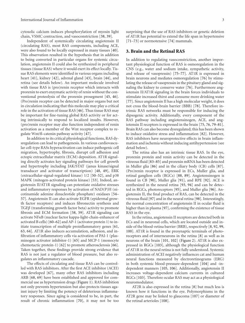
International Journal of Inflammation 3
cytosolic calcium induces phosphorylation of myosin lightchain, VSMC contraction, and vasoconstriction [38, 39].
Independent of systemically circulating angiotensin II(circulating RAS), most RAS components, including ACE,were also found to be locally expressed in many tissues [40].This observation resulted in the hypothesis that in additionto being converted in particular organs for systemic circu-lation, angiotensin II could also be synthesized in peripheraltissues (tissue RAS) where it would exert its effect locally. Tis-sue RAS elements were identified in various organs includingheart [41], kidney [42], adrenal gland [43], brain [44], andretina (see details below). An important molecule involvedwith tissue RAS is (pro)renin receptor which interacts withprorenin to exert enzymatic activity of renin without the con-ventional proteolysis of the prorenin prosegment [45, 46].(Pro)renin receptor can be detected in major organs but notin circulation indicating that this molecule may play a criticalrole in the activation of tissue RAS [46]. Thus tissue RAS maybe important for fine-tuning global RAS activity or for act-ing intrinsically to respond to localized insults. However,(pro)renin receptor may also function independent of reninactivation as a member of the Wnt receptor complex to re-gulate Wnt/ß-catenin pathway activity [47].
In addition to its critical physiological functions, RAS dy-sregulation can lead to pathogenesis. In various cardiovascu-lar cell-type RASs hyperactivation can induce pathogenic cellmigration, hypertrophy, fibrosis, disrupt cell adhesion andectopic extracellular matrix (ECM) deposition. AT1R signal-ing directly activates key signaling pathways for cell growthand hypertrophy including JAK/STAT (janus kinase/signaltransducer and activator of transcription) [48, 49], ERK(extracellular-signal-regulated kinase) 1/2 [50–52], and p38MAPK (mitogen-activated protein kinase) [53]. Indeed, an-giotensin II/AT1R signaling can potentiate oxidative stressesand inflammatory responses by activation of NAD(P)H (ni-cotinamide adenine dinucleotide phosphate) oxidases [54–57]. Angiotensin II can also activate EGFR (epidermal grow-th factor receptors) and induces fibronectin synthesis andTGFβ (transforming growth factor beta) activity to promotefibrosis and ECM formation [58, 59]. AT1R signaling canactivate NFκB (nuclear factor kappa-light-chain-enhancer ofactivated B cells) [60–62] and AP-1 (activator protein 1) to in-itiate transcription of multiple proinflammatory genes [61,63, 64]. AT1R also induces accumulation, adhesion, and in-filtration of inflammatory cells via activation of PAI-1 (plas-minogen activator inhibitor-1) [65] and MCP-1 (monocytechemotactic protein-1) [62] to promote atherosclerosis [66].Taken together, these findings provide strong evidence thatRAS is not just a regulator of blood pressure, but also re-gulates an inflammatory cascade.
The effects of circulating and tissue RAS can be control-led with RAS inhibitors. After the first ACE inhibitor (ACEI)was developed [67], many other RAS inhibitors includingARB [68, 69] have been established and approved for com-mercial use as hypertension drugs (Figure 1). RAS inhibitionnot only prevents hypertension but also protects tissues aga-inst injury by limiting the potency of deleterious inflamma-tory responses. Since aging is considered to be, in part, theresult of chronic inflammation [70], it may not be too
surprising that the use of RAS inhibitors or genetic deletionof AT1R has potential to extend the life span in hypertensive[71–73] or normotensive [74] mammals.
3. Brain and the Retinal RAS
In addition to regulating vasoconstriction, another impor-tant physiological function of RAS is osmoregulation in theCNS (e.g., water and sodium intake, sympathetic activity,and release of vasopressin) [75–77]. AT1R is expressed inbrain neurons and mediates osmoregulation [76] by stimu-lating the release of vasopressin in the pituitary gland and sig-naling the kidney to conserve water [76]. Furthermore ang-iotensin II/AT1R signaling in the brain forces individuals tostimulate increased thirst and consume more drinking water[77]. Since angiotensin II has a high molecular weight, it doesnot cross the blood-brain barrier (BBB) [78]. Therefore in-trinsic RAS networks must be responsible for inducing thedipsogenic activity. Additionally, every component of theRAS pathway including angiotensinogen, ACE, and ang-iotensin II receptors is expressed in the brain [75, 76, 79–81].Brain RAS can also become dysregulated; this has been shownto induce oxidative stress and inflammation [82]. However,RAS inhibitors have neuroprotective effects in brain inflam-mation and ischemia without inducing antihypertension (seedetail below).
The retina also has an intrinsic tissue RAS. In the eye,prorenin protein and renin activity can be detected in thevitreous fluid [83–85] and prorenin mRNA has been detectedin Muller glia [86] and in the ciliary body (CB) cells [87].(Pro)renin receptor is expressed in ECs, Muller glia, andretinal ganglion cells (RGCs) [88, 89]. Angiotensinogen isfound in CB [90], Muller glia [91], and RPE [92]. ACE issynthesized in the neural retina [93, 94] and can be detec-ted in RGCs, photoreceptors [95], and Muller glia [96]. An-giotensin II, the final product of RAS, can be detected in thevitreous fluid [97] and in the neural retina [98]. Interestingly,the normal concentration of angiotensin II in ocular fluid ishigher than in plasma [97], confirming the existence of tissueRAS in the eye.
In the retina, angiotensin II receptors are detected both inECs and in neuronal cells, which are located outside and in-side of the blood-retina barrier (BRB), respectively [8, 92, 99,100]. AT1R is found in the presynaptic terminals of photo-receptors and of interneurons in the retina [8] as well as inneurons of the brain [101, 102] (Figure 2). AT1R is also ex-pressed in RGCs [103], although the physiological functionof AT1R in the neural retina is not fully understood. Systemicadministration of ACEI negatively influences cat and humanneural functions measured by electroretinograms (ERG)in both systemic blood-pressure-dependent [104] and -in-dependent manners [105, 106]. Additionally, angiotensin IIincreases voltage-dependent calcium currents in culturedRGCs [103]. Therefore ocular RAS may act as a physiologicalneuromodulator.
AT2R is also expressed in the retina [8] but much less isknown how it functions in the eye. Polymorphisms in theAT2R gene may be linked to glaucoma [107] or diameter ofthe retinal arterioles [108].

4 International Journal of Inflammation
Figure 2: AT1R expression in a neuronal cell line. Immunohisto-chemistry for angiotensin II type 1 receptor (AT1R) in PC12D cells.Note that AT1R is detected in presynaptic terminal (arrow) or soma(arrow head). Scale bar: 20 μm.
4. RAS and Ocular Diseases
4.1. Uveitis. Increasing evidence suggests that RAS activityand inflammation may be associated with various ocular dis-eases, and, therefore, RAS inhibitors may be effective thera-peutic agents. Several lines of evidences suggest that RAS in-hibition is an effective treatment for uveitis [8, 12, 17, 18, 88].Endotoxin-induced uveitis (EIU) is induced with intraperi-toneal injections of lipopolysaccharide (LPS); this results inupregulated expression of proinflammatory and adhesionmolecules such as ICAM-1 (intercellular adhesion molecule1), MCP-1, IL-6 (interleukin 6), and IFN-γ (interferon-gamma) [17, 88]. These molecules are also upregulated in ex-perimental autoimmune uveoretinitis (EAU) models gener-ated by immunizing animals with interphotoreceptor retin-oid-binding protein (IRBP) [18]. The upregulation of thesemolecules, however, can be inhibited with ARB or (pro)reninreceptor blocker (PRRB). (PRRB is an experimental decoypeptide that contains “handle” region sequence of (pro)reninreceptor.) RAS inhibition also suppresses retinal leukocytestasis, CD4+ T-cell activation [17, 18, 88]. Furthermore, RASinhibition suppresses gliosis by preventing STAT3 activation[8]. Lastly, when the expression levels of RAS pathway com-ponents are examined in EIU, prorenin, (pro)renin receptor[88], angiotensin II [8], and AT1R [17] levels are elevatedin the retina. These findings suggest that heightened inflam-matory responses in the eye and RAS activation are stronglycorrelated.
4.2. Chronic Inflammation and Eye Diseases. Besides beingcorrelated with classically acute inflammation cases such asuveitis, one of the largest risk factors for developing prevalentand vision-threatening diseases such as DR, AMD, and glauc-oma is aging [1–3]. These age-related eye diseases [109, 110]and others [5, 6] are now known to be caused (at least part-ially) by chronic inflammation and oxidative stress. SinceRAS inhibition may prolong the life spans of hypertensive[71–73] or normotensive [74] mammals, it is logical that age-related eye diseases may be prevented or treated by suppres-sing inflammation and oxidative stress. The main patho-logical event of DR and AMD is abnormal neovascularizationand VEGF (vascular endothelial growth factor) has beenknown to be a large contri-butor for them [111–113]. VEGF
Angiotensin II
UPS
VEGF Syp GFAPRho
Adhesion Infiltration Angiogenesis Neural dysfunction Gliosis
Vascular inflammation Neural dysfunction
MCP-1ICAM-1
STAT 3AP-1NFκB
AT1R
Figure 3: Downstream of AT1R in vascular inflammation and neu-ral dysfunction. NFκB: nuclear factor kappa-light-chain-enhancerof activated B cells, AP-1: activator protein 1, UPS: ubiquitin-pro-teasome system, STAT3: signal transducer and activator of tran-scription 3, ICAM-1: intercellular adhesion molecule 1, MCP-1:monocyte chemotactic protein 1, VEGF: vascular endothelialgrowth factor, Syp: synaptophysin, Rho: rhodopsin, GFAP: glial fi-brillary acidic protein.
is a potent angiogenic factor and an inflammatory cytokinethat induces the accumulation, adhesion, and infiltration ofleukocytes [114, 115]. Inflammatory response in the retinacan promote tissue ischemia by inducing vascular regression(vaso-obliteration) and also pathological angiogenesis [116].Angiotensin II can induce upregulation of VEGF receptor(VEGFR)-2 and angiopoietin-2 in retinal ECs [117, 118] andVEGF in retinal pericytes [119] (Figure 3). Oxygen-inducedretinopathy (OIR) is an animal model induced by continualaeration with 75–80% oxygen in early postnatal stages. OIRanimals develop stereotypical phenotypes and is useful toevaluate vaso-obliteration and pathological angiogenesis (tu-ft formation) in the developing retina [120] which is largelyre-gulated by VEGF [121]. This phenotype can be preven-ted with RAS inhibitors ACEI [122, 123], ARB [15], orPRRB [89, 124] that prevent pathological angiogenesis inOIR. The use of ARB and PRRB has the added benefit of sup-pressing abnormal angiogenesis without suppressing physio-logical vascular regeneration [15, 124]. In animals exposed toOIR RAS inhibitors may function to prevent gene expressionof proinflammatory molecules and prevent leukocyte infil-tration. Infiltration of VEGF-expressing inflammatory cellsinto the vitreous cavity is thought to induce pathological an-giogenesis by causing ECs to grow in the wrong direction[115].
4.3. Diabetic Retinopathy (DR). DR is one of the leading caus-es of blindness in the world [3]. It is characterized by vas-cular loss due to hyperglycemia and inflammation due to oxi-dative stress and AGEs (advanced glycation end products) ac-cumulation. In severe cases hypoxia induces abnormalneovascularization (proliferative diabetic retinopathy, PDR)in addition to hyperpermeability (diabetic macular edema;DME). Prorenin [83] and angiotensin II [125, 126] are found

International Journal of Inflammation 5
to be increased in the vitreous humor of PDR and DR pa-tients. RAS may potentiate the vascular phenotype of DRby upregulating VEGF/VEGFR-2 signaling (through angio-tensin II) [118, 119] thereby inducing neovascularizationand promoting blood vessel permeability. In fact, VEGF wasinitially named “vascular permeability factor” (VPF) [127].
Multiple attempts have been made to treat DR with RASinhibitors. Although in one study ACEI administration se-emed to attenuate retinal hyperpermeability in diabetic pa-tients [128], interpretations of these studies are still being ac-tively debated [129, 130]. However, recently three independ-ent groups showed that ARB prevents BRB breakdown inanimal models [131–133]. In 1998 and 2008, the results ofrandomized double-blind placebo-controlled trials usingACEI or ARB to treat DR were released from the EUCLID(EURODIAB Controlled Trial of Lisinopril in Insulin-De-pendent Diabetes; ACEI treatment) [134] and DIRECT (Dia-betic Retinopathy Candesartan Trial; ARB treatment) [24,25]. Afterwards, RASS (Renin-Angiotensin System Study) inwhich both inhibitors were tested in DR patients was alsoreleased [26]. Large number of participants were examinedin these trials (354 (type 1 diabetes) for EUCLID, 1421 (type1) and 1905 (type 2) for DIRECT, and 285 (type 1) for RASS,resp.), and the results from all three studies provided strongevidence that RAS inhibition delays the onset or prevents thedevelopment of human DR symptom. However, these treat-ments were not universally beneficial. For example, in DI-RECT, ARBs were not effective with respect to primary end-points and had differing effects regarding secondary end-points in different patient groups (type I or type II diabetes)[24, 25].
Clues for why RAS inhibition is effective for treating DRhave come from animal studies. Streptozotocin (STZ) injec-tions in rodents induce leukocyte stasis, blood vessel hyper-permeability, and formation of acellular capillaries. Impor-tantly, ERG recordings are attenuated in rodents after STZinjections before vascular phenotypes are observed, indicat-ing that neuronal dysfunction precedes neovascularization indiabetic models [9, 135]. Apoptosis of retinal neurons is alsoobserved in later stage [136]. The administration of ACEI[137–140], ARB [10, 13, 14, 141], or PRRB [142] has beenshown to rescue the vascular phenotypes of STZ-induceddiabetic retinas. To generate another and more severe modelof DR, Ren-2 transgenic rats (that have severe hypertensiondue to genetic knock-in of a mouse ren-2 renin gene [143])can be injected with STZ. In these rats advanced vascularphenotypes are observed (including abnormal endothelialproliferation). Even in this model ACEI [144] or ARB [19,145, 146] administration served as effective treatments. RASinhibitors probably function by suppressing inflammatorycascades [10, 14] and by preventing oxidative stress [147]by limiting NFκB and NAD(P)H activation. RAS inhibitorsmay also function to directly inhibit glucose accumulationinto retinal cells by modulating GLUT-1 (glucose transporter1) expression [148]. Furthermore, ARB was reported to in-fluence the expression of glyoxalase I, a key regulator of AGEs[11]. Lastly, even though AT1R and AT2R are consideredto have opposing functions AT2R inhibition may also
effectively treat DR by suppressing VEGF and angiopoietin-2expression levels in experimental retinopathies [33, 149].
4.4. Age-Related Macular Degeneration (AMD). AMD is oneof the leading causes of blindness especially in western coun-tries. The greatest risk factors are aging and smoking [1],and the central phenotypes are choroidal neovascularization(CNV; wet AMD) and atrophy of photoreceptors and RPEcells (dry AMD). While no cure exists for dry AMD, wetAMD is currently treated with VEGF inhibitors [112, 113].Inflammation exacerbates the wet AMD phenotype since in-filtrating macrophages promote CNV formation [150–152].Experimental CNV can be induced using laser coagulationto mechanically disrupt Bruch’s membrane. The size of thelaser-induced lesions after treatment with ACEI [153], ARB[16], and PRRB [154] is significantly reduced. Furthermore,AT1R-deficient mice are resistant to laser-induced CNV[154]. RAS inhibition may protect against CNV formation byinhibiting RAS activity and suppressing ERK signaling (di-rectly with (pro)renin receptor-mediated intracellular signal-ing) [154].
RPE cells are positioned between the choroidal vascula-ture and photoreceptors and have function to maintain thevisual (retinoid) cycle and to form a tight seal that preventschoroidal vessel invasion. Angiotensin II signaling in RPEcells increases abnormal production [155–157] and excessiveturnover [158] of ECM via MMP (matrix metalloprotein-ase)-2 and -14 thereby weakening the seal that prevent chor-oidal EC invasion. These studies suggest that RAS inhibitionmay be an effective treatment for AMD as well as DR.
4.5. Glaucoma. Glaucoma is another age-related and highincidence ocular disease [2]. The feature of this disease isneurodegenerative of RGCs, but it can be caused by hetero-geneous and complex mechanisms. One direct mechanism toinduce RGC death is to increase the intraocular pressure(IOP). Studies devoted to developing new methods of con-trolling IOP are critical and ongoing. However, a subpopu-lation of glaucoma patients have normal IOP (normal ten-sion glaucoma, NTG). This complicates the development ofeffective therapies since both forms are induced by seeminglyseparate mechanisms. Some RAS components including ang-iotensin II receptors are expressed in CB cells [90, 159, 160]that secrete aqueous humor and regulate IOP. Like otherantihypertensive drugs such as calcium channel blockers,ACEI or ARB decreases IOP in humans and other primates[161–165] although IOP is considered to be regulated inde-pendently of systemic blood pressure. In an experimentalmodel of high IOP and glaucoma, ARB treatments effectivelysuppress RGC death [23]. These findings suggest that RASinhibition may be effective for treating glaucoma patientswith high IOP.
5. RAS Inhibition Protects Brain andRetinal Neurons
Angiotensin II receptors are expressed inside and outside ofthe BBB [75, 76, 79–81] and the BRB [8, 92, 99, 100] indicat-ing that both circulating and tissue RAS exist in the CNS,

6 International Journal of Inflammation
and if dysregulated, could elicit pathological effects. Indeed,RAS inhibition can attenuate the degree of inflammation inthe brain and the eye [166, 167]. Inhibiting RAS can preventexperimental brain injuries induced by middle cerebralartery occlusion [168, 169] by suppressing vascular inflam-mation [170], including BBB breakdown [171], and/or regu-lating neural apoptosis directly [169]. Interestingly, AT2R ismore highly expressed in developing neuronal tissues in vivothan in adult tissues [172] and AT2R stimulation promotesaxonal regeneration of optic nerve [173] and minimizesformation of ischemia-induced cerebral lesions [174]. Thissuggests that ARB, which not only blocks AT1R but also caus-es angiotensin II to bind AT2R [175], may be an ideal drugfor treating inflammatory diseases in the CNS. Inhibitionof RAS may also prevent stress-induced behaviors includinganxiety, depression, and panic by suppressing the releaseof corticotrophin-releasing factor [176–178]. Furthermore,recent studies suggest that brain RAS may potentiate Al-zheimer’s disease progression by stimulating the productionof beta amyloid [179–182].
Retinal dysfunction as detected in ERG recordings canbe observed in early diabetic animal models and in humansbefore vascular changes and neural cell loss are observed[135]. Amazingly, these deficits can be prevented by in-hibiting RAS [9, 183, 184]. We have reported that ARB pre-vents retinal dysfunction (e.g., decrease of amplitude and anextension of the implicit time of ERG) in EIU [8] and in STZ-induced early diabetic retinas [9]. Furthermore, in these in-flamed retinas, we determined that angiotensin II promptedthe degradation of the presynaptic protein synaptophysinthrough the ubiquitin proteasome system (UPS) [8, 9]. UPS-mediated degradation of rhodopsin (part of the light-res-ponsive complex in photoreceptors) can also be observed inEIU via STAT3 activation (which operates downstream ofAT1R) [8, 185]. Additionally, STAT3 signaling serves as anegative regulator of rhodopsin in differentiating photore-ceptors during retinal development [186, 187]. Thus, regu-lating angiotensin-II-induced protein degradation could ser-ve as an important neuroprotective measure [188] (Figure 3).
Another target of inflammation is reactive glia includingmicroglia, astrocytes, and Muller glia. Activated glia causegliosis and alter proper neuronal morphology. Microglia areresident CNS myeloid-derived cells and mediate critical im-mune and inflammatory responses. AT1R signaling indu-ces activation of microglia via NFκB and AP-1 [189, 190].GFAP (glial fibrillary acidic protein) is a differential and re-active marker of astrocyte and Muller glia, respectively, andits transcription is regulated by STAT3 activation [191]. Theactivation of astrocytes and Muller glia in experimental re-tinopathy can be prevented by ARB [8, 192] (Figure 3),although it is important to consider that the contributions ofreactive glia can be context dependent [193].
IOP-independent RGC apoptosis can be observed inSTZ-induced diabetes [136], after ischemia/reperfusion[194], after optic nerve crush [195], and after intraocularNMDA (N-methyl-D-aspartic acid) injections [196] in ani-mal models. RGC loss in diabetic hypertensive models can beprevented by ARB which restores oxidative redox and mito-chondrial functions [22]. ACEI or ARB also prevents RGC
apoptosis in ischemia/reperfusion models by suppressingtoxic oxidative stress [21]. ARB can also rescue dying amacri-ne cells in OIR [20]. Polymorphisms of RAS pathway genesare reported to be associated with brain infarction or itsearly lesion [197–199] and AT2R gene polymorphisms are re-ported to be associated with the risk of NTG [107]. Thesefindings may indicate that RAS inhibitors may directly pro-tect retinal neurons from apoptosis and further suggest thatRAS inhibition may be useful for therapeutic treatments ofIOP-independent glaucoma.
6. Conclusion
RAS, which has been classically known as blood pressure reg-ulator, is becoming widely recognized as a proinflammatorymediator. Many age-related ocular diseases may be causedor exacerbated by chronic inflammation. Cells in the eyeare responsive to circulating and tissue RAS and increasingevidence indicates that RAS inhibition may prevent variousocular diseases including uveitis, AMD, and glaucoma. Basedon the findings from multiple clinical trials, RAS inhibitorsare effective therapeutic agents for treating DR although theresults of these studies must be examined critically since theinhibitors were not universally beneficial. Other groups in-cluding our own have shown that RAS inhibitors protectneurons from oxidative stress and apoptosis by preventingposttranslational ubiquitination of proteins critical for re-tinal functions. Although not mentioned previously in thispaper, another new and exciting RAS inhibitor, aliskiren (adirect renin inhibitor), has been developed. It may actuallymediate more robust vascular protection than either ACEIor ARB [200]. Therefore, work is underway to characterizeexisting RAS inhibitors and to develop novel inhibitors sincethey hold great promise for attenuating chronic inflam-mation and for treating multiple ocular and nonoculardiseases.
Abbreviations List(In Order of Their Appearance)
RAS: Renin-angiotensin systemDR: Diabetic retinopathyAMD: Age-related macular degenerationCNS: Central nervous systemRPE: Retinal pigment epitheliumARB: Angiotensin II type 1 receptor blockerACE: Angiotensin-converting enzymeEC: Vascular endothelial cellVSMC: Vascular smooth muscle cellAT1R: Angiotensin II type 1 receptorIP3: Inositol-1,4,5-triphosphatePLC: Phospholipase CECM: Extracellular matrixJAK: Janus kinaseSTAT: Signal transducer and activator of
transcriptionERK: Extracellular-signal-regulated kinaseMAPK: Mitogen-activated protein kinase

International Journal of Inflammation 7
NAD(P)H: Nicotinamide adenine dinucleotide phos-phate
NFκB: Nuclear factor kappa-light-chain-enhan-cer of activated B cells
AP-1: Activator protein 1EGFR: Epidermal growth factor receptorTGFβ: Transforming growth factor betaPAI-1: Plasminogen activator inhibitor-1MCP-1: Monocyte chemotactic protein-1ACEI: ACE inhibitorBBB: Blood-brain barrierCB: Ciliary bodyRGC: Retinal ganglion cellBRB: Blood-retina barrierERG: ElectroretinogramEIU: Endotoxin-induced uveitisLPS: LipopolysaccharideICAM-1: Intercellular adhesion molecule 1IL-6: Interleukin 6IFN-γ: Interferon-gammaEAU: Experimental autoimmune uveoretinitisIRBP: Interphotoreceptor retinoid-binding pro-
teinPRRB: (Pro)renin receptor blockerVEGF: Vascular endothelial growth factorVEGFR: VEGF receptorOIR: Oxygen-induced retinopathyAGE: Advanced glycation end-productPDR: Proliferative diabetic retinopathyDME: Diabetic macular edemaEUCLID: EURODIAB Controlled Trial of Lisinopril
in Insulin-Dependent DiabetesDIRECT Diabetic Retinopathy Candesartan TrialRASS: Renin-Angiotensin System StudySTZ: StreptozotosinGLUT-1: Glucose transporter 1CNV: Choroidal neovascularizationMMP: Matrix metalloproteinaseIOP: Intraocular pressureNTG: Normal tension glaucomaGFAP: Glial fibrillary acidic proteinNMDA: N-methyl-D-aspartic acid.
Acknowledgments
T. Kurihara is supported by a fellowship from the ManpeiSuzuki Diabetes Foundation and The Japan Society for thePromotion of Science (JSPS) Postdoctoral Fellowships forResearch Abroad. The authors thank Drs. Martin Friedlan-der, Peter D. Westenskow, and Lea Scheppke for the criticalreading of the paper.
References
[1] R. Klein, B. E. K. Klein, and K. L. P. Linton, “Prevalenceof age-related maculopathy: the Beaver Dam Eye study,”Ophthalmology, vol. 99, no. 6, pp. 933–943, 1992.
[2] J. M. Tielsch, A. Sommer, J. Katz, R. M. Royall, H. A. Quigley,and J. Javitt, “Racial variations in the prevalence of primary
open-angle glaucoma: the Baltimore eye survey,” Journal ofthe American Medical Association, vol. 266, no. 3, pp. 369–374, 1991.
[3] M. D. Davis, M. R. Fisher, R. E. Gangnon et al., “Risk factorsfor high-risk proliferative diabetic retinopathy and severevisual loss: early treatment diabetic retinopathy study report18,” Investigative Ophthalmology and Visual Science, vol. 39,no. 2, pp. 233–252, 1998.
[4] M. C. Haigis and B. A. Yankner, “The Aging Stress Response,”Molecular Cell, vol. 40, no. 2, pp. 333–344, 2010.
[5] C. K. Glass, K. Saijo, B. Winner, M. C. Marchetto, and F.H. Gage, “Mechanisms Underlying Inflammation in Neuro-degeneration,” Cell, vol. 140, no. 6, pp. 918–934, 2010.
[6] R. S. Balaban, S. Nemoto, and T. Finkel, “Mitochondria, oxi-dants, and aging,” Cell, vol. 120, no. 4, pp. 483–495, 2005.
[7] M. Salzet, L. Deloffre, C. Breton, D. Vieau, and L. Schoofs,“The angiotensin system elements in invertebrates,” BrainResearch Reviews, vol. 36, no. 1, pp. 35–45, 2001.
[8] T. Kurihara, Y. Ozawa, K. Shinoda et al., “Neuroprotectiveeffects of angiotensin II type 1 receptor (AT1R) blocker,telmisartan, via modulating AT1R and AT2R signaling in re-tinal inflammation,” Investigative Ophthalmology and VisualScience, vol. 47, no. 12, pp. 5545–5552, 2006.
[9] T. Kurihara, Y. Ozawa, N. Nagai et al., “Angiotensin II type 1receptor signaling contributes to synaptophysin degradationand neuronal dysfunction in the diabetic retina,” Diabetes,vol. 57, no. 8, pp. 2191–2198, 2008.
[10] P. Chen, G. M. Scicli, M. Guo et al., “Role of angiotensin II inretinal leukostasis in the diabetic rat,” Experimental Eye Re-search, vol. 83, no. 5, pp. 1041–1051, 2006.
[11] A. G. Miller, G. Tan, K. J. Binger et al., “Candesartan atten-uates diabetic retinal vascular pathology by restoring glyo-xalase-I function,” Diabetes, vol. 59, no. 12, pp. 3208–3215,2010.
[12] A. Miyazaki, N. Kitaichi, K. Ohgami et al., “Anti-inflam-matory effect of angiotensin type 1 receptor antagonist onendotoxin-induced uveitis in rats,” Graefe’s Archive for Clini-cal and Experimental Ophthalmology, vol. 246, no. 5, pp. 747–757, 2008.
[13] F. Mori, T. Hikichi, T. Nagaoka, J. Takahashi, N. Kitaya, andA. Yoshida, “Inhibitory effect of losartan, an AT1 angiotensinII receptor antagonist, on increased leucocyte entrapment inretinal microcirculation of diabetic rats,” British Journal ofOphthalmology, vol. 86, no. 10, pp. 1172–1174, 2002.
[14] N. Nagai, K. Izumi-Nagai, Y. Oike et al., “Suppressionof diabetes-induced retinal inflammation by blocking theangiotensin II type 1 receptor or its downstream nuclearfactor-κB pathway,” Investigative Ophthalmology and VisualScience, vol. 48, no. 9, pp. 4342–4350, 2007.
[15] N. Nagai, K. Noda, T. Urano et al., “Selective suppressionof pathologic, but not physiologic, retinal neovascularizationby blocking the angiotensin II type 1 receptor,” InvestigativeOphthalmology and Visual Science, vol. 46, no. 3, pp. 1078–1084, 2005.
[16] N. Nagai, Y. Oike, K. Izumi-Nagai et al., “Angiotensin IItype 1 receptor-mediated inflammation is required for chor-oidal neovascularization,” Arteriosclerosis, Thrombosis, andVascular Biology, vol. 26, no. 10, pp. 2252–2259, 2006.
[17] N. Nagai, Y. Oike, K. Noda et al., “Suppression of ocular in-flammation in endotoxin-induced uveitis by blocking theangiotensin II type 1 receptor,” Investigative Ophthalmologyand Visual Science, vol. 46, no. 8, pp. 2925–2931, 2005.
[18] Y. Okunuki, Y. Usui, N. Nagai et al., “Suppression of exp-erimental autoimmune uveitis by angiotensin II type 1

8 International Journal of Inflammation
receptor blocker telmisartan,” Investigative Ophthalmologyand Visual Science, vol. 50, no. 5, pp. 2255–2261, 2009.
[19] J. L. Wilkinson-Berka, G. Tan, K. Jaworski, and S. Ninkovic,“Valsartan but not Atenolol Improves Vascular Pathology inDiabetic Ren-2 Rat Retina,” American Journal of Hyperten-sion, vol. 20, no. 4, pp. 423–430, 2007.
[20] L. E. Downie, K. M. Hatzopoulos, M. J. Pianta et al., “An-giotensin type-1 receptor inhibition is neuroprotective toamacrine cells in a rat model of retinopathy of prematurity,”Journal of Comparative Neurology, vol. 518, no. 1, pp. 41–63,2010.
[21] K. Fukuda, K. Hirooka, M. Mizote, T. Nakamura, T. Itano,and F. Shiraga, “Neuroprotection against retinal ischemia-reperfusion injury by blocking the angiotensin II type 1 re-ceptor,” Investigative Ophthalmology & Visual Science, vol. 51,no. 7, pp. 3629–3638, 2010.
[22] K. C. Silva, M. A. B. Rosales, S. K. Biswas, J. B. L. De Faria,and J. M. L. De Faria, “Diabetic retinal neurodegenerationis associated with mitochondrial oxidative stress and is im-proved by an angiotensin receptor blocker in a model com-bining hypertension and diabetes,” Diabetes, vol. 58, no. 6,pp. 1382–1390, 2009.
[23] H. Yang, K. Hirooka, K. Fukuda, and F. Shiraga, “Neuropro-tective effects of angiotensin II type 1 receptor blocker in arat model of chronic glaucoma,” Investigative Ophthalmology& Visual Science, vol. 50, no. 12, pp. 5800–5804, 2009.
[24] N. Chaturvedi, M. Porta, R. Klein et al., “Effect of candesar-tan on prevention (DIRECT-Prevent 1) and progression (DI-RECT-Protect 1) of retinopathy in type 1 diabetes: ran-domised, placebo-controlled trials,” The Lancet, vol. 372, no.9647, pp. 1394–1402, 2008.
[25] A. K. Sjolie, R. Klein, M. Porta et al., “Effect of candesartan onprogression and regression of retinopathy in type 2 diabetes(DIRECT-Protect 2): a randomised placebo-controlled trial,”The Lancet, vol. 372, no. 9647, pp. 1385–1393, 2008.
[26] M. Mauer, B. Zinman, R. Gardiner et al., “Renal and retinaleffects of enalapril and losartan in type 1 diabetes,” NewEngland Journal of Medicine, vol. 361, no. 1, pp. 40–51, 2009.
[27] H. Goldblatt, J. Lynch, R. F. Hanzal, and W. W. Summerville,“Studies on experimental hypertension : I. The productionof persistent elevation of systolic blood pressure by means ofrenal ischemia,” The Journal of Experimental Medicine, vol.59, no. 3, pp. 347–379, 1934.
[28] J. C. Fasciolo, B. A. Houssay, and A. C. Taquini, “Theblood-pressure raising secretion of the ischaemic kidney,”The Journal of Physiology, vol. 94, no. 3, pp. 281–293, 1938.
[29] I. H. Page, “On the nature of the pressor action of renin,” TheJournal of Experimental Medicine, vol. 70, no. 5, pp. 521–542,1939.
[30] E. Braun-Menendez and I. H. Page, “Suggested revision ofnomenclature—angiotensin,” Science, vol. 127, no. 3292, p.242, 1958.
[31] L. T. Skeggs Jr, J. R. Kahn, and N. P. Shumway, “The prepa-ration and function of the hypertensin-converting enzyme,”The Journal of Experimental Medicine, vol. 103, no. 3, pp.295–299, 1956.
[32] S. Y. Lin and T. L. Goodfriend, “Angiotensin receptors,” TheAmerican journal of physiology, vol. 218, no. 5, pp. 1319–1328,1970.
[33] S. Sarlos, B. Rizkalla, C. J. Moravski, Z. Cao, M. E. Cooper,and J. L. Wilkinson-Berka, “Retinal angiogenesis is mediatedby an interaction between the angiotensin type 2 receptor,VEGF, and angiopoietin,” American Journal of Pathology, vol.163, no. 3, pp. 879–887, 2003.
[34] Q. Xue, C. Dasgupta, M. Chen, and L. Zhang, “Foetal hypoxiaincreases cardiac AT2R expression and subsequent vulnera-bility to adult ischaemic injury,” Cardiovascular Research, vol.89, no. 2, pp. 300–308, 2011.
[35] K. Sasaki, Y. Yamano, S. Bardhan et al., “Cloning and expres-sion of a complementary DNA encoding a bovine adrenal an-giotensin II type-1 receptor,” Nature, vol. 351, no. 6323, pp.230–233, 1991.
[36] T. J. Murphy, R. W. Alexander, K. K. Griendling, M. S. Runge,and K. E. Bernstein, “Isolation of a cDNA encoding the vas-cular type-1 angiotensin II receptor,” Nature, vol. 351, no.6323, pp. 233–236, 1991.
[37] M. Ushio-Fukai, K. K. Griendling, M. Akers, P. R. Lyons,and R. W. Alexander, “Temporal dispersion of activation ofphospholipase C-β1 and -γ isoforms by angiotensin II in vas-cular smooth muscle cells. Role of αq/11, α12, and βγ G pro-tein subunits,” The Journal of Biological Chemistry, vol. 273,no. 31, pp. 19772–19777, 1998.
[38] J. M. Anderson, M. A. Gimbrone, and R. W. Alexander, “An-giotensin II stimulates phosphorylation of the myosin lightchain in cultured vascular smooth muscle cells,” Journal of Bi-ological Chemistry, vol. 256, no. 10, pp. 4693–4696, 1981.
[39] D. E. Dostal, T. Murahashi, and M. J. Peach, “Regulation ofcytosolic calcium by angiotensins in vascular smooth mus-cle,” Hypertension, vol. 15, no. 6, pp. 815–822, 1990.
[40] F. Soubrier, F. Alhenc-Gelas, C. Hubert et al., “Two putativeactive centers in human angiotensin I-converting enzymerevealed by molecular cloning,” Proceedings of the NationalAcademy of Sciences of the United States of America, vol. 85,no. 24, pp. 9386–9390, 1988.
[41] L. J. Dell’Italia, Q. C. Meng, E. Balcells et al., “Compartmen-talization of angiotensin II generation in the dog heart: evi-dence for independent mechanisms in intravascular and in-terstitial spaces,” Journal of Clinical Investigation, vol. 100, no.2, pp. 253–258, 1997.
[42] H. M. Kimbrough, E. D. Vaughan, R. M. Carey, and C. R.Ayers, “Effect of intrarenal angiotensin II blockade on renalfunction in conscious dogs,” Circulation Research, vol. 40, no.2, pp. 174–178, 1977.
[43] J. W. Ryan, “Renin-like enzyme in the adrenal gland,” Science,vol. 158, no. 3808, pp. 1589–1590, 1967.
[44] D. Ganten, J. L. Minnich, P. Granger et al., “Angiotensin-for-ming enzyme in brain tissue,” Science, vol. 173, no. 3991, pp.64–65, 1971.
[45] A. Ichihara, M. Hayashi, Y. Kaneshiro et al., “Inhibition ofdiabetic nephropathy by a decoy peptide corresponding tothe ”handle” region for nonproteolytic activation of prore-nin,” Journal of Clinical Investigation, vol. 114, no. 8, pp.1128–1135, 2004.
[46] G. Nguyen, F. Delarue, C. Burckle, L. Bouzhir, T. Giller, andJ. D. Sraer, “Pivotal role of the renin/prorenin receptor in an-giotensin II production and cellular responses to renin,” Jour-nal of Clinical Investigation, vol. 109, no. 11, pp. 1417–1427,2002.
[47] C. M. Cruciat, B. Ohkawara, S. P. Acebron et al., “Require-ment of prorenin receptor and vacuolar H+-ATPase-media-ted acidification for Wnt signaling,” Science, vol. 327, no.5964, pp. 459–463, 2010.
[48] M. B. Marrero, B. Schieffer, W. G. Paxton et al., “Direct stim-ulation of Jak/STAT pathway by the anglotensin II AT1receptor,” Nature, vol. 375, no. 6528, pp. 247–250, 1995.
[49] B. Schieffer, M. Luchtefeld, S. Braun, A. Hilfiker, D. Hilfiker-Kleiner, and H. Drexler, “Role of NAD(P)H oxidase inangiotensin II-induced JAK/STAT signaling and cytokine

International Journal of Inflammation 9
induction,” Circulation Research, vol. 87, no. 12, pp. 1195–1201, 2000.
[50] S. Kim, Y. Izumi, M. Yano et al., “Angiotensin blockade in-hibits activation of mitogen-activated protein kinases in ratballoon-injured artery,” Circulation, vol. 97, no. 17, pp. 1731–1737, 1998.
[51] D. F. Liao, J. L. Duff, G. Daum, S. L. Pelech, and B. C. Berk,“Angiotensin II stimulates MAP kinase kinase kinase activityin vascular smooth muscle cells: role of raf,” Circulation Re-search, vol. 79, no. 5, pp. 1007–1014, 1996.
[52] D. F. Liao, B. Monia, N. Dean, and B. C. Berk, “Protein kinaseC-ζ mediates angiotensin II activation of ERK1/2 in vascularsmooth muscle cells,” Journal of Biological Chemistry, vol.272, no. 10, pp. 6146–6150, 1997.
[53] M. Nishida, S. Tanabe, Y. Maruyama et al., “Gα12/13- andreactive oxygen species-dependent activation of c-Jun NH2-terminal kinase and p38 mitogen-activated protein kinase byangiotensin receptor stimulation in rat neonatal cardiomy-ocytes,” Journal of Biological Chemistry, vol. 280, no. 18, pp.18434–18441, 2005.
[54] S. Rajagopalan, S. Kurz, T. Munzel et al., “Angiotensin II-me-diated hypertension in the rat increases vascular superoxideproduction via membrane NADH/NADPH oxidase activa-tion: contribution to alterations of vasomotor tone,” Journalof Clinical Investigation, vol. 97, no. 8, pp. 1916–1923, 1996.
[55] R. M. Touyz, X. Chen, F. Tabet et al., “Expression of afunctionally active gp91phox-containing neutrophil-typeNAD(P)H oxidase in smooth muscle cells from human resis-tance arteries: regulation by angiotensin II,” Circulation Re-search, vol. 90, no. 11, pp. 1205–1213, 2002.
[56] K. K. Griendling, C. A. Minieri, J. D. Ollerenshaw, and R. W.Alexander, “Angiotensin II stimulates NADH and NADPHoxidase activity in cultured vascular smooth muscle cells,”Circulation Research, vol. 74, no. 6, pp. 1141–1148, 1994.
[57] P. N. Seshiah, D. S. Weber, P. Rocic, L. Valppu, Y. Taniyama,and K. K. Griendling, “Angiotensin II stimulation ofNAD(P)H oxidase activity: upstream mediators,” CirculationResearch, vol. 91, no. 5, pp. 406–413, 2002.
[58] Y. Moriguchi, H. Matsubara, Y. Mori et al., “Angiotensin II-induced transactivation of epidermal growth factor recep-tor regulates fibronectin and transforming growth factor-βsynthesis via transcriptional and posttranscriptional mecha-nisms,” Circulation Research, vol. 84, no. 9, pp. 1073–1084,1999.
[59] S. Kagiyama, S. Eguchi, G. D. Frank, T. Inagami, Y. C. Zha-ng, and M. I. Phillips, “Angiotensin II-induced cardiac hy-pertrophy and hypertension are attenuated by epidermalgrowth factor receptor antisense,” Circulation, vol. 106, no.8, pp. 909–912, 2002.
[60] D. N. Muller, R. Dechend, E. M. A. Mervaala et al., “NF-κBinhibition ameliorates angiotensin II-induced inflammatorydamage in rats,” Hypertension, vol. 35, no. 1, pp. 193–201,2000.
[61] A. Fiebeler, F. Schmidt, D. N. Muller et al., “Mineralocorti-coid receptor affects AP-1 and nuclear factor-κB activation inangiotensin II-induced cardiac injury,” Hypertension, vol. 37,no. 2, pp. 787–793, 2001.
[62] M. Hernandez-Presa, C. Bustos, M. Ortego et al., “Angioten-sin-converting enzyme inhibition prevents arterial nu-clear factor-κB activation, monocyte chemoattractant pro-tein-1 expression, and macrophage infiltration in a rabbitmodel of early accelerated atherosclerosis,” Circulation, vol.95, no. 6, pp. 1532–1541, 1997.
[63] S. Kudoh, I. Komuro, T. Mizuno et al., “Angiotensin II stimu-lates c-jun NH2-terminal kinase in cultured cardiac myocytesof neonatal rats,” Circulation Research, vol. 80, no. 1, pp. 139–146, 1997.
[64] P. L. Puri, M. L. Avantaggiati, V. L. Burgio et al., “Reactiveoxygen intermediates mediate angiotensin II-induced c-Jun·c-Fos heterodimer DNA binding activity and prolifer-ative hypertrophic responses in myogenic cells,” Journal ofBiological Chemistry, vol. 270, no. 38, pp. 22129–22134, 1995.
[65] D. E. Vaughan, S. A. Lazos, and K. Tong, “Angiotensin II re-gulates the expression of plasminogen activator inhibitor-1 incultured endothelial cells. A potential link between the re-nin-angiotensin system and thrombosis,” Journal of ClinicalInvestigation, vol. 95, no. 3, pp. 995–1001, 1995.
[66] A. Daugherty, M. W. Manning, and L. A. Cassis, “AngiotensinII promotes atherosclerotic lesions and aneurysms in apoli-poprotein E-deficient mice,” Journal of Clinical Investigation,vol. 105, no. 11, pp. 1605–1612, 2000.
[67] M. A. Ondetti, B. Rubin, and D. W. Cushman, “Design ofspecific inhibitors of angiotensin converting enzyme: newclass of orally active antihypertensive agents,” Science, vol.196, no. 4288, pp. 441–444, 1977.
[68] P. B. M. W. M. Timmermans, D. J. Carini, A. T. Chiu et al.,“Angiotensin II receptor antagonists: from discovery to anti-hypertensive drugs,” Hypertension, vol. 18, no. 5, pp. I-136–I-142, 1991.
[69] P. C. Wong, W. A. Price, A. T. Chiu et al., “Nonpeptide an0giotensin II receptor antagonists. Studies with EXP9270 andDuP 753,” Hypertension, vol. 15, no. 6, pp. 823–834, 1990.
[70] D. R. Green, L. Galluzzi, and G. Kroemer, “Mitochondria andthe autophagy-inflammation-cell death axis in organismalaging,” Science, vol. 333, no. 6046, pp. 1109–1112, 2011.
[71] G. Baiardi, C. Bregonzio, M. Jezova, I. Armando, and J. M.Saavedra, “Angiotensin II AT1 receptor blockade prolongsthe lifespan of spontaneously hypertensive rats and reducesstress-induced release of catecholamines, glucocorticoids,and vasopressin,” Annals of the New York Academy of Sciences,vol. 1018, pp. 131–136, 2004.
[72] W. Linz, H. Heitsch, B. A. Scholkens, and G. Wiemer, “Long-term angiotensin II type 1 receptor blockade with fonsartandoubles lifespan of hypertensive rats,” Hypertension, vol. 35,no. 4, pp. 908–913, 2000.
[73] W. Linz, T. Jessen, R. H. A. Becker, B. A. Scholkens, andG. Wiemer, “Long-term ACE inhibition doubles lifespan ofhypertensive rats,” Circulation, vol. 96, no. 9, pp. 3164–3172,1997.
[74] A. Benigni, D. Corna, C. Zoja et al., “Disruption of the AngII type 1 receptor promotes longevity in mice,” Journal ofClinical Investigation, vol. 119, no. 3, pp. 524–530, 2009.
[75] F. A. Mendelsohn, R. Quirion, J. M. Saavedra, G. Aguilera,and K. J. Catt, “Autoradiographic localization of angiotensinII receptors in rat brain,” Proceedings of the National Academyof Sciences of the United States of America, vol. 81, no. 5, pp.1575–1579, 1984.
[76] J. M. Saavedra, F. M.A. Correa, and L. M. Plunkett, “Bindingof angiotensin and atrial natriuretic peptide in brain of hy-pertensive rats,” Nature, vol. 320, no. 6064, pp. 758–760,1986.
[77] J. Buggy, A. E. Fisher, and W. E. Hoffman, “Ventricular ob-struction: effect on drinking induced by intracranial injec-tion of angiotensin,” Science, vol. 190, no. 4209, pp. 72–74,1975.

10 International Journal of Inflammation
[78] M. Van Houten, E. L. Schiffrin, and J. F. E. Mann, “Radioau-tographic localization of specific binding sites for blood-borne angiotensin II in the rat brain,” Brain Research, vol.186, no. 2, pp. 480–485, 1980.
[79] R. Castren and J. M. Saavedra, “Angiotensin II receptors inparaventricular nucleus, subfornical organ, and pituitarygland of hypophysectomized, adrenalectomized, and vaso-pressin-deficient rats,” Proceedings of the National Academyof Sciences of the United States of America, vol. 86, no. 2, pp.725–729, 1989.
[80] C. Chevillard and J. M. Saavedra, “High angiotensin-con-verting enzyme activity in the neurohypophysis of Brattle-boro rats,” Science, vol. 216, no. 4546, pp. 646–647, 1982.
[81] S. Landas, M. I. Phillips, J. F. Stamler, and M. K. Raizada,“Visualization of specific angiotensin II binding sites in thebrain by fluorescent microscopy,” Science, vol. 210, no. 4471,pp. 791–793, 1980.
[82] M. C. Zimmerman, E. Lazartigues, J. A. Lang et al., “Super-oxide mediates the actions of angiotensin II in the central ne-rvous system,” Circulation Research, vol. 91, no. 11, pp. 1038–1045, 2002.
[83] A. H. J. Danser, M. A. Van Den Dorpel, J. Deinum et al., “Re-nin, prorenin, and immunoreactive renin in vitreous fluidfrom eyes with and without diabetic retinopathy,” Journalof Clinical Endocrinology and Metabolism, vol. 68, no. 1, pp.160–167, 1989.
[84] J. Deinum, F. H. M. Derkx, A. H. J. Danser, and M. A. D. H.Schalekamp, “Identification and quantification of renin andprorenin in the bovine eye,” Endocrinology, vol. 126, no. 3,pp. 1673–1682, 1990.
[85] S. J. Sramek, I. H. L. Wallow, R. P. Day, and E. N. Ehrlich,“Ocular renin-angiotensin: immunohistochemical evidencefor the presence of prorenin in eye tissue,” Investigative Oph-thalmology and Visual Science, vol. 29, no. 11, pp. 1749–1752,1988.
[86] J. L. Berka, A. J. Stubbs, D. Z. M. Wang et al., “Renin-con-taining muller cells of the retina display endocrine features,”Investigative Ophthalmology and Visual Science, vol. 36, no. 7,pp. 1450–1458, 1995.
[87] C. R. Brandt, A. M. Pumfery, B. Micales et al., “Renin mRNAis synthesized locally in rat ocular tissues,” Current EyeResearch, vol. 13, no. 10, pp. 755–763, 1994.
[88] S. Satofuka, A. Ichihara, N. Nagai et al., “Suppression ofocular inflammation in endotoxin-induced uveitis by in-hibiting nonproteolytic activation of prorenin,” InvestigativeOphthalmology & Visual Science, vol. 47, no. 6, pp. 2686–2692, 2006.
[89] J. L. Wilkinson-Berka, R. Heine, G. Tan et al., “RILLKKMPSVInfluences the vasculature, neurons and glia, and (Pro)reninreceptor expression in the Retina,” Hypertension, vol. 55, no.6, pp. 1454–1460, 2010.
[90] S. J. Sramek, I. H. L. Wallow, D. A. Tewksbury, C. R. Brandt,and G. L. Poulsen, “An ocular renin-angiotensin system:immunohistochemistry of angiotensinogen,” InvestigativeOphthalmology and Visual Science, vol. 33, no. 5, pp. 1627–1632, 1992.
[91] C. Gerhardinger, M. B. Costa, M. C. Coulombe, I. Toth, T.Hoehn, and P. Grosu, “Expression of acute-phase responseproteins in retinal Muller cells in diabetes,” InvestigativeOphthalmology and Visual Science, vol. 46, no. 1, pp. 349–357,2005.
[92] J. Wagner, A. H. J. Danser, F. H. M. Derkx et al., “De-monstration of renin mRNA, angiotensinogen mRNA, andangiotensin converting enzyme mRNA expression in the
human eye: evidence for an intraocular renin-angiotensinsystem,” British Journal of Ophthalmology, vol. 80, no. 2, pp.159–163, 1996.
[93] R. Igic and V. Kojovic, “Angiotensin I converting enzyme(kininase II) in ocular tissues,” Experimental Eye Research,vol. 30, no. 3, pp. 299–303, 1980.
[94] G. Ferrari-Dileo, J. W. Ryan, E. J. Rockwood, E. B. Davis, andD. R. Anderson, “Angiotensin-converting enzyme in bov-ine, feline, and human ocular tissues,” Investigative Ophthal-mology and Visual Science, vol. 29, no. 6, pp. 876–881, 1988.
[95] E. Savaskan, K. U. Loffler, F. Meier, F. Muller-Spahn, J.Flammer, and P. Meyer, “Immunohistochemical localizationof angiotensin-converting enzyme, angiotensin II and AT1receptor in human ocular tissues,” Ophthalmic Research, vol.36, no. 6, pp. 312–320, 2004.
[96] T. Kida, T. Ikeda, M. Nishimura et al., “Renin-angiotensinsystem in proliferative diabetic retinopathy and its geneexpression in cultured human Muller cells,” Japanese Journalof Ophthalmology, vol. 47, no. 1, pp. 36–41, 2003.
[97] A. H. J. Danser, F. H. M. Derkx, P. J. J. Admiraal, J. Deinum, P.T. V. M. De Jong, and M. A. D. H. Schalekamp, “Angiotensinlevels in the eye,” Investigative Ophthalmology and VisualScience, vol. 35, no. 3, pp. 1008–1018, 1994.
[98] K. H. Datum and E. Zrenner, “Angiotensin-like immunore-active cells in the chicken retina,” Experimental Eye Research,vol. 53, no. 2, pp. 157–165, 1991.
[99] P. Mallorga, R. W. Babilon, and M. F. Sugrue, “Angiotensin IIreceptors labelled with 125I-[Sar1,Ile8]-AII in albino rabbitocular tissues,” Current Eye Research, vol. 8, no. 8, pp. 841–849, 1989.
[100] P. D. Senanayake, J. Drazba, K. Shadrach et al., “AngiotensinII and its receptor subtypes in the human retina,” Investiga-tive Ophthalmology and Visual Science, vol. 48, no. 7, pp.3301–3311, 2007.
[101] C. Sun, H. Li, L. Leng, M. K. Raizada, R. Bucala, and C.Sumners, “Macrophage migration inhibitory factor: an in-tracellular inhibitor of angiotensin II-induced increases inneuronal activity,” Journal of Neuroscience, vol. 24, no. 44, pp.9944–9952, 2004.
[102] D. P. Li, S. R. Chen, and H. L. Pan, “Angiotensin II stimulatesspinally projecting paraventricular neurons through presy-naptic disinhibition,” Journal of Neuroscience, vol. 23, no. 12,pp. 5041–5049, 2003.
[103] E. Guenther, S. Schmid, B. Hewig, and K. Kohler, “Two-fold effect of angiotensin II on voltage-dependent calciumcurrents in rat retinal ganglion cells,” Brain Research, vol. 718,no. 1-2, pp. 112–116, 1996.
[104] E. Demant, K. Nagahara, and G. Niemeyer, “Effects of cha-nges in systemic blood pressure on the electroretinogram ofthe cat: evidence for retinal autoregulation,” InvestigativeOphthalmology and Visual Science, vol. 23, no. 5, pp. 683–687,1982.
[105] P. C. Jacobi, H. Osswald, B. Jurklies, and E. Zrenner, “Neuro-modulatory effects of the renin-angiotensin system on the catelectroretinogram,” Investigative Ophthalmology and VisualScience, vol. 35, no. 3, pp. 973–980, 1994.
[106] B. Jurklies, A. Eckstein, P. Jacobi, K. Kohler, T. Risler, andE. Zrenner, “The renin-angiotensin system–a possible neuro-modulator in the human retina?” German journal of ophthal-mology, vol. 4, no. 3, pp. 144–150, 1995.
[107] K. Hashizume, Y. Mashima, T. Fumayama et al., “Geneticpolymorphisms in the angiotensin II receptor gene andtheir association with open-angle glaucoma in a Japanese
International Journal of Inflammation 11
population,” Investigative Ophthalmology and Visual Science,vol. 46, no. 6, pp. 1993–2001, 2005.
[108] Y.-P. Liu, T. Kuznetsova, L. Thijs et al., “Are retinal microvas-cular phenotypes associated with the 1675G/A polymor-phism in the angiotensin II Type-2 receptor gene,” AmericanJournal of Hypertension, vol. 24, no. 12, pp. 1300–1305, 2011.
[109] K. Yuki, Y. Ozawa, T. Yoshida et al., “Retinal ganglion cellloss in superoxide dismutase 1 deficiency,” Investigative Oph-thalmology & Visual Science, vol. 52, no. 7, pp. 4143–4150,2011.
[110] Y. Imamura, S. Noda, K. Hashizume et al., “Drusen, chor-oidal neovascularization, and retinal pigment epitheliumdysfunction in SOD1-deficient mice: a model of age-relatedmacular degeneration,” Proceedings of the National Academyof Sciences of the United States of America, vol. 103, no. 30, pp.11282–11287, 2006.
[111] L. P. Aiello, R. L. Avery, P. G. Arrigg et al., “Vascular en-dothelial growth factor in ocular fluid of patients with dia-betic retinopathy and other retinal disorders,” New EnglandJournal of Medicine, vol. 331, no. 22, pp. 1480–1487, 1994.
[112] E. S. Gragoudas, A. P. Adamis, E. T. Cunningham, M. Fein-sod, and D. R. Guyer, “Pegaptanib for neovascular age-relatedmacular degeneration,” New England Journal of Medicine, vol.351, no. 27, pp. 2805–2816, 2004.
[113] P. J. Rosenfeld, D. M. Brown, J. S. Heier et al., “Ranibizumabfor neovascular age-related macular degeneration,” New En-gland Journal of Medicine, vol. 355, no. 14, pp. 1419–1431,2006.
[114] S. Ishida, T. Usui, K. Yamashiro et al., “VEGF164 is proin-flammatory in the diabetic retina,” Investigative Ophthalmol-ogy and Visual Science, vol. 44, no. 5, pp. 2155–2162, 2003.
[115] S. Ishida, T. Usui, K. Yamashiro et al., “VEGF164-mediatedinflammation is required for pathological, but not physiolog-ical, ischemia-induced retinal neovascularization,” Journal ofExperimental Medicine, vol. 198, no. 3, pp. 483–489, 2003.
[116] S. Ishida, K. Yamashiro, T. Usui et al., “Leukocytes mediateretinal vascular remodeling during development and vaso-obliteration in disease,” Nature Medicine, vol. 9, no. 6, pp.781–788, 2003.
[117] A. Otani, H. Takagi, K. Suzuma, and Y. Honda, “AngiotensinII potentiates vascular endothelial growth factor-inducedangiogenic activity in retinal microcapillary endothelialcells,” Circulation Research, vol. 82, no. 5, pp. 619–628, 1998.
[118] A. Otani, H. Takagi, H. Oh, S. Koyama, and Y. Honda, “An-giotensin II induces expression of the Tie2 receptor ligand,angiopoietin-2, in bovine retinal endothelial cells,” Diabetes,vol. 50, no. 4, pp. 867–875, 2001.
[119] A. Otani, H. Takagi, H. Oh et al., “Angiotensin II-stimulatedvascular endothelial growth factor expression in bovineretinal pericytes,” Investigative Ophthalmology and VisualScience, vol. 41, no. 5, pp. 1192–1199, 2000.
[120] L. E.H. Smith, E. Wesolowski, A. McLellan et al., “Oxygen-in-duced retinopathy in the mouse,” Investigative Ophthalmol-ogy and Visual Science, vol. 35, no. 1, pp. 101–111, 1994.
[121] T. Alon, I. Hemo, A. Itin, J. Pe’er, J. Stone, and E. Keshet,“Vascular endothelial growth factor acts as a survival factorfor newly formed retinal vessels and has implications forretinopathy of prematurity,” Nature Medicine, vol. 1, no. 10,pp. 1024–1028, 1995.
[122] C. J. Moravski, D. J. Kelly, M. E. Cooper et al., “Retinal neo-vascularization is prevented by blockade of the renin-an-giotensin system,” Hypertension, vol. 36, no. 6, pp. 1099–1104, 2000.
[123] M. Lonchampt, L. Pennel, and J. Duhault, “Hyperoxia/nor-moxia-driven retinal angiogenesis in mice: a role for an-giotensin II,” Investigative Ophthalmology and Visual Science,vol. 42, no. 2, pp. 429–432, 2001.
[124] S. Satofuka, A. Ichihara, N. Nagai et al., “Role of nonpro-teolytically activated prorenin in pathologic, but not physi-ologic, retinal neovascularization,” Investigative Ophthalmol-ogy and Visual Science, vol. 48, no. 1, pp. 422–429, 2007.
[125] H. Funatsu, H. Yamashita, T. Ikeda, Y. Nakanishi, S. Kitano,and S. Hori, “Angiotensin II and vascular endothelial growthfactor in the vitreous fluid of patients with diabetic macularedema and other retinal disorders,” American Journal of Oph-thalmology, vol. 133, no. 4, pp. 537–543, 2002.
[126] H. Funatsu, H. Yamashita, Y. Nakanishi, and S. Hori, “An-giotensin II and vascular endothelial growth factor in thevitreous fluid of patients with proliferative diabetic retinopa-thy,” British Journal of Ophthalmology, vol. 86, no. 3, pp. 311–315, 2002.
[127] D. R. Senger, S. J. Galli, A. M. Dvorak, C. A. Perruzzi, V. SusanHarvey, and H. F. Dvorak, “Tumor cells secrete a vascularpermeability factor that promotes accumulation of ascitesfluid,” Science, vol. 219, no. 4587, pp. 983–985, 1983.
[128] M. Larsen, E. Hommel, H. H. Parving, and H. Lund-Andersen, “Protective effect of captopril on the blood-retinabarrier in normotensive insulin-dependent diabetic patientswith nephropathy and background retinopathy,” Graefe’sArchive for Clinical and Experimental Ophthalmology, vol.228, no. 6, pp. 505–509, 1990.
[129] B. Van Kooij, R. Fijnheer, J. De Boer et al., “A randomized,masked, cross-over trial of lisinopril for inflammatory macu-lar edema,” American Journal of Ophthalmology, vol. 141, no.4, pp. 646–651, 2006.
[130] S. T. Knudsen, T. Bek, P. L. Poulsen, M. N. Hove, M. Reh-ling, and C. E. Mogensen, “Effects of losartan on diabetic ma-culopathy in type 2 diabetic patients: a randomized, double-masked study,” Journal of Internal Medicine, vol. 254, no. 2,pp. 147–158, 2003.
[131] J. H. Kim, J. H. Kim, Y. S. Yu, C. S. Cho, and K.-W.Kim, “Blockade of angiotensin II attenuates VEGF-mediatedblood-retinal barrier breakdown in diabetic retinopathy,”Journal of Cerebral Blood Flow and Metabolism, vol. 29, no.3, pp. 621–628, 2009.
[132] J. A. Phipps, A. C. Clermont, S. Sinha, T. J. Chilcote, S. E.Bursell, and E. P. Feener, “Plasma kallikrein mediates an-giotensin II type 1 receptor-stimulated retinal vascular per-meability,” Hypertension, vol. 53, no. 2, pp. 175–181, 2009.
[133] H. Nakamura, M. Yamazaki, T. Ohyama et al., “Role ofangiotensin II type 1 receptor on retinal vascular leakage ina rat oxygen-induced retinopathy model,” Ophthalmic Re-search, vol. 41, no. 4, pp. 210–215, 2009.
[134] N. Chaturvedi, A. K. Sjolie, J. M. Stephenson et al., “Effectof lisinopril on progression of retinopathy in normoten-sive people with type 1 diabetes. The EUCLID studygroup. EURODIAB controlled trial of lisinopril in insulin-dependent diabetes mellitus,” The Lancet, vol. 351, no. 9095,pp. 28–31, 1998.
[135] Y. Shirao and K. Kawasaki, “Electrical responses from dia-betic retina,” Progress in Retinal and Eye Research, vol. 17, no.1, pp. 59–76, 1998.
[136] A. J. Barber, E. Lieth, S. A. Khin, D. A. Antonetti, A. G. Buch-anan, and T. W. Gardner, “Neural apoptosis in the retina dur-ing experimental and human diabetes: early onset and effectof insulin,” Journal of Clinical Investigation, vol. 102, no. 4,pp. 783–791, 1998.

12 International Journal of Inflammation
[137] A. Ottlecz and T. Bensaoula, “Captopril ameliorates the de-creased Na+,K+-ATPase activity in the retina of strep-tozotocin-induced diabetic rats,” Investigative Ophthalmologyand Visual Science, vol. 37, no. 8, pp. 1633–1641, 1996.
[138] R. E. Gilbert, D. J. Kelly, A. J. Cox et al., “Angiotensin con-verting enzyme inhibition reduces retinal overexpression ofvascular endothelial growth factor and hyperpermeability inexperimental diabetes,” Diabetologia, vol. 43, no. 11, pp.1360–1367, 2000.
[139] J. Z. Zhang, X. Xi, L. Gao, and T. S. Kern, “Captopril in-hibits capillary degeneration in the early stages of diabeticretinopathy,” Current Eye Research, vol. 32, no. 10, pp. 883–889, 2007.
[140] Z. Zheng, H. Chen, G. Ke et al., “Protective effect of perindo-pril on diabetic retinopathy is associated with decreased vas-cular endothelial growth factor-to-pigment epithelium-der-ived factor ratio: involvement of a mitochondria-reactiveoxygen species pathway,” Diabetes, vol. 58, no. 4, pp. 954–964,2009.
[141] S. I. Yamagishi, M. Takeuchi, T. Matsui, K. Nakamura, T.Imaizumi, and H. Inoue, “Angiotensin II augments advancedglycation end product-induced pericyte apoptosis throughRAGE overexpression,” FEBS Letters, vol. 579, no. 20, pp.4265–4270, 2005.
[142] S. Satofuka, A. Ichihara, N. Nagai et al., “(Pro)renin receptor-mediated signal transduction and tissue renin-angiotensinsystem contribute to diabetes-induced retinal inflammation,”Diabetes, vol. 58, no. 7, pp. 1625–1633, 2009.
[143] J. J. Mullins, J. Peters, and D. Ganten, “Fulminant hyperten-sion in transgenic rats harbouring the mouse Ren-2 gene,”Nature, vol. 344, no. 6266, pp. 541–544, 1990.
[144] C. J. Moravski, S. L. Skinner, A. J. Stubbs et al., “The renin-angiotensin system influences ocular endothelial cell prolif-eration in diabetes: transgenic and interventional studies,”American Journal of Pathology, vol. 162, no. 1, pp. 151–160,2003.
[145] A. A. Dosso, E. Rungger-Brandle, and P. M. Leuenberger,“Ultrastructural alterations in capillaries of the diabetichypertensive rat retina: protective effects of ACE inhibition,”Diabetologia, vol. 47, no. 7, pp. 1196–1201, 2004.
[146] A. G. Miller, G. Tan, K. J. Binger et al., “Candesartan at-tenuates diabetic retinal vascular pathology by restoring gly-oxalase-I function,” Diabetes, vol. 59, no. 12, pp. 3208–3215,2010.
[147] P. Chen, A. M. Guo, P. A. Edwards, G. Trick, and A. G.Scicli, “Role of NADPH oxidase and ANG II in diabetes-in-duced retinal leukostasis,” American Journal of Physiology—Regulatory Integrative and Comparative Physiology, vol. 293,no. 4, pp. R1619–R1629, 2007.
[148] J. Z. Zhang, L. Gao, M. Widness, X. Xi, and T. S. Kern,“Captopril inhibits glucose accumulation in retinal cells indiabetes,” Investigative Ophthalmology and Visual Science, vol.44, no. 9, pp. 4001–4005, 2003.
[149] X. Zhang, M. Lassila, M. E. Cooper, and Z. Cao, “RetinalExpression of Vascular Endothelial Growth Factor Is Med-iated by Angiotensin Type 1 and Type 2 Receptors,” Hyper-tension, vol. 43, no. 2, pp. 276–281, 2004.
[150] E. Sakurai, A. Anand, B. K. Ambati, N. Van Rooijen, and J.Ambati, “Macrophage depletion inhibits experimental chor-oidal neovascularization,” Investigative Ophthalmology andVisual Science, vol. 44, no. 8, pp. 3578–3585, 2003.
[151] C. Tsutsumi, K. H. Sonoda, K. Egashira et al., “The criticalrole of ocular-infiltrating macrophages in the development of
choroidal neovascularization,” Journal of Leukocyte Biology,vol. 74, no. 1, pp. 25–32, 2003.
[152] D. G. Espinosa-Heidmann, I. J. Suner, E. P. Hernandez,D. Monroy, K. G. Csaky, and S. W. Cousins, “Macrophagedepletion diminishes lesion size and severity in experimentalchoroidal neovascularization,” Investigative Ophthalmologyand Visual Science, vol. 44, no. 8, pp. 3586–3592, 2003.
[153] N. Nagai, Y. Oike, K. Izumi-Nagai et al., “Suppression ofchoroidal neovascularization hy inhibiting angiotensin-con-verting enzyme: minimal role of bradykinin,” InvestigativeOphthalmology and Visual Science, vol. 48, no. 5, pp. 2321–2326, 2007.
[154] S. Satofuka, A. Ichihara, N. Nagai et al., “(Pro)renin receptorpromotes choroidal neovascularization by activating its sig-nal transduction and tissue renin-angiotensin system,” Amer-ican Journal of Pathology, vol. 173, no. 6, pp. 1911–1918, 2008.
[155] G. E. Striker, F. Praddaude, O. Alcazar, S. W. Cousins, and M.E. Marin-Castano, “Regulation of angiotensin II receptorsand extracellular matrix turnover in human retinal pigmentepithelium: role of angiotensin II,” American Journal ofPhysiology, vol. 295, no. 6, pp. C1633–C1646, 2008.
[156] F. Praddaude, S. W. Cousins, C. Pecher, and M. E. Marin-Castano, “Angiotensin II-induced hypertension regulatesAT1 receptor subtypes and extracellular matrix turnover inmouse retinal pigment epithelium,” Experimental Eye Re-search, vol. 89, no. 1, pp. 109–118, 2009.
[157] O. Alcazar, S. W. Cousins, G. E. Striker, and M. E. Marin-Castano, “(Pro)renin receptor is expressed in human retinalpigment epithelium and participates in extracellular matrixremodeling,” Experimental Eye Research, vol. 89, no. 5, pp.638–647, 2009.
[158] M. Pons, S. W. Cousins, O. Alcazar, G. E. Striker, and M. E.Marin-Castano, “Angiotensin II-induced MMP-2 activityand MMP-14 and basigin protein expression are mediated viathe angiotensin ii receptor type 1-mitogen-activated proteinkinase 1 pathway in retinal pigment epithelium: implicationsfor age-related macular degeneration,” American Journal ofPathology, vol. 178, no. 6, pp. 2665–2681, 2011.
[159] T. H. Wheeler-Schilling, K. Kohler, M. Sautter, and E. Gue-nther, “Angiotensin II receptor subtype gene expression andcellular localization in the retina and non-neuronal oculartissues of the rat,” European Journal of Neuroscience, vol. 11,no. 10, pp. 3387–3394, 1999.
[160] A. B. Cullinane, P. S. Leung, J. Ortego, M. Coca-Prados,and B. J. Harvey, “Renin-angiotensin system expression andsecretory function in cultured human ciliary body non-pig-mented epithelium,” British Journal of Ophthalmology, vol.86, no. 6, pp. 676–683, 2002.
[161] C. Costagliola, R. Di Benedetto, L. De Caprio, R. Verde, andL. Mastropasqua, “Effect of oral captopril (SQ 14225) onintraocular pressure in man,” European Journal of Ophthal-mology, vol. 5, no. 1, pp. 19–25, 1995.
[162] C. Costagliola, M. Verolino, M. Leonarda De Rosa, G. Iac-carino, M. Ciancaglini, and L. Mastropasqua, “Effect of orallosartan potassium administration on intraocular pressurein normotensive and glaucomatous human subjects,” Exper-imental Eye Research, vol. 71, no. 2, pp. 167–171, 2000.
[163] R. F. Wang, S. M. Podos, T. W. Mittag, and T. Yokoyoma, “Ef-fect of CS-088, an angiotensin AT1 receptor antagonist, onintraocular pressure in glaucomatous monkey eyes,” Exper-imental Eye Research, vol. 80, no. 5, pp. 629–632, 2005.
[164] G. B. Shah, S. Sharma, A. A. Mehta, and R. K. Goyal, “Oc-ulohypotensive effect of angiotensin-converting enzyme

International Journal of Inflammation 13
inhibitors in acute and chronic models of glaucoma,” Journalof Cardiovascular Pharmacology, vol. 36, no. 2, pp. 169–175,2000.
[165] W. H. Constad, P. Fiore, C. Samson, and A. A. Cinotti, “Useof an angiotensin converting enzyme inhibitor in ocular hy-pertension and primary open-angle glaucoma,” AmericanJournal of Ophthalmology, vol. 105, no. 6, pp. 674–677, 1988.
[166] M. Platten, S. Youssef, M. H. Eun et al., “Blocking angio-tensin-converting enzyme induces potent regulatory T cellsand modulates TH1- and TH17-mediated autoimmunity,”Proceedings of the National Academy of Sciences of the UnitedStates of America, vol. 106, no. 35, pp. 14948–14953, 2009.
[167] T. V. Lanz, Z. Ding, P. P. Ho et al., “Angiotensin II sustainsbrain inflammation in mice via TGF-β,” Journal of ClinicalInvestigation, vol. 120, no. 8, pp. 2782–2794, 2010.
[168] T. Ito, H. Yamakawa, C. Bregonzio, J. A. Terron, A. Falcon-Neri, and J. M. Saavedra, “Protection against ischemia andimprovement of cerebral blood flow in genetically hyperten-sive rats by chronic pretreatment with an angiotensin II AT1antagonist,” Stroke, vol. 33, no. 9, pp. 2297–2303, 2002.
[169] S. Chen, G. Li, W. Zhang et al., “Ischemia-induced brainda-mage is enhanced in human renin and angiotensinogendouble-transgenic mice,” American Journal of Physiology—Regulatory Integrative and Comparative Physiology, vol. 297,no. 5, pp. R1526–R1531, 2009.
[170] H. Yamakawa, M. Jezova, H. Ando, and J. M. Saavedra, “Nor-malization of endothelial and inducible nitric oxide synthaseexpression in brain microvessels of spontaneously hyperten-sive rats by angiotensin II AT1 receptor inhibition,” Journal ofCerebral Blood Flow and Metabolism, vol. 23, no. 3, pp. 371–380, 2003.
[171] M. A. Fleegal-Demotta, S. Doghu, and W. A. Banks, “Ang-iotensin II modulates BBB permeability via activation of theAT 1 receptor in brain endothelial cells,” Journal of CerebralBlood Flow and Metabolism, vol. 29, no. 3, pp. 640–647, 2009.
[172] M. A. Millan, D. M. Jacobowitz, G. Aguilera, and K. J. Catt,“Differential distribution of AT1 and AT2 angiotensin II re-ceptor subtypes in the rat brain during development,” Procee-dings of the National Academy of Sciences of the United Statesof America, vol. 88, no. 24, pp. 11440–11444, 1991.
[173] R. Lucius, S. Gallinat, P. Rosenstiel, T. Herdegen, J. Sievers,and T. Unger, “The angiotensin II type 2 (AT2) receptor pro-motes axonal regeneration in the optic nerve of adult rats,”Journal of Experimental Medicine, vol. 188, no. 4, pp. 661–670, 1998.
[174] M. Iwai, H. W. Liu, R. Chen et al., “Possible inhibition offocal cerebral ischemia by angiotensin II type 2 receptor stim-ulation,” Circulation, vol. 110, no. 7, pp. 843–848, 2004.
[175] T. Unger, “The role of the renin-angiotensin system in thedevelopment of cardiovascular disease,” American Journal ofCardiology, vol. 89, supplement 2A, pp. 3A–10A, 2002.
[176] I. Armando, A. Carranza, Y. Nishimura et al., “Peripheraladministration of an angiotensin II AT1 receptor antagonistdecreases the hypothalamic-pituitary-adrenal response toisolation stress,” Endocrinology, vol. 142, no. 9, pp. 3880–3889, 2001.
[177] S. Okuyama, T. Sakagawa, F. Sugiyama, A. Fukamizu, and K.Murakami, “Reduction of depressive-like behavior in micelacking angiotensinogen,” Neuroscience Letters, vol. 261, no.3, pp. 167–170, 1999.
[178] A. Shekhar, P. L. Johnson, T. J. Sajdyk et al., “Angiotensin-IIis a putative neurotransmitter in lactate-induced panic-likeresponses in rats with disruption of GABAergic inhibition in
the dorsomedial hypothalamus,” Journal of Neuroscience, vol.26, no. 36, pp. 9205–9215, 2006.
[179] D. Zhu, J. Shi, Y. Zhang et al., “Central angiotensin II stimu-lation promotes β amyloid production in Sprague Dawleyrats,” PLoS ONE, vol. 6, no. 1, Article ID e16037, 2011.
[180] J. Wang, L. Ho, L. Chen, Z. Zhao, W. Zao, and X. Qian, “Val-sartan lowers brain β-amyloid protein levels and improvesspatial learning in a mouse model of Alzheimer disease,” Hi-pertension, vol. 25, no. 2, pp. 85–86, 2008.
[181] K. Zou, H. Yamaguchi, H. Akatsu et al., “Angiotensin-con-verting enzyme converts amyloid β-protein 1–42 (Aβ(1–42))to Aβ(1–40), and its inhibition enhances brain Aβ deposi-tion,” The Journal of Neuroscience, vol. 27, no. 32, pp. 8628–8635, 2007.
[182] S. Shaw, M. Bencherif, and M. B. Marrero, “Angiotensin IIBlocks Nicotine-Mediated Neuroprotection against β-Amy-loid (1-42) via Activation of the Tyrosine Phosphatase SHP-1,” Journal of Neuroscience, vol. 23, no. 35, pp. 11224–11228,2003.
[183] B. V. Bui, J. A. Armitage, M. Tolcos, M. E. Cooper, and A.J. Vingrys, “ACE inhibition salvages the visual loss caused bydiabetes,” Diabetologia, vol. 46, no. 3, pp. 401–408, 2003.
[184] J. A. Phipps, J. L. Wilkinson-Berka, and E. L. Fletcher, “Re-tinal dysfunction in diabetic ren-2 rats is ameliorated bytreatment with valsartan but not atenolol,” Investigative Oph-thalmology and Visual Science, vol. 48, no. 2, pp. 927–934,2007.
[185] Y. Ozawa, K. Nakao, T. Kurihara et al., “Roles of STAT3/SOCS3 pathway in regulating the visual function and ubiq-uitin-proteasome-dependent degradation of rhodopsin dur-ing retinal inflammation,” Journal of Biological Chemistry,vol. 283, no. 36, pp. 24561–24570, 2008.
[186] Y. Ozawa, K. Nakao, T. Shimazaki et al., “SOCS3 is requiredto temporally fine-tune photoreceptor cell differentiation,”Developmental Biology, vol. 303, no. 2, pp. 591–600, 2007.
[187] Y. Ozawa, K. Nakao, T. Shimazaki et al., “Downregulation ofSTAT3 activation is required for presumptive rod photore-ceptor cells to differentiate in the postnatal retina,” Molecularand Cellular Neuroscience, vol. 26, no. 2, pp. 258–270, 2004.
[188] Y. Ozawa, T. Kurihara, K. Tsubota, and H. Okano, “Regula-tion of posttranscriptional modification as a possible thera-peutic approach for retinal neuroprotection,” Journal of Oph-thalmology, vol. 2011, Article ID 506137, 8 pages, 2011.
[189] M. Miyoshi, K. Miyano, N. Moriyama, M. Taniguchi, and T.Watanabe, “Angiotensin type 1 receptor antagonist inhibitslipopolysaccharide-induced stimulation of rat microglialcells by suppressing nuclear factor κB and activator protein-1activation,” European Journal of Neuroscience, vol. 27, no. 2,pp. 343–351, 2008.
[190] J. Benicky, E. Sanchez-Lemus, J. Pavel, and J. M. Saavedra,“Anti-inflammatory effects of angiotensin receptor blockersin the brain and the periphery,” Cellular and Molecular Neu-robiology, vol. 29, no. 6-7, pp. 781–792, 2009.
[191] M. R. Song and A. Ghosh, “FGF2-induced chromatin re-modeling regulates CNTF-mediated gene expression andastrocyte differentiation,” Nature Neuroscience, vol. 7, no. 3,pp. 229–235, 2004.
[192] L. E. Downie, M. J. Pianta, A. J. Vingrys, J. L. Wilkinson-Berka, and E. L. Fletcher, “AT1 receptor inhibition preventsastrocyte degeneration and restores vascular growth in oxy-gen-induced retinopathy,” GLIA, vol. 56, no. 10, pp. 1076–1090, 2008.

14 International Journal of Inflammation
[193] S. Okada, M. Nakamura, H. Katoh et al., “Conditional ab-lation of Stat3 or Socs3 discloses a dual role for reactiveastrocytes after spinal cord injury,” Nature Medicine, vol. 12,no. 7, pp. 829–834, 2006.
[194] E. R. Buchi, “Cell death in the rat retina after a pressure-in-duced ischaemia-reperfusion insult:an electron microscopicstudy. I. Ganglion cell layer and inner nuclear layer,” Ex-perimental Eye Research, vol. 55, no. 4, pp. 605–613, 1992.
[195] M. Berkelaar, D. B. Clarke, Y. C. Wang, G. M. Bray, and A. J.Aguayo, “Axotomy results in delayed death and apoptosis ofretinal ganglion cells in adult rats,” Journal of Neuroscience,vol. 14, no. 7, pp. 4368–4374, 1994.
[196] Y. Li, C. L. Schlamp, and R. W. Nickells, “Experimental in-duction of retinal ganglion cell death in adult mice,” In-vestigative Ophthalmology and Visual Science, vol. 40, no. 5,pp. 1004–1008, 1999.
[197] S. Takami, Y. Imai, T. Katsuya et al., “Gene polymorphismof the renin-angiotensin system associates with risk for lac-unar infarction: the Ohasama study,” American Journal of Hy-pertension, vol. 13, no. 2, pp. 121–127, 2000.
[198] J. Hata, K. Matsuda, T. Ninomiya et al., “Functional SNP inan Sp1-binding site of AGTRL1 gene is associated with sus-ceptibility to brain infarction,” Human Molecular Genetics,vol. 16, no. 6, pp. 630–639, 2007.
[199] L. H. G. Henskens, A. A. Kroon, M. P. J. Van Boxtel, P. A. M.Hofman, and P. W. De Leeuw, “Associations of the an-giotensin II type 1 receptor A1166C and the endothelial NOsynthase G894T gene polymorphisms with silent subcorticalwhite matter lesions in essential hypertension,” Stroke, vol.36, no. 9, pp. 1869–1873, 2005.
[200] J. L. Wilkinson-Berka, G. Tan, K. J. Binger et al., “Aliskirenreduces vascular pathology in diabetic retinopathy and oxy-gen-induced retinopathy in the transgenic (mRen-2)27 rat,”Diabetologia, vol. 54, no. 10, pp. 2724–2735, 2011.

Hindawi Publishing CorporationInternational Journal of InflammationVolume 2012, Article ID 714704, 4 pagesdoi:10.1155/2012/714704
Research Article
IL-8 and MCP Gene Expression and Production byLPS-Stimulated Human Corneal Stromal Cells
Roni M. Shtein,1 Susan G. Elner,1 Zong-Mei Bian,1 and Victor M. Elner1, 2
1 Department of Ophthalmology and Visual Sciences, University of Michigan Kellogg Eye Center, 1000 Wall Street, Ann Arbor,MI 48105, USA
2 Department of Pathology, University of Michigan Kellogg Eye Center, 1000 Wall Street, Ann Arbor,MI 48105, USA
Correspondence should be addressed to Roni M. Shtein, [email protected]
Received 15 October 2011; Accepted 13 January 2012
Academic Editor: Michelle C. Callegan
Copyright © 2012 Roni M. Shtein et al. This is an open access article distributed under the Creative Commons Attribution License,which permits unrestricted use, distribution, and reproduction in any medium, provided the original work is properly cited.
Purpose. To determine time course of effect of lipopolysaccharide (LPS) on production of interleukin-8 (IL-8) and monocytechemotactic protein (MCP) by cultured human corneal stromal cells. Methods. Human corneal stromal cells were harvested fromdonor corneal specimens, and fourth to sixth passaged cells were used. Cell cultures were stimulated with LPS for 2, 4, 8, and 24hours. Northern blot analysis of IL-8 and MCP gene expression and ELISA for IL-8 and MCP secretion were performed. ELISAresults were analyzed for statistical significance using two-tailed Student’s t-test. Results. Northern blot analysis demonstratedsignificantly increased IL-8 and MCP gene expression after 4 and 8 hours of exposure to LPS. ELISA for secreted IL-8 and MCPdemonstrated statistically significant increases (P < 0.05) after corneal stromal cell stimulation with LPS. Conclusions. This papersuggests that human corneal stromal cells may participate in corneal inflammation by secreting potent leukocyte chemotactic andactivating proteins in a time-dependent manner when exposed to LPS.
1. Introduction
Leukocytic infiltration of the cornea is a common and impor-tant pathologic process observed in infection, autoimmunediseases, allograft rejection, and surgical and nonsurgicaltrauma. Soluble chemotaxins, such as interleukin-8 (IL-8)and monocyte chemotactic peptide (MCP), both appear tobe essential to leukocyte recruitment, accumulation, andactivation at sites of inflammation. IL-8 and MCP are distinctpolypeptides that directly mediate leukocyte chemotaxis invitro and in vivo and may be secreted by tissue-based cellsexposed to inflammatory cytokines [1, 2]. Both IL-8 andMCP have been shown to be produced by human cornealtissue in response to inflammatory stimuli [3, 4].
Since the cornea is normally avascular, identification ofchemotaxins that elicit corneal leukocyte infiltration may bepertinent to understanding pathogenetic mechanisms reg-ulating corneal inflammation and immunity. Lipopolysac-charide (LPS) is a component of gram-negative bacteriacell membrane that is known to induce the innate immune
response. The importance of corneal leukocytic infiltrationin disease has prompted prior investigations of corneal-derived chemotactic factors, including lipopolysaccharide(LPS) [5–8], but the specific time- and dose-dependentproperties of IL-8 and MCP in response to LPS have notpreviously been identified.
In this study, we evaluated cultured human cornealstromal cells for the production of IL-8 and MCP usingnorthern blot analysis to assess gene expression and enzyme-linked immunoabsorbent assays (ELISA) to measure cornealstromal cell secretion of IL-8 and MCP product in responseto stimulation by LPS.
2. Materials and Methods
2.1. Human Corneal Stromal Cell Cultures. Human donorcorneal specimens of good quality, but unsuitable for trans-plantation, were obtained from the Michigan Eye Bank andTransplantation Center or the Illinois Eye Bank. Within 24

2 International Journal of Inflammation
hours of death, corneal stromal cells were harvested by trim-ming all limbal tissue, mechanically removing the epitheliumand endothelial cells, and establishing the corneal stromalcells in Dulbecco’s modified essential medium containing15% fetal bovine serum (FBS). Fourth to sixth passaged cellswere used for all assays. Before stimulation with cytokines,the cell cultures were rinsed with fresh, serum-free medium.
2.2. Human Corneal Stromal Cell Stimulation with Cytokines.Assayed stromal cells were either left unstimulated or treatedwith lipopolysaccharide (LPS; Escherichia coli 0111:B4,Sigma Chemical Co., St. Louis, MO) for 2, 4, 8, and 24hours. Cultured cells were also exposed to LPS at specifiedconcentrations (1, 10, 100, 1000, 10,000 ng/mL) for 8 hours.After experimental incubations, culture media were collectedand stored at−70◦C until ELISA assays for IL-8 or MCP wereperformed and the cell monolayers were extracted for IL-8and MCP mRNA analysis.
2.3. Northern Blot Analysis of Human Corneal Stromal CellIL-8 and MCP mRNA. Human corneal stromal cell mono-layers were solubilized in 25 mM TRIS containing 4.2 Mguanidine isothiocyanate, 9.5% Sarkosyl, and 0.1 M β-mer-captoethanol. Total corneal stromal cell RNA was extracted,and Northern blots prepared by extracting RNA, whichwas separated by electrophoresis, transferred to nitrocel-lulose and hybridized with either a 32P-5′-end-labeled 30nucleotide probe complementary to either nucleotides 262to 291 of the published cDNA sequence for IL-8 or tonucleotides 256 to 285 of the published cDNA sequencefor MCP. The blots were washed and autoradiographed.Equivalent amounts of RNA in each Northern blot wasassessed by monitoring equivalence of 28s and 18s rRNAsignals.
2.4. IL-8 and MCP ELISA Assays of Human Corneal Stro-mal Cell Supernatants. Immunoreactive IL-8 or MCP wasmeasured in corneal stromal cell supernatants using amodification of a double ligand ELISA method. Briefly, 96-well microtiter plates were coated with either rabbit anti-IL-8 or anti-MCP antibody. Sequential incubations withbiotinylated rabbit anti-IL-8 (1 : 2000) or anti-MCP thenperformed and chromogen substrate was added. The plateswere incubated to the desired extinction and the reactionterminated. Plates were read in an ELISA reader and cal-ibrated using 1/2 log dilution standards of rIL-8 or rMCPconcentrations ranging from 1 pg to 1000 ng/well. Undereach condition, the ELISA was performed 1 mL of mediacovering 500,000 cells. This ELISA method consistentlydetected IL-8 or MCP concentrations greater than 10 pg/mLin a linear fashion.
2.5. Statistical Analysis. Individual experiments were per-formed 4 times on 4 different corneal stromal cell lines.All data were expressed as means ± standard error of themeans. The various assay conditions were compared usingStudent’s t-test and P-values less than 0.05 were consideredto be statistically significant.
IL-8 MCP
2 4 8 24 2 4 8 24
Figure 1: Time-dependent human corneal stromal cell mRNAexpression of IL-8 and MCP detected by Northern blot analysisfollowing lipopolysaccharide (LPS) stimulation of 2, 4, 8, and24 hours.
3. Results and Discussion
Northern blot analysis was performed to assess mRNAexpression of IL-8 and MCP by human corneal stromalcells in response to varying exposure times (2, 4, 8, and24 hours) to LPS at a concentration of 1000 ng/mL. mRNAexpression of both IL-8 and MCP was demonstrated to beminimal after 2 hours of exposure to LPS, but then increasedsubstantially after 4 to 8 hours of exposure to LPS (Figure 1).mRNA expression of both IL-8 and MCP was decreased after24 hours of LPS exposure, with a greater decrease seen in IL-8 than MCP expression (Figure 1). No induction or mockinduction controls were used in this analysis.
ELISA was performed to assess the expression of IL-8and MCP by human corneal stromal cells after exposure toLPS (1000 ng/mL) for 2, 4, 8, and 24 hours. A low level ofconstitutive IL-8 and MCP was consistently determined onunstimulated control cells. A substantial increase was seenrelative to unstimulated control with stimulation of LPS assoon as 2 hours after exposure, and at all measured timepoints (P < 0.05) (Figures 2 and 3). Expression of both IL-8 and MCP-1 increased progressively with length of time ofexposure to LPS (P < 0.05) (Figures 2 and 3).
To determine the effect of LPS concentration on humancorneal stromal cell IL-8 and MCP expression, cells wereincubated for 8 hours with different concentrations of LPS(1, 10, 100, 1,000, and 10,000 ng/mL). ELISA was usedto compare the IL-8 and MCP expression at the differentconcentrations of LPS. Increasing concentrations of LPSled to progressive increases in IL-8 expression which werestatistically significant (P < 0.05) (Figure 4). There was alsoincreased MCP expression with increasing concentrations ofLPS, but MCP expression reached a plateau at LPS concen-trations greater than 100 ng/mL (Figure 5). MCP expressionwas statistically significantly increased from baseline at allconcentrations greater than 1 ng/mL (P < 0.05).
4. Conclusions
Circulating leukocytes infiltrate tissue at sites of inflamma-tion by binding to cellular adherence molecules expressed bytissue-based cells and migrating along gradients of specific

International Journal of Inflammation 3
160
120
80
40
0
IL-8
(n
g/m
L)
LPS
Control
2 4 8 24
∗∗
∗∗
Time (hr)
Figure 2: Time-dependent human corneal stromal cell secretion ofIL-8 detected by ELISA following lipopolysaccharide (LPS) stimu-lation of 2, 4, 8, and 24 hours. ∗P < 0.05 compared to unstimulatedcontrol.
0
LPS
Control
2 4 8 24
Time (hr)
40
30
20
10
MC
P (
ng/
mL) ∗
∗
∗
∗
Figure 3: Time-dependent human corneal stromal cell secretionof MCP detected by ELISA following lipopolysaccharide (LPS)stimulation of 2, 4, 8, and 24 hours. ∗P < 0.05 compared to un-stimulated control.
chemotactic factors. Although the precise stimuli initiatinginflammation vary, they appear to trigger common patho-genetic cascades leading to leukocyte elicitation and activa-tion. LPS is an important mediator of inflammation that hasbeen shown to induce de novo synthesis of specific leukocytechemotaxins, such as IL-8 and MCP, by tissue-based cells.
Our results show that IL-8 and MCP are chemotaxinsthat are significantly induced in corneal stromal cells byLPS stimulation. Our findings suggest that LPS stimulationof steady-state corneal stromal cell IL-8 and MCP mRNAis prompt and is subsequently followed by secretion of
160
120
80
40
00 1 10 100 1000 10000
205.1 ± 27.7
∗∗
∗
∗
IL-8
(n
g/m
L)
LPS (ng/mL)
Figure 4: Dose-dependent human corneal stromal cell secretionof IL-8 detected by ELISA following lipopolysaccharide (LPS)stimulation at 1, 10, 100, 1,000, and 10,000 ng/mL concentrations.∗P < 0.05 compared to unstimulated control.
40
30
20
10
00 1 10 100 1000 10000
MC
P (
ng/
mL)
LPS (ng/mL)
∗∗
∗
∗
Figure 5: Dose-dependent human corneal stromal cell secretionof MCP detected by ELISA following lipopolysaccharide (LPS)stimulation at 1, 10, 100, 1,000, and 10,000 ng/mL concentrations.∗P < 0.05 compared to unstimulated control.
significant levels of these cytokines human corneal stromalcells.
This induction is time dependent, with exposures of 4 to8 hours inducing the highest increase in mRNA expressionseen in our assays. There is then a decrease in mRNAexpression at 24 hours, possibly indicating a saturation effectof this pathway of acute inflammation. This decrease wasmore marked with IL-8 mRNA expression than MCP, likelyassociated with the role of MCP in more chronic inflam-mation.
Our results show that both IL-8 and MCP protein expres-sion continues to increase over the first 24 hours of exposure

4 International Journal of Inflammation
to LPS. It would be interesting to proceed with a longer-term study to evaluate the time dependency of the proteinexpression of these chemokines. Our study also evaluates theeffect of concentration of LPS on protein expression of IL-8and MCP by corneal stromal cells. IL-8 expression increaseddramatically in response to increasing concentrations of LPS.On the other hand, MCP expression reached a maximumat an LPS concentration of 100 ng/mL, and then seemed toplateau or even show slight decreases in expression at higherconcentrations of LPS. This unusual dose response at higherconcentrations requires further study.
Previous studies have shown that LPS from several differ-ent bacterial species induces increased IL-8 and MCP mRNAby quantitative RT-PCR and increased protein expression byELISA [5]. It has also been shown that increased IL-8 andMCP mRNA and protein expression in corneal fibroblasts ispotentiated by LPS-binding protein and soluble CD14 [6]. Toour knowledge, this is the first study to show the time-courseand dose dependence of the induction of IL-8 and MCP inhuman corneal stromal cells after exposure to LPS.
Once secreted, corneal-derived IL-8 and MCP may elicittheir inflammatory effects by binding specific receptors onneutrophils, lymphocytes, and monocytes, leading to leuko-cyte accumulation and activation at the site of inflammation[3, 4]. The elaboration of IL-8 and MCP by tissue-basedcells may be an especially important mechanism directingleukocytes to migrate over long distances through tissue inwhich blood vessels containing circulating leukocytes areabsent, such as the normally avascular cornea. The inductionof IL-8 and MCP is one of many events that occur in responseto LPS. Thus, corneal stromal keratocytes may producesoluble, diffusible chemotaxins, including IL-8 and MCP,that may be important in the inflammatory cascade responseto LPS stimulation.
Acknowledgments
This paper was supported by NIH/NEI K23EY017885 (R. M.Shtein), R01EY09441 (V. M. Elner), and P30EY07003.
References
[1] K. Matsushima, C. G. Larsen, G. C. DuBois, and J. J. Oppen-heim, “Purification and characterization of a novel monocytechemotactic and activating factor produced by a human myelo-monocytic cell line,” Journal of Experimental Medicine, vol. 169,no. 4, pp. 1485–1490, 1989.
[2] T. Yoshimura, K. Matsushima, S. Tanaka et al., “Purificationof a human monocyte-derived neutrophil chemotactic factorthat has peptide sequence similarity to other host defensecytokines,” Proceedings of the National Academy of Sciences of theUnited States of America, vol. 84, no. 24, pp. 9233–9237, 1987.
[3] V. M. Elner, R. M. Strieter, M. A. Pavilack et al., “Human cornealinterleukin-8: IL-1 and TNF-induced gene expression and se-cretion,” American Journal of Pathology, vol. 139, no. 5, pp. 977–988, 1991.
[4] Z. M. Bian, V. M. Elner, N. W. Lukacs, R. M. Strieter, S. L.Kunkel, and S. G. Elner, “Glycated human serum albumininduces IL-8 and MCP-1 gene expression in human corneal
keratocytes,” Current Eye Research, vol. 17, no. 1, pp. 65–72,1998.
[5] N. Kumagai, K. Fukuda, Y. Fujitsu, Y. Lu, N. Chikamoto, and T.Nishida, “Lipopolysaccharide-induced expression of intercellu-lar adhesion molecule-1 and chemokines in cultured humancorneal fibroblasts,” Investigative Ophthalmology and VisualScience, vol. 46, no. 1, pp. 114–120, 2005.
[6] K. Fukuda, N. Kumagai, K. Yamamoto, Y. Fujitsu, N. Chik-amoto, and T. Nishida, “Potentiation of lipopolysaccharide-induced chemokine and adhesion molecule expression incorneal fibroblasts by soluble CD14 or LPS-binding protein,”Investigative Ophthalmology and Visual Science, vol. 46, no. 9,pp. 3095–3101, 2005.
[7] Y. Lu, Y. Liu, K. Fukuda, Y. Nakamura, N. Kumagai, and T.Nishida, “Inhibition by triptolide of chemokine, proinflam-matory cytokine, and adhesion molecule expression inducedby lipopolysaccharide in corneal fibroblasts,” Investigative Oph-thalmology and Visual Science, vol. 47, no. 9, pp. 3796–3800,2006.
[8] Y. Wong, C. Sethu, F. Louafi, and P. Hossain, “Lipopolysac-charide regulation of toll-like receptor-4 and matrix metal-loprotease-9 in human primary corneal fibroblasts,” Investiga-tive Ophthalmology and Visual Science, vol. 52, no. 5, pp. 2796–2803, 2011.

Hindawi Publishing CorporationInternational Journal of InflammationVolume 2012, Article ID 810359, 10 pagesdoi:10.1155/2012/810359
Research Article
Resident Corneal Cells Communicate withNeutrophils Leading to the Production of IP-10 during thePrimary Inflammatory Response to HSV-1 Infection
S. J. Molesworth-Kenyon,1 N. Popham,1 A. Milam,1 J. E. Oakes,2 and R. N. Lausch2
1 Department of Biology, University of West Georgia, Carrollton, GA 30118, USA2 Department of Microbiology and Immunology, University of South Alabama, Mobile, AL 36688, USA
Correspondence should be addressed to S. J. Molesworth-Kenyon, [email protected]
Received 12 October 2011; Revised 28 November 2011; Accepted 19 December 2011
Academic Editor: Michelle C. Callegan
Copyright © 2012 S. J. Molesworth-Kenyon et al. This is an open access article distributed under the Creative CommonsAttribution License, which permits unrestricted use, distribution, and reproduction in any medium, provided the original work isproperly cited.
In this study we show that murine and human neutrophils are capable of secreting IP-10 in response to communication from theHSV-1 infected cornea and that they do so in a time frame associated with the recruitment of CD8+ T cells and CXCR3-expressingcells. Cellular markers were used to establish that neutrophil influx corresponded in time to peak IP-10 production, and cellulardepletion confirmed neutrophils to be a significant source of IP-10 during HSV-1 corneal infection in mice. A novel ex vivo modelfor human corneal tissue infection with HSV-1 was used to confirm that cells resident in the cornea are also capable of stimulatingneutrophils to secrete IP-10. Our results support the hypothesis that neutrophils play a key role in T-cell recruitment and controlof viral replication during HSV-1 corneal infection through the production of the T-cell recruiting chemokine IP-10.
1. Introduction
Herpes simplex virus-type 1 (HSV-1) infection of the humancornea can lead to a damaging inflammatory responseknown as herpes stromal keratitis (HSK). According to theNational Eye Institute, 50,000 new and recurring HSV-1 ocu-lar infections are reported annually, and HSK is the leadingcause of infectious blindness in the United States [1]. Duringphysical trauma or HSV-1 infection, cells resident in thecornea initiate an immune response through the productionof proinflammatory mediators such as IL-1α, IL-6, and theneutrophil chemoattractant CXCL8 (homologous to MIP-2in the mouse) [2–5]. Mice also exhibit an inflammatoryresponse to HSV-1 infection of the cornea which is markedby neutrophil infiltration at days 2 and 9 after infection (p.i.)[6–9]. In the mouse, CD8+ T cells are required for viralclearance around day 8 after infection, while the CD4+ T-cellsubset has been described as having a role in the developmentof HSK and CD8+ cell regulation after viral clearance hasoccurred [8, 10, 11].
The role of the neutrophil in the immune response toHSV-1 infection has not yet been fully explored. However,neutrophil depletion studies have demonstrated that in theabsence of neutrophils CD8+, T-cell levels are reduced andviral clearance is limited leading to more severe disease de-velopment [12, 13]. Divito and Hendricks [14] reported thatneutrophil accumulation in addition to CD4+ T-cell infiltra-tion was necessary for HSK development. Previous studiesreported from our lab and others indicate a role for neutro-phils in the secretion of T-cell-recruiting chemokines IP-10(Interferon gamma-induced protein 10 or CXCL10) andMIG (Monokine induced by gamma interferon or CXCL9)[15, 16]. It is known that T lymphocytes express the receptorCXCR3 and may be recruited by the ligands IP-10 and MIGand that antibody neutralization of IP-10 results in increasedviral titers during HSV-1 corneal infection [17, 18]. Wepreviously used a model for delayed type hypersensitivity(DTH), a secondary immune response to HSV-1 antigen inthe skin of mice, to demonstrate that the neutrophil acts as asource for both IP-10 and MIG in the model. In the absence

2 International Journal of Inflammation
of neutrophil recruitment, T lymphocyte numbers werereduced during the DTH response [15, 19]. Thus, althoughthe neutrophil has traditionally been described as merely aphagocytic cell, there is now increasing evidence to supportthe hypothesis that it may also function to bridge the res-ponse between the innate and adaptive immune system.
In the study presented here, we hypothesize that neu-trophils play a role in T-cell recruitment into the HSV-1infected cornea through the production of IP-10. We describethe use of a model for the inflammatory response to a pri-mary HSV-1 infection of the murine cornea and a novel exvivo system for the study of primary HSV-1 infection in hu-man corneas. Data is presented from experiments designedto investigate the production kinetics of chemokine IP-10and its receptors during infection. We also demonstrate theeffect of cellular depletion of neutrophils, natural killer cells,and CD4+ T cells on the level of IP-10 production and pro-vide evidence for secretion of IP-10 by neutrophils, in vitroand in vivo, in both murine and human corneal tissueinfected by HSV-1.
2. Materials and Methods
2.1. Mice. Six-week-old female C57Bl/6 mice were obtainedfrom Jackson Laboratories (Bar Harbour, ME). All animalswere cared for in accordance with federal, state, and localregulations.
2.2. Antibodies and Reagents. Anti-mouse granulocyte mAbRB6-8C5 was used for the depletion of neutrophils andwas a gift from Robert Coffman (DNAX Research Institute,Palo Alto, CA). Anti-mouse CD4 hybridoma clone GK1.5was obtained from the American Type Culture Collection(ATCC, Manassas, VA). Antibodies were prepared fromhybridoma lines as previously described [13]. A combinationof rabbit antiasialo GM1 (Wako Pure Chemical Industries,Ltd., Richmond, VA) and mAb NK1.1 (BD Biosciences, SanDiego, CA) were used for NK cell depletion. Recombinantmouse IL-1α and IFN-γ) were purchased from R&D Systems(Minneapolis, MN.).
2.3. Topical and Intracorneal HSV-1 Infection of MurineCorneas. For experiments investigating the protein kineticsof IP-10, infections were achieved topically by applying 2 ×10⁵ PFU/2 μL HSV-1 strain RE on the scarified cornea ofthe mice. For mRNA investigation and cellular depletionstudies, infection was performed using an intrastromal routeto reduce intragroup variability. In brief, a pilot hole wasformed through the epithelium of the cornea, with a 30-gauge disposable needle, through which a 32-gauge, 30 cmneedle attached to a repeating dispenser (Hamilton, Reno,NV) was then inserted. 1× 10⁴ or 1× 10⁵ PFU HSV-1 RE (asindicated in the figure legend) was injected intrastromally ina volume of 1 μL.
2.4. Corneal Opacity Scores. Eyes were monitored for cornealopacity by visual observation under the dissecting micro-scope and graded as follows: 0 = clear cornea, 1 = slight
corneal haze, 2 = moderate corneal opacity, 3 = severe cornealopacity but iris visible, 4 = severe corneal opacity with irisobscured, and 5 = necrotizing stromal keratitis.
2.5. Virus Titration. Corneas were titrated for infectiousvirus on vero cell monolayers in a standard 48 h plaque assay.
2.6. mRNA Extraction and Real-Time PCR Analysis. In ex-periments where upregulation of mRNA levels was inves-tigated, corneas from 5 mice of the same experimentalgroup were excised and pooled. The pooled corneas werehomogenized for 30 s in 0.5 mL of RNAwiz (Ambion Inc.Austin, TX) and total RNA was then extracted following themanufacturers protocol. Contaminating genomic DNA wasremoved by DNase treatment using DNA free (Ambion Inc.,Austin, TX), while total RNA purity and quantity were deter-mined using the Bio-Rad SmartSpec 3000 spectrophotome-ter (Hercules, CA). Ambion Message Sensor RT (reversetranscriptase) kit was used to convert 0.5 μg total RNA tocDNA for real-time PCR analysis. Bio-Rad iQ SYBR GreenSupermix in a 96-well plate format was used for analysis witha Bio-Rad iCycler IQ system. GAPD mRNA levels were usedto normalize template loading variations and negative RTreactions were performed to ensure the absence of genomiccontamination in the samples. mRNA levels are reported asa fold increase over expression levels in untreated controlcorneas.
2.7. Murine Corneal Fibroblast Cell Culture. Sixteen corneasfrom 8 uninfected mice were excised, pooled, and incubatedat 37◦C, 5% CO2 in 5 mM EDTA/PBS for 20 min. After thistime, epithelial sheets were removed with forceps under thedissecting microscope. The stromal layers of each corneawere minced and incubated in 1500 U/mL of collagenase typeI (Sigma) at 37◦C, 5% CO2, for 1 h. The digested stromallayers were washed repeatedly in DMEM containing 20%FBS. Resultant cells were cultured in a T25 tissue culture flaskin minimal volume of DMEM + 20% FBS medium. After 1week of culture, cells were trypsinized and redistributed tothe same flask to avoid clumping. At 90% confluency cellswere passaged to a 12-well tissue culture plate at a density of4×104 cells/well. Cells were serum starved for 3 days prior tostimulation for 24 h with IFN-γ or IL-1α. Each stimulationwas performed in triplicate and chemokine production wasquantitated by ELISA.
2.8. Murine Neutrophil Isolation and Stimulation. Neu-trophils were isolated from the bone marrow of mice as pre-viously described [20]. In brief, bone marrow cells extractedfrom the hind limbs of mice were gradient purified overHistopaque 1119 and 1077 (Sigma, St. Louis, MO). Theenriched neutrophil band was washed in medium and treatedwith red blood cell lysis buffer (Sigma). Contaminatingmonocytes were depleted by adherence to a polystyreneculture plate. Neutrophil purity was established to be con-sistently >99%, by HEMA3 (Biochemical Sciences) stainingof cytospin slides. For in vitro stimulation assays, 1 × 10⁶
neutrophils in 0.5 mL medium were placed in triplicate in

International Journal of Inflammation 3
24-well tissue culture plates (Corning, New York, NY) pre-viously coated with newborn calf serum (NCS). Neutrophilswere stimulated for 8 h at 37◦C, 5% CO2, with IL-1α, IFN-γ, or HSV-1. Medium alone was used as a negative control.Supernatants were collected, clarified, and assayed by ELISAfor protein levels.
2.9. Chemokine Protein Assay. Corneas were excised andcleaned of limbal tissue at the indicated times and processedby homogenization (30 seconds in a tissue tearer; BiospecProducts, Bartlesville, OK) and sonication (15 s) in a total of0.5 mL RPMI + 10% NCS unless stated otherwise. Cornealsamples were clarified by centrifugation at 150× g for 10 minand supernatants were analyzed by ELISA. In cell stud-ies, supernatants were collected and assayed for secretedchemokine levels by ELISA. Murine IP-10 and MIP-2 ELISAassay kit sensitivities were 2.2 and 1.2 pg/mL, respectively,and the human IP-10 assay kit had a mean minimal detect-able dose of 1.67 pg/mL. All kits were obtained from R&Dsystems (Minneapolis, MN.).
2.10. In Vivo Cellular Depletions. To achieve in vivo depletionof cellular subsets, 0.5 mg of mAb RB6-8C5 (neutrophildepletion), 0.5 mg GK1.5 (CD4+ T-cell depletion), or 1 mgantiasialo GMI admixed with 0.1 mg NK1.1 (NK cell deple-tion) were administered by intraperitoneal injection to mice3 h prior to HSV-1 challenge [13, 21–23]. Mice were chal-lenged by intrastromal injection of 1 × 10⁵ PFU of HSV-1and corneas excised at the times indicated. Depletion ofneutrophils was confirmed by differential staining of bloodsmears. FACS analysis of spleen cells was performed usingGK1.5 and NK1.1 to quantify CD4+ T-cell and NK-celldepletion levels, respectively. Excised corneas were processedby homogenization and sonication to produce a lysate forchemokine analysis by quantitative ELISA.
2.11. Human Corneal Tissue Infection. Human corneas wereobtained from the Georgia Eye Bank, Inc. EBAA (Atlanta,GA). All donors were screened and found to be nonreactivefor HIV and Hepatitis. Corneas were released for researchupon failure to meet transplantation criteria.
Four, 4 mm corneal buttons were punched from eachcornea using an arch punch (C.S. Osborne Tools). A min-imum of three donors were used per experiment. Cornealbuttons were placed in 24-well tissue culture plates contain-ing 0.5 mL serum free RPMI 1640 media for incubation at37◦C, 5% CO2. Corneal button surfaces were scarified usingan 18-gauge needle to mimic the murine topical infectionprotocol. For HSV-1 infected samples 1 × 10⁶ PFU of viruswas added to the corneal button in the well. Infected anduninfected corneas were then incubated in the presence orabsence of purified human neutrophils, 1 × 10⁶ neutrophils/well at 37◦C, 5% CO2, for 24 h. Media, cells, and cornealbuttons were removed and processed by sonication (30 s).Quantitation of human IP-10 chemokine levels was per-formed by ELISA. Levels of the chemokine CXCL8 weremonitored as a marker of inflammation. This model permitsthe study of the interaction between resident corneal cells,
neutrophils, and virus in the absence of other cell types whichcould be recruited to the site of inflammation in vivo.
2.12. Human Neutrophil Isolation. Neutrophils were ob-tained from freshly donated venous blood. Gradient purifi-cation was achieved following a protocol equivalent to thatdescribed above for the murine neutrophils.
2.13. Statistical Analysis. Student’s t test was performed todetermine significant differences between experimental andcontrol groups which each contained a minimum of threemice or three human corneal donors. A value of P < 0.05 wasconsidered significant. A nonparametric test was performedon clinical samples where indicated in the figure legend.A representative experiment is shown in each figure withexperiments having been performed multiple times.
3. Results
3.1. CXCR3 mRNA Is Detected in the Infected Murine Corneaat 6 Days Post Infection. It has been shown previously in themurine model for HSV-1 corneal infection that the virus iscleared within 8 days due to the recruitment of CD8+ T lym-phocytes [24]. With the focus of this study being the T-cellrecruiting chemokine IP-10, it was necessary to establish thatthe T lymphocytes recruited to the HSV-1 infected corneawere expressing the CXCR3 receptor for IP-10. Mice wereinfected by intrastromal injection of 1 × 10⁴ PFU HSV-1and corneas harvested 2–6 days post infection (p.i.) formRNA analysis. In Figure 1, CXCR2 was determined to beupregulated >240-fold above levels found in the controls atday 2 p.i. Upregulation of CXCR2 expression was signif-icantly reduced by day 4 and 6 (84- and 21-fold, resp.).CXCR3 mRNA was observed to be most significantly upreg-ulated at day 6 p.i with an expression level >28-fold higherthan that of the uninfected control. This coincides with a>39-fold upregulation in the expression of the CD8 markeron infiltrating cells (data not shown). These results indicatethat a marker for neutrophil recruitment (CXCR2) peaks inthe cornea at day 2 p.i. with HSV-1 and that CXCR3 is presentin the infected cornea at a time point associated with therecruitment of CD8+ T cells.
3.2. Expression of IP-10 mRNA Is Upregulated during the First48 Hours of HSV-1 Infection of the Murine Cornea. Havingdemonstrated the time points for expression of a neutrophilreceptor and T-cell receptor in the model, experimentsproceeded to investigate the potential to produce IP-10 in atime frame appropriate for neutrophils to be the source forthis T-cell-recruiting chemokine. Figure 2 shows that levelsfor upregulation of the mRNA for MIP-2 were found to beelevated >724-fold at day 2 p.i. dropping to >93-fold at day4 p.i. Expression levels for IP-10 mRNA were highly upreg-ulated (1179-fold over control) at day 2 after infection butthe level of upregulation was reduced significantly by day4 after infection (decreased to 24-fold over control). Thus,IP-10 message was found at the same time point as thepeak in CXCR2 and MIP-2 mRNA expression which marked

4 International Journal of InflammationFo
ld in
crea
se in
mR
NA
leve
l ove
ru
nin
fect
ed c
ontr
ol c
orn
ea
Fold
incr
ease
in m
RN
A le
vel o
ver
un
infe
cted
con
trol
cor
nea
Days after infection
CXCR2 CXCR3
P < 0.05
P < 0.01
2 4 6 2 4 60
100
200
300
400
0
20
40
60
80
100
Figure 1: CXCR2 and CXCR3 mRNA are detected in the infectedcornea at 2 and 6 days post infection respectively. Mice (n = 3) werechallenged by intrastromal injection with 1 × 10⁴ PFU HSV-1 andcorneas were harvested on the days indicated post infection. Controlmice were uninfected and corneas from this group representedthe level of constitutive mRNA production for each receptor. TotalRNA was isolated from each group and converted to cDNA whichwas analyzed by real-time PCR. Upregulation in mRNA for eachreceptor is reported as the fold increase over production inuninfected control corneas.
the infiltration of neutrophils. In addition, IP-10 mRNAexpression was observed to be elevated prior to the timepoint for infiltration of T cells as suggested by the presenceof CXCR3 mRNA (day 6 p.i.) and CD8a and CD8b mRNA(data not shown).
3.3. IP-10 is Produced at Significant Levels in the HSV-1Infected Murine Cornea. Although chemokine and receptormRNA expression profiles provided supporting evidencefor the hypothesis that neutrophils may act as a cellularsource for the T-cell-recruiting chemokine IP-10, it was stillnecessary to demonstrate the presence of the IP-10 proteinwithin the model. The kinetics of IP-10 production weredetermined over an 8 day period after topical infection ofcorneas with 2 × 10⁵ PFU HSV-1. At each time point indi-cated on Figure 3, mice were monitored for corneal opacityas an indicator of the level of inflammation (Figure 3(a),�). Mice were then sacrificed and corneal lysates producedto determine virus titers by plaque assay (Figure 3(a), ◦).IP-10 protein levels were quantitated by ELISA and areshown in Figure 3(b).
Figure 3(a) demonstrates that viral load (�) was signifi-cantly reduced by day 8 p.i. compared to the day 0 inoculumand that corneal opacity due to cellular infiltration was sig-nificantly increased (◦) in the same time frame. Constitutiveexpression of IP-10 was observed to be >38.6 pg/mL ± 2.8SEM at day 0 in the model. The kinetics of IP-10 productionare marked by a significant peak of 861.8 pg/mL± 136.2 SEMat day 2 after infection IP-10 levels remain significantly highuntil day 7 and return to constitutive levels at day 8 p.i.(130.4 pg/mL± 70.2 SEM and 30.1 pg/mL± 7.6 SEM, resp.).This day 2 p.i. time point for peak IP-10 protein productionagrees with that observed for peak IP-10 mRNA and peakCXCR2 mRNA expression.
Days after HSV-1 infection
Fold
incr
ease
in m
RN
A le
vel o
ver
un
infe
cted
con
trol
cor
nea
2 4 2 40
500
1000
1500MIP-2 IP-10
P < 0.01
P < 0.001
Figure 2: Expression of IP-10 mRNA is upregulated during the first48 hours of HSV-1 infection of the cornea. Mice (n = 3) werechallenged by intrastromal injection with 1 × 10⁴ PFU HSV-1 andcorneas were harvested at the indicated times post infection. Con-trol mice were uninfected and corneas from this group representedthe level of constitutive mRNA production for each chemokine.Total RNA was isolated from each group and converted to cDNAwhich was analyzed by real-time PCR. Upregulation in mRNA foreach chemokine is reported as the fold increase over production inuninfected control corneas.
3.4. Cells Resident in the Murine Cornea Have the Potentialto Produce IP-10. Murine corneas have been reported tobe productive for mRNA for various chemokines includingMIP-2 and IP-10 in a Balb/c HSV-1 infection model [25, 26].As reported by Lundberg et al. the effects of IP-10 signalingmay be strain specific [27]. Having established that IP-10protein is produced at high levels within the C57 Bl/6 HSV-1infected cornea, experiments were initiated to determine thepotential of resident corneal cells from this strain of mouseto act as a source of IP-10. Mouse corneas were excised fromuninfected hosts and incubated ex vivo for 24 h. Uninfectedcorneas were devoid of lymphocytes normally recruitedduring the inflammatory response to HSV-1. Constitutivelevels of IP-10 protein were measured from samples whichwere excised and processed immediately. In Figure 4, it canbe observed that constitutive levels of IP-10 were low(38.6 pg/mL± 2.8 SEM) but that incubation for 24 hours ledto a >5-fold increase in IP-10 levels. This data demonstratesthat resident cells of the cornea have the potential to secreteIP-10 protein in response to excision trauma and may, there-fore, contribute to the production of the chemokine duringvirus infection in the C57 Bl/6 model.
3.5. Cultured Murine Fibroblast Cells Produce High Levelsof IP-10 in Response to Stimulation with ProinflammatoryMediators. Tissue culture experiments were performed tofurther explore the potential for production of IP-10 byresident corneal cells as opposed to infiltrating leukocytes.Corneas from uninfected mice were excised and processed,as described in the materials and methods, to establish a pri-mary corneal fibroblast cell line. 4× 10⁴ fibroblast cells were

International Journal of Inflammation 5
0 1 2 3 4 5 6 7 80
1
2
3
4
5
N.D.Vir
us
tite
r P
FU/m
L (■
)
∗
∗101
102
103
104
106
105
107
Days post HSV-1 infection
Cor
nea
l opa
city
(◦)
(a)
0 1 2 3 4 5 6 7 80
500
1000
1500
N.D.
Days post HSV-1 infection
IP-1
0 pg
/mL P < 0.04 P < 0.02
(b)
Figure 3: IP-10 is produced at significant levels in the HSV-1 infected cornea. Mice were infected with 2 × 10⁵ PFU HSV-1 on the scarifiedcornea. Mice were monitored for corneal opacity (graph (a), open circle, ◦) an asterisk indicates a significant increase between day 1 and 8. Atthe indicated time points corneas (n = 3) were excised and lysates produced for virus titration (graph (a), closed box, �), an asterisk indicatesa significant reduction from day 1 to day 8 (t-test) and the medians for the data are found to be significantly different by a nonparametrictest (Kruskal-Wallis test). Levels of IP-10 protein production within the infected corneal lysates are shown in (b). N.D. = not done.
Constitutive0
50
100
150
200
250
300
P < 0.03
IP-1
0 (p
g/m
L)
24 h ex vivo
Figure 4: Cells resident in the cornea have the potential to produceIP-10. Corneas (n = 4) were excised from uninfected mice andplaced in 250 μL of serum-free medium. Corneas were either pro-cessed immediately for analysis of chemokine production (consti-tutive levels) or incubated at 37◦C in 5% CO2 for 24 h. Chemokinelevels were quantitated by ELISA and reported as pg/mL of sample.
plated under serum-starved conditions and stimulated withthe proinflammatory cytokines IL-1α or IFN-γ at 10 ng/mLfor 24 h. The cell supernatants were assayed and levels ofIP-10 production are shown in Figure 5. Within the timeframe of the stimulation, expansion of the cell populationwas not observed to occur (data not shown). Fibroblast cellsproduced high levels of IP-10 after stimulation with IFN-γ(3383 pg/mL ± 306 SEM). Although IL-1α stimulation alsolead to significant IP-10 production compared to the mediacontrol, the level was >24-fold lower than that observed forIFN-γ stimulation. The ability of murine corneal fibroblastcells to produce IP-10 in response to stimulation with pro-inflammatory mediators was confirmed.
Stimulant
Medium IL-1α IFN-γ0
1000
2000
3000
4000
P < 0.04
P < 0.001
IP-1
0 (p
g/m
L)
Figure 5: Cultured fibroblast cells produce IP-10 in response tostimulation with proinflammatory mediators. 4 × 10⁴ culturedmouse corneal fibroblast cells were incubated at 37◦C and 5% CO2
in 500 μL serum-free media with or without cytokine stimulationfor 24 h. Cells were stimulated with 10 ng/mL of either recombinantmouse IL-α or IFN-γ and assayed for IP-10 production by ELISA.
3.6. Purified Murine Neutrophils Produce High Levels of IP-10in Response to Stimulation with Proinflammatory Mediatorsor HSV-1. Neutrophils are rapidly recruited to the HSV-1infected cornea and are present in high numbers at 2 dayafter infection [4]. We observe this to also be the time pointfor peak IP-10 production in the model. We performed invitro stimulation of bone marrow-derived neutrophils withIFN-γ, IL-1α or HSV-1 in order to determine the neutrophilspotential to secrete IP-10. Figure 6 illustrates that neutrophilsproduce high levels of IP-10 (4068 pg/mL ± 50 SEM) afterincubation with 10 ng/mL IFN-γ for 8 h but did not producesignificant levels of IP-10 after stimulation with the sameconcentrations of IL-1α. Neutrophils also responded to thepresence of HSV-1 at a multiplicity of infection (M.O.I) of0.1 by producing 937 pg/mL ± 126.5 SEM IP-10. A 10-foldincrease in number of virus particles led to a 3.9-fold in-crease in chemokine production. Thus, neutrophils were

6 International Journal of Inflammation
Stimulant
Medium 0.1 10
1000
2000
3000
4000
5000
M.O.I of HSV-1
*
IL-1α IFN-γ
P < 0.02
P < 0.0001P < 0.0002
∗
IP-1
0 (p
g/m
L)
Figure 6: Purified mouse neutrophils produce high levels of IP-10 in response to stimulation with proinflammatory mediators orHSV-1. 1×10⁶ neutrophils purified from bone marrow were stimu-lated for 8 h in vitro with 10 ng/mL of either recombinant mouse IL-1α, IFN-γ, or HSV-1 at the dose indicated. Chemokine productionwas assayed by ELISA. An asterisk indicates a value not significantlydifferent from the unstimulated control.
demonstrated to have the ability to produce IP-10 in responseto both proinflammatory cytokines and the presence ofHSV-1. These results indicate a potential for neutrophils tocontribute to the production of IP-10 within the HSV-1 in-fected cornea.
3.7. Neutrophil or NK Cell Depletion Lead to Significant Re-duction of IP-10 Levels in the HSV-1 Infected Murine Cornea.Having established that neutrophils were a potential cellularsource of IP-10 in vitro, we performed in vivo depletion ofthese cells to determine their contribution to chemokineproduction during a primary HSV-1 corneal infection. NKand CD4+ T cells infiltrate the HSV-1-infected cornea [9]and have the potential to secrete cytokines including IFN-γ which could in turn stimulate neutrophils to secrete IP-10.We therefore included depletion of NK and CD4+ cells in thisinvestigation. Peripheral neutrophil depletion was assessedby differential staining of tail vein blood smears to be reducedby >84% of the cell number observed in the IgG-control-treated mice. CD4+ T-cell and NK-cell depletions were mon-itored by FACS analysis of spleen cells and cell numbersdetermined to be reduced by >89% and >67%, respectively,when compared to the IgG control.
Two and three days post infection, corneas were har-vested and analyzed to determine the effect of cellular deple-tion on IP-10 production. Figure 7 shows a representativeexperiment. Neutrophil depletion by antibody treatmentwith RB6 8C5 produced a significant 2-fold reduction in IP-10 protein levels when compared to the IgG group on day2 p.i. By day 3 p.i. IP-10 levels in the control corneas weregreatly reduced (299 pg/mL ± 103 SEM) when compared tothose observed at day 2 p.i. (931.4 pg/mL ± 111 SEM), andthe effect of neutrophil depletion was abrogated. NK deple-tion also led to a 2-fold reduction in IP-10 levels at day 2 p.i.compared to the control. At day 3 p.i. the levels of IP-10 in theNK cell depletion group were not significantly different fromthat of the control. CD4+ T-cell depletion was not conducted
at day 2 p.i. and does not significantly affect IP-10 productionat day 3 p.i. in the model. These results indicate a role forboth neutrophils and natural killer cells in the production ofIP-10 in the model.
3.8. Corneal Cells Communicate with Neutrophils Leading tothe Production of IP-10 during Ex Vivo Incubation of HumanCorneas. In order to relate the findings of the murine modelto infection of human corneal tissue with HSV-1, a novelex vivo model was designed using donated human corneas.As described in the material and methods, this modelpermits the interaction of resident corneal cells, HSV-1, andneutrophils to be studied in the absence of other recruitedinflammatory leukocytes which may be present in vivo.Corneal tissue for each experiment was obtained from a min-imum of three independent donors and sectioned into 4 mmbuttons. Results are shown in Figure 8. Corneal buttonsfrom a single cornea were placed in one of the 4 test groups:control incubation with media (media column), incubationwith 1 × 10⁶ neutrophils (PMN column), infection with1 × 106 PFU HSV-1, a M.O.I. = 1 with respects to the PMN(HSV-1 column) or infection with HSV-1-plus incubationwith neutrophils (PMN + HSV-1 column). Corneal but-tons incubated in isolation produced 33.7 pg/mL IP-10 ±13.3 SEM. Infection of corneal buttons with HSV-1 failedto significantly increase IP-10 levels above this control. In-cubation of corneal buttons in the presence of neutrophils ledto a 3-fold increase in the production of IP-10 (96.8 pg/mL±21.9 SEM). HSV-1-infected corneas incubated in the pres-ence of neutrophils also produced significant levels of IP-10 (100.4 pg/mL ± 25.7 SEM). Neutrophils incubated inisolation under conditions equivalent to those of the cornealtests produced relatively low levels of IP-10 which weresignificantly elevated on addition of HSV-1. Levels of CXCL8production were also monitored for each corneal button andfound to be significantly higher in HSV-1-infected corneascompared to the uninfected control (data not shown).CXCL8 is a proinflammatory chemokine produced at highlevels during HSV-1 infection of the murine and humancornea. It is responsible for the recruitment of the neutrophilto the site of infection and marks the development of aninflammatory response [4].
Corneal infection with HSV-1 in the absence of neu-trophils was thus determined to be insufficient for elevatedproduction of IP-10 in this model. Levels of IP-10 wereobserved to be elevated when traumatized or HSV-1-infectedcorneas were incubated with neutrophils. These results in-dicate that cellular interaction between corneal cells andneutrophils contribute to IP-10 production in a novel ex vivohuman model and are supportive of the findings describedabove for the mouse model.
4. Discussion
It is known that both neutrophils and CD8+ T cells are keyplayers in the inflammatory response to primary HSV-1infection of the cornea and that CD8+ T cells are requiredfor viral clearance [12, 13, 24]. Although it is reported that

International Journal of Inflammation 7
Antibody treatment
IgG RB6 8C5 GM1/NK1.1 GK1.50
500
1000
1500Day 2
N.D.
P < 0.002
P < 0.03
IP-1
0 (p
g/m
L)
(a)
Antibody treatment
IgG RB6 8C5 GM1/NK1.1 GK1.5
Day 3
0
500
1000
1500
P > 0.5P > 0.2
P > 0.7
IP-1
0 (p
g/m
L)
(b)
Figure 7: Neutrophil and NK depletion lead to a significant reduction of IP-10 in the HSV-1-infected cornea. Different groups of mice(n = 4) were depleted of each subset of cells by intraperitoneal injection 1 mg mAb RB6 8C5 (neutrophils), 1 mg antiasialo GM1 antibodyadmixed with 0.1 mg mAb, NK1.1 (NK cells) or 0.5 mg GK1.5 antibody (CD4+ cells) three hours prior to intrastromal injection of 1×10⁵ PFUHSV-1. Control animals received intraperitoneal injection of rat IgG prior to virus infection at the same dose. At days 2 and 3 post infectioncorneas were collected and processed for chemokine analysis via ELISA. Levels of IP-10 protein production within the infected cornea areshown as pg/mL for each sample. Cellular depletions were confirmed as described in the materials and methods. N.D. = not done.
Media PMN HSV-1 PMN PMN+HSV-1
0
50
100
150
Corneal incubations Cellular controls
PMN +HSV-1
P < 0.04
P < 0.04
IP-1
0 (p
g/m
L)
Figure 8: Corneal cells communicate with neutrophils leadingto the production of IP-10 during ex vivo incubation of humancorneas. 4 mm corneal buttons from human donor tissue wereincubated in 500 μL serum-free media at 37◦C, 5% CO2 for 24 h.Where indicated corneal buttons were incubated in the presence of1×106 neutrophils and/or HSV-1 at a M.O.I. = 1. Cell controls wereincluded to monitor IP-10 production in the absence of cornealtissue. IP-10 levels are shown as pg/mL of sample.
neutrophils are also involved in the process of viral clearance,their specific mode of action is not yet fully understood[13]. Work by Gasperini et al. has previously demonstratedthat human neutrophils can be stimulated to produce IP-10and that neutrophil secretions are potent chemoattractantsfor NK and Th1 cells [28]. The requirement for IP-10 andCXCR3 during T-cell recruitment and activation in variousinflammatory models has been established with the use ofCXCR3 knockout mice. A model for Bordetella respiratoryinfection reported IP-10 induction and CXCR3 expres-sion early during the inflammatory response and reportedreduced lymphocyte and NK recruitment when CXCR3knockout mice were investigated [29]. The requirement for
CXCR3 signaling for mobilization and activation of NK andCD8+ T cells and the requirement for IP-10 for viral clear-ance during HSV-1 and HSV-2 infections has also beendescribed [30–33]. In addition, neutrophils have been iden-tified as a cellular source for IP-10 during ulcerative colitisdisease and are required to sustain CD8+ cell recruitmentduring acute myocarditis [34, 35]. We have previouslypublished data which indicates that neutrophils contributeto production of IP-10 and recruitment of CD4+ T cells in aninflammatory model of delayed type hypersensitivity (DTH)to HSV-1 in the mouse [19]. We now present data which con-firms IP-10 is produced in a model for corneal inflammationdue to HSV-1 infection and that neutrophils are a source ofthis chemokine in vivo.
To generate further evidence linking the involvementof neutrophils in T-cell recruitment to the HSV-1-infectedcornea, a time line for cellular marker and chemokine ex-pression was generated. Due to the technical difficulties asso-ciated with the isolation and quantitation of cells infiltratingthe cornea, we chose to use a semiquantitative real-time PCRscreening assay to monitor receptor expression within themurine model. We established that the assay was predictivefor cellular infiltration by confirming that CD3 and CD8mRNA (T cell markers) were significantly upregulated incorneas at 6 days p.i. (data not shown). Continued screeningdemonstrated upregulation of mRNA for both the chemo-kine IP-10 and its receptor, CXCR3, at day 2 and 6 p.i.,respectively. Day 2 p.i. was further marked by upregulationof the MIP-2 chemokine receptor (CXCR2). CXCR2 is ex-pressed on fibroblasts, melanomas, and neutrophils. Of thesecell types, the neutrophil is known to be recruited to the siteof HSV-1 infection in the cornea. These results may be inter-preted to predict neutrophil recruitment at day 2 p.i. whichcoincides with IP-10 mRNA upregulation and that CXCR3expressing cells infiltrate to peak numbers by day 6 p.i.

8 International Journal of Inflammation
These findings are in accordance with those of Araki-Sasakiet al. and Cook et al. who reported IP-10 and CXCR3 messageupregulation in the corneas of mice infected with HSV-1 [36,37]. Taken together, these results confirm that neutrophils arerecruited within an appropriate time frame to be a cellularsource for IP-10.
Although previous research has begun to explore therole of the neutrophil in production of T-cell-recruitingchemokines at the site of inflammation due to viral infection,other cell types in the corneal model may contribute to IP-10 production. Studies on human corneal epithelial (HCE)and fibroblast (HCF) cells by our research group havedescribed the ability of these cells to express IP-10 after stim-ulation with proinflammatory mediators such as IL-1α andIFN-γ [38, 39]. Corneal production of IP-10 during theinflammatory response to other viral infections such as res-piratory syncytial virus has been established [40]. In thestudy presented here, we confirm that murine corneas behavein a similar manner and secrete IP-10 constitutively at lowlevels and significantly elevated levels in response to proin-flammatory mediators such as IL-1α produced by physicaltrauma (e.g., excision of cornea for ex vivo incubation). Fur-thermore, we show that the murine corneal fibroblast cells inisolation produce high levels of IP-10 when stimulated withIFN-γ and to a lesser extent IL-1α. The cells resident in thecornea can thus be demonstrated to have the potential tocontribute to IP-10 levels during infection of the cornea.
We have previously demonstrated the ability of murineneutrophils to secrete IP-10 in vitro in response to stimula-tion with IFN-γ but not IL-1α [15]. Here we expand the studyto investigate the ability of purified neutrophils to secretIP-10 after in vitro stimulation with HSV-1. Neutrophilsproduce increasing levels of IP-10 in response to HSV-1stimulation in a dose-dependent manner. The level of IP-10observed in the assay after stimulation with HSV-1 at a M.O.Iof 1 were equivalent to those observed for stimulation with10 ng/mL IFN-γ, a known signal for IP-10 production inother models [19].
These in vitro stimulation experiments provide evidencethat within the HSV-1-infected murine cornea there is po-tential for either resident corneal cells or infiltrating neu-trophils to be a source of IP-10. To investigate the relativecontribution to IP-10 production by resident corneal cellsversus infiltrating neutrophils we conducted in vivo cellulardepletion experiments. Antibody-mediated cellular deple-tion of neutrophils, natural killer cells (NK) or CD4+ T cellshas been shown to be an effective method for the study ofcellular function within inflammatory models [13, 21–23].We have demonstrated previously that such depletion leadsto a reduction in numbers of the cells recruited to the site ofinflammation within a DTH model [19]. Here we describehow IP-10 production was significantly reduced in responseto reduction in numbers of either neutrophil or NK cellsfrom the host. NK cells are not reported to be a source of IP-10 but do express the receptor for the chemokine (CXCR3)[41]. This suggests that in our study the reduction in IP-10production observed after NK cell depletion is due to an in-direct effect. NK cells are a source of IFN-γ which we dem-onstrate here to be an effective stimulant for the production
of IP-10 by neutrophils. Thus the reduction in IP-10 pro-duction after NK cell depletion may be explained in termsof a reduction in IFN-γ stimulation of neutrophils. Thisconclusion is supported by observations in an IFN-γ geneknockout mouse which demonstrate reduced control ofvirus replication during corneal infections with HSV-1 [42].Although CD4+ T cells may also secrete IFN-γ, results fromdepletion of this cell type do not indicate that the CD4+ T cellis required for IP-10 production in the in vivo model withinthe time frame studied. The depletion studies thus supportthe hypothesis that neutrophils act as a major cellular sourcefor IP-10 during HSV-1 infection of the cornea and that lossof IP-10 production due to neutrophil depletion cannot becompensated for by IP-10 production by resident cornealcells.
In the final set of data presented we investigated IP-10production within an ex vivo model for human corneal infec-tion by HSV-1. Previous work from our research group hasfocused on the chemokine and cytokine expression patternsfrom cultured HCE and HCF cells. In this study, we choseto maintain the potential for communication between thesecell types by using corneal buttons comprised of all cell layersnormally present in the human cornea. Therefore, we anti-cipated that the human model would reflect the findingsfrom the murine in vivo model. Furthermore, the novel exvivo human model described was designed to permit thestudy of interactions between resident corneal cells andpurified cell populations, such as the neutrophil, in isolationfrom other cell types normally recruited to the site of in-flammation in vivo.
In the human ex vivo model levels of IP-10 producedby corneas increase significantly when the cornea was incu-bated in the presence of neutrophils. An equivalent resultwas observed when the corneas were infected with HSV-1and incubated with neutrophils. This data suggests thatproinflammatory mediators secreted by resident corneal cellsin response to either physical trauma (excision) or viralinfection lead to neutrophil production of IP-10. Cornealcells are reported to produce a variety of cytokines duringresponse to viral infection including IL-1α, IL-6, CXCL8, andlow levels of IFN-γ [43]. Although we found that murineneutrophils secreted high levels of IP-10 due to exposure toHSV-1 (Figure 6) this was not observed to occur to the sameextent when human neutrophils were incubated with HSV-1(Figure 8). Variations between experimental conditions andcellular origin may account for this difference. However, theresults from the human ex vivo assay do indicate that IP-10production by neutrophils in vivo is stimulated in part bycommunication from the cornea rather than as a direct res-ponse to neutrophil contact with the virus. It should be notedthat in the ex vivo human model availability of virus for directcontact with the neutrophil is limited when the virus infectsthe corneal cells (viral eclipse). In conclusion, we presenthere evidence that neutrophils are a key source of the T-cell-recruiting chemokine, IP-10, during HSV-1 infection of thecornea. Our results suggest that cells resident in the corneaproduce proinflammatory mediators in response to HSV-1infection which recruit and stimulate neutrophils to produceIP-10. The neutrophils thereby contribute to the production

International Journal of Inflammation 9
of IP-10 during the early time point after infection (day 2).We hypothesize that this IP-10 leads in part to recruitment ofNK cells which promote the inflammatory cascade by furtherstimulation of neutrophils with IFN-γ to increase IP-10levels. Peak levels of IP-10 may subsequently recruit T cellsleading to viral clearance.
Acknowledgment
This work was supported by National Institutes of HealthGrant EY07564, and funding from the University of WestGeorgia.
References
[1] T. J. Liesegang, L. J. Melton, P. J. Daly, and D. M. Ilstrup, “Epi-demiology of ocular herpes simplex: incidence in Rochester,Minn, 1950 through 1982,” Archives of Ophthalmology, vol.107, no. 8, pp. 1155–1159, 1989.
[2] H. F. Staats and R. N. Lausch, “Cytokine expression in vivoduring murine herpetic stromal keratitis: effect of protectiveantibody therapy,” Journal of Immunology, vol. 151, no. 1, pp.277–283, 1993.
[3] R. N. Lausch, S. H. Chen, T. M. Tumpey, Y. H. Su, and J. E.Oakes, “Early cytokine synthesis in the excised mouse cornea,”Journal of Interferon and Cytokine Research, vol. 16, no. 1, pp.35–40, 1996.
[4] X. T. Yan, T. M. Tumpey, S. L. Kunkel, J. E. Oakes, and R. N.Lausch, “Role of MBP-2 in neutrophil migration and tissueinjury in the herpes simplex virus-1-infected cornea,” Inves-tigative Ophthalmology and Visual Science, vol. 39, no. 10, pp.1854–1862, 1998.
[5] D. Miyazaki, Y. Inoue, K. Araki-Sasaki, Y. Shimomura, Y. Tano,and K. Hayashi, “Neutrophil chemotaxis induced by cornealepithelial cells after herpes simplex virus type 1 infection,”Current Eye Research, vol. 17, no. 7, pp. 687–693, 1998.
[6] W. Chen, Q. Tang, and R. L. Hendricks, “Ex vivo model ofleukocyte migration into herpes simplex virus-infected mousecorneas,” Journal of Leukocyte Biology, vol. 60, no. 2, pp. 167–173, 1996.
[7] R. H. Meyers-Elliott and P. A. Chitjian, “Immunopathogenesisof corneal inflammation in herpes simplex virus stromal ker-atitis: role of the polymorphonuclear leukocyte,” InvestigativeOphthalmology and Visual Science, vol. 20, no. 6, pp. 784–798,1981.
[8] T. H. Stumpf, R. Case, C. Shimeld, D. L. Easty, and T. J. Hill,“Primary herpes simplex virus type 1 infection of the eyetriggers similar immune responses in the cornea and the skinof the eyelids,” Journal of General Virology, vol. 83, no. 7, pp.1579–1590, 2002.
[9] T. Inoue, Y. Inoue, R. Kosaki et al., “Immunohistological studyof infiltrated cells and cytokines in murine herpetic keratitis,”Acta Ophthalmologica Scandinavica, vol. 79, no. 5, pp. 484–487, 2001.
[10] P. M. Stuart, B. Summers, J. E. Morris, L. A. Morrison, and D.A. Leib, “CD8+ T cells control corneal disease following ocularinfection with herpes simplex virus type 1,” Journal of GeneralVirology, vol. 85, no. 7, pp. 2055–2063, 2004.
[11] S. Suvas, U. Kumaraguru, C. D. Pack, S. Lee, and B. T. Rouse,“CD4+CD25+ T cells regulate virus-specific primary andmemory CD8+ T cell responses,” Journal of Experimental Med-icine, vol. 198, no. 6, pp. 889–901, 2003.
[12] J. Thomas, S. Gangappa, S. Kanangat, and B. T. Rouse, “On theessential involvement of neutrophils in the immunopathologicdisease: herpetic stromal keratitis,” Journal of Immunology, vol.158, no. 3, pp. 1383–1391, 1997.
[13] T. M. Tumpey, S. H. Chen, J. E. Oakes, and R. N. Lausch,“Neutrophil-mediated suppression of virus replication afterherpes simplex virus type 1 infection of the murine cornea,”Journal of Virology, vol. 70, no. 2, pp. 898–904, 1996.
[14] S. J. Divito and R. L. Hendricks, “Activated inflammatoryinfiltrate in HSV-1-infected corneas without herpes stromalkeratitis,” Investigative Ophthalmology and Visual Science, vol.49, no. 4, pp. 1488–1495, 2008.
[15] S. J. Molesworth-Kenyon, J. E. Oakes, and R. N. Lausch, “Anovel role for neutrophils as a source of T cell-recruitingchemokines IP-10 and Mig during the DTH response to HSV-1 antigen,” Journal of Leukocyte Biology, vol. 77, no. 4, pp. 552–559, 2005.
[16] M. A. Cassatella, S. Gasperini, F. Calzetti, A. Bertagnin, A.D. Luster, and P. P. McDonald, “Regulated production ofthe interferon-γ-inducible protein-l0 (IP-10) chemokine byhuman neutrophils,” European Journal of Immunology, vol. 27,no. 1, pp. 111–115, 1997.
[17] J. M. Farber, “Mig and IP-10: CXC chemokines that targetlymphocytes,” Journal of Leukocyte Biology, vol. 61, no. 3, pp.246–257, 1997.
[18] D. J. J. Carr, J. Chodosh, J. Ash, and T. E. Lane, “Effect of anti-CXCL10 monoclonal antibody on herpes simplex virus type 1keratitis and retinal infection,” Journal of Virology, vol. 77, no.18, pp. 10037–10046, 2003.
[19] S. Molesworth-Kenyon, A. Mates, R. Yin, R. Strieter, J. Oakes,and R. Lausch, “CXCR3, IP-10, and Mig are required for CD4+
T cell recruitment during the DTH response to HSV-1 yet areindependent of the mechanism for viral clearance,” Virology,vol. 333, no. 1, pp. 1–9, 2005.
[20] T. M. Tumpey, R. Fenton, S. Molesworth-Kenyon, J. E. Oakes,and R. N. Lausch, “Role for macrophage inflammatory protein2 (MIP-2), MIP-1α, and interleukin-1α in the delayed-typehypersensitivity response to viral antigen,” Journal of Virology,vol. 76, no. 16, pp. 8050–8057, 2002.
[21] J. M. Daley, A. A. Thomay, M. D. Connolly, J. S. Reichner, andJ. E. Albina, “Use of Ly6G-specific monoclonal antibody todeplete neutrophils in mice,” Journal of Leukocyte Biology, vol.83, no. 1, pp. 64–70, 2008.
[22] J. C. Rice and R. P. Bucy, “Differences in the degree ofdepletion, rate of recovery, and the preferential elimination ofnaive CD4+ T cells by anti-CD4 monoclonal antibody (GK1.5)in young and aged mice,” Journal of Immunology, vol. 154, no.12, pp. 6644–6654, 1995.
[23] R. M. Welsh, P. L. Dundon, E. E. Eynon, J. O. Brubaker, G.C. Koo, and C. L. O’Donnell, “Demonstration of the antiviralrole of natural killer cells in vivo with a natural killer cell-specific monoclonal antibody (NK 1.1),” Natural Immunityand Cell Growth Regulation, vol. 9, no. 2, pp. 112–120, 1990.
[24] R. G. Russell, M. P. Nasisse, H. S. Larsen, and B. T. Rouse, “Roleof T-lymphocytes in the pathogenesis of herpetic stromalkeratitis,” Investigative Ophthalmology and Visual Science, vol.25, no. 8, pp. 938–944, 1984.
[25] Y. H. Su, X. T. Yan, J. E. Oakes, and R. N. Lausch, “Protec-tive antibody therapy is associated with reduced chemokinetranscripts in herpes simplex virus type 1 corneal infection,”Journal of Virology, vol. 70, no. 2, pp. 1277–1281, 1996.
[26] T. M. Tumpey, H. Cheng, X. T. Yan, J. E. Oakes, and R. N.Lausch, “Chemokine synthesis in the HSV-1-infected cornea

10 International Journal of Inflammation
and its suppression by interleukin-10,” Journal of LeukocyteBiology, vol. 63, no. 4, pp. 486–492, 1998.
[27] P. Lundberg, H. Openshaw, M. Wang, H. J. Yang, and E.Cantin, “Effects of CXCR3 signaling on development of fatalencephalitis and corneal and periocular skin disease in HSV-infected mice are mouse-strain dependent,” Investigative Oph-thalmology and Visual Science, vol. 48, no. 9, pp. 4162–4170,2007.
[28] S. Gasperini, M. Marchi, F. Calzetti et al., “Gene expressionand production of the monokine induced by IFN-γ (MIG),IFN-inducible T cell α chemoattractant (I-TAC), and IFN-γ-inducible protein-10 (IP-10) chemokines by human neu-trophils,” Journal of Immunology, vol. 162, no. 8, pp. 4928–4937, 1999.
[29] D. P. Widney, Y. Hu, A. K. Foreman-Wykert et al., “CXCR3and its ligands participate in the host response to Bordetellabronchiseptica infection of the mouse respiratory tract but arenot required for clearance of bacteria from the lung,” Infectionand Immunity, vol. 73, no. 1, pp. 485–493, 2005.
[30] M. Thapa and D. J. J. Carr, “CXCR3 deficiency increasessusceptibility to genital herpes simplex virus type 2 infection:uncoupling of CD8+ T-cell effector function but not migra-tion,” Journal of Virology, vol. 83, no. 18, pp. 9486–9501, 2009.
[31] T. R. Wuest and D. J. Carr, “Dysregulation of CXCR3 signalingdue to CXCL10 deficiency impairs the antiviral response toherpes simplex virus 1 infection,” Journal of Immunology, vol.181, no. 11, pp. 7985–7993, 2008.
[32] T. Wuest, J. Farber, A. Luster, and D. J. J. Carr, “CD4+ T cellmigration into the cornea is reduced in CXCL9 deficient butnot CXCL10 deficient mice following herpes simplex virustype 1 infection,” Cellular Immunology, vol. 243, no. 2, pp. 83–89, 2006.
[33] D. J. J. Carr, T. Wuest, and J. Ash, “An increase in herpessimplex virus type 1 in the anterior segment of the eye islinked to a deficiency in NK cell infiltration in mice deficientin CXCR3,” Journal of Interferon and Cytokine Research, vol.28, no. 4, pp. 245–251, 2008.
[34] A. Noguchi, K. Watanabe, S. Narumi et al., “The productionof interferon-γ-inducible protein 10 by granulocytes andmonocytes is associated with ulcerative colitis disease activity,”Journal of Gastroenterology, vol. 42, no. 12, pp. 947–956, 2007.
[35] N. Grabie, D. T. Hsieh, C. Buono et al., “Neutrophils sustainpathogenic CD8+ T cell responses in the heart,” AmericanJournal of Pathology, vol. 163, no. 6, pp. 2413–2420, 2003.
[36] K. Araki-Sasaki, T. Tanaka, Y. Ebisuno et al., “Dynamicexpression of chemokines and the infiltration of inflammatorycells in the HSV-infected cornea and its associated tissues,”Ocular Immunology and Inflammation, vol. 14, no. 5, pp. 257–266, 2006.
[37] W. J. Cook, M. F. Kramer, R. M. Walker et al., “Persistentexpression of chemokine and chemokine receptor RNAs atprimary and latent sites of herpes simplex virus 1 infection,”Virology Journal, vol. 1, article 5, 2004.
[38] K. A. McInnis, A. Britain, R. N. Lausch, and J. E. Oakes,“Synthesis of α-chemokines IP-10, I-TAC, and MIG are differ-entially regulated in human corneal keratocytes,” InvestigativeOphthalmology and Visual Science, vol. 46, no. 5, pp. 1668–1674, 2005.
[39] K. A. McInnis, A. Britain, R. N. Lausch, and J. E. Oakes,“Human corneal epithelial cells synthesize ELR-α-chemokinesin response to proinflammatory mediators,” Ocular Immunol-ogy and Inflammation, vol. 15, no. 4, pp. 295–302, 2007.
[40] V. Bitko, N. E. Garmon, T. Cao et al., “Activation of cyto-kines and NF-kappa B in corneal epithelial cells infected
by respiratory syncytial virus: potential relevance in ocularinflammation and respiratory infection,” BMC Microbiology,vol. 4, article no. 28, 2004.
[41] J. R. Groom and A. D. Luster, “CXCR3 ligands: redundant,collaborative and antagonistic functions,” Immunology andCell Biology, vol. 89, no. 2, pp. 207–215, 2011.
[42] T. L. Keadle, D. E. Alexander, D. A. Leib, and P. M. Stuart,“Interferon gamma is not required for recurrent herpetic stro-mal keratitis,” Virology, vol. 380, no. 1, pp. 46–51, 2008.
[43] P. S. Biswas and B. T. Rouse, “Early events in HSV keratitis—setting the stage for a blinding disease,” Microbes and Infection,vol. 7, no. 4, pp. 799–810, 2005.

Hindawi Publishing CorporationInternational Journal of InflammationVolume 2012, Article ID 643104, 8 pagesdoi:10.1155/2012/643104
Clinical Study
Support of the Laboratory in the Diagnosis ofFungal Ocular Infections
Virginia Vanzzini Zago,1 Marino Alcantara Castro,1 and Ramon Naranjo Tackman2
1 Laboratory of Microbiology, Asociacion para Evitar la Ceguera en Mexico Hospital “Dr. Luis Sanchez-Bulnes”,Vicente Garcia Torres No. 46, 04030 Coyoacan, DF, Mexico
2 Cornea Department, Asociacion para Evitar la Ceguera en Mexico Hospital “Dr. Luis Sanchez-Bulnes”,Vicente Garcia Torres No. 46, 04030 Coyoacan, DF, Mexico
Correspondence should be addressed to Virginia Vanzzini Zago, [email protected]
Received 27 September 2011; Revised 6 December 2011; Accepted 17 December 2011
Academic Editor: Mark Willcox
Copyright © 2012 Virginia Vanzzini Zago et al. This is an open access article distributed under the Creative Commons AttributionLicense, which permits unrestricted use, distribution, and reproduction in any medium, provided the original work is properlycited.
This is a retrospective, and descriptive study about the support that the laboratory of microbiology aids can provide in the diagnosisof ocular infections in patients whom were attended a tertiary-care hospital in Mexico City in a 10-year-time period. We describethe microbiological diagnosis in palpebral mycose; in keratitis caused by Fusarium, Aspergillus, Candida, and melanized fungi;endophthalmitis; one Histoplasma scleritis and one mucormycosis. Nowadays, ocular fungal infections are more often diagnosed,because there is more clinical suspicion and there are easy laboratory confirmations. Correct diagnosis is important becausean early medical treatment gives a better prognosis for visual acuity. In some cases, fungal infections are misdiagnosed and theantifungal treatment is delayed.
1. Introduction
Fungus is a wide group of living organisms very useful in thenature for cellulose degradation and for humans in theantibiotic synthesis and food maturation, and they have co-existed with mankind in its external and internal environ-ment.
Even when fungal ocular infectious diseases are not fre-quent, actually they are more often described because thereare more risky factors like corticosteroid prolonged timetreatment or long-lasting broad spectrum antibiotic postsur-gical treatment and intravenous drug abuse.
Dimmer in 1913 cited by Francoise [1] published the firstcase of fungal panophthalmitis. In 1958, Haggerty and Zim-merman [2] described three cases of keratitis in histologicalstudies in patients from Africa with invasion of the fungalinfection to corneal epithelium, stroma, endothelium, andreaching vitreous. In Mexico, in 1950, Machado [3] reportedthe first ocular mycoses, and, in 1969, Gomez-Leal [4, 5]published three systemic mycoses, coccidioidomycosis, mu-cormycosis, and sporotrichosis with ocular and periocular
involvement, with histological studies made by Sadi de Buenand Gonzalez-Ochoa [6, 7].
2. Material and Methods
This is a retrospective and descriptive study of patientsattended in Asociacion Para Evitar la Ceguera en Mexico,Hospital “Dr. Luis Sanchez Bulnes” for their diagnosis, medi-cal and surgical attention for fungal ocular infections, in aperiod of 10 years, from August 2001 to August 2011, basedon clinical histories and laboratory records, including allfungal infections attended. Before starting this study, we ob-tained the permission from the authorities of the hospitalto search the clinical history and laboratory records of eachpatient.
The clinical description was made by slit lamp examina-tion of each patient, and a sample from clinical suspectedfungal infection was collected for microscopic examinationand culture, with fungal identification according Jones [8].
In order to take the corneal sample, 2 drops of topical an-esthetic (tetracaine 5 mgs/mL) were applied before obtaining

2 International Journal of Inflammation
smears from the corneal ulcer with heat sterilized Kimuraspatula or with the sterile cotton swab (for nasal samples)and were seeded in Petri dishes with culture medium in Cstreaks and in slant media, for fungus and bacteria cultures.In the same way, three samples were taken for making threesmears in the center of each previously cleaned slide markedwith a circle made with glass pencil for microscopic observa-tion.
For other samples like conjunctiva, orbital or sclera secre-tions, the samples were obtained during consultation orsurgical procedures by attendant ophthalmologist and sentto the laboratory of microbiology for smears and cultures foranaerobic, aerobic bacteria, and fungus as was referred.
For aqueous and vitreous humors, the samples weretaken in the surgical room and sent to the laboratory in thesame syringe, in which the samples were collected.
Microscopic examination to identify fungi was made ineach case by periodic-acid Schiff (PAS), Giemsa and Gramstains smears according to Prophet [9], and in few cases withcalcofluor white and epifluorescent light microscopy. Cul-tures were made in a wide variety of medium; blood agar,chocolate agar in 4-5% CO2 ambient at 37◦ centigrade forbacterial growth, and for fungus Biggy agar slant for Can-dida, Sabouraud dextrose 2%, Sabouraud Emmons bothwith 0.01% chloramphenicol and without cycloheximideagar slant, with incubation at 27◦ centigrade and daily obser-vation for a minimum of 3 weeks.
The fungal identification was made by AUXACOLOR 2(Biorad France) absorption sugars kit for Candida, and cellgermination forming pseudomycelium. For filamentous andmelanized fungus, it was observed the morphology and pig-mentation of the colony or in the medium and microculturesfor its identification according to Larone [10, 11].
3. Results and Discussion
3.1. Fungal Blepharitis Infections. Fungal infections in theeyelids are less frequent than other fungal ocular infections,they are caused by fungus that live and have harmful effect onthe skin named dermatophytes. For its hairy condition, theeyelids and eyebrows are some time involved in fungal infec-tions in children [12] and adults. The dermatophytes de-scribed in eyelid infections are the same kind that affects thehead skin (Tinea capitis), or in other skin locations (Tineacorporis), or even the intertriginous skin in hands like Micro-sporum or Trichophyton.
We describe a fungal blepharitis case in which at thebeginning of infection it is observed like eyelid erythema, andafter one week, in the same place, there were little blisters thatlook opened according with the patient reference, the patienthad delayed fungal diagnosis and was treated with topicalantibiotic ointment and steroid because it seemed to the firstattendant ophthalmologist like one allergic skin reaction.After the desquamation for the breaking of the blisters, theedema remained, and then a tinea skin ulcer appeared, it hadconcentrically involvement in the skin of upper and inferioreyelid with edema, erythematic skin (Figure 1), and loss ofeye lashes.
Figure 1: Blepharitis caused by Trichophyton ajelloi.
Figure 2: Septate conidia of Trichophyton ajelloi from fungalblepharitis from Figure 1 ×400.
The laboratory diagnosis was made by taking a sample ofdesquamating skin and staining it with periodic-acid Schiff(PAS) technique, the culture revealed after 10 days a whitecottony colony that was identified as Trichophyton ajelloi(Figure 2).
The medical treatment was made with oral Itraconazole100 mgs each 12 hs and topic clotrimazole ointment; thenormalization of the skin and eyelid were observed after 3weeks.
3.2. Conjunctivitis by Fungal Infections
3.2.1. Candida Conjunctivitis. Conjunctivitis caused by Can-dida has been described mainly in two life times, in thenewborns and school children [13] and in the adults age [14]with the primary infection localized in oral mucosa orvagina.
A follicular-papillary chronic conjunctivitis with no res-ponse to topic antibiotic and a slow evolution makes itsuspicious to Candida conjunctivitis; the patients often haveslow response to medical antifungal treatment; in some pa-tients, conjunctiva membranes or pseudomembranes may beobserved.
Laboratory diagnosis was made by cultures mainly be-cause the yeast-like cells of Candida often are scarce and areoverlooked in the smears; the final identification was madeby sugar absorption and pseudomycelium formation in apool of human serum. Candida albicans, C. parapsilosis, andC. tropicalis had been identified as cause of conjunctivitis.

International Journal of Inflammation 3
Figure 3: Yeast-like cells colony of Sporothrix schenckii isolated fromconjunctiva granular tissue in, after five days of incubation in 5%CO2 ambient in blood agar medium.
3.2.2. Sporotrichum schenckii Conjunctivitis. Conjunctiva in-fections caused by filamentous fungus are very rare and notfrequently diagnosed and published. One caused by dimor-phic fungus Sporotrichum schenckii was reported in Japanwith the diagnosis made for histological study, over bulbarconjunctiva and with a good response to topical fluconazoleand oral potassium-iodine [15]. In Mexico, we attended onecase culture proved, in 19-year-old female patient, the infec-tion was located in inferior tarsal conjunctiva in right eye andexternal angle, with a 4 mm by 3 mm zone of granular tissueover the conjunctiva and edema around; the granular con-junctivitis started two months before being attended, preau-ricular enlargement of lymph node, and no systemic involve-ment was observed in her first consultation. Fungal culturerevealed in blood agar and 5% CO2 white-yellow coloniesformed by yeast-like cells (Figure 3), and after 10 days inSabouraud dextrose agar grew a cottony white-gray andfinally black colony that was identified by its colony mor-phology and microculture characteristic as Sporothrix schenc-kii [16].
The patient was treated with oral potassium-iodine sol-ution, and the response was very good for conjunctiva symp-toms at third day, and total cure was obtained eleven daysafter beginning the treatment.
3.3. Fungal Keratitis. Some keratitis are caused by fungusthat live freely in the teleomorphic form in the environmentlike Fusarium or Aspergillus; this fungus in laboratory cul-tures shows asexual spores with meiotic or anamorphic cel-lular division. Cornea can be invaded for three kind of fun-gus; white filamentous, filamentous-melanized, and yeast-like Candida. Each kind of this fungus owns to groups andfamilies in constant reclassification. Nowadays, there is anincrease of new cases and reports related to soil contaminatedcorneal trauma, contaminated contact lens [17], inadequatedisinfectant contact lens solutions, topical steroid abuse, anddry eye [18].
The traumatic antecedent risks are more often referredin patients in developing countries. Meanwhile, the use ofcontact contaminated lens was as main risk referred in
Figure 4: Fungal keratitis caused by Aspergillus fumigatus.
Figure 5: Fungal keratitis caused by melanized filamentous Alter-naria spp.
developed countries, and the contaminant fungus reportedwas Fusarium solani, Acremonium (Cephalosporium), Pae-cilomyces, Candida, C. tropicalis, Curvularia, Alternaria, andAspergillus according to Wilhelmus [17].
Clinical signs and symptoms are no different from theulcers caused by white filamentous and filamentous melan-ized fungus. The slow evolution, risk factors and the clinicalsigns are important facts for the diagnosis, the smears andcultures aid the final approach to diagnosis (Figures 4 and 5).In one serial of 219 keratitis cases studied in our center for eyecare, 75.3% were male and 24.6 % were female patients withages ranging from 8 to 94 year old and 46 median years old,36% cases were referred previous corneal trauma, and sur-gical trauma in 5.4% developed postsurgery mainly causedby Candida.
3.3.1. Fusarium. In the corneal samples, smears with PASstain, Fusarium showed septate fungal cells (hyphae) (Figure6) indistinguishable to other septate fungus. The culturesgrew in 48 to 72 hours in blood agar at 37◦ centigrade devel-oping cottony white colonies.
On Sabouraud dextrose 2% and Sabouraud Emmonsmediums without cycloheximide, a white or red-yellowishcolor are developed at the reverse of the colony at 4 or 5 daysof incubation. In the microculture technique identification,they showed banana-like macroconidia with 3-4 cells divi-sion and round to oval microconidia and were recognizedaccording to Nelson [19].
In our patients’ keratitis cases serial study, Fusarium sol-ani, F. dimerum, and F. oxysporum were identified in 37.7%.

4 International Journal of Inflammation
Figure 6: Pas stain of septate hyphae in corneal sample smears×400.
Figure 7: Microscopic view of fungal cells in a corneal sample smearwith calcofluor and epifluorescent light ×400.
3.3.2. Aspergillus. In the corneal sample smears, the hyphaeshowed septate cells and, in calcofluor stain technique, wereindistinguishable to other moulds (Figure 7). In cultures,Aspergillus showed cottony white-green, or green-grey orbrown-black color upon the specie colonies, with its charac-teristics conidiophores monoseriate or biseriate for its iden-tification, and round to oval conidia. In our patient serialstudy, Aspergillus was isolated in 26% of keratitis cases. Asper-gillus fumigatus, A. nidulans, A. flavus, A. niger, and A. glaucuswere the most identified species.
3.3.3. Filamentous Melanized Moulds. Keratitis caused byfilamentous melanized fungi in some cases showed brown-color over the corneal ulcer because the fungal cells arebrown-color too (Figure 8).
Filamentous melanized fungi were isolated from 39%of the keratitis studied [20] and were identified Curvulariageniculata, Cladosporium carrionii, Alternaria spp, Phialoph-ora spp, Exophiala spp, Wangiella spp., Scytalidium lignicola,S. dimidiatum, Phialemonium, and, in one case, Chaetomiumglobosum (Figure 9).
In our serial keratitis studied, the most frequent fungusinvolved as cause of infection was Fusarium in 37.7%, andAspergillus in 26%. Traumatic antecedents were referred in36%. In Madurai, India, Srinivasan reported in 139 fungalkeratitis 47% caused by Fusarium, and 17% caused byAspergillus, and trauma referred in 46.8%, our serial patientseemed in the relation of Fusarium and Aspergillus to theIndian author [21].
Figure 8: PAS stain in smear from keratitis caused by melanizedfungus ×400.
Figure 9: Chaetomium globosum isolated from a fungal keratitis inan agronomist patient ×200.
3.3.4. Candida spp. Candida was isolated in 16% of thosepatients diagnosed with fungal keratitis. Candida albicans,C. parapsilosis, C. glabrata, and C. tropicalis were identified(Figures 10 and 11).
Candida albicans keratitis was identified in an 8-month-old female.
In temperate zones like Philadelphia Tanure, reported 24keratitis cases in adults, and Candida albicans represented45% of the fungus isolated in his serial study, and Fusariumonly 25% [22].
3.4. Mycotic Endophthalmitis. Fungi that cause endophthal-mitis reach vitreous by two ways, by trauma or surgicaltrauma or by endogenous way in the middle of a transientfungemia derivate from other site fungal infection, and somefungal cells reach the artery or venous retinal system.
In our serial study, we found a low frequency of fungalendophthalmitis, in 234 samples of aqueous humor and 422samples of vitreous humor from 369 patients studied forendophthalmitis diagnosis in ten years; we obtained 189 pos-itive cultures for bacteria or fungus, the number of fungalculture positive endophthalmitis were 17, all obtained in vit-reous samples, and the percent of fungal endophthalmitisdiagnosed by cultures, related to positive bacteria or fungussamples, was 8.99% (17/189).
The antecedent risks and endophthalmitis diagnostic2/17 (11.7%) were caused for ocular trauma and foreignbody in retina, confirmed with Paecilomyces in both cases;

International Journal of Inflammation 5
Figure 10: Candida albicans keratitis in an 8-month-old female.
Figure 11: Yeast-like cells of Candida albicans in a smear fromfungal keratitis sample stained with PAS ×1000.
previous surgical trauma in five cases 5/17 (29.4%) Candida,Aspergillus, Acremonium, and Phoma; in one case 1/17(5.8%), the endophthalmitis developed after Aspergillus nigerfungal keratitis due to cornea trauma. Endogenous endoph-thalmitis was 9/17 (52.9%) caused by Candida albicans infive cases; one case by Candida dubliniensis and one case byCandida tropicalis, in one case was isolated Penicillium citri-num, and one case with fungus cells in the smear was isolatedScopulariopsis spp, in the vitreous sample. In any endogenousendophthalmitis case described, the hemoculture was posi-tive.
From this series of patients, we describe one case of post-PRK (Photorefractive keratectomy) fungal keratitis. After awide central corneal fungal keratitis, it was diagnosed (Figure14), the patient was submitted to topical treatment withnatamycin 5% suspension topical drops (Miconacina SophiaMexico); the therapeutic response to medical treatment wasacceptable, and, for visual reasons, the patient was submit-ted to penetrate keratoplasty (PKP). Ten days after PKP,the patient developed a fungal endophthalmitis diagnosedby smears and cultures of aqueous and vitreous samples(Figures 12, 13, and 14).
The fungus isolated was Phoma (Figure 15), a rare, fun-gus classified in genus Peyronellaea [23] associated with anendophthalmitis case described by Errera [24]. Our patientwas submitted to intravitreous and oral voriconazole treat-ment, and the final visual acuity was diminished by retinaldetachment.
Figure 12: Fungal keratitis post-PRK hypopyon and central ulcerwith satellite lesions.
Figure 13: In the same case, in Figure 14, after corneal transplantfor visual acuity reason, the patient developed endophthalmitissigns.
3.5. Fungal Sclera Infection. Sclera fungal infections are veryrare events, and, in our series of cases, we described an histo-plasma sclerotic infection in an immunosuppressed 47-year-old female, who was being treated for rheumatoid arthritisand dermatomyositis diagnosed 3 years ago, and treated withoral methotrexate 10 mgs weekly, azathioprine 50 mgs, andoral prednisolone 10 mgs both daily, was attended in our hos-pital by a red and swelled eye, with diminished ocular vision,foreign body sensation in left eye, and one sclerotic lesion inthe same eye.
At the slit lamp ocular examination, it was observed:visual acuity: left eye CF at 4 meters and corrected 1.5/10,important ecchymosis and induration in the eyelid.
In conjunctiva, hyperaemia, chemosis, and ecchymosisevaluated each in ++++ and a sclerotic lesion of 4 mmlocated in the external angle, covered by gray-white secretion(Figure 16), were diagnosed as infectious scleritis. One sam-ple was taken for bacteria and fungus cultures and smear aspreviously described.
The smear with Giemsa stain showed intracellular yeastlike forms in hystiocytes, and in cultures after 12 days of incu-bation, a cottony white colony grew in Sabouraud-Emmonsmedia (Figure 17) and in microculture shown Histoplasmaconidia (Figure 18), in a second sample taken from the samesclerotic ulcer, the cultures had the same microbiologicresults.

6 International Journal of Inflammation
Figure 14: Hyphae and inflammatory cells observed in aqueoushumor from case in Figure 14 ×1000.
Figure 15: Pycnidial forms of genus Peyronellaea that identifiedPhoma, obtained in the aqueous and vitreous samples of the patientfrom Figure 14 ×400.
Because of the laboratory smear report, oral Itraconazole100 mgs/day was added for treatment; prednisolone wasdiminished to 25 mgs by mouth each day for leucopenia de-tected. The ocular response to itraconazole treatment wasvery good, within 8 days after starting oral itraconazole. Lab-oratory informed the identification of fungal colonies; His-toplasma capsulatum var. capsulatum and, for the PCR iden-tification, HC 100 protein (110 Kd) and M protein were used.
Ocular histoplasmosis culture proved is not often des-cribed; there are some endophthalmitis cases referred inimmunosuppressed patients [25]. Sclerotic infections Histo-plasma capsulatum cultures proved had not been reportedbefore.
In Mexico, there are serologic epidemiologic studiesabout the infections in some risky areas [26–28]. The femalepatient described in histoplasmosis scleritis case lives in anendemic template zone in Morelos State, where the fructiv-orous bats population are high and the fruit trees are neararound the living homes. We think that this together withher immunosuppression was the risk facts for the ocular in-fection developed.
3.6. Fungal Orbital Infections. In immunosupressed patients,orbital subcutaneous mucosal infections are often caused byAspergillus, Mucor, Absidia, and Rhizopus that are moulds
Figure 16: Scleritis due to Histoplasma capsulatum in a female im-munosuppressed patient.
Figure 17: Histoplasma capsulatum tan colored colony after 20 daysof incubation in Sabouraud-Emmons agar obtained from sclerasample culture.
that belong to the class Zygomycetes. The terms mucormy-cosis or zygomycoses are used to refer to the subcutaneousor deep infections like rhinoorbital, rhinocerebral, cerebral,gastrointestinal, and pulmonary mycose caused any one ofthese moulds. The term aspergillosis is used for pulmonary,paranasal, sinusal, or rhinoorbital fungal infrequent infec-tion caused by diverse species of Aspergillus [29].
We described the microbiologic diagnosis of one immu-nosuppressed patient for a kidney transplant that presenteda mucormycosis that began like nasal sinusitis with orbitalinflammatory involvement caused by Rhizopus arrhizus (for-merly Rhizopus oryzae) (Figure 19); 30 days after the si-nusitis debridement surgery and antifungal treatment, thesame patient, because of work risk, was overinfected anddeveloped a second nasal sinuses severe inflammation anda second fungal infection caused by Aspergillus niger.
In the first sinus debridement tissue sample, no septatehyphae was observed in PAS stain, and the culture in Sabour-aud Emmons 2% without cycloheximide agar slant mediumyields a fast growing fungus white-gray colony identifiedlike Rhizopus arrhizus (Figure 20); in the second surgery fordebridement, the samples showed a mixed fungus culture inSabouraud Emmons without cycloheximide, and, in bloodagar Petri dishes, streaked for isolation, one colony waswhite-gray (Rhizopus arrhizus) and the other was white and

International Journal of Inflammation 7
Figure 18: Microscopic view of microculture of Histoplasmacapsulatum PAS stained. From scleritis case sample, described inFigure 16 ×400 magnification.
Figure 19: Patient showed an evident quemosis, and orbital inflam-matory involvement of fungal sinusitis, diagnosed as mucormyco-sis.
3 days after turned into black on the surface, and, finally,it was identified by microculture technique like Aspergillusniger [30].
4. Conclusion
Ocular fungal infections fortunately are infrequent; its diag-nosis, today, is easy because there are laboratory facilities forits confirmation. Some fungal keratitis are distinguishableclinically from other infectious keratitis, but clinically fungalendophthalmitis are difficult to differentiate from bacterialendophthalmitis. Fungal ocular infections treatments are dif-ficult to manage because some antifungal drugs are not watersoluble and do not penetrate in optimal concentrations intothe tissues where they are useful, some like Amphotericin Band its derivatives are toxic for the delicate ocular tissues.
For these reasons, an early clinical and laboratory diag-nosis are very important for a better final visual prognosis.Preventive measures avoiding ocular trauma and adequatecontact lens disinfection can diminish fungal infections incornea. Prophylactic antifungal treatment in intensive carefor patients whom are with long-lasting assisted respiratoryor catheters, using an oral or intravenous dose of triazoles forfungal endophthalmitis concomitant to long-term and broad
Figure 20: Rhizopus arrhizus in microculture, isolated from firstsample of tissue nasal sinus debridement in patient from Figure 19×400 magnification.
spectrum antibacterial treatment, is a good project to reducethe incidence of these intraocular infections.
Conflict of Interests
The authors have no commercial or other conflict of interestsin any of the products and techniques mentioned in thisstudy.
Acknowledgments
The authors are in debt with all ophthalmologists of theAsociacion para Evitar la Ceguera en Mexico Hospital whoattended the patients whose cases were mentioned in thisstudy, to the Pathology Laboratory for its invaluable help, tothe Head of the UNAM Histoplasma Mycology LaboratoryM Lucia Taylor, and to Mrs. Elia Portugal for her kind andcooperative editorial work.
References
[1] J. Francoise and M. Rysselaere, Oculomycose, Charles C Thom-as, Springfield, Ill, USA, 1972.
[2] T. E. Haggerty and L. E. Zimmerman, “Mycotic keratitis,”Southern Medical Journal, vol. 51, pp. 153–159, 1958.
[3] N. R. Machado, “Infecciones oculares producidas por hongos,”Anales de la Sociedad Mexicana de Oftalmologıa, vol. 25, pp.61–95, 1950.
[4] A. Gomez-Leal, “Micosis oculares. Introduccion,” Archivos dela Asociacion para Evitar la Ceguera en Mexico, vol. 11, no. 55,pp. 105–106, 1969.
[5] A. Gomez-Leal, “Rhinosporidiosis, coccidioidomicosis, espor-otricosis y mucormicosis,” Archivos de la Asociacion para Evitarla Ceguera en Mexico, vol. 11, no. 55, pp. 125–132, 1969.
[6] S. De Buen, “Anatomıa patologica de las micosis oculares,”Archivos de la Asociacion para Evitar la Ceguera en Mexico, vol.11, no. 55, pp. 117–124, 1969.
[7] A. Gonzalez-Ochoa, “El laboratorio en las micosis ocularesmas frecuentes,” Archivos de la Asociacion para Evitar la Ce-guera en Mexico, vol. 11, no. 55, pp. 111–116, 1969.
[8] D. B. Jones, T. J. Lissegang, and N. Robinson, Cumitech 13b:Laboratory Diagnosis of Ocular Infections, ASM, Washington,DC, USA, 1981.

8 International Journal of Inflammation
[9] E. B. Prophet, Laboratory Methods in Histotechnology, ArmedForce Institute of Pathology, Washington, DC, USA, 1992.
[10] P. R. Murray, E. Jo Baron, M. A. Pfaller, F. C. Tenover, and R.H. Yolken, Manual of Clinical Microbiology, AMS, Washington,DC, USA, 7th edition, 1998.
[11] D. H. Larone, Medically Important Fungi. A Guide to Identifi-cation, ASM Press, Washington, DC, USA, 4th edition, 2002.
[12] S. A. F. Basak, D. R. Berk, G. T. Lueder, and S. J. Bayliss, “Com-mon features of periocular tinea,” Archives of Ophthalmology,vol. 129, no. 3, pp. 306–309, 2011.
[13] A. Lupetti, A. Tavanti, P. Davini et al., “Horizontal transmis-sion of Candida parapsilosis candidemia in a neonatal inten-sive care unit,” Journal of Clinical Microbiology, vol. 40, no. 7,pp. 2363–2369, 2002.
[14] R. Forte, G. Cennamo, S. D. Prete, N. Napolitano, E. Farese,and A. D. Prete, “Allergic conjunctivitis and latent infections,”Cornea, vol. 28, no. 8, pp. 839–842, 2009.
[15] T. Kashima, R. Honma, S. Kishi, and J. Hirato, “Bulbar con-junctival sporotrichosis presenting as a salmon-pink tumor,”Cornea, vol. 29, no. 5, pp. 573–576, 2010.
[16] C. De Regil, G. Gonzalez, F. Manzur, and R. Hernandez, “Es-porotricosis en conjunctiva palpebral,” Archivos APEC, tomo III,no. 3, pp. 73–78, 1984.
[17] K. R. Wilhelmus, N. M. Robinson, R. A. Font, M. B. Hamill,and D. B. Jones, “Fungal keratitis in contact lens wearers,”American Journal of Ophthalmology, vol. 106, no. 6, pp. 708–714, 1988.
[18] F. M. Polack, H. E. Kaufman, and E. Newmark, “Keratomyco-sis. Medical and surgical treatment,” Archives of Ophthalmol-ogy, vol. 85, no. 4, pp. 410–416, 1971.
[19] P. E. Nelson, T. A. Toussoun, and W. F. O. Marasas, FusariumSpecies, The Pennsylvania State University Press, 1983.
[20] Z. V. Vanzzini, G. P. Manzano, H. F. Hernandez, T. L. J.Mendez, L. A. Gomez, and M. R. Lopez, “Queratomicosis enun centro de atencion oftalmologica en la Ciudad de Mexico,”Revista Iberoamericana de Micologıa, vol. 27, no. 2, pp. 57–61,2010.
[21] M. Srinivasan, C. A. Gonzales, C. George et al., “Epidemiologyand aetiological diagnosis of corneal ulceration in Madurai,south India,” British Journal of Ophthalmology, vol. 81, no. 11,pp. 965–971, 1997.
[22] M. A. G. Tanure, E. J. Cohen, S. Sudesh, C. J. Rapuano, and P.R. Laibson, “Spectrum of fungal keratitis at Wills Eye Hospital,Philadelphia, Pennsylvania,” Cornea, vol. 19, no. 3, pp. 307–312, 2000.
[23] G. H. Boerema, J. Gruyter, M. E. Noordeloos, and M. E. C.Hamers, Phoma Identification Manual, Phoma section Peyron-ellaea, chapter 11, CABI, 2004.
[24] M. H. Errera, P. O. Barale, H. Nourry et al., “Usefulness ofvoriconazole in treatment of Phoma glomerata after penetrat-ing injury,” Journal Francais d’Ophtalmologie, vol. 31, no. 1, pp.62–66, 2008.
[25] C. A. Gonzales, I. U. Scott, N. A. Chaudhry et al., “Endogenousendophthalmitis caused by Histoplasma capsulatum var. cap-sulatum: a case report and literature review,” Ophthalmology,vol. 107, no. 4, pp. 725–729, 2000.
[26] M. A. Vaca-Marin, M. De los Angeles Martinez Rivera, andC. J. J. F. Estrada, “Histoplasmosis in Mexico, historic andepidemiological aspects,” Revista del Instituto Nacional deEnfermedades Respiratorias, vol. 11, no. 3, pp. 208–215, 1998.
[27] M. L. Taylor, M. R. Reyes-Montes, C. B. Chavez-Tapia et al.,“Ecology and molecular epidemiology findings of Histoplas-ma capsulatum, in Mexico,” in Research Advances in Microbi-ology, R. M. Mojan and M. Benedik, Eds., vol. 1, pp. 29–35,Global Research Network, Kerala, India, 2000.
[28] M. L. Taylor, A. Morales-Quiroz, C. R. Chavez-Cortes, D.Garcia-Torres, G. Montano-Ortiz, and M. Pedroza-Seres, “Ac-tualidades inmunologicas y moleculares sobre la epidemi-ologıa de la histoplasmosis en Morelos Mexico,” Gaceta Medicade Mexico, vol. 136, no. 5, pp. 441–448, 2000.
[29] D. J. Kraus and J. D. Bullock, Ocular Infection and Immunity,Mosby, St Louis, Mo, USA, 1998.
[30] A. Balajee and M. E. Brandt, “Chapter 117 Aspergillus andPenicillium,” Manual of Clinical Microbiology, ASM Press,Washington, DC, USA, 10th edition, 2011.

Hindawi Publishing CorporationInternational Journal of InflammationVolume 2012, Article ID 548453, 16 pagesdoi:10.1155/2012/548453
Review Article
Cataract Surgery in Uveitis
Rupesh Agrawal,1 Somashiela Murthy,2 Sudha K. Ganesh,3 Chee Soon Phaik,4
Virender Sangwan,2 and Jyotimai Biswas3
1 Tan Tock Seng Hospital, Singapore 3084332 L. V. Prasad Eye Institute, Andhra Pradesh, Hyderabad 500034, India3 Medical Research Foundation, 18 College Road, Tamil Nadu, Chennai 600006, India4 Uveitis and Cataract, Singapore National Eye Centre, Singapore 168751
Correspondence should be addressed to Rupesh Agrawal, rupesh [email protected]
Received 21 September 2011; Accepted 28 October 2011
Academic Editor: Mark Willcox
Copyright © 2012 Rupesh Agrawal et al. This is an open access article distributed under the Creative Commons AttributionLicense, which permits unrestricted use, distribution, and reproduction in any medium, provided the original work is properlycited.
Cataract surgery in uveitic eyes is often challenging and can result in intraoperative and postoperative complications. Mostuveitic patients enjoy good vision despite potentially sight-threatening complications, including cataract development. In thosepatients who develop cataracts, successful surgery stems from educated patient selection, careful surgical technique, and aggressivepreoperative and postoperative control of inflammation. With improved understanding of the disease processes, pre- andperioperative control of inflammation, modern surgical techniques, availability of biocompatible intraocular lens material anddesign, surgical experience in performing complicated cataract surgeries, and efficient management of postoperative complicationshave led to much better outcome. Preoperative factors include proper patient selection and counseling and preoperative controlof inflammation. Meticulous and careful cataract surgery in uveitic cataract is essential in optimizing the postoperative outcome.Management of postoperative complications, especially inflammation and glaucoma, earlier rather than later, has also contributedto improved outcomes. This manuscript is review of the existing literature and highlights the management pearls in tacklingcomplicated cataract based on medline search of literature and experience of the authors.
1. Introduction
One of the most daunting tasks for an ophthalmic surgeon isthe management of complicated cataracts. Cataract in uveitismay develop as a result of the intraocular inflammation perse, from chronic corticosteroid usage or more often fromboth [1]. The incidence of cataract in uveitis varies from57% in pars planitis [2] to 78% in Fuchs heterochromiciridocyclitis (FHI) [3].
Cataract surgery in uveitic eyes is challenging and canpresent with many unforeseen intraoperative complications.Two decades ago, the outcome of surgery in these eyes wasguarded and often confounded by postoperative complica-tions such as severe inflammation, hypotony, and even phthi-sis bulbi. However, with modern day cataract surgical tech-niques, this is seldom the case. With improved understandingof disease processes, optimization of immunosuppression forperioperative control of inflammation, minimally invasive
surgical techniques, availability of biocompatible intraocularlens material and design and surgeons trained in performingcomplicated cataract surgeries and anticipatory managementof postoperative complications [4], the outcome has beenmaximized. Ocular morbidity in patients undergoing com-plicated cataract surgery is now limited to those cases thathave pre-existing changes in the retina or optic nerve suchas irreversible macular scarring or optic nerve atrophy. Thefundamentals of cataract surgery in patients with differentforms of uveitis have been recently reviewed by numerousauthors [1, 4–17]. This paper presents a review of the existingliterature on this topic and proposes a set of managementpearls in tackling complicated cataract based on the literaturereview and authors’ combined experience.
Extensive literature search using OVID medline searchengine and all available library databases was employed withreferences cross-matching to obtain all peer reviewed articlespublished on cataract surgery in uveitis. The literature search

2 International Journal of Inflammation
included all relevant published studies on cataract surgery inuveitis since 1960 in English language.
2. Historical Perspective
Evolution of cataract surgery in uveitis. Until the adventof corticosteroids in the early 1960s, ocular inflammationwas difficult and often impossible to control, and articlesdiscussing the results of cataract extraction in inflamed eyesreported a high incidence of severe complications [4, 6, 7,10].
In many cases, the complications resulted in markedreduction of vision or even loss of the eye [11]. More recentpublications have reported a considerable decrease in theincidence of intraoperative and postoperative complicationsduring cataract extraction in uveitis [4–7, 12]. The mostlikely reasons for this vast improvement would appear to bethe increased ability to control inflammation perioperativelyand the rapid evolution in microsurgical techniques that hasoccurred in recent years.
3. Review of Current Literature
3.1. Cataract Extraction in Different Uveitic Entities. Severalfactors must be kept in mind when assessing the currentliterature on cataract extraction in patients with uveitis.Inflammatory squealae may develop at relatively predictablerates in eyes with a given inflammatory syndrome [4–7, 11–17]. However, these rates do vary markedly among syn-dromes. It is therefore important to consider each syndromeseparately when assessing the results and complicationsencountered following cataract extraction.
3.1.1. Cataract Surgery in Patients with Fuchs HeterochromicIridocyclitis (FHI). By far the largest volume of informationregarding cataract surgery in uveitis patients concerns thosewith FHI. The uveitis tends to be low grade and chronic,posterior synechiae rarely form, and patients are oftenunaware of the disorder until complications develop or theinflammation is discovered during a routine eye examination[18]. If patients are symptomatic, the two most commonsymptoms at the time of presentation are blurred visionas a result of cataract formation and vitreous floaters [18,19]. The reported incidence of cataract formation in FHIsyndrome ranges from 15% to 75%, with the vast majorityof series reporting an incidence of around 50% [3, 4, 11, 13,18, 19]. Most cataracts are of the posterior subcapsular type,with the remainder being cortical or mixed [18].
Numerous studies of cataract extraction in FHI havereported insignificant intraoperative and postoperative com-plications. To summarize, the recurring complications seenfollowing cataract surgery in FHI, in order of reportedfrequency, were hyphema, progressive vitreous opacification,glaucoma, and spontaneously resolving vitreous hemor-rhage. Other complications such as retinal detachment,extensive synechiae formation, and corneal edema werereported by some authors [3, 18, 19].
The reported incidence of intraoperative and post-operative hemorrhage varied from 3.6% to 76%. Recentarticles report a much lower incidence of hyphema thanwas recorded previously, perhaps a result of the use ofimproved microsurgical techniques. Javadi and colleagueshad safe outcomes with phacoemulsification and in-the-bag intraocular lens implantation in FHI, achieving a postoperative visual acuity of 20/40 or better in all 41 eyes in theirseries. Vitreous haze was the major cause of postoperativevisual acuity of less than 20/20. In the follow-up periodof 17.8 ± 8.7 months, the only complication was PCO,which developed in six (14.6%) eyes [18]. The most visuallysignificant complication of cataract extraction in FHI eyesappears to be the development of glaucoma. Permanentelevation of IOP was reported to develop at some timefollowing the operation in 3% to 35% of these eyes [13].The review of the literature reveals that, initially, control ofintraocular pressure can be achieved by medication alone,but eventually up to 70% of these patients will requirefiltering surgery [13].
3.1.2. Cataract Surgery in Patients with Juvenile Idio-pathic Arthritis (JIA). Not unlike inflammation associatedwith FHI, the chronic nongranulomatous anterior seg-ment inflammation associated with this condition is oftenasymptomatic until complications supervene. However, thecomplications arising from this syndrome are more severe.The literature indicates that commonly encountered com-plications include band keratopathy, extensive posteriorsynechiae, and hypotony or glaucoma, in addition tocataract.
Rehabilitation of 40–60% eyes with JIA that developcataract is considerably more difficult than that in eyes withFHI [20–31]. Numerous studies have reported the poor postoperative outcome of conventional cataract extraction evenwithout intraocular lens implantation. The largest series ofpatients with JIA who had cataract extraction was reportedby Kanski and Shun-Shin [32]. Out of 162 eyes, 61 hadthe cataract removed by needling and aspiration, and 101had lensectomy and limited anterior vitrectomy. Vision washand motion or less in l5% of the lensectomized eyes,20/400 to count fingers in 30%, and better than 20/60 in56%.
It was not until the important concept of adequateimmunosuppression aimed at zero tolerance of inflamma-tion and abolishment of every cell was strongly advocatedand supported by clinical studies [22, 33] that patientswith JIA undergoing cataract surgery saw better visualoutcomes. In his landmark paper, Foster described the inten-sive use of preoperative and postoperative corticosteroidsand reported visual acuities of 20/40 or better in 67%of patients and no major intraoperative or postoperativecomplications [22]. Subsequently several studies reportedsuccessful postoperative outcome with limited complicationswith perioperative and postoperative immunosuppression.Treatment with systemic, topical, and periocular steroidsis recommended during the perioperative period for alleyes with uveitis associated with JIA that undergo cataract

International Journal of Inflammation 3
extraction. Surgery should be delayed until the anteriorchamber is free of inflammatory cells (“flare” will persist).
The addition of a limited vitrectomy in combinationwith lens removal resulted in a decrease in the incidence ofphthisis from 25% to 2% in a series described by Kanski [34].Another similar study revealed the beneficial outcome withvitrectomy in patients with JIA-associated uveitic cataract[35].
Successful use of intraocular lens implantation in uveitiswas reported by Probst and Holland in 1996 wherein visualacuity of 20/40 or better was achieved in eight eyes at anaverage follow-up period of 17.5 months; however the studywas limited by very small number of patients [26]. Thepatients were operated at around a mean age of twelve years,which was almost five years after the diagnosis of JIA in thisstudy as against the midteens in other studies.
A more recent study by Kotaniemi and Penttila in 2006reported good postoperative outcome following intraocu-lar lens implantation in patients with juvenile idiopathicarthritis-associated uveitis where cataract surgery withintraocular lens implantation was performed in 36 eyes andthe mean postoperative follow-up period was 3.3 years. Thevisual result was good (>0.5) in 64%, moderate (0.3–0.5)in 11%, and poor (0.3) in 25% eyes. Secondary cataractdeveloped in 16 eyes but in none of the eyes with primaryposterior capsulotomy and anterior vitrectomy. Secondaryglaucoma developed in 18 eyes, retinal detachment in 2 eyes,cystoid macular edema in 16 eyes, and band keratopathy in12 eyes [36].
Another major study published in 2009 by Quinones etal. looking at cataract extraction in children with chronicuveitis with 21 out of 34 children having JIA reportedgood tolerance of intraocular lens in JIA patients andgood postoperative visual outcome with optimal controlof inflammation with immunomodulatory therapy. Theaverage followup reported in this study was more than fouryears [33].
Recently published study by Ganesh and colleaguesanalysed ten eyes of 7 patients who had phacoemulsificationwith IOL implantation done by a single surgeon. A heparinsurface modified IOL was used in 7 eyes and a foldable acrylicIOL was used in 3 eyes. At final followup, 70% of eyes had avisual acuity of 20/40 or better and 30% had improved visualacuity to 20/60. Posterior capsular opacification was found in2 eyes and anterior capsular fibrosis in 1 eye [37].
Most JIA patients are in the amblyogenic age when theydevelop cataracts and the surgeon must bear this in mindwhen planning cataract surgery. For this reason, cataractsurgery cannot be withheld or delayed for too long whilstbattling to keep the eye quiescent in preparation for theoperation. Furthermore, primary capsulotomy with limitedanterior vitrectomy may be considered in children under theage of six to eight years as performing YAG capsulotomy inthe postoperative period for posterior capsular opacificationmay be difficult in a very young child.
A number of factors combine to make cataract surgerymore hazardous in these patients. These eyes have a markedtendency to form synechiae [22]. Implantation of an IOLinto the capsular bag at the time of cataract removal often
results in reformation of posterior synechiae and develop-ment of membranes over the IOL, resulting in cocooningof the IOL [26]. This may results in malignant glaucomaif the iris is plastered back onto the anterior capsule.If seclusion or occlusion pupillae occur, secondary pupilblock glaucoma develops warranting a surgical peripheraliridectomy [26]. With cyclitic membrane formation, tractionof the ciliary processes and ciliary body result in hypotonyand phthisis bulbi if surgery is not done early. Althoughglaucoma is common in this syndrome due to the chronicityof inflammation, with a reported incidence of about 25%in most studies, some eyes are hypotonous by the timecataract extraction is contemplated [22, 35]. In additionto making surgery technically more difficult, hypotony isassociated with an increased risk of postoperative choroidaleffusion, macular edema, and phthisis. As a result of the highrisk of complications that may develop following surgery,there is still controversy surrounding the implantation ofan IOL at the time of surgery [29]. These complicationsare still very real despite the development of biocompatibleIOL materials. Thus, even with successful cataract surgery,the outcome of cataract surgery in JIA patients is oftenlimited by the state of the optic nerve and the macula andmay be compromised by the presence of band keratopathy[21, 22, 24].
3.1.3. Cataract Surgery in Patients with Intermediate Uveitis.Inflammation in eyes with pars planitis is limited primarilyto the posterior segment, although mild anterior chamberactivity is present in some cases. This contrasts markedlywith the inflammation seen in Fuchs syndrome or JIA-associated uveitis, which is located primarily anterior to thelens. As the anterior chamber is largely free of inflammationin intermediate uveitis, synechiae seldom develop and theincidence of glaucoma is low [2]. As chronic inflammationpersists in close proximity to the lens, cataracts eventuallydevelop in 40% of patients [2]. Lens opacities first developas a diffuse haze in the posterior subcapsular region. A largepercentage of the cataracts remain at this stage; only half ofthe cataracts in one large series eventually became visuallysignificant [2].
Macular edema is the major complication encounteredfollowing surgery in pars planitis patients and is the mostimportant cause of poor vision. It has been seen to somedegree in almost half of the eyes that undergo cataractextraction and is responsible for 80% of the eyes with lessthan 20/40 vision. Generally, few other complications areseen, and inflammation appears to remain under controlfollowing surgery. The incidence of glaucoma after cataractsurgery in uveitis averages around 10%, which closelyparallels the natural rate of glaucoma in this syndrome [7].Numerous studies have reported varying results of cataractextraction in patients with intermediate uveitis [2, 4, 20,38–42]. A possible reason for the varied outcomes is thatintermediate uveitis can take on a variable clinical course,with approximately a third of all patients having a severeprognosis despite therapy. There have been a few studieswhich showed good postoperative outcome with vitrectomy

4 International Journal of Inflammation
along with cataract extraction in patients with chronicintermediate uveitis [43–47].
A large study by Ganesh and colleagues in 2004 [38] ana-lyzed the outcome of phacoemulsification with intraocularlens implantation in 100 eyes with intermediate uveitis. Inthis study, 91% of eyes showed a favorable visual outcome ataverage followup of 19.67 months. The major complicationsreported by authors in this study were significant posteriorcapsular opacification which occurred in 10%, CME in 50%,reactivation of intermediate uveitis in 51%, IOL deposits in29%, IOL decentration in 1%, and anterior capsule fibrosis in14%. The three most frequent causes of poor visual recoverywere CME, submacular fibrosis, and epiretinal membrane.The authors concluded that phacoemulsification with IOLimplantation in eyes with pars planitis was safe and led togood visual outcomes in most cases. They attributed thesuccess to control of inflammation, meticulous surgery, in-the-bag IOL implantation, and vigilant postoperative care.
3.1.4. Cataract Surgery in Behcet’s Disease. Cataract forma-tion is the most common anterior segment complicationafter recurrent inflammation, occurring in up to 36% ofcases [1]. It was reported that the postoperative visual acuitywas found to be significantly lower in eyes with BD than inthose with idiopathic uveitis because of the severe posteriorsegment complications, mainly optic atrophy [48].
Surgery is indicated whenever visual improvement canbe expected and the eye has been free of inflammation for aminimum of 3 months. Operating on eyes with cataract anduveitis has been previously reviewed by Foster and associates[8]. Their recommendations for a successful cataract surgeryand for minimizing the postoperative uveitis are as follows.
Uveitis should be inactive for at least 3 months pre-operatively, systemic and topical steroids should be usedprophylactically for 1 week preoperatively and continuedpostoperatively, immunosuppressive drugs should be con-tinued, complete removal of cortical material should takeplace, and one-piece PMMA posterior chamber intraocularlens should be used if the patient and the surgeon understandthe special nature of this surgery, its risks, and the prognosisfor success.
In another paper by Berker et al., the authors reportedresults of phacoemulsification and intraocular lens implan-tation in patients with Behcet’s disease [49]. They reported72.5% of eyes had improvement in vision after surgery.However, the vision got worse in 17.5% of the eyes. Mostfrequent complication reported by them was posteriorcapsular opacification in 37.5% of eyes. Other complicationswere posterior seynchiae and severe inflammation. Posteriorsegment complications such as epiretinal membrane forma-tion, cystoid macular edema, and optic atrophy were alsoreported by the authors.
3.1.5. Cataract Surgery in Patients with Idiopathic and OtherForms of Uveitis. This group includes patients with uveitisassociated with sarcoidosis, toxoplasmosis, Vogt-Koyanagi-Harada (VKH) syndrome, sympathetic ophthalmia, andother types of uveitis. Duke-Elder [50] and Smith and
Nozik [51] both reported based on anecdotal evidence thatsuch patients do well following conventional surgery, aslong as inflammation has been absent for at least two tothree months preoperatively. Moorthy et al. [52] performedcataract surgery in 19 eyes of VKH. 68% of eyes had bestcorrected visual acuity (BCVA) of >6/12. The most commonreason for BCVA <6/12 was pigmentary disturbance inmacula. In 1983, Reynard and Meckler [53] reported theresults of cataract extraction in six sympathizing eyes ofpatients with sympathetic ophthalmia. All eyes showed min-imal inflammation at the time of surgery, two eyes under-went intracapsular cataract extraction, three extracapsularcataract extraction, and in one case the cataract was needled.Following surgery, all the eyes required steroid treatmentduring followup to control recurrences of inflammation.Uncontrolled inflammation led to the formation of cycliticmembranes or phthisis in three eyes in spite of corticosteroidtherapy. Two eyes achieved visual acuity better than 20/40during the follow-up period, which ranged from one to 23years. The three eyes with severe postoperative inflammationretained only light perception vision; one eye, with chronicinflammation and macular edema, retained 20/100 vision.
Akova and Foster [54] analyzed results in 21 eyes ofsarcoidosis. 61% eyes achieved a stable visual acuity of >6/12.In 2004 Ganesh et al. [55] reported results of cataract surgeryin 59 eyes of VKH and found BCVA improvement by oneor more lines on Snellen’s chart in 40 (67.79%) eyes. PCOwas seen in 38 (76%) eyes, followed by optic atrophy andsubretinal gliosis.
Fox et al. [23] described 16 patients with various typesof uveitis associated with ankylosing spondylitis in 5 andinflammatory bowel disease in two. All patients had lessthan 0–2 anterior chamber cells for at least three monthspreceding surgery. Cataracts were removed by extracapsulartechniques, including phacoemulsification, and 14/16 eyeshad posterior chamber intraocular lenses implanted. Visionimproved in all cases, with most eyes achieving 20/40 orbetter visual acuity. Few complications were noted, themost serious appeared to be the development of posteriorsynechiae and in 6/14 eyes (43%), macular pathology wasseen postoperatively.
4. Current Guidelines for the Management ofUveitic Cataract
4.1. Clinical Evaluation of Complicated Cataract and Asso-ciated Uveitis. The eye in which visual loss is mainlyattributable to cataract formation is most likely to benefitfrom cataract surgery. The outcome of surgery dependsupon several factors, namely the uveitic diagnosis, properperioperative management and meticulous surgery. Thespecific uveitic diagnosis is of paramount importance alsowhen planning the surgical strategy [4], such as determiningwhether an intraocular lens should be implanted or not.
Diseases that spare the posterior segment generally havea better prognosis than those that affect the macular and/oroptic nerve. Acute uveitic syndromes tend to be associatedwith better outcomes than chronic uveitis. Thus, JIA patients,

International Journal of Inflammation 5
(a) (b)
Figure 1: A 54-year-old farmer gave history of loss of vision and repeated episodes of redness and pain in the left eye following injury 40 yearsago. B scan of the left eye showed findings suggestive of old vitreous hemorrhage and retinal detachment. Since visual acuity was doubtfulperception of light, surgery was not advised for him. Diffuse (a) and slit (b) photographs show the left eye with peripheral corneal scar andperipheral anterior synechiae and total posterior synechia with cataract. At the temporal periphery, there is an incidental conjunctival lesionsuggestive of actinic keratosis. Note the polychromatic crystals (cholesterolosis) deposited on the iris.
especially those with anterior uveitis in the pediatric agegroup [56] have more poor outcome than patients withankylosing spondylitis and anterior uveitis.
The visual potential of the eye, determined by prior per-manent structure damage, should be carefully determinedbefore planning cataract surgery as this will have a directimpact on the visual outcome. The state of the maculaand optic nerve should be thoroughly examined for duringthe preoperative assessment. Macular ischemia, atrophy,chronic macular edema, or scar, such as that resulting froma choroidal neovascular membrane, are poor prognosticfactors. Similarly, optic atrophy and severe cupping ofthe optic disc are bad prognostic signs. Furthermore, thestate of the retina must also be carefully examined forevidence of ischemia. In presence of dense lens opacity, Bscan ultrasonography should be done to rule out retinaldetachment which may complicate the eye with uveitis. Ineyes with chronic retinal complications, possibly the cataractsurgery may not result in optimal and desirable visualoutcome and such cases are left to the discretion of surgeonto operate under nil visual prognosis or for cosmetic reasons(Figures 1(a) and 1(b)). Less disabling abnormalities such aspre-existing corneal scars and severe iris atrophy may alsocompromise the visual outcome.
Regardless of the guarded prognosis, a definite indicationfor cataract removal in an eye that is not blind, is phacoanti-genic uveitis. This may result from the hypermature stateof a cataract, whereby lens proteins leak out of the capsularbag through an intact capsule, or in cases of trauma, wherethe lens capsule has been breached, leading to persistenceof intraocular inflammation. Cataract surgery may also beindicated to permit better visualization of the posteriorsegment for appropriate medical or surgical management ofthe eye [57].
4.2. Control of Pre Operative Inflammation. The risk ofreactivation of uveitis must be assessed. Jancevski and Fosterrecommended the use of supplementary perioperative anti-inflammatory therapy to prevent damage to ocular structures
essential to good vision [58]. This has been shown toreduce the risk of postoperative CME [59]. In eyes whichare at risk of developing macular edema postoperatively,such as chronic anterior uveitis secondary to sarcoidosis;or eyes with previous episodes of CME (e.g., intermediateuveitis), steroid prophylaxis should be given perioperativelyto protect against recurrence of macular swelling. Similarly,steroid prophylaxis should be administered in eyes at risk ofdeveloping recurrence of uveitis following cataract surgery,for example, Vogt-Koyanagi Harada disease, Behcet’s diseaseand birdshot choroidopathy, to name a few. This may takethe form of oral steroids 1 mg per kg/day starting 3 dayspreoperatively, tapering the steroid dose according to theamount of inflammation postoperatively. Generally, the oralsteroids are tapered or reduced to the preoperative levelsover the subsequent month, whilst maintaining the dose ofother concurrent immunosuppressive therapy. Alternatively,if there are no contraindications to periocular steroidinjections, such as documented steroid response or infectiousuveitis, an orbital floor or sub-tenon’s injection of depotsteroid, such as triamcinolone acetonide 40 mg/1 mL maybe given, especially in patients where high doses of oralsteroid are contraindicated, for example in poorly controlleddiabetics. In addition, guttae prednisolone acetate 1% 2hourly administered 2 days prior to surgery, together withan oral and topical non-steroidal anti-inflammatory agent,can be given. Supported by encouraging results in the recentliterature, the authors favour an intravitreal injection ofpreservative-free triamcinolone acetonide 4 mg in 0.1 mL atthe conclusion of cataract surgery [60–62]. This has beenshown to be as effective as prescribing systemic steroidsperioperatively. Similarly, optimal control of periocularinflammation is imperative in cases with sclerokeratouveitisfor optimal surgical and visual outcome (Figures 2(a) and2(b)). In eyes with types of infectious uveitis that havea propensity to recur, such as ocular toxoplasmosis andherpes simplex uveitis, preoperative prophylaxis should alsobe considered as surgery may trigger reactivation of theinfection. Toxoplasmic retinochoroiditis is associated witha 36% risk of reactivation following surgery [63]. Herpes

6 International Journal of Inflammation
(a) (b)
Figure 2: A 48-year-old lady was diagnosed as bilateral sclera-keratouveitis, with complicated cataract. She was investigated extensivelyand had a positive Mantoux test, for which she received anti-tubercular therapy. She underwent cataract surgery in the left eye with apreoperative visual acuity of counting fingers. Postoperatively, her visual acuity improved to 20/125, limited by the presence of a centralcorneal scar. (a) Shows the right eye showing evidence of healed scleritis, corectopia, and a complicated cataract. (b) Shows the left eye twomonths postoperatively.
simplex is also associated with reactivation following thestress of surgery, and acyclovir 400 mg bid or valtrex 0.5 g qdpreoperatively and for 2 to 3 weeks postoperatively mayhelp prevent recurrence. In addition, topical NSAID andprednisolone acetate 1% or even oral NSAID may helpcontrol postoperative inflammation [64].
4.3. Complications of Uveitis Adversely Affecting SurgicalOutcome: High-Risk Surgical Cases. Determining the surgicalrisk is a very important aspect of preoperative assessment.Eyes which have generally low intraocular pressures, espe-cially readings of 6 mmHg or less even when quiescent, areat high risk of developing postoperative hypotony or evenphthisis bulbi. Other warning signs such as seclusio papillaewith normal intraocular pressure reading and apparentphacodonesis without evident zonulysis are important poorprognostic signs for postoperative hypotony. Eyes in whichthe uveitis is difficult to control are also at high risk of severepostoperative inflammation and hypotony or phthisis bulbi.The presence of choroidal effusion on B scan ultrasonogra-phy or diffusely thickened choroid is a poor prognostic sign.Conducting a careful ultrasonic biomicroscopy is essentialin eyes with relative hypotony [65] to assess the state ofthe ciliary body and its processes. If the ciliary body hasundergone atrophy, the risk of hypotony is high. If the ciliarybody is found to be detached and processes appear undertraction from a ciliary (cyclitic) membrane, cataract surgeryshould be combined with vitrectomy and trimming of theciliary membrane aided by indentation of sclera to relieveciliary body traction and to restore normal IOP.
4.4. Diagnostic Aids. Apart from the standard means ofassessing macular, optic nerve and retinal function, one mayapply additional methods such as pupillary response, lightprojection, colour perception and B scan ultrasonography,or an attempted OCT in looking for macular atrophy, edema,or hole [66]. Performing a macular potential test using laserinterferometry may help in determining minimum visualpotential [67]. A fundus fluorescein angiogram may also
demonstrate macular ischemia or edema, retinal ischemia,active posterior segment disease, including disc leakage [68].Considering the risk of post operative hypotony and torule out cyclitic membrane preoperatively in chronic uveiticcases with bound pupils as described above, ultrasoundbiomicroscopy can also aid preoperative surgical planning[65]. Finally, the laser flare meter is a useful tool to measureflare in the anterior chamber and may be used to determineif the eye is quiet. It also helps guide therapy as it can be usedto monitor the level of inflammation in the anterior chamberduring the postoperative period [69].
4.5. Optimal Time for Cataract Surgery. Before schedulingsurgery, the ophthalmologist should attempt to ensure thatthe eye has been quiescent for at least 3 months [11]. Thishas been shown to reduce the risk of postoperative CME[59]. In cases where, despite heavy immunosuppression, theintraocular inflammation is still not completely abolishedand surgery is urgently required, such as in an intumes-cent cataract, the patient may be administered intravenousmethylprednisolone 1 g one day before surgery. A study fromJapan suggests that in patients with Behcet’s disease the eyeshould be inactive for a minimum of 6 months and that therisk is higher if attacks have occurred within 12 months ofcataract surgery [70].
4.6. Counselling of the Patient for Uveitic Cataract Surgery.The most important aspect of counselling when planningto perform cataract surgery for the uveitic eye is explainingthe visual prognosis. The general risks involved in surgery,such as infection and other intraoperative complicationswill also need to be thoroughly explained, especially ifthere is phacodonesis, hypotony, or glaucoma. Emphasizingthat the eye will need a minimum period of quiescencebefore surgery to minimise the chance of recurrence andimprove the visual outcome is important. Furthermore, itis important to explain that surgery may be complicatedand possibly take longer than usual because of abnormalanatomy, such as the presence of synechiae, membranes,

International Journal of Inflammation 7
and so forth, and that these factors may contribute topostoperative inflammation. Other factors requiring carefuldiscussion and explanation include the possibility of andreasons for delayed visual recovery, the need for compliancewith medications (systemic immunosuppression may needto be adjusted), and frequent followup, especially if thepatient has difficulty accessing medical care.
These patients are often young, therefore losing a lensthat was still able to accommodate in exchange for anintraocular lens implant means loss of accommodation.Consequently, they will need to understand and accept thefact that they will now need reading glasses. The type of IOLimplant, the material, and design are all important pointsthat need discussion. The choice of intraocular lens can bebased on the extensive literature available [39, 71–74].
Generally, multifocal implants, whether based on diffrac-tive or refractive principles may compromise the visualoutcomes due to the presence of preexisting macular or opticnerve conditions. Hazy or scarred vitreous gel contributesto poor contrast sensitivity and any previous episode ofinflammation with macular involvement increases the riskof poor visual performance with multifocal implants. Thesepatients do better with a monofocal IOL implant. Evenaccommodative IOLs may not be effective in the long termdue to recurrent inflammation and scarring of the ciliarybody, or slowly progressive fibrosis of the capsular bag.
Posterior capsule opacification is another frequent com-plication encountered postoperatively because of the relativeyouth of these patients [75]. Choice of IOL, as will bediscussed later (see IOL implantation-contraindications andtype of IOL), and surgical technique are major determiningfactors as well. Occasionally, opacities are observed duringthe surgery, and some surgeons prefer to perform a primaryposterior capsulorhexis at the time of the cataract surgerybefore implanting the IOL [76]. This possibility should bediscussed with the patient before surgery, especially in viewof the increased risk of postoperative endophthalmitis, CME,and retinal detachment.
Should the patient have complications other thancataract formation alone, the option of separate, staged, orcombined surgery should be discussed with the patient andadvice given regarding their risks and benefits [77].
5. Surgical Technique
5.1. Choice of Surgery. The choice of cataract surgery tech-nique is best left to the surgeon and depends upon theindividual surgeon’s surgical skill and experience. Cataractremoval by phacoemulsification is safer for the uveiticcataract as less inflammation is induced than that by a man-ual extracapsular cataract extraction. During the surgery, theanatomy of the anterior segment should be restored to a stateas close to normal as possible.
Some uveitic cataract eyes are complicated by glaucomaor retinal problems that may also benefit from surgery.For eyes with concomitant uveitic glaucoma, surgery ispreferably not combined with the cataract surgery as the
risk of bleb failure is increased with drainage of post-cataract surgery inflammatory exudate through a healingbleb. Where possible, cataract surgery should be done first.Regarding retinal complications, such as epiretinal mem-branes or coexisting retinal detachment, cataract surgerymay be combined with vitreoretinal surgery. In cases withmajor retinal problems, the eye may be safely renderedaphakic until the retinal problem has been dealt with. Ineyes with intermediate uveitis, or FHI, cataract surgerymay be combined with vitrectomy, performed to clearthe vitreous gel, thereby reducing vitreous clouding. Inintermediate uveitis, this often not only improves vision butalso controls intraocular inflammation and helps resolve thecystoid macular edema. At the end of surgery, especiallywith combined surgical procedures, having excluded steroidresponders and eyes with infectious uveitis, an intravitrealinjection of triamcinolone acetonide, is often adequate tocontrol the postoperative inflammation and prevent CME.The risks and benefits of combining or separating thesurgical procedures should be thoroughly explained to thepatient.
5.2. Intraoperative Surgical Techniques and Skills [78]
(a) Posture. Patients with ankylosing spondylitis with a fixedflexion deformity of the axial spine, especially when thecervical spine is involved, have difficulty not only in placingtheir chin on the slit-lamp rest but also lying flat on theoperating table for ocular surgery. These patients are bestpostured in the Trendelenburg position, whereby their lowerlimbs are elevated above the level of their head, so as tomaintain the plane of the face parallel to the floor. The pillowsupport may need to be stacked up high in order to supportthe head. As the patient tends to slide down the bed, a strapis best secured around the torso to prevent the body fromslipping.
(b) Surgical Challenges. The uveitic eye poses numeroussurgical challenges. These include the small pupil, shallowanterior chamber, posterior synechiae, peripheral ante-rior synechiae, pupillary membranes and even zonulolysis.Complications that may arise from the problems includean undersized or incomplete capsulorhexis, iris prolapse,increased risk of posterior capsular rent, increased risk ofintraoperative zonular dehiscence, and increased postoper-ative inflammation.
(c) Anaesthesia. Whilst phacoemulsification surgery may bedone under topical anesthesia, manipulation of the iris mayinduce ocular discomfort or pain. Either regional anesthesiaor an intracameral injection of preservative-free lignocaine1% can provide adequate analgesia. For children and inpatients for whom prolonged surgical time is anticipated,as in severe zonulolysis requiring modified capsular tensionrings that need suturing, general anesthesia may be preferred.
(d) Incision. Either a scleral or temporal clear cornealincision may be used. However, the incision should be of

8 International Journal of Inflammation
(a) (b)
Figure 3: (a) Showing the eye with synechiae at papillary border, pigment deposition on the lens, and an early cataract and (b) showingpresence of 360 degree posterior synechiae.
(a) (b)
Figure 4: Intraoperative use of Kuglen hooks to stretch and dilate the pupils.
Figure 5: Postoperative slit lamp photograph showing minimallydistorted pupil following pupil manipulation intraoperatively tonegotiate posterior synechiae.
adequate length in order to prevent iris prolapse in eyes withsmall or stretched pupils.
(e) Pupil Enlargement. An attempt at pupil dilation can bemade by injecting balanced salt solution with adrenaline(1 : 1000 0.5 mL adrenaline in 500 mL) into eyes with pupilsthat are not bound by synechiae or membranes. Preservative-free intracameral lignocaine 1% may also be used to help
dilate the pupil only if it is not bound. Choosing a viscoadap-tive viscoelastic such as Healon 5 (sodium hyaluronate 2.3%,Abbott Medical Optics) is useful as this high-molecular-weight sodium hyaluronate can physically roll open the pupiland keep it dilated as long as the aspiration flow rate is keptlow.
(f) Synechiolysis and Removal of Pupillary Membrane.Synechiae may be present between the iris and the anteriorlens capsule (posterior synechiae, PS) (Figures 3(a) and3(b)) or may form between the peripheral iris and cornealendothelium as a result of previous iris bombe (peripheralanterior synechiae, PAS). When both are present, the PASshould be released before the PS. Release of PAS may be doneby injecting viscoelastic, such as Healon 5 (Viscoadaptivefrom Abbott Medical Optics, Inc. Abbott Park, Ill, USA), tophysically separate the iris from the cornea, failing which thetip of the viscoelastic cannula may be used to sweep the irisaway from the peripheral cornea as the viscoelastic materialis being injected into the angle of the anterior chamber. Thisshould be done very gently and carefully, taking care notto detach Descemet’s membrane in the process. PS may belysed by simply injecting viscoelastic against the adherentiris, allowing the viscoelastic to “bulldoze” the iris awayfrom the anterior capsule. Alternatively, this may be done by

International Journal of Inflammation 9
(a) (b)
(c)
Figure 6: Intraoperative use of self-retaining iris hooks to dilate the pupils.
(a) (b)
(c)
Figure 7: Postoperatively removal of self-retaining iris hooks.

10 International Journal of Inflammation
Figure 8: Intraoperative still video clip demonstrating the step ofcapsulorrhexis.
sweeping the pupil free from the lens capsule with a cannula.In cases where a narrow strip of membrane is present atthe site of PS formation, a 27-gauge needle may be used tosimultaneously nick the membrane to segment and release itfrom the anterior capsule and release the PS, thus stretchingthe pupil.
(g) Pupil Expansion. The most user-friendly instrumentfor extensive synechiae once an edge of the iris has beenviscodissected off the anterior capsule is a bent Kuglen hook.This “push-pull” instrument is excellent for the safe release ofPS, ranging from mild to extensive, as it enables the surgeonto push or pull the iris, thereby releasing the iris from theanterior lens capsule, even when a pupillary membrane ispresent. When the pupil has been freed, the membranecan be removed using a pair Kelman-Mcpherson forceps.Often, once the pupillary membrane has been removed, thepupil begins to widen with viscoelastic. However, if this isinadequate, the pupil may be stretched using a pair of angledKuglen hooks introduced through the main incision, used ina manner to latch around the pupil edge, pulling the iris inopposite directions (Figure 4(a)). This is then repeated in adirection perpendicular to the initial stretch (Figure 4(b)).The surgeon should stop at once should the iris sphincterdevelop a tear. If proper execution of pupil manoeuvringis done, postoperatively pupil looks relatively round withminimal distortion of pupillary margins (Figure 5). Thepupil may then be further enlarged using multiple sphinc-terotomies by means of intraocular scissors, taking care notto compromise the anterior capsule.
Alternative means of opening the pupil include the useof a Beehler pupil dilator (2 or 3 pronged) to mechanicallystretch the pupil in a single injector system. Pupil retainersmay also be tried. Disposable iris hooks are easy to placethrough multiple corneal paracentesis (Figures 6(a)–6(c)).The iris hooks are removed at end of surgery (Figures 7(a)–7(c))). More recently a pupil device, the Malyugin ring(Microsurgical Technologies, Redmond, Wash, USA) hasbeen used, which may be injected into the anterior chamberthrough a 2.2 mm incision and manoeuvred to expand andmaintain the pupil open at a 6 or 7 mm diameter.
(h) Continuous Circular Capsulorhexis. It is generally prefer-able to keep the size of the capsulorhexis slightly smaller thanthe pupil so that iris chaffing does not occur and resultsin progressive intraoperative miosis as nuclear fragmentsare being moved out of the capsular bag (Figure 8). Thisalso contributes to increased postoperative inflammation.When the pupil size is small, the capsulorhexis inevitablyneeds to be larger than the pupil. The anterior chambermust be kept deep and the anterior capsule flattened usingadequate viscoelastic material in order to control the tearingof a capsulorhexis. The capsulotomy can be initiated by a26- or 27-gauge bent cystitome and a modified vitreoretinalforceps (pediatric rhexis forceps) can be then inserted fromthe side port to complete the rhexis. Creating the idealcapsulorhexis is also very important in preventing posteriorcapsule opacification. The capsulorhexis should be centred,overlapping the edge of the optic at all times, but not so smallas to prevent capsular phimosis.
(i) Nucleus Management. In small pupils, the safest tech-nique is the vertical chop employed in the in situ choptechnique. Chopping of fragments is done within thepupillary aperture with the phaco tip kept in view at alltimes with minimal risk of engaging and traumatizing theiris (Figures 9(a) and 9(b)).
(j) Irrigation and Aspiration. This step must be done thor-oughly so as not to leave cortical material behind. The eyeshould be rotated to look for residual lens matter and a gentleshake given at the end of nucleus removal to ensure that nofragments are still lodged in the posterior chamber duringphaco (Figure 10).
(k) Intraocular Lens Implantation Contraindications and Typeof IOL. The review of the literature suggests that whileearlier, implantation of IOLs in uveitic eyes with JIA andchronic uveitis was considered a contraindication, now withmodern IOLs, it may be safe to implant IOLs in these difficultcase scenarios as long as the uveitis is well under control. Alioet al. [39, 79] showed that the most biocompatible IOL forthe anterior chamber and the capsular bag is a single-piece,square-edged acrylic (either hydrophilic or hydrophobic)IOL. They also found that the posterior capsular opaci-fication rate was highest (34.2%) in eyes with siliconeIOLs. In addition, silicone IOLs had a higher incidence ofpostoperative cystoid macular edema and PS formation, andpupillary membranes were formed only in eyes with siliconeIOLs. In general, hydrophilic acrylic material has good uvealbut worse capsular biocompatibility, but hydrophobic acrylicmaterial had lower uveal but better capsular biocompatibility[78]. In eyes with chronic uncontrolled uveitis, IOL implantshould be deferred.
Removal of viscoelastic from under the IOL is animportant step in reducing the space behind the IOL intowhich lens epithelial cells tend to migrate, thus causing PCO.Pressing the optic against the posterior capsule when using asingle-piece hydrophobic IOL also encourages its adhesion tothe posterior capsule, thereby reducing the risk of PCO [79].

International Journal of Inflammation 11
(a) (b)
(c)
Figure 9: Intraoperative still surgical video clip showing the nucleus management in total white uveitic cataract.
Figure 10: Intraoperative still surgical video clip showing irrigationand aspiration of the soft lens matter.
5.3. Complications and Postoperative Management
5.3.1. Intraoperative Complications
Zonulolysis. An infrequent intraoperative complication isZonulolysis. This can occur in eyes with chronic uveitis.Insertion of a plain capsular tension ring (CTR) is oftennecessary in order to prevent IOL decentration. However,if the overall zonular strength is weak, fixation of the CTRto the sclera by means of a modified Cionni CTR ensuresthat the IOL remains centred. Failure to use a CTR in thepresence of weak zonules may result in capsular phimosis,due to unopposed capsular bag fibrosis and shrinkage.
Retained Lens or Nuclear Fragments. Due to the small pupilsize, small hard nuclear fragments may lodge in the posteriorchamber during phacoemulsification, only to pop into theanterior chamber months later. These small fragments cancause recurrent anterior uveitis when their position in theanterior chamber changes and may also cause localizedcorneal edema and even localized corneal decompensationin the long term. Hence, at the end of phaco, the eyeshould be given a gentle shake whilst aspirating to ensure nonuclear fragments are inadvertently left behind. Any retainedsoft lens material or nuclear fragment should be removedsurgically as soon as possible [80].
5.3.2. Early Postoperative Complications
Excessive Postoperative Inflammation. One of the most com-mon postoperative complications is excessive postoperativeinflammation (Figures 11(a)–11(g)). This may vary in termsof severity or duration of inflammation. Associated with thisis the development of cystoid macular edema, which maybe treated by controlling the inflammation. The incidenceof CME following extracapsular cataract surgery in uveiticeyes is frequent and has been reported to range from33% to 56% [10, 14]. Following phacoemulsification, theincidence has been reported to range from 12% to 59% [38].Generally, if preoperative prophylactic oral steroids havebeen given and maximal topical steroids and cycloplegicshave proven ineffective in controlling the uveitis, the dose

12 International Journal of Inflammation
(a) (b)
(c) (d)
(e) (f)
(g) (h)
Figure 11: A 45-year-old man, a treated case of Hansen’s disease 15 years ago, presented with progressive visual loss. Examination showedactive anterior uveitis and complicated cataract. He was treated with topical steroids and after 4 months, underwent phacoemulsificationwith PCIOL in the left eye. On the first postoperative day, his visual acuity improved to 20/50 (from preoperative vision of 20/200) with amild fibrinous reaction in the anterior chamber. He returned to the emergency clinic on the next day with loss of vision and showed severeanterior chamber reaction with hypopyon. Vitreous appeared uninvolved. He was hospitalized and treated with intensive topical steroidsand cycloplegics. He improved over the course of one week and regained good vision at the end of one month. (a, b) show the right andleft eye with quiet anterior chambers and nondilating pupil with posterior synechia. (c, d) Diffuse and slit view of the left eye on the secondpostoperative day shows hypopyon and coagulum around the IOL. (e, f, g) Diffuse low and high magnification and slit view of the left eyetwo days after intensive treatment showing decrease in the inflammation. (h) The left eye shows near-quiet anterior chamber 2 weeks aftertreatment, the visual acuity has improved to 20/50.

International Journal of Inflammation 13
(a) (b)
Figure 12: A 36-year-old man presented with a history of redness and pain since two years and decreased vision since one year. Examinationshowed a total cataract in the right eye, with 360 degrees posterior synechiae. Investigations showed HLA-B 27 was positive. After theinflammation subsided, the patient underwent cataract surgery with synechiolysis. Postoperatively, there was increased anterior chamberinflammation, which was treated with oral and topical steroids. The patient regained visual acuity of 20/25; 3 months postoperatively and ison maintenance with oral methotrexate and topical steroids. (a, b) Diffuse and slit photograph of the right eye on the first postoperative dayshows a membrane on the IOL which responded to intensive topical steroids and cycloplegics.
of oral steroids may be sharply increased. If no prophylacticoral steroids had been given the patient should be given anoral pulse of steroids or injection of periocular steroids. Analternative means would be to give an intravitreal injectionof triamcinolone acetonide [81] if this had not been givenintraoperatively, thus avoiding the need to adjust the sys-temic immunosuppression. In the pediatric eye with chronicuveitis undergoing cataract surgery, a multistage surgery maybe the safer approach, whereby various complications areaddressed at different sittings [79]. This strategy may avoidpostoperative complications and improve surgical outcomes.
Posterior synechiae, pupillary or ciliary membrane for-mation may occur during the postoperative period due toexcessive inflammation (Figures 12(a) and 12(b)). Controlof uveitis and keeping the pupil mobile during this time areimportant.
Intraocular Pressure (IOP) Abnormalities. The IOP may beraised transiently during the early postoperative period ineyes with compromised trabecular meshwork or angles.This can often be managed with topical and systemic anti-glaucoma medications.
However, the surgeon’s greatest fear is hypotony. Oncewound leakage has been ruled out, the next step is to increasethe anti-inflammatory therapy topically and systemically.This is often effective in raising the IOP, but the topicalsteroids may be difficult to taper or withdraw and patientsmay require long-term topical steroids to maintain the IOP.Stabilisation of the IOP and vision has been successfullytreated with an intraocular injection of sodium hyaluronatevia a limbal paracentesis in non uveitic eyes [82]. In severecases, vitrectomy and trimming of ciliary body tractionmembranes and silicone oil filling may be needed if UBMshows the presence of ciliary body detachment secondaryto tractional membranes not addressed during the cataractsurgery [83].
Recurrence of Uveitis. Increased frequency of recurrencefollowing cataract surgery may occur. This is thought tobe triggered by the intraocular procedure. The recurrencerate has been reported to be as high as 51% [15]. Hence,stepping up the immunosuppression for the long term maybe necessary to prevent further recurrences.
5.3.3. Late Postoperative Complications
Posterior Capsular Opacification. In the late postoperativeperiod, posterior capsular opacification is perhaps themost common complication following any type of cataractsurgery. Okhravi et al. [9] reported an incidence of 48.0%,Rauz et al. [84] 81.7% and Kucukerdonmez et al. [82] 34.2%at 1 year. Their corresponding Nd:YAG capsulotomy rateswere 32.2%, 8.3%, and 3.6%.
Preventive measures include creating a circular well-centred capsulorhexis which is smaller than the optic size,using an acrylic IOL with a square-edged optic design,meticulous removal of viscoelastic from within the capsularbag and ensuring the optic is stuck on to the posteriorcapsule at the conclusion of surgery. Control of postoperativeinflammation also plays an important role in preventingPCO.
Explanting an IOL. Removal of intraocular lens in uveiticeyes is rarely necessary. Foster et al. reported that theirindications include the formation of perilental membrane,chronic low-grade inflammation not responding to anti-inflammatory treatment and cyclitic membrane resulting inhypotony and maculopathy [85, 86]. The underlying diagno-sis for uveitis included sarcoidosis, JIA, and pars planitis, ineyes with predominantly intermediate or panuveitis, with theinflammation centered on the pars plana region. They believethat undetected subclinical inflammation present chroni-cally after surgery was responsible for the postoperative

14 International Journal of Inflammation
complications leading to IOL removal despite the necessaryprecautions having been taken during the perioperativeperiod.
6. Conclusion and Summary
It is possible to achieve successful visual outcomes followingcataract surgery in uveitis with the modern day cataractsurgery. The predictability has improved mainly becauseof a higher level of understanding of the uveitic diseaseamong clinicians. Preoperative factors include proper patientselection and counseling and preoperative control of inflam-mation. It is now well recognized that chronic inflammation,even low grade, can irreversibly damage the retina and opticnerve [6], and therefore control of inflammation, both pre-and postoperatively, is vital. The use of immunosuppressiveagents other than steroids also helps control inflammationand has enabled long-term use of these agents especiallyas steroids sparing medication. Management of postopera-tive complications, especially inflammation and glaucoma,earlier rather than later, has also contributed to improvedoutcomes. Still several questions remain unanswered, espe-cially in the area of pediatric uveitis with cataract, whichcontinue to challenge the ophthalmologist to further refinethe surgical technique and search for new treatment modal-ities [56, 57]. In conclusion, management of the uveiticcataract requires careful case selection, proper timing ofsurgery, meticulous surgery and close monitoring withappropriate handling of the postoperative complications thatmay occur. These eyes can achieve good outcomes withproper management.
References
[1] P. L. Hooper, N. A. Rao, and R. E. Smith, “Cataract extractionin uveitis patients,” Survey of Ophthalmology, vol. 35, no. 2, pp.120–144, 1990.
[2] S. M. Malinowski, J. S. Pulido, and J. C. Folk, “Long-term visual outcome and complications associated with parsplanitis,” Ophthalmology, vol. 100, no. 6, pp. 818–825, 1993.
[3] S. Velilla, E. Dios, J. M. Herreras, and M. Calonge, “Fuchs’heterochromic iridocyclitis: a review of 26 cases,” OcularImmunology and Inflammation, vol. 9, no. 3, pp. 169–175,2001.
[4] C. S. Foster and S. Rashid, “Management of coincidentcataract and uveitis,” Current Opinion in Ophthalmology, vol.14, no. 1, pp. 1–6, 2003.
[5] R. N. Van Gelder and T. K. Leveque, “Cataract surgery in thesetting of uveitis,” Current Opinion in Ophthalmology, vol. 20,no. 1, pp. 42–45, 2009.
[6] F. M. Meier, S. J. Tuft, and C. E. Pavesio, “Cataract surgery inuveitis,” Ophthalmology Clinics of North America, vol. 15, no.3, pp. 365–373, 2002.
[7] I. Rahman and N. P. Jones, “Long-term results of cataractextraction with intraocular lens implantation in patients withuveitis,” Eye, vol. 19, no. 2, pp. 191–197, 2005.
[8] C. S. Foster, L. P. Fong, and G. Singh, “Cataract surgeryand intraocular lens implantation in patients with uveitis,”Ophthalmology, vol. 96, no. 3, pp. 281–288, 1989.
[9] N. Okhravi, S. L. Lightman, and H. M. A. Towler, “Assessmentof visual outcome after cataract surgery in patients withuveitis,” Ophthalmology, vol. 106, no. 4, pp. 710–722, 1999.
[10] R. Krishna, D. M. Meisler, C. Y. Lowder, M. Estafanous,and R. E. Foster, “Long-term follow-up of extracapsularcataract extraction and posterior chamber intraocular lensimplantation in patients with uveitis,” Ophthalmology, vol.105, no. 9, pp. 1765–1769, 1998.
[11] B. Rojas, P. Zafirakis, and C. S. Foster, “Cataract surgery inpatients with uveitis,” Current Opinion in Ophthalmology, vol.8, no. 1, pp. 6–12, 1997.
[12] M. A. Elgohary, P. J. McCluskey, H. M. A. Towler et al.,“Outcome of phacoemulsification in patients with uveitis,”British Journal of Ophthalmology, vol. 91, no. 7, pp. 916–921,2007.
[13] S. L. Harper and C. S. Foster, “Intraocular lens explantationin uveitis,” International Ophthalmology Clinics, vol. 40, no. 1,pp. 107–116, 2000.
[14] M. F. G. Estafanous, C. Y. Lowder, D. M. Meisler, andR. Chauhan, “Phacoemulsification cataract extraction andposterior chamber lens implantation in patients with uveitis,”American Journal of Ophthalmology, vol. 131, no. 5, pp. 620–625, 2001.
[15] R. E. Foster, C. Y. Lowder, D. M. Meisler, and R. Chauhan,“Extracapsular cataract extraction and posterior chamberintraocular lens implantation in uveitis patients,” Ophthalmol-ogy, vol. 99, no. 8, pp. 1234–1241, 1992.
[16] P. S. Suresh and N. P. Jones, “Phacoemulsification withintraocular lens implantation in patients with uveitis,” Eye,vol. 15, no. 5, pp. 621–628, 2001.
[17] A. Hazari and V. S. Sangwan, “Cataract surgery in uveitis,”Indian Journal of Ophthalmology, vol. 50, no. 2, pp. 103–107,2002.
[18] M. A. Javadi, M. R. Jafarinasab, A. A. S. Araghi, M. Moham-madpour, and S. Yazdani, “Outcomes of phacoemulsificationand in-the-bag intraocular lens implantation in Fuchs’ het-erochromic iridocyclitis,” Journal of Cataract and RefractiveSurgery, vol. 31, no. 5, pp. 997–1001, 2005.
[19] S. Tejwani, S. Murthy, and V. S. Sangwan, “Cataract extractionoutcomes in patients with Fuchs’ heterochromic cyclitis,”Journal of Cataract and Refractive Surgery, vol. 32, no. 10, pp.1678–1682, 2006.
[20] K. F. Tabbara and P. S. Chavis, “Cataract extraction in patientswith chronic posterior uveitis,” International OphthalmologyClinics, vol. 35, no. 2, pp. 121–131, 1995.
[21] L. E. Probst and E. J. Holland, “Intraocular lens implantationin patients with juvenile rheumatoid arthritis,” AmericanJournal of Ophthalmology, vol. 122, no. 2, pp. 161–170, 1996.
[22] C. S. Foster and F. Barrett, “Cataract development andcataract surgery in patients with juvenile rheumatoid arthritis-associated iridocyclitis,” Ophthalmology, vol. 100, no. 6, pp.809–817, 1993.
[23] G. M. Fox, H. W. Flynn Jr., J. L. Davis, and W. Culbertson,“Causes of reduced visual acuity on long-term follow-upafter cataract extraction in patients with uveitis and juvenilerheumatoid arthritis,” American Journal of Ophthalmology,vol. 114, no. 6, pp. 708–714, 1992.
[24] G. N. Holland and E. R. Stiehm, “Special considerations in theevaluation and management of uveitis in children,” AmericanJournal of Ophthalmology, vol. 135, no. 6, pp. 867–878, 2003.
[25] P. Paikos, M. Fotopoulou, M. Papathanassiou, P. Choreftaki,and G. Spyropoulos, “Cataract surgery in children withuveitis,” Journal of Pediatric Ophthalmology and Strabismus,vol. 38, no. 1, pp. 16–20, 2001.

International Journal of Inflammation 15
[26] L. E. Probst and E. J. Holland, “Intraocular lens implantationin patients with juvenile rheumatoid arthritis,” AmericanJournal of Ophthalmology, vol. 122, no. 2, pp. 161–170, 1996.
[27] L. A. Lam, C. Y. Lowder, G. Baerveldt, S. D. Smith, and E. I.Traboulsi, “Surgical management of cataracts in children withjuvenile rheumatoid arthritis-associated uveitis,” AmericanJournal of Ophthalmology, vol. 135, no. 6, pp. 772–778, 2003.
[28] J. J. Kanski, “Juvenile arthritis and uveitis,” Survey of Ophthal-mology, vol. 34, no. 4, pp. 253–267, 1990.
[29] K. Quinones, R. A. Cervantes-Castaneda, A. Y. Hynes, Y.J. Daoud, and C. S. Foster, “Outcomes of cataract surgeryin children with chronic uveitis,” Journal of Cataract andRefractive Surgery, vol. 35, no. 4, pp. 725–731, 2009.
[30] A. G. Zaborowski, A. G. Quinn, C. E. Gibbon, S. Banerjee,and A. D. Dick, “Cataract surgery with primary intraocularlens implantation in children with chronic uveitis,” Archives ofOphthalmology, vol. 126, no. 4, pp. 583–584, 2008.
[31] H. Haberle, K. H. Velhagen, and U. Pleyer, “Pseudophakia inchildren with juvenile arthritis,” Ophthalmologe, vol. 95, no.12, pp. 823–827, 1998.
[32] J. J. Kanski and G. A. Shun-Shin, “Systemic uveitis syndromesin childhood: an analysis of 340 cases,” Ophthalmology, vol. 91,no. 10, pp. 1247–1251, 1984.
[33] K. Quinones, R. A. Cervantes-Castaneda, A. Y. Hynes, Y.J. Daoud, and C. S. Foster, “Outcomes of cataract surgeryin children with chronic uveitis,” Journal of Cataract andRefractive Surgery, vol. 35, no. 4, pp. 725–731, 2009.
[34] J. J. Kanski, “Lensectomy for complicated cataract in juvenilechronic iridocyclitis,” British Journal of Ophthalmology, vol. 76,no. 2, pp. 72–75, 1992.
[35] H. W. Flynn Jr., J. L. Davis, and W. W. Culbertson, “Parsplana lensectomy and vitrectomy for complicated cataracts injuvenile rheumatoid arthritis,” Ophthalmology, vol. 95, no. 8,pp. 1114–1119, 1988.
[36] K. Kotaniemi and H. Penttila, “Intraocular lens implanta-tion in patients with juvenile idiopathic arthritis-associateduveitis,” Ophthalmic Research, vol. 38, no. 6, pp. 318–323,2006.
[37] S. K. Ganesh and S. Sudharshan, “Phacoemulsification withintraocular lens implantation in juvenile idiopathic arthritis,”Ophthalmic Surgery Lasers and Imaging, vol. 41, no. 1, pp. 104–108, 2010.
[38] S. K. Ganesh, K. Babu, and J. Biswas, “Phacoemulsificationwith intraocular lens implantation in cases of pars planitis,”Journal of Cataract and Refractive Surgery, vol. 30, no. 10, pp.2072–2076, 2004.
[39] H. H. Tessler and M. D. Farber, “Intraocular lens implan-tation versus no intraocular lens implantation in patientswith chronic iridocyclitis and pars planitis: a randomizedprospective study,” Ophthalmology, vol. 100, no. 8, pp. 1206–1209, 1993.
[40] A. Heiligenhaus, N. Bornfeld, M. H. Foerster, and A.Wessing, “Long term results of pars plana vitrectomy inthe management of complicated uveitis,” British Journal ofOphthalmology, vol. 78, no. 7, pp. 549–554, 1994.
[41] A. M. Petrilli, R. Belfort Jr., and M. T. Abreu, “Ultrasonicfragmentation of cataract in uveitis,” Retina, vol. 6, no. 1, pp.61–65, 1986.
[42] M. E. Dangel, W. J. Stark, and R. G. Michels, “Surgicalmanagement of cataract associated with chronic uveitis,”Ophthalmic Surgery, vol. 14, no. 2, pp. 145–149, 1983.
[43] S. Androudi, M. Ahmed, T. Fiore, P. Brazitikos, and C. S.Foster, “Combined pars plana vitrectomy and phacoemulsifi-cation to restore visual acuity in patients with chronic uveitis,”
Journal of Cataract and Refractive Surgery, vol. 31, no. 3, pp.472–478, 2005.
[44] P. Tranos, R. Scott, H. Zambarajki, W. Ayliffe, C. Pavesio,and D. Charteris, “The effect of pars plana vitrectomy oncystoid macular oedema associated with chronic uveitis: arandomised, controlled pilot study,” British Journal of Oph-thalmology, vol. 90, no. 9, pp. 1107–1110, 2006.
[45] P. Gupta, A. Gupta, V. Gupta, and R. Singh, “Successfuloutcome of pars plana vitreous surgery in chronic hypotonydue to uveitis,” Retina, vol. 29, no. 5, pp. 638–643, 2009.
[46] J. G. Diamond and H. J. Kaplan, “Uveitis: effect of vitrectomycombined with lensectomy,” Ophthalmology, vol. 86, no. 7, pp.1320–1329, 1979.
[47] W. F. Mieler, B. R. Will, H. Lewis, and T. M. Aaberg,“Vitrectomy in the management of peripheral uveitis,” Oph-thalmology, vol. 95, no. 7, pp. 859–864, 1988.
[48] O. U. Ciftci and O. Ozdemir, “Cataract extraction comparativestudy of ocular Behcet’s disease and idiopathic uveitis,”Ophthalmologica, vol. 209, no. 5, pp. 270–274, 1995.
[49] N. Berker, E. Soykan, U. Elgin, and S. S. Ozkan, “Phacoemulsi-fication cataract extraction and intraocular lens implantationin patients with Behcet’s disease,” Ophthalmic Surgery Lasersand Imaging, vol. 35, no. 3, pp. 215–218, 2004.
[50] J. Walker, N. A. Rao, R. R. Ober, P. E. Liggett, and R.E. Smith, “A combined anterior and posterior approach tocataract surgery in patients with chronic uveitis,” InternationalOphthalmology, vol. 17, no. 2, pp. 63–69, 1993.
[51] R. E. Smith and R. A. Nozik, Uveitis: a clinical approach todiagnosis and management, Williams and Wilkins, Baltimore,Md, USA, 2nd edition, 1989.
[52] R. S. Moorthy, B. Rajeev, R. E. Smith, and N. A. Rao,“Incidence and management of cataracts in Vogt-Koyanagi-Harada syndrome,” American Journal of Ophthalmology, vol.118, no. 2, pp. 197–204, 1994.
[53] M. Reynard and D. S. Minckler, “Cataract extraction in thesympathizing eye,” Archives of Ophthalmology, vol. 101, no. 11,pp. 1701–1703, 1983.
[54] Y. A. Akova and C. S. Foster, “Cataract surgery in patients withsarcoidosis-associated uveitis,” Ophthalmology, vol. 101, no. 3,pp. 473–479, 1994.
[55] S. K. Ganesh, P. M. Sundaram, J. Biswas, and K. Babu,“Cataract surgery in sympathetic ophthalmia,” Journal ofCataract and Refractive Surgery, vol. 30, no. 11, pp. 2371–2376,2004.
[56] A. A. Okada and C. S. Foster, “Posterior uveitis in the pediatricpopulation,” International Ophthalmology Clinics, vol. 32, no.1, pp. 121–152, 1992.
[57] B. Rojas, P. Zafirakis, and C. S. Foster, “Cataract surgery inpatients with uveitis,” Current Opinion in Ophthalmology, vol.8, no. 1, pp. 6–12, 1997.
[58] M. Jancevski and C. S. Foster, “Cataracts and uveitis.,”Discovery Medicine, vol. 9, no. 44, pp. 51–54, 2010.
[59] M. L. Belair, S. J. Kim, J. E. Thorne et al., “Incidence ofcystoid macular edema after cataract surgery in patients withand without uveitis using optical coherence tomography,”American Journal of Ophthalmology, vol. 148, no. 1, pp. 128–135, 2009.
[60] J. B. Jonas, “Intravitreal triamcinolone acetonide: a change ina paradigm,” Ophthalmic Research, vol. 38, no. 4, pp. 218–245,2006.
[61] N. Okhravi, A. Morris, H. S. Kok et al., “Intraoperative useof intravitreal triamcinolone in uveitic eyes having cataractsurgery: pilot study,” Journal of Cataract and RefractiveSurgery, vol. 33, no. 7, pp. 1278–1283, 2007.

16 International Journal of Inflammation
[62] T. Dada, M. Dhawan, S. Garg, S. Nair, and S. Mandal,“Safety and efficacy of intraoperative intravitreal injection oftriamcinolone acetonide injection after phacoemulsificationin cases of uveitic cataract,” Journal of Cataract and RefractiveSurgery, vol. 33, no. 9, pp. 1613–1618, 2007.
[63] L. H. Bosch-Driessen, M. B. Plaisier, J. S. Stilma, A. Van derLelij, and A. Rothova, “Reactivations of ocular toxoplasmosisafter cataract extraction,” Ophthalmology, vol. 109, no. 1, pp.41–45, 2002.
[64] C. P. Herbort, A. Jauch, P. Othenin-Girard, J. J. Tritten, and M.Fsadni, “Diclofenac drops to treat inflammation after cataractsurgery,” Acta Ophthalmologica Scandinavica, vol. 78, no. 4, pp.421–424, 2000.
[65] V. T. Tran, P. LeHoang, and C. P. Herbort, “Value of high-frequency ultrasound biomicroscopy in uveitis,” Eye, vol. 15,no. 1, pp. 23–30, 2001.
[66] N. N. Markomichelakis, I. Halkiadakis, E. Pantelia et al., “Pat-terns of macular edema in patients with uveitis: qualitative andquantitative assessment using optical coherence tomography,”Ophthalmology, vol. 111, no. 5, pp. 946–953, 2004.
[67] A. G. Palestine, G. J. Alter, C. C. Chan, and R. B. Nussenblatt,“Laser interferometry and visual prognosis in uveitis,” Oph-thalmology, vol. 92, no. 11, pp. 1567–1569, 1985.
[68] J. J. de Laey, “Fluorescein angiography in posterior uveitis,”International Ophthalmology Clinics, vol. 35, no. 3, pp. 33–58,1995.
[69] W. R. Meacock, D. J. Spalton, L. Bender et al., “Steroid pro-phylaxis in eyes with uveitis undergoing phacoemulsification,”British Journal of Ophthalmology, vol. 88, no. 9, pp. 1122–1124,2004.
[70] T. Matsuo, M. Takahashi, Y. Inoue, K. Egi, Y. Kuwata, andA. Yamaoka, “Ocular attacks after phacoemulsification andintraocular lens implantation in patients with Behcet disease,”Ophthalmologica, vol. 215, no. 3, pp. 179–182, 2001.
[71] S. D. Trocme and H. I. Li, “Effect of heparin-surface-modified intraocular lenses on postoperative inflammationafter phacoemulsification: a randomized trial in a UnitedStates patient population,” Ophthalmology, vol. 107, no. 6, pp.1031–1037, 2000.
[72] K. F. Tabbara, A. S. Al-Kaff, A. A. Al-Rajhi et al., “Heparinsurface-modified intraocular lenses in patients with inactiveuveitis or diabetes,” Ophthalmology, vol. 105, no. 5, pp. 843–845, 1998.
[73] C. Abela-Formanek, M. Amon, G. Schild et al., “Inflammationafter implantation of hydrophilic acrylic, hydrophobic acrylic,or silicone intraocular lenses in eyes with cataract and uveitis:comparison to a control group,” Journal of Cataract andRefractive Surgery, vol. 28, no. 7, pp. 1153–1159, 2002.
[74] C. Abela-Formanek, M. Amon, G. Schild, J. Schauersberger, G.Heinze, and A. Kruger, “Uveal and capsular biocompatibilityof hydrophilic acrylic, hydrophobic acrylic, and siliconeintraocular lenses,” Journal of Cataract and Refractive Surgery,vol. 28, no. 1, pp. 50–61, 2002.
[75] M. R. Dana, K. Chatzistefanou, D. A. Schaumberg, and C. S.Foster, “Posterior capsule opacification after cataract surgeryin patients with uveitis,” Ophthalmology, vol. 104, no. 9, pp.1387–1394, 1997.
[76] D. S. Lam, R. W. Law, and A. K. Wong, “Phacoemulsification,primary posterior capsulorhexis, and capsular intraocular lensimplantation for uveitic cataract,” Journal of Cataract andRefractive Surgery, vol. 24, no. 8, pp. 1111–1118, 1998.
[77] D. S. Fan, C. K. Chan, A. C. Cheng, S. K. Rao, C. B.Yu, and D. S. Lam, “Multistage approach to uveitic cataract
management in children,” Journal of Pediatric Ophthalmologyand Strabismus, vol. 43, no. 3, pp. 172–175, 2006.
[78] S. P. Chee and K. Bacsal, Management of ComplicatedCataracts. SNEC Clinical Ophthalmology- An Asian Perspective,Elsevier, New York, NY, USA, 2005.
[79] J. L. Alio, E. Chipont, D. BenEzra et al., “Comparativeperformance of intraocular lenses in eyes with cataract anduveitis,” Journal of Cataract and Refractive Surgery, vol. 28, no.12, pp. 2096–2108, 2002.
[80] L. Teo and S. P. Chee, “Retained lens fragment in the anteriorsegment as a cause of recurrent anterioruveitis,” InternationalOphthalmology, vol. 30, no. 1, pp. 89–91, 2010.
[81] R. F. Degenring and J. B. Jonas, “Intravitreal injection oftriamcinolone acetonide as treatment for chronic uveitis,”British Journal of Ophthalmology, vol. 87, no. 3, p. 361, 2003.
[82] C. Kucukerdonmez, J. Beutel, K. U. Bartz-Schmidt, andF. Gelisken, “Treatment of chronic ocular hypotony withintraocular application of sodium hyaluronate,” British Jour-nal of Ophthalmology, vol. 93, no. 2, pp. 235–239, 2009.
[83] P. Gupta, A. Gupta, V. Gupta, and R. Singh, “Successfuloutcome of pars plana vitreous surgery in chronic hypotonydue to uveitis,” Retina, vol. 29, no. 5, pp. 638–643, 2009.
[84] S. Rauz, P. Stavrou, and P. I. Murray, “Evaluation of foldableintraocular lenses in patients with uveitis,” Ophthalmology,vol. 107, no. 5, pp. 909–919, 2000.
[85] C. S. Foster, P. Stavrou, P. Zafirakis, B. Rojas, N. Tesavibul, andS. Baltatzis, “Intraocular lensremoval patients with uveitis,”American Journal of Ophthalmology, vol. 128, no. 1, pp. 31–37,1999.
[86] C. S. Foster, P. Stavrou, P. Zafirakis, B. Rojas, N. Tesavibul, andS. Baltatzis, “Intraocular lens removal patients with uveitis,”American Journal of Ophthalmology, vol. 128, no. 4, p. 541,1999.

Hindawi Publishing CorporationInternational Journal of InflammationVolume 2012, Article ID 503912, 5 pagesdoi:10.1155/2012/503912
Clinical Study
Intravitreal Dexamethasone in the Management ofDelayed-Onset Bleb-Associated Endophthalmitis
David J. Jacobs,1 Avinash Pathengay,1 Harry W. Flynn Jr.,1 Theodore Leng,2
Darlene Miller,1 and Wei Shi1
1 Department of Ophthalmology, Bascom Palmer Eye Institute, Miller School of Medicine, University of Miami,Miami, FL 33136, USA
2 Department of Ophthalmology, Byers Eye Institute at Stanford, Stanford School of Medicine, Palo Alto, CA 94303, USA
Correspondence should be addressed to Harry W. Flynn Jr., [email protected]
Received 7 August 2011; Accepted 23 October 2011
Academic Editor: Meredith Gregory-Ksander
Copyright © 2012 David J. Jacobs et al. This is an open access article distributed under the Creative Commons Attribution License,which permits unrestricted use, distribution, and reproduction in any medium, provided the original work is properly cited.
Purpose. To report the visual acuity (VA) outcomes and culture results of delayed-onset bleb-associated endophthalmitis (BAE)with and without intravitreal dexamethasone (IVD). Methods. Retrospective nonrandomized comparative case series of BAE atBascom Palmer Eye Institute between January 1, 1996 and December 31, 2009. Clinical data were compared using the 2-sidedStudent’s t-test for patients who received IVD and patients who did not receive IVD. Results. 70/83 (84%) received IVD, and13/83 (16%) did not receive IVD. Mean baseline VA was 20/90 in the IVD group and 20/70 in the group that did not receive IVD(P = 0.57). Mean presenting VA was 0.9/200 in the IVD group and 1.7/200 in the group that did not receive IVD (P = 0.23).Repeat cultures were positive in 2/70 (3%) IVD cases and 1/13 (8%) cases that did not receive IVD (P = 0.57). Mean VA at 1month was 5/200 in the IVD group and 1.8/200 in the group that did not receive IVD, logMARΔ of 0.85 and 1.56, respectively(P = 0.02). Mean VA at 3 months was 7/200 in the IVD group and 3/200 in the group that did not receive IVD, logMARΔ of0.74 and 1.33, respectively (P = 0.14). Conclusion. In the current study of BAE, IVD was associated with improved short-term VAoutcomes without an increased rate of persistent infection.
1. Introduction
Since 1974 intravitreal dexamethasone (IVD) has been usedas an adjunct to intravitreal antibiotics in the managementof bacterial endophthalmitis [1–3]. In 1992, Irvine et al.reported favorable outcomes in a series of Gram-negativeendophthalmitis cases treated with adjunctive IVD [2]. In2004 43% of retina specialists responded that they use IVDin the management of postcataract endophthalmitis [4]. Therole of IVD in the management of bacterial endophthalmitisremains controversial due to contradictory results reportedby small, comparative studies [5–8].
In delayed-onset bleb-associated endophthalmitis (BAE),the majority of reported cases, 53–82%, received adjunctiveIVD [9–12]. No BAE series however has yet reported theVA outcomes of cases treated with and without IVD. Unlikepostcataract endophthalmitis, BAE is commonly associatedwith virulent Streptococcus and Gram-negative organisms[10–13]. If IVD potentiates intraocular infection then VA
outcomes may be worse in BAE cases treated with IVD.Additionally, the culture data of second biopsies in BAE casestreated with IVD may manifest a higher rate of persistentinfection. The current study reports the VA outcomes andculture results of BAE cases treated with and without IVDto further clarify the role IVD plays in the management ofbacterial endophthalmitis.
2. Methods
The study protocol was approved by the Institutional ReviewBoard of the University of Miami Miller School of MedicineSubcommittee for the Protection of Human Subjects inResearch. The medical records and microbiologic records ofall patients treated for BAE at Bascom Palmer Eye Institute(BPEI) between January 1, 1996 and December 31, 2009 werereviewed. As a nonrandomized comparative case series thedecision to use or not use IVD was made by the individual
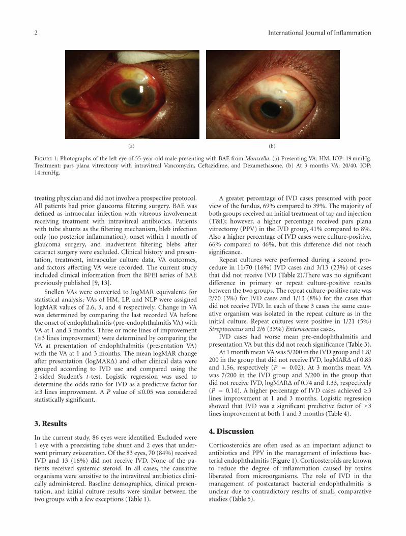
2 International Journal of Inflammation
(a) (b)
Figure 1: Photographs of the left eye of 55-year-old male presenting with BAE from Moraxella. (a) Presenting VA: HM, IOP: 19 mmHg.Treatment: pars plana vitrectomy with intravitreal Vancomycin, Ceftazidime, and Dexamethasone. (b) At 3 months VA: 20/40, IOP:14 mmHg.
treating physician and did not involve a prospective protocol.All patients had prior glaucoma filtering surgery. BAE wasdefined as intraocular infection with vitreous involvementreceiving treatment with intravitreal antibiotics. Patientswith tube shunts as the filtering mechanism, bleb infectiononly (no posterior inflammation), onset within 1 month ofglaucoma surgery, and inadvertent filtering blebs aftercataract surgery were excluded. Clinical history and presen-tation, treatment, intraocular culture data, VA outcomes,and factors affecting VA were recorded. The current studyincluded clinical information from the BPEI series of BAEpreviously published [9, 13].
Snellen VAs were converted to logMAR equivalents forstatistical analysis; VAs of HM, LP, and NLP were assignedlogMAR values of 2.6, 3, and 4 respectively. Change in VAwas determined by comparing the last recorded VA beforethe onset of endophthalmitis (pre-endophthalmitis VA) withVA at 1 and 3 months. Three or more lines of improvement(≥3 lines improvement) were determined by comparing theVA at presentation of endophthalmitis (presentation VA)with the VA at 1 and 3 months. The mean logMAR changeafter presentation (logMARΔ) and other clinical data weregrouped according to IVD use and compared using the2-sided Student’s t-test. Logistic regression was used todetermine the odds ratio for IVD as a predictive factor for≥3 lines improvement. A P value of ≤0.05 was consideredstatistically significant.
3. Results
In the current study, 86 eyes were identified. Excluded were1 eye with a preexisting tube shunt and 2 eyes that under-went primary evisceration. Of the 83 eyes, 70 (84%) receivedIVD and 13 (16%) did not receive IVD. None of the pa-tients received systemic steroid. In all cases, the causativeorganisms were sensitive to the intravitreal antibiotics clini-cally administered. Baseline demographics, clinical presen-tation, and initial culture results were similar between thetwo groups with a few exceptions (Table 1).
A greater percentage of IVD cases presented with poorview of the fundus, 69% compared to 39%. The majority ofboth groups received an initial treatment of tap and injection(T&I); however, a higher percentage received pars planavitrectomy (PPV) in the IVD group, 41% compared to 8%.Also a higher percentage of IVD cases were culture-positive,66% compared to 46%, but this difference did not reachsignificance.
Repeat cultures were performed during a second pro-cedure in 11/70 (16%) IVD cases and 3/13 (23%) of casesthat did not receive IVD (Table 2).There was no significantdifference in primary or repeat culture-positive resultsbetween the two groups. The repeat culture-positive rate was2/70 (3%) for IVD cases and 1/13 (8%) for the cases thatdid not receive IVD. In each of these 3 cases the same caus-ative organism was isolated in the repeat culture as in theinitial culture. Repeat cultures were positive in 1/21 (5%)Streptococcus and 2/6 (33%) Enterococcus cases.
IVD cases had worse mean pre-endophthalmitis andpresentation VA but this did not reach significance (Table 3).
At 1 month mean VA was 5/200 in the IVD group and 1.8/200 in the group that did not receive IVD, logMARΔ of 0.85and 1.56, respectively (P = 0.02). At 3 months mean VAwas 7/200 in the IVD group and 3/200 in the group thatdid not receive IVD, logMARΔ of 0.74 and 1.33, respectively(P = 0.14). A higher percentage of IVD cases achieved ≥3lines improvement at 1 and 3 months. Logistic regressionshowed that IVD was a significant predictive factor of ≥3lines improvement at both 1 and 3 months (Table 4).
4. Discussion
Corticosteroids are often used as an important adjunct toantibiotics and PPV in the management of infectious bac-terial endophthalmitis (Figure 1). Corticosteroids are knownto reduce the degree of inflammation caused by toxinsliberated from microorganisms. The role of IVD in themanagement of postcataract bacterial endophthalmitis isunclear due to contradictory results of small, comparativestudies (Table 5).

International Journal of Inflammation 3
Table 1: Baseline demographics, clinical presentation, and initialculture results.
IVD No IVDP value
70/83 (84%) 13/83 (16%)
Age
Mean, SD 74 yr (12) 70 yr (14) 0.27
Gender
Female 34 (49%) 8 (62%) 0.39
Male 36 (51%) 5 (39%)
Diabetes mellitus
Present 9 (13%) 2 (15%) 0.81
Absent 61 (87%) 11 (85%)
Antimetabolites(MMC or 5FU)
Used 45 (64%) 7 (54%) 0.47
Not used 25 (36%) 6 (46%)
Mean time of onset, SD 60 mo (43) 49 mo (55) 0.46
Bleb leak
Present 16 (23%) 5 (38%) 0.23
Absent 54 (77%) 8 (62%)
Anterior chamber
Hypopyon 48 (69%) 10 (77%) 0.55
View to fundus
Hazy 22 (31%) 8 (62%) 0.04
Poor/none 48 (69%) 5 (39%)
Intraocular Pressure
Presentation, SD 20 (14) 19 (12) 0.8
Treatment, initial
Tap and injection 41 (59%) 12 (92%) 0.03
Pars plana vitrectomy 29 (41%) 1 (8%)
Treatment, additional
Filtering procedure 12 (17%) 1 (8%) 0.39
Pars plana vitrectomy 21 (30%) 2 (15%) 0.28
Culture results
Culture positive 46 (66%) 6 (46%) 0.18
Culture negative 24 (34%) 7 (54%)
Gram-positive cases 33 (47%) 4 (31%) 0.28
Streptococcus 19 (27%) 2 (15%) 0.37
Coagulase-negativeStaphylococcus
7 (10%) 2 (15%) 0.57
Enterococcus 6 (9%) 0 0.27
Staphylococcus aureus 1 (1%) 0 0.67
Gram-negative cases 12 (17%) 2 (15%) 0.88
Moraxella 8 (11%) 0 0.2
Pseudomonas 2 (3%) 1 (8%) 0.39
Serratia 1 (1%) 1 (8%) 0.18
Das et al. reported favorable results of reduced intraocu-lar inflammation at 1 and 4 weeks in eyes treated with IVD[5]. Gan et al. additionally found a trend toward better visualoutcomes at 3 and 12 months in eyes treated with IVD [6].In contrast, Hall et al. reported no difference in inflammation
Table 2: Repeat culture results.
IVD No IVDP value
70/83(84%)
13/83(16%)
Repeat Cultures Performed:
Number of eyes 11 (16%) 3 (23%) 0.52
Mean time, days (range) 20 (1–60) 14 (2–30) 0.64
Primary culture results
Streptococcus 4 (36%) 2 (67%)
Enterococcus faecalis 2 (18%) 0
Coagulase-neg Staph. 2 (18%) 1 (33%)
Enterobacter aerogenes 1 (9%) 0
No growth 2 (18%) 0 0.43
Repeat culture results
Streptococcus 0 1 (33%)
Enterococcus faecalis 2 (18%) 0
Coagulase-neg Staph. 0 0
Enterobacter aerogenes 0 0
No growth 9 (82%) 2 (67%) 0.57
Repeat culture positive rate 2 (3%) 1 (8%) 0.57
Table 3: VA outcomes.
IVD No IVDP value
71/84 (84%) 13/84 (16%)
VA, pre-endophthalmitis
n = 67 n = 13
Mean 20/90 20/70 0.57
Range 20/20-LP 20/25–20/400
VA, presentation n = 70 n = 13
Mean 0.9/200 1.7/200 0.23
Range 20/40-NLP 20/80-LP
VA, 1 month n = 66 n = 12
Mean 5/200 1.8/200 0.14
Range 20/25-NLP 20/25-NLP
≥3 linesImprovement
44 (67%) 3 (25%) 0.01
logMARΔ 0.85 1.56 0.02
VA, 3 months n = 56 n = 9
Mean 7/200 3/200 0.36
Range 20/25-NLP 20/25-LP
≥3 linesImprovement
36 (64%) 3 (33%) 0.14
logMARΔ 0.74 1.33 0.14
Table 4: Predictive factor ≥3 lines improvement.
IVD versus No IVD Odds ratio, (CI) P value
1 month 7.04 (1.63,30.43) 0.01
3 months 5.21 (1.07,25.37) 0.04
and VA outcomes at last followup in eyes treated with IVD[7]. Shah et al. found worse VA outcomes at 1, 3, and 6

4 International Journal of Inflammation
Table 5: Comparative studies of IVD for bacterial endophthalmitis.
Clinical setting Culture resultsInflammation VA outcomes Time
n Staph epi. Strep/Enterococcus Gram-negative
Das et al. [5]Postcataract and
trauma
IVD 29 n/a n/a n/a 2.6 score 86% success3 months
No IVD 34 3.2 score1 71% success
Shah et al. [8] Postcataract
IVD 26 31% 12% 0 n/a 20/70 median6 months
No IVD 31 35% 13% 3% 20/50 median2
Gan et al. [6] Postcataract
IVD 16 39% 8% 0 n/a 85% 20/200 or better3 months
No IVD 13 50% 6% 0 50% 20/200 or better3
Hall et al. [7] Postcataract
IVD 26 46% 23% 0 0.3 cell/flare 20/40 median lastfollowupNo IVD 38 37% 5% 0 0.3 cell/flare 20/50 median
Jacobs et al. Bleb-associated
IVD 70 10% 36% 17% n/a 7/200 mean3 months
No IVD 13 15% 15% 15% 3/200 mean4
1Relative change in inflammation showed statistical significance at 1 and 4 weeks, not at 3 months. 2P < 0.05 , 3P = 0.055. 4Relative logMARΔ showed
statistical significance at 1 month, not at 3 months.
Table 6: PPV versus T&I in present BAE series.
PPV T&IP value
29/83 (35%) 54/83 (65%)
VA, pre-endophthalmitis
n = 28 n = 53
Mean 20/55 20/50 0.3
Range 20/20-CF 20/20-LP
VA, presentation n = 30 n = 54
Mean LP HM 0.02
Range 20/80-LP 20/40-NLP
VA, 3 months n = 27 n = 39
Mean 3/200 20/390
Range 20/25-NLP 20/25-LP
logMARΔ 1.23 0.57 0.02
months in eyes treated with IVD [8]. The present series wasunique as it was the first to study BAE cases that had a higherrate of Streptococcus and Gram-negative cases.
BAE studies are limited by the relatively small numberof BAE cases. Conclusions in BAE studies are found in theinherent limitations of retrospective nonrandomized data.The majority of cases in the present study received IVDwhich is similar to other BAE series [9–12]. Overall thetwo groups compared in this study had similar baselinedemographic, clinical presentation, and initial culture data(Table 1).
A difference was found in the initial treatment of the twogroups. The majority of cases in both groups received T&Ias the initial treatment, however the percentage that receivedinitial PPV was higher in the IVD group. The effect this
Table 7: PPV versus T&I: presentation VA of LP or worse in presentBAE series.
PPV T&IP value
18/26 (69%) 8/26 (31%)
VA, pre-endophthalmitis
n = 18 n = 8
Mean 20/65 20/270 0.16
Range 20/20–1/200 20/25-HM
VA, presentation n = 18 n = 8
Mean LP LP 0.35
Range LP LP-NLP
VA, 3 months n = 17 n = 5
Mean 1/200 1/200
Range 20/60-NLP 20/200-LP
logMARΔ 1.71 1.18 0.46
difference had on the VA outcomes is unclear. A comparisonof PPV and T&I cases in this series showed that PPV caseshad significantly worse mean VA at presentation and 3months (Table 6).
When presentation of VA was LP or worse, 3-month-logMARΔ was worse in the PPV group but not significantly(Table 7).
As VA outcomes with PPV were worse than T&I in thisseries, it is unlikely that improved VA outcomes in the IVDgroup were due to a higher percentage of initial treatmentwith PPV.
The repeat culture results in this series were similar to theEndophthalmitis Vitrectomy Study (EVS). In the EVS, 14 of420 (3.3%) had positive repeat cultures [14]. In the present

International Journal of Inflammation 5
series the overall repeat culture-positive rate was 3 of 83(3.6%). Of note the EVS did not use IVD and the EVS had alower rate of Streptococcus and Gram-negative organisms, yetthe rate of persistent infection was similar in the EVS to thepresent study. The present study confirms the observationmade by Shaarawy et al. that persistent infection can occurin bacterial endophthalmitis and appears to be more com-mon with virulent organisms such as Streptococcus and En-terococcus [15]. Although persistent infection occurred in thepresent BAE series there was not a higher rate among thecases treated with IVD.
VA outcomes in the present series confirm the clinicalobservation by Irvine et al. that intraocular steroids appearedto hasten visual recovery [2]. At 1 month, 67% gained ≥3lines in the IVD group compared to 25% in the groupthat did not receive IVD. The VA gains in the IVD groupwere more significant at 1 month than 3 months. Logisticregression did show that IVD was a predictive factor of ≥3lines of improvement at both 1 and 3 months. VA gains mayhave been due to a decrease in intraocular inflammation,but a standardized manner of grading inflammation was notemployed in the present series.
Limitations of the current study include the retrospectivenature, small sample size in the control arm, and lack of adefinitive treatment protocol. This study does demonstratethat IVD was associated with improved short-term VA out-comes and did not potentiate infection in BAE.
Disclosure
The authors in this paper have no financial or proprietaryinterests in products, methods, or materials published in thispaper.
Acknowledgment
This paper was supported by the National Institute of HealthCenter Grant P30-EY014801, an unrestricted grant to theUniversity of Miami from Research to Prevent Blindness,New York, NY, USA.
References
[1] G. A. Peyman and R. Herbst, “Bacterial endophthalmitis:treatment with intraocular injection of gentamicin and dexamethasone,” Archives of Ophthalmology, vol. 91, no. 5, pp.416–418, 1974.
[2] W. D. Irvine, H. W. Flynn, D. Miller, and S. C. Pflugfelder,“Endophthalmitis caused by gram-negative organisms,” Ar-chives of Ophthalmology, vol. 110, no. 10, pp. 1450–1454, 1992.
[3] J. A. Schulman and G. A. Peyman, “Intravitreal corticosteroidsas an adjunct in the treatment of bacterial and fungal endoph-thalmitis: a review,” Retina, vol. 12, no. 4, pp. 336–340, 1992.
[4] J. S. Pollack, “ASRS pat survey,” 2004, http://www.asrs.org/.[5] T. Das, S. Jalali, V. K. Gothwal, S. Sharrna, and T. J. Naduvilath,
“Intravitreal dexamethasone in exogenous bacterial endoph-thalmitis: results of a prospective randomised study,” BritishJournal of Ophthalmology, vol. 83, no. 9, pp. 1050–1055, 1999.
[6] I. M. Gan, L. C. Ugahary, J. T. van Dissel et al., “Intravitrealdexamethasone as adjuvant in the treatment of postoperative
endophthalmitis: a prospective randomized trial,” Graefe’sArchive for Clinical and Experimental Ophthalmology, vol. 243,no. 12, pp. 1200–1205, 2005.
[7] E. F. Hall, G. R. Scott, D. C. Musch, and D. N. Zacks, “Ad-junctive intravitreal dexamethasone in the treatment of acuteendophthalmitis following cataract surgery,” Journal of Clini-cal Ophthalmology, vol. 2, no. 1, pp. 139–145, 2008.
[8] G. K. Shah, J. D. Stein, S. Sharma et al., “Visual outcomes fol-lowing the use of intravitreal steroids in the treatment of post-operative endophthalmitis,” Ophthalmology, vol. 107, no. 3,pp. 486–489, 2000.
[9] T. Leng, D. Miller, H. W. Flynn, D. J. Jacobs, and S. J. Gedde,“Delayed-onset bleb-associated endophthalmitis (1996–2008): causative organisms and visual acuity outcomes,”Retina, vol. 31, no. 2, pp. 344–352, 2010.
[10] T. A. Kangas, D. S. Greenfield, H. W. Flynn, R. K. Parrish, andP. Palmberg, “Delayed-onset endophthalmitis associated withconjunctival filtering blebs,” Ophthalmology, vol. 104, no. 5,pp. 746–752, 1997.
[11] B. G. Busbee, F. M. Recchia, R. Kaiser et al., “Bleb-associatedendophthalmitis: clinical characteristics and visual outcomes,”Ophthalmology, vol. 111, no. 8, pp. 1495–1503, 2004.
[12] S. Mandelbaum, R. K. Forster, H. Gelender, and W. Cul-bertson, “Late onset endophthalmitis associated with filteringblebs,” Ophthalmology, vol. 92, no. 7, pp. 964–972, 1985.
[13] D. J. Jacobs, T. Leng, H. W. Flynn Jr., W. Shi, D. Miller, andS. J. Gedde, “Delayed-onset bleb-associated endophthalmitis:presentation and outcome by culture result,” Clinical Ophthal-mology, vol. 5, no. 1, pp. 739–744, 2011.
[14] B. H. Doft, S. F. Kelsey, and S. R. Wisniewski, “Additionalprocedures after the initial vitrectomy or tap-biopsy in theendophthalmitis vitrectomy study,” Ophthalmology, vol. 105,no. 4, pp. 707–716, 1998.
[15] A. Shaarawy, M. G. Grand, T. A. Meredith, and H. E. Ibanez,“Persistent endophthalmitis after intravitreal antimicrobialtherapy,” Ophthalmology, vol. 102, no. 3, pp. 382–387, 1995.

Hindawi Publishing CorporationInternational Journal of InflammationVolume 2012, Article ID 753560, 8 pagesdoi:10.1155/2012/753560
Research Article
Clinico-Microbiological Profile andTreatment Outcome of Infectious Scleritis: Experience froma Tertiary Eye Care Center of India
Srikant Kumar Sahu,1 Sujata Das,1 Savitri Sharma,2 and Kalyani Sahu1
1 Cornea and Anterior Segment Service, L V Prasad Eye Institute, Bhubaneswar 751024, India2 Ocular Microbiology Service, L V Prasad Eye Institute, Bhubaneswar 751024, India
Correspondence should be addressed to Srikant Kumar Sahu, srikant [email protected]
Received 16 July 2011; Accepted 31 August 2011
Academic Editor: Mark Willcox
Copyright © 2012 Srikant Kumar Sahu et al. This is an open access article distributed under the Creative Commons AttributionLicense, which permits unrestricted use, distribution, and reproduction in any medium, provided the original work is properlycited.
Medical and microbiology records of seventeen patients (17 eyes), diagnosed as scleritis of infectious origin were reviewed; tostudy clinical features, predisposing risk factors, microbiologic profile and treatment outcome of infectious scleritis. The meanpatient age was 52.3 ± 19.75 years. Twelve patients (70.6%) had history of trauma/prior surgery. Isolated organisms includedStaphylococcus species (spp) (n = 5), Fungus (n = 4), Nocardia spp (n = 3), two each of atypical Mycobacterium spp andStreptococcus pneumoniae and one Pseudomonas aeruginosa. Treatment included intensive topical antimicrobial in all eyes andsystemic medication in 15 (88.2%) patients; surgical exploration was needed for 13 (76.5%) patients and scleral patch graft wasdone in four (23.5%) patients. Lesions resolved in all patients and none required evisceration. The presenting log MAR visualacuity of 1.77 ± 1.40 and improved to 0.99 ± 0.91. (P ≤ 0.039) after treatment with a mean follow up of 22.57 ± 19.53 weeks. Amicrobiological confirmation, appropriate medical and/or surgical intervention has a good tectonic and visual outcome.
1. Introduction
Infectious scleritis presents as an ulcerated or nonulcerated,inflamed scleral nodule [1]. It accounts for 5–10% of all casesof scleritis [2–5]. But the presenting picture of infectiousscleritis may not differ too much from immune-mediatedscleritis [6–10]. Approximately 40–90% of immune medi-ated scleritis have an associated systemic vascular disease[3, 4, 6, 10]. While systemic treatment with corticosteroidsor immunosuppressant benefits immune-mediated scleritis,it worsens the infectious scleritis. Hence it is imperative todifferentiate between two conditions.
Many authors have reported infection by Pseudomonasaeruginosa and fungus as the most common causativeorganism [1, 11–13]. Pterygium surgery with beta radiationor application of mitomycin C has been identified as acommon risk factor for infectious scleritis [11–13].
The clinical outcome is generally poor and most casesrequired evisceration in the many series [8, 9, 14]. Systemic
and topical medication combined with early surgical inter-vention have improved the anatomical success, but not thevisual outcome in two other series [1, 12].
Infectious scleritis is a rare entity; hence it is notsuspected at the initial presentation resulting in delayeddiagnosis and treatment. In this communication, we describethe predisposing factors, clinical features, etiology, andtreatment outcome of infectious scleritis.
2. Material and Method
We retrospectively reviewed the medical and microbiolog-ical records of all patients with microbiologically proveninfectious scleritis examined at Cornea Services of L VPrasad Eye Institute Bhubaneswar from November 2006 toAugust 2009. At presentation, all patients had received adetailed ophthalmic examination in the office. Patients withulcerative lesions had received a scraping with a no. 15

2 International Journal of Inflammation
surgical blade on a Bard Parker handle from the base andactive edges of the ulcer under topical anesthesia in the office,and all nonulcerative nodular abscesses had received scleralscraping in the operating room under peribulbar anesthesia.In the later case, the base of the lesion was scrapped afterdissecting the conjunctiva and deroofing of the nodularlesion. Materials collected from the lesions were smearedon glass slides and stained with Potassium hydroxide +Calcoflour white stain, Grams stain, Zeihl Neelsen stainusing 20% H2SO4 or modified Zeihl Neelsen stain using1% H2SO4. Acid fast staining was usually done if Gramsstain smear was negative, but the lesions were stronglysuspicious of microbial origin. The exudates from the lesionwere cultured on blood agar, chocolate agar, Sabouraud’sdextrose agar (SDA), Brain-heart infusion broth and non-nutrient agar with an Escherichia coli overlay. All media wereincubated at 37◦C except SDA; this was incubated at 27◦C.Significant growth was defined as confluent growth on solidmedia, and/or there was growth of the same organism onmore than one medium, and/or growth in one medium wasaccompanied by presence of similar organism in smears. Allbacteria and fungi grown were identified as per standardprotocol, and bacteria were tested for antibiotic susceptibilityby Kirby-Bauer disc diffusion method.
The initial therapy either was based on results of smearor when smear was negative, an empirical treatment withtopical antibiotic (mostly gatifloxacin 0.3% and amikacin2.5%) along with systemic gatifloxacin ((400 mg) twice daily)was given. The treatment was modified, if needed, basedon final culture and sensitivity report. Surgical debridementwas done should the scleral ulceration area extend locally orprogressed to form a subconjunctival abscess at another siteaway from the main lesion.
The collected retrospective data included patient demog-raphy (age, gender, occupation), disease history (onset,course, predisposing factors), clinical feature, type of organ-isms, antibiotic susceptibility, treatment given, and theoutcome.
3. Results
We included 17 patients (17 eyes) of infectious scleritisbetween November 2006 and August 2009. The inclusioncriteria were presentation with a scleral ulcer and/or abscessand an organism isolated microbiologically. This included7 women and 10 men; the age ranged from 13 years to 75years (mean 52.3 ± 19.75 years, median 55 years). (Table 1)The mean followup was 22.57 ± 19.53 weeks (range of 3–89weeks).
The most common predisposing factor was an ocularsurgery [n = 9; 52.9%. 95% (CI), 29.27–76.73]. The surgeryincluded cataract in seven eyes, scleral buckle in one eye,and trabeculectomy in one eye. The interval from surgeryto diagnosis of infectious scleritis ranged from one week tofour years. Four patients had injury with organic materiallike wood and mud three weeks to seven months priorto presentation (mean 37 ± 33.04 days; median 21 days).Fifteen of 17 patients were using topical corticosteroids and
two patients were using oral corticosteroids at the time ofreporting to us. Only one patient was diabetic in this series.
The symptoms present in all patients were redness, pain,and watering in the affected eye. The presenting visual acuityvaried from hand motions (HM) to a normal vision of 20/20.Twelve patients (70.54%) presented with a vision less than20/40. Unifocal or multifocal scleral abscess was seen in sixpatients (35.39%) (Figures 1(a) and 1(b)). Characteristically,the abscesses presented as yellowish nodules under the intactconjunctival epithelium. Scleral ulceration and necrosis wasseen in eight patients (47.05%) (Figures 1(c) and 1(d)).Necrosis around the incisional area was seen in all sevenpatients who had received cataract surgery earlier. Three eyes(17.34%) had both ulceration and abscess (Figure 2(a)).
The culture grew a variety of organisms. They includedStaphylococcus species (n = 5; 29.41%), fungus (n = 4;23.52%), Nocardia species (n = 3; 17.6%), Streptococcuspneumoniae (n = 2), atypical Mycobacterium (n = 2),and Pseudomonas aeruginosa (n = 1). One of the fourStaphylococcus was methicillin resistant (no. 7). Of the fourfungi isolated, two could not be identified, one belonged toFusarium and Paecilomyces species each.
The results of antibiotic susceptibility testing for bacterialisolates (Kirby Bauer disc diffusion method) are given inTable 2. Staphylococcal and streptococcal cases were treatedwith fortified Cefazolin (50 mg/mL) and a flouroquinolone(ciprofloxacin 0.3%/gatifloxacin 0.3%) along with systemicfluoroquinolones (ciprofloxacin 500 mg/gatifloxacin 400 mgtwice daily). Patient no. 7 was treated only with topi-cal ciprofloxacin. Fungal scleritis was treated with topicalnatamycin 5% and systemically either itraconazole (100 mg)or ketoconazole (200 mg) two times daily. Nocardia andatypical Mycobacterium scleritis were treated primarily withtopical fortified amikacin (25 mg/mL). Systemic trimetho-prim (160 mg) sulphamethoxazole (800 mg) (TMX-SMZ)combination was used in patients of Nocardia scleritis only.
Thirteen of the 17 patients underwent wound debride-ment. During the surgical debridement, it was noticed thatthe area actually involved was generally much larger thaninitially seen under slit lamp. N-Butyl Cyanoacrylate gluewas used in two patients as there was a limbal perforationof less than 1 mm × 1 mm in size. Five patients receivedscleral patch graft; as primary procedure in one (patientno. 4, Figure 1(c)), and the remaining four eyes, 2 days toone month after initiation of medical treatment. Seven eyesneeded multiple surgical interventions.
Oral corticosteroid (1 mg/kg of body weight) was usedin 5 eyes with bacterial scleritis two days after antibacterialtreatment. In case no. 4, scleral patch graft was done asthe primary procedure, later fungal filaments were identifiedfrom smear; so corticosteroids were started only aftertwo weeks when recurrence was not noticed. Intraocularantibiotic was used in three patients suspected to haveendophthalmitis. All of them presented scleral infection aftercataract surgery.
Resolution was defined as absence of symptoms, con-gestion, or active infiltrate. All the patients in this seriesresponded to treatment; 13(76.47%) patients had onlyscarring with no or minimal uveal show; one patient

International Journal of Inflammation 3
Ta
ble
1:Pa
tien
tde
mog
raph
ic,c
linic
al,a
nd
mic
robi
olog
ical
feat
ure
,man
agem
ent,
and
outc
ome.
Sl.
no.
Age
Sex
Eye
His
tory
Du
rati
onof
sym
ptom
s(i
nda
ys)
Pre
sen
tin
gvi
sual
acu
ity
Clin
ical
sign
sO
rgan
ism
Trea
tmen
tFi
nal
visu
alac
uit
y
Rem
arks
Ou
tcom
e
Topi
cal
Syst
emic
Surg
ical
1.72
MO
SC
atar
act
surg
ery
45C
F2
mN
ecro
sis,
corn
eal
infi
ltra
teS.
aure
usF.
cefa
zolin
,ci
profl
oxac
inC
ipro
flox
acin
TA+
BC
l20
/70
Cor
nea
lsca
rR
esol
ved
2.65
MO
DN
il60
20/4
0A
bsce
ssN
ocar
dia
spp.
F.am
ikac
in,
gati
flox
acin
TM
X-S
MZ
Exp
lora
tion
-220
/60p
Cat
arac
tR
esol
ved
3.19
FO
SN
il15
020
/25
Pu
nch
edou
tu
lcer
Aty
pica
lm
ycob
acte
rium
F.am
ikac
in,
cipr
oflox
acin
TM
X-S
MZ
,pr
edn
isol
one
Nil
20/3
0C
orn
eal
infi
ltra
te,
chor
oida
lR
esol
ved
4.72
MO
SC
atar
act
surg
ery
45C
F2
mN
ecro
sis
Fusa
rium
spp.
Nat
amyc
in,
gati
flox
acin
,cy
clos
pori
ne
Itra
con
azol
eSP
G20
/120
Gra
ftva
scu
lari
zed
Res
olve
d
5.35
FO
DIn
jury
(wit
hst
ick)
15C
F2
mA
bsce
ss,
corn
eal
infi
ltra
teFu
ngu
sN
atam
ycin
,ga
tifl
oxac
inIt
raco
naz
ole
Exp
lora
tion
,In
trac
am-
eral
amph
o-B
20/1
60C
orn
eals
car
and
cata
ract
Res
olve
d
6.66
MO
DC
atar
act
surg
ery
9H
MSc
lera
ln
ecro
sis,
exu
date
S.pn
eum
onia
eF.
cefa
zolin
,ga
tifl
oxac
in,
beta
met
has
one
Lizo
lidin
e,pr
edin
isol
one
SPG
+IO
AB
20/6
0G
raft
vasc
ula
rize
dR
esol
ved
7.49
MO
DN
il12
020
/20
Ulc
er,
absc
ess,
thin
nin
g
Stap
hylo
cocc
ussp
p.C
ipro
flox
acin
,pr
edn
isol
one
Pre
dnis
olon
eN
il20
/20
Nil
Res
olve
d
8.50
MO
SC
atar
act
surg
ery
7H
MN
ecro
sis
S.au
reus
F.ce
fazo
lin,
cipr
oflox
acin
Gat
iflox
acin
SPG
+V
itre
ous
biop
sy+
IOA
B
20/1
00G
raft
vasc
u-
lari
zed,
corn
eals
car
Res
olve
d
9.74
FO
SIn
jury
(wit
hm
ud)
15H
MA
bsce
ssP.
aeru
gino
saF.
amik
acin
,C
ipro
flox
acin
,P
redn
isol
one
Cip
rofl
oxac
in,
pred
nis
olon
eE
xplo
rati
on20
/200
Cat
arac
t,ch
oroi
dal
Res
olve
d

4 International Journal of Inflammation
Ta
ble
1:C
onti
nu
ed.
Sl.
no.
Age
Sex
Eye
His
tory
Du
rati
onof
sym
ptom
s(i
nda
ys)
Pre
sen
tin
gvi
sual
acu
ity
Clin
ical
sign
sO
rgan
ism
Trea
tmen
tFi
nal
visu
alac
uit
y
Rem
arks
Ou
tcom
e
Topi
cal
Syst
emic
Surg
ical
10.
50F
OD
Inju
ry(w
ith
twig
)75
20/3
0C
hem
osis
,ab
sces
sN
ocar
dia
spp.
F.am
ikac
in,
cipr
oflox
acin
Cip
rofl
oxac
inE
xplo
rati
onSP
G+
AM
G20
/20
Gra
ftva
scu
lari
zed
Res
olve
d
11.
13M
OD
Trab
ecu
lect
omy
30C
F1
mA
bsce
ssS.
aure
usF.
cefa
zolin
,ga
tifl
oxac
inC
efad
roxi
lN
ilC
F1
mG
lau
com
a,op
tic
atro
phy
Res
olve
d
12.
21M
OS
Thy
roid
Oph
thal
mop
legi
aN
otav
aila
ble
20/2
0A
bsce
ssS.
pneu
mon
iae
F.ge
nta
mic
in,
chlo
ram
phen
icol
Pre
dnis
olon
eN
il20
/30
Nil
Res
olve
d
13.
53M
OS
Ret
inal
deta
chm
ent
surg
ery
(bu
ckle
)60
CF
1m
Ulc
er,
absc
ess
Aty
pica
lm
ycob
acte
rium
F.am
ikac
in,
gati
flox
acin
Gat
iflox
acin
BB
Rem
oval
HM
Ele
vate
dG
ran
ulo
ma
Res
olve
d
14.
55F
OS
Nil
6020
/200
Th
inn
ing,
ulc
er,
absc
ess
Noc
ardi
asp
p.F.
amik
acin
,ci
profl
oxac
inC
ipro
flox
acin
,T
MX
-SM
ZE
xplo
rati
on-3
20/5
0N
ilR
esol
ved
15.
75F
OD
Cat
arac
tsu
rger
y60
HM
Nec
rosi
sFu
ngu
sN
atam
ycin
Itra
con
azol
eTA
+B
CL
20/5
0C
orn
eals
car
Res
olve
d
16.
65F
OS
Cat
arac
tsu
rger
y7
20/7
0N
ecro
sis,
corn
eal
infi
ltra
teS.
aure
usF.
cefa
zolin
,ga
tifl
oxac
in,
pred
nis
olon
eC
ipro
flox
acin
Wou
nd
repa
ir+
IOA
B20
/40
Mem
bran
eov
erIO
LR
esol
ved
17.
55M
OS
Cat
arac
tsu
rger
y30
20/1
60N
ecro
sis
Paec
ilom
yces
spp.
Nat
amyc
inK
etoc
onaz
ole
SPG
PL
+P
upi
llary
mem
bran
eR
esol
ved

International Journal of Inflammation 5
(a) (b)
(c) (d)
Figure 1: Slit lamp picture depicting different clinical presentation. (a) Case no. 2: multiple scleral abscess. (b) Case no.12: single scleralabscess. (c) Case no. 4: necrotic ulcer (post cataract surgery). (d) Case no. 3: two punched out ulcers.
(a) (b) (c)
Figure 2: Slit lamp picture of Case no. 10. (a) At Presentation showing multiple abscesses and ulcer. (b) Three weeks after initiation oftreatment (surgical exploration and medical management), there is a uveal show. (c) Final outcome showing vascularized scleral graft (fourmonths after scleral patch graft).
required scleral patch graft (Patient no. 10 Figures 2(a),2(b), and 2(c)) one month after initiation of treatment. Inthe patients where scleral patch graft was done, success wasdefined as no evidence of graft or surrounding infiltrate andvascularization of the graft. All five grafts were healthy at thelast visit.
The mean presenting and post treatment logMAR visualacuity was 1.77 ± 1.40 and 0.99 ± 0.91. (P ≤ 0.039)(Figure 3). The vision improved by greater than 2 lines in 8of 12 patients who presented with a visual acuity of <20/40patients.
The primary cause of decreased vision after resolutionof infection was cataract and corneal scar. Total choroidaldetachment developed in two patients and was treated with
tapering oral corticosteroids. One of the two patients hadatypical Mycobacteria infection. This 19-year-old lady alsodeveloped a corneal abscess after seven months of treatment.It resolved with topical fortified amikacin.
4. Discussion
Scleritis may represent a diagnostic challenge and is oftenassociated with life-threatening systemic disease (in thisseries, only one patient had diabetes mellitus though) andvision-threatening ocular complications [15].
Scleral infection from Pseudomonas, Staphylococcus, orHerpes zoster virus can cause necrotizing scleritis, whichis clinically identical to systemic autoimmune disease [6].
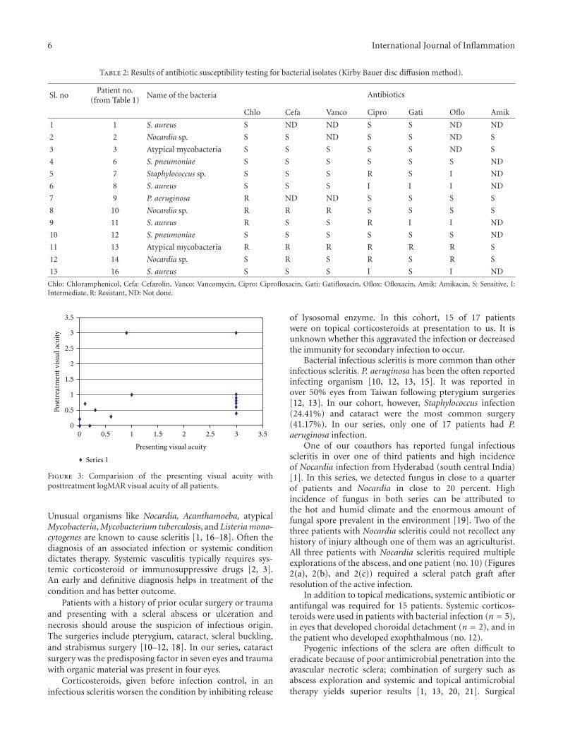
6 International Journal of Inflammation
Table 2: Results of antibiotic susceptibility testing for bacterial isolates (Kirby Bauer disc diffusion method).
Sl. noPatient no.
(from Table 1)Name of the bacteria Antibiotics
Chlo Cefa Vanco Cipro Gati Oflo Amik
1 1 S. aureus S ND ND S S ND ND
2 2 Nocardia sp. S S ND S S ND S
3 3 Atypical mycobacteria S S S S S ND S
4 6 S. pneumoniae S S S S S S ND
5 7 Staphylococcus sp. S S S R S I ND
6 8 S. aureus S S S I I I ND
7 9 P. aeruginosa R ND ND S S S S
8 10 Nocardia sp. R R R S S S S
9 11 S. aureus R S S R I I ND
10 12 S. pneumoniae S S S S S S ND
11 13 Atypical mycobacteria R R R R R R S
12 14 Nocardia sp. S R S R S R S
13 16 S. aureus S S S I S I ND
Chlo: Chloramphenicol, Cefa: Cefazolin, Vanco: Vancomycin, Cipro: Ciprofloxacin, Gati: Gatifloxacin, Oflox: Ofloxacin, Amik: Amikacin, S: Sensitive, I:Intermediate, R: Resistant, ND: Not done.
3.532.521.510.50
3.5
3
2.5
2
1.5
1
0.5
0
Series 1
Presenting visual acuity
Post
trea
tmen
t vi
sual
acu
ity
Figure 3: Comparision of the presenting visual acuity withposttreatment logMAR visual acuity of all patients.
Unusual organisms like Nocardia, Acanthamoeba, atypicalMycobacteria, Mycobacterium tuberculosis, and Listeria mono-cytogenes are known to cause scleritis [1, 16–18]. Often thediagnosis of an associated infection or systemic conditiondictates therapy. Systemic vasculitis typically requires sys-temic corticosteroid or immunosuppressive drugs [2, 3].An early and definitive diagnosis helps in treatment of thecondition and has better outcome.
Patients with a history of prior ocular surgery or traumaand presenting with a scleral abscess or ulceration andnecrosis should arouse the suspicion of infectious origin.The surgeries include pterygium, cataract, scleral buckling,and strabismus surgery [10–12, 18]. In our series, cataractsurgery was the predisposing factor in seven eyes and traumawith organic material was present in four eyes.
Corticosteroids, given before infection control, in aninfectious scleritis worsen the condition by inhibiting release
of lysosomal enzyme. In this cohort, 15 of 17 patientswere on topical corticosteroids at presentation to us. It isunknown whether this aggravated the infection or decreasedthe immunity for secondary infection to occur.
Bacterial infectious scleritis is more common than otherinfectious scleritis. P. aeruginosa has been the often reportedinfecting organism [10, 12, 13, 15]. It was reported inover 50% eyes from Taiwan following pterygium surgeries[12, 13]. In our cohort, however, Staphylococcus infection(24.41%) and cataract were the most common surgery(41.17%). In our series, only one of 17 patients had P.aeruginosa infection.
One of our coauthors has reported fungal infectiousscleritis in over one of third patients and high incidenceof Nocardia infection from Hyderabad (south central India)[1]. In this series, we detected fungus in close to a quarterof patients and Nocardia in close to 20 percent. Highincidence of fungus in both series can be attributed tothe hot and humid climate and the enormous amount offungal spore prevalent in the environment [19]. Two of thethree patients with Nocardia scleritis could not recollect anyhistory of injury although one of them was an agriculturist.All three patients with Nocardia scleritis required multipleexplorations of the abscess, and one patient (no. 10) (Figures2(a), 2(b), and 2(c)) required a scleral patch graft afterresolution of the active infection.
In addition to topical medications, systemic antibiotic orantifungal was required for 15 patients. Systemic corticos-teroids were used in patients with bacterial infection (n = 5),in eyes that developed choroidal detachment (n = 2), and inthe patient who developed exophthalmous (no. 12).
Pyogenic infections of the sclera are often difficult toeradicate because of poor antimicrobial penetration into theavascular necrotic sclera; combination of surgery such asabscess exploration and systemic and topical antimicrobialtherapy yields superior results [1, 13, 20, 21]. Surgical

International Journal of Inflammation 7
debridement not only facilitates penetration of antibiotic butalso debulks the infected scleral tissue.
We do agree with Lin et al. who suggested mandatorysurgical exploration of the abscess that does not respond toinitial medication to increase the penetration of antibiotic[13]. Raber et al. reported a “tunnel lesion” on histopatho-logical finding in cases of scleral ulcer [22]. Our experienceis similar to Lin et al. who also described the tunnel lesionand recommended the need of careful exploration to avoidresidual infection [13]. Two (no. 2, 14) of our 7 patients inthis series required multiple surgical intervention and bothof them had “tunnel lesion”.
All 17 patients in this series achieved anatomical success.Fourteen of 17 patients (82.35%) regained useful vision(defined as vision ≥20/200). The causes of poor vision inthe remaining three eyes were glaucomatous optic atrophy(no. 11), chronic retinal detachment (no. 13), and fibroticmembrane in the pupillary area (no. 17). In an earlier seriesby one of our coauthors, one-third of patients had regaineduseful vision, but three of 21 eyes were eviscerated and oneeye became phthisical [1]. Eleven of the 18 eyes in Hsiao et al.series retained useful vision, three had poor vision and fourhad to be eviscerated [12]. None of our patients in this seriesrequired evisceration.
Associated uveitis is not uncommon. (66% in Su et al.series) These inflammations lead to formation of pupil-lary membrane, cataract, and endophthalmitis [10]. Mostpatients in our series had low-grade inflammation in theanterior chamber at presentation; severe intraocular inflam-mation occurred in five patients subsequently. Cataract wasthe most common sequel in our series (n = 3). Four patientswho had extension to cornea resolved with corneal scar.
Our series has all weaknesses inherent to all retrospectivestudies. Our institution is a tertiary care referral center, sothere is a potential for bias towards more unusual or difficult-to-control diseases. Nonetheless, we have demonstrated thata variety of organism can cause scleritis. An early clinicalsuspicion of infectious origin, identification of the infectingorganism, knowledge of antibiotic susceptibility, and insti-tution of appropriate medical and/or surgical therapy couldhave good tectonic and visual outcome.
Conflict of interests
The authors declare no conflict of interests.
Acknowledgment
The authors would like to thank Hyderabad Eye ResearchFoundation for support.
References
[1] V. Jain, P. Garg, and S. Sharma, “Microbial scleritis—experience from a developing country,” Eye, vol. 23, no. 2, pp.255–261, 2009.
[2] D. A. Jabs, A. Mudun, J. P. Dunn, and M. J. Marsh, “Episcleritisand scleritis: clinical features and treatment results,” AmericanJournal of Ophthalmology, vol. 130, no. 4, pp. 469–476, 2000.
[3] M. S. de la Maza, N. S. Jabbur, and C. S. Foster, “Severity ofscleritis and episcleritis,” Ophthalmology, vol. 101, no. 2, pp.389–396, 1994.
[4] E. K. Akpek, J. E. Thorne, F. A. Qazi, D. V. Do, and D. A. Jabs,“Evaluation of patients with scleritis for systemic disease,”Ophthalmology, vol. 111, no. 3, pp. 501–506, 2004.
[5] T. A. Albini, N. A. Rao, and R. E. Smith, “The diagnosis andmanagement of anterior scleritis,” International Ophthalmol-ogy Clinics, vol. 45, no. 2, pp. 191–204, 2005.
[6] N. Okhravi, B. Odufuwa, P. McCluskey, and S. Lightman,“Scleritis,” Survey of Ophthalmology, vol. 50, no. 4, pp. 351–363, 2005.
[7] W. P. Riono, A. A. Hidayat, and N. A. Rao, “Scleritis: aclinicopathologic study of 55 cases,” Ophthalmology, vol. 106,no. 7, pp. 1328–1333, 1999.
[8] M. S. de la Maza, R. K. Hemady, and C. S. Foster, “Infectiousscleritis: report of four cases,” Documenta Ophthalmologica,vol. 83, no. 1, pp. 33–41, 1993.
[9] D. Sevel, “Necrogranulomatous scleritis. Clinical and histo-logic features,” American Journal of Ophthalmology, vol. 64, no.6, pp. 1125–1134, 1967.
[10] C. Y. Su, J. J. Tsai, Y. C. Chang, and C. P. Lin, “Immunologicand clinical manifestations of infectious scleritis after ptery-gium excision,” Cornea, vol. 25, no. 6, pp. 663–666, 2006.
[11] F. C. Huang, S. P. Huang, and S. H. Tseng, “Management ofinfectious scleritis after pterygium excision,” Cornea, vol. 19,no. 1, pp. 34–39, 2000.
[12] C. H. Hsiao, J. J. Chen, S. C. Huang, H. K. Ma, P. Y.Chen, and R. J. Tsai, “Intrascleral dissemination of infectiousscleritis following pterygium excision,” British Journal ofOphthalmology, vol. 82, no. 1, pp. 29–34, 1998.
[13] C. P. Lin, M. H. Shih, and M. C. Tsai, “Clinical experiencesof infectious scleral ulceration: a complication of pterygiumoperation,” British Journal of Ophthalmology, vol. 81, no. 11,pp. 980–983, 1997.
[14] M. G. Reynolds and E. Alfonso, “Treatment of infectious scle-ritis and keratoscleritis,” American Journal of Ophthalmology,vol. 112, no. 5, pp. 543–547, 1991.
[15] D. D. O. Machado, A. L. L. Curi, R. S. Fernandes, T. F. Bessa, W.R. Campos, and F. Orefice, “Scleritis: clinical characteristics,systemic associations, treatment and outcome in 100 patients,”Arquivos Brasileiros de Oftalmologia, vol. 72, no. 2, pp. 231–235, 2009.
[16] K. B. Ebrahimi, W. R. Green, R. Grebe, and A. S. Jun,“Acanthamoeba sclerokeratitis,” Graefe’s Archive for Clinicaland Experimental Ophthalmology, vol. 247, no. 2, pp. 283–286,2009.
[17] O. Oz, D. H. Lee, S. M. Smetana, and L. Akduman, “Acase of infected scleral buckle with Mycobacterium chelonaeassociated with chronic intraocular inflammation,” OcularImmunology and Inflammation, vol. 12, no. 1, pp. 65–67, 2004.
[18] E. Tay, M. Rajan, and S. Tuft, “Listeria monocytogenessclerokeratitis: a case report and literature review,” Cornea, vol.27, no. 8, pp. 947–949, 2008.
[19] U. Gopinathan, P. Garg, M. Fernandes, S. Sharma, S. Ath-manathan, and G. N. Rao, “The epidemiological features andlaboratory results of fungal keratitis: a 10-year review at areferral eye care center in South India,” Cornea, vol. 21, no.6, pp. 555–559, 2002.
[20] W. Bernauer, B. D. S. Allan, and J. K. G. Dart, “Successfulmanagement of Aspergillus scleritis by medical and surgicaltreatment,” Eye, vol. 12, part 2, pp. 311–316, 1998.
[21] C. J. Helm, G. N. Holland, R. G. Webster Jr., R. K. Maloney,and B. J. Mondino, “Combination intravenous ceftazidime

8 International Journal of Inflammation
and aminoglycosides in the treatment of pseudomonal scle-ritis,” Ophthalmology, vol. 104, no. 5, pp. 838–843, 1997.
[22] I. M. Raber, P. R. Laibson, G. H. Kurz, and V. B. Bernardino,“Pseudomonas corneoscleral ulcers,” American Journal ofOphthalmology, vol. 92, no. 3, pp. 353–362, 1981.