Atrial Fibrillation - Hindawi.com
74
Cardiology Research and Practice Guest Editors: Natig Gassonov, Evren Caglayan, Firat Duru, and Fikret Er Atrial Fibrillation
-
Upload
khangminh22 -
Category
Documents
-
view
0 -
download
0
Transcript of Atrial Fibrillation - Hindawi.com

Cardiology Research and Practice
Guest Editors: Natig Gassonov, Evren Caglayan, Firat Duru, and Fikret Er
Atrial Fibrillation

Atrial Fibrillation

Cardiology Research and Practice
Atrial Fibrillation
Guest Editors: Natig Gassonov, Evren Caglayan, Firat Duru,and Fikret Er

Copyright © 2013 Hindawi Publishing Corporation. All rights reserved.
This is a special issue published in “Cardiology Research and Practice.” All articles are open access articles distributed under the CreativeCommons Attribution License, which permits unrestricted use, distribution, and reproduction in any medium, provided the originalwork is properly cited.

Editorial Board
Atul Aggarwal, USAJesus M. Almendral, SpainPeter Backx, CanadaJ Brugada, SpainRamon Brugada, CanadaHans R. Brunner, SwitzerlandVicky A. Cameron, New ZealandDavid J. Chambers, UKRobert Chen, TaiwanMariantonietta Cicoira, ItalyAntonio Colombo, ItalyOmar H. Dabbous, USANaranjan S. Dhalla, CanadaFirat Duru, SwitzerlandVladimır Dzavık, CanadaGerasimos Filippatos, GreeceMihai Gheorghiade, USAEnrique P. Gurfinkel, ArgentinaPaul Holvoet, Belgium
H. A. Katus, GermanyHosen Kiat, AustraliaAnne A. Knowlton, USAGavin W. Lambert, AustraliaChim Choy Lang, UKF. H H Leenen, CanadaSeppo Lehto, FinlandJohn C. Longhurst, USALars S. Maier, GermanyOlivia Manfrini, ItalyGerald Maurer, AustriaG. A. Mensah, USARobert M. Mentzer, USAPiera Angelica Merlini, ItalyMarco Metra, ItalyVeselin Mitrovic, GermanyClaudio Moretti, ItalyJoseph Brent Muhlestein, USADebabrata P. Mukherjee, USA
J. D. Parker, CanadaFausto J. Pinto, PortugalBertram Pitt, UKRobert Edmund Roberts, CanadaTerrence D. Ruddy, CanadaFrank T. Ruschitzka, SwitzerlandChristian Seiler, SwitzerlandSidney G. Shaw, SwitzerlandPawan K. Singal, CanadaFelix C. Tanner, SwitzerlandHendrik T. Tevaearai, SwitzerlandG. Thiene, ItalyH. O. Ventura, USAStephan von Haehling, GermanyJames T. Willerson, USAMichael S. Wolin, USAMichael Wolzt, Austria

Contents
Atrial Fibrillation, Natig Gassonov, Evren Caglayan, Firat Duru, and Fikret ErVolume 2013, Article ID 142673, 2 pages
Chronobiological Analysis of Blood Pressure in a Patient with Atrial Fibrillation at the Development ofHeart Failure and Its Therapeutic and Surgical Treatmen, Sergey Chibisov, George Katinas,Inna Brodskaya, Aleksandr Ertman, Grigory Gromyko, Aleksandra Konradi, Oleg Mamontov,Anna Merkuryeva, Ekaterina Polunicheva, Evgeny Shlyakhto, Anna Soboleva, Sergey Yashin,and Bharadva BhavdipVolume 2013, Article ID 490705, 16 pages
Comparison of Atrial Fibrillation in the Young versus That in the Elderly: A Review,Rajiv Sankaranarayanan, Graeme Kirkwood, Katharine Dibb, and Clifford J. GarrattVolume 2013, Article ID 976976, 16 pages
The Use of Cardiac Magnetic Resonance Imaging in the Diagnostic Workup and Treatment of AtrialFibrillation, Peter Haemers, Piet Claus, and Rik WillemsVolume 2012, Article ID 658937, 6 pages
Left Atrial Appendage Exclusion for Stroke Prevention in Atrial Fibrillation, Taral K. Patel,Clyde W. Yancy, and Bradley P. KnightVolume 2012, Article ID 610827, 8 pages
Long-Term Costs of Ischemic Stroke and Major Bleeding Events among Medicare Patients withNonvalvular Atrial Fibrillation, Catherine J. Mercaldi, Kimberly Siu, Stephen D. Sander, David R. Walker,You Wu, Qian Li, and Ning WuVolume 2012, Article ID 645469, 13 pages
Effect of Preoperative Atrial Fibrillation on Postoperative Outcome following Cardiac Surgery,Nael Al-Sarraf, Lukman Thalib, Anne Hughes, Michael Tolan, Vincent Young, and Eillish McGovernVolume 2012, Article ID 272384, 7 pages

Hindawi Publishing CorporationCardiology Research and PracticeVolume 2013, Article ID 142673, 2 pageshttp://dx.doi.org/10.1155/2013/142673
EditorialAtrial Fibrillation
Natig Gassanov,1 Evren Caglayan,1 Firat Duru,2 and Fikret Er1
1 Department of Internal Medicine III, University of Cologne, Kerpener Stra𝛽𝛽e 62, 50937 Cologne, Germany2 Clinic for Cardiology, University Hospital Zurich, 8091 Zürich, Switzerland
Correspondence should be addressed to Natig Gassanov; [email protected]
Received 23 December 2012; Accepted 23 December 2012
Copyright © 2013 Natig Gassanov et al.is is an open access article distributed under the Creative CommonsAttribution License,which permits unrestricted use, distribution, and reproduction in any medium, provided the original work is properly cited.
Atrial �brillation (AF) is the most common clinically impor-tant cardiac arrhythmia. e prevalence of AF roughlydoubles with each advancing decade of age, from 0.5% at ageof 50–59 years to almost 9% at age of 80–89 years [1]. AF isassociatedwith substantialmorbidity andmortality.us, AFis a signi�cant risk factor for ischemic stroke and accounts for15–20% of all strokes [2]. Considering the clinical relevanceof AF, this journal initiated a special issue dealing with therecent developments in AF over the past few years. isissue contains important work—both review and originalarticles—addressing the epidemiology, economic impact ofAF, and new therapeutic/diagnostic developments and theirpotential clinical implications.
e general therapeutic strategies in AF include heart rateor rhythm control and anticoagulation. Current drugs usedfor AF therapy have major limitations, including incompleteefficacy and risks of life-threatening proarrhythmic eventsand bleeding complications. However, there have been sev-eral recent advancements in therapy of AF. ey includedthe availability of new anticoagulants, such as dabigatran,rivaroxaban, and apixaban, as well as guideline changesto incorporate the catheter-based isolation of pulmonaryveins (PV) as a class IIa/A indication [3]. Since the �rstpaper evidencing the role of PV as triggers of AF [4],various ablation techniques targeting PV (focal ablation, PVisolation, circumferential antral ablation, cryoballoon) wereintroduced into clinical practice [5, 6]. ough PV isolationby catheter-based radiofrequency ablation has become aneffective treatment option in AF, the studies on long-termoutcomes are still limited and less encouraging.
Recently, F. Ouyang et al. examined 5-year outcomes inparoxysmal AF and found that sinus rhythm was present in
46.6% of patients aer one procedure [7]. Long-term out-come data aer catheter ablation for persistent AF are evenless favourable [8]. ese data highlight important aspects ofcatheter ablation, in particular the need for improved toolsfor patient selection, energy delivery for durable transmurallesions, and more studies on long-term outcomes and themanagement of very late recurrences as mediators of initialprocedural approach re�nement. Radiological investigationssuch as CT scan or cardiac magnetic resonance imaging(cMRI) can demonstrate the complex LA anatomy very wellin all three dimensions and should reduce the �uoroscopytimes. In this special issue, P. Haemers et al. review thepotential application of cMRI in the diagnostic workup andtreatment of AF. Besides its value in the guidance of catheterablation, the authors describe the role of cMRI in identifyingthe underlying pathophysiological mechanisms of AF andin stroke prevention. cMRI can be used for le atrial scarquanti�cation to assist not only in the procedural approachbut also in patient selection.
As mentioned above, stroke is a major critical AF-associated complication. Today, stroke prevention with anti-coagulant agents based on the CHADS2/CHA2DS2-VASc-Score is the main cornerstone of AF management. However,anticoagulation—even with newly developed oral anticoag-ulants—has several limitations. In addition to the bleedingrisk, anticoagulation is not effectively utilized or contraindi-cated in signi�cant number of eligible AF patients, oendue to the limitations highlighted in the current paper byT. K. Patel et al. While newer oral anticoagulants overcomemany of these limitations, all anticoagulants suffer from anunavoidable lifelong commitment tomedication and elevatedbleeding risk.

2 Cardiology Research and Practice
e le atrial appendage (LAA) is particularly vulnerableto thrombus formation due to its complex anatomy and lowblood �ow during AF. erefore, LAA exclusion may be anespecially appealing option for patients with intolerance orcontraindications to anticoagulation. e procedure of LAAexcision as well as the device characteristics are now high-lighted in the current special issue. Indeed, LAA appendagemay be a true alternative to anticoagulation in some patients.However, there is a very limited clinical experience with thisnovel procedure, and it is obvious that further studies areurgently required to clarify the bene�ts and disadvantages ofLAA removal in patients with AF.
In another study of the current issue, C. J. Mercaldi et al.quanti�ed the direct long-term costs, up to 3 years, of bothstroke and bleeding events among patients with nonvalvularAF. Health care costs due to AF are enormous; on the basisof current �S age- and sex-speci�c prevalence data, thenational incremental AF cost is estimated to range from $6.0to $26.0 billion [9]. e authors conducted a retrospectivecohort study of Medicare bene�ciaries newly diagnosedwith AF who later developed stroke or hemorrhagic events.e results show that beyond the �rst year aer the event,patients with major bleeding events other than intracranialhemorrhage have signi�cantly higher costs than matchedcontrols, underlying the need for proper cost-effectivenessassessment of the true long-term costs of stroke and majorbleeding events. is strategy is particularly required whenweighing the risks (bleeding) and bene�ts (stroke prevention)of anticoagulation in patients with AF.
In summary, we hope that expert contributions tothis special issue will improve our understanding of themechanisms of AF and ultimately result in a better clinicalmanagement of AF and long-term outcomes.
Natig GassanovEvren Caglayan
Firat DuruFikret Er
References
[1] W. B. Kannel, P. A. Wolf, E. J. Benjamin, and D. Levy,“Prevalence, incidence, prognosis, and predisposing conditionsfor atrial �brillation: population-based estimates,” AmericanJournal of Cardiology, vol. 82, pp. 2N–9N, 1998.
[2] C. Marini, F. De Santis, S. Sacco et al., “Contribution of atrial�brillation to incidence and outcome of ischemic stroke: Resultsfrom a population-based study,” Stroke, vol. 36, no. 6, pp.1115–1119, 2005.
[3] A. J. Camm, G. Y. Lip, R. De Caterina et al., “2012 focusedupdate of the ESC Guidelines for the management of atrial�brillation: an update of the 2010 ESC Guidelines for themanagement of atrial �brillation ∗ Developed with the specialcontribution of the European Heart Rhythm Association,”European Heart Journal, vol. 33, no. 21, pp. 2719–2747, 2012.
[4] M. Haïssaguerre, P. Jaïs, D. C. Shah et al., “Spontaneousinitiation of atrial �brillation by ectopic beats originating in thepulmonary veins,” e New England Journal of Medicine, vol.339, no. 10, pp. 659–666, 1998.
[5] J. G. Andrade, P. Khairy, P. G. Guerra et al., “Efficacy andsafety of cryoballoon ablation for atrial �brillation: a system-atic review of published studies,” Heart Rhythm, vol. 8, pp.1444–1451, 2011.
[6] G. Lee, P. Sanders, and J. M. Kalman, “Catheter ablation ofatrial arrhythmias: state of the art,” e Lancet, vol. 380, pp.1509–1519, 2012.
[7] F. Ouyang, R. Tilz, J. Chun et al., “Long-term results of catheterablation in paroxysmal atrial �brillation: lessons from a 5-yearfollow-up,” Circulation, vol. 122, no. 23, pp. 2368–2377, 2010.
[8] R. R. Tilz, A. Rillig, A.M.umet al., “Catheter ablation of long-standing persistent atrial �brillation: 5-year outcomes of thehamburg sequential ablation strategy,” Journal of the AmericanCollege of Cardiology, vol. 60, pp. 1921–1929, 2012.
[9] M. H. Kim, S. S. Johnston, B. C. Chu, M. R. Dalal, and K. L.Schulman, “Estimation of total incremental health care costs inpatients with atrial �brillation in the united states,” Circulation:Cardiovascular Quality andOutcomes, vol. 4, no. 3, pp. 313–320,2011.

Hindawi Publishing CorporationCardiology Research and PracticeVolume 2013, Article ID 490705, 16 pageshttp://dx.doi.org/10.1155/2013/490705
Clinical StudyChronobiological Analysis of Blood Pressure in a Patient withAtrial Fibrillation at the Development of Heart Failure and ItsTherapeutic and Surgical Treatment
Sergey Chibisov,1 George Katinas,2 Inna Brodskaya,2 Aleksandr Ertman,2
Grigory Gromyko,2 Aleksandra Konradi,3 Oleg Mamontov,3 Anna Merkuryeva,2
Ekaterina Polunicheva,2 Evgeny Shlyakhto,3 Anna Soboleva,2
Sergey Yashin,2 and Bharadva Bhavdip1
1 People’s Friendship University of Russia, Moscow 117198, Russia2 Saint Petersburg State Medical University, St. Petersburg 197098, Russia3 VA Almasov Heart, Blood and Endocrinology Centre, Russia
Correspondence should be addressed to Bharadva Bhavdip; [email protected]
Received 17 June 2012; Accepted 19 November 2012
Academic Editor: Firat Duru
Copyright © 2013 Sergey Chibisov et al.This is an open access article distributed under theCreative CommonsAttribution License,which permits unrestricted use, distribution, and reproduction in any medium, provided the original work is properly cited.
Dynamics of blood pressure (BP) and heart rate (HR) was traced by automatic monitoring every 30min uninterruptedly alongseveral months in a patient suffering from combined atrial fibrillation and heart failure during the development of disease andits therapeutic and surgical treatment (pacemaker implanting and atrioventricular ablation). Analyses of spectral components aswell as signal’s shape revealed instabilities in circadian and semicircadian parameters. A new approach for signal’s form descriptionwithout using cosine approximation is suggested.The meaning that referring a patient as dipper, night peaker, or nondipper mightbe useful at choosing tactics of his treatment is impugned, because all these “types” can transform themselves in the same personin few days. Optimization timing of treatment provides better results if not the “types” of daily profile would be taken to accountbut the real form of the BP-signal and timing its first and second derivatives.
1. Introduction
The peculiarity of the case described in the paper wasa possibility to trace objectively and in many details thehemodynamic changes continuously at all stages of thedevelopment and treatment of the heart disease: from thevery beginning of cardiac insufficiency to its culmination,next therapeutic correction and final surgical intervention.
Uninterrupted monitoring of systolic and diastolic bloodpressure (SBP and DBP) and heart rate (HR) providedrecording data, and next applying of specialized chronobio-logical programs ensured obtaining quantitative informationdealing with dynamics of the process.
Before the events described, the patient (GSK, further thepatient, P, a man of 82), was suffering for years with essential
hypertension. Atrial fibrillation with AV connections dys-function was registered during the last 6 years, and he hadrenal failure during the last 3 years.
Cardiovascular functions (SBP, DBP, and HR) are moni-tored in P uninterruptedly since 1998 (the second continuousmonitoring in the world). Every 30minutes data are automat-ically registered byTM-2421 recorder (A&D, Japan) and aftertheir cumulation transferred into the computer.
Dynamics of processes was analyzed using specializedsoftware for analyzing trends, global, and gliding spectra[1] and the signal’s waveform [2, 3], methods previouslydescribed shortly in [4]. For 14 years it provided an oppor-tunity to observe the behavior of cardiovascular functions atmany very different changes in environmental conditions andexternal impacts to the organism [5–8].

2 Cardiology Research and Practice
Time (calendar dates in 2010) Time (calendar dates in 2010)Time (calendar dates in 2010)
SBP1 2 3 4 5 6 7 8 9 10 1 2 3 4 5 6 7 8 9 10 1 2 3 4 5 6 7 8 9 10
80 6050
7580100
100 100120160
0 0 0 0 0
5
10
15
5
10
15
10
20
−360
−270
−180
−90
0
06
12
18
24
06
12
18
24
01.0
4
15.0
4
01.0
5
15.0
5
01.0
6
15.0
6
01.0
4
15.0
4
01.0
5
15.0
5
01.0
6
15.0
6
01.0
4
15.0
4
01.0
5
15.0
5
01.0
6
15.0
6
DBP HR
M
A
Ph
Figure 1: 24- and 12-hour rhythms of cardiovascular functions during heart failure development and treatment. SBP: systolic blood pressure,DBP: diastolic blood pressure, HR: heart rate. Blue curves: daily mean values, red: 24-hour rhythm parameters, and green: 12-hour rhythmparameters. Rhythm parameters: M: mesor, A: amplitude, and Ph: acrophase. Abscissae: above: stages of development (according Table 1),below: calendar time (dates in 2010). Ordinates: upper row left: BP or HR values (mmHg, beats/min), upper row right: amplitudes (sameunits of measurements), lower row left: acrophases (degrees of cycle), and lower row right: acrophases of 24-hour rhythm (clock time).
2. Short History of the Disease
Hypertension was first diagnosed in P in 1959. Its passingwas favorable; the subject until 1988 regularly participatedin sports with high physical loads and was well tolerated tooxygen deficiency (mountaineering up to 5000m above thesea level). Manifestations of heart failure were never noted.
In 2005-2006 paroxysms of atrial fibrillation hadappeared, with periods of bradycardia when the HR was lessthan 40 bpm. In process of time the frequencies of paroxysmsarise more often. Nevertheless their ECG registering up to2006 was not done (objectively their presence could beconfirmed indirectly by changes of the oscillometric signal’swaveforms registered by the recorder during monitoring).
In 2007 appendectomy was implemented using generalanesthesia, and in about a week acute renal failure devel-oped. P was turned to the nephrological clinic. Duringthe treatment span the blood pressure often was elevatingup to 190 (SBP) and 120mmHg (DBP). For the first timeatrial fibrillation was identified by means of ECG. After 2months of treatment P was discharged with a diagnosis ofchronic kidney disease, stage II. Later the blood pressurewas gradually decreasing; regular careful monitoring andperiodic adjustment of medication facilitated this process.Nevertheless atrial fibrillation had passed into a permanentform.
3. Methods of Data Analysis
Moving spectra of the whole series for each of variables werecomputed, as well as the total spectra [2] for different spans
of development of the process. Dynamics of the mean value(MESOR), amplitude, and acrophase in the range of 6 to 96hours were traced. Absence of oscillations (amplitude equalto zero) was accepted as the null hypothesis. The spectralcomponents having probability of the null hypothesis 𝑃 <0.05 were considered as statistically significant (further forbrevity significant). For a more detailed assessment of thecircadian signals their real shape (daily profile) was approxi-mated and positions of peak and trough and their quantitativeestimation with confidence limits were calculated [2, 3].
The applied methods of analysis allow detecting use-ful signals among noise and considering the dynamics ofparameters as a continuous oscillatory process consideringoscillations occurring at different frequencies.
4. Results
The main quantitative characteristics of 24- and 12-hourrhythmic spectral components are presented in Tables 1, 2,and 3 (the higher harmonics of the circadian rhythm werenot significant) and in Figure 1. Changes in daily profiles ofSBP and HR are shown in Figure 2.
4.1. State before the Acute Disease. During the first quarterof 2010 subjectively the state of the P remained satisfactory.Hemodynamic parameters at this time are shown in Figure 1(segment 1-2).
All this time P took in beta blockers, calcium channelblockers, and ACE inhibitors. Every few days, the monitorrecords were analyzed, and dosage, and timing of antihyper-tensive medications were regularly corrected in accordance

Cardiology Research and Practice 3
Table 1: Dynamics of the systolic blood pressure in 2010 in the patient GSK.
Stage Time(calendar dates in 2010) MESOR 24-hour component 12-hour component
Amplitude Acrophase 𝑃 value Amplitude Acrophase 𝑃 value1-2 05/01–19/01 117.5 ± 4.4 14.5 ± 3.2 −241 ± 24 <0.0000 2.08 ± 1.7 −219 ± 200 0.58131-2 12/02–26/02 121.8 ± 5.4 19.8 ± 2.0 −226 ± 13 <0.0000 2.02 ± 1.7 −256 ± 154 0.65311-2 02/04–16/04 115.9 ± 3.7 15.4 ± 5.2 −208 ± 17 <0.0000 4.2 ± 3.9 −259 ± 115 0.28922-3 18/04–25/04 111.4 ± 6.2 14.8 ± 3.3 −226 ± 16 <0.0000 5.9 ± 2.9 −223 ± 31 0.06003-4 26/04–04/05 109.8 ± 4.8 11.8 ± 3.2 −215 ± 24 <0.0000 3.9 ± 3.1 −254 ± 37 0.17984-5 05/05–13/05 126.7 ± 8.8 16.6 ± 3.4 −222 ± 34 <0.0000 6.5 ± 6.2 −233 ± 19 0.09345-6 14/05–18/05 127.4 ± 7.4 5.1 ± 6.8 −237 ± 57 0.2261 11.3 ± 1.6 −209 ± 29 <0.00006-7 19/05–23/05 144 ± 6.1 7.2 ± 4.1 −240 ± 31 0.0529 7.2 ± 6.2 −225 ± 33 0.04157-8 24/05–26/05 136.1 ± 9.3 13.0 ± 5.2 −213 ± 16 <0.0000 10.1 ± 6.1 −226 ± 25 0.01878 27/05 129.4 ± 0.4 16.9 ± 0.4 −216 ± 1 <0.0000 5.9 ± 0.3 −191 ± 13 0.04248-9 28/05–30/05 124.3 ± 5.0 14.8 ± 3.9 −231 ± 13 <0.0000 7.8 ± 1.9 −179 ± 20 0.00389 31/05 131.4 ± 4.9 12.7 ± 5.9 −210 ± 15 0.0001 5.6 ± 2.7 −180 ± 31 0.07549-10 01/06-02/06 136.2 ± 4.3 12.0 ± 7.2 −213 ± 18 0.0001 7.3 ± 5.0 −220 ± 33 0.021110-11 03/06–08/06 129.6 ± 9.8 6.5 ± 7.2 −216 15 <0.0000 12.5 ± 6.6 −201 ± 17 0.003511-12 09/06–17/06 122.9 ± 3.1 12.4 ± 3.3 −216 25 0.0005 12.5 ± 2.3 −204 ± 20 0.0004CommentsStages of developing process:(1) out of disease (before April 18),(2) beginning of acute respiratory illness (April 18),(3) ankle oedema joints (April 26),(4) dyspnoea joints (May 5),(5) admission to the therapeutic clinic (May 14),(6) copping heart failure (May 19),(7) transferring to the surgical clinic (May 24),(8) pacemaker transplanted (May 27),(9) atrio-ventricular ablation performed (May 31),(10) discharge (June 2),(11) weekly rehabilitation at home (up to June 9),(12) stabile state at home (after June 9).After symbol “±” 95% confidence limits of values are shown.Patient GSK, a man of 84.
Table 2: Dynamics of the diastolic blood pressure in 2010.
Stage Time(calendar dates in 2010) Mesor 24-hour component 12-hour component
Amplitude Acrophase 𝑃 value Amplitude Acrophase 𝑃 value1-2 05/01–19/01 75.8 ± 2.8 9.1 ± 2.6 −239 ± 30 <0.0000 1.8 ± 1.6 −196 ± 237 0.50441-2 12/02–26/02 78.4 ± 3.8 13.7 ± 2.1 −223 ± 22 <0.0000 1.3 ± 1.3 −235 ± 188 0.71781-2 02/04–16/04 74.5 ± 2.2 9.3 ± 3.9 −210 ± 26 0.0003 2.9 ± 2.4 −248 ± 158 0.26022-3 18/04–25/04 69.6 ± 3.3 9.4 ± 1.7 −237 ± 25 <0.0000 4.2 ± 2.1 −229 ± 73 0.05863-4 26/04–04/05 67.6 ± 4.1 6.0 ± 2.4 −229 ± 35 9.44E−05 2.9 ± 2.7 −271 ± 70 0.18384-5 05/05–13/05 77.4 ± 10.0 9.5 ± 4.5 −225 ± 150 <0.0000 3.9 ± 2.4 −222 ± 26 0.08295-6 14/05–18/05 80.4 ± 3.2 3.6 ± 5.8 −150 ± 156 0.3468 7.6 ± 2.0 −225 ± 27 0.00016-7 19/05–23/05 82.5 ± 1.8 6.0 ± 5.0 −214 ± 78 0.0847 5.7 ± 2.6 −235 ± 26 0.00127-8 24/05–26/05 82.8 ± 2.3 11.0 ± 6.3 −213 ± 36 <0.0000 6.4 ± 3.3 −252 ± 67 0.01848 27/05 84.7 ± 2.4 13.8 ± 2.8 −213 ± 30 <0.0000 3.3 ± 1.8 −223 ± 55 0.18988-9 28/05–30/05 83.5 ± 4.3 11.8 ± 2.4 −242 ± 19 <0.0000 4.6 ± 1.6 −191 ± 34 0.02839 31/05 83.5 ± 0.6 8.8 ± 0.7 −236 ± 42 <0.0000 3.4 ± 1.7 −170 ± 39 0.06499-10 01/06-02/06 83.7 ± 1.5 6.3 ± 3.4 −232 ± 51 0.0002 3.4 ± 2.4 −197 ± 21 0.075710-11 03/06–08/06 79.6 ± 3.6 6.17 ± 2.9 −211 ± 28 <0.0000 4.7 ± 2.1 −200 ± 17 0.004111-12 09/06–17/06 74.3 ± 4.4 4.4 ± 2.0 −202 ± 44 0.0027 4.6 ± 2.2 −195 ± 21 0.0019Comments are the same as in Table 1.

4 Cardiology Research and Practice
60
80
100
120
140
160
180
200
30
50
70
90
110
130
150
Clock time
0 3 6 9 12 15 18 21 24
(a) January 09–11
30
50
70
90
110
130
150
60
80
100
120
140
160
180
200
Clock time
0 3 6 9 12 15 18 21 24
(b) February 18–20
30
50
70
90
110
130
150
60
80
100
120
140
160
180
200
Clock time
0 3 6 9 12 15 18 21 24
(c) April 13–15
60
80
100
120
140
160
180
200
30
50
70
90
110
130
150
Clock time
0 3 6 9 12 15 18 21 24
(d) April 22–24
30
50
70
90
110
130
150
60
80
100
120
140
160
180
200
Clock time
0 3 6 9 12 15 18 21 24
(e) Apr 30–May 02
30
50
70
90
110
130
150
60
80
100
120
140
160
180
200
Clock time
0 3 6 9 12 15 18 21 24
(f) May 05–07
Figure 2: Continued.

Cardiology Research and Practice 5
60
80
100
120
140
160
180
200
30
50
70
90
110
130
150
Clock time
0 3 6 9 12 15 18 21 24
(g) May14–16
30
50
70
90
110
130
150
60
80
100
120
140
160
180
200
Clock time
0 3 6 9 12 15 18 21 24
(h) May 18-19
30
50
70
90
110
130
150
60
80
100
120
140
160
180
200
Clock time
0 3 6 9 12 15 18 21 24
(i) May 21–23
60
80
100
120
140
160
180
200
Clock time30
50
70
90
110
130
150
0 3 6 9 12 15 18 21 24
(j) May 24
60
80
100
120
140
160
180
200
30
50
70
90
110
130
150
Clock time
0 3 6 9 12 15 18 21 24
(k) May 25
60
80
100
120
140
160
180
200
30
50
70
90
110
130
150
Clock time
0 3 6 9 12 15 18 21 24
(l) May 26
Figure 2: Continued.

6 Cardiology Research and Practice
60
80
100
120
140
160
180
200 0 3 6 9 12 15 18 21 24
30
50
70
90
110
130
150
(m) May 27
60
80
100
120
140
160
180
2000 3 6 9 12 15 18 21 24
30
50
70
90
110
130
150
(n) May 28–30
60
80
100
120
140
160
180
2000 3 6 9 12 15 18 21 24
30
50
70
90
110
130
150
(o) May 31
30
50
70
90
110
130
15060
80
100
120
140
160
180
2000 3 6 9 12 15 18 21 24
Clock time
(p) July 01
30
50
70
90
110
130
15060
80
100
120
140
160
180
200 0 3 6 9 12 15 18 21 24
Clock time
(q) July 02
30
50
70
90
110
130
150
Clock time
60
80
100
120
140
160
180
2000 3 6 9 12 15 18 21 24
(r) July 11–14
Figure 2: Daily profiles of systolic blood pressure (SBP), and heart rate (HR) before heart failure and during its treatment. ((a)–(r)):Subsequent stages of development. Upper row: SBP; lower row: HR. Abscissa: clock time; ordinates: variable’s value (SBP: mmHg; HR:beats/min). Black dots: separate records, Black curve: approximation of the process, Green lines: 95% confidence limits (CL) of approximation,blue lines: 95%CL of records population, Horizontal brown lines: middle night-time and middle day-time variable levels and Vertical redlines: positions of top and statistically significant elevations during the 24-hour span (99%CL-s). Vertical violet lines: positions of bottom andstatistically significant decreases during the 24-hour span and their (99%CL-s).

Cardiology Research and Practice 7
Table 3: Dynamics of the heart rate in 2010.
Stage Time(calendar dates in 2010) Mesor 24-hour component 12-hour component
Amplitude Acrophase 𝑃 value Amplitude Acrophase 𝑃 value1-2 05/01–19/01 71.7 ± 5.0 7.1 ± 3.6 −325 ± 24 0.0073 3.1 ± 2.2 −319 ± 202 0.29621-2 12/02–26/02 73.3 ± 3.0 7.3 ± 2.6 −283 ± 31 0.0004 2.4 ± 2.3 −327 ± 175 0.40401-2 02/04–16/04 71.1 ± 5.8 6.1 ± 5.0 −270 ± 62 0.0422 2.3 ± 2.1 −331 ± 106 0.47412-3 18/04–25/04 74.6 ± 4.2 3.9 ± 2.3 −260 ± 65 0.0918 1.7 ± 1.5 −316 ± 151 0.51353-4 26/04–04/05 65.6 ± 16 3.9 ± 4.0 −276 ± 115 0.1508 2.7 ± 2.9 −286 ± 66 0.26934-5 05/05–13/05 71.8 ± 7.0 6.8 ± 5.1 −156 ± 221 0.0613 3.2 ± 3.1 −271 ± 102 0.36295-6 14/05–18/05 70.5 ± 5.4 5.0 ± 6.7 −208 ± 289 0.2876 4.2 ± 3.7 −268 ± 33 0.25816-7 19/05–23/05 54.2 ± 6.6 4.9 ± 3.4 −232 ± 122 0.0294 3.0 ± 2.2 −243 ± 136 0.11257-8 24/05–26/05 64.5 ± 13.2 8.0 ± 4.2 −246 ± 34 0.0022 3.1 ± 2.4 −311 ± 60 0.26288 27/05 69.9 ± 3.6 5.2 ± 4.2 −234 ± 12 0.0514 2.6 ± 1.9 −311 ± 116 0.41118-9 28/05–30/05 70.3 ± 10.4 11.2 ± 10.7 −262 ± 34 0.0079 5.9 ± 8.0 −246 ± 201 0.12069 31/05 81.5 ± 3.5 13.4 ± 4.4 −229 ± 10 <0.0000 5.0 ± 2.6 −150 ± 61 0.07809-10 01/06-02/06 76.5 ± 8.1 3.9 ± 8.24 −169 ± 160 0.1519 1.9 ± 2.9 −354 ± 228 0.392610-11 03/06–08/06 67.3 ± 2.7 2.4 ± 1.2 −198 ± 45 0.0516 2.0 ± 1.4 −254 ± 22 0.051411-12 09/06–17/06 67.9 ± 2.4 3.2 ± 1.0 −201 ± 30 0.0008 3.4 ± 1.7 −241 ± 10 0.0008Comments are the same as in Table 1.
to their changes. As a result of such adjustment rates of BPexceeded the commonly accepted target values only rarely(10% of all measurements); see Figures 2(a)–2(c).
The daily mean SBP value changed slightly, rising from114–121 in January to 118–126mmHg, in the middle of Febru-ary, and then dropped again to 112–116mmHg in the middleof April (expression of the seasonal changes?).
The amplitude of the 24-hour rhythm component (A-24)at 10–20mm. 12-hour component (A-12)was expressed rarely,and when it was statistically significant, its amplitude variedfrom 5 to 10mmHg. Presence of this ultradian componentwas manifested by additional waves of the daily profile (seeFigures 2(b)-2(c)).
24-hour rhythm acrophase (AF-24) fluctuated around225∘ in the range from −187 up to 262∘, that is, about 15hours (from 12 : 30 to 17 : 30). AF-12 (when this rhythmiccomponent was significant) varied around 240∘ (−30 to−360∘).
Dynamics of DBP repeated in the main features thedynamics of SBP. Peaks and troughs of DBP were coinciding,and dynamics of daily mean values, amplitude of oscillationand their acrophases were similar (synchronous).
The daily mean level of HR ranged around 75 bpm, its A-24 ranged from 5 to 12, and A-12 (when it was statisticallysignificant) was less than 5 bpm. AF-24 was not synchronizedwith that of systolic and diastolic blood pressure; most oftenit fell to 18–21 hours. AF-12 was very unstable, varying from−180 to −360∘.
4.2. Changes in the Profile of Hemodynamic Parameters duringProgression of the Disease, after Joining of Heart Failure andat Medication and Surgical Treatment. On April 18, 2010 thepatient developed acute respiratory illness that lasted abouta week and manifested as a poor health, a runny nose,
and eye-watering (see Figure 1, Sections 2-3); the tempera-ture was not overdue 37∘. The daily mean of BP becomesdecreased if compared with the previous few weeks; A-24was progressively decreasing, especially in DBP. A-12 has notchanged.HRdailymean decreased, but theA-24 andA-12 didnot change. In the SBP profile circadian rhythm dominated,and small 6–8-hour variations were superimposed to it (seeFigure 2(d)). Circadian rhythm of HR was not regular, andwhen he nonetheless appeared his A-24 was reduced, andacrophase unstable (see Figures 1 and 2(d)).
On April 26 oedema of feet and ankles appeared. BP wasdecreasing in combination with low A-24 and bradycardia(see Figure 1, Sections 3-4 and Figure 2(e)). The P appealedto the physician. Following his advice, timing adjustment ofregular P was canceled and changed to the standard proce-dure taking medicine in the morning and in the evening; thepreparations were also changed.
On May 5 dyspnoea joined, which occurred even at lowphysical activity (see Figure 1, Sections 4-5). The level ofblood pressure as well as A-24 and A-12 began to grow. BPvalues became above 140mmHg in 19% of measurements;sometimes they exceeded 160mmHg. The mean heart ratereturned to normal, but, because of tachycardia affiliation,amplitude of oscillations increased (see Figure 2(f)).
On May 11 diuretics medication began. On May 14because of progressing heart failure the P was admitted to atherapeutic cardiology clinic. During his stay in the hospitalP had the opportunity to continue the continuous BP andHR monitoring but analysis of its results was carried outretrospectively.
After May 14, due to intensive diuretic therapy (bothmedicamentous and drip feeds), the phenomenon of heartfailure has been copped, but BP had not decreased (seeFigure 1, Sections 5-6). Circadian rhythm has stopped: A-24

8 Cardiology Research and Practice
Frequency (Hz)
0.02
0.03
0.01
0
Pow
er
Frequency (Hz)
Period (s)
0.08
0.06
0.04
0.02
0
Pow
er
0.040.030.020.010.005 0.015 0.025 0.035 0.040.030.020.010.005 0.015 0.025 0.035
30 34571015 2.5Period (s)
30 34571015 2.5
(a)
300.050.1
0.150.2
0.250.30.4
Perio
d (s
)
10
6
4
3Freq
uenc
y (H
z)
Time (s)0 120 240 360 480
300.050.1
0.150.2
0.250.30.4
Perio
d (s
)
10
6
4
3Freq
uenc
y (H
z)Time (s)
0 120 240 360 480
(b)
0.3
4800.2
0.1
0.2
0 120360240
0.4 Time (s)Frequency (Hz)
Powe
r
0
0.1
0.3
480
0.20.1
0.2
0 120360240
0.4 Time (s)Frequency (Hz)
Powe
r
00.1
(c)
Figure 3: Spectra of electrocardiograms (ECG) in two persons. Left fragment: sinus rhythm in patient B; right one: atrial fibrillation inpatient GSK. (a): global spectra of 9 minutes record. Abscissas: spectral components, below: frequency (Hz), and above: period length ofoscillations (sec); ordinate: power (𝜂2) of spectral components (𝜂2 = ratio of described and general variances). ((b) and (c)); 3D gliding spectraof the same records. and (b): view from above (elevation = 90∘); (c): view from aside (elevation, rotation, and perspective = 30∘). Abscissa:time from starting records (sec), ordinate: frequency (Hz), and applicate: spectral power marked by different colors; blue: statistically notsignificant values, green components significant at 0.05 > 𝑃 > 0.01, yellow: at 0.01 > 𝑃 > 0.001, orange: at 0.001 > 𝑃 > 0.0001, and red: at0.0001 > 𝑃 > 0.00001.
abruptly decreased and ceased to be significant (note to breakthe red curve, reflecting the A-24 in Figure 1 on the fragmentsof SBP and DBP). At the same time the 12-hour rhythmbecame intensified; its amplitude increased more than twice(green curve on the same fragment). HR began to decline, butits A-24 has doubled and A-12 has become more significant.
As a result, BP has become bimodal with the largestrises in 6–9 and 16–20 and downturns in 0–4 and 11–16 h(see Figures 2(g)-2(h)). 12-hour and even shorter oscillationsdominated in HR, combining with apparently circadianprofile but with the inverted phase.
During indwelling in the clinic Holter ECG monitoringwas carried out, and pauses of more than 2.5 sec wererevealed. The necessity of pacemaker implantation becomeobvious. Retrospective analysis of the ECG spectrum isshown in Figure 3.
To May 19 edema disappeared, shortness of breathdecreased, and drop-feed injections were stopped. BP (par-ticularly SBP) remained high exceeding in 69% of themeasurements 140mmHg (see Figure 2(i)). A-24 restored,but does not exceed the A-12 one. Bradycardia restoredtoo (see Figure 1, Sections 6-7). Circadian rhythm of HRheart rate appeared again, but its A-24 was very low (seeFigure 2(i)). According to Holter monitoring HR during theday span often happened to be rarer than 40 bpm, and it wasconsidered as a sign for surgical intervention.
On May 24 the P was transferred to the surgical depart-ment. BP remained high, A-24 increased, especially in DBPeven surpassing that of the SAD (see Figure 1, Sections7-8). BP profile was unstable, varying from one day toanother, and the ratio of 24- and 12-hour rhythm componentsvaried greatly too (see Figures 2(j)–2(l)). In HR A-24 was

Cardiology Research and Practice 9
predominant, but the phase of the HR rhythm was unstable(see Figures 2(j)–2(l)).
On May 27 the dual chamber pacemaker was implanted(see Figure 1, event 8). SBP began to decline, but the level ofDBP did not change. 24-hour component of rhythm clearlydominated; 12-hour one was weak. HR did not fall below65 bpm; there was a tendency to tachycardia. A-24 increaseddrastically (see Figure 1, Sections 8-9, and Figures 2(m) and2(n)). ECG recording demonstrated necessity of interruptionof the atrioventricular conduction pathways. Next observa-tion had shown that ventricular tachisystolia could not becopped by usual therapeutic treatment. Cardioversion wasnot acceptable because of patient’s age and heart chamberssize.
On May 31 at 1-2 PM atrioventricular node ablation hadbeen performed (see Figure 1, event 9, and Figure 2(o)). TheHR level was established by means of pacemaker tuning, butthe mean BP remained high (83% of measurements exceeded140mmHg). No any daily or ultradian fluctuations of BPwerechecked after surgery during the next 30 hours (up to theevening of the following day). BP began decreasing only after9 PM, June 1 (see Figures 2(p)-2(q)).
On June 2 the P was discharged from the hospital(Figure 1, event 10, and Figure 2(q)). The manifestations ofarrhythmia did not disturb him anymore. The possibilityto make regular analyses of the monitor records and toperform according timing corrections of antihypertensivedrugs became renewed.
The BP mean began decreasing and stabilized in 1 week(since June 9). Excesses of SBP over 140mmHg occurred onlyin 16% of records. A-24 also declined and stabilized at valueswhich were lower than ones in the beginning of the year. A-12 remained significant and was equal to A-24. Because of itthe circadian profile remained bimodal, although bimodalitywas not as strong as during May 14–20 (see Figure 2(r)). TheHR profile was keeping pacemaker’s setting—60 bpm at nightand not less than 65 during the day time (see Figure 2(r)).
5. Discussion
5.1. Methods for Detection Rhythms and Estimating TheirParameters. In this paper all computing was made bymeans of original program complex, elaborated especially forunequidistant data, which are most often used in medicobio-logical research.
In the standard software elaborated for technical pur-poses the warranty for precision of results is equidistanceof data; the main components of rhythm (24- and 12-hourly) are often evaluated in those programs after theirapproximation by means of cosine function—the methodnamed “single cosinor” [9]. It is widely used to analyzethe results of BP and HR monitoring [10], although in thestandard software usually neither cosine parameters nor theirstatistical significance is estimated.
The first program used during our research (KEKS 2 [2])permits doing calculations in unequidistant series (havinggaps in data). It detects the whole spectrum of periodicoscillations within the user-defined band of frequencies and
evaluates mesor, amplitude, and acrophase as well as theirstatistical significance for every spectral component. A totalseries is used, thus results of analysis should be called the“global spectrum.”
The second program (GLISSER 3) allows to trace dynam-ics of spectral components and their parameters in the seriesand provides revealing their amplitude and frequency mod-ulations [2]. Results of analyses are called “gliding spectrum.”Specialized version of this program (GLIRR) is adapted forthe gliding spectral analysis of electrocardiogram and creates3D visualization of the process [2]. Well-known method oftime serial sections [11] is reproduced in GLISSER as one ofthe possibilities among many others.
Because all three programs use trigonometric approx-imation, they smooth the real form of signals too much;they detect periodical signals and reveal their modulations,but too many cosine harmonics should be used for detaileddescription signal’s real shape; in particular, the straight partsof the profiles are reproduced as curved.
For reproducing those parts of oscillations which allowto judge the true state of peaks and troughs of the process,and the steepness of its ups and downs, and time of theirdistribution the program “FORM” was elaborated.
Accuracy of shape description is determined by user bymeans of setting initial parameters. The program FORM [2]provides those calculations. It is based on the modification ofSawitsky-Goley filter and does not use sinusoidal transforma-tions.The program is able to detect in unequidistal and noisyseries even such difficult for description signals as having sawshaped or rectangular cycles.
5.2.TheBehavior of 24- and 12-HourOscillators under ExtremeConditions. Results of computing time series by gliding spec-tral programs might be visualized in 3D graph. Coordinatesof its base are frequencies (ordinate) and time (abscissa).Values of spectral parameter (e.g., power or amplitude) arepresented. Constant (stationary) rhythm looks in 3D repre-sentation like a mountain crest going parallel to abscissa andhaving everywhere equal heath proportional to the spectrumparameter. If amplitude (or power) is changing in time itreflects in the height of the crest. If frequency of rhythmicoscillations is modulated position of crest is moving alongthe ordinate axis. When looking at such graph from above(elevation of viewer as 90∘) the crest looks likes a stripe.
Different values of parameter might be expressed bydifferent shading or colors (like it is usually made in geo-graphical maps to show relief of the earth surface).
Unlike the serial sections method gliding spectra per-mit demonstrating the simultaneous development of severalspectral components.
The application of this method demonstrates in Figure 4the simultaneous development of 24- and 12-hour spectralcomponents.
24-hour spectral component (24-sc) looks like the bandwithin the boundaries of statistical significance. Interruptionof the band means the disappearance of oscillations at thisfrequency. 12-hour spectral component (12-sc) looks lessconstant as 24-h one.

10 Cardiology Research and Practice
Main events
Perio
d of
osc
illat
ions
(hou
rs)
12
24
48
12
24
48
12
24
48
1 2 5 6 7 8 9 10
HR
DBP
SBP
3 9 16 23 30 6 13Time (calendar dates in May-June 2010)
Figure 4: Gliding spectra of systolic and diastolic blood pressureand heart rate during treatment of heart failure and cardiac surgery.Abscissa: time (calendar dates in 2010), ordinates: period length(hours), and applicate: areas corresponding to oscillations of equalpower (𝜂2); blue: 𝜂2 < 0.05, green: 0.05 > 𝜂2 > 0.10, yellow: 0.10> 𝜂2 > 0.15, orange: 0.15 > 𝜂2 > 0.20, red: 0.20 > 𝜂2 > 0.25, andother colors: 𝜂2 > 0.25. Main events: 1-2: healthy, 2–5: increasingheart failure, at the therapeutic clinic, 5-6: intensive therapeutictreatment, 6: finish intensive therapeutic treatment, 7: transferringto the surgical clinic, 8: pacemaker implanting, 9: atrium-ventricularablation, and 10: discharge for home rehabilitation (numbers are thesame as in Table 1).
Despite of growing heart failure, the power of 24-scremained high. Its colors were changing from orange topurple, it means the amplitude of this component wasmodulated, and the distance between equal colors in thegraph demonstrated regularity of modulation with a periodof about a week (circaseptan).
At start of observations circadian component was domi-nating, and the 12-hour one was represented only like smallnot continuous islands.
After the heart failure began developing, 12-sc remainedweak, but after the start of intensive diuretics medicationthe 24-sc disappeared, and 12-sc immediately becomes dom-inant (in daily profile of blood pressure was manifested asbimodality of approximating curve, see Figures 2(f) and 2(g)).After completion of intensive therapy 12-hour componentwas again lost, and the leading position returned to 24-hourone. Inhibition of 24-sc in the rhythm of blood pressurewith the replacement of them by 12-sc has been noted alsopreviously in the same person after a sudden increase in thedosage of antihypertensive drugs [6].
Spectral characteristics of the rhythm occurred changesalso under conditions of both surgical procedures, but theywere not similar.
Implanting pacemaker onMay 27 included several stages:(1) skin incision and entry into the vein, (2) passaging a probethrough the veins and penetrating into the heart cavities, (3)fixing electrodes, and (4) removing the probe and suturingthe incision. Interrupting atrio-ventricular conduction path-ways on May 31 differed fundamentally from that describedmanipulations mainly at the third stage, when destruction ofthe anatomical integrity of the organ was performed (whichwas not done at the first intervention).
The main difference in the rhythms behavior of BP (seeFigure 4) was strengthening the 24-sc in the first case but itsdisappearance in the second one. Its “silence” lasted about30 hours, after which the oscillations were restored withthe same phase relationship as before. 12-sc was depressedimmediately after the first operation and reappeared simul-taneously with the 24-sc.
Earlier sudden stop of a 24-ch and his subsequent sharprecovery has been observed during rapid transmeridianflights across 9 time zones [12]. Such behavior of the oscillator,from physical point of view, corresponds to the passage by itsso-called singular point, when drastic changes of conditionscompel oscillations to stop, for choosing new oscillatoryparameters adequate to those new conditions.
First of all, it refers to the phase of fluctuations. Theyare usually defined by any external synchronizer. Duringand after flights phase light regimen and social environmentroutine are inherent for different time zone and serve aspacemakers.
Moment of ablation was that shock which plunged 24-sc to the singularity, but after surgery lightening and socialenvironment remained the same, thus when a new switch onthe oscillator got restored the previous phase of oscillationsrestored too (see Figure 1).
The circadian rhythm of HR before the operation wasunstable: arrhythmia, immanent for the P; its period lengthwas not strongly equal to 24, but varied within the wholecircadian range of 20–28 h; its amplitude and phase were alsonot constant (see Figure 1) as well as its power (see Figure 4).The first surgery has caused strong increase of the power(see Figure 4), which was manifested as tachycardia (seeFigure 2(o)).The following ablation stopped the circadianHRoscillator (as well as in BP), but later the natural circadianvariations of HR could not be restored independently andrecovered in a week after setting pacemaker which was tunedup to different frequencies for the night and day spans (seeFigure 4).
Such behavior of HR rhythmicity supports the previ-ously stated assumption that behavior of 24- and 12-hourlyoscillators at various drastic modifications of the organism’sactivity might be independent [8, 13]. The genes that specifythe 24-hour rhythm are now actively studied [14–17]. Thereare also data that the more frequent (ultradian) oscillations(with periods multiple to 24 hours) might be determinedby the combined activity of different 24-hour genes [18–20].Phenotypically these oscillations are manifested themselvesas usually in the activities of peripheral organs [21–23], and

Cardiology Research and Practice 11
their power is increasing during adaptive reorganizations oforganism [24, 25]. The observations described in this paperallow assuming that the activity in different peripheral organshas also different anatomical substrates, including specificstructures inherent immanently to them.
If our suppositions would be correct we could suggestthat the regulatory mechanisms of peripheral circadian andsemicircadian heart oscillator include the system of conduc-tive cardiomyocytes. After itsmechanical destruction the roleof circadian oscillator in relation to the inotropic function isrecovered and passes on myocardium of the ventricles, butchronotropic (in terms of circadian and ultradian rhythms)function is not peculiar to ventricles and is lost.
5.3. The Physiological Coordination of Diastolic and SystolicBlood Pressure. The natural oscillatory changes in functionalactivity provide the opportunity of adjustment to changingenvironmental conditions and, thus, serve as a means forkeeping homeostasis: homeostasis should be understood notas a strictly stable state but as coordinated fluctuations notexceeding any limits. These limits, beyond whose pathologi-cal changes appear, serve as the boundaries of the norm.
Fluctuations in the various functional systems do notnecessarily occur simultaneously (activity of some of themmay not coincide) but they occur in concert, that is, ina certain sequence of time; from physical point of view,they are coherent. A lot of external impacts may violatethis time coordination (desynchronization occurs). As themost easily diagnosable signs of desynchronized rhythms arechanging the correlation coefficients between the processes(which characterizes the tightness of interrelations) andthe regression coefficients between them (quantifying thefunctional dependence of one process from another one)[26, 27].
Such an approach would require to assess the mutualcoordination of the three registered by monitoringfunctions—SBP, DAD and HR. However, as the heartrate of the P was erratical due to atrial fibrillation, theanalysis was possible only for SBP and DBP.
Regression coefficients for SBP versus DBP and DBPversus SBP are not equal (Figure 5). According to theirchanges in the dynamics of development any two processesare possible to assume, each of them is leading being relatedto the other one: leader’s modification entails themore signif-icant changes in regression coefficient (correlation coefficientat the permutation of regression variables does not change).
Figure 5 shows the change in regression coefficients forSBP and DBP SBP DBP, as well as the correlation coefficientsat different stages of development and treatment of disease inP. Changes in correlation coefficients most commonly usedin the literature to assess the degree of desynchronization[28–30] almost did not deviate from the values observed inP, during the absence of heart failure symptoms. Changes inthe regression coefficient for SBP DBP were expressed muchmore strongly than those of DBP to SBP.
Taking into account that the value of SBP is largelydetermined by the state of central regulatory level circu-latory system, and diastolic blood pressure—the state of
0
0.5
1
1.5
2
Coe
ffici
ent’s
val
ues
1 3 4 5 6 7 8 9 10 11
DBP versus SBP
Corr
SBP versus DBP
Figure 5: Interdependence of systolic and diastolic blood pressureat different stages of heart failure treatment and cardiac surgery.Abscissa: main events of developing process: 1–3: healthy, 3-4:increasing heart failure, at the therapeutic clinic, 6: finish intensivetherapeutic treatment, 7: transferring to the surgical clinic, 8:pacemaker implanting, 9: atrium-ventricular ablation, 10: dischargefor home rehabilitation, and 11: after a week at home (numbers arethe same as in Table 1). Ordinate: regression coefficients, SBP versusDBP (red), DBP versus SBP (green), and correlation coefficient(blue).
arteriolar level—we can conclude that the consistency ofresponses in the regulation of the cardiovascular systemto the changing conditions of the circulation is the last totake a leadership role. An intensive therapeutic treatmentof heart failure, which was accompanied by suppressionof the circadian rhythm component, probably temporarilymodified the baroreflex (see eventmarkers 5 to 6 in Figure 4).Subsequent reaction proceeded wavelike: increased reflexreactions immediately after the end of intensive therapyreplaced by of weakening. Implantation of a pacemaker (seethe marker events in Figure 4) was just on the backgroundof a weakening. Immediately after the intervention with thebaroreceptor reflex abruptly increased, and the reaction ofSBP relative to DBP was inadequately grown up to repeatsurgery ablation of conductive paths (see themarker events of9 in Figure 4). Restoration of normal relations between DBPand SBP occurred after the operation gradually over severalweeks after discharge of the patient at home.
These observations demonstrate that the correlation coef-ficient which is widely used to assess desynchronoses is notthe best tool for this purpose. Regression coefficients aremuch more informative, and from the possible combinationsof the two of them regression SBP versus DBP is moresensitive.
Changes of the SBP versus DBP correlation coefficientsand regression ones in subjects with desynchronoses, arisenas a result of chaotic schedule of shift work and regularity ofsleep and rest [31–33], confirm our present observations.
5.4. Assessment of the Daily Profile of the Process as a Criterionfor Optimal Timing of the Treatment Effects. That fact that theexternal impacts on the body, applied at different phases ofthe circadian rhythm, do not entail the same effect, is knownfor a long time. This phenomenon is universal; it was shown

12 Cardiology Research and Practice
in relation to light signals, diets, physiotherapy, and manyother effects [34–38].
Let us demonstrate some examples from our own obser-vations dealing with experimental surgery. The rate of colla-gen accumulation of the wound area, the rate of developmentof the capillary bed in granulation tissue during its healing,and agility of rearrangement peritoneal mesothelium afterits injury, as well as other reactions in posttraumatic tissueregeneration are varying depending on whether the injurywas inflicted in the morning, afternoon, or evening [39–52].
The main regularities in time dependence of medicationeffectiveness at their admission at different circadian phase(time of the day) were systematized still about 40 yearsago [53]. Taking into account those peculiarities at medicalpractice was named chronotherapy. Many publications aredevoted to optimizing the treatment of hypertensive statesby choosing time of the day when the drug should be mosteffective when using smaller doses.
Such works were usually based on one-day monitoringand the ratio of the averages of daily and nocturnal BPvalues were calculated (the so-called circadian index). Inaccordance with the result, the patient is defined as a dipper,nondipper, or nigt-peaker, and antihypertensive medicationwould be recommended for taking in the morning or inthe evening [54–66]. The results were very controversial,and chronotherapy of hypertension began causing skepticism[67].
In 2008, a quite different principle of chronotherapywas proposed—not the simple taking into account only theday time and nocturnal rates, and not the approximationof the 24-hour profile of the circadian curve by the rigidsinusoidal functions: Cosinor method which was proposeda few decades ago [9] does not provide restoration of thereal circadian profile because it is able to take into accountthe ultradian components only of a priori settled periodslengths. This approach makes it possible to determine therate of change of the process (velocity, the first derivative)throughout the whole circadian cycle as well as the distri-bution of the accelerations (second derivative). Distributionof accelerations, in turn, makes it possible to decide, whenregulatory physiological mechanisms might be included intocontrol of the process [68]. According to results revealed, itshould be expedient to use the drug not at the time whenBP reaches its maximum values, but when the regulatorymechanisms are just beginning to be switched up: to preventa fire is easier than to extinguish it only after the flame shouldbe broken out.
Profile of daily course of the process is revealed bymeans of modified Savitzky and Golay [69] filtering com-bined with the superposed epochs principle [70]. Revealingprofile would be done on the base of three-day monitoring,which makes possible it to calculate all the parameters ofthe curve with their statistical confidence intervals [3, 71].Next parameters and their statistical probability might beestimated being appointed by users (Figure 6):
(1) distribution of data recorded according to phases ofthe cycle,
(2) average of data values (mean level),
40
50
60
70
80
90
100
110
120
130
Valu
e of v
aria
ble (
mm
Hg)
0 3 6 9 12 15 18 21 24Clock time
Bottom
Pit
Hill
Top1 2 3
4 5 6 7
Figure 6: Main features of signal profile. Abscissa: time, ordinate:values of variable. 1: general mean level, 2: approximating curve(profile), 3: upper confidence limit of population, 4: mean level ofnight values, 5: lower confidence limit of population, 6: separaterecords, 7: Mean level of night values. Top: highest area of theprocess, Bottom: lowest area of the process, hill: intermediate eleva-tion, pit: intermediate decrease. Red horizontal bars at those areas:confidence limits of their timing. Hills and tops are shown onlyif they are statistically significant comparing with the neighboringareas. Ovals include value of parameter and its confidence limits.
(3) approximation of the mid value of the process atvarious phases of the cycle,
(4) peak (top) value,(5) peak’s phase,(6) trough (bottom) value,(7) trough’s phase,(8) values and positions in the cycle of intermediate
elevations and decreases of the profile.
Repetitive investigation of BP andHR circadian profile inthe same persons revealed that it does not remain constant;even no acute disease takes place (see Figures 2(a), 2(b), and2(c)). In all cases, the P looked as a dipper, but the wave shapewas quite not equal, and timing of medication for preventingexcessive leaps of BP at each case should be different.
This approach allowed the P to keep his BP for a long timewithin acceptable limits. After May 4, 2010 chronobiologicalapproach has been canceled, and drugs were taken after tradi-tional pattern—in themorning and in the evening. After that,BP became gradually increasing, exceeded the permissiblelimits, and returned to the target values only after discharge,when the opportunity of regular analyzing circadian profileand adjusting timing of treatment in accordance with theacceleration of the process had come back (see Figure 2).
Approach based on the described principle has beenapplied in dozens of cases at treatment hypertensive patients,and has shown good results [72].

Cardiology Research and Practice 13
Taking into account that the daily profile of BP andHR rarely remains stable for half a week (especially duringdevelopment of pathology), monitoring for longer than 3-days span may blur the assessment of the actual parametersof the daily profile. From the other side, to settle any medicalconclusion based ononly one-day recordingmeans to operatein a deficit of information.
Recommendations made earlier (also with our partic-ipation) to carry out continuous monitoring at least oneweek [73–80] from the point of view of our today experienceseem to be informative more scientifically than having actualdiagnostic value: the treatment of individual patients is moreexpedient to carry out for several times three-day ambulatorymonitoring at intervals of several days: during treatmentof hypertensive states approaching to the target BP valuesoccur gradually, and this mode allows to optimize timing ofantihypertensive medications in the better way.
Our observations impugn the meaning that referring apatient as dipper, night-peaker, or non-dipper may be usefulat choosing tactics of his treatment: all these types cantransform themselves in few days. From the other side, even ifBP “type” was not changed the real peak and trough positionsin the cycle might be moved, and such circumstance shouldrequire new treatment timing.
Natural mobility of circadian BP profile impugns also theprognostic validity of dipper—nondipper classification.
6. Conclusion
Long-term (multiday) BP and HRmonitoring provides valu-able information of continuous dynamics of the processes atall stages of their development.This is important, since at the“traditional” planning observation are performed only beforeany event and after it (e.g., transmeridian flight or surgery),but not during the same event. Unfortunately, today suchstudies can be carried out only for scientific purposes. If theyshould be available for every patient, their predictive value forthe early detection of cardiovascular diseases becomes veryvaluable.
Such idea was expressed still many years ago [81–84] andlives up to now, but for most of people it seems to be as anutopia.
To reach it, progress on several fronts is necessary: (1)miniaturization of recorders in order to rescue patients fromtroubles associated with wearing the device, (2) wirelesstransmission of recorded data to any analytical center, (3)establishment of such centers, equipped with the necessaryhard- and software, (4) improvement of existing programs fordata processing and development of new ones, and (5) train-ing specialists who would be able to use such sophisticatedequipment and interpret the results from the medical pointof view.
It is difficult to predict how much time will be spent onsuch work, but if we would like to be ready to use advantagesof the latest future technology, the development of theoreticalapproaches must begin without delay today.
Progress comes much faster than we can imagine. One ofthe authors of this paper for the first time used the BPmonitor
still in 1971. It was a large heavy box, its weight was about20 kg, and it should be carried on the rolling table behind thepatient, being connected with the user by wires and an air-tube; records were registered by an ink device; they should bemeasured and put into computer manually. Let us look at BPmonitoring in 40 years.
References
[1] S. Nintcheu-Fata, G. Cornelissen, G. Katinas et al., “Software forcontour maps of moving least-squares spectra,” Scripta Medica,vol. 76, no. 5, pp. 279–283, 2003.
[2] G. S. Katinas, “Methods of time series analysis,” in Chronobiol-ogy and Chronomedicine: AManual, S. I. Rapoport, V. A. Frolpv,and L. G. Khetagurova, Eds., pp. 206–251, Medical InformaticsAgency, Moscow, Russia, 2012.
[3] G. S. Katinas, “Revealing profile of non-sinusoidal oscillations,”in Adaptational Physiology and Life Quality: Problems of Tradi-tional and Innovative Medicine, Proceedings of the InternationalSymposium, pp. 14–16, People’s Friendship University of Russia,Moscow, Russia, 2008.
[4] V. P. Karp and G. S. Katinas, Computational Methods of Analysisin Chronobiology and Chronomedicine, Vostochnaya Corona,St-Petersburg, Russia, 1997.
[5] O. Schwartzkopff, D. Hillman, F. Halberg et al., “Circasemid-ian and circasemiseptan gauges of vascular adjustment aftertransmeridian crossing of three time zones,” in SymposiumNoninvasive Methods in Cardiology, F. Halberg, T. Kenner, B.Fiser, and J. Siegelova, Eds., pp. 80–85,MasaricUniversity, Brno,Czech Republic, 2010.
[6] G. S. Katinas, G. Cornelissen, K. Otsuka, E. Haus, E. E. Bakken,and F. Halberg, “Why continued surveillance? Intermittentblood pressure and heart rate abnormality under treatment,”Biomedicine and Pharmacotherapy, vol. 59, supplement 1, pp.S141–S151, 2005.
[7] F. Halberg, G. Cornelissen, G. S. Katinas, and S. Sanchez dela Pea, “Continued self-surveillance for treatment a frequentvascular variability syndrome,” International Journal of Geronto-Geriatrics, vol. 11, no. 14, pp. 147–154, 2008.
[8] G. S. Katinas, S. M. Chibisov, O. Schwartzkopff, G. Cornelissen,and F. Halberg, “∼12-hour and ∼ 84-hour oscillations duringhuman adjustments to crossing time zones: more than wave-form descriptors,” International Journal of Geronto-Geriatrics,vol. 13, no. 1, pp. 9–19, 2010.
[9] F. Halberg, R. Engel, R. Swank, G. Seaman, and W. Hissen,“Cosinor-auswertung circadianer rhythmen mit niedrigeramplitude im menschlichen blut,” Physical Medicine andRehabilitation, vol. 5, pp. 101–107, 1966.
[10] A. N. Rogoza, V. P. Nikolsky, E. V. Oshchepkova, O. N.Epifanova, N. K. Runikhina, and V. V. Dmitriev, “Day-longblood pressure monitoring in hypertension,” Russian Cardiol-ogy Research and Production Complex, Moscow, Russia, 2002.
[11] F. Halberg, “Chronobiology,” Annual Review of Physiology, vol.31, pp. 675–725, 1969.
[12] O. Schwartzkopff, D. Hillman, F. Halberg et al., “Circasemid-ian and circasemiseptan gauges of vascular adjustment aftertransmeridian crossing of 3 time zones,” in Proceedings ofthe 10th International Congress ‘Health and Education in 21thCentury—Innivation Technology in Biology and Medicine’, pp.12–15, People’s FriendshipUniversity of Russia,Moscow, Russia,December 2009.

14 Cardiology Research and Practice
[13] G. S. Katinas, S. M. Chibisob, and F. Halberg, “Is the 12-hour oscillator independent in the organism?” in Proceedingsof the 10th International Congress ‘Health and Education inXXI Century—Innivation Technology in Biology and Medicine’,pp. 576–577, People’s Friendship University of Russia, Moscow,Russia, December 2009.
[14] S. M. Reppert and D. R. Weaver, “Molecular analysis ofmammalian circadian rhythms,” Annual Review of Physiology,vol. 63, pp. 647–676, 2001.
[15] T. Yamamoto, Y. Nakahata, H. Soma, M. Akashi, T. Mamine,and T. Takumi, “Transcriptional oscillation of canonical clockgenes inmouse peripheral tissues,” BMCMolecular Biology, vol.5, article 18, 2004.
[16] H. Kobayashi, K. Oishi, S. Hanai, and N. Ishida, “Effect offeeding on peripheral circadian rhythms and behaviour inmammals,” Genes to Cells, vol. 9, no. 9, pp. 857–864, 2004.
[17] L. Fu,M. S. Patel, A. Bradley, E. F.Wagner, andG.Karsenty, “Themolecular clock mediates leptin-regulated bone formation,”Cell, vol. 122, no. 5, pp. 803–815, 2005.
[18] T. Watanabe, M. Kojima, S. Tomida et al., “Peripheral clockgene expression in CS mice with bimodal locomotor rhythms,”Neuroscience Research, vol. 54, no. 4, pp. 295–301, 2006.
[19] J. B. Hogenesch, “Harmonics of circadian gene transcriptionin mammals,” PLoS Genetics, vol. 5, no. 4, Article ID 1000442,2009.
[20] M. E. Hughes, L. Di Tacchio, K. R. Hayes et al., “Harmonics ofcircadian gene transcription in mammals,” PLoS Genetics, vol.5, no. 4, Article ID 10004422, 2009.
[21] O. G. Liashko, L. V. Chigrina, Y. O. Kotovoj, M. O. Kotovoj,and G. S. Katinas, “The rhythm of mucus formation in thesmall intestinal epithelium of rats and mice,” Arkhiv Anatomii,Gistologii i Embriologii, vol. 69, no. 7, pp. 91–95, 1975.
[22] O. G. Lashko, “Frequency analysis of border enterocytesbiorhythms at adequate and inverted feeding,”Arkhiv Anatomii,Gistologii i Embriologii, vol. 73, no. 10, pp. 86–91, 1977.
[23] G. Cornelissen, C. Bratteli, C. Alinder et al., “About-daily andabout-weekly hemodynamic variations, including small- andlarge-artery compliance,” in Proceedings of the InternationalConference ‘Frontiers of Biomedical Science’, Chronobiology, pp.114–118, Chengdu, China, September 2006.
[24] G. S. Katinas, Die Charakteristik Der Biorhythmen Des Funk-tionellen ZustanDes Einiger Gewebe Wahrend Adaptiver Reak-tionen. Deutsch-Sowjet Symp, vol. 20, Chronobiologie undChronomedizin, Halle, Germany, 1978.
[25] G. S. Katinas, “Zur Charakterisierung ultradianer Biorhithmendes Funktionzustandes einiger Gewebe bei adaptiven Reaktio-nen,” in Chronobiologie-Chronomedizin. Vortr Deutsch-SovietSymp, pp. 511–526, Akademie-Verlag, Berlin, Germany, 1981.
[26] L. A. Alexina, M. V. Demetyev, G. S. Katinas, A. V. Sorokin, andS. M. Chibisov, “Methods for complex correlation and regres-sion analysis of organism’s functional status,” Saint PetersburgState Medical University Records, vol. 18, no. 3, pp. 72–75, 2011.
[27] B. Gavish, I. Z. Ben-Dov, andM. Bursztyn, “Linear relationshipbetween systolic and diastolic blood pressure monitored over24 h: assessment and correlates,” Journal of Hypertension, vol.26, no. 2, pp. 182–185, 2008.
[28] M. Yoshizawa, M. Abe, and A. Tanaka, “Comparison of maxi-mum cross-correlation coefficient between blood pressure andheart rate with traditional index associated with baroreflexsensitivity,” in Proceedings of the IEEE Engineering in Medicineand Biology Society, pp. 2574–2577, 2008.
[29] K. J. Bar, S. Schulz, M. Koschke et al., “Correlations betweenthe autonomic modulation of heart rate, blood pressure andthe pupillary light reflex in healthy subjects,” Journal of theNeurological Sciences, vol. 279, no. 1-2, pp. 9–13, 2009.
[30] C. S. E. Bulte, S. W. M. Keet, C. Boer, and R. A. Bouwman,“Level of agreement between heart rate variability and pulserate variability in healthy individuals,” European Journal ofAnaesthesiology, vol. 28, no. 1, pp. 34–38, 2011.
[31] G. S. Katinas and A. V. Martynikhin, “Revealing un-stationaryin adaptive processes,” in First Russian Congress of Pathophysi-ology, p. 225, Moscow, Russia, 1996.
[32] S. M. Chibisov, G. S. Katinas, M. V. Demetyev et al., “Desyn-chronosis of circadian rhythms in cardio-vascular function atshiftwork,” inContemporary Problems of Science and Education,Medical Science, vol. 5, 2011, http://www.science-education.ru/ .
[33] M. V. Demetyev, A. V. Sorokin, S. M. Chibisov, and G. S.Katinas, “Mutual coordination of blood pressure and heart ratein persons at shift work,” in Proceedings of the 12th InternationalCongress ‘Health and Education in XXI cCentury’, pp. 69–73, People’s Friendship University of Russia, Moscow, Russia,December 2011.
[34] J. Aschoff, “Die tagesperiodik licht- und dunkelaktiver tiere,”Revue Suisse de Zoologie, vol. 71, pp. 528–558, 1964.
[35] J. Aschoff, Ed., Circadian Clocks, North Holland Press, Amster-dam, The Netherlands, 1965.
[36] C. Goetz, C. Wildgruber, and R. A. Wever, “Meal timingin humans during isolation without time cues,” Journal ofBiological Rhythms, vol. 1, no. 2, pp. 151–162, 1986.
[37] W. L. Koukkary and R. B. Sothern, Introducting BiologicalRhythms. A Primer on the Temporal Organization of Life, WithImplications For Health, Society, Reproduction and the NaturalEnvironment, Springer, New York, NY, USA, 2006.
[38] G. S. Katinas, G. Cornelissen, and F. Halberg, “Timing avail-ability of food alters functional circadian differences withinintracellular morphology of rat enterocytes,” in Proceedings ofthe International Conference ‘Frontiers of Biomedical Science’,Chronobiology, pp. 96–100, Chengdu, China, September 2006.
[39] G. S. Katinas, L. R. Sapozhnikova, and S. V. Lonshakov, “Impactof time of the daywhen traumawas inflicted at themesotheliumproliferation,” Arkhiv Anatomii Gistologii i Embriologii, vol. 74,no. 4, pp. 69–73, 1978.
[40] L. R. Sapozhnikova, “Posttraumatic regeneration of endothe-lium and mesothelium and the time of trauma infliction,”in 74th Versammlung der Anatomischen Gesellschaft, vol. 76,Regensburg, Germany, 1979.
[41] L. R. Sapozhnikova and G. S. Katinas, “Nucleoli of endothelialcells during regeneration after trauma inflicted at different timeof the day,”Arkhiv Anatomii, Gistologii i Embriologii, vol. 74, no.4, pp. 69–73, 1980.
[42] G. S. Katinas and L. R. Sapozhnikova, “Posttraumatic regener-ation of endothelium and mesothelium and the time of traumainfliction,” Verhandlungen der Anatomischen Gesellschaft, vol.74, p. 551, 1980.
[43] L. R. Sapozhnikova, J. M. Avino Marrades, and G. S. Katinas,“Post-traumatic regeneration and time in the 24-hour cycle,” inTissue Biology, vol. 3, pp. 54–56, Tartu, Estonia, 1980.
[44] G. S. Katinas, V. A. Brezhneva, V. L. Bykov, V. G. Gololobov, andO. G. Lyashko, “Rhythmicity of cell proliferation in lymphoidorgans and peculiarities of skin graft rejection after surgeryperformed at different time of the day,” in Tissue Biology, vol.3, pp. 131–133, Tartu, Estonia, 1980.

Cardiology Research and Practice 15
[45] V. G. Gololobov and G. S. Katinas, “Tissue reactions dynamicsat skin graft allo-transplantation in mice, epidermis and itsderivatives,” Arkhiv Anatomii, Gistologii i embrIologii, vol. 80,no. 6, pp. 50–58, 1981.
[46] G. S. Katinas and L. R. Sapozhnikova, “Nucleoli of mesothelialcells during regeneration after injuries inflicted at differenttimes of the day,” Arkhiv Anatomii, Gistologii i Embriologii, vol.80, no. 3, pp. 58–63, 1981.
[47] G. S. Katinas, J. M. Avino Marrades, and L. R. Sapozhnikova,“The tissue regeneration after trauma at various time of day,”Verhandlungen der Anatomischen Gesellschaft, vol. 77, pp. 661–662, 1983.
[48] G. S. Katinas, “Chronobiological problems of tissue regener-ation,” in Proceedings of the Constituent Congress of the Inter-national Society for Pathophysiology, vol. 269, Kuopio, Finland,1991.
[49] D. Weinert, D. E. Korzhevsky, G. S. Katinas et al., “Influence ofLyz-Pro dipeptide applied at various times of the day on gran-ulation tissue development,” in Proceedings of the ConstituentCongress of the International Society for Pathophysiology, vol.273, Kuopio, Finland, 1991.
[50] D. E. Korzhevsky, G. S. Katinas, D. Weinert, and H. Eimert,“Fibrocites and endotheliocites population density in granula-tion tissue at pharmacological stimulation depends on time ofthe day when trauma was inflicted,” in Temporal Organizationof Organism’s Sensitivity, pp. 67–68, Sverdlovsk, Russia, 1991.
[51] G. S. Katinas, F. Halberg, G. Cornelissen et al., “About 8- and∼84-h rhythms in endotheliocytes as in endothelin-1 and effectof trauma,” Peptides, vol. 22, no. 4, pp. 647–659, 2001.
[52] G. Katinas, L. Sapozhnikova, J. Avino Marrades, O.Schwartzkopff, G. Cornelissen, and F. Halberg, “Circadian timeof trauma affects formation of collagen in scar’s connectivetissue: a chronometaanalysis,” in Proceedings of the 3rdInternational Conference ‘Civilization diseases in the spirit ofV.I. Vernadsky’, pp. 118–120, People’s Friendship University ofRussia, Moscow, Russia, October 2005.
[53] A. Reinberg and F. Halberg, “Circadian chronopharmacology,”Annual Review of Pharmacology, vol. 11, pp. 455–492, 1971.
[54] H. G. Guellner, F. C. Bartter, and F. Halberg, “Timing antihy-pertensive medication,”The Lancet, vol. 2, no. 8141, p. 527, 1979.
[55] R. M. Zaslavskaja, M. G. Varsickij, and M. M. Tejbljum,“Chronotherapie bei Patienten mit Hypertonie,” in Proceed-ings of the 3rd DDR-UdSSR Symposium ‘Chronobiologie undChronomedizin’, pp. 38–39, Martin-Luter University of Halle-Wittenberg, Halle, Germany, Juli 1986.
[56] R. M. Zaslavskaya, Chronodiagnosis and Chronotherapy of Car-diovascular Diseases, Medicina, Moscow, Russia, 2nd edition,1993.
[57] P. Palatini, A. Racioppa, G. Raule, M. Zaninotto, M. Penzo,and A. C. Pessina, “Effect of timing of administration on theplasma ACE inhibitory activity and the antihypertensive effectof quinapril,” Clinical Pharmacology and Therapeutics, vol. 52,no. 4, pp. 378–383, 1992.
[58] G. Nold, G. Strobel, and B. Lemmer, “Morning versus eveningamlodipine treatment: effect on circadian blood pressure profilein essential hypertensive patients,” Blood Pressure Monitoring,vol. 3, no. 1, pp. 17–25, 1998.
[59] Y. G. Qiu, X. Y. Yao, Q. M. Tao et al., “Profile on circadian bloodpressure and the influencing factors in essential hypertensivepatients after treatment,” Zhonghua Liu Xing Bing Xue Za Zhi,vol. 25, no. 8, pp. 710–714, 2004.
[60] C. S. Chu, K. T. Lee, S. H. Chen et al., “Morning versus eveningadministration of a calcium channel blocker in combinationtherapy for essential hypertension by ambulatory blood pres-sure monitoring analysis,” International Heart Journal, vol. 46,no. 3, pp. 433–442, 2005.
[61] C. Cuspidi, S. Meani, C. Valerio et al., “Reproducibility ofdipping/nondipping pattern in untreated essential hypertensivepatients: impact of sex and age,” Blood Pressure Monitoring, vol.12, no. 2, pp. 101–106, 2007.
[62] Y. Watanabe, G. Cornelissen, F. Halberg et al., “. benefitfrom losartan with hydrochlorothiazide at different circadiantimes in MESOR-hypertension or CHAT,” in Symp NoninvasiveMethods in Cardiology, F. Halberg, T. Kenner, B. Fiser, and J.Siegelova, Eds., pp. 149–167, Masaric University, Brno, 2008.
[63] A. Takeda, T. Toda, T. Fujii, and N. Matsui, “Bedtime adminis-tration of long-acting antihypertensive drugs restores normalnocturnal blood pressure fall in nondippers with essentialhypertension,”Clinical andExperimentalNephrology, vol. 13, no.5, pp. 467–472, 2009.
[64] Y. Meng, Z. Zhang, X. Liang, C. Wu, and G. Qi, “Effects ofcombination therapy with amlodipine and fosinopril adminis-tered at different times on blood pressure and circadian bloodpressure pattern in patients with essential hypertension,” ActaCardiologica, vol. 65, no. 3, pp. 309–314, 2010.
[65] R. C. Hermida, D. E. Ayala, M. J. Fontao, and A. Mojon,“Chronotherapy with valsartan/amlodipine fixed combination:improved blood pressure control of essential hypertension withbedtime dosing,” Chronobiology International, vol. 27, no. 6, pp.1287–1303, 2010.
[66] B. Lemmer, “Chronopharmacology—impact of circadianrhythms at medicamentous therapy of cardio-vasculardiseases,” in Chronobiology and Chronomedicine: A Manual,S. I. Rapoport, V. A. Frolpv, and L. G. Khetagurova, Eds., pp.462–480, Medical Informatics Agency, Moscow, Russia, 2012.
[67] R. M. Zaslavskaya, “Chronopharmacology and chronotherapy,”inChronobiology and Chronomedicine: AManual, F. I. Komarovand S. I. Rapoport, Eds., pp. 197–210, Triada-X, Moscow, Russis,2000.
[68] G. S. Katinas, “Possibilities of correction the 24-hour profileof arterial blood pressure according to results of monitoring,”in Proceedings of the 1st Russian Congress of Chronobiologyand Chronomedicine, pp. 30–33, IPO SOIGSMI, Vladikavkaz,Russia, 2008.
[69] A. Savitzky and M. J. E. Golay, “Smoothing and differentiationof data by simplified least squares procedures,” AnalyticalChemistry, vol. 36, no. 8, pp. 1627–1639, 1964.
[70] C. Chree and J. M. Stagg, “Recurrence phenomena in terrestrialmagnetism,” Philosophical Transactions of the Royal Society ofLondon A, vol. 227, pp. 21–62, 1928.
[71] G. S. Katinas, G. Cornelissen, O. Schwartzkopff, and F. Halberg,“Visualization of amplitude-phase relationships in entrainmentprocesses,” Bulletin of Peoples’ Friendship University of Russia.Series Medical, no. 2, pp. 13–22, 2012.
[72] G. S. Katinas, O. V. Mamontov, A. O. Konrady et al., “Optimiza-tion of chronotherapy of hypertension according to ambulatoryblood pressure monitoring (ABPM) after modeling circadianrhythms (CR) by means of Sawittzky-Goley filters,” in Proceed-ings of the 14th International Society for Holter and NoninvasiveElectrocardiology (ISHNE ’11), vol. 8,Moscow, Russia, April 2011.
[73] F. Halberg, E. Halberg, and K. Shankaraiah, “Circadian mon-itoring and plexo-serial linear-nonlinear rhythmometry of

16 Cardiology Research and Practice
systolic and diastolic blood pressure and pulse in the elderly,”Chronobiologia, vol. 8, article 186, 1981.
[74] M. Keenan, W. Marz, and F. Halberg, “Automatic 7-day moni-toring of human blood pressure (BP) in health,” Journal of theMinnesota Academy of Science, vol. 51, article 14, 1986.
[75] K. T. Borer, G. Cornelissen, and F. Halberg, “Week-long bloodpressure monitoring for reference values and diagnosis ofdisease risk syndromes,” in Proceedings of the 7th Meeting ofthe Society for Research on Biological Rhythms, Jacksonville, Fla,USA, May 2000.
[76] F. Halberg, G. Katinas, G. Cornelissen et al., “Ambulatory bloodpressure monitoring: the need of 7-day record,” Scripta Medica,vol. 78, no. 2, pp. 83–88, 2005.
[77] F. Halberg, G. Cornelissen, G. S. Katinas et al., “Need toimplement minimal standards in dealing with blood pressureand heart rate for prehabilitation a resolution proposed for aconsensus inMoscow,” People’s FriendshipUniversity of Russia,Moscow, Russia, Series Medical Physiology, vol. 6, pp. 40–46,2007.
[78] S. M. Chibisov and G. S. Katinas, “Circadian rhythms ofthe heart before and after electric cardioversion,” 2008, Con-temporary high-tech technologies 6: 40-41 http://www.rae.ru/snt/?section=content&op=show article&article id=4843.
[79] S. M. Chibisov, G. S. Katinas, and F. Halberg, “Chronostructureof biorhythms of heart before and after electric cardioversion,”in Proceedings of the 1st Russian Congress of Chronobiologyand Chronomedicine, pp. 55–58, IPO SOIGSMI, Vladikavkaz,Russia, 2008.
[80] S. M. Chibisov, S. I. Rapoport, and G. S. Katinas, “Multy-dayarterial blood pressure and heart ratemonitoring at paroxysmalatrial fibrillation,” Klin Med (Mosk), no. 3, pp. 68–70, 2009.
[81] F. Halberg, T. K. Breus, G. Cornelissen et al., “Internationalwomb-to-tomb chronome initiative group. Chronobiology inspace. Keynote,” in Proceedings of the 37th Annual Meetingof Japanese Society Aerospace Environmental Medicine, 1, Uni-versityMinnesota/Medtronic Chronobiology Seminar, Nagoya,Japan, November 1991.
[82] G. Cornelissen, G. G. Klee, O. Sullivan M et al., “Interna-tional womb-to-tomb chronome initiative group: toward achronotherapy of high blood pressure and cancer,” University ofMinnesota/Medtronic Chronobiology Seminar Series 4, 1991.
[83] F. Halberg, G. Cornelissen, and International Womb-to-TombChronome InitiativeGroup, “Fairy tale or reality?” inResolutionfrom a Meeting of the International Society for Research onCivilization Diseases and the Environment (New SIRMCE Con-federation), vol. 8 ofMedtronic Chronobiology Seminar, Brussels,Belgium, March 1995.
[84] G. Cornelissen, E. Haus, and F. Halberg, “Chronobiologic bloodpressure assessment from womb to tomb,” in Biologic Rhythmsin Clinical and Laboratory Med, Y. Touitou and E. Haus, Eds.,pp. 428–452, Springer, Berlin, Germany, 1994.
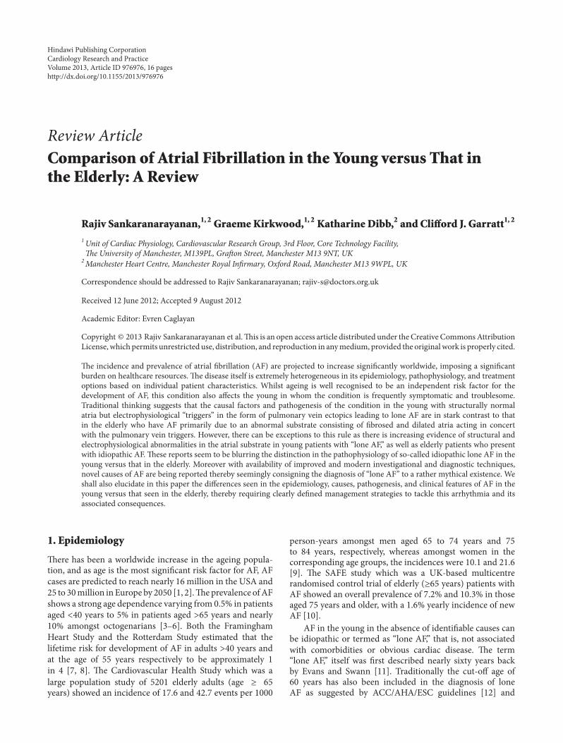
Hindawi Publishing CorporationCardiology Research and PracticeVolume 2013, Article ID 976976, 16 pageshttp://dx.doi.org/10.1155/2013/976976
Review ArticleComparison of Atrial Fibrillation in the Young versus That inthe Elderly: A Review
Rajiv Sankaranarayanan,1, 2 GraemeKirkwood,1, 2 Katharine Dibb,2 and Clifford J. Garratt1, 2
1 Unit of Cardiac Physiology, Cardiovascular Research Group, 3rd Floor, Core Technology Facility,e University of Manchester, M139PL, Graon Street, Manchester M13 9NT, UK
2 Manchester �eart Centre, Manchester Royal �n�r�ary, ��ford Road, Manchester M13 9�PL, UK
Correspondence should be addressed to Rajiv Sankaranarayanan; [email protected]
Received 12 June 2012; Accepted 9 August 2012
Academic Editor: Evren Caglayan
Copyright © 2013 Rajiv Sankaranarayanan et al.is is an open access article distributed under the Creative CommonsAttributionLicense, which permits unrestricted use, distribution, and reproduction in anymedium, provided the originalwork is properly cited.
e incidence and prevalence of atrial �brillation (AF) are projected to increase signi�cantly worldwide, imposing a signi�cantburden on healthcare resources. e disease itself is extremely heterogeneous in its epidemiology, pathophysiology, and treatmentoptions based on individual patient characteristics. Whilst ageing is well recognised to be an independent risk factor for thedevelopment of AF, this condition also affects the young in whom the condition is frequently symptomatic and troublesome.Traditional thinking suggests that the causal factors and pathogenesis of the condition in the young with structurally normalatria but electrophysiological “triggers” in the form of pulmonary vein ectopics leading to lone AF are in stark contrast to thatin the elderly who have AF primarily due to an abnormal substrate consisting of �brosed and dilated atria acting in concertwith the pulmonary vein triggers. However, there can be exceptions to this rule as there is increasing evidence of structural andelectrophysiological abnormalities in the atrial substrate in young patients with “lone AF,” as well as elderly patients who presentwith idiopathic AF. ese reports seem to be blurring the distinction in the pathophysiology of so-called idiopathic lone AF in theyoung versus that in the elderly. Moreover with availability of improved and modern investigational and diagnostic techniques,novel causes of AF are being reported thereby seemingly consigning the diagnosis of “lone AF” to a rather mythical existence. Weshall also elucidate in this paper the differences seen in the epidemiology, causes, pathogenesis, and clinical features of AF in theyoung versus that seen in the elderly, thereby requiring clearly de�ned management strategies to tackle this arrhythmia and itsassociated consequences.
1. Epidemiology
ere has been a worldwide increase in the ageing popula-tion, and as age is the most signi�cant risk factor for AF, AFcases are predicted to reach nearly 16 million in the USA and25 to 30million inEurope by 2050 [1, 2].eprevalence ofAFshows a strong age dependence varying from 0.5% in patientsaged <40 years to 5% in patients aged >65 years and nearly10% amongst octogenarians [3–6]. Both the FraminghamHeart Study and the Rotterdam Study estimated that thelifetime risk for development of AF in adults >40 years andat the age of 55 years respectively to be approximately 1in 4 [7, 8]. e Cardiovascular Health Study which was alarge population study of 5201 elderly adults (age ≥ 65years) showed an incidence of 17.6 and 42.7 events per 1000
person-years amongst men aged 65 to 74 years and 75to 84 years, respectively, whereas amongst women in thecorresponding age groups, the incidences were 10.1 and 21.6[9]. e SAFE study which was a UK-based multicentrerandomised control trial of elderly (≥65 years) patients withAF showed an overall prevalence of 7.2% and 10.3% in thoseaged 75 years and older, with a 1.6% yearly incidence of newAF [10].
AF in the young in the absence of identi�able causes canbe idiopathic or termed as “lone AF,” that is, not associatedwith comorbidities or obvious cardiac disease. e term“lone AF,” itself was �rst described nearly sixty years backby Evans and Swann [11]. Traditionally the cut-off age of60 years has also been included in the diagnosis of loneAF as suggested by ACC/AHA/ESC guidelines [12] and

2 Cardiology Research and Practice
used previously in several landmark epidemiological studiessuch as the Framingham Heart Study [13] and the MayoClinic Study [14]. Old age is indeed one of the strongestrisk factors for AF [6] but terming adults over 60 years as“elderly” seems rather controversial in this modern age ofimproving longevity. e rationale for this “threshold age”therefore has to be questioned [15]. At variance with this age-based de�nition of lone AF, studies such as that by Kopeckyet al. have analysed the outcome of lone AF in patientsaged over 61 years, mean age 74 years (range, 61–97 years)[16]. As newer aetiologies are uncovered and the existenceof “truly lone AF” becomes increasingly controversial [17–19], the prevalence of “lone AF” also seems to be steadily“decreasing” in modern studies. A recent study of 3978 AFpatients from the Euro Heart Survey by Weijs et al. thatexcluded age or le atrial si�e from the de�nition reporteda prevalence of idiopathic AF of 3% out of 3978 patients [20].e mean age of patients with idiopathic AF in this study was58 (SD 14) years and nearly half of these (48%) were olderthan 60 years. Similarly a 30-year follow-up study reportedby theMayo Clinic reported that lone AF constituted only 2%of the total proportion of patients with AF [14]. In contrast,earlier studies showed that lone AF occurs in 1.6% to 30% ofall cases of AF, depending on the de�nition of idiopathic AFor inclusion criteria used [13, 21–23].
2. AF Aetiology in the Young
Lone AF is a diagnosis of exclusion for which any clinicalfeatures of comorbidities or structural cardiac abnormalitiesthat could cause AF must be ruled out. Novel risk factorsfor AF are increasingly being discovered such as geneticcauses, lifestyle factors (such as alcohol consumption, per-sonality traits, and smoking), body mass index, and physicalactivity, thereby all refuting the diagnosis of “lone AF.” Inaddition, it can also be argued that occult cardiac patholo-gies such as hypertension or ischaemic heart disease maywell be diagnosed in these patients if they are investigatedthoroughly.
2.1. Familial. Nearly seven decades ago, Wolff described acase of familial AF in three brothers [24] and since thenstudies have found a positive family history of AF in upto a third of AF patients [25, 26]. Having a positive familyhistory especially in younger patients nearly doubles the riskof developing AF [27]. In 1997, Brugada et al. described the�rst genetic locus on chromosome 10q22-24 in a family withAF segregated as an autosomal dominant trait [28]. In thelast few years, genomewide association studies for AF haveshown SNPs (small nuclear polymorphisms) at three geneticloci—4q25, 16q22, and 1q21 (reviewed in [29]). Monogenicforms of AF have also been described due to mutations ofgenes encoding for potassium channels (KCNQI, KCNE2,KCNJ2, and KCNA5), sodium channel gene SCN5A, K(ATP)gene, the ABCC9 gene, and the connexin 40 gene GJA5 [30–36]. Attempts to further unravel the interplay of genomicsand AF have shown that the genetic basis of AF is bothcomplex and heterogeneous.
2.2. Alcohol. e “holiday heart syndrome” was described in1978 by Ettinger to explain the association between supraven-tricular tachyarrhythmias particularly AF and episodes ofincreased alcohol consumption during weekends and holidaybinge drinking by people without structural heart disease[37]. Overall chronic heavy alcohol consumption (>36 g/day)has been shown to increase risk of AF in several studiesincluding the Framingham cohort (reviewed in [17, 38]).Mechanisms of acute alcohol-induced AF include metabolicacidosis, catecholamine release, and electrolyte disturbanceswhereas chronic overconsumption leads to myocardial �bro-sis, dilatation, and autonomic changes [38]. Binge drinkingis particularly prevalent amongst young people and alcoholhas been identi�ed to potentiate paroxysmal AF in up totwo-thirds of cases [39]. In the elderly, the relationshipbetweenAF and alcohol intake ismore complex however.eCardiovascular Health Study showed an inverse associationbetween alcohol consumption and risk of AF in patientsover 60 years old, with a 4% lower risk for each additionaldrink per week [9]. Other studies have also shown a lackof association between risk of developing AF and moderatealcohol consumption [40, 41].
2.3. Obesity. A meta-analysis by Wanahita et al looking at 16studies that enrolled 123249 individuals (mean age 56 ± 2years) demonstrated a 49% increased risk of developing AFdue to obesity (relative risk 1.49, 95% CI 1.36–1.64) [42].Obesity-associated le ventricular hypertrophy and le atrialdilation are postulated to be important causes which lead toAF [43, 44]. A 3–8% increased risk of developing AF has beenassociated with each unit increase in body mass index [45,46]. Obstructive sleep apnoea which is also associated withobesity has been shown to portend risk of AF developmentin individuals <65 years of age [47]. e association betweenobesity and lone AF is not robust however [19] and in facttaller and leaner adults are reported to be more prone todevelop lone AF [42, 48].
2.4. Sports and Physical Activity. AF has been recognisedto be the commonest cause of palpitations amongst youngathletes [49]. Endurance athletes such as marathon runnershave been shown to have a greater predisposition to developAF when compared to nonathletes [48, 50–52]. Karjalainenet al. diagnosed lone AF in 5.3% out of a cohort of 228male cross-country runners (mean age 47.5 years) [53]. Montet al. reported a four times higher proportion of sportsenthusiasts who had lone AF (aged <65 years) in comparisonto controls in a Catalonian population (63% versus 15%)[51]. e same group reported that >1500 lifetime hoursis the threshold for increased AF propensity [52]. In morethan half of the sportsmen with lone AF, a likely vagalprecipitant was identi�ed (postprandial, postexercise or atrest). Interestingly, in the recent GIRAFA study, cumulativework-related moderate-to-heavy physical activity has alsobeen shown to predict risk of developing lone AF in middleaged men aged <65 years [48]. A meta-analysis of six casecontrol studies, including 655 athletes and 895 controls(predominantlymen), with amean age of 51±9 years showed

Cardiology Research and Practice 3
a signi�cantly higher risk of AF in athletes (OR = 5.29;3.57–7.85; 𝑃𝑃 = 𝑃.𝑃𝑃𝑃𝑃) [54]. Increased le atrial volume wasshown to strongly predict AF in athletes [54]. Mechanismsfor sports-inducedAF postulated include enlarged le atriumand le ventricular mass [17], increased vagal tone leading tobradycardia aswell as shorter atrial refractory period [17] andhypovolemia [55].
2.5. Cardiac Pathologies. ere are a variety of cardiacpathologies associated with AF in the young. ese includehypertrophic cardiomyopathy which confers a four-to sixfoldgreater risk of AF [56]. Prevalence of AF in these patientsis relatively high at about 22% and incidence is 2% annually[56]. Even in this pathology, prevalence increases with ageand is seen predominantly in the elderly (>60 years age)[56]. Congenital Heart Disease is another risk factor for AFand with improved surgical outcomes, increasing numbersof infants and children are surviving into adulthood. Alarge Quebec-based population study of about 38000 adultcongenital heart disease patients, with a median age of 42years, showed a prevalence of atrial arrhythmias of 15.1%(three times greater than that seen in the general population[57]). Wolff-Parkinson-syndrome, myocarditis, pericarditis,and dilated cardiomyopathy are some of the other causesof AF in the young. Valvular heart disease secondary torheumatic fever is also a signi�cant cause of AF in the youngin the developing world.
2.6. Other Risk Factors. Behavioural or emotional triggerssuch as Type A personality [58], stress [58], anger, andhostility in men [59] have also been shown to predispose todevelopment of AF. Other risk factors associated with AFinclude increased coffee and nicotine consumption [17, 58,60].e association with caffeine intake is debatable howeveras a canine study showed inverse association between riskof AF and intravenous caffeine and no causative role wasfound in the Danish diet, cancer, and health study [61, 62].Smoking has been shown to lead to atrial �brosis which iswellrecognised to portend AF [63]. Endocrine causes of AF in theyoung include hyperthyroidism and phaeochromocytoma.
3. AF Aetiology in the Elderly
As shown in the Framingham Heart Study, AF is usuallysecondary to a variety of cardiac pathologies (ischaemic heartdisease, heart failure, and valvular heart disease) as well assystemic disorders (hypertension, diabetes, hyperthyroidism)[6, 64].is is particularly true in the elderly who also have anincreased predisposition of these conditions. However evenaccounting for other comorbidities, ageing is the strongestindependent risk factor that predisposes to AF [6, 65]. eCardiovascular Health Study showed that the prevalence ofAF was 9.1% in the subgroup with clinical cardiovasculardisease, 4.6% in the subgroup with only subclinical cardio-vascular disease, and 1.6% in the absence of clinical or sub-clinical cardiovascular disease (i.e., lone AF) [22]. Indepen-dent risk factors for AF in the elderly included age, treatedsystemic hypertension, congestive cardiac failure, valvular
heart disease, stroke, enlarged le atria size, mitral or aorticvalve dysfunction, echocardiographic features of diastolicdysfunction, and raised serum levels of NT-proBNP [22, 66,67]. Hyperthyroidism is another cause of atrial �brillationin the elderly and one study reports an AF incidence of25% amongst hyperthyroid patients older than 60 years incomparison to 5% in those aged less than 60 years [68].
4. Pathophysiology of Lone AF
AF initiation andmaintenance is the result of pulmonary veinrepetitive activity (triggers), atrial abnormalities (substrate),and remodelling. Lone AF is likely to be initiated andmaintained by the interplay of pulmonary vein ectopics andthe posterior wall of the le atrium [69, 70]. Any substrateabnormalities noted have previously been attributed to AF-induced structural remodelling rather than deemed to havea causative role in initiating lone AF. ere is increasingevidence however of structural as well as electrophysiologicalabnormalities in the substrate (i.e., atrium) as well in loneAF. For instance, atrial biopsies in such patients have shown�brotic changes and even other occult pathologies suchas myocarditis [71, 72]. More recently, electrophysiologicalstudies in patients with a history of paroxysmal lone AFremote from the arrhythmia have also shown abnormali-ties strongly suggestive of dysfunctional atrial conductioninvolved in AF initiation and progression [73]. ese includelarger atrial volumes, longer effective refractory period,longer conduction time along linear catheters, longer bi-atrial activation time, slower conduction velocity, largerproportion of fractionated electrograms, longer correctedsinus node recover time, and lower mean atrial voltage[73]. Another study of early onset lone AF patients (ageless than forty years old) showed a signi�cant difference inP wave morphology thereby implying abnormal interatrialconduction [74]. ese �ndings seem to implicate a causativerole for altered atrial electrophysiology in initiating loneAF. However as AF-induced electrical remodelling is wellrecognised to start early within a few hours of the onset of AF[75], there remains the possibility that these changes could besecondary to very early remodelling. For instance, previousexperiments in a goat model of paroxysmal AF by Garratt etal. showed that it takes between 5 days to 4 weeks to developchanges in atrial substrate which then self-perpetuate AF [76,77]. A variety of studies have also implicated in�ammation inhaving an aetiological role in the initiation and perpetuationof atrial �brillation due to evidence of raised in�ammatorymarkers such as interleukin-6 and C-reactive protein in AFpatients [78, 79]. In view of the multiple confounding factorsand comorbidities associated with in�ammation, the �ury isstill out whether it is a cause or consequence of AF [80].Structural remodelling as a consequence of AF can causecalcium overload and atrial myocyte apoptosis leading toan in�ammatory response [81]. In lone AF patients, therehave been contradictory results such as from a large studyby Ellinor et al. that showed no difference in high sensitiveCRP [82] but others have noted high levels of CRP and highsensitive CRP in lone AF patients [83, 84]. Signi�cantly, how-ever, Aviles et al. included patients with hypertension in their

4 Cardiology Research and Practice
de�nition of lone AF albeit with overtly structurally normalhearts. is could have been a signi�cant confounding factoras hypertension itself is associated with in�ammation [83].Myocardial perfusion imaging in patients with lone recurrentAF has shown isolated perfusion abnormalities indicative ofmicrovascular dysfunction [85]. Echocardiography has alsoshown evidence of le ventricular diastolic dysfunction inpatients thought to have idiopathic AF [86].
5. Electrophysiological and StructuralAlterations in Aged Atria
5.1. Initiation of AF in the Aged Atria. A number of studies(summarised in Table 1) have attempted to unravel the atrialelectrophysiological characteristics of aged atria in relationto AF. Whilst most animal and human studies have shownthat aged atria have an increased propensity to develop AF[87–93], some studies in elderly AF patients have yieldedcon�icting results although these could have been in�uencedby underlying pathology or treatment in patients [94–96].Increased atrial ERP has been noted in these studies whichcould be sufficient to overcome any other arrhythmogenicremodelling. ere is therefore a need to understand, inthe absence of underlying disease, how AF is initiated andmaintained in the aged atria.
5.2. Initiation of AF. Why the aged atria are more susceptibleto the development of AF in the absence of other risk factorsremains poorly understood. We shall describe below howageing may increase the propensity of both (i) triggeredactivity in the form of DADs and (ii) reentrant circuits.
5.3. Delayed aer Depolarizations (DADs) in the Aged Atria.Potential contributory factors that may predispose to DADformation and indeed the increased incidence of DADshave been demonstrated in some models of ageing [90, 93].Wongcharoen et al. observed DADs of greater amplitude inaged rabbit LA pulmonary vein sleeve tissue sections associ-ated with an increase in NCX protein which would providean additional means to facilitate triggered activity as moredepolarising current would �ow during any spontaneousCa2+ release and therefore, all things being equal, a smallerspontaneous release would produce a bigger DAD [90]. Someother studies have also described evidence of atrial calciummishandling in AF (Table 1) [97]. is has been attributedto protein kinase A-induced hyperphosphorylation leadingto dysfunctional ryanodine receptor [98]. While decreasedSERCA and increased RyR protein expression give us cluesto SR function, it would also be of interest to understandhow SR Ca2+ content responds to age in the pulmonary vein.One study using human atrial tissue from elderly patients(mean age 68 years) has suggested that hyperphosphorylationof phospholamban could be contributory to leaky ryanodinereceptors and thus abnormal calcium handling in chronic AFpatients [99]. Clearly there is a pressing need to understandhow Ca2+ homeostasis is achieved in the aged atria and howit is subsequently remodelled in the aged atria in AF.
5.4. Reentrant Circuits in the Aged Atria. A reentrant sub-strate can result fromaltered functional (electrical) propertiesor structural changes to the atrium and these are discussedbelow with reference to ageing.
5.4.1. Effective Refractory Period (ERP). Whilst con�ictingresults have been noted when analysing the effects of ageingon effective refractory period in both human [95, 96, 100,101] as well as animal right atria [92, 102, 103], review ofvarious studies indicates that the right atrial ERP is prolongedwith ageing [104]. Inconsistency in the literature may relateto variation in anatomical sites studied [105] and, in terms ofpatient studies, the effects of underlying disease, arrhythmias,drug treatment, or a general lack of studies including veryelderly patients as highlighted by Dun and Boyden [104].efew studies investigating action potential duration (APD) orERP in aged le atrium also show con�icting results [106,107]. Furthermore in rabbit pulmonary vein sleeves and leatrial posterior wall APD was prolonged with age [90, 91].Electrical characteristics and age-associated remodelling ofthe atrium appear to be region speci�c [93] and this mayunderlie differences between the above studies. Interestinglywhile the prolonged action potential in aged rabbit le atrialposterior wall may be expected to be anti-arrhythmogenic,this gave rise to an increase in APD dispersion (discussedbelow) which was suggested to potentiate AF [91].
Many ion channels have yet to be studied in the agedatria and work mainly performed in the dog has shown thatthere is no age-related change in sodium current density[108]. However 𝐼𝐼Ca-L is depressed, which would be expectedto depress the plateau of the action potential [92], and sincerepolarising currents activatemore strongly at positive poten-tials, this may slow activation and prolong repolarisation,thus prolonging ERP.Of the repolarising currents only 𝐼𝐼to hasbeen assessed and peak as well as sustained 𝐼𝐼to was increasedwith age in the right atrium [109] but not the le atrium[104]. Indirect evidence suggests 𝐼𝐼KACH may be enhanced inthe right atrium with age [103]; however there are no studiesinvestigating age-associated changes in 𝐼𝐼K1, 𝐼𝐼Kur, 𝐼𝐼Kr and 𝐼𝐼Ks,in either atria.
5.4.2. Conduction Velocity. A number of studies havereported age-associated general or directional conductionslowing and resultant spiral-reentrant waves in the right atriamainly in tissue strips but also in vivo in various species[100, 111]. In the aged dog, conduction of normal beats wasunaltered but premature impulses were slowed suggesting acertain amount of depolarizing current is needed to over-come conduction discontinuities in age [92]. By calculatingconduction in the direction of the wavefront, Kojodjojo et al.have shown in human atria that increasing age is associatedwith decreased propagation velocity in both atria duringsinus rhythm and also during pacing [107]. In this respectthe reduction of 𝐼𝐼Na noted in AF [112, 113] is potentiallysigni�cant as it will slow conduction velocity and reduce theexcitation wavelength. Based on a very limited number ofstudies the effects of ageing on peak 𝐼𝐼Na are inconsistent,showing either no change at low stimulation frequencies but

Cardiology Research and Practice 5
T 1: Studies investigating relationship between atrial electrophysiological changes and ageing.
Authors Species Characteristics Key �ndingsBrembilla-Perrot et al.[96] Human Patients aged >70 years versus younger Decreased AF inducibility due to increased
atrial ERP
Centurión et al.[89] Human Patients with paroxysmal AF during sinus
rhythm aged >60 years versus younger
Greater mean number of abnormal rightatrial electrograms de�ned as ≥100msecduration and, or showing eight fragmentedde�ections
Roberts-omsonet al. [87] Human Patients aged >60 years versus younger Greater number of complex fractionated
electrograms
Sakabe et al. [94] Human Patients without a history of AF orstructural heart disease
No relationship between age andinducibility of AF
Calcium mishandling
El-Armouche et al.[99] Human
Western blotting used to assessphosphorylation levels of Ca handling
proteins in right atrial appendage
Hyperphosphorylation of phospholambancould be contributory to leaky ryanodinereceptors and thus abnormal calciumhandling in chronic AF patients
Hove-Madsen et al.[97] Human Age > 66 years
Higher calcium spark frequency and higherincidence of spontaneous calcium waves incomparison to patients with sinus rhythm
Ono et al. [88] Rats Old versus young rats
Glycolytic inhibition has been shown toresult in spontaneous AF due to calciummishandling and early aerdepolarisation-induced triggered activity
Wongcharoen et al.[90] Rabbits
Responses of pulmonary vein tissues torapamycin, FK-506, and ouabain in young
and aged rabbits
Increased pulmonary vein arrhythmogenesissecondary to ryanodine receptordysfunction-resultant calcium mis-handling
Atrial ERP
Kistler et al. [100] HumanElectrophysiological and electroanatomicalstudies in 3 age groups (≥60 years, 31–59
years, and ≤30 years)
Age-associated electrical and structuralremodeling (regional conduction slowing,increase in atrial ERP, impaired sinus nodefunction, conduction delay at cristaterminalis, and areas of low voltage)
Brembilla-Perot et al.[96] Human 734 patients (age 16–85 years, mean 61 ± 15
years)
Increased atrial ERP and age >70 yearsindependently predicted reduced AFinducibility
Brorson and Olsson[101] Human Right atrial monophasic action potentials
recorded in 40 healthy males No age correlation
Anyukhovsky et al.[92] Dogs Young versus old canine atrial
Age-related differences in action potentialcontour, decreased 𝐼𝐼CaL, and slowerconduction of early premature beats
Huang et al. [106] Rats Adult, middle aged versus aged rats
Age-associated prolongation of themonophasic action potential (mAP) andERP in the right atrium, but a decrease inmAP and ERP in the le atrium, suggestinga potential reentrant mechanism for AF
Kojodjojo et al. [107] Humans
Most study subjects suffered fromatrioventricular reentrant arrhythmias,syncope, or palpitations and hence these
atria were not “healthy”
No change in le atrial ERP with ageing
Michelucci et al. [105] Humans 17 normal subjects (age range 17–78 years) Age-related increase in right atrial ERP
Su et al. [103] Rats Adults versus aged ratsIn response to muscarinic stimulation,ageing-related prolongation of atrialmaximum diastolic potential but not of APD
Toda [102] Rabbit Rabbit ages varied from 2–360 days old Age-related prolongation of APD

6 Cardiology Research and Practice
T 1: Continued.
Authors Species Characteristics Key �ndingsIon channel remodelling in ageing and AF
𝐼𝐼CaL Anyukhovskyet al. [92] Canine atria Reduced 𝐼𝐼CaL
𝐼𝐼Na Baba et al. [108] Canine atria
(i) Peak current unchanged at lowstimulation frequencies but reduced atstimulation frequencies relevant to AF
Wu et al. [110] Rabbit atria (ii) Decreased in hyperlipidemic agedrabbits
𝐼𝐼to Dun et al. [109] Canine atrium Increased in the le atrium𝐼𝐼KAch Su et al. [103] Rat Indirect evidence of increase [104]
reduced at stimulation frequencies relevant to AF (10Hz)[108] or a decrease [110].
Changes in connexin expression (especially Cx40 andCx43) have been noted in AF-related remodelling [114] andare also noted in ageing [115]. A single study has shown anage-associated decrease in connexin 43 in the sinoatrial nodebut unaltered expression of connexins in the right atria [115].However a change in the distribution of connexins away fromthe lateral cell edges to the intercalated discs has also beennoted in ageing [116]. is is potentially signi�cant, as itwill result in anisotropic propagation of excitation and theformation of reentrant circuits [114, 117].
Increased or heterogeneous �brosis, oen associated withadvancing age [92, 111], can disrupt the coupling betweenindividual myocytes and result in non homogenous conduc-tion or conduction slowing which can lead to re-entry (forreview see [118]). Recent work has demonstrated increasedAF stability in long-term AF in goats due to “micro�brosis”separatingmyocyte bundles [119]. Conduction abnormalitiescan occur following redistribution without altered expression[120].
5.4.3. Dispersion of Conduction Velocity and ERP. ERP dis-persion and conduction heterogeneity correlate with AFinducibility and both have been shown to increase withageing [91, 93, 105].
In summary, what induces and sustains AF in the youngmay be different to that in the old. Anyukhovsky et al. showedin old dogs that atrial tissuewas depolarised,with longerAPDand a slower max upstroke and greater variability in APD. Inchronic AF both young and old, the atrial cell membrane washyperpolarised, with slowed upstroke and decreased APD.But chronic AF led to an increase of APD dispersion inadults and a decrease in old dogs. us, AF was sustainedin two different substrates: one with short AP duration andwith expanded heterogeneity of AP parameters (adult) andone with short AP duration but limited heterogeneity (old).ese data also suggest that the increased dispersion in atrialelectrophysiology that occurs in adults may be an importantadditional contributory factor for AF stabilization at this age,while the occurrence of �brosis and slowed conduction ofpremature beats that has been demonstrated previously maybemore contributory in the old. Table 2 summarises the atrial
electrophysiological differences between the elderly versusthe young in relationship to the propensity to develop AF.
Histological changes in healthy elderly patients with AFinclude increased deposition of collagen, adipose tissue andamyloid, atrophy and vacuolar myocyte degeneration and�brofatty substitution of the sinoatrial node (reviewed in[118]). Aging-related oxidative damage has been shown toportend atrial �brillation through mitochondrial bioener-getic dysfunction [121]. Imaging studies have also shown age-related dilatation of the pulmonary veins and the atrium,thereby potentiating pulmonary vein triggers as well assubstrate-induced AFmaintenance throughmechanoelectricfeedback [122, 123].
6. Clinical Features
e clinical presentation of AF varies signi�cantly dependingon age and comorbidities. In the young, the initial presenta-tion is usually with paroxysmal AF [124]; persistent AF underthe age of 50 is oen associated with identi�able causes likestructural heart disease, hyperthyroidism, or alcohol excess.Whilst the incidence of both paroxysmal and persistent AFincreases dramatically over the age of 60, there is a dispropor-tionate increase in chronic forms [125], with the result that80% of newly diagnosedAF in octogenarians is of a persistentor permanent form, even in the absence of structural heartdisease [126]. Moreover, advanced age is a risk factor forearly recurrence aer �rst AF presentation and of rapidprogression from paroxysmal to persistent AF [125, 127].
AF is classically associated with “typical” symptoms ofirregular palpitations, with or without chest pain, breathless-ness, or dizziness. Palpitations are reported in 80% of youngpatients with paroxysmal AF. [128]. In contrast, less than 10%of AF patients over the age of 80 years have palpitations [129]and up to 40% of elderly hospital inpatients found to have AFare entirely asymptomatic [130]. Whilst atypical chest pain isrelatively common in young AF patients, in elderly patientsanginal chest pain during AF episodes strongly suggests thepresence of signi�cant concurrent coronary disease [131]and might be sufficient to warrant investigation for coronaryischaemia even in the absence of typical symptoms of angina.
AF in elderly patients is frequently diagnosed coinciden-tally during general health assessment, hospital admission for

Cardiology Research and Practice 7
T 2: Electrophysiological differences between the elderly and young that can predispose to AF (summarised from human and animalstudies in Table 1).
Features Elderly YoungImpulse initiation
(i) Sinus node functionImpaired (leading to longer sinus node
recovery times), contributing toabnormal impulse initiation
Generally preserved
(ii) Pulmonary vein ectopic activity
Also contributes to AF pathogenesisalthough substrate abnormalities have a
dominant role in initiation andmaintenance
Predominant trigger for AF initiation
Impulse conduction(i) P wave morphology and duration(usually signifying interatrialconduction)
Abnormal P wave morphology andprolonged interatrial conduction Usually normal
(ii) Wavefront propagation
Abnormalities noted such as conductionslowing (particularly of prematureimpulses) thereby contributing to
reentrant waves
Usually normal
Substrate abnormalities(i) Complex fractionated atrialelectrograms Greater number Lesser than in elderly
(ii) Atrial refractoriness—effectiveRefractory Period (ERP)
ERP prolonged in the right atrium andcould contribute to dispersion in
refractorinessUsually not prolonged
(iii) Action potential duration (APD) Prolonged in the right atrium Generally within normal limits
(iv) Regional atrial voltage differences Larger atrial volumes with more numberof low voltage areas
Atria usually of normal size and meanvoltage within normal limits
nonrelated illnesses, or as a result of its complications [132]. Arecent randomized controlled study in primary care suggeststhat implementing targeted opportunistic screening of over65-year olds, based on a simple annual pulse assessment, islikely to be cost-effective in improving AF detection [10].
7. Management of AF
7.1. General Principles. e management of AF is concernedwith two main aspects; symptom relief (through rate orrhythm control) and prevention of complications. Althoughcertain complications, such as le ventricular dysfunction,may be reduced by these therapies, the prevention ofthromboembolic events requires targeted and appropriateantithrombotic therapy. AF management including strokeprevention is dependent onmultiple factors including patientage, comorbidities, and disease pro�le.
7.2. Stroke Risk, Bleeding Complications, and Anticoagulation.AF is associated with a 5-fold increase in the risk of strokes,and strokes due to AF are associated with higher mortalityand worse functional outcome [133]. Age has a particularlydramatic impact on the risk of AF-associated stroke: betweenthe ages of 50–59 the average lifetime risk is 5% and 3.9% formen and women, respectively, and this rises exponentiallyto 22.3% and 23.9% between 80 and 84 [134]. Vitamin Kantagonists (usually warfarin) or other oral anticoagulants
(see below) reduce the risk of strokes by around 60–70%,albeit at the risk of intracranial haemorrhage and otherbleeding-related complications [135, 136].
AF in the context of mitral valve stenosis, or a pros-thetic mitral valve, is considered to be extremely high risk,and anticoagulation is mandatory in the absence of clearcontraindications [136]. For nonvalvular AF, the CHADS2score was introduced in 2001 as a simple scoring systemto assess the stroke risk (see Table 3) [137]. However, thein�uence of age is underestimated by the CHADS2 system;recent studies have indicated that, amongst moderate riskpatients with a CHADS2 score of 1, individuals over theage of 75 with isolated AF are at a higher risk of strokethan are younger patients with a single additional riskfactor [138, 139]. erefore a more comprehensive scoringsystem called CHA(2)DS(2)VASc has been recommendedin recent guidelines (see Table 4) [140]. By recognizinga spectrum of major and minor risk factors that warranttreatment with anticoagulation, this system more accuratelyidenti�es individuals at truly low risk and extends the useof anticoagulation into the previous medium risk category.Notably, using the CHA(2)DS(2)VASc, anticoagulation isconsidered to be potentially bene�cial for all patients agedover 65 years, whilst the decision in younger patients dependson additional risk factors.
Although most AF patients are elderly, until recentlythese patients have been somewhat underrepresented inclinical trials of anticoagulation. e clinical bene�t from

8 Cardiology Research and Practice
T 3: CHADS2 scoring system [137].
Comorbidity ScoreC Congestive heart failure 1H Hypertension 1A Age ≥ 75 years 1D Diabetes mellitus 1S2 Stroke or TIA 2CHADS2 score 0; annual stroke risk 1.9%, >1; annual stroke risk 2.8 = 18.2%.
anticoagulation not only persists but potentially increaseswith advanced age; for example, the recent ATRIA studydemonstrated that AF patients aged over 75 years bene�tmost from warfarin treatment in terms of absolute and rela-tive reduction in stroke rate [141]. Despite this clear evidenceof bene�t, anticoagulation is considerably underutilised inthis age group, typically with only one-third of over 85sreceiving appropriate therapy despite an absence of de�nitecontraindications [142].
In physician studies, the most commonly reported con-cerns with anticoagulation are perceived bleeding risk and ahistory of falls [143]. Elderly patients certainly do have anincreased risk of bleeding complications [144], and whilstall forms of bleed have a potential associated mortality andmorbidity, the main concern is intracranial haemorrhage.Although this complication is relatively rare even in theelderly (<1% per year in the BAFTA trial of patients over 75years old), nevertheless it constitutes 90% of anticoagulation-related deaths [145]. Careful monitoring and control of anti-coagulation can improve this balance; the risk of intracranialhaemorrhage is only modestly increased with a therapeuticINR between 2-3 but rises sixfold when the INR rises above3.5. Conversely, targeting a subtherapeutic INR of <1.8results in a sevenfold reduction in stroke prevention withouta reduction in intracranial haemorrhage risk (thus equatingto a loss of clinical effectiveness and worse outcomes). “Real-world” studies of anticoagulation in octogenarians, reportthat the risk of major bleeding (either fatal or requiringtransfusion) is 13.1 per 100 patient-years [146]. Whilst thisis higher than the rate of 4.7 per 100 patient years seen inyounger patients, the highest risk of bleeding was seen inpatients with a high CHADS2 score of ≥3. us patients whoare at highest risk of bleeding are also those who potentiallybene�t themost in terms of stroke reduction, and the absoluterisk reduction in stroke-related mortality exceeds the riskincrease of fatal bleeds. e perceived risk of anticoagulationin patients with a history of falls is probably exaggerated;whilst there is a slight excess of nonintracranial bleeds inthese individuals, a meta-analysis has calculated that a fallrate of at least 300 per year would be required to negate thestroke-prevention bene�t [147].
Recently, guidelines have suggested the use of scoringsystems such as HASBLED (see Table 5) in order to betterassess bleeding risk prior to commencing anticoagulation. Ascore of ≥3 is considered to represent high risk of bleeding,and caution and careful monitoring recommended [148]. Inview of the complexity of managing anticoagulation in theelderly, there has also been substantial interest in alternatives
to oral vitamin K antagonists. However, it is clear that aspirinis signi�cantly less effective than warfarin at preventingstrokes, and there appears to be no net bene�t over theage of 77 [139]. Recent consensus therefore advises againstthe use of aspirin for AF thromboprophylaxis [149]. AsCHA(2)DS(2)�ASc allows identi�cation of patients with anexceedingly low stroke risk, aspirin is now considered to be anonpreferred alternative for young patients with a single riskfactor.
Recently there has been a surge of new anticoagulantalternatives to warfarin. ese do not require regular mon-itoring of clotting pro�le. Dabigatran is a newly approvedoral direct thrombin inhibitor. e RE-LY trial reported thathigh dose Dabigatran (150mg b.d.) was more efficaciousthan warfarin with a similar risk of bleeding complications(intracranial and extracranial), whilst low dose Dabigatran(110mg b.d.) was noninferior to warfarin with a reducedrisk of bleeding [150]. ese effects were consistent amongstyoung and elderly patient subgroups, although there wasa slight excess of gastrointestinal bleeds in elderly patientstaking higher dose [151]. is drug could therefore provideeither improved stroke prevention when used in high dosesin young patients or reduced bleeding risk at low doses inthe elderly. Rivaroxaban (a factor Xa inhibitor) is anothernew anticoagulant. e ROCKET-AF trial reported non-inferiority to warfarin in nonvalvular AF, with no signi�cantincrease in major bleeding and a lesser incidence of intracra-nial and fatal bleeding in comparison to warfarin [152]. Ofparticular relevance to the elderly, the median age in thistrial was 73 years (25% were ≥78 years). Apixaban, anotherfactor Xa inhibitor, was reported last year in the Aristotle trial(median age 70 years) to be more efficacious than warfarinand also caused less bleeding [153].
7.3. Rate or Rhythm Control? ere are two main strategiesof management in AF rate control and rhythm control witheither pharmacological or nonpharmacological options.
Pharmacological strategies of rate and rhythm controlhave been compared in multiple studies. Despite clear the-oretical bene�ts of sinus rhythm in the form of improvedatrioventricular dynamics and an improvement in cardiacoutput, there is no signi�cant bene�t of a rhythm controlstrategy in large randomised population studies. Althoughthere have been few investigations exploring the strategiesspeci�cally in different age groups, results of �ve studiesare relevant to a consideration of the elderly and have beenincluded in a meta-analysis [154]; this demonstrates a cleartrend towards increased mortality associated with a rhythmcontrol strategy in the elderly. Interestingly, the excess deathsdo not appear to relate to proarrhythmic effects or speci�cside effects of the trial drugs, but rather to other factorssuch as malignancies or lung disease. is suggests that inelderly frail patients, the use of powerful antiarrhythmic withcomplex drug interactions may uncover latent comorbiditiesin a form of nonspeci�c pharmacotoxicity.
us, current guidelines clearly favour a rate-controlstrategy in the elderly. Initial therapies might include eitherbeta blockers or calcium channel blockers, with digoxin

Cardiology Research and Practice 9
T 4: CHADS2VAS2C scoring system [140].
Comorbidity PointsC Congestive heart failure (or le ventricular systolic dysfunction) 1H Hypertension: blood pressure consistently above 140/90mmHg (or treated hypertension on medication) 1A2 Age ≥ 75 years 2D Diabetes mellitus 1S2 Prior stroke or TIA or thromboembolism 2V Vascular disease (e.g., peripheral artery disease, myocardial infarction, aortic plaque) 1A Age 65–74 years 1Sc Sex category (i.e., female gender) 1
T 5: HASBLED scoring system [148].
Hypertension (BP > 160 without control) 1Renal disease (dialysis, transplant, Cr > 2.6mg/dL or >200𝜇𝜇mol/L) 1Liver disease (cirrhosis, bilirubin > 2x normal, AST/ALT/AP > 3x normal) 1Stroke 1Predisposition to bleeding or previous major bleed 1Labile INR (unstable/high INRs, <60% time in therapeutic range) 1Age ≥ 65 1Medications (antiplatelets or NSAIDS) 1Alcohol excess 1
Risk of spontaneous major bleeding (episodes per 100 patient years)Score 0-1 1.02–1.13Score 2-3 1.88–3.74Score ≥ 4 ≥8.7
added as appropriate where a second agent is necessary.Whilst previous guidelines emphasised the importance oftight control of ventricular rate (resting heart rate below80 bpm), it has recently been demonstrated that equivalentoutcomes are achieved with a less stringent target of 115 bpm[155]. In elderly patients, where issues such as coexistingconduction system disease, polypharmacology, and renalimpairment are common, a more relaxed approach to ratecontrol is likely to reduce the complexity of management,including the need for pacemaker implantation [156].
In patients with drug-refractory symptoms due to per-manent AF, pacemaker implantation with atrioventricularnode ablation results in symptomatic bene�t for up to 83%of patients including those over the age of 70 [157]. Manyelderly patients with AF have conduction system disease andpacemaker implantation is oen indicated for bradycardicindications; AV node ablation in these patients also allowscessation of rate control medications. Moreover, AV nodeablation and cardiac resynchronization therapy should alsobe appropriate where AF coexists with heart failure, witha recent meta-analysis demonstrating signi�cant mortalitybene�t over pharmacological rate control [158].
Although large clinical studies do not clearly favour eithera rate or rhythm control strategy, there are factors that suggestthat certain subgroups may bene�t from a rhythm controlstrategy. As older patients with comorbidities demonstratean excess mortality due to drug-related side effects, so it isarguable that young and otherwise healthy patients should
not experience these side effects and therefore experiencethe full bene�ts from restoration of sinus rhythm, includingimproved ventricular function, quality of life, and reducedprogression to permanent AF. ere is some evidence fromstudies focusing on younger patients suggesting this to bethe case, and a convincing case that where a rhythm controlstrategy is effective, it is associated with reduced symptomsand improved mortality compared with rate control [159],although the data as yet do not suggest that anticoagulationcan safely be withdrawn.
Factors associated with rapid progression of AF(advanced age and structural heart disease) are alsoassociated with a poor response to rhythm control strategy.Recently, the HATCH scoring system (hypertension, ageolder than 75 years, previous transient ischemic attack orstroke, chronic obstructive pulmonary disease, and heartfailure) has been proposed to identify patients at risk ofprogression to persistent AF [127]; a HATCH score of ≥2was associated with a high risk of disease progression despiteantiarrhythmic therapy.
Where a rhythm control strategy is considered appro-priate, treatment choices are guided by side effects andcontraindications rather than expected efficacy. Where thereis no underlying structural heart disease, appropriate �rstline options include dronedarone, �ecainide, propafenone,and sotalol [160]. Although amiodarone is a more effica-cious antiarrhythmic [161], it is associated with signi�cantpulmonary, liver and thyroid toxicity with a cumulative and

10 Cardiology Research and Practice
T 6: Common differences between AF in the young versus elderly.
AF in the young AF in elderly patients
Causes
(i) Idiopathic (i) Ischaemic heart disease(ii) Genetic (ii) Heart failure
(iii) Alcohol, smoking (iii) Valvular heart disease(iv) Personality traits (iv) Hypertension(v) Body mass index (v) Cardiomyopathies(vi) Endurance sports (vi) Hyperthyroidism
(vii) Cardiac pathologies(vii) Secondary causes such as postoperative, infection, pulmonary
embolism(viii) Endocrine disorders (viii) Idiopathic
PathogenesisTriggers/pulmonary vein Repetitive
activity +++Substrate/atrial abnormalities +
Pulmonary vein repetitive activity ++Atrial Abnormalities +++
Clinical features Usually typical symptoms Atypical symptoms or asymptomatic
ManagementRhythm control preferred
romboprophylaxis usually not requiredunless based on CHADS2VASC
Rate control preferredromboprophylaxis usually required
unless contraindicated
dose-dependent risk; its long-term use in the young thereforerequires very cautious consideration.
Dronedarone has recently emerged as an anti-arrhythmictherapy for paroxysmal AF. It appears to be somewhat lessefficacious than amiodarone, but associated with less sideeffects [162]. e recent ATHENA [163] study suggestedthat dronedarone may impart a bene�t in terms of reducedmortality and hospital admission compared to placebo incertain high risk patient groups, but data showing bene�t overother anti-arrhythmic therapies are lacking. Importantly, itis also contra-indicated in patients with permanent AF ordecompensated heart failure.
In recent years, the major advance in rhythm manage-ment has been in nonpharmacological therapy, namely, leatrial ablation. Since the �rst report by Haissaguerre et al.that pulmonary vein isolation reduced AF recurrence byeliminating spontaneous focal discharges from pulmonaryveins that initiated AF, there have been signi�cant advancesin ablation techniques [70]. is treatment strategy is par-ticularly efficacious in patients with lone AF and thus theEuropean Society of Cardiology guidelines recommend leatrial ablation (Class IIa recommendation) for symptomaticpatients with paroxysmal or persistent AF who have failedto respond to trial of anti-arrhythmic medication [160].Whilst complications are reported in up to 5% of cases,multiple studies have demonstrated freedom fromAF leadingto signi�cant improvements in symptoms and quality oflife [164, 165]. Results in paroxysmal AF demonstrate thatpulmonary vein isolation gives satisfactory symptom controland freedom from AF in around 70 to 80% patients; however,persistent AF generally requires more complicated proce-dures involving multiple lesions throughout the le atrium(success rates of 65–75%) [166]. Several studies have alsodemonstrated that catheter ablation is more effective thananti-arrhythmic therapy [167–169] at controlling symptoms
as well as restoring sinus rhythm, although there is no �rmevidence of bene�t in terms ofmortality or stroke prevention.Most studies have included patients only under the age of 65,although there have been some small studies suggesting thatthe procedures can be performed safely in older patients withstructurally normal hearts [170].
8. Conclusion
To summarise, AF is a heterogeneous condition, with signif-icant differences in its epidemiology, pathogenesis, clinicalpresentation and management across age groups (shown inTable 6). Older patients are more likely to have an abnormalsubstrate and present at an advanced stage with atypicalsymptoms and associated comorbidities. Whilst there havebeen a few reports of substrate abnormalities in youngpatients with idiopathic AF thereby implying a causative role,there is not yet conclusive evidence that these do not simplyrepresent AF-induced atrial remodeling. With increasingrecognition of rare aetiologies of what was previously deemedto be “idiopathic AF” and the archaic cut-off age to de�ne“lone AF,” the term “lone AF” itself is becoming increasinglyobsolete. It is all the more pressing therefore to exclude anyoccult risk factors for AF as this could in�uence prognosisand management. e important differences between AF inthe young and that in the elderly necessitate clearly de�neddiagnostic and targeted management strategies to relievesymptoms as well as to prevent complications.
Abbreviations
AF: Atrial �brillationAPD: Action potential durationERP: Effective refractory period.

Cardiology Research and Practice 11
Acknowledgment
e authors are grateful to Professor Andrew Trafford (Car-diovascular Research Group, University of Manchester) forhis helpful suggestions.
Funding
Dr. Sankaranarayanan’s research is funded by a grant from theBritish Heart Foundation (Grant Reference FS/11/15/28693).Dr. Kirkwood’s research is funded by a grant from the BritishHeart Foundation (Grant Reference FS/10/71/28563).
References
[1] Y. Miyasaka, M. E. Barnes, B. J. Gersh et al., “Secular trends inincidence of atrial �brillation in �lmsted County, Minnesota,1980 to 2000, and implications on the projections for futureprevalence,” Circulation, vol. 114, no. 2, pp. 119–125, 2006.
[2] H. Stefansdottir, T. Aspelund, V. Gudnason et al., “Trends inthe incidence and prevalence of atrial �brillation in Icelandand future projections,” Europace, vol. 13, no. 8, pp. 1110–1117,2011.
[3] H. Rutzen-Lopez, V. Khanna, and M. R. Reynolds, “Atrial�brillation: epidemiology, prognosis and therapy,” MinervaMedica, vol. 102, no. 3, pp. 187–207, 2011.
[4] K. Abusada, S. B. Sharma, R. Jaldai et al., “Epidemiology andmanagement of new-onset atrial �brillation,” American Journalof Managed Care, vol. 10, supplement, pp. S50–S57, 2004.
[5] A. S. Go, E. M. Hylek, K. A. Phillips et al., “Prevalence ofdiagnosed atrial �brillation in adults: national implicationsfor rhythm management and stroke prevention: the anticoag-ulation and risk factors in atrial �brillation (ATRIA) study,”Journal of the American Medical Association, vol. 285, no. 18,pp. 2370–2375, 2001.
[6] E. J. Benjamin, D. Levy, S. M. Vaziri, R. B. D’Agostino, A. J.Belanger, and P. A. Wolf, “Independent risk factors for atrial�brillation in a population-based cohort. e FraminghamHeart Study,” Journal of the American Medical Association, vol.271, no. 11, pp. 840–844, 1994.
[7] D. M. Lloyd-Jones, T. J. Wang, E. P. Leip et al., “Lifetime risk fordevelopment of atrial �brillation: the framingham heart study,”Circulation, vol. 110, no. 9, pp. 1042–1046, 2004.
[8] J. Heeringa, D. A. M. Van Der Kuip, A. Hofman et al.,“Prevalence, incidence and lifetime risk of atrial �brillation: theRotterdam study,” European Heart Journal, vol. 27, no. 8, pp.949–953, 2006.
[9] B. M. Psaty, T. A. Manolio, L. H. Kuller et al., “Incidence of andrisk factors for atrial �brillation in older adults,”Circulation, vol.96, no. 7, pp. 2455–2461, 1997.
[10] F. D. R. Hobbs, D. A. Fitzmaurice, J. Mant et al., “A ran-domised controlled trial and cost-effectiveness study of sys-tematic screening (targeted and total population screening)versus routine practice for the detection of atrial �brillation inpeople aged 65 and over. e SAFE study,” Health TechnologyAssessment, vol. 9, no. 40, pp. 3–5, 2005.
[11] W. Evans and P. Swann, “Lone auricular �brillation,” BritishHeart Journal, vol. 16, pp. 189–194, 1954.
[12] V. Fuster, L. E. Ryden, D. S. Cannom et al., “ACC/AHA/ESC2006 Guidelines for the Management of Patients with Atrial
Fibrillation: a report of the American College of Cardiol-ogy/AmericanHeart Association Task Force on Practice Guide-lines and the European Society of Cardiology Committeefor Practice Guidelines (Writing Committee to Revise the2001 Guidelines for the Management of Patients With AtrialFibrillation): developed in collaboration with the EuropeanHeart Rhythm Association and the Heart Rhythm Society,”Circulation, vol. 114, no. 7, pp. e257–e354.
[13] F. N. Brand, R. D. Abbott, W. B. Kannel, and P. A. Wolf,“Characteristics and prognosis of lone atrial �brillation. 30 yearfollow-up in the Framingham study,” Journal of the AmericanMedical Association, vol. 254, no. 24, pp. 3449–3453, 1985.
[14] A. Jahangir, V. Lee, P. A. Friedman et al., “Long-term pro-gression and outcomes with aging in patients with lone atrial�brillation: a 30-year follow-up study,” Circulation, vol. 115, no.24, pp. 3050–3056, 2007.
[15] L. Frost, “Lone atrial �brillation: good, bad, or ugly?” Circula-tion, vol. 115, no. 24, pp. 3040–3041, 2007.
[16] S. L. Kopecky, B. J. Gersh, M. D. McGoon et al., “Lone atrial�brillation in elderly persons: a marker for cardiovascular risk,”Archives of Internal Medicine, vol. 159, no. 10, pp. 1118–1122,1999.
[17] B. A. Schoonderwoerd,M. D. Smit, L. Pen, and I. C. VanGelder,“New risk factors for atrial �brillation: causes of ’not-so-loneatrial �brillation’,” Europace, vol. 10, no. 6, pp. 668–673, 2008.
[18] D. G. Wyse, “Idiopathic atrial �brillation: a rose by any othername?” Europace, vol. 14, no. 2, pp. 151–152, 2012.
[19] D. Kozlowski, S. Budrejko, G. Y. H. Lip et al., “Lone atrial�brillation: what do we know?” Heart, vol. 96, no. 7, pp.498–503, 2010.
[20] B. Weijs, R. Pisters, R. Nieuwlaat et al., “Idiopathic atrial�brillation revisited in a large longitudinal clinical cohort,”Europace, vol. 14, no. 2, pp. 184–190, 2012.
[21] S. L. Kopecky, B. J. Gersh, and M. D. McGoon, “e naturalhistory of lone atrial �brillation. A population-based study overthree decades,” New England Journal of Medicine, vol. 317, no.11, pp. 669–674, 1987.
[22] C. D. Furberg, B. M. Psaty, T. A. Manolio et al., “Prevalence ofatrial �brillation in elderly subjects (the Cardiovascular HealthStudy),” American Journal of Cardiology, vol. 74, no. 3, pp.236–241, 1994.
[23] S. Lévy, M. Maarek, P. Coumel et al., “Characterization ofdifferent subsets of atrial �brillation in general practice inFrance: the ALFA study,” Circulation, vol. 99, no. 23, pp.3028–3035, 1999.
[24] L. Wolff, “Familial auricular �brillation,” e New EnglandJournal of Medicine, vol. 229, pp. 396–398, 1943.
[25] D. Darbar, K. J. Herron, J. D. Ballew et al., “Familial atrial�brillation is a genetically heterogeneous disorder,” Journalof the American College of Cardiology, vol. 41, no. 12, pp.2185–2192, 2003.
[26] P. T. Ellinor, D. M. Yoerger, J. N. Ruskin, and C. A. MacRae,“Familial aggregation in lone atrial �brillation,” Human Genet-ics, vol. 118, no. 2, pp. 179–184, 2005.
[27] C. S. Fox, H. Parise, R. B. D’Agostino et al., “Parental atrial�brillation as a risk factor for atrial �brillation in offspring,”Journal of the American Medical Association, vol. 291, no. 23,pp. 2851–2855, 2004.
[28] R. Brugada, T. Tapscott, G. Z. Czernuszewicz et al., “Identi-�cation of a genetic locus for familial atrial �brillation,” NewEngland Journal of Medicine, vol. 336, no. 13, pp. 905–911, 1997.

12 Cardiology Research and Practice
[29] M. F. Sinner, P. T. Ellinor, T. Meitinger, E. J. Benjamin, andS. K��b, “Genome-wide association studies of atrial �brillation:past, present, and future,” Cardiovascular Research, vol. 89, no.4, pp. 701–709, 2011.
[30] Y. H. Chen, S. J. Xu, S. Bendahhou et al., “KCNQ1 gain-of-function mutation in familial atrial �brillation,” Science, vol.299, no. 5604, pp. 251–254, 2003.
[31] Y. Yang, M. Xia, Q. Jin et al., “Identi�cation of a KCNE2 gain-of-functionmutation in patients with familial atrial �brillation,”American Journal ofHumanGenetics, vol. 75, no. 5, pp. 899–905,2004.
[32] M. Xia, Q. Jin, S. Bendahhou et al., “A Kir2.1 gain-of-functionmutation underlies familial atrial �brillation,” Biochemical andBiophysical Research Communications, vol. 332, no. 4, pp.1012–1019, 2005.
[33] T. M. Olson, A. E. Alekseev, X. K. Liu et al., “Kv1.5 channelopa-thy due to KCNA5 loss-of-function mutation causes humanatrial �brillation,” Human Molecular Genetics, vol. 15, no. 14,pp. 2185–2191, 2006.
[34] L. Y. Chen, J. D. Ballew, K. J. Herron, R. J. Rodeheffer, and T. M.Olson, “A common polymorphism in SCN5A is associated withlone atrial �brillation,” Clinical Pharmacology and erapeutics,vol. 81, no. 1, pp. 35–41, 2007.
[35] M.H.Gollob, D. L. Jones, A.D. Krahn et al., “Somaticmutationsin the conne�in 40 gene �GJA5� in atrial �brillation,” NewEngland Journal of Medicine, vol. 354, no. 25, pp. 2677–2688,2006.
[36] T. M. Olson, A. E. Alekseev, C. Moreau et al., “KATP channelmutation confers risk for vein of Marshall adrenergic atrial�brillation,” Nature Clinical Practice Cardiovascular Medicine,vol. 4, no. 2, pp. 110–116, 2007.
[37] P. O. Ettinger, C. F. Wu, C. De La Cruz Jr., A. B. Weisse, S. S.Ahmed, and T. J. Regan, “Arrhythmias and the ’holiday heart’:alcohol associated cardiac rhythm disorders,” American HeartJournal, vol. 95, no. 5, pp. 555–562, 1978.
[38] C. E. B. Balbão, A. A. V. de Paola, and G. Fenelon, “Effectsof alcohol on atrial �brillation: myths and truths,” erapeuticAdvances in Cardiovascular Disease, vol. 3, no. 1, pp. 53–63,2009.
[39] S. R. Lowenstein, P. A. Gabow, and J. Cramer, “e role ofalcohol in new-onset atrial �brillation,” Archives of InternalMedicine, vol. 143, no. 10, pp. 1882–1885, 1983.
[40] K. J. Mukamal, B. M. Psaty, P. M. Rautaharju et al., “Alcoholconsumption and risk and prognosis of atrial �brillation amongolder adults: the Cardiovascular Health Study,” American HeartJournal, vol. 153, no. 2, pp. 260–266, 2007.
[41] G. M. Marcus, L. M. Smith, D. Whiteman et al., “Alcohol intakeis signi�cantly associated with atrial �utter in patients under 60years of age and a shorter right atrial effective refractory period,”Pacing andClinical Electrophysiology, vol. 31, no. 3, pp. 266–272,2008.
[42] N. Wanahita, F. H. Messerli, S. Bangalore, A. S. Gami, V.K. Somers, and J. S. Steinberg, “Atrial �brillation and obe-sity—results of a meta-analysis,” American Heart Journal, vol.155, no. 2, pp. 310–315, 2008.
[43] T. J. Wang, H. Parise, D. Levy et al., “Obesity and the risk ofnew-onset atrial �brillation,” Journal of the American MedicalAssociation, vol. 292, no. 20, pp. 2471–2477, 2004.
[44] C. S. Ku, S. L. Lin, D. J. Wang, S. K. Chang, and W. J. Lee,“Le ventricular �lling in young normotensive obese adults,”American Journal of Cardiology, vol. 73, no. 8, pp. 613–615,1994.
[45] L. Frost, L. J. Hune, and P. Vestergaard, “Overweight and obesityas risk factors for atrial �brillation or �utter: the Danish Diet,Cancer, and Health Study,” American Journal of Medicine, vol.118, no. 5, pp. 489–495, 2005.
[46] S. Dublin, B. French, N. L. Glazer et al., “Risk of new-onset atrial�brillation in relation to body mass inde�,” Archives of InternalMedicine, vol. 166, no. 21, pp. 2322–2328, 2006.
[47] A. S. Gami, D. O. Hodge, R. M. Herges et al., “Obstructive sleepapnea, obesity, and the risk of incident atrial �brillation,” Journalof theAmericanCollege of Cardiology, vol. 49, no. 5, pp. 565–571,2007.
[48] L. Mont, D. Tamborero, R. Elosua et al., “Physical activity,height, and le atrial size are independent risk factors for loneatrial �brillation in middle-aged healthy individualsEuropace,”vol. 10, no. 1, pp. 15–20, 2008.
[49] F. Furlanello, A. Bertoldi, M. Dallago et al., “Atrial �brillation inelite athletes,” Journal of Cardiovascular Electrophysiology, vol.9, no. 8, supplement, pp. S63–S68, 1998.
[50] L. Molina, L. Mont, J. Marrugat et al., “Long-term endurancesport practice increases the incidence of lone atrial �brillationinmen: a follow-up study,” Europace, vol. 10, no. 5, pp. 618–623,2008.
[51] L. Mont, A. Sambola, J. Brugada et al., “Long-lasting sportpractice and lone atrial �brillation,” European Heart Journal,vol. 23, no. 6, pp. 477–482, 2002.
[52] R. Elosua, A. Arquer, L. Mont et al., “Sport practice and therisk of lone atrial �brillation: a case-control study,” InternationalJournal of Cardiology, vol. 108, no. 3, pp. 332–337, 2006.
[53] J. Karjalainen, U.M. Kujala, J. Kaprio, S. Sarna, andM. Viitasalo,“Lone atrial �brillation in vigorously e�ercising middle agedmen: case-control study,” British Medical Journal, vol. 316, no.7147, pp. 1784–1785, 1998.
[54] J. Abdulla and J. R. Nielsen, “Is the risk of atrial �brillationhigher in athletes than in the general population? A system-atic review and meta-analysis,” Europace, vol. 11, no. 9, pp.1156–1159, 2009.
[55] J. D. Edwards and R. G. Wilkins, “Atrial �brillation precipitatedby acute hypovolaemia,” British Medical Journal, vol. 294, no.6567, pp. 283–284, 1987.
[56] I. Olivotto, F. Cecchi, S. A. Casey, A. Dolara, J. H. Traverse, andB. J. Maron, “Impact of atrial �brillation on the clinical courseof hypertrophic cardiomyopathy,” Circulation, vol. 104, no. 21,pp. 2517–2524, 2001.
[57] J. Bouchardy, J. errien, L. Pilote et al., “Atrial arrhythmias inadults with congenital heart disease,” Circulation, vol. 120, no.17, pp. 1679–1686, 2009.
[58] A. V. Mattioli, S. Bonatti, M. Zennaro, and G. Mattioli, “erelationship between personality, socio-economic factors, acutelife stress and the development, spontaneous conversion andrecurrences of acute lone atrial �brillation,” Europace, vol. 7, no.3, pp. 211–220, 2005.
[59] E. D. Eaker, L. M. Sullivan, M. Kelly-Hayes, R. B. D’Agostino,and E. J. Benjamin, “Anger and hostility predict the develop-ment of atrial �brillation in men in the framingham offspringstudy,” Circulation, vol. 109, no. 10, pp. 1267–1271, 2004.
[60] A. V. Mattioli, S. Bonatti, M. Zennaro, R. Melotti, and G.Mattioli, “Effect of coffee consumption, lifestyle and acutelife stress in the development of acute lone atrial �brillation,”Journal of Cardiovascular Medicine, vol. 9, no. 8, pp. 794–798,2008.

Cardiology Research and Practice 13
[61] A. Rashid, M. Hines, B. J. Scherlag, W. S. Yamanashi, and W.Lovallo, “e effects of caffeine on the inducibility of atrial�brillation,” Journal of Electrocardiology, vol. 39, no. 4, pp.421–425, 2006.
[62] L. Frost and P. Vestergaard, “Caffeine and risk of atrial �b-rillation or �utter: the Danish diet, cancer, and health study,”American Journal of Clinical Nutrition, vol. 81, no. 3, pp.578–582, 2005.
[63] A. Goette, U. Lendeckel, A. Kuchenbecker et al., “Cigarettesmoking induces atrial �brosis in humans via nicotine,” Heart,vol. 93, no. 9, pp. 1056–1063, 2007.
[64] K.M. Ryder and E. J. Benjamin, “Epidemiology and signi�canceof atrial �brillation,”American Journal of Cardiology, vol. 84, no.9, pp. R131–R138, 1999.
[65] R. B. Schnabel, L. M. Sullivan, D. Levy et al., “Development ofa risk score for atrial �brillation �Framingham Heart Study�: acommunity-based cohort study,” e Lancet, vol. 373, no. 9665,pp. 739–745, 2009.
[66] M. A. Rosenberg, J. S. Gottdiener, S. R. Heckbert et al.,“Echocardiographic diastolic parameters and risk of atrial�brillation: the Cardiovascular Health Study,” European HeartJournal, vol. 33, no. 7, pp. 904–912, 2012.
[67] K. K. Patton, P. T. Ellinor, S. R. Heckbert et al., “N-Terminalpro-b-type natriuretic peptide is a major predictor of thedevelopment of atrial �brillation: the cardiovascular healthstudy,” Circulation, vol. 120, no. 18, pp. 1768–1774, 2009.
[68] T. Agner, T. Almdal, B. orsteinsson, and E. Agner, “Areevaluation of atrial �brillation in thyrotoxicosis,” Danishmedical bulletin, vol. 31, no. 2, pp. 157–159, 1984.
[69] M. Haissaguerre, P. Jais, D. C. Shah et al., “Spontaneousinitiation of atrial �brillation by ectopic beats originating in thepulmonary veins1998,” e New England Journal of Medicine,vol. 339, no. 10, pp. 659–666.
[70] P. Jaïs, M. Hocini, L. Macle et al., “Distinctive electrophysio-logical properties of pulmonary veins in patients with atrial�brillation,” Circulation, vol. 106, no. 19, pp. 2479–2485, 2002.
[71] A. Frustaci, C. Chimenti, F. Bellocci, E. Morgante, M. A. Russo,and A. Maseri, “Histological substrate of atrial biopsies inpatients with lone atrial �brillation,” Circulation, vol. 96, no. 4,pp. 1180–1184, 1997.
[72] A. Frustaci, M. Caldarulo, A. Buffon, F. Bellocci, R. Fenici,and D. Melina, “Cardiac biopsy in patients with ’primary’ atrial�brillation� Histologic evidence of occult myocardial diseases,”Chest, vol. 100, no. 2, pp. 303–306, 1991.
[73] M. K. Stiles, B. John, C. X. Wong et al., “Paroxysmal loneatrial �brillation is associated with an abnormal atrial substrate:characterizing the ’second factor’,” Journal of the AmericanCollege of Cardiology, vol. 53, no. 14, pp. 1182–1191, 2009.
[74] F. Holmqvist, M. S. Olesen, A. Tveit et al., “Abnormal atrial acti-vation in young patients with lone atrial �brillation,” Europace,vol. 13, no. 2, pp. 188–192, 2011.
[75] P. Korantzopoulos, T. Kolettis, K. Siogas, and J. Goudevenos,“Atrial �brillation and electrical remodeling: the potential roleof in�ammation and oxidative stress,” Medical Science Monitor,vol. 9, no. 9, pp. RA225–RA229, 2003.
[76] C. J. Garratt, M. Duytschaever, M. Killian, R. Dorland, F.Mast, and M. A. Allessie, “Repetitive electrical remodelingby paroxysms of atrial �brillation in the goat: no cumulativeeffect on inducibility or stability of atrial �brillation,” Journal ofCardiovascular Electrophysiology, vol. 10, no. 8, pp. 1101–1108,1999.
[77] D. M. Todd, S. P. Fynn, A. P. Walden, W. J. Hobbs, S. Arya,and C. J. Garratt, “Repetitive 4-week periods of atrial electricalremodeling promote stability of atrial �brillation: time courseof a second factor involved in the self-perpetuation of atrial�brillation,” Circulation, vol. 109, no. 11, pp. 1434–1439, 2004.
[78] T. T. Issac, H. Dokainish, and N. M. Lakkis, “Role of in�am-mation in initiation and perpetuation of atrial �brillation: asystematic reviewof the published data,” Journal of theAmericanCollege of Cardiology, vol. 50, no. 21, pp. 2021–2028, 2007.
[79] D. S. G. Conway, P. Buggins, E. Hughes, and G. Y. H. Lip,“Prognostic signi�cance of raised plasma levels of interleukin-6 and C-reactive protein in atrial �brillation,” American HeartJournal, vol. 148, no. 3, pp. 462–466, 2004.
[80] C. J. Boos and G. Y. H. Lip, “C-reactive protein and loneatrial �brillation: potential confounders,” American Journal ofCardiology, vol. 98, no. 7, pp. 991–992, 2006.
[81] M. J. Mihm, F. Yu, C. A. Carnes et al., “Impaired myo�brillarenergetics and oxidative injury during human atrial �brillation,”Circulation, vol. 104, no. 2, pp. 174–180, 2001.
[82] P. T. Ellinor, A. Low, K. K. Patton,M.A. Shea, andC. A.MacRae,“C-reactive protein in lone atrial �brillation,” American Journalof Cardiology, vol. 97, no. 9, pp. 1346–1350, 2006.
[83] R. J. Aviles, D. O. Martin, C. pperson-Hansen et al., “In�amma-tion as a risk factor for atrial �brillation,” Circulation, vol. 108,no. 24, pp. 3006–3010, 2003.
[84] E. Hatzinikolaou-Kotsakou, D. Tziakas, A. Hotidis et al., “Rela-tion of C-reactive protein to the �rst onset and the recurrencerate in lone atrial �brillation,” American Journal of Cardiology,vol. 97, no. 5, pp. 659–661, 2006.
[85] E. I. Skalidis, M. I. Hamilos, I. K. Karalis, G. Chlouverakis, G.E. Kochiadakis, and P. E. Vardas, “Isolated atrial microvasculardysfunction in patients with lone recurrent atrial �brillation,”Journal of the American College of Cardiology, vol. 51, no. 21,pp. 2053–2057, 2008.
[86] P. Jais, J. T. Peng, D. C. Shah et al., “Le ventricular diastolicdysfunction in patients with so-called lone atrial �brillation,”Journal of Cardiovascular Electrophysiology, vol. 11, no. 6, pp.623–625, 2000.
[87] K. C. Roberts-omson, P. M. Kistler, P. Sanders et al., “Frac-tionated atrial electrograms during sinus rhythm: relationshipto age, voltage, and conduction velocity,” Heart Rhythm, vol. 6,no. 5, pp. 587–591, 2009.
[88] N. Ono, H. Hayashi, A. Kawase et al., “Spontaneous atrial�brillation initiated by triggered activity near the pulmonaryveins in aged rats subjected to glycolytic inhibition,” AmericanJournal of Physiology. Heart and Circulatory Physiology, vol. 292,no. 1, pp. H639–H648, 2007.
[89] O. A. Centurión, S. Isomoto, A. Shimizu et al., “e effectsof aging on atrial endocardial electrograms in patients withparoxysmal atrial �brillation,” Clinical Cardiology, vol. 26, no.9, pp. 435–438, 2003.
[90] W. Wongcharoen, Y. C. Chen, Y. J. Chen et al., “Agingincreases pulmonary veins arrhythmogenesis and susceptibilityto calcium regulation agents,” Heart Rhythm, vol. 4, no. 10, pp.1338–1349, 2007.
[91] W. Wongcharoen, Y. C. Chen, Y. J. Chen, C. I. Lin, and S. A.Chen, “Effects of aging and ouabain on le atrial arrhythmo-genicity,” Journal of Cardiovascular Electrophysiology, vol. 18,no. 5, pp. 526–531, 2007.
[92] E. P. Anyukhovsky, E. A. Sosunov, A. Plotnikov et al., “Cellularelectrophysiologic properties of old canine atria provide a

14 Cardiology Research and Practice
substrate for arrhythmogenesis,” Cardiovascular Research, vol.54, no. 2, pp. 462–469, 2002.
[93] E. P. Anyukhovsky, E. A. Sosunov, P. Chandra et al., “Age-associated changes in electrophysiologic remodeling: a poten-tial contributor to initiation of atrial �brillation,” Cardiovascu-lar Research, vol. 66, no. 2, pp. 353–363, 2005.
[94] K. Sakabe, N. Fukuda, T. Nada et al., “Age-related changes in theelectrophysiologic properties of the atrium in patients with nohistory of atrial �brillation,” Japanese Heart Journal, vol. 44, no.3, pp. 385–393, 2003.
[95] K. Sakabe, N. Fukuda, T. Soeki et al., “Relation of age and sex toatrial electrophysiological properties in patients with no historyof atrial �brillation,” Pacing and Clinical Electrophysiology, vol.26, no. 5, pp. 1238–1244, 2003.
[96] B. Brembilla-Perrot, G. Burger, D. Beurrier et al., “�n�uenceof age on atrial �brillation inducibility,” Pacing and ClinicalElectrophysiology, vol. 27, no. 3, pp. 287–292, 2004.
[97] L. Hove-Madsen, A. Llach, A. Bayes-Gen�s et al., “Atrial �bril-lation is associated with increased spontaneous calcium releasefrom the sarcoplasmic reticulum in human atrial myocytes,”Circulation, vol. 110, no. 11, pp. 1358–1363, 2004.
[98] J. A. Vest, X. H. T. Wehrens, S. R. Reiken et al., “Defectivecardiac ryanodine receptor regulation during atrial �brillation,”Circulation, vol. 111, no. 16, pp. 2025–2032, 2005.
[99] A. El-Armouche, P. Boknik, T. Eschenhagen et al., “Moleculardeterminants of altered Ca2+ handling in human chronic atrial�brillation,” Circulation, vol. 114, no. 7, pp. 670–680, 2006.
[100] P.M.Kistler, P. Sanders, S. P. Fynn et al., “Electrophysiologic andelectroanatomic changes in the human atrium associated withage,” Journal of the American College of Cardiology, vol. 44, no.1, pp. 109–116, 2004.
[101] L. Brorson and S. B. Olsson, “Right atrial monophasic actionpotential in healthy males. Studies during spontaneous sinusrhythm and atrial pacing,” Acta Medica Scandinavica, vol. 199,no. 6, pp. 433–446, 1976.
[102] N. Toda, “Age-related changes in the transmembrane potentialof isolated rabbit sino-atrial nodes and atria,” CardiovascularResearch, vol. 14, no. 1, pp. 58–63, 1980.
[103] N. Su, J. Duan, M. P. Moffat, and N. Narayanan, “Agerelated changes in electrophysiological responses to muscarinicreceptor stimulation in rat myocardium,” Canadian Journal ofPhysiology and Pharmacology, vol. 73, no. 10, pp. 1430–1436,1995.
[104] W. Dun and P. A. Boyden, “Aged atria: electrical remodelingconducive to atrial �brillation,” Journal of Interventional Car-diac Electrophysiology, vol. 25, no. 1, pp. 9–18, 2009.
[105] A. Michelucci, L. Padeletti, and G. A. Fradella, “Ageing andatrial electrophysiologic properties in man,” International Jour-nal of Cardiology, vol. 5, no. 1, pp. 75–81, 1984.
[106] C.Huang,W.Ding, L. Li, andD.Zhao, “Differences in the aging-associated trends of the monophasic action potential durationand effective refractory period of the right and le atria of therat,” Circulation Journal, vol. 70, no. 3, pp. 352–357, 2006.
[107] P. Kojodjojo, P. Kanagaratnam, V. Markides, D. W. Davies, andN. Peters, “Age-related changes in human le and right atrialconduction,” Journal of Cardiovascular Electrophysiology, vol.17, no. 2, pp. 120–127, 2006.
[108] S. Baba, W. Dun, M. Hirose, and P. A. Boyden, “Sodium currentfunction in adult and aged canine atrial cells,” American Journalof Physiology. Heart and Circulatory Physiology, vol. 291, no. 2,pp. H756–H761, 2006.
[109] W. Dun, T. Yagi, M. R. Rosen, and P. A. Boyden, “Calcium andpotassium currents in cells from adult and aged canine rightatria,”Cardiovascular Research, vol. 58, no. 3, pp. 526–534, 2003.
[110] C. C. Wu, M. J. Su, J. F. Chi, M. H. Wu, and Y. T. Lee,“Comparison of aging and hypercholesterolemic effects on thesodium inward currents in cardiac myocytes,” Life Sciences, vol.61, no. 16, pp. 1539–1551, 1997.
[111] H. Hayashi, C. Wang, Y. Miyauchi et al., “Aging-related increaseto inducible atrial �brillation in the rat model,” Journal ofCardiovascular Electrophysiology, vol. 13, no. 8, pp. 801–808,2002.
[112] S. Sossalla, B. Kallmeyer, S. Wagner et al., “Altered Na+ currentsin atrial �brillation. effects of ranolazine on arrhythmias andcontractility in human atrial myocardium,” Journal of theAmerican College of Cardiology, vol. 55, no. 21, pp. 2330–2342,2010.
[113] R. Gaspo, R. F. Bosch, E. Bou-Abboud, and S. Nattel,“Tachycardia-induced changes in Na+ current in a chronic dogmodel of atrial �brillation,” Circulation Research, vol. 81, no. 6,pp. 1045–1052, 1997.
[114] P. Kanagaratnam, A. Cherian, R. D. L. Stanbridge, B. Glenville,N. J. Severs, and N. S. Peters, “Relationship between connexinsand atrial activation during human atrial �brillation,” Journalof Cardiovascular Electrophysiology, vol. 15, no. 2, pp. 206–213,2004.
[115] S. A. Jones, M. K. Lancaster, and M. R. Boyett, “Ageing-relatedchanges of connexins and conduction within the sinoatrialnode,” Journal of Physiology, vol. 560, no. 2, pp. 429–437, 2004.
[116] T. Koura, M. Hara, S. Takeuchi et al., “Anisotropic conductionproperties in canine atria analyzed by high-resolution opticalmapping: preferential direction of conduction block changesfrom longitudinal to transverse with increasing age,” Circula-tion, vol. 105, no. 17, pp. 2092–2098, 2002.
[117] M. S. Spach, J. F. Heidlage, P. C. Dolber, and R. C. Barr,“Mechanism of origin of conduction disturbances in aginghuman atrial bundles: experimental and model study,” HeartRhythm, vol. 4, no. 2, pp. 175–185, 2007.
[118] C. Chimenti, M. A. Russo, A. Carpi, and A. Frustaci, “Histo-logical substrate of human atrial �brillation,” Biomedicine andPharmacotherapy, vol. 64, no. 3, pp. 177–183, 2010.
[119] S. Verheule, E. Tuyls, A. Van Hunnik, M. Kuiper, U. Schotten,and M. Allessie, “Fibrillatory conduction in the atrial freewalls of goats in persistent and permanent atrial �brillation,”Circulation: Arrhythmia and Electrophysiology, vol. 3, no. 6, pp.590–599, 2010.
[120] M. S. Spach and P. C. Dolber, “Relating extracellular potentialsand their derivatives to anisotropic propagation at a micro-scopic level in human cardiac muscle: evidence for electricaluncoupling of side-to-side �ber connections with increasingage,” Circulation Research, vol. 58, no. 3, pp. 356–371, 1986.
[121] P. H. Lin, S. H. Lee, C. P. Su, and Y. H. Wei, “Oxidative damageto mitochondrial DNA in atrial muscle of patients with atrial�brillation,” Free Radical Biology and Medicine, vol. 35, no. 10,pp. 1310–1318, 2003.
[122] N. H. Pan, H. M. Tsao, N. Chang, Y. J. Chen, and S. A. Chen,“Aging dilates atrium and pulmonary veins: implications for thegenesis of atrial �brillation,” Chest, vol. 133, no. 1, pp. 190–196,2008.
[123] S. A. Nazir and M. J. Lab, “Mechanoelectric feedback and atrialarrhythmias,” Cardiovascular Research, vol. 32, no. 1, pp. 52–61,1996.

Cardiology Research and Practice 15
[124] A. Ruigómez, S. Johansson, M. A. Wallander, and L. A. GarcíaRodríguez, “Predictors and prognosis of paroxysmal atrial�brillation in general practice in the UK,” BMC CardiovascularDisorders, vol. 5, article no. 20, 2005.
[125] C. R. Kerr, K. H. Humphries, M. Talajic et al., “Progression tochronic atrial �brillation a�er the initial diagnosis of parox-ysmal atrial �brillation: results from the Canadian Registry ofAtrial Fibrillation,” American Heart Journal, vol. 149, no. 3, pp.489–496, 2005.
[126] S. A. Lopez, F. Formiga, X. Bosch et al., “Prevalence of atrial�brillation and related factors in hospitalized old patients:ESFINGE study,” Medicina Clínica, vol. 138, no. 6, pp. 231–237,2012.
[127] C. B. de Vos, R. Pisters, R. Nieuwlaat et al., “Progression fromparoxysmal to persistent atrial �brillation. clinical correlatesand prognosis,” Journal of the American College of Cardiology,vol. 55, no. 8, pp. 725–731, 2010.
[128] C. Kerr, J. Boone, S. Connolly et al., “Follow-up of atrial�brillation: the initial experience of the Canadian Registryof Atrial Fibrillation,” European Heart Journal, vol. 17, no. 7,supplement, pp. 48–51, 1996.
[129] N. D. Brunetti, L. De Gennaro, P. L. Pellegrino, G. Dellegrot-taglie, G. Antonelli, and M. Di Biase, “Atrial �brillation withsymptoms other than palpitations: incremental diagnostic sen-sitivity with at-home tele-cardiology assessment for emergencymedical service,” European Journal of Preventive Cardiology, vol.19, no. 3, pp. 306–313, 2011.
[130] U. Lotze, J. Liebetrau, I. Malsch et al., “Medical treatmentof patients with atrial �brillation aged over 80 years in dailyclinical practice: in�uence of age and CHADS2 score,” Archivesof Gerontology and Geriatrics, vol. 50, no. 1, pp. 36–41, 2010.
[131] S. Kralev, K. Schneider, S. Lang et al., “Incidence and severityof coronary artery disease in patients with atrial �brillationundergoing �rst-time coronary angiography,” PLoS One, vol. 6,no. 9, Article ID e24964, 2011.
[132] J. Liao, Z. Khalid, C. Scallan, C. Morillo, and M. O’Donnell,“Noninvasive cardiac monitoring for detecting paroxysmalatrial �brillation or �utter a�er acute ischemic stroke: a system-atic review,” Stroke, vol. 38, no. 11, pp. 2935–2940, 2007.
[133] K. Lakshminarayan, C. A. Solid, A. J. Collins, D. C. Anderson,and C. A. Herzog, “Atrial �brillation and stroke in the generalmedicare population: a 10-year perspective (1992 to 2002),”Stroke, vol. 37, no. 8, pp. 1969–1974, 2006.
[134] P. A. Wolf, R. D. Abbott, and W. B. Kannel, “Atrial �brillation asan independent risk factor for stroke: the Framingham Study,”Stroke, vol. 22, no. 8, pp. 983–988, 1991.
[135] R. G. Hart, O. Benavente, R. McBride, and L. A. Pearce,“Antithrombotic therapy to prevent stroke in patients withatrial �brillation: a meta-analysis,” Annals of Internal Medicine,vol. 131, no. 7, pp. 492–501, 1999.
[136] D. N. Salem, P. D. Stein, A. Al-Ahmad et al., “Antithrombotictherapy in valvular heart disease—native and prosthetic: the7th ACCP Conference on Antithrombotic and rombolyticerapy,” Chest, vol. 126, no. 3, supplement, pp. 457S–482S,2004.
[137] B. F. Gage, A. D. Waterman, W. Shannon, M. Boechler, M.W. Rich, and M. J. Radford, “Validation of clinical classi�ca-tion schemes for predicting stroke: results from the NationalRegistry of Atrial Fibrillation,” Journal of the American MedicalAssociation, vol. 285, no. 22, pp. 2864–2870, 2001.
[138] M. Hughes and G. Y. H. Lip, “Stroke and thromboembolism inatrial �brillation: a systematic review of stroke risk factors, risk
strati�cation schema and cost effectiveness data,” rombosisand Haemostasis, vol. 99, no. 2, pp. 295–304, 2008.
[139] Stroke in AF Working Group, “Independent predictors ofstroke in patients with atrial �brillation: a systematic review,”Neurology, vol. 69, no. 6, pp. 546–554, 2007.
[140] G. Y. H. Lip, R. Nieuwlaat, R. Pisters, D. A. Lane, and H. J.G. M. Crijns, “Re�ning clinical risk strati�cation for predictingstroke and thromboembolism in atrial �brillation using a novelrisk factor-based approach: the Euro Heart Survey on atrial�brillation,” Chest, vol. 137, no. 2, pp. 263–272, 2010.
[141] D. E. Singer, Y. Chang, M. C. Fang et al., “e net clinicalbene�t of warfarin anticoagulation in atrial �brillation,” Annalsof Internal Medicine, vol. 151, no. 5, pp. 297–305, 2009.
[142] A. S. Go, E. M. Hylek, L. H. Borowsky, K. A. Phillips, J. V. Selby,and D. E. Singer, “Warfarin use among ambulatory patientswith nonvalvular atrial �brillation: the anticoagulation and riskfactors in atrial �brillation (ATRIA) study,” Annals of InternalMedicine, vol. 131, no. 12, pp. 927–934, 1999.
[143] D. Pugh, J. Pugh, and G. E. Mead, “Attitudes of physiciansregarding anticoagulation for atrial �brillation: a systematicreview,” Age Ageing, vol. 40, no. 6, pp. 675–683, 2011.
[144] R. Marinigh, G. Y. H. Lip, N. Fiotti, C. Giansante, and D.A. Lane, “Age as a risk factor for stroke in atrial �brillationpatients: implications for thromboprophylaxis,” Journal of theAmerican College of Cardiology, vol. 56, no. 11, pp. 827–837,2010.
[145] M. C. Fang, A. S. Go, Y. Chang et al., “Death and disabilityfrom warfarin-associated intracranial and extracranial hem-orrhages,” American Journal of Medicine, vol. 120, no. 8, pp.700–705, 2007.
[146] E. M. Hylek, C. Evans-Molina, C. Shea, L. E. Henault, andS. Regan, “Major hemorrhage and tolerability of warfarin inthe �rst year of therapy among elderly patients with atrial�brillation,” Circulation, vol. 115, no. 21, pp. 2689–2696, 2007.
[147] M. Man-Son-Hing, G. Nichol, A. Lau, and A. Laupacis,“Choosing antithrombotic therapy for elderly patients withatrial �brillation who are at risk for falls,” Archives of InternalMedicine, vol. 159, no. 7, pp. 677–685, 1999.
[148] R. Pisters, D. A. Lane, R. Nieuwlaat, C. B. De Vos, H. J. G. M.Crijns, and G. Y. H. Lip, “A novel user-friendly score (HAS-BLED) to assess 1-year risk of major bleeding in patients withatrial �brillation: the Euro heart survey,” Chest, vol. 138, no. 5,pp. 1093–1100, 2010.
[149] D. J. Stott, R. I. Dewar, C. J. Garratt et al., “RCPE UK consensusconference on ’Approaching the comprehensivemanagement ofatrial �brillation: evolution or revolution�’,” e Journal of theRoyal College of Physicians of Edinburgh, vol. 42, supplement 18,pp. 3–4.
[150] S. J. Connolly, M. D. Ezekowitz, S. Yusuf et al., “Dabigatranversus warfarin in patients with atrial �brillation,” e NewEngland Journal of Medicine, vol. 361, no. 12, pp. 1139–1151,2009.
[151] J. W. Eikelboom, L. Wallentin, S. J. Connolly et al., “Risk ofbleeding with 2 doses of dabigatran compared with warfarin inolder and younger patients with atrial �brillation: an analysis ofthe randomized evaluation of long-term anticoagulant therapy(RE-LY) Trial,” Circulation, vol. 123, no. 21, pp. 2363–2372,2011.
[152] M. R. Patel, K. W. Mahaffey, J. Garg et al., “Rivaroxaban versuswarfarin in nonvalvular atrial �brillation,” e New EnglandJournal of Medicine, vol. 365, no. 10, pp. 883–891, 2011.

16 Cardiology Research and Practice
[153] C. B. Granger, J. H. Alexander, J. J. McMurray et al., “Apixabanversus warfarin in patients with atrial �brillation,” e NewEngland Journal of Medicine, vol. 365, no. 11, pp. 981–992, 2011.
[154] D. G. Wyse, “Rate control vs rhythm control strategies in atrial�brillation,” Progress in Cardiovascular Diseases, vol. 48, no. 2,pp. 125–138, 2005.
[155] I. C. Van Gelder, H. F. Groenveld, H. J. G. M. Crijns etal., “Lenient versus strict rate control in patients with atrial�brillation,” New England Journal of Medicine, vol. 362, no. 15,pp. 1363–1373, 2010.
[156] I. C. Van Gelder, D. G. Wyse, M. L. Chandler et al., “Doesintensity of rate-control in�uence outcome in atrial �brillation�An analysis of pooled data from the RACE and AFFIRMstudies,” Europace, vol. 8, no. 11, pp. 935–942, 2006.
[157] M. Brignole, G. L. Botto, L. Mont et al., “Predictors of clinicalefficacy of ’Ablate and Pace’ therapy in patients with permanentatrial �brillation,” Heart, vol. 98, no. 4, pp. 297–302, 2012.
[158] A. N. Ganesan, A. G. Brooks, K. C. Roberts-omson et al.,“Role of AV nodal ablation in cardiac resynchronization inpatients with coexistent atrial �brillation and heart failure a sys-tematic review,” Journal of the American College of Cardiology,vol. 59, no. 8, pp. 719–726, 2012.
[159] A. E. Epstein, “Relationships between snus rhythm, treatment,and survival in the Atrial Fibrillation Follow-Up Investigationof RhythmManagement (AFFIRM) study,”Circulation, vol. 109,no. 12, pp. 1509–1513, 2004.
[160] A. J. Camm, P. Kirchhof, G. Y. Lip et al., “Guidelines forthe management of atrial �brillation: the Task Force for theManagement of Atrial Fibrillation of the European Society ofCardiology (ESC),” European Heart Journal, vol. 31, no. 19, pp.2369–2429, 2010.
[161] C. Lafuente-Lafuente, S. Mouly, M. A. Longas-Tejero, and J.F. Bergmann, “Antiarrhythmics for maintaining sinus rhythma�er cardioversion of atrial �brillation,” Cochrane Database ofSystematic Reviews, no. 4, Article ID CD005049, 2007.
[162] J. Y. Le Heuzey, G. M. De Ferrari, D. Radzik, M. Santini,J. Zhu, and J. M. Davy, “A short-term, randomized, double-blind, parallel-group study to evaluate the efficacy and safetyof dronedarone versus amiodarone in patients with persistentatrial �brillation: the dionysos study,” Journal of CardiovascularElectrophysiology, vol. 21, no. 6, pp. 597–605, 2010.
[163] S. J. Connolly, H. J. G. M. Crijns, C. Torp-Pedersen et al.,“Analysis of stroke in ATHENA: a placebo-controlled, double-blind, parallel-arm trial to assess the efficacy of dronedarone400mg bid for the prevention of cardiovascular hospitalizationor death from any cause in patients with atrial �brillation�atrial�utter,” Circulation, vol. 120, no. 13, pp. 1174–1180, 2009.
[164] M. R. Reynolds, J. Walczak, S. A. White, D. J. Cohen, andD. J. Wilber, “Improvements in symptoms and quality of lifein patients with paroxysmal atrial �brillation treated withradiofrequency catheter ablation versus antiarrhythmic drugs,”Circulation: Cardiovascular Quality and Outcomes, vol. 3, no. 6,pp. 615–623, 2010.
[165] J. P. Piccini, R. D. Lopes, M. H. Kong, V. Hasselblad, K.Jackson, and S. M. Al-Khatib, “Pulmonary vein isolation for themaintenance of sinus rhythm in patients with atrial �brillationa meta-analysis of randomized, controlled trials,” Circulation:Arrhythmia and Electrophysiology, vol. 2, no. 6, pp. 626–633,2009.
[166] R. Cappato, H. Calkins, S. A. Chen et al., “Updated worldwidesurvey on the methods, efficacy, and safety of catheter ablation
for human atrial �brillation,” Circulation: Arrhythmia andElectrophysiology, vol. 3, no. 1, pp. 32–38, 2010.
[167] A. Noheria, A. Kumar, J. V. Wylie, and M. E. Josephson,“Catheter ablation vs antiarrhythmic drug therapy for atrial�brillation: a systematic review,” Archives of Internal Medicine,vol. 168, no. 6, pp. 581–586, 2008.
[168] P. Jaïs, B. Cauchemez, L. Macle et al., “Catheter ablationversus antiarrhythmic drugs for atrial �brillation: the A4 study,”Circulation, vol. 118, no. 24, pp. 2498–2505, 2008.
[169] H. Calkins, M. R. Reynolds, P. Spector et al., “Treatment ofatrial �brillation with antiarrhythmic drugs or radiofrequencyablation: two systematic literature reviews and meta-analyses,”Circulation: Arrhythmia and Electrophysiology, vol. 2, no. 4, pp.349–361, 2009.
[170] L. M. Haegeli, F. Duru, E. E. Lockwood et al., “Ablation of atrial�brillation a�er the retirement age: considerations on safety andoutcome,” Journal of Interventional Cardiac Electrophysiology,vol. 28, no. 3, pp. 193–197, 2010.

Hindawi Publishing CorporationCardiology Research and PracticeVolume 2012, Article ID 658937, 6 pagesdoi:10.1155/2012/658937
Review Article
The Use of Cardiac Magnetic Resonance Imaging in theDiagnostic Workup and Treatment of Atrial Fibrillation
Peter Haemers,1 Piet Claus,2 and Rik Willems3
1 Division of Experimental Cardiology, Department of Cardiovascular Sciences, University of Leuven, B-3000 Leuven, Belgium2 Division of Imaging and Cardiovascular Dynamics, Department of Cardiovascular Sciences, University of Leuven,B-3000 Leuven, Belgium
3 Division of Clinical Cardiology, Department of Cardiovascular Sciences, University of Leuven, Herestraat 49,B-3000 Leuven, Belgium
Correspondence should be addressed to Rik Willems, [email protected]
Received 17 May 2012; Revised 26 October 2012; Accepted 30 October 2012
Academic Editor: Firat Duru
Copyright © 2012 Peter Haemers et al. This is an open access article distributed under the Creative Commons Attribution License,which permits unrestricted use, distribution, and reproduction in any medium, provided the original work is properly cited.
Atrial fibrillation (AF) is the most common cardiac arrhythmia and imposes a huge clinical and economic burden. AF is correlatedwith an increased morbidity and mortality, mainly due to stroke and heart failure. Cardiovascular imaging modalities, includingechocardiography, computed tomography (CT), and cardiovascular magnetic resonance (CMR), play a central role in the workupand treatment of AF. One of the major advantages of CMR is the high contrast to noise ratio combined with good spatial andtemporal resolution, without any radiation burden. This allows a detailed assessment of the structure and function of the leftatrium (LA). Of particular interest is the ability to visualize the extent of LA wall injury. We provide a focused review of the valueof CMR in identifying the underlying pathophysiological mechanisms of AF, its role in stroke prevention and in the guidanceof radiofrequency catheter ablation. CMR is a promising technique that could add valuable information for therapeutic decisionmaking in specific subpopulations with AF.
1. Introduction
Atrial fibrillation is the most common cardiac arrhythmia.The prevalence of AF is estimated at 1% in the generalpopulation and increases with age up to 10% in octoge-narians [1]. Every adult older than 45 years has a 25%lifetime risk of developing AF [2]. The prevalence of AF isincreasing and will reach epidemiological proportions thatcould pose a huge socioeconomical challenge [3]. AF is aprogressive disease, which evolves from limited paroxysmsto a persistent and sometimes permanent presence [4].Structural, electrical and contractile remodeling processesunderlying this progressive nature have been identified. Thetreatment of AF remains challenging and the longer AF existsthe more difficult treatment becomes. Pulmonary vein abla-tion has emerged as a promising novel nonpharmacologicaltreatment for AF. However, this is associated with significant
complications, with little evidence of clear survival benefits[5].
Cardiovascular magnetic resonance imaging (CMR) is anoninvasive imaging technique which is used to assess thestructure and function of the cardiovascular system. CMRis based on the same basic principles as magnetic resonanceimaging (MRI), but with optimized features specific forcardiac imaging such as ECG gating and rapid imagingsequences. Different CMR sequences can be used to enhancethe signal of the diseased tissue of interest. It is a very usefuldiagnostic tool and the therapy guidance’s of various heartdiseases. The major advantages of CMR are the absence ofradiation exposure, the high temporal and spatial resolution,and the ability to characterize the composition of the tissue.However several disadvantages are also evident, such ascost, limited availability, incompatibility of certain prostheticmaterials and time consuming analysis. In this paper we

2 Cardiology Research and Practice
will focus on the possible use of CMR in AF. AF itself andthe morphology of the atria impose additional obstacles forCMR. The complex geometry and the thin-walled anatomymake the structural analysis of the atria more challenging.Furthermore, as CMR depends heavily on ECG gating, theirregularity of the ventricular rhythm during AF can makeits implementation problematic.
However, CMR could improve ablation outcome bymore effective preablation structure analysis, predicting thechange of recurrence and the detection of postablationcomplications. Furthermore, CMR could help to revealsome of the underlying remodeling processes and could addimportant information, allowing more effective decisions onwhich patients would benefit most from anticoagulation.We will review the available scientific data on these possibleadvantages and challenges of CMR in the diagnostic workupand treatment of patients with AF.
2. Cardiovascular Magnetic Resonance andthe Assessment of Left Atrial Remodeling
The knowledge of the pathophysiological mechanisms of AFhas expanded rapidly the last decades [6, 7]. Atrial dilationcan be found in the setting of diverse cardiac and pulmonarydiseases and plays an important role in the developmentof AF. AF itself can also induce atrial dilation and in thisway leads to the perseveration of AF [8, 9]. Measurementsof left atrial (LA) dimensions play an important role inthe workup of AF, since atrial enlargement is an impor-tant marker of LA structural remodeling and predictor ofAF (re)occurrence and mortality [10]. B-mode or two-dimensional echocardiography is most commonly used inclinical practice to assess atrial and ventricular volume.However, echocardiographic volume measurements requirecorrect angulations and two-dimensional based calculationsof volumes depend on geometric assumptions about the LAshape. Three-dimensional echocardiography allows a moreaccurate LA volume assessment [11]. However, feasibilityand correct interpretation may be more difficult duringAF, due to the irregular heart rhythm, which results in asubstantial variation of the LA volume.
CMR has the advantage of providing an exact detailedimage of the LA morphology, the pulmonary veins (PV),and the surrounding structures. Furthermore, CMR canassess the reservoir, conduit, and contractile function basedon phasic changes in volumes [12]. A tight correlationbetween measured atrial volumes by CMR and true vol-umes measured in cadaveric casts has been observed [13].Scanning during an irregular heart rhythm can result in aloss of image quality, which makes it sometimes necessaryto repeat slices and to adjust the trigger window. However,the drawback of CMR to record every image slice duringseveral heartbeats can be taken as an advantage since itwould tend to compensate for the irregular heart rhythmduring AF, allowing a more averaged LA volume. Therkelsenet al. demonstrated the feasibility of measuring atrial andventricular volumes in AF patients with an irregular heartrhythm [14]. Several CMR studies demonstrated that in
general patients with paroxysmal AF have larger LA volumesthan control subjects [15, 16]. However, patients withparoxysmal AF without structural heart disease (“lone AF”)had no significant difference in atrial volumes comparedwith healthy volunteers [17]. CMR also demonstrated thatthe LA enlargement further increases when AF evolves fromparoxysmal to persistent AF [18]. Therkelsen et al. comparedatrial and ventricular volumes and ejection fraction (EF)between healthy volunteers and patients with persistentand permanent AF. The mean atrial volumes were similarbetween patients with persistent and permanent AF, butsignificantly larger compared to healthy volunteers. Thissuggests that LA dilation stabilizes when patients evolve frompersistent to permanent AF [14].
CMR was also used to document the restoration ofthe function and structure of the atria and ventriclesafter cardioversion. Therkelsen et al. showed an immediatereversal of atrial systolic volumes and contractile function theday after cardioversion of patients with persistent AF. Therewas a further recovery of atrial dimensions and function at30 days and 180 days. However, only the right atrial volumeswere completely normalized 180 days after cardioversion.The restoration of ventricular function and dimensionsstarted only 30 days after cardioversion and was incompleteat 180 days. These results suggest that structural remodelingof the atria and ventricles during AF could be permanent[19].
The pulmonary veins (PVs) play a critical role in thepathophysiology and treatment of AF [20]. CMR allowsan accurate measurement of the PV dimensions and thebranching pattern. However, identification of the true ostiaremains problematic due to the lack of a clear anatomicborder between the PVs and the LA. The PV size also variessignificantly during the cardiac cycle [21]. Measuring thePVs in the sagittal plane at which the PVs separate fromeach other and the LA appears to be highly reproducibleand may be advantageous for serial examinations [22].Tsao et al. demonstrated significant dilation of the superiorPVs with simultaneous LA dilation in paroxysmal andpermanent AF. However, PV size couldn’t predict the originof arrhythmogenic trigger foci [23]. Similarly, Kato et al. haveshown that patients with AF have larger PVs [15].
One of the unique features of CMR is the ability tocharacterize the tissue composition of the LA wall. Oakeset al. reported the feasibility to detect and quantify lategadolinium enhancement in the left atrium, assessed bydelayed enhancement MRI (DE-MRI). They showed anassociation between regions of enhancement and low-voltageregions on electroanatomic maps. This suggests that lategadolinium enhancement may be a feasible way to detectLA fibrosis. They also identified two distinct patterns ofenhancement: a more continuous pattern and a scatteredpattern. The extent of LA wall enhancement was a significantpredictor for the type of AF, with significant more LAenhancement in patients with persistent AF compared toparoxysmal AF [24]. Kuppahally et al. showed that the extentof LA enhancement on DE-MRI was inversely related withechocardiographic measured regional myocardial function,assessed with LA strain and strain rate [25].

Cardiology Research and Practice 3
3. Cardiovascular Magnetic Resonance andStroke Management in AF Patients
Stroke is one of the most devastating complications of AF.However, thrombus formation in the LA is incompletelyunderstood. The pathophysiological mechanisms can besummarized in the classic Virchow’s triad: blood stasis,abnormal changes in the LA wall, and abnormal changesin blood constituents [26]. The left atrial appendage (LAA)is the suspected culprit in the majority of thrombo-embolic phenomena related to AF [27]. Transoesophagealechocardiography (TEE) is the clinical standard to eval-uate thrombus formation in the LAA. However, TEEis semi-invasive and possesses a small risk of seriouscomplications. Preliminary studies have shown that com-bined two-dimensional and three-dimensional transthoracicechocardiography (TTE) had comparable accuracy to two-dimensional TEE in evaluating the LAA for thrombus.However, a significant number of patients have a suboptimalacoustic window which limits the use of TTE evaluation ofthe LAA [28, 29]. CMR is an alternative, noninvasive tool,which allows a detailed evaluation of the LAA. Ohyama etal. showed that unenhanced CMR (without administrationof contrast media) is a sensitive alternative for thrombusdetection in the LAA. It has been shown that CMR cancorrectly differentiate thrombus from slow blood flow,appearing as spontaneous contrast in echocardiography [30].However, in another study contrast-enhanced CMR (afterthe administration of contrast media) lacked diagnosticaccuracy compared with the clinical reference TEE. It ispostulated that the limited data acquisition time windowin contrast-enhanced CMR resulted in insufficient spatialresolution precluding the accurate detection of LAA throm-bus [31]. Caution should be taken in the use of CMR inclinical practice until the promising diagnostic accuracy ofunenhanced CMR is confirmed in a larger multicenter study.
Stroke risk prediction is a key factor in the managementof AF. This is crucial in the selection of patients whichwill benefit most from chronic anticoagulation therapy.There are several risk stratification schemes, of which theCHADS2 score is easy to apply and clinically well established[32]. However, the classic risk schemes have only a limitedoverall ability to predict thromboembolism, particularly inlow-risk patients. Additional independent risk factors areneeded to improve patient selection [33]. The more recentCHA2DS2VASc risk score takes additional clinically relevantnonmajor risk factors into account [34]. This approachleads to a better risk prediction in the patients with aCHADS2 score of 0 to 1 [35]. However, novel risk factorsbased on individual LA pathophysiological properties couldfurther improve this risk stratification. CMR could helpto identify some of the factors of the classic Virchow’striad. Beinart et al. showed a relation between stroke riskand larger LAA volumes, LAA depths, and necks. LAAneck dimensions remained predictive of stroke risk afteradjustment for traditional stroke risk factors, indicating apossible role for its use in additional risk stratification [36].Fyrenius et al. looked at the global flow patterns of the LA inhealthy volunteers. They observed vortical flow in all subjects
during systole and diastolic diastasis. This vortex formationmay have beneficial effects in avoiding left atrial stasis andclot formation during sinus rhythm [37]. Further study isnecessary to confirm loss of vortex formation during AFand it is relation with stroke risk. As suggested by Virchow’striad, structural changes to the LA wall may also contributeto the prothrombotic state in AF [26]. The extent of LAlate gadolinium enhancement may be used as a markerfor the severity of LA wall injury in AF. Daccarett et al.studied the association between LA late enhancement andthe CHADS2 score. They found a clear association betweenpatients with previous stroke and a higher percentage of LAlate enhancement. This association was independent of allclinical stroke risk variables (CHAD score). However, it isunclear if this association was also independent from LAdilation [38]. As demonstrated by Fatema et al., there is asignificant association between LA volume index, assessedby transthoracic echocardiography, and first-ever ischemicstroke [39]. Further research is necessary to evaluate whichof these additional markers has the ability to substantiallyimprove the predictive power of the current risk models.
4. CMR to Guide Atrial Fibrillation Ablation
Haissaguerre et al. were the first to report that the pulmonaryveins play an important role in the initiation of AF. Theydemonstrated that local radiofrequency catheter ablation ofthese ectopic beats could stop AF in the majority of patients[20]. However, recurrence rate was high and was associatedwith recurrent ectopic beats, indicating the need for a bettermapping and ablation technique. As a result of leading-edge technologic advances, AF ablation has evolved into asafer and commonly performed procedure [40]. However,the success rate of AF ablation remains moderate, with asingle-procedure success rate of 57% and multiple-proceduresuccess rate of 71%, with a complication rate of 4.9% in arecent meta-analysis [5]. CMR could play an important rolein the optimization of AF ablation, by accurate selection ofcandidates, improving the success rate of the procedure andreducing the chance of complications.
4.1. The Role of Preablation CMR. Preprocedural CMR canbe used as a non-invasive imaging tool to delineate therelevant anatomical structures and to assess the parameterswhich are predictive for recurrent AF.
Electrical isolation of the PV is the cornerstone in AFablation. This requires a detailed regional anatomic visu-alisation before the ablation procedure. The integration ofpre-procedural anatomic information and electroanatomicmapping is associated with superior procedural success andsafety [41]. A comparison of CMR with CT showed similardetails which allowed effective evaluation of the PV anatomy[42]. A study by Kato et al. demonstrated that 38% of the AFpatients have a variant PV anatomy [15]. Similar results wereobserved by Anselmino et al. where only 40% of the patientshad a typical PV branching pattern (2 left and 2 right PVs).The most frequent variant branching patterns are a commonleft trunk and an additional right middle PV [18].

4 Cardiology Research and Practice
Besides the identification of the PV branching patternand LA morphology, CMR can also assess parameterspredictive for recurrent AF after ablation. Several CMR stud-ies have identified various potential predictive parameters,such as LA volume, extent and pattern of LA wall lategadolinium enhancement, and pericardial fat. LA volumewas an independent predictor for recurrence after ablationin a mixed group of patients with paroxysmal and persistentAF [43]. However, LA volume couldn’t predict the successof ablation in patients with exclusive paroxysmal AF [44].Oakes et al. showed that the extent of LA wall enhancementwas the most significant predictor for the success of AFablation. Furthermore, the location of late enhancementappeared to predict the success. Success was higher whenlate enhancement was limited primarily to the posterior walland septum. Also LA volume was predictive for recurrentAF, although the extent of LA wall enhancement had agreater adjusted odds ratio [24]. Another interesting findingby Wong et al. is the association between pericardial fatand presence of AF, severity of AF, LA volume, and pooreroutcomes after AF ablation. However, the study designdid not allow any conclusions to be drawn on causality[45].
4.2. The Role of Postablation CMR. A particular strength ofCMR in the post-procedural period is the ability to visualizescar formation. CMR can also be used to study the effectsof ablation on LA structure and function, and to detect PVstenosis.
Several investigators demonstrated the feasibility toassess postablation scar with DE-MRI [46–48]. McGannet al. detected hyperenhancing and nonenhancing lesions.The nonenhancing lesions demonstrated no-reflow charac-teristics and were a better predictor for scar formation at3 months [46]. A correlation between procedure outcomeand the extent of scar formation has also been described.Patients with minimal scar formation had a higher rate of AFrecurrences [47]. Furthermore, visualization of postablationscar can detect incomplete isolation and thus can be usefulin assessing the reason for failure. Moreover, detection of thelocation of the isolation gaps can be helpful in planning aredo procedure [47].
Radiofrequency ablation results in a significant decreaseof the LA size. However, a similar decrease in LA size wasnoted in patients with a successful ablation as in patients withAF recurrence. These data suggest that the reduction in LAsize may be induced by the ablation procedure itself, ratherthan reverse remodeling [49]. Nori et al. studied the effectsof ablation on global and regional LA function. Global LAtransport function and regional LA motion were decreased3 months following ablation in patients with paroxysmalAF. However, in patients with persistent AF, global, andregional functions were improved. Here, the positive reverseremodeling due to restoration of sinus rhythm seemed tooutweigh the negative effects of the ablation procedure [16].It was also demonstrated by Wylie Jr. et al. that the extent ofLA scar formation influences the atrial systolic function afterablation, with a more pronounced decrease in LA systolic
function in extensive scar formation [48]. AF ablation alsoinfluence the PVs. Tsao et al. noted a reduction of the ostialarea of the superior PVs after successful ablation, as well asa geometric adaptation towards a rounder shape of the ostia.In patients with AF recurrence there was further LA and PVenlargement [50].
AF ablation can induce unwanted and harmful effects onthe PVs. Case reports of PV stenosis with severe pulmonaryhypertension appeared shortly after the introduction ofcatheter ablation of AF [51]. CMR allows sequential PV anal-ysis without repeated radiation exposure and is comparableto radiographic angiography for the detection of PV stenosis[52]. Dong et al. reported a ≥3 mm PV narrowing in 38%of PVs 8–10 weeks following ablation. However, moderate(50–70%) and severe (>70%) stenosis was only noted in 3.2and 0.6%, respectively [53]. Distal ablations inside the PV,individual PV encircling lesions, and larger PV size are allassociated with a higher risk of stenosis [53, 54].
5. Summary
Atrial fibrillation is a very frequent disorder, with anunderlying continuously evolving atrial substrate. Detailedimaging of the LA, PVs, and surrounding structures duringAF progression is crucial for good patient management.CMR has multiple advantages over other imaging modalities.This allows a detailed assessment of the LA morphology andfunction and is currently the only technique which allowsan appraisal of the extent of LA wall injury. However, costand time will limit routine use of CMR in clinical practice.Many of these techniques are new and need to be confirmedin larger multicenter studies. Nevertheless, it is clear thatCMR can play an important role in specific AF patientsubpopulations, such as patients undergoing pulmonary veinablation.
References
[1] A. S. Go, E. M. Hylek, K. A. Phillips et al., “Prevalence ofdiagnosed atrial fibrillation in adults: national implicationsfor rhythm management and stroke prevention: the anticoag-ulation and risk factors in atrial fibrillation (ATRIA) study,”Journal of the American Medical Association, vol. 285, no. 18,pp. 2370–2375, 2001.
[2] D. M. Lloyd-Jones, T. J. Wang, E. P. Leip et al., “Lifetime riskfor development of atrial fibrillation: the framingham heartstudy,” Circulation, vol. 110, no. 9, pp. 1042–1046, 2004.
[3] Y. Miyasaka, M. E. Barnes, B. J. Gersh et al., “Secular trends inincidence of atrial fibrillation in Olmsted County, Minnesota,1980 to 2000, and implications on the projections for futureprevalence,” Circulation, vol. 114, no. 11, p. e498, 2006.
[4] C. R. Kerr, K. H. Humphries, M. Talajic et al., “Progressionto chronic atrial fibrillation after the initial diagnosis ofparoxysmal atrial fibrillation: results from the CanadianRegistry of Atrial Fibrillation,” American Heart Journal, vol.149, no. 3, pp. 489–496, 2005.
[5] H. Calkins, M. R. Reynolds, P. Spector et al., “Treatment ofatrial fibrillation with antiarrhythmic drugs or radiofrequencyablation: two systematic literature reviews and meta-analyses,”Circulation, vol. 2, no. 4, pp. 349–361, 2009.

Cardiology Research and Practice 5
[6] Y. K. Iwasaki, K. Nishida, T. Kato, and S. Nattel, “Atrialfibrillation pathophysiology: implications for management,”Circulation, vol. 124, no. 20, pp. 2264–2274, 2011.
[7] U. Schotten, S. Verheule, P. Kirchhof, and A. Goette, “Patho-physiological mechanisms of atrial fibrillation: a translationalappraisal,” Physiological Reviews, vol. 91, no. 1, pp. 265–325,2011.
[8] H. C. Dittrich, L. A. Pearce, R. W. Asinger et al., “Left atrial di-ameter in nonvalvular atrial fibrillation: an echocardiographicstudy,” American Heart Journal, vol. 137, no. 3, pp. 494–499,1999.
[9] A. J. Sanfilippo, V. M. Abascal, M. Sheehan et al., “Atrial en-largement as a consequence of atrial fibrillation. A prospectiveechocardiographic study,” Circulation, vol. 82, no. 3, pp. 792–797, 1990.
[10] W. P. Abhayaratna, J. B. Seward, C. P. Appleton et al., “Leftatrial size: physiologic determinants and clinical applications,”Journal of the American College of Cardiology, vol. 47, no. 12,pp. 2357–2363, 2006.
[11] C. Jenkins, K. Bricknell, and T. H. Marwick, “Use of real-time three-dimensional echocardiography to measure leftatrial volume: comparison with other echocardiographic tech-niques,” Journal of the American Society of Echocardiography,vol. 18, no. 9, pp. 991–997, 2005.
[12] W. Y. I. Tseng, T. Y. Liao, and J. L. Wang, “Normal systolicand diastolic functions of the left ventricle and left atrium bycine magnetic resonance imaging,” Journal of CardiovascularMagnetic Resonance, vol. 4, no. 4, pp. 443–457, 2002.
[13] V. M. Jarvinen, M. M. Kupari, P. E. Hekali, and V. P. Poutanen,“Right atrial MR imaging studies of cadaveric atrial casts andcomparison with right and left atrial volumes and function inhealthy subjects,” Radiology, vol. 191, no. 1, pp. 137–142, 1994.
[14] S. K. Therkelsen, B. A. Groenning, J. H. Svendsen, andG. B. Jensen, “Atrial and ventricular volume and functionin persistent and permanent atrial fibrillation, a magneticresonance imaging study,” Journal of Cardiovascular MagneticResonance, vol. 7, no. 2, pp. 465–473, 2005.
[15] R. Kato, L. Lickfett, G. Meininger et al., “Pulmonary veinanatomy in patients undergoing catheter ablation of atrialfibrillation: lessons learned by use of magnetic resonanceimaging,” Circulation, vol. 107, no. 15, pp. 2004–2010, 2003.
[16] D. Nori, G. Raff, V. Gupta, R. Gentry, J. Boura, and D. E.Haines, “Cardiac magnetic resonance imaging assessment ofregional and global left atrial function before and after catheterablation for atrial fibrillation,” Journal of Interventional Car-diac Electrophysiology, vol. 26, no. 2, pp. 109–117, 2009.
[17] N. Ishimoto, M. Ito, and M. Kinoshita, “Signal-averagedP-wave abnormalities and atrial size in patients with andwithout idiopathic paroxysmal atrial fibrillation,” AmericanHeart Journal, vol. 139, no. 4, pp. 684–689, 2000.
[18] M. Anselmino, A. Blandino, S. Beninati et al., “Morphologicanalysis of left atrial anatomy by magnetic resonance angiog-raphy in patients with atrial fibrillation: a large single centerexperience,” Journal of Cardiovascular Electrophysiology, vol.22, no. 1, pp. 1–7, 2011.
[19] S. K. Therkelsen, B. A. Groenning, J. H. Svendsen, and G. B.Jensen, “Atrial and ventricular volume and function evaluatedby magnetic resonance imaging in patients with persistentatrial fibrillation before and after cardioversion,” AmericanJournal of Cardiology, vol. 97, no. 8, pp. 1213–1219, 2006.
[20] M. Haissaguerre, P. Jaıs, D. C. Shah et al., “Spontaneousinitiation of atrial fibrillation by ectopic beats originating inthe pulmonary veins,” The New England Journal of Medicine,vol. 339, pp. 659–666, 1998.
[21] T. H. Hauser, S. B. Yeon, K. V. Kissinger, M. E. Josephson, andW. J. Manning, “Variation in pulmonary vein size during thecardiac cycle: implications for non-electrocardiogram-gatedimaging,” American Heart Journal, vol. 152, no. 5, pp. 974.e1–974.e6, 2006.
[22] T. H. Hauser, S. B. Yeon, S. McClennen et al., “A methodfor the determination of proximal pulmonary vein size usingcontrast-enhanced magnetic resonance angiography,” Journalof Cardiovascular Magnetic Resonance, vol. 6, no. 4, pp. 927–936, 2004.
[23] H. M. Tsao, W.-C. Yu, H.-C. Cheng et al., “Pulmonary veindilation in patients with atrial fibrillation: detection bymagnetic resonance imaging,” Journal of Cardiovascular Elec-trophysiology, vol. 12, pp. 809–813, 2001.
[24] R. S. Oakes, T. J. Badger, E. G. Kholmovski et al., “Detectionand quantification of left atrial structural remodeling withdelayed-enhancement magnetic resonance imaging in patientswith atrial fibrillation,” Circulation, vol. 119, no. 13, pp. 1758–1767, 2009.
[25] S. S. Kuppahally, N. Akoum, N. S. Burgon et al., “Left atrialstrain and strain rate in patients with paroxysmal andpersistent atrial fibrillation: relationship to left atrial structuralremodeling detected by delayed-enhancement MRI,” Circula-tion, vol. 3, no. 3, pp. 231–239, 2010.
[26] T. Watson, E. Shantsila, and G. Y. Lip, “Mechanisms ofthrombogenesis in atrial fibrillation: virchow’s triad revisited,”The Lancet, vol. 373, no. 9658, pp. 155–166, 2009.
[27] T. Narumiya, T. Sakamaki, Y. Sato, and K. Kanmatsuse,“Relationship between left atrial appendage function and leftatrial thrombus in patients with nonvalvular chronic atrialfibrillation and atrial flutter,” Circulation Journal, vol. 67, no.1, pp. 68–72, 2003.
[28] G. Karakus, V. Kodali, V. Inamdar, N. C. Nanda, T. Suwan-jutah, and K. R. Pothineni, “Comparative assessment of leftatrial appendage by transesophageal and combined two- andthree-dimensional transthoracic echocardiography,” Echocar-diography, vol. 25, no. 8, pp. 918–924, 2008.
[29] I. Agoston, T. Xie, F. L. Tiller, A. M. Rahman, and M.Ahmad, “Assessment of left atrial appendage by Live Three-Dimensional Echocardiography: early experience and com-parison with transesophageal echocardiography,” Echocardio-graphy, vol. 23, no. 2, pp. 127–132, 2006.
[30] H. Ohyama, N. Hosomi, T. Takahashi et al., “Comparison ofmagnetic resonance imaging and transesophageal echocardio-graphy in detection of thrombus in the left atrial appendage,”Stroke, vol. 34, no. 10, pp. 2436–2439, 2003.
[31] O. K. Mohrs, B. Nowak, S. E. Petersen et al., “Thrombusdetection in the left atrial appendage using contract-enhancedMRI: a pilot study,” American Journal of Roentgenology, vol.186, no. 1, pp. 198–205, 2006.
[32] B. F. Gage, A. D. Waterman, W. Shannon, M. Boechler, M. W.Rich, and M. J. Radford, “Validation of clinical classificationschemes for predicting stroke: results from the NationalRegistry of Atrial Fibrillation,” Journal of the American MedicalAssociation, vol. 285, no. 22, pp. 2864–2870, 2001.
[33] M. C. Fang, A. S. Go, Y. Chang, L. Borowsky, N. K.Pomernacki, and D. E. Singer, “Comparison of risk stratifi-cation schemes to predict thromboembolism in people withnonvalvular atrial fibrillation,” Journal of the American Collegeof Cardiology, vol. 51, no. 8, pp. 810–815, 2008.
[34] G. Y. H. Lip, R. Nieuwlaat, R. Pisters, D. A. Lane, andH. J. G. M. Crijns, “Refining clinical risk stratification forpredicting stroke and thromboembolism in atrial fibrillation

6 Cardiology Research and Practice
using a novel risk factor-based approach: the Euro HeartSurvey on atrial fibrillation,” Chest, vol. 137, no. 2, pp. 263–272, 2010.
[35] J. B. Olesen, C. Torp-Pedersen, M. L. Hansen, G. Y. Lip et al.,“The value of the CHA2DS2-VASc score for refining strokerisk stratification in patients with atrial fibrillation with aCHADS2 score 0-1: a nationwide cohort study,” Journal ofThrombosis and Haemostasis, vol. 107, no. 6, pp. 1172–1179,2012.
[36] R. Beinart, E. K. Heist, J. B. Newell, G. Holmvang, J. N. Ruskin,and M. Mansour, “Left atrial appendage dimensions predictthe risk of Stroke/TIA in patients with atrial fibrillation,”Journal of Cardiovascular Electrophysiology, vol. 22, no. 1, pp.10–15, 2011.
[37] A. Fyrenius, L. Wigstrom, T. Ebbers, M. Karlsson, J. Engvall,and A. F. Bolger, “Three dimensional flow in the human leftatrium,” Heart, vol. 86, no. 4, pp. 448–455, 2001.
[38] M. Daccarett, T. J. Badger, N. Akoum et al., “Association ofleft atrial fibrosis detected by delayed-enhancement magneticresonance imaging and the risk of stroke in patients with atrialfibrillation,” Journal of the American College of Cardiology, vol.57, no. 7, pp. 831–838, 2011.
[39] K. Fatema, K. R. Bailey, G. W. Petty et al., “Increased leftatrial volume index: potent biomarker for first-ever ischemicstroke,” Mayo Clinic Proceedings, vol. 83, no. 10, pp. 1107–1114, 2008.
[40] H. Calkins, K. H. Kuck, R. Cappato et al., “HRS/EHRA/ECASExpert Consensus Statement on Catheter and Surgical Abla-tion of Atrial Fibrillation: recommendations for patientselection, procedural techniques, patient management andfollow-up, definitions, endpoints, and research trial design,”Europace, vol. 14, no. 4, pp. 528–606, 2012.
[41] M. Martinek, H. J. Nesser, J. Aichinger, G. Boehm, and H.Purerfellner, “Impact of integration of multislice computedtomography imaging into three-dimensional electroanatomicmapping on clinical outcomes, safety, and efficacy usingradiofrequency ablation for atrial fibrillation,” Pacing andClinical Electrophysiology, vol. 30, no. 10, pp. 1215–1223, 2007.
[42] A. Hamdan, K. Charalampos, R. Roettgen et al., “Magneticresonance imaging versus computed tomography for char-acterization of pulmonary vein morphology before radiofre-quency catheter ablation of atrial fibrillation,” AmericanJournal of Cardiology, vol. 104, no. 11, pp. 1540–1546, 2009.
[43] A. Montefusco, L. Biasco, A. Blandino et al., “Left atrialvolume at MRI is the main determinant of outcome afterpulmonary vein isolation plus linear lesion ablation forparoxysmal-persistent atrial fibrillation,” Journal of Cardiovas-cular Medicine, vol. 11, no. 8, pp. 593–598, 2010.
[44] C. von Bary, C. Dornia, C. Eissnert et al., “Predictive value ofleft atrial volume measured by non-invasive cardiac imagingin the treatment of paroxysmal atrial fibrillation,” Journal ofInterventional Cardiac Electrophysiology, vol. 34, no. 2, pp.181–188, 2012.
[45] C. X. Wong, H. S. Abed, P. Molaee et al., “Pericardialfat is associated with atrial fibrillation severity and ablationoutcome,” Journal of the American College of Cardiology, vol.57, pp. 1745–1751, 2011.
[46] C. McGann, E. Kholmovski, J. Blauer et al., “Dark regionsof no-reflow on late gadolinium enhancement magnetic res-onance imaging result in scar formation after atrial fibrillationablation,” Journal of the American College of Cardiology, vol. 58,no. 2, pp. 177–185, 2011.
[47] C. J. McGann, E. G. Kholmovski, R. S. Oakes et al., “Newmagnetic resonance imaging-based method for defining the
extent of left atrial wall injury after the ablation of atrialfibrillation,” Journal of the American College of Cardiology, vol.52, no. 15, pp. 1263–1271, 2008.
[48] J. V. Wylie Jr., D. C. Peters, V. Essebag, W. J. Manning, M. E.Josephson, and T. H. Hauser, “Left atrial function and scarafter catheter ablation of atrial fibrillation,” Heart Rhythm, vol.5, no. 5, pp. 656–662, 2008.
[49] I. E. Hof, B. K. Velthuis, S. M. Chaldoupi et al., “Pulmonaryvein antrum isolation leads to a significant decrease of leftatrial size,” Europace, vol. 13, no. 3, pp. 371–375, 2011.
[50] H. M. Tsao, M. H. Wu, B. H. Huang et al., “Morphologicremodeling of pulmonary veins and left atrium after catheterablation of atrial fibrillation: insight from long-term follow-upof three-dimensional magnetic resonance imaging,” Journal ofCardiovascular Electrophysiology, vol. 16, no. 1, pp. 7–12, 2005.
[51] I. M. Robbins, E. V. Colvin, T. P. Doyle et al., “Pulmonaryvein stenosis after catheter ablation of atrial fibrillation,”Circulation, vol. 98, no. 17, pp. 1769–1775, 1998.
[52] T. Dill, T. Neumann, O. Ekinci et al., “Pulmonary veindiameter reduction after radiofrequency catheter ablation forparoxysmal atrial fibrillation evaluated by contrast-enhancedthree-dimensional magnetic resonance imaging,” Circulation,vol. 107, no. 6, pp. 845–850, 2003.
[53] J. Dong, C. R. Vasamreddy, V. Jayam et al., “Incidence andpredictors of pulmonary vein stenosis following catheter abla-tion of atrial fibrillation using the anatomic pulmonary veinablation approach: results from paired magnetic resonanceimaging,” Journal of Cardiovascular Electrophysiology, vol. 16,no. 8, pp. 845–852, 2005.
[54] T. Arentz, N. Jander, J. Von Rosenthal et al., “Incidence ofpulmonary vein stenosis 2 years after radiofrequency catheterablation of refractory atrial fibrillation,” European HeartJournal, vol. 24, no. 10, pp. 963–969, 2003.

Hindawi Publishing CorporationCardiology Research and PracticeVolume 2012, Article ID 610827, 8 pagesdoi:10.1155/2012/610827
Review Article
Left Atrial Appendage Exclusion forStroke Prevention in Atrial Fibrillation
Taral K. Patel, Clyde W. Yancy, and Bradley P. Knight
Division of Cardiology, Department of Internal Medicine, Feinberg School of Medicine, Northwestern University,251 East Huron Street, Feinberg 8-503E, Chicago, IL 60611, USA
Correspondence should be addressed to Bradley P. Knight, [email protected]
Received 18 May 2012; Accepted 19 September 2012
Academic Editor: Firat Duru
Copyright © 2012 Taral K. Patel et al. This is an open access article distributed under the Creative Commons Attribution License,which permits unrestricted use, distribution, and reproduction in any medium, provided the original work is properly cited.
The public health burden of atrial fibrillation (AF) and associated thromboembolic stroke continues to grow at alarming rates. AFleads to a fivefold increase in the risk of stroke. Therefore, stroke prevention remains the most critical aspect of AF management.Current standard of care focuses on oral systemic anticoagulation, most commonly with warfarin and now with newer agentssuch as dabigatran, rivaroxaban, and apixaban. However, the challenges and limitations of oral anticoagulation have been welldocumented. Given the critical role of the left atrial appendage (LAA) in the genesis of AF-related thromboembolism, recent effortshave targeted removal or occlusion of the LAA as an alternative strategy for stroke prevention, particularly in patients deemedunsuitable for oral anticoagulation. This paper highlights recent advances in mechanical exclusion of the LAA. The problem of AFand stroke is briefly summarized, followed by an explanation for the rationale behind LAA exclusion for stroke prevention. Afterbriefly reviewing the history of LAA exclusion, we highlight the most promising LAA exclusion devices currently available. Finally,we discuss future challenges and opportunities in this growing field.
1. Introduction: Atrial Fibrillation and Stroke
Atrial fibrillation (AF) is the most common arrhythmia inmodern clinical practice, currently affecting up to 5 millionpeople in the United States [1, 2]. The prevalence risessharply with age, from approximately 1% among people aged55–59 years to over 10% among those aged greater than 80years [3]. Importantly, the burden of AF is expected to risethreefold by 2050 to an estimated 12–16 million Americans[4].
The most feared clinical consequence of AF is stroke dueto thromboembolism. Stroke is the third leading cause ofdeath and the number one cause of major disability in theUnited States [5]. AF is a powerful risk factor for stroke;a diagnosis of AF increases stroke risk fivefold and conveysan overall stroke rate of 5% per year [5]. Of the estimated800,000 annual strokes in the USA, the percentage attribu-table to AF ranges from 1.5% (50–59 years old) to 23.5%(80–89 years old) [5]. As AF is commonly silent and undi-agnosed, the influence of AF on stroke is almost certainlyunderestimated.
2. The Role of the Left Atrial Appendage
AF promotes thromboembolism through a variety of mech-anisms, most significantly mechanical dysfunction in theatria leading to impaired blood flow and stasis. Additionalfactors including endothelial dysfunction, inflammation,platelet activation, and a hypercoagulable state have alsobeen implicated [6–8]. The left atrial appendage (LAA) isparticularly vulnerable to thrombus formation due to itscomplex anatomy and low blood flow during AF. A reviewof 23 studies found that thrombi were present in 17% ofpatients with nonrheumatic AF, of which 91% were locatedin the LAA [9]. A consistent theme has emerged that the vastmajority of strokes due to AF represent thromboembolismoriginating from the left atrial appendage.
Johnson et al. described the LAA as “our most lethalhuman attachment” [10]. The LAA is derived from the leftatrium and forms a blind pouch approximately 2–4 cm long.Most commonly, it lies on the anterior surface of the heartalthough variation is present. The neck of the LAA is rela-tively narrow and the endocardial surface is irregular due to

2 Cardiology Research and Practice
pectinate muscles. The number of lobes can vary; oneautopsy survey of 500 patients showed that 77% of LAA hadtwo or three lobes while 20% had one lobe [11]. This trabec-ulated and crypt-rich structure provides an ideal substratefor stasis and clotting. Magnetic resonance imaging andtransesophageal echocardiography (TEE) have shed light onthe influence of LAA anatomy on the risk of thrombusformation. There are signals that larger LAA ostia (perhapsdue to a lower emptying velocity), larger neck diameter, andgreater length all portend a higher risk of stroke [12].
3. Stroke Prevention: Oral Anticoagulationand Its Limitations
Stroke prophylaxis is one of the pillars of AF management.The current standard of care for stroke prevention in AF isoral systemic anticoagulation [13]. Importantly, stroke risk isnot homogenous across the entire population of AF patients.Therefore, the decision to anticoagulate is based on a patient-by-patient assessment of stroke risk in the presence of knownclinical risk factors. The most widely used risk assessmenttool for nonvalvular AF is the CHADS2 scoring system,which incorporates the risk factors of congestive heartfailure, hypertension, age over 75 years, diabetes, and priorstroke or transient ischemic attack [14]. Aspirin therapy isrecommended for a CHADS2 score of 0, systemic anticoag-ulation is recommended for a CHADS2 score of 2 or higher,and either option is reasonable for a CHADS2 score of 1. Thisstrategy attempts to balance the bleeding risk from systemicanticoagulation with the thromboembolic stroke risk fromuntreated AF across the spectrum of CHADS2 scores. Thenewer CHADS2-VASc score, which adds the risk factors ofvascular disease and female gender, has become useful tofurther refine stroke risk in patients with an otherwise lowCHADS2 score [15]. Oral anticoagulation has been undeni-ably effective as a stroke reduction strategy. Warfarin, the pre-dominant anticoagulant in clinical use, was shown to reduceAF-related stroke by 64% in a large meta-analysis [16].
However, the widespread use of systemic anticoagulationin AF has brought to light important limitations and disad-vantages of this management strategy. Most notably, systemicanticoagulation has the unavoidable consequence of elevatedbleeding risk. This has led to relative or absolute contraindi-cations to anticoagulation in up to 40% of AF patients,usually due to a history of significant bleeding or a perceivedelevated risk of falls and trauma [17, 18]. Aside from bleedingrisk, anticoagulation use is further limited by the inconve-nience of frequent blood testing and widespread interactionswith food and other medications. In fact, anticoagulation isnot utilized in up to 50% of eligible AF patients, often due tothe limitations highlighted [19]. Furthermore, patients whoare actually treated with warfarin spend up to half of thetreatment time outside the therapeutic range [20]. Thismeans that the full potential of warfarin to reduce stroke riskhas never been fully realized.
Largely in response to the challenges of using warfarin,the newer oral anticoagulants dabigatran (a direct thrombininhibitor) and rivaroxaban (a factor Xa inhibitor) have been
recently developed and are now commercially available. Athird agent, apixaban, has also been evaluated in a largeclinical trial but has yet to gain the approval of the Foodand Drug Administration (FDA) [21]. These newer agentsminimize food and drug interactions and eliminate the needfor INR monitoring, thus increasing the ease of admin-istration and compliance. Unfortunately, they still sufferfrom comparable bleeding risk and a not insignificant levelof drug intolerance; the two-year discontinuation rates fordabigatran and rivaroxaban are 21% and 24%, respectively[22, 23]. In addition, unlike warfarin, the newer agents arenot easily reversible with blood product transfusion, raisingserious concerns in the event of a bleeding incident. Finally,the novel agents come at a significantly higher cost thanwarfarin. Whether these agents are truly cost-effective incomparison to warfarin is an open question.
4. Stroke Prevention: Targeting theLeft Atrial Appendage
While oral anticoagulation options have certainly improved,there remains another glaring question: is systemic anticoag-ulation the best strategy for treating a pathological processthat is largely focal, namely, thromboembolism originatingfrom the LAA? Theoretically, a strategy focused on excludingthe LAA should offer similar stroke prophylaxis while alsoeliminating the disadvantages of systemic anticoagulation,not the least of which is the lifelong commitment to dailymedication. LAA exclusion would be an especially appealingoption for patients with intolerance or contraindicationsto anticoagulation. Therefore, in recent years interest hasemerged in mechanical exclusion of the LAA as an alternativestrategy for AF stroke prevention.
5. LAA Exclusion: Surgical Techniques
Left atrial appendage exclusion is not a new idea. The firstreport of LAA exclusion in the surgical literature was in 1949,when Madden published his account of LAA removal in twopatients as a prophylactic measure against recurrent arterialemboli [24]. The high complication rate of the procedureprevented its widespread adoption until the 1990s, wheninterest in the procedure was rekindled by the developmentof the Cox-Maze III procedure, which included removal ofthe LAA [25]. Surgical techniques have evolved around thetwo basic strategies of LAA exclusion (using various suturetechniques) and LAA excision (via surgical stapler or removalwith oversewing).
The data for surgical LAA exclusion consist primarilyof retrospective case series and case reports. An importantproblem in the interpretation of these data is that surgicaltechniques are nonuniform across the literature. Outcomesmeasurements, especially pertaining to visual confirmationof surgical success, are also variable. The use of transeso-phageal echocardiography, considered the gold standard forLAA visualization, is absent in many reports. A large reviewfound that surgical success is highly technique and operatordependent, with complete closure rates ranging from 17% to
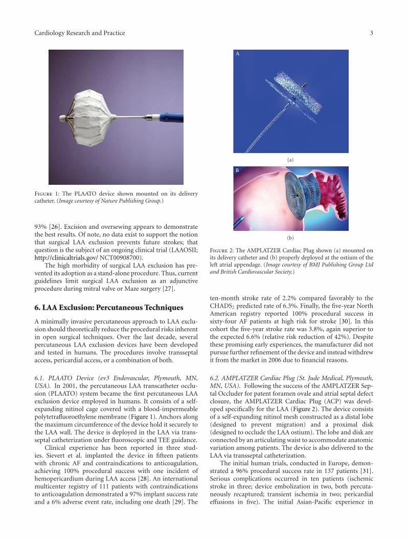
Cardiology Research and Practice 3
Figure 1: The PLAATO device shown mounted on its deliverycatheter. (Image courtesy of Nature Publishing Group.)
93% [26]. Excision and oversewing appears to demonstratethe best results. Of note, no data exist to support the notionthat surgical LAA exclusion prevents future strokes; thatquestion is the subject of an ongoing clinical trial (LAAOSII;http://clinicaltrials.gov/ NCT00908700).
The high morbidity of surgical LAA exclusion has pre-vented its adoption as a stand-alone procedure. Thus, currentguidelines limit surgical LAA exclusion as an adjunctiveprocedure during mitral valve or Maze surgery [27].
6. LAA Exclusion: Percutaneous Techniques
A minimally invasive percutaneous approach to LAA exclu-sion should theoretically reduce the procedural risks inherentin open surgical techniques. Over the last decade, severalpercutaneous LAA exclusion devices have been developedand tested in humans. The procedures involve transseptalaccess, pericardial access, or a combination of both.
6.1. PLAATO Device (ev3 Endovascular, Plymouth, MN,USA). In 2001, the percutaneous LAA transcatheter occlu-sion (PLAATO) system became the first percutaneous LAAexclusion device employed in humans. It consists of a self-expanding nitinol cage covered with a blood-impermeablepolytetrafluoroethylene membrane (Figure 1). Anchors alongthe maximum circumference of the device hold it securely tothe LAA wall. The device is deployed in the LAA via trans-septal catheterization under fluoroscopic and TEE guidance.
Clinical experience has been reported in three stud-ies. Sievert et al. implanted the device in fifteen patientswith chronic AF and contraindications to anticoagulation,achieving 100% procedural success with one incident ofhemopericardium during LAA access [28]. An internationalmulticenter registry of 111 patients with contraindicationsto anticoagulation demonstrated a 97% implant success rateand a 6% adverse event rate, including one death [29]. The
(a)
(b)
Figure 2: The AMPLATZER Cardiac Plug shown (a) mounted onits delivery catheter and (b) properly deployed at the ostium of theleft atrial appendage. (Image courtesy of BMJ Publishing Group Ltdand British Cardiovascular Society.)
ten-month stroke rate of 2.2% compared favorably to theCHADS2 predicted rate of 6.3%. Finally, the five-year NorthAmerican registry reported 100% procedural success insixty-four AF patients at high risk for stroke [30]. In thiscohort the five-year stroke rate was 3.8%, again superior tothe expected 6.6% (relative risk reduction of 42%). Despitethese promising early experiences, the manufacturer did notpursue further refinement of the device and instead withdrewit from the market in 2006 due to financial reasons.
6.2. AMPLATZER Cardiac Plug (St. Jude Medical, Plymouth,MN, USA). Following the success of the AMPLATZER Sep-tal Occluder for patent foramen ovale and atrial septal defectclosure, the AMPLATZER Cardiac Plug (ACP) was devel-oped specifically for the LAA (Figure 2). The device consistsof a self-expanding nitinol mesh constructed as a distal lobe(designed to prevent migration) and a proximal disk(designed to occlude the LAA ostium). The lobe and disk areconnected by an articulating waist to accommodate anatomicvariation among patients. The device is also delivered to theLAA via transseptal catheterization.
The initial human trials, conducted in Europe, demon-strated a 96% procedural success rate in 137 patients [31].Serious complications occurred in ten patients (ischemicstroke in three; device embolization in two, both percuta-neously recaptured; transient ischemia in two; pericardialeffusions in five). The initial Asian-Pacific experience in

4 Cardiology Research and Practice
twenty patients was also recently published, highlightinga 95% procedural success rate and complications in threepatients (catheter-related thrombus, coronary artery airembolism, and TEE-induced esophageal injury); one yearfollowup showed no incidence of stroke or death [32].Importantly, ACP implantation protocols have not involvedperiprocedural anticoagulation, instead employing dualantiplatelet therapy for one month followed by aspirinmonotherapy for six months. The ACP has received CE markapproval and is currently in Phase I clinical trials in theUnited States (http://clinicaltrials.gov/ NCT01118299).
6.3. WATCHMAN LAA Closure Device (Atritech, Plymouth,MN, USA). The WATCHMAN device, first implanted in2002, shares similarities with the ACP and PLAATO sys-tems. It consists of a self-expanding, open-ended nitinolframe with fixation anchors and a polyethylene membrane(Figures 3 and 4). It also has a catheter-based transseptaldelivery system. With this device, the membrane is perme-able and covers only the part of the device exposed to the leftatrium. The WATCHMAN device protocols have requiredwarfarin for at least 6 weeks to prevent thrombus formationprior to endothelialization of the device. Warfarin has beendiscontinued once a follow-up TEE demonstrates completeocclusion of the LAA. Recently, a strategy of substitutingdual antiplatelet therapy for warfarin postimplantation wasevaluated in 150 patients in the ASAP registry. Resultssuggest that this strategy may be safe and effective in patientswith contraindications even to short-term warfarin (HeartRhythm Society Scientific Sessions 2012).
After the initial feasibility studies, which included amajor product redesign, a landmark randomized clinical trialcompared the WATCHMAN device with warfarin therapy[33–35]. In PROTECT-AF, 707 patients from fifty-nine cen-ters in the USA and Europe were randomized 2 : 1 to deviceversus standard warfarin therapy. These patients did nothave contraindications to warfarin and their stroke risk wassomewhat lower than the PLAATO population (68% had aCHADS2 score of 1 or 2). The trial was designed to assessnoninferiority of WATCHMAN to standard warfarin ther-apy. After 1065 patient-years of followup, the primary effi-cacy endpoint (stroke, systemic embolism, or cardiovascularor unexplained death) was superior in the WATCHMANgroup over the warfarin group (3.0% versus 4.9% per 100patient-years) and achieved the criteria for noninferiority.However, the primary safety endpoint (excessive bleeding orprocedure-related complications) was significantly worse inthe WATCHMAN group (7.4% versus 4.4% per 100 patient-years). Periprocedural complications included 22 pericardialeffusions (4.8%), four air emboli (0.9%), and three deviceembolizations (0.6%). On the other hand, the warfaringroup had a higher incidence of major bleeding (4.1% versus3.5%) and hemorrhagic stroke (2.5% versus 0.2%). Overallimplantation success was 91% and at six months, 92% ofpatients in the WATCHMAN group were able to discontinuewarfarin after a TEE confirmed complete LAA closure.
Of note, procedure-related and device-related adverseevents were greater in the first half of PROTECT AF than
(a)
(b)
Figure 3: The WATCHMAN LAA Closure Device (a) highlightingthe permeable membrane covering its left atrial face and (b) prop-erly deployed within the ostium of the left atrial appendage. (Imagecourtesy of BMJ Publishing Group Ltd and British CardiovascularSociety.)
Figure 4: Fluoroscopic image of the WATCHMAN device (arrow)deployed in the left atrial appendage.

Cardiology Research and Practice 5
Figure 5: Transesophageal echocardiographic image of a largethrombus (arrow) on a WATCHMAN device several months afteranticoagulation was discontinued.
in the second half, highlighting the learning curve involvedwith device implantation [36]. The adverse events ratecontinued to remain low in the Continued Access Protocol(CAP) Registry of 460 patients. A second randomized trial,PREVAIL, is currently underway in hopes of obtaining FDAapproval for the WATCHMAN device. This trial is similar indesign to PROTECT AF but with stricter inclusion criteria.
Thus far, the WATCHMAN device is the only transseptalLAA exclusion device that has demonstrated non-inferiorityto warfarin for stroke prevention. However, concerns remainregarding peri-procedural complications and thrombus for-mation on the device prior to endothelialization (Figure 5).
6.4. LARIAT Suture Delivery System (SentreHeart, Palo Alto,CA, USA). The newest LAA exclusion device with promisinghuman data is the LARIAT suture delivery system, whichreceived FDA approval in 2009 for soft-tissue occlusion.This hybrid system involves the epicardial and transseptalplacement of magnet-tipped guidewires, forming a single railfor the delivery of an endocardial balloon and an epicardialsnare with a pretied suture loop (Figure 6). The inflatedendocardial balloon acts as a marker for the placement ofthe epicardial snare around the base of the LAA. Under flu-oroscopic and TEE guidance, the suture is then released andcinched to ligate the LAA at its base (Figure 7). Importantly,LAA closure can be evaluated in real time with TEE andleft atrial angiography prior to irreversible suture delivery.If closure is not satisfactory, the snare can be opened andrepositioned (Figure 8).
There are several advantages to this approach, includingcomplete control of the pericardial space in the event ofcardiac perforation, lack of any endovascular hardware left
Figure 6: Major components of the LARIAT Suture Delivery Sys-tem, including magnet-tipped guidewires, endovascular ballooncatheter, and epicardial snare mounted with a pretied suture loop.(Image courtesy of SentreHeart, Inc.)
behind, and possible elimination of the need for post-procedure anticoagulation. The major disadvantage of theLARIAT system is the need for simultaneous transseptal andpericardial access. In addition, anatomic variables can limitcandidacy for the device, such as LAA diameter greater than40 mm, posteriorly rotated LAA, or pericardial adhesionsfrom prior cardiac surgery or pericarditis.
Initial experience in a canine model has confirmed thesafety and feasibility of the LARIAT system [37]. In the firsthuman experience, ten patients successfully underwent LAAexclusion with the LARIAT suture, one of whom requiredthorascopic removal of the snare owing to pectus excavatum[38]. Complete exclusion was confirmed in all ten patientsusing left atrial angiography and TEE. In a single center,nonrandomized study (PLACE II), LAA exclusion with theLARIAT system was attempted in 89 patients and was suc-cessful in 85 (96%). There were three adverse events (3.3%)involving bleeding—two pericardial related, one transseptalrelated. There were no device-related complications orembolic strokes. Persistent LAA closure (defined as less than1 mm residual flow) was achieved in 95% of patients at dayninety and 98% of patients at one year (Heart RhythmSociety Scientific Sessions 2012).
7. Conclusions
Stroke prevention in AF presents significant challenges aswell as opportunities. Current treatment strategies withsystemic anticoagulation, while effective, are fraught withlimitations involving poor compliance, intolerance, andinconvenience. While newer oral anticoagulants overcomemany of these limitations, all anticoagulants suffer from anunavoidable lifelong commitment to medication and ele-vated bleeding risk.
Given that the LAA is the source of thromboembolismin the vast majority of patients with AF and stroke, a newerparadigm of targeting the LAA has naturally evolved. Severalstrategies are available, although surgical LAA removal willlikely not have a large role as a stand-alone procedure dueto its significant morbidity. The minimally invasive strategiesinvolve foreign body occlusion of the LAA ostium and peri-cardial suture ligation of the LAA base.

6 Cardiology Research and Practice
(a) (b)
Figure 7: Transesophageal echocardiographic images of the left atrial appendage (a) prior to and (b) immediately after LARIAT suturedeployment, highlighting acute LAA exclusion. (Image courtesy of SentreHeart, Inc.)
(a) (b) (c)
(d) (e) (f)
Figure 8: Fluoroscopic sequence of the LARIAT procedure. (a) After transseptal and pericardial access, baseline left atrial angiographyidentifies the left atrial appendage. (b) Magnet-tipped endocardial and epicardial guidewires make contact across the wall of the left atrialappendage. (c) The balloon catheter is inflated just within the ostium of the left atrial appendage, guiding the placement of the epicardialsnare. (d) The snare is tightened at the base of the left atrial appendage. (e) The balloon catheter is pulled back and left atrial angiographyconfirms occlusion of the left atrial appendage. (f) The suture is cinched down permanently, the snare is retracted, and a final left atrialangiogram reconfirms complete occlusion of the left atrial appendage.
Several questions remain regarding LAA exclusion. Asidefrom the PLAATO device, which is no longer available, theinformation regarding long-term durability of percutaneousLAA exclusion is not yet available. Even after acute proce-dural success, there is commonly a small diverticulum or“beak” left behind at the LAA ostium. In light of the surgicaldata that incomplete closure is worse than no closure at all[26], there are concerns about the thrombogenicity of thisunnatural diverticulum.
The data also highlight that success rates are operator andexperience dependent. As every new procedure necessarilyinvolves a learning curve, the hope is that the second-and third- generation data with LAA exclusion will showimproving procedural success rates with decreasing com-plication rates. The WATCHMAN experience has alreadydemonstrated this.
The ultimate dominance of one percutaneous tech-nique over the rest is unlikely. A more likely outcome is that

Cardiology Research and Practice 7
device selection will be tailored to patient characteristics.For instance, prior cardiac surgery or pericardial adhesionswould make endocardial occlusion devices more feasiblethan the LARIAT system. On the other hand, an absolutecontraindication to antiplatelet drugs or oral anticoagulationmakes the LARIAT system more attractive as it appearsto have no requirement for postprocedure anticoagulation.Similarly, a patient deemed at high risk for infection maybenefit from the LARIAT system given its lack of endovas-cular hardware.
An even larger issue is the selection of appropriate can-didates for these devices. Current focus has been on patientswith intolerance or contraindications to warfarin. Whetherthese devices will be offered as an equal (or preferred) alter-native to anticoagulation remains to be seen. PROTECT AFconfirmed the non-inferiority of LAA exclusion to warfarin,but superiority data is still lacking. In addition, all LAAexclusion trials excluded patients with valvular AF or withprosthetic valves; the role of LAA exclusion in these patientsis unknown. Finally, to date the comparison arm for thesedevices has only been warfarin. As some of the neweranticoagulants have reduced bleeding risk compared withwarfarin, it is possible that the benefit of mechanical LAAexclusion would diminish in head-to-head trials against thenewer agents.
The ultimate goal of LAA exclusion is to replace the life-long need for anticoagulation with a single procedure asso-ciated with a small upfront risk and tremendous long-termbenefit. This paradigm rests on the assumption that throm-boembolism in AF is due solely to the anatomic presenceof the LAA. However, data suggests that AF is associated witha systemic hypercoagulable state which may contribute tostroke risk in an independent and meaningful way [39]. Thisargues against discontinuation of anticoagulation, regardlessof the patency of the LAA. Larger and longer-term studieswill help shed light on this important question.
Despite the remaining challenges, LAA exclusion rep-resents a promising alternative to systemic anticoagulationfor the prevention of stroke in patients with AF. Already,studies have established that LAA exclusion is a viableoption in patients with intolerance or contraindications toanticoagulation. Whether LAA exclusion is a superior strat-egy to anticoagulation in all AF patients remains to be seen.In addition, whether mechanical LAA exclusion reduces riskof stroke over the long term will require further clinicaltrials.
References
[1] A. S. Go, E. M. Hylek, K. A. Phillips et al., “Prevalence ofdiagnosed atrial fibrillation in adults: national implications forrhythm management and stroke prevention: the anticoagula-tion and risk factors in atrial fibrillation (ATRIA) study,” TheJournal of the American Medical Association, vol. 285, no. 18,pp. 2370–2375, 2001.
[2] O. Wazni, B. Wilkoff, and W. Saliba, “Catheter ablation foratrial fibrillation,” The New England Journal of Medicine, vol.365, pp. 2296–2304, 2011.
[3] W. M. Feinberg, J. L. Blackshear, A. Laupacis, R. Kronmal,and R. G. Hart, “Prevalence, age distribution, and gender
of patients with atrial fibrillation: analysis and implications,”Archives of Internal Medicine, vol. 155, no. 5, pp. 469–473,1995.
[4] Y. Miyasaka, M. E. Barnes, B. J. Gersh et al., “Secular trends inincidence of atrial fibrillation in Olmsted County, Minnesota,1980 to 2000, and implications on the projections for futureprevalence,” Circulation, vol. 114, no. 11, pp. 119–125, 2006.
[5] V. L. Roger, A. S. Go, D. M. Lloyd-Jones et al., “Heart diseaseand stroke statistics—2012 update: a report from the Ameri-can heart association,” Circulation, vol. 125, 1, Article ID e2-e220, 2012.
[6] G. Y. H. Lip and A. D. Blann, “Atrial fibrillation and abnor-malities of hemostatic factors,” American Journal of Cardiology,vol. 87, no. 9, pp. 1136–1137, 2001.
[7] M. K. Chung, D. O. Martin, D. Sprecher et al., “C-reactiveprotein elevation in patients with atrial arrhythmias: Inflam-matory mechanisms and persistence of atrial fibrillation,”Circulation, vol. 104, no. 24, pp. 2886–2891, 2001.
[8] M. Guazzi and R. Arena, “Endothelial dysfunction andpathophysiological correlates in atrial fibrillation,” Heart, vol.95, no. 2, pp. 102–106, 2009.
[9] J. L. Blackshear and J. A. Odell, “Appendage obliteration toreduce stroke in cardiac surgical patients with atrial fibrilla-tion,” Annals of Thoracic Surgery, vol. 61, no. 2, pp. 755–759,1996.
[10] W. D. Johnson, A. K. Ganjoo, C. D. Stone et al., “The leftatrial appendage: our most lethal human attachment! surgicalimplications,” European Journal Cardio-Thoracic Surgery, vol.17, no. 6, pp. 718–722, 2000.
[11] J. P. Veinot, P. J. Harrity, F. Gentile et al., “Anatomy ofthe normal left atrial appendage: a quantitative study ofage-related changes in 500 autopsy hearts: implications forechocardiographic examination,” Circulation, vol. 96, no. 9,pp. 3112–3115, 1997.
[12] R. Beinart, E. K. Heist, J. B. Newell, G. Holmvang, J. N. Ruskin,and M. Mansour, “Left atrial appendage dimensions predictthe risk of stroke/TIA in patients with atrial fibrillation,”Journal of Cardiovascular Electrophysiology, vol. 22, no. 1, pp.10–15, 2011.
[13] V. Fuster, L. E. Ryden, D. S. Cannom et al., “2011 ACCF/AHA/HRS focused updates incorporated into the ACC/AHA/ESC2006 guidelines for the management of patients with atrialfibrillation: a report of the American college of cardiologyfoundation/American heart association task force on practiceguidelines,” Circulation, vol. 123, no. 10, pp. e269–e367, 2011.
[14] B. F. Gage, A. D. Waterman, W. Shannon, M. Boechler, M. W.Rich, and M. J. Radford, “Validation of clinical classificationschemes for predicting stroke: results from the national reg-istry of atrial fibrillation,” The Journal of the American Medi-cal Association, vol. 285, no. 22, pp. 2864–2870, 2001.
[15] G. Y. H. Lip, R. Nieuwlaat, R. Pisters, D. A. Lane, and H. J. G.M. Crijns, “Refining clinical risk stratification for predictingstroke and thromboembolism in atrial fibrillation using anovel risk factor-based approach: the euro heart survey onatrial fibrillation,” Chest, vol. 137, no. 2, pp. 263–272, 2010.
[16] R. G. Hart, L. A. Pearce, and M. I. Aguilar, “Meta-analysis:antithrombotic therapy to prevent stroke in patients who havenonvalvular atrial fibrillation,” Annals of Internal Medicine,vol. 146, no. 12, pp. 857–867, 2007.
[17] M. Sudlow, R. Thomson, B. Thwaites, H. Rodgers, and R.A. Kenny, “Prevalence of atrial fibrillation and eligibility foranticoagulants in the community,” The Lancet, vol. 352, no.9135, pp. 1167–1171, 1998.

8 Cardiology Research and Practice
[18] L. M. Brass, H. M. Krumholz, J. M. Scinto, and M. Radford,“Warfarin use among patients with atrial fibrillation,” Stroke,vol. 28, no. 12, pp. 2382–2389, 1997.
[19] A. S. Go, E. M. Hylek, L. H. Borowsky, K. A. Phillips, J. V. Selby,and D. E. Singer, “Warfarin use among ambulatory patientswith nonvalvular atrial fibrillation: the anticoagulation andrisk factors in atrial fibrillation (ATRIA) study,” Annals ofInternal Medicine, vol. 131, no. 12, pp. 927–934, 1999.
[20] A. M. Walker and D. Bennett, “Epidemiology and outcomesin patients with atrial fibrillation in the United States,” HeartRhythm, vol. 5, no. 10, pp. 1365–1372, 2008.
[21] C. B. Granger, J. H. Alexander, J. J. McMurray et al., “Apixabanversus warfarin in patients with atrial fibrillation,” The NewEngland Journal of Medicine, vol. 365, pp. 981–992, 2011.
[22] S. J. Connolly, M. D. Ezekowitz, S. Yusuf et al., “Dabigatranversus warfarin in patients with atrial fibrillation,” The NewEngland Journal of Medicine, vol. 361, pp. 1139–1151, 2009.
[23] M. R. Patel, K. W. Mahaffey, J. Garg et al., “Rivaroxaban versuswarfarin in nonvalvular atrial fibrillation,” The New EnglandJournal of Medicine, vol. 365, pp. 883–891, 2011.
[24] J. L. Madden, “Resection of the left auricular appendix; aprophylaxis for recurrent,” The Journal of the American Medi-cal Association, vol. 140, no. 9, pp. 769–772, 1949.
[25] T. Weimar, S. Schena, M. S. Bailey et al., “The cox-mazeprocedure for lone atrial fibrillation: a single-center experienceover 2 decades,” Circulation: Arrhythmia and Electrophysiology,vol. 5, pp. 8–14, 2012.
[26] A. G. Dawson, S. Asopa, and J. Dunning, “Should patientsundergoing cardiac surgery with atrial fibrillation have leftatrial appendage exclusion?” Interactive Cardiovascular andThoracic Surgery, vol. 10, no. 2, pp. 306–311, 2010.
[27] R. O. Bonow, B. A. Carabello, K. Chatterjee et al., “2008Focused update incorporated into the ACC/AHA 2006 guide-lines for the management of patients with valvular heart dis-ease: a report of the American college of cardiology/Americanheart association task force on practice guidelines (writingcommittee to revise the 1998 guidelines for the managementof patients with valvular heart disease): endorsed by thesociety of cardiovascular,” Circulation, vol. 118, no. 15, pp.e523–e661, 2008.
[28] H. Sievert, M. D. Lesh, T. Trepels et al., “Percutaneous leftatrial appendage transcatheter occlusion to prevent stroke inhigh-risk patients with atrial fibrillation: early clinical experi-ence,” Circulation, vol. 105, no. 16, pp. 1887–1889, 2002.
[29] S. H. Ostermayer, M. Reisman, P. H. Kramer et al., “Percuta-neous left atrial appendage transcatheter occlusion (PLAATOSystem) to prevent stroke in high-risk patients with non-rheumatic atrial fibrillation: results from the internationalmulti-center feasibility trials,” Journal of the American Collegeof Cardiology, vol. 46, no. 1, pp. 9–14, 2005.
[30] P. C. Block, S. Burstein, P. N. Casale et al., “Percutaneousleft atrial appendage occlusion for patients in atrial fibrilla-tion suboptimal for Warfarin therapy. 5-year results of thePLAATO (percutaneous left atrial appendage transcatheterocclusion) study,” Journal of the American College of Cardiol-ogy: Cardiovascular Interventions, vol. 2, no. 7, pp. 594–600,2009.
[31] J. W. Park, A. Bethencourt, H. Sievert et al., “Left atrialappendage closure with Amplatzer Cardiac Plug in atrial fibril-lation: initial European experience,” Catheterization and Car-diovascular Interventions, vol. 77, no. 5, pp. 700–706, 2011.
[32] Y. Y. Lam, G. W. Yip, C. M. Yu et al., “Left atrial appendageclosure with AMPLATZER cardiac plug for stroke prevention
in atrial fibrillation: initial Asia-Pacific experience,” Catheteri-zation and Cardiovascular Interventions, vol. 79, no. 5, pp. 794–800, 2012.
[33] D. R. Holmes, V. Y. Reddy, Z. G. Turi et al., “Percutaneousclosure of the left atrial appendage versus warfarin therapyfor prevention of stroke in patients with atrial fibrillation: arandomised non-inferiority trial,” The Lancet, vol. 374, no.9689, pp. 534–542, 2009.
[34] P. B. Sick, G. Schuler, K. E. Hauptmann et al., “Initial world-wide experience with the watchman left atrial appendage sys-tem for stroke prevention in atrial fibrillation,” Journal of theAmerican College of Cardiology, vol. 49, no. 13, pp. 1490–1495,2007.
[35] Y. L. Bayard, S. H. Ostermayer, and H. Sievert, “Transcatheterocclusion of the left atrial appendage for stroke prevention,”Expert Review of Cardiovascular Therapy, vol. 3, no. 6, pp.1003–1008, 2005.
[36] V. Y. Reddy, D. Holmes, S. K. Doshi, P. Neuzil, and S. Kar,“Safety of percutaneous left atrial appendage closure: resultsfrom the watchman left atrial appendage system for embolicprotection in patients with AF (PROTECT AF) clinical trialand the continued access registry,” Circulation, vol. 123, no. 4,pp. 417–424, 2011.
[37] R. J. Lee, K. Bartus, and S. J. Yakubov, “Catheter-based leftatrial appendage (LAA) ligation for the prevention of embolicevents arising from the LAA initial experience in a caninemodel,” Circulation: Cardiovascular Interventions, vol. 3, no. 3,pp. 224–229, 2010.
[38] K. Bartus, J. Bednarek, J. Myc et al., “Feasibility of closed-chestligation of the left atrial appendage in humans,” Heart Rhythm,vol. 8, no. 2, pp. 188–193, 2011.
[39] T. Watson, E. Shantsila, and G. Y. Lip, “Mechanisms ofthrombogenesis in atrial fibrillation: virchow’s triad revisited,”The Lancet, vol. 373, no. 9658, pp. 155–166, 2009.

Hindawi Publishing CorporationCardiology Research and PracticeVolume 2012, Article ID 645469, 13 pagesdoi:10.1155/2012/645469
Research Article
Long-Term Costs of Ischemic Stroke and Major Bleeding Eventsamong Medicare Patients with Nonvalvular Atrial Fibrillation
Catherine J. Mercaldi,1 Kimberly Siu,2 Stephen D. Sander,2
David R. Walker,2 You Wu,3 Qian Li,3 and Ning Wu3
1 Center for Epidemiology and Database Analytics, United BioSource Corporation, 7101 Wisconsin Avenue, Suite 600, Bethesda,MD 20814, USA
2 Health Economics and Outcomes Research, Boehringer Ingelheim Pharmaceuticals, Inc., 900 Ridgebury Road, Ridgefield,CT 06877, USA
3 Economic Analysis and Solutions, United BioSource Corporation, 7101 Wisconsin Avenue, Suite 600, Bethesda,MD 20814, USA
Correspondence should be addressed to Catherine J. Mercaldi, [email protected]
Received 17 May 2012; Revised 30 July 2012; Accepted 1 August 2012
Academic Editor: Natig Gassanov
Copyright © 2012 Catherine J. Mercaldi et al. This is an open access article distributed under the Creative Commons AttributionLicense, which permits unrestricted use, distribution, and reproduction in any medium, provided the original work is properlycited.
Purpose. Acute healthcare utilization of stroke and bleeding has been previously examined among patients with nonvalvular atrialfibrillation (NVAF). The long-term cost of such outcomes over several years is not well understood. Methods. Using 1999–2009Medicare medical and enrollment data, we identified incident NVAF patients without history of stroke or bleeding. Patientswere followed from the first occurrence of ischemic stroke, major bleeding, or intracranial hemorrhage (ICH) resulting inhospitalization. Those with events were matched with 1–5 NVAF patients without events. Total incremental costs of events werecalculated as the difference between costs for patients with events and matched controls for up to 3 years. Results. Among the25,465 patients who experienced events, 94.5% were successfully matched. In the first year after event, average incremental costswere $32,900 for ischemic stroke, $23,414 for major bleeding, and $47,640 for ICH. At 3 years after these events, costs remainedelevated by $3,156–$5,400 per annum. Conclusion. While the costs of stroke and bleeding among patients with NVAF are mostdramatic in the first year, utilization remained elevated at 3 years. Cost consequences extend beyond the initial year after theseevents and should be accounted for when assessing the cost-effectiveness of treatment regimens for stroke prevention.
1. Introduction
Atrial fibrillation (AF) affects more than 3 million Americansand is characterized by an irregularly irregular heart rhythm,often with a rapid heart rate that may result in blood clots,shortness of breath, and overall weakness [1]. Primarily adisease of the elderly, the prevalence of AF doubles with eachdecade of life after the age of 60 years and occurs in about10% of the population by 80 years [2, 3]. AF is associatedwith a five-fold increase in the risk of ischemic stroke andaccounts for 15%–20% of all strokes [3]. Anticoagulationtherapy with warfarin is recommended for patients with AFwho are at moderate to high risk of stroke based on clinicalguidelines [4], but it has been well documented that proper
dosing and close monitoring are imperative to attain thecorrect level of anticoagulation to balance the risk of strokewithout introducing a disproportionate risk of bleeding[5–7].
Costs to manage AF in the USA are estimated at $6.65billion, including $4.88 billion in hospitalization expensesand $1.53 billion in outpatient management costs (in 2005US$) [8]. Accounting for some fraction of these costs areexpenses related to occurrence of stroke and hemorrhagicevents. While numerous studies have estimated direct andindirect costs of stroke and bleeding outcomes among AFpatients in the first year following the event [9–11], long-term US cost estimates have generally relied on modelingtechniques extrapolating available data covering a shorter

2 Cardiology Research and Practice
period of time. Lifetime and other long-term cost modelingestimates would be expected to be more accurate if based ondata covering a period of time extended beyond 1 year [12].Furthermore, direct estimates of stroke and bleeding costsover several years could show whether costs for patients withthese events remain elevated or eventually return to baselinelevels, that is, the expected cost to treat AF without stroke ormajor bleeding.
The objective of the current study was to quantifythe direct long-term costs, up to 3 years, of both strokeand bleeding events among patients with non-valvular AF(NVAF), which comprises approximately 90% of the overallAF population [13]. To separate the costs attributable tothese events from the general costs of healthcare associatedwith NVAF, we conducted a retrospective cohort study ofMedicare beneficiaries newly diagnosed with NVAF who laterdeveloped stroke or hemorrhagic events and were matchedwith NVAF patients who did not experience these outcomesbased on demographic and NVAF disease characteristics.
2. Methods
2.1. Data Source. The study was conducted using 1999–2009 data from the Centers for Medicare & MedicaidServices (CMS) 5% sample standard analytical files (SAF)—limited data set (LDS). In contrast to commercial claimsdata sources, Medicare has a large number of patients overthe age of 65 years, when incidence and prevalence ofAF begin to increase dramatically. The analysis used fee-for-service (FFS), nondisabled Medicare patients who werenot eligible for Medicaid. The files contain final actionclaims with all adjustments resolved for a 5% sample ofall Medicare beneficiaries in each calendar year, includinginpatient, outpatient, emergency room (ER), skilled nurs-ing facility (SNF), hospice, home health agency, durablemedical equipment (DME), and carrier (formerly Part Bphysician/supplier) claims. As this study was a retrospectiveanalysis of existing, deidentified claims data, an institutionalreview board evaluation was not applicable and therefore,not conducted.
2.2. Patient Identification. Patients with AF were identifiedby at least 1 inpatient claim or 2 outpatient claims in the samecalendar quarter with International Statistical Classificationof Diseases and Related Health Problems, 9th Edition (ICD-9) code 427.31. Included patients were required to havea baseline period of 1 year (4 quarters) of continuousenrollment prior to the quarter of AF diagnosis. Patients withevidence of valvular conditions during the baseline period orquarter of AF diagnosis (codes in Table 1) were excluded.
Because the stage of NVAF disease progression has animpact on medical utilization (e.g., newly diagnosed patientstend to have higher costs than prevalent patients), the studywas limited to patients with incident NVAF, defined as noAF claims during the baseline period, to allow patients tobe matched on duration of NVAF. Similarly, strokes and/orbleeding events prior to AF diagnosis could lead to elevatedbaseline costs and would bias the estimate of long-term eventcosts after diagnosis. Thus, patients with prevalent AF (any
claim with ICD-9 diagnosis code 427.31 during the baselineperiod) and patients with a principal inpatient claim with acode for stroke [14, 15] or bleeding [16, 17] events (Table 1)in the baseline period (1 year prior to AF diagnosis) orquarter of AF diagnosis were excluded.
2.3. Matching. All incident NVAF patients without a historyof ischemic stroke or major bleeding event were thenfollowed for first ischemic stroke or major bleeding eventusing validated ICD-9 codes (Table 1) [14–17]. Patients withevents were matched with NVAF patients without eventson age group at NVAF diagnosis, gender, race, geographicregion, year of NVAF diagnosis, duration of enrollment, andwarfarin use. The data did not include prescription claims;therefore, warfarin therapy was inferred for patients with atleast 3 prothrombin tests (ICD-9 codes V58.61, CPT codes85610, 85611, 99363, and 99364) during a 1-year periodafter NVAF diagnosis. This methodology has been validatedpreviously in the Medicare data with 89% sensitivity and92% specificity [6].
Each patient with an event was matched to up to 5control patients via the greedy matching algorithm [18, 19].Allowing more than 1 control to be matched to each patientwith an event has been shown to improve statistical efficiency[20, 21]. While efficiency gains are limited beyond 3 or 4matches, the low cost of obtaining additional matches inclaims data permitted us to extend the maximal number ofmatches to 5. The number of matches were allowed to differfor each patient with an event, since varying numbers ofcontrols have been shown to reduce bias [18]. Any patientsnot matched (either unmatchable patients with events orremaining controls) were excluded from the analysis.
2.4. Followup. Although the primary objective was to esti-mate costs up to 3 years following events, we did not limit thestudy population to those with 3 years of followup becausedoing so would have biased the population toward healthierpatients. As such, no minimum duration of enrollmentfollowing the initial event was required. The quarter of thestroke or bleeding event was considered the start of followupfor the patient with the event and for all controls matched tothat patient.
The risk of stroke and bleeding events is somewhatintertwined (e.g., prior stroke is a stated risk factor for majorbleeding), and it is possible that patients with an initial stroke(or bleed) may later experience a major bleed (or stroke). Tokeep the costs of these events distinct, if a patient experienceda bleeding event after stroke or a stroke after a major bleed,we ended followup for this patient in the quarter prior tothe alternate event. A new followup period began with thequarter of the alternate event, and utilization measures werecalculated separately for patients with both events. Thus,patients (and their matched controls) were categorized bythe type(s) of events they experienced during followup as (1)patients with stroke events only or whose first event was astroke, (2) patients with bleeding events only or whose firstevent was a major bleed, or (3) patients with both strokeand major bleeding events. To illustrate, a patient who first

Cardiology Research and Practice 3
Table 1: Study codes.
Condition ICD-9 codes CPT codes
Patient identification
Atrial fibrillation 427.31
Valvular conditionsProcedure codes:35.0, 35.00–35.04, 35.1, 35.10–35.14, 35.2,35.20–35.28, 35.96
33400, 33401, 33403, 33405, 33406, 33410–33415,33417, 33420, 33422, 33425–33427, 33430, 33460,33463–33465, 33468, 33470, 33472, 33474–33476,33478, 33496, 33600, 33602
Outcomes
Ischemic stroke 433.x1, 434.x1, 436, 437.1, 437.9
Major bleeding
Intracranial hemorrhage 430, 431, 432
Other major bleeds
423.0, 455.2, 455.5, 455.8, 456.0, 456.2, 459.0,530.7, 530.8, 531.0, 531.2, 531.4, 531.6, 532.0,532.2, 532.4, 532.6, 533.0, 533.2, 533.4, 533.6,534.0, 534.2, 534.4, 534.6, 562.0, 562.1, 569.3,569.85, 578, 599.7, 626.2, 626.6, 719.1, 784.7,786.3, 852, 853
CHA2DS2-VASc components
Cardiac failure 398.91, 402.x1, 404.x3, 425, 428
Hypertension1 362.11, 401, 402, 403, 404, 405
Diabetes mellitus1 250, 357.2, 362.0, 366.41
Prior TIA2 362.34, 435
Vascular disease
410, 411, 412, 413, 414, 440, 441, 442, 443, 444,445Procedure codes:00.66, 36.0, 36.1, 39.25
33510–33545, 34051, 34151, 34201, 34203,34800–34834, 34900, 35081–35103, 35131, 35132,35141, 35142, 35151, 35152, 35331, 35341, 35351,35355, 35361, 35363, 35371, 35372, 35381, 35450,35452, 35454, 35456, 35459, 35470, 35471, 35472,35473, 35474, 35480, 35481, 35482, 35483, 35485,35490, 35491, 35492, 35493, 35495, 35521, 35531,35533, 35541, 35546, 35546, 35548, 35549, 35551,35556, 35558, 35563, 35565, 35566, 35571, 35583,35585, 35587, 35621, 35623, 35646, 35647, 35651,35654, 35656, 35661, 35663, 35665, 35666, 35671,92980, 92981, 92982, 92984
HAS-BLED components
Hypertension1 362.11, 401, 402, 403, 404, 405
Abnormal renal function 582, 583, 585, 586
Abnormal liver function 570, 571, 572, 573, 790.4
Prior TIA2 362.34, 435
Excessive alcohol use 291.0, 291.1, 291.2, 303, 305.0, 535.3, V11.3
Note: With the exception of valvular conditions, ICD-9 codes reported as 3 digits will include all 4-digit and 5-digit codes beginning with the same 3 digits.For 4-digit codes, any 5-digit code beginning with the same 4 digits will also be included. ICD-9 codes reported are diagnosis codes unless otherwise indicated.1Patient must have at least 2 diagnoses documented in the same calendar quarter to be considered as having the condition.2Risk score definition includes prior stroke, but with the exclusion of patients with prior stroke, only prior TIA is applicable in this study.
experienced a stroke and then later had a major bleedingevent was categorized both in the ischemic stroke group(to allow followup before the latter event to contribute tothe estimation of stroke utilization) as well as in the groupfor patients with both events for the remainder of follow-up beginning with the subsequent bleeding event. Underthis definition, some patients were double-counted acrossgroups, although each quarter of followup was assigned toonly one event group (stroke, major bleeding, or both).
Patients who had an initial stroke and a major bleedingevent in the same quarter (and their matched controls)contributed only to the group of patients with both events.Because patients with strokes or bleeds in the baseline periodwere excluded, recent history of these events had no impacton cost estimates. Recurrent strokes (a new stroke eventfollowing the initial stroke) or bleeds (a new bleeding eventfollowing the initial bleed) are an important contributor tolong-term event costs and were included during followup.

4 Cardiology Research and Practice
Thus, followup began with the initial event of inter-est and ended with the first occurrence of (1) Medicaredisenrollment (including patients changing from FFS tocapitation, dropping Part B coverage, becoming eligible forstate Medicaid services, becoming disabled, or dying), (2)for patients with stroke events, a subsequent bleeding eventor for patients with bleeding events, a subsequent strokeevent, or (3) 3 years after initial stroke or bleeding event.For patients with both stroke and bleeding events, the secondfollowup will begin in the quarter of the latter event andend with either disenrollment or 3 years after the start of thesecond follow-up period, whichever occurs first.
2.5. Patient Characteristics. Basic demographic informationincluding age, gender, race, and geographic region wereassessed at the time of AF diagnosis. We also reported riskfactors for both stroke and bleeding events. Risk factors wereassessed during the baseline period and in the quarter of AFdiagnosis. Stroke risk was quantified using the CHA2DS2-VASc score [22]. Bleeding risk was assessed using the HAS-BLED score, which had the best predictive value of severalhemorrhage risk scores [23–27] in a recent assessment [28];however, time in the therapeutic international normalizedratio (INR) range and drug use were unavailable in thedata source and thus, were excluded from the HAS-BLEDdefinition for this study. Definitions of stroke and bleedingrisk factors used to calculate these scores are provided inTable 1.
2.6. Identification of Events. Stroke and major bleedingevents were identified using principal ICD-9 codes oninpatient hospitalization claims only. Previously validatedICD-9 codes for identifying stroke [14, 15] and bleeding [16,17] outcomes are provided in Table 1. Major bleeding eventswere divided into intracranial hemorrhage (ICH) and othermajor bleeding event, and results were produced separatelyfor these hemorrhagic events. Because the Medicare 5%sample data only have calendar quarter reported on claims,events were recorded starting in the quarter following NVAFdiagnosis.
2.7. Definition of Outcomes. The primary outcome was theincremental costs of stroke and bleeding events up to 3years after the event. Costs were from a Medicare perspective(limited to Parts A and B paid amounts) and did not includeoral medications or copayments from any supplementalinsurance. Control patients with NVAF but no study events(stroke or major bleeding) were used to estimate the baselinecost of treating NVAF. Costs for patients with stroke ormajor bleeding events beyond the baseline treatment cost ofNVAF were attributed to the event. Thus, the incrementalcosts of stroke and major bleeding were estimated as thedifference in costs between patients with events and theirmatched controls, with cost accrual beginning in the quarterof the event and up to 3 years afterward. For example, ifa patient had a stroke in the third quarter following theNVAF diagnosis, then we began summing costs for thispatient and any controls matched to this patient in the
third quarter after the NVAF diagnosis. Incremental costswere also broken down by costs occurring in inpatient,outpatient, ER, SNF, hospice, and home healthcare settings,as well as DME costs to determine aspects of care drivingthe potential cost consequences associated with managementand treatment of NVAF. Other utilization measures includednumber of inpatient admissions and associated length of stayand number of outpatient or office visits.
Patients with recurrent stroke, ICH, or other majorbleeding events were also identified, as well as the time fromthe initial event until recurrence. Recurrent events were onlyidentified in the quarter following the initial event sincethe dating system in the data source made it impossibleto discern between separate events occurring in the samequarter.
2.8. Analysis. All variables will be reported descriptively asmeans with standard deviations and medians with rangesfor continuous variables and counts with percentages forcategorical measures for patients with and without studyevents. Cost measures were adjusted to 2011 US dollars(USD) using the medical care component of the ConsumerPrice Index (CPI) annual inflation measures. Total andincremental utilization variables after a stroke/bleeding eventwere reported for 3 months (quarter of the event), 6months, 9 months, 12 months, 18 months, 24 months,and 36 months. Because not all patients had the full 3years of followup, the patient denominator declined overtime. Comparison of total costs for patients with events andmatched controls were conducted using standard 2-sided t-tests assuming unequal variances and a significance level ofα = 0.05.
Although matching should eliminate the need to adjustfor all variables that were matched in the analysis of costdifferentials, in order to adjust for any potential resid-ual confounding, total incremental costs of strokes andbleeding events were adjusted for both matching variablesand individual measures within the CHA2DS2-VASc andHAS-BLED scores, respectively. Multivariate adjusted costswere estimated using generalized estimating equation (GEE)models with a gamma distribution and log link function[29]. Bootstrapping techniques were employed to estimatestandard errors of adjusted costs once the parameter estimatewas retransformed from the log form. All analyses wereconducted using SAS version 9.2 (SAS Institute, Cary, NC).
3. Results
A total of 445,796 patients with at least 1 inpatient or 2outpatient claims with diagnosis codes for AF were identifiedin the data. After excluding patients without continuousenrollment in the baseline period (24.3%), disabled andMedicaid-eligible patients (4.6%), patients with valvularconditions (1.9%), patients with prevalent AF (9.5%), andpatients with a history of stroke or hemorrhagic conditions(4.8%), 245,052 patients with NVAF was available forthe study. Of these patients, 8,243 (3.4%) experienced anischemic stroke during followup, 1,406 (0.5%) had an ICH,

Cardiology Research and Practice 5
and 15,816 (6.5%) had other types of major bleeding events.A total of 1,291 (0.5%) patients had both an ischemic strokeand major bleeding event (either ICH or other bleedingoutcome). Matching was successful for 94.5% of all patientswith stroke and major bleeds, with an average of 4 matchedcontrols for each patient with an event. Among patientswith events who were matched, the average time from NVAFdiagnosis to event was 30 months for all events.
Descriptive statistics of matching characteristics showthat patients with events and controls were balanced on allmeasures (Table 2). Average age was similar for patients withischemic stroke, ICH, and other major bleeding events atapproximately 80 years. Duration of followup was at least 2years for 44% of patients, with 29% having data for all 3 yearsexamined. Although patients were not matched on stroke orbleeding risk, patients with events and controls had similarCHA2DS2-VASc and HAS-BLED scores (Table 3).
Patients with both events had an average age of 81.1 years(81.3 years for matched controls), with 61.8% of patientsfemale (62.7% controls). Warfarin use was prevalent for37.5% of patients with both stroke and major bleedingevents. CHA2DS2-VASc score averaged 4.02 points (3.80for controls), and mean HAS-BLED was 1.37 (1.31 forcontrols). Approximately one-third (32.8%) of patients withboth events had data for at least 2 years after the secondaryevent, and 19.4% had data at 3 years.
3.1. Ischemic Stroke Cohort. The ischemic stroke cohortconsisted of 7,799 patients with stroke and 33,084 matchedcontrols without study events. At 1 year poststroke, 62.9%of patients were still contributing data. By 2 years, 41.2%remained in the cohort, and at the end of followup 3 yearsafter the event, 27.1% of patients were available. Amongpatients with stroke, 6.7% died, with 52.4% of deathsoccurring in the quarter of the event. A total of 7.0% of strokepatients later went on to experience major bleeding events(either ICH or other major bleeds). Average quarterly totalMedicare-reimbursed costs for patients with ischemic strokeand their matched controls without events are presented inFigure 1. The mean acute incremental cost of stroke in thequarter of the event was $20,604, and the average total 1-year cost was $32,900 more for patients with stroke thanamong controls. Total cumulative cost of stroke at 2 years was$36,515 and at 3 years was $38,712 for patients contributingdata at these time points. In the second year after the event,total costs were $5,621 higher for stroke patients than formatched patients without events. Costs remained elevated inthe third year after stroke, with an average incremental costof $3,775. After adjusting for stroke risk factors and matchingcharacteristics, the acute and annual incremental costs ofstroke were slightly higher than in unadjusted analyses(Table 4). The largest contributors to second-year and third-year incremental costs associated with stroke were inpatientcosts, SNF costs, and home healthcare costs (Table 5). Inthe third year after the event, patients with stroke averaged5.2 more office visits than those without events. Recurrentischemic stroke occurred in 6.7% of stroke patients, withan average time to recurrence of 12.0 months. The total
incremental costs for patients with recurrent stroke wereapproximately $3,000–$4,000 higher as compared to thosewithout recurrence in each of the first 3 years following theinitial event (Table 5).
3.2. Major Bleeding Cohorts. A total of 1,276 patients withICH and 5,097 matched controls were included in the study.Follow-up data was available for 57.5% of patients at 1 yearafter event, 37.3% at 2 years, and 24.0% after 3 years. Theoverall mortality rate for this cohort was 11.4%; 78.1% ofdeaths occurred in the quarter of the event. Among ICHpatients, 4.7% subsequently had an ischemic stroke. Theacute incremental cost of ICH in the quarter of the eventwas $29,877 (Figure 2). Total costs in the first year followingICH (including the quarter of the event) were $47,640 morefor patients with events than among controls. The totalcumulative cost of ICH after 2 years was $53,074 and at 3years was $54,158. Second-year post-ICH costs were $7,910,and by the third year after the event, costs continued to be$3,156 higher than controls. Adjustment for bleeding riskfactors and matching characteristics resulted in marginallyhigher ICH cost estimates in the first 2 years after the eventand a similar value in year 3 as compared to the unadjustedresults (Table 4). Inpatient, outpatient, SNF, and hospiceutilization were the principal cost drivers in year 2 (Table 5).In the third year after ICH, hospice and outpatient costs weremost elevated as compared to control costs. A total of 3.1%of patients with this event had a recurrent ICH. Average timeto recurrence was 8.1 months. Total incremental costs forpatients with recurrent ICH were approximately $7,000 morethan for those without recurrence in the first year after theevent, but were not elevated in the second and third yearsthereafter (Table 5).
The cohort of patients with major bleeding events otherthan ICH had 14,996 patients with events and 60,058controls. At 1, 2, and 3 years after the event, 69.4%, 46.6%,and 31.0% of these patients contributed data, respectively.The rate of mortality for patients with major bleeds was4.3%, with 36.3% of those deaths taking place in the quarterof the event. Ischemic stroke followed the bleeding eventin 3.1% of these patients. Patients with major bleedingevents had an average acute incremental cost of $15,699(Figure 3), with total first-year cost of $23,414 more thanfor matched controls. Total cumulative cost 2 years aftermajor bleeding events averaged $28,064 and after 3 yearswas $31,393. The second-year and third-year incrementalcosts after major bleeds were $6,936 and $5,400. Similar tothe ischemic stroke and ICH results, after controlling forbleeding risk factors and matching variables, estimates of theincremental costs of other major bleeds were comparableto, if slightly higher than, unadjusted figures (Table 5).Inpatient and outpatient costs were particularly elevated inthe second and third years after the event (Table 4). Inyear 2, patients with major bleeding events had 0.5 moreinpatient admissions and 11.0 additional outpatient visits ascompared with control patients. Number of inpatient andoutpatient visits remained increased by 0.3 admissions and8.8 visits in the third year after the event. Recurrent major

6 Cardiology Research and Practice
Ta
ble
2:M
atch
ing
char
acte
rist
ics
ofm
edic
are
pati
ents
wit
hN
VA
Fw
ith
and
wit
hou
tst
roke
and
blee
din
gev
ents
.
Maj
orbl
eedi
ng
even
ts
Mat
chin
gch
arac
teri
stic
sIs
chem
icst
roke
ICH
Oth
erm
ajor
blee
ds
Even
tN
=7,
799
Con
trol
N=
33,0
84Ev
ent
N=
1,27
6C
ontr
olN
=5,
097
Even
tN
=14
,996
Con
trol
N=
60,0
58
Age
atA
Fdi
agn
osis
,yea
rsM
ean
(SD
)81
.1(7
.6)
81.1
(7.9
)79
.9(7
.3)
79.9
(7.6
)80
.0(7
.7)
79.8
(7.8
)A
ge,c
ateg
oriz
ed,n
(%)
65–6
9ye
ars
626
(8.0
%)
2,67
6(8
.1%
)11
9(9
.3%
)48
2(9
.5%
)1,
498
(10.
0%)
6,28
2(1
0.5%
)70
–74
year
s1,
058
(13.
6%)
4,56
9(1
3.8%
)19
7(1
5.4%
)81
5(1
6.0%
)2,
408
(16.
1%)
9,91
6(1
6.5%
)75
–79
year
s1,
582
(20.
3%)
6,64
7(2
0.1%
)28
5(2
2.3%
)1,
156
(22.
7%)
3,32
8(2
2.2%
)13
,537
(22.
5%)
80–8
4ye
ars
1,79
1(2
3.0%
)7,
318
(22.
1%)
319
(25.
0%)
1,23
5(2
4.2%
)3,
295
(22.
0%)
12,6
96(2
1.1%
)≥8
5ye
ars
2,74
2(3
5.2%
)11
,874
(35.
9%)
356
(27.
9%)
1,40
9(2
7.6%
)4,
467
(29.
8%)
17,6
21(2
9.3%
)G
ende
r,n
(%)
Mal
e2,
913
(37.
4%)
12,2
87(3
7.1%
)63
4(4
9.7%
)2,
506
(49.
2%)
6,50
7(4
3.4%
)25
,669
(42.
7%)
Fem
ale
4,88
6(6
2.7%
)20
,797
(62.
9%)
642
(50.
3%)
2,59
1(5
0.8%
)8,
489
(56.
6%)
34,3
89(5
7.3%
)R
ace,
n(%
)W
hit
e7,
194
(92.
2%)
31,5
39(9
5.3%
)1,
191
(93.
3%)
4,90
1(9
6.2%
)14
,018
(93.
5%)
57,6
68(9
6.0%
)B
lack
445
(5.7
%)
1,01
2(3
.1%
)50
(3.9
%)
120
(2.4
%)
678
(4.5
%)
1,62
8(2
.7%
)H
ispa
nic
66(0
.9%
)20
2(0
.6%
)17
(1.3
%)
29(0
.6%
)12
6(0
.8%
)30
6(0
.5%
)O
ther
85(1
.1%
)28
8(0
.9%
)16
(1.3
%)
44(0
.9%
)15
6(1
.0%
)40
8(0
.7%
)U
nkn
own
9(0
.1%
)43
(0.1
%)
2(0
.2%
)3
(0.1
%)
18(0
.1%
)48
(0.1
%)
US
geog
raph
iclo
cati
on,n
(%)
Nor
thea
st1,
547
(19.
8%)
6,40
8(1
9.4%
)25
1(1
9.7%
)97
5(1
9.1%
)3,
251
(21.
7%)
12,4
50(2
0.7%
)So
uth
1,97
0(2
5.3%
)8,
599
(26.
0%)
317
(24.
8%)
1,27
0(2
4.9%
)4,
151
(27.
7%)
16,7
68(2
7.9%
)M
idw
est
3,22
7(4
1.4%
)14
,034
(42.
4%)
525
(41.
1%)
2,16
0(4
2.4%
)5,
818
(38.
8%)
24,2
57(4
0.4%
)W
est
1,05
1(1
3.5%
)4,
043
(12.
2%)
183
(14.
3%)
692
(13.
6%)
1,76
7(1
1.8%
)6,
570
(10.
9%)
Oth
er/u
nkn
own
4(0
.1%
)3
(0.0
%)
0(0
.0%
)0
(0.0
%)
9(0
.1%
)12
(0.0
%)
Year
ofA
Fdi
agn
osis
,n(%
)20
00–2
003
4,91
9(6
3.1%
)20
,082
(60.
7%)
715
(56.
0%)
2,65
1(5
2.0%
)8,
935
(59.
6%)
33,2
54(5
5.4%
)20
04–2
006
2,13
2(2
7.3%
)9,
449
(28.
6%)
403
(31.
6%)
1,70
0(3
3.4%
)4,
359
(29.
1%)
18,7
86(3
1.3%
)20
07–2
009
748
(9.6
%)
3,55
3(1
0.7%
)15
8(1
2.4%
)74
6(1
4.6%
)1,
704
(11.
4%)
8,01
8(1
3.4%
)D
ura
tion
ofst
udy
enro
llmen
t,n
(%)
3–<
6m
onth
s1,
571
(20.
1%)
6,60
4(2
0.0%
)34
2(2
6.8%
)1,
324
(26.
0%)
2,12
3(1
4.2%
)8,
582
(14.
3%)
6–<
12m
onth
s1,
341
(17.
2%)
5,67
1(1
7.1%
)20
2(1
5.8%
)83
9(1
6.5%
)2,
455
(16.
4%)
9,83
8(1
6.4%
)12
–<18
mon
ths
933
(12.
0%)
4,03
0(1
2.2%
)13
8(1
0.8%
)57
0(1
1.2%
)1,
812
(12.
1%)
7,34
5(1
2.2%
)18
–<24
mon
ths
740
(9.5
%)
3,16
0(9
.6%
)11
4(8
.9%
)46
8(9
.2%
)1,
576
(10.
5%)
6,35
4(1
0.6%
)24
–36
mon
ths
3,21
4(4
1.2%
)13
,621
(41.
2%)
480
(37.
6%)
1,89
6(3
7.2%
)7,
030
(46.
9%)
27,9
39(4
6.5%
)W
arfa
rin
use
,n(%
)Ye
s2,
568
(32.
9%)
10,0
87(3
0.5%
)53
2(4
1.7%
)1,
968
(38.
6%)
5,59
4(3
7.3%
)20
,576
(34.
3%)
No
5,23
1(6
7.1%
)22
,997
(69.
5%)
744
(58.
3%)
3,12
9(6
1.4%
)9,
402
(62.
7%)
39,4
82(6
5.7%
)
ICH
:in
trac
ran
ialh
emor
rhag
e;SD
:sta
nda
rdde
viat
ion
.

Cardiology Research and Practice 7
Ta
ble
3:R
isk
scor
esfo
rM
edic
are
pati
ents
wit
hN
VA
Fw
ith
and
wit
hou
tst
roke
and
blee
din
gev
ents
.
Maj
orbl
eedi
ng
even
ts
Mat
chin
gch
arac
teri
stic
sIs
chem
icst
roke
ICH
Oth
erm
ajor
blee
ds
Even
tN
=7,
799
Con
trol
N=
33,0
84Ev
ent
N=
1,27
6C
ontr
olN
=5,
097
Even
tsN
=14
,996
Con
trol
N=
60,0
58C
HA
2D
S 2-V
ASc
scor
eM
ean
(SD
)3.
89(1
.49)
3.81
(1.4
6)3.
72(1
.46)
3.68
(1.5
0)3.
91(1
.51)
3.69
(1.4
6)M
edia
n(r
ange
)4
(0–9
)4
(0–9
)4
(1–9
)4
(0–9
)4
(0–9
)4
(0–9
)C
HA
2D
S 2-V
ASc
,cat
egor
ized
,n(%
)0
poin
ts7
(0.1
%)
17(0
.1%
)0
(0.0
%)
3(0
.1%
)13
(0.1
%)
30(0
.1%
)1-
2po
ints
1,35
3(1
7.4%
)6,
107
(18.
5%)
272
(21.
3%)
1,14
9(2
2.5%
)2,
647
(17.
7%)
12,9
49(2
1.6%
)3–
5po
ints
5,33
7(6
8.4%
)22
,861
(69.
1%)
858
(67.
2%)
3,35
8(6
5.9%
)10
,086
(67.
3%)
40,4
01(6
7.3%
)6–
9po
ints
1,10
2(1
4.1%
)4,
096
(12.
4%)
146
(11.
4%)
587
(11.
5%)
2,24
9(1
5.0%
)6,
678
(11.
1%)
HA
S-B
LED
scor
eM
ean
(SD
)1.
33(0
.75)
1.30
(0.7
5)1.
32(0
.76)
1.27
(0.7
7)1.
33(0
.78)
1.25
(0.7
6)M
edia
n(r
ange
)1
(0–4
)1
(0–4
)1
(0–3
)1
(0–3
)1
(0–4
)1
(0–4
)H
AS-
BLE
D,c
ateg
oriz
ed,n
(%)
0po
ints
1,03
6(1
3.3%
)4,
552
(13.
8%)
181
(14.
2%)
790
(15.
5%)
2,09
9(1
4.0%
)9,
777
(16.
3%)
1-2
poin
ts6,
481
(83.
1%)
27,3
70(8
2.7%
)1,
042
(81.
7%)
4,10
5(8
0.5%
)12
,139
(81.
0%)
48,2
93(8
0.4%
)3–
5po
ints
282
(3.6
%)
1,16
5(3
.5%
)53
(4.2
%)
202
(4.0
%)
757
(5.1
%)
1,99
4(3
.3%
)
ICH
:in
trac
ran
ialh
emor
rhag
e;SD
:sta
nda
rdde
viat
ion
.

8 Cardiology Research and Practice
$26,390
$12,351
$6,937$5,927 $5,200 $4,942 $4,428
$5,539$4,412 $4,024 $4,005 $3,605 $3,539 $3,361
$0
$5,000
$10,000
$15,000
$20,000
$25,000
$30,000
$35,000
3 m7,799
33,084
6 m6,228
26,480
9 m5,451
23,175
12 m4,887
20,811
18 m3,954
16,781
24 m3,214
13,620
36 m2,121
8,968
Tota
l qu
arte
rly
cost
(20
11 U
SD)
Time after event
Ischemic stroke patients
95% lower limit95% upper limitMatched controls95% lower limit
95% upper limit
Nevent =Ncontrol =
Figure 1: Total quarterly cost for Medicare patients with NVAF with and without ischemic stroke. Note: P < 0.0001 for difference in costsbetween patients with events and matched controls at all-time points using t-test assuming unequal variances and α = 0.05 level.
Table 4: Adjusted total incremental cost of ischemic stroke, intracranial hemorrhage, and other major bleeding events (2011 USD).
Matching characteristics Ischemic stroke1
Adjusted cost (95% CI)
Major bleeding events
ICH2
Adjusted cost (95% CI)Other major bleeds2
Adjusted cost (95% CI)
Acute and annual costs
Acute (quarter of event) $22,204 ($21,699–$22,808) $33,887 ($31,692–$36,868) $16,437 ($16,056–$16,853)
Year 1 $34,772 ($33,691–$35,870) $49,216 ($45,490–$53,431) $25,442 ($24,700–$26,190)
Year 2 $6,186 ($4,964–$7,450) $8,572 ($5,207–$12,206) $7,193 ($6,342–$8,038)
Year 3 $4,504 ($3,383–$5,617) $3,150 ($475–$5,764) $5,852 ($5,010–$6,671)
ICH: intracranial hemorrhage; CI: confidence interval.Note: multivariate adjusted costs were estimated using generalized estimating equation (GEE) models with a gamma distribution and log link function. Year1 costs include acute costs incurred during the quarter of the event.1Adjusted for age group, gender, race, geographic region, year of NVAF diagnosis, warfarin use, cardiac failure, hypertension, diabetes, prior TIA, and vasculardisease.2Adjusted for age group, gender, race, geographic region, year of NVAF diagnosis, warfarin use, hypertension, abnormal renal function, abnormal liverfunction, and excessive alcohol use.
bleeding events (excluding ICH) were more common thanrepeat ischemic stroke or ICH events, occurring in 10.7%of stroke patients, with an average time to recurrence of11.7 months. Patients with recurrent major bleeding eventshad substantially higher incremental costs in the first 3 yearsfollowing the initial event as compared to those without
recurrence, ranging from about $4,000–$7,000 in each year(Table 5).
3.3. Patients Experiencing Both Ischemic Stroke and MajorBleeding Events. Of the 1,291 patients (matched with 5,608controls) who experienced both events, the first event was

Cardiology Research and Practice 9
Ta
ble
5:In
crem
enta
luti
lizat
ion
for
the
firs
t,se
con
d,an
dth
ird
year
sfo
llow
ing
isch
emic
stro
ke,i
ntr
acra
nia
lhem
orrh
age,
and
oth
erm
ajor
blee
din
gev
ents
(201
1U
SD).
Incr
emen
talu
tiliz
atio
nIs
chem
icst
roke
ICH
Oth
erm
ajor
blee
dsYe
ar1
Year
2Ye
ar3
Year
1Ye
ar2
Year
3Ye
ar1
Year
2Ye
ar3
Pati
ents
wit
hev
ents
,n4,
887
3,21
42,
121
732
480
310
10,4
177,
030
4,68
2C
ontr
ols,
n20
,811
13,6
208,
968
2,93
41,
896
1,21
841
,634
27,9
3618
,563
Vis
its,
mea
n(S
D)
Inpa
tien
tad
mis
sion
s1.
8(1
.9)
0.2
(2.1
)0.
1(1
.5)
1.9
(2.0
)0.
2(2
.1)
0.1
(1.3
)1.
7(2
.0)
0.5
(2.0
)0.
3(1
.8)
Inpa
tien
tle
ngt
hof
stay
13.0
(18.
8)1.
7(1
7.3)
0.8
(12.
1)17
.5(2
1.1)
1.8
(17.
0)0.
4(9
.4)
9.3
(16.
7)2.
8(1
4.9)
1.9
(12.
6)O
utp
atie
nt
visi
ts29
.3(5
0.9)
7.6
(54.
5)5.
2(4
5.6)
37.5
(55.
6)3.
9(5
2.1)
2.5
(44.
6)29
.7(5
3.3)
11.0
(57.
6)8.
8(4
8.5)
Cos
ts,m
ean
(SD
)
Tota
lcos
tfo
rpa
tien
tsw
ith
recu
rren
ce$3
6,44
6($
46,9
15)
$5,6
51($
24,6
53)
$6,4
25($
36,9
41)
$54,
132
($48
,830
)$3
,960
($20
,287
)$3
55($
27,8
07)
$29,
168
($44
,457
)$6
,866
($23
,571
)$1
1,36
8($
38,3
79)
Tota
lcos
tfo
rpa
tien
tsw
ith
out
recu
rren
ce$3
2,43
4($
43,0
46)
$2,3
91($
21,4
93)
$3,3
49($
31,1
99)
$47,
091
($54
,879
)$3
,957
($23
,026
)$3
,434
($28
,414
)$2
2,60
8($
43,3
23)
$2,9
08($
22,1
63)
$4,2
53($
33,2
63)
Inpa
tien
t$1
6,66
9($
25,5
92)
$1,7
93($
27,0
22)
$665
($16
,337
)$2
6,90
4($
38,1
84)
$2,3
14($
27,1
44)
$294
($12
,219
)$1
2,72
3($
27,6
37)
$2,7
91($
27,0
77)
$2,2
48($
18,9
82)
Ou
tpat
ien
t$3
,869
($11
,865
)$6
89($
12,5
84)
$426
($10
,366
)$6
,952
($13
,924
)$1
,520
($14
,808
)$7
65($
13,6
63)
$4,7
03($
13,6
12)
$2,0
81($
14,4
68)
$1,6
35($
12,1
15)
ER
$1,5
56($
2,55
6)$2
88($
2,70
6)$1
33($
1,97
6)$2
,029
($3,
329)
$318
($2,
809)
$239
($1,
819)
$1,7
09($
2,88
5)$4
43($
2,98
4)$3
72($
2,29
4)
SNF
$7,1
60($
14,3
92)
$1,3
81($
12,8
90)
$1,1
30($
9,30
3)$7
,832
($14
,374
)$1
,560
($12
,664
)$4
44($
8,11
2)$2
,688
($10
,366
)$7
51($
11,2
70)
$587
($7,
886)
Hom
eh
ealt
hca
re$3
,043
($11
,375
)$9
18($
9,64
0)$6
66($
8,27
5)$2
,676
($8,
553)
$697
($9,
373)
$408
($5,
518)
$1,2
26($
9,18
8)$5
31($
10,7
79)
$244
($6,
961)
Hos
pice
$341
($6,
376)
$406
($8,
440)
$629
($8,
093)
$874
($6,
856)
$1,3
64($
10,4
38)
$806
($7,
627)
$54
($5,
504)
$84
($6,
897)
$46
($5,
446)
DM
E$2
62($
2,40
6)$1
47($
2,49
1)$1
26($
1,97
6)$3
74($
2,66
1)$1
38($
2,74
6)$1
99($
2,58
8)$3
09($
3,22
5)$2
56($
3,41
2)$2
68($
3,98
3)
ICH
:in
trac
ran
ialh
emor
rhag
e;SD
:sta
nda
rdde
viat
ion
;ER
:em
erge
ncy
room
;SN
F:sk
illed
nu
rsin
gfa
cilit
y;D
ME
:du
rabl
em
edic
aleq
uip
men
t.N
ote:
incr
emen
talu
tiliz
atio
nis
calc
ula
ted
asth
edi
ffer
ence
betw
een
the
uti
lizat
ion
mea
sure
for
each
pati
ent
wit
han
even
tan
dm
atch
edco
ntr
ols
duri
ng
the
give
nti
me
inte
rval
.All
pati
ents
wit
hda
taat
each
tim
epo
int
are
incl
ude
din
the
esti
mat
es.

10 Cardiology Research and Practice
$36,151
$15,004
$7,389$6,224 $5,689 $5,439
$3,795$6,403
$4,462 $4,304 $4,101 $3,902 $3,622 $3,243$0
$5,000
$10,000
$15,000
$20,000
$25,000
$30,000
$35,000
3 m1,2765,097
6 m934
3,773
9 m825
3,324
12 m732
2,934
18 m594
2,364
24 m480
1,896
36 m310
1,218
Tota
l qu
arte
rly
cost
(20
11 U
SD)
Time after event
ICH patients
95% lower limit95% upper limitMatched controls95% lower limit
95% upper limit
Nevent =Ncontrol =
Figure 2: Total quarterly cost for Medicare patients with NVAF with and without ICH. Note: P < 0.01 for difference in costs between patientswith events and matched controls at all-time points using t-test assuming unequal variances and α = 0.05 level.
ischemic stroke for 544 (42.1%) patients, ICH for 60 (4.6%)patients, and other major bleeding for 461 (35.7%) patients.Initial stroke and major bleeding events occurred in the samequarter for the remaining 236 (18.3%) of patients. Averagetime from stroke to major bleeding event was 12.0 monthsfor patients with an initial stroke. For patients with ICHas the first event, mean time to ischemic stroke was 9.8months, and for patients with other types of major bleeds,the average time to stroke was 11.7 months. The mortalityrate for patients with both types of events was 8.1%, and43.3% of deaths occurred in the quarter of the secondaryevent. Total costs in the first year following the secondaryevent were $37,691 more for patients with events than forcontrols. The total cumulative cost of patients with bothevents after 2 years was $46.857 and at 3 years was $60,511.Second-year incremental costs were $10,519, and by the thirdyear after the event, costs increased up to $13,512 higherthan controls. After adjusting for both stroke and bleedingrisk factors, as well as matching characteristics, estimatesof year 1, 2, and 3 costs for patients with both ischemicstroke and major bleeding events were $42,271 (95% CI:$39,542–$45,907), $10,146 (95% CI: $6,696–$14,192), and$12,722 (95% CI: $9,287–$16,710), respectively. Recurrentevents (either stroke or major bleeds) occurred in 14.2%
of patients after the secondary event, with average time torecurrence of 10.5 months. Patients with recurrent eventshad costs $19,042 higher in the first year and just over $3,000higher in the second and third years after the latter event ascompared to those without recurrence.
4. Discussion
In this large population of Medicare beneficiaries with NVAF,the total direct medical costs of patients with incidentischemic stroke, ICH, and other major bleeding events werehigher than for matched control patients in the first yearfollowing the event. Costs stabilized beyond the initial yearafter these events, but never returned to the same level asfor patients with NVAF who never experienced a stroke orbleeding outcome. Even in the third year after these events,costs remained elevated by $3,000–$6,000 after adjusting forevent risk factors and matching characteristics. Patients whoexperienced both ischemic stroke and major bleeding eventssuffered the highest rates of recurrence and had incrementalcosts of more than $10,000 in the second and third yearsafter the secondary event as compared to controls. Theseresults imply that the cost consequences of ischemic stroke

Cardiology Research and Practice 11
$21,033
$9,289
$6,717 $6,081 $5,479 $5,190 $4,652
$5,079$4,089 $4,007 $3,834 $3,485 $3,538 $3,236
$0
$5,000
$10,000
$15,000
$20,000
$25,000
$30,000
$35,000
3 m14,996
60,058
6 m12,872
51,473
9 m11,549
46,208
12 m10,417
41,634
18 m8,606
34,291
24 m7,030
27,936
36 m4,682
18,563
Tota
l qu
arte
rly
cost
(20
11 U
SD)
Time after event
Patients with other major bleeds
95% lower limit95% upper limitMatched controls
95% lower limit
95% upper limit
Nevent =Ncontrol =
Figure 3: Total Quarterly Cost for Medicare Patients with NVAF with and without Other Major Bleeds. Note: P < 0.0001 for difference incosts between patients with events and matched controls at all time points using t-test assuming unequal variances and α = 0.05 level.
occurring due to untreated NVAF and bleeding associatedwith anticoagulation treatment are observable beyond thefirst year after the event. This study builds on previous workestimating event costs in NVAF patients [30] by delineatingbetween the costs attributable to new stroke and bleedingevent versus expenses due to management of the underlyingNVAF condition.
NVAF baseline costs were similar for all control groups.Because followup for some patients began shortly afterNVAF diagnosis due to an early stroke or bleeding event,average costs for the control groups were higher in theinitial quarters of the study due to the greater expense ofestablishing a treatment regimen for new NVAF patientsversus managing patients with prevalent disease. Total costsfor managing established NVAF ranged from approximately$3,000–$4,000 per quarter, confirming previous estimates of$10,100–$14,200 per year ($2,525–$3,550 quarterly) in theUSA [11].
In the first year of followup, ICH was the most expensiveof the events examined, followed by ischemic stroke andthen other major bleeding events. Shorter-term costs forstroke and hemorrhagic events found in this study are largelyconsistent with prior work. We found first-year costs forischemic stroke to be $34,772, which is within the rangeof estimates reported in a systematic review by Miller etal. ($24,991–$142,251) [12, 31–33]. Acute cost of major
bleeding complications for patients receiving anticoagulationtherapy was estimated to be $15,988 by Fanikos et al. in 2005,which is closely approximated by our total cost estimate of$16,437 in the quarter of the major bleed. To our knowledge,no previous study has reported direct cost estimates forstroke and bleeding events among NVAF patients beyond thefirst year after the event. By the third year after the event,patients with major bleeding events other than ICH hadmean adjusted costs $5,852 higher than matched controls,as compared to $4,504 for ischemic stroke and $3,150 forICH. This difference in long-term costs may be related tothe rate of recurrence, as nearly as 11% of patients withmajor bleeds had a recurrent event versus 7% of ischemicstroke patients and only 3% of ICH patients. Patients withrecurrent events tended to be more costly than those withoutrecurrence, particularly those with repeat non-ICH majorbleeding events. The reported differences in incrementalcosts among patients with and without recurrence confirmsthat repeat events are important cost drivers, although theydo not fully explain the elevation in costs found over 3 yearsfor patients with stroke and major bleeding as compared tothose who never experienced these events, as the costs forpatients with only one event were still more than $2,000–$4,000 greater than for patients without events.
Although analyses conducted using a claims databasehave several advantages, including large sample sizes and

12 Cardiology Research and Practice
the ability to examine real-world treatment patterns, somelimitations were present. For one, we were restricted to thedata that were available in the source (i.e., missing data,limited variables, etc.). Some uncertainty exists regardingthe exact timing of events because only the quarter inwhich the claim occurred (rather than the exact date) wasavailable in the data. For instance, if a stroke event occurredat the end of a quarter, costs may appear to be lowerthan had it happened in the beginning of the quarter.Because the primary objective of this study was to examinelong-term costs, the impact of such temporal issues isexpected to be small. In addition, data on use of medicationssuch as nonsteroidal anti-inflammatory drugs or antiplatelettherapies were of interest, however, this information was notavailable in the data source. We were able to approximate theuse of anticoagulants using previously validated algorithmsexamining INR tests, but the laboratory value of these testswas unknown, and therefore, we had no way to assess howadequately these patients are being treated via measuressuch as time in the target therapeutic INR range. Similarly,INR tests administered in anticoagulation clinics or usingpoint-of-care systems during office visits were not available,allowing for potential misclassification of warfarin exposure.It is also possible that some patients may have becomedisabled or eligible for Medicaid as the result of a strokeor major bleeding event. Since we were unable to fullycapture costs for patients for whom Medicare was not theprimary payer, followup was ended for such patients oncethey qualified for these services. These patients may havebeen sicker than patients who did not become disabled orMedicaid eligible, so cost estimates from this study may haveunderestimated the true cost of stroke and major bleedingevents in patients with NVAF.
The study methods aimed to compare patients with andwithout events at a similar stage in NVAF disease progressionby matching on age and time of initial diagnosis, but itis possible that some patients were initially diagnosed at amore advanced stage of disease or that some patients’ diseaseprogressed more quickly than that of others. We found thatpatients with events and matched controls had similar strokeand bleeding risk as estimated by CHA2DS2-VASc and HAS-BLED scores, respectively, despite not having been matchedon these measures. This provides some evidence that thehealth status of the comparator groups was similar at thetime of NVAF diagnosis. Given the long timeframe of thestudy and advanced age of NVAF patients, as well as theirpoor health status and the severity of the health outcomesbeing examined, we expected a substantial amount of loss-to-followup prior to the end of the 3-year timeframe ofinterest. Having at least 2 years of postevent followup for44% and complete data for 29% of the study populationis therefore, a strength of the study. However, the patientsremaining after 3 years of followup may not be representativeof the general NVAF cohort. We attempted to account forthis by not requiring a minimum duration of followup afterevents and also including all patients with data available ateach time point, not just patients with the complete datafor all 3 years. Furthermore, because nondisabled patientseligible for Medicare are almost exclusively over the age of
65 years, data on younger NVAF patients were unavailable inthis study.
In spite of stated limitations, the results of this studyoffer a unique insight into costs of stroke and hemorrhagicoutcomes among NVAF for up to several years followingthe events. While the acute costs of events associated withNVAF and anticoagulation treatment were most dramaticin the first year, the total healthcare costs for patients withevents that were alive and contributing data for up to 3 yearsremained elevated as compared to patients with NVAF whodid not have these events. Thus, a proper cost-effectivenessassessment that takes into account the true long-term costs ofstroke and major bleeding events is required when weighingthe risks (bleeding) and benefits (stroke prevention) ofanticoagulation in patients with NVAF.
Funding
This study was funded by Boehringer Ingelheim Pharmaceu-ticals, Inc. (BIPI).
Disclosure
C. J. Mercaldi, Y. Wu, N. Wu, and Q. Li are employeesof United BioSource Corporation, which was contracted byBIPI to work in collaboration on this study. Authors K.Siu, D. R. Walker, and S. Sander are employees of BIPI.All authors meet criteria for authorship as recommendedby the International Committee of Medical Journal Editors(ICMJE); they were fully responsible for all content andeditorial decisions and were involved at all stages of paperdevelopment.
References
[1] G. V. Naccarelli, H. Varker, J. Lin, and K. L. Schulman,“Increasing prevalence of atrial fibrillation and flutter in theUnited States,” American Journal of Cardiology, vol. 104, no.11, pp. 1534–1539, 2009.
[2] W. B. Kannel and E. J. Benjamin, “Status of the epidemiologyof atrial fibrillation,” Medical Clinics of North America, vol. 92,no. 1, pp. 17–40, 2008.
[3] P. A. Wolf, R. D. Abbott, and W. B. Kannel, “Atrial fibrillationas an independent risk factor for stroke: the FraminghamStudy,” Stroke, vol. 22, no. 8, pp. 983–988, 1991.
[4] D. E. Singer, G. W. Albers, J. E. Dalen et al., “Antithrombotictherapy in atrial fibrillation: American College of ChestPhysicians evidence-based clinical practice guidelines (8thedition),” Chest, vol. 133, no. 6, pp. 546–592, 2008.
[5] T. Darkow, A. M. Vanderplas, K. H. Lew, J. Kim, and O.Hauch, “Treatment patterns and real-world effectiveness ofwarfarin in nonvalvular atrial fibrillation within a managedcare system,” Current Medical Research and Opinion, vol. 21,no. 10, pp. 1583–1594, 2005.
[6] K. Lakshminarayan, C. A. Solid, A. J. Collins, D. C. Anderson,and C. A. Herzog, “Atrial fibrillation and stroke in the generalmedicare population: a 10-year perspective (1992 to 2002),”Stroke, vol. 37, no. 8, pp. 1969–1974, 2006.
[7] L. Boulanger, J. Kim, M. Friedman, O. Hauch, T. Foster, andJ. Menzin, “Patterns of use of antithrombotic therapy andquality of anticoagulation among patients with non-valvular

Cardiology Research and Practice 13
atrial fibrillation in clinical practice,” International Journal ofClinical Practice, vol. 60, no. 3, pp. 258–264, 2006.
[8] K. S. Coyne, C. Paramore, S. Grandy, M. Mercader, M.Reynolds, and P. Zimetbaum, “Assessing the direct costs oftreating nonvalvular atrial fibrillation in the United States,”Value in Health, vol. 9, no. 5, pp. 348–356, 2006.
[9] S. J. Boccuzzi, J. Martin, J. Stephenson et al., “Retrospec-tive study of total healthcare costs associated with chronicnonvalvular atrial fibrillation and the occurrence of a firsttransient ischemic attack, stroke or major bleed,” CurrentMedical Research and Opinion, vol. 25, no. 12, pp. 2853–2864,2009.
[10] N. M. Engel-Nitz, S. D. Sander, C. Harley, G. G. Rey, andH. Shah, “Costs and outcomes of noncardioembolic ischemicstroke in a managed care population,” Vascular health and riskmanagement, vol. 6, pp. 905–913, 2010.
[11] S. E. Wolowacz, M. Samuel, V. K. Brennan, J. G. Jasso-Mosqueda, and I. C. Van Gelder, “The cost of illness ofatrial fibrillation: a systematic review of the recent literature,”Europace, vol. 13, pp. 1375–1385, 2011.
[12] P. S. J. Miller, F. L. Andersson, and L. Kalra, “Are cost benefitsof anticoagulation for stroke prevention in atrial fibrillationunderestimated?” Stroke, vol. 36, no. 2, pp. 360–366, 2005.
[13] T. J. Wang, J. M. Massaro, D. Levy et al., “A risk score forpredicting stroke or death in individuals with new-onset atrialfibrillation in the community: the Framingham heart study,”Journal of the American Medical Association, vol. 290, no. 8,pp. 1049–1056, 2003.
[14] E. Birman-Deych, A. D. Waterman, Y. Yan, D. S. Nilasena,M. J. Radford, and B. F. Gage, “Accuracy of ICD-9-CM codesfor identifying cardiovascular and stroke risk factors,” MedicalCare, vol. 43, no. 5, pp. 480–485, 2005.
[15] P. M. Wahl, K. Rodgers, S. Schneeweiss et al., “Validationof claims-based diagnostic and procedure codes for car-diovascular and gastrointestinal serious adverse events ina commercially-insured population,” Pharmacoepidemiologyand Drug Safety, vol. 19, no. 6, pp. 596–603, 2010.
[16] B. F. Gage, E. Birman-Deych, R. Kerzner, M. J. Radford,D. S. Nilasena, and M. W. Rich, “Incidence of intracranialhemorrhage in patients with atrial fibrillation who are proneto fall,” American Journal of Medicine, vol. 118, no. 6, pp. 612–617, 2005.
[17] R. H. White, R. J. Beyth, H. Zhou, and P. S. Romano, “Majorbleeding after hospitalization for deep-venous thrombosis,”American Journal of Medicine, vol. 107, no. 5, pp. 414–424,1999.
[18] K. Ming and P. R. Rosenbaum, “Substantial gains in biasreduction from matching with a variable number of controls,”Biometrics, vol. 56, no. 1, pp. 118–124, 2000.
[19] P. R. Rosenbaum, “Optimal matching for observational stud-ies,” Journal of the American Statistical Association, vol. 84, pp.1024–1032, 1989.
[20] O. S. Miettinen, “Individual matching with multiple controlsin the case of all-or-none responses,” Biometrics, vol. 25, no. 2,pp. 339–355, 1969.
[21] H. K. Ury, “Efficiency of case control studies with multiplecontrols per case: continuous or dichotomous data,” Biomet-rics, vol. 31, no. 3, pp. 643–649, 1975.
[22] A. J. Camm, P. Kirchhof, G. Y. H. Lip et al., “Guidelines forthe management of atrial fibrillation: the task force for themanagement of atrial fibrillation of the european society ofcardiology (esc),” European Heart Journal, vol. 31, pp. 2369–2429, 2010.
[23] R. J. Beyth, L. M. Quinn, and C. S. Landefeld, “Prospectiveevaluation of an index for predicting the risk of major bleedingin outpatients treated with warfarin,” American Journal ofMedicine, vol. 105, no. 2, pp. 91–99, 1998.
[24] B. F. Gage, Y. Yan, P. E. Milligan et al., “Clinical classificationschemes for predicting hemorrhage: results from the NationalRegistry of Atrial Fibrillation (NRAF),” American HeartJournal, vol. 151, no. 3, pp. 713–719, 2006.
[25] P. M. M. Kuijer, B. A. Hutten, M. H. Prins, and H. R.Buller, “Prediction of the risk of bleeding during anticoagulanttreatment for venous thromboembolism,” Archives of InternalMedicine, vol. 159, no. 5, pp. 457–460, 1999.
[26] R. Pisters, D. A. Lane, R. Nieuwlaat, C. B. De Vos, H. J. G. M.Crijns, and G. Y. H. Lip, “A novel user-friendly score (HAS-BLED) to assess 1-year risk of major bleeding in patients withatrial fibrillation: the Euro heart survey,” Chest, vol. 138, no. 5,pp. 1093–1100, 2010.
[27] T. I. Shireman, J. D. Mahnken, P. A. Howard, T. F. Kresowik,Q. Hou, and E. F. Ellerbeck, “Development of a contemporarybleeding risk model for elderly warfarin recipients,” Chest, vol.130, no. 5, pp. 1390–1396, 2006.
[28] G. Y. H. Lip, L. Frison, J. L. Halperin, and D. A. Lane,“Comparative validation of a novel risk score for predictingbleeding risk in anticoagulated patients with atrial fibrillation:the HAS-BLED (hypertension, abnormal renal/liver function,stroke, bleeding history or predisposition, labile INR, elderly,drugs/alcohol concomitantly) score,” Journal of the AmericanCollege of Cardiology, vol. 57, no. 2, pp. 173–180, 2011.
[29] P. Diehr, D. Yanez, A. Ash, M. Hornbrook, and D. Y. Lin,“Methods for analyzing health care utilization and costs,”Annual Review of Public Health, vol. 20, pp. 125–144, 1999.
[30] C. J. Mercaldi, M. Ciarametaro, B. Hahn et al., “Cost efficiencyof anticoagulation with warfarin to prevent stroke in medicarebeneficiaries with nonvalvular atrial fibrillation,” Stroke, vol.42, no. 1, pp. 112–118, 2011.
[31] J. J. Caro and K. F. Huybrechts, “Stroke treatment economicmodel (STEM): predicting long-term costs from functionalstatus,” Stroke, vol. 30, no. 12, pp. 2574–2579, 1999.
[32] H. M. Dewey, A. G. Thrift, C. Mihalopoulos et al., “Lifetimecost of stroke subtypes in Australia: findings from the NorthEast Melbourne Stroke Incidence Study (NEMESIS),” Stroke,vol. 34, no. 10, pp. 2502–2507, 2003.
[33] P. Youman, K. Wilson, F. Harraf, and L. Kalra, “The economicburden of stroke in the United Kingdom,” PharmacoEco-nomics, vol. 21, no. 1, pp. 43–50, 2003.

Hindawi Publishing CorporationCardiology Research and PracticeVolume 2012, Article ID 272384, 7 pagesdoi:10.1155/2012/272384
Research Article
Effect of Preoperative Atrial Fibrillation onPostoperative Outcome following Cardiac Surgery
Nael Al-Sarraf,1, 2 Lukman Thalib,3 Anne Hughes,1 Michael Tolan,1
Vincent Young,1 and Eillish McGovern1
1 Department of Cardiothoracic Surgery, St. James’s Hospital, Dublin 8, Ireland2 Department of Cardiothoracic Surgery, Chest Disease Hospital, Al-Jabriah, P.O. Box 718, Kuwait City 46308, Kuwait3 Department of Community Medicine (Biostatistics), Kuwait University, Safat 13110, Kuwait
Correspondence should be addressed to Nael Al-Sarraf, [email protected]
Received 28 February 2012; Revised 27 May 2012; Accepted 10 June 2012
Academic Editor: Natig Gassanov
Copyright © 2012 Nael Al-Sarraf et al. This is an open access article distributed under the Creative Commons Attribution License,which permits unrestricted use, distribution, and reproduction in any medium, provided the original work is properly cited.
Atrial fibrillation remains the commonest arrhythmia encountered in cardiac surgery. Data on the effect of preoperative atrialfibrillation on postoperative outcome remain limited. We sought to assess the effects preoperative atrial fibrillation on patients’outcome following cardiac surgery. This is a retrospective review of prospectively collected departmental data of all patients whounderwent cardiac surgery over 8-year period. Our cohort consisted of 3777 consecutive patients divided into atrial fibrillation(n = 413, 11%) and sinus rhythm (n = 3364, 89%). Postoperative complications and in-hospital mortality were analysed. Uni-variate analysis showed significantly increased mortality and major complications in atrial fibrillation compared to sinus rhythmpatients. Using multiple logistic regression analysis and after accounting for Euro SCORE as a confounding variable, we found thatpreoperative atrial fibrillation significantly increases the risk of mortality (OR 1.7), low cardiac output state (OR 1.3), prolongedventilation (OR 1.4), infective complication (OR 1.5), gastrointestinal complications (OR 2.0), and intensive care unit readmission(OR 1.6). Preoperative atrial fibrillation in cardiac surgery patients increases their risk of mortality and major complicationsfollowing cardiac surgery. Surgical strategies such as Cox-Maze procedure may be beneficial in these patients.
1. Introduction
Atrial fibrillation (A Fib) is the most common arrhythmiaseen in cardiac surgery. In addition, it has also been identifiedin nonsurgical patients as a marker of severe cardiac diseaseand a risk factor for decreased long-term survival [1]. Ad-vancing age has been shown to have a significant associationwith the incidence of A Fib, a relationship that is particularlyimportant as the number of elderly patients referred for sur-gical revascularization is increasing [2]. Data from the societyof thoracic surgery national adults cardiac database collectedduring 2002 and 2003 revealed that the prevalence of AFib was 5.3% among patients undergoing isolated coronaryartery bypass graft (CABG) but it increased to 6.1% in 2005[3]. Although previous studies have examined the effects ofpreoperative A Fib on mortality following cardiac surgery,the full spectrum of postoperative complications that mightbe encountered in such patients has not been reported. In
addition, previous studies have concentrated on one type ofcardiac surgery than the full spectrum of surgeries whichmight have impacted on the outcomes observed (e.g., CABGonly, or aortic valve replacements alone or mitral surgeriesonly). We sought to assess the effect of preoperative A Fibon postoperative outcome in patients undergoing cardiacsurgery in order to understand the potential deleteriouseffects of atrial fibrillation.
2. Patients and Methods
2.1. Patients. This is a retrospective review of a prospectivedatabase (Patient Analysis and Tracking System, DendriteClinical, UK). All patients who underwent isolated CABG,valve surgery, or combination of both at St James’s hospitalbetween January 2000 and July 2008 were reviewed. Threethousands and seven hundreds and seventy seven (n = 3777)consecutive patients were included in the study. Patients were

2 Cardiology Research and Practice
Table 1: Preoperative variables among sinus rhythm and atrial fibrillation patients (n = 3777).
VariableAtrial fibrillation
(n = 413)Normal rhythm
(n = 3364)P-value
Age (years)
Mean ± SD 67.9± 9.4 63.4± 10.1 <0.001
Gender
Female 126 (31%) 808 (24%) 0.004
Euro SCORE (additive)
Mean ± SD 6.3± 3.2 3.8± 2.8 <0.001
Angina class (CCS)
CCS 3-4 179 (43%) 1777 (53%) <0.001
NYHA score
Moderate-severe 277 (67%) 1344 (40%) <0.001
Congestive cardiac failure 203 (49%) 469 (14%) <0.001
Number of previous MI
None 294 (71%) 2032 (60%)<0.001One 92 (22%) 1103 (33%)
Two or more 27 (7%) 229 (7%)
Interval of MI to surgery
None 294 (71%) 2032 (60%)<0.001<90 days 62 (15%) 591 (18%)
>90 days 57 (14%) 741 (22%)
Diabetes mellitus 76 (18%) 601 (18%) 0.789
Hypercholeterolemia 210 (51%) 2359 (70%) <0.001
Hypertension 228 (55%) 1816 (54%) 0.638
Smoking status
Current smoker 42 (10%) 532 (16%)0.007Former smoker 226 (55%) 1787 (53%)
None smoker 145 (35%) 1045 (31%)
Renal failure 51 (12%) 172 (5%) <0.001
Chronic obstructivepulmonary disease
65 (16%) 295 (9%) <0.001
Cerebrovascular accident 36 (9%) 175 (5%) 0.003
Peripheral vascular disease 63 (15%) 541 (16%) 0.665
Extracardiac arteriopathy 24 (6%) 189 (6%) 0.873
Extent coronary arterydisease
Single/double vessel 101 (24%) 770 (23%)
Triple vessel 159 (39%) 2140 (64%) <0.001
None 153 (37%) 454 (13%)
Left main stem disease 67 (16%) 823 (24%) <0.001
Ejection fraction
<50% 209 (51%) 1198 (36%)<0.001
>50% 204 (49%) 2166 (64%)
Intra-aortic balloon pump 7 (2%) 34 (1%) 0.206
Operative priority
Elective/urgent 394 (95%) 3252 (97%)
Emergency/salvage 19 (5%) 112 (3%)0.183

Cardiology Research and Practice 3
Table 1: Continued.
VariableAtrial fibrillation
(n = 413)Normal rhythm
(n = 3364)P-value
Cardiac procedure
CABG 140 (34%) 2624 (78%)
CABG + valve 102 (25%) 263 (8%) <0.001
Valve 171 (41%) 477 (14%)
BMI (kg/m2)
Mean ± SD 26.9± 4.6 27.7± 4.8 0.003
Cardiopulmonary bypasstime (min)
Mean ± SD 121.0± 45.7 98.5± 40.8 <0.001
Cross-clamp time (min)
Mean ± SD 79.0± 32.7 58.6± 24.9 <0.001
BMI: body mass index.CABG: coronary artery bypass graft.CCS: canadian cardiovascular society.MI: myocardial infarction.NYHA: New York heart association.
divided into two groups based on their preoperative rhythmstatus: A Fib (n = 413, 11%) and normal sinus rhythm(n = 3364, 89%). Patients with other types of arrhythmias orthose with automated implantable cardioverter defibrillator(AICD) were excluded from the study. All data were collectedprospectively in a departmental database through the inputof a dedicated database manager. The database was strictlycontrolled with twice yearly departmental reports generatedand an annual report of departmental performance gen-erated for audit purposes. All missing information wascollected by reviewing patients’ charts. Outcome measuresstudied were all types of postoperative complications, in-hospital mortality and length of both hospital and intensivecare unit (ICU) stay. This study and the database wereboth approved by our institutional review board. Individualpatient consent was obtained for entry into the database.However, our institutional review board waived the need forindividual patient consent for this study.
2.2. Definitions. All CABGs were performed through a medi-an sternotomy. Operative priority was determined by car-diothoracic surgeons according to standard criteria. Emer-gency/salvage surgery refers to medical factors relating topatient’s cardiac disease dictating that surgery should be per-formed within hours to prevent morbidity or death. Urgentoperation means that medical factors require the patient tohave an operation during the same admission (i.e., beforedischarge). Elective operation means that medical factorsindicate the need for an operation through a readmissionat a later date. Renal complications refer to postoperativerenal failure that required dialysis or managed conservativelyin patients with no prior history of same or patients withpreexisting renal impairment that worsened after the surgeryrequiring dialysis. Neurologic complications refer to theincidence of transient ischemic attacks or permanent strokes.Gastrointestinal complications refer to gastrointestinal bleed,
pancreatitis, as well as bowel ischemia and obstruction.Infective complications refer to sternal/leg wound infec-tions (requiring antibiotics or surgical intervention) andsepsis. Pulmonary complications refer to postoperative chestinfection, tracheostomy insertion, pleural effusion requiringdrainage, acute respiratory distress syndrome, respiratoryarrest, and reintubation. Blood transfusion requirementrefers to the need of transfusion of packed red blood cells andexcluding the need of isolated platelet or fresh frozen plasmatransfusion.
2.3. Data Analysis. Data analysis began by exploring the dif-ferences between the two groups for clinical, admission,and outcome variables. Categorical variables were comparedusing the Z test for proportion or Fisher’s exact test asappropriate. Continuous variables were compared usingindependent t-test or the nonparametric Mann-Whiteneytest based on the satisfaction of the normality assumption(Tables 1 and 2). The effect of atrial fibrillation on mortalityand other patient outcome variables was further analyzedusing logistic regression methods. Effect size of Atrialfibrillation on each individual outcome was quantified bycrude odds ratios (OR), followed by adjusted OR afteraccounting for Euro SCORE (Table 3). Subgroup analysis ofthe type of operation and the outcome variables in relationto presence of atrial fibrillation is depicted in Table 4. OR,95% confidence intervals (CI) as well as exact P-valuesare reported. Statistical analysis was performed using SPSSversion 17 (SPSS, Chicago, IL, USA). The P values wereconsidered statistically significant when <0.05.
3. Results
Our cohort (n = 3777) consisted of 934 (25%) females and2843 (75%) males. Age ranged from 19 to 89 years old witha mean (±1 S.D.) of 63.9 (±10.1) years old. In-hospital

4 Cardiology Research and Practice
Table 2: Postoperative outcome in atrial fibrillation patients under-going cardiac surgery compared to sinus rhythm patients (n =3777).
OutcomeAtrial
fibrillation(n = 413)
Normalrhythm
(n = 3364)P-value
Low cardiac output requiringInotropes ± IABP
241 (58%) 1308 (39%) <0.001
Reoperation 34 (8%) 182 (5%) 0.020
Ventilation time
<24 hours 348 (84%) 3161 (94%)<0.001
>24 hours 65 (16%) 203 (6%)
Pulmonary complication 97 (23%) 640 (19%) 0.031
Neurological complications 37 (9%) 228 (7%) 0.101
Renal failure
Dialysis/nondialysis 53 (13%) 208 (6%)<0.001
No 360 (87%) 3156 (94%)
Infective complications 60 (15%) 253 (8%) <0.001
Gastrointestinal complications 13 (3%) 39 (1%) <0.001
ICU stay (days)
Mean ± SD 3.4± 7.4 1.9± 4.3 <0.001
ICU readmission 29 (7%) 99 (3%) <0.001
Length of hospital stay (days)
Mean ± SD 12.7± 17.4 8.7± 11.4 <0.001
Blood transfusion 231 (56%) 1486 (44%) <0.001
Status
Alive 375 (91%) 3278 (97%)<0.001
Dead 38 (9%) 86 (3%)
ICU: intensive care unit.IABP: intraaortic balloon pump.
Table 3: Logistic regression analysis for atrial fibrillation andoutcome (unadjusted and Euro SCORE adjusted) (n = 3777).
Unadjusted Euro SCORE Adjusted
OR 95% C.I P-value OR 95% C.I P-value
Death 3.9 (2.6–5.7) <0.001 1.7 (1.1–2.7) 0.013
Low Cardiac 2.2 (1.8–2.7) <0.001 1.3 (1.03–1.6) 0.026
Reoperation 1.6 (1.1–2.3) 0.020 1.1 (0.8–1.7) 0.501
Ventilation 3.0 (2.2–3.9) <0.001 1.4 (1.0–1.96) 0.048
Pulmonary 1.3 (1.0–1.7) 0.030 0.96 (0.7–1.2) 0.734
Neurological 1.4 (0.9–2.0) 0.100 0.9 (0.6–1.3) 0.507
Renal 2.2 (1.6–3.1) <0.001 1.3 (0.9–1.8) 0.197
Infective 2.1 (1.5–2.8) <0.001 1.5 (1.1–2.1) 0.010
GI∗ 2.8 (1.5–2.2) 0.002 2.0 (1.0–3.9) 0.044
ICU 2.5 (1.6–3.8) <0.001 1.6 (1.0–2.5) 0.047
Bloodtransfusion
1.6 (1.3–1.8) <0.001 0.8 (0.6–0.99) 0.043
∗GI: gastrointestinal complications.
ICU: intensive care unit readmission.
mortality overall was 3.3% (124 patients). The incidence ofpreoperative A Fib in our cohort was 11% (413 patients)
while 89% of patients were in normal sinus rhythm preop-eratively (n = 3364). Preoperative factors and patient char-acteristics among the two groups are summarized in Table 1.As shown in this table, patients with preoperative A Fibwere on average 4.5 years older, more females, higherEuro SCORE, with higher New York heart association(NYHA) grades, but lower Canadian cardiovascular society(CCS) grades. The incidence of congestive heart failure,renal failure, chronic obstructive pulmonary disease, andcerebrovascular accidents were also higher in the A Fibgroup. A Fib patients had higher incidence of valve surgery orcombination of CABG and valve surgery than sinus rhythmpatients. There was no significant difference between thetwo groups in relation to diabetes, hypertension, periph-eral vascular disease, extracardiac arteriopathy, preoperativeintraaortic balloon pump (IABP) requirement, and operativepriority.
The full spectrum of postoperative complications and in-hospital mortality among the two groups is summarized inTable 2. As shown, mortality was significantly higher in A Fibgroup (9% versus 3%, P < 0.001). In addition, the lengthsof both hospital and ICU stay were significantly higher inthe A Fib group. All types of postoperative complicationsincluding low cardiac output state, pulmonary complica-tions, renal failure, infective complications, gastrointestinalcomplications, and reexploration were significantly higherin A Fib patients. Neurological complications were the onlyexception where no significant increase was noted.
By using logistic regression analysis (Table 3), crude andadjusted odds ratios (OR) were calculated. By adjusting forpotential confounding variable such as Euro SCORE, wefound that preoperative atrial fibrillation is significantly asso-ciated with in-hospital mortality (OR 1.7), low cardiacoutput (OR1.3), prolonged ventilation (OR 1.4), infectivecomplications (OR 1.5), gastrointestinal complications (OR2.0), and ICU readmission (OR 1.6) compared to normalsinus rhythm patients. Subgroup analysis of the type of sur-gical procedure and magnitude of the adverse influence ofatrial fibrillation on each outcome variable is shown inTable 4.
4. Discussion
A Fib remains the most common arrhythmia seen in cardiacsurgery both preoperatively and postoperatively. Variousstudies have examined the effect of postoperative A Fib onpatients’ outcome following cardiac surgery. However, onlylimited studies have examined the effect of preoperative AFib on patients’ outcome following cardiac surgery [4–8].As summarized in Table 5, all these studies have inherentlimitations such as type of surgery performed, the numberof patients enrolled, and the type of morbidity examined. Todate, our study remains the only study which comprehen-sively examined all types of postoperative complications inA Fib patients undergoing cardiac surgery. This is importantas factors such as advancing age is a risk factor for A Fib,and cardiac surgery patients are increasingly referred at olderage where this problem is most common. An understanding

Cardiology Research and Practice 5
Table 4: Subgroup analysis of the type of surgery and magnitude of adverse influence of atrial fibrillation on each outcome variable (n =3777).
CABG CABG + valve Isolated valve
OR 95% C.I P-value OR 95% C.I P-value OR 95% C.I P-value
Death 2.0 0.98–4.2 0.056 2.5 1.0–5.9 0.045 1.5 0.7–3.5 0.319
Low cardiac 0.8 0.5–1.1 0.206 1.9 1.1–3.2 0.021 1.99 1.4–2.9 <0.001
Reoperation 1.3 0.6–2.7 0.503 1.1 0.5–2.2 0.834 0.7 0.3–1.4 0.299
Ventilation 0.97 0.5–1.9 0.926 1.6 0.9–2.9 0.135 1.4 0.8–2.4 0.284
Pulmonary 1.2 0.8–1.8 0.425 1.2 0.7–2.0 0.433 0.7 0.4–1.2 0.190
Neurological 0.7 0.3–1.4 0.337 0.9 0.4–1.9 0.815 0.97 0.5–1.8 0.927
Renal 0.9 0.5–1.8 0.806 2.3 1.2–4.2 0.011 0.99 0.5–1.8 0.973
Infective 1.1 0.6–1.9 0.769 2.7 1.5–4.8 0.001 1.7 0.9–3.4 0.132
GI∗ 2.1 0.7–6.4 0.177 2.7 0.9–7.8 0.075 0.9 0.1–5.3 0.889
ICU∗∗ 1.4 0.6–3.0 0.428 2.1 0.9–4.9 0.099 1.6 0.7–3.7 0.286
Blood 0.8 0.5–1.1 0.157 0.8 0.5–1.3 0.344 0.8 0.6–1.2 0.402
CABG: coronary artery bypass graft.∗GI: gastrointestinal complications.∗∗ICU: intensive care unit readmission.
of the pattern of such complications and the effect of AFib on outcome following surgery can lead to potentiallypreventative strategies to decrease the burden of thesecomplications such as the use of concomitant Cox-MAZEprocedure.
As shown in Tables 3 and 4, there was an increase inthe rate of in-hospital mortality among A Fib patients com-pared to those in sinus rhythm. This effect persisted afteradjusting for confounding variable such as Euro SCORE.In addition, low output state, gastrointestinal complications,and ICU readmission were also significantly increased in AFib patients. Various reasons can potentially explain theseobserved effects. Preoperative atrial fibrillation can lead tolow cardiac output postoperatively, hence the requirementof both rate control and inotropic support which in itself isarrhythmogenic. Our finding of low output state in preoper-ative A Fib patients is supported by previous observations [7,Table 5]. The effect of preoperative A Fib on gastrointestinalcomplications is a direct result of both low output stateand embolic nature of such cases. These can lead to bowelischemic or unstable gastrointestinal haemorrhages fromlow perfusion state [9]. Data on short-term mortality inA Fib patients are rather conflicting (Table 5). Some haveshowed increase in hospital mortality [7] while others didnot. The main reason for the conflicting results stem fromthe number of patients analysed in these studies. Largerseries showed significant association with hospital mortalitywhile smaller series did not. This rise in mortality could berelated to the complications observed in A Fib patients suchas low output state and gastrointestinal complications andprolonged hospitalization or could be related to other factorsperpetuated by A Fib itself.
The use of Cox-Maze procedure as a surgical strategyin dealing with preoperative A Fib patients is increasinglyused. Data supporting the use of such procedure showed
success rate of over 90% with low postoperative risk [10]. Inaddition, it also showed significantly reduced cost comparedto medical therapy alone [10]. Our data show the increasedrisks of mortality and major morbidities may lend supportto the routine inclusion of such procedure in A Fib patientsundergoing cardiac surgery. Although, this has to be studiedin dedicated studies.
5. Limitation of Study
There are few inherent limitations in our study. Firstly, ourstudy is a retrospective study and as such we can only reportan association rather than causality which could only beestablished by a randomized controlled trial. Secondly, nolong-term followup is available for our patients that wereincluded in this series. Thirdly, our data base does notrecord type of atrial fibrillation preoperatively (i.e., whetherparoxysmal or chronic) which may or may not have differentimpact on outcomes observed. However, within these limita-tions, we believe our work does raise important observationthat will stimulate more research into some preventativestrategies when dealing with patients with preoperative atrialfibrillation.
6. Conclusion
As shown by our work and supported by other observationsfrom literature, preoperative atrial fibrillation does increasethe risk of mortality and major complications following car-diac surgery. Surgical strategies such as Cox-Maze proceduremay be beneficial in these patients.
Conflict of Interests
The authors declare they have no conflict of interests.

6 Cardiology Research and Practice
Table 5: Summary of previously published studies examining the effect of preoperative atrial fibrillation (A Fib) on outcome followingcardiac surgery.
First author/year Type of study/number Main findings limitations/comment
Ngaage et al. [4],2006
Retrospectiven = 381
(1) A Fib patients had significantly lower survival at 1,5, and 7 years than sinus rhythm patients.(2) A Fib patients had higher incidences of stroke,congestive heart failure and rhythm-relatedintervention on followup.(3) A Fib was an independent predictor of late adversecardiac and cerebrovascular events but not late death.(4) A trend towards increase in-hospital mortalityamong A Fib patients.
(1) Patients were matched for age,gender, and ejection fraction.(2) Only included aortic valvereplacements.
Rogers et al. [5],2006
Retrospectiven = 5092
(1) A Fib occurred in 3.4% of patients undergoingisolated elective CABG.(2) No difference in hospital mortality between sinusrhythm and A Fib patients.(3) A Fib patients had longer hospital stay and higherrequirement of intraaortic balloon pump.(4) A Fib patients had 49% higher risk of death after 5years of surgery than sinus rhythm patients.
(1) Only elective CABG (on-pumpand off-pump).(2) Limited morbidities wereexamined.
Ngaage et al. [6],2007
Retrospectiven = 526
(matched analysis)
(1) A Fib occurred in 8.3% of patients undergoingisolated CABG.(2) Operative mortality was similar between sinusrhythm and A Fib patients.(3) A Fib patients had longer hospitalization.(4) A Fib patients had more late hospital admission(median followup 6.7 years).(5) A Fib patients had 40% higher late mortality riskfrom all causes compared to sinus rhythm patients withmore MACE in A Fib patients.(6) A Fib patients had 2.1 relative risk of pacemakerinsertion than sinus rhythm patients.
(1) Only on-pump CABG included.(2) Small sample size overall.(3) Only examined MACE, stroke,mortality and hospital stay.
Banach et al. [7],2008
Retrospectiven = 3000
(1) A Fib patients had lower survival at 6, 12, and 36months post CABG.(2) A Fib was an independent risk factor for in-hospitaldeath.(3) A Fib patients had 20% lower survival differencethan sinus rhythm patients.(4) A Fib was associated with prolonged ventilation,low output state and prolonged hospital stay and ICUstay
Only CABG patients were included.The risks of other majorcomplications were not evaluated.
Fukahara et al. [8],2010
Retrospectiven = 513
(1) Preoperative A Fib occurred in 5.1% of patients.(2) No difference in operative mortality between A Fiband sinus rhythm patients.(3) A Fib patients had significantly lower survival at 5years than sinus rhythm patients (70% versus 87%).(4) Freedom from cerebral complications wassignificantly decreased in A Fib patients.(5) No difference in cardiac mortality or MACE at 5years.(6) A Fib was a significant adverse predictor of survivaland independent predictor of late cerebral infarction.
(1) Only off-pump CABG included.(2) Transient ischemic attacks andcerebral haemorrhages were notcounted as cerebrovascular events.(3) Sample size was small to detectearly association between A Fib andpostoperative mortality ormorbidity.(4) Postoperative morbidities werenot examined.
ICU: intensive care unit.MACE: major adverse cardiac events.
References
[1] K. M. Ryder and E. J. Benjamin, “Epidemiology and signif-icance of atrial fibrillation,” American Journal of Cardiology,vol. 84, no. 9, pp. 131–138, 1999.
[2] T. Athanasiou, O. Aziz, O. Mangoush et al., “Do off-pumptechniques reduce the incidence of postoperative atrial fibril-lation in elderly patients undergoing coronary artery bypassgrafting?” Annals of Thoracic Surgery, vol. 77, no. 5, pp. R1567–R1574, 2004.

Cardiology Research and Practice 7
[3] J. S. Gammie, M. Haddad, S. Milford-Beland et al., “Atrialfibrillation correction surgery: lessons from the society of tho-racic surgeons national cardiac database,” Annals of ThoracicSurgery, vol. 85, no. 3, pp. 909–914, 2008.
[4] D. L. Ngaage, H. V. Schaff, S. A. Barnes et al., “Prognosticimplications of preoperative atrial fibrillation in patients un-dergoing aortic valve replacement: is there an argument forconcomitant Arrhythmia Surgery?” Annals of Thoracic Sur-gery, vol. 82, no. 4, pp. 1392–1399, 2006.
[5] C. A. Rogers, G. D. Angelini, L. A. Culliford, R. Capoun, andR. Ascione, “Coronary surgery in patients with preexistingchronic atrial fibrillation: early and midterm clinical out-come,” Annals of Thoracic Surgery, vol. 81, no. 5, pp. 1676–1682, 2006.
[6] D. L. Ngaage, H. V. Schaff, C. J. Mullany et al., “Does pre-operative atrial fibrillation influence early and late outcomesof coronary artery bypass grafting?” Journal of Thoracic andCardiovascular Surgery, vol. 133, no. 1, pp. 182–189, 2007.
[7] M. Banach, A. Goch, M. Misztal et al., “Relation between post-operative mortality and atrial fibrillation before surgical revas-cularization—3-Year follow-up,” Thoracic and CardiovascularSurgeon, vol. 56, no. 1, pp. 20–23, 2008.
[8] K. Fukahara, K. Kotoh, T. Doi, T. Misaki, and S. Sumi, “Impactof preoperative atrial fibrillation on the late outcome of off-pump coronary artery bypass surgery,” European Journal ofCardio-thoracic Surgery, vol. 38, no. 3, pp. 366–372, 2010.
[9] N. Chaudhuri, J. James, A. Sheikh, A. D. Grayson, and B.M. Fabri, “Intestinal ischaemia following cardiac surgery: amultivariate risk model,” European Journal of Cardio-thoracicSurgery, vol. 29, no. 6, pp. 971–977, 2006.
[10] J. A. Poynter, D. J. Beckman, A. M. Abarbanell et al., “Surgicaltreatment of atrial fibrillation: the time is now,” Annals ofThoracic Surgery, vol. 90, no. 6, pp. 2079–2086, 2010.