Cardiac remodeling and atrial fibrillation in transgenic mice overexpressing junctin
25
The FASEB Journal express article 10.1096/fj.01-0908fje. Published online June 21, 2002. Cardiac remodeling and atrial fibrillation in transgenic mice overexpressing junctin Chang-Soo Hong, * Myeong-Chan Cho, Yong-Geun Kwak, Chang-Ho Song, § Young-Hoon Lee, § Jung Su Lim, ¶ Yunhee Kim Kwon, ¶ Soo-Wan Chae, and Do Han Kim * * Department of Life Science, Kwangju Institute of Science and Technology (K-JIST), Kwangju, Korea; Department of Cardiology, College of Medicine, Chungbuk National University, Cheongju, Korea; Department of Pharmacology; Institute of Cardiovascular Research; § Department of Anatomy, Chonbuk National University Medical School, Chonju, Korea; ¶ Department of Biology, Kyunghee University, Seoul, Korea 1 Corresponding author: Do Han Kim, Department of Life Science, Kwangju Institute of Science and Technology (K-JIST), 1 Oryong-dong, Puk-gu, Kwangju, 500-712, Korea. E-mail: [email protected] ABSTRACT Junctin is a 26-kDa integral membrane protein, colocalized with the ryanodine receptor (RyR) and calsequestrin at the junctional sarcoplasmic reticulum (SR) membrane in cardiac and skeletal muscles. To elucidate the functional role of junctin in heart, transgenic (TG) mice overexpressing canine junctin (2429 folds) under the control of mouse α-myosin heavy chain promoter were generated. Overexpression of the junctin in mouse heart was associated with heart enlargements, bradycardia, atrial fibrillation, and increased fibrosis. Many ultrastructural alterations were observed in TG atria. The junctional SR cisternae facing transverse-tubules contained a dense matrix of calsequestrin in TG heart. According to echocardiography, TG mice showed enlarged left ventricles, dilated right atriums, and ventricles with paradoxical septal motion and impaired left ventricular systolic function. Overexpression of junctin led to down- regulation of triadin and RyR but to up-regulation of dihydropyridine receptor. The L-type Ca 2+ current density and action potential durations increased, which could be the cause for the bradycardia in TG heart. This study provides an important example of pathogenesis leading to substantial cardiac remodeling and atrial fibrillation, which was caused by overexpression of junctin in heart. Key words: excitation-contraction coupling • sarcoplasmic reticulum • ryanodine receptor • dihydropyridine receptor I n striated muscles, dihydropyridine receptor (DHPR) senses depolarization of the transverse (T)-tubules and triggers Ca 2+ release from sarcoplasmic reticulum (SR), and hence, muscle contraction (18). The main SR protein responsible for the Ca 2+ release from SR is the ryanodine receptor (RyR)/Ca 2+ release channel, whereas the active Ca 2+ uptake mediated by sarco(endo)plasmic reticulum Ca 2+ ATPase (SERCA) leads to lowering the cytosolic-free Ca 2+ concentration and therefore to muscle relaxation (911). The RyR in the junctional SR forms a quaternary protein complex with calsequestrin, triadin, and junctin (1215). It has been postulated that calsequestrin is physically anchored to the RyR by triadin and junctin (15). The anchored calsequestrin may regulate the function of RyR during muscle contraction (16, 17).
Transcript of Cardiac remodeling and atrial fibrillation in transgenic mice overexpressing junctin

The FASEB Journal express article 10.1096/fj.01-0908fje. Published online June 21, 2002.
Cardiac remodeling and atrial fibrillation in transgenic mice overexpressing junctin Chang-Soo Hong,* Myeong-Chan Cho,� Yong-Geun Kwak,� Chang-Ho Song,§ Young-Hoon Lee,§ Jung Su Lim,¶ Yunhee Kim Kwon,¶ Soo-Wan Chae,� and Do Han Kim* *Department of Life Science, Kwangju Institute of Science and Technology (K-JIST), Kwangju, Korea; �Department of Cardiology, College of Medicine, Chungbuk National University, Cheongju, Korea; �Department of Pharmacology; Institute of Cardiovascular Research; §Department of Anatomy, Chonbuk National University Medical School, Chonju, Korea; ¶Department of Biology, Kyunghee University, Seoul, Korea 1Corresponding author: Do Han Kim, Department of Life Science, Kwangju Institute of Science and Technology (K-JIST), 1 Oryong-dong, Puk-gu, Kwangju, 500-712, Korea. E-mail: [email protected] ABSTRACT Junctin is a 26-kDa integral membrane protein, colocalized with the ryanodine receptor (RyR) and calsequestrin at the junctional sarcoplasmic reticulum (SR) membrane in cardiac and skeletal muscles. To elucidate the functional role of junctin in heart, transgenic (TG) mice overexpressing canine junctin (24�29 folds) under the control of mouse α-myosin heavy chain promoter were generated. Overexpression of the junctin in mouse heart was associated with heart enlargements, bradycardia, atrial fibrillation, and increased fibrosis. Many ultrastructural alterations were observed in TG atria. The junctional SR cisternae facing transverse-tubules contained a dense matrix of calsequestrin in TG heart. According to echocardiography, TG mice showed enlarged left ventricles, dilated right atriums, and ventricles with paradoxical septal motion and impaired left ventricular systolic function. Overexpression of junctin led to down-regulation of triadin and RyR but to up-regulation of dihydropyridine receptor. The L-type Ca2+ current density and action potential durations increased, which could be the cause for the bradycardia in TG heart. This study provides an important example of pathogenesis leading to substantial cardiac remodeling and atrial fibrillation, which was caused by overexpression of junctin in heart. Key words: excitation-contraction coupling • sarcoplasmic reticulum • ryanodine receptor • dihydropyridine receptor
I
n striated muscles, dihydropyridine receptor (DHPR) senses depolarization of the transverse (T)-tubules and triggers Ca2+ release from sarcoplasmic reticulum (SR), and hence, muscle contraction (1�8). The main SR protein responsible for the Ca2+ release from SR is the
ryanodine receptor (RyR)/Ca2+ release channel, whereas the active Ca2+ uptake mediated by sarco(endo)plasmic reticulum Ca2+ ATPase (SERCA) leads to lowering the cytosolic-free Ca2+ concentration and therefore to muscle relaxation (9�11). The RyR in the junctional SR forms a quaternary protein complex with calsequestrin, triadin, and junctin (12�15). It has been postulated that calsequestrin is physically anchored to the RyR by triadin and junctin (15). The anchored calsequestrin may regulate the function of RyR during muscle contraction (16, 17).

Inhibition of RyR activity by triadin has been previously shown (17). Junctin is an integral membrane protein that colocalizes with RyR, calsequestrin, and triadin at the junctional SR membrane in cardiac and skeletal muscles (14, 15). Junctin was first identified as a 26-kDa calsequestrin-binding protein in cardiac and skeletal junctional SR by an 125I-calsequestrin filter overlay assay (18). Based on this result, junctin was first proposed as one of the calsequestrin-anchoring proteins to the SR membrane (18). Subsequently, junctin was cloned, and its localization in skeletal and cardiac muscles was reported (14). Junctin has a single transmembrane domain, a short N-terminal segment located in the cytoplasm, and a long and highly basic C-terminal tail located in SR lumen (14). However, the functional role of junctin in cardiac excitation-contraction (E-C) coupling is unknown. In this study, transgenic (TG) mice overexpressing canine cardiac junctin in cardiomyocytes were generated, and the phenotype changes were studied to identify the functional roles of junctin in heart. The results showed that postnatal junctin overexpression led to substantial biventricular and biatrial enlargements, atrial fibrillation, and bradycardia. The severe symptoms were associated with the observed atrial fibrosis, altered expression levels of DHPR and RyR, and increased action potential duration and L-type channel current density. Junctin is a molecule that could affect not only the E-C coupling but also the conduction system of the heart. This junctin TG animal model could be a good tool to study how conduction disorders in the heart can cause substantial cardiac remodeling. MATERIALS AND METHODS Isolation of junctin cDNA Canine junctin cDNA was obtained using reverse transcriptase-polymer chain reaction (RT-PCR) according to a previously reported sequence (14). Canine cardiac muscle mRNA was isolated from dog heart tissues, according to institutional guidelines, using the FastTrack mRNA isolation kit (Stratagene, La Jolla, CA), and was used to synthesize canine junctin cDNA by moloney murine leukemia virus reverse transcriptase (New England Biolabs, Beverly, MA). A DNA forward primer (JFF) corresponding to the canine junctin nucleotide sequence �60 to �28 (JFF: 5'-GCC-CGT-AGC-ACT-CTG-ATT-TTT-GTC-TTC-CAT-ATA) and a reverse primer (JFR) corresponding to the nucleotide sequence 841-873 (JFR: 5'-AGA-AGC-TAT-TAT-ATC-ACA-TTT-TTA-TTA-CAT-GGT-3') were used to amplify 933-bp junctin cDNA by using the reverse-transcribed cDNA by RT-PCR. Production of TG mice The transgene construct was prepared by an insertion of the canine junctin cDNA between the 5.4-kbp mouse cardiac α myosin heavy chain (αMHC) promoter (19) and the human growth hormone polyadenylation sequence (hGH polyA). The 933-bp canine junctin cDNA construct was composed of 60 bp of 5' UTR (untranslated region), 633 bp of the full ORF and 240 bp of 3' UTR. The DNA sequence of the RT-PCR product was confirmed using the ABI PRISMT Kit with AmpliTaq DNA Polymerase (PE Applied Biosystems, Foster City, CA). The transgene was microinjected into the pronucleus of fertilized eggs from FVB/N mice. Microinjected eggs were transferred into the oviduct of pseudo-pregnant recipients to produce founder mice (Macrogen, Seoul, Korea). The transgenic founders were mated with wild-type (WT) FVB/N mice to produce F1 heterozygotes, and F2 homozygotes were produced by cross-matings. Six- to eight-

week-old TG and age-matched WT mice were used for this study in accordance with institutional guidelines. The TG mice died prematurely of heart failure between the ages of 9 and 11 wk. Genomic PCR and Southern blot analysis To identify junctin overexpressing TG mice, we extracted genomic DNA from parts of tails (∼1 cm) of 3-wk-old mice. PCR analysis of tail genomic DNA was carried out using the forward primer corresponding to the 5' end of the canine junctin cDNA sequence (JFF) and the reverse primer corresponding to the 3' end of the canine junctin cDNA sequence (JFR). The genomic PCR products were examined in 1% agarose gel. The genomic PCR results were also confirmed by genomic Southern blot analysis. The mouse-tail genomic DNA was completely digested by Sal I, and the digested DNA was separated in 0.8% agarose gel and transferred onto Hybond-N+membrane (Amersham Pharmacia Biotech). The membrane was hybridized using [α-32P]dCTP-labeled canine junctin cDNA as a probe, which was made using the T7QuickPrime kit (Pharmacia, Piscataway, NJ). Northern blot analysis Total RNA from the heart tissues was isolated with Trizol reagent (Gibco BRL, Rockville, MD) according to the manufacturer�s instructions. Total RNA (20 µg) was separated by electrophoresis, transferred to nylon membrane, and hybridized with the same probe used for the Southern blot analysis. Trichrome staining Trichrome staining was performed according to Pollak�s method (20). In brief, the hearts were fixed in 10% (v/v) neutrally buffered formalin. Fixed tissues were dehydrated through an ascending ethanol series and were embedded in paraffin. Four-micrometer sections were deparaffinized and stained with Mayer hematoxylin. After they were washed in distilled water, the sections were further stained with trichrome solution to differentiate collagen from muscle fibers. Transmission electron microscopy Mouse hearts were fixed by perfusion through the aorta with phosphate-buffered saline (PBS) buffer, followed by 2.5% glutaraldehyde and 2% paraformaldehyde in isotonic 0.1 M cacodylate buffer (pH 7.4). Each tissue was dissected and immersed in the same fixative for 2 h. After they were washed, the specimens were postfixed in 1% osmium tetraoxide, dehydrated, and embedded in Epon. Sections were further stained with uranyl acetate and lead citrate and were examined in a transmission electron microscope (JEM 1200EX, Jeol, Tokyo, Japan). Electrocardiography (ECG) and echocardiography After general anesthesia with 2.5% avertin (10 µl/g) was administered to the mice, the anterior chest wall of the mice was shaved and limb-lead ECG was monitored with small needle electrodes. Transthoracic two-dimensional and Doppler echocardiographic examinations were performed using Acuson Sequoia system equipped with a 15-MHz linear transducer (15L8). With the mouse in the left lateral decubitus position, M-mode tracing of the left ventricle (LV) was obtained close to the papillary muscle level by using the parasternal short axis imaging plane,

and pulsed Doppler tracing of the aortic flow velocity was recorded in a parasternal long axis view. Using the analysis program, PiView Standard (version 3.0, Mediface), we measured all dimensions and thickness data of LV, aortic ejection time (ET), and heart rate (HR) by using the American Society of Echocardiography leading-edge technique, and the results were presented as the average of three measurements. The fractional shortening (FS) of LV was expressed as FS (%) = (LVEDD�LVESD)/LVEDD × 100, and the estimated LV mass was calculated as LV mass (mg) = [(LVIDd + IVSth + PWth)3 � LVIDd3] × 1.055, where 1.055 (mg/mm3) is the density of myocardium. Using aortic ET obtained from the Doppler tracing of the LV outflow tract, we calculated mean velocity of circumferential fiber shortening (mean Vcf) as mean Vcf (cir/s) = FS/ET, and mean Vcf was corrected by the HR as mean Vcf × )/60( HR . Tissue homogenization After it was weighed, the tissue was homogenized twice for 20 s in homogenization buffer (50 mg tissue/ml, 20 mM MOPS (pH 7.4), 1 M KCl, 1 µM leupeptin, 1 µM pepstatin, 1 µM aprotinin, 0.1 mM phenylmethylsulfonyl fluoride, and 10 µg/ml trypsin inhibitor) by using Polytron PT 10 probe (Brinkmann, Westbury, NY) at a speed setting of 5 (21). Tissue homogenate was aliquoted and frozen in liquid nitrogen. Protein concentration was determined by the colorimetric method of the BioRad assay kit by absorbance measurement at 595 nm by using bovine serum albumin (BSA) as the standard (22). Western blot analysis Mouse junctin-specific polyclonal antibody was raised to peptide corresponding to amino acid residues 83�95 of the mouse junctin (GLAKRKTKAKAKE) (Anygen, Kwangju, Korea). Cardiac whole homogenate samples (40�200 µg) in sodium dodecyl sulfate (SDS) sample buffer (23) were run on SDS-polyacrylamide gel electrophoresis (PAGE) gel, and proteins in the gel were electrophoretically transferred to nitrocellulose paper (24). The transferred proteins on nitrocellulose paper were incubated with blocking solution containing 5% skim milk, 0.1% Tween 20, and 0.05% NaN3 in Tris-buffered saline for 1 h at room temperature. After it was blocked, the nitrocellulose paper was treated with anti-junctin (25), anti-mouse junctin, anti-calsequestrin (25), anti-calreticulin (26), anti-triadin (13), anti-RyR (Affinity Bioreagents, Golden, CO), anti-FKBP12 (Santa Cruz Biotechnology, Santa Cruz, CA), anti-DHPR (Affinity Bioreagents), anti-phospholamban (Affinity Bioreagents), anti-SERCA2a (Affinity Bioreagents), or anti-Na+-Ca2+ exchanger antibody (Affinity Bioreagents) diluted in Tris-buffered saline solution containing 1% skim milk and 0.05% NaN3, washed with Tris-buffered saline, and incubated with alkaline phosphatase conjugated secondary antibody. After it was washed, the membrane was developed with nitro blue tetrazolium chloride/5-bromo-4-chloro-3-indolyl phosphate solution in alkaline phosphatase buffer (100 mM Tris-Cl (pH 9.5), 100 mM NaCl, and 5 mM MgCl2). Ryanodine binding assay Equilibrium ryanodine binding to whole homogenates was performed by incubation of 250 µg of whole homogenate in 250 µl of reaction mixture containing 20 mM MOPS (pH 7.4), 1 M KCl, various concentrations of [3H]ryanodine up to 16 nM (54.7 Ci/mmol), 1 mM EGTA, and 0.98 mM CaCl2 for 2 h at 37oC (21, 27). To determine nonspecific binding, we included a 100-fold excess of unlabeled ryanodine (Calbiochem, La Jolla, CA ). After incubation, 100 µl of polyethylene glycol (PEG) solution (50 mM Tris (pH 7.4), 30% polyethylene glycol, and 1 mM

EDTA) was added to each vial, and incubation was continued for 5 min at room temperature (28). Precipitated protein was sedimented for 5 min at 14,000 rpm in an Eppendorf microcentrifuge, and the pellets were rinsed twice with 0.4 ml of the relevant ryanodine-binding buffer without radioactive ryanodine. The pellets were then solubilized in 100 µl of Solune 350 (Packard, Downers Grove, IL ) at 70oC for 30 min, and the solution was counted in 4 ml of Picofluor (Packard) by liquid scintillation (Beckman Instruments, Fullerton, CA) (21). Isolation of cardiomyocytes Single ventricular cells from mice were prepared by enzymatic digestion. After we opened chest cavities, hearts were excised and immersed in Krebs-Henseleit (KH) buffer solution (10 mM HEPES (pH 7.35), 118 mM NaCl, 4.7 mM KCl, 1.2 mM MgSO4, 1.2 mM KH2PO4, 25 mM NaHCO3, 10 mM pyruvate, 11 mM dextrose, and 1 mM CaCl2). Each heart was retrogradely perfused via the aorta in a Langendorff apparatus with KH solution for 5 min to clear visible blood. Hearts were then perfused with Ca2+-free KH solution until they stopped beating, and then they were perfused with Ca2+-free KH solution containing 0.075% collagenase (Worthington, Freehold, NJ) for 30 min. After enzymatic digestion, ventricular muscle was removed, placed in Ca2+-free KH solution containing 1.0% BSA, cut into small pieces, and mechanically dissociated into single cells. All cells used for experiments were rod-shaped, with clear striations. [Ca2+]i measurement A Photon Technology International (PTI, Lawrenceville, NJ) microfluometry system was used to measure the effect of caffeine on Ca2+ release in mouse ventricular cardiomyocytes. The cells were loaded at room temperature with 1 µM fura-2 AM in KH solution (pH 7.4). The cells were excited alternatively at 340 nm and 380 nm by using a dual monochromator. The emitted fluorescence was filtered through a 510-nm filter before feeding into the photomultiplier tube. Averaged light intensities over excitation periods of 0.05 s at each of the two wavelengths were used to calculate the 340/380 nm ratio, with Felix (PTI) software. ICa measurement To measure the voltage-gated L-type Ca2+ currents, ICa, conventional (ruptured patch) recording techniques were used at room temperature. The pipette solution contained 125 mM CsCl, 20 mM tetraethylammonium chloride (TEA-Cl), 5 mM MgATP, 3.6 mM creatine phosphate, 10 mM EGTA, and 10 mM HEPES (pH 7.2) with CsOH. The bath solution contained 10 mM HEPES (pH 7.4), 157 mM TEA-Cl, 1 mM CaCl2, and 0.5 mM MgCl2. Data were acquired using a Pentium computer that controlled data acquisition hardware and software (pClamp 6.03+, Axon Instruments, Foster City) connected to Axopatch 200 amplifiers (Axon Instruments). Currents were filtered at 2 kHz and sampled between 4 and 10 kHz. A holding potential of �50 mV was used to inactivate Na+ current. ICa was elicited with step depolarization protocols, using test potentials in the range of �40 to +30 mV. ICa densities were computed by dividing current amplitudes by the whole-cell capacitance. Peak Ca2+ current density refers to ICa density at the peak of the current-voltage (I-V) curve. To calculate cell capacitance (Cm), the capacitive transients elicited by symmetrical 10-mV voltage clamp steps of 10 ms from �80 mV were recorded at an acquisition rate of 50 kHz. A single exponential was fitted to the capacitance current decay, which provided estimates of the initial current (I0), the time constant (τ), and the steady state current (Iss). Cm was derived using the following equation: Cm = τ * (1/Rs + 1/Rm) with Rs = ∆V/I0 and Rm = ( ∆V/Iss � ∆V/I0).

Action potential recording Mice (6- to 8-wk old) were anesthetized with pentobarbital (30 mg/kg) and were killed by cervical dislocation. Hearts were rapidly excised and transferred to a dissection bath filled with Tyrode�s solution (0.45 mM NaH2PO4, 11.9 mM NaHCO3 (pH 7.3�7.4), 137 mM NaCl, 5.4 mM KCl, 1.05 mM MgCl2, 1.8 mM CaCl2, and 5 mM dextrose) oxygenated with 97% O2 and 3% CO2 gas mixture. Ventricular muscles were carefully dissected free from the left ventricular wall. Each dissected tissue was mounted horizontally in a narrow channel of a tissue chamber and continuously superfused with oxygenated Tyrode�s solution at 36�37℃. The size of dissected tissues ranged from 0.5 to 1 mm wide and from about 2 to 3 mm long. The one end of each tissue was fixed by an insect pin to the bottom of the chamber coated with Sylgard. The tissue next to the insect pin was pressed against the floor by stimulating electrodes, which were used to elicit action potentials and contractions. The action potentials were elicited by stimulating the cardiac cells with square pulses (2 Hz, 1-ms duration, 20�30% above threshold voltage) by a stimulator via a stimulus isolation unit (WPI, Sarasota, FL). Action potentials were recorded with a 3 M KCl-filled microelectrode (10�20 megaohm) connected to an amplifier (KS-700, WPI) and displayed on an oscilloscope (Dual beam storage 5113, Tektronix, Beaverton, OR). Tracings on the oscilloscope screen were photographed on 35-mm film and also recorded on a chart recorder (RS 3400, Gould, Cleveland, OH). Mouse cardiac tissue was superfused with Tyrode�s solution at a constant rate (5 ml/min). RESULTS Generation of junctin TG mice TG mice overexpressing canine junctin in heart were generated using the mouse αMHC promoter and were screened by genomic PCR and Southern blot analysis, as described in Materials and Methods (Fig. 1). The transgene-specific bands were detected only in genomic DNA of the TG mice (Fig. 1B, C). Northern blot analysis, using a canine junctin-specific probe, showed an abundant mRNA expression in the TG heart (Fig. 1D). Overexpression of junctin in TG heart was also examined by Western blot analysis, using anti-junctin antibody (Fig. 1E, F). The expression level of junctin (ML#129) both in ventricle and atrium was 29-fold higher in TG animals than in WT animals. A similar junctin expression level (24-fold) was also found in ML#122. The dog junctin contains 210 amino acids (14), whereas the mouse junctin contains 207 amino acids (29); therefore, the overexpressed dog junctin showed lower mobility than the endogenous mouse junctin (Fig. 1E, F). The ML#129 mice started to die at 6 wk after birth, and the deaths were predominant at 8�11 wk (Fig. 1G). Gross remodeling of heart found in TG mice Hearts from 6- to 8-wk-old TG mice showed a dramatic enlargement of both ventricles and atria compared with WT litters (Fig. 2A). Heart weight, and heart to body weight ratio, increased approximately 2 times, whereas the right atrium to body weight ratio increased almost 15 times in the TG mice (Table 1). The dissection of the TG heart revealed a significant remodeling, compared with the WT (Fig. 2B). The right ventricular and atrial cavities were markedly dilated. Intraatrial thrombus was found in both atria, which might be due to a blood clot formation caused by the development of atrial fibrillation.
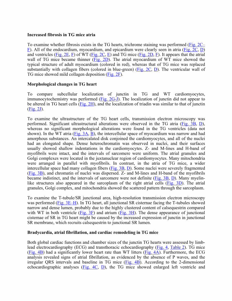
Increased fibrosis in TG mice atria To examine whether fibrosis exists in the TG hearts, trichrome staining was performed (Fig. 2C�F). All of the endocardium, myocardium, and epicardium were clearly seen in atria (Fig. 2C, D) and ventricles (Fig. 2E, F) of WT (Fig. 2C, E) and TG mice (Fig. 2D, F). It appears that the atrial wall of TG mice became thinner (Fig. 2D). The atrial myocardium of WT mice showed the typical structure of adult myocardium (colored in red), whereas that of TG mice was replaced substantially with collagen fibers (colored in blue-green) (Fig. 2C, D). The ventricular wall of TG mice showed mild collagen deposition (Fig. 2F). Morphological changes in TG heart To compare subcellular localization of junctin in TG and WT cardiomyocytes, immunocytochemistry was performed (Fig. 2G-J). The localization of junctin did not appear to be altered in TG heart cells (Fig. 2H), and the localization of triadin was similar to that of junctin (Fig. 2J). To examine the ultrastructure of the TG heart cells, transmission electron microscopy was performed. Significant ultrastructural alterations were observed in the TG atria (Fig. 3B, D), whereas no significant morphological alterations were found in the TG ventricles (data not shown). In the WT atria (Fig. 3A, B), the intercellular space of myocardium was narrow and had amorphous substances. An intercalated disk conjoined the cardiomyocytes, and all of the nuclei had an elongated shape. Dense heterochromatin was observed in nuclei, and their surfaces usually showed shallow indentations in the cardiomyocytes. Z- and M-lines and H-band of myofibrils were intact, and the intervals of sarcomere were uniform. The atrial granules and Golgi complexes were located in the juxtanuclear region of cardiomyocytes. Many mitochondria were arranged in parallel with myofibrils. In contrast, in the atria of TG mice, a wider intercellular space had many collagen fibers (Fig. 3B, D). Some nuclei were severely fragmented (Fig. 3B), and chromatin of nuclei was dispersed. Z- and M-lines and H-band of the myofibrils became indistinct, and the intervals of sarcomere were not definite (Fig. 3B, D). Many myelin-like structures also appeared in the sarcoplasm of the right atrial cells (Fig. 3D). The atrial granules, Golgi complex, and mitochondria showed the scattered pattern through the sarcoplasm. To examine the T-tubule/SR junctional area, high-resolution transmission electron microscopy was performed (Fig. 3E�H). In TG heart, all junctional SR cisternae facing the T-tubules showed narrow and dense lumen, probably due to the highly clustered content of calsequestrin compared with WT in both ventricle (Fig. 3F) and atrium (Fig. 3H). The dense appearance of junctional cisternae of SR in TG heart might be caused by the increased expression of junctin in junctional SR membrane, which recruits calsequestrin to junctional SR lumen. Bradycardia, atrial fibrillation, and cardiac remodeling in TG mice Both global cardiac functions and chamber sizes of the junctin TG hearts were assessed by limb-lead electrocardiography (ECG) and transthoracic echocardiography (Fig. 4, Table 2). TG mice (Fig. 4B) had a significantly lower heart rate than WT litters (Fig. 4A). Furthermore, the ECG analysis revealed signs of atrial fibrillation, as evidenced by the absence of P waves, and the irregular QRS intervals and baseline in TG mice (Fig. 4B). According to the 2-dimensional echocardiographic analyses (Fig. 4C, D), the TG mice showed enlarged left ventricle and
markedly dilated right atrium and right ventricle. M-mode tracing demonstrated dilated right ventricle with paradoxical interventricular septal motion (Fig. 4F) probably due to increased right ventricular cavity compared to WT litters (Fig. 4E). Overexpression of junctin also resulted in the increased left ventricular end diastolic dimension (LVEDD), left ventricular end systolic dimension (LVESD), left ventricular mass and decreased heart rate, fractional shorting (FS) and mean velocity of circumferential fiber shorting (mVcf), whereas interventricular septum thickness (IVSth) and left ventricular posterior wall thickness (LVPWth), were not altered significantly (Table 2). Expression levels of E-C coupling proteins in junctin TG heart To see whether the overexpression of junctin altered the expression levels of other E-C coupling proteins, we performed Western blot analysis, using left ventricular whole homogenates of TG and WT mice (Fig. 5, Table 3). The expression level of junctin in TG hearts was markedly higher than in WT hearts, as shown in Figure 1, whereas those of endogenous mouse junctin and triadin were significantly lower in TG hearts than in WT hearts. The expression level of RyR decreased slightly, whereas that of DHPR increased 2.4 times. The expression levels of calsequestrin, calreticulin, FKBP12, phospholamban, SERCA2a, and Na+-Ca2+ exchanger did not change significantly. To examine whether junctin overexpression altered the characteristics of RyR, ryanodine binding to whole homogenates was measured at various [3H]ryanodine concentrations. As the [3H]ryanodine concentration increased from 0 to 16 nM, ryanodine binding to whole homogenates was rapidly activated and saturated. The parameters [maximal binding (Bmax) and dissociation constant (Kd)] of ryanodine binding were calculated by iterative computer fitting, using the following equation: Y = Bmax × [X/(Kd + X)]. The densities of RyR, as determined by Bmax of ryanodine to the receptor, were significantly lower in TG hearts than in WT hearts (0.751 ± 0.014 vs. 0.979 ± 0.032 pmol/mg protein, n = 4, P < 0.05), whereas the Kd of ryanodine binding was not significantly altered (WT: 1.39 ± 0.11 vs. TG: 1.33 ± 0.07 nM, n = 4). Caffeine-induced Cai-transients in junctin TG cardiomyocytes The possible alterations of SR-mediated Ca2+ transients in TG mice were studied by measuring the resting and the elevated cytosolic Ca2+ concentration by application of 10 mM caffeine in enzymatically isolated ventricular cardiomyocytes (Fig. 6). The addition of 10 mM caffeine to the cardiomyocytes led to a rapid increase of cytosolic Ca2+ concentrations. The calculated intracellular Ca2+ concentrations at basal, transient, and steady state were 215.42 ± 48.7, 855.46 ± 185.4, and 150.2 ± 46.9, respectively, for WT (n=12), and 108.8 ± 17.72, 450.36 ± 127.7, and 67.6 ± 13.6, respectively, for junctin TG cardiomyocytes (n=8). In junctin-overexpressing cardiomyocytes, Ca2+ concentrations at all of the basal, transient, and steady states appeared to be generally lower than in WT cardiomyocytes. However, only basal intracellular Ca2+ concentrations were significantly lower in TG cardiomyocytes than in WT cardiomyocytes (P < 0.05). Characteristics of L-type Ca2+ currents (ICa) in junctin TG cardiomyocytes In light of the evidence that the expression level of DHPR increased in the TG mice (Fig. 5), the characteristics of ICa currents in ventricular cardiomyocytes were examined (Fig. 7A). The measured membrane capacitance, an indicative of cell size, was not significantly different

between the two types of ventricular cardiomyocytes (324.6 ± 16.7 pF, n=26 vs. 313.8 ± 15.2 pF, n=20). Figure 7B shows typical current-voltage relationships of ICa for WT and TG cardiomyocytes. When peak ICa was normalized to cell capacitance, the current-voltage relationships were similar between the two groups, but the current density of peak ICa was significantly larger in TG cardiomyocytes compared with that in WT cardiomyocytes. The average values were 2.15 ± 0.19 pA/pF (n=26) for WT cardiomyocytes and 3.18± 0.32 pA/pF (n=20) for TG cardiomyocytes. Prolongation of action potential duration in junctin TG cardiomyocytes To test whether overexpression of junctin could affect the general electrophysiological characteristics of the heart, the characteristics of the action potential in the ventricle and atrium of WT and TG hearts were examined (Fig. 7C�F). Action potentials were recorded from isolated mouse ventricular and atrial muscles with 3 M KCl-filled conventional microelectrodes. The resting membrane potential in WT ventricular muscle was �67.3 ± 4.3 mV (n = 16), and the action potential duration at 70% repolarization (APD70) from the same preparations was 29.2 ± 4.0 ms. In TG ventricular muscles, the resting membrane potential was not changed (�64.3 ± 3.1 mV, n = 7), but the APD70 increased significantly to 49.0 ± 7.2 ms. The resting membrane potential in WT atrial muscle was �70 ± 1.4 mV (n = 16), and the APD70 from the same preparation was 46.7 ± 4.1 ms. In TG atrial muscle, the resting membrane potential was not changed (�65 ± 2.4 mV, n=12), but APD70 increased significantly to 91.5 ± 8.5 ms. The muscles used for action potential measurement were stimulated at 2 Hz DISCUSSION Cardiac junctin is localized in the junctional SR, and it appears to interact with calsequestrin and the RyR (15). However, the functional consequences of the interaction in the heart have not been fully elucidated. In this study, we generated TG mouse lines overexpressing dog cardiac junctin (24- to 29-fold) (Fig. 1) and showed that overexpression of junctin in the hearts of newborn mice induced dramatic heart remodeling (Fig. 2) and bradycardia with atrial fibrillation (Fig. 4). These pathophysiological changes in the heart were accompanied by various morphological (Fig 3), biochemical (Fig. 5), and electrical (Fig. 7) changes to the heart cells. Recently, TG mouse lines that overexpressed canine junctin were also reported (29). However, those TG mouse lines showed only mild phenotype changes such as tighter and more extended structures of SR cisternae and T-tubules (29). The severity of the phenotype changes could be related to the degree of overexpression of junctin, because the reported overexpression level of junctin was only 5- to 10-fold (29). We have identified two independent lines of severely overexpressed junctin TG mice (24- and 29-fold higher than WT), and both TG lines showed the similar phenotype changes, as described previously. Judging from the severe phenotype changes, the junctin TG mice died earlier because of heart failure (Fig. 1G). However, our other TG lines (#20 and #110), which overexpressed canine junctin 9- to 11-fold, showed a phenotype of concentric left ventricular enlargement, ventricular premature beats, and bradycardia on ECG monitoring (data not shown). These results suggest that cardiac remodeling and bradycardia could occur both in mildly and severely junctin-overexpressed hearts. In many cardiac gene-specific TG mice models, the enlargements of hearts were commonly reported and the majority of them were due to myocardial hypertrophy (30�41). However, the increased mass of the heart in the our TG animals (Table 1) may not be due to hypertrophy of

cardiomyocytes per se, but it could be due to the formation of fibrosis (Fig. 2), because the cell capacitance measurement did not show any significant change. The bradycardia found in the TG heart (Table 2 and Fig. 4B) could be explained by increased DHPR expression (Fig. 5), elevated L-type Ca2+ current density (Fig. 7B), and prolongation of the action potential duration (Fig. 7D, F). The increased expression level of DHPR could cause the increased density of L-type Ca2+ current (Fig. 7B). The increased L-type Ca2+ current density might affect the electrical properties in TG heart, such as the action potentials. The attenuated membrane repolarization by increased L-type Ca2+ current density might induce the prolonged refractory period and action potential duration. The prolonged action potential could result in the bradycardia in junctin TG hearts. Our interpretation of the underlying mechanisms for the observed bradycardia in junctin TG hearts can be supported by a previous study (42) showing that hypertensively hypertrophied rabbit heart had longer action potential duration and the reduced ratio of RyR to DHPR. In a similar context, the increased L-type Ca2+ current density and bradycardia were reported in calreticulin TG mice (43). The paradoxical septal motion as seen in the M-mode echocardiogram of the junctin TG mice could be due to the enlarged right ventricular cavity, altered electrical conductivity, and/or enhanced electrical heterogeneity (Fig. 4F). In patients with atrial septum defects, right ventricular enlargement and paradoxical septal motion were predominantly observed (44, 45). The marked enlargement of right ventricles was found in TG mice (Fig. 2B) and subsequently progressed to right ventricular failure characterized by hepatomegaly, ascites, and edema. Note that we have reproducibly observed the spontaneous beating of the isolated cardiac Purkinje fibers in the junctin TG mice, whereas no spontaneous beating was observed in WT Purkinje fibers (unpublished observations). The Purkinje fibers showing pace-making activity may act as ectopic foci and induce an arrhythmia in the junctin TG mice. Chronic atrial fibrillation, an age-related prevalent arrhythmia, has been reported to induce structural remodeling in the case of human, goat, and dog disease models. (46�48). In the rapid pacing dog model, atrial fibrillation and biatrial cardiomyopathy were observed (48). In a similar goat model, ultrastructural remodeling such as abnormally shaped mitochondria and dispersed chromatin in nuclei were observed (47). These phenotype changes appear to be similar to the morphological changes found in our junctin TG mice (Fig. 3). Because the proteins in the E-C coupling system (RyR, calsequestrin, triadin, and junctin) are closely associated with each other, a selective overexpression of one protein in the complex may interrupt their tight balance, hence we measured the Ca2+ release function (15, 29�32, 38), the expression levels of triadin, calsequestrin, and RyR along with other proteins involved in the E-C coupling processes (Fig. 5 and Table 3). The expression level of calsequestrin was not altered in the TG heart, but that of triadin and the density of RyR, as determined by [3H]ryanodine binding, decreased significantly (Fig. 5 and Table 3). Because the overall structures and interacting characteristics of junctin and triadin are similar, the overexpression of junctin may be compensated for by the down-regulation of triadin (Fig. 5) (15). The expression levels of cardiac triadin bands composed of isoforms and glycosylated forms decreased in junctin TG heart (49). Similarly, the expression level of junctin also decreased in the triadin TG mice (30). Recently, it was reported that the expression level of junctin was reduced, but that of triadin was not altered, in β1-adrenergic receptor TG mice, which showed impaired contraction and relaxation associated with abnormal calcium handling (50). These results also suggest that junctin may play an important role in E-C coupling.
To examine whether the altered expression of the E-C coupling proteins (Fig. 5 and Table 3) could cause an altered Ca2+ transient in heart cells, we monitored caffeine-induced Ca2+ release by using ventricular cardiomyocytes (Fig. 6). The overall trace of caffeine-induced Ca2+ release was lower, but the basal Ca2+ level was significantly lower in the TG heart. This lower basal Ca2+ level in the TG heart could be at least in part due to the lower density of RyR (Table 3). The junctin TG mice in this study have shown various morphological and physiological alterations. The most outstanding phenotype changes would be the substantial biatrial and biventricular enlargements and the occurrence of the atrial fibrillation. Those phenotype changes could be caused by the perturbations of the intracellular Ca2+ homeostasis caused by the overexpression of junctin and consequent changes of the protein expression of the other E-C coupling proteins such as DHPR. Our TG model may be helpful in elucidating the relationship between the altered expression levels of cardiac membrane proteins and the pathogenesis of hearts, especially related to conduction abnormalities. ACKNOWLEDGMENTS We thank K. P. Campbell for anti-triadin, W. K. Song for anti-calreticulin, and W. J. Park for anti-calsequestrin antibodies. This work was supported by grants from the Korea Ministry of Science and Technology (Critical Technology 21, 00-J-LF-01-B-54), Korea Science and Engineering Foundation (Basic Research Program 1999-1-20700-002-5), and Ministry of Education (Brain Korea 21 Project). REFERENCES 1. Beam, K.G., Knudson, C.M., and Powell, J.A. (1986) A lethal mutation in mice eliminates the slow calcium current in skeletal muscle cells. Nature 320, 168�170 2. Rios, E., and Brum, G. (1987) Involvement of dihydropyridine receptors in excitation-contraction coupling in skeletal muscle. Nature 325, 717�720 3. Block, B. A., Imagawa, T., Campbell, K. P., and Franzini-Armstrong, C. (1988) Structural evidence for direct interaction between the molecular components of the transverse tubule/sarcoplasmic reticulum junction in skeletal muscle. J. Cell. Biol. 107, 2587�2600 4. Tanabe, T., Beam, K. G., Powell, J.A., and Numa, S. (1988) Restoration of excitation-contraction coupling and slow calcium current in dysgenic muscle by dihydropyridine receptor complementary DNA. Nature 336, 134�139 5. Rios, E., and Pizarro, G. (1991) Voltage sensor of excitation-contraction coupling in skeletal muscle. Physiol. Rev. 71, 849�908 6. Anderson, K., and Meissner, G. (1995) T-tubule depolarization-induced SR Ca2+ release is controlled by dihydropyridine receptor- and Ca2+-dependent mechanisms in cell homogenates from rabbit skeletal muscle. J. Gen. Physiol. 105, 363�383 7. Fleischer, S., and Inui, M. (1989) Biochemistry and biophysics of excitation-contraction coupling. Annu. Rev. Biophys. Biophys. Chem. 18, 333�364

8. Franzini-Armstrong, C., and Jorgensen, A.O. (1994) Structure and development of E-C coupling units in skeletal muscle. Annu. Rev. Physiol. 56, 509�534 9. Inui, M., Saito, A., and Fleischer, S. (1987) Purification of the ryanodine receptor and identify with feet structure of junctional terminal cisternae of sarcoplasmic reticulum from fast skeletal muscle. J. Biol. Chem. 262, 1740�1747 10. MacLennan, D.H., Brandl, C.J., Korczak, B., and Green, N.M. (1985) Amino-acid sequence of Ca2++Mg2+-dependent ATPase form rabbit muscle sarcoplasmic reticulum, deduced from its complementary DNA sequence. Nature 316, 696�700 11. Meissner, G. (1994) Ryanodine receptor/Ca2+ release channel and their regulation by endogenous effectors. Annu. Rev. Physiol. 56, 485�508 12. Yano, K., and Zarain-Herzberg, A. (1994) Sarcoplasmic reticulum calsequestrins. Mol. Cell. Biochem. 135, 61�70 13. Guo, W., Jorgensen, A. O., Jones, L. R, and Campbell, K.P. (1996) Biochemical characterization and molecular cloning of cardiac triadin. J. Biol. Chem. 271, 458�465 14. Jones, L.R., Zhang, L., Sanborn, K., Jorgensen, A.O., and Kelley, J. (1995) Purification, primary structure, and immunological characterization of the 26-kDa calsequestrin binding protein (Junctin) from cardiac junctional sarcoplasmic reticulum. J. Biol. Chem. 270, 30787�30796 15. Zhang, L., Kelly, J., Schmeisser, G., Kobayashi, Y.M., and Jones, L.R. (1997) Complex formation between junctin, triadin, calsequestrin, and ryanodine receptor. J. Biol. Chem. 272, 23389�23397 16. Ikemoto, N., Ronjat, M., Meszaros, L.G., and Koshita, M. (1989) Postulated role of calsequestrin in the regulation of calcium release from sarcoplasmic reticulum. Biochemistry 28, 6764�6771 17. Ohkura, M., Furukawa, K., Fujimori, H., Kuruma, A., Kawano, S., Hiraoka, M., Kuniyasu, A., Nakayama, H., and Ohizumi, Y. (1998) Dual regulation of the skeletal muscle ryanodine recpetor by triadin and calsequestrin. Biochemistry 37, 12987�1299 18. Mitchell, R.D., Simmerman, H.K.B., and Jones, L.R. (1988) Ca2+ binding effects on protein conformation and protein interactions of canine cardiac calsequestrin. J. Biol. Chem. 263, 1376�1381 19. Palermo, J., Gulick, J., Colbert, M., Fewell, J., and Robbins, J. (1996) Transgenic remodeling of the contractile apparatus in the mammalian heart. Circ. Res. 78, 504�509 20 Pollak, O.J. (1972) A rapid trichrome stain. In Animal Tissue Techniques. (Humason G.L., ed) pp. 182-183, W. H. Freeman and Company, New York 21. Kim, D.H., Mkparu, F., Kim, C.R. and Caroll, R.F. (1994) Alteration of Ca2+ release

channel function in sarcoplasmic reticulum of pressure-overload-induced hypertrophic rat heart. J. Mol. Cell. Cardiol. 26, 1505�1512 22. Bradford, M.M. (1976) A rapid and sensitive method for the quantitation of microgram quantities of protein utilizing the principle of protein-dye binding. Anal. Biochem. 72, 248�254 23. Laemmli, U.K. (1970) Cleavage of structural proteins during the assembly of the head of bacteriophage T4. Nature 227, 680�685 24. Towbin, H., Staehelin, T., and Gordon, J. (1979) Electrophoretic transfer of proteins from polyacrylamide gels to nitrocellulose sheets: procedure and some applications. Proc. Natl. Acad. Sci. U. S. A. 76, 4350�4354 25. Shin, D.W., Ma, J., and Kim, D.H. (2000) The asp-rich region at the carboxyl-terminus of calsequestrin binds to Ca2+ and interacts with triadin. FEBS Lett. 486, 178�182. 26. Kwon M.S., Park C.S., Choi K., Ahnn J., Kim J.I., Eom S.H., Kaufman S.J., and Song W.K. (2000) Calreticulin couples calcium release and calcium influx in integrin-mediated calcium signaling. Mol. Biol. Cell 11, 1433�1443 27. Tian, Q., Katz, A.M., and Kim, D.H. (1991) Effects of azumolene on doxorubicin-induced Ca2+ release from skeletal and cardiac muscle sarcoplasmic reticulum. Biochim. Biophys. Acta. 1094, 27�34 28. Campbell, K.P., Knudson, C.M., Imagawa, T., Leung, A.T., Sutko, J.L., Kahl, S.D., Raab, C.R., and Madson, L. (1987) Identification and characterization of the high affinity [3H]ryanodine receptor of the junctional sarcoplasmic reticulum Ca2+ release channel. J. Biol. Chem. 262, 6460�6463 29. Zhang, L., Franzini-Armstrong, C., Ramesh, V., and Jones, L.R. (2001) Structural alterations in cardiac calcium release units resulting from overexpression of junctin. J. Mol. Cell. Cardiol. 33, 233�247 30. Kirchhefer, U., Neumann, J., Baba, H.A., Begrow, F., Kobayashi, Y.M., Reinke, U., Schmitz W., and Jones, L.R. (2001) Cardiac hypertrophy and impaired relaxation in transgenic mice overexpressing triadin 1. J. Biol. Chem. 276, 4142�4149 31. Sato, Y., Ferguson, D.G., Sako, H., Dorn, G.W. 2nd, Kadambi, V.J., Yatani, A., Hoit, B.D., Walsh, R.A., and Krania,s E.G. (1998) Cardiac-specific overexpression of mouse cardiac calsequestrin is associated with depressed cardiovascular function and hypertrophy in transgenic mice. J. Biol. Chem. 273, 28470�28477 32. Jones, L.R., Suzuki, Y.J., Wang, W., Kobayashi, Y.M., Ramesh, V., Franzini-Armstrong, C., Cleemann, L., and Morad, M. (1998) Regulation of Ca2+ signaling in transgenic mouse cardiac myocytes overexpressing calsequestrin. J. Clin. Invest. 101, 1385�1393 33. Molkentin, J.D., Lu J.R., Antos, C.L., Markham, B., Richardson, J., Robbins, J., Grant, S.R., Olson, E.N. (1998) A calcineurin-dependent transcriptional pathway for cardiac hypertrophy. Cell 93, 215�228

34. Hein, L., Stevens, M.E., Barsh, G.S., Pratt, R.E., Kobilka, B.K., and Dzau, V.J. (1997) Overexpression of angiotensin AT1 receptor transgene in the mouse myocardium produces a lethal phenotype associated with myocyte hyperplasia and heart block. Proc. Natl. Acad. Sci. U. S. A. 94, 6391�6396 35. Paradis, P., Dali-Youcef, N., Paradis, F.W., Thibault, G., and Nemer, M. (2000) Overexpression of angiotensin II type I receptor in cardiomyocytes induces cardiac hypertrophy and remodeling. Proc. Natl. Acad. Sci. U. S. A. 97, 931�936 36. Mende, U., Kagen, A., Cohen, A., Aramburu, J., Schoen, F.J., Neer, E.J. (1998) Transient cardiac expression of constitutively active Gαq leads to hypertrophy and dilated cardiomyopathy by calcineurin-dependent and independent pathways. Proc. Natl. Acad. Sci. U. S. A. 95, 13893�13898 37. MacLellan, W.R., and Schneider, M.D. (2000) Genetic dissection of cardiac growth control pathways. Annu. Rev. Physiol. 62, 289-319 38. Kiriazis, H., and Kranias, E.G. (2000) Genetically engineered models with alterations in cardiac membrane calcium-handling proteins. Annu. Rev. Physiol. 62, 321�351 39. Gruver, C.L., DeMayo, F., Goldstein, M.A., and Means, A.R. (1993) Targeted developmental overexpression of calmodulin induces proliferative and hypertrophic growth of cardiomyocytes in transgenic mice. Endocrinology 133, 376�388 40. Robbins, R.J., and Swain, J.L. (1992) C-myc protooncogene modulates cardiac hypertrophic growth in transgenic mice. Am. J. Physiol. 262, H590�H597 41. Katz, E.B., Steinhelper, M.E., Delcarpio, J.B., Daud, A.I., Claycomb, W.C., and Field, L.J. (1992) Cardiomyocyte proliferation in mice expressing alpha-cardiac myosin heavy chain-SV40 T-antigen transgenes. Am. J Physiol. 262, H1867�H1876 42. Milnes, J.T., and MacLeod, K.T. (2001) Reduced ryanodine receptor to dihydropyridine receptor ratio may underlie slowed contraction in a rabbit model of left ventricular cardiac hypertrophy. J. Mol. Cell. Cardiol. 33, 473�485 43. Nakamura, K, Robertson, M., Liu, G., Dickie, P., Nakamura, K., Guo, J.Q., Duff, H.J., Opas, M., Kavanagh, K., and Michalak, M. (2001) Complete heart block and sudden death in mice overexpressing calreticulin. J. Clin. Invest. 107, 1245�1253 44. Lieppe, W., Scallion, R., Behar, and V.S., Kisslo, J.A. (1977) Two-dimensional echocardiographic findings in atrial septal defect. Circulation 56, 447�456 45. Weyman, A.E., Wann, S., Feigenbaum, H., and Dillon, J.C. (1976) Mechanism of abnormal septal motion in patients with right ventricular volume overload: a cross-sectional echocardiographic study. Circulation 54, 179�186 46. Thijssen, V.L., Ausma, J., and Borgers, M. (2001) Structural remodelling during chronic atrial fibrillation: act of programmed cell survival. Cardiovasc. Res. 52, 14�24
47. Ausma, J., Wijffels, M., Thone, F., Wouters, L., Allessie, M., and Borgers, M. (1997) Structural changes of atrial myocardium due to sustained atrial fibrillation in the goat. Circulation 96, 3157�3163 48. Morillo, C.A., Klein, G.J., Jones, D.L., and Guiraudon, C.M. (1995) Chronic rapid atrial pacing. Structural, functional, and electrophysiological characteristics of a new model of sustained atrial fibrillation. Circulation 91, 1588�1595 49. Hong C.S., Ji J.H., Kim J.P., Jung D.H., and Kim D.H. (2001) Molecular cloning and characterization of mouse cardiac triadin isoforms. Gene 278, 193�199. 50. Engelhardt S., Boknik P., Keller U., Neumann J., Lohse M.J., and Hein L. (2001) Early impairment of calcium handling and altered expression of junction in hearts of mice overexpressing the beta1-adrenergic receptor. FASEB J 15, 2718�2720
Received December 17, 2001; revised April 11, 2002.

Table 1 Junctin overexpression is accompanied by heart enlargementa
WT TG
Number of observations 3 3 Body weight (g) 23.4 ± 0.3 20.1 ± 2.5 Heart weight (mg) 114.0 ± 4.6 228.0 ± 32.5* Heart weight/body weight (mg/g) 4.86 ± 0.27 11.25 ± 0.85* Left ventricle weight/body weight (mg/g) 2.43 ± 0.16 4.40 ± 0.58* Right ventricle weight/body weight (mg/g) 1.01 ± 0.04 2.50 ± 0.24* Left atrium weight/body weight (mg/g) 0.24 ± 0.05 0.40 ± 0.06 Right atrium weight/body weight (mg/g) 0.19 ± 0.02 2.83 ± 0.71*
aBody weight and heart weight were determined for wild-type (WT) and junctin overexpressing, or transgenic (TG), mice. The indicated values are means ±SE. *P < 0.05 vs. WT.

Table 2 Echocardiographic parameters observed in wild-type (WT) and junctin overexpressing, or transgenic (TG), micea
WT TG
Number of observations 9 17 LVEDD (mm) 2.8 ± 0.1 3.9 ± 0.2* LVESD (mm) 1.3 ± 0.1 2.0 ± 0.2* IVSth (mm) 0.86 ± 0.04 0.89 ± 0.04 LVPWth (mm) 0.87 ± 0.06 0.89 ± 0.03 HR (beats/min) 424 ± 25 272 ± 17* FS (%) 57.1 ± 3.3 48.2 ± 2.5* mVcf (circ/s) 8.5 ± 0.5 6.3 ± 0.5* LVM/BW (mg/g) 3.2 ± 0.2 5.7 ± 0.5*
aThe echocardiographic parameters of WT and TG mouse hearts (8–9 weeks old) were studied. LVESD, left ventricular end systolic dimension; LVEDD, left ventricular end diastolic dimension; IVSth, interventricular septum thickness; LVPWth, left ventricular posterior wall thickness; HR, heart rate; FS, fractional shorting; mVcf, mean velocity of circumferential fiber shorting; LVM, left ventricular mass; BW, body weight. The indicated values are mean ±SE. *P < 0.05.

Table 3 Relative levels of E-C coupling-associated proteins in wild-type (WT) and junctin, or transgenic (TG), mouse heartsa
WT TG
Number of observations 4 4 Junctin 1.00 ± 0.12 28.73 ± 0.05* Mouse junctin 1.00 ± 0.04 0.56 ± 0.07* Ryanodine receptor 1.00 ± 0.06 0.88 ± 0.09 Calsequestrin 1.00 ± 0.05 0.93 ± 0.04 Calreticulin 1.00 ± 0.04 1.05 ± 0.05 Triadin 1.00 ± 0.07 0.20 ± 0.10* FKBP12 1.00 ± 0.11 1.05 ± 0.07 Phospholamban 1.00 ± 0.08 0.96 ± 0.09 SERCA2a 1.00 ± 0.03 0.96 ± 0.03 Na-Ca exchanger 1.00 ± 0.12 1.03 ± 0.10 Dihydropyridine receptor 1.00 ± 0.09 2.35 ± 0.07*
aProtein levels in cardiac homogenates were determined by densitometry of the immunoblots. Values were normalized to the average level of the specific protein in WT hearts. [3H]ryanodine binding was also measured using cardiac homogenates. The indicated values are means ±SE. *P < 0.05 vs. WT.

Fig. 1
Figure 1. Generation of cardiac-specific junctin transgenic (TG) mice. A) Canine junctin cDNA encoding full-length protein was subcloned between α-myosin heavy chain promoter and human growth hormone polyadenylation sequence (hGH poly A), as described in Materials and Methods. B) Genomic polymer chain reaction products of wild-type (WT) and junctin TG mice amplified by using the junctin transgene-specific primers are shown. C) Genomic Southern blot of WT and TG mice was conducted using the junctin transgene-specific probe. D) Northern blot of WT and TG cardiac RNA was conducted by hybridizing with the [α-32P]dCTP-labeled canine junctin cDNA. E) Western blot of WT and TG heart homogenate was conducted using anti-junctin antibody. F) The junctin expression levels in WT and TG heart were determined by densitometry. G) Kaplan-Meier survival analysis of junctin TG mice (ML#129).

Fig. 2
Figure 2. Phenotype changes in the heart of TG mice. A) Gross morphology of WT and TG mouse hearts. B) Longitudinal section of WT and TG mouse hearts. Both bi-atria and biventricular enlargements were seen in TG mouse. C–F) Trichrome staining of the hearts in the WT and TG mice. Atria (C and D) and ventricles (E and F) consisted of endocardium (arrow), myocardium (M), and epicardium (arrowhead) in both WT (C, E) and TG mice (D, F). Cardiomyocytes and collagen fibers were stained with red and blue green colors, respectively. RA, right atrium; LA, left atrium; RV, right ventricle; LV, left ventricle. Scale bar: 50 µm. G–J) Sections of WT and TG ventricles were immunostained with anti-junctin (G, H) or anti-triadin (I, J) antibodies. Scale bar: 5 µm.

Fig. 3
Figure 3. Transmission electron micrographs of TG heart. A, C) In WT left and right atrium, sarcomeres were regular and surrounded by mitochondria. B, D) In junctin TG atrium, Z-line (arrowhead), M-line, and H-band became indistinct and the intervals of sarcomere were not definite, and mitochondria showed scattered pattern through sarcoplasm. Some nuclei were severely fragmented with dispersed chromatin. Many myelin-like structures (arrow) appeared in the cardiomyocytes of TG right atrium (D). N, nucleus; C, collagen fibril; Scale bars: 1 µm. F, H) In TG ventricular and atrial junctional sarcoplasmic reticulum cisternae (arrowhead) facing the T-tubules showed narrow and dense lumen compared with WT counterparts (E, G). Scale bar: 200 nm.

Fig. 4
Figure 4. Electrocardiogram and echocardiogram from WT and TG mice. A, B) Representative traces of electrocardiogram from WT and TG mice. The P waves (arrowhead) disappeared in TG mice, and the intervals of QRS complex (arrow) were irregular in TG mice (B). C, D) Representative 2-D echocardiogram of WT and junctin TG mice. Left ventricle was enlarged, and right atrium and right ventricle were markedly dilated in TG mice on the parasternal long-axis view (D). E, F) Representative M-mode echocardiogram from WT and junctin TG mice. Right ventricular dimension was enlarged, and interventricular septum showed paradoxical motion in TG mice (F). LV, left ventricle; RV, right ventricle; AO, aorta; septum, interventricular septum.

Fig. 5
Figure 5. Western blot analysis of E-C coupling proteins in WT and TG heart ventricular homogenates. Western blot analysis was conducted as described in Materials and Methods by using the specific primary antibodies. tJTN, total junctin; mJTN, mouse junctin; RyR, ryanodine receptor; CSQ, calsequestrin; CRT, calreticulin; TRN, triadin; FKBP12, FK506 binding protein 12; PLB, phospholamban; SERCA2a, sarcoplasmic reticular Ca2+ ATPase 2a; NCX, Na+-Ca2+ exchanger; DHPR, dihydropyridine receptor.

Fig. 6
Figure 6. Caffeine-induced Ca2+-release in WT and TG ventricular cardiomyocytes. A) Representative recordings of the Ca2+ transients induced by 10 mM caffeine in fura-2 loaded cardiomyocytes from WT and TG mice. B) Averaged values of basal intracellular Ca2+ concentration, Ca2+ transients induced by 10 mM caffeine, and sustained intracellular Ca2+ concentrations obtained from WT (n=12) and junctin TG (n=8) ventricular cardiomyocytes. *P < 0.05 vs. WT

Fig. 7
Figure 7. Electrophysiological changes in TG hearts. A, B) ICa in WT and junctin-overexpressing TG ventricular cardiomyocytes. A) Typical whole-cell L-type Ca2+ currents recorded in WT (n=26) and TG (n=20) cells. Shown are traces of currents that were recorded from a holding potential of –50 mV to the indicated test potentials. B) The peak current-voltage relationships obtained from these cells. The peak currents were normalized to cell capacitance. C–F) Action potentials in ventricular and atrial muscles of WT and junctin TG mice. C) Typical representations of action potentials in WT and junctin TG ventricular muscle. D) Averaged values of action potential durations at 70% repolarization (APD70) of ventricular muscle isolated from WT (n=16) and junctin TG mice (n=7). E) Typical representations of action potentials in WT and junctin TG atrial muscle. F) Averaged values of APD70 of atrial muscle isolated from WT (n=16) and junctin TG mice (n=12). *P < 0.05 vs. WT