CHADS2 score predicts atrial fibrillation following cardiac surgery
Iotthn
FHpHJSgcBC
a
Journal of the American College of Cardiology Vol. 54, No. 22, 2009© 2009 by the American College of Cardiology Foundation ISSN 0735-1097/09/$36.00P
QUARTERLY FOCUS ISSUE: HEART RHYTHM DISORDERS
The Role of Ganglionated Plexiin Apnea-Related Atrial Fibrillation
Muhammad Ghias, MD, Benjamin J. Scherlag, PHD, Zhibing Lu, MD, Guodong Niu, MD,Annerie Moers, MD, Warren M. Jackman, MD, Ralph Lazzara, MD, Sunny S. Po, MD, PHD
Oklahoma City, Oklahoma
Objectives This study was conducted to simulate sleep apnea-induced atrial fibrillation (AF) in an experimental model andto determine whether neural ablation will prevent AF.
Background An increasing number of clinical reports have associated sleep apnea and AF, and many possible mechanismsresponsible for this relationship have been proposed.
Methods Thirty dogs anesthetized with Na-pentobarbital were ventilated by a positive pressure respirator. Protocol 1 (n �
14): After a right thoracotomy, atrial and pulmonary vein programmed pacing at 2� and 4� threshold deter-mined the shortest atrial refractory period. Obstructive apnea was induced by turning off the respirator duringend expiration for 2 min. During apnea, programmed pacing was performed with S1-S2 � 5 to 10 ms earlierthan the atrial refractory period. Neural activity was monitored from the ganglionated plexi (GP) adjacent to theright pulmonary veins. Protocol 2 (n � 16): Electrical stimulation identified the GP at the right pulmonary artery(RPA). Programmed pacing was again instituted, below atrial refractory period, during 2 min of apnea. After ra-diofrequency ablation of the RPA GP, continuous programmed pacing was again repeated during 2 min of ap-nea. In 5 dogs, blood gases were determined at baseline and at 2 min of apnea.
Results Protocol 1: During apnea, S1-S2 induced AF within 85 � 38 s (9 of 10). In 1 case, AF occurred spontaneously at1 min 36 s of apnea. Recorded GP neural activity progressively increased before AF onset. Systolic but not dia-stolic blood pressure rose significantly before AF (149 � 26 mm Hg to 193 � 38 mm Hg, p � 0.05). In 4 dogs,autonomic blockade prevented apnea-induced AF. Protocol 2: AF induced by pacing occurred in 8 of 11 dogswithin the 2-min period of apnea, before neural ablation. After ablation, 0 of 6 showed AF during 2 min of apnea(p � 0.009).
Conclusions This experimental model of apnea shows a reproducible incidence of AF. After neural ablation of the RPA GP orautonomic blockade, AF inducibility was significantly inhibited. (J Am Coll Cardiol 2009;54:2075–83) © 2009by the American College of Cardiology Foundation
ublished by Elsevier Inc. doi:10.1016/j.jacc.2009.09.014
po
eiupi
M
Ta
n the last few years, there have been an increasing numberf clinical reports associating sleep apnea and atrial fibrilla-ion (AF) (1–8). Many possible mechanisms responsible forhis relationship have been proposed, including hypoxemia,ypercapnea, pulmonary and systemic hypertension, auto-omic factors, stretch-mediated channel activation, and
rom the Heart Rhythm Institute, College of Medicine, University of Oklahomaealth Sciences Center, Oklahoma City, Oklahoma. This study was supported in
art by grants from the American Heart Association (0650077Z) and the Nationaleart, Lung and Blood Institute (5K23HL069972) to Dr. Po, and by grants from St.
ude Medical and the Helen and Wil Webster Arrhythmia Research Fund to Dr.cherlag. Dr. Scherlag is an AtriCure, Inc. consultant, a Cardiovascular Therapeuticsrantee, a St. Jude Medical grantee and consultant, and a Symphony Medicalonsultant. Dr. Jackman is a consultant to and receives research funding fromiosense Webster, Stereotaxis, ProRhythm, CardioFocus, Endosense, and Atri-ure, Inc.
cManuscript received June 13, 2009; revised manuscript received September 9, 2009,
ccepted September 21, 2009.
ro-inflammatory factors such as C-reactive protein, amongthers (9).
See page 2084
The purpose of the present study was to develop anxperimental model that simulated obstructive sleep apnea-nduced AF. Using this model, we tested the possiblenderlying autonomic mechanisms and used ganglionatedlexi (GP) ablation to prevent AF occurrence under thenfluence of a 2-min period of apnea.
ethods
hirty dogs were anesthetized with Na-pentobarbital, 30 mg/kg,nd maintained with 50 to 100 mg as needed. Insertion of a
uffed endotracheal tube allowed artificial ventilation by a
pb
acvEBM
d2Rc1tcrwAmiBwga
(Ja5bCmPwtSu
2076 Ghias et al. JACC Vol. 54, No. 22, 2009Sleep Apnea-Induced Atrial Fibrillation November 24, 2009:2075–83
positive pressure respirator (Har-vard Apparatus Co., Natick, Mas-sachusetts). The right and leftfemoral veins were dissected, and8F sheaths inserted into each ves-sel. An electrode catheter was in-serted into the left femoral artery,and passed into the aortic root torecord the His bundle potential.On the right side, the side arm ofthe sheath in the femoral arterywas connected to a pressure trans-ducer to continuously monitor ar-terial blood pressure (BP). Theright femoral vein was used todeliver drugs and for a continuoussaline infusion. In the oppositevein, a Thermistor probe (YellowSprings Tele-thermometer, YellowSprings, Ohio) was inserted and
assed into the inferior vena cava to continually monitor coreody temperature.
After a right thoracotomy at the fourth intercostal spacend reflection of the incised pericardium, multi-electrodeatheters were sutured against the right superior pulmonaryein (RSPV) and onto the right atrial (RA) free wall (Fig. 1).lectrocardiogram lead II was continuously registered on aard computerized recording system (Bard Inc., Billerica,assachusetts). The electrocardiographic lead and intracar-
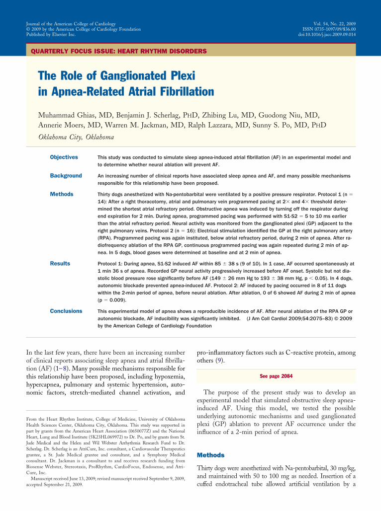
Figure 1 View From Right Thoracotomy With Attachment of Mu
Schematic representation of the view from a right thoracotomy with the attachmenatrial (RA) free wall. The positioning of a tungsten microelectrode (ME) in the gangshown. IVC � inferior vena cava; LA � left atrium; RT � right; SAN � sinoatrial no
Abbreviationsand Acronyms
AF � atrial fibrillation
ARGP � anterior rightganglionated plexi
ARP � atrial refractoryperiod
AV � atrioventricular
BP � blood pressure
GP � ganglionated plexi
PV � pulmonary vein
RA � right atrial
RPA � right pulmonaryartery
RSPV � right superiorpulmonary vein
SVC � superior vena cava
iac electrograms were electronically filtered between 0.1 to50 Hz and 30 to 250 Hz, respectively.ecordings of neural activity from the GP. Neural re-
ordings were obtained in 10 of 14 experiments in Protocol(see the following text). These recordings were made from
he anterior right ganglionated plexi (ARGP), located at theaudal end of the sinus node between the RSPV and theight inferior pulmonary vein (PV). A circular plastic ringas held against the surface of the atrium surrounding theRGP to reduce local movement (10). A coated tungstenicroelectrode with an exposed tip of 50 �m and an
mpedance of 9 to 12 Meg � at 1,000 Hz (F. Haer Co.,owdoin, Maine) was mounted on a micromanipulator,hich allowed the electrode to be inserted into the GP. Around lead was attached to the chest wall. Neuronalctivity was recorded only from the ARGP throughout.
The microelectrode signals were fed into pre-amplifiersPrinceton Applied Research, Model 113, Princeton, Newersey), which were set with bandpass filters between 300nd 10 kHz, with amplification ranging from 100� to00�. Further amplification (50� to 200�) was obtainedy use of a hardwired amplifier (Spike 2, CED, Ltd.,ambridge, England). Neural recordings from the GP wereade on the Bard recording system.rotocol 1. In 14 dogs, programmed electrical stimulationas delivered at the RA site at 2� and 4� the diastolic
hresholds. Eight S1-S1 � 330 ms cycles were followed by1-S2, progressively decrementing by 10 and then 1 msntil the atrial refractory period (ARP) was reached. The
ectrode Catheters to RSPV and RA Free Wall
ulti-electrode catheters to the right superior pulmonary vein (RSPV) and the rightd plexi (GP) and the connection to amplifiers and recording device is also� sulcus terminalis; SVC � superior vena cava.
lti-El
t of mlionatede; ST
Srrriaw4t(PpvlabeHdurAptp
SptnaCtab
F
cwspSSobcvGptMR
R
Pa
2077JACC Vol. 54, No. 22, 2009 Ghias et al.November 24, 2009:2075–83 Sleep Apnea-Induced Atrial Fibrillation
1-S2 interval was fixed at 5 to 10 ms shorter than theefractory period, and the pacing algorithm was continuallyepeated (every 2 s). Under these conditions, no conductedesponse was observed during the baseline state. Apnea wasnduced by switching off the respirator at end expiration sos to simulate obstructive sleep apnea. The apneic periodas maintained until AF occurred or 2 min had elapsed. Indogs, before apnea, autonomic blockade was induced by
he intravenous administration of esmolol or propranolol1 mg/kg) and atropine (2 mg) (Fig. 3).rotocol 2. In all dogs, we dissected a branch of the rightulmonary artery (RPA) that passed beneath the superiorena cava (SVC) and entered the upper lobe of the rightung. Cannulation of this vessel with a short 8-F sheathllowed the placement of a specially designed expandableasket probe (5 splines, each with 3 pairs of close bipolarlectrodes) into the RPA adjacent to the SVC (Fig. 2).igh-frequency electrical stimulation (frequency, 20 Hz,
uration of each stimulus, 0.1 ms, voltage 0.6 to 4.5 V) wassed to activate the neural tissues at the RPA, causing heartate and atrioventricular (AV) conduction slowing (11,12).n electrode catheter attached to the right atrium allowedrogrammed stimulation (S1-S1, S1-S2) at 2� and 4�hreshold to determine the shortest ARP. Continuous
Figure 2 Position of Basket Electrode Catheter in RPA
Diagrammatic depiction of a posterior view of the heart showing the position ofa basket electrode catheter in the right pulmonary artery (RPA) in back of orbeneath the SVC as it enters the RA. It is at this position that electrical stimu-lation can activate what has been called the posterior atrial fat pad (PAFP) orthe RPA GP. The multi-electrode catheters on the RSPV and RA are alsoshown, as described in Figure 1. AO � aorta; ARGP � anterior right ganglion-ated plexi; IRGP � inferior right ganglionated plexi; LI � left inferior pulmonaryvein; LPA � left pulmonary artery; LSPV � left superior pulmonary vein; LV �
left ventricle; RI � right inferior pulmonary vein; RV � right ventricle; otherabbreviations as in Figure 1.
rogrammed atrial pacing from the RA or RSPV with the
1-S2 set at 5 to 10 ms shorter than the ARP waserformed and apnea induced, as described in the precedingext. This procedure was performed before and after RPAeural ablation using a probe with bipolar bar electrodesttached to a radiofrequency generator (AtriCure, Inc.,incinnati, Ohio). When �1 bout of apnea was applied,
he apneic periods were separated by 30-min intervals tollow recovery. In 5 dogs, blood gases were determined ataseline and at 2 min of apnea.
LOW CHARTS OF THE EXPERIMENTAL PROCEDURES. Tolarify the several procedures described in protocols 1 and 2,e have included flow charts (Figs. 3 and 4) elaborating the
equence of steps we followed throughout the experimentalrotocol.tatistical analysis. The data are expressed as the mean �D of the mean. The Student t test was used for comparisonf paired and unpaired data within 2 groups. Differencesetween interventions were determined using a 2 � 2ontingency table followed by a Fisher exact test. Probabilityalues �0.05 were considered to be statistically significant.
uidelines for animal care. All experiments were ap-roved by the Institutional Animal Care and Use Commit-ee of the University of Oklahoma and the Veterans Affairs
edical Center. The animals were housed at the Animalesearch Facility at the Veterans Affairs Medical Center.
esults
rotocol 1. In the initial studies (n � 4), we used apnea fors long as 3 min in an attempt to induce AF. For periods of
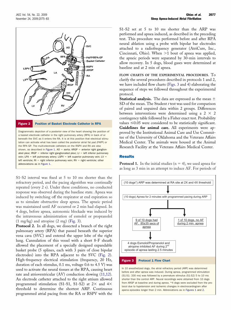
(10 dogs*) ARP was determined at RA site at 2X and 4X threshold
(10 dogs) Apnea for 2 minutes with programmed pacing during ARP
9 of 10 dogs had AF, 85±35 secs of
apnea
1 of 10 dogs, no AF during 2 min. apnea
4 dogs Esmolol/Propranolol and atropine inhibited AF during 2nd
episode of apnea lasting 2.5 minutes
Figure 3 Protocol 1 Flow Chart
In 10 anesthetized dogs, the atrial refractory period (ARP) was determinedbefore and after apnea was induced. During apnea, programmed stimulation(S1-S1: 330 ms) was followed by a premature stimulus (S1-S2) 5 to 10 msshorter than the control ARP. Neural recordings were obtained from 10 dogsfrom ARGP at baseline and during apnea. *4 dogs were excluded from the pro-tocol due to hypotension and ischemic changes in electrocardiogram afterapena episodes longer than 2 min. Abbreviations as in Figures 1 and 2.
ahictbwwsd
tvsobAnbtmptb
p1oS4Nicmi1
pos
2078 Ghias et al. JACC Vol. 54, No. 22, 2009Sleep Apnea-Induced Atrial Fibrillation November 24, 2009:2075–83
pnea �2 min, there were some episodes that resulted inypotension, electrocardiographic changes indicative of
schemia, and more prolonged recovery back to baselineonditions. Figure 5 illustrates the characteristic changeshat occurred during this prolonged apneic period. In theaseline state, the frequency and level of GP neuronal firingas highly variable (Fig. 5A). However, a consistent findingas the decrease in neuronal activity within 15 s after the
tart of apnea (Fig. 5B). In this case, the amplitudeecreased by 50% of the baseline activity.As apnea continued, there was a progressive increase in
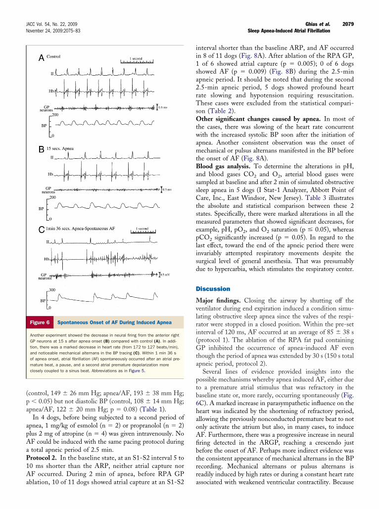
he intensity and amplitude of neuronal firing. At 2.5 min,entricular ectopic beats were observed as well as ST-egment depression (Fig. 5C). Furthermore, the amplitudef the neuronal firing increased by 2� that recorded ataseline. Figure 6 represents the 1 instance of spontaneousF during apnea that occurred within 2 min. Although theeural recordings obtained were relatively low level ataseline (Fig. 6A), there was a notable decrease in ampli-ude within 15 s after apnea was induced (Fig. 6B). At 1in 36 s, associated with the increased neural firing, a
remature atrial depolarization occurred spontaneously,hen a pause, a sinus beat, and an earlier coupled premature
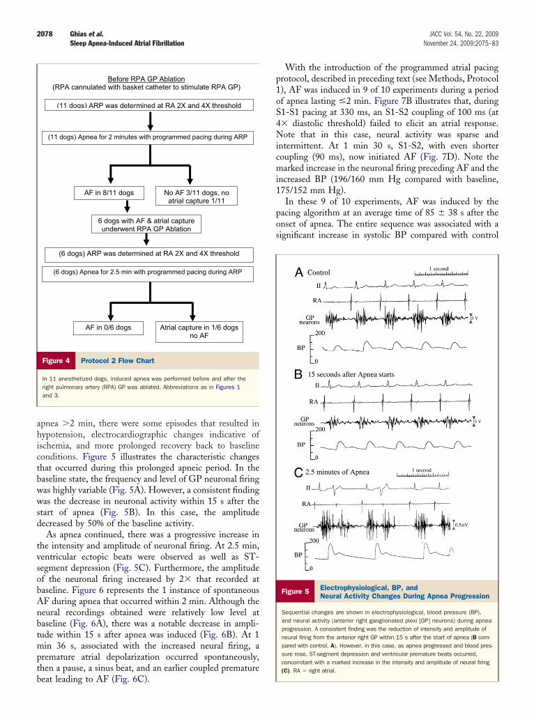
Before RPA GP Ablation(RPA cannulated with basket catheter to stimulate RPA GP)
(11 dogs) ARP was determined at RA 2X and 4X threshold
(11 dogs) Apnea for 2 minutes with programmed pacing during ARP
AF in 8/11 dogs No AF 3/11 dogs, no atrial capture 1/11
6 dogs with AF & atrial capture underwent RPA GP Ablation
(6 dogs) ARP was determined at RA 2X and 4X threshold
(6 dogs) Apnea for 2.5 min with programmed pacing during ARP
AF in 0/6 dogs Atrial capture in 1/6 dogsno AF
Figure 4 Protocol 2 Flow Chart
In 11 anesthetized dogs, induced apnea was performed before and after theright pulmonary artery (RPA) GP was ablated. Abbreviations as in Figures 1and 3.
eat leading to AF (Fig. 6C).
With the introduction of the programmed atrial pacingrotocol, described in preceding text (see Methods, Protocol), AF was induced in 9 of 10 experiments during a periodf apnea lasting �2 min. Figure 7B illustrates that, during1-S1 pacing at 330 ms, an S1-S2 coupling of 100 ms (at� diastolic threshold) failed to elicit an atrial response.ote that in this case, neural activity was sparse and
ntermittent. At 1 min 30 s, S1-S2, with even shorteroupling (90 ms), now initiated AF (Fig. 7D). Note thearked increase in the neuronal firing preceding AF and the
ncreased BP (196/160 mm Hg compared with baseline,75/152 mm Hg).In these 9 of 10 experiments, AF was induced by the
acing algorithm at an average time of 85 � 38 s after thenset of apnea. The entire sequence was associated with aignificant increase in systolic BP compared with control
Figure 5 Electrophysiological, BP, andNeural Activity Changes During Apnea Progression
Sequential changes are shown in electrophysiological, blood pressure (BP),and neural activity (anterior right ganglionated plexi [GP] neurons) during apneaprogression. A consistent finding was the reduction of intensity and amplitude ofneural firing from the anterior right GP within 15 s after the start of apnea (B com-pared with control, A). However, in this case, as apnea progressed and blood pres-sure rose, ST-segment depression and ventricular premature beats occurred,concomitant with a marked increase in the intensity and amplitude of neural firing(C). RA � right atrial.
(pa
apAaP1Aa
ii1sa2rTsOtwamtBassCtsmeplisd
D
Mvlri(Gta
ptb6haoAfibtrr
2079JACC Vol. 54, No. 22, 2009 Ghias et al.November 24, 2009:2075–83 Sleep Apnea-Induced Atrial Fibrillation
control, 149 � 26 mm Hg; apnea/AF, 193 � 38 mm Hg;� 0.05) but not diastolic BP (control, 108 � 14 mm Hg;
pnea/AF, 122 � 20 mm Hg; p � 0.08) (Table 1).In 4 dogs, before being subjected to a second period of
pnea, 1 mg/kg of esmolol (n � 2) or propranolol (n � 2)lus 2 mg of atropine (n � 4) was given intravenously. NoF could be induced with the same pacing protocol duringtotal apneic period of 2.5 min.rotocol 2. In the baseline state, at an S1-S2 interval 5 to0 ms shorter than the ARP, neither atrial capture norF occurred. During 2 min of apnea, before RPA GP
Figure 6 Spontaneous Onset of AF During Induced Apnea
Another experiment showed the decrease in neural firing from the anterior rightGP neurons at 15 s after apnea onset (B) compared with control (A). In addi-tion, there was a marked decrease in heart rate (from 172 to 127 beats/min),and noticeable mechanical alternans in the BP tracing (C). Within 1 min 36 sof apnea onset, atrial fibrillation (AF) spontaneously occurred after an atrial pre-mature beat, a pause, and a second atrial premature depolarization moreclosely coupled to a sinus beat. Abbreviations as in Figure 5.
blation, 10 of 11 dogs showed atrial capture at an S1-S2 a
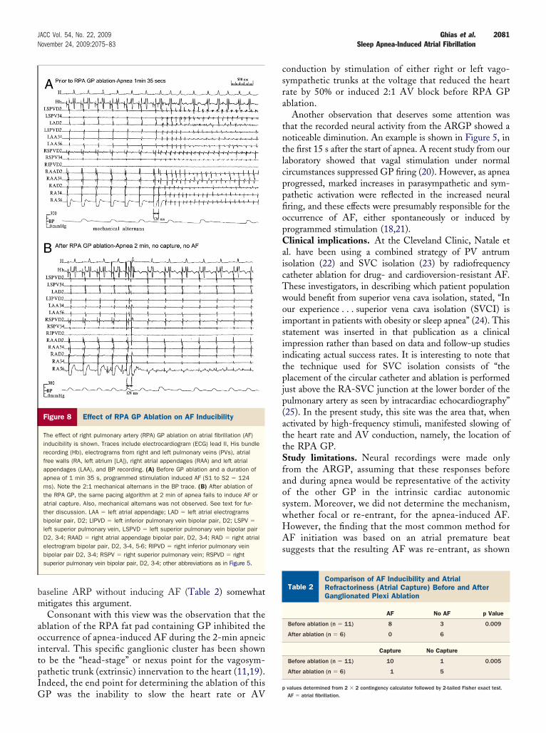
nterval shorter than the baseline ARP, and AF occurredn 8 of 11 dogs (Fig. 8A). After ablation of the RPA GP,
of 6 showed atrial capture (p � 0.005); 0 of 6 dogshowed AF (p � 0.009) (Fig. 8B) during the 2.5-minpneic period. It should be noted that during the second.5-min apneic period, 5 dogs showed profound heartate slowing and hypotension requiring resuscitation.hese cases were excluded from the statistical compari-
on (Table 2).ther significant changes caused by apnea. In most of
he cases, there was slowing of the heart rate concurrentith the increased systolic BP soon after the initiation of
pnea. Another consistent observation was the onset ofechanical or pulsus alternans manifested in the BP before
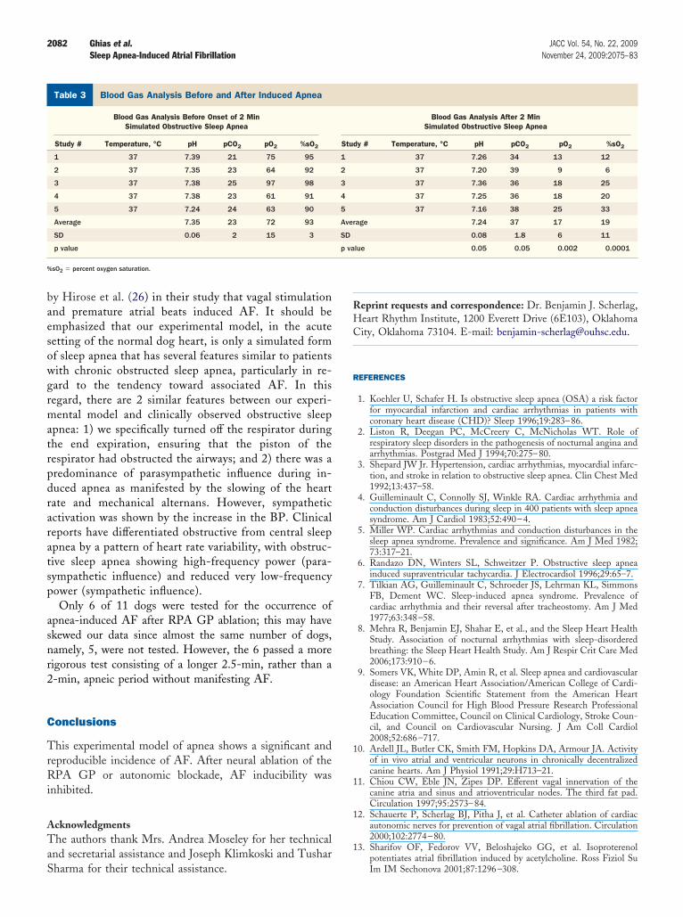
he onset of AF (Fig. 8A).lood gas analysis. To determine the alterations in pH,
nd blood gases CO2 and O2, arterial blood gases wereampled at baseline and after 2 min of simulated obstructiveleep apnea in 5 dogs (I Stat-1 Analyzer, Abbott Point ofare, Inc., East Windsor, New Jersey). Table 3 illustrates
he absolute and statistical comparison between these 2tates. Specifically, there were marked alterations in all theeasured parameters that showed significant decreases, for
xample, pH, pO2, and O2 saturation (p � 0.05), whereasCO2 significantly increased (p � 0.05). In regard to the
ast effect, toward the end of the apneic period there werenvariably attempted respiratory movements despite theurgical level of general anesthesia. That was presumablyue to hypercarbia, which stimulates the respiratory center.
iscussion
ajor findings. Closing the airway by shutting off theentilator during end expiration induced a condition simu-ating obstructive sleep apnea since the valves of the respi-ator were stopped in a closed position. Within the pre-setnterval of 120 ms, AF occurred at an average of 85 � 38 sprotocol 1). The ablation of the RPA fat pad containingP inhibited the occurrence of apnea-induced AF even
hough the period of apnea was extended by 30 s (150 s totalpneic period, protocol 2).
Several lines of evidence provided insights into theossible mechanisms whereby apnea induced AF, either dueo a premature atrial stimulus that was refractory in theaseline state or, more rarely, occurring spontaneously (Fig.C). A marked increase in parasympathetic influence on theeart was indicated by the shortening of refractory period,llowing the previously nonconducted premature beat to notnly activate the atrium but also, in many cases, to induceF. Furthermore, there was a progressive increase in neuralring detected in the ARGP, reaching a crescendo justefore the onset of AF. Perhaps more indirect evidence washe consistent appearance of mechanical alternans in the BPecording. Mechanical alternans or pulsus alternans iseadily induced by high rates or during a constant heart rate
ssociated with weakened ventricular contractility. Because
tsdtmpvttosn
optittoafsioA
geeaApcR
E
2080 Ghias et al. JACC Vol. 54, No. 22, 2009Sleep Apnea-Induced Atrial Fibrillation November 24, 2009:2075–83
he test for AF inducibility consisted of programmedtimulation during which S1-S2 was continually (every 2 s)elivered at a fixed coupling interval, 5 to 10 ms earlier thanhe baseline ARP (Figs. 8A and 8B), the appearance ofechanical alternans would likely be due to the increased
arasympathetic and negative inotropic influence on theentricles despite the overall increase in systolic BP. Indeed,he latter would also contribute to an increased parasympa-hetic influence through a baroreflex effect. Recent studiesf the mechanisms underlying AF inducibility have clearlyhown the direct involvement of both arms of the auto-omic nervous system (13–15).It is interesting to note that the most common occurrence
f AF during apnea was initiated by a closely coupled atrialremature stimulus that did not result in a conducted beat inhe baseline state. Similarly, atrial premature stimuli are thenvariable triggers for AF with delivery of vagosympatheticrunk stimulation, namely, extrinsic autonomic innervation,o the heart. Conversely, autonomic stimulation at the levelf the intrinsic cardiac nervous system, namely, GP andssociated neural network, directly induces AF, whetherrom PV or non-PV sites (16–18). Specifically, it has beenhown that activation of local parasympathetic nerves,nducing shortening of atrial refractoriness, and stimulationf local sympathetic nerves serve as the direct triggers for
Figure 7 AF Induction During Apnea With Delivery of Atrial Pre
Atrial fibrillation (AF) was induced during apnea with the delivery of atrial prematurbaseline state. (A) In this experiment, very sparse neural activity was recorded fro(control). (B) At 1 min 30 s of apnea, programmed stimulation was started with aa coupling interval 10 ms earlier than the atrial refractory period (S1-S2 � 110 mspremature depolarization (APD) (arrow) followed by AF (D). Note the associated inBP. Abbreviations as in Figure 5.
F through early after-depolarizations (17). Taken to-
ether, this would suggest that the major autonomic influ-nce for apnea-induced AF is primarily occurring from thextrinsic input to the heart. It could be argued that, duringpnea, the atrial capture occurring earlier than the baselineRP induced AF as a result of falling in the atrial vulnerableeriod and was unrelated to the associated autonomichanges. However, the finding that, during apnea, afterPA ablation, atrial capture could still occur before the
re Stimuli Earlier Than Baseline ARP
uli 5 to 10 ms earlier than the atrial refractory period (ARP) determined in theanterior right ganglionated plexi (GP) neurons (arrows) during the baseline state8-beat drive cycle (S1-S1 � 330 ms). The premature stimulus was delivered withThe premature stimulus now induced atrial capture and a spontaneous atrialin frequency, intensity, and amplitude of neural activity as well as the increased
ffect of Apnea on Blood PressureTable 1 Effect of Apnea on Blood Pressure
Experiment #
Systolic Blood Pressure Diastolic Blood Pressure
Control Experimental Control Experimental
1 132 270 92 144
2 94 151 82 109
3 175 191 125 138
4 136 180 104 145
5 138 216 107 144
6 164 196 112 124
7 168 192 112 112
8 192 222 136 132
9 152 176 108 104
10 144 132 104 84
Average 149 193 108 122
SD 26 38 14 20
matu
e stimm thebasic) (C).
crease
p value 0.05 0.08
bm
aoitpIG
csra
tntlcppfiopCaicTwoisiitpjp(attSfaoswHAs
CRG
2081JACC Vol. 54, No. 22, 2009 Ghias et al.November 24, 2009:2075–83 Sleep Apnea-Induced Atrial Fibrillation
aseline ARP without inducing AF (Table 2) somewhatitigates this argument.Consonant with this view was the observation that the
blation of the RPA fat pad containing GP inhibited theccurrence of apnea-induced AF during the 2-min apneicnterval. This specific ganglionic cluster has been showno be the “head-stage” or nexus point for the vagosym-athetic trunk (extrinsic) innervation to the heart (11,19).ndeed, the end point for determining the ablation of this
Figure 8 Effect of RPA GP Ablation on AF Inducibility
The effect of right pulmonary artery (RPA) GP ablation on atrial fibrillation (AF)inducibility is shown. Traces include electrocardiogram (ECG) lead II, His bundlerecording (Hb), electrograms from right and left pulmonary veins (PVs), atrialfree walls (RA, left atrium [LA]), right atrial appendages (RAA) and left atrialappendages (LAA), and BP recording. (A) Before GP ablation and a duration ofapnea of 1 min 35 s, programmed stimulation induced AF (S1 to S2 � 124ms). Note the 2:1 mechanical alternans in the BP trace. (B) After ablation ofthe RPA GP, the same pacing algorithm at 2 min of apnea fails to induce AF oratrial capture. Also, mechanical alternans was not observed. See text for fur-ther discussion. LAA � left atrial appendage; LAD � left atrial electrogramsbipolar pair, D2; LIPVD � left inferior pulmonary vein bipolar pair, D2; LSPV �
left superior pulmonary vein, LSPVD � left superior pulmonary vein bipolar pairD2, 3-4; RAAD � right atrial appendage bipolar pair, D2, 3-4; RAD � right atrialelectrogram bipolar pair, D2, 3-4, 5-6; RIPVD � right inferior pulmonary veinbipolar pair D2, 3-4; RSPV � right superior pulmonary vein; RSPVD � rightsuperior pulmonary vein bipolar pair, D2, 3-4; other abbreviations as in Figure 5.
P was the inability to slow the heart rate or AVp
onduction by stimulation of either right or left vago-ympathetic trunks at the voltage that reduced the heartate by 50% or induced 2:1 AV block before RPA GPblation.
Another observation that deserves some attention washat the recorded neural activity from the ARGP showed aoticeable diminution. An example is shown in Figure 5, inhe first 15 s after the start of apnea. A recent study from ouraboratory showed that vagal stimulation under normalircumstances suppressed GP firing (20). However, as apnearogressed, marked increases in parasympathetic and sym-athetic activation were reflected in the increased neuralring, and these effects were presumably responsible for theccurrence of AF, either spontaneously or induced byrogrammed stimulation (18,21).linical implications. At the Cleveland Clinic, Natale et
l. have been using a combined strategy of PV antrumsolation (22) and SVC isolation (23) by radiofrequencyatheter ablation for drug- and cardioversion-resistant AF.hese investigators, in describing which patient populationould benefit from superior vena cava isolation, stated, “Inur experience . . . superior vena cava isolation (SVCI) ismportant in patients with obesity or sleep apnea” (24). Thistatement was inserted in that publication as a clinicalmpression rather than based on data and follow-up studiesndicating actual success rates. It is interesting to note thathe technique used for SVC isolation consists of “thelacement of the circular catheter and ablation is performed
ust above the RA-SVC junction at the lower border of theulmonary artery as seen by intracardiac echocardiography”25). In the present study, this site was the area that, whenctivated by high-frequency stimuli, manifested slowing ofhe heart rate and AV conduction, namely, the location ofhe RPA GP.tudy limitations. Neural recordings were made onlyrom the ARGP, assuming that these responses beforend during apnea would be representative of the activityf the other GP in the intrinsic cardiac autonomicystem. Moreover, we did not determine the mechanism,hether focal or re-entrant, for the apnea-induced AF.owever, the finding that the most common method forF initiation was based on an atrial premature beat
uggests that the resulting AF was re-entrant, as shown
omparison of AF Inducibility and Atrialefractoriness (Atrial Capture) Before and Afteranglionated Plexi AblationTable 2
Comparison of AF Inducibility and AtrialRefractoriness (Atrial Capture) Before and AfterGanglionated Plexi Ablation
AF No AF p Value
Before ablation (n � 11) 8 3 0.009
After ablation (n � 6) 0 6
Capture No Capture
Before ablation (n � 11) 10 1 0.005
After ablation (n � 6) 1 5
values determined from 2 � 2 contingency calculator followed by 2-tailed Fisher exact test.AF � atrial fibrillation.
baesowgrmatrpdraratsp
asnr2
C
TrRi
ATaS
RHC
R
1
1
1
1
B
%
2082 Ghias et al. JACC Vol. 54, No. 22, 2009Sleep Apnea-Induced Atrial Fibrillation November 24, 2009:2075–83
y Hirose et al. (26) in their study that vagal stimulationnd premature atrial beats induced AF. It should bemphasized that our experimental model, in the acuteetting of the normal dog heart, is only a simulated formf sleep apnea that has several features similar to patientsith chronic obstructed sleep apnea, particularly in re-ard to the tendency toward associated AF. In thisegard, there are 2 similar features between our experi-ental model and clinically observed obstructive sleep
pnea: 1) we specifically turned off the respirator duringhe end expiration, ensuring that the piston of theespirator had obstructed the airways; and 2) there was aredominance of parasympathetic influence during in-uced apnea as manifested by the slowing of the heartate and mechanical alternans. However, sympatheticctivation was shown by the increase in the BP. Clinicaleports have differentiated obstructive from central sleeppnea by a pattern of heart rate variability, with obstruc-ive sleep apnea showing high-frequency power (para-ympathetic influence) and reduced very low-frequencyower (sympathetic influence).Only 6 of 11 dogs were tested for the occurrence of
pnea-induced AF after RPA GP ablation; this may havekewed our data since almost the same number of dogs,amely, 5, were not tested. However, the 6 passed a moreigorous test consisting of a longer 2.5-min, rather than a-min, apneic period without manifesting AF.
onclusions
his experimental model of apnea shows a significant andeproducible incidence of AF. After neural ablation of thePA GP or autonomic blockade, AF inducibility was
nhibited.
cknowledgmentshe authors thank Mrs. Andrea Moseley for her technical
nd secretarial assistance and Joseph Klimkoski and Tushar
lood Gas Analysis Before and After Induced ApneaTable 3 Blood Gas Analysis Before and After Induced Apnea
Blood Gas Analysis Before Onset of 2 MinSimulated Obstructive Sleep Apnea
Study # Temperature, °C pH pCO2 pO2 %sO2
1 37 7.39 21 75 95
2 37 7.35 23 64 92
3 37 7.38 25 97 98
4 37 7.38 23 61 91
5 37 7.24 24 63 90
Average 7.35 23 72 93
SD 0.06 2 15 3
p value
sO2 � percent oxygen saturation.
harma for their technical assistance.
eprint requests and correspondence: Dr. Benjamin J. Scherlag,eart Rhythm Institute, 1200 Everett Drive (6E103), Oklahomaity, Oklahoma 73104. E-mail: [email protected].
EFERENCES
1. Koehler U, Schafer H. Is obstructive sleep apnea (OSA) a risk factorfor myocardial infarction and cardiac arrhythmias in patients withcoronary heart disease (CHD)? Sleep 1996;19:283–86.
2. Liston R, Deegan PC, McCreery C, McNicholas WT. Role ofrespiratory sleep disorders in the pathogenesis of nocturnal angina andarrhythmias. Postgrad Med J 1994;70:275–80.
3. Shepard JW Jr. Hypertension, cardiac arrhythmias, myocardial infarc-tion, and stroke in relation to obstructive sleep apnea. Clin Chest Med1992;13:437–58.
4. Guilleminault C, Connolly SJ, Winkle RA. Cardiac arrhythmia andconduction disturbances during sleep in 400 patients with sleep apneasyndrome. Am J Cardiol 1983;52:490–4.
5. Miller WP. Cardiac arrhythmias and conduction disturbances in thesleep apnea syndrome. Prevalence and significance. Am J Med 1982;73:317–21.
6. Randazo DN, Winters SL, Schweitzer P. Obstructive sleep apneainduced supraventricular tachycardia. J Electrocardiol 1996;29:65–7.
7. Tilkian AG, Guilleminault C, Schroeder JS, Lehrman KL, SimmonsFB, Dement WC. Sleep-induced apnea syndrome. Prevalence ofcardiac arrhythmia and their reversal after tracheostomy. Am J Med1977;63:348–58.
8. Mehra R, Benjamin EJ, Shahar E, et al., and the Sleep Heart HealthStudy. Association of nocturnal arrhythmias with sleep-disorderedbreathing: the Sleep Heart Health Study. Am J Respir Crit Care Med2006;173:910–6.
9. Somers VK, White DP, Amin R, et al. Sleep apnea and cardiovasculardisease: an American Heart Association/American College of Cardi-ology Foundation Scientific Statement from the American HeartAssociation Council for High Blood Pressure Research ProfessionalEducation Committee, Council on Clinical Cardiology, Stroke Coun-cil, and Council on Cardiovascular Nursing. J Am Coll Cardiol2008;52:686–717.
0. Ardell JL, Butler CK, Smith FM, Hopkins DA, Armour JA. Activityof in vivo atrial and ventricular neurons in chronically decentralizedcanine hearts. Am J Physiol 1991;29:H713–21.
1. Chiou CW, Eble JN, Zipes DP. Efferent vagal innervation of thecanine atria and sinus and atrioventricular nodes. The third fat pad.Circulation 1997;95:2573–84.
2. Schauerte P, Scherlag BJ, Pitha J, et al. Catheter ablation of cardiacautonomic nerves for prevention of vagal atrial fibrillation. Circulation2000;102:2774–80.
3. Sharifov OF, Fedorov VV, Beloshajeko GG, et al. Isoproterenolpotentiates atrial fibrillation induced by acetylcholine. Ross Fiziol Su
Blood Gas Analysis After 2 MinSimulated Obstructive Sleep Apnea
y # Temperature, °C pH pCO2 pO2 %sO2
37 7.26 34 13 12
37 7.20 39 9 6
37 7.36 36 18 25
37 7.25 36 18 20
37 7.16 38 25 33
age 7.24 37 17 19
0.08 1.8 6 11
lue 0.05 0.05 0.002 0.0001
Stud
1
2
3
4
5
Aver
SD
p va
Im IM Sechonova 2001;87:1296–308.
1
1
1
1
1
1
2
2
2
2
2
2
2
K
2083JACC Vol. 54, No. 22, 2009 Ghias et al.November 24, 2009:2075–83 Sleep Apnea-Induced Atrial Fibrillation
4. Patterson E, Po S, Scherlag BJ, Lazzara R. Triggered firing inpulmonary veins initiated by in vitro autonomic nerve stimulation.Heart Rhythm 2005;2:624–31.
5. Tan AY, Zhou S, Ogawa M, et al. Neural mechanisms of paroxysmalatrial fibrillation and paroxysmal atrial tachycardia in ambulatorycanines. Circulation 2008;118:916–25.
6. Po SS, Scherlag BJ, Yamanashi WS, et al. Experimental model forparoxysmal atrial fibrillation arising at the pulmonary vein-atrialjunctions. Heart Rhythm 2006;3:201–8.
7. Patterson E, Lazzara R, Szabo B, et al. Sodium-calcium exchangeinitiated by the Ca�� transient: an arrhythmia trigger within pulmo-nary veins. J Am Coll Cardiol 2006;47:1196–206.
8. Scherlag BJ, Hou Y-L, Lin J, et al. An acute model for atrialfibrillation arising from a peripheral atrial site: evidence for primaryand secondary triggers. J Cardiovasc Electrophysiol 2008;19:519–27.
9. Niu G, Scherlag BJ, Lu Z, et al. Inducibility of atrial fibrillation in themyocardial sleeve of the superior vena cava is mediated by theganglionated plexi on the right pulmonary artery (abstr). Circulation2007;116:II140.
0. Zhang Y, Scherlag BJ, Lu Z, et al. Comparison of atrial fibrillationinducibility by electrical stimulation of either the extrinsic or theintrinsic autonomic nervous systems. J Intervent Cardiac Electro-
physiol 2009;24:5–10. a1. Zhou J, Scherlag BJ, Edwards J, Jackman WM, Lazzara R, Po SS.Gradients of atrial refractoriness and inducibility of atrial fibrillationdue to stimulation of ganglionated plexi. J Cardiovasc Electrophysiol2007;18:83–90.
2. Verma A, Marrouche NF, Natale A. Pulmonary vein antrum isolation:intracardiac echocardiography guided technique. J Cardiovasc Electro-physiol 2004;11:1335–40.
3. Marrouche NF, Saliba W, Schweikert R, et al. Feasibility andcomplications of electrical disconnection of the superior vena cavaguided by circular mapping in patients with symptomatic atrialfibrillation (abstr). Pacing Clin Electrophysiol 2003;26:1116.
4. Kanj M, Wazni O, Natale A. Pulmonary vein antrum isolation. HeartRhythm 2007;4:S73–9.
5. Arruda M, Mlcochova H, Prasad SK, et al. Electrical isolation of thesuperior vena cava: an adjunctive strategy to pulmonary vein antrumisolation improving the outcome of AF ablation. J Cardiovasc Elec-trophysiol 2007;18:1261–6.
6. Hirose M, Leatmanoratn Z, Laurita KR, Carlson MD. Partial vagaldenervation increases vulnerability to vagally induced atrial fibrillation.J Cardiovasc Electrophysiol 2002;13:1272–9.
ey Words: atrial fibrillation y autonomic nervous system y sleep
pnea.Copyright © 2022 FDOKUMEN