Robotic magnetic navigation for atrial fibrillation ablation
11
Heart Rhythm Disorders Robotic Magnetic Navigation for Atrial Fibrillation Ablation Carlo Pappone, MD, PHD, Gabriele Vicedomini, MD, Francesco Manguso, MD, PHD, Filippo Gugliotta, BE, Patrizio Mazzone, MD, Simone Gulletta, MD, Nicoleta Sora, MD, Simone Sala, MD, Alessandra Marzi, MD, Giuseppe Augello, MD, Laura Livolsi, MD, Andreina Santagostino, MD, Vincenzo Santinelli, MD Milan, Italy OBJECTIVES We assessed feasibility of magnetic catheter guidance in patients with atrial fibrillation (AF) undergoing circumferential pulmonary vein ablation (CPVA). BACKGROUND No data are available on feasibility of remote navigation for AF ablation. METHODS Forty patients underwent CPVA for symptomatic AF using the NIOBE II remote magnetic system (Stereotaxis Inc., St. Louis, Missouri). Ablation was performed with a 4-mm tip, magnetic catheter (65°C, maximum 50 W, 15 s). The catheter tip was guided by a uniform magnetic field (0.08-T), and a motor drive (Cardiodrive unit, Stereotaxis Inc.). Left atrium maps were created using an integrated CARTO RMT system (Stereotaxis Inc.). End point of ablation was voltage abatement 90% of bipolar electrogram amplitude. RESULTS Remote ablation was successful in 38 of 40 patients without complications. The median mapping and ablation time was 152.5 min (range, 90 to 380 min) but was much longer in the first 12 patients (192.5 min vs. 148 min; p 0.012). Median ablation time was 49.5 min (range, 17 to 154 min), but it was much shorter in the last 28 patients than in the first 12 patients (49 min vs. 70 min; p 0.021). Patients receiving remote ablation had longer procedure times than control patients (p 0.001) with similar mapping time but shorter ablation time on right-sided pulmonary veins. Many more mapping points regardless of their location were collected remotely (p 0.001). CONCLUSIONS Remote magnetic navigation for AF ablation is safe and feasible with a short learning curve. Although all procedures were performed by a highly experienced operator, remote AF ablation can be performed even by less experienced operators. (J Am Coll Cardiol 2006;47: 1390 – 400) © 2006 by the American College of Cardiology Foundation Catheter ablation techniques for atrial fibrillation (AF) are evolving with targets for radiofrequency (RF) ablation of AF increasingly being selected based on anatomic consider- ations. Such anatomic ablation techniques require precise catheter localization and stable contact during ablation. Even experienced electrophysiologists occasionally encoun- ter difficulties in maintaining stable catheter contact, espe- cially in some regions of complex anatomy where catheter stability is indeed crucial (1–5). Limitations of manually deflected conventional ablation catheters in performing complex catheter maneuvers may also contribute to these challenges. A recent feasibility study in animals suggests that continuous stability of ablation catheters can be ob- tained by the combination of the external magnetic field and flexibility of the distal portion of a magnetic catheter (6). Based on experimental and clinical data (6,7), we evaluated safety and feasibility of such navigation system in patients undergoing AF ablation. Here, we report the first human experience on magnetic remote navigation in 40 patients undergoing circum- ferential pulmonary vein ablation (CPVA) for AF. METHODS Patient population. This study included 40 patients (60% male patients; median age, 57 years, range, 28 to 75 years; Table 1) undergoing CPVA for drug-refractory symptom- atic paroxysmal (62.5%) or permanent AF (median duration, 46.5 months; range, 12 to 286) with the use of the remote magnetic navigation NIOBE II system (Stereotaxis Inc., St. Louis, Missouri). Patients had no contraindications for mag- netic navigation such as implanted cardioverter defibrillators, pacemakers, or vascular clips. Twenty-eight patients, matched for gender, age, history of AF, duration of AF, previous antiarrhythmic drugs tried, associated disease, and all clinical characteristics, were selected as a procedural control group from patients who underwent standard CPVA ablation between September 2004 and June 2005. Ablation proce- dure and selection criteria were similar for both groups. Patients were studied in a fasting state, and before the procedure gave their written permission after informed consent was obtained. Remote magnetic navigation system. The remote mag- netic navigation system consists of two focused-field per- manent magnets of a neodymium-iron-boron compound that are computer-controlled and located on either side of the body, as previously reported (7). The magnetic field From the Department of Electrophysiology, San Raffaele Scientific Institute, Milan, Italy. This study was supported by a grant from Johnson & Johnson. This work received technical support from Stereotaxis Inc. Manuscript received August 23, 2005; revised manuscript received November 9, 2005, accepted November 16, 2005. Journal of the American College of Cardiology Vol. 47, No. 7, 2006 © 2006 by the American College of Cardiology Foundation ISSN 0735-1097/06/$32.00 Published by Elsevier Inc. doi:10.1016/j.jacc.2005.11.058
-
Upload
independent -
Category
Documents
-
view
0 -
download
0
Transcript of Robotic magnetic navigation for atrial fibrillation ablation

RfCFSAM
CeiacEtcsdccttflofamf
Mr
2
Journal of the American College of Cardiology Vol. 47, No. 7, 2006© 2006 by the American College of Cardiology Foundation ISSN 0735-1097/06/$32.00P
Heart Rhythm Disorders
obotic Magnetic Navigationor Atrial Fibrillation Ablationarlo Pappone, MD, PHD, Gabriele Vicedomini, MD, Francesco Manguso, MD, PHD,ilippo Gugliotta, BE, Patrizio Mazzone, MD, Simone Gulletta, MD, Nicoleta Sora, MD,imone Sala, MD, Alessandra Marzi, MD, Giuseppe Augello, MD, Laura Livolsi, MD,ndreina Santagostino, MD, Vincenzo Santinelli, MDilan, Italy
OBJECTIVES We assessed feasibility of magnetic catheter guidance in patients with atrial fibrillation (AF)undergoing circumferential pulmonary vein ablation (CPVA).
BACKGROUND No data are available on feasibility of remote navigation for AF ablation.METHODS Forty patients underwent CPVA for symptomatic AF using the NIOBE II remote magnetic
system (Stereotaxis Inc., St. Louis, Missouri). Ablation was performed with a 4-mm tip,magnetic catheter (65°C, maximum 50 W, 15 s). The catheter tip was guided by a uniformmagnetic field (0.08-T), and a motor drive (Cardiodrive unit, Stereotaxis Inc.). Left atriummaps were created using an integrated CARTO RMT system (Stereotaxis Inc.). End pointof ablation was voltage abatement �90% of bipolar electrogram amplitude.
RESULTS Remote ablation was successful in 38 of 40 patients without complications. The medianmapping and ablation time was 152.5 min (range, 90 to 380 min) but was much longer in thefirst 12 patients (192.5 min vs. 148 min; p � 0.012). Median ablation time was 49.5 min(range, 17 to 154 min), but it was much shorter in the last 28 patients than in the first 12patients (49 min vs. 70 min; p � 0.021). Patients receiving remote ablation had longerprocedure times than control patients (p � 0.001) with similar mapping time but shorterablation time on right-sided pulmonary veins. Many more mapping points regardless of theirlocation were collected remotely (p � 0.001).
CONCLUSIONS Remote magnetic navigation for AF ablation is safe and feasible with a short learning curve.Although all procedures were performed by a highly experienced operator, remote AFablation can be performed even by less experienced operators. (J Am Coll Cardiol 2006;47:
ublished by Elsevier Inc. doi:10.1016/j.jacc.2005.11.058
1390–400) © 2006 by the American College of Cardiology Foundation
M
PmTa4mLnpfacfbdPpcRnmt
atheter ablation techniques for atrial fibrillation (AF) arevolving with targets for radiofrequency (RF) ablation of AFncreasingly being selected based on anatomic consider-tions. Such anatomic ablation techniques require preciseatheter localization and stable contact during ablation.ven experienced electrophysiologists occasionally encoun-
er difficulties in maintaining stable catheter contact, espe-ially in some regions of complex anatomy where cathetertability is indeed crucial (1–5). Limitations of manuallyeflected conventional ablation catheters in performingomplex catheter maneuvers may also contribute to thesehallenges. A recent feasibility study in animals suggestshat continuous stability of ablation catheters can be ob-ained by the combination of the external magnetic field andexibility of the distal portion of a magnetic catheter (6). Basedn experimental and clinical data (6,7), we evaluated safety andeasibility of such navigation system in patients undergoing AFblation. Here, we report the first human experience onagnetic remote navigation in 40 patients undergoing circum-
erential pulmonary vein ablation (CPVA) for AF.
From the Department of Electrophysiology, San Raffaele Scientific Institute,ilan, Italy. This study was supported by a grant from Johnson & Johnson. This work
eceived technical support from Stereotaxis Inc.
tManuscript received August 23, 2005; revised manuscript received November 9,
005, accepted November 16, 2005.
ETHODS
atient population. This study included 40 patients (60%ale patients; median age, 57 years, range, 28 to 75 years;able 1) undergoing CPVA for drug-refractory symptom-
tic paroxysmal (62.5%) or permanent AF (median duration,6.5 months; range, 12 to 286) with the use of the remoteagnetic navigation NIOBE II system (Stereotaxis Inc., St.ouis, Missouri). Patients had no contraindications for mag-etic navigation such as implanted cardioverter defibrillators,acemakers, or vascular clips. Twenty-eight patients, matchedor gender, age, history of AF, duration of AF, previousntiarrhythmic drugs tried, associated disease, and all clinicalharacteristics, were selected as a procedural control grouprom patients who underwent standard CPVA ablationetween September 2004 and June 2005. Ablation proce-ure and selection criteria were similar for both groups.atients were studied in a fasting state, and before therocedure gave their written permission after informedonsent was obtained.emote magnetic navigation system. The remote mag-etic navigation system consists of two focused-field per-anent magnets of a neodymium-iron-boron compound
hat are computer-controlled and located on either side of
he body, as previously reported (7). The magnetic field
cus
SsdvmdtlatXMR
T
A
MHDE
E
C
PE
L
A
P
F
M
A
M
A
*
d
1391JACC Vol. 47, No. 7, 2006 Pappone et al.April 4, 2006:1390–400 Remote Catheter Navigation for AF Ablation
reates a 360° omnidirectional rotation of the device byniform magnetic field (0.08-T) within an approximatelypherical navigation volume of 20-cm diameter (Navi-
able 1. Characteristics of Subjects Undergoing Remote Magnet
VariablesAll Patients
(n � 40)First 12Patients
ge (yrs)Median (IQR) 57 (15) 55.5 (15)Range 28–75 28–65ale gender (%) 60 66.7ypertension (%) 30 33.3iabetes (%) 7.5 0CG at admission (%)SR 52.5 58.3AF 42.5 33.3AT 5.0 8.3
CG at discharge (%)SR 100 100
lassification of AF (%)Paroxysmal 62.5 75Permanent 37.5 25
revious CPVA (%) 22.5 25F%Median (IQR) 60 (5) 60 (7)Range 25–70 55–70
AD (mm)Median (IQR) 42.5 (11) 43.5 (9)Range 30–52 30–49
F duration (months)Median (IQR) 46.5 (81) 63 (130)Range 12–286 12–182
rocedure time (min)Median (IQR) 152.5 (64) 192.5 (98)Range 90–380 92–380
luoro time (min)Median (IQR) 32.3 (11) 34.5 (28.4)Range 21.8–81.4 22.4–81.4apping time (min)Median (IQR) 26.5 (21) 49 (72)Range 14–258 19–258
blation time (min)Median (IQR) 49.5 (26) 70 (66)Range 17–154 33–154apping points (n)Median (IQR) 167.5 (41) 165.5 (65)Range 84–249 84–234
blation points (n)Median (IQR) 105.5 (38) 98 (44)Range 24–182 55–156
p refers to the first 12 patients vs. the last 28 patients; †p refers to the last 28 patie
Abbreviations and AcronymsAF � atrial fibrillationCPVA � circumferential pulmonary vein ablationLAO � left anterior obliqueLIPV � left inferior pulmonary veinLSPV � left superior pulmonary veinPV � pulmonary veinRAO � right anterior obliqueRF � radiofrequency
AF � atrial fibrillation; AT � atrial tachycardia; CPVA � circumferential pulmonary viameter; SR � sinus rhythm.
phere) inside the patient’s chest. The magnetic navigationystem is integrated with a modified C-arm single-planeigital imaging system (Artis, Siemens, Malvern, Pennsyl-ania). The combination of rotation, translation, and tiltovements of the magnets adjusts the magnetic field to any
esired orientation in the spherical 20-cm diameter naviga-ion volume. The maximum X-ray imaging angles areimited to approximately 30° left anterior oblique (LAO)nd right anterior oblique (RAO). The operator is posi-ioned in a separate control room, at a distance from the-ray beam and the patient’s body.agnetic catheter. A 4-mm flexible catheter (NaviStar-
MT, Biosense Webster, Diamond Bar, California) incor-
vigation for AF Ablation and Control Patients
Last 28Patients p Value*
Controls(n � 28) p Value†
57.5 (15) 0.313 57 (16) 1.19729–75 31–75
57.1 0.573 57.1 1.00028.6 0.763 28.6 1.00010.7 0.541‡ 10.7 1.000‡
50.0 0.880§ 50.0 1.000§46.4 46.43.6 3.6
100 — 100 —
57.1 0.285 57.1 1.00042.9 42.921.4 1.000 21.4 1.000
55.5 (5) 0.146 57 (9) 0.71725–65 27–66
42 (12) 0.942 41.5 (11) 0.68030–52 30–52
41.5 (38) 0.389 41.5 (34) 0.27012–286 12–278
148 (58) 0.012 110 (10) �0.00190–209 45–150
30.3 (11.4) 0.065 30.3 (12.5) 0.35221.8–60.4 20–59
23 (15) 0.012 27 (4) 0.30914–52 20–34
49 (24) 0.021 57.5 (23) 0.0417–72 30–86
168 (37) 0.439 72 (20) �0.001110–249 60–110
105.5 (38) 0.760 110 (38) 0.42124–182 60–147
. controls; ‡by Fisher’s exact test; §by exact method.
ic Na
nts vs
ein ablation; EF � ejection fraction; IQR � interquartile range; LAD � left atrial
pmtsmfimvetoatssCtm(s
ttfsdc(Mtpsvtdocodfi
Fom vein (a
1392 Pappone et al. JACC Vol. 47, No. 7, 2006Remote Catheter Navigation for AF Ablation April 4, 2006:1390–400
orating a small permanent magnet in the tip and additionalagnets in the distal portion of the device can be deflected in
he desired direction and steered by the magnetic navigationystem (Fig. 1). The catheter is advanced and retracted by aechanical device (Cardiodrive, Stereotaxis Inc.). All magnetic
eld vectors can be stored and, if necessary, reapplied while theagnetic catheter is navigated automatically (Fig. 2). The
ideo workstation-based User Interface (Navigant, Ster-otaxis Inc.) used together with the permanent magnets andhe Cardiodrive unit (Stereotaxis Inc.) permits accuraterientation changes of the catheter by 1° increments anddvancement or retraction by 1-mm steps (Fig. 3). Addi-ionally, X-ray image data can be transferred from the X-rayystem to the user interface of the magnetic navigationystem to provide an anatomical reference.ARTO RMT integration. The magnetic navigation sys-
em is integrated with a newly developed electroanatomicalapping system, the CARTO RMT mapping system
Stereotaxis Inc.) (Fig. 4). The CARTO RMT system
igure 1. Fluoroscopic views in anteroposterior projection showing the Navther magnets in the distal portion of the device. The soft distal catheter bagnetic navigation system to reach the targeted right-superior pulmonary
nd right ventricle.
ends real-time catheter tip location and orientation data to fi
he magnetic navigation system. It also sends target loca-ions, groups of points, and anatomical surface informationrom the electroanatomical map to the magnetic navigationystem. The real-time catheter location information can beisplayed on the Navigant reference X-ray images, enablingontinuous real-time monitoring of the catheter tip positionFig. 3) even without acquiring a fresh X-ray image.
agnetic navigation procedure. After transseptal punc-ure, the magnetic catheter was advanced to the targetositions in the left atrium and guided by using the X-rayystem and user interface monitors and the catheter ad-ancer (Cardiodrive) system. Directional catheter naviga-ion was performed with a graphical user interface. Aesired magnetic field vector was drawn and displayed onrthogonal fluoroscopic views, as shown in Figure 3. Aontrol computer then calculates the appropriate orientationf each of the magnets and moves them to produce theesired magnetic field vector. Manipulation of the magneticeld vector usually requires a few seconds to redirect the
RMT catheter incorporating a small permanent magnet in the tip and twollows the tip to be deflected in any direction (A to C) and steered by theD). Two standard quadripolar catheters are placed into the coronary sinus
iStar-ody a
eld vector in a new direction with time proportional to the
mstmainoiea(naraamwb
flwqcRsa(cbitXcXttNt
F C) to(
1393JACC Vol. 47, No. 7, 2006 Pappone et al.April 4, 2006:1390–400 Remote Catheter Navigation for AF Ablation
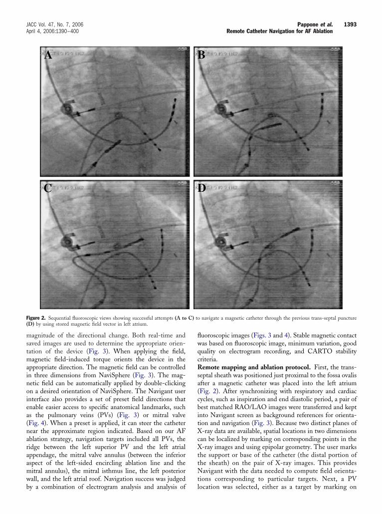
agnitude of the directional change. Both real-time andaved images are used to determine the appropriate orien-ation of the device (Fig. 3). When applying the field,agnetic field-induced torque orients the device in the
ppropriate direction. The magnetic field can be controlledn three dimensions from NaviSphere (Fig. 3). The mag-etic field can be automatically applied by double-clickingn a desired orientation of NaviSphere. The Navigant usernterface also provides a set of preset field directions thatnable easier access to specific anatomical landmarks, suchs the pulmonary veins (PVs) (Fig. 3) or mitral valveFig. 4). When a preset is applied, it can steer the catheterear the approximate region indicated. Based on our AFblation strategy, navigation targets included all PVs, theidge between the left superior PV and the left atrialppendage, the mitral valve annulus (between the inferiorspect of the left-sided encircling ablation line and theitral annulus), the mitral isthmus line, the left posteriorall, and the left atrial roof. Navigation success was judged
igure 2. Sequential fluoroscopic views showing successful attempts (A toD) by using stored magnetic field vector in left atrium.
y a combination of electrogram analysis and analysis of l
uoroscopic images (Figs. 3 and 4). Stable magnetic contactas based on fluoroscopic image, minimum variation, gooduality on electrogram recording, and CARTO stabilityriteria.emote mapping and ablation protocol. First, the trans-
eptal sheath was positioned just proximal to the fossa ovalisfter a magnetic catheter was placed into the left atriumFig. 2). After synchronizing with respiratory and cardiacycles, such as inspiration and end diastolic period, a pair ofest matched RAO/LAO images were transferred and keptnto Navigant screen as background references for orienta-ion and navigation (Fig. 3). Because two distinct planes of-ray data are available, spatial locations in two dimensions
an be localized by marking on corresponding points in the-ray images and using epipolar geometry. The user marks
he support or base of the catheter (the distal portion ofhe sheath) on the pair of X-ray images. This providesavigant with the data needed to compute field orienta-
ions corresponding to particular targets. Next, a PV
navigate a magnetic catheter through the previous trans-septal puncture
ocation was selected, either as a target by marking on

sssTpm
tw
pataaiit
FtsiFlC
1394 Pappone et al. JACC Vol. 47, No. 7, 2006Remote Catheter Navigation for AF Ablation April 4, 2006:1390–400
uitable locations in the reference X-ray images, or byelecting a preset magnetic field vector based on selectedtudy protocol from the list on Navigant (Figs. 3 and 4).hroughout, points were simultaneously acquired for map-ing (Fig. 5). The NaviStar-RMT (Biosense Webster)agnetic catheter was used for both mapping and ablation.Circumferential PV ablation was performed with a target
emperature of 65°C, a power limit of 50 W. Ablation lines
igure 3. (A to D) The video workstation-based User Interface (Nawo-dimensional sync C-arm on the left and three-dimensional NaviSpheuperior pulmonary vein; (B) left inferior pulmonary vein; (C) right superios in yellow. The images displayed also show the magnetic catheter alignluoroscopic views (29° right or left anterior oblique projections including
ocation is displayed as the white-blue quadripolar catheter tip. Note: TARTO RMT.
ere performed by sequentially navigating to contiguous c
oints with a single 15-s application of RF current tochieve �90% reduction in the bipolar electrogram ampli-ude (Fig. 5), and/or peak-to-peak bipolar electrogrammplitude �0.1 mV inside the line. The mitral isthmusblation line was extended to near the region of the leftnferior pulmonary vein (LIPV), taking care to ensure, usingmpedance and anatomical map information, to stay outsidehe PV ostium (Fig. 5). The lesion line consisting of
t, Stereotaxis Inc.) shows four paired Navigant images (upper two,the right, which represent navigation of the magnetic catheter to (A) leftmonary vein; (D) right inferior pulmonary vein. The magnetic field vectorrallel to the chosen magnetic field vector to reach each pulmonary vein.catheter shadows) were previously stored still images. Real-time catheter
d points on fluoroscopic images were desired locations transferred from
viganre onr pul
ing pathreehe re
ontiguous point-by-point locations of RF power delivery

FTt(l
1395JACC Vol. 47, No. 7, 2006 Pappone et al.April 4, 2006:1390–400 Remote Catheter Navigation for AF Ablation
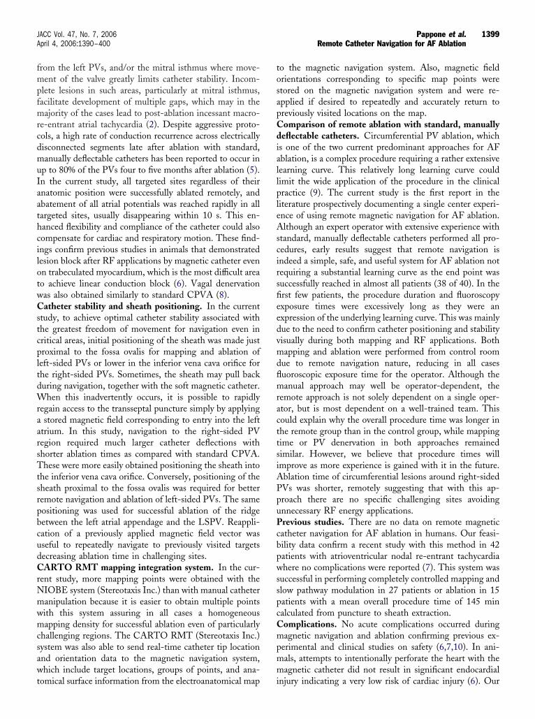
igure 4. Three fluoroscopic images (A to C) of sequential magnetic catheter navigation at three different locations of mitral valve annulus for mapping.he figure shows how we can deflect the catheter and maintain a good contact remotely. The electrograms inset (A to C) in the second column shows the
ypical appearance of the corresponding mitral annular electrogram recorded from the magnetic ablation catheter aligned with the magnetic field vectorwhite line catheter). Consistent atrial and ventricular electrogram recordings demonstrate a stable catheter contact at the three predefined mitral annular
ocations.
cPdcafeattrv
embtSmcgttw
FRNli ts the
1396 Pappone et al. JACC Vol. 47, No. 7, 2006Remote Catheter Navigation for AF Ablation April 4, 2006:1390–400
an be resent to and recorded on the fluoroscopic image.otential vagal target sites are identified during the proce-ure as previously reported (8). Briefly, vagal reflexes areonsidered sinus bradycardia (�40 beats/min) or asystole,trioventricular block, or hypotension occurring within aew seconds of the onset of RF application. If a reflex islicited, RF energy is delivered until such reflexes arebolished, or for up to 30 s. The end point for ablation athese sites is termination of the reflex, followed by sinusachycardia or AF. Failure to reproduce the reflexes withepeat RF is considered confirmation of local vagal dener-
igure 5. (A) Posteroanterior view showing color-coded preablation electroMT integration system. (B) Post-ablation electroanatomical map with enote atrial electrograms before (A, arrow) and after ablation (B). No poten
ine is evident. (C) A mesh anatomical map is shown. (D) Red points depicntegration system. Blue line shown on both fluoroscopic images represen
ation. A single ablation location was considered as failure if S
ach of the selected targets was not completed within 60in. Patients underwent transthoracic echocardiography
efore and after the procedure to assess potential complica-ions.tatistical analysis. Continuous variables are expressed asedian and interquartile range, with range values, and
ompared by use of the Mann-Whitney U test. For cate-orical variables the chi-square test was performed, unlesshe exact test was required for frequency tables when morehan 20% of the expected values were �5. Statistical testsere performed with SPSS for Windows (version 13.0.1,
mical voltage map of the left atrium and pulmonary veins using a CARTOd lesions around all targeted pulmonary veins and the mitral isthmus line.ere recorded inside encircled lesions (arrow). The mitral isthmus ablation
n fluoroscopic views indicate ablation points sent from the CARTO RMTbase for target control navigation on Navigant.
anatocircletials wted o
PSS Inc., Chicago, Illinois).

R
Rawttio(titaw
tmmmscdecsfmsc
bfncafMuatpumafsmbtpRmi
snecctjaietiLc3mtPpipfipwa(tafls2CttcaOtamcedaRs(irerCt
1397JACC Vol. 47, No. 7, 2006 Pappone et al.April 4, 2006:1390–400 Remote Catheter Navigation for AF Ablation
ESULTS
emote catheter mapping. FIRST THREE CASES. Retrogradeortic approach was attempted only in the first patient inhom the magnetic catheter could not be passed through
he aortic valve and therefore could not reach navigationargets within the left atrium. In the first three patients, thenitial sheath position was unstable causing repeat draggingf both sheath and magnetic catheter into the right atriumtwo patients) or even the right ventricle (one patient). Inhese cases, the transseptal sheath was pulled back in thenferior vena cava orifice while the catheter wire was left inhe left atrium. With the first three cases, many attemptsnd time-consuming manipulations of the magnetic fieldere required to refine precise and stable catheter position.The transseptal sheath was positioned just proximal to
he fossa ovalis to allow the greatest movement of theagnetic wire catheter. Before mapping, the position of theagnetic catheter was also controlled by manual advance-ent or retraction of the catheter through the vascular
heath. Sheath insertion and positioning of diagnosticatheters, including the magnetic catheter, required a me-ian of 7.9 min (range, 5 to 12 min). Median fluoroscopyxposure was 32.3 min (range, 21.8 to 81.4 min). Afterrossing the atrial septum and positioning the transseptalheath, the physician left the interventional room to per-orm mapping and ablation from the control room. Remoteagnetic navigation and ablation to all targeted sites was
uccessfully achieved in 38 of 40 patients withoutomplications.
Navigation to the PV ostia was guided by real-timeiplanar fluoroscopic images, but venography was not per-ormed to identify the location of PV ostia. We accuratelyavigated all PVs (Fig. 3), and then, by advancing magneticatheter using the Cardiodrive unit (Stereotaxis Inc.), wecquired a median of 274 points (range, 139 to 382 points)or accurate maps of the left atrium (Table 1, Fig. 5).
inimal orientation changes of the catheter up to 5° weresed for acquiring points around venoatrial junction regionnd up to 15° for the remaining left atrial reconstruction. Athe beginning of the learning curve and for the first 10atients, the catheter orientation was frequently adjustedsing the keyboard rotation and deflection keys to reachapping targets. In all cases, the catheter was retracted and
dvanced to access all PVs by using this sequence wheneasible: left superior pulmonary vein (LSPV), LIPV, rightuperior PV, and finally right inferior PV. Afterwards, theitral valve annulus and the left atrial appendage were accessed
y selecting different field directions on NaviSphere. Finally,he magnetic catheter was navigated in sequence to theosterior wall, the roof, the septal wall, and the anterior wall.emote catheter ablation. In the first three patients, bothapping and ablation were extremely time consuming and
n two of them RF applications were made manually with c
tandard catheters. In the remaining 38 patients, the mag-etic catheter successfully reached all targets. We started thencircling lesions around the left-sided PVs, which wereompleted by repeatedly moving and adjusting the magneticatheter. The right-sided PVs were then encircled. Duringhis process, the NaviSphere display orientation was ad-usted to match that of the CARTO map to facilitatedjustment of the catheter directly from the NaviSphere. Nonterference of magnetic field with the surface 12-leadlectrocardiogram (ECG) was observed in any patient. Athe end of the procedure and at discharge, all patients weren stable sinus rhythm without antiarrhythmic drugs.earning curve. Overall, the median procedure time in-luding mapping and ablation was 152.5 min (range, 90 to80 min). In the last 28 patients, this time shortened to 148in (range, 90 to 209 min) (Table 1). The median ablation
ime was 49.5 min (range, 17 to 154 min) for encircling allVs. The mitral isthmus line and posterior lines wereerformed in a median of 12.5 min each. To evaluate thempact of the magnetic navigation learning curve for map-ing and ablation, we compared procedural times for therst 12 patients and the last 28 patients. The medianrocedure time, including both mapping and ablation times,as 192.5 min (range, 92 to 380 min) in the first 12 patients
nd 148 min (range, 90 to 209 min) in the last 28 patientsp � 0.012) while ablation time significantly shortened inhe last 28 patients (Table 1, Fig. 6). In the first 12 patients,
median of 34.5 min (range, 22.4 to 81.4 min) ofuoroscopy was used for mapping and ablation while ithortened to 30.3 min (range, 21.8 to 60.4 min) in the last8 patients (p � 0.065) (Table 1).omparison between remote CPVA and control pa-
ients. Comparison of clinical and procedure parameters ofhe last 28 patients undergoing remote navigation and theirontrol patients is shown in Table 1. By definition, casesnd control patients were similar in clinical characteristics.verall, procedure time was longer in remote navigation
han in control group with similar mapping time but shorterblation time (Tables 1 and 2). Many more CARTOapping points were collected in the remote group than in
ontrol group (Table 1). Vagal denervation, defined aslicitation and abolition of vagal reflexes during the proce-ure, did not differ in both groups (39.3% in remoteblation vs. 28.6% in standard approach; p � 0.397).emote ablation time of right-sided PVs was shorter than
tandard ablation but similar for the remaining targetsTable 2). Atrial fibrillation was converted to sinus rhythmn two and three patients during standard CPVA andemote CPVA, respectively. At the end of the procedure,lectrical cardioversion was successful in all patients in AF,egardless of the procedure.
omplications. No adverse effects occurred during naviga-ion and ablation of the soft magnetic catheter despite very
omplex and broad movements inside the left atrium.
D
Miar
sftswimmwtrCqeRlRSbarw
Fac
Ta
L
R
M
P
ILp
1398 Pappone et al. JACC Vol. 47, No. 7, 2006Remote Catheter Navigation for AF Ablation April 4, 2006:1390–400
ISCUSSION
ain findings. This is the first human study of a magnet-cally remote-guided catheter with the use of a new catheterdvancer system for AF ablation. Our results indicate thatemote magnetic navigation can safely maneuver magnetic
igure 6. Graph showing the learning curve of remote magnetic navigatiot Hospital San Raffaele. Note that the ablation time curve stabilizes afteompleted manually.
able 2. Comparison of Target Ablation Time in Remotend Standard CPVA
CPVA
p ValueRemote Standard
SPV � LIPVMedian (IQR) 18 (9) 18 (8) 0.663Range 6–26 6–24
SPV � RIPVMedian (IQR) 16.5 (8) 25.5 (8) �0.001Range 6–24 15–35itral lineMedian (IQR) 9 (4) 9 (4) 0.941Range 3–13 5–17
osterior lineMedian (IQR) 6 (3) 6 (3) 0.488Range 2–9 4–10
QR � interquartile range; LA � left atrial; LIPV � left inferior pulmonary vein;
tSPV � left superior pulmonary vein; PV � pulmonary vein; RIPV � right inferiorulmonary vein; RSPV � right superior pulmonary vein.
oft catheters within the left atrium and around the PV ostiaor successful ablation with shorter RF time probably due tohe stability of catheter contact, particularly for the right-ided PVs. The integration of a stable magnetic catheterith a newly developed electroanatomical mapping system
s very useful to reconstruct an accurate electroanatomicalap by acquiring many more points than is possibleanually for successful ablation even of challenging areasithin the left atrium. Appropriate transseptal sheath posi-
ioning is important for assuring catheter stability. Theemote magnetic guidance system in patients undergoingPVA appears to be safe, feasible, and, more importantly,uick and easy to learn, reducing in all cases fluoroscopicxposure time for the operator.emote magnetic navigation and ablation. Precise target
ocalization and catheter stability are prerequisite for adequateF applications minimizing risks of potential complications.tiff manually deflectable catheters with unidirectional oridirectional deflection radius have several inherent limitationss stable wall contact may be difficult to achieve particularly inegions of complex cardiac anatomy. Usually, challenging sitesithin the LA include the anterior aspect of right-sided PVs,
atrial fibrillation ablation. Sequence of the first 40 procedures performedfirst 12 cases. In the first and third patient, radiofrequency ablation was
n forr the
he narrow ridge that separates the left atrial appendage
fmpfmrcdmuIaathcilotwCstcpltdWraarsTtsrpbcudCrNmwmcsawt
tosapCdiallpleAscirsfieedvmdflmracttsiAPpuPcbpwsspcCmpmm
1399JACC Vol. 47, No. 7, 2006 Pappone et al.April 4, 2006:1390–400 Remote Catheter Navigation for AF Ablation
rom the left PVs, and/or the mitral isthmus where move-ent of the valve greatly limits catheter stability. Incom-
lete lesions in such areas, particularly at mitral isthmus,acilitate development of multiple gaps, which may in theajority of the cases lead to post-ablation incessant macro-
e-entrant atrial tachycardia (2). Despite aggressive proto-ols, a high rate of conduction recurrence across electricallyisconnected segments late after ablation with standard,anually deflectable catheters has been reported to occur in
p to 80% of the PVs four to five months after ablation (5).n the current study, all targeted sites regardless of theirnatomic position were successfully ablated remotely, andbatement of all atrial potentials was reached rapidly in allargeted sites, usually disappearing within 10 s. This en-anced flexibility and compliance of the catheter could alsoompensate for cardiac and respiratory motion. These find-ngs confirm previous studies in animals that demonstratedesion block after RF applications by magnetic catheter evenn trabeculated myocardium, which is the most difficult areao achieve linear conduction block (6). Vagal denervationas also obtained similarly to standard CPVA (8).atheter stability and sheath positioning. In the current
tudy, to achieve optimal catheter stability associated withhe greatest freedom of movement for navigation even inritical areas, initial positioning of the sheath was made justroximal to the fossa ovalis for mapping and ablation of
eft-sided PVs or lower in the inferior vena cava orifice forhe right-sided PVs. Sometimes, the sheath may pull backuring navigation, together with the soft magnetic catheter.
hen this inadvertently occurs, it is possible to rapidlyegain access to the transseptal puncture simply by applyingstored magnetic field corresponding to entry into the left
trium. In this study, navigation to the right-sided PVegion required much larger catheter deflections withhorter ablation times as compared with standard CPVA.hese were more easily obtained positioning the sheath into
he inferior vena cava orifice. Conversely, positioning of theheath proximal to the fossa ovalis was required for betteremote navigation and ablation of left-sided PVs. The sameositioning was used for successful ablation of the ridgeetween the left atrial appendage and the LSPV. Reappli-ation of a previously applied magnetic field vector wasseful to repeatedly navigate to previously visited targetsecreasing ablation time in challenging sites.ARTO RMT mapping integration system. In the cur-
ent study, more mapping points were obtained with theIOBE system (Stereotaxis Inc.) than with manual catheteranipulation because it is easier to obtain multiple pointsith this system assuring in all cases a homogeneousapping density for successful ablation even of particularly
hallenging regions. The CARTO RMT (Stereotaxis Inc.)ystem was also able to send real-time catheter tip locationnd orientation data to the magnetic navigation system,hich include target locations, groups of points, and ana-
omical surface information from the electroanatomical map i
o the magnetic navigation system. Also, magnetic fieldrientations corresponding to specific map points weretored on the magnetic navigation system and were re-pplied if desired to repeatedly and accurately return toreviously visited locations on the map.omparison of remote ablation with standard, manually
eflectable catheters. Circumferential PV ablation, whichs one of the two current predominant approaches for AFblation, is a complex procedure requiring a rather extensiveearning curve. This relatively long learning curve couldimit the wide application of the procedure in the clinicalractice (9). The current study is the first report in the
iterature prospectively documenting a single center experi-nce of using remote magnetic navigation for AF ablation.lthough an expert operator with extensive experience with
tandard, manually deflectable catheters performed all pro-edures, early results suggest that remote navigation isndeed a simple, safe, and useful system for AF ablation notequiring a substantial learning curve as the end point wasuccessfully reached in almost all patients (38 of 40). In therst few patients, the procedure duration and fluoroscopyxposure times were excessively long as they were anxpression of the underlying learning curve. This was mainlyue to the need to confirm catheter positioning and stabilityisually during both mapping and RF applications. Bothapping and ablation were performed from control room
ue to remote navigation nature, reducing in all casesuoroscopic exposure time for the operator. Although theanual approach may well be operator-dependent, the
emote approach is not solely dependent on a single oper-tor, but is most dependent on a well-trained team. Thisould explain why the overall procedure time was longer inhe remote group than in the control group, while mappingime or PV denervation in both approaches remainedimilar. However, we believe that procedure times willmprove as more experience is gained with it in the future.blation time of circumferential lesions around right-sidedVs was shorter, remotely suggesting that with this ap-roach there are no specific challenging sites avoidingnnecessary RF energy applications.revious studies. There are no data on remote magneticatheter navigation for AF ablation in humans. Our feasi-ility data confirm a recent study with this method in 42atients with atrioventricular nodal re-entrant tachycardiahere no complications were reported (7). This system was
uccessful in performing completely controlled mapping andlow pathway modulation in 27 patients or ablation in 15atients with a mean overall procedure time of 145 minalculated from puncture to sheath extraction.omplications. No acute complications occurred duringagnetic navigation and ablation confirming previous ex-
erimental and clinical studies on safety (6,7,10). In ani-als, attempts to intentionally perforate the heart with theagnetic catheter did not result in significant endocardial
njury indicating a very low risk of cardiac injury (6). Our

ecalt(bAcEsSsTtTlacesiplrms
ATdh
RpOv
R
1
1
1400 Pappone et al. JACC Vol. 47, No. 7, 2006Remote Catheter Navigation for AF Ablation April 4, 2006:1390–400
xperience confirms these experimental observations asomplex tip movements of the soft catheter within the lefttrium did not result in any complications. However, furtherarger studies are required to evaluate whether remote naviga-ion is associated with less complications than standard CPVA9,11). In our initial experience, echocardiography performedefore and after the procedure did not show abnormalities.lthough previous animal studies reported interfering signal
omponents in the presence of the magnetic field on surfaceCG (1), we did not observe any interference with both
urface ECG and intracardiac electrograms.tudy limitations. Mapping and navigation are displayed oneparated screens, which may cause longer procedure times.herefore, it seems to be reasonable to provide a single screen
o improve remote procedure times.he present and the future. At present, our experience is
imited to a relatively small number of patients, and therere incomplete data on long-term follow-up. The learningurve with this navigation system appears to be short, but ourxperience is limited just to 40 cases performed by a highlykilled operator. Previous experience with magnetic navigationn the left atrium among 13 patients with left-sided accessoryathways also indicated that PV ostia stability may be prob-
ematic just at the beginning of the learning curve (10). Theesults of this study have important clinical implications asagnetic soft catheters can easily be navigated precisely and
afely in the left atrium even in challenging sites.
cknowledgmentshe authors express their appreciation to Leonardo Man-ile, RN, for his technical assistance and Rosa Michela for
er continuous secretarial assistance.eprint requests and correspondence: Dr. Carlo Pappone, De-artment of Arrhythmology, University Hospital San Raffaele, Vialgettina, 60, 20132 Milan, Italy. E-mail: [email protected] or
EFERENCES
1. Pappone C, Rosanio S, Oreto G, et al. Circumferential radiofrequencyablation of PV ostia. Circulation 2000;102:2619–28.
2. Pappone C, Manguso F, Vicedomini G, et al. Prevention of iatrogenicatrial tachycardia after ablation of atrial fibrillation. A prospectiverandomized study comparing circumferential pulmonary vein ablationwith a modified approach. Circulation 2004;110:3036–42.
3. Haissaguerre M, Jais P, Shah DC, et al. Spontaneous initiation ofatrial fibrillation by ectopic beats originating in the pulmonary veins.N Engl J Med 1998;339:659–66.
4. Ernst S, Ouyang F, Lober F, Antz M, Kuck KH. Catheter-inducedlinear lesions in the left atrium in patients with atrial fibrillation: anelectroanatomic study. J Am Coll Cardiol 2003;42:1271–82.
5. Cappato R, Negroni S, Pecora D, et al. Prospective assessment of lateconduction recurrence across radiofrequency lesions producing electri-cal disconnection at the pulmonary vein ostium in patients with atrialfibrillation. Circulation 2003;108:1599–604.
6. Faddis MN, Blume W, Finney J, et al. Novel, magnetically guidedcatheter for endocardial mapping and radiofrequency catheter ablation.Circulation 2002;106:2980–5.
7. Ernst S, Ouyang F, Linder C, et al. Initial experience with a remotecatheter ablation using a novel magnetic navigation system. Circula-tion 2004;109:1472–5.
8. Pappone C, Santinelli V, Manguso F, et al. Pulmonary vein denerva-tion enhances longterm benefit after circumferential ablation forparoxysmal AF. Circulation 2004;109:327–34.
9. Pappone C, Santinelli V. The who, what, why, and how-to guide forcircumferential pulmonary vein ablation. J Cardiovasc Electrophysiol2004;15:1226–30.
0. Faddis MN, Chen J, Osborn J, Talcott M, Cain ME, Lindsay BD.Magnetic guidance system for cardiac electrophysiology. J Am CollCardiol 2003;42:1952–8.
1. Pappone C, Oral H, Santinelli V, et al. Atrio-esophageal fistula as acomplication of percutaneous catheter ablation of atrial fibrillation.
Circulation 2004;109:2724–6.