Real-time dominant frequency mapping and ablation of dominant frequency sites in atrial fibrillation...
16
Real-time Dominant Frequency Mapping and Ablation of Dominant-Frequency Sites in Atrial Fibrillation with Left-to-Right Frequency Gradients Predicts Long-Term Maintenance of Sinus Rhythm Felipe Atienza, MD, PhD 1 , Jesus Almendral, MD, PhD 1 , José Jalife, MD, FHRS 2 , Sharon Zlochiver, PhD 2 , Robert Ploutz-Snyder, PhD 2 , Esteban G Torrecilla, MD, PhD 1 , Angel Arenal, MD 1 , Jérôme Kalifa, MD, PhD 2 , Francisco Fernández-Avilés, MD, PhD 1 , and Omer Berenfeld, PhD 2 1 Hospital General Universitario Gregorio Marañón, Madrid, Spain 2 Center for Arrhythmia Research, Department of Internal Medicine, University of Michigan Abstract Background—Spectral analysis identifies localized sites of high-frequency activity during atrial fibrillation (AF). We determined the effectiveness of using real-time dominant frequency (DF) mapping for radiofrequency ablation of maximal DF (DFmax) sites and elimination of left-to-right frequency gradients in the long-term maintenance of sinus rhythm (SR) in AF patients. Methods—DF mapping was performed in 50 pts during ongoing AF (32 paroxysmal, 18 persistent), acquiring a mean of 117±38 points. Ablation was performed targeting DFmax sites, followed by circumferential pulmonary vein isolation. Results—Ablation significantly reduced DFs (Hz) in the LA (7.9±1.4 vs 5.7±1.3, p<0.001), CS (5.7±1.1 vs 5.3±1.2, p=0.006) and RA (6.3±1.4 vs 5.4±1.3, p<0.001) abolishing baseline left-to- right atrial DF gradient (1.7±1.7 vs 0.2±0.9; p<0.001). Only a significant reduction in DFs in all chambers with a loss of the left-to-right atrial gradient after ablation was associated with a higher probability of long-term SR maintenance in both paroxysmal and persistent AF pts. After a mean follow-up of 9.3±5.4 months, 88% of paroxysmal and 56% of persistent AF pts were free of AF (p=0.01). Ablation of DFmax sites was associated with a higher probability of remaining both free of arrhythmias (78% vs 22%; p=0.001) and free of AF (88% vs 30%; p<0.001). Conclusions—Radiofrequency ablation leading to elimination of LA-to-RA frequency gradients predicts long-term SR maintenance in AF patients. Keywords atrial fibrillation; spectral analysis; catheter ablation; adenosine Correspondence: Omer Berenfeld, PhD, Center for Arrhythmia Research, Division of Cardiovascular Medicine, 5025 Venture Drive, Ann Arbor, Michigan 48108, Telephone: 734.998.7560, Facsimile: 734.998.7711, Email: [email protected]. Publisher's Disclaimer: This is a PDF file of an unedited manuscript that has been accepted for publication. As a service to our customers we are providing this early version of the manuscript. The manuscript will undergo copyediting, typesetting, and review of the resulting proof before it is published in its final citable form. Please note that during the production process errors may be discovered which could affect the content, and all legal disclaimers that apply to the journal pertain. NIH Public Access Author Manuscript Heart Rhythm. Author manuscript; available in PMC 2010 May 11. Published in final edited form as: Heart Rhythm. 2009 January ; 6(1): 33–40. doi:10.1016/j.hrthm.2008.10.024. NIH-PA Author Manuscript NIH-PA Author Manuscript NIH-PA Author Manuscript
Transcript of Real-time dominant frequency mapping and ablation of dominant frequency sites in atrial fibrillation...

Real-time Dominant Frequency Mapping and Ablation ofDominant-Frequency Sites in Atrial Fibrillation with Left-to-RightFrequency Gradients Predicts Long-Term Maintenance of SinusRhythm
Felipe Atienza, MD, PhD1, Jesus Almendral, MD, PhD1, José Jalife, MD, FHRS2, SharonZlochiver, PhD2, Robert Ploutz-Snyder, PhD2, Esteban G Torrecilla, MD, PhD1, AngelArenal, MD1, Jérôme Kalifa, MD, PhD2, Francisco Fernández-Avilés, MD, PhD1, and OmerBerenfeld, PhD21 Hospital General Universitario Gregorio Marañón, Madrid, Spain2 Center for Arrhythmia Research, Department of Internal Medicine, University of Michigan
AbstractBackground—Spectral analysis identifies localized sites of high-frequency activity during atrialfibrillation (AF). We determined the effectiveness of using real-time dominant frequency (DF)mapping for radiofrequency ablation of maximal DF (DFmax) sites and elimination of left-to-rightfrequency gradients in the long-term maintenance of sinus rhythm (SR) in AF patients.
Methods—DF mapping was performed in 50 pts during ongoing AF (32 paroxysmal, 18persistent), acquiring a mean of 117±38 points. Ablation was performed targeting DFmax sites,followed by circumferential pulmonary vein isolation.
Results—Ablation significantly reduced DFs (Hz) in the LA (7.9±1.4 vs 5.7±1.3, p<0.001), CS(5.7±1.1 vs 5.3±1.2, p=0.006) and RA (6.3±1.4 vs 5.4±1.3, p<0.001) abolishing baseline left-to-right atrial DF gradient (1.7±1.7 vs 0.2±0.9; p<0.001). Only a significant reduction in DFs in allchambers with a loss of the left-to-right atrial gradient after ablation was associated with a higherprobability of long-term SR maintenance in both paroxysmal and persistent AF pts. After a meanfollow-up of 9.3±5.4 months, 88% of paroxysmal and 56% of persistent AF pts were free of AF(p=0.01). Ablation of DFmax sites was associated with a higher probability of remaining both freeof arrhythmias (78% vs 22%; p=0.001) and free of AF (88% vs 30%; p<0.001).
Conclusions—Radiofrequency ablation leading to elimination of LA-to-RA frequency gradientspredicts long-term SR maintenance in AF patients.
Keywordsatrial fibrillation; spectral analysis; catheter ablation; adenosine
Correspondence: Omer Berenfeld, PhD, Center for Arrhythmia Research, Division of Cardiovascular Medicine, 5025 Venture Drive,Ann Arbor, Michigan 48108, Telephone: 734.998.7560, Facsimile: 734.998.7711, Email: [email protected]'s Disclaimer: This is a PDF file of an unedited manuscript that has been accepted for publication. As a service to ourcustomers we are providing this early version of the manuscript. The manuscript will undergo copyediting, typesetting, and review ofthe resulting proof before it is published in its final citable form. Please note that during the production process errors may bediscovered which could affect the content, and all legal disclaimers that apply to the journal pertain.
NIH Public AccessAuthor ManuscriptHeart Rhythm. Author manuscript; available in PMC 2010 May 11.
Published in final edited form as:Heart Rhythm. 2009 January ; 6(1): 33–40. doi:10.1016/j.hrthm.2008.10.024.
NIH
-PA Author Manuscript
NIH
-PA Author Manuscript
NIH
-PA Author Manuscript

The recognition of the critical role of the pulmonary veins-posterior left atrial wall (PV-PLAW) region in paroxysmal atrial fibrillation (AF) initiation and maintenance has led tothe widespread use of the circumferential pulmonary vein isolation (CPVI) ablationprocedure.1 However, this anatomically based approach is limited by its inability tosuccessfully restore sinus rhythm (SR) in large groups of AF patients. Thus, two alternativestrategies have emerged: 1) extensive anatomical ablation procedures combining CPVI withadditional linear lesions along the left atrial (LA) roof, mitral annulus and other atrialstructures;2,3 and 2) electrogram-guided ablation procedures consisting of either mappingand ablating localized reentrant sources driving AF4 or targeting complex fractionatedelectrograms (CFAEs).5 However, systematic ablation of CFAEs has led to conflictingresults.5,6
Spectral analysis of the bipolar signal enables rigorous characterization of the spatialdistribution of excitation frequency at multiple locations of the atria and may also provideinsight into the mechanisms responsible for AF in specific patients.7 Dominant frequency(DF) mapping is aimed at identifying localized sites of maximal DF (DFmax) during AF.7–9 Retrospective analyses have shown that radiofrequency (RF) ablation at such DFmax sitesresults in slowing and termination in a significant proportion of paroxysmal AF patients,indicating their role in AF maintenance.8,9
We have previously demonstrated that adenosine infusion during ongoing AF may be usedto highlight DFmax sites driving paroxysmal AF.9 Here we have combined for the first timethe use of real-time DF mapping with RF ablation to conduct a feasibility study andprospectively evaluate the safety and long-term outcome of targeting DFmax sites. Inaddition, to search for predictive insight, we determined the effects of RF ablation of DFmaxsites on AF spectral characteristics (i.e. left-to-right atrial frequency gradients) and itsrelation to the long-term SR maintenance.
MethodsPatients
The study included drug-refractory paroxysmal and persistent AF patients admitted for RFablation. Self-terminating AF lasting <48 hours was defined as paroxysmal; sustained AFlasting >1 week before the procedure or requiring external cardioversion to SR was definedas persistent. All antiarrhythmic drugs except amiodarone (n=7) were withheld >5 half livesbefore the study.
Real-Time Frequency MappingDefinitions and standard electrophysiological protocols are presented in the OnlineSupplement. The protocol was approved by the Institution Research and Ethics Committee.All patients gave informed consent.
After the catheters were in place and sustained AF was induced in paroxysmal AF patients,3D reconstruction of the atrial chambers and real-time DF determination was created usingthe CARTO navigation system with embedded spectral analysis capabilities (CARTO XPversion 7.7, Biosense Webster) described in the Online Supplement.7–9 Color-coded DFmaps in real-time during ongoing AF were superimposed on the atrial shell geometry,displaying low frequencies in red and high frequencies in purple (Figure 1).9 Sitesdemonstrating DFs that were at least 20% higher than their surrounding points wereidentified as primary and secondary DFmax sites; i.e., the primary DFmax site was that withthe highest DF throughout the atria. Once a primary DFmax site was identified, anadenosine bolus was given and the DF was measured again at that site during peakadenosine effect.9
Atienza et al. Page 2
Heart Rhythm. Author manuscript; available in PMC 2010 May 11.
NIH
-PA Author Manuscript
NIH
-PA Author Manuscript
NIH
-PA Author Manuscript

Radiofrequency AblationThe endpoint of the procedure was ablation of DFmax sites plus CPVI (see OnlineSupplement for ablation protocol and setting details). Once the primary DFmax site wasidentified, ablation started at that site creating a circumferential set of lesions (Figure 1).After all DFmax sites were ablated, CPVI was performed in all patients. In case of SRconversion during the ablation protocol, AF inducibility was evaluated by repeating thestimulation protocol that previously induced AF. AF was considered inducible if it persistedfor >10 minutes. For ethical reasons, due to the observational nature of the study and the apriori unknown outcome of DFmax sites ablation, CPVI was performed in all patients afterDFmax sites ablation.
Post-ablation DF assessmentPost-ablation DF measurements were obtained at stable recording sites (RA appendage[RAA], CS) and the LA posterior wall. Measurements were performed prior tocardioversion in patients not converting to SR during the ablation protocol. In patients thatconverted to SR and became non-inducible, recordings were acquired at the last recordingsite prior to termination. Post-ablation DF maps were obtained again prior to cardioversionin 7 patients that remained in AF after ablation.
Follow-upAll patients were followed at the outpatient clinic at 3, 6 and 12 months after the procedure.A blanking period of 2 months was considered to define recurrences. A repeat procedureusing the previously described protocol was indicated in all patients with recurrent atrialarrhythmias. Procedural success was defined as lack of AF or atrial arrhythmia recurrence inthe absence of antiarrhythmic medication. Only AF recurrences were considered whenanalyzing the effect of the proposed ablation method on spectral characteristics and itsrelation to the long-term outcome.
Statistical AnalysesContinuous variables are reported as mean±SD and assessed for normality using theShapiro-Wilk test. Baseline characteristics comparisons were made using Student’s t-testand the Fisher’s exact test, as appropriate. In case of non-normally distributed continuousvariables, non-parametric tests were used. The effect of ablation at each atrial region wascompared using a mixed-model ANOVA design with Bonferroni correction. Analyses wereperformed with SPSS (V14.0.2) and statistical significance was established at p<0.05.
ResultsPatients
Fifty consecutive patients (52±11 years, 37 (74%) male) admitted for AF ablation (32paroxysmal, 18 persistent) were included. Table shows the patients’ demographic andclinical characteristics. Five (16%) paroxysmal AF patients were in AF on arrival at theelectrophysiology laboratory. AF was induced during catheter manipulation in 7 (22%) andby incremental pacing in 16 (50%) patients; 4 patients (12%) required isoproterenol infusionand CS pacing to induce sustained AF.
Baseline spectral characteristics: Spatial distribution of DFmax sitesWe acquired a mean of 117±38 points (LA: 101±24; RA: 44±8; CS: 17±9) during DFmapping of ongoing AF. In most paroxysmal AF patients, only LA maps were obtained.However, 3-D maps were also obtained (n=4) from the CS and RA when the operatorpresumed the presence of a DFmax source at either location by cycle length (CL) analysis,
Atienza et al. Page 3
Heart Rhythm. Author manuscript; available in PMC 2010 May 11.
NIH
-PA Author Manuscript
NIH
-PA Author Manuscript
NIH
-PA Author Manuscript

or in the absence of SR conversion after ablation of DF sites on the LA. DF maps from LA,CS and RA were obtained in most persistent AF patients (n=12; 67%). The mean mappingprotocol duration and fluoroscopy times were 62.7±21.5 and 65±17.6 minutes, respectively.The overall duration of the procedure was 248±68.6 minutes.
In Figure 2, we show data from four regions: PV-PLAW, LA, RA, and CS. In paroxysmalAF patients, 83% of DFmax sites were located at the PV-PLAW region, with 12% beinglocated at other sites in the LA; 4% at the RA and 1% at the CS. In contrast, permanent AFpatients had a significantly lower incidence of DFmax sites in the LA-PLAW region (39%),and were more likely to have DFmax sites at non-PV locations of the LA (39%), RA (16%)and CS (6%) compared with paroxysmal AF patients (Figure 3). Interestingly, in persistentAF more than one third (36%) of DFmax secondary sites were found in the left or right atrialappendages.
RF ablation: AF termination and inducibilityAF terminated during ablation in 23 (72%) paroxysmal and 2 (11%) persistent AF patients(p<0.001). AF terminated after ablating the primary DFmax site in 17 paroxysmal AFpatients. In paroxysmal AF patients, 92% of DFmax sites whose ablation led to successfulAF termination were located at the PV-PLAW region; the reminder 8% sites were in the LA.In 2 persistent AF patients, ablation of a DFmax site at the PV-PLAW region and at the LAroof, respectively terminated AF.
RF ablation effect on AF spectral characteristicsOverall, RF ablation significantly reduced DFs (Hz) in the LA (7.9±1.4 vs 5.7±1.3,p<0.001), CS (5.7±1.1 vs 5.3±1.2, p=0.006) and RA (6.3±1.4 vs 5.4±1.3, p<0.001),effectively abolishing LA-to-RA DF gradient (1.7±1.7 vs 0.2±0.9; p<0.001).
Among patients with paroxysmal AF, RF ablation significantly reduced regional DFmax inLA (7.8±1.4 versus 5.2±1 Hz; p<0.001) and RA (5.7±1.3 versus 4.8±0.8 Hz; p<0.001), butdid not modify CS DFs (5.3±0.9 versus 5.1±1.2 Hz; p=ns). Consequently, the LA-to-RA DFgradient was abolished after ablation (2.1±1.7 vs 0.4±1.1 Hz; p<0.001). Similarly, amongpatients with persistent AF, RF ablation significantly reduced DFmax in the LA (7.9±1.3versus 6.5±1.3 Hz; p<0.001), RA (7.3±0.9 versus 6.5±1.5 Hz; p=0.006) and CS (6.4±1versus 5.8±1.3 Hz; p=0.004). Consequently, the LA-to-RA DF gradient that was present atbaseline was also abolished after ablation (1±1.5 versus 0.1±0.7 Hz; p=0.02).
Long-Term OutcomeAfter a mean of 9±5 months of follow-up, 24 (75%) paroxysmal and 9 (50%) persistent AFpatients were free of arrhythmias in the absence of antiarrhythmic therapy (p=0.07).Freedom from AF was achieved in 28 (88%) of paroxysmal and 10 (56%) persistent AFpatients (p=0.02). A repeat ablation procedure was performed in 3 (9%) paroxysmal and in 6(33%) persistent AF patients (p=0.05).
As shown in Figure 3A, paroxysmal AF patients that remained free from AF had significantmaximal DF reduction at all recording sites (LA: 7.9±1.4 versus 5.2±1 Hz, p<0.001; RA:5.8±1.3 versus 4.7±0.8 Hz, p<0.001; CS: 5.3±0.9 versus 4.9±0.9 Hz, p=0.03). Consequently,as shown in Figure 3B, the LA-to-RA gradient was abolished (2.2±1.8 versus 0.4±1.3 Hz,p<0.001). In contrast, in patients with recurrent AF (left), DFmax was significantly reducedonly in the LA (7.1±1.3 versus 5.4±1.4 Hz, p=0.02) with no significant changes in RA(5.5±1 versus 5.2±1 Hz, p=ns), or CS (5.5±0.9 versus 6.1±2.4 Hz, p=ns) or in the LA-to-RAgradient (1.4±1.2 versus 0.3±0.5 Hz, p=ns).
Atienza et al. Page 4
Heart Rhythm. Author manuscript; available in PMC 2010 May 11.
NIH
-PA Author Manuscript
NIH
-PA Author Manuscript
NIH
-PA Author Manuscript

As shown in Figure 4A, persistent AF patients that remained AF-free (right) had asignificant reduction in DFmax in the LA (8.3±1.4 versus 6.3±1.4 Hz, p<0.001) and RA(7.4±1 versus 6.2±1.7 Hz, p<0.01), abolishing LA-to-RA DF gradient (Figure 4B, right). Incontrast, in patients with recurrent AF (panel A), DFmax was significantly reduced only inthe LA (7.5±0.9 versus 6.6±1.3 Hz, p=0.04) and CS (6.4±1.2 versus 5.5±0.8 Hz, p=0.04),with no significant change in the RA (7.1±0.8 versus 6.7±1 Hz, p=ns), or the LA-to-RAgradient (0.4±0.6 versus −0.14±0.6 Hz, p=0.05).
Predictors of arrhythmia recurrenceThe presence of a LA-to-RA gradient prior to ablation was predictive of a better outcome inpatients with persistent AF (Figure 4B). Ablation of DF sites was associated with a higherprobability of remaining both free of arrhythmias (78% vs 20%; p=0.001) and free of AF(88% vs 30%; p<0.001). The proportion of AF recurrences was higher in patients in whomthere remained untargeted DFmax sites after the procedure (50% vs 77%; p=0.05).Untargeted DFmax sites were more common in persistent compared to paroxysmal AFpatients (28% versus 61%; p=0.02). The most common locations of untargeted DFmax siteswere: atrial appendages in 13, LAPW in 7, CS in 2, RA in 2 and LA septum in 1 patient.The number of patients with untargeted DFmax sites located at the appendages wassignificantly higher in persistent compared to paroxysmal AF patients (33% versus 9%,p=0.03). All univariate predictors of AF recurrence, together with other relevant variables(i.e. SR conversion during ablation; radiofrequency applications duration), were introducedinto a stepwise logistic regression model. The only independent predictors of freedom of AFwere ablation of DF sites (OR: 0.144 (CI 0.025–0.833); p=0.002) and paroxysmal AF (OR:0.051 (CI 0.008–0.338); p=0.03). There was a significant interaction between ablation of DFsites and LA-to-RA gradient, which explains why the latter variable was not selected (seeOnline Supplement).
Repeat DF mapping and ablationRepeat ablation procedures were performed in 10 patients 6±4 months after the initialprocedure. The reasons for the repeat procedure were: recurrent AF in 6 patients, focal atrialtachycardia in 1 patient and macro-reentrant tachycardia in another. Atrial tachycardias weresuccessfully treated. In 2 additional patients AV nodal ablation was performed. In the 6persistent AF patients undergoing repeat procedure, there had been reconnection of all 4PVs (n=4) or left PVs (n=2). As seen in Figure 5 for one patient, compared to the initialprocedure (Panel A), primary and secondary DFmax sites in the redo procedure (Panel B)were located at the same posterior LA area (purple). Similar results were obtained in 4patients. Moreover, there was no significant difference between primary (7.4±0.6 vs 8±1.2Hz; p=ns) and secondary (7.2±0.6 vs 7.6±1 Hz; p=ns) DFmax sites when both procedureswere compared.
A second DF map was obtained immediately after the ablation protocol in 7 patients (3paroxysmal, 4 persistent) in whom there was no SR conversion after ablation. Compared tothe pre-ablation map, DF was reduced significantly after ablation at both primary (10±2 vs7.6±1.4 Hz; p=0.02) and secondary sites (9.1±1.9 vs 6.8±1.6 hz; p=0.05). As illustrated inFigure 6 for one case, in patients with paroxysmal AF, the DFmax after ablation becameslower and its location shifted from the PVs to the base of the posterior wall, LA appendage(LAA) and RA. A similar post-ablation shift was observed in persistent AF patients inwhom DFmax sites were located at the PVs prior to ablation (Figure 7). In contrast,persistent AF patients in whom DFmax was located at sites other than the PVs, such as theLAA base or the CS, they either remained at that site or became slower and shifted to acontiguous LA site.
Atienza et al. Page 5
Heart Rhythm. Author manuscript; available in PMC 2010 May 11.
NIH
-PA Author Manuscript
NIH
-PA Author Manuscript
NIH
-PA Author Manuscript

Safety AnalysisAfter a total of 60 ablation procedures there were 3 minor and 1 major complications: PVstenosis occurred in a paroxysmal AF patient that was successfully treated with PVangioplasty; 2 patients presented vascular problems at the access site that resolved withoutsurgical treatment; one patient presented transient phrenic nerve paralysis spontaneouslyresolved.
DiscussionWe demonstrate for the first time that real-time DF mapping is feasible and can be safelyperformed as a complement to conventional AF ablation procedures. Real-time spectralanalysis of AF enables identification and elimination of DFmax sources responsible for AFmaintenance. Most important, targeting DFmax sites followed by CPVI results in long-termSR maintenance in 75% paroxysmal and 50% persistent AF patients.
Online determination of spatial distribution of DFmax sitesDF analysis of the bipolar signal of atrial activity during AF provides an objectivecharacterization of the spatial distribution of excitation frequency.7 Previously, retrospectiveanalyses showed that ablation at DFmax sites terminated a significant proportion ofparoxysmal AF cases.8,9 Here we demonstrate that real-time DF mapping is a safe anduseful aid to routine AF ablation. Total procedure duration, fluoroscopy times and rate ofmajor complications were similar to previously reported studies.2,4,6
The different distribution of DFmax sites in paroxysmal vs persistent AF patients aftermapping with online spectral analysis resembles that obtained by Sanders et al.8 who usedoffline DF mapping. Together, the data from both studies demonstrate our ability todifferentiate paroxysmal versus permanent AF patients. Once DFmax sites were identified,we performed adenosine infusions to confirm their role in driving AF,9 and eliminatedDFmax sites by creating a circumferential set of lesions around them. All DFmax sites,including those located at the PVs and those at other atrial structures were selectivelyablated. Nevertheless, some DFmax sites, particularly those in the atrial appendages, werenot targeted mainly due to safety concerns or inability to reach those sites. The higher AFrecurrence rate in patients with non-ablated DFmax sites at the end of the proceduresupports the important role of extra-pulmonary sites in persistent AF maintenance.
AF spectral characteristics and long-term ablation outcomeGlobally, DFmax site ablation and CPVI results in significant decreases in regional DF andreduction of LA-to-RA DF gradients in both paroxysmal and persistent AF patients.However, the degree of regional DFmax reduction differed in terms of long-term outcome.Only those patients with a pre-existing LA-to-RA DF gradient at baseline in whom therewas a significant reduction in both LA and RA DFs with abolition of the gradient, remainedfree from AF during long-term follow-up. We found no relationship between acute AFtermination during ablation and long-term freedom from recurrent AF in paroxysmal AFpatients. Our results are in agreement with prior studies in patients with chronic AF thatdemonstrated a clinically successful outcome despite the absence of AF termination duringthe ablation procedure.3,6,10
The prevalent findings in persistent AF with recurrences that required repeated procedureare 1) recovered PV conduction10 and 2) DFmax site location and maximal DF valuessimilar to baseline prior to ablation. DFmax site ablation in the atria leading to a significantDF reduction is required for a successful outcome. This can be achieved with CPVI in the
Atienza et al. Page 6
Heart Rhythm. Author manuscript; available in PMC 2010 May 11.
NIH
-PA Author Manuscript
NIH
-PA Author Manuscript
NIH
-PA Author Manuscript

majority of paroxysmal AF patients. However, in patients with persistent AF, ablation ofextrapulmonary sites is required since half of DFmax sites are located outside the PV region.
Prior studiesRandomized studies have shown the superiority of isolating a large area around ispilateralveins.1,11 Currently, most ablation strategies for AF use a standardized anatomical approachof CPVI with or without additional ablation lines. However, because a single predefinedablation schema is not equally effective across patients with AF the technique has evolvedtowards 2 different strategies: 1) extensive anatomical ablation procedures that combineCPVI with linear lesions,2,3 and 2) electrogram-guided ablation procedures aimed at eithermapping and ablating localized re-entrant sources or targeting CFAEs.4–6 In the first case,very extensive lesions are performed that may involve more ablation than necessary in somepatients, specifically when isolation of the posterior wall is attempted.4 In the secondapproach other authors have proposed targeting CFAEs.5,6 However this approach is alsolimited because it is not systematic, based on qualitative inference of electrogrammorphology and/or cycle length, and thus its results are difficult to interpret.6 Moreover, arecent study using CFAE ablation as the only ablation strategy reported suboptimal results inchronic AF.6
Two other studies have analyzed the effects of ablation strategies on atrial DFs, butimportant differences exist.13,14 In both cases, DF analysis was retrospective and used alimited mapping and different ablation strategies. Prior to ablation, Lazar et al.13 did notfind a significant LA-to-RA gradient in persistent AF patients, probably due to lowermapping resolution. Nevertheless, in agreement with our results, individual persistent AFpatients demonstrating LA-to-RA DF gradients had a better outcome with PV isolation.Lemola et al.14 found that both CPVI and CFAE ablation, reduced DFs in the LA and theCS. However, only a significant frequency reduction in both atria (as assessed by DFsmeasurement on lead V1) following CFAE ablation was associated with long-term freedomof AF. These results emphasize the need for extensive atrial mapping to detect and ablateextra-pulmonary DFmax sites in persistent AF.
Clinical implicationsThe strategy of combining CPVI and DFmax site ablation may be particularly useful in twosituations. First, in persistent AF patients, this pathophysiologically-based strategy enablesselective ablation of sites responsible for AF maintenance, reducing the risks imposed byextensive empirical ablation procedures. Second, in paroxysmal AF patients it facilitatessource identification when an extra-pulmonary source is suspected. Nevertheless, newermapping and ablating techniques are still needed to enhance DFmax site identification andfacilitate ablation at challenging anatomical sites (e.g., LAA).
LimitationsThis work is limited by being an observational single center study. Further studies will beneeded to confirm the impact of DF gradient elimination on AF ablation outcomesregardless of the strategy. Yet, our results may serve as the basis for a future randomizedclinical trial comparing our strategy versus standard CPVI. Second, the sequential data pointacquisition prolonged mapping and procedure time may raise concerns regarding spatio-temporal stability. However, results in repeat procedures point to an acceptable long-termstability of DF. A better outcome after all DFmax sites ablation would be required toconfirm role of DF sites in AF maintenance. Finally, we cannot rule out the presence ofDFmax sites outside the mapped regions but this may only have an impact in persistent AFcases.8,9
Atienza et al. Page 7
Heart Rhythm. Author manuscript; available in PMC 2010 May 11.
NIH
-PA Author Manuscript
NIH
-PA Author Manuscript
NIH
-PA Author Manuscript

Supplementary MaterialRefer to Web version on PubMed Central for supplementary material.
AcknowledgmentsSupported in part by grants from the Spanish Society of Cardiology and Fundación 3M (FA); NIH grants P01HL039707, P01-HL87226; R01 HL070074, and R01 HL060843 (JJ; OB); AHA Scientist Development Grant0230311N (OB); and the Ministerio Español de Sanidad y Consumo, Instituto de Salud Carlos III, Red RECAVA(FA, JA, EGT, AA, FFA).
References1. Oral H, Scharf C, Chugh A, et al. Catheter ablation for paroxysmal atrial fibrillation: segmental
pulmonary vein ostial ablation versus left atrial ablation. Circulation 2003;108:2355–2360.[PubMed: 14557355]
2. Haïssaguerre M, Hocini M, Sanders P, et al. Catheter ablation of long-lasting persistent atrialfibrillation: Clinical outcome and mechanisms of subsequent arrhythmias. J CardiovascElectrophysiol 2005;16:1138–1147. [PubMed: 16302893]
3. Oral H, Pappone C, Chugh A, et al. Circumferential pulmonary-vein ablation for chronic atrialfibrillation. N Engl J Med 2006;354:934–941. [PubMed: 16510747]
4. Haissaguerre M, Hocini M, Sanders P, et al. Localized sources maintaining atrial fibrillationorganized by prior ablation. Circulation 2006;113:616–625. [PubMed: 16461833]
5. Nademanee K, McKenzie J, Kosar E, et al. A new approach for catheter ablation of atrialfibrillation: mapping of the electrophysiologic substrate. J Am Coll Cardiol 2004;43:2044–2053.[PubMed: 15172410]
6. Oral H, Chugh A, Good E, et al. Radiofrequency catheter ablation of chronic atrial fibrillationguided by complex electrograms. Circulation 2007;115:2606–2612. [PubMed: 17502567]
7. Berenfeld O. Quantifying activation frequency in atrial fibrillation to establish underlyingmechanisms and ablation guidance. Heart Rhythm 2007;4:1225–1234. [PubMed: 17765627]
8. Sanders P, Berenfeld O, Hocini M, et al. Spectral analysis identifies sites of high frequency activitymaintaining atrial fibrillation in humans. Circulation 2005;112:789–797. [PubMed: 16061740]
9. Atienza F, Almendral J, Moreno J, et al. Activation of inward rectifier potassium channelsaccelerates atrial fibrillation in humans: evidence for a reentrant mechanism. Circulation2006;114:2434–2442. [PubMed: 17101853]
10. Ouyang F, Ernst S, Chun J, et al. Electrophysiological findings during ablation of persistent atrialfibrillation with electroanatomic mapping and double Lasso catheter technique. Circulation2005;112:3038–3048. [PubMed: 16275866]
11. Arentz T, Weber R, Burkle G, et al. Small or large isolation areas around the pulmonary veins forthe treatment of atrial fibrillation? Results from a prospective randomized study. Circulation2007;115:3057–3063. [PubMed: 17562956]
12. Konings KT, Kirchhof CJ, Smeets JR, et al. High-density mapping of electrically induced atrialfibrillation in humans. Circulation 1994;89:1665–1680. [PubMed: 8149534]
13. Lazar S, Dixit S, Callans DJ, Lin D, et al. Effect of pulmonary vein isolation on the left-to-rightatrial dominant frequency gradient in human atrial fibrillation. Heart Rhythm 2006;3:889–895.[PubMed: 16876735]
14. Lemola K, Ting M, Gupta P, et al. Effects of two different catheter ablation techniques on spectralcharacteristics of atrial fibrillation. J Am Coll Cardiol 2006;48:340–348. [PubMed: 16843185]
Atienza et al. Page 8
Heart Rhythm. Author manuscript; available in PMC 2010 May 11.
NIH
-PA Author Manuscript
NIH
-PA Author Manuscript
NIH
-PA Author Manuscript

Figure 1.A, Real-time atrial DF map (posterior view; CARTO system) in a paroxysmal AF patient.Purple, primary DFmax site (red arrow) on right intermediate PV (RIPV). Red dots,circumferential ablation line. B, bipolar recording (top) of primary DFmax site and its powerspectrum (bottom) prior to ablation. C, surface ECG leads and intracardiac lasso catheterelectrograms within RIPV; ablation catheter in the encircled area, CS and HRA catheterduring isolation of right-sided PVs. Catheters recording outside the encircled area (CS,HRA) show conversion to SR (star) whereas the lasso catheter inside RIPV demonstratesongoing AF.
Atienza et al. Page 9
Heart Rhythm. Author manuscript; available in PMC 2010 May 11.
NIH
-PA Author Manuscript
NIH
-PA Author Manuscript
NIH
-PA Author Manuscript

Figure 2.Spatial distribution of DFmax sites in paroxysmal and persistent AF patients grouped in fourregions: PV-PLAW, LA, RA, and CS.
Atienza et al. Page 10
Heart Rhythm. Author manuscript; available in PMC 2010 May 11.
NIH
-PA Author Manuscript
NIH
-PA Author Manuscript
NIH
-PA Author Manuscript

Figure 3.Regional DFmax following ablation in paroxysmal AF patients in relation to long-termfreedom from AF. A. DFmax in LA, RA and CS and LA-to-RA gradient were allsignificantly reduced in AF free (right) compared with recurrent AF patients (left). B. Right,LA-to-RA DF gradient disappeared following ablation. Left, in patients with recurrent AF,maximal regional DFs were significantly reduced in LA only, with no significant change inLA-to-RA gradient.
Atienza et al. Page 11
Heart Rhythm. Author manuscript; available in PMC 2010 May 11.
NIH
-PA Author Manuscript
NIH
-PA Author Manuscript
NIH
-PA Author Manuscript

Figure 4.Regional DFmax following ablation in persistent AF patients in relation to long-termfreedom from AF. A. Right, AF-free patients had a significant reduction in maximalregional DF in both LA and RA. Left, in patients with recurrent AF, DF was reduced in LAand CS only. B. LA-to-RA DF gradient was abolished in AF-free patients (right) but not inpatients with recurrent AF (left). The presence of a LA-to-RA gradient at baseline waspredictive of freedom from AF.
Atienza et al. Page 12
Heart Rhythm. Author manuscript; available in PMC 2010 May 11.
NIH
-PA Author Manuscript
NIH
-PA Author Manuscript
NIH
-PA Author Manuscript

Figure 5.DF maps in a persistent AF patient; LA posterior view. A, Prior to ablation. B, Prior to redoprocedure. Purple, primary DF site on the LA posterior wall. Note close correlation betweenboth maps regarding primary DF site location (posterior LA wall) and maximal DF values atprimary DF site.
Atienza et al. Page 13
Heart Rhythm. Author manuscript; available in PMC 2010 May 11.
NIH
-PA Author Manuscript
NIH
-PA Author Manuscript
NIH
-PA Author Manuscript
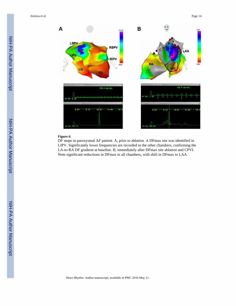
Figure 6.DF maps in paroxysmal AF patient. A, prior to ablation. A DFmax site was identified inLIPV. Significantly lower frequencies are recorded in the other chambers, confirming theLA-to-RA DF gradient at baseline. B, immediately after DFmax site ablation and CPVI.Note significant reductions in DFmax in all chambers, with shift in DFmax to LAA.
Atienza et al. Page 14
Heart Rhythm. Author manuscript; available in PMC 2010 May 11.
NIH
-PA Author Manuscript
NIH
-PA Author Manuscript
NIH
-PA Author Manuscript
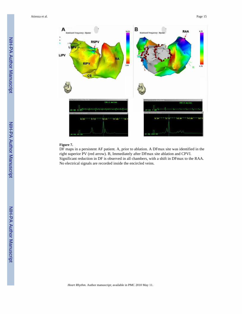
Figure 7.DF maps in a persistent AF patient. A, prior to ablation. A DFmax site was identified in theright superior PV (red arrow). B, Immediately after DFmax site ablation and CPVI.Significant reduction in DF is observed in all chambers, with a shift in DFmax to the RAA.No electrical signals are recorded inside the encircled veins.
Atienza et al. Page 15
Heart Rhythm. Author manuscript; available in PMC 2010 May 11.
NIH
-PA Author Manuscript
NIH
-PA Author Manuscript
NIH
-PA Author Manuscript

NIH
-PA Author Manuscript
NIH
-PA Author Manuscript
NIH
-PA Author Manuscript
Atienza et al. Page 16
Table 1
Patient Characteristics
Paroxysmal AF (n=32) Persistent AF (n=18) p-value
Age (years) 52.8±11.3 50.3±11 0.52
Male (%) 24 (75%) 13 (72%) 0.83
Body mass index (kg/m2) 26.6±3.1 30.5±7.5 0.06
Structural heart disease 1 (11%) 8 (44%) 0.001
First AF episode
<1 year 3 (9%) 2 (12%)
1–3 years 3 (9%) 2 (12%)
>3 years 26 (82%) 13 (76%) 0.92
Sustained AF duration (months) NA 19.9±11 -
Left atrial size (mm) 40.7±6 47.5±6 <0.001
Left atrial area (cm2) 18.8±3.1 26.3±7.2 <0.001
Right atrial area (cm2) 16.4±2.7 22.4±8.5 0.004
LV ejection fraction (%) 59.2±7 48,8±12.4 <0.001
Number antiarrhythmic used 2.1±0.9 2±1 0.65
NA, not applicable; AF, atrial fibrillation; LV, left ventricle.
Heart Rhythm. Author manuscript; available in PMC 2010 May 11.