198921.pdf - Hindawi.com
12
Hindawi Publishing Corporation Journal of Biomedicine and Biotechnology Volume 2010, Article ID 198921, 11 pages doi:10.1155/2010/198921 Research Article Pretreatment with Cry1Ac Protoxin Modulates the Immune Response, and Increases the Survival of Plasmodium -Infected CBA/Ca Mice Martha Legorreta-Herrera, 1 Rodrigo Oviedo Meza, 1 and Leticia Moreno-Fierros 2 1 Laboratorio de Inmunolog´ ıa Molecular, Facultad de Estudios Superiores Zaragoza, Universidad Nacional Aut´ onoma de M´ exico, Batalla 5 de mayo esq. Fuerte de Loreto, Iztapalapa 09230, Mexico 2 Inmunidad en Mucosas UBIMED, FES-Iztacala, Universidad Nacional Aut´ onoma de M´ exico, Avenida de los Barrios 1, Los Reyes Iztacala, Tlalnepantla 54090, Mexico Correspondence should be addressed to Martha Legorreta-Herrera, [email protected] Received 11 August 2009; Revised 24 November 2009; Accepted 16 December 2009 Academic Editor: Luis I. Terrazas Copyright © 2010 Martha Legorreta-Herrera et al. This is an open access article distributed under the Creative Commons Attribution License, which permits unrestricted use, distribution, and reproduction in any medium, provided the original work is properly cited. Malaria is a major global health problem that kills 1-2 million people each year. Despite exhaustive research, naturally acquired immunity is poorly understood. Cry1A proteins are potent immunogens with adjuvant properties and are able to induce strong cellular and humoral responses. In fact, it has been shown that administration of Cry1Ac protoxin alone or with amoebic lysates induces protection against the lethal infection caused by the protozoa Naegleria fowleri. In this work, we studied whether Cry1Ac is able to activate the innate immune response to induce protection against Plasmodium berghei ANKA (lethal) and P. chabaudi AS (nonlethal) parasites in CBA/Ca mice. Treatment with Cry1Ac induced protection against both Plasmodium species in terms of reduced parasitaemia, longer survival time, modulation of pro- and anti-inflammatory cytokines, and increased levels of specific antibodies against Plasmodium. Understanding how to boost innate immunity to Plasmodium infection should lead to immunologically based intervention strategies. 1. Introduction Each year, malaria infects approximately 500 million people and kills one to two million people, mainly children below the age of five years [1]. Despite decades of research on the subject, naturally acquired immunity to Plasmodium is still poorly understood [2–4]. There are reports about the immunosuppressive effects of Plasmodium infection in humans [5] and in animal models [6]. It is believed that the initial interaction of the parasitised red blood cells with the host immune system is one of the most important factors in determining the nature of the subsequent innate and acquired response, and in determining whether or not severe pathology, such as cerebral malaria, severe anaemia, or cachexia, results [7–9]. On the other hand, insecticidal proteinaceous crystals called Cry proteins are produced as protoxins by Bacillus thuringiensis (Bt) during sporulation. Upon ingestion, crys- talline protoxins are solubilised and proteolytically activated by midgut proteases of susceptible insects. The activated toxin, which is not toxic to vertebrates, binds to specific receptors on the brush-border membrane surface of the midgut epithelium of the insect, inducing the formation of pores and eventually leading to insect mortality [10]. In particular, Cry1Ac is a pore-forming protein that is specifically toxic to lepidopteran insect larvae and acts by binding to the cell-surface receptor aminopeptidase N in the Manduca sexta midgut via the sugar N-acetyl-D- galactosamine (GalNAc) [11, 12]. Although most studies on Cry proteins have been performed with regard to their toxicity in insects, we have described that recombinant Cry1Ac protoxin from Bacillus thuringiensis is a potent mucosal and systemic immunogen with adjuvant properties [13, 14].
-
Upload
khangminh22 -
Category
Documents
-
view
1 -
download
0
Transcript of 198921.pdf - Hindawi.com

Hindawi Publishing CorporationJournal of Biomedicine and BiotechnologyVolume 2010, Article ID 198921, 11 pagesdoi:10.1155/2010/198921
Research Article
Pretreatment with Cry1Ac Protoxin Modulatesthe Immune Response, and Increases the Survival ofPlasmodium-Infected CBA/Ca Mice
Martha Legorreta-Herrera,1 Rodrigo Oviedo Meza,1 and Leticia Moreno-Fierros2
1 Laboratorio de Inmunologıa Molecular, Facultad de Estudios Superiores Zaragoza, Universidad Nacional Autonomade Mexico, Batalla 5 de mayo esq. Fuerte de Loreto, Iztapalapa 09230, Mexico
2 Inmunidad en Mucosas UBIMED, FES-Iztacala, Universidad Nacional Autonoma de Mexico, Avenida de los Barrios 1,Los Reyes Iztacala, Tlalnepantla 54090, Mexico
Correspondence should be addressed to Martha Legorreta-Herrera, [email protected]
Received 11 August 2009; Revised 24 November 2009; Accepted 16 December 2009
Academic Editor: Luis I. Terrazas
Copyright © 2010 Martha Legorreta-Herrera et al. This is an open access article distributed under the Creative CommonsAttribution License, which permits unrestricted use, distribution, and reproduction in any medium, provided the original work isproperly cited.
Malaria is a major global health problem that kills 1-2 million people each year. Despite exhaustive research, naturally acquiredimmunity is poorly understood. Cry1A proteins are potent immunogens with adjuvant properties and are able to induce strongcellular and humoral responses. In fact, it has been shown that administration of Cry1Ac protoxin alone or with amoebic lysatesinduces protection against the lethal infection caused by the protozoa Naegleria fowleri. In this work, we studied whether Cry1Acis able to activate the innate immune response to induce protection against Plasmodium berghei ANKA (lethal) and P. chabaudiAS (nonlethal) parasites in CBA/Ca mice. Treatment with Cry1Ac induced protection against both Plasmodium species in termsof reduced parasitaemia, longer survival time, modulation of pro- and anti-inflammatory cytokines, and increased levels ofspecific antibodies against Plasmodium. Understanding how to boost innate immunity to Plasmodium infection should lead toimmunologically based intervention strategies.
1. Introduction
Each year, malaria infects approximately 500 million peopleand kills one to two million people, mainly children belowthe age of five years [1]. Despite decades of research onthe subject, naturally acquired immunity to Plasmodiumis still poorly understood [2–4]. There are reports aboutthe immunosuppressive effects of Plasmodium infection inhumans [5] and in animal models [6]. It is believed thatthe initial interaction of the parasitised red blood cells withthe host immune system is one of the most importantfactors in determining the nature of the subsequent innateand acquired response, and in determining whether or notsevere pathology, such as cerebral malaria, severe anaemia,or cachexia, results [7–9].
On the other hand, insecticidal proteinaceous crystalscalled Cry proteins are produced as protoxins by Bacillus
thuringiensis (Bt) during sporulation. Upon ingestion, crys-talline protoxins are solubilised and proteolytically activatedby midgut proteases of susceptible insects. The activatedtoxin, which is not toxic to vertebrates, binds to specificreceptors on the brush-border membrane surface of themidgut epithelium of the insect, inducing the formationof pores and eventually leading to insect mortality [10].In particular, Cry1Ac is a pore-forming protein that isspecifically toxic to lepidopteran insect larvae and actsby binding to the cell-surface receptor aminopeptidase Nin the Manduca sexta midgut via the sugar N-acetyl-D-galactosamine (GalNAc) [11, 12].
Although most studies on Cry proteins have beenperformed with regard to their toxicity in insects, we havedescribed that recombinant Cry1Ac protoxin from Bacillusthuringiensis is a potent mucosal and systemic immunogenwith adjuvant properties [13, 14].

2 Journal of Biomedicine and Biotechnology
In addition, we have shown that recombinant Cry1Atoxins possess the ability to induce serum and mucosalspecific antibody responses as well as to modulate IgGsubclasses due to their strong immunogenic properties [14,15]. Furthermore, it has been demonstrated that Cry proteinsfrom B. thuringiensis can induce strong cellular immuneresponses. In particular, we have described that these toxinsare able to promote IFN-γ responses [16]. In malariainfections, an initial IFN-γ response, mainly produced by NKcells, is implicated in the activation of macrophages, whichleads to parasite elimination [17, 18]. In a previous study,we found that administration of the immunogenic proteinwith adjuvant properties, Cry1Ac protoxin alone or withamoebic lysates, markedly increased protective immunityagainst experimental N. fowleri meningoencephalitis in mice[13]. In this work, we determined the ability of Cry1Acprotoxin to activate the innate immune response. So wetested whether the pretreatment with the protein aloneimproved the resistance of mice to Plasmodium chabaudi ASand P. berghei ANKA experimental infections.
2. Materials and Methods
2.1. Mice and Parasites. CBA/Ca mice were kindly donatedby Dr. W Jarra (National Institute for Medical Research,London). The mice were bred, fed, and maintained in aspecific, pathogen-free environment at the FES Zaragoza,Universidad Nacional Autonoma de Mexico animal housefacility in accordance with the institutional and nationalofficial guideline NOM-062-ZOO-1999 for use and care oflaboratory animals.
P. chabaudi AS and P. berghei ANKA were donated byDr. William Jarra (National Institute for Medical Research,London).
2.2. Infection and Treatment. Batches of 6 to 8 sex- and age-matched (6–8 weeks) CBA/Ca mice were treated weekly withCry1Ac protoxin (5 μg/mouse i.p.) or with vehicle (PBS)during four weeks. One day after the last treatment, micewere inoculated intravenously with either 5× 10⁴P. chabaudiAS- or 5 × 10⁴P. berghei ANKA-parasitised erythrocytes.On the days indicated, mice were sacrificed under etheranaesthesia. As controls, a parallel batch of noninfected micewas divided into two groups and treated with PBS or Cry1Acat the same dose. Data presented are representative of twoseparate experiments.
2.3. Blood Sampling. Parasitaemias were evaluated daily byexamination of Giemsa-stained blood smears. Numerationof the parasitaemia was performed under oil, using a ZeissStandard 20 microscope (Carl Zeiss Ltd., Welwyn GardenCity). Parasitaemias of 0.5% and above were determined bycounting the number of parasitised erythrocytes present in atotal of 200 red blood cells. Lower levels of parasitaemia wereassessed by counting the number of parasitised erythrocytespresent in 50 fields. The course of infection in each groupis shown as the geometric mean of the percentage ofparasitaemia.
2.4. Recombinant Cry1Ac Escherichia coli JM103 (pOS9300).The recombinant Cry1Ac E. coli JM103 (pOS9300) strainwas kindly donated by Dr. Dean, from Ohio State University.The bacteria were grown in Luria-Bertani medium contain-ing 50 μg/mL of ampicillin, and Cry1Ac production wasinduced with 1 mM isopropyl-β-D-thiogalactopyranoside(IPTG) [19]. Recombinant Cry1Ac was purified from theIPTG-induced E. coli JM103 (pOS9300) cultures as follows.Cell pellets harvested by centrifugation were suspendedin 50 mM Tris-HCl, 50 mM EDTA (pH 8) (TE buffer)and sonicated (Fisher Sonic Dismembrator Model 300)three times for 5 minutes on ice. Inclusion bodies werecollected by centrifugation at 10 000 × g for 10 minutes,and pellets were washed twice with TE buffer, twice with0.5 M NaCl, and once with 0.5 M/NaCl-1% Triton X-100,once with 0.5 M NaCl, once with cold distilled water andwere finally solubilised in CBP buffer (0.1 M Na2CO3,
1% 2-mercaptoethanol [pH 9.6]). Particulate material wasdiscarded by centrifugation at 10 000 × g for 10 min, andthe purified solubilised protoxin was stored at 4◦C andexamined by sodium dodecyl sulphate-polyacrylamide gelelectrophoresis (SDS-PAGE). The protein concentration wasdetermined using the Bradford method [20]. The presenceof endotoxin contamination in the Cry1Ac protoxin prepara-tions was tested using the E-toxate, Part 1 kit (Sigma), whichhas a sensitivity limit of 0.05–0.1 endotoxin units (EU)/mL,following the manufacturer’s instructions. Endotoxin levelsin the purified Cry1Ac protoxin preparations were below0.1 EU/mL. These preparations were further treated with anexcess of a polymyxin B resin (Bio-Rad, Hercules CA, USA)to remove any possible remnants of endotoxin.
2.5. Cytokine mRNA Expression. Groups of CBA/Ca micewere treated weekly with Cry1Ac protoxin or with PBS forfour weeks. Twenty-four hours after the last inoculation,mice were infected with either P. chabaudi AS or P. bergheiANKA. On the days indicated, three mice of each groupwere sacrificed under ether anaesthesia, and spleen mRNAwas extracted using Trizol (Invitrogen, Carlsbad, CA, USA).DNA was digested with DNase I (Invitrogen) according tothe manufacturer’s instructions, and RNA was quantifiedspectrophotometrically at 260 nm. Next, 1.5 μg of RNAwere retrotranscribed using 1.5 μg of oligo dT (Invitrogen),0.5 mM dNTPs (Pharmacia, Uppsala, Sweden), 40 U RNaseinhibitor, and 200 U MMLV-RT (Invitrogen). Next, 1 μL ofthe resulting cDNA was used to amplify IFN-γ and TGF-β by PCR. Each sample was amplified in duplicate using apreviously described method [21].
Each set of primers as well as the cDNA concentrationwas optimized for a number of cycles to obtain ampliconsin the linear phase of amplification. The following gene-specific primer sequences were used: (IFN-γ) forward: 5′
TGC ATC TTG GCT TTG CAG CTC TTC CTC ATG GC3′, reverse 5′ TGG ACC TGT GGG TTG TTG ACC TCATTG GC 3′; (TGF-β) forward 5′ GAC CGC AAC AACGCC ATC TA 3′ reverse 5′ GGC GTA TCA GTG GGGGTC AG 3′; (β-actin) forward 5′ GTG GGC CGC TCTAGG CAC CAA 3′, reverse 5′ CTC TTT GAT GTC ACG

Journal of Biomedicine and Biotechnology 3
CAC GAT TTC 3′. PCR reactions were performed in a totalvolume of 20 μL. Amplification was carried out in 50 mMKCl, 10 mM Tris-HCl (pH 8.3), 0.1 mg/mL gelatin, 200 mMof each dNTP, 2 mM MgCl2, 100 nM of each primer, 0.5 UAmpli Taq polymerase (Applied Biosystems, Branchburg, NJ,USA), and 15 ng of cDNA. The β-actin gene and either IFN-γ or TGF-β were then simultaneously amplified in a singletube. After 27–29 cycles, the PCR products were separated on5% polyacrylamide gels and stained with ethidium bromide.Each band was analysed by densitometry, and the results areshown as the relation of the absorbance of the correspondingcytokine to that of β-actin in the same sample.
2.6. Cytokine Serum Measurement. On days indicated, micefrom both the P chabaudi AS- and the P. berghei ANKA-infected groups were sacrificed under ether anaesthesia.Immediately, blood from the heart was extracted and thencentrifuged at 2000 × g at 4◦C for 15 min. The serum wasremoved and aliquoted into two tubes and snap frozen at−70◦C until used.
The levels of the cytokines interleukin-2 (IL-2),interleukin-4 (IL-4), interleukin-5 (IL-5), interferon-γ(IFN-γ), and tumour necrosis factor- (TNF-α) in the serumsamples were measured using a cytometric bead arrayperformed according to the manufacturer’s instructions (BDMouse Th1/Th2 Cytokine CBA Kit Biosciences-Pharmingen,Heidelberg, Germany) with the following modifications.We performed all steps in microtubes, and we started thestandard curve at a concentration of 0.625 pg/mL. Thesensitivity achieved with these minor adaptations to theprotocol was 0.9±0.05 pg/mL while the variation inter-assaywas approximately 5%.
2.7. Measurement of Protective Antibodies. Antibody-specificresponses were evaluated using a previously describedmethod [21].
A lysate of pRBC was used as the capture antigen; it wasprepared as follows. P. chabaudi AS- or P. berghei ANKA-infected mice (25% parasitaemia) were bled into PBS-heparin at 4◦C to provide parasitised erythrocytes. The bloodwas passed through a CF11 cellulose powder (Whatman,Maidstone, UK) column to remove leukocytes and thenwashed three times with PBS by centrifugation at 750 × g for15 min at 4◦C. The final cell pellet was resuspended to 5 mLin PBS, and 3 μL of 10% (w/v) saponin in PBS was addedto lyse the erythrocyte membranes. After centrifugation at18,000 × g for 5 min at 4◦C, the supernatant was removed,the pellets were resuspended to 3 mL in PBS, and thecells were lysed by ultrasonication for 3 seconds with 21%amplitude (Ultrasonic Processor Model GE750, USA). Theprotein concentration of the lysates was determined using aBio-Rad commercial reagent, and the lysate was diluted incarbonate-buffered solution to give a coating concentrationof 10 μg/mL. A volume of 100 μL/well was applied to flat-bottomed 96-well ELISA plates (Corning USA). First, theplates were washed with 0.05% (v/v) Tween 20 in PBS, andthen the excess binding sites were blocked using a solution of3% skim milk in PBS for 2 hours at 37◦C. The plates were
then incubated with test sera in duplicate for 1 h at 37◦C anddiluted to 1/20 in PBS. Plates were washed extensively beforedetection of parasite-specific Abs using goat antimousehorseradish peroxidase-conjugated monoclonal Abs (mAb)specific to IgG1, IgG2a, IgG2b, IgG3, total IgG, or IgM(Zymed, San Francisco California, USA) diluted in 0.02%skim milk, 0.05% Tween 20 in PBS to previously calibrateddilutions, which were applied to plates for 1 h at 37◦C.Plates were washed before incubation with a streptavidinperoxidase solution (diluted 1 : 3000 in 0.05% Tween 20in PBS) before the final wash. Plates were developed withortho phenylenediamine at 0.4 mg/mL in citrate buffer (pH5) with 0.03% of hydrogen peroxide as a substrate andincubated in the dark at room temperature for 20 min.Absorbance was determined at 492 nm by measurementof optical density (OD) using a Stat-Fax 2100 microplatereader (Awareness Technology Inc, USA). No standard ofknown concentration for each Ig isotype was available. Henceresults were expressed directly as OD 492 nm values andcompared to an internal standard of normal CBA mouseserum obtained from eight- to ten-week-old naive femalemice. This internal standard provided a background value ofnonspecific responsiveness to the lysate used.
2.8. Statistical Analysis. Statistical analysis was performedwith the Stat Graphs software (version 5.1). Differencesbetween groups were tested for statistical significance bynonparametric analysis of variance (Kruskal-Wallis). A Pvalue <.05 was considered significant. All data are expressedas the mean ± S.D. Each experiment was performed induplicate.
3. Results
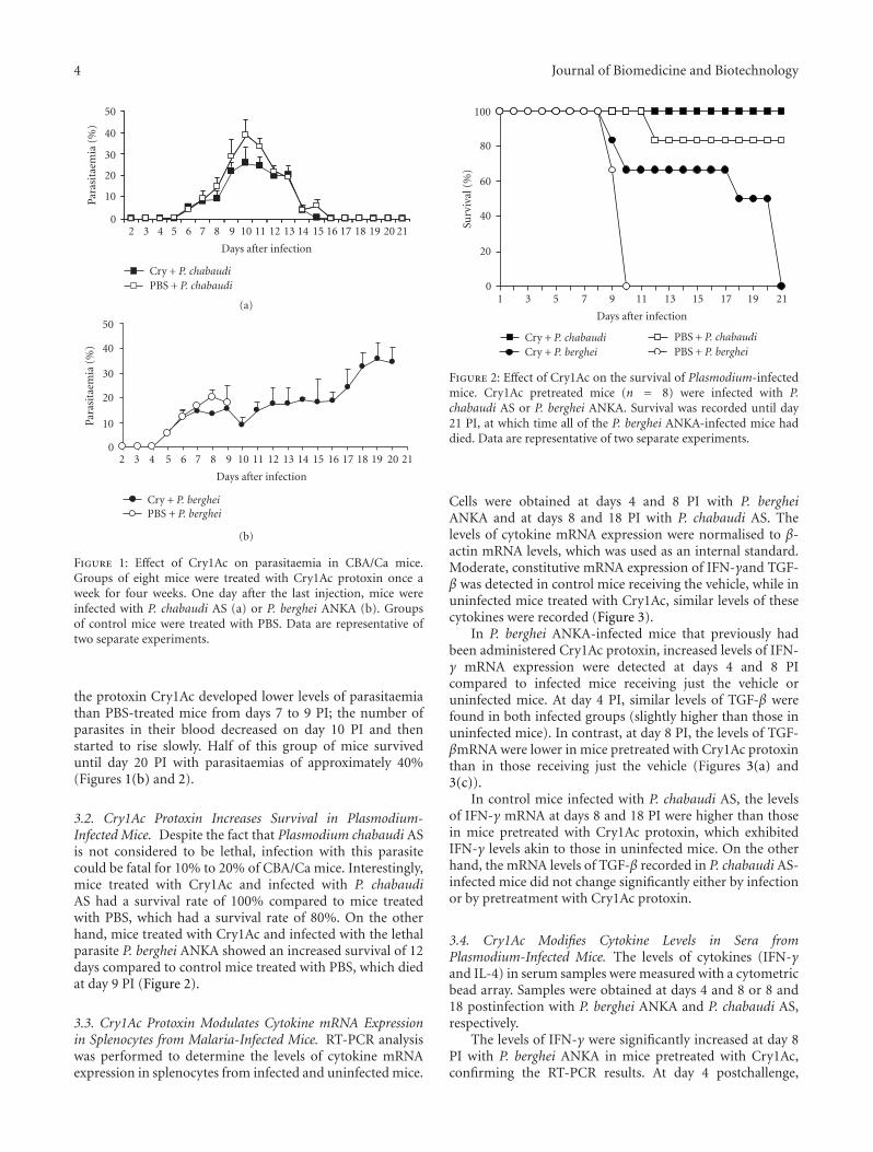
3.1. Cry1Ac Treatment Decreases Parasitaemia in CBA/CaMice Infected with Plasmodium chabaudi AS or P. bergheiANKA. Groups of CBA/Ca mice were injected once weeklyfor four weeks with Cry1Ac protoxin or PBS as described inthe Materials and Methods. One day after the last injection,mice were intravenously infected either with P. chabaudi ASor with P. berghei ANKA. Mice treated with Cry1Ac protoxinprior to P. chabaudi AS infection developed a moderateparasitaemia that increased from day 6 postinfection (PI)to reach a peak of 27% at day 10 PI. Parasitaemia resolvedspontaneously and was cleared by day 15 PI. In contrast,control mice treated with vehicle (PBS) developed higherparasitaemias from day 6 to 15 PI (significantly (P < .05)from days 8 to 11 PI) compared to mice treated with Cry1Acprotoxin. Parasitaemia reached a peak of 40% at day 10, andthe parasite was completely cleared at day 16 PI, one day laterthan in the group of mice treated with Cry1Ac (Figure 1(a)).
In contrast, infection with P. berghei ANKA was lethal.In control CBA/Ca mice treated with vehicle (PBS),parasitaemia increased from day 5 PI to reach a peak of23% at day 8 PI. There was a slight decrease in parasitaemiaat day 9, and then mice started to die on day 10 PI withparasitaemias around 20%, confirming the reported lethalityof this strain [22, 23]. Infected mice previously treated with
4 Journal of Biomedicine and Biotechnology
2 3 4 5 6 7 8 9 10 11 12 13 14 15 16 17 18 19 20 21
Days after infection
0
10
20
30
40
50
Para
sita
emia
(%)
Cry + P. chabaudiPBS + P. chabaudi
(a)
2 3 4 5 6 7 8 9 10 11 12 13 14 15 16 17 18 19 20 21
Days after infection
0
10
20
30
40
50
Para
sita
emia
(%)
Cry + P. bergheiPBS + P. berghei
(b)
Figure 1: Effect of Cry1Ac on parasitaemia in CBA/Ca mice.Groups of eight mice were treated with Cry1Ac protoxin once aweek for four weeks. One day after the last injection, mice wereinfected with P. chabaudi AS (a) or P. berghei ANKA (b). Groupsof control mice were treated with PBS. Data are representative oftwo separate experiments.
the protoxin Cry1Ac developed lower levels of parasitaemiathan PBS-treated mice from days 7 to 9 PI; the number ofparasites in their blood decreased on day 10 PI and thenstarted to rise slowly. Half of this group of mice surviveduntil day 20 PI with parasitaemias of approximately 40%(Figures 1(b) and 2).
3.2. Cry1Ac Protoxin Increases Survival in Plasmodium-Infected Mice. Despite the fact that Plasmodium chabaudi ASis not considered to be lethal, infection with this parasitecould be fatal for 10% to 20% of CBA/Ca mice. Interestingly,mice treated with Cry1Ac and infected with P. chabaudiAS had a survival rate of 100% compared to mice treatedwith PBS, which had a survival rate of 80%. On the otherhand, mice treated with Cry1Ac and infected with the lethalparasite P. berghei ANKA showed an increased survival of 12days compared to control mice treated with PBS, which diedat day 9 PI (Figure 2).
3.3. Cry1Ac Protoxin Modulates Cytokine mRNA Expressionin Splenocytes from Malaria-Infected Mice. RT-PCR analysiswas performed to determine the levels of cytokine mRNAexpression in splenocytes from infected and uninfected mice.
1 3 5 7 9 11 13 15 17 19 21
Days after infection
0
20
40
60
80
100
Surv
ival
(%)
Cry + P. chabaudiCry + P. berghei
PBS + P. chabaudiPBS + P. berghei
Figure 2: Effect of Cry1Ac on the survival of Plasmodium-infectedmice. Cry1Ac pretreated mice (n = 8) were infected with P.chabaudi AS or P. berghei ANKA. Survival was recorded until day21 PI, at which time all of the P. berghei ANKA-infected mice haddied. Data are representative of two separate experiments.
Cells were obtained at days 4 and 8 PI with P. bergheiANKA and at days 8 and 18 PI with P. chabaudi AS. Thelevels of cytokine mRNA expression were normalised to β-actin mRNA levels, which was used as an internal standard.Moderate, constitutive mRNA expression of IFN-γand TGF-β was detected in control mice receiving the vehicle, while inuninfected mice treated with Cry1Ac, similar levels of thesecytokines were recorded (Figure 3).
In P. berghei ANKA-infected mice that previously hadbeen administered Cry1Ac protoxin, increased levels of IFN-γ mRNA expression were detected at days 4 and 8 PIcompared to infected mice receiving just the vehicle oruninfected mice. At day 4 PI, similar levels of TGF-β werefound in both infected groups (slightly higher than those inuninfected mice). In contrast, at day 8 PI, the levels of TGF-βmRNA were lower in mice pretreated with Cry1Ac protoxinthan in those receiving just the vehicle (Figures 3(a) and3(c)).
In control mice infected with P. chabaudi AS, the levelsof IFN-γ mRNA at days 8 and 18 PI were higher than thosein mice pretreated with Cry1Ac protoxin, which exhibitedIFN-γ levels akin to those in uninfected mice. On the otherhand, the mRNA levels of TGF-β recorded in P. chabaudi AS-infected mice did not change significantly either by infectionor by pretreatment with Cry1Ac protoxin.
3.4. Cry1Ac Modifies Cytokine Levels in Sera fromPlasmodium-Infected Mice. The levels of cytokines (IFN-γand IL-4) in serum samples were measured with a cytometricbead array. Samples were obtained at days 4 and 8 or 8 and18 postinfection with P. berghei ANKA and P. chabaudi AS,respectively.
The levels of IFN-γ were significantly increased at day 8PI with P. berghei ANKA in mice pretreated with Cry1Ac,confirming the RT-PCR results. At day 4 postchallenge,

Journal of Biomedicine and Biotechnology 5
PBS +P. berghei
Cry +P. berghei
PBS Cry0
0.2
0.4
0.6
0.8
1
1.2
1.4
Rel
ativ
eex
pres
sion
IFN
-γ/β
-act
in∗
∗
(a)
PBS +P. chabaudi
Cry +P. chabaudi
PBS Cry0
0.2
0.4
0.6
0.8
1
1.2
1.4
IFN
-γ/β
-act
in
∗
(b)
PBS +P. berghei
Cry +P. berghei
PBS Cry0
0.2
0.4
0.6
0.8
1
1.2
1.4
Rel
ativ
eex
pres
sion
TG
F-β/β
-act
in
∗
Day 4Day 8
(c)
PBS +P. chabaudi
Cry +P. chabaudi
PBS Cry0
0.2
0.4
0.6
0.8
1
1.2
1.4
TG
F-β/β
-act
in
Day 8Day 18
(d)
Figure 3: Effect of Cry1Ac on the mRNA expression of IFN-γ (a and b) and TGF-β (c and d) in mice infected with P. chabaudi AS or P.berghei ANKA. Mice were treated with Cry1Ac protoxin or PBS as a control. One day after the last injection, mice were infected with P.berghei ANKA or P. chabaudi AS. On days 4 and 8 PI (a and c) or days 8 and 18 PI (b and d), three mice from each group were killed, andtheir splenic mRNA was extracted and retrotranscribed. The cDNA obtained was used to amplify the β-actin gene and either IFN-γ or TGF-βby PCR in the same tube. PCR products were separated on 5% polyacrylamide gels. Bands were analysed by densitometry, and the resultsare shown as the absorbance of the corresponding cytokine divided by the absorbance of β-actin. Data are representative of two separateexperiments. Asterisks indicate statistically significant differences between the indicated groups.
the IFN-γ mRNA levels recorded were low and akin toinfected control mice, although they are greater than thelevels in uninfected control mice. The levels of IL-4 were alsolow and did not vary significantly as a result of infectionwith P. berghei ANKA or administration of Cry1Ac protein(Figure 4). In contrast, following infection with P. chabaudiAS, the levels of IFN-γ were higher in control mice thanin those pretreated with Cry1Ac protoxin. The IFN-γ levelsinduced after P. chabaudi AS infection were considerablylower compared to those elicited following P. berghei ANKAinfection, but they were still higher than those present inuninfected mice.
In control P. chabaudi AS-infected mice, the levels of IL-4increased at day 18 PI, while in mice pretreated with Cry1Ac,the levels of this cytokine did not change (Figure 4). Levelsof IL-2, IL-5 and TNF-α were not modified by any of thesetreatments (results not shown).
3.5. Protoxin Cry1Ac Increases the Levels of Specific Antibodiesfor P. berghei ANKA and P. chabaudi AS in Infected Mice.Specific anti-P. berghei ANKA antibodies were induced insera from mice infected with the parasite (at days 4 and 8
PI), while sera from control uninfected mice, which wereuntreated or received PBS or Cry1Ac alone, did not havedetectable anti-P.berghei ANKA antibodies (Figure 5).
The treatment with protoxin Cry1Ac before infectionincreased the levels of IgG1, IgG2a, IgG2b, and IgM inP. berghei ANKA-infected mice. Interestingly, this increasewas only detected on day 4 PI compared to mice receivingthe vehicle alone. At day 8 PI, similar levels of IgG andIgM responses were induced in both experimental groups(Figure 5).
In infected mice pretreated with Cry1Ac, the specificanti-P. chabaudi AS IgG response was significantly higher ondays 8 and 18 than that elicited in infected mice pretreatedwith the vehicle (Figure 6). Regarding the analyses of thedifferent IgG subclasses, specific responses at day 4 PI of thefour IgG subclasses (IgG1, IgG2a, IgG2b, and IgG3) werealso significantly higher in the group receiving Cry1Ac beforethe infection compared to the group receiving the vehiclealone (Figure 5). However, at day 8 PI, the IgG2b responsesdetected were higher in the vehicle group with respect tothe Cry1Ac group, while the IgG responses of the rest of theisotypes recorded were similar between the two groups.

6 Journal of Biomedicine and Biotechnology
PBS +P. berghei
Cry +P. berghei
PBS Cry0
10
20
30
40
50
60
70
80
IFN
-γ(p
g/m
L)
∗
(a)
PBS +P. chabaudi
Cry +P. chabaudi
PBS Cry0
1
2
3
4
5
6
7
8
IFN
-γ(p
g/m
L)
∗
(b)
PBS +P. berghei
Cry +P. berghei
PBS Cry0
0.5
1
1.5
2
2.5
3
3.5
IL-4
(pg/
mL
)
Day 4Day 8
(c)
PBS +P. chabaudi
Cry +P. chabaudi
PBS Cry0
0.5
1
1.5
2
2.5
3
3.5
IL-4
(pg/
mL
)
Day 8Day 18
∗
(d)
Figure 4: Effect of Cry1Ac on serum cytokines in Plasmodium-infected mice.Mice were treated with Cry1Ac protoxin or PBS once weeklyfor four weeks. One day after the last injection, mice were infected with P. berghei ANKA or P. chabaudi AS. On days 4 and 8 PI (a and c)or days 8 and 18 PI (b and d), three mice from each group were killed, their sera was separated from peripheral blood and the levels of thecytokines IFN-γ (a and b) and IL-4 (c and d) were measured by cytometric bead array. Data are representative of two separate experiments.Asterisks indicate statistically significant differences between the indicated groups.
The IgM-specific response at day 8 PI was significantlyhigher in the group that received Cry1Ac than in the grouptreated with vehicle.
The specific antibody responses recorded in miceinfected with P. berghei ANKA were lower in relation to thoseelicited in mice infected with P. chabaudi AS. This result wasexpected since the latter group of mice had a longer period ofantigenic stimulation. In sera from uninfected mice receivingeither the vehicle (PBS) or Cry1Ac, specific anti-P. bergheiANKA antibodies were not detected.
4. Discussion
Our results demonstrate that administration of the Cry1Acprotoxin from B. thuringiensis induces protection againstthe malaria parasite when it is administered in CBA/Camice before infection with P. chabaudi AS and inducesa longer survival time in P. berghei ANKA-infected mice(Figures 1 and 2). Protection was shown by lower levels ofparasitaemia (first peak) in groups of mice infected witheither P. chabaudi AS or P. berghei ANKA compared to
control mice. In addition, Cry1Ac protoxin modulated themRNA expression of proinflammatory cytokines, such asIFN-γ and TGF-β, and increased the levels of IgG andIgM in both P. berghei ANKA- and P. chabaudi AS-infectedmice.
Due to the different courses of parasitaemia betweenCBA/Ca mice infected with the lethal P. berghei ANKA, whichkilled all of the mice around day 9 PI, and the non-lethalP. chabaudi AS, the samples were analysed at different days(4 and 8 versus 8 and 18, resp.) to get the best comparisonbetween both infections.
We evaluated the effect of Cry1Ac protoxin againstPlasmodium infection because we have previouslydescribed that this protein may be a valuable tool forthe improvement of mucosal vaccines; when Cry1Acprotoxin is coadministered as an adjuvant, it increasesprotective immunity against experimental Naegleria fowlerimeningoencephalitis, an acute fulminant infection initiatedat the nasal mucosa, in mice [13]. Interestingly, intranasaladministration of Cry1Ac alone also had protective effectsagainst N. fowleri infection, as this treatment increased

Journal of Biomedicine and Biotechnology 7
NS PBS +P. berghei
Cry +P. berghei
PBS Cry0
0.020.040.06
0.080.1
0.120.14
OD
492
nm
∗IgG1
(a)
NS PBS +P. berghei
Cry +P. berghei
PBS Cry0
0.02
0.040.060.08
0.10.120.14
OD
492
nm
∗IgG2a
(b)
NS PBS +P. berghei
Cry +P. berghei
PBS Cry0
0.02
0.040.06
0.080.1
0.120.14
OD
492
nm
∗
IgG2b
(c)
NS PBS +P. berghei
Cry +P. berghei
PBS Cry0
0.02
0.040.06
0.080.1
0.120.14
OD
492
nm
IgG3
(d)
NS PBS +P. berghei
Cry +P. berghei
PBS Cry0
0.1
0.2
0.3
0.4
0.5
OD
492
nm
IgG t
(e)
NS PBS +P. berghei
Cry +P. berghei
PBS Cry0
0.1
0.2
0.3
0.4
0.5
OD
492
nm
∗IgM
(f)
Figure 5: Parasite-specific antibody responses in mice treated with Cry1Ac and infected with P. berghei ANKA. Mice were treated withCry1Ac protoxin or PBS as described previously and were infected with P. berghei ANKA. At days 4 (white bars) and 8 (grey bars) PI, the seraof three mice from each group were collected and measured by ELISA for antibody levels. The results are expressed as the mean OD value± SD (n = 3). Control absorbance values were provided by normal mouse serum (NS) obtained from age- and sex-matched CBA/Ca mice.Data are representative of two separate experiments. Asterisks indicate statistically significant differences between the indicated groups.
survival, as immunization with amoebal lysates alone did,suggesting that Cry1Ac protoxin may boost innate immunity.
In addition, it has been reported that Cry1Ac protoxinenhances the respiratory burst of human monocytes andneutrophils [24]. Accordingly, our unpublished results sug-gest that Cry1Ac activates mouse macrophages, inducing theexpression of the costimulatory molecules B7-1 and B7-2 andthe production of some proinflammatory cytokines (IFN-γand MCP-1), but further studies are required to elucidate themechanisms involved.
Despite the fact that Cry proteins are not toxic tovertebrates and Cry1Ac is known to form pores exclusivelyin the midgut epithelial cells of lepidopteran insect, theexistence of an unknown receptor in mammals has beensuggested, because Cry1Ac protoxin binds to brush bordermembrane vesicles prepared from mouse small intestine invitro [14]. The nature of the molecules interacting with Cry
proteins in mammalian enterocytes seems to be differentthan the receptor glycoproteins described in insects, suchas the 120-kDa aminopeptidase N [11] and the 210-kDacadherin-like glycoprotein (Bt-R1) [25] because binding toits receptor was not inhibited by GalNAc, mannose, or biotin.
On the other hand, it has been shown that IFN-γ is able to activate macrophages, which are responsiblefor elimination of the the malaria parasite [26–28]. Ourresults show that treatment with Cry1Ac protoxin decreasedparasitaemia in mice infected with P. berghei ANKA at day8 PI. At that time, downregulation of TGF-β expressionand an increase in both mRNA expression and serum levelsof IFN-γ were found (Figures 3 and 4). This finding isconsistent with a previous report showing that protectiveimmunity was associated with a decrease in TGF-β anda concomitant increase in IFN-γ production in P. yoelii17XL-infected mice [29]. In addition, it has been shown

8 Journal of Biomedicine and Biotechnology
NS PBS +P. chabaudi
Cry +P. chabaudi
PBS Cry0
0.10.2
0.3
0.40.5
0.6
OD
492
nm
∗
IgG1
(a)
NS PBS +P. chabaudi
Cry +P. chabaudi
PBS Cry0
0.1
0.2
0.3
0.40.5
0.6
OD
492
nm
∗
IgG2a
(b)
NS PBS +P. chabaudi
Cry +P. chabaudi
PBS Cry0
0.1
0.2
0.3
0.4
0.5
0.6
OD
492
nm
∗IgG2b
(c)
NS PBS +P. chabaudi
Cry +P. chabaudi
PBS Cry0
0.1
0.2
0.3
0.4
0.5
0.6
OD
492
nm
∗IgG3
(d)
NS PBS +P. chabaudi
Cry +P. chabaudi
PBS Cry0
0.20.40.60.8
11.21.4
OD
492
nm
∗∗IgG t
(e)
NS PBS +P. chabaudi
Cry +P. chabaudi
PBS Cry0
0.1
0.2
0.3
0.4
OD
492
nm
∗IgM
(f)
Figure 6: Parasite-specific antibody responses in mice treated with Cry1Ac and infected with P. chabaudi AS. Mice were treated with Cry1Acprotoxin or PBS as described previously and were infected with P. chabaudi AS. At days 8 (white bars) and 18 (grey bars) PI, the sera of threemice from each group were collected and measured by ELISA for antibody levels. The results are expressed as the mean OD value ± SD(n = 3). Control absorbance values were provided by normal mouse serum (NS) obtained from age- and sex-matched CBA/Ca mice. Dataare representative of two separate experiments. Asterisks indicate statistically significant differences between the indicated groups.
that upregulation of IFN-γ in P. berghei ANKA-infectedmice leads to the activation of macrophage, which increasesparasite elimination [30]. However, mice infected with P.berghei ANKA in our study were not fully protected, and allof them died at day 21 PI. A possible explanation for thisfinding is that the effect of Cry1Ac protoxin is limited; itcould promote a decrease in parasitaemia in mice at day 8PI, which, in turn, diminishes the antigenic stimulation anddownregulates the immunopathology. As the parasite wasnot completely eliminated, it started to proliferate again, andby day 11, PI Cry1Ac protoxin no longer had an effect.
On the other hand, clearance of P. chabaudi in micedepends first on their ability to mount an early proinflam-matory cytokine response and second on their ability todownregulate the inflammatory response before the onsetof the immunopathology [31]. In our study, mice treatedwith protoxin Cry1Ac and infected with P. chabaudi AS moreefficiently eliminated parasitaemia, despite the fact that this
group demonstrated a decrease in IFN-γ mRNA expressionthat correlated with decreased serum levels of this cytokinecompared to mice treated with PBS (Figures 3 and 4). Thisfact could be related to the upregulation of TGF-β, which hasbeen shown to play a double role in malaria infections; it isable to lead pro- and anti-inflammatory responses that maydownregulate the production of IFN-γ [29]. It is also possiblethat the improved parasite elimination in the mice treatedwith Cry1Ac could be related to higher levels of IgG and IgMantibodies, which could be associated with better parasiteelimination due to an increase in phagocytosis [32–36].
Treatment with protoxin Cry1Ac before Plasmodiuminfection induced a stronger antibody response in CBA/Camice to both Plasmodium berghei ANKA and P. chabaudiAS infections compared to control mice treated with PBS(Figures 5 and 6). Currently, the reason for this increaseis not clear; however, this finding could be explained by across-reaction resulting from molecular similarities between

Journal of Biomedicine and Biotechnology 9
epitopes of Plasmodium and the Cry1Ac protoxin. We haveperformed ELISA assays in which the Cry1Ac protoxinor P. berghei ANKA antigen was bound to the plate andseveral dilutions of P. berghei ANKA immune mice serumwere tested. In these assays, immune sera to P. bergheiANKA or P. chabaudi AS recognised Cry1Ac protoxin anddeveloped higher OD values compared to normal mouseserum (data not shown), strongly suggesting that P. bergheiANKA and Cry1Ac share common antigens. However,further study is required to characterise the precise antigensinvolved.
Another possible mechanism, in which the pretreatmentwith Cry1Ac may increase survival in Plasmodium-infectedmice and may increase both the levels of Plasmodium-specific antibodies elicited after the challenge and the levelsof IFN-γ, may involve the activation of innate immunecells, such as antigen presenting cells, that permit a fasterestablishment of adaptive immune responses. In agreementwith this proposal, our unpublished data indicate thatCry1Ac protoxin activates mouse macrophages, inducing theexpression of the costimulatory molecules B7-1 and B7-2 andthe production of proinflammatory cytokines.
The isotypes of protective antibodies in Plasmodiuminfections are under debate. In general, it is acceptedthat IgG1 and IgG2a are protective [37–40]. However, insome reports, IgG2b or IgG3 are also mentioned as beingprotective [41, 42]. In mice, it is well known that IFN-γ,the principal Th1 effector cytokine, regulates the productionof the opsonising or cytophilic isotype IgG2a [43], whichis in accordance with our results in P. berghei-infectedmice, and that IL-4 is central to the synthesis of IgG1,while TGF-β is involved in the synthesis of IgG2b [38].In our study, we found that pretreatment with Cry1Acprotoxin significantly increases the IgG subclasses assessedand IgM in P. chabaudi AS- or P. berghei ANKA-infectedmice, which could be associated with parasite clearancebecause it has been shown that antibody responses playa critical role in immune protection against Plasmodiumin asexual blood stages. This role has been demonstratedby passive transfer experiments using sera or purifiedimmunoglobulins from adults residing in areas with hyper-endemic malaria [44, 45]. However, the mechanisms bywhich malaria-specific antibodies interfere with the develop-ment and/or multiplication of the asexual stages of humanPlasmodia are still unclear. It has been postulated thatantibodies inhibit parasite growth in cooperation either withmonocytes or neutrophils via antibody-dependent cellularinhibition [46, 47] or by immunophagocytosis through Fcreceptors expressed on the cell surface after binding theirparasite target [48]. In addition, a correlation betweenimmune protection and the ability of serum to mediateopsonisation of infected erythrocytes has been described[49]. Further studies are required to clarify the mecha-nisms involved in boosting the innate immunity againstmalaria.
Like most adjuvants Cry1Ac protoxin might exert itsactivity by activating innate immune cells. The data pre-sented in this study suggest that pretreatment with this pro-tein could lead to design prophylactic strategies to improve
resistance against malaria infections. However, further stud-ies are required to clarify the mechanisms involved inboosting the innate immunity against Plasmodium infection.
Acknowledgments
The authors thank Armando Cervantes for his help withthe statistical analysis. This work was supported by thefollowing Grants: PAPIIT IN214007, IN220310, IN221807,and CONACYT 080920.
References
[1] R. W. Snow, C. A. Guerra, A. M. Noor, H. Y. Myint, andS. I. Hay, “The global distribution of clinical episodes ofPlasmodium falciparum malaria,” Nature, vol. 434, no. 7030,pp. 214–217, 2005.
[2] P. C. Bull and K. Marsh, “The role of antibodies to Plasmodiumfalciparum-infected-erythrocyte surface antigens in naturallyacquired immunity to malaria,” Trends in Microbiology, vol. 10,no. 2, pp. 55–58, 2002.
[3] D. L. Doolan, C. Dobano, and J. K. Baird, “Acquired immunityto Malaria,” Clinical Microbiology Reviews, vol. 22, no. 1, pp.13–36, 2009.
[4] K. Artavanis-Tsakonas, J. E. Tongren, and E. M. Riley, “Thewar between the malaria parasite and the immune system:immunity, immunoregulation and immunopathology,” Clini-cal and Experimental Immunology, vol. 133, no. 2, pp. 145–152,2003.
[5] B. M. Greenwood, A. M. Bradley-Moore, A. D. Bryceson, andA. Palit, “Immunosuppression in children with malaria,” TheLancet, vol. 1, no. 7743, pp. 169–172, 1972.
[6] D. B. Whitmore, “Suppression of the immune response toheterologous erythrocytes in mice infected with Plasmodiumberghei berghei,” Transactions of the Royal Society of TropicalMedicine and Hygiene, vol. 66, no. 1, pp. 5–6, 1972.
[7] B. C. Urban, R. Ing, and M. M. Stevenson, “Early interactionsbetween blood-stage Plasmodium parasites and the immunesystem,” Current Topics in Microbiology and Immunology, vol.297, pp. 25–70, 2005.
[8] K. Grech, K. Watt, and A. F. Read, “Host-parasite interactionsfor virulence and resistance in a malaria model system,”Journal of Evolutionary Biology, vol. 19, no. 5, pp. 1620–1630,2006.
[9] M. N. Wykes, X. Q. Liu, L. Beattie, et al., “Plasmodium straindetermines dendritic cell function essential for survival frommalaria,” PLoS Pathogens, vol. 3, no. 7, article e96, 2007.
[10] H. Hofte and H. R. Whiteley, “Insecticidal crystal proteins ofBacillus thuringiensis,” Microbiological Reviews, vol. 53, no. 2,pp. 242–255, 1989.
[11] P. J. K. Knight, N. Crickmore, and D. J. Ellar, “The receptorfor Bacillus thuringiensis CrylA(c) delta-endotoxin in thebrush border membrane of the lepidopteran Manduca sextais aminopeptidase N,” Molecular Microbiology, vol. 11, no. 3,pp. 429–436, 1994.
[12] E. Schnepf, N. Crickmore, J. Van Rie, et al., “Bacillusthuringiensis and its pesticidal crystal proteins,” Microbiologyand Molecular Biology Reviews, vol. 62, no. 3, pp. 775–806,1998.
[13] S. Rojas-Hernandez, M. A. Rodriguez-Monroy, R. Lopez-Revilla, A. A. Resendiz-Albor, and L. Moreno-Fierros,“Intranasal coadministration of the Cry1Ac protoxin with
10 Journal of Biomedicine and Biotechnology
amoebal lysates increases protection against Naegleria fowlerimeningoencephalitis,” Infection and Immunity, vol. 72, no. 8,pp. 4368–4375, 2004.
[14] R. I. Vazquez-Padron, L. Moreno-Fierros, L. Neri-Bazan,G. A. de la Riva, and R. Lopez-Revilla, “Intragastric andintraperitoneal administration of Cry1Ac protoxin fromBacillus thuringiensis induces systemic and mucosal antibodyresponses in mice,” Life Sciences, vol. 64, no. 21, pp. 1897–1912, 1999.
[15] G. G. Guerrero and L. Moreno-Fierros, “Carrier potentialproperties of Bacillus thuringiensis Cry1A toxins for a diphthe-ria toxin epitope,” Scandinavian Journal of Immunology, vol.66, no. 6, pp. 610–618, 2007.
[16] G. G. Guerrero, W. M. Russell, and L. Moreno-Fierros,“Analysis of the cellular immune response induced by Bacillusthuringiensis Cry1A toxins in mice: effect of the hydrophobicmotif from diphtheria toxin,” Molecular Immunology, vol. 44,no. 6, pp. 1209–1217, 2007.
[17] S. Roetynck, M. Baratin, S. Johansson, C. Lemmers, E.Vivier, and S. Ugolini, “Natural killer cells and malaria,”Immunological Reviews, vol. 214, no. 1, pp. 251–263, 2006.
[18] K. Artavanis-Tsakonas and E. M. Riley, “Innate immuneresponse to malaria: rapid induction of IFN-γ from humanNK cells by live Plasmodium falciparum-infected erythro-cytes,” Journal of Immunology, vol. 169, no. 6, pp. 2956–2963,2002.
[19] A. Z. Ge, R. M. Pfister, and D. H. Dean, “Hyperexpressionof a Bacillus thuringiensis delta-endotoxin-encoding gene inEscherichia coli: properties of the product,” Gene, vol. 93, no.1, pp. 49–54, 1990.
[20] M. M. Bradford, “A rapid and sensitive method for thequantitation of microgram quantities of protein utilizing theprinciple of protein dye binding,” Analytical Biochemistry, vol.72, no. 1-2, pp. 248–254, 1976.
[21] M. Legorreta-Herrera, M. L. Ventura-Ayala, R. N. Licona-Chavez, I. Soto-Cruz, and F. F. Hernandez-Clemente, “Earlytreatment during a primary malaria infection modifies thedevelopment of cross immunity,” Parasite Immunology, vol. 26,no. 1, pp. 7–17, 2004.
[22] S. Campino, S. Bagot, M.-L. Bergman, et al., “Genetic controlof parasite clearance leads to resistance to Plasmodium bergheiANKA infection and confers immunity,” Genes and Immunity,vol. 6, no. 5, pp. 416–421, 2005.
[23] A. Shibui, N. Hozumi, C. Shiraishi, et al., “CD4+ T cellresponse in early erythrocytic stage malaria: Plasmodiumberghei infection in BALB/c and C57BL/6 mice,” ParasitologyResearch, vol. 105, no. 1, pp. 281–286, 2009.
[24] A. R. Rodriguez-Orozco, G. Rico Rosillo, and R. Lopez-Revilla,“The effect of Cry1Ac on human monocytes and neutrophilactivation,” Allergy and Clinical Immunology International, vol.17, no. 2, pp. 64–65, 2005.
[25] Y. Nagamatsu, T. Koike, K. Sasaki, A. Yoshimoto, and Y.Furukawa, “The cadherin-like protein is essential to specificitydetermination and cytotoxic action of the Bacillus thuringien-sis insecticidal CryIAa toxin,” FEBS Letters, vol. 460, no. 2, pp.385–390, 1999.
[26] C. F. Ockenhouse, S. Schulman, and H. L. Shear, “Inductionof crisis forms in the human malaria parasite Plasmod-ium falciparum by γ-interferon-activated, monocyte-derivedmacrophages,” Journal of Immunology, vol. 133, no. 3, pp.1601–1608, 1984.
[27] T. Yoneto, T. Yoshimoto, C.-R. Wang, et al., “Gammainterferon production is critical for protective immunityto infection with blood-stage Plasmodium berghei XAT but
neither NO production nor NK cell activation is critical,”Infection and Immunity, vol. 67, no. 5, pp. 2349–2356, 1999.
[28] H. L. Shear, R. Srinivasan, T. Nolan, and C. Ng, “Role of IFN-γ in lethal and nonlethal malaria in susceptible and resistantmurine hosts,” Journal of Immunology, vol. 143, no. 6, pp.2038–2044, 1989.
[29] F. M. Omer, J. B. de Souza, and E. M. Riley, “Differentialinduction of TGF-β regulates proinflammatory cytokine pro-duction and determines the outcome of lethal and nonlethalPlasmodium yoelii infections,” Journal of Immunology, vol. 171,no. 10, pp. 5430–5436, 2003.
[30] K. N. Couper, D. G. Blount, J. C. R. Hafalla, N. Van Rooijen,J. B. de Souza, and E. M. Riley, “Macrophage-mediatedbut gamma interferon-independent innate immune responsescontrol the primary wave of Plasmodium yoelii parasitemia,”Infection and Immunity, vol. 75, no. 12, pp. 5806–5818, 2007.
[31] M. M. Stevenson and E. M. Riley, “Innate immunity tomalaria,” Nature Reviews Immunology, vol. 4, no. 3, pp. 169–180, 2004.
[32] R. J. Pleass, S. A. Ogun, D. H. McGuinness, J. G. J. Van DeWinkel, A. A. Holder, and J. M. Woof, “Novel antimalarialantibodies highlight the importance of the antibody Fc regionin mediating protection,” Blood, vol. 102, no. 13, pp. 4424–4430, 2003.
[33] A. E. Tebo, P. G. Kremsner, and A. J. F. Luty, “Fcγ receptor-mediated phagocytosis of Plasmodium falciparum-infectederythrocytes in vitro,” Clinical and Experimental Immunology,vol. 130, no. 2, pp. 300–306, 2002.
[34] M. M. Mota, K. N. Brown, A. A. Holder, and W. Jarra,“Acute Plasmodium chabaudi chabaudi malaria infectioninduces antibodies which bind to the surfaces of parasitizederythrocytes and promote their phagocytosis by macrophagesin vitro,” Infection and Immunity, vol. 66, no. 9, pp. 4080–4086,1998.
[35] Y. Peng, R. Kowalewski, S. Kim, and K. B. Elkon, “The role ofIgM antibodies in the recognition and clearance of apoptoticcells,” Molecular Immunology, vol. 42, no. 7, pp. 781–787, 2005.
[36] H. L. Shear, R. S. Nussenzweig, and C. Bianco, “Immunephagocytosis in murine malaria,” Journal of ExperimentalMedicine, vol. 149, no. 6, pp. 1288–1298, 1979.
[37] B. D. Akanmori, S. Waki, and M. Suzuki, “ImmunoglobulinG(2a) isotype may have a protective role in Plasmodiumberghei NH65 infection in immunised mice,” ParasitologyResearch, vol. 80, no. 8, pp. 638–641, 1994.
[38] S. Waki, S. Uehara, K. Kanbe, H. Nariuch, and M. Suzuki,“Interferon-gamma and the induction of protective IgG2aantibodies in non-lethal Plasmodium berghei infections ofmice,” Parasite Immunology, vol. 17, no. 10, pp. 503–508, 1995.
[39] J. Langhorne, K. J. Kim, and R. Asofsky, “Distribution ofimmunoglobulin isotypes in the nonspecific B-cell responseinduced by infection with Plasmodium chabaudi adami andPlasmodium yoelii,” Cellular Immunology, vol. 90, no. 1, pp.251–257, 1985.
[40] R. A. Cavinato, K. R. B. Bastos, L. R. Sardinha, R. M. Elias,J. M. Alvarez, and M. R. D’Imperio Lima, “Susceptibility ofthe different developmental stages of the asexual (schizogonic)erythrocyte cycle of Plasmodium chabaudi chabaudi to hyper-immune serum, immunoglobulin (Ig)G1, IgG2a and F(ab′)2
fragments,” Parasite Immunology, vol. 23, no. 11, pp. 587–597,2001.
[41] L. Kedzierski, C. G. Black, A. W. Stowers, M. W. Goschnick, D.C. Kaslow, and R. L. Coppel, “Comparison of the protectiveefficacy of yeast-derived and Escherichia coli-derived recom-binant merozoite surface protein 4/5 against lethal challenge
Journal of Biomedicine and Biotechnology 11
by Plasmodium yoelii,” Vaccine, vol. 19, no. 32, pp. 4661–4668,2001.
[42] O. Garraud, S. Mahanty, and R. Perraut, “Malaria-specificantibody subclasses in immune individuals: a key source ofinformation for vaccine design,” Trends in Immunology, vol.24, no. 1, pp. 30–35, 2003.
[43] C. M. Snapper and W. E. Paul, “Interferon-γ and B cell stim-ulatory factor-1 reciprocally regulate Ig isotype production,”Science, vol. 236, no. 4804, pp. 944–947, 1987.
[44] S. Cohen, I. A. McGregor, and S. Carrington, “Gamma-globulin and acquired immunity to human malaria,” Nature,vol. 192, no. 4804, pp. 733–737, 1961.
[45] A. Sabchareon, T. Burnouf, D. Ouattara, et al., “Parasitologicand clinical human response to immunoglobulin adminis-tration in falciparum malaria,” American Journal of TropicalMedicine and Hygiene, vol. 45, no. 3, pp. 297–308, 1991.
[46] H. Bouharoun-Tayoun, P. Attanath, A. Sabchareon, T. Chong-suphajaisiddhi, and P. Druilhe, “Antibodies that protecthumans against Plasmodium falciparum blood stages do noton their own inhibit parasite growth and invasion in vitro, butact in cooperation with monocytes,” Journal of ExperimentalMedicine, vol. 172, no. 6, pp. 1633–1641, 1990.
[47] H. Bouharoun-Tayoun, C. Oeuvray, F. Lunel, and P. Druilhe,“Mechanisms underlying the monocyte-mediated antibody-dependent killing of Plasmodium falciparum asexual bloodstages,” Journal of Experimental Medicine, vol. 182, no. 2, pp.409–418, 1995.
[48] A. Ferrante, L. Kumaratilake, C. M. Rzepczyk, and J.-M. Dayer,“Killing of Plasmodium falciparum by cytokine activatedeffector cells (neutrophils and macrophages),” ImmunologyLetters, vol. 25, no. 1–3, pp. 179–187, 1990.
[49] H. Groux and J. Gysin, “Opsonization as an effector mech-anism in human protection against asexual blood stages ofPlasmodium falciparum: functional role of IgG subclasses,”Research in Immunology, vol. 141, no. 6, pp. 529–542, 1990.

Submit your manuscripts athttp://www.hindawi.com
Hindawi Publishing Corporationhttp://www.hindawi.com Volume 2014
Anatomy Research International
PeptidesInternational Journal of
Hindawi Publishing Corporationhttp://www.hindawi.com Volume 2014
Hindawi Publishing Corporation http://www.hindawi.com
International Journal of
Volume 2014
Zoology
Hindawi Publishing Corporationhttp://www.hindawi.com Volume 2014
Molecular Biology International
GenomicsInternational Journal of
Hindawi Publishing Corporationhttp://www.hindawi.com Volume 2014
The Scientific World JournalHindawi Publishing Corporation http://www.hindawi.com Volume 2014
Hindawi Publishing Corporationhttp://www.hindawi.com Volume 2014
BioinformaticsAdvances in
Marine BiologyJournal of
Hindawi Publishing Corporationhttp://www.hindawi.com Volume 2014
Hindawi Publishing Corporationhttp://www.hindawi.com Volume 2014
Signal TransductionJournal of
Hindawi Publishing Corporationhttp://www.hindawi.com Volume 2014
BioMed Research International
Evolutionary BiologyInternational Journal of
Hindawi Publishing Corporationhttp://www.hindawi.com Volume 2014
Hindawi Publishing Corporationhttp://www.hindawi.com Volume 2014
Biochemistry Research International
ArchaeaHindawi Publishing Corporationhttp://www.hindawi.com Volume 2014
Hindawi Publishing Corporationhttp://www.hindawi.com Volume 2014
Genetics Research International
Hindawi Publishing Corporationhttp://www.hindawi.com Volume 2014
Advances in
Virolog y
Hindawi Publishing Corporationhttp://www.hindawi.com
Nucleic AcidsJournal of
Volume 2014
Stem CellsInternational
Hindawi Publishing Corporationhttp://www.hindawi.com Volume 2014
Hindawi Publishing Corporationhttp://www.hindawi.com Volume 2014
Enzyme Research
Hindawi Publishing Corporationhttp://www.hindawi.com Volume 2014
International Journal of
Microbiology