Management of Small Renal Masses - Hindawi.com
144
Management of Small Renal Masses Guest Editors: Jose Rubio Briones and F. Algaba Advances in Urology
-
Upload
khangminh22 -
Category
Documents
-
view
0 -
download
0
Transcript of Management of Small Renal Masses - Hindawi.com

Management of Small Renal Masses
Guest Editors: Jose Rubio Briones and F. Algaba
Advances in Urology

Management of Small Renal Masses

Advances in Urology
Management of Small Renal Masses
Guest Editors: Jose Rubio Briones and F. Algaba

Copyright © 2008 Hindawi Publishing Corporation. All rights reserved.
This is a special issue published in volume 2008 of “Advances in Urology.” All articles are open access articles distributed under theCreative Commons Attribution License, which permits unrestricted use, distribution, and reproduction in any medium, provided theoriginal work is properly cited.

Editor-in-ChiefRichard A. Santucci, Detroit Medical Center, USA
Advisory Editor
Markus Hohenfellner, Germany
Associate Editors
Hassan Abol-Enein, EgyptDarius J. Bagli, CanadaFernando J. Bianco, USASteven B. Brandes, USARobert E. Brannigan, USAJames Brown, USAPeter Clark, USADonna Deng, USAPaddy Dewan, AustraliaMiroslav L. Djordjevic, Serbia
Walid A. Farhat, CanadaChristopher M. Gonzalez, USANarmada Gupta, IndiaEdward Kim, USABadrinath Konety, USADaniel W. Lin, USAWilliam Lynch, AustraliaMaxwell V. Meng, USAHiep Nguyen, USASangtae Park, USA
David F. Penson, USAMichael P. Porter, USAJose Rubio Briones, SpainDouglas Scherr, USANorm D. Smith, USAArnulf Stenzl, GermanyFlavio Trigo Rocha, BrazilWillie Underwood, USA

Contents
Management of Small Renal Masses, Jose Rubio Briones and F. AlgabaVolume 2008, Article ID 606401, 2 pages
Epidemiology of Kidney Cancer, D. Pascual and A. BorqueVolume 2008, Article ID 782381, 7 pages
Small Renal Masses: Incidental Diagnosis, Clinical Symptoms, and Prognostic Factors,F. M. Sanchez-Martın, F. Millan-Rodrıguez, G. Urdaneta-Pignalosa, J. Rubio-Briones,and H. Villavicencio-MavrichVolume 2008, Article ID 310694, 6 pages
Multifocal Renal Cell Carcinoma: Clinicopathologic Features and Outcomes for Tumors≤ 4 cm,Paul L. Crispen, Christine M. Lohse, and Michael L. BluteVolume 2008, Article ID 518091, 7 pages
Familial Syndromes Coupling with Small Renal Masses, Jorge Hidalgo and Gilberto ChechileVolume 2008, Article ID 413505, 7 pages
Genetic Counseling in Renal Masses, Jose Antonio Lopez-Guerrero, Zaida Garcıa-Casado,Antonio Fernandez-Serra, and Jose Rubio-BrionesVolume 2008, Article ID 720840, 12 pages
Radiologic Evaluation of Small Renal Masses (I): Pretreatment Management, A. Marhuenda,M. I. Martın, C. Deltoro, J. Santos, and Jose Rubio BrionesVolume 2008, Article ID 415848, 16 pages
Radiologic Evaluation of Small Renal Masses (II): Posttreatment Management, J. Santos,C. Deltoro, M. I. Martın, and A. MarhuendaVolume 2008, Article ID 918050, 8 pages
Importance and Limits of Ischemia in Renal Partial Surgery: Experimental and ClinicalResearch, Fernando P. SecinVolume 2008, Article ID 102461, 10 pages
Surveillance for the Management of Small Renal Masses, Mehmet Ozsoy, Tobias Klatte,Matthias Waldert, and Mesut RemziVolume 2008, Article ID 196701, 6 pages
Surveillance as an Option for the Treatment of Small Renal Masses, S. Klaver, S. Joniau,and H. Van PoppelVolume 2008, Article ID 705958, 6 pages
Open Partial Nephrectomy in the Management of Small Renal Masses, Ziya Kirkali andA. Erdem CandaVolume 2008, Article ID 309760, 7 pages
Open Partial Nephrectomy in Renal Cancer: A Feasible Gold Standard Technique inAll Hospitals, J. M. Cozar and M. TalladaVolume 2008, Article ID 916463, 9 pages

Fast Track Open Partial Nephrectomy: Reduced Postoperative Length of Stay with a Goal-DirectedPathway Does Not Compromise Outcome, Bilal Chughtai, Christa Abraham, Daniel Finn,Stuart Rosenberg, Bharat Yarlagadda, and Michael PerrottiVolume 2008, Article ID 507543, 5 pages
Cryoablation for Small Renal Masses, J. L. Dominguez-Escrig, K. Sahadevan, and P. JohnsonVolume 2008, Article ID 479495, 10 pages
Cryoablation of Small Renal Tumors in Patients with Solitary Kidneys: Initial Experience,Ravi Munver, Grant I. S. Disick, Salvatore A. Lombardo, Vladislav G. Bargman, and Ihor S. SawczukVolume 2008, Article ID 197324, 3 pages
High-Intensity Focused Ultrasound in Small Renal Masses, Jose Rubio Briones,Argimiro Collado Serra, Alvaro Gomez-Ferrer Lozano, Juan Casanova Ramon-Borja,Inmaculada Iborra Juan, and Eduardo Solsona NarbonVolume 2008, Article ID 809845, 5 pages
Renal Adenomas: Pathological Differential Diagnosis with Malignant Tumors, F. AlgabaVolume 2008, Article ID 974848, 4 pages
Histological Characterisation of Small Renal Masses and Incidence of Silent Renal Masses,Sergio Almenar Medina and Ana Calatrava FonsVolume 2008, Article ID 758073, 7 pages

Hindawi Publishing CorporationAdvances in UrologyVolume 2008, Article ID 606401, 2 pagesdoi:10.1155/2008/606401
EditorialManagement of Small Renal Masses
Jose Rubio Briones1 and F. Algaba2
1 Urology Department, Valencia Institute of Oncology, C/ Prof. Beltran Baguena 8, 46009 Valencia, Spain2 Department of Pathology, Fundacion Puigvert, C/Cartagena 340, 08025 Barcelona, Spain
Correspondence should be addressed to Jose Rubio Briones, [email protected]
Received 30 December 2008; Accepted 30 December 2008
Copyright © 2008 J. Rubio Briones and F. Algaba. This is an open access article distributed under the Creative CommonsAttribution License, which permits unrestricted use, distribution, and reproduction in any medium, provided the original work isproperly cited.
When we started to plan this special issue, we were under thethought that we are facing more and more cases of small renalmasses in our daily work as urologists and pathologists. Thiscommon fact nowadays will probably increase in the nearfuture as radiological studies are more frequently orderedand fortunately we face an increase in longevity, and alsoas people can get to detect their renal masses before theyreally arrive to the classic lumbar pain/haematuria/lumbo-abdominal mass symptoms.
First of all, strict definition of small renal mass is lacking;most of the authors consider 4 cm as cut-off, imported fromthe classical one regarding partial surgery of the kidney andTNM classification; but we all know that these conceptsare changing and probably will need to be taken intoconsideration.
Been sure the increase in detection, we have to precise thedifferent needs of radiological explorations to characterize asmall renal mass; is sonography, CT, and MRI necessary forall patients? We are still lacking to differentiate from a stan-dard radiological approach benign and malignant small renalmasses. What is the role of percutaneous biopsies in thesecases? These (and others) are questions that urologists do notanswer uniformly. Economical issues are also important in apublic medical system.
When we move to therapeutic aspects, things are evenmore unresolved. There is an increasing number of smallrenal masses managed under a strict watchful waiting policybut this is not plausible for all cases. Limits of age andgrowth rate have been argued again for this approach andmost of the times, at least in our country, people are nothappy knowing they could harbor a renal cancer been just“observed”.
Regarding active treatment, first radical nephrectomyand lastly open partial nephrectomy have been the gold
standard approaches. In fact, main guidelines consider thesecond the treatment of choice for small renal masses nowa-days, having shown the same oncological control comparedto radical surgery. During the last decade, laparoscopic par-tial nephrectomy has emerged with comparable oncologicalresults, adding better cosmetical and perioperative recoverydata. The main drawback of laparoscopic partial nephrec-tomy is its difficulty, being just feasible in experienced centerswith high volume of patients.
In the last five years, different nonablative techniqueshave appeared to compete with partial (open or laparo-scopic) nephrectomy aiming to achieve same oncologicalcontrol, testing percutaneous approach, reducing compli-cation rates, and improving recovery, what have beencalled minimally invasive treatments. As time goes by, thesetechniques have failed to demonstrate good and repro-ducible results in any prospective trial for the percutaneousapproach, but this and the laparoscopic approach areincreasing in number worldwide, mainly radiofrequency andcryotherapy for small renal masses. Follow-up will tell usif they achieve same cancer control, but preliminary resultsshow acceptable results for cryotherapy and are questionablefor radiofrequency.
Our aims are to summarize distinct aspects of themanagement of small renal masses nowadays, focusing onits epidemiology, pathological aspects, prognosis, and mostlythe different treatment strategies.
In the first three manuscripts, the authors try to con-crete the clinical problem of small renal masses nowadays,focusing on multifocality and other prognostic factors thatcould guide their management. Two papers more analyze thefamilial syndromes involved with small renal masses and thepossible genetic counselling we should offer the relatives ofpatients with these tumors.

2 Advances in Urology
The next block studies the different radiological aspectsof small renal masses, both in the preoperative scenarioand then after treatment, where many doubts about localrecurrence need to be clarified by radiologists.
There is an interesting and vast review about thephysiopathology of renal ischemia, a crucial point in renalpartial surgery. The reader will find the limits of it and theresearch ongoing in such an “unknown” field.
In the therapeutic block, there are two nice reviews aboutwatchful waiting policy analyzing fresh data. Then, openpartial nephrectomy will be reviewed and presented witha comparative intent to laparoscopic partial nephrectomyin 3 papers, and reviews on nonablative techniques will bediscussed in two papers more.
Finally, two papers analyze the problems that pathologistface in front of these many-times small renal masses.
We hope that this special issue will answer some of thereader doubts about the management of small renal masses,knowing that the next and near future will offer us muchmore data that can change our actual point of view.
Jose Rubio BrionesF. Algaba

Hindawi Publishing CorporationAdvances in UrologyVolume 2008, Article ID 782381, 7 pagesdoi:10.1155/2008/782381
Review ArticleEpidemiology of Kidney Cancer
D. Pascual1 and A. Borque2
1 Department of Urology, San Pedro Hospital, 26006 Logrono, Spain2 Department of Urology, Miguel Servet University Hospital, 50009 Zaragoza, Spain
Correspondence should be addressed to A. Borque, [email protected]
Received 24 March 2008; Revised 20 August 2008; Accepted 26 September 2008
Recommended by J. Rubio
Some tumors are known to have a definite cause-effect etiology, but renal cell carcinoma (RCC) is not one of them precisely. Withregard to RCC we can only try to identify some clinical and occupational factors as well as substances related to tumorigenesis.Smoking, chemical carcinogens like asbestos or organic solvents are some of these factors that increase the risk of the RCC. Viralinfections and radiation therapy have also been described as risk factors. Some drugs can increase the incidence of RCC as well asother neoplasms. Of course, genetics plays an outstanding role in the development of some cases of kidney cancer. Chronic renalfailure, hypertension, and dialysis need to be considered as special situations. Diet, obesity, lifestyle, and habits can also increasethe risk of RCC. The aim of this review is to summarize the well-defined causes of renal cell carcinoma.
Copyright © 2008 D. Pascual and A. Borque. This is an open access article distributed under the Creative Commons AttributionLicense, which permits unrestricted use, distribution, and reproduction in any medium, provided the original work is properlycited.
1. INTRODUCTION
Speaking about cancer, one of the most difficult issues is tofind a definite and direct cause. There are few tumors with awell-known etiology, but renal cell carcinoma (RCC) is notone of them precisely.
In these cases, we can only try to identify some clinicaland occupational factors, or some substances related tocarcinogenesis.
Epidemiology is an important tool to answer manyquestions about cancer origin. Differences in age, gender, andgeographic distribution have been reported, and multipleclinical factors related to the development of RCC havebeen established. Some of them have been thoroughlydemonstrated in experimental models and in vitro studies,however not all of them recognized as definite etiologicfactors.
2. MATERIAL AND METHODS
A systematic review search strategy was developed to identifypublications related to epidemiology of renal cell carcinoma.This search strategy was run in PubMed through the medicalsubject heading “carcinoma, renal cell” and the subheadingof this descriptor “epidemiology.” We limited our search
strategy to articles published in the previous 5 years, languageEnglish or Spanish, and related to humans.
585 articles were found. Abstracts were evaluated andthe full text of articles selected was reviewed. Secondarysearch from the bibliography of selected articles was alsoconsidered.
The European cancer registry-based study on survivaland care of cancer patients (EUROCARE) and our experi-ence was considered. Last review was on 31 of March 2008.
3. DEMOGRAPHIC ASPECTS OFRENAL CELL CARCINOMA
Among urologic tumors, RCC takes the third place inincidence, following prostate carcinoma and transitional cellcarcinoma of bladder.
Representing two percent of the adult malignancies [1],this malignancy takes the tenth and fourteenth place amongmen and women, respectively, with a man to woman ratio of3/2 [2]; see Table 1.
The peak incidence occurs in the sixth decade, with 80%of the cases within the 40 to 69-year-old population.
Although the most frequent renal tumor in the childhoodis the Wilms tumor, it is important to state that theRCC represents between 2% to 6% of the renal tumors in

2 Advances in Urology
Table 1: Epidemiologic features of the RCC.
3rd urologic tumor in incidenceMaximum incidence on 6thdecade
2% of adults malignancies Male/Female ratio: 3/2
85–90% of adult renalparenchymal malignancies
10th male malignancy and 14thfemale malignancy
2–6% of child renalmalignancies
More frequent in afroamericanpeople
Cancer specific mortality of40%
More frequent in urban people
Table 2: Geographic distribution of RCC [8].
Incidence Zones
High Denmark, New Zealand, Norway, Scotland
Moderate United States, Australia, Belgium, France, Holland
Low Spain, Ireland, Italy, Japan, Venezuela, India, China
children, without differences between sexes [3, 4]. Besides,the incidence of both malignancies is similar in the seconddecade of life. In these early ages the papillary differentiationseems to be more frequent with higher tendency to presenta locally advanced and high-degree disease at the momentof the diagnosis [5], However, when comparing stage bystage with adult tumors, we find a better response to surgicaltreatment and higher survival rates, even with positive nodaldisease.
RCC represents 85 to 90% of renal parenchymal malig-nancies [6, 7].
Among urologic tumors, it is the worst in cancer specificmortality, since more than 40% of the patients with RCCdie of the disease, opposite to the 20% mortality observedin prostate cancer or bladder carcinoma.
In United States 30 000 new cases are diagnosed everyyear, and approximately 12 000 patients die of this disease,with an incidence of near nine cases per 100 000 inhabitantsper year. Afroamericans have 10 to 20% higher incidence,and the reason is not completely understood [9].
Most of the cases of RCC are sporadic and only 4% arefamiliar. The estimated number of new cases in the EuropeanUnion during 2006 was 63300, with 26400 deaths of RCC[10]. The estimated survival in 5 years rises to 54% in malesand 57% in women [11].
Table 2 shows different incidences of RCC in the world.Since 1930 incidence of RCC has been increasing, mostly
between 1930 and 1980. Within this period the incidence alsorose from 0.7 to 4.2 per 100 000 per year in women and from1.6 to 9.6 in men [12]. Since 1980 a sharp increase has notbeen observed comparing to other genitourinary tumors orother type of malignancies. Similarly, deaths caused by RCChad been stable.
Variations of incidence within the first period could beexplained by an easier diagnosis, as a result of diffusion androutine use of diagnostic tools such as ultrasound or CT scan,and not due to a real increased incidence of RCC.
Table 3: Some etiologic factors of RCC [15].
Etiologic factor Relative risk (C.I. 95%)
Von Hipple-Lindau disease 100 (not available)
Chronic dialysis 32 (not available)
Obesity 3.6 (2.3–5.7)
Smoking 2.3 (1.1–5.1)
First relative with RCC 1.6 (1.1–2.4)
Hypertension 1.4 (1.2–1.7)
Dry cleaners 1.4 (1.1–1.7)
Diuretics 1.3 (1.07–1.52)
Trichloroethylene 1 (0.7–9.66)
Radiation therapy 1.1 (3.2–8.1)
Phenacetin 1.1 (2.6–6)
Polycystic kidneys 0.8 (2–4.5)
Cadmium exposure 1 (2–3.9)
Arsenic exposure 1.6 (2.3–4.1)
Asbestos exposure 1.1 (1.4–1.8)
It is also important to state that RCC is found incidentallyin 1.5% of the autopsies [13].
RCC is more frequent in urban populations ratherthan in rural ones. This observation may be explained bythe sanitary conditions and the smoking habit in urbanpopulations. However it has not been related neither tosocioeconomic nor to educational status [14].
There are multiple factors related to the development ofRCC; see Table 3. Some of them have been demonstrated inexperimental models and in vitro studies, however not all ofthem can be considered as definite etiologic factors.
Herein, we describe these main factors.
3.1. Smoking
Multiple carcinogenic substances have been identified intobacco and related to a variety of neoplasms at differentlevels. A high incidence of RCC in smokers has been shown[16], estimated in 2.3 fold risk ratio, directly related with thenumber of cigarettes and inversely with age of beginning ofthe habit. Likewise it has been shown that the carcinogendimetilnitrosamine induces this neoplasm in experimentalstudies. Some authors reported that smokers’ risk for RCCcompares to nonsmokers’ after the fifth year of nonsmoking,but a meta-analysis made by Hunt showed that only after tenyears the risk can be similar in both groups [17], dependingon the dose of tobacco inhaled. Another study by McLaughin[18] and Lipworth [19] confirmed tobacco as the mostimportant risk factor for renal cancer, detected in 20% of thecases of RCC.
But smoking is not only important in the genesis ofRCC, and prognostic nomograms have also been developed[20]. A multivariate study carried out in “Miguel Servet”University Hospital of Zaragoza, Spain (in press), smokinghabit increases 2.84 fold (1.27–6,32) the risk of progressionof the disease after surgery [21], similarly to previous studiesin other countries.

D. Pascual and A. Borque 3
3.2. Chemical carcinogens
Some radiological contrasts have been associated with anincreased incidence of RCC [22]. Although Cycasin (asubstance derived from a palm fruit that grows in the islandof Guam) induces RCC in animals, a higher incidence of thisneoplasm within the island population could not be shown.
Cadmium was demonstrated to have influence on thedevelopment of RCC in smokers [23, 24].
(i) Asbestos. A significantly elevated mortality rate forkidney cancer has been reported in two cohortstudies, on insulation workers [25] and on asbestosproducts workers [26]. Autopsy surveys and animalstudies indicate that asbestos fibers can be depositedin kidney tissue.
(ii) Organic solvents. Pesticides, copper sulphate, benzi-dine, benzene herbicides, and vinyl chloride havebeen found as risk factors of RCC in prolongedexposure. A dose dependent effect has been seen onlyfor organic solvents and copper sulphate [27, 28].
Recent reviews of cohort studies found little or noevidence of an increased risk for RCC among peopleexposed to gasoline and petroleum derived products[29, 30].
(iii) Polycyclic aromatic hydrocarbons. Workers exposed tohigh levels of polycyclic aromatic hydrocarbons likecoke and coal oven workers, firefighters, asphalt, andtar have been reported to be at increased risk forkidney cancer.
3.3. Radiation
Ionizing radiation appears to increase the RCC risk slightly,especially among patients treated for ankylosing spondylitisand cervical cancer [31]. An increased risk has also beenreported for patients receiving radium 224 for bone tuber-culosis and ankylosing spondylitis [32].
3.4. Viruses
The immunosuppressant state related to the HIV infectiondetermines that prevalence of RCC in the infected popu-lation rises 8.5 times compared to the prevalence of thenoninfected ones.
The influence of the polyomavirus SV 40 and of theadenovirus 7 has also been detected in experimental studies.
A clear-cut association was found between herpes-typevirus and renal tumors in toads. These findings led to searchfor evidence of herpes virus proteins in human tumors aswell. Although herpes simplex proteins were found in onlyone study [33, 34], these findings need to be confirmed byfurther research.
3.5. Diuretics
This type of drugs which inhibits water reabsorption onthe renal tubule cells seemed to be responsible for a higher
incidence of RCC in patients with chronic intake of diuretics[35, 36]. Even though, it is noteworthy that hydrochloroth-iazide and furosemide (both effective at the renal tubulelevel) induce tubular cell adenomas and adenocarcinomas ofthe kidney in rats [37]. But Yuan [38] showed in his studythat an adequate use of diuretics for treating hypertensioneliminates the risk associated with the above mentioneddrugs, differentiating the influence of hypertension as a riskfactor for RCC rather than diuretics.
3.6. Analgesics
This is a controverted topic. Several studies reported anincreased incidence of RCC in patients with chronic intakeof analgesics like paracetamol, salicylates, or phenacetin [39,40], however in other studies this relationship has not beenconfirmed neither for time of consumption nor for dose ofthe drug taken [41].
Although a heavy use of drugs containing phenacetin hasbeen clearly demonstrated to increase the risk for transitionalcancer of the renal pelvis, the association with RCC ismuch weaker. On the other hand, an increased risk of RCCassociated with aspirin or acetaminophen consumers wasobserved [42], but others believe that neither acetaminophennor other analgesics have been convincingly linked with RCC[19].
3.7. Oestrogens (dietilestilbestrol)
Although oestrogens can induce RCC in the animal model,little evidence supports an association of the disease withoestrogens in humans [43] and only weak relation has beenreported for the use of oestrogens after menopause and fororal contraceptives [44].
3.8. Inheritance
Most of the cases of RCC are sporadic; however there aresome defined types of RCC with a hereditary pattern [45].
(1) Von Hippel-Lindau (VHL) disease
The VHL disease is inherited through an autosomal domi-nant trait. The syndrome is caused by germline mutationsof the VHL tumor suppressor gene, located on chromosome3p25-26; these mutations can virtually always be identified[46]. The VHL protein takes part in cell cycle regulation andangiogenesis [47]. Patients develop capillary haemangioblas-tomas of the central nervous system and retina, clear cellcarcinoma, phaeochromocytoma, pancreatic, and inner eartumors.
The clinical diagnostic criteria of VHL disease consist of
(i) presence of capillary haemangioblastoma in the cen-tral nervous system or retina,
(ii) presence of one of the typical VHL associatedextraneural tumors, within pertinent family history.

4 Advances in Urology
Fourty to sixty percent of the patients with VHL diseasepresent an RCC. Although they are usually low-gradetumors, the progress rate to metastasis is around 30% [48].
Renal lesions in carriers of VHL germline mutations areeither cysts or clear cell RCC. They are typically multifocaland bilateral.
(2) Hereditary papillary renal carcinoma
This type of renal carcinoma is an inherited tumor syndromewith autosomal dominant trait and of late onset, withmultiple and bilateral papillary renal cell carcinomas type 1.The disease is caused by activating mutations of the METoncogene which maps the chromosome 7q31.
(3) Hereditary leiomyomatosis and renal cell carcinoma
This is an autosomal dominant tumor syndrome withgermline mutations in the FH gene (chromosome lq42.3–q43), These patients have the tendency to acquire benignleiomyomas of the skin and the uterus, and occasionallypapillary renal cell carcinoma type 2 and uterine leiomyosar-comas.
(4) Birt-Hogg-Dube syndrome
This syndrome is characterized by benign skin tumors,specifically fibrofolliculomas, trichodiscomas, and acrochor-dons. Multiple renal tumors and spontaneous pneumotho-races are also frequent. We can find chromophobe RCC, typ-ical RCC, hybrid oncocytoma, papillary RCC, or oncocytictumors.
The Birt-Hogg-Dube gene maps the chromosome17p11.2 and encodes the protein called folliculin. This geneis also involved in sporadic RCC [49].
(5) Familiar clear cell renal cell carcinoma
These families present a hereditary form of multiple, bilateralclear cell RCC but without any clinical evidence of sufferingthe von Hippel-Lindau disease.
This hereditary cancer is characterized to present translo-cations affecting the chromosome 3. Translocations havebeen described among the chromosome 3 and the chromo-somes 8 [50], 6 [51, 52], 2 [53, 54], 1 [55], and 4 [48].
3.9. Acquired cystic disease/chronic dialysis
Approximately the 35 to 47% of the patients on dialysis andspecially those with a very long history present acquiredcystic disease. Some patients with this disease develop apapillary hyperplasia in the epithelium of the cysts thatwould be the origin of the RCC [56, 57].
Approximately the 5 to 9% of the patients with acquiredcystic disease will develop an RCC [58], showing a higherincidence than the general population. As such, we suggesta close follow-up in the kidney-transplant population, andtherefore the immune-suppressed individuals who are ondialysis for a long time, due to a high risk of developing RCC.
3.10. Diet and obesity
Hypercaloric diet and obesity seem to be associated with ahigher risk for suffering of RCC. Obesity accounts for about30% of renal cancers [19].
Some studies relate a higher incidence of RCC with highbody mass index. The relative risk was found to be 3.3 inmales and 2.3 in females [59].
The mechanism of obesity to cause kidney cancer isnot clear. Hormonal changes such as increased levels ofendogenous oestrogens in the obese may be the mechanismthrough which oestrogens induce renal cancer as observedin animal models. However, there is scant epidemiologicevidence supporting hormonal carcinogenesis regardingRCC. Obesity may also predispose to arterionephrosclerosis,which may render the renal tubules more susceptible to car-cinogens. Elevated cholesterol levels associated with obesitymight also play a role, as suggested by animal studies showingthat cholesterol-lowering drugs provide some protectionagainst RCC. Cholesterol and other lipids may favour tumordevelopment by an inhibitory effect on immune cells.
Low vitamin D level, which is usually present in obesepatients, may predispose to acquire RCC. This vitamin isknown to have inhibitory effects on the growth of RCC celllines in vitro [60].
Finally, Lipworth [19] reported that the only consistentprotective factor is consumption of fruit and vegetables.
3.11. Coffee, alcohol, and other beverages
Case-control studies have not confirmed the suggestedrelationship between kidney cancer and consumption ofcoffee, when adjusting the smoking variable. Although twostudies have suggested a positive association, a two-foldincreased risk in both sexes was associated with the use ofdecaffeinated coffee. In another study, an increased risk ofRCC was found only among women with regular coffeeintake [61]. No dose-dependent risk was reported in eitherstudy.
On the other hand, a significant lower risk was reportedin Norway among consumers of seven or more cups ofcoffee compared to those who drink two or fewer cupsdaily, representing a relative risk of 0.25 [62]. Review studiesindicate that coffee consumption does not increase RCCrisk.
Few studies have shown an increased risk of RCC amongtea women consumers [63]. Another study has found a dose-response relationship between tea consumption and kidneycancer mortality [64]. The etiologic significance of thesefindings in direct relationship with tea tannins is not clear[65].
The association between alcoholism and kidney cancermortality has not been demonstrated by well-deisgnedstudies [66]. In fact, a recent case-control study found astatistically significant inverse association between alcoholconsumption and RCC risk [67]. No increase in mortalityfrom kidney cancer has been reported either within alcoholicpatients or brewery workers [68].

D. Pascual and A. Borque 5
3.12. Physical activity
A moderate recreational activity reduces the risk of renalcancer both in men and women. The mechanism is not clear,but there is no doubt that energy expenditure is one of themajor determinants for obesity, which is a strong risk factorfor RCC.
3.13. Hypertension
Hypertension seems to be significant for the development ofkidney cancer. The strength of this relationship is reducedwith the use of diuretics and other antihypertensive drugs,regardless of some of these drugs have been associated withRCC risk. The main problem consists to identify whetherthe increased risk is due to hypertension or antihypertensivemedications.
Despite the mechanism for hypertension to cause renalcancer is not completely understood [69] it seems thatmetabolic and/or functional changes in the renal tubular cellsproduce carcinogenesis. Wide case-control studies have onlyfound a slight relation between RCC and hypertension [70].Further studies are needed.
3.14. Alterations in development of the kidney
The anomalous development of the kidneys may act as ateratogenic factor.
In horseshoe kidneys, the area of the isthmus is prone todevelop tumors [71], due to an anomalous migration of thecells toward this area. However, although the most frequenttumor developed in this malformation is the RCC, theincidence remains identical to that of the general population,without differences in evolution or prognosis [72].
In conclusion we can affirm that respect RCC, like inother malignant diseases, ethiology, and risk factors arenot completely understood. There is some evidence thatcertain situations, drugs, habits, or genetics are related tothe development of renal cancer, but several studies foundcontroversial results and different degrees of evidence.
Smoking and obesity seem to be the most importantindependent risk factors in the genesis of RCC, reported bydifferent authors.
Chromosomal mutations were clearly identified in thecontext of well-defined hereditary diseases.
The adequate use of diuretics and analgesics may berecognized as protective factors not only for RCC but forother diseases as well.
General healthy habits like limiting alcohol and coffeeintake, decreasing the number of cigarettes, lowering fatconsumption, keeping a suitable weight, and practicingregular exercise may reduce the risk and indicence of RCC.
REFERENCES
[1] F. Algaba, A. Moreno, and I. Trias, “Tumores de pene,” inUropatologıa Tumoral. Capitulo II, Tumores Renales, pp. 21–101, Pulso Ediciones, Barcelona, Spain, 1996.
[2] J. K. McLaughlin and L. Lipworth, “Epidemiologic aspects ofrenal cell cancer,” Seminars in Oncology, vol. 27, no. 2, pp. 115–123, 2000.
[3] A. L. Freedman, T. S. Vates, T. Stewart, N. Padiyar, A. D.Perlmutter, and C. A. Smith, “Renal cell carcinoma in children:the detroit experience,” The Journal of Urology, vol. 155, no. 5,pp. 1708–1710, 1996.
[4] H. Asanuma, H. Nakai, M. Takeda, et al., “Renal cell carci-noma in children: experience at a single institution in Japan,”The Journal of Urology, vol. 162, no. 4, pp. 1402–1405, 1999.
[5] A. A. Renshaw, H. Zhang, C. L. Corless, J. A. Fletcher, andM. R. Pins, “Solid variants of papillary (chromophil) renalcell carcinoma: clinicopathologic and genetic features,” TheAmerican Journal of Surgical Pathology, vol. 21, no. 10, pp.1203–1209, 1997.
[6] J. E. Robles, D. Rosell, J. J. Zudaire, and J. M. Berian,“Epidemiologıa de los tumores del parenquima renal,” Revistade medicina de la Universidad de Navarra, vol. 43, no. 2, pp.68–76, 1999.
[7] A. Jemal, R. Siegel, E. Ward, et al., “Cancer statistics, 2006,”Ca: A Cancer Journal for Clinicians, vol. 56, no. 2, pp. 106–130,2006.
[8] S. L. Parker, T. Tong, S. Bolden, and P. A. Wingo, “Cancerstatistics, 1997,” Ca: A Cancer Journal for Clinicians, vol. 47,no. 1, pp. 5–27, 1997.
[9] W.-H. Chow, S. S. Devesa, J. L. Warren, and J. F. FraumeniJr., “Rising incidence of renal cell cancer in the United States,”Journal of the American Medical Association, vol. 281, no. 17,pp. 1628–1631, 1999.
[10] J. Ferlay, P. Autier, M. Boniol, M. Heanue, M. Colombet, andP. Boyle, “Estimates of the cancer incidence and mortality inEurope in 2006,” Annals of Oncology, vol. 18, no. 3, pp. 581–592, 2007.
[11] EUROCARE-3, March 2008, http://www.eurocare.it/.
[12] D. L. Katz, T. Zheng, T. R. Holford, and J. Flannery, “Timetrends in the incidence of renal carcinoma: analysis ofConnecticut tumor Registry data, 1935–1989,” InternationalJournal of Cancer, vol. 58, no. 1, pp. 57–63, 1994.
[13] S. Hellsten, T. Berge, and L. Wehlin, “Unrecognized renalcell carcinoma. Clinical and diagnostic aspects,” ScandinavianJournal of Urology and Nephrology, vol. 15, no. 3, pp. 269–272,1981.
[14] J. K. McLaughlin, W. J. Blot, and S. S. Devesa, “Renal cancer,”in Cancer Epidemiology and Prevention, D. Schoffendeld and J.F. Fraumeni Jr., Eds., pp. 1142–1155, Oxford University Press,New York, NY, USA, 2nd edition, 1996.
[15] J. K. McLaughlin, P. Lindblad, A. Mellemgaard, et al., “Inter-national renal-cell cancer study. I. Tobacco use,” InternationalJournal of Cancer, vol. 60, no. 2, pp. 194–198, 1995.
[16] N. J. Vogelzang and W. M. Stadler, “Kidney cancer,” TheLancet, vol. 352, no. 9141, pp. 1691–1696, 1998.
[17] J. D. Hunt, O. L. van der Hel, G. P. McMillan, P. Boffetta,and P. Brennan, “Renal cell carcinoma in relation to cigarettesmoking: meta-analysis of 24 studies,” International Journal ofCancer, vol. 114, no. 1, pp. 101–108, 2005.
[18] J. K. McLaughlin and L. Lipworth, “Epidemiologic aspects ofrenal cell cancer,” Seminars in Oncology, vol. 27, no. 2, pp. 115–123, 2000.
[19] L. Lipworth, R. E. Tarone, and J. K. McLaughlin, “Theepidemiology of renal cell carcinoma,” The Journal of Urology,vol. 176, no. 6, part 1, pp. 2353–2358, 2006.

6 Advances in Urology
[20] B. R. Lane, D. Babineau, M. W. Kattan, et al., “A preoperativeprognostic nomogram for solid enhancing renal tumors 7 cmor less amenable to partial nephrectomy,” The Journal ofUrology, vol. 178, no. 2, pp. 429–434, 2007.
[21] D. Pascual, Carcinoma de celulas renales. Modelo predictivode progresion, Tesis, University of Zaragoza, Zaragoza, Spain,2008.
[22] G. A. Bannayan and D. L. Lamm, “Renal cell tumors,”Pathology Annual, vol. 15, part 2, pp. 271–308, 1980.
[23] L. N. Kolonel, “Association of cadmium with renal cancer,”Cancer, vol. 37, no. 4, pp. 1782–1787, 1976.
[24] S. A. Baynham, H. P. Katner, and K. B. Cleveland, “Increasedprevalence of renal cell carcinoma in patients with HIVinfection,” AIDS Patient Care STDS, vol. 11, no. 3, pp. 161–165, 1997.
[25] I. J. Selikoff, E. C. Hammond, and H. Seidman, “Mortalityexperience of insulation workers in the United States andCanada, 1943–1976,” Annals of the New York Academy ofSciences, vol. 330, pp. 91–116, 1979.
[26] P. E. Enterline, J. Hartley, and V. Henderson, “Asbestos andcancer: a cohort followed up to death,” British Journal ofIndustrial Medicine, vol. 44, no. 6, pp. 396–401, 1987.
[27] L. Buzio, M. Tondel, G. De Palma, et al., “Occupational riskfactors for renal cell cancer. An Italian case-control study,”Medicina del Lavoro, vol. 93, no. 4, pp. 303–309, 2002.
[28] J. Hu, Y. Mao, and K. White, “Renal cell carcinoma andoccupational exposure to chemicals in Canada,” OccupationalMedicine, vol. 52, no. 3, pp. 157–164, 2002.
[29] C. K. Redmond, A. Ciocco, J. W. Lloyd, and H. W. Rush,“Long-term mortality study of steelworkers. VI. Mortalityfrom malignant neoplasms among coke oven workers,” Journalof Occupational Medicine, vol. 14, no. 8, pp. 621–629, 1972.
[30] A. Mellemgaard, G. Engholm, J. K. McLaughlin, and J. H.Olsen, “Occupational risk factors for renal-cell carcinoma inDenmark,” Scandinavian Journal of Work, Environment andHealth, vol. 20, no. 3, pp. 160–165, 1994.
[31] J. D. Boice Jr., G. Engholm, R. A. Kleinerman, et al., “Radiationdose and second cancer risk in patients treated for cancer ofthe cervix,” Radiation Research, vol. 116, no. 1, pp. 3–55, 1988.
[32] H. Spiess, C. W. Mays, and B. Chmelvesky, “Radium 244 inhumans,” in Risks from Radium and Thorotrast, D. M. Taylor,C. W. Mays, and G. B. Gerber, Eds., BIR Report 21, pp. 7–12,British Institute of Radiology, London, UK, 1989.
[33] M. Murai and M. Oya, “Renal cell carcinoma: etiology,incidence and epidemiology,” Current Opinion in Urology, vol.14, no. 4, pp. 229–233, 2004.
[34] A. S. Parker, J. R. Cerhan, C. F. Lynch, B. C. Leibovich, andK. P. Cantor, “History of urinary tract Infection and risk ofrenal cell carcinoma,” American Journal of Epidemiology, vol.159, no. 1, pp. 42–48, 2004.
[35] A. Mellemgaard, H. Moller, and J. H. Olsen, “Diuretics mayincrease risk of renal cell carcinoma,” Cancer Causes andControl, vol. 3, no. 4, pp. 309–312, 1992.
[36] W. D. Finkle, J. K. McLaughlin, S. A. Rasgon, H. H. Yeoh,and J. E. Low, “Increased risk of renal cell cancer amongwomen using diuretics in the United States,” Cancer Causesand Control, vol. 4, no. 6, pp. 555–558, 1993.
[37] W. Lijinsky and M. D. Reuber, “Pathologic effects of chronicadministration of hydrochlorothiazide, with and withoutsodium nitrite, to F344 rats,” Toxicology and Industrial Health,vol. 3, no. 3, pp. 413–422, 1987.
[38] J.-M. Yuan, J. E. Castelao, M. Gago-Domınguez, R. K. Ross,and M. C. Yu, “Hypertension, obesity and their medicationsin relation to renal cell carcinoma,” British Journal of Cancer,vol. 77, no. 9, pp. 1508–1513, 1998.
[39] N. Kreiger, L. D. Marrett, L. Dodds, S. Hilditch, and G. A.Darlington, “Risk factors for renal cell carcinoma: results ofa population-based case-control study,” Cancer Causes andControl, vol. 4, no. 2, pp. 101–110, 1993.
[40] P. Lindblad, J. K. McLaughlin, A. Mellemgaard, and H.-O.Adami, “Risk of kidney cancer among patients using analgesicsand diuretics: a population-based cohort study,” InternationalJournal of Cancer, vol. 55, no. 1, pp. 5–9, 1993.
[41] M. McCredie, W. Pommer, J. K. McLaughlin, et al., “Inter-national renal-cell cancer study. II. Analgesics,” InternationalJournal of Cancer, vol. 60, no. 3, pp. 345–349, 1995.
[42] A. Mellemgaard, S. Niwa, E. S. Mehl, G. Engholm, J. K.McLauglin, and J. H. Olsen, “Risk factors for renal cellcarcinoma in Denmark: role of medication and medicalhistory,” International Journal of Epidemiology, vol. 23, no. 5,pp. 923–930, 1994.
[43] J. K. McLaughlin and L. M. Schuman, “Epidemiology of renalcell carcinoma,” in Reviews in Cancer Epidemiology, A. M.Lilienfeld, Ed., vol. 2, pp. 170–210, Elsevier/North Holland,New York, NY, USA, 1983.
[44] N. R. Asal, J. R. Geyer, D. R. Risser, E. T. Lee, S. Kadamani, andN. Cherng, “Risk factors in renal cell carcinoma. II. Medicalhistory, occupation, multivariate analysis, and conclusions,”Cancer Detection and Prevention, vol. 13, no. 3-4, pp. 263–279,1988.
[45] M. J. Merino, D. M. Eccles, W. M. Linehan, et al., “Familialrenal cell carcinoma,” in Pathology & Genetics. Tumours of theUrinary System and Male Genital Organs, I. N. Eble, G. Sauter,J. I. Epstein, and I. A. Sesterhenn, Eds., pp. 15–22, IARC Press,Lyon, France, 2004.
[46] C. Stolle, G. Glenn, B. Zbar, et al., “Improved detection ofgermline mutations in the von Hippel-Lindau disease tumorsuppressor gene,” Human Mutation, vol. 12, no. 6, pp. 417–423, 1998.
[47] P. H. Maxwell, M. S. Wiesener, G.-W. Chang, et al., “Thetumour suppressor protein VHL targets hypoxia-induciblefactors for oxygen-dependent proteolysis,” Nature, vol. 399,no. 6733, pp. 271–275, 1999.
[48] J. L. Ruiz Cerda and J. F. Jimenez, “Diagnostico y tratamientoquirurgico de la recidiva y enfermedad metastasica del adeno-carcinoma renal,” in Diagnostico y Tratamiento de la Recidivaen los Tumores Urologico, pp. 1–22, Ed Grupo Aula Medica,Madrid, Spain, 1996.
[49] S. K. Khoo, K. Kahnoski, J. Sugimura, et al., “Inactivation ofBHD in sporadic renal tumors,” Cancer Research, vol. 63, no.15, pp. 4583–4587, 2003.
[50] A. J. Cohen, F. P. Li, S. Berg, et al., “Hereditary renal-cellcarcinoma associated with a chromosomal translocation,” TheNew England Journal of Medicine, vol. 301, no. 11, pp. 592–595,1979.
[51] G. Kovacs, P. Brusa, and W. De Riese, “Tissue-specific expres-sion of a constitutional 3;6 translocation: development ofmultiple bilateral renal-cell carcinomas,” International Journalof Cancer, vol. 43, no. 3, pp. 422–427, 1989.
[52] A. Geurts van Kessel, R. F. Suijkerbuijk, R. J. Sinke, L.Looijenga, J. W. Oosterhuis, and B. de Jong, “Molecularcytogenetics of human germ cell tumours: i(12p) and relatedchromosomal anomalies,” European Urology, vol. 23, no. 1, pp.23–29, 1993.

D. Pascual and A. Borque 7
[53] M. I. Koolen, A. P. M. van der Meyden, D. Bodmer, et al., “Afamilial case of renal cell carcinoma and a t(2;3) chromosometranslocation,” Kidney International, vol. 53, no. 2, pp. 273–275, 1998.
[54] J. Podolski, T. Byrski, S. Zajaczek, et al., “Characterizationof a familial RCC-associated t(2;3)(q33;q21) chromosometranslocation,” Journal of Human Genetics, vol. 46, no. 12, pp.685–693, 2001.
[55] H.-O. Kanayama, W.-O. Lui, M. Takahashi, et al., “Associationof a novel constitutional translocation t(1q;3q)with familialrenal cell carcinoma,” Journal of Medical Genetics, vol. 38, no.3, pp. 165–170, 2001.
[56] J. F. Brennan, M. M. Stilmant, R. K. Babayan, and M. B. Siroky,“Acquired renal cystic disease: implications for the urologist,”British Journal of Urology, vol. 67, no. 4, pp. 342–348, 1991.
[57] P. S. Chandhoke, R. J. Torrence, R. V. Clayman, and M. Roth-stein, “Acquired cystic disease of the kidney: a managementdilemma,” The Journal of Urology, vol. 147, no. 4, pp. 969–974,1992.
[58] M. A. Matson and E. P. Cohen, “Acquired cystic kidney disease:occurrence, prevalence, and renal cancers,” Medicine, vol. 69,no. 4, pp. 217–226, 1990.
[59] J. A. Shapiro, M. A. Williams, and N. S. Weiss, “Body massindex and risk of renal cell carcinoma,” Epidemiology, vol. 10,no. 2, pp. 188–191, 1999.
[60] C. L. Amling, “The association between obesity and theprogression of prostate and renal cell carcinoma,” UrologicOncology: Seminars and Original Investigations, vol. 22, no. 6,pp. 478–484, 2004.
[61] R. J. Menezes, G. Tomlinson, and N. Kreiger, “Physical activityand risk of renal cell carcinoma,” International Journal ofCancer, vol. 107, no. 4, pp. 642–646, 2003.
[62] B. K. Jacobsen, E. Bjelke, G. Kvale, and I. Heuch, “Coffeedrinking, mortality, and cancer incidence: results from aNorwegian prospective study,” Journal of the National CancerInstitute, vol. 76, no. 5, pp. 823–831, 1986.
[63] N. R. Asal, D. R. Risser, S. Kadamani, J. R. Geyer, E. T.Lee, and N. Cherng, “Risk factors in renal cell carcinoma:I. Methodology, demographics, tobacco, beverage use, andobesity,” Cancer Detection and Prevention, vol. 11, no. 3–6, pp.359–377, 1988.
[64] L. J. Kinlen, A. N. Willows, P. Goldblatt, and J. Yudkin, “Teaconsumption and cancer,” British Journal of Cancer, vol. 58,no. 3, pp. 397–401, 1988.
[65] International Agency for Research on Cancer, “Tannic acidand tannins,” IARC Monographs on the Evaluation of Carcino-genic Risks to Humans, vol. 10, pp. 253–262, 1976.
[66] R. C. Brownson, “A case-control study of renal cell carcinomain relation to occupation, smoking, and alcohol consump-tion,” Archives of Environmental Health, vol. 43, no. 3, pp. 238–241, 1988.
[67] A. Mellemgaard, G. Engholm, J. K. McLaughlin, and J. H.Olsen, “Risk factors for renal cell carcinoma in Denmark.I. Role of socioeconomic status, tobacco use, beverages, andfamily history,” Cancer Causes and Control, vol. 5, no. 2, pp.105–113, 1994.
[68] H.-O. Adami, J. K. McLaughlin, A. W. Hsing, et al., “Alco-holism and cancer risk: a population-based cohort study,”Cancer Causes and Control, vol. 3, no. 5, pp. 419–425, 1992.
[69] J. A. Shapiro, M. A. Williams, N. S. Weiss, A. Stergachis, A. Z.LaCroix, and W. E. Barlow, “Hypertension, antihypertensivemedication use, and risk of renal cell carcinoma,” AmericanJournal of Epidemiology, vol. 149, no. 6, pp. 521–530, 1999.
[70] E. Grossman, F. H. Messerli, V. Boyko, and U. Goldbourt,“Is there an association between hypertension and cancermortality?” American Journal of Medicine, vol. 112, no. 6, pp.479–486, 2002.
[71] M. Hohenfellner, D. Schultz-Lampel, A. Lampel, F. Steinbach,B. M. Cramer, and J. W. Thuroff, “Tumor in the horseshoekidney: clinical implications and review of embryogenesis,”The Journal of Urology, vol. 147, no. 4, pp. 1098–1102, 1992.
[72] J. Rubio Briones, R. Regalado, and F. Sanchez, “Incidenciade patologıa tumoral en los rinones en herradura,” EuropeanUrology, vol. 6, no. 4, pp. 306–310, 1999.

Hindawi Publishing CorporationAdvances in UrologyVolume 2008, Article ID 310694, 6 pagesdoi:10.1155/2008/310694
Review ArticleSmall Renal Masses: Incidental Diagnosis, Clinical Symptoms,and Prognostic Factors
F. M. Sanchez-Martın,1 F. Millan-Rodrıguez,1 G. Urdaneta-Pignalosa,1 J. Rubio-Briones,2
and H. Villavicencio-Mavrich1
1 Servicio de Urologıa, Fundacio Puigvert, C/Cartagena 340, 08025 Barcelona, Spain2 Servicio de Urologıa, Instituto Valenciano de Oncologıa, C/Beltran Baguena 8, 46009 Valencia, Spain
Correspondence should be addressed to F. M. Sanchez-Martın, [email protected]
Received 13 May 2008; Revised 26 September 2008; Accepted 18 November 2008
Recommended by Maxwell V. Meng
Introduction. The small renal masses (SRMs) have increased over the past two decades due to more liberal use of imagingtechniques. SRMs have allowed discussions regarding their prognostic, diagnosis, and therapeutic approach. Materials andmethods. Clinical presentation, incidental diagnosis, and prognosis factors of SRMs are discussed in this review. Results. SRMsare defined as lesions less than 4 cm in diameter. SRM could be benign, and most malignant SMRs are low stage and low grade.Clinical symptoms like hematuria are very rare, being diagnosed by chance (incidental) in most cases. Size, stage, and grade arestill the most consistent prognosis factors in (RCC). An enhanced contrast SRM that grows during active surveillance is clearlymalignant, and its aggressive potential increases in those greater than 3 cm. Clear cell carcinoma is the most frequent cellular typeof malign SRM. Conclusions. Only some SRMs are benign. The great majority of malign SRMs have good prognosis (low stageand grade, no metastasis) with open or laparoscopic surgical treatment (nephron sparing techniques). Active surveillance is anaccepted attitude in selected cases.
Copyright © 2008 F. M. Sanchez-Martın et al. This is an open access article distributed under the Creative Commons AttributionLicense, which permits unrestricted use, distribution, and reproduction in any medium, provided the original work is properlycited.
1. INTRODUCTION
The incidence of renal cell carcinoma (RCC) has increasedover the past two decades reflecting earlier diagnosis at anearlier stage, largely due to more liberal use of radiologicalimaging techniques [1], introducing concepts as “incidental”or “small renal masses” (SRMs). SRM could be definedas those renal masses lower than 4 cm in diameter [2–4],accounting for 48–66% of RCC diagnosis [5]. Actually, 79–84% of SRM are detected before genitourinary symptomsare present [6–8] (size is smaller than symptomatic cancerclassifying it as local stage with a better prognosis) [9].Although mean tumor size has decreased in the last years,several studies indicate that this variable is one of themost important prognosis factors for RCC, and it has alsocontributed to the last modifications of RCC staging andtreatment [10, 11].
Years ago, when most RCC were symptomatic, hematuriawas the main symptom, so asymptomatic tumors werediagnosed later or not diagnosed. Before widespread use
of imaging techniques, 67–74% of RCC remained unde-tected until death (autopsies), and only 8.9–20.0% of theseundiagnosed RCC were responsible for the patient’s death[5]. These data support the fact that some RCC have afavorable evolution and support active surveillance in selectcases. Natural history of SRM has not been historically wellestablished because most masses were surgically removedsoon after diagnosis.
2. DEFINITIONS AND GENERAL CONCEPTS
A renal mass discovered by routine ultrasound, CT or MRindicated for other pathology, could be named incidental.A significant number of SRMs are incidentally diagnosed[2, 12]. Renal masses (benign and malign) can be consideredincidental if they are diagnosed in the absence of symptomsor signs. “Incidentaloma” or “incidental” masses relatedto other organs such as adrenal, pituitary, thyroid andparathyroid, as well as the liver are published. Mirilasand Skandalakis questioned the scientific justification for

2 Advances in Urology
this neologism and suggested that should be replaced by“incidentally found” [13]. Narrow relation of “incidental”and “small masses” are considered in some papers [2, 14–16].A possible confusion factor may be that tumors classified as“incidental” show symptoms not directly attributable to therenal mass, thus not detected by the urologist [5].
Small renal masses include all solid or complex cysticlesions lower than 4 cm. Among them, different benigntumors are found in a 12.8 to 17.3% of cases [17–19] includ-ing oncocytoma in 53%, angiomyolipoma in 22%, atypicalcyst in 10%, and different benign lesions as leiomyoma,xanthogranulomatous pyelonephritis, and focal infarction in13% [17].
Incidental renal tumors have a mean size of 3.7 cm(median 3, range 0.8 to 12) [7]. Nevertheless, tumorsgreater than 4 cm could be incidental. Incidental diagnosisis performed in the 82.4%, 78.9%, and 56.7% of the 1–4 cm,4–6 cm and greater than 6 cm renal masses, respectively [5].If a cut-off should be made, most cases of RCC lower than7 cm are incidentally discovered, while tumors greater than7 cm are mainly symptomatic but, as mentioned previously,this cannot be taken as a rule [7].
3. SYMPTOMS
The main symptom of RCC is hematuria (35%–60%)[20–22] but SRMs are often asymptomatic (incidental).Classical manifestations of RCC such as fever or jaundice areextremely rare in front of an SRM. In a study of 349 SRM’s,microhematuria was reported in only 8 cases. Prognosticof those RCC diagnosed by hematuria is worse than thoseincidentally diagnosed [23]. Stage I lesions were observed in62.1% of patients with incidental RCC renal cell carcinomaand just in 23% with symptomatic RCC [6]. Among thedifferent entities causing the incidental diagnosis of an SRM,many have been considered; evaluation for other malignancy(17.7%), gastrointestinal symptoms including nonspecificabdominal pain (16%), evaluation of medical renal disease(6.6%), hypertension (4%), back pain (5.1%), cirrhosis(1.4%), nephrolithiasis (1.4%), diverticulitis (1.4%), lunglesion (1.1%), increased liver enzymes (1.1%), trauma(0.8%), screening CT (0.8%), urinary tract infection (0.8%),chest pain (0.8%), aortic aneurysm evaluation (0.8%), cough(0.5%), shortness of breath (0.5%), Crohn’s disease (0.5%),bronchocele (0.5%), and anemia (0.5%). No differences werefound among incidental or symptomatic RCC according toage, sex, and laterality [15].
Laboratory findings have a significant impact on thepatients with organ-confined RCC prognosis. Although,neoplasic condition reflects an increased invasive potential,characterized by overexpression of substances involved incell proliferation as matrix metalloproteinases [24]; how-ever, inflammatory markers like erythrocyte sedimentationrate greater than 30 mm/hour, hemoglobin levels less than10 gm/dL (female) or 12 gm/dL (male), and increased alka-line phosphatase are negative prognosis elements [22].
Some demographic data may help to presume the matterof SRM: RCC is unusual in young patients; angiomyolipomas
and multilocular cystic nephromas are more common inwomen [25].
4. PROGNOSIS FACTORS
Age is not a significant factor on survival in patients withincidental RCC [26], so it is probably not a prognosis factorfor SRM [5]. However, as the patient ages, the SMR stage ishigher; so the incidence of SRM finally staged as pT3 tumorsin younger than 45 years, 45–75 years, and older than 75 yearsis 2.3%, 6.9%, and 14.3%, respectively [17]. The probabilityof developing metastases, with 12 years follow-up, is greaterin men [27].
5. BENIGN TUMOR FREQUENCY
Lee et al. published 230 cases of SRM (lower than 4 cm),88% malignant and 12% benign (oncocytoma) [6]. DeRocheet al. described that SRMs are nonneoplasic entities. Benignneoplasms and low-and high-grade carcinoma accountedfor 1.6%, 18.0%, 49.0%, and 31.4%, respectively [8]. Thepercentage of malignancies increases from 72.1% in masseslower than 2 cm to 93.7% in tumors greater than 7 cm [7].
In conclusion, if the tumor is greater in dimensions, thepossibility of being benign is lower; so tumors lower than1, 2, 3, and 4 cm were benign in 46.3, 22.4, 22, and 19.9%,respectively [18].
6. SIZE AND STAGE
In a study from Schlomer et al., global mean renal tumorsize decreased by 32% and pT1 tumors increased from 4%to 22% (1989–1998). For every cm increase in size, theodds ratio of malignancy increased 17–39% [7, 18]. Meantumor size for benign tumors was 4.2 cm (median 3.3, range0.2–25) compared to 6.3 cm (median 5.5, range 0.1–24) formalignant tumors. Median clinical diameter was 2.93 cm(range 0.8 to 4.0) in RCC lower than 4 cm. RCC meansize was 4.6 cm (range 0.8–21) and benign masses meansize 2.8 cm (range 0.8–9.5) [5]. Incidental RCC mean sizewas 3.7 cm (median 3, range 0.8–12) and symptomatic RCCmean size was 6.2 cm [7]. In pathological stage, 51.33% and27.3% were pT1, 25.6% and 27.3% pT2, 10.9% and 23.8%pT3a, 10.9% and 16.6% pT3b, 1.2% and 2.3% pT3c, and0% and 2.3% pT4 in incidental and symptomatic RCC,respectively.
Puppo et al. reported 94 patients with resected RCC (size:1.1–4.5 cm), describing that pathological stage was pT1a in92.5%, pT1b in 4.2%, and pT3a in 3.1% [28], similar toPahernik et al. that reports pT1a in 84.5%, pT1b in 8%, andpT3 in 7.5% (organ confined in 92.5%) and≥pT3 was foundin 3.0%, 5.1%, and 12.1% of the patients when analyzed bytumor size 2, 3, and 4 cm, respectively [17]. A total of 25%of SRM doubled in volume within 12 months, 34% reached4 cm and experienced rapid doubling time [5].
Kunkle et al. found synchronous metastatic diseaseincreased by 22% with each cm increase in tumor size, by50% for each increase of 2 cm, and doubled for each 3.5 cmincrease in primary tumor size [11].

F. M. Sanchez-Martın et al. 3
In other manuscript, incidental RCC had lower stagescompared to symptomatic RCC [15]. Between T1a and T1blesions, there was no significant difference in the rate ofmalignancy and high-grade malignancy regarding incidentalor symptomatic presentation. The different percentage of T2malignant tumors between incidental (90.9%) and symp-tomatic tumors was neither significant [5]. Understagingfor pT3 tumors lower than 3 cm was 7.5% [17]. Cysticcomponent appears in 24.1% of renal masses lower than4 cm, being 57.1% in Bosniak type III and the rest in Bosniaktype IV [5].
Volpe et al. showed no differences between the averagegrowth rate for solid SRM (0.11 cm per year) and cysticmasses (0.09 cm per year) [5]. Multifocality was present in5.3–12% in small RCC [7, 8]. The rate of multifocalitywas 2.0%, 5.1%, and 7.05% in tumors of 2, 3, and 4 cm,respectively [17].
7. GRADE
Ninety percent of tumors lower than 1 cm were low-gradecompared to only 37.9% of tumors ≥7 cm [18]. Grade 3was found in 7.1%, 9.0%, and 14.0% of the patients in the2, 3, and 4 cm groups, respectively and just 10.6% of smallRCC were grade 3 [17]. Tumor grade increase as tumorsize increase from 2 to 4 cm. Grade 1 was 31.3% for 2 cm,27.4% for 3 cm, and 18.1% for 4 cm tumors; and grade 3 was7.1% for 2 cm, 9% for 3 cm, and 14% for 4 cm tumors [17].Urinary tract invasion, reported in some low-grade tumors,is a negative prognostic factor [29]. However, 45% of T2incidental malignancies were high grade compared to 78.8%of T2 symptomatic malignancies [5]. Tumor grade increasedaccording to size in clear cell, papillary, and chromophobetumors. In high-grade carcinomas, 65% of the tumors had a1-year volume doubling time.
8. CELLULAR TYPE
Clear cell is the most frequent cellular type regardless oftumor size [7]. Among SRM, Frank et al. showed thatpercentage of clear cell cellular type increased according tosize: 59.9, 70.2, and 72% in lower than 2, 3, and 4 cm,respectively [18]. Cellular type for small RCC was 78%clear cell carcinoma, 15.3% papillary carcinoma, and 7%chromophobe carcinoma [17].
Volpe et al. showed that papillary RCC incidence is morefrequent in 2 cm tumors than in 3 and 4 cm tumors (24%,13.2%, and 13.5%, resp.) [17]; data not refuted by otherauthors [5]. Papillary cell type is more frequent than clearcell in tumors lower than 1 cm [18].
9. METASTASES
Metastases at diagnosis were found in 3.0%, 2.6%, and6.0% of the patients with 2, 3, and 4 cm renal tumors,respectively [17]. Furthermore, lymph node spread was 4.8%and 15%, metastasis was 9.2% and 26%, and local recurrencewas 1.2% and 8.3%, among incidental and symptomaticRCC, respectively [15]. With active surveillance, enhancing
lesions with zero median growth rates did not progress tometastatic disease, and only 1.4% of patients with 0.31 cmyearly median growth rate progressed to metastatic disease[7]. Chawla et al. showed RCC mean growth rate of 0.40 cmyearly (median 0.35, range 0.42 to 1.6) [30].
Median tumor size for patients presented with patho-logically confirmed synchronous metastatic disease wassignificantly greater than for those presenting with localizeddisease, 8.0 cm (range 2.2 to 20.0) and 4.5 cm (range 0.3to 17.5), respectively. Tumors of 3.0 cm or smaller hadsynchronous metastasis in just 4.5% of the cases [31].
10. SURVIVAL
A total of 548 patients with small RCC were analyzed byPahernik et al.: 22 (4%) had metastasis, 9 died by cancer in amean time of 1.9 years (range 0.7 to 3.4) after diagnosis [17].D’allOglio et al. observed a mean overall survival of 91% inpatients with T1a tumors and up to 78.7% survival after 10years of local treatment [15].
Several groups have developed predictive models toconstruct prognosis algorithms in order to facilitate follow-up and to indentify progression risk. Raj et al. present apredictive model that includes gender, symptoms, radiolog-ical findings, and size as preoperative prognostic factors; inorder to establish a chance of being cancer-free 12 years aftersurgery (Figure 1). In case of SRM, it could not be usefulto decide surveillance or active treatment. For example, awoman with a 3 cm incidental malign SRM has a 96% chanceof being cancer-free 12 years after surgery. In contrast, aman with a 4 cm symptomatic (local signs) malign SRM andpositive TC showing enlarged lymph nodes has 60% chanceof being cancer-free 12 years after surgery [27].
Classically, better prognosis has been assigned to inci-dental diagnosis, papillary or chromophobe pathology, smallsize, and early stage [32]. Presence of necrosis and vascularinvasion is useful in a specific algorithm looked toward clearcell renal tumor [33].
Table 1 resumes the main prognosis factors useful onSRM.
11. TREATMENT AS PROGNOSIS FACTOR
Size is a significant factor in the decision to perform NSS:tumors sized 2 cm (81%), 3 cm (73%), and 4 cm (44%)cm could be treated by means of NSS. This treatment istechnically easier in incidental than not incidental RCC (76%versus 24%) [15]. Local excision is a safe treatment for smallRCC, even in extreme cases such as living donor kidneywith a 5 × 5 mm RCC found on its surface [34]. In patientswith RCC lower 4 cm, who underwent partial or radicalnephrectomy 14% and 10% died during follow-up (cancer-specific death occurred in 3% in both approaches). Diseasespecific survival rate at 3 and 5 years is 95 and 97% in partialand radical nephrectomy, respectively [6].
When active surveillance is applied to 2 cm meansize contrast-enhancing renal masses, no differences werereported about age, sex, initial size, and solid versus cysticradiologic appearance. A significant different frequency

4 Advances in Urology
0 10 20 30 40 50 60 70 80 90 100
M
F
Located
SystemicIncidental
Points
Gender
Mode ofpresentation
Y
N
Lymph nodeby imaging
Necrosisby imaging
Y
N
Size byimaging (cm)
0 1 2 3 4 5 6 7 9 13
Total points 0 20 40 60 80 100 120 140 160 180
12-yearmetastasis free
probability0.99 0.98 0.95 0.9 0.7 0.5 0.3 0.1 0.01
Figure 1: Preoperatory prognosis RCC nomogram [27].
Table 1: Small RCC prognosis factors.
Better prognosis Worse prognostic
Incidental Symptoms
Small size <3 cm Size > 3 cm
T1 T2 and >
Low grade High grade
No upper tract invasion Upper tract invasion
No lymph nodes Necrosis
No necrosis Lymph nodes
No vascular invasion Vascular invasion
Negative biological markers Positive biological markers
Papillary or chromophobepathology (¿)
Sarcomatoid component
Zero median grown rate Grown rate > 0.31 cm yearly
Option to NSS No option to NSS
of surgery was found among tumors with 0 or 0.31 cmmean yearly growth rate of 17% and 51%, respectively [7].However, 33% of SRM under active surveillance showedzero or negative radiologic growth [7]. The probability todevelop metastasis in masses lower than 3 cm managed byactive surveillance was only 2% [14]. Prior and duringfollow-up, renal tumor biopsies are recommended. As ageneral rule, biopsy may be indicated in masses that havefeatures of oncocytoma in poor surgical candidates. Forpatients who have a surgical contraindication or rejectsurgery, alternative ablation techniques can be proposed(cryoablation, radiofrequency) [35].
For Kassouf et al., 20.8% of renal masses showed tumorgrowth during the surveillance period (mean 31.6 months),but neither of them developed metastasis. Patients receiving
surgical treatment after surveillance did not modify theirprognostic [16]. Hereditary renal tumors may have a moreaggressive natural history, and thus surveillance should bemade with caution. Meta-analysis of Kunkle et al. observedno statistical differences in the incidence of SRM progressionregardless excision, ablation, or active surveillance [2].
12. CONCLUSIONS
SRMs are those smaller than 4 cm, often incidentally diag-nosed. Clinical symptoms, like hematuria, are rare, butconfer worse prognosis. Size, stage, and grade are still themost consistent prognostic factors in RCC. It is importantto keep in mind that SRM could be benign tumors, mainlyoncocytoma. Most malign SMRs are low stage and low grade,without metastatic spread if diameter is below 2-3 cm. Clearcell carcinoma is the most frequent cellular type of malignSRM. Papillary tumors are more frequent when SRM size isless than 1 cm, having a better prognosis. Aggressive potentialof small RCC could increase in tumors greater than 3 cm,so it is suggested that the threshold for selecting patients(old age, high-risk, solitary kidney, reject surgery) for asurveillance strategy should be set well below a tumor sizeof 3 cm. In active surveillance, the size increase of an SRM isa strong indicator of malignancy; helping to decide a surgicaltreatment.
REFERENCES
[1] W.-H. Chow, S. S. Devesa, J. L. Warren, and J. F. FraumeniJr., “Rising incidence of renal cell cancer in the United States,”Journal of the American Medical Association, vol. 281, no. 17,pp. 1628–1631, 1999.
[2] D. A. Kunkle, B. L. Egleston, and R. G. Uzzo, “Excise, ablate orobserve: the small renal mass dilemma—a meta-analysis and

F. M. Sanchez-Martın et al. 5
review,” The Journal of Urology, vol. 179, no. 4, pp. 1227–1234,2008.
[3] D. A. Duchene, Y. Lotan, J. A. Cadeddu, A. I. Sagalowsky, andK. S. Koeneman, “Histopathology of surgically managed renaltumors: analysis of a contemporary series,” Urology, vol. 62,no. 5, pp. 827–830, 2003.
[4] M. L. Ramırez and C. P. Evans, “Current management ofsmall renal masses,” The Canadian Journal of Urology, vol. 14,supplement 1, pp. 39–47, 2007.
[5] A. Volpe, T. Panzarella, R. A. Rendon, M. A. Haider, F.I. Kondylis, and M. A. S. Jewett, “The natural history ofincidentally detected small renal masses,” Cancer, vol. 100, no.4, pp. 738–745, 2004.
[6] C. T. Lee, J. Katz, W. Shi, H. T. Thaler, V. E. Reuter, and P.Russo, “Surgical management of renal tumors 4 cm. Or less ina contemporary cohort,” The Journal of Urology, vol. 163, no.3, pp. 730–736, 2000.
[7] B. Schlomer, R. S. Figenshau, Y. Yan, R. Venkatesh, and S. B.Bhayani, “Pathological features of renal neoplasms classifiedby size and symptomatology,” The Journal of Urology, vol. 176,no. 4, pp. 1317–1320, 2006.
[8] T. DeRoche, E. Walker, C. Magi-Galluzzi, and M. Zhou,“Pathologic characteristics of solitary small renal masses:can they be predicted by preoperative clinical parameters?”American Journal of Clinical Pathology, vol. 130, no. 4, pp. 560–564, 2008.
[9] J.-J. Patard, A. Rodriguez, N. Rioux-Leclercq, F. Guille, andB. Lobel, “Prognostic significance of the mode of detection inrenal tumours,” BJU International, vol. 90, no. 4, pp. 358–363,2002.
[10] K. E.-H. Tsui, O. Shvarts, R. B. Smith, R. Figlin, J. B. dekernion, and A. Belldegrun, “Renal cell carcinoma: prognosticsignificance of incidentally detected tumors,” The Journal ofUrology, vol. 163, no. 2, pp. 426–430, 2000.
[11] D. A. Kunkle, P. L. Crispen, D. Y. T. Chen, R. E. Greenberg,and R. G. Uzzo, “Enhancing renal masses with zero net growthduring active surveillance,” The Journal of Urology, vol. 177,no. 3, pp. 849–854, 2007.
[12] A. Volpe, “The role of surveillance in the management of smallrenal masses,” The Scientific World Journal, vol. 7, pp. 860–868,2007.
[13] P. Mirilas and J. E. Skandalakis, “Benign anatomical mistakes:incidentaloma,” American Surgeon, vol. 68, no. 11, pp. 1026–1028, 2002.
[14] M. A. Bosniak, “Observation of small incidentally detectedrenal masses,” Seminars in Urologic Oncology, vol. 13, no. 4,pp. 267–272, 1995.
[15] M. F. Dall’Oglio, M. A. Arap, A. A. Antunes, J. Cury,K. R. Leite, and M. Srougi, “Impact of clinicopathologicalparameters in patients treated for renal cell carcinoma,” TheJournal of Urology, vol. 177, no. 5, pp. 1687–1691, 2007.
[16] W. Kassouf, A. G. Aprikian, M. Laplante, and S. Tanguay,“Natural history of renal masses followed expectantly,” TheJournal of Urology, vol. 171, no. 1, pp. 111–113, 2004.
[17] S. Pahernik, S. Ziegler, F. Roos, S. W. Melchior, and J. W.Thuroff, “Small renal tumors: correlation of clinical andpathological features with tumor size,” The Journal of Urology,vol. 178, no. 2, pp. 414–417, 2007.
[18] I. Frank, M. L. Blute, J. C. Cheville, C. M. Lohse, A. L. Weaver,and H. Zincke, “Solid renal tumors: an analysis of pathologicalfeatures related to tumor size,” The Journal of Urology, vol. 170,no. 6, part 1, pp. 2217–2220, 2003.
[19] P. Russo, “Should elective partial nephrectomy be performedfor renal cell carcinoma >4 cm in size? ” Nature ClinicalPractice Urology, vol. 5, no. 9, pp. 482–483, 2008.
[20] R. A. Medina Lopez, C. B. Congregado Ruiz, P. CampoyMartınez, A. Morales Lopez, E. Sanchez Gomez, and J. L.Pascual del Pobil Moreno, “Cancer renal: analisis descriptivede una serie de 267 casos intervenidos,” Archivos Espanoles deUrologıa, vol. 54, no. 5, pp. 423–428, 2001.
[21] K. Sugimura, S.-I. Ikemoto, H. Kawashima, N. Nishisaka,and T. Kishimoto, “Microscopic hematuria as a screeningmarker for urinary tract malignancies,” International Journalof Urology, vol. 8, no. 1, pp. 1–5, 2001.
[22] A. Navarro Izquierdo, R. Matesanz Acedos, and J. M. PeredaGarcıa, “Evolucion y respuesta terapeutica del adenocarci-noma renal,” Revista Clınica Espanola, vol. 147, no. 1, pp. 55–60, 1977.
[23] J.-J. Patard, K. Bensalah, S. Vincendeau, N. Rioux-Leclerq,F. Guille, and B. Lobel, “Correlation between the mode ofpresentation of renal tumours and patient survival,” Progresen Urologie, vol. 13, no. 1, pp. 23–28, 2003.
[24] K. Harada, I. Sakai, T. Ishimura, T. Inoue, I. Hara, and H.Miyake, “Clinical symptoms in localized renal cell carcinomareflect its invasive potential: comparative study betweenincidentally detected and symptomatic diseases,” UrologicOncology: Seminars and Original Investigations, vol. 24, no. 3,pp. 201–206, 2006.
[25] S. G. Silverman, G. M. Israel, B. R. Herts, and J. P. Richie,“Management of the incidental renal mass,” Radiology, vol.249, no. 1, pp. 16–31, 2008.
[26] L. Gomez Perez, A. Budıa Alba, F. J. Delgado Oliva, J. L. RuizCerda, M. Trassiera Villa, and F. Jimenez Cruz, “Cancer renalincidental en pacientes menores de 40 anos: hallazgos clınicose histopatologicos,” Actas Urologicas Espanolas, vol. 31, no. 3,pp. 244–249, 2007.
[27] G. V. Raj, R. H. Thompson, B. C. Leibovich, M. L. Blute, P.Russo, and M. W. Kattan, “Preoperative nomogram predicting12-year probability of metastatic renal cancer,” The Journal ofUrology, vol. 179, no. 6, pp. 2146–2151, 2008.
[28] P. Puppo, C. Introini, P. Calvi, and A. Naselli, “Long termresults of excision of small renal cancer surrounded by aminimal layer of grossly normal parenchyma: review of 94cases,” European Urology, vol. 46, no. 4, pp. 477–481, 2004.
[29] R. G. Uzzo, E. E. Cherullo, J. Myles, and A. C. Novick,“Renal cell carcinoma invading the urinary collecting system:implications for staging,” The Journal of Urology, vol. 167, no.6, pp. 2392–2396, 2002.
[30] S. N. Chawla, P. L. Crispen, A. L. Hanlon, R. E. Greenberg, D.Y. T. Chen, and R. G. Uzzo, “The natural history of observedenhancing renal masses: meta-analysis and review of the worldliterature,” The Journal of Urology, vol. 175, no. 2, pp. 425–431,2006.
[31] D. A. Kunkle, P. L. Crispen, T. Li, and R. G. Uzzo, “Tumorsize predicts synchronous metastatic renal cell carcinoma:implications for surveillance of small renal masses,” TheJournal of Urology, vol. 177, no. 5, pp. 1692–1697, 2007.
[32] M. W. Kattan, V. Reuter, R. J. Motzer, J. Katz, and P. Russo, “Apostoperative prognostic nomogram for renal cell carcinoma,”The Journal of Urology, vol. 166, no. 1, pp. 63–67, 2001.
[33] M. Sorbellini, M. W. Kattan, M. E. Snyder, et al., “Apostoperative prognostic nomogram predicting recurrence forpatients with conventional clear cell renal cell carcinoma,” TheJournal of Urology, vol. 173, no. 1, pp. 48–51, 2005.

6 Advances in Urology
[34] A. Ghafari, “Transplantation of a kidney with a renal cell car-cinoma after living donation: a case report,” TransplantationProceedings, vol. 39, no. 5, pp. 1660–1661, 2007.
[35] A. Berger, S. Crouzet, D. Canes, G. P. Haber, and I. S.Gill, “Minimally invasive nephron-sparing surgery,” CurrentOpinion in Urology, vol. 18, no. 5, pp. 462–466, 2008.

Hindawi Publishing CorporationAdvances in UrologyVolume 2008, Article ID 518091, 7 pagesdoi:10.1155/2008/518091
Research ArticleMultifocal Renal Cell Carcinoma: Clinicopathologic Featuresand Outcomes for Tumors≤4 cm
Paul L. Crispen,1 Christine M. Lohse,2 and Michael L. Blute1
1 Departments of Urology, Mayo Clinic, Rochester, MN 55905, USA2 Department of Health Sciences Research, Mayo Clinic, Rochester, MN 55905, USA
Correspondence should be addressed to Michael L. Blute, [email protected]
Received 27 February 2008; Accepted 20 March 2008
Recommended by Jose Rubio-Briones
A significant increase in the incidental detection of small renal tumors has been observed with the routine use of cross-sectionalabdominal imaging. However, the proportion of small renal tumors associated with multifocal RCC has yet to be established. Herethen, we report our experience with the treatment of multifocal RCC in which the primary tumor was≤4 cm. In our series of 1113RCC patients, 5.4% (60/1113) had multifocal disease at the time of nephrectomy. Discordant histology was present in 17% (10/60)of patients with multifocal RCC. Nephron sparing surgery was utilized more frequently in patients with solitary tumors. Overall,cancer-specific, and distant metastasis-free survival appeared to be similar between multifocal and solitary tumors. These findingsare consistent with previous series which evaluated multifocal RCC with tumors >4 cm. With the known incidence of multifocalityRCC, careful inspection of the entire renal unit should be performed when performing nephron sparing surgery.
Copyright © 2008 Paul L. Crispen et al. This is an open access article distributed under the Creative Commons AttributionLicense, which permits unrestricted use, distribution, and reproduction in any medium, provided the original work is properlycited.
1. INTRODUCTION
The routine use of cross-sectional abdominal imaging hasled to a significant increase in the diagnosis of renal cellcarcinoma (RCC) [1, 2]. An estimated 51000 new cases ofcancer of the kidney and renal pelvis were diagnosed in2007, with the vast majority representing RCC [3]. Whilethe preponderance of patients with sporadic RCC will havesolitary tumors, 4–20% of patients will have multifocalRCC at the time of diagnosis [4–9]. This is in contrastto patients with hereditary forms of RCC, such as vonHippel-Lindau, Birt-Hogg-Dube, and hereditary papillaryrenal carcinoma, who typically have multifocal disease atthe time of presentation [10–12]. Whether a patient withmultifocal RCC has sporadic or hereditary disease, treatmentdecisions are based on balancing the preservation of renalfunction with oncologic efficacy. This is especially true inthe case of small (≤4 cm) renal tumors, which are oftenamenable to nephron sparing surgery (NSS) [13, 14].
Although several previous series have reported on theincidence of multifocality (Table 1), there are limited dataon the incidence and outcomes of patients with small(≤4 cm) sporadic multifocal RCC. Here then, we review our
experience with the management and outcomes of patientswith multifocal sporadic RCC in which the primary tumorsize was ≤4 cm.
2. MATERIALS AND METHODS
We studied 1113 patients treated with radical nephrectomyor NSS for sporadic, pNX/pN0, pM0 RCC ≤4.0 cm between1970 and 2004. Of these, 1053 (94.6%) patients had a solitary,unilateral RCC. The remaining 60 (5.4%) patients had uni-lateral multifocal RCC. Patients with bilateral disease at timeof presentation were excluded from analysis. Clinical andpathologic variables were compared between patients withmultifocal and solitary tumors. Clinical variables evaluatedincluded patient age, gender, symptoms at presentation,ECOG performance status, and type of surgery. Pathologicfeatures evaluated included 2002 primary tumor classifica-tion, histologic subtype, nuclear grade, presence of histologicnecrosis, and sarcomatoid differentiation. Histologic subtypewas assigned following the recommendations of the 1997Union Internationale Contre le Cancer and American JointCommittee on Cancer workshop on the classification of RCC[15]. For the 60 patients with multifocal RCC, pathologic

2 Advances in Urology
Table 1: Incidence of multifocal RCC in prior series. ccRCC = clear cell renal cell carcinoma; pRCC = papillary renal cell carcinoma; NA =not available.
Author Year Total Patients Multifocal (%)Median Tumor ≤4 cm (%)
Size (cm)
Richstone 2004 1071 57 (5.3) 5.0 16%
Dimarco 2004 2373 101 (4.3)4.5 ccRCC
NA4.0 pRCC
Lang 2004 255 37 (14.5) NA 12.9%
Junker 2002 372 61 (16.4) NA NA
Karayiannis 2002 56 10 (17.8) 7.5 30%
Schlichter 2000 281 48 (17.1) NA NA
Baltaci 2000 103 22 (21.4) 7.5 24.1%
Wunderlich 1999 260 36 (13.9) NA NA
characteristics of the largest tumor were summarized, withthe exception of histologic subtype. All pathologic specimenswere reviewed by a single urologic pathologist. Patientfollowup data are obtained and maintained through ournephrectomy registry. Information on patients who do notfollow up at our institution is obtained by a registered nursevia outside medical records, patient/physician correspon-dence, or death certificates. Fewer than 3% of the patientsin the Nephrectomy Registry have been lost to follow up.
Clinical and pathologic features between the multifocaland solitary groups were compared using Wilcoxon ranksum, chi-square, and Fisher’s exact tests. Overall, cancer-specific and distant metastases-free survivals were estimatedusing the Kaplan-Meier method and overall survival wascompared between patient groups using the log-rank test.All tests were two-sided and P-values less than .05 wereconsidered statistically significant. Statistical analysis wasperformed using SAS software (SAS Institute, Cary, NC,USA).
3. RESULTS
Multifocal RCC was present in 5.4% (60/1113) of patientswith a primary tumor ≤ 4 cm. Clinical and pathologicfeatures between patients with solitary and multifocal RCCtumors ≤ 4.0 cm are summarized in Table 2. Multifocaldisease was suspected in only 27% (16/60) of patients basedon preoperative imaging. Median age at surgery for thesolitary patients was 64 years (mean 62.3; range 22–87)compared with 67.5 years (mean 64.1; range 21–82) forthe multifocal patients (P = .147). Median tumor size forthe solitary patients was 3.0 cm (mean 2.8; range 0.2–4.0)compared with 2.9 cm (mean 2.8; range 0.4–4.0) for themultifocal patients (P = .631). A comparison of histologicsubtype is shown in Table 3. Note that 10/60 (17%) ofthe patients with multifocal RCC had multiple tumors ofdifferent histologic subtypes.
Among the 1053 patients with solitary RCC, 414 diedat a median of 7.7 years following surgery (range 1 day to37.0 years), including 29 who died from RCC at a medianof 5.5 years following surgery (range 0.5–21.5). Among the639 patients who were still alive at last followup, the median
0
20
40
60
80
100
Ove
rall
surv
ival
0 1 2 3 4 5 6 7 8 9 10
Years from surgery to last follow-up
Solitary
Multifocal
Figure 1: Overall survival in patients with multifocal versus solitaryRCC.
duration of followup was 6.8 years (range 2 days to 35.1years). Forty-four patients experienced distant metastases ata median of 4.0 years following surgery (range 0.2–21.5).Sixteen patients experienced a contralateral recurrence at amedian of 2.9 years following surgery (range 0.3–13.8).
Among the 60 patients with multifocal RCC, 23 died ata median of 6.1 years following surgery (range 0.7–14.1),including 4 who died from RCC at 1.0, 3.4, 5.7, and 9.5years following surgery, respectively. Among the 37 patientswho were still alive at last followup, the median durationof followup was 7.6 years (range 0.7–29.3). Three patientsexperienced distant metastases at 0.8, 0.9 and 6.8 yearsfollowing surgery, respectively. Eight patients experienced acontralateral recurrence at a median of 5.5 years followingsurgery (range 0.6–8.1).
Overall survival rates (SE, number still at risk) at 5 and10 years following surgery were 84.8% (1.2%, 691) and68.4% (1.7%, 373), respectively, for patients with solitaryRCC compared with 84.0% (4.9%, 40) and 63.3% (7.5%,17), respectively, for patients with multifocal RCC (P = .531;Figure 1). Median overall survival for the two groups was15.2 and 12.3 years, respectively.

Paul L. Crispen et al. 3
Table 2: Clinical and pathologic features.
Solitary N = 1053 Multifocal N = 60
Feature N (%) P-value
Age at Surgery (years)
<65 554 (52.6) 23 (38.3) .031
≥65 499 (47.4) 37 (61.7)
Sex
Female 339 (32.2) 11 (18.3) .025
Male 714 (67.8) 49 (81.7)
Symptoms at presentation
Absent 582 (55.3) 39 (65.0) .140
Present 471 (44.7) 21 (35.0)
Constitutional symptoms at presentation
Absent 893 (84.8) 49 (81.7) .512
Present 160 (15.2) 11 (18.3)
ECOG Performance status (N = 903)
0 749 (88.4) 50 (89.3) .846
≥1 98 (11.6) 6 (10.7)
Type of Surgery
Open radical nephrectomy 532 (50.5) 41 (68.3) .007
Open nephron-sparing surgery 460 (43.7) 16 (26.7)
Laparoscopic radical nephrectomy 23 (2.2) 3 (5.0)
Laparoscopic nephron-sparing surgery 38 (3.6) 0
2002 Primary tumor classification
pT1a 1020 (96.9) 59 (98.3) 1.00
pT3a 20 (1.9) 1 (1.7)
pT3b 11 (1.0) 0
pT3c 2 (0.2) 0
RCC Nuclear grade
1 147 (14.0) 6 (10.0) .672
2 673 (63.9) 40 (66.7)
3 221 (21.0) 14 (23.3)
4 12 (1.1) 0
Coagulative tumor necrosis
Absent 921 (87.5) 54 (90.0) .562
Present 132 (12.5) 6 (10.0)
Sarcomatoid Differentiation
Absent 1048 (99.5) 60 (100.0) 1.00
Present 5 (0.5) 0
Cancer-specific survival rates (SE, number still at risk)at 5 and 10 years following surgery were 98.7% (0.4%, 691)and 96.7% (0.7%, 373), respectively, for patients with solitaryRCC compared with 96.2% (2.6%, 40) and 89.0% (5.7%,17), respectively, for patients with multifocal RCC (Figure 2).Median cancer-specific survival was not attained for eithergroup during the observed duration of followup. Becauseso few patients with multifocal RCC died from RCC, nostatistical comparison of outcome between the two patientgroups was performed.
Distant metastases-free survival rates (SE, number stillat risk) at 5 and 10 years following surgery were 97.6%(0.5%, 687) and 95.1% (0.9%, 368), respectively, for patients
with solitary RCC compared with 96.5% (2.1%, 39) and93.7% (3.7%, 17), respectively, for patients with multifocalRCC (Figure 3). Median distant metastases-free survival wasnot attained for either group during the observed durationof followup. Because so few patients with multifocal RCCexperienced distant metastases, no statistical comparison ofoutcome between the two patient groups was performed.
Contralateral recurrence-free survival rates (SE, numberstill at risk) at 5 and 10 years following surgery were99.1% (0.3%, 684) and 98.3% (0.5%, 366), respectively, forpatients with solitary RCC compared with 94.4% (3.2%,38) and 79.2% (6.9%, 16), respectively, for patients withmultifocal RCC (P < .001; Figure 4). Median contralateral

4 Advances in Urology
Table 3: RCC histologic subtype.
Patient Group N (%)
Solitary RCC (N = 1053)
Clear cell 771 (73.2)
Papillary 226 (21.5)
Chromophobe 45 (4.3)
Collecting duct 2 (0.2)
RCC, not otherwise specified 9 (0.9)
Multifocal RCC (N = 60)
Clear cell 26 (43.3)
Papillary 23 (38.3)
Chromophobe 1 (1.7)
Clear cell + papillary 8 (13.3)
Clear cell + chromophobe 1 (1.7)
Papillary + chromophobe 1 (1.7)
0
20
40
60
80
100
Can
cer-
spec
ific
surv
ival
0 1 2 3 4 5 6 7 8 9 10
Years from surgery to last follow-up
Solitary
Multifocal
Figure 2: Cancer-specific survival in patients with multifocal versussolitary RCC.
recurrence-free survival was not attained for either groupduring the observed duration of followup.
4. DISCUSSION
In the current series of tumors ≤ 4 cm, the rate of multifocalRCC was similar to prior reports. In a review of seriespublished between 1988 to 1999, multifocal disease wasnoted in 15.2% (179/1,180) of patients, with 9–100% of theprimary tumors being ≤ 4 cm in individual reports [13].Contemporary series have shown a similar rate of multifocalRCC, ranging from 4.3% to 21.4% [4–9, 17, 18]. Withthe known incidence of multifocal disease, the ability toidentify multifocal renal tumors preoperatively is extremelyimportant and has been evaluated by several series. Kletscheret al. noted that preoperative imaging suggested multifocalityin only 44% (7/16) of patients prior to nephrectomy[19]. While in the series by Richstone et al. only 33% ofmultifocal tumors were identified on preoperative imaging,
0
20
40
60
80
100
Met
asta
ses-
free
surv
ival
0 1 2 3 4 5 6 7 8 9 10
Years from surgery to progression or last follow-up
Solitary
Multifocal
Figure 3: Distant metastasis-free survival in patients with multifo-cal versus solitary RCC.
0
20
40
60
80
100
Con
tral
ater
alre
curr
ence
-fre
esu
rviv
al
0 1 2 3 4 5 6 7 8 9 10
Years to contralateral recurrence or last follow-up
Solitary
Multifocal
Figure 4: Contralateral recurrence-free survival in patients withmultifocal versus solitary RCC.
resulting in the discovery of occult multifocal disease in3.5% of all patients overall at the time of nephrectomy[7]. Another series by Schlichter et al. investigated theability of ultrasound and computed tomography to identifymultifocality. Upon pathologic evaluation 17.1% (48/281) ofradical nephrectomy specimens contained multifocal RCC.However, preoperative imaging was only able to identify23% (11/48) of multifocal tumors. Collectively, these andthe current series demonstrate that preoperative imagingis not a sensitive means of identifying multifocal diseasepreoperatively. Thus complete mobilization and inspectionon the entire kidney is warranted when performing NSS toproperly evaluate the presence of multifocal disease.
Several associations have been suggested between clini-copathologic features and the presence of multifocal RCCincluding primary tumor size, histologic subtype, bilateraldisease, nodal status, and tumor stage. However, only twoseries have performed multivariate analysis when evaluatingthe associations between multifocality and clinicopathologic

Paul L. Crispen et al. 5
Table 4: Cancer-specific survival in patients with multifocal versus solitary RCC. ccRCC = clear cell renal cell carcinoma; pRCC = papillaryrenal cell carcinoma; NS = not significant.
Author N multifocal 5 year survival N solitary 5 year survival P-value
Dimarco et al. [4]40 (ccRCC) 74.6% 1934 (ccRCC) 69.0% .47
29 (pRCC) 100% 237(pRCC) 86.6% .62
Lang et al. [5] 37 74.0% 218 79.9% .26
Richstone et al. [7] 51 71.5%∗ 938 73.2%∗ NS
Mejean et al. [16] 28 (pRCC) 96% 30 (pRCC) 100% .53∗
Disease-free survival.
features. Baltaci et al. evaluated 103 cases of RCC and notedthe incidence of multifocal RCC to be 21.4% [18]. Univariateand multiple logistic regression analysis demonstrated thatprimary tumor stage was the only independent predictor ofmultifocality. In the series by Richstone et al. of 1071 radicalnephrectomy specimens, 5.3% of patients were noted to havemultifocal RCC [7]. Multivariate analysis of this populationrevealed significant associations between multifocality withpapillary subtype, lymph node metastasis, advanced tumorstage (pT4), and bilateral disease. Interestingly, neitherseries noted a significant association with tumor size andmultifocal RCC. This is important to consider when treatingsmall renal tumors, as size alone has not been shown topredict the presence of multifocal disease.
Discordant pathology between the primary and satellitetumors occurs in up to 6–30% of multifocal tumors [4,7, 19]. A similar rate of discordant histology between theprimary and satellite tumors was noted in the current seriesat 17%. Although it is obvious that separate events are likelyresponsible for multifocal RCC with discordant histology, theorigin of multifocal RCC with concordant histology is notas apparent. However, the evaluation of genetic markers hasprovided insight into the origin of multifocal RCC. An initialreport by Miyake et al. evaluated the loss of heterozygosity(LOH) using 18 satellite markers in 10 patients with multi-focal clear cell RCC (ccRCC) [20]. Identical LOH patternswere noted in 80% (8/10) cases, suggesting that multifocalccRCC represent intrarenal metastasis. In a second reportexamining the genetic clonality of multifocal ccRCC byJunker et al. 89% (17/19) cases demonstrated identical LOHpatterns [17]. In contrast to ccRCC, multifocal papillaryRCC appears to represent independent primary tumors. In areport by Jones et al. LOH was examined in 21 patients withmultifocal papillary RCC [21]. The majority, 95% (20/21), ofcases demonstrated distinct LOH patterns between tumorssuggesting that multifocal papillary tumors do not representintrarenal metastasis, unlike ccRCC.
Survival outcomes following the treatment of multifocalRCC have been evaluated in several series (Table 4). Dimarcoet al. reviewed 2373 patients treated for RCC over 30years. Multifocal disease was present in 4.3% (101/2373)of all patients. Of the patients with multifocal disease70% (71/101) had multifocal lesions of the same histologicsubtype; these patients were utilized to evaluate survivaloutcomes. Ipsilateral recurrence rates were similar betweenmultifocal and solitary RCC following radical nephrectomy.Contralateral recurrence was more common in patients
with multifocal ccRCC with an increased risk ratio of 2.91;however, this increase did not reach statistical significance (P= 1.42). However, in a separate report by Bani-Hani et al.a significant association between the risk of contralateralrecurrence and multifocality was demonstrated [22]. Theassociation between contralateral recurrence and multifocal-ity was again noticed in current series which only includesRCC ≤4 cm. Cancer-specific survival was similar betweenpatients with multifocal RCC at 1, 5, and 10 years followingnephrectomy in patients with clear cell and papillary RCC[23]. Similar findings were noted by Lang et al. in the reviewof 255 patients undergoing radical nephrectomy [5]. In thisseries multifocality was present in 14.5% (37/255) of patientsundergoing radical nephrectomy for RCC. Multifocality wasnot associated with metastatic progression, cancer-specific oroverall survival in patients treated with radical nephrectomyduring median followup of 183 months compared to patientstreated for solitary tumors. Additionally, in the report byRichstone et al. no significant difference was noted in 5 yeardisease-free (71.5% versus 73.2%) and overall (75.2% versus79.3%) survival when comparing patients with multifocaland solitary RCC [7]. In another study by Mejean et al.focusing on papillary RCC, the presence of multifocal diseasewas not a significant predictor of overall survival comparedto solitary tumors [16]. Collectively these results, with theinclusion of the results from the current series, suggest thatcancer specific outcomes are equivalent between patientswith multifocal and solitary RCC when treated with radicalnephrectomy.
NSS in the management of solitary RCC provides equiv-alent oncologic efficacy while improving overall survivalcompared to radical nephrectomy [13, 24, 25]. Althoughthere are limited data on the efficacy of NSS when treatingsporadic multifocal RCC, available data suggest that NSShas equivalent oncologic efficacy when treating multifocaldisease. An initial report from the Mayo Clinic by Bluteet al. reviewed 16 cases of multifocal tumors treated withNSS [23]. 6/16 (38%) of these patients had a solitary kidneyat the time of presentation. Local recurrence was noted in2/16 patients at 1.7 and 2.8 years following NSS. Recurrentdisease was treated with repeat NSS in one patient andsystemic therapy in the other. Cancer specific survival was100% at 5 years, however 2/16 patients died of RCC at 6and 11 years postoperatively. Because of the small numberof patients treated, survival outcome comparisons werenot made between patients treated with NSS and radicalnephrectomy.

6 Advances in Urology
Additionally, when considering disease recurrence inpatients undergoing NSS for multifocal disease, it can be dif-ficult discriminating recurrent and persistent disease. Localtreatment failures in patients previously treated for multifo-cal RCC does not automatically indicate radical nephrectomyof the renal remnant. Two recent series have reported thefeasibility and outcomes of salvage partial nephrectomy inpatients with local recurrence following a previous partialnephrectomy [26, 27]. Bratslavsky et al. reported on 11patients undergoing salvage partial nephrectomies for vonHippel-Lindau disease [27]. Three renal remnants were lostwhile attempting to preserve renal function, and 46% of caseswere associated with major postoperative complications. Itshould be noted that salvage partial nephrectomy in thisseries was defined as at least the third partial nephrectomyon the renal remnant. A second series by Magera et al.reported outcomes following salvage partial nephrectomyin 18 patients (8 solitary kidneys, 7 patients with vonHippel-Lindau disease) [26]. Postoperative complicationswere noted in 28% of patients. Although there was noreported loss of a renal remnant in this series, chronic renalinsufficiency (serum creatinine> 2.0 mg/dl) was noted in onepatient and chronic renal failure (serum creatine> 2.5 mg/dl)in two others. Obviously, salvage partial nephrectomy wasperformed for absolute indications in all cases in an attemptto preserve renal function and avoid long-term hemodialysis.
Additional data from series evaluating the efficacy ofNSS for multifocal RCC in patients with von Hippel-Lindaudisease have demonstrated the significant impact of tumorsize on future disease progression. In the series by Duffeyet al., NSS was utilized in 97% of patients with tumors≤3 cmcompared to 69% in patients with tumors >3 cm [14]. Pro-gression to metastatic disease was noted in 27% of patientstreated for tumors >3 cm (mean followup 73 months);however, no patients treated for tumors≤3 cm demonstrateddisease progression (mean followup 58 months). Althoughthese data suggest that small multifocal RCCs can be treatedwith NSS, with a low rate of progression to metastaticdisease, direct comparisons between the natural history ofsporadic and hereditary multifocal RCC should be madewith caution.
5. CONCLUSIONS
In the current series multifocal RCC was present in 5.4% ofpatients with tumors≤4 cm. Multifocal RCC presents severalchallenges in terms of diagnosis and treatment. Althoughmultifocal disease is present in only a small proportion ofpatients with RCC, recognition of multifocality is importantto ensure appropriate treatment. As preoperative imaging isan imperfect means of establishing the presence of multifocaldisease, careful intraoperative inspection of the entire renalunit should be performed routinely during NSS. Basedon the current and other available series, the presenceof multifocal disease does not portend a worse prognosiscompared to solitary RCC. Additional evaluation of the roleof NSS in patients with multifocal sporadic RCC, especiallyamong those with tumors ≤4 cm, is warranted.
REFERENCES
[1] L. M. Hock, J. Lynch, and K. C. Balaji, “Increasing incidenceof all stages of kidney cancer in the last 2 decades in theUnited States: an analysis of surveillance, epidemiology andend results program data,” Journal of Urology, vol. 167, no. 1,pp. 57–60, 2002.
[2] W.-H. Chow, S. S. Devesa, J. L. Warren, and J. F. FraumeniJr., “Rising incidence of renal cell cancer in the United States,”Journal of the American Medical Association, vol. 281, no. 17,pp. 1628–1631, 1999.
[3] A. Jemal, R. Siegel, E. Ward, T. Murray, J. Xu, and M. J. Thun,“Cancer statistics,” CA: A Cancer Journal for Clinicians, vol. 57,no. 1, pp. 43–66, 2007.
[4] D. S. Dimarco, C. M. Lohse, H. Zincke, J. C. Cheville, and M. L.Blute, “Long-term survival of patients with unilateral sporadicmultifocal renal cell carcinoma according to histologic sub-type compared with patients with solitary tumors after radicalnephrectomy,” Urology, vol. 64, no. 3, pp. 462–467, 2004.
[5] H. Lang, V. Lindner, M. Martin, et al., “Prognostic value ofmultifocality on progression and survival in localized renalcell carcinoma,” European Urology, vol. 45, no. 6, pp. 749–753,2004.
[6] A. Karayiannis, I. Varkarakis, M. Chort, G. Alivizatos, and S.Fragiskos, “Multifocality of renal cell tumors is a factor toconsider before performing a partial nephrectomy,” AnticancerResearch, vol. 22, no. 5, pp. 3103–3107, 2002.
[7] L. Richstone, D. S. Scherr, V. R. Reuter, et al., “Multifocal renalcortical tumors: frequency, associated clinicopathological fea-tures and impact on survival,” Journal of Urology, vol. 171,no. 2, part 1, pp. 615–620, 2004.
[8] A. Schlichter, R. Schubert, W. Werner, D.-H. Zermann,and J. Schubert, “How accurate is diagnostic imaging indetermination of size and multifocality of renal cell carcinomaas a prerequisite for nephron-sparing surgery?” UrologiaInternationalis, vol. 64, no. 4, pp. 192–197, 2000.
[9] H. Wunderlich, A. Schlichter, D.-H. Zermann, O. Reichelt,H. Kosmehl, and J. Schubert, “Multifocality in renal cellcarcinoma: a bilateral event?” Urologia Internationalis, vol. 63,no. 3, pp. 160–163, 1999.
[10] C. P. Pavlovich, R. L. Grubb III, K. Hurley, et al., “Evaluationand management of renal tumors in the Birt-Hogg-Dubesyndrome,” Journal of Urology, vol. 173, no. 5, pp. 1482–1486,2005.
[11] E. R. Maher, J. R. W. Yates, R. Harries, et al., “Clinical featuresand natural history of von Hippel-Lindau disease,” QuarterlyJournal of Medicine, vol. 77, no. 283, pp. 1151–1163, 1990.
[12] B. Zbar, K. Tory, M. Merino, et al., “Hereditary papillary renalcell carcinoma,” Journal of Urology, vol. 151, no. 3, pp. 561–566, 1994.
[13] R. G. Uzzo and A. C. Novick, “Nephron sparing surgery forrenal tumors: indications, techniques and outcomes,” Journalof Urology, vol. 166, no. 1, pp. 6–18, 2001.
[14] B. G. Duffey, P. L. Choyke, G. Glenn, et al., “The relationshipbetween renal tumor size and metastases in patients with vonHippel-Lindau disease,” Journal of Urology, vol. 172, no. 1,pp. 63–65, 2004.
[15] S. Storkel, J. N. Eble, K. Adlakha, et al., “Classification ofrenal cell carcinoma: Workgroup No. 1. Union InternationaleContre le Cancer (UICC) and the American Joint Committeeon Cancer (AJCC),” Cancer, vol. 80, no. 5, pp. 987–989, 1997.

Paul L. Crispen et al. 7
[16] A. Mejean, V. Hopirtean, J. P. Bazin, et al., “Prognostic factorsfor the survival of patients with papillary renal cell carcinoma:meaning of histological typing and multifocality,” Journal ofUrology, vol. 170, no. 3, pp. 764–767, 2003.
[17] K. Junker, K. Thrum, A. Schlichter, G. Muller, W. Hinder-mann, and J. Schubert, “Clonal origin of multifocal renal cellcarcinoma as determined by microsatellite analysis,” Journal ofUrology, vol. 168, no. 6, pp. 2632–2636, 2002.
[18] S. Baltaci, D. Orhan, S. Soyupek, Y. Beduk, O. Tulunay, andO. Gogus, “Influence of tumor stage, size, grade, vascularinvolvement, histological cell type and histological pattern onmultifocality of renal cell carcinoma,” Journal of Urology, vol.164, no. 1, pp. 36–39, 2000.
[19] B. A. Kletscher, J. Qian, D. G. Bostwick, P. E. Andrews, andH. Zincke, “Prospective analysis of multifocality in renal cellcarcinoma: influence of histological pattern, grade, number,size, volume and deoxyribonucleic acid ploidy,” Journal ofUrology, vol. 153, supplement 1, no. 3, pp. 904–906, 1995.
[20] H. Miyake, H. Nakamura, I. Hara, et al., “Multifocal renalcell carcinoma: evidence for a common clonal origin,” ClinicalCancer Research, vol. 4, no. 10, pp. 2491–2494, 1998.
[21] T. D. Jones, J. N. Eble, M. Wang, et al., “Molecular geneticevidence for the independent origin of multifocal papillarytumors in patients with papillary renal cell carcinomas,”Clinical Cancer Research, vol. 11, no. 20, pp. 7226–7233, 2005.
[22] A. H. Bani-Hani, B. C. Leibovich, C. M. Lohse, J. C. Cheville,H. Zincke, and M. L. Blute, “Associations with contralateralrecurrence following nephrectomy for renal cell carcinomausing a cohort of 2,352 patients,” Journal of Urology, vol. 173,no. 2, pp. 391–394, 2005.
[23] M. L. Blute, G. P. Thibault, B. C. Leibovich, J. C. Cheville, C.M. Lohse, and H. Zincke, “Multiple ipsilateral renal tumorsdiscovered at planned nephron sparing surgery: importanceof tumor histology and risk of metachronous recurrence,”Journal of Urology, vol. 170, no. 3, pp. 760–763, 2003.
[24] R. H. Thompson, S. A. Boorjian, C. M. Lohse, et al., “Radicalnephrectomy for pT1a renal masses may be associated withdecreased overall survival compared with partial nephrec-tomy,” Journal of Urology, vol. 179, no. 2, pp. 468–471, 2008.
[25] D. C. Miller, M. Schonlau, M. S. Litwin, J. Lai, and C. S. Saigal,“Renal and cardiovascular morbidity after partial or radicalnephrectomy,” Cancer, vol. 112, no. 3, pp. 511–520, 2007.
[26] J. S. Magera Jr., I. Frank, C. M. Lohse, B. C. Leibovich, J. C.Cheville, and M. L. Blute, “Analysis of repeat nephron sparingsurgery as a treatment option in patients with a solid mass ina renal remnant,” Journal of Urology, vol. 179, no. 3, pp. 853–856, 2008.
[27] G. Bratslavsky, J. J. Liu, A. D. Johnson, et al., “Salvagepartial nephrectomy for hereditary renal cancer: feasibility andoutcomes,” Journal of Urology, vol. 179, no. 1, pp. 67–70, 2008.
Hindawi Publishing CorporationAdvances in UrologyVolume 2008, Article ID 413505, 7 pagesdoi:10.1155/2008/413505
Review ArticleFamilial Syndromes Coupling with Small Renal Masses
Jorge Hidalgo and Gilberto Chechile
Department of Urology, Instituto Medico Tecnologico, Barcelona 08024, Spain
Correspondence should be addressed to Jorge Hidalgo, [email protected]
Received 27 March 2008; Accepted 2 July 2008
Recommended by J. Rubio
During the past two decades, several new hereditary renal cancers have been discovered but are not yet widely known. Hereditaryrenal cancer syndromes can lead to multiple bilateral kidney tumors that occur at a younger age than that at which thenonhereditary renal cancers occur. The aim of our work is to review the features of hereditary renal cancers, the basic principlesof genetic relevant to these syndromes, and the various histopathologic features of renal cancer. In addition, we will describe theknown familial syndromes associated with small renal masses.
Copyright © 2008 J. Hidalgo and G. Chechile. This is an open access article distributed under the Creative Commons AttributionLicense, which permits unrestricted use, distribution, and reproduction in any medium, provided the original work is properlycited.
1. INTRODUCTION
The incidence of renal cell carcinoma is increasing. Thisdisease affects approximately 150 000 people annually world-wide, causing nearly 78000 deaths [1]. Of these cases,approximately 4% are thought to be associated with auto-somal dominant hereditary cancer syndromes [2].
Hereditary renal cancer differs from sporadic renal can-cer in several important respects. A hallmark of hereditaryrenal cancer is that it is often multiple and bilateral.
These distinct forms of inherited epithelial kidney cancerinclude von Hippel-Lindau disease (VHC), hereditary pap-illary renal carcinoma (HPRC), Birt-Hogg-Dube syndrome(BHD), hereditary leiomyomatosis renal cell carcinoma(HLRCC) and renal carcinoma associated with hereditaryparaganglioma [2]. The genes for each of these disordershave been identified by positional cloning including the VHLgene, the MET proto-oncogene, the BHD gene, the FH gene,and the SDHB gene [2]. Recently, familial renal carcinoma(FRC) has been described. Families with multiple memberswith renal carcinoma who do not have one of the knowninherited forms of renal carcinoma are considered to haveFRC. FRC is currently a diagnosis of exclusion [3].
A small percentage of renal cell carcinomas (RCCs),which are subclassified by histology into clear cell (75% ofcases), papillary (10–15%), and chromophobe (5%) RCCs,and renal oncocytoma (3%–5%), are due to inherited cancersyndromes [4]. Each inherited cancer syndrome, such asVHL, HPRC and hereditary leiomyomatosis and renal cell
carcinoma (HLRCC), is characterized by the developmentof specific histologic types of renal cancer [2]. For example,affected members of families with VHL syndrome frequentlydevelop clear cell RCCs, whereas patients with HPRC arepredisposed to develop type-1 papillary renal carcinomas[5]. Patients with HLRCC, by contrast, develop aggressivepapillary type-2 renal carcinomas [6] (see Table 1).
The widespread use of body imaging in recent yearshas led to a significant increase in the incidence of renalcell carcinoma (RCC). A distinction between benign andmalignant small renal masses cannot be made based onradiographic data alone and percutaneous renal mass biopsyis still controversial [7]. Clinicians therefore, when con-fronted with small renal masses, must carefully weight therisks and benefits of surgical removal [8].
Herein, we will review the features of hereditary renalcancers, the basic principles of genetic relevant to thesesyndromes, and the various histopathologic features of renalcancer. In addition, we will describe the known familialsyndromes associated with small renal masses.
2. VON HIPPEL-LINDAU DISEASE
Von Hippel-Lindau (VHL) disease is an autosomal dominantsyndrome that affects multiple organ systems. Extrarenalmanifestations of VHL include central nervous systemhemangioblastomas, endolymphatic sac tumors, retinalangiomas, pheocromocytomas, and pancreatic cysts andtumors [9].

2 Advances in Urology
Table 1: Characteristics of autosomal dominant (AD) forms of kidney cancer (adapted from Zbar et al. [3]).
Disease Gene Renal Tumor histology
VHL VHL Clear cell Ca
HPRC MET Papillary type 1
HLRCC FH Renal cell Ca, HLRC type
BHD BHD Chromophobe/hybrid oncocytic neoplasm/clear cell Ca
Renal cancer occurs in 25–45% of patients with VHL;if cystic lesions are included in this estimate, the incidenceincreases to over 60% [10]. As renal tumors in VHL tend tobe multifocal and bilateral and unaffected renal tissue is atrisk for developing additional tumors [11], management ofthese patients is a challenge. The primary goal of managingpatients with VHL is prevention of metastatic disease [9].However, the capacity for CT to detect solid renal masses atan earlier stage increases the importance of secondary goals,such as preservation of renal function and maximizationof quality of life, by minimizing the number of surgicalprocedures that patients must undergo [10].
In VHL, patients inherit a germline mutation of the VHLgene on chromosome 3p25 [12]. The VHL gene encodespVHL, which is part of a complex (including elongin B/Cand CUL2) that targets the α-subunit of hypoxia-induciblefactors 1 and 2 (HIF1α and HIF2α) for ubiquitin-mediatedproteasomal degradation. If the second copy of the VHL genein a patient is inactivated, HIF1α and HIF2α accumulate.This leads to an increased transcription of genes that encodedownstream substrates of HIF1α and HIF2α, such as vascularendothelial growth factor, platelet-derived growth factor andtransforming growth factor-α (TGF-α). These molecules arethought to be important in VHL tumorigenesis [13].
In the pre-CT era, strategies for managing VHL renaltumors were often limited to watchful waiting or bilateralnephrectomy with renal replacement therapy (dyalisis orrenal transplantation). The high historical rate of metastasis(13–42%) [14], despite the generally low grade of VHL renaltumors, makes watchful waiting an unappealing strategy.Some researchers have advocated bilateral nephrectomy asa means of removing all renal tissue at risk for tumordevelopment [15].
Performing bilateral nephrectomy necessitates renalreplacement therapy. Goldfarb et al. compared 32 patientswith VHL who underwent bilateral nephrectomy and subse-quent renal transplantation with a matched cohort of renaltransplant recipients without VHL [16].
No significant differences in graft survival or renalfunction were observed between the two groups. Five deathsocurred in both groups; three in the VHL group were due tometastatic disease. Five-year survival was 65%. The authorsconcluded that renal transplantation was an effective form ofrenal replacement therapy for VHL patients with limited riskof cancer recurrence [16].
Nephron-sparing surgery (NSS) has the potential topreserve renal function while maintaining oncology efficacyfor appropiately selected patients. Favorable results wereachieved in several cohorts of VHL patients undergoing NSS[9].
Factors associated with successful NSS outcomes weresmall tumor size and low tumor grade; larger tumors (>5 cm)had higher local recurrence and metastatic rates [17].
Even though many VHL patients are young and other-wise healthy, the morbidity of hemodialysis and immuno-suppression is significant; five-year survival rates for a cohortof patients demographically similar to VHL patients were71% on hemodialysis and 86% following renal transplanta-tion [9].
Although salvage partial nephrectomy carries a highrate of perioperative morbidity. However, more than three-quarters of operated kidneys can be preserved with onlymodest decreases in renal function. These patients are ableto avoid or postpone the associated morbidity of dialysis,including some patients with solitary kidney. Oncologicaloutcomes are encouraging at intermediate followup with noevidence of detectable metastatic disease [18].
Undergoing frequent surgeries for small renal masses isnot an appealing prospect for VHL patients; an observationalstrategy in combination with NSS is more attractive.
A cohort of patients with VHL and renal masses wereobserved until the largest tumor in a renal unit was 3 cmin diameter, at which time surgery was recommended.The pattern of recurrence, bilaterality, and number oftumors were not taken into consideration [14]. The rate ofmetastases increases with increasing tumor size (Table 2).
The 3 cm threshold is not an absolute threshold demar-cating development of metastatic disease; rather it is a pointat which the risk of metastasis with the potential morbiditiesof multiple procedures are balanced [9].
Because of the high rate of recurrence of renal tumorsin VHL and the difficulties associated with repeated renalsurgery, ablative technologies such as cryoablation andradiofrequency ablation (RFA) have been valid alternatives.Ablative procedures have been performed both laparoscopi-cally and percutaneously [9].
The potential role of the heat-shock protein 90 inhibitor17-allylamino-17-desmethoxy-geldanamycin in VHC is cur-rently being evaluated in patients with small (2-3 cm)presurgical renal lesion. Inhibition of heat shock protein90, a molecular chaperone of HIF, facilitates proteasomaldegradation of HIF [9]. These approaches, which target theabnormal molecular pathways involved in VHL tumorigen-esis, represent a potential future approach to treatment ofpatients with both sporadic and VHL-associated clear-cellkidney cancer.
NSS-bases approaches to management of VHL-associated renal tumors, using a 3 cm tumor size thresholdfor recommendation of surgery, can provide good cancercontrol while preserving renal function and minimizing

J. Hidalgo and G. Chechile 3
Table 2: Comparision of tumor size and metastaes (adapted from Duffey et al. [19]).
Tumor Size (cm) Number of metastases Number of patients Percentage of patients with metastases
≤3.0 0 108 0
3.1–4.1 1 27 4
4.1–5.5 4 19 21
5.6–10.0 10 20 50
≥10.1 5 7 71
interventions. This type of strategy mandates diligentscreening and followup. More experience with minimallyinvasive techniques is needed before their role in treatmentof VHL renal tumors can be defined. Medical therapy withnew molecular-targeted agents is a promising potentialdevelopment in the management of VHL renal tumors[9].
3. HEREDITARY PAPILLARY RENAL CARCINOMA
Papillary renal carcinoma (PRC) comprises 10% to 15% ofkidney epithelial tumors and it is histologically subdividedinto types 1 and 2 [20]. Hereditary papillary renal carcinoma(HPRC) is an uncommon form of inherited kidney cancercharacterized by the predisposition to develop bilateral,multifocal renal tumors with type-1 papillary architecture[21]. Tumors show frequent trisomy of chromosome 7 andthey appear to arise from independent clonal events. HPRCis associated with a mutation of the A MET proto-oncogeneat 7q31.3. The gene was originally described in 1984 butwas not linked with papillary renal cancer until 1997 [5].This gene codes for a transmembrane receptor tyrosinekinase. Mutations lead to activation of the MET protein,which is also the receptor for hepatocyte growth factor. Thetumors produced in hereditary papillary renal cancer are welldifferentiated type-1 papillary renal cancers [22].
Hereditary papillary renal tumors are generally hypovas-cular and enhance only 10–30 HU after intravenous admin-istration of contrast material. This mirrors the experiencewith sporadic papillary renal cancers, which are also typicallyhypovascular. Papillary renal cancers can be mistaken forcysts, and one must be careful to obtain accurate attenuationmeasurements before and after contrast enhancement. Ultra-sonography can be particularly misleading with this disorder,because small tumors are often isoechoic [22].
Patients in HPRC have previously been reported to haverenal cancer on average in the sixth decade of life [21], laterthan other inherited renal cancer syndromes such as VHLdisease, which often develops in patients in the third andfourth decades of life. Schmidt and colleagues reported on3 families with HPRC in which, individuals in HPRC areat risk for bilateral, multifocal kidney cancer earlier in life(second decade) [23]. In addition, this report emphasizesthat HPRC can be a lethal disease since a number of affectedindividuals in these families died of metastatic kidney cancer.Type-1 papillary renal carcinoma in patients with HPRC is amalignant tumor that can be lethal if it is not detected andtreated early [23].
4. BIRT-HOGG-DUBE SYNDROME
Birt-Hogg-Dube syndrome (BHD) is an autosomal domi-nant cancer syndrome characterized by the development ofsmall dome-shaped papules on the face, neck, and uppertrunk (fibrofolliculomas). In addition to these benign hairfollicle tumors, BHD confers and increases the risk ofrenal neoplasia and spontaneous pneumothorax. The genehas been mapped to chromosome 17p11.2 and recentlyidentified, expressing a novel protein called folliculin [24].
Recently, individuals with BHD syndrome were found tohave a seven-fold higher risk over the general population ofdeveloping kidney neoplasms [25].
Unlike renal tumors in patients with other inheritedkidney cancer syndromes, renal tumors from BHD patientsexhibit a spectrum of histologic types, including chromo-phobe (34%), oncocytoma (5%), clear cell (9%), papillary(2%), and an oncocytic hybrid (50%) with features ofchromophobe RCC and renal oncocytoma [26]. Germlinemutations have been identified in a novel gene, BHD inaffected family members [27]. BHD encodes a protein,folliculin, which is named for the hallmark dermatologiclesions found in BHD patients. All germline mutationsidentified to date are frameshift or nonsense mutationsthat are predicted to truncate folliculin, including insertionsor deletions of a tract of eight cytosines (C8) in exon11 [27].
Pavlovich et al. [26] reported 130 solid renal tumorsresected from 30 patients with BHD in 19 different families.Preoperative CT demonstrated a mean of 5.3 tumors perpatient (range 1–28 tumors), the largest tumors averaging5.7 cm in diameter (±3.4 cm, range 1.2–15 cm). Multiple andbilateral tumors were noted at an early age (mean 50.7 years).The resected tumors consisted predominantly of chromo-phobe renal cell carcinomas (34%) or of hybrid oncocyticneoplasms that had areas reminiscent of chromophobe renalcell carcinoma and oncocytoma (50%). Twelve clear cell(conventional) renal carcinomas (9%) were diagnosed. Thetumors were on average larger (4.7 ± 4.2 cm) than the chro-mophobe (3.0 ± 2.5 cm) and hybrid tumors (2.2 ± 2.4 cm).Microscopic oncocytosis was found in the renal parenchymaof most patients, including the parenchyma of five patientswith evidence of clear cell renal cell carcinoma. Thesefindings suggest that microscopic oncocytic lesions may beprecursors of hybrid oncocytic tumors, chromophobe renalcell carcinomas, and perhaps clear cell renal cell carcinomasin patients with BHD syndrome.
The malignant nature of BHD associated renal tumorshas not been previously established. BHD associated kidney

4 Advances in Urology
cancer has the potential to be a lethal disease based on familyhistories of death from metastatic RCC in several patientswith BHD. Pavlovich and colleagues [28] suggest that BHD-associated chromophobe and hybrid oncocytic RCC maybe of lesser malignant potential than BHD associated clearcell RCC but these lesions cannot be considered completelybenign based on their cytomorphology and the knownoccasionally malignant behavior of chromophobe tumors.In families in which multiple members are found to havechromophobe or hybrid oncocytic renal carcinomas, BHDshould be considered. Efforts are currently underway todetermine why some BHD families have kidney cancer andothers do not [28].
Urological surgeons who treat patients with BHD shouldkeep in mind the potential for perioperative pneumothoraxin these patients. A high percent of patients with BHD isaffected with pulmonary cysts (almost 90%) and more than20% have a history of spontaneous pneumothorax.
To address the mutation status of the BHD gene intumors from Birt-Hogg-Dube patients, Vocke and colleagues[29] analized a panel of 77 renal tumors by direct DNAsequence analysis. Tumor samples, as well as matched normalsamples, were obtained from 12 affected members of BHDfamilies after renal surgery. BHD patients were often foundto have bilateral, multifocal tumors and underwent stagedbilateral partial nephrectomies, providing tumor samplesfor the study. The entire coding region of BHD (exons4–14) was sequenced in each tumor sample, followingpolymerase chain reaction (PCR) amplification. Their datashowed that the tumors from a given BHD patient havedifferent second hits. These observations strongly suggestthat multiple renal tumors from some BHD patients areindependent, clonal events, each arising from a separate andunique second mutation in the BHD gene. However, sometumors with mixed histologies shared a common somaticmutation in the distinct histologic regions within eachtumor. This finding suggests that in some cases, a somaticsecond hit precedes histologic diversification within a singletumor. The molecular mechanism that drives these events isunknown. These results document the high frequency andwide spectrum of second mutations, which strongly supporta tumor suppressor role of BHD. Inactivation of both copiesof BHD ocurred in several histologic types of renal tumors,suggesting that BHD may act at an early stage of renaloncogenesis. Further understanding of the mechanism ofBHD-induced tumorigenesis awaits functional studies of thefollicullin protein [29].
In addition, BHD is an autosomal dominant hereditarycancer syndrome, in which affected individuals are at risk forcutaneous fibrofolliculomas, pulmonary cysts, spontaneouspneumothorax, and kidney tumors. Almost 30% of affectedpatients with BHD examined had solid renal tumors.Because of the spectrum of renal tumor histologies foundin patients with BHD, their variable natural history, andthe risk of recurrent renal tumors in such patients, it isimportant for urologists to be aware of this syndrome. Thecurrent management approach for BHD associated renaltumors is to perform nephron sparing surgery when possible[28].
5. HEREDITARY LEIOMYOMATOSIS ANDRENAL CELL CANCER
A hereditary form of kidney cancer referred to as hereditaryleiomyomatosis and renal cell cancer (HLRCC) has beenidentified, in which affected family members have cutaneousleiomyomas, uterine fibroids, and/or kidney cancers [6].The renal malignancies that develop in HLRCC families areoften metastatic at presentation and are a significant causeof mortality in these families. Analysis of families with thisdisorder has identified the responsible gene locus as FH[30]. This gene encodes fumarate hydratase (FH), an enzymethat is part of the mitochondrial Krebs or tricarboxylic acid(TCA) cycle, located at 1q42.2-42.3. The gene is inherited inan autosomal dominant manner. The mechanism by whichalterations in FH lead to HLRCC remains to be determined,but it apparently involves increased cellular dependence onglycolysis.
The reason why FH alterations are associated with tumorformation in HLRCC families is not entirely clear at thistime. It seems intuitively that a cell that lacks functionalFH (and hence has a defective TCA cycle) would be ata metabolic disadvantage, particularly with regard to theefficiency of nutrient catabolism. HLRCC is not, however, theonly hereditary cancer syndrome associated with a defectiveenzyme of the Krebs cycle. Germline mutations in thesuccinate dehydrogenase complex have been identified thatpredispose to the development of hereditary paragangliomas.Succinate dehydrogenase catalyzes the conversion of succi-nate to fumarate—the step in the TCA cycle that immediatelyprecedes the reaction catalyzed by FH. Mutations in subunitsB, C, and D of the succinate dehydrogenase complex have allbeen linked to hereditary paraganglioma.
The extrarenal manifestations of HLRCC were describedpreviously [6]. The most frequent manifestation is uterineleiomyomas in affected females (75% to 98%). More than90% of the women underwent myomectomy or hysterectomyand approximately half had undergone hysterectomy by ageof 30 years. Cutaneous leiomyomas are firm, skin-coloredto light brown or red papules. They may be segmentaland multifocal, and are mainly found on the trunk andextremities. They may be painful. Mean age at onset ofcutaneous manifestations is 25 years (range 10 to 47) [31].The incidence is 36% to 85%.
There are several unique aspects to HLRCC-associatedrenal tumors that differentiate them from other inheritedforms of kidney cancer. Whereas tumors in VHL, HPRC, andBHD twins are often multifocal and involve the 2 kidneys [2],renal tumors in patients with HLRCC may be solitary.
Toro et al. [31] reported 19 patients with heredi-tary leiomyomatosis and renal cell cancer associated renaltumors. Individual considered affected by HLRCC hadgreater than 10 skin lesions clinically compatible withleiomyoma and a minimum of 1 lesion histologically con-firmed as leiomyoma or tested positive for a germline FHmutation. Patients underwent precontrast and postcontrastCT of the chest, abdomen, and pelvis after informed consentwas provided. Renal lesions were considered indeterminateif they were too small (less than 1 cm in diameter) to be

J. Hidalgo and G. Chechile 5
accurately classified as solid or cystic. Only lesions 1 cmor greater and enhancing more than 20 HU that werepredominantly solid were considered renal tumors.
For HLRCC they did not adhere to the strategy ofexpectant management for tumors less than 3 cm, as wepreviously described for other hereditary kidney cancersyndromes, such as VHL, HPRC, or BHD [2, 19, 28].
HLRCC-associated renal tumors appear to represent asignificantly more aggressive type of renal cancer than thatin patients with VHL, HPRC, or BHD. Even small HLRCCrenal tumors are associated with nodal and metastaticdisease. In the reported study [32], they often treat patientswith VHL, HPRC, and BHD and small (less than 3 cm)tumors expectantly, recommending surgery in many whenthe largest lesion reached 3 cm. To their knowledge nopatients with VHL, HPRC, or BHD who presented withtumors less than 3 cm have had metastatic disease usingthis clinical management approach [14, 19]. Because of theaggressive nature of the renal cancer in HLRCC in thecurrent study [32] and the potential for small tumors tometastasize, the data suggest that small lesions prospec-tively identified in patients at risk for HLRCC shouldnot be managed by an expectant, nonsurgical strategy.Experience with nephron sparing surgery in the setting ofHLRCC is limited to date and no formal recommendationsregarding the most efficacious surgical approach (radicalversus partial nephrectomy) for clinically localized renaltumors can be made at this time, nephron-sparing surgerycould be potentially as curative as radical nephrectomyas has been demonstrated in nonhereditary forms ofRCC.
A family history of renal tumors, especially causing deathat a young age, early hysterectomy in women due to symp-tomatic fibroids, cutaneous leiomyomas, and importantlysmall tumors with a lymph node or metastatic disease burdenout of proportion to tumor size should alert cliniciansto the possibility of HLRCC. Renal tumors found in thissyndrome, which are frequently described as papillary typeII or collecting duct histology, appear to be significantlymore aggressive than other forms of hereditary renal cancer.Because of limited experience with screening and treatingthese patients, optimal management strategies remain tobe defined. However, the early experience with HLRCC-associated renal carcinoma suggests that extreme cautionis warranted. Observational strategies that are suitable forselect patients with small renal masses associated with otherhereditary renal cancer syndromes are not appropriate forpatients with HLRCC. HLRCC-associated kidney canceris markedly different from kidney cancer associated withother hereditary cancer syndromes, such as VHL, HPRC,and BHD. These patients should be evaluated and treatedcautiously [32].
6. RENAL CARCINOMA ASSOCIATED WITHHEREDITARY PARAGANGLIOMA
Germline mutations of the genes encoding succinate dehy-drogenase subunits B (SDHB) and D (SDHD) predispose toparaganglioma syndromes type-4 (PGL-4) and type-1 (PGL-
Table 3: Clinical and pathological subtypes of familial renalcarcinoma (adapted from Zbar et al. [3]).
(1) Single clear cell renal carcinomas
(2) Bilateral multiple clear cell renal carcinomas, without VHL
(3) Single clear cell renal carcinomas and renal oncocytomas∗
(4) Single clear cell renal carcinomas and papillary renalcarcinomas∗∗
(5) Single and multiple renal oncocytomas without the otherclinical features of BHD syndrome
(6) Single or multiple bilaterally renal carcinomas but not HPRCor HLRCC
(7) Other∗Families with some members affected with single clear cellrenal carcinomas and other members affected with single renaloncocytomas∗∗Families with one member affected with single clear cell renalcarcinoma and another member affected with single clear cellrenal carcinoma and another member affected with papillary renalcarcinoma
1), respectively. In both syndromes, pheochromocytomasas well as head and neck paragangliomas occur; however,details for individual risks and other clinical characteristicsare unknown.
The paraganglioma syndromes have been relatively newlydelineated as unique entities. Although paraganglioma hasbeen clinically recognized for more than 40 years, onlyin the last 4 years they have been classified based onmolecular genetics: SDHD mutations predispose to PGL-1,mutations in an unidentified gene on chromosome 11 toPGL-2, SDHC mutations to PGL-3, and SDHB mutationsto PGL-4. In Neumann et al. report [33], consistent withthe apparently aggressive nature of SDHB dysfunction, 5mutation carriers in their study were also found to haveextra paraganglial malignancies (e.g., renal cell carcinomaand thyroid papillary carcinoma). Kidney carcinomas areconsidered oncocytic tumors (replete with mitochondria)and thus, the involvement of a mitochondrial complexII gene in kidney carcinogenesis may be explained. Theapparently more aggressive nature of the tumors in SDHBmutation carriers may be postulated to be a consequenceof the prevention of assembly of the catalytic complexthat normally comprises SDHA and SDHB, thus leav-ing only complexes of the structural SDHC and SDHDmoieties.
Mutations in other genes can be causes of hereditaryrenal carcinoma. These genes include hepatocyte nuclearfactor 1 α and 1 β, the tuberous sclerosis genes and andSDHB [33].
7. FAMILIAL RENAL CARCINOMA
There are several interrelated questions when approachingthe family with multiple members with renal carcinoma.The first issue is determining whether the family is affectedwith one of the known inherited forms of renal carcinoma.The diagnosis of one of the known inherited forms of renal

6 Advances in Urology
Table 4: FRC versus autosomal dominant (AD) forms of kidney cancer (adapted from Zbar et al. [3]).
Disease Inheritance mode Age at onset Tumor multiciplicity Histology
FRC Complex Late Single varied
VHL AD Adolescence Bilat, multiple Clear cell
HPRC AD 40–49 years Bilat, multiple Papillary type 1
BHD AD 30–39 years Bilat, multiple Chromophobe/hybrid oncocytic
HLRCC AD 10–20 years Single or multiple Renal cell Ca, HLRCC type
carcinoma is based on clinical evaluation and DNA testing.Families with 2 or more members with renal carcinoma whodo not have one of the known inherited forms of renalcarcinoma are considered to have FRC. Recently, Zbar etal. [3], reported a study in which familial renal carcinoma(FRC) was described and provisionally clasified (Table 3).They evaluated 141 at risk asymptomatic relatives of affectedindividuals from 50 families with 2 or more members withrenal carcinoma. Histology slides of renal tumors fromaffected family members were reviewed and were not foundto be VHL, BHD, HLRCC, or HPRC. At risk, membersfrom renal carcinoma families were screened for occultrenal neoplasms by renal ultrasound and computerizedtomography. DNA from selected families was tested forgermline mutations of known renal carcinoma genes whenclinically indicated and constitutional cytogenetic analysiswas performed to search for germline chromosome alter-ations. This collection of renal carcinoma families representsa well-studied population from which families with the4 well-known causes of inherited renal carcinoma wereremoved from the study.
Findings suggested that, when confronted with a familywith FRC, careful analysis should be performed of the familyto search for known causes of inherited renal carcinoma(Table 4). The manifestations of hereditary leiomyoma renalcarcinoma may be particularly difficult to identify. The mostlikely cause of aggressive, early onset FRC was hereditaryleiomyoma renal cell carcinoma. In general, bilateral mul-tiple renal carcinomas in more than 1 family member arehighly suggestive of an autosomal dominant form of renalcarcinoma. If there is a suggestion of hereditary renal cancer,appropriate biopsies and scans should be performed andDNA mutation studies should be performed to confirm thediagnosis.
8. CONCLUSIONS
Small renal masses in the case of renal cancer syndromesmust be studied from a particular point of view becausehereditary renal cancers can lead to multiple and/or bilateralkidney tumors. The primary goal of managing patients withfamilial renal syndromes is prevention of metastatic disease.Nephron sparing surgery has the potential to preserve renalfunction while maintaining oncology efficacy for selectedpatients. In some syndromes, it is appropiate to develop awatchful waiting attitude, 3 cm size tumor seems to be thethreshold for renal surgery. Due to the aggressive form ofrenal carcinoma in the case HLRCC, initial surgical approachis recommended.
REFERENCES
[1] B. Zbar, R. Klausner, and W. M. Linehan, “Studying cancerfamilies to identify kidney cancer genes,” Annual Review ofMedicine, vol. 54, pp. 217–233, 2003.
[2] W. M. Linehan, M. M. Walther, and B. Zbar, “The genetic basisof cancer of the kidney,” The Journal of Urology, vol. 170, no.6, part 1, pp. 2163–2172, 2003.
[3] B. Zbar, G. Glenn, M. Merino, et al., “Familial renal carci-noma: clinical evaluation, clinical subtypes and risk of renalcarcinoma development,” The Journal of Urology, vol. 177, no.2, pp. 461–465, 2007.
[4] G. Kovacs, M. Akhtar, B. J. Beckwith, et al., “The Heidelbergclassification of renal cell tumours,” The Journal of Pathology,vol. 183, no. 2, pp. 131–133, 1997.
[5] L. Schmidt, F.-M. Duh, F. Chen, et al., “Germline and somaticmutations in the tyrosine kinase domain of the MET proto-oncogene in papillary renal carcinomas,” Nature Genetics, vol.16, no. 1, pp. 68–73, 1997.
[6] V. Launonen, O. Vierimaa, M. Kiuru, et al., “Inheritedsusceptibility to uterine leiomyomas and renal cell cancer,”Proceedings of the National Academy of Sciences of the UnitedStates of America, vol. 98, no. 6, pp. 3387–3392, 2001.
[7] B. R. Lane, M. K. Samplaski, B. R. Herts, M. Zhou, A.C. Novick, and S. C. Campbell, “Renal mass biopsy—arenaissance?” The Journal of Urology, vol. 179, no. 1, pp. 20–27, 2008.
[8] I. Zeltser and J. A. Cadeddu, “The aggressive potential of smallrenal tumors,” Nature Clinical Practice Urology, vol. 4, no. 3,pp. 124–125, 2007.
[9] R. L. Grubb III, P. L. Choyke, P. A. Pinto, W. M. Linehan,and M. M. Walther, “Management of von Hippel-Lindau-associated kidney cancer,” Nature Clinical Practice Urology, vol.2, no. 5, pp. 248–255, 2005.
[10] J. C. Herring, E. G. Enquist, A. Chernoff, W. M. Linehan,P. L. Choyke, and M. M. Walther, “Parenchymal sparingsurgery in patients with hereditary renal cell carcinoma: 10-year experience,” The Journal of Urology, vol. 165, no. 3, pp.777–781, 2001.
[11] M. M. Walther, I. A. Lubensky, D. Venzon, B. Zbar, and W.M. Linehan, “Prevalence of microscopic lesions in grosslynormal renal parenchyma from patients with von Hippel-Lindau disease, sporadic renal cell carcinoma and no renaldisease: clinical implications,” The Journal of Urology, vol. 154,no. 6, pp. 2010–2015, 1995.
[12] F. Latif, K. Tory, J. Gnarra, et al., “Identification of the vonHippel-Lindau disease tumor suppressor gene,” Science, vol.260, no. 5112, pp. 1317–1320, 1993.
[13] W. G. Kaelin Jr., “The von Hippel-Lindau tumor suppressorgene and kidney cancer,” Clinical Cancer Research, vol. 10, no.18, pp. 6290S–6295S, 2004.
[14] M. M. Walther, P. L. Choyke, G. Glenn, et al., “Renal cancer infamilies with hereditary renal cancer: prospective analysis of a

J. Hidalgo and G. Chechile 7
tumor size threshold for renal parenchymal sparing surgery,”The Journal of Urology, vol. 161, no. 5, pp. 1475–1479, 1999.
[15] A. C. Novick and S. B. Streem, “Long-term followup afternephron sparing surgery for renal cell carcinoma in vonHippel-Lindau disease,” The Journal of Urology, vol. 147, no.6, pp. 1488–1490, 1992.
[16] D. A. Goldfarb, H. P. H. Neumann, I. Penn, and A. C. Novick,“Results of renal transplantation in patients with renal cellcarcinoma and von Hippel-Lindau disease,” Transplantation,vol. 64, no. 12, pp. 1726–1729, 1997.
[17] M. Frydenberg, R. S. Malek, and H. Zincke, “Conservativerenal surgery for renal cell carcinoma in von Hippel-Lindau’sdisease,” The Journal of Urology, vol. 149, no. 3, pp. 461–464,1993.
[18] G. Bratslavsky, J. J. Liu, A. D. Johnson, et al., “Salvagepartial nephrectomy for hereditary renal cancer: feasibility andoutcomes,” The Journal of Urology, vol. 179, no. 1, pp. 67–70,2008.
[19] B. G. Duffey, P. L. Choyke, G. Glenn, et al., “The relationshipbetween renal tumor size and metastases in patients with vonHippel-Lindau disease,” The Journal of Urology, vol. 172, no. 1,pp. 63–65, 2004.
[20] B. Delahunt and J. N. Eble, “Papillary renal cell carcinoma:a clinicopathologic and immunohistochemical study of 105tumors,” Modern Pathology, vol. 10, no. 6, pp. 537–544, 1997.
[21] B. Zbar, G. Glenn, I. Lubensky, et al., “Hereditary papillaryrenal cell carcinoma: clinical studies in 10 families,” TheJournal of Urology, vol. 153, no. 3, supplement 1, pp. 907–912,1995.
[22] P. L. Choyke, G. M. Glenn, M. M. Walther, B. Zbar, and W. M.Linehan, “Hereditary renal cancers,” Radiology, vol. 226, no. 1,pp. 33–46, 2003.
[23] L. S. Schmidt, M. L. Nickerson, D. Angeloni, et al., “Earlyonset hereditary papillary renal carcinoma: germline missensemutations in the tyrosine kinase domain of the MET proto-oncogene,” The Journal of Urology, vol. 172, no. 4, part 1, pp.1256–1261, 2004.
[24] S. K. Khoo, S. Giraud, K. Kahnoski, et al., “Clinical and geneticstudies of Birt-Hogg-Dube syndrome,” Journal of MedicalGenetics, vol. 39, no. 12, pp. 906–912, 2002.
[25] B. Zbar, W. G. Alvord, G. Glenn, et al., “Risk of renal andcolonic neoplasms and spontaneous pneumothorax in theBirt-Hogg-Dube syndrome,” Cancer Epidemiology Biomarkers& Prevention, vol. 11, no. 4, pp. 393–400, 2002.
[26] C. P. Pavlovich, M. M. Walther, R. A. Eyler, et al., “Renaltumors in the Birt-Hogg-Dube syndrome,” American Journalof Surgical Pathology, vol. 26, no. 12, pp. 1542–1552, 2002.
[27] M. L. Nickerson, M. B. Warren, J. R. Toro, et al., “Mutationsin a novel gene lead to kidney tumors, lung wall defects, andbenign tumors of the hair follicle in patients with the Birt-Hogg-Dube syndrome,” Cancer Cell, vol. 2, no. 2, pp. 157–164,2002.
[28] C. P. Pavlovich, R. L. Grubb III, K. Hurley, et al., “Evaluationand management of renal tumors in the Birt-Hogg-Dubesyndrome,” The Journal of Urology, vol. 173, no. 5, pp. 1482–1486, 2005.
[29] C. D. Vocke, Y. Yang, C. P. Pavlovich, et al., “High frequencyof somatic frameshift BHD gene mutations in Birt-Hogg-Dube—associated renal tumors,” Journal of the NationalCancer Institute, vol. 97, no. 12, pp. 931–935, 2005.
[30] I. P. M. Tomlinson, N. A. Alam, A. J. Rowan, et al., “Germlinemutations in FH predispose to dominantly inherited uterinefibroids, skin leiomyomata and papillary renal cell cancer,”Nature Genetics, vol. 30, no. 4, pp. 406–410, 2002.
[31] J. R. Toro, M. L. Nickerson, M.-H. Wei, et al., “Mutations in thefumarate hydratase gene cause hereditary leiomyomatosis andrenal cell cancer in families in North America,” The AmericanJournal of Human Genetics, vol. 73, no. 1, pp. 95–106, 2003.
[32] R. L. Grubb III, M. E. Franks, J. Toro, et al., “Hereditaryleiomyomatosis and renal cell cancer: a syndrome associatedwith an aggressive form of inherited renal cancer,” The Journalof Urology, vol. 177, no. 6, pp. 2074–2080, 2007.
[33] H. P. H. Neumann, C. Pawlu, M. Peczkowska, et al., “Distinctclinical features of paraganglioma syndromes associated withSDHB and SDHD and gene mutations,” The Journal of theAmerican Medical Association, vol. 292, no. 8, pp. 943–951,2004.

Hindawi Publishing CorporationAdvances in UrologyVolume 2008, Article ID 720840, 12 pagesdoi:10.1155/2008/720840
Review ArticleGenetic Counseling in Renal Masses
Jose Antonio Lopez-Guerrero,1 Zaida Garcıa-Casado,1 Antonio Fernandez-Serra,1 and Jose Rubio-Briones2
1 Laboratory of Molecular Biology, Fundacion Instituto Valenciano de Oncologıa, C/Prof. Beltran Baguena 8, 46009 Valencia, Spain2 Service of Urology, Fundacion Instituto Valenciano de Oncologıa, C/Prof. Beltran Baguena 8, 46009 Valencia, Spain
Correspondence should be addressed to Jose Antonio Lopez-Guerrero, [email protected]
Received 29 March 2008; Accepted 9 September 2008
Recommended by F. Algaba
All urologists have faced patients suffering a renal cancer asking for the occurrence of the disease in their offspring and very oftenthe answer to this question has not been well founded from the scientific point of view, and only in few cases a familial segregationtree is performed. The grate shift seen in the detection of small renal masses and renal cancer in the last decades will prompt usto know the indications for familial studies, which and when are necessary, and probably to refer those patients with a suspectedfamilial syndrome to specialized oncological centers where the appropriate molecular and familial studies could be done. Useof molecular genetic testing for early identification of at-risk family members improves diagnostic certainty and would reducecostly screening procedures in at-risk members who have not inherited disease-causing mutations. This review will focus on themolecular bases of familial syndromes associated with small renal masses and the indications of familial studies in at-risk familymembers.
Copyright © 2008 Jose Antonio Lopez-Guerrero et al. This is an open access article distributed under the Creative CommonsAttribution License, which permits unrestricted use, distribution, and reproduction in any medium, provided the original work isproperly cited.
1. INTRODUCTION
Renal cell carcinoma (RCC) affects approximately 150 000people worldwide each year, causing close to 78 000 deathsannually, and its incidence seems to be rising [1]. Thisrising trend is partially due to the growing use of new andimproved noninvasive abdominal imaging modalities, suchas ultrasonography, CT, and MRI [2, 3]. In more recent years,48–66% of RCCs have been detected incidentally as smallrenal masses in asymptomatic patients, whereas historicallymost cases were diagnosed following investigations for flankpain or hematuria [4]. RCC is not a single entity, but rathercomprises the class of tumors of renal epithelial origin.Broad histological and molecular studies have resulted in aconsensus classification of different RCC subtypes (Table 1)[5].
Most cases of RCC are thought to be sporadic whereasthere has been estimated that hereditary RCC syndromesare estimated at 1–4% but have major clinical and scientificimplications [6, 7]. First, the identification of predisposinggene offers the possibility of genetic testing: surveillance ofmutation carriers results in early diagnosis and treatment.Secondly, the involvement of the same genes is demonstratedin a number of sporadic RCCs, providing insight into the
various mechanisms of renal tumorigenesis [8]. To date,10 familial syndromes associated with one or more of thevarious histological subtypes of RCC have been described,all of them inherited with an autosomal dominant trait,that means that carrier individuals of a mutant allele have a50% chance of passing the mutant gene to the offspring andtherefore the associated disorder (Table 2) [9]. The diversenature of these predisposing genes implicates differentmechanisms and biological pathways in RCC tumorigenesis.Hence, identification of mutations responsible for thesesyndromes in healthy carriers constitutes a challenge in theclinical management of these individuals.
There are no generally accepted screening guidelines forhereditary RCC syndromes; however, some recommenda-tions can be made. A hereditary predisposition to renalcancer should be suspected whenever an individual whois diagnosed with renal cancer has a close relative alsodiagnosed with the disease, and/or when an individualpresents with multifocal renal tumors or a history of previousrenal tumor. Family history should be obtained and apedigree created, paying specific attention to relatives with aknown history of cancer. Whenever possible (when a gene-causing disease is identifiable), a germline genetic testingshould be performed on the proband. In addition, and as

2 Advances in Urology
Table 1: Classification of renal epithelial tumors.
Histological type Frequency Cell of origin Behavior Gene involved Chromosomal abnormalities
Conventional(clear-cell) renal-cellcarcinoma
75% Proximal renal tubule Malignant VHL, BHD−3p, +5q, −Y, −8p, −9p, −14q;
t(3;5)(p;q)
Papillary renal-cellcarcinoma
10–15% Proximal renal tubule Malignant MET, FH, HRPT2+7, +17, −Y, +12, +16, +20;
t(X;1)(p11.2;q21.2),
t(X;17)(p11.2;q25.3)
Chromophobe renalcarcinoma
5% Intercalated cell ofrenal collecting duct
Rarely malignant BHD −1, −2, −6, −10, −13, −17, −21
Oncocytoma 5% Intercalated cell ofrenal collecting duct
Benign BHD−1, −Y; t(5;11)(q35;q13),
t(9;11)(p23;q13)
Collecting-ductcarcinoma
2% Renal collecting duct Aggressively malignant FH −1p32, −6p, −8p, −21q
BHD, Birt-Hogg-Dube (encoding folliculin); FH, fumarate hydratase; HRTP2, hyperparathyroidism 2; VHL, von Hippel-Lindau.
Table 2: Hereditary renal cell carcinoma (RCC) syndromes and histological subtypes.
Renal tumors Manifestation Disease Gene
Clear cell RCC
Bilateral and multiple Von Hippel-Lindau VHL, 3p25-26
Bilateral and multiple Chromosome 3 translocations Unknown, VHL?
Hereditary paraganglioma SDHB, 1p36
Angiomyolipomas Tuberous sclerosisTSC1, 9q34
TSC2, 16q13
Papillary RCC Solid, bilateral and multiple (type 1) Hereditary papillary RCC MET, 7q31
Unilateral solitary, aggressive (type 2) Hereditary leiomyomatosis FH, 1q42-43
Hamartomas, Wilm’s tumor Hyperparathyroidism-jaw tumor HRPT2, 1q25-32
Oncocytoma Familial papillary thyroid cancer ?, 1q21
Chromophobe RCC Oncocytic-chromophobe Birt-Hogg-Dube BHD, 17p11.2
a general rule, molecular genetic testing of at-risk familymembers is appropriate in order to identify the need forcontinued, lifelong, clinical surveillance. Interpretation ofthe result is most accurate when a disease-causing mutationhas been identified in an affected family member. Thosewho have a disease-causing mutation require lifelong regularsurveillance. Meanwhile, family members who have notinherited the mutation and their offspring have risks similarto the general population [10].
In this case, and generally speaking within a genetictesting context, the presence or absence of a mutation in apredisposing gene or the type of mutation determines theclinical actuation in cases of hereditary syndromes of cancer.In this sense, and following the American College of MedicalGenetics (ACMG) recommendations, we can describe thefollowing situations [10]:
Situation 1.
When the mutation is present:
(i) the pathogenic sequence alteration is reported in theliterature;
(ii) sequence alteration is predicted to be pathogenic butnot reported in the literature;
(iii) sequence variation of unknown clinical significance;
(iv) sequence alteration is predicted to be benign but notreported in the literature;
(v) a benign sequence alteration is reported in theliterature.
Situation 2.
Possibilities if a sequence alteration is not detected:
(i) patient does not have a mutation in the tested gene(e.g., a sequence alteration exists in another gene atanother locus);
(ii) patient has a sequence alteration that cannot bedetected by sequence analysis (e.g., a large deletion,a splice site deletion);
(iii) patient has a sequence alteration in a region ofthe gene (e.g., an intron or regulatory region) notcovered by the laboratory’s test.
Herein we review the four most frequent syndromes(von Hippel-Lindau, Hereditary papillary RCC, Hereditaryleiomyomatosis RCC, and Birt-Hogg-Dube), the molecularbiology of the associated genes, and the clinical consequencesof a genetic counseling.

Jose Antonio Lopez-Guerrero et al. 3
Table 3: Hereditary patterns and risks of renal cell carcinoma (RCC) associated syndromes.
Syndrome Hereditary pattern Risk of developing an RCC of the affected individuals
Von Hippel-Lindau Autosomal dominant 75%
Papillary RCC Autosomal dominant 20%
Leiomyomatosis RCC Autosomal dominant 10–16%
Birt-Hogg-Dube Autosomal dominant 15–29%
2. VON HIPPEL-LINDAU (VHL) DISEASE
2.1. Clinical manifestation and molecular biology
VHL (OMIM: 193300) is the main cause of inheritedRCC [11]. This syndrome includes central nervous system(CNS) and retinal hemangioblastomas, clear cell RCC andrenal cysts, pheochromocytomas, neuroendocrine pancreatictumors and pancreatic cysts, and endolymphatic sac tumors[12]. VHL occurs at a prevalence of about 1/36 000 andVHL-associated tumors with relatively high penetrance (80–90%) develop in the second to fourth decades of life. RCCaffects up to 75% of patients by the age of 60 years. RCC ispredominantly multiple and bilateral and occurs at a meanage of 39 years [11, 12] (Table 3).
Genetically, VHL is caused by germline mutations in theVHL tumor suppressor gene located on 3p25-26 accompa-nied by inactivation of the wild-type copy of the VHL genein a susceptible cell through loss of heterozygosity (LOH),promoter hypermethylation, or somatic mutation [6].
VHL disease tumor suppressor protein (pVHL) has beenimplicated in a variety of functions including transcriptionalregulation, posttranscriptional gene expression, protein fold-ing, extracellular matrix formation, and ubiquitinylation[13]. The role of pVHL in the regulation of hypoxia-inducible genes through the targeted ubiquitinylation anddegradation of hypoxia-inducible factor-1α (HIF1α) hasbeen elucidated, leading to a model of how disruption ofthe VHL gene results in RCC and the production of highlyvascularized tumors.
Under normoxic conditions, HIF1α is hydroxilated(−OH) on two conserved praline residues by a memberof the EGLN family of prolyl hydroxylase enzymes. Thishydroxylation provides a substrate-recognition site for thepVHL-E3 ubiquitin ligase complex, which contains elonginsC and B, cullin-2 (CUL2), and RBX1. Polyubiquitylationof HIF1α by the VHL complex leads to its proteasomaldegradation by the 26S proteasome [6] (Figure 1).
However, under hypoxic conditions, HIF1α is nothydroxylated, pVHL does not bind, and HIF1α subunitsaccumulate. HIF1α forms heterodimers with HIF1β andactivates transcription of a variety of hypoxia-induciblegenes (i.e., VEGF, EPO, TGFα, PDGFβ). Likewise, whenpVHL is absent or mutated, HIF1α subunits accumulate,resulting in cell proliferation and the neovascularization oftumors characteristic of VHL disease [13].
Mutations in the VHL gene either prevent its expression(i.e., deletions, and frameshifts, nonsense mutations, splicesite mutations) or lead to the expression of an abnormalprotein (i.e., missense mutations), and interesting genotype-
Normal O2
OH OH
HIF1α
Elongin B
Elongin C
RBX1
CUL2
VHL
HIF1α
Ubiquitylation
Degradation
26S proteasome
RBX1
CUL2Elongin B
Elongin CVHL
E3 ubiquitin ligasecomplex
Figure 1: VHL complex interaction with HIFα under normal O2
levels. Its normal function leads to HIFα degradation (see text fordetails).
phenotype correlations are emerging for VHL disease thatrelate to the development of RCC [14]. A group of VHLmutations termed type 1, comprising mostly deletions andpremature-termination mutations that cause total loss ofpVHL function, predispose to the entire spectrum of VHL-syndrome except pheocromocytomas [15]. By contrast,type 2 mutations, which are mostly missense changesthat reduce pVHL activity, predispose to the entire VHLspectrum, including pheochromocytomas with or with-out RCC, called type 2B and type 2A, respectively [6].Several studies have revealed that type 1 and type 2Bmutations, which predispose to RCC, show complete lossof HIF1α ubiquitylation and regulation, whereas type 2Amutations result in an incomplete defect in HIF regulation[16]. However, type 2A mutations have been shown todisrupt binding of pVHL to microtubules and abrogatethe associated microtubule-stabilizing function of pVHL,

4 Advances in Urology
implicating defective cytoskeleton organization in this VHLphenotype [17]. A third VHL-syndrome subclass (type 2C)predisposes almost exclusively to pheochromocytomas [9].Type 2C mutations produce pVHL that regulates HIF butis defective in fibronectin assembly, indicating a possiblelink between fibronectin-matrix assembly and pheochro-mocytoma development [17]. Another class of VHL pointmutations inactivates pVHL function by disrupting properprotein folding mediated by chaperonin TriC/CCT [18].More recently, two independent groups reported a reducedrisk for RCC in individuals with a complete deletion of theVHL gene. This group of individuals would define a newVHL phenotype characterized by a low risk for both RCCand pheochromocytoma [19, 20].
2.2. Molecular genetic testing
The molecular genetic testing of VHL is mainly performedby sequence analysis of all three exons which detects pointmutations and small deletions or insertions and that repre-sents the 72% of VHL mutations, and deletion analysis (bymeans of Southern Blot, MLPA, quantitative PCR, etc.) fordetecting partial or complete gene deletions, which accountfor approximately 28% of all VHL mutations [21, 22].
Over 300 different VHL germline mutations have beenidentified [6, 11]. The mutations occur in all three exons,with only a handful of mutations found in four or morefamilies (i.e., delPhe76, Asn78Ser, Arg161X, Arg167Gln,Arg167Trp, Leu178Pro). Codon 167 is a hot spot muta-tion. A database of mutations in the VHL gene is main-tained on the human gene mutation database websitehttp://www.hgmd.cf.ac.uk/ac/index.php.
Molecular genetic testing is indicated in all individualsknown to have or suspected of having VHL syndrome[23]. Since the detection rate for VHL gene mutationsis nearly 100%, molecular testing may also be used toevaluate individuals with a single VHL-associated tumorand a negative family history of the disease. In addition,for individuals with manifestations of VHL syndrome whodo not meet strict diagnostic criteria and who do not havea detectable VHL germline mutation, somatic mosaicismfor a de novo VHL disease-causing mutation should beconsidered. In some instances, molecular genetic testing ofthe offspring of such individuals reveals a VHL mutation[24].
The level of mutation detection obtained by moleculargenetic testing of the VHL makes it possible to effectively ruleout VHL syndrome with a high degree of certainty in indi-viduals with isolated hemangioblastoma, retinal angioma, orclear cell RCC, who have no detectable VHL disease-causinggermline mutation; somatic mosaicism for a VHL genemutation still needs to be considered in such individuals. Ayounger individual, especially one with multiple lesions, ismore likely to have a germline VHL mutation than an olderindividual with a single lesion [25].
Since pheochromocytoma is part of the VHL syndromespectrum and may occur as the exclusive manifestation ofVHL syndrome (type 2C), individuals with a family historyof these tumors, or those in whom the disease is bilateral
I
II
III
Figure 2: Pedigree showing affected members with VHL. Opensymbols, unaffected subjects; solid symbols, affected subjects; symbolswith slashes, deceased members; and arrow, proband.
or multifocal, should be offered molecular genetic testingfor VHL germline mutations [26]. Germline VHL mutationsare rare in simplex cases of unilateral pheochromocytoma(i.e., an affected individual with no family history of VHLsyndrome), unless the individual is younger than age 20years. Exceptions are those individuals with a family historythat is more consistent with familial paragangliomas of thehead and neck, which are caused by mutations in varioussubunits of the gene encoding succinic dehydrogenase (SDH)[27, 28], or those individuals who have features of otherheritable diseases associated with pheochromocytoma suchas multiple endocrine neoplasia type 2A or 2B or neurofi-bromatosis type 1 [25].
Use of molecular genetic testing for early identificationof at-risk family members improves diagnostic certaintyand reduces the need for costly screening procedures inthose at-risk family members who have not inherited thedisease-causing mutation [29]. In addition, the AmericanSociety of Clinical Oncologists (ASCO) identifies VHLsyndrome as a Group 1 disorder, that is, a hereditarysyndrome for which genetic testing is considered part of thestandard management for at-risk family members [30]. Earlyrecognition of manifestations of VHL syndrome may allowfor timely intervention and improved outcome; thus, clinicalsurveillance of asymptomatic at-risk individuals, includingchildren, for early manifestations of VHL syndrome isappropriate.
2.3. Genetic counseling
Genetic counseling is the process of providing individualsand families with information on the nature, inheritance,and implications of genetic disorders to help them makeinformed medical and personal decisions.
As mentioned above, VHL syndrome is inherited in anautosomal dominant manner, and we call proband (or indexcase) to the affected individual through whom a family witha genetic disorder is ascertained. It has been reported thatabout 80% of individuals diagnosed with VHL syndromehave an affected parent whereas de novo mutations of theVHL gene are estimated to occur in about 20% of probands.Recommendations for the evaluation of parents of a proband

Jose Antonio Lopez-Guerrero et al. 5
with an apparent de novo mutation include moleculargenetic testing if the VHL disease-causing mutation in theproband is known. If the disease-causing VHL mutation inthe proband is not known, ophthalmologic screening andabdominal ultrasound evaluation, at a minimum, should beoffered to both parents [31].
In the case of the sibs of a proband, the risk of VHLsyndrome to sibs depends upon the genetic status of theparents: if a parent of a proband is clinically affected or hasa disease-causing VHL mutation, the sibs of the probandare at 50% risk of inheriting the altered gene; and if neitherparent has the disease-causing VHL mutation identified inthe proband, the sibs have a small risk of VHL syndromebecause of the possibility of germline mosaicism in oneparent (at present the incidence of mosaicism is not known)[24].
Each offspring of an affected individual has a 50% riskof inheriting the mutant VHL gene; but the degree ofclinical severity is not predictable (Figure 2), whereas therisk to other family members depends upon their biologicalrelationship to the affected family member and can bedetermined by pedigree analysis and/or molecular genetictesting.
Molecular genetic testing of at-risk family members isappropriate in order to determine the need for continuedclinical surveillance. Interpretation of molecular genetic testresults is most accurate when a disease-causing germlinemutation has been identified in an affected family member.Those who have the disease-causing mutation require regularsurveillance, whereas family members who have not inher-ited the disease-causing mutation and their offspring needhave no future concern [31].
Because early detection of at-risk individuals affectsmedical management, testing of asymptomatic individualsduring childhood is beneficial [30]. As ophthalmologicscreening for those at risk for VHL syndrome begins as earlyas possible, certainly before age five years, molecular genetictesting may be considered in young children. Moleculargenetic testing may be performed earlier if the results wouldalter the medical management of the child.
The use of molecular genetic testing for determiningthe genetic status of presumably at-risk relatives when afamily member with a clinical diagnosis of VHL syndromeis not available for testing is less straightforward. Suchtest results need to be interpreted with caution. A positivetest result signals the presence of a VHL disease-causingmutation in the at-risk family member and indicates thatthe same molecular genetic testing method can be used toassess the genetic status of other at-risk family members.However, a negative test for a VHL gene mutation under suchcircumstances suggests one of the following possibilities:
(i) the at-risk family member has not inherited a VHLdisease-causing mutation;
(ii) the familial VHL mutation may not be detectable bythe assays used; or
(iii) the diagnosis of VHL syndrome in the affected familymember is questionable.
In this situation, the presumably at-risk family memberhas a small, but finite, residual risk of having inheriteda disease-causing allele (i.e., VHL syndrome or otherhereditary disorder). In counseling such individuals, carefulconsideration should be given to the strength of the clinicaldiagnosis of VHL syndrome in the affected family member,the relationship of the at-risk individual to the affected familymember, the perceived risk of an undetected VHL (or other)gene mutation, and the potential need for some form ofcontinued clinical surveillance [31].
It is recommended that physicians ordering VHL molec-ular genetic testing and individuals considering undergoingtesting understand the risks, benefits, and limitations of thetesting prior to sending a sample to a laboratory. In fact,in some countries the individuals must give and sign aninformed consent before the genetic analysis.
When neither parent of a proband with an autosomaldominant condition has the disease-causing mutation orclinical evidence of the disorder, it is likely that the probandhas a de novo mutation. However, possible nonmedicalexplanations including alternate paternity or maternity (i.e.,with assisted reproduction) or undisclosed adoption couldalso be carefully explored.
3. HEREDITARY PAPILLARY RCC
3.1. Clinical manifestation and molecular biology
Hereditary papillary RCC (HPRCC) (OMIM 605074) ischaracterized by the development of multifocal, bilateralpapillary type-1 RCCs (low-grade tumors with basophiliccells and a favorable prognosis) occurring at a late age in∼20% of gene carriers and a male/female ratio of 2:1 amongaffected members [6, 32] (Table 3). The pattern of inheri-tance is consistent with autosomal dominant transmissionwith reduced penetrance. Metastasis is less frequent, andage-dependent penetrance in mutation carriers seems to bereduced relative to penetrance in VHL syndrome [6].
HPRCC is mainly caused by activating germline muta-tions in the tyrosine kinase domain of the MET proto-oncogene. MET is located in 7q31 and codifies a tyrosinekinase receptor that is normally activated by hepatocytegrowth factor (HGF) [33] (Table 2). The MET–HGF sig-nalling pathway is important for cell proliferation, epithelial–mesenchymal transitions, branching morphogenesis, differ-entiation and regulation of cell migration in many tissues.Most of the germline mutations occur within the METactivation loop or in the ATP-binding pocket and causeligand-independent MET activation (Figure 3) [34].
Tumors from patients with papillary RCC and germlinemutations of MET commonly show trisomy of chromosome7 when analyzed by cytogenetic studies and comparativegenomic hybridization (CGH) providing the second activat-ing event in the renal cells [9].
3.2. Molecular genetic testing
The molecular genetic testing of MET is mainly performedby sequence analysis of exons 16 to 19. All reported
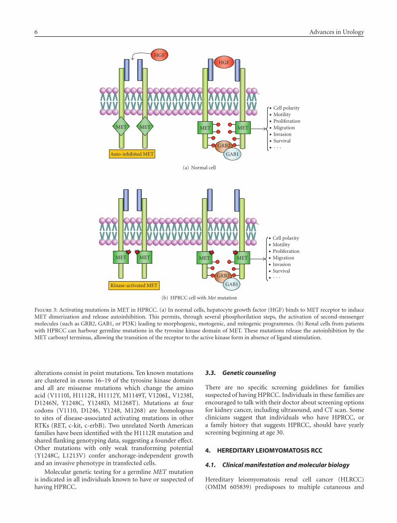
6 Advances in Urology
HGF
MET MET
HGF
MET MET
GRB2
GAB1
• Cell polarity• Motility• Proliferation• Migration• Invasion• Survival• · · ·
Auto-inhibited MET
(a) Normal cell
MET MET MET MET
GRB2
GAB1
• Cell polarity• Motility• Proliferation• Migration• Invasion• Survival• · · ·
Kinase-activated MET
(b) HPRCC cell with Met mutation
Figure 3: Activating mutations in MET in HPRCC. (a) In normal cells, hepatocyte growth factor (HGF) binds to MET receptor to induceMET dimerization and release autoinhibition. This permits, through several phosphorilation steps, the activation of second-messengermolecules (such as GRB2, GAB1, or PI3K) leading to morphogenic, motogenic, and mitogenic programmes. (b) Renal cells from patientswith HPRCC can harbour germline mutations in the tyrosine kinase domain of MET. These mutations release the autoinhibition by theMET carboxyl terminus, allowing the transition of the receptor to the active kinase form in absence of ligand stimulation.
alterations consist in point mutations. Ten known mutationsare clustered in exons 16–19 of the tyrosine kinase domainand all are missense mutations which change the aminoacid (V1110I, H1112R, H1112Y, M1149T, V1206L, V1238I,D1246N, Y1248C, Y1248D, M1268T). Mutations at fourcodons (V1110, D1246, Y1248, M1268) are homologousto sites of disease-associated activating mutations in otherRTKs (RET, c-kit, c-erbB). Two unrelated North Americanfamilies have been identified with the H1112R mutation andshared flanking genotyping data, suggesting a founder effect.Other mutations with only weak transforming potential(Y1248C, L1213V) confer anchorage-independent growthand an invasive phenotype in transfected cells.
Molecular genetic testing for a germline MET mutationis indicated in all individuals known to have or suspected ofhaving HPRCC.
3.3. Genetic counseling
There are no specific screening guidelines for familiessuspected of having HPRCC. Individuals in these families areencouraged to talk with their doctor about screening optionsfor kidney cancer, including ultrasound, and CT scan. Someclinicians suggest that individuals who have HPRCC, ora family history that suggests HPRCC, should have yearlyscreening beginning at age 30.
4. HEREDITARY LEIOMYOMATOSIS RCC
4.1. Clinical manifestation and molecular biology
Hereditary leiomyomatosis renal cell cancer (HLRCC)(OMIM 605839) predisposes to multiple cutaneous and

Jose Antonio Lopez-Guerrero et al. 7
uterine leiomyomas and solitary papillary type 2 RCCs [6,35] (Table 2).
The majority of individuals (76%) present with a singleor multiple cutaneous leiomyoma. These lesions appear asskin-colored to light brown papules or nodules distributedover the trunk and extremities, and occasionally on theface. Forty percent of individuals with HLRCC have mildcutaneous manifestations with five or fewer lesions [36].Histologically, proliferation of interlacing bundles of smoothmuscle fibers with centrally located long blunt-edged nucleiis observed.
Practically all females with HLRCC develop uterineleiomyomas [36–38]. However, whether all women withHLRCC have a higher risk of developing uterine leiomyosar-comas is unclear. In the original description of HLRCC, itwas reported that 15% of women with uterine leiomyomasalso had uterine leiomyosarcoma [39].
Most renal tumors are unilateral and solitary. Approxi-mately 10%–16% of individuals with HLRCC who presentwith multiple cutaneous leiomyomas had renal tumors at thetime that renal imaging was performed [37, 38]. Most tumorsare classified as “type 2” papillary renal cancer, which displaydistinct papillary architecture and characteristic histopathol-ogy (high-grade tumors with large eosinophilic cells, anaggressive course, and a bad prognosis) [38] (Table 3). Themedian age at detection of renal tumors is 44 years, and, incontrast to other hereditary renal cancer syndromes, renalcancers associated with HLRCC are aggressive [38].
The disease is caused by germline mutations in thetumor suppressor gene FH located in 1q42-43 that encodesthe mitochondrial Krebs cycle enzyme fumarate hydratase(EC 4.2.1.2.) [35]. FH consists of ten exons encompassing22.15 kb of DNA and is highly conserved across species. Theactive form of the enzyme is a homotetramer and catalyzesthe conversion of fumarate to L-malate. In mammals, thereare two fumarase isoforms (mitochondrial and cytosolic)that are synthesized from the same mRNA. After initial syn-thesis, the FH proteins are partially imported and processedat the mitochondrial outer membrane [6].
Activity of FH enzyme can be measured in cultured skinfibroblasts or lymphoblastoid cells to confirm the diagnosis.Reduced activity (≤60%) of FH enzyme was found in allaffected individuals with the diagnosis of HLRCC [40, 41].
The overall risk for renal tumor development is unclearand the mechanism of FH-mutation-driven tumorigenesisremains unknown so far [6]. It is plausible that intracellularfumarate accumulation as a result of FH inactivation causesdecreased HIF degradation and overexpression of genesmore downstream in the HIF pathway [42].
4.2. Molecular genetic testing
FH is the only gene known to be associated with HLRCC.Between 80% and 100% of individuals with HLRCC haveidentifiable sequence variants in FH [36–38]. The spec-trum of mutations includes missense, insertion/deletion,and nonsense mutations that are predicted to truncate theprotein, or substitute or delete highly conserved aminoacids,along with several whole-gene deletion. About 40 different
FH mutations have been identified and are distributedthroughout the entire gene without genotype-phenotypecorrelation [40]. Several of the mutations occur in manyfamilies, which could reflect a founder effect; notably, theArg190His mutation, which is the most frequent mutation(33%) in a North American family study, and the Arg58Xand Asn64Thr mutations in studies by the European-basedMultiple Leiomyoma Consortium [6].
Molecular genetic testing for a germline FH mutation isindicated in all individuals known to have or suspected ofhaving HLRCC, including individuals with the following:
(i) multiple cutaneous leiomyomas (with at least onehistologically-confirmed leiomyoma) without a fam-ily history of HLRCC;
(ii) a single cutaneous leiomyoma with family history ofHLRCC;
(iii) one or more tubulo-papillary, collecting-duct, orpapillary type 2 renal tumors with or without a familyhistory of HLRCC.
Measurement of FH enzyme activity can be useful in thediagnosis of HLRCC in cases with atypical presentation andundetectable FH mutations [40, 41].
No correlation is observed between FH mutations andthe occurrence of cutaneous lesions, uterine fibroids, orrenal cancer of HLRCC [36]. To date, six women with agermline mutation in FH have been reported with uterineleiomyosarcoma [43, 44]. It seems that FH mutation-positivefamilies are in general not highly predisposed to uterinecancer, but a few individuals and families seem to be at highrisk.
4.3. Genetic counseling
HLRCC is inherited in an autosomal dominant manner.Some individuals diagnosed with HLRCC have an affectedparent and some have HLRCC as the result of a de novogene mutation. In this case, the proportion of cases causedby de novo mutations is unknown as subtle manifestationin parents has not been evaluated and genetic testing dataare insufficient. Recommendations for evaluation of parentsof a proband with a suspected de novo mutation includemolecular genetic testing if the FH disease-causing mutationin the proband has been identified. However, it is importantto note that although some individuals diagnosed withHLRCC have an affected parent, the family history mayappear to be negative because of failure to recognize thedisorder in family members, early death of the parent beforethe onset of symptoms, or late onset of the disease in theaffected parent.
In the case of the siblings of a proband, the risk dependsupon the genetic status of the proband’s parents. If a parentof a proband is clinically affected or has a disease-causingmutation, each sibling of the proband is at a 50% riskof inheriting the mutation. If the disease-causing mutationcannot be detected in the DNA of either parent, the risk tosiblings is low, but greater than that of the general populationbecause the possibility of germline mosaicism exists [38].

8 Advances in Urology
The risk to other family members depends upon thestatus of the proband’s parents. If a parent is found to beaffected or to have a disease-causing mutation, his or herfamily members are at risk.
It is not possible to predict whether symptoms willoccur, or if they do, what the age of onset, severity, andtype of symptoms, or rate of disease progression will be inindividuals who have a disease-causing mutation.
When neither parent of a proband with an autosomaldominant condition has the disease-causing mutation orclinical evidence of the disorder, it is likely that the probandhas a de novo mutation. However, possible nonmedicalexplanations including alternate paternity or undisclosedadoption could also be explored.
There is no consensus on clinical surveillance for HLRCCindividuals so far but the following provisional recommen-dations have been accepted until a consensus conference isconducted [31].
Individuals with the clinical diagnosis of HLRCC,individuals with heterozygous mutations in FH withoutclinical manifestations, and at-risk family members who havenot undergone molecular genetic testing should have thefollowing regular surveillance by physicians familiar with theclinical manifestations of HLRCC.
(i) Skin. Full skin examination is recommended annuallyto every two years to assess the extent of disease andto evaluate for changes suggestive of leiomyosarcoma.
(ii) Uterus. Annual gynecologic consultation is recom-mended to assess severity of uterine fibroids and toevaluate for changes suggestive of leiomyosarcoma.
(iii) Kidneys. If both the initial (baseline) and the firstannual follow-up abdominal CT scan with contrastare normal, this evaluation should be repeated everytwo years.
Any suspicious renal lesion (indeterminate lesion, ques-tionable or complex cysts) at a previous examination shouldbe followed with a CT scan with and without contrast. PET-CT may be added to identify metabolically active lesionssuggesting possible malignant growth. It must be taken intoconsideration that ultrasound examination alone is neversufficient.
Renal tumors should be evaluated by a urologic oncologysurgeon familiar with the renal cancer of HLRCC.
5. BIRT-HOGG-DUBE SYNDROME
5.1. Clinical manifestation and molecular biology
Birt-Hogg-Dube (BHD) syndrome (OMIM 135150) is agenodermatosis that predisposes individuals to benign cuta-neous lesions of the face and neck, spontaneous recurrentpneumothorax and/or lung cysts, and renal tumors [6, 7].Approximately 15–29% of individuals with BHD syndromehave renal tumors [45, 46] (Table 3). The renal tumors areusually bilateral and multifocal. Tumor types include renaloncocytoma, chromophobe RCC, oncocytic hybrid tumor,and a minority of clear cell RCC [47]. The most common
tumors are a hybrid of oncocytoma and chromophobe his-tologic cell types, so-called oncocytic hybrid tumor (67%),chromophobe RCC (23%), and renal oncocytoma (3%).Only renal oncocytoma is considered a benign tumor [48].Other types of renal tumors reported in lower frequencyinclude clear cell RCC and papillary renal carcinoma. Mostrenal tumors are slow-growing. Median age of diagnosis is48 years with range from 31 to 71 years [46].
The disease is caused by germline mutations in the BHD(FLCN) gene on chromosome 17p11.2 [49]. BHD encodesfolliculin, a new protein with unknown function but it ishighly expressed in a variety of tissues including skin andskin appendages, type 1 pneumocytes, and distal nephrons ofthe kidney [50]. Recent studies suggest that folliculin mightbe involved in energy and/or nutrient sensing through theAMPK and mTOR signaling pathways [51].
BHD somatic mutations are very rare in sporadic RCCbut hypermethylations are encountered in ∼30% of all RCChistological types [52]. Germline mutations in BHD, plussomatic mutations and loss of heterozygosity in tumor tissue,suggest that loss of function of the folliculin protein is thebasis of tumor formation in BHD syndrome [53].
5.2. Molecular genetic testing
BHD is the only gene known to be associated with BHDsyndrome. Various mutations have been identified in familieswith BHD syndrome. All mutations predict protein trun-cation. The most common mutation is cytosine insertionor deletion, which occurs in a polycytosine tract in exon11, suggesting the presence of a hypermutable hot spot[46, 47]. Fifty-three percent of families with BHD syndromehave been found to have an insertion or deletion in thepolycytosine tract in exon 11 (mutational hot spot) [46].Sequence analysis of all coding exons (exon 4–14) increasesthe mutation detection in probands to 84% [46].
Molecular genetic testing is indicated in all individualsknown to have or suspected of having BHD syndromeincluding individuals with the following.
(1) Five or more facial or truncal papules with at least onehistologically confirmed fibrofolliculoma [54] withor without family history of BHD.
(2) A family history of BHD syndrome with a singlefibrofolliculoma or a single renal tumor or history ofspontaneous pneumothorax.
(3) Multiple and bilateral chromophobe, oncocytic,and/or oncocytic hydrid renal tumors.
(4) A single oncocytic, chromophobe, or oncocytic-hydrid tumor and a family history of renal cancerwith any of the above renal cell tumor types.
(5) A family history of autosomal dominant primaryspontaneous pneumothorax without a history ofchronic obstructive pulmonary disease.
Mutations in BHD were found in families with dom-inantly inherited spontaneous pneumothorax. Pulmonary

Jose Antonio Lopez-Guerrero et al. 9
involvement appears to be the only manifestation; pene-trance is 100% [55, 56].
Acquired mutations in BHD have been identified in spo-radic clear cell renal cell carcinoma [52, 57] and colon cancer[58, 59] without other associated tumors characteristic of theheritable disease.
No correlation is observed between type of BHDmutation and pulmonary and cutaneous manifestations.However, individuals who have a deletion in the polycytosinetract of exon 11 may have a lower risk of developing renalcancers than individuals with other mutations [46].
5.3. Genetic counseling
BHD syndrome is inherited in an autosomal dominantmanner. Some individuals with BHD syndrome have anaffected parent and some have BHD syndrome as a resultof a de novo gene mutation. The proportion of cases causedby de novo mutations is unknown as a sufficient number ofparents have not been evaluated for subtle manifestations,nor are there sufficient data on clinically unaffected parentswho have been evaluated by molecular genetic testing.Recommendations for the evaluation of parents of a probandwith a suspected de novo mutation include molecular genetictesting if the disease-causing mutation in the BHD gene inthe proband is identified. But, although some individualsdiagnosed with BHD syndrome have an affected parent, thefamily history may appear to be negative because of failureto recognize the disorder in family members, early death ofthe parent before the onset of symptoms, or late onset of thedisease in the affected parent.
The risk to the siblings of the proband depends uponthe genetic status of the proband’s parents. If a parent ofa proband is clinically affected or has a disease-causingmutation, the sibs of the proband are at a 50% risk ofinheriting the mutation. If neither parent has the disease-causing mutation identified in the proband, the risk to sibs islow, but greater than that of the general population becausethe possibility of germline mosaicism exists.
When neither parent of a proband with an autosomaldominant condition has the disease-causing mutation orclinical evidence of the disorder, it is likely that the probandhas a de novo mutation. However, other possible nonmedicalexplanations could also be explored.
There is no consensus on clinical surveillance; therefore,these recommendations are provisional until a consensusconference is conducted.
Individuals with known BHD syndrome, individualsknown to have disease-causing mutations in BHD withoutclinical manifestation, and at-risk family members whohave not undergone genetic testing should have regularmonitoring by physicians familiar with the spectrum of BHDsyndrome. In particular, surveillance for and monitoring ofrenal tumors include the following:
(i) if normal at baseline, abdominal/pelvic CT scan withcontrast every two years;
(ii) if any suspicious lesion (indeterminate lesion, ques-tionable or complex cysts) at previous examination,
annual abdominal/pelvic CT scan with contrast alter-nating every other year with MRI to reduce lifetimeexposure to radiation;
(iii) evaluation of renal tumors by a urologic surgeon;
(iv) monitoring of tumors less than three centimeters indiameter by periodic imaging; they may not requiresurgical intervention while this small.
6. FUTURE TRENDS
The identification of genes responsible for inherited RCChas resulted in a better understanding of renal tumorigenesisincluding sporadic RCC and is paving the way for newtherapeutic approaches [6, 7]. For VHL, recent and ongoinginsights into the functions of the VHL gene, especially theHIF-ubiquitylation pathway, provide an attractive molecularbasis for the development of specific inhibitors of HIFand/or its downstream targets [13]. Preliminary studies withthe VEGF receptor inhibitor SU5416 showed that at leasta third of patients with advanced VHL disease improvedtheir clinical status giving promising expectations [60]. Insame directions, new protein kinase receptor inhibitors areemerging [9]. In HPRCC, MET inhibitors gave encouragingresults in in vitro studies, but clinical trials have started veryrecently and although data on the antitumor activity of theanti-MET compounds are not yet available, these studieshave shown that MET inhibition results in low-grade toxicity,in agreement with the preclinical analyses performed inanimal models [61, 62].
Recent studies suggest that HIF overexpression isinvolved in HLRCC tumorigenesis [42, 63]. Therefore,future target therapies for HLRCC-associated tumors mayinclude, for example, anti-HIF therapies such as R59949that regulate prolyl hydroxylase activity, thus preventing HIFaccumulation.
The study of families with increased rates of cancerwill continue to yield more insight into the factors thatincrease cancer risk. Genetic predisposition in the form ofmutations and polymorphisms will increasingly be cata-logued and DNA-level genetic profiling of high-risk familiesand individuals will become commonplace. The increasein availability of genetic testing and counseling for high-risk families should prove both helpful and cost-effective, asgenetically unaffected family members reassured regardingtheir health status and removed from lifelong follow-upscreening programmes.
Finally, we also should keep in mind, although notdeeply discussed in this review, the psychological and ethicalimplications of the genetic counseling [64–66], not onlyfrom the strictly clinical point of view, but also regarding themanagement of personal genetic information that could havean impact on the individual and their relatives from certainhealth insurance companies.
REFERENCES
[1] P. Godley and S. W. Kim, “Renal cell carcinoma,” CurrentOpinion in Oncology, vol. 14, no. 3, pp. 280–285, 2002.

10 Advances in Urology
[2] L. M. Hock, J. Lynch, and K. C. Balaji, “Increasing incidenceof all stages of kidney cancer in the last 2 decades in theUnited States: an analysis of surveillance, epidemiology andend results program data,” The Journal of Urology, vol. 167,no. 1, pp. 57–60, 2002.
[3] A. J. Pantuck, A. Zisman, and A. S. Belldegrun, “The changingnatural history of renal cell carcinoma,” The Journal of Urology,vol. 166, no. 5, pp. 1611–1623, 2001.
[4] L. G. Luciani, R. Cestari, and C. Tallarigo, “Incidental renalcell carcinoma—age and stage characterization and clinicalimplications: study of 1092 patients (1982–1997),” Urology,vol. 56, no. 1, pp. 58–62, 2000.
[5] G. Kovacs, M. Akhtar, B. J. Beckwith, et al., “The Heidelbergclassification of renal cell tumours,” The Journal of Pathology,vol. 183, no. 2, pp. 131–133, 1997.
[6] C. P. Pavlovich and L. S. Schmidt, “Searching for the hereditarycauses of renal-cell carcinoma,” Nature Reviews Cancer, vol. 4,no. 5, pp. 381–393, 2004.
[7] B. Zbar, R. Klausner, and W. M. Linehan, “Studying cancerfamilies to identify kidney cancer genes,” Annual Review ofMedicine, vol. 54, pp. 217–233, 2003.
[8] M. Zimmer and O. Iliopoulos, “Molecular genetics of kidneycancer,” Cancer Treatment and Research, vol. 116, pp. 3–27,2003.
[9] S. Richard, R. Lidereau, and S. Giraud, “The growing familyof hereditary renal cell carcinoma,” Nephrology Dialysis Trans-plantation, vol. 19, no. 12, pp. 2954–2958, 2004.
[10] H. H. Kazazian Jr., C. D. Boehm, and W. K. Seltzer, “ACMGrecommendations for standards for interpretation of sequencevariations,” Genetics in Medicine, vol. 2, no. 5, pp. 302–303,2000.
[11] S. Richard, J. Graff, J. Lindau, and F. Resche, “von Hippel-Lindau disease,” The Lancet, vol. 363, no. 9416, pp. 1231–1234,2004.
[12] R. R. Lonser, G. M. Glenn, M. Walther, et al., “von Hippel-Lindau disease,” The Lancet, vol. 361, no. 9374, pp. 2059–2067,2003.
[13] W. G. Kaelin Jr., “Molecular basis of the VHL hereditary cancersyndrome,” Nature Reviews Cancer, vol. 2, no. 9, pp. 673–682,2002.
[14] B. Zbar, T. Kishida, F. Chen, et al., “Germline mutations in thevon Hippel-Lindau disease (VHL) gene in families from NorthAmerica, Europe, and Japan,” Human Mutation, vol. 8, no. 4,pp. 348–357, 1996.
[15] C. E. Stebbins, W. G. Kaelin Jr., and N. P. Pavletich, “Structureof the VHL-ElonginC-ElonginB complex: implications forVHL tumor suppressor function,” Science, vol. 284, no. 5413,pp. 455–461, 1999.
[16] S. C. Clifford, M. E. Cockman, A. C. Smallwood, et al.,“Contrasting effects on HIF-1α regulation by disease-causingpVHL mutations correlate with patterns of tumourigenesis invon Hippel-Lindau disease,” Human Molecular Genetics, vol.10, no. 10, pp. 1029–1038, 2001.
[17] M. A. Hoffman, M. Ohh, H. Yang, J. M. Klco, M. Ivan, andW. G. Kaelin Jr., “von Hippel-Lindau protein mutants linkedto type 2C VHL disease preserve the ability to downregulateHIF,” Human Molecular Genetics, vol. 10, no. 10, pp. 1019–1027, 2001.
[18] D. E. Feldman, C. Spiess, D. E. Howard, and J. Frydman,“Tumorigenic mutations in VHL disrupt folding in vivo byinterfering with chaperonin binding,” Molecular Cell, vol. 12,no. 5, pp. 1213–1224, 2003.
[19] C. Cybulski, K. Krzystolik, A. Murgia, et al., “Germlinemutations in the von Hippel-Lindau (VHL) gene in patientsfrom Poland: disease presentation in patients with deletions ofthe entire VHL gene,” Journal of Medical Genetics, vol. 39, no.7, p. E38, 2002.
[20] J. K. Maranchie, A. Afonso, P. S. Albert, et al., “Solid renaltumor severity in von Hippel-Lindau disease is related togermline deletion length and location,” Human Mutation, vol.23, no. 1, pp. 40–46, 2004.
[21] C. Stolle, G. Glenn, B. Zbar, et al., “Improved detection ofgermline mutations in the von Hippel-Lindau disease tumorsuppressor gene,” Human Mutation, vol. 12, no. 6, pp. 417–423, 1998.
[22] J. Hoebeeck, R. van der Luijt, B. Poppe, et al., “Rapid detectionof VHL exon deletions using real-time quantitative PCR,”Laboratory Investigation, vol. 85, no. 1, pp. 24–33, 2005.
[23] A. Rasmussen, S. Nava-Salazar, P. Yescas, et al., “Von Hippel-Lindau disease germline mutations in Mexican patients withcerebellar hemangioblastoma,” Journal of Neurosurgery, vol.104, no. 3, pp. 389–394, 2006.
[24] M. T. Sgambati, C. Stolle, P. L. Choyke, et al., “Mosaicismin von Hippel-Lindau disease: lessons from kindreds withgermline mutations identified in offspring with mosaic par-ents,” American Journal of Human Genetics, vol. 66, no. 1, pp.84–91, 2000.
[25] H. P. H. Neumann, B. Bausch, S. R. McWhinney, et al., “Germ-line mutations in nonsyndromic pheochromocytoma,” TheNew England Journal of Medicine, vol. 346, no. 19, pp. 1459–1466, 2002.
[26] F. M. Richards, A. R. Webster, R. Mcmahon, E. R. Woodward,S. Rose, and E. R. Maher, “Molecular genetic analysis of vonHippel-Lindau disease,” Journal of Internal Medicine, vol. 243,no. 6, pp. 527–533, 1998.
[27] E. R. Maher and C. Eng, “The pressure rises: update on thegenetics of phaeochromocytoma,” Human Molecular Genetics,vol. 11, no. 20, pp. 2347–2354, 2002.
[28] J. Bryant, J. Farmer, L. J. Kessler, R. R. Townsend, and K.L. Nathanson, “Pheochromocytoma: the expanding geneticdifferential diagnosis,” Journal of the National Cancer Institute,vol. 95, no. 16, pp. 1196–11204, 2003.
[29] M. Priesemann, K. M. Davies, L. A. Perry, et al., “Benefitsof screening in von Hippel-Lindau disease—comparison ofmorbidity associated with initial tumours in affected parentsand children,” Hormone Research, vol. 66, no. 1, pp. 1–5, 2006.
[30] ASCO, “American Society of Clinical Oncology policy state-ment update: genetic testing for cancer susceptibility,” Journalof Clinical Oncology, vol. 21, no. 12, pp. 2397–2406, 2003.
[31] O. Iliopoulos and C. Eng, “Genetic and clinical aspects offamilial renal neoplasms,” Seminars in Oncology, vol. 27, no.2, pp. 138–149, 2000.
[32] B. Zbar, G. Glenn, I. Lubensky, et al., “Hereditary papillaryrenal cell carcinoma: clinical studies in 10 families,” TheJournal of Urology, vol. 153, no. 3, pp. 907–912, 1995.
[33] L. Schmidt, F.-M. Duh, F. Chen, et al., “Germline and somaticmutations in the tyrosine kinase domain of the MET proto-oncogene in papillary renal carcinomas,” Nature Genetics, vol.16, no. 1, pp. 68–73, 1997.
[34] M. Miller, K. Ginalski, B. Lesyng, N. Nakaigawa, L. Schmidt,and B. Zbar, “Structural basis of oncogenic activation causedby point mutations in the kinase domain of the MET proto-oncogene: modeling studies,” Proteins: Structure, Function,and Bioinformatics, vol. 44, no. 1, pp. 32–43, 2001.

Jose Antonio Lopez-Guerrero et al. 11
[35] I. P. M. Tomlinson, N. A. Alam, A. J. Rowan, et al., “Germlinemutations in FH predispose to dominantly inherited uterinefibroids, skin leiomyomata and papillary renal cell cancer,”Nature Genetics, vol. 30, no. 4, pp. 406–410, 2002.
[36] M.-H. Wei, O. Toure, G. M. Glenn, et al., “Novel mutations inFH and expansion of the spectrum of phenotypes expressed infamilies with hereditary leiomyomatosis and renal cell cancer,”Journal of Medical Genetics, vol. 43, no. 1, pp. 18–27, 2006.
[37] N. A. Alam, E. Barclay, A. J. Rowan, et al., “Clinical featuresof multiple cutaneous and uterine leiomyomatosis: an under-diagnosed tumor syndrome,” Archives of Dermatology, vol.141, no. 2, pp. 199–206, 2005.
[38] J. R. Toro, M. L. Nickerson, M.-H. Wei, et al., “Mutations inthe fumarate hydratase gene cause hereditary leiomyomatosisand renal cell cancer in families in North America,” AmericanJournal of Human Genetics, vol. 73, no. 1, pp. 95–106, 2003.
[39] V. Launonen, O. Vierimaa, M. Kiuru, et al., “Inheritedsusceptibility to uterine leiomyomas and renal cell cancer,”Proceedings of the National Academy of Sciences of the UnitedStates of America, vol. 98, no. 6, pp. 3387–3392, 2001.
[40] N. A. Alam, A. J. Rowan, N. C. Worth, et al., “Genetic andfunctional analyses of FH mutations in multiple cutaneousand uterine leiomyomatosis, hereditary leiomyomatosis andrenal cancer, and fumarate hydratase deficiency,” HumanMolecular Genetics, vol. 12, no. 11, pp. 1241–1252, 2003.
[41] M. Pithukpakorn, M.-H. Wei, O. Toure, et al., “Fumaratehydratase enzyme activity in lymphoblastoid cells and fibrob-lasts of individuals in families with hereditary leiomyomatosisand renal cell cancer,” Journal of Medical Genetics, vol. 43, no.9, pp. 755–762, 2006.
[42] J. S. Isaacs, J. J. Yun, D. R. Mole, et al., “HIF overexpressioncorrelates with biallelic loss of fumarate hydratase in renalcancer: novel role of fumarate in regulation of HIF stability,”Cancer Cell, vol. 8, no. 2, pp. 143–153, 2005.
[43] H. J. Lehtonen, M. Kiuru, S. K. Ylisaukko-Oja, et al.,“Increased risk of cancer in patients with fumarate hydratasegermline mutation,” Journal of Medical Genetics, vol. 43, no. 6,pp. 523–526, 2006.
[44] S. K. Ylisaukko-Oja, M. Kiuru, H. J. Lehtonen, et al., “Analysisof fumarate hydratase mutations in a population-based seriesof early onset uterine leiomyosarcoma patients,” InternationalJournal of Cancer, vol. 119, no. 2, pp. 283–287, 2006.
[45] B. Zbar, W. G. Alvord, G. Glenn, et al., “Risk of renaland colonic neoplasms and spontaneous pneumothoraxpneurnothorax in the Birt-Hogg-Dubapos;e syndrome,” Cancer Epidemiology BiomarkersPrevention, vol. 11, no. 4, pp. 393–400, 2002.
[46] L. S. Schmidt, M. L. Nickerson, M. B. Warren, et al., “GermlineBHD-mutation spectrum and phenotype analysis of a largecohort of families with Birt-Hogg-Dubapos;e syndrome,” American Journal of Human Genetics, vol.76, no. 6, pp. 1023–1033, 2005.
[47] M. L. Nickerson, M. B. Warren, J. R. Toro, et al., “Mutationsin a novel gene lead to kidney tumors, lung wall defects, andbenign tumors of the hair follicle in patients with the Birt-Hogg-Dubapos;e syndrome,” Cancer Cell, vol. 2, no. 2, pp. 157–164, 2002.
[48] C. P. Pavlovich, R. L. Grubb III, K. Hurley, et al., “Evaluationand management of renal tumors in the Birt-Hogg-Dubapos;e syndrome,” The Journal of Urology, vol. 173, no. 5, pp.1482–1486, 2005.
[49] J. D. Carpten, C. M. Robbins, A. Villablanca, et al., “HRPT2,encoding parafibromin, is mutated in hyperparathyroidism-
jaw tumor syndrome,” Nature Genetics, vol. 32, no. 4, pp. 676–680, 2002.
[50] M. B. Warren, C. A. Torres-Cabala, M. L. Turner, et al.,“Expression of Birt-Hogg-Dube gene mRNA in normal andneoplastic human tissues,” Modern Pathology, vol. 17, no. 8,pp. 998–1011, 2004.
[51] M. Baba, S.-B. Hong, N. Sharma, et al., “Folliculin encodedby the BHD gene interacts with a binding protein, FNIP1,and AMPK, and is involved in AMPK and mTOR signaling,”Proceedings of the National Academy of Sciences of the UnitedStates of America, vol. 103, no. 42, pp. 15552–15557, 2006.
[52] S. K. Khoo, K. Kahnoski, J. Sugimura, et al., “Inactivation ofBHD in sporadic renal tumors,” Cancer Research, vol. 63, no.15, pp. 4583–4587, 2003.
[53] C. D. Vocke, Y. Yang, C. P. Pavlovich, et al., “High frequency ofsomatic frameshift BHD gene mutations in Birt-Hogg-Dubapos;e-associated renal tumors,” Journal of the National CancerInstitute, vol. 97, no. 12, pp. 931–935, 2005.
[54] J. R. Toro, G. Glenn, P. Duray, et al., “Birt-Hogg-Dubapos;e syndrome: a novel marker of kidney neoplasia,”Archives of Dermatology, vol. 135, no. 10, pp. 1195–1202, 1999.
[55] R. B. Graham, M. Nolasco, B. Peterlin, and C. K. Garcia, “Non-sense mutations in folliculin presenting as isolated familialspontaneous pneumothorax in adults,” American Journal ofRespiratory and Critical Care Medicine, vol. 172, no. 1, pp. 39–44, 2005.
[56] J. N. Painter, H. Tapanainen, M. Somer, P. Tukiainen, and K.Aittomquot;aki, “A 4-bp deletion in the Birt-Hogg-Dubapos;e gene (FLCN) causes dominantly inherited spontaneouspneumothorax,” American Journal of Human Genetics, vol. 76,no. 3, pp. 522–527, 2005.
[57] N. F. da Silva, D. Gentle, L. B. Hesson, D. G. Morton, F. Latif,and E. R. Maher, “Analysis of the Birt-Hogg-Dube (BHD)tumour suppressor gene in sporadic renal cell carcinoma andcolorectal cancer,” Journal of Medical Genetics, vol. 40, no. 11,pp. 820–824, 2003.
[58] K. Kahnoski, S. K. Khoo, N. T. Nassif, et al., “Alterations of theBirt-Hogg-Dube gene (BHD) in sporadic colorectal tumours,”Journal of Medical Genetics, vol. 40, no. 7, pp. 511–515, 2003.
[59] J.-H. Shin, Y.-K. Shin, J.-L. Ku, et al., “Mutations of the Birt-Hogg-Dube (BHD) gene in sporadic colorectal carcinomasand colorectal carcinoma cell lines with microsatellite insta-bility,” Journal of Medical Genetics, vol. 40, no. 5, pp. 364–367,2003.
[60] S. Madhusudan, G. Deplanque, J. P. Braybrooke, et al.,“Antiangiogenic therapy for von Hippel-Lindau disease,”Journal of the American Medical Association, vol. 291, no. 8,pp. 943–944, 2004.
[61] C. Migliore and S. Giordano, “Molecular cancer therapy: canour expectation be MET?” European Journal of Cancer, vol. 44,no. 5, pp. 641–651, 2008.
[62] W. M. Linehan, P. A. Pinto, R. Srinivasan, et al., “Identificationof the genes for kidney cancer: opportunity for disease-specifictargeted therapeutics,” Clinical Cancer Research, vol. 13, no. 2,part 2, pp. 671s–679s, 2007.
[63] P. J. Pollard, J. J. Briere, N. A. Alam, et al., “Accumulationof Krebs cycle intermediates and over-expression of HIF1α intumours which result from germline FH and SDH mutations,”Human Molecular Genetics, vol. 14, no. 15, pp. 2231–2239,2005.
[64] J. E. Wagner, “Practical and ethical issues with geneticscreening,” Hematology, vol. 2005, pp. 498–502, 2005.

12 Advances in Urology
[65] E. Rantanen, M. Hietala, U. Kristoffersson, et al., “Regulationsand practices of genetic counselling in 38 European countries:the perspective of national representatives,” European Journalof Human Genetics, vol. 16, no. 10, pp. 1208–1216, 2008.
[66] E. Rantanen, M. Hietala, U. Kristoffersson, et al., “What isideal genetic counselling? A survey of current internationalguidelines,” European Journal of Human Genetics, vol. 16, no.4, pp. 445–452, 2008.

Hindawi Publishing CorporationAdvances in UrologyVolume 2008, Article ID 415848, 16 pagesdoi:10.1155/2008/415848
Review ArticleRadiologic Evaluation of Small Renal Masses (I):Pretreatment Management
A. Marhuenda,1 M. I. Martın,1 C. Deltoro,1 J. Santos,1 and Jose Rubio Briones2
1 Departamento de Radiologıa, Instituto Valenciano de Oncologıa, C/Profesor Beltran Baguena 8, 46009 Valencia, Spain2 Departamento de Urologıa, Instituto Valenciano de Oncologıa, C/Profesor Beltran Baguena 8, 46009 Valencia, Spain
Correspondence should be addressed to A. Marhuenda, [email protected]
Received 9 April 2008; Accepted 11 December 2008
Recommended by F. Algaba
When characterizing a small renal mass (SRM), the main question to be answered is whether the mass represents a surgical ornonsurgical lesion or, in some cases, if followup studies are a reasonable option. Is this a task for a urologist or a radiologist? It isobvious that in the increasing clinical scenario where this decision has to be made, both specialists ought to work together. Thispaper will focus on the principles, indications, and limitations of ultrasound, CT, and MRI to characterize an SRM in 2008 with adetailed review of relevant literature. Special emphasis has been placed on aspects regarding the bidirectional information betweenradiologists and urologists needed to achieve the best radiological approach to an SRM.
Copyright © 2008 A. Marhuenda et al. This is an open access article distributed under the Creative Commons Attribution License,which permits unrestricted use, distribution, and reproduction in any medium, provided the original work is properly cited.
1. INTRODUCTION
Over the last 3 decades, there has been a rising trend to definesmall renal masses (SRMs) as masses below 4 cm in diameter[1], making it the major reason for the 126% increase inincidence of renal cell carcinoma (RCC) in the United States.The reason for that is well known; the increasing number ofimaging examinations performed for unrelated indicationswith many renal neoplasms of small size and early stageincidentally detected. Faced with this situation, urologistsdo not only suggest surgery as 30 years ago, but also offerdifferent options to deal with the problem. Most of theirdecisions are based on radiological characterization of theSRM, as biopsies of these masses have not been completelyaccepted by the international urological community.
So, evidently urologists have to ask their colleagues inthe Radiology Department to improve their explorationsexpecting more and more extensive radiological reportsanalyzing not just the presence of the mass. The SMR analysismust be carried out by both a radiologist and an urologist, asbidirectional information is extremely important to definethe most probable nature of the mass.
The accurate diagnosis of a renal mass depends onmany factors, including the clinical history; so there is someclinical information that urologists have to report to theirradiologists:
(i) presence of a familial syndrome,
(ii) presence of a urological tract infectious diseaseprevious or concomitant to the diagnoses of the SRM,
(iii) presence of previous stone disease and related treat-ments,
(iv) presence of previous renal trauma,
(v) presence of kidney disease and renal insufficiency.
A high-quality imaging examination, under the controlof a radiologist, is essential. The most accurate diagnosisof a renal mass is then made according to the nature ofthe imaging findings, the experience of the radiologist, andthe quality of the examination, as well as the exclusion ofconditions that can mimic a renal neoplasm. There are somekey points that, due to their therapeutic decision-makingimportance, radiologists need to provide in their reports:
(i) signs suspecting fat involvement in an SRM,
(ii) metabolic behavior during the different phases of CTand MRI after contrast administration, allowing tocharacterize benign SRM,
(iii) the need (or not) to complete studies with differenttechniques,

2 Advances in Urology
(iv) accurate and standard (for followup in case ofwatchful waiting policy) measurement of 3 diametersof the SRM,
(v) signs of active tumoral tissue after conservativetreatments which do not remove the SRM,
(vi) differential diagnosis of residual tumour with com-plications after partial nephrectomies and foreignbodies used to achieve haemostasis.
Having established the collaboration between urologistand radiologist for this review paper, the aim of the two com-plementary chapters submitted for the SRM diagnoses andcharacterization is to give some light on the new challengeswhich face radiologists nowadays, extremely important forthe SRM management.
2. OBJECTIVES
Renal cell carcinoma and oncocytoma are indistinguishablefrom each other at imaging. Many other renal lesions mustbe considered, such as angiomyolipoma (AML), lymphoma,metastatic disease, renal anomalies, and other pseudotumorsthat can mimic renal cell carcinoma. Although it is possibleto make this differentiation by using the imaging findingsalone, the clinical history can often be very important inmaking the correct diagnosis. In fact, before making adiagnosis of renal cell carcinoma, one should be certain thatnone of these possible mimickers of renal cell carcinoma arepotentially present.
Staging by TNM system can be considered a prognosticclassification, and there is evidence that the smaller the size,the better the prognosis [2, 3] . The increasing incidence ofrenal mass manifestations of tumours that are confined tothe renal capsule and relatively small in size has stimulated agrowing trend toward nephron-sparing surgical techniques,as current data show survival rates comparable to thoseassociated with radical nephrectomy.
Imaging findings that can affect the decision to performpartial nephrectomy included tumor size in three planes:tumor location within the kidney; presence of a pseudo-capsule (a thin band of fibrous tissue and compressed renalparenchyma surrounding the lesion); tumor invasion of therenal sinus fat, collecting system, renal vein, or perinephricfat; presence of lymphadenopathy; morphologic and physio-logic status of the contralateral kidney. All these aspects areevaluated by means of different imaging techniques.
The increased implementation of kidney-sparing surgeryfor renal cell carcinoma may create an important role fordiagnostic imaging in the discovery of small synchronouscarcinomas. Radiologist should be aware of the possibility oftumor multifocality or of adrenal metastases from a high-grade small renal tumor as well as of the association of RCCwith lymphoma [4].
The challenge is to detect and delineate all lesionsto ensure complete surgical excision while preserving themaximal amount of functioning parenchyma. For patientswho are not surgical candidates, imaging staging, along withthe other factors, can provide prognostic information.
I.V.O. RadilogiaID: / MD:
SIEMENS
0
140IM: 0.4 POT: 0 dB
L
EM: calc B/M: 55/1/18
D1 =15.6 mm
Ju 24/05/07∗ 11:43:46Org men3C40 40Distancia
Figure 1: Simple cyst as anechoic lesion with a sharply defined backwall and enhancement of through sound transmission.
3. ULTRASOUND
The fact that renal neoplasms have been detected earlierand with increased frequency is well documented in theliterature [5, 6]. This is probably due to two major factors.One factor is that there has been a considerable increasein the number of people who undergo kidney imaging inthe general population because of the widespread use ofultrasound (US). The other reason is that this imagingtechnique is able to depict lesions of the kidneys that couldbe missed with urography [7]. This increased detection ofrenal neoplasms also results in the increased detection ofbenign lesions and nonneoplastic masses, particularly renalcysts. Therefore, the differentiation between a neoplastic anda nonneoplastic lesion is a common dilemma.
To differentiate benign from malignant SRM can proveeven more problematic because the findings can also becomesmaller, hence requiring more detailed and more sensitiveimaging studies.
Ultrasound plays an important role in the detection andevaluation of these SRMs. While this technique may not beas sensitive as contrast-enhanced CT or MR for revealingSRM, US has been the initial technique in the discovery ofa large number of these incidentally discovered tumors whenthe kidney is studied in the course of abdominal imaging.Sonography is very accurate in distinguishing liquid fromsolid tissue. Therefore, its major use in these small lesionsis to help differentiate small cysts (see Figure 1) from smallsolid tumors [8]. Maintaining rigid criteria is necessary tomaintain the high accuracy possible with this technique.
In the general population, renal cysts are the mostcommon space-occupying lesions in the kidney. With thistechnique, 80% of detected renal masses are characterized assimple cysts [9] thus ending their diagnostic evaluation. Theremaining 20% of renal masses require further study withCT or MR imaging [10]. Any mass detected that does notmeet the strict sonographic criteria for a simple cyst shouldbe further evaluated with CT or MR imaging of the kidneys.

A. Marhuenda et al. 3
RD
Figure 2: A cyst with nodular thickening of the wall and internalsepta.
However, one or two thin septations may also be visible sono-graphically in simple renal cysts [11]. Because these findingsare diagnostic, no further imaging or followup is neededin the evaluation of these lesions. However, other atypicalfeatures sonographically detected calcifications; more thantwo septations, septal thickening or nodularity, and thepresence of solid components indicate that sonography alonewill not be adequate for complete evaluation of these renalmasses (see Figure 2). The addition of Doppler sonography,color Doppler sonography, power Doppler sonography [12,13], and sonographic contrast agents may further improvethe detection and characterization of renal masses. However,none of these techniques preclude the need for CT or MRimaging of renal masses that do not meet the sonographiccriteria for diagnosis of a simple cyst.
In the study of solid renal masses, the role for US hasbeen mainly centred on the differentiation of RCC andAML, which are the most common malignant and benignsolid renal tumors, respectively [14–17]. When a solid massis diagnosed, RCC or AML should be initially consideredbecause of the high frequency of their occurrence. At US,most AML lesions are markedly hyperechoic relative to renalparenchyma. They may appear less echogenic dependingon the relative proportion of fat, smooth muscle, vascularcomponents, and haemorrhage in the lesion [18, 19]. RCCdisplays a broad range of echogenicities. Although oftenthought of as hypoechoic or isoechoic, recent studies haveshown that most RCC are hyperechoic relative to renalparenchyma and that up to 12% simulate AML [14–17].Forman et al. [14] have shown that one third of smallRCC are as echogenic as a “classical” AML. An echotextureequal to that of renal sinus fat seen in a small renal massis, therefore, no longer considered adequate to exclude thediagnosis of malignancy (see Figure 3).
Other ultrasound signs have been used to differentiatebetween hyperechoic RCC and AMLs. The presence of ananechoic rim and/or an intratumoral cyst is only seen in RCC(see Figure 4). The presence of acoustic shadow is specificof AML. However, the detectability of these findings varies
No oncologia ID: /∗ 18:47:48SIEMENS
IM: 0.6EM: P
Ma 1Abd
3C40CPS
Figure 3: Well-defined hyperechoic small renal mass. Pathologicanalysis of the surgical specimen revealed a renal cell carcinoma.
ID: /
EM: D
Abd
3C40Dist
Figure 4: Well-defined hyperechoic small renal mass with hypoe-choic rim and intratumoral cystic area, confirmed with pathologicanalysis as renal cell carcinoma.
[15, 16], their diagnostic value has not been established, andthe presence of these features is not sufficient to differentiateRCC from the other solid renal masses that are incidentallydetected on gray-scale US. On the power Doppler US, theanalysis of the vascular distribution has not increased thediagnostic accuracy for small renal tumors [12]. Contrast-enhanced Doppler US can increase the detection of intra-tumoral vascularity compared to color/power Doppler US[20]. However, their signal intensity has not been found to besufficiently intense for tumor characterization. Recently, thedevelopment of contrast-enhanced harmonic US imaginghas provided a better assessment of the diagnostic accuracyof RCC as compared with gray-scale US by allowing bettervisualization of the intratumoral anechoic areas and thepseudocapsule than can the gray-scale US [21], but there stillexists an overlapping of signs of RCC and the other solidrenal masses, making it necessary to use CT or MR imagingin the study of small renal masses.

4 Advances in Urology
4. CT
Helical CT is generally accepted as the critical imaging test forthe classification of renal masses. Radiation exposure is thegreatest disadvantage of this technique. MRI is comparableto helical CT for detection, diagnosis, and staging of renalmasses. However, CT has the advantages of widespreadavailability, shorter examination time, and lower cost incomparison with MRI.
A detailed analysis of a variety of CT features is required,including the size, location, appearance on unenhanced scan,the presence and location of calcifications, the presence andsize of a cyst wall or septations, and the amount and patternof contrast enhancement [7, 22, 23].
4.1. CT technique
Single detector and especially multidetector spiral (MDCT)have refined the diagnostic evaluation of renal patho-logic conditions. Compared with single-detector helical CT,MDCT allows the kidneys to be scanned with a collimationof less than 5 mm during a single breath hold [24]. Froma single data set obtained with thin collimation, both thinand thick sections can be reconstructed and no additionalradiation exposure is required to obtain the thin sections.This dataset is manipulated by using a workstation to pro-duce volume-rendered and three-dimensional (3D) imageswhen necessary. The 3DCT images can be viewed in multipleplanes and orientations to define the lesion.
A triphasic imaging protocol consists of an unenhancedphase through the kidneys, an arterial or corticomedullaryphase through the liver and kidneys (between 25 and 70seconds after the start of injection of contrast), and aportal venous or nephrographic phase of the entire abdomen(between 80 and 180 seconds). Excretory phase (>180seconds) is occasionally helpful.
An initial series of unenhanced scans provides a baselinefrom which to measure the enhancement within the lesion.
The corticomedullary phase is useful to perform 3Dreconstructions and to depict the renal vasculature. Further-more, this phase is considered essential for staging.
The nephrographic phase provides greater lesion detec-tion and improved lesion characterization of renal massesthan corticomedullary phase [25, 26]. However, a case ofrenal cell carcinoma visible only during the corticomedullaryphase has been shown in the literature [27]. The excretoryphase is occasionally helpful to better delineate the rela-tionship of a centrally located mass within the collectingsystem. Delayed scanning (15 minutes) can also be used inlieu of unenhanced scanning to characterize an incidentalrenal lesion detected on a routine contrast-enhanced CT scan[28].
At present, there is no worldwide agreement uponthe specific number that can be used as definitive andunequivocal evidence of enhancement within a renal mass,and it has been proposed by many authors that thepreviously used threshold of 10 HU should be increased to20 HU (a currently accepted criterion) (see Figure 5). Someauthors think that a renal mass that enhances 10–20 HU isindeterminate and needs further evaluation [29].
(a)
(b)
Figure 5: Hemorrhagic cyst. (a) Unenhanced CT scan shows ahyperattenuating small renal mass (62 H) (arrow). (b) Contrast-enhanced CT scan during the nephrographic phase reveals lightenhancing (attenuation value increased 9 H:71 H) (arrow).
4.2. Imaging of specific small renal masses
4.2.1. Cysts
Most small renal masses incidentally discovered on CT aresimple cortical cysts that need no further evaluation. TheBosniak classification system is used to asses the likelihoodof malignancy in cystic renal masses on the basis of lesioncomplexity in CT imaging [30, 31]. Application of theBosniak classification can be difficult in the case of anindeterminate renal lesion, especially if it is small. Bosniakadmits that distinction between category II (not requiringsurgery) and category III (requiring surgery) can be verydifficult [32].
4.2.2. Angiomyolipoma
It is typically a solid lesion that exhibits fat density on CTscans (−10 to –100 HU) (see Figure 6). However, in somecases, it may contain very small quantities of fat that can beoverlooked. Angiomyolipomas rarely contain calcification,and, therefore, a diagnosis of angiomyolipoma should notbe made if a lesion contains fat and calcium [33, 34]. Therehave been few case reports of fat from RCC that also containcalcification. In such cases, a renal cell carcinoma must beconsidered the most likely diagnosis.
4.2.3. Oncocytoma
It is usually a hypodense mass, homogeneous, with smoothcontours and a tendency to enhance avidly (see Figure 7).

A. Marhuenda et al. 5
Figure 6: Angiomyolipoma. Contrast-enhanced CT shows a smallhomogeneous fat-containing mass.
A
A: 1.63 cm
Figure 7: Oncocytoma. Small mass isoattenuated to renal paren-chyma after contrast.
Until now, an oncocytoma was suggested on postcontrastCT by the presence of a central hypoenhancing scar.Because of its lack of specificity, patient management hasbeen unaffected by the presence of this finding. Renal cellcarcinoma and oncocytoma are indistinguishable from eachother at imaging.
4.2.4. Renal cell carcinoma
The imaging characteristics of RCC are extremely varied,with masses ranging from cystic to solid, from homogeneousto heterogeneous and necrotic, from small to large, andfrom localized to extensive. The typical CT appearance ofsmall RCC is a homogeneously isodense/hypodense mass,noncalcified, with an attenuation value of 20 HU or more,that enhance avidly and early with contrast medium (seeFigure 8) [23]. The early-stage contrast enhancement isbelieved to be caused by tumor angiogenesis. However, asmall proportion of RCC are hypovascular, and the amountof enhancement may be minimal. Small RCC with a pre-dominantly cystic growth pattern, necrosis, or calcifications(peripheral curvilinear or punctate central) are uncommon.Areas of fat attenuation can be present within renal cellcarcinomas, but are uncommon in small tumors.
(a)
(b)
Figure 8: Small hyper vascular renal cell carcinoma. (a) Contrast-enhanced CT shows small renal mass that enhances early incorticomedullary phase. (b) Rapid washout in nephrographicphase.
Furthermore, a small RCC can be hyperattenuating. Ifthe lesion is depicted only on enhanced CT, delayed scanningcan also be used. Macari and Bosniak [28] have suggestedthat measurement of the washout of contrast material fromat least 15 minutes allows differentiation between hyperdensecyst and renal neoplasms. The washout of 15 HU or moreindicates that, excluding vascular abnormality, the mass issolid. A lack of washout indicates that the mass is probablya hyperattenuating cyst.
On the other hand, renal cortical tumors are familyneoplasms with distinct cytogenetic and molecular charac-teristics and varying malignant potential. In the 1997 Hei-delberg classification, renal cell carcinoma was subdividedin subtypes [35]. It has been suggested that certain imagingfeatures may be associated with different subtypes of solidrenal cortical tumors [36, 37]. The most consistent findingin these studies was that the degree of enhancement was themost valuable parameter for differentiation of RCC subtypes,as clear cell RCC enhance to a greater degree than othersubtypes, especially papillary RCC. Clear cell RCC is alsostrongly associated with a mixed enhancement pattern ofboth enhancing soft-tissue components and low-attenuationareas (necrotic or cystic changes). When homogeneous orperipheral enhancement is present, clear cell RCC is a lesslikely diagnosis, and other cell types should be considered.

6 Advances in Urology
Figure 9: Papillary renal cell carcinoma. Contrast-enhanced CTshows small homogeneous mass that is mild and less enhanced thanrenal parenchyma does.
Notably, a majority of papillary tumours were either homo-geneous or peripheral enhancement (see Figure 9). Thepresence to neovascularity was mildly associated with moreaggressive tumor. The clear cell RCC (and oncocytomas)enhanced avidly during the parenchymal phase; the chro-mophobe RCC (and lipid poor angiomyolipoma) enhancedmoderately and papillary RCC enhance mildly.
4.2.5. Non-Hodgkin’s lymphoma
Lymphoma can have a variable appearance and may onoccasion resemble renal cell carcinoma. Most frequently, itmanifests as bilateral solid renal masses, and in a patient withsystemic lymphoma, the proper diagnosis is not difficult.Characteristically, lymphoma often infiltrates into the kidneyvia the renal sinus or surrounds the kidney. In a patient withknown systemic lymphoma to whom a renal mass with imag-ing is detected, systemic treatment for lymphoma should beinstituted. If the patient’s systemic disease responds, and therenal mass does not respond, biopsy of the mass is indicated.However, lymphoma may rarely manifest as a solitary renalmass or a homogeneous infiltrating renal mass. In this case,biopsy of the mass is indicated previous to systemic therapy.
4.2.6. Metastases
Metastatic disease to the kidney typically manifests as mul-tiple bilateral renal masses, often associated with metastaticdisease to other organs. They are often poorly definedand infiltrate the renal parenchyma. With the appropriateclinical history, the diagnosis is straightforward. However, ina patient with a solitary renal mass (especially an infiltratingmass) and history of previous malignancy, percutaneousrenal biopsy is indicated for a definitive diagnosis.
4.2.7. Benign mesenchymal tumors
Included leiomyomas, lipomas, fibromas, and mixed mes-enchymal nodules. They are usually small (< 1 cm) lesions,found in autopsies.
4.2.8. Pseudotumors
They include congenital anomalies (prominent renal col-umns of Bertin, renal dimorphisms, and dromedary humps)
and acquired pseudotumors (hypertrophied normal renalparenchyma). This condition enhances identically to the nor-mal renal parenchyma. In these situations it proves appropri-ate to scan during the corticomedullary and nephrographicphase.
4.2.9. Renal mass mimickers
These include inflammatory masses (including focalpyelonephritis, renal abscess) and hematoma. A carefulevaluation with high-quality CT or MR examinationcombined with the clinical context of the case and afamiliarity with this group of “lesions” should reveal itstrue nature. Most hematomas are perinephric and aresurrounded by fat stranding. They may occasionally have amasslike appearance, and some may not be discovered untillong time after the traumatism. Chronic hematomas canhave calcifications and do not enhance.
4.3. Diagnosis and management ofrenal call carcinoma with MD-CT
CT remains the most widely available and single mosteffective modality for staging renal call carcinoma [38, 39].3DCT combined with CT angiography has the potentialto provide all the critical information needed to planthe surgical procedure. 3DCT images can be viewed inmultiples planes and orientations to define the tumor andits relationship to the renal surface, the collecting system,and adjacent organs. A 3DCT angiogram can be created todelineate the renal arterial and venous anatomy.
The anatomic extent of the tumor at the time of diagnosisis the single most important factor in determining prognosis(see Table 1) [38].
Most urological surgeons continue to refer to Robson’sclassification, which is essentially a surgical staging approach.This system includes the important staging variables thathave survived scrutiny over the years. Confinement withinthe renal capsule, penetration into the perirenal fat, invasioninto the renal vein, and lymph node metastases are allimportant in determining the prognosis.
Under and overstating of perinephric invasion are themost common staging errors at CT [40]. The most specificfinding of stage T3a, the presence of an enhanced nodule inthe perinephric space, is highly specific but also low sensitive.The differentiation between stage T2 and T3a tumors is veryproblematic.
If tumoral spread within the IVC is identified, precisedelineation of the superior extent of the thrombus isessential for the surgeon to plan the optimal surgical strategyfor thrombectomy and minimize the risk of embolism.The level of involvement of the IVC dictates the surgicalapproach. Involvement of the IVC is best shown duringcorticomedullary phase. Because of its multiplanar capabil-ity, magnetic resonance imaging is the preferred modalityto image. However, the three-dimensional CT with sagittaland coronal reconstructions is also effective in depictingthe superior extent of inferior vena cava thrombus (seeFigure 10), with the advantages of widespread availability,

A. Marhuenda et al. 7
Table 1: Staging system for renal carcinoma and CT criteria.
Tumour position Robson TNM CT findings
Confined withinI
Soft-tissue mass enhances
renal capsule less than normal renal
Small (<7 cm) T1 parenchyma; central
Large (≥7 cm) T2 necrosis in large RCC.
II T3aPerinephric stranding;
Spread to perinephric fat Perinephric collateral vessels;
Soft-tissue mass in perinephric space
Venous thrombus III A
Renal vein only T3b Low-attenuation filling defect vein;
IVC infradiaphragmatic T3c Direct continuity of thrombus with primary mass;
IVC supradiaphragmatic T4b Enhanced thrombus
Regional lymphIII B N1-N3 Lymph nodes 1 cm in diameter or larger
node metastases
Direct invasion of adjacent organs IV A T4a Obliteration of normal soft-tissues planes between tumor and adjacent organs
Distant metastases M1Metastases enhance with IV contrast material;
Hepatic metastases best in arterial phase
IV: intravenous, IVC: inferior vena cava.
shorter examination time, and lower cost in comparison withMR [40].
5. MR
5.1. Indications
Although ultrasound and CT are often combined to revealand characterize most renal lesions, MR is sometimesrequired when indeterminate lesions are found [41], orwhen a hyperattenuating renal mass is observed on CT [42].Furthermore, some renal masses are incidentally discoveredwhen MR imaging is performed to answer questions otherthan urology ones.
On the other hand, MR imaging has been considered themost accurate method for those patients with contraindica-tions to iodinated contrast administration such as in patientswith renal failure and those with an allergy to iodinatedcontrast material [43].
Recently, nephrogenic systemic fibrosis was linked togadolinium contrast agent exposure in patients with renalfailure; therefore its use should be reserved for neurologicaland vascular cases where the quality of information gainedwas sufficient to justify the risk of potential devastatingadverse effects [44]. Nevertheless, the better contrast reso-lution of MR imaging, as opposed to CT, is an undoubtfuladvantage in those patients for performing an MR explo-ration, even though no contrast is provided.
MR imaging has been considered useful not only incharacterization but also in evaluation of most cystic renalmasses with the Bosniak classification system [45]. Inaddition, MR imaging can potentially improve staging andpreoperative studying of a renal mass [41, 46–51] as well astumor multifocality, collecting system invasion, and venousinvasion [52]. Moreover, MRI is capable of showing the
patency of a blood vessel without the use of intravenouscontrast medium.
The accuracy in detection and characterization of SRMcarries great significance when nephron sparing surgeryis being considered for a renal cell carcinoma becausesynchronous lesions measuring 1–15 mm have been reportedto a frequency as high as 19.7% [53].
5.2. Basic technical examination
A proper technical examination is needed for studying arenal mass which is indeterminate after US and CT tech-niques. Owing to intrinsic proprieties of magnetic resonanceexamination, high-quality renal MR imaging is dependenton multiple factors, includingpatient cooperation in holdingbreath. In patients referred for evaluation of a renal mass,examinations are currently performed with a torso phased-array coil, preferably during a more reproducibility end-expiratory breath hold [29, 54].
Different protocols can be used by different institutionsdepending on their technical requirements. Essentially, thecommon aspects are shown as follows. Prior to con-trast administration, sequences are used to obtain imagesweighted on T1 and T2, in and out of phase T1, anda precontrast 3D weighted T1 with fat saturation can beacquired in different planes, basically on axial plane butalso coronal or sagittal can be employed to best depict themass, especially for such lesions located in renal poles. Thisapproach is most important when evaluating a patient witha solitary kidney that contains a renal neoplasm that isamenable to partial nephrectomy.
These sequences provide us with morphologic informa-tion about the renal parenchyma and a renal mass (location,size, and signal intensity), parenchymal structures andadjacent organs, vascular structures and lymphadenopathies,

8 Advances in Urology
(a)
(b)
Figure 10: Renal cell carcinoma. (a) Three-dimensional CT withcoronal reconstruction, (b) sagittal oblique reconstruction.
and are designed to improve visualization of tisular intrinsiccharacteristics (fluid, haemorrhage, fat, fibrosis), except fordetecting calcification.
Use of gadolinium as a contrast agent has been describedas a higher detection and characterization of SRM withMR imaging to a higher level than does contrast CT [55,56]. After contrast administration, MR angiography, MRvenography, and MR urography are performed by using anoblique coronal breath-hold 3D fat-suppressed dynamicallyacquired T1-weighted spoiled sequence. The imaging delayfor MR angiography is based ona bolus test with a powerinjector. MR venography and MR urography are performedat approximately 30 seconds and 5 minutes after MRangiography, respectively. The postcontrast acquisition isperformed between MR venography and MR urography.For the characterization of renal masses and to determinethe presenceor absence of enhancement, most authorsrecommend an imaging delay of 3–5 minutes [29].
These sequences are used to increase conspicuity of arenal mass, and to depict the renal arteries and veins, for theevaluation of the extent of the tumor in the perinephric fatand the relationship of a renal tumor to the hilar vessels and
collecting system, which is helpful to the urologist in surgicalplanning [49].
5.3. Important features to recognize on MR imaging
5.3.1. Signal intensity
Most renal masses are hypointense on T1 and hyperintenseon T2-weighted images, reflecting their water content (fluid,oedema, etc.). Haemorrhage or infection causes different andheterogeneous signal intensities both on T1 and T2, depend-ing on the age of the bleed or the protein concentration,making difficult the characterization of lesions.
In some cases, T1 hyperintensity and T2 hypointensityrenal lesions are noticed (see Figure 11), and most ofthem are also hyperattenuating renal masses on unenhancedCT. These are either benign or malignant masses andinclude blood breakdown products or proteinaceous cysts,haematomas and vascular malformations, and oncocytomasor angiomyolipomas with minimal fat, but also somemalignant lesions such as RCC and lymphomas. Shinmotoet al. [57] reported that papillary RCC is associated with T2-hypointense appearance as well as hemosiderin deposition,haemorrhage, and necrosis. When a solid hyperattenuatedrenal mass is seen on unenhanced CT, an MR must beperformed to characterize the lesion looking for signalloss on T2-weighted images owing to blood products, irondeposits, hypercellularity, and proteinaceous content. MR ishelpful to recognize lesions that are otherwise impossible todifferentiate by CT alone, such as AML with minimal fatand clear cell renal cell carcinomas. However, differentiationbetween AML with minimal fat and papillary RCC is oftennot always possible by MR, and a percutaneous biopsy maybe useful [42]. When a cystic mass is evaluated, MR mayshow additional thickness of the septa and wall on T2-weighted images than on CT [29].
5.3.2. Presence of fatty tissue
AMLs are the only solid renal tumors that can be positivelycharacterized using MR by demonstrating macroscopic fatin the lesion, and this fact is basically useful in patients withtuberous sclerosis, since they develop AML and an increasingrisk of renal cell carcinomas [49] (see Figure 12).
Opposed phase images and spectral fat suppression areuseful in differentiating fat from haemorrhage containingcarcinomas causing high signal intensity on T1. Further-more, in phase gradient echo images are often helpfulbecause the fatty portions of AML will be hyperintense andrenal clear cell carcinomas usually will not, although renalcell carcinomas incidentally may be hyperintense on T1; inthese cases spectral fat suppression images should be used toprove the presence of macroscopic fat in the AML [49].
Some renal lesions may contain very small amount offat, the so called “minimal fat AML,” with microscopic fatand without demonstrable macroscopic fat (angiomyomas),and it is not possible to differentiate from a renal neoplasm.Fat suppression techniques generally are not helpful fordetecting fat in AML with minimal fat, because such masses

A. Marhuenda et al. 9
(a)
(b)
Figure 11: (a) Coronal haste T2 and (b) axial precontrast T1: smalllesion on medial aspect of the upper pole of the left kidney shows alow signal intensity on T1 and hyperintensity on T1, revealing bloodbreakdown products consistent with haemorrhagic cyst.
contain little or no fat and often appear as isointense to therenal parenchyma. Chemical shift imaging may be used todeterminate a small amount of fat within a mass, and can beused to differentiate AML with minimal fat from other renalneoplasms, with a high sensitivity and specificity [58].
However, a renal mass that is suppressed focally ordiffusely on opposed-phase sequences and that does notexhibit fat suppression should arouse suspicion about thepossible presence of clear renal cell carcinoma due tointracellular lipid [42]. Obviously, this differentiation cannotbe made only with this unique technique and information ofother sequences, such as signal intensity on T2 and dynamicgadolinium enhancement, and proper followup shouldtherefore take place. Kim et al. [58] suggested followup withCT or MR imaging for two years after detection for suchlesions.
The presence of fat in clear cell carcinoma has beenused to differentiate subtypes of renal cell carcinoma becausethis fact does not occur in oncocytoma and transitional cellcarcinoma [49], but if the tumor does not show signal loss, itcan still be a clear cell carcinoma.
5.3.3. Presence of pseudocapsule
This is a pathologic feature composed of fibrous tissueand compressed renal parenchyma, seen frequently in the
early stages of a SRM. Although not specific (also seenin some oncocytomas), it has been related to renal cellcarcinomas usually small and of low histologic grade, slowgrowing, and less likely to metastasize [15]. Their presenceis an indicator of prognostic value [59]. This conditionallows renal parenchyma-sparing surgery, especially if sim-ple enucleation is considered in patients with multipletumors, Von Hippel-Lindau disease or familial tendency forRCC.
At MR, a pseudocapsule was seen as a hypointense thinrim surrounding the tumor on both T1- and T2-weightedimages and is more difficult to detect in hypointense tumors[60, 61]. With postcontrast images, late enhancement ofthe pseudocapsule resulted in poor contrast relative to thesurrounding tissue, lessening its own visualization in thissequence (see Figure 13). Some reports [61, 62] noticedthat the presence of a pseudocapsule offers an additionalvalue for local staging. On that series, T2-weighted imagingwas the most sensitive technique for visualization of thepseudocapsule (sensitivity: 68%; specificity: 91%), and iscorroborated by other authors [15, 63, 64]. For this reasons,MR shows a moderate to high sensitivity in depicting thepseudocapsule than CT [49]. In some large tumors, althoughtumor invasion was seen, a residual pseudocapsule was foundin some areas.
5.3.4. Involvement of perinephric fat
This is a key point in treatment planning in modifying thesurgical approach from conservative to radical nephrectomy(Robson’s stage I versus stage II). If partial nephrectomyis considered, it is essential to know preoperatively if theperinephric fat is invaded or not by the tumor. Although MRIappears slightly more sensitive than CT, it is not specific indistinguishing between these two stages.
The presence of an intact pseudocapsule is an indirectsign of lack of perinephric fat invasion [61]. The overallsensitivity of CT in detecting pseudocapsule is very low [62,63, 65, 66], and MR had a pertinent accuracy for evaluatingpossible involvement of perinephric fat using the aspect ofthe pseudocapsule as an additional feature [61].
Perirenal fat invasion diagnosis was made when there wasloss of capsular integrity indicated by interruption of the lowsignal intensity line around the kidney on T1- and T2-
weighted images and thick (>0.5 mm) perinephricstranding. Thin perinephric stranding and collateral vesselformation alone were not considered features of perinephricfat invasion. This causes under- and overstaging on CT andMR and lacks good specificity [67]. It also may be causedby edema, vascular engorgement, and/or inflammation [65].The only highly specific finding was the presence of anenhancing nodule or soft-tissue mass in the perinephricspace but this sign had only 46% sensitivity [38].
5.3.5. Enhancement
Different patterns of enhancement have been observed, pre-dominantly peripheral, heterogeneous, and homogeneous.The dynamic evaluation appears to be useful in the detection

10 Advances in Urology
(a) (b) (c)
(d)
A
(e) (f)
(g)
Figure 12: (a) Axial T1 in phase, (b) axial T1 out of phase, (c) axial haste T2 with fat saturation, and ((d–g) axial dynamic postcontrastT1 (d), arterial; (e), venous; (f), nephrographic, and (g), excretory phase): right yuxtahilar renal lesion seen as a T1 hyperintense mass thatshows gross fat suppression on T2 revealing macroscopic fat and also probably small foci of peripheral fat as there is a small amount of signalloss on out of phase sequence. On dynamic postcontrast images the mass shows a transitional highly peripheral enhancement in the non-fatcomponents with poor enhancement on the remaining postcontrast study. An angiomyolipoma was found at surgery.
and characterization of simple renal cysts and solid neo-plasms [68].
The presence or absence of enhancement within arenal mass is the most important factor in its propercharacterization. When a predominant part of a renal massenhances, the mass is considered solid and likely neoplastic.Otherwise, it is important to be aware of the possibility ofpseudoenhancement and to know when to suspect it. Allsolid lesions demonstrated gadolinium enhancement, notonly RCC and invasive transitional cell carcinomas but alsooncocytomas and AML (see Figures 12 and 14). Althoughenhancement is sufficient for predicting malignancy, nonen-hancement is not sufficient to exclude malignancy, and againthe integration of T2 appearance is useful in improving the
differentiation between benign from malignant renal lesions[41].
Hypervascular RCC can be easily differentiated ondynamic contrast-enhanced MR. Hypovascular RCC, AML,and complicated cysts enhanced significantly less than corti-cal and medullary tissue did (see Figure 15). Furthermore,papillary RCC is typically hypovascular and shows mildcontrast enhancement, whereas AML with minimal fat isgenerally hypervascular and shows marked enhancement,but occasionally the degree of enhancement varies, makingthis differentiation difficult [42]. Also hypovascular RCCfrom the first minute after gadolinium injection showedsignificantly greater enhancement than complicated cyst[69].

A. Marhuenda et al. 11
(a)
(b)
Figure 13: (a) Axial stir and (b) axial postcontrast T1. Apseudocapsule is seen as a hypointense rim surrounding the tumoron T2 and is better delineated than on postcontrast T1 owing to thelate contrast enhancement of the pseudocapsule.
Thus, areas with haemorrhage or infection products donot enhance but their signal intensity remains higher thanthat of simple cyst, making quantitative ROI measurementsin these lesions essential [70] for correct characterization.This can be made by quantitative and qualitative methods.Ho et al. [71] concluded that above 15% was the optimalpercentage of enhancement threshold for distinguishingcysts from malignancies. Although usually quantitative andqualitative methods are sensitive in the detection of enhance-ment, in hyperintense lesions on unenhanced T1, qualitativeassessment based on image subtraction should be performedto avoid false negative quantitative results [54].
When a cystic mass is evaluated, MR imaging maydemonstrate definitive enhancement that shows only equiv-ocal enhancement on CT. In a cystic lesion with only a smallsolid component, subtraction images may again be used tobetter assess the presence of enhancement [49]. Furthermore,even if detecting a calcification is a limitation on MRimaging, it is however an advantage to determine whetherenhancement is present in a heavily calcified cyst on CT giventhat it could be better appreciated [45]. The combinationof mural irregularity and intense mural enhancement isa strong predictor of malignancy in renal cystic lesions(see Figure 16). However, the appearance of benign andmalignant lesions may overlap [72, 73].
5.3.6. Other
Invasion of Gerota’s fascia was diagnosed when continuity ofthe low signal intensity line around the perinephric fat onT1-weighted images was disrupted by tumor. Imaging of theipsilateral adrenal gland and venous spread of tumors are outof the scope of this chapter, thus the clinical setting of smallrenal tumors is not found incidentally, as it is the case in thelow probability of nodal metastases in this stage of disease.
On the other hand, advanced imaging techniques ledto improve the global accuracy for MDCT to adequatelystage these clinical aspects [38]. Hricak et al. [60] reportedaccuracy rates for detecting adjacent organ invasion withMRI, although, in their series, overstaging was caused by thepresence of abnormal signal, indistinct interface, and absenceof a free fat plane between the tumor and the adjacent organ.
5.4. Interpretation of images and surgical criteria
When characterizing a renal mass, the major question tobe answeredis whether the mass represents a surgical ornonsurgical lesion or, in some cases, if followup studies arenecessary.
Magnetic resonance imaging allows an accurate dif-ferentiation between solid and cystic masses, as a firstapproximation, but angiomyolipomas are the only solidrenal tumors that can be positively characterized using MR.Nevertheless, Prasad et al. [74] reported that small renalmedullary tumors may be differentiated from the morecommon renal adenocarcinomas by their central locationand certain demographic characteristics. Furthermore, someauthors recently [75] analyzed the correlation between MRimage features and histopathological findings, giving value tosubvoxel fat on chemical shift imaging as a good correlationto clear cell type with a high specificity. These authorsreported that small size, peripheral location, low intratu-moral signal intensity on T2, and low level enhancementwere also associated with low-grade papillary carcinomas.
Detection of macroscopic fat is the key for diagnosingAML in the proper clinical setting, and it is decisive becauseAML does not need to be surgically removed. The diagnosisis made by demonstrating fat within a solid renal mass.One pitfall of fat containing renal masses is the presence ofa renal cell carcinoma involving the perinephritic fat. Thisdifferentiation is easily made if there is some calcification onCT, as do some RCC [29]. However, calcium is not alwayspresent in renal cell carcinoma [76].
The observation of a pseudocapsule surrounding a renalcell carcinoma is a sign of lack of perinephric fat invasion,and therefore it is more likely to predict that the tumor canbe removed by nephron sparing, so partial nephrectomy orsimple enucleation may be indicated when a pseudocapsuleis detected [49, 62].
The presence of haemorrhagic products may obscureenhancement on dynamic postcontrast T1 images, and itmay also contribute to heterogeneity on T2 images. Thus,in the setting of a T2 heterogeneous nonenhancing mass,careful followup after an antibiotic trial may be a prudentrecommendation to avoid nephrectomy of renal abscess and

12 Advances in Urology
(a) (b) (c)
(d) (e) (f)
(g)
Figure 14: (a) Axial T1 in phase, (b) axial T1 out of phase, (c) axial stir, and (d–g) axial dynamic postcontrast T1 ((d) arterial; (e) venous; (f)nephrographic, and (g) excretory phase). A nonfatty mass hyperintense on T2 and highly vascular on corticomedullary and nephrographicphase with late mild washout as is shown on this clear renal cell carcinoma.
(a) (b) (c)
BB: 1.47 cm
(d) (e)
Figure 15: (a) Cor haste T2, (b) out of phase axial T1, (c) axial precontrast T1 with fat suppression, (d) early postcontrast axial T1 withfat saturation, and (e) axial postcontrast T1 with fat suppression. Small renal lesion on the lateral aspect of the upper pole of the rightkidney shows a solid mass hypointense on T2 that does not present a loss of signal out of phase sequence and only a small enhancement onpostcontrast sequences, consistent with a papillary renal carcinoma.

A. Marhuenda et al. 13
(a)
(b)
Figure 16: (a) Coronal haste T2 and (b) late postcontrast coronalT1 with fat saturation. Cystic lesion with an enhancing nodule onthe caudal aspect of the kidney. Renal cell carcinoma was found atsurgery.
to avoid misdiagnosis of a hemorrhagic renal cell carcinoma[41]. Furthermore, a common occurrence of haemorrhageis described in patients with renal cancer and/or in patientswith renal insufficiency, and caution should be exercisedwhen evaluating haemorrhagic cystic lesions in these patients[77].
The most importantcriterion used in differentiatingsurgical fromnonsurgical renalmasses is the determinationof enhancement [29]. Despite that, the lack of enhancementof a renal lesion, particularly if small (<1 cm), is notconsidered a sufficient criterion for excluding malignancy,as the haemorrhagic lesions occur, and in this setting,T2-weighted images must be considered. Moreover, it isimportant to combine the degree of enhancement with themorphologic features of the lesion, such as homogeneity, wallthickening, and presence of calcifications.
When a cystic mass is evaluated, a surgical cyst canbe suspected only if enhancement is present. Calcificationin a cystic renal mass is not as important in diagnosis asthe presence of associated enhancing soft-tissue elements[78]. MR imaging may depict additional septa, thickening ofthe wall and/or septa, or enhancement, which may lead toan upgraded Bosniak cyst classification and can affect casemanagement [31, 49].
5.5. Limitations
As with all imaging techniques, it is extremely difficultwith MR to determine whether malignant tissue extendsto adjacent normal tissue when strictly regular marginsare found because microscopic local invasion could haveoccurred [61, 64]. Staging errors were made because oflimitations of the imaging technique: inability to detectmicroscopic invasion of the perinephric fat, difficulty indifferentiating inflammatory changes from tumor infiltra-tion, and insensitivity in differentiating small collateral bloodvessels from tumor extension in the lymphatics [67].
There are also limitations to the detection of a pseudo-capsule by MR, mainly with hypointense tumors because itsdetection may be less accurate on T2-weighted owing to thelack of delineation of the surrounding rim, as is the case ofsome papillary tumors. With SRM, partial volume averagingmay obscure its visualization, thus evaluation in three planes,and coronal, sagittal, or oblique views are required to avoidthis phenomenon on the upper and lower part of the tumor[61]. Furthermore, given that the pseudocapsule was alsofound in oncocytomas, it is not useful for differentiating RCCfrom this benign solid tumor, and it cannot be used to predictthe nature of the lesion. But it can offer an additional valueto the performance of preoperative MR to stage renal tumors,aiding to make decisions about the most appropriate surgicaltechnique to employ.
Concerning nodal staging, MR has the same limitationsas CT according to the nodal size over 1 cm in short-axisdiameter, being this an additional indication for the futureuse of new iron oxide-based contrast agents on MR toimprove specificity and accuracy in nodal staging [49, 79].
REFERENCES
[1] L. G. Luciani, R. Cestari, and C. Tallarigo, “Incidental renalcell carcinoma—age and stage characterization and clinicalimplications: study of 1092 patients (1982–1997),” Urology,vol. 56, no. 1, pp. 58–62, 2000.
[2] V. Ficarra, F. Guille, L. Schips, et al., “Proposal for revisionof the TNM classification system for renal cell carcinoma,”Cancer, vol. 104, no. 10, pp. 2116–2123, 2005.
[3] J. M. Elmore, K. T. Kadesky, K. S. Koeneman, and A. I.Sagalowsky, “Reassessment of the 1997 TNM classificationsystem for renal cell carcinoma: a 5-cm T1/T2 cutoff is a betterpredictor of clinical outcome,” Cancer, vol. 98, no. 11, pp.2329–2334, 2003.
[4] A. Prando, D. Prando, and P. Prando, “Renal cell carcinoma:unusual imaging manifestations,” Radiographics, vol. 26, no.1, pp. 233–244, 2006.
[5] S. J. Smith, M. A. Bosniak, A. J. Megibow, D. H. Hulnick, S. C.Horii, and B. N. Raghavendra, “Renal cell carcinoma: earlierdiscovery and increased detection,” Radiology, vol. 170, no. 3,part 1, pp. 699–703, 1989.
[6] N. S. Curry, S. I. Schabel, and W. L. Betsill Jr., “Smallrenal neoplasms: diagnostic imaging, pathologic features, andclinical course,” Radiology, vol. 158, no. 1, pp. 113–117, 1986.
[7] M. A. Bosniak, “The small (≤ 3.0 cm) renal parenchymaltumor: detection, diagnosis, and controversies,” Radiology,vol. 179, no. 2, pp. 307–317, 1991.

14 Advances in Urology
[8] W. L. Foster Jr., L. Roberts Jr., R. A. Halvorsen Jr., and N.R. Dunnick, “Sonography of small renal masses with inde-terminant density characteristics on computed tomography,”Urologic Radiology, vol. 10, no. 2, pp. 59–67, 1988.
[9] D. M. Einstein, B. R. Herts, R. Weaver, N. Obuchowski, R.Zepp, and A. Singer, “Evaluation of renal masses detected byexcretory urography: cost-effectiveness of sonography versusCT,” American Journal of Roentgenology, vol. 164, no. 2, pp.371–375, 1995.
[10] R. J. Zagoria and R. B. Dyer, “The small renal mass: detection,characterization, and management,” Abdominal Imaging, vol.23, no. 3, pp. 256–265, 1998.
[11] A. J. Davidson, D. S. Hartman, P. L. Choyke, and B. J.Wagner, “Radiologic assessment of renal masses: implicationsfor patient care,” Radiology, vol. 202, no. 2, pp. 297–305, 1997.
[12] M. Jinzaki, K. Ohkuma, A. Tanimoto, et al., “Small solid renallesions: usefulness of power Doppler US,” Radiology, vol. 209,no. 2, pp. 543–550, 1998.
[13] R. Kier, K. J. W. Taylor, A. L. Feyock, and I. M. Ramos, “Renalmasses: characterization with Doppler US,” Radiology, vol.176, no. 3, pp. 703–707, 1990.
[14] H. P. Forman, W. D. Middleton, G. L. Melson, and B. L.McClennan, “Hyperechoic renal cell carcinomas: increase indetection at US,” Radiology, vol. 188, no. 2, pp. 431–434, 1993.
[15] Y. Yamashita, M. Takahashi, O. Watanabe, et al., “Smallrenal cell carcinoma: pathologic and radiologic correlation,”Radiology, vol. 184, no. 2, pp. 493–498, 1992.
[16] C. L. Siegel, W. D. Middleton, S. A. Teefey, and B. L.McClennan, “Angiomyolipoma and renal cell carcinoma: USdifferentiation,” Radiology, vol. 198, no. 3, pp. 789–793, 1996.
[17] S. G. Silverman, G. D. N. Pearson, S. E. Seltzer, et al.,“Small (< or = 3 cm) hyperechoic renal masses: comparison ofhelical and convention CT for diagnosing angiomyolipoma,”American Journal of Roentgenology, vol. 167, no. 4, pp. 877–881, 1996.
[18] M. A. Bosniak, “Angiomyolipoma (hamartoma) of the kidney:a preoperative diagnosis is possible in virtually every case,”Urologic Radiology, vol. 3, no. 3, pp. 135–142, 1981.
[19] D. S. Hartman, S. M. Goldman, A. C. Friedman, C. J. DavisJr., J. E. Madewell, and J. L. Sherman, “Angiomyolipoma:ultrasonic-pathologic correlation,” Radiology, vol. 139, no. 2,pp. 451–458, 1981.
[20] G. Ascenti, G. Zimbaro, S. Mazziotti, et al., “Doppler powerwith contrast media in the characterization of renal masses,”Radiologia Medica, vol. 100, no. 3, pp. 168–174, 2000 (Italian).
[21] B. K. Park, S. H. Kim, and H. J. Choi, “Characterization ofrenal cell carcinoma using agent detection imaging: compar-ison with gray-scale US,” Korean Journal of Radiology, vol. 6,no. 3, pp. 173–178, 2005.
[22] N. S. Curry, “Small renal masses (lesions smaller than 3 cm):imaging evaluation and management,” American Journal ofRoentgenology, vol. 164, no. 2, pp. 355–362, 1995.
[23] S. G. Silverman, B. Y. Lee, S. E. Seltzer, D. A. Bloom, C. L.Corless, and D. F. Adams, “Small (< or = 3 cm) renal masses:correlation of spiral CT features and pathologic findings,”American Journal of Roentgenology, vol. 163, no. 3, pp. 597–605, 1994.
[24] H. Hu, H. D. He, W. D. Foley, and S. H. Fox, “Fourmultidetector-row helical CT: image quality and volumecoverage speed,” Radiology, vol. 215, no. 1, pp. 55–62, 2000.
[25] B. A. Birnbaum, J. E. Jacobs, and P. Ramchandani, “Multipha-sic renal CT: comparison of renal mass enhancement duringthe corticomedullary and nephrographic phases,” Radiology,vol. 200, no. 3, pp. 753–758, 1996.
[26] D. H. Szolar, F. Kammerhuber, S. Altziebler, et al., “Mul-tiphasic helical CT of the kidney: increased conspicuityfor detection and characterization of small (< 3-cm) renalmasses,” Radiology, vol. 202, no. 1, pp. 211–217, 1997.
[27] E. Y. Lee, J. P. Heiken, P. C. Huettner, and W. Na-ChiangMai,“Renal cell carcinoma visible only during the corticomedullaryphase of enhancement,” American Journal of Roentgenology,vol. 184, no. 3, pp. S104–S106, 2005.
[28] M. Macari and M. A. Bosniak, “Delayed CT to evaluaterenal masses incidentally discovered at contrast-enhanced CT:demonstration of vascularity with deenhancement,” Radiol-ogy, vol. 213, no. 3, pp. 674–680, 1999.
[29] G. M. Israel and M. A. Bosniak, “How I do it: evaluating renalmasses,” Radiology, vol. 236, no. 2, pp. 441–450, 2005.
[30] M. A. Bosniak, “The current radiological approach to renalcysts,” Radiology, vol. 158, no. 1, pp. 1–10, 1986.
[31] G. M. Israel and M. A. Bosniak, “Follow-up CT of moderatelycomplex cystic lesions of the kidney (Bosniak category IIF),”American Journal of Roentgenology, vol. 181, no. 3, pp. 627–633, 2003.
[32] M. A. Bosniak, “Difficulties in classifying cystic lesions of thekidney,” Urologic Radiology, vol. 13, no. 2, pp. 91–93, 1991.
[33] O. Helenon, Y. Chretien, F. Paraf, P. Melki, A. Denys, and J. F.Moreau, “Renal cell carcinoma containing fat: demonstrationwith CT,” Radiology, vol. 188, no. 2, pp. 429–430, 1993.
[34] M. Strotzer, K. B. Lehner, and K. Becker, “Detection of fat in arenal cell carcinoma mimicking angiomyolipoma,” Radiology,vol. 188, no. 2, pp. 427–428, 1993.
[35] G. Kovacs, M. Akhtar, B. J. Beckwith, et al., “The Heidelbergclassification of renal cell tumours,” The Journal of Pathology,vol. 183, no. 2, pp. 131–133, 1997.
[36] J. K. Kim, T. K. Kim, H. J. Ahn, C. S. Kim, K.-R. Kim, and K.-S. Cho, “Differentiation of subtypes of renal cell carcinoma onhelical CT scans,” American Journal of Roentgenology, vol. 178,no. 6, pp. 1499–1506, 2002.
[37] J. Zhang, R. A. Lefkowitz, N. M. Ishill, et al., “Solid renalcortical tumors: differentiation with CT,” Radiology, vol. 244,no. 2, pp. 494–504, 2007.
[38] S. Sheth, J. C. Scatarige, K. M. Horton, F. M. Corl, and E. K.Fishman, “Current concepts in the diagnosis and managementof renal cell carcinoma: role of multidetector CT and three-dimensional CT,” Radiographics, vol. 21, pp. S237–S254, 2001.
[39] P. A. Smith, F. F. Marshall, F. M. Corl, and E. K. Fishman,“Planning nephron-sparing renal surgery using 3D helical CTangiography,” Journal of Computer Assisted Tomography, vol.23, no. 5, pp. 649–654, 1999.
[40] C. D. Johnson, N. R. Dunnick, R. H. Cohan, and F. F. Illescas,“Renal adenocarcinoma: CT staging of 100 tumors,” AmericanJournal of Roentgenology, vol. 148, no. 1, pp. 59–63, 1987.
[41] R. Tello, B. D. Davison, M. O’Malley, et al., “MR imaging ofrenal masses interpreted on CT to be suspicious,” AmericanJournal of Roentgenology, vol. 174, no. 4, pp. 1017–1022, 2000.
[42] S. G. Silverman, K. J. Mortele, K. Tuncali, M. Jinzaki,and E. S. Cibas, “Hyperattenuating renal masses: etiologies,pathogenesis, and imaging evaluation,” Radiographics, vol. 27,no. 4, pp. 1131–1143, 2007.
[43] N. M. Rofsky, J. C. Weinreb, M. A. Bosniak, R. B. Libes, and B.A. Birnbaum, “Renal lesion characterization with gadolinium-enhanced MR imaging: efficacy and safety in patients withrenal insufficiency,” Radiology, vol. 180, no. 1, pp. 85–89, 1991.
[44] M. Willicombe and J. Cunningham, “Nephrogenic systemicfibrosis: a sufficient reason to avoid gadolinium-based contrastin all patients with renal impairment?” Seminars in Dialysis,vol. 21, no. 2, pp. 140–141, 2008.

A. Marhuenda et al. 15
[45] G. M. Israel, N. Hindman, and M. A. Bosniak, “Evaluation ofcystic renal masses: comparison of CT and MR imaging byusing the Bosniak classification system,” Radiology, vol. 231,no. 2, pp. 365–371, 2004.
[46] F. B. Ergen, H. K. Hussain, E. M. Caoili, et al., “MRI forpreoperative staging of renal cell carcinoma using the 1997TNM classification: comparison with surgical and pathologicstaging,” American Journal of Roentgenology, vol. 182, no. 1, pp.217–225, 2004.
[47] R. J. Zagoria, “Imaging of small renal masses: a medical successstory,” American Journal of Roentgenology, vol. 175, no. 4, pp.945–955, 2000.
[48] P. L. Choyke, M. M. Walther, J. R. Wagner, W. Rayford, J. C.Lyne, and W. M. Linehan, “Renal cancer: preoperative eval-uation with dual-phase three-dimensional MR angiography,”Radiology, vol. 205, no. 3, pp. 767–771, 1997.
[49] J. J. Nikken and G. P. Krestin, “MRI of the kidney—state of theart,” European Radiology, vol. 17, no. 11, pp. 2780–2793, 2007.
[50] C. Walter, M. Kruessell, A. Gindele, H. G. Brochhagen,A. Gossmann, and P. Landwehr, “Imaging of renal lesions:evaluation of fast MRI and helical CT,” British Journal ofRadiology, vol. 76, no. 910, pp. 696–703, 2003.
[51] A. J. Beer, M. Dobritz, N. Zantl, G. Weirich, J. Stollfuss,and E. J. Rummeny, “Comparison of 16-MDCT and MRIfor characterization of kidney lesions,” American Journal ofRoentgenology, vol. 186, no. 6, pp. 1639–1650, 2006.
[52] G. J. Huang, G. Israel, A. Berman, and S. S. Taneja, “Preoper-ative renal tumor evaluation by three-dimensional magneticresonance imaging: staging and detection of multifocality,”Urology, vol. 64, no. 3, pp. 453–457, 2004.
[53] E. Mukamel, M. Konichezky, D. Engelstein, and C. Servadio,“Incidental small renal tumors accompanying clinically overtrenal cell carcinoma,” The Journal of Urology, vol. 140, no. 1,pp. 22–24, 1988.
[54] E. M. Hecht, G. M. Israel, G. A. Krinsky, et al., “Renal masses:quantitative analysis of enhancement with signal intensitymeasurements versus qualitative analysis of enhancement withimage subtraction for diagnosing malignancy at MR imaging,”Radiology, vol. 232, no. 2, pp. 373–378, 2004.
[55] R. C. Semelka, J. P. Shoenut, M. A. Kroeker, R. G. MacMahon,and H. M. Greenberg, “Renal lesions: controlled comparisonbetween CT and 1.5-T MR imaging with nonenhanced andgadolinium-enhanced fat-suppressed spin-echo and breath-hold FLASH techniques,” Radiology, vol. 182, no. 2, pp. 425–430, 1992.
[56] M. B. Rominger, P. J. Kenney, D. E. Morgan, W. K. Bernreuter,and J. J. Listinsky, “Gadolinium-enhanced MR imaging ofrenal masses,” Radiographics, vol. 12, no. 6, pp. 1097–1116,1992.
[57] H. Shinmoto, Y. Yuasa, A. Tanimoto, et al., “Small renalcell carcinoma: MRI with pathologic correlation,” Journal ofMagnetic Resonance Imaging, vol. 8, no. 3, pp. 690–694, 1998.
[58] J. K. Kim, S. H. Kim, Y. J. Jang, et al., “Renal angiomyolipomawith minimal fat: differentiation from other neoplasms atdouble-echo chemical shift FLASH MR imaging,” Radiology,vol. 239, no. 1, pp. 174–180, 2006.
[59] O. Rouviere, L. Brunereau, D. Lyonnet, and P. Rouleau,“Staging and follow-up of renal cell carcinoma,” Journal deRadiologie, vol. 83, no. 6, part 2, pp. 805–822, 2002 (French).
[60] H. Hricak, B. E. Demas, R. D. Williams, et al., “Magneticresonance imaging in the diagnosis and staging of renal andperirenal neoplasms,” Radiology, vol. 154, no. 3, pp. 709–715,1985.
[61] C. Roy Sr., S. El Ghali, X. Buy, et al., “Significance of thepseudocapsule on MRI of renal neoplasms and its potentialapplication for local staging: a retrospective study,” AmericanJournal of Roentgenology, vol. 184, no. 1, pp. 113–120, 2005.
[62] Y. Yamashita, S. Honda, T. Nishiharu, J. Urata, and M. Taka-hashi, “Detection of pseudocapsule of renal cell carcinomawith MR imaging and CT,” American Journal of Roentgenology,vol. 166, no. 5, pp. 1151–1155, 1996.
[63] S. Takahashi, J. Ueda, T. Furukawa, et al., “Renal cellcarcinoma: preoperative assessment for enucleative surgerywith angiography, CT, and MRI,” Journal of Computer AssistedTomography, vol. 20, no. 6, pp. 863–870, 1996.
[64] E. S. Pretorius, E. S. Siegelman, P. Ramchandani, T. Cangiano,and M. P. Banner, “Renal neoplasms amenable to partialnephrectomy: MR imaging,” Radiology, vol. 212, no. 1, pp. 28–34, 1999.
[65] L. Kopka, U. Fischer, G. Zoeller, C. Schmidt, R. H. Ringert, andE. Grabbe, “Dual-phase helical CT of the kidney: value of thecorticomedullary and nephrographic phase for evaluation ofrenal lesions and preoperative staging of renal cell carcinoma,”American Journal of Roentgenology, vol. 169, no. 6, pp. 1573–1578, 1997.
[66] R. C. Semelka, J. P. Shoenut, C. M. Magro, M. A. Kroeker,R. MacMahon, and H. M. Greenberg, “Renal cancer stag-ing: comparison of contrast-enhanced CT and gadolinium-enhanced fat-suppressed spin-echo and gradient-echo MRimaging,” Journal of Magnetic Resonance Imaging, vol. 3, no.4, pp. 597–602, 1993.
[67] A. B. Fein, J. K. Lee, D. M. Balfe, et al., “Diagnosis and stagingof renal cell carcinoma: a comparison of MR imaging and CT,”American Journal of Roentgenology, vol. 148, no. 4, pp. 749–753, 1987.
[68] S. S. Eilenberg, J. K. Lee, J. J. Brown, S. A. Mirowitz, and V.M. Tartar, “Renal masses: evaluation with gradient-echo Gd-DTPA-enhanced dynamic MR imaging,” Radiology, vol. 176,no. 2, pp. 333–338, 1990.
[69] M. Scialpi, A. Di Maggio, M. Midiri, A. Loperfido, G.Angelelli, and A. Rotondo, “Small renal masses: assessment oflesion characterization and vascularity on dynamic contrast-enhanced MR imaging with fat suppression,” American Jour-nal of Roentgenology, vol. 175, no. 3, pp. 751–757, 2000.
[70] R. C. Semelka, H. Hricak, S. K. Stevens, R. Finegold, E. Tomei,and P. R. Carroll, “Combined gadolinium-enhanced and fat-saturation MR imaging of renal masses,” Radiology, vol. 178,no. 3, pp. 803–809, 1991.
[71] V. B. Ho, S. F. Allen, M. N. Hood, and P. L. Choyke,“Renal masses: quantitative assessment of enhancement withdynamic MR imaging,” Radiology, vol. 224, no. 3, pp. 695–700,2002.
[72] N. C. Balci, R. C. Semelka, R. H. Patt, et al., “Complexrenal cysts: findings on MR imaging,” American Journal ofRoentgenology, vol. 172, no. 6, pp. 1495–1500, 1999.
[73] M. A. Bosniak, “Diagnosis and management of patients withcomplicated cystic lesions of the kidney,” American Journal ofRoentgenology, vol. 169, no. 3, pp. 819–821, 1997.
[74] S. R. Prasad, P. A. Humphrey, C. O. Menias, et al., “Neoplasmsof the renal medulla: radiologic-pathologic correlation,”Radiographics, vol. 25, no. 2, pp. 369–380, 2005.
[75] I. Pedrosa, M. T. Chou, L. Ngo, et al., “MR classification ofrenal masses with pathologic correlation,” European Radiology,vol. 18, no. 2, pp. 365–375, 2008.

16 Advances in Urology
[76] P. C. D’Angelo, J. R. Gash, A. W. Horn, and F. A. Klein, “Fatin renal cell carcinoma that lacks associated calcifications,”American Journal of Roentgenology, vol. 178, no. 4, pp. 931–932, 2002.
[77] G. John, R. C. Semelka, D. A. Burdeny, et al., “Renal cellcancer: incidence of hemorrhage on MR images in patientswith chronic renal insufficiency,” Journal of Magnetic Reso-nance Imaging, vol. 7, no. 1, pp. 157–160, 1997.
[78] G. M. Israel and M. A. Bosniak, “Calcification in cystic renalmasses: is it important in diagnosis?” Radiology, vol. 226, no.1, pp. 47–52, 2003.
[79] O. Will, S. Purkayastha, C. Chan, et al., “Diagnostic precisionof nanoparticle-enhanced MRI for lymph-node metastases: ameta-analysis,” The Lancet Oncology, vol. 7, no. 1, pp. 52–60,2006.

Hindawi Publishing CorporationAdvances in UrologyVolume 2008, Article ID 918050, 8 pagesdoi:10.1155/2008/918050
Review ArticleRadiologic Evaluation of Small Renal Masses (II):Posttreatment Management
J. Santos, C. Deltoro, M. I. Martın, and A. Marhuenda
Departamento de Radiologıa, Instituto Valenciano de Oncologıa, C/ Profesor Beltran Baguena 8, 46009 Valencia, Spain
Correspondence should be addressed to J. Santos, [email protected]
Received 9 April 2008; Accepted 5 August 2008
Recommended by J. Rubio
The increase in the detection of small renal masses (SRMs) and their best knowledge leads to a change in the therapeuticmanagement of these lesions. The use of a less aggressive surgical technique or even an expectant attitude is the current tendency, inorder to preserve as much renal function as possible. Imaging techniques are essential in the followup of these lesions. It allows usto know the postsurgical changes and possible complications due to treatment and the presence of local recurrence and metastases.Furthermore, a close radiological followup of SRM related to ablative treatments is mandatory. The purpose of this article is toreveal the imaging features of complications due to surgical or ablative treatments, local recurrence and metastasis, as well as theirfollowup.
Copyright © 2008 J. Santos et al. This is an open access article distributed under the Creative Commons Attribution License, whichpermits unrestricted use, distribution, and reproduction in any medium, provided the original work is properly cited.
1. IMAGING FOLLOWUP OF SRM
Several authors have reported that small incidentally detectedtumors are associated with better survival outcomes. The5-year disease-free survival rate for incidental renal tumorsof <4 cm treated with radical or partial nephrectomy is95%–100%. There is a clear increased rate of metastases inpatients found to have renal cell carcinoma (RCC) >3 cmin maximum dimension at autopsy compared to those withRCCs of < or =3 cm [1, 2].
Silverman et al. [3] have established the indicationsfor percutaneous biopsy of renal masses in patients witha renal mass and known extrarenal primary malignancy,imaging findings that suggest unresectable renal cancer,surgical comorbidity, those that may have been caused byan infection. Emerging indications are patients with a small(<3 cm) hyperattenuating, homogeneusly enhancing renalmass, those with a renal mass considered for percutaneusablation and patients with an indeterminate cystic renalmass.
After surgical treatment, radical nephrectomy (RN) orpartial nephrectomy (PN), about 20%–30% of patients withlocalized renal tumors relapse [4]. The recurrences occurthree years after surgery, with a median time to relapsebeing 1 to 2 years. In multifocal renal cortical tumors, localrecurrences rates following elective partial nephectomy are
from 0% to 10% with a risk of local recurrence for tumors of4 cm or less [5]. However, late tumor recurrences can occurmany years after treatment. The lung is the most vulnerablesite for distant recurrence (50%–60% of patients) [6]. Othersites of recurrence are bone, surgical site, brain, liver, and thecontralateral kidney.
There are multiple prognostic factors to predict recur-rence after surgery. A postoperative prognostic nomogramhas been published predicting recurrence for patients withconventional clear cell renal cell carcinoma [7], and it canbe useful for patient counselling, clinical trial, and effectivepatient followup strategies.
Greatest tumor diameter, T stage, stage group, andnuclear grade are important factors in determining the like-lihood of recurrence. At the present time, active surveillanceof small renal masses is an experimental approach, butrepresents an attractive option for elderly patients and thosewith significant comorbidity.
Bilateral multifocal renal tumors are present in approx-imately 5% of patients with sporadic renal tumors [8].Conventional clear cell carcinoma is the most commonhistologic subtype, followed by papillary carcinoma [5].Most of them can be synchronous but asynchronouslesions may occur many years after the initial nephrec-tomy, and that is why a long-term followup. must bemaintained.

2 Advances in Urology
In imaging followup evaluation of kidney cancer, CTis the modality of choice for detection of local recurrenceand distant metastases. In patients with compromised renalfunction or with contraindications to iodinated contrast,gadolinium-enhanced MR imaging of the abdomen andpelvis may be used. Also a chest radiograph or chest CT studycan be performed for surveillance of pulmonary metastasis.
Renal cysts are common benign lesions and are often anincidental finding during abdominal CT, (see the appendix)[9]. If they are of fluid attenuation, lack internal architecture,have thin walls, and show no evidence of enhancement afterIV contrast administration, they can be easily dismissed asbenign. However, the appearance of moderately complex ormild renal cyst varies and can cause difficulties in diagnosisand management. The Bosniak classification or renal cystshas proven to be a useful tool in helping to evaluatethese lesions and decide clinical management [10]. In 1993,Bosniak revised the original classification system [11] toinclude a subset of category II lesions, category IIF lesions(“F” for followup).
CT studies are an effective way of managing patients withmoderately complex cystic lesions of the kidney (Bosniakcategory IIF) because the absence of change supportsbenignity and progression indicates neoplasm. Alternatively,MRI may prove helpful in the characterization of theselesions and may possibly avoid the need for followupexaminations in these cases [12]. In these lesions consideredto be category IIF, the followup examinations are necessaryto prove stability and, therefore, benignity. The first followupexamination is recommended 6 months after the initialexamination. If the lesion is unchanged, additional followupexaminations should be performed at yearly intervals for atleast 5 years, although the optimal followup period has notbeen determined. However, in younger patients, a longerfollowup period may be necessary.
45% of the patients with von Hippel-Lindau diseasewill have a renal adenocarcinoma, often (80%) multifocalor bilateral. Treatment must be as conservative as possiblebecause of the multifocality and its usual low grade. The riskof recidive is very high: 30% at 5 years, 80% at 10 years,therefore, they must be followed up strictly and regularly(Figure 1) [13].
2. IMAGING OF COMPLICATIONS OFPARTIAL NEPHRECTOMY
The standard treatment for renal cell carcinoma was, formany years, radical nephrectomy, but over the past 10 years,there has been a trend toward the use of nephron-sparingsurgery to treat renal cell carcinoma. The results of numerousstudies have demonstrated equivalent cancer survival ratesfor patients who underwent radical nephrectomy and thosewho underwent partial nephrectomy for small renal neo-plasms [14–16].
The procedure can be performed by using open orlaparoscopic techniques. However, partial nephrectomy withlaparoscopic techniques is a more complex operation thanthe traditional radical nephrectomy and higher complicationrates have been reported [17].
(a)
(b)
(c)
Figure 1: 37-year old woman with von Hipple Lindau disease.Radical right nefrectomy and partial tumorectomy in left kidney.(a) Axial contrast-enhanced CT scan shows scar in the lowerpole in the left kidney (arrow), without any mass. Note theabsence of right kidney. (b) One year later sagittal US scan witha large mass less echogenic than renal sinus fat, involving thelower pole parenchyma (arrows). (c) Axial contrast-enhanced CT(nephrographic phase) scan obtained at the same time shows themass that has grown with perinephric extension (white arrow). Itwas a renal adenocarcinoma. Note of the presence of paraaorthiclymph node (black arrow).
It is important to know normal findings and imagingfeatures of postsurgical complications after partial nephrec-tomy, for appropriate postoperative management.
2.1. Postoperative appearance
The appearance of the postoperative kidney depends onthe size and location of the resected tumor. After partialnephrectomy for a small peripheral tumor, a wedge-shapeddefect in the renal parenchyma is typically visible at CT andMR imaging. The postoperative kidney usually has a moreposterior location and abuts to the posterior abdominal wall(Figure 2). Perinephric fat maybe packed into the surgicalbed to help achieve haemostasis. This material may bemistaken for a fatty mass such as an AML.

J. Santos et al. 3
Figure 2: Postoperative findings after laparoscopic left partialnephrectomy, image shows a posterior location of the left kidney,which abuts the posterior abdominal wall.
To help control intraoperative bleeding, biologicallyabsorbable haemostatic agents also may be used. Such mate-rials may contain bubbles or air pockets that may resemblea focal abscess. The possible presence of a haemostatic agentshould be considered if a linear arrangement of air bubblesis noted or if air bubbles maintain the same position onsubsequent images. In most cases, the air in a haemostaticagent is rapidly reabsorbed during the first postsurgical week.However, in some cases, air bubbles can be identified onimages even 1 month after surgery. The presence of anabscess should be suspected if a localized fluid collectionthat has an enhanced rim and contains gas bubbles or agas-fluid level is seen. In addition, decreased intensity ofthe nephrogram because of edema in the surrounding renalparenchyma supports the diagnosis of an abscess. Of course itis necessary to consider the imaging findings in combinationwith the patient’s clinical history and symptoms [18–20].
The biologically absorbable haemostatic agents may alsomimic a pseudotumor that can lead to confusion. Severalcases have been reported on literature after nephron-sparingsurgery using gelatine bio absorbable sponge. They were seenas solid masses, with regular borders and enhancement afterinjection of intravenous contrast agent, due to the presenceon granulomatous tissue surrounding the haemostatic mate-rial. In all the cases there was a complete resolution of suchlesions in an average time of thirteen months [21, 22].
2.2. Complications
Complications seen on partial nephrectomy can be dividedinto vascular complications, complications in the collectingsystem, infection, recurrent tumor and complications due totechnical factors [20].
2.2.1. Vascular complications
During partial nephrectomy, the renal hiliar vessels mustbe temporarily clamped to ensure a bloodless surgical field;however, clamping may injure the arterial intima and lead tothrombosis. If that complication is not recognized at the time
Figure 3: Hematoma after open right partial nephrectomy. Masswith attenuation of 60HU that extends from postoperative bed tothe perinephric space.
of surgery or in the immediate postoperative period, renalinfarction and atrophy will occur. Complications related toinjury of the intrarenal arteries in the surgical bed may alsooccur. A hematoma may result if the suturing of transectedblood vessels is inadequate (Figure 3). A pseudoaneurysmmay result from injury to an intrarenal artery at the surgicalsite or to the main renal artery or one of its major branches[23–25].
2.2.2. Complications in the collecting system
When calyceal entry is necessary, it would have to be repairedin order to avoid urinary leakage. If the repair is notwatertight, a urine leak may occur into the surgical bed.Such leakage may have the appearance of a simple fluidcollection in the perirenal space [26], or it may have a moreheterogeneous appearance if it contains blood products. Thiscomplication can be diagnosed on the basis of contrast-enhanced CT and MR images acquired during the excretoryphase, with the observation of contrast material leakage fromthe collecting system into the surgical bed. In most cases,the fluid collection resolves either spontaneously or afterplacement of a ureteral stent or nephrostomy catheter. Lesscommonly, urinary leakage persists and an urinoma forms[20].
2.2.3. Infection
A fluid collection in the surgical bed may become infected,and an abscess may develop. With imaging techniques alone,it may be difficult to differentiate an infected fluid collectionfrom an uninfected one. Moreover, as mentioned before, thepresence of air bubbles in a bioabsorbable haemostatic agentmay further complicate the interpretation of imaging studies.However, patients with a postoperative abscess are likely tomanifest clinical symptoms and signs suggestive of infection;in such cases, a needle aspiration is performed for laboratoryanalysis, followed by drainage if necessary. In addition,patients who have undergone a partial nephrectomy maypresent with symptoms of pyelonephritis, which may appear

4 Advances in Urology
Figure 4: Fibrosis. Axial post contrast T1 with fat saturation showsa hipointense lesion on the lateral aspect of the left kidney that doesnot enhance with gadolinium, revealing post surgical changes.
as a striated or heterogeneous nephrogram and may bedifficult to differentiate from renal infarction on imagesalone [20].
2.2.4. Complications due to technical factors
During partial nephrectomy, the liver or spleen may beinadvertently lacerated or contused by surgical instrumentsused to keep adjacent organs away from the surgical field.Such injuries may be detected with CT and MR imaging.In addition, hernias may occur at the incision site and maycontain portions of the bowel or other abdominal organs[26].
3. IMAGING OF LOCAL RECURRENCE
The most important risk factor for recurrence is the surgicalstage of renal cell carcinoma at the time of diagnosis,being for large tumors a bigger incidence than for smallones. However, size is not of prognostic value if capsule isnot invaded (13). Patients with positive nodes at surgeryrelapse sooner, and factors like a high Fuhrman gradeon histopathology, and collecting duct carcinoma spindled(sarcomatoid) tumor architecture also adversely influenceprognosis [27]. Recurrence must be differentiated frompostsurgical fibrosis (Figure 4) and multifocality within thekidney, probably more often seen since small renal tumorsare managed with conservative surgical techniques (Figure 5)[13].
The possibility of local recurrence in the remainingkidney is the main limitation of nephron-sparing surgery inpatients with renal-cell carcinoma. Local recurrence occursin about 5% of patients, and has been related to cancermultifocality, incomplete resection of the primary tumor,positive surgical margins, or regional lymph node metastasis.Some authors reported that the type of surgical intervention(enucleation, enucleoresection and resection) does not affectthe frequency of tumor local recurrence [28].
Recurrence usually occurs within the first five yearsafter surgery, but late recurrence has been related to renalcancer and long-term followup after a nephrectomy is
(a)
F
F: 2.71 cm
(b)
(c)
Figure 5: Local recurrence. (a) Axial contrast enhanced CT scanshowing the postsurgical changes on the right lumbar fosse withremoval of the kidney and migration of the right colon to thenephrectomy fosse. Note the second small renal lesion on theanterior pole of the left kidney, probably due to multifocality thatwas not advertised on the previous studies of the patient (arrow).(b) A soft tissue mass on the medial aspect of the left kidney at thesite of the previous enucleation resection with enhancing nodulesaround the kidney and adjacent to the psoas. (c) Enhanced nodulesadjacent to the diaphragm muscle (arrows).
mandatory for patients with perinephric invasion of a renalcell carcinoma due to the risk of renal fosse recurrence [29].Followup of these patients is usually made by CT but alsoMRI for selected cases, as mentioned in the previous articleand, in both techniques, arterial phase scanning is essentialfor maximizing lesion conspicuity, followed by a portalvenous phase. Owing to the increased risk of these patientsfor additional renal primary carcinomas, the renal fosse andremaining kidney must be carefully evaluated looking for arecurrence.
J. Santos et al. 5
3.1. Local recurrence in renal fosse after nefrectomy
Recurrent cancer after nephron-sparing surgery can besuggested when an enhancing nodule develops in the wedge-shaped partial nephrectomy defect. After radical nephrec-tomy at imaging, retroperitoneal anatomy is significantlyaltered after surgical removal of the kidney. Small bowel,spleen, pancreas, and colon may migrate into the nephrec-tomy fosse [30] (Figure 5(a)).
At partial nephrectomy if an adequate margin is notobtained and surgical excision is incomplete, the growth ofany remaining neoplastic cells at the resection site over timemay result in tumor recurrence in the surgical bed. Even if atumor is completely excised, it may recur if tumor cells arespilled into the surgical field at the time of resection.
Alternatively, in a patient with multiple foci of disease,an apparent tumor recurrence may actually be an additionalpreexistent renal cell carcinoma that either was not depictedat preoperative imaging studies or was not identified intraop-eratively [23]. The surgical field of view during laparoscopicpartial nephrectomy is limited, and the surgeon can see onlya small portion of the kidney. This limitation may lead toa failure to identify a specific small renal tumor if there ismore than one small lesion in the vicinity. Unless previousimaging studies are carefully reviewed, the latter then mightbe misidentified as a recurrent lesion.
The radiologic presentation of a recurrent renal carci-noma after surgery appears as an enhancingmass in thesurgical site. The recurrence often involves the quadratuslumborum and psoas muscles andcan displace or invadenearby structures, even the spine. The cephalic extent mayreach the adrenal bed or may involve the ipsilateral adrenalgland if the latter was spared at the time of nephrectomy[31].
Moreover, as mentioned earlier, bioabsorbable haemo-static agents may be seen as pseudotumor, so, a closefollowup examination is required to see the evolution.
3.2. Local recurrence and residual disease afterthermal ablation
Early detection of a recurrence following initial treatment ismandatory for any surveillance protocol, and it is essentialto review the preablation and ablation images for a goodinterpretation of followup images. Imaging must be carefullyevaluated to determine the initial tumor size, tumor location,and electrode placement in an effort to predictare as that arelikely to demonstrate recurrence. Eccentric electrodeplace-ment within a mass is likely to result in residual diseaseat the tumor margin farthest from the ablation device tip.Occasionally, a new tumor focus may develop.
As with local recurrence, residual tumor is suggestedwhen enhancing nodules or crescents areas noted in thevicinity of the treated tumor on contrast enhanced CTor MR images. Furthermore, gadolinium-enhanced fat-suppressed T1-weighted subtraction MR images are helpfulin demonstrating subtle areas of enhancement by elimi-nating the high signal intensity often present within thetumor on unsubtracted images. Because the ablation zone
following RF ablation typically has low signal intensityon T2-weighted MR images, a new or enlarging focus ofhyperintensity on these images may also be a sign of viabletumor.
Ablated tumors remain stable in size or involute overtime on followup images. Therefore, an increase in tumorsize after the acute postablation changes have resolved shouldraise concern for tumor recurrence, as well as within the renalvein and inferior vena cava, even in the electrode insertionsite [30].
4. FOLLOWUP IMAGING AFTER RADIOFREQUENCYABLATION OF SRM
There has been a clear increase in the incidence of RCCduring the past 10 years, as a result of an increased rate ofincidental detection of renal neoplasm. It has been reportedthat radiofrequency ablation can completely destroy renalcancers, while transmitting minimal collateral damage tosurrounding renal parenchyma [32].
Radiofrequency ablation is a safe effective treatment forsmall renal-cell carcinoma (RCC) in selected patients whoare not good operative candidates. Small size and noncentrallocation are favorable tumor characteristics (large tumorscan sometimes be successfully treated but could result in anincreased risk of residual RCC). After ablation, computedtomography or magnetic resonance imaging is used toconfirm complete eradication or the presence of residualunablated tumor. When the appearance of the ablated tumordeviates from expected findings, percutaneous biopsy isnecessary to further evaluate the ablation zone [33].
4.1. Imaging followup
All patients must undergo contrast-enhanced imaging (MRIor CT) before radiofrequency ablation as a baseline com-parison for subsequent imaging after ablation (initial tumorcontrol).
4.1.1. CT imaging
CT scan of the kidney must be obtained immediately afterthe ablation session to assess tumor destruction. Normaltissue shows enhancement, with no enhancement in treatedarea, which encompasses tumor. Small gas bubbles are seenin area of treatment, this is an expected finding resultingfrom tissue boiling during ablation [34]. After ablation aninitial CT scan, imaging followup without and with contrastagent must be performed after 1 month, 3 months, and 6months and subsequent followup will depend on the clinicalcondition of the patient and the comorbid conditions,generally at 6 to 12 month interval. Enhancement of anyportion of the tumor must be considered residual viabletumor, and the absence of enhancement as no evidenceof disease (complete necrosis and thus completely ablatedtumor). Images must also be reviewed for the presence ofany new metastatic disease or new renal cell carcinomas[34, 35].

6 Advances in Urology
4.1.2. MR imaging
A considerable number of patients of eligible patientscannot receive contrast agents that contain iodine becauseof preexisting impaired renal function or severe contrastmaterial allergies. These patients are usually referred forcontrast enhanced magnetic resonance (MR) imaging of thekidney. As in CT imaging, followup MR imaging must beperformed in all patients immediately after the completionof the RF ablation. At T2-weighted fast SE images performed,the ablation zone, in all cases appear as a round or ovoidhypointense region that replaces the intermediate or highsignal intensity tumor seen on the preablation image. Thehypointense thermal ablation zone is surrounded by a brightrim with a well-defined outer border. Thin rim enhancementis noted in all contrast-enhanced MR images [36].
Followup MR imaging must be performed every 3months during the first year after ablation and every 6months thereafter.
Tumor recurrence is defined as the appearance ofhyperintense soft-tissue signal within the ablation zone oralong its margin on T2-weighted or STIR MR images or asareas of abnormal contrast enhancement within the treatedregion on the postcontrast images [37].
5. FOLLOWUP CRYOSURGYCAL ABLATION OF SRM
Concomitantly with the change in presentation of renalmasses there is a paradigm shift in the management oflocalized small renal lesions. Minimally invasive optionssuch as cryoablation have emerged as an alternative surgicaloption for selected patients. The potential complications ofnephron-sparing kidney surgery make renal cryoablation anappealing option in high-risk surgical populations.
Cryoablation requires real-time monitoring of the ice ballby ultrasound, CT, or magnetic resonance imaging (MRI), toensure that the tumour is completely frozen and to minimizeinjury to the surrounding healthy tissue. However, it ispreferable to use the MR imaging guidance to monitor in realtime so that the entire circumference of the treatment effectscan be viewed during the procedure.
The MR imaging protocol is limited to the abdomenand included: transverse T2-weighted, transverse T1-weighted sequences, and transverse fat-suppressed T1-weighted sequences before and four phases after the intra-venous administration of contrast medium.
The purpose of the cryotherapy is not the excision ofthe tumor, but their necrosis “in situ.” The effects of renalcryoabation on the kidney have been studied in animalmodels [38].
The acute histologic changes are rapid coagulationnecrosis and a sharp zone of transition within the normalkidney. A peripheral zone of incomplete necrosis surroundsthe area of necrosis. Over time, resorption of cellular debrisand fibrosis lead to shrinkage of the cryolesion.
Given that the renal lesions are treated “in situ,” arigourous followup is required, usually with MR. Data formlong-term followup examinations are crucial to asses theusefulness of cryotherapy and detecting tumor recurrence.
MR imaging and the same protocol used prior to thetreatment are performed also at 24–48 hours after treatment,for assessment of complications (bleeding or urinoma).
Remer and coworkers [39] reported several characteristicfindings in serial MR scans performed on the first day,1 month, 3 months, 6 months, and 12 months afterrenal cryoablation. MR images are also compared withthe pretreatment MR images, to determine the amount ofcryonecrosis, defined as tissue that no longer appeared to beenhanced by intravenous contrast material.
The signal intensities of cryolesions on T1-and T2-weighted images were somewhat variable. Lesions weregenerally isointense on T1-weighted images and iso-orhypointense on T2. The borders of cryolesions were welldepicted on T2-weighted images because of the relativehypointensity of the lesion compared with normal renalparenchyma.
In patients without evidence of tumor recurrence, allcryolesions showed a dramatic progressive decrease in sizeover time (63% and 94% at 1 month and 1 year, resp.). Somecryolesions had a peripheral hypervascularized rim on T1-weighted gadolinium enhanced images. This was seen in upto 50% of lesions imaged within the first 3 months afterablation, but was present in just 10% of lesions at 12 months.Initial rim enhancement has been reported in liver ablationcases and has been attributed to the inflammatory response.
Any increase in size of a cryolesion should be viewed withsuspicion.
Although MR is the most studied method of monitoringcryolesions, CT has also been evaluated [40]. The cryolesionson followup CT showed no evidence of enhancement and thetumor demonstrate stable size or decease in size.
APPENDIX
THE BOSNIAK RENAL CYST CLASSIFICATION SYSTEM
Category I
A benign simple cyst with a hairline-thin wall that doesnot contain septa, calcifications, or solid components: itmeasures water density and does not enhance with contrastmaterial.
Category II
A benign cyst that may contain a few hairline-thin septa:fine calcification or a short segment of slightly thickenedcalcification may be present in the wall or septa. Uniformlyhigh-attenuation lesions (<3 cm) that are sharply marginatedand do not enhance are included in this group.
Category IIF
These cysts may contain an increased number of hairline-thin septa. Minimal enhancement of a hairline-thin smoothseptum or wall can be seen, and there may be minimalthickening of the septa or wall. The cyst may containcalcification that may be thick and nodular, but no contrast

J. Santos et al. 7
enhancement is present. There are no enhancing soft-tissue components. Totally intrarenal nonenhancing high-attenuation renal lesions that are 3 cm or larger are alsoincluded in this category. These lesions are generally wellmarginated.
Category III
These lesions are indeterminate cystic masses that havethickened irregular walls or septa in which enhancement canbe seen.
Category IV
These lesions are clearly malignant cystic masses that notonly have all the characteristics of category III lesions, butalso contain enhancing soft-tissue components adjacent tobut independent of the wall or septa.
REFERENCES
[1] A. Volpe and M. A. S. Jewett, “The natural history of smallrenal masses,” Nature Clinical Practice Urology, vol. 2, no. 8,pp. 384–390, 2005.
[2] K. H. Tsui, O. Shvarts, R. B. Smith, R. Figlin, J. B. deKernion,and A. Belldegrun, “Renal cell carcinoma: prognostic signifi-cance of incidentally detected tumors,” The Journal of Urology,vol. 163, no. 2, pp. 426–430, 2000.
[3] S. G. Silverman, Y. U. Gan, K. J. Mortele, K. Tuncali, andE. S. Cibas, “Renal masses in the adult patient: the role ofpercutaneous biopsy,” Radiology, vol. 240, no. 1, pp. 6–22,2006.
[4] J. Zhang, R. A. Lefkowitz, and A. Bach, “Imaging of kidneycancer,” Radiologic Clinics of North America, vol. 45, no. 1, pp.119–147, 2007.
[5] L. Richstone, D. S. Scherr, V. R. Reuter, et al., “Multifocal renalcortical tumors: frequency, associated clinicopathological fea-tures and impact on survival,” The Journal of Urology, vol. 171,no. 2, part 1, pp. 615–620, 2004.
[6] E. J. Chae, J. K. Kim, S. H. Kim, S.-J. Bae, and K.-S. Cho, “Renalcell carcinoma: analysis of postoperative recurrence patterns,”Radiology, vol. 234, no. 1, pp. 189–196, 2005.
[7] M. Sorbellini, M. W. Kattan, M. E. Snyder, et al., “Apostoperative prognostic nomogram predicting recurrence forpatients with conventional clear cell renal cell carcinoma,” TheJournal of Urology, vol. 173, no. 1, pp. 48–51, 2005.
[8] M. I. Patel, R. Simmons, M. W. Kattan, R. J. Motzer, V.E. Reuter, and P. Russo, “Long-term follow-up of bilateralsporadic renal tumors,” Urology, vol. 61, no. 5, pp. 921–925,2003.
[9] G. M. Israel and M. A. Bosniak, “Follow-up CT of moderatelycomplex cystic lesions of the kidney (Bosniak category IIF),”American Journal of Roentgenology, vol. 181, no. 3, pp. 627–633, 2003.
[10] M. A. Bosniak, “The current radiological approach to renalcysts,” Radiology, vol. 158, no. 1, pp. 1–10, 1986.
[11] M. A. Bosniak, “Problems in the radiologic diagnosis of renalparenchymal tumors,” Urologic Clinics of North America, vol.20, no. 2, pp. 217–230, 1993.
[12] V. B. Ho, S. F. Allen, M. N. Hood, and P. L. Choyke,“Renal masses: quantitative assessment of enhancement with
dynamic MR imaging,” Radiology, vol. 224, no. 3, pp. 695–700,2002.
[13] O. Rouviere, L. Brunereau, D. Lyonnet, and P. Rouleau,“Staging and follow-up of renal cell carcinoma,” Journal deRadiologie, vol. 83, no. 6, part 2, pp. 805–822, 2002, discussion823-824.
[14] W. K. O. Lau, M. L. Blute, A. L. Weaver, V. E. Torres, andH. Zincke, “Matched comparison of radical nephrectomy vsnephron-sparing surgery in patients with unilateral renal cellcarcinoma and a normal contralateral kidney,” Mayo ClinicProceedings, vol. 75, no. 12, pp. 1236–1242, 2000.
[15] A. Belldegrun, K. H. Tsui, J. B. deKernion, and R. B. Smith,“Efficacy of nephron-sparing surgery for renal cell carcinoma:analysis based on the new 1997 tumor-node-metastasis stagingsystem,” Journal of Clinical Oncology, vol. 17, no. 9, pp. 2868–2875, 1999.
[16] B. P. Butler, A. C. Novick, D. P. Miller, S. A. Campbell,and M. R. Licht, “Management of small unilateral renal cellcarcinomas: radical versus nephron-sparing surgery,” Urology,vol. 45, no. 1, pp. 34–41, 1995.
[17] J. L. F. Duque, K. R. Loughlin, M. P. O’Leary, S. Kumar, andJ. P. Richie, “Partial nephrectomy: alternative treatment forselected patients with renal cell carcinoma,” Urology, vol. 52,no. 4, pp. 584–590, 1998.
[18] K. Sandrasegaran, C. Lall, A. Rajesh, and D. T. Maglinte, “Dis-tinguishing gelatin bioabsorbable sponge and postoperativeabdominal abscess on CT,” American Journal of Roentgenology,vol. 184, no. 2, pp. 475–480, 2005.
[19] S. T. Young, E. K. Paulson, R. L. McCann, and M. E. Baker,“Appearance of oxidized cellulose (Surgicel) on postoperativeCT scans: similarity to postoperative abscess,” AmericanJournal of Roentgenology, vol. 160, no. 2, pp. 275–277, 1993.
[20] G. M. Israel, E. Hecht, and M. A. Bosniak, “CT and MR imag-ing of complications of partial nephrectomy,” Radiographics,vol. 26, no. 5, pp. 1419–1429, 2006.
[21] A. V. Kshirsagar, P. L. Choyke, W. M. Linehan, and M.M. Walther, “Pseudotumors after renal parenchymal sparingsurgery,” The Journal of Urology, vol. 159, no. 4, pp. 1148–1151,1998.
[22] E. Mallen Mateo, M. J. Gil Sanz, C. Sancho Serrano, D. PascualRegueiro, A. Garcıa de Jalon Martınez, and L. A. Rioja Sanz,“Pseudotumor renal after partial nephrectomy with the useof surgical gelatin sponge,” Actas Urologicas Espanolas, vol. 28,no. 6, pp. 455–457, 2004.
[23] H. Van Poppel, K. Dilen, and L. Baert, “Incidental renal cellcarcinoma and nephron sparing surgery,” Current Opinion inUrology, vol. 11, no. 3, pp. 281–286, 2001.
[24] C. J. Moore, S. M. Rozen, and E. K. Fishman, “Two cases ofpseudoaneurysm of the renal artery following laparoscopicpartial nephrectomy for renal cell carcinoma: CT angiographicevaluation,” Emergency Radiology, vol. 10, no. 4, pp. 193–196,2004.
[25] S. Heye, G. Maleux, H. Van Poppel, R. Oyen, and G. Wilms,“Hemorrhagic complications after nephron-sparing surgery:angiographic diagnosis and management by transcatheterembolization,” American Journal of Roentgenology, vol. 184,no. 5, pp. 1661–1664, 2005.
[26] I. S. Gill, “Minimally invasive nephron-sparing surgery,”Urologic Clinics of North America, vol. 30, no. 3, pp. 551–579,2003.
[27] J. C. Scatarige, S. Sheth, F. M. Corl, and E. K. Fishman,“Patterns of recurrence in renal cell carcinoma: manifestationson helical CT,” American Journal of Roentgenology, vol. 177, no.3, pp. 653–658, 2001.

8 Advances in Urology
[28] I. A. Iliukhin and D. V. Shchukin, “The local recurrence ofrenal-cell carcinoma after nephron-sparing surgery,” Voenno-Meditsinskii Zhurnal, vol. 326, no. 2, pp. 32–37, 2005.
[29] M. Takashi, H. Hibi, M. Ohmura, K. Sato, T. Sakata, and M.Ando, “Renal fossa recurrence of a renal cell carcinoma 13years after nephrectomy: a case report,” International Journalof Urology, vol. 4, no. 5, pp. 508–511, 1997.
[30] G. E. Wile, J. R. Leyendecker, K. A. Krehbiel, R. B. Dyer, and R.J. Zagoria, “CT and MR imaging after imaging-guided thermalablation of renal neoplasms,” RadioGraphics, vol. 27, no. 2, pp.325–339, 2007, discussion 339-340.
[31] F. Mignon and B. Mesurolle, “Local recurrence and metastaticdissemination of renal cell carcinoma: clinical and imagingcharacteristics,” Journal de Radiologie, vol. 84, no. 3, pp. 275–284, 2003.
[32] R. J. Zagoria, A. D. Hawkins, P. E. Clark, et al., “PercutaneousCT-guided radiofrequency ablation of renal neoplasms: fac-tors influencing success,” American Journal of Roentgenology,vol. 183, no. 1, pp. 201–207, 2004.
[33] S. Javadi, S. F. Matin, P. Tamboli, and K. Ahrar, “Unexpectedatypical findings on CT after radiofrequency ablation for smallrenal-cell carcinoma and the role of percutaneous biopsy,”Journal of Vascular and Interventional Radiology, vol. 18, no.9, pp. 1186–1191, 2007.
[34] R. J. Zagoria, M. A. Traver, D. M. Werle, M. Perini, S. Hayasaka,and P. E. Clark, “Oncologic efficacy of CT-guided percu-taneous radiofrequency ablation of renal cell carcinomas,”American Journal of Roentgenology, vol. 189, no. 2, pp. 429–436, 2007.
[35] D. A. Gervais, F. J. McGovern, R. S. Arellano, W. S. McDougal,and P. R. Mueller, “Radiofrequency ablation of renal cellcarcinoma—part I, indications, results, and role in patientmanagement over a 6-year period and ablation of 100 tumors,”American Journal of Roentgenology, vol. 185, no. 1, pp. 64–71,2005.
[36] E. M. Merkle, S. G. Nour, and J. S. Lewin, “MR imagingfollow-up after percutaneous radiofrequency ablation of renalcell carcinoma: findings in 18 patients during first 6 months,”Radiology, vol. 235, no. 3, pp. 1065–1071, 2005.
[37] J. S. Lewin, S. G. Nour, C. F. Connell, et al., “Phase IIclinical trial of interactive MR imaging-guided interstitialradiofrequency thermal ablation of primary kidney tumors:initial experience,” Radiology, vol. 232, no. 3, pp. 835–845,2004.
[38] P. J. Cozzi, W. J. Lynch, S. Collins, L. Vonthethoff, and D.L. Morris, “Renal cryotherapy in a sheep model; a feasabilitystudy,” The Journal of Urology, vol. 157, no. 2, pp. 710–712,1997.
[39] E. M. Remer, E. J. Weinberg, A. Oto, C. M. O’Malley, andI. S. Gill, “MR imaging of the kidneys after laparoscopiccryoablation,” American Journal of Roentgenology, vol. 174, no.3, pp. 635–640, 2000.
[40] J. K. Anderson, W. B. Shingleton, and J. A. Cadeddu, “Imagingassociated with percutaneous and intraoperative managementof renal tumors,” Urologic Clinics of North America, vol. 33, no.3, pp. 339–352, 2006.

Hindawi Publishing CorporationAdvances in UrologyVolume 2008, Article ID 102461, 10 pagesdoi:10.1155/2008/102461
Review ArticleImportance and Limits of Ischemia in Renal Partial Surgery:Experimental and Clinical Research
Fernando P. Secin
Urology Section, CEMIC University Hospital, Buenos Aires C1431FWO, Argentina
Correspondence should be addressed to Fernando P. Secin, [email protected]
Received 29 February 2008; Accepted 18 June 2008
Recommended by J. Rubio
Introduction. The objective is to determine the clinical and experimental evidences of the renal responses to warm and coldischemia, kidney tolerability, and available practical techniques of protecting the kidney during nephron-sparing surgery. Materialsand methods. Review of the English and non-English literature using MEDLINE, MD Consult, and urology textbooks. Results anddiscussion. There are three main mechanisms of ischemic renal injury, including persistent vasoconstriction with an abnormalendothelial cell compensatory response, tubular obstruction with backflow of urine, and reperfusion injury. Controversy persistson the maximal kidney tolerability to warm ischemia (WI), which can be influenced by surgical technique, patient age, presenceof collateral vascularization, indemnity of the arterial bed, and so forth. Conclusions. When WI time is expected to exceed from 20to 30 minutes, especially in patients whose baseline medical characteristics put them at potentially higher, though unproven, risksof ischemic damage, local renal hypothermia should be used.
Copyright © 2008 Fernando P. Secin. This is an open access article distributed under the Creative Commons Attribution License,which permits unrestricted use, distribution, and reproduction in any medium, provided the original work is properly cited.
1. INTRODUCTION
Nephron-sparing surgery in the oncologic setting entailscomplete local resection of a renal tumor while leaving thelargest possible amount of normal functioning parenchymain the involved kidney. Different surgical techniques can beemployed for performing partial nephrectomy, but all ofthem require adherence to basic principles of early vascularcontrol, avoidance of ischemic renal damage with completetumor excision with free margins, precise closure of thecollecting system, careful hemostasis, and closure with orwithout tamponading of the renal defect with adjacent fat,fascia, or any available artificial sealant [1, 2].
Observance of all these principles is extremely important,however, prevention of ischemic renal damage is a key to thefinal success of the procedure. Ischemia is the leading causeof postoperative acute and chronic renal failure in patientsundergoing nephron sparing surgery, for which no specificmedical treatment modality has been established to date.
By the same token, surgeons need to apply transitoryocclusion of the renal artery as it not only diminishesintraoperative parenchymal bleeding but also improvesvisualization and facilitates access to intrarenal structuresby causing the kidney to contract and by reducing renal
tissue fullness. Surgeons performing this approach require anunderstanding of renal responses to warm ischemia (WI) andavailable methods of protecting the kidney when the periodof arterial occlusion exceeds normal parenchyma tolerability[3].
In order to decrease the exposure of the sparedparenchyma to ischemia, the surgeon should have a completepreoperative and intraoperative assessment of the relation-ship of the tumor and its vascular supply to the collectingsystem and adjacent normal renal parenchyma [4–6].
There is no question that the less the better, wheneverthe philosophy to preserve as much functioning renal tissueas possible is followed. This manuscript seeks to determinethe clinical and experimental evidences of the renal responsesto warm and cold ischemia, kidney tolerability, and availablepractical techniques of protecting the kidney when theperiod of arterial occlusion surpasses that which may besafely tolerated during renal nephron sparing surgery.
2. MATERIAL AND METHODS
Biomedical and related databases were queried includ-ing MEDLINE, MD Consult, and urology textbooks.Manuscripts and library archives were retrieved from the

2 Advances in Urology
Nathan Cummings Center, Memorial Sloan-Kettering Can-cer Center, NY, USA.
A Medline search in combination with additionalreferences of non-Medline-indexed journals included thefollowing key words: “nephron-sparing surgery,” “partialnephrectomy,” “warm ischemia and kidney,” and “ischemiatime and kidney,” as well as links to related articles. Non-English articles and letters to editors were reviewed as well.These references formed the basis of the article. Followingselection and deletion based on relevance of the subjectand importance of the studies, a library of 115 referencesremained.
3. RESULTS AND DISCUSSION
3.1. Intraoperative renal ischemia:pathophysiology of injury
In recent years, there have been significant insights into thepathophysiologic process of renal ischemia [7, 8]. Ischemicinsult to the kidney often results in damage to cells ofnephron and renal vasculature. Cells are lost through theprocesses of necrosis and apoptosis, inevitably leading torenal failure. Renal failure is characterized by a decline inglomerular filtration rate, retention of nitrogenous wasteproducts, perturbation of extra cellular fluid volume, andelectrolyte and acid-base homeostasis. Renal failure is onlydiagnosed when these pathophysiologic perturbations areadvanced enough to manifest biochemical abnormalitiesin the blood. The pathophysiologic response to cell deathdictates the prevailing level of renal functional impairment[9]. Therefore, a clear understanding of the extent of postischemic kidney damage and associated inflammation isneeded to prevent this hitherto intractable condition, whichwill ultimately impact on overall survival [10].
For understanding and didactic purposes, three interre-lated main mechanisms through which ischemia damagesthe kidney are herein described based on a recent reviewby Abuelo [7]. One mechanism is merely vascular, causedby persistent vasoconstriction and an abnormal responseof endothelial cells to compensatory means. The secondis obstructive, where soughed tubular epithelial cells andbrush-border-membrane debris form casts that obstructtubules, and glomerular filtrate leaks from the tubularlumen across denuded tubular walls into capillaries and thecirculation (back-leak) causing a reduction in the “effective”GFR, where the latter is defined as the rate at which filtrateis delivered into final urine. The third has to do withreperfusion injury after blood flow is restored [7, 11].
3.1.1. Vascular mechanism
Both animal and human studies have found that a multi-inflammatory response is involved in ischemia/reperfusioninjury of the kidney [12]. The inflammatory reactionincurred after an ischemic insult precipitates more damageto the tissue and impedes intrarenal blood flow causedby vasoconstriction and vascular congestion, leading to avicious cycle [13].
This damage mainly takes place in endothelial cells of theperitubular capillaries, especially in the outer medulla, which
is marginally oxygenated under normal circumstances. Thisoxidant injury, together with a shift in the balance of vasoac-tive substances toward vasoconstrictors such as endothe-lin, results in vasoconstriction, congestion, hypoperfusion,and expression of adhesion molecules. The expression ofadhesion molecules, in turn, initiates leukocyte infiltration,augmented by proinflammatory and chemotactic cytokinesgenerated by ischemic tubular cells [7].
Inciting stimuli induce kidney macrophages and prob-ably renal parenchymal cells to release inflammatorycytokines, such as tumor necrosis factor-α (TNF-α) andinterleukin-1 (IL-1). TNF-α and IL-1 promote renalparenchymal damage by directly inducing apoptosis inepithelial cells, recruitment of neutrophils that release reac-tive oxygen metabolites and proteases, and up regulatingadhesion receptors on endothelial cells and leukocytes [14,15]. These cytokines also stimulate renal cortical epithelialcells to release the chemoattractant interleukin-8 [16, 17].The arrival of additional leukocytes obstructs the micro-circulation and releases more cytotoxic cytokines, reactiveoxygen species, and proteolytic enzymes, which damage thetubular cells [7].
Endothelial injury results in cell swelling and enhancedexpression of cell adhesion molecules. This, togetherwith leukocyte activation, leads to enhanced leukocyte-endothelial cell interactions, which can promote injuryand swelling of the endothelial cell. Endothelial swellingcontributes to the production of local factors promotingvasoconstriction and adds to the effects of vasoconstrictionand tubule cell metabolism by physically impeding bloodflow, perpetuating that vicious cycle [18].
Heterogeneity of intrarenal blood flow contributes tothe pathophysiology of ischemic renal failure. An imbalancebetween the vasodilator nitric oxide and the vasoconstrictorendothelin impairs medullary blood flow, especially in theouter medulla, where tubules have high oxygen require-ments, resulting in cellular injury due to a mismatch betweenoxygen delivery and demand. Endothelial activation andinjury together with increased leukocyte-endothelial cellinteractions and activation of coagulation pathways mayhave a greater effect on outer medullary ischemia than arte-riolar vasoconstriction, as there can be markedly impairedoxygen delivery to the outer medulla despite adequate renalblood flow [18].
The arteriolar response to vasoactive substances canalso be altered during endothelial injury. The basal toneof arterioles is increased in post ischemic kidneys as wellas their reactivity to vasoconstrictive agents. These arteri-oles also have decreased vasodilatory responses comparedwith arterioles from normal kidneys. Alterations in locallevels of vasoconstrictors (angiotensin II, thromboxane A2,leukotrienes, adenosine, endothelin-1) have been implicatedin abnormal vascular tone [19]. Angiotensin II seems toplay a key role by activating endothelin B or prostaglandinH2-thromboxane A2 receptors. Systemic endothelin-1 levelsincrease with ischemia, and administration of antiendothelinantibodies or endothelin receptor antagonists has beenreported to protect against ischemia-reperfusion injury[20]. Saralasin, an angiotensin II receptor antagonist, could

Fernando P. Secin 3
also attenuate angiotensin II vasoconstricting effect [21].Nitric oxide, an endothelial-derived relaxing factor, plays atheoretical protective role against ischemic renal injury, bymeans of its vasodilatory effect and by decreasing endothe-lin expression and secretion in the vascular endothelium.Of interest, endothelial nitric oxide synthase is inhibitedduring endothelial injury [22]. A combination therapyconsisting of 5-aminoimidazole-4-carboxamide-1-beta-D-ribonucleoside (AICAR) and N-acetyl cysteine (NAC), drugsthat inhibit the induction of proinflammatory cytokinesand nitric oxide synthase, and block tumor necrosis factor-alpha induced apoptotic cell death, has shown to attenuateischemia-reperfusion injury in a canine model of autolo-gous renal transplantation [23]. Early studies showed noconclusive evidence that vasodilators (such as diltiazem ordopamine) or other compounds have any clinical utilityin either preventing or treating ischemic renal failure inhumans thus far [24–26]. More recently, however, the highlyselective dopamine type 1 agonist fenoldopam mesylate[27] and the antianginal medication trimetazidine [28]appeared to aid in restoring renal function to baseline valuesin patients with prolonged WI time. Further research isneeded.
3.1.2. Obstructive mechanism
Normally, the cells are bathed in an extra cellular solutionhigh in sodium and low in potassium. This ratio is main-tained by a sodium pump (Na+-K + ATPase pump) whichuses much of the adenosine triphosphate (ATP) energyderived from oxidative phosphorylation. ATP is required forthe cellular sodium pump to maintain a high intracellularconcentration of potassium and a low concentration ofsodium. The sodium pump effectively makes Na+ an imper-meant outside the cell that counteracts the colloidal osmoticpressure derived from intracellular proteins and other anions[29].
The ischemic insult causes a failure of oxidative phospho-rylation and ATP depletion, leading to malfunctioning of thesodium pump. When the sodium pump is impaired, sodiumchloride and water passively diffuse into the cells, resultingin cellular swelling and the “no-reflow” phenomenon afterrenal reperfusion. Cellular potassium and magnesium arelost, calcium is gained, anaerobic glycolysis and acidosisoccur, and lysosomal enzymes are activated. This results incell death. During reperfusion, hypoxanthine, a product ofATP degradation, is oxidized to xanthine with the formationof free radicals that cause further cell damage [29]. (Seelater.)
As mentioned, the mechanism whereby ischemia andoxygen depletion injure tubular cells starts with ATP deple-tion, which activates a number of critical alterations inmetabolism, causing cytoskeletal disruption and loss of thoseproperties that normally render the tubule cell monolayerimpermeable to certain components of filtrate. Cytoskeletaldisruption causes not only loss of brush-border microvilliand cell junctions but also mislocation of integrins andthe sodium pump from the basal surface to the apicalsurface.
In addition, impaired sodium reabsorption by injuredtubular epithelial cells increases the sodium concentrationin the tubular lumen. The increased intratubular sodiumconcentration polymerizes Tamm-Horsfall protein, which isnormally secreted by the loop of Henle, forming a gel andcontributing to cast formation. As a result, brush-bordermembranes and cells slough obstruct tubules downstream.As mentioned before, these debris form casts that obstructtubules, and glomerular filtrate leaks from the tubularlumen across denuded tubular walls into capillaries and thecirculation (back-leak) causing a reduction in the “effective”GFR. ATP depletion also activates harmful proteases andphospholipases, which, with reperfusion, cause oxidantinjury to tubular cells, the so-called reperfusion injury[7].
3.1.3. Reperfusion injury
WI insult followed by restoration of blood flow to theischemic tissue frequently results in a secondary reperfusioninjury. Despite WI causing significant renal dysfunction,reperfusion injury has been shown to be as damaging oreven more detrimental than renal ischemia itself, producingan inflammatory response that worsens local kidney damageand leads to a systemic insult [30, 31].
The reperfusion injury can be mediated by severalmechanisms including the generation of reactive oxygenspecies, cellular derangement, microvessel congestion andcompression, polimorphonuclear (PMN)-mediated damage,and hypercoagulation. Reperfusion with the resulting rein-troduction of molecular oxygen of constricted microvesselsleads to congestion and red cell trapping. This vascular effectcan reduce renal blood flow by as much as 50% [32].
During the reperfusion period, superoxide productionin the kidney is markedly enhanced by the transformationof xanthine dehydrogenase to xanthine oxidase and theincrease in free electrons in mitochondria, prostaglandinH, and lipoxygenase with the coexistence of NAD(P)Hand infiltrated neutrophils. Superoxide raises the followingchain reactions, producing hydroxyl radicals or other reactiveoxygen species (ROSs), or interacts with nitric oxide (NO),which is produced by macrophage inducible NO synthase,generating a highly toxic radical peroxynitrite. These ROSand NO derived species consume tissue antioxidants anddecrease organ reducing activity [33].
The exact magnitude of reperfusion injury is still unclear.Some authors state that the role of free radicals mediatedinjury in kidneys may not be as significant as in other organsgiven the low relative activity of renal xanthine oxidasecompared with the high endogenous activity of superoxidedismutase [29].
Notwithstanding, nicaraven (N,N9-propylenebisnicotin-amide), a drug that may actively trap free radicals andprevent vascular constriction due to lipid peroxide [34] andedaravone (3-methyl-1-phenyl-2-pyrazolin-5-one, MCI-186), a synthetic free radical scavenger, have shown in vitroexperiments to protect endothelial cells against ischemicinjury in different organs, including ischemically damagedkidneys [35, 36]. Clinical studies are eagerly awaited.

4 Advances in Urology
4. FOR HOW LONG CAN THE KIDNEY TOLERATEWARM ISCHEMIA?
Despite several animal studies [37–39] and clinical reports[40, 41] demonstrating kidney tolerance to warm ischemiatimes beyond 30 minutes, concern still remains regardingthe potential for full-renal function recovery after thistime period [42]. The stoic 30-minute cutoff has beenquestioned by some authors [43] on the grounds that kidneysharvested from nonheart beating donors (NHBDs) haveshown favorable recovery of renal function in transplantedkidneys that sustained warm ischemia times well over 30minutes [44–46]. Nishikido et al. [45] found that the riskfactors affecting significant graft loss were WI time morethan 20 minutes, donor age above 50 years, and donorserum creatinine at admission above 1.0 mg/dL. Today, mostnonheart beating donor programs currently exclude thosedonors with a WI time exceeding 40 minutes [45, 47–49].
Although laparoscopic surgeons are gaining furtherexperience and are more ambitious to perform partialnephrectomy for larger and deeper tumors, the 30-minutecutoff still remains the accepted safe limit time beyond whichirreversible kidney damage occurs in the absence of renalcooling [50–52].
Although early observations in dog models showed thatthere may be substantial variation in kidney tolerance up totwo or three hours of ischemia [53] there is no doubt thatthe extent of renal damage after transitory arterial occlusionexclusively depends on the duration of the ischemic insult[25, 54, 55]. The literature also demonstrates that, evenwithin a tolerable period of WI, the longer the WI time thelonger it takes for the kidney to recover (or approach) itspreoperative function [55]. Notwithstanding, the maximumtolerable limit of renal warm ischemia time that can rendercomplete function recovery remains to be established inhumans.
The study by Ward [56] is commonly cited by opinionleaders to state a maximum 30-minute tolerance of thekidney to WI. These authors showed in dogs that warmischemic intervals of up to 30 minutes can be sustainedwith eventual full recovery of renal function. However, thisstudy was not strictly designed to establish the most accuratelength of time a kidney would be able to sustain reversibledamage following ischemic injury. What the authors actuallyconcluded was that no additional protection to ischemiacould be gained by cooling below 15 degrees. Thus, theyrecommended 15 degrees as the optimum temperature foruse in clinical renal hypothermia.
Research in rats, pigs, and monkeys has also beenconducted by other investigators. Laven et al. [38] foundrenal resilience to WI beyond the traditionally accepted 30minutes in a solitary kidney pig model. Prolonged renal WItime increased the incidence of renal dysfunction during theinitial 72 hours after the ischemic insult. However, by 2 weeksafter the WI insult renal function returned to baseline inthe 30, 60, and 90-minute WI groups. However, the samestudy group found that prolonged WI time of 120 minutesproduced significant loss of renal function and mortality[43].
Martin et al. [57] proved potential kidney WI tolerabilityof up to 35 minutes in a single kidney monkey model.
Haisch et al. [58] studies in dog models suggested that thewindow of reversible WI injury could be as long as 2 hoursafter the insult.
The question remains whether findings in animal studiescan be extrapolated to humans. One limitation has to dowith a reliable method to differentiate between ischemicinjury and the loss of renal volume secondary to tumorexcision. The ideal method to evaluate residual kidneyfunction in the operated kidney is still undefined. Whilemost authors use serum creatinine assay or 99mtechnetium-labeled mercaptoacetyl triglycine (MAG3) renal scintigraphywith split renal function, others, like Abukora et al. [59]proposed estimation of parenchymal transit time (PTT) asa good indicator of ischemic injury. Transit time is thetime that a tracer remains within the kidney or within apart of the kidney. However, the international consensuscommittee on renal transit time, from the subcommitteeof the International Scientific Committee of Radionuclidesin Nephrourology, recently concluded that the value ofdelayed transit remains controversial, and the committeerecommended further research [60].
Bhayani et al. [40] evaluated 118 patients, with a single,unilateral, sporadic renal tumor, and normal contralateralkidney, who underwent laparoscopic partial nephrectomy(LPN) to assess the effect of variable durations of WI onlong-term renal function. Patients were divided into 3 groupsbased on WI time: group 1, no renal occlusion (n = 42),group 2, WI < 30 minutes (n = 48), and group 3, WI >30 minutes (n = 28). At a median followup of 28 months(minimum followup of 6 months) median creatinine hadnot statistically increased postoperatively and none of the118 patients progressed to renal insufficiency or requireddialysis after LPN. The authors concluded that WI time upto 55 minutes did not significantly influence long-term renalfunction after LPN. A main limitation of this study has todo with the fact that all patients had a normal contralateralkidney so that 6 months postoperatively creatinine valuescould have reflected contralateral kidney function.
A similar study has been conducted by Shekarriz etal. [61] on a substantially lower number of patients (n =17); however, the authors assessed kidney function using99technetium labeled diethylenetetraminepentaacetic acidscan renal scan with differential function 1 month before and3 months after surgery in all patients. The authors found thatall their patients preserved adequate renal function in theaffected kidney following temporary hilar clamping of up to44 minutes. (The mean WI time was 22.5 minutes.)
In line with this author, Kane et al. [62] showed thattemporary arterial occlusion did not appear to affect short-term renal function (mean followup: 130 days) in a series oflaparoscopic partial nephrectomies (LPNs) with a mean WIof 43 minutes (range: 25–65 minutes).
Desai et al. [50] retrospectively assessed the effect of WIon renal function after LPN for tumor, and evaluated theinfluence of various risk factors on renal function in 179patients under WI conditions. No kidney was lost becauseof ischemic sequelae with clamping of the renal artery and

Fernando P. Secin 5
vein of up to 55 minutes. The mean WI time was 31 minutes.Nonetheless, the authors concluded that advancing age andpre-existing azotaemia increased the risk of renal dysfunctionafter LPN, especially when the warm ischemia exceeded 30minutes.
In contrast, Kondo et al. [63] found that patient age didnot influence residual function in patients undergoing partialnephrectomy, while tumor size was the only significant factorthat inversely correlated with the relative 99technetiumlabeled dimercaptosuccinic acid (DMSA) uptake.
Porpiglia et al. [52] assessed kidney damage in 18 patients1 year after LPN with a WI time between 31 and 60minutes. The authors evaluated the contribution of theoperated kidney to the overall renal function by radionuclidescintigraphy with 99mTc-MAG3. They observed that therewas an initial significant drop of approximately 11% in theoperated kidney’s contribution to overall function, followedby a constant and progressive recovery that never reachedthe preoperative value (42.8% at 1 year versus 48.3% beforesurgery). The authors stated by logistic regression analysisthat the loss of function of the operated kidney dependedmostly on the WI time and less importantly on the maximumthickness of resected healthy parenchyma. Unfortunately, thefull regression model that included 6 variables to predict anevent in only 18 patients is not shown in the manuscript.
Recently, Thompson et al. [42] made a retrospectivereview of 537 patients with solitary kidneys who underwentopen nephron sparing surgery by more than 20 differentsurgeons from both the Cleveland Clinic, Ohio, USA, andMayo Clinic, Minn, USA, to evaluate the renal effects ofvascular clamping in patients with solitary kidneys. Afteradjusting for tumor complexity and tumor size, the authorfound in a subsequent analysis [64] that patients with morethan 20 minutes of WI were significantly more likely to haveacute renal failure (24% versus 6%, p 0.002) compared tothose requiring less than 20 minutes, and this risk remainedsignificant even after adjusting for tumor size (odds ratio 3.4,p 0.025). Additionally, patients with more than 20 minutesof WI were significantly more likely to progress to chronicrenal failure (odds ratio 2.9, p 0.008) and were more than4 times more likely to experience an increase in creatininepostoperatively of greater than 0.5 mg/dL (odds ratio 4.3, p0.001) compared to those requiring less than 20 minutes ofWI. After adjusting for tumor size, the risk of chronic renalfailure (odds ratio 2.6, p 0.03) and an increase in creatinineof greater than 0.5 mg/dL (odds ratio 4.6, p 0.002) remainedstatistically significant if more than 20 minutes of WI wereneeded. The authors concluded that WI should be restrictedto less than 20 minutes when technically feasible, especiallyin patients with solitary kidneys.
5. WHAT ARE THE FACTORS AFFECTINGTOLERANCE TO WARM ISCHEMIA?
It often goes without saying that there may be individualvariation to WI tolerance. Baldwin et al. [37] observed thatsome of the 16 solitary porcine kidneys showed a rapidreturn to the dark red color, and other animals demonstratedminimal color change during the several minutes following
complete hilar clamp removal, despite all of them receiv-ing similar surgical technique and ischemia time. Havingacknowledged the potential for individual variation, theremay be other multiple factors that can affect tolerance to WIwhich are herein described.
It has been suggested that patients with solitary kidneysmight safely tolerate longer periods of ischemia than patientswith both kidneys as the result of development of a collateralvascular supply; [65–67] however, the presence of vascularcollateralization secondary to vascular occlusive disease, [68]or yet other clinical entities like hypertension, [69] shouldwarn the surgeon for the possibility of a kidney less resistantto WI injury for the likely presence of panvascular diseaseand or occult chronic renal insufficiency.
Another factor that can impact ischemic damage isthe method employed to achieve vascular control of thekidney. When technically possible, depending on the sizeand location of the tumor, it is helpful to leave therenal vein patent throughout the operation. This measurehas been proven to decrease intraoperative renal ischemiaand, by allowing venous backbleeding, facilitates hemostasisby enabling identification of small, transected renal veins[1–3, 5].
Animal studies have shown that functional impairmentis least when the renal artery alone is occluded. Althoughsome authors found no difference [70] simultaneous occlu-sion of the renal artery and vein for an equivalent timeinterval is more damaging because it prevents, as mentioned,retrograde perfusion of the kidney through the renal veinand may also produce venous congestion of the kidney[2, 3, 71–73]. However, this benefit may not be observedin patients undergoing LRP since the pressure of thepneumoperitoneum may cause partial occlusion of the renalvein, thus, negating the advantage of renal artery clampingonly [72].
Intermittent clamping of the renal artery with shortperiods of recirculation may also be more damaging thancontinuous arterial occlusion, possibly because of the releaseand trapping of damaging vasoconstrictor agents within thekidney [39, 55, 71, 74–77].
Manual (or instrumental) compression of the kidneyparenchyma to control intraoperative hemorrhage (as analternative to clamping of the pedicle) has the theoreticaladvantages of avoiding WI of the normal parenchyma whileallowing the surgeon to operate in an almost bloodless field,something that could be particularly useful in periphericallylocated tumors. Although animal studies have shown that theuse of renal parenchyma compression may be more delete-rious than simple arterial occlusion [71, 76], this techniquehas been recently “resuscitated” by some authors both in theopen kidney surgery [78–82] and in the laparoscopic setting[83].
When the surgeon anticipates a WI time exceeding the“classical” 30 minutes, local renal hypothermia is used toprotect against ischemic renal injury. Hypothermia has beenthe most effective and universally used means of protectingthe kidney from the ischemic insult. Hypothermia reducesbasal cell metabolism, energy-dependent metabolic activity

6 Advances in Urology
of the cortical cells, with a resultant decrease in both theconsumption of oxygen and ATP [84–86].
There are multiple ways of achieving hypothermia.Surrounding the fully mobilized kidney with crushed ice(ice slush) is the most frequently used technique becauseof its ease and simplicity [87, 88]. When using ice slushto reduce kidney temperature, it is recommended to keepthe entire kidney covered with ice for 10 to 15 minutesimmediately after occluding the renal artery and beforecommencing the resection of the tumor in order to allow corerenal temperature to decrease to approximately 20 degreescentigrade or less [2]. Mannitol, with or without the additionof furosemide, should be administered intravenously 5 to 15minutes before renal arterial clamping as it increases renalplasma flow, decreases intrarenal vascular resistance andintracellular edema, and promotes an osmotic diuresis whenrenal circulation is restored [89]. Regular use of heparin toprevent intrarenal vascular thrombosis has not been foundto be useful [2, 3, 56].
Other methods than the use of ice slush to achieve renalhypothermia have also being explored, including applicationof ice-slurry [90, 91], antegrade perfusion of the renalartery either via preoperative renal artery catheterization[92] or via intraoperative renal artery cannulation [93],retrograde perfusion of the collecting system with coldsolutions [94, 95] or near-freezing saline irrigation deliveredwith a standard irrigator aspirator [96] among others,some of them particularly used in the laparoscopic setting.Very few studies compared kidney cooling techniques; [97–100] however, hypothermia by properly applying ice tothe renal surface seems to be equivalent to hypothermiaby perfusion [98]. Perfusion of the kidney with a coldsolution instilled via the renal artery not only may havea theoretic risk of tumor dissemination, but also requiresparticipation of an intervention radiology team to performpreoperative renal artery catheterization, adding complexityand risks of potential complications to the procedure [3].On the contrary, continuous renal perfusion might have theadvantage of providing a more homogeneous and effectivehypothermia for a more extended period of time [99, 100]. Itis generally accepted, founded on data extrapolated from thekidney stone literature, that adequate hypothermia providesup to 2 to 3 hours of renal protection from circulatory arrest[99, 101–104].
Needless to say, generous preoperative and intraoper-ative hydration, prevention of intraoperative hypotension,avoidance of unnecessary manipulation or traction on therenal artery as well as the aforementioned administrationof mannitol are necessary to keep the kidney adequatelyperfused before and after the ischemic insult.
Ischemic preconditioning (IP) has emerged as a powerfulmethod of ameliorating ischemia/reperfusion injury notonly the myocardium (as initially described) [105] butalso to other organs, including kidney. IP is a physiologicphenomenon by which cells develop defense strategies toallow them survive in a hypoxic environment. The originalIP hypothesis stated that multiple brief ischemic episodesapplied to an organ would actually protect it (originallythe myocardium) during a subsequent sustained ischemic
insult so that, in effect, ischemia could be exploited toprotect that organ (originally the heart) from ischemicinjury [105]. The “preconditioned” cells would become moretolerant to ischemia by adjusting its energy balance to a new,lower steady-state equilibrium. Specifically, preconditionedtissues exhibit reduced energy requirements, altered energymetabolism, better electrolyte homeostasis and genetic reor-ganization, giving rise to the concept of “ischemia tolerance.”IP also induces “reperfusion tolerance” with less reactiveoxygen species and activated neutrophils released, reducedapoptosis and better microcirculatory perfusion comparedto not preconditioned tissue. Systemic reperfusion injuryis also diminished by preconditioning [31]. A review byPasupathy and Homer-Vanniasinkam [31] showed that IPutilizes endogenous mechanisms in skeletal muscle, liver,lung, kidney, intestine, and brain in animal models to conveyvarying degrees of protection from reperfusion injury. Todate, there are few human studies, but some reports suggestthat human liver, lung, and skeletal muscle acquire similarprotection after IP. IP is ubiquitous but more research isrequired to fully translate these findings to the clinical arena.
Some authors propose that during laparoscopy, theincrease of intra-abdominal pressure due to the pneumoperi-toneum may create an IP-like situation that might increasekidney tolerance to subsequent WI and reduce tissue injury[106–110]. For this reason, it might theoretically be possibleto increase WI time during LPN, compared to open surgery,something which is still very far from being demonstrated[30, 109–111].
In contrast, other studies expressed some concern aboutthe potential harm of pneumoperitoneum and increasedintra-abdominal pressure (IAP) on kidney function. Severalexperimental animal studies have investigated the effect ofpneumoperitoneum on renal function. While some authorsdemonstrated that increased IAP by insufflation of CO2gas resulted in decreased renal blood flow that may lead toischemia and subsequent decreased glomerular filtration rate[112], others denied such effect [37, 113].
Kirsch et al. [112] showed a decrease in urine outputand GFR with increasing IAP. A pneumoperitoneum of15 mmHg for 4 hours resulted in a decrease in renal bloodflow to 70% of baseline. Even IAPs of 4 and 10 mmHgresulted in a reduction of the renal circulation of 34% and41%, respectively. Although, the decreased urinary outputduring prolonged IAP greater than or equal to 15 mmHgin the animal model was associated with a correspondingdecrease in renal vein flow, it did not appear to beassociated with any permanent renal derangement nor anytransient histological changes [114]. After the release of thepneumoperitoneum or pneumoretroperitoneum, the renalfunction and urine output return to normal with no long-term sequelae, even in patients with pre-existing renal disease[115].
Lind et al. [113] found that WI time of 20 minutes didnot impair graft function and histomorphology during 1year of followup after renal transplantation in a syngeneicrat model. Most important, WI in combination with pneu-moperitoneum did not result in an additive negative effecton long-term graft function.

Fernando P. Secin 7
In addition, Baldwin et al. [37] observed that temporaryserum creatinine elevation evident after 60 and 90 minutes ofischemia normalized within 7 days in 16 farm pigs which hadbeen nephrectomized 14 days prior to the laparoscopicallyapplied ischemic insult. No difference from the controls wasnoted in those pigs receiving 30 minutes of ischemia duringthe laparoscopic procedure. Of note, insufflation had beenmaintained for 150 minutes at 15 mmHg in all animals.Those findings suggested that in laparoscopic renal surgery,WI times of up to 90 minutes (and a pneumoperitoneumof up to 150 minutes) might be well tolerated and followedby complete renal recovery. The reader is referred to theexcellent review by Dunn and McDougall [115] for furtherinformation on the impact of pneumoperitoneum on renalphysiology.
6. CONCLUSIONS
The maximal duration of WI allowable before the onsetof irreversible renal damage continues to be a topic ofdebate, irrespective of the surgical approach. In addition,there seems to be variation among patients, possibly relatedto surgical technique, patient age, presence of collateralvascularization, and indemnity of the arterial bed, amongothers. Unfortunately, no method exists for predictingpreoperatively or intraoperative monitoring for renal injury.Surgeons should exert extreme efforts to keep warm ischemiatime as short as possible. When WI time is expected toexceed from 20 to 30 minutes, specially in patients whosebaseline medical characteristics put them at potentiallyhigher, though unproven, risks of ischemic damage, thetime-tested way around this time limit has been renalhypothermia, regardless of what the time limit may exactlybe.
ACKNOWLEDGMENT
The author would like to thank the Nathan CummingsCenter, Memorial Sloan-Kettering Cancer Center, New York,for providing all paper references and some of the textbooks.
REFERENCES
[1] R. G. Uzzo and A. C. Novick, “Nephron sparing surgery forrenal tumors: indications, techniques and outcomes,” TheJournal of Urology, vol. 166, no. 1, pp. 6–18, 2001.
[2] A. C. Novick, “Renal hypothermia: in vivo and ex vivo,”Urologic Clinics of North America, vol. 10, no. 4, pp. 637–644,1983.
[3] A. J. Wein, L. R. Kavoussi, A. C. Novick, A. W. Partin, andC. A. Peters, Campbell-Walsh Urology, chapter 50, SaundersElsevier, Philadelphia, Pa, USA, 9th edition, 2007.
[4] S. C. Campbell, J. Fichtner, A. C. Novick, et al., “Intraoper-ative evaluation of renal cell carcinoma: a prospective studyof the role of ultrasonography and histopathological frozensections,” The Journal of Urology, vol. 155, no. 4, pp. 1191–1195, 1996.
[5] N. J. Vogelzang, P. T. Scardino, W. U. Shipley, F. M. J.Debruyne, and W. M. Linehan, Comprehensive Textbookof Genitourinary Oncology, Vol. 1, Lippincott Williams &Wilkins, New York, NY, USA, 3rd edition, 2005.
[6] D. M. Coll, B. R. Herts, W. J. Davros, R. G. Uzzo, and A.C. Novick, “Preoperative use of 3D volume rendering todemonstrate renal tumors and renal anatomy,” Radiograph-ics, vol. 20, no. 2, pp. 431–438, 2000.
[7] J. G. Abuelo, “Normotensive ischemic acute renal failure,”The New England Journal of Medicine, vol. 357, no. 8, pp. 797–805, 2007.
[8] P. M. O’Connor, “Renal oxygen delivery: matching deliveryto metabolic demand,” Clinical and Experimental Pharmacol-ogy and Physiology, vol. 33, no. 10, pp. 961–967, 2006.
[9] O. Kwon, B. A. Molitoris, M. Pescovitz, and K. J. Kelly,“Urinary actin, interleukin-6, and interleukin-8 may predictsustained ARF after ischemic injury in renal allografts,”American Journal of Kidney Diseases, vol. 41, no. 5, pp. 1074–1087, 2003.
[10] R. H. Thompson, S. A. Boorjian, C. M. Lohse, et al., “Radicalnephrectomy for pT1a renal masses may be associated withdecreased overall survival compared with partial nephrec-tomy,” The Journal of Urology, vol. 179, no. 2, pp. 468–473,2008.
[11] O. Kwon, W. J. Nelson, R. Sibley, et al., “Backleak, tightjunctions, and cell-cell adhesion in postischemic injury to therenal allograft,” Journal of Clinical Investigation, vol. 101, no.10, pp. 2054–2064, 1998.
[12] J. P. Richer, H. Gibelin, M. Planet, et al., “Ischemia-reperfusion injury is associated with inflammatory cell infil-tration: evaluation in a pig kidney autotransplant model,”Transplantation Proceedings, vol. 32, no. 2, pp. 482–483, 2000.
[13] J. H. Henriksen and H. K. Petersen, “Renal blood flow andmetabolism after cold ischaemia: peroperative measurementsin patients with calculi,” Clinical Physiology, vol. 4, no. 1, pp.41–50, 1984.
[14] K. K. Donnahoo, D. R. Meldrum, R. Shenkar, C.-S. Chung,E. Abraham, and A. H. Harken, “Early renal ischemia, withor without reperfusion, activates NFκB and increases TNF-α bioactivity in the kidney,” The Journal of Urology, vol. 163,no. 4, pp. 1328–1332, 2000.
[15] K. K. Donnahoo, B. D. Shames, A. H. Harken, and D. R.Meldrum, “Review article: the role of tumor necrosis factorin renal ischemia-reperfusion injury,” The Journal of Urology,vol. 162, no. 1, pp. 196–203, 1999.
[16] K. Budde, J. Waiser, M. Ceska, A. Katalinic, M. Kurzdorfer,and H.-H. Neumayer, “Interleukin-8 expression in patientsafter renal transplantation,” American Journal of KidneyDiseases, vol. 29, no. 6, pp. 871–880, 1997.
[17] R. L. Schmouder, R. M. Strieter, R. C. Wiggins, S. W.Chensue, and S. L. Kunkel, “In vitro and in vivo interleukin-8 production in human renal cortical epithelia,” KidneyInternational, vol. 41, no. 1, pp. 191–198, 1992.
[18] R. Thadhani, M. Pascual, and J. V. Bonventre, “Acute renalfailure,” The New England Journal of Medicine, vol. 334, no.22, pp. 1448–1460, 1996.
[19] S. Knight and E. J. Johns, “Renal functional responses toischaemia-reperfusion injury in normotensive and hyper-tensive rats following non-selective and selective cyclo-oxygenase inhibition with nitric oxide donation,” Clinicaland Experimental Pharmacology and Physiology, vol. 35, no.1, pp. 11–16, 2008.
[20] C. Huang, C. Huang, D. Hestin, et al., “The effect of endothe-lin antagonists on renal ischaemia-reperfusion injury and thedevelopment of acute renal failure in the rat,” NephrologyDialysis Transplantation, vol. 17, no. 9, pp. 1578–1585, 2002.

8 Advances in Urology
[21] M. A. Newaz and A. O. Oyekan, “Vascular responsesto endothelin-1, angiotensin-II, and U46619 in glycerol-induced acute renal failure,” Journal of Cardiovascular Phar-macology, vol. 38, no. 4, pp. 569–577, 2001.
[22] S. Kourembanas, L. P. McQuillan, G. K. Leung, and D. V.Faller, “Nitric oxide regulates the expression of vasoconstric-tors and growth factors by vascular endothelium under bothnormoxia and hypoxia,” Journal of Clinical Investigation, vol.92, no. 1, pp. 99–104, 1993.
[23] A. Lin, C. Sekhon, B. Sekhon, et al., “Attenuation of ischemia-reperfusion injury in a canine model of autologous renaltransplantation,” Transplantation, vol. 78, no. 5, pp. 654–659,2004.
[24] A. Hernandez, J. A. Light, D. Y. Barhyte, M. Mabudian, andF. Gage, “Ablating the ischemia-reperfusion injury in non-heart-beating donor kidneys,” Transplantation, vol. 67, no. 2,pp. 200–206, 1999.
[25] C. Chiu, K. D. Sievert, S. Dahms, and P. Bretan Jr.,“Intraoperative reperfusion blood flow predicts post warmischemic kidney transplant survival and efficacy of perser-vation maneuvers,” Transplantation Proceedings, vol. 31, no.1-2, pp. 1049–1050, 1999.
[26] J. F. O’Hara Jr., T. H. S. Hsu, J. Sprung, J. B. Cywinski, H.A. Rolin, and A. C. Novick, “The effect of dopamine onrenal function in solitary partial nephrectomy surgery,” TheJournal of Urology, vol. 167, no. 1, pp. 24–28, 2002.
[27] T. B. Gilbert, J. U. Hasnain, W. R. Flinn, M. P. Lilly, andM. E. Benjamin, “Fenoldopam infusion associated withpreserving renal function after aortic cross-clamping foraneurysm repair,” Journal of Cardiovascular Pharmacologyand Therapeutics, vol. 6, no. 1, pp. 31–36, 2001.
[28] J. Cau, F. Favreau, J. P. Tillement, L. O. Lerman, T. Hauet, andJ. M. Goujon, “Trimetazidine reduces early and long-termeffects of experimental renal warm ischemia: a dose effectstudy,” Journal of Vascular Surgery, vol. 47, no. 4, pp. 852–860.e4, 2008.
[29] F. O. Belzer and J. H. Southard, “Principles of solid-organpreservation by cold storage,” Transplantation, vol. 45, no. 4,pp. 673–676, 1988.
[30] M. A. Orvieto, K. C. Zorn, F. P. Mendiola, et al., “Ischemiapreconditioning does not confer resilience to warm ischemiain a solitary porcine kidney model,” Urology, vol. 69, no. 5,pp. 984–987, 2007.
[31] S. Pasupathy and S. Homer-Vanniasinkam, “Ischaemic pre-conditioning protects against ischaemia/reperfusion injury:emerging concepts,” European Journal of Vascular andEndovascular Surgery, vol. 29, no. 2, pp. 106–115, 2005.
[32] L. Brasile, E. Green, and C. Haisch, “Warm ex vivo perfusionprevents reperfusion injury in warm ischemically damagedkidneys,” Transplantation Proceedings, vol. 29, no. 8, pp.3422–3423, 1997.
[33] A. Hirayama, S. Nagase, A. Ueda, et al., “In vivo imagingof oxidative stress in ischemia-reperfusion renal injuryusing electron paramagnetic resonance,” American Journal ofPhysiology, vol. 288, no. 3, pp. F597–F603, 2005.
[34] Y. Masaki, K. Kumano, T. Endo, et al., “Protective effect ofnicaraven against prolonged cold kidney preservation andreperfusion injury,” Transplantation Proceedings, vol. 30, no.7, pp. 3758–3760, 1998.
[35] K. Doi, Y. Suzuki, A. Nakao, T. Fujita, and E. Noiri, “Radicalscavenger edaravone developed for clinical use amelioratesischemia/reperfusion injury in rat kidney,” Kidney Interna-tional, vol. 65, no. 5, pp. 1714–1723, 2004.
[36] M. Tahara, M. Nakayama, M. B. Jin, et al., “A radical scav-enger, edaravone, protects canine kidneys from ischemia-reperfusion injury after 72 hours of cold preservation andautotransplantation,” Transplantation, vol. 80, no. 2, pp. 213–221, 2005.
[37] D. D. Baldwin, L. J. Maynes, K. A. Berger, et al., “Laparoscopicwarm renal ischemia in the solitary porcine kidney model,”Urology, vol. 64, no. 3, pp. 592–597, 2004.
[38] B. A. Laven, M. A. Orvieto, M. S. Chuang, et al., “Renaltolerance to prolonged warm ischemia time in a laparoscopicversus open surgery porcine model,” The Journal of Urology,vol. 172, no. 6, part 1, pp. 2471–2474, 2004.
[39] F. J. Leary, D. C. Utz, and K. G. Wakim, “Effects of continuousand intermittent renal ischemia on renal function,” SurgeryGynecology & Obstetrics, vol. 116, pp. 311–317, 1963.
[40] S. B. Bhayani, K. H. Rha, P. A. Pinto, et al., “Laparoscopicpartial nephrectomy: effect of warm ischemia on serumcreatinine,” The Journal of Urology, vol. 172, no. 4, part 1, pp.1264–1266, 2004.
[41] O. Yossepowitch, S. E. Eggener, A. Serio, et al., “Temporaryrenal ischemia during nephron sparing surgery is associatedwith short-term but not long-term impairment in renalfunction,” The Journal of Urology, vol. 176, no. 4, pp. 1339–1343, 2006.
[42] R. H. Thompson, I. Frank, C. M. Lohse, et al., “The impactof ischemia time during open nephron sparing surgery onsolitary kidneys: a multi-institutional study,” The Journal ofUrology, vol. 177, no. 2, pp. 471–476, 2007.
[43] M. A. Orvieto, S. R. Tolhurst, M. S. Chuang, et al., “Definingmaximal renal tolerance to warm ischemia in porcinelaparoscopic and open surgery model,” Urology, vol. 66, no.5, pp. 1111–1115, 2005.
[44] A. Alonso, C. Fernandez-Rivera, P. Villaverde, et al., “Renaltransplantation from non-heart-beating donors: a single-center 10-year experience,” Transplantation Proceedings, vol.37, no. 9, pp. 3658–3660, 2005.
[45] M. Nishikido, M. Noguchi, S. Koga, et al., “Kidney trans-plantation from non-heart-beating donors: analysis of organprocurement and outcome,” Transplantation Proceedings, vol.36, no. 7, pp. 1888–1890, 2004.
[46] G. Kootstra, R. Wijnen, J. P. van Hooff, and C. J. van derLinden, “Twenty percent more kidneys through a non-heartbeating program,” Transplantation Proceedings, vol. 23, no. 1,part 2, pp. 910–911, 1991.
[47] J. C. Bains, R. M. Sandford, N. R. Brook, S. A. Hosgood,G. R. R. Lewis, and M. L. Nicholson, “Comparison of renalallograft fibrosis after transplantation from heart-beatingand non-heart-beating donors,” British Journal of Surgery,vol. 92, no. 1, pp. 113–118, 2005.
[48] M. A. Gok, A. A. Bhatti, J. Asher, et al., “The effect ofinadequate in situ perfusion in the non heart-beating donor,”Transplant International, vol. 18, no. 10, pp. 1142–1146, 2005.
[49] M. L. Nicholson, M. S. Metcalfe, S. A. White, et al., “Acomparison of the results of renal transplantation from non-heart-beating, conventional cadaveric, and living donors,”Kidney International, vol. 58, no. 6, pp. 2585–2591, 2000.
[50] M. M. Desai, I. S. Gill, A. P. Ramani, M. Spaliviero, L.Rybicki, and J. H. Kaouk, “The impact of warm ischaemia onrenal function after laparoscopic partial nephrectomy,” BJUInternational, vol. 95, no. 3, pp. 377–383, 2005.
[51] G. Janetschek, “Laparoscopic partial nephrectomy forRCC: how can we avoid ischemic damage of the renalparenchyma?” European Urology, vol. 52, no. 5, pp. 1303–1305, 2007.

Fernando P. Secin 9
[52] F. Porpiglia, J. Renard, M. Billia, et al., “Is renal warmischemia over 30 minutes during laparoscopic partialnephrectomy possible? One-year results of a prospectivestudy,” European Urology, vol. 52, no. 4, pp. 1170–1178, 2007.
[53] C. Handley, C. Heider, G. C. Morris Jr., and J. H. Moyer,“Renal failure—I: the effect of complete renal artery occlu-sion for variable periods of time as compared to exposure tosub-filtration arterial pressures below 30 mm Hg for similarperiods,” Annals of Surgery, vol. 145, no. 1, pp. 41–58, 1957.
[54] L. Brasile, B. M. Stubenitsky, M. H. Booster, D. Arenada, C.Haisch, and G. Kootstra, “Hypothermia—a limiting factorin using warm ischemically damaged kidneys,” AmericanJournal of Transplantation, vol. 1, no. 4, pp. 316–320, 2001.
[55] F. Lyrdal, “The effect of surgical trauma, ischaemia andureteral occlusion on renal blood flow and function. Anexperimental study in the rabbit,” Scandinavian Journal ofUrology and Nephrology, supplement 24, pp. 1–15, 1975.
[56] J. P. Ward, “Determination of the optimum temperaturefor regional renal hypothermia during temporary renalischaemia,” British Journal of Urology, vol. 47, no. 1, pp. 17–24, 1975.
[57] X. Martin, M. Da Silva, R. S. Virieux, et al., “Autotransplan-tation of the kidney in primates: a model of renal damageto study the ischemia-reperfusion injury,” TransplantationProceedings, vol. 29, no. 8, pp. 3428–3429, 1997.
[58] C. Haisch, E. Green, and L. Brasile, “Predictors of graftoutcome in warm ischemically damaged organs,” Transplan-tation Proceedings, vol. 29, no. 8, pp. 3424–3425, 1997.
[59] F. Abukora, N. Albqami, T. Nambirajan, J. Ziegerhofer, K.Leeb, and G. Janetschek, “Long-term functional outcome ofrenal units after laparoscopic nephron-sparing surgery undercold ischemia,” Journal of Endourology, vol. 20, no. 10, pp.790–793, 2006.
[60] E. Durand, M. D. Blaufox, K. E. Britton, et al., “InternationalScientific Committee of Radionuclides in Nephrourology(ISCORN) consensus on renal transit time measurements,”Seminars in Nuclear Medicine, vol. 38, no. 1, pp. 82–102,2008.
[61] B. Shekarriz, G. Shah, and J. Upadhyay, “Impact of temporaryhilar clamping during laparoscopic partial nephrectomyon postoperative renal function: a prospective study,” TheJournal of Urology, vol. 172, no. 1, pp. 54–57, 2004.
[62] C. J. Kane, J. A. Mitchell, M. V. Meng, J. Anast, P. R. Carroll,and M. L. Stoller, “Laparoscopic partial nephrectomy withtemporary arterial occlusion: description of technique andrenal functional outcomes,” Urology, vol. 63, no. 2, pp. 241–246, 2004.
[63] T. Kondo, H. Nakazawa, F. Ito, et al., “Impact of arterialocclusion during partial nephrectomy on residual renal func-tion: an evaluation with 99m technetium-dimercaptosuccinicacid scintigraphy,” International Journal of Urology, vol. 9, no.8, pp. 435–440, 2002.
[64] R. H. Thompson, I. Frank, C. M. Lohse, et al., “The impactof ischemia time during open nephron sparing surgery onsolitary kidneys: a multi-institutional study,” The Journal ofUrology, vol. 177, no. 2, pp. 471–476, 2007.
[65] L. Zinman and J. A. Libertino, “Revascularization of thechronic totally occluded renal artery with restoration of renalfunction,” The Journal of Urology, vol. 118, no. 4, pp. 517–521,1977.
[66] A. Askari, A. C. Novick, B. H. Stewart, and R. A. Straffon,“Surgical treatment of renovascular disease in the solitarykidney: results in 43 cases,” The Journal of Urology, vol. 127,no. 1, pp. 20–22, 1982.
[67] P. SChefft, A. C. Novick, B. H. Stewart, and R. A. Straffon,“Renal revascularization in patients with total occlusion ofthe renal artery,” The Journal of Urology, vol. 124, no. 2, pp.184–186, 1980.
[68] J. May, A. G. Sheil, J. Horvath, D. J. Tiller, and J. R. Johnson,“Reversal of renal failure and control of hypertension inpatients with occlusion of the renal artery,” Surgery Gynecol-ogy & Obstetrics, vol. 143, no. 3, pp. 411–413, 1976.
[69] C.-Y. Hsu, C. E. McCulloch, J. Darbinian, A. S. Go, andC. Iribarren, “Elevated blood pressure and risk of end-stagerenal disease in subjects without baseline kidney disease,”Archives of Internal Medicine, vol. 165, no. 8, pp. 923–928,2005.
[70] L. I. Plaine and F. Hinman Jr., “Comparison of occlusionof the renal artery with occlusion of the entire pedicle onsurvival and serum creatinine levels of the rabbit,” TheJournal of Urology, vol. 93, pp. 117–121, 1965.
[71] W. A. Neely and M. D. Turner, “The effect of arterial, venous,and arteriovenous occlusion on renal blood flow,” Surgery,Gynecology & Obstetrics, vol. 108, no. 6, pp. 669–672, 1959.
[72] M. A. Orvieto, K. C. Zorn, F. Mendiola, et al., “Recovery ofrenal function after complete renal hilar versus artery aloneclamping during open and laparoscopic surgery,” The Journalof Urology, vol. 177, no. 6, pp. 2371–2374, 2007.
[73] L. I. Plaine and F. Hinman Jr., “Comparison of occlusionof the renal artery with occlusion of the entire pedicleon survival and serum creatinine levels of the rabbit,”Transactions of the American Association of Genito-UrinarySurgeons, vol. 56, pp. 110–114, 1964.
[74] H. K. Schirmer, “The effect of intermittent and prolongedrenal artery occlusion upon respiration and anaerobic gly-colysis of dog kidney,” The Journal of Urology, vol. 94, no. 5,pp. 511–513, 1965.
[75] D. H. Wilson, B. B. Barton, W. L. Parry, and L. B. Hinshaw,“Effects of intermittent versus continuous renal arterialocclusion on hemodynamics and function of the kidney,”Investigative Urology, vol. 8, no. 5, pp. 507–515, 1971.
[76] G. A. McLoughlin, M. R. Heal, and I. M. Tyrell, “Anevaluation of techniques for the production of temporaryrenal ischemia,” British Journal of Urology, vol. 50, no. 6, pp.371–375, 1978.
[77] F. Truss, “Advantages and disadvantages of intermittentpedicle clamping in renal preserving surgery,” British Journalof Urology, vol. 43, no. 1, pp. 35–38, 1971.
[78] F. Denardi, G. M. Borges, W. Silva Jr., et al., “Nephron-sparing surgery for renal tumours using selective renalparenchymal clamping,” BJU International, vol. 96, no. 7, pp.1036–1039, 2005.
[79] E. Huyghe, J. Nohra, B. Leobon, et al., “Open partialnephrectomy with selective renal parenchymal control: a newreliable clamp,” Urology, vol. 68, no. 3, pp. 658–660, 2006.
[80] M. Khedis, L. Bellec, B. Leobon, et al., “Partial nephrectomyby selective renal parenchymal clamping using a new clamp,”Progres en Urologie, vol. 17, no. 1, pp. 41–44, 2007.
[81] A. Mejean, B. Vogt, S. Cazin, C. Balian, J. F. Poisson, and B.Dufour, “Nephron sparing surgery for renal cell carcinomausing selective renal parenchymal clamping,” The Journal ofUrology, vol. 167, no. 1, pp. 234–235, 2002.
[82] F. Rodreguez-Covarrubias, B. Gabilondo, J. L. Borgen, andF. Gabilondo, “Partial nephrectomy for renal tumors usingselective parenchymal clamping,” International Urology andNephrology, vol. 39, no. 1, pp. 43–46, 2007.
[83] G. Verhoest, A. Manunta, K. Bensalah, et al., “Laparoscopicpartial nephrectomy with clamping of the renal parenchyma:

10 Advances in Urology
initial experience,” European Urology, vol. 52, no. 5, pp. 1340–1346, 2007.
[84] R. B. Harvey, “Effect of temperature on function of isolateddog kidney,” American Journal of Physiology, vol. 197, no. 1,pp. 181–186, 1959.
[85] M. N. Levy, “Oxygen consumption and blood flow inthe hypothermic, perfused kidney,” American Journal ofPhysiology, vol. 197, no. 5, pp. 1111–1114, 1959.
[86] B. E. Miles and H. C. Churchill-Davidson, “The effect ofhypothermia on the renal circulation of the dog,” Anesthe-siology, vol. 16, no. 2, pp. 230–234, 1955.
[87] J. E. A. Wickham, N. Coe, and J. P. Ward, “100 cases ofnephrolithotomy under hypothermia,” European Urology,vol. 1, no. 2, pp. 71–74, 1975.
[88] J. E. Wickham, “A simple method for regional renalhypothermia,” The Journal of Urology, vol. 99, no. 3, pp. 246–247, 1968.
[89] E. E. Nosowsky and J. J. Kaufman, “The protective action ofmannitol in renal artery occlusion,” The Journal of Urology,vol. 89, pp. 295–299, 1963.
[90] B. A. Laven, K. E. Kasza, D. E. Rapp, et al., “A pilot studyof ice-slurry application for inducing laparoscopic renalhypothermia,” BJU International, vol. 99, no. 1, pp. 166–170,2007.
[91] M. A. Orvieto, K. C. Zorn, M. B. Lyon, et al., “Laparoscopicice slurry coolant for renal hypothermia,” The Journal ofUrology, vol. 177, no. 1, pp. 382–385, 2007.
[92] G. Janetschek, A. Abdelmaksoud, F. Bagheri, H. Al-Zahrani,K. Leeb, and M. Gschwendtner, “Laparoscopic partialnephrectomy in cold ischemia: renal artery perfusion,” TheJournal of Urology, vol. 171, no. 1, pp. 68–71, 2004.
[93] J. Steffens, U. Humke, M. Ziegler, and S. Siemer, “Partialnephrectomy with perfusion cooling for imperative indica-tions: a 24-year experience,” BJU International, vol. 96, no. 4,pp. 608–611, 2005.
[94] J. Landman, J. Rehman, C. P. Sundaram, et al., “Renalhypothermia achieved by retrograde intracavitary salineperfusion,” Journal of Endourology, vol. 16, no. 7, pp. 445–449, 2002.
[95] D. S. Crain, C. R. Spencer, M. A. Favata, and C. L. Amling,“Transureteral saline perfusion to obtain renal hypothermia:potential application in laparoscopic partial nephrectomy,”Journal of the Society of Laparoendoscopic Surgeons, vol. 8, no.3, pp. 217–222, 2004.
[96] K. J. Weld, S. Koziol, C. Montiglio, P. Sorenson, R. D.Cespedes, and J. T. Bishoff, “Feasibility of laparoscopic renalcooling with near-freezing saline irrigation delivered with astandard irrigator aspirator,” Urology, vol. 69, no. 3, pp. 465–468, 2007.
[97] V. K. Mathur and E. W. Ramsey, “Comparison of methods forpreservation of renal function during ischemic renal surgery,”The Journal of Urology, vol. 129, no. 1, pp. 163–165, 1983.
[98] D. Ackermann, A. Lenzin, and R. Tscholl, “Renal hypother-mia in situ. Comparison between surface and perfusioncooling concerning renal function in pigs (author’s transl),”Urologe A, vol. 18, no. 1, pp. 38–43, 1979.
[99] L. V. Wagenknecht, W. Hupe, E. Buecheler, and H. Kloster-halfen, “Selective hypothermic perfusion of the kidney forintrarenal surgery,” European Urology, vol. 3, no. 2, pp. 62–68, 1977.
[100] M. Marberger and F. Eisenberger, “Regional hypothermia ofthe kidney: surface of transarterial perfusion of cooling? Afunctional study,” The Journal of Urology, vol. 124, no. 2, pp.179–183, 1980.
[101] S. Koga, Y. Arakaki, M. Matsuoka, and C. Ohyama, “Staghorncalculi—long-term results of management,” British Journal ofUrology, vol. 68, no. 2, pp. 122–124, 1991.
[102] W. Luttrop, C. E. Nelson, T. Nilsson, and T. Olin, “Study ofglomerular and tubular function after in situ cooling of thekidney,” The Journal of Urology, vol. 115, no. 2, pp. 133–135,1976.
[103] M. Marberger, R. Guenther, E. J. Mayer, and M. Wiestler, “Asimple method for in situ preservation of the ischemic kidneyduring renal surgery,” Investigative Urology, vol. 14, no. 3, pp.191–193, 1976.
[104] H. K. Petersen, B. Broch Moller, and H. G. Iversen, “Regionalhypothermia in renal surgery for severe lithiasis,” Scandina-vian Journal of Urology and Nephrology, vol. 11, no. 1, pp. 27–34, 1977.
[105] C. E. Murry, R. B. Jennings, and K. A. Reimer, “Precondi-tioning with ischemia: a delay of lethal cell injury in ischemicmyocardium,” Circulation, vol. 74, no. 5, pp. 1124–1136,1986.
[106] A. I. Linares Quevedo, F. J. Burgos Revilla, J. J. VillafruelaSanz, et al., “Comparative analysis about hemodynamic andrenal blood flow effects during open versus laparoscopicnephrectomy. An experimental study,” Actas UrologicasEspanolas, vol. 31, no. 4, pp. 382–393, 2007.
[107] N. Toosy, E. L. J. McMorris, P. A. Grace, and R. T. Mathie,“Ischaemic preconditioning protects the rat kidney fromreperfusion injury,” BJU International, vol. 84, no. 4, pp. 489–494, 1999.
[108] S. Yilmaz, T. Koken, C. Tokyol, et al., “Can precondi-tioning reduce laparoscopy-induced tissue injury?” SurgicalEndoscopy, vol. 17, no. 5, pp. 819–824, 2003.
[109] E. Ates, S. Yilmaz, E. Ihtiyar, B. Yasar, and E.Karahuseyinoglu, “Preconditioning-like amelioration oferythropoietin against laparoscopy-induced oxidativeinjury,” Surgical Endoscopy, vol. 20, no. 5, pp. 815–819, 2006.
[110] A. S. Cevrioglu, S. Yilmaz, T. Koken, C. Tokyol, M. Yilmazer,and I. V. Fenkci, “Comparison of the effects of low intra-abdominal pressure and ischaemic preconditioning on thegeneration of oxidative stress markers and inflammatorycytokines during laparoscopy in rats,” Human Reproduction,vol. 19, no. 9, pp. 2144–2151, 2004.
[111] A. Cay, M. Imamoglu, M. A. Unsal, et al., “Does anti-oxidant prophylaxis with melatonin prevent adverse out-comes related to increased oxidative stress caused bylaparoscopy in experimental rat model?” Journal of SurgicalResearch, vol. 135, no. 1, pp. 2–8, 2006.
[112] A. J. Kirsch, T. W. Hensle, D. T. Chang, M. L. Kayton,C. A. Olsson, and I. S. Sawczuk, “Renal effects of CO2
insufflation: oliguria and acute renal dysfunction in a ratpneumoperitoneum model,” Urology, vol. 43, no. 4, pp. 453–459, 1994.
[113] M. Y. Lind, E. J. Hazebroek, I. M. Bajema, et al., “Effect ofprolonged warm ischemia and pneumoperitoneum on renalfunction in a rat syngeneic kidney transplantation model,”Surgical Endoscopy, vol. 20, no. 7, pp. 1113–1118, 2006.
[114] E. M. McDougall, T. G. Monk, J. S. Wolf Jr., et al., “The effectof prolonged pneumoperitoneum on renal function in ananimal model,” Journal of the American College of Surgeons,vol. 182, no. 4, pp. 317–328, 1996.
[115] M. D. Dunn and E. M. McDougall, “Renal physiology.Laparoscopic considerations,” Urologic Clinics of North Amer-ica, vol. 27, no. 4, pp. 609–614, 2000.

Hindawi Publishing CorporationAdvances in UrologyVolume 2008, Article ID 196701, 6 pagesdoi:10.1155/2008/196701
Review ArticleSurveillance for the Management of Small Renal Masses
Mehmet Ozsoy, Tobias Klatte, Matthias Waldert, and Mesut Remzi
Department of Urology, Medical University of Vienna, Wahringer Gurtel 18-20, 1090 Vienna, Austria
Correspondence should be addressed to Mesut Remzi, [email protected]
Received 16 April 2008; Accepted 8 June 2008
Recommended by J. Rubio
Surveillance is a new management option for small renal masses (SRMs) in aged and infirm patients with short-life expectancy.The current literature on surveillance of SRM contains mostly small, retrospective studies with limited data. Imaging alone isinadequate for suggesting the aggressive potential of SRM for both diagnosis and followup. Current data suggest that a computedtomography (CT) or magnetic resonance imaging (MRI) every 3 months in the 1st year, every 6 months in the next 2 years, andevery year thereafter, is appropriate for observation. The authors rather believe in active surveillance with mandatory initial andfollowup renal tumor biopsies than classical observation. Since not all SRMs are harmless, selection criteria for active surveillanceneed to be improved. In addition, there is need for larger studies in order to better outline oncological outcome and followupprotocols.
Copyright © 2008 Mehmet Ozsoy et al. This is an open access article distributed under the Creative Commons Attribution License,which permits unrestricted use, distribution, and reproduction in any medium, provided the original work is properly cited.
1. INTRODUCTION
During the last 20 years, the incidence of renal cell cancer(RCC) has been steadily increasing (2-3%/year) [1]. Thisrise is mostly due to the increase in detection of incidentalsmall renal masses (SRM ≤ 4 cm) by widespread use ofcross-sectional imaging techniques such as ultrasonogra-phy (USG), computed tomography (CT), and magneticresonance imaging (MRI). Between 1983 and 2002, thedetection of SRM showed almost a three-fold increasefor tumors <2 cm and 2–4 cm, respectively, whereas thedetection rate of 4–7 cm and >7 cm just rose by 50% and26%, respectively [2]. Nowadays, more than 50% of all renaltumors are SRM [3]. The majority of these tumors areasymptomatic and carry a good prognosis [4]. For open andlaparoscopic nephron-sparing surgery (NSS), 5-year cancer-specific survival rates of 96–100% have been reported [5, 6].
The highest incidence of incidentally detected SRM isseen in elderly patients [4], who usually present with anumber of comorbidities [7–9]. To manage these patients, avariety of treatment options have evolved in the past severalyears ranging from radical nephrectomy to observation forSRM. NSS remains the standard of care for small RCC,but minimal-invasive therapies and surveillance evolved asalternative treatment options. Here, we review the literatureon surveillance for the management of SRM.
2. ARE SMALL RENAL MASSES ALWAYS MALIGNANT?
Up to 20% of SRM are actually benign [10–12]. Unfortu-nately, tumor size alone does not appear to be a predictor ofbenign or malignant tumor biology. Frank et al. retrospec-tively examined 2935 solid renal tumors at all sizes treatedover a 25-year period and reported 46.3%, 22.4%, 22%,and 19.9% of renal lesions lower than 1 cm, 2 cm, 3 cm,and 4 cm to be benign, respectively [12]. In the prospectiverandomized multicenter EORTC 30904 study, comparingnephron-sparing surgery with nephrectomy in patients withresectable RCC, 11.6% of the 541 surgically removed tumors(≤5 cm) were benign [13] and among the 100 lesions(mean diameter 2.8 cm) on which Gill et al. [14] performedlaparoscopic partial nephrectomy 30% were identified asbenign. In a recent report by Remzi et al. [11] that reflectsthe renal tumors of today, renal tumors of ≤2 cm, 2-3 cm,3-4 cm were reported to be benign in 24.6%, 20.4%, and16.0%, respectively, without any correlation to tumor size(P = .66). In another series of 1208 SRM, the frequency ofbenign lesions in the tumor size ranges 0.1–1.0, 1.1–2.0, and2.1–3.0 was 15%, 14%, and 14%, respectively. However, theincidence of benign lesions decreased significantly in tumorsmeasuring 3.1–4.0 cm (8%, P = .001) [10].
Benign lesions (oncocytomas, low-fat angiomyolipomas)are still difficult to differentiate from RCC’s even with

2 Advances in Urology
today’s advanced imaging modalities. In a recent studyby Remzi et al. [3], only 17% of all benign lesions werecorrectly identified as benign on preoperative CT and 43%of patients who were assessed incorrectly on preoperativeCT’s underwent unnecessary radical surgery. There are onlya few studies pointing out the success of differentiating low-fat AML from RCC with nonroutine special imaging studies[15].
In addition to initial tumor size, growth rate of tumorson surveillance is also not a reliable predictor of histology. Intheir review, Chawla et al. compared initial tumor size andthe observed growth rate between oncocytomas and RCCvariants. Of the 76 tumors, 12% were oncocytomas and theremaining 88% were RCC. At first presentation, mean tumorsizes were 2.00 ± 0.99 (median 1.50, ranging from 1 to 3.9)and 2.21± 1.5 cm (median 2.0, ranging from 0.20 to 12.0) inoncocytomas and RCC’s, respectively, (P = .59). The meangrowth rate of oncocytomas and RCC variants did not differstatistically (0.05 ± 0.67, median 0.16, ranging from 1.62 to0.62, and 0.35 ± 0.41 cm yearly, median 0.35, ranging from0.42 to 1.6, respectively, P = .15) [8]. Thus growth rate afteropting for surveillance strategy is not an ideal parameter tofurther initiate surgical treatment [12–14].
3. ARE SMALL RENAL CELL CARCINOMAS HARMLESS?
In their study, Remzi et al. [11] retrospectively analyzed 287SRMs that were defined to be ≤4 cm by preoperative CTscans and subsequently underwent surgery. About 80% ofthese lesions were malignant. Tumors were stratified intothree groups according to their largest diameter, definedas ≤2 cm, 2.1 to 3.0 cm and 3.1 to 4.0 cm. They were alsogrouped into two groups of≤3 cm and 3.1 to 4 cm. There wasa significant correlation between tumor size and Fuhrmangrade. Two (4.2%), four (5%), and twenty five (25.5%) casesof RCC 2 cm or less, 2.1 to 3 cm, and 3.1 to 4 cm in diameterhad Fuhrman grade G3/4, respectively, (P = .0007). butthere was no statistical difference in Fuhrman grades G3/4between those ≤2 and 2.1 to 3 cm (P = .847), whereasthe difference between ≤3 cm and 3.1 to 4 was statisticallysignificant (P = .0023). Advanced stage (pT3a or greater)was documented in two (4.2%), 12 (14.9%) and 35 (35.7%)cases for RCC diameter ≤2 cm, 2.1 to 3.0 cm and 3.1 to4 cm, respectively, (P = .0023). At least pT3a stage showed nostatistical difference between ≤2 cm and 2.1 to 3 cm group(P = .172) whereas the difference between ≤3 cm and 3.1 to4 cm groups was statistically significant (P = .0007). Amongthe 287 patients, 14 present with distant metastases, 10 ofwhich being among the 119 tumors within the 3.1 to 4 cmgroup (8.4%) and the remaining four among the 168 tumorsof ≤3 cm group (2.4%) (P = .0045). This study showed ahigh-aggressive potential of SRM beyond 3 cm, and thus notall SRM are actually harmless [11].
Klatte et al. [10] investigated 1208 patients with SRM,of whom 88% had RCC. Mean tumor size (±SD) was 2.9(±0.9) cm. In their study, cancer-specific survival of smallnonmetastatic (NX/N0M0) RCC was 96% and 91% after5 and 10 years, respectively. There was a 7% chance ofRCC recurrence post nephrectomy at 5 years. Independent
prognostic factors of cancer-specific survival were ECOGperformance status, T stage, presence of metastatic disease,and Fuhrman nuclear grade. This study pointed out thatthere is a small but not insignificant number of patients whorecur after curative surgery for SRM.
Measuring tumor diameters by sequential imagingmodalities are also not reliable, so when choosing surveil-lance as an option the cut-off diameter should be set well.As stated above, SRM with a tumor diameter below 3 cm onCT seems to fit better for surveillance than larger tumors. Inaddition to size, patients with concomitant invasion of theperirenal fat (clinical T3a) on cross-sectional imaging shouldbe excluded from a surveillance protocol, since T3a tumorsare at a higher risk of RCC-specific death [10].
In their study, Minardi et al. warn against the possibilityof recurrence and death in patients even with low-gradeRCC’s. They report on 48 patients with pT1a clear cell RCCwho underwent NSS. After a median followup of 2 years,3.9% had died of metastatic RCC. Thus even small lesionscan metastasize [16].
4. NEW TREATMENT OPTIONS
Nowadays, NSS is the standard treatment for SRM, whichis related to the minimal impairment of renal function andexcellent cancer-specific survival rates either in open andlaparoscopic NSS of about 96–100% after 5 years [5, 6].
Recently minimal invasive therapy modalities suchas radiofrequency ablation (RFA), High-intensity focusedultrasound (HIFU) and cryotherapy emerged as potentialtreatment options for clinically localized RCC for SRMwith promising short-term results [17]. Effective renalcryoablation has been achieved by open and laparoscopicapproaches as well as by percutaneous image-guided tech-niques. Percutaneous RFA has been successfully performedunder ultrasound, CT, or MRI guidance [18]. Many studiesreport excellent cancer-specific survival rates of 90–100%[18, 19], however most of the series do not describethe underlying tumor entity (e.g., benign/malignant), havesmall number of patients treated and a short followup.Additionally Klingler et al. recently reported that skipping(up to 24%) was a major problem in RFA [20]. It iswell known from series of open NSS that the time torecurrence is in mean over 5 years, thus a followup ofone or two years, which is reported in most series isinsufficient to show oncological safety [5, 21]. In a recentmeta-analysis on RFA, cryoablation, and surveillance, therisk of recurrence was 7.45 and 18.23 higher for cryoablationand RFA, compared to NSS. Additionally this meta-analysisshowed that NSS, ablation, and surveillance are viablestrategies for SRMs based on short-term and intermediateterm oncological outcomes [18]. However, a significantselection bias currently exists in the clinical applicationof these techniques with regard to patient age and tumorsize. Although long-term data have demonstrated excellentoutcomes for NSS, extended oncological efficacy remains tobe established for ablation and surveillance strategies. Whilecurrent data demonstrate a significantly higher incidence oflocal tumor progression following cryoablation and RFA,

Mehmet Ozsoy et al. 3
no significant differences in progression to metastatic RCCwere seen regardless of treatment modality [18]. These datasuggest an overtreatment bias for SRMs, thus nowadays anew treatment strategy is “surveillance: treatment by initialobservation with serial imaging with delayed treatment forprogression” gains popularity as a management option forSRMs.
5. ACTIVE SURVEILLANCE IN WHOM,HOW AND WHEN?
5.1. Natural history
The natural history of SRM is not well known due to theirearly removal after diagnosis in most of the cases. There areonly few insights on the natural history of SRMs.
In one of the important studies regarding surveillanceBosniak and colleagues retrospectively examined the fol-lowup images of 40 incidentally detected renal masses(<3.5 cm). After an average of 3.8 years, 26 tumors wereremoved. Of these 26 removed masses, 22 (84.6%) werehistologically confirmed as RCC. The overall mean lineargrowth rate for all tumors was 0.36 cm per year. None of thepatients developed metastasis [22].
In one study, 13 patients with SRM who were either tooold (median patient age was 69 years) or no candidates forsurgery were followed with abdominal imaging for a medianof 42 months. The growth rate was highly variable. MostSRM grew at a low rate or not at all [23]. In a subsequentstudy, 32 renal masses <4 cm (25 solid masses, 7 complexcysts) which were found in 29 patients were managed bysurveillance. During a median followup of 27.9 months(range: 5.3–143.0 months) serial abdominal imaging wasperformed at least three times on each mass. The averagegrowth rate was low and it did not show any statisticaldifference from zero growth (P = .09; 95% confidenceinterval, −0.005–0.2 cm per year) and was not associatedwith either initial size (P = .28) or mass type (P = .41).Seven masses (22%) reached 4 cm in greatest dimension after12–85 months of followup. Eight masses (25%) doubledtheir volumes within 12 months. Overall, 11 masses (34%)fulfilled one of these two criteria of rapid growth. Ninetumors were removed surgically after an average of 3.1 yearsof followup either due to surgeon’s concern or patient’sanxiety. No progression to metastatic disease was observed[24].
There are a certain number of other retrospective studiesinvestigating the natural history of SRM. Commonly theseauthors render surveillance as a possible safe option forpatients with short-life expectancies or patients who are unfitfor surgery. Unfortunately these studies accrued a limitednumber of patients and had short followup durations [25–29]. In their review, Rendon and Jewett [30] conclude activesurveillance as a viable and safe option for the patient with ashort-life expectancy or within well-controlled clinical trials.They also point out the necessity of close interval followupsusing imaging techniques every 3 months for the 1st year,every 6 months for the next 2 years and once every yearthereafter.
In a meta-analysis by Chawla et al. [8], 234 SRM undersurveillance were included. Mean lesion size at presentationwas 2.60 cm (median 2.48, ranging from 1.73 to 4.08).Lesions were observed for a mean followup of 34 months(median 32, ranging from 26 to 39 in all series combined).The mean growth rate was 0.28 cm per year (median 0.28,ranging from 0.09 to 0.86) and only 1% of the patientsdeveloped metastatic disease. In 46% of the cases (131 outof 286), a pathological confirmation was available, whichshowed RCC in 92% (120 of 131). Among RCC, a meangrowth rate of 0.40 cm yearly (median 0.35, ranging from0.42 to 1.6) was observed. Lesion size at presentation did notcorrelate with growth rate (P = .46). Serial radiographic dataalone were insufficient to predict the true natural history ofSRM and patients concomitant diseases should also be takeninto consideration when deciding for active surveillance.
Kouba et al. [31] reported short-term outcomes ofpatients under surveillance. A total of 43 patients with 46renal masses underwent planned expectant management ofenhancing solid or cystic (Bosniak IV) renal masses. 74% ofpatients had tumor growth with a mean (median) growthrate of 0.70 (0.35) cm per year during a mean followupof 36 months. There were no significant symptoms, diseaseprogression or cancer-specific death. Four patients (10%)died of other causes. 13 out of 43 patients underwent surgicalintervention after a mean delay of 12 months. Initial tumorsize showed no significant difference in the intervention andnonintervention group (3.1 cm 2.6 cm, resp., P = .4504) andthere was also no correlation between growth rate and tumorsize. Delayed intervention did not appear to adversely impactpathological outcomes. The authors consider surveillancefor SRM as a reasonable option for appropriately selectedpatients, especially the elderly and those with competing co-morbidities.
These data suggest that active surveillance is an optionin elderly patients with severe comorbidities or patients,who are not willing to undergo surgery. Excellent patientcompliance and close followup with contrast enhanced CTor MRI is mandatory.
5.2. Limitations
Imaging alone is inadequate on defining management inpatients with SRMs. Punnen et al. observed inter- andintraobserver variability in measuring tumor diameter(±0.3 cm in diameter). Tumor volume is exponentiallyrelated to tumor diameter, and thus inaccuracy of measuringtumor diameters is related to a greater error (inter- andintraobserver variability for tumor volume 2.515 mm3 and2.075 mm3, resp.) [32].
5.3. Role of renal tumor biopsy
There have been serious suspicions in the past about needle-core biopsies of renal masses regarding complications, tumorseeding, and wrong sampling. Due to advances in applicationtechniques and help of imaging guidance, the results ofneedle biopsies have improved significantly. Fine-needleaspiration (18-gauge or thinner) and core biopsies of renal
4 Advances in Urology
masses are now much safer than before and they can evenbe applied in an outpatient setting with low morbidity rates[33–36].
Today the success rate of obtaining tissue and theirpathologic interpretation are excellent [37]. Neuzillet et al.[34] were able to obtain adequate material for histologicalexamination from 96.6% out of 88 patients that under-went Helical-CT-guided percutaneous fine-needle biopsy. 62patients whose biopsy examinations indicated RCCs weretreated surgically (radical or partial nephrectomy). Thepostoperative evaluation revealed a 92%-sensitivity rate ofbiopsy in predicting malignancy and tumor subtype. Theresults also showed no false-positive cases, no track seeding,and no complications.
Schmidbauer et al. [37] published a prospective study on78 patients with SRMs who underwent 18-gauge core biopsyunder computed tomography (CT)-fluoroscopic guidance.In addition, using the same sheath, fine-needle aspirationwas taken in 44 patients and analyzed cytologically. The renalmasses were subsequently removed surgically and evaluatedhistologically. The results showed a sensitivity of 93.5%and 90.6%, for core biopsy and fine-needle aspiration forthe detection of renal cell carcinoma (RCC), respectively;Fuhrman grade was correctly predicted in 76% and 28%and the correct histologic subtype identified 91% and 86%,respectively. Cytology from fine-needle aspiration revealeda sensitivity of 100% and 75% in detecting malignant andbenign lesions, respectively. Two of the SRMs’ diagnosedas oncocytomas on core biopsy were revealed to be hybridtumors with scattered areas of oncocytomas and chromo-phobe RCC on histological evaluation.
These data suggest that before opting for surveillance asufficient renal tumor mass biopsy should be performed tofurther guide followup.
6. WHO AND HOW?
According to many authors, active surveillance is a feasibleoption especially for elderly and unfit patients. Because activesurveillance is a new concept, large studies with appropriatefollowup are still missing. Which patients should undergoactive surveillance? What should be our followup intervals?What is the cut-off tumor diameter? What should be thelimit for annual growth? In order to precisely answer thesequestions, larger numbers of studies are required. In somecenters, renal masses below the limit of 3-4 cm in diameterare considered to be at low risk of metastasis [38, 39].
The authors believe that low-grade tumors measuring<3 cm could enter an active surveillance protocol. Priorand during followup, renal tumor biopsies are mandatory.Biopsies should be assessed by the same pathologist toexclude interobserver variations, especially in grading. High-grade tumors, sarcomatoid features, collecting duct, andunclassified RCC have to be excluded because of their knownunfavorable outcomes. Additionally, young and/or healthypatients are no good candidates for active surveillancebecause of lacking long-term data (see Table 1).
For followup, Rendon et al. suggested CT or MRI every3 months in the 1st year, every 6 months for the next 2
Table 1: Possible inclusion and exclusion criteria for active sur-veillance of SRM.
Inclusion:(1) Benign lesion on renal tumor biopsy(2) Aged and infirm patient(3) Tumor size <3 cm on cross-sectional imaging(4) Chromophobe RCC, low-grade, on renal tumor biopsy(5) Chromophobe-oncocytic hybrid tumor on renal tumor biopsy(6) Willingness of the patient to undergo regular CT or MRI scans
and repeated biopsies (good compliance)Exclusion:(1) Young and healthy patient(2) Sarcomatoid features(3) Collecting duct or unclassified RCC(4) Evidence of ≥T3a disease on cross-sectional imaging(5) High grade(6) Symptomatic lesionsUnclear:(1) Low grade clear-cell RCC(2) Low-grade papillary RCC(3) Multifocal RCC(4) Hereditary RCC
years and every year thereafter [30], however, there are nooncological outcome data that support this approach. Again,repeated biopsies of the mass have to be performed in certainintervals. Tumors that exceed 3 or 4 cm or which double involume in <12 months need further intervention [7, 10, 11],for example, surgery or ablation.
These short-interval followups may have a negativeimpact on patient compliance. Another downside of activesurveillance is the patient’s anxiety. The knowledge of livingwith a tumor and “not doing anything about it” is also apsychological burden on the patient.
7. CONCLUSIONS
Active surveillance is a new management option for the agedand infirm patient with short-life expectancy. The currentliterature contains mostly small, retrospective studies withlimited data. Prior and during followup, renal tumor biopsiesare mandatory. Thus, the authors rather believe in activesurveillance than in classical observation. Imaging alone isinadequate for suggesting the aggressive potential of SRM forboth diagnosis and followup. Current data suggest that a CTor MRI every 3 months in the 1st year, every 6 months inthe next 2 years and every year thereafter, is appropriate forobservation.
Since not all SRM are harmless, selection criteria foractive surveillance need to be improved. In addition, there isneed for larger studies in order to better outline oncologicaloutcome and followup protocols.
REFERENCES
[1] A. J. Pantuck, A. Zisman, and A. S. Belldegrun, “The changingnatural history of renal cell carcinoma,” The Journal of Urology,vol. 166, no. 5, pp. 1611–1623, 2001.

Mehmet Ozsoy et al. 5
[2] J. M. Hollingsworth, D. C. Miller, S. Daignault, and B. K.Hollenbeck, “Rising incidence of small renal masses: a needto reassess treatment effect,” Journal of the National CancerInstitute, vol. 98, no. 18, pp. 1331–1334, 2006.
[3] M. Remzi, D. Katzenbeisser, M. Waldert, et al., “Renal tumoursize measured radiologically before surgery is an unreliablevariable for predicting histopathological features: benigntumours are not necessarily small,” BJU International, vol. 99,no. 5, pp. 1002–1006, 2007.
[4] W.-H. Chow, S. S. Devesa, J. L. Warren, and J. F. FraumeniJr., “Rising incidence of renal cell cancer in the United States,”The Journal of the American Medical Association, vol. 281, no.17, pp. 1628–1631, 1999.
[5] K. S. Hafez, A. F. Fergany, and A. C. Novick, “Nephron sparingsurgery for localized renal cell carcinoma: impact of tumor sizeon patient survival, tumor recurrence and TNM staging,” TheJournal of Urology, vol. 162, no. 11, pp. 1930–1933, 1999.
[6] B. R. Lane and I. S. Gill, “5-year outcomes of laparoscopicpartial nephrectomy,” The Journal of Urology, vol. 177, no. 1,pp. 70–74, 2007.
[7] A. Volpe and M. A. S. Jewett, “The natural history of smallrenal masses,” Nature Clinical Practice Urology, vol. 2, no. 8,pp. 384–390, 2005.
[8] S. N. Chawla, P. L. Crispen, A. L. Hanlon, R. E. Greenberg, D.Y. T. Chen, and R. G. Uzzo, “The natural history of observedenhancing renal masses: meta-analysis and review of the worldliterature,” The Journal of Urology, vol. 175, no. 2, pp. 425–431,2006.
[9] H. Van Poppel and S. Joniau, “Is surveillance an option for thetreatment of small renal masses?” European Urology, vol. 52,no. 5, pp. 1323–1330, 2007.
[10] T. Klatte, J.-J. Patard, M. de Martino, et al., “Tumor size doesnot predict risk of metastatic disease or prognosis of smallrenal cell carcinomas,” The Journal of Urology, vol. 179, no. 5,pp. 1719–1726, 2008.
[11] M. Remzi, M. Ozsoy, H.-C. Klingler, et al., “Are smallrenal tumors harmless? Analysis of histopathological featuresaccording to tumors 4 cm or less in diameter,” The Journal ofUrology, vol. 176, no. 3, pp. 896–899, 2006.
[12] I. Frank, M. L. Blute, J. C. Cheville, C. M. Lohse, A. L. Weaver,and H. Zincke, “Solid renal tumors: an analysis of pathologicalfeatures related to tumor size,” The Journal of Urology, vol. 170,no. 6, part 1, pp. 2217–2220, 2003.
[13] H. Van Poppel, L. Da Pozzo, W. Albrecht, et al., “A prospectiverandomized EORTC intergroup phase 3 study comparing thecomplications of elective nephron-sparing surgery and radicalnephrectomy for low-stage renal cell carcinoma,” EuropeanUrology, vol. 51, no. 6, pp. 1606–1615, 2007.
[14] I. S. Gill, S. F. Matin, M. M. Desai, et al., “Comparative analysisof laparoscopic versus open partial nephrectomy for renaltumors in 200 patients,” The Journal of Urology, vol. 170, no. 1,pp. 64–68, 2003.
[15] J. K. Kim, S.-Y. Park, J.-H. Shon, and K.-S. Cho, “Angiomy-olipoma with minimal fat: differentiation from renal cellcarcinoma at biphasic helical CT,” Radiology, vol. 230, no. 3,pp. 677–684, 2004.
[16] D. Minardi, G. Lucarini, R. Mazzucchelli, et al., “Prognosticrole of Fuhrman grade and vascular endothelial growthfactor in pT1a clear cell carcinoma in partial nephrectomyspecimens,” The Journal of Urology, vol. 174, no. 4, part 1, pp.1208–1212, 2005.
[17] I. H. Derweesh and A. C. Novick, “Small renal tumors: naturalhistory, observation strategies and emerging modalities of
energy based tumor ablation,” The Canadian Journal ofUrology, vol. 10, no. 3, pp. 1871–1879, 2003.
[18] D. A. Kunkle, B. L. Egleston, and R. G. Uzzo, “Excise, ablate orobserve: the small renal mass dilemma—a meta-analysis andreview,” The Journal of Urology, vol. 179, no. 4, pp. 1227–1234,2008.
[19] M. Memarsadeghi, T. Schmook, M. Remzi, et al., “Percu-taneous radiofrequency ablation of renal tumors: midtermresults in 16 patients,” European Journal of Radiology, vol. 59,no. 2, pp. 183–189, 2006.
[20] H. C. Klingler, M. Marberger, J. Mauermann, M. Remzi, andM. Susani, “‘Skipping’ is still a problem with radiofrequencyablation of small renal tumours,” BJU International, vol. 99,no. 5, pp. 998–1001, 2007.
[21] R. J. Stein and J. H. Kaouk, “Renal cryotherapy: a detailedreview including a 5-year follow-up,” BJU International, vol.99, no. 5b, pp. 1265–1270, 2007.
[22] M. A. Bosniak, B. A. Birnbaum, G. A. Krinsky, and J. Waisman,“Small renal parenchymal neoplasms: further observations ongrowth,” Radiology, vol. 197, no. 3, pp. 589–597, 1995.
[23] R. A. Rendon, N. Stanietzky, T. Panzarella, et al., “The naturalhistory of small renal masses,” The Journal of Urology, vol. 164,no. 4, pp. 1143–1147, 2000.
[24] A. Volpe, T. Panzarella, R. A. Rendon, M. A. Haider, F.I. Kondylis, and M. A. S. Jewett, “The natural history ofincidentally detected small renal masses,” Cancer, vol. 100, no.4, pp. 738–745, 2004.
[25] T. Oda, N. Miyao, A. Takahashi, et al., “Growth rates ofprimary and metastatic lesions of renal cell carcinoma,”International Journal of Urology, vol. 8, no. 9, pp. 473–477,2001.
[26] M. J. Wehle, D. D. Thiel, S. P. Petrou, P. R. Young, I. Frank,and N. Karsteadt, “Conservative management of incidentalcontrast-enhancing renal masses as safe alternative to invasivetherapy,” Urology, vol. 64, no. 1, pp. 49–52, 2004.
[27] M. Kato, T. Suzuki, Y. Suzuki, Y. Terasawa, H. Sasano, and Y.Arai, “Natural history of small renal cell carcinoma: evaluationof growth rate, histological grade, cell proliferation andapoptosis,” The Journal of Urology, vol. 172, no. 3, pp. 863–866,2004.
[28] G. W. A. Lamb, E. J. Bromwich, P. Vasey, and M. Aitchison,“Management of renal masses in patients medically unsuitablefor nephrectomy—natural history, complications, and out-come,” Urology, vol. 64, no. 5, pp. 909–913, 2004.
[29] W. Kassouf, A. G. Aprikian, M. Laplante, and S. Tanguay,“Natural history of renal masses followed expectantly,” TheJournal of Urology, vol. 171, no. 1, pp. 111–113, 2004.
[30] R. A. Rendon and M. A. S. Jewett, “Expectant management forthe treatment of small renal masses,” Urologic Oncology, vol.24, no. 1, pp. 62–67, 2006.
[31] E. Kouba, A. Smith, D. McRackan, E. M. Wallen, and R. S.Pruthi, “Watchful waiting for solid renal masses: insight intothe natural history and results of delayed intervention,” TheJournal of Urology, vol. 177, no. 2, pp. 466–470, 2007.
[32] S. Punnen, M. A. Haider, G. Lockwood, F. Moulding, M. E.O’Malley, and M. A. S. Jewett, “Variability in size measurementof renal masses smaller than 4 cm on computerized tomogra-phy,” The Journal of Urology, vol. 176, no. 6, pp. 2386–2390,2006.
[33] B. J. Wood, M. A. Khan, F. McGovern, M. Harisinghani,P. F. Hahn, and P. R. Mueller, “Imaging guided biopsy ofrenal masses: indications, accuracy and impact on clinical
6 Advances in Urology
management,” The Journal of Urology, vol. 161, no. 5, pp.1470–1474, 1999.
[34] Y. Neuzillet, E. Lechevallier, M. Andre, L. Daniel, and C.Coulange, “Accuracy and clinical role of fine needle percu-taneous biopsy with computerized tomography guidance ofsmall (less than 4.0 cm) renal masses,” The Journal of Urology,vol. 171, no. 5, pp. 1802–1805, 2004.
[35] E. Lechevallier, M. Andre, D. Barriol, et al., “Fine-needle per-cutaneous biopsy of renal masses with helical CT guidance,”Radiology, vol. 216, no. 2, pp. 506–510, 2000.
[36] S. G. Silverman, Y. U. Gan, K. J. Mortele, K. Tuncali, andE. S. Cibas, “Renal masses in the adult patient: the role ofpercutaneous biopsy,” Radiology, vol. 240, no. 1, pp. 6–22,2006.
[37] J. Schmidbauer, M. Remzi, M. Memarsadeghi, et al., “Diagnos-tic accuracy of computed tomography-guided percutaneousbiopsy of renal masses,” European Urology, vol. 53, no. 5, pp.1003–1012, 2008.
[38] A. Zisman, A. J. Pantuck, D. Chao, et al., “Reevaluation of the1997 TNM classification for renal cell carcinoma: T1 and T2cutoff point at 4.5 rather than 7 cm. Better correlates withclinical outcome,” The Journal of Urology, vol. 166, no. 1, pp.54–58, 2001.
[39] M. M. Walther, P. L. Choyke, G. Glenn, et al., “Renal cancer infamilies with hereditary renal cancer: prospective analysis of atumor size threshold for renal parenchymal sparing surgery,”The Journal of Urology, vol. 161, no. 5, pp. 1475–1479, 1999.

Hindawi Publishing CorporationAdvances in UrologyVolume 2008, Article ID 705958, 6 pagesdoi:10.1155/2008/705958
Review ArticleSurveillance as an Option for the Treatment ofSmall Renal Masses
S. Klaver, S. Joniau, and H. Van Poppel
Department of Urology, University Hospital Gasthuisberg, 3000 Leuven, Belgium
Correspondence should be addressed to S. Klaver, [email protected]
Received 22 April 2008; Accepted 13 July 2008
Recommended by J. Rubio
Objectives. To review the natural history and biological potential of small renal masses in order to evaluate surveillance asa treatment option. Methods. Literature search of Medline and additional references from non-Medline-indexed publicationsconcerning surveillance of small renal masses. Results. The natural history and biological potential of small renal masses can stillnot be unambiguously predicted at present. There seems to be no clear correlation between tumour size and presence of benignhistology. The majority of small renal masses grow and the majority are cancer, but one cannot safely assume that a lack of growthon serial CT scans is the confirmation of absence of malignancy. Needle core biopsies could be used to help in decision making.They show a high accuracy for histopathological tumour type but are less accurate in evaluating Fuhrman grade. Conclusions.At present, surveillance of small renal masses should only be considered in elderly and/or infirm patients with competing healthrisks, in those with a limited life expectancy, and in those for whom minimal invasive treatment or surgery is not an option. In allother patients, active surveillance should only be considered in the context of a study protocol. Long-term, prospective studies areneeded to provide a more accurate assessment of the natural history and metastastic potential of small renal masses.
Copyright © 2008 S. Klaver et al. This is an open access article distributed under the Creative Commons Attribution License, whichpermits unrestricted use, distribution, and reproduction in any medium, provided the original work is properly cited.
1. INTRODUCTION
The increased use of modern imaging techniques has leadto an increase in incidentally detected small renal tumours,leading to an increase of asymptomatic small renal masseswith no evidence of metastatic disease. The greatest incidenceof these tumours occurs in patients older than 70 yearsin whom multiple comorbidities may increase the risks ofsurgery [1].
The accepted standard treatment for the last 50 yearsfor clinically localised renal cell carcinoma (RCC) hasbeen radical nephrectomy; however, more recently nephron-sparing surgery has become the gold standard for most smallrenal masses.
There is an increased interest in minimally invasivetherapies nowadays, such as radiofrequency ablation andcryotherapy, with encouraging short-term data. However,long-term outcome data are still awaited. Despite thedecreased perioperative morbidity of partial nephrectomywith advances in surgical techniques, especially the laparo-scopic approach, some patients may not be candidates
for surgery because of medical comorbidities [2–4] orunwillingness to undergo surgical resection.
Previous findings assume that many small renal masseshave a slow growth rate and a low metastatic potential.This has raised the question of close monitoring as analternative to surgery. To be able to implement such strategy,a better understanding of the biological behaviour andnatural history of these lesions is important. We reviewed theavailable data from two prospective studies [5, 6] and severalsmall case series [7–17] describing the short-term outcomesof expectantly followed renal masses to better assess thecurrent role of active surveillance as a therapeutic strategyof these small renal masses.
2. SMALL RENAL MASSES AND THEIRNATURAL HISTORY
Kouba et al. [1] suggested that active surveillance forrenal masses is an appropriate option in selected patients,especially those with competing comorbidities, since delayed
2 Advances in Urology
intervention after an initial period of surveillance does notappear to adversely impact pathological outcomes. Theypublished the results of a retrospective case series including43 patients with 46 renal masses (24% of tumours >4 cm)who underwent active surveillance of enhancing solid orcystic Bosniak IV renal masses. A subset of 13 patientswho ultimately underwent surgical intervention was alsoexamined. Mean delay to intervention was 12 months. Ata 36 months mean follow-up, renal masses grew in 74% ofpatients with a mean (median) growth rate of 0.70 (0.35) cmper year, no patient died of RCC and none had evidence ofmetastatic disease. Initial tumour size (3.1 versus 2.6 cm, p =0.4504) was similar in the intervention and noninterventiongroups and growth rate did not correlate with initial tumoursize.
Volpe et al. [5] reported in their report that only one thirdof small renal masses grew during surveillance and thereforeraising the possibility of a period of initial observation inselected patients, especially in infirm or elderly patients. Theyprospectively followed 29 patients with 32 small renal masses(<4 cm, 7 lesions were complex cystic masses) for a medianof 27.9 months who refused or were deemed unfit for surgicaltreatment. The median baseline volume was 4.9 cm3 for the25 solid masses and 22.8 cm3 for the 7 cystic masses. Theaverage growth rates for the solid and the cystic masses werecomparable (0.11 and 0.09 cm per year, resp.; P = .41).
Also Rendon et al. [6], who prospectively followed 13patients with small renal masses for a median of 42 months,concluded that most small renal masses grow slowly, if atall, and metastases are unlikely to arise before the massshows rapid growth. Later, in 2006 Rendon and Jewett [18]reviewed the available data on the natural history of smallrenal masses and considered surveillance a feasible and safeoption in patients with a short-life expectancy or withina well-controlled clinical trial. They recommended closeimaging follow-up should be performed every 3 months forthe 1st year, every 6 months for the next 2 years, and everyyear thereafter. A cut-off tumour size of 4 cm was consideredsafe although this has not yet been systematically validated.
Most other published series concerning this matter aresmall retrospective case series with limited follow-up andpathology data for many patients are missing [7–15]. Theindividual investigators advise that a period of surveillancecan be safely performed in patients who are medically unfitfor surgery.
Chawla et al. [16] performed a meta-analysis in whichthey combined the data from several small observationalseries and their institutional series, including 234 untreatedlocalised small renal masses with a mean follow-up of 34months [5–15]. This analysis revealed first of all that themajority of the lesions with a mean size of 2.60 cm have aslow growth rate (mean rate 0.28 cm per year) and rarelymetastasise and this in only 1% of cases while under activemonitoring. Secondly, the initial tumour size did not predictthe overall growth rate (P = .46). An absolute safe cut-offfor surveillance may therefore not exist since the metastaticpotential of observed tumours cannot be predicted. Only46% of the patients had pathological evaluation however andthe pathologically analysis was incomplete in many cases.
3. NATURE OF SMALL RENAL MASSES INRELATION TO TUMOUR SIZE
3.1. Benign lesions
A significant number of renal masses are actually benigntumours. It remains difficult to differentiate oncocytomas,for instance, from renal cell carcinomas (RCCs) and this evenwith the most advanced cross-sectional imaging techniques.In a literature review by Chawla et al. [16] including 76tumours (12% oncocytomas, 88% RCCs), there was nostatistical difference in mean initial tumour size (2 versus2.2 cm) or mean growth rate (0.16 versus 0.35 cm per year)comparing oncocytomas versus RCCs, respectively [16, 17].Radiographic data alone are yet still unable to predict theexact natural history of small renal masses. A retrospectivestudy by Remzi et al. [19] reported that 81.9% of all smallrenal masses were RCC and only 17% were correctly definedas benign on preoperative CT.
Although up to 90% of the solid renal masses are RCCs,elderly patients with small renal masses are up to 3.5 timesmore likely to have benign lesions than RCCs [18]. Recentdata suggest that smaller lesions may have an even greaterchance of being benign than previously recognised [20–22].As the tumour size increases, there is a significantly greaterprobability that the tumour is malignant versus benign, clearcell versus papillary RCC, and high-grade versus low-gradeRCC. These results provide a pathological basis for the use ofsurveillance strategies in the treatment of small renal massesin poor surgical candidates. In the EORTC 30904 study byVan Poppel et al. [21], 11.6% of the 541 surgically removedtumours (≤5 cm) were benign. In another study by Gill etal. [22], 30% of the 100 tumours treated with laparoscopicpartial nephrectomy were benign. The tumours had a meandiameter of 2.8 cm.
Schlomer et al. [23] examined the relationship betweentumour size and pathological findings in 349 renal masses(Table 1). The percent of malignant tumours increased from72.1% for those <2 cm in diameter to 93.7% for those>7 cm (odds ratio = 1.39; 95% confidence interval, 1.17–1.65). Lesions ≤4 cm and >7 cm were associated with high-Fuhrman grade (G3/G4) in fewer than 28% and in greaterthan 63% of the cases, respectively (chi-square test P < .001).Small renal tumours are more likely to be benign or to be oflower grade than larger tumours.
It seems to be that small renal masses might be benignand that larger renal masses are RCC. Tumour size alonehowever does not provide adequate information for decidingon the optimal treatment. Preoperative evaluation should bemore refined.
3.2. Small renal tumours: how benign are they?
Remzi et al. [24] reviewed data of 287 small renal masses(≤4 cm) detected by CT and treated surgically with patho-logical analysis. They found no correlation between tumoursize and benign histology. In this analysis, tumours werestratified according to preoperative diameter into three ortwo groups (Table 2). What they found is that Fuhrman

S. Klaver et al. 3
Table 1: Tumour size versus histology [23].
Tumour size (cm) No. of benign tumours (%) No. of RCC (%)
0.0–0.9 (n = 7) 1 (14.3) 6 (85.7)
1.0–1.9 (n = 54) 16 (29.6) 38 (70.4)
2.0–2.9 (n = 83) 19 (22.9) 63 (75.9)
3.0–3.9 (n = 63) 11 (17.5) 52 (82.5)
4.0–4.9 (n = 32) 3 (9.4) 28 (87.5)
5.0–5.9 (n = 29) 2 (6.9) 26 (89.7)
6.0–6.9 (n = 18) 0 (0.0) 18 (100.0)
7.0 or greater 4 (6.3) 58 (92.1)
Totals 56 (16.0) 289 (82.8)
grades G3 and G4, higher pathological stage (pT3a orgreater), and metastatic disease were seen significantly morefrequently in tumours >3 cm in diameter. This differencewas however not observed when masses ≤2 cm in diameterwere compared with those measuring 2.1–3.0 cm. Takinginto account that measuring tumour diameters is difficultand is based on the reliably of sequential imaging, one mayspeculate that surveillance strategies should be limited topatients with tumours below the diameter of 3 cm.
To analyse which malignant potential small renal massesmight have, Gill et al. analyzed their own series. Theyincluded tumours with a mean diameter of 2.8 cm. Eventhough it was assumed that the risk of malignancy was lessin smaller tumours, their findings showed that the majorityof these tumours were malignant with growth potential[16, 18].
Also, Hsu et al. [25] showed that 38% of the 50 resectedRCCs of <3 cm had extracapsular extension (pT3 or pT4)and 28% were Fuhrman grade G3/G4.
Minardi et al. [26] considered 48 patients with pT1aclear cell RCC. Of the patients treated with nephron-sparingsurgery, 3.9% died of metastatic renal cancer at a medianfollow-up of 2 years, with one patient having Fuhrman grade2 (G2), one having G3 and two having G4 RCCs.
These findings support resection of even small lesionsand support that recurrence and death are possible inpatients with small renal tumours even with low-grade RCC.Active monitoring in these patients would have been unsafe.
We can conclude that not all small renal tumours areharmless and that even very small lesions may progress tometastatic disease.
4. GROWTH RATE
4.1. Can the initial tumour size predict itssubsequent growth rate?
As stated above, one could not identify a significant cor-relation between initial tumour size and growth rate in ananalysis of 157 tumours from 5 observational series [5, 7–9, 13, 16] (P = .46). Therefore, the initial tumour size cannotpredict the subsequent growth rate.
4.2. Growth or no growth: what can it predict?
When growth of (small) renal masses is apparent, it becomesmore likely that these lesions need treatment becausemalignant behaviour is suspected. A recent meta-analysiscould confirm this. The mean growth rate of pathologicallyconfirmed RCC (92%) was significantly greater than fortumours continued under surveillance (0.40 ± 0.36 versus0.21 ± 0.40 cm per year, P = .0001). Also Kouba et al. [1]showed a higher growth rate in patients undergoing eventualintervention than for tumours continued under surveillance(0.90 versus 0.61 cm per year, resp.; P = .1486).
It remains however difficult to predict biologicalbehaviour of small renal masses, even if they do not showgrowth. We cannot conclude that small renal masses thatdo not show growth during surveillance are less likely to becancerous.
Kunkle et al. [17] observed 106 renal masses for at least 1year and compared clinical, radiographic, and pathologicalcharacteristics of the lesions with zero or negative radio-graphic growth (33%) versus those with positive growth(67%) (median, 0.31 cm per year). Rates of malignancy weresimilar in both groups (83% and 89%, resp.; P = .56).The results suggest that a lack of radiographic growth is notassociated with malignant potential or pathological findings.
In other observation series, Kouba et al. [1]. observed asignificant difference in growth rates between grades 2 and 3but not between grades 1 and 2 tumours. Growth rate did notcorrelate with prognosis [16, 17]. In conclusion, the majorityof small renal masses grow and the majority are cancer. Onecannot safely assume that a lack of growth on serial CTscanning confirms the absence of malignancy. No clinical orradiological predictors of growth rate are yet identified [17].
4.3. What about age and growth rate?
A meta-analysis of published observation series [5, 6, 11–13,16] demonstrated an inverse correlation between increasingage and tumour growth rate. Also in the observation studyby Kouba et al. [1], patients ≤60 years (n = 15) had morerapid growth rate of renal masses compared with those >60years (n = 31) (0.90 versus 0.60, P = .0570). Becauseyounger patients have longer life expectancies and most likelyfewer comorbidities, these results provide greater support topropose surgery in young patients with renal masses [1].
5. PROGRESSION TO METASTATIC DISEASE
There are no published reports of metastasis occurring inthe absence of tumour growth. While all observed lesions dohave the potential to metastasise, the risk to do so appearslow in the absence of growth. Yet follow-up is however shortand we have to take into account the retrospective nature ofthese studies.
6. ROLE OF BIOPSY
The value of tumour biopsies remains controversial. Somehistologically proven RCCs may demonstrate nonaggressive

4 Advances in Urology
Table 2: Tumour diameter and aggressiveness [24].
65 tumours: ≤2 cm 103 tumours: 2.1–3.0 cm 119 tumours: 3.1–4.0 cm
73.8% RCC 78.6% RCC 82.4% RCC
24.6% benign 20.4% benign 16.0% benign
168 tumours: ≤3 cm 119 tumours: 3.1–4.0 cm
10.9% pT3a or greater 35.7% pT3a or greater
4.7% G3-G4 25.5% G3-G4
2.4% M+ 8.4% M+
behaviour, negative biopsy would not rule out RCC, and apositive biopsy may significantly understage or undergradethe lesion [16, 27, 28]. Preoperative needle biopsies remaininaccurate in 18 to 23% of patients [29, 30]. However, arecent retrospective study of Vasudevan et al. [31] revealedhowever that a higher than previously anticipated proportionof incidentally detected small renal masses are benign. Giventhe high sensitivity and specificity of biopsies in their hands,they proposed that there is value in taking a core biopsy ofsmall incidental renal lesions. The investigators concludedthat biopsy could avoid unnecessary surgery in one thirdof the incidental renal masses. When using contemporarybiopsy techniques, the risk of tumour seeding or hemorrhageis extremely low [32].
Although the accuracy of fine-needle percutaneousbiopsy with CT guidance of small renal masses (<4 cm)evaluated in 88 patients was high for histopathologicaltumour type (92%), biopsy was less accurate in evaluatingFuhrman grade (70%) [33]. Many benign tumours may bediagnosed with the help of biopsy findings, but more dataare still needed to understand the overall accuracy of biopsyfor the diagnosis of benign tumours [34].
We currently do not have any reliable molecular markerfor separating indolent from aggressive tumours [35]. How-ever a recent pilot study [36] demonstrated that the detectionof the MN/CA9 gene can reliably be detected in fine-needleaspiration biopsy and that this gene marker can be helpful inseparating malignant from benign renal tumours. It remainsto be confirmed that other molecules such as carbonicanhydrase IX and vascular endothelial growth factor couldpotentially be used in conjunction with usual prognosticparameters for refining prognosis in small renal masses.
7. ACTIVE TREATMENT OPTIONS
In the majority of the patients, nephron-sparing surgeryremains the gold standard treatment because it is a safe andeffective procedure and because even very small lesions mayprogress to metastatic disease.
For frail patients who are not fit for open or laparoscopicnephron-sparing surgery, treatment option by minimallyinvasive techniques, such as radiofrequency ablation andcryoablation under ultrasound or CT guidance, might bea middle way between aggressive treatment and expectmonitoring. These newer minimally invasive therapies havebecome available for the treatment of small renal masses.In patients who have medical comorbidities or a limited
life expectancy, radio frequency ablation and cryoablationprovides reasonable long-term oncological control and itmay have a role in the management of small renal masses[37]. Meticulous long-term follow-up is however required inpatients receiving radio frequency ablation.
Cryoablation is less well investigated. Schwart et al.[38] published is Urology in 2006 a retrospective analysisof cryoablation of small peripheral renal masses. Theyconcluded that renal cryotherapy is a viable option fornephron-sparing surgery in small, peripheral renal lesions.The procedure has well-tolerated results, may be consideredin patients who are not good candidates for open surgicalapproaches, in minimal morbidity, and has shown encour-aging treatment results. Close post treatment surveillanceis essential. Meticulous long-term follow-up is howeverrequired before these new ablative treatments can be consid-ered a valuable alternative to surgical extirpation.
Furthermore, whether these treatments provide a benefitover surveillance strategies in older, poor surgical candidateswith limited life expectancy needs to be addressed inprospective, randomised, clinical trials.
8. CONCLUSIONS
Approximately 26–33% of observed small renal massesdo not show radiographic growth. Therefore, it has beensuggested that a short period of active monitoring may befeasible for a very selected patients group.
It has been proposed to delay treatment in these patientswhen the tumour does not show growth. However, eventhough tumour growth might be absent or slow, a pro-portion of these tumours will express significant malignantbehaviour, since the natural history and biological potentialof small renal masses can still not be unambiguouslypredicted at present. We therefore believe that the indicationsfor active surveillance with regular radiographic follow-upare limited to elderly and/or infirm patients with competinghealth risks, to those with limited life expectancy and to poorsurgical candidates.
In all other patients, active surveillance can be consideredin the context of a study protocol only. It is noteworthythat, because of the increase in life expectancy even in a70-year-old otherwise healthy patient, some sort of activetreatment of small renal masses should be preferred oversurveillance [32]. Nephron-sparing surgery remains thegold standard treatment of (small) renal masses. However,minimally invasive techniques like radiofrequency ablation

S. Klaver et al. 5
and cryoablation have emerged. While there is still a needfor prospective, randomised, clinical trials with sufficientfollow-up, these procedures are considered suitable forpatients who have limited life expectancy or are high-risk-surgicalcandidates. These treatment strategies provide rea-sonable short- and intermediate-term oncological control,however long-term results are unavailable so far.
When facing patients with small bilateral or multiplerenal tumours, treatment strategies will not significantlydiffer compared to patients with small single lesions, sincethe indications for active surveillance remain limited toelderly and/or infirm patients with competing health risksand those with limited life expectancy.
The value of tumour biopsies remains controversial.Preoperative needle biopsy remains inaccurate in 18 to 23%of patients. Molecular or biochemical markers as well asbetter imaging techniques are required to select individualsat the highest risk for tumour progression.
To date, there is no a nonstandardised follow-up protocolfor surveillance. There is the patients’ fear of harbouring atumour with an uncertain malignant potential. Surveillancerequires a high degree of individual compliance by thepatient. Even in compliant patients there is a risk thatthe onset of progression will be missed. When small renalmasses have progressed to metastatic disease, there are noeffective systemic therapies available. The safety of longer-term surveillance is still questionable. On the basis of currentliterature, we have no data on the risk and cost of patients onactive surveillance.
Finally, the treatment modality of active monitoringshould always be combined with close follow-up imagingand should be allowed only when the patient and theurologist accept the calculated risk. Long-term, prospectivestudies are needed to provide a more accurate assessmentof the natural history and metastatic potential of smallrenal masses in the long term. Until long-term follow-upresults including outcome, risk, and cost of patients managedexpectantly are available, no definitive guidelines can beestablished for the surveillance of small renal masses.
REFERENCES
[1] E. Kouba, A. Smith, D. McRackan, E. M. Wallen, and R. S.Pruthi, “Watchful waiting for solid renal masses: insight intothe natural history and results of delayed intervention,” TheJournal of Urology, vol. 177, no. 2, pp. 466–470, 2007.
[2] I. H. Derweesh and A. C. Novick, “Small renal tumors: naturalhistory, observation strategies and emerging modalities ofenergy based tumor ablation,” The Canadian Journal ofUrology, vol. 10, no. 3, pp. 1871–1879, 2003.
[3] A. J. Pantuck, A. Zisman, and A. S. Belldegrun, “The changingnatural history of renal cell carcinoma,” The Journal of Urology,vol. 166, no. 5, pp. 1611–1623, 2001.
[4] C. T. Lee, J. Katz, W. Shi, H. T. Thaler, V. E. Reuter, and P.Russo, “Surgical management of renal tumors 4 cm. Or less ina contemporary cohort,” The Journal of Urology, vol. 163, no.3, pp. 730–736, 2000.
[5] A. Volpe, T. Panzarella, R. A. Rendon, M. A. Haider, F.I. Kondylis, and M. A. S. Jewett, “The natural history of
incidentally detected small renal masses,” Cancer, vol. 100, no.4, pp. 738–745, 2004.
[6] R. A. Rendon, N. Stanietzky, T. Panzarella, et al., “The naturalhistory of small renal masses,” The Journal of Urology, vol. 164,no. 4, pp. 1143–1147, 2000.
[7] N. Fujimoto, A. Sugita, Y. Terasawa, and M. Kato, “Observa-tions on the growth rate of renal cell carcinoma,” InternationalJournal of Urology, vol. 2, no. 2, pp. 71–76, 1995.
[8] M. A. Bosniak, “Observation of small incidentally detectedrenal masses,” Seminars in Urologic Oncology, vol. 13, no. 4,pp. 267–272, 1995.
[9] M. A. Bosniak, B. A. Birnbaum, G. A. Krinsky, and J. Waisman,“Small renal parenchymal neoplasms: further observations ongrowth,” Radiology, vol. 197, no. 3, pp. 589–597, 1995.
[10] T. Oda, N. Miyao, A. Takahashi, et al., “Growth rates ofprimary and metastatic lesions of renal cell carcinoma,”International Journal of Urology, vol. 8, no. 9, pp. 473–477,2001.
[11] W. Kassouf, A. G. Aprikian, M. Laplante, and S. Tanguay,“Natural history of renal masses followed expectantly,” TheJournal of Urology, vol. 171, no. 1, pp. 111–113, 2004.
[12] M. J. Wehle, D. D. Thiel, S. P. Petrou, P. R. Young, I. Frank,and N. Karsteadt, “Conservative management of incidentalcontrast-enhancing renal masses as safe alternative to invasivetherapy,” Urology, vol. 64, no. 1, pp. 49–52, 2004.
[13] M. Kato, T. Suzuki, Y. Suzuki, Y. Terasawa, H. Sasano, and Y.Arai, “Natural history of small renal cell carcinoma: evaluationof growth rate, histological grade, cell proliferation andapoptosis,” The Journal of Urology, vol. 172, no. 3, pp. 863–866,2004.
[14] G. W. A. Lamb, E. J. Bromwich, P. Vasey, and M. Aitchison,“Management of renal masses in patients medically unsuitablefor nephrectomy—natural history, complications, and out-come,” Urology, vol. 64, no. 5, pp. 909–913, 2004.
[15] R. D. Sowery and D. R. Siemens, “Growth characteristicsof renal cortical tumors in patients managed by watchfulwaiting,” The Canadian Journal of Urology, vol. 11, no. 5, pp.2407–2410, 2004.
[16] S. N. Chawla, P. L. Crispen, A. L. Hanlon, R. E. Greenberg, D.Y. T. Chen, and R. G. Uzzo, “The natural history of observedenhancing renal masses: meta-analysis and review of the worldliterature,” The Journal of Urology, vol. 175, no. 2, pp. 425–431,2006.
[17] D. A. Kunkle, P. L. Crispen, D. Y. T. Chen, R. E. Greenberg,and R. G. Uzzo, “Enhancing renal masses with zero net growthduring active surveillance,” The Journal of Urology, vol. 177,no. 3, pp. 849–854, 2007.
[18] R. A. Rendon and M. A. S. Jewett, “Expectant management forthe treatment of small renal masses,” Urologic Oncology, vol.24, no. 1, pp. 62–67, 2006.
[19] M. Remzi, D. Katzenbeisser, M. Waldert, et al., “Renal tumoursize measured radiologically before surgery is an unreliablevariable for predicting histopathological features: benigntumours are not necessarily small,” BJU International, vol. 99,no. 5, pp. 1002–1006, 2007.
[20] I. Frank, M. L. Blute, J. C. Cheville, C. M. Lohse, A. L. Weaver,and H. Zincke, “Solid renal tumors: an analysis of pathologicalfeatures related to tumor size,” The Journal of Urology, vol. 170,no. 6, part 1, pp. 2217–2220, 2003.
[21] H. Van Poppel, L. Da Pozzo, W. Albrecht, et al., “A prospectiverandomized EORTC intergroup phase 3 study comparing the

6 Advances in Urology
complications of elective nephron-sparing surgery and radicalnephrectomy for low-stage renal cell carcinoma,” EuropeanUrology, vol. 51, no. 6, pp. 1606–1615, 2007.
[22] I. S. Gill, S. F. Matin, M. M. Desai, et al., “Comparative analysisof laparoscopic versus open partial nephrectomy for renaltumors in 200 patients,” The Journal of Urology, vol. 170, no. 1,pp. 64–68, 2003.
[23] B. Schlomer, R. S. Figenshau, Y. Yan, R. Venkatesh, and S. B.Bhayani, “Pathological features of renal neoplasms classifiedby size and symptomatology,” The Journal of Urology, vol. 176,no. 4, pp. 1317–1320, 2006.
[24] M. Remzi, M. Ozsoy, H.-C. Klingler, et al., “Are smallrenal tumors harmless? Analysis of histopathological featuresaccording to tumors 4 cm or less in diameter,” The Journal ofUrology, vol. 176, no. 3, pp. 896–899, 2006.
[25] R. M. Hsu, D. Y. Chan, and S. S. Siegelman, “Smallrenal cell carcinomas: correlation of size with tumor stage,nuclear grade, and histologic subtype,” American Journal ofRoentgenology, vol. 182, no. 3, pp. 551–557, 2004.
[26] D. Minardi, G. Lucarini, R. Mazzucchelli, et al., “Prognosticrole of Fuhrman grade and vascular endothelial growthfactor in pT1a clear cell carcinoma in partial nephrectomyspecimens,” The Journal of Urology, vol. 174, no. 4, part 1, pp.1208–1212, 2005.
[27] S. C. Campbell, A. C. Novick, B. Herts, et al., “Prospectiveevaluation of fine needle aspiration of small, solid renalmasses: accuracy and morbidity,” Urology, vol. 50, no. 1, pp.25–29, 1997.
[28] C. B. Dechet, H. Zincke, T. J. Sebo, et al., “Prospectiveanalysis of computerized tomography and needle biopsy withpermanent sectioning to determine the nature of solid renalmasses in adults,” The Journal of Urology, vol. 169, no. 1, pp.71–74, 2003.
[29] P. T. Johnson, L. N. Nazarian, R. I. Feld, et al., “Sonograph-ically guided renal mass biopsy: indications and efficacy,”Journal of Ultrasound in Medicine, vol. 20, no. 7, pp. 749–753,2001.
[30] S. Permpongkosol, R. E. Link, S. B. Solomon, and L.R. Kavoussi, “Results of computerized tomography guidedpercutaneous ablation of renal masses with nondiagnostic pre-ablation pathological findings,” The Journal of Urology, vol.176, no. 2, pp. 463–467, 2006.
[31] A. Vasudevan, R. J. Davies, B. A. Shannon, and R. J. Cohen,“Incidental renal tumours: the frequency of benign lesions andthe role of preoperative core biopsy,” British Journal of Urology,vol. 97, no. 5, pp. 946–949, 2006.
[32] M. A. S. Jewett and M. Stockle, “The motion: surveillance is anoption for renal cancer,” European Urology, vol. 50, no. 6, pp.1363–1366, 2006.
[33] Y. Neuzillet, E. Lechevallier, M. Andre, L. Daniel, and C.Coulange, “Accuracy and clinical role of fine needle percu-taneous biopsy with computerized tomography guidance ofsmall (less than 4.0 cm) renal masses,” The Journal of Urology,vol. 171, no. 5, pp. 1802–1805, 2004.
[34] S. G. Silverman, Y. U. Gan, K. J. Mortele, K. Tuncali, andE. S. Cibas, “Renal masses in the adult patient: the role ofpercutaneous biopsy,” Radiology, vol. 240, no. 1, pp. 6–22,2006.
[35] J.-J. Patard, “With increasing minimally invasive options forsmall renal tumours, it is time to develop patient-specifictreatment strategies,” European Urology, vol. 51, no. 4, pp.876–878, 2007.
[36] G. Li, M. Cuilleron, M. Cottier, et al., “The use of MN/CA9gene expression in identifying malignant solid renal tumors,”European Urology, vol. 49, no. 2, pp. 401–405, 2006.
[37] A. W. Levinson, L.-M. Su, D. Agarwal, et al., “Long-termoncological and overall outcomes of percutaneous radiofrequency ablation in high risk surgical patients with a solitarysmall renal mass,” The Journal of Urology, vol. 180, no. 2, pp.499–504, 2008.
[38] B. F. Schwartz, J. C. Rewcastle, T. Powell, C. Whelan, T.Manny Jr., and J. C. Vestal, “Cryoablation of small peripheralrenal masses: a retrospective analysis,” Urology, vol. 68, no. 1,supplement 1, pp. 14–18, 2006.
[39] D. S. Finley, S. Beck, G. Box, et al., “Percutaneous andlaparoscopic cryoablation of small renal masses,” The Journalof Urology, vol. 180, no. 2, pp. 492–498, 2008.

Hindawi Publishing CorporationAdvances in UrologyVolume 2008, Article ID 309760, 7 pagesdoi:10.1155/2008/309760
Review ArticleOpen Partial Nephrectomy in the Management ofSmall Renal Masses
Ziya Kirkali1 and A. Erdem Canda2
1 Department of Urology, Faculty of Medicine, Dokuz Eylul University, 35340 Izmir, Turkey2 1st Urology Clinic, Ankara Ataturk Training and Research Hospital, 06520 Ankara, Turkey
Correspondence should be addressed to Ziya Kirkali, [email protected]
Received 14 February 2008; Accepted 23 April 2008
Recommended by J. Rubio
Introduction. Most of the kidney masses are being detected incidentally with smaller size due to widespread use of imagingmodalities leading to increased RCC incidence worldwide with an earlier stage. This article reviews the role of open partialnephrectomy (PN) in the management of small renal masses. Material and Methods. Review of the English literature usingMEDLINE has been performed between 1963–2008 on small renal masses, partial nephrectomy, kidney cancer, nephron sparingsurgery (NSS), radical nephrectomy, laparoscopy, and surgical management. Special emphasis was given on the indications of NSS,oncological outcomes and comparison with open and laparoscopic PN. Results. Overall 68 articles including 31 review papers, 35human clinical papers, 1 book chapter, and 1 animal research study were selected for the purpose of this article and were reviewedby the authors. Conclusions. Currently, open NSS still remains as the gold standard surgical treatment modality in patients withsmall renal masses.
Copyright © 2008 Z. Kirkali and A. Erdem Canda. This is an open access article distributed under the Creative CommonsAttribution License, which permits unrestricted use, distribution, and reproduction in any medium, provided the original work isproperly cited.
1. INTRODUCTION
Renal cell carcinoma (RCC) accounts for 3% of adultsolid tumors; and the highest incidence of RCC is detectedbetween 50–70 years of age [1, 2]. Almost 20,000 renalcancer patients are estimated to be detected yearly in theEuropean Union [3]. In the pathogenesis of conventionalRCC, mutations leading to inactivation of the von HippelLindau (VHL) tumor suppressor gene have been detectedin the hereditary and up to 80% of sporadic forms of clearcell RCC. Premalignant lesions in the kidney such as renalintraepithelial neoplasia have been described, which seemsto be sharing similar genetical changes with RCC [4, 5].Independent predictors of survival in patients with RCC arelimited. Tumor stage, grade, and patient-performance statusare the known prognostic indicators [6].
Currently, most of the kidney masses are being detectedincidentally up to 40% with smaller size due to widespreaduse of imaging modalities such as ultrasound (US), com-puted tomography (CT), and magnetic resonance imaging(MRI). This leads to increased RCC incidence worldwide [7]with an earlier stage which can be cured by surgery [8].
This paper reviews the role of open partial nephrectomy(PN) in the management of small renal masses particularlyfocusing on indications, oncological outcomes and compar-ison with laparoscopic PN.
2. MATERIALS AND METHODS
Review of the English literature using MEDLINE has beenperformed between 1963–2008 on small renal masses, par-tial nephrectomy, kidney cancer, nephron sparing surgery,radical nephrectomy, indications, outcomes, surgical man-agement, and laparoscopy.
Overall 68 articles including 31 review papers, 35 humanclinical papers, 1-book chapter, and 1 animal research studywere selected for the purpose of this article and werereviewed by the authors.
3. RESULTS AND DISCUSSION
Small renal masses are considered as tumors less than 4 cmin size in the kidney although there is not an establishedconsensus concerning a clear cut-off value for the definition

2 Advances in Urology
of a “small renal mass.” However, although 4 cm is commonlyconsidered as the size limit for nephron sparing surgery(NSS) in kidney tumors, when technically feasible, partialnephrectomy (PN) should be performed irrespective oftumor size [9].
3.1. Radical nephrectomy versus partial nephrectomy
Radical nephrectomy (RN) was first described by Robson in1963; it has been the standard for the surgical treatment ofkidney cancer [10]. Traditionally, RN can be regarded as theoptimal technique with long-term cancer control in kidneycancer [11]. Five-year cancer specific survival for patientswith organ-confined disease is over 90% after surgery alone.Since 15–25% of incidentally detected tumours are benign,removing the whole kidney for a small benign lesion isnot logical [12]. Current indications of open RN can besummarized as large tumor size which is not suitable forNSS or for laparoscopy, locally advanced diseases, existenceof complicated tumor thrombus with vena cava extension,and presence of other concomitant diseases such as renalartery stenosis or single-organ metastases necessitating opensurgery [13]. Due to improved technology regarding radi-ologic imaging modalities and their frequent use, currentlymost of the kidney tumors are detected incidentally withsmaller tumor size and are associated with less lymph nodeand adrenal gland involvement [14]. Therefore, there is atendency to perform NSS rather than RN in suitable kidneytumors particularly with recent improvements in surgicaltechniques.
PN was first performed by Czerny in 1887 [15] andVermooten described indications of conservative surgery inkidney tumors in 1950 [16]. The goal of NSS is to preserve asmuch normal renal parenchyma as possible and meticulouscancer control with negative surgical margins and no localrecurrence in the follow-up [17]. Multiple studies in the lastdecade have established the safety and efficacy of PN forselected cases with small renal tumors [11, 18, 19]. Suchconsiderations have led to expanding the indications of PNto include centrally located tumors and larger tumors up to7 cm [18, 19].
3.2. Indications of open partial nephrectomy
The TNM 1997 classification considers tumor size of 4 cmas cut-off value in order to classify stage T1 tumors asT1a (≤4 cm) and T1b (4–7 cm) [20]. Excellent outcomesregarding tumors less than 4 cm in size treated with NSShave an important impact in this staging. Current indica-tions for open PN are summarized on Table 1. In electivesetting when contralateral kidney is normal, NSS should beattempted whenever feasible irrespective of the status of thecontralateral kidney.
3.3. Surgical technique and complications
We prefer a flank incision and a lumbar extraperitonealapproach. Kidney is mobilized completely and exploredfor satellite lesions. If necessary, intraoperative ultrasound
Table 1: Open partial nephrectomy indications (table adapted from12).
I. Absolute Indications
a. Tumors in a solitary kidney
b. Bilateral synchronous renal masses
c. Severe renal insufficiency
II. Relative Indications
a. Presence of pre-existing renal disease in the contralateralkidney
1. Nephrolithiasis
2. Recurrent pyelonephritis
3. Mild-moderate renal insufficiency
4. Ureteropelvic junction obstruction
5. Vesicoureteral reflux
b. Presence of diseases predisposing to renal insufficiency
1. Diabetes
2. Hypertension
c. Patients with known multifocal disease or underlying geneticsyndromes
1. Papillary RCC
2. Von Hippel-Lindau disease
can be used. Renal vessels are controlled by using vascularclamps, vascular tape, or by the surgeon’s fingers. In difficultcases the artery and the vein are clamped, and ice slushshould be applied in order to cool down the kidney. Scalpel,laser, ultrasonic aspirator, water jet, cautery, blunt dissection,or combinations of these can be used to cut the renalparenchyma in order to remove the tumor with surroundingfew millimeters of healthy parenchyma and together with thecovering perirenal fat. In case of any suspicion in terms ofsurgical margins, further resection can be performed. Frozensection examination of the tumor bed is usually not helpful.Bleeders are coagulated or sutured and collecting system isclosed by absorbable sutures in a water-tight manner if ithas been opened. Peroperative hydration and diuresis bymannitol infusion are very helpful. Absorbable sutures forapproximation of the renal parenchyma in a “suture of eight”or “Z” sutures fashion are useful. Perirenal fat or omentumcan be used in order to close the defect. A drain is placedin the retroperitoneum and wound is closed. Because tumorcells might remain in the residual kidney after resection,enucleation is usually discouraged (Figure 1) [21].
Renal failure, post operative hemorrhage, urine leak, andurinary fistula are the most frequently seen complicationsafter open NSS [21, 22]. Recently, Van Poppel et al. comparedthe complications of elective open NSS surgery and RNfor low-stage, incidentally detected, solitary, small (≤5 cm)RCCs in a prospective study in the presence of a normalcontralateral kidney (Table 2). They concluded that NSS canbe performed safely in this patient group with slightly highercomplication rates than after RN [23].

Z. Kirkali and A. Erdem Canda 3
Polar resection
Enucleation
Wedgeresection
Excavation/enucleoresection
Figure 1: Techniques used in open NSS. NSS: Nephron sparing sur-gery.
Table 2: Complications of open NSS and RN [23].
NSS (%),(n = 268)
RN (%),(n = 273)
Rate of severe haemorrhage 3.1 1.2
Pleural damage 11.5 9.3
Spleen damage 0.4 0.4
Postoperative CT abnormalities 5.8 2.0
Urinary fistula development 4.4 —
Reoperation for complications 4.4 2.4
Perioperative blood loss was slightly higher after RN (P > .05).NSS: Nephron sparing surgery, RN: Radical nephrectomy, CT: Computedtomography.
3.4. Outcomes of open partial nephrectomy
Similar cancer-specific survival rates and oncologic out-comes have been detected in patients with small (<4 cm)renal masses who underwent RN or NSS. Therefore, cur-rently NSS is considered as the treatment of choice in thesepatients (T1a tumors) [9, 24–29] (Table 3).
Ten-year oncological and functional follow-up datarevealed almost 100% survival, especially in patients withrenal tumors less than 4 cm in size [34–38] with PN. There-fore, open NSS is currently accepted as the gold standardtreatment modality for patients with small, exophytic, easilyresectable renal masses [19, 34]. In a series of 435 patientswho underwent NSS for a tumor size between 2.6–4.0 cm,local recurrence was detected in 3 patients (0.7%) with amean follow-up of 31–76 months, which is ten times lowerthan the rate for NSS performed for an absolute indication[39]. Local recurrence has been reported to be between 0–12% in NSS which is related with multifocal disease orinsufficient resection of the tumor [40]. Local recurrence isexpected to be more frequent locally advanced disease [41].
Presence of preneoplastic lesions such as renal intraep-ithelial neoplasia in the residual kidney might be a factor forthe occurrence of local recurrence [4]. However, recurrencedue to insufficient resection could be prevented by propersurgical technique [42]. Multifocal tumors can be detectedboth in large- and small-sized tumors [4]. Although there is
a possibility of presence of multifocality and premalignantlesions, local recurrence rate is quite low in well selected casesafter NSS [43].
3.5. Preoperative kidney biopsy
It has been demonstrated that almost a quarter of all smallrenal masses (<4 cm) are benign lesions like angiomyoli-poma, oncocytoma, or metanephric adenoma. Preoperativediagnosis of these lesions is difficult despite latest advances inimaging techniques [27, 40, 44, 45]. Therefore, performingRN would be unnecessary for these benign lesions. One maypossibly think of diagnosing these lesions preoperatively bykidney biopsy. Currently, the role of renal biopsy in diagnos-ing these lesions is controversial. Although preoperative fineneedle aspiration biopsy can be performed for diagnosis, itssensitivity is low, has complication risks such as bleeding andtumor seeding, might give false positive and false negativeresults, and finally it needs an experienced cytopathologistparticularly subspecialized in kidney and RCC [17, 46]. Wesuggest kidney biopsy particularly in those patients whererenal lymphoma or metastatic involvement of the kidney issuspected [47].
3.6. Optimal margins in open NSS and significance ofperforming tumor bed biopsies
For tumors smaller than 4 cm, the local/ipsilateral renalrecurrence rate has been reported to range between 1.5 and4% in open NSS series [34, 36]. In the past, a 1 cm normalparenchyma was suggested as a safety margin in NSS butcontroversy exists concerning the optimal margin width [48].Intraoperative biopsy and frozen-section examination of thetumor bed is suggested in order to rule out residual tumorin the kidney [49]. However, false-positive and false-negativeresults can be obtained due to freezing artifacts and difficultyin distinguishing cancer cells from normal cells [50] whichmight also lead to unnecessary resections or even RN [51]. Ithas been shown that more than 30% of small renal tumors(≤4 cm) did not have an intact pseudocapsule; and cancercells might be detected beyond the pseudocapsule reachingup to 0–5 mm [52] therefore, an amount of normal kidneytissue surrounding the tumor is suggested to be includedwith PN in order to prevent incomplete resection [53]. Thisamount has been recommended to be at least 5 mm in NSSby some authors [52], whereas others suggest a normal tissuesafety margin of ≥1 mm to be removed [54]. In conclusion,the margin status rather than size seems to be important inNSS and 1 mm of normal parenchyma around the tumorseems to be enough.
It is known that RCC has a 1–5% recurrence ratein the contralateral kidney particularly in surgical marginpositive patients and patients with multifocal tumors whichsupport NSS in this patient group [55, 56]. Several authorssuggest intraoperative use of ultrasonography to rule outmultifocal disease, and to clearly define tumor extent [57,58]. Coagulation of the tumor bed in addition to biopsiesis recommended when tumor enucleation is performed [59].

4 Advances in Urology
Table 3: Selected published series including patients who underwent open NSS or RN for renal masses due to their tumor size (tablemodified from [14]).
Author Reference Year N Local recurrence (%) 5-year dfs (mos)
Tumor size <4 cm in size∗
Hafez et al. [26] 1999 310 0.6 96
Lee et al. [27] 2000 79 0 100
McKiernan et al. [24] 2002 117 1.2 100
Patard et al. [18] 2004 314 0.8 98
Tumor size >4 cm in size∗∗
Hafez et al. [26] 1999 175 0.8 86
Patard et al. [18] 2004 65 3.6 94
Leibovich et al. [28] 2004 91 5.4 98
Becker et al. [29] 2006 69 5.8 100
Selected series comparing outcomes of patients underwent NSS or RN for renal masses∗
RN NSS RN NSS RN NSS
Patard et al. [18] 2004 1075 379 99 99 97 98
Lee et al. [27] 2000 183 79 100 100 96 96
Leibovich et al. [28] 2004 841 91 98 95 86 98
McKiernan et al. [24] 2002 173 117 99 96 100 100∗
Median follow-up is >25 months for all studies.∗∗Median follow-up is >47 months for all studies.N: Number of patients, dfs: disease-free survival, FU: Follow-up, mos: months.
Table 4: Comparison of laparoscopic versus open PN in patients with a solitary renal tumor of 7 cm or less in size (table modified from[30]).
Laparoscopic PN (n = 100) Open PN (n = 100) P
Complications:Major intraoperative 5% 0% .02
Renal/urological 11% 2% .01
Median surgical time (hours) 3 3.9 <.001
Blood loss (mL) 125 250 <.001
Mean warm ischemia time (minutes) 27.8 7.5 <.001
Median analgesic requirement (morphine sulfateequivalents, mg)
20.2 252.5 <.001
Hospital stay (days) 2 5 <.001
Average convalescence (weeks) 4 6 <.001
Median preoperative serum creatinine (mg/dL) 1.0 1.0 .52
Median postoperative serum creatinine (mg/dL) 1.1 1.2 .65
PN: Partial nephrectomy.
3.7. Open versus laparoscopic partial nephrectomy
In the recent years, laparoscopy has gained popularity andemerged as an alternative to open PN in the treatmentof renal masses [58]. Technical advances have enabledlaparoscopists to duplicate the techniques used during openPN, including vascular control, hemostasis, and repair of thepelvicalyceal system [58, 60]. Promising postoperative andintermediate-term oncological outcomes have been reportedwith laparoscopic partial nephrectomy (LPN) [57, 61, 62].
Recently, Lane BR and Gill IS reported their 5-yearoutcomes in LPN including 58 patients which are com-
parable to those of open NSS. At a median follow-upof 5.7 years, no distant recurrence and a single localrecurrence (2.7%) were detected. Overall and cancer-specificsurvival was 86% and 100%, respectively, at 5 years [62].Moinzadeh et al. also reported oncological results in 100patients with a minimum follow-up of 3 years. Overallsurvival was 86%, and a cancer-specific survival was 100%[61].
Although LPN seems to be a promising and attractivesurgical approach in the management of renal masses, thereare still some problems for LPN. Bleeding and hemostasis,prolonged warm ischemia, longer operative time, increased
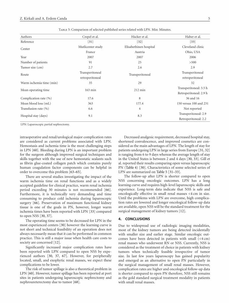
Z. Kirkali and A. Erdem Canda 5
Table 5: Comparison of selected published series related with LPN. Min: Minutes.
Authors Crepel et al. Hacker et al. Haber et al.
Reference [31] [32] [33]
CenterMutlicenter study Elisabethinen hospital Cleveland clinic
France Austria Ohio, USA
Year 2007 2007 2006
Number of patients 91 25 >500
Tumor size (cm) 2.7 2.6 2.9
RouteTransperitoneal
TransperitonealTransperitoneal
retroperitoneal retroperitoneal
Warm ischemia time (min) 35 29 32
Mean operating time 163 min 212 minTransperitoneal: 3.5 h
Retroperitoneal: 2.9 h
Complication rate (%) 17.6 8 36 and 16
Mean blood loss (mL) 363 177.4 150 versus 100 and 231
Transfusion rate (%) 6.6 4 Not reported
Hospital stay (days) 9.1 8.3Transperitoneal: 2.9
Retroperitoneal: 2.2
LPN: Laparoscopic partial nephrectomy.
intraoperative and renal/urological major complication ratesare considered as current problems associated with LPN.Hemostasis and ischemia time is the most challenging stepsin LPN [60]. Bleeding during LPN is an important problemfor the surgeon although improved surgical techniques andskills together with the use of new hemostatic sealants suchas fibrin glue-coated collagen patch which contains purelyhuman coagulation factor components can be helpful inorder to overcome this problem [63–65].
There are several studies investigating the impact of thewarm ischemia time on renal functions and as a widelyaccepted guideline for clinical practice, warm renal ischemiaperiod exceeding 30 minutes is not recommended [66].Furthermore, it is technically very demanding and timeconsuming to produce cold ischemia during laparoscopicsurgery [66]. Preservation of maximum functional kidneytissue is one of the goals in PN, however, longer warmischemia times have been reported with LPN [33] comparedto open NSS [30, 37].
The operating time seems to be decreased for LPN in themost experienced centers [30] however the learning curve isnot short and technical feasibility of an operation does notalways necessarily mean that it can be performed in commonpractice. This is still a major issue when health care costs tosociety are concerned [12].
Significantly increased major complication rates havebeen reported with LPN compared to open NSS by expe-rienced authors [30, 37, 67]. However, for peripherallylocated, small, and exophytic renal masses, we expect thesecomplications to be lower.
The risk of tumor spillage is also a theoretical problem inLPN [60]. However, tumor spillage has been reported at portsites in patients undergoing laparoscopic nephrectomy andnephroureterectomy due to tumor [68].
Decreased analgesic requirement, decreased hospital stay,shortened convalescence, and improved cosmetics are con-sidered as the main advantages of LPN. The length of stay forpatients undergoing LPN in large series from Europe [31, 32]is ranging from 6 to 9 days whereas the average length of stayin the United States is between 2 and 4 days [30, 33]. Gill etal. reported their results comparing open versus laparoscopicPN (Table 4) [30]. Characteristics of some selected series ofLPN are summarized on Table 5 [31–33].
The follow-up after LPN is shorter compared to openNSS concerning oncologic outcomes. LPN has a longlearning curve and requires high-level laparoscopic skills andexperience. Long-term data indicate that NSS is safe andoncologically effective in small renal masses <4 cm in size.Until the problems with LPN are overcome, high complica-tion rates are lowered and longer oncological follow-up dataare available, open NSS will be the standard treatment for thesurgical management of kidney tumors [12].
4. CONCLUSIONS
Due to widespread use of radiologic imaging modalities,most of the kidney tumors are being detected incidentallywith smaller size and earlier stage. Similar oncologic out-comes have been detected in patients with small (<4 cm)renal masses who underwent RN or NSS. Currently, NSS isconsidered as the treatment of choice in patients with kidneytumors when technically feasible irrespective of tumorsize. In last few years laparoscopy has gained popularityand emerged as an alternative to open PN particularly inthe surgical management of small renal masses. However,complication rates are higher and oncological follow-up datais shorter compared to open PN therefore, NSS still remainsas the gold standard surgical treatment modality in patientswith small renal masses.
6 Advances in Urology
REFERENCES
[1] D. Jacqmin, H. van Poppel, Z. Kirkali, and G. Mickisch, “Renalcancer,” European Urology, vol. 39, no. 3, pp. 361–369, 2001.
[2] Z. Kirkali and C. Obek, “Clinical aspects of renal cellcarcinoma,” EAU Update Series, vol. 1, no. 4, pp. 189–196,2003.
[3] Z. Kirkali, E. Tuzel, and M. U. Mungan, “Recent advances inkidney cancer and metastatic disease,” BJU International, vol.88, no. 8, pp. 818–824, 2001.
[4] Z. Kirkali and K. Yorukoglu, “Premalignant lesions in thekidney,” The Scientific World Journal, vol. 1, pp. 855–867, 2001.
[5] S. Pehlivan, M. Koyuncuoglu, M. Pehlivan, et al., “Premalig-nant lesions of the kidney share the same genetics changes asconventional renal cell carcinoma,” World Journal of Urology,vol. 22, no. 2, pp. 120–123, 2004.
[6] Z. Kirkali and M. Lekili, “Renal cell carcinoma: new prognosticfactors?” Current Opinion in Urology, vol. 13, no. 6, pp. 433–438, 2003.
[7] A. E. Canda, M. O. Sahin, E. Tuzel, et al., “Significance of inci-dental renal cell carcinoma,” Turkish Journal of Ecopathology,vol. 7, no. 3-4, pp. 105–109, 2001.
[8] Z. Kirkali, “The future of oncological urology in Europe: arewe prepared?” European Urology, vol. 49, no. 1, pp. 11–12,2006.
[9] B. C. Leibovich, M. L. Blute, J. C. Cheville, C. M. Lohse,A. L. Weaver, and H. Zincke, “Nephron sparing surgery forappropriately selected renal cell carcinoma between 4 and7 cm results in outcome similar to radical nephrectomy,” TheJournal of Urology, vol. 171, no. 3, pp. 1066–1070, 2004.
[10] C. J. Robson, “Radical nephrectomy for renal cell carcinoma,”The Journal of Urology, vol. 89, pp. 37–42, 1963.
[11] P. Russo, “Open partial nephrectomy: an essential operationwith an expanding role,” Current Opinion in Urology, vol. 17,no. 5, pp. 309–315, 2007.
[12] Z. Kirkali, “The motion: open partial nephrectomy is thestandard of care for small resectable solid renal masses,”European Urology, vol. 51, no. 2, pp. 561–564, 2007.
[13] A. E. Canda and Z. Kirkali, “Current management of renal cellcarcinoma and targeted therapy,” Urology Journal, vol. 3, no. 1,pp. 1–14, 2006.
[14] K. Vander Eeckt, S. Joniau, and H. van Poppel, “Open surgeryfor localized RCC,” The Scientific World Journal, vol. 7, pp.742–752, 2007.
[15] H. E. Czerny, “Cited by Herczele: Ueber Nierenexstirpation,”Beitrage zur Kenntnis der Flora von China, vol. 6, pp. 484–486,1890.
[16] V. Vermooten, “Indications for conservative surgery in certainrenal tumors: a study based on the growth pattern of the cellcarcinoma,” The Journal of Urology, vol. 64, pp. 200–208, 1950.
[17] M. L. Ramırez and C. P. Evans, “Current management ofsmall renal masses,” The Canadian Journal of Urology, vol. 14,supplement 1, pp. 39–47, 2007.
[18] J.-J. Patard, O. Shvarts, J. S. Lam, et al., “Safety and efficacyof partial nephrectomy for all T1 tumors based on aninternational multicenter experience,” The Journal of Urology,vol. 171, no. 6, part 1, pp. 2181–2185, 2004.
[19] H. W. Herr, “A history of partial nephrectomy for renaltumors,” The Journal of Urology, vol. 173, no. 3, pp. 705–708,2005.
[20] P. Guinan, L. H. Sobin, F. Algaba, et al., “TNM staging of renalcell carcinoma: workgroup No. 3,” Cancer, vol. 80, no. 5, pp.992–993, 1997.
[21] Z. Kirkali and H. van Poppel, “Developments in organpreserving treatments for renal cell cancer: open surgery,”European Urology Supplements, vol. 3, no. 3, pp. 9–13, 2004.
[22] A. J. Stephenson, A. A. Hakimi, M. E. Snyder, and P. Russo,“Complications of radical and partial nephrectomy in a largecontemporary cohort,” The Journal of Urology, vol. 171, no. 1,pp. 130–134, 2004.
[23] H. van Poppel, L. Da Pozzo, W. Albrecht, et al., “A prospectiverandomized EORTC intergroup phase 3 study comparing thecomplications of elective nephron-sparing surgery and radicalnephrectomy for low-stage renal cell carcinoma,” EuropeanUrology, vol. 51, no. 6, pp. 1606–1615, 2007.
[24] J. McKiernan, R. Simmons, J. Katz, and P. Russo, “Naturalhistory of chronic renal insufficiency after partial and radicalnephrectomy,” Urology, vol. 59, no. 6, pp. 816–820, 2002.
[25] W. K. Lau, M. L. Blute, A. L. Weaver, V. E. Torres, andH. Zincke, “Matched comparison of radical nephrectomy vsnephron-sparing surgery in patients with unilateral renal cellcarcinoma and a normal contralateral kidney,” Mayo ClinicProceedings, vol. 75, no. 12, pp. 1236–1242, 2000.
[26] K. S. Hafez, A. F. Fergany, and A. C. Novick, “Nephron sparingsurgery for localized renal cell carcinoma: impact of tumor sizeon patient survival, tumor recurrence and TNM staging,” TheJournal of Urology, vol. 162, no. 6, pp. 1930–1933, 1999.
[27] C. T. Lee, J. Katz, W. Shi, H. T. Thaler, V. E. Reuter, and P.Russo, “Surgical management of renal tumors 4 cm. Or less ina contemporary cohort,” The Journal of Urology, vol. 163, no.3, pp. 730–736, 2000.
[28] B. C. Leibovich, M. L. Blute, J. C. Cheville, et al., “Nephronsparing surgery for approximately selected renal cell carci-noma between 4 an 7 cm results in outcome similar to radicalnephrectomy,” The Journal of Urology, vol. 171, no. 3, pp.1066–1070, 2004.
[29] F. Becker, S. Siemer, M. Hack, U. Humke, M. Ziegler, andM. Stockle, “Excellent long-term cancer control with electivenephron-sparing surgery for selected renal cell carcinomasmeasuring more than 4 cm,” European Urology, vol. 49, no. 6,pp. 1058–1064, 2006.
[30] I. S. Gill, S. F. Matin, M. M. Desai, et al., “Comparative analysisof laparoscopic versus open partial nephrectomy for renaltumors in 200 patients,” The Journal of Urology, vol. 170, no. 1,pp. 64–68, 2003.
[31] M. Crepel, J.-C. Bernhard, L. Bellec, et al., “Comparisonof open and laparoscopie partial nephrectomy : a Frenchmulticentre experience,” Progres en Urologie, vol. 17, no. 1, pp.45–49, 2007.
[32] A. Hacker, A. Albadour, W. Jauker, et al., “Nephron-sparingsurgery for renal tumours: acceleration and facilitation of thelaparoscopic technique,” European Urology, vol. 51, no. 2, pp.358–365, 2007.
[33] G.-P. Haber and I. S. Gill, “Laparoscopic partial nephrectomy:contemporary technique and outcomes,” European Urology,vol. 49, no. 4, pp. 660–665, 2006.
[34] A. F. Fergany, K. S. Hafez, and A. C. Novick, “Long-termresults of nephron sparing surgery for localized renal cellcarcinoma: 10-year followup,” The Journal of Urology, vol. 163,no. 2, pp. 442–445, 2000.
[35] H. van Poppel, B. Bamelis, R. Oyen, and L. Baert, “Partialnephrectomy for renal cell carcinoma can achieve long-termtumor control,” The Journal of Urology, vol. 160, no. 3, part 1,pp. 674–678, 1998.
[36] H. W. Herr, “Partial nephrectomy for unilateral renal carci-noma and a normal contralateral kidney: 10-year followup,”The Journal of Urology, vol. 161, no. 1, pp. 33–35, 1999.

Z. Kirkali and A. Erdem Canda 7
[37] A. F. Fergany, I. R. Saad, L. Woo, and A. C. Novick, “Openpartial nephrectomy for tumor in a solitary kidney: experiencewith 400 cases,” The Journal of Urology, vol. 175, no. 5, pp.1630–1633, 2006.
[38] F. Becker, S. Siemer, U. Humke, M. Hack, M. Ziegler, andM. Stockle, “Elective nephron sparing surgery should becomestandard treatment for small unilateral renal cell carcinoma:long-term survival data of 216 patients,” European Urology,vol. 49, no. 2, pp. 308–313, 2006.
[39] S. C. Campbell and A. C. Novick, “Surgical techniqueand morbidity of elective partial nephrectomy,” Seminars inUrologic Oncology, vol. 13, no. 4, pp. 281–287, 1995.
[40] H. van Poppel and L. Baert, “Elective conservative surgery forrenal cell carcinoma,” AUA Update Series, vol. 13, pp. 246–251,1994.
[41] M. R. Licht, A. C. Novick, and M. Goormastic, “Nephronsparing surgery in incidental versus suspected renal cellcarcinoma,” The Journal of Urology, vol. 152, no. 1, pp. 39–42,1994.
[42] P. Dal Cin, H. van Poppel, B. van Damme, L. Baert, and H.van den Berghe, “Cytogenetic investigation of synchronousbilateral renal tumors,” Cancer Genetics and Cytogenetics, vol.89, no. 1, pp. 57–60, 1996.
[43] H. van Poppel, F. Deroo, and S. Joniau, “Open surgicaltreatment of localized renal cell cancer,” EAU Update Series,vol. 1, no. 4, pp. 220–225, 2003.
[44] F. Becker, S. Siemer, M. Hack, U. Humke, M. Ziegler, andM. Stockle, “Excellent long-term cancer control with electivenephron-sparing surgery for selected renal cell carcinomasmeasuring more than 4 cm,” European Urology, vol. 49, no. 6,pp. 1058–1064, 2006.
[45] M. O. Sahin, A. E. Canda, M. U. Mungan, Z. Kirkali, andM. Sade, “Benign lesions underwent radical nephrectomy forrenal cancer,” Turkish Journal of Urology, vol. 30, no. 4, pp.405–409, 2004.
[46] B. R. Herts and M. E. Baker, “The current role of percuta-neeous biopsy in the enucleation of renal mases,” SeminarUrologic Oncology, vol. 13, pp. 254–261, 1995.
[47] H. Goethuys, H. van Poppel, R. Oyen, and L. Baert, “The caseagainst fine-needle aspiration cytology for small solid kidneytumors,” European Urology, vol. 29, no. 3, pp. 284–287, 1996.
[48] A. C. Novick, “Surgery of the kidney,” in Campbell’s Urology,P. Walsh, A. L. Retik, E. D. Vaugman, and A. J. Wein, Eds., vol.3, pp. 3571–3640, Saunders, New York, NY, USA, 8th edition,2002.
[49] A. C. Novick, “Nephron-sparing surgery for renal cell carci-noma,” Annual Review of Medicine, vol. 53, pp. 393–407, 2002.
[50] T. McHale, S. B. Malkowicz, J. E. Tomaszewski, and E. M.Genega, “Potential pitfalls in the frozen section evaluation ofparenchymal margins in nephron-sparing surgery,” AmericanJournal of Clinical Pathology, vol. 118, no. 6, pp. 903–910, 2002.
[51] H. van Poppel, “The optimal margins in nephron-sparingsurgery,” Current Opinion in Urology, vol. 14, no. 4, pp. 227–228, 2004.
[52] Q.-L. Li, H.-W. Guan, Q.-P. Zhang, L.-Z. Zhang, F.-P. Wang,and Y.-J. Liu, “Optimal margin in nephron-sparing surgery forrenal cell carcinoma 4 cm or less,” European Urology, vol. 44,no. 4, pp. 448–451, 2003.
[53] H. van Poppel, B. Bamelis, and L. Baert, “Elective nephronsparing surgery for renal cell carcinoma,” EAU Update Series,vol. 6, pp. 8–12, 1997.
[54] N. Y. Piper, J. T. Bishoff, C. Magee, et al., “Is a 1-cm marginnecessary during nephron-sparing surgery for renal cellcarcinoma?” Urology, vol. 58, no. 6, pp. 849–852, 2001.
[55] C. B. Dechet, M. L. Blute, and H. Zincke, “Nephron-preserving surgery for unilateral renal cell carcinoma: whichpathologic variables contribute to contralateral renal tumorrecurrence?” The Journal of Urology, vol. 159, supplement, p.169, 1998.
[56] A. H. Bani-Hani, B. C. Leibovich, C. M. Lohse, J. C. Cheville,H. Zincke, and M. L. Blute, “Associations with contralateralrecurrence following nephrectomy for renal cell carcinomausing a cohort of 2,352 patients,” The Journal of Urology, vol.173, no. 2, pp. 391–394, 2005.
[57] M. E. Allaf, S. B. Bhayani, C. Rogers, et al., “Laparoscopicpartial nephrectomy: evaluation of long-term oncologicaloutcome,” The Journal of Urology, vol. 172, no. 3, pp. 871–873,2004.
[58] I. S. Gill, M. M. Desai, J. H. Kaouk, et al., “Laparoscopic partialnephrectomy for renal tumor: duplicating open surgicaltechniques,” The Journal of Urology, vol. 167, no. 2, part 1, pp.469–475, 2002.
[59] F. Steinbach, M. Stockle, and R. Hohenfellner, “Currentcontroversies in nephron-sparing surgery for renal-cell carci-noma,” World Journal of Urology, vol. 13, no. 3, pp. 163–165,1995.
[60] J. J. Rassweiler, C. Abbou, G. Janetschek, and K. Jeschke,“Laparoscopic partial nephrectomy the European experience,”Urologic Clinics of North America, vol. 27, no. 4, pp. 721–736,2000.
[61] A. Moinzadeh, I. S. Gill, A. Finelli, J. Kaouk, and M. Desai,“Laparoscopic partial nephrectomy: 3-year followup,” TheJournal of Urology, vol. 175, no. 2, pp. 459–462, 2006.
[62] B. R. Lane and I. S. Gill, “5-year outcomes of laparoscopicpartial nephrectomy,” The Journal of Urology, vol. 177, no. 1,pp. 70–74, 2007.
[63] S. Siemer, S. Lahme, S. Altziebler, et al., “Efficacy and safetyof tachoSil as haemostatic treatment versus standard suturingin kidney tumor resection: a randomised prospective study,”European Urology, vol. 52, no. 4, pp. 1156–1163, 2007.
[64] A. H. Wille, M. Tullmann, J. Roigas, S. A. Loening, andS. Deger, “Laparoscopic partial nephrectomy in renal cellcancer—results and reproducibility by different surgeons in ahigh volume laparoscopic center,” European Urology, vol. 49,no. 2, pp. 337–342, 2006.
[65] E. Kouba, C. Tornehl, J. Lavelle, E. Wallen, and R. S. Pruthi,“Partial nephrectomy with fibrin glue repair: measurement ofvascular and pelvicaliceal hydrodynamic bond integrity in alive and abbatoir porcine model,” The Journal of Urology, vol.172, no. 1, pp. 326–330, 2004.
[66] I. S. Gill, “Minimally invasive nephron-sparing surgery,”Urologic Clinics of North America, vol. 30, no. 3, pp. 551–579,2003.
[67] A. P. Ramani, M. M. Desai, A. P. Steinberg, et al., “Complica-tions of laparoscopic partial nephrectomy in 200 cases,” TheJournal of Urology, vol. 173, no. 1, pp. 42–47, 2005.
[68] A. Cicco, L. Salomon, H. Hoznek, et al., “Carcinological risksand retroperitoneal laparoscopy,” European Urology, vol. 38,no. 5, pp. 606–612, 2000.

Hindawi Publishing CorporationAdvances in UrologyVolume 2008, Article ID 916463, 9 pagesdoi:10.1155/2008/916463
Review ArticleOpen Partial Nephrectomy in Renal Cancer:A Feasible Gold Standard Technique in All Hospitals
J. M. Cozar and M. Tallada
Department of Urology, Virgen de las Nieves University Hospital, Granada, Spain
Correspondence should be addressed to J. M. Cozar, [email protected]
Received 25 March 2008; Accepted 31 May 2008
Recommended by J. Rubio
Introduction. Partial nephrectomy (PN) is playing an increasingly important role in localized renal cell carcinoma (RCC) as atrue alternative to radical nephrectomy. With the greater experience and expertise of surgical teams, it has become an alternativeto radical nephrectomy in young patients when the tumor diameter is 4 cm or less in almost all hospitals since cancer-specificsurvival outcomes are similar to those obtained with radical nephrectomy. Materials and Methods. The authors comment ontheir own experience and review the literature, reporting current indications and outcomes including complications. The surgicaltechnique of open partial nephrectomy is outlined. Conclusions. Nowadays, open PN is the gold standard technique to treat smallrenal masses, and all nonablative techniques must pass the test of time to be compared to PN. It is not ethical for patients toundergo radical surgery just because the urologists involved do not have adequate experience with PN. Patients should be involvedin the final treatment decision and, when appropriate, referred to specialized centers with experience in open or laparoscopicpartial nephrectomies.
Copyright © 2008 J. M. Cozar and M. Tallada. This is an open access article distributed under the Creative Commons AttributionLicense, which permits unrestricted use, distribution, and reproduction in any medium, provided the original work is properlycited.
1. INTRODUCTION
In recent years, the surgical treatment of renal cell carcinoma(RCC) has developed towards conservative surgery of therenal parenchyma and the use of minimally invasive tech-niques. The emerging conservative technique is open partialnephrectomy (PN), which is no longer an option reserved forpatients with a single kidney or bilateral renal tumors; it hasbecome a viable alternative to radical nephrectomy, and isconsidered the treatment of choice for selected patients witha normal contralateral kidney [1, 2].
The more frequent use of PN in renal cancer treatmentderives from a spectacular rise in the incidental diagnosis ofrenal tumors in patients undergoing abdominal ultrasoundor computed tomography (CT) for abdominal diseases. Thishas markedly increased the detection of smaller, asymp-tomatic tumors than those observed when Robson proposedradical nephrectomy as the technique of choice more thanthree decades ago [3]. Incidental tumors have a morefavorable prognosis than clinically detected or symptomatictumors of a similar size and stage [4, 5]. The better health andlonger life span of the general population and the availabilityof radiological imaging techniques for closer screening and
follow-up programs are creating a favorable environment forthe development of conservative renal surgery. When PNis indicated, the decision to adopt an open or laparoscopic(minimally invasive) approach depends on the benefits andrisks to the patient and the experience of the surgical team.
This article is devoted to open PN, providing anupdate on the indications, disease-free and disease-specificsurvival outcomes, benefits and risks, limitations andtechnical aspects of the surgery, intra- and postoperativecomplications, and post-treatment follow-up protocols. Theaim is to describe the main concepts to be considered in thedecision-taking algorithm for an open PN in the treatmentof RCC [6, 7].
2. INDICATIONS FOR PARTIAL NEPHRECTOMY
Indications can be classified as absolute, relative, or elective(Algorithm 1), always basing the selection on the viability ofthe technique and an optimal cancer control [8].
2.1. Absolute indications
Absolute indications relate to patientswho would be anatom-ically or functionally anephric if radical nephrectomy was

2 Advances in Urology
(1) Absolute(i) Single kidney(ii) Bilateral renal tumor(iii) Severe renal failure
(2) Relative(i) Abnormal contralateral kidney (nephropathy,
nephrolithiasis, trauma, etc.)(ii) Metabolic disease associated with renal failure(iii) Genetic syndrome with tumor multifocality
(e.g., VHL syndrome)(3) Elective
(i) Tumor <4 cm in young and healthy patients(ii) Peripheral tumor(iii) Tumor >4 cm (limit at 7 cm?)
Algorithm 1: Indications for partial nephrectomy.
performed. They include the presence of only one kidney,synchronous bilateral renal cancer, and severe renal failure.It was proposed in the 1950’s that these patients undergoconservative tumor excision to preserve maximum renalparenchyma and allow the possibility of renal filtrationwith no need for dialysis. However, this proposal gainedlittle acceptance among urologists due to the high rate ofcomplications observed after open PN. More recently, therehas been a strong resurgence in the use of this technique as analternative to radical nephrectomy for the above-mentionedtypes of patients. In the 1990’s, various studies reportedgood survival outcomes and fewer complications with aconservative approach.
2.2. Relative indications
These include conditions that might compromise the futurefunctioning of the contralateral kidney (without tumor),for example, moderate renal failure, nephrolithiasis, recur-rent pyelonephritis with parenchymal lesions, vesicoureteralreflux, and congenital or acquired obstruction of the urinarytract, among others. A further relative indication would bethe presence of disease with a potentially negative medium-term effect on renal function, for example, diabetes orhypertension. Other factors must be taken into account inthese patients, including their current age and age at onset ofthe disease, estimating the duration of its possible effect onrenal function.
2.3. Elective indications
Partial nephrectomy has been proposed for small peripheralrenal tumors over the past few years. Being initiallycontroversial, this indication has been supported by widestudies showing similar outcomes to radical surgery insmall (≤4 cm) renal tumors (Algorithm 1). The age andgeneral state of the patient are important in the selectionof candidates for PN, which is most beneficial for youngand healthy patients. Some authors have proposed to widenthe indication for a conservative approach to include larger
Bu
tler
,19
95
Lern
er,
1996
Bel
ldeg
run
,19
99Lau
,20
00
Lee,
2000
McK
iern
an,
2002
RNPN
50
55
60
65
70
75
80
85
90
95
100
RN173/PN:117
RN: 183/PN: 79
NR: 164/NP: 164
RN: 125/PN: 108
RN: 209/PN: 185
RN: 42/PN: 46
Figure 1: Five-year survival: radical nephrectomy (RN) versuspartial nephrectomy (PN).
tumors of up to 7 cm. Thus, Fergany et al. at the ClevelandClinic reported similar five-year disease-free survival ratesbetween patients with tumors <4 cm and those with tumorsof 4–7 cm [4].
The greater longevity of the population and the treat-ment of ever younger patients for incidentally detectedtumors have drawn attention to the long-term risks of renalfailure or metachronous tumor recurrence posed by PNtreatment. Nevertheless, these risks should not outweigh thebenefits of this renal parenchyma-preserving surgery.
3. CLINICAL EXPERIENCE: SURVIVAL OUTCOMES
There is now considerable clinical experience in patients withone of the above indications for PN, allowing careful analysisof patient outcomes and evaluation of the prognostic factorsthat influence results.
Studies of patients with small renal tumors at theMemorial Sloan Kettering Cancer Center of UCLA, MayoClinic, and Cleveland Clinic showed no significant differ-ences in five-year survival rates (91–100%) between thosetreated with open PN and those treated with open radicalnephrectomy (Figure 1) [9–14]. Long-term follow-up stud-ies have corroborated these results. Thus, Lau et al. comparedbetween patients treated with open PN (mean tumor size of3.7 cm) and those treated with radical nephrectomy (meansize of 3.3 cm), and found no significant differences in overallsurvival, cancer-specific survival, metastasis-free survival, orlocal recurrence-free survival at 5, 10, or 15 years [11]. Thesefindings validate the oncological efficacy of conservativeversus radical renal surgery.
There have been no prospective randomized clinical trialscomparing the two techniques. Moreover, global results ofthe published studies cannot be grouped together becausethe distribution of indication levels (absolute, relative, orelective) is different in each study population. For this reason,

J. M. Cozar and M. Tallada 3
Table 1: Conservative renal surgery (partial nephrectomy); five-year outcomes in patients with elective indication.
Author, year N Disease-specific survival Local recurrence Mean tumor size
Morgan, 1990 20 100% 0% 3.1
Selli, 1991 20 90% 0% 3.5
Provet, 1991 19 100% 0% 2.6
Steinbach, 1992 72 94.4% 2.7% ND
Moll, 1993 98 100% 1% 4
Lerner, 1996 54 92% 5.6% 4
D’Armiento, 1997 19 96% 0% 3.3
Van Poppel, 1998 51 98% 0% 3
Herr, 1999 70 97.5% 1.5% 3
Hafez, 1999 45 100% 0% 4
Barbalias, 1999 41 97.5% 7.3% 3.5
Belldegrun, 1999 63 100% 3.2% 4
outcomes of open PN are reported below in relation to thetype of indication.
3.1. Outcomes of partial nephrectomy withabsolute indication
In general terms, patient survival rates are lower whenthe indication for surgery is absolute rather than elective,influenced by the higher age, the more advanced stages, thelarger tumor size, and the poorer health status of patientswith an absolute indication. Reports from the Clevelandand Mayo Clinics [4, 15] described disease-free survivalrates after PN of 81–88% at 5 years and 64–73% at 10years, being relatively similar to disease-free survival ratesdescribed for radical nephrectomy in tumors of the samesize and stage. In 2007, Berdjis et al. studied 38 cases of openPN in single kidney carried out between 1993 and 2003[16]. After a mean follow-up of 41.7 months, they observedlocal recurrence in four patients (including 3 with distantprogression) and metastatic progression in two. Tumorsize was significantly larger in patients with metastaticprogression versus those without (6.2 cm versus 3.5 cm)and in patients with subsequent renal failure versus thosewithout (5.2 cm versus 3.3 cm).
According to these authors, tumor size is the mostsignificant prognostic factor for disease progression followedby tumor stage (localized versus locally advanced), andlarger tumor size is the main prognostic factor for renalfailure onset [16].
3.2. Results of partial nephrectomy withelective indication
In the 1990’s, numerous reports [5, 10, 11, 17–26] werepublished on a total of 572 patients with normal contralateralkidney treated by open PN, having tumor sizes ranging from2 to 4.3 cm (Table 1). A survival rate of 90–100% wasachieved in these cases, with a local recurrence rate of 0% inmost series [10, 11, 18–20, 22–25, 27], 1% in 2 series [5, 22],3% in 2 series [12, 21], and 6-7% in 2 series [13, 26]. Theseoutcomes opened up the way for open PN to become aneffective alternative to radical nephrectomy although higher
rates of intra- and postoperative complications were initiallyobserved.
Published data establish 4 cm as the cut-off tumor sizefor indication of this surgery, describing a shorter disease-free survival period in patients with larger tumors. Studiesreport 95% five-year disease-free survival rates in patientswith a tumor <4 cm, comparable to the outcomes of radicalnephrectomy in tumors of a similar size (Table 1).
3.3. Results of partial nephrectomy in patients withVon Hippel Lindau (VHL) syndrome
The risk of local recurrence is very high in VHL patientsbecause of the multifocal nature of their malignant tumors;consequently their disease-free survival is much lower incomparison to patients with incidental or sporadic renalcarcinoma.
Out of nine VHL patients with bilateral renal carcinomastudied by Novick and Campbell, seven had local recurrenceand one died from metastatic disease [2]. It is likely that mostof these recurrences represented a manifestation of a micro-scopic residual CCR that was not excised during the NP [2].
Walther et al. [27] reported on 52 VHL patients withrenal cancer treated at the National Cancer Institute, findingthat no patient with tumors <3 cm developed metastaticdisease. They therefore recommend waiting until this type oftumor reaches 3 cm in order to reduce the need for surgerybefore onset of the multiple recurrences observed duringfollow-up of these patients.
The effectiveness of PN as a valid alternative for thetreatment of this disease was demonstrated by a multicenterstudy in USA on the results of treating 65 patients withVHL and localized RCC (54 bilateral, 11 unilateral). PN wasperformed on 49 of these patients, with five-year and ten-year survival rates of 100% and 81%, respectively. Thesesurvival outcomes are similar to those obtained with radicalnephrectomy, and they support the role of PN in thetreatment of this type of patient.
In patients with advanced VHL and large multiplebilateral tumors that require complete excision of bothkidneys at first surgery or after various interventions dueto the post-PN growth of residual RCCs, renal transplant is

4 Advances in Urology
an appropriate option to avoid terminal kidney failure andthe need for dialysis, especially in young patients with thisgenetic syndrome.
4. PROGNOSTIC FACTORS FOR TUMOR RECURRENCEAFTER PARTIAL NEPHRECTOMY
In RCC, prognostic factors for distant recurrence ormetastatic progression after radical nephrectomy are knownto include the Fuhrman grade, size, and stage, as well ashistological type of the tumor, the presence of positive lymphnodes, and ECOG performance status [2, 8]. Some of thesefactors, described below, are of special interest in selectingcandidates for PN.
4.1. Tumor size >4 cm
Tumor size was found to be the most significant predictorof the outcome in large series of PN patients [4, 11, 13, 25].Tumor size independently predicts local recurrence and isthe most important criterion for the indication of a PN.The Cleveland Clinic series of 485 PNs, including 9% withelective indications, showed significant differences in five-year and ten-year survival rates between patients with tumorssmaller and larger than 4 cm, with a significant correlationbetween recurrence rate and tumor size. For this reason,Barbalias et al. [25] proposed a subclassification of stage T1(tumors <7 cm and limited to renal parenchyma) into T1aand T1b for tumor sizes of <4 cm and ≥4 cm, respectively.
Lerner et al. [13] observed a 95% five-year survival rate inPN patients with tumors <3 cm versus an 80% rate in thosewith tumors >6 cm. They also reported a significantly higherdisease-free survival rate in patients with tumors >4 cm afterradical versus partial nephrectomy. More recently, variousstudies [9–14, 23] (Figure 2) demonstrated equivalent cancercontrol rates between patients with tumors <4 cm and thosewith tumors of 4–7 cm after electively indicated PN.
4.2. Localization of tumor
It was classically thought that centrally localized tumorscarried a greater risk of metastasis at the time of diseasepresentation. It was therefore considered that the risk ofrecurrence and/or progression would be higher after partialversus radical nephrectomy in central tumors. This ideawas challenged by the results of a retrospective studyby D’Armiento et al. [23] on tumor localization as anindependent risk factor. They found no difference in cancer-free survival or recurrence between peripheral tumors (notextending into the interior of the kidney) and central tumors(infiltrating beyond the renal medulla). These authorsconcluded that PN is more complex in the case of centraltumors, but it is not associated with a worse recurrence orprogression prognosis.
4.3. Multifocality
The incidence of small renal tumors removed during radicalnephrectomy for RCC or in necropsies ranges from 4 to 25%[27, 28]. As a consequence, many urologists have argued
1007550250
(%)
>7 cm
4–7 cm<4 cm
Lee et al, 2000
Fergany, 2000
Hafez, 1999
Herr, 1999
Belldegrun, 1999
Lerner, 1996
Butler, 1995
Figure 2: Five-year disease-free survival by tumor size in patientsundergoing partial nephrectomy.
against PN as a standard treatment for RCC, even whentumors are small, due to the high risk of multifocality. Weneed to know the factors that increase the risk of multifocal-ity to allow us to select PN when the risk of multifocality islow and radical nephrectomy when the risk is high.
The main factor signaling an increased risk of multifo-cality is large tumor size, since 91% of multifocal tumorsare associated with primary tumors >5 cm [29]. The secondfactor is tumor stage (pT2 or higher). Thus, stage pT3a showsa 16.4% incidence of multifocality, with a mean distancebetween primary and secondary tumors of 26.4 mm [30].Other factors increasing the risk of multifocality cannotbe known before surgery but only after examination ofthe surgical specimen, including histological factors suchas vascular infiltration and papillary or mixed histologicalvariants [27]. Knowledge of factors carrying an elevated riskof multifocality alerts to the need for more rigorous patientfollow-up and inspection of the whole defatted kidney to ruleout satellite tumors during PN.
With regard to the preoperative detection of multifocallesions by imaging techniques, only 22.9% of additionaltumors subsequently observed in specimens after radicalnephrectomy had been detected by ultrasound or CT [31].Intraoperative ultrasound studies show a higher sensitivity,detecting up to 78% of multifocal tumors, which may be veryuseful when there are multifocality risk factors and a PN hasbeen proposed to the patient.
4.4. High Fuhrman nuclear grade and symptomaticclinical presentation
It was reported that recurrence-free survival after PN wasnot only significantly improved by smaller tumor size (<4)

J. M. Cozar and M. Tallada 5
but also by low Fuhrman grade and incidental clinicalpresentation [4]. This finding was confirmed by Licht [32],who observed a significantly worse prognosis after thissurgery in symptomatic (83% five-year survival) versusincidental (94% five-year survival) tumors.
Moll et al. [5] and Ghavamian et al. [15] reported thattumor stage and nuclear grade are significantly associatedwith RCC mortality. These classic prognostic factors arevalid for both radical and partial nephrectomies, but a muchmore rigorous follow-up is required if partial nephrectomy isselected and the pathology study reports grade III disease.
4.5. Surgical margins
Conventional PN includes ≥1 cm margin of healthyparenchyma, whereas this margin is not left in tumorenucleation and there is a higher risk of surgical margininvolvement. Recent reports have shown similar rates ofcancer control between PN and enucleation, provided thatsurgical margins in enucleation are examined by intraopera-tive biopsy of the kidney bed.
In a series of 44 patients treated with PN for tumors witha mean size of 3.2 cm and a mean surgical margin of 2.5 mm,93% had negative surgical margins and showed no localrecurrence after after a mean follow-up of 4 years [33]. Inthe Mayo Clinic series, all partial nephrectomies were carriedout with margins≥3 mm of healthy peritumoral renal tissue,verified by intraoperative biopsy of the kidney bed. Localfive-year recurrence-free survival was 97% in a series of 130patients [34].
It can therefore be proposed that a margin of 1-2 cm isnot necessary in PN, for which a few millimeters (3–5 mm)can be adequate as long as the intraoperative biopsy of thekidney bed is negative.
5. SURGICAL TECHNIQUE IN PARTIAL NEPHRECTOMY
Surgical technique in PN has advanced over recent years,offering improved cancer control and anatomical-functionaloutcomes for the saved kidney.
A flank incision approach is used, opening Gerota’s fasciaand localizing the kidney and the tumor. A thorough visualinspection is essential for adequate planning of the resection,especially if the tumor is near the hilum. It is controversialwhether renal ischemia is required for the resection. Thisdecision depends on the nature of the tumor and the skillof the surgeon. At our center, where there is considerableexperience acquired over many years and excellent cancercontrol and renal preservation outcomes have been obtained,the renal pedicle is identified and released, isolating the mainrenal artery and vein with vessel-loops.
If the tumor is small and the indication is elective, resec-tion of the tumor then commences, allowing a safety marginof several millimeters of healthy renal parenchyma. If there isno major bleeding as the resection proceeds, total resectionof the tumor is completed without recourse to any type ofrenal ischemia. If there is any doubt about the resectionmargins after removal of the tumor, an intraoperative biopsyof the bed is performed and we proceed according to the
Figure 3: Open partial nephrectomy for renal cell carcinomaof inferior pole satisfactory hemostasis with application of afibrinogen hemostatic patch (Tachosil, Nycomed Pharma, Austria).
results. Then, hemostasis of the tumor bed is started rapidlywith single stitches of 4/0 vicryl at the main bleeding pointsusing spray-coagulation on secondary vessels with an electricscalpel, which takes considerable time. To reduce this time,a technique modification was introduced at our centre oneyear ago, with the application of a fibrinogen hemostaticpatch (Tachosil, Nycomed Pharma, Austria) on the resectionsurface after suturing the main bleeding vessels. This hasshortened the time for intervention and hemostasis to 15–20minutes, and improved the visual appearance (Figure 3). Wealso leave the surgical bed open after the resection, withoutusing a mattress stitch to draw the renal parenchyma togetherfor better hemostasis. The resection bed must be carefullyinspected, and the opening of the urinary passage (calyx orpelvis) must be avoided or, when necessary, repaired.
When tumors are large but PN is relatively or absolutelyindicated, we prefer to clamp the renal artery to produceischemia after administering intravenous mannitol, keepingthe renal vein patent to minimize the risk of postoperativeacute tubular necrosis. It may also be necessary to clampthe renal artery in cases of central tumors that affect majorvessels (e.g., arcuate arteries) or in cases of unexpectedbleeding that can only be controlled by ischemia. In thesecases, it is very useful to have the renal pedicle preparedin advance, allowing ischemia to be produced within a fewseconds and minimizing blood loss.
Intraoperative ultrasonography is not a standard proce-dure in our setting but can greatly assist in the identificationof other renal tumors when multifocality is suspected. Someauthors use intraoperative ultrasonography to demarcateintrarenal tumors and to avoid damaging large vessels nearthe resection line or bed.
Once hemostasis has been achieved, a carefulexamination is required to detect any inadvertent openingof the urinary tract, thereby avoiding postoperative leaksor fistulas. If an opening is identified, it is closed usinga resorbable suture. When an opening is suspected butcannot be seen, an intrapelvic injection of methylene blue isrequired, leading some authors to previously insert a ureteralstent in patients with central-located tumors. Fibrin sealants,

6 Advances in Urology
Table 2: Complications after open partial nephrectomy.
Author, year (Hospital) N Acute renal failure (%) Urinary fistula (%)
Ghavamian, 2002 (Mayo Clinic) 63 12.7 3.2
Duque, 1998 (Brigham) 64 15.1 9.1
Polascik, 1995 (Johns Hopkins) 67 1.5 9
Herr, 1994 (Memorial Sloan-KCC) 41 0 0
Campbell, 1994 (Cleveland Clinic) 259 12.7 17.3
as well as being hemostatic agents, can reinforce repair ofthe collecting system. Gelfoam soaked with thrombin canbe placed over the defect and then infiltrated with Hemaseelfibrin sealant to close small defects of the urinary tract at thelevel of the calyces.
6. COMPLICATIONS OF OPENPARTIAL NEPHRECTOMY
Open partial nephrectomy is more complex than radicalnephrectomy, and many authors place limits on its use citinga higher risk of complications. Several decades ago, reportson open PN described a greater risk of acute renal failure,urinary fistula, and hemorrhage of the surgical bed, amongother complications [15, 22, 35, 36].
The lower incidence of complications in the presentpatient series can be attributed to the greater experience thaturologists have gained with this technique and the higherprevalence of incidental small tumors. In 1994, Campbellet al. [35] described complication rates after open PN of 37%for symptomatic tumors and of 22% for incidental tumorsTable 2.
More recently, however, open PN and radical nephrec-tomy have shown a similar complications’ rate, overallmorbidity rate, hospital stay, blood losses, and frequency ofacute renal failure [37, 38]. The risk of acute renal failureafter open PN ranges from 0 to 18% according to the series.Campbell et al. [35] reported a 13% incidence of acuterenal failure in 259 patients after open PN (for which only10/259 patients [3.9%] had an elective indication). Riskfactors for postoperative acute renal failure were tumor size>7 cm, excision of more than half the renal parenchyma,and ischemia >60 minutes. In patients with PN, the renalparenchymal volume loss correlates best with the renalfunction loss several months after surgery. Estimates ofvolume loss may be useful for predicting postoperative renalfunction when planning PN in patients with a solitary kidney[6, 7, 39].
The risk of urinary fistula after open PN ranges from1.8 to 21%, and is lower in patients treated for a smallincidental tumor with elective indication. Campbell et al.[35] described an increased risk of urinary fistula in tumors>4 cm localized centrally or near the hilum in surgeryrequiring reconstruction of the excretory tract.
To summarize, complications of open PN appear tohave been reduced to levels found with open radicalnephrectomy—thanks to the greater experience of surgicalteams with this technique. In the medium term, however,at 6–12 months, open PN patients have a significantly
lower serum creatinine level compared with laparoscopicradical nephrectomy patients [40]. This information mustbe explained to patients when they are informed about theshort-term and long-term risks of the two approaches.
7. FOLLOW-UP GUIDELINES AFTER OPENPARTIAL NEPHRECTOMY
Several clinical guidelines have been established for thefollow-up of patients after open PN, based on detailedanalysis of reported tumor recurrence patterns at specializedcenters (e.g., Cleveland Clinic).
Rates of local recurrence and metastatic progression varyas a function of the tumor stage at surgery as follows:T1N0M0: 0% local recurrence and 4.4% distant progression;T2N0M0: 2% and 5.3%, respectively; T3aN0M0: 8.2%and 11.5%, respectively; T3bN0M0: 10.6% and 14.9%,respectively. Postoperative time periods associated with amaximum incidence of local recurrence are between 6 and24 months for T3 and T2 tumors and after 48 months for T1tumors. Hence, the follow-up time and protocol are selectedaccording to the pathological stage at the time of the openPN.
Novick and Campbell [2] proposed these follow-upguidelines.
(1) Patients with T1N0M0 tumors have annual anamne-sis, physical examination, and serology, with no needfor radiology during the first year. No subsequentsystematic diagnostic imaging studies are requireddue to the low risk of recurrence.
(2) Patients with T2N0M0 tumors have annual anamne-sis, physical examination, chest X-ray, and abdominalCT scan, with abdominal X-ray every 2 years.
(3) Patients with T3N0M0 tumors have anamnesis, phys-ical examination, chest X-ray, and abdominal CTevery 6 months for 3 years and then annually.
In the long term, hyperfiltration can cause renal injuryin these patients, especially if there has been >50% loss ofnephrons, with proteinuria, focal segmental glomeruloscle-rosis, and progressive renal failure. Because proteinuria is thefirst change in this disorder, 24-hour urine proteins shouldbe determined annually in all patients with suspicion ofhyperfiltration due to loss of renal parenchyma.
8. WHEN TO PROPOSE OPEN PARTIAL NEPHRECTOMY
Based on the above reported data, clinical studies (MayoClinic, Cleveland Clinic, UCLA, etc.), and our own

J. M. Cozar and M. Tallada 7
experience, we can affirm, in common with other authors[1, 2], that open PN is now the gold standard treatment foryoung and healthy patients with incidentally detected smallrenal tumors (<4 cm). It also represents an alternative toradical nephrectomy in single-kidney patients or those withbilateral tumors.
The presence of renal failure, diseases that predisposetowards renal failure, or genetic syndromes associated withmultifocality also shows indications for open PN versusradical nephrectomy since, in small tumors, there are no dif-ferences in disease-free survival, morbidity, or complicationrates between the techniques.
The current standard surgical technique for partialnephrectomy is open partial nephrectomy. Only certainhighly specialized centers have gained sufficient experiencewith laparoscopic PN to minimize its risks and complications[40]. It remains a challenging technique, requiring a longerperiod of warm renal ischemia, vein closure, and the difficultsuturing of open vessels during tumor resection. In fact,the laparoscopic approach has been associated with a higherrate of complications, even in the best hands. Thus, Sharmaet al. reported intra- and postoperative complication rates of5% and 11%, respectively, using laparoscopic PN, comparedwith 0% and 2%, respectively, using open PN [39].
Although no studies have been published to date onthe long-term oncological effectiveness of laparoscopic PN[1], preliminary data indicate that it does not differ fromthat obtained with the open approach. In 2007, Gill et al.[41] reported three-year cancer-specific survival rates of99.3% in 771 patients treated with laparoscopic partialnephrectomy and 99.2% in 1028 patients treated with openpartial nephrectomy. The same study confirmed a shortersurgery time (P < .0001), hospital stay (P < .0001), anda lower blood loss (P < .0001) with laparoscopic partialnephrectomy versus open partial nephrectomy, while intra-operative complication rates were similar. Disadvantagesof laparoscopic versus open partial nephrectomy were thesignificantly longer ischemia time (P < .0001) and the morefrequent postoperative complications, especially urologicaldisorders (P < .0001).
Importantly, the laparoscopic approach is associated witha reduction in postoperative pain and a shorter recoveryperiod, posing surgeons and patients with a difficult decisionbetween open and laparoscopic partial nephrectomies for asmall incidentally detected renal tumor.
Renal laparoscopy will continue to develop, and urol-ogists will gain greater experience with the technique overtime. Thus, outcomes published by Gill et al. in 2007 weresuperior to those obtained by the same author in 2003[41, 42]. It should be taken into account that the study byGill et al. comparing laparoscopic nephrectomy with openpartial nephrectomy [41, 43] was not a randomized clinicaltrial. In fact, most of the tumors selected for open partialnephrectomy were >4 cm, and all of them were centrallylocalized single tumors (size up to 7 cm) with a malignanthistology. These cases are technically more challenging andcarry a higher oncological risk, representing an importantselection bias. There is a need for a randomized clinical trialto be undertaken to assess the risks and benefits of each
approach. Nevertheless, there is an evident trend towardsa minimally invasive approach to renal tumor treatment,and we can expect laparoscopic PN to develop in the nearfuture to a point where it can replace open PN as a standardtreatment for localized renal tumors.
At our center, we have treated 35 patients by conservativesurgery of the renal parenchyma over the past 14 years(1993–2007), using open PN in 7 and enucleation in 28, withbiopsies of the renal bed when involvement of the surgicalmargin was suspected. Indications were elective in 16 cases(45.5%), absolute in 11 (31.5%), and relative in 8 (23%).Mean size of tumors was 3.6 cm (range of 1–9 cm), withperipheral localization in 22 patients (63%), mesorenal in12 (34%), and multifocal (6 tumors) in 1 patient (3%).Applying the technique described above in Section 5, we havehad no intraoperative complications. Postoperative com-plications were renocutaneous fistula (resolved by internalderivation via ureteral catheter) and acute tubular necrosis(renal function recovered after hemodialysis) in the samesingle-kidney patient. After a median follow-up time of 69months, we have observed one local recurrence (3%), whichwas from enucleation and was excised. Three patients dieddue to other causes and three were lost to the follow-up aftermoving from the area. All followed-up patients are disease-free and have creatinine levels similar to preoperative values.
Based on this experience, our group considers conserva-tive renal surgery to be an alternative to radical surgery intumors <4 cm or in larger tumors in single-kidney patients.Our selection of partial nephrectomy or enucleation is basedon tumor localization and size. Thus, we prefer enucleationin tumors in mesorenal location because of its lowercomorbidity versus PN. Our approach to peripheral tumorsdepends on their size. We use enucleation for small tumors of1–3 cm, but we prefer partial nephrectomy for tumors≥4 cmor when there is any suspicion of positive margins.
9. CONCLUSIONS
Open PN has been shown to be a safe and effectivesurgical technique in patients with a localized renal tumor,including patients with a normal contralateral kidney. Wehave gained experience with this technique by applying itto patients with an absolute indication, and we are nowincreasingly able to recommend it to patients with an electiveindication, based on its good oncological outcomes andlower morbidity rates versus radical nephrectomy. Moreover,the preservation of nephrons achieved with open partialnephrectomy reduces the long-term risk of renal failure inthese patients. These benefits outweigh any problems causedby the follow-up required for these patients due to fears oflocal recurrence, which will undoubtedly be more effectivelydetected at an earlier stage with new three-dimensionalimaging techniques.
Nowadays, open PN is the gold standard technique totreat small renal masses, and all nonablative techniques mustpass the test of time to be considered equally effective.
It is not ethical for patients to undergo radical surgeryjust because urologists involved do not have adequateexperience with PN or have concerns about their capacity

8 Advances in Urology
to manage its possible complications. Patients must beclearly informed about the possibility of laparoscopic PNin specialized centers. Patients should be involved in thefinal treatment decision and, when appropriate, referredto centers with experience in open or laparoscopic partialnephrectomies.
REFERENCES
[1] B. Shuch, J. S. Lam, and A. S. Belldegrun, “Open partialnephrectomy for the treatment of renal cell carcinoma,”Current Urology Reports, vol. 7, no. 1, pp. 31–38, 2006.
[2] A. C. Novick and S. C. Campbell, “Tumores renales,” inCampbell Urology, P. C. Walsh, A. B. Retik, E. D. Vaughan,and A. J. Wein, Eds., pp. 2911–2979, Editorial Panamericana,Buenos Aires, Spain, 2005.
[3] M. Jayson and H. Sanders, “Increased incidence of serendipi-tously discovered renal cell carcinoma,” Urology, vol. 51, no. 2,pp. 203–205, 1998.
[4] A. F. Fergany, K. S. Hafez, and A. C. Novick, “Long-termresults of nephron sparing surgery for localized renal cellcarcinoma: 10-year followup,” The Journal of Urology, vol. 163,no. 2, pp. 442–445, 2000.
[5] V. Moll, E. Becht, and M. Ziegler, “Kidney preserving surgeryin renal cell tumors: Indications, techniques and results in 152patients,” The Journal of Urology, vol. 150, no. 2, pp. 319–323,1993.
[6] M. Sorbellini, M. W. Kattan, M. E. Snyder, A. A. Hakimi, D.M. Sarasohn, and P. Russo, “Prognostic nomogram for renalinsufficiency after radical or partial nephrectomy,” The Journalof Urology, vol. 176, no. 2, pp. 472–476, 2006.
[7] A. Borque, “Re: prognostic nomogram for renal insufficiencyafter radical or partial nephrectomy. M. Sorbellini, M. W.Kattan, M. E. Snyder, A. A. Hakimi, D. M. Sarasohn and P.Russo J Urol 2006; 176: 472–476,” The Journal of Urology, vol.177, no. 3, pp. 1201–1203, 2007.
[8] A. M. Nieder and S. S. Taneja, “The role of partial nephrec-tomy for renal cell carcinoma in contemporary practice,”Urologic Clinics of North America, vol. 30, no. 3, pp. 529–542,2003.
[9] J. McKiernan, R. Simmons, J. Katz, and P. Russo, “Naturalhistory of chronic renal insufficiency after partial and radicalnephrectomy,” Urology, vol. 59, no. 6, pp. 816–820, 2002.
[10] C. T. Lee, J. Katz, W. Shi, H. T. Thaler, V. E. Reuter, and P.Russo, “Surgical management of renal tumors 4 cm. Or less ina contemporary cohort,” The Journal of Urology, vol. 163, no.3, pp. 730–736, 2000.
[11] W. K. O. Lau, M. L. Blute, A. L. Weaver, V. E. Torres, andH. Zincke, “Matched comparison of radical nephrectomy vsnephron-sparing surgery in patients with unilateral renal cellcarcinoma and a normal contralateral kidney,” Mayo ClinicProceedings, vol. 75, no. 12, pp. 1236–1242, 2000.
[12] A. Belldegrun, K.-H. Tsui, J. B. DeKernion, and R. B. Smith,“Efficacy of nephron-sparing surgery for renal cell carcinoma:analysis based on the new 1997 tumor-node-metastasis stagingsystem,” Journal of Clinical Oncology, vol. 17, no. 9, pp. 2868–2875, 1999.
[13] S. E. Lerner, C. A. Hawkins, M. L. Blute, et al., “Diseaseoutcome in patients with low stage renal cell carcinoma treatedwith nephron sparing or radical surgery,” The Journal ofUrology, vol. 155, no. 6, pp. 1868–1873, 1996.
[14] B. P. Butler, A. C. Novick, D. P. Miller, S. A. Campbell, M.R. Licht, and M. Marberger, “Management of small unilateral
renal cell carcinomas: radical versus nephron-sparing surgery,”Urology, vol. 45, no. 1, pp. 34–41, 1995.
[15] R. Ghavamian, J. C. Cheville, C. M. Lohse, A. L. Weaver,H. Zincke, and M. L. Blute, “Renal cell carcinoma in thesolitary kidney: an analysis of complications and outcomeafter nephron sparing surgery,” The Journal of Urology, vol.168, no. 2, pp. 454–459, 2002.
[16] N. Berdjis, O. W. Hakenberg, V. Novotny, A. Manseck, S.Oehlschlager, and M. P. Wirth, “Neprhon-sparing surgeryfor renal cell carcinoma in the solitary kidney,” ScandinavianJournal of Urology and Nephrology, vol. 41, no. 1, pp. 10–13,2007.
[17] W. R. Morgan and H. Zincke, “Progression and survival afterrenal-conserving surgery for renal cell carcinoma: experiencein 104 patients and extended followup,” The Journal of Urology,vol. 144, no. 4, pp. 852–857, 1990.
[18] C. Selli, A. Lapini, and M. Carini, “Conservative surgery ofkidney tumors,” Progress in Clinical and Biological Research,vol. 370, pp. 9–17, 1991.
[19] J. Provet, A. Tessler, J. Brown, M. Golimbu, M. Bosniak, andP. Morales, “Partial nephrectomy for renal cell carcinoma:indications, results and implications,” The Journal of Urology,vol. 145, no. 3, pp. 472–476, 1991.
[20] F. Steinbach, M. Stockle, S. C. Muller, et al., “Conservativesurgery of renal cell tumors in 140 patients: 21 years ofexperience,” The Journal of Urology, vol. 148, no. 1, pp. 24–30,1992.
[21] H. W. Herr, “Partial nephrectomy for unilateral renal carci-noma and a normal contralateral kidney: 10-year followup,”The Journal of Urology, vol. 161, no. 1, pp. 33–35, 1999.
[22] J. B. Thrasher, J. E. Robertson, and D. F. Paulson, “Expandingindications for conservative renal surgery in renal cell carci-noma,” Urology, vol. 43, no. 2, pp. 160–168, 1994.
[23] M. D’Armiento, R. Damiano, B. Feleppa, S. Perdona, G.Oriani, and M. De Sio, “Elective conservative surgery for renalcarcinoma versus radical nephrectomy: a prospective study,”British Journal of Urology, vol. 79, no. 1, pp. 15–19, 1997.
[24] K. S. Hafez, A. F. Fergany, and A. C. Novick, “Nephron sparingsurgery for localized renal cell carcinoma: impact of tumor sizeon patient survival, tumor recurrence and TNM staging,” TheJournal of Urology, vol. 162, no. 6, pp. 1930–1933, 1999.
[25] G. A. Barbalias, E. N. Liatsikos, A. Tsintavis, and G. Niki-foridis, “Adenocarcinoma of the kidney: nephron sparingsurgical approach vs. radical nephrectomy,” Journal of SurgicalOncology, vol. 72, no. 3, pp. 156–161, 1999.
[26] H. Van Poppel, B. Bamelis, R. Oyen, and L. Baert, “Partialnephrectomy for renal cell carcinoma can achieve long-termtumor control,” The Journal of Urology, vol. 160, no. 3, pp.674–678, 1998.
[27] M. M. Walther, P. L. Choyke, G. Glenn, et al., “Renal cancer infamilies with hereditary renal cancer: prospective analysis of atumor size threshold for renal parenchymal sparing surgery,”The Journal of Urology, vol. 161, no. 5, pp. 1475–1479, 1999.
[28] I. Nissenkorn, J. Bernheim, and H. Zincke, “Multicentricity inrenal cell carcinoma,” The Journal of Urology, vol. 153, no. 3,pp. 620–622, 1995.
[29] M. Whang, K. O’Toole, R. Bixon, et al., “The incidence ofmultifocal renal cell carcinoma in patients who are candidatesfor partial nephrectomy,” The Journal of Urology, vol. 154, no.3, pp. 968–971, 1995.
[30] A. Schlichter, H. Wunderlich, K. Junker, H. Kosmehl, D.-H.Zermann, and J. Schubert, “Where are the limits of electivenephron-sparing surgery in renal cell carcinoma?” EuropeanUrology, vol. 37, no. 5, pp. 517–520, 2000.
J. M. Cozar and M. Tallada 9
[31] A. Schlichter, R. Schubert, W. Werner, D.-H. Zermann,and J. Schubert, “How accurate is diagnostic imaging indetermination of size and multifocality of renal cell carcinomaas a prerequisite for nephron-sparing surgery?” UrologiaInternationalis, vol. 64, no. 4, pp. 192–197, 2000.
[32] M. R. Licht, A. C. Novick, and M. Goormastic, “Nephronsparing surgery in incidental versus suspected renal cellcarcinoma,” The Journal of Urology, vol. 152, no. 1, pp. 39–42,1994.
[33] S. E. Sutherland, M. I. Resnick, G. T. Maclennan, and H. B.Goldman, “Does the size of the surgical margin in partialnephrectomy for renal cell cancer really matter?” The Journalof Urology, vol. 167, no. 1, pp. 61–64, 2002.
[34] S. F. Matin, I. S. Gill, S. Worley, and A. C. Novick, “Outcomeof laparoscopic radical and open partial nephrectomy for thesporadic 4 cm. Or less renal tumor with a normal contralateralkidney,” The Journal of Urology, vol. 168, no. 4, part 1, pp.1356–1360, 2002.
[35] S. C. Campbell, A. C. Novick, S. B. Streem, E. Klein, and M.Licht, “Complications of nephron sparing surgery for renaltumors,” The Journal of Urology, vol. 151, no. 5, pp. 1177–1180,1994.
[36] J. L. F. Duque, K. R. Loughlin, M. P. O’Leary, S. Kumar, andJ. P. Richie, “Partial nephrectomy: alternative treatment forselected patients with renal cell carcinoma,” Urology, vol. 52,no. 4, pp. 584–590, 1998.
[37] B. Shekarriz, J. Upadhyay, H. Shekarriz, et al., “Comparisonof costs and complications of radical and partial nephrectomyfor treatment of localized renal cell carcinoma,” Urology, vol.59, no. 2, pp. 211–215, 2002.
[38] J. M. Corman, D. F. Penson, K. Hur, et al., “Comparison ofcomplications after radical and partial nephrectomy: resultsfrom the National Veterans Administration Surgical QualityImprovement Program,” BJU International, vol. 86, no. 7, pp.782–789, 2000.
[39] N. Sharma, J. O’Hara, A. C. Novick, M. Lieber, E. M. Remer,and B. R. Herts, “Correlation between loss of renal functionand loss of renal volume after partial nephrectomy for tumorin a solitary kidney,” The Journal of Urology, vol. 179, no. 4, pp.1284–1288, 2008.
[40] I. S. Gill, M. M. Desai, J. H. Kaouk, et al., “Laparoscopic partialnephrectomy for renal tumor: duplicating open surgicaltechniques,” The Journal of Urology, vol. 167, no. 2 I, pp. 469–475, 2002.
[41] I. S. Gill, L. R. Kavoussi, B. R. Lane, et al., “Comparison of1,800 laparoscopic and open partial nephrectomies for singlerenal tumors,” The Journal of Urology, vol. 178, no. 1, pp. 41–46, 2007.
[42] I. S. Gill, S. F. Matin, M. M. Desai, et al., “Comparative analysisof laparoscopic versus open partial nephrectomy for renaltumors in 200 patients,” The Journal of Urology, vol. 170, no. 1,pp. 64–68, 2003.
[43] B. R. Lane, A. C. Novick, D. Babineau, A. F. Fergany, J.H. Kaouk, and I. S. Gill, “Comparison of laparoscopic andopen partial nephrectomy for tumor in a solitary kidney,” TheJournal of Urology, vol. 179, no. 3, pp. 847–852, 2008.

Hindawi Publishing CorporationAdvances in UrologyVolume 2008, Article ID 507543, 5 pagesdoi:10.1155/2008/507543
Clinical StudyFast Track Open Partial Nephrectomy: ReducedPostoperative Length of Stay with a Goal-DirectedPathway Does Not Compromise Outcome
Bilal Chughtai, Christa Abraham, Daniel Finn, Stuart Rosenberg,Bharat Yarlagadda, and Michael Perrotti
Urology Service, Saint Peter’s Cancer Care Center, and Department of Surgery, Albany Medical College, Albany, NY 12208, USA
Correspondence should be addressed to Michael Perrotti, [email protected]
Received 10 April 2008; Accepted 5 August 2008
Recommended by J. Rubio
Introduction. The aim of this study is to examine the feasibility of reducing postoperative hospital stay following open partialnephrectomy through the implementation of a goal directed clinical management pathway. Materials and Methods. A fast trackclinical pathway for open partial nephrectomy was introduced in July 2006 at our institution. The pathway has daily goals andtargets discharge for all patients on the 3rd postoperative day (POD). Defined goals are (1) ambulation and liquid diet on theevening of the operative day; (2) out of bed (OOB) at least 4 times on POD 1; (3) removal of Foley catheter on the morning ofPOD 2; (4) removal of Jackson Pratt drain on the afternoon of POD 2; (4) discharge to home on POD 3. Patients and family areinstructed in the fast track protocol preoperatively. Demographic data, tumor size, length of stay, and complications were capturedin a prospective database, and compared to a control group managed consecutively immediately preceding the institution of thefast track clinical pathway. Results. Data on 33 consecutive patients managed on the fast track clinical pathway was compared tothat of 25 control patients. Twenty two (61%) out of 36 fast track patients and 4 (16%) out of 25 control patients achieved dischargeon POD 3. Overall, fast track patients had a shorter hospital stay than controls (median, 3 versus 4 days; P = .012). Age (median,55 versus 57 years), tumor size (median, 2.5 versus 2.5 cm), readmission within 30 days (5.5% versus 5.1%), and complications(10.2% versus 13.8%) were similar in the fast track patients and control, respectively. Conclusions. In the present series, a fast trackclinical pathway after open partial nephrectomy reduced the postoperative length of hospital stay and did not appear to increasethe postoperative complication rate.
Copyright © 2008 Bilal Chughtai et al. This is an open access article distributed under the Creative Commons Attribution License,which permits unrestricted use, distribution, and reproduction in any medium, provided the original work is properly cited.
1. INTRODUCTION
Radical nephrectomy has considered the optimal surgicalapproach to the management of renal cancer [1]. Additionaloptions include observation, cryosurgery, and radiofre-quency ablation [2, 3]. Management of the small renalmass requires multiple perspective decision making on thepart of the physician [4]. Recently, partial nephrectomy hasbeen shown to have efficacy in the management of selectpatients with small renal masses (4 cm or less) [5, 6]. Severalinvestigators have reported an expanded use of this approachwith success in patients with larger renal neoplasms (7 cmor less). Hospital stay after partial nephrectomy is usually5–8 days [7]. Factors limiting early discharge are usuallypain, stress-induced major organ dysfunction (i.e., ileus,atelectasis), tradition, fatigue, pain, nausea, and morbidity
[8, 9]. In other abdominal procedures, including colonresection, lung transplant, and laparoscopic nephrectomy,the introduction of a program comprised of optimized painrelief using nonsteroidal anti-inflammatories for analgesiaenforced oral nutrition and mobilization, and revision oftraditional care principles has reduced hospital stay from5–8 days to 220 133 days in some studies [10, 11]. Theconcept of fast-track (FT) surgery has recently attractedmore interest, but has not yet been applied in patientsundergoing partial nephrectomy. The purpose of this studywas to investigate the postoperative course before andafter the introduction of a fast track program in patientsundergoing open partial nephrectomies. We sought todetermine whether the use of this fast track programmight decrease length of hospital stay without sacrificingoutcomes.

2 Advances in Urology
Table 1: Fast track pathway.
Day Goal
PreoperativePatient and family counseling
Medication review
Preparation instructions
Postoperative day 0 (evening of surgery)OOB at least once
Clear liquids
Postoperative day 1OOB four times
Liquid diet
Oral pain medications
Postoperative day 2
OOB ad lib
Regular diet
Remove Foley catheter at 7 am
Flank drain fluid for Cr at 2 pm
Remove drain if fluid Cr = serum Cr
Postoperative day 3 Discharge to home
2. MATERIALS AND METHODS
A fast track clinical pathway for open partial nephrectomywas introduced at our institution in July 2006. The pathwayhas an established management protocol (Table 1). Allpatients undergoing open partial nephrectomy from July2006 thus far at our institution were managed by the fasttrack protocol, and comprise the study cohort. Patientsundergoing laparoscopic partial nephrectomy and roboticpartial nephrectomy were not included. In the event thatthe decision was made intraoperatively to perform completenephrectomy, patients were included on an intention-to-treat basis. Demographic data, tumor size, blood loss, trans-fusion, final pathology, margin status, length of hospital stay,and complications were captured in a prospective database,and compared to a control group managed consecutivelyimmediately preceding the institution of the fast trackclinical pathway. All operations were performed for a renaltumor less than 7 cm in greatest diameter by the samesurgical team.
2.1. Preoperative preparation
Patients were admitted to the hospital on the day of surgeryand performed all preparation on an outpatient basis. Allpatients were counseled preoperatively regarding the targetgoals outlined in Table 1. Aspirin and nonsteroidal anti-inflammatory drugs (NSAIDs) were stopped 10–14 daysprior to surgery. Coumadin was stopped 5 days preopera-tively and stat protime/partial thromboplastin time obtainedon the morning of surgery to confirm acceptable value.All patients received 3 bisacodyl tablets and 1 bottle ofmagnesium citrate at noon on the day prior to surgery. Allpatients received erythromycin and neomycin antibiotic peroral (PO), 500 mg (milligram) tab, at 3 pm, 6 pm, and 9 pmon the day prior to surgery. All patients were instructed to eatlightly and orally hydrate on the day prior to surgery.
2.2. Intraoperative
This report is restricted to patients undergoing open partialnephrectomy on an intention to treat basis. General anes-thesia, oral gastric tube (removed at case conclusion), andFoley catheter were used in all cases. Compression pneumaticstockings were placed as soon as the patient moved fromthe stretcher onto the operating table. The retroperitonealflank approach was used. All patients were positioned withthe bean bag, with legs straight, hyper-extended with thekidney rest raised maximally. Before draping, the surgeonmarked the posterior axillary line (PAL), the anterior axillaryline (AAL), the lateral border of the rectus muscle, and thecourse of the 10th, 11th, and 12th ribs. Incision was madefrom the PAL to the AAL in the course of the 11th rib usingthe cautery on pure cut (setting of 30). The distal tip of the11th rib was removed in the standard manner. An extra-pleural/extra-peritoneal approach to the kidney was used.The kidney was explored, and vessel loop passed to tag theureter, renal artery, and renal vein. A double loop was passedaround the vein for subsequent occlusion. Patients receivedmannitol 12.5 gm (gram) IV (intravenous) bolus prior tomanipulation of the renal vessels followed by 5 gm/hourcontinuous infusion for the remainder of the operation.Renal artery was clamped in all cases and the kidney cooled.The renal vein was selectively occluded as required to providea bloodless operative field. The collecting system was closedwith 3–0 monocryl on SH needle in all cases. Renal arteriesand venules were oversewn in figure of eight fashions with3–0 monocryl on SH needle. Prior to the removal of therenal artery clamp, the kidney was reconstructed essentiallyobliterating the resection defect utilizing 0-Chromic sutureon CT needle in horizontal mattress fashion. This resultedin a reniform shape approximation in nearly all cases. Therib bed and skin were infiltrated with 0.25% marcaine (30 ml(milliliters)). The skin wound was closed in a subcuticular(3–0 monocryl). A #7 Jackson-Pratt drain was placed in allcases. In no case was a ureteral stent placed.

Bilal Chughtai et al. 3
Table 2: Outcomes of fast track open partial nephrectomy.
Conservative group Fast track group
(N) 25 33
Discharge in <3 days 4 22
Age range 32–74 39–73
Male/female 18/8 22/11
Length of stay
Range 3–10 days 2–6 days
Median 4 days 3 days
Average 4.4 days 3.3 days
Estimated blood loss
Range 50–500 cc 50–600 cc
Median 200 cc 200 cc
Average 228 cc 263 cc
Transfusions 3 2
Complications 4 4
Respiratory distress 1 1
Conversion to nephrectomy 2 1
Post operative bleed 0 1
Urine leak 0 1
Tumor size
Range 1.1–6.8 cm 1.2–6.2 cm
Median 2.5 cm 2.5 cm
Average 2.8 cm 2.9 cm
Pathology
Clear Cell RCC 17 (68%) 25 (76%)
Papillary RCC 2 (8%) 3 (9%)
Chromophobe RCC — 3 (9%)
Oncocytoma 3 (12%) —
AML 1 (4%) 2 (6%)
Other 2 (8%) —
2.3. Postoperative
On POD 0 (postoperative day), on the evening of surgery,patients received celecoxib 200 mg per oral in the postanes-thesia care unit (PACU) with sip when awake, and dailythereafter (Table 1). Morphine sulfate was administered IVat 2 hour intervals as needed. Metoclopramide was adminis-tered IV 10 mg every 6 hours. Famotidine was administered20 mg IV every 12 hours. On the day of surgery, patientsambulated and were encouraged to take liquids by mouth.On POD 1, diet was advanced to tray of clears, and oral painmedication administered (hydrocodone/acetominophen 5–10 mg/500 mg every 4 hours as needed). On day 2, the Foleycatheter was removed at 7 am and regular diet initiated.Patients received milk of magnesia 30 cc PO at 8 am and arepeat dose in 4 hours. Jackson-Pratt drain fluid was sentto the laboratory for creatinine measurement and if equal toserum creatinine level, the Jackson-Pratt drain was removed.The patient was assessed and discharged to home on POD 3if appropriate.
3. RESULTS
A total of 33 patients were managed by fast track andcompared to 25 control patients (Table 2). The estimatedblood loss, transfusion rate, tumor size, pathology, andcomplication rate were similar between groups. There was,however, a significant difference in the length of hospitalstay observed between groups. Of 25 control patients, 4(16%) achieved discharge to home in <3 days comparedto 22 (67%) of the 33 patients managed in the fast trackprogram. Overall, fast track patients had a shorter hospitalstay compared to controls (median, 3 days versus 4 days;P = .012). Of the 11 patients in the fast track cohort whowere discharged after the third postoperative day, this wasdue to poor ambulation/inadequate pain control (n = 5),abdominal bloating (n = 3), multiple co-morbidities (n =2), and respiratory distress postoperatively requiring ICUcare (n = 1).
Complications are worth noting to determine whetherthe fast track approach was harmful in any way to the

4 Advances in Urology
study cohort. In the control cohort, there was 1 patientwith respiratory distress requiring ICU admission, 3 patientsreceived blood transfusion, there were 2 conversions tocomplete nephrectomy, and 1 positive surgical margin. Inthe fast track cohort there was 1 patient with respiratorydistress requiring ICU admission, 2 patients required bloodtransfusion, there was one conversion to total nephrectomy,1 postoperative bleed (gross hematuria) requiring selectivearterial embolization, and 1 urine leak requiring percuta-neous drainage and ureteral stent placement.
The percentage of patients with malignancy increased inthe fast track cohort compared to control (85% versus 76%).This may represent improved preoperative assessment.
4. DISCUSSION
Since 1950 in the United States, there has been a 126%increase in the incidence of renal cancer [7, 8]. Althoughthere has been an increase in all stages of renal cancerincluding advanced cases (i.e., regional extension, distantmetastases), there has been the greatest increase in thosediscovered incidentally [8, 12]. In the early 1970s, approxi-mately 10% of tumors were detected incidentally comparedwith 61% in 1988 [8].
Previous studies of other types of major surgery haveshown that a combined effort comprising intensive pre-operative information, effective postoperative pain reliefand enforced mobilization, and early enteral nutrition canaccelerate postoperative recovery and decrease hospital stay[11, 13].
Investigators have recently illustrated that in elderly high-risk patients undergoing colonic resection, mean hospitalstay could be reduced to 2-3 days [11]. In another groupof high risk patients undergoing open aortic surgery, meanhospital length of stay was reduced from a mean of 9 days to5 days [14–16]. In a study by Harinath et al., a decrease inlength of stay was observed from 5 to 4 days for ileal pouch-anal anastomosis [11]. The concept of fast track has alsobeen applied to infants and children [17]. In the urologicliterature, after radical prostatectomy, median hospital stayin 252 consecutive patients was reduced to 1 day [13].Specifically with kidney surgery, postoperative hospital stayafter open nephrectomy was reduced to 4 days [18]. Withlaparoscopy, hospital stay has been reduced to 2 days withan FT rehabilitation program [19].
In this study, with the introduction of a fast-track pro-gram, open partial nephrectomy hospital stay was decreasedto 3 days, compared to 4 days before implementation ofthe program. Sixty six per cent of patients achieved atarget discharge on day 3 or less. Notably, both groups didhave similar characteristics as demonstrated in Table 2. Theestimated blood loss, transfusion rate, tumor size, pathology,and complication rate were similar between groups. Wesuspect that based upon our data the main contributingfactors responsible for the decrease in hospital stay was aclear protocol of expectations at each stage of the recoveryperiod. This was accomplished in the present series withoutan apparent increase in complication rate. Most notably, fasttrack did not lead to an increase in readmissions.
It is impossible to discern exactly which componentsof our protocol are “more essential” than others, and thiswould require selective application in future investigations.In addition, we have no proof from the present investigationthat the fast track protocol is advantageous to the patient.The purpose of the present study was to assess the feasibilityof such an approach and we conclude that such an approachis feasible. Ultimately, the patients in this study decided theirdischarge date. Most patients were eager to receive dischargeto home as soon as they are medically safe. One patientin the fast track protocol was discharged to home at herrequest on POD 2. She expressed regret at doing so at herfirst postoperative visit.
Our readmissions to the hospital were as follows. One(3.0%) patient had returned to hospital for postoperativehemorrhage resulting in gross hematuria. This patient wasmanaged successfully with embolization. One (3.0%) patienthad returned to hospital forurinoma. This was managedsuccessfully with indwelling ureteral stent for 6 weeks andtransient percutaneous drainage of urinoma. Both patientshad complex resections, and it is unlikely that fast trackmanagement resulted in return to hospital.
5. CONCLUSIONS
In the present investigation, a fast track clinical pathwayafter open partial nephrectomy reduced the postoperativelength of hospital stay and did not appear to increase thepostoperative complication rate.
REFERENCES
[1] D. A. Kunkle, B. L. Egleston, and R. G. Uzzo, “Excise, ablate orobserve: the small renal mass dilemma—a meta-analysis andreview,” The Journal of Urology, vol. 179, no. 4, pp. 1227–1234,2008.
[2] G. Bandi, S. P. Hedican, and S. Y. Nakada, “Current practicepatterns in the use of ablation technology for the managementof small renal masses at academic centers in the United States,”Urology, vol. 71, no. 1, pp. 113–117, 2008.
[3] M. Matuszewski and K. Krajka, “Radio-frequency ablation:new technology for palliative treatment of hematuria indisseminated renal cell carcinoma,” Scandinavian Journal ofUrology and Nephrology, vol. 41, no. 6, pp. 563–564, 2007.
[4] M. Perrotti, W. J. Badger, D. Mcleod, S. Prater, and M. E.Moran, “Does laparoscopy beget underuse of partial nephrec-tomy for T1 renal masses? Competing treatment decisionpathways may influence utilization,” Journal of Endourology,vol. 21, no. 10, pp. 1223–1228, 2007.
[5] M. Aron and I. S. Gill, “Partial nephrectomy—why, when,how...?” The Journal of Urology, vol. 179, no. 3, pp. 811–812,2008.
[6] S. B. Riggs, T. Klatte, and A. S. Belldegrun, “Update onpartial nephrectomy and novel techniques,” Urologic Oncology:Seminars and Original Investigations, vol. 25, no. 6, pp. 520–522, 2007.
[7] A. S. Merseburger and M. A. Kuczyk, “Changing concepts inthe surgery of renal cell carcinoma,” World Journal of Urology,vol. 26, no. 2, pp. 127–133, 2008.
[8] M. L. Ramırez and C. P. Evans, “Current management ofsmall renal masses,” The Canadian Journal of Urology, vol. 14,supplement 1, pp. 39–47, 2007.

Bilal Chughtai et al. 5
[9] B. Firoozfard, T. H. Christensen, A. Bendixen, J. Nordling, andH. Kehlet, “Nephrectomy in denmark 2002–2005,” Ugeskriftfor Laeger, vol. 168, no. 15, pp. 1526–1528, 2006.
[10] Y. Kariv, C. P. Delaney, A. J. Senagore, et al., “Clinicaloutcomes and cost analysis of a “fast track” postoperativecare pathway for ileal pouch-anal anastomosis: a case controlstudy,” Diseases of the Colon & Rectum, vol. 50, no. 2, pp. 137–146, 2007.
[11] G. Harinath, K. Somasekar, and P. N. Haray, “The effectivenessof new criteria for colorectal fast track clinics,” ColorectalDisease, vol. 4, no. 2, pp. 115–117, 2002.
[12] F. Porpiglia, A. Volpe, M. Billia, and R. M. Scarpa, “Laparo-scopic versus open partial nephrectomy: analysis of thecurrent literature,” European Urology, vol. 53, no. 4, pp. 732–743, 2008.
[13] H. Heinzer, R. Heuer, O. V. Nordenflycht, et al., “Fast-tracksurgery in radical retropubic prostatectomy. First experienceswith a comprehensive program to enhance postoperativeconvalescence,” Der Urologe A, vol. 44, no. 11, pp. 1287–1294,2005.
[14] M. A. Murphy, T. Richards, C. Atkinson, J. Perkins, andL. J. Hands, “Fast track open aortic surgery: reduced postoperative stay with a goal directed pathway,” European Journalof Vascular and Endovascular Surgery, vol. 34, no. 3, pp. 274–278, 2008.
[15] M. M. Marrocco-Trischitta, G. Melissano, and R. Chiesa,“Letter to the editor regarding “Fast track open aortic surgery:reduced post operative stay with a goal directed pathway”,”European Journal of Vascular and Endovascular Surgery, vol. 35,no. 2, p. 251, 2008.
[16] B. M. Muehling, G. Halter, G. Lang, et al., “Prospectiverandomized controlled trial to evaluate “fast-track” electiveopen infrarenal aneurysm repair,” Langenbeck’s Archives ofSurgery, vol. 393, no. 3, pp. 281–287, 2008.
[17] M. Reismann, M. von Kampen, B. Laupichler, R. Suempel-mann, A. I. Schmidt, and B. M. Ure, “Fast-track surgery ininfants and children,” Journal of Pediatric Surgery, vol. 42, no.1, pp. 234–238, 2007.
[18] B. Firoozfard, T. Christensen, J. K. Kristensen, S. Mogensen,and H. Kehlet, “Fast-track open transperitoneal nephrec-tomy,” Scandinavian Journal of Urology and Nephrology, vol.37, no. 4, pp. 305–308, 2003.
[19] A. Recart, D. Duchene, P. F. White, T. Thomas, D. B. Johnson,and J. A. Cadeddu, “Efficacy and safety of fast-track recoverystrategy for patients undergoing laparoscopic nephrectomy,”Journal of Endourology, vol. 19, no. 10, pp. 1165–1169, 2005.

Hindawi Publishing CorporationAdvances in UrologyVolume 2008, Article ID 479495, 10 pagesdoi:10.1155/2008/479495
Review ArticleCryoablation for Small Renal Masses
J. L. Dominguez-Escrig,1 K. Sahadevan,2 and P. Johnson2
1 Department of Urology, Freeman Hospital, Freeman Road, High Heaton, Newcastle Upon Tyne NE7 7DN, UK2 Department of Urology, Sunderland Royal Hospital, Kayll Road, Sunderland SR4 7TP, UK
Correspondence should be addressed to J. L. Dominguez-Escrig, [email protected]
Received 13 March 2008; Accepted 19 May 2008
Recommended by J. Rubio
Advances in imaging techniques (CT and MRI) and widespread use of imaging especially ultrasound scanning have resulted ina dramatic increase in the detection of small renal masses. While open partial nephrectomy is still the reference standard forthe management of these small renal masses, its associated morbidity has encouraged clinicians to exploit the advancements inminimally invasive ablative techniques. The last decade has seen the rapid development of laparoscopic partial nephrectomy andnovel ablative techniques such as, radiofrequency ablation (RFA), high-intensity focused ultrasound (HIFU), and cryoablation(CA). In particular, CA for small renal masses has gained popularity as it combines nephron-sparing surgery with a minimallyinvasive approach. Studies with up to 5-year followup have shown an overall and cancer-specific 5-year survival of 82% and 100%,respectively. This manuscript will focus on the principles and clinical applications of cryoablation of small renal masses, withdetailed review of relevant literature.
Copyright © 2008 J. L. Dominguez-Escrig et al. This is an open access article distributed under the Creative Commons AttributionLicense, which permits unrestricted use, distribution, and reproduction in any medium, provided the original work is properlycited.
1. INTRODUCTION
Worldwide, around 208 500 new cases of renal cancer arediagnosed each year, accounting for just under a 2% of allcancers with higher incidence in more developed countries[1–3]. Regardless of its true impact on annual incidence,the widespread use of more sensitive imaging techniques(USS, CT, and MRI) has led to an increase in the number ofincidentally detected renal tumors [4–7], with an estimatedincreased detection of asymptomatic, small renal masses by60% in recent years [8].
In Europe, the most recent estimates of incidence of renalcancer suggest that there are 63 300 new cases annually inthe EU25, accounting for nearly 3% of all cancers [9], withan estimated annual increase in incidence of approximately2% [2, 10]. In Spain, the estimated incidence and mortalityfor the year 2002 were 4085 (2778 men, 1307 women) and1644 (1093 men, 551 women) cases, respectively (FCAECC,La situacion del cancer en Espana. Ministerio de Sanidad,2005).
In contrast to a historical incidence of 5% of renaltumours of less than 3 cm in size, current incidence of suchtumours ranges between 10% and 40% [11, 12]. Although
the natural history and biological behaviour of this “smallrenal mass” are yet to be understood, the available evidencedemonstrates a rather slow growth of these small masses,with an annual size increase not greater of 0.5 cm [13–17].Furthermore, between 15% and 30% of small renal tumoursare confirmed to be benign or to have a low grade and low-malignant potential on pathological examination [18–21].
As a result, urologists now face a subset of early-stage asymptomatic patients with clinical, pathological, andmorbid characteristics clearly different from those with aclassically presented renal malignancy. The management ofthis group of patients, while still controversial, has evolveddramatically in recent years. Conservative approach bymeans of active monitoring or watchful waiting has beenadvocated by some authors [14, 22–24], and is a feasibleoption particularly in the elderly and significantly comorbidpatient. Surgery, however, is the preferred managementoption for the younger, healthier patient. In recent years,nephron-sparing surgery (open and laparoscopic partialnephrectomy) has become the standard treatment for smallrenal masses, with data available from large series confirmingsimilar 5-year cancer-specific survival rates (90%–100%) anda low risk (0%–3%) of local recurrence [25–29]. Although

2 Advances in Urology
laparoscopic partial nephrectomy has clear advantages overthe open approach, particularly on wound-related mor-bidity, its technical difficulty has limited its widespreaduse. Consequently, laparoscopic and percutaneous ablativetechniques in renal surgery, such as, radio frequency abla-tion (RFA), high-intensity focused ultrasound (HIFU), andcryoablation (CA) are being increasingly utilized as theyoffer parenchymal preservation along with less morbidity.Although long-term oncological data is currently not avail-able, present 5-year followup data is very encouraging. Thisarticle will focus on cryoablation (CA) of small renal massesand in particular, on laparoscopic cryoablation (LCA), withan up-to-date review of the available literature and detailedanalysis of the largest published series.
2. HISTORICAL BACKGROUND
Cryoablation has been used in medicine since James Arnott,back in 1845–1851, demonstrated that freezing temperaturescould be applied to cause tissue destruction [30]. Furtherinterests in this field with improved delivery system andunderstanding of freeze-thaw sequence were followed by theuse of CA in the treatment of prostate cancer only to beabandoned because of local complications [31–34].
At the turn of the last century, driven by the need for min-imally invasive techniques and facilitated by rapid technolog-ical developments, a renewed interest on cryoablation and itsapplications in urological oncology re-emerged. Experiencewith vacuum-insulated liquid nitrogen or argon-cooledprobes in other disciplines and technological advantagesin intraoperative imaging [35], laparoscopic USS probes inparticular, has allowed a safe and efficient targeting of kidneytumours. As a result, renal cryoablation, either percutaneousor laparoscopic, has become a feasible and exciting newminimally invasive surgical option for the treatment of smallmasses.
3. CRYOBIOLOGY AND PATHOPHYSIOLOGYOF CRYOABLATION
Cryoablation causes tissue destruction by a direct, as well asby a vascular, delayed mechanism [36, 37]. Direct cell dam-age begins with falling temperatures as structural/functionalcell components are stressed and cell metabolism progres-sively fails. With freezing, ice crystal formation first occurs inthe extracellular space, creating a hyperosmotic environmentwhich draws water from the cells and, by a “solution-effect injury,” causes cell shrinkage and membrane damage.With further cooling, especially at high cooling rates, icecrystals will form within the cell. This phenomenon, pos-sibly facilitated by cell-to-cell propagation via intercellularchannels [38], is almost always lethal to the cell. Whilesome cells will contain ice crystals at temperatures as highas −15◦C, certainty of intracellular ice formation requirestemperatures below −40◦C (homogeneous nucleation) [37,39]. During thawing, with temperatures above −40◦C,ice crystals fuse into larger crystals (“recrystallization”)which, together with a transient hypotonic extracellularenvironment that draws water back into the cell, will result
in further damage of the cell membrane and membranerupture.
Indirectly, hypoxic damage occurs as a result of microvas-cular stasis. With lowering temperatures, initial vasoconstric-tion produces a decrease in blood flow, with complete cessa-tion during freezing. During thawing, the circulation returnswith transient vasodilatation. Endothelial damage producesincreased permeability, oedema, platelet aggregation, andformation of thrombi, resulting in a sustained microvascularocclusion and stagnation [40, 41].
While downregulation of tumour suppressor genes es-sential to the control of apoptosis has been implicated inmost malignancies and proapoptotic factors such as hypo-thermia, ischaemia, inflammation, elevated calcium lev-els, immunologic-based mechanisms including macrophagerecruitment are associated with freezing injury. Recentstudies implicate gene regulated cell death (apoptosis) incryosurgical outcomes [42, 43].
The histological end result is a confluent coagulativenecrosis, as evidenced by the presence of numerous histio-cytes, cholesterol crystals, and dystrophic calcification withinthe cryolesion, with eventual fibrosis and scarring. Featuresthat have been demonstrated in animal models [44, 45] aswell as in human renal cryoablated tumours [46, 47].
4. TECHNICAL PRINCIPLES OF CRYOABLATION
Renal cryoablation has been shown to produce predictableand reproducible tissue destruction in animal models [48–53]. Cell damage depends on the cooling rate, the numberof freeze-thaw cycles [45], the lowest temperatures achievedas well as the hold time at subzero temperatures [37,54]. Importantly, while temperature below −19.4◦C hasbeen shown to be sufficient for complete destruction ofnormal renal parenchyma [48], neoplastic cells may requiretemperatures as low as −50◦C to guarantee cell death [37].Moreover, preclinical models have demonstrated that suchlow temperatures can only be achieved within a core volumeof tissue, limited to 4 to 6 mm inside the edge of the formingice ball [48, 49]. Thus, most authors will extend the ice ballto 1 cm beyond the tumour margins, incorporating the outerfew millimetres or “indeterminate zone” and a margin ofnormal renal parenchyma, to optimize oncological control[55].
Modern cryoprobes can achieve temperatures as low as−190◦C by exploiting the Joule-Thompson effect. Typically,compressed argon gas is allowed to expand through a smallorifice, producing temperatures well below those required toablate normal renal tissue (−19.4◦C) [48] and cancer cells(−40◦C), as demonstrated on in vivo prostatic [56] andrenal cryolesions [45]. Although, the number of cycles is stillcontroversial, early data from in vivo experiments [37] hasnow been corroborated in cryoablated tumours. With theincorporation of double-freeze cycles, a larger cryolesion canbe achieved than with a single cycle. Apart of the number ofcycles and in contrast to original experimental observations,it has been demonstrated that rapid thawing, with helium gasat 15◦C to 20◦C/min, does not infringe on lesion size, whilereducing procedural time [45].

J. L. Dominguez-Escrig et al. 3
5. CLINICAL APPLICATION OF RENAL CRYOABLATION
Following the first experimental renal cryosurgery byLutzeyer et al. [57, 58], it was not until 1995 that Uchida etal. performed the first reported percutaneous cryoablation incanine kidneys and, later that year, reproduced the techniquein 2 patients with advanced renal carcinoma [59]. CA hasdeveloped rapidly since and can currently be delivered viaopen, laparoscopic and percutaneous approaches.
6. OPEN CA
Feasibility of open renal cryotherapy in humans was firstreported in 1996 by Delworth et al., at the University of TexasM. D. Anderson Cancer Center, after a successful treatmentof two patients with tumours in a solitary kidney, one renalcell carcinoma and one angiomyolipoma [60]. Rukstalis etal. published in 2001 the first report on systematic use of thisapproach [61]. A total of 29 tumours (22 solid masses and7 complex cysts) with a median size of 2.2 cm were treatedusing intraoperative ultrasound monitoring and double-freeze sequences. With a median followup of 16 months,only one patient had a biopsy-confirmed recurrent tumour.Five serious adverse events occurred in 5 patients, with onlyone event directly related to the procedure. Overall, 91.3%of patients demonstrated a complete radiographic response[61]. In 2002, Khorsandi et al. reported open cryoablationon 17 patients with small renal tumours (median 2 cm;range: 1.1–4.2 cm), using a double freeze-thaw technique to−180◦C. Median age was 62 years (range: 35–75 years). Witha median followup of 30 months (range: 10–60 months),MRI demonstrated infarction and a reduction of lesion sizein 15 of 16 cases. One patient’s mass was unchanged at 3months followup [62].
Whilst open CA offers safe parenchymal preservation,wound morbidity appears to be the drawback of thistechnique. With only two further reports in the literature[63, 64], practice in recent years has clearly favoured thelaparoscopic and percutaneous approaches, with a markedtrend towards the former.
7. LAPAROSCOPIC CA (LCA)
Laparoscopic cryoablation (LCA) offers several proceduraladvantages, namely, a minimally invasive approach, mag-nification, direct visualization of the tumour and internalmanipulation of the cryoprobes and dual (visual and ultra-sound) monitoring of the cryolesion [65] as well as allowingextensive pathologic sampling [66]. Surgeon preference andexperience are crucial for choosing between transperitonealand retroperineoscopic approaches. While transperitonealapproach allows a more direct access to anterior tumours,it carries a higher risk of bowel injury. Posteriorly locatedtumours are more amenable to retroperineoscopy, however,blunt dissection in this approach is associated with anincreased risk of bleeding [12].
In our experience at Sunderland Royal Hospital, fromSeptember 2005, 17 patients have undergone LCA undera strict departmental protocol. Patient is positioned in
Figure 1: Ultrasound scanning of an exophytic left renal tumourexposed by laparoscopic mobilisation prior to cryoablation.
lateral position as for nephrectomy. We used one 10 mmport for camera and two working ports (10 and 5 mm).Depending on the position of the tumour, we have used afurther 5 mm port to retract the liver. Following adequatepneumoperitoneum, kidney is mobilised in order to accessthe tumour favourably for the needle insertion and forultrasound probe positioning. Gerota’s fascia and peri-renalfat are carefully dissected to expose the tumour. A standardbiopsy of the tumour is then performed. Cryoprobes (17G)are inserted under visual and ultrasound control (Figure 1),at a maximum distance of 1 cm apart from each other.Tumour core temperature and tumour margin temperatureare monitored throughout. Our protocol includes 2 Freeze-Thaw cycles: Freezing, during 10 minutes, achieving a coretemperature of −70◦C and a peripheral temperature ofat least −40◦C, followed by of 10 minutes of thawing (5minutes active + 5 minutes passive thawing). The ice-ball is monitored visually by the surgeon and by real-timelaparoscopic USS probe (Hitachi) performed by an expertconsultant uroradiologist (Figure 2). The ice-ball is extendedto a minimum of 5 mm beyond the tumour margins.Following surgery, our preferred imaging modality is pre-and postcontrast CT, which is performed as part of ourfollowup protocol at 3, 6, 12, 18, 24 and yearly thereafter.Renal function is checked at each clinic visit. Since majorityof recurrences are found at 3 months and almost all at 1 year,CT or MRI at 3, 6, 12 months and yearly thereafter has beenrecommended by other authors [67].
No treatment failures have been so far observed. Twelvemasses (70%) were demonstrated to be a RCC. Histologyin one patient revealed urothelial carcinoma necessitatingnephroureterectomy. One patient required transfusion andanother underwent embolisation of an arterio-venous fistula.
A comprehensive review of the literature reveals promis-ing results. A summary of outcomes for the larger series issummarised in Table 1.
In 2003, Lee et al. reported results of LCA withultrasound guidance, double-freeze cycle and up to 3-yearsfollowup (mean 20.25 months), in 20 patients with small

4 Advances in Urology
Figure 2: Visualisation of the Ice-ball during thawing, demon-strating arrangement of cryoprobes and temperature monitoringprobes.
renal masses (1.4–4.5 cm) and age ranging from 43 to 84years. Mean operating time was 305.9 minutes and bloodloss 92.5 mL (50–200 mL). Biopsies demonstrated renal cellcarcinoma (RCC) in 11 cases, none of which had recurred.Overall survival was 100% for this cohort [68].
In the same year, Nadler et al. reported results on 15patients. Mean age was 68.5 years (range: 49–86 years). Meantumour size was 2.15 cm (range: 1.2–3.2 cm), and meanestimated blood loss was 67 mL (range: 15–125 mL). RCCwas demonstrated in 10 cases. Median radiographic followup(15 months, range 4.9–27 months) revealed stable lesions inall patients. There was 1 treatment failure due to incompletetreatment of the periphery of the lesion. Another patient,with a successfully treated tumour, had a positive followupbiopsy due to multifocal papillary renal cell carcinoma andrequired nephrectomy [69].
Initial data from the Southern Illinois University waspublished in 2005, a total of 25 patients with an averageage of 65 years (range: 32–83 years) and mean tumoursize of 2.4 cm (range: 1.5–3.6 cm). Pathology revealed RCCin 72% of cases. With a followup for up to 36 months(range: 6–36 months), no recurrences were reported [70].Subsequent publication including 84 consecutive patientswith an average age of 67 years and a mean tumour sizeof 2.6 cm (range: 1.2-4.7 cm) of which, 70 procedures wereperformed laparoscopically. They reported 7 conversions, 2of them for failures. Intraoperative biopsy yielded a 59%malignancy rate. With a mean followup of 10 months (range:3–36 months), an abnormal postoperative enhancementoccurred in 2 patients, one of which was confirmed to be aRCC [71].
Cestari et al. presented data from a cohort of 70 patientstreated with laparoscopic (48 transperitoneal, 28 retroperi-neoscopic) cryoablation (LCA). Average age was 63.2 years,mean size 2.37 cm (range: 1–6 cm), mean operating time andblood loss were 181.4 minutes and 164.2 mL, respectively.With a followup of up to 36 months, progressive reductionof the cryolesion was demonstrated in all patients on MRI.
Only 1 patient required radical nephrectomy for recurrenttumour [72].
In 2005, with 168 cases performed at the ClevelandClinic Foundation (1997–2005), Hegarty et al. reported,prospectively collected, intermediate-term (3 years) followupdata in 56 patients, with a mean tumour size of 2.3 cm, whounderwent LCA under a strict MRI imaging and CT-guidedbiopsy followup protocol, introduced in 1997. Sequentialmean cryolesion size on MRI on postoperative 1 day, at 3and 6 months, and at 1, 2, and 3 years was 3.7, 2.8, 2.3, 1.7,1.2, and 0.9 cm, representing a 26%, 39%, 56%, 69%, and75% reduction in cryolesion size at 3 and 6 months and 1,2, and 3 years, respectively. At 3 years, 17 cryolesions (38%)had completely disappeared on MRI. Postoperative needlebiopsy identified locally persistent/recurrent renal tumourin 2 patients. In the 51 patients undergoing cryotherapy fora unilateral, sporadic renal tumour 3-year cancer specificsurvival was 98%. There was no open conversion. Duringthe 2006 AUA Meeting, this group presented updated resultson 60 patients that had each completed 5 years followup(median 72 months). Mean tumour size was 2.3 cm (range1–4.5 cm). Three patients (6.7%) developed local recurrence.Overall and cancer-specific 5-year survival was 82% and100%, respectively [73].
Moon et al. published results on 16 patients withsmall renal masses (mean size 2.6 cm), and their meanoperating time was 188 minutes. There was 1 reportedconversion, and mean blood loss was 40 mL. Tumour biopsydemonstrated 5 RCC. With a mean followup of 9.6 months,all tumours remained nonenhancing and either stable orsmaller than the original lesion [74]. This group has recentlyreported combined data from its 5-year experience withrenal cryoablation on 88 cases, treated by LCA [58] or PCA[20]. Mean tumour size was 2.6 cm. At a mean followup of19 months, the overall, cancer-specific and recurrence-freesurvival rates were 88.5%, 100%, and 98.7%, respectively.Four patients required a further treatment due to persistentdisease, and one had progression to locally advanced disease[75].
In 2007 Polascik et al. published results from hisexperience in 26 patients who underwent LCA using third-generation cryotechnology, for 28 renal masses of 3.5 cm orless (median 2 cm). Patients were followed by serial CT orMRI scan, at least every six months after cryoablation. Themean patient age was 64 years (range: 44–79), and the meanfollowup was 20.9 months. The median tumour size was2.0 cm (range: 1–3.5 cm). No patient was converted to opensurgery. With an overall survival rate of 100%, no evidenceof recurrence or progression was found in this cohort [76].
With 47 cases in their series, Beemster et al., from theUniversity of Amsterdam group, have now published data on26 patients with available followup of 6 months or more.With an average followup of 17.2 months (range: 6–36months) and a mean tumour size of 2.4 cm (range: 1.3–3.8 cm), only 1 treatment failure has been reported [77].
In agreement with data generated by larger series,preliminary results from smaller series have recently beenpublished [78–81]. Although comprising smaller number ofpatients and limited followup in some cases, the published
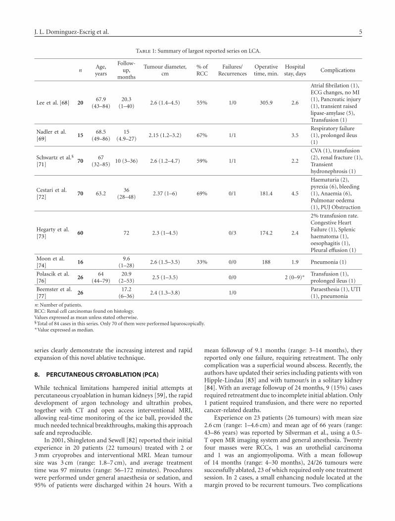
J. L. Dominguez-Escrig et al. 5
Table 1: Summary of largest reported series on LCA.
nAge,years
Follow-up,
months
Tumour diameter,cm
% ofRCC
Failures/Recurrences
Operativetime, min.
Hospitalstay, days
Complications
Lee et al. [68] 2067.9
(43–84)20.3
(1–40)2.6 (1.4–4.5) 55% 1/0 305.9 2.6
Atrial fibrilation (1),ECG changes, no MI(1), Pancreatic injury(1), transient raisedlipase-amylase (5),Transfusion (1)
Nadler et al.[69]
1568.5
(49–86)15
(4.9–27)2.15 (1.2–3.2) 67% 1/1 3.5
Respiratory failure(1), prolonged ileus(1)
Schwartz et al.§
[71]70
67(32–85)
10 (3–36) 2.6 (1.2–4.7) 59% 1/1 2.2
CVA (1), transfusion(2), renal fracture (1),Transienthydronephrosis (1)
Cestari et al.[72]
70 63.236
(28–48)2.37 (1–6) 69% 0/1 181.4 4.5
Haematuria (2),pyrexia (6), bleeding(1), Anaemia (6),Pulmonar oedema(1), PUJ Obstruction
Hegarty et al.[73]
60 72 2.3 (1–4.5) 0/3 174.2 2.4
2% transfusion rate.Congestive HeartFailure (1), Splenichaematoma (1),oesophagitis (1),Pleural effusion (1)
Moon et al.[74]
169.6
(1–28)2.6 (1.5–3.5) 33% 0/0 188 1.9 Pneumonia (1)
Polascik et al.[76]
2664
(44–79)20.9
(2–53)2.5 (1–3.5) 0/0 2 (0–9)∗
Transfusion (1),prolonged ileus (1)
Beemster et al.[77]
2617.2
(6–36)2.4 (1.3–3.8) 1/0
Paraesthesia (1), UTI(1), pneumonia
n: Number of patients.RCC: Renal cell carcinomas found on histology.Values expressed as mean unless stated otherwise.§Total of 84 cases in this series. Only 70 of them were performed laparoscopically.∗Value expressed as median.
series clearly demonstrate the increasing interest and rapidexpansion of this novel ablative technique.
8. PERCUTANEOUS CRYOABLATION (PCA)
While technical limitations hampered initial attempts atpercutaneous cryoablation in human kidneys [59], the rapiddevelopment of argon technology and ultrathin probes,together with CT and open access interventional MRI,allowing real-time monitoring of the ice ball, provided themuch needed technical breakthroughs, making this approachsafe and reproducible.
In 2001, Shingleton and Sewell [82] reported their initialexperience in 20 patients (22 tumours) treated with 2 or3 mm cryoprobes and interventional MRI. Mean tumoursize was 3 cm (range: 1.8–7 cm), and average treatmenttime was 97 minutes (range: 56–172 minutes). Procedureswere performed under general anaesthesia or sedation, and95% of patients were discharged within 24 hours. With a
mean followup of 9.1 months (range: 3–14 months), theyreported only one failure, requiring retreatment. The onlycomplication was a superficial wound abscess. Recently, theauthors have updated their series including patients with vonHipple-Lindau [83] and with tumour/s in a solitary kidney[84]. With an average followup of 24 months, 9 (15%) casesrequired retreatment due to incomplete initial ablation. Only1 patient required transfusion, and there were no reportedcancer-related deaths.
Experience on 23 patients (26 tumours) with mean size2.6 cm (range: 1–4.6 cm) and mean age of 66 years (range:43–86 years) was reported by Silverman et al., using a 0.5-T open MR imaging system and general anesthesia. Twentyfour masses were RCCs, 1 was an urothelial carcinomaand 1 was an angiomyolipoma. With a mean followupof 14 months (range: 4–30 months), 24/26 tumours weresuccessfully ablated, 23 of which required only one treatmentsession. In 2 cases, a small enhancing nodule located at themargin proved to be recurrent tumours. Two complications

6 Advances in Urology
(1 haemorrhage requiring transfusion and 1 abscess drainedpercutaneously) occurred in a total of 27 cryoablations [85].
In 2006 Gupta et al. published CT-guided PCA on 27tumours of 5 cm or less (mean size 2.5 cm), using conscioussedation and real-time CT monitoring. With 1 month ormore followup imaging available on 16 cases (mean 5.9months), 15 tumours showed no signs of enhancement. In1 case, blood transfusion was required for bleeding [86].
The Mayo Clinic experience on 40 cases of PCA withCT monitoring has recently been published [87]. Meantumour size was 4.2 cm (range: 3.0–7.2 cm) and at least 3months followup was available in 65% of the cases (mean9 months; range: 3–22 months). Technical success, definedas extension of the ice ball beyond the tumour margin andabsence of postablation enhancement on CT, was reportedin 38 (95%) cases, with no tumour recurrence or progressionin the cohort. Overall complication rate for this cohort wasreported at 8%.
9. FUTURE DIRECTIONS
Initial studies of combination therapy with 5-FU priorto freezing, indicated a temperature-dependent reductionon cell viability in a prostate cancer cell (PC-3) model[88]. Furthermore, molecular analysis using this model hasdemonstrated a synergistic effect of sublethal concentrationsof 5-FU and Cisplatin prior to freezing (−15◦C), mediatedby a shift in the Bcl-2 to Bax ratio to a prodeath tendency[89]. Similar synergistic response has been reported in arenal cell model, the data suggesting that 5-FU chemotherapymay be more effective when followed by cryosurgery [90]. Inthe clinical setting, synergistic activity of cryoablation andcyclophosphamide is currently been evaluated on advancedepithelial tumours (NCI. Trial protocol NCT00499733).
Equally, since freezing enhances the radiosentitivity ofcells, combination of radiotherapy with cryoablation maypotentially confer benefits [65], as already indicated in pre-clinical models of prostate cancer, where adjuvant radiationand curcumin have demonstrated a synergistic effect withcryoablation [91].
At the time of writing this review, the Cleveland Clinicgroup have made public the initial results employing SinglePort Access Renal Cryoablation (SPARC). A total of 6patients, with mean tumour size of 2.6± 0.4 cm, successfullyunderwent SPARC, via a transperitoneal or retroperitonealapproach, with no intraoperative complications and no needfor conversion, demonstrating the feasibility and safety ofthis, potentially scarless, procedure [92, 93].
Further development of imaging techniques and cry-oprobe technology, clinical evaluation of combination ther-apy with conventional chemo- and radiotherapy, togetherwith promising novel cryoenhancers, may have major impli-cations on the management of small renal masses in thefuture
10. CONCLUSIONS
Widespread implementation of USS, CT, and MRI hasresulted in an increased detection of early, small renal masses.
In the last 20 years, the proportion of incidentally foundrenal tumours raised from 13% to an estimated 60%, witha substantial parallel decrease in tumour stage, grade, andproportion of metastasis at presentation, in these patients[94]. As a result, urologists are now faced with a new cohortof asymptomatic, healthier patients, with incidentally foundsmall renal masses.
While open partial nephrectomy is still the referencestandard [95], its associated morbidity has encouragedresearchers and practicing clinicians towards less radicalapproaches, thus the rapid development of laparoscopicpartial nephrectomy and novel ablative techniques suchas radiofrequency ablation (RFA), high-intensity focusedultrasound (HIFU), and cryoablation (CA). Among abla-tive techniques, cryotherapy, and in particular laparoscopiccryoablation, is the most extensively studied and the one withmore rapid expansion in clinical practice.
Cryosurgery offers the clear advantage of combining anephron-sparing surgery together with a minimally invasiveapproach. Anaesthetic requirements, postoperative analge-sia, and hospital stay are significantly reduced, with a muchrapid return to normal activity and work.
In the early days of development and clinical imple-mentation of cryoablation, concerns were raised regardingsafety of the procedure, the lack of followup, and oncologicaloutcome [96].
Regarding the safety of the procedure, published studiesup to this day have shown minimal procedure-specific mor-bidity, with complication rates comparable or better thancurrent available minimally invasive procedures. Reportsfrom the largest series have demonstrated to be a less morbidprocedure than laparoscopic partial nephrectomy, with acomparable 5-year oncological safety [97].
Among the novel ablative techniques, radio frequencyablation (RFA) is the procedures with more emerging clinicaldata. Although the procedure-specific morbidity, mostlybased on small and nonstandardised series, appears to be low,serious issues have been raised regarding the RFA cell-killingpotential and its higher risk of local disease recurrence, asdemonstrated en several clinical studies [98–102].
When compared to RFA, available data from preclinical[52] and several clinical studies confer to cryoablationan advantageous oncological safety profile. The ClevelandClinic group have recently published results from their RFAand LCA series, highlighting the issue of residual diseaseand demonstrating a clear advantage in the LCA cohort.With 109 renal lesions (88 patients) treated with RFA and192 lesions (176 patients) treated with LCA, radiographic(CT or MRI) success at 6 months was 85% and 90% forRFA and LCA, respectively. More importantly, when lesionswere later biopsied at 6 months, the success in the RFAcohort decreased to 64.8%, while LCA success remainedhigh at 93.8%. Six of 13 patients (46.2%) with a 6-monthpositive biopsy after radio frequency ablation demonstratedno enhancement on posttreatment MRI or CT, while inthe LCA group, all positive biopsies revealed posttreatmentenhancement on imaging just before biopsy. The authorsrecommend postradio frequency ablation followup biopsy

J. L. Dominguez-Escrig et al. 7
due to the significant risk of residual renal cell cancer withoutradiographic evidence [103].
Supporting these findings, a recent meta-analysis ofavailable data demonstrates a higher risk of local diseaserecurrence in tumours treated with RFA, when compared tothose managed by cryoablation [104].
While long-term followup is still awaited, encouragingresults have been reported in series with up to 5-yearfollowup, with cancer-specific survival rates ranging from98 to 100% [68, 70, 72, 73, 76, 105] with LCA and 97%with PCA [7]. This is comparable to 5-year cancer-specificsurvival rate of 92%, reported with partial nephrectomy[95, 97, 106].
While clinical application and indications of cryoablationof small renal masses are still not clearly defined, it isrecommended by available clinical evidence, that CA shouldbe reserved for small (<3 cm) solid-enhancing renal massesin older patients with high operative risk. Young age, tumoursize >4 cm, hilar tumours, intrarenal tumours, and cysticlesions can be regarded as relative contraindication, whilstirreversible coagulopathy is widely accepted as an absolutecontraindication [107].
REFERENCES
[1] European Network of Cancer Registries. Eurocin version 4.0.European incidence database v2.3 ed, Lyon, 2001.
[2] B. Ljungberg, D. C. Hanbury, M. A. Kuczyk, et al., “Guide-lines on renal cell carcinoma,” European Association ofUrology Guidelines, 2007.
[3] P. Lindblad and H. O. Adami, “Kidney cancer,” in Textbookof Cancer Epidemiology, H. O. Adami, D. Hunter, and D.Trichopoulos, Eds., Oxford University Press, Oxford, UK,2002.
[4] A. J. Pantuck, A. Zisman, M. K. Rauch, and A. Belldegrun,“Incidental renal tumors,” Urology, vol. 56, no. 2, pp. 190–196, 2000.
[5] A. J. Pantuck, A. Zisman, and A. S. Belldegrun, “Thechanging natural history of renal cell carcinoma,” The Journalof Urology, vol. 166, no. 5, pp. 1611–1623, 2001.
[6] H. Van Poppel, K. Dilen, and L. Baert, “Incidental renal cellcarcinoma and nephron sparing surgery,” Current Opinion inUrology, vol. 11, no. 3, pp. 281–286, 2001.
[7] V. Mouraviev, S. Joniau, H. Van Poppel, and T. J. Polascik,“Current status of minimally invasive ablative techniques inthe treatment of small renal tumours,” European Urology, vol.51, no. 2, pp. 328–336, 2007.
[8] W.-H. Chow, S. S. Devesa, J. L. Warren, and J. F. Fraumeni Jr.,“Rising incidence of renal cell cancer in the United States,”Journal of the American Medical Association, vol. 281, no. 17,pp. 1628–1631, 1999.
[9] J. Ferlay, P. Autier, M. Boniol, M. Heanue, M. Colombet, andP. Boyle, “Estimates of the cancer incidence and mortality inEurope in 2006,” Annals of Oncology, vol. 18, no. 3, pp. 581–592, 2007.
[10] EUCAN, EUCAN 1998 estimates, 1998, http://www-dep.iarc.fr/eucan/eucan.htm.
[11] S. J. Smith, M. A. Bosniak, A. J. Megibow, D. H. Hulnick, S. C.Horii, and B. N. Raghavendra, “Renal cell carcinoma: earlierdiscovery and increased detection,” Radiology, vol. 170, no. 3,pp. 699–703, 1989.
[12] K. Perry, A. Zisman, A. J. Pantuck, N. Janzen, P. Schulam, andA. S. Belldegrun, “Laparoscopic and percutaneous ablativetechniques in the treatment of renal cell carcinoma,” Reviewsin Urology, vol. 4, no. 3, pp. 103–111, 2002.
[13] M. A. Bosniak, B. A. Birnbaum, G. A. Krinsky, and J.Waisman, “Small renal parenchymal neoplasms: furtherobservations on growth,” Radiology, vol. 197, no. 3, pp. 589–597, 1995.
[14] M. J. Wehle, D. D. Thiel, S. P. Petrou, P. R. Young, I.Frank, and N. Karsteadt, “Conservative management ofincidental contrast-enhancing renal masses as safe alternativeto invasive therapy,” Urology, vol. 64, no. 1, pp. 49–52, 2004.
[15] M. Kato, T. Suzuki, Y. Suzuki, Y. Terasawa, H. Sasano, and Y.Arai, “Natural history of small renal cell carcinoma: evalua-tion of growth rate, histological grade, cell proliferation andapoptosis,” The Journal of Urology, vol. 172, no. 3, pp. 863–866, 2004.
[16] W. Kassouf, A. G. Aprikian, M. Laplante, and S. Tanguay,“Natural history of renal masses followed expectantly,” TheJournal of Urology, vol. 171, no. 1, pp. 111–113, 2004.
[17] T. Abou Youssif, W. Kassouf, J. Steinberg, A. G. Aprikian, M.P. Laplante, and S. Tanguay, “Active surveillance for selectedpatients with renal masses: updated results with long-termfollow-up,” Cancer, vol. 110, no. 5, pp. 1010–1014, 2007.
[18] I. Frank, M. L. Blute, J. C. Cheville, C. M. Lohse, A. L.Weaver, and H. Zincke, “Solid renal tumors: an analysis ofpathological features related to tumor size,” The Journal ofUrology, vol. 170, no. 6, part 1, pp. 2217–2220, 2003.
[19] M. Remzi, M. Ozsoy, H.-C. Klingler, et al., “Are smallrenal tumors harmless? Analysis of histopathological featuresaccording to tumors 4 cm or less in diameter,” The Journal ofUrology, vol. 176, no. 3, pp. 896–899, 2006.
[20] S. Pahernik, S. Ziegler, F. Roos, S. W. Melchior, and J.W. Thuroff, “Small renal tumors: correlation of clinicaland pathological features with tumor size,” The Journal ofUrology, vol. 178, no. 2, pp. 414–417, 2007.
[21] I. S. Gill, S. F. Matin, M. M. Desai, et al., “Comparativeanalysis of laparoscopic versus open partial nephrectomy forrenal tumors in 200 patients,” The Journal of Urology, vol.170, no. 1, pp. 64–68, 2003.
[22] M. A. Bosniak, “Observation of small incidentally detectedrenal masses,” Seminars in Urologic Oncology, vol. 13, no. 4,pp. 267–272, 1995.
[23] F. F. Marshall, “Conservative management of incidentalcontrast-enhancing renal masses as safe alternative to inva-sive therapy,” The Journal of Urology, vol. 174, no. 3, pp. 868–869, 2005.
[24] E. Kouba, A. Smith, D. McRackan, E. M. Wallen, and R. S.Pruthi, “Watchful waiting for solid renal masses: insight intothe natural history and results of delayed intervention,” TheJournal of Urology, vol. 177, no. 2, pp. 466–470, 2007.
[25] A. C. Novick, S. Streem, J. E. Montie, et al., “Conservativesurgery for renal cell carcinoma: a single-center experiencewith 100 patients,” The Journal of Urology, vol. 141, no. 4, pp.835–839, 1989.
[26] A. Belldegrun, K.-H. Tsui, J. B. deKernion, and R. B.Smith, “Efficacy of nephron-sparing surgery for renal cellcarcinoma: analysis based on the new 1997 tumor-node-metastasis staging system,” Journal of Clinical Oncology, vol.17, no. 9, pp. 2868–2875, 1999.
[27] R. G. Uzzo and A. C. Novick, “Nephron sparing surgery forrenal tumors: indications, techniques and outcomes,” TheJournal of Urology, vol. 166, no. 1, pp. 6–18, 2001.

8 Advances in Urology
[28] A. C. Novick, “Nephron-sparing surgery for renal cellcarcinoma,” Annual Review of Medicine, vol. 53, pp. 393–407,2002.
[29] M. M. Desai, M. Aron, and I. S. Gill, “Laparoscopic partialnephrectomy versus laparoscopic cryoablation for the smallrenal tumor,” Urology, vol. 66, no. 5, supplement 1, pp. 23–28, 2005.
[30] J. Arnott, “Practical illustrations of the remedial efficacy ofa very low or anaesthetic temperature—I: in cancer,” TheLancet, vol. 56, no. 1409, pp. 257–259, 1850.
[31] I. S. Cooper and A. J. Lee, “Cryostatic congelation: a systemfor producing a limited, controlled region of cooling orfreezing of biological tissue,” The Journal of Nervous andMental Disease, vol. 133, no. 3, pp. 259–263, 1961.
[32] A. A. Gage, S. Koepf, D. Wehrle, and F. Emmings, “Cryother-apy for cancer of the lip and oral cavity,” Cancer, vol. 18, no.12, pp. 1646–1651, 1965.
[33] I. S. Cooper, “Cryogenic surgery for cancer,” FederationProceedings, vol. 24, pp. S237–S240, 1965.
[34] W. Cahan, “Cryosurgery of malignant and benign tumors,”Federation Proceedings, vol. 24, pp. S241–S248, 1965.
[35] G. M. Onik, G. Reyes, J. K. Cohen, and B. Porterfield,“Ultrasound characteristics of renal cryosurgery,” Urology,vol. 42, no. 2, pp. 212–215, 1993.
[36] J. Baust, A. A. Gage, H. Ma, and C.-M. Zhang, “Minimallyinvasive cryosurgery—technological advances,” Cryobiology,vol. 34, no. 4, pp. 373–384, 1997.
[37] A. A. Gage and J. Baust, “Mechanisms of tissue injury incryosurgery,” Cryobiology, vol. 37, no. 3, pp. 171–186, 1998.
[38] W. K. Berger and B. Uhrık, “Freeze-induced shrinkage ofindividual cells and cell-to-cell propagation of intracellularice in cell chains from salivary glands,” Cellular and MolecularLife Sciences, vol. 52, no. 9, pp. 843–850, 1996.
[39] D. K. Whittaker, “Ice crystals formed in tissue duringcryosurgery—II: electron microscopy,” Cryobiology, vol. 11,no. 3, pp. 202–217, 1974.
[40] D. K. Whittaker, “Vascular responses in the oral mucosafollowing cryosurgery,” Journal of Periodontal Research, vol.12, no. 1, pp. 55–63, 1977.
[41] D. K. Whittaker, “Mechanisms of tissue destruction followingcryosurgery,” Annals of the Royal College of Surgeons ofEngland, vol. 66, no. 5, pp. 313–318, 1984.
[42] W. A. Nagle, B. L. Soloff, A. J. Moss Jr., and K. J. Henle,“Cultured Chinese hamster cells undergo apoptosis afterexposure to cold but nonfreezing temperatures,” Cryobiology,vol. 27, no. 4, pp. 439–451, 1990.
[43] J. G. Baust, A. A. Gage, D. Clarke, J. M. Baust, and R. VanBuskirk, “Cryosurgery—a putative approach to molecular-based optimization,” Cryobiology, vol. 48, no. 2, pp. 190–204,2004.
[44] J. T. Bishoff, R. B. Chen, B. R. Lee, et al., “Laparoscopic renalcryoablation: acute and long-term clinical, radiographic, andpathologic effects in an animal model and application in aclinical trial,” Journal of Endourology, vol. 13, no. 4, pp. 233–239, 1999.
[45] M. L. Woolley, D. A. Schulsinger, D. B. Durand, I. S. Zeltser,and W. C. Waltzer, “Effect of freezing parameters (freezecycle and thaw process) on tissue destruction following renalcryoablation,” Journal of Endourology, vol. 16, no. 7, pp. 519–522, 2002.
[46] T. L. Jang, R. Wang, S. C. Kim, T. Troe, M. R. Pins, andR. B. Nadler, “Histopathology of human renal tumors afterlaparoscopic renal cryosurgery,” The Journal of Urology, vol.173, no. 3, pp. 720–724, 2005.
[47] T. B. Edmunds Jr., D. A. Schulsinger, D. B. Durand, and W.C. Waltzer, “Acute histologic changes in human renal tumorsafter cryoablation,” Journal of Endourology, vol. 14, no. 2, pp.139–143, 2000.
[48] S. G. Chosy, S. Y. Nakada, F. T. Lee Jr., and T. F. Warner,“Monitoring renal cryosurgery: predictors of tissue necrosisin swine,” The Journal of Urology, vol. 159, no. 4, pp. 1370–1374, 1998.
[49] S. C. Campbell, V. Krishnamurthi, G. Chow, J. Hale, J.Myles, and A. C. Novick, “Renal cryosurgery: experimentalevaluation of treatment parameters,” Urology, vol. 52, no. 1,pp. 29–34, 1998.
[50] S. Y. Nakada, F. T. Lee Jr., T. Warner, S. G. Chosy, and T.D. Moon, “Laparoscopic cryosurgery of the kidney in theporcine model: an acute histological study,” Urology, vol. 51,no. 5, supplement 1, pp. 161–166, 1998.
[51] S. Y. Nakada, F. T. Lee Jr., T. F. Warner, S. G. Chosy,and T. D. Moon, “Laparoscopic renal cryotherapy in swine:comparison of puncture cryotherapy preceded by arterialembolization and contact cryotherapy,” Journal of Endourol-ogy, vol. 12, no. 6, pp. 567–573, 1998.
[52] W. C. Collyer, J. Landman, E. O. Olweny, et al., “Comparisonof renal ablation with cryotherapy, dry radiofrequency,and saline augmented radiofrequency in a porcine model,”Journal of the American College of Surgeons, vol. 193, no. 5,pp. 505–513, 2001.
[53] B. K. Auge, R. W. Santa-Cruz, and T. J. Polascik, “Effectof freeze time during renal cryoablation: a swine model,”Journal of Endourology, vol. 20, no. 12, pp. 1101–1105, 2006.
[54] A. Finelli, J. C. Rewcastle, and M. A. S. Jewett, “Cryotherapyand radiofrequency ablation: pathophysiologic basis andlaboratory studies,” Current Opinion in Urology, vol. 13, no.3, pp. 187–191, 2003.
[55] C.-H. Lin, A. Moinzadeh, A. P. Ramani, and I. S. Gill,“Histopathologic confirmation of complete cancer cell kill inexcised specimens after renal cryotherapy,” Urology, vol. 64,no. 3, p. 590, 2004.
[56] T. R. Larson, D. W. Rrobertson, A. Corica, and D. G.Bostwick, “In vivo interstitial temperature mapping of thehuman prostate during cryosurgery with correlation tohistopathologic outcomes,” Urology, vol. 55, no. 4, pp. 547–552, 2000.
[57] W. Lutzeyer, S. Lymberopoulos, H. Breining, and S.Langer, “Experimentelle kryochirurgie der niere,” Langen-beck’s Archives of Surgery, vol. 322, no. 1, pp. 843–847, 1968.
[58] W. Lutzeyer, “Fortschritte in der operativen Therapie (Urolo-gie),” Langenbeck’s Archives of Surgery, vol. 332, no. 1, pp.137–145, 1972.
[59] M. Uchida, Y. Imaide, K. Sugimoto, H. Uehara, and H.Watanabe, “Percutaneous cryosurgery for renal tumours,”British Journal of Urology, vol. 75, no. 2, pp. 132–137, 1995.
[60] M. G. Delworth, L. L. Pisters, B. D. Fornage, and A. C.von Eschenbach, “Cryotherapy for renal cell carcinoma andangiomyolipoma,” The Journal of Urology, vol. 155, no. 1, pp.252–255, 1996.
[61] D. B. Rukstalis, M. Khorsandi, F. U. Garcia, D. M. Hoenig,and J. K. Cohen, “Clinical experience with open renalcryoablation,” Urology, vol. 57, no. 1, pp. 34–39, 2001.
[62] M. Khorsandi, R. C. Foy, W. Chong, D. M. Hoenig, J. K.Cohen, and D. B. Rukstalis, “Preliminary experience withcryoablation of renal lesions smaller than 4 centimeters,” TheJournal of the American Osteopathic Association, vol. 102, no.5, pp. 277–281, 2002.

J. L. Dominguez-Escrig et al. 9
[63] R. Rodriguez, D. Y. Chan, J. T. Bishoff, et al., “Renalablative cryosurgery in selected patients with peripheral renalmasses,” Urology, vol. 55, no. 1, pp. 25–30, 2000.
[64] A. J. Pantuck, A. Zisman, J. Cohen, and A. Belldegrun,“Cryosurgical ablation of renal tumors using 1.5-millimeter,ultrathin cryoprobes,” Urology, vol. 59, no. 1, pp. 130–133,2002.
[65] I. S. Gill, “Renal cryotherapy: pro,” Urology, vol. 65, no. 3, pp.415–418, 2005.
[66] J. G. Pattaras and F. F. Marshall, “Percutaneous cryoablationof renal tumors: limitations and uncertainties,” NatureClinical Practice Urology, vol. 2, no. 11, pp. 518–519, 2005.
[67] S. F. Matin, K. Ahrar, J. A. Cadeddu, et al., “Residual andrecurrent disease following renal energy ablative therapy: amulti-institutional study,” The Journal of Urology, vol. 176,no. 5, pp. 1973–1977, 2006.
[68] D. I. Lee, D. E. McGinnis, R. Feld, and S. E. Strup,“Retroperitoneal laparoscopic cryoablation of small renaltumors: intermediate results,” Urology, vol. 61, no. 1, pp. 83–88, 2003.
[69] R. B. Nadler, S. C. Kim, J. N. Rubenstein, R. L. Yap, S. C.Campbell, and H. M. User, “Laparoscopic renal cryosurgery:the Northwestern experience,” The Journal of Urology, vol.170, no. 4, part 1, pp. 1121–1125, 2003.
[70] T. Powell, C. Whelan, and B. F. Schwartz, “Laparoscopic renalcryotherapy: biology, techniques and outcomes,” MinervaUrologica e Nefrologica, vol. 57, no. 2, pp. 109–118, 2005.
[71] B. F. Schwartz, J. C. Rewcastle, T. Powell, C. Whelan, T.Manny Jr., and J. C. Vestal, “Cryoablation of small peripheralrenal masses: a retrospective analysis,” Urology, vol. 68, no. 1,supplement 1, pp. 14–18, 2006.
[72] A. Cestari, G. Guazzoni, R. Naspro, T. Maga, V. Dell’acqua,and P. Rigatti, “Laparoscopic renal cryoablation (LRC) ofsmall renal masses: lesson learned after 70 procedures,”European Urology Supplements, vol. 5, no. 2, p. 220, 2006.
[73] N. J. Hegarty, I. S. Gill, J. H. Kaouk, et al., “Renalcryoablation: 5 year outcomes,” The Journal of Urology, vol.175, p. 351, 2006.
[74] T. D. Moon, F. T. Lee Jr., S. P. Hedican, P. Lowry, and S.Y. Nakada, “Laparoscopic cryoablation under sonographicguidance for the treatment of small renal tumors,” Journalof Endourology, vol. 18, no. 5, pp. 436–440, 2004.
[75] G. Bandi, C. C. Wen, S. P. Hedican, T. D. Moon, F. T.Lee Jr., and S. Y. Nakada, “Cryoablation of small renalmasses: assessment of the outcome at one institution,” BJUInternational, vol. 100, no. 4, pp. 798–801, 2007.
[76] T. J. Polascik, I. Nosnik, J. M. Mayes, and V. Mouraviev,“Short term clinical outcome after laparoscopic cryoablationof the renal tumor ≤ 3.5 cm,” Technology in Cancer Research& Treatment, vol. 6, no. 6, pp. 621–624, 2007.
[77] P. Beemster, S. Phoa, H. Wijkstra, J. de la Rosette, andP. Laguna, “Follow-up of renal masses after cryosurgeryusing computed tomography; enhancement patterns andcryolesion size,” BJU International, vol. 101, no. 10, pp. 1237–1242, 2008.
[78] I. Colon and G. J. Fuchs, “Early experience with laparoscopiccryoablation in patients with small renal tumors and severecomorbidities,” Journal of Endourology, vol. 17, no. 6, pp.415–423, 2003.
[79] J. L. Gore, H. L. Kim, and P. Schulam, “Initial experience withlaparoscopically assisted percutaneous cryotherapy of renaltumors,” Journal of Endourology, vol. 19, no. 4, pp. 480–483,2005.
[80] A. Bachmann, T. Sulser, C. Jayet, et al., “Retroperiton-eoscopy-assisted cryoablation of renal tumors using multiple1.5 mm ultrathin cryoprobes: a preliminary report,” Euro-pean Urology, vol. 47, no. 4, pp. 474–479, 2005.
[81] S. F. Wyler, T. Sulser, R. Ruszat, et al., “Intermediate-term results of retroperitoneoscopy-assisted cryotherapy forsmall renal tumours using multiple ultrathin cryoprobes,”European Urology, vol. 51, no. 4, pp. 971–979, 2007.
[82] W. B. Shingleton and P. E. Sewell Jr., “Percutaneous renaltumor cryoablation with magnetic resonance imaging guid-ance,” The Journal of Urology, vol. 165, no. 3, pp. 773–776,2001.
[83] W. B. Shingleton and P. E. Sewell Jr., “Percutaneous renalcryoablation of renal tumors in patients with von Hippel-Lindau disease,” The Journal of Urology, vol. 167, no. 3, pp.1268–1270, 2002.
[84] W. B. Shingleton and P. E. Sewell Jr., “Cryoablation of renaltumours in patients with solitary kidneys,” BJU International,vol. 92, no. 3, pp. 237–239, 2003.
[85] S. G. Silverman, K. Tuncali, E. vanSonnenberg, et al., “Renaltumors: MR imaging-guided percutaneous cryotherapy—initial experience in 23 patients,” Radiology, vol. 236, no. 2,pp. 716–724, 2005.
[86] A. Gupta, M. E. Allaf, L. R. Kavoussi, et al., “Computerizedtomography guided percutaneous renal cryoablation withthe patient under conscious sedation: initial clinical experi-ence,” The Journal of Urology, vol. 175, no. 2, pp. 447–453,2006.
[87] T. D. Atwell, M. A. Farrell, M. R. Callstrom, et al.,“Percutaneous cryoablation of large renal masses: technicalfeasibility and short-term outcome,” American Journal ofRoentgenology, vol. 188, no. 5, pp. 1195–1200, 2007.
[88] D. M. Clarke, J. M. Baust, R. G. Van Buskirk, and J. G. Baust,“Chemo-cryo combination therapy: an adjunctive model forthe treatment of prostate cancer,” Cryobiology, vol. 42, no. 4,pp. 274–285, 2001.
[89] D. M. Clarke, J. M. Baust, R. G. Van Buskirk, and J. G. Baust,“Addition of anticancer agents enhances freezing-inducedprostate cancer cell death: implications of mitochondrialinvolvement,” Cryobiology, vol. 49, no. 1, pp. 45–61, 2004.
[90] D. M. Clarke, W. R. Hollister, J. G. Baust, and R. G. VanBuskirk, “Cryosurgical modeling: sequence of freezing andcytotoxic agent application affects cell death,” MolecularUrology, vol. 3, no. 1, pp. 25–31, 1999.
[91] M. A. K. Kenneson, D. B. Rukstalis, M. Ahmed, and S.Boyer, “Novel adjuvants of cryotherapy to enhance direct andbystander killing of human prostate cancer cells (PC3),” inProceedings of the 64th Annual Meeting of the Mid-AtlanticSection of the American Urological Association (MAAUA ’06),Capitol Hill, Wash, USA, October 2006.
[92] J. H. Kaouk, G.-P. Haber, R. K. Goel, et al., “Single-portlaparoscopic surgery in urology: initial experience,” Urology,vol. 71, no. 1, pp. 3–6, 2008.
[93] R. K. Goel and J. H. Kaouk, “Single port access renalcryoablation (SPARC): a new approach,” European Urology,vol. 53, no. 6, pp. 1204–1209, 2008.
[94] L. G. Luciani, R. Cestari, and C. Tallarigo, “Incidental renalcell carcinoma—age and stage characterization and clinicalimplications: study of 1092 patients (1982–1997),” Urology,vol. 56, no. 1, pp. 58–62, 2000.
[95] A. F. Fergany, K. S. Hafez, and A. C. Novick, “Long-termresults of nephron sparing surgery for localized renal cellcarcinoma: 10-year followup,” The Journal of Urology, vol.163, no. 2, pp. 442–445, 2000.

10 Advances in Urology
[96] A. Sorcini and J. A. Libertino, “Cryotherapy for small renalcell tumors: CON,” Urology, vol. 53, no. 6, pp. 1079–1081,1999.
[97] Y.-C. Lin, B. Turna, R. Frota, et al., “Laparoscopic partialnephrectomy versus laparoscopic cryoablation for multipleipsilateral renal tumors,” European Urology, vol. 53, no. 6, pp.1210–1218, 2008.
[98] R. A. Rendon, J. R. Kachura, J. M. Sweet, et al., “Theuncertainty of radio frequency treatment of renal cellcarcinoma: findings at immediate and delayed nephrectomy,”The Journal of Urology, vol. 167, no. 4, pp. 1587–1592, 2002.
[99] M. J. Michaels, H. K. Rhee, A. P. Mourtzinos, I. C. Sum-merhayes, M. L. Silverman, and J. A. Libertino, “Incompleterenal tumor destruction using radio frequency interstitialablation,” The Journal of Urology, vol. 168, no. 6, pp. 2406–2410, 2002.
[100] B. R. Matlaga, R. J. Zagoria, R. D. Woodruff, F. M. Torti, andM. C. Hall, “Phase II trial of radio frequency ablation of renalcancer: evaluation of the kill zone,” The Journal of Urology,vol. 168, no. 6, pp. 2401–2405, 2002.
[101] I. M. Varkarakis, M. E. Allaf, T. Inagaki, et al., “Percutaneousradio frequency ablation of renal masses: results at a 2-yearmean followup,” The Journal of Urology, vol. 174, no. 2, pp.456–460, 2005.
[102] R. G. Uzzo, “Is CT-guided percutaneous radiofrequencyablation oncologically effective in patients with renal cellcarcinoma?” Nature Clinical Practice Urology, vol. 5, no. 1,pp. 18–19, 2008.
[103] C. J. Weight, J. H. Kaouk, N. J. Hegarty, et al., “Correlation ofradiographic imaging and histopathology following cryoab-lation and radio frequency ablation for renal tumors,” TheJournal of Urology, vol. 179, no. 4, pp. 1277–1283, 2008.
[104] D. A. Kunkle, B. L. Egleston, and R. G. Uzzo, “Excise, ablateor observe: the small renal mass dilemma—a meta-analysisand review,” The Journal of Urology, vol. 179, no. 4, pp. 1227–1234, 2008.
[105] I. S. Gill, E. M. Remer, W. A. Hasan, et al., “Renalcryoablation: outcome at 3 years,” The Journal of Urology, vol.173, no. 6, pp. 1903–1907, 2005.
[106] K. S. Hafez, A. F. Fergany, and A. C. Novick, “Nephronsparing surgery for localized renal cell carcinoma: impact oftumor size on patient survival, tumor recurrence and TNMstaging,” The Journal of Urology, vol. 162, no. 6, pp. 1930–1933, 1999.
[107] M. Aron and I. S. Gill, “Minimally invasive nephron-sparingsurgery (MINSS) for renal tumours—part II: probe ablativetherapy,” European Urology, vol. 51, no. 2, pp. 348–357, 2007.

Hindawi Publishing CorporationAdvances in UrologyVolume 2008, Article ID 197324, 3 pagesdoi:10.1155/2008/197324
Review ArticleCryoablation of Small Renal Tumors in Patients withSolitary Kidneys: Initial Experience
Ravi Munver,1, 2, 3, 4 Grant I. S. Disick,1 Salvatore A. Lombardo,1, 4 Vladislav G. Bargman,1, 4
and Ihor S. Sawczuk1, 2, 3, 5
1 Department of Urology, Hackensack University Medical Center, 360 Essex Street, Suite 403, Hackensack, NJ 07601, USA2 Department of Urology, Touro University College of Medicine, 360 Essex Street, Suite 403, Hackensack, NJ 07601, USA3 The John Theurer Cancer Center at Hackensack University Medical Center, 20 Prospect Avenue, Hackensack, NJ 07601, USA4 Division of Urology, Department of Surgery, UMDNJ-New Jersey Medical School, 185 South Orange Avenue,Newark, NJ 07103, USA
5 Department of Urology, College of Physicians and Surgeons, Columbia University, 161 Fort Washington Avenue,New York, NY 10032, USA
Correspondence should be addressed to Ravi Munver, [email protected]
Received 1 May 2008; Accepted 28 August 2008
Recommended by J. Rubio
Introduction. The purpose of this study was to evaluate the role of renal cryoablation in patients with solitary kidneys with the goalsof tumor destruction and maximal renal parenchymal preservation. Methods. Eleven patients with single tumors were treated withcryoablation, of which 10 patients had solitary kidneys and 1 had a nonfunctioning contralateral kidney. All procedures wereperformed via an open extraperitoneal approach; ten tumors were treated with in-situ cryoablation and 1 tumor was treatedwith cryo-assisted partial nephrectomy. Results. Cryoablation was successfully performed without any preoperative complications.Mean patient age was 62.4 years (range 49–79), tumor location included: 6 (upper pole), 2 (mid-kidney), 3 (lower pole). The meanand median tumor size was 2.6 cm and 2.8 cm (range 1.2–4.3 cm), mean operative time 205 minutes (range 180–270 minutes),blood loss 98.5 ml (range 40–250 ml), and hospitalization 4.6 days (range 3–8 days). Creatinine values included: preoperative1.43 mg/dL (range 1.2–1.9), postoperative 1.67 mg/dL (range 1.5–2.5), and nadir 1.57 mg/dL (range 1.3–2.1). All patients werefollowed postoperatively with magnetic resonance imaging for surveillance. At a median follow-up of 43 months, 9 patientshad no evidence of recurrence, 1 patient has an enhancing indeterminate area, and 1 patient was lost to follow-up. Conclusion.Intermediate-term results suggest that renal cryoablation offers a feasible alternative for patients that require a maximal nephron-sparing effort with preservation of renal function and minimal risk of tumor recurrence.
Copyright © 2008 Ravi Munver et al. This is an open access article distributed under the Creative Commons Attribution License,which permits unrestricted use, distribution, and reproduction in any medium, provided the original work is properly cited.
1. INTRODUCTION
Nephron-sparing surgery (NSS) entails complete resectionor destruction of a renal tumor while maximizing preserva-tion of normal parenchymal tissue. Improvements in surgicaltechniques have gradually led to more widespread utiliza-tion of partial nephrectomy with acceptable postoperativemorbidity and equivalent oncologic efficacy as comparedto radical nephrectomy. Cryoablation is an alternative topartial nephrectomy for the treatment of renal tumors andit employs the concept of nephron-sparing surgery [1, 2].
The purpose of this study was to evaluate cryoablation asan NSS technique for the treatment of small renal masses inpatients with solitary kidneys. We reviewed the application,
effect on renal function, and intermediate outcomes ofcryoablation in this subset of patients that require maximalrenal parenchyma preservation while fulfilling the goal oftumor destruction.
2. MATERIALS AND METHODS
Between August 2000 and November 2004, 11 patients (9male, 2 female) were treated with renal cryoablation forsuspicious renal lesions. All patients had a single renalmass suspicious for malignancy based on radiologic imagingstudies. Ten patients had a solitary kidney and 1 had a non-functioning contralateral kidney. Cryoablation was selected

2 Advances in Urology
Figure 1: Open renal cryoablation. Cryoprobe with 2 temperatureprobes are seen.
in order to offer patients a nephron-sparing procedure incases that, due to tumor location, were not ammenable topartial nephrectomy. Patients did not receive preoperativebiopsy of the renal mass lesion due to the associated risk ofbleeding and renal injury in patients with solitary kidneys.Ten tumors were treated with insitu cryoablation and 1tumor was treated with cryoassisted partial nephrectomy.
An extraperitoneal flank incision was made between the10th and 11th ribs allowing exposure of the kidney and renaltumor. Intraoperative high-resolution renal ultrasonographywith a 7.5 MHz transducer was used to establish and confirmtumor size, depth of invasion, and proximity to the renalhilar vessels and the collecting system. The renal hilar vesselswere isolated but not occluded in all cases. Each tumor wasbiopsied with a 14gauge Tru-Cut needle prior to initiation ofcyroablation.
Cryoablation was performed using the Cryocare surgicalsystem (Endocare Inc., Irvine, Calif, USA). Under ultrasoundguidance, 1 or 2 cryoprobes were placed directly into theidentified lesion, and temperature sensor probes were placedat the periphery of each mass to provide intraoperative mon-itoring of adjacent renal parenchymal temperatures. In eachcase, the tumor margins were localized with intraoperativeultrasound, allowing for precise placement of the cryoprobes.After cryoprobe placement, the tumor was treated with 2freeze cycles (−40C◦ for 10–15 minutes/cycle) (see Figure 1).Each freeze cycle was followed by an active thaw process. Inthe case of the partial nephrectomy, the tumor was excisedwith a scalpel by tracing the edge of the ice ball.
Perioperative data were evaluated including tumor size,operative time, estimated blood loss, length of hospital stay,and preoperative and postoperative creatinine.
3. RESULTS
Eleven patients were treated with cryoablation for 5 rightrenal tumors and 6 left renal tumors. The mean patient agewas 62.4 years (range 49–79 years). Tumors were locatedin the upper pole (n = 6), mid-kidney (n = 2), and
lower pole (n = 3) with a mean and median tumorsize of 2.6 cm and 2.8 cm, respectively (range 1.2–4.3 cm).The mean operative time was 205 minutes (range 180–270 minutes), blood loss 98.5 mL (range 40–250 mL), andhospitalization 4.6 days (range 3–8 days). The procedurewas successfully completed in all patients without any majorintraoperative or postoperative complications.
Biopsies of the 11 lesions confirmed renal cell carcinoma(n = 7), oncocytoma (n = 2), and angiomyolipoma (n =1), with one biopsy specimen that was indeterminate. Thepatient that underwent cryoassisted partial nephrectomy hadnegative margins.
The mean preoperative creatinine was 1.43 mg/dL (range1.2–1.9) and postoperative creatinine was 1.67 mg/dL (range1.5–2.5). The nadir creatinine was 1.57 mg/dL (range 1.3–2.1). All patients were followed postoperatively with mag-netic resonance imaging (MRI) at 3–6 month intervals.Imaging these patients within the first 3 months was notperformed due to our prior experience with inflammatoryresponses in the treated area that can lead to misinterpreta-tion. At a median follow-up of 43 months (4–59 months),9 patients had no evidence of recurrence, 1 patient had anindeterminate area, and 1 patient was lost to follow-up after4 months.
4. DISCUSSION
Cryoablation is a minimally invasive technique that hasemerged as an option for small renal masses with reducedmorbidity compared to partial nephrectomy. This technol-ogy provides a nephron-sparing alternative that is curativeby destruction rather than excision of the renal mass [3].
Tissue destruction from cryoablative therapy occursfrom sequential freezing and thawing of tissues. Cellulardestruction from the freeze process results from complexdirect and indirect physiologic mechanisms, including directphysical disruption of the cellular membranes, proteins,and intracellular organelles from ice crystals. In addition,there are indirect effects such as microvascular thrombosis,osmotic dehydration, and cellular anoxia during the freezeprocess, which may also extend beyond the physical iceball. The initial histologic change noted after a cryoablativeprocedure is coagulative necrosis. Subsequently, chronicfibrosis with collagen deposition results [2].
Early animal studies by Nakada et al. utilizing and invivo rabbit renal cell cancer tumor model demonstrated thatthermosensor-monitored renal cryosurgery produces similaroutcomes to nephrectomy in terms of preventing metastaticdisease [4]. Rodriguez et al. published preliminary results ofseries of seven patients undergoing renal cryoablation [5].The estimated blood loss averaged 111 mL and there wereno perioperative complications. Six of the 7 patients hada minimum of one follow-up computed tomography scan(mean follow-up of 14.2 months) and each of these studiesdemonstrated partial resolution of the lesion.
Rukstalis et al. reviewed a cohort of 29 patients that weretreated with open renal cryoablation since 1996 [6]. Themedian preoperative renal mass size was 2.2 cm, of which22 were solid renal masses and 7 were complex renal lesions.

Ravi Munver et al. 3
Five major adverse events occurred of which only one eventwas directly related to the procedure. At median follow-up of 16 months, 1 patient experienced a biopsy-provenlocal recurrence, and 91.3% of patients had a completeradiographic response with only a residual scar or smallnonenhancing cyst. The authors concluded that open renalcryoablation appeared safe for the destruction of solid orcomplex renal masses, although rigorous radiographic, andclinical follow-up was required.
Chen et al. reported their experience with laparoscopiccryoablation of renal masses in 35 patients that underwentsuccessful therapy with minimal postoperative complications[7]. In their series, the mean operative time was 3 hoursand mean estimated blood loss was 85 mL. At 11 months offollow-up, there were no local or port site tumor recurrences.In a similar study by Gill et al., 32 patients underwentlaparoscopic renal cryoablation. In this study, there were nolocal tumor recurrences in this group of patients [8].
Cryoablation is becoming an increasingly popular mini-mally invasive technique for treating renal cell carcinoma andhas been shown to effectively treat renal and adrenal masses[8, 9]. This technique appears to be safe and efficacious, withrecurrence rate reported as low as 6.7%, and a 5year cancer-specific survival rate of 100% for RCC [10–12].
Patients with solitary kidneys or impaired renal functionmay benefit from renal cryoablation as compared to partialnephrectomy for several reasons. Cryoablation may beassociated with a lower risk for bleeding and may obviatethe need for hilar occlusion, thus preventing the detrimentaleffects related to controlled ischemia. Our patients that wereconsidered to be appropriate candidates for cryoablationdid not require hilar occlusion of their solitary renal unit.While partial nephrectomy can be selectively performedwithout warm ischemia, the patients in our series had tumorcharacteristics that were not optimal for this procedurewithout hilar occlusion.
In our early experience with cryoablation for patientswith solitary kidneys, each patient underwent an open pro-cedure in order to minimize inadvertent injury and confirmadequate placement of the cryoprobes. Since our initialexperience, we have continued our efforts to further decreasemorbidity by transitioning to a laparoscopic approach forsuitable tumors in this cohort of patients.
Intraoperative ultrasound was essential in delineating theintrarenal anatomy and the dimensions of the tumor [13].Moreover, ultrasonography allows visualization of the iceball to confirm adequate treatment of the entire lesion. Inthe patient that underwent cryoassisted partial nephrectomy,the tumor was excised following the freeze cycle with theintention of minimizing blood loss, as hilar occlusion wasnot employed. Utilization of the edge of the ice ball served asa guide for resection and facilitated complete excision of themass with minimal blood loss.
Renal cryoablation has been reported to target kidneytumors in a precise, safe, and reproducible manner. Thistechnology offers the ability to treat renal tumors in patientsthat require a maximal parenchymal sparing procedure,such as patients with solitary kidneys. Renal cryoablationallows the accurate and safe application of this surgical
modality for the treatment of renal tumors with emphasison parenchymal sparing. Additionally, the need for hilarocclusion of a solitary renal unit is completely obviated. Ourintermediate follow-up data is promising in terms of bothcancer control and preservation of renal function. We donot routinely perform renal biopsies following cryoablationdue to the associated risks of bleeding and renal injury inpatients with solitary kidneys. Instead, this practice can bereserved for patients with a suspicious enhancing area onfollow-up surveillance imaging studies. If a positive biopsyis obtained in this instance, the options for surveillance,repeat cryoablation, or nephrectomy may be considered. Ourresults are consistent with other reports in the literature anddemonstrate that renal cryoablation is a feasible techniquefor the management of small renal masses in patients withsolitary kidneys. Additionally, combined with a laparoscopicapproach, cryoablation is an attractive minimally invasivetreatment option in such patients.
REFERENCES
[1] I. S. Sawczuk and M. A. Ghafar, “Cryotherapeutic assistedpartial nephrectomy for renal cell cancer,” UrolOncology, vol.1, pp. 285–290, 2001.
[2] Y. D. Chan, R. Rodriguez, and F. F. Marshall, “Renalcryosurgery,” AUA Update, Vol. 19, Lesson 18, 2001.
[3] N. E. Hoffmann and J. C. Bischof, “The cryobiology ofcryosurgical injury,” Urology, vol. 60, no. 2, supplement 1, pp.40–49, 2002.
[4] S. Y. Nakada, T. J. Jerde, F. T. Lee Jr., and T. Warner, “Efficacyof cryotherapy and nephrectomy in treating implanted VX-2carcinoma in rabbit kidney,” Journal of Endourology, vol. 13,supplement A13, 1999.
[5] R. Rodriguez, D. Y. Chan, J. T. Bishoff, et al., “Renal ablativecryosurgery in selected patients with peripheral renal masses,”Urology, vol. 55, no. 1, pp. 25–30, 2000.
[6] D. B. Rukstalis, M. Khorsandi, F. U. Garcia, D. M. Hoenig,and J. K. Cohen, “Clinical experience with open renalcryoablation,” Urology, vol. 57, no. 1, pp. 34–39, 2001.
[7] R. N. Chen, A. C. Novick, and I. S. Gill, “Laparoscopic cryoab-lation of renal masses,” Urologic Clinics of North America, vol.27, no. 4, pp. 813–820, 2000.
[8] I. S. Gill, A. C. Novick, A. M. Meraney, et al., “Laparoscopicrenal cryoablation in 32 patients,” Urology, vol. 56, no. 5, pp.748–753, 2000.
[9] R. Munver and R. E. Sosa, “Cryosurgery of the adrenal gland,”Technology in Cancer Research and Treatment, vol. 3, no. 2, pp.181–186, 2004.
[10] S. C. Campbell, A. C. Novick, S. B. Streem, E. Klein, and M.Licht, “Complications of nephron sparing surgery for renaltumors,” The Journal of Urology, vol. 151, no. 5, pp. 1177–1180,1994.
[11] N. J. Hegarty, I. S. Gill, J. H. Kaouk, et al., “Renal cryoablation:5 years outcomes,” The Journal of Urology, vol. 175, p. 351,2006.
[12] M. Spaliviero, A. Moinzadeh, and I. S. Gill, “Laparoscopiccryotherapy for renal tumors,” Technology in Cancer Researchand Treatment, vol. 3, no. 2, pp. 177–180, 2004.
[13] H. G. Zegel, G. A. Holland, S. B. Jennings, W. K. Chong,and J. K. Cohen, “Intraoperative ultrasonographically guidedcryoablation of renal masses: initial experience,” Journal ofUltrasound in Medicine, vol. 17, no. 9, pp. 571–576, 1998.

Hindawi Publishing CorporationAdvances in UrologyVolume 2008, Article ID 809845, 5 pagesdoi:10.1155/2008/809845
Review ArticleHigh-Intensity Focused Ultrasound in Small Renal Masses
Jose Rubio Briones, Argimiro Collado Serra, Alvaro Gomez-Ferrer Lozano, Juan Casanova Ramon-Borja,Inmaculada Iborra Juan, and Eduardo Solsona Narbon
Servicio de Urologıa, Instituto Valenciano de Oncologıa, C/Beltran Baguena 8, 46009 Valencia, Spain
Correspondence should be addressed to Jose Rubio Briones, [email protected]
Received 26 March 2008; Revised 6 October 2008; Accepted 4 November 2008
Recommended by F. Algaba
High-intensity focused ultrasound (HIFU) competes with radiofrequency and cryotherapy for the treatment of small renal massesas a third option among ablative approaches. As an emerging technique, its possible percutaneous or laparoscopic application,low discomfort to the patient and the absence of complications make this technology attractive for the management of smallrenal masses. This manuscript will focus on the principles, basic research and clinical applications of HIFU in small renal masses,reviewing the present literature. Therapeutic results are controversial and from an clinical view, HIFU must be considered a tech-nique under investigation at present time. Further research is needed to settle its real indications in the management of small renalmasses; maybe technical improvements will certainly facilitate its use in the management of small renal masses in the near future.
Copyright © 2008 Jose Rubio Briones et al. This is an open access article distributed under the Creative Commons AttributionLicense, which permits unrestricted use, distribution, and reproduction in any medium, provided the original work is properlycited.
1. INTRODUCTION
We are facing a rapid increase of incidentally detected smallrenal masses (SRMs) nowadays [1, 2], prompting us to facemany different clinical scenarios and probably minimallyinvasive ablative techniques will find their role in those unfitpatients who are not operable and do not accept a partialnephrectomy or a watchful waiting policy [3] in case ofa possible renal cell carcinoma (RCC) diagnosed probablyincidentally [4].
High-intensity focused ultrasound (HIFU) induces ther-mal damage to the targeted tissue without the need of theinsertion of a probe into the tissue, thus being the most real“minimally invasive” proposed technique among the ablativetreatments for small renal masses (SRMs).
This manuscript will focus on the principles, basicresearch, and clinical applications of HIFU in small renalmasses, reviewing the present literature and analyzing HIFUas a possible treatment for SRM, recognizing no experiencein its use for any renal masses by our group.
2. MATERIAL AND METHODS
We reviewed PubMed with no limit on time, searching forpapers in English or Spanish, using HIFU and renal or
HIFU and kidney as key words. We included the literaturepublished on HIFU both in the experimental and clinicalsettings.
3. PRINCIPLES AND TECHNIQUE
The principle of HIFU resembles the one for ultrasound(US), but a higher intensity is used. Ultrasound is pro-gressively absorbed by the tissue and its mechanical energyconverted to heat. At a high and focused strategy, thegenerated heat denatures proteins and produces coagulativenecrosis, objectives obtained when temperature reaches 65◦Cin renal lesions [5]. The induction of thermal necrosiswill depend on several factors: the applied power, the USfrequency, transducer characteristics (shape, type, size, andnumber of probes), exposure time, spatial distribution ofthe field, absorption properties of the tissue, attenuation inthe intervening tissue, acoustic reflection and refraction, andfinally the perfusion rate in the targeted tissue. Differentnecrosis rates were shown (volume of ablated tissue per 1second isonication) for different organs, and for example, thekidney has a lower necrosis rate than the liver [6].
As a technique under development, there are no standardrecommendations for its application and this is a point undervast research. Initially, extracorporeal HIFU generators used

2 Advances in Urology
multiple piezoelectric elements located in a concave disk,generating intensities of >10000 W/cm2 which were derivedin cavitation lesions, limitating its use in humans [7].Nowadays, HIFU systems use single transducers, focused byacoustic lenses or by being concave. As the focal lengthsare smaller, frequencies of 3-4 MHz can be achieved, pro-ducing smaller but better defined lesions. Modern HIFUdevices obtain focal depths of 10–16 cm; the focal zonesare cigar shaped and the volume ablated depends on powerintensity, duration of application, and location of pulses.All this equipment is accompanied by US regular probestrying to control the effectiveness of the HIFU application;interestingly, as power intensity increases (5–20 kW/cm2),a cavitation phenomenon appears which permits a targetpoint to monitor HIFU effects [8]. An excellent summary onphysical principles and devices of HIFU [9] has recently beenpublished.
In the extracorporeal approach, frequencies of 1–1.8 MHz are used in an attempt to increase penetration; inthis range, renal thermal lesions were observed in animalmodels [10, 11]. In the clinical setting, two systems forextracorporeal HIFU have been tested. First, from StorzMedical (Storz; Schaffhausen, Switzerland), we use a 1 MHzpiezo element focused at a depth of 100 mm with a parabolicreflector of 10 cm aperture. An integrated 3.5 MHz B-modeUS transducer permits inline imaging of the area to treat. TheUS beam is coupled into the body by a flexible polyurethanecushion filled with degassed water at 16◦C, witch permitsthe variation of the skin-focal spot distance altering its filling[12]. The second HIFU therapeutic system was designed byChonqing Haifu CO. Ltd (Chonqing, China); it is composedby a patient table, an operating console, and a treatment unit,situated under the table within a basin filled with degassedwater to couple with US delivered to the patient, who liesover the water bath. Exchangeable ellipsoidal transducers of12 or 15 cm diameter are installed in the water bath arounda central 3.5 MHz diagnostic transducer. This system permitsfrequencies of 0.5, 1.2, and 1.5 MHz and focal lengths of 100–160 mm depending on the transducer used. Following thetreatment protocol and by exposing the targeted areas up tosix times, the authors achieve an estimated site intensity ofup to 20000 W/cm2, enough to create cavitation and evenbubble formation on real-time diagnostic imaging, whichauthors propose as successful tissue ablation marker [13].
Due to the problems with extracorporeal HIFU appli-cations that we will further comment on, the equipmentmoved into the laparoscopic field. In porcine models, itwas modified with acceptable partial kidney ablation withno damage to surrounding not targeted tissues [14]. Inphase I study, this approach was attempted in the humansetting, using conventional lap isolation of the SRM throughfour 12 mm access ports; authors used intraoperative renalpower Doppler US with a 10 Hz laparoscopic US probe(BK Medical, Denmark) to locate the SRM. They thenchanged one of the ports to an 18 mm port (Ethicon; SanAngelo, Tx, USA) to introduce the laparoscopic HIFU system(Sonatherm, Misonix Inc., Fiarmigdale, NY, USA), whichis composed by a treatment console, an articulated probearm, a pomp unit, and the laparoscopic probe (covered with
a system which permits cooling with gas-free cold water)[15]. HIFU energy is delivered by a truncated spherical shell4 MHz transducer with a 30× 13 mm aperture and a 35 mmfocal length. One of the best improvements of this approach,compared to the percutaneous one, is that the probe worksin direct contact with the SRM and real-time imaging, basedon qualitative assess on hyperechoic changes resulting fromboiling and cavitation events, permitting direct control ofthe procedure with the 12 mm transducer aligned confocallywith the HIFU transducer. The procedure was calibratedresembling the results obtained in animal models researchto ablate tissue at an average rate of 0.6 cm3/min at typicalpower level between 30–38 W [14, 16].
4. RESULTS
We found 42 manuscripts using HIFU and renal/kidneyas key words. HIFU has been extensively used in otherorgans, targeted to malignant and nonmalignant tissues:brain, breast, eye, prostate, bladder, uterus, liver, and soforth, showing no increase in cell dissemination [17–20]. Anattractive indication for tumour in a solitary testis has beenrecently published with acceptable results [21].
4.1. Pathological assessment
The thermal damage produced by HIFU causes progressivetissue changes depending on the time when the patholog-ical study is done. Immediately after its application in aporcine model, the tissue demonstrated intense congestion,hyperaemia, and alterations of the micropapillaries, andelectron microscopy showed alterations of the mitochondria,ribosomes, and lysozymes. At day 2, necrosis starts to beseen within an intense area of hyperaemia and congestionwhich results in complete necrosis at day 7. Finally, at day90, a complete fibrosis of the targeted area is observed[22]. On healthy human kidney, haemorrhages were seenin 15 out of 19 cases and microscopically, it was shownthat they were caused by fibre ruptures in the wall ofsmall vessels [23]. In papers where SRMs have been excisedafter HIFU application, “severe thermal tissue damage”has been defined as intravascular disruption of erythrocytemembranes, vacuolisation of tumour and arterial smoothmuscle cells, pycnosis and elongation of tumour cell nuclei,rupture of tumour cell membranes, and cell detachment,changes which correspond to complete tissue necrosis if thetime elapsed from HIFU application and specimen removalis longer [24]. Negative NADH staining in snap-frozentissue obtained before tissue fixation with formaldehydeafter HIFU treatment also reaffirms irreversible heat damage[15, 24].
4.2. Results in the percutaneous approach
Linke et al. were the first to treat a kidney of a rabbit usingextracorporeal HIFU [25]. When applied percutaneously ina rabbit model, it was clearly showed that only 2 out of 9tumours showed well-demarcated effects of ablation [26].Watkin et al. treated 18-pig kidneys with acute damage

Jose Rubio Briones et al. 3
detected in 67% [11]. In a canine model, HIFU applicationwith 400 W power and 4-second pulse duration and acalculated site intensity of 1430 W/h obtained coagulativenecrosis of variable degree in the targeted area [12]. Recently,the use of microbubbles injected before percutaneous HIFUisonication of goat kidneys showed better necrosis rates thandirect HIFU application [13].
In humans, phase II study using the Storz systemwas conducted by the University of Vienna. Sixteen renaltumours were treated with HIFU, two with curative intentand 14 were subsequently removed. Examination of thespecimens showed poor results in terms of therapeutic effect,as necrosis was found only in 9 out of 14 cases, all of whichhad been exposed to the highest site intensities, and thehistologically damaged tissue only composed 15–35% of thetargeted tissue [27]. In another phase II study, Hacker et al.treated 19 patients with RCC before nephrectomy, focusingHIFU to healthy renal tissue; after immediate removal of thekidney, they observed variable but limited pathological signsof thermal damage, as for example haemorrhages, just in 15out of 19 specimens, but these effects could not be correlatedto the energy administered and lesion size did never reach thetargeted volume [23].
When using the Chongqing system, Wu et al. appliedpercutaneous HIFU with a palliative intent in 13 advancedRCC, having shown clinical improvement (less pain anddisappearance of haematuria) in most of the treated patients,although treatment was considered incomplete in 10 patients[18]. Similar disappointing results were published from UK,where 8 patients were treated with a similar system andonly 4 out of 6 kidneys showed radiological evidence oftreatment effect on MRI 12 days after HIFU applicationand just 1 out of 4 removed kidneys showed histologicalconfirmed ablation [17]. This group is currently undergoinga prospective, nonrandomized clinical trial of percutaneousHIFU in the treatment of SRM, looking at histologicaloutcome in resected tumours in one arm and following theablated tumours with contrast enhanced MRI in the otherarm [28].
4.3. Results in the laparoscopic approach
In a recently published clinical phase I study, the laparoscopicHIFU approach previously described was applied to 10patients with solitary renal masses. Two of them had 9 cmtumours and HIFU was applied just as marker lesion beforeradical laparoscopic nephrectomy; the rest had SRM witha median size of 22 mm and were treated with a “curativeintent” applying HIFU to the entire tumour with a margin of2-3 mm of surrounding parenchyma. Seven of these tumourswere operated afterwards by means of a laparoscopic partialnephrectomy and one was left in situ in a patient withhigh comorbidities. In the SRM subgroup, a median HIFUexposure time of 19 minutes (range 8–42) was used. Thefirst two patients showed, in the subsequent pathologicalexamination, just a 2-3 mm of vital tissue adjacent to wherethe HIFU probe was approximated with the rest of thetumour with thermal necrosis; the authors explained thisphenomenon to an excessive cooling of the probe, and
changing this parameter, they did not observe it again in theremaining cases, although a patient showed a 20% centralarea with no thermal effects, showing complete thermalnecrosis in the 4 remaining removed cases (57%). Thenonexcised tumour was successfully treated attending to real-time US data, examination of core biopsies showing thermalnecrosis, and follow-up CT scans up to 6 months showing noconstraint enhancement and shrinking of the lesion [15].
4.4. Complications
There have been just two severe complications due toHIFU application in the abdominal cavity in humans: asuperior mesenteric artery infarction and a perforation of theterminal ileum, but both were after treatment of recurrent ormetastatic colon carcinoma [29]. When focused to kidneys,no serious side effects have been shown [27]; just 2 patientshad grade III skin lesions [28], but the most common type ofskin toxicity is less than 1cm blister or track at the treatmentsite [17]. Changes in laboratory tests are also nonsignificant[17, 18].
5. DISCUSSION
Technology has improved the initial problems of the firstHIFU intents to treat kidneys with devices derived frompiezoelectric lithotripters [22] which could not focus the tar-geted lesion; the development of a new HIFU source (StorzUTT System, Storz Medical AG, Kreuzlingen, Switzerland)with a smaller (10 cm) diameter for flexible extracorporealapplication permitted the authors to focus precisely on thetargeted area in an ex vivo scenario with perfused kidneys,adjusting the pulse duration and the power of the generatorto the lesion size [30].
One of the major problems with HIFU is that froman extracorporeal application, there are several factors thatinterfere between the power emitted by the ultrasound probeand the energy arriving to the targeted area: focal length,type, and characteristics of the tissue to be crossed throughvariable vascularization of the kidney and its mobility as wellas the limitation proximity of air (gut) or bone (ribs) becauseof reverberation, acoustic shadowing, and refraction [31],the last with burning power with potential damage to closeorgans.
Another drawback of percutaneous HIFU applicationis the absence of a reliable radiologic method controllingthe effects of HIFU in real time. Research is being doneto find more fixed devices coupled with respiratory move-ments trying to save absorption of ultrasound energy fromnontargeted tissues like ribs, fat, or muscles; MRI is beingmore extensively proposed as a guide to the treatmentcompared with regular ultrasound due to its informationregarding temperature changes in the treated tissue withinseconds after application [31]. Unfortunately, movement ofthe kidney also affects the accuracy of MRI thermometry[32]. Mobility has been partly corrected using multichannelfocused US systems, trying to combine motion trackingand feedback electronic steering of the HIFU beam [33]and multiprobe systems of small-aperture confocal HIFU

4 Advances in Urology
transducers that also theoretically permit more flexibletargeting [34, 35].
All these reasons could explain the poor results in theclinical setting, mostly when histopathological assessment ofthermal necrosis on the targeted tissue has been studied. Thelimited clinical experience with the extracorporeal approachand its poor results make this approach not suitable totreat renal cancer in humans, and it has to be considered atechnique under experimental research [12].
Although percutaneous approach would be the idealand real “no invasive,” laparoscopic approach facilitatesresolutions of many of the problems facing the percutaneousapproach. The use of the 18 mm laparoscopic HIFU trans-ducer, applicable to conventional lap armamentarium andcontrolled by US, as shown by Klingler et al. in phase I study,indicated just for peripheral tumours not larger than 3.5 cmin size [15], opens a window to clinical research with thismethod as it really does not clamp the kidney or punctureit as other ablative techniques. Although the protocol isunder evolution, the authors have shown safe and promisingresults with at least better thermal necrosis results than thoseobtained with the percutaneous approach, but it has to bekept in mind that laparoscopy itself is not complication-free as it needs general anaesthesia, pneumoperitoneum, andtumour isolation, so this approach will have to be comparedin randomized trials with other nonablative techniques andalso with watchful waiting policies in front of SRM in elderlyor unfit patients, the subgroup of patients where it makessense to avoid open or laparoscopic partial nephrectomy foran SRM.
The follow-up of SRM treated with HIFU is gener-ally performed by contrast-enhanced CT and MRI, butother methods such as PET and microbubble contrast-enhanced ultrasound are under evaluation [36]. Microbub-bles increased the ablation efficiency and the visibility of tis-sue destruction attending to the appearance of hyperechoicregions within the targeted tissue [6]. As with the rest of thenonablative techniques, definitive follow-up protocols aremissing [37], and the role of the biopsy in contrast-enhancedlesions has to be investigated [38].
One of the advantages of HIFU applications is thattreatments could be repeated, but the need to do it undergeneral anaesthesia results in a limitation of this strategy.
Vast research is needed to establish standards of pulseand power levels which ascertain tissue death, as well as thenumber and types of probes utilized, as in ex vivo porcineexperiments, at identical power levels, lesions induced bymultiple probes were larger than those induced by singleprobe [34]. Another nonresolved issue is the final extent ofthe coagulative thermal-induced necrosis with time. Finally,extracorporeal or laparoscopic approach will have to definetheir advantages.
Thus, to establish the clinical usefulness of HIFU totreat SRM, long-term follow-up studies are needed takinginto account recurrence-free survival data, quality of lifeparameters, complications and cost analysis, and all thesedata compared in clinical trials with open or laparoscopicpartial nephrectomy as gold standard techniques [39],cryotherapy and radiofrequency as minimally invasive more
developed techniques [40], and watchful waiting policy [3]as options to manage small renal masses.
6. CONCLUSIONS
HIFU is a promising approach to treat SRM because it isprobably the most minimally invasive among the proposedtechniques. Nevertheless, the number of treated patients isvery small, and its results with the percutaneous approachmake it not applicable to the humans with a curativeintent. Laparoscopic approach makes it a loose part of its“minimally invasive” principles, but preliminary data showbetter thermal necrosis results and better US real-timecontrol of the treatment. For the moment, we think thatHIFU has to be considered as an investigational technique.Technical improvements could certainly facilitate its use inthe management of SRM in the near future.
REFERENCES
[1] A. J. Pantuck, A. Zisman, and A. S. Belldegrun, “The changingnatural history of renal cell carcinoma,” The Journal of Urology,vol. 166, no. 5, pp. 1611–1623, 2001.
[2] L. M. Hock, J. Lynch, and K. C. Balaji, “Increasing incidenceof all stages of kidney cancer in the last 2 decades in theunited states: an analysis of surveillance, epidemiology andend results program data,” The Journal of Urology, vol. 167,no. 1, pp. 57–60, 2002.
[3] S. Klaver, S. Joniau, and H. Van Poppel, “Surveillance as anoption for the treatment of small renal masses,” Advances inUrology, vol. 2008, Article ID 705958, 6 pages, 2008.
[4] L. G. Luciani, R. Cestari, and C. Tallarigo, “Incidental renalcell carcinoma—age and stage characterization and clinicalimplications: study of 1092 patients (1982–1997),” Urology,vol. 56, no. 1, pp. 58–62, 2000.
[5] K. U. Kohrmann, M. S. Michel, A. Steidler, E. Marlinghaus,O. Kraut, and P. Alken, “Technical characterization of anultrasound source for noninvasive thermoablation by high-intensity focused ultrasound,” BJU International, vol. 90, no.3, pp. 248–252, 2002.
[6] T. Yu, D. Hu, and C. Xu, “Microbubbles improve the ablationefficiency of extracorporeal high intensity focused ultrasoundagainst kidney tissues,” World Journal of Urology, vol. 26, no. 6,pp. 631–636, 2008.
[7] G. Vallancien, M. Haroun, B. Veillot, et al., “Extracorporealpyrotherapy feasibility study in man,” Journal of Endourology,vol. 6, pp. 171–181, 1992.
[8] C. C. Wen and S. Y. Nakada, “Energy ablative techniques fortreatment of small renal tumors,” Current Opinion in Urology,vol. 16, no. 5, pp. 321–326, 2006.
[9] G. R. ter Haar and C. Coussios, “High intensity focusedultrasound: physical principles and devices,” InternationalJournal of Hyperthermia, vol. 23, no. 2, pp. 89–104, 2007.
[10] J. Y. Chapelon, J. Margonari, Y. Theillere, et al., “Effects ofhigh-energy focused ultrasound on kidney tissue in the rat andthe dog,” European Urology, vol. 22, no. 2, pp. 147–152, 1992.
[11] N. A. Watkin, S. B. Morris, I. H. Rivens, and G. R. ter Haar,“High-intensity focused ultrasound ablation of the kidney ina large animal model,” Journal of Endourology, vol. 11, no. 3,pp. 191–196, 1997.
[12] M. Marberger, “Ablation of renal tumours with extracorporealhigh-intensity focused ultrasound,” BJU International, vol. 99,no. 5B, pp. 1273–1276, 2007.

Jose Rubio Briones et al. 5
[13] F. Wu, W.-Z. Chen, J. Bai, et al., “Tumor vessel destructionresulting from high-intensity focused ultrasound in patientswith solid malignancies,” Ultrasound in Medicine & Biology,vol. 28, no. 4, pp. 535–542, 2002.
[14] R. F. Paterson, E. Barret, T. M. Siqueira Jr., et al., “Laparo-scopic partial kidney ablation with high intensity focusedultrasound,” The Journal of Urology, vol. 169, no. 1, pp. 347–351, 2003.
[15] H. C. Klingler, M. Susani, R. Seip, J. Mauermann, N. Sanghvi,and M. J. Marberger, “A novel approach to energy ablativetherapy of small renal tumours: laparoscopic high-intensityfocused ultrasound,” European Urology, vol. 53, no. 4, pp. 810–818, 2008.
[16] M. Orvieto, M. Lyon, A. A. Mikhail, et al., “High intensityfocused ultrasound renal tissue ablation in a laparoscopicporcine model,” The Journal of Urology, vol. 175, no. 1, pp.338–342, 2006.
[17] R. O. Illing, J. E. Kennedy, F. Wu, et al., “The safety andfeasibility of extracorporeal high-intensity focused ultrasound(HIFU) for the treatment of liver and kidney tumours in aWestern population,” British Journal of Cancer, vol. 93, no. 8,pp. 890–895, 2005.
[18] F. Wu, Z.-B. Wang, W.-Z. Chen, J. Bai, H. Zhu, and T.-Y.Qiao, “Preliminary experience using high intensity focusedultrasound for the treatment of patients with advanced stagerenal malignancy,” The Journal of Urology, vol. 170, no. 6, part1, pp. 2237–2240, 2003.
[19] J. Y. Chapelon, J. Margonari, F. Vernier, F. Gorry, R. Ecochard,and A. Gelet, “In vivo effects of high-intensity ultrasound onprostatic adenocarcinoma Dunning R3327,” Cancer Research,vol. 52, no. 22, pp. 6353–6357, 1992.
[20] F.-J. Murat, L. Poissonnier, G. Pasticier, and A. Gelet, “High-intensity focused ultrasound (HIFU) for prostate cancer,”Cancer Control, vol. 14, no. 3, pp. 244–249, 2007.
[21] C. Kratzik, G. Schatzl, J. Lackner, and M. Marberger, “Tran-scutaneous high-intensity focused ultrasonography can curetesticular cancer in solitary testis,” Urology, vol. 67, no. 6, pp.1269–1273, 2006.
[22] G. Vallancien, E. Chartier-Kastler, N. Bataille, D. Chopin, M.Harouni, and J. Bougaran, “Focused extracorporeal pyrother-apy,” European Urology, vol. 23, supplement 1, pp. 48–52,1993.
[23] A. Hacker, M. S. Michel, E. Marlinghaus, K. U. Kohrmann,and P. Alken, “Extracorporeally induced ablation of renal tis-sue by high-intensity focused ultrasound,” BJU International,vol. 97, no. 4, pp. 779–785, 2006.
[24] M. Susani, S. Madersbacher, C. Kratzik, L. Vingers, and M.Marberger, “Morphology of tissue destruction induced byfocused ultrasound,” European Urology, vol. 23, supplement 1,pp. 34–38, 1993.
[25] C. A. Linke, E. L. Carstensen, L. A. Frizzell, A. Elbadawi, andC. W. Fridd, “Localized tissue destruction by high intensityfocused ultrasound,” Archives of Surgery, vol. 107, no. 6, pp.887–891, 1973.
[26] J. B. Adams, R. G. Moore, J. H. Anderson, J. D. Strandberg,F. F. Marshall, and L. R. Davoussi, “High-intensity focusedultrasound ablation of rabbit kidney tumors,” Journal ofEndourology, vol. 10, no. 1, pp. 71–75, 1996.
[27] M. Marberger, G. Schatzl, D. Cranston, and J. E. Kennedy,“Extracorporeal ablation of renal tumours with high-intensityfocused ultrasound,” BJU International, vol. 95, supplement 2,pp. 52–55, 2005.
[28] T. A. Leslie and J. E. Kennedy, “High intensity focusedultrasound in the treatment of abdominal and gynaecological
diseases,” International Journal of Hyperthermia, vol. 23, no. 2,pp. 173–182, 2007.
[29] J.-J. Li, G.-L. Xu, M.-F. Gu, et al., “Complications of highintensity focused ultrasound in patients with recurrent andmetastatic abdominal tumors,” World Journal of Gastroenterol-ogy, vol. 13, no. 19, pp. 2747–2751, 2007.
[30] A. Hacker, K. U. Kohrmann, T. Knoll, et al., “High-intensityfocused ultrasound for ex vivo kidney tissue ablation: influ-ence of generator power and pulse duration,” Journal ofEndourology, vol. 18, no. 9, pp. 917–924, 2004.
[31] T. J. Dubinsky, C. Cuevas, M. K. Dighe, O. Kolokythas, andJ. H. Hwang, “High-intensity focused ultrasound: currentpotential and oncologic applications,” American Journal ofRoentgenology, vol. 190, no. 1, pp. 191–199, 2008.
[32] N. H. Peters, L. W. Bartels, F. Lalezari, et al., “Respiratoryand cardiac motion-induced Bo fluctuations in the breast:implications for PRFS-based temperature monitoring,” inProceedings of the 5th International Symposium on TherapeuticUltrasound (ISTY ’05), G. T. Clement, N. J. McDannold, andK. Hynynen, Eds., vol. 829 of AIP Proceedings, pp. 81–85,Melville, Boston, Mass, USA, May 2006.
[33] J. Civale, J. Bamber, I. Rivens, and G. R. ter Haar, “Opti-mising HIFU lesion formation with backscatter attenuationestimation (BAE),” in Proceedings of the 5th InternationalSymposium on Therapeutic Ultrasound (ISTY ’05), G. T.Clement, N. J. McDannold, and K. Hynynen, Eds., vol. 829of AIP Proceedings, pp. 176–180, Melville, Boston, Mass, USA,May 2006.
[34] A. Hacker, S. Chauhan, K. Peters, et al., “Multiple high-intensity focused ultrasound probes for kidney-tissue abla-tion,” Journal of Endourology, vol. 19, no. 8, pp. 1036–1040,2005.
[35] S. Chauhan, A. Hyranto, A. Haecker, et al., “A multi-probesystem for automated control of HIFU beam,” in Proceedingsof the 5th International Symposium on Therapeutic Ultrasound(ISTY ’05), G. T. Clement, N. J. McDannold, and K. Hynynen,Eds., vol. 829 of AIP Proceedings, pp. 252–255, Melville,Boston, Mass, USA, May 2006.
[36] J. E. Kennedy, G. R. ter Haar, F. Wu, et al., “Contrast-enhancedultrasound assessment of tissue response to high-intensityfocused ultrasound,” Ultrasound in Medicine & Biology, vol.30, no. 6, pp. 851–854, 2004.
[37] J. Santos, C. Deltoro, M. I. Martın, and A. Marhuenda,“Radiologic evaluation ot small renal masses (II): posttreat-ment management,” Advances in Urology, vol. 2008, Article ID918050, 8 pages, 2008.
[38] S. Javadi, S. F. Matin, P. Tamboli, and K. Ahrar, “Unexpectedatypical findings on CT after radiofrequency ablation for smallrenal-cell carcinoma and the role of percutaneous biopsy,”Journal of Vascular and Interventional Radiology, vol. 18, no.9, pp. 1186–1191, 2007.
[39] Z. Kirkali and A. E. Canda, “Open partial nephrectomy in themanagement of small renal masses,” Advances in Urology, vol.2008, Article ID 309760, 7 pages, 2008.
[40] J. L. Domınguez-Escrig, S. Kanagasabai, and P. Johnson,“Cryoblation for small renal masses,” Advances in Urology, vol.2008, Article ID 479495, 10 pages, 2008.

Hindawi Publishing CorporationAdvances in UrologyVolume 2008, Article ID 974848, 4 pagesdoi:10.1155/2008/974848
Review ArticleRenal Adenomas: Pathological Differential Diagnosis withMalignant Tumors
F. Algaba
Pathology section, Fundacio Puigvert, Universitat Autonoma de Barcelona (UAB), 08025 Barcelona, Spain
Correspondence should be addressed to F. Algaba, [email protected]
Received 28 May 2008; Accepted 28 August 2008
Recommended by J. Rubio
The renal adenomas can be confused by imaging diagnosis with malignant renal tumors, but there are also real biological dilemmasto determine their behavior. The consensus decisions are the following. (1) The adenoma of clear cells is not accepted, instead it isconsidered that all the clear-cell tumors are carcinomas, with greater or lesser aggressiveness. (2) Among the papillary neoplasmsthe WHO 2004 renal cell tumors classification are considered as papillary adenomas tumors with a maximum diameter of 5 mmand may represent a continuum biological process to papillary renal cell carcinoma. The papillary adenomas associated withEnd-kidney and/or acquired cystic disease may have a different pathogenesis. (3) To consider a tumor as an oncocytoma thesize is not important, only the cytological features, microscopic, ultrastructural, and immunohistochemically can help, but somechromosomal observations introduce some questions about its relation with the chromophobe renal cell carcinoma. (4) Finally,the metanephric adenoma, a tumor with some morphological similarity with the nephroblastoma must be considered in the renaladenomas diagnosis.
Copyright © 2008 F. Algaba. This is an open access article distributed under the Creative Commons Attribution License, whichpermits unrestricted use, distribution, and reproduction in any medium, provided the original work is properly cited.
1. INTRODUCTION
Before sonographic studies, 85% to 90% of renal masses weremalignant, with the majority being renal-cell carcinoma.However, with the increasing frequency of incidentallydiscovered renal masses, only 70% to 85% of lesions arefound to be malignant [1].
When we take on the subject of benign neoplasias ofthe kidney, we must make two large groups, the benignmesenchymal neoplasias and the benign epithelial neoplasiasor adenomas.
The benign mesenchymal tumors, with the exception ofthe angiomyolypoma, are usually subclinical and rarely givethe pathologist diagnostic problems, although they can beconfused with malignant neoplasias for imaging diagnosis[2].
The adenomas are a true clinical-pathological dilemma,not only because they can be confused by imaging diagnosis,but because there are biological dilemmas to determine andtherefore different questions emerge: firstly, do the renaladenomas really exist?, and secondly, in case they do exist,are they precursor lesions of renal carcinomas?, and if they
were, do we have the possibility of differentiating the benignneoplasias from the malignant ones?
The current classifications of renal carcinomas havemanaged to integrate the genetic and molecular findingswith the cytological characteristics [3]. This conjunction hasmade it possible to correlate the histological subtypes withthe prognostic and therapeutic ones. For this reason, we canapproach the renal adenomas according they are of clearcells, of eosinophilic cells (oncocytes), with papillary growth,or have a metanephric blastema appearance.
2. RESULTS
2.1. Adenomas with clear cells?
The most frequent renal neoplasia of the adult is the clear cellrenal cell carcinoma. When the pieces from the nephrectomyare studied with this type of carcinoma, around 10% ofthe cases are multifocal and small tumor nodes with clearcells can be found. This same finding can be made morefrequently in kidneys of patients with von Hipel-Lindaudisease. These nodes could have been considered adenomasof clear cells, but since Bell’s descriptions [4] it is well known

2 Advances in Urology
(a) (b)
Figure 1: Cystic nephroma: cystic neoplasm with fibrous stromaan flat epithelium covering the wall.
that some of the small clear-cell tumors have metastasiscapacity and therefore currently the existence of an adenomaof clear cells is not accepted, instead it is considered that allthe clear-cell tumors are carcinomas, with greater or lesseraggressiveness.
Having established this axiomatic attitude, there isno problem of differential diagnosis; however, from themorphological point of view, the cystic nephroma (Figure 1),formed by multiple separate cysts (which are also knownas multilocular cyst) covered by epithelium without nuclearatypia, monolayer, with eosinophilic cytoplasma, can occa-sionally be covered by cells of clear cytoplasma, withoutnuclear atypia. In this case, clear cells must not be found inthe walls and the intercystic stroma. The cystic nephromadoes not have any relation to the multilocular clear cellcarcinoma (despite certain similarity with it) [5]. Currently,it is being related to other benign neoplasias such as themixed epithelial and stromal tumor of the kidney, all of themare much more frequent in women and with estrogen andprogesterone receptors in the stromal component [6].
2.2. Papillary adenomas
In about 35% of the cases the renal carcinomas with apapillary pattern have multiple lesions of diverse sizes (frommillimeters to centimeters), especially those associated withfamily syndromes. This fact again poses the existence ofadenomas and their possible relation with carcinomas.
The small papillary tumors are characterized by agrowth of cells with scant cytoplasma (chromophilic cells),occasionally somewhat eosinophilic, with tubular-papillarypatterns, well delimited and not encapsulated (Figure 2).In chromosomal studies, trisomies in chromosomes 7 and17 were confirmed in the small tumors. Additionally, otherchromosomes presented tri-tetrasomies when the in size ofthe tumor increase. From these findings it was consideredthat there is a series of small benign lesions and that theincrease in size is associated with greater amount of chromo-somal alterations and therefore the possible transformation
(a) (b)
(c) (d)
Figure 2: Papillary adenomas: different types of basophilic celladenomas.
in papillary carcinomas. For this reason, the WHO 2004renal cell tumors classification considered tumors with amaximum diameter of 5 mm as papillary adenomas [3].In a practical manner, many pathologists consider that thetumors over 5 mm and up to 10 mm are of low aggressiveness[7].
It should be underscored that although the majority ofthe papillary adenomas are associated with papillary renalcell carcinoma (47%), they can also be found associatedwith other variants (16% with clear cell RCC, 8% withchromophobe RCC and 2.5% with oncocytoma) [8].
It should be highlighted that 5% of the papillaryadenomas are found in sclerosed kidneys (end-kidneys) and18% in patients with acquired cystic disease (with or withoutdialysis). Their morphological characteristics are identical tothose associated with carcinomas but curiously they differfrom the latter by not expressing alpha-methylacyl-CoAracemase (AMACR) [8].
In conclusion, papillary adenoma and papillary renalcell carcinoma may represent a continuum of the samebiological process. Unfortunately, it is not possible to definean unequivocally benign papillary renal adenoma, for thisreason the WHO used the size (arbitrarily) as a marker.
The papillary adenomas associated with end-kidneyand/or acquired cystic disease may have a different patho-genesis.
2.3. Oncocytoma
The clinically most important renal cortical adenoma is theoncocytoma, since despite the fact that it is not usuallyassociated with the carcinoma, in the imaging diagnosis itis usually considered as renal cell carcinoma.
The cytological characteristics of the oncocytoma aredefined by the oncocytic cells (tumor cells arranged innests, cords, or tubules, with eosinophilic cytoplasm andno mitosis). They are usually solid, homogeneous, with

F. Algaba 3
(a) (b)
Figure 3: (a) Oncocitomas, (b) eosinophilic chromophobe renalcell carcinoma variant. Notice the similar aspect that both lesionscan present.
occasional sclerosed central areas, which can also present inother tumors, and are of a diameter from millimeters up to12–15 or more centimeters. Therefore, in this tumor type,the criterion of size does not exist [9].
The problem originates in the cytological characteristicsthat at times are difficult to distinguish from other neoplasiasof eosinophilic cells, such as the clear-cell renal carcino-mas eosinophilic variant and especially the chromophobeeosinophilic carcinomas (Figure 3).
To distinguish them, the electronic microscope, histo-chemistry (colloidal iron), and immunohistochemistry canhelp (Figure 4).
It is interesting to point out that the chromosomalstudies have demonstrated different types of alterations, andtherefore while some tumors do not have any chromosomalalteration, others show translocation 11q13 and (−) 1p,14q, Y [10]. The latter chromosomal alteration is similarto that of the chromophobe carcinomas (−1p, Y), whichtogether with the finding of hybrid carcinomas (oncocytoma+ chromophobe renal cell carcinoma) especially in the Birt-Hogg-Dube syndrome [11] it has suggested that certain casesof oncocytomas could evolve into chromophobe renal cellcarcinoma.
2.4. Metanephric adenoma
A relatively short time ago a tumor was introduced amongthe renal adenomas that was comprised by small cells withscant cytoplasma, uniform, without mitosis, embryonic-appearing, distributed in small round acini with a phenotypesimilar to the nephroblastoma (Figure 5). They represent 1%of localized tumors of less than 7 cm. The mean age is 41years (from 5 to 83 years). Fifty percent are incidental and10% have a polycythemia. Immunohistochemistry, the WT1,CD 56, and CD 57 are positive and the AMACR is negative[12].
From the genetic point of view, it is characterized byallelic loss in 2p13 (56% of the cases) and is differentiatedfrom the nephroblastoma (with alterations 11p13) and fromthe papillary carcinoma (+7, +17) [13].
Oncocytoma
(a)
ChromophobeRCC
(b)
Figure 4: Colloidal iron to distinguish oncocytoma (negative) andchromophobe renal cell carcinoma (positive).
Figure 5: Metanephric adenoma. microacinar structures ofbasophilic cells with a nephroblastic appearance.
To distinguish the metanephric adenoma from thenephroblastoma, strict diagnostic criteria have been used,and only accepting the tumors without mitosis and nucleolias metanephric adenoma.
3. CONCLUSIONS
We see that the criteria used to consider renal neoplasia asadenoma vary a great deal according to the cellular type(never in the neoformations of clear cells, only in smallneoplasias of papillary pattern, and any size if we are surethat they are oncocytic or metanephric cells). Therefore, it isfundamental to establish the cellular type, and this determi-nation is usually done with the usual pathological anatomicalmethods with the help of the immunohistochemical markersto which occasionally molecular methods can be added.
REFERENCES
[1] L. R. Schachter, M. S. Cookson, S. S. Chang, et al., “Secondprize: frequency of benign renal cortical tumors and histologicsubtypes based on size in a contemporary series: what to tell

4 Advances in Urology
our patients,” Journal of Endourology, vol. 21, no. 8, pp. 819–823, 2007.
[2] A. Kutikov, L. K. Fossett, P. Ramchandani, et al., “Incidenceof benign pathologic findings at partial nephrectomy forsolitary renal mass presumed to be renal cell carcinoma onpreoperative imaging,” Urology, vol. 68, no. 4, pp. 737–740,2006.
[3] J. N. Eble, G. Sauter, J. I. Epstein, and I. A. Sesterhenn,Pathology and Genetics of Tumours of the Urinary System andMale Genital Organs, IARC, Lyon, France, 2004.
[4] E. T. Bell, “A classification of renal tumors with observation onthe frequency of various types,” The Journal of Urology, vol. 39,pp. 238–243, 1938.
[5] S. Suzigan, A. Lopez-Beltran, R. Montironi, et al., “Multi-locular cystic renal cell carcinoma: a report of 45 cases of akidney tumor of low malignant potential,” American Journalof Clinical Pathology, vol. 125, no. 2, pp. 217–222, 2006.
[6] J. Turbiner, M. B. Amin, P. A. Humphrey, et al., “Cysticnephroma and mixed epithelial and stromal tumor of kidney:a detailed clinicopathologic analysis of 34 cases and proposalfor renal epithelial and stromal tumor (REST) as a unifyingterm,” American Journal of Surgical Pathology, vol. 31, no. 4,pp. 489–500, 2007.
[7] J. N. Eble and H. Moch, “Renal neoplasia: diagnostic problemsand recently recognized entities,” in Proceedings of the 97thAnnual Meeting on the United States and Canadian Academyof Pathology (USCAP ’08), Denver, Colo, USA, March 2008.
[8] K. L. Wang, D. M. Weinrach, C. Luan, et al., “Renalpapillary adenoma—a putative precursor of papillary renalcell carcinoma,” Human Pathology, vol. 38, no. 2, pp. 239–246,2007.
[9] T. Gudbjartsson, S. Hardarson, V. Petursdottir, A.Thoroddsen, J. Magnusson, and G. V. Einarsson, “Renaloncocytoma: a clinicopathological analysis of 45 consecutivecases,” BJU International, vol. 96, no. 9, pp. 1275–1279, 2005.
[10] T. Al-Saleem, P. Cairns, E. A. Dulaimi, M. Feder, J. R.Testa, and R. G. Uzzo, “The genetics of renal oncocytosis: apossible model for neoplastic progression,” Cancer Geneticsand Cytogenetics, vol. 152, no. 1, pp. 23–28, 2004.
[11] K. T. Mai, P. Dhamanaskar, E. Belanger, and W. A. Stin-son, “Hybrid chromophobe renal cell neoplasm,” PathologyResearch and Practice, vol. 201, no. 5, pp. 385–389, 2005.
[12] S. Olgac, B. Hutchinson, S. K. Tickoo, and V. E. Reuter, “Alpha-methylacyl-CoA racemase as a marker in the differentialdiagnosis of metanephric adenoma,” Modern Pathology, vol.19, no. 2, pp. 218–224, 2006.
[13] T. Pesti, F. Sukosd, E. C. Jones, and G. Kovacs, “Mapping atumor suppressor gene to chromosome 2p13 in metanephricadenoma by microsatellite allelotyping,” Human Pathology,vol. 32, no. 1, pp. 101–104, 2001.

Hindawi Publishing CorporationAdvances in UrologyVolume 2008, Article ID 758073, 7 pagesdoi:10.1155/2008/758073
Review ArticleHistological Characterisation of Small Renal Masses andIncidence of Silent Renal Masses
Sergio Almenar Medina and Ana Calatrava Fons
Department of Pathology, Instituto Valenciano de Oncologıa, 46009 Valencia, Spain
Correspondence should be addressed to Ana Calatrava Fons, [email protected]
Received 28 May 2008; Revised 8 September 2008; Accepted 30 September 2008
Recommended by J. Rubio
With the introduction of sonographic and CT examinations, the number of small renal masses detected has increased. Benignneoplastic lesions are usually smaller than 4 cm in size, whilst the most common types of renal cell carcinomas have a mean sizegreater than that, but we must not forget that a significant number of small masses are renal cell carcinomas; even though therate of benign cases increases as the diameter of the lesions decreases, therefore, size itself cannot be used to rule out a diagnosticof malignancy and often image characteristics are not enough to predict the nature of the lesion with certainty. In this case,histological confirmation must be recommended. Ideally, the histological study must be conducted on the surgical specimen, eventhough biopsy can be an option in selected cases.
Copyright © 2008 S. Almenar Medina and A. Calatrava Fons. This is an open access article distributed under the CreativeCommons Attribution License, which permits unrestricted use, distribution, and reproduction in any medium, provided theoriginal work is properly cited.
1. INTRODUCTION
Since the beginning of the eighties, with the introductionof sonographic and CT examinations, the number of smallrenal masses detected is greater than it was previously, whenthey were discovered by clinical methods [1]. According toa study conducted by The New York University MedicalCenter, the number of renal masses smaller than 3 cm whichwere detected in a period of 5 years during the eighties wasfive times greater than that found in a similar period duringthe seventies [2] due to, according to Bosniak, the increasein the number of abdominal image studies carried out andto the systematic inclusion of kidneys in these studies. In theWunderlich series [3], the percentage of tumours of less than4 cm increased from 28% in 1985 to 61% in 1995.
These lesions can be sporadic or associated to hereditarysyndromes, chronic renal failure, or renal transplantation.In the first case, they can be detected during the course ofabdominal studies due to renal symptoms or other causes. Inthe second case, they are detected during the illnesses’ follow-up or as a consequence of specific screening programmes.
In every case, masses can be solitary or multiple andmay be solid, cystic, or solid-cystic. Depending on theircystic component in images, they can be classified into four
categories. Lesions belonging to Bosniak I y II are benign,whereas 59% of Bosniak III and 100% of Bosniak IV aremalignant [4].
2. CYSTIC RENAL LESIONS
Renal cysts are frequent lesions of variable size, which appearassociated to different clinical situations. Histologically, theyare lined by a single layer of cells which may be cubical inthe beginning but become more or less flattened as the cystincreases in size. However, sometimes this epithelium maydevelop hyperplastic lesions, giving way to the big discussionthat exists about possible malignant transformation.
Simple renal cysts, whether solitary or multiple, are veryvariable in size but are frequently smaller than 4 cm. Usually,they are autopsy incidental findings and have no clinicalrelevance.
In polycystic kidney disease (dominant or recessive),cysts have a cuboidal or flattened lining which may prolif-erate to form papillary structures inside.
In acquired renal cystic disease, which is associated todialysis, the majority of the cysts measure between 0.5 and2 cm, but may develop renal cell carcinomas mainly ofpapillary type. The risk of developing a carcinoma in patients

2 Advances in Urology
which are undergoing dialysis is greater in those who havedeveloped acquired renal cystic disease [5].
3. SOLID AND COMPLEX RENAL MASSES
Solid and complex renal masses are mainly of neoplasticorigin but some inflammatory lesions may also have equiv-ocal sonographic images, as it may be seen in Lebret’s series,where 22 out of 106 studied lesions turned out to be inflam-matory tissues, abscesses, or granulomatous pyelonephritis[6]. On the other hand, most renal cell carcinomas are solid,but 40% of them have a cystic component [7].
Some of the benign neoplastic lesions are usually smallerthan 4 cm in size, but we must not forget that, to reach itsfinal size, every lesion must go through this initial stage.Therefore, size itself is not a criterion which can be used torule out a diagnostic of malignancy.
The most frequently detected benign neoplasms areoncocytomas and angiomyolipomas.
Papillary adenomas are tumours with papillary or tubu-lar architecture of low nuclear grade and 5 mm in diameteror smaller [8]. These are the most common neoplasms ofthe epithelium of the renal tubules and have been foundin 40% of autopsies of patients older than 70 years. Mostpapillary adenomas are silent, solitary, and occur just belowthe renal capsule. Histologically, they have tubular, papillary,or tubulopapillary architectures corresponding closely totypes 1 and 2 of papillary renal cell carcinoma. Loss of theY chromosome and a combined trisomy of chromosomes 7and 17 are the first genetic alterations we can find in papillarytumours and the sole karyotype change in papillary tumoursfrom 2 mm to 5 mm in diameter, all with nuclear grade1. However, it is not possible to distinguish adenomas andcarcinomas by genetic changes, because many carcinomasshow only a few genetic alterations [9]. Therefore, thedifference between low-grade papillary renal cell carcinomaand adenoma depends mainly on size [8].
Metanephric adenoma is another solid, less frequent,typically benign renal tumour, which ranges widely in size.Jones et al. have reported 7 incidental cases, all of them areless than 1 cm, although symptomatic cases are usually largerthan 3 cm [10].
The most common types of renal cell carcinomas havea mean size which is greater than 4 cm, but some unusualtypes have a mean size of less than 4 cm. According to Nassir,the mean size of multilocular cystic renal cell carcinoma is3.4 cm [11] and the acquired cystic disease-associated RCCusually has a mean size of around 3 cm and shows peculiarmorphological and immunohistochemical features [12, 13].Normally, they have a microcystic architecture and Fuhrmangrade 3. They also describe another group which they referto as “clear-cell papillary RCC of the end-stage kidneys.”
Papillary renal tumours with oncocytic cells of the adulthave, according to Lefevre, a mean size of 3.3 cm, theyare intrarenal, with sharp edges and all, except one, haveFuhrman grade 2 [14].
Carcinomas belonging to the hybrid oncocytic tumourvariety (which frequently occur in the Birt-Hogg-Dubesyndrome) are usually of a small size and their behaviour
is between the oncocytoma and the well-differentiatedchromophobe renal cell carcinoma [15].
Finally, 10 out of the 13 tubulocystic renal cell carcino-mas described by Yang et al. [16] measured less than 3 cm.
4. RELATIONSHIP BETWEEN TUMOURAL SIZE ANDHISTOLOGICAL TYPE
One of the main problems when it comes to analysing thisrelationship is that most of the small renal tumour seriesare based on clinical and radiological data but histologicalconfirmation lacks [27], especially when lesions are diag-nosed incidentally, because the biopsy is not indicated asa routine method [19, 22, 28]. Moreover, the series whichincludes a histological study uses different criteria to indicatesurgery or biopsy, different cutoff points for small masses,and even different pathologic classifications, which make iteven more complicated to draw general conclusions (Table 1)[5, 6, 17–25].
Nevertheless, it is clear that a significant number of smallsolid and complex masses are renal cell carcinomas andthey are, according to some authors, more frequent thanbenign lesions [23, 26], even though the rate of benign casesincreases as the diameter of the lesions decreases.
In these cases, it is not possible to predict the behaviourthat the lesions will have later neither by their imagecharacteristics [29] nor by their growing speed throughouta short period of time, due to the fact that this speed is notrelated to the tumoural volume or to the histological gradeat a given time [18, 30]. This speed may vary throughouttime for a same tumour [31] and can be temporarily zero,even though it is a carcinoma [22]. According to Kunkle,there are no any significant differences between tumourswith growth zero during a period of one year and thosewhich have positive growth during the same period at thetime of the diagnosis. In both cases, the mean size is of2 cm and 80% of the lesions of growth zero happened to becarcinomas [17].
According to a recent study by Tabibi [32], amongstrenal cell carcinomas, there is no significant relationshipbetween size and histological subtype, even though it istrue that, in long series, the size of tumours of the sametype tends to gather around a certain value. The usualhistological subtypes of renal cell carcinoma have a meansize of more than 4 cm [33] and the same happens withtranslocation Xp11 renal cell carcinomas [34] and withtranslocation t(6;11)(p21;q12) [35], where the mean diam-eter is 6.8 cm and 6.28 cm, respectively. When carcinomasare analysed according to size, it can be observed that whenthe mean tumoural diameter increases, the ratio of papillarycarcinomas decreases and that of chromophobe carcinomasincreases (Table 2) [20, 23, 26].
5. RELATIONSHIP BETWEEN SIZE ANDAGGRESSIVENESS OF RENAL CELL CARCINOMAS
It cannot be categorically assured that the size of a tumouris directly related to its histological grade and its clinicalaggressiveness. For example, low-grade tubular-mucinous

S. Almenar Medina and A. Calatrava Fons 3
Table 1: Small renal masses with histological confirmation. N : total number of cases; n: number of cases; RCC: renal cell carcinoma; OM:other malignant tumors; Onc: oncocytoma; AML: angiomyolipoma; AP: papillary adenoma; OB: other benign lesions.
Size NRCC OM Onc AML AP OB
n % n % n % n % n % n %
Kunkle, 2007 [17] Median 2 cm 42 37 88.1 0 0.0 4 9.5 0 0.0 0 0.0 1 2.4
Lebret, 2007 [6] Median 3 cm 135 55 40.7 13 9.6 15 11.1 4 2.9 0 0.0 48 35.6
Chawla, 2006 [18] Most < 4 cm 21 17 80.9 0 0.0 4 19.1 0 0.0 0 0.0 0 0.0
Vasudevan, 2006 [19] All < 5 cm 70 41 58.6 6 8.6 14 20.0 9 12.9 0 0.0 0 0.0
Neuzillet, 2005 [5] Mean 3.7 cm 15 12 80.0 0 0.0 1 6.7 0 0.0 1 6.7 1 6.7
Mindrup, 2005 [20] Mean 1.7 cm 73 28 38.6 2 2.7 1 1.4 4 5.5 22 30.1 16 21.9
Volpe, 2004 [21] All < 4 cm 9 8 88.9 0 0.0 1 11.1 0 0.0 0 0.0 0 0.0
Wehle, 2004 [22] All < 4 cm 5 4 80.0 0 0.0 1 20.0 0 0.0 0 0.0 0 0.0
Frank, 2003 [23] All < 4 cm 2935 2559 87.2 0 0.0 274 9.3 67 2.3 16 0.5 19 0.7
Bosniak, 1995 [24] All < 3.5 cm 26 22 84.6 0 0.0 4 15.4 0 0.0 0 0.0 0 0.0
Silverman, 1994 [25] All < 3 cm 35 27 77.1 2 5.7 0 0.0 1 2.9 0 0.0 5 14.3
Table 2: Distribution of histological subtypes of renal cell carcinoma depending on size. RCC: renal cell carcinoma; N : total number ofRCC; n: number of each subtype; RCCcc: clear cell RCC; RCCp: papillar RCC; RCCchr: chromophobe RCC.
RCC RCCcc RCCp RCCchr
N n % N % n %
Schlomer, 2006 [26]
0-1 cm 6 5 83,3 0 0,0 1 16,7
1-2 cm 38 25 65,8 10 26,3 3 7,9
2-3 cm 63 49 77,8 12 19,0 2 3,2
3-4 cm 52 43 82,3 9 17,3 0 0,0
4-5 cm 28 22 78,6 4 14,3 1 3,6
> 5 cm 102 81 84,4 11 11,5 4 4,1
Mindrup, 2005 [20]
Media 1, 7 cm 28 7 35,0 11 68,8 0 0,0
Media 4, 7 cm 40 13 65,0 5 31,2 3 100,0
Frank, 2003 [23]
0-1 cm 25,6 74,4 0,0
1-2 cm 59,9 38,6 1,5
2-3 cm 70,2 26,0 3,8
3-4 cm 80,2 24,5 3,8
renal neoplasia (also known as mucinous tubular and spindlecell carcinoma) is a neoplasia with a low grade of aggressive-ness and, nevertheless, usually has a mean diameter whichis larger than 4 cm [12, 41, 42]. In addition, chromophobecarcinomas are usually larger but less aggressive than clearcell renal cell carcinomas [33]. In the same way, among smallrenal cell carcinomas, the clear cell subtype is much morefrequent than chromophobe carcinoma (Table 3) [5, 6, 17–20, 23, 25, 26, 31, 36–38].
Amongst small size tumours, there is a higher rate of low-grade lesions and this percentage tends to decrease as thetumoural size increases (Table 4) [23, 26, 39], but we mustnot forget that among carcinomas which are smaller than4 cm, there is a significant ratio, between 6% and 50%, ofhigh-grade tumours (Table 5) [5, 6, 18, 20, 21, 23, 25, 39, 40].
In Schlomer’s series [26], 16.7% of the tumours which aresmaller than 1 cm are high-grade tumours and 38% of the 50carcinomas with a size equal to or less than 3 cm included inHsu’s series [39] extend beyond the renal capsule.
This last series also shows that there is no significantdifference in grade or stage between tumours of less than3 cm and those of 3 to 5 cm, but these differences do existbetween tumours of less than 5 cm and those which aregreater. In the results reported by Tabibi [32], extracapsularspread is rare in tumours of less than 4 cm, but he doesnot find statistically significant differences in grade when thecutoff point is established at 4 cm. Schlomer and Miyagawa’sresults [26, 43] point towards this same direction. This is whysome authors question that the cutoff point is established at4 and not at 5 cm [39, 43].

4 Advances in Urology
Table 3: Percentage of histological subtypes of renal cell carcinoma in small renal masses. RCCcc: clear cell renal cell carcinoma; RCCp:papillary renal cell carcinoma; RCCchr: chromophobe renal cell carcinoma; RCCo: other variants of renal cell carcinoma.
SizeRCCcc RCCp RCCchr RCCo
n % N % n % n %
Kunkle, 2007 [17] Median 2 cm 24 64,9 12 32,4 0 0,0 1 2,7
Lebret, 2007 [6] Median 3 cm 41 74,5 10 18,2 4 7,3 0 0,0
Chawla, 2006 [18] Most < 4 cm 9 52,9 7 41,2 0 0,0 1 5,9
Pankhurst, 2006 [36] 21 of them < 1 mm 2 8,0 22 88,0 0 0,0 1 4,0
Schlomer, 2006 [26] Only < 4 cm 122 76,7 31 19,5 6 3,8 0 0,0
Vasudevan, 2006 [19] All < 5 cm 32 78,0 4 9,8 5 12,2 0 0,0
Neuzillet, 2005 [5] Mean 37 mm 7 58,3 3 25,0 2 16,7 0 0,0
Mindrup, 2005 [20] Mean 17 mm 7 25,0 11 39,3 0 0,0 10 35,7
Kato, 2004 [31] All < 4 cm 15 83,3 3 16,7 0 0,0 0 0,0
Frank, 2003 [23] All < 4 cm 1970 77,0 436 17,0 125 4,9 28 1,1
Shishikura,1996 [37] All < 2, 5 cm 84 86,6 3 3,1 0 0,0 10 10,3
Silverman, 1994 [25] All < 3 cm 17 63,0 3 11,1 3 11,1 4 14,8
Yamashita, 1992 [38] All < 3 cm 26 72,2 0 0,0 7 19,4 3 8,3
Table 4: Percentage of low-grade and high-grade carcinomas depending on size. LG: low grade; HG: high grade.
Schlomer, 2006 [26] Hsu, 2004 [39] Frank, 2003 [23]
LG % HG % LG % HG % LG % HG %
0-1 cm 83,3 16,7 — — 90,9 9,1
1-2 cm 94,7 5,3 — — 88,6 11,4
2-3/< 3 cm 71,4 28,6 72,0 28,0 93,6 6,5
3-4 cm 71,1 28,9 — — 81,3 18,7
4-5/3–5 cm 67,9 32,1 67,8 32,2 77,6 22,4
5-6 cm 53,8 46,2 — — 69,3 30,7
6-7/> 5 cm 44,4 55,6 40,4 59,6 60,9 39,1
> 7 cm 36,2 63,8 — — 37,9 62,1
6. INCIDENTAL RENAL TUMOURS
Most of the incidentally diagnosed lesions are benign. Ina classical series which studied 205 incidental lesions ofless than 1 cm found in autopsies, the most commonwas medullary fibrous nodules (159), followed by corticaladenoma (49), leiomyoma (12), lipoma (7), and myolipoma(13) [44].
Renal carcinomas which are incidentally diagnosedrepresent between 15 and 60% of the total number ofcarcinomas, depending on the series.
A lot of them are smaller than 4 cm [28]. Generally speak-ing, carcinomas discovered incidentally are smaller thanthose which are symptomatic [45, 46]. Their mean diameteris 5.7 cm in contrast with the 8.7 cm in symptomatic cases.Moreover, the mean size has reduced notably thanks to imagetechniques. The mean diameter of renal tumours incidentallyfound in autopsies at the University of Iowa decreased from4.63 cm in the fifties to 1.65 cm in the nineties [20]. They arealso associated with a lower stage and a lower nuclear grade[47], as well as with the increasing age of patients [48].
Therefore, it would be reasonable to consider incidentalcarcinomas as a group with its own clinical and pathologicalsignificance, even though we cannot establish at presentwhether they are discovered incidentally because they stillsmall or because they have their own particular biologicalcharacteristics which make them behave in a less-aggressiveway.
On the other hand, the presence of certain syndromesmay influence the size that some tumours reach, as it happensto angiomyolipomas which have a greater mean diameterin the context of tuberous sclerosis than when they appearsporadically [49].
7. CONCLUSIONS
A significant rate of small renal masses, discovered eithersymptomatic or incidentally, are carcinomas. Moreover, upto 50% of carcinomas measuring less than 4 cm are high-grade lesions and some of them extend beyond the renalcapsule despite the fact that they have got such a smalldiameter. Therefore, when a small renal mass is detected, the

S. Almenar Medina and A. Calatrava Fons 5
Table 5: Distribution of Fuhrman grades in renal cell carcinomas. F1, F2, F3, F4: Furhman grades.
SizeRCC F1+F2 F3+F4
n n % n %
Lebret, 2007 [6] Median 3 cm 57 51 89,5 6 10,5
Chawla, 2006 [18] Most < 4 cm 17 16 94,1 1 5,9
Neuzillet, 2005 [5] Mean 37 mm 12 6 50,0 6 50,0
Mindrup, 2005 [20] Only < 4 cm 159 123 77,4 36 22,6
Peces, 2004 [40] Only < 4 cm 12 11 91,7 1 8,3
Hsu, 2004 [39] Only < 3 cm 50 36 72,0 14 28,0
Volpe, 2004 [21] All < 4 cm 8 4 50,0 4 50,0
Frank, 2003 [23] Only < 4 cm 480 420 87,5 60 12,5
Silverman, 1994 [25] All < 3 cm 27 24 88,9 3 11,1
size itself is not a reliable feature to rule out a diagnosticof malignancy. Unfortunately, there are many times whenimage characteristics or speed of growth along a period ofseveral months are not enough to predict the nature of thelesion with certainty. In these cases, histological confirmationmust be recommended.
Most of the times, the histological study is conducted onthe surgical specimen, though biopsy can be considered agood option if surgery represents a high risk for the patient.The problem is that the smaller a mass is, the more difficultit is to get the right sample; but the bigger it is, the lessrepresentative the whole of the lesion is. That is why thereis no general agreement about biopsy indications at themoment. On the other hand, surgical resection in the earlystage of the tumour is still the best treatment option forrenal cell cancer, and small masses are good candidates forconservative techniques. From this point of view, surgeryimplies a double benefit because it is a good therapeuticchoice and provides the most accurate diagnosis.
Risks and benefits must be evaluated in every singlecase taking into account the particular clinical situation ofthe patient as well as the available technical means and theexpertise of the medical team involved.
ACKNOWLEDGMENT
The authors would like to thank Miss Ana Molina for herassistance in the translation.
REFERENCES
[1] P. L. Choyke, “Radiologic imaging of renal cell carcinoma: itsrole in diagnosis,” in Comprehensive Textbook of GenitourinaryOncology, N. J. Vogelzang, P. T. Scardino, W. U. Shipley, F. M. J.Debruyne, and W. M. Linehan, Eds., pp. 709–723, LippincottWilliams & Wilkins, Philadelphia, Pa, USA, 3rd edition, 2006.
[2] M. A. Bosniak, “The small (≤3.0 cm) renal parenchymaltumor: detection, diagnosis, and controversies,” Radiology,vol. 179, no. 2, pp. 307–317, 1991.
[3] H. Wunderlich, O. Reichelt, S. Schumann, et al., “Nephronsparing surgery for renal cell carcinoma 4 cm. or less indiameter: indicated or under treated?” The Journal of Urology,vol. 159, no. 5, pp. 1465–1469, 1998.
[4] N. S. Curry, S. T. Cochran, and N. K. Bissada, “Cystic renalmasses: accurate Bosniak classification requires adequate renalCT,” American Journal of Roentgenology, vol. 175, no. 2, pp.339–342, 2000.
[5] Y. Neuzillet, F. Lay, A. Luccioni, et al., “De novo renal cellcarcinoma of native kidney in renal transplant recipients,”Cancer, vol. 103, no. 2, pp. 251–257, 2005.
[6] T. Lebret, J. E. Poulain, V. Molinie, et al., “Percutaneous corebiopsy for renal masses: indications, accuracy and results,” TheJournal of Urology, vol. 178, no. 4, pp. 1184–1188, 2007.
[7] J. W. Charboneau, R. R. Hattery, E. C. Ernst III, B. WilliamsonJr., and G. W. Hartman, “Spectrum of sonographic findingsin 125 renal masses other than benign simple cyst,” AmericanJournal of Roentgenology, vol. 140, no. 1, pp. 87–94, 1983.
[8] J. N. Eble, G. Sauter, J. I. Epstein, and I. A. Sesterhenn, WorldHealth Organization Classification of Tumours: Pathology andGenetics of Tumours of the Urinary System and Male GenitalOrgans, IARC Press, Lyon, France, 2004.
[9] P. Cossu-Rocca, J. N. Eble, B. Delahunt, et al., “Renalmucinous tubular and spindle carcinoma lacks the gains ofchromosomes 7 and 17 and losses of chromosome Y that areprevalent in papillary renal cell carcinoma,” Modern Pathology,vol. 19, no. 4, pp. 488–493, 2006.
[10] E. C. Jones, M. Pins, G. R. Dickersin, and R. H. Young,“Metanephric adenoma of the kidney. A clinicopathological,immunohistochemical, flow cytometric, cytogenetic, and elec-tron microscopic study of seven cases,” The American Journalof Surgical Pathology, vol. 19, no. 6, pp. 615–626, 1995.
[11] A. Nassir, J. Jollimore, R. Gupta, D. Bell, and R. Norman,“Multilocular cystic renal cell carcinoma: a series of 12 casesand review of the literature,” Urology, vol. 60, no. 3, pp. 421–427, 2002.
[12] P. Cossu-Rocca, J. N. Eble, S. Zhang, G. Martignoni, M.Brunelli, and L. Cheng, “Acquired cystic disease-associatedrenal tumors: an immunohistochemical and fluorescence insitu hybridization study,” Modern Pathology, vol. 19, no. 6, pp.780–787, 2006.
[13] S. K. Tickoo, M. N. dePeralta-Venturina, L. R. Harik, et al.,“Spectrum of epithelial neoplasms in end-stage renal disease:an experience from 66 tumor-bearing kidneys with emphasison histologic patterns distinct from those in sporadic adultrenal neoplasia,” The American Journal of Surgical Pathology,vol. 30, no. 2, pp. 141–153, 2006.
[14] M. Lefevre, J. Couturier, M. Sibony, et al., “Adult pap-illary renal tumor with oncocytic cells: clinicopathologic,immunohistochemical, and cytogenetic features of 10 cases,”

6 Advances in Urology
The American Journal of Surgical Pathology, vol. 29, no. 12, pp.1576–1581, 2005.
[15] B. P. Adley, N. D. Smith, R. Nayar, and X. J. Yang, “Birt-Hogg-Dube syndrome: clinicopathologic findings and geneticalterations,” Archives of Pathology and Laboratory Medicine,vol. 130, no. 12, pp. 1865–1870, 2006.
[16] X. J. Yang, M. Zhou, O. Hes, et al., “Tubulocystic carcinoma ofthe kidney: clinicopathologic and molecular characterization,”The American Journal of Surgical Pathology, vol. 32, no. 2, pp.177–187, 2008.
[17] D. A. Kunkle, P. L. Crispen, D. Y. T. Chen, R. E. Greenberg,and R. G. Uzzo, “Enhancing renal masses with zero net growthduring active surveillance,” The Journal of Urology, vol. 177,no. 3, pp. 849–854, 2007.
[18] S. N. Chawla, P. L. Crispen, A. L. Hanlon, R. E. Greenberg, D.Y. T. Chen, and R. G. Uzzo, “The natural history of observedenhancing renal masses: meta-analysis and review of the worldliterature,” The Journal of Urology, vol. 175, no. 2, pp. 425–431,2006.
[19] A. Vasudevan, R. J. Davies, B. A. Shannon, and R. J. Cohen,“Incidental renal tumours: the frequency of benign lesions andthe role of preoperative core biopsy,” BJU International, vol.97, no. 5, pp. 946–949, 2006.
[20] S. R. Mindrup, J. S. Pierre, L. Dahmoush, and B. R. Konety,“The prevalence of renal cell carcinoma diagnosed at autopsy,”BJU International, vol. 95, no. 1, pp. 31–33, 2005.
[21] A. Volpe, T. Panzarella, R. A. Rendon, M. A. Haider, F.I. Kondylis, and M. A. S. Jewett, “The natural history ofincidentally detected small renal masses,” Cancer, vol. 100, no.4, pp. 738–745, 2004.
[22] M. J. Wehle, D. D. Thiel, S. P. Petrou, P. R. Young, I. Frank,and N. Karsteadt, “Conservative management of incidentalcontrast-enhancing renal masses as safe alternative to invasivetherapy,” Urology, vol. 64, no. 1, pp. 49–52, 2004.
[23] I. Frank, M. L. Blute, J. C. Cheville, C. M. Lohse, A. L. Weaver,and H. Zincke, “Solid renal tumors: an analysis of pathologicalfeatures related to tumor size,” The Journal of Urology, vol. 170,no. 6, part 1, pp. 2217–2220, 2003.
[24] M. A. Bosniak, B. A. Birnbaum, G. A. Krinsky, and J. Waisman,“Small renal parenchymal neoplasms: further observations ongrowth,” Radiology, vol. 197, no. 3, pp. 589–597, 1995.
[25] S. G. Silverman, B. Y. Lee, S. E. Seltzer, D. A. Bloom, C.L. Corless, and D. F. Adams, “Small (≤3 cm) renal masses:correlation of spiral CT features and pathologic findings,”American Journal of Roentgenology, vol. 163, no. 3, pp. 597–605, 1994.
[26] B. Schlomer, R. S. Figenshau, Y. Yan, R. Venkatesh, and S. B.Bhayani, “Pathological features of renal neoplasms classifiedby size and symptomatology,” The Journal of Urology, vol. 176,no. 4, pp. 1317–1320, 2006.
[27] B. G. Duffey, P. L. Choyke, G. Glenn, et al., “The relationshipbetween renal tumor size and metastases in patients with vonHippel-Lindau disease,” The Journal of Urology, vol. 172, no. 1,pp. 63–65, 2004.
[28] D. N. Reddan, G. V. Raj, and T. J. Polascik, “Managementof small renal tumors: an overview,” American Journal ofMedicine, vol. 110, no. 7, pp. 558–562, 2001.
[29] P. L. Crispen and R. G. Uzzo, “The natural history of untreatedrenal masses,” BJU International, vol. 99, no. 5b, pp. 1203–1207, 2007.
[30] T. Oda, N. Miyao, A. Takahashi, et al., “Growth rates ofprimary and metastatic lesions of renal cell carcinoma,”International Journal of Urology, vol. 8, no. 9, pp. 473–477,2001.
[31] M. Kato, T. Suzuki, Y. Suzuki, Y. Terasawa, H. Sasano, and Y.Arai, “Natural history of small renal cell carcinoma: evaluationof growth rate, histological grade, cell proliferation andapoptosis,” The Journal of Urology, vol. 172, no. 3, pp. 863–866,2004.
[32] A. Tabibi, M. Parvin, H. Abdi, R. Bashtar, N. Zamani, and B.Abadpour, “Correlation between size of renal cell carcinomaand its grade, stage, and histological subtype,” Urology Journal,vol. 4, no. 1, pp. 10–13, 2007.
[33] T. Onishi, Y. Ohishi, H. Goto, M. Suzuki, and Y. Miyazawa,“Papillary renal cell carcinoma: clinicopathological charac-teristics and evaluation of prognosis in 42 patients,” BJUInternational, vol. 83, no. 9, pp. 937–943, 1999.
[34] P. Argani, S. Olgac, S. K. Tickoo, et al., “Xp11 translocationrenal cell carcinoma in adults: expanded clinical, pathologic,and genetic spectrum,” The American Journal of SurgicalPathology, vol. 31, no. 8, pp. 1149–1160, 2007.
[35] P. Argani, M. Lae, B. Hutchinson, et al., “Renal carcino-mas with the t(6;11)(p21;q12): clinicopathologic featuresand demonstration of the specific alpha-TFEB gene fusionby immunohistochemistry, RT-PCR, and DNA PCR,” TheAmerican Journal of Surgical Pathology, vol. 29, no. 2, pp. 230–240, 2005.
[36] T. Pankhurst, A. J. Howie, D. Adu, D. M. A. Wallace, and G. W.Lipkin, “Incidental neoplasms in renal biopsies,” NephrologyDialysis Transplantation, vol. 21, no. 1, pp. 64–69, 2006.
[37] Y. Shishikura and M. Suzuki, “Clinicopathologic study of97 cases of small renal cell carcinomas using DNA flowcytometric analyses,” Pathology International, vol. 46, no. 12,pp. 947–952, 1996.
[38] Y. Yamashita, M. Takahashi, O. Watanabe, et al., “Smallrenal cell carcinoma: pathologic and radiologic correlation,”Radiology, vol. 184, no. 2, pp. 493–498, 1992.
[39] R. M. Hsu, D. Y. Chan, and S. S. Siegelman, “Smallrenal cell carcinomas: correlation of size with tumor stage,nuclear grade, and histologic subtype,” American Journal ofRoentgenology, vol. 182, no. 3, pp. 551–557, 2004.
[40] R. Peces, J. Martınez-Ara, J. L. Miguel, et al., “Renal cellcarcinoma co-existent with other renal disease: clinico-pathological features in pre-dialysis patients and those receiv-ing dialysis or renal transplantation,” Nephrology DialysisTransplantation, vol. 19, no. 11, pp. 2789–2796, 2004.
[41] C. Rakozy, G. E. Schmahl, S. Bogner, and S. Storkel,“Low-grade tubular-mucinous renal neoplasms: morpho-logic, immunohistochemical, and genetic features,” ModernPathology, vol. 15, no. 11, pp. 1162–1171, 2002.
[42] P. Brandal, A. K. Lie, A. Bassarova, et al., “Genomic aber-rations in mucinous tubular and spindle cell renal cellcarcinomas,” Modern Pathology, vol. 19, no. 2, pp. 186–194,2006.
[43] T. Miyagawa, T. Shimazui, S. Hinotsu, et al., “Does tumor sizeor microvascular invasion affect prognosis in patients withrenal cell carcinoma?” Japanese Journal of Clinical Oncology,vol. 37, no. 3, pp. 197–200, 2007.
[44] A. J. M. Reese and D. P. Winstanley, “The small tumour-likelesions of the kidney,” British Journal of Cancer, vol. 12, no. 4,pp. 507–516, 1958.
[45] J.-J. Patard, A. Rodriguez, N. Rioux-Leclercq, F. Guille, andB. Lobel, “Prognostic significance of the mode of detection inrenal tumours,” BJU International, vol. 90, no. 4, pp. 358–363,2002.
[46] C. T. Lee, J. Katz, P. A. Fearn, and P. Russo, “Mode ofpresentation of renal cell carcinoma provides prognostic

S. Almenar Medina and A. Calatrava Fons 7
information,” Urologic Oncology, vol. 7, no. 4, pp. 135–140,2002.
[47] V. Ficarra, T. Prayer-Galetti, G. Novella, et al., “Incidentaldetection beyond pathological factors as prognostic predictorof renal cell carcinoma,” European Urology, vol. 43, no. 6, pp.663–669, 2003.
[48] L. G. Luciani, R. Cestari, and C. Tallarigo, “Incidental renalcell carcinoma-age and stage characterization and clinicalimplications: study of 1092 patients (1982–1997),” Urology,vol. 56, no. 1, pp. 58–62, 2000.
[49] R. E. Jimenez, J. N. Eble, V. E. Reuter, et al., “Concurrentangiomyolipoma and renal cell neoplasia: a study of 36 cases,”Modern Pathology, vol. 14, no. 3, pp. 157–163, 2001.