Sickle Cell Anemia - Hindawi.com
67
Sickle Cell Anemia Anemia
-
Upload
khangminh22 -
Category
Documents
-
view
0 -
download
0
Transcript of Sickle Cell Anemia - Hindawi.com

Sickle Cell Anemia
Anemia

Sickle Cell Anemia

Anemia
Sickle Cell Anemia

Copyright © 2011 Hindawi Publishing Corporation. All rights reserved.
This is a focus issue published in volume 2011 of “Anemia.” All articles are open access articles distributed under the Creative CommonsAttribution License, which permits unrestricted use, distribution, and reproduction in any medium, provided the original work is prop-erly cited.

Editorial Board
Bruno Annibale, ItalyEdward J. Benz, USADuran Canatan, TurkeyFernando Ferreira Costa, BrazilEitan Fibach, IsraelMaria Stella Figueiredo, BrazilAjit C. Gorakshakar, IndiaS. Ha, Hong KongH. Heimpel, Germany
Maureen E. Hoatlin, USAHans Joenje, The NetherlandsGeorge J. Kontoghiorghes, CyprusVichai Laosombat, ThailandJohnson M. Liu, USAMaurizio Longinotti, ItalyDimitris Loukopoulos, GreeceIain C. Macdougall, UK
Aurelio Maggio, ItalyJohn Meletis, GreeceA. Piga, ItalyKanokwan Sanchaisuriya, ThailandDonald S. Silverberg, IsraelMaria Tsironi, GreeceGerard R. Vreugdenhil, The NetherlandsJohn S. Waye, Canada

Contents
Role of Calcium in Phosphatidylserine Externalisation in Red Blood Cells from Sickle Cell Patients,Erwin Weiss, David Charles Rees, and John Stanley GibsonVolume 2011, Article ID 379894, 8 pages
The Properties of Red Blood Cells from Patients Heterozygous for HbS and HbC (HbSC Genotype),A. Hannemann, E. Weiss, D. C. Rees, S. Dalibalta, J. C. Ellory, and J. S. GibsonVolume 2011, Article ID 248527, 8 pages
Relationship between Painful Crisis and Serum Zinc Level in Children with Sickle Cell Anaemia,Edamisan Olusoji Temiye, Edem Samuel Duke, Mbang Adeyemi Owolabi, and James Kweku RennerVolume 2011, Article ID 698586, 7 pages
Priapism in Sickle Cell Anemia: Emerging Mechanistic Understanding and Better PreventativeStrategies, Genevieve M. Crane and Nelson E. Bennett Jr.Volume 2011, Article ID 297364, 6 pages
Intrahepatic Cholestasis in Sickle Cell Disease: A Case Report, Denise Menezes Brunetta,Ana Cristina Silva-Pinto, Maria do Carmo Favarin de Macedo, Sarah Cristina Bassi,Joao Victor Piccolo Feliciano, Fernanda Borges Ribeiro, Benedito de Pina Almeida Prado Jr.,Gil Cunha De Santis, Ivan de Lucena Angulo, and Dimas Tadeu CovasVolume 2011, Article ID 975731, 3 pages
Role of Extracellular Hemoglobin in Thrombosis and Vascular Occlusion in Patients with Sickle CellAnemia, Zhou Zhou, Molly Behymer, and Prasenjit GuchhaitVolume 2011, Article ID 918916, 5 pages
Musculoskeletal Manifestations of Sickle Cell Anaemia: A Pictorial Review, A. Ganguly, W. Boswell,and H. AniqVolume 2011, Article ID 794283, 9 pages
Asthma in Sickle Cell Disease: Implications for Treatment, Kathryn Blake and John LimaVolume 2011, Article ID 740235, 15 pages

Hindawi Publishing CorporationAnemiaVolume 2011, Article ID 379894, 8 pagesdoi:10.1155/2011/379894
Research Article
Role of Calcium in Phosphatidylserine Externalisation inRed Blood Cells from Sickle Cell Patients
Erwin Weiss,1 David Charles Rees,2 and John Stanley Gibson1
1 Department of Veterinary Medicine, University of Cambridge, Madingley Road, Cambridge CB3 0ES, UK2 Department of Molecular Haematology, King’s College School of Medicine, London SE5 9RS, UK
Correspondence should be addressed to John Stanley Gibson, [email protected]
Received 13 July 2010; Accepted 23 August 2010
Academic Editor: Ferreira Costa
Copyright © 2011 Erwin Weiss et al. This is an open access article distributed under the Creative Commons Attribution License,which permits unrestricted use, distribution, and reproduction in any medium, provided the original work is properly cited.
Phosphatidylserine exposure occurs in red blood cells (RBCs) from sickle cell disease (SCD) patients and is increased by deoxygena-tion. The mechanisms responsible remain unclear. RBCs from SCD patients also have elevated cation permeability, and, in partic-ular, a deoxygenation-induced cation conductance which mediates Ca2+ entry, providing an obvious link with phosphatidylserineexposure. The role of Ca2+ was investigated using FITC-labelled annexin. Results confirmed high phosphatidylserine exposure inRBCs from SCD patients increasing upon deoxygenation. When deoxygenated, phosphatidylserine exposure was further elevatedas extracellular [Ca2+] was increased. This effect was inhibited by dipyridamole, intracellular Ca2+ chelation, and Gardos channelinhibition. Phosphatidylserine exposure was reduced in high K+ saline. Ca2+ levels required to elicit phosphatidylserine exposurewere in the low micromolar range. Findings are consistent with Ca2+ entry through the deoxygenation-induced pathway (Psickle),activating the Gardos channel. [Ca2+] required for phosphatidylserine scrambling are in the range achievable in vivo.
1. Introduction
Patients with sickle cell disease (SCD) display a range ofsymptoms which include chronic anemia together withischemic pain and organ damage [1]. The underlying causeis the presence in patients’ red blood cells (RBCs) of theabnormal hemoglobin, HbS [2]. HbS polymerises into rigidrods on deoxygenation, changing RBC shape from biconcavedisc into the characteristic sickle appearance [3]. RBC mem-brane permeability is markedly abnormal [4] whilst HbS isalso unstable, representing an oxidative threat [5]. Alteredbehaviour of these HbS-containing RBCs (here termedHbS cells), other circulating cells, and the endotheliumcombine to reduce RBC lifespan (hence the anemia) andalso result in microvascular occlusion (hence the ischemia)[6]. Although the exact pathogenesis remains unclear, animportant feature is considered to be increased exposureof phosphatidylserine (PS) on the outer bilayer of the RBCmembrane [7–10]. Externalised PS is prothrombotic, andalso provides a potential adhesion site for both macrophagesand activated endothelial cells, contributing to both reducedHbS cell lifespan and vascular occlusion [11–13].
Two membrane phospholipid transporters represent themajor determinants of PS exposure in RBCs: the ATP-dependent aminophospholipid translocase (APLT or flip-pase) transports aminophospholipids (APs), including PS,from outer to inner leaflet, whilst the Ca2+-dependent scram-blase moves APs rapidly in both directions thus disruptingphospholipid asymmetry [14]. In normal RBCs, PS is largelyconfined to inner leaflet, through the dominant action of theflippase whilst the scramblase remains quiescent. A small, butvariable, proportion of HbS cells from sickle cell patients,however, show exposure of PS ranging from about 2–10%[7, 9, 15, 16]. Both flippase inhibition and activation of thescramblase are probably involved [17]. Flippase inhibitioncould follow oxidative stress [18, 19], whilst scramblaseactivation could be caused by raised intracellular Ca2+ (e.g.,[19, 20]) or other stimuli (e.g., [21]). The exact mechanisms,however, remain uncertain.
It is also well established that deoxygenation of HbS invitro results in increased PS exposure [22, 23] but, again, themechanism is not clear. Possibilities include disruption of thespectrin cytoskeleton [24], ATP depletion [25], decrease inintracellular Mg2+ [26], and also a rise in intracellular Ca2+

2 Anemia
[20, 26]. In many reports concerning PS exposure, however,Ca2+ is not controlled or is present at unphysiologicallevels, making it difficult to assess its role definitively. Inaddition, whilst a more recent study correlated PS exposurein HbS cells with flippase inhibition, rather than elevationof intracellular Ca2+, the effects of deoxygenation were notdetermined [9].
Deoxygenation of HbS cells as well as causing HbSpolymerisation and shape change, also activates a permeabil-ity pathway termed Psickle [4, 27]. Psickle is often describedas a deoxygenation-induced cation conductance, apparentlyunique to HbS-containing red cells. A major importance ofPsickle is its permeability to Ca2+ [28, 29]. Although Ca2+
entry via this pathway represents an obvious link betweenHbS polymerisation and the deoxygenation-induced PSexposure, estimates suggest that the magnitude to whichCa2+ may be elevated is still relatively modest (around100 nM) [29], and several orders of magnitude below thatrequired for scramblase activation (around 100 μM is usuallycited [20, 30–32]). The present work is aimed at assessing therole of Ca2+ in PS exposure in RBCs from sickle cell patients.
2. Materials and Methods
2.1. Blood. Anonymised, discarded, routine blood samples(taken into the anticoagulant EDTA) were collected fromindividuals homozygous for HbS (HbSS genotype, n =62) with approval from the local Ethics committee. Afterwithdrawal, blood samples were kept refrigerated until used.(RBCs from HbSS individuals are here termed HbS cells).
2.2. Salines and Chemicals. HbS cells were washed intolow (LK) or high potassium- (HK-) containing saline,comprising (in mM) NaCl 140, KCl 4, glucose 5, HEPES 10for LK saline, and NaCl 55, KCl 90, glucose 5 and HEPES 10for HK saline, all pH 7.4 at 37◦C, with different extracellular[Ca2+]s ([Ca2+]os) as indicated. When required, inhibitors(clotrimazole, DIDS, and dipyridamole) were added fromstock solutions in DMSO. In these experiments, DMSO(final concentration 0.5%) was also added to controls. Toinvestigate the effect of Ca2+ chelation, MAPTAM (5 μM;Calbiochem, UK) was loaded into RBCs (5% haematocrit)for 60 min at 37◦C with added pyruvate (5 mM) to pre-vent inhibition of glycolysis [33]. Extracellular chelatorwas removed by washing once with saline. Control RBCswithout chelator were handled in the same way. FITC-labelled annexin V was obtained from Becton-Dickinson(Oxford, UK) in aqueous stock solutions (final concentration0.3 μg·mL−1). The calmodulin inhibitor N-(6-aminohexyl)-5-chloro-1-naphthalene sulphonamide (W-7) and the cal-cium fluorophore fluo-4-AM came from Invitrogen; all otherreagents were obtained from Sigma (Poole, UK).
2.3. Control of O2 Tension, Measurement of PS Exposureand Intracellular Ca2+. Salines and HbS cell suspensionswere first equilibrated with humidified air (oxygenated) orN2 (deoxygenated) in Eschweiler tonometers (Eschweiler,Kiel, Germany). They were then placed in 24-well plates
(108 cells·mL−1, depth 3 mm) at 37◦C in humidified incu-bators flushed with room air or 1% O2 (using a Galaxy-Roxygen incubator, RS Biotech, Irvine, UK) for 3–18 hours.After incubation, RBCs were treated with vanadate (1 mM)to inhibit flippase activity. They were then immediately har-vested, washed once, and resuspended at a concentration of5×106 cells·mL−1 in binding annexin buffer (composition inmM: 145 NaCl, 2.5 CaCl2, 10 HEPES, pH 7.4) and incubatedfor 15 min at room temperature with FITC-labelled annexin(0.3 μg·mL−1). Unattached annexin was then removed bywashing once followed by resuspension in 5-times the initialvolume of ice-cold binding buffer, after which samples wereplaced on ice. Percentage of RBCs with PS exposed on theirexternal membrane was then measured in the FL-1 channelof a fluorescence-activated flow cytometer (FACSCalibur,BD), in which negative fluorescent gate was set using cellsexposed to FITC-labelled annexin but in the absence of Ca2+
(which prevents annexin binding). PS exposure here refersto the percentage of RBCs which fluoresce more brightlythan the negative gate. To alter intracellular [Ca2+], RBCsat 1% Hct were exposed to the calcium ionophore bromo-A23187 (1–6 μM), vanadate (1 mM), EDTA (2 mM), anddifferent [Ca2+]os for 30 min to achieve the requisite final[Ca2+]o [34]. This was multiplied by the square of Donnanratio, r2([H+]i/[H+]o)2 = 2.05 [35, 36], to calculate [Ca2+]i.After 30 min, RBCs were treated with Co2+ (0.4 mM, toblock A23187) after which they were processed for annexin-labelling, as above. Annexin was used to label PS because it isimportant to compare findings with extensive reports in theliterature using this PS marker (e.g., [8, 19, 37–39]). Bromo-A23187 (in preference to A23187 per se) was used because itdoes not fluoresce. These experiments were carried out in LKor HK saline, composition as above except for the addition of0.15 mM MgCl2 to keep intracellular [Mg2+] at physiologicallevels. Finally, to show Ca2+-loading of RBCs, cells wereloaded with fluo-4-AM (30 min at 37◦C, 5 μM; then washedonce) with fluo-4 fluorescence also then measured in theFITC channel by FACS.
2.4. Statistics. Unless otherwise stated, data are presentedas means ± S.E.M. for blood samples from n patients.Statistical significance of any differences was tested usingpaired Student’s t-test (with P < .05 taken as significant).
3. Results
3.1. The Effect of Ca2+ on PS Exposure. PS exposure inHbS cell samples taken from SCD patients and immediatelylabelled with FITC-annexin ranged from 0.4 to 16.0% with amean of 2.3± 0.5% (n = 36). The effect of different [Ca2+]os(0.1, 0.5, 1.1, 2 and 5 mM) on the percentage of HbS cellsshowing PS exposure was then investigated. In oxygenated(20% O2) HbS cells, PS exposure was lower and althoughthe extent of exposure was augmented when RBCs wereincubated at higher [Ca2+]os, the effect was small and notsignificant (Figure 1). When cells were deoxygenated (1%O2), PS exposure was always higher than that observed inoxygenated HbS cells. There was also a marked increase in

Anemia 3
0
5
10
15
20
PS
expo
sin
gR
BC
s(%
)
0.5 1.1 2 5
Extracellular [Ca2+] (mM)
OxygenatedDeoxygenated
∗∗
∗+
∗#
Figure 1: Effect of oxygen tension and extracellular Ca2+ onphosphatidylserine (PS) exposure in red blood cells (RBCs) fromsickle cell patients. RBCs were incubated for 18 hours at fourextracellular [Ca2+]’s (0.5, 1.1, 2.0 and 5.0 mM) after which theywere labelled with FITC-annexin (as described in Section 2).Histograms representing mean percentage of positive RBCs ±S.E.M. for 5 different patients. ∗P < .01 deoxy compare to oxy;+P < .05 cf 0.5 mM Ca2+ deoxy; #P < .01 cf 0.5 mM Ca2+ deoxy.
PS at the higher [Ca2+]os (Figure 1). This effect was presentwithin 30 min, with longer incubation periods increasing theeffect. To determine whether Ca2+ was acting extracellularlyor intracellularly, HbS cells were loaded with the Ca2+
chelator MAPTAM prior to deoxygenation (Figure 2). Overa 3 hour period, MAPTAM decreased the percentage ofpositive HbS cells (P < .01). This inhibitory effect didnot persist over an 18 hour incubation, probably becausethe available cytoplasmic MAPTA becomes saturated withCa2+.
3.2. Effect of Partial Psickle Inhibitors on PS Exposure.Although there are no specific inhibitors of Psickle, dipyri-damole is partially effective [40]. When present duringdeoxygenation, dipyridamole (50 μM) reduced PS exposurein deoxygenated HbS cells (Figure 2; P < .01), consistentwith Ca2+ entry via Psickle stimulating exposure. DIDS,although better known as a band 3 inhibitor, is also apartial Psickle inhibitor [41]. Addition of DIDS (50 μM),however, produced a marked increase in PS exposing RBCswith percentage of positive RBCs increasing several folds(Figure 2; P < .01). When DIDS was added to RBCs fromnormal HbAA individuals, PS exposure was also similarlyincreased: to 95.0 ± 0.3% in oxygenated conditions, and to98.7±0.1% in deoxygenated cells (both means±S.E.M., n =3). These findings suggest that annexin binding was caused
0
100
200
300
400
PS
expo
sin
gR
BC
s(r
elat
ive
toD
MSO
con
trol
s)(%
)
MAPTAM Clotrimazole Dipyridamole DIDS
∗ ∗ ∗
#
Figure 2: Effect of inhibitors on phosphatidylserine (PS) exposurein red blood cells (RBCs) from sickle cell patients. RBCs wereincubated under deoxygenated conditions (1% O2) for 3 hours(5 mM extracellular [Ca2+]) after which they were labelled withFITC-annexin. Four conditions (all with 0.5% DMSO) are shown:MAPTAM-treated RBCs (loaded with 5 μM MAPTAM prior todeoxygenation), clotrimazole (10 μM), dipyridamole (50 μM), andDIDS (50 μM). Results are presented as percentage PS exposingRBCs relative to control RBCs exposed to 0.5% DMSO only.Histograms represent means ± S.E.M. (n = 3). ∗P < .01 and#P < .0001 cf DMSO controls.
by DIDS reacting with its target on the RBC membrane.HbS cells exposed to DIDS, but not subsequently treatedwith FITC-annexin, did not fluoresce (e.g., 0% DIDS-treatedwithout FITC-annexin cf 50% DIDS-treated with annexin),indicating that the high values were not due to fluorescencefrom DIDS itself.
3.3. PS Exposure and Red Cell Shrinkage. Elevated intracel-lular Ca2+ activates the Gardos channel and leads to K+
loss with Cl− following through separate Cl− channels [4].PS exposure could therefore be secondary to the ensuingcell shrinkage [37]. To investigate this possibility, HbS cellswere suspended in high K+-containing saline (90 mM) toremove any gradient for K+ efflux. The deoxygenation-induced increase in PS exposure was abolished (Figure 3),with values reduced to those observed in oxygenated samples(P < .001 deoxy LK cf oxy LK; N.S. deoxy HK cf oxy LK).An estimate of RBC size is provided by FACS forward scattermeasurement. Forward scatter was 487± 8 (means± S.E.M.,n = 3) in oxygenated LK saline, falling to 439 ± 4 indeoxygenated LK saline (P < .005). In deoxygenated HKsaline a value of 497 ± 3 was obtained (N.S. cf. oxygenatedLK saline). PS exposure following deoxygenation in LK salinewas therefore accompanied by cell shrinkage. This was notobserved during deoxygenation in high K+ saline. A secondmethod of inhibiting the Gardos channel, treatment with

4 Anemia
0
5
10
15
20
PS
expo
sin
gR
BC
s(%
)
Oxy LK Deoxy LK Deoxy HK
∗
#
Figure 3: Effect of extracellular K+ on phosphatidylserine (PS)exposure in red blood cells (RBCs) from sickle cell patients. RBCswere incubated for 18 hours with extracellular [Ca2+] of 5 mMunder oxygenated (20% O2) or deoxygenated (1% O2) conditions ineither low K+-containing (extracellular [K+] of 5 mM) saline or highK+-containing (90 mM [K+]) saline. Histograms represent means±S.E.M. (n = 3). ∗P < .001 compare to LK oxy; #N.S. cf. LK oxy.
clotrimazole (10 μM), was also tested. In this case, however,PS exposure was only partially prevented (Figure 2; P < .01).
3.4. PS Exposure and Direct Manipulation of Intracellu-lar [Ca2+]. Treatment of RBCs with the divalent cationionophore bromo-A23187 was used to alter intracellular[Ca2+] directly [34, 35]. RBCs were initially treated withvanadate (1 mM), to inhibit both the plasma membraneCa2+ pump and also the flippase. Following 30 min incu-bation with bromo-A23187 to alter [Ca2+]i, Co2+ (0.4 mM)was then added to block Ca2+ permeability via A23187thereby keeping intracellular [Ca2+] constant during annexinlabelling (for which 2.5 mM extracellular [Ca2+] is required).Results are shown in Figure 4. PS exposure is elicited as[Ca2+]i increased above about 600 nM. A sigmoidal depen-dence of PS exposure with [Ca2+] was then apparent withan EC50 of 1.31 ± 0.84 μM (n = 6). Peak exposures variedfrom 16–46%, mean 28± 5 (n = 6) with a plateau reached atabout 10 μM and without further change at higher [Ca2+]is([Ca2+]s up to 600 μM were tested).
3.5. Modulation of PS Exposure. In the preceding section,although high affinity Ca2+-induced scrambling was present,it was noticeable that nevertheless only a minority of allRBCs stained positively for PS using FITC-annexin—as isalso found in many literature reports, for example, [39].That Ca2+ loading was complete and homogeneous wasfirst ascertained using intracellular fluo-4 (Figure 5). Itis apparent that the majority of RBCs (98 ± 1%, n = 3)were Ca2+-loaded. Uneven Ca2+ loading can therefore be
0
5
10
15
20
PS
expo
sin
gR
BC
S(%
)
0.1 1 10 100
Intracellular [Ca2+] (μM)
Figure 4: Effect of manipulation of intracellular Ca2+ on phos-phatidylserine (PS) exposure in red blood cells (RBCs) from sicklecell patients. RBCs were first treated with vanadate (1 mM) toinhibit the plasma membrane Ca2+ pump and also the aminophos-pholipid translocase (flippase) before addition of bromo-A23187(1.2 μM, 1% haematocrit) and requisite extracellular [Ca2+]s for30 min. They were then treated with Co2+ (0.4 mM) beforelabelling with FITC-annexin. Intracellular [Ca2+] is calculated fromextracellular [Ca2+] × r2, where r2 was taken as 2.05 [36]. Resultspresented are from a single experiment representative of 5 others.
discounted. As K+ has been reported to inhibit PS scrambling[42], the effect of 30 min incubation in LK saline comparedto HK was determined in the presence of bromo-A23187and different [Ca2+]. LK saline was found to increase thepercentage of positive cells (Figure 6(a)), an effect again par-tially inhibited by clotrimazole (10 μM) which, for example,reduced percentage of positive cells from 44% to 28% at10 μM Ca2+. Finally, the effect of the calmodulin inhibitorW-7 was tested (Figure 6(b)). In this case, the percentage ofpositive RBCs increased. It was noticeable, however, that inall these manoeuvres, Ca2+ affinity was unaffected (Figure 6).
4. Discussion
Whilst it is well known that RBCs from SCD patients showelevated levels of PS exposure and that these are increasedupon deoxygenation, the mechanism is not clear. The presentresults explore more fully that the role of Ca2+. Ca2+ concen-trations required for scrambling is considerably lower thanpreviously appreciated. The Ca2+ affinity of the scramblingprocess is not dissimilar to that associated with inhibitionof flippase activity or activation of the Ca2+-activated K+
channel (Gardos channel). This important finding suggestscoordination of these eryptotic events. Results also implicatea role for RBC shrinkage and shape change.

Anemia 5
0
20
40
60
80
100
120
Cou
nts
100 101 102 103 104
FL1-H
Figure 5: Ca2+ loading of red blood cells (RBCs) from sickle cellpatients. RBCs were loaded with the Ca2+ fluorophore fluo-4 (seeMethods). They were then incubated for 30 min in the absence(left—thin line) or presence (right—thick line) of bromo-A23187 atan extracellular [Ca2+] of 1 μM. Results are presented as histogramof fluorescence of a single experiment representative of 3.
4.1. Role of Ca2+ and Psickle on PS Exposure. Altering extracel-lular Ca2+ levels had little effect on PS exposure in oxygenatedHbS cells. Under deoxygenated conditions, however, PSexposure increased with [Ca2+]o. This effect was partiallyinhibited by dipyridamole [40] and by intracellular Ca2+
chelation with MAPTAM treatment [34]. These findings areconsistent with Ca2+ entering via the deoxygenation-inducedpathway Psickle [4, 27] and acting intracellularly. IntracellularCa2+ can have several actions. First, it will activate the Gardoschannel leading to RBC shrinkage [43]. Second, it maystimulate the Ca2+-dependent scramblase whilst inhibitingthe ATP-dependent flippase [14]. Third, it may stimulatecysteine proteases [44]. Any of these events may lead to PSexposure [21]. Several manoeuvres were tested to separatethese possibilities. The most effective way of inhibiting PSexposure was incubation in high K+ saline. Removal ofthe electrochemical gradient for K+ efflux abolished thedeoxygenation-induced increase in PS exposure. The Gardoschannel inhibitor clotrimazole also partially inhibited PSexposure. Findings are consistent with the hypothesis thatactivation of Psickle, by deoxygenation mediates Ca2+ entry,elevating [Ca2+]i which then promotes PS exposure byGardos channel activation, loss of intracellular solutes, andred cell shrinkage. Importantly, high K+ salines were effectiveover all incubation times (up to 18 hours). Shrinkage hasbeen shown previously to stimulate PS exposure in bothnormal RBCs and HbS cells [37, 45] and would appear tobe involved in deoxygenation-induced PS exposure in sicklecells.
4.2. Ca2+ Dependence of PS Exposure. A major aim ofthis work was to determine unequivocally the intracellularCa2+ required to elicit PS exposure in HbS cells. Thiswas investigated using RBCs loaded with different [Ca2+]susing bromo-A23187. RBCs were first treated with vanadate(to inhibit both the plasma membrane Ca2+ pump and
0
20
40
60
80
PS
expo
sin
gR
BC
S(%
)
0.1 1 10 100
Intracellular [Ca2+] (μM)
LK salineHK saline
(a)
0
20
40
60
80
100
PS
expo
sin
gR
BC
S(%
)
0.1 1 10 100
Intracellular [Ca2+] (μM)
W-7Control
(b)
Figure 6: Effect of K+ and calmodulin inhibition on Ca2+-inducedexposure of phosphatidylserine (PS) in red blood cells (RBCs) fromsickle cell patients. Experimental details were as described in thelegend to Figure 4, except that in (a) where incubation was carriedout in either high K+-(HK, K+ = 90 mM) or low K+-containingsaline (LK, 4 mM), and, in (b) where HK saline was used in theabsence or presence of W-7 (100 μM). Results are presented as singleexperiments representative of 3 others.

6 Anemia
the flippase) and subsequently with Co2+ (which blocksA23187 so that the relatively high [Ca2+] required forannexin binding, 2.5 mM, could not gain access to thecytoplasm). Results showed that PS exposure was stimulatedby micromolar Ca2+ concentrations with an EC50 of about1.2 μM. This concentration is similar, though slightly higher,compared with that required for half-maximal activation ofthe Gardos channel activation [46, 47] and for inhibition ofthe flippase [26]. A similar high affinity for Ca2+ was alsoobserved in RBCs incubated in LK saline indicating thathigh K+ levels are not responsible for these observations.Calmodulin is known to interact with RBC cytoskeletonand influence PS exposure [48, 49]. Incubation with thecalmodulin antagonist W-7 again showed a similar highCa2+ affinity for PS exposure. In this case, the percentageof positive cells was also increased so that the majority ofRBCs became positive, showing that most RBCs are capableof PS scrambling at these low Ca2+ levels. Previously reportedvalues for activation of the scramblase are considerablyhigher than those given here, with values of 25–100 μMquoted [14, 32]. Previous measurements, however, weremade largely on resealed RBC ghosts, inside-out vesicles, orpurified PLSCR1 [30, 31, 50, 51], which may not in factrepresent the RBC scramblase [52]. These preparations willalso necessarily lack much of the cytoplasmic contents whichmay result in reduction in Ca2+ affinity of the scramblingprocess. Furthermore, several previous reports were carriedout in the presence of high concentrations of extracellularMg2+ (1 mM) [20, 30, 50], which with the ionophore A23187would set intracellular Mg2+ at over 2 mM, considerably inexcess of the normal RBC [Mg2+] [53], and which might beexpected to dampen any Ca2+ driven process. We speculatethat having a similar Ca2+ level for Gardos channel activa-tion, flippase inhibition and activation of scrambling wouldcoordinate eryptotic events [21] and facilitate removal dam-aged RBCs in normal individuals, whilst in SCD patients,hyperactivity of these processes may contribute to diseasepathogenesis.
Authorship Contributions
The paper was designed by J. S. Gibson and D. C. Rees andcarried out by E. Weiss. E. Weiss and J. S. Gibson analysed thedata. J. S. Gibson wrote the paper.
Acknowledgment
The authors thank the British Heart Foundation and theMedical Research Council Trust for financial support.
References
[1] M. H. Steinberg, “Sickle cell anemia, the first moleculardisease: overview of molecular etiology, pathophysiology, andtherapeutic approaches,” TheScientificWorldJournal, vol. 8, pp.1295–1324, 2008.
[2] L. Pauling, H. A. Itano, S. J. Singer, and I. C. Wells, “Sickle cellanemia, a molecular disease,” Science, vol. 110, no. 2865, pp.543–548, 1949.
[3] H. F. Bunn and B. G. Forget, Hemoglobin: Molecular, Geneticand Clinical Aspects, Saunders, Philadelphia, Pa, USA, 1986.
[4] V. L. Lew and R. M. Bookchin, “Ion transport pathology in themechanism of sickle cell dehydration,” Physiological Reviews,vol. 85, no. 1, pp. 179–200, 2005.
[5] R. P. Hebbel, W. T. Morgan, J. W. Eaton, and B. E. Hedlund,“Accelerated autoxidation and heme loss due to instability ofsickle hemoglobin,” Proceedings of the National Academy ofSciences of the United States of America, vol. 85, no. 1, pp. 237–241, 1988.
[6] R. P. Hebbel, “Beyond hemoglobin polymerization: the redblood cell membrane and sickle disease pathophysiology,”Blood, vol. 77, no. 2, pp. 214–237, 1991.
[7] J. F. Tait and D. Gibson, “Measurement of membrane phos-pholipid asymmetry in normal and sickle-cell erythrocytesby menas of annexin V binding,” Journal of Laboratory andClinical Medicine, vol. 123, no. 5, pp. 741–748, 1994.
[8] F. A. Kuypers, R. A. Lewis, M. Hua et al., “Detection of alteredmembrane phospholipid asymmetry in subpopulations ofhuman red blood cells using fluorescently labeled annexin V,”Blood, vol. 87, no. 3, pp. 1179–1187, 1996.
[9] K. De Jong, S. K. Larkin, L. A. Styles, R. M. Bookchin, andF. A. Kuypers, “Characterization of the phosphatidylserine-exposing subpopulation of sickle cells,” Blood, vol. 98, no. 3,pp. 860–867, 2001.
[10] B. N. Y. Setty, S. Kulkarni, and M. J. Stuart, “Role oferythrocyte phosphatidylserine in sickle red cell-endothelialadhesion,” Blood, vol. 99, no. 5, pp. 1564–1571, 2002.
[11] R. P. Hebbel, M. A. B. Boogaerts, J. W. Eaton, and M. H.Steinberg, “Erythrocyte adherence to endothelium in sickle-cell anemia. A possible determinant of disease severity,” NewEngland Journal of Medicine, vol. 302, no. 18, pp. 992–995,1980.
[12] D. Chiu, B. Lubin, B. Roelofsen, and L. L. M. Van Deenen,“Sickled erythrocytes accelerate clotting in vitro: an effect ofabnormal membrane lipid asymmetry,” Blood, vol. 58, no. 2,pp. 398–401, 1981.
[13] B. N. Y. Setty and S. G. Betal, “Microvascular endothelial cellsexpress a phosphatidylserine receptor: a functionally activereceptor for phosphatidylserine-positive erythrocytes,” Blood,vol. 111, no. 2, pp. 905–914, 2008.
[14] C. W. M. Haest, “Distribution and movement of membranelipids,” in Red Cell Membrane Transport in Health and Disease,I. Bernhardt and J. C. Ellory, Eds., pp. 1–25, Springer, Berlin,Germany, 2003.
[15] B. L. Wood, D. F. Gibson, and J. F. Tait, “Increased erythro-cyte phosphatidylserine exposure in sickle cell disease: flow-cytometric measurement and clinical associations,” Blood, vol.88, no. 5, pp. 1873–1880, 1996.
[16] F. A. Kuypers, “Phospholipid asymmetry in health anddisease,” Current Opinion in Hematology, vol. 5, no. 2, pp. 122–131, 1998.
[17] L. A. Barber, M. B. Palascak, C. H. Joiner, and R. S.Franco, “Aminophospholipid translocase and phospholipidscramblase activities in sickle erythrocyte subpopulations,”British Journal of Haematology, vol. 146, no. 4, pp. 447–455,2009.
[18] P. F. Devaux and A. Zachowski, “Maintenance and conse-quences of membrane phospholipid asymmetry,” Chemistryand Physics of Lipids, vol. 73, no. 1-2, pp. 107–120, 1994.
[19] K. De Jong, D. Geldwerth, and F. A. Kuypers, “Oxidativedamage does not alter membrane phospholipid asymmetry inhuman erythrocytes,” Biochemistry, vol. 36, no. 22, pp. 6768–6776, 1997.

Anemia 7
[20] P. Williamson, A. Kulick, A. Zachowski, R. A. Schlegel, and P. F.Devaux, “Ca2+ induces transbilayer redistribution of all majorphospholipids in human erythrocytes,” Biochemistry, vol. 31,no. 27, pp. 6355–6360, 1992.
[21] F. Lang, K. S. Lang, P. A. Lang, S. M. Huber, and T. Wieder,“Mechanisms and significance of eryptosis,” Antioxidants andRedox Signaling, vol. 8, no. 7-8, pp. 1183–1192, 2006.
[22] D. Chiu, B. Lubin, and S. B. Shohet, “Erythrocyte mem-brane lipid reorganization during the sickling process,”British Journal of Haematology, vol. 41, no. 2, pp. 223–234,1979.
[23] B. Lubin, D. Chiu, and J. Bastacky, “Abnormalities in mem-brane phospholipid organization in sickled erythrocytes,”Journal of Clinical Investigation, vol. 67, no. 6, pp. 1643–1649,1981.
[24] P. F. H. Franck, E. M. Bevers, B. H. Lubin, et al., “Uncou-pling of the membrane skeleton from the lipid bilayer.The cause of accelerated phospholipid flip-flop leading toan enhanced procoagulant activity of sickled cells,” Jour-nal of Clinical Investigation, vol. 75, no. 1, pp. 183–190,1985.
[25] E. Middelkoop, B. H. Lubin, E. M. Bevers et al., “Studieson sickled erythrocytes provide evidence that the asymmetricdistribution of phosphatidylserine in the red cell membraneis maintained by both ATP-dependent translocation andinteraction with membrane skeletal proteins,” Biochimica etBiophysica Acta, vol. 937, no. 2, pp. 281–288, 1988.
[26] M. Bitbol, P. Fellmann, A. Zachowski, and P. F. Devaux, “Ionregulation of phosphatidylserine and phosphatidylethanol-amine outside-inside translocation in human erythrocytes,”Biochimica et Biophysica Acta, vol. 904, no. 2, pp. 268–282,1987.
[27] C. H. Joiner, “Cation transport and volume regulation insickle red blood cells,” American Journal of Physiology, vol.264, no. 2, pp. C251–C270, 1993.
[28] M. D. Rhoda, M. Apovo, Y. Beuzard, and F. Giraud, “Ca2+
permeability in deoxygenated sickle cells,” Blood, vol. 75, no.12, pp. 2453–2458, 1990.
[29] Z. Etzion, T. Tiffert, R. M. Bookchin, and V. L. Lew, “Effects ofdeoxygenation on active and passive Ca2+ transport and on thecytoplasmic Ca2+ levels of sickle cell anemia red cells,” Journalof Clinical Investigation, vol. 92, no. 5, pp. 2489–2498, 1993.
[30] B. Verhoven, R. A. Schlegel, and P. Williamson, “Rapid lossand restoration of lipid asymmetry by different pathways inresealed erythrocyte ghosts,” Biochimica et Biophysica Acta,vol. 1104, no. 1, pp. 15–23, 1992.
[31] F. Basse, J. G. Stout, P. J. Sims, and T. Wiedmer, “Isolation of anerythrocyte membrane protein that mediates Ca2+-dependenttransbilayer movement of phospholipid,” Journal of BiologicalChemistry, vol. 271, no. 29, pp. 17205–17210, 1996.
[32] D. Kamp, T. Sieberg, and C. W. M. Haest, “Inhibitionand stimulation of phospholipid scrambling activity.Consequences for lipid asymmetry, echinocytosis, andmicrovesiculation of erythrocytes,” Biochemistry, vol. 40, no.31, pp. 9438–9446, 2001.
[33] J. Garcia-Sancho, “Pyruvate prevents the ATP depletioncaused by formaldehyde or calcium-chelator esters in thehuman red cell,” Biochimica et Biophysica Acta, vol. 813, no. 1,pp. 148–150, 1985.
[34] T. Tiffert, Z. Etzion, R. M. Bookchin, and V. L. Lew, “Effectsof deoxygenation on active and passive Ca2+ transport andcytoplasmic Ca2+ buffering in normal human red cells,”Journal of Physiology, vol. 464, pp. 529–544, 1993.
[35] P. Flatman and V. L. Lew, “Use of ionophore A23187 tomeasure and to control free and bound cytoplasmic Mg inintact red cells,” Nature, vol. 267, no. 5609, pp. 360–362,1977.
[36] M. C. Muzyamba, E. H. Campbell, and J. S. Gibson, “Effectof intracellular magnesium and oxygen tension on K+-Cl−
cotransport in normal and sickle human red cells,” CellularPhysiology and Biochemistry, vol. 17, no. 3-4, pp. 121–128,2006.
[37] K. Lang, B. Roll, S. Myssina et al., “Enhanced erythrocyteapoptosis in sickle cell anemia, thalassemia and glucose-6-phosphate dehydrogenase deficiency,” Cellular Physiology andBiochemistry, vol. 12, no. 5-6, pp. 365–372, 2002.
[38] Z. Yasin, S. Witting, M. B. Palascak, C. H. Joiner, D. L. Ruck-nagel, and R. S. Franco, “Phosphatidylserine externalizationin sickle red blood cells: associations with cell age, density,and hemoglobin F,” Blood, vol. 102, no. 1, pp. 365–370,2003.
[39] K. de Jong and F. A. Kuypers, “Sulphydryl modificationsalter scramblase activity in murine sickle cell disease,” BritishJournal of Haematology, vol. 133, no. 4, pp. 427–432, 2006.
[40] C. H. Joiner, M. Jiang, W. J. Claussen, N. J. Roszell, Z. Yasin,and R. S. Franco, “Dipyridamole inhibits sickling-inducedcation fluxes in sickle red blood cells,” Blood, vol. 97, no. 12,pp. 3976–3983, 2001.
[41] C. H. Joiner, “Deoxygenation-induced cation fluxes in sicklecells: II. Inhibition by stilbene disulfonates,” Blood, vol. 76,no. 1, pp. 212–220, 1990.
[42] J. L. N. Wolfs, P. Comfurius, O. Bekers et al., “Directinhibition of phospholipid scrambling activity in erythrocytesby potassium ions,” Cellular and Molecular Life Sciences, vol.66, no. 2, pp. 314–323, 2009.
[43] G. Gardos, “The function of calcium in the potassiumpermeability of human erythrocytes,” Biochimica et BiophysicaActa, vol. 30, no. 3, pp. 653–654, 1958.
[44] D. R. Anderson, J. L. Davis, and K. L. Carraway, “Calcium-promoted changes of the human erythrocyte membrane.Involvement of spectrin, transglutaminase, and a membrane-bound protease,” Journal of Biological Chemistry, vol. 252, no.19, pp. 6617–6623, 1977.
[45] K. S. Lang, S. Myssina, V. Brand et al., “Involvementof ceramide in hyperosmotic shock-induced death oferythrocytes,” Cell Death and Differentiation, vol. 11, no. 2,pp. 231–243, 2004.
[46] T. Tiffert, J. L. Spivak, and V. L. Lew, “Magnitude of calciuminflux required to induce dehydration of normal humanred cells,” Biochimica et Biophysica Acta, vol. 943, no. 2, pp.157–165, 1988.
[47] P. Bennekou and P. Christophersen, “Ion channels,” in RedCell Membrane in Health and Disease, I. Bernhardt and J. C.Ellory, Eds., pp. 139–152, Springer, Berlin, Germany, 2003.
[48] M. Stromqvist, A. Berglund, V. P. Shanbhag, and L. Backman,“Influence of calmodulin on the human red cell membraneskeleton,” Biochemistry, vol. 27, no. 4, pp. 1104–1110, 1988.
[49] Z. Wang, S. Li, Q. Shi, R. Yan, G. Liu, and K. Dai, “Calmodulinantagonists induce platelet apoptosis,” Thrombosis Research,vol. 125, no. 4, pp. 340–350, 2010.
[50] L. A. Woon, J. W. Holland, E. P. W. Kable, and B. D. Roufogalis,“Ca2+ sensitivity of phospholipid scrambling in human redcell ghosts,” Cell Calcium, vol. 25, no. 4, pp. 313–320, 1999.
[51] J. G. Stout, Q. Zhou, T. Wiedmer, and P. J. Sims, “Change inconformation of plasma membrane phospholipid scramblaseinduced by occupancy of its Ca2+ binding site,” Biochemistry,vol. 37, no. 42, pp. 14860–14866, 1998.

8 Anemia
[52] Q. Zhou, J. Zhao, T. Wiedmer, and P. J. Sims, “Normalhemostasis but defective hematopoietic response to growthfactors in mice deficient in phospholipid scramblase 1,” Blood,vol. 99, no. 11, pp. 4030–4038, 2002.
[53] P. W. Flatman, “The effect of buffer composition anddeoxygenation on the concentration of ionized magnesiuminside human red blood cells,” Journal of Physiology, vol. 300,pp. 19–30, 1980.

Hindawi Publishing CorporationAnemiaVolume 2011, Article ID 248527, 8 pagesdoi:10.1155/2011/248527
Review Article
The Properties of Red Blood Cells from Patients Heterozygous forHbS and HbC (HbSC Genotype)
A. Hannemann,1 E. Weiss,1 D. C. Rees,2 S. Dalibalta,3 J. C. Ellory,3 and J. S. Gibson1
1 Department of Veterinary Medicine, University of Cambridge, Madingley Road, Cambridge CB3 0ES, UK2 Department of Molecular Haematology, King’s College Hospital, London SE5 9RS, UK3 Department of Physiology, Anatomy & Genetics, University of Oxford, Parks Road, Oxford OX1 3PT, UK
Correspondence should be addressed to J. S. Gibson, [email protected]
Received 5 August 2010; Revised 2 September 2010; Accepted 8 September 2010
Academic Editor: Maria Stella Figueiredo
Copyright © 2011 A. Hannemann et al. This is an open access article distributed under the Creative Commons AttributionLicense, which permits unrestricted use, distribution, and reproduction in any medium, provided the original work is properlycited.
Sickle cell disease (SCD) is one of the commonest severe inherited disorders, but specific treatments are lacking and thepathophysiology remains unclear. Affected individuals account for well over 250,000 births yearly, mostly in the Tropics, theUSA, and the Caribbean, also in Northern Europe as well. Incidence in the UK amounts to around 12–15,000 individuals andis increasing, with approximately 300 SCD babies born each year as well as with arrival of new immigrants. About two thirds ofSCD patients are homozygous HbSS individuals. Patients heterozygous for HbS and HbC (HbSC) constitute about a third of SCDcases, making this the second most common form of SCD, with approximately 80,000 births per year worldwide. Disease in thesepatients shows differences from that in homozygous HbSS individuals. Their red blood cells (RBCs), containing approximatelyequal amounts of HbS and HbC, are also likely to show differences in properties which may contribute to disease outcome.Nevertheless, little is known about the behaviour of RBCs from HbSC heterozygotes. This paper reviews what is known aboutSCD in HbSC individuals and will compare the properties of their RBCs with those from homozygous HbSS patients. Importantareas of similarity and potential differences will be emphasised.
1. Introduction
Like homozygous HbSS individuals, individuals heterozy-gous for HbS and HbC (HbSCs) suffer from sickle celldisease (SCD) [1–6]. The condition in HbSC patients (herecalled HbSC disease cf. HbSS disease in homozygotes) notonly has some overlap with that seen in HbSS patients,but also has distinctive laboratory and clinical featuresidentifying it as a separate entity [6–8]. Although HbSCdisease is one of the commonest significant genetic dis-eases worldwide, it is comparatively neglected with veryfew laboratory or clinical studies addressing the conditiondirectly. Thus, whilst extensive research has been carriedout on understanding SCD in HbSS patients, little relatesspecifically to the pathogenesis in HbSC patients. In clinicaltrials of potential novel therapies for SCD, HbSC patients areoften specifically excluded. Furthermore, most clinical andlaboratory features of HbSC disease have been inferred from
studies of HbSS, which may not be appropriate. This paperaddresses the pathophysiological differences shown by SCDin HbSS and HbSC patients and the diversity in their clinicalcomplications. Particular reference is paid to the transportabnormalities of the RBC membrane.
2. Genotypic Variants of SCD
All SCD patients have the abnormal haemoglobin HbS intheir red cells instead of the normal adult HbA [9–12]. HbSresults from a single base mutation in codon 6 of the β-globin gene which causes a single amino acid substitutionin position β6 (glutamic acid → valine, with net loss ofone negative charge). Homozygous HbSS patients have twocopies of the altered gene. The mutation arose in WestAfrica, where the high prevalence of HbS appears to be dueto selection pressure conferred by a relative resistance tomalaria. Malaria resistance has also increased the prevalence
2 Anemia
of a second abnormal Hb, HbC, which like HbS representsone of the most prevalent forms of abnormal human Hb.HbC also has a single mutation/amino acid change at thesame position in β-Hb, but with lysine replacing glutamicacid (hence net loss of two negative charges). These changesin protein charge may alter how the different Hbs interactand modulate transporter function at the RBC membrane[13]. The charge differences are also used for electrophoretictests for abnormal Hb, although care must be exercised toexclude certain non-SCD haemoglobinopathies which maymimic HbSC. Homozygous HbCC individuals show fewdisease symptoms apart from a mild haemolytic anaemia[6]. Heterozygotes of HbA with either HbS or HbC are alsolargely asymptomatic. Coinheritance of HbS and HbC toproduce HbSC heterozygotes, however, results in a clinicallysignificant disease similar, but not identical, to that in HbSSindividuals [6–8]. Although globally HbSC heterozygotesrepresent about a third of SCD cases, their distributionis by no means uniform. HbC appears to have originatedin Burkina Faso [6] where HbSC cases may outnumberthose of HbSS. In other areas, such as the Middle Eastand India, HbSC cases are rare. In this context, it is worthpointing out that estimates of the frequency of differenthaemoglobinopathies are likely to be inexact, relying onoutdated or incorrect information [14].
3. HbSC Disease as a Unique Clinical Entity
All cases of SCD, including those of HbSC disease, arecharacterised by shortened red cell life span and chronicanaemia, together with recurrent episodes of more acutevaso-occlusion, tissue ischaemia, and increased mortality[12]. Affected individuals have a poor quality of life withnumerous complications, for example, pain, cerebrovasculardisease (strokes), renal and pulmonary damage, leg ulcers,and priapism [2]. An important feature of SCD is thatthe clinical scenario is notably heterogeneous—patientsmay present with mild forms of the disease which rarelyrequire medical intervention or alternatively with moresevere complications warranting frequent hospitalisationand aggressive management. Presumably modifier genesand/or environmental factors are significant, but althoughthis area is now receiving considerable attention, it remainspoorly understood at present [15–17].
In most cases, HbSC disease is clinically milder thanHbSS disease and the various complications of SCD usuallyoccur less often or later in life [8]. For example, leg ulcers andother chronic vascular manifestations occur infrequently.Loss of splenic function is relatively delayed, preserving redcell scavenging and thereby possibly affecting disease com-plications. Nevertheless, HbSC disease still has a significantimpact on patients who show haemolytic anaemia, organfailures (stroke, renal failure, chronic lung disease.), andincreased mortality (with a median survival of 60 yearsfor males in USA) [15, 18]. Pregnant women sometimesdevelop complications having been hitherto asymptomatic[19]. Complications also occur in children with the riskof stroke in childhood being about 100 times greater than
that in the general population [18]. Furthermore, in HbSCheterozygotes, some of the serious complications of SCD(such as osteonecrosis) are as common as for HbSS patientsand some (e.g., proliferative sickle retinopathy and possiblyacute chest syndrome) occur more frequently [8]. This is alsoapparent for some central auditory and vestibular problems[19].
Additionally, HbSC is haematologically distinct fromHbSS, with higher Hb levels (but lower levels of HbF),lower rates of haemolysis and lower white cell counts [8].Some of these features are well illustrated in clinical andhaematological observations on patients from our clinics(see [20, Table 1]). These distinctive features imply thatindividuals with HbSC disease should be treated as a discretesubset of SCD patients.
Currently there is very little specific information on thepathophysiology and management of HbSC disease, withmuch being inferred from studies of HbSS patients. Differ-ences in pathogenesis between HbSC and HbSS disease areexpected, however. Understanding them will be important inthe management of HbSC patients and may also contributeto a better appreciation of the condition in homozygousindividuals.
4. Pathogenesis of SCD
Although the underlying molecular defect of SCD is longestablished, how HbS results in the clinical complicationsremains poorly understood. The chronic anaemia and acuteischaemic episodes both are associated with altered rheologyand increased adhesiveness of both RBCs and vascularendothelium [21]. RBCs are more fragile and more readilyscavenged from the circulation, contributing to the chronicanaemia, whilst microvascular occlusion is also encouragedcausing the acute ischaemic events characteristic of SCD.Intravascular haemolysis is observed and the consequentrelease of Hb to circulate freely in plasma contributesto the vasculopathy, probably by scavenging nitric oxide(NO) and causing a functional deficiency of that molecule[22, 23]. Some authors divide the disease complicationsof SCD into two broad categories, with sequelae causedeither predominantly by altered RBC rheology and elevatedblood viscosity (e.g., pain, osteonecrosis, and acute chestsyndrome) or by intravascular haemolysis and NO scaveng-ing (e.g., pulmonary hypertension, stroke, priapism, and legulceration) [24–26]. In any event, polymerisation of HbSon deoxygenation is central to anaemia, vaso-occlusion, andhaemolysis—although complete deoxygenation may not beneeded, especially in the case of hyperdense RBCs withhigh cell [Hb], such as some of those found in HbSCindividuals. Formation of long rods of HbS distorts RBCshape, reduces deformability, and increases viscosity, thuscompromising vascular red cell rheology [27]. Other keyevents in the pathogenesis have been identified. First, redcell volume is critically important [28]. The increasedcation permeability of HbS-containing red cells results insolute loss with water following osmotically. Consequently,[HbS] increases. As the rate of HbS polymerisation upon

Anemia 3
Oxygenated
1.KCl cotransport Gardoschannel
K+
Cl−
OxyHb
Psickle
Deoxygenated3. Gardoschannel
DeoxyHb
K+Cl−
1. KCC
2. Deoxygenation-induced
cation conductance(DIC)/Psickle
Ca2+
Figure 1: Schematic diagram of the main transport pathways activated in red blood cells (RBCs) from sickle cell patients. In RBCs fromhomozygous HbSS individuals, high cation permeability is accounted for by three main pathways [28, 37]. Under oxygenated conditions,the KCl cotransporter (KCC) is highly active. It is overexpressed in HbSS cells compared to HbAA ones and does not become quiescent asRBCs mature. It is stimulated further by low pH (reduction in extracelluar pH from 7.4 to 7). Under deoxygenated conditions, KCC remainsactive—again unlike the situation in HbAA RBCs [40]. In addition, two other pathways are observed. The deoxygenation-induced cationconductance (or Psickle) is activated as HbS polymerises. It mediates entry of Ca2+. Elevation in intracellular Ca2+ then leads to activationof the third pathway, the Ca2+-activated K+ channel, or Gardos channel. These three pathways result in solute loss, cell shrinkage anddehydration, and consequent increase in [HbS]. They thereby contribute to pathogenesis of sickle cell disease. They are also likely to beinvolved in solute loss from RBCs of patients heterozygous for HbS and HbC (HbSC genotype), though details are lacking and differencesin their behaviour compared to that in HbSS cells are expected.
deoxygenation is proportional to a very high power of [HbS],a small reduction in cell volume and hence increase in [HbS]markedly encourages HbS polymerisation [27]. Second, redcell stickiness is also increased [21, 29–31]. This results,at least in part, from exposure of phosphatidylserine (PS)on the outer bilayer of the membrane [32]. Exposed PSis prothrombotic and increases adherence of red cells tomacrophages and endothelium, contributing to chronicanaemia, haemolysis, and vaso-occlusion [33]. Again, thecause of PS exposure is not clear, but sickling-induced Ca2+
entry may play an important role [34]. Third, SCD representsan inflammatory state with raised levels of cytokines, chronicelevation of leukocyte counts, shortened leukocyte half-life,and abnormal activation of granulocytes, monocytes, andendothelium [35, 36]. The resulting cytokine stimulation ofendothelial cells increases their adhesiveness to sickle RBCs[35, 36]. How these various changes interact to produce thesymptoms of SCD represents a major research challenge. Inaddition, the extent to which these various mechanisms areinvolved in disease pathogenesis would be expected to differbetween HbSS homozygotes and HbSC heterozygotes. Forexample, reduced intravascular haemolysis in the latter mayameliorate NO scavenging.
5. Altered Membrane Transport inHomozygous HbSS Cells
Increased membrane permeability of HbS-containing redcells contributes to SCD pathogenesis by promoting Ca2+
entry, KCl loss with water following osmotically, andhence RBC dehydration [28, 34, 37]. In HbSS cells, theinvolvement of three pathways has been proposed: theKCl cotransporter (KCC), the deoxygenation-induced cationconductance (or Psickle), and the Ca2+-activated K+ channel
1501251007550250
Oxygen tension (mmHg)
0
20
40
60
80
100
120
KC
lcot
ran
spor
tor
Psi
ckle
acti
vity
Figure 2: Effect of oxygen tension on the activity of KCl cotransport(KCC) or Psickle in red blood cells (RBCs) from normal indi-viduals or patients with sickle cell disease. The activity of eachtransport pathway is normalised—to the value in oxygenated cells(150 mmHg O2) for KCC activity and for that in deoxygenatedRBCs (0 mmHg) in the case of Psickle—and given as a percentage.Solid circles give KCC activity in RBCs from normal HbAAindividuals; open symbols give KCC activity (open circles) or Psickle
activity (open triangles) in RBCs from sickle cell patients (HbSShomozygotes). In these experiments, total magnitude of KCCactivity was about 10-fold greater in RBCs from HbSS individualscompared with HbAA ones. Note how the deoxygenation-inducedKCC activity and activation of Psickle follow a similar dependence onO2 tension. Data taken from [67].

4 Anemia
(or Gardos channel, KCNN4) [28]. These three systems areillustrated schematically in Figure 1.
The first of these, KCC (likely KCC1 and KCC3 isoforms),is more active and abnormally regulated in HbSS cells [38–40]. Mean activity is enhanced >10-fold in unstimulated cellswith several stimuli increasing activity further. In normalRBCs, cell swelling is an important trigger of KCC activity[41]. For HbSS cells, however, intracellular pH is probablythe most important stimulus in vivo, with KCC activityreaching a peak at about pH 7 [38, 42]. The transporter alsoresponds to O2 tension [43]. In normal red cells, high levelsof O2 are required for KCC activity, with the transporterbecoming inactivated at low O2. By contrast, in HbSS cells,the transporter remains active during full deoxygenation,thereby allowing it to respond to low pH in hypoxic areas(like active muscle beds) [40] (Figures 1 and 2). KCC isregulated by phosphorylation, through cascades of conjugateprotein kinases and phosphatases [44], with differencesapparent in HbSS cells compared with HbAA ones, butat present these are poorly defined. The relative deficiencyof intracellular Mg2+ in HbSS cells [45, 46] probably actsto increase KCC activity by altering the activity of theseregulatory enzymes.
The second pathway, Psickle, is apparently unique toHbS-containing red cells [28, 34]. It is activated to avariable extent by deoxygenation, HbS polymerisation, andshape change [47, 48] (Figures 1 and 2). Psickle has thecharacteristics of a nonspecific cation channel [34]. An anionpermeability is controversial, whilst, more recently, it hasbeen proposed as permeable under certain conditions tononelectrolytes [49]. The main effect of Psickle is probablythe increased Ca2+ entry [49, 50] and possibly the Mg2+
loss [45]. Raised intracellular Ca2+ has several roles whichinclude phospholipid scrambling [51]. It will also activatethe third pathway responsible for HbS cell dehydration,the Gardos channel [52] (Figures 1 and 3). The Gardoschannel is then capable of mediating very rapid effluxof K+ with Cl− following for electroneutrality and waterosmotically.
These mechanisms cause solute loss and HbSS cellshrinkage. Episodes may be short lived and produce onlymodest degrees of solute loss. But they may occur repeatedlyduring the lifetime of the RBCs, often during deoxygenation-induced sickling events. Accordingly, HbSS cells show anincrease in MCHC of a few percent compared to normalred cells (c.34 g·dL−1 cf. 33 in HbAA cells, density approx1.085 g·mL−1), but importantly there is a large rangeabout this mean with many dense cells (>1.095 g·ml−1,MCHC c.38 g·dl−1), some of which are exceedingly dense(1.125 g·ml−1, c.50 g·dl−1) [53]. A significant feature ofHbSS RBCs is their marked heterogeneity, with certain sub-populations possibly more important in pathogenesis [28].The densest HbSS cells are mainly older ones, presumablyfollowing repeated episodes of solute loss [54]. Reticulocytesare mostly low density (c.26 g·dl−1), as they are in normalindividuals [55]. However, there is a small fraction of young,dense HbSS cells, the so-called fast-track reticulocytes, whichbecome rapidly dehydrated on deoxygenation while stillyoung [28, 56].
SicklingPsickleGardosKCC
OxygenatedDeoxygenated
0
5
10
15
20
K+
infl
ux
[mm
ol·(c
ells·h
)−1]
0
20
40
60
80
100
Sick
ling
(%)
Figure 3: Components of K+ transport pathways and sickling inred blood cells (RBCs) from sickle cell disease patients heterozygousfor HbS and HbC (HbSC genotype). K+ influxes are given as fluxunits [mmol·(l cells·h)−1] measured at 5 mM [K+]o and numbersof sickled cells as a percentage of total RBCs in fully oxygenated(150 mmHg O2) or deoxygenated (0 mmHg) conditions. Althoughthis technique measures a K+ influx, because of the high K+ contentof RBCs, net solute movement through the transport systems willbe outwards. KCl cotransport activity was calculated as the Cl−-dependent K+ influx, Gardos channel activity as the clotrimazole(5 μM)-sensitive K+ influx, and Psickle as the Cl−-independent K+
influx (Cl− substituted with NO3−). Sickling, Psickle, and Gardos
channel activation occurs in deoxygenated conditions—as for HbSSRBCs—but KCC activity is low when O2 is removed (as in RBCsfrom HbAA cells). Data taken from [64].
6. Altered Properties of HbSC Cells
RBCs from HbSC patients also show K+ loss, raised MCHCand haemolytic anaemia with reticulocytosis [5, 57–59].The properties of HbSC RBCs, however, differ in importantrespects from those of HbSS cells. In HbSC cells, K+ lossand dehydration are markedly more pronounced [58, 59].MCHC is particularly high, at about 37 g·dl−1 (cf. 33 g·dl−1
in HbAA individuals; 33-34 g·dl−1 for the reversibly sickledfraction of HbSS patients) [57, 58]. Whilst most reticulocytesfrom normal HbAA and HbSS are characteristically lowdensity (26 g·dl−1), HbSC reticulocytes are mainly highdensity (MCHC c.34 g·dl−1) [5, 58]. Usually older RBCsare denser; however the monotonic decrease in reticulocytecount with increasing cell density observed for red cells fromHbSS patients (as well as HbAA and HbAS individuals)does not occur in HbSC patients [5, 60]. Instead, HbSCreticulocytes are fairly evenly distributed across the differentRBC densities [5], or perhaps even more concentrated inthe denser fractions [60]. This has been taken as evidencethat a significant proportion of young HbSC cells begin theirlives with a high density [5], rather than undergoing a moregradual dehydration observed in HbSS cells upon repeat
Anemia 5
100 ms−300
0
300I
(pA
)
(a)
100 ms
−300
0
300
I(p
A)
(b)
OxyDeoxy
80−80
Vm (mV)
−300
−200
−100
100
200
300
I(p
A)
(c)
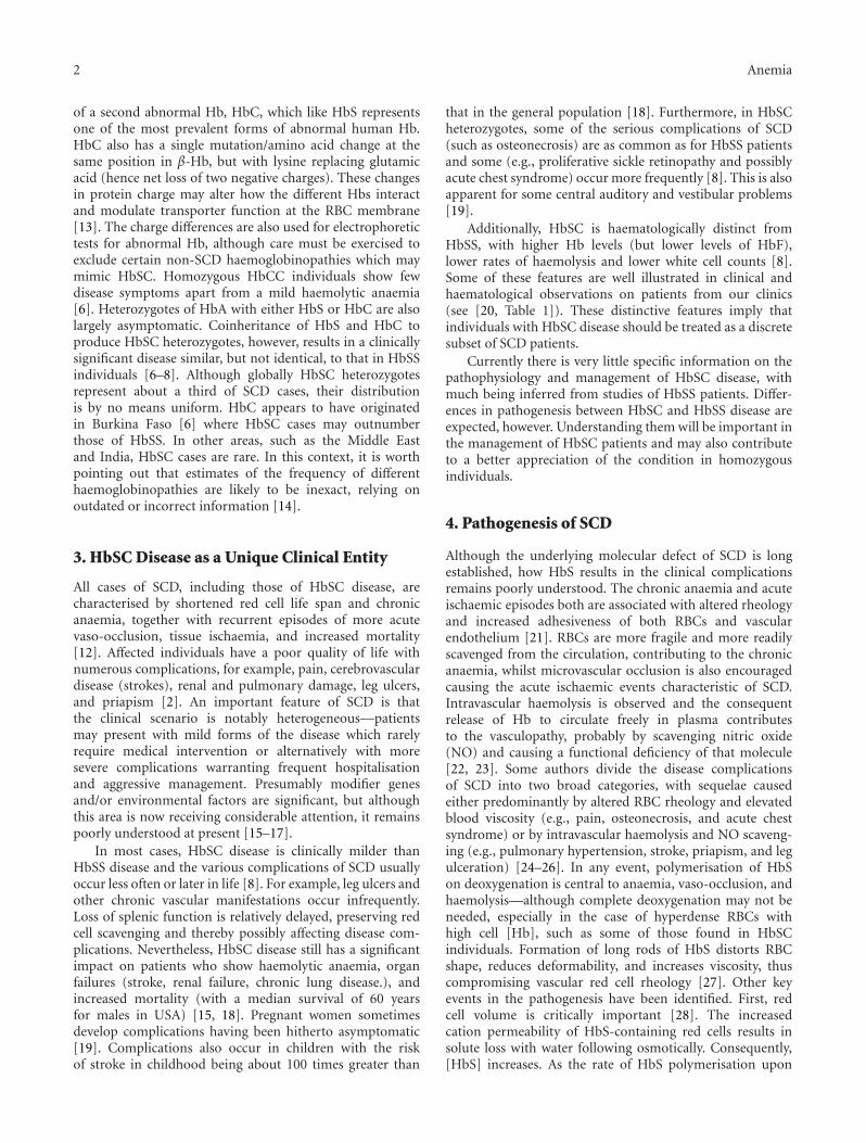
Figure 4: Conductance of red blood cells (RBCs) from sickle cell patients heterozygous for HbS and HbC (HbSC genotype). (a), (b)Representative whole-cell recordings from (a) oxygenated and (b) deoxygenated RBCs. (c) Mean whole-cell currents + S.E.M., n = 5.Test potentials from −80 to +80 mV were applied for 300 ms in 10 mV increments from a holding potential of −10 mV. Measurements weremade using Na+-containing bath and pipette solutions. Data taken from [20]. See [66] for experimental details. The conductance of RBCsfrom HbSC patients is high and increases further on deoxygenation.
episodes of sickling. In this respect, perhaps the majority ofHbSC reticulocytes behave like the “fast-track” reticulocytesof HbSS patients [56]—cells which dehydrate rapidly onleaving the bone marrow—but this remains to be established.It also raises the question as to what constitutes RBCs inthe less dense HbSC fractions. Can shrunken HbSC cellsregain lost solute and increase their volume? If so, what isthe mechanism and what are transport systems involved?
As heterozygotes, HbSC cells contain both HbS and HbC,in approximately equal amounts (i.e., 50%). This contrastswith the lower HbS content (c.40%) found in sickle traitHbAS cells [5]. Crystals of HbC are sometimes presentin oxygenated RBCs. In contrast to HbS polymers, thesedeposits are lost on deoxygenation [60]. Because of the highHbS content and polymerisation, HbSC cells also show adeoxygenation-induced sickling shape change. In this case,however, rather than the HbSS sickles and holly leaf forms,deoxygenated HbSC cells show multifolded shapes such as“pita breads” and “tricorns” [60], perhaps because of thehigh surface area to volume ratio subsequent to their moremarked dehydration. How HbS and HbC interact has also
received some attention. Using different Hb mixtures, adirect interaction between the two Hbs appears to onlyslightly enhance HbS polymerisation. Much more importantin HbSC disease is RBC dehydration and consequently thehigh MCHC [5, 61, 62]. High MCHC and lower levels ofHbF may have an effect on the extent and kinetics of HbSpolymerisation whilst concurrent Hb mutations (such as β-thalassaemia) may also play a significant role.
It is therefore critical to understand fully the mechanismsby which these RBCs shrink, but our understanding of themechanisms involved remains uncertain. Oxygenated HbSCcells have elevated KCC activity that is stimulated by low pHand swelling [13, 60]. Cytoplasmic protein concentration hasbeen suggested as the “volume” sensor of RBCs [63]. It istherefore intriguing to speculate that high KCC activity inoxygenated HbSC cells may result from the presence of HbCcrystals which would lower the total concentration of solubleHb, as occurs in swollen RBCs. In effect, the cells “think” thatthey are swollen and so activate mechanisms to lose solutesand water, namely, KCC. On the other hand, deoxygenatedHbSC cells also show increased K+ efflux, to an extent

6 Anemia
apparently greater than that observed in deoxygenated HbSScells [58]. Which pathway mediates the flux in deoxygenatedconditions, however, has not been established. If Psickle isinvolved, given the lower [HbS] of HbSC cells, it is notclear why it should be activated to a greater extent than inHbSS cells. KCC and the Gardos channel represent obviousalternative pathways.
A number of manoeuvres which reduce reticulocytedensity may provide evidence for the transport pathwaysinvolved in their dehydration. Both Cl− removal anddeoxygenation shift HbSC reticulocytes to lower densities,consistent with solute retention following inhibition ofKCC [60]. Hypotonic swelling of HbSC cells also reducesthe deoxygenation-induced K+ loss [58], perhaps throughreduction in [HbS] removing a Psickle-like element of K+ flux.
In preliminary studies, we have observed high KCCactivity in oxygenated unfractionated HbSC cells, which wasalmost completely inhibited on deoxygenation [64]. Thus,KCC in HbSC cells behaved like that in HbSS cells athigh O2 tension and like that in HbAA cells when tensionwas reduced [64] (Figure 3). We also found activation ofa deoxygenation-induced Cl−-independent K+ flux [64],a deoxygenation-induced nonelectrolyte permeability [65]and a deoxygenation-induced rise in K+ conductance inpatch-clamp experiments [66] (Figure 4), namely, a Psickle-like permeability, together with activation of the Gardoschannel. In this context, it is interesting that HbC has ahigher affinity for the RBC membrane than either HbAor HbS [59] leading to an early suggestion that HbSCinteraction is involved in modulating RBC permeability.
It is apparent, however, that our understanding of thepermeability of HbSC cells requires further investigation.
7. Conclusion: The Importance of CellDehydration in HbSC Disease
Understanding dehydration is particularly relevant for HbSCcells. The solubility of deoxygenated HbS is about 17 g·dl−1
compared to 70 g·dl−1 for HbA. As HbS represents onlyabout half the total Hbs in HbSC cells, a relatively smalldecrease in MCHC (from an RBC total of 37 to 33 g·dl−1)will prevent HbS polymerisation [61] while retaining thefunctionally important discocyte morphology. As HbS con-stitutes about half the Hbs in these RBCs, this would meana fall in [HbS] from 18.5 to 16.5. In comparison, in HbSScells, a reduction of MCHC to <25 g·dl−1 is required, bywhich time RBCs will be spherocytic, and cell swelling perse will adversely affect rheology [58]. Notwithstanding theirrelevance to dehydration and sickling, the permeability ofHbSC cells has not been well studied nor compared indetail with that of HbSS cells. Several areas require morecareful investigation. The interaction between different Hbs,membrane target sites regulating permeability, the transportpathways involved, the role of cell density, oxygenation,volume, and pH presents a complex pattern of modalitiescontrolling solute content and hence cell density and MCHC.Control by phosphorylation remains mainly unexplored.The challenge ahead lies to define the most important
stimuli and how they interact to determine cell volume. Amajor therapeutic goal is the ability to prevent HbSC celldehydration or to promote rehydration.
Abbreviations
Here we term red cells from HbSS patients as HbSS cells,those from HbSC individuals as HbSC cells.
Acknowledgment
The authors thank the Medical Research Council, the BritishHeart Foundation, and the Wellcome Trust for financialsupport.
References
[1] G. R. Serjeant, “Sickle-cell disease,” The Lancet, vol. 350, no.9079, pp. 725–730, 1997.
[2] M. H. Steinberg, “Management of sickle cell disease,” The NewEngland Journal of Medicine, vol. 340, no. 13, pp. 1021–1030,1999.
[3] M. Hickman, B. Modell, P. Greengross et al., “Mapping theprevalence of sickle cell and beta thalassaemia in England:estimating and validating ethnic-specific rates,” British Journalof Haematology, vol. 104, no. 4, pp. 860–867, 1999.
[4] NHS sickle cell & thalassaemia screening programme, 2010.[5] H. F. Bunn, C. T. Noguchi, J. Hofrichter, G. P. Schechter,
A. N. Schechter, and W. A. Eaton, “Molecular and cellularpathogenesis of hemoglobin SC disease,” Proceedings of theNational Academy of Sciences of the United States of America,vol. 79, no. 23, pp. 7527–7531, 1982.
[6] R. L. Nagel, M. H. Steinberg, and S. C. Hemoglobin, “diseaseand HbC disorders,” in Disorders of Hemoglobin, M. H.Steinberg, B. G. Forget, D. R. Higgs, and R. L. Nagel, Eds., pp.756–785, Cambridge University Press, Cambridge, UK, 2001.
[7] R. L. Nagel and C. Lawrence, “The distinct pathobiology ofsickle cell-hemoglobin C disease: therapeutic implications,”Hematology/Oncology Clinics of North America, vol. 5, no. 3,pp. 433–451, 1991.
[8] R. L. Nagel, M. E. Fabry, and M. H. Steinberg, “The paradoxof hemoglobin SC disease,” Blood Reviews, vol. 17, no. 3, pp.167–178, 2003.
[9] L. Pauling, H. A. Itano, S. J. Singer, and I. C. Wells, “Sickle cellanemia, a molecular disease,” Science, vol. 110, no. 2865, pp.543–548, 1949.
[10] V. M. Ingram, “Gene mutations in human haemoglobin:the chemical difference between normal and sickle cellhaemoglobin,” Nature, vol. 180, no. 4581, pp. 326–328, 1957.
[11] C. A. Marotta, J. T. Wilson, B. G. Forget, and S. M. Weissman,“Human β globin messenger RNA. III. Nucleotide sequencesderived from complementary DNA,” The Journal of BiologicalChemistry, vol. 252, no. 14, pp. 5040–5053, 1977.
[12] H. F. Bunn and B. G. Forget, Hemoglobin: Molecular, Geneticand Clinical Aspects, WB Saunders, Philadelphia, Pa, USA,1986.
[13] O. Olivieri, D. Vitoux, F. Galacteros et al., “Hemoglobinvariants and activity of the (K+Cl-) cotransport system inhuman erythrocytes,” Blood, vol. 79, no. 3, pp. 793–797, 1992.
[14] D. J. Weatherall, “The inherited diseases of haemoglobin arean emerging global health burden,” Blood, vol. 115, pp. 4331–4336, 2010.

Anemia 7
[15] M. H. Steinberg, “Genetic etiologies for phenotypic diversityin sickle cell anemia,” TheScientificWorldJournal, vol. 9, pp. 46–67, 2009.
[16] P. Sebastini, N. Solovieff, S. W. Hartley et al., “Geneticmodifiers of the severity of sickle cell anemia idenitifiedthrough a genome-wide association study,” American Journalof Hematology, vol. 85, pp. 29–35, 2010.
[17] S. L. Thein and S. Menzel, “Discovering the genetics underly-ing foetal haemoglobin production in adults,” British Journalof Haematology, vol. 145, no. 4, pp. 455–467, 2009.
[18] D. Powars, L. S. Chan, and W. A. Schroeder, “The variableexpression of sickle cell disease is genetically determined,”Seminars in Hematology, vol. 27, no. 4, pp. 360–376, 1990.
[19] K. Ohene-Frempong and M. H. Steinberg, “Clinical aspectsof sickle cell anemia in adults and children,” in Disorders ofHemoglobin, M. H. Steinberg, B. G. Forget, D. R. Higgs, andR. L. Nagel, Eds., pp. 611–670, Cambridge University Press,Cambridge, UK, 2001.
[20] S. Dalibalta, J. C. Ellory, J. A. Browning, R. J. Wilkins, D. C.Rees, and J. S. Gibson, “Novel permeability characteristics ofred blood cells from sickle cell patients,” Blood Cells, Molecules,and Diseases, vol. 45, no. 1, pp. 46–52, 2010.
[21] R. P. Hebbel, “Beyond hemoglobin polymerization: the redblood cell membrane and sickle disease pathophysiology,”Blood, vol. 77, no. 2, pp. 214–237, 1991.
[22] C. D. Reiter, X. Wang, J. E. Tanus-Santos et al., “Cell-freehemoglobin limits nitric oxide bioavailability in sickle-celldisease,” Nature Medicine, vol. 8, no. 12, pp. 1383–1389, 2002.
[23] G. J. Kato, V. McGowan, R. F. Machado et al., “Lactate dehy-drogenase as a biomarker of hemolysis-associated nitric oxideresistance, priapism, leg ulceration, pulmonary hypertension,and death in patients with sickle cell disease,” Blood, vol. 107,no. 6, pp. 2279–2285, 2006.
[24] M. J. Stuart and R. L. Nagel, “Sickle-cell disease,” The Lancet,vol. 364, no. 9442, pp. 1343–1360, 2004.
[25] R. P. Rother, L. Bell, P. Hillmen, and M. T. Gladwin, “Theclinical sequelae of intravascular hemolysis and extracellularplasma hemoglobin: a novel mechanism of human disease,”Journal of the American Medical Association, vol. 293, no. 13,pp. 1653–1662, 2005.
[26] K. C. Wood, L. L. Hsu, and M. T. Gladwin, “Sickle cell diseasevasculopathy: a state of nitric oxide resistance,” Free RadicalBiology and Medicine, vol. 44, no. 8, pp. 1506–1528, 2008.
[27] W. A. Eaton and J. Hofrichter, “Hemoglobin S gelation andsickle cell disease,” Blood, vol. 70, no. 5, pp. 1245–1266, 1987.
[28] V. L. Lew and R. M. Bookchin, “Ion transport pathology in themechanism of sickle cell dehydration,” Physiological Reviews,vol. 85, no. 1, pp. 179–200, 2005.
[29] R. Hoover, R. Rubin, G. Wise, and R. Warren, “Adhesionof normal and sickle erythrocytes to endothelial monolayercultures,” Blood, vol. 54, no. 4, pp. 872–876, 1979.
[30] R. P. Hebbel, M. A. B. Boogaerts, J. W. Eaton, and M. H.Steinberg, “Erythrocyte adherence to endothelium in sickle-cell anemia. A possible determinant of disease severity,” TheNew England Journal of Medicine, vol. 302, no. 18, pp. 992–995, 1980.
[31] D. K. Kaul, X.-D. Liu, X. Zhang et al., “Peptides based onαV-binding domains of erythrocyte ICAM-4 inhibit sicklered cell-endothelial interactions and vaso-occlusion in themicrocirculation,” American The Journal of Physiology, vol.291, no. 5, pp. C922–C930, 2006.
[32] B. N. Yamaja Setty, S. Kulkarni, and M. J. Stuart, “Role oferythrocyte phosphatidylserine in sickle red cell-endothelialadhesion,” Blood, vol. 99, no. 5, pp. 1564–1571, 2002.
[33] F. A. Kuypers, “Red cell membrane lipids inhemoglobinopathies,” Current Molecular Medicine, vol.8, no. 7, pp. 633–638, 2008.
[34] C. H. Joiner, “Cation transport and volume regulation in sicklered blood cells,” American The Journal of Physiology, vol. 264,no. 2, pp. C251–C270, 1993.
[35] B. N. Yamaja Setty and S. G. Betal, “Microvascular endothelialcells express a phosphatidylserine receptor: a functionallyactive receptor for phosphatidylserine-positive erythrocytes,”Blood, vol. 111, no. 2, pp. 905–914, 2008.
[36] B. N. Y. Setty, S. G. Betal, J. Zhang, and M. J. Stuart, “Hemeinduces endothelial tissue factor expression: potential rolein hemostatic activation in patients with hemolytic anemia,”Journal of Thrombosis and Haemostasis, vol. 6, no. 12, pp.2202–2209, 2008.
[37] J. S. Gibson and J. C. Ellory, “Membrane transport in sicklecell disease,” Blood Cells, Molecules, and Diseases, vol. 28, no. 3,pp. 303–314, 2002.
[38] C. Brugnara, H. F. Bunn, and D. C. Tosteson, “Regulation oferythrocyte cation and water content in sickle cell anemia,”Science, vol. 232, no. 4748, pp. 388–390, 1986.
[39] M. Canessa, A. Spalvins, and R. L. Nagel, “Volume-dependentand NEM-stimulated K+, Cl- transport is elevated in oxy-genated SS, SC and CC human red cells,” FEBS Letters, vol.200, no. 1, pp. 197–202, 1986.
[40] J. S. Gibson, P. F. Speake, and J. C. Ellory, “Differential oxygensensitivity of the K+-Cl- cotransporter in normal and sicklehuman red blood cells,” The Journal of Physiology, vol. 511, no.1, pp. 225–234, 1998.
[41] A. C. Hall and J. C. Ellory, “Evidence for the presence ofvolume-sensitive KCl transport in ’young’ human red cells,”Biochimica et Biophysica Acta, vol. 858, no. 2, pp. 317–320,1986.
[42] J. C. Ellory, A. C. Hall, and S. A. Ody, “Is acid a more potentactivator of KCl co-transport than hypotonicity in human redcells?” The Journal of Physiology, vol. 420, p. 149, 1990.
[43] J. S. Gibson, A. R. Cossins, and J. C. Ellory, “Oxygen-sensitivemembrane transporters in vertebrate red cells,” Journal ofExperimental Biology, vol. 203, no. 9, pp. 1395–1407, 2000.
[44] J. S. Gibson and J. C. Ellory, “K+-Cl- cotransport in vertebratered cells,” in Red Cell Membrane Transport in Health andDisease, I. Bernhardt and J. C. Ellory, Eds., pp. 197–220,Springer, New York, NY, USA, 2004.
[45] O. E. Ortiz, V. L. Lew, and R. M. Bookchin, “Deoxygenationpermeabilizes sickle cell anaemia red cells to magnesiumand reverses its gradient in the dense cells,” The Journal ofPhysiology, vol. 427, pp. 211–226, 1990.
[46] J. P. Willcocks, P. J. Mulquiney, J. C. Ellory, R. L. Veech, G.K. Radda, and K. Clarke, “Simultaneous determination of lowfree Mg2+ and pH in human sickle cells using 31P NMRspectroscopy,” The Journal of Biological Chemistry, vol. 277, no.51, pp. 49911–49920, 2002.
[47] N. Mohandas, M. E. Rossi, and M. R. Clark, “Associa-tion between morphologic distortion of sickle cells anddeoxygenation-induced cation permeability increase,” Blood,vol. 68, no. 2, pp. 450–454, 1986.
[48] V. L. Lew, O. E. Ortiz, and R. M. Bookchin, “Stochastic natureand red cell population distribution of the sickling- inducedCa2+ permeability,” The Journal of Clinical Investigation, vol.99, no. 11, pp. 2727–2735, 1997.
[49] J. A. Browning, H. C. Robinson, J. C. Ellory, and J. S. Gibson,“Deoxygenation-induced non-electrolyte pathway in red cellsfrom sickle cell patients,” Cellular Physiology and Biochemistry,vol. 19, no. 1–4, pp. 165–174, 2007.

8 Anemia
[50] M. D. Rhoda, M. Apovo, Y. Beuzard, and F. Giraud, “Ca2+
permeability in deoxygenated sickle cells,” Blood, vol. 75, no.12, pp. 2453–2458, 1990.
[51] L. A. Woon, J. W. Holland, E. P. W. Kable, and B. D. Roufogalis,“Ca2+ sensitivity of phospholipid scrambling in human redcell ghosts,” Cell Calcium, vol. 25, no. 4, pp. 313–320, 1999.
[52] G. Gardos, “The function of calcium in the potassiumpermeability of human erythrocytes,” Biochimica et BiophysicaActa, vol. 30, no. 3, pp. 653–654, 1958.
[53] R. L. Nagel and O. S. Platt, “General pathophysiology of sicklecell anemia,” in Disorders of Hemoglobin, M. H. Steinberg, B.G. Forget, D. R. Higgs, and R. L. Nagel, Eds., pp. 494–526,Cambridge University Press, Cambridge, UK, 2001.
[54] R. S. Franco, M. Palascak, H. Thompson, and C. H. Joiner,“KCl cotransport activity in light versus dense transferrinreceptor- positive sickle reticulocytes,” The Journal of ClinicalInvestigation, vol. 95, no. 6, pp. 2573–2580, 1995.
[55] R. S. Franco, M. Palascak, H. Thompson, D. L. Rucknagel,and C. H. Joiner, “Dehydration of transferrin receptor-positivesickle reticulocytes during continuous or cyclic deoxygena-tion: role of KCl cotransport and extracellular calcium,” Blood,vol. 88, no. 11, pp. 4359–4365, 1996.
[56] R. M. Bookchin, O. E. Ortiz, and V. L. Lew, “Evidence fora direct reticulocyte origin of dense red cells in sickle cellanemia,” The Journal of Clinical Investigation, vol. 87, no. 1,pp. 113–124, 1991.
[57] G. R. Serjeant and B. E. Serjeant, “A comparison of erythrocytecharacteristics in sickle cell syndromes in Jamaica,” BritishJournal of Haematology, vol. 23, no. 2, pp. 205–213, 1972.
[58] M. E. Fabry, D. K. Kaul, and C. Raventos-Suarez, “SC ery-throcytes have an abnormally high intracellular hemoglobinconcentration. Pathophysiological consequences,” The Journalof Clinical Investigation, vol. 70, no. 6, pp. 1315–1319, 1982.
[59] S. K. Ballas, J. Larner, E. D. Smith, S. Surrey, E. Schwartz, andE. F. Rappaport, “The xerocytosis of HbSC disease,” Blood, vol.69, pp. 124–130, 1987.
[60] C. Lawrence, M. E. Fabry, and R. L. Nagel, “The uniquered cell heterogeneity of SC disease: crystal formation, densereticulocytes, and unusual morphology,” Blood, vol. 78, no. 8,pp. 2104–2112, 1991.
[61] ME Fabry, J. Harrington, H. Chang, and R. L. Nagel, “Criticalcontribution of cell density to the pathophysiology of SC cells,”Clinical Research, vol. 30, article 559a, 1982.
[62] R. M. Bookchin and T. Balazs, “Ionic strength dependence ofthe polymer solubilities of deoxyhemoglobin S + C and S + Amixtures,” Blood, vol. 67, no. 4, pp. 887–892, 1986.
[63] A. P. Minton, “Influence of macromolecular crowding onintracellular association reactions: possible role in volumeregulation,” in Cellular and Molecular Physiology of Cell VolumeRegulation, K. Strange, Ed., pp. 181–190, CRC Press, BocaRaton, Fla, USA, 1994.
[64] J. S. Gibson, M. C. Muzyamba, S. E. Ball, and J. C. Ellory, “K+
transport in HbSC-containing human red blood cells,” TheJournal of Physiology, vol. 535, article S008, p. 27, 2001.
[65] J. C. Ellory, R. Sequeira, A. Constantine, R. J. Wilkins, and J. S.Gibson, “Non-electrolyte permeability of deoxygenated sicklecells compared,” Blood Cells, Molecules, and Diseases, vol. 41,no. 1, pp. 44–49, 2008.
[66] J. A. Browning, H. M. Staines, H. C. Robinson, T. Powell,J. C. Ellory, and J. S. Gibson, “The effect of deoxygenationon whole-cell conductance of red blood cells from healthyindividuals and patients with sickle cell disease,” Blood, vol.109, no. 6, pp. 2622–2629, 2007.
[67] J. S. Gibson, A. Khan, P. F. Speake, and J. C. Ellory, “O2
dependence of K+ transport in sickle cells: effect of differentcell populations and the substituted benzaldehyde 12C79,” TheFASEB Journal, vol. 15, no. 3, pp. 823–832, 2001.

Hindawi Publishing CorporationAnemiaVolume 2011, Article ID 698586, 7 pagesdoi:10.1155/2011/698586
Clinical Study
Relationship between Painful Crisis and Serum Zinc Level inChildren with Sickle Cell Anaemia
Edamisan Olusoji Temiye,1 Edem Samuel Duke,2 Mbang Adeyemi Owolabi,3
and James Kweku Renner1
1 Department of Paediatrics, College of Medicine, University of Lagos (CMUL), P.M.B 12003, Lagos, Nigeria2 Critical Rescue International (CRI), Plot 144, Oba Akran Road, Ikeja, Lagos, Nigeria3 Department of Pharmaceutical Chemistry, Faculty of Pharmacy, College of Medicine, Idi-Araba, University of Lagos, P.M.B. 12003,Lagos, Nigeria
Correspondence should be addressed to Edamisan Olusoji Temiye, [email protected]
Received 14 August 2010; Accepted 11 October 2010
Academic Editor: Duran Canatan
Copyright © 2011 Edamisan Olusoji Temiye et al. This is an open access article distributed under the Creative CommonsAttribution License, which permits unrestricted use, distribution, and reproduction in any medium, provided the original work isproperly cited.
Sickle cell anaemia (SCA) is associated with zinc deficiency; zinc supplementation may ameliorate some of its clinicalmanifestations including the relief of painful crisis. Subjects and Methods. Serum zinc levels were determined in 71 children withSCA and painful crisis and in equal numbers in steady state. Seventy-one children with AA genotype acted as controls. Qualitativeassessment of zinc content of 24-hour dietary recall and the last meal consumed before blood was drawn was taken. Serum zincwas determined using atomic absorption spectrophotometer. Haemoglobin concentration and packed cell volume (PCV) weredetermined using standard methods. Results. The mean serum zinc concentration in the study was less than international referencerange. The controls had significantly higher serum zinc concentrations than the SCA group (42.7± 13.6 versus 32.3± 14.0μg/dL,P < .000); this difference was due to the significantly lower values of serum zinc in SCA with painful crisis compared with theremaining two groups F = 30.9, P < .000. There was a positive correlation between serum zinc and haemoglobin concentrationonly in the control group (r = 0.4; P = .001). Conclusion. The serum zinc levels in this study were low. Painful crisis in SCA mayexert greater demand for zinc utilization in children with SCA thereby resulting in lower serum levels.
1. Introduction
Sickle cell anaemia is the most common inherited disorderof the black race. It affects red blood cells resulting inchronic haemolytic anaemia of varying severity and in somepatients, periodic painful crisis caused by the occlusion ofsmall blood vessels by spontaneous intravascular sicklingwith multiorgan affectation [1, 2].
Brewer and Oelshlegel discovered that calcium bindingto red cell membrane is responsible for the formationof irreversible sickled cells and that the antisickling effectof zinc in sickle cell anaemia is due to its ability toantagonize calcium binding to red cell membrane [3].In sickle cell anaemia, there is also increased oxidativestress and peroxidation as well as low antioxidant poten-tial which predisposes the patients to vaso-occlusive crisis
[4–6]. Zinc exerts its antioxidant action by inhibition of lipidperoxidation which occurs in red blood cells and liver therebystabilizing biomembranes and biostructures thus protectingthe body against oxidative stress [5, 6]. These effects of zincare believed to give zinc its ability to reduce vaso-occlusivecrisis in sickle cell anaemia.
Various studies have shown that zinc deficiency maybe common in SCA patients. This has been attributed tochronic haemolysis that occurs in these patients, increaseddemand and utilization, along with the secondary loss ofzinc in the urine [1, 7, 8]. Furthermore, supplementationof zinc in sickle cell anaemia has been reported to improvewound healing, decrease incidence of infection, improvethe age of attaining secondary sexual characteristics, reversedark adaptation of the eyes, and accelerate growth [8–12].

2 Anemia
Although it has been suggested that zinc therapy couldreduce painful crisis [13, 14]; however, it has not been shownwith certainty that zinc deficiency in sickle cell anaemiais directly related to the development or severity of vaso-occlusive crisis. Prasad et al. [15], in 1976 administeredoral zinc sulfate to 10 adults who had severe painful crisisand found symptomatic improvement in 8 of the subjects,which they attributed to the antisickling effect of zinc. Thesignificance of their findings may be limited because of thevery small sample size and lack of control subjects in thestudy. In a placebo-controlled double-blind study, 145 sicklecell adult subjects were treated with either 220 mg of zincsulphate 3 times daily or placebo [14]. After 18 months, thezinc-treated subjects had an average of 2.5 crises, comparedto 5.3 in the placebo group; but the severity of painful crisiswas not reduced. A study in Ibadan, Nigeria [16] foundno relationship between serum zinc level and the differentdegrees of clinical severity of the disease in these subjects.They observed that the level of zinc in each patient, thoughsignificantly lower than the controls, had no correlationwith the haematocrit, frequency of bone pain crisis, andsusceptibility to infection. The sample size in their study wasalso small making it difficult to draw any valid inference.
It is probable that zinc deficiency may be associated withpainful crisis. However, these authors are not aware of anyprevious work comparing zinc status in sickle cell anaemicpatients in vaso-occlusive crisis with those in steady state.This study is therefore designed to assess the level of serumzinc in sickle cell anaemia children in painful state (vaso-occlusive crisis) as compared with those in steady state. Wealso relate the levels of serum zinc with diet and haemoglobinlevels in the subjects.
2. Subjects and Methods
This was a case-controlled study of patients at the LagosUniversity Teaching Hospital (LUTH), Nigeria. The samplesize for the study and the number of subjects for each armwere determined using standard formulae for populationstudy [17], using the prevalence rate of SCA in westernNigeria [18] and the rate of deficient erythrocyte zinc level insickle cell anaemia children in Zaria [19]. One hundred andforty-two paediatric subjects aged between one and 12 yrswho had haemoglobin genotype SS were studied. Of these, 71were in had bone pain crisis and were recruited consecutivelyfrom both the haematology clinic and Children’s EmergencyRoom (CHER) the remainder were in steady state recruitedfrom the sickle cell clinic. The control group consisted of 71apparently well subjects whose haemoglobin genotype wasAA. They were recruited from the Well Baby and Children’sOutpatient Clinic. Both the SCA in steady state subjects andthe controls were selected such that they were matched forage and sex with the subjects who had painful crisis.
In this study, sickle cell anaemia in painful crisis wasdefined as subjects who had pains at the time of recruitmentor within 48 hours before recruitment in any of thelimbs [20], while steady state was defined as subjects whowere apparently well without evidence of recent infection,
bone pain, or other problems for at least 4 weeks beforerecruitment [21]. The study, which was approved by theresearch and ethics committee of the hospital, lasted for aperiod of 12 months. Other inclusion criteria were that studypopulation were not on any zinc-containing medications,they were fed at least 4 hours before blood collection, andhad not been transfused with blood or blood products in theprevious three months preceding study. Also included weresubjects who did not have any symptoms or signs of infectionsuch as fever (axillary temp < 37.0◦C), acute respiratoryinfection, diarrhoea, or malaria. Additionally, controls hadno evidence of chronic disease including protein energymalnutrition. The haemoglobin genotype of all the subjectswas determined by haemoglobin cellulose electrophoresis.
After informed consent, a detailed history of each childfrom the care-giver was taken; these included the mother’sand father’s educational status and occupation, 24-hourmeal recall, date of last blood transfusion, and the presenceor absence of pain in the limbs. Physical examinationincluding assessment for the presence or absence of fever(temp > 37.0◦C), jaundice, pallor, finger clubbing, liversize, spleen size, and tenderness in the limbs, the weight,and length/height was done. Previous health status of eachsubject was obtained by examining the hospital records. FivemL of blood was collected from each subject by venepunctureafter appropriate skin preparations with a 21-gauge stainless-steel needle with a polypropylene syringe (Norm-Ject; HenkeSass Wolf GMBH, Germany) [22]. Two mL of bloodwas transferred into a sodium ethylenediamine-tetra-acetate(EDTA) bottle for haemoglobin (Hb), Packed cell volume(PCV), and haemoglobin genotype determination usingstandard laboratory procedure. The remaining 3 mL wastransferred into zinc free nonheparinized bottles previouslywashed clean of possible zinc contamination by leaving themsoaked in 10% nitric acid for 24 hours and rinsed three timeswith deionised water. The clotted blood sample in the zinc-free bottles was centrifuged at 1500 rpm for 10 minutes andthe serum removed with zinc free Pasteur Pipette preparedby previously washing with 10% nitric acid and thoroughlyrinsed with deionised water as described for the bottles above[23]. The serum samples were then stored at −20◦C pendinganalysis [24]. All haemolysed samples were discarded. Beforethe analysis, the serum samples were allowed to come toroom temperature and each mixed by gently inverting thetube four times. Serum zinc level was determined using anatomic absorption spectrophotometer (PU9100X, Philips,Holland) at the Food Technology laboratory of the FederalInstitute of Industrial Research Oshodi (FIIRO) Lagos,Nigeria and following the method of Smith et al. [22].The concentration of each sample, which was expressedin parts per million, was extrapolated from the calibrationcurve prepared from the standard zinc calibration curve andconverted to μg/dL by calculation.
3. Classification of Zinc Content in24-Hour Meal Recall
The zinc contents of 24-hour meal recall and the last mealconsumed before blood was taken were classified based on

Anemia 3
Table 1: Mean haematological parameters and serum zinc concentration in Cases and Controls.
ParameterGenotype (Mean ± 2SD)
F P-valueGenotype AA (n = 71)
Sickle cell anaemia
Steady state n = 71 Painful crisis n = 71
Age (mo) 71.3± 34.3 67.2 ± 30.1 79.9 ± 31.7 2.9 .06
Weight (Kg) 21.1± 8.0 17.9 ± 5.4 20.3 ± 6.3 4.6 .01
Height (cm) 115.7± 21.1 108.4 ± 16.8 115.7 ± 17.8 3.6 .03
Hb (g/dL) 11.3± 2.0 7.1 ± 1.1 7.6 ± 1.6 144.9 .00
PCV (%) 33.1± 5.6 20.9 ± 3.4 22.3 ± 4.8 143.9 .00
Serum Zinc Conc (μg/dL) 42.7± 13.6 38.4 ± 13.8 26.3 ± 11.3 30.9 .00
P < .05 is significant.
the concentration of zinc in food items consumed by thesubject. Thus each food item was classified as containinghigh, moderate, or low zinc [25, 26]. Meals which containitems such as plantain, red beef, sea foods, egg, milk, cocoaproducts, and fish were classified as high zinc content, whilebeans, cereals made out of maize and rice products containmoderate zinc. Cassava products were classified as havingtraces or low zinc content [25, 26]. For the purpose of thisstudy, a subject who had more than 2 high zinc containingitems with any 3 moderate zinc containing items or all highzinc containing items was classified as having high zinc diet.A subject who had one high zinc containing item and lessthan 3 moderate zinc containing items or had all moderatezinc containing items was classified as having moderate zinc,while a subject who had one moderate zinc or one high zinccontaining item with low zinc containing item, or only lowzinc containing items, was classified as having low zinc.
4. Data Analysis
Data generated was analysed using the SPSS statisticalpackage version 11. Frequency distributions were generatedfor all categorical variables. The measures of location weredetermined for quantitative outcome such as age, zinc level,and haematocrit. Statistical significance between two meanswas assessed using Student t-test, while one-way analysis ofvariance was used where there were multiple means. Wherethere was significant difference within means, post hoc testwas applied to determine where the level of significanceoccurred. Chi-square (χ2) test was applied for categoricalvariables and correlation analysis as applicable. Differencesbetween values were accepted as statistically significant whereprobability was less than .05.
5. Results
Two hundred and thirteen children aged between one and 12yrs were recruited into the study; seventy-one were controlswith haemoglobin genotype AA, while one hundred andforty-two subjects had haemoglobin genotype SS. Seventyone of the subjects with sickle cell anaemia were in steadystate while the rest had vaso-occlusive bone pain crisis.Eighty-two of the study subjects were males and 60 were
females giving a male : female ratio of 1.3 : 1. This differencewas not statistically significant. Also, the male: female ratiofor each of the subgroup was not significantly different.
The mean ages of the subjects in control group and thosewith sickle cell anaemia were 71.3± 34.3 months and 73.6±31.4 months, respectively. The difference was not statisticallysignificant. (t = 0.5, P = .64). Although the control subjectswere heavier than the SCA subjects (weight (kg), 21.1 ± 8.0versus 19.1 ± 6.0, t = 2.1, P = .04) and they were alsotaller, (height (cm) 115.7 ± 21.1 versus 112.1 ± 17.6, t =1.3, P = .2), the differences were not statistically significant.However, the control subjects had a mean packed cell volumeof 33.1 ± 5.6% and haemoglobin of 11.3 ± 2.0 g/dL whichwere significantly higher (t = 16.9, P = .000) than the SCAgroup with a mean packed cell volume of 21.6 ± 4.2% andhaemoglobin concentration of 7.4 ± 1.4 g/dL, respectively.The mean serum zinc concentration in the control subjectswas also significantly higher than the sickle cell anaemiagroup (42.7±13.6 versus 32.3±14.0μg/dL, t = 5.2, P = .000)
When the sickle cell anaemia group was subdivided intothose in steady state and those with bone pain crisis, themean age remained not significantly different although themean age for subjects with painful crisis was higher thanthose of sickle cell anaemia in steady state and the controlsubjects. Sickle cell anaemia subjects in steady state werehowever significantly shorter than those with painful crisisand with haemoglobin AA. Similarly, sickle cell anaemiasubjects in steady state were significantly lighter than thecontrols (P = .01) subjects and sickle cell anaemia subjectswith painful crisis. The significant difference observed inthe haemoglobin concentration and packed cell volumewas found to be between the control group and the sicklecell anaemia in painful crisis and sickle cell anaemia insteady state. Although the sickle cell anaemia in painfulcrisis had higher haematological parameters than the steadystate subjects, the difference was not statistically signifi-cant. The mean serum zinc concentration in subjects withhaemoglobin genotype SS in steady state of 38.4±13.8μg/dLwas significantly higher than in subjects with bone pain crisisof 26.3 ± 11.3μg/dL (P < .001). The result also showedsignificantly higher mean serum zinc concentration levels(P < .001) in controls when compared with subjects in bonepain crisis. However, the differences between control andsteady state subjects were not statistically significant, Table 1.

4 Anemia
0
10
20
30
40
50
60
13–35 36–71 72–107 108–144
Age group of subjects (months)
AASS steady state
SS painful crisisAll
Mea
nse
rum
zin
cle
vel(μ
g/dL
)
Figure 1: Mean zinc concentration by age groups in the threesubgroups.
The subjects were arranged into four age groups. Thirtythree subjects (15.5%) were between 13 and 35 months ofage, 57 (26.7%) were in the age group of 36–71 months,the majority (83, 39.0%) were between 72 and 107 monthsof age and the remaining 40 (18.8%) were between 108–144 months of age. There were, however, no significantdifferences in the numbers of subjects in the different agegroups (P > .03). The mean serum levels of zinc for the agegroups for all the subjects ranged from 36.21±12.8μg/dL forage group of 13–35 months, 39.5± 15.6μg/dL for age groupof 36–71 months, 35.6± 16.0μg/dL, to 30.7± 10.2μg/dL, forage groups of 108–144 months. The differences in the valueswere statistically significant F = 2.9, P = .04.
When the subjects were grouped into controls, sicklecell steady state, and painful crisis, however, the controlsubjects had higher mean serum zinc levels than the sicklecell anaemia subgroups in all age groups except in the agegroup of 108–144 months where the steady state subjectshad higher levels than the control group. These differenceswere statistically significant and the level of significance wasbetween the controls and the sickle cell anaemia subjects inpainful crisis except in the age group of 108–144 monthswhere the level of significance was between the sickle cellanaemia in steady state and SCA in painful crisis. Among thecontrols, there was a significant fall in serum zinc levels asthe subjects get older, F = 5.0, P = .004, but such decreasein zinc levels with age was not observed among the sickle cellsubgroups; F = 0.8, P = .3 (in sickle cell anaemia in steadystate and F = 0.5, P = .7 in sickle cell anaemia subjectsin painful crisis). In all the age groups, the SCA in painfulcrisis had serum zinc levels significantly lower than those ofcontrols and subject with SCA in steady state (Figure 1).
Figure 2 shows the correlation between haemoglobin andserum zinc concentrations. There is a positive correlationbetween haemoglobin concentration and zinc levels in thecontrol group and this was significant (r = 0.4, P <.001). There was no correlation observed in the steady statessubjects (r = 0.01, P = .4) and in subjects in bone paincrisis (r = 0.01, P = .9).
Haemoglobin concentration (g/dL)
1816141210864
100
80
60
40
20
0
GenotypeSS painful crisis
Rsq = 0.0002SS steady stateRsq = 0.0124
AARsq = 0.1615
Note: painful crisis Steady state Control AA
r = 0.0141 r = 0.0111 r = 0.4138
Seru
mzi
nc
con
cen
trat
ion
(μg/
dL)
P = .913 P = .354 P = .001
Figure 2: Linear relationship between haemoglobin concentrationto serum zinc levels in Control, SCA in steady state, and SCA inpainful crisis.
The effect of dietary intake of zinc-containing meals onserum zinc was assessed. Ten (4.7%) subjects were assessedto have consumed meals low in zinc content in the last 24hours, 6 of these were SCA in steady state and 3 were SCAin painful crisis; the remaining subject in this category wasthe control. Ninety seven (45.5%) subjects, consisting of 18controls, 31 SCA in steady state, and 48 SCA in painful crisishad food items with moderate zinc content. The remaining106 (49.8%) subjects of whom 52 were in control group, 34SCA in steady state, and 20 SCA with painful crisis had fooditems containing high level of zinc.
When the zinc content of food items consumed bysubjects 24 hour prior to collection of blood for analysiswere compared with mean serum zinc concentration, it wasfound that subjects in the control group whose meal wereadjudged to contain moderate amount of zinc had meanserum zinc concentration of 41.1 ± 17.2μg/dl compared to43.3 ± 12.5μg/dl in those whose food items contained highamount of zinc. This difference was however not statisticallysignificant. In the SCA group, subjects in the steady statewho consumed low zinc items in their meal had serum zincconcentration of 30.7 ± 12.1μg/dL, those that consumedmoderate zinc levels had 40.8 ± 14.0 while those that

Anemia 5
Table 2: Relationship between zinc content of 24-hour dietary recall and mean serum zinc level.
Zinc content of meal Mean serum zinc concentration μg/dL (± SD)F P-valueGenotype AA Sickle cell anaemia
Steady state Painful crisis
Low 45.0†† 30.7 (12.1) 33.9 (20.7) t = 0.3 .8
Moderate 41.1 (17.2) 40.8 (14.0) 24.8 (10.5) 18.4 .000
High 43.3 (12.5) 37.6 (13.7) 28.5 (11.4) 9.9 .000††Only one subject in the control group had low zinc item and was removed from the statistical analysis; therefore, student t-test was used to compare themean zinc level between sickle cell anaemia subjects in steady state and painful crisis in the first row.
Table 3: Relationship between zinc content of last meal consumed before taking blood and mean serum zinc level.
Zinc content of mealMean serum zinc concentration μg/dL (± SD)
F P-valueGenotype AA n = 71
Sickle cell anaemia
Steady state n = 71 Painful crisis n = 71
Low 46.7 (37.8) 29.3 (10.8) 20.0 (9.9) 1.9 .192
Moderate 40.6 (12.5) 37.6 (13.0) 25.7 (10.9) 23.1 .000
High 46.9 (10.8) 55.4 (11.5) 34.4 (11.5) 6.6 .004
consumed high zinc levels had a mean value of 37.6 ± 13.7.This difference was not statistically significant. SCA subjectswith painful crisis who consumed low zinc containingmeals had mean serum concentration of 33.9 ± 20.7μg/dL.For those who consumed moderate zinc containing itemshad mean serum zinc level of 24.8 ± 10.5μg/dL, and forthose that took high zinc containing items their meanserum zinc concentration was 28.5 ± 11.4μg/dL. Again thisdifference was not statistically significant. However, acrossthe subgroups, control subjects had mean serum levels ofzinc that were significantly higher than those in the SCAsubgroups among subjects that consumed moderate to highzinc containing meals, Table 2.
When the zinc contents of the last meals consumedbefore blood was taken were considered, most subjects, 164(77.0%) of whom 47, 59, and 58 were in the control, SCAsteady state, and SCA painful crisis subgroups, respectively,consumed meals containing moderate zinc contents. Thirty-five (16.4%) subjects had food items containing high zinccontent; 21 of these subjects were in control group while 6and 8 subjects were in SCA in steady state and painful crisis,respectively. The remaining 14 (6.6%) (3 in control group,6 SCA in steady state, and 5 in SCA painful crisis) had fooditems containing low zinc contents.
Using the content of the last meal consumed it was foundthat, except in the control groups, those that consumed highzinc containing meals had highest mean serum zinc levels.This was significant among the SCA in steady state, F = 7.0,P = .002, and just reached significant level in SCA in painfulcrisis, F = 3.1, P = .05. Within the subgroups, those with lowzinc consumption had no significant difference, the meanserum zinc level was however significantly lower in SCA inpainful crisis than those in controls and steady state whotook food items with moderate zinc content. Similar trendwas observed in those who consumed food items containinghigh zinc contents; again the mean serum level for SCA in
painful crisis was significantly lower than those of controlsand SCA in steady state, Table 3.
6. Discussion
The serum zinc levels in this group of Nigerian children werefound to be lower than the internationally acceptable normalvalues of 50–150 μg/dL {7.65–22.95 μmol/L} [27]. This isalso true for the control group as well as those with sicklecell anaemia. Several studies have indicated that nutritionalzinc deficiency is widespread all over the world. Akinkugbeand Ette [28] observed that low zinc levels are commonin Nigerian children. Important risk factors related to zincdeficiency are common use of grain proteins and low contentof zinc in breast milk of mothers and weaning food [27, 28].These factors may be important in our subjects as more than80% were on food containing low or moderate amount ofzinc in their last meal before blood was drawn for analysis.This should reflect more accurately the nature of meal thechild is taking than the 24-hour recall. Most of these fooditems contained high level of cassava- and grain-based dietwhich limits the quantity of zinc available for absorption dueto their high phosphate and phytate content as zinc forms anunavailable complex with these compounds [27–29].
Although the subjects had low serum zinc levels, symp-toms attributable solely to zinc deficiency were not found,especially in the control subjects. It is difficult to say atwhat level of serum zinc that symptoms of deficiencywould appear, and the reasons for lack of symptoms in thecontrol subjects is unclear. The significantly lower valuesof zinc observed in children older than 107 months in thecontrol group probably reflect increased demand for zincin already suboptimal zinc nutriture in these subjects at thecommencement of pubertal growth [30].
In all age groups, the serum zinc levels were significantlylower in the SCA group than in the controls. This finding

6 Anemia
has been reported in several communications [6, 7, 15, 30]and has been attributed to several factors including thechronic haemolysis that characterizes sickle cell anaemialeading to loss of zinc from red blood cells which is animportant storage site for zinc [6, 28]. Furthermore, there isa defective zinc homeostasis as a result of excessive excretionof zinc in urine or abnormal renal tubular reabsorption ofzinc due to the sickling phenomena, increased demand andconsumption due to increased oxidative stress and sicklecell redox imbalance [6, 8, 10]. The further lowering ofserum zinc in SCA subjects with painful crisis in this studywas an interesting finding. It could be postulated that SCAsubjects who are more severely zinc-deficient would havemore painful crisis. On the other hand, frequent painfulcrisis might interfere with nutrition and lead to more severenutritional deficiencies [6].
Zinc is the most abundant intracellular element with85% of total body zinc found in muscle and bone; lessthan 0.1% of total body zinc is in the plasma [31]. Plasmazinc is maintained by continuous shift from the intracellularsources and absorption from the intestine. Hence plasmazinc is a poor indicator of total body zinc. The retentiontime of zinc in the blood is estimated as 2.3 days [32];however, metabolic stress, such as infection and acuteillnesses which are more common in children with sickle celldisease compared to children with haemoglobin genotypeAA, increases intracellular shift of zinc into the liver andlowers plasma level even when total body zinc level isunchanged [33]. As already noted above, the low plasmazinc level in this study in sickle cell anemia subjects withpainful crisis may indicate the effects of stress in increasingthe intracellular shifting of zinc into the liver from the plasmaof these subjects rather than increased loss of zinc from thebody.
This study showed a positive correlation between theserum zinc and haemoglobin in the control group. This isin conformity with the findings of Akinkugbe and Ette [28],whose study was on serum zinc and haemoglobin levels inchildren with common paediatric problems. However, therewas no correlation between serum zinc and haemoglobin inthe subjects with SCA; this is in conformity with the findingof Akenami et al. [16] in sickle cell anaemia children. Thereason for this is not clear. It may be due to the chronichaemolytic state experienced by sickle cell anaemia subjects,leading to loss of zinc at it is storage site (red blood cell) andit is urinary loss. Further studies are required to explain thisfinding.
Zinc deficiency is associated with poor growth, amongother clinical manifestations [8, 34]. In this study, the controlgroup had significantly higher weight and height than thesickle cell anaemia group in steady state but not with theSCA group with painful crisis. This may be attributed to thegrowth retardation associated with zinc deficiency. However,one would have expected the SCA children with painfulcrisis who also had the lowest levels of serum zinc to bethe most affected in growth, but this was not the case. TheHaemoglobin concentration in the SCA with painful crisiswas also higher than those without painful crisis. It is possiblethat the higher oxygen-carrying capacity of children with
painful crisis had a better effect on growth than the level ofplasma zinc. Since there was also no significant difference inthe quality of the meals consumed in terms of zinc contentbetween these groups, it is possible that the lower serum zincconcentration in SCA subjects with painful crisis resultedfrom more severe disease with higher oxidative stress leadingto higher utilization of zinc rather than a poorer serum zincstatus.
Since this study was a cross-sectional one, it was impor-tant to observe whether the zinc status of these subjectsimproved several days after recovery from their painfulepisodes or whether zinc therapy would have aborted orshorten periods of painful episodes. These should form thebasis for our next study in these children.
References
[1] R. G. Hendrickse, “Disorders of the blood,” in Paediatricsin the Tropics, R. G. Hendrickse, D. C. G. Barr, and T. S.Matthews, Eds., pp. 346–352, Oxford Blackwell Press, 1stedition, 1991.
[2] G. R. Serjeant, “Nomenclature and genetics of sickle celldisease, historical aspects,” in Sickle Cell Disease, G. R. Serjeant,Ed., pp. 25–61, Oxford University Press, New York, NY, USA,1st edition, 1988.
[3] G. J. Brewer and F. J. Oelshlegel Jr., “Antisickling effects ofzinc,” Biochemical and Biophysical Research Communications,vol. 58, no. 3, pp. 854–861, 1974.
[4] D. A. Adelekan, D. I. Thurnham, and A. D. Adekile, “Reducedantioxidant capacity in paediatric patients with homozygoussickle cell disease,” European Journal of Clinical Nutrition, vol.43, no. 9, pp. 609–614, 1989.
[5] C. K. Chow, “Nutritional influence on cellular antioxidantdefense systems,” American Journal of Clinical Nutrition, vol.32, no. 5, pp. 1066–1081, 1979.
[6] R. M. W. Hasanato, “Zinc and antioxidant vitamin deficiencyin patients with severe sickle cell anemia,” Annals of SaudiMedicine, vol. 26, no. 1, pp. 17–21, 2006.
[7] M. B. Leonard, B. S. Zemel, D. A. Kawchak, K. Ohene-Frempong, and V. A. Stallings, “Plasma zinc status, growth,and maturation in children with sickle cell disease,” Journal ofPediatrics, vol. 132, pp. 467–471, 1998.
[8] B. S. Zemel, D. A. Kawchak, E. B. Fung, K. Ohene-Frempong,and V. A. Stallings, “Effect of zinc supplementation on growthand body composition in children with sickle cell disease,”American Journal of Clinical Nutrition, vol. 75, no. 2, pp. 300–307, 2002.
[9] W. J. Pories, J. H. Henzel, C. G. Rob, and W. H. Strain,“Acceleration of healing with zinc sulfate,” Annals of Surgery,vol. 165, no. 3, pp. 432–436, 1967.
[10] A. S. Prasad, F. W. J. Beck, J. Kaplan et al., “Effect ofzinc supplementation on incidence of infections and hospitaladmissions in sickle cell disease (SCD),” American Journal ofHematology, vol. 61, no. 3, pp. 194–202, 1999.
[11] A. S. Prasad, A. A. Abbasi, P. Rabbani, and E. DuMouchelle,“Effect of zinc supplementation on serum testosterone levelin adult male sickle cell anemia subjects,” American Journal ofHematology, vol. 10, no. 2, pp. 119–127, 1981.
[12] J. A. Warth, A. S. Prasad, F. Zwas, and R. N. Frank, “Abnormaldark adaptation in sickle cell anemia,” Journal of Laboratoryand Clinical Medicine, vol. 98, no. 2, pp. 189–194, 1981.

Anemia 7
[13] A. S. Prasad and Z. T. Cossack, “Zinc supplementation andgrowth in sickle cell disease,” Annals of Internal Medicine, vol.100, no. 3, pp. 367–371, 1984.
[14] V. L. Gupta and B. S. Chaubey, “Efficacy of zinc therapyin prevention of crisis in sickle cell anemia: a double—blind, randomized controlled clinical trial,” The Journal of theAssociation of Physicians of India, vol. 43, no. 7, pp. 467–469,1995.
[15] A. S. Prasad, J. Ortega, G. J. Brewer, D. Oberleas, and E. B.Schoomaker, “Trace elements in sickle cell disease,” Journal ofthe American Medical Association, vol. 235, no. 22, pp. 2396–2398, 1976.
[16] F. O. Akenami, Y. A. Aken’Ova, and B. O. Osifo, “Serum zinc,copper and magnesium in sickle cell disease at Ibadan, southwestern Nigeria,” African Journal of Medical Sciences, vol. 28,no. 3-4, pp. 137–139, 1999.
[17] B. R. Kirkwood, “Calculation of required sample size,” inMedical Statistics, B. R. Kirkwood, Ed., Blackwell Science Press,Oxford, UK, 1995.
[18] J. B. Familusi and A. A. Adeyokunnu, “Geographical distribu-tion and pathogensis of the haemoglobinopathies,” DOKITA,vol. 7, pp. 5–8, 1990.
[19] G. O. Ogunrinde, A. M. Yakubu, and O. O. Akinyanju,“Anthropometric measures and zinc status of children withsickle cell anaemia,” Nigerian Journal of Paediatrics, vol. 27, pp.64–69, 2000.
[20] O. O. Akinkugbe, “Sickle cell disease,” in Non CommunicableDisease in Nigeria, O. O. Akinkugbe, Ed., 1992.
[21] A. I. Juwah, E. U. Nlemadim, and W. Kaine, “Types of anaemiccrises in paediatric patients with sickle cell anaemia seen inEnugu, Nigeria,” Archives of Disease in Childhood, vol. 89, no.6, pp. 572–576, 2004.
[22] J. C. Smith, G. P. Butrimovitz, and W. C. Purdy, “Directmeasurement of zinc in plasma by atomic absorption spec-troscopy,” Clinical Chemistry, vol. 25, no. 8, pp. 1487–1491,1979.
[23] R. C. Whitehouse, A. S. Prasad, P. I. Rabbani, and Z. T.Cossack, “Zinc in plasma, neutrophils, lymphocytes, anderythrocytes as determined by flameless atomic absorptionspectrophotometry,” Clinical Chemistry, vol. 28, no. 3, pp.475–480, 1982.
[24] A. S. Prasad, D. Oberleas, and J. A. Halsted, “Determinationof zinc in biological fluids by atomic absorption spectropho-tometry in normal and cirrhotic subjects,” The Journal ofLaboratory and Clinical Medicine, vol. 66, no. 3, pp. 508–516,1965.
[25] A. P. Balch, “Minerals,” in Prescription for Nutritional Healing,P. A. Balch and J. F. Balch, Eds., New York Press, New York, NY,USA, 3rd edition, 2000.
[26] M. J. T. Norma, C. J. Pearson, and P. G. E. Searle, TropicalFood Crops in Their Environment, Cambridge University Press,Cambridge, UK, 2nd edition, 1996.
[27] S. M. Bahijri, “Serum zinc in infants and preschool childrenin the Jeddah area: effect of diet and diarrhea in relation togrowth,” Annals of Saudi Medicine, vol. 21, no. 5-6, pp. 324–329, 2001.
[28] F. M. Akinkugbe and S. I. Ette, “Role of zinc, copper, andascorbic acid in some common clinical paediatric problems,”Journal of Tropical Pediatrics, vol. 33, no. 6, pp. 337–342, 1987.
[29] A. Ploysangam, G. A. Falciglia, and B. J. Brehm, “Effect ofmarginal zinc deficiency on human growth and development,”Journal of Tropical Pediatrics, vol. 43, no. 4, pp. 192–198, 1997.
[30] C. K. Phebus, B. J. Maciak, M. F. Gloninger, and H. S. Paul,“Zinc status of children with sickle cell disease: relationship to
poor growth,” American Journal of Hematology, vol. 29, no. 2,pp. 67–73, 1988.
[31] J. C. King, D. M. Shames, and L. R. Woodhouse, “Zinchomeostasis in humans,” Journal of Nutrition, vol. 130, no. 5,pp. 1360S–1366S, 2000.
[32] R. Raghunath, R. M. Tripathi, R. N. Khandekar, and K. S. V.Nambi, “Retention times of Pb, Cd, Cu and Zn in children’sblood,” The Science of the Total Environment, vol. 207, no. 2-3,pp. 133–139, 1997.
[33] A. Lau and L. Chan, “Electrolytes, other minerals and traceelements,” in Basic Skills in Interpreting Laboratory Data, M.Lee, Ed., pp. 119–150, Harvey Whitney Books, Bethesda, Md,USA, 4th edition, 2009.
[34] K. H. Brown, J. M. Peerson, J. Rivera, and L. H. Allen,“Effect of supplemental zinc on the growth and serum zincconcentrations of prepubertal children: a meta-analysis ofrandomized controlled trials1-3,” American Journal of ClinicalNutrition, vol. 75, no. 6, pp. 1062–1071, 2002.

Hindawi Publishing CorporationAnemiaVolume 2011, Article ID 297364, 6 pagesdoi:10.1155/2011/297364
Review Article
Priapism in Sickle Cell Anemia: Emerging MechanisticUnderstanding and Better Preventative Strategies
Genevieve M. Crane and Nelson E. Bennett Jr.
Institute of Urology, Lahey Clinic, 41 Mall Road, Burlington, MA 01805, USA
Correspondence should be addressed to Nelson E. Bennett Jr., [email protected]
Received 11 August 2010; Revised 11 October 2010; Accepted 29 October 2010
Academic Editor: Kanokwan Sanchaisuriya
Copyright © 2011 G. M. Crane and N. E. Bennett Jr. This is an open access article distributed under the Creative CommonsAttribution License, which permits unrestricted use, distribution, and reproduction in any medium, provided the original work isproperly cited.
Sickle cell anemia is a common and disabling disorder profoundly affecting mortality as well as quality of life. Up to 35% of menwith sickle cell disease are affected by painful, prolonged erections termed ischemic priapism. A priapic episode may result infibrosis and permanent erectile dysfunction. The severity of sickle cell disease manifestations is variable dependent on a numberof contributing genetic factors; however, priapism tends to cluster with other severe vascular complications including pulmonaryhypertension, leg ulceration, and overall risk of death. The mechanisms underlying priapism in sickle cell disease have begun tobe elucidated including hemolysis-mediated dysregulation of the nitric oxide signaling pathway and dysregulation of adenosine-mediated vasodilation. A better understanding of these mechanisms is leading toward novel preventative strategies. This paperwill focus on the mechanisms underlying development of ischemic priapism in sickle cell disease, current acute and preventativetreatment strategies, and future directions for improved management of this disorder.
1. Introduction
Sickle cell disease (SCD) is a common disorder with 8% ofAfrican Americans heterozygous for the hemoglobin S pointmutation (HbS) in the β-globin gene and approximately50,000 Americans homozygous for this mutation [1, 2].Hemoglobin is a tetrameric protein composed of two α-chains and two β-globin chains. Deoxygenation results inaggregation and polymerization of HbS tetramers, formationof needle-like hemoglobin fibers, and distortion of red cellsinto the characteristic sickle shape.
Sickling is dependent on multiple variables includinghemoglobin concentration and hydration status, pH anddegree of deoxygenation. Sickling is more common invascular beds with low-flow states including the splenicsinusoids, the bone marrow, the penile corpora during anerection, or any inflamed tissue. The combined influence ofthese factors can result in a vicious cycle of inflammation,hypoxia, and acidosis resulting in increased sickling, vesselocclusion, and ischemia [3].
Manifestations of SCD are widespread and generallysecondary to ischemia from vessel occlusion and hemolysis
from direct rupture of sickle cells that have lost normal redcell deformability. The type and severity of complicationsvary significantly between individuals. Common manifes-tations include vaso-occlusive crises, the result of hypoxicinjury or infarction, which may affect the brain, pulmonaryvessels, spleen, bone marrow, kidney, retina, penis or othertissues resulting in sequelae such acute chest syndrome withpulmonary infarction, stroke or splenic infarction. Splenicinfarction and subsequent fibrosis ultimately contribute toincreased susceptibility to infections from encapsulated bac-teria leading to increased risk of septicemia and meningitis,the most common causes of death in children with SCD. Legulcers are another common manifestation of vaso-occlusion.A comprehensive discussion of the manifestations of SCD isbeyond the scope of this paper.
This paper will focus on one common manifestationthat can significantly affect quality of life of men with SCD,which is the development of prolonged, painful erectionsknown as low-flow or ischemic priapism that result intissue ischemia and attenuated or absent return of functionalerections. Reports of the lifetime prevalence of ischemicpriapism in SCD range from 2–35% [4, 5]. High flow

2 Anemia
priapism, which results from increased arterial flow generallycaused by trauma, does not result in tissue ischemia andhas not been associated with increased risk in SCD patients.The mechanisms underlying high and low-flow priapismcompletely differ, as do their treatments; therefore, this paperwill focus only on ischemic priapism, which is substantiallyincreased in SCD patients.
2. Pathophysiology of Priapism
In the flaccid state, the erectile tissue smooth muscle istonically contracted as are the arterial and arteriolar vessels,allowing only a small amount of flow to meet nutritionalneeds. When neurotransmitters are released to signal anerection, there is smooth muscle relaxation resulting infilling of the sinusoids and ultimate compression of venousoutflow resulting in trapping of blood. This results in anerection with pressure greater than that of systolic bloodpressure. The corpora cavernosa are covered in a tunicaalbuginea, a bilayered structure with an inner layer thatsupports the cavernous tissue and an outer layer that runslongitudinally from the glans to insert onto the pubic rami.Emissary veins run between the inner and outer layers.During an erection, these are compressed along with thesubtunical venous plexus to prevent venous outflow. Thecorpora spongiosum lacks the tunica, and therefore, servesas an arteriovenous fistula with less rigidity than the corporacavernosa [6]. The principal neurotransmitter for erectionis nitric oxide (NO), which is released by both nerveterminals and endothelial cells. The NO then diffuses intoboth trabecular cells and arterial smooth muscle cells. Thisactivates guanylate cyclase, which catalyzes the formation ofcGMP from GTP, and results in a cascade that decreasesintracellular calcium and opens potassium channels to causesmooth muscle relaxation. Alternative pathways generatecAMP, which enhances this effect. Detumescence followsdegradation of cGMP and cAMP. cGMP is degraded byphosphodiesterase-5 (PDE5), the target of sildenafil, varde-nafil and tadalafil.
Stasis and low blood flow rates within the sinusoids ofthe erectile tissue make the penis a site at high risk fordeveloping a veno-occlusive crisis in SCD. Ischemic priapismis characterized by a prolonged erection that is usuallypainful and not associated with sexual excitement or desire.Typically, the corpora cavernosa are tense, congested andtender to palpation while the glans and corpus spongiosumare usually soft and uninvolved. Classically, the primarymechanism is thought to be obstruction of venous drainageresulting in viscous, hypoxic blood [7] leading to interstitialedema and fibrosis of the cavernosa, ultimately resultingin impotence. The length of time of ischemia directlycorrelates with the likelihood of return of erectile function,and therefore, ischemic priapism is considered a urologicemergency. For example, in one study, all patients withischemic priapism episodes of greater than 12 hours durationhad reduction in erectile rigidity. No patients with priapismgreater than 36 hours had return of spontaneous functionalerections [4].
3. Mechanisms Underlying SickleCell-Induced Priapism
While anatomic factors and a low-flow state contribute tomaking erectile tissue prone to sickle cell-induced pain andischemia, abnormal regulation of the nitric oxide (NO)pathway and its downstream signaling may be particularlykey in the development of priapism and other vascularcomplications in SCD.
NO is a potent vasodilator and key to tumescenceas described above, but free NO is readily scavenged inthe blood by hemoglobin. It is converted to nitrate plusmethemoglobin following reaction with oxyhemoglobinand to iron-nitrosylhemoglobin following reaction withdeoxyhemoglobin [8]. The ability of hemoglobin to scav-enge NO is normally dramatically reduced by its com-partmentalization within red blood cells, which createsa cell-free and therefore hemoglobin-free zone adjacentto the vessel endothelium due to laminar flow (Figure1(a)). In addition, the presence of the cell membrane,cytoskeleton and other factors increase the diffusion distancebetween NO and hemoglobin. As a result, hemoglobincontained in red cells has less than 0.2% of the scavengingcapacity of free hemoglobin [9]. The chronic intravascularhemolysis in SCD changes this tight regulation of NO.SCD patients have elevated free hemoglobin concentrationscompared to normal volunteers [8], and these concen-trations were frequently sufficient to blunt vasodilationin response to nitroprusside administration. Pain crisesmay be exacerbated by further hemolysis, NO scaveng-ing and prevention of vasodilation in the presence ofischemia.
Other studies have also associated risk for priapismwith markers of hemolysis. Lactate dehydrogenase (LDH),bilirubin and reticulocyte count were all elevated in menwith SCD who experienced priapism compared with menwith SCD who had never experienced an episode of pri-apism [10]. In another study, LDH level closely correlatedwith the free-hemoglobin level in plasma, accelerated NOconsumption and impaired vasodilation [11]. In addition,LDH levels in this study could be used to identify asubset of SCD patients at higher risk for vascular complica-tions including pulmonary hypertension, priapism, and legulcers.
Chronic scavenging of NO results in a decreased expres-sion of downstream regulatory molecules including PDE5that normally degrades cGMP, the second messenger in NOsignaling. Genetically modified mice homozygous for thesickle cell mutation (SS−/−) have been shown to expressdecreased levels of PDE5 in penile tissue, and similareffects were seen in mice deficient for endothelial nitricoxide synthase (eNOS−/−), which also develop priapism[12]. Nonetheless, following cavernosal nerve stimulation,these mice express high levels of cavernosal cGMP, likelysecondary to the fact that NO released from nerve terminalsis not as readily scavenged. The combination of chronicallydecreased PDE5 and normal cGMP generation followingnerve stimulation may result in an unregulated, prolongederection.

Anemia 3
Cell free zoneBasal no activity
NO Guanylatecyclase
5GMP, GTP cGMP
PDE5
NO
5GMP, GTP cGMP
PDE5
Guanylatecyclase
Nor
mal
Sick
lece
ll
Smooth muscle
Endothelium
Nitric oxide
Nitrate
Free hemoglobin
(a)
Untreated priapismNOcavernosal nervestimulation
5GMP, GTP cGMP
PDE5
Guanylatecyclase
Guanylatecyclase
Unchecked cGMPProlonged erection
5GMP, GTP cGMP
Sildenafil treated
PDE5
Sildenafil
Baseline PDE5 increasedNormal detumescence
(b)
Figure 1: Abnormal nitric oxide signaling leads to priapism in SCD. NO can be readily scavenged by free hemoglobin and deactivatedby conversion to nitrate. This is normally prevented by compartmentalization of hemoglobin in red cells and a cell free zone adjacent tothe endothelium ((a), first panel adapted from Liao [9] and Rother et al. [23]). In SCD, chronic hemolysis leads to higher levels of freehemoglobin and decreased NO availability. This results in decreased basal levels of its downstream effector, cGMP, and decreased PDE5,which degrades cGMP. Following cavernosal nerve stimulation in SCD (b), normal levels of cGMP are achieved as NO is released fromnerve terminals. However, PDE5 levels remain low, leading to prolonged cGMP activity and prolonged erection. This can be prevented bylong-term treatment with sildenafil ((b), second panel). Inhibition of PDE5 by sildenafil results in increased cGMP, which then increasesbasal PDE5 levels through normal feedback mechanisms [13, 14].

4 Anemia
There is complex regulation of PDE5 by cGMP andits effector, cGMP-dependent protein kinase (PKG) [13].cGMP is the substrate for PDE5, but it can also bind atan additional allosteric site to alter the conformation ofPDE5, exposing a phosphorylation site. This site can bephosphorylated by PKG, which then increases the activity ofPDE5. Also, as a normal feedback mechanism, the promoterswhich regulate the expression PDE5 isoforms found in peniletissue are responsive to increasing concentrations of cGMP[14]. That is, cGMP stimulates PDE5 expression to regulateits own degradation, suggesting that PDE expression couldbe enhanced by agents that increase cGMP levels. Thisphenomenon has now been demonstrated both in vitro andin vivo. Culture of cavernous smooth muscle cells treatedwith sildenafil increased PDE5 expression, and young butnot old rats treated with sildenafil for 3 weeks demonstratedan elevation of PDE5 expression in penile tissue followingtreatment [15].
The ability of elevated cGMP to stimulate increasedexpression of PDE5 has raised the possibility of a novelpreventative strategy for priapism in SCD. While initiallysomewhat paradoxical, these results suggest that treatmentof sickle cell patients with PDE5 inhibitors may actually helpprevent priapism by correcting the chronically low levelsof PDE5 in their cavernosal tissue (Figure 1(b)). Indeed,Burnett and colleagues who posed this hypothesis have foundthat long-term, low-dose PDE5 inhibitor treatment may helpreduce priapism in a series of cases [13].
Additional pathways may also affect the developmentof priapism in SCD. One that has recently gained interestis the regulation of adenosine signaling. Similar to NO,adenosine is a potent vasodilator and neurotransmitter.Adenosine signals through G-protein coupled receptorsto increase cAMP to result in smooth muscle relaxation,and intracavernosal injection of adenosine can be used toinduce erection. Studies in adenosine deaminase deficientmice, which develop prolonged erections, have demon-strated that excessive adenosine accumulation and adenosinereceptor signaling contribute to priapism through smoothmuscle relaxation [16]. Adenosine signaling was similarlyelevated in the sickle cell SS−/− mouse model. Elevatedlevels of adenosine appear to contribute to fibrosis, asadenosine deaminase enzyme therapy attenuated fibrosisin both mouse models [17]. These results suggest thatthe adenosine signaling pathway may be an excellent tar-get for the development of new treatment strategies toprevent priapism or to limit fibrosis following a priapicepisode.
In addition to pathways that regulate vasodilation,appropriate regulation of vasoconstriction is also necessaryfor normal erectile function. RhoA and its downstreameffectors, Rho-kinases (ROCK1 and 2), help maintain thepenis in a flaccid state and have been shown to have abnormalactivation in erectile dysfunction [18] including in old ageand diabetes. There is reduced activity of components of thispathway in both eNOS−/− and SS−/− mice with priapism[19, 20]. Additional work will be required to further clarifythe role of this pathway in priapism as well as appropriatetargets for therapy.
4. Treatment Strategies
Acute treatment for ischemic priapism should be institutedwithin hours given the increasing likelihood of cavernosalfibrosis and permanent erectile dysfunction. Standard treat-ment involves penile blood gas studies, corporeal aspiration,and phenylephrine injection to induce smooth musclecontraction and detumescence. SCD patients may benefitfrom early high-dose intracavernosal phenylephrine giventhat the acidic pH of the ischemic cavernosa may decreasethe affinity of adrenergic receptors for their ligands [21]. Ifconservative measures fail, penile shunt surgery should beperformed. This may involve anastamosing corpus spongio-sum and corpora cavernosa to create an internal fistula. SCDpatients may also benefit from hydration, blood transfusions,exchange transfusions, or hyperbaric oxygen.
Ideally, patients at high risk for the development of suchepisodes would be identified early to increase education ofthe risks of prolonged priapism and to institute potentialprophylactic strategies. Increased awareness of the signs ofpriapism and education on the need for early treatmentmay be the best way to prevent longterm sequelae. Despitethe risks of ischemic priapism and its association withSCD being well known in the medical community, fewmen receive education on the need to seek urgent medicalattention for this issue [4]. In addition, it may be possibleto identify the subset of SCD patients at highest riskfor severe priapic episodes for education and prophylaticstrategies before permanent damage has taken place. Thisis because prolonged episodes of priapism that result inintracavernosal fibrosis are often preceded by periods of“stuttering priapism” (72% of cases in one report [5]) oracute transient attacks as originally described by Hinman [7].Treatment of individuals with stuttering priapism with PDE5inhibitors has been reported to reduce the frequency of theseepisodes [13]. Use of PDE5 inhibitors to abort acute attackshas also been reported [22], but positive results would beless likely to be attributable to short-term changes in PDE5levels and, in fact, such use could potentially worsen a priapicevent [13]. However, it has been hypothesized that use ofPDE5 inhibitors in acute attacks may result in vasodilatationof cavernosal tissue allowing egress of sickle cells.
There has been evidence to suggest that nocturnaloxygen desaturation, particularly in children, may contributeto the onset of painful crises [24, 25]. This could beparticularly relevant to priapism given the normal physiologyof nocturnal tumescence, and patients may benefit fromprophylatic treatment including nocturnal oxygen or the useof continuous positive airway pressure machines.
Hormonal therapy has also been used to decreasethe incidence of priapism [26] including GnRH analogs,stilbestrol [27] and antiandrogens which are associated withsignificant side effects including hot flashes and gyneco-mastia. More recently, one group has investigated the useof finasteride, a 5 α-reductase inhibitor, to decrease thefrequency of priapism in a population with SCD andrecurrent priapism with a significant decrease in the numberof episodes per month [28]. Unfortunately, these hormonaltherapies may have significant or unknown effects on fertility

Anemia 5
and libido, posing a significant disadvantage for use in thetypically young SCD patient population.
Long-term use of oral [29, 30] or intracavernous delivery[31] of α-adrenergic agonists has also been investigated withsome success as first described by Virag and colleagues.Digoxin has been used and is believed to exert its effect byinhibition of the Na/K ATPase pump to prevent relaxationof cavernosal smooth muscle. Neuromodulatory agents havealso been used, particularly in spinal cord patients, to mod-erate the autonomic and somatic reflex pathways involvedin erection. These include gabapentin, a synthetic analogueof GABA, an inhibitory neurotransmitter, and baclofen, aGABA receptor agonist. A detailed paper of these and otheragents in the chronic treatment of ischemic priapism can befound in a recent paper by Chow and Payne [26].
If irreversible damage has been done, long-term manage-ment of erectile dysfunction may be treated with placementof a penile prosthesis to restore function depending onthe preference of the patient, likelihood of success and adetailed discussion of the risks of the procedure. A severeand prolonged episode of priapism will result in cavernoussmooth muscle necrosis, fibrosis, and ultimately penileshortening. If erectile function is unlikely to recover, imme-diate implantation of a penile prosthesis may, therefore, beconsidered to avoid the complications of penile fibrosis andshortening [32].
The type and severity of the complications of SCD arevariable depending on a variety of modulating genetic factorsincluding the presence of other types of hemoglobin, par-ticularly persistent fetal hemoglobin (HbF). Normally afterbirth, a switch occurs in the β-globin locus from productionof predominantly γ-globin to β-globin. Hydroxyurea, whichblocks DNA synthesis, has been used to help induce HbF inthe adult, thus helping to reduce the frequency of sickle cellcrises; however, it results in significant side effects includinganemia, neutropenia, and renal and liver dysfunction.
5. Conclusions
While the complications associated with the painful crisesassociated with sickle cell disease including ischemic pri-apism remain common and debilitating in the SCD pop-ulation, a better understanding of the underlying biologyis emerging. There is hope for new preventative treatmentsincluding long-term low-dose PDE5 inhibitors to normalizeNO downstream signaling and potential inhibition of adeno-sine signaling to minimize fibrosis following an episode ofpriapism.
References
[1] A. G. Motulsky, “Frequency of sickling disorders in U.S.blacks,” New England Journal of Medicine, vol. 288, no. 1, pp.31–33, 1973.
[2] S. H. Orkin and D. R. Higgs, “Sickle cell disease at 100 years,”Science, vol. 329, no. 5989, pp. 291–292, 2010.
[3] V. Kumar, A. K. Abbas, N. Fausto, and J. Aster, Robbins andCotran Pathologic Basis of Disease, Saunders, Philadelphia, Pa,USA, 2004.
[4] N. Bennett and J. Mulhall, “Sickle cell disease status and out-comes of African-American men presenting with priapism,”Journal of Sexual Medicine, vol. 5, no. 5, pp. 1244–1250, 2008.
[5] A. B. Adeyoju, A. B. K. Olujohungbe, J. Morris et al.,“Priapism in sickle-cell disease; incidence, risk factors andcomplications—an international multicentre study,” BritishJournal of Urology International, vol. 90, no. 9, pp. 898–902,2002.
[6] E. A. Tanagho and J. W. McAninch, Smith’s General Urology,McGraw Hill Medical, New York, NY, USA, 2008.
[7] F. Hinman, “Priapism: report Of cases and a clinical study ofthe literature with reference to its pathogenesis and surgicaltreatment,” Annals of Surgery, vol. 60, pp. 689–716, 1914.
[8] C. D. Reiter, X. Wang, J. E. Tanus-Santos et al., “Cell-freehemoglobin limits nitric oxide bioavailability in sickle-celldisease,” Nature Medicine, vol. 8, no. 12, pp. 1383–1389, 2002.
[9] J. C. Liao, “Blood feud: keeping hemoglobin from nixing NO,”Nature Medicine, vol. 8, no. 12, pp. 1350–1351, 2002.
[10] V. G. Nolan, D. F. Wyszynski, L. A. Farrer, and M. H. Steinberg,“Hemolysis-associated priapism in sickle cell disease,” Blood,vol. 106, no. 9, pp. 3264–3267, 2005.
[11] G. J. Kato, V. McGowan, R. F. Machado et al., “Lactate dehy-drogenase as a biomarker of hemolysis-associated nitric oxideresistance, priapism, leg ulceration, pulmonary hypertension,and death in patients with sickle cell disease,” Blood, vol. 107,no. 6, pp. 2279–2285, 2006.
[12] H. C. Champion, T. J. Bivalacqua, E. Takimoto, D. A. Kass, andA. L. Burnett, “Phosphodiesterase-5A dysregulation in penileerectile tissue is a mechanism of priapism,” Proceedings of theNational Academy of Sciences of the United States of America,vol. 102, no. 5, pp. 1661–1666, 2005.
[13] A. L. Burnett, T. J. Bivalacqua, H. C. Champion, andB. Musicki, “Long-term oral phosphodiesterase 5 inhibitortherapy alleviates recurrent priapism,” Urology, vol. 67, no. 5,pp. 1043–1048, 2006.
[14] C.-S. Lin, S. Chow, A. Lau, R. Tu, and T. F. Lue, “HumanPDE5A gene encodes three PDE5 isoforms from two alternatepromoters,” International Journal of Impotence Research, vol.14, no. 1, pp. 15–24, 2002.
[15] B. Musicki, H. C. Champion, R. E. Becker, T. Liu, M. F.Kramer, and A. L. Burnett, “Erection capability is potentiatedby long-term sildenafil treatment: role of blood flow-inducedendothelial nitric-oxide synthase phosphorylation,” MolecularPharmacology, vol. 68, no. 1, pp. 226–232, 2005.
[16] T. Mi, S. Abbasi, H. Zhang et al., “Excess adenosine inmurine penile erectile tissues contributes to priapism via A2Badenosine receptor signaling,” Journal of Clinical Investigation,vol. 118, no. 4, pp. 1491–1501, 2008.
[17] J. Wen, X. Jiang, Y. Dai et al., “Increased adenosine contributesto penile fibrosis, a dangerous feature of priapism, via A2Badenosine receptor signaling,” FASEB Journal, vol. 24, no. 3,pp. 740–749, 2010.
[18] H. Wang, M. Eto, W. D. Steers, A. P. Somlyo, and A.V. Somlyo, “RhoA-mediated Ca2+ sensitization in erectilefunction,” Journal of Biological Chemistry, vol. 277, no. 34, pp.30614–30621, 2002.
[19] T. J. Bivalacqua, T. Liu, B. Musicki, H. C. Champion, and A.L. Burnett, “Endothelial nitric oxide synthase keeps erectionregulatory function balance in the penis,” European Urology,vol. 51, no. 6, pp. 1732–1740, 2007.
[20] T. J. Bivalacqua, A. E. Ross, T. D. Strong et al., “AttenuatedrhoA/rho-kinase signaling in penis of transgenic sickle cellmice,” Urology, vol. 76, no. 2, pp. 510.e7–510.e12, 2010.

6 Anemia
[21] C. Wen, R. Munarriz, I. Mcauley, I. Goldstein, A. Traish,and N. Kim, “Management of ischemic priapism with high-dose intracavernosal phenylephrine: from bench to bedside,”Journal of Sexual Medicine, vol. 3, no. 5, pp. 918–922, 2006.
[22] E. S. Bialecki and K. R. Bridges, “Sildenafil relieves priapism inpatients with sickle cell disease,” American Journal of Medicine,vol. 113, no. 3, p. 252, 2002.
[23] R. P. Rother, L. Bell, P. Hillmen, and M. T. Gladwin, “Theclinical sequelae of intravascular hemolysis and extracellularplasma hemoglobin: a novel mechanism of human disease,”Journal of the American Medical Association, vol. 293, no. 13,pp. 1653–1662, 2005.
[24] M. B. Scharf, J. S. Lobel, and E. Caldwell, “Nocturnal oxygendesaturation in patients with sickle cell anemia,” Journal of theAmerican Medical Association, vol. 249, no. 13, pp. 1753–1755,1983.
[25] B. N. Y. Setty, M. J. Stuart, C. Dampier, D. Brodecki, andJ. L. Allen, “Hypoxaemia in sickle cell disease: biomarkermodulation and relevance to pathophysiology,” Lancet, vol.362, no. 9394, pp. 1450–1455, 2003.
[26] K. Chow and S. Payne, “The pharmacological managementof intermittent priapismic states,” British Journal of UrologyInternational, vol. 102, no. 11, pp. 1515–1521, 2008.
[27] G. R. Serjeant, K. De Ceulaer, and G. H. Maude, “Stilboestroland stuttering priapism in homozygous sickle-cell disease,”Lancet, vol. 2, no. 8467, pp. 1274–1276, 1985.
[28] D. Rachid-Filho, A. G. Cavalcanti, L. A. Favorito, W. S. Costa,and F. J. B. Sampaio, “Treatment of recurrent priapism insickle cell anemia with finasteride: a new approach,” Urology,vol. 74, no. 5, pp. 1054–1057, 2009.
[29] R. Virag, D. Bachir, K. Lee, and F. Galacteros, “Preventivetreatment of priapism in sickle cell disease with oral and self-administered intracavernous injection of etilefrine,” Urology,vol. 47, no. 5, pp. 777–781, 1996.
[30] I. Okpala, N. Westerdale, T. Jegede, and B. Cheung, “Etilefrinefor the prevention of priapism in adult sickle cell disease,”British Journal of Haematology, vol. 118, no. 3, pp. 918–921,2002.
[31] D. J. Ralph, E. S. Pescatori, G. S. Brindley, and J. P. Pryor,“Intracavernosal phenylephrine for recurrent priapism: self-administration by drug delivery implant,” Journal of Urology,vol. 165, no. 5, p. 1632, 2001.
[32] E. A. Salem and O. El Aasser, “Management of ischemicpriapism by penile prosthesis insertion: prevention of distalerosion,” Journal of Urology, vol. 183, no. 6, pp. 2300–2303,2010.

Hindawi Publishing CorporationAnemiaVolume 2011, Article ID 975731, 3 pagesdoi:10.1155/2011/975731
Case Report
Intrahepatic Cholestasis in Sickle Cell Disease: A Case Report
Denise Menezes Brunetta, Ana Cristina Silva-Pinto, Maria do Carmo Favarin de Macedo,Sarah Cristina Bassi, Joao Victor Piccolo Feliciano, Fernanda Borges Ribeiro,Benedito de Pina Almeida Prado Jr., Gil Cunha De Santis, Ivan de Lucena Angulo,and Dimas Tadeu Covas
Hematology Division and Center for Cell Based Therapy, Department of Internal Medicine, Medical School of Ribeirao Preto,University of Sao Paulo, 14051-140 Ribeirao Preto, SP, Brazil
Correspondence should be addressed to Denise Menezes Brunetta, [email protected]
Received 27 September 2010; Accepted 23 November 2010
Academic Editor: Ajit C. Gorakshakar
Copyright © 2011 Denise Menezes Brunetta et al. This is an open access article distributed under the Creative CommonsAttribution License, which permits unrestricted use, distribution, and reproduction in any medium, provided the original work isproperly cited.
Intrahepatic cholestasis (SCIC) is an uncommon but potentially fatal complication of sickle cell disease (SCD), with a high deathrate, observed mainly in patients with homozygous sickle cell anemia. Herein, we describe a case of severe SCIC treated successfullywith aggressive manual exchange transfusion (ET). The patient was admitted with enlarged liver and signs of hepatic failure, suchas hyperbilirubinemia and coagulopathy. There was no evidence of viral hepatitis or biliary obstruction. We performed severalsessions of ET in order to reduce the percentage of HbS to levels inferior to 30%, which was successfully accomplished. Thepatient had a complete recovery of hepatic function. This case has shown that ET is an effective treatment of SCIC and should beintroduced early on the onset of this severe complication.
1. Introduction
Acute sickle hepatic crisis, also known as intrahepaticcholestasis (SCIC), is an uncommon but potentially fatalcomplication of sickle cell disease (SCD), with a highdeath rate, observed mainly in patients with homozygoussickle cell anemia. Its presentation ranges from a benignhyperbilirubinemia to a fulminant hepatic failure [1–3].This syndrome is characterized by abdominal pain inthe upper right quadrant, enlarged liver, acholic or lightstools, marked hyperbilirubinemia, and variable elevationof aminotransferases (AST and ALT) levels, but usually nomore than 1–3 times normal [4–6]. In more severe cases,renal dysfunction and alteration of coagulation tests canbe observed [3].The diagnosis requires exclusion of viralhepatitis and cholecystitis/choletithiasis. Treatment patternsare not well established and range from supportive care toexchange transfusion. However, early recognition of severecases and aggressive exchange transfusion of red blood cells
(RBCs) are considered crucial to attain a favorable outcome[7–10]. Herein, we have reported a case of severe SCIC witha favorable outcome after aggressive exchange transfusion ofRBC.
2. Case
A 41-year-old male patient with sickle cell anemia, andno family history of SCD, was admitted to the emergencyward of our institution due to upper right quadrant painin abdomen started a few hours before. Moreover, thepatient noted worsening of jaundice, choluria, and fatigue.Physical exam demonstrated jaundice, right upper quadranttenderness, an enlarged liver as well as a distended abdomen.The patient denied fever, diarrhea, or bleeding. Therewere no signs of encephalopathy. He had a history ofrecurrent pain crisis and was submitted to cholecystectomya few years before because of the presence of gallstones.The abdominal ultrasonography showed hepatomegaly with

2 Anemia
Table 1: Evolution of bilirubin, HbS percentage, and INR withexchange transfusion (ET).
D1 of ET D5 of ET D10 of ET
Bilirubin level (mg/dL) 58.9 50.19 16.99
HbS percentage 88.40 34.20 9.75
INR 4.21 1.4 1.4
signs suggestive of chronic liver disease, a moderate ascitis,and absence of gallbladder and of spleen. There were noevidences of dilated extrahepatic biliary ducts or obstruction.The laboratory data on admission were total bilirubin39 mg/dL (direct bilirubin 21.4 mg/dL), hemoglobin concen-tration (Hb) 6.0 g/dL, hematocrit (Hct) 18%, white bloodcell (WBC) count 23,100/μL, platelet count 317,000/μL, ala-nine aminotransferase (ALT) 76 U/L, aspartate aminotrans-ferase (AST) 210 U/L, alkaline phosphatase 557 U/L, gama-glutamyltransferase (gama-GT) 127 U/L, albumin 2.8 g/dL,INR 1.6 and partial thromboplastin time (PTT) 162,2 s (ratio6,19). The serology tests for hepatitis B and C and forHIV were negative. The patient was submitted to aggressivemanual exchange transfusion of RBC, in order to reducethe percentage of HbS to levels inferior to 30%. Thirtymilligrams of vitamin K were administered on the first daywith no recovery of INR or PTT. Simple transfusion of freshfrozen plasma was performed whenever the INR values werehigher than 2.0. Evolution of bilirubin levels, hemoglobin S(HbS) percentage and INR is shown in Figure 1 and Table 1.
3. Discussion
In this paper we report one case of intrahepatic cholesta-sis, which is a rare but potentially fatal complication ofsickle cell disease. Its etiology remains unknown, but it isbelieved that the deformed erythrocytes adhere to the hepaticvascular endothelium resulting in sludging and congestionof vascular beds, followed by tissue ischemia, infarction,and liver dysfunction in the more severe cases. Moreover,SCIC can superimpose on a liver with chronic disease dueto hemochromatosis and hepatitis.
Ahn and colleagues [11] reported 7 cases from theirinstitution and another 37 cases from the literature publishedbetween 1953 and 2002. The authors classified the patients in2 groups according to disease severity. Group I patients pre-sented a total bilirubin level of 27.6 mg/dL and no evidenceof severe hepatic dysfunction, whereas patients from group IIpresented a mean bilirubin of 76.8 mg/dL and signs of severehepatic dysfunction, manifested by prolonged coagulationtests (PT and PTT) and/or encephalopathy. Overall mortalitywas 4% in group I and 64% in group II. However, only2 of 9 who were submitted to RBC exchange transfusiondied, whereas 12 of 13 who did not receive transfusion died.This result indicates that exchange transfusion is the basisof SCIC treatment as suggested by previous studies [12–14]. In the case described here the diagnosis of SCIC wassuspected because the sudden hyperbilirubinemia associatedwith a modest elevation of aminotransferases and severeliver dysfunction manifested by coagulopathy. Viral hepatitis
0 1 2 3 4 5 6 7 8 9 10 11 12 13 14 15 16 17 18
Day
100
90
80
70
60
50
40
30
20
10
0
1.6
4.2
2.7
1.21.4 1.4
INR
TB (mg/dL)Hbs (%)
Exchangetransfusion
Figure 1: Exchange transfusion and evolution of bilirubin levels,hemoglobin S (HbS) percentage and INR according to the day ofhospital admission.
can present a similar clinical feature, however elevatedtransferases is a frequent finding, sometimes with valueshigher than 1000 U/L. Furthermore, positive serology helpsto define viral hepatitis. Another potentially confoundinghepatic condition is hepatic sequestration that present tenderhepatomegaly associated with decrease in Hb concentrationand reticulocytosis, a mild to moderate increase in aminotransferases, but not extreme hyperbilirubinemia. The so-called hepatic crisis is another hepatic condition consideredin the differential diagnosis of SCIC. It is characterizedby tender hepatomegaly, increasing jaundice, a mild tomoderate elevation of amino transferases, but the bilirubinconcentration seldom exceeds 13 mg/dL. The case describedhere presented clinical features suggestive of SCIC, and weperformed aggressive manual RBC exchange transfusionsaiming a decrease of HbS to levels below 30%, which wassuccessfully accomplished. The patient presented a favorableevolution, and his hepatic function recovered to the previouspattern. Currently, the patient is maintained in a schemeof chronic RBC transfusion. This case reinforces the needof performing ET for intrahepatic cholestasis treatment andshould be initiated promptly.
References
[1] E. C. Ebert, M. Nagar, and K. D. Hagspiel, “Gastrointestinaland hepatic complications of sickle cell disease,” ClinicalGastroenterology and Hepatology, vol. 8, no. 6, pp. 483–489,2010.
[2] S. Banerjee, C. Owen, and S. Chopra, “Sickle cell hepatopathy,”Hepatology, vol. 33, no. 5, pp. 1021–1028, 2001.
[3] D. B. Costa, R. A. Miksad, M. S. Buff, Y. Wang, and B.J. Dezube, “Case of fatal sickle cell intrahepatic cholestasisdespite use of exchange transfusion in an African-Americanpatient,” Journal of the National Medical Association, vol. 98,no. 7, pp. 1183–1187, 2006.
[4] P. A. Berry, T. J. S. Cross, S. L. Thein et al., “Hepaticdysfunction in sickle cell disease: a new system of classification

Anemia 3
based on global assessment,” Clinical Gastroenterology andHepatology, vol. 5, no. 12, pp. 1469–1476, 2007.
[5] D. M. Owen, J. E. Aldridge, and R. B. Thompson, “An unusualhepatic sequel of sickle cell anemia: a report of five cases,” TheAmerican Journal of the Medical Sciences, vol. 249, pp. 175–185,1965.
[6] I. Khurshid, L. Anderson, G. H. Downie, and G. S. Pape,“Sickle cell disease, extreme hyperbilirubinemia, and peri-cardial tamponade: case report and review of the literature,”Critical Care Medicine, vol. 30, no. 10, pp. 2363–2367, 2002.
[7] S. G. Delis, Z. Touloumis, A. Bourli, J. Madariaga, andC. Dervenis, “Can exchange transfusions treat postoperativeintrahepatic colestasis in patients with sickle cell anemia?”Transplantation Proceedings, vol. 38, no. 5, pp. 1385–1386,2006.
[8] P. S. Swerdlow, “Red cell exchange in sickle cell disease,” TheAmerican Society of Hematology , pp. 48–53, 2006.
[9] B. L. Shneider, “Progressive intrahepatic cholestasis: mecha-nisms, diagnosis and therapy,” Pediatric Transplantation, vol.8, no. 6, pp. 609–612, 2004.
[10] S. H. Shao and E. P. Orringer, “Sickle cell intrahepaticcholestasis: approach to a difficult problem,” American Journalof Gastroenterology, vol. 90, no. 11, pp. 2048–2050, 1995.
[11] H. Ahn, C. S. Li, and W. Wang, “Sickle cell hepatopathy:clinical presentation, treatment, and outcome in pediatric andadult patients,” Pediatric Blood and Cancer, vol. 45, no. 2, pp.184–190, 2005.
[12] T. W. Sheehy, D. E. Law, and B. H. Wade, “Exchangetransfusion for sickle cell intrahepatic cholestasis,” Archives ofInternal Medicine, vol. 140, no. 10, pp. 1364–1366, 1980.
[13] E. Svarch, A. Gonzalez, R. Villaescusa, and P. Basanta, “Plasmaexchange for acute cholestasis in homozygous sickle celldisease,” Haematologia, vol. 19, no. 1, pp. 49–51, 1986.
[14] A. O’Callaghan, S. G. O’Brien, M. Ninkovic et al., “Chronicintrahepatic cholestasis in sickle cell disease requiringexchange transfusion,” Gut, vol. 37, no. 1, pp. 144–147, 1995.

Hindawi Publishing CorporationAnemiaVolume 2011, Article ID 918916, 5 pagesdoi:10.1155/2011/918916
Review Article
Role of Extracellular Hemoglobin in Thrombosis andVascular Occlusion in Patients with Sickle Cell Anemia
Zhou Zhou, Molly Behymer, and Prasenjit Guchhait
Thrombosis Research Division, Cardiovascular Research Section, Department of Medicine, Baylor College of Medicine,One Baylor Plaza, N1319, Houston, TX 77030, USA
Correspondence should be addressed to Prasenjit Guchhait, [email protected]
Received 15 August 2010; Revised 5 November 2010; Accepted 22 November 2010
Academic Editor: Fernando F. Costa
Copyright © 2011 Zhou Zhou et al. This is an open access article distributed under the Creative Commons Attribution License,which permits unrestricted use, distribution, and reproduction in any medium, provided the original work is properly cited.
Sickle cell anemia (SCA) is a common hemolytic disorder caused by a gene mutation in the β-globin subunit of hemoglobin(Hb) and affects millions of people. The intravascular hemolysis releases excessive amount of extracellular hemoglobin (ECHb)into plasma that causes many cellular dysfunctions in patients with SCA. ECHb scavenges NO which promotes crisis events suchas vasoconstriction, thrombosis and hypercoagulation. ECHb and its degradation product, heme, are known to cause oxidativedamage to the vessel wall and stimulate the expression of adhesive protein ligands on vascular endothelium. Our study showsthat ECHb binds potently to VWF—largest multimeric glycoprotein in circulation—through the A2-domain, and significantlyinhibits its cleavage by the metalloprotease ADAMTS13. Furthermore, a subpopulation of VWF multimers bound to ECHb existsin significant amount, accounting for about 14% of total plasma VWF, in SCD patients. The Hb-bound VWF multimers areresistant to ADAMTS13, and are hyperactive in aggregating platelets. Thus, the data suggest that Hb-bound VWF multimers areultralarge and hyperactive because they are resistant to the protease. The Hb-bound VWF multimers are elevated parallely withthe level of ECHb in patients’ plasma, and is associated with the pathogenesis of thrombosis and vascular occlusion in SCA.
1. Introduction
Sickle cell anemia (SCA) is a hemolytic disorder first de-scribed by Herrick in 1910 [1]. In 1949, Pauling and his teamfirst demonstrated the molecular basis of SCA, showing thatthe disease is caused by a small difference in the molecularstructure of hemoglobin, an oxygen-carrying protein inplasma [2]. In 1957, Ingram discovered that the disease wascaused by a single amino acid substitution (Glutamic acid →Valine) in the β-globin subunit of hemoglobin [3].
Sickle cell anemia affects millions of people worldwideand is associated with significant morbidity and mortality.An estimated 2% of the world’s population carries genesresponsible for SCA. Each year about 300,000 infants areborn with SCA, including more than 200,000 cases in Africa[4]. More than 33% of deaths in SCA patients are caused byvascular occlusions and related crises, such as strokes andtransient ischemic attacks [5, 6]. It is estimated that strokealone results in 20% mortality in such patients between the
ages of 5 to 10 years, with 70% of those patients having amotor deficit and significant neurocognitive deficits; 70%have a recurrent stroke within the next 3 years [7, 8].
It is known that under hypoxic conditions, deoxy-genation triggers a hydrophobic interaction between themutated hemoglobin (HbS) molecules, resulting in the poly-merization of HbS and sickling of the RBCs. Sicklingalters the cell membrane properties, which reduce cellularflexibility and lead to unusual cell adherence to vascularendothelium [5, 6]. Studies further suggest that sicklingalters the RBCs’ membrane properties including a significantexpression/exposure of different adhesive molecules, whichmediate the adhesion of sickle RBCs to endothelium andsubendothelial matrix [9–16]. Previous studies, includingour own, suggest that exposure of a membrane lipid such assulfated glycosphingolipid or sulfatide on sickle RBCs pro-motes sickle cell adhesion to endothelial ligands (such as αvβ3
and ULVWF) and subendothelial matrix proteins (such asVWF, TSP, LN and FN) [17–19]. Furthermore, the data also

2 Anemia
demonstrate that sickle RBCs express a significant quantityof phosphatidylserine (PS) on the cell surface and promotethe adhesion to the endothelium through binding to αvβ3
[20]. Besides the increase of expression/exposure of adhesivemolecules on sickle RBCs, which is associated with increasedcell adhesion to the vessel wall, sickling also causes the releaseof excessive extracellular hemoglobin (ECHb) into plasmafrom the sickle RBCs during intravascular hemolysis.
2. Extracellular Hemoglobin CausesCellular Dysfunctions
Hemoglobin is a heme-containing globular protein in RBCsthat can deliver oxygen and remove carbon dioxide to/fromcells and tissues. In hemolytic conditions such as SCA,RBCs release an excessive amount of ECHb into plasma,ranging from 20–330 μg/mL in patients, which can exceed410 μg/mL during vasoocclusive crisis [21–23]. Upon release,ECHb forms a complex with the hemoglobin-scavenger, hap-toglobin, in circulation and is cleared by binding to CD163on macrophage or leukocyte surfaces [24, 25]. Haptoglobincan bind approximately 70–150 μg/mL of ECHb dependingon the haptoglobin allotype [26]. Once the capacity ofhaptoglobin is exceeded, an elevated level of ECHb accu-mulates in plasma, causing many cellular dysfunctions. Insevere hemolytic diseases such as PNH and SCA, serum hap-toglobin is typically undetectable [27].
The accumulation of excessive ECHb in plasma intensi-fies the consumption of endogenous nitric oxide (NO), re-sulting in several cellular dysfunctions such as vasoconstric-tion and systemic and pulmonary hypertension in patientswith hemolytic disorders including SCA. It has been shownthat the ECHb could scavenge NO, an important endoge-nous vasodilator, and impair the vasodilatory response toinfusions of the direct-acting NO donor to patients [28].Consistently, patients with plasma ECHb levels higher than100 μg/ml have shown an 80% reduction in NO-dependentblood flow responses [28]. Other in vivo studies further showthat nitroglycerin-induced vasodilation is impaired in SCApatients [29] and the diminished vasomotor response to NOdonors is observed in transgenic SCA mice [30].
Excessive ECHb also causes other cellular dysfunctions inSCA. The elevated ECHb in patients’ plasma impairs renalfunction. Plasma ECHb is normally filtered through theglomerulus and actively reabsorbed in proximal tubule cellswhere it is catabolized with release of iron in the form ofhemosiderin. When the capacity of kidney’s reabsorptionis exceeded, crises events such as renal dysfunction andfailure occur [31]. Second, excessive plasma ECHb may alsocontribute to platelet activation and thrombosis. An in vitroexperiment shows that the addition of ECHb to humanserum at concentrations of 0.2–2 mg/mL dose-dependentlyinhibits the activity of metalloprotease ADAMTS13, anenzyme critical in limiting platelet thrombus formation[32]. It is also suggested that the major untoward effects ofECHb on platelet function are most likely mediated by NOscavenging. NO has been shown to inhibit platelet aggre-gation, induce disaggregation of aggregated platelets, and
inhibit platelet adhesion through increasing cyclic guaninemonophosphate (cGMP) levels [33]. In addition, NO isalso known to interact with components of the coagulationcascade (such as factor XIII and fibrin) to downregulate clotformation [34, 35]. Thus, NO scavenging by ECHb or thereduction of NO generation may result in an increase inintravascular coagulopathy.
3. Extracellular Hemoglobin BlocksCleavage of VWF Multimers
Our investigation shows that excessive ECHb significantlyinhibits the cleavage of VWF [23], a multimeric protein incirculation that normally serves in hemostatic functions.However, under pathophysiological conditions such as inSCA patients, the hyperactive VWF multimers play a signifi-cant role in cell adhesion and prothrombotic complications.This is particularly significant given that VWF multimerssecreted from endothelial cells or platelets may accumulateas ultralarge (UL) multimers in plasma if not properlycleaved by the metalloprotease ADAMTS13. As evident inSCA, inflamed endothelium constitutively secretes ULVWF,maintaining a high VWF antigen level in plasma. Studies,including our own, suggest that elevated VWF levels, particu-larly ultralarge multimers, exist in SCA patients’ plasma, andare associated with increased sickle cell and platelet adhesionto vascular endothelium [23, 36–39]. Several studies havealso suggested that ULVWF multimers contain all thedeterminants necessary for blood cells (including platelets,sickled-RBCs, and neutrophils) to tether and stably adhereto endothelium spontaneously [40–42].
Considering the implicated role of VWF in SCD patho-physiology, we have investigated the function of the plasmametalloprotease ADAMTS13 that determines the length andactivity of VWF. Though SCD patients have an elevated levelof higher molecular weight or ultralarge VWF multimers inplasma than normal, they have a very mild [23] or no [36]deficiency in ADAMTS13 activity. This is important giventhat ULVWF multimers freshly secreted from endothelialcells are accumulated in plasma due to impaired cleav-age by ADAMTS13, as seen in patients with thromboticthrombocytopenic purpura (TTP). Severe TTP patients havevery low ADAMTS13 activity (<5% of normal) caused bya genetic mean or autoantibody inhibition, resulting in theaccumulation of ULVWF multimers and the developmentof thrombotic microangiopathy [43–45]. We therefore sug-gested that such a mild deficiency in ADAMTS13 in SCDpatients (70% of normal) is probably insufficient to causeany similar effects on the VWF axis. Consistent with thatnotion, we have shown that ECHb does interact with VWFto inhibit its cleavage by ADAMTS13, and the mechanismis independent of the metalloprotease activity [23]. We havefurther demonstrated that ECHb binds to the ADAMTS13cleavage site on the A2 domain of VWF multimers to blockits cleavage by the metalloprotease. The study shows thatthe presence of 100 μg/mL of ECHb in buffer completelyinhibited the VWF cleavage by ADAMTS13 under physio-logical flow shear conditions [23]. Since the SCA patients

Anemia 3
HbV
WF
into
talp
lasm
aV
WF
(%)
SCD Normal0
4
8
12
16
13.9
3.5
P < .001
(a)
RC
ofV
WF
acti
vity
(%)
P < .001
Hb-free Hb-bound0
35
70
105
140
(b)
VW
Fbi
ndi
ng
toc o
ll age
n(%
)
Hb-free Hb-bound0
40
80
120
160 P < .001
(c)
Figure 1: Hb-bound VWF multimers are prevalent in SCD patients and are hyperactive. (a): the VWF was purified from plasma (1 mL)of SCD patients or normal individuals by a Superose gel filtration column. The 1st protein peak (at 280 nm UV) was collected for VWF.Further, the Hb-bound VWF was isolated from above VWF fraction using an Ni-NTA affinity column. The VWF antigen level was measuredusing a commercial kit. The Hb VWF multimers existed as about 14% of total VWF in patients compared to only about 3.5% in normals(n = 5). (b): the ristocetin cofactor VWF activity assay shows that the Hb-bound VWF multimers are 35% more activity than the Hb-freecounterparts. (c), The collagen binding assay also shows that the Hb-bound VWF multimers are 33% more adhesive to collagen than theHb-free multimers.
ULVWF
ULVWF
ADAMTS-13
Plasma VWF
Cell-free Hb
Inflamedendothelium
Shear induced unfolding
Shear induced unfolding
ULVWF
RBC polymer
PlateletSickled RBC
Vascular occlusion
ULVWF
Figure 2: The schematic diagram shows that upon activation of the vascular endothelium, ultralarge VWF is released and is cleaved byADAMTS13 to smaller fragments that circulate in plasma as an inactive form. Under pathophysiological conditions such as SCA, excessiveextracellular Hb blocks the cleavage of a subpopulation of VWF multimers. The uncleaved Hb-bound VWF multimers accumulate in plasma,which are hyperactive in binding to platelets or sickled/fragmented-RBCs to promote cell adhesion and events such as thrombosis andvascular occlusion.
have elevated ECHb in plasma, as estimated up to 410 μg/mLduring their vasoocclusive crisis [21–23], we speculated thatexcessive ECHb could impair VWF cleavage by ADAMTS13in vivo and promote accumulation of hyperactive VWFmultimers in patients’ plasma. Accordingly, we have isolated
a subpopulation of VWF multimers from plasma that arebound to ECHb [46]. The Hb-bound VWF multimersexist in SCA patients’ plasma, accounting for 14% of totalplasma VWF, which is about 4-folds more than normalindividuals (Figure 1). Though the interaction between

4 Anemia
ECHb and VWF and their relationship in vivo is still notclearly un-derstood, our data have shown that increased Hb-bound VWF multimers parallely coexisted with the highECHb level in patients’ plasma (mean ± SE ∼ 263.8 ±41.7 (SCA) versus 46.2 ± 9.6 (Normal, n = 5)) [46].Furthermore, we have observed that ECHb preferentiallybinds to the endothelial cell-purified ULVWF over theplasma-purified VWF multimers. It is possible that ECHbbinds to ULVWF multimers that are freshly released intoplasma and have their A2-domains exposed. As evident,the A2-domain of ULVWF multimers has unique featuressuch as a lack of protection by disulfide bonds withinVWF and low resistance to unfolding that help the domainto be exposed easily [47]. When exposed to intravascularhydrodynamic shear forces, the tensile force on the ULVWFmultimers increases by the square of the multimer length,providing an efficient mechanism for unfolding of the A2domain on ULVWF [48]. The similar mechanism in whichshear force induced exposure of the A2 domain in freshlyreleased (UL)VWF multimers could probably facilitate thebinding of ECHb to VWF, which prevents the cleavageof ULVWF multimers by ADAMTS13. As expected, wefound that Hb-bound VWF multimers are 30–35% moreadhesive to platelets and subendothelial matrix collagen andless cleavable by ADAMTS13 compared to their Hb-freecounterparts [46].
We have speculated that elevated plasma Hb-boundVWF, parallely with increased ECHb, might play an impor-tant role in the culmination of blood cell (including platelets,sickle RBCs, and neutrophils) adhesion to vascular endothe-lium and development of related crises events such as throm-bosis, thromboembolism, ischemic strokes, and myocardialinfarctions in patients with SCA. This mechanism is not onlylimited to SCA, but also occurs in other hemolytic disordersincluding thalassemia, and hemolytic uremic syndrome.This mechanism is shown through a schematic diagram inFigure 2.
References
[1] J. B. Herrick, “Peculiar elongated and sickle-shaped red bloodcorpuscles in a case of severe anemia,” Archives of InternalMedicine, vol. 6, no. 5, pp. 517–521, 1910.
[2] L. Pauling, H. A. Itano, S. J. Singer, and I. C. Wells, “Sickle cellanemia, a molecular disease,” Science, vol. 110, no. 2865, pp.543–548, 1949.
[3] V. M. Ingram, “Gene mutations in human hæmoglobin:the chemical difference between normal and sickle cellhæmoglobin,” Nature, vol. 180, no. 4581, pp. 326–328, 1957.
[4] “WHO report on sickle cell anemia and thalassemia,” 2006.[5] M. J. Stuart and R. L. Nagel, “Sickle-cell disease,” Lancet, vol.
364, no. 9442, pp. 1343–1360, 2004.[6] W. A. Eaton and J. Hofrichter, “Hemoglobin S gelation and
sickle cell disease,” Blood, vol. 70, no. 5, pp. 1245–1266, 1987.[7] B. Balkaran, G. Ghar, J. S. Morris, P. W. Thomas, B. E. Serjeant,
and G. R. Serjeant, “Stroke in a cohort of patients withhomozygous sickle cell disease,” Journal of Pediatrics, vol. 120,no. 3, pp. 360–366, 1992.
[8] W. C. Wang, J. W. Langston, R. G. Steen et al., “Abnormalitiesof the central nervous system in very young children with
sickle cell anemia,” Journal of Pediatrics, vol. 132, no. 6, pp.994–998, 1998.
[9] J. M. Harlan, “Introduction: anti-adhesion therapy in sicklecell disease,” Blood, vol. 95, no. 2, pp. 365–367, 2000.
[10] R. P. Hebbel, M. A. B. Boogaerts, J. W. Eaton, and M. H.Steinberg, “Erythrocyte adherence to endothelium in sickle-cell anemia. A possible determinant of disease severity,” NewEngland Journal of Medicine, vol. 302, no. 18, pp. 992–995,1980.
[11] N. Mohandas, E. Evans, B. Kukan, and A. Leung, “Sickleerythrocyte adherence to vascular endothelium. Morphologycorrelates and the requirement for divalent cations andcollagen-binding plasma proteins,” Journal of Clinical Investi-gation, vol. 76, no. 4, pp. 1605–1612, 1985.
[12] D. K. Kaul and M. E. Fabry, “In vivo studies of sickle red bloodcells,” Microcirculation, vol. 11, no. 2, pp. 153–165, 2004.
[13] M. Udani, Q. Zen, M. Cottman et al., “Basal cell adhesionmolecule/lutheran protein: the receptor critical for sickle celladhesion to laminin,” Journal of Clinical Investigation, vol. 101,no. 11, pp. 2550–2558, 1998.
[14] R. A. Swerlick, J. R. Eckman, A. Kumar, M. Jeitler, and T. M.Wick, “αβ-integrin expression on sickle reticulocytes: vascularcell adhesion molecule-1-dependent binding to endothelium,”Blood, vol. 82, no. 6, pp. 1891–1899, 1993.
[15] K. Sugihara, T. Sugihara, N. Mohandas, and R. P. Hebbel,“Thrombospondin mediates adherence of CD36 sickle retic-ulocytes to endothelial cells,” Blood, vol. 80, no. 10, pp. 2634–2642, 1992.
[16] C. A. Hillery, M. C. Du, R. R. Montgomery, and J. P. Scott,“Increased adhesion of erythrocytes to components of theextracellular matrix: isolation and characterization of a redblood cell lipid that binds thrombospondin and laminin,”Blood, vol. 87, no. 11, pp. 4879–4886, 1996.
[17] D. D. Roberts, S. B. Williams, H. R. Gralnick, and V.Ginsburg, “von Willebrand factor binds specifically to sulfatedglycolipids,” Journal of Biological Chemistry, vol. 261, no. 7, pp.3306–3309, 1986.
[18] D. D. Roberts, C. N. Rao, and J. L. Magnani, “Laminin bindsspecifically to sulfated glycolipids,” Proceedings of the NationalAcademy of Sciences of the United States of America, vol. 82, no.5, pp. 1306–1310, 1985.
[19] P. Guchhait, J. A. Lopez, and P. Thiagarajan, “Characterizationof autoantibodies against sulfatide from a V-gene phage-display library derived from patients with systemic lupuserythematosus,” Journal of Immunological Methods, vol. 295,no. 1-2, pp. 129–137, 2004.
[20] P. Guchhait, S. K. Dasgupta, A. Le, S. Yellapragada, J. A. Lopez,and P. Thiagarajan, “Lactadherin mediates sickle cell adhesionto vascular endothelial cells in flowing blood,” Haematologica,vol. 92, no. 9, pp. 1266–1267, 2007.
[21] H. N. Naumann, L. W. Diggs, L. Barreras, and B. J. Williams,“Plasma hemoglobin and hemoglobin fractions in sickle cellcrisis,” American Journal of Clinical Pathology, vol. 56, no. 2,pp. 137–147, 1971.
[22] C. D. Reiter, X. Wang, J. E. Tanus-Santos et al., “Cell-freehemoglobin limits nitric oxide bioavailability in sickle-celldisease,” Nature Medicine, vol. 8, no. 12, pp. 1383–1389, 2002.
[23] Z. Zhou, H. Han, M. A. Cruz, J. A. Lopez, J. F. Dong,and P. Guchhait, “Haemoglobin blocks von Willebrand factorproteolysis by ADAMTS-13: a mechanism associated withsickle cell disease,” Thrombosis and Haemostasis, vol. 101, no.6, pp. 1070–1077, 2009.
[24] P. Philippidis, J. C. Mason, B. J. Evans et al., “Hemoglobinscavenger receptor CD163 mediates interleukin-10 release and

Anemia 5
heme oxygenase-1 synthesis: antiinflammatory monocyte-macrophage responses in vitro, in resolving skin blisters invivo, and after cardiopulmonary bypass surgery,” CirculationResearch, vol. 94, no. 1, pp. 119–126, 2004.
[25] C. A. Schaer, F. Vallelian, A. Imhof, G. Schoedon, andD. J. Schaer, “CD163-expressing monocytes constitute anendotoxin-sensitive Hb clearance compartment within thevascular system,” Journal of Leukocyte Biology, vol. 82, no. 1,pp. 106–110, 2007.
[26] M. Langlois, J. Delanghe, and M. De Buyzere, “Relationbetween serum IgA concentration and haptoglobin type,”Clinical Chemistry, vol. 42, no. 10, pp. 1722–1723, 1996.
[27] I. A. Tabbara, “Hemolytic anemias: diagnosis and manage-ment,” Medical Clinics of North America, vol. 76, no. 3, pp.649–668, 1992.
[28] C. D. Reiter, X. Wang, J. E. Tanus-Santos et al., “Cell-freehemoglobin limits nitric oxide bioavailability in sickle-celldisease,” Nature Medicine, vol. 8, no. 12, pp. 1383–1389, 2002.
[29] R. T. Eberhardt, L. McMahon, S. J. Duffy et al., “Sickle cellanemia is associated with reduced nitric oxide bioactivity inperipheral conduit and resistance vessels,” American Journal ofHematology, vol. 74, no. 2, pp. 104–111, 2003.
[30] K. A. Nath, V. Shah, J. J. Haggard et al., “Mechanisms ofvascular instability in a transgenic mouse model of sickle celldisease,” American Journal of Physiology, vol. 279, no. 6, pp.R1949–R1955, 2000.
[31] W. Y. Wong, D. Elliott-Mills, and D. Powars, “Renal failurein sickle cell anemia,” Hematology/Oncology Clinics of NorthAmerica, vol. 10, no. 6, pp. 1321–1331, 1996.
[32] J. D. Studt, J. A. Kremer Hovinga, G. Antoine et al., “Fatal con-genital thrombotic thrombocytopenic purpura with apparentADAMTS13 inhibitor: in vitro inhibition of ADAMTS13activity by hemoglobin,” Blood, vol. 105, no. 2, pp. 542–544,2005.
[33] M. W. Radomski, R. M. J. Palmer, and S. Moncada, “Endoge-nous nitric oxide inhibits human platelet adhesion to vascularendothelium,” Lancet, vol. 2, no. 8567, pp. 1057–1058, 1987.
[34] Y. Kayanoki, S. Kawata, E. Yamasaki et al., “Reducednitric oxide production by L-arginine deficiency in lysinuricprotein intolerance exacerbates intravascular coagulation,”Metabolism, vol. 48, no. 9, pp. 1136–1140, 1999.
[35] J. Shao, T. Miyata, K. Yamada et al., “Protective role of nitricoxide in a model of thrombotic microangiopathy in rats,”Journal of the American Society of Nephrology, vol. 12, no. 10,pp. 2088–2097, 2001.
[36] J. J. B. Schnog, J. A. Hovinga, S. Krieg et al., “ADAMTS13activity in sickle cell disease,” American Journal of Hematology,vol. 81, no. 7, pp. 492–498, 2006.
[37] S. Krishnan, J. Siegel, G. Pullen, M. Hevelow, C. Dampier, andM. Stuart, “Increased von Willebrand factor antigen and highmolecular weight multimers in sickle cell disease associatedwith nocturnal hypoxemia,” Thrombosis Research, vol. 122, no.4, pp. 455–458, 2008.
[38] A. C. Makis, E. C. Hatzimichael, and K. L. Bourantas, “Therole of cytokines in sickle cell disease,” Annals of Hematology,vol. 79, no. 8, pp. 407–413, 2000.
[39] T. M. Wick, J. L. Moake, M. M. Udden, and L. V. McIntire,“Unusually large von Willebrand factor multimers preferen-tially promote young sickle and nonsickle erythrocyte adhe-sion to endothelial cells,” American Journal of Hematology, vol.42, no. 3, pp. 284–292, 1993.
[40] M. Furlan and B. Lammle, “Aetiology and pathogenesisof thrombotic thrombocytopenic purpura and haemolyticuraemic syndrome: the role of von Willebrand factor-cleaving
protease,” Best Practice and Research: Clinical Haematology,vol. 14, no. 2, pp. 437–454, 2001.
[41] D. D. Wagner, “Cell biology of von Willebrand factor,” AnnualReview of Cell Biology, vol. 6, pp. 217–246, 1990.
[42] R. Pendu, V. Terraube, O. D. Christophe et al., “P-selectinglycoprotein ligand 1 and β2-integrins cooperate in theadhesion of leukocytes to von Willebrand factor,” Blood, vol.108, no. 12, pp. 3746–3752, 2006.
[43] M. Furlan, R. Rubles, M. Solenthaler, M. Wassmer, P. Sandoz,and B. Lammle, “Deficient activity of von Willebrand factor-cleaving protease in chronic relapsing thrombotic thrombocy-topenic purpura,” Blood, vol. 89, no. 9, pp. 3097–3103, 1997.
[44] H. M. Tsai and E. C. Y. Lian, “Antibodies to von Willebrandfactor-cleaving protease in acute thrombotic thrombocy-topenic purpura,” New England Journal of Medicine, vol. 339,no. 22, pp. 1585–1594, 1998.
[45] G. Remuzzi, M. Galbusera, M. Noris et al., “Von Willebrandfactor cleaving protease (ADAMTS13) is deficient in recurrentand familial thrombotic thrombocytopenic purpura andhemolytic uremic syndrome,” Blood, vol. 100, no. 3, pp. 778–785, 2002.
[46] Z. Zhou and P. Guchhait, “Extracellular Hemoglobin regu-lation of von Willebrand factor activity,” US Hematology. Inpress.
[47] Q. Zhang, Y. F. Zhou, C. Z. Zhang, X. Zhang, C. Lu, and T.A. Springer, “Structural specializations of A2, a force-sensingdomain in the ultralarge vascular protein von Willebrandfactor,” Proceedings of the National Academy of Sciences of theUnited States of America, vol. 106, no. 23, pp. 9226–9231, 2009.
[48] X. Zhang, K. Halvorsen, C. Z. Zhang, W. P. Wong, and T.A. Springer, “Mechanoenzymatic cleavage of the ultralargevascular protein von willebrand factor,” Science, vol. 324, no.5932, pp. 1330–1334, 2009.

Hindawi Publishing CorporationAnemiaVolume 2011, Article ID 794283, 9 pagesdoi:10.1155/2011/794283
Review Article
Musculoskeletal Manifestations of Sickle Cell Anaemia:A Pictorial Review
A. Ganguly, W. Boswell, and H. Aniq
Department of Radiology, Royal Liverpool University Hospital, Prescot Street, Liverpool L7 8XP, UK
Correspondence should be addressed to A. Ganguly, drakash [email protected]
Received 15 August 2010; Accepted 24 October 2010
Academic Editor: Maria Stella Figueiredo
Copyright © 2011 A. Ganguly et al. This is an open access article distributed under the Creative Commons Attribution License,which permits unrestricted use, distribution, and reproduction in any medium, provided the original work is properly cited.
Sickle cell anaemia is an autosomal recessive genetic condition producing abnormal haemoglobin HbS molecules that resultin stiff and sticky red blood cells leading to unpredictable episodes of microvascular occlusions. The clinical and radiologicalmanifestations of sickle cell anaemia result from small vessel occlusion, leading to tissue ischemia/infarction and progressive end-organ damage. In this paper we discuss and illustrate the various musculoskeletal manifestations of sickle cell disease focusingprimarily on marrow hyperplasia, osteomyelitis and septic arthritis, medullary and epiphyseal bone infarcts, growth defects, andsoft tissue changes.
1. Introduction
Sickle cell anaemia is an autosomal recessive genetic con-dition due to a mutation in the beta-globin gene resultingin replacement of glutamic acid in position 6 of the beta-globin chain by valine resulting in an abnormal haemoglobinHbS molecule. The term sickle cell disease applies to thosepatients who have at least one abnormal HbS chain andanother abnormal beta chain. If the second abnormalchain is also an HbS chain then the patient is consideredto be homozygous Hb SS-defined as sickle cell anaemia.Alternatively, other abnormal haemoglobin chains like Hb Cor thalassemia result in Hb SC and Hb S-thal, respectively.The combination of an abnormal HbS chain and a normalbeta-globin chain is called sickle cell trait.
The abnormal HbS protein chain polymerizes reversiblyin deoxygenated environment into a gelatinous network offibrous polymers that stiffen the RBC membrane, increasesthe viscosity, and causes dehydration resulting in a sickleshape. These abnormal cells lose there pliability and areabnormally sticky provoking unpredictable episodes ofmicrovascular occlusions and premature haemolysis. Theclinical and radiological manifestations of sickle cell anaemiaare manifold; however, pathophysiologically all of themresult from rigid adherent cells cogging small vessels, leadingto tissue ischemia/infarction and gradual end-organ damage.
In this paper we discuss and illustrate the variousmusculoskeletal manifestations of sickle cell disease. For thebenefit of the reader we have subdivided the paper into thefollowing sections.
2. Marrow Replacement and Hyperplasia
In a normal healthy adult haematopoietic red marrow isfound in the axial skeleton (spine, sternum, pelvis, ribs,and proximal long bones) with yellow marrow conversionin the rest of the appendicular skeleton. As the sickle cellpatient is chronically anaemic there is persistence of redmarrow in both the axial and appendicular skeleton intoadulthood together with bone marrow hyperplasia. On T1WMRI, normal fatty marrow shows high signal intensity, whilehaematopoietic red marrow is low in signal (Figures 1(a) and1(b)). Marrow hyperplasia results in widening of the medullaand subsequent cortical thinning, resulting in coarsening ofthe normal trabecular pattern with loss of corticomedullarydifferentiation in both long and flat bones (Figure 2). Thisprocess may also cause the bone to appear osteopaenic andmake the bone prone to softening and fracture [1]. The bestexample of bone softening is seen in the vertebral bodies,where the end plates assume a smooth concavity describedas fish mouth vertebra (Figure 1(a)).

2 Anemia
(a) (b)
Figure 1: (a) shows a T1W sagittal MRI spine of a patient with sickle cell anaemia with diffuse low signal from the vertebral bodies consistentwith hyperplastic haematopoietic red marrow replacing the normal bright fatty marrow. Note smooth concavity of the vertebral endplatesat multiple levels from bone softening (fish mouth vertebra). Normal appearance of marrow is depicted on (b) for comparison.
R
Figure 2: Chest radiograph showing coarsening of trabecular pattern with loss of corticomedullary differentiation of the ribs subject tohaematopoietic marrow replacement in a patient with sickle cell disease. Note avascular necrosis involving the humeral heads bilaterallyand colonic gas replacing splenic shadow in the left upper quadrant (secondary sign of autosplenectomy/small spleen subject to previousinfarcts).

Anemia 3
Figure 3: Plain radiograph of the hand of a patient with sickle cell anaemia showing patchy lucency and associated soft tissue swellingconsistent with osteomyelitis of the middle phalanx of the index finger.
Although less common in sickle cell disease, wideningof the diploic space in the skull results in “hair on end”appearances, which is classically described in other forms ofhaemoglobinopathy-like thalassemia [2].
A further form of marrow hyperplasia is extramedullaryhaematopoiesis (although this is relatively uncommon insickle cell anaemia, being more usually associated withsickle cell variants like HbS-thal, hereditary spherocytosisand thalassemia) [3]. Potential sites include the liver andspleen and paraspinal soft tissues. Rarer locations include themiddle ear [4]. Radionuclide imaging with sulphur colloidimaging can confirm the haematopoietic nature of thesemasses [5]. Foci of extramedullary haematopoiesis are seenas well defined focal mass lesions and show intermediatesignal weighting on T1 and T2 weighted MR images and areof soft tissue attenuation at CT [1].
Patients with sickle cell anaemia who have receivedmultiple transfusions may develop secondary haemosidero-sis, with excess iron collecting in the reticuloendothelialsystem. Classically it shows diffuse low signal on T2Wgradient echo images, involving the liver and spleen, subjectto the increased sensitivity of GRE images to magneticsusceptibility artefact of Iron.
3. Osteomyelitis
Osteomyelitis occurs in 18% while septic arthritis occursin 7% of patients with sickle cell disease according to astudy [6]. Patients with sickle cell anaemia have an increasedincidence of septic arthritis and osteomyelitis as comparedto the general population due to the abnormal red bloodcells reducing flow in the small vessels, resulting in relativeischemic zones [7]. The body’s own immunological responseis less effective in areas of impaired vascularity. Hypos-plenism due to autosplenectomy also results in a degree
of immunocompromise. Osteomyelitis is most common inthe diaphyses of long bones [8]. There is an increasedincidence of salmonella osteomyelitis in sickle cell patients[9], where it is believed to be the most common pathogen:staph. aureus being the second most common organism[9].
The classical clinical findings of pain, fever, and raisedinflammatory markers can also be seen in infarction, whichcan cause diagnostic difficulty [3].
Plain film findings of osteomyelitis include osteopae-nia, periosteal reaction with or without associated corticaldestruction, sinus tract formation, and soft tissue extension(Figure 3). Features such as osteopaenia and periostealreaction are not specific to osteomyelitis and can also be seenin acute bone infarction. As in osteomyelitis in nonsicklecell patients, the plain film findings lag behind the clinicalpicture and the plain film may be normal for up to 10 days[1]. Isotope bone scan and labelled white cell scans maybe helpful, with triple phase bone scan showing increasedactivity in all 3 phases [1]. Labelled white cell scans can showincreased uptake in infection with reduced uptake in areas ofinfarction, but the diffuse marrow abnormality present canhinder interpretation (Figure 4) [10, 11].
MR scanning is the preferred method of assessment[3]. On T2 weighted images, areas of osteomyelitis willbe of increased signal intensity [12]. On T1 weighting,osteomyelitis is of low signal intensity (although areas ofred marrow will also be of low signal intensity). Focalfluid collections and associated soft tissue abnormalities canalso be demonstrated. Osteomyelitis will also show areas ofenhancement postgadolinium. This will tend to be morediffuse than in infarction. Rim enhancement may also beseen but is not specific as it is also seen in bone infarction[13]. Soft tissue enhancement is again not specific forinfection and can be seen in both.

4 Anemia
Tc99m MDP
ARMS
In111 WBC
In111 WBC
In111 WBC
Tc99m MDP
Tc99m MDP
LT MED ELBOW
(a)
Tc99m colloid
Tc99m colloid
Tc99m colloid
Ant Ant AntIn111 WBC
In111 WBC
In111 WBC
Post Post PostSubtraction
Subtraction
SubtractionRt Lat Rt Lat Rt Lat
(b)
Figure 4: (a) In111 WBC and Tc99m colloid scan images in a patient with sickle cell anaemia showing abnormal increased activity in thedistal left humerus on the WBC scan (white arrows) consistent with osteomyelitis. (b) Note also that multiple photopenic areas in the splenictissue, seen on both WBC and colloid images (thick white arrows) suggest multiple splenic infarcts.
In the spine particularly, high signal is seen on T2weighted images in the affected disc or vertebral body inthe case of infective discitis and vertebral osteomyelitiswith enhancement of the abnormal areas postgadoliniuminjection (Figures 5(a), 5(b) and 5(c)).
As well as osteomyelitis, patients with sickle cell diseasehave an increased incidence of septic arthritis [1]. Jointeffusion will be seen (although this can also be seen withbone infarction). Ultrasound may be used to confirm orrefute the presence of effusion and also to guide jointaspiration. MR may also demonstrate joint fluid. Synovium
enhances vividly postgadolinium and there may be bonemarrow oedema surrounding the joint.
4. Bone Infarcts: Epiphyseal and Medullary
Abnormal red cell shape blocking capillaries result in boneinfarction in both the diaphyses, causing medullary infarctsand in the epiphyses, causing the appearances of avascularnecrosis. This can present as the classical painful bone crisis.
Medullary bone infarcts are far more common thanosteomyelitis in patients with sickle cell disease [14], but

Anemia 5
(a) (b) (c)
Figure 5: (a), (b), and (c) T2W sagittal, T1W axial and sagittal postgadolinium images in a patient with sickle cell anaemia, showing highsignal in the anterior aspect of the L4/5 disc on T2W sequence (thin arrow) and enhancement on the post contrast images (thick arrows)consistent with discitis.
(a) (b) (c)
Figure 6: (a), (b) and (c) T1W sagittal, T2W sagittal, and coronal MRI knee in a patient with sickle cell anaemia showing, low signal changeon T1W images with corresponding areas of solid and serpiginous high signal change on T2W sequence consistent with medullary infarctsinvolving the femur and tibia.
clinical differentiation can be difficult [7]. Initial radiographsare usually normal with an acute infarction [12]. Later filmsshow patchy lucency, possibly with periosteal reaction [3].As the condition becomes more chronic sclerosis develops.Bone scintigraphy may show initial photopenia, but as thebone revascularises uptake may return to normal or evenbe increased. This makes interpretation of the radioisotopeimaging difficult as the relevance of the imaging findingsis related to the length of time since the infarction [1]. OnT2 weighted MR scanning, infarction is seen as an area ofhigh signal intensity (as in osteomyelitis) [13]. Infarcts mayalso show peripheral enhancement postgadolinium and softtissue change, further complicating diagnosis [15] (Figures6(a), 6(b), and 6(c)).
A further pattern of bone infarction is involving theepiphysis. This is more usually referred to as avascularnecrosis (Figures 2, 7, 8, and 9). Again, initial radiographs arenormal. Later typical appearances of sclerosis, subchondralcollapse, and flattening can be appreciated. It is stated thatabout 40% of patients with sickle cell disease would developAVN by their mid 50s [16]. T2 weighted MR scanningshows high signal intensity (fat suppressed sequences areparticularly sensitive). A serpiginous low signal intensityline is classically seen. As the necrosis progresses, sclerosisdevelops, and there is collapse of the affected epiphysis(Figures 10(a), 10(b), and 10(c)).
In the spine, AVN of the end plate produces a sharpcentral step in the vertebral body end plate, causing the

6 Anemia
R
Figure 7: Plain radiograph of the pelvis showing sclerosis of the left femoral head consistent with avascular necrosis. Note total hipreplacement due to premature secondary osteoarthritis secondary to avascular necrosis on the right.
L
Figure 8: Plain radiograph of the pelvis in another patient with sickle cell anaemia more advanced avascular necrosis with sclerosis andsubchondral collapse of the right femoral head. Less severe changes are seen on the left.
H-shaped vertebra (Figures 11(a) and 11(b)). This can beeasily differentiated from the smooth concavity seen withbone softening.
In children, infarction within the small bones of thehand and feet result in painful dactylitis termed “hand-foot”syndrome; as red marrow does not persist in the hands andfeet into later childhood, the syndrome is not seen in childrenbeyond the age of 5 [17]. It presents with painful extremitiesand radiologically shows soft tissue swelling, periosteal newbone, and patchy areas of sclerosis and lucency [1].
5. Soft Tissue Abnormalities
Occlusion of vessels leads to inflammation and myonecrosisresulting in areas of fluid collection, haematoma, infarction,or abscess formation in the muscles. Abnormal areas showhigh signal on T2 weighted and fluid sensitive sequenceson MRI. Areas of infection can occur independently or inconjunction with osteomyelitis where it again shows highsignal on T2 weighted images and postgadolinium ringenhancement [1].

Anemia 7
Figure 9: Plain radiograph of the shoulder showing early patchy sclerosis of the right humeral head consistent with early avascular necrosis(snow storm appearance).
H
(a)
L
(b)
(c)
Figure 10: (a), (b), and (c) T1W coronal, T2FS (fat saturated) coronal and T1W sagittal MRI of the pelvis and right hip of a patient withsickle cell anaemia. Advanced avascular necrosis with subchondral collapse is seen superiorly in the femoral heads as evidenced by low signalareas on both T1W and T2W images. Additional areas of high signal, particularly in the right femoral neck on the T2FS images depict furtherareas of ischemia.
8 Anemia
L
(a)
L
(b)
Figure 11: (a) and (b) AP and lateral plain radiographs of the lumbar spine showing sharp end plate depressions due to central end-plateinfarction resulting in classic H-shaped vertebrae. On the other hand bone softening results in smooth concavity described as fish mouthvertebra. Also note cholecystectomy clips from previous surgery for pigmented stones and patchy sclerosis of the pelvic bones from medullaryinfarction.
Leg ulcers are also common, particularly over bonyprominences subject to tissue ischemia.
6. Growth Effects
Patients with sickle cell anaemia have reduced height [18].This is believed to be due to bone marrow hyperplasia [7].Bones are generally shorter due to epiphyseal shorteningsubject to ischemia/infarction and vascular compromise tothe growth plate. Premature closure of growth plates alsooccurs.
References
[1] V. C. Ejindu, A. L. Hine, M. Mashayekhi, P. J. Shorvon, andR. R. Misra, “Musculoskeletal manifestations of sickle celldisease,” Radiographics, vol. 27, no. 4, pp. 1005–1021, 2007.
[2] J. I. Sebes and L. W. Diggs, “Radiographic changes of the skullin sickle cell anemia,” American Journal of Roentgenology, vol.132, no. 3, pp. 373–377, 1979.
[3] G. J. Lonergan, D. B. Cline, and S. L. Abbondanzo, “From thearchives of the AFIP: sickle cell anemia,” Radiographics, vol. 21,no. 4, pp. 971–994, 2001.
[4] E. L. Applebaum and A. Frankel, “Extramedullaryhematopoiesis of the middle ear,” American Journal ofOtolaryngology, vol. 10, no. 4, pp. 287–290, 1989.
[5] F. L. Datz and A. Taylor, “The clinical use of radionuclide bonemarrow imaging,” Seminars in Nuclear Medicine, vol. 15, no. 3,pp. 239–259, 1985.
[6] J. Bahebeck, R. Atangana, A. Techa, M. Monny-Lobe, M.Sosso, and P. Hoffmeyer, “Relative rates and features of muscu-loskeletal complications in adult sicklers,” Acta OrthopaedicaBelgica, vol. 70, no. 2, pp. 107–111, 2004.
[7] A. Almeida and I. Roberts, “Bone involvement in sickle celldisease,” British Journal of Haematology, vol. 129, no. 4, pp.482–490, 2005.
[8] J. E. Stark, C. M. Glasier, R. D. Blasier, J. Aronson, and J.J. Seibert, “Osteomyelitis in children with sickle cell disease:early diagnosis with contrast-enhanced CT,” Radiology, vol.179, no. 3, pp. 731–733, 1991.
[9] M. W. Burnett, J. W. Bass, and B. A. Cook, “Etiology ofosteomyelitis complicating sickle cell disease,” Pediatrics, vol.101, no. 2, pp. 296–297, 1998.
[10] C. J. Palestro, C. Love, G. G. Tronco, M. B. Tomas, andJ. N. Rini, “Combined labeled leukocyte and technetium99m sulfur colloid bone marrow imaging for diagnosingmusculoskeletal infection,” Radiographics, vol. 26, no. 3, pp.859–870, 2006.
[11] C. J. Palestro, P. Roumanas, A. J. Swyer, C. K. Kim, and S.J. Goldsmith, “Diagnosis of musculoskeletal infection usingcombined In-111 labeled leukocyte and Tc-99m SC marrowimaging,” Clinical Nuclear Medicine, vol. 17, no. 4, pp. 667–670, 1992.
[12] G. Madani, A. M. Papadopoulou, B. Holloway, A. Robins,J. Davis, and D. Murray, “The radiological manifestations ofsickle cell disease,” Clinical Radiology, vol. 62, no. 6, pp. 528–538, 2007.
[13] H. Umans, N. Haramati, and G. Flusser, “The diagnosticrole of gadolinium enhanced MRI in distinguishing between

Anemia 9
acute medullary bone infarct and osteomyelitis,” MagneticResonance Imaging, vol. 18, no. 3, pp. 255–262, 2000.
[14] K. Keeley and G. R. Buchanan, “Acute infarction of long bonesin children with sickle cell anemia,” Journal of Pediatrics, vol.101, no. 2, pp. 170–175, 1982.
[15] V. Bonnerot, G. Sebag, M. De Montalembert et al.,“Gadolinium-DOTA enhanced MRI of painful osseous crisesin children with sickle cell anemia,” Pediatric Radiology, vol.24, no. 2, pp. 92–95, 1994.
[16] H. E. Ware, A. P. Brooks, R. Toye, and S. I. Berney, “Sickle celldisease and silent avascular necrosis of the hip,” Journal of Boneand Joint Surgery, vol. 73, no. 6, pp. 947–949, 1991.
[17] S. S. Babhulkar, K. Pande, and S. Babhulkar, “The hand-footsyndrome in sickle-cell haemoglobinopathy,” Journal of Boneand Joint Surgery, vol. 77, no. 2, pp. 310–312, 1995.
[18] O. S. Platt, W. Rosenstock, and M. A. Espeland, “Influence ofsickle hemoglobinopathies on growth and development,” NewEngland Journal of Medicine, vol. 311, no. 1, pp. 7–12, 1984.

Hindawi Publishing CorporationAnemiaVolume 2011, Article ID 740235, 15 pagesdoi:10.1155/2011/740235
Review Article
Asthma in Sickle Cell Disease: Implications for Treatment
Kathryn Blake and John Lima
Biomedical Research Department, Center for Clinical Pharmacogenomics and Translational Research, Nemours Children’s Clinic,807 Children’s Way, Jacksonville, FL 32207, USA
Correspondence should be addressed to Kathryn Blake, [email protected]
Received 17 August 2010; Revised 9 November 2010; Accepted 13 December 2010
Academic Editor: Maurizio Longinotti
Copyright © 2011 K. Blake and J. Lima. This is an open access article distributed under the Creative Commons AttributionLicense, which permits unrestricted use, distribution, and reproduction in any medium, provided the original work is properlycited.
Objective. To review issues related to asthma in sickle cell disease and management strategies. Data Source. A systematic review ofpertinent original research publications, reviews, and editorials was undertaken using MEDLlNE, the Cochrane Library databases,and CINAHL from 1947 to November 2010. Search terms were [asthma] and [sickle cell disease]. Additional publicationsconsidered relevant to the sickle cell disease population of patients were identified; search terms included [sickle cell disease]combined with [acetaminophen], [pain medications], [vitamin D], [beta agonists], [exhaled nitric oxide], and [corticosteroids].Results. The reported prevalence of asthma in children with sickle cell disease varies from 2% to approximately 50%. Havingasthma increases the risk for developing acute chest syndrome , death, or painful episodes compared to having sickle cell diseasewithout asthma. Asthma and sickle cell may be linked by impaired nitric oxide regulation, excessive production of leukotrienes,insufficient levels of Vitamin D, and exposure to acetaminophen in early life. Treatment of sickle cell patients includes usingcommonly prescribed asthma medications; specific considerations are suggested to ensure safety in the sickle cell population.Conclusion. Prospective controlled trials of drug treatment for asthma in patients who have both sickle cell disease and asthma areurgently needed.
1. Introduction
Asthma and sickle cell disease are interrelated, and thepresence of asthma increases morbidity and mortality insickle cell patients. This paper discusses the relationshipsbetween asthma and sickle cell disease and suspected patho-physiological commonalities. A review of guideline appro-priate treatment in patients with asthma without sickle celldisease and specific recommendations for sickle cell patientsin the treatment of persistent asthma and acute asthmaexacerbation is provided. Specific cautions for use of β2
agonists, leukotriene modifiers, and systemic corticosteroidtherapies in patients with sickle cell disease are provided.
2. Search Strategy
The PubMed search engine of the National Library ofMedicine was used to identify English-language and non-English language articles published from 1947 to November2010 pertinent to asthma in sickle cell disease. Keywords
and topics included: asthma, sickle cell disease, acute chestsyndrome, drug classes and specific drug names used in thetreatment of asthma, vitamin D, acetaminophen, exhalednitric oxide, QTc, and pharmacogenetics. The same strategywas used for the Cochrane Library Database and CINAHL.Reference types included randomized controlled trials,reviews, and editorials. All publications were reviewed by theauthors and those most relevant were used to support thetopics covered in this paper.
3. Epidemiology and the Comorbidities:Asthma and Sickle Cell Disease
Sickle cell disease is a common genetic disorder believed toaffect up to 100,000 persons in the United States thoughthe actual prevalence is unknown [1, 2]. It occurs inapproximately 1 in 350 African Americans,1 in every 32,000Hispanic Americans (western states), and 1 in 1,000 HispanicAmericans (eastern states) [1, 3].

2 Anemia
Asthma affects 23 million persons in the US (8 inevery 100 persons) [4]. The prevalence rate of people evertold that they had asthma was 115/1000 persons in 2007[4]. African-American children ages 0 to 17 years old aredisproportionately affected having a 62% greater prevalencerate for asthma than European Caucasians (12.8% versus7.9%, resp.), a 250% higher hospitalization rate, and a 500%higher death rate [5].
There is now ample evidence that asthma is a com-monly occurring comorbidity in children with sickle celldisease. The diagnosis of asthma often includes evidence ofairway bronchodilator response to inhaled β2 agonists orbronchoconstriction in response to methacholine, cold air,or exercise in addition to medical history. The publishedreported prevalence of asthma in children with sickle celldisease has varied from 2% to approximately 50% [6–12].Even more children appear to have airway dysfunction asthe prevalence of airways hyperresponsiveness, measured bybronchodilator response to inhaled β2 agonists or bron-choconstrictive response to cold air or exercise, ranges from40% to 77% of sickle cell disease patients [7–11, 13]. Whileairways hyperresponsiveness can occur in the absence ofasthma, the large disparity between the prevalence of airwayshyperresponsiveness and asthma suggests that asthma couldbe underdiagnosed in the sickle cell disease population.However, a recent study found no relationship betweenasthma diagnosis and other asthma indices and airwayhyperresponsiveness measured by methacholine sensitivity[14].
It is not yet known if asthma in sickle cell disease is adisease resulting from sickle cell disease pathophysiology orcaused by similar genetic and environmental factors foundin typical asthma. A recent study determined that evenafter controlling for a personal history of asthma in thechild with sickle cell disease, simply having a sibling withasthma increased sickle cell disease morbidity (pain: 1.91episodes/year, 95% confidence interval (CI) = 1.18–3.09;acute chest syndrome (ACS): 1.48 episodes/year, 95% CI0.97–2.26) [15]. While these data do not distinguish betweena genetic versus environmental effect on asthma, results froma segregation analysis study of the familial pattern of inheri-tance of asthma found that a major gene effect was presentand followed Mendelian expectations [16]. These findingssuggest that asthma in sickle cell disease patients is likely acomorbid condition rather than a disease due to sickle celldisease induced airway inflammation/bronchoconstriction.
4. Risks of Acute Chest Syndrome and Death inChildren with Sickle Cell Disease and Asthma
The presence of asthma in sickle cell disease patients carriessignificant risks of morbidity and mortality in excess of thatfound in children with sickle cell disease without asthma[5, 17]. Acute chest syndrome is characterized by a newpulmonary infiltrate with fever and/or signs and symptomsof respiratory distress. A strong relationship is presentbetween having asthma and risk for developing acute chestsyndrome [18]. Children with sickle cell disease and asthma
1.2
1
0.8
0.6
0.4
0.2
00 2 4 6 8 10
Number of episodes (x)
Observed P(x)Linear probability model95% Cl
y = 0.178 + 0.097x
Pro
port
ion
hav
ing
asth
ma,P
(x)
Figure 1: Prevalence of physician-diagnosed asthma and ACSepisodes. The proportion of SCD patients having physician diag-nosed asthma was plotted against the number of episodes of ACSin children with SCD. SCD: sickle cell disease, ACS: acute chestsyndrome, reproduced with permission from [18].
have a greater than 5-fold risk of developing acute chestsyndrome compared to children with sickle cell disease butwithout asthma (Figure 1) [8, 17–19]. The median time to anacute chest syndrome event in children with asthma has beenobserved to be shorter by nearly half compared to childrenwithout asthma [20]. The diagnosis of asthma tends toprecede the first episode of acute chest syndrome by 0.5 to 7years suggesting that the presence of asthma may predisposechildren to developing acute chest syndrome [21]. In onestudy, asthma increased the risk of acute chest syndrome thegreatest in children aged 2 to 4 years, and continues to confera greater risk of ACS until at least 12 years of age [20].
It is likely that the increased rate of acute chest syndromein children with asthma contributes to greater mortality inthis group. Children who experience acute chest syndromeearly in life are at risk of acute chest syndrome episodesthroughout childhood [22] and acute chest syndrome con-tributes to the cause of death in over 60% of patients withsickle cell disease [23, 24]. One study reported that childrenwith sickle cell disease with asthma have a 2.36 (hazard ratio)greater risk of death compared to children with sickle celldisease without asthma [17]. Despite the strong associationbetween asthma and acute chest syndrome, it is not clearif asthma triggers more frequent episodes of acute chestsyndrome or if children with frequent episodes of acute chestsyndrome are more likely to have asthma [18, 20].
5. Acute Chest Syndrome, Asthma, andthe Nitric Oxide Pathway
Perturbations of the nitric oxide pathway contribute to thepathophysiology of both asthma and sickle cell disease.Under homeostatic conditions, arginine is a substrate for the

Anemia 3
arginase I and II, and nitric oxide synthase (NOS; isoforms1, 2, and 3) enzymes and these enzymes coregulate thefunction of each other [25]. In asthma, arginine metabo-lized by arginase forms ornithine and subsequently formspolyamines and proline leading to smooth muscle contrac-tion, collagen formation, and cell proliferation [26]; whereasarginine metabolized by NOS produces nitric oxide (NO)which also produces epithelial damage and airway hyperre-activity [25]. Upregulation of NOS2, and contributions fromNOS1 and NOS3, results in greater production of NO whichcan be measured in expired air. Exhaled NO is increasinglyused as clinical biomarker of airway inflammation andresponse to anti-inflammatory treatment [27–30]. In sicklecell disease, erythrocyte hemolysis increases availability ofplasma arginase, which increases production of ornithine,polyamines, and proline from arginine [31, 32]. Less arginineis available as a substrate for NOS and production of NOis decreased in this population [31–33]. However, there arecurrently no data directly linking disruptions in NO pathwayhomeostasis in the vasculature to that occurring in the lung.The signaling mechanisms regulating enzyme activity andmetabolism of L-arginine are exceedingly complex and theeffect of polymorphisms in the arginase and NOS genes onnitric oxide and ornithine production are only beginning tobe evaluated.
Despite the known alterations in the arginine pathwayin sickle cell disease resulting in reduced NO formation, theassociation between fraction of expired NO (FENO) levelsand frequency of ACS events is not consistent [33–36]. Itis possible that polymorphisms in the nitric oxide pathwaymay modify this relationship as the greater the numberof nitric oxide synthase gene 1 (NOS1) AAT repeats, thelower the FENO levels in children with sickle cell disease[33]. Furthermore, in sickle cell patients without asthmabut not in those with asthma, the number of AAT repeatsassociates with the risk of acute chest syndrome (r2 = 0.76)(Figure 2) [18]. If future studies confirm that this NOS1polymorphism could be used to identify those childrenwhose acute chest syndrome episodes are unrelated toasthma versus those whose acute chest syndrome episodesare related to asthma, one could speculate that treatmentstrategies may differ for the management of acute chestsyndrome events (see Section 16). Currently however, thereare no data describing the relationship between exhalednitric oxide levels and acute chest syndrome in children withsickle cell disease and asthma.
6. Painful Episodes and Respiratory Symptoms
Painful episodes, defined as body pain complaints (excludinghead pain) which require administration of opioids, are themost common cause of morbidity in sickle cell disease andare associated with an increased risk of early death [37].Children with >3 episodes of pain per year have higherreports of breathing difficulty and chest pain [38]. Painoccurs at least 2 times more frequently in children withasthma and sickle cell disease compared to those withoutasthma [20]. Monthly episodes of mild-to-moderate pain
1.1
1
0.9
0.8
0.7
0.6
0.5
0.4
0.3<12 12 13 14 15 16 ≥17
AAT repeat number in intron 13 of NOS1
Ris
kac
ute
ches
tsy
ndr
ome
Sickle cell patients with asthma
Sickle cell patients without asthma P = .001Quadratic regression line for SCDNA
y = 0.82− 0.22x + 0.03x2
Mean ± SD = 0.87± 0.09
= 0.49± 0.12Mean ± SD
Figure 2: Risk of ACS and NOS1 AAT repeats in intron 13. Therisk of ACS (1-[controls/(cases+controls)]) is plotted against thenumber of NOS1 AAT repeats in patients with SCD with physician-diagnosed asthma (closed circles) and without physician- diagnosedasthma (SCDNA). ACS: acute chest syndrome, NOS1: nitric oxidesynthase 1 gene, SCDNA: sickle cell disease physician diagnosedasthma, reproduced with permission from [18].
managed at home occur in up to 40% of children withsickle cell disease and pain can occur on 30% of days [39,40]. In children with asthma, respiratory symptoms are 3times more likely to precede, or 5 times more like to occurconcurrently with, painful episodes than in patients withoutasthma [41].
7. Leukotrienes and Asthma and Pain inSickle Cell Disease
Inflammatory mediators are increased in both asthma andsickle cell disease. Leukotrienes, interleukins, soluble vas-cular adhesion molecules, tumor necrosis factor, and C-reactive protein are elevated in, and are believed to contributeto the chronicity of, both asthma and sickle cell disease[42, 43]. Cysteinyl leukotrienes (LT) are potent mediatorsof inflammation and are synthesized from arachidonic acidlocated in membrane-phospholipids by cytosolic phospho-lipase A2 in response to stimulation [44, 45]. Arachidonicacid is converted to 5-hydroperoxyeicosatetraenoic acid andLTA4 by membrane-bound 5-lipoxygenase (ALOX5) and 5-lipoxygenase activating protein (FLAP) [46]. In human mastcells, basophils, eosinophils, and macrophages, LTA4 is con-verted to LTB4 by LTA4 hydrolase (LTA4H), or is conjugatedwith reduced glutathione by LTC4 synthase to form LTC4[45]. LTC4 is transported to the extracellular space mainlyby the multidrug resistance protein 1 (MRP1) [47]. LTC4 isconverted to LTD4 and LTE4 by γ-glutamyltransferase anddipeptidase; LTE4 is excreted in the urine and is a measureof whole body leukotriene production [48–50]. Leukotrienescan also be produced by transcellular biosynthesis [51].

4 Anemia
Experimentally induced asthma in a transgenic murinemodel of sickle cell disease caused greater mortality dueto increased allergic lung inflammation (elevations ineosinophils, eosinophil peroxidase, and IgE levels) comparedwith control sickle cell disease mice without induced asthma[52]. Eosinophils are a major source of leukotrienes inasthma and elevations of LTB4 and LTC4 in blood andLTE4 in urine occur of patients with sickle cell disease[53–57].
A recent study found that urinary LTE4 levels wereelevated at baseline in children with sickle cell diseaseand that higher levels were associated with a greater than2-fold increased rate of hospitalization for pain episodescompared with lower levels in children without sickle celldisease [57]. Other data show that urinary LTE4 levelsare directly associated with increased rate of pain andacute chest syndrome episodes in sickle cell disease patients[55, 56].
8. Leukotriene Pathway Genes in Asthma andSickle Cell Disease
Typical symptoms of asthma caused by cysteinyl leukotrienes(LTC4, LTD4, and LTE4) are mediated by the cysteinylleukotriene-1 and cysteinyl leukotriene-2 receptors [44, 58,59]. The ALOX5 gene located on 10q11.21 encodes ALOX5,a key enzyme in the synthesis of cysteinyl leukotrienes[45]. Early studies identified addition and deletion variants(wildtype n = 5; variant n /= 5) in the core promoter of theALOX5 gene that were associated with diminished promoter-reporter activity in tissue culture [60] which has been con-firmed in both healthy African Americans and patients withasthma [61, 62]. Recently, expression of 5-lipoxygenase and5-lipoxygenase activating protein were shown to be elevatedin peripheral blood mononuclear cells from patients withsickle cell disease [63]. Increased expression was mediatedby placenta growth factor, an angiogenic growth factor, andincreased levels correlated with sickle cell disease severity[63, 64]. Thus, the leukotriene pathway, and in particular theALOX5 gene, are implicated in both asthma and sickle celldisease severity and morbidity.
9. Potential Relevance of Vitamin Dand Childhood Use of Acetaminophenon Asthma in Sickle Cell Disease
Several epidemiological and association studies support alink between hypovitaminosis D (either insufficiency ordeficiency) and asthma. The prevalence of hypovitaminosisD among African American youths has been found tobe greater in individuals with asthma (86%) comparedto controls without asthma (19%) [65]. Epidemiologicalstudies also report an inverse association between maternalintake of vitamin D and the risk of childhood wheezing andasthma in offspring [66, 67].
Among individuals with asthma, hypovitaminosis Dhas been associated with asthma, asthma severity andreduced steroid response. Brehm et al reported vitamin D
insufficiency in 28% of children with asthma living in CostaRica [68], which is near the equator. Additionally, vitaminD levels were inversely associated with airway responsiveness(methacholine challenge), total IgE and eosinophils count.Increasing vitamin D levels were also associated with reducedodds of hospitalization and with reduced odds of inhaledcorticosteroid use. In a recent study of adults with asthma,higher vitamin D levels were associated with greater lungfunction, while hypovitaminosis D was associated withincreased airway hyperresponsiveness and reduced glucocor-ticoid response [69]. These studies, though small in number,suggest an important link between hypovitaminosis D andasthma.
Vitamin D deficiency in sickle cell disease patients hasbecome well recognized in the past decade. Between 33%and 76% of children and adults are classified as Vitamin Ddeficient (<10 to12 ng/mL) and 65% to 98% are Vitamin Dinsufficient (<20 to 30 ng/mL) [70–75]. There are no reportsof Vitamin D levels in patients with both sickle cell diseaseand asthma. It is possible that the low Vitamin D levelsobserved in patients with sickle cell disease and patientswith asthma contributes to the significantly increased mor-bidity and mortality that is observed in patients with bothsickle cell disease and asthma compared to those withoutasthma.
Painful vaso-occlusive crises may occur as early as 6 to 12months of age and monthly episodes of mild-to-moderatepain managed at home occur in up to 40% of children withsickle cell disease and pain can occur on 30% of days [39, 40,76]. Acetaminophen is the most commonly used analgesicfor the management of mild-to-moderate pain [76] and isa component of up to 47% of pain medications in childrenwith sickle cell disease [77]. Thus, patients with sickle celldisease have significant exposure to acetaminophen duringtheir lifetime.
Over the past decade, several publications have reportedan association with acetaminophen use prenatally andduring childhood and an increased risk of developing asthma[78]. In a worldwide assessment of asthma, acetaminophenuse was associated with an increased risk of asthma inyoung children and adolescents (odds ratio 1.43–3.23)[79, 80]. Several putative mechanisms have been sug-gested. The formation of N-acetyle-ρI-benzoquinoneimine,a highly reactive metabolite of acetaminophen, may result indecreased glutathione. Glutathione serves as an antioxidantand oxygen radicals are known to produce tissue injury,bronchoconstriction and hyperreactivity, and stimulationof inflammatory mediators [78, 81]. Glutathione levelsin alveolar fluid in patients with asthma are associatedwith levels of bronchial hyperresponsiveness [78]. Reducedglutathione may also shift cytokine production from Th1to Th2 responses. A functional genetic polymorphism inthe glutathione S-transferase P1 gene (GSTP1) is mostcommon in Hispanics and African Americans and has beenassociated with susceptibility to asthma development andmost recently in the relationship between acetaminophenuse and the subsequent development of asthma [78, 82].Research is needed to determine if there is an associationbetween acetaminophen use in early life for the management

Anemia 5
of pain in children with sickle cell disease and the increasedprevalence of asthma or airway hyperreactivity.
10. Management of Chronic Asthma inPatients with Sickle Cell Disease
Medications for the treatment of asthma are classified aslong-term control or quick-relief medications [83]. Quick-relief medications include bronchochodilators such as short-acting β2 agonists, short-acting anticholinergics, and sys-temic corticosteroids; long-term control medications includeinhaled corticosteroids with or without long-acting β2
agonists, leukotriene modifiers, omalizumab, and less com-monly these days, theophylline and cromolyn. The NationalHeart, Lung, and Blood Institute revised the Guidelinesfor the Diagnosis and Management of Asthma in 2007 fordifferent age groups (Figures 3 and 4) [83].
For quick relief of symptoms in patients of all ages andasthma severity, short-acting β2 agonists are the preferredtherapy because of the rapid onset of effect and overalleffectiveness in relieving symptoms. They are also theprimary treatment for patients who have intermittent asthma(Step 1) which is defined as mild impairment (symptoms lessthan twice per week, no interference with normal activity,no nocturnal awakenings, normal forced expiratory volumein the first second (FEV1), and one or fewer exacerbationsrequiring oral steroids per year). For patients with mild-to-severe persistent asthma, long-term control medications arerecommended and inhaled corticosteroids are the preferredfirst-line drugs with leukotriene modifiers, cromolyn, ortheophylline as alternative drugs in children over 5-year-old and adults. In patients insufficiently controlled withlow or medium doses of inhaled corticosteroids, a long-acting β2-agonist, leukotriene modifier, or theophylline maybe added though controversy surrounds the use of addinga long-acting β2-agonist (see Section 14). Patients withsevere persistent asthma with allergic disease may requireadditional add-on treatment with omalizumab or oralcorticosteroid.
Initial severity classification and treatment recommen-dations and changes are guided by assessments of apatient’s level of current impairment (symptoms, night-time awakenings, short-acting β2-agonist use, pulmonaryfunction, and asthma control questionnaire assessments)and future risk (exacerbations requiring oral corticosteroidtreatment loss of lung function over time, adverse effects oftreatment).
There are currently no published data from prospectivecontrolled trials of drug treatment for asthma symptomsin patients who have both sickle cell disease and asthma[32, 84]. At Nemours Children’s Clinic in Florida andDelaware only 27%, 35%, and 49% of patients with sicklecell disease and physician diagnosed asthma are treated witha corticosteroid+long acting β2-agonist inhaler, leukotrienemodifier, or corticosteroid inhaler, respectively, suggestingundertreatment of asthma (personal data). One abstract ofa retrospective analysis observed a reduced rate of pain crisesand acute chest syndrome in children with sickle cell disease
and asthma treated with inhaled corticosteroids ± a long-acting β2-agonist [85].
11. Inhaled Short-Acting β2-Agonists
There are two potential concerns with the use of inhaledshort-acting β2 agonists in patients with sickle cell disease:genotype at the β2-adrenergic receptor gene (ADRB2) andinherent cardiovascular effects of β2-adrenergic stimulation.
Historically, use of inhaled β2 agonists for the manage-ment of asthma has been fraught with controversy relatingto epidemics of increased asthma mortality associated withtheir initial introduction in the late1950s [86–89] andmore recently with analysis of drug prescription recordsassociating use with an increased risk of death or near deathfrom asthma [90]. However, a controlled trial of regularlyscheduled albuterol use in patients with asthma with milddisease to examine potential adverse effects demonstratedno deterioration in asthma control with albuterol use [91].However, in current practice, regular scheduled use of shortacting inhaled β2 agonists is discouraged and as-needed useis promoted as a way to minimize exposure and to monitorchanges in asthma control. Whether these adverse effects onasthma control are due to genetic polymorphisms in ADRB2or inherent pharmacological effects of β2 agonists has beenthe recent focus of this controversy.
The ADRB2 is a small, intronless gene and two com-mon nonsynonomous variants at amino acid positions 16(Gly16Arg) and 27 (Gln27Glu) have functional relevance invitro [92, 93], and clinical studies have focused on outcomesresulting from the Gly16Arg polymorphism. Several retro-spective studies, though results have been inconsistent, havefound that patients who are homozygous Arg16 have worseasthma control during regularly scheduled albuterol usecompared to homozygous Gly16 patients [94–97]. Howevera carefully controlled prospective study found a betterrelative response by patients homozygous for Gly16 treatedwith regularly scheduled albuterol compared with patientswho were homozygous Arg16; the authors suggest thata different class of bronchodilator (e.g., anticholinergics)may be appropriate for patients harboring the homozygousArg16 genotype [98]. These findings are relevant to AfricanAmericans with sickle cell disease because African Americansare more likely to be homozygous Arg16 (23–30% of thepopulation) compared to Whites (14–16%) [99, 100]. Inaddition, African Americans have a poorer bronchodilatorresponse to acute use of albuterol compared to Whites[101, 102] which may place them at risk of overuse of theiralbuterol inhaler for relief of symptoms.
Though rare, β2 agonists can have adverse cardiovasculareffects including increased atrial or ventricular ectopy andprolongation of the QTc interval [103, 104]. In patients withprolonged QTc, the use of β2 agonists doubles the risk ofcardiac events (hazard ratio 2.0, 95% CI 1.26, to 3.15) witha greater risk in the first year of use [103]. Because of theincreased frequency of prolonged QTc in patients with sicklecell disease, β2-agonist use may pose a specific risk in thispopulation [32, 105, 106].

6 Anemia
Stepwise approach for managing asthma in children 0–4 years of age
Intermittentasthma
Persistent asthma: Daily medicationConsult with asthma specialist if step 3 care or higher is required
Consider consultation at step 2
Step 1
Step 2
Step 3
Step 4
Step 5
Step 6
Preferred:
SABA PRN
Low-dose ICS
Alternative:
Cromolyn ormontelukast
Preferred:
Medium-doseICS
Preferred:Medium-doseICS + either
LABA ormontelukast
either
LABA ormontelukast
High-dose ICS +
Preferred:High-dose ICS +eitherLABA ormontelukast
Oral systemiccorticosteriods
Step up ifneeded
(first, checkadherence,
inhaler
technique, andenvironmental
control)
Assesscontrol
Step down ifpossible
(and asthma iswell controlled
at least3 months)
Patient education and environmental control at each step
Quick-relief medication for all patients
systemic corticosteroids if exacerbation is severe or patient has history of previous severe exacerbations.
long-term-control therapy.
Stepwise approach for managing asthma in children 5–11 years of age
Intermittentasthma
Persistent asthma: Daily medicationConsult with asthma specialist if step 4 care or higher is required.
Consider consultation at step 3.
Step 1
Preferred:
SABA PRN
Step 2
Preferred:
Low-dose ICS
Alternative:
Cromolyn, LTRA,
nedocromil, or
theophylline
Step 3
Preferred:
medium-dose
ICS
Either:
Low dose ICS
either LABA,
LTRA, or
theophylline
or
Step 4
Preferred:
Medium-doseICS + LABA
Alternative:
Medium-dose
ICS + either
theophylline
LTRA or
Step 5
Preferred:
High-dose ICS +
LABA
Alternative:
High-dose ICS +
theophylline
either LTRA or
Step 6
Preferred:
High-dose ICS + LABA + oralsystemiccorticosteriod
Alternative:
High-dose ICS +
theophylline +either LTRA or
oral systemiccorticosteriod
Eachstep: Patient education, environmental control, and management of comorbiditiesSteps 2–4 : Consider subcutaneous allergen immunotherapy for patients who have allergic asthma(see notes)
Quick-relief medication for all patients
• SABA as needed for symptoms. Intensity of treatment depends on severity of symptoms: up to 3 treatments at 20-minute
intervals as needed. Short course of oral systemic corticosteroids may be needed.
• Caution: Increasing use of SABA or use >2 days a week for symptom relief (not prevention of EIB) generally indicates
inadequate control and the need to step up treatment.
Step up ifneeded
(first, checkadherence,
inhaler
technique,environmental
control, andcomorbidconditions)
Assesscontrol
Step down ifpossible
(and asthma iswell controlled
at least3 months)
Preferred:
Preferred:
• SABA as needed for symptoms. Intensity of treatment depends on severity of symptoms.
• With viral respiratory infection: SABA q 4–6 hours up to 24 hours (longer with physician consult). Consider short course of oral
• Caution: Frequent use of SABA may indicate the need to step up treatment. See text for recommendations on initiating daily
Figure 3: Guideline recommended stepwise approach to managing asthma in young children. ICS: inhaled corticosteroid, EIB: exerciseinduced bronchospasm, LABA: long-acting β2-agonist, LTRA: leukotriene receptor antagonist, and SABA: short-acting β2-agonist,reproduced from [83].

Anemia 7
Stepwise approach for managing asthma in youths 12 years of age and adult
Persistent asthma: Daily medicationConsult with asthma specialist if step 4 care or higher is required
Consider consultation at step 3
Intermittentasthma
Step 1
Preferred:
SABA PRN
Step 2
Preferred:
Low-dose ICS
Alternative:
Cromolyn, LTRA,
nedocromil, or
theophylline
Preferred:Low-doseICS + LABA
Alternative:
either LTRA,
theophylline, or
Step 3
ormedium-dose
low-dose ICS +
zileuton
Step 4
Preferred:
Medium-doseICS + LABA
Alternative:
Medium-doseICS + either
theophylline, orLTRA or
Step 5
Preferred:
High-dose ICS +
LABA
AND
Consider omalizumab forpatients who haveallergies
Step 6
Preferred:
High-dose ICS + LABA + oralcorticosteriod
AND
Consider omalizumab forpatients who haveallergies
Step up ifneeded
(first, checkadherence,
environmentalcontrol, andcomorbidconditions)
Assesscontrol
Step down ifpossible
(and asthma iswell controlled
at least3 months)Each step: patient education, environmental control, and management of comorbidities
Steps 2–4 :Consider subcutaneous allergen immunotherapy for patients who have allergic asthma (see notes)
Quick-relief medication for all patients
• SABA as needed for symptoms. Intensity of treatment depends on severity of symptoms: up to 3 treatments at 20-minute intervals
as needed. Short course of oral systemic corticosteroids may be needed
• Use of SABA >2 days a week for symptom relief (not prevention of EIB) generally indicates inadequate control and the need to step
up treatment
zileuton
Figure 4: Guideline recommended stepwise approach to managing asthma in adolescents and adults. ICS: inhaled corticosteroid, EIB:exercise induced bronchospasm, LABA: long-acting β2-agonist, LTRA: leukotriene receptor antagonist, and SABA: short-acting β2-agonist,reproduced from [83].
Despite these issues, inhaled short-acting β2 agonistsshould remain as first-line therapy for prevention andtreatment of acute bronchospasm. Inhaled short-actinganticholinergic drugs (see Section 16) are an alternative andcan be used if there are concerns for a specific patient and theuse of short-acting β2 agonists.
12. Inhaled Corticosteroids
Inhaled corticosteroids are the preferred treatment for long-term control of persistent asthma symptoms [83]. Thereare no concerns unique to patients with sickle cell diseaseand asthma which would preclude use in this population.Issues related to systemic corticosteroid use are discussed inSection 16.
13. Leukotriene Modifiers
Leukotrienes are a known component of airway inflamma-tion in asthma and in sickle cell disease though the addedcontribution of asthma on leukotriene level production inpatients with sickle cell disease has not been specifically
studied. However, it is reasonable to suspect that blockade ofleukotriene receptor activity by a leukotriene modifier couldbe effective in patients who have both sickle cell disease andasthma.
Leukotriene modifiers include a 5-lipoxygenase inhibitor(zileuton) and three leukotriene receptor antagonists (mon-telukast, zafirlukast, and pranlukast, the latter is availableonly in Japan). Leukotriene receptor antagonists exert theirbeneficial effects in asthma by binding to the cys-leukotriene1 receptor and antagonizing the detrimental effects of thecysteinyl leukotrienes in airways. Despite leukotriene synthe-sis blockade through inhibition of 5-lipoxygenase, there isno evidence for clinical differences between 5-lipoxygenaseinhibitors and leukotriene receptor antagonists in asthma[107]. Several in vitro trials have documented zileuton, ahydroxyurea derivative, may have potential beneficial effectsin sickle cell disease pathology including effects on nitricoxide, sickle red blood cell retention and adhesion inthe pulmonary circulation, and decreased interleukin-13secretion [108–112].
Montelukast, however, would be the preferred leuko-triene modifier in patients with sickle cell disease and asthma

8 Anemia
because it has well-established effects on the improvementsof asthma symptoms and it can be given once daily [107,113, 114]. The safety profile of montelukast is similar toplacebo and its safety record extends over 10 years of use inmillions of patients. The other available LTD4 inhibitor at theCysLT1 receptor, zafirlukast, must be given twice daily andmay require liver function testing.
Zileuton, a leukotriene synthesis inhibitor, must be giventwice daily and treatment has been associated with increasedhepatic enzymes most often appearing in the first threemonths of treatment with the extended release product.These abnormalities can progress, remain unchanged, ormay resolve with continued treatment. Use of the imme-diate release product has been associated with severe liverinjury including symptomatic jaundice, hyperbilirubinemia,aspartate aminotransferase elevations greater than 8 timesthe upper limit of normal, life-threatening liver injury, anddeath. Liver function monitoring should be performed priorto the start of therapy, then monthly for three months, thenevery two-to-three months for the remainder of the firstyear, and then periodically. If liver dysfunction develops ortransaminase elevations are more than 5 times the upperlimits of normal, then the drug should be discontinued (fromZileuton prescribing information).
Response to montelukast is highly variable and limits itsusefulness in asthma [115–117]; heterogeneity in responseis due in large part to genetic variability [115, 118–120]. Thepharmacogenetics of leukotriene pathway and transportergenes may be relevant to patients with asthma and sicklecell disease. In a six-month clinical trial of montelukasttherapy in patients with asthma, in which 80% of EuropeanCaucasians and 47% of African Americans, carried fivetandem repeats of the ALOX5 promoter sp1 tandem repeatpolymorphism, European Caucasian participants carryinga variant number (either 2, 3, 4, 6, or 7) repeats of theALOX5 promoter on one allele had a 73% reduction in therisk of having one or more asthma exacerbations comparedwith homozygotes for the five repeat alleles [115]. Incontrast, there were no differences in exacerbation risk bygenotype in placebo treated patients. African Americanswere not studied for the association analysis due to toofew numbers of African American participants. However,African Americans were nearly 3 times more likely thanWhites to carry a variant number of repeats (53% versus20%) suggesting that African Americans with asthma andsickle cell disease may have significant improvements inasthma control with montelukast therapy [115].
Montelukast is an orally administered drug in whichresponse is directly related to blood concentration and wideranges of response to the same doses has been observed [115,117, 121]. Montelukast is a substrate for OATP2B1, a mem-ber of the SLCO family of organic anion membrane transportproteins encoded by SLCO2B1 [119]. A nonsynonomouspolymorphism, rs1242149 (c935G > A), in SLCO2B1 hasbeen found to associate with significantly reduced plasmaconcentrations of montelukast and Asthma Symptom UtilityIndex (ASUI) in patients with asthma treated with mon-telukast for 6 months [119, 122]. The ASUI is a validated toolthat assesses patient preferences for combinations of asthma-
related symptoms and drug effects and correlates withpatient perception of asthma control [123]. If these findingsare confirmed, future studies would be needed to examinedose-response relationships by genotype to determine ifspecific genotype-driven doses are required for effectiveness.
Montelukast use has been associated with behaviorchanges which recently prompted labeling changes to includeinformation that agitation, aggressive behavior or hostility,anxiousness, depression, dream abnormalities, hallucina-tions, insomnia, irritability, restlessness, somnambulism,suicidal thinking and behavior (including suicide), andtremor may occur with montelukast use. Analyses from tworecent publications (authored by employees of Merck andCo, Inc.) involving over 20,000 patients treated with mon-telukast found no evidence of “possibly suicidality relatedadverse events” nor “behavior-related adverse events” [124,125]. In addition, analysis of three recent large asthma trialsin 569 patients treated with montelukast conducted by theAmerican Lung Association Asthma Clinical Research Cen-ters network have uncovered no behavioral problems [126].
However, these adverse events could be of concern inpatients with sickle cell disease who are already at riskfor suicide ideation and attempted suicide and depression[127, 128]. The Duke University Psychiatry Departmentrecently published data that 29% of patients with sickle celldisease reported suicide ideation and 8% had attemptedsuicide during their lifetime [127]. Therefore, monitoringfor these adverse effects in patients with sickle cell diseasewould be reasonable.
14. Inhaled Long-Acting β2-Agonists
Long-acting β2 agonists (salmeterol and formoterol) maybe associated with particular risks in African Americans.Several clinical studies and meta-analyses have documentedan increased risk of asthma exacerbations or death due toasthma in patients using long-acting β2 agonists with andwithout inhaled corticosteroid therapy [129–132]. Theserisks were identified in clinical trials prior to the marketingof salmeterol, the first long-acting β2-agonist available in thiscountry [129]. A large postmarketing study, demonstrateda 2-fold increase in respiratory-related deaths and over4-fold increase in asthma-related deaths in patients treatedwith salmeterol versus placebo over 6 months and theseincreases were driven largely by the increases in AfricanAmerican subpopulation (4- and 7-fold increases, resp.)[130]. Similar effects on exacerbation rates have been foundfor formoterol [133]. Long-acting β2 agonists are not tobe used as monotherapy and are to only be used with ananti-inflammatory drug (preferably inhaled corticosteroids).These risks appear to be even greater in children andadolescents compared to adults based upon results presentedat an FDA Advisory Committee meeting in 2008 [134].Guidelines for the Diagnosis and Management of Asthmastate that long-acting β2 agonists are to be used only inpatients who are not controlled on low- to medium-doses ofinhaled corticosteroids or whose disease is considered severeenough to warrant initial treatment with two maintenancetherapies [83, 83].

Anemia 9
Results from retrospective pharmacogenetic associationstudies of long-acting β2 agonists (salmeterol and for-moterol) on asthma control in patients with and withoutconcomitant inhaled corticosteroid treatment have largelyfailed to find any association between the Gly16Arg genotypeand asthma control even in studies specifically evaluatingeffects in African Americans [100, 135–138]. Two prospectivegenotype driven, randomized, double-blind trials examiningthe effects of salmeterol plus inhaled corticosteroid therapyhave failed to find any significant effects on adverse asthmaoutcomes by the Gly16Arg genotype [139, 140].
However, given the adverse consequences found forAfrican Americans in the large post marketing study withsalmeterol and FDA analysis [130, 134] it would be prudentto carefully evaluate the risk to benefit of adding eithersalmeterol or formoterol to treatment in patients with sicklecell disease and asthma.
15. Other Treatments for Long-Term Control inPersistent Asthma
Theophylline, cromolyn, and omalizumab are additionaltreatment options for the patient with asthma and sickle celldisease. Theophylline is not a particularly attractive choicebecause of its cardiostimulatory effects and adverse gastroin-testinal effects (nausea, dyspepsia) [141]. Inhaled cromolynis exceedingly safe but is rarely used because it requires fourtimes daily dosing and use has been supplanted by mon-telukast in the pediatric asthma population. Omalizumabis an add-on option for a select set of patients 12 yearsand older with moderate to severe persistent asthma whohave a positive skin test or in vitro reactivity to a perennialaeroallergen and symptoms that are inadequately controlledwith inhaled corticosteroids plus a long-acting β2-agonist[83]. Omalizumab is an anti-IgE monoclonal antibody whichbinds to the Cε3 domain of free IgE in the serum and notto IgE already bound to mast cells. The omalizumab-IgEcomplex prevents IgE from binding to the Fcε-R1 on mastcells and basophils; cross-linking IgE bound to mast cells andbasophils causes mast cell and basophil degranulation withrelease of histamine, tryptase, bradykinin, prostaglandinE2, prostaglandin F2, and leukotrienes. Omalizumab mustbe given by subcutaneous injection (1 to 3 injections)every 2 or 4 weeks and patients must be observed fora period of time after dosing for the development of ananaphylactic reaction. Thus, omalizumab treatment requiresconsiderable motivation on behalf of the patient in order tobe effective.
16. Management of Acute Asthma in Patientswith Sickle Cell Disease
Risks of pharmacologic treatment during acute exacerba-tions of asthma in patients with sickle cell disease mayrequire specific considerations to ensure effectiveness withminimization of adverse effects. Emergency department andhospital-based pharmacologic care of asthma in the absenceof sickle cell disease includes the use of frequent inhaled
short-acting β2-agonist treatment with oral (or intravenous,if hospitalized) corticosteroids [83]. Inhaled ipratropium(an anticholinergic drug) can be added to short-acting β2-agonist treatment in severe exacerbations. Corticosteroidtreatment should be continued until lung function is at least70% of predicted normal function or the patient’s personalbest value, and symptoms have resolved [83]. Corticosteroidtreatment may require up to 10 days of therapy or longer butdose tapering is not needed for treatments less than 14 days[83].
The potential risks associated with high doses of inhaledshort-acting β2 agonists in the acute management of patientswith asthma and sickle cell disease are no different than thosepreviously described for as-needed use in persistent asthma.However, because African Americans may be less responsiveto acute use of short-acting β2 agonists, higher doses may berequired compared to White patients [101, 102]. The risks oftherapy associated with prolongation of the QTc should beconsidered when administering multiple inhaled treatmentsor continuous nebulization of short-acting β2 agonists [32].There are no reasons to expect anticholinergic efficacy ortoxicity would be any different for patients with asthmaand sickle cell disease compared to those without sickle celldisease.
Systemic corticosteroid use in the management of acutechest syndrome in sickle cell patients has been associatedwith rebound pain and increased early (within 2 weeks)readmission rates in many but not all studies [142–146].Readmission rates after treatment with corticosteroids foracute chest syndrome in patients with asthma is no differentthan rates for all patients (including those without asthma).While the time to readmission is longer after corticosteroidwith a taper versus without a taper, patients with a taperare more likely to be readmitted than those without [145].It is not clear however if the increased risk of readmissionin patients with a corticosteroid taper is actually dueto underdosing of corticosteroid during the taper periodresulting in inadequate resolution of symptoms prior todiscontinuation of corticosteroid treatment. This same studyshowed that in patients with asthma, readmission ratesare greater in those who are treated with corticosteroidsalone compared with corticosteroids plus transfusions orno therapy [145]. In a large study examining over 5,000admissions for acute chest syndrome in over 3,000 individ-uals, 48% of patients with asthma received corticosteroidtreatment [143]. The relative risk of readmission of patientswith asthma (compared to those without asthma) was 3.2which was slightly reduced (relative risk 2.9) in those whoalso received bronchodilators [143]. It is not clear if theincrease in relative risk is due to the undertreatment withcorticosteroid therapy (only 48% of patients with asthmareceiving corticosteroid treatment) or a reflection of moresevere acute chest syndrome events in patients with asthma.A smaller study of 53 children found no adverse effect of ashort course of prednisone on readmission rate after acutechest syndrome, though a Type II error may have precludedobserving an effect [142]. Another study found a shorterlength of stay in patients with asthma treated for acutechest syndrome compared to those without asthma (6.4 days
10 Anemia
versus 8.6 days, resp.) which may have been due to theuse of bronchodilators and corticosteroids for acute chestsyndrome (not currently standard treatment for acute chestsyndrome) and favored a response in those with asthma;readmission rate was not evaluated [12]. Thus, availableevidence suggests that even in patients with asthma, sys-temic corticosteroid treatment is not without risk. Whethermanagement of asthma exacerbations occurring in theabsence of acute chest syndrome would identify a satisfactoryrisk to benefit ratio is unknown but deserves study in acontrolled trial. Also unclear is whether a sufficiently longcorticosteroid taper would lessen the risk of readmissionin patients with asthma. However, unraveling these issuesis complicated due to the overlap between the diagnosisof acute chest syndrome events and asthma exacerbationsin patients with asthma. With the presently available data,patients with asthma should receive standard guidelineappropriate care [83] which would include aggressive useof bronchodilators with systemic corticosteroid treatmentwith consideration for a sufficiently long taper after dischargeuntil symptoms are completely resolved as recommended inthe current asthma guidelines [83]. In addition, all patientswith asthma should be discharged with prescribed inhaledcorticosteroid treatment for the long-term management ofasthma.
17. Conclusions
Patients with sickle cell disease and asthma have uniquecharacteristics that suggest they are a subpopulation ofpatients with asthma that require special considerationsfor management of persistent and acute asthma symptoms.Until further evidence is available from controlled clinicaltrials, the management of asthma in the patient withsickle cell disease should be consistent with the publishedGuidelines for the Diagnosis and Management of Asthma.The pharmacogenomics of asthma therapy is of interestbut there is little firm evidence that research findings canbe translated to the clinic setting at present. Given theoverall preference for, and better adherence with, oral versusinhaled medications, even in low-income African Americanswith asthma [147–149], and the evidence indicating apredominate contribution of leukotrienes in both diseases,montelukast may be an attractive choice for the treatmentof persistent asthma. Systemic corticosteroid use in acuteasthma exacerbations presents a conundrum that is notresolved. In the typical patient with asthma without sicklecell disease, systemic corticosteroids are standard of carebut in those with sickle cell disease may worsen sicklecell disease outcomes after discontinuation of treatment.At present, guideline appropriate care may be warranted.Clearly, this population of patients with asthma requireslarge controlled trials to clearly define the most appropriatecare.
Conflicts of Interests
The authors have no conflicts of interests to report.
References
[1] K. L. Hassell, “Population estimates of sickle cell disease inthe U.S.,” American Journal of Preventive Medicine, vol. 38,no. 4, pp. S512–S521, 2010.
[2] A. Ashley-Koch, Q. Yang, and R. S. Olney, “Sicklehemoglobin (Hb S) allele and sickle cell disease: a HuGEreview,” American Journal of Epidemiology, vol. 151, no. 9, pp.839–845, 2000.
[3] R. S. Olney, “Newborn screening for sickle cell dis-ease: public health impact and evaluation,” in Part IV.Developing, Implementing, and Evaluating Population Inter-ventions, chapter 22, Oxford University Press, Oxford,UK, 2000, http://www.cdc.gov/genomics/resources/books/21stcent/chap22.htm.
[4] “Trends in Asthma morbidity and mortality-January 2009,”American Lung Association Epidemiology & Statistics UnitResearch Epidemiology & Statistics Unit, January 2009.
[5] L. Akinbami, “The state of childhood asthma, United States,1980–2005,” Advance data, no. 381, pp. 1–24, 2006.
[6] E. P. Vichinsky, L. D. Neumayr, A. N. Earles et al., “Causes andoutcomes of the acute chest syndrome in sickle cell disease,”New England Journal of Medicine, vol. 342, no. 25, pp. 1855–1865, 2000.
[7] M. A. Leong, C. Dampier, L. Varlotta, and J. L. Allen, “Airwayhyperreactivity in children with sickle cell disease,” Journal ofPediatrics, vol. 131, no. 2, pp. 278–285, 1997.
[8] J. M. Knight-Madden, T. S. Forrester, N. A. Lewis, and A.Greenough, “Asthma in children with sickle cell disease andits association with acute chest syndrome,” Thorax, vol. 60,no. 3, pp. 206–210, 2005.
[9] A. C. Koumbourlis, H. J. Zar, A. Hurlet-Jensen, and M.R. Goldberg, “Prevalence and reversibility of lower airwayobstruction in children with sickle cell disease,” Journal ofPediatrics, vol. 138, no. 2, pp. 188–192, 2001.
[10] K. P. Sylvester, R. A. Patey, G. F. Rafferty, D. Rees, S. L. Thein,and A. Greenough, “Airway hyperresponsiveness and acutechest syndrome in children with sickle cell anemia,” PediatricPulmonology, vol. 42, no. 3, pp. 272–276, 2007.
[11] O. Y. Ozbek, B. Malbora, N. Sen, A. C. Yazici, E. Ozyurek, andN. Ozbek, “Airway hyperreactivity detected by methacholinechallenge in children with sickle cell disease,” PediatricPulmonology, vol. 42, no. 12, pp. 1187–1192, 2007.
[12] R. Bryant, “Asthma in the pediatric sickle cell patient withacute chest syndrome,” Journal of Pediatric Health Care, vol.19, no. 3, pp. 157–162, 2005.
[13] R. C. Strunk, M. S. Brown, J. H. Boyd, P. Bates, J. J. Field,and M. R. DeBaun, “Methacholine challenge in children withsickle cell disease: a case series,” Pediatric Pulmonology, vol.43, no. 9, pp. 924–929, 2008.
[14] J. J. Field, J. Stocks, F. J. Kirkham et al., “Airway hyper-responsiveness in children with sickle cell anemia,” Chest. Inpress.
[15] J. J. Field, E. A. Macklin, Y. Yan, R. C. Strunk, and M. R.DeBaun, “Sibling history of asthma is a risk factor for painin children with sickle cell anemia,” American Journal ofHematology, vol. 83, no. 11, pp. 855–857, 2008.
[16] K. L. Phillips, P. An, J. H. Boyd et al., “Major gene effect andadditive familial pattern of inheritance of asthma exist amongfamilies of probands with sickle cell anemia and asthma,”American Journal of Human Biology, vol. 20, no. 2, pp. 149–153, 2008.

Anemia 11
[17] J. H. Boyd, E. A. Macklin, R. C. Strunk, and M. R. DeBaun,“Asthma is associated with increased mortality in individualswith sickle cell anemia,” Haematologica, vol. 92, no. 8, pp.1115–1118, 2007.
[18] L. Duckworth, L. Hsu, H. Feng et al., “Physician-diagnosedasthma and acute chest syndrome: associations with NOSpolymorphisms,” Pediatric Pulmonology, vol. 42, no. 4, pp.332–338, 2007.
[19] F. Bernaudin, R. C. Strunk, A. Kamdem et al., “Asthmais associated with acute chest syndrome, but not with anincreased rate of hospitalization for pain among children inFrance with sickle cell anemia: a retrospective cohort study,”Haematologica, vol. 93, no. 12, pp. 1917–1918, 2008.
[20] J. H. Boyd, E. A. Macklin, R. C. Strunk, and M. R. DeBaun,“Asthma is associated with acute chest syndrome and pain inchildren with sickle cell anemia,” Blood, vol. 108, no. 9, pp.2923–2927, 2006.
[21] K. P. Sylvester, R. A. Patey, S. Broughton et al., “Temporalrelationship of asthma to acute chest syndrome in sickle celldisease,” Pediatric Pulmonology, vol. 42, no. 2, pp. 103–106,2007.
[22] C. T. Quinn, E. P. Shull, N. Ahmad, N. J. Lee, Z. R. Rogers,and G. R. Buchanan, “Prognostic significance of early vaso-occlusive complications in children with sickle cell anemia,”Blood, vol. 109, no. 1, pp. 40–45, 2007.
[23] D. S. Darbari, P. Kple-Faget, J. Kwagyan, S. Rana, V. R.Gordeuk, and O. Castro, “Circumstances of death in adultsickle cell disease patients,” American Journal of Hematology,vol. 81, no. 11, pp. 858–863, 2006.
[24] C. D. Fitzhugh, N. Lauder, J. C. Jonassaint et al., “Car-diopulmonary complications leading to premature deaths inadult patients with sickle cell disease,” American Journal ofHematology, vol. 85, no. 1, pp. 36–40, 2010.
[25] N. Zimmermann and M. E. Rothenberg, “The arginine-arginase balance in asthma and lung inflammation,” Euro-pean Journal of Pharmacology, vol. 533, no. 1–3, pp. 253–262,2006.
[26] D. Vercelli, “Arginase: marker, effector, or candidate gene forasthma?” Journal of Clinical Investigation, vol. 111, no. 12, pp.1815–1817, 2003.
[27] “ATS/ERS recommendations for standardized procedures forthe online and offline measurement of exhaled lower respi-ratory nitric oxide and nasal nitric oxide, 2005,” AmericanJournal of Respiratory and Critical Care Medicine, vol. 171, no.8, pp. 912–930, 2005.
[28] A. D. Smith, J. O. Cowan, K. P. Brassett, G. P. Herbison, andD. R. Taylor, “Use of exhaled nitric oxide measurements toguide treatment in chronic asthma,” New England Journal ofMedicine, vol. 352, no. 21, pp. 2163–2173, 2005.
[29] S. A. Kharitonov, D. Yates, R. A. Robbins, R. Logan-Sinclair,E. A. Shinebourne, and P. J. Barnes, “Increased nitric oxide inexhaled air of asthmatic patients,” Lancet, vol. 343, no. 8890,pp. 133–135, 1994.
[30] S. A. Kharitonov, D. H. Yates, and P. J. Barnes, “Inhaled glu-cocorticoids decrease nitric oxide in exhaled air of asthmaticpatients,” American Journal of Respiratory and Critical CareMedicine, vol. 153, no. 1, pp. 454–457, 1996.
[31] C. R. Morris, M. T. Gladwin, and G. J. Kato, “Nitric oxideand arginine dysregulation: a novel pathway to pulmonaryhypertension in hemolytic disorders,” Current MolecularMedicine, vol. 8, no. 7, pp. 620–632, 2008.
[32] C. R. Morris, “Asthma management: reinventing the wheel insickle cell disease,” American Journal of Hematology, vol. 84,no. 4, pp. 234–241, 2009.
[33] K. J. Sullivan, N. Kissoon, L. J. Duckworth et al., “Lowexhaled nitric oxide and a polymorphism in the NOS I geneis associated with acute chest syndrome,” American Journal ofRespiratory and Critical Care Medicine, vol. 164, no. 12, pp.2186–2190, 2002.
[34] K. J. Sullivan, N. Kissoon, E. Sandler et al., “Effect of oral argi-nine supplementation on exhaled nitric oxide concentrationin sickle cell anemia and acute chest syndrome,” Journal ofPediatric Hematology/Oncology, vol. 32, no. 7, pp. e249–e258,2010.
[35] R. E. Girgis, M. A. Qureshi, J. Abrams, and P. Swerdlow,“Decreased exhaled nitric oxide in sickle cell disease: rela-tionship with chronic lung involvement,” American Journalof Hematology, vol. 72, no. 3, pp. 177–184, 2003.
[36] S. S. Pawar, J. A. Panepinto, and D. C. Brousseau, “The effectof acute pain crisis on exhaled nitric oxide levels in childrenwith sickle cell disease,” Pediatric Blood and Cancer, vol. 50,no. 1, pp. 111–113, 2008.
[37] P. Niscola, F. Sorrentino, L. Scaramucci, P. de Fabritiis, and P.Cianciulli, “Pain syndromes in sickle cell disease: an update,”Pain Medicine, vol. 10, no. 3, pp. 470–480, 2009.
[38] E. Jacob, M. M. Sockrider, M. Dinu, M. Acosta, and B. U.Mueller, “Respiratory symptoms and acute painful episodesin sickle cell disease,” Journal of Pediatric Oncology Nursing,vol. 27, no. 1, pp. 33–39, 2010.
[39] B. S. Shapiro, D. F. Dinges, E. C. Orne et al., “Home manage-ment of sickle cell-related pain in children and adolescents:natural history and impact on school attendance,” Pain, vol.61, no. 1, pp. 139–144, 1995.
[40] C. Dampier, B. N. Y. Setty, B. Eggleston, D. Brodecki, P.O’Neal, and M. Stuart, “Vaso-occlusion in children withsickle cell disease: clinical characteristics and biologic corre-lates,” Journal of Pediatric Hematology/Oncology, vol. 26, no.12, pp. 785–790, 2004.
[41] J. Glassberg, J. F. Spivey, R. Strunk, S. Boslaugh, and M. R.DeBaun, “Painful episodes in children with sickle cell diseaseand asthma are temporally associated with respiratory symp-toms,” Journal of Pediatric Hematology/Oncology, vol. 28, no.8, pp. 481–485, 2006.
[42] S. T. Holgate, “Pathogenesis of asthma,” Clinical and Experi-mental Allergy, vol. 38, no. 6, pp. 872–897, 2008.
[43] O. S. Platt, “Sickle cell anemia as an inflammatory disease,”Journal of Clinical Investigation, vol. 106, no. 3, pp. 337–338,2000.
[44] J. M. Drazen, E. Israel, and P. M. O’Byrne, “Treatment ofasthma with drugs modifying the leukotriene pathway,” NewEngland Journal of Medicine, vol. 340, no. 3, pp. 197–206,1999.
[45] Y. Kanaoka and J. A. Boyce, “Cysteinyl leukotrienes and theirreceptors: cellular distribution and function in immune andinflammatory responses,” Journal of Immunology, vol. 173,no. 3, pp. 1503–1510, 2004.
[46] J. W. Woods, J. F. Evans, D. Ethier et al., “5-lipoxygenaseand 5-lipoxygenase-activating protein are localized in thenuclear envelope of activated human leukocytes,” Journalof Experimental Medicine, vol. 178, no. 6, pp. 1935–1946,1993.
[47] B. K. Lam, W. F. Owen, K. F. Austen, and R. J. Soberman,“The identification of a distinct export step following thebiosynthesis of leukotriene C by human eosinophils,” Journalof Biological Chemistry, vol. 264, no. 22, pp. 12885–12889,1989.

12 Anemia
[48] M. E. Anderson, R. D. Allison, and A. Meister, “Inter-conversion of leukotrienes catalyzed by purified γ-glutamyltranspeptidase: concomitant formation of leukotriene D4and γ-glutamyl amino acids,” Proceedings of the NationalAcademy of Sciences of the United States of America, vol. 79,no. 4, pp. 1088–1091, 1982.
[49] C. W. Lee, R. A. Lewis, E. J. Corey, and K. F. Austen, “Con-version of leukotriene D to leukotriene E by a dipeptidasereleased from the specific granule of human polymorphonu-clear leukocytes,” Immunology, vol. 48, no. 1, pp. 27–35, 1983.
[50] N. Rabinovitch, “Urinary leukotriene E,” Immunology andAllergy Clinics of North America, vol. 27, no. 4, pp. 651–664,2007.
[51] G. Folco and R. C. Murphy, “Eicosanoid transcellular biosyn-thesis: from cell-cell interactions to in vivo tissue responses,”Pharmacological Reviews, vol. 58, no. 3, pp. 375–388, 2006.
[52] S. D. Nandedkar, T. R. Feroah, W. Hutchins et al.,“Histopathology of experimentally induced asthma in amurine model of sickle cell disease,” Blood, vol. 112, no. 6,pp. 2529–2538, 2008.
[53] B. N. Y. Setty and M. J. Stuart, “Eicosanoids in sickle celldisease: potential relevance of neutrophil leukotriene B todisease pathophysiology,” Journal of Laboratory and ClinicalMedicine, vol. 139, no. 2, pp. 80–89, 2002.
[54] B. O. Ibe, J. Kurantsin-Mills, J. U. Raj, and L. S. Lessin,“Plasma and urinary leukotrienes in sickle cell disease:possible role in the inflammatory process,” European Journalof Clinical Investigation, vol. 24, no. 1, pp. 57–64, 1994.
[55] J. J. Field, R. C. Strunk, J. E. Knight-Perry, M. A. Blinder, R. R.Townsend, and M. R. DeBaun, “Urinary cysteinyl leukotrieneE significantly increases during pain in children and adultswith sickle cell disease,” American Journal of Hematology, vol.84, no. 4, pp. 231–233, 2009.
[56] J. J. Field, J. Krings, N. L. White et al., “Urinary cysteinylleukotriene e is associated with increased risk for pain andacute chest syndrome in adults with sickle cell disease,” Amer-ican Journal of Hematology, vol. 84, no. 3, pp. 158–160, 2009.
[57] J. E. Jennings, T. Ramkumar, J. Mao et al., “Elevated urinaryleukotriene E levels are associated with hospitalization forpain in children with sickle cell disease,” American Journal ofHematology, vol. 83, no. 8, pp. 640–643, 2008.
[58] J. M. Drazen and K. F. Austen, “Leukotrienes and airwayresponses,” American Review of Respiratory Disease, vol. 136,no. 4, pp. 985–998, 1987.
[59] M. D. Thompson, J. Takasaki, V. Capra et al., “G-protein-coupled receptors and asthma endophenotypes: the cysteinylleukotriene system in perspective,” Molecular Diagnosis andTherapy, vol. 10, no. 6, pp. 353–366, 2006.
[60] K. H. In, K. Asano, D. Beier et al., “Naturally occurringmutations in the human 5-lipoxygenase gene promoterthat modify transcription factor binding and reporter genetranscription,” Journal of Clinical Investigation, vol. 99, no. 5,pp. 1130–1137, 1997.
[61] S. Vikman, R. M. Brena, P. Armstrong, J. Hartiala, C.B. Stephensen, and H. Allayee, “Functional analysis of5-lipoxygenase promoter repeat variants,” Human MolecularGenetics, vol. 18, no. 23, pp. 4521–4529, 2009.
[62] O. Kalayci, E. Birben, C. Sackesen et al., “ALOX5 promotergenotype, asthma severity and LTC production byeosinophils,” Allergy, vol. 61, no. 1, pp. 97–103, 2006.
[63] N. Patel, C. S. Gonsalves, M. Yang, P. Malik, and V. K. Kalra,“Placenta growth factor induces 5-lipoxygenase-activatingprotein to increase leukotriene formation in sickle celldisease,” Blood, vol. 113, no. 5, pp. 1129–1138, 2009.
[64] N. Perelman, S. K. Selvaraj, S. Batra et al., “Placenta growthfactor activates monocytes and correlates with sickle celldisease severity,” Blood, vol. 102, no. 4, pp. 1506–1514, 2003.
[65] R. J. Freishtat, S. F. Iqbal, D. K. Pillai et al., “High prevalenceof vitamin D deficiency among inner-city African Americanyouth with asthma in Washington, DC,” Journal of Pediatrics,vol. 156, no. 6, pp. 948–952, 2010.
[66] R. Beasley, “The burden of asthma with specific referenceto the United States,” Journal of Allergy and ClinicalImmunology, vol. 109, no. 5, pp. S482–S489, 2002.
[67] C. A. Camargo Jr., S. L. Rifas-Shiman, A. A. Litonjua et al.,“Maternal intake of vitamin D during pregnancy and risk ofrecurrent wheeze in children at 3 y of age,” American Journalof Clinical Nutrition, vol. 85, no. 3, pp. 788–795, 2007.
[68] J. M. Brehm, J. C. Celedon, M. E. Soto-Quiros et al., “Serumvitamin D levels and markers of severity of childhood asthmain Costa Rica,” American Journal of Respiratory and CriticalCare Medicine, vol. 179, no. 9, pp. 765–771, 2009.
[69] E. R. Sutherland, E. Goleva, L. P. Jackson, A. D. Stevens, andD. Y. M. Leung, “Vitamin D levels, lung function, and steroidresponse in adult asthma,” American Journal of Respiratoryand Critical Care Medicine, vol. 181, no. 7, pp. 699–704, 2010.
[70] B. M. Goodman III, N. Artz, B. Radford, and I. A. Chen,“Prevalence of vitamin D deficiency in adults with sickle celldisease,” Journal of the National Medical Association, vol. 102,no. 4, pp. 332–335, 2010.
[71] E. Chapelon, M. Garabedian, V. Brousse, J. C. Souberbielle,J. L. Bresson, and M. de Montalembert, “Osteopenia andvitamin D deficiency in children with sickle cell disease,”European Journal of Haematology, vol. 83, no. 6, pp. 572–578,2009.
[72] A. J. Rovner, V. A. Stallings, D. A. Kawchak, J. I. Schall, K.Ohene-Frempong, and B. S. Zemel, “High risk of vitamin Ddeficiency in children with sickle cell disease,” Journal of theAmerican Dietetic Association, vol. 108, no. 9, pp. 1512–1516,2008.
[73] A. H. Adewoye, T. C. Chen, Q. Ma et al., “Sickle cell bonedisease: response to vitamin D and calcium,” AmericanJournal of Hematology, vol. 83, no. 4, pp. 271–274, 2008.
[74] A. Lal, E. B. Fung, Z. Pakbaz, E. Hackney-Stephens, and E.P. Vichinsky, “Bone mineral density in children with sicklecell anemia,” Pediatric Blood and Cancer, vol. 47, no. 7, pp.901–906, 2006.
[75] A. M. Buison, D. A. Kawchak, J. Schall, K. Ohene-Frempong,V. A. Stallings, and B. S. Zemel, “Low vitamin D status inchildren with sickle cell disease,” Journal of Pediatrics, vol.145, no. 5, pp. 622–627, 2004.
[76] J. Stinson and B. Naser, “Pain management in children withsickle cell disease,” Pediatric Drugs, vol. 5, no. 4, pp. 229–241,2003.
[77] S. L. Yoon and S. Black, “Comprehensive, integrative man-agement of pain for patients with sickle-cell disease,” Journalof Alternative and Complementary Medicine, vol. 12, no. 10,pp. 995–1001, 2006.
[78] H. Farquhar, A. Stewart, E. Mitchell et al., “The role ofparacetamol in the pathogenesis of asthma,” Clinical andExperimental Allergy, vol. 40, no. 1, pp. 32–41, 2010.
[79] R. W. Beasley, T. O. Clayton, J. Crane et al., “Acetaminophenuse and risk of asthma, rhinoconjunctivitis and eczemain adolescents: ISAAC phase three,” American Journal ofRespiratory and Critical Care Medicine. In press.

Anemia 13
[80] R. Beasley, T. Clayton, J. Crane et al., “Association betweenparacetamol use in infancy and childhood, and risk ofasthma, rhinoconjunctivitis, and eczema in children aged 6-7 years: analysis from Phase Three of the ISAAC programme,”The Lancet, vol. 372, no. 9643, pp. 1039–1048, 2008.
[81] V. W. Persky, “Acetaminophen and Asthma,” Thorax, vol. 65,no. 2, pp. 99–100, 2010.
[82] M. S. Perzanowski, R. L. Miller, D. Tang et al., “Prenatalacetaminophen exposure and risk of wheeze at age 5 yearsin an urban low-income cohort,” Thorax, vol. 65, no. 2, pp.118–123, 2010.
[83] EPR 3. National Asthma Education and PreventionProgram, Expert Panel Report 3: Guidelines for the diagnosisand management of asthma, U.S. Department of Health andHuman Services, Public Health Service, National Institutes ofHealth, National Heart, Lung, and Blood Institute, Bethesda,Md, USA, August 2007, no. 08-4051.
[84] J. J. Field and M. R. DeBaun, “Asthma and sickle celldisease: two distinct diseases or part of the same process?”Hematology/American Society of Hematology. EducationProgram, pp. 45–53, 2009.
[85] S. A. Schroeder and A. G. Nepo, “Treatment of asthma inchildren with sickle cell disease can prevent recurrences ofacute chest syndrome,” American Journal of Respiratory andCritical Care Medicine, vol. 177, article A262, 2008.
[86] C. G. Giuntini and P. L. Paggiaro, “Present state of thecontroversy about regular inhaled β-agonists in asthma,”European Respiratory Journal, vol. 8, no. 5, pp. 673–678, 1995.
[87] M. R. Sears, R. M. Sly, and R. O’Donnell, “Relationshipsbetween asthma mortality and treatment,” Annals of Allergy,vol. 70, no. 5, pp. 425–426, 1993.
[88] W. H. Inman and A. M. Adelstein, “Rise and fall of asthmamortality in England and Wales in relation to use of pres-surised aerosols,” Lancet, vol. 2, no. 7615, pp. 279–285, 1969.
[89] N. Pearce, R. Beasley, J. Crane, C. Burgess, and R. Jackson,“End of the New Zealand asthma mortality epidemic,”Lancet, vol. 345, no. 8941, pp. 41–44, 1995.
[90] W. O. Spitzer, S. Suissa, P. Ernst et al., “The use of β-agonistsand the risk of death and near death from asthma,” New Eng-land Journal of Medicine, vol. 326, no. 8, pp. 501–506, 1992.
[91] J. M. Drazen, E. Israel, H. A. Boushey et al., “Comparison ofregularly scheduled with as-needed use of albuterol in mildasthma,” New England Journal of Medicine, vol. 335, no. 12,pp. 841–847, 1996.
[92] S. A. Green, J. Turki, M. Innis, and S. B. Liggett, “Amino-terminal polymorphisms of the human β2-adrenergicreceptor impart distinct agonist-promoted regulatory prop-erties,” Biochemistry, vol. 33, no. 32, pp. 9414–9419, 1994.
[93] S. A. Green, J. Turki, P. Bejarano, I. P. Hall, and S. B. Liggett,“Influence of beta 2-adrenergic receptor genotypes onsignal transduction in human airway smooth muscle cells,”American Journal of Respiratory Cell and Molecular Biology,vol. 13, no. 1, pp. 25–33, 1995.
[94] E. Israel, J. M. Drazen, S. B. Liggett et al., “The effectof polymorphisms of the β2-adrenergic receptor on theresponse to regular use of albuterol in asthma,” AmericanJournal of Respiratory and Critical Care Medicine, vol. 162,no. 1, pp. 75–80, 2000.
[95] D. R. Taylor, J. M. Drazen, G. P. Herbison, C. N. Yandava,R. J. Hancox, and G. I. Town, “Asthma exacerbations duringlong term β agonist use: influence of β2 adrenoceptorpolymorphism,” Thorax, vol. 55, no. 9, pp. 762–767, 2000.
[96] D. K. C. Lee, C. E. Bates, and B. J. Lipworth, “Acute systemiceffects of inhaled salbutamol in asthmatic subjects expressingcommon homozygous β2-adrenoceptor haplotypes atpositions 16 and 27,” British Journal of Clinical Pharmacology,vol. 57, no. 1, pp. 100–104, 2004.
[97] R. J. Hancox, M. R. Sears, and D. R. Taylor, “Polymorphismof the β2-adrenoceptor and the response to long-termβ-agonist therapy in asthma,” European Respiratory Journal,vol. 11, no. 3, pp. 589–593, 1998.
[98] E. Israel, V. M. Chinchilli, J. G. Ford et al., “Use of regularlyscheduled albuterol treatment in asthma: genotype-stratified,randomised, placebo-controlled cross-over trial,” Lancet,vol. 364, no. 9444, pp. 1505–1512, 2004.
[99] G. A. Hawkins, K. Tantisira, D. A. Meyers et al., “Sequence,haplotype, and association analysis of ADRβ2 in a multi-ethnic asthma case-control study,” American Journal ofRespiratory and Critical Care Medicine, vol. 174, no. 10, pp.1101–1109, 2006.
[100] E. R. Bleecker, R. M. Lawrance, H. J. Ambrose, and M.Goldman, “Beta2-adrenergic receptor gene polymorphisms:is Arg/Arg genotype associated with serious adverse eventsduring treatment with budesonide and formoterol in onepressurized metered-dose inhaler (BUD/FM pMDI) withinracial groups?” American Journal of Respiratory and CriticalCare Medicine, vol. 177, article A775, 2008.
[101] K. Blake, R. Madabushi, H. Derendorf, and J. Lima, “Popula-tion pharmacodynamic model of bronchodilator response toinhaled albuterol in children and adults with asthma,” Chest,vol. 134, no. 5, pp. 981–989, 2008.
[102] G. E. Hardie, J. K. Brown, and W. M. Gold, “Adrenergicresponsiveness: FEV1 and symptom differences in Whitesand African Americans with mild asthma,” Journal ofAsthma, vol. 44, no. 8, pp. 621–628, 2007.
[103] P. Thottathil, J. Acharya, A. J. Moss et al., “Risk of cardiacevents in patients with asthma and long-QT syndrometreated with beta2 agonists,” American Journal of Cardiology,vol. 102, no. 7, pp. 871–874, 2008.
[104] S. R. Salpeter, T. M. Ormiston, and E. E. Salpeter, “Cardiovas-cular effects of β-agonists in patients with asthma and COPD:a meta-analysis,” Chest, vol. 125, no. 6, pp. 2309–2321,2004.
[105] B. U. Mueller, K. J. Martin, W. Dreyer, L. I. Bezold, andD. H. Mahoney, “Prolonged QT interval in pediatric sicklecell disease,” Pediatric Blood and Cancer, vol. 47, no. 6, pp.831–833, 2006.
[106] F. Akgul, E. Seyfeli, I. Melek et al., “Increased QT dispersionin sickle cell disease: effect of pulmonary hypertension,” ActaHaematologica, vol. 118, no. 1, pp. 1–6, 2007.
[107] H. W. Kelly, “Non-corticosteroid therapy for long-termcontrol of asthma,” Expert Opinion on Pharmacotherapy, vol.8, no. 13, pp. 2077–2087, 2007.
[108] B. A. Rohrman and D. A. Mazziotti, “Quantum chemicaldesign of hydroxyurea derivatives for the treatment of sickle-cell anemia,” Journal of Physical Chemistry B, vol. 109, no. 27,pp. 13392–13396, 2005.
[109] J. Haynes Jr., B. Obiako, J. A. King, R. B. Hester, and S.Ofori-Acquah, “Activated neutrophil-mediated sickle redblood cell adhesion to lung vascular endothelium: role ofphosphatidylserine-exposed sickle red blood cells,” AmericanJournal of Physiology, vol. 291, no. 4, pp. H1679–H1685,2006.

14 Anemia
[110] S. Kuvibidila, B. S. Baliga, R. Gardner et al., “Differentialeffects of hydroxyurea and zileuton on interleukin-13secretion by activated murine spleen cells: implication onthe expression of vascular cell adhesion molecule-1 andvasoocclusion in sickle cell anemia,” Cytokine, vol. 30, no. 5,pp. 213–218, 2005.
[111] J. Haynes Jr., B. S. Baliga, B. Obiako, S. Ofori-Acquah, and B.Pace, “Zileuton induces hemoglobin F synthesis in erythroidprogenitors: role of the L-arginine-nitric oxide signalingpathway,” Blood, vol. 103, no. 10, pp. 3945–3950, 2004.
[112] J. Haynes Jr. and B. Obiako, “Activated polymorphonuclearcells increase sickle red blood cell retention in lung: role ofphospholipids,” American Journal of Physiology, vol. 282, no.1, pp. H122–H130, 2002.
[113] K. V. Blake, “Montelukast: data from clinical trials in themanagement of asthma,” Annals of Pharmacotherapy, vol. 33,no. 12, pp. 1299–1314, 1999.
[114] G. P. Currie and K. McLaughlin, “The expanding role ofleukotriene receptor antagonists in chronic asthma,” Annalsof Allergy, Asthma and Immunology, vol. 97, no. 6, pp.731–741, 2006.
[115] J. J. Lima, S. Zhang, A. Grant et al., “Influence of leukotrienepathway polymorphisms on response to montelukast inasthma,” American Journal of Respiratory and Critical CareMedicine, vol. 173, no. 4, pp. 379–385, 2006.
[116] K. Malmstrom, G. Rodriguez-Gomez, J. Guerra et al., “Oralmontelukast, inhaled beclomethasone, and placebo forchronic asthma: a randomized, controlled trial,” Annals ofInternal Medicine, vol. 130, no. 6, pp. 487–495, 1999.
[117] S. J. Szefler, B. R. Phillips, F. D. Martinez et al.,“Characterization of within-subject responses to fluticasoneand montelukast in childhood asthma,” Journal of Allergyand Clinical Immunology, vol. 115, no. 2, pp. 233–242, 2005.
[118] M. Klotsman, T. P. York, S. G. Pillai et al., “Pharmacogeneticsof the 5-lipoxygenase biosynthetic pathway and variableclinical response to montelukast,” Pharmacogenetics andGenomics, vol. 17, no. 3, pp. 189–196, 2007.
[119] E. B. Mougey, H. Feng, M. Castro, C. G. Irvin, and J. J.Lima, “Absorption of montelukast is transporter mediated:a common variant of OATP2B1 is associated with reducedplasma concentrations and poor response,” Pharmacogeneticsand Genomics, vol. 19, no. 2, pp. 129–138, 2009.
[120] J. J. Telleria, A. Blanco-Quiros, D. Varillas et al., “ALOX5promoter genotype and response to montelukast inmoderate persistent asthma,” Respiratory Medicine, vol. 102,no. 6, pp. 857–861, 2008.
[121] B. Knorr, S. Holland, J. D. Rogers, H. H. Nguyen, and T. F.Reiss, “Montelulkast adult (10-mg film-coated tablet) andpediatric (5-mg chewable tablet) dose selections,” Journalof Allergy and Clinical Immunology, vol. 106, no. 3, pp.S171–S178, 2000.
[122] C. G. Irvin, D. A. Kaminsky, N. R. Anthonisen et al.,“Clinical trial of low-dose theophylline and montelukast inpatients with poorly controlled asthma,” American Journalof Respiratory and Critical Care Medicine, vol. 175, no. 3, pp.235–242, 2007.
[123] D. A. Revicki, N. K. Leidy, F. Brennan-Diemer, S. Sorensen,and A. Togias, “Integrating patient preferences into healthoutcomes assessment: the multiattribute asthma symptomutility index,” Chest, vol. 114, no. 4, pp. 998–1007, 1998.
[124] G. Philip, C. Hustad, G. Noonan et al., “Reports of suicidalityin clinical trials of montelukast,” Journal of Allergy andClinical Immunology, vol. 124, no. 4, pp. 691–696.e6, 2009.
[125] G. Philip, C. M. Hustad, M. P. Malice et al., “Analysis ofbehavior-related adverse experiences in clinical trials ofmontelukast,” Journal of Allergy and Clinical Immunology,vol. 124, no. 4, pp. 699–706.e8, 2009.
[126] J. T. Holbrook and R. Harik-Khan, “Montelukast and emo-tional well-being as a marker for depression: results from 3randomized, double-masked clinical trials,” Journal of Allergyand Clinical Immunology, vol. 122, no. 4, pp. 828–829, 2008.
[127] C. L. Edwards, M. Green, C. C. Wellington et al., “Depression,suicidal ideation, and attempts in black patients with sicklecell disease,” Journal of the National Medical Association, vol.101, no. 11, pp. 1090–1095, 2009.
[128] J. U. Ohaeri, W. A. Shokunbi, K. S. Akinlade, and L. O. Dare,“The psychosocial problems of sickle cell disease sufferersand their methods of coping,” Social Science and Medicine,vol. 40, no. 7, pp. 955–960, 1995.
[129] W. Castle, R. Fuller, J. Hall, and J. Palmer, “Sereventnationwide surveillance study: comparison of salmeterolwith salbutamol in asthmatic patients who require regularbronchodilator treatment,” British Medical Journal, vol. 306,no. 6884, pp. 1034–1037, 1993.
[130] H. S. Nelson, S. T. Weiss, E. K. Bleecker, S. W. Yancey, and P.M. Dorinsky, “The salmeterol multicenter asthma researchtrial: a comparison of usual pharmacotherapy for asthma orusual pharmacotherapy plus salmeterol,” Chest, vol. 129, no.1, pp. 15–26, 2006.
[131] S. R. Salpeter, N. S. Buckley, T. M. Ormiston, and E. E.Salpeter, “Meta-analysis: effect of long-acting β-agonists onsevere asthma exacerbations and asthma-related deaths,”Annals of Internal Medicine, vol. 144, no. 12, pp. 904–912,2006.
[132] S. R. Salpeter, A. J. Wall, and N. S. Buckley, “Long-actingbeta-agonists with and without inhaled corticosteroids andcatastrophic asthma events,” American Journal of Medicine,vol. 123, no. 4, pp. 322–328.e2, 2010.
[133] M. Mann, B. Chowdhury, E. Sullivan, R. Nicklas, R. An-thracite, and R. J. Meyer, “Serious asthma exacerbations inasthmatics treated with high-dose formoterol,” Chest, vol.124, no. 1, pp. 70–74, 2003.
[134] J. M. Kramer, “Balancing the benefits and risks of inhaledlong-acting beta-agonists—the influence of values,” NewEngland Journal of Medicine, vol. 360, no. 16, pp. 1592–1595,2009.
[135] E. R. Bleecker, S. W. Yancey, L. A. Baitinger et al., “Salmeterolresponse is not affected by β2-adrenergic receptor genotypein subjects with persistent asthma,” Journal of Allergy andClinical Immunology, vol. 118, no. 4, pp. 809–816, 2006.
[136] E. R. Bleecker, D. S. Postma, R. M. Lawrance, D. A. Meyers,H. J. Ambrose, and M. Goldman, “Effect of ADRB2 poly-morphisms on response to longacting β2-agonist therapy:a pharmacogenetic analysis of two randomised studies,”Lancet, vol. 370, no. 9605, pp. 2118–2125, 2007.
[137] E. R. Bleecker, R. Lawrance, H. Ambrose, and M. Goldman,“Beta2-adrenergic receptor Gly16Arg variation: effecton response to budesonide/formoterol (BUD/FM) orbudesonide (BUD; post-formoterol) in children andadolescents with asthma,” American Journal of Respiratoryand Critical Care Medicine, vol. 177, article A776, 2008.
[138] W. Anderson, S. A. Bacanu, E. R. Bleecker et al., “A prospec-tive haplotype analysis of beta2-adrenergic receptor poly-morphisms and clinical response to salmeterol and salme-terol/fluticasone propionate,” American Journal of Respiratoryand Critical Care Medicine, vol. 177, article A775, 2008.

Anemia 15
[139] M. E. Wechsler, S. J. Kunselman, V. M. Chinchilli et al.,“Effect of β2-adrenergic receptor polymorphism on responseto longacting β2 agonist in asthma (LARGE trial): agenotype-stratified, randomised, placebo-controlled, cross-over trial,” The Lancet, vol. 374, no. 9703, pp. 1754–1764,2009.
[140] E. R. Bleecker, H. S. Nelson, M. Kraft et al., “β2-receptor poly-morphisms in patients receiving salmeterol with or withoutfluticasone propionate,” American Journal of Respiratory andCritical Care Medicine, vol. 181, no. 7, pp. 676–687, 2010.
[141] K. Blake, “Theophylline,” in Pediatric Asthma, S. Murphyand H. W. Kelly, Eds., pp. 363–431, Marcel Dekker, NewYork, 1999.
[142] R. Kumar, S. Qureshi, P. Mohanty, S. P. Rao, and S. T. Miller,“A short course of prednisone in the management of acutechest syndrome of sickle cell disease,” Journal of PediatricHematology/Oncology, vol. 32, no. 3, pp. e91–e94, 2010.
[143] A. Sobota, D. A. Graham, M. M. Heeney, and E. J. Neufeld,“Corticosteroids for acute chest syndrome in childrenwith sickle cell disease: variation in use and associationwith length of stay and readmission,” American Journal ofHematology, vol. 85, no. 1, pp. 24–28, 2010.
[144] D. S. Darbari, O. Castro, J. G. Taylor et al., “Severe vasoocclu-sive episodes associated with use of systemic corticosteroidsin patients with sickle cell disease,” Journal of the NationalMedical Association, vol. 100, no. 8, pp. 948–951, 2008.
[145] J. J. Strouse, C. M. Takemoto, J. R. Keefer, G. J. Kato, and J. F.Casella, “Corticosteroids and increased risk of readmissionafter acute chest syndrome in children with sickle celldisease,” Pediatric Blood and Cancer, vol. 50, no. 5, pp.1006–1012, 2008.
[146] M. S. Isakoff, J. A. Lillo, and J. N. Hagstrom, “A single-institution experience with treatment of severe acute chestsyndrome: lack of rebound pain with dexametha-sone plustransfusion therapy,” Journal of Pediatric Hematology/Oncolo-gy, vol. 30, no. 4, pp. 322–325, 2008.
[147] M. P. Celano, J. F. Linzer, A. Demi et al., “Treatmentadherence among low-income, african american childrenwith persistent asthma,” Journal of Asthma, vol. 47, no. 3, pp.317–322, 2010.
[148] L. Hendeles, M. Asmus, and S. Chesrown, “What is the roleof budesonide inhalation suspension for nebulization?” TheJournal of Pediatric Pharmacology and Therapeutics, vol. 6,pp. 162–166, 2001.
[149] C. Rand, A. Bilderback, K. Schiller, J. M. Edelman, C. M.Hustad, and R. S. Zeiger, “Adherence with montelukast orfluticasone in a long-term clinical trial: results from the mildasthma montelukast versus inhaled corticosteroid trial,”Journal of Allergy and Clinical Immunology, vol. 119, no. 4,pp. 916–923, 2007.