3626726.pdf - Hindawi.com
18
Research Article Computational Methods for Automated Analysis of Malaria Parasite Using Blood Smear Images: Recent Advances Shankar Shambhu , 1 Deepika Koundal , 2 Prasenjit Das , 1 Vinh Truong Hoang , 3 Kiet Tran-Trung , 3 and Hamza Turabieh 4 1 Chitkara University School of Computer Applications, Chitkara University, Himachal Pradesh, India 2 School of Computer Science, University of Petroleum and Energy Studies, Dehradun, India 3 Faculty of Computer Science, Ho Chi Minh City Open University, Ho Chi Minh City, Vietnam 4 Department of Information Technology, College of Computing and Information Technology, Taif University, P.O. Box 11099, Taif 21944, Saudi Arabia Correspondence should be addressed to Deepika Koundal; [email protected] Received 14 January 2022; Accepted 26 March 2022; Published 11 April 2022 Academic Editor: Zhongxu Hu Copyright © 2022 Shankar Shambhu et al. is is an open access article distributed under the Creative Commons Attribution License, which permits unrestricted use, distribution, and reproduction in any medium, provided the original work is properly cited. Malaria comes under one of the dangerous diseases in many countries. It is the primary reason for most of the causalities across the world. It is presently rated as a significant cause of the high mortality rate worldwide compared with other diseases that can be reduced significantly by its earlier detection. erefore, to facilitate the early detection/diagnosis of malaria to reduce the mortality rate, an automated computational method is required with a high accuracy rate. is study is a solid starting point for researchers who want to look into automated blood smear analysis to detect malaria. In this paper, a comprehensive review of different computer-assisted techniques has been outlined as follows: (i) acquisition of image dataset, (ii) preprocessing, (iii) segmentation of RBC, and (iv) feature extraction and selection, and (v) classification for the detection of malaria parasites using blood smear images. is study will be helpful for: (i) researchers can inspect and improve the existing computational methods for early diagnosis of malaria with a high accuracy rate that may further reduce the interobserver and intra-observer variations; (ii) microbiologists to take the second opinion from the automated computational methods for effective diagnosis of malaria; and (iii) finally, several issues remain addressed, and future work has also been discussed in this work. 1. Introduction Malaria has turned into a major risk to individuals worldwide as one of the main reasons for causalities across the world. It is a curable infectious disease caused by a protozoan parasite that can be life-threatening. As per the latest report of the World Health Organization (WHO), in 2019, 229 million malaria cases were detected worldwide, andcausalitieswerereachedto409000.In2018,228million malaria cases were detected, and causalities were reached 411000 [1]. In 2016 and 2017, about 1.09 million and 0.84 million malaria cases were registered in India, in which most of the malaria cases were P. falciparum species affected [2]. Dr. Ronald Ross first discovered malaria transmission in the human body by mosquitoes in 1897 [3]. e main reason for malaria is a protozoan parasite. e plasmodium genus infects the red blood cells (RBC) of the human body, which causes malaria [4]. In general, female Anopheles mosquitoes and human beings are the two main hosts infected by the parasite. When female Anopheles mosquitoes desire to foster their eggs, they bite and draw blood from the human body. If a parasite infects that person, then that same infected parasite blood is found in the mosquito and that parasite reproduces and develops in the mosquito body. When that infected mosquito bites another person, parasites containing the salivary gland are transferred into that person’s blood [5]. After transferring parasites into the human body by the Hindawi Computational Intelligence and Neuroscience Volume 2022, Article ID 3626726, 18 pages https://doi.org/10.1155/2022/3626726
-
Upload
khangminh22 -
Category
Documents
-
view
0 -
download
0
Transcript of 3626726.pdf - Hindawi.com

Research ArticleComputational Methods for Automated Analysis of MalariaParasite Using Blood Smear Images: Recent Advances
Shankar Shambhu ,1 Deepika Koundal ,2 Prasenjit Das ,1 Vinh Truong Hoang ,3
Kiet Tran-Trung ,3 and Hamza Turabieh 4
1Chitkara University School of Computer Applications, Chitkara University, Himachal Pradesh, India2School of Computer Science, University of Petroleum and Energy Studies, Dehradun, India3Faculty of Computer Science, Ho Chi Minh City Open University, Ho Chi Minh City, Vietnam4Department of Information Technology, College of Computing and Information Technology, Taif University, P.O. Box 11099,Taif 21944, Saudi Arabia
Correspondence should be addressed to Deepika Koundal; [email protected]
Received 14 January 2022; Accepted 26 March 2022; Published 11 April 2022
Academic Editor: Zhongxu Hu
Copyright © 2022 Shankar Shambhu et al. +is is an open access article distributed under the Creative Commons AttributionLicense, which permits unrestricted use, distribution, and reproduction in any medium, provided the original work isproperly cited.
Malaria comes under one of the dangerous diseases in many countries. It is the primary reason for most of the causalities acrossthe world. It is presently rated as a significant cause of the high mortality rate worldwide compared with other diseases that can bereduced significantly by its earlier detection.+erefore, to facilitate the early detection/diagnosis of malaria to reduce the mortalityrate, an automated computational method is required with a high accuracy rate. +is study is a solid starting point for researcherswho want to look into automated blood smear analysis to detect malaria. In this paper, a comprehensive review of differentcomputer-assisted techniques has been outlined as follows: (i) acquisition of image dataset, (ii) preprocessing, (iii) segmentation ofRBC, and (iv) feature extraction and selection, and (v) classification for the detection of malaria parasites using blood smearimages. +is study will be helpful for: (i) researchers can inspect and improve the existing computational methods for earlydiagnosis of malaria with a high accuracy rate that may further reduce the interobserver and intra-observer variations; (ii)microbiologists to take the second opinion from the automated computational methods for effective diagnosis of malaria; and (iii)finally, several issues remain addressed, and future work has also been discussed in this work.
1. Introduction
Malaria has turned into a major risk to individualsworldwide as one of the main reasons for causalities acrossthe world. It is a curable infectious disease caused by aprotozoan parasite that can be life-threatening. As per thelatest report of the World Health Organization (WHO), in2019, 229 million malaria cases were detected worldwide,and causalities were reached to 409000. In 2018, 228 millionmalaria cases were detected, and causalities were reached411000 [1].
In 2016 and 2017, about 1.09 million and 0.84 millionmalaria cases were registered in India, in which most of themalaria cases were P. falciparum species affected [2].
Dr. Ronald Ross first discovered malaria transmission inthe human body by mosquitoes in 1897 [3].+emain reasonfor malaria is a protozoan parasite. +e plasmodium genusinfects the red blood cells (RBC) of the human body, whichcauses malaria [4]. In general, female Anopheles mosquitoesand human beings are the two main hosts infected by theparasite. When female Anophelesmosquitoes desire to fostertheir eggs, they bite and draw blood from the human body. Ifa parasite infects that person, then that same infectedparasite blood is found in the mosquito and that parasitereproduces and develops in the mosquito body. When thatinfected mosquito bites another person, parasites containingthe salivary gland are transferred into that person’s blood[5]. After transferring parasites into the human body by the
HindawiComputational Intelligence and NeuroscienceVolume 2022, Article ID 3626726, 18 pageshttps://doi.org/10.1155/2022/3626726
mosquito, malaria parasites grow with very high speed in theliver and RBC of that infected person. Symptoms of malariaappear after one or two weeks. Primary symptoms thatappear are headache, vomiting, fever, and chills. If malaria isnot treated early and properly, it is very harmful to thehuman body. It may be a reason for kidney failure, low bloodsugar, respiratory distress, enlargement of the spleen, etc.[6]. Malaria can kill a person by destroying their RBC.Malaria during pregnancy is very dangerous, and it is one ofthe reasons for abortion [7].
+ere are five different protozoan parasite species, whichare the main cause of malaria in the human body. +ese arePlasmodium falciparum (P. falciparum), Plasmodium vivax(P. vivax), Plasmodium ovale (P. ovale), Plasmodiummalaria(P. malaria), and Plasmodium knowlesi (P. knowlesi).Among all five species, the first four are the most commonspecies, which occur in the human body. +e fifth species isP. knowlesimostly occurs in monkeys that live in South-EastAsia forests. But, in past years, some cases of P. knowlesimalaria occurred in the human body. +e most commonspecies found in the human body is P. vivax, but the mostdangerous species is P. falciparum [8]. Figure 1 shows theimages of the different types of malaria found in humanperipheral blood smears.
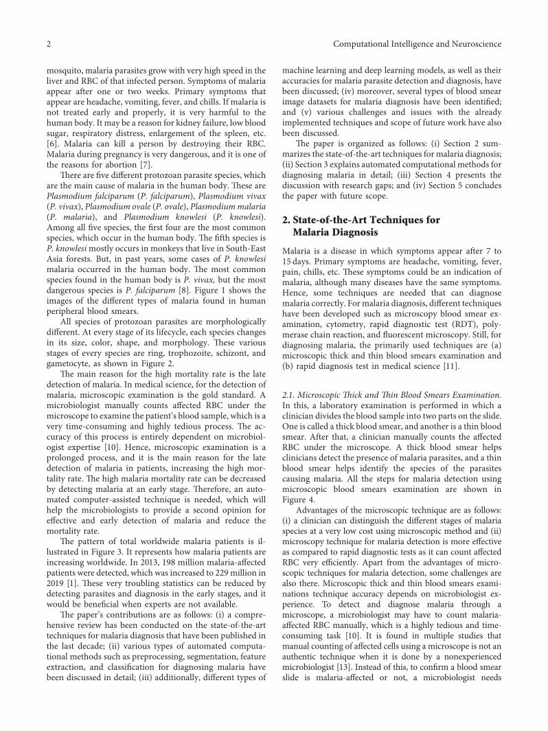
All species of protozoan parasites are morphologicallydifferent. At every stage of its lifecycle, each species changesin its size, color, shape, and morphology. +ese variousstages of every species are ring, trophozoite, schizont, andgametocyte, as shown in Figure 2.
+e main reason for the high mortality rate is the latedetection of malaria. In medical science, for the detection ofmalaria, microscopic examination is the gold standard. Amicrobiologist manually counts affected RBC under themicroscope to examine the patient’s blood sample, which is avery time-consuming and highly tedious process. +e ac-curacy of this process is entirely dependent on microbiol-ogist expertise [10]. Hence, microscopic examination is aprolonged process, and it is the main reason for the latedetection of malaria in patients, increasing the high mor-tality rate. +e high malaria mortality rate can be decreasedby detecting malaria at an early stage. +erefore, an auto-mated computer-assisted technique is needed, which willhelp the microbiologists to provide a second opinion foreffective and early detection of malaria and reduce themortality rate.
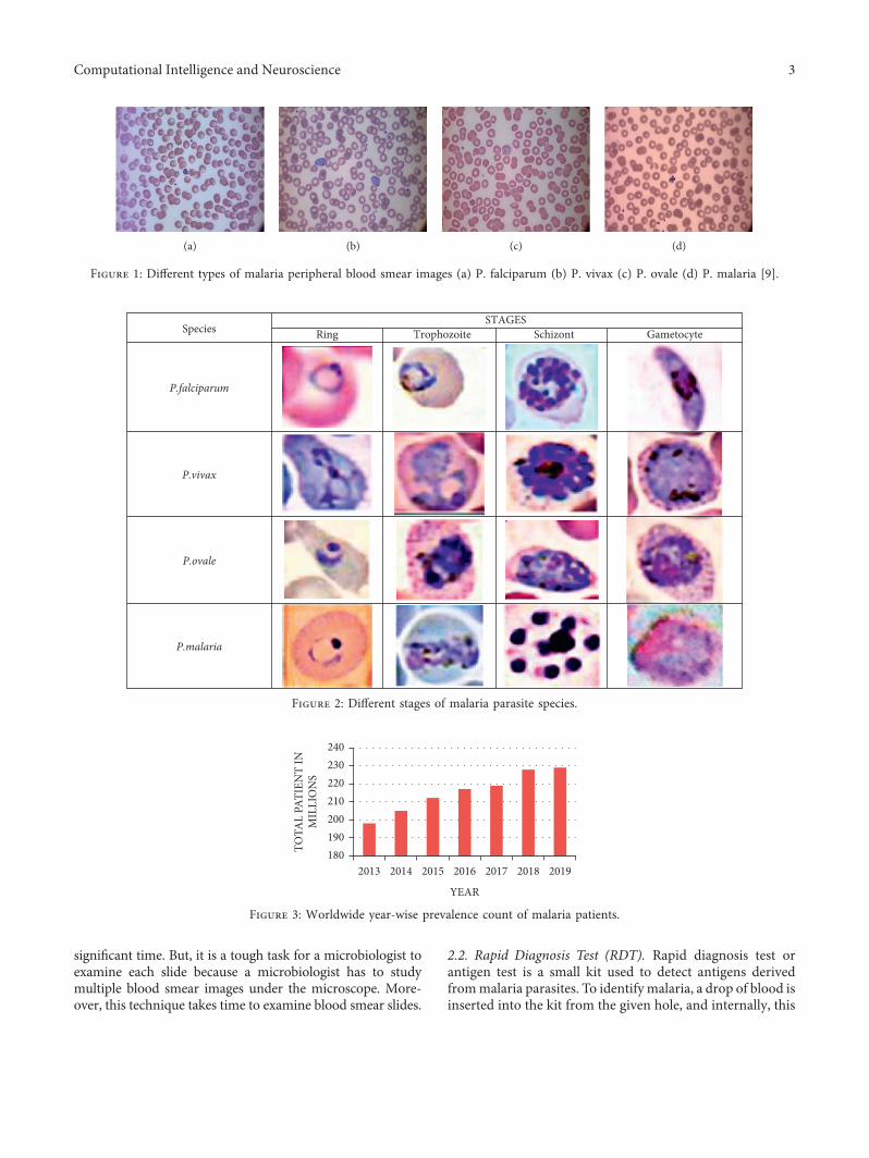
+e pattern of total worldwide malaria patients is il-lustrated in Figure 3. It represents how malaria patients areincreasing worldwide. In 2013, 198 million malaria-affectedpatients were detected, which was increased to 229million in2019 [1]. +ese very troubling statistics can be reduced bydetecting parasites and diagnosis in the early stages, and itwould be beneficial when experts are not available.
+e paper’s contributions are as follows: (i) a compre-hensive review has been conducted on the state-of-the-arttechniques for malaria diagnosis that have been published inthe last decade; (ii) various types of automated computa-tional methods such as preprocessing, segmentation, featureextraction, and classification for diagnosing malaria havebeen discussed in detail; (iii) additionally, different types of
machine learning and deep learning models, as well as theiraccuracies for malaria parasite detection and diagnosis, havebeen discussed; (iv) moreover, several types of blood smearimage datasets for malaria diagnosis have been identified;and (v) various challenges and issues with the alreadyimplemented techniques and scope of future work have alsobeen discussed.
+e paper is organized as follows: (i) Section 2 sum-marizes the state-of-the-art techniques for malaria diagnosis;(ii) Section 3 explains automated computational methods fordiagnosing malaria in detail; (iii) Section 4 presents thediscussion with research gaps; and (iv) Section 5 concludesthe paper with future scope.
2. State-of-the-Art Techniques forMalaria Diagnosis
Malaria is a disease in which symptoms appear after 7 to15 days. Primary symptoms are headache, vomiting, fever,pain, chills, etc. +ese symptoms could be an indication ofmalaria, although many diseases have the same symptoms.Hence, some techniques are needed that can diagnosemalaria correctly. Formalaria diagnosis, different techniqueshave been developed such as microscopy blood smear ex-amination, cytometry, rapid diagnostic test (RDT), poly-merase chain reaction, and fluorescent microscopy. Still, fordiagnosing malaria, the primarily used techniques are (a)microscopic thick and thin blood smears examination and(b) rapid diagnosis test in medical science [11].
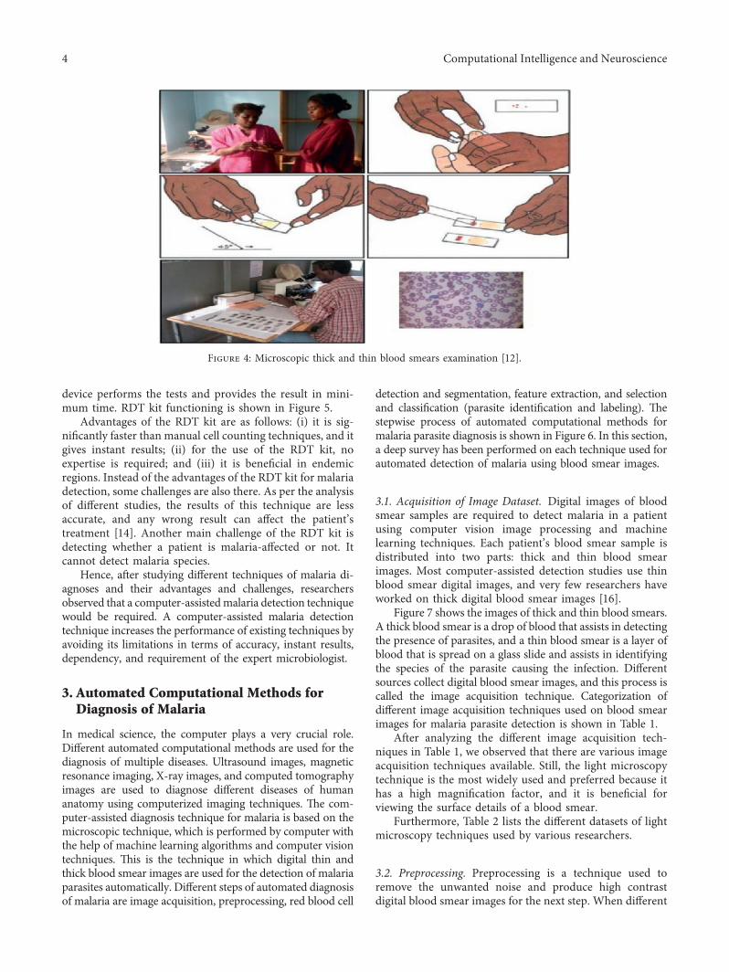
2.1. Microscopic 0ick and 0in Blood Smears Examination.In this, a laboratory examination is performed in which aclinician divides the blood sample into two parts on the slide.One is called a thick blood smear, and another is a thin bloodsmear. After that, a clinician manually counts the affectedRBC under the microscope. A thick blood smear helpsclinicians detect the presence of malaria parasites, and a thinblood smear helps identify the species of the parasitescausing malaria. All the steps for malaria detection usingmicroscopic blood smears examination are shown inFigure 4.
Advantages of the microscopic technique are as follows:(i) a clinician can distinguish the different stages of malariaspecies at a very low cost using microscopic method and (ii)microscopy technique for malaria detection is more effectiveas compared to rapid diagnostic tests as it can count affectedRBC very efficiently. Apart from the advantages of micro-scopic techniques for malaria detection, some challenges arealso there. Microscopic thick and thin blood smears exami-nations technique accuracy depends on microbiologist ex-perience. To detect and diagnose malaria through amicroscope, a microbiologist may have to count malaria-affected RBC manually, which is a highly tedious and time-consuming task [10]. It is found in multiple studies thatmanual counting of affected cells using a microscope is not anauthentic technique when it is done by a nonexperiencedmicrobiologist [13]. Instead of this, to confirm a blood smearslide is malaria-affected or not, a microbiologist needs
2 Computational Intelligence and Neuroscience
significant time. But, it is a tough task for a microbiologist toexamine each slide because a microbiologist has to studymultiple blood smear images under the microscope. More-over, this technique takes time to examine blood smear slides.
2.2. Rapid Diagnosis Test (RDT). Rapid diagnosis test orantigen test is a small kit used to detect antigens derivedfrommalaria parasites. To identify malaria, a drop of blood isinserted into the kit from the given hole, and internally, this
(a) (b) (c) (d)
Figure 1: Different types of malaria peripheral blood smear images (a) P. falciparum (b) P. vivax (c) P. ovale (d) P. malaria [9].
SpeciesSTAGES
Ring Trophozoite Schizont Gametocyte
P.falciparum
P.vivax
P.ovale
P.malaria
Figure 2: Different stages of malaria parasite species.
2013180190200210220230240
TOTA
L PA
TIEN
T IN
MIL
LIO
NS
2014 2015
YEAR
2016 2017 2018 2019
Figure 3: Worldwide year-wise prevalence count of malaria patients.
Computational Intelligence and Neuroscience 3
device performs the tests and provides the result in mini-mum time. RDT kit functioning is shown in Figure 5.
Advantages of the RDT kit are as follows: (i) it is sig-nificantly faster than manual cell counting techniques, and itgives instant results; (ii) for the use of the RDT kit, noexpertise is required; and (iii) it is beneficial in endemicregions. Instead of the advantages of the RDT kit for malariadetection, some challenges are also there. As per the analysisof different studies, the results of this technique are lessaccurate, and any wrong result can affect the patient’streatment [14]. Another main challenge of the RDT kit isdetecting whether a patient is malaria-affected or not. Itcannot detect malaria species.
Hence, after studying different techniques of malaria di-agnoses and their advantages and challenges, researchersobserved that a computer-assistedmalaria detection techniquewould be required. A computer-assisted malaria detectiontechnique increases the performance of existing techniques byavoiding its limitations in terms of accuracy, instant results,dependency, and requirement of the expert microbiologist.
3. Automated Computational Methods forDiagnosis of Malaria
In medical science, the computer plays a very crucial role.Different automated computational methods are used for thediagnosis of multiple diseases. Ultrasound images, magneticresonance imaging, X-ray images, and computed tomographyimages are used to diagnose different diseases of humananatomy using computerized imaging techniques. +e com-puter-assisted diagnosis technique for malaria is based on themicroscopic technique, which is performed by computer withthe help of machine learning algorithms and computer visiontechniques. +is is the technique in which digital thin andthick blood smear images are used for the detection of malariaparasites automatically. Different steps of automated diagnosisof malaria are image acquisition, preprocessing, red blood cell
detection and segmentation, feature extraction, and selectionand classification (parasite identification and labeling). +estepwise process of automated computational methods formalaria parasite diagnosis is shown in Figure 6. In this section,a deep survey has been performed on each technique used forautomated detection of malaria using blood smear images.
3.1. Acquisition of Image Dataset. Digital images of bloodsmear samples are required to detect malaria in a patientusing computer vision image processing and machinelearning techniques. Each patient’s blood smear sample isdistributed into two parts: thick and thin blood smearimages. Most computer-assisted detection studies use thinblood smear digital images, and very few researchers haveworked on thick digital blood smear images [16].
Figure 7 shows the images of thick and thin blood smears.A thick blood smear is a drop of blood that assists in detectingthe presence of parasites, and a thin blood smear is a layer ofblood that is spread on a glass slide and assists in identifyingthe species of the parasite causing the infection. Differentsources collect digital blood smear images, and this process iscalled the image acquisition technique. Categorization ofdifferent image acquisition techniques used on blood smearimages for malaria parasite detection is shown in Table 1.
After analyzing the different image acquisition tech-niques in Table 1, we observed that there are various imageacquisition techniques available. Still, the light microscopytechnique is the most widely used and preferred because ithas a high magnification factor, and it is beneficial forviewing the surface details of a blood smear.
Furthermore, Table 2 lists the different datasets of lightmicroscopy techniques used by various researchers.
3.2. Preprocessing. Preprocessing is a technique used toremove the unwanted noise and produce high contrastdigital blood smear images for the next step. When different
Figure 4: Microscopic thick and thin blood smears examination [12].
4 Computational Intelligence and Neuroscience

‘C’ - control line ‘T’ - test line
Results windowSquare hole (for blood) Round hole
(for buffer)
Figure 5: Rapid diagnosis testing (RDT) kit [15].
Segmentation
Blood ImageAcquisition
Image Pre-Processing
Training Testing
Classification
Non-InfectedMalaria
InfectedMalaria
Dataset
PerformanceEvaluation
SensitivitySpecificityAccuracy
PPV
Figure 6: Computational methods for automated diagnosis system for malaria.
Figure 7: Malaria infected thin (left) and thick (right) blood smear image.
Table 1: Categorization of image acquisition techniques used on blood smear images for malaria parasite detection.
References Lightmicroscopy
Binocularmicroscopy
Fluorescentmicroscopy
Polarizedmicroscopy
Multispectral andmultimodalmicroscopy
Image-basedcytometer
Scanningelectron
microscopy[17] ✓[18] ✓[19] ✓[20] ✓[21] ✓[22] ✓[23] ✓[24] ✓[25] ✓[26] ✓[27] ✓[28] ✓[29] ✓[30] ✓
Computational Intelligence and Neuroscience 5

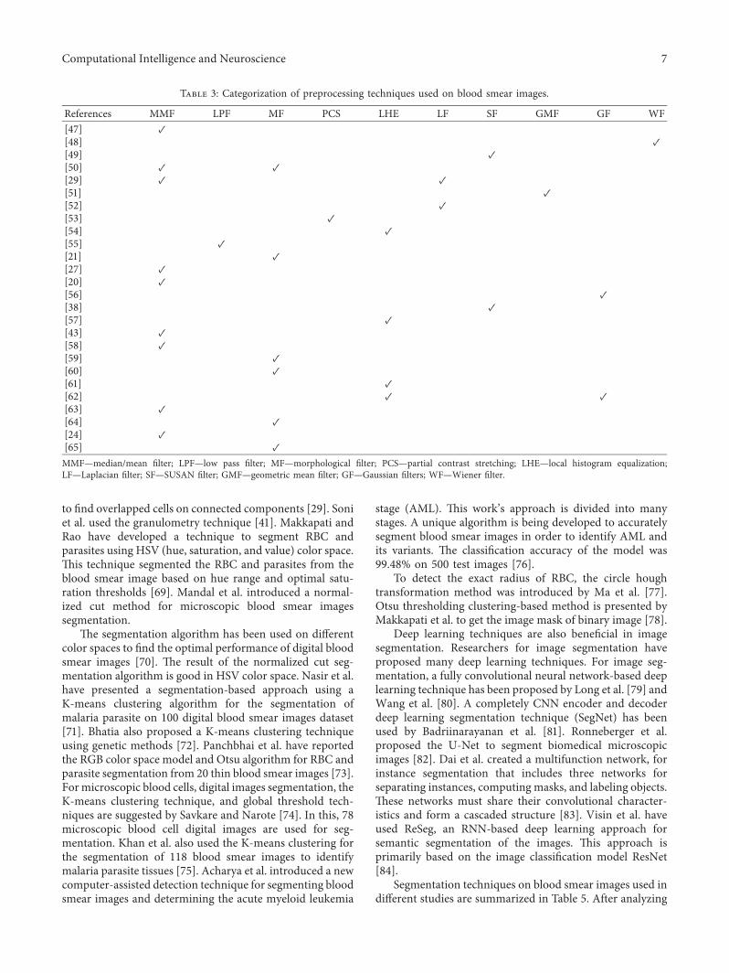
resources take blood smear images, the images are corruptedby noise, and thus, visualization of the images is not good.Due to this problem, further steps of segmentation andclassification are challenging to implement, and it producespoor results. Hence, certain preprocessing techniques havebeen used to remove that unwanted noise from images.Preprocessing techniques remove the noise from the imagefor better visualization, which is very useful for furtheranalysis [46]. As shown in Table 3, researchers used multiplepreprocessing techniques such as median filter, mean filter,low-pass filter, morphological filter, partial contraststretching, local histogram equalization, Laplacian filter,SUSAN filter, geometric mean filter, Gaussian filters, andWiener filter for enhancing the contrast and remove theunwanted noise of digital images. May et al. have given anapproach in which the median filter technique for removingthe impulse noise from digital images and for removingadditive noise has been used [4]. +e Gaussian filter is usedby Arco et al. to enhance the quality of the images affected byGaussian noise.+e geometric mean filter is also used by Daset al. for preserving the edges and removing Gaussian noisefrom the digital microscopic image [66]. Laplacian filter isused by Savkare et al. for smoothening and enhancing edgesof malaria parasite images [29]. To preserve the structure ofan image, Susan’s filter is suggested by [41]. To remove the
intensity of high frequency from a digital image, a low passfilter has been suggested by [55]. +e histogram matchingtechnique is used by Abbas et al. to normalize the intensityvalue of digital image pixels [67]. +e categorization ofpreprocessing techniques to enhance the quality of digitalblood smear images is shown in Table 3.
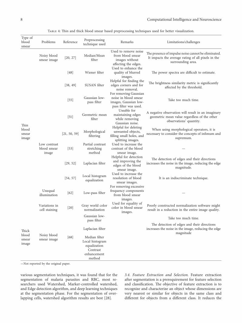
Table 4 displays the image preprocessing approachesused by various researchers for better visualization of thinand thick blood smear images with their properties.
3.3. Red Blood Cell Detection and Segmentation.Segmentation is the process in which digital images aredisjoint into nonoverlapping regions. Each disjoint imagetypically corresponds to other parts of an image object. Onceeach digital image object is isolated, each object can be easilymeasured and classified. In the literature, different seg-mentation techniques have been applied on digital bloodsmear images to detect ROI (region of interest).
Das et al. have developed an automated system forclassifying malaria at different stages. +e researcher usedthe watershed segmentation technique in their researchwork for the segmentation of thin blood smear digital im-ages. +is technique provided better results for detectingerythrocytes from the whole blood smear image [66].Further, a watershed algorithm is suggested by Savkare et al.
Table 1: Continued.
References Lightmicroscopy
Binocularmicroscopy
Fluorescentmicroscopy
Polarizedmicroscopy
Multispectral andmultimodalmicroscopy
Image-basedcytometer
Scanningelectron
microscopy[31] ✓[32] ✓[33] ✓[34] ✓[35] ✓[36] ✓[37] ✓[38] ✓[39] ✓[40] ✓[41] ✓[42] ✓
Table 2: Light microscopy datasets used by different researchers.
Reference No. of images indataset Remarks
[43] 300 images Used KNN classifier on light microscopic images, got 91% accuracy.
[33] 21 images Light microscopic images of 1296×1024 resolution captured by an axiocam high-resolution color camerawere used.
[44] — —[29] 68 images Light microscopic images of different magnification have been used.[25] 300 images Used KNN classifier and got 90.17% accuracy to detect malaria parasite species.
[26] 27578 images 27578 single cell light microscopy images were used, and a new 16-layer CNN model was proposed toidentify malaria-infected or infected images.
[23] 160 images Achieve 95% accuracy for the detection of malaria.
[45] — Used Giemsa-stained blood smear images were taken by a camera attached with a microscope on 1000xmagnification, and the proposed model got 77.78% accuracy.
[19] 27558 images Implement novel stacked convolutional neural network technique for parasite detection.—Not reported by the original paper.
6 Computational Intelligence and Neuroscience
to find overlapped cells on connected components [29]. Soniet al. used the granulometry technique [41]. Makkapati andRao have developed a technique to segment RBC andparasites using HSV (hue, saturation, and value) color space.+is technique segmented the RBC and parasites from theblood smear image based on hue range and optimal satu-ration thresholds [69]. Mandal et al. introduced a normal-ized cut method for microscopic blood smear imagessegmentation.
+e segmentation algorithm has been used on differentcolor spaces to find the optimal performance of digital bloodsmear images [70]. +e result of the normalized cut seg-mentation algorithm is good in HSV color space. Nasir et al.have presented a segmentation-based approach using aK-means clustering algorithm for the segmentation ofmalaria parasite on 100 digital blood smear images dataset[71]. Bhatia also proposed a K-means clustering techniqueusing genetic methods [72]. Panchbhai et al. have reportedthe RGB color space model and Otsu algorithm for RBC andparasite segmentation from 20 thin blood smear images [73].Formicroscopic blood cells, digital images segmentation, theK-means clustering technique, and global threshold tech-niques are suggested by Savkare and Narote [74]. In this, 78microscopic blood cell digital images are used for seg-mentation. Khan et al. also used the K-means clustering forthe segmentation of 118 blood smear images to identifymalaria parasite tissues [75]. Acharya et al. introduced a newcomputer-assisted detection technique for segmenting bloodsmear images and determining the acute myeloid leukemia
stage (AML). +is work’s approach is divided into manystages. A unique algorithm is being developed to accuratelysegment blood smear images in order to identify AML andits variants. +e classification accuracy of the model was99.48% on 500 test images [76].
To detect the exact radius of RBC, the circle houghtransformation method was introduced by Ma et al. [77].Otsu thresholding clustering-based method is presented byMakkapati et al. to get the image mask of binary image [78].
Deep learning techniques are also beneficial in imagesegmentation. Researchers for image segmentation haveproposed many deep learning techniques. For image seg-mentation, a fully convolutional neural network-based deeplearning technique has been proposed by Long et al. [79] andWang et al. [80]. A completely CNN encoder and decoderdeep learning segmentation technique (SegNet) has beenused by Badriinarayanan et al. [81]. Ronneberger et al.proposed the U-Net to segment biomedical microscopicimages [82]. Dai et al. created a multifunction network, forinstance segmentation that includes three networks forseparating instances, computing masks, and labeling objects.+ese networks must share their convolutional character-istics and form a cascaded structure [83]. Visin et al. haveused ReSeg, an RNN-based deep learning approach forsemantic segmentation of the images. +is approach isprimarily based on the image classification model ResNet[84].
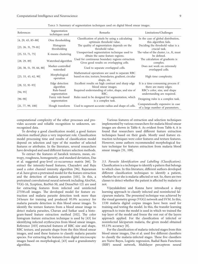
Segmentation techniques on blood smear images used indifferent studies are summarized in Table 5. After analyzing
Table 3: Categorization of preprocessing techniques used on blood smear images.
References MMF LPF MF PCS LHE LF SF GMF GF WF[47] ✓[48] ✓[49] ✓[50] ✓ ✓[29] ✓ ✓[51] ✓[52] ✓[53] ✓[54] ✓[55] ✓[21] ✓[27] ✓[20] ✓[56] ✓[38] ✓[57] ✓[43] ✓[58] ✓[59] ✓[60] ✓[61] ✓[62] ✓ ✓[63] ✓[64] ✓[24] ✓[65] ✓MMF—median/mean filter; LPF—low pass filter; MF—morphological filter; PCS—partial contrast stretching; LHE—local histogram equalization;LF—Laplacian filter; SF—SUSAN filter; GMF—geometric mean filter; GF—Gaussian filters; WF—Wiener filter.
Computational Intelligence and Neuroscience 7
various segmentation techniques, it was found that for thesegmentation of malaria parasites and RBC, most re-searchers used Watershed, Marker-controlled watershed,and Edge detection algorithm, and deep learning techniquesat the segmentation phase. For the segmentation of over-lapping cells, watershed algorithm results are best [28].
3.4. Feature Extraction and Selection. Feature extractionafter segmentation is a prerequirement for feature selectionand classification. +e objective of feature extraction is torecognize and characterize an object whose dimensions arevery nearest or similar for objects in the same class anddifferent for objects from a different class. It reduces the
Table 4: +in and thick blood smear based preprocessing techniques used for better visualization.
Type ofbloodsmear
Problems Reference Preprocessingtechnique used Remarks Limitations/challenges
+inbloodsmearimage
Noisy bloodsmear image [20, 27] Median/Mean
filter
Used to remove noisefrom blood smearimages without
affecting the edges.
+e presence of impulse noise cannot be eliminated.It impacts the average rating of all pixels in the
surrounding area.
[48] Wiener filterUsed to enhance thequality of blurred
images.+e power spectra are difficult to estimate.
[38, 49] SUSAN filterHelpful for finding theedges corners and for
noise removal.
+e brightness similarity metric is significantlyaffected by the threshold.
[55] Gaussian low-pass filter
For removing Gaussiannoise in blood smearimages, Gaussian low-pass filter was used.
Take too much time.
[51] Geometric meanfilter
Useable formaintaining edgeswhile removingGaussian noise.
A negative observation will result in an imaginarygeometric mean value regardless of the other
observations’ quantity.
[21, 50, 59] Morphologicalfiltering
Helpful for deletingunwanted objects,
filling small holes, andsplitting images.
When using morphological operators, it isnecessary to consider the concepts of infimum and
supremum.
Low contrastblood smear
image[53]
Partial contraststretchingmethod
Used to increase thecontrast of the blood
smear image.—
[29, 52] Laplacian filter
Helpful for detectionand improving theedges of the blood
smear image.
+e detection of edges and their directionsincreases the noise in the image, reducing the edge
magnitude.
[54, 57] Local histogramequalization
Used to increase theresolution of blood
smear images.It is an indiscriminate technique.
Unequalillumination [62] Low-pass filter
For removing excessivefrequency componentsfrom blood smear
images.
—
Variations incell staining [20] Gray world color
normalization
Used for equality ofcolor in blood smear
images.
Poorly constructed normalization software mightresult in a reduction in the entire image quality.
+ickbloodsmearimage
Noisy bloodsmear image [68]
Gaussian low-pass filter Take too much time.
Laplacian filter+e detection of edges and their directions
increases the noise in the image, reducing the edgemagnitude
Median filterLocal histogramequalizationContrast
enhancementmethod
—Not reported by the original paper.
8 Computational Intelligence and Neuroscience
computational complexity of the other processes and pro-vides accurate and reliable recognition to unknown, un-recognized data.
To develop a good classification model, a good featureselection method plays a very important role. Classificationmodel processing time and results of classification modeldepend on selection and type of the number of selectedfeatures or attributes. In the literature, several researchershave developed and used different feature selectionmethods.
To extract the features of haralick textures, mean, en-tropy, roughness, homogeneity, and standard deviation, Daset al. suggested gray-level co-occurrence matrix [66]. Toextract the intensity-based features, Chayadevi and Rajuused a color channel intensity algorithm [96]. Rajaramanet al. have given a pretrained model for the feature extractionand the detection of malaria parasites [101]. In this, apretrained convolutional neural network including AlexNet,VGG-16, Xception, ResNet-50, and DenseNet-121 are usedfor extracting features from infected and uninfected27558 cell images. +e developed model for feature ex-traction and malaria parasite detection took more than24 hours for training and produced 95.9% accuracy formalaria parasite detection in thin blood smear images. Toidentify the texture features from a blood smear image todetect malaria parasites, Chavan and Sutkar used a histo-gram-based feature extraction method [102]. +e colorhistogram feature extraction technique is used by [43] foridentifying infected erythrocytes from blood smear images.Reference [103] extracted features of RBC size and shape,RBC texture, and parasite shape from the thin blood smearimages, and used these features to classify malaria parasitespecies. For extracting the features from digital microscopicimages based on morphological, [43] used a granulometryalgorithm.
Various features of extraction and selection techniquesimplemented by various researchers for malaria blood smearimages are shown in Table 6. As evident from Table 6, it isfound that researchers used different feature extractiontechniques based on their goals. Mostly used feature ex-traction techniques were color features and texture features.However, some authors recommended morphological fea-ture technique for features extraction from malaria bloodsmear images [51, 108].
3.5. Parasite Identification and Labelling (Classification).Classification is a technique to identify a pattern that belongsto which class. In this literature, different authors developeddifferent classification techniques to identify a patient,whether he or she is malaria-affected or not. So, there are twoclasses to detect whether the patient is affected by malaria ornot.
Vijayalakshmi and Kanna have introduced a deeplearning approach to classify infected and noninfected fal-ciparum malaria. +e presented technique was achieved bythe visual geometry group (VGG) network and SVM. In this,1530 malaria digital corpus images have been used fortraining and testing the model. In this, the transfer learningapproach to train the model is used in which we trained thetop layer of the model and freeze the rest out of the layersapproach applied. For the classification of infected ornoninfected falciparum malaria, the given model obtained93.13% accuracy [8].
For the classification of malaria-infected stages from thinblood smear images, Das et al. used five different classifiersto classify the malaria-infected stages. +ese five classifiersare Naive Bayes, Logistic regression, Radial Basis Functions(RBF) neural network, Multilayer perceptron neural
Table 5: Summary of segmentation techniques used on digital blood smear images.
References Segmentationtechniques used Remarks Limitations/Challenges
[4, 29, 43, 85–88] Otsu thresholding Classification of pixels by using a calculatingoptimum threshold value.
In the case of global distribution,this algorithm fails.
[23, 26, 31, 79–81] Histogramthresholding
+e quality of segmentation depends on thethreshold value.
Deciding the threshold value is acrucial task.
[25, 53, 71, 75] K-means clustering Unsupervised segmentation technique used toobtain the same feature regions.
+e value of the cluster, i.e., K, mustbe defined.
[28, 29, 89] Watershed algorithm Used for continuous boundary regions extraction.Gives good results on overlapping cells.
+e calculation of gradients iscomplex.
[20, 38, 51, 59, 66, 88] Marker-controlledwatershed Used to separate overlapped cells. Does not work on extremely
overlapped cells.
[23, 33, 43, 62, 90] Morphologicaloperation
Mathematical operations are used to separate RBCbased on size, texture, boundaries, gradient, circular
shape, etc.High time complexity.
[28, 32, 91–93] Edge detectionalgorithm
Excellent results on high contrast and sharp edgeblood smear images.
It is a time-consuming process ifthere are many edges.
[94, 95] Rule-basedsegmentation
Required understanding of color, shape, and size ofRBC.
RBC’s color, size, and shapeunderstanding are required.
[96–98] Fuzzy rule-basedsegmentation
Rules need to be designed for segmentation, whichis a complex task. Designing rules is a complex task.
[21, 77, 99, 100] Hough transform Used to segment accurate radius and shape of cells. Computationally expensive in caseof a large number of parameters.
Computational Intelligence and Neuroscience 9

Tabl
e6:
Differenttechniqu
esused
fortheextractio
nof
features
andselectionfrom
malaria
bloo
dsm
earim
ages.
References
Color
features
Texturefeature
Morph
ological
feature
RGB
HSV
YCbC
rLab
Intensity
CCM
Haralick
GLR
LMGLC
MLB
PFractal
WT
GT
Entrop
ySIFT
Shape
Mom
ents
Area
[24]
✓[104]
✓✓
[105]
✓✓
[106]
✓✓
✓✓
[96]
✓✓
[70]
✓✓
[107]
✓✓
[108]
✓✓✓
✓[109]
✓✓
[51]
✓✓
✓✓
✓[100]
✓✓
✓✓
[25]
✓[110]
✓✓
✓✓
[111]
✓[43]
✓✓
✓[38]
✓[66]
✓[102]
[65]
✓✓
[21]
✓✓
[20]
✓[31]
✓✓
RGB—
redgreenblue;H
SV—hu
e,saturatio
n,andvalue;
CCM—colorco-occurrencematrix;
GLR
LM—Gray-levelrun
leng
thmatrices;GLC
M—gray-le
velc
o-occurrence
matrix;
LBP—
localb
inarypatte
rn;
WT—
wavelet
transform;G
T—gradient
texture.
10 Computational Intelligence and Neuroscience
network, and classification and regression tree. In this, 888erythrocytes infected and noninfected image dataset is used.Out of this, 592 labeled images are used to train the classifiersand the remaining are used for testing the classifiers. +eexperimental results show that among all five classifiers, themultilayer perceptron network has provided better resultsthan the other four classifiers on the 750 images dataset [66].
Seman et al. developed a multilayer perceptron network(MLP) to classify different malaria parasite species from thinblood smear images. +is work classified three differentspecies from malaria parasites: P. falciparum, P. malariae,and P. vivax [103]. +e authors used the backpropagationalgorithm of the MLP network for training and comparedthe results of the MLP network with Levenberg–Marquardtand Bayesian rule algorithms. MLP network has producedbetter results as compared to the other two algorithms.
Otsu thresholding method is used by Malihi et al. for theclassification of four species of malaria parasites in bloodsmear images. +is technique has provided better results incomparison with other techniques. In this, 363 blood smearimages are used and obtained 91% accuracy [43].
Further, Anggraini et al. have classified the differentstages of malaria parasites using a Bayesian classifier on 110thin blood smear images and obtained 93.3% accuracy [112].Minimum distance classifier technique has been given byGhate et al. for detecting the presence of malaria parasitesusing 80 blood smear images and got 83.75% accuracy [39].Savkare and Narote presented Otsu thresholding, watershedtransform, and SVM binary classifier to classify normal andparasite-infected cells [113]. Das et al. have presented theBayesian approach for automated screening of malariaparasite from microscopic images [51].
Kareem et al. have developed an automated techniquefor detecting malaria parasites in thin blood smear images.In this, a dataset of more than 200 images is used. Twomethods of classification for parasites are used. +e first oneis based on relative size and morphology, and the second isbased on intensity variation. +e final results of the devel-oped model have shown an accuracy rate of 87% [36].
Prasad et al. have presented a decision support systemapproach to classify the infected and noninfected malariaparasites in thin blood smear images. In this, 200 thin bloodsmear images have been used, and 96% accuracy has beenobtained [114]. Rosado et al. have developed a supervisedclassification technique to detect malaria parasites in bloodsmears. In this, machine learning (ML) classification 10-foldcross-validation for WBC (white blood cell) andP. falciparum trophozoites detection has been performedand got 91.8% accuracy [85].
Mohammed and Abdelrahman have given a techniquefor detecting and classifying malaria from 160 thin bloodsmear images taken from the Centre for Disease Control andPrevention (CDC). To extract the RBC from blood smearimages, researchers used morphological processing. +istechnique found the parasites and overlapped cells in theimage. Based on the number of RBCs in each image, RBC isclassified into two classes: infected and noninfected cells.After that, a normalized cross-correlation algorithm wasemployed to classify the affected blood smear parasite into
four different malaria species. +e given method has pro-duced 95% accuracy for detection [23]. Saiprasath et al.evaluated seven different machine learning algorithms onthe same malaria image dataset and concluded that RandomForest outperforms every other algorithm, closely followedby the Ada Boost algorithm [115].
Bibin et al. have given an automated technique to detectmalaria parasites in peripheral blood smear images. +egiven binary classifier is based on deep learning, which used1978 images to train and test the technique and achieved96.21% accuracy [106]. Simon et al. suggested a CNN-RNNmodel for malaria detection. Compared with the CNN-LSTM and CNN-GRU models, the proposed model gen-erated the best results [116]. Dev et al. suggested a hierar-chical convolutional network and produced better resultsthan prior studies [117].
Dave et al. used adaptive thresholding, erosion, anddilation operations to diagnose malaria from 117 bloodsmear microscopic digital images and got 89.88% accuracy[34]. Oliveira et al. have suggested the face detection al-gorithm to identify Plasmodium parasites from bloodsamples. In this, a dataset of 1332 blood sample images hasbeen taken and shown 91% accuracy [118].
Mohanty et al. have presented the autoencoder (AE)neural network unsupervised technique to identify malariain blood smear images. In this, the AE technique has beencompared with the SOM technique. +e AE techniqueobtained 87.5% accuracy compared to the 79% accuracy ofthe SOM technique. 1182 blood smear images have beenused to perform experiments [119]. Morales-Lopez et al.suggested the SVM technique for classification problems[120]. Table 7 has listed different types of classificationtechniques used for the identification of malaria parasites.
After the analysis of Table 7 and literature of malariaparasite classification techniques, it is found that variousclassification techniques that researchers commonly im-plement are CNN, SVM, and TL-VGG classifiers.
4. Discussion
In the last decade, a lot of experiments have been done in thearea of automated detection of malaria to reach the currentstate-of-the-art. In this study, different computationalmethods implemented on various stages of computer-assisted techniques for detecting malaria parasites usingblood smear images have been examined. Image acquisitionis the first and very important step for automatic detection ofthe malaria parasite. +e present study shows varioustechniques for acquiring digital blood smear images, but thelight microscopy technique is the most widely used and likedtechnique by researchers. +ere is a number of computa-tional methods out of which preprocessing is the first step inimage analysis.
Preprocessing is one of the crucial stages implementedon acquired digital blood smear images. It plays a crucial rolein detecting infected RBC by removing the unwanted noiseand producing high contrast digital blood smear imageswithout demolishing the image features. As per the currentstudy, median/mean filter, morphological filter, Laplacian
Computational Intelligence and Neuroscience 11

Table 7: Different classification techniques used for the identification of malaria parasites.
Reference Technique used Dataset Accuracy(%) Limitations/challenges
[103] Multilayer perceptron network for classification ofmalaria species.
562 malariaimages 89.90 Computation cost is very high.
[113]Otsu thresholding, watershed transform, and SVMbinary classifier for classification of normal and
parasite-infected cells.
15 malariaimages 93.12 Species detection of malaria is not done.
Not suitable for large datasets.
[121] Comprehensive CAD techniques with 10-fold cross-validation.
1182malariaimages
89.10 Training and testing time is very high forlarge datasets.
[93] Suggested SVM technique to find the different stages ofinfected malaria parasite
530 malariaimages 86 Feature scaling is required.
[73]Used RGB color space model and Otsu algorithm forRBC and parasite segmentation from thin blood smear
images.
20 malariaimages 92
+e unpredictability and imperfections inmicroscope pictures make precise
detection difficult.
[114] +e decision support system for the classification of aninfected and noninfected parasite of malaria.
200 malariaimages 96 FP rate is 20% and used only thin blood
smear images.
[122] Used minimum distance classifier to detect malariaparasites in blood smear images.
80 malariaimages 83.75 Dataset size is very small.
[43] Used SVM, NM, KNN, 1-NN, and Fisher classifiers toclassify different malaria species.
363 malariaimages 91 Using a hybrid approach, results can be
improved.
[51] Used Bayesian algorithm for detection of the malariaparasite.
888 malariaimages 84 Detect only 1 stage of malaria.
[123]An artificial neural network has been used to identifythe different malaria species from malaria parasites’
blood smear images.
200 malariaimages 79.7 Performance can be improved by
extracting more features.
[96] Used the neural network method to identify infectedRBC from blood smear images.
476 malariaimages 94.45 Results can be improved by training the
model on a large dataset.
[75] K-means clustering has been used for the segmentationof malaria parasites cells.
118 malariaimages 95 Other types of parasites are not detectable
with this technique.
[74]For the segmentation of RBC, the K-means clusteringtechnique and global threshold technique have been
used.
78 malariaimages 95.5 Dataset size is minimal.
[66]For the classification of gametocyte stage and ring stageof malaria species, multilayer perceptron network and
4 other classifiers have been used.
750 malariaimages 96.73 By increasing training size, more accurate
results can be achieved.
[124] Used artificial neural network (ANN) for the detectionof malaria parasite using morphological features.
7 malariaimages 73.57 Achieve better results by increasing
dataset size and using 2 or more classifiers.
[85] Used SVM classifier for WBC and P. falciparumtrophozoites detection.
1843malariaimages
91.8 Implemented only with the mobile-basedframework.
[125]Used image processing and artificial intelligence
techniques and face detection algorithm to identifyplasmodium parasites from blood samples.
1332malariaimages
91Detected only 1 malaria parasite, andmore algorithms can explore to achieve
better accuracy.
[106] Malaria parasite detection using a deep belief network.1978
malariaimages
96.21 +e technique was not implemented on adataset acquired from a mobile phone.
[119] Used autoencoder neural network technique to identifymalaria in blood smear images.
1182malariaimages
87.5 +e segmentation technique can beimproved.
[101]
Used 6 pretrained CNN for feature extraction andsubsequent training for malaria parasite detection inthin blood smear images. +is model took more than
24 hours for training.
27558malariaimages
95.9 +e model took more than 24 hours fortraining.
[8]Used transfer learning approach based on VGG-SVMmodel to classify infected and noninfected falciparum
malaria parasite.
1530malariaimages
93.13 A trained model can recognize only 1falciparum malaria parasite.
[126] Used CNN based deep learning model (VGGNet-16architecture) for malaria parasite detection.
27558malariaimages
95.03 Results can be improved by implementingthe VGG-19 architecture.
12 Computational Intelligence and Neuroscience
filter, Susan filter, and Gaussian low-pass filter are mostlyused techniques by researchers to remove unwanted noiseand increase the contrast of the images. Segmentation is thenext stage after the preprocessing, which is used to segmentthe RBC to detect malaria parasites using blood smearimages to facilitate the classification process. As per thestudy of literature, mostly used segmentation techniques byresearchers are as follows: (i) Otsu thresholding for seg-mentation of parasite RBC; (ii) Marker-controlled watershedand Edge detection algorithm is used at the segmentationphase; and (iii) for the segmentation of overlapping cells, thewatershed algorithm has been widely used.
After segmentation, blood smear images have beenclassified to diagnose malaria-infected or not infected byfeature extraction and selection techniques. As per the study,Color features, Texture features, and Morphological featureshave been mostly used feature extraction techniques forearly diagnosis of malaria from blood smear images. Fromthe literature, it has been observed that the maximum ac-curacy of 95.03% has been achieved by CNN based deeplearning model in comparison with the VGG-SVM model[8, 101, 126].
A thorough review of the literature on automatedanalysis of malaria parasite using blood smear imagesyielded the following challenges and future directions:
+e accuracy of an automatic image classification modeldepends upon multiple aspects such as analysis of the digitalblood smear image depend on the staining method, mag-nification factor of an image, condition of nearest envi-ronment where the digital image has been collected like thebackground of the image, light in the room, and mostimportant quality and position of the camera. +erefore, astandard digital blood smear dataset is necessary to test andvalidate the model to obtain efficient and reliable results.
Many researchers have performed their experiments andpublished their articles in the same area. Moreover, anautomated computational-based computer vision method,which should be efficient and effective for automated de-tection of the malaria parasite from blood smear images,needs to be improvised according to the requirement of thecommunity.
+e community requires (i) standard image datasetbecause researchers’ datasets are mostly unstandardized.+e digital blood smear dataset depends on the charac-teristics and quality of the microscope as all digital imagesof blood smears are taken by a digital camera attached to amicroscope. So, a well-standardized dataset is most im-portant for a machine learning algorithm for automateddetection of malaria. (ii) In the literature, developedmethods can recognize only one type of malaria parasite[8]. But, the patient may be affected by more than one
parasites species. Hence, there is a need for such a modelthat can recognize different types of malaria parasites. (iii)To classify malaria parasites from blood smear images,authors trained the machines with different models andtechniques. +e training model is taking a long time tolearn [66]. Hence, there is a necessity to reduce the trainingtime to train the classification models. (iv) In literature,developed models by different researchers analyze theblood smear digital images that are taken from a camerathat is attached with a microscope [4, 8]. Hence, there is ademand for a model to analyze thin blood smears imagesacquired exclusively with smart phones [43]. (v) A tech-nique that different authors use to diagnose malaria isinvasive, in which an injection syringe takes a bloodsample. +erefore, there is a requirement for a noninvasivetechnique that can be used to detect malaria [128]. (vi)After the analysis of Table 7, it has been found that all theexisting state-of-the-art techniques used to detect malariafrom microscopic blood smear images are not very accu-rate. Each technique has some limitations and challenges.+erefore, there is a necessity for an automatic techniquethat can improve the accuracy for the detection of malariaparasites and it will also help in early detection of malariaand reduce the mortality rate in future.
5. Conclusion
+is study is a solid starting point for researchers who want tolook into automated blood smear analysis to detect malaria.+is study reviews and discusses computer vision and imageanalysis works that target the automated detection of malariaon blood smear images. In this paper, we have discussed thepresent facts of necessary components of computer-assistedtechnique: (i) acquisition of image dataset, (ii) preprocessing,(iii) segmentation of RBC, (iv) feature extraction and selec-tion, and (v) classification, which have been used to diagnosemalaria parasite from blood smear images suggested byvarious researchers. Digital blood smear images taken from amicroscope may affect how and which malaria parasites aredetected. After analyzing segmentation and classification stateof the art techniques, it has been observed that futurecomputer-assisted techniques should be based on standarddatasets and magnification factors to detect malaria parasites.+e complexity of different classifiers of machine learningthat are based on deep learning increases as the number oflayers increases. To achieve efficient and reliable results, alarge dataset is required for training and testing.With the helpof computational methods such as data augmentation anddeep learning methods, the computer-assisted method canobtain better results.
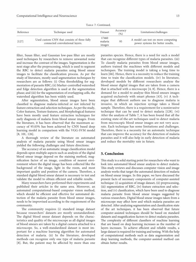
Table 7: Continued.
Reference Technique used Dataset Accuracy(%) Limitations/challenges
[127] Used custom CNN that consists of three fullyconnected convolutional layers.
17460malariaimages
95 A model can test on more computingpower systems for better results.
Computational Intelligence and Neuroscience 13
However, some state-of-the-art techniques are presentedin the literature, but still, there is a huge scope of futurework, which may help the microbiologists in the detectionand diagnosis of the malaria parasite at an earlier stage toreduce the mortality rate such as (i) different computationalmethods that are used to collect blood smear imagesphysically can be studied more to enhance the segmentationresults to detect infected RBC very effectively. Hence, anefficient computational method of infected RBC segmen-tation can be developed. (ii) Various feature extractionmethods such as color features, texture features, and mor-phological features [51, 106, 108] can be analyzed more,which will be very helpful for the development of an efficientautomated computer-assisted system to detect infectedmalaria RBC using blood smear images. (iii) To classifymalaria blood smear images, mostly implemented tech-niques are SVM, K-means, and VGG classifiers. Still, there isa vast scope to implement customize CNN algorithms todetect infected malaria RBC with high accuracy. If the CNNmodel is implemented on blood smear images at a minimummagnification factor for classification, it may decrease thecost and time complexity of the system.
In the field of malaria detection from blood smearimages, the contribution of many research publications isnoteworthy. However, this study has tried to presentopinions to the microbiologists and technical community. Itwill be very helpful for them to generate an effective andefficient computer-assisted technique for malaria detectionat an early stage. [129, 130].
Data Availability
Dataset is available at https://www.kaggle.com/iarunava/cell-images-for-detecting-malaria.
Conflicts of Interest
+e authors stated that there are no conflicts of interest.
Acknowledgments
+e authors would like to acknowledge Taif UniversityResearchers Supporting Project Number (TURSP-2020/125), Taif University, Taif, Saudi Arabia, http://shodh.inflibnet.ac.in:8080/jspui/bitstream/123456789/8785/1/shankar_phdeng17050_synopsis.pdf.
References
[1] Who, “World health organization report on malaria,” Re-port/2021, World health organization, Geneva, Switzerland,2021.
[2] Incidence of Malaria in India, “Incidence of malaria in In-dia,” 2020, https://www.malariasite.com/malaria-india.
[3] F. E. Cox, “History of the discovery of the malaria parasitesand their vectors,” Parasites & Vectors, vol. 3, no. 1, pp. 5–9,2010.
[4] Z. May and S. S. A. M. Aziz, “Automated quantification andclassification of malaria parasites in thin blood smears,” inProceedings of the 2013 IEEE International Conference on
Signal and Image Processing Applications, pp. 369–373,Melaka, Malaysia, October 2013.
[5] Who, “Transmission of malaria,” 2020, https://www.who.int/features/qa/10/en.
[6] H. M. Gilles, “Management of severe and complicatedmalaria,”A Practical Handbook, World Health Organization,Geneva, Switzerland, 1991.
[7] S. Murphy and J. Breman, “Gaps in the childhood malariaburden in Africa: cerebral malaria, neurological sequelae,anemia, respiratory distress, hypoglycemia, and complica-tions of pregnancy,” 0e American Journal of TropicalMedicine and Hygiene, vol. 64, no. 1_suppl, pp. 57–67, 2001.
[8] A. Vijayalakshmi and B. R. Kanna, “Deep learning approachto detect malaria from microscopic images,” MultimediaTools and Applications, vol. 79, no. 21, pp. 15297–15317,2020.
[9] A. Loddo, C. Di Ruberto, and M. Kocher, “Recent advancesof malaria parasites detection systems based on mathe-matical morphology,” Sensors, vol. 18, no. 2, p. 513, 2018.
[10] N. Tangpukdee, C. Duangdee, P. Wilairatana, andS. Krudsood, “Malaria diagnosis: a brief review,” KoreanJournal of Parasitology, vol. 47, no. 2, p. 93, 2009.
[11] Ncbi, “Malaria diagnosis: a brief review,” 2020, https://www.ncbi.nlm.nih.gov/pmc/articles/PMC2688806.
[12] A. Rosebrock, “Microscopic thick and thin blood smearsexamination,” 2020, http://medicostenerife.es/news/intelgigencia-artificial-deep-learning-and-medical-image-analysis-with-keras-for-malaria-deteccion-97-effectiveness.
[13] I. Bates, V. Bekoe, and A. Asamoa-Adu, “Improving theaccuracy of malaria-related laboratory tests in Ghana,”Malaria Journal, vol. 3, no. 1, pp. 1–5, 2004.
[14] A. Tankeshwar, “Rapid diagnosis test,” 2019, https://microbeonline.com/rdts-malaria-diagnosis-principle-results-advantages.
[15] V. N. Orish, V. F. De-Gaulle, and A. O. Sanyaolu, “Inter-preting rapid diagnostic test (RDT) for Plasmodium falci-parum,” BMC Research Notes, vol. 11, no. 1, pp. 850–856,2018.
[16] Z. Jan, A. Khan, M. Sajjad, K. Muhammad, S. Rho, andI. Mehmood, “A review on automated diagnosis of malariaparasite in microscopic blood smears images,” MultimediaTools and Applications, vol. 77, no. 8, pp. 9801–9826, 2018.
[17] K. M. F. Fuhad, J. F. Tuba, M. R. A. Sarker, S. Momen,N. Mohammed, and T. Rahman, “Deep learning based au-tomatic malaria parasite detection from blood smear and itssmartphone based application,” Diagnostics, vol. 10, no. 5,p. 329, 2020.
[18] C. Wongsrichanalai, F. Kawamoto, M. Hommel, andP. G. Kremsner, “Fluorescent microscopy and fluorescentlabelling for malaria diagnosis,” Encycl. Malar, pp. 1–7,Springer, New York, NY, USA, 2021.
[19] M. Umer, S. Sadiq, M. Ahmad, S. Ullah, G. S. Choi, andA. Mehmood, “A novel stacked cnn for malarial parasitedetection in thin blood smear images,” IEEE Access, vol. 8,pp. 93782–93792, 2020.
[20] S. S. Devi, A. Roy, J. Singha, S. A. Sheikh, and R. H. Laskar,“Malaria infected erythrocyte classification based on a hybridclassifier using microscopic images of thin blood smear,”Multimedia Tools and Applications, vol. 77, no. 1, pp. 631–660, 2018.
[21] Y. Dong, Z. Jiang, H. Shen, W. David Pan, L. A. Williams,and V. V. B. Redd, “Evaluations of deep convolutional neuralnetworks for automatic identification of malaria infectedcells,” in Proceedings of the 2017 IEEE EMBS International
14 Computational Intelligence and Neuroscience

Conference on Biomedical & Health Informatics (BHI),pp. 101–104, Orlando, FL, USA, February 2017.
[22] S. M. Parsel, S. A. Gustafson, E. Friedlander et al., “Malariaover-diagnosis in Cameroon: diagnostic accuracy of Fluo-rescence and Staining Technologies (FAST) Malaria Stainand LED microscopy versus Giemsa and bright field mi-croscopy validated by polymerase chain reaction,” Infectiousdiseases of poverty, vol. 6, no. 1, pp. 32–39, 2017.
[23] H. A. Mohammed and I. A. M. Abdelrahman, “Detectionand classification of malaria in thin blood slide images,” inProceedings of the 2017 International Conference on Com-munication, Control, Computing and Electronics Engineering(ICCCCEE), pp. 1–5, Khartoum, Sudan, January 2017.
[24] D. Yang, G. Subramanian, J. Duan et al., “A portable image-based cytometer for rapid malaria detection and quantifi-cation,” PLoS One, vol. 12, no. 6, Article ID e0179161, 2017.
[25] A. Nanoti, S. Jain, C. Gupta, and G. Vyas, “Detection ofmalaria parasite species and life cycle stages using micro-scopic images of thin blood smear,” in Proceedings of the 2016International Conference on Inventive Computation Tech-nologies (ICICT), vol. 1, pp. 1–6, Coimbatore, India, October2016.
[26] Z. Liang, A. Powell, I. Ersoy, M. Poostchi, K. Silamut, andK. Palani, “CNN-based image analysis for malaria diagno-sis,” in Proceedings of the 2016 IEEE International Conferenceon Bioinformatics and Biomedicine (BIBM), pp. 493–496,Shenzhen, China, December 2016.
[27] K. T. Fn, T. Daniel, E. Pierre, T. Emmanuel, and B. Philippe,“Automated diagnosis of malaria in tropical areas using 40Xmicroscopic images of blood smears,” International Journalof Biometric and Bioinformatics, vol. 10, no. 2, p. 12, 2016.
[28] J.-D. Kim, K.-M. Nam, C.-Y. Park, Y.-S. Kim, and H.-J. Song,“Automatic detection of malaria parasite in blood imagesusing two parameters,” Technology and Health Care, vol. 24,no. s1, pp. S33–S39, 2016.
[29] S. S. Savkare and S. P. Narote, “Automated system formalaria parasite identification,” in Proceedings of the 2015international conference on communication, information &computing technology (ICCICT), pp. 1–4, Mumbai, India,January 2015.
[30] C. W. Pirnstill and G. L. Cote, “Malaria diagnosis using amobile phone polarized microscope,” Scientific Reports,vol. 5, no. 1, pp. 1–13, 2015.
[31] D. L. Omucheni, K. A. Kaduki, W. D. Bulimo, andH. K. Angeyo, “Application of principal component analysisto multispectral-multimodal optical image analysis formalaria diagnostics,”Malaria Journal, vol. 13, no. 1, pp. 1–11,2014.
[32] B. Maiseli, J. Mei, H. Gao, and S. Yin, “An automatic andcost-effective parasitemia identification framework for low-end microscopy imaging devices,” in Proceedings of the 2014International Conference on Mechatronics and Control(ICMC), pp. 2048–2053, Jinzhou, China, July 2014.
[33] M. C. Mushabe, R. Dendere, and T. S. Douglas, “Automateddetection of malaria in Giemsa-stained thin blood smears,”in Proceedings of the 2013 35th Annual International Con-ference of the IEEE Engineering in Medicine and BiologySociety (EMBC), pp. 3698–3701, Osaka, Japan, July 2013.
[34] S. Moon, S. Lee, H. Kim et al., “An image analysis algorithmfor malaria parasite stage classification and viability quan-tification,” PLoS One, vol. 8, no. 4, Article ID e61812, 2013.
[35] S. Bhowmick, D. K. Das, A. K. Maiti, and C. Chakraborty,“Structural and textural classification of erythrocytes in
anaemic cases: a scanning electron microscopic study,”Micron, vol. 44, pp. 384–394, 2013.
[36] S. Kareem, I. Kale, and R. C. S. Morling, “Automated malariaparasite detection in thin blood films:-A hybrid illuminationand color constancy insensitive, morphological approach,”in Proceedings of the 2012 IEEE Asia Pacific Conference onCircuits and Systems, pp. 240–243, Kaohsiung, Taiwan,December 2012.
[37] S. Dabo-Niang and J. T. Zoueu, “Combining kriging, mul-tispectral and multimodal microscopy to resolve malaria-infected erythrocyte contents,” Journal of Microscopy,vol. 247, no. 3, pp. 240–251, 2012.
[38] N. Ahirwar, S. Pattnaik, and B. Acharya, “Advanced imageanalysis based system for automatic detection and classifi-cation of malarial parasite in blood images,” InternationalJournal of Information Technology and Knowledge Man-agement, vol. 5, no. 1, pp. 59–64, 2012.
[39] S. Mavandadi, S. Dimitrov, S. Feng et al., “Distributedmedical image analysis and diagnosis through crowd-sourced games: a malaria case study,” PLoS One, vol. 7, no. 5,Article ID e37245, 2012.
[40] A. Simon, R. Vinayakumar, V. Sowmya, and K. P. Soman,“Shallow cnn with lstm layer for tuberculosis detection inmicroscopic images,” Machine learning for Biomedical Ap-plications, 2019.
[41] J. Soni, N. Mishra, and C. Kamargaonkar, “Automatic dif-ferentiation between RBC and malarial parasites based ONmorphology with first order features using image process-ing,” International Journal of Advances in Engineering &Technology, vol. 1, no. 5, p. 290, 2011.
[42] D. N. Breslauer, R. N. Maamari, N. A. Switz, W. A. Lam, andD. A. Fletcher, “Mobile phone based clinical microscopy forglobal health applications,” PLoS One, vol. 4, no. 7, Article IDe6320, 2009.
[43] L. Malihi, K. Ansari-Asl, and A. Behbahani, “Malaria parasitedetection in giemsa-stained blood cell images,” in Proceed-ings of the 2013 8th Iranian Conference on Machine Visionand Image Processing (MVIP), pp. 360–365, Zanjan, Iran,September 2013.
[44] A. Mehrjou, T. Abbasian, and M. Izadi, “Automatic malariadiagnosis system,” in Proceedings of the 2013 1st RSI/ISMInternational Conference on Robotics and Mechatronics(ICRoM), pp. 205–211, Tehran, Iran, February 2013.
[45] R. Rosnelly, “Identification of malaria disease and its stadiumbased on digital image processing,” 2016.
[46] M. Poostchi, K. Silamut, R. J. Maude, S. Jaeger, andG. +oma, “Image analysis and machine learning fordetecting malaria,” Translational Research, vol. 194, pp. 36–55, 2018.
[47] J. Gatc, F. Maspiyanti, D. Sarwinda, and A. M. Arymurthy,“Plasmodium parasite detection on red blood cell image forthe diagnosis of malaria using double thresholding,” inProceedings of the 2013 international conference on advancedcomputer science and information systems (ICACSIS),pp. 381–385, Sanur Bali, Indonesia, September 2013.
[48] P. Rakshit and K. Bhowmik, “Detection of presence ofparasites in human RBC in case of diagnosing malaria usingimage processing,” in Proceedings of the 2013 IEEE SecondInternational Conference on Image Information Processing(ICIIP-2013), pp. 329–334, Shimla, India, December 2013.
[49] M. I. Khan, B. Acharya, B. K. Singh, and J. Soni, “Contentbased image retrieval approaches for detection of malarialparasite in blood images,” International Journal of Biometricand Bioinformatics, vol. 5, no. 2, p. 97, 2011.
Computational Intelligence and Neuroscience 15
[50] N. E. Ross, C. J. Pritchard, D. M. Rubin, and A. G. Duse,“Automated image processing method for the diagnosis andclassification of malaria on thin blood smears,” Medical, &Biological Engineering & Computing, vol. 44, no. 5,pp. 427–436, 2006.
[51] D. K. Das, M. Ghosh, M. Pal, A. K. Maiti, andC. Chakraborty, “Machine learning approach for automatedscreening of malaria parasite using light microscopic im-ages,” Micron, vol. 45, pp. 97–106, 2013.
[52] S. Kaewkamnerd, C. Uthaipibull, A. Intarapanich,M. Pannarut, S. Chaotheing, and S. Tongsima, “An automaticdevice for detection and classification of malaria parasitespecies in thick blood film,” BMC Bioinformatics, vol. 13,no. 17, pp. 1–10, 2012.
[53] A.-N. Aimi Salihah, M. Yusoff, and M. Zeehaida, “Colourimage segmentation approach for detection of malariaparasites using various colour models and k-means clus-tering,” WSEAS Transactions on Biology and Biomedicine,vol. 10, 2013.
[54] Y. Purwar, S. L. Shah, G. Clarke, A. Almugairi, andA. Muehlenbachs, “Automated and unsupervised detectionof malarial parasites in microscopic images,” MalariaJournal, vol. 10, no. 1, pp. 1–11, 2011.
[55] G. Dıaz, F. A. Gonzalez, and E. Romero, “A semi-automaticmethod for quantification and classification of erythrocytesinfected with malaria parasites in microscopic images,”Journal of Biomedical Informatics, vol. 42, no. 2, pp. 296–307,2009.
[56] J. Somasekar, B. E. Reddy, E. K. Reddy, and C.-H. Lai, “Animage processing approach for accurate determination ofparasitemia in peripheral blood smear images,” InternationalJournal of Computers and Applications, vol. 1, pp. 23–28,2011.
[57] M.-H. Tsai, S.-S. Yu, Y.-K. Chan, and C.-C. Jen, “Blood smearimage based malaria parasite and infected-erythrocyte de-tection and segmentation,” Journal of Medical Systems,vol. 39, no. 10, pp. 1–14, 2015.
[58] L. Gitonga, D. M. Memeu, K. A. Kaduki, A. C. K. Mjomba,and N. S. Muriuki, “Determination of plasmodium parasitelife stages and species in images of thin blood smears usingartificial neural networks,” Open Journal of Clinical Diag-nostics, vol. 4, 2014.
[59] S. Kareem, R. C. S. Morling, and I. Kale, “A novel method tocount the red blood cells in thin blood films,” in Proceedingsof the 2011 IEEE International Symposium of Circuits andSystems (ISCAS), pp. 1021–1024, Rio de Janeiro, Brazil, May2011.
[60] K. M. Khatri, V. R. Ratnaparkhe, S. S. Agrawal, andA. S. Bhalchandra, “Image processing approach for malariaparasite identification,” International Journal of ComputerApplications® (IJCA), 2013.
[61] F. Sheeba, R. +amburaj, J. J. Mammen, and A. K. Nagar,“Detection of plasmodium falciparum in peripheral bloodsmear images,” Advances in Intelligent Systems and Com-puting, vol. 202, pp. 289–298, 2013.
[62] J. E. Arco, J. M. Gorriz, J. Ramırez, I. Alvarez, andC. G. Puntonet, “Digital image analysis for automatic enu-meration of malaria parasites using morphological opera-tions,” Expert Systems with Applications, vol. 42, no. 6,pp. 3041–3047, 2015.
[63] Y.-W. Hung, C.-L. Wang, C.-M. Wang et al., “Parasite andinfected-erythrocyte image segmentation in stained bloodsmears,” Journal of Medical and Biological Engineering,vol. 35, no. 6, pp. 803–815, 2015.
[64] S. K. Reni, I. Kale, and R. Morling, “Analysis of thin bloodimages for automated malaria diagnosis,” in Proceedings ofthe 2015 E-Health and Bioengineering Conference (EHB),pp. 1–4, Iasi, Romania, December 2015.
[65] M. I. Razzak, “Automatic detection and classification ofmalarial parasite,” International Journal of Biometric andBioinformatics, vol. 9, no. 1, pp. 1–12, 2015.
[66] D. K. Das, A. K. Maiti, and C. Chakraborty, “Automatedsystem for characterization and classification of malaria-infected stages using light microscopic images of thin bloodsmears,” Journal of Microscopy, vol. 257, no. 3, pp. 238–252,2015.
[67] N. Abbas and D. Mohamad, “And others, “Microscopic RGBcolor images enhancement for blood cells segmentation inYCBCR color space for k-means clustering,” Journal of0eoretical and Applied Information Technology, vol. 55,no. 1, pp. 117–125, 2013.
[68] M. Bruckner, K. Becker, J. Popp, and T. Frosch, “Fiber arraybased hyperspectral Raman imaging for chemical selectiveanalysis of malaria-infected red blood cells,” AnalyticaChimica Acta, vol. 894, pp. 76–84, 2015.
[69] V. Makkapati and R. M. Rao, “Segmentation of malariaparasites in peripheral blood smear images,” in Proceedingsof the 2009 IEEE International Conference on Acoustics,Speech and Signal Processing, pp. 1361–1364, Taipei, Taiwan,April 2009.
[70] S. Mandal, A. Kumar, J. Chatterjee, M. Manjunatha, andA. K. Ray, “Segmentation of blood smear images usingnormalized cuts for detection of malarial parasites,” inProceedings of the 2010 Annual IEEE India Conference(INDICON), pp. 1–4, Kolkata, India, December 2010.
[71] A. S. A. Nasir, M. Y. Mashor, and Z. Mohamed, “Segmen-tation based approach for detection of malaria parasitesusing moving k-means clustering,” in Proceedings of the 2012IEEE-EMBS Conference on Biomedical Engineering andSciences, pp. 653–658, Langkawi, Malaysia, December 2012.
[72] S. Bhatia, “New improved technique for initial cluster centersof K means clustering using Genetic Algorithm,” in Pro-ceedings of the International Conference for Convergence forTechnology-2014, pp. 1–4, Pune, India, April 2014.
[73] V. V. Panchbhai, L. B. Damahe, A. V. Nagpure, andP. N. Chopkar, “RBCs and parasites segmentation from thinsmear blood cell images,” International Journal of Image,Graphics and Signal Processing, vol. 4, no. 10, pp. 54–60, 2012.
[74] S. S. Savkare and S. P. Narote, “Blood cell segmentation frommicroscopic blood images,” in Proceedings of the 2015 In-ternational Conference on Information Processing (ICIP),pp. 502–505, Pune, India, December 2015.
[75] N. A. Khan, H. Pervaz, A. K. Latif, and A. Musharraf,“Unsupervised identification of malaria parasites usingcomputer vision,” in Proceedings of the 2014 11th Interna-tional Joint Conference on Computer Science and SoftwareEngineering (JCSSE), pp. 263–267, Chon Buri, +ailand, May2014.
[76] V. Acharya, V. Ravi, T. D. Pham, and C. Chakraborty,“Peripheral blood smear analysis using automated com-puter-aided diagnosis system to identify Acute myeloidleukemia,” IEEE Transactions on Engineering Management,pp. 1–14, 2021.
[77] C. Ma, P. Harrison, L. Wang, and R. L. Coppel, “Automatedestimation of parasitaemia of Plasmodium yoelii-infectedmice by digital image analysis of Giemsa-stained thin bloodsmears,” Malaria Journal, vol. 9, no. 1, pp. 348-349, 2010.
16 Computational Intelligence and Neuroscience

[78] V. V. Makkapati and R. M. Rao, “Ontology-based malariaparasite stage and species identification from peripheralblood smear images,” in Proceedings of the 2011 AnnualInternational Conference of the IEEE Engineering in Medicineand Biology Society, pp. 6138–6141, Boston, MA, USA,August 2011.
[79] J. Long, E. Shelhamer, and T. Darrell, “Fully convolutionalnetworks for semantic segmentation,” in Proceedings of theIEEE conference on computer vision and pattern recognition,pp. 3431–3440, Boston, MA, USA, May 2015.
[80] G.Wang,W. Li, S. Ourselin, and T. Vercauteren, “Automaticbrain tumor segmentation using cascaded anisotropic con-volutional neural networks,” in Proceedings of the Interna-tional MICCAI brainlesion workshop, pp. 178–190, Quebec,QC, Canada, September 2017.
[81] V. Badrinarayanan, A. Kendall, and R. Cipolla, “Segnet: adeep convolutional encoder-decoder architecture for imagesegmentation,” IEEE Transactions on Pattern Analysis andMachine Intelligence, vol. 39, no. 12, pp. 2481–2495, 2017.
[82] O. Ronneberger, P. Fischer, and T. Brox, “U-net: convolu-tional networks for biomedical image segmentation,” inProceedings of the International Conference onMedical imagecomputing and computer-assisted intervention, pp. 234–241,Munich, Germany, October 2015.
[83] J. Dai, K. He, and J. Sun, “Instance-aware semantic seg-mentation via multi-task network cascades,” in Proceedingsof the IEEE conference on computer vision and pattern rec-ognition, pp. 3150–3158, Las Vegas, NV, USA, June 2016.
[84] F. Visin, A. Romero, K. Cho, M. Matteucci, M. Ciccone, andK. Kastner, “Reseg: a recurrent neural network-based modelfor semantic segmentation,” in Proceedings of the IEEEConference on Computer Vision and Pattern RecognitionWorkshops, pp. 41–48, Las Vegas, NV, USA, June 2016.
[85] L. Rosado, J. M. C. d. Costa, D. Elias, and J. S. Cardoso,“Automated detection of malaria parasites on thick bloodsmears via mobile devices,” Procedia Computer Science,vol. 90, pp. 138–144, 2016.
[86] S. S. Devi, A. Roy, M. Sharma, and R. H. Laskar, “kNNclassification based erythrocyte separation in microscopicimages of thin blood smear,” in Proceedings of the 2016 2ndInternational Conference on Computational Intelligence andNetworks (CINE), pp. 69–7272, Bhubaneswar, India, January2016.
[87] H. Lee and Y.-P. P. Chen, “Cell morphology based classi-fication for red cells in blood smear images,” Pattern Rec-ognition Letters, vol. 49, pp. 155–161, 2014.
[88] G. P. Gopakumar, M. Swetha, G. Sai Siva, and G. R. K. SaiSubrahmanyam, “Convolutional neural network-basedmalaria diagnosis from focus stack of blood smear imagesacquired using custom-built slide scanner,” Journal of Bio-photonics, vol. 11, no. 3, Article ID e201700003, 2018.
[89] J. M. Sharif, M. F. Miswan, M. A. Ngadi, M. S. H. Salam, andM. M. bin Abdul Jamil, “Red blood cell segmentation usingmasking and watershed algorithm: a preliminary study,” inProceedings of the 2012 International Conference on Bio-medical Engineering (ICoBE), pp. 258–262, Penang, Malay-sia, February 2012.
[90] S. Punitha, P. Logeshwari, P. Sivaranjani, and S. Priyanka,“Detection of malarial parasite in blood using image pro-cessing,” Asian J. Appl. Sci. Technol, vol. 1, no. 2, pp. 211–213,2017.
[91] E. Komagal, K. S. Kumar, and A. Vigneswaran, “Recognitionand classification of malaria plasmodium diagnosis,”
International Journal of Engineering Research and Technol-ogy, vol. 2, no. 1, pp. 1–4, 2013.
[92] J. Somasekar and B. Eswara Reddy, “Segmentation oferythrocytes infected with malaria parasites for the diagnosisusing microscopy imaging,” Computers & Electrical Engi-neering, vol. 45, pp. 336–351, 2015.
[93] S. K. Kumarasamy, S. H. Ong, and K. S. W. Tan, “Robustcontour reconstruction of red blood cells and parasites in theautomated identification of the stages of malarial infection,”Machine Vision and Applications, vol. 22, no. 3, pp. 461–469,2011.
[94] S. W. S. Sio, W. Sun, S. Kumar et al., “MalariaCount: animage analysis-based program for the accurate determina-tion of parasitemia,” Journal of Microbiological Methods,vol. 68, no. 1, pp. 11–18, 2007.
[95] S. S. Savkare and S. P. Narote, “Automatic system forclassification of erythrocytes infected with malaria andidentification of parasite’s life stage,” Procedia Technology,vol. 6, pp. 405–410, 2012.
[96] M. Chayadevi and G. Raju, “Usage of art for automaticmalaria parasite identification based on fractal features,”International Journal of Video & Image Processing andNetwork Security, vol. 4, pp. 7–15, 2014.
[97] M. L. Chayadevi and G. T. Raju, “Automated colour seg-mentation of malaria parasite with fuzzy and fractalmethods,” in Computational Intelligence in Data Mining-Volume 3, pp. 53–63, Springer, Salmon, NY, USA, 2015.
[98] M. Ghosh, D. Das, C. Chakraborty, and A. K. Ray,“Quantitative characterisation of Plasmodium vivax in in-fected erythrocytes: a textural approach,” InternationalJournal of Artificial Intelligence and Soft Computing, vol. 3,no. 3, pp. 203–221, 2013.
[99] Z. Zhang, L. L. Sharon Ong, K. Fang et al., “Image classi-fication of unlabeled malaria parasites in red blood cells,” inProceedings of the 2016 38th Annual International Conferenceof the IEEE Engineering in Medicine and Biology Society(EMBC), pp. 3981–3984, Orlando, FL, USA, October 2016.
[100] V. Muralidharan, Y. Dong, andW. D. Pan, “A comparison offeature selection methods for machine learning based au-tomatic malarial cell recognition in wholeslide images,” inProceedings of the 2016 IEEE-EMBS International Conferenceon Biomedical and Health Informatics (BHI), pp. 216–219,Las Vegas, NV, USA, February 2016.
[101] S. Rajaraman, S. K. Antani, M. Poostchi et al., “Pre-trainedconvolutional neural networks as feature extractors towardimproved malaria parasite detection in thin blood smearimages,” PeerJ, vol. 6, Article ID e4568, 2018.
[102] S. N. Chavan and A. M. Sutkar, “Malaria disease identifi-cation and analysis using image processing,” Int. J. Comput.Technol, vol. 1, no. 6, pp. 218–223, 2014.
[103] N. A. Seman, N. A. M. Isa, L. C. Li, Z. Mohamed, U. K. Ngah,and K. Z. Zamli, “Classification of malaria parasite speciesbased on thin blood smears using multilayer perceptronnetwork,” International Journal of Computer IntegratedManufacturing, vol. 16, no. 1, pp. 46–52, 2008.
[104] I. Suwalka, A. Sanadhya, A. Mathur, and M. S. Chouhan,“Identify malaria parasite using pattern recognition tech-nique,” in Proceedings of the 2012 International Conferenceon Computing, Communication and Applications, pp. 1–4,Dindigul, India, February 2012.
[105] S. Kareem, I. Kale, and R. C. S. Morling, “AutomatedP. falciparum detection system for post-treatment malariadiagnosis using modified annular ring ratio method,” inProceedings of the 2012 UKSim 14th International Conference
Computational Intelligence and Neuroscience 17

on Computer Modelling and Simulation, pp. 432–436,Cambridge, UK, March 2012.
[106] D. Bibin, M. S. Nair, and P. Punitha, “Malaria parasite de-tection from peripheral blood smear images using deep beliefnetworks,” IEEE Access, vol. 5, pp. 9099–9108, 2017.
[107] D. K. Das, C. Chakraborty, B. Mitra, A. K. Maiti, andA. K. Ray, “Quantitative microscopy approach for shape-based erythrocytes characterization in anaemia,” Journal ofMicroscopy, vol. 249, no. 2, pp. 136–149, 2013.
[108] L. Yunda, A. Alarcon, and J. Millan, “Automated imageanalysis method for p-vivax malaria parasite detection inthick film blood images,” Sistemas y Telematica, vol. 10,no. 20, pp. 9–25, 2012.
[109] A. Ajala, A. Funmilola, F. Fenwa, D. Olusayo, A. Aku, andA. Micheal, “Comparative analysis of different types ofmalaria diseases using first order features,” InternationalJournal of Applied Information Systems, vol. 8, no. 3,pp. 20–26, 2015.
[110] M. Maity, A. K. Maity, P. K. Dutta, and C. Chakraborty, “Aweb-accessible framework for automated storage withcompression and textural classification of malaria parasiteimages,” International Journal of Computer Application,vol. 52, no. 15, pp. 31–39, 2012.
[111] N. Linder, R. Turkki, M. Walliander et al., “A malaria di-agnostic tool based on computer vision screening and vi-sualization of Plasmodium falciparum candidate areas indigitized blood smears,” PLoS One, vol. 9, no. 8, Article IDe104855, 2014.
[112] D. Anggraini, A. S. Nugroho, C. Pratama, I. E. Rozi,V. Pragesjvara, and M. Gunawan, “Automated statusidentification of microscopic images obtained from malariathin blood smears using Bayes decision: a study case inPlasmodium falciparum,” in Proceedings of the 2011 Inter-national Conference on Advanced Computer Science andInformation Systems, pp. 347–352, Jakarta, Indonesia, De-cember 2011.
[113] S. S. Savkare and S. P. Narote, “Automatic detection ofmalaria parasites for estimating parasitemia,” InternationalJournal of Computer Science and Security, vol. 5, no. 3, p. 310,2011.
[114] K. Prasad, J. Winter, U. M. Bhat, R. V. Acharya, andG. K. Prabhu, “Image analysis approach for development of adecision support system for detection of malaria parasites inthin blood smear images,” Journal of Digital Imaging, vol. 25,no. 4, pp. 542–549, 2012.
[115] G. B. Saiprasath, R. Naren Babu, J. ArunPriyan,R. Vinayakumar, V. Sowmya, and K. P. Soman, “Perfor-mance comparison of machine learning algorithms formalaria detection using microscopic images,” IJRAR19RP014Int. J. Res. Anal. Rev.(IJRAR), vol. 6, no. 1, 2019.
[116] A. Simon, R. Vinayakumar, V. Sowmya, K. P. Soman, andE. A. A. Gopalakrishnan, “A deep learning approach forpatch-based disease diagnosis from microscopic images,” inClassification Techniques for Medical Image Analysis andComputer Aided Diagnosis, pp. 109–127, Elsevier, Amster-dam, +e Netherlands, 2019.
[117] K. Dev, S. A. Khowaja, A. S. Bist, V. Saini, and S. Bhatia,“Triage of potential covid-19 patients from chest x-ray im-ages using hierarchical convolutional networks,” NeuralComputing & Applications, no. –16, p. 1, 2021.
[118] I. R. Dave and K. P. Upla, “Computer aided diagnosis ofmalaria disease for thin and thick blood smear microscopicimages,” in Proceedings of the 2017 4th International
Conference on Signal Processing and Integrated Networks(SPIN), pp. 561–565, Noida, India, February 2017.
[119] I. Mohanty, P. A. Pattanaik, and T. Swarnkar, “Automaticdetection of malaria parasites using unsupervised tech-niques,” in Proceedings of the International Conference onISMAC in Computational Vision and Bio-Engineering,pp. 41–49, Palladam, India, May 2018.
[120] H. Morales-Lopez, I. Cruz-Vega, and J. Rangel-Magdaleno,“Cataract detection and classification systems using com-putational intelligence: a survey,” Archives of ComputationalMethods in Engineering, vol. 28, pp. 1–14, 2020.
[121] P. A. Pattanaik, M. Mittal, and M. Z. Khan, “Unsuperviseddeep learning cad scheme for the detection of malaria inblood smear microscopic images,” IEEE Access, vol. 8,pp. 94936–94946, 2020.
[122] D. A. Ghate, C. Jadhav, and N. U. Rani, “Automatic detectionof malaria parasite from blood images,” International Journalof Computer Science and Applications, vol. 1, 2012.
[123] D. M. Memeu, A Rapid Malaria Diagnostic Method Based onAutomatic Detection and Classification of PlasmodiumParasites in Stained 0in Blood Smear Images, University ofNairobi, Nairobi, Kenya, 2014.
[124] S. T. Khot and R. K. Prasad, “Optimal computer basedanalysis for detecting malarial parasites,” in Proceedings ofthe 3rd International Conference on Frontiers of IntelligentComputing: 0eory and Applications (FICTA), pp. 69–80,Durgapur, India, June 2015.
[125] A. D. Oliveira, C. Prats, M. Espasa, F. Zarzuela Serrat, andC. Montañola Sales, “+e malaria system microApp: a new,mobile device-based tool for malaria diagnosis,” JMIR Res.Protoc, vol. 6, no. 4, Article ID e6758, 2017.
[126] A. Sharma, C. Vaishampayan, K. Santlani, M. Sunhare,M. Arya, and S. Gupta, “Malaria parasite detection usingdeep learning,” International Journal for Research in AppliedScience and Engineering Technology, vol. 8, no. 5, pp. 163–168, 2020.
[127] D. Shah, K. Kawale, M. Shah, S. Randive, and R. Mapari,“Malaria parasite detection using deep learning:(beneficial tohumankind),” in Proceedings of the 2020 4th InternationalConference on Intelligent Computing and Control Systems(ICICCS), pp. 984–988, Madurai, India, May 2020.
[128] M. Osman, H. Salih, O. Salih, N. Abdalaah, and M. Khider,“Design and implementation of non-invasive malaria de-tection system,” in Proceedings of the 2018 InternationalConference on Computer, Control, Electrical, and ElectronicsEngineering (ICCCEEE), pp. 1–4, Khartoum, Sudan, August2018.
[129] L. Zou, J. Chen, J. Zhang, and N. Garcia, “Malaria cellcounting diagnosis within large field of view,” in Proceedingsof the 2010 International Conference on Digital ImageComputing: Techniques and Applications, pp. 172–177,Sydney, NSW, Australia, December 2010.
[130] K. Adi, S. Pujiyanto, R. Gernowo, A. Pamungkas, andA. B. Putranto, “Identifying the developmental phase ofplasmodium falciparum in malaria-infected red blood cellsusing adaptive color segmentation and back propagationneural network,” International Journal of Applied Engi-neering Research, vol. 11, pp. 8754–8759, 2016.
18 Computational Intelligence and Neuroscience