Harmful and Beneficial Role of ROS 2017 - Hindawi.com
166
Oxidative Medicine and Cellular Longevity Harmful and Beneficial Role of ROS 2017 Lead Guest Editor: Sergio Di Meo Guest Editors: Paola Venditti, Tanea T. Reed, and Victor M. Victor
-
Upload
khangminh22 -
Category
Documents
-
view
0 -
download
0
Transcript of Harmful and Beneficial Role of ROS 2017 - Hindawi.com

Oxidative Medicine and Cellular Longevity
Harmful and Beneficial Role of ROS 2017
Lead Guest Editor: Sergio Di MeoGuest Editors: Paola Venditti, Tanea T. Reed, and Victor M. Victor

Harmful and Beneficial Role of ROS 2017

Oxidative Medicine and Cellular Longevity
Harmful and Beneficial Role of ROS 2017
Lead Guest Editor: Sergio Di MeoGuest Editors: Paola Venditti, Tanea T. Reed, and Victor M. Victor

Copyright © 2018 Hindawi. All rights reserved.
This is a special issue published in “OxidativeMedicine and Cellular Longevity.” All articles are open access articles distributed under theCreative Commons Attribution License, which permits unrestricted use, distribution, and reproduction in any medium, provided theoriginal work is properly cited.

Editorial Board
Darío Acuña-Castroviejo, SpainFabio Altieri, ItalyFernanda Amicarelli, ItalyJosé P. Andrade, PortugalCristina Angeloni, ItalyAntonio Ayala, SpainElena Azzini, ItalyPeter Backx, CanadaDamian Bailey, UKConsuelo Borrás, SpainNady Braidy, AustraliaDarrell W. Brann, USARalf Braun, GermanyLaura Bravo, SpainVittorio Calabrese, ItalyGianluca Carnevale, ItalyAngel Catalá, ArgentinaShao-Yu Chen, USAFerdinando Chiaradonna, ItalyZhao Zhong Chong, USAAna Cipak Gasparovic, CroatiaGiuseppe Cirillo, ItalyMassimo Collino, ItalyMark Crabtree, UKManuela Curcio, ItalyAndreas Daiber, GermanyFelipe Dal Pizzol, BrazilFrancesca Danesi, ItalyDomenico D’Arca, ItalyClaudio De Lucia, ItalyYolanda de Pablo, SwedenGrégory Durand, FranceJavier Egea, SpainErsin Fadillioglu, TurkeyQingping Feng, CanadaGiuseppe Filomeni, ItalySwaran J. S. Flora, IndiaTeresa I. Fortoul, MexicoJeferson L. Franco, Brazil
Rodrigo Franco, USAJosé Luís García-Giménez, SpainGerardo García-Rivas, MexicoJanusz Gebicki, AustraliaAlexandros Georgakilas, GreeceHusam Ghanim, USADaniela Giustarini, ItalySaeid Golbidi, CanadaAldrin V. Gomes, USATilman Grune, GermanyEva-Maria Hanschmann, GermanyTim Hofer, NorwaySilvana Hrelia, ItalyMaria G. Isaguliants, SwedenVladimir Jakovljevic, SerbiaPeeter Karihtala, FinlandEric E. Kelley, USAKum Kum Khanna, AustraliaNeelam Khaper, CanadaThomas Kietzmann, FinlandDemetrios Kouretas, GreeceJean-Claude Lavoie, CanadaChristopher Horst Lillig, GermanyPaloma B. Liton, USAAna Lloret, SpainDaniel Lopez-Malo, SpainAntonello Lorenzini, ItalyNageswara Madamanchi, USAKenneth Maiese, USATullia Maraldi, ItalyReiko Matsui, USASteven McAnulty, USAA. Desmond McCarthy, ArgentinaBruno Meloni, AustraliaPedro Mena, ItalyTrevor A. Mori, AustraliaRyuichi Morishita, JapanFabiana Morroni, ItalyAnge Mouithys-Mickalad, Belgium
Colin Murdoch, UKRyoji Nagai, JapanHassan Obied, AustraliaPál Pacher, USAValentina Pallottini, ItalyVassilis Paschalis, GreeceDaniela Pellegrino, ItalyIlaria Peluso, ItalySerafina Perrone, ItalyTiziana Persichini, ItalyVincent Pialoux, FranceAda Popolo, ItalyJosé L. Quiles, SpainWalid Rachidi, FranceKota V. Ramana, USASid D. Ray, USAAlessandra Ricelli, ItalyFrancisco J. Romero, SpainJoan Roselló-Catafau, SpainH. P. Vasantha Rupasinghe, CanadaGabriele Saretzki, UKSebastiano Sciarretta, ItalyHonglian Shi, USACinzia Signorini, ItalyMithun Sinha, USACarla Tatone, ItalyShaneThomas, AustraliaRosa Tundis, ItalyGiuseppe Valacchi, ItalyJeannette Vasquez-Vivar, USAVictor M. Victor, SpainPhilip Wenzel, GermanyAnthony R. White, AustraliaMichal Wozniak, PolandSho-ichi Yamagishi, JapanLiang-Jun Yan, USAGuillermo Zalba, SpainNeven Zarkovic, CroatiaJacek Zielonka, USA

Contents
Harmful and Beneficial Role of ROS 2017Sergio Di Meo , Tanea T. Reed , Paola Venditti, and Victor M. VictorVolume 2018, Article ID 5943635, 2 pages
TheGood, the Bad, and the Ugly of ROS: New Insights on Aging and Aging-Related Diseases fromEukaryotic and ProkaryoticModel OrganismsAna L. Santos , Sanchari Sinha, and Ariel B. LindnerVolume 2018, Article ID 1941285, 23 pages
ROS-Induced DNA Damage Associates with Abundance of Mitochondrial DNA in White Blood Cells ofthe Untreated Schizophrenic PatientsI. V. Chestkov, E. M. Jestkova, E. S. Ershova, V. G. Golimbet, T. V. Lezheiko, N. Yu Kolesina, O. A. Dolgikh,V. L. Izhevskaya, G. P. Kostyuk, S. I. Kutsev, N. N. Veiko, and S. V. KostyukVolume 2018, Article ID 8587475, 7 pages
Cadmium-Induced Oxidative Stress Impairs Glycemic Control in AdolescentsGabriele Pizzino, Natasha Irrera, Alessandra Bitto, Giovanni Pallio, Federica Mannino, Vincenzo Arcoraci,Federica Aliquò, Letteria Minutoli, Chiara De Ponte, Paola D’andrea, Francesco Squadrito,and Domenica AltavillaVolume 2017, Article ID 6341671, 6 pages
Effect of Bioactive Compound of Aronia melanocarpa on Cardiovascular System in ExperimentalHypertensionMartina Cebova, Jana Klimentova, Pavol Janega, and Olga PechanovaVolume 2017, Article ID 8156594, 8 pages
TheRole of Oxidative Stress in Decreased Acetylcholinesterase Activity at the Neuromuscular Junctionof the Diaphragm during SepsisHua Liu, Jin Wu, Jun-yan Yao, Hong Wang, and Shi-tong LiVolume 2017, Article ID 9718615, 6 pages
Molecular Mechanisms Responsible for Increased Vulnerability of the Ageing Oocyte to OxidativeDamageBettina P. Mihalas, Kate A. Redgrove, Eileen A. McLaughlin, and Brett NixonVolume 2017, Article ID 4015874, 22 pages
Oxidative Stress and Endometriosis: A Systematic Review of the LiteratureGennaro Scutiero, Piergiorgio Iannone, Giulia Bernardi, Gloria Bonaccorsi, Savino Spadaro,Carlo Alberto Volta, Pantaleo Greco, and Luigi NappiVolume 2017, Article ID 7265238, 7 pages
Benign Effect of Extremely Low-Frequency Electromagnetic Field on Brain Plasticity Assessed by NitricOxide Metabolism during Poststroke RehabilitationNatalia Cichoń, Piotr Czarny, Michał Bijak, Elżbieta Miller, Tomasz Śliwiński, Janusz Szemraj,and Joanna Saluk-BijakVolume 2017, Article ID 2181942, 9 pages

Cell Signaling with ExtracellularThioredoxin andThioredoxin-Like Proteins: Insight intoTheirMechanisms of ActionThierry Léveillard and Najate Aït-AliVolume 2017, Article ID 8475125, 11 pages
Chronic Variable Stress Is Responsible for Lipid and DNA Oxidative Disorders and Activation ofOxidative Stress Response Genes in the Brain of RatsMariola Herbet, Agnieszka Korga, Monika Gawrońska-Grzywacz, Magdalena Izdebska,Iwona Piątkowska-Chmiel, Ewa Poleszak, Andrzej Wróbel, Włodzimierz Matysiak,Barbara Jodłowska-Jędrych, and Jarosław DudkaVolume 2017, Article ID 7313090, 10 pages
TheContribution of Singlet Oxygen to Insulin ResistanceArnold N. OnyangoVolume 2017, Article ID 8765972, 14 pages
Total Oxidant and Antioxidant Status in Prepubertal Children with ObesityGrażyna Rowicka, Hanna Dyląg, Jadwiga Ambroszkiewicz, Agnieszka Riahi, Halina Weker,and Magdalena ChełchowskaVolume 2017, Article ID 5621989, 6 pages
Resveratrol-Enriched Rice Attenuates UVB-ROS-Induced Skin Aging via Downregulation ofInflammatory CascadesLalita Subedi, Taek Hwan Lee, Hussain Mustatab Wahedi, So-Hyeon Baek, and Sun Yeou KimVolume 2017, Article ID 8379539, 15 pages
Oxidative Stress: Harms and Benefits for Human HealthGabriele Pizzino, Natasha Irrera, Mariapaola Cucinotta, Giovanni Pallio, Federica Mannino,Vincenzo Arcoraci, Francesco Squadrito, Domenica Altavilla, and Alessandra BittoVolume 2017, Article ID 8416763, 13 pages

EditorialHarmful and Beneficial Role of ROS 2017
Sergio Di Meo ,1 Tanea T. Reed ,2 Paola Venditti,1 and Victor M. Victor3,4
1Dipartimento di Biologia, Università di Napoli “Federico II”, 80126 Napoli, Italy2Department of Chemistry, Eastern Kentucky University, Richmond, KY 40475, USA3Service of Endocrinology, Dr. Peset University Hospital, Foundation for the Promotion of Health and Biomedical Research in theValencian Region (FISABIO), 46010 Valencia, Spain4Department of Physiology, University of Valencia, Valencia, Spain
Correspondence should be addressed to Sergio Di Meo; [email protected]
Received 31 January 2018; Accepted 1 February 2018; Published 2 April 2018
Copyright © 2018 Sergio Di Meo et al. This is an open access article distributed under the Creative Commons Attribution License,which permits unrestricted use, distribution, and reproduction in any medium, provided the original work is properly cited.
Over the years, the idea of a contrasting dual role played byfree radicals in living organisms has become increasingly val-idated. On the one hand, a look at the recent literature con-firms that free radicals are oxidizing agents implicated intissue damage and that uncontrolled oxidative activityunderlies various pathological conditions and can be coun-tered by antioxidant treatment. On the other hand, researchshows that oxidants exert beneficial effects by regulating cellsignaling cascades. Further support for the idea of harmfuland beneficial roles of free radicals is provided by the articlesin the present special issue.
One paper examines extracellular thioredoxins, smalloxidoreductase enzymes that control redox homeostasis inthe cell via NADPH, which is generated by the pentose phos-phate pathway as a cofactor of thioredoxin reductase. T.Léveillard and N. Aït-Ali chronologically reviewed key find-ings concerning extracellular thioredoxins. Starting with thediscovery of human thioredoxin (TXN1), first identified asthe adult T-cell leukemia-derived factor (ADF), a secretedprotein, they go on to examine the extracellular truncatedthioredoxin RdCVF encoded by the NXNL1 gene, a uniqueexample of a complete extracellular thioredoxin signalingsystem.
Several papers deal with the damaging effects of ROS and,in some cases, the protective effects of antioxidant interven-tion. The work by M. Cebova et al. shows that thepolyphenol-rich extract of Aronia melanocarpa has a positiveeffect onbloodpressure,NO-synthase activity, andproinflam-matory processes in experimentally induced hypertension, a
condition normally associated with endothelial dysfunctionand oxidative stress.
I. V. Chestkov et al. show that the leukocytes of maleparanoid schizophrenia patients contain more mitochondrialDNA (mtDNA) than those of both male patients treated withantipsychotic medication and healthy controls. Furthermore,the mtDNA content of unmedicated patients positivelycorrelates with the level of 8-oxodG, a marker of DNAoxidation.
M. Herbet et al. studied biochemical and molecularchanges associated with ROS generation in the brains of ratssubmitted to variable levels of chronic environmental stress.Their results show that this stress causes lipid andDNAoxida-tive damage and disruption of the antioxidant defense system,including decreased expression of gene encoding antioxida-tive transcriptional factors. Furthermore, theydetected activa-tion of oxidative stress-responsive genes such as 8-oxoguanineglycosylase1 and methionine sulfoxide reductase A, whichplay a role in triggering the oxidative DNA repair system.
L. Subedi et al. evaluated the efficacy of the antiagingcompound resveratrol contained in genetically modified nor-mal edible rice as a treatment for skin aging caused bychronic exposure to ultraviolet radiation. They found thatthis resveratrol-enriched rice overcomes the usual drawbacksof resveratrol and enhances its antiaging potential by control-ling major pathways of skin aging.
H. Liu et al. investigated whether decreased AChE activityduring sepsis is related to oxidative stress by observing AChEactivity in different grades of sepsis induced by caecal ligation
HindawiOxidative Medicine and Cellular LongevityVolume 2018, Article ID 5943635, 2 pageshttps://doi.org/10.1155/2018/5943635

and puncture. Their results show that AChE activity at theneuromuscular junction of the diaphragmdecreasesmore sig-nificantly during severe sepsis. Furthermore, this activity issignificantly and negatively correlated with the level of oxida-tive stress during sepsis.
The review by B. P. Mihalas et al. explores the reducedcapacity of the aging oocyte to mitigate macromoleculardamage arising from oxidative insults and highlights the dra-matic consequences for oocyte quality and female fertility. Itdiscusses how impaired ROS metabolism, decreased DNArepair, reduced sensitivity of the spindle assembly check-point, and decreased capacity for protein repair and degrada-tion collectively render the aged oocyte acutely vulnerable tooxidative stress.
G. Pizzino et al. studied glycemic control and oxidativestress markers in male adolescents with increased urinarylevels of cadmium. Their results indicate that cadmium bur-den alters glycemic control in adolescents and that oxidativestress plays a key role in cadmium-induced insulin resistance,thus increasing the risk of developing metabolic disorders.
G. Rowicka et al. evaluated the presence of oxidativestress in obese prepubertal children. They found a significantnegative correlation between total antioxidant capacity andox-LDL concentrations. Furthermore, obesity duration waspositively correlated with total oxidative capacity level, whichsuggests that obesity-related oxidative stress already occursin prepubescence.
The review by G. Scutiero et al. focuses on how a disrup-tion of the balance between ROS production and antioxidantdefenses and the oxidative stress, occuring as a consequence,affects the development and progression of endometriosis.The study of aspects such as iron metabolism, oxidative stressmarkers, genes involved in oxidative stress, endometriosis-associated infertility, and cancer development supports therole of oxidative stress in the development and progressionof endometriosis.
A. N. Onyango reviews the literature which suggests thatinsulin-responsive cells such as endothelial cells, hepatocytes,adipocytes, and myocytes also produce singlet oxygen, thuscontributing to insulin resistance, for example, by generatingbioactive aldehydes, inducing endoplasmic reticulum (ER)stress, and modifying mitochondrial DNA.
One paper by N. Cichoń et al. examines the beneficialeffect of nitrogen radicals such as nitric oxide (NO•). Indeed,it investigated the effect of an extremely low-frequency elec-tromagnetic field (ELF-EMF) on the generation and metabo-lism of NO• as a neurotransmitter on the rehabilitation ofpoststroke patients. They observed that the application ofELF-EMF significantly increased 3-nitrotyrosine andnitrate/nitrite levels, while NOS2 expression was insignifi-cantly decreased. Their results showed that ELF-EMF treat-ments also improve functional and mental status. Theauthors conclude that ELF-EMF therapy can promote recov-ery in poststroke patients.
Two other reviews deal with harmful and beneficialroles of ROS. A. L. Santos et al. summarize recentadvances in research in the field and show that this dualrole is found across species, from bacteria to humans,and in various aspects of cellular physiology. They also
highlight the utility of bacterial models to elucidate themolecular pathways by which ROS mediate aging andaging-related diseases. G. Pizzino et al. describe recentfindings regarding oxidative stress, highlighting both itspositive and negative influence on human health. Theauthors show that oxidative stress and free radicals aregenerally detrimental to human health, contributing tothe initiation and progression of several pathologies rang-ing from cardiovascular disease to cancer. On the otherhand, they discuss how some prooxidant compounds oragents can benefit human health, particularly regardingcancer treatment.
Sergio Di MeoTanea T. ReedPaola Venditti
Victor M. Victor
2 Oxidative Medicine and Cellular Longevity

Review ArticleThe Good, the Bad, and the Ugly of ROS: NewInsights on Aging and Aging-Related Diseases fromEukaryotic and Prokaryotic Model Organisms
Ana L. Santos ,1 Sanchari Sinha,2 and Ariel B. Lindner1
1Institut National de la Santé et de la Recherche Médicale, U1001 & Université Paris Descartes, Sorbonne Paris Cité, Paris, France2Defence Institute of Physiology and Allied Sciences, DRDO, New Delhi, India
Correspondence should be addressed to Ana L. Santos; [email protected]
Received 4 August 2017; Revised 18 December 2017; Accepted 2 January 2018; Published 18 March 2018
Academic Editor: Sergio Di Meo
Copyright © 2018 Ana L. Santos et al. This is an open access article distributed under the Creative Commons Attribution License,which permits unrestricted use, distribution, and reproduction in any medium, provided the original work is properly cited.
Aging is associated with the accumulation of cellular damage over the course of a lifetime. This process is promoted in large part byreactive oxygen species (ROS) generated via cellular metabolic and respiratory pathways. Pharmacological, nonpharmacological,and genetic interventions have been used to target cellular and mitochondrial networks in an effort to decipher aging and age-related disorders. While ROS historically have been viewed as a detrimental byproduct of normal metabolism and associatedwith several pathologies, recent research has revealed a more complex and beneficial role of ROS in regulating metabolism,development, and lifespan. In this review, we summarize the recent advances in ROS research, focusing on both thebeneficial and harmful roles of ROS, many of which are conserved across species from bacteria to humans, in various aspects ofcellular physiology. These studies provide a new context for our understanding of the parts ROS play in health and disease.Moreover, we highlight the utility of bacterial models to elucidate the molecular pathways by which ROS mediate aging andaging-related diseases.
1. Introduction
Aging is characterized by a gradual loss of fitness over time.Aging is manifested as a series of dynamic changes at themolecular and macromolecular level over the course of a life-time [1]. Faulty regulation of cellular processes can damagethe cell’s physiological integrity and subsequently lead toaccumulation of damaged byproducts. Mankind has beenfascinated with obtaining a better understanding of agingfor many centuries, yet the exact mechanisms underlyingthe human aging process remain largely unclear. The agingprocess itself is complex due to several confounders, such asenvironmental factors, socioeconomic status, physical char-acteristics, and lifestyle [2].
Over the past few decades, life expectancy has increasedlinearly worldwide to an average of 60 years. The world’spopulation over 60 is expected to increase from approxi-mately 900 million people (12%) in 2015 to approximately2 billion people (22%) in 2050 [3]. This increased life
expectancy is associated with a reduced rate of child mortal-ity, improved standards of living, and medical advancements,among others. Despite an increase in overall lifespan, agingand age-related diseases are major causes of mortality andmorbidity worldwide [4]. Moreover, age-related disorders,such as Alzheimer’s disease, dementia, cardiopulmonarydisorders, diabetes, neurodegenerative and cognitive impair-ments, fragile physical condition, and psychosomatic dis-orders, are major causes of disability worldwide. Thesedisorders account for over 20% of years lived with a disability[5]. Understanding the molecular mechanisms of aging iscritical for developing therapeutic interventions that pro-mote healthy aging.
Mitochondria often termed “the powerhouse of thecell,” metabolize carbohydrates and fatty acids via oxida-tive phosphorylation. Through this process, the mitochon-dria can generate 32 to 34 adenosine triphosphate (ATP)molecules per molecule of glucose. The protein complexesin the inner mitochondrial membrane collectively form the
HindawiOxidative Medicine and Cellular LongevityVolume 2018, Article ID 1941285, 23 pageshttps://doi.org/10.1155/2018/1941285

mitochondrial electron transport chain (ETC), whichreleases free radicals as byproducts of energy metabolism[6]. Harman originally proposed the free radical theory ofaging in 1956 [7], according to which reactive oxygen species(ROS) are the primary mediators of the aging process. A briefoverview of the sources of ROS and subsequent cellularresponses is provided in Figure 1. The sources of ROS, anti-oxidant defenses, and subsequent biological effects havebeen reviewed elsewhere (e.g., [8]) and will not be cov-ered in depth in this review. While extensive evidenceindicates that enhanced ROS production and decreasedROS-scavenging ability shortens lifespan [9, 10], the freeradical theory of aging has faced opposition, underminingthe idea that ROS alone are responsible for the aging pro-cess. For instance, organisms can live a healthy lifespan inthe absence of ROS scavengers [11–14]. Further, nutri-tional, pharmacological, and genetic interventions thatincrease production of ROS can promote longevity byactivating mitochondrial oxidative phosphorylation andtriggering downstream signaling pathways that promote anadaptive response [11, 15, 16], while pharmacological inter-ventions that limit ROS production have been shown toshorten lifespan [11, 15].
While ROS and ROS-induced oxidative damage may notbe the sole cause of the aging process, it is fairly consensualthat ROS do play an important role in the molecular mecha-nisms that influence longevity. Thus, bridging the gapbetween the free radical theory and the current aging knowl-edge can help us to better understand how the interactionbetween ROS-induced oxidative damage and cellular metab-olism affects aging and uncover genetic and pharmaceuticalinterventions that could modulate this interaction.
2. The Free Radical Theory of Aging and Beyond
Over the last few decades, the dominant aging model hasbeen the free radical theory of aging. This theory states thatorganisms age because they accumulate oxidative damageproduced by ROS. ROS are partially reduced metabolites ofmolecular oxygen generated by various metabolic reactionsand cellular processes, such as respiration [11, 15]. Severalstudies support the free radical theory of aging. For instance,the garlic-derived thioallyl compounds S-allyl cysteine andS-allylmercaptocysteine have been shown to reduce ROSaccumulation and increase C. elegans lifespan [17]. Similarly,treatment of C. elegans with four synthetic stilbene deriva-tives extended longevity by reducing ROS accumulationand oxidative stress [18].
However, recent research indicates that ROS play a morecomplex role in determining longevity than previouslythought. For instance, C. elegans mutants lacking superoxidedismutase (SOD)—an enzyme that neutralizes the superox-ide radical—while being more susceptible to multiplestressors, retain a normal lifespan [12]. In another study,deletion of the mitochondrial superoxide dismutase sod-2was actually found to extend the lifespan of C. elegans [19].Furthermore, C. elegans lacking functional genes for subunitsof the mitochondrial respiratory chain complexes I and IIIproduce higher levels of superoxide, but they also have an
extended lifespan. The extended lifespan of these knockoutscan be completely abolished by treating them with thesuperoxide scavenger N-acetylcysteine [20]. Additionally,when wild-type C. elegans and the long-lived clk-1 mito-chondrial mutant were treated with paraquat, a superoxidegenerator, both the mean and maximum lifespan increasedsignificantly [20].
2.1. Antioxidant Enzymes: Good or Bad? Antioxidantenzymes play a key role in the neutralization of variousROS. However, the relationship between antioxidant enzymelevels and lifespan is not straightforward.
Several studies investigating the role of the antioxidantdefense system in regulating longevity have shown thatincreased resistance to oxidative stress can improve longevityin mice [21, 22]. For instance, Cu/Zn superoxide dismutase 1knockout (Sod1−/−) mice have significantly decreased life-spans. This reduced lifespan was associated with increasedcellular senescence based on the increased expression of thesenescence markers p16 and p21 [23]. Further, mitochon-drial catalase overexpression has been connected to theincreased median and maximum lifespan in transgenic miceoverexpressing peroxisomal, nuclear, and mitochondrialcatalases [24]. Mitochondrial catalase overexpression hasalso been shown to reduce various age-related pathologicalconditions, such as cardiac problems, inflammation-relateddisorders, and cancer [25].
However, other studies have found that increased antiox-idant enzyme activity does not contribute to extended life-span in rodents [26]. For instance, a study of transgenicmice overexpressing Cu/Zn SOD, Mn-SOD, and catalase,either alone or in combination, showed that overexpressionof these enzymes did not significantly improve longevitycompared with wild-type (WT) mice [27].
Glutathione peroxidase 1 (GPX1), the main isoform ofthe GPX protein family, is an important antioxidant enzymethat is ubiquitous in cells and plays an important role inthe neutralization of hydrogen peroxides. While GPX1expression has a protective effect against ROS-mediated cel-lular damage, Gpx1-knockout mice showed no evidence ofincreased oxidative damage to proteins and lipids, comparedwith their WT littermates [28]. By contrast, mice lackingboth Mn-SOD and Gpx1 had a higher level of oxidativeDNA and protein damage, but their lifespan was not reducedcompared with WT littermates [29]. Moreover, single-nucleotide polymorphisms of Mn-SOD and Gpx1genes havebeen shown to impact aging and longevity [30].
Another GPX family protein, GPX4, plays a major role inprotecting the plasma membrane from peroxide-inducedlipid damage. Null mutations of the Gpx4 gene are lethal inmice. Ablation of GPX4 in a transgenic mice line (C57BL/6background) resulted in increased oxidative damage in thebrain as well as neuronal loss compared with WT mice[31]. Transgenic overexpression of GPX4 was shown to pro-tect mice from the lethal null-mutation phenotype and pre-vented oxidative-stress-induced liver damage and cell death[32]. However, mice with reduced GPX4 expression andactivity showed no significant differences in mean, median,and maximal lifespan compared with WT mice [33].
2 Oxidative Medicine and Cellular Longevity

Thioredoxin (Trx) is a redox protein that acts as a hydro-gen donor in many reductive reactions in cells. It has twoforms: cytoplasmic (Trx1) and mitochondrial (Trx2). Similarto Gpx4, Trx2 null mutations are lethal in mice [34], andTrx2 knockout impairs mitochondrial function by decreasingATP production; increasing ROS production; inducing oxi-dative DNA, protein, and lipid damage in the liver; andincreasing oxidative-stress-induced apoptosis of liver cells[35]. Trx1 overexpression (Tg(TRX1)+/0) has been shown toprotect against oxidative damage of cellular macromoleculesand extend the earlier part of the lifespan in male mice; how-ever, neither male nor female Tg(TRX1)+/0 mice showedchanges in maximum lifespan [36].
The cellular location of ROS production may determinewhether ROS play a beneficial or detrimental role. Forinstance, deletion of mitochondrial sod-2 in C. elegans hasbeen shown to promote longevity, whereas deletion of cyto-plasmic sod-1 and sod-5 limits lifespan [37]. ROS producedby mitochondrial respiratory complex I reverse electrontransport have been shown to improve lifespan in Drosophila[38]. Moreover, respiration inhibition appears to activate the
hypoxia-inducible factor-1 (HIF-1) by elevating ROSlevels. This activation has been shown to increase longevity[17, 18]. Studies in genetically modified mice have shownthat a moderately impaired mitochondrial function canresult in healthier aging, whereas severely altered mitochon-drial homeostasis can be detrimental [39, 40]. Based on theseobservations, it is clear that both the level and location ofROS production contribute to determining the role of ROSin regulating longevity [41].
3. Role of ROS in Nuclear and MitochondrialDNA Damage
Nuclear and mitochondrial DNA damage caused by ROScontributes significantly to the aging process. Under nor-mal physiological conditions, a myriad of DNA repairmechanisms work in harmony to keep damage contained.Base excision repair, mismatch repair, nucleotide excisionrepair, and double-strand-break repair all work rigorouslyto mend DNA damage induced by ROS, X-rays, UV and ion-izing radiation, alkaline agents, replication errors, antitumor
Endogenoussources
Exogenoussources
Antioxidantdefences
MitochondriaPeroxisomesLipoxygenasesNADPH oxidaseCytochrome P450
Enzymatic systemsCAT, SOD, GPxNonenzymatic systemsGlutathioneVitamins (A, C, and E)
Ultraviolet lightIonizing radiationChemotherapeuticsInflammatory cytokinesEnvironmental toxins
Less More
Aging Disease Cell death
ONOO−
∙RO
∙RO
∙NO
∙O2−
∙O2∙OH
∙NO2H2O2
O2
ROS
Impaired physiologicalfunction
Decreased proliferativeresponseDefective host defences
Impaired physiologicalfunction
Homeotasis
Normal growthand metabolism
Randomcellulardamage
Specificsignalingpathways
Figure 1: The sources and cellular responses to reactive oxygen species (ROS). Oxidants are generated as a result of normal intracellularmetabolism in mitochondria and peroxisomes, as well as from a variety of cytosolic enzyme systems. In addition, a number of externalagents can trigger ROS production. A sophisticated enzymatic and nonenzymatic antioxidant defense system including catalase (CAT),superoxide dismutase (SOD), and glutathione peroxidase (GPx) counteracts and regulates overall ROS levels to maintain physiologicalhomeostasis. Lowering ROS levels below the homeostatic set point may interrupt the physiological role of oxidants in cellular proliferationand host defense. Similarly, increased ROS may also be detrimental and lead to cell death or to an acceleration in aging and age-relateddiseases. Traditionally, the impairment caused by increased ROS is thought to result from random damage to proteins, lipids, and DNA.In addition to these effects, a rise in ROS levels may also constitute a stress signal that activates specific redox-sensitive signaling pathways.Once activated, these diverse signaling pathways may have either damaging or potentially protective functions. Reproduced withpermission from T. Finkel and N.J. Holbrook: Oxidants, oxidative stress and the biology of aging.Nature, vol. 408, no. 6809, pp.239-247, 2000.
3Oxidative Medicine and Cellular Longevity

agents, and various chemical agents [42]. Deficiencies inany of these repair mechanisms can accelerate the onsetof aging [43].
The DNA theory of aging, first postulated by Szilard in1959 [44], correlates the steady accumulation of DNA dam-age with imbalances in cellular function, ultimately leadingto cell and organismal aging. Vilenchik and Knudson [45]calculated that the mammalian genome can sustain as manyas 1000 lesions per hour per cell. These lesions include oxida-tive damage to bases, cross-linkages, and single-/double-strand breaks. Endogenous ROS usually cause the formationof abasic sites by breaking the glycosidic bonds betweennucleotide bases and deoxyribose residues [46, 47]. Environ-mental agents like UV rays and chemical mutagens causestrand breaks through base modifications and intercalations[48, 49]. When unrepaired damage accumulates, it triggersthe DNA damage response (DDR) [50, 51], which activatesDNA repair systems [43]. Despite the number of lesions fromwhich the genome suffers, the frequency of actual mutationsis much lower, precisely because of these well-coordinatedsensing and repair systems. However, when DNA repairmechanisms are overwhelmed or become dysfunctional, theDDR triggers senescence or apoptosis to suspend or elimi-nate the damaged cells, respectively. The accumulation ofsenescent cells in aging tissues [32] has been implicated asthe driving force in the aging process, primarily throughinflammatory pathways [33].
DNA repair can be divided into three types: base excisionrepair (BER), nucleotide excision repair (NER), and nonho-mologous end joining (NHEJ). These processes have beenreviewed exhaustively in the literature [52–54]. BER typicallyrepairs oxidative DNA damage, most commonly the 8-oxoguanine lesion [55]. Briefly, DNA glycosylases excisethe damaged base and a polymerase inserts the correctnucleotide in its place [56]. NER corrects more complexlesions not associated with oxidative damage, such as adductformation between bases and UV-ray-induced cross-linkages[57]. While excision repairs primarily occur during replica-tion, NHEJ can repair DNA double-strand breaks duringthe resting state as well [58]. NHEJ is a 3-step process thatstarts with the binding of the broken strand end to the Kuprotein. The damaged and/or mismatched nucleotides arethen removed, and the correct sequence is synthesized byDNA polymerase [59].
Unsurprisingly, studies have observed an age-relateddecline in DNA repair protein levels and activities [55].Reduced BER activity has been reported in different tissuesin older humans [60] and in mice lacking sirtuin 6, a histonedeacetylase that is active during DNA repair [61]. Decreasedlevels of Ku protein and other NHEJ mediators are seen dur-ing normal human aging and in cases of Alzheimer’s disease[62]. Similarly, NHEJ activity also decreases in aged rats thathave accumulated DNA strand breaks in their neurons [63].
The strongest evidence for the DNA theory of agingcomes from human progeroid (i.e., premature aging) syn-dromes, such as Werner syndrome (WS), Bloom’s syndrome(BS), and xeroderma pigmentosum (XP). These syndromesare caused by genomic instability and an underlying defectin DNA repair. WS and BS are caused by loss-of-function
mutations in the WRN and BLM genes, respectively[64, 65]. These genes encode RecQ helicases, which areinvolved in both DNA replication and repair and are knownto interact with the Ku protein [66, 67]. Murine knockouts ofWRN and BLM have significant genomic instability andimpaired DNA repair mechanisms compared with WT mice[68, 69]. XP is characterized by a mutation in the excisionrepair cross-complementation group 1 xeroderma pigmento-sum group F (ERCC1-XPF) nuclease, which plays an impor-tant role in both NER and NHEJ repairs [70]. Mice lackingERCC1 show accelerated skin aging and increased DNAdamage and cellular senescence compared with WT mice[71]. Replicative telomere shortening has been implicated inaging based on studies in the telomerase-knockout mousemodel. This mouse model exhibits progeria and accumulatesextensive DNA damage (reviewed by [72]). Telomere short-ening also accompanies human progeria syndromes, suchasWS and BS. More recent studies have directly linked defec-tive DNA repair—specifically BER and NER—to the sites oftelomere-uncapping-induced DDR [73, 74]. Examples ofspecific DNA damage repair and response defects that leadto genetic disorders in humans are shown in Figure 2. Thus,there is substantial evidence linking impaired DNA repairwith aging syndromes; however, further studies are neededto provide a direct mechanistic link.
Since mitochondria are the main sites of ROS produc-tion, mitochondrial DNA (mtDNA) contains higher levelsof oxidative damage and its mutation rate is significantlygreater than that of the nuclear DNA [75]. In addition totheir proximity to the sites of ROS generation, it is likely thatthe mitochondrial genomes are more prone to oxidativedamage because histones and other chromatin-associatedproteins, present in nuclear genomes where they act as scav-engers of oxygen radicals, are absent in the mitochondria.The existence of repair of oxidative damage to mtDNA, orig-inally reported in the early '90s, is well established [76–78].BER appears to be the only excision repair process active inthe mitochondrial genomes. All mtDNA repair proteins areencoded by the nuclear genome and imported into the mito-chondrial matrix. Most mtDNA repair proteins discoveredso far are isoforms of the nuclear BER proteins arising fromdifferential splicing or truncation of the terminal sequences[79, 80]. The mitochondrial DNA polymerase γ (Polγ) andmtDNA ligase (Lig IIIα), involved in mtDNA replication,appear to also be functional in mitochondrial BER [79, 80].
Accumulation of somatic mtDNA mutations has beenfound to accelerate normal aging [81–84], leading to oxida-tive damage, energy failure, increased production of ROS,and accumulation of amyloid-beta peptide (Abeta) [85, 86],a key molecule in Alzheimer disease (AD) [83]. A viciouscycle ensues which reinforces mtDNA damage, the impair-ment of the mitochondrial respiration, and oxidative stress.
4. Role of ROS in Protein Homeostasis
Similar to DNA damage, age-related protein damage andthe accumulation of damaged protein products contributeto aging. Therefore, it is critical to understand how ROS
4 Oxidative Medicine and Cellular Longevity

Source of DNAdamage
Ionizing radiationOxidizing agents
Endogenousmetabolic
byproducts;spontaneous
reactions
OxidationAlkylation
DeaminationLost bases
Damagerecognition
Double-strandbreaks
DNA damageresponse syndromesDamage type Repair pathway
DNA ligase Ligation
Global genome NER Transcription-coupled NERPyrimidine dimer RNA pol
Nuclease
DNA helicase
DNA with nucleotide gap
DNA polymeraseDNA ligase
BER
DNA glycosylase
DNA with abasic site
AtherosclerosisCancerNeurodegenerative diseases(AD, PD, HD) Sarcopenia
DNA polymerase
AdenineThymineCytosine
GuanineUracil
DNA ligase
AP endonucleasephosphodiesteraseDNA with singlenucleotide gap
Xeroderma pigmentosumCockayne syndromeTrichothiodystrophy
Pyrimidine dimer
NER
Double-strand break
Recruitment of Ku+other factors
NucleaseDNA polymerase
End processing
End binding andtethering
Ataxia-telangiectasia syndromeNijmegen breakage syndrome
NHEJ
Bulky adducts
Damaged base5′3′
5′3′
UV light
Ku
-
Figure 2: Examples of distinct DNA damage repair and response defects leading to genetic disorders in humans. Various damage types,including DNA double-strand breaks, bulky lesions, and base lesions, require nonhomologous end joining (NHEJ), nucleotide excisionrepair (NER), and base excision repair (BER), respectively. Defects in DNA-damage-response pathways lead to genome instability and,consequently, to complex syndromes characterized by tissue degeneration, cancer susceptibility, developmental defects, and prematureaging. AD: Alzheimer’s disease; PD: Parkinson’s disease; HD: Huntington’s disease.
5Oxidative Medicine and Cellular Longevity

contribute to an imbalance in cellular protein homeostasisand alter the aging process.
Free radicals can “attack” proteins, causing oxidativedamage. Oxidative damage can alter protein function. Fur-ther, it can produce carbon-oxygen double bonds at argi-nine, lysine, proline, and threonine side chains, formingreactive ketones or aldehydes, known as protein carbonyls[87], normally considered to reflect the overall levels of cel-lular oxidative stress [88]. Protein carbonyls are associatedwith the production of aberrant protein isoforms [89, 90].Unlike other oxidative modifications, such as disulfide bondformation, protein carbonylation is irreversible. Thus, theonly means of limiting the damage caused by the affectedproteins is their degradation. As more oxidative damageaccumulates, proteins are more likely to misfold. Moder-ately oxidized proteins undergo degradation by the protea-some, the highly sophisticated protease complex designedto carry out selective, efficient, and processive degradationof short-lived, damaged, misfolded, or otherwise obsoleteproteins [53]. However, heavily oxidized proteins can cross-link with other proteins, which prevents their degradation[54]. As a consequence, heavily damaged proteins accumu-late within the cell, affecting its proper functioning.Accordingly, impaired proteostasis is a hallmark of manyage-related diseases, including Alzheimer’s and Parkinson’sdisease [91, 92].
Many studies have shown links between proteinhomeostasis, ROS, and oxidative stress. For instance, reduc-ing insulin/IGF-1 signaling or inhibiting downstreammTOR signaling has been shown to improve the homeostasisof Alzheimer’s disease-associated proteins, promoting lon-gevity and protecting cognitive function in animal models[93]. Several studies in C. elegans have also shown thatthe heat shock factor 1 (HSF-1) works with the FOXO-liketranscription factor, daf-16, to improve protein homeostasisand increase lifespan [94, 95]. Treating C. elegans with theamyloid-binding dye thioflavin T has been shown to reduceprotein aggregation and extend lifespan via HSF-1- andSKN-1-/Nrf-mediated signaling [96]. Another study com-paring the role of small heat shock proteins in Drosophilaidentified two proteins—CG14207 and HSP67BC—involvedin proteostasis which mildly improved longevity when over-expressed in Drosophila [97].
Two important proteolytic pathways are the ubiquitin-proteasome pathway (UPP) and autophagy [98]. The UPPis a proteolytic system responsible for the majority of intra-cellular protein degradation. A key aspect of UPP-mediatedproteolysis is the selective targeting of proteins for degrada-tion via posttranslational modifications, particularly ubiqui-tination and sumoylation [77, 78]. Aging is associated withincreased levels of ubiquitinated and sumoylated protein invarious tissues [99–103], potentially as a result of age-dependent UPP malfunctioning [104, 105].
Ubiquitination pathways have been shown to play a sig-nificant role in regulating lifespan [106, 107]. In Drosophila,a loss-of-function mutation in the ubiquitin-activatingenzyme Uba1 significantly reduced lifespan and weakenedmotor function [108]. In C. elegans, overexpression ofthe E3 ubiquitin ligase, WWP-1, increased lifespan via
signaling mediated by the forkhead box A (FoxA) tran-scription factor [109].
Enhanced expression of the proteasome assembly proteinUmp1 has also been associated with enhanced viabilityfollowing exposure to various oxidative stress factors(e.g., menadione, hydrogen peroxide, and 4-hydroxynonenal)in S. cerevisiae [89]. This increased viability was associatedwith an enhanced preservation of proteasome-mediatedprotein degradation. Interestingly, cells expressing elevatedlevels of Ump1 also exhibited an enhanced preservation ofproteasome-mediated protein degradation and enhancedviability during stationary-phase aging. Taken together,these data strongly support a key role of the proteasomeduring oxidative stress and aging [89].
Autophagy is also essential for maintaining proteinhomeostasis, as both cellular autophagy and mitophagy(autophagy of an entire mitochondrion) impact lifespan[90, 110]. Three autophagy proteins (LC3B, ATG5, andATG12) play an important role in preserving mitochondrialintegrity and lifespan [111]. In human umbilical vein endo-thelial cells, targeted mitochondrial damage was found to ini-tiate a cascade of events involving a short-term increase inROS production, followed by mitochondrial fragmentationand upregulation of LC3B, ATG5, and ATG12. This cascadesignificantly enhanced the replicative lifespan up to 150%and the number of population doublings up to 200% [111].Additionally, in normal aging and during the progressionof age-related pathologies, autophagy is responsible for theremoval of proteins damaged by oxidation, for instance, fromthe brain to restore its proper function [112, 113].
During aging, mitochondria—the primary source ofROS—are often subjected to oxidative damage at a level thatsupersedes the protective capacity of the antioxidantresponse. In such cases, removal of damaged mitochondriathrough mitophagy is crucial to mitigate the detrimentaleffects on the organism [114]. Furthermore, in C. elegans,tight coupling between mitophagy and mitochondrial bio-genesis is important for promoting longevity under stressconditions [115]. Also, in flies, overexpression of the mito-phagy protein PARKIN has been shown to extend lifespanby enhancing mitochondrial turnover [116]. Therefore,mitophagy acts as a major marker of ROS-induced damageand plays a significant role in aging and various age-relateddisorders [117].
5. The Nucleus-Mitochondria Connection andthe Importance of MitochondrialProteostasis
Nuclear DNA damage induces nuclear-to-mitochondrialsignaling (NM signaling). This process plays a vital rolein mitochondrial homeostasis and aging. Nuclear proteins(e.g., HIF-1α, proliferator-activated receptor gamma coacti-vator-1α (PGC-1α), forkhead box protein O (FOXO), andthe sirtuin family) together with nuclear DNA damage repairproteins can affect mitochondrial integrity and contribute toage-related pathologies [118]. Recent studies have establishedan important connection between nicotinamide adenine
6 Oxidative Medicine and Cellular Longevity

dinucleotide (NAD+) and DNA repair proteins in maintain-ing mitochondrial metabolism and increasing lifespan [119].
Sirtuins, NAD+-dependent deacetylases, act as metabolicsensors that perceive imbalances in the NAD+/NADH ratio.The inhibition of DNA repair proteins, specifically NAD+-consuming poly (ADP-ribose) polymerase proteins (PARP-1 and PARP-2), increases cellular NAD+ levels [119]. HighNAD+ levels subsequently activate sirtuins, which in turnpromote higher mitochondrial content, increased energyexpenditure, and protection against metabolic disease [119],ultimately extending longevity [120]. Furthermore, sirtuinactivators, such as resveratrol, have been shown to promotelongevity [121, 122] by inducing calorie restriction- (CR-)like effects in C. elegans [123].
However, both PARP and sirtuins must consume NAD+
to be functional. Large amounts of PARP and sirtuins candeplete cellular NAD+ levels. Depleted NAD+ levels lead tosirtuin inactivation and excessive ROS production, whichalters mitochondrial integrity [124]. Moreover, perturbationsin the activity of sirtuins deactivate several enzymes includ-ing PGC-1α (peroxisome proliferator-activated receptorgamma coactivator 1α), forkhead box O (FOXO) transcrip-tion factors, hypoxia-inducible factor-1α (HIF-1α), andAMP-activated protein kinase (AMPK), which modulatesthe production of various antioxidative enzymes, affectingoxidative defense mechanisms [125].
DNA-damage-induced NM signaling through thePARP-NAD+-sirtuin axis can accelerate the onset of agingby disrupting mitochondrial integrity. Thus, genetic or phar-macological interventions targeting proteins or metabolitesinvolved in NM signaling can potentially promote longevity.For instance, in aging rats, treatment with the PARPinhibitor INO-1001 reduces cardiovascular disorders [126],and treatment with the PARP inhibitor PJ34 improves
myocardial contractile function and restores endothelialfunction [127]. Furthermore, PARP-1 inhibition may pro-tect against age-dependent endothelial dysfunction, poten-tially by regulating NO bioavailability via iNOS [128].
However, the beneficial role of PARP-1 inhibition inaging has been questioned [129]. For instance, PARP-1-nullmice have a reduced lifespan, an earlier onset of aging, andan increased rate of spontaneous carcinogenesis comparedwith WT mice [130]. One explanation for discrepanciesamong studies is the dual role of PARP: while PARP contrib-utes to maintain genomic stability and promote longevity,excessive PARP activity depletes cellular NAD+ and triggersnuclear factor-κB- (NF-κB-) induced inflammation, leadingto the rapid onset of aging and age-related disorders [131].
Aging is accompanied by decreased NAD+ synthesis andincreased NAD+ consumption, resulting in a net decrease inthe pool of available NAD+ (Figure 3). Reduced NAD+ levelslead to an age-related reduction of sirtuin 1 (SIRT1) activity.Reduced SIRT1 activity impacts mitochondrial functionthrough at least two mechanisms: (1) reduced biogenesis sec-ondary to a reduction in PGC1-α activity and (2) impairedmitochondrial function due to a reduction in mitochondrialDNA replication and transcription [132, 133]. Therefore,supplementation with NAD+ or its precursors is hypothe-sized to promote healthy aging and longevity [134–136].
Experimental models have shown that NAD+ supple-mentation is beneficial for maintaining carbohydrate me-tabolism, cardiovascular function, stem cell function, andlongevity [137]. Moreover, nicotinamide prevents cellularsenescence by reducing excessive ROS production [138,139]. Several human clinical studies testing the efficacy ofthis compound are ongoing [140].
The NAD+-mediated improvement in C. elegans lifespanwas shown to involve a series of interconnected mechanisms
NAD+ consumingprocesses
NAD+ synthesis(i) Precursors, NMN
NAM(ii) Biosynthetic
PGC1�훼Mitochondrial
biosynthesis
Mitochondrial-encodedgene expression
cMyc and HIF1�훼regulated geneexpression
TFAM
NAD+ pool
SIRT1
(i) Metabolism(ii) Cleaving enzymes:
PARP, CD38
NAD+
NADH
NMNATenzymes: NAMPT,
Figure 3: Age-dependent decline in NAD+. Decreased NAD+ synthesis and increased NAD+ consumption with age may both contribute to adecrease in the NAD+ pool. A reduction in NAD+ levels leads to an age-related reduction of SIRT1 activity. Reduced SIRT1 activity impactsmitochondrial function through at least two mechanisms: (1) a reduction in biogenesis secondary due to a reduction in PGC1-α activity and(2) an impairment of mitochondrial function due to a reduction in mtDNA replication and transcription. Reproduced with permission fromProlla, T.A. and Denu, J.M., 2014. NAD+ deficiency in age-related mitochondrial dysfunction. Cell Metabolism, 19(2), pp.178-180.
7Oxidative Medicine and Cellular Longevity

that include (1) activation of the worm sirtuin homologSir-2.1, (2) nuclear translocation and activation of the FOXOtranscription factor daf-16, and (3) increased expression ofantioxidative enzymes [141].
In a mouse model, treatment with the NAD+ precursornicotinamide riboside (NR) delayed muscle and neural stemcell senescence and increased longevity. This effect seemed tobe mediated by the induction of the mitochondrial unfoldedprotein response (UPRmt) [142]. Involvement of the UPRmtin the lifespan-extending effect of NAD+ has also been pro-posed in C. elegans [143].
The UPRmt is a form of retrograde signaling that con-tributes to ensuring the maintenance and functional integ-rity of the mitochondrial proteome [144]. Accumulation ofmisfolded proteins or unassembled complexes in the mito-chondria beyond a certain threshold leads to altered proteos-tasis that can result in organelle/cell dysfunction [145].Mitochondria relay this distress message to the cytosoland nucleus through various types of signals, and inresponse, the cell elicits a set of responses, including theproduction of mitochondrial localized molecular chaper-ones and proteases to promote the recovery of organellarprotein homeostasis [91, 92, 146, 147].
An adaptive pathway triggered by a sirtuin-dependentUPRmt, which results in increased mitochondrial complexcontent and activity [143, 148], has been shown to lead toincreased lifespan, at least in mice and flies [143, 142, 146].Mitochondrial retrograde signaling to the nucleus via themTOR pathway has also been found to extend normalhuman fibroblast lifespan, increase the mitochondrial mem-brane potential, reduce ROS level, and enhance autophagicflux [149]. ROS can exert an additional burden on the proteinquality control system since protein chaperones themselvesare susceptible to oxidative damage resulting in further dam-age accumulation and accelerated aging [4, 65].
Collectively, these studies establish a ROS-mediatedconnection between the mitochondria, the nucleus, andproteostasis.
6. Role of ROS in NonpharmacologicalStrategies to Extend Lifespan
6.1. Calorie Restriction (CR). The term “caloric restriction”designates reduced energy intake without malnutrition,and it represents the most effective and reproducible dietaryintervention known to promote healthy aging and slowdown the manifestation of age-related disorders in variousmodel organisms including yeast [150–153], nematodes[154, 155], fruit flies [156], mice [157–159], and primates[160]. CR regulates numerous physiological processes asso-ciated with aging, including metabolism [161–165], oxida-tive stress [166, 167], genomic stability [168], and growthsignals [169–171].
Four major theories have been proposed to accountfor the beneficial effects of caloric restriction. Accordingto the “oxidative damage attenuation” hypothesis, oxida-tive damage is decreased during caloric restriction (CR),through the decreased production of reactive oxygen spe-cies and the upregulation of protective enzymes, resulting
in a decrease in DNA damage and increase in genomicstability [168, 172, 173]. The “glucose-insulin” hypothesissuggests that the decreased levels of circulating insulin andglucose that accompany CR lead to decreased cell growthand division, shifting the resources of the cell towards main-tenance and repair [172, 173]. The related “insulin-likegrowth factor (IGF) 1” hypothesis suggests that decreasedlevels of growth hormone and IGF-1 in response to CR pro-mote maintenance and repair activities [172, 173]. Finally,the “stress-adaptation” (or hormesis) hypothesis suggeststhat CR promotes a low level of stress which induces cross-adaptation to other stress factors by increasing the levels ofantioxidant and DNA repair proteins [174].
Several molecular explanations for the lifespan-extending effects of CR have been proposed. However, muchis still unknown about the precise contribution of each path-way to the lifespan-extension effect of CR. This understand-ing is further complicated by the extensive crosstalk betweenthe different pathways and by the fact that some pathwaysare present in some model organisms but not in others.The complex network of pathways that are involved in thelifespan-extending effects of caloric restriction is depictedin Figure 4.
Two of the most studied pathways purportedly involvedin the lifespan-mediated extension conferred by CR are thosemediated by inhibition of insulin/IGF-1 signaling and inacti-vation of mTOR (mechanistic target of rapamycin). Both areconsidered nutrient-sensing pathways (insulin for glucoseand mTOR for amino acids). Decreases in circulating levelsof nutrients (amino acids, glucose, and even cholesterol)—allof which are also sensed by mTOR—contribute to decreasedmTOR activity during CR [175]. mTOR inhibition leads toSKN-1-/Nrf- and daf-16-/FOXO-mediated activation of pro-tective genes, resulting in an increase in stress resistance andlongevity [176]. Additionally, inhibition of mTOR is knownto induce autophagy, which has an important role in proteos-tasis during aging [177, 178]. The lifespan-extending effectof mTOR inhibition, either genetically or chemically, seemsto be very conserved across different model organisms [159,179–181]. The insulin pathway is mediated via several addi-tional enzymes including PI3K/Akt/Ras and the forkhead O(FOXO) transcriptional factor [182–184].
The pathway mediated by adenosine monophosphate-activated protein kinase (AMPK) is a third possible CR-relevant pathway that can, in some organisms, crosstalk withthe mTOR pathway. AMPK is a highly conserved sensor ofincreased levels of AMP and ADP originating from ATPdepletion [185–187]. In general, activation of AMPK acts tomaintain cellular energy stores, switching on catabolic path-ways that produce ATP, mostly by enhancing oxidativemetabolism and mitochondrial biogenesis, while switchingoff anabolic pathways that consume ATP. The importanceof AMPK in determining lifespan is demonstrated by the factthat treatment with metformin, an AMPK activator, extendsthe lifespan of C. elegans and short-lived, cancer-prone micestrains [188–190].
One additional important pathway is that directed by sir-tuins, the activity of which increases with CR. Association ofsirtuins with decreased oxidative stress levels and increased
8 Oxidative Medicine and Cellular Longevity

antioxidative defense has been proposed for several modelorganisms [191, 192], as well as humans, but the exact molec-ular mechanisms behind this association remain unclear.SIRT3 has been suggested as an essential player in enhancingthe mitochondrial glutathione antioxidant defense systemduring caloric restriction [193]. SIRT3-dependent mitochon-drial adaptation may also contribute to delaying aging inmammals [193].
Their role as mediators in the beneficial effects exerted bycaloric restriction have made sirtuins promising pharmaco-logical targets to delay aging and age-related diseases [194].Resveratrol is a polyphenol antioxidant found in red wineand shown to activate sirtuins in several organisms, includinghumans [195, 196]. Resveratrol is also an AMPK activator,and this activity can also contribute to the beneficial effectsof this polyphenol [197]. Purportedly, resveratrol upregulatesantioxidant defense mechanisms and attenuates mitochon-drial ROS production via sirtuin activation. Significantreduction of cellular hydrogen peroxide [198–200], upregu-lated MnSOD expression [195, 196], and increased cellularglutathione content [201] have been observed after resvera-trol administration. The therapeutic potential of resveratrol
has been the subject of intense research over the last decade(e.g., [195–198]).
CR has also been shown to reduce age-related accumu-lation of oxidative damage by decreasing mitochondrialrespiration, membrane potential, and the rate of ROS pro-duction [166, 167], although CR seems to have only a minoreffect on age-related changes in the mitochondrial proteome[202]. CR also increases mitochondrial biogenesis throughthe PGC-1α signaling pathway [203]. Moreover, otherstudies have also shown that CR protects from age-relatedvascular malfunctioning by increasing nitric oxide (NO)bioavailability, reducing ROS production, triggering anti-inflammatory responses, and preventing oxidative damageby activating the NRF-antioxidant response element (ARE)signaling pathway [204, 205].
Caloric restriction typically involves a 20–40% reductionof food consumption relative to normal intake. This is arather severe intervention that can have detrimental effects[191]. Intermittent or periodic dietary restrictions withoutchronic caloric restriction have the potential to provide a sig-nificant health span increase while minimizing adverseeffects. In fact, studies in rodents have shown that even a
IGF-1Calorie
restriction
Mitochondrial turnover,biogenesis, and
metabolism
Oxidativemetabolism
Autophagy
Inflammation
Proliferation
Rapalogs
Autophagy
Antioxidants
IIS
P13K
PDK1
Akt
daf-16/FOXO
HIF-1
SKN-1/Nrf2
S6K
ATG
SIRTAMPKNF-�휅B
PGC1�훼 CREB
Growthfactors
mTORC2
Oxygen
Nutrients
mTORC1
Figure 4: Crosstalk between mTOR and other longevity pathways. mTORC1 responds to a variety of environmental cues, including oxygenand nutrients, and communicates with several other known longevity factors in a complex network of interactions. Rapalogs inhibit mTORCand decrease its activity. Sensing of low oxygen levels stimulates mTORC1 to activate the hypoxic response by enhancing translation of HIF-1,which inhibits FOXO family members and increases longevity. mTORC1 inhibits the stress response transcription factor SKN-1/Nfr2,resulting in extended lifespan. Inhibition of the mTOR downstream effector ribosomal protein S6 kinase (S6K), involved in the regulationof protein translation, also results in extended lifespan. Caloric restriction can lower mTORC1 signaling partly through activation ofAMPK, resulting in enhanced longevity, potentially via PGC1α-mediated increase in mitochondrial metabolism. Calorie restriction alsoinhibits IGF1-dependent signaling via PI3K/PDK1/Akt which inhibits FOXO, blocking the expression of antioxidants and autophagy.Calorie restriction leads to increased NAD+/NADH ratio, which activates sirtuins, that in turn induce mechanisms to enhance cellprotection, including enhanced antioxidant production and autophagy. Calorie restriction can also block inflammation via the effects ofsirtuins on NF-κB. cAMP response element binding proteins (CREB) can also upregulate the transcription of sirtuins, slowing aging.
9Oxidative Medicine and Cellular Longevity

10% decrease in food consumption can substantially affectlifespan [206]. Sod−/− mice show increased levels of oxidativestress, which in turn results in reduced lifespan. Dietaryrestriction (60% of ad libitum fed diet) was shown to increasethe lifespan of Sod−/− mice by 30%, making it similar to thatof wild-type, control mice fed ad libitum [207], by reducinglipid peroxidation in the liver and brain. The same dietaryintervention was found to attenuate age-associated muscleatrophy by lowering oxidative stress in mice even in completeabsence of the key antioxidant enzyme CuZnSOD [208].
6.2. Exercise. Exercise is another effective nonpharmacologi-cal means of delaying the negative effects of aging. Severalstudies reported elevated O2 load in skeletal muscle fibers[209, 210] and increased ROS levels [209, 211] during exer-cise as a result of increased mitochondrial respirationrequired to generate ATP for muscle contractions. Whilemitochondrial oxidative phosphorylation is the primarysource of exercise-induced ROS, xanthine oxidase and endo-thelial nitric oxide synthase (eNOS) also contribute to ROSgeneration during endurance training [212] and stretchingexercises [40, 41]. Regular exercise has been associated withlowered mortality and incidence of age-related diseases[213–215]. Therefore, exercise interventions potentiallycould have benefits for older individuals through modulationof inflammatory and redox status, which can influence pro-teostasis, insulin sensitivity, body composition (e.g., adiposetissue), and hormone levels [216].
An aging-associated increase in ROS production inskeletal and cardiac muscle cells during rest and in a postex-ercise state has been reported [211, 217]. At the muscularlevel, age-related increases in ROS levels have been associatedwith various mechanisms, such as ETC dysregulation dueto decreased activity of cytochrome c oxidase and otherenzymes [218] and mitochondrial membrane disruptiondue to lipid peroxidation and unsaturation [44, 45].
However, conflicting results also have been reported. Astudy of the skeletal and cardiac muscle tissues of agedrats showed a significant increase in antioxidant enzymes,such as SOD, catalase, GPX [47–50], and glutathione(GSH) [51, 132]. Additionally, muscles that undergo chronicexercise show lower oxidative stress in terms of lipid, protein,and DNA damage in both humans and model organisms [38,49, 133]. Accordingly, mitochondria isolated from trainedmuscle cells showed higher oxidative resistance in vitro [55,219]. Studies also show an increase in SOD, GPX, and GSHlevels following endurance training in both young and oldindividuals [56, 57]. These results suggest that regular physi-cal exercise is accompanied by an adaptation of the cells todeal with oxidative stress, which in turn elicits beneficialeffects, for example, in the immune system [220]. This ideais summarized by the concept of hormesis. Hormesis canbe defined as the adaptive response seen in organisms contin-uously exposed to low to moderate levels of stress. Underthese conditions, cells develop an adaptive response, includ-ing increased expression of antioxidant genes, which in turnmakes them resistant to multiple stressors [221, 222].
The induction of hormesis is controlled by redox sensorpathways which, upon activation by oxidants, upregulate
the antioxidant enzymatic system [223]. For example, intensephysical exercise activates the mitogen-activated proteinkinase (MAPK) and the NF-κB redox signaling pathways inboth humans and rodents [60, 61]. The major targets of thesepathways are antioxidant enzymes, including SOD, GPX, andGSH which contain NF-κB and activator protein-1 (AP-1)binding sites in their promoters [62–65] as well as responsiveelements to various stimuli like proinflammatory cytokines,oxygen tension, and ROS [66–68]. In skeletal muscles,another crucial hormetic adaptation to oxidative stress isthe increase in mitochondrial mass and protein content[69, 70], particularly the level of cytochrome c oxidase. Cyto-chrome c oxidase controls electron flow and the superoxideformation in the ETC [224]. These changes upregulate theexpression of PGC-1, which drives mitochondrial biogenesisin skeletal muscles during exercise [225]. PGC-1 is alsolinked with reduced oxidative stress [226].
It has been hypothesized that this hormetic response tooxidative stress becomes impaired as skeletal muscles age[218, 227]. This hypothesis is supported by several studiesreporting significantly lower NF-κB expression and activityin aged muscles [217, 228, 229]. By contrast, other studieshave reported unchanged [230] or even higher [231] NF-κBlevels at a resting state and decreased MAPK pathway activa-tion postexercise in aged muscles of rodents and humans.
While exercise interventions have been proposed as effec-tive, nonpharmacological means of delaying the negativeeffects of aging on functional and metabolic parameters[216], it is also well known that regular vigorous exercisecan have detrimental effects, as evidenced by the enhancedsusceptibility of elite athletes to infections [232]. This effectseems to be at least partly due to the detrimental effects oflong-term exposure to the enhanced ROS production asso-ciated with intense exercise practice. For instance, chronicmuscle injury, a common affliction of not only athletesbut also older individuals, increases the production of pro-inflammatory cytokines, such as tumor necrosis factor-α(TNF-α) and interleukin-6 (IL-6), which further contributeto oxidative stress that, in turn, exacerbates muscle inflam-mation, creating a vicious cycle of inflammation and oxida-tive damage [233].
Additional studies are needed to resolve the conflictingresults regarding the effects of exercise and exercise-induced hormesis on the oxidative stress status skeletal mus-cles and its progression throughout the lifespan.
7. ROS versus Aging: May Bacteria Takethe Stand
Until recently, dogma held that bacteria do not undergo anyevents that are equivalent to the aging process [234]. How-ever, this viewpoint has changed over the last decade. Bacte-rial aging was first reported in the asymmetrically dividingCaulobacter crescentus [235, 236]. In this α-Proteobacteria,cell division is both morphologically and functionally asym-metric. This asymmetry produces a clear distinction betweenmother and daughter cells. Ackermann et al. [162] reportedthat, over multiple divisions, the time required for a mothercell to yield a new daughter cell doubled from 2.6 h to 5 h
10 Oxidative Medicine and Cellular Longevity

per division cycle, a process similar to replicative aging ineukaryotes [235, 236]. Later, Stewart et al. [237] demon-strated that Escherichia coli also displayed features of replica-tive aging despite dividing by symmetrical, binary fission.Using automated time-lapse microscopy to image 8–10reproduction cycles of individual cells, the authors observedthat the old-pole (mother) cells showed a decreased growthfitness (e.g., growth rate) over successive generations, com-pared with their new-pole (sister) cells. The old-pole cellshad reduced offspring formation and increased incidence ofcell death. After approximately 100 divisions, the old-polecells ceased to grow [237].
Subsequent research demonstrated that similar processesoccur in Bacillus subtilis [238] andMycobacterium spp. [239](Figure 5). These observations confirm that aging in bacteriais a more general phenomenon than once thought, whichaffects not only microbes with distinct morphologies withinthe mother-to-daughter lineage but also those in whichgrowth asymmetry is seen in the progeny at the functional/molecular level.
7.1. Aging and Conditional Senescence. During the stationaryphase, as a result of nutrient limitation, E. coli cells enter aunique state known as conditional senescence [240]. Oncerendered senescent, bacteria continuously lose their cultur-ability and are unable to resume growth even when nutrientsbecome available again. This feature makes conditionalsenescence very similar to human somatic cell senescence[100–103], and the replicative lifespan of yeast (S. cerevisiae),which is commonly used to model the aging process ofmitotic tissues in higher organisms [241].
The observed functional asymmetry in bacterial division,initially reported by Stewart et al. [237], has been associatedwith asymmetric segregation of damaged cell components(e.g., protein aggregates) [242, 243], a process also presentin eukaryotes [244–246]. Asymmetric protein damageaggregation seems to be an active process in yeast [87]. Inbacteria, this process seems to be mainly passive and drivenby molecular crowding [247].
Batch cultures of E. coli subjected to starvation-inducedgrowth arrest exhibit markedly higher loads of damaged(carbonylated) proteins [248], a feature also present in agingeukaryotes [249, 250]. However, this load does not seemto be uniformly distributed in the population. Interestingly,low-carbonyl-load cells remained reproductively competent,whereas high-carbonyl-load cells were genetically dead (i.e.,unable to be cultured). Whether this starvation-induced het-erogeneity in carbonylation and fitness is programmed andwhether it is the result of damage segregation during cytoki-nesis has not been elucidated. Bacterial cell senescenceinduced by other external stimuli, including UVA radiation,is also associated to the accumulation of protein carbonylsas a result of oxidative damage [251–253] (Figure 6).
Time-dependent accumulation of protein carbonyls hasbeen observed during the stationary phase in E. coli [254].The activities that contribute to protein oxidation duringthe stationary phase are shown in Figure 7. Given that oneof the criteria for aging is an increase in mortality rate overtime [255], this time-dependent accumulation of protein
carbonyls provides a compelling argument that prokaryotes,such as E. coli, age. Some proteins, such as tricarboxylic acid(TCA) cycle enzymes, seem to be particularly susceptible tocarbonylation [256]. Interestingly, cells lacking SOD-1 activ-ity display higher amounts of protein carbonylation and loseviability more rapidly in the stationary phase [248]. Further-more, stationary-phase populations incubated in the absenceof oxygen have significantly extended lifespans compared tocounterparts grown in the presence of oxygen [254]. Theseobservations highlight the involvement of ROS and oxidativestress in stationary phase-associated senescence.
During the stationary phase and under stressful condi-tions, the oxidation of specific proteins in E. coli takesplace. These proteins include DnaK (an Hsp70 chaperone),DNA-binding protein H-NS, universal stress protein A(UspA), the elongation factors EF-Tu and EF-G, glutaminesynthetase, glutamate synthase, and aconitase [254, 256,257]. Interestingly, some of these proteins are also carbo-nylated in yeast cells under oxidative stress [258], in agingflies [259, 260], and in the human brain of individualswith Alzheimer’s disease [261]. These observations suggestthat unchecked oxidative damage in the form of proteincarbonylation could be the proximal cause of aging amongstationary-phase E. coli populations [248]. However, thereis no direct proof of this hypothesis.
Growth-arrested, stationary-phase E. coli develop resis-tance to heat and oxidative stress, a phenomenon knownas stasis-induced cross-protection [262]. Cells starved of car-bon or nitrogen are markedly more resistant to heat shockand oxidative stress than proliferating cells [262, 263]. Anassociation between stress resistance and lifespan has beendescribed in several eukaryotic model organisms [264–267].These observations indicate that there might be an evolu-tionarily conserved mechanism channeling resources awayfrom reproduction and toward maintenance and protectivefunctions [268]. Similar to eukaryotes, the ability of cellsto quench ROS may play a role in determining the bacte-rial lifespan [248].
However, as with eukaryotes, there are conflicting resultsregarding the contribution of ROS to bacterial senescence.For instance, reproductively arrested populations of E. colihave increased levels of oxidative defense proteins andincreased population resistance to external oxidative stresses[138, 139]. However, these populations also display higherlevels of damaged proteins [254, 256]. Additionally, no strictcorrelation has been observed between respiratory activity,protein oxidation, and the lifespan of growth-arrested E. coli[269]. Similar results have been observed in G0-growth-arrested yeast cells [270].
The first genes induced following growth arrest in bacte-ria play roles in countering stasis-induced senescence anddeath [262, 271]. Many of these genes encode proteins thatprotect the cell from external stresses, such as heat, oxidants,and osmotic challenge, which could account for stasis-induced cross-protection [262]. Cross-protection relies onthe sigma factor Sigma-S [272]. Under not only starvationbut also general stress conditions, the Sigma-S transcriptionfactor accumulates, binds, and directs RNA polymerasestoward more than 50 specific genes [272]. E. coli mutants
11Oxidative Medicine and Cellular Longevity

First division
Second division
Inheritsnew pole fromfirst division
Inheritsold pole fromfirst division
Newpole
Oldpole
(a)
Caulobacter crescentus
Flagellum
Flagellatedswarmer cell
Immotilestalked cell
Stalk
(b)
Mycobacterium
Growthpole
Alternatorcell
Acceleratorcell
(c)
Figure 5: (a) All cell divisions in rod-shaped bacteria are asymmetric in that one daughter cell inherits the “new” pole (green) from a previousdivision and the other inherits the “old” pole (red). In some bacteria, this asymmetry is used to create functional specialization of daughtercells. (b) In C. crescentus, different polar appendages form at the new and old poles, leading to dimorphic daughter cells. (c) InMycobacterium, cells preferentially grow at the old pole (marked with an arrow). Daughter cells that inherit the old pole, calledaccelerators, continue growing whereas those inheriting the new pole, called alternators, must form a new growth pole before elongating.Reproduced with permission from Aakre CD, Laub MT. Asymmetric cell division: a persistent issue? Developmental cell. 2012; 22(2):235-236. doi:10.1016/j.devcel.2012.01.016.
12 Oxidative Medicine and Cellular Longevity

lacking Sigma-S have elevated levels of proteins with oxida-tive damage [254, 256] and accelerated senescence duringgrowth arrest [272]. In Salmonella sp., both Sigma-S andSigma-E are required for protection against oxidative damagein the stationary phase and mutants lacking Sigma-E havereduced survival and increased susceptibility to oxidativestress [273]. However, under anaerobic stationary-phaseconditions, survival is completely preserved [273], indicatingthat oxidative injury is a major mechanism by whichmicrobial viability is reduced during nutrient deprivation.Interestingly, members of the Sigma-S regulon include a
diverse set of proteins with functions that overlap thoseof FOXO-/daf-16-regulated longevity genes in C. elegans[142, 147, 148]. Thus, functionally similar signaling path-ways seem to regulate stress resistance, protein damageprotection, and longevity in eukaryotes and prokaryotes.These pathways are pivotal for survival during periods of
NH2
NH2
NHNH
NH2
NH2
NH NO2
NO2
NO2
NO2
NH
NHC=N
NHNHMCO
Arginyl
CONHCONH
CONHCONH
CONH
HCIAlcoholDerivatization
Glutamic semialdehyde Carbonylgroup
CONH
CONHCONH
2,4-Dinitrophenolhydrazone
2,4-Dinitrophenolhydrazine
C
C-OH
C=OH
N2N
Figure 6: Carbonylation and derivatization of a protein amino acidside chain. A scheme for the formation of glutamic semialdehydefrom an arginyl residue is depicted as a consequence of an MCO. Fordetection, the carbonyl group, in this case, glutamic semialdehyde,is subsequently derivatized by 2,4-dinitrophenolhydrazine. Theresulting protein 2,4-dinitrophenolhydrazone can be detected byspecific monoclonal or polyclonal antibodies [210]. Reproducedwith permission from Nyström T. Role of oxidative carbonylationin protein quality control and senescence. The EMBO Journal.2005; 24 (7):1311-1317. doi:10.1038/sj.emboj.7600599.
E
Transcription(e)
(d)
(f)
(a)(b)
(c)
ROS
H2O + O2
Translation
Respiration
TN
PN
POX
Proteolysis
PA
TA
Figure 7: Activities of potential importance for stasis-inducedoxidation of proteins. Traditionally, increased protein oxidationhas been argued to be an effect of (a) increased production ofreactive oxygen species (ROS), presumably derived fromrespiratory activity, (b) diminished activity or abundance of theantioxidant systems, or (c) reduced activity of the proteolysis ordamage repair systems. Work on E. coli has highlighted the role ofsome alternative pathways in protein oxidation. These pathwaysrelate to the production of aberrant proteins, which are highlysusceptible to oxidative modification (carbonylation). Increasedlevels of such aberrant, malformed polypeptides can be the resultof (d) reduced translational fidelity, (e) reduced transcriptionalfidelity, or (f) diminished activity of the repair refoldingapparatus. In the early stages of E. coli growth arrest, reducedtranslational fidelity appears to be the most importantcontributing factor to the elevated levels of oxidatively modifiedaberrant proteins. E, core RNA polymerase; PA, aberrant protein;PN, native protein; Pox, oxidized protein; TA, aberrant transcript;TN, native transcript. Reproduced with permission from Nyström,Thomas. “Aging in bacteria.” Current Opinion in Microbiology 5,no. 6 (2002): 596-601.
13Oxidative Medicine and Cellular Longevity

starvation. They may have been evolutionarily conservedacross different branches of the tree of life because theyenhanced the maintenance capacity of the cell. Over time,they also may have become crucial for retarding aging inmulticellular organisms [143, 149].
7.2. Genetic Determinants of Senescence and Aging inBacteria. Literature investigating the genes that extendstationary-phase survival in bacteria is scarce. However, afew mutant strains that survive longer than WT have beenreported. RssB, which regulates the stability of the sigma fac-tor Sigma-S, has been found to play a key role in the survivalof E. coli, potentially by increasing the cell’s resistance tospontaneous, endogenous stresses [274].
More recently, a genome-wide screen for E. coli mutantswith a prolonged stationary-phase survival phenotype iden-tified three strains that lived longer than WT [275]. One ofthe strains, ΔsdhA (succinate dehydrogenase subunit A), dis-played increased stress resistance and extended lifespan.Succinate dehydrogenase is a tetrameric protein complexthat catalyzes the conversion of succinate to fumarate inthe TCA cycle [276]. Subunit A, the enzymatically activepart of the complex, is a well-established source of superox-ide in the ETC of E. coli [277]. Purportedly, when thisenzyme is absent, the rate of superoxide production isreduced, extending stationary-phase survival [275].
The two other mutants displaying extended stationary-phase survival were ΔlipA (lipoyl synthase) and ΔlpdA(dihydrolipoyl dehydrogenase) [275]. The authors attributedthe enhanced lifespan of these two mutants to their reducedconsumption of oxygen, compared to WT, which in turnincreased the expression of the hypoxia transcription factorArcA [278]. ArcA suppresses the expression of TCA cyclegenes, such as citrate synthase (gltA), and activates theexpression of genes required to generate energy underoxygen-limited conditions, extending stationary-phase sur-vival [279]. These observations suggest that the extendedlifespan observed in these mutants is associated with theinduction of a physiological state typically associated withhypoxic conditions. These results are consistent with thelifespan-modulating role of HIF-1α in higher organisms[37, 162, 163]. In fact, ArcA could be considered a functionalhomolog of HIF-1α, although the two proteins do not sharesignificant sequence similarity. This functional similaritypoints toward the adaptive response to oxygen-limitedconditions as an evolutionarily conserved mechanism thatcan extend lifespan.
Given the conservation of key phenotypes associ-ated with age-dependent macromolecular damage and thelifespan-extending role of genes that control the hypoxicresponse in both bacteria and higher eukaryotes, it is rea-sonable to hypothesize that the most fundamental mecha-nisms of aging might be conserved at all levels of life.Future studies will help to clarify what molecular processesunderlying aging are similar between bacteria and eukary-otes. The results of these studies could open the possibilityof using E. coli as a model organism of aging on whichspecific molecular mechanisms and evolutionary theoriescan be easily tested.
8. Conclusions and Future Perspectives
The progressive loss of mitochondrial function is a consis-tent and conserved hallmark of aging that impacts bothcellular homeostasis and organismal health [134, 135]. WhileROS contribute to aging, they also play a crucial role in cellsignaling and development, thus serving a beneficial role.The mitochondrial theory of aging offers a conciliatoryperspective of the dual role of ROS in the aging processby incorporating two important adaptive responses: (1)UPRmt-mediated retrograde signaling from the mitochon-dria to the nucleus to regulate aging and (2) ROS-mediatedadaptive response to activate the antioxidant defense systemof the cell. Interventions targeting either of these twoadaptive pathways could be considered potential targets forantiaging and lifespan-promoting therapies.
Because the accumulation of oxidative damage through-out life is a major cause of aging, genetic or pharmacologicalinterventions targeting oxidative damage repair or damageremoval pathways themselves also have significant therapeu-tic potential. However, further research in humans and non-human primates is needed to gain insights into the clinicalsignificance of potential genetic, pharmacological, and non-pharmacological interventions.
The observation that several of the processes that charac-terize eukaryotic aging can also be seen in bacteria highlightsthe potential of bacteria to serve as a simple model organismto study aging and age-related mechanisms. These tractablemodels might provide crucial assistance in the quest touncover the genetic, molecular, and biochemical processesunderlying aging and age-related diseases.
Disclosure
The funders had no role in the study design, data collection,and analysis, decision to publish, or preparation of the man-uscript. Sanchari Sinha’s current address is IndependentScience Writer, Kolkata, India.
Conflicts of Interest
The authors declare no conflict of interest regarding the con-tent of this paper.
Acknowledgments
Acknowledgments are due to the anonymous reviewer fora detailed analysis and helpful criticism of the previousversion of this manuscript. The authors would also liketo acknowledge Dr. Nathan Susnik (Hanover MedicalSchool, Hanover, Germany) for his insightful commentsand thoughtful advice. The authors gratefully acknowledgethe AXA Research Fund (http://www.axa-research.org/) fortheir support.
References
[1] L. Partridge and D. Gems, “Mechanisms of ageing: publicor private?,” Nature Reviews Genetic, vol. 3, no. 3, pp. 165–175, 2002.
14 Oxidative Medicine and Cellular Longevity

[2] P. Davalli, T. Mitic, A. Caporali, A. Lauriola, andD. D’Arca, “ROS, cell senescence, and novel molecularmechanisms in aging and age-related diseases,” OxidativeMedicine and Cellular Longevity, vol. 2016, Article ID3565127, 18 pages, 2016.
[3] World Health Organization, “World report on aging andhealth,” 2015.
[4] E. A. Kikis, T. Gidalevitz, and R. I. Morimoto, “Proteinhomeostasis in models of aging and age-related conforma-tional disease,” Experimental Medicine and Biology, vol. 694,pp. 138–159, 2010.
[5] K. Jin, J. W. Simpkins, X. Ji, M. Leis, and I. Stambler, “Thecritical need to promote research of aging and aging-relateddiseases to improve health and longevity of the elderly popu-lation,” Aging and Disease, vol. 6, no. 1, pp. 1–5, 2015.
[6] R. J. Mailloux, “Teaching the fundamentals of electrontransfer reactions in mitochondria and the productionand detection of reactive oxygen species,” Redox Biology,vol. 4, pp. 381–398, 2015.
[7] D. Harman, “Aging: a theory based on free radical and radi-ation chemistry,” Journal of Gerontology, vol. 11, no. 3,pp. 298–300, 1956.
[8] K. Krumova and G. Cosa, “Chapter 1. Overview of reactiveoxygen species,” in Singlet Oxygen: Applications in Biosciencesand Nanosciences, Volume 1, pp. 1–21, The Royal Society ofChemistry, 2016.
[9] V. K. Koltover, “Free radical timer of aging: from chemistryof free radicals to systems theory of reliability,” Current AgingScience, vol. 10, no. 1, pp. 12–17, 2017.
[10] G. Barja, “Chapter one - The mitochondrial free radical the-ory of aging,” in The Mitochondrion in Aging and Disease,pp. 1–27, Academic Press, 2014.
[11] M. Ristow and S. Schmeisser, “Extending life span by increas-ing oxidative stress,” Free Radical Biology and Medicine,vol. 51, no. 2, pp. 327–336, 2011.
[12] J. M. Van Raamsdonk and S. Hekimi, “Superoxide dismutaseis dispensable for normal animal lifespan,” Proceedings of theNational Academy of Sciences, vol. 109, no. 15, pp. 5785–5790, 2012.
[13] J. Lapointe and S. Hekimi, “When a theory of aging agesbadly,” Cellular and Molecular Life Sciences, vol. 67, no. 1,pp. 1–8, 2010.
[14] D. Gems and R. Doonan, “Antioxidant defense and aging inC. elegans: is the oxidative damage theory of aging wrong?,”Cell Cycle, vol. 8, no. 11, pp. 1681–1687, 2009.
[15] M. Ristow and K. Zarse, “How increased oxidative stresspromotes longevity and metabolic health: the concept ofmitochondrial hormesis (mitohormesis),” ExperimentalGerontology, vol. 45, no. 6, pp. 410–418, 2010.
[16] M. Ristow and K. Schmeisser, “Mitohormesis: promotinghealth and lifespan by increased levels of reactive oxygen spe-cies (ROS),” Dose-Response, vol. 12, no. 2, pp. 288–341, 2014.
[17] T. Ogawa, Y. Kodera, D. Hirata, T. K. Blackwell, andM. Mizunuma, “Natural thioallyl compounds increase oxida-tive stress resistance and lifespan in Caenorhabditis elegansby modulating SKN-1/Nrf,” Scientific Reports, vol. 6, no. 1,pp. 1–13, 2016.
[18] N. Fischer, C. Büchter, K. Koch, S. Albert, R. Csuk,and W. Wätjen, “The resveratrol derivatives trans-3,5-dimethoxy-4-fluoro-4′-hydroxystilbene and trans-2,4′,5-trihydroxystilbene decrease oxidative stress and prolong
lifespan in Caenorhabditis elegans,” Journal of Pharmacyand Pharmacology, vol. 69, no. 1, pp. 73–81, 2017.
[19] J. M. Van Raamsdonk and S. Hekimi, “Deletion of themitochondrial superoxide dismutase sod-2 extends lifespanin Caenorhabditis elegans,” PLoS Genetics, vol. 5, no. 2,article e1000361, 2009.
[20] W. Yang and S. Hekimi, “A mitochondrial superoxide signaltriggers increased longevity in Caenorhabditis elegans,” PLoSBiology, vol. 8, no. 12, article e1000556, 2010.
[21] M. Holzenberger, J. Dupont, B. Ducos et al., “IGF-1 receptorregulates lifespan and resistance to oxidative stress in mice,”Nature, vol. 421, no. 6919, pp. 182–187, 2002.
[22] E. Migliaccio, M. Giorgio, S. Mele et al., “The p66shcadaptor protein controls oxidative stress response and lifespan in mammals,” Nature, vol. 402, no. 6759, pp. 309–313, 1999.
[23] Y. Zhang, A. Unnikrishnan, S. S. Deepa et al., “A newrole for oxidative stress in aging: the accelerated agingphenotype in Sod1-/- mice is correlated to increasedcellular senescence,” Redox Biology, vol. 11, pp. 30–37,2017.
[24] S. E. Schriner, N. J. Linford, G. M. Martin et al., “Extension ofmurine life span by overexpression of catalase targeted tomitochondria,” Science, vol. 308, no. 5730, pp. 1909–1911,2005.
[25] P. M. Treuting, N. J. Linford, S. E. Knoblaugh et al.,“Reduction of age-associated pathology in old mice byoverexpression of catalase in mitochondria,” The Journalsof Gerontology Series A: Biological Sciences and MedicalSciences, vol. 63, no. 8, pp. 813–822, 2008.
[26] T. T. Huang, E. J. Carlson, A. M. Gillespie, Y. Shi, andC. J. Epstein, “Ubiquitous overexpression of CuZn superox-ide dismutase does not extend life span in mice,” The Journalsof Gerontology Series A: Biological Sciences and MedicalSciences, vol. 55, pp. B5–B9, 2000.
[27] V. I. Perez, H. Van Remmen, A. Bokov, C. J. Epstein, J. Vijg,and A. Richardson, “The overexpression of major antioxidantenzymes does not extend the lifespan of mice,” Aging Cell,vol. 8, no. 1, pp. 73–75, 2009.
[28] Y. S. Ho, J. L. Magnenat, R. T. Bronson et al., “Mice defi-cient in cellular glutathione peroxidase develop normallyand show no increased sensitivity to hyperoxia,” Journalof Biological Chemistry, vol. 272, no. 26, pp. 16644–16651,1997.
[29] Y. Zhang, Y. Ikeno, W. Qi et al., “Mice deficient in bothMn superoxide dismutase and glutathione peroxidase-1have increased oxidative damage and a greater incidenceof pathology but no reduction in longevity,” The Journalsof Gerontology Series A: Biological Sciences and MedicalSciences, vol. 64A, no. 12, pp. 1212–1220, 2009.
[30] M. Soerensen, K. Christensen, T. Stevnsner, andL. Christiansen, “The Mn-superoxide dismutase singlenucleotide polymorphism rs4880 and the glutathione per-oxidase 1 single nucleotide polymorphism rs1050450 areassociated with aging and longevity in the oldest old,”Mechanisms of Ageing and Development, vol. 130, no. 5,pp. 308–314, 2009.
[31] S.-E. Yoo, L. Chen, R. Na et al., “Gpx4 ablation in adult miceresults in a lethal phenotype accompanied by neuronal loss inbrain,” Free Radical Biology and Medicine, vol. 52, no. 9,pp. 1820–1827, 2012.
15Oxidative Medicine and Cellular Longevity

[32] Q. Ran, H. Liang, M. Gu et al., “Transgenic mice overexpress-ing glutathione peroxidase 4 are protected against oxidativestress-induced apoptosis,” Journal of Biological Chemistry,vol. 279, no. 53, pp. 55137–55146, 2004.
[33] V. I. Pérez, A. Bokov, H. Van Remmen et al., “Is the oxidativestress theory of aging dead?,” Biochimica et Biophysica Acta(BBA) - General Subjects, vol. 1790, no. 10, pp. 1005–1014,2009.
[34] L. Nonn, R. R. Williams, R. P. Erickson, and G. Powis,“The absence of mitochondrial thioredoxin 2 causes massiveapoptosis, exencephaly, and early embryonic lethality inhomozygous mice,” Molecular and Cellular Biology, vol. 23,no. 3, pp. 916–922, 2003.
[35] V. I. Pérez, C. M. Lew, L. A. Cortez et al., “Thioredoxin 2 hap-loinsufficiency in mice results in impaired mitochondrialfunction and increased oxidative stress,” Free Radical Biologyand Medicine, vol. 44, no. 5, pp. 882–892, 2008.
[36] V. I. Pérez, L. A. Cortez, C. M. Lew et al., “Thioredoxin 1overexpression extends mainly the earlier part of life spanin mice,” The Journals of Gerontology Series A: Biological Sci-ences and Medical Sciences, vol. 66A, no. 12, pp. 1286–1299,2011.
[37] C. E. Schaar, D. J. Dues, K. K. Spielbauer et al., “Mitochon-drial and cytoplasmic ROS have opposing effects on lifespan,”PLOS Genetics, vol. 11, no. 2, article e1004972, 2015.
[38] F. Scialò, A. Sriram, D. Fernández-Ayala et al., “Mitochon-drial ROS produced via reverse electron transport extend ani-mal lifespan,” Cell Metabolism, vol. 23, no. 4, pp. 725–734,2016.
[39] X. Liu, N. Jiang, B. Hughes, E. Bigras, E. Shoubridge,and S. Hekimi, “Evolutionary conservation of the clk-1-dependent mechanism of longevity: loss of mclk1 increasescellular fitness and lifespan in mice,” Genes & Development,vol. 19, no. 20, pp. 2424–2434, 2005.
[40] C. Dell'Agnello, S. Leo, A. Agostino et al., “Increased longev-ity and refractoriness to Ca(2+)-dependent neurodegenera-tion in Surf1 knockout mice,” Human Molecular Genetics,vol. 16, no. 4, pp. 431–444, 2007.
[41] J. M. Van Raamsdonk, Y. Meng, D. Camp et al., “Decreasedenergy metabolism extends life span in Caenorhabditis ele-gans without reducing oxidative damage,” Genetics, vol. 185,no. 2, pp. 559–571, 2010.
[42] T. Lindahl and D. E. Barnes, “Repair of Endogenous DNADamage,” Cold Spring Harbor Symposia on Quantitative Biol-ogy, vol. 65, no. 0, pp. 127–134, 2000.
[43] H. L. Gensler and H. Bernstein, “DNA damage as the primarycause of aging,” The Quarterly Review of Biology, vol. 56,no. 3, pp. 279–303, 1981.
[44] L. Szilard, “On the nature of the aging process,” Proceedingsof the National Academy of Sciences, vol. 45, no. 1, pp. 30–45, 1959.
[45] M. M. Vilenchik and A. G. Knudson, “Inverse radiationdose-rate effects on somatic and germ-line mutations andDNA damage rates,” Proceedings of the National Academyof Sciences, vol. 97, no. 10, pp. 5381–5386, 2000.
[46] T. Lindahl and B. Nyberg, “Heat-induced deamination ofcytosine residues in deoxyribonucleic acid,” Biochemistry,vol. 13, no. 16, pp. 3405–3410, 1974.
[47] T. Lindahl and B. Nyberg, “Rate of depurination ofnative deoxyribonucleic acid,” Biochemistry, vol. 11, no. 19,pp. 3610–3618, 1974.
[48] F. R. de Gruijl and H. Rebel, “Early events in UV carcinogen-esis–DNA damage, target cells and mutant p53 foci,” Photo-chemistry and Photobiology, vol. 84, no. 2, pp. 382–387, 2008.
[49] M. C. Poirier, R. M. Santella, and A. Weston, “Carcinogenmacromolecular adducts and their measurement,” Carcino-genesis, vol. 21, no. 3, pp. 353–359, 2000.
[50] K. A. Cimprich and D. Cortez, “ATR: an essential regulator ofgenome integrity,” Nature Reviews Molecular Cell Biology,vol. 9, no. 8, pp. 616–627, 2008.
[51] F. Lazzaro, M. Giannattasio, F. Puddu et al., “Checkpointmechanisms at the intersection between DNA damage andrepair,” DNA Repair, vol. 8, no. 9, pp. 1055–1067, 2009.
[52] S. Dukan, A. Farewell, M. Ballesteros, F. Taddei, M. Radman,and T. Nyström, “Protein oxidation in response to increasedtranscriptional or translational errors,” Proceedings of theNational Academy of Sciences, vol. 97, no. 11, pp. 5746–5749, 2000.
[53] K. Tanaka, “The proteasome: overview of structure and func-tions,” Proceedings of the Japan Academy, Series B, vol. 85,no. 1, pp. 12–36, 2009.
[54] T. Jung, N. Bader, and T. Grune, “Oxidized proteins: intracel-lular distribution and recognition by the proteasome,”Archives of Biochemistry and Biophysics, vol. 462, no. 2,pp. 231–237, 2007.
[55] P. Fortini, B. Pascucci, E. Parlanti, M. D'Errico, V. Simonelli,and E. Dogliotti, “8-Oxoguanine DNA damage: at the cross-road of alternative repair pathways,”Mutation Research/Fun-damental and Molecular Mechanisms of Mutagenesis,vol. 531, no. 1-2, pp. 127–139, 2003.
[56] H. E. Krokan and M. Bjoras, “Base excision repair,” ColdSpring Harbor Perspectives in Biology, vol. 5, no. 4, articlea012583, 2013.
[57] O. D. Scharer, “Nucleotide excision repair in eukaryotes,”Cold Spring Harbor Perspectives in Biology, vol. 5, no. 10,article a012609, 2013.
[58] M. R. Lieber, “The mechanism of double-strand DNA breakrepair by the nonhomologous DNA end-joining pathway,”Annual Review of Biochemistry, vol. 79, no. 1, pp. 181–211,2010.
[59] J. K. Moore and J. E. Haber, “Cell cycle and genetic require-ments of two pathways of nonhomologous end-joining repairof double-strand breaks in Saccharomyces cerevisiae,”Molec-ular and Cellular Biology, vol. 16, no. 5, pp. 2164–2173, 1996.
[60] G. Xu, M. Herzig, V. Rotrekl, and C. A.Walter, “Base excisionrepair, aging and health span,” Mechanisms of Ageing andDevelopment, vol. 129, no. 7-8, pp. 366–382, 2008.
[61] R. Mostoslavsky, K. F. Chua, D. B. Lombard et al., “Genomicinstability and aging-like phenotype in the absence of mam-malian SIRT6,” Cell, vol. 124, no. 2, pp. 315–329, 2006.
[62] J. Kanungo, “DNA-dependent protein kinase and DNArepair: relevance to Alzheimer’s disease,” Alzheimer'sResearch & Therapy, vol. 5, no. 2, p. 13, 2013.
[63] V. N. Vyjayanti and K. S. Rao, “DNA double strand breakrepair in brain: reduced NHEJ activity in aging rat neurons,”Neuroscience Letters, vol. 393, no. 1, pp. 18–22, 2006.
[64] J. German, “Bloom syndrome: a mendelian prototypeof somatic mutational disease,” Medicine, vol. 72, no. 6,pp. 393–406, 1993.
[65] C. J. Epstein, G. M. Martin, A. L. Schultz, and A. G. Motulsky,“A review of its symptomatology, natural history, pathologic
16 Oxidative Medicine and Cellular Longevity

features, genetics and relationship to the natural aging pro-cess,” Medicine, vol. 45, no. 3, pp. 177–221, 1966.
[66] A. S. Balajee, A. Machwe, A. May et al., “The Wernersyndrome protein is involved in RNA polymerase II tran-scription,” Molecular Biology of the Cell, vol. 10, no. 8,pp. 2655–2668, 1999.
[67] B. Li and L. Comai, “Functional interaction between Ku andthe werner syndrome protein in DNA end processing,” Jour-nal of Biological Chemistry, vol. 275, no. 37, pp. 28349–28352,2000.
[68] P. Hasty, “The impact of DNA damage, genetic mutation andcellular responses on cancer prevention, longevity and aging:observations in humans and mice,” Mechanisms of Ageingand Development, vol. 126, no. 1, pp. 71–77, 2005.
[69] J. H. J. Hoeijmakers, “DNA damage, aging, and cancer,” NewEngland Journal of Medicine, vol. 361, no. 15, pp. 1475–1485,2009.
[70] K. Kashiyama, Y. Nakazawa, D. T. Pilz et al., “Malfunction ofnuclease ERCC1-XPF results in diverse clinical manifesta-tions and causes Cockayne syndrome, xeroderma pigmento-sum, and Fanconi anemia,” The American Journal of HumanGenetics, vol. 92, no. 5, pp. 807–819, 2013.
[71] L. J. Niedernhofer, G. A. Garinis, A. Raams et al., “A new pro-geroid syndrome reveals that genotoxic stress suppresses thesomatotroph axis,”Nature, vol. 444, no. 7122, pp. 1038–1043,2006.
[72] M. A. Blasco, “Telomere length, stem cells and aging,”NatureChemical Biology, vol. 3, no. 10, pp. 640–649, 2007.
[73] H. Vallabhaneni, F. Zhou, R. W. Maul et al., “Defective repairof uracil causes telomere defects in mouse hematopoieticcells,” Journal of Biological Chemistry, vol. 290, no. 9,pp. 5502–5511, 2015.
[74] H. Vallabhaneni, N. O'Callaghan, J. Sidorova, and Y. Liu,“Defective repair of oxidative base lesions by the DNA glyco-sylase Nth1 associates with multiple telomere defects,” PLoSGenetics, vol. 9, no. 7, article e1003639, 2013.
[75] C. Richter, J. W. Park, and B. N. Ames, “Normal oxidativedamage to mitochondrial and nuclear DNA is extensive,”Proceedings of the National Academy of Sciences, vol. 85,no. 17, pp. 6465–6467, 1988.
[76] S. P. Ledoux and G. L. Wilson, “Base excision repair ofmitochondrial DNA damage in mammalian cells,” Progressin Nucleic Acid Research and Molecular Biology, vol. 68,pp. 273–284, 2001.
[77] W. J. Driggers, S. P. LeDoux, and G. L. Wilson, “Repair ofoxidative damage within the mitochondrial DNA of RINr38 cells,” Journal of Biological Chemistry, vol. 268, no. 29,pp. 22042–22045, 1993.
[78] G. L. Dianov, N. Souza-Pinto, S. G. Nyaga, T. Thybo,T. Stevnsner, and V. A. Bohr, “Base excision repair innuclear and mitochondrial DNA,” Progress in Nucleic AcidResearch and Molecular Biology, vol. 68, pp. 285–297,2001.
[79] J. A. Stuart, K. Hashiguchi, D. M. Wilson 3rd, W. C.Copeland, N. C. Souza-Pinto, and V. A. Bohr, “DNA baseexcision repair activities and pathway function in mito-chondrial and cellular lysates from cells lacking mitochon-drial DNA,” Nucleic Acids Research, vol. 32, no. 7,pp. 2181–2192, 2004.
[80] R. Chattopadhyay, L. Wiederhold, B. Szczesny et al.,“Identification and characterization of mitochondrial abasic
(AP)-endonuclease in mammalian cells,” Nucleic AcidsResearch, vol. 34, no. 7, pp. 2067–2076, 2006.
[81] A. Bender, K. J. Krishnan, C. M. Morris et al., “High levelsof mitochondrial DNA deletions in substantia nigra neuronsin aging and Parkinson disease,” Nature Genetics, vol. 38,no. 5, pp. 515–517, 2006.
[82] A. Trifunovic, A. Hansson, A. Wredenberg et al., “SomaticmtDNA mutations cause aging phenotypes without affectingreactive oxygen species production,” Proceedings of theNational Academy of Sciences, vol. 102, no. 50, pp. 17993–17998, 2005.
[83] J. N. Octave, “Alzheimer disease: cellular and molecularaspects,” Bulletin et memoires de l’Academie royale de mede-cine de Belgique, vol. 160, pp. 441–445, 2005.
[84] A. Chomyn and G. Attardi, “MtDNAmutations in aging andapoptosis,” Biochemical and Biophysical Research Communi-cations, vol. 304, no. 3, pp. 519–529, 2003.
[85] P. E. Coskun, M. F. Beal, and D. C. Wallace, “Alzheimer’sbrains harbor somatic mtDNA control-region mutationsthat suppress mitochondrial transcription and replication,”Proceedings of the National Academy of Sciences, vol. 101,no. 29, pp. 10726–10731, 2004.
[86] M. T. Lin, D. K. Simon, C. H. Ahn, L. M. Kim, and M. F. Beal,“High aggregate burden of somatic mtDNA point mutationsin aging and Alzheimer’s disease brain,” Human MolecularGenetics, vol. 11, no. 2, pp. 133–145, 2002.
[87] T. Nystrom, “Role of oxidative carbonylation in proteinquality control and senescence,” The EMBO Journal, vol. 24,no. 7, pp. 1311–1317, 2005.
[88] Y. J. Suzuki, M. Carini, and D. A. Butterfield, “Proteincarbonylation,” Antioxidants & Redox Signaling, vol. 12,no. 3, pp. 323–325, 2010.
[89] Q. Chen, J. Thorpe, J. R. Dohmen, F. Li, and J. N. Keller,“Ump1 extends yeast lifespan and enhances viability duringoxidative stress: central role for the proteasome?,” Free Radi-cal Biology and Medicine, vol. 40, no. 1, pp. 120–126, 2006.
[90] L. Knuppertz and H. D. Osiewacz, “Orchestrating thenetwork of molecular pathways affecting aging: role of nonse-lective autophagy and mitophagy,” Mechanisms of Ageingand Development, vol. 153, pp. 30–40, 2016.
[91] Q. Zhao, J. Wang, I. V. Levichkin, S. Stasinopoulos, M. T.Ryan, and N. J. Hoogenraad, “A mitochondrial specific stressresponse in mammalian cells,” The EMBO Journal, vol. 21,no. 17, pp. 4411–4419, 2002.
[92] T. Yoneda, C. Benedetti, F. Urano, S. G. Clark, H. P. Harding,and D. Ron, “Compartment-specific perturbation of proteinhandling activates genes encoding mitochondrial chaper-ones,” Journal of Cell Science, vol. 117, no. 18, pp. 4055–4066, 2004.
[93] C. O'Neill, A. P. Kiely, M. F. Coakley, S. Manning, and C. M.Long-Smith, “Insulin and IGF-1 signalling: longevity, proteinhomoeostasis and Alzheimer’s disease,” Biochemical SocietyTransactions, vol. 40, no. 4, pp. 721–727, 2012.
[94] J. F. Morley and R. I. Morimoto, “Regulation of longevity inCaenorhabditis elegans by heat shock factor and molecularchaperones,” Molecular Biology of the Cell, vol. 15, no. 2,pp. 657–664, 2003.
[95] E. Cohen, J. Bieschke, R. M. Perciavalle, J. W. Kelly, andA. Dillin, “Opposing activities protect against age-onsetproteotoxicity,” Science, vol. 313, no. 5793, pp. 1604–1610, 2006.
17Oxidative Medicine and Cellular Longevity

[96] S. Alavez, M. C. Vantipalli, D. J. S. Zucker, I. M. Klang, andG. J. Lithgow, “Amyloid-binding compounds maintain pro-tein homeostasis during ageing and extend lifespan,” Nature,vol. 472, no. 7342, pp. 226–229, 2011.
[97] M. J. Vos, S. Carra, B. Kanon et al., “Specific protein homeo-static functions of small heat-shock proteins increase life-span,” Aging Cell, vol. 15, no. 2, pp. 217–226, 2016.
[98] S. Kaushik and A. M. Cuervo, “Proteostasis and aging,”Nature Medicine, vol. 21, no. 12, pp. 1406–1415, 2015.
[99] T. Grune, “Oxidative stress, aging and the proteasomal sys-tem,” Biogerontology, vol. 1, no. 1, pp. 31–40, 2000.
[100] P. A. Szweda, M. Camouse, K. C. Lundberg, T. D. Oberley,and L. I. Szweda, “Aging, lipofuscin formation, and freeradical-mediated inhibition of cellular proteolytic systems,”Ageing Research Reviews, vol. 2, no. 4, pp. 383–405, 2003.
[101] Q. Ding, E. Dimayuga, W. R. Markesbery, and J. N. Keller,“Proteasome inhibition induces reversible impairments inprotein synthesis,” The FASEB Journal, vol. 20, no. 8,pp. 1055–1063, 2006.
[102] Q. Ding, E. Dimayuga, and J. N. Keller, “Proteasome regula-tion of oxidative stress in aging and age-related diseases ofthe CNS,” Antioxidants & Redox Signaling, vol. 8, no. 1-2,pp. 163–172, 2006.
[103] A. L. Santos and A. B. Lindner, “Protein posttranslationalmodifications: roles in aging and age-related disease,” Oxida-tive Medicine and Cellular Longevity, vol. 2017, Article ID5716409, 19 pages, 2017.
[104] J. N. Keller, F. F. Huang, and W. R. Markesbery, “Decreasedlevels of proteasome activity and proteasome expression inaging spinal cord,” Neuroscience, vol. 98, no. 1, pp. 149–156,2000.
[105] J. N. Keller, K. B. Hanni, and W. R. Markesbery, “Possibleinvolvement of proteasome inhibition in aging: implicationsfor oxidative stress,”Mechanisms of Ageing and Development,vol. 113, no. 1, pp. 61–70, 2000.
[106] E. Kevei and T. Hoppe, “Ubiquitin sets the timer: impacts onaging and longevity,” Nature Structural & Molecular Biology,vol. 21, no. 4, pp. 290–292, 2014.
[107] N. Hoe, C. M. Huang, G. Landis et al., “Ubiquitin over-expression phenotypes and ubiquitin gene molecular mis-reading during aging in Drosophila melanogaster,” Aging,vol. 3, no. 3, pp. 237–261, 2011.
[108] H.-Y. Liu and C. M. Pfleger, “Mutation in E1, the ubiquitinactivating enzyme, reduces Drosophila lifespan and resultsin motor impairment,” PLoS One, vol. 8, no. 1, articlee32835, 2013.
[109] A. C. Carrano, A. Dillin, and T. Hunter, “A Krüppel-likefactor downstream of the E3 ligase WWP-1 mediatesdietary-restriction-induced longevity in Caenorhabditis ele-gans,” Nature Communications, vol. 5, 2014.
[110] R. A. Gottlieb and R. S. Carreira, “Autophagy in health anddisease. 5. Mitophagy as a way of life,” American Journal ofPhysiology-Cell Physiology, vol. 299, no. 2, pp. C203–C210,2010.
[111] S. Mai, B. Muster, J. Bereiter-Hahn, and M. Jendrach,“Autophagy proteins LC3B, ATG5 and ATG12 participatein quality control after mitochondrial damage and influencelifespan,” Autophagy, vol. 8, no. 1, pp. 47–62, 2014.
[112] J. N. Keller, E. Dimayuga, Q. Chen, J. Thorpe, J. Gee, andQ. Ding, “Autophagy, proteasomes, lipofuscin, and oxida-tive stress in the aging brain,” The International Journal
of Biochemistry & Cell Biology, vol. 36, no. 12, pp. 2376–2391, 2004.
[113] T. Hara, K. Nakamura, M. Matsui et al., “Suppression of basalautophagy in neural cells causes neurodegenerative disease inmice,” Nature, vol. 441, no. 7095, pp. 885–889, 2006.
[114] M. Dodson, V. Darley-Usmar, and J. Zhang, “Cellularmetabolic and autophagic pathways: traffic control by redoxsignaling,” Free Radical Biology and Medicine, vol. 63,pp. 207–221, 2013.
[115] K. Palikaras, E. Lionaki, and N. Tavernarakis, “Coordinationof mitophagy and mitochondrial biogenesis during ageing inC. elegans,” Nature, vol. 521, no. 7553, pp. 525–528, 2015.
[116] A. Rana, M. Rera, and D. W. Walker, “Parkin overexpressionduring aging reduces proteotoxicity, alters mitochondrialdynamics, and extends lifespan,” Proceedings of the NationalAcademy of Sciences, vol. 110, no. 21, pp. 8638–8643, 2013.
[117] J. Lee, S. Giordano, and J. Zhang, “Autophagy, mitochondriaand oxidative stress: cross-talk and redox signalling,” Bio-chemical Journal, vol. 441, no. 2, pp. 523–540, 2012.
[118] E. F. Fang, M. Scheibye-Knudsen, K. F. Chua, M. P. Mattson,D. L. Croteau, and V. A. Bohr, “Nuclear DNA damage signal-ling to mitochondria in ageing,” Nature Reviews MolecularCell Biology, vol. 17, no. 5, pp. 308–321, 2016.
[119] P. Bai, C. Cantó, H. Oudart et al., “PARP-1 inhibitionincreases mitochondrial metabolism through SIRT1 activa-tion,” Cell Metabolism, vol. 13, no. 4, pp. 461–468, 2011.
[120] S. Imai and L. Guarente, “NAD(+) and sirtuins in aging anddisease,” Trends in Cell Biology, vol. 24, no. 8, pp. 464–471,2014.
[121] S. Park, R. Mori, and I. Shimokawa, “Do sirtuins promotemammalian longevity?: a critical review on its relevance tothe longevity effect induced by calorie restriction,” Moleculesand Cells, vol. 35, no. 6, pp. 474–480, 2013.
[122] K. T. Howitz, K. J. Bitterman, H. Y. Cohen et al., “Small mol-ecule activators of sirtuins extend Saccharomyces cerevisiaelifespan,” Nature, vol. 425, no. 6954, pp. 191–196, 2003.
[123] E. Morselli, M. C. Maiuri, M. Markaki et al., “Caloricrestriction and resveratrol promote longevity through thesirtuin-1-dependent induction of autophagy,” Cell Death &Disease, vol. 1, no. 1, article e10, 2010.
[124] E. Verdin, M. D. Hirschey, L. W. S. Finley, and M. C. Haigis,“Sirtuin regulation of mitochondria: energy production, apo-ptosis, and signaling,” Trends in Biochemical Sciences, vol. 35,no. 12, pp. 669–675, 2010.
[125] E. F. Fang, M. Scheibye-Knudsen, L. E. Brace et al.,“Defective mitophagy in XPA via PARP1 hyperactivationand NAD+/SIRT1 reduction,” Cell, vol. 157, no. 4,pp. 882–896, 2014.
[126] T. Radovits, L. Seres, D. Gerő et al., “Single dose treatmentwith PARP-inhibitor INO-1001 improves aging-associatedcardiac and vascular dysfunction,” Experimental Gerontology,vol. 42, no. 7, pp. 676–685, 2007.
[127] P. Pacher, J. G. Mabley, F. G. Soriano, L. Liaudet, K. Komjáti,and C. Szabó, “Endothelial dysfunction in aging animals: therole of poly(ADP-ribose) polymerase activation,” BritishJournal of Pharmacology, vol. 135, no. 6, pp. 1347–1350,2002.
[128] G. Zhang, M. Chao, L. Hui et al., “Poly(ADP-ribose)polymer-ase 1 inhibition protects against age-dependent endothelialdysfunction,” Clinical and Experimental Pharmacology andPhysiology, vol. 42, no. 12, pp. 1266–1274, 2015.
18 Oxidative Medicine and Cellular Longevity

[129] S. Shall and G. de Murcia, “Poly(ADP-ribose) polymerase-1:what have we learned from the deficient mouse model?,”Mutation Research/DNA Repair, vol. 460, no. 1, pp. 1–15,2000.
[130] T. S. Piskunova, M. N. Yurova, A. I. Ovsyannikov et al.,“Deficiency in poly(ADP-ribose) polymerase-1 (PARP-1)accelerates aging and spontaneous carcinogenesis in mice,”Current Gerontology and Geriatrics Research, vol. 2008,Article ID 754190, 11 pages, 2008.
[131] A. Mangerich and A. Bürkle, “Pleiotropic cellular functionsof PARP1 in longevity and aging: genomemaintenance meetsinflammation,” Oxidative Medicine and Cellular Longevity,vol. 2012, Article ID 321653, 19 pages, 2012.
[132] A. A. Freitas and J. P. de Magalhaes, “A review and appraisalof the DNA damage theory of ageing,” Mutation Research/Reviews in Mutation Research, vol. 728, no. 1-2, pp. 12–22,2011.
[133] N. J. Curtin, “DNA repair dysregulation from cancer driver totherapeutic target,” Nature Reviews Cancer, vol. 12, no. 12,pp. 801–817, 2012.
[134] T. A. Prolla and J. M. Denu, “NAD+ deficiency in age-relatedmitochondrial dysfunction,” Cell Metabolism, vol. 19, no. 2,pp. 178–180, 2014.
[135] F. Li, Z. Z. Chong, and K. Maiese, “Cell life versus cell longev-ity: the mysteries surrounding the NAD(+) precursor nico-tinamide,” Current Medicinal Chemistry, vol. 13, no. 8,pp. 883–895, 2006.
[136] M. S. Bonkowski and D. A. Sinclair, “Slowing ageing bydesign: the rise of NAD+ and sirtuin-activating compounds,”Nature Reviews Molecular Cell Biology, vol. 17, no. 11,pp. 679–690, 2016.
[137] S. Imai and L. Guarente, “It takes two to tango: NAD+ andsirtuins in aging/longevity control,” npj Aging and Mecha-nisms of Disease, vol. 2, no. 1, article 16017, 2016.
[138] K. Maiese, Z. Z. Chong, J. Hou, and Y. C. Shang, “The vitaminnicotinamide: translating nutrition into clinical care,” Mole-cules, vol. 14, no. 12, pp. 3446–3485, 2009.
[139] J. Y. Kwak, H. J. Ham, C. M. Kim, and E. S. Hwang, “Nicotin-amide exerts antioxidative effects on senescent cells,” Mole-cules and Cells, vol. 38, no. 3, pp. 229–235, 2015.
[140] K. Tsubota, “The first human clinical study for NMN hasstarted in Japan,” npj Aging and Mechanisms of Disease,vol. 2, no. 1, p. 16021, 2016.
[141] T. Hashimoto, M. Horikawa, T. Nomura, and K. Sakamoto,“Nicotinamide adenine dinucleotide extends the lifespan ofCaenorhabditis elegans mediated by sir-2.1 and daf-16,” Bio-gerontology, vol. 11, no. 1, pp. 31–43, 2010.
[142] H. Zhang, D. Ryu, Y. Wu et al., “NAD+ repletion improvesmitochondrial and stem cell function and enhances life spanin mice,” Science, vol. 352, no. 6292, pp. 1436–1443, 2016.
[143] L. Mouchiroud, R. H. Houtkooper, N. Moullan et al.,“The NAD(+)/sirtuin pathway modulates longevity throughactivation of mitochondrial UPR and FOXO signaling,” Cell,vol. 154, no. 2, pp. 430–441, 2013.
[144] M. B. Jensen and H. Jasper, “Mitochondrial proteostasis inthe control of aging and longevity,” Cell Metabolism,vol. 20, no. 2, pp. 214–225, 2014.
[145] T. Arnould, S. Michel, and P. Renard, “Mitochondria retro-grade signaling and the UPRmt: where are we in mammals?,”International Journal of Molecular Sciences, vol. 16, no. 8,pp. 18224–18251, 2015.
[146] M. Borch Jensen, Y. Qi, R. Riley, L. Rabkina, and H. Jasper,“PGAM5 promotes lasting FoxO activation after develop-mental mitochondrial stress and extends lifespan inDrosophila,” Elife, vol. 6, 2017.
[147] C. J. Fiorese and C. M. Haynes, “Integrating the UPRmtintothe mitochondrial maintenance network,” Critical Reviewsin Biochemistry and Molecular Biology, vol. 52, no. 3,pp. 304–313, 2017.
[148] K. Gariani, K. J. Menzies, D. Ryu et al., “Eliciting the mito-chondrial unfolded protein response by nicotinamide ade-nine dinucleotide repletion reverses fatty liver disease inmice,” Hepatology, vol. 63, no. 4, pp. 1190–1204, 2016.
[149] C. Lerner, A. Bitto, D. Pulliam et al., “Reduced mammaliantarget of rapamycin activity facilitates mitochondrial retro-grade signaling and increases life span in normal humanfibroblasts,” Aging Cell, vol. 12, no. 6, pp. 966–977, 2013.
[150] M. Kaeberlein, D. Hu, E. O. Kerr et al., “Increased life spandue to calorie restriction in respiratory-deficient yeast,” PLoSGenetics, vol. 1, no. 5, article e69, 2005.
[151] S.-J. Lin, E. Ford, M. Haigis, G. Liszt, and L. Guarente,“Calorie restriction extends yeast life span by lowering thelevel of NADH,” Genes & Development, vol. 18, no. 1,pp. 12–16, 2004.
[152] P. Kyryakov, A. Beach, V. R. Richard et al., “Caloric restric-tion extends yeast chronological lifespan by altering a patternof age-related changes in trehalose concentration,” Frontiersin Physiology, vol. 3, 2012.
[153] A. Leonov, R. Feldman, A. Piano et al., “Caloric restrictionextends yeast chronological lifespan via a mechanism linkingcellular aging to cell cycle regulation, maintenance of a quies-cent state, entry into a non-quiescent state and survival in thenon-quiescent state,” Oncotarget, vol. 8, no. 41, pp. 69328–69350, 2017.
[154] T. L. Kaeberlein, E. D. Smith, M. Tsuchiya et al., “Lifespanextension in Caenorhabditis elegans by complete removal offood,” Aging Cell, vol. 5, no. 6, pp. 487–494, 2006.
[155] B. N. Heestand, Y. Shen, W. Liu et al., “Dietary restrictioninduced longevity is mediated by nuclear receptor NHR-62in Caenorhabditis elegans,” PLoS Genetics, vol. 9, no. 7, articlee1003651, 2013.
[156] I. Bjedov, J. M. Toivonen, F. Kerr et al., “Mechanisms oflife span extension by rapamycin in the fruit fly Drosophilamelanogaster,” Cell Metabolism, vol. 11, no. 1, pp. 35–46,2010.
[157] R. A. Miller, D. E. Harrison, C. M. Astle et al., “Rapamycin,but not resveratrol or simvastatin, extends life span of genet-ically heterogeneous mice,” The Journals of Gerontology:Series A, vol. 66A, no. 2, pp. 191–201, 2011.
[158] V. N. Anisimov, M. A. Zabezhinski, I. G. Popovich et al.,“Rapamycin increases lifespan and inhibits spontaneoustumorigenesis in inbred female mice,” Cell Cycle, vol. 10,no. 24, pp. 4230–4236, 2011.
[159] D. E. Harrison, R. Strong, Z. D. Sharp et al., “Rapamycin fedlate in life extends lifespan in genetically heterogeneousmice,” Nature, vol. 460, no. 7253, pp. 392–395, 2009.
[160] R. J. Colman, T. M. Beasley, J. W. Kemnitz, S. C. Johnson,R. Weindruch, and R. M. Anderson, “Caloric restrictionreduces age-related and all-cause mortality in rhesus mon-keys,” Nature Communications, vol. 5, 2014.
[161] C. K. Martin, L. K. Heilbronn, L. de Jonge et al., “Effectof calorie restriction on resting metabolic rate and
19Oxidative Medicine and Cellular Longevity

spontaneous physical activity,” Obesity, vol. 15, no. 12,pp. 2964–2973, 2007.
[162] K. Hagopian, J. J. Ramsey, and R. Weindruch, “Enzymes ofglycerol and glyceraldehyde metabolism in mouse liver:effects of caloric restriction and age on activities,” BioscienceReports, vol. 28, no. 2, pp. 107–115, 2008.
[163] S. Summermatter, D. Mainieri, A. P. Russell et al., “Thriftymetabolism that favors fat storage after caloric restriction: arole for skeletal muscle phosphatidylinositol-3-kinase activityand AMP-activated protein kinase,” The FASEB Journal,vol. 22, no. 3, pp. 774–785, 2008.
[164] P. B. M. De Andrade, L. A. Neff, M. K. Strosova et al., “Caloricrestriction induces energy-sparing alterations in skeletalmuscle contraction, fiber composition and local thyroid hor-mone metabolism that persist during catch-up fat uponrefeeding,” Frontiers in Physiology, vol. 6, 2015.
[165] D. W. Lamming and R. M. Anderson, “Metabolic effects ofcaloric restriction,” in eLS. Chichester, John Wiley & SonsLtd, 2014.
[166] A. J. Donato, A. E. Walker, K. A. Magerko et al., “Life-longcaloric restriction reduces oxidative stress and preservesnitric oxide bioavailability and function in arteries of oldmice,” Aging Cell, vol. 12, no. 5, pp. 772–783, 2013.
[167] X. Qiu, K. Brown, M. D. Hirschey, E. Verdin, and D. Chen,“Calorie restriction reduces oxidative stress by SIRT3-mediated SOD2 activation,” Cell Metabolism, vol. 12, no. 6,pp. 662–667, 2010.
[168] A. R. Heydari, A. Unnikrishnan, L. V. Lucente, andA. Richardson, “Caloric restriction and genomic stability,”Nucleic Acids Research, vol. 35, no. 22, pp. 7485–7496, 2007.
[169] W. E. Sonntag, C. D. Lynch, W. T. Cefalu et al., “Pleiotropiceffects of growth hormone and insulin-like growth factor(IGF)-1 on biological aging: inferences from moderatecaloric-restricted animals,” The Journals of Gerontology SeriesA: Biological Sciences and Medical Sciences, vol. 54, no. 12,pp. B521–B538, 1999.
[170] I. Shimokawa, Y. Higami, T. Tsuchiya et al., “Life span exten-sion by reduction of the growth hormone-insulin-like growthfactor-1 axis: relation to caloric restriction,” The FASEB Jour-nal, vol. 17, no. 9, pp. 1108-1109, 2003.
[171] S. D. Hursting, S. M. Smith, L. M. Lashinger, A. E. Harvey,and S. N. Perkins, “Calories and carcinogenesis: lessonslearned from 30 years of calorie restriction research,” Carci-nogenesis, vol. 31, no. 1, pp. 83–89, 2010.
[172] J. H. Um, S. J. Kim, D. W. Kim et al., “Tissue-specific changesof DNA repair protein Ku and mtHSP70 in aging rats andtheir retardation by caloric restriction,” Mechanisms of Age-ing and Development, vol. 124, no. 8-9, pp. 967–975, 2003.
[173] D. C. Cabelof, S. Yanamadala, J. J. Raffoul, Z. Guo, A. Soofi,and A. R. Heydari, “Caloric restriction promotes genomicstability by induction of base excision repair and reversal ofits age-related decline,” DNA Repair, vol. 2, no. 3, pp. 295–307, 2003.
[174] V. Calabrese, C. Cornelius, S. Cuzzocrea, I. Iavicoli,E. Rizzarelli, and E. J. Calabrese, “Hormesis, cellular stressresponse and vitagenes as critical determinants in aging andlongevity,” Molecular Aspects of Medicine, vol. 32, no. 4-6,pp. 279–304, 2011.
[175] M. V. Blagosklonny, “Calorie restriction: deceleratingmTOR-driven aging from cells to organisms (includinghumans),” Cell Cycle, vol. 9, no. 4, pp. 683–688, 2014.
[176] S. Robida-Stubbs, K. Glover-Cutter, D. W. Lamming et al.,“TOR signaling and rapamycin influence longevity by regu-lating SKN-1/Nrf and DAF-16/FoxO,” Cell Metabolism,vol. 15, no. 5, pp. 713–724, 2012.
[177] S. L. Hands, C. G. Proud, and A. Wyttenbach, “mTOR’s rolein ageing: protein synthesis or autophagy?,” Aging, vol. 1,no. 7, pp. 586–597, 2009.
[178] C. H. Jung, S.-H. Ro, J. Cao, N. M. Otto, and D.-H. Kim,“mTOR regulation of autophagy,” FEBS Letters, vol. 584,no. 7, pp. 1287–1295, 2010.
[179] M. Kaeberlein, R. W. Powers 3rd, K. K. Steffen et al.,“Regulation of yeast replicative life span by TOR and Sch9in response to nutrients,” Science, vol. 310, no. 5751,pp. 1193–1196, 2005.
[180] P. Kapahi, B. M. Zid, T. Harper, D. Koslover, V. Sapin, andS. Benzer, “Regulation of lifespan in Drosophila by modula-tion of genes in the TOR signaling pathway,” Current Biology,vol. 14, no. 10, pp. 885–890, 2004.
[181] T. Vellai, K. Takacs-Vellai, Y. Zhang, A. L. Kovacs, L. Orosz,and F. Muller, “Genetics: influence of TOR kinase on lifespanin C. elegans,” Nature., vol. 426, no. 6967, p. 620, 2003.
[182] L. Fontana, L. Partridge, and V. D. Longo, “Extending healthylife span—from yeast to humans,” Science, vol. 328, no. 5976,pp. 321–326, 2010.
[183] C. Slack, M. E. Giannakou, A. Foley, M. Goss, andL. Partridge, “dFOXO-independent effects of reducedinsulin-like signaling in Drosophila,” Aging Cell, vol. 10,no. 5, pp. 735–748, 2011.
[184] A. Ortega-Molina, A. Efeyan, E. Lopez-Guadamillas et al.,“Pten positively regulates brown adipose function, energyexpenditure, and longevity,” Cell Metabolism, vol. 15, no. 3,pp. 382–394, 2012.
[185] M. M. Mihaylova and R. J. Shaw, “The AMPK signallingpathway coordinates cell growth, autophagy and metabo-lism,”Nature Cell Biology, vol. 13, no. 9, pp. 1016–1023, 2011.
[186] D. G. Hardie, “AMP-activated protein kinase: an energysensor that regulates all aspects of cell function,” Genes& Development, vol. 25, no. 18, pp. 1895–1908, 2011.
[187] G.R. Steinberg andB. E.Kemp, “AMPK inhealth anddisease,”Physiological Reviews, vol. 89, no. 3, pp. 1025–1078, 2009.
[188] B. Onken and M. Driscoll, “Metformin induces a dietaryrestriction–like state and the oxidative stress response toextend C. elegans healthspan via AMPK, LKB1, and SKN-1,” PLoS One, vol. 5, no. 1, article e8758, 2010.
[189] J. Apfeld, G. O'Connor, T. McDonagh, P. S. DiStefano, andR. Curtis, “The AMP-activated protein kinase AAK-2 linksenergy levels and insulin-like signals to lifespan in C. elegans,”Genes & Development, vol. 18, no. 24, pp. 3004–3009, 2004.
[190] V. N. Anisimov, “Metformin for cancer and aging preven-tion: is it a time to make the long story short?,” Oncotarget,vol. 6, no. 37, pp. 39398–39407, 2015.
[191] M. Sorensen, A. Sanz, J. Gomez et al., “Effects of fasting onoxidative stress in rat liver mitochondria,” Free RadicalResearch, vol. 40, no. 4, pp. 339–347, 2009.
[192] A. B. Crujeiras, D. Parra, E. Goyenechea, and J. A. Martinez,“Sirtuin gene expression in human mononuclear cells ismodulated by caloric restriction,” Eur J Clin Invest., vol. 38,no. 9, pp. 672–678, 2008.
[193] S. Someya,W. Yu,W. C. Hallows et al., “Sirt3 mediates reduc-tion of oxidative damage and prevention of age-related
20 Oxidative Medicine and Cellular Longevity

hearing loss under caloric restriction,” Cell, vol. 143, no. 5,pp. 802–812, 2010.
[194] S. Michan and D. Sinclair, “Sirtuins in mammals:insights into their biological function,” Biochemical Journal,vol. 404, no. 1, pp. 1–13, 2007.
[195] M. T. Borra, B. C. Smith, and J. M. Denu, “Mechanism ofhuman SIRT1 activation by resveratrol,” Journal of BiologicalChemistry, vol. 280, no. 17, pp. 17187–17195, 2005.
[196] M. Gertz, G. T. T. Nguyen, F. Fischer et al., “A molecularmechanism for direct sirtuin activation by resveratrol,” PLoSOne, vol. 7, no. 11, article e49761, 2012.
[197] B. Dasgupta and J. Milbrandt, “Resveratrol stimulatesAMP kinase activity in neurons,” Proceedings of theNational Academy of Sciences, vol. 104, no. 17, pp. 7217–7222, 2007.
[198] L. M. V. de Almeida, M. C. Leite, A. P. Thomazi et al.,“Resveratrol protects against oxidative injury induced byH2O2 in acute hippocampal slice preparations from Wistarrats,” Archives of Biochemistry and Biophysics, vol. 480,no. 1, pp. 27–32, 2008.
[199] L. M. V. de Almeida, C. C. Piñeiro, M. C. Leite et al., “Protec-tive effects of resveratrol on hydrogen peroxide induced tox-icity in primary cortical astrocyte cultures,” NeurochemicalResearch, vol. 33, no. 1, pp. 8–15, 2008.
[200] L. Liu, L. Gu, Q. Ma, D. Zhu, and X. Huang, “Resveratrolattenuates hydrogen peroxide-induced apoptosis in humanumbilical vein endothelial cells,” European Review for Medi-cal and Pharmacological Sciences, vol. 17, no. 1, pp. 88–94,2013.
[201] L. M. V. de Almeida, C. C. Piñeiro, M. C. Leite et al., “Resver-atrol increases glutamate uptake, glutathione content, andS100B secretion in cortical astrocyte cultures,” Cellular andMolecular Neurobiology, vol. 27, no. 5, pp. 661–668, 2007.
[202] J. Chang, J. E. Cornell, H. Van Remmen, K. Hakala, W. F.Ward, and A. Richardson, “Effect of aging and caloricrestriction on the mitochondrial proteome,” The Journals ofGerontology Series A: Biological Sciences andMedical Sciences,vol. 62, no. 3, pp. 223–234, 2007.
[203] G. López-Lluch, N. Hunt, B. Jones et al., “Calorie restric-tion induces mitochondrial biogenesis and bioenergetic effi-ciency,” Proceedings of the National Academy of Sciences,vol. 103, no. 6, pp. 1768–1773, 2006.
[204] Z. Ungvari, C. Parrado-Fernandez, A. Csiszar, and R. deCabo, “Mechanisms underlying caloric restriction and life-span regulation: implications for vascular aging,” CirculationResearch, vol. 102, no. 5, pp. 519–528, 2008.
[205] L. Guarente, “NO link between calorie restriction and mito-chondria,” Nature Chemical Biology, vol. 1, no. 7, pp. 355-356, 2005.
[206] A. Richardson, S. N. Austad, Y. Ikeno, A. Unnikrishnan, andR. J. McCarter, “Significant life extension by ten percent die-tary restriction,” Annals of the New York Academy of Sciences,vol. 1363, no. 1, pp. 11–17, 2016.
[207] Y. Zhang, Y. Ikeno, A. Bokov et al., “Dietary restrictionattenuates the accelerated aging phenotype of Sod1-/- mice,”Free Radical Biology and Medicine, vol. 60, pp. 300–306,2013.
[208] Y. C. Jang, Y. Liu, C. R. Hayworth et al., “Dietary restrictionattenuates age-associated muscle atrophy by lowering oxida-tive stress in mice even in complete absence of CuZnSOD,”Aging Cell, vol. 11, no. 5, pp. 770–782, 2012.
[209] M. Meydani and W. J. Evans, “Free radicals, exercise, andaging,” in Free Radicals in Aging, B. P. Yu, Ed., pp. 183–204,CRC Press, Boca Raton, 1993.
[210] L. L. Ji, “Antioxidants and oxidative stress in exercise,”Proceedings of the Society for Experimental Biology andMedicine, vol. 222, no. 3, pp. 283–292, 1999.
[211] J. Bejma, P. R. Ramires, C. Donahue, and L. L. Ji, “Aging andacute exercise enhances free radical generation and oxidativedamage in skeletal muscle,” Medicine & Science in Sports &Exercise, vol. 30, Supplement, p. 322, 1998.
[212] M.-C. Gomez-Cabrera, C. Borrás, F. V. Pallardó, J. Sastre,L. L. Ji, and J. Viña, “Decreasing xanthine oxidase-mediatedoxidative stress prevents useful cellular adaptations to exer-cise in rats,” The Journal of Physiology, vol. 567, no. 1,pp. 113–120, 2005.
[213] M. A. Naseeb and S. L. Volpe, “Protein and exercise in theprevention of sarcopenia and aging,” Nutrition Research,vol. 40, pp. 1–20, 2017.
[214] A. T. Ludlow and S. M. Roth, “Physical activity andtelomere biology: exploring the link with aging-relateddisease prevention,” Journal of Aging Research, vol. 2011,Article ID 790378, 12 pages, 2011.
[215] B. Hurley and I. Reuter, “Aging, physical activity, anddisease prevention,” Journal of Aging Research, vol. 2011,Article ID 782546, 2 pages, 2011.
[216] N. Garatachea, H. Pareja-Galeano, F. Sanchis-Gomar et al.,“Exercise attenuates the major hallmarks of aging,” Rejuvena-tion Research, vol. 18, no. 1, pp. 57–89, 2015.
[217] J. Hollander, J. Bejma, T. Ookawara, H. Ohno, and L. L. Ji,“Superoxide dismutase gene expression in skeletal muscle:fiber-specific effect of age,”Mechanisms of Ageing and Devel-opment, vol. 116, no. 1, pp. 33–45, 2000.
[218] A. McArdle and M. J. Jackson, “Exercise, oxidative stress andageing,” Journal of Anatomy, vol. 197, no. 4, pp. 539–541,2000.
[219] Y.-J. Kim and D. M. Wilson III, “Overview of base excisionrepair biochemistry,” Current Molecular Pharmacology,vol. 5, no. 1, pp. 3–13, 2012.
[220] M. Bar-Shai, E. Carmeli, and A. Z. Reznick, “The role ofNF-κB in protein breakdown in immobilization, aging,and exercise: from basic processes to promotion of health,”Annals of the New York Academy of Sciences, vol. 1057,no. 1, pp. 431–447, 2005.
[221] Z. Radak, H. Y. Chung, E. Koltai, A. W. Taylor, and S. Goto,“Exercise, oxidative stress and hormesis,” Ageing ResearchReviews, vol. 7, no. 1, pp. 34–42, 2008.
[222] Z. Radak, H. Y. Chung, and S. Goto, “Exercise and hormesis:oxidative stress-related adaptation for successful aging,”Biogerontology, vol. 6, no. 1, pp. 71–75, 2005.
[223] L. L. Ji, “Antioxidant signaling in skeletal muscle: a briefreview,” Experimental Gerontology, vol. 42, no. 7, pp. 582–593, 2007.
[224] B. Chance, H. Sies, and A. Boveris, “Hydroperoxide metabo-lism in mammalian organs,” Physiological Reviews, vol. 59,no. 3, pp. 527–605, 1979.
[225] T. Akimoto, S. C. Pohnert, P. Li et al., “Exercise stimulatesPgc-1α transcription in skeletal muscle through activationof the p38 MAPK pathway,” Journal of Biological Chemistry,vol. 280, no. 20, pp. 19587–19593, 2005.
[226] J. St-Pierre, S. Drori, M. Uldry et al., “Suppression of reactiveoxygen species and neurodegeneration by the PGC-1
21Oxidative Medicine and Cellular Longevity
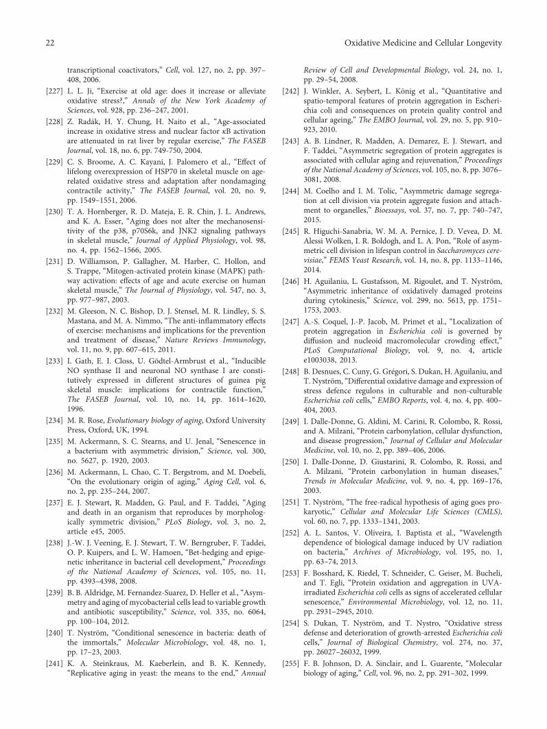
transcriptional coactivators,” Cell, vol. 127, no. 2, pp. 397–408, 2006.
[227] L. L. Ji, “Exercise at old age: does it increase or alleviateoxidative stress?,” Annals of the New York Academy ofSciences, vol. 928, pp. 236–247, 2001.
[228] Z. Radák, H. Y. Chung, H. Naito et al., “Age-associatedincrease in oxidative stress and nuclear factor κB activationare attenuated in rat liver by regular exercise,” The FASEBJournal, vol. 18, no. 6, pp. 749-750, 2004.
[229] C. S. Broome, A. C. Kayani, J. Palomero et al., “Effect oflifelong overexpression of HSP70 in skeletal muscle on age-related oxidative stress and adaptation after nondamagingcontractile activity,” The FASEB Journal, vol. 20, no. 9,pp. 1549–1551, 2006.
[230] T. A. Hornberger, R. D. Mateja, E. R. Chin, J. L. Andrews,and K. A. Esser, “Aging does not alter the mechanosensi-tivity of the p38, p70S6k, and JNK2 signaling pathwaysin skeletal muscle,” Journal of Applied Physiology, vol. 98,no. 4, pp. 1562–1566, 2005.
[231] D. Williamson, P. Gallagher, M. Harber, C. Hollon, andS. Trappe, “Mitogen-activated protein kinase (MAPK) path-way activation: effects of age and acute exercise on humanskeletal muscle,” The Journal of Physiology, vol. 547, no. 3,pp. 977–987, 2003.
[232] M. Gleeson, N. C. Bishop, D. J. Stensel, M. R. Lindley, S. S.Mastana, and M. A. Nimmo, “The anti-inflammatory effectsof exercise: mechanisms and implications for the preventionand treatment of disease,” Nature Reviews Immunology,vol. 11, no. 9, pp. 607–615, 2011.
[233] I. Gath, E. I. Closs, U. Gödtel-Armbrust et al., “InducibleNO synthase II and neuronal NO synthase I are consti-tutively expressed in different structures of guinea pigskeletal muscle: implications for contractile function,”The FASEB Journal, vol. 10, no. 14, pp. 1614–1620,1996.
[234] M. R. Rose, Evolutionary biology of aging, Oxford UniversityPress, Oxford, UK, 1994.
[235] M. Ackermann, S. C. Stearns, and U. Jenal, “Senescence ina bacterium with asymmetric division,” Science, vol. 300,no. 5627, p. 1920, 2003.
[236] M. Ackermann, L. Chao, C. T. Bergstrom, and M. Doebeli,“On the evolutionary origin of aging,” Aging Cell, vol. 6,no. 2, pp. 235–244, 2007.
[237] E. J. Stewart, R. Madden, G. Paul, and F. Taddei, “Agingand death in an organism that reproduces by morpholog-ically symmetric division,” PLoS Biology, vol. 3, no. 2,article e45, 2005.
[238] J.-W. J. Veening, E. J. Stewart, T. W. Berngruber, F. Taddei,O. P. Kuipers, and L. W. Hamoen, “Bet-hedging and epige-netic inheritance in bacterial cell development,” Proceedingsof the National Academy of Sciences, vol. 105, no. 11,pp. 4393–4398, 2008.
[239] B. B. Aldridge, M. Fernandez-Suarez, D. Heller et al., “Asym-metry and aging of mycobacterial cells lead to variable growthand antibiotic susceptibility,” Science, vol. 335, no. 6064,pp. 100–104, 2012.
[240] T. Nyström, “Conditional senescence in bacteria: death ofthe immortals,” Molecular Microbiology, vol. 48, no. 1,pp. 17–23, 2003.
[241] K. A. Steinkraus, M. Kaeberlein, and B. K. Kennedy,“Replicative aging in yeast: the means to the end,” Annual
Review of Cell and Developmental Biology, vol. 24, no. 1,pp. 29–54, 2008.
[242] J. Winkler, A. Seybert, L. König et al., “Quantitative andspatio-temporal features of protein aggregation in Escheri-chia coli and consequences on protein quality control andcellular ageing,” The EMBO Journal, vol. 29, no. 5, pp. 910–923, 2010.
[243] A. B. Lindner, R. Madden, A. Demarez, E. J. Stewart, andF. Taddei, “Asymmetric segregation of protein aggregates isassociated with cellular aging and rejuvenation,” Proceedingsof the National Academy of Sciences, vol. 105, no. 8, pp. 3076–3081, 2008.
[244] M. Coelho and I. M. Tolic, “Asymmetric damage segrega-tion at cell division via protein aggregate fusion and attach-ment to organelles,” Bioessays, vol. 37, no. 7, pp. 740–747,2015.
[245] R. Higuchi-Sanabria, W. M. A. Pernice, J. D. Vevea, D. M.Alessi Wolken, I. R. Boldogh, and L. A. Pon, “Role of asym-metric cell division in lifespan control in Saccharomyces cere-visiae,” FEMS Yeast Research, vol. 14, no. 8, pp. 1133–1146,2014.
[246] H. Aguilaniu, L. Gustafsson, M. Rigoulet, and T. Nyström,“Asymmetric inheritance of oxidatively damaged proteinsduring cytokinesis,” Science, vol. 299, no. 5613, pp. 1751–1753, 2003.
[247] A.-S. Coquel, J.-P. Jacob, M. Primet et al., “Localization ofprotein aggregation in Escherichia coli is governed bydiffusion and nucleoid macromolecular crowding effect,”PLoS Computational Biology, vol. 9, no. 4, articlee1003038, 2013.
[248] B. Desnues, C. Cuny, G. Grégori, S. Dukan, H. Aguilaniu, andT. Nyström, “Differential oxidative damage and expression ofstress defence regulons in culturable and non-culturableEscherichia coli cells,” EMBO Reports, vol. 4, no. 4, pp. 400–404, 2003.
[249] I. Dalle-Donne, G. Aldini, M. Carini, R. Colombo, R. Rossi,and A. Milzani, “Protein carbonylation, cellular dysfunction,and disease progression,” Journal of Cellular and MolecularMedicine, vol. 10, no. 2, pp. 389–406, 2006.
[250] I. Dalle-Donne, D. Giustarini, R. Colombo, R. Rossi, andA. Milzani, “Protein carbonylation in human diseases,”Trends in Molecular Medicine, vol. 9, no. 4, pp. 169–176,2003.
[251] T. Nyström, “The free-radical hypothesis of aging goes pro-karyotic,” Cellular and Molecular Life Sciences (CMLS),vol. 60, no. 7, pp. 1333–1341, 2003.
[252] A. L. Santos, V. Oliveira, I. Baptista et al., “Wavelengthdependence of biological damage induced by UV radiationon bacteria,” Archives of Microbiology, vol. 195, no. 1,pp. 63–74, 2013.
[253] F. Bosshard, K. Riedel, T. Schneider, C. Geiser, M. Bucheli,and T. Egli, “Protein oxidation and aggregation in UVA-irradiated Escherichia coli cells as signs of accelerated cellularsenescence,” Environmental Microbiology, vol. 12, no. 11,pp. 2931–2945, 2010.
[254] S. Dukan, T. Nyström, and T. Nystro, “Oxidative stressdefense and deterioration of growth-arrested Escherichia colicells,” Journal of Biological Chemistry, vol. 274, no. 37,pp. 26027–26032, 1999.
[255] F. B. Johnson, D. A. Sinclair, and L. Guarente, “Molecularbiology of aging,” Cell, vol. 96, no. 2, pp. 291–302, 1999.
22 Oxidative Medicine and Cellular Longevity

[256] S. Dukan and T. Nyström, “Bacterial senescence: stasis resultsin increased and differential oxidation of cytoplasmic pro-teins leading to developmental induction of the heat shockregulon,” Genes & Development, vol. 12, no. 21, pp. 3431–3441, 1998.
[257] J. Tamarit, E. Cabiscol, and J. Ros, “Identification of themajor oxidatively damaged proteins in Escherichia coli cellsexposed to oxidative stress,” Journal of Biological Chemistry,vol. 273, no. 5, pp. 3027–3032, 1998.
[258] E. Cabiscol, E. Piulats, P. Echave, E. Herrero, and J. Ros,“Oxidative stress promotes specific protein damage inSaccharomyces cerevisiae,” Journal of Biological Chemistry,vol. 275, no. 35, pp. 27393–27398, 2000.
[259] R. S. Sohal, “Role of oxidative stress and protein oxidation inthe aging process,” Free Radical Biology andMedicine, vol. 33,no. 1, pp. 37–44, 2002.
[260] L.-J. Yan, R. L. Levine, and R. S. Sohal, “Oxidative damageduring aging targets mitochondrial aconitase,” Proceedingsof the National Academy of Sciences, vol. 94, no. 21,pp. 11168–11172, 1997.
[261] A. Castegna, M. Aksenov, M. Aksenova et al., “Proteomicidentification of oxidatively modified proteins in Alzheimer’sdisease brain. Part I: Creatine kinase BB, glutamine synthase,and ubiquitin carboxy-terminal hydrolase L-1,” Free RadicalBiology and Medicine, vol. 33, no. 4, pp. 562–571, 2002.
[262] A. Matin, “The molecular basis of carbon-starvation-inducedgeneral resistance in Escherichia coli,” Molecular Microbiol-ogy, vol. 5, no. 1, pp. 3–10, 1991.
[263] D. E. Jenkins, J. E. Schultz, and A. Matin, “Starvation-inducedcross protection against heat or H2O2 challenge in Escheri-chia coli,” Journal of Bacteriology, vol. 170, no. 9, pp. 3910–3914, 1988.
[264] R. A. Miller, “Cell stress and aging: new emphasis on multi-plex resistance mechanisms,” The Journals of GerontologySeries A: Biological Sciences and Medical Sciences, vol. 64A,no. 2, pp. 179–182, 2009.
[265] L. E. Enell, N. Kapan, J. A. E. Soderberg, L. Kahsai, andD. R. Nassel, “Insulin signaling, lifespan and stress resis-tance are modulated by metabotropic GABA receptors oninsulin producing cells in the brain of Drosophila,” PLoSOne, vol. 5, no. 12, article e15780, 2010.
[266] S. Ayyadevara, M. R. Engle, S. P. Singh et al., “Lifespan andstress resistance of Caenorhabditis elegans are increased byexpression of glutathione transferases capable of metaboliz-ing the lipid peroxidation product 4-hydroxynonenal,” AgingCell, vol. 4, no. 5, pp. 257–271, 2005.
[267] M. Shaposhnikov, E. Proshkina, L. Shilova, A. Zhavoronkov,and A. Moskalev, “Lifespan and stress resistance in Drosoph-ila with overexpressed DNA repair genes,” Scientific Reports,vol. 5, no. 1, article 15299, 2015.
[268] V. D. Longo, J. Mitteldorf, and V. P. Skulachev, “Pro-grammed and altruistic ageing,” Nature Reviews Genetics,vol. 6, no. 11, pp. 866–872, 2005.
[269] M. Ballesteros, A. Fredriksson, J. Henriksson, and T. Nyström,“Bacterial senescence: protein oxidation in non-proliferatingcells is dictated by the accuracy of the ribosomes,” TheEMBO Journal, vol. 20, no. 18, pp. 5280–5289, 2001.
[270] H. Aguilaniu, L. Gustafsson, M. Rigoulet, and T. Nyström,“Protein oxidation in G0 cells of Saccharomyces cerevisiaedepends on the state rather than rate of respiration and is
enhanced in pos9 but not yap1mutants,” Journal of BiologicalChemistry, vol. 276, no. 38, pp. 35396–35404, 2001.
[271] T. Nyström, “Not quite dead enough: on bacterial life, cultur-ability, senescence, and death,” Archives of Microbiology,vol. 176, no. 3, pp. 159–164, 2001.
[272] A. Battesti, N. Majdalani, and S. Gottesman, “The RpoS-mediated general stress response in Escherichia coli,” AnnualReview of Microbiology, vol. 65, no. 1, pp. 189–213, 2011.
[273] T. L. Testerman, A. Vazquez-Torres, Y. Xu, J. Jones-Carson,S. J. Libby, and F. C. Fang, “The alternative sigma factor σE
controls antioxidant defences required for Salmonella viru-lence and stationary-phase survival,”Molecular Microbiology,vol. 43, no. 3, pp. 771–782, 2002.
[274] F. Fontaine, E. J. Stewart, A. B. Lindner, and F. Taddei,“Mutations in two global regulators lower individual mortal-ity in Escherichia coli,” Molecular Microbiology, vol. 0, no. 0,pp. 2–14, 2007.
[275] S. Gonidakis, S. E. Finkel, and V. D. Longo, “Genome-widescreen identifies Escherichia coli TCA-cycle-related mutantswith extended chronological lifespan dependent on acetatemetabolism and the hypoxia-inducible transcription factorArcA,” Aging Cell, vol. 9, no. 5, pp. 868–881, 2010.
[276] L. Yu and C. Yu, “Interaction between succinate dehydroge-nase and ubiquinone-binding protein from succinate-ubiquinone reductase,” Biochimica et Biophysica Acta (BBA)- Bioenergetics, vol. 593, no. 1, pp. 24–38, 1980.
[277] K. R. Messner and J. A. Imlay, “Mechanism of superoxide andhydrogen peroxide formation by fumarate reductase, succi-nate dehydrogenase, and aspartate oxidase,” Journal of Bio-logical Chemistry, vol. 277, no. 45, pp. 42563–42571, 2002.
[278] S. Gonidakis, S. E. Finkel, and V. D. Longo, “E. coli hypoxia-inducible factor ArcA mediates lifespan extension in a lipoicacid synthase mutant by suppressing acetyl-CoA synthetase,”Biological Chemistry, vol. 391, no. 10, pp. 1139–1147, 2010.
[279] A. S. Lynch and E. C. Lin, “Transcriptional control mediatedby the ArcA two-component response regulator protein ofEscherichia coli: characterization of DNA binding at targetpromoters,” Journal of Bacteriology, vol. 178, no. 21,pp. 6238–6249, 1996.
23Oxidative Medicine and Cellular Longevity

Research ArticleROS-Induced DNA Damage Associates with Abundance ofMitochondrial DNA in White Blood Cells of the UntreatedSchizophrenic Patients
I. V. Chestkov,1 E. M. Jestkova,2 E. S. Ershova,1,3 V. G. Golimbet,4 T. V. Lezheiko,4
N. Yu Kolesina,4 O. A. Dolgikh,1 V. L. Izhevskaya,1 G. P. Kostyuk,2 S. I. Kutsev,1
N. N. Veiko,1,3 and S. V. Kostyuk 4
1Research Centre for Medical Genetics (RCMG), Moscow 115478, Russia2N. A. Alexeev Clinical Psychiatric Hospital №1 of Moscow Healthcare Department, Moscow 115447, Russia3V. A. Negovsky Research Institute of General Reanimatology, Federal Clinical Research Center of Reanimatology and Rehabilitogy,Moscow 107031, Russia4Mental Health Research Center, Moscow 115522, Russia
Correspondence should be addressed to S. V. Kostyuk; [email protected]
Received 31 July 2017; Revised 1 November 2017; Accepted 10 December 2017; Published 25 February 2018
Academic Editor: Tanea T. Reed
Copyright © 2018 I. V. Chestkov et al. This is an open access article distributed under the Creative Commons Attribution License,which permits unrestricted use, distribution, and reproduction in any medium, provided the original work is properly cited.
Objective. The aim of this study was (1) to examine the leukocyte mtDNA copy number (CN) in unmedicated (SZ (m−)) andmedicated (SZ (m+)) male patients with paranoid schizophrenia (SZ) in comparison with the healthy male controls (HC) and(2) to compare the leukocyte mtDNA CN with the content of an oxidation marker 8-oxodG in lymphocytes of the SZ (m−)patients. Methods. We evaluated leukocyte mtDNA CN of 110 subjects with SZ in comparison with 60 male HC by the methodqPCR (ratio mtDNA/nDNA (gene B2M) was detected). SZ patients were divided into two subgroups. The patients of thesubgroups SZ (m+) (N = 55) were treated with standard antipsychotic medications in the hospital. The patients of the subgroupSZ (m−) (N = 55) were not treated before venous blood was sampled. To evaluate oxidative DNA damage, we quantified thelevels of 8-oxodG in lymphocytes (flow cytometry) of SZ (m−) patients (N = 55) and HC (N = 30). Results. The leukocytemtDNA CN showed no significant difference in SZ (m+) patients and HC. The mtDNA CN in the unmedicated subgroup SZ(m−) was significantly higher than that in the SZ (m+) subgroup or in HC group. The level of 8-oxodG in the subgroup SZ(m−) was significantly higher than that in HC group. Conclusion. The leukocytes of the unmedicated SZ male patients withacute psychosis contain more mtDNA than the leukocytes of the male SZ patients treated with antipsychotic medications or thehealthy controls. MtDNA content positively correlates with the level of 8-oxodG in the unmedicated SZ patients.
1. Introduction
Approximately 1% of the world’s population suffer fromschizophrenia (SZ). Schizophrenia is a highly heritableneuropsychiatric disorder of complex genetic etiology.Mitochondrion (mt) is a cellular organelle involved in theregulation of a variety of complex cellular processes. Mito-chondria, the cell energy source, have a crucial role in intra-cellular calcium homeostasis, producing ROS and activatingthe apoptotic pathway. MtDNA copy number (CN) variation
has been suggested as a sensitive index of cellular oxidativestress, inflammation, and mitochondrial dysfunction [1, 2].
The accumulating morphological, genetic, and imagingdata delineates mitochondrial multifaceted dysfunction as apathological factor in schizophrenia [3]. Several studies haveevaluated the influence of schizophrenia on mtDNA copynumber. Results are controversial to each other. Someauthors show lower mtDNA copy number in brain tissuesand peripheral lymphocytes of schizophrenia patients com-pared with healthy controls (HC) [4–7]. Some studies
HindawiOxidative Medicine and Cellular LongevityVolume 2018, Article ID 8587475, 7 pageshttps://doi.org/10.1155/2018/8587475

reported no anomalous mtDNA copy number in the tissuesof schizophrenia patients [8, 9]. The reason for the observedcontradictions may be the lack of sufficient data on the effectof antipsychotic therapy on mtDNA copy number. It isknown that administration of antipsychotics involved mito-chondrial functions, especially OXPHOS. The antipsychoticsinhibit the mitochondrial respiratory chain [3]. However,there are few studies on the impact of antipsychotics onmtDNA content in schizophrenia patients. Li et al. recentlyhad showed that antipsychotic risperidone causes a decreasein the number of mtDNA copies in leukocytes of patientswith the first-episode antipsychotic-naïve schizophrenia[4]. Thus, the first objective of our work was to determinethe effects of the standard antipsychotic therapy on thenumber of mtDNA copies in the cells of male patients withparanoid schizophrenia.
Systemic oxidative stress is associated with schizophre-nia. Elevated reactive oxygen species (ROS) levels anddeclined antioxidant statuses have been reported in the brainand peripheral tissues of the patients with schizophrenia[10–12]. However, it is known that oxidative stress in thebody is associated with the changes in mtDNA copy number.For example, oxidative stress caused by environmental expo-sure, for example, ionizing radiation, induces ROS-causedincrease of the amount of mtDNA in the human and animalcells and organisms [13–15].
Thus, we can assume that a high level of ROS in the bodyof some schizophrenia patients should correlate with anincreased mtDNA copy number. So, the second objective ofour work was to test this hypothesis. We quantified mtDNAcontent and the levels of oxidative stress marker 8-oxodGin the white blood cells of male patients with untreatedparanoid schizophrenia.
2. Methods
2.1. Subjects. This investigation was approved by the RegionalEthics Committee of RCMG. It was carried out in accordancewith the latest version of the Declaration of Helsinki. Onehundred ten male paranoid SZ patients with acute psychoticdisorders were recruited from the general psychiatric unitsfor treatment of acute forms of mental disorders (PsychiatricHospital #14 of Moscow City Health Department andMentalHealth Research Center, Moscow). All participants signed aninformed written consent to participate in this investigation.Age of the patients: 38± 13 (19–62). Clinical symptoms havebeen measured using the positive and negative syndromesscale (PANSS) (Kay et al. [16]), a widespread instrumentproven to be valid and suitable for evaluation of positive,negative, and general psychopathological items. It includesthree subscales, totally 30 items: 7 for positive symptoms,7 for negative, and 16 for general psychopathological ones.Each symptom has 7 ratings (1—absent, 2—questionable,3—mild, 4—moderate, 5—severe, 6—markedly severe,and 7—extremely severe). The PANSS interviews, completedby a trained researcher, were conducted one week before thepatient’s discharge from the hospital. Patients were diag-nosed with paranoid schizophrenia (F20.00 and F20.01 inthe International Classification of Diseases 10th Revision
(ICD-10)) using structured mini international neuropsychi-atric interview (MINI). Diagnoses were also confirmed pur-suant to DSM-IV criteria. The paranoid SZ duration wasless than 3 years (n = 24, 22%), 3–10 years (n = 20, 18%),and more than 10 years (n = 66, 60%). No subjects sufferedfrom any relevant disease.
SZ patients were divided into groups: SZ (m+) (N = 55)and SZ (m−) (N = 55). The patients in group SZ (m+)received standard antipsychotic therapy in the hospital forat least six weeks before the venous blood was sampled. Theywere clinically stable and their medications had not changedfor at least one month. For the treatment of the acute dis-orders, standard antipsychotics were used: haloperidol,chlorpromazine, clozapine, risperidone, quetiapine, andolanzapine. Most of the patients of the group SZ (m−)refused to take antipsychotics in the home despite of thechronic course of the disease. These patients did not takeantipsychotics for 3 to 8 months until a new hospitaliza-tion with acute psychosis. Some patients of the group SZ(m−) (N = 12, 21.8%) were diagnosed as the first-episodeSZ and were never treated with antipsychotic medications.
From the SZ (m−) patients, the venous blood was col-lected on the day of hospitalization before starting anyantipsychotic therapy. 20mL of blood was collected froma peripheral vein of the subject using a syringe flushedwith heparin under strict aseptic conditions. The controlgroup consisted of 60 healthy males with no history of anypsychiatric disorder, correlating with the patient group byage (37± 12 (17–73)) and by their smoking habits.
2.2. Isolating of DNA from the Blood. The leukocytes wereisolated from 5mL of blood by the method of Boyum [17].5mL of the solution containing 2% sodium lauryl sarcosylate,0.04M EDTA, and 150μg/mL RNAse A (Sigma, USA) wasadded to the fresh leucocytes for 45min (37°C) and thenwas treated with proteinase K (200μg/mL, Promega, USA)for 24 h at 37°C. The lysate samples were extracted with anequal volume of phenol, phenol/chloroform/isoamyl alcohol(25 : 24 : 1), and chloroform/isoamyl alcohol (24 : 1), respec-tively. DNA was precipitated by adding 1/10 volume of 3Msodium acetate (pH5.2) and 2.5 volume of ice-cold ethanol.For the extraction procedure, only freshly distilled solventswere used. Phenol was stabilized with 8-hydroxyquinoline.Finally, the DNA was collected by centrifugation at 10,000gfor 15min at 4°C, washed with 70% ethanol (v/v), dried,and dissolved in water. The quantitation of the purifiedgenomic DNA is performed using the PicoGreen dsDNAquantitation reagent from Molecular Probes (Invitrogen,CA, USA). The assay displays a linear correlation betweendsDNA quantity and fluorescence over a wide range. TheDNA concentration of the samples is calculated accordingto a DNA standard curve. We use EnSpire equipment(Finland) with the following parameters: excitation andemission wavelengths 488 and 528nm, respectively.
2.3. Quantitative Real-Time PCR (qPCR). Serial qPCR assaywas established using the StepOnePlus (Applied Biosystems).QPCR efficiencies for ten samples were determined byserially diluted genomic DNA. MtDNA copy number
2 Oxidative Medicine and Cellular Longevity
determinations in the other samples were obtained using acalculated average efficiency (E+ 1) of 2.0± 0.05 for geneB2M and 1.95± 0.03 for mtDNA. Each reaction contained10μL 2хSYBR Premix Ex Taq (PerfectRealTime™, TakaraBio), 2μL primers (10μM), and 8μL of DNA (5ng/μL)for a final volume of 20μL. All reactions were performedin duplicates. PCR conditions were 6min at 95°C initialdenaturation, followed by 40 cycles of 30 s of denatur-ation at 95°C, 15 s of primer annealing at 60°C, and 10 s at72°C of extension. The presence of unspecific ampliconswas excluded by melting curve analysis and gel electropho-resis. The following primers (Malik et al. [18]) were used(Sintol, Russia):
Human mitochondrial genome NC_012920 (D-loop)hmito (65) F CTTCTGGCCACAGCACTTAAAC; RGCTGGTGTTAGGGTTCTTTGTTTTHuman B2M (accession number M17987)hB2M (95) F GCTGGGTAGCTCTAAACAATGTATTCA; R CCATGTACTAACAAATGTCTAAAATGG
2.4. Flow Cytometry Analysis (FCA) of 8-oxodG. Method isdescribed in detail earlier [19]. Briefly, lymphocytes wereisolated from 15mL of the fresh blood. To fix the cells,2% paraformaldehyde (Sigma) was used (37°C, 10min).Cells were permeabilized with 0.1% Triton X-100 (Sigma)in PBS (15min, 25°C). Cells were stained with FITC-8-oxodG Abcam antibody (1μg/mL, 4 h, 4°C). We stained aportion of the cells with secondary FITC-conjugated anti-bodies to determine the level of the background fluorescence.Cells were analyzed at CyFlow Space (Partec, Germany).Primary data are presented as median values of the signalFL1 (8-oxodG). Index 8-oxodG= (I−Ib)/Ib, where I and Ibare the experienced and the background signal.
2.5. Statistics. All reported results were reproduced at leasttwo times as independent biological replicates.
The significance of the observed differences was ana-lyzed using the nonparametric Mann–Whitney U-tests. Allp values were two-sided and considered statistically signifi-cant at α< 0.05.
For the statistical analysis was used Professional softwareStatPlus2007 (http://www.analystsoft.com/).
3. Results
3.1. Quantification of mtDNA in Leukocytes of HC and SZGroups. The mtDNA CN in the leukocytes of SZ patientsand HC detected by qPCR (mtDNA to nDNA (gene B2M)ratio was measured) is given in Figure 1. Comparison of thesamples by the Mann–Whitney test is given in the table inFigure 1. In 12 patients of 55 (21.8%) from SZ (m−) groupand only in 1 patients of 55 (1.7%) from SZ (m+) group, aconsiderable increase in mtDNA CN compared to the HCwas found (Figure 1). Median mtDNA CN for the leukocytesof the unmedicated SZ (m−) patients was 2.8 or 2.2 timeshigher than the median mtDNA CN for the leukocytes ofthe medicated SZ (m+) or HC. The samples of HC and SZ(m+) did not differ among themselves in the content ofmtDNA in leukocytes.
We did not find any significant correlation between theclinical symptoms (indexes of the scale PANSS) and thenumber of copies of mtDNA for the medicated SZ patients(k < 0 2; p > 0 1, linear regression method).
3.2. Quantification of 8-oxodG in Lymphocytes of HC andSZ (m−) Groups. FCA was employed in order to evaluate8-oxodG content in the lymphocytes (Figure 2). Stainingof the fixed cells with 8-oxodG antibodies was performed[19]. Figure 2 shows the mean values of the signal againstthe baseline values (8-oxodG index) for the groups HCand SZ (m−) (see table in Figure 2 for descriptive statistics).The 8-oxodG content in SZ (m−) lymphocytes was higherthan in the control group (p = 10−6). The patients fromthe SZ (m−) group were divided into subgroups: SZ-1(m−)(N = 31) with normal 8-oxodG content and SZ-2 (m−)(N = 24) with increased 8-oxodG content compared to con-trol. These groups differ in the level of cellular DNA oxida-tion (p = 10−10) (Figure 2).
3.3. The Dependence of mtDNA CN on 8-oxodG in HC andSZ (m−) Groups. Figure 3(a) presents dependence ofmtDNA CN in the leukocytes on logarithm of the 8-oxodG content in the lymphocytes for control and SZ(m−) groups. In the control group (N = 30), correlationbetween the two parameters was found (k = 0 47; p < 0 01).In the total SZ (m−) group (N = 55), linear correlationbetween lg (8-oxodG) and mtDNA CN content was alsofound (k = 0 44; p < 0 001). The correlation between the ana-lyzed parameters was detected for the total sample of healthy
U-test
0
100
200
300
400
500
600
700
1 3 5 7 9 11 13 15 17 19 21 23 25 27 29 31 33 35 37 39 41 43 45 47 49 51 53 55 57 59
SZ (m+)
HD
SZ (m−)
The serial number of the sample in the ranked cohort
mtD
NA
CN
(±SD
)
Mt DNA CN
Group N Mean ± SD Median Range HD-SZ
SZ (m+) 55 141 ± 95 137 14 - 457 p = 0.09SZ (m−) 55 311 ± 141 301 34 - 622 p = 10−9
HD 60 153 ± 91 107 28 - 438
Figure 1: Leukocytes from unmedicated SZ patients with acutepsychosis are enriched with mtDNA. Graph—Quantification ofmtDNA in leukocytes of HC (green), SZ (m−) (red), and SZ (m+)(yellow) groups. The data in each group is ranked by the value ofmtDNA CN. The mean and standard deviation for each DNAsample is given (the data of two independent experiments, threemeasurements in each experiment, n = 6). Table containsstatistical data.
3Oxidative Medicine and Cellular Longevity

and schizophrenic people (N = 85, k = 0 53, p < 0 0001).Thus, the greater the level of DNA oxidation in the humancells, the greater the content of mtDNA.
The ratio (8-oxodG/mtDNA CN) indicates the amount of8-oxodG per one copy of mtDNA (Figure 3(b)). This ratiowas significantly higher in the SZ-2 group than in the groupSZ-1 or HC. For 11 patients of 55, this index reached veryhigh values in comparison with the rest of the SZ (m−) sam-ple. All these patients were sick with schizophrenia for morethan 10 years and smoked more than 40 cigarettes a day.However, for the whole group SZ (m−), we did not find apronounced dependence of the indices 8-oxodG, mtDNACN, and (8-oxodG/mtDNA CN) on the duration of schizo-phrenia or on the intensity of smoking.
We also analyzed the indices 8-oxodG, mtDNA CN, and(8-oxodG/mtDNA CN) in connection with the 29 indices ofstandard biochemical analysis of venous blood. We did notfind any significant correlations.
4. Discussion
SZ is increasingly considered as a systemic disorder, which isassociated with biochemical disturbances not only in theCNS/brain cells [20]. In recent years, there has been an inten-sive search for blood-based biomarkers for SZ [21]. Thestudy of white blood cells can give an understanding of howthe whole body responds to disturbances that eventually leadto the disorders of a brain function.
4.1. Abundance of the mtDNA CN in Leucocytes of theUnmedicated SZ Patients. The first finding of this study ishigher mtDNA CN in the unmedicated SZ patients than inthe medicated patients and the healthy controls (Figure 1).We have shown that acute psychosis of the unmedicated SZ(m−) patients is accompanied by a significant increase inthe mtDNA CN in leucocytes (Figure 1). The leukocytes ofSZ (m+) patients with the antipsychotic medication containthe amount of mtDNA comparable to the control. Thereare few studies on the impact of antipsychotics on mtDNAcontent in SZ patients. Li et al. recently also demonstratedthat antipsychotic risperidone causes a decrease in themtDNA CN in leukocytes of patients with the first-episodeantipsychotic-naïve schizophrenia [4].
The reason for reduction of mtDNA CN in the medicatedSZ patients remains unknown. The patients in this studywere treated with various typical and atypical antipsychotics.In a state of acute psychosis, patients usually were treatedwith typical antipsychotics. After improving the mental stateof the patient, he was treated with atypical antipsychotics.This fact did not allow us to form large enough groups toanalyze the effect of specific antipsychotics on mtDNA CN.Recent studies have suggested that some antipsychotic drugshave a useful therapeutic effect by reducing oxidative stress inschizophrenic patients [22, 23]. Perhaps these drugs cause adecrease in the mtDNA CN through the antioxidant effects.It is also possible that antipsychotics change mitophagy inthe cells of SZ patients. Mitophagy is a highly specific qualitycontrol process which eliminates dysfunctional mitochon-dria and promotes mitochondrial turnover [24]. Furtherstudies are needed to understand the reasons for reducingthe number of copies of mtDNA in the treated patients.
4.2. Abundance of mtDNA in the SZ (m−) Patients Associateswith Oxidative DNA Damage. The lymphocytes of someunmedicated SZ patients contain more 8-oxodG than thelymphocytes of healthy controls (Figure 2). 8-oxodG repre-sents major oxidative DNA damage products. An elevatedlevel of 8-oxodG in the blood and urine has been reportedin SZ patients [25, 26]. Postmortem studies have revealed ahigher level of 8-oxodG in the brains of SZ patients com-pared to the controls [27, 28]. So our findings are consistentwith those of other author who showed a high level of ROS inthe SZ patient’s body.
An internal source of ROS is present in the body of apatient with SZ [10–12]. The main cause of oxidativestress in schizophrenia has not yet been found. Differentmechanisms of oxidative stress in schizophrenia have beenpostulated: dopamine metabolism, NO metabolism, abnor-malities in the mitochondrial electron transport chainpatients with schizophrenia, mutations in the genes responsi-ble for maintaining the required level of ROS in cells, and soon. However, regardless of the cause, the result is important:in the cells of some SZ patients, the level of ROS is signifi-cantly increased. We can compare this situation with theeffect on the cells of an external source of ionizing radiation,which induces oxidative stress in human. This model isattractive because it is well studied [13–15, 29–32]. More-over, some authors hypothesized that ionizing radiation
U-testHD-SZGroup
N Mean ± SD Median RangeHD 30 0.6 ± 0.5 0.57 0.01 - 1.87SZ (m−) 55 3.2 ± 3.7 1.5 0.05 - 1.67 p = 10−6
SZ-1 (m−) 31 0.9 ± 0.5 1.06 0.05 - 1.78 p = 0.06SZ-2 (m−) 24 6.1 ± 3.9 4.6 1.99 - 16.7 p = 10−10
0
3
6
9
12
15
18
HD SZ-2 (m−)
8-ox
odG
(±SD
), ar
b.un
.
SZ-1 (m−)The serial number of the sample in the ranked cohort
8-oxodG, arb.un.
Figure 2: Lymphocytes from the unmedicated SZ patients withacute psychosis are enriched with 8-oxodG. Flow cytometryanalysis of 8-oxodG. Index 8-oxodG (arb.un) is the ratio of thedifference between the experienced and the background signal tothe magnitude of the background signal. Graph—Quantification of8-oxodG in lymphocytes of HC (green) and SZ (m−) (red) groups.The data in each group is ranked by the value of 8-oxodG. Themean and standard deviation for each cell sample is given (thedata of three independent experiments, two measurements in eachexperiment, n = 6). Table contains statistical data.
4 Oxidative Medicine and Cellular Longevity
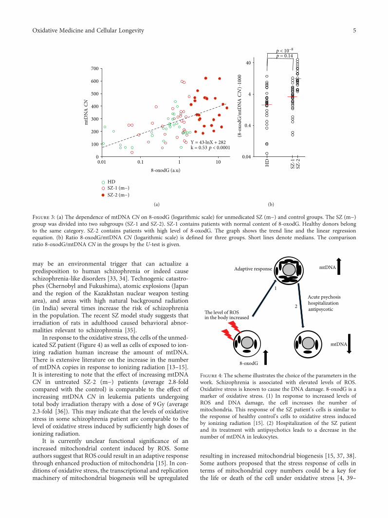
may be an environmental trigger that can actualize apredisposition to human schizophrenia or indeed causeschizophrenia-like disorders [33, 34]. Technogenic catastro-phes (Chernobyl and Fukushima), atomic explosions (Japanand the region of the Kazakhstan nuclear weapon testingarea), and areas with high natural background radiation(in India) several times increase the risk of schizophreniain the population. The recent SZ model study suggests thatirradiation of rats in adulthood caused behavioral abnor-malities relevant to schizophrenia [35].
In response to the oxidative stress, the cells of the unmed-icated SZ patient (Figure 4) as well as cells of exposed to ion-izing radiation human increase the amount of mtDNA.There is extensive literature on the increase in the numberof mtDNA copies in response to ionizing radiation [13–15].It is interesting to note that the effect of increasing mtDNACN in untreated SZ-2 (m−) patients (average 2.8-foldcompared with the control) is comparable to the effect ofincreasing mtDNA CN in leukemia patients undergoingtotal body irradiation therapy with a dose of 9Gy (average2.3-fold [36]). This may indicate that the levels of oxidativestress in some schizophrenia patient are comparable to thelevel of oxidative stress induced by sufficiently high doses ofionizing radiation.
It is currently unclear functional significance of anincreased mitochondrial content induced by ROS. Someauthors suggest that ROS could result in an adaptive responsethrough enhanced production of mitochondria [15]. In con-ditions of oxidative stress, the transcriptional and replicationmachinery of mitochondrial biogenesis will be upregulated
resulting in increased mitochondrial biogenesis [15, 37, 38].Some authors proposed that the stress response of cells interms of mitochondrial copy numbers could be a key forthe life or death of the cell under oxidative stress [4, 39–
0
100
200
300
400
500
600
700
0.01 0.1 1 10
Y = 43·lnX + 282
HDSZ-1 (m−)SZ-2 (m−)
k = 0.53 p < 0.0001
mtD
NA
CN
8-oxodG (a.u)
(a)
p < 10−8
p = 0.14
0.04
0.4
4
40
HD
SZ-1
SZ-2
(8-o
xodG
/mtD
NA
CN
) ·10
00
(b)
Figure 3: (a) The dependence of mtDNA CN on 8-oxodG (logarithmic scale) for unmedicated SZ (m−) and control groups. The SZ (m−)group was divided into two subgroups (SZ-1 and SZ-2). SZ-1 contains patients with normal content of 8-oxodG. Healthy donors belongto the same category. SZ-2 contains patients with high level of 8-oxodG. The graph shows the trend line and the linear regressionequation. (b) Ratio 8-oxodG/mtDNA CN (logarithmic scale) is defined for three groups. Short lines denote medians. The comparisonratio 8-oxodG/mtDNA CN in the groups by the U-test is given.
mtDNA
mtDNA
Adaptive response
Acute psychosishospitalizationantipsycoticThe level of ROS
in the body increased
1
2
8-oxodG
Figure 4: The scheme illustrates the choice of the parameters in thework. Schizophrenia is associated with elevated levels of ROS.Oxidative stress is known to cause the DNA damage. 8-oxodG is amarker of oxidative stress. (1) In response to increased levels ofROS and DNA damage, the cell increases the number ofmitochondria. This response of the SZ patient’s cells is similar tothe response of healthy control’s cells to oxidative stress inducedby ionizing radiation [15]. (2) Hospitalization of the SZ patientand its treatment with antipsychotics leads to a decrease in thenumber of mtDNA in leukocytes.
5Oxidative Medicine and Cellular Longevity

41]. According to this hypothesis, an increase in mtDNAcontent may precede mitochondrial dysfunction as an adap-tive response and could therefore be a predictive biomarker.
The above-mentioned hypothesis is confirmed by thedata obtained in this study. Analyzing the oxidation marker8-oxodG in SZ patients, we found that the unmedicated SZpatients can be distinctively divided into subgroups withdifferent levels of 8-oxodG (Figure 2). MtDNA CN wasmuch higher in the subgroup SZ-2 (m−) with a high levelof 8-oxodG (Figure 3). On the one hand, the correlationbetween lg (8-oxodG) and mtDNA CN (Figure 3(a)) forHC and SZ(m−) can be explained by the preferential oxida-tion of mtDNA compared to nuclear DNA. MtDNA is highlysusceptible to oxidative damage due to lack of protective his-tones and limited DNA repair capacity [42]. It can beassumed that the more copies of oxidized mtDNA in the cell,the higher the index 8-oxodG. However, very high values ofthe ratio 8-oxodG/mtDNA CN (Figure 3(b)) may indicatesignificant damage of the nuclear DNA. In the future, it isinteresting to compare the ratio of 8-oxodG/mtDNA CNwith the content of 8-oxodG in mtDNA and nuclear DNAusing the PCR method.
It is still unclear whether peripheral findings reflect alter-ations in the brain of SZ patients at the time of an acute psy-chotic disorder. Nevertheless, we can suggest that enhancedcopy number of mtDNA in the brain under the middle oxi-dative stress can contribute to abnormal behavioral symp-toms in paranoid SZ. To answer the question about therole of an increased amount of mtDNA in patients withunmedicated acute psychosis, further studies are required.
Additional Points
Highlights. (i) Unmedicated SZmen contain more mtDNA inleukocytes than medicated ones and control. (ii) Unmedi-cated SZ men contain more 8-oxodG in lymphocytes thanhealthy control. (iii) MtDNA content positively correlateswith the level of 8-oxodG in unmedicated SZ patients.
Disclosure
The small fragment of this work was presented at theInternational SymposiumMAPEEG-2017 (Modern Achieve-ments in Population, Evolutionary, and Ecological Genetics),Vladivostok, September 3–8, 2017; ISBN 978-5-8044-1648-6.
Conflicts of Interest
The authors declare that they have no conflict of interest.
Authors’ Contributions
T. V. Lezheiko, N. Yu Kolesina, I. V. Chestkov, E. M.Jestkova, and E. S. Ershova provided clinical samples, partic-ipated in designing, carried out most of the experimentalprocedures, and helped in the drafting of the manuscript.The software for “Imager 6” was written by R. Veiko. V. G.Golimbet, N. N. Veiko, and S. V. Kostyuk participated in per-forming the statistical analyses and the interpretation of
results and writing the first draft of the manuscript. G. P.Kostyuk, V. L. Izhevskaya, and S. I. Kutsev participated inthe coordination, in the conception, and in the design ofthe study and helped in the drafting of the manuscript. Allauthors read and approved the final manuscript.
Acknowledgments
This work was supported by the Russian Foundation forBasic Research (OFI Project Number 17-29-06017).
References
[1] A. N. Malik and A. Czajka, “Is mitochondrial DNA content apotential biomarker of mitochondrial dysfunction?,” Mito-chondrion, vol. 13, no. 5, pp. 481–492, 2013.
[2] C. S. Liu, C. S. Tsai, C. L. Kuo et al., “Oxidative stress-relatedalteration of the copy number of mitochondrial DNA inhuman leukocytes,” Free Radical Research, vol. 37, no. 12,pp. 1307–1317, 2003.
[3] A. Rajasekaran, G. Venkatasubramanian, M. Berk, andM. Debnath, “Mitochondrial dysfunction in schizophrenia:pathways, mechanisms and implications,”Neuroscience & Bio-behavioral Reviews, vol. 48, pp. 10–21, 2015.
[4] Z. Li, M. Hu, X. Zong et al., “Association of telomere lengthand mitochondrial DNA copy number with risperidone treat-ment response in first-episode antipsychotic-naïve schizo-phrenia,” Scientific Reports, vol. 5, no. 1, article 18553, 2015.
[5] S. M. Somerville, R. R. Conley, and R. C. Roberts, “Mitochon-dria in the striatum of subjects with schizophrenia,” TheWorldJournal of Biological Psychiatry, vol. 12, no. 1, pp. 48–56, 2011.
[6] R. C. Roberts, K. A. Barksdale, J. K. Roche, and A. C. Lahti,“Decreased synaptic and mitochondrial density in the post-mortem anterior cingulate cortex in schizophrenia,” Schizo-phrenia Research, vol. 168, no. 1-2, pp. 543–553, 2015.
[7] N. Uranova, P. Bonartsev, O. Brusov, M. Morozova,V. Rachmanova, and D. Orlovskaya, “The ultrastructure oflymphocytes in schizophrenia,” The World Journal of Biologi-cal Psychiatry, vol. 8, no. 1, pp. 30–37, 2007.
[8] S. Sabunciyan, E. Kirches, G. Krause et al., “Quantification oftotal mitochondrial DNA and mitochondrial common dele-tion in the frontal cortex of patients with schizophrenia andbipolar disorder,” Journal of Neural Transmission, vol. 114,no. 5, pp. 665–674, 2007.
[9] H. Torrell, E. Montaña, N. Abasolo et al., “MitochondrialDNA (mtDNA) in brain samples from patients with majorpsychiatric disorders: gene expression profiles, mtDNA con-tent and presence of the mtDNA common deletion,”AmericanJournal of Medical Genetics Part B: Neuropsychiatric Genetics,vol. 162, no. 2, pp. 213–223, 2013.
[10] F. E. Emiliani, T.W. Sedlak, and A. Sawa, “Oxidative stress andschizophrenia: recent breakthroughs from an old story,” Cur-rent Opinion in Psychiatry, vol. 27, no. 3, pp. 185–190, 2014.
[11] M. Boskovic, T. Vovk, B. Kores Plesnicar, and I. Grabnar,“Oxidative stress in schizophrenia,” Current Neuropharmacol-ogy, vol. 9, no. 2, pp. 301–312, 2011.
[12] I. Smaga, E. Niedzielska, M. Gawlik et al., “Oxidative stress asan etiological factor and a potential treatment target of psy-chiatric disorders. Part 2. Depression, anxiety, schizophreniaand autism,” Pharmacological Reports, vol. 67, no. 3, pp. 569–580, 2015.
6 Oxidative Medicine and Cellular Longevity

[13] L. Malakhova, V. G. Bezlepkin, V. Antipova et al., “Theincrease in mitochondrial DNA copy number in the tissuesof Γ-irradiated mice,” Cellular & Molecular Biology Letters,vol. 10, no. 4, pp. 721–732, 2005.
[14] H. Zhang, D. Maguire, S. Swarts et al., “Replication of murinemitochondrial DNA following irradiation,” Advances inExperimental Medicine and Biology, vol. 645, pp. 43–48, 2009.
[15] W. W.-Y. Kam and R. B. Banati, “Effects of ionizing radiationon mitochondria,” Free Radical Biology & Medicine, vol. 65,pp. 607–619, 2013.
[16] S. R. Kay, A. Fiszbein, and L. A. Opler, “The positive andnegative syndrome scale (PANSS) for schizophrenia,” Schizo-phrenia Bulletin, vol. 13, no. 2, pp. 261–276, 1987.
[17] A. Boyum, “Separation of leukocytes from blood and bonemarrow,” Scandinavian Journal of Clinical and LaboratoryInvestigation, vol. 21, pp. 90–109, 1968.
[18] A. N. Malik, R. Shahni, A. Rodriguez-de-Ledesma, A. Laftah,and P. Cunningham, “Mitochondrial DNA as a non-invasivebiomarker: accurate quantification using real time quantitativePCR without co-amplification of pseudogenes and dilutionbias,” Biochemical and Biophysical Research Communications,vol. 412, no. 1, pp. 1–7, 2011.
[19] E. S. Ershova, E. M. Jestkova, I. V. Chestkov et al., “Quantifica-tion of cell-free DNA in blood plasma and DNA damagedegree in lymphocytes to evaluate dysregulation of apoptosisin schizophrenia patients,” Journal of Psychiatric Research,vol. 87, pp. 15–22, 2017.
[20] B. Kirkpatrick, “Schizophrenia as a systemic disease,” Schizo-phrenia Bulletin, vol. 35, no. 2, pp. 381-382, 2009.
[21] C. Y. Lai, E. Scarr, M. Udawela, I. Everall, W. J. Chen, andB. Dean, “Biomarkers in schizophrenia: a focus on blood baseddiagnostics and theranostics,” World Journal of Psychiatry,vol. 6, no. 1, pp. 102–117, 2016.
[22] S. W. Park, C. H. Lee, J. G. Lee et al., “Protective effects of atyp-ical antipsychotic drugs against MPP+-induced oxidative stressin PC12 cells,” Neuroscience Research, vol. 69, no. 4, pp. 283–290, 2011.
[23] K. Kriisa, L. Haring, E. Vasar et al., “Antipsychotic treat-ment reduces indices of oxidative stress in first-episode psy-chosis patients,” Oxidative Medicine and Cellular Longevity,vol. 2016, Article ID 9616593, 7 pages, 2016.
[24] I. Kim, S. Rodriguez-Enriquez, and J. J. Lemasters, “Selectivedegradation of mitochondria by mitophagy,” Archives of Bio-chemistry and Biophysics, vol. 462, no. 2, pp. 245–253, 2007.
[25] U. Sertan Copoglu, O. Virit, M. Hanifi Kokacya et al.,“Increased oxidative stress and oxidative DNA damage innon-remission schizophrenia patients,” Psychiatry Research,vol. 229, no. 1-2, pp. 200–205, 2015.
[26] A. Jorgensen, K. Broedbaek, A. Fink-Jensen et al., “Increasedsystemic oxidatively generated DNA and RNA damage inschizophrenia,” Psychiatry Research, vol. 209, no. 3, pp. 417–423, 2013.
[27] F. Cardozo-Pelaez, P. J. Brooks, T. Stedeford, S. Song, andJ. Sanchez-Ramos, “DNA damage, repair, and antioxidant sys-tems in brain regions: a correlative study,” Free Radical Biology& Medicine, vol. 28, no. 5, pp. 779–785, 2000.
[28] N. Nishioka and S. E. Arnold, “Evidence for oxidative DNAdamage in the hippocampus of elderly patients with chronicschizophrenia,” The American Journal of Geriatric Psychiatry,vol. 12, no. 2, pp. 167–175, 2004.
[29] T. Yamamori, H. Yasui, M. Yamazumi et al., “Ionizing radia-tion induces mitochondrial reactive oxygen species productionaccompanied by upregulation of mitochondrial electron trans-port chain function and mitochondrial content under controlof the cell cycle checkpoint,” Free Radical Biology & Medicine,vol. 53, no. 2, pp. 260–270, 2012.
[30] T. Yamamori, T. Sasagawa, O. Ichii et al., “Analysis of themechanism of radiation-induced upregulation of mitochon-drial abundance in mouse fibroblasts,” Journal of RadiationResearch, vol. 58, no. 3, pp. 292–301, 2016.
[31] S. M. E. Nugent, C. E. Mothersill, C. Seymour, B. McClean,F. M. Lyng, and J. E. J. Murphy, “Increased mitochondrialmass in cells with functionally compromised mitochondriaafter exposure to both direct γ radiation and bystander fac-tors,” Radiation Research, vol. 168, no. 1, pp. 134–142, 2007.
[32] T. Yoshida, S. Goto, M. Kawakatsu, Y. Urata, and T.-s. Li,“Mitochondrial dysfunction, a probable cause of persistentoxidative stress after exposure to ionizing radiation,” Free Rad-ical Research, vol. 46, no. 2, pp. 147–153, 2012.
[33] K. N. Loganovsky and T. K. Loganovskaja, “Schizophreniaspectrum disorders in persons exposed to ionizing radiationas a result of the Chernobyl accident,” Schizophrenia Bulletin,vol. 26, no. 4, pp. 751–773, 2000.
[34] K. N. Loganovsky, S. V. Volovik, K. G. Manton, D. A. Bazyka,and P. Flor-Henry, “Whether ionizing radiation is a risk factorfor schizophrenia spectrum disorders?,” The World Journal ofBiological Psychiatry, vol. 6, no. 4, pp. 212–230, 2005.
[35] Y. Iwata, K. Suzuki, T. Wakuda et al., “Irradiation in adult-hood as a new model of schizophrenia,” PLoS One, vol. 3,no. 5, article e2283, 2008.
[36] Q. Wen, Y. Hu, F. Ji, and G. Qian, “Mitochondrial DNAalterations of peripheral lymphocytes in acute lymphoblasticleukemia patients undergoing total body irradiation therapy,”Radiation Oncology, vol. 6, no. 1, p. 133, 2011.
[37] K. Palikaras and N. Tavernarakis, “Mitochondrial homeosta-sis: the interplay between mitophagy and mitochondrial bio-genesis,” Experimental Gerontology, vol. 56, pp. 182–188, 2014.
[38] S. Michel, A. Wanet, A. De Pauw, G. Rommelaere, T. Arnould,and P. Renard, “Crosstalk between mitochondrial (dys)func-tion and mitochondrial abundance,” Journal of Cellular Physi-ology, vol. 227, no. 6, pp. 2297–2310, 2012.
[39] H. C. Lee and Y. H. Wei, “Mitochondrial biogenesis andmitochondrial DNA maintenance of mammalian cells underoxidative stress,” The International Journal of Biochemistry& Cell Biology, vol. 37, no. 4, pp. 822–834, 2005.
[40] K. Singh, “Mitochondria damage checkpoint in apoptosis andgenome stability,” FEMS Yeast Research, vol. 5, no. 2, pp. 127–132, 2004.
[41] A. Rodell, L. J. Rasmussen, L. H. Bergersen, K. K. Singh, andA. Gjedde, “Natural selection of mitochondria during somaticlifetime promotes healthy aging,” Frontiers in Neuroenergetics,vol. 5, p. 7, 2013.
[42] F. M. Yakes and B. Van Houten, “Mitochondrial DNAdamage is more extensive and persists longer than nuclearDNA damage in human cells following oxidative stress,” Pro-ceedings of the National Academy of Sciences of the UnitedStates of America, vol. 94, no. 2, pp. 514–519, 1997.
7Oxidative Medicine and Cellular Longevity

Research ArticleCadmium-Induced Oxidative Stress Impairs GlycemicControl in Adolescents
Gabriele Pizzino,1 Natasha Irrera,1 Alessandra Bitto,1 Giovanni Pallio,1 Federica Mannino,1
Vincenzo Arcoraci,1 Federica Aliquò,1 Letteria Minutoli,1 Chiara De Ponte,1
Paola D’andrea,2 Francesco Squadrito,1 and Domenica Altavilla3
1Department of Clinical and Experimental Medicine, University of Messina, Messina, Italy2Department of Pharmacy, University of Bari, Bari, Italy3Department of Biomedical, Dental, Morphological and Functional Sciences, University of Messina, Messina, Italy
Correspondence should be addressed to Francesco Squadrito; [email protected]
Received 26 May 2017; Revised 12 October 2017; Accepted 23 October 2017; Published 12 December 2017
Academic Editor: Victor M. Victor
Copyright © 2017 Gabriele Pizzino et al. This is an open access article distributed under the Creative Commons AttributionLicense, which permits unrestricted use, distribution, and reproduction in any medium, provided the original work isproperly cited.
Experimental evidence suggests that cadmium (Cd) boosts oxidative stress that may result in toxicity on the endocrine system alsoin humans. The aim of this study was to investigate the glycemic control and oxidative stress markers in male adolescentswith increased urinary levels of cadmium. We investigated 111 males, aged 12–14 years, living in a polluted area of Sicilyand a control age-matched population (n = 60) living 28–45 km far from the polluted site. Malondialdehyde (MDA), totalantioxidant activity (TAC), metallothionein-1A (MT-1A) gene expression, insulin resistance by the homeostatic modelassessment (HOMA-IR), and urinary cadmium were investigated. Cd levels were significantly higher in adolescents livingin the polluted area than in control age-matched subjects. Adolescents with elevated Cd levels had a significant increase inMDA, MT-1A, and HOMA-IR and reduced TAC compared to the control group. A robust correlation was found betweenurinary cadmium and MT-1A, HOMA-IR, and MDA whereas an inverse correlation was identified between urinarycadmium and TAC. This study indicates that cadmium burden alters glycemic control in adolescents and suggests thatoxidative stress plays a key role in cadmium-induced insulin resistance, increasing the risk of developing metabolic disorders.
1. Introduction
Air pollution represents an important risk factor for thedevelopment of diabetes and obesity. Air pollution, in fact,boosts a robust oxidative stress and a sustained activationof the inflammatory cascade that in turn causes lipogenesis,adipose tissue inflammation, and insulin resistance [1, 2].Interestingly, ambient air pollution is also associated withpredisposing conditions for diabetes and insulin resistance.Particulate matter (PM), a complex mixture of small particlesof various sizes (ranging from 10μm to 0.2μm) formed byboth numerous components (including nitrates, sulfates,organic chemicals, and heavy metals) and liquid droplets, isused currently to monitor outdoor air pollution. It hasbeen shown that higher PM10 (particulate matter of 10
micrometers or less) levels were correlated with higherconcentration of HbA1c in Germans affected by type 2 dia-betes (T2D), which may be considered as a read-out of theaverage glycemia during the previous 30–120 days [3].Several pollutants were also reported to display negativeeffects on HbA1c and fasting glucose levels in an elderlyTaiwanese population [4]. A Korean study showed thatsubjects with a history of diabetes or with diabetes,exposed to nitric oxide (NO2), had augmented homeostaticmodel assessment of insulin resistance (HOMA-IR) [5]. Inagreement with these findings, it has been reported thathealthy adults living in a nonpolluted area showed decreasedinsulin sensitivity after they were exposed to air pollution in aheavy-traffic urban area for 4-5 hours each day for fiveconsecutive days [6]. Ambient air pollution also alters insulin
HindawiOxidative Medicine and Cellular LongevityVolume 2017, Article ID 6341671, 6 pageshttps://doi.org/10.1155/2017/6341671

resistance in healthy children and adolescents: exposureto NO2 and PM10 was, in fact, correlated with increasedHOMA-IR in German adolescents [7]. Furthermore,HOMA-IR was augmented in 374 Iranian children aged10–18 years following short-term coexposure to carbonmonoxide (CO) and PM10 [8].
Generally speaking, there are poor data concerning chil-dren; this generates concerns. Indeed, children may absorbheavy metals, including Cd, more readily than adults andthey are more susceptible to its toxicity because of biologicand developmental reasons. Cadmium (Cd) is an endocrinedisruptor that interferes with metabolic homeostasis andnormal development [9–11]. It is produced by the emissionof the industrial plants, and it is taken up by the ecosystemcomponents, entering the food chain. This explains why,besides the industrial workers, people living near pollutedareas may be at enhanced risk of human exposure [12, 13].
The urban area of Milazzo-Valle del Mela (Sicily, Italy)has been indicated to be at high risk of environmental crisisby local government authorities because of the presence ofseveral industrial plants nearby the residential area. We havepreviously shown that male adolescents living and residing inthis area have enhanced urinary Cd levels and show delayedpuberty and testis growth [14].
The aim of this study was to investigate insulin resistanceby the means of HOMA-IR, a well-known predictor of T2D,and the possible correlation with oxidative stress markers inadolescents living in polluted areas.
2. Materials and Methods
2.1. Study Design and Population. This study was a part of across-sectional investigation aimed to evaluate the correla-tion between cadmium and pubertal development in adoles-cents. A population of 111 male children, aged 12–14 years ofCaucasian origin, was recruited in the industrial area ofMilazzo-Valle del Mela.
A control population (n = 60) race, sex, and age matchedliving 28–45 km far from the industrial site was also enrolledin a volunteer base. The study protocol was adherent with theprinciples of the Declaration of Helsinki, and all parents orlegal tutor participants gave written informed consent. Onlysubjects of Sicilian origin, healthy and nonsmokers, livingin the selected areas from at least 10 years were included inthe study.
A medical visit was performed at enrollment, and allchildren were evaluated by specifically trained nurses anddoctors that evaluated the height, weight, body mass index(BMI), and scored pubertal development, according toTanner classification, as previously reported [14]. Testicularvolume was assessed by ultrasound evaluation, and testoster-one levels were determined as previously described [14]. Acomplete family history was obtained and routine evalua-tions were performed.
2.2. Cadmium Urine Levels. All children received urinecollection containers for 24 h specimens, and their parentswere tutored for apposite procedure and storage. Urineswere collected 1 or 2 days before the medical visit and
stored at 2–6°C to avoid contamination. Blinded techni-cians analyzed cadmium urine samples on coded samplesusing an atomic absorption spectrometer procedure, aspreviously reported [14].
2.3. Determination of Prooxidant Markers, AntioxidantMarkers, and HOMA-IR. To assess oxidative stress, concen-tration of malondialdehyde (MDA), used as a marker of lipidperoxidation index, was measured in plasma using a colori-metric commercial kit (ab118970, Abcam plc, Cambridge,UK). MDA concentration was calculated from a standardcurve and expressed as μmol/l.
The total antioxidant capacity (TAC) was determined bythe ferric reducing ability of plasma (FRAP) method in whicha colourless ferric tripyridyltriazine complex is reduced to ablue ferrous complex by the antioxidants in the serum [15].Briefly, a mixed solution of 50μl of serum and 50μl ofdistilled water was added to 900μl of FRAP reagent and incu-bated at 37°C for 25min. The change in absorbance at593 nm is directly related to the total reducing power ofelectron-donating antioxidants present in the serum. Theresults were expressed in μmol/dl.
Insulin resistance was assessed using the homeostasismodel assessment for insulin resistance (HOMA-IR).HOMA-IR was calculated using the following formula: fast-ing glucose (mg/dl)× fasting insulin (μIU/ml)/22.5.
2.4. Metallothionein (MT-1A) Gene Expression. Total RNAwas extracted from whole blood samples (250 μl) using TRI-zol reagent (Thermo Fisher Scientific, Waltham, MA, USA),following the manufacturer’s protocol, and was quantifiedwith a spectrophotometer (NanoDrop Lite; Thermo FisherScientific). Reverse transcription was carried out using 1μgof RNA by using the SuperScript® VILO™ cDNA synthesiskit (Thermo Fisher Scientific) and random primers, follow-ing the manufacturer’s protocol. 1μl of total cDNA was usedto quantify MT-1A (catalogue number: Hs00831826_s1), byreal-time qPCR, using β-actin (catalogue number:4310881E; Life Technologies) as reference gene. Reactionshave been carried out in Singleplex in 96-well plates usingthe TaqMan Universal PCR master mix and premade hydro-lysis probes (Thermo Fisher Scientific). PCR reaction wasmonitored by using the QuantStudio 6 Flex (Thermo FisherScientific), and results were quantified by the 2−ΔΔCt methodfor both target and reference genes. As calibrator, a nonex-posed volunteer was used.
2.5. Outcomes. Changes in HOMA-IR, plasmaMDA, MT-1Agene expression, and TAC and their dependence to cadmiumlevels were evaluated.
2.6. Statistical Analysis. Standard descriptive statistical analy-ses were performed to evaluate basal demographic andclinical characteristics. All results were expressed as medianwith an interquartile range for continue variables, absoluteand percentage frequencies for categorical variables.
The Kolmogorov-Smirnov test for normality was used tocheck data distribution. Because of some not-normal numer-ical variable, a nonparametric approach was used. TheMann–Whitney U test was applied to compare adolescents
2 Oxidative Medicine and Cellular Longevity

living in the polluted area and controls with reference toquantitative characteristics.
The nonparametric two-tailed Spearman Rho test wasestimated to assess possible associations between all covari-ates of interest.
Univariate linear regression models were used to assessthe possible dependence of HOMA-IR, MDA, MT-1A, andTAC levels by cadmium exposition levels and by eachcovariate of interest. Moreover, predictors that emerged assignificant using the univariate model were included in amultivariate linear regression model.
The nonparametric two-tailed Spearman Rho test wasalso used to estimate possible interdependence betweenall the predictors that emerged as significant using theunivariate models. This has been done to avoid multicolli-nearity and, consequently, to better identify the keypredictors to include in the multivariate model. Beta coef-ficient with 95% confidence interval (CI) was calculatedfor each covariate of interest.
Two-tailed p value was set at 0.05 to be considered statis-tically significant. Statistical analysis was performed by usingStatistical Package for Social Science (SPSS Statistics 17.0,Chicago, IL) software.
3. Results
The characteristics of population included in the study arereported in Table 1. There were no statistical differencesbetween adolescents living in the polluted area and controlsregarding weight, height, and BMI, whereas tanner score,testicular volume, and testosterone were significantly lowerin adolescents living in the polluted area. Cadmium urinarylevels HOMA-IR and MDA resulted higher in exposedsubjects, while TAC was significantly lower with respect tocontrols (Table 1).
Cadmium directly influenced HOMA-IR (β=2.48 (95%CI 2.08–2.88); p < 0 001; Figure 1(a)) and MDA (β=2.37(95% CI 2.00–2.74); p < 0 001; Figure 1(b)), as assessed bythe univariate linear regression models. On the contrary,TAC was inversely related to urinary cadmium levels(β=−17.8 (95% CI −23.85/−11.81); p < 0 001; Figure 1(c)).
Instead, HOMA-IR was inversely related to Tanner stage(β=−0.90 (95% CI −1.40/−0.40); p = 0 001), as well as testic-ular volume (β=−0.19 (95% CI −0.28/−0.09); p < 0 001),and testosterone levels (β=−0.13 (95% CI −0.20/−0.06);p < 0 001). On the contrary, TAC was directly influencedby testicular volume (β=1.22 (95% CI 0.10–2.35); p = 0 033)and testosterone levels (β=1.18 (95% CI 0.41–1.96);p = 0 003).
MDA was inversely related to Tanner stage (β=−1.09(95% CI −1.47/−0.71); p < 0 001), testicular volume (β=−0.25 (95% CI −0.33/−0.17); p < 0 001), and testosteronelevels (β=−0.18 (95% CI −0.23/−0.13); p < 0 001). On thecontrary, age directly influenced MDA (β=0.99 [95% CI0.38–1.61]; p = 0 002).
The expression of the MT-1A gene coding for metallo-thionein (isoform 1A) was directly related to cadmium levels(β=1.12 (95% CI 0.99/1.24); p < 0 001; Figure 1(d)) andinversely related to testis volume (β=−0.04 (95% CI−0.07/−0.01); p = 0 005), in the univariate model.
Because of interdependence between cadmium andTanner stage (rs −346; p < 0 001), testicular volume (rs−456; p < 0 001), and testosterone levels (rs −635; p < 0 001);and testicular volume and age (rs 243; p < 0 001), Tanner stage(rs 914; p < 0 001), and testosterone levels (rs 755; p < 0 001),only cadmiumand testicular volumewere included in themul-tivariate linear regression model
Cadmium urinary levels resulted the only key predic-tors influencing HOMA-IR (β=2.41 (95% CI 1.97–2.85);p < 0 001), TAC (β=−17.81 (95% CI −4.5/−11.12);p < 0 001), and MT-1A (β=1.16 (95% CI 1.03/1.29); p <0 001); whereas, cadmium urinary levels (β=2.03 (95% CI1.65–2.41); p< 0.001) and testicular volume (β=−0.12(95% CI −0.17/−0.06); p < 0 001) are independent keypredictors influencing MDA.
4. Discussion
The hyperglycemic potential of Cd has been demonstrated inboth animal models and humans. Systemic cadmium expo-sure causes marked changes on several parameters of
Table 1: Characteristics of the study population.
Exposed ControlsMedian IQ range Median IQ range p
Age (yrs) 13.1 12.7–13.8 13.0 12.5–13.6 0.326
Weight (kg) 51.0 44.0–63.0 52.0 48.6–56.5 0.623
Height (cm) 158.0 153.5–164.5 158.0 150.0–163.0 0.127
BMI 20.2 18.1–24.6 21.2 19.4–22.9 0.474
Tanner (stage) 3.0 2.0–3.0 3.0 3.0–4.0 <0.001Testicular volume (ml) 6.2 3.9–8.7 11.3 9.4–15.2 <0.001Testosterone (nmol/l) 1.0 0.2–1.7 9.5 6.7–15.9 <0.001Cadmium (μg/L) 0.7 0.3–1.0 0.2 0.0–0.2 <0.001HOMA-IR 2.3 1.0–4.6 0.9 0.4–1.2 <0.001TAC μmol/dl 149.6 131.5–160.5 167.4 161.9–171.3 <0.001MDA μmol/l 7.0 6.0–8.0 3.7 3.1–4.0 <0.001
3Oxidative Medicine and Cellular Longevity

glycemic metabolism that are abated by the administration ofestradiol [16] and most interestingly by antioxidants [17].
In vitro, Cd also impairs insulin secretion by pancreaticbeta cells and this is associated with an increase in metal-lothioneins, the most important cadmium-binding proteins[18]. As a matter of fact, the gene expression of metallothio-neins increases following Cd exposure in adults and it hasbeen related to renal toxicity [19]. The present data supportthese previous findings; in fact, the gene expression of MT-1A was significantly increased in Cd-exposed adolescents ascompared to controls. In addition, this increase was relatedto a reduced testicular volume suggesting that cadmium notonly reduces testis growth in adolescents but is also responsi-ble for an increase in MT-1A as a compensatory detoxifica-tion mechanism.
An epidemiological study confirmed the Cd-disruptingeffect on glycemic control: in fact, enhanced blood glucoselevel and reduced serum insulin levels were reported insmelter workers exposed to cadmium [20]. All these findings,taken together, strongly support the potential of Cd to causeinsulin resistance, a predisposing condition for developingdiabetes. Indeed, the adolescents exposed to cadmiumrevealed an altered insulin resistance that may actually pre-dispose to diabetes. In addition, HOMA-IR also correlatedwith MDA and TAC variations that strongly indicates somelevel of Cd-induced systemic toxicity. However, as far as weknow, no study has investigated the mechanisms underlyingthis Cd effect in humans and especially in adolescents.
Taking in consideration the results obtained so far, mon-itoring the internal exposure to chemicals in biological fluids
�훽=2.48 (95% CI 2.08/2.88); p<0.001
10.00
8.00
6.00
HO
MA-
IR
4.00
2.00
0.00
0.0 1.0 2.0 3.0Cadmium urinary levels
4.0 5.0
Not exposedGroups
Exposed
(a)
�훽=17.8 (95% CI −23.85/−11.81); p<0.001
200.00
150.00
100.00
TAC
50.00
0.00
0.0 1.0 2.0 3.0 4.0 5.0
Not exposedGroups
Exposed
Cadmium urinary levels
(b)
�훽=2.37 (95% CI 2.00/2.74); p<0.001
10.00
8.00
6.00
4.00
2.00
0.0 1.0 2.0 3.0 4.0 5.0
MD
A
Cadmium urinary levels
Not exposedGroups
Exposed
(c)
�훽=1.12 (95% CI 0.99/1.24); p<0.001
6.00
5.00
4.00
3.00
MT1
A
2.00
1.00
0.00
0.0 1.0 2.0 3.0Cadmium urinary levels
4.0 5.0
Not exposedGroups
Exposed
(d)
Figure 1: HOMA-IR (a), TAC (b), MDA (c), and MT-1A (d) dependence to cadmium urinary levels in subjects living or not in polluted area:univariate linear regression models.
4 Oxidative Medicine and Cellular Longevity

(human biomonitoring (HBM)) may be useful to study thepossible effects of chronic low environmental exposure topollutants in the general population of industrializedcountries, especially those particularly susceptible such asadolescents. Using this methodology, we have shown thatadolescents, living in the industrialized area of Milazzo-Valle del Mela in the north of Sicily, have increased urinarycadmium levels [21] that are robustly associated to a markedoxidative stress [22] and to a delayed puberty onset and tes-ticular weight in males [14].
Considering the ability of cadmium to generate insulinresistance and reactive oxygen species, we investigated on apossible correlation between the heavy metal and these twovariables. Our results suggest that adolescents with higherurinary levels of Cd had increased HOMA-IR and plasmamalondialdehyde together with reduced total antioxidantactivity. A robust correlation was also found between urinarycadmium and both HOMA-IR andMDA, whereas an inversecorrelation was identified between urinary cadmium andTAC. As far as we know, this is the first study demonstratinga correlation between increased internal exposure of Cd andelevated insulin resistance. Furthermore, this study confirmsthe mechanistic role of oxidative stress in mediating the dis-rupting effect on glycemic control. More specifically, theresults of the present data led us to speculate that cadmiumcauses, as suggested in experimental studies, insulin resis-tance by boosting the production of oxygen free radicals.However, our study has some limits such as this cross-sectional investigation has been carried out only in maleadolescents and, at the present, we do not know whetherthe same conclusion can be extended to the female gendercharacterized by a different hormonal status. Furthermore,we lack information on the history of T2D in the family ofenrolled adolescents. However, the presence of a controlpopulation that suffers from the same bias mitigates thesemethodological weaknesses.
5. Conclusion
This study, for the first time, indicates that cadmium burdenalters glycemic control in adolescents and suggests that oxi-dative stress plays a key role in cadmium-induced insulinresistance that may augment at an older age, the risk of devel-oping metabolic disorder.
Conflicts of Interest
The authors state no conflict of interest.
Authors’ Contributions
Gabriele Pizzino and Natasha Irrera equally contributed tothis paper.
Acknowledgments
The authors are thankful to all members of the Squadrito lab-oratory for technical support.
References
[1] T. Münzel, M. Sørensen, T. Gori et al., “Environmentalstressors and cardio-metabolic disease: part I-epidemiologicevidence supporting a role for noise and air pollution andeffects of mitigation strategies,” European Heart Journal,vol. 38, no. 8, pp. 550–556, 2017.
[2] T. Münzel, M. Sørensen, T. Gori et al., “Environmentalstressors and cardio-metabolic disease: part II-mechanisticinsights,” European Heart Journal, vol. 38, no. 8, pp. 557–564, 2017.
[3] T. Tamayo, W. Rathmann, U. Krämer, D. Sugiri, M. Grabert,and R. W. Holl, “Is particle pollution in outdoor air associatedwith metabolic control in type 2 diabetes?,” PLoS One, vol. 9,no. 3, article e91639, 2014.
[4] K. J. Chuang, Y. H. Yan, S. Y. Chiu, and T. J. Cheng, “Long-term air pollution exposure and risk factors for cardiovasculardiseases among the elderly in Taiwan,” Occupational & Envi-ronmental Medicine, vol. 68, no. 1, pp. 64–68, 2011.
[5] J. H. Kim and Y. C. Hong, “GSTM1, GSTT1, and GSTP1 poly-morphisms and associations between air pollutants andmarkers of insulin resistance in elderly Koreans,” Environmen-tal Health Perspectives, vol. 120, no. 10, pp. 1378–1384, 2012.
[6] R. D. Brook, X. Xu, R. L. Bard et al., “Reduced metabolic insu-lin sensitivity following sub-acute exposures to low levels ofambient fine particulate matter air pollution,” Science of TheTotal Environment, vol. 448, pp. 66–71, 2013.
[7] E. Thiering, J. Cyrys, J. Kratzsch et al., “Long-term exposure totraffic-related air pollution and insulin resistance in children:results from the GINIplus and LISAplus birth cohorts,” Diabe-tologia, vol. 56, no. 8, pp. 1696–1704, 2013.
[8] R. Kelishadi, N. Mirghaffari, P. Poursafa, and S. S. Gidding,“Lifestyle and environmental factors associated with inflam-mation, oxidative stress and insulin resistance in children,”Atherosclerosis, vol. 203, no. 1, pp. 311–319, 2009.
[9] O. Ademuyiwa, R. Agarwal, R. Chandra, and J. R. Behari,“Effects of sub-chronic low-level lead exposure on the homeo-stasis of copper and zinc in rat tissues,” Journal of Trace Ele-ments in Medicine and Biology, vol. 24, no. 3, pp. 207–211,2010.
[10] P. Kakkar and F. N. Jaffery, “Biological markers for metal tox-icity,” Environmental Toxicology and Pharmacology, vol. 19,no. 2, pp. 335–349, 2005.
[11] E. Diamanti-Kandarakis, J. P. Bourguignon, L. C. Giudiceet al., “Endocrine-disrupting chemicals: an Endocrine Societyscientific statement,” Endocrine Review, vol. 30, no. 4,pp. 293–342, 2009.
[12] Z. Mazej, S. Al Sayegh-Petkovsek, and B. Pokorny, “Heavymetal concentrations in food chain of lake Velenjsko jezero,Slovenia: an artificial lake from mining,” Archives of Environ-mental Contamination and Toxicology, vol. 58, no. 4,pp. 998–1007, 2010.
[13] M. Qiao, C. Cai, Y. Huang, Y. Liu, A. Lin, and Y. Zheng, “Char-acterization of soil heavy metal contamination and potentialhealth risk in metropolitan region of Northern China,” Envi-ronmental Monitoring and Assessment, vol. 172, no. 1-4,pp. 353–365, 2011.
[14] M. Interdonato, G. Pizzino, A. Bitto et al., “Cadmium delayspuberty onset and testis growth in adolescents,” Clinical Endo-crinology, vol. 83, no. 3, pp. 357–362, 2015.
[15] C. A. Pieme, J. A. Tatangmo, G. Simo et al., “Relationshipbetween hyperglycemia, antioxidant capacity and some
5Oxidative Medicine and Cellular Longevity

enzymatic and non-enzymatic antioxidants in African patientswith type 2 diabetes,” BMC Research Notes, vol. 10, no. 1,p. 141, 2017.
[16] J. Mladenović, B. Ognjanović, N. Dorđević et al., “Protectiveeffects of oestradiol against cadmium-induced changes inblood parameters and oxidative damage in rats,” Archives ofIndustrial Hygiene and Toxicology, vol. 65, no. 1, pp. 37–46,2014.
[17] I. Abdelaziz, M. I. Elhabiby, and A. A. Ashour, “Toxicity ofcadmium and protective effect of bee honey, vitamins C andB complex,” Human & Experimental Toxicology, vol. 32,no. 4, pp. 362–370, 2013.
[18] M. El Muayed, M. R. Raja, X. Zhang et al., “Accumulation ofcadmium in insulin-producing β cells,” Islets, vol. 4, no. 6,pp. 405–416, 2012.
[19] K. Boonprasert, R. Ruengweerayut, R. Aunpad, S. Satarug, andK. Na-Bangchang, “Expression of metallothionein isoforms inperipheral blood leukocytes from Thai population residing incadmium-contaminated areas,” Environmental Toxicologyand Pharmacology, vol. 34, no. 3, pp. 935–940, 2012.
[20] Y. W. Chen, C. Y. Yang, C. F. Huang, D. Z. Hung, Y. M. Leung,and S. H. Liu, “Heavy metals, islet function and diabetes devel-opment,” Islets, vol. 1, no. 3, pp. 169–176, 2009.
[21] M. Interdonato, A. Bitto, G. Pizzino et al., “Levels of heavymetals in adolescents living in the industrialised area ofMilazzo-Valle del Mela (northern Sicily),” Journal of Environ-mental and Public Health, vol. 2014, Article ID 326845, 9pages, 2014.
[22] G. Pizzino, A. Bitto, M. Interdonato et al., “Oxidative stressand DNA repair and detoxification gene expression in adoles-cents exposed to heavy metals living in the Milazzo-Valle delMela area (Sicily, Italy),” Redox Biology, vol. 2, pp. 686–693,2014.
6 Oxidative Medicine and Cellular Longevity
Research ArticleEffect of Bioactive Compound of Aronia melanocarpa onCardiovascular System in Experimental Hypertension
Martina Cebova,1 Jana Klimentova,1 Pavol Janega,1,2 and Olga Pechanova1
1Institute of Normal and Pathological Physiology, Slovak Academy of Sciences, Bratislava, Slovakia2Department of Pathology, Faculty of Medicine, Comenius University, Bratislava, Slovakia
Correspondence should be addressed to Martina Cebova; [email protected]
Received 4 August 2017; Revised 10 October 2017; Accepted 18 October 2017; Published 30 November 2017
Academic Editor: Victor M. Victor
Copyright © 2017Martina Cebova et al. This is an open access article distributed under the Creative Commons Attribution License,which permits unrestricted use, distribution, and reproduction in any medium, provided the original work is properly cited.
Aronia melanocarpa has attracted scientific interest due to its dense contents of different polyphenols. We aimed to analyse effectsof Aronia melanocarpa (AME) extract on blood pressure (BP), lipid peroxidation, cytokine level, total NOS activity in the leftventricle (LV), and aorta of L-NAME-induced hypertensive rats. 12-week-old male WKY rats were assigned to the controlgroup and groups treated with AME extract (57.90mg/kg/day), L-NAME (40mg/kg/day), or combination of L-NAME(40mg/kg/day) and AME (57.90mg/kg/day) in tap water for 3 weeks. NOS activity, eNOS protein expression, andconjugated diene (CD) concentration were determined in the LV and aorta. After 3 weeks of L-NAME treatment, BP wasincreased by 28% and concomitant treatment with AME reduced it by 21%. NOS activity of the LV and aorta in the L-NAMEgroup was decreased by about 40%, while AME increased it almost on the control level. AME-induced eNOS upregulationmay contribute to increase NOS activity. Moreover, AME decreased CD concentration in the LV and aorta and TNF-α andIL-6 production in the plasma were increased by L-NAME treatment. In conclusion, our results showed that activesubstances of Aronia melanocarpa may have a positive effect on blood pressure, NOS activity, and proinflammatory processesin L-NAME-induced hypertension.
1. Introduction
Hypertension is a cardiovascular risk factor associated withendothelial dysfunction and oxidative stress as well. Thiscan lead to a reduction of NO availability and vasodilatation.Those processes participate in increasing systemic bloodpressure and myocardial remodelling and in the develop-ment of cardiac hypertrophy [1, 2]. Inhibition of nitric oxidesynthase by NG-nitro-L-arginine methyl ester (L-NAME) is awell-established rat model of experimental hypertension withincreased blood pressure and contractility in different partsof the vasculature, attenuated vascular relaxation, anddecreased heart rate [3, 4]. The structural changes afterlong-term NO synthase inhibition in the cardiovascular sys-tem included left ventricle hypertrophy, remodelling of coro-nary arteries and aorta, as well as extensive areas of fibrosisand necrosis [4–6]. In hypertension, the increased produc-tion of oxygen-free radicals is a generally accepted fact.
Usually, there is a balance between the antioxidants and theprooxidants in vivo, but several factors like stress, radiation,and nutrition may lead to the so-called oxidative stress,which imposes the necessity to contribute exogenous antiox-idants with the diet. The injury caused by oxidative stress canaffect all organ systems. Therefore, many studies are orientedon antioxidant treatment that may prevent the hypertensionand associated organ alterations.
Increasing consumption of polyphenol-rich foods is apromising strategy to reduce cardiovascular risk. Increasedflavonoid consumption correlated with positive effect onlow-density lipoproteins, blood pressure, and flow-mediated dilation [7]. Polyphenols with numerous biologicalactivities are used for their ability to act as an antioxidanteither by scavenging reactive oxygen species (ROS) or inhi-biting enzymes involved in the ROS production [1, 8, 9].
Aronia melanocarpa, also known as black chokeberry,belongs to the Rosaceae family, originally coming from
HindawiOxidative Medicine and Cellular LongevityVolume 2017, Article ID 8156594, 8 pageshttps://doi.org/10.1155/2017/8156594

North America. Nowadays, it is commonly used also inEurope due to a high content of nutrients being beneficialfor the health. The components of the aronia are dependenton many factors such as cultivar, fertilization, maturationof the berries, harvest date, or habitat/location [10, 11]. Themost abundant components are anthocyanins and flavonoids[12]. Chokeberries with high resistance to frost and mecha-nized harvesting are widely used in processed and derivedproducts including juices, wines, jellies, and tea [13, 14].The protective role of the main components from aroniaagainst cardiovascular diseases came up from clinical trials[15, 16] and animal studies [17–19]. Attention has been alsofocused on chokeberries due to their antioxidant propertiesrelated to the high polyphenolic content [20, 21].
The aim of our study was to determine the effect of non-alcohol Aronia melanocarpa extract on NO synthase activity,particularly NO synthase isoform protein expressions in theaorta and left ventricle, as well as their immunohistochemis-try and on cytokine and conjugated diene levels on L-NAME-induced experimental model of arterial hypertension.
2. Materials and Methods
2.1. Chemicals. Most of the chemicals and reagents wereobtained from Sigma-Aldrich; when not, the company isindicated.
2.2. Aronia melanocarpa Extract. Samples of Aronia melano-carpa wine (Winery Pereg Ltd.) were subjected to theprocess of dealcoholisation and concentration, producingan alcohol-free Aronia melanocarpa extract (AME) (previ-ously described by Kondrashov [22]). The total phenolic con-tent of AME was assessed according to the Folin-Ciocalteumethod. Briefly, 1mL of AME, 1mL of Folin-Ciocalteu’sreagent, and 5mL of distilled water were mixed together.The solution was incubated for 5min at a room temperaturein the darkness. Then, 1mL of 20% Na2CO3 was added. Thesolution was made up to 10mL, mixed, and incubated for1 h at a room temperature in the darkness. The absorbanceof AME sample was measured at 765nm against a blank(corresponding extraction mixture was used instead of algalextract) on UV/VIS spectrometer Lambda 25 (PerkinElmer,Waltham, MA, USA). Gallic acid was used as a standard toconstruct the calibration curve (20, 40, 60, 80, and100mg·L−1). The total phenolic content of AME is expressedin mg·g−1 of gallic acid equivalent (GAE).
2.3. Animal Study
2.3.1. Animals and Treatment. All procedures and experi-mental protocols were approved by the Ethical Committeeof the Institute of Normal and Pathological PhysiologySAS and conform to the European Convention on AnimalProtection and Guidelines on Research Animal Use.
12-week-old male Wistar Kyoto rats were divided intothe control group, the group treated with AME in the dose57.90mg/kg/day, the group treated with NG-nitro-L-argininemethyl ester (L-NAME) in the dose 40mg/kg/day, and thegroup treated with L-NAME in the dose 40mg/kg/day+AME 57.90mg/kg/day. Each group obtained 6 animals.
L-NAME and AME were administered via the drinkingwater from the 12th week of age for 3 weeks. Daily waterconsumption was estimated individually for every animaland adjusted, if necessary. All animals were housed at atemperature of 22–24°C and fed with a regular pellet dietad libitum. Blood pressure (BP) was measured noninva-sively, using tail-cuff plethysmography weekly. At the endof treatment, the animals were sacrificed; body weight(BW) and heart weight (HW) were determined. TheHW/BW ratio was calculated. Samples of the left ventricleand aorta were used to determine NO synthase activity,eNOS and iNOS protein expressions (Western blot andimmunohistochemistry), and conjugated diene level. Cyto-kine levels were measured in the plasma.
2.3.2. Total NOS Activity and Protein Expression. Total NOsynthase activity was determined in crude homogenates ofthe left ventricle and aorta by measuring the formation of[3H]-L-citrulline from [3H]-L-arginine (ARC, Montana,USA) as previously described and slightly modified byPechanova [23]. [3H]-L-citrulline was measured with theQuanta Smart TriCarb Liquid ScintillationAnalyzer (PackardInstrument Company, Meriden, CT).
Protein expressions of eNOS and iNOS were deter-mined in the aorta and left ventricle by Western blotanalysis. The samples were probed with polyclonal rab-bit, anti-eNOS, anti-iNOS, and anti-GAPDH antibodies(Abcam, Cambridge, UK). The intensity of bands was visual-ized using the enhanced chemiluminescence system (ECL,Amersham, UK), quantified by using ChemiDoc™ TouchImagine System (Image Lab™ Touch software, Bio-Rad),and normalized to GAPDH bands.
2.3.3. Cytokine Level and CD Determination. Cytokine levelswere determined by Bio-Plex Pro™ rat cytokine, chemokine,and growth factor assays in plasma.
The concentration of conjugated dienes (CD) was mea-sured in lipid extracts of the left ventricle homogenates[24]. After chloroform evaporation under inert atmosphereand addition of cyclohexane, conjugated diene concentra-tions were determined spectrophotometrically (λ=233 nm,GBC 911A, Bio-Rad Laboratories).
2.3.4. Immunohistochemical Analysis of eNOS and iNOS inLV. The tissue samples were processed in a standard manner,embedded in paraffin, and sectioned; 3μm thick slices weredeparaffinized and rehydrated in phosphate-buffered physio-logical saline solution (10mM, pH7.2). The tissue epitopeswere damasked using the automated water bath heatingprocess in Dako PT Link (Agilent, Santa Clara, California);the slides were incubated in citrate retrieval solution(10mM citrate, pH6.0) at 98°C for 20 minutes. The slideswere subsequently incubated 2 hours at room temperaturewith the primary mouse monoclonal IgG2a antibodyagainst eNOS or iNOS (Santa Cruz Biotechnology, Dallas,USA, sc-376751) diluted 1 : 100. The samples were immuno-stained using anti-mouse anti-rabbit immune-peroxidasepolymer (Histofine, Nichirei Biosciences, Tokyo, Japan)for 30 minutes at a room temperature according to the
2 Oxidative Medicine and Cellular Longevity

manufacturer’s instructions. For visualization, the diami-nobenzidine substrate-chromogen solution was used(DAB, Agilent, Santa Clara, California) for 5 minutes.The slides were counterstained with hematoxylin. TheDAB positivity was evaluated by light microscopy andmeasured by histomorphometry using the Fiji morphomet-ric software [25] based on ImageJ 1.51n platform [26].The final results are expressed as proportional ratio com-paring to the mean DAB positivity of evaluated markerin WKY controls (expressed as 1.00).
2.4. Statistics. The results are expressed as mean± SEM.One-way analysis of variance and Duncan test were usedfor statistical analysis. Values were considered significantwith a probability value p < 0 05.
3. Results
3.1. AME Content. The total content of nonalcohol extract ofAronia melanocarpa is shown in Table 1. The totalphenolic content adjusted on gallic acid equivalent was57,870mg/L.
The most predominant mineral elements detected inAME were ferrum (Fe), zinc (Zn), and cuprum (Cu). Theresults are visualized in Table 2.
3.2. Animal Studies
3.2.1. Cardiovascular Parameters and Plasma Cytokine Level.After three weeks of L-NAME treatment, BP was increasedby 28% in comparison to the control group. Concomitanttreatment by AME reduced BP by 21% (Table 3). At theend of the experiment, HW/BW ratio (mg/g) was 2.72±0.05 in the control group. This ratio was increased inL-NAME group (2.95± 0.10) versus control rats. AME wasable to decrease this value on the control level (2.77± 0.02)(Table 3).
3.2.2. Cytokine and CD Concentration. AME was able toinhibit TNFα, and IL-6 production increased in the L-NAME group (Table 3). L-NAME administration increasedthe level of CD in both investigated tissues. Furthermore,AME was able to decrease the level of CD almost on thelevel of controls (Figures 1(a) and 1(b)).
3.2.3. Total NO Synthase Activity and Protein Expression. Thetotal NO synthase activity was decreased after 3 weeks ofL-NAME treatment in both the left ventricle and aorta. How-ever, AME was able to increase NO synthase activity in theleft ventricle on 90% of the control value (Figure 2(a)) andon control value in the aorta (Figure 2(b)).
Endothelial NOS protein expression was upregulatedonly in LV of AME group (Figure 3). The L-NAME adminis-tration alone and concomitant treatment with AME had thetendency to increase eNOS expression as well. We did notobserve any changes of eNOS expression in the aorta.
Inducible NOS protein expression was not changed inany investigated tissue and group (data are not shown).
3.2.4. Immunohistochemical Analysis of eNOS and iNOSin LV. The eNOS showed diffuse and regular cytoplasmic
positivity in cardiomyocytes of all experimental animals.Only the AME treatment without the L-NAME adminis-tration significantly increased the eNOS expression(Figures 4(a)–4(d)). Positivity of eNOS is well correlatedwith its protein expression analysed by Western blot.Similarly, as Western blot, immunohistochemistry didnot show any changes in iNOS protein expression betweenthe groups (data are not shown).
4. Discussion
Long-lasting inhibition of NO synthase induces hyperten-sion, endothelial and contractile dysfunction, inflammation,and fibrosis enlargement [27]. Chronic inhibition of NOsynthase by L-NAME causes increase of blood pressure asso-ciated with vascular structural changes [4, 28]. It has beenalso shown that L-NAME increased fibrosis and left ventric-ular hypertrophy [3, 29]. Thus, L-NAME-induced hyperten-sion represents a useful tool for studying increase of bloodpressure associated with inflammatory processes and fibrosis.Lowering blood pressure is one of the most important meansto reduce cardiovascular morbidity and mortality. Our studyshowed that Aronia melanocarpa extract resulted in signifi-cant reduction of blood pressure in L-NAME-treated ratsthat may be due to high polyphenolic content. In our previ-ous study, we have shown that polyphenols can act as antiox-idants, due to their functional groups and aromatic structure[30]. Our finding is in agreement with Ciocoiu [31] in exper-imental hypertension in rats. They found reduction ofsystolic and diastolic pressures after treatment with Aroniamelanocarpa extract. Other authors showed decreased blood
Table 1: Components of Aronia melanocarpa extract (in μg/g).
Component (μg·g−1)Gallic acid 2.24± 0.01Protocatechuic acid 340.86± 7.704-Hydroxybenzoic acid 21.44± 0.31Epigallocatechin 354.81± 11.19Catechin 149.68± 1.91Chlorogenic acid 1948.60± 0.59Vanillic acid 14.61± 0.11Caffeic acid 4.78± 0.09Syringic acid 4.32± 0.29Epicatechin 29.24± 0.52Trans-p-coum acid 19.37± 1.32Ferulic acid 173.90± 1.94Ellagic acid 42.78± 0.14Rutin 17.44± 0.00T-2-hydroxycinnamic acid 1.18± 0.12Protocatechuic acid etylester 11.55± 0.39Resveratrol 16.65± 0.32Cinnamic acid 2.35± 0.00Kaempferol 23.09± 0.79Quercetin 0.00± 0.00Data are expressed as mean values (n = 3) ±SD.
3Oxidative Medicine and Cellular Longevity

pressure in SHR after the commercial Aronia melanocarpaextract Aronox treatment [32]. The hypotensive effect wasrevealed also by Naruszewicz [33] in patients after myocar-dial infarction with statin therapy and by Tjelle [34] in hyper-tensive volunteers.
Several studies showed that hypertension may lead tooverproduction of free radicals, which could be the rea-son of biochemical, molecular, and behavioural changes[35, 36]. In this study, concentration of conjugated dienes(CD) was determined as a marker of oxidative damage andlipid peroxidation. Our previous results observed increasedproduction of free radicals in the L-NAME model of hyper-tension [37]. We confirmed increased concentration of CDin the aorta and left ventricle after L-NAME treatment.
AME extract was able to decrease the CD on the control level.Similarly, improved plasma and hepatic antioxidant functionwere found in apo E−/− mice after chokeberry extract treat-ment [38]. In the same pattern, IL-6 and TNF- α levels weredecreased in the hypertensive group treated with AME. Hanand Nicholson [39] showed significant drop of IL-6 as well asreduction in the level of oxidative stress in cardiac patientsafter chokeberry treatment. Nowadays, chokeberry con-sumption is in good relation with human immune system.Mechanisms of action are mediated by inhibition of cytokineIL-6, IL-8, and TNF-α in humanmonocytes as well as by acti-vation of NFκB and prostaglandin E2 [40].
The antioxidant activity of Aronia melanocarpa is mostlycontributed by the phenolic compounds [40] and depends on
Table 2: Elemental profile of Aronia melanocarpa extract (in ng/mL).
Sample K Mg P Fe Cu Pb Zn Na
AME 12.12± 0.09 85.41± 0.82 14.47± 0.12 1960.71± 11.05 405.14± 2.87 28.62± 2.15 950.34± 4.35 12.19± 0.13Data are expressed as mean values (n = 3) ± SD.
Table 3: Blood pressure (BP), relative heart weight (HW/BW), TNFα (pg/mL), and IL-6 level (pg/mL) ofWistar Kyoto rats treated with AME(57.90mg/kg/day), with L-NAME (40mg/kg/day), and with L-NAME (40mg/kg/day) +AME (57.90mg/kg/day) for 3 weeks and age-matched controls.
Group BP (mmHg) Hw/Bw (mg/g) TNFα (pg/mL) IL-6 (pg/mL)
WKY 118.39± 4.29 2.72± 0.05 12.11± 2.41 23.41± 3.78WKY+AME 122.04± 1.81 2.55± 0.05 7.32± 2.12 22.14± 5.22WKY+L-NAME 155.11± 3.71∗ 2.95± 0.10∗ 18.19± 1.38∗ 36.43± 3.08∗
WKY+L-NAME+AME 129.22± 2.51 2.77± 0.02# 13.89± 1.29# 25.13± 2.11#
Data are means ± SEM, significant differences: ∗P < 0 05 compared to age-matched controls; #P < 0 05 compared to L-NAME group.
0
50
100
150
200
250
CD (n
mol
/g ti
ssue
)
⁎
Le� ventricle
WKY
WKY
+ A
ME
WKY
+ L
-NA
ME
WKY
+ L
-NA
ME
+ A
ME
#
(a)
0
50
100
150
200
250
300CD
(nm
ol/g
tiss
ue)⁎
Aorta
WKY
WKY
+ A
ME
WKY
+ L
-NA
ME
WKY
+ L
-NA
ME
+ A
ME
#
(b)
Figure 1: Conjugate diene (CD) level of the left ventricle (a) and aorta (b) of Wistar Kyoto rats treated with AME (57.90mg/kg/day), with L-NAME (40mg/kg/day), and with L-NAME (40mg/kg/day) +AME (57.90mg/kg/day) for 3 weeks and age-matched controls. Data are means± SEM, significant differences: ∗P < 0 05 compared to age-matched controls; #P < 0 05 compared to L-NAME group.
4 Oxidative Medicine and Cellular Longevity

the structure of polyphenol functional groups [41]. It is alsoknown from the literature that an increased level of free rad-icals leads to uncoupling of the NOS dimer to a monomerform, and an activity of this enzyme as well as productionof NO is decreased. Moreover, under conditions of oxidativestress, NO synthase synthesizes rather superoxide radicalthan NO [37, 42]. This, besides direct L-NAME inhibition,may be one of the reasons of decreased NO synthase activity
in our experiment. AME treatment in our experimentdirectly increased NO synthase activity probably by bothdecreasing oxidative stress by polyphenolic group functionand stabilizing NO synthase dimer by addition of Zn.
This study provides also the evidence that active com-pounds of AME are likely to stimulate the expression ofeNOS in myocardium; however, in combination with L-NAME, this stimulation is not significant. Those results
0
1
2
3
4
5
NO
S ac
tivity
(pka
t/g)
⁎
Le� ventricle
WKY
WKY
+ A
ME
WKY
+ L
-NA
ME
WKY
+ L
-NA
ME
+ A
ME
#
(a)
⁎
0
5
10
15
20
25
30
NO
S ac
tivity
(pka
t/g)
Aorta
WKY
WKY
+ A
ME
WKY
+ L
-NA
ME
WKY
+ L
-NA
ME
+ A
ME
#
(b)
Figure 2: Nitric oxide synthase (NOS) activity of the left ventricle (a) and aorta (b) of Wistar Kyoto rats treated with AME (57.90mg/kg/day),with L-NAME (40mg/kg/day), and with L-NAME (40mg/kg/day) +AME (57.90mg/kg/day) for 3 weeks and age-matched controls. Data aremeans± SEM, significant differences: ∗P < 0 05 compared to age-matched controls; #P < 0 05 compared to L-NAME group.
0
50
100
150
200
eNO
S ex
pres
sion
(U.A
.)
Le� ventricle
⁎
#
WKY
WKY
+ A
ME
WKY
+ L
-NA
ME
WKY
+ L
-NA
ME
+ A
ME
(a)
0
50
100
150
eNO
S ex
pres
sion
(U.A
.)
AortaW
KY
WKY
+ A
ME
WKY
+ L
-NA
ME
WKY
+ L
-NA
ME
+ A
ME
(b)
Figure 3: Endothelial NOS (eNOS) expression of the left ventricle (a) and aorta (b) ofWistar Kyoto rats treated with AME (57.90mg/kg/day),with L-NAME (40mg/kg/day), and with L-NAME (40mg/kg/day) +AME (57.90mg/kg/day) for 3 weeks and age-matched controls. Data aremeans± SEM, significant differences: ∗P < 0 05 compared to age-matched controls; #P < 0 05 compared to L-NAME group.
5Oxidative Medicine and Cellular Longevity

are in good agreement with Wallerath [43] who foundincreased eNOS expression in humans after polyphenoltreatment. The positive effect on endothelial formation ofNO in coronary arteries was observed also by Kim [38].Authors showed that phosphorylation of eNOS via redox-sensitive activation of Src/PI3/Akt pathway is mostly byconjugated cyanidins and chlorogenic acids, which are alsothe most, represented content of our extract.
5. Conclusions
In conclusion, AME treatment was able to reduce bloodpressure in L-NAME-induced hypertension by decreasingoxidative stress and increasing NO production. Bothdirect contribution of polyphenolic groups and Zn addi-tion may be responsible for NO synthase upregulationin LV. Moreover, in the same tissue, AME alone was ableto increase eNOS protein expression and had a tendencyto increase it during the concomitant treatment with L-NAME which may further contribute to NO synthaseactivity increase.
Disclosure
An earlier version of this work was presented before at the“25th Anniversary of the FEPS, 168th Anniversary of FrenchPhysiological Society, 2016,” and published as an abstract inthe “Acta Phyiologica” Official Journal of the Federation ofEuropean Physiological Societies in 2016.
Conflicts of Interest
The authors declare that they have no conflicts of interest.
Acknowledgments
The study was supported by Scientific Grant Agency VEGA(nos. 2/0170/17 and 2/0165/15) and Slovak Research andDevelopment Agency APVV (no. APVV-14-0932).
References
[1] O. Pechanova, Z. V. Varga, M. Cebova, Z. Giricz, P. Pacher,and P. Ferdinandy, “Cardiac NO signalling in the metabolicsyndrome,” British Journal of Pharmacology, vol. 172, no. 6,pp. 1415–1433, 2015.
[2] M. Rodriguez-Porcel, X. Y. Zhu, A. R. Chade et al., “Functionaland structural remodeling of the myocardial microvasculaturein early experimental hypertension,” American Journal ofPhysiology: Heart and Circulatory Physiology, vol. 290, no. 3,pp. H978–H984, 2006.
[3] P. Babal, O. Pechanova, and I. Bernatova, “Long-term admin-istration of D-NAME induces hemodynamic and structuralchanges in the cardiovascular system,” Physiological Research,vol. 49, no. 1, pp. 47–54, 2000.
[4] F. Kristek and M. Cebova, “Long-term effect of exogenous NOdonors on geometry of conduit arteries in NO-deficient andspontaneously hypertensive rats,” in Experimental Hyperten-sion and Ischemic Heart Diseases, pp. 175–191, PublishingHouse of the Slovak Academy of Sciences, Bratislava, 2005.
[5] F. Kristek and M. Gerova, “Long-term NO synthase inhibi-tion affects heart weight and geometry of coronary and
(a) (b)
(c) (d)
Figure 4: eNOS expression in heart tissue. Diffuse cytoplasmic positivity of eNOS was found in all samples. Only the AME treatment withoutthe L-NAME administration significantly increased the eNOS expression. (a) WKY, (b) AME, (c) L-NAME, (d) L-NAME+AME,eNOS, Ab-Poly, DAB, 200x.
6 Oxidative Medicine and Cellular Longevity

carotid arteries,” Physiological Research, vol. 45, no. 5,pp. 361–367, 1996.
[6] P. Babal, O. Pechanova, I. Bernatova, and S. Stvrtina, “Chronicinhibition of NO synthesis produces myocardial fibrosis andarterial media hyperplasia,” Histology and Histopathology,vol. 12, no. 3, pp. 623–629, 1997.
[7] L. Hooper, P. A. Kroon, E. B. Rimm et al., “Flavonoids,flavonoid-rich foods, and cardiovascular risk: a meta-analysisof randomized controlled trials,” The American Journal ofClinical Nutrition, vol. 88, no. 1, pp. 38–50, 2008.
[8] A. Agouni, H. A. Mostefai, A. H. Lagrue et al., “Paradoxicaleffect of nonalcoholic red wine polyphenol extract, Provi-nols™, in the regulation of cyclooxygenases in vessels fromZucker fatty rats (fa/fa),” Oxidative Medicine and CellularLongevity, vol. 2017, Article ID 8536910, 12 pages, 2017.
[9] J. A. Ross and C. M. Kasum, “Dietary flavonoids: bioavailabil-ity, metabolic effects, and safety,” Annual Review of Nutrition,vol. 22, no. 1, pp. 19–34, 2002.
[10] N. Jeppsson and R. Johansson, “Changes in fruit quality inblack chokeberry (Aronia melanocarpa) during maturation,”The Journal of Horticultural Science and Biotechnology,vol. 75, no. 3, pp. 340–345, 2000.
[11] K. Skupien and J. Oszmianski, “The effect of mineral fertiliza-tion on nutritive value and biological activity of chokeberryfruit,” Agricultural and Food Science, vol. 16, pp. 46–55, 2007.
[12] S. E. Kulling and H. M. Rawel, “Chokeberry (Aronia mela-nocarpa) – a review on the characteristic components andpotential health effects,” Planta Medica, vol. 74, no. 13,pp. 1625–1634, 2008.
[13] I. Ochmian, J. Grajkowski, and M. Smolik, “Comparison ofsome morphological features, quality and chemical contentof four cultivars of chokeberry fruits (Aronia melanocarpa),”Notulae Botanicae Horti Agrobotanici Cluj-Napoca, vol. 40,pp. 253–260, 2012.
[14] K. Wilkes, L. R. Howard, C. Brownmiller, and R. L. Prior,“Changes in chokeberry (Aronia melanocarpa L.) polyphenolsduring juice processing and storage,” Journal of Agriculturaland Food Chemistry, vol. 62, no. 18, pp. 4018–4025, 2014.
[15] M. Broncel, M. Kozirog, P. Duchnowicz, M. Koter-Michalak,J. Sikora, and J. Chojnowska-Jezierska, “Aronia melanocarpaextract reduces blood pressure, serum endothelin, lipid, andoxidative stress marker levels in patients with metabolic syn-drome,” Medical Science Monitor, vol. 16, no. 1, pp. CR28–CR34, 2010.
[16] S. Pascual-Teresa and M. T. Sanchez-Ballesta, “Anthocyanins:from plant to health,” Phytochemistry Reviews, vol. 7, no. 2,pp. 281–299, 2008.
[17] S. Valcheva-Kuzmanova, K. Kuzmanov, S. Tancheva, andA. Belcheva, “Hypoglycemic effects of Aronia melanocarpafruit juice in streptozotocin-induced diabetic rats,” Methodsand Findings in Experimental and Clinical Pharmacology,vol. 29, no. 2, pp. 101–105, 2007.
[18] L. I. Mennen, D. Sapinho, A. de Bree et al., “Consumption offoods rich in flavonoids is related to a decreased cardiovascularrisk in apparently healthy French women,” The Journal ofNutrition, vol. 134, no. 4, pp. 923–926, 2004.
[19] M. Cebova, A. Barta, Z. Matuskova, R. Rehakova,M. Kosutova, and O. Pechanova, “The effect of bioactive com-pound of Aronia melanocarpa on cardiovascular system inexperimental hypertension,” Acta Physiologica, vol. 217,Supplement 708, p. 49, 2016.
[20] B. W. Bolling, R. Taheri, R. Pei et al., “Harvest date affects aro-nia juice polyphenols, sugars, and antioxidant activity, but notanthocyanin stability,” Food Chemistry, vol. 187, pp. 189–196,2015.
[21] T. Jurikova, J. Mlcek, S. Skrovankova et al., “Fruits of blackchokeberry Aronia melanocarpa in the prevention of chronicdiseases,” Molecules, vol. 22, no. 6, p. 944, 2017.
[22] A. Kondrashov, S. Vrankova, I. Dovinova et al., “The effects ofnew Alibernet red wine extract on nitric oxide and reactiveoxygen species production in spontaneously hypertensiverats,” Oxidative Medicine and Cellular Longevity, vol. 2012,Article ID 806285, 8 pages, 2012.
[23] O. Pechanova, I. Bernatova, V. Pelouch, and F. Simko,“Protein remodelling of the heart in NO-deficient hyperten-sion: the effect of captopril,” Journal of Molecular andCellular Cardiology, vol. 29, no. 12, pp. 3365–3374, 1997.
[24] K. Kogure, B. D. Watson, R. Busto, and K. Abe, “Potentiationof lipid peroxides by ischemia in rat brain,” NeurochemicalResearch, vol. 7, no. 4, pp. 437–454, 1982.
[25] J. Schindelin, I. Arganda-Carreras, E. Frise et al., “Fiji: an open-source platform for biological-image analysis,” NatureMethods, vol. 9, no. 7, pp. 676–682, 2012.
[26] M. D. Abramoff, P. J. Magelhaes, and S. J. Ram, “Image pro-cessing with ImageJ,” Biophotonics International, vol. 11,no. 7, pp. 36–42, 2004.
[27] S. Moncada, R. M. Palmer, and E. A. Higgs, “Nitric oxide:physiology, pathophysiology, and pharmacology,” Pharmaco-logical Reviews, vol. 43, no. 2, pp. 109–142, 1991.
[28] O. Pechánová and I. Bernátová, “Effect of long-term NOsynthase inhibition on cyclic nucleotide content in rattissues,” Physiological Research, vol. 45, no. 4, pp. 305–309,1996.
[29] A. Michalska and A. N. Schechter, “Dietary nitrite and nitrate:a review of potential mechanisms of cardiovascular benefits,”European Journal of Nutrition, vol. 50, pp. 293–303, 2011.
[30] O. Pechánová, R. Rezzani, P. Babál, I. Bernátová, andR. Andriantsitohaina, “Beneficial effects of Provinols™: cardio-vascular system and kidney,” Physiological Research, vol. 55,Supplement 1, pp. S17–S30, 2006.
[31] M. Ciocoiu, M. Badescu, O. Badulescu, D. Tutunaru, andL. Badescu, “Polyphenolic extract association with renin inhib-itors in experimental arterial hypertension,” Journal of Bio-medical Science and Engineering, vol. 6, no. 4, Article ID30525, 5 pages, 2013.
[32] J. K. Hellstrom, A. N. Shikov, M. N. Makarova et al., “Bloodpressure-lowering properties of chokeberry (Aronia mitchuri-nii, var. Viking),” Journal of Functional Food, vol. 2, no. 2,pp. 163–169, 2010.
[33] M. Naruszewicz, I. Laniewska, B. Millo, and M. Dłuzniewski,“Combination therapy of statin with flavonoids rich extractfrom chokeberry fruits enhanced reduction in cardiovascularrisk markers in patients after myocardial infraction (MI),”Atherosclerosis, vol. 194, no. 2, pp. e179–e184, 2007.
[34] T. E. Tjelle, L. Holtung, S. K. Bøhn et al., “Polyphenol-richjuices reduce blood pressure measures in a randomisedcontrolled trial in high normal and hypertensive volunteers,”The British Journal of Nutrition, vol. 114, no. 07, pp. 1054–1063, 2015.
[35] D. Berg, M. B. Youdim, and P. Riederer, “Redox imbal-ance,” Cell and Tissue Research, vol. 318, no. 1, pp. 201–213, 2004.
7Oxidative Medicine and Cellular Longevity

[36] R. Kohen and A. Nyska, “Oxidation of biological systems: oxi-dative stress phenomena, antioxidants, redox reactions, andmethods for their quantification,” Toxicologic Pathology,vol. 30, no. 6, pp. 620–650, 2002.
[37] O. Pechanova, J. Matuskova, D. Capikova, L. Jendekova,L. Paulis, and F. Simko, “Effect of spironolactone and capto-pril on nitric oxide and S-nitrosothiol formation in kidneyof L-NAME-treated rats,” Kidney International, vol. 70,no. 1, pp. 170–176, 2006.
[38] J. H. Kim, C. Auger, I. Kurita et al., “Aronia melanocarpa juice,a rich source of polyphenols, induces endothelium-dependentrelaxations in porcine coronary arteries via the redox-sensitiveactivation of endothelial nitric oxide synthase,” Nitric Oxide,vol. 35, pp. 54–64, 2013.
[39] J. Han and A. C. Nicholson, “Lipoproteins modulate expres-sion of the macrophage scavenger receptor,” The AmericanJournal of Pathology, vol. 152, no. 6, pp. 1647–1654, 1998.
[40] K. Appel, P. Meiser, E. Millán et al., “Chokeberry (Aroniamelanocarpa (Michx.) Elliot) concentrate inhibits NF-κBand synergizes with selenium to inhibit the release of pro-inflammatory mediators in macrophages,” Fitoterapia,vol. 105, pp. 73–82, 2015.
[41] T. Hussain, B. Tan, Y. Yin, F. Blachier, M. C. Tossou, andN. Rahu, “Oxidative stress and inflammation: what polyphe-nols can do for us?,” Oxidative Medicine and CellularLongevity, vol. 2016, Article ID 7432797, 9 pages, 2016.
[42] U. Landmesser, S. Dikalov, S. R. Price et al., “Oxidation oftetrahydrobiopterin leads to uncoupling of endothelial cellnitric oxide synthase in hypertension,” The Journal ofClinical Investigation, vol. 111, no. 8, pp. 1201–1209, 2003.
[43] T. Wallerath, D. Poleo, H. Li, and U. Förstermann, “Redwine increases the expression of human endothelial nitricoxide synthase: a mechanism that may contribute to itsbeneficial cardiovascular effects,” Journal of the AmericanCollege of Cardiology, vol. 41, no. 3, pp. 471–478, 2003.
8 Oxidative Medicine and Cellular Longevity

Research ArticleThe Role of Oxidative Stress in DecreasedAcetylcholinesterase Activity at the NeuromuscularJunction of the Diaphragm during Sepsis
Hua Liu,1,2 Jin Wu,1 Jun-yan Yao,1 Hong Wang,1 and Shi-tong Li1
1Department of Anesthesiology, Shanghai General Hospital, Shanghai Jiao Tong University School of Medicine,Shanghai 200080, China2Department of Anesthesiology, The Ninth People’s Hospital, Shanghai Jiao Tong University School of Medicine,Shanghai 200011, China
Correspondence should be addressed to Shi-tong Li; [email protected]
Received 13 July 2017; Accepted 17 October 2017; Published 5 November 2017
Academic Editor: Paola Venditti
Copyright © 2017 Hua Liu et al. This is an open access article distributed under the Creative Commons Attribution License, whichpermits unrestricted use, distribution, and reproduction in any medium, provided the original work is properly cited.
Our recent study demonstrated that acetylcholinesterase (AChE) activity at the neuromuscular junction (NMJ) of thediaphragm decreased during sepsis. However, the mechanisms were not clearly identified. In this study, we aimed toinvestigate whether the decreased AChE activity was related to oxidative stress by observing AChE activity in differentgrades of sepsis induced by caecal ligation and puncture (CLP). At 24 h after surgery, an assay of thiobarbituric acidreactive species (TBARS) and protein carbonyls, as well as the myeloperoxidase (MPO), superoxide dismutase (SOD), andcatalase (CAT) activity, was conducted. AChE activity was measured by biochemical and histological detection. AChE and CATactivity in the diaphragm decreased, while the contents of TBARS and protein carbonyls, the activity of MPO and SOD, andthe SOD/CAT ratios increased. The above changes were much more significant in the mid-grade septic group than in thelow-grade septic group. The colour of the AChE activity staining at the NMJ gradually lightened from the sham surgerygroup to the mid-grade septic group. AChE activity was significantly negatively correlated with the levels of TBARS andprotein carbonyls. We consider that oxidative stress might be responsible for decreased AChE activity in the diaphragms ofrats induced with sepsis.
1. Introduction
Sepsis is a clinical syndrome caused by severe infection,leading to multiple organ dysfunction [1, 2]. Our recentstudy has reported that acetylcholinesterase (AChE) activityat the neuromuscular junction (NMJ) of the diaphragm wasinhibited during sepsis [3]. However, the effects of differ-ent grades of sepsis on the activity of AChE were not illus-trated in that study. Moreover, the mechanisms throughwhich AChE activity was inhibited during sepsis were notclearly identified.
Previous studies have demonstrated that several factors,including inflammatory, immune, hormonal, metabolic, andbioenergetic responses, were involved in the pathogenesis of
sepsis [4–6]. One of the crucial factors in these processesis the loss of balance between the reactive species (ROS)and antioxidant systems, leading to a state of irreversibleoxidative stress [7]. ROS contain a series of active oxy-gen free radical or molecule, including superoxide (O2
•−),hydrogen peroxide (H2O2), hydroxyl radicals (•OH), andhypochlorous acid (HOCl) [8, 9], which can attack proteins,carbohydrates, nucleic acids, and unsaturated lipids, subse-quently causing tissue and organism injury [10]. Severalprevious studies have found that ROS can inhibit AChE invarious tissues [11–17].
These findings raise the possibility that oxidative stressmight be a vital contributor to the inhibition of AChE activityduring sepsis. In the current study, we detected AChE activity
HindawiOxidative Medicine and Cellular LongevityVolume 2017, Article ID 9718615, 6 pageshttps://doi.org/10.1155/2017/9718615

in two different grades of sepsis to more intensively deter-mine the effects of sepsis on AChE activity. Meanwhile, wemeasured the indicators of oxidative injury (thiobarbituricacid reactive species (TBARS) and protein carbonyl) toobserve the correlation between AChE activity and oxidativestress during sepsis. We aimed to clarify the role of oxidativestress in the decreased AChE activity at the NMJ of dia-phragm during sepsis.
2. Materials and Methods
2.1. Animals and Experimental Protocol. Our study wasapproved by the Animal Care and Use Committee ofShanghai General Hospital Affiliated to Shanghai Jiao TongUniversity (Grant number 2017DW001). Experiments wereperformed using a total of 28 adult male Sprague-Dawley ratsweighing between 220 and 260 g. The animals were allowedunrestrained access to food and water and housed at anambient temperature of 23–25°C with 12h light-dark cyclesthroughout the study. The rats were randomly divided intothree groups: (1) the sham group (S group, n = 8), (2) thelow-grade sepsis group (L group, n = 8), and (3) the midgradesepsis group (M group, n = 12 due to an expected mortalityrate of approximately 25% within the first 24 h in our previ-ous study). The rats were anaesthetized with intraperitonealinjection of pentobarbital (50mg/kg). In the L and M groups,sepsis was surgically induced using the CLP method, as it iswidely used and known to closely mimic the pathophysiologyof septic patients [18]. Under aseptic conditions, a 3 cm mid-line abdominal incision was performed to expose the caecum,with adjoining intestine. The caecum in the L group wasligated near the distal pole comprising 10% of the caecum,whereas the caecum in the M group was ligated in themiddle portion [19]. Then, the caecum was perforated oncewith an 18-gauge needle. Faecal droplets were squeezed outof the caecum through penetration holes. The caecumwas then relocated into the abdominal cavity, and the lap-arotomy was closed with 3-0 silk sutures. In the S group, amidline abdominal incision was made, and the caecumwas exposed but not ligated or perforated. All rats wereimmediately resuscitated with prewarmed (37°C) normalsaline (50ml/kg subcutaneous). All animals were allowedto return to their cages, with free access to food and waterafter surgery.
At 24 h after surgery, the surviving rats were eutha-nized through intraperitoneal injection of pentobarbital(100mg/kg), and their diaphragms were harvested. The righthemidiaphragm was removed and immediately stored at−80°C until being assayed for AChE activity, as well as thecontents of TBARS formation and protein carbonyls, andMPO, SOD, and CAT activities. The ventral costal regionof the left hemidiaphragm was immediately removed forhistological detection of AChE activity at the NMJ.
2.2. Histological Detection of AChE Activity at the NMJ. TheAChE activity at the NMJ of the diaphragm was detectedthrough a modified Karnovsky and Roots method by referen-cing our previous study [3]. The brown insoluble products onthe muscle fibres indicated AChE activity [20].
2.3. Biochemical Measurement of AChE Activity. The methodis based on the fact that AChE can hydrolyse acetylcholineto form acetic acid and choline. The latter can react witha mercapto colour reagent to form a yellow symmetricaltrinitrobenzene compound. Measuring the colour depthof the compound can reflect the activity of AChE. The AChEactivity assay test was carried out using an AChE assay kitfrom the Nanjing Jiancheng Bioengineering Institute (A024,Nanjing, China) according to the manufacturer’s instructions[21]. The results were determined by measuring the absor-bance at 412nm and expressed as units of AChE activityper milligram of protein.
2.4. TBARS Measurement. As an indicator of lipid peroxida-tion in oxidative stress, we determined the formation ofTBARS during an acid-heating reaction, as previouslydescribed [22]. Briefly, the samples were mixed with 1ml oftrichloroacetic acid 10% purchased from Sigma Chemical(St. Louis, MO, USA) and 1ml thiobarbituric acid 0.67%,and then, the mixture was heated in a boiling water bathfor 15 mins. TBARS was determined by measuring the absor-bance at 535nm, and the results are expressed as malondial-dehyde (MDA) equivalents per milligram of protein.
2.5. Measurement of Protein Carbonyls. As a marker of theoxidative damage to proteins, protein carbonyl groups weremeasured based on the reaction with dinitrophenyl hydra-zine, as previously described [23]. Briefly, the proteins wereprecipitated by adding 20% trichloroacetic acid and redis-solved in dinitrophenyl hydrazine. Eventually, the proteincarbonyls were determined by reading the absorbance at375 nm, and the results were expressed as nmols of proteincarbonyls per milligram of protein.
2.6. MPO Activity Assay. MPO activity in homogenates ofdiaphragm tissue was determined using a test kit fromNanjing Jiancheng Bioengineering Institute (A044, Nanjing,China). Activity was measured spectrophotometrically asthe change in absorbance at 412nm, using a Spectramaxmicroplate reader. The results are expressed as units ofMPO activity per gram of tissue wet weight.
2.7. Measurement of CAT and SOD Activities. The above-prepared supernatant was used for CAT and SOD activityassay according to the instructions of their correspondingtest kit purchased from Nanjing Jiancheng BioengineeringInstitute (CAT: A007-1; SOD: A001-1, Nanjing, China). Bothresults were expressed as units of enzyme activity per gramof tissue wet weight. Finally, the ratio of SOD/CAT wascalculated and recorded.
2.8. Statistical Analyses. SPSS (version 19.0; SPSS Inc.,Chicago, Illinois, USA) was used for the statistical analysis.Values are expressed as the mean± standard deviation(SD). The data were tested for normality and equality ofvariance. Between-group comparisons for each dependentvariable were assessed using analysis of variance (ANOVA)with the least significant difference (LSD) test. Pearson’scorrelation analysis was used to determine the association
2 Oxidative Medicine and Cellular Longevity

between AChE activity and TBARS or protein carbonyls,and P < 0 05 was considered to be statistically significant.
3. Results
3.1. Mortality within 24 h. Four of the 12 rats in the M groupdied within 24h, and all other rats survived during the first24 h. In addition, the rats in midgrade septic group developedmore severe septic symptoms, such as shortness of breath,hair erection, subconjunctival haemorrhage, diarrhoea, andloss of movement, than those in low-grade septic group.
3.2. AChE Activity Staining at the NMJ.As shown in Figure 1,AChE staining was detected at motor endplate regions in ratdiaphragms from all three groups. Observed from the slices,the brown insoluble products of AChE staining in the Sgroup were deepest, while the brown in M group was lightest(Figures 1(a), 1(b), and 1(c)).
3.3. AChE Activity in the Diaphragm. Compared with theS group, AChE activity decreased significantly in both theL and M groups (P < 0 01). Furthermore, the decline ofAChE activity in the M group was much more significantthan that in the L group (P < 0 01) (Figure 1(d)).
3.4. Oxidative Parameters at the Diaphragm
3.4.1. TBARS and Protein Carbonyls in the Diaphragm.Twenty-four hours after CLP (in both the L and M groups),TBARS and protein carbonyls increased significantly in the
diaphragm (P < 0 01). Additionally, the levels of TBARSand protein carbonyls were higher in the M group than thosein the L group (P < 0 01) (Figures 2(a) and 2(b)).
3.4.2. MPO Activity in the Diaphragm. As illustrated inFigure 2(c), MPO activity was significantly elevated in ratsin both the L and M groups compared to rats in the S group(P < 0 01). Moreover, MPO activity was higher in the Mgroup than that in the L group (P < 0 01) (Figure 2(c)).
3.5. Antioxidant Enzyme Activities
3.5.1. SOD and CAT Activities and SOD/CAT Ratios. SODactivity increased in both the L and M groups (P < 0 01),and the increase of SOD activity was higher in the M groupthan that in the L group (Figure 3(a)). In contrast, asignificant decrease was observed in CAT activity in the L(P < 0 05) and M groups (P < 0 01). CAT activity decreasedmore in the M group than in the L group (Figure 3(b)). Theratio of SOD and CAT increased significantly in the L group(P < 0 05) and M group (P < 0 01). Meanwhile, this ratio washigher in the M group than that in the L group (P < 0 01)(Figure 3(c)).
3.6. Correlation between AChE Activity and TBARS orProtein Carbonyls. AChE activity was significantly andnegatively correlated with the levels of TBARS (r = −0 839,P < 0 01) (Figure 4(a)) and protein carbonyls (r = −0 857,P < 0 01) (Figure 4(b)).
(a) (b) (c)
⁎⁎
⁎
ACh
E ac
tivity
(U/m
g pr
otei
n)
0
0.02
0.04
0.06
0.08
S L M
(d)
Figure 1: AChE staining at neuromuscular junction in sham group (a), low-grade septic group (b), and midgrade septic group (c).Bars = 50μm. Comparison of AChE activity through biochemical measurement in three groups of rat diaphragm (d). The values areexpressed as means± SD. n = 8, ∗P < 0 01. S: sham group; L: low-grade septic group; M: midgrade septic group; SD: standard deviation.
⁎
⁎
⁎
0
2
4
6
10
MD
A eq
uiva
lent
s(n
m/m
g pr
otei
ns)
S L M
8
(a)
⁎
⁎⁎
Prot
ein
carb
onyl
s (n
m/m
g pr
otei
ns)
0
0.2
0.4
0.6
S L M
(b)
⁎
⁎
⁎
MPO
activ
ity(U
/g w
et w
eigh
t)
0
0.2
0.4
0.6
0.8
S L M
(c)
Figure 2: Comparison of thiobarbituric acid reactive species (a), protein carbonyls (b), and MPO activity (c) in three groups of ratdiaphragm. The values are expressed as means± SD. n = 8, ∗P < 0 01. S: sham group; L: low-grade septic group; M: midgrade septic group;SD: standard deviation.
3Oxidative Medicine and Cellular Longevity

4. Discussion
Using low-grade and midgrade models of sepsis in rats, thisstudy found that the AChE and CAT activities in the dia-phragm decreased, while the contents of TBARS and proteincarbonyls, the activity of MPO and SOD, and the SOD/CATratios increased at 24 h after CLP surgery. Furthermore, theabove changes were much more significant in the midgradeseptic group than those in the low-grade septic group. Strik-ingly, we found that AChE activity was significantly and neg-atively correlated with the levels of TBARS and proteincarbonyls in rats.
In this study, low-grade and midgrade sepsis was estab-lished by ligating different caecum lengths, as described inprevious studies [19]. The rats in the midgrade septic grouphad a higher mortality rate and developed more severe septicsymptoms than those in the low-grade septic group. More-over, TBARS and protein carbonyls, which represent the lipidand protein injury, respectively, in oxidative stress increasedmore in the midgrade septic group than in the low-grade sep-tic group. These results illustrated that our sepsis models ofdifferent severity grades were successfully created.
AChE activity was found to be significantly and nega-tively correlated with the levels of TBARS and protein car-bonyls, which indicated that oxidative stress may contribute
to the decreased AChE activity during sepsis. It isbelieved that ROS plays an important role in the oxida-tive stress to cause cellular injury during sepsis. ROS inthe diaphragm may primarily be derived from mitochon-drial respiration chain impairment and infiltrating inflam-matory cells [24, 25]. O2
•− produced through the abovetwo manners can be converted to H2O2 by SOD. H2O2 willbe converted to harmless H2O and O2 through the action ofCAT. Otherwise, it will allow neutrophils to oxidize chlorideions into HOCl through MPO [26]. SOD and CAT are twooxidative enzymes that play vital roles in eliminating ROS.Some research has revealed that SOD activity increasedwithout a proportional increase in CAT activity duringsepsis [27, 28]. In our study, the imbalance also existed,and it was much more serious in midgrade sepsis than inlow-grade sepsis. The different modulation of SOD andCAT during sepsis may rise from some reasons below. SODactivity increased in the CLP surgery probably as a responseto oxidative stress induced by sepsis. SOD activation duringsepsis could be a compensatory response to the overpro-duction of mitochondrial O2
•− so as to eliminate excessiveO2
•−[28]. Additionally, the increase of interleukin-1 (IL-1),tumor necrosis factor (TNF), and lipopolysaccharide (LPS)during sepsis could also activate SOD [29, 30]. Unlike SOD,the previous study demonstrated that excessive O2
•− or
0 2 4 6 80
0.02
0.04
0.06
0.08
TBARS
AChE
r = ‒0.839, P < 0.01
(a)
AChE
0 0.2 0.4 0.60
0.02
0.04
0.06
0.08
Protein carbonyls
r = ‒0.857, P < 0.01
(b)
Figure 4: Correlation between AChE activity and TBARS (a) or protein carbonyls (b).
⁎
⁎
⁎SO
D ac
tivity
(U/m
g pr
otei
n)
0
100
200
S ML
300
(a)
⁎
⁎
CAT
activ
ity(U
/mg
prot
ein)
0
1
2
3
†
S L M
(b)
⁎
⁎
SOD
/CAT
0
50
100
150
200
250
†
S L M
(c)
Figure 3: Comparison of SOD activity (a), CAT activity (b), and SOD/CAT (c) in three groups of rat diaphragms. The values are expressed asthe means± SD. n = 8, ∗P < 0 01, †P < 0 05. S: sham group; L: low-grade septic group; M: midgrade septic group; SD: standard deviation.
4 Oxidative Medicine and Cellular Longevity

H2O2 could oxidize CAT active site leading to enzymaticinactivation [31]. The imbalance between SOD and CATmay result in the accumulation of excessive H2O2 in thediaphragm [27, 32]. Additionally, our results indicated thatMPO activity increased in both septic groups, and the activityincreased higher in midgrade sepsis than in low-grade sepsis.MPO, synthesized and secreted by neutrophils, is a marker ofinflammation and neutrophil infiltration in tissues [33]. Asdescribed above, increased MPO can further convert exces-sive H2O2 into HOCl. Moreover, excessive H2O2 will reactwith iron to generate •OH through Fenton chemistry [28].
Early in 1966, O’Malley et al. found that both H2O2 andperoxides could inhibit erythrocyte AChE [11]. A study fromDanylovych reported that AChE of myometrium sarco-lemma could be inhibited by H2O2 [12]. Accumulation ofH2O2 in the mM range can decrease epidermal AChE expres-sion and inhibit human recombinant AChE activity [13, 14].Additionally, it has been verified that •OH inhibits rat brainAChE and human recombinant AChE activity [15, 16]. Inaddition, HOCl has been found to be a strong inhibitor ofAChE [17]. A kinetic analysis using pure recombinanthuman AChE and a molecular modelling based on theestablished 3D structure of human AChE supported thatROS-mediated oxidation of Trp432, Trp435, and Met436moves and disorients the active site His440 of AChE, leadingto deactivation of the protein [13, 14]. Moreover, TBARS candecrease membrane fluidity and decrease AChE activitythrough lipid-protein interactions [15]. These results pro-vided a molecular basis through which oxidative stressinhibits AChE activity during sepsis.
However, our study has two limitations. First, we failed toobserve the conformation change of AChE induced by ROSdirectly because of the limitations of the experimentalmethods. Second, we did not detect the quantity of AChEexpression simultaneously, although recent data fromanother study in our laboratory revealed that such datamay be another factor affecting the decrease of AChE activity(unpublished data).
5. Conclusions
We believe that there were at least two highlights in thepresent study. First, we found that AChE activity at theNMJ of diaphragm decreased more significantly duringsevere sepsis for the first time. Second, AChE activity at theNMJ of diaphragm is found to be significantly and negativelycorrelated with level of oxidative stress during sepsis.
In conclusion, our study verified that oxidative stressmight be responsible for the decreased activity of the AChEat the NMJ in the diaphragm. Furthermore, the effects ofantioxidant measurements on AChE activity during sepsisshould be investigated in the future.
Conflicts of Interest
All authors declare that there is no conflict of interestsregarding the publication of this paper.
Authors’ Contributions
Hua Liu and Jin Wu contributed equally.
Acknowledgments
This study was supported by a grant from the NationalNatural Science Foundation of China (Grant no. 81171845).
References
[1] M. Singer, C. S. Deutschman, and C. W. Seymour, “Thethird international consensus definitions for sepsis and sep-tic shock (sepsis-3),” JAMA, vol. 315, no. 8, pp. 801–810,2016.
[2] A. Demoule, B. Jung, H. Prodanovic et al., “Diaphragm dys-function on admission to ICU: prevalence, risk factors andprognostic impact - a prospective study,” American Journalof Respiratory and Critical Care Medicine, vol. 88, no. 2,pp. 213–219, 2013.
[3] J. Wu, T. Jin, H. Wang, and S.-T. Li, “Sepsis strengthens antag-onistic actions of neostigmine on rocuronium in a rat model ofcecal ligation and puncture,” Chinese Medical Journal, vol. 129,no. 12, pp. 1477–1482, 2016.
[4] R. C. Bone, C. J. Grodzin, and R. A. Balk, “Sepsis: a newhypothesis for pathogenesis of the disease process,” Chest,vol. 112, no. 1, pp. 235–243, 1997.
[5] R. C. Bone, R. A. Balk, F. B. Cerra, R. P. Dellinger, A. M. Fein,andW. A. Knaus, “Definitions for sepsis and organ failure andguidelines for the use of innovative therapies in sepsis,” Chest,vol. 101, no. 6, pp. 1644–1655, 1992.
[6] D. Rittirsch, M. A. Flierl, and P. A. Ward, “Harmful molecularmechanisms in sepsis,” Nature Reviews Immunology, vol. 8,no. 10, pp. 776–787, 2008.
[7] J. Duran-Bedolla, M. A. Montes de Oca-Sandoval, andV. Saldaña-Navor, “Sepsis, mitochondrial failure and multipleorgan dysfunction,” Clinical and Investigative Medicine,vol. 37, no. 2, pp. E58–E69, 2014.
[8] M. P. Fink, “Role of reactive oxygen and nitrogen species inacute respiratory distress syndrome,” Current Opinion inCritical Care, vol. 8, no. 1, pp. 6–11, 2002.
[9] H. Bayir, “Reactive oxygen species,” Critical Care Medicine,vol. 33, no. 12, pp. S498–S501, 2005.
[10] C. A. Prauchner, “Oxidative stress in sepsis: pathophysiologi-cal implications justifying antioxidant co-therapy,” Burns,vol. 43, no. 3, pp. 471–485, 2017.
[11] B. W. O’Malley, C. E. Mengel, and W. D. Meriwether,“Inhibition of erythrocyte acetylcholinesterase by peroxides,”Biochemistry, vol. 5, no. 1, pp. 40–44, 1966.
[12] D. IuV, “Hydrogen peroxide inhibits acetylcholinesterase ofmyometrium sarcolemma,” UkrBiokhim Zh, vol. 81, no. 4,pp. 32–38, 2009.
[13] K. U. Schallreuter, S. M. Elwary, and N. C. Gibbons, “Activa-tion/deactivation of acetylcholinesterase by H2O2: moreevidence for oxidative stress in vitiligo,” Biochemical andBiophysical Research, vol. 315, no. 2, pp. 502–508, 2004.
[14] K. U. Schallreuter and S. Elwary, “Hydrogen peroxideregulates the cholinergic signal in a concentration dependentmanner,” Life Sciences, vol. 80, no. 24-25, pp. 2221–2226, 2007.
[15] S. Tsakiris, P. Angelogianni, K. H. Schulpis, and J. C.Stavridis, “Protective effect of L phenylalanine on rat brain
5Oxidative Medicine and Cellular Longevity

acetylcholinesterase inhibition induced by free radicals,”Clinical Biochemistry, vol. 33, no. 2, pp. 103–106, 2000.
[16] A. Méndez-Garrido, M. Hernández-Rodríguez, R. Zamorano-Ulloa et al., “In vitro effect of H2O2, some transition metalsand hydroxyl radical produced via Fenton and Fenton-likereactions, on the catalytic activity of AChE and the hydrolysisof ACh,” Neurochemical Research, vol. 39, no. 11, pp. 2093–2104, 2014.
[17] G. J. den Hartog, E. Vegt, andW. J. van der Vijgh, “Hypochlor-ous acid is a potent inhibitor of acetylcholinesterase,” Toxicol-ogy and Applied Pharmacology, vol. 181, no. 3, pp. 228–232,2002.
[18] D. Rittirsch, M. S. Huber-Lang, M. A. Flierl, and P. AWard,“Immunodesign of experimental sepsis by cecal ligation andpuncture,” Nature Protocols, vol. 4, no. 1, pp. 31–36, 2009.
[19] S. Ruiz, F. Vardon-Bounes, V. Merlet-Dupuy, J.-M. Conil,M. Buléon, and O. Fourcade, “Sepsis modeling in mice:ligation length is a major severity factor in cecal ligation andpuncture,” Intensive Care Medicine Experimental, vol. 4,no. 1, p. 22, 2016.
[20] M. J. Karnovsky and L. Roots, “A “direct-coloring” thiocholinemethod for cholinesterases,” The Journal of Histochemistryand Cytochemistry, vol. 12, no. 3, pp. 219–221, 1964.
[21] S. Ma, S. Xu, B. Liu et al., “Long-term treatment of l-3-n-butylphthalide attenuated neurodegenerative changes in agedrats,” Naunyn-Schmiedeberg's Archives of Pharmacology,vol. 379, no. 6, pp. 565–574, 2009.
[22] H. H. Draper and M. Hadley, “Malondialdehyde determina-tion as index of lipid peroxidation,” Methods in Enzymology,vol. 186, pp. 421–431, 1990.
[23] R. L. Levine, D. Garland, and C. N. Oliver, “Determination ofcarbonyl content in oxidatively modified proteins,”Methods inEnzymology, vol. 186, pp. 464–478, 1990.
[24] D. E. Taylor, A. J. Ghio, and C. A. Piantadosi, “Reactive oxygenspecies produced by liver mitochondria of rats in sepsis,”Archives of Biochemistry and Biophysics, vol. 316, no. 1,pp. 70–76, 1995.
[25] Y. V. Lerman and M. Kim, “Neutrophil migration under nor-mal and sepsis conditions,” Cardiovascular & HematologicalDisorders Drug Targets, vol. 15, no. 1, pp. 19–28, 2015.
[26] J. P. Gaut, G. C. Yeh, and H. D. Tran, “Neutrophils employthe myeloperoxidase system to generate antimicrobial bro-minating and chlorinating oxidants during sepsis,” Proceed-ings of the National Academy of Sciences of the United Statesof America, vol. 98, no. 21, pp. 11961–11966, 2001.
[27] C. Ritter, M. E. Andrades, A. Reinke, S. Menna-Barreto, J. C.Moreira, and F. Dal-Pizzol, “Treatment with N-acetylcysteineplus deferoxamine protects rats against oxidative stress andimproves survival in sepsis,” Critical Care Medicine, vol. 32,no. 2, pp. 342–349, 2004.
[28] C. Ritter, M. Andrades, and M. L. Frota Júnior, “Oxidativeparameters and mortality in sepsis induced by cecal ligationand perforation,” Intensive Care Medicine, vol. 29, pp. 1782–1789, 2003.
[29] A. Masuda, D. L. Longo, and Y. Kobayashi, “Induction ofmitochondrial manganese superoxide dismutase by interleu-kin 1,” The FASEB Journal, vol. 2, pp. 3087–3091, 1988.
[30] G. A. Visner, W. C. Douglas, J. M. Wilson, I. A. Burr, and H. S.Nick, “Regulation of manganese superoxide dismutase by lipo-polysaccaride, interleukin-1, and tumor necrosis factor,” TheJournal of Biological Chemistry, vol. 265, pp. 2856–2286, 1990.
[31] T. G. Gantchev and J. E. van Lier, “Catalase inactivationfollowing photosensitization with tetrasulfonated metal-lophthalocyanines,” Photochemistry and Photobiology, vol. 62,pp. 123–134, 1995.
[32] C. A. Prauchner, S. Prestes Ade, and J. B. da Rocha, “Effectsof diphenyl diselenide on oxidative stress induced by sepsisin rats,” Pathology, Research and Practice, vol. 207, pp. 554–558, 2011.
[33] M. Faith, A. Sukumaran, A. B. Pulimood, and M. Jacob,“How reliable an indicator of inflammation is myeloperoxi-dase activity?,” Clinica Chimica Acta, vol. 396, pp. 23–25, 2008.
6 Oxidative Medicine and Cellular Longevity

Review ArticleMolecular Mechanisms Responsible for IncreasedVulnerability of the Ageing Oocyte to Oxidative Damage
Bettina P. Mihalas,1 Kate A. Redgrove,1 Eileen A. McLaughlin,1,2 and Brett Nixon1
1Priority Research Centre for Reproductive Science, School of Environmental and Life Sciences, University of Newcastle, Callaghan,NSW, Australia2School of Biological Sciences, University of Auckland, Auckland, New Zealand
Correspondence should be addressed to Bettina P. Mihalas; [email protected] andBrett Nixon; [email protected]
Received 24 May 2017; Accepted 3 August 2017; Published 18 October 2017
Academic Editor: Tanea T. Reed
Copyright © 2017 Bettina P. Mihalas et al. This is an open access article distributed under the Creative Commons AttributionLicense, which permits unrestricted use, distribution, and reproduction in any medium, provided the original work isproperly cited.
In their midthirties, women experience a decline in fertility, coupled to a pronounced increase in the risk of aneuploidy,miscarriage, and birth defects. Although the aetiology of such pathologies are complex, a causative relationship between theage-related decline in oocyte quality and oxidative stress (OS) is now well established. What remains less certain are themolecular mechanisms governing the increased vulnerability of the aged oocyte to oxidative damage. In this review, weexplore the reduced capacity of the ageing oocyte to mitigate macromolecular damage arising from oxidative insults andhighlight the dramatic consequences for oocyte quality and female fertility. Indeed, while oocytes are typically endowed with acomprehensive suite of molecular mechanisms to moderate oxidative damage and thus ensure the fidelity of the germline,there is increasing recognition that the efficacy of such protective mechanisms undergoes an age-related decline. For instance,impaired reactive oxygen species metabolism, decreased DNA repair, reduced sensitivity of the spindle assembly checkpoint,and decreased capacity for protein repair and degradation collectively render the aged oocyte acutely vulnerable to OS andlimits their capacity to recover from exposure to such insults. We also highlight the inadequacies of our current armoury ofassisted reproductive technologies to combat age-related female infertility, emphasising the need for further research intomechanisms underpinning the functional deterioration of the ageing oocyte.
1. Introduction
The developmental potential of the mammalian oocytemarkedly decreases with increasing maternal age, culmi-nating in elevated rates of miscarriage, birth defects, andultimately reduced fertility [1–4]. This loss of fecunditybecomes evident when a woman reaches her midthirties.In particular, the incidence of chromosome abnormalitiesincreases from approximately 2% for women in theirtwenties to 35% and 50% in their forties and fifties, respec-tively [3, 4]. Despite public misconceptions, current IVFtechnologies are unable to recover the fertility of olderwomen with the live birthrate per oocyte steadily decreas-ing from 26% in younger women (<35) to just 1% forwomen at 42 [5]. The need to elucidate the mechanisms
by which advanced maternal age negatively affects oocytequality has become particularly pressing owing to therecent trend for women in developed countries to delaychild bearing several years beyond that of their peak repro-ductive capacity. For example, in Australia, the averagechildbearing age increased from 27.7 years in 1987 to30.7 years in 2008 [6]. In addition, the percentage ofwomen having children later in life has also risen, with8.5% of mothers being ≥35 in 1987 increasing to 24.4%in 2008 in Australia. Similar trends have also beenobserved in other developed countries including the UK,US, and Japan [6, 7].
More than two decades after being first proposed, the freeradical theory of ageing remains a leading hypothesis toexplain the deterioration of the ageing oocyte [8–11]. Indeed,
HindawiOxidative Medicine and Cellular LongevityVolume 2017, Article ID 4015874, 22 pageshttps://doi.org/10.1155/2017/4015874

an increase in intraovarian reactive oxygen species (ROS) hasbeen convincingly correlated with increasing maternal age[12–15]. Moreover, several studies have drawn a compellinglink between oxidative stress (OS) and the decline in oocytequality [16–21] as well as in vitro fertilisation (IVF) and preg-nancy success rates [15, 21–25]. The devastating conse-quences of OS on oocyte quality and female fertility havebeen comprehensively reviewed [9, 26–29]. Despite this, themolecular mechanisms that underpin the increased vulnera-bility of the aged oocyte to oxidative insults are still beingelucidated. In this review, we provide a new perspective onreproductive ageing by exploring the underlying mechanismsbehind the increased vulnerability of the ageing mammalianoocyte to OS. We consider the origin of elevated ROS in age-ing oocytes but focus on the simultaneous decrease in thecapacity of the oocyte to mitigate the detrimental impact ofsuch oxidative insults. We also discuss the current meansby which OS can be prevented or delayed in older mothers.
2. Overview of Oocyte andFollicular Development
The synergetic processes of folliculogenesis and oocytematuration are required to produce oocytes capable of
fertilisation. Primordial germ cells (PGCs) undergo mitoticproliferation to form a finite number of oogonia during pre-natal life. In humans, folliculogenesis commences in utero inthe second trimester with the recruitment of pregranulosacells to the germ cells forming the primary functional unitof the ovary, the ovarian follicle [30] (Figure 1). These pri-mordial follicles remain meiotically arrested in an extendedprophase I, also known as germinal vesicle (GV) arrest, untilthey are recruited into the growing follicle pool for matura-tion and subsequent ovulation [31]. Upon activation, the pri-mordial follicle experiences a period of follicle stimulatinghormone- (FSH-) mediated follicular growth through pri-mary, secondary, and antral follicle stages. This growth isaccompanied by an accumulation of granulosa cell layers sur-rounding the oocyte, formation of the zona pellucida (ZP),and the differentiation of steroid-secreting theca cells at thebasement membrane [30, 31]. Continual follicular growthsees the formation of the preovulatory follicle with the pres-ence of an antral cavity containing plasma fluid and steroidhormones that are excreted by the granulosa cells, adjacentto the oocyte [32]. The oocytes populating preovulatoryfollicles remain in prophase I arrest yet have experienced sig-nificant growth, completed nuclear and cytoplasmic matura-tion, and are now surrounded by cumulus cells—a granulosa
Cuboidalgranulosa cells
Zona pellucida
Antral cavityCumuluscells
Primordial
Primary
�eca cells
Secondary
Antral/Graa�an
Metaphase II
Follicle
Oocyte
Prophase I arrestGerminal vesicle
breakdownMetaphase I Anaphase I/
telophaseI
Ruptured
Germinalvesicle
Polar body
Squamouspregranulosa
cells
Figure 1: Stages of folliculogenesis and oocyte maturation. Primordial follicles consist of an immature GV oocyte arrested at prophase I,which is encapsulated by pregranulosa cells. Activation of primordial follicles to primary follicles is marked by a morphological change ofpregranulosa cells from squamous to cuboidal. The development of the secondary follicle is marked by the acquisition of two or morelayers of granulosa cells and the presence of a theca layer and contains an oocyte with a completely formed zona pellucida. The antral orGraafian follicle is the last stage of follicular development. This stage is marked by the presence of a follicular fluid-filled antral cavityadjacent to the oocyte. In the final stage of folliculogenesis, the oocyte achieves meiotic resumption, undergoing germinal vesicalbreakdown, and progresses through anaphase I and telophase I to complete meiosis I. At the completion of the first meiotic division, thefirst polar body is extruded and the ovulated oocyte becomes arrested once more at metaphase II until after fertilisation. Once the follicleis ruptured to release the mature oocyte, the remaining granulosa and theca cells differentiate into the corpus luteum.
2 Oxidative Medicine and Cellular Longevity

cell subtype [31]. A subsequent surge of luteinising hormone(LH) is responsible for initiating meiotic resumption andovulation. During ovulation, the basement membrane ofthe follicle ruptures releasing a mature oocyte equippedfor fertilisation [32]. The remaining granulosa and thecacells differentiate into the corpus luteum, which producesprogesterone for pregnancy maintenance, or alternativelyundergoes luteolysis [33]. Meanwhile, in the preovulatoryfollicle, the oocyte undergoes germinal vesicle breakdown(GVBD) and entry into the first meiotic division (MI)[34], during which chromosomes attach to the meioticspindle and line up on the metaphase plate. Chromosomesthen separate, with one pair of sister chromatids retainedwithin the oocyte and the other extruded in the first polarbody [35]. Following the first meiotic division, the oocytebecomes arrested once more, at metaphase II of meiosis(MII), whereupon transcription is temporarily represseduntil after fertilisation at the 2-cell stage in mice or 4-cellstage in humans [33].
3. Aetiology of Elevated Cytosolic ROS inAgeing Oocytes
Traditional paradigms hold that the primary mechanismbehind the age-related decline in oocyte quality is the accu-mulation of spontaneous damage to the mitochondria arisingfrom ROS produced by the mitochondria themselves duringdaily biological metabolism [36]. Furthermore, there are sev-eral additional sources of ROS, attributed to either exogenous(i.e., lifestyle, stress conditions, and environmental factors)[37, 38] and/or endogenous factors (i.e., inflammation, cellproliferation, and apoptosis) [39], that can impact the ovarianenvironment and contribute to cellular ageing. While each ofthese factors have featured in elegant reviews [28, 40, 41], ithas also recently been shown that the ovarian specific surgesin ROS that accompany ovulation and luteolysis can act as apotent source of intraovarian OS with the potential tocontribute to age-related decline in oocyte quality [20, 42].Moreover, additional age-associated sources of ovarian ROShave been suggested to arise from the accelerated productionof advanced glycation end-products (AGEs) and an accom-panying decrease in the efficiency of perifollicular vascular-isation [43, 44].
3.1. Ovulation-Induced ROS. Following FSH-mediated follic-ular maturation, an LH surge stimulates LH receptors ongranulosa and cumulus cells, resulting in the generation ofROS and the concomitant depletion of antioxidant defences.This response is essential for promotion of granulosa cellapoptosis and breakdown of the follicular wall to permitoocyte release during ovulation [45–48]. The ROS generatedhave also been linked to integral roles in meiotic resumptionvia activation of the maturation-promoting factor (MPF) [48,49]. Additionally, progesterone-induced OS is required forthe induction of luteal cell apoptosis; a prerequisite for luteo-lysis with in vitro exposure of corpus luteum to ROS scaven-gers and antioxidants leading to a potent inhibition of thisprocess [45, 50]. The potential damage arising from chronicexposure of oocytes to OS generated during recurrent cycles
of stimulated ovulation has been highlighted in mousemodels. Indeed, the induction of as few as three to six con-current stimulated ovulatory cycles has been shown to elicitincreased mitochondrial aggregation and mitochondrialDNA (mtDNA) mutations in oocytes, as well as oxidativedamage to nuclear DNA, lipids, and proteins—lesions thatcollectively result in degenerative embryos and failure toreach blastulation [42]. Comparable phenotypes have alsobeen documented after the induction of five sequential stim-ulated ovulation cycles in mice. In this study, it was deter-mined that oocytes were of poorer functional quality andpossessed significantly more mitochondrial defects, resultingin an accentuation of the level of intracellular OS [20]. Nota-bly, these defects were ameliorated upon administration ofthe antioxidant L-carnitine throughout the repetitive ovula-tion cycles, indicating that repeated exposure to elevatedROS generated via folliculogenesis can certainly stimulateOS and precipitate a decline in oocyte quality [20]. Inaddition, repeated ovarian stimulation has been linked toprogressive increases in spindle abnormalities, detachedchromosomes, and cytoplasmic asters. Interestingly, thesedata were only observed after in vivo maturation (IVO),indicative of a compromised intrafollicular milieu [51]. Incontrast, after four weeks of stimulation, an alternativemouse study reported a decline in meiotic competence dur-ing in vitro maturation (IVM), but not IVO. The latter washowever associated with decreased ATP content in GV andIVO MII oocytes. Despite this, the authors failed to recordany observable impact on implantation or reabsorption ratesupon mating [52].
The injurious effects of repeated ovarian stimulation havealso been demonstrated in several human studies of ovarianhyperstimulation, with consequential decreases in the fre-quency of implantation and pregnancy rates having beenreported [53–55]. However, this evidence must be consideredagainst that of other studies, which have failed to documentany significant decline in ovarian response to repeated stim-ulation, including reports of no difference in the number ofoocytes retrieved, embryo morphology, fertilisation andimplantation, or pregnancy rates [56–59]. Despite the con-flicting evidence emerging from human studies, it remainspossible that oocytes from women of advanced maternalage, who will likely have experienced monthly ovulatorycycles for anywhere between three to four decades as wellas a concomitant decrease in antioxidant defences (detailedbelow), are placed at heightened vulnerability upon exposureto additional source(s) of ROS.
3.2. Advanced Glycation End Products and AlteredVascularisation. An age-related increase in the levels ofadvanced glycation end products (AGEs) in the ovarianmicroenvironment has been newly postulated to contributeto ovarian ageing through the induction of elevated ROS[43, 44]. AGEs act to induce the generation of intracellularROS by binding and activating ligand transmembrane recep-tors, known as RAGE (receptor for advanced glycation endproducts). Indeed, upon binding, RAGE triggers the down-stream activation of NAD(P)H oxidase, mitogen-activatedprotein kinases (MAPKs), and the transcription factor
3Oxidative Medicine and Cellular Longevity

nuclear factor kappa B (NF-κB) [60–62]. This leads to anincrease in intracellular ROS as well as upregulated expres-sion of growth factors, cell-adhesion molecules, and proin-flammatory cytokines and RAGE [63–67]. This positivefeedback cycle ultimately culminates in a proinflammatoryresponse and an increase in OS [64, 65, 68].
In support of the contribution of AGEs in ovarian age-ing, Takeo et al. have recently reported significantly higherlevels of AGEs in follicular fluid derived from aged cowswhen compared to their younger counterparts [14]. Further-more, elevated levels of AGEs were associated with increasedROS as well as additional age-associated events includingaccelerated nuclear maturation, abnormal fertilisation, anddecreased blastulation rates [14]. A corroborative study alsoreported an age-associated accumulation of AGEs andRAGE in human ovarian granulosa-lutein and monocytes[69]. Moreover, Tatone et al. documented reduced expres-sion and activity of detoxifying methylglyoxal (an AGE pre-cursor) in the ovaries of aged mice compared to those ofyoung mice [70]. Taken together, these studies implicateAGEs as a contributor to the accelerating ROS generationobserved in the ageing ovary.
Notably, AGEs have also been implicated in propagatingage-related cellular damage in a more direct manner, inde-pendent of ROS induction, by inducing protein crosslinking[71]. Proteins with long half-lives such as collagen are themost vulnerable to this type of modification, leading to col-lateral damage in the form of altered vascular structure andfunction [72, 73]. Accordingly, it been suggested that theweakened efficiency of perifollicular vascularisation can beattributed to collagen damage induced by AGEs [43, 44].Normal perifollicular vascularisation is essential to meet thedemand for oxygen supply to the oocyte. This is particularlythe case in advanced phases of follicular development wherethe oocyte relies on an ingrowth of capillaries into thesurrounding theca cells [74]. It follows that impairedvascularisation resulting from inadequate capillary ingrowthcan lead to a state of hypoxia, which in itself can trigger thegeneration of ROS and contribute to the pathology of age-related ovarian dysfunction [75]. The implications ofinefficient vascularisation have been clinically demonstratedas oocytes derived from such follicles have reduced oxygencontent (<3%) and lower fertilisation and developmentalpotential [76]. Accordingly, a negative correlation has beenestablished between a woman’s age and the degree ofvascularisation observed in her late-stage ovarian follicles[77]. The converse is also true, whereby a positive correlationexists between highly vascularised oocytes and live birthrates resulting from IVF procedures [78, 79]. Furtherhighlighting the potential significance of insufficient vascu-larisation in the aged ovary is the demonstration thatoocytes retrieved from Graafian follicles with reducedvascularisation commonly present with spindle and chro-mosomal alterations similar to those witnessed in agedMII oocytes [80, 81].
3.3. Mitochondrial Defects. Several studies have convergedon the notion that dysfunctional mitochondria representthe main source of elevated ROS within the ageing oocyte
[36, 82]. Mitochondria hold central roles in calciumhomeostasis, initiation of apoptosis, and cellular energeticmetabolism within oocytes [83]. A key element of thismetabolic strategy is the electron transport chain (ETC)that resides in the mitochondria and is responsible forthe bulk of ATP generation within the oocyte; energy thatis essential for the successful completion of meiosis [84].Indeed, during spindle assembly in the MI and MII phasesof oocyte maturation, mitochondria relocalise in denseclusters around the spindles in order to meet the enhanceddemand for ATP [85]. This redistribution of mitochondriaresults in a burst of ATP production during oocyte matu-ration [86], the importance of which is highlighted by thefact that mitochondrial damage potently comprises GVBD,formation of the meiotic spindles, chromosome segrega-tion, and polar body extrusion [87].
Mitochondria possess their own maternally transmittedmulticopy genome (mtDNA) that acts independently ofnuclear DNA. Oxidative damage to mtDNA is of particu-lar importance as, unlike genomic DNA, these organelleslack protective histones and encompass limited mtDNArepair enzymes [88]. This underpins the hypothesis forwhy mtDNA have a 10- to 25-fold increased mutationand deletion rate relative to their nuclear DNA counter-parts [89–91]. Accordingly, unfertilised oocytes retrievedfrom older women present with a higher incidence ofchromosomal deletions and mtDNA point mutations[92, 93]. Such an increase in mtDNA mutations poses asignificant threat to the health of the oocyte as it couldlead to impairment of several mitochondrial encodedcomponents of the ETC.
Maternal ageing has also been linked to mitochondrialdysfunction resulting in decreased oxidative phosphorylationand ATP generation [94, 95]. Indeed, global transcript anal-yses of human and mouse oocytes have revealed that a largeproportion of age-related changes in transcript expressionare associated with mitochondrial activity, including thedownregulation of succinate dehydrogenase complex flavo-protein subunit A (Sdha) and genes coding for proteinsassociated with ATP production [3, 96, 97].
Additionally, Ben-Meir et al. demonstrated that genera-tion of the coenzyme Q10 (CoQ10), which fulfils an essentialrole in the transport of electrons within the ETC, is com-promised in the oocytes of ageing humans and mice.Furthermore, disruption of CoQ10 production elicits phe-notypic changes that mimic the ageing effect in ovaries,with reduced ovarian reserve, decreased ATP production,and increased spindle abnormities resulting in infertility.Interestingly, oral administration of CoQ10 can reportedlyreduce the level of the age-related decline in oocyte qualityand quantity [98].
Similarly, Wilding et al. also reported that alteredmitochondrial morphology and lower electron potential atthe inner mitochondrial membrane in aged human oocyteswas negatively correlated with the rate of embryo develop-ment [99]. Moreover, a decrease in mtDNA copy numberhas been recorded in bovine, mouse, hamster, and humanoocytes with increasing maternal age and has beenassociated with a concomitant decrease in ATP production
4 Oxidative Medicine and Cellular Longevity

[93, 94, 100, 101]. In addition, oocyte mitochondria inaged mice and hamsters also exhibit altered morphologywith vacuolisation, cristae alterations, and changes incytoplasmic lamellae [94].
Among the most insidious consequences of mitochon-drial dysfunction is an increase in ROS leakage from theETC. Such elevated concentrations of ROS have the potentialto set in train a cascade of events that culminate in a state ofauto-oxidation whereby the mitochondria are unable to reg-ulate the events of the ETC, thus exacerbating damage tomtDNA and proteins. During mitochondrial ATP produc-tion, ROS are released locally as a by-product, becoming amajor source of intracellular ROS. Under normal physiolog-ical conditions, the mitochondria only reduce 0.1% of all oxy-gen entering the ETC into the prooxidant O2
·−, which servesas a precursor to the majority of biological ROS [18, 102].However, damage to the ETC has the potential to elevateO2·− generation, leading to increased cellular oxidation.
Studies from our research group have also established thatin MII mouse oocytes, OS-catalyzed lipid peroxidation iscapable of initiating cyclic ROS propagation via direct dam-age to mitochondrial components [103]. Specifically, wehave shown that covalent modification of SDHA by the lipidaldehyde 4-hydroxynonenal (4-HNE) results in the auto-oxidation and subsequent transference of electrons to oxy-gen rather than reduction via CoQ10. This, in turn, leadsto a decrease in mitochondrial membrane potential, anincrease in OS, and the propagation of the lipid peroxida-tion cycle. As a consequence, oocytes experience DNA frag-mentation and increased apoptosis [103]. Independentresearch has also established that exposure of mouse oocytesto exogenous H2O2 leads to the dissipation of mitochondrialmembrane potential and a concomitant decrease in cyto-plasmic ATP levels and the disassembly of MII spindles[104]. However, supplementation of the antioxidant N-acet-ylcysteine is able to ameliorate such damage [104].
Table 1: Alterations in gene expression of pathways involved in mitigating oxidative damage in the aged oocyte.
Category Genes Cellular compartment References
Antioxidants
↓Sod1, ↓Sod2, and ↓Cat Human granulosa cells [112]
↑Gpx1, ↓Gstm2, ↓Prdx3, and ↓Txn2 Mouse ovaries [13]
↓Sod1, ↓Txn1, ↓Txndc9, and ↓Apacd Mouse MII oocytes [97]
↓Apacd, ↓Glrx, ↓N33, ↓Pdcl, ↓Grp58, and ↓Pdia6 Human MII oocytes [96]
↓Txnrd1 and ↓ Sod1 Mouse GV oocytes [3]
↓Txnrd1, ↓Txnrd3, and ↑Sod2 Mouse MII oocytes [3]
Sirtuin proteins
↑Sirt1 Mouse GV oocyte [127]
↓Sirt3 Human granulosa and cumulus cells [133]
↓Sirt2 and ↓Sirt6 Mouse cumulus cells [136]
DNArepair/checkpoint
↓Brca1, ↓Mre11, ↓ATM, and ↓Rad51Mice and human primordial follicles
and GV oocytes[142]
↓Brca1 Mouse MII oocytes [3]
↓Atr, ↓ Chek1, ↓Nbs1, and ↓Rad17 Human MII oocytes [96]
↓Tert Mouse MII oocytes [148]
Ubiquitinproteasomesystem
↓Psmb2, ↓Psmb5, ↓Psmc2, ↓Psmd4, ↓Psmd8, ↓Ubap1,↑Ube2h, ↑Usp15, ↓Usp2, ↓Usp31, ↑Usp7, and ↑Usp8
Mouse GV oocytes [3]
↑Psma4, ↑Psmb4, ↑Psmc2, ↓Psmc5, ↓Psmd9, ↑Psmd11, ↓Psme3,↓Psmf1, ↑Ubap1, ↓Ube1c, ↑Ubc, ↑Ube2a, ↓Ube2d1, ↓Ube2d2,
↓Ube2h, ↓Ube3a, ↑Usp15, and ↑Usp8Mouse MII oocytes [3]
↓Anapc4, ↓Hip2, ↓Ubc, ↓Ube1c, ↓Ube2a, ↓Ube2e3,↓Ube2g1, ↓Usp1, ↓Usp30,
↓Psma6, ↓Psmb1, ↓Psmb4, ↓Psmc2, ↓Psmc3, ↓Psmd12,and ↓Siah2
Mouse MII oocytes [97]
↓Hip2, ↓Psmc2, ↓Psmc6, ↑Psmd2, ↑Psmd5, ↑Psmd1, ↑Psmd9,↑Ubch9, ↓Ube2n, ↓Ube2e, ↓Ube2g1, ↑Ube2h, ↑Ube2v, ↑Ube3a,
↓Usp1, ↓Usp8, and ↓Usp9xHuman MII oocytes [96]
Chaperones
↓Cct2, ↓Cct3, ↓Cct5, ↓Tra1, ↓Dnaj1, ↓Hsp86-1, ↓Hspa4, ↓Hsp70-4,↓Hspa8, ↓Vbp1, ↓Mmp2, ↓Skt25, ↓Map4k5, and ↓Ndr4
Mouse MII oocytes [97]
↓Cct2, ↓Dnajb1, ↓Dnajb6, ↓Dnajc5, ↓Hspa14, ↓Hspa1b,↓Hspa8, and ↓Hspcb
Mouse GV oocytes [3]
↑Cct2, ↓Cct7, ↑Dnaja1, ↓Dnaja2, ↓Dnaja4, ↓Dnajb10,↓Dnajb11, ↓Dnajc3,
↓Dnajc8, ↓Hspa1b, ↓Hspa1b, ↓Hspa9a, ↑Hspb1,↓Hspcb, and ↓Mmp2
Mouse MII oocytes [3]
5Oxidative Medicine and Cellular Longevity

4. Increased Vulnerability of the AgeingOocyte to Oxidative Damage
4.1. Reduced Antioxidant Capacity and ROS Metabolism.Mammalian cells are endowed with a wide array of antiox-idants with the ability to scavenge ROS, including nonen-zymatic antioxidants such as vitamins A, C, and E andglutathione (GSH) as well as several enzymatic antioxi-dants including superoxide dismutase (SOD), glutathioneperoxidase (GPX), catalase (CAT), glutathione S-transferase(GST), peroxiredoxin (PRDX), and thioredoxin (TXN)[105–110]. Enhanced levels of cellular OS with ageing havebeen, at least partially, attributed to the weakening of theantioxidant enzymatic defences present within the cell [111].In combination with age-associated increases in prooxidants,it is likely that this scenario may enable free radicals to evadecellular defences and subsequently cause damage to a suiteof macromolecules that are required to maintain oocyte via-bility. While reports of the impact of ageing on the expressionand activity of various antioxidants are plagued by consider-able variability, there is a general consensus amongst thesestudies that dysregulation of ROS metabolism is a feature ofthe ageing ovary and oocyte (Tables 1 and 2).
Antioxidant enzymes within granulosa cells, cumuluscells, and follicular fluid each play a critical role in the protec-tion of the oocyte, owing to their ability to facilitate thescavenging of ROS, particularly during steroid hormone syn-thesis. Of concern, however, is that age-dependent dysregula-tion of antioxidant enzyme activity and/or expression hasbeen reported in every one of these cellular compartments.For instance, a study of patients undergoing IVF reportedlower gene and protein expression of SOD1, SOD2, andCAT in cultured granulosa cells from 38- to 42-year-oldIVF patients compared to equivalent cells obtained from ayounger patient cohort aged between 27 and 32 years [112](Tables 1 and 2). Total SOD activity, as well as SOD1 protein
expression, has also been shown to decrease within thecumulus cells surrounding ovulated oocytes from IVFpatients of advanced maternal age. Moreover, such changeshave been correlated with unsuccessful IVF outcomes[113]. In human follicular fluid, Carbone et al. observed adecrease in the levels of GST and CAT activities, higherSOD activity, and a decrease in GST protein expression inIVF patients aged between 39 and 45 years compared toyounger patients aged between 27 and 32 years [114].Decreased activity of GST, glutathione reductase (GR),and GPX is also characteristic of the follicular fluid recov-ered from poor IVF responders and has, in turn, beenpositivity correlated with elevated nitric oxide and thelipid peroxidation products of malondialdehyde (MDA)and 4-HNE [24].
Differing ovarian gene expression levels of critical antiox-idant enzymes have also been documented in aged versusyoung mice and attributed to notable increases in lipid, pro-tein, and DNA oxidation. This ageing phenomenon is truefor both ovarian interstitial cells and the follicles themselves[13]. For instance, the expression of the cytosolic antioxidantGpx1 has been documented to increase in aged mouse ova-ries, while the expression of glutathione S-transferase mu 2(Gstm2) apparently decreases in this tissue. Additionally,the mitochondrial antioxidants Prdx3 and Txn2 also experi-ence decreased expression in such models [13]. Similarly, sig-nificant attenuation of the activity of both SOD and GPX hasbeen recorded in ovarian homogenates from premenopausalto menopausal women [115].
Global gene expression analysis of aged oocytes alsorevealed reduced expression of Sod1 and Txn family mem-bers in ovulated mouse and human oocytes [3, 96, 97]. Atthe protein level, only modest decreases have been detectedin the expression of GSTM5 and TXN1 in MII oocytes recov-ered from mature versus aged mice [116]. Additionally, theactivities of GST and thiols have been reported to decrease
Table 2: Alterations in protein expression and activity of pathways involved in mitigating oxidative damage in the aged oocyte.
Category Proteins/hormones Cellular compartment References
Antioxidants
↓SOD1, ↓SOD2, and ↓CAT Human granulosa cells [112]
↓SOD1 and ↓SOD (activity) Human cumulus cells [113]
↓GST (activity and expression), ↓CAT (activity), and ↑SOD (activity) Human follicular fluid [114]
↓GST (activity), ↓GR (activity), and ↓GPX (activity) Human follicular fluid [24]
↓SOD and ↓GPX Human ovaries [115]
↓TXN1 and ↓GSTM5 Mouse MII oocytes [116]
↓GST (activity) Mouse MII oocytes [117]
↓Melatonin Human follicular fluid [121]
Sirtuin proteins
↓SIRT1 Mouse GV oocyte [127]
↓SIRT2 Mouse MII oocyte [137]
↓SIRT3Human granulosa and
cumulus[133]
DNA repair/checkpoint
↓Telomerase (activity) Human ovary [147]
↓Telomerase (activity) and ↓TERT Mouse MII oocytes [148]
UPS ↓PSMD12 and ↓USP15 Mouse MII oocytes [116]
Chaperones ↑Dnajc19 and ↑Dnajc11 Mouse MII oocytes [116]
6 Oxidative Medicine and Cellular Longevity

in aged MII mouse oocytes [117]. In alternative model spe-cies such as the pig, chemical inhibition of SOD activity inoocytes has been shown to elicit a reduction in meiotic pro-gression, decreased GSH levels, and diminished rates ofcleavage and blastocyst formation [118]. Such defects havealso been documented in the hamster and bovine, wherethe depletion of GSH has been associated with altered spindlemorphology, disturbed microtubule function, and chromo-some clumping in MII oocytes [119, 120].
More recently, the hormone melatonin, which has strongantioxidant capacity, has also been shown to be downregu-lated upon ageing. Indeed, reduced levels of melatonin havebeen recorded in the follicular fluid of women of advancedmaternal age [121]. The authors of these studies also reporteda positive correlation between melatonin levels and IVFoutcomes, with higher ovarian reserves, numbers of collectedoocytes, fertilised oocytes, cleaved zygotes, high-qualityembryos, blastocysts, and embryos suitable for transplanta-tion all being documented in parallel with elevated melatonin.Accordingly, an inverse correlation has been establishedbetween lower levels of melatonin and higher levels of the lipidperoxidation product MDA in the serum obtained frominfertile female patients [122].
4.2. Downregulation of Sirtuin Proteins. The sirtuin (SIRT)family of proteins have been newly established as havingstrong antioxidant capacity, an important biochemicalproperty in the context of the protection they afford tooocytes from oxidative insults [123]. SIRT proteins pos-sess either NAD+-dependent deacetylases or mono-ADP-ribosyltransferase activity and have been shown to modulateageing and cell metabolism, primarily by guarding cellsagainst the damaging impact of oxidative insults [123–125].SIRT proteins have proven to be essential in oocytes, withinhibition of total SIRT activity resulting in the disruptionof meiotic maturation, the formation of the actin cap andthe cortical granule-free domain, and induced spindle defectsand chromosome misalignments [126].
A decrease in SIRT1 protein expression and an increasein Sirt1 gene expression have been observed in GV oocytesof aged mice [127]. Di Emidio et al. also detected a relocalisa-tion of SIRT1 and the upregulation of Sirt1 expression inresponse to the induction of OS in mouse GV oocytes. Thesechanges occurred commensurate with a decrease inmiR-132,a microRNA implicated in the posttranscriptional regulationof the Sirt1 transcript. Interestingly, aged oocytes were foundto be characterised by a lower basal expression of miR-132and, upon exposure to oxidative insults, did not experiencea further reduction in miR-132 or concomitant increase inSirt1, equivalent to the response recorded in the oocytes ofyounger females. Together, these data suggest that this stressresponse mechanism is compromised during maternalageing. The authors of this study also reported a dose-dependent increase in intracellular ROS and a decrease inthe number of oocytes reaching the MII phase of develop-ment, upon inhibition of SIRT1 activity [127]. SIRT1 inhibi-tion also prevented the upregulation of the antioxidant Sod2,in response to OS, indicating that the protein likely actsupstream to mediate Sod2 gene expression. In additional
studies, an age-dependent decrease in SIRT1 protein expres-sion has been linked to chromatin disorganisation in the GVoocyte, a defect possibly arising from an inability to modulatethe activity of histone methyl-transferase and subsequenttrimethylation of histones [128, 129].
Remarkably, transgenic mice engineered to overexpressSIRT1 presented with a pronounced delay in reproductiveageing accompanied by a decreased time to conception com-pared to that of wild-type control mice [130]. Similarly,resveratrol-mediated upregulation of SIRT1 in IVM bovineoocytes has resulted in an improved fertilisation rate andblastocyst numbers and increased mtDNA copy number,membrane potential, and ATP content in the mature oocyte[131]. Furthermore, a rapamycin-mediated elevation ofSIRT1 and SIRT6 expression in rat ovaries has also beenassociated with preservation of the primordial follicle pool[132]. Overall, these studies suggest that a SIRT1-mediateddecrease in ROS may contribute to the preservation of fertil-ity under conditions of ageing and oxidative stress.
In addition to SIRT1, the SIRT3 isoform also has distinctroles in the deacetylation and activation of diverse mitochon-drial enzymes involved in antioxidant protection (e.g., gluta-mate dehydrogenase 1), the metabolism of amino acids andfatty acids, and in the electron transport chain [133, 134].Indeed, an age-dependent decrease in SIRT3 gene and pro-tein expression in human granulosa and cumulus cells ofIVF patients has been associated with attenuation of GSHdeacetylation, mitochondrial activation, and altered ROSmetabolism, thus contributing to a depleted ovarian reserve[133]. Moreover, pan SIRT inhibition, siRNA-inducedknockdown of Sirt3 in fertilised eggs, and the targeted abla-tion of Sirt3 in global knockout mouse models have eachbeen shown to increase mitochondrial ROS production andrepress blastulation and postimplantation development inmouse embryos generated via IVF [135]. Interestingly, thesame study also reported an upregulation of SIRT3 proteinand Sirt3 mRNA expression in response to OS. Moreover,under conditions of low oxygen, 2-cell embryo and blastocystformation were unaffected by siRNA knock down, with ROSlevels also remaining low [135]. These findings highlight theimportance of SIRT3 activity under conditions that lead to aninduction of OS.
In terms of alternative isoforms of the sirtuin family, theexpression of both Sirt2 and Sirt6 has also been shown todecrease in the cumulus cells of aged mice, suggesting theytoo may contribute to the hierarchy of mechanisms that pro-tect the quality of oocytes in young animals [136]. Accord-ingly, SIRT2 depletion in mouse oocytes has been linked tospindle defects, chromosome disorganisation, and impairedmicrotubule-kinetochore interactions [137]. A similar spec-trum of lesions (i.e., spindle defects, chromosome misalign-ment, impaired kinetochore-microtubule interactions, andaneuploidy during meiosis) has also been reported in theoocytes of mice targeted for Sirt6 knockdown [138]. Con-versely, a range of age-related oocyte pathologies can be mit-igated via the overexpression of SIRT2 in these cells [137].Taken together, these data highlight the important role thatseveral members of the SIRT family of proteins hold inprotecting the oocyte from oxidative stress and regulating
7Oxidative Medicine and Cellular Longevity

oocyte meiotic events, and the detrimental pathologies thatdevelop if these proteins are downregulated during theageing process.
4.3. Compromised Oxidative DNA Damage Repair Pathways.An additional characteristic of the aged oocyte that rendersthem particularly vulnerable to oxidative attack is thedownregulation of genes involved in DNA damage repair.DNA damage can result in alterations in gene expressionmediated through epigenetic modifications and mutagene-sis [139–141]. Oocytes are acutely susceptible to accumulat-ing DNA damage due to their extended prophase arrest.Indeed, an increase in DNA double stand breaks (DSBs)has been detected in aged mouse and human primordial fol-licles as well as in GV oocytes [142–144]. Alternatively, theinterstitial tissue of ageing mouse ovaries has also beenshown to accumulate a higher proportion of oxidative DNAdamage, detected via measurement of the DNA oxidationmarker 8-hydroxy-2′-deoxyguanosine (8-OHdG), than thatof an equivalent tissue in young animals [13]. This is partic-ularly concerning as elevated intrafollicular levels of 8-OHdGlesions positively correlate with high rates of degenerativeoocytes in women [21]. The causative nature of these pheno-types is suggested by evidence that antioxidant treatmentsaimed at ameliorating 8-OHdG concentrations can enhancea woman’s chances of conceiving and maintaining preg-nancy, as has been documented amongst a cohort of womenthat had previously experienced failed pregnancies arisingfrom IVF embryo transfers [21]. In rat models, it has beenshown that GV oocytes subjected to exogenous H2O2 experi-ence an increase in DNA fragmentation and eventually suc-cumb to apoptosis [17]. Studies of this nature serve toillustrate the pervasive nature OS and the capacity of thisinsult to elicit DNA damage and compromise the fidelity ofthe ageing oocyte.
Such findings take on added significance in view of thefact that the efficacy of DNADSB repair mechanisms becomeattenuated in aged oocytes. Indeed, an increase in the expres-sion of the DNA DSB damage maker γH2AX in the primor-dial follicles and GV oocytes from aged mice and humanscorrelates with a decline in the expression of several DNADSB repair genes including, Brca1, Mre11, ATM, andRad51 [142]. The decrease in Brca1 expression in the MIIoocytes of aged mice was also observed by Pan et al. withthe RNAi-mediated reduction of Brca1 resulting in abnormalspindle formation, chromosome misalignment, and a signif-icant increase in hyperploid oocytes [3]. Interestingly, micro-array analysis of human MII oocytes recovered from agedversus young donors has also revealed that the former arecharacterised by an apparent decrease in several genes associ-ated with the DNA damage checkpoint, including Atr, Chek1,Nbs1, and Rad17 [96]. The importance of oxidative DNAdamage repair during ageing has been further highlightedin senescence-accelerated mice (SAM) models. Using thismouse strain, it has been demonstrated that increased oxida-tive damage brought about by mutations in mtDNA and theoxidative DNA repair enzyme OGG1 leads to acceleratedageing phenotypes including spindle and chromosomalabnormalities [145, 146]. While the consequential reduction
in DNA repair capacity argues that oxidative insults couldelicit a higher level of DNA damage in an aged oocyte, thishas yet to be experimentally confirmed.
Aside from DNA damage, telomere shortening has alsobeen documented as a consequence of both ageing and OSin the ovarian environment [147, 148]. Telomeres arecomprised of repetitive DNA nucleotide sequences and associ-ated proteins localised to the end of eukaryotic chromosomes.These entities serve a predominantly protective function tomaintain chromosomal integrity and prevent end-to-endchromosome binding [149]. During ageing, telomere lengthgradually shortens due to repeated cycles of DNA replicationand the adverse effects of a variety of genotoxic agents includ-ing OS [150]. Although oocytes and their surrounding granu-losa cells are among a limited number of normal adult cellpopulations endowed with telomerase enzymes to counteracttelomere shortening and ensure genetic stability, the telome-rase activity of these ovarian cells is reduced in women ofadvanced maternal age [147, 151]. Similarly, in aged mousemodels, oocytes experience a reduction in gene and proteinexpression of telomerase reverse transcriptase (TERT), a cata-lytic subunit of telomerase, as well as a consequential attenu-ation of telomerase activity [148]. Telomeres are a primarytarget of DNA damage in ageing human somatic cells, andseverely shortened or uncapped telomeres result in geneticinstability and cellular senescence [152]. By analogy, telomereshortening in oocytes during reproductive ageing may alsopredict developmental competence.
4.4. Reduced Fidelity of the Spindle Assembly Checkpoint.Stringent molecular mechanisms exist to prevent oocyteswith significant DNA damage from progressing throughmeiosis and/or embryo development. Damage to genomiccontent of primordial and primary follicles ultimately leadsto the induction of an apoptotic cascade via activation ofthe transcription factor transformation-related protein 63(TAP63). However, oocytes that have proceeded to second-ary and more advanced stages of development fail to expressTAP63 and are therefore reliant on the meiotic spindleassembly checkpoint (SAC) to halt the development of anycells compromised by DNA damage and/or chromosomalabnormalities [153, 154]. In oocytes harbouring DNA dam-age induced by chemical agents and UV radiation, SAC activ-ity increases resulting in MI arrest [155–157].
Despite this checkpoint, in ageing mothers, an increase inDNA damage and abnormalMII oocytes and embryos is morelikely to occur than in their younger counterparts. This is con-sistent with evidence that the fidelity of SAC is compromisedin aged oocytes, leading to a situation in which aged oocytesharbouring DNA damage are able to more readily evade MIarrest [3, 155]. This suggests that SAC failure is a likelycontributor to the increased incidence of chromosomeabnormalities documented in oocytes and embryos from olderwomen. Concomitantly, there is mounting evidence fromsomatic cells that even modest concentrations of H2O2 cancompromise the stringency of the SAC [158]. By analogy, itis tempting to speculate that oxidative induced DNA damage,which escapes the SAC in aged mothers, could increase theincidence of chromosomal defects in oocytes and embryos.
8 Oxidative Medicine and Cellular Longevity

4.5. Downregulation of Reversible Protein Oxidation Repair.Oocytes are also equipped with stringent repair and proteo-lytic pathways to mitigate the impact of oxidatively damagedproteins. Upon oxidation, targeted proteins are subject torepair or, in the event that the damage is too great to mountcomplete repair, they are selectively degraded [159, 160].Unfortunately, only limited oxidative-based modificationsof the sulfur-containing amino acids, cysteine, and methi-onine possess the capacity for reversibility via reductionback to their native form [161, 162]. For instance, oxidationof the sulfhydryl groups in cysteine can be reduced by thethioredoxin-thioredoxin reductase system and glutaredoxin-glutathione-glutathione reductase system. Glutaredoxin andthioredoxin reduce disulfide bonds of cysteine by actingas electron doners and are subsequently reduced byglutathione-glutathione reductase and thioredoxin reductase,respectively, in an NADPH-dependent manner. An alterna-tive class of enzymes, the methionine sulfoxide reductases(MSRS), are responsible for the conversion of methioninesulfoxides back to methionine, with the resulting sulfenicacid intermediate being reduced by the thioredoxin-thioredoxin reductase system [162, 163].
The importance of a fully functioning thioredoxin-thioredoxin reductase system is further emphasised by thefact that homozygous KO of either Txn1 or Txn2 is embryon-ically lethal, with cells derived from the inner cell mass ofTxn1 KO animals unable to proliferate [164, 165]. It is there-fore of concern that the gene expression of the thioredoxinfamily has been shown to decease in the ovaries and oocytesof aged mice. Indeed, members of the thioredoxin familysuch as Txn1 and thioredoxin domain-containing protein 9(Txndc9) are each characterised by decreased expression inthe MII oocytes of 42- to 45-week-old mice compared to thatof younger, 5- to 6-week-old animals [97]. Similar reducedexpression profiles have also been reported for thioredoxinreductase 1 (Txnrd1) and Txnrd3 in the GV and MII oocytesof 66-week-old mice versus that of 6-week-old animals [3].This trend also applies to Txn2, which experiences a notabledecrease in ovarian expression between 2- and 12-month-oldmice [13]. Consequently, a marked decrease in reduced sulf-hydryl groups, indicative of an elevated level of protein oxi-dation, has been observed in follicular fluid recovered fromaged women [43]. Taken together, these data indicate thatthe capacity to repair reversibly oxidised proteins may becompromised in the aged oocyte owing to the downregula-tion of the reductase systems tasked with this role, whichcould manifest in catastrophic consequences for the oocyte.
4.6. Compromised Degradation of Irreversible ProteinOxidation. In contrast to the situation described in the previ-ous section, the majority of nonenzymatic oxidative proteinmodifications are actually irreversible, a situation that pre-vents their repair and instead targets the damaged proteinsfor proteolysis [166, 167]. In terms of the nature of thesemodifying agents, it is well-established that they include avariety of highly reactive aldehyde species that are formedas secondary products of the lipid peroxidation cycle, includ-ing 4-HNE (Figure 2) [168–172]. Indeed, an elevation in thelevels of 4-HNE has been demonstrated in whole ovary and
the interstitial tissue of aged mice ovaries [13, 173]. Similarly,in recent work conducted in our laboratory, this oxidativelesion also appeared to be concentrated in the vicinity ofthe oocyte [174]. Accordingly, we identified a significantincrease in the level of 4-HNE-modified proteins in GV andMII stage mouse oocytes between 4–6 weeks and 14 monthsof age [174]. Moreover, direct exposure of oocytes from theseyoung mice to exogenous 4-HNE resulted in a range ofdefects that mirrored those witnessed in aged oocytes. Suchchanges included decreased meiotic completion, increasedspindle abnormities, chromosome misalignments, and ele-vated aneuploidy rates. Our study also identified α-, β-, andγ-tubulin as major targets for 4-HNE modifications in bothaged oocytes as well as young oocytes exposed to exogenousoxidative insult in the form of either H2O2 or 4-HNE. Onthe basis of these data, we infer that the susceptibility of keymeiotic proteins, such as tubulins, to 4-HNE modificationcontributes to the increased aneuploidy rates recorded inaged oocytes.
In addition to tubulin, several other functionally impor-tant proteins have also been shown to be vulnerable to oxida-tive adduction by reactive aldehyde species. In the case ofmouse oocytes, previous work from our laboratory has iden-tified SDHA, a component of complex II of the mitochon-drial electron transport chain, as a proven target foradduction by 4-HNE, and possibly MDA and acrolein, thusfurther implicating these reactive aldehydes in the deteriorat-ing quality of the postovulatory aged eggs [103] (Figure 2).Interestingly, both alpha-tubulin and SDHA appear to beconserved targets for 4-HNE adduction in alternative celltypes such as the male gamete [175, 176]. However,additional targets in these cells are known to include themolecular chaperone, HSPA2, which facilitates ZP binding,as well as dynein heavy chain, cAMP-dependent proteinkinase, alpha-catalytic subunit (PRKACA), and tubulinpolymerisation-promoting protein family member 2 (TPPP2),each of which are each important for sperm functionally[175–177]. The detrimental impact of each of these phe-nomena on cellular proteostasis highlights the impetus forrapid clearance of the recipients of oxidative damage.
The degradation of irreversibly damaged protein is mostoften mediated by the ubiquitin-proteasome system (UPS)[178–180]. In the UPS, proteins are covalently modified withmultiubiquitin chains for targeted degradation by the 26S pro-teasome holoenzyme. This enzyme comprises a 19S regulatorysubunit and a 20S catalytic core, each of which are composedof multiple subunits. In its regulatory capacity, the 19S subunitis responsible for controlling the entrance of ubiquitin-taggedproteins into the 20S core as well as the removal of the ubiqui-tinated chains thereby permitting target protein degradationby the 20S core. Despite this gate-keeper role, there is accumu-lating evidence to suggest that the predominant pathwayutilised for the removal of oxidatively damaged proteins isvia the 20S proteasome alone, independent of the 19S subunit,ATP, and/or ubiquitin [179, 180] (Figure 3).
In cells functioning under normal physiological param-eters, the activity of the proteasome is upregulated inresponse to OS in order to resolve concomitant increasesin dysfunctional and/or misfolded proteins [181–184].
9Oxidative Medicine and Cellular Longevity

Nevertheless, it has been convincingly demonstrated thatthe proteasomal system is generally less active in ageingsomatic cells, thus decreasing the rate of oxidised proteinresolution and creating an imbalance between ROS produc-tion and ROS clearance, factors that ultimately contributeto increased aggregate formation and accentuation of cellu-lar damage [185–187]. Furthermore, this imbalance leads toa decrease in the ability of the ageing cell to prevail throughoxidative attack.
Though it has yet to be proven directly, the activity of theproteasome is also likely to be downregulated in ageingoocytes owing to a decrease in transcript expression of sev-eral constituents of the UPS as detected via global compara-tive transcript analyses conducted on young versus agedmouse oocytes (GV and MII). Candidate genes identified inthese studies included those encoding ubiquitin-activatingenzymes (Ube1c), ubiquitin-conjugating enzymes (Hip2,Ube2a, Ube2e3, and Ube2g1), ubiquitin-ligases (Siah2), andubiquitin itself (Ubc), as well other ubiquitination promoting
enzymes (Anapc4). Proteasomal components appear to beequally susceptible to age-dependent decline, with documen-tation of a downregulation of transcripts encoding for 20Sproteasome subunits (Psma6, Psmb1, Psmb2, Psmb4, andPsmb5), ATPase (Psmc2, Psmc3, and Psmc5), as well as thenon-ATPase subunits of the 19S regulator (Psmd4, Psmd8,Psmd9, and Psmd12) [3, 97] (Table 1). Moreover, several ofthese transcripts appear to be similarly dysregulated in theMII oocytes of aged human donors [96] (Table 1). Althoughnot as comprehensive, an equivalent decrease in the levels ofproteasomal proteins, such as PMSD12, has also beendetected in the MII oocytes of aged mice [116].
In contrast to this hypothesis, Tsakiri et al. demonstratedthat the gonads and maturing oocytes of naturally aged D.melanogaster retain relatively high activity of the 26S protea-some and consequently accumulate less oxidatively damagedproteins than those of equivalently aged somatic tissues[187]. In addition, a study in C. elegans revealed that carbo-nylated proteins are abruptly eliminated by the proteasome
4-HNEgeneration
Adduction to key meiotic proteins
Adduction to SHDA
Electron
ROS
ROSROS
leakage
Lipid peroxidation
ROSROS
ROSROS
ROSROS
Figure 2: Cyclic propagation OS via lipid peroxidation impacts oocyte quality. Upon the induction of OS, ROS can instigate the peroxidationof lipids and the subsequent generation of highly electrophilic lipid aldehyde by-products such as 4-HNE. 4-HNE has the capacity tocovalently modify and damage a wide array of proteins, including those essential for meiosis [174]. Additionally, adduction ofmitochondrial SDHA impairs the ETC chain leading to electron leakage and the initiation of a positive feedback loop resulting in thegeneration of more ROS and lipid aldehydes [103].
10 Oxidative Medicine and Cellular Longevity

in maturing oocytes, irrespective of the age of the mother[188]. While it is difficult to refute the possibility that suchconflicting data may simply reflect species-specific responses,it must also be considered that the influence of ageing onproteasome activity does vary between distinct cell types.Indeed, the maintenance of proteasome activity within theoocyte may very well act as a compensatory mechanism torepair and protect against damage to the germline. Consid-ering this, investigation into the existence of such potentialcompensatory mechanisms in mammalian ovarian tissue isclearly required.
Notwithstanding the obvious importance of the UPS, ithas been suggested that the proteasome is only capable ofdegrading mildly oxidised proteins stemming from the fact
that proteins must have the capacity to be unfolded prior toentering the relatively narrow 20S catalytic core [189]. Thus,the degradation of moderately to severely oxidised proteinsthat are unable to be sufficiently linearized rests with alterna-tive lysosomal-dependent pathways [189]. Indeed, throughthe use of inhibitors capable of selectively, and independently,targeting the proteasomal or lysosomal pathways, it has beendemonstrated in somatic systems that proteins bearing moreextensive modifications are preferentially directed away fromthe proteasome and toward lysosomal degradation pathwayswhereupon they are recycled via the action of acidic lyso-somal hydrolases [190]. This pathway has been demonstratedinXenopus oocytes wherein lysosomal activity was stimulatedupon intracellular accumulation of damaged proteins [191].
Lipid peroxidation
Native protein
20S proteasome Peptides
Oxidation
Aldehydegeneration
Recognised byHSP70
Recognised by ubiquitin Poly UBI K48
26S proteasome Peptides
Reversibleoxidation
Irreversible oxidation
Chaperone-mediated autophagy
Peptides
Native protein
Sulfoxide reductases
SH
H2N COOH
SH
H2N COOHHSP70
HSP70
U U U U U
HSP70
UUUU
Poly UBI K63
(c)
(d)
(e)
(f) ROS
(g)
(a)
(b)
ATP ADP
ROS
ROSROS
Figure 3: Repair and degradation mechanisms of oxidised proteins. (a) Elevated ROS induces a state of OS resulting in the peroxidation oflipids and (b) the generation of lipid aldehyde by-products such as 4-HNE. (c) During OS, native proteins can be oxidised directly by ROS orby secondary by-products of oxidation, such as 4-HNE. There are several pathways for the resolution of oxidised proteins; (d) oxidisedprotein can be degraded into peptides directly by the proteasomes 20S catalytic core, or (e) can be modified by ubiquitin andpolyubiquitinated via K48 to be recognised and degraded by the 26S proteasome in an ATP-dependent manner. Alternatively, the oxidisedprotein can also be recognised by HSP70s. (f) In the case of revisable oxidation, HSP70s, in combination with cochaperones, mediateprotein reduction and refolding back to their native form. (g) Where HSP70 recognition occurs and the oxidative modification isirreversible, such as 4-HNE adduction, the HSP70 and an alternate subset of cochaperones act to facilitate protein degradation viamediating polyubiquitination via K63 (recognised by the autophagy machinery) or K48 (recognised by the 26 proteasome).
11Oxidative Medicine and Cellular Longevity

The lysosomal pathways can, in turn, act via directengulfment of the substrate by the lysosomal membrane, oralternatively, substrates can be delivered to the lysosome byeither molecular chaperone proteins or by a double mem-braned autophagosome that forms around cytoplasmic mate-rial [192, 193]. The latter catabolic mechanism, also knownas autophagy, serves to compensate for the impairment ofUPS activity under high oxidative burden [194, 195]. Inter-estingly, autophagy was shown to be upregulated in oocytesobtained from cows aged between 25 and 167 months, poten-tially suggesting that autophagic compensation may indeedbe occurring in the context of the ageing oocyte. Curiously,chemical stimulation of autophagy in aged oocytes has beenassociated with an increase in oocyte quality [196].
Ubiquitination has also been implicated in coordinatingthe catabolism of proteins via selective autophagy, thus act-ing as a unifying factor linking the UPS, autophagy, and lyso-somal pathways of degradation [197]. Accordingly, linkermolecules exist with the capacity for interaction betweenboth ubiquitin and components of the autophagy machinery.Indeed, under situations in which a cell’s proteasomalcapacity is overwhelmed by an accumulation of misfoldedproteins, polyubiquitinated proteins tend to aggregate intolarge-scale aggresomes, which are subsequently degraded byautophagy. The formation of these aggresomes is critical forcell survival owing to their ability to mitigate the cytotoxiceffects of free-floating misfolded proteins [198]. Illustrativeof this process, the adaptor molecules HDAC6 and p62have the capacity to bind mono and polyubiquitinated pro-teins and subsequently translocate them to aggresomes andto autophagic machinery, respectively [199–202]. Takentogether, it is tempting to speculate that the dysregulationof ubiquitin and ubiquitin-related enzymes in the ageingoocyte might also compromise the efficiency of aggresomeformation and the selective autophagy of oxidised proteinsin oocytes.
4.7. The Role of Molecular Chaperones. Molecular chaper-ones are defined by their ability to confer resistance to envi-ronmental stressors and as such are often referred to the cellstress response or heat shock proteins (HSPs) [203, 204].HSPs act by transiently associating with client proteins tomediate conformational stabilisation and aggregate preven-tion, relocalisation, or degradation [205]. The decision asto whether a protein will undergo protein repair and refold-ing or proteolysis is regulated in part by HSPs and theircochaperones. Indeed, it has been suggested that theHSP70 and HSP90 chaperone families and ubiquitin com-pete for the binding of a substrate, with those interactionsfavouring substrate-ubiquitin adhesion resulting in proteol-ysis [206]. In contrast, substrate-chaperone interactions candirect proteins towards either refolding and repair or prote-olysis, with the protein’s fate being largely arbitrated bycochaperones. Interactions with cochaperones that promoteclient binding and/or ATPase activity often facilitate proteinrepair and refolding [206, 207]. However, in situationswhere the substrate is unable to resume its folded conforma-tion, chaperones can maintain the folded intermediate in asoluble form for recognition and catabolism by proteolytic
machinery [208]. Conversely, interaction between theHSP70-HSP90 complex and the cochaperones BAG1 andCHIP, which can also act as an E3 ubiquitin ligase to catal-yse the transfer of ubiquitin to the protein substrate, resultsin the inactivation of the HSPs ATPase folding activity,leading to client proteins being ubiquitinated and directedtowards proteasomal degradation [206, 209].
The importance of HSPs in maintaining proteostasiswithin oocytes takes on added significance in light of evi-dence that oocytes obtained from aged mice exhibit a markeddecrease in the gene expression of Hsp40s (Dnaja2, Dnaja4,Dnajb1, Dnajb6, Dnajb10, Dnajb11, Dnajc3, Dnajc5, Dnajc8,and Dnajc21), Hsp70s (Hspa1b, Hspa4, Hspa8, Hspa9a,Hspa14, and Hsp70–3), Hsp90s (Hspcb), and componentsof the chaperonin containing TCP1 complex (Cct1–3 andCct5) (Table 1) [3, 97]. Such reductions are particularly con-cerning given the vital role that HSPs play in survival andrecovery from oxidative stress. Accordingly, several studieshave reported an increase in gene and protein expression ofHSPs in response to oxidative stress [207, 210, 211]. Con-versely, it has also been shown that oxidative stress canimpair the heat shock response, thus delaying resolution ofunfolded proteins [212]. HSP70-mediated autophagy hasalso been demonstrated to be induced during oxidative stressand shown to accelerate the degradation of specific oxidizedproteins [213, 214]. Moreover, HSP70 proteins have beenshown to mediate dissociation and reassociation of the 26Sproteasome during adaptation to oxidative stress [215].Furthermore, HSP70 and HSP90 are known to interact withmisfolded proteins to avert aggregation and initiate substraterefolding in an ATP- and cochaperone-dependent manner[216] with an elevation of HSP70 resulting in reduced aggre-gate formation via proteasome stimulation [217]. Ultimately,an age-related deterioration in the capacity of the oocyte toproduce active heat shock proteins could contribute to anaccumulation of oxidatively damaged proteins and theiraggregation in ageing oocytes.
5. Therapeutic Interventions to CombatAge-Associated Decline in Oocyte Quality
5.1. Antioxidant Treatments. In recognition of the funda-mental role that OS holds in the aetiology of ageing, in thefollowing studies, the administration of antioxidant therapieshas proven successful in improving both the quality andquantity of oocytes recovered from aged mice. In this con-text, potent antioxidants such as resveratrol have been suc-cessful in counteracting the ageing phenotype in mouseovaries. Indeed, 12 months of resveratrol treatment, initiatedat the time of weaning, resulted in an elevated follicle pool,decreased spindle aberrations, and decreased chromosomemisalignments, which together culminated in increased littersizes when compared to nontreated females of the same age[19]. Notably, telomerase activity was elevated and telomerelength was also preserved in resveratrol-treated mice [19].Liu et al. were also able to delay ovarian ageing with short-term treatment (2 months) of the antioxidant N-acetyl-L-cysteine (NAC), reporting an increase in zygote quality andearly embryo development as well as increased telomerase
12 Oxidative Medicine and Cellular Longevity

activity and longer telomere length. Furthermore, long-term(12 month) administration of low doses of NAC also resultedin increased litter size [218]. Complementing these promis-ing data, mice fed a diet supplemented with antioxidants,vitamins C and E, from weaning onwards also experienceda significant decrease in age-related aneuploidy in theiroocytes [219]. Two additional studies from this group alsoserved to demonstrate that either early-onset (from weaning)or late-onset (from 22 to 33 weeks) oral administration of acombinatorial treatment of pharmacological doses of vita-mins C and E was successful in ameliorating age-associateddecrease in oocyte quantity and quality. Indeed, both timingregimens resulted in increase in ovulated eggs and normalchromosomal alignment atMII [220, 221]. Conversely, exper-imental mouse models of accelerated ageing (e.g., achievedthrough the administration of D-galactose) have also shownimprovement upon treatment with radical-scavenging agentssuch as the biliprotein C-phycocyanin [222]. In this context,phycocyanin treatment led to decreased oocyte fragmentationand aneuploidy relative to that of females receiving D-galactose only. The phycocyanin-treated animals alsoresponded with elevating levels of SOD in oocytes suchthat they were indistinguishable from those levels founduntreated control females [222].
In a clinical context, supplementation of the antioxidantmelatonin during ovarian stimulation in women undergoingIVF resulted in efficient accumulation into follicular fluid andimproved oocyte and embryo quality in aged women [223].In mice, the long-term dietary supplementation of melato-nin, between 10 and 43 weeks of age, was also able to mitigatethe phenotype of the ageing oocyte with the restoration ofoocyte numbers, fertilisation rate, and blastulation rate. Ata molecular level, melatonin largely preserved the mRNAreserve of aged oocytes and stimulated SIRT expression, anti-oxidant capacity, and ribosome function and maintainedtelomere length [224]. Similar beneficial effects have alsobeen demonstrated in a bovine model, with the addition ofmelatonin during bovine IVF and embryo culture promotingblastocyst quality and yield [225].
Notwithstanding these positive outcomes, it is importantto note that high doses of antioxidant therapy can have neg-ative effects on female fertility. For instance, Tarin et al.revealed the risks associated with the pharmacological dosesof antioxidant required to ameliorate the effects of ageingon oocyte quality. In this study, administration of vitaminsC and E had a negative impact on uterine function andresulted in decreased litter sizes, reduced frequency of litters,and poor survival of male pups. The authors attributed theseadverse outcomes to an inability of the corpus luteum tosupport pregnancy, thus leading to an increase in foetalresorption [226]. Additionally, high concentrations of eicosa-tetraynoic and eicosatriynoic acids, as well as lipoxygenaseinhibitors, can reversibly block GVBD in mouse oocytesthrough their antioxidant action [227, 228]. Furthermore,administration of broad-spectrum ROS scavengers into theovarian bursa have been shown to significantly decrease ovu-lation rates in hormonally stimulated mice. This response isapparently mediated by inhibition of LH driven upregulationof ovulation related genes [46]. This collective evidence
showcases the delicate balance of pro- and antioxidantsrequired for normal ovarian function and highlights the needfor careful screening of safe and efficient antioxidant treat-ments before such a strategy can be reasonably transferredinto a clinical setting.
5.2. Mitochondrial Replacement Therapy. In recent years,several groups have advocated for age-related infertility tobe treated with mitochondrial replacement therapy (MRT).MRT has, as recently as December 2016, been legallyapproved in the UK for the treatment of mitochondrial dis-ease [229]. The two most well-studied techniques includematernal spindle transfer (MST) and pronuclear transfer(PNT) [229, 230]. Briefly, MST involves the removal of thespindle-chromosome complex of a donor MII stage oocyteand its replacement with the spindle contents of the intendedmother prior to fertilisation. In contrast, PNT involves theremoval of the female and male pronuclei from the donoroocyte following fertilisation and before pronuclei fusionhas occurred. The pronuclei are then replaced with those ofthe intended parents.
Considering that aneuploidy at the MII phase of oocytedevelopment is a primary source of age-related infertility, itis apparent that MRT using already segregated nuclear mate-rial from oocytes at MII or beyond would not be sufficient inovercoming this obstacle. As such, the benefit of similar tech-niques for the treatment of age-related infertility remains anactive area of research. Indeed, germinal vesicle transfer(GVT) is one promising development whereby the cytoplas-mic contents of the oocyte are replaced before the onset ofchromosome segregation. GVT involves the removal andreplacement of the entire nuclear content including diploidchromosome sets as well as the surrounding nuclear materialfrom GV stage oocytes [231]. A recent study by Nakagawaand FitzHarris was able to demonstrate the value of thistechnique with the restoration of spindle dynamics in theaged oocyte following reciprocal transfer of nuclei betweenyoung and aged GV mouse oocytes [232]. Encouragingly,the efficacy of this technique appears to be conserved inhuman oocytes as demonstrated in a preclinical study wherereconstructed oocytes successfully overcome age associatedaneuploidy with a euploidy rate of 80% [233]. However, theauthors of this study did offer the cautionary note that theresults were based on small oocyte numbers and thus arguedfor more research in this field. In an alternative study, the GVfrom young and aged human oocytes were reciprocally trans-planted into ooplasts from the opposing age group [234].Remarkably, 71% of aged GV to young ooplast transfers dis-played a normal karyotype compared to only 25% of youngGV to aged ooplast transfers [234]. Such findings indicatethat the cytoplasmic content of the young oocyte containsthe factors necessary to reduce damage in aged oocytes.Furthermore, developmental and fertilisation potential canbe restored when the GV of mitochondrially damagedoocytes are transferred into donor ooplasts [87]. Given theelectron leakage associated with mitochondrial damage, itcould be assumed that the oxidative burden is reducedupon GVT of aged oocytes into young ooplasts and leadsus to once again highlight the contribution of OS to the
13Oxidative Medicine and Cellular Longevity

age-related deterioration of the oocyte. Despite theseencouraging results, considerable research is still requiredbefore MRT is likely to receive widespread adoption as atherapeutic intervention of choice to treat age-relatedfemale infertility.
5.3. Calorie Restriction. Dietary caloric restriction (CR) inadult life could be an alternate mechanism to reduce OSwithin the oocytes of older women. Calorie restriction hasbeen linked to a decrease in free radical production by themitochondria [1, 235–237]. After as little as 6 weeks of 40%CR in rats, ROS production was reduced by 45% andmtDNAdamage by 30% in cardiac cells [238]. While translation intononhuman primates has been controversial, there is evidenceto suggest that CR also delays the onset of age-related disor-ders in rhesus monkeys [239]. Moreover, the application ofthe same CR regimen for an extended period of 7.5 monthswas shown to maintain oocyte quality in 12-month-old mice.Indeed, oocytes from the 12-month-old CR mice presentedwith significantly decreased aneuploidy, meiotic spindleabnormalities, mitochondrial dysfunction, and chromo-somal misalignment compared to noncalorie-restrictedage-matched control animals [1]. In seeking to account forthis positive effect, the authors posited that it was likelyattributed to a decrease in the expression of peroxisomeproliferator-activated receptor γ coactivator-1α (PGC-1α),a transcriptional regulator of several genes associated withmitochondrial respiration. It is therefore possible thatreduction in mitochondrial-derived ROS mediated by CRin adult life could be an effective means of improving thefertility of women of advancing maternal age. However, thisstudy was performed for over 40% of the animal’s lifetimeand therefore a more practical model would need to bedeveloped for human use.
6. Concluding Remarks
The causative relationship between the age-related decline inoocyte quality and increases in oxidative stress (OS) is wellaccepted. A concomitant decline in the fidelity of the protec-tive mechanisms that oocytes are capable of mounting tomitigate oxidative insults only acts to perpetuate the intensityof the oxidative damage. Indeed, an attenuation of theefficacy of ROS metabolism, DNA repair, spindle assemblycheckpoint, capacity for protein repair, and/or degradationcollectively render the aged oocyte acutely vulnerable to oxi-dative insult. While antioxidant treatments, mitochondrialreplacement therapy, and calorie restriction offer thepotential for therapeutic intervention for the treatment ofage-related infertility in women of advanced maternal age,it is clear that each of these strategies would benefit fromfurther refinement before their full potential can be realised.In this review, we conclude that the accumulation ofoxidative damage in the maternally aged oocyte likely resultsfrom the convergence of an increase in ROS production aswell as a decreased capacity of the oocyte to mitigateoxidative damage. We also urge further basic research intotherapeutic interventions to enhance the activity of
oxidative repair pathways in the oocyte or otherwiseameliorate the damaging impact of OS lesions in these cells.
Conflicts of Interest
The authors have no conflicts to declare.
Acknowledgments
This work was supported by the University of Newcastle’sPriority Research Centre for Reproductive Science and theHunter Medical Research Institute’s Pregnancy and Repro-duction Program. Bettina P. Mihalas is a recipient of aResearch Training Program Scholarship and an Emlyn andJennie Thomas Postgraduate Medical Research Scholarship.
References
[1] K. Selesniemi, H.-J. Lee, A. Muhlhauser, and J. L. Tilly, “Pre-vention of maternal aging-associated oocyte aneuploidy andmeiotic spindle defects in mice by dietary and genetic strate-gies,” Proceedings of the National Academy of Sciences,vol. 108, no. 30, pp. 12319–12324, 2011.
[2] Y. L. Miao, K. Kikuchi, Q. Y. Sun, and H. Schatten, “Oocyteaging: cellular and molecular changes, developmental poten-tial and reversal possibility,” Human Reprodution Update,vol. 15, no. 5, pp. 573–585, 2009.
[3] H. Pan, P. Ma, W. Zhu, and R. M. Schultz, “Age-associatedincrease in aneuploidy and changes in gene expression inmouse eggs,” Developmental Biology, vol. 316, no. 2,pp. 397–407, 2008.
[4] T. Chiang, R. M. Schultz, and M. A. Lampson, “Meioticorigins of maternal age-related aneuploidy,” Biology of Repro-duction, vol. 86, no. 1, pp. 1–7, 2012.
[5] S. J. Silber, K. Kato, N. Aoyama et al., “Intrinsic fertility ofhuman oocytes,” Fertility and Sterility, vol. 107, no. 5,pp. 1232–1237, 2017.
[6] M. Carolan and D. Frankowska, “Advanced maternal ageand adverse perinatal outcome: a review of the evidence,”Midwifery, vol. 27, no. 6, pp. 793–801, 2011.
[7] K. M. Benzies, “Advanced maternal age: are decisions aboutthe timing of child-bearing a failure to understand the risks?,”Canadian Medical Association Journal, vol. 178, no. 2,pp. 183-184, 2008.
[8] J. J. Tarin, “Aetiology of age-associated aneuploidy: a mecha-nism based on the 'free radical theory of ageing',” HumanReproduction, vol. 10, no. 6, pp. 1563–1565, 1995.
[9] J. J. Tarin, “Potential effects of age-associated oxidative stresson mammalian oocytes/embryos,” Molecular Human Repro-duction, vol. 2, no. 10, pp. 717–724, 1996.
[10] D. Harman, “Aging: a theory based on free radical and radi-ation chemistry,” Journal of Gerontology, vol. 11, no. 3,pp. 298–300, 1956.
[11] D. Harman, “The biologic clock: the mitochondria?,” Journalof the American Geriatrics Society, vol. 20, no. 4, pp. 145–147,1972.
[12] S. E. Elizur, O. Lebovitz, R. Orvieto, J. Dor, and T. Zan-Bar,“Reactive oxygen species in follicular fluid may serve as bio-chemical markers to determine ovarian aging and follicularmetabolic age,” Gynecological Endocrinology, vol. 30, no. 10,pp. 705–707, 2014.
14 Oxidative Medicine and Cellular Longevity

[13] J. Lim and U. Luderer, “Oxidative damage increases and anti-oxidant gene expression decreases with aging in the mouseovary,” Biology of Reproduction, vol. 84, no. 4, pp. 775–782,2011.
[14] S. Takeo, K. Kimura, K. Shirasuna, T. Kuwayama, and H.Iwata, “Age-associated deterioration in follicular fluidinduces a decline in bovine oocyte quality,” ReproductionFertility and Development, vol. 29, no. 4, pp. 759–767, 2017.
[15] Z. Wiener-Megnazi, L. Vardi, A. Lissak et al., “Oxidativestress indices in follicular fluid as measured by the thermo-chemiluminescence assay correlate with outcome parametersin in vitro fertilization,” Fertility and Sterility, vol. 82, Supple-ment 3, pp. 1171–1176, 2004.
[16] W. J. Choi, J. Banerjee, T. Falcone, J. Bena, A. Agarwal,and R. K. Sharma, “Oxidative stress and tumor necrosisfactor-α-induced alterations in metaphase II mouse oocytespindle structure,” Fertility and Sterility, vol. 88, 4 Supple-ment, pp. 1220–1231, 2007.
[17] S. K. Chaube, P. V. Prasad, S. C. Thakur, and T. G. Shrivastav,“Hydrogen peroxide modulates meiotic cell cycle and inducesmorphological features characteristic of apoptosis in ratoocytes cultured in vitro,” Apoptosis, vol. 10, no. 4, pp. 863–874, 2005.
[18] A. P. Goud, P. T. Goud, M. P. Diamond, B. Gonik, and H. M.Abu-Soud, “Reactive oxygen species and oocyte aging: role ofsuperoxide, hydrogen peroxide, and hypochlorous acid,” FreeRadical Biology & Medicine, vol. 44, no. 7, pp. 1295–1304,2008.
[19] M. Liu, Y. Yin, X. Ye et al., “Resveratrol protects against age-associated infertility in mice,” Human Reproduction, vol. 28,no. 3, pp. 707–717, 2013.
[20] K. Miyamoto, E. F. Sato, E. Kasahara et al., “Effect of oxidativestress during repeated ovulation on the structure and func-tions of the ovary, oocytes, and their mitochondria,” FreeRadical Biology & Medicine, vol. 49, no. 4, pp. 674–681,2010.
[21] H. Tamura, A. Takasaki, I. Miwa et al., “Oxidative stressimpairs oocyte quality and melatonin protects oocytes fromfree radical damage and improves fertilization rate,” Journalof Pineal Research, vol. 44, no. 3, pp. 280–287, 2008.
[22] V. Babuska, M. Cedikova, D. Rajdl et al., “Comparison ofselective oxidative stress parameters in the follicular fluid ofinfertile women and healthy fertile oocyte donors,” CeskáGynekologie, vol. 77, no. 6, pp. 543–548, 2012.
[23] S. Das, R. Chattopadhyay, S. Ghosh et al., “Reactive oxygenspecies level in follicular fluid—embryo quality marker inIVF?,” Human Reproduction, vol. 21, no. 9, pp. 2403–2407,2006.
[24] R. Nuñez-Calonge, S. Cortés, L. M. Gutierrez Gonzalezet al., “Oxidative stress in follicular fluid of young womenwith low response compared with fertile oocyte donors,”Reproductive Biomedicine Online, vol. 32, no. 4, pp. 446–456, 2016.
[25] A. Agarwal, A. Aponte-Mellado, B. J. Premkumar, A. Shaman,and S. Gupta, “The effects of oxidative stress on female repro-duction: a review,” Reproductive Biology and Endocrinology,vol. 10, pp. 49–49, 2012.
[26] C. M. H. Combelles, S. Gupta, and A. Agarwal, “Could oxida-tive stress influence the in-vitro maturation of oocytes?,”Reproductive Biomedicine Online, vol. 18, no. 6, pp. 864–880, 2009.
[27] S. Prasad, M. Tiwari, A. N. Pandey, T. G. Shrivastav, and S. K.Chaube, “Impact of stress on oocyte quality and reproductiveoutcome,” Journal of Biomedical Science, vol. 23, p. 36, 2016.
[28] E. H. Ruder, T. J. Hartman, J. Blumberg, and M. B. Goldman,“Oxidative stress and antioxidants: exposure and impact onfemale fertility,” Human Reproduction Update, vol. 14,no. 4, pp. 345–357, 2008.
[29] A. Agarwal, S. Gupta, and R. K. Sharma, “Role of oxidativestress in female reproduction,” Reproductive Biology andEndocrinology, vol. 3, pp. 28–28, 2005.
[30] M. K. Skinner, “Regulation of primordial follicle assemblyand development,” Human Reproduction Update, vol. 11,no. 5, pp. 461–471, 2005.
[31] E. A. McLaughlin and S. C. McIver, “Awakening the oocyte:controlling primordial follicle development,” Reproduction,vol. 137, no. 1, pp. 1–11, 2009.
[32] A. N. Hirshfield, “Development of follicles in the mammalianovary,” International Review of Cytology, vol. 124, pp. 43–101, 1991.
[33] K. J. Hutt and D. F. Albertini, “An oocentric view of folliculo-genesis and embryogenesis,” Reproductive BiomedicineOnline, vol. 14, no. 6, pp. 758–764, 2007.
[34] J. E. Holt, S. I. Lane, and K. T. Jones, “The control of meioticmaturation in mammalian oocytes,” Current Topics in Devel-opmental Biology, vol. 102, pp. 207–226, 2013.
[35] A. P. Sobinoff, J. M. Sutherland, and E. A.McLaughlin, “Intra-cellular signalling during female gametogenesis,” MolecularHuman Reproduction, vol. 19, no. 5, pp. 265–278, 2013.
[36] Y. Bentov, T. Yavorska, N. Esfandiari, A. Jurisicova, and R. F.Casper, “The contribution of mitochondrial function toreproductive aging,” Journal of Assisted Reproduction andGenetics, vol. 28, no. 9, pp. 773–783, 2011.
[37] N. J. Camlin, A. P. Sobinoff, J. M. Sutherland et al., “Maternalsmoke exposure impairs the long-term fertility of female off-spring in a murine model,” Biology of Reproduction, vol. 94,no. 2, p. 39, 2016.
[38] A. P. Sobinoff, V. Pye, B. Nixon, S. D. Roman, and E. A.McLaughlin, “Adding insult to injury: effects of xenobiotic-induced preantral ovotoxicity on ovarian development andoocyte fusibility,” Toxicological Sciences, vol. 118, no. 2,pp. 653–666, 2010.
[39] L. Sominsky, A. P. Sobinoff, M. S. Jobling, V. Pye, E. A.McLaughlin, and D. M. Hodgson, “Immune regulation ofovarian development: programming by neonatal immunechallenge,” Frontiers in Neuroscience, vol. 7, p. 100, 2013.
[40] E. H. Ruder, T. J. Hartman, and M. B. Goldman, “Impact ofoxidative stress on female fertility,” Current Opinion inObstetrics and Gynecology, vol. 21, no. 3, pp. 219–222, 2009.
[41] K. C. Kregel and H. J. Zhang, “An integrated view of oxidativestress in aging: basic mechanisms, functional effects, andpathological considerations,” American Journal of PhysiologyRegulatory, Integrative and Comparative Physiology, vol. 292,no. 1, pp. R18–R36, 2007.
[42] H. T. Chao, S. Y. Lee, H. M. Lee, T. L. Liao, Y. H. Wei, andS. H. Kao, “Repeated ovarian stimulations induce oxidativedamage and mitochondrial DNA mutations in mouseovaries,” Annals of the New York Academy of Sciences,vol. 1042, pp. 148–156, 2005.
[43] C. Tatone, F. Amicarelli, M. C. Carbone et al., “Cellularand molecular aspects of ovarian follicle ageing,” HumanReproduction Update, vol. 14, no. 2, pp. 131–142, 2008.
15Oxidative Medicine and Cellular Longevity

[44] Q. Li, X. Geng,W. Zheng, J. Tang, B. Xu, and Q. Shi, “Currentunderstanding of ovarian aging,” Science China Life Sciences,vol. 55, no. 8, pp. 659–669, 2012.
[45] A. Rizzo, M. T. Roscino, F. Binetti, and R. L. Sciorsci, “Rolesof reactive oxygen species in female reproduction,” Reproduc-tion in Domestic Animals, vol. 47, no. 2, pp. 344–352, 2012.
[46] K. Shkolnik, A. Tadmor, S. Ben-Dor, N. Nevo, D. Galiani, andN. Dekel, “Reactive oxygen species are indispensable inovulation,” Proceedings of the National Academy of Sciences,vol. 108, no. 4, pp. 1462–1467, 2011.
[47] A. N. Pandey, A. Tripathi, K. V. PremKumar, T. G. Shrivas-tav, and S. K. Chaube, “Reactive oxygen and nitrogen speciesduring meiotic resumption from diplotene arrest in mamma-lian oocytes,” Journal of Cellular Biochemistry, vol. 111, no. 3,pp. 521–528, 2010.
[48] A. Tripathi, S. Khatun, A. Pandey et al., “Intracellular levels ofhydrogen peroxide and nitric oxide in oocytes at variousstages of meiotic cell cycle and apoptosis,” Free RadicalResearch, vol. 43, no. 3, pp. 287–294, 2009.
[49] A. N. Pandey and S. K. Chaube, “A moderate increase ofhydrogen peroxide level is beneficial for spontaneousresumption of meiosis from diplotene arrest in rat oocytescultured in vitro,” BioResearch Open Access, vol. 3, no. 4,pp. 183–191, 2014.
[50] M. Tanaka, T. Miyazaki, S. Tanigaki et al., “Participation ofreactive oxygen species in PGF2alpha-induced apoptosisin rat luteal cells,” Journal of Reproduction and Fertility,vol. 120, no. 2, pp. 239–245, 2000.
[51] J. V. Blerkom and P. Davis, “Differential effects of repeatedovarian stimulation on cytoplasmic and spindle organizationin metaphase II mouse oocytes matured in vivo and in vitro,”Human Reproduction, vol. 16, no. 4, pp. 757–764, 2001.
[52] C. M. Combelles and D. F. Albertini, “Assessment of oocytequality following repeated gonadotropin stimulation in themouse,” Biology of Reproduction, vol. 68, no. 3, pp. 812–821, 2003.
[53] T. Silberstein, J. R. Trimarchi, L. Gonzalez, D. L. Keefe, andA. S. Blazar, “Pregnancy outcome in in vitro fertilizationdecreases to a plateau with repeated cycles,” Fertility andSterility, vol. 84, no. 4, pp. 1043–1045, 2005.
[54] J. G. Wang, N. C. Douglas, C. Dicken, G. S. Nakhuda, M. M.Guarnaccia, and M. V. Sauer, “Cryopreservation of supernu-merary high quality embryos predicts favorable outcomes forpatients undergoing repeated cycles of in vitro fertilization,”Fertility and Sterility, vol. 89, no. 2, pp. 368–374, 2008.
[55] B. S. Shapiro, K. S. Richter, D. C. Harris, and S. T. Daneshmand,“Dramatic declines in implantation and pregnancy rates inpatients who undergo repeated cycles of in vitro fertilizationwith blastocyst transfer after one or more failed attempts,”Fertility and Sterility, vol. 76, no. 3, pp. 538–542, 2001.
[56] C. Caligara, J. Navarro, G. Vargas, C. Simón, A. Pellicer, andJ. Remohí, “The effect of repeated controlled ovarian stimula-tion in donors,” Human Reproduction, vol. 16, no. 11,pp. 2320–2323, 2001.
[57] E. M. Kolibianakis and P. Devroey, “No decrease occurs inthe number of COCs retrieved with repeated IVF cycles,”Human Reproduction, vol. 19, no. 8, pp. 1927-1928, 2004.
[58] N. Doldi, P. Persico, L. D. Santis, E. Rabellotti, E. Papaleo, andA. Ferrari, “Consecutive cycles in in vitro fertilization–embryo transfer,” Gynecological Endocrinology, vol. 20,no. 3, pp. 132–136, 2005.
[59] F. Hoveyda, L. Engmann, J. Steele, A. L. Bernal, and D. H.Barlow, “Ovarian response in three consecutive in vitrofertilization cycles,” Fertility and Sterility, vol. 77, no. 4,pp. 706–710, 2002.
[60] H. M. Lander, J. M. Tauras, J. S. Ogiste, O. Hori, R. A. Moss,and A. M. Schmidt, “Activation of the receptor for advancedglycation end products triggers a p21(ras)-dependentmitogen-activated protein kinase pathway regulated byoxidant stress,” The Journal of Biological Chemistry,vol. 272, no. 28, pp. 17810–17814, 1997.
[61] M. Brownlee, “Biochemistry and molecular cell biology ofdiabetic complications,” Nature, vol. 414, no. 6865,pp. 813–820, 2001.
[62] D. Xu and J. M. Kyriakis, “Phosphatidylinositol 3'-kinase-dependent activation of renal mesangial cell Ki-Ras andERK by advanced glycation end products,” The Journal ofBiological Chemistry, vol. 278, no. 41, pp. 39349–39355,2003.
[63] J. Li and A. M. Schmidt, “Characterization and functionalanalysis of the promoter of RAGE, the receptor for advancedglycation end products,” The Journal of Biological Chemistry,vol. 272, no. 26, pp. 16498–16506, 1997.
[64] A. K. Mohamed, A. Bierhaus, S. Schiekofer, H. Tritschler, R.Ziegler, and P. P. Nawroth, “The role of oxidative stress andNF-κB activation in late diabetic complications,” BioFactors,vol. 10, no. 2-3, pp. 157–167, 1999.
[65] A. Neumann, R. Schinzel, D. Palm, P. Riederer, and G.Munch, “High molecular weight hyaluronic acid inhibitsadvanced glycation endproduct-induced NF-κB activationand cytokine expression,” FEBS Letters, vol. 453, no. 3,pp. 283–287, 1999.
[66] G. Basta, A. M. Schmidt, and R. D. Caterina, “Advanced gly-cation end products and vascular inflammation: implicationsfor accelerated atherosclerosis in diabetes,” CardiovascularResearch, vol. 63, no. 4, pp. 582–592, 2004.
[67] A. Goldin, J. A. Beckman, A. M. Schmidt, and M. A. Creager,“Advanced glycation end products: sparking the develop-ment of diabetic vascular injury,” Circulation, vol. 114,no. 6, pp. 597–605, 2006.
[68] A. M. Schmidt, S. D. Yan, S. F. Yan, and D. M. Stern,“The multiligand receptor RAGE as a progression factoramplifying immune and inflammatory responses,” Journalof Clinical Investigation, vol. 108, no. 7, pp. 949–955,2001.
[69] M. H. Stensen, T. Tanbo, R. Storeng, and P. Fedorcsak,“Advanced glycation end products and their receptorcontribute to ovarian ageing,” Human Reproduction, vol. 29,no. 1, pp. 125–134, 2014.
[70] C. Tatone, M. C. Carbone, G. Campanella et al., “Femalereproductive dysfunction during ageing: role of methyl-glyoxal in the formation of advanced glycation endproductsin ovaries of reproductively-aged mice,” Journal of BiologicalRegulators and Homeostatic Agents, vol. 24, no. 1, pp. 63–72,2010.
[71] Z. Merhi, “Advanced glycation end products and theirrelevance in female reproduction,” Human Reproduction,vol. 29, no. 1, pp. 135–145, 2014.
[72] M. B. Yim, H. S. Yim, C. Lee, S. O. Kang, and P. B. Chock,“Protein glycation: creation of catalytic sites for free radicalgeneration,” Annals of the New York Academy of Sciences,vol. 928, pp. 48–53, 2001.
16 Oxidative Medicine and Cellular Longevity

[73] G. Soldatos and M. E. Cooper, “Advanced glycation endproducts and vascular structure and function,” CurrentHypertension Reports, vol. 8, no. 6, pp. 472–478, 2006.
[74] O. Oktem and B. Urman, “Understanding follicle growthin vivo,” Human Reproduction, vol. 25, no. 12, pp. 2944–2954, 2010.
[75] M. Tafani, L. Sansone, F. Limana et al., “The interplay of reac-tive oxygen species, hypoxia, inflammation, and sirtuins incancer initiation and progression,” Oxidative Medicine andCellular Longevity, vol. 2016, Article ID 3907147, 18 pages,2016.
[76] S. Huey, A. Abuhamad, G. Barroso et al., “Perifollicularblood flow Doppler indices, but not follicular pO2, pCO2,or pH, predict oocyte developmental competence inin vitro fertilization,” Fertility and Sterility, vol. 72, no. 4,pp. 707–712, 1999.
[77] M. F. Costello, S. M. Shrestha, P. Sjoblom et al., “Power dopp-ler ultrasound assessment of the relationship between age andovarian perifollicular blood flow in women undergoingin vitro fertilization treatment,” Journal of Assisted Reproduc-tion and Genetics, vol. 23, no. 9-10, pp. 359–365, 2006.
[78] P. S. Bhal, N. D. Pugh, L. Gregory, S. O'Brien, and R. W.Shaw, “Perifollicular vascularity as a potential variable affect-ing outcome in stimulated intrauterine insemination treat-ment cycles: a study using transvaginal power Doppler,”Human Reproduction, vol. 16, no. 8, pp. 1682–1689, 2001.
[79] A. Borini, A. Maccolini, A. Tallarini, M. A. Bonu, R. Sciajno,and C. Flamigni, “Perifollicular vascularity and its relation-ship with oocyte maturity and IVF outcome,” Annals of theNew York Academy of Sciences, vol. 943, pp. 64–67, 2001.
[80] J. V. Blerkom, M. Antczak, and R. Schrader, “The develop-mental potential of the human oocyte is related to the dis-solved oxygen content of follicular fluid: association withvascular endothelial growth factor levels and perifollicularblood flow characteristics,” Human Reproduction, vol. 12,no. 5, pp. 1047–1055, 1997.
[81] J. V. Blerkom, “The influence of intrinsic and extrinsic factorson the developmental potential and chromosomal normalityof the human oocyte,” Journal of the Society for GynecologicInvestigation, vol. 3, no. 1, pp. 3–11, 1996.
[82] P. May-Panloup, L. Boucret, J.-M. Chao de la Barca et al.,“Ovarian ageing: the role of mitochondria in oocytes and fol-licles,” Human Reproduction Update, vol. 22, no. 6, pp. 725–743, 2016.
[83] R. Dumollard, M. Duchen, and J. Carroll, “The role of mito-chondrial function in the oocyte and embryo,” Current Topicsin Developmental Biology, vol. 77, pp. 21–49, 2007.
[84] S. Chappel, “The role of mitochondria from mature oocyte toviable blastocyst,” Obstetrics and Gynecology International,vol. 2013, Article ID 183024, 10 pages, 2013.
[85] S. Nagai, T. Mabuchi, S. Hirata et al., “Oocyte mitochondria:strategies to improve embryogenesis,” Human Cell, vol. 17,no. 4, pp. 195–202, 2004.
[86] Y. Yu, R. Dumollard, A. Rossbach, F. A. Lai, and K. Swann,“Redistribution of mitochondria leads to bursts of ATP pro-duction during spontaneous mouse oocyte maturation,” Jour-nal of Cellular Physiology, vol. 224, no. 3, pp. 672–680, 2010.
[87] T. Takeuchi, Q. V. Neri, Y. Katagiri, Z. Rosenwaks, and G. D.Palermo, “Effect of treating induced mitochondrial damageon embryonic development and epigenesis,” Biology ofReproduction, vol. 72, no. 3, pp. 584–592, 2005.
[88] M. Alexeyev, I. Shokolenko, G. Wilson, and S. LeDoux, “Themaintenance of mitochondrial DNA integrity—critical anal-ysis and update,” Cold Spring Harbor Perspectives in Biology,vol. 5, no. 5, article a012641, 2013.
[89] R. W. Taylor, M. J. Barron, G. M. Borthwick et al., “Mito-chondrial DNA mutations in human colonic crypt stemcells,” The Journal of Clinical Investigation, vol. 112, no. 9,pp. 1351–1360, 2003.
[90] H.-C. Lee and Y.-H. Wei, “Oxidative stress, mitochondrialDNA mutation, and apoptosis in aging,” ExperimentalBiology and Medicine, vol. 232, no. 5, pp. 592–606, 2007.
[91] M. Lynch, B. Koskella, and S. Schaack, “Mutation pressureand the evolution of organelle genomic architecture,” Science,vol. 311, no. 5768, pp. 1727–1730, 2006.
[92] D. L. Keefe, T. Niven-Fairchild, S. Powell, and S. Buradagunta,“Mitochondrial deoxyribonucleic acid deletions in oocytesand reproductive aging in women,” Fertility and Sterility,vol. 64, no. 3, pp. 577–583, 1995.
[93] C. C. Chan, V. W. Liu, E. Y. Lau, W. S. Yeung, E. H. Ng, andP. C. Ho, “Mitochondrial DNA content and 4977 bp deletionin unfertilized oocytes,” Molecular Human Reproduction,vol. 11, no. 12, pp. 843–846, 2005.
[94] F. Simsek-Duran, F. Li, W. Ford, R. J. Swanson, H. W. JonesJr., and F. J. Castora, “Age-associated metabolic and morpho-logic changes in mitochondria of individual mouse andhamster oocytes,” PLoS One, vol. 8, no. 5, article e64955, 2013.
[95] J. V. Blerkom, J. Sinclair, and P. Davis, “Mitochondrial trans-fer between oocytes: potential applications of mitochondrialdonation and the issue of heteroplasmy,” Human Reproduc-tion, vol. 13, no. 1o, pp. 2857–2868, 1998.
[96] N. M. Steuerwald, M. G. Bermudez, D. Wells, S. Munne, andJ. Cohen, “Maternal age-related differential global expressionprofiles observed in human oocytes,” Reproductive Biomedi-cine Online, vol. 14, no. 6, pp. 700–708, 2007.
[97] T. Hamatani, G. Falco, M. G. Carter et al., “Age-associatedalteration of gene expression patterns in mouse oocytes,”Human Molecular Genetics, vol. 13, no. 19, pp. 2263–2278,2004.
[98] A. Ben-Meir, E. Burstein, A. Borrego-Alvarez et al.,“Coenzyme Q10 restores oocyte mitochondrial functionand fertility during reproductive aging,” Aging Cell, vol. 14,no. 5, pp. 887–895, 2015.
[99] M. Wilding, B. Dale, M. Marino et al., “Mitochondrialaggregation patterns and activity in human oocytes andpreimplantation embryos,” Human Reproduction, vol. 16,no. 5, pp. 909–917, 2001.
[100] H. Iwata, H. Goto, H. Tanaka et al., “Effect of maternal age onmitochondrial DNA copy number, ATP content and IVFoutcome of bovine oocytes,” Reproduction, Fertility andDevelopment, vol. 23, no. 3, pp. 424–432, 2011.
[101] E. Babayev, T. Wang, K. Szigeti-Buck et al., “Reproductiveaging is associated with changes in oocyte mitochondrialdynamics, function, and mtDNA quantity,” Maturitas,vol. 93, pp. 121–130, 2016.
[102] I. Martin and M. S. Grotewiel, “Oxidative damage and age-related functional declines,” Mechanisms of Ageing andDevelopment, vol. 127, no. 5, pp. 411–423, 2006.
[103] T. Lord, J. H. Martin, and R. J. Aitken, “Accumulation ofelectrophilic aldehydes during postovulatory aging of mouseoocytes causes reduced fertility, oxidative stress, and apopto-sis,” Biology of Reproduction, vol. 92, no. 2, p. 33, 2015.
17Oxidative Medicine and Cellular Longevity

[104] X. Zhang, X. Q. Wu, S. Lu, Y. L. Guo, and X. Ma, “Deficit ofmitochondria-derived ATP during oxidative stress impairsmouse MII oocyte spindles,” Cell Research, vol. 16, no. 10,pp. 841–850, 2006.
[105] E. F. Sato, H. Kobuchi, K. Edashige et al., “Dynamic aspects ofovarian superoxide dismutase isozymes during the ovulatoryprocess in the rat,” FEBS Letters, vol. 303, no. 2-3, pp. 121–125, 1992.
[106] C. H. Hsieh, S. P. Tsai, H. I. Yeh, T. C. Sheu, and M. F. Tam,“Mass spectrometric analysis of rat ovary and testis cytosolicglutathione S-transferases (GSTs): identification of a novelclass-Alpha GST, rGSTA6∗, in rat testis,” Biochemical Jour-nal, vol. 323, Part 2, pp. 503–510, 1997.
[107] R. F. Aten, K. M. Duarte, and H. R. Behrman, “Regulation ofovarian antioxidant vitamins, reduced glutathione, and lipidperoxidation by luteinizing hormone and prostaglandin F2α,” Biology of Reproduction, vol. 46, no. 3, pp. 401–407, 1992.
[108] G. Leyens, B. Verhaeghe, M. Landtmeters, J. Marchandise, B.Knoops, and I. Donnay, “Peroxiredoxin 6 is upregulated inbovine oocytes and cumulus cells during in vitro maturation:role of intercellular communication,” Biology of Reproduc-tion, vol. 71, no. 5, pp. 1646–1651, 2004.
[109] L. J. Osborne, K. F. Tonissen, V. H. Tang, and F. M. Clarke,“Expression and localisation of thioredoxin in mouse repro-ductive tissues during the oestrous cycle,” Molecular Repro-duction and Development, vol. 58, no. 4, pp. 359–367, 2001.
[110] U. Luderer, T. J. Kavanagh, C. C. White, and E. M. Faustman,“Gonadotropin regulation of glutathione synthesis in the ratovary,” Reproductive Toxicology, vol. 15, no. 5, pp. 495–504,2001.
[111] T. C. Brink, L. Demetrius, H. Lehrach, and J. Adjaye, “Age-related transcriptional changes in gene expression in differentorgans of mice support the metabolic stability theory ofaging,” Biogerontology, vol. 10, no. 5, pp. 549–564, 2009.
[112] C. Tatone, M. C. Carbone, S. Falone et al., “Age-dependentchanges in the expression of superoxide dismutases and cata-lase are associated with ultrastructural modifications inhuman granulosa cells,” Molecular Human Reproduction,vol. 12, no. 11, pp. 655–660, 2006.
[113] L. Matos, D. Stevenson, F. Gomes, J. L. Silva-Carvalho, andH. Almeida, “Superoxide dismutase expression in humancumulus oophorus cells,” Molecular Human Reproduction,vol. 15, no. 7, pp. 411–419, 2009.
[114] M. C. Carbone, C. Tatone, S. Delle Monache et al., “Antioxi-dant enzymatic defences in human follicular fluid: character-ization and age-dependent changes,” Molecular HumanReproduction, vol. 9, no. 11, pp. 639–643, 2003.
[115] Y. Okatani, N. Morioka, A. Wakatsuki, Y. Nakano, and Y.Sagara, “Role of the free radical-scavenger system in aroma-tase activity of the human ovary,” Hormone Research,vol. 39, Supplement 1, pp. 22–27, 1993.
[116] C. Schwarzer, M. Siatkowski, M. J. Pfeiffer et al., “Maternalage effect on mouse oocytes: new biological insight from pro-teomic analysis,” Reproduction, vol. 148, no. 1, pp. 55–72,2014.
[117] J. J. Tarin, V. Gomez-Piquer, J. F. Pertusa, C. Hermenegildo,and A. Cano, “Association of female aging with decreasedparthenogenetic activation, raised MPF, and MAPKsactivities and reduced levels of glutathione S-transferasesactivity and thiols in mouse oocytes,” Molecular Reproduc-tion and Development, vol. 69, no. 4, pp. 402–410, 2004.
[118] H. Tatemoto, N. Muto, I. Sunagawa, A. Shinjo, and T.Nakada, “Protection of porcine oocytes against cell damagecaused by oxidative stress during in vitro maturation: roleof superoxide dismutase activity in porcine follicular fluid,”Biology of Reproduction, vol. 71, no. 4, pp. 1150–1157, 2004.
[119] E. C. Curnow, J. Ryan, D. Saunders, and E. S. Hayes,“Bovine in vitro oocyte maturation as a model for manip-ulation of the γ-glutamyl cycle and intraoocyte glutathi-one,” Reproduction, Fertility and Development, vol. 20,no. 5, pp. 579–588, 2008.
[120] K. A. Zuelke, D. P. Jones, and S. D. Perreault, “Glutathioneoxidation is associated with altered microtubule functionand disrupted fertilization in mature hamster oocytes,”Biology of Reproduction, vol. 57, no. 6, pp. 1413–1419, 1997.
[121] J. Tong, S. L. Sheng, Y. Sun et al., “Melatonin levels in follicularfluid as markers for IVF outcomes and predicting ovarianreserve,” Reproduction, vol. 153, no. 4, pp. 443–451, 2017.
[122] S. Soleimani Rad, S. Abbasalizadeh, A. Ghorbani Haghjo, M.Sadagheyani, A. Montaseri, and J. Soleimani Rad, “Evaluationof the melatonin and oxidative stress markers level in serumof fertile and infertile women,” Iranian Journal of Reproduc-tive Medicine, vol. 13, no. 7, pp. 439–444, 2015.
[123] C. Tatone, G. D. Emidio, M. Vitti et al., “Sirtuin functions infemale fertility: possible role in oxidative stress and aging,”Oxidative Medicine and Cellular Longevity, vol. 2015, ArticleID 659687, 11 pages, 2015.
[124] X. Guo, M. Kesimer, G. Tolun et al., “The NAD+-dependentprotein deacetylase activity of SIRT1 is regulated by its oligo-meric status,” Scientific Reports, vol. 2, p. 640, 2012.
[125] P. O. Hassa, S. S. Haenni, M. Elser, and M. O. Hottiger,“Nuclear ADP-ribosylation reactions in mammalian cells:where are we today and where are we going?,” Microbiologyand Molecular Biology Reviews, vol. 70, no. 3, pp. 789–829,2006.
[126] L. Zhang, R. Ma, J. Hu, X. Ding, and Y. Xu, “Sirtuin inhibitionadversely affects porcine oocyte meiosis,” PLoS One, vol. 10,no. 7, article e0132941, 2015.
[127] G. D. Emidio, S. Falone, M. Vitti et al., “SIRT1 signallingprotects mouse oocytes against oxidative stress and is deregu-lated during aging,” Human Reproduction, vol. 29, no. 9,pp. 2006–2017, 2014.
[128] I. Manosalva and A. González, “Aging changes the chromatinconfiguration and histone methylation of mouse oocytesat germinal vesicle stage,” Theriogenology, vol. 74, no. 9,pp. 1539–1547, 2010.
[129] A. Vaquero, M. Scher, H. Erdjument-Bromage, P. Tempst,L. Serrano, and D. Reinberg, “SIRT1 regulates the histonemethyl-transferase SUV39H1 during heterochromatin for-mation,” Nature, vol. 450, no. 7168, pp. 440–444, 2007.
[130] L. Bordone, D. Cohen, A. Robinson et al., “SIRT1 transgenicmice show phenotypes resembling calorie restriction,” AgingCell, vol. 6, no. 6, pp. 759–767, 2007.
[131] S. Takeo, K. Kimura, Y. Monji, T. Kuwayama, R. Kawahara-Miki, and H. Iwata, “Resveratrol improves the mitochondrialfunction and fertilization outcome of bovine oocytes,” Jour-nal of Reproduction and Development, vol. 60, no. 2, pp. 92–99, 2014.
[132] X.-m. Zhang, L. Li, J.-j. Xu et al., “Rapamycin preserves thefollicle pool reserve and prolongs the ovarian lifespan offemale rats via modulating mTOR activation and sirtuinexpression,” Gene, vol. 523, no. 1, pp. 82–87, 2013.
18 Oxidative Medicine and Cellular Longevity

[133] L. Pacella-Ince, D. Zander-Fox, and M. Lane, “MitochondrialSIRT3 and its target glutamate dehydrogenase are altered infollicular cells of women with reduced ovarian reserve oradvanced maternal age,” Human Reproduction, vol. 29,no. 7, pp. 1490–1499, 2014.
[134] B. Kincaid and E. Bossy-Wetzel, “Forever young: SIRT3 ashield against mitochondrial meltdown, aging, and neurode-generation,” Frontiers in Aging Neuroscience, vol. 5, p. 48,2013.
[135] Y. Kawamura, Y. Uchijima, N. Horike et al., “Sirt3 pro-tects in vitro-fertilized mouse preimplantation embryosagainst oxidative stress-induced p53-mediated developmen-tal arrest,” The Journal of Clinical Investigation, vol. 120,no. 8, pp. 2817–2828, 2010.
[136] N. Okamoto, K. Kawamura, N. Kawamura et al., “Effects ofmaternal aging on expression of sirtuin genes in ovulatedoocyte and cumulus cells,” Journal of Mammalian OvaResearch, vol. 30, no. 1, pp. 24–29, 2013.
[137] L. Zhang, X. Hou, R. Ma, K. Moley, T. Schedl, and Q. Wang,“Sirt2 functions in spindle organization and chromosomealignment in mouse oocyte meiosis,” The FASEB Journal,vol. 28, no. 3, pp. 1435–1445, 2014.
[138] L. Han, J. Ge, L. Zhang et al., “Sirt6 depletion causes spin-dle defects and chromosome misalignment during meiosisof mouse oocyte,” Scientific Reports, vol. 5, article 15366,2015.
[139] M. Yasui, Y. Kanemaru, N. Kamoshita, T. Suzuki, T.Arakawa, and M. Honma, “Tracing the fates of site-specifically introduced DNA adducts in the human genome,”DNA Repair, vol. 15, pp. 11–20, 2014.
[140] J. Li, A. Braganza, and R. W. Sobol, “Base excision repairfacilitates a functional relationship between guanine oxida-tion and histone demethylation,” Antioxidants & RedoxSignaling, vol. 18, no. 18, pp. 2429–2443, 2013.
[141] N. Nishida, T. Arizumi, M. Takita et al., “Reactive oxygenspecies induce epigenetic instability through the formationof 8-hydroxydeoxyguanosine in human hepatocarcinogen-esis,” Digestive Diseases, vol. 31, no. 5-6, pp. 459–466, 2013.
[142] S. Titus, F. Li, R. Stobezki et al., “Impairment of BRCA1-related DNA double-strand break repair leads to ovarianaging in mice and humans,” Science Translational Medicine,vol. 5, no. 172, article 172ra121, 2013.
[143] H. Li, J. R. Mitchell, and P. Hasty, “DNA double-strandbreaks: a potential causative factor for mammalian aging?,”Mechanisms of Ageing and Development, vol. 129, no. 7-8,pp. 416–424, 2008.
[144] G. A. Garinis, G. T. v. d. Horst, J. Vijg, and J. H. Hoeijmakers,“DNA damage and ageing: new-age ideas for an age-oldproblem,” Nature Cell Biology, vol. 10, no. 11, pp. 1241–1247, 2008.
[145] L. Liu and D. L. Keefe, “Ageing-associated aberration inmeiosis of oocytes from senescence-accelerated mice,”Human Reproduction, vol. 17, no. 10, pp. 2678–2685, 2002.
[146] L. Liu and D. L. Keefe, “Defective cohesin is associatedwith age-dependent misaligned chromosomes in oocytes,”Reproductive Biomedicine Online, vol. 16, no. 1, pp. 103–112, 2008.
[147] C. Kinugawa, T. Murakami, K. Okamura, and A. Yajima,“Telomerase activity in normal ovaries and premature ovar-ian failure,” The Tohoku Journal of Experimental Medicine,vol. 190, no. 3, pp. 231–238, 2000.
[148] T. Yamada-Fukunaga, M. Yamada, T. Hamatani et al.,“Age-associated telomere shortening in mouse oocytes,”Reproductive Biology and Endocrinology, vol. 11, p. 108,2013.
[149] M. S. Rhyu, “Telomeres, telomerase, and immortality,”Journal of the National Cancer Institute, vol. 87, no. 12,pp. 884–894, 1995.
[150] D. Keefe, L. Liu, and K. Marquard, “Telomeres and aging-related meiotic dysfunction in women,” Cellular andMolecular Life Sciences, vol. 64, no. 2, pp. 139–143, 2007.
[151] S. Ozturk, B. Sozen, and N. Demir, “Telomere length andtelomerase activity during oocyte maturation and earlyembryo development in mammalian species,” MolecularHuman Reproduction, vol. 20, no. 1, pp. 15–30, 2014.
[152] G. Hewitt, D. Jurk, F. D. Marques et al., “Telomeres arefavoured targets of a persistent DNA damage response in age-ing and stress-induced senescence,”Nature Communications,vol. 3, p. 708, 2012.
[153] E. K. Suh, A. Yang, A. Kettenbach et al., “p63 protects thefemale germ line during meiotic arrest,” Nature, vol. 444,no. 7119, pp. 624–628, 2006.
[154] J. B. Kerr, K. J. Hutt, E. M. Michalak et al., “DNAdamage-induced primordial follicle oocyte apoptosis andloss of fertility require TAp63-mediated induction of Pumaand Noxa,” Molecular Cell, vol. 48, no. 3, pp. 343–352,2012.
[155] P. Marangos, M. Stevense, K. Niaka et al., “DNA damage-induced metaphase I arrest is mediated by the spindle assem-bly checkpoint and maternal age,” Nature Communications,vol. 6, article 8706, 2015.
[156] J. K. Collins, S. I. Lane, J. A. Merriman, and K. T. Jones,“DNA damage induces a meiotic arrest in mouse oocytesmediated by the spindle assembly checkpoint,” NatureCommunications, vol. 6, article 8553, 2015.
[157] J. Y. Ma, Y. C. O. Yang, Z. W. Wang et al., “The effects ofDNA double-strand breaks on mouse oocyte meiotic matura-tion,” Cell Cycle, vol. 12, no. 8, pp. 1233–1241, 2013.
[158] V. D'Angiolella, C. Santarpia, and D. Grieco, “Oxidativestress overrides the spindle checkpoint,” Cell Cycle, vol. 6,no. 5, pp. 576–579, 2007.
[159] A. Hohn, J. Konig, and T. Grune, “Protein oxidation in agingand the removal of oxidized proteins,” Journal of Proteomics,vol. 92, pp. 132–159, 2013.
[160] R. S. Sohal, “Role of oxidative stress and protein oxidation inthe aging process,” Free Radical Biology & Medicine, vol. 33,no. 1, pp. 37–44, 2002.
[161] A. Holmgren, C. Johansson, C. Berndt, M. E. Lönn, C.Hudemann, and C. H. Lillig, “Thiol redox control viathioredoxin and glutaredoxin systems,” Biochemical SocietyTransactions, vol. 33, no. 6, pp. 1375–1377, 2005.
[162] N. Brot and H. Weissbach, “Peptide methionine sulfoxidereductase: biochemistry and physiological role,” PeptideScience, vol. 55, no. 4, pp. 288–296, 2000.
[163] S. Boschi-Muller, A. Olry, M. Antoine, and G. Branlant, “Theenzymology and biochemistry of methionine sulfoxidereductases,” Biochimica et Biophysica Acta (BBA) - Proteinsand Proteomics, vol. 1703, no. 2, pp. 231–238, 2005.
[164] M. Matsui, M. Oshima, H. Oshima et al., “Early embryoniclethality caused by targeted disruption of the mousethioredoxin gene,” Developmental Biology, vol. 178, no. 1,pp. 179–185, 1996.
19Oxidative Medicine and Cellular Longevity

[165] L. Nonn, R. R. Williams, R. P. Erickson, and G. Powis, “Theabsence of mitochondrial thioredoxin 2 causes massiveapoptosis, exencephaly, and early embryonic lethality inhomozygous mice,” Molecular and Cellular Biology, vol. 23,no. 3, pp. 916–922, 2003.
[166] A. Höhn, J. König, and T. Grune, “Protein oxidation in agingand the removal of oxidized proteins,” Journal of Proteomics,vol. 92, pp. 132–159, 2013.
[167] B. Friguet, “Oxidized protein degradation and repair inageing and oxidative stress,” FEBS Letters, vol. 580, no. 12,pp. 2910–2916, 2006.
[168] S. Pizzimenti, E. S. Ciamporcero, M. Daga et al., “Interactionof aldehydes derived from lipid peroxidation and membraneproteins,” Frontiers in Physiology, vol. 4, p. 242, 2013.
[169] S. Dalleau, M. Baradat, F. Guéraud, and L. Huc, “Cell deathand diseases related to oxidative stress: 4-hydroxynonenal(HNE) in the balance,” Cell Death and Differentiation,vol. 20, no. 12, pp. 1615–1630, 2013.
[170] J. A. Doorn and D. R. Petersen, “Covalent modification ofamino acid nucleophiles by the lipid peroxidation products4-hydroxy-2-nonenal and 4-oxo-2-nonenal,” ChemicalResearch in Toxicology, vol. 15, no. 11, pp. 1445–1450,2002.
[171] L. M. Sayre, D. Lin, Q. Yuan, X. Zhu, and X. Tang, “Proteinadducts generated from products of lipid oxidation: focuson HNE and one,” Drug Metabolism Reviews, vol. 38, no. 4,pp. 651–675, 2006.
[172] H. Esterbauer, R. Schaur Jr., and H. Zollner, “Chemistry andbiochemistry of 4-hydroxynonenal, malonaldehyde andrelated aldehydes,” Free Radical Biology & Medicine, vol. 11,no. 1, pp. 81–128, 1990.
[173] C. E. Aiken, J. L. Tarry-Adkins, and S. E. Ozanne, “Transge-nerational developmental programming of ovarian reserve,”Scientific Reports, vol. 5, article 16175, 2015.
[174] B. P. Mihalas, G. N. D. Luliis, K. A. Redgrove, E. A.McLaughlin, and B. Nixon, “The lipid peroxidation product4-hydroxynonenal contributes to oxidative stress-mediateddeterioration of the ageing oocyte,” Scientific Reports, vol. 7,no. 1, article 6247, 2017.
[175] M. A. Baker, A. Weinberg, L. Hetherington et al., “Definingthe mechanisms by which the reactive oxygen speciesby-product, 4-hydroxynonenal, affects human sperm cellfunction,” Bioliogy of Reproduction, vol. 92, no. 4,p. 108, 2015.
[176] R. J. Aitken, S. Whiting, G. N. D. Iuliis, S. McClymont, L. A.Mitchell, andM. A. Baker, “Electrophilic aldehydes generatedby sperm metabolism activate mitochondrial reactive oxygenspecies generation and apoptosis by targeting succinate dehy-drogenase,” The Journal of Biological Chemistry, vol. 287,no. 39, pp. 33048–33060, 2012.
[177] E.G.Bromfield,R. J.Aitken,A.L.Anderson,E.A.McLaughlin,and B. Nixon, “The impact of oxidative stress on chaperone-mediated human sperm-egg interaction,” Human Reproduc-tion, vol. 30, no. 11, pp. 2597–2613, 2015.
[178] F. Shang, T. R. Nowell Jr, and A. Taylor, “Removal ofoxidatively damaged proteins from lens cells by theubiquitin-proteasome pathway,” Experimental Eye Research,vol. 73, no. 2, pp. 229–238, 2001.
[179] K. J. A. Davies, “Degradation of oxidized proteins by the20S proteasome,” Biochimie, vol. 83, no. 3-4, pp. 301–310, 2001.
[180] R. Shringarpure, T. Grune, J. Mehlhase, and K. J. Davies,“Ubiquitin conjugation is not required for the degradationof oxidized proteins by proteasome,” The Journal of BiologicalChemistry, vol. 278, no. 1, pp. 311–318, 2003.
[181] A. M. Pickering, A. L. Koop, C. Y. Teoh, G. Ermak, T. Grune,and K. J. Davies, “The immunoproteasome, the 20S protea-some and the PA28αβ proteasome regulator are oxidative-stress-adaptive proteolytic complexes,” Biochemical Journal,vol. 432, no. 3, pp. 585–594, 2010.
[182] Q. Ding, K. Reinacker, E. Dimayuga et al., “Role of the pro-teasome in protein oxidation and neural viability followinglow-level oxidative stress,” FEBS Letters, vol. 546, no. 2-3,pp. 228–232, 2003.
[183] S. A. Hussong, R. J. Kapphahn, S. L. Phillips, M. Maldonado,and D. A. Ferrington, “Immunoproteasome deficiency altersretinal proteasome’s response to stress,” Journal of Neuro-chemistry, vol. 113, no. 6, pp. 1481–1490, 2010.
[184] Q. Ding, S. Martin, E. Dimayuga, A. J. Bruce-Keller, and J. N.Keller, “LMP2 knock-out mice have reduced proteasomeactivities and increased levels of oxidatively damaged pro-teins,” Antioxidants & Redox Signaling, vol. 8, no. 1-2,pp. 130–135, 2006.
[185] F. Shang, X. Gong, H. J. Palmer, T. R. Nowell Jr., and A.Taylor, “Age-related decline in ubiquitin conjugation inresponse to oxidative stress in the lens,” ExperimentalEye Research, vol. 64, no. 1, pp. 21–30, 1997.
[186] F. Shang and A. Taylor, “Oxidative stress and recovery fromoxidative stress are associated with altered ubiquitin conjugat-ing and proteolytic activities in bovine lens epithelial cells,”Biochemical Journal, vol. 307, Part 1, pp. 297–303, 1995.
[187] E. N. Tsakiri, G. P. Sykiotis, I. S. Papassideri, V. G. Gorgoulis,D. Bohmann, and I. P. Trougakos, “Differential regulation ofproteasome functionality in reproductive vs. somatic tissuesof Drosophila during aging or oxidative stress,” The FASEBJournal, vol. 27, no. 6, pp. 2407–2420, 2013.
[188] J. Goudeau and H. Aguilaniu, “Carbonylated proteins areeliminated during reproduction in C. elegans,” Aging Cell,vol. 9, no. 6, pp. 991–1003, 2010.
[189] R. A. Dunlop, U. T. Brunk, and K. J. Rodgers, “Oxidizedproteins: mechanisms of removal and consequences ofaccumulation,” IUBMB Life, vol. 61, no. 5, pp. 522–527, 2009.
[190] K. J. Rodgers, H. Wang, S. Fu, and R. T. Dean, “Biosyntheticincorporation of oxidized amino acids into proteins and theircellular proteolysis,” Free Radical Biology &Medicine, vol. 32,no. 8, pp. 766–775, 2002.
[191] I. C. Bathurst, D. M. Errington, R. C. Foreman, J. D. Judah,and R. W. Carrell, “Human Z α1-antitrypsin accumulatesintracellularly and stimulates lysosomal activity when synthe-sised in the Xenopus oocyte,” FEBS Letters, vol. 183, no. 2,pp. 304–308, 1985.
[192] S. J. Orenstein and A. M. Cuervo, “Chaperone-mediatedautophagy: molecular mechanisms and physiological rele-vance,” Seminars in Cell & Developmental Biology, vol. 21,no. 7, pp. 719–726, 2010.
[193] D. Glick, S. Barth, and K. F. Macleod, “Autophagy: cellularand molecular mechanisms,” The Journal of Pathology,vol. 221, no. 1, pp. 3–12, 2010.
[194] W.-X. Ding, H.-M. Ni, W. Gao et al., “Linking of autophagy toubiquitin-proteasome system is important for the regulation ofendoplasmic reticulum stress and cell viability,” The AmericanJournal of Pathology, vol. 171, no. 2, pp. 513–524, 2007.
20 Oxidative Medicine and Cellular Longevity

[195] U. B. Pandey, Z. Nie, Y. Batlevi et al., “HDAC6 rescuesneurodegeneration and provides an essential link betweenautophagy and the UPS,” Nature, vol. 447, no. 7146,pp. 859–863, 2007.
[196] M. Sugiyama, R. Kawahara-Miki, H. Kawana, K. Shirasuna,T. Kuwayama, and H. Iwata, “Resveratrol-induced mito-chondrial synthesis and autophagy in oocytes derived fromearly antral follicles of aged cows,” Journal of Reproductionand Development, vol. 61, no. 4, pp. 251–259, 2015.
[197] V. Kirkin, D. G. McEwan, I. Novak, and I. Dikic, “A role forubiquitin in selective autophagy,” Molecular Cell, vol. 34,no. 3, pp. 259–269, 2009.
[198] J. A. Johnston, C. L. Ward, and R. R. Kopito, “Aggresomes: acellular response to misfolded proteins,” The Journal of CellBiology, vol. 143, no. 7, pp. 1883–1898, 1998.
[199] Y. Kawaguchi, J. J. Kovacs, A. McLaurin, J. M. Vance, A. Ito,and T. P. Yao, “The deacetylase HDAC6 regulates aggresomeformation and cell viability in response to misfolded proteinstress,” Cell, vol. 115, no. 6, pp. 727–738, 2003.
[200] A. Iwata, B. E. Riley, J. A. Johnston, and R. R. Kopito,“HDAC6 and microtubules are required for autophagicdegradation of aggregated huntingtin,” The Journal ofBiological Chemistry, vol. 280, no. 48, pp. 40282–40292, 2005.
[201] V. I. Korolchuk, F. M. Menzies, and D. C. Rubinsztein,“Mechanisms of cross-talk between the ubiquitin-proteasome and autophagy-lysosome systems,” FEBS Letters,vol. 584, no. 7, pp. 1393–1398, 2010.
[202] J. Long, T. R. A. Gallagher, J. R. Cavey et al., “Ubiquitinrecognition by the ubiquitin-associated domain of p62involves a novel conformational switch,” Journal of BiologicalChemistry, vol. 283, no. 9, pp. 5427–5440, 2008.
[203] B. Nixon, E. G. Bromfield, M. D. Dun, K. A. Redgrove, E. A.McLaughlin, and R. J. Aitken, “The role of the molecularchaperone heat shock protein A2 (HSPA2) in regulatinghuman sperm-egg recognition,” Asian Journal of Andrology,vol. 17, no. 4, pp. 568–573, 2015.
[204] F. Ritossa, “Discovery of the heat shock response,” Cell Stress& Chaperones, vol. 1, no. 2, pp. 97-98, 1996.
[205] H. R. Saibil, “Chaperone machines in action,” CurrentOpinion in Structural Biology, vol. 18, no. 1, pp. 35–42,2008.
[206] C. Marques, W. Guo, P. Pereira et al., “The triage of damagedproteins: degradation by the ubiquitin-proteasome pathwayor repair by molecular chaperones,” FASEB Journal, vol. 20,no. 6, pp. 741–743, 2006.
[207] B. Kalmar and L. Greensmith, “Induction of heat shock pro-teins for protection against oxidative stress,” Advanced DrugDelivery Reviews, vol. 61, no. 4, pp. 310–318, 2009.
[208] B. Bercovich, I. Stancovski, A. Mayer et al., “Ubiquitin-dependent degradation of certain protein substratesin vitro requires the molecular chaperone Hsc70,” Journalof Biological Chemistry, vol. 272, no. 14, pp. 9002–9010,1997.
[209] P. Connell, C. A. Ballinger, J. Jiang et al., “The co-chaperoneCHIP regulates protein triage decisions mediated by heat-shock proteins,” Nature Cell Biology, vol. 3, no. 1, pp. 93–96, 2001.
[210] C. P. Liu, J. Fu, F. P. Xu, X. S. Wang, and S. Li, “The role ofheat shock proteins in oxidative stress damage induced bySe deficiency in chicken livers,” Biometals, vol. 28, no. 1,pp. 163–173, 2015.
[211] X. H. Gu, Y. Hao, and X. L. Wang, “Overexpression of heatshock protein 70 and its relationship to intestine under acuteheat stress in broilers: 2. Intestinal oxidative stress1,” PoultryScience, vol. 91, no. 4, pp. 790–799, 2012.
[212] M. Adachi, Y. Liu, K. Fujii et al., “Oxidative stress impairs theheat stress response and delays unfolded protein recovery,”PLoS One, vol. 4, no. 11, article e7719, 2009.
[213] R. Kiffin, C. Christian, E. Knecht, and A. M. Cuervo, “Activa-tion of chaperone-mediated autophagy during oxidativestress,” Molecular Biology of the Cell, vol. 15, no. 11,pp. 4829–4840, 2004.
[214] A. M. Cuervo and J. F. Dice, “Lysosomes, a meeting point ofproteins, chaperones, and proteases,” Journal of MolecularMedicine, vol. 76, no. 1, pp. 6–12, 1998.
[215] T. Grune, B. Catalgol, A. Licht et al., “HSP70 mediatesdissociation and reassociation of the 26S proteasome duringadaptation to oxidative stress,” Free Radical Biology &Medicine, vol. 51, no. 7, pp. 1355–1364, 2011.
[216] K. Liberek, A. Lewandowska, and S. Ziętkiewicz, “Chaperonesin control of protein disaggregation,” The EMBO Journal,vol. 27, no. 2, pp. 328–335, 2008.
[217] J. L. Dul, D. P. Davis, E. K. Williamson, F. J. Stevens, andY. Argon, “Hsp70 and antifibrillogenic peptides promotedegradation and inhibit intracellular aggregation of amyloi-dogenic light chains,” The Journal of Cell Biology, vol. 152,no. 4, pp. 705–716, 2001.
[218] J. Liu, M. Liu, X. Ye et al., “Delay in oocyte aging in miceby the antioxidant N-acetyl-L-cysteine (NAC),” HumanReproduction, vol. 27, no. 5, pp. 1411–1420, 2012.
[219] J. J. Tarin, F. J. Vendrell, J. Ten, and A. Cano, “Antioxidanttherapy counteracts the disturbing effects of diamide andmaternal ageing on meiotic division and chromosomal segre-gation in mouse oocytes,” Molecular Human Reproduction,vol. 4, no. 3, pp. 281–288, 1998.
[220] J. J. Tarin, S. Perez-Albala, and A. Cano, “Oral antioxidantscounteract the negative effects of female aging on oocytequantity and quality in the mouse,” Molecular Reproductionand Development, vol. 61, no. 3, pp. 385–397, 2002.
[221] J. Tarin, J. Ten, F. J. Vendrell, M. N. de Oliveira, and A.Cano, “Effects of maternal ageing and dietary antioxidantsupplementation on ovulation, fertilisation and embryodevelopment in vitro in the mouse,” Reproduction NutritionDevelopment, vol. 38, no. 5, pp. 499–508, 1998.
[222] Y. J. Li, Z. Han, L. Ge et al., “C-phycocyanin protects againstlow fertility by inhibiting reactive oxygen species in agingmice,” Oncotarget, vol. 7, no. 14, pp. 17393–17409, 2016.
[223] A. Pacchiarotti, “Antioxidative capacity of melatonin in fol-licular fluid of aged IVF patients: beneficial effects on oocytesand embryo,” Journal of Gynecology & Neonatal Biology,vol. 1, no. 2, pp. 1–5, 2015.
[224] H. Tamura, M. Kawamoto, S. Sato et al., “Long term melato-nin treatment delays ovarian aging,” Journal of PinealResearch, vol. 62, no. 2, 2017.
[225] F. Wang, X. Tian, Y. Zhou et al., “Melatonin improves thequality of in vitro produced (IVP) bovine embryos: implica-tions for blastocyst development, cryotolerance, and modifi-cations of relevant gene expression,” PLoS One, vol. 9, no. 4,article e93641, 2014.
[226] J. J. Tarin, S. Perez-Albala, J. F. Pertusa, and A. Cano, “Oraladministration of pharmacological doses of vitamins C andE reduces reproductive fitness and impairs the ovarian and
21Oxidative Medicine and Cellular Longevity

uterine functions of female mice,” Theriogenology, vol. 57,no. 5, pp. 1539–1550, 2002.
[227] M. Takami, S. L. Preston, V. A. Toyloy, and H. R. Behrman,“Antioxidants reversibly inhibit the spontaneous resumptionof meiosis,” American Journal of Physiology, vol. 276, no. 4,Part 1, pp. E684–E688, 1999.
[228] M. Takami, S. L. Preston, and H. R. Behrman, “Eicosatetray-noic and eicosatriynoic acids, lipoxygenase inhibitors, blockmeiosis via antioxidant action,” American Journal of Physiol-ogy Cell Physiology, vol. 278, no. 4, pp. C646–C650, 2000.
[229] A. S. Reznichenko, C. Huyser, and M. S. Pepper, “Mitochon-drial transfer: implications for assisted reproductive technol-ogies,” Applied & Translational Genomics, vol. 11, pp. 40–47,2016.
[230] S. Mitalipov and D. P. Wolf, “Clinical and ethical implica-tions of mitochondrial gene transfer,” Trends in Endocrinol-ogy and Metabolism, vol. 25, no. 1, pp. 5–7, 2014.
[231] J. Zhang, “Revisiting germinal vesicle transfer as a treatmentfor aneuploidy in infertile women with diminished ovarianreserve,” Journal of Assisted Reproduction and Genetics,vol. 32, no. 2, pp. 313–317, 2015.
[232] S. Nakagawa and G. FitzHarris, “Intrinsically defectivemicrotubule dynamics contribute to age-related chromosomesegregation errors in mouse oocyte meiosis-I,” Current Biol-ogy, vol. 27, no. 7, pp. 1040–1047, 2017.
[233] J. Zhang, C. W. Wang, L. Krey et al., “In vitro maturation ofhuman preovulatory oocytes reconstructed by germinalvesicle transfer,” Fertility and Sterility, vol. 71, no. 4,pp. 726–731, 1999.
[234] G. D. Palermo, T. Takeuchi, and Z. Rosenwaks, “Technicalapproaches to correction of oocyte aneuploidy,” HumanReproduction, vol. 17, no. 8, pp. 2165–2173, 2002.
[235] G. Barja, “Aging in vertebrates, and the effect of caloricrestriction: a mitochondrial free radical production-DNAdamage mechanism?,” Biological Reviews, vol. 79, no. 2,pp. 235–251, 2004.
[236] R. S. Sohal, H. H. Ku, S. Agarwal, M. J. Forster, and H. Lal,“Oxidative damage, mitochondrial oxidant generation andantioxidant defenses during aging and in response to foodrestriction in the mouse,” Mechanisms of Ageing andDevelopment, vol. 74, no. 1-2, pp. 121–133, 1994.
[237] G. Barja, “Endogenous oxidative stress: relationship to aging,longevity and caloric restriction,” Ageing Research Reviews,vol. 1, no. 3, pp. 397–411, 2002.
[238] R. Gredilla, A. Sanz, M. Lopez-Torres, and G. Barja, “Caloricrestriction decreases mitochondrial free radical generation atcomplex I and lowers oxidative damage to mitochondrialDNA in the rat heart,” FASEB Journal, vol. 15, no. 9,pp. 1589–1591, 2001.
[239] J. A. Mattison, R. J. Colman, T. M. Beasley et al., “Caloricrestriction improves health and survival of rhesus monkeys,”Nature Communications, vol. 8, article 14063, 2017.
22 Oxidative Medicine and Cellular Longevity

Review ArticleOxidative Stress and Endometriosis: A SystematicReview of the Literature
Gennaro Scutiero,1 Piergiorgio Iannone,1 Giulia Bernardi,1 Gloria Bonaccorsi,1
Savino Spadaro,2 Carlo Alberto Volta,2 Pantaleo Greco,1 and Luigi Nappi3
1Department of Morphology, Surgery and Experimental Medicine, Section of Obstetrics and Gynecology,Azienda Ospedaliero-Universitaria S. Anna, University of Ferrara, Via Aldo Moro, 8, 44121 Cona, Ferrara, Italy2Department of Morphology, Surgery and Experimental Medicine, Section of Anaesthesia and Intensive Care University of Ferrara,Azienda Ospedaliero-Universitaria S. Anna, Via Aldo Moro, 8, 44121 Cona, Ferrara, Italy3Department of Medical and Surgical Sciences, Institute of Obstetrics and Gynecology, University of Foggia, Viale L. Pinto,71100 Foggia, Italy
Correspondence should be addressed to Piergiorgio Iannone; [email protected]
Received 23 May 2017; Revised 22 June 2017; Accepted 24 August 2017; Published 19 September 2017
Academic Editor: Victor M. Victor
Copyright © 2017 Gennaro Scutiero et al. This is an open access article distributed under the Creative Commons AttributionLicense, which permits unrestricted use, distribution, and reproduction in any medium, provided the original work isproperly cited.
Endometriosis is one of the most common gynaecologic diseases in women of reproductive age. It is characterized by the presenceof endometrial tissue outside the uterine cavity. The women affected suffer from pelvic pain and infertility. The complex etiology isstill unclear and it is based on three main theories: retrograde menstruation, coelomic metaplasia, and induction theory. Geneticsand epigenetics also play a role in the development of endometriosis. Recent studies have put the attention on the role of oxidativestress, defined as an imbalance between reactive oxygen species (ROS) and antioxidants, which may be implicated in thepathophysiology of endometriosis causing a general inflammatory response in the peritoneal cavity. Reactive oxygen species areintermediaries produced by normal oxygen metabolism and are inflammatory mediators known to modulate cell proliferationand to have deleterious effects. A systematic review was performed in order to clarify the different roles of oxidative stress andits role in the development of endometriosis. Several issues have been investigated: iron metabolism, oxidative stress markers (inthe serum, peritoneal fluid, follicular fluid, peritoneal environment, ovarian cortex, and eutopic and ectopic endometrial tissue),genes involved in oxidative stress, endometriosis-associated infertility, and cancer development.
1. Introduction
Endometriosis is an estrogen-dependent pelvic inflammatorydisease characterized by implantation and growth of endo-metrial tissue (glands and stroma) outside the uterine cavity[1]. It affects about 10–15% of women of reproductive age[2, 3]. The most common symptoms of the disease are pelvicpain and infertility [1]. In fact, the prevalence of endometri-osis in women with pelvic pain ranges from 30 to 45% ofinfertile population [4]. However, endometriosis can be alsoasymptomatic or accompanied by symptoms such asdysmenorrhea and dyspareunia [3, 5, 6]. The etiology ofendometriosis is still unclear: the implantation theory ofSampson, the coelomic metaplasia theory of Mayer, and the
theory of induction are the three classic theories that havetried to designate the definitive pathogenetic mechanism ofendometriosis but they have failed to establish it [7, 8].Recent studies have addressed the role of other factors inthe development of endometriotic lesions such as familiartendency and genetic predisposition [9]. It is now widelyaccepted that oxidative stress, defined as an imbalancebetween reactive oxygen species (ROS) and antioxidants,may be implicated in the pathophysiology of endometriosiscausing a general inflammatory response in the peritonealcavity [10–12]. Reactive oxygen species are intermediariesproduced by normal oxygen metabolism and are inflamma-tory mediators known to modulate cell proliferation and tohave deleterious effects [13]. Indeed, cells have developed a
HindawiOxidative Medicine and Cellular LongevityVolume 2017, Article ID 7265238, 7 pageshttps://doi.org/10.1155/2017/7265238

wide range of antioxidant system, such as superoxide dismut-ase, catalase and glutathione peroxidase, and vitamin E andvitamin C, to limit ROS production, inactivate them, andrepair cell damage; however, oxidative stress may occur whenthe balance between ROS production and antioxidantdefence is disrupted [14]. Macrophages, erythrocytes, andapoptotic endometrial tissue that transplant into the perito-neal cavity through retrograde menstruation are well knowninducers of oxidative stress; therefore, peritoneal productionof ROS may be involved in endometriosis. Indeed, activatedmacrophages play an important role in the degradation oferythrocytes that release prooxidant and proinflammatoryfactors such as heme and iron, implicated in the formationof deleterious ROS [15].
2. Materials and Methods
A review of the literature was conducted in order to identifythe most relevant studies reported in the English language.We searched PubMed MEDLINE electronic database(https://www.ncbi.nlm.nih.gov/pubmed) published untilMarch 2017. The keywords used were as follows: “endome-triosis,” “oxidative stress,” “oxidative stress markers,”“reactive oxygen species,” “inflammation,” and “iron.”Different combinations of the terms were used. Moreover,references in each article were searched to identify potentiallymissed studies.
3. Results and Discussion
3.1. Role of Iron. Recent findings have put the attention onthe role of altered iron metabolism in the endometriosisdevelopment [16]. The presence of iron overload in the dif-ferent components of the peritoneal cavity of endometriosispatients has been widely studied; however, it remainsstrongly localized in the pelvic cavity and does not affect bodyiron content [16].
Higher levels of iron, ferritin, and haemoglobin havebeen found in the peritoneal fluid of affected women thancontrols [17]. The stroma of endometriotic lesions and peri-toneum also revealed the presence of iron conglomerates.Peritoneal iron overload may be a consequence ofincreased influx caused by erythrocyte degradation, result-ing either from more abundant menstrual reflux orbleeding lesions or from a deficiency in the peritoneal ironmetabolism system [14].
Iron metabolism by macrophages appears to be enhancedin endometriosis. In fact, siderophages, macrophages storingiron, are heavily laden with hemosiderin inside the pelviccavity [17]. Moreover, macrophages express more transferrinreceptors and are more haptoglobin-saturated [14].
Overwhelming iron can act as a catalyst in the Fentonreaction (Fe2+ +H2O2→Fe3++OH−+OH) to potentiateoxygen and nitrogen toxicity by the generation of a widerange of ROS, inducing oxidative injury to cells [17].
Oxidative stress is responsible for local destruction ofthe peritoneal mesothelium, producing adhesions forectopic endometrial cells. Iron-binding protein haemoglo-bin has been identified as one of the menstrual effluent
factors potentially harmful to mesothelium, leading toadhesion formation [14].
Defrere et al. showed that epithelial cells in endometrioticlesions increase the proliferative activity after erythrocyteinjection in murine model, whereas desferrioxamine admin-istration, an iron chelator, inhibits this process, suggestingthat iron may contribute to endometriotic lesion growth [18].
Nuclear factor-kappa B (NF-kappa B) is a transcriptionalfactor that plays a role in the immune and inflammatoryresponse. In vivo and in vitro studies have demonstrated itsinflammatory activation in endometriotic cells. ROS produc-tion by iron overload induces an increase of NF-kappa B inperitoneal macrophages, leading to proinflammatory,growth, and angiogenic factors in endometriosis women thanhealthy controls [14].
3.2. Oxidative Stress Markers. The progression of endometri-osis is clearly related to oxidative stress. The connectionbetween endometriosis and the ROS production is widelyaccepted and deeply studied [19].
In endometriotic cells as in tumor cells, the increasedproduction of ROS is associated with an increase in the pro-liferation rate [20]. For twenty years, researchers have paidattention to the oxidative stress markers in endometrioticdisease. These markers studied have been collected fromdifferent samples, which can be divided in 5 main groups:
(i) Serum
(ii) Peritoneal fluid
(iii) Follicular fluid
(iv) Ovarian cortex and endometrial tissue (ectopic andeutopic)
3.2.1. Serum. Women affected by endometriosis show ahigher level of oxidative stress markers than women whoare not affected.
Heat shock proteins (HSPs) are intracellular proteinsinduced to protect cells from various insults during stressstatus caused by infection or inflammation. HSP70 is astress-inducible member of HSP family. HSP70 is a chaper-one protein which prevents abnormal interactions duringprotein synthesis. Lambrinoudaki et al. showed that womenwith endometriosis have a higher serum level of HSP70[21]. The expression increase is also present in the eutopicendometrium of women affected [22].
Lipid metabolism and its connections with inflammationfactors might play a role in the genesis of oxidative stress.Lipid levels have been evaluated in women with endometri-osis resulting in a higher level of triglyceride, total choles-terol, and low-density lipoprotein (LDL) in serum ofaffected women. On the other side, lower levels of high-density lipoprotein (HDL) have been observed [23].
The increase of lipid peroxides can be considered as anoxidative stress marker. Malondialdehyde (MDA) has beenevaluated as an index of lipid peroxides. Nasiri et al. observeda higher level of MDA in the serum of women with endome-triosis than healthy controls [24].
2 Oxidative Medicine and Cellular Longevity
Lipid peroxidation leads also to the generation of lipidhydroperoxides (LOOHs). These compounds derive fromunsaturated phospholipids, glycolipids, and cholesterol.Women with endometriosis show a higher level of LOOHsthan controls [23].
The concentration of vitamin E, which is a natural anti-oxidant, is higher in endometriosis women serum, but thisfinding has not been clearly explained [25, 26].
Catalase is described as an intracellular antioxidantenzyme in hepatic pathogenesis; its concentration is foundto be higher in endometriosis women than healthycontrols [27].
Paraoxonase-1 (PON-1) is a HDL-associated antioxidantenzyme, which is considered as a strong predictor of coro-nary artery disease (CAD). PON-1 shows a significantdecreased activity in the serum of women with endometriosis[23, 25]. Though its activity is decreased, there is no correla-tion between PON-1 and the stage of the disease [28].
Another enzyme involved in oxidative stress is superox-ide dismutase (SOD). SOD is an important antioxidant sys-tem. It catalyzes the dismutation of superoxide intohydrogen peroxide and oxygen. SDO shows a decreasedactivity in the plasma of women of endometriosis, suggestinga decreased antioxidant capacity in these women [26].
Another oxidative stress marker which appears to bediminished in the serum of women with endometriosis is 8-F2-isoprostane [25]. Thiols are molecules, which can reactwith oxidizing agents and mediate the formation of reversibledisulphide bonds. Turgut et al. studied the antioxidant sys-tem in women with endometriosis and showed that the totalantioxidant system (TAS) and the native thiol levels in theserum of women with endometriosis are significantly lowerthan those of controls [27, 29]. They also described higherlevels of copper and ceruloplasmin in the serum of womenwith endometriosis, although Turkyilmaz found lower levelsin contrast to the previous finding [27]. These findingssupport the hypothesis that low antioxidant levels are linkedto the pathogenesis of endometriosis.
Andrisani et al. examined the possible involvement ofcarbonic anhydrase activation in response to oxidative stressin red blood cells of women with endometriosis. They foundan increased enzyme activity together with a membraneincrease of glutathionylated protein and a cytosolic decreaseof glutathione content than control serum. The oxidation-induced activation of carbonic anhydrase is also positivelycorrelated to glutathione content in red blood cells of womenwith endometriosis [30]. Most of these studies are eitherobservational or case-control studies; moreover, measure-ment of biomarkers is subject to interlaboratory variationsand interobserver differences. A uniform method should beused so that the results can be compared across studies.
3.2.2. Peritoneal Fluid. Peritoneal oxidative stress is currentlythought to be a major constituent of the endometriosis-associated inflammation. The development of peritonealendometriotic lesions involve multiple factors based onimmunological and inflammatory etiology. Peritoneal oxida-tive stress regulates expression of numerous genes encodingimmunoregulators, cytokines, and cell adhesion molecules.
Peritoneal concentration of macrophages appears to behigher in women with endometriosis and they may releaseprostaglandins, cytockines, growth factors, and otherenzymes. It has been theorized that macrophages play animportant role in the initiation, maintenance, and progres-sion of endometriotic disease [14, 31].
Santulli et al. explored the peritoneal fluid protein oxida-tive status in women with endometriosis. They found higherlevels of advanced oxidation protein products (AOPP) thancontrols. In the same way, concentrations of nitrates andnitrites are higher in affected patients than controls. More-over, AOPP and nitrates/nitrites are higher in patients withdeep infiltrating endometriosis, especially those with intesti-nal involvement [32]. The higher concentration of nitratesprobably derives from the augmented nitric oxide (NO)activity of the peritoneal macrophages, as already describedby Osborn, who observed also a higher activity of nitric oxidesynthase 2 (NOS 2) [33]. Interestingly, they also observed asignificant correlation between pelvic pain symptom scoresand peritoneal protein oxidative stress markers in womenwith endometriosis [32]. Oxidative mechanisms involvingLDL are higher in women with endometriosis. It consists inthe oxidation of polyunsaturated fatty acid containing lipidsof the lipoprotein. Murphy et al. showed higher levels of oxi-dized LDL (ox-LDL) than controls in peritoneal fluid [34].Polak et al. noted that ox-LDL concentrations are higher inwomen with severe endometriosis [35].
MDA and LOOHs peritoneal levels are higher inwomen with endometriosis [36]. As a confirmation of thishypothesis, Mier-Cabrera et al. observed a decrease in theconcentrations of MDA and LOOHs in women with endo-metriosis both in serum and peritoneal fluid after the sup-plementation of vitamins C and E, natural antioxidants,whose levels are low in affected women. These findingssupport the hypothesis of decreased antioxidant activityin peritoneal fluid of women with endometriosis [36–38].Other oxidative stress markers found higher in peritonealfluid of women with endometriosis are 8-hydroxy-2-deox-yguanosine, 8-isoprostane, 8-iso prostaglandin F2α, and25-hydroxycholesterol [35, 39].
3.2.3. Follicular Fluid. Follicular fluid (FF) plays a crucial rolein the reproductive performance of oocyte. An imbalancebetween ROS and antioxidant systems in the FF could beresponsible for abnormal oocyte development, causingDNA, cytoskeleton, and cell membrane damage, whichwould result in lower egg quality and endometriosis-associated infertility [24, 26].
Women with endometriosis show higher levels of lipidperoxide (LPO) and lower levels of total antioxidant capacity(TAC) than controls [24].
Singh et al. widely evaluated the FF oxidative stress ofwomen with endometriosis. They found higher levels ofROS, MDA, and NO in the affected women FF. High concen-trations of ROS and NO were found to be corresponding toimmature oocytes and poor-quality embryos. The antioxi-dant system is less active in women with endometriosis.Antioxidant enzymatic activity, such as SOD, catalase, gluta-thione peroxidase, and glutathione reductase are found to be
3Oxidative Medicine and Cellular Longevity

lower in studied FF. Also, nonenzymatic activity is lessexpressed. Vitamins A, C, and E concentration in FF of endo-metriosis women are significantly decreased than controls[26, 40]. Interestingly, however, the oxidative stress andantioxidant system in FF in patients with unilateralendometrioma is similar to those who do not haveendometrioma [41].
3.2.4. Ovarian Cortex and Endometrial Tissue. Oxidativestress induces ovarian damage. In fact, granulosa cells inpatients with endometriosis show more signs of oxidativeDNA damage than controls. Granulosa cells of women withendometriosis exhibit higher incidence of apoptotic bodiesand nytrotyrosine than controls [42]. Ovarian cortex ofwomen affected is also damaged by oxidative stress.Matsuzaki et al. demonstrated that ovarian cortex of womenwith endometriosis express higher levels of 8-hydroxy-2-deoxyguanosine than those of women with dermoid andserous cysts [43].
Oxidative stress activity and ROS levels are high inendometriosis, and their main effects on cells are translatedinto damage and proliferation [44]. Ngô et al. have evaluatedoxidative stress level from biopsies of eutopic endometriumand endometriotic lesions. They observed a higher concen-tration of superoxide anions in both samples, whereashydrogen peroxide is higher in endometriotic cells than incontrols and endometrial cells. The detoxification of hydro-gen peroxide is achieved through two different enzymaticsystems: glutathione peroxidase and catalase. Glutathioneperoxidase expression is higher in endometriotic cells thanin controls, whereas catalase concentration is lower inendometriotic cells than in controls. Also SDO activityappears augmented in endometriotic lesions than healthycontrols [45]. These findings show the role of oxidative stressin the control of endometriotic cell proliferation [20].
Oxidative stress plays its role through mitogen-activatedprotein (MAP) kinase/extracellular signal-regulated kinase(ERK) pathway for survival and proliferation of endometrio-tic lesions through expression and action of c-Fos and c-Jun.The ERK signalling pathway is involved in the proliferativeresponse induced by endogenous ROS [44, 46]. Activationof ERK pathway and its connection to deep infiltrating endo-metriosis (DIE) is established through a specific inhibitor ofphosphorylation of the protein tyrosine kinase ERK [47].The endogenous activation of the mammalian target ofrapamycin (mTOR)/AKT pathways is also involved in thedevelopment of DIE [44]. Among oxidative stress markers,8-hydroxy-2-deoxyguanosine and MDA are higher in endo-metriotic lesions than healthy controls [48]. MDA levels arealso positively correlated with plasma 17β-estradiol (E2)concentrations in the ectopic endometrioma [45]. The endo-metrial cells of endometriosis women subjected to both E2and hydrogen peroxide show increased ERK phosphoryla-tion. These results show the connection between E2 and apo-ptosis resistance and endometriotic lesion progression [46].
Toll-like receptors (TLR) are endogenous ligands ofendometrium. TLR3 and TLR 4 are predominantly expressedin healthy endometrium and in endometriotic tissue; TLR 4seems to promote cell growth in endometriosis [49].
3.3. Oxidative Stress and Endometriosis-Associated Infertility.The association between endometriosis and infertility is wellestablished in literature. The monthly fecundity rate in infer-tile women with endometriosis is from 2 to 10%, whereas inhealthy women, the rate is between 15 and 20% [50]. In theliterature, it is well explained how ROS might affect a varietyof physiologic functions such as oocyte maturation, ovariansteroidogenesis, ovulation, implantation, formation of blas-tocyst, luteolysis, and luteal maintenance in pregnancy. Oxi-dative stress affects fertility in women with endometriosis ineither natural or assisted conception [11]. The imbalancebetween ROS and antioxidant mechanisms leads to oxidativestress status in peritoneal environment, follicular fluid, andovary surrounding, which can partly explain the infertilitystatus associated to endometriosis.
3.4. Oxidative Stress and Genes. The roles of molecular alter-ation such as genomic instability and cell survival are debatedaspects of the pathogenesis of endometriosis. Recent geneticstudies have put the attention on several elements whichcan be related to oxidative stress, such as cell cycle checkpointsensors, hepatocyte nuclear factor (HNF), forkhead tran-scription factor (FOX), and microRNAs [51].
The role of ROS, iron, and superoxide might be an epige-netic modulation. Superoxide plays an important role inepigenetic process under physiologic and pathologic condi-tions and regulates main epigenetic processes of DNA meth-ylation, histone methylation, and histone acetylation [52].
Recent studies have observed aberrant histone modifica-tions in the promoter regions of the cell cycle checkpointkinase genes. Thus, oxidative stress stimulates cell cycle pro-gression and enhances cellular transformation [51].
Ito et al. suggested that iron, heme, and hemoglobinaccumulation leads to oxidative stress causing DNA hyper-methylation and histone modifications. DNA hypermethyla-tion is linked to defective endometrium development inendometriosis patient [52].
HFN overexpression is linked to endometriotic cellsurvival probably through detoxification and antiapoptoticactivated pathways [53].
FOX activity is controlled by ROS-induced posttransla-tional modifications. Loss of FOX disables the ability of cellsto arrest at checkpoint, thereby facilitating lesion develop-ment. FOX levels are lower in endometriosis women thanin controls [51].
MicroRNAs are a class of noncoding small RNAs thatregulates hundreds of gene expression via both posttransla-tional inhibition and mRNA degradation. MicroRNAs con-trol development, differentiation, apoptosis, proliferation,and cell survival. MicroRNAs have been studied in endome-triosis with up- and downregulated levels in women affected.MicroRNA dysfunction results in immune alterations andinflammatory cytokine production [51].
Moreover, microRNAs responsible for targeting nocicep-tive and inflammatory molecules are downregulated inwomen with endometriosis, thus playing a role in theetiology of endometriotic pain [54].
Hevir et al. evaluated several genes expression involved inoxidative metabolism of estrogens. Increased expression of
4 Oxidative Medicine and Cellular Longevity

CYP1A1, CYP3A7, and COMT was observed in endometri-osis. Expression of SULT1E1, SULT2B1, UGT2B7, NQO1,and GSTP1 was decreased. These findings exhibit a disturbedbalance between phase I and II metabolizing enzymes inendometriosis, leading to excessive hydroxy-estrogen andaltered ROS formation, and stimulation of ectopic endome-trium proliferation [55].
3.5. Oxidative Stress and Endometriosis-Associated CancerDevelopment. Oxidative stress within endometriosis is likelyto contribute to the malignant transformation process. Datafrom literature show at least a two-step explanation whichmight lead to cancer. The first step is as follows: the genera-tion of oxidative stress-induced DNA damage evokesenhanced cell apoptosis and survival in endometriotic cells.The second step is as follows: cancer progression may beassociated with persistent antioxidant production favouringa protumoral microenvironment [56]. Oxidant/antioxidantbalance function is a double-edged sword, promoting celldeath or carcinogenesis. Upregulation of antioxidantfunctions in endometriosis may result in restoration of cellsurvival and subsequent malignant transformation [57].
4. Conclusions
Reactive oxygen species have an important role in modulat-ing many physiological functions in reproduction as well asin conditions such as endometriosis and infertility; a delicatebalance exists between ROS and antioxidants in the femalereproductive process that maintains redox homeostasis[58]. Oxidative stress occurs when this balance betweenROS production and antioxidant defence is disrupted, andit may be due to either inadequate antioxidant protectionor excess production of ROS.
Various lines of evidence support the role of oxidativestress in the development and progression of endometriosis[12, 14, 15, 59, 60]. This observation may open the way toevaluation of therapeutic approaches targeting oxidativeimbalance: the oxidative stress status may represent the keyto treat and, eventually, to prevent endometriosis. Inparticular, in the future, clinical trials will help to better clar-ify the efficacy of antioxidants as potential therapies ofendometriosis.
Conflicts of Interest
The authors declare that there is no conflict of interestregarding the publication of this article.
Authors’ Contributions
The project was designed and executed by all authors. Thepaper was revised and approved by all authors.
Acknowledgments
The present study was supported by the Department ofMorphology, Surgery and Experimental Medicine, AziendaOspedaliero-Universitaria S. Anna, University of Ferrara,Via Aldo Moro, 8, 44121 Cona, Ferrara.
References
[1] S. Kennedy, A. Bergqvist, C. Chapron et al., “ESHRE guidelinefor the diagnosis and treatment of endometriosis,” HumanReproduction, vol. 20, no. 10, pp. 2698–2704, 2005.
[2] A. Augoulea, A. Alexandrou, M. Creatsa, N. Vrachnis, and I.Lambrinoudaki, “Pathogenesis of endometriosis: the role ofgenetics, inflammation and oxidative stress,” Archives ofGynecology and Obstetrics, vol. 286, no. 1, pp. 99–103, 2012.
[3] N. Singh, K. Lata, M. Naha, N. Malhotra, A. Tiwari, andP. Vanamail, “Effect of endometriosis on implantation rateswhen compared to tubal factor in fresh non donor in vitrofertilization cycles,” Journal of Human Reproductive Sciences,vol. 7, no. 2, pp. 143–147, 2014.
[4] D. E. Houston, “Evidence for the risk of pelvic endometriosisby age, race and socioeconomic status,” Epidemiologic Reviews,vol. 6, pp. 167–191, 1984.
[5] P. Bellelis, J. A. Dias Jr., S. Podgaec, M. Gonzales, E. C. Baracat,and M. S. Abrao, “Epidemiological and clinical aspects ofpelvic endometriosis-a case series,” Revista da AssociacaoMedica Brasileira (1992), vol. 56, no. 4, pp. 467–471, 2010.
[6] P. G. Signorile and A. Baldi, “Endometriosis: new concepts inthe pathogenesis,” The International Journal of Biochemistry& Cell Biology, vol. 42, no. 6, pp. 778–780, 2010.
[7] J. A. Sampson, “Metastatic or embolic endometriosis, due tothe menstrual dissemination of endometrial tissue into thevenous circulation,” The American Journal of Pathology,vol. 3, no. 2, pp. 93–110.43, 1927.
[8] D. Vinatier, G. Orazi, M. Cosson, and P. Dufour, “Theories ofendometriosis,” European Journal of Obstetrics, Gynecology,and Reproductive Biology, vol. 96, no. 1, pp. 21–34, 2001.
[9] S. Kennedy, H. Mardon, and D. Barlow, “Familial endometri-osis,” Journal of Assisted Reproduction and Genetics, vol. 12,no. 1, pp. 32–34, 1995.
[10] G. Christodoulakos, A. Augoulea, I. Lambrinoudaki, V.Sioulas, and G. Creatsas, “Pathogenesis of endometriosis:the role of defective ‘immunosurveillance’,” The EuropeanJournal of Contraception & Reproductive Health Care,vol. 12, no. 3, pp. 194–202, 2007.
[11] A. Augoulea, G. Mastorakos, I. Lambrinoudaki, G.Christodoulakos, and G. Creatsas, “The role of the oxidative-stress in the endometriosis-related infertility,” GynecologicalEndocrinology, vol. 25, no. 2, pp. 75–81, 2009.
[12] A. Agarwal, S. Gupta, and R. K. Sharma, “Role of oxidativestress in female reproduction,” Reproductive Biology andEndocrinology, vol. 3, p. 28, 2005.
[13] S. Gupta, J. M. Goldberg, N. Aziz, E. Goldberg, N. Krajcir, andA. Agarwal, “Pathogenic mechanisms in endometriosis-associated infertility,” Fertility and Sterility, vol. 90, no. 2,pp. 247–257, 2008.
[14] J. C. Lousse, A. Van Langendonckt, S. Defrere, R. G. Ramos,S. Colette, and J. Donnez, “Peritoneal endometriosis is aninflammatory disease,” Frontiers in Bioscience (Elite Edition),vol. 4, pp. 23–40, 2012.
[15] A. Van Langendonckt, F. Casanas-Roux, and J. Donnez,“Oxidative stress and peritoneal endometriosis,” Fertility andSterility, vol. 77, no. 5, pp. 861–870, 2002.
[16] M. M. Wolfler, I. M. Meinhold-Heerlein, C. Henkel et al.,“Reduced hemopexin levels in peritoneal fluid of patients withendometriosis,” Fertility and Sterility, vol. 100, no. 3, pp. 777–781, 2013.
5Oxidative Medicine and Cellular Longevity

[17] S. Defrere, J. C. Lousse, R. Gonzalez-Ramos, S. Colette, J.Donnez, and A. Van Langendonckt, “Potential involvementof iron in the pathogenesis of peritoneal endometriosis,”Molec-ular Human Reproduction, vol. 14, no. 7, pp. 377–385, 2008.
[18] S. Defrere, A. Van Langendonckt, S. Vaesen et al., “Ironoverload enhances epithelial cell proliferation in endometrio-tic lesions induced in a murine model,” Human Reproduction,vol. 21, no. 11, pp. 2810–2816, 2006.
[19] L. F. Carvalho, A. N. Samadder, A. Agarwal, L. F. Fernandes,and M. S. Abrao, “Oxidative stress biomarkers in patients withendometriosis: systematic review,” Archives of Gynecology andObstetrics, vol. 286, no. 4, pp. 1033–1040, 2012.
[20] C. Ngo, C. Chereau, C. Nicco, B. Weill, C. Chapron, andF. Batteux, “Reactive oxygen species controls endometriosisprogression,” The American Journal of Pathology, vol. 175,no. 1, pp. 225–234, 2009.
[21] I. V. Lambrinoudaki, A. Augoulea, G. E. Christodoulakos et al.,“Measurable serum markers of oxidative stress response inwomen with endometriosis,” Fertility and Sterility, vol. 91,no. 1, pp. 46–50, 2009.
[22] H. Ota, S. Igarashi, J. Hatazawa, and T. Tanaka, “Distributionof heat shock proteins in eutopic and ectopic endometrium inendometriosis and adenomyosis,” Fertility and Sterility,vol. 68, no. 1, pp. 23–28, 1997.
[23] F. F. Verit, O. Erel, and N. Celik, “Serum paraoxonase-1 activ-ity in women with endometriosis and its relationship with thestage of the disease,” Human Reproduction, vol. 23, no. 1,pp. 100–104, 2008.
[24] N. Nasiri, A. Moini, P. Eftekhari-Yazdi, L. Karimian, R.Salman-Yazdi, and A. Arabipoor, “Oxidative stress statuesin serum and follicular fluid of women with endometri-osis,” Cell Journal, vol. 18, no. 4, pp. 582–587, 2017.
[25] L. W. Jackson, E. F. Schisterman, R. Dey-Rao, R. Browne, andD. Armstrong, “Oxidative stress and endometriosis,” HumanReproduction, vol. 20, no. 7, pp. 2014–2020, 2005.
[26] L. Prieto, J. F. Quesada, O. Cambero et al., “Analysis of follic-ular fluid and serummarkers of oxidative stress in women withinfertility related to endometriosis,” Fertility and Sterility,vol. 98, no. 1, pp. 126–130, 2012.
[27] E. Turkyilmaz, M. Yildirim, B. D. Cendek et al., “Evaluation ofoxidative stress markers and intra-extracellular antioxidantactivities in patients with endometriosis,” European Journalof Obstetrics, Gynecology, and Reproductive Biology, vol. 199,pp. 164–168, 2016.
[28] F. B. Bragatto, C. P. Barbosa, D. M. Christofolini et al., “Thereis no relationship between paraoxonase serum level activity inwomen with endometriosis and the stage of the disease: anobservational study,” Reproductive Health, vol. 10, p. 32, 2013.
[29] A. Turgut, A. Ozler, N. Y. Goruk, S. Y. Tunc, O. Evliyaoglu,and T. Gul, “Copper, ceruloplasmin and oxidative stress inpatients with advanced-stage endometriosis,” EuropeanReview for Medical and Pharmacological Sciences, vol. 17,no. 11, pp. 1472–1478, 2013.
[30] A. Andrisani, G. Dona, A. M. Brunati et al., “Increasedoxidation-related glutathionylation and carbonic anhydraseactivity in endometriosis,” Reproductive Biomedicine Online,vol. 28, no. 6, pp. 773–779, 2014.
[31] J. Donnez, M. M. Binda, O. Donnez, and M. M. Dolmans,“Oxidative stress in the pelvic cavity and its role in the patho-genesis of endometriosis,” Fertility and Sterility, vol. 106, no. 5,pp. 1011–1017, 2016.
[32] P. Santulli, S. Chouzenoux, M. Fiorese et al., “Protein oxidativestress markers in peritoneal fluids of women with deep infil-trating endometriosis are increased,” Human Reproduction,vol. 30, no. 1, pp. 49–60, 2015.
[33] B. H. Osborn, A. F. Haney, M. A. Misukonis, and J. B.Weinberg, “Inducible nitric oxide synthase expression byperitoneal macrophages in endometriosis-associated infertil-ity,” Fertility and Sterility, vol. 77, no. 1, pp. 46–51, 2002.
[34] A. A. Murphy, N. Santanam, and S. Parthasarathy, “Endome-triosis: a disease of oxidative stress?,” Seminars in ReproductiveEndocrinology, vol. 16, no. 4, pp. 263–273, 1998.
[35] G. Polak, B. Barczynski, W. Kwasniewski et al., “Low-densitylipoproteins oxidation and endometriosis,” Mediators ofInflammation, vol. 2013, Article ID 624540, 4 pages, 2013.
[36] J. Mier-Cabrera, L. Jimenez-Zamudio, E. Garcia-Latorre, O.Cruz-Orozco, and C. Hernandez-Guerrero, “Quantitativeand qualitative peritoneal immune profiles, T-cell apoptosisand oxidative stress-associated characteristics in womenwith minimal and mild endometriosis,” BJOG: An Interna-tional Journal of Obstetrics and Gynaecology, vol. 118, no. 1,pp. 6–16, 2011.
[37] J. Mier-Cabrera, M. Genera-Garcia, J. De la Jara-Diaz, O.Perichart-Perera, F. Vadillo-Ortega, and C. Hernandez-Guerrero, “Effect of vitamins C and E supplementation onperipheral oxidative stress markers and pregnancy rate inwomen with endometriosis,” International Journal of Gynae-cology and Obstetrics, vol. 100, no. 3, pp. 252–256, 2008.
[38] D. Vinatier, M. Cosson, and P. Dufour, “Is endometriosis anendometrial disease?,” European Journal of Obstetrics, Gyne-cology, and Reproductive Biology, vol. 91, pp. 113–125, 2000,Ireland.
[39] I. Sharma, L. K. Dhaliwal, S. C. Saha, S. Sangwan, and V.Dhawan, “Role of 8-iso-prostaglandin F2alpha and 25-hydroxycholesterol in the pathophysiology of endometri-osis,” Fertility and Sterility, vol. 94, no. 1, pp. 63–70, 2010.
[40] A. K. Singh, R. Chattopadhyay, B. Chakravarty, and K.Chaudhury, “Markers of oxidative stress in follicular fluid ofwomen with endometriosis and tubal infertility undergoingIVF,” Reproductive Toxicology, vol. 42, pp. 116–124, 2013.
[41] K. Nakagawa, M. Hisano, R. Sugiyama, and K. Yamaguchi,“Measurement of oxidative stress in the follicular fluid of infer-tility patients with an endometrioma,” Archives of Gynecologyand Obstetrics, vol. 293, no. 1, pp. 197–202, 2016.
[42] P. T. Goud, A. P. Goud, N. Joshi, E. Puscheck, M. P. Diamond,and H. M. Abu-Soud, “Dynamics of nitric oxide, altered follic-ular microenvironment, and oocyte quality in women withendometriosis,” Fertility and Sterility, vol. 102, no. 1,pp. 151–159.e5, 2014.
[43] S. Matsuzaki and B. Schubert, “Oxidative stress status in nor-mal ovarian cortex surrounding ovarian endometriosis,”Fertility and Sterility, vol. 93, no. 7, pp. 2431-2432, 2010.
[44] C. Tosti, S. Pinzauti, P. Santulli, C. Chapron, and F. Petraglia,“Pathogenetic mechanisms of deep infiltrating endometriosis,”Reproductive Sciences, vol. 22, no. 9, pp. 1053–1059, 2015.
[45] Y. Oner-Iyidogan, H. Kocak, F. Gurdol, D. Korkmaz, andF. Buyru, “Indices of oxidative stress in eutopic and ectopicendometria of women with endometriosis,” Gynecologic andObstetric Investigation, vol. 57, no. 4, pp. 214–217, 2004.
[46] S. S. Andrade, C. Azevedo Ade, I. C. Monasterio et al.,“17β-estradiol and steady-state concentrations of H2O2:antiapoptotic effect in endometrial cells from patients with
6 Oxidative Medicine and Cellular Longevity

endometriosis,” Free Radical Biology & Medicine, vol. 60,pp. 63–72, 2013.
[47] C. Ngo, C. Nicco, M. Leconte et al., “Protein kinase inhibi-tors can control the progression of endometriosis in vitroand in vivo,” The Journal of Pathology, vol. 222, no. 2,pp. 148–157, 2010.
[48] S. H. Kao, H. C. Huang, R. H. Hsieh, S. C. Chen, M. C. Tsai,and C. R. Tzeng, “Oxidative damage and mitochondrialDNA mutations with endometriosis,” Annals of the New YorkAcademy of Sciences, vol. 1042, pp. 186–194, 2005.
[49] H. Kajihara, Y. Yamada, S. Kanayama et al., “New insights intothe pathophysiology of endometriosis: from chronic inflam-mation to danger signal,” Gynecological Endocrinology,vol. 27, no. 2, pp. 73–79, 2011.
[50] M. G. Da Broi and P. A. Navarro, “Oxidative stress and oocytequality: ethiopathogenic mechanisms of minimal/mildendometriosis-related infertility,” Cell and Tissue Research,vol. 364, no. 1, pp. 1–7, 2016.
[51] H. Shigetomi, Y. Higashiura, H. Kajihara, and H. Kobayashi,“A potential link of oxidative stress and cell cycle regulationfor development of endometriosis,” Gynecological Endocrinol-ogy, vol. 28, no. 11, pp. 897–902, 2012.
[52] F. Ito, Y. Yamada, A. Shigemitsu et al., “Role of oxidative stressin epigenetic modification in endometriosis,” ReproductiveSciences, vol. 60, 2017.
[53] Y. Yamada, H. Shigetomi, A. Onogi et al., “Redox-activeiron-induced oxidative stress in the pathogenesis of clearcell carcinoma of the ovary,” International Journal of Gyne-cological Cancer, vol. 21, no. 7, pp. 1200–1207, 2011.
[54] K. R. Wright, B. Mitchell, and N. Santanam, “Redox regulationof microRNAs in endometriosis-associated pain,” Redox Biol-ogy, vol. 12, pp. 956–966, 2017.
[55] N. Hevir, M. Ribic-Pucelj, and R. T. Lanisnik, “Disturbedbalance between phase I and II metabolizing enzymes in ovar-ian endometriosis: a source of excessive hydroxy-estrogensand ROS?,” Molecular and Cellular Endocrinology, vol. 367,no. 1-2, pp. 74–84, 2013.
[56] H. Kobayashi, “Potential scenarios leading to ovarian cancerarising from endometriosis,” Redox Report, vol. 21, no. 3,pp. 119–126, 2016.
[57] T. Iwabuchi, C. Yoshimoto, H. Shigetomi, and H. Kobayashi,“Oxidative stress and antioxidant defense in endometriosisand its malignant transformation,” Oxidative Medicine andCellular Longevity, vol. 2015, Article ID 848595, 7 pages, 2015.
[58] A. Agarwal, S. Gupta, L. Sekhon, and R. Shah, “Redox consid-erations in female reproductive function and assisted repro-duction: from molecular mechanisms to health implications,”Antioxidants & Redox Signaling, vol. 10, no. 8, pp. 1375–1404, 2008.
[59] A. Agarwal, A. Aponte-Mellado, B. J. Premkumar, A. Shaman,and S. Gupta, “The effects of oxidative stress on female repro-duction: a review,” Reproductive Biology and Endocrinology,vol. 10, p. 49, 2012.
[60] A. Harlev, S. Gupta, and A. Agarwal, “Targeting oxidativestress to treat endometriosis,” Expert Opinion on TherapeuticTargets, vol. 19, no. 11, pp. 1447–1464, 2015.
7Oxidative Medicine and Cellular Longevity

Research ArticleBenign Effect of Extremely Low-FrequencyElectromagnetic Field on Brain Plasticity Assessed by NitricOxide Metabolism during Poststroke Rehabilitation
Natalia Cichoń,1 Piotr Czarny,2 Michał Bijak,1 Elżbieta Miller,3,4 Tomasz Śliwiński,5
Janusz Szemraj,2 and Joanna Saluk-Bijak1
1Department of General Biochemistry, Faculty of Biology and Environmental Protection, University of Lodz, Pomorska 141/143,Lodz, Poland2Department of Medical Biochemistry, Medical University of Lodz, Mazowiecka 6/8, Lodz, Poland3Department of Physical Medicine, Medical University of Lodz, Pl. Hallera 1, Lodz, Poland4Neurorehabilitation Ward, III General Hospital in Lodz, Milionowa 14, Lodz, Poland5Department of Molecular Genetics, Laboratory of Medical Genetics, Faculty of Biology and Environmental Protection,University of Lodz, Lodz, Pomorska 141/143, Lodz, Poland
Correspondence should be addressed to Natalia Cichoń; [email protected]
Received 12 May 2017; Revised 2 July 2017; Accepted 14 August 2017; Published 12 September 2017
Academic Editor: Tanea T. Reed
Copyright © 2017 Natalia Cichoń et al. This is an open access article distributed under the Creative Commons AttributionLicense, which permits unrestricted use, distribution, and reproduction in any medium, provided the original work isproperly cited.
Nitric oxide (NO) is one of the most important signal molecules, involved in both physiological and pathological processes. As aneurotransmitter in the central nervous system, NO regulates cerebral blood flow, neurogenesis, and synaptic plasticity. The aimof our study was to investigate the effect of the extremely low-frequency electromagnetic field (ELF-EMF) on generation andmetabolism of NO, as a neurotransmitter, in the rehabilitation of poststroke patients. Forty-eight patients were divided intotwo groups: ELF-EMF and non-ELF-EMF. Both groups underwent the same 4-week rehabilitation program. Additionally, theELF-EMF group was exposed to an extremely low-frequency electromagnetic field of 40Hz, 7mT, for 15min/day. Levels of3-nitrotyrosine, nitrate/nitrite, and TNFα in plasma samples were measured, and NOS2 expression was determined in wholeblood samples. Functional status was evaluated before and after a series of treatments, using the Activity Daily Living,Geriatric Depression Scale, and Mini-Mental State Examination. We observed that application of ELF-EMF significantlyincreased 3-nitrotyrosine and nitrate/nitrite levels, while expression of NOS2 was insignificantly decreased in both groups.The results also show that ELF-EMF treatments improved functional and mental status. We conclude that ELF-EMF therapyis capable of promoting recovery in poststroke patients.
1. Introduction
Cardiovascular diseases, including ischemic stroke (IS), are aserious problem of the modern age, killing 4 million peopleeach year in Europe [1]. Stroke is caused by ischemia of braintissue. Brain structure damage occurring during ischemia/reperfusion is due to the generation of significant amountsof reactive oxygen species and inflammatory mediators [2].Damage to brain tissue as a result of a stroke cannot be
undone. However, the most important part of poststroketherapy is immediate and long-term rehabilitation, consider-ing the enormous plasticity of the brain [3]. Althoughextremely low-frequency electromagnetic field (ELF-EMF)therapy is not a standard treatment in the poststroke rehabil-itation, some authors suggest its increased positive effect onpatients [4]. ELF-EMF treatment is based on regeneration,osteogenesis, analgesics, and anti-inflammatory action. Itsbiological effect is related to processes of ion transport, cell
HindawiOxidative Medicine and Cellular LongevityVolume 2017, Article ID 2181942, 9 pageshttps://doi.org/10.1155/2017/2181942

proliferation, apoptosis, protein synthesis, and changes in thetransmission of cellular signals [5]. The regenerative andcytoprotective effect of ELF-EMF is based on mechanismassociated with nitric oxide induction, collateral blood flow,opioids, and heat shock proteins [6].
Nitric oxide (NO) is an unstable, colourless, water-solublegas with a short half-life (3–6 sec). The compound has oneunpaired electron,whichmakes it ahighly reactive free radical.It is characterized by the multiplicity of action in the body, inboth physiological and pathological conditions [7]. Synthesisof NO in the organism is catalysed by nitric oxide synthase(NOS), occurring in three isoforms: neuronal (nNOS), induc-ible (iNOS), and endothelial (eNOS), encoded by differentgenes whose expression is subject to varying regulation. Theconstituent isoforms of NOS are eNOS and nNOS, whoseactivity is associated with concentration of calcium ions andthe level of calmodulin in a cell, as well as with hypoxia,physical activity, and the level of certain hormones, that is,oestrogens [8]. In contrast, because it is closely related withthe calmodulin, iNOS does not require a high concentrationof calcium ions but is regulated by various endogenous andexogenous proinflammatory factors [9].
The two-stage synthesis of NO consists of the oxidationof L-arginine to Nω-hydroxy-L-arginine and, under theinfluence of NOS and oxygen, formation of L-citrulline andrelease ofNO.All isoforms ofNOS require the same cofactors:nicotinamide adenine dinucleotide phosphate (NADPH),flavin mononucleotide (FMN), flavin adenine dinucleotide(FAD), tetrahydrobiopterin (BH4), iron protoporphyrin IX(heme), and O2 [7].
Nitric oxide is one of themost important signalmolecules,involved in both physiological and pathological processes.One of the major functions of NO is as a potent vasodilation,increasing the blood flow and regulation of blood pressure,which has been used in clinical practice for many years. Defi-ciency of this compound is observed in various disorders ofmany systems: cardiovascular, gastrointestinal, respiratory,and genitourinary [10]. The beneficial effects of NO lie in itsplatelet inhibition, macrophage cytotoxicity (antibacterial,antiviral, and antiparasitic), and protection of the mucosallining of the digestive system. On the other hand, excessiveexpression of iNOS can be disadvantageous, for example,during sepsis. The adverse action of NO is associated withthe production of superoxide anions and subsequent gener-ation of peroxynitrite and hydroxyl radicals, which arehighly toxic [11].
In the central nervous system, NO as a neurotransmitterregulates cerebral blood flow, as well as neurogenesis andsynaptic plasticity. Furthermore, neuronal death is causedby high concentrations of NO by caspase-dependent apopto-sis process and promotion of inflammation. Elevated levels ofnitric oxide promote necrosis by energy depletion. On thebasis of these mechanisms, NO is involved in the etiologyof many neurological diseases, such as major depression,schizophrenia, epilepsy, anxiety, and drug addiction [12].
Our study was designed to investigate the effect ofELF-EMF on the metabolism of NO, as a signal moleculein the central nervous system, in the rehabilitation of acutepoststroke patients.
2. Materials and Methods
2.1. Blood Sample Collection. Blood samples were collected intoCPDA1-containing tubes (Sarstedt, Nümbrecht, Germany).Immediately upon doing so, a portion of the sample was frozenat −80°C and the rest of the samples centrifuged to isolatethe plasma (15min, 1500g) at 25°C. Blood samples werecollected twice, at an interval of 14 days before and after astandard 10 sessions of therapy. For additional analysis of3-nitrotyrosine levels, the blood samples were collected threetimes, at an interval of 28 days: before treatment, after 10treatments, and after 20 treatments. All blood samples weretaken in the morning (between 7am and 9am) under patientfasting condition and stored using the same protocol.
2.2. Subject Presentation. Forty-eight poststroke patientswere enrolled in the study. Participants were randomlydivided into two groups: ELF-EMF (n = 25) and non-ELF-EMF (n = 23). Patients with metal and/or electronic implants(pacemakers, etc.) were excluded from the ELF-EMF group,for safety reasons. The ELF-EMF group had already under-gone ELF-EMF therapy with specific parameters (40Hzfrequency, magnetic induction of 5mT (ΔB), rectangularand bipolar waveforms) (Figure 1), which was conductedusing a Magnetronic MF10 generator (EiE Elektronika iElektromedycyna, Otwock, Poland). The parameters wereselected on the basis of the fact that low-intensity stimuliimprove the vital functions of the body. In addition, rectan-gular pulses are more intense than sinusoidal and trapezoid,while bipolar pulses showmore range of changes than unipo-lar pulses [13]. The ELF-EMF and non-ELF-EMF groupswere treated for the same amount of time (15minutes). Thenon-ELF-EMF subjects were given only sham exposure.The pelvic girdle of the patients was exposed to the electro-magnetic field, because exposure of the head to ELF-EMFcan affect the activation of the epilepsy focus in the brain.The same therapeutic program was used for both subjectgroups. This consisted of aerobic exercise (30min), neuro-physiological routines (60min), and psychological therapy(15min). Poststroke patients with moderate stroke severityaccording to NIHSS scores of 4.9± 3.1 in the ELF-EMF group(aged 48.8± 7.7) and 5.4± 2.9 (aged 44.8± 8.0) in the non-ELF-EMF group were enrolled in the study. Table 1 showsthe clinical and demographic characteristics. Participantswith haemorrhagic stroke, dementia, chronic or significantacute inflammatory factors, decreased consciousness, and/
t (s)
B (mT)
TΔB
Figure 1: ELF-EMF description. ΔB= 5mT; T = 1 3 sec.
2 Oxidative Medicine and Cellular Longevity

or neurological illness other than stroke in their medicalprestroke history were excluded. The subjects had undergoneneurorehabilitation for 4 weeks in Neurorehabilitation WardIII of the General Hospital in Lodz, Poland, as well as internaland neurological examinations. The Bioethics Committee ofthe Faculty of Biology and Environmental Protection ofThe University of Lodz, Poland, approved the protocol withresolution numbers 28/KBBN-UŁ/II/2015 and 13/KBBN-UŁ/II/2016. All participants provided written informedconsent prior to participation. Depression was screened inboth groups using the Geriatric Depression Scale (GDS).Cognitive status was estimated in a Mini-Mental State Exam-ination (MMSE), and functional status using the BarthelIndex of Activities of Daily Living (ADL). The GDS, ADL,and MMSE were administered either on the same day asthe blood sampling or on the afternoon before.
2.3. Magnetronic MF10 Devices. ELF-EMF therapy wasperformed by a Magnetronic MF10 generator as per acceptedguidelines. This device is able to produce pulses in rectangu-lar, trapezoid, and sinusoidal shapes. The pulses were appliedusing an AS-550 applicator (EiE, Otwock, Poland), which hasthe following properties: 550mm in diameter, 270mm inlength, and 5 layers of 187 turns of 1.45mm twin-parallelwires. Magnetic induction was set at 5mT. The electromag-netic field intensity was not uniformed; its distribution isvertical, while the induction coils are set horizontally. Induc-tion of the electromagnetic field of 5mT is present at thegeometric center of the applicator, and the value increasesin the proximity to the surface about 7mT. Other factorsthat could affect EMF were eliminated (electronic measuringinstruments occurring in rehabilitation room and otherelectronic equipment).
2.4. Immunodetection of 3-Nitrotyrosine by c-ELISA. Levels of3-NT-containing proteins in plasma were determined using amodified c-ELISA method, as described by Khan et al. [14].96-well microtiter plates were coated with nitro-fibrinogen(nitro-Fg) (1mg/mL) and kept overnight at 4°C. Concentra-tions of nitrated proteins inhibiting the binding of anti-nitrotyrosine antibodies were assessed from the standardcurve (10–100nM nitro-Fg equivalents) and expressed asnitro-Fg equivalents [15].
2.5. Nitrate/Nitrite Estimation. Plasma samples were dilutedtwice before the measurement of nitrate/nitrite concentra-tion using a Nitrate/Nitrite Colorimetric Assay Kit (CaymanChemical Company, USA), based on the two-step Griessmethod. In the first step, the nitrate is converted to nitritewith nitrate reductase, while in the second step, after additionof the Griess reagent, the nitrite is converted to a deep purpleazo compound. The absorbance measurement was per-formed at 540nm in a 96-well microplate reader (SPECTRO-starNano, BMG Labtech, Ortenberg, Germany) [16].
2.6. Determination of NOS2 Expression in Whole BloodSamples. RNA was isolated from the frozen whole bloodsamples (−80°C), in accordance with the manufacturer’sprotocol using TRI Reagent® (Sigma-Aldrich, USA). Theaqueous phase was purified in accordance with the manu-facturer’s protocol using an InviTrap Spin Universal RNAMini Kit (Stratec Biomedical Systems, Germany). The purityand quantity of isolated RNA were assessed using a SynergyHTX Multi-Mode Microplate Reader equipped with a Take3Micro-Volume Plate and connected to a PC running Gen5Software (BioTek Instruments Inc., Winooski, VT, USA).Isolated RNA (20 ng/μL) was transcribed onto cDNA with
Table 1: Clinical demographic characteristics.
Controln = 23
Study groupn = 25 p
Demographics
Age (mean± SD) 44.8± 7.7 48.0± 8.0 0.84
Sex: man versus female (%) 48 versus 52 60 versus 40 0.27
Living alone (%) 32.1 34.2 0.59
Vascular risk
Hypertension (%) 97.3 98.5 0.07
Diabetes (%) 31.4 39.2 0.21
Dyslipidemia (%) 78.8 72.2 0.7
BMI≥ 30 (%) 21 34 0.78
Concomitant medications
Antidepressants (%) 29 34 0.5
ASA (%) 70 65 0.42
NSAID (%) 25 27 0.8
Stroke characteristics
Weeks since stroke (mean± SD) 3.9± 0.6 3.2± 0.4NIHSS scores (mean± SD) 5.4± 2.9 4.9± 3.1
ADL (mean± SD) 8.89± 2.87 9.95± 2.35 0.22
Lesion location
Anterior (n) 3 5
Posterior (n) 7 6
Intermediate (n) 13 14
Lesion sideLeft (n) 15 13
Right (n) 8 12
3Oxidative Medicine and Cellular Longevity

a High-Capacity cDNA Reverse Transcription Kit (AppliedBiosystems™, Waltham, MA, USA). Quantitative assayswere executed using a TaqMan Hs01075529_m1 probe forhuman NOS2 genes and an Hs02786624_g1 for endogenouscontrol, which was GAPDH (Life Technologies). Reactionswere carried out using a TaqMan Universal Master Mix II,without UNG (Life Technologies) in a BioRad CFX96 real-time PCR system (BioRad Laboratories, Hercules, CA,USA), all in accordance with the manufacturers’ protocols.Relative expression of NOS2 was obtained using the equation2−ΔCt, whereΔCt is the threshold cycle (Ct) value for the targetgene minus Ct values obtained for the housekeeping geneGAPDH [17].
2.7. Determination of TNFα. Measurements of humantumour necrosis factor alpha (TNFα) in plasma sampleswere made with a Human TNFα ELISA development kit(MABTECH, Cincinnati, OH, USA), in accordance with themanufacturer’s protocol. The combination of two coatingantibodies (TNF3 and TNF4) were used for the analysis. Theabsorbancewasmeasuredat450 nm, andTNFα concentrationwas expressed as pg/mL [18].
2.8. Data Analysis. Biochemical and clinical data wereexpressed as mean± SD. All measurements were executedin duplicate. Output value (100%) was determined for eachmeasured parameter of each patient before treatment. Datafrom tests performed on the same study subjects after therapyconstituted apercentage of the output value. Percentage valueswere presented as mean± SD. Statistical analyses were per-formed using the Statistica 12 statistical software (StaftSoftInc.). A Shapiro-Wilk test was used to analyse for normality.A paired Student t-test was used to the calculate differencesbetween the values obtained for subjects before and after ther-apy, whereas unpaired Student t-test or Mann–Whitney Utests were used to determine differences between the ELF-EMF and non-ELF-EMF groups. p values of 0.05 wereaccepted as statistically significant for all analyses.
3. Results
Our comparative analysis demonstrated an increased level of3-nitrotyrosine (3-NT) (p < 0 05) (Figure 2) and an elevated
nitrate/nitrite concentration (p < 0 01) (Figure 3) in theplasma of patients from the ELF-EMF group. The gain in the3-NT level was significantly higher with an increased amountof sessions (Figure 2). In thenon-ELF-EMFgroup,we saw thattheeffectof rehabilitationonnitrative stresswas largelyweakerand not statistically significant (p > 0 05) (Figures 2 and 3).The 3-NT level increased more in the ELF-EMF group thanin the non-ELF-EMF after 10 treatments (68% versus 17%,p < 0 05) (Figure 2). The level of nitrate/nitrite in the non-ELF-EMF group even decreased after 10 treatments (althoughnot statistically significantly) (Figure 3).
In the next set of experiments, we determined the effect ofmagnetotherapy on gene expression in the whole blood sam-ples of NOS2 mRNA. Its expression was unmeasurable in35% of subjects from both the ELF-EMF and non-ELF-EMFgroups. We observed a statistically insignificant decrease inthe level of NOS2 mRNA expression after treatment in boththe ELF-EMF and non-ELF-EMF groups (Figure 4).
Subsequently, we determined the concentration of proin-flammatory cytokine TNFα. We found that the concentrationof TNFα was comparable before treatment in both the ELF-EMF and non-ELF-EMF-groups. The cytokine level did notchange in either groups after rehabilitation (Figure 5).
The ADL, MMSE, and GDS were used to evaluate thefunctional and mental status of poststroke patients undergo-ing rehabilitation. We demonstrated that treatment usingELF-EMF improves their clinical parameters, particularly incognitive and psychosomatic functions.
Motor abilities estimated by ADL score changed at simi-lar levels in both groups, with the observed improvementbeing statistically significant in all rehabilitated patients(p < 0 001) (Table 2).
The baseline MMSE values before treatment in bothgroups were comparable, but statistically different (p < 0 05)after rehabilitation. After 2 weeks of rehabilitation, MMSEparameters improved markedly in the ELF-EMF group(p = 0 002), while a small increase in the non-ELF-EMFgroup was not statistically significant (p = 0 2) (Table 2).
Depression syndrome expressed by GDS improvedsignificantly in both groups after rehabilitation. However,the ΔGDS value reached about a 60% lower result inthe ELF-EMF group than in the non-ELF-EMF group
A DB E0
20406080
100120140160180200
Non-ELF-EMF group ELF-EMF group
% o
f con
trol
3-NT
Before treatmentA�er 10 treatments
p < 0.01
Figure 2: The comparison of 3-NT levels in plasma proteins obtained from the ELF-EMF group versus those from the non-ELF-EMF group.Statistical significance between the ELF-EMF and non-ELF-EMF groups: B versus D (p < 0 05).
4 Oxidative Medicine and Cellular Longevity

(p = 0 018), starting from a similar base level in both groups(p > 0 05) (Table 2).
4. Discussion
In this study, we provide the evidence that application ofextremely low-frequency electromagneticfield increases nitricoxide generation and its metabolism, as well as improving theeffectiveness of poststroke ischemic patients’ treatments.
Ischemic stroke is one of the major causes of morbidityand mortality in the world’s population and is one of themain causes of long-term disability. The mechanisms of neu-rological function recovery after brain injury associated withneuroplasticity (cortical reorganization) are still insufficientlyunderstood. Poststroke neurorehabilitation is designed toprovide external stimuli, improving the effectiveness ofcompensatory plasticity [19].
In the central nervous system, NO is both a pre- andpostsynaptic signal molecule. The activity of NO is associatedwith a cGMP-mediated signalling cascade. The presynapticexcitatory action of NO is related to the phosphorylation
of synaptophysin by the cGMP-dependent protein kinaseG (PKG) pathway and the subsequent potentates of gluta-matergic neurotransmission [20]. On the other hand, NOcauses a neurotransmission inhibition through gamma-aminobutyric acid- (GABA-) ergic synaptic communication.It is associatedwith ion exchange and regulation ofmembraneexcitation [21, 22]. Moreover, NO as an important vasodila-tion factormediates neurovascular coupling.The enlargementof vessel diameter is caused by increasingmetabolic consump-tion as a result of neuronal activity. Neurovascular couplingmaintains functional and structural brain integrity [23].
This study was designed to investigate the impact of ELF-EMF on the metabolism of nitric oxide in the rehabilitationof acute poststroke patients.
In our study, we demonstrate that poststroke rehabilita-tion increases the level of 3-NT and nitrate/nitrite concentra-tions. Due to its vasodilating and proangiogenic effects, NOserves as a protective function during cerebral ischemia. Suet al. investigated the role of simvastatin-regulated TRPV1receptors (transient receptor potential vanilloid type 1) inNO bioavailability, activation of eNOS, and angiogenesis in
A CB D0
20406080
100120140160
Non-ELF-EMF group ELF-EMF group
% o
f con
trol
Nitrate/nitrite
p < 0.05
Before treatmentA�er 10 treatments
Figure 3: The comparison of nitrate/nitrite levels in plasma proteins obtained from the ELF-EMF group versus those from the non-ELF-EMFgroup. Statistical significance between ELF-EMF and non-ELF-EMF groups: B versus D (p < 0 05).
0
0.001
0.002
0.003
0.004
0.005
0.006
Non-ELF-EMF group ELF-EMF group
NOS2
mRN
A [2
−𝛥𝛥
CT]
NOS2
Before treatmentA�er 10 treatments
Figure 4: The comparison of NOS2 mRNA expression obtained from the ELF-EMF group versus that from the non-ELF-EMF group.
5Oxidative Medicine and Cellular Longevity

mice. They demonstrated that simvastatin causes an influx ofcalcium ions through the TRPV1-TRPA1 (transient receptorpotential ankyrin 1) pathway, which then causes activation ofCaMKII (Ca2+/calmodulin-dependent protein kinase II).This then enhances the formation of the TRPV1-eNOS com-plex, which also includes CaMKII, AMPK (5′AMP-activatedprotein kinase), and Akt (protein kinase B), which leads toactivation of eNOS, production of NO, and thus the promo-tion of endothelial angiogenesis [24]. There have beennumerous reports of the protective effects of NO againstinflammation and oxidative stress [25]. Transgenic eNOS-deficient mice demonstrated a more extensive infarct of themiddle cerebral artery (MCA), compared to controls [26].
NO effects on the regulation of endothelial integrity,anti-inflammatory and anti-apoptotic effects, as well asmaintenance of cerebral blood flow, inhibition of plateletaggregation, and reduction of leukocyte adhesion [25, 27].Khan et al. studied structurally different NO donors as agentsof cerebrovascular protection in experimentally inducedstroke in rats. They showed that NO donors promote cerebralblood flow through S-nitrosylation and may be an effectivedrug for acute stroke [28, 29].
Furthermore, Greco et al. proved the protective effect ofnitroglycerin (donors of NO) on cerebral damage inducedby MCA occlusion inWistar rats. They observed a significantreduction in stroke volume in preinjected rats compared totheir control group, which confirms the protective effect ofnitroglycerin in vivo. They speculated that the mechanismof action is associated with the generation of a complex chainof phenomena, triggering activation of apoptosis and subse-quent activation of antiapoptotic responses [30].
The biological action of ELF-EMF is still being investi-gated. It is suggested thatELF-EMFhas an impact on the phys-icochemical properties of water, the liquid crystal structuregenerated by cholesterol, and its derivatives [31, 32]. Changesin ion balance caused by ELF-EMF appeal to the structure oftissue with piezoelectric and magnetostrictive properties, freeradicals, diamagnetic molecules, and uncompensated mag-netic spins of paramagnetic elements [33]. Therefore, ELF-EMF causes depolarization of cells having the ability tospontaneously depolarize, predominantly throughCa2+ influx[34]. In our previous study, we investigated the effect of ELF-EMF on oxidative stress in patients after ischemic stroke. Wedemonstrated that ELF-EMF causes activation of antioxidantenzymes [35], which leads to reduction of the oxidativemodification of plasma protein (this is detailed in an articlepublished in Advances in Clinical and Experimental Medi-cine). As a highly reactive molecule, NO can also regulate thelevel of oxidative stress. Through the covalent interaction,NO influences the activity of various enzymes. Mechanismsof this modulation can be varied: NO reacts with coenzymesand active centers containing metal ions and interacts withcysteine residues of proteins [36].
A CB D0
20
40
60
80
100
120
140
Non-ELF-EMF group ELF-EMF group
% o
f con
trol
TNF𝛼
Before treatmentA�er 10 treatments
Figure 5: The comparison of TNFα levels in plasma proteins obtained from the ELF-EMF group versus those from the non-ELF-EMF group.
Table 2: Clinical parameters: ADL, MMSE, and GDS measured inthe ELF-EMF and non-ELF-EMF groups. Data presented as thedelta of a clinimetric scale before and after the standard series oftreatments (ΔADL= the increase of ADL; ΔMMSE= the increaseof MMSE; and ΔGDS= the decrease of GDS).
Non-ELF-EMFgroup
ELF-EMFgroup
p
ADL
Beforetreatment
8.99 9.95 0.378
After treatment 14.3 16.12 0.429
Δ 5.31 6.17 0.194
p <0.001 <0.001
MMSE
Beforetreatment
22.75 23.00 0.873
After treatment 23.94 26.28 0.047
Δ 1.19 3.28 0.036
p 0.204 0.002
GDS
Beforetreatment
11.38 13.00 0.054
After treatment 9.13 6.72 0.049
Δ 2.25 6.28 0.018
p 0.009 0.001
6 Oxidative Medicine and Cellular Longevity

In the current study, we observed that in the ELF-EMFgroup, the level of plasma 3-NT was increased (Figure 2).The formation of 3-NT in protein molecules occurs in vivoby the action of nitrating agents on the polypeptide chain.The formation of 3-NT is mainly attributed to NO andsuperoxide anions (O2
−˙), which react rapidly to form perox-ynitrite (ONOO−). This is one of the major oxidizing andnitrating agents produced in vivo in acute and chronicinflammation, as well as in ischemia/reperfusion. Endothelialcells, macrophages, and neutrophils release large amounts ofNO and O2
−. Thus, increased amounts of NO contribute tothe creation of 3-NT [37].
To investigate the effect of ELF-EMF on NOmetabolism,we determined nitrate/nitrite concentrations in plasma. Weshowed that in the ELF-EMF group, the level of nitrate/nitrite compounds in plasma increased after treatment(Figure 3), and these results correspond with the datapresented by Chung et al. [38]. The authors investigated theeffects of ELF-EMF (60Hz, 2mT) on the level of NO,biogenic amines, and amino acid neurotransmitters in thehippocampus, cortex, thalamus, cerebellum, and striatum inrats. They found a significant increase in NO concentrationin the hippocampus, thalamus, and striatum. Moreover,ELF-EMF also caused a change in the level of biogenicamines and amino acid neurotransmitters in the brain. How-ever, the observed effect and range were different, dependingon the brain area. Balind et al. determined the effect of ELF-EMF (50Hz, 0.5mT) on oxidative stress in gerbils withinduced cerebral ischemia. They measured the level of NOusing the Griess reagent and showed an increased level ofNO, provoked by electromagnetic fields. Moreover, ELF-EMF reduces oxidative stress generated during cerebralischemia, thus leading to a decrease in the damaged braintissue [39].
NO is produced from L-arginine with the involvement ofnitric oxide synthase. Three NOS isoforms are expressed indifferent tissues. Although, in the blood, only NOS2 isexpressed, in 35% of the subjects in both the ELF-EMF andnon-ELF-EMF groups, mRNA expression of NOS2 wasunder detection. In the remaining patients, the expressionof NOS2 had not significantly changed after treatment. TheNOS2 gene in fact encodes for iNOS, which is primarily acti-vated during inflammation. In order to exclude deeperinflammation, we measured the concentration of TNFα,one of the main proinflammatory cytokines. TNFα is a pleio-tropic cytokine that is involved in nearly all phenomena ofinflammatory responses: initiating chemokine synthesis, pro-moting the expression of adhesion molecules, promoting thematuration of dendritic cells, and inducing the production ofinflammatory mediators and other proinflammatory cyto-kines [40]. TNFα stimulates collagenase synthesis in synovialfibroblasts and synovial cartilage chondrocytes and activatesosteoclasts, leading to joint cartilage damage, hypertrophy,bone resorption and erosion, and angiogenesis. It also acti-vates monocytes and macrophages, enhancing their cytotox-icity and stimulating cytokine production. Chemokines andgrowth factors are responsible for T cell proliferation, prolif-eration and differentiation of B lymphocytes, and the releaseof inflammatory cytokines by the lymphocytes. Moreover, in
the hypothalamus, TNFα stimulates prostaglandin E andIL-1 synthesis [41]. Pena-Philippides et al. investigated theeffect of pulsed electromagnetic fields on injury size andneuroinflammation in mice after middle cerebral arteryocclusion (MCAO). They found, using magnetic resonanceimaging (MRI), that EMF reduced infarct size, as well aschanged expression of genes encoding pro- and anti-inflammatory cytokines in the hemisphere with ischemicinjury. After EMF exposure, genes encoding IL-1α andTNF superfamily were downregulated, while IL-10 expres-sion was upregulated. Thus, the authors suggested thatapplication of EMF to poststroke patients could have beenbeneficial through anti-inflammatory effect and reductionof injury size [42].
On the basis of our results, we suggest that the observedincrease in NO level is associated with nNOS and/or eNOSactivities, but not with iNOS expression. Our research is con-sistent with evidence shown by Cho et al., who establishedthat ELF-EMF (60Hz, 2mT) increased the expression andactivation of nNOS in rat brains [43].
The activities of nNOS and eNOS depend on calciumions. There are many reports that the biological effect ofELF-EMF is related to the control of calcium channels[44–48]. In view of these findings, the observed mechanismof increased NO generation and metabolism may be associ-ated with calcium-ion flux.
Additionally, we noticed that ELF-EMF treatmentenhances the effectiveness of poststroke rehabilitation(Table 2). Some researchers suggest that electromagneticfields have a beneficial effect on ischemic/reperfusioninjury, and in some places, therapeutic programs usingELF-EMF are considered to be standard therapy for post-stroke patients [49, 50]. The beneficial effects of ELF-EMF include the following: improvement in the transportof cellular and mitochondrial membranes; normalizationof blood rheological values; counteraction of tissue oxida-tion; intensification of regenerative processes; stimulationof axon growth in undamaged neurons; intensificationof neuronal dissociation and differentiation; reduction ofstress-induced emotional reactions and free radicals;acceleration of the return of fibre function in functionaldisorders; reduction of periapical scarring; and increaseof the level of energetic substances in the brain tissueand erythrocytes [48–53]. Grant et al. estimated the impactof low-frequency pulsed electromagnetic field on cerebralischemia in rabbit. They observed using MRI that exposureto electromagnetic field caused extenuation of cortical ische-mia oedema and reduction of neuronal injury in corticalarea [54].
In conclusion, ELF-EMF therapy increases the metabo-lism and generation of NO, which has both neuroprotec-tive and cytotoxic properties. An increase in NO level isprobably associated with nNOS and/or eNOS activities,but not with iNOS expression, which increases mainlyduring inflammation. We suggested that in poststrokepatients, NO demonstrated a protective effect due to signif-icant improvement in patient functional status. Thus, ourstudies promote the validity of this method in poststrokerehabilitation therapy.
7Oxidative Medicine and Cellular Longevity
Conflicts of Interest
The authors declare that there is no conflict of interestregarding the publication of this article.
Acknowledgments
This study was supported by the Department of GeneralBiochemistry, Faculty of Biology and Environmental Pro-tection, University of Lodz (no. 506/1136), and Laboratoryof Medical Genetics, Faculty of Biology and EnvironmentalProtection, University of Lodz (no. B161100000004601),and Grants for Young Scientists and PhD Students, Facultyof Biology and Environmental Protection, University ofLodz (B1611000001145.02).
References
[1] N. Townsend, L. Wilson, P. Bhatnagar, K. Wickramasinghe,M. Rayner, andM. Nichols, “Cardiovascular disease in Europe:epidemiological update 2016,” European Heart Journal,vol. 37, no. 42, pp. 3232–3245, 2016.
[2] X. Li, L. Su, X. Zhang et al., “Ulinastatin downregulates TLR4and NF-kB expression and protects mouse brains againstischemia/reperfusion injury,” Neurological Research, vol. 13,pp. 1–7, 2017.
[3] T. Klarner, T. S. Barss, Y. Sun, C. Kaupp, P. M. Loadman, andE. P. Zehr, “Long-term plasticity in reflex excitability inducedby five weeks of arm and leg cycling training after stroke,”Brain Sciences, vol. 6, no. 4, 2016.
[4] Y. Cheng, Y. Dai, X. Zhu et al., “Extremely low-frequencyelectromagnetic fields enhance the proliferation and differenti-ation of neural progenitor cells cultured from ischemicbrains,” Neuroreport, vol. 26, no. 15, pp. 896–902, 2015.
[5] N. Cichon, A. K. Olejnik, E. Miller, and J. Saluk, “The multipo-tent action of magnetic fields,” Biologia, vol. 71, no. 10,pp. 1103–1110, 2016.
[6] J. A. Robertson, A. W. Thomas, Y. Bureau, and F. S. Prato,“The influence of extremely low frequency magnetic fields oncytoprotection and repair,” Bioelectromagnetics, vol. 28,no. 1, pp. 16–30, 2007.
[7] S. Kumar, R. K. Singh, and T. R. Bhardwaj, “Therapeutic roleof nitric oxide as emerging molecule,” Biomedicine & Pharma-cotherapy, vol. 85, pp. 182–201, 2017.
[8] W. K. Alderton, C. E. Cooper, and R. G. Knowles, “Nitric oxidesynthases: structure, function and inhibition,” The BiochemicalJournal, vol. 357, Part 3, pp. 593–615, 2001.
[9] H. Li and T. L. Poulos, “Structure-function studies on nitricoxide synthases,” Journal of Inorganic Biochemistry, vol. 99,pp. 293–305, 2005.
[10] J. Lei, Y. Vodovotz, E. Tzeng, and T. R. Billiar, “Nitric oxide, aprotective molecule in the cardiovascular system,” NitricOxide, vol. 35, pp. 175–185, 2013.
[11] M. Rasool, M. A. Ashraf, A. Malik et al., “Comparative study ofextrapolative factors linked with oxidative injury and anti-inflammatory status in chronic kidney disease patientsexperiencing cardiovascular distress,” PLoS One, vol. 12,no. 2, article e0171561, 2017.
[12] A. V. Kozlov, S. Bahrami, H. Redl, and C. Szabo, “Alterationsin nitric oxide homeostasis during traumatic brain injury,”
Biochimica et Biophysica Acta (BBA) - Molecular Basis ofDisease, 2017, In press.
[13] T. Mika,Metodyka Magnetoterapii, T. Mika and W. Kasprzak,Eds., pp. 337–339, Fizykoterapia, Wydawnictwo LekarskiePZWL. Warszawa, 2013.
[14] J. Khan, D. M. Brennan, N. Bradley, B. Gao, R. Bruckdorfer,and M. Jacobs, Part 2, “3-Nitrotyrosine in the proteins ofhuman plasma determined by an ELISA method,” TheBiochemical Journal, vol. 330, pp. 795–801, 1998.
[15] J. Kolodziejczyk, J. Saluk-Juszczak, and B. Wachowicz,“L-Carnitine protects plasma components against oxidativealterations,”Nutrition, vol. 27, no. 6, pp. 693–699, 2011.
[16] A. É. Declèves, I. Jadot, V. Colombaro et al., “Protective effectof nitric oxide in aristolochic acid-induced toxic acute kidneyinjury: an old friend with new assets,” Experimental Physiol-ogy, vol. 101, no. 1, pp. 193–206, 2016.
[17] K. Bobińska, J. Szemraj, P. Czarny, and P. Gałecki, “Expressionand activity of metalloproteinases in depression,” MedicalScience Monitor, vol. 22, pp. 1334–1341, 2016.
[18] O. Désy, D. Carignan, M. Caruso, and P. O. de Campos-Lima,“Methanol induces a discrete transcriptional dysregulationthat leads to cytokine overproduction in activated lympho-cytes,” Toxicological Sciences, vol. 117, no. 2, pp. 303–313,2010.
[19] C. Allman, U. Amadi, A. M.Winkler et al., “Ipsilesional anodaltDCS enhances the functional benefits of rehabilitation inpatients after stroke,” Science Translational Medicine, vol. 8,no. 330, p. 330re1, 2016.
[20] H. G. Wang, F. M. Lu, I. Jin et al., “Presynaptic and postsynap-tic roles of NO, cGK, and RhoA in long-lasting potentiationand aggregation of synaptic proteins,” Neuron, vol. 45,pp. 389–403, 2005.
[21] Y. R. Yang, J. H. Jung, S. J. Kim et al., “Forebrain-specificablation of phospholipase Cγ1 causes manic-like behavior,”Molecular Psychiatry, 2017.
[22] C. D. Fekete, R. U. Goz, S. Dinallo et al., “In vivo transgenicexpression of collybistin in neurons of the rat cerebral cortex,”The Journal of Comparative Neurology, vol. 525, no. 5,pp. 1291–1311, 2017.
[23] Z. Ungvari, S. Tarantini, P. Hertelendy et al., “Cerebromicro-vascular dysfunction predicts cognitive decline and gait abnor-malities in a mouse model of whole brain irradiation-inducedaccelerated brain senescence,” Geroscience, vol. 39, no. 1,pp. 33–42, 2017.
[24] K. H. Su, S. J. Lin, J. Wei et al., “The essential role of transientreceptor potential vanilloid 1 in simvastatin-induced activa-tion of endothelial nitric oxide synthase and angiogenesis,”Acta Physiologica (Oxford, England), vol. 212, no. 3, pp. 191–204, 2014.
[25] G. Cirino, S. Fiorucci, and W. C. Sessa, “Endothelial nitricoxide synthase: the Cinderella of inflammation?,” Trends inPharmacological Sciences, vol. 24, pp. 91–95, 2003.
[26] Z. Huang, P. L. Huang, J. Ma et al., “Enlarged infarcts inendothelial nitric oxide synthase knockout mice are attenuatedby nitro-L-arginine,” Journal of Cerebral Blood Flow andMetabolism, vol. 16, pp. 981–987, 1996.
[27] H. Li and U. Forstermann, “Nitric oxide in the pathogenesisof vascular disease,” The Journal of Pathology, vol. 190,pp. 244–254, 2000.
[28] M. Khan, M. Jatana, C. Elango, A. S. Paintlia, A. K. Singh, andI. Singh, “Cerebrovascular protection by various nitric oxide
8 Oxidative Medicine and Cellular Longevity

donors in rats after experimental stroke,” Nitric Oxide, vol. 15,no. 2, pp. 114–124, 2006.
[29] M. Khan, B. Sekhon, S. Giri et al., “S-Nitrosoglutathionereduces inflammation and protects brain against focal cerebralischemia in a rat model of experimental stroke,” Journal ofCerebral Blood Flow and Metabolism, vol. 25, pp. 177–192,2005.
[30] R. Greco, D. Amantea, F. Blandini et al., “Neuroprotectiveeffect of nitroglycerin in a rodent model of ischemic stroke:evaluation of Bcl-2 expression,” International Review ofNeurobiology, vol. 82, pp. 423–435, 2007.
[31] M. Sulpizio, S. Falone, F. Amicarelli et al., “Molecular basisunderlying the biological effects elicited by extremely low-frequency magnetic field (ELF-MF) on neuroblastoma cells,”Journal of Cellular Biochemistry, vol. 112, pp. 3797–3806,2011.
[32] G. Yi, J. Wang, X. Wei et al., “Effects of extremely low-frequency magnetic fields on the response of a conductance-based neuron model,” International Journal of Neural Systems,vol. 24, no. 1, article 1450007, 2014.
[33] F. Brisdelli, F. Bennato, A. Bozzi, B. Cinque, F. Mancini, andR. Iorio, “ELF-MF attenuates quercetin-induced apoptosis inK562 cells through modulating the expression of Bcl-2 familyproteins,” Molecular and Cellular Biochemistry, vol. 397,no. 1-2, pp. 33–43, 2014.
[34] C. Morgado-Valle, L. Verdugo-Díaz, D. E. García, C. Morales-Orozco, and R. Drucker-Colín, “The role of voltage-gated Ca2+
channels in neurite growth of cultured chromaffin cellsinduced by extremely low frequency (ELF) magnetic fieldstimulation,” Cell and Tissue Research, vol. 291, no. 2,pp. 217–230, 1998.
[35] N. Cichon, M. Bijak, E. Miller, and J. Saluk, “Extremely low-frequency electromagnetic field (ELF-EMF) reduces oxidativestress and improves functional and psychological status inischemic stroke patients,” Bioeletromagtetics, vol. 38, no. 5,pp. 386–396, 2017.
[36] D. A. Wink, K. M. Miranda, M. G. Espey et al., “Mechanismsof the antioxidant effects of nitric oxide,” Antioxidants &Redox Signaling, vol. 3, no. 2, pp. 203–213, 2001.
[37] R. S. Ronson, M. Nakamura, and J. Vinten-Johansen, “Thecardiovascular effects and implications of peroxynitrite,”Cardiovascular Research, vol. 44, pp. 47–59, 1999.
[38] Y. H. Chung, Y. J. Lee, H. S. Lee et al., “Extremely lowfrequency magnetic field modulates the level of neurotrans-mitters,” The Korean Journal of Physiology and Pharmacology,vol. 19, no. 1, pp. 15–20, 2015.
[39] B. S. Rauš, V. Selaković, L. Radenović, Z. Prolić, and B. Janać,“Extremely low frequency magnetic field (50 Hz, 0.5 mT)reduces oxidative stress in the brain of gerbils submitted toglobal cerebral ischemia,” PLoS One, vol. 9, no. 2, articlee88921, 2014.
[40] P. Wu, F. Jia, B. Zhang, and P. Zhang, “Risk of cardiovasculardisease in inflammatory bowel disease,” Experimental andTherapeutic Medicine, vol. 13, no. 2, pp. 395–400, 2017.
[41] J. Godos, A. Biondi, F. Galvano et al., “Markers of systemicinflammation and colorectal adenoma risk: meta-analysis ofobservational studies,” World Journal of Gastroenterology,vol. 23, no. 10, pp. 1909–1919, 2017.
[42] J. C. Pena-Philippides, Y. Yang, O. Bragina, S. Hagberg,E. Nemoto, and T. Roitbak, “Effect of pulsed electromagneticfield (PEMF) on infarct size and inflammation after cerebral
ischemia in mice,” Translational Stroke Research, vol. 5, no. 4,pp. 491–500, 2014.
[43] S. I. Cho, Y. S. Nam, L. Y. Chu et al., “Extremely low-frequencymagnetic fields modulate nitric oxide signaling in rat brain,”Bioelectromagnetics, vol. 33, no. 7, pp. 568–574, 2012.
[44] J. Walleczek, “Electromagnetic field effects on cells of theimmune system: the role of calcium signaling,” The FASEBJournal, vol. 6, pp. 3177–3185, 1992.
[45] C. Grassi, M. D'Ascenzo, A. Torsello et al., “Effects of 50 Hzelectromagnetic fields on voltage-gated Ca2+ channels andtheir role in modulation of neuroendocrine cell proliferationand death,” Cell Calcium, vol. 35, pp. 307–315, 2004.
[46] R. Piacentini, C. Ripoli, D. Mezzogori, G. B. Azzena, andC. Grassi, “Extremely low-frequency electromagnetic fieldspromote in vitro neurogenesis via upregulation of Cav1-channel activity,” Journal of Cellular Physiology, vol. 215,pp. 129–139, 2008.
[47] F. Gobba, D. Malagoli, and E. Ottaviani, “Effects of 50 Hzmagneticfields on fMLP-induced shape changes in invertebrateimmunocytes: the role of calcium ion channels,”Bioelectromag-netics, vol. 24, pp. 277–282, 2003.
[48] G. L. Craviso, S. Choe, P. Chatterjee, I. Chatterjee, andP. T. Vernier, “Nanosecond electric pulses: a novel stimulus fortriggering Ca2+ influx into chromaffin cells via voltage-gatedCa2+ channels,” Cellular and Molecular Neurobiology, vol. 30,pp. 1259–1265, 2010.
[49] A. Sieroñ and G. Cieslar, “Use of magnetic fields in medicine -15 years of personal experience,” Wiadomości Lekarskie,vol. 56, pp. 434–441, 2003.
[50] M. Woldańska-Okońska and J. Czernicki, “Effect of lowfrequency magnetic fields used in magnetotherapy andmagne-tostimulation on the rehabilitation results of patients afterischemic stroke,” Przegla̧d Lekarski, vol. 64, no. 2, pp. 74–77,2007.
[51] F. Capone, M. Dileone, P. Profice et al., “Does exposure toextremely low frequency magnetic fields produce functionalchanges in human brain?,” Journal of Neural Transmission(Vienna), vol. 116, no. 3, pp. 257–265, 2009.
[52] A. Miecznik, J. Czernicki, and J. Krukowska, “Influence ofmagnetic field of different characteristics on blood pressurein patients with back pain syndromes and hypertensivedisease,” Acta Bio-Optica et Informatica Medica, vol. 7,no. 1-2, pp. 9–13, 2001.
[53] V. Di Lazzaro, F. Capone, F. Apollonio et al., “A consensuspanel review of central nervous system effects of the exposureto low-intensity extremely low-frequency magnetic fields,”Brain Stimulation, vol. 6, no. 4, pp. 469–476, 2013.
[54] G. Grant, R. Cadossi, and G. Steinberg, “Protection againstfocal cerebral ischemia following exposure to a pulsedelectromagnetic field,” Bioelectromagnetics, vol. 15, no. 3,pp. 205–216, 1994.
9Oxidative Medicine and Cellular Longevity

Review ArticleCell Signaling with Extracellular Thioredoxin and Thioredoxin-Like Proteins: Insight into Their Mechanisms of Action
Thierry Léveillard1,2,3 and Najate Aït-Ali1,2,3
1INSERM, U968, Sorbonne Universités, 75012 Paris, France2UPMC Univ Paris 06, UMR_S 968, Institut de la Vision, 75012 Paris, France3CNRS, UMR_7210, 75012 Paris, France
Correspondence should be addressed to Thierry Léveillard; [email protected]
Received 24 May 2017; Revised 6 July 2017; Accepted 17 July 2017; Published 12 September 2017
Academic Editor: Sergio Di Meo
Copyright © 2017 Thierry Léveillard and Najate Aït-Ali. This is an open access article distributed under the Creative CommonsAttribution License, which permits unrestricted use, distribution, and reproduction in any medium, provided the original workis properly cited.
Thioredoxins are small thiol-oxidoreductase enzymes that control cellular redox homeostasis. Paradoxically, human thioredoxin(TXN1) was first identified as the adult T cell leukemia-derived factor (ADF), a secreted protein. ADF has been implicated in awide variety of cell-to-cell communication systems acting as a cytokine or a chemokine. TRX80 is a truncated TXN1 proteinwith cytokine activity. The unconventional secretion mechanism of these extracellular thioredoxins is unknown. Thethioredoxin system is relying on glucose metabolism through the pentose phosphate pathway that provides reducing power inthe form of NADPH, the cofactor of thioredoxin reductase (TXNRD). While a complete extracellular TXN system is present inthe blood in the form of circulating TXN1 and TXNDR1, the source of extracellular NADPH remains a mystery. In the absenceof redox regenerating capacity, extracellular thioredoxins may rather be prooxidant agents. Rod-derived cone viability factor(RdCVF) is the product of intron retention of the nucleoredoxin-like 1 (NXNL1) gene, a secreted truncated thioredoxin-likeprotein. The other product encoded by the gene, RdCVFL, is an enzymatically active thioredoxin. This is a very singularexample of positive feedback of a superthioredoxin system encoded by a single gene likely emerging during evolution frommetabolic constraints on redox signaling.
1. Introduction
The inverse correlation between the life-span and the rate ofoxygen consumption of mammals has directed research onaging into the field of oxygen metabolism. Reactive oxygenspecies (ROS), which are too reactive to exist in biologicalsystems, are formed in situ by leakage from the mitochon-drial respiratory chain generating cumulative cellular dys-functions. ROS are continuously produced in the cell as aproduct of aerobic life. In order to avoid or to reverse thedamage to macromolecules by ROS, proper redox conditionsmust be maintained within the intracellular environment.Therefore, aerobic organisms have several antioxidant sys-tems including superoxide dismutase, catalase, and thiore-doxin (TXN) systems to compensate for this inherentfragility. The prototype of the thioredoxin proteins, TXN1,is a 12 kDa protein with the redox-active disulfide/dithiol
group within the conserved active-site sequence CGPC.Reduced TXN1 catalyses the reduction of disulfide bonds inmany cytoplasmic and nuclear proteins, and oxidizedTXN1 is reversibly reduced by the action of the thioredoxinreductase using electron transfer from nicotinamide adeninedinucleotide phosphate (NADPH) [1]. The TXN system iscompartmentalized into distinct steady-state redox poten-tials within each cellular organelle.
Cysteine is a rarely used amino acid that accounts forabout 2% of the amino acids in eukaryotic proteins. ROS,as well as reactive nitrogen species (RNS), can induce redoxsignals by means of oxidative modifications of cysteine resi-dues in targeted proteins. ROS and RNS, including hydrogenperoxide (H2O2), superoxide ion (O2
•−), nitric oxide (NO•),and hydroxyl radical (OH•), can be produced in vivo froma wide range of cellular processes. The large, polarizablesulfur atom in a thiol group is electron rich and highly
HindawiOxidative Medicine and Cellular LongevityVolume 2017, Article ID 8475125, 11 pageshttps://doi.org/10.1155/2017/8475125

nucleophilic; hence, cysteines can undergo a broad range ofchemical reactions [2]. C-SH is in equilibrium with C-S-
and to the disulfide S-S, which can be oxidized by ROSto C-SOH, -SO2H, and SO3H, or S-nitrosylated by RNSto C-SNO and finally, in the presence of glutathione(GSH), S-thiolated to -S-SG. Cysteines differ in their reactiv-ity properties depending of the protein microenvironment[3]. As many of these modifications are reversible throughreduction catalyzed by oxidoreductases, such as thioredoxins(TXNs), glutaredoxins (GRXs), sulfiredoxin (SRX), andsestrins (SESs), protein thiol redox state can respond to theredox environment.
Originally, human TXN1 was identified as an extracellu-lar protein which is extending the field of redox biology tothe functions of thioredoxins in a compartment that maynot be under the control of the cellular TXN reducing system[4]. In order to develop this question in a comprehensivemanner, we have chronologically reviewed key findings onextracellular thioredoxins.
2. Adult T Cell Leukemia-Derived Factor at theOrigin of Extracellular Thioredoxins
Adult T cell leukemia (ATL) is the first human cancer foundto be caused by a retrovirus [5]. ATL arises in ~1% of humanT cell lymphotropic virus type 1- (HTLV1-) infected individ-uals after a latent period of 10 to 20 years [6]. ATL cell linesproduce a soluble factor that stimulates the expression ofinterleukin-2 receptor alpha chain (IL2RA) by HTLV1-posi-tive/ATL-negative cells, the ATL-derived factor (ADF) [7].The activity was purified to homogeneity by four successivechromatographic steps from the conditioned medium of anATL cell line (ATL-2). The N-terminal sequence obtainedby Edman degradation was used to isolate a cDNA encodinga 105 amino acid protein by screening a cDNA library con-structed with ATL-2 cells [4]. ADF is homologous to the bac-terial thioredoxin TXN1 and identical to human TXN1. Thesame extracellular thioredoxin was identified in the condi-tioned medium of 3B6 cells from an Epstein-Barr virus-(EBV-) infected B-lymphoblastoid cell line [8]. RecombinantADF/TXN1 enhances the cytokine effect of interleukin (IL) 1and IL2, suggesting that ADF might sensitize these cells to aspecific subset of interleukins. The potentiation of ADF/TXN1 biological activity by 2-mercaptoethanol, a reducingagent, suggests that cell signaling involves the reduction ofan unknown ADF/TXN1 cell surface protein. The authorsproposed at that time (1989) that using radiolabeled recom-binant ADF/TXN1 could clarify the nature of the cellsurface-binding molecules. Twenty-eight years later, theresult of such experiment is still awaited [9]. Protein purifica-tion, while conducted to homogeneity, is not a guarantee thatthe identified peptide is necessarily carrying the biologicalactivity. Current proteomic methods might have revealedthe existence of many other proteins in the most purifiedADF/TXN1 fraction. This remark emphasizes that evenexperts in protein purification are not immune to overinter-pretations. The conditioned medium of COS cells transfectedwith ADF/TXN1 cDNA replicates the growth-promotingactivity of that of ATL-2 cells, but COS cells might also
secrete IL1, IL2, or other cytokines whose action may beenhanced by oxidoreduction of their cysteines. In fact, fol-lowing the purification of ADF/TXN1 by four chromato-graphic steps, two peptides A and B were identified.Peptide A was used to identify ADF/TXN1, and later on,peptide B led to the identification of macrophage migra-tion inhibitory factor (MIF) [10]. MIF binds to ADF/TXN1, and both form a complex in the conditionedmedium of ATL-2 cells [11]. Internalization of extracellu-lar MIF in ATL-2 cells is facilitated by ADF/TXN1. Theactivity of ADF/TXN1 could theoretically be relayed byCD74, C-X-C chemokine receptor type 2 (CXCR2), orCXCR4, one of the cell surface receptors of MIF [12, 13].MIF is a cytokine with two distinct catalytic activities, tauto-merase/isomerase and thiol-oxidoreductase activities [14].The cytokine activity of MIF requires the formation of ahomotrimeric complex through interdisulfide bond forma-tion [15]. Ebselen, a MIF inhibitor, forms a covalent bondwith MIF cysteine 80, which led to dissociation of trimersto monomers and the loss of cytokine activity [16]. Overall,these results suggest an alternative mechanism for which,besides the existence of a TXN1 cell surface receptor, TXN1might regulate enzymatically the trimerisation of MIF andthus its biological activity through one of the MIF cell surfacereceptors (Figure 1).
Notwithstanding, a bona fide TXN1 cell surface receptor,the tumor necrosis factor receptor superfamily member 8(TNFRSF8/CD30), was identified [17]. A mutant TXN1protein lacking the resolving cysteine of the catalytic site(CXXC > CXXS), resulting in the formation of a stable inter-mediate between mutant TXN1 and its substrate, was usedas a probe. This mutant trapped cell surface proteins fromthe EBV-transformed lymphoblastoid B-cell line, a previ-ously identified target of ADF/TXN1 [8]. Competition anal-ysis with TNFRSF8 antibodies suggested that ADF/TXN1induces a redox-dependent conformational change withinthe extracellular domain of TNFRSF8. Only a partial confor-mation rearrangement was observed when reduced TXN1/ADF was applied in the absence of thioredoxin reductaseand NADPH confirming that the enzymatic reactionrequires the regeneration of reduced ADF/TXN1, oxidizedafter TNFRSF8 thiol-oxidoreductase reaction [18]. Surpris-ingly, proliferation assays mediated by TNFRSF8 of theEBV-transformed lymphoblastoid B-cell line have not beenreported since 2007 [8]. Recombinant ADF/TXN1 alsoactivates the short transient receptor potential channel 5(TRPC5) by means of a similar thiol-oxidoreductasemechanism [19].
3. The Paradox of the Redox Power ofExtracellular Thioredoxins
Deoxyribonucleotides are all made from a ribonucleotideprecursor by the action of ribonucleotide reductase (RNR),a tetramer of two subunits, R1 and R2 (Figure 2(a)). Duringribonucleotide reductase catalysis, a cysteinyl radical isformed. After the completion of one turnover cycle, adisulfide bond is formed between the conserved cysteinepairs at the R1 active site which is transferred at a C-
2 Oxidative Medicine and Cellular Longevity

terminal dithiol through disulfide exchange. Then, theresulting disulfide bond at the C-terminal tail is reducedby the thioredoxin (TXN) system reactivating R1 for thenext cycle of RNR catalysis [20] (Figure 2(b)). So the
reduction of ribonucleotides, the rate-limiting step of theDNA synthesis, depends on the reducing power providedby NADPH through thioredoxin enzymes. TXN is ubiqui-tously distributed from archaea, bacteria, plants, and
MIF receptor(CD74, CXCR2, or
CXCR4)
TXN1 TXN1SS
SHSH
SH
SH
SH
SH
SH
SH
SHS
SSH
MIF
MIF
MIF
MIF
MIF
MIF
Figure 1: Hypothetical model for ADF/TXN1 extracellular signaling based onMIF trimerisation. The cytokine effect of ADF/TXN1mediatedthrough MIF cell surface receptors.
OH
C
C
C C
C
OH OH
OHO
2′
1′ OH
C
C
C C
C
OH H
OHO
2′
2′-Deoxyribose
1′
Ribose
(a)
SH SH S-S
TXN TXN
S-S SH SH
RNR
NTP dNTP
(b)
Figure 2: Deoxyribonucleotide by ribonucleotide reductase. (a) Chemical structure of ribose and 2′deoxyribose. (b) Ribonucleotidereductase catalysis and recycling. RNR: ribonucleotide reductase, NTP: ribonucleotide triphosphate, dNTP: deoxyribonucleotidetriphosphate, TXN: thioredoxin.
3Oxidative Medicine and Cellular Longevity

animals [21]. E. coli thioredoxin 1 (TRXA) was originallyidentified in 1964 by the group of Laurent et al. as aprotein required for ribonucleotide reduction by ribonucle-otide reductase [22]. The term thioredoxin refers to thebiological function of a protein that depends on the cyclicreduction-oxidation of a single S-S group of the com-pound. Holmgren then disclosed the 109-amino acidsequence of TRXA and characterized the two cysteine res-idues (C32 and C35) of the catalytic site [23]. Years ofintense research in this field revealed the dependence ofthe thioredoxin system on glucose metabolism throughthe pentose phosphate pathway that provides reducingpower in the form of NADPH, the cofactor of thioredoxinreductase (TXNRD) [24]. How are oxidized extracellularthioredoxins regenerated in the rather oxidized extracellu-lar compartment? [25]. A complete extracellular TXN sys-tem is present in the blood in the form of circulatingTXN1 and TXNDR1. The procoagulant activity of cell sur-face tissue factor (F3), a member of the cytokine receptorfamily involved in the onset of coagulation, is negativelyregulated by the TXN1 system that reduces F3 cysteines186 and 209 [26]. Interestingly, the process is sensitiveto change in the ratio NADPH/NADP+ which dictatesthe redox states and activity of TXN1/TXNRD1. Theorigin of extracellular NADPH was not revealed by thatstudy and remains mysterious since NADPH is nottransported across intracellular membranes [27]. In theabsence of NADPH and TXNRD, extracellular thioredox-ins will not act as enzymes [28]. The catalytic site ofreduced thioredoxins could become oxidized (SH, SH >S-S) while oxidized thioredoxins could become reduced(S-S > SH, SH), but those reactions are irreversible.The exchanged electrons would play here the role of aone-shot biological signal.
4. TRX80, a Proteolytic Product of Thioredoxin1 Involved in Cell-to-Cell Communication
In addition to its cytokine activity, TXN1 is a chemoattrac-tant for granulocytes, monocytes, and T-lymphocytes witha range of activities similar to other classical chemokines[29]. TXN1/ADF released from HTLV-1-infected cellscarries this chemotactic activity. The mutant TXN1/ADFprotein lacking the resolving cysteine of the catalytic site(CXXC > CXXS) is not active as a chemokine, showing thatthe redox function of TXN1/ADF is involved. It is presentlynot yet known if the chemokine and the cytokine activitiesof TXN1/ADF rely broadly or partly on the same down-stream signaling molecules [9]. Through an independentresearch direction, a heterogeneous cytotoxic activity forthe subclass of eosinophilic granulocytes was isolated fromthe conditioned medium of monocytes. It comprises two dis-tinct forms of TXN1, a 14 and a 10 kDa peptide [30]. TXN1(10 kDa) is 20-fold more active than TXN1 (14 kDa) in bio-logical assays. The 10 kDa protein corresponds to the 80–84N-terminal amino acids of TXN1 and was then namedTRX80 accordingly [31]. TRX80 cannot be reduced byTXNRD and NADPH most likely because the missing C-terminal sequence is required for the interaction with
TXNRD1 or alternatively because the truncation within thethioredoxin fold in TRX80, removing one strand (84–92)and one alpha helix (93–105), impairs with the overall3D structure involved in the interaction [32]. Withoutthe regenerative action of the TXNRD and NADPH,TRX80 cannot sustain thiol-oxidoreductase enzymatic activ-ity. Interestingly, TRX80 catalytic cysteines can be reducedby TXN1 in a TXNRD1/NADPH-dependent manner. Inthat configuration, TRX80 is a target of TXN1. TRX80 is atruncated thioredoxin that shares similarities with anothertruncated thioredoxin, the rod-derived cone viability factor(RdCVF) [33] (Figure 3). The correlation between theinduction of expression of TRX80 and that of metallopro-teinases ADAM10 and ADAM17 by phorbol 12-myristate13-acetate (PMA) [30, 34] led to the discovery thatTRX80 is produced from TXN1 by cleavage in monocytesby the alpha-secretases ADAM10 and ADAM17 [35]. Thebiological functions of extracellular TRX80 is quite diverse;originally cytotoxic, it was shown to act as a cytokine formonocytes, to promote proinflammatory macrophage phe-notype, to inhibit beta-amyloid peptide aggregation, and toactivate the classical and alternative pathways of comple-ment activation [35–38]. Importantly, here, the mitogeniccytokine effect on peripheral blood mononuclear cells doesnot involve any thiol-oxidoreductase activity provided byTRX80 [39]. It seems that the TXN1 sequence correspond-ing to TRX80 was recruited by the immune system toexert cell communication functions that lost their relationto redox biology.
5. Thioredoxin Secretion, a LongLasting Mystery
Thioredoxins are secreted but have no N-terminal signalsequence and consequently are not secreted through theendoplasmic reticulum and Golgi pathway but by mecha-nisms that are referred to as unconventional protein secre-tion pathways [40]. The mechanism of secretion ofthioredoxins has remained a mystery for more than 25years after its first observation and is even currentlyignored [41]. The four principal types of unconventionalprotein secretion can be divided into nonvesicular andvesicular pathways. The nonvesicular pathways encompassself-sustained protein translocation across plasma mem-branes (type I, FGF2) and ABC-transporter-based secretion(type II, yeast pheromone a-factor). Vesicular pathways arecharacterized by autophagy-based secretion and secretorylysosomes (type III, IL1β or IL1B) and proteins that bypassthe Golgi complex for trafficking to the plasma membrane(type IV, CFTR). The type of secretory mechanism of thior-edoxins remains unknown [4, 40]. TXN1 secretion in vitrois temperature sensitive and inhibited by unknown factorspresent in serum [42]. Its secretion appears to be mediatedby a pathway distinct from IL1B as TXN1 could be detectedneither in intracellular vesicles nor in its secretion blockedby ABC transporter inhibitors. However, even if controver-sial, secretion of thioredoxin is inhibited by methylamine, alysosome inhibitor, as for that of IL1B [42]. Surprisingly,
4 Oxidative Medicine and Cellular Longevity

the redox state of TXN1 does not influence its unconven-tional export [42].
6. The Truncated Thioredoxin Rod-DerivedCone Viability Factor Links RedoxHomeostasis to Glucose Metabolism
The retina of vertebrates is dual with rod photoreceptors fordim-light vision and cone photoreceptor for color and day-light vision [43]. They differentiate by the morphology ofthe outer segment made of stacks of membranal disks orinvaginations containing the light-sensitive molecule as orig-inally observed by Schultze in 1866 [44]. The truncated thior-edoxin rod-derived cone viability factor (RdCVF) wasidentified by high content screening. A retinal cDNA libraryfrom a wild-type mouse was used as pools of 100 plasmids totransfect COS-1 cells. Conditioned media harvested fromthese cells were incubating with a cone-enriched culture pre-pared from the retina of chicken embryos in the absence ofserum [45]. In this culture, progenitor cells isolated fromchicken embryos at embryonic day 6 [stage 29] were plattedat low density and differentiated into cones. The viability ofcells after 7 days in vitro was used as the readout of the assay.The set-up of the screening implies that the active polypep-tide would be secreted by COS-1 cells. One pool out of the2100 screened was diluted and led to a unique clone encodingfor an open reading frame of a putative polypeptide of 109amino acids, the RdCVF protein [46]. RdCVF was furthershown to protect cones from the mouse. Retrospectively,the thought that the RdCVF factor could not have beenidentified if we had realized the cone-enriched cultures inthe presence of serum gives vertigo. RdCVF, an alternativesplicing product of nucleoredoxin-like 1 (NXNL1) gene, issecreted by rods that also encodes for an active thioredoxinenzyme RdCVFL with an entire thioredoxin fold and a
CXXC catalytic domain [33]. RdCVFL protects photorecep-tors, rods, and cones, against oxidative damage [47–49].Since RdCVF is truncated within the thioredoxin fold, likeTRX80, it does not have enzymatic activity and consequentlymust signal through a cell surface receptor on cones. A far-western blotting approach using the cone-dominated retinaof chicken embryos was used to identify cell surface proteinsinteracting with GST-RdCVF after the transfer of the pro-teins resolved by electrophoresis on a nitrocellulose mem-brane. Proteins migrating along with the specific signal thatwas detected were identified by mass spectrometry [50].Among the identified proteins, basigin 1 (BSG1) is a trans-membrane protein representing a splice variant with an addi-tional third extracellular immunoglobulin domain (Ig0) ofthe basigin gene (Bsg) expressed specifically by photorecep-tors. This interaction was validated in a cellular context usingtransiently transfected BSG1 cDNA with RdCVF-alkalinephosphatase fusion protein and a colorimetric assay. Silenc-ing Bsg in chicken cone-enriched cultures reduced cell sur-vival mediated by RdCVF indicating that the BSG1 is theRdCVF transducing cell surface receptor. Unfortunately,BSG1 was not previously known as a cell surface receptorbut rather as a protein involved in cell adhesion [51]. Itsintracellular domain is short and does not carry any informa-tive motifs such as a tyrosine phosphorylation site. Therefore,we used a BSG1 antibody to coimmunoprecipitate BSG1-interacting proteins from membrane fraction of the retinaof chicken embryos and identified these proteins by massspectrometry [50]. BSG1 interacts with the glucose trans-porter GLUT1/SLC2A1. The formation of the complexRdCVF/BSG1/GLUT1 on cone surface stimulates glucoseentry into the cones. The mechanism that leads to an acceler-ated glucose entry into cones via GLUT1/SLC2A1 afterRdCVF binding to BSG1 is presently unknown. Consideringthat the catalytic site of extracellular thioredoxins couldbecome oxidized (SH, SH > S-S), RdCVF could act as a
TRX80 RdCVF
Figure 3: Structure models of TRX80 and RdCVF based on the X-ray structure of human TXN1 for TRX80 and on that of the tryparedoxinTRYX of Crithidia fasciculata.
5Oxidative Medicine and Cellular Longevity

prooxidant that triggers the conversion of homodimeric andreduced form of GLUT1/SLC2A1 to its homotetrameric andoxidized form that is known to transport glucose more effi-ciently [52]. RdCVF would be a positive allosteric heterotro-phic effector of the glucose transporter GLUT1/SLC2A1 [53].Notice that TXN1/ADF is also acting as a prooxidant for MIFin the hypothetical model proposed above (Figure 1). Glu-cose is used by cones to produce lactate by aerobic glycolysis.The idea that cones use this particular metabolism of glucosewhere the pyruvate produced by glycolysis is not transportedto the mitochondria, but rather metabolized to lactate by lac-tate dehydrogenase, is usually restricted to cancer cells andnamed the Warburg effect [54, 55]. This peculiar use of glu-cose by cones was demonstrated by measuring the oxygenconsumption and extracellular acidification resulting fromlactate secretion. Inhibition of pyruvate transport to themitochondria or pentose phosphate pathway does not affectthe activity of RdCVF, while, oxamate, a lactate dehydroge-nase inhibitor, abolishes RdCVF effects on cones. Quiteinterestingly, in 1924, Otto Warburg established that aerobicglycolysis is specific of cancer cells, he also reported that
avian retina metabolizes glucose by aerobic glycolysis, buthe suspected at this time that this was an artifact. So 91 yearshave been necessary to resolve the enigma of aerobic glycol-ysis in the retina of cone-dominated species such as thechicken and pigeon [56]. Glucose is partially metabolized toglycerol-3-phosphate, a molecule entering the compositionof phospholipids forming the membranes of cone outer seg-ments. Phospholipids are composed of two fatty acids, a glyc-erol unit and a phosphate group. Fatty acids derived fromfood are activated in the form of acyl-CoA intermediatesand esterified with glycerol-3-phosphate into phospholipidsthrough the Kennedy pathway [57]. Glycerol-3-phosphateis made from a glycolytic metabolite, dihydroxyacetonephosphate (DHAP), the breakdown of one molecule of fruc-tose-1,6-bisphosphate (C6) by aldolase A. Aldolase pro-duces one molecule of glyceraldehyde-3-phosphate (C3)and one molecule of DHAP (C3). Phosphatidic acid issynthesized through multiple enzymatic steps fromglycerol-3-phosphate and fatty acyl-CoA. Phospholipidsynthesis is taking place on the cytoplasmic face of the endo-plasmic reticulum where the membrane-bound choline/
Evolution
1
Nxnl1 ATGGCATCTCTC GATGAACTGAGGAGGTGAGGCCCC
Splicing
AGGGACCTCGGG GAGCTCTGGTGA
RdCVFL M. . . . . . . . . . . . . . . . . . . . . . . . ... . . .. .
. . .⁎
⁎
.. . . . . . . .
A S L
M A S L
L R
R
D D L L WEGE
. . . . . . . .
. .
L RD E
R
RdCVF
2
RdCVFLThioredoxin
CxxC CxxC
Selection pressure
RdCVF
�훽2�훽2
�훽1�훽1
�훽5�훽5
�훽3�훽3
�훽6
�훽7�훼5
�훼4
�훼2�훼3
�훼1
�훼3�훼2
�훼1
�훽4�훽4
Trophic factor
Figure 4: Theoretical consideration on the evolutionary history of the nucleoredoxin-like 1 gene.
6 Oxidative Medicine and Cellular Longevity

Glc
Lact
NADPH
Glc
NADPH
RdCVFL
TXNRD
RdCVFL
SHSHS
SNADPH
ROS ROS
GLUT1
(a)
NADPH
ROS ROS
NADPH
BSG1
RdCVF
Glc
GLUT1
RdCVFL
TXNRD
RdCVFL
SHSH
SS
NADPH
ROS ROS
NADPH
Glc
RdCVFL RdCVFL
SHSH
SS
Lact
(b)
Figure 5: The nucleoredoxin-like 1 gene encodes for a superthioredoxin system. (a) In the absence of RdCVF signaling mediated by thecomplex BSG1/GLUT1, the reduction of oxidized RdCVFL in cones by thioredoxin reductase (TXNRD) and NADPH is limited byglucose uptake rate. (b) The extracellular truncated thioredoxin RdCVF accelerates glucose uptake by its interaction with the BSG1/GLUT1 complex at the surface of cones. The concentration of NADPH produced by the pentose phosphate pathway is increased.Consequently, the reduction of RdCVFL is ameliorated so that the combination of extracellular RdCVF and intracellular RdCVFLconstitutes a superthioredoxin system. Glc: glucose, lact: lactate, and ROS: reactive oxygen species. Circles represent metabolites and theirdiameters and their concentrations. Yellow: glycolysis, Orange: triglyceride synthesis, and purple: pentose phosphate pathway.
7Oxidative Medicine and Cellular Longevity

ethanolamine phosphotransferase enzyme (CEPT1) islocated. This reaction produces the phospholipids, phospha-tidylcholine, or phosphatidylethanolamine.
7. The Nucleoredoxin-Like 1 Gene Encodes for aSuperthioredoxin System
The retina of the mouse with a disruption of the Nxnl1 geneshows signs of oxidative and photooxidative damages [47].Using adeno-associated viral vector delivery through a ubiq-uitous promoter, we found that RdCVF (109 residues) pro-tects the cones of the mouse retina but not RdCVFL (217residues) [58]. The 109 residues of RdCVF are identical tothe N-terminal region of the 217 amino acid-long RdCVFLprotein since RdCVF is truncated within the thioredoxinfold. The fact that the full-length RdCVF sequence does notprotect cones when delivered through a ubiquitous promotersuggests that the structure of the thioredoxin fold of RdCVFis altered by the truncation. The bifunctional gene NXNL1,whose expression is restricted to the retina, is composed bytwo coding exons framing one intron. RdCVF is producedby alternative splicing. When the intron is spliced, the geneencodes for a long product coding for the thioredoxinenzyme RdCVFL, whereas when the intron is retained anddue to the presence of a conserved stop codon in the readingframe, the gene encodes for a short product coding for thetruncated thioredoxin RdCVF [33]. Alternative splicing iscell specific as it occurs only in rods but not in cones of themouse retina that express only RdCVFL [49]. When usingadeno-associated viral vector delivery through a cone-specific promoter, RdCVFL (217 residues) protects the conesof the mouse retina against oxidative damage. RdCVFexpressed by rods stimulates cone outer segment growth.Downstream of that metabolic signaling, RdCVFL isinvolved in redox homeostasis. Hence, in that sense, bothproducts of the NXNL1 are acting toward the same goaland have a complementary activity: protecting cones [59].
In an evolutionary perspective, the selection pressure actsfirst on the thioredoxin fold of RdCVFL before acting on thetruncated thioredoxin RdCVF (Figure 4). This is correlatedto the fact that cones precede rods [60]. What could havebeen the driving force leading to the emergence of the trophicfactor RdCVF? An accidental splicing leading to an extracel-lular truncated thioredoxin as RdCVF would not work with-out the expression of BSG in the form of the splicing varianthaving a third extracellular domain Ig0, produced by photo-receptors by alternative splicing. Are there temporal linksbetween the appearance of the inhibition of the NXNL1gene’s splicing leading to its intron retention and the rodappearance during evolution? Regardless of the evolutionhistory of RdCVF signaling, it is quite reasonable to proposethat the thioredoxin activity of RdCVFL precedes that ofRdCVF but that conjunction of both proteins is acting as apositive feedback loop for redox homeostasis in the conesand defines a superthioredoxin system (Figure 5). Meaningthat the ancestral NXNL1 gene originally encodes for athioredoxin enzyme and then after for another proteinRdCVF boosting the activity of the first one. Whatever the
details, the identification of the molecular mechanisms lead-ing to intron retention will certainly be very informative.
8. A Truncated Extracellular Thioredoxin as aTherapeutic Agent for Blinding Diseases
Opsin activation by photon capture requires the sensing mol-ecule to be embedded in a lipid bilayer of optimal fluiditymade of phospholipids rich in polyunsaturated fatty acids(PUFA) [61]. PUFA have the tendency to oxidize and mustbe removed daily for maintenance of the visual system [62].Consequently, since cones are postmitotic neurons, 10% oftheir outer segment is removed daily by phagocytosis of theretinal pigment epithelium and renewed from the inner seg-ment where glucose metabolism is very active. With a preva-lence of 1/5000, retinitis pigmentosa is the most commonform of inherited retinal degeneration. In patients sufferingfrom retinitis pigmentosa, the vision loss develops in twosuccessive steps. The first clinical sign of this disease is nightblindness, which is the consequence of rod degeneration dueto the direct action of mutations in any of the 60 distinctgenes presently known to cause the disease. This is feltas a minor handicap, and these people retain an almostnormal way of life [63]. Rod degeneration leads to the lossof expression of RdCVF [64]. The subsequent loss of func-tion of cones at the centre of the retina results in reducedcentral vision leading ultimately to untreatable blindness.As patients usually visit ophthalmologists when their day-light vision decreases which corresponds to the momentwhen cones start to be affected, preventing the death of rodswill not be medically effective [65]. Treating patients byrestoring the expression of the superthioredoxin systemmade of RdCVF and RdCVFL, while not correcting the caus-ative gene defect, should maintain cone-mediated centralvision, potentially benefiting an estimated 1.5 million peopleworldwide [66, 67].
9. Conclusion
Extracellular thioredoxins have been implicated in intercellu-lar communication early on from the discovery of TXN1/ADF. Meanwhile, in a detailed description of its mode ofaction and that of the truncated form of TXN1, TRX80remains incomplete. The extracellular truncated thioredoxinRdCVF encoded by the NXNL1 gene is to our knowledgea unique example of a complete extracellular thioredoxinsignaling system.
Conflicts of Interest
Thierry Léveillard holds a patent on the use of NXNL1 geneproducts to treat inherited retinal blindness.
Acknowledgments
The authors gratefully thank Frédéric Blond for his help indrawing the figures and for reading the manuscript. Theauthors would like to thank Erika Camacho for her contri-bution to the correction of the manuscript. This work was
8 Oxidative Medicine and Cellular Longevity

supported by UPMC, Inserm, CNRS, ANR, and the Foun-dation Fighting Blindness.
References
[1] A. Holmgren, “Thioredoxin,” Annual Review of Biochemistry,vol. 54, pp. 237–271, 1985.
[2] Y. M. Go, J. D. Chandler, and D. P. Jones, “The cysteineproteome,” Free Radical Biology and Medicine, vol. 84,pp. 227–245, 2015.
[3] C. E. Paulsen and K. S. Carroll, “Cysteine-mediated redox sig-naling: chemistry, biology, and tools for discovery,” ChemicalReviews, vol. 113, no. 7, pp. 4633–4679, 2013.
[4] Y. Tagaya, Y. Maeda, A. Mitsui et al., “ATL-derived factor(ADF), an IL-2 receptor/Tac inducer homologous to thiore-doxin; possible involvement of dithiol-reduction in the IL-2receptor induction,” The EMBO Journal, vol. 8, no. 3,pp. 757–764, 1989.
[5] E. McNeill, M. J. Crabtree, N. Sahgal et al., “Regulation ofiNOS function and cellular redox state by macrophage Gch1reveals specific requirements for tetrahydrobiopterin inNRF2 activation,” Free Radical Biology and Medicine, vol. 79,pp. 206–216, 2015.
[6] J. M. Coffin, S. H. Hughes, and H. E. Varmus, “The inter-actions of retroviruses and their hosts,” in Retroviruses, J. M.Coffin, S. H. Hughes and H. E. Varmus, Eds., Cold Spring Har-bor Laboratory Press, Cold Spring Harbor (NY), USA, 1997.
[7] K. Teshigawara, M. Maeda, K. Nishino et al., “Adult T leuke-mia cells produce a lymphokine that augments interleukin 2receptor expression,” The Journal of Molecular and CellularImmunology, vol. 2, no. 1, pp. 17–26, 1985.
[8] N. Wakasugi, Y. Tagaya, H. Wakasugi et al., “Adult T-cellleukemia-derived factor/thioredoxin, produced by bothhuman T-lymphotropic virus type I- and Epstein-Barr virus-transformed lymphocytes, acts as an autocrine growth factorand synergizes with interleukin 1 and interleukin 2,” Proceed-ings of the National Academy of Sciences of the United States ofAmerica, vol. 87, no. 21, pp. 8282–8286, 1990.
[9] Y. Matsuo and J. Yodoi, “Extracellular thioredoxin: atherapeutic tool to combat inflammation,” Cytokine & GrowthFactor Reviews, vol. 24, no. 4, pp. 345–353, 2013.
[10] J. Yodoi and M. Maeda, “Discovery of ATL: an odyssey inrestrospect,” International Journal of Hematology, vol. 94,no. 5, pp. 423–428, 2011.
[11] A. Son, N. Kato, T. Horibe et al., “Direct association ofthioredoxin-1 (TRX) with macrophage migration inhibitoryfactor (MIF): regulatory role of TRX on MIF internalizationand signaling,” Antioxidants & Redox Signaling, vol. 11,no. 10, pp. 2595–2605, 2009.
[12] J. Bernhagen, R. Krohn, H. Lue et al., “MIF is a noncognateligand of CXC chemokine receptors in inflammatory and ath-erogenic cell recruitment,” Nature Medicine, vol. 13, no. 5,pp. 587–596, 2007.
[13] L. Leng, C. N. Metz, Y. Fang et al., “MIF signal transductioninitiated by binding to CD74,” The Journal of ExperimentalMedicine, vol. 197, no. 11, pp. 1467–1476, 2003.
[14] C. C. G. Nobre, J. M. G. de Araujo, T. A. A. D. Fernandes et al.,“Macrophage migration inhibitory factor (MIF): biologicalactivities and relation with cancer,” Pathology & OncologyResearch, vol. 23, no. 2, pp. 235–244, 2017.
[15] C. P. Fan, D. Rajasekaran, M. A. Syed et al., “MIF intersu-bunit disulfide mutant antagonist supports activation ofCD74 by endogenous MIF trimer at physiologic concentra-tions,” Proceedings of the National Academy of Sciences ofthe United States of America, vol. 110, no. 27, pp. 10994–10999, 2013.
[16] H. Ouertatani-Sakouhi, F. El-Turk, B. Fauvet et al., “Identifica-tion and characterization of novel classes of macrophagemigration inhibitory factor (MIF) inhibitors with distinctmechanisms of action,” Journal of Biological Chemistry,vol. 285, no. 34, pp. 26581–26598, 2010.
[17] U. Schwertassek, Y. Balmer, M. Gutscher et al., “Selectiveredox regulation of cytokine receptor signaling by extracellularthioredoxin-1,” The EMBO Journal, vol. 26, no. 13, pp. 3086–3097, 2007.
[18] A. Holmgren, “Antioxidant function of thioredoxin and glu-taredoxin systems,” Antioxidants & Redox Signaling, vol. 2,no. 4, pp. 811–820, 2000.
[19] S. Z. Xu, P. Sukumar, F. Zeng et al., “TRPC channel activationby extracellular thioredoxin,” Nature, vol. 451, no. 7174,pp. 69–72, 2008.
[20] R. Sengupta and A. Holmgren, “Thioredoxin andglutaredoxin-mediated redox regulation of ribonucleotidereductase,” World Journal of Biological Chemistry, vol. 5,no. 1, pp. 68–74, 2014.
[21] B. B. Buchanan, A. Holmgren, J. P. Jacquot, and R. Scheibe,“Fifty years in the thioredoxin field and a bountiful harvest,”Biochimica et Biophysica Acta (BBA) - General Subjects,vol. 1820, no. 11, pp. 1822–1829, 2012.
[22] T. C. Laurent, E. C. Moore, and P. Reichard, “Enzymaticsynthesis of deoxyribonucleotides IV. Isolation and charac-terization of thioredoxin, the hydrogen donor from Escher-ichia coli B,” The Journal of Biological Chemistry, vol. 239,pp. 3436–3444, 1964.
[23] A. Holmgren, “Thioredoxin. 6. The amino acid sequence of theprotein from escherichia coli B,” European Journal of Biochem-istry/FEBS, vol. 6, no. 4, pp. 475–484, 1968.
[24] J. M. Berg, J. L. Tymoczko, and L. Stryer, Biochemistry, W.H.Freeman, New York, 5th edition, 2002.
[25] D. P. Jones and Y. M. Go, “Redox compartmentalizationand cellular stress,” Diabetes, Obesity & Metabolism, vol. 12,Supplement 2, pp. 116–125, 2010.
[26] P. Wang, Y. Wu, X. Li, X. Ma, and L. Zhong, “Thioredoxin andthioredoxin reductase control tissue factor activity by thiolredox-dependent mechanism,” The Journal of BiologicalChemistry, vol. 288, no. 5, pp. 3346–3358, 2013.
[27] C. A. Lewis, S. J. Parker, B. P. Fiske et al., “Tracing com-partmentalized NADPH metabolism in the cytosol andmitochondria of mammalian cells,” Molecular Cell, vol. 55,no. 2, pp. 253–263, 2014.
[28] E. S. Arner and A. Holmgren, “Physiological functions ofthioredoxin and thioredoxin reductase,” European Journal ofBiochemistry/FEBS, vol. 267, no. 20, pp. 6102–6109, 2000.
[29] R. Bertini, O. M. Z. Howard, H. F. Dong et al., “Thiore-doxin, a redox enzyme released in infection and inflammation,is a unique chemoattractant for neutrophils, monocytes, andT cells,” Journal of Experimental Medicine, vol. 189, no. 11,pp. 1783–1789, 1999.
[30] M. K. Balcewicz-Sablinska, E. E. Wollman, R. Gorti, andD. S. Silberstein, “Human eosinophil cytotoxicity enhancingfactor. II. Multiple forms synthesized by U937 cells and
9Oxidative Medicine and Cellular Longevity
their relationship to thioredoxin/adult T cell leukemia-derived factor,” Journal of Immunology, vol. 147, no. 7,pp. 2170–2174, 1991.
[31] K. Pekkari, R. Gurunath, E. S. Arner, and A. Holmgren,“Truncated thioredoxin is a mitogenic cytokine for restinghuman peripheral blood mononuclear cells and is presentin human plasma,” The Journal of Biological Chemistry,vol. 275, no. 48, pp. 37474–37480, 2000.
[32] K. Fritz-Wolf, S. Kehr, M. Stumpf, S. Rahlfs, and K. Becker,“Crystal structure of the human thioredoxin reductase-thioredoxin complex,” Nature Communications, vol. 2,p. 383, 2011.
[33] F. Chalmel, T. Leveillard, C. Jaillard et al., “Rod-derived coneviability factor-2 is a novel bifunctionalthioredoxin- like pro-tein with therapeutic potential,” BMC Molecular Biology,vol. 8, p. 74, 2007.
[34] J. D. Buxbaum, S. E. Gandy, P. Cicchetti et al., “Processing ofAlzheimer beta-A4 amyloid precursor protein - modulationby agents that regulate protein-phosphorylation,” Proceedingsof the National Academy of Sciences of the United States ofAmerica, vol. 87, no. 15, pp. 6003–6006, 1990.
[35] F. Gil-Bea, S. Akterin, T. Persson et al., “Thioredoxin-80 is aproduct of alpha-secretase cleavage that inhibits amyloid-beta aggregation and is decreased in Alzheimer's diseasebrain,” EMBO Molecular Medicine, vol. 4, no. 10, pp. 1097–1111, 2012.
[36] K. Pekkari and A. Holmgren, “Truncated thioredoxin: physio-logical functions and mechanism,” Antioxidants & RedoxSignaling, vol. 6, no. 1, pp. 53–61, 2004.
[37] D. F. D. Mahmood, A. Abderrazak, D. Couchie et al.,“Truncated thioredoxin (Trx-80) promotes proinflammatorymacrophages of the M1 phenotype and enhances atheroscle-rosis,” Journal of Cellular Physiology, vol. 228, no. 7,pp. 1577–1583, 2013.
[38] B. C. King, J. Nowakowska, C. M. Karsten, J. Kohl, E.Renstrom, and A. M. Blom, “Truncated and full- lengththioredoxin-1 have opposing activating and inhibitory prop-erties for human complement with relevance to endothelialsurfaces,” Journal of Immunology, vol. 188, no. 8, pp. 4103–4112, 2012.
[39] K. Pekkari, J. Avila-Carino, R. Gurunath, A. Bengtsson,A. Scheynius, and A. Holmgren, “Truncated thioredoxin(Trx80) exerts unique mitogenic cytokine effects via a mecha-nism independent of thiol oxido-reductase activity,” FEBSLetters, vol. 539, no. 1–3, pp. 143–148, 2003.
[40] A. Rubartelli, A. Bajetto, G. Allavena, E. Wollman, and R. Sitia,“Secretion of thioredoxin by normal and neoplastic cellsthrough a leaderless secretory pathway,” The Journal of Biolog-ical Chemistry, vol. 267, no. 34, pp. 24161–24164, 1992.
[41] F. Ng and B. L. Tang, “Unconventional protein secretion inanimal cells,” Methods in Molecular Biology, vol. 1459,pp. 31–46, 2016.
[42] M. Tanudji, S. Hevi, and S. L. Chuck, “The nonclassic secretionof thioredoxin is not sensitive to redox state,” AmericanJournal of Physiology-Cell Physiology, vol. 284, no. 5,pp. C1272–C1279, 2003.
[43] S. J. Ryan, Retina, Elsevier Mosby, Philadelphia, 4th edition,2006.
[44] M. Schultze, “Zur Anatomie und Physiologie der Retina,”Archiv für Mikroskopische Anatomie, vol. 2, no. 1, pp. 175–286, 1866.
[45] R. Adler and M. Hatlee, “Plasticity and differentiation ofembryonic retinal cells after terminal mitosis,” Science,vol. 243, no. 4889, pp. 391–393, 1989.
[46] T. Leveillard, S. Mohand-Said, O. Lorentz et al., “Identificationand characterization of rod-derived cone viability factor,”Nature Genetics, vol. 36, no. 7, pp. 755–759, 2004.
[47] T. Cronin, W. Raffelsberger, I. Lee-Rivera et al., “The dis-ruption of the rod-derived cone viability gene leads to photo-receptor dysfunction and susceptibility to oxidative stress,”Cell Death and Differentiation, vol. 17, no. 7, pp. 1199–1210,2010.
[48] G. Elachouri, I. Lee-Rivera, E. Clerin et al., “Thioredoxin rod-derived cone viability factor protects against photooxidativeretinal damage,” Free Radical Biology and Medicine, vol. 81,pp. 22–29, 2015.
[49] X. Mei, A. Chaffiol, C. Kole et al., “The Thioredoxin encodedby the rod-derived cone viability factor gene protects conephotoreceptors against oxidative stress,” Antioxidants &Redox Signaling, vol. 24, no. 16, pp. 909–923, 2016.
[50] N. Ait-Ali, R. Fridlich, G. Millet-Puel et al., “Rod-derived coneviability factor promotes cone survival by stimulating aerobicglycolysis,” Cell, vol. 161, no. 4, pp. 817–832, 2015.
[51] J. D. Ochrietor, T. P. Moroz, L. van Ekeris et al., “Retina-spe-cific expression of 5A11/Basigin-2, a member of the immuno-globulin gene superfamily,” Investigative Ophthalmology &Visual Science, vol. 44, no. 9, pp. 4086–4096, 2003.
[52] D. N. Hebert and A. Carruthers, “Glucose transporter oligo-meric structure determines transporter function. Reversibleredox-dependent interconversions of tetrameric and dimericGLUT1,” The Journal of Biological Chemistry, vol. 267,no. 33, pp. 23829–23838, 1992.
[53] E. T. Camacho, T. Leveillard, J. A. Sahel, and S. Wirkus,“Mathematical model of the role of RdCVF in the coexistenceof rods and cones in a healthy eye,” Bulletin of MathematicalBiology, vol. 78, no. 7, pp. 1394–1409, 2016.
[54] M. G. Vander Heiden, L. C. Cantley, and C. B. Thompson,“Understanding the Warburg effect: the metabolic require-ments of cell proliferation,” Science, vol. 324, no. 5930,pp. 1029–1033, 2009.
[55] T. Leveillard, “Cancer metabolism of cone photoreceptors,”Oncotarget, vol. 6, no. 32, pp. 32285-32286, 2015.
[56] H. A. Krebs, “The Pasteur effect and the relations betweenrespiration and fermentation,” Essays in Biochemistry, vol. 8,pp. 1–34, 1972.
[57] N. Chauhan, L. Farine, K. Pandey, A. K. Menon, and P.Bütikofer, “Lipid topogenesis—35 years on,” Biochimica etBiophysica Acta (BBA) - Molecular and Cell Biology of Lipids,vol. 1861, 2016.
[58] L. C. Byrne, D. Dalkara, G. Luna et al., “Viral mediated RdCVFand RdCVFL expression protects cone and rod photoreceptorsin retinal degeneration,” The Journal of Clinical Investigation,vol. 125, no. 1, pp. 105–116, 2015.
[59] T. Leveillard and J. A. Sahel, “Metabolic and redox signaling inthe retina,” Cellular and Molecular Life Sciences, 2016.
[60] G. L. Fain, R. Hardie, and S. B. Laughlin, “Phototransductionand the evolution of photoreceptors,” Current Biology,vol. 20, no. 3, pp. R114–R124, 2010.
[61] A. Watts, I. D. Volotovski, and D. Marsh, “Rhodopsin-lipidassociations in bovine rod outer segment membranes. Identifi-cation of immobilized lipid by spin-labels,” Biochemistry,vol. 18, no. 22, pp. 5006–5013, 1979.
10 Oxidative Medicine and Cellular Longevity

[62] R. S. Molday and O. L. Moritz, “Photoreceptors at a glance,”Journal of Cell Science, vol. 128, no. 22, pp. 4039–4045, 2015.
[63] A. M. Geller and P. A. Sieving, “Assessment of foveal conephotoreceptors in Stargardt’s macular dystrophy using a smalldot detection task,” Vision Research, vol. 33, no. 11, pp. 1509–1524, 1993.
[64] T. Leveillard and J. A. Sahel, “Rod-derived cone viability factorfor treating blinding diseases: from clinic to redox signaling,”Science Translational Medicine, vol. 2, no. 26, article 26ps16,2010.
[65] J. Krol and B. Roska, “Rods feed cones to keep them alive,”Cell, vol. 161, no. 4, pp. 706–708, 2015.
[66] A. F. Wright, “A searchlight through the fog,” Nature Genetics,vol. 17, no. 2, pp. 132–134, 1997.
[67] C. Cepko and C. Punzo, “Cell metabolism: sugar for sight,”Nature, vol. 522, no. 7557, pp. 428-429, 2015.
11Oxidative Medicine and Cellular Longevity

Research ArticleChronic Variable Stress Is Responsible for Lipid and DNAOxidative Disorders and Activation of Oxidative Stress ResponseGenes in the Brain of Rats
Mariola Herbet,1 Agnieszka Korga,1 Monika Gawrońska-Grzywacz,1 Magdalena Izdebska,1
Iwona Piątkowska-Chmiel,1 Ewa Poleszak,2 Andrzej Wróbel,3 Włodzimierz Matysiak,4
Barbara Jodłowska-Jędrych,4 and Jarosław Dudka1
1Chair and Department of Toxicology, Medical University of Lublin, Chodźki 8, 20-093 Lublin, Poland2Department of Applied Pharmacy, Medical University of Lublin, Chodźki 1, 20-093 Lublin, Poland3Second Department of Gynecology, Medical University of Lublin, Jaczewskiego 8, 20-090 Lublin, Poland4Department of Histology and Embryology, Medical University of Lublin, Radziwiłłowska 11, 20-080 Lublin, Poland
Correspondence should be addressed to Mariola Herbet; [email protected]
Received 26 May 2017; Revised 22 July 2017; Accepted 14 August 2017; Published 11 September 2017
Academic Editor: Tanea T. Reed
Copyright © 2017 Mariola Herbet et al. This is an open access article distributed under the Creative Commons AttributionLicense, which permits unrestricted use, distribution, and reproduction in any medium, provided the original work isproperly cited.
Chronic environmental stress is associated with reactive oxygen species (ROS) overproduction and the pathogenesis of depression.The purpose of this study was to evaluate biochemical and molecular changes associated with ROS generation in the brains of ratssubmitted to chronic variable stress. Male Wistar rats (50–55 days old, weighing 200–250 g) were divided in two groups (n = 10):control and stressed. Rats in the stressed group were exposed to stress conditions for 40 days. The animals were decapitated and thebrain samples were collected. In prefrontal cortex, we measured the following biochemical parameters: lipid peroxidation andconcentration of glutathione—GSH, GSSG, GSH/GSSG ratio, glutathione peroxidase, and glutathione reductase activities. In thehippocampus marker of DNA, oxidative damage and expression of DNA-repairing genes (Ogg1, MsrA) and gene-encodingantioxidative transcriptional factor (Nrf2) were determined. The results demonstrate indirect evidence of ROS overproductionand presence of oxidative stress. They also reveal disruption of oxidative defense systems (decreased GR activity, diminishedGSH/GSSG ratio, and decreased Nrf2 expression) and activation of the oxidative DNA repair system (increased Ogg1 and MsrAexpression). Together, the presented data suggest that independent activation of oxidative stress response genes occurs inchronic variable stress conditions.
1. Introduction
Depression is currently the most common affective disorder.It is estimated to affect over 120 million people worldwideand the number of cases is steadily increasing [1]. Fore-casts indicate that by 2020, it will rank second in lifestylediseases that reduce the capacity to work [2]. Known causesof depression do not provide sufficient explanation of patho-physiology, despite extensive research in this area. It isbelieved that the process is multifactorial and has manysubtypes with more than one etiology. Studies have shownthat chronic stress is directly implicated in the pathogenesis
of depression [3, 4]. Stressful events may induce multiplebehavioral, neurochemical, and biological alterations, pre-sumably as an adaptive response to meet environmentaldemands. It has been described that a prolonged andsustained stimulation, caused by stress exceeding the body’scapacity to maintain homeostasis, can result in psychopatho-logical events [5].
It is postulated that chronic environmental stressors mayhave a significant impact on reactive oxygen species (ROS)generation in the brain [6, 7]. Studies have consistentlyreported an increase of ROS in the blood of patients withmajor depression [8]. The mechanism underlying the ROS
HindawiOxidative Medicine and Cellular LongevityVolume 2017, Article ID 7313090, 10 pageshttps://doi.org/10.1155/2017/7313090

increase remains unclear and is likely mediated in part bystress-related hormones such as cortisol [9]. A stress-induced increase in cortisol levels has been reported to accel-erate glucose metabolism and the production of ROS [10].ROS, such as superoxide anion (O2
−), hydrogen peroxide(H2O2), and hydroxyl radicals (HO•), are extremely reactive,due to unpaired electrons. Hydrogen peroxide is particularlydamaging the DNA as it is less reactive than other radicalsand able to travel into the cell nucleus, subsequently reactingwith macromolecules such as DNA. In well-functioning cen-tral nervous system (CNS) cells, there is a balance betweenthe formation of free radicals and their scavenging. The shiftin this equilibrium toward the formation of ROS generatesoxidative stress, which is defined as an increase in oxidationpotential to the level leading to macromolecular oxidation(e.g., lipids, DNA, and protein), despite adaptive activationof antioxidative defense system [11, 12]. Oxidative stressleads to the formation of lipid peroxidation products, result-ing in the loss of cell membrane fluidity, reduced membranepotential and possible rupture. Upon rupture, cell and organ-elle contents are released into the extracellular space, includ-ing neurotransmitters (serotonin and noradrenaline) relatedto major depression [13]. Subjecting cells to oxidative stresscan result in cell membrane modifications and leads todisruption of receptor functions and enzyme and geneactivity [14]. Oxidation-modified compounds and moleculesthat disturb cellular homeostasis can lead to apoptosis ornecrosis [15, 16]. These events are related to various neuro-degenerative diseases [3, 14, 17]. Neuronal cells in the brainare highly sensitive to oxidative stress due to a large depen-dence on oxidative phosphorylation for energy compared toother cell types. Normal brain function is dependent on acontinuous and efficient use of oxygen. Although the brainrepresents only 2% of human body weight, it consumes20% of the total body oxygen with 1-2% of the oxygen beingconverted into superoxide anion radicals and hydrogen per-oxide. Increased O2
∗− production in the mitochondriainhibits the Krebs cycle through temporary aconitase inacti-vation. The Krebs cycle is inhibited by lipid peroxidationproducts—MDA and 4HNE [18, 19]. Moreover, MDA and4HNE can cause DNA damage [20]. It is worth mentioningthat the brain as an organ is a major metabolizer of oxygen(20% of the body consumption) and yet has relatively feebleprotective antioxidant mechanisms [21].
One of the most important consequences of ROS over-production and chronic oxidative damage is DNA modifica-tion, which can become permanent via the formation ofmutations and other types of genomic instabilities [22–25].Studies have shown that modulation of gene expressionunder oxidative stress conditions is an important mechanismin depression [24–27]. Among various types of DNA basemodification induced by ROS, 7, 8-dihydro-8-oxoguanine(8-oxoG) is the most widely studied and is considered to bea key biomarker of oxidative DNA damage [14, 27, 28]. Stud-ies have revealed significantly elevated levels of 8-OxoG inurine of depressed patients in comparison to healthy controls[29]. The 8-oxoguanine glycosylase1 (OGG1) gene is a keycomponent of the base excision repair pathway, becauseOGG1 encodes the enzyme responsible for the excision
of 8-oxoguanine, a mutagenic base byproduct that occursas a result of exposure to reactive oxygen [22]. Methioninesulfoxide reductase A (MSRA) also plays an importantrole in the repair of oxidatively damaged proteins towardrestoration of biological activity [23]. MSRA reducesmethionine sulfoxide (MetO) to methionine. Becausemethionine residues are particularly susceptible to oxida-tion by ROS, MSRA has important functions in cellularmetabolism: as an antioxidant enzyme that scavengesROS by facilitating the cyclic interconversion of methio-nine between oxidized and reduced forms and as a repairenzyme by keeping critical methionine residues in theirreduced form [30, 31]. Several studies have demonstratedhigh levels of MsrA expression in brain neurons and con-firmed that MsrA overexpression results in an extension oflifespan in mice and in human T cells under conditions ofoxidative stress [23, 31]. An emerging regulator of cellularresistance to oxidants is nuclear factor erythroid 2-relatedfactor 2 (NRF2). NRF2, beyond its regulatory role in anti-oxidant enzyme expression, has been recognized as a keyfactor in the regulation of an array of genes that defendcells against the deleterious effects of environmental insults[32]. An important mechanism of cellular defense againstoxidative or electrophilic stress is achieved through activa-tion of the Nrf2-antioxidant response element signalingpathway. The pathway controls the expression of geneswhose protein products are involved in the detoxificationand elimination of reactive oxidants through enhancingcellular antioxidant capacity [33–35]. By regulating oxidantlevels and oxidant signaling, NRF2 participates in the con-trol of mitochondrial biogenesis [36]. Moreover, NRF2likely also affects mitochondrial ROS production [37].Increased expression of Nrf2 is an important preventativecomponent in neurodegenerative diseases [38]. LoweringNRF2 activation may reduce antioxidative responses [39].
The role of oxidative stress in depression appears to beassociated with neurogenesis and cell survival, but whetheroxidative stress is a cause or merely a downstream conse-quence of the neurodegenerative process still remainsunexplained [23]. It has been hypothesized that chronic envi-ronmental stress-induced oxidative stress may contribute tochanges at the molecular level. The molecular studies seemto be crucial in this regard. Chronic oxidative damage canbe indirectly assessed by measuring changes in the expressionof genes involved in protection and repair systems.
In consideration of these findings, the main objective ofour study was the assessment of oxidative DNA damage inthe brain of rats submitted to chronic variable stress. Theextent of AP sites (one of the major types of damage gener-ated by ROS) andOgg1,MsrA, andNrf2 genes was measured.In the present study, we also investigated the effect of chronicenvironmental stress on lipid peroxidation, glutathioneredox status, and antioxidant enzyme activities (glutathioneperoxidase and reductase). We used the prefrontal cortex todetermine biochemical parameters and hippocampus formolecular tests because depressed patients present alter-ations in these cerebral structures and relevant research mostoften relates to these regions of the brain [40–42]. Thechronic variable stress (CVS) paradigm is a well-validated
2 Oxidative Medicine and Cellular Longevity

animal model of depression, as recent publications haveconfirmed that CVS can induce behavioral and neuro-chemical changes in animals that are similar to the symp-toms and presumed neurochemical changes accompanyingdepression in humans [43–45]. Similarly, clinical evidencedescribes that stressful life events, which significantlyincrease the risk of depressive episodes, are generally of achronic nature [46, 47].
2. Materials and Methods
2.1. Animals. Male Wistar rats, weighing 200–250 g, approx-imately 50–55 days old at the time of arrival, purchased froma licensed breeder (Brwinów, Poland) were used as testsubjects. The animals were housed in standard rectangularpolypropylene cages with standard diet and water availablead libitum. The colony room was maintained at a constanttemperature (20± 2°C) under a 12 h day/12 h night cycle inconstant environmental conditions (humidity, noise). Allexperimental procedures were approved by the Local EthicsCommittee on Animal Experimentation of the MedicalUniversity of Lublin (number 12/2015) and were performedin accordance with obligatory European standards relatedto the experimental studies on animal models.
2.2. Chronic Variable Stress Procedure. A chronic variablestress protocol was carried out as described by Gamaroet al. with slight modifications [5, 43]. The animals weredivided in two groups: control (CTL) and stressed (CVS);each group consisted of 10 animals. Rats in the control groupwere kept undisturbed in their home cages (5 rats in eachcage of dimensions 65× 25 cm, 18 cm high), while rats inthe stressed group were exposed to various stress conditionsfor 40 days. In the experiment, the following stressorswere used: 24 h of food deprivation, 24 h of water depriva-tion, 1–3h of restraint, 1.5–2h of restraint at 4°C, forcedswimming for 10 or 15min, flashing light for 120–210min,and isolation (2-3 days). Specific stressors and length oftime applied each day are listed in Table 1. To avoid pre-dictability, rats were exposed to these stressors at differenttimes each day.
The rat was placed inside a 26× 6 cm plastic tube in orderto restrain it, followed by plaster tape adjustments on the out-side to prevent it from moving. Breathing was enabledthrough a 1 cm hole at the far end of the tube. Forced swim-ming was performed by placing the rat in a round glass tank(50 cm radius) filled with 23°C water. In order to expose theanimal to the flashing light, we put the rat inside a 50 cm-high open-field container (40× 60 cm) made of brown ply-wood with a frontal glass wall. A flashing light was deliveredfrom a 40W lamp, set at 60 flashes/min.
After 40 days of stress procedures and 24 h after the laststressor, the animals were decapitated and individual brainsamples were washed with 20mL of saline and stored at−75°C until the time of analysis.
2.3. Determination of Biochemical Parameters. Homogenatesof the prefrontal cortex were prepared from frozen brainsamples, using extraction buffer. All biochemical
measurements were conducted from these homogenates.The experimental procedures were performed according tothe instructions supplied with each respective kit. In thisexperiment, the following analysis was performed: lipidperoxidation (LPO), which was based on malondialdehydeand 4-hydroxyalkenals concentration (MDA+4HAE) (Oxi-sResearch, USA) and concentration of glutathione—GSH,GSSG, GSH/GSSG ratio (Calbiochem, Germany), glutathi-one peroxidase (GPX), and glutathione reductase (GR)
Table 1: Stressor agents used during the chronic variable stress.
Day of treatment Stressor Duration Start time
1 Water deprivation 24 h 8:00 a.m.
2 Food deprivation 24 h 9:00 a.m.
3 Isolation 24 h 10:00 a.m.
4 Isolation 24 h 10:00 a.m.
5 Isolation 24 h 10:00 a.m.
6 Flashing light 3 h 12:00 a.m.
7 Food deprivation 24 h 8:00 a.m.
8 Forced swimming 10min 9:00 a.m.
9 Restraint 1 h 11:00 a.m.
10 Water deprivation 24 h 9:00 a.m.
11 No stressor — —
12 No stressor — —
13 Restraint and cold 2 h 10:00 a.m.
14 Flashing light 2.5 h 9:00 a.m.
15 Food deprivation 24 h 8:00 a.m.
16 Forced swimming 15min 12:00 a.m.
17 Isolation 24 h 8:00 a.m.
18 Isolation 24 h 8:00 a.m.
19 Isolation 24 h 8:00 a.m.
20 Water deprivation 24 h 10:00 a.m.
21 Food deprivation 24 h 9:00 a.m.
22 Flashing light 3 h 13:00 a.m.
23 Restraint 2 h 12:00 a.m.
24 Isolation 24 h 8:00 a.m.
25 Isolation 24 h 8:00 a.m.
26 Restraint and cold 1.5 h 12:00 a.m.
27 Forced swimming 10min 10:00 a.m.
28 Flashing light 3.5 h 12:00 a.m.
29 No stressor — —
30 Food deprivation 24 h 8:00 a.m.
31 Restraint 3 h 9:00 a.m.
32 Flashing light 2 h 10:00 a.m.
33 Water deprivation 24 h 8:00 a.m.
34 Restraint and cold 2 h 10:00 a.m.
35 Forced swimming 15min 11:00 a.m.
36 Isolation 24 h 8:00 a.m.
37 Isolation 24 h 8:00 a.m.
38 No stressor — —
39 Flashing light 3 h 13:00 a.m.
40 Forced swimming 10min 8:00 a.m.
3Oxidative Medicine and Cellular Longevity

activities (Cayman Chemical, USA). In short, the princi-ple underlying lipid peroxidation assessment was basedon the reaction of a chromogenic reagent R1 (N-methyl-2-phenylindole) with malondialdehyde (MDA) and 4-hydroxyalkenals (4HAE) at 45°C. Two molecules of R1 reactwith one molecule of MDA or 4-hydroxyalkenals to form achromophore with an absorbance maximum at 586nm.Measuring the concentration of MDA in combination with4-hydroxyalkenals in methane sulfonic acid was used as anindicatorof lipidperoxidation.GSHandGSSGconcentrationswere determined by an enzymatic reaction using Ellman’sreagent (5,5′-dithiobis-2-nitrobenzoic acid) and reagentM2VP (1-methyl-2-vinyl-pyridine-trifluoro-methanesulfo-namide sulfonate). In this method, GSH reacts with Ellman’sreagent to form a product identified spectrophotometricallyat a wavelength of λ = 412 nm. Oxidized glutathione (GSSG),produced upon reduction of an organic hydroperoxide byGPX, is recycled to its reduced state by GR and NADPH. Theoxidation ofNADPH toNADP+ is accompanied by a decreasein absorbance at 340nm. GPX activity was measured by thekinetic method, using the aforementioned kit. In this method,GPX catalyzes the oxidation of glutathione by cumene hydro-peroxide, and in the presence ofGRandNADPH, the oxidizedglutathione (GSSG) is immediately converted to a reducedform with a concomitant oxidation of NADPH to NADP+.The GR activity assay is based on the reduction of GSSGcatalyzed by GR in the presence of NADPH, which is oxi-dized to NADP+. The reduction in absorbance was measuredat 340nm.
2.4. Determination of DNA Oxidative Damage.DNAwas iso-lated with a Syngen DNA Mini Kit (Syngen, Poland) accord-ing to the manufacturer’s protocol. The concentration andpurity of the genomic DNA were measured using aNanoDrop MaestroNano Micro-Volume Spectrophotometer(Maestrogen Inc., Taiwan) and adjusted to 100μg/mL in TEbuffer. Oxidative DNA damage was evaluated by measuringthe amount of basic sites (the so-called AP) with a DNADamage Quantification Kit (Dojindo, Japan). Oxidativeattacks by ROS on the deoxyribose moiety lead to the releaseof free bases from DNA, generating strand breaks with vari-ous sugar modifications and simple abasic sites (AP sites).Aldehyde-reactive probe (ARP; N′-aminooxymethylcarbo-nylhydrazin-D-biotin) reacts specifically with an aldehydegroup present on the open ring form of AP sites, making itpossible to detect DNA modifications that result in the for-mation of an aldehyde group. Biotin-avidin-specific connec-tion and horseradish peroxidase were used for colorimetricdetection at 650 nm. AP sites were measured in DNA isolatedfrom the hippocampus of the rats.
2.5. mRNA Expression Analysis. The isolated hippocampuswas rinsed with 20μL of the solution used for injectionsand stored at −75°C until isolation of RNA was carried out.A quantitative real-time PCR (qPCR) method was used toevaluate expression of selected genes (Ogg1, MsrA, andNrf2). RNA was isolated from 30mg of tissue according tothe manufacturer’s instructions using Syngen Tissue RNAMini Kit (Syngen Biotech, Poland). Reverse transcription
was performed using NG dART RT-PCR kit (EURx, Poland)according to the manufacturer’s instructions. The relativeexpression of genes was measured with the ΔΔCt method,using Hprt (Mn00446968_m1) as an endogenous control.The reaction was carried out in octuplicate by qPCR usingthe SmartChip Real-Time PCR System (WaferGen Bio-systems) and TaqMan Fast Universal PCR Master Mix (2x)(Applied BioSystems, USA) according to manufacturer’sinstructions. Sample quality screening based on amplifica-tion, Tm, and Ct values was performed to remove any outlierdata points before ΔΔCt calculation and to determine foldchange in mRNA levels. The data were presented as RQ value(RQ=2−ΔΔCt).
2.6. Statistical Analysis. The results were analysed statisticallyin the STATISTICA versus 10 application (StaftSoft, Cracow,Poland). Data was calculated as mean± SEM and expressedas percentage of control group. The statistical significanceamong the groups was determined by a Student’s t-test. Allparameters were considered statistically significantly differ-ent if p values were less than 0.05.
3. Results
3.1. Lipid Peroxidation. In this experiment, we evaluated lipidperoxidation by measuring malondialdehyde (MDA) and 4-hydroxyalkenals (4HAE) concentration in the prefrontal cor-tex of rats. MDA+4HAE levels were significantly higher inthe group of rats exposed to stress in comparison to the con-trol group (Figure 1).
3.2. GSH and GSSG Levels.We noticed that the levels of GSHand GSSG in rats exposed to CVS did not change substan-tially. However, we also evaluated the GSH/GSSG ratio, sinceit is considered to be a sensitive indicator of the cellular redoxstate. Data obtained from the prefrontal cortex of rats sub-mitted to chronic stress did show a statistically significantdecrease in the GSH/GSSG ratio (Figures 2 and 3).
3.3. Glutathione Peroxidase and Glutathione ReductaseActivity. No statistically significant changes were observedin glutathione peroxidase activity in rats subjected to CVSin comparison to the control group (Figure 4). However,the results indicate that stressors caused a decrease in gluta-thione reductase activity in the brain of rats exposed toCVS compared to the brain of control rats (Figure 5).
3.4. Oxidative DNA Damage. Assessment of oxidative DNAdamage showed a threefold increase of AP site accumulationin DNA isolated from the hippocampus of rats subjected toCVS in comparison to the control group. This result demon-strates that the amount of oxidative damage in stressed ratsrose significantly compared to controls (Figure 6).
3.5. The Level of mRNA Expression for Ogg1, MsrA, and Nrf2.The expression levels of genes involved in oxidative stresswere examined. Sample variation was accounted for by com-parison to the expression levels of Hprt, which is a house-keeping gene responsible for nucleotide metabolism.Expression of mRNA was measured in reference to the
4 Oxidative Medicine and Cellular Longevity

⁎
125
100
75
50
25
0
GSH
/GSS
G ra
tio(%
of c
ontro
l)
CTL CVS
Figure 3: The effect of chronic variable stress on the GSH/GSSHratio in the prefrontal cortex of rats. Data is displayed as mean± SEM and expressed as percentage of control group. Significance:∗p < 0 05 by Student’s t-test; p = 0 0489; t = 2 320 with 8 degreesof freedom.
180
160
140
120
100
80
60
40
20
0CTL CVS
GPX
Act
ivity
(% o
f con
trol)
Figure 4: The effect of chronic variable stress on GPX activity in theprefrontal cortex of rats. The GPX activity was measured by akinetic method based on the oxidation of NADPH to NADP+
which is accompanied by a decrease in absorbance at 340 nm.Data is displayed as mean± SEM and expressed as percentage ofcontrol group.
⁎
200
LPO
(MD
A+
3HA
E)(%
of c
ontr
ol)
180
160
140
120
100
80
60
40
20
0CTL CVS
Figure 1: The effect of chronic variable stress on LPO (MDA+ 4HAE concentration) in the prefrontal cortex of rats. The methodis based on the measurement of the following products of lipidperoxidation: malondialdehyde (MDA) and 4-hydroxyalkenals(4HAE), which react with a chromogenic reagent N-methyl-2-phenylindole. Data is displayed as mean± SEM and expressed aspercentage of control group. Significance: ∗p < 0 05 by Student’st-test; p = 0 0222; t = 2 503 with 18 degrees of freedom.
160
Con
cent
ratio
n(%
of c
ontro
l)
140
120
100
80
60
40
20
0GSH GSSG
CTLCVS
Figure 2: The effect of chronic variable stress on GSH andGSSG concentrations in the prefrontal cortex of rats. Theconcentrations were determined spectrophotometrically in anenzymatic reaction using the following reagents: 5,5′-dithiobis-2-nitrobenzoic acid and 1-methyl-2-vinyl-pyridine-trifluoro-methanesulfonamide sulfonate. Data is displayed as mean± SEMand expressed as percentage of control group.
5Oxidative Medicine and Cellular Longevity

control group, where the expression level is estimated asRQ=1. In the hippocampus of rats submitted to CVS, themRNA levels of Ogg1 andMsrA were significantly increased,while a decrease in the expression of the Nrf2 gene wasnoticed in comparison to the control. The results of qRT-PCR experiments are shown in Figure 7.
4. Discussion
It is postulated that environmental stress, at least partially, isrelated to oxidative stress. The main source of ROS formationis mitochondrial failure, that is, associated with neurodegen-eration [48]. In stressogenic-related depressive disorders, therate of conversion of the oxygen to ROS may increase andcan result in severe metabolic dysfunction and oxidativedamages to subcellular and cellular membrane lipids andenzymes [8, 12–14]. However, one of the most importantconsequences of ROS overproduction is modification ofDNA. For these reasons, the objective of our study was toevaluate oxidative DNA damage in the brain of rats submit-ted to chronic variable stress. We also measured the bio-chemical changes associated with ROS generation, whichcan confirm the occurrence of oxidative stress.
The current study revealed an increase in lipid peroxida-tion in the prefrontal cortex of rats exposed to stress, whichindicates oxidative stress. It is coherent with the results ofthe clinical tests, which indicate that the pathological stresscausing a huge depression is accompanied by an increase oflipid peroxidation in the brain [8, 11, 17]. Highly reactiveoxygen metabolites act on unsaturated fatty acids of phos-pholipid components of membranes to produce malondial-dehyde, a lipid peroxidation product. Reactivity of MDAand 4-HNE may cause damage to DNA [20]. In the brain,
⁎
CTL CVS
120
100
80
60
40
20
0
GR
Activ
ity(%
of c
ontro
l)
Figure 5: The effect of chronic variable stress on GR activity in theprefrontal cortex of rats. The GR activity was measured by kineticmethod based on the oxidation of NADPH to NADP+ which isaccompanied by a decrease in absorbance at 340 nm. Data isdisplayed as mean± SEM and expressed as percentage of controlgroup. Significance: ∗p < 0 05 by Student’s t-test; p = 0 0189;t = 2 932 with 8 degrees of freedom.
450
400
350
300
250
150
100
50
0
200
CTL CVS
⁎
AP
site l
eves
(% o
f con
trol)
Figure 6: The effect of chronic variable stress on oxidativedamages of DNA in the hippocampus of rats. The method isbased on the measurement of simple abasic sites (AP sites) inDNA. Data is displayed as mean± SEM and expressed aspercentage of control group. Significance: ∗p < 0 05 by Student’st-test; p = 0 0391; t = 2 194 with 22 degrees of freedom.
CTLCVS
OGG1 MsrA Nrf2
3
0
0.5
1.5
1
2
2.5
mRN
A ex
pres
sion
(AU
)
⁎⁎⁎
⁎⁎⁎
⁎
Figure 7: The effect of chronic variable stress on the level of mRNAexpression for OGG1, MsrA, and Nrf2 genes in the hippocampus ofrats. The mRNA’s expression was assayed in regard to the controlgroup, where the expression level is estimated at 1. Data isdisplayed as mean± SEM. Significance: ∗p < 0 05; ∗∗∗p < 0 001 byStudent’s t-test; OGG1: p < 0 0001 and t = 8 408 with 18 degreesof freedom; MsrA: p < 0 0001 and t = 7 155 with 18 degrees offreedom; Nrf2: p = 0 0422 and t = 2 091 with 45 degrees of freedom.
6 Oxidative Medicine and Cellular Longevity

there is a significant amount of unsaturated fatty acids, whichare susceptible to peroxidation. Moreover, in the brain, thereis a relatively poor antioxidant defense system. This creates arisk of DNA damage and disturbances in secondary electrontransport and cellular damage [49]. Lipid peroxidationdecreases the life span of neurons, affects neurotransmitterrelease, and was reported as a major contributor to the lossof cell function under oxidative stress conditions in depres-sion [26, 50].
In our study, exposure to stress caused a decrease of theGSH/GSSG ratio in the prefrontal cortex of rats in compari-son to control. Glutathione plays an important role in a mul-titude of cellular processes, including cell differentiation,proliferation, and apoptosis. GSH is critical for protectingthe brain from oxidative stress, acting as a free radical scaven-ger and inhibitor of lipid peroxidation. The GSH/GSSG ratiois reduced in neurodegenerative diseases [51]. The brain isparticularly susceptible to alterations in GSH homeostasis,probably due to the fact that GSH may be a neuromodulatoror neurotransmitter and may thus be essential for centralnervous system activities [48]. The oxidation of glutathioneis considered to be one of the first and most important eventsleading to a change in the overall cellular redox state. Theresulting damage is thought to be involved in neurodegener-ative diseases [48, 52]. Thus, the GSH/GSSG ratio is consid-ered to be a sensitive indicator of the cellular redox state[53, 54]. Taking this into account, the increase in LPOaccompanied by the decrease in the GSH/GSSG ratio maysuggest that the antioxidative adaptation is not sufficient. Adecrease in the GSH/GSSG ratio manifests itself largelythrough an increased susceptibility to oxidative stress, andthe resulting damage is thought to be involved in neurode-generative diseases [48].
In addition to the GSH/GSSG ratio, the relative activitiesof the enzymes responsible for glutathione metabolism are animportant factor for assessing the redox potential of tissuesand cells. For this reason, glutathione peroxidase and gluta-thione reductase activities were measured. They are enzy-matic antioxidant system chains and provide protectionagainst the damaging effects of free radicals [52, 55]. In thissystem, glutathione peroxidase provides detoxification oforganic and inorganic peroxides by using reduced glutathi-one. Glutathione reductase regenerates GSH and protects cellfrom death caused by oxidative stress, probably throughmaintaining a high GSH/GSSG ratio [54, 56]. This exper-iment showed that GR activity is decreased in the prefron-tal cortex of stressed rats. The decrease in GR activity,observed in our work, may result in impairment of GSHregeneration. This statement is confirmed by the decreasein the GSH/GSSG ratio. Perhaps, regulation of GR is associ-ated with cortisol, which is secreted in response to stress.Becerril-Chavez et al. recently showed that the activity levelsof glutathione-related enzymes (GPX, GST) are disrupted inthe prefrontal cortex of rats subjected to chronic stress.Their research proved that downregulation of glutathioneS-transferase (GST) occurred [57]. This enzyme catalyzesdeactivation of many harmful substances and requiresreduced glutathione as a cofactor. In turn, glutathionereductase is responsible for the restoration of GSH. The
decrease in GR activity may result in insufficient GSHregeneration and can adversely affect GST activity. TheGST dysfunction was accompanied by a high cortisol level,which increases in response to an acutely stressful event[57]. Hence, chronic stress mounts a stronger correspondingcortisol response [9]. Importantly, the elevated cortisol levelwas decreased by application of an antioxidant, which alsoattenuated the GST activity level. However, further studiesare required to confirm these correlations. So far, the aboveresults are indirect evidence of ROS overproduction andpresence of disrupted oxidative defense systems.
In stress-induced depressive disorders affecting the brain,DNA damage induced by oxidative stress is a major factorleading to neuronal dysfunction and cell death. Studies haveshown a relationship between DNA damage and cortisol,especially during prolonged exposure to high levels of corti-sol, in brain structures such as the hippocampus and frontalcortex [10]. Oxidant-induced DNA damage may be a usefulbiomarker for chronic oxidative stress determination [58].Oxidative damage of DNA results from an interaction ofDNA with reactive oxygen species, in particular the hydroxylradical. Oxidative damage is believed to contributesubstantially to the decline in cellular functions that are asso-ciated with nervous system diseases [59]. Moreover, studieshave shown that oxidative DNA damage is linked to the onsetof specific human diseases such as neuronal degeneration[58–60]. One of the major types of damage generated byROS is the AP site, the most common DNA damage resultingfrom loss of a DNA base [61]. The current study revealed astatistically significant increase of AP site accumulation inDNA isolated from the hippocampus of rats subjected toCVS compared to that of the control group, an indicator ofconsiderable oxidative DNA damage. An important role inthe repair of oxidatively damaged proteins is to restore bio-logical activity through specific gene activation. The Ogg1gene is induced by oxidative stress and its mRNA levels arecorrelated with base excision capacity. OGG1 is ubiquitouslyexpressed in the brain and is considered a cellular marker forboth oxidative stress and oxidative DNA damage [23].Protein-bound methionine residues are the most susceptibleto oxidation by ROS. However, this modification can berepaired by MsrA, which catalyzes the thioredoxin-dependent reduction of free and protein-bound MetO tomethionine [31].
The current study revealed that, in the hippocampus ofrats subjected to CVS, the mRNA levels of Ogg1 and MsrAwere significantly increased. Taking into consideration theresults of related biochemical research, we can assume thatoverexpression of DNA repair enzymes, important for main-tenance of mitochondrial functions, may result out of neces-sity to rescue cells from ROS. Disruption of antioxidant andDNA repair mechanisms in the cell by ROSmay result in oxi-dative stress and oxidative damage to the cell. Moskovitzet al. showed that loss of antioxidant capacity is associatedwith a decrease in total OGG1 and MSRA activities [31];thus, the significant increase in expression of these genesshown in our study suggests that they play an important rolein the protection against ROS-related oxidative DNA dam-age. The results presented above confirm that chronic
7Oxidative Medicine and Cellular Longevity

environmental stress-induced oxidative stress causes DNAoxidative damage. At the same time, upregulation of Ogg1and MsrA increases efficiency of DNA repair.
Studies have determined that NRF2 is a regulator of anti-oxidant response and is a factor that regulates the transcrip-tion of oxidative DNA damage repair genes [62]. Oxidativestress can lead to NRF2 activation, which in turn acts as anautoregulatory feed-forward loop to dampen the increasedROS levels, thereby maintaining homeostasis following tissueor cellular injury. NRF2 activation would be expected in thepresence of an increase in ROS. However, in our study, adecrease in Nrf2 gene expression was observed in the brainof rats submitted to chronic stress in comparison to thecontrol group. One explanation for these findings may bethat NRF2 is not properly responding to oxidative stressunder conditions of chronic stress. Disturbed NRF2 signalingin the brain may contribute to neurodegeneration viadecreased antioxidative defense as documented in depressedpatients [32, 63]. This phenomenon has also been confirmedby studies that reported decreasedNrf2 expression in the hip-pocampus of rats submitted to chronic stressors [64, 65]. Theincrease in Ogg1 and MsrA expression observed in our workseems to be independent from Nrf2 expression levels. Theobtained data suggest that oxidative stress directly activatesthe oxidative stress response genes but not the NRF2 path-way. These results support the theory that oxidative stressresponses do not always involve a coordinated regulation ofgenes and their activities are regulated by different factors.The second explanation for the decrease in activity of Nrf2gene is its exceptional sensitivity for oxidative products(ROS) and byproducts (MDA and 4-HNE), but this assump-tion should be supported by future studies.
The redox-sensitive transcription factor NRF2 also regu-lates the rate of GSH synthesis [66]. GSH-metabolizingenzymes are induced at the transcriptional level by mild oxi-dative stress, which involves binding of the NRF2 transcrip-tion factor to the antioxidant response element [67]. Takinginto account the above results, downregulation of Nrf2 maybe correlated with decreased GR activity and, consequently,could lead to a decrease in the GSH/GSSG ratio, as recordedin our study. The decrease of Nrf2 is corresponding with thedata from biochemical studies and points on disorders indefense of oxidative system.
5. Conclusions
The current study confirmed that chronic variable stresscauses oxidative stress in part of the brain involved in depres-sion development, that is, the prefrontal cortex and hippo-campus. In the hippocampus, an increase in oxidativedamages of DNA was noticed. Our finding ofOgg1 andMsrAupregulation indicates that the oxidative DNA repair systemhas been activated. The decrease ofNrf2may suggest an inde-pendent activation of oxidative stress response genes. Futurestudies are required to explain if oxidative damage of DNAoriginates from mitochondria or nucleus and if the decreasein important antioxidativeNrf2 gene activity is related to spe-cial susceptibility for oxidative stress byproducts. This may
help in clarifying whether oxidative stress is the cause or adownstream consequence of depression.
Additional Points
Compliance with ethical standards. All procedures were con-ducted in accordance with the European CommunitiesCouncil Directive of September 22, 2010 (2010/63/EU), andPolish legislation acts concerning animal experimentations.The experimental procedures and protocols were approvedby the First Local Ethics Committee at the Medical Univer-sity of Lublin.
Conflicts of Interest
The authors declare that they have no competing interests.
Acknowledgments
This study was supported by Funds for Statutory Activity ofMedical University of Lublin, Poland.
References
[1] K. Hashimoto, “The role of glutamate on the action of antide-pressants,” Progress in Neuro-Psychopharmacology & Biologi-cal Psychiatry, vol. 7, no. 35, pp. 1558–1568, 2011.
[2] R. C. Kessler, P. Berglund, O. Demler et al., “The epidemiologyof major depressive disorder: results from the NationalComorbidity Survey Replication (NCS-R),” JAMA, vol. 23,no. 289, pp. 3095–3105, 2003.
[3] J. Du, M. Zhu, H. Bao et al., “The role of nutrients in protectingmitochondrial function and neurotransmitter signaling: impli-cations for the treatment of depression, PTSD, and suicidalbehaviors,” Critical Reviews in Food Science and Nutrition,vol. 15, no. 56, pp. 2560–2578, 2016.
[4] M. E. Robinson, D. S. Teyhen, S. S. Wu et al., “Mental healthsymptoms in combat medic training: a longitudinal examina-tion,” Military Medicine, vol. 6, no. 174, pp. 572–577, 2009.
[5] B. Tagliari, C. G. Noschang, A. G. Ferreira et al., “Chronicvariable stress impairs energy metabolism in prefrontal cortexand hippocampus of rats: prevention by chronic antioxidanttreatment,” Metabolic Brain Disease, vol. 25, no. 2, pp. 169–176, 2010.
[6] F.U. Fontella, R. I. Siqueira,A. P. S.Vasconcellos,A. S.Tabajara,C. A. Netto, and C. Dalmaz, “Repeated restraint stress inducesoxidative damage in rat hippocampus,” NeurochemicalResearch, vol. 1, no. 30, pp. 105–111, 2005.
[7] L. P. Manoli, G. D. Gamaro, P. P. Silveira, and C. Dalmaz,“Effect of chronic variate stress on thiobarbituric-acid reactivespecies and on total radical-trapping potential in distinctregions of rat brain,” Neurochemical Research, vol. 25,pp. 915–921, 2000.
[8] M. Blici, H. Efe, M. A. Köroğlu, H. A. Uydu, M. Bekaroğlu, andO. Değer, “Antioxidative enzyme activities and lipid perox-idation in major depression: alterations by antidepressanttreatments,” Journal of Affective Disorders, vol. 64, no. 1,pp. 43–51, 2001.
[9] K.Aschbacher, A.O'Donovan,O.M.Wolkowitz, F. S.Dhabhar,Y. Su, and E. Epel, “Good stress, bad stress and oxidative stress:
8 Oxidative Medicine and Cellular Longevity

insights from anticipatory cortisol reactivity,” Psychoneuroen-docrinology, vol. 38, no. 9, pp. 1698–1708, 2013.
[10] Ş. Şimşek, T. Yüksel, İ. Kaplan, C. Uysal, and H. Aktaş, “Thelevels of cortisol and oxidative stress and DNA damage in childand adolescent victims of sexual abuse with or without post-traumatic stress disorder,” Psychiatry Investigation, vol. 13,no. 6, pp. 616–621, 2016.
[11] T. M. Michel, D. Pulschen, and J. Thome, “The role of oxida-tive stress in depressive disorders,” Current PharmaceuticalDesign, vol. 18, no. 36, pp. 5890–5899, 2012.
[12] B. Roediger and P. J. Armati, “Oxidative stress induces axonalbeading in cultured human brain tissue,” Neurobiology ofDisease, vol. 13, no. 3, pp. 222–229, 2003.
[13] G. Lucca, C. M. Comim, S. S. Valvassori et al., “Increased oxi-dative stress in submitochondrial particles into the brain ofrats submitted to the chronic mild stress paradigm,” Journalof Psychiatric Research, vol. 9, no. 43, pp. 864–869, 2009.
[14] A. Valavanidis, T. Vlachogianni, K. Fiotakis, and S. Loridas,“Pulmonary oxidative stress, inflammation and cancer: respira-ble particulate matter, fibrous dusts and ozone as major causesof lung carcinogenesis through reactive oxygen species mecha-nisms,” International Journal of Environmental Research andPublic Health, vol. 9, no. 10, pp. 3886–3907, 2013.
[15] R. L. Torres, I. L. S. Torres, G. D. Gamaro et al., “Lipid perox-idation and total radical-trapping potential of the lungs of ratssubmitted to chronic and sub-chronic stress,” BrazilianJournal of Medical and Biological Research, vol. 37, no. 2,pp. 185–192, 2004.
[16] H. Brenner-Lavie, E. Klein, and D. Ben-Shachar, “Mitochon-drial complex I as a novel target for intraneuronal DA:modulation of respiration in intact cells,” BiochemicalPharmacology, vol. 1, no. 78, pp. 85–95, 2009.
[17] A. Bajpai, A. K. Verma, M. Srivastava, and R. Srivastava, “Oxi-dative stress and major depression,” Journal of Clinical andDiagnostic Research, vol. 12, no. 8, pp. CC04–CC07, 2014.
[18] A. Hausladen and I. Fridovich, “Superoxide and peroxynitriteinactivate aconitases, but nitric oxide does not,” The Journal ofBiological Chemistry, vol. 269, no. 47, pp. 29405–29408, 1994.
[19] J. J. Lemasters and A. L. Nieminen, Mitochondria in Patho-genesis, Kluwer Academic/Plenum, New York, USA, 2001.
[20] A. Ayala, M. F. Muñoz, and S. Argüelles, “Lipid peroxidation:production, metabolism, and signaling mechanisms of malon-dialdehyde and 4-hydroxy-2-nonenal,” Oxidative Medicineand Cellular Longevity, vol. 2014, Article ID 360438, 31 pages,2014.
[21] A. Popa-Wagner, S. Mitran, S. Sivanesan, E. Chang, and A. M.Buga, “ROS and brain diseases: the good, the bad, and theugly,” Oxidative Medicine and Cellular Longevity, vol. 2013,Article ID 963520, 14 pages, 2013.
[22] F. Zhou, W. Zhang, Y. Wei et al., “The changes of oxidativestress and human 8-hydroxyguanine glycosylase1 gene expres-sion in depressive patients with acute leukemia,” LeukemiaResearch Reports, vol. 31, no. 3, pp. 387–393, 2007.
[23] J. R. Teyssier, S. Ragot, J. C. Chauvet-Gélinier, B. Trojak, andB. Bonin, “Expression of oxidative stress-response genes isnot activated in the prefrontal cortex of patients with depres-sive disorder,” Psychiatry Research, vol. 186, no. 2-3,pp. 244–247, 2011.
[24] K. J. A. Davies, “Oxidative stress, antioxidant defenses, anddamage removal, repair, and replacement systems,” IUBMBLife, vol. 50, no. 4-5, pp. 279–289, 2000.
[25] C. L. Powell, J. A. Swenberg, and I. Rusyn, “Expression of baseexcision DNA repair genes as a biomarker of oxidative DNAdamage,” Cancer Letters, vol. 229, no. 1, pp. 1–11, 2005.
[26] M. Vaváková, Z. Ďuračková, and J. Trebatická, “Markers ofoxidative stress and neuroprogression in depression disorder,”Oxidative Medicine and Cellular Longevity, vol. 2015, ArticleID 898393, 12 pages, 2015.
[27] J. Fukae, M. Takanashi, S. Kubo et al., “Expression of 8-oxoguanine DNA glycosylase (OGG1) in Parkinson’s diseaseand related neurodegenerative disorders,” Acta Neuropatholo-gica, vol. 109, no. 3, pp. 256–262, 2005.
[28] M. Dizdarouglu, “Chemical determination of free radical-induced damage to DNA,” Free Radical Biology and Medicine,vol. 10, no. 3-4, pp. 225–242, 1991.
[29] M. Maes, I. Mihaylova, M. Kubera, M. Uytterhoeven, N. Vry-dags, and E. Bosmans, “Increased 8-hydroxy-deoxyguanosine,a marker of oxidative damage to DNA, in major depressionand myalgic encephalomyelitis/chronic fatigue syndrome,”Neuroendocrinology Letters, vol. 30, no. 6, pp. 715–722, 2009.
[30] J. Moskovitz, E. Flescher, B. S. Berlett, J. Azare, J. M. Poston,and E. R. Stadtman, “Overexpression of peptide-methioninesulfoxide reductase in Saccharomyces cerevisiae and human Tcells provides them with high resistance to oxidative stress,”Proceedings of the National Academy of Sciences of the UnitedStates of America, vol. 95, no. 24, pp. 14071–14075, 1998.
[31] J. Moskovitz, S. Bar-Noy, W. M. Williams, J. Requena, B. S.Berlett, and E. R. Stadtman, “Methionine sulfoxide reductase(MsrA) is a regulator of antioxidant defense and lifespan inmammals,” Proceedings of the National Academy of Sciencesof the United States of America, vol. 98, no. 23, pp. 12920–12925, 2001.
[32] H. E. de Vries, M. Witte, D. Hondius et al., “Nrf2-inducedantioxidant protection: a promising target to counteractROS-mediated damage in neurodegenerative disease?,” FreeRadical Biology and Medicine, vol. 45, no. 10, pp. 1375–1383,2008.
[33] T. Nguyen, P. Nioi, and C. B. Pickett, “The Nrf2-antioxidantresponse element signaling pathway and its activation by oxi-dative stress,” The Journal of Biological Chemistry, vol. 284,no. 20, pp. 13291–13295, 2009.
[34] M. D. Martín-de-Saavedra, J. Budni, M. P. Cunha et al.,“Nrf2 participates in depressive disorders through ananti-inflammatory mechanism,” Psychoneuroendocrinology,vol. 38, no. 10, pp. 2010–2022, 2013.
[35] I. Mendez-David, L. Tritschler, Z. E. Ali et al., “Nrf2-signalingand BDNF: a new target for the antidepressant-like activity ofchronic fluoxetine treatment in a mouse model of anxiety/depression,” Neuroscience Letters, vol. 597, pp. 121–126, 2015.
[36] Q. Ma, “Role of Nrf2 in oxidative stress and toxicity,” AnnualReview of Pharmacology and Toxicology, vol. 53, pp. 401–426,2013.
[37] S. Kovac, P. R. Angelova, K. M. Holmström, Y. Zhang, A. T.Dinkova-Kostova, and A. Y. Abramov, “Nrf2 regulates ROSproduction by mitochondria and NADPH oxidase,” Biochi-mica et Biophysica Acta, vol. 1850, no. 4, pp. 794–801, 2015.
[38] M. Kobayashi and M. Yamamoto, “Molecular mechanismsactivating the Nrf2-Keap1 pathway of antioxidant gene regula-tion,” Antioxidants and Redox Signaling, vol. 7, no. 3-4,pp. 385–394, 2005.
[39] K. Tomobe, T. Shinozuka, M. Kuroiwa, and Y. Nomura, “Age-related changes of Nrf2 and phosphorylated GSK-3β in a
9Oxidative Medicine and Cellular Longevity

mouse model of accelerated aging (SAMP8),” Archives ofGerontology and Geriatrics, vol. 54, no. 2, pp. e1–e7, 2012.
[40] V. Lorenzetti, N. B. Allen, A. Fornito, and M. Yücel, “Struc-tural brain abnormalities in major depressive disorder: a selec-tive review of recent MRI studies,” Journal of AffectiveDisorders, vol. 117, no. 1-2, pp. 1–17, 2009.
[41] J. W. Gawryluk, J. F. Wang, A. C. Andreazza, L. Shao, andL. T. Young, “Decreased levels of glutathione, the major brainantioxidant, in post-mortem prefrontal cortex from patientswith psychiatric disorders,” International Journal of Neuropsy-chopharmacology, vol. 14, no. 1, pp. 123–113, 2011.
[42] B. M. Andrus, K. Blizinsky, P. T. Vedell et al., “Gene expressionpatterns in the hippocampus and amygdala of endogenousdepression and chronic stress models,” Molecular Psychiatry,vol. 17, no. 1, pp. 49–61, 2012.
[43] G. D. Gamaro, L. P. Manoli, I. L. S. Torres, R. Silveira, and C.Dalmaz, “Effects of chronic variate stress on feeding behaviorand on monoamine levels in different rat brain structures,”Neurochemistry International, vol. 42, no. 2, pp. 107–114, 2003.
[44] B. Tagliari, T. M. dos Santos, A. A. Cunha et al., “Chronicvariable stress induces oxidative stress and decreases butyryl-cholinesterase activity in blood of rats,” Journal of NeuralTransmission, vol. 117, no. 9, pp. 1067–1076, 2010.
[45] Z. Zhao, W.Wang, H. Guo, and D. Zhou, “Antidepressant-likeeffect of liquiritin from Glycyrrhiza uralensis in chronicvariable stress induced depression model rats,” BehavioralBrain Research, vol. 194, no. 1, pp. 108–113, 2008.
[46] V. Krishnan and E. J. Nestler, “Animal models of depression:molecular perspectives,” Current Topics in Behavioral Neuro-sciences, vol. 7, pp. 121–147, 2011.
[47] K. Raudkivi, T. Mällo, and J. Harro, “Effect of chronic variablestress on corticosterone levels and hippocampal extracellular5-HT in rats with persistent differences in positive affectivity,”Acta Neuropsychiatrica, vol. 24, no. 4, pp. 208–214, 2012.
[48] N. Ballatori, S. M. Krance, S. Notenboom, S. Shi, K. Tieu, andC. L. Hammond, “Glutathione dysregulation and the etiologyand progression of human diseases,” Biological Chemistry,vol. 390, no. 3, pp. 191–214, 2009.
[49] M. Ott, V. Gogvadze, S. Orrenius, and B. Zhivotovsky, “Mito-chondria, oxidative stress and cell death,” Apoptosis, vol. 12,no. 5, pp. 913–922, 2007.
[50] K. B. Storey, “Oxidative stress: animal adaptations in nature,”Brazilian Journal of Medical and Biological Research, vol. 29,no. 12, pp. 1715–1733, 1996.
[51] J. B. Owen and D. A. Butterfield, “Measurement of oxidized/reduced glutathione ratio,” Methods in Molecular Biology,vol. 648, pp. 269–277, 2010.
[52] S. Kaushik and J. Kaur, “Chronic cold exposure affects theantioxidant defense system in various rat tissues,” ClinicaChimica Acta, vol. 333, no. 1, pp. 69–77, 2003.
[53] R. Faria, M. M. Santana, C. A. Aveleira et al., “Alterations inphospholipidomic profile in the brain of mouse model ofdepression induced by chronic unpredictable stress,”Neurosci-ence, vol. 273, pp. 1–11, 2014.
[54] D. P. Jones, “Redox potential of GSH/GSSG couple: assay andbiological significance,” Methods in Enzymology, vol. 348,pp. 93–112, 2002.
[55] D. E. Paglia and W. N. Valentine, “Studies on the quantitativeand qualitative characterization of erythrocyte glutathioneperoxidase,” Journal of Laboratory and Clinical Medicine,vol. 70, no. 1, pp. 158–169, 1967.
[56] M. S. Yang, H.W. Chan, and L. C. Yu, “Glutathione peroxidaseand glutathione reductase activities are partially responsiblefor determining the susceptibility of cells to oxidative stress,”Toxicology, vol. 226, no. 2-3, pp. 126–130, 2006.
[57] H.Becerril-Chavez,A. L.Colin-Gonzalez, J.Villeda-Hernandezet al., “Protective effects of S-allyl cysteine on behavioral,morphological and biochemical alterations in rats subjected tochronic restraint stress: antioxidant and anxiolytic effects,”Journal of Functional Foods, vol. 35, pp. 105–114, 2017.
[58] F. M. Yakes and B. VanHouten, “Mitochondrial DNA damageis more extensive and persists longer than nuclear DNAdamage in human cells following oxidative stress,” Proceedingsof the National Academy of Sciences of the United States ofAmerica, vol. 94, no. 2, pp. 514–519, 1997.
[59] E.W. Englander, “Brain capacity for repair of oxidatively dam-aged DNA and preservation of neuronal function,” Mecha-nisms of Ageing and Development, vol. 129, no. 7–8, pp. 475–482, 2008.
[60] D. Huang, A. Shenoy, J. Cui, W. Huang, and P. K. Lui, “In situdetection of AP sites and DNA strand breaks bearing 3'-phos-phate termini in ischemic mouse brain,” FASEB Journal,vol. 14, no. 2, pp. 407–417, 2000.
[61] K. Filler, D. Lyon, J. Bennett et al., “Association of mitochon-drial dysfunction and fatigue: A review of the literature,” Bio-chimica et Biophysica Acta Clinical, no. 1, pp. 12–23, 2014.
[62] B. Singh, A. Chatterjee, A. M. Ronghe, N. K. Bhat, and H. K.Bhat, “Antioxidant-mediated up-regulation of OGG1 viaNRF2 induction is associated with inhibition of oxidativeDNA damage in estrogen-induced breast cancer,” BioMedCentral Cancer, vol. 13, no. 253, 2014.
[63] M. Maes, P. Galecki, Y. S. Chang, and M. Berk, “A review onthe oxidative and nitrosative stress (O&NS) pathways in majordepression and their possible contribution to the (neuro)de-generative processes in that illness,” Progress in Neuropsycho-pharmacology and Biological Psychiatry, vol. 35, no. 3,pp. 676–692, 2011.
[64] J. Djordjevic, A. Djordjevic, M. Adzic, and M. B. Radojcic,“Chronic social isolation compromises the activity of bothglutathione peroxidase and catalase in hippocampus of malewistar rats,” Cellular and Molecular Neurobiology, vol. 30,no. 5, pp. 693–700, 2010.
[65] J. Djordjevic, A. Djordjevic, M. Adzic, M. Mitic, I. Lukic, andM. B. Radojcic, “Alterations in the Nrf2-Keap1 signaling path-way and its downstream target genes in rat brain under stress,”Brain Research, no. 1602, pp. 20–31, 2015.
[66] M. L. Steele, S. Fuller, M. Patel, C. Kersaitis, L. Ooi, and G.Münch, “Effect of Nrf2 activators on release of glutathione,cysteinylglycine and homocysteine by human U373 astroglialcells,” Redox Biology, vol. 1, no. 1, pp. 441–445, 2013.
[67] V. I. Lushchak, “Glutathione homeostasis and functions:potential targets for medical interventions,” Journal of AminoAcids, vol. 2012, Article ID 736837, 26 pages, 2012.
10 Oxidative Medicine and Cellular Longevity

Review ArticleThe Contribution of Singlet Oxygen to Insulin Resistance
Arnold N. Onyango
Department of Food Science and Technology, Jomo Kenyatta University of Agriculture and Technology, P.O. Box 62000,Nairobi 00200, Kenya
Correspondence should be addressed to Arnold N. Onyango; [email protected]
Received 24 April 2017; Accepted 7 August 2017; Published 7 September 2017
Academic Editor: Victor M. Victor
Copyright © 2017 Arnold N. Onyango. This is an open access article distributed under the Creative Commons Attribution License,which permits unrestricted use, distribution, and reproduction in any medium, provided the original work is properly cited.
Insulin resistance contributes to the development of diabetes and cardiovascular dysfunctions. Recent studies showed that elevatedsinglet oxygen-mediated lipid peroxidation precedes and predicts diet-induced insulin resistance (IR), and neutrophils weresuggested to be responsible for such singlet oxygen production. This review highlights literature suggesting that insulin-responsive cells such as endothelial cells, hepatocytes, adipocytes, and myocytes also produce singlet oxygen, which contributesto insulin resistance, for example, by generating bioactive aldehydes, inducing endoplasmic reticulum (ER) stress, and modifyingmitochondrial DNA. In these cells, nutrient overload leads to the activation of Toll-like receptor 4 and other receptors, leadingto the production of both peroxynitrite and hydrogen peroxide, which react to produce singlet oxygen. Cytochrome P450 2E1and cytochrome c also contribute to singlet oxygen formation in the ER and mitochondria, respectively. Endothelial cell-derivedsinglet oxygen is suggested to mediate the formation of oxidized low-density lipoprotein which perpetuates IR, partly throughneutrophil recruitment to adipose tissue. New singlet oxygen-involving pathways for the formation of IR-inducing bioactivealdehydes such as 4-hydroperoxy-(or hydroxy or oxo)-2-nonenal, malondialdehyde, and cholesterol secosterol A are proposed.Strategies against IR should target the singlet oxygen-producing pathways, singlet oxygen quenching, and singlet oxygen-inducedcellular responses.
1. Introduction
Insulin resistance is a condition in which a given concentra-tion of insulin produces a less than expected effect on targetcells, and this may lead to impaired glucose tolerance aheadof overt type II diabetes mellitus [1]. It was recently reportedthat elevated plasma levels of products formed by singletoxygen-mediated lipid oxidation precede and predicts thedevelopment of insulin resistance and diabetes in bothhumans and mice [2, 3]. Neutrophils recruited to adiposetissue as a result of high-fat feeding were speculated to beresponsible for generating the singlet oxygen-modified lipids[2, 3]. On the contrary, the present article highlights literatureconsistent with (i) a primary role of insulin-responsive cellssuch as endothelial cells, adipocytes, hepatocytes, and skeletalmuscle cells in singlet oxygen formation even prior to theactivation of neutrophils and (ii) an important role of singletoxygen in decreased insulin signaling by the insulin-responsive cells. Key insulin resistance-associated singletoxygen-producingpathways in these cells areproposed, aswell
as mechanisms by which this ROS induces insulin resistance.New pathways are proposed for the singlet oxygen-mediatedformation of bioactive aldehydes, including cholesterolsecosterol aldehyde A, which was previously considered tobe exclusively generated by cholesterol ozonolysis and to bea key piece of evidence for endogenous ozone formation.
2. Insulin Signaling
As reviewed by Siddle [4], insulin signaling begins with insu-lin binding to its receptor, a receptor tyrosine kinase, and thisresults in the sequential activation of (i) an insulin receptorsubstrate (IRS), typically IRS-1 or IRS-2, (ii) phosphatidylinositol 3 kinase (PI3K), (iii) protein kinase B (PKB/Akt),and (iv) various Akt substrates such as the Akt substrate of160 kDa, whose phosphorylation facilitates translocation ofglucose transporter 4 (GLUT 4) from cytoplasmic storagevesicles to the plasma membrane of adipocytes and skeletalmuscle cells. Akt-mediated phosphorylation of glycogen syn-thase kinase 3 results in the activation of glycogen synthase
HindawiOxidative Medicine and Cellular LongevityVolume 2017, Article ID 8765972, 14 pageshttps://doi.org/10.1155/2017/8765972

and enhanced glycogen synthesis, while Akt-mediated phos-phorylation of the forkhead transcription factor (FOXO 1)prevents translocation of the latter to the nucleus and inhibitsexpression of enzymes responsible for hepatocyte gluconeo-genesis and glycogenolysis [4]. In endothelial cells, Aktphosphorylates and activates endothelial nitric oxide syn-thase, leading to nitric oxide synthesis [5].
3. Obesity, Adipose-Derived Inflammation, andInsulin Resistance
Chronic adipose tissue inflammation during obesity pro-motes both adipose tissue and systemic insulin resistance,mainly through the release of proinflammatory compoundsby various adipose tissue cells including adipocytes and mac-rophages, as well as neutrophils that were shown to infiltrateadipose tissue at an early stage of diet-induced obesity inmice [6, 7]. Nevertheless, there is evidence that such adiposetissue-derived inflammation is not essential for the initiationof systemic insulin resistance [8]. In a mouse model of diet-induced obesity, cellular inflammation and insulin resistanceoccurred first in arterial tissue, within the first week, followedby skeletal muscle and liver between weeks 4 and 8, and thesechanges were not detected in adipose tissue until week 14 [9].Likewise, there was absence of systemic inflammation ingrade 1 obese women, who displayed IR in skeletal musclebut not in adipose tissue [10]. At the cellular level, culturedendothelial cells, hepatocytes, or skeletal muscle cells treatedwith palmitate developed insulin resistance in the absence ofneutrophils or macrophages [5, 11–14]. On the other hand,palmitate does not directly activate neutrophils, and it evenreduces hydrogen peroxide production by these cells [15].
4. Toll-Like Receptor 4 or 2 (TLR4 or TLR2)Signaling in Response to High Fat, HighSugar, and Lipopolysaccharide PromotesInsulin Resistance through Oxidative Stressand the Activation of Serine Kinases
Oxidative stress refers to an imbalance between cellularreactive oxygen species (ROS) and antioxidants, in favor ofthe former [16]. Examples of ROS include superoxide anion(∙−O2), hydrogen peroxide (H2O2), hydroxyl radical (∙OH),singlet oxygen (1O2), and ozone (O3). There is mounting evi-dence that oxidative stress has a causative role in insulinresistance. For example, attenuating mitochondrial hydrogenperoxide emission by treating rats with a mitochondrial-targeted antioxidant or by overexpressing catalase in mouseskeletal muscle mitochondria preserved insulin sensitivity[17]. Excessive caloric intake by healthy men for 1 weekacutely induced weight gain, oxidative stress, and insulinresistance in adipocytes prior to the onset of inflammatorystress [18].
Toll-like receptor 4 (TLR4) signaling is responsible formuch of the proinflammatory cytokine production by innatecells [19]. It is also expressed in other cells includingendothelial cells, skeletal muscle cells, hepatocytes, andadipocytes, where it contributes to insulin resistance by
promoting the formation of ROS and reactive nitrogenspecies such as nitric oxide (NO), as well as the activationof serine kinases which catalyze serine rather than tyrosinephosphorylation of IRS (Figure 1) [19, 20]. Such serinekinases include protein kinase C (PKC) isoforms, IκB kinase(IKK) complex, and mitogen-activated protein kinases(MAPKs) such as c-Jun N-terminal kinase (JNK) and p38MAPK [19]. p38 MAPK also activates phosphatase andtensin homolog (PTEN) which reduces phosphorylation ofP13K and Akt [11].
Enterobacterial lipopolysaccharide (LPS), whose plasmalevels are increased by a high-fat diet, is a direct ligand andactivator of TLR4 through its lipid A component whichpartly consists of palmitic, lauric, or myristic acids [19, 20].On the other hand, fatty acids and sugars may interactindirectly with TLR via diacylglycerol (DAG) synthesis andsubsequent PKC and NADPH oxidase (Nox) activation(Figure 1), as discussed shortly. TLR signaling requires adap-tors such as myeloid differentiation primary response protein88 (myD88) and TIR-domain-containing adapter-inducinginterferon-β (TRIF) [19, 21]. Both TLR2 and TLR4 signalvia the myD88 pathway whereby myD88 leads to activationof TAK 1, a member of the MAPK kinase family, which acti-vates both the IKK complex-nuclear factor kappa B (NF-kB)pathway and MAPKs such as ERK1/2, JNK, and p38 [21].IKK activation leads to phosphorylation of the NF-kBinhibitor, IkBα, whose subsequent proteosomal degradationfrees NF-kB to translocate to the nucleus and activate theexpression of proinflammatory cytokines as well as induciblenitric oxide synthase (iNOS) and Nox components such asp91phox and p22phox [19–22].
An increase in intracellular diacylglycerol (DAG) wassuggested as a unifying hypothesis that could explain mostforms of insulin resistance [8]. During hyperglycemia, denovo DAG synthesis occurs via the polyol pathway [23].During fatty acid overload, DAGs are synthesized as interme-diates in triacylglycerol synthesis [24]. DAG is an essentialcofactor and activator of PKC isoforms such as PKC-α,PKC-δ, PKC-ε, PKC-ζ, and PKC-θ which catalyze serinephosphorylation of IRS-1 [25]. These PKC isoforms alsointeractwithTLR andother components of theTLR4pathwaysuch as myD88 to promote NF-kB activation [26]. PKCs alsoactivate Nox by promoting p47phox translocation to themembrane [27]. Nox isoforms such as Nox2, Nox3, andNox4haveacritical role inhepatocyte, endothelial cell, skeletalmuscle cell, and adipocyte insulin resistance [11, 12, 28–30],and Nox-derived ROS contribute to TLR4 activation andsignaling [31].
iNOS plays a key role in skeletal muscle, adipose tissue,and hepatic insulin resistance [32–34]. Nox-derived superox-ide anion (∙−O2) and iNOS-derived nitric oxide (NO)undergo a diffusion-controlled reaction to form the peroxy-nitrite anion (ONOO−) (Figure 1), and this is the onlyreaction that occurs at comparable or even higher rate thansuperoxide dismutase- (SOD-) catalyzed conversion ofsuperoxide anion to hydrogen peroxide [35]. Peroxynitriteis a major contributor to insulin resistance. For example,treatment of cultured adipocytes with hypochlorous acid(HOCl) resulted in adipocyte peroxynitrite production,
2 Oxidative Medicine and Cellular Longevity

PKC-θ, IKK, JNK phosphorylation, IRS-1 serine 307phosphorylation, and insulin resistance, and peroxynitriteinhibitors abolished the rest of these events [36]. The biolog-ical effects of peroxynitrite have largely been attributed to itsdirect oxidation of thiol groups, or its involvement in theformation of radicals that participate in reactions such aslipid oxidation and protein tyrosine nitration [33, 35].However, the mechanism by which peroxynitrite inducesPKC-θ, IKK, JNK, and IRS-1 serine 307 phosphorylationmay at least partly involve singlet oxygen-mediated ER stressbecause (i) peroxynitrite is involved in singlet oxygen forma-tion (Sections 7 and 8), (ii) singlet oxygen induces ER stress(Section 10), and (iii) ER stress activates PKC-θ, IKK, andJNK [34, 37, 38].
5. The Receptor for Advanced Glycation EndProducts (RAGE) Mediates Similar Effectsas TLR4
Serum advanced glycation end products (AGEs) are an inde-pendent determinant of insulin resistance as determined bythe homeostatic model assessment method (HOMA-IR) inboth males and females [39]. AGEs signal via the RAGEreceptor, which, like TLR4, induces the recruitment ofmyD88 and TIRAP, and downstream signaling via NF-kB
upon phosphorylation of the cytoplasmic domain of RAGEby PKC-ζ [40]. RAGE signaling is associated with a positiveautoregulatory loop that perpetuates NF-kB activation, sinceNF-kB increases the expression of RAGE [41] and theRAGE-ligand, high mobility box protein [42]. Thus, AGE-RAGE signaling is a key player in endothelial cell dysfunctionand adipocyte insulin resistance [40, 41]. Apart from AGEs, ahigh concentration of uric acid also induces endothelialdysfunction through the RAGE receptor [42].
6. Elevated Singlet Oxygen Production PrecedesInsulin Resistance and Diabetes
According to recent reports, plasma levels of two hydroxy-octadecadienes (HODES) specifically derived from singletoxygen-mediated linoleic acid (LA) oxidation, namely,10-hydroxy-8(E), 12(Z)-octadecadienoic acid and 12-hydroxy-9(Z),13(E)-octadecadienoic acid, rather than twoHODES specifically derived from free radical LA oxida-tion, namely, 13-hydroxy-9(E),11(E)-octadecadienoic acidand 9-hydroxy-10(E),12(E)-octadecadienoic acid, are suit-able biomarkers for predicting insulin resistance and type 2diabetes in humans [2, 43]. Further, Tsumura Suzuki obesediabetes (TSOD) mice on a high-fat diet had significantlyhigher singlet oxygen-associated fatty acid oxidation prod-ucts than control mice at week 5, ahead of significant
LPS Fatty acids/sugar
TLR4
IKK
PKC
JNK, p38 MAPKIRS serine
phosphorylationInsulin
resistance
Nox DAG
myD88
NF-kB Nox
iNOS
SOD Lipid, protein and DNAmodification, ER stress,apoptosis
NO ONOO‒
‒O21O2
H2O2
Figure 1: TLR4-dependent pathways for the development of insulin resistance via (i) the activation of serine kinases such as PKC isoforms,IKK, JNK, and p38MAPK that induce serine phosphorylation of insulin receptor substrate (IRS) and (ii) the formation of reactive oxygen andnitrogen species such as hydrogen peroxide (H2O2), nitric oxide (NO), peroxynitrite (ONOO−), and singlet oxygen (1O2). Entericlipopolysaccharide (LPS) is a direct ligand of TLR4, while fatty acids and sugars such as fructose activate this pathway via DAG, PKC, andNox-derived ROS.
3Oxidative Medicine and Cellular Longevity

differences in free radical oxidation-derived products andinsulin resistance at week 8 [3]. Thus, it was suggested thatexcessive singlet-oxygen formation occurs as an early eventin the pathogenesis of insulin resistance and type 2 diabetesand that singlet oxygen may be directly or indirectly involvedin initiating these disorders [3].
7. Diverse Cell Types, Including Insulin-Responsive Cells, Produce Singlet Oxygen
Murotomi et al. [3] suggested that activated neutrophilswere responsible for the elevated plasma levels of singletoxygen-associated LPO products in TSOD mice, through themyeloperoxidase-HOCl-H2O2 system. However, as recentlyreviewed, singlet oxygen can be generated through manytypes of reactions involving molecules that are found invirtually all cell types [44], which is not consistent with thisROS being produced just by leukocytes. Interestingly, singletoxygen is now recognized as an important signaling mole-cule in plant cells, as a result not only of its photodynamicformation in chloroplasts but also by dark reactions innonphotosynthetic cells, even in roots, in response towounding and other stresses [45]. Peroxisomes, mitochon-dria, and the nucleus are major intracellular regions of suchplant cell singlet oxygen formation in the dark [45].Although the mechanism of formation of singlet oxygenunder such conditions is unknown, this may at least partlyinvolve the reaction of peroxynitrite and hydrogen peroxide,because this reaction produces singlet oxygen [46] andperoxisomes are a site for both hydrogen peroxide andperoxynitrite formation in plant cells [47]. Singlet oxygenwas also reported to be generated not only by culturedtumor cells but also by the cell-free culture medium uponthe addition of hydrogen peroxide, by a process involvingthe formation of excited carbonyls [48]. Singlet oxygenformation has been demonstrated in enterocytes [49], endo-thelial cells [50], and hepatocytes [49, 51]. During liverischemia-reperfusion injury, acute hepatocyte oxidativestress can occur independently of Kupffer cells, the residentmacrophages [52], and hepatocyte oxidative stress producescytokines and chemokines that activate the latter [53], whichis a major source of singlet oxygen prior to neutrophilactivation [54]. Such a sequence of hepatocyte-Kupffer cell-neutrophil oxidative stress and singlet oxygen formationmight occur during nutrient overload and the developmentof hepatic insulin resistance. Cytochrome P450 2E1, whoseprotein levels are 10 times higher in hepatocytes than inKupffer cells [55] and whose expression is induced by xenobi-otics aswell lipidoverload [55, 56], is an important contributorto singlet oxygen formation by liver microsomes [57].
8. The Importance of Peroxynitrite Anion inSinglet Oxygen Formation during thePathogenesis of Insulin Resistance
The reaction between peroxynitrite and hydrogen peroxidemay produce more singlet oxygen than the neutrophil-associated reaction between hypochlorous acid and hydrogen
peroxide in vivo because hypochlorous acid is very reactivewith other biomolecules [58]. The related reaction betweennitric oxide and hydrogen peroxide was also found to releaselarge amounts of chemiluminescence due to singlet oxygen,and this reaction was suggested to be involved in nitricoxide-mediated cell killing [59]. Since insulin-responsivecells upregulate Nox and iNOS expression and the resultantformation of nitric oxide, peroxynitrite, and hydrogen perox-ide under conditions relevant to insulin resistance (Sections 4and 5), singlet oxygen should be generated under suchcircumstances. Peroxynitrite also reacts with various othermolecules to generate singlet oxygen, as recently reviewed[44]. For example, as illustrated in Figure 2, the reaction ofperoxynitrite (1) with CO2 forms nitrosoperoxycarbonate(2) which decomposes to reactive carbonate and nitrogendioxide radicals ((3) and (4), resp.) that readily convert gluta-thione (5) to glutathyl radical (6) [35, 60]. Glutathyl radicalsreacts with oxygen to generate peroxysulphenyl radical (7)which, via tetroxide species (8) and peroxide (9), generates1O2 and glutathione disulfide (10) [61]. Since glutathione isone of the major peroxynitrite sinks [60], such reactions limitperoxynitrite-dependent free radical reactions while promot-ing singlet oxygen production.
9. Singlet Oxygen Generated near the PlasmaMembrane May Induce PeroxynitriteFormation and Further Singlet OxygenFormation in Insulin-Responsive Cells via theDeath Receptor Fas
When tumor cells are exposed to a low dose of extracellularphotodynamically generated singlet oxygen, the latter acti-vates the death receptor Fas, which signals to upregulate Noxand NOS, resulting in peroxynitrite and H2O2 formation,and a “massive increase in secondary singlet oxygen” [62]. Asimilar phenomenon may contribute to endothelial cell dys-function, because (i) endothelial cells express the Fas receptor[63]; (ii) endothelial cells may generate extracellular singletoxygen because hydrogen peroxide and peroxynitrite formedin them can cross the plasma membrane [35]; (iii) treatmentof endothelial cells with 3-morpholinosydnonimine (SIN-1),a peroxynitrite donor, increased iNOS expression via NF-kBand thus established a positive feedback loop for peroxynitriteformation [64]; (iv) high glucose-induced, Nox-mediatedendothelial cell dysfunction is exacerbated by myeloperoxi-dase, which utilizes endothelial cell-derived hydrogen perox-ide to generate hypochlorous acid [65], and should thusproduce extracellular singlet oxygen by the reaction of thelatter with hydrogen peroxide; and (v) apart from a directeffect of singlet oxygen on the endothelial cell Fas receptor,singlet oxygen may oxidize LDL to produce oxidized LDL,which signals via the Fas receptor to induce endothelial cellapoptosis accompanied by activation ofMAP and Jun kinases[63]. In fact, the already mentioned (Section 4) hypochlorousacid-mediated, peroxynitrite-dependent induction of insulinresistance in cultured adipocytes [36] may involve a similarmechanism, whereby adipocyte-derived H2O2 reacts withHOCl to generate singlet oxygen, which then activates Fas.
4 Oxidative Medicine and Cellular Longevity

The latter, which has been shown to induce adipocyte insulinresistance [66], will then induce iNOS andNox. The inductionof iNOS activity in response to Fas activation in hepatocyteswas shown to be a mechanism to reduce apoptosis andenhance survival [67]. Although the foregoing examplesassume the activation of Fas by extracellular singlet oxygen,the same effects might generally result from singlet oxygengenerated on either side of the plasmamembrane, since singletoxygen photodynamically generated near the plasma mem-brane was found to induce endothelial cell apoptosis [68]. Inhepatocytes, Fas activation also promotes CYP2E1 activity[69], which is an important source of singlet oxygen [57].
10. Singlet Oxygen Formation in theEndoplasmic Reticulum Induces ER Stress
Endoplasmic reticulum stress is an important contributor toadipose tissue and hepatic insulin resistance, through multi-ple mechanisms including (i) activating JNK and p38, (ii)inducing the pseudokinase tribble 3 (TRB3), which preventsinsulin-induced Akt phosphorylation, and (iii) upregulatingprotein tyrosine phosphatase B (PTPB), a negative regulatorof the insulin receptor [34, 70, 71]. The ER stress-inhibitingchaperone tauroursodeoxycholic acid (TUDCA) has beenshown to improve insulin signaling in both mice andhumans [72, 73].
Singlet oxygen photodynamically generated within theER induces calcium efflux and ER stress [74]. CytochromeP450 2E1 (CYP2E1), which is mainly located in the ER, is astrong producer of superoxide [56] and also produces perox-ynitrite even in the absence of iNOS [75]. Since the ERconstantly produces H2O2 during protein folding [76], thereaction of CYP2E1-derived peroxynitrite with H2O2 to
generate singlet oxygen and thereby induce ER stress ishighly likely. CYP2E1 also generates singlet oxygen by amechanism independent of peroxynitrite [57]. This proteinalso promotes lipid peroxidation, and its expression corre-lates with lipid peroxidation in obese patients [56, 77].CYP2E1 expression is induced by JNK [78], which may beactivated by pathways such as TLR4 or Fas [Sections 4and 9], and CYP2E1 in turn strongly activates JNK [78].Interestingly, adipocytes and hepatocytes strongly expressCYP2E1 [78] and are prone to ER stress [34], while skeletalmuscle cells only weakly express this protein [79, 80] and areless prone to ER stress [34]. There are both iNOS-dependentand iNOS-independent pathways for ER stress in the adiposetissue and liver, and silencing iNOSandabolishing the residualER stress completely abolishes insulin resistance in theseorgans [34]. The iNOS-independent ER stress can be wellexplained by CYP2EI in hepatocytes and adipocytes. Wholebody CYP2E1 knockout protected mice from HFD-inducedobesity and insulin resistance, and it especially improvedinsulin sensitivity in hepatic and adipose tissues but notskeletal muscle tissue [81].
11. Mitochondrial Singlet Oxygen FormationDamages Mitochondrial DNA
Mitochondria are a key site for peroxynitrite formation dueto NO easily diffusing into them and reacting with superox-ide formed as a result of electron leakage from the electrontransport chain [35]. Peroxynitrite further promotes suchelectron leakage, resulting in the elevation of mitochondrialH2O2 [35, 82], thus setting the right conditions for singletoxygen formation by these two reactive species. Hence, mito-chondria should be a major site for peroxynitrite-dependent
NO2
ONOO‒
CO2 HNO2
ONOOCO2‒
CO3‒ HCO3
‒
GSH GSSG
GS
GSOO
O2
GSOOOOSG
GSOOSG
1O2
1O2
1
2
3
4
5
6
7 8
9
105
4
6
7
7
Figure 2: Formation of singlet oxygen (1O2) as a result of the facile conversion of peroxynitrite (1) to nitrosoperoxycarbonate (2),decomposition of the latter into carbonate and nitrogen dioxide radicals ((3) and (4), resp.) that convert glutathione (5) to glutathyl radical(6) [35, 60], reaction of (6) with oxygen to form peroxysulphenyl radical (7), and further reactions of the latter by a Russel-type mechanismto afford tetroxide (8), peroxide (9), oxidized glutathione (10), and 1O2 [44, 61]. Oxidized glutathione (10) can also be formed by the directcombination of two glutathyl radicals (6).
5Oxidative Medicine and Cellular Longevity

singlet oxygen formation. This is consistent with the findingsthat, in skeletal muscle cells, mitochondrial ROS and theresultant DNA damage and apoptosis contribute to insulinresistance [13], attenuating mitochondrial hydrogen perox-ide emission prevents skeletal muscle insulin resistance[17], deletion of iNOS or addition of a peroxynitrite inhibitorprevents such mitochondrial DNA damage and insulin resis-tance [14, 34], and skeletal muscles from rats injected withLPS produced singlet oxygen, which was associated withincreased Nox and iNOS activity, hydrogen peroxide andperoxynitrite formation, and enhanced mitochondrial lipidoxidation [83].
ER calcium efflux is another major contributor to mito-chondrial ROS, since it causes mitochondrial calcium influx,which greatly enhances mitochondrial superoxide produc-tion, for example, in cardiomyocytes [76]. In hepatocytes,uric acid-induced activation of Nox preceded ER stress,which further induced mitochondrial ROS [84].
Increased mitochondrial H2O2 either as a result of perox-ynitrite production or calcium influx may promote singletoxygen formation by an additional pathway involving cardi-olipin oxidation. In the presence of H2O2, cytochrome c actsas a cardiolipin-specific oxygenase, converting cardiolipin(11) to a cardiolipin hydroperoxide such as (12) (Figure 3)[85]. The decomposition of cardiolipin hydroperoxide gener-ates triplet carbonyls that transfer energy to triplet oxygenand thus form singlet oxygen [86]. Formation of such tripletcarbonyls from cardiolipin hydroperoxide (12) may partlyinvolve the amine- (RNH2-) catalyzed conversion of thelatter to dioxetane (13), whose decomposition affords analdehydic cardiolipin (14) and 3(Z)-nonenal (15), either ofwhich could be in the excited triplet state (Figure 3). Theamine (RNH2) could be a lysine residue or the amino groupof phosphatidylethanolamine or phosphatidylserine. Suchamine-catalyzed conversion of the 13-hydroperoxide oflinoleic acid (13-hydroperoxy-9Z, 11E-octadecadienoic acid,
11
R
ROOH
Cyt c, H2O2
12
RNH2
+RNH2
RO-OH
HOO
O
17
1O2
15
O
13
RO-O
14
O2⁎OR
1O2
1O2
16RNH2
H2O2 OR
Figure 3: Proposed mechanisms for the mitochondrial formation of 1O2 and 4-hydroperoxy-2-nonenal (17) during the oxidation of acardiolipin (11) (which bears an n-6 fatty acid). In the presence of H2O2, cytochrome c converts the latter to cardiolipin hydroperoxide(12) [85], followed by lysine-catalyzed rearrangement of the latter into dioxetane (13) [44] which decomposes into aldehyde (14)(oxononanoyl-cardiolipin in case the fatty acid moiety being oxidized is linoleic acid) and 3-Z-nonenal (15). The asterisk on carbonyl (14)indicates the excited (triplet) state, since dioxetane decomposition produces such carbonyls. Energy transfer from (14) to triplet oxygenconverts the latter to 1O2 and the former to its ground state form (16), whose reaction with hydrogen peroxide regenerates (14) and formssinglet oxygen [44, 88, 90].
6 Oxidative Medicine and Cellular Longevity

HPODE) to a dioxetane that yields hexanal was recentlysuggested [44] to explain the known reaction of lysine withHPODE to form Nε-(hexanoyl)lysine (HEL), which doesnot form by reaction of preformed hexanal with lysine inthe absence of HPODE [87]. The Schiff base between hexanaland lysine was suggested to react with a second HPODEmol-ecule to form HEL [44]. Formyl-lysine, a product analogousto HEL, is formed in a system containing formaldehyde,lysine, and H2O2 [88], where H2O2 (rather than a lipidhydroperoxide) reacts with the corresponding Schiff base.The fact that plasma HEL levels were significantly andpositively correlated with fasting plasma glucose, seruminsulin, and HOMA-IR in obese males [89] indicates that thiskind of reaction is important in vivo.
Amines also catalyze a reaction between hydrogen perox-ide and carbonyls (including sugars) to form singlet oxygenand excited carbonyls [44, 88, 90]. Such reactions might beresponsible for the already-mentioned formation of singletoxygen upon addition of hydrogen peroxide to a cell-free cul-ture medium (Section 7) [48]. Accordingly, aldehyde (16), thenonexcited form of (14), may react with H2O2 to form singletoxygen and to regenerate (14), thus amplifying singlet oxygenformation. In this way, any other aldehydes formed in themitochondrion may participate in singlet oxygen formation.This aldehyde-amine-hydrogen peroxide-dependent mecha-nism of singlet oxygen formation may be equally importantin the ER since it has been demonstrated in liver microsomes[88, 90], whereCYP2E1 induces lipid oxidation in an environ-ment favoring H2O2 formation [77].
12. Singlet Oxygen Oxidizes Low-DensityLipoprotein
Oxidized low-density lipoproteins (oxLDLs) were positivelyassociated with HOMA-IR in young human adults indepen-dently of obesity in a longitudinal study [91]. Similar strongassociation between oxLDL levels and insulin resistance wasobtained in a weight reduction study [92]. Endothelial cellsmediate oxLDL formation by a peroxynitrite-dependentmechanism [93], indicating the potential involvement ofsinglet oxygen, since both singlet oxygen and oxLDL arestrong precursors of insulin resistance [3, 92]. oxLDL canperpetuate the effects of HFD on endothelial cells, because itssignaling via the lectin-like oxidized low-density lipoproteinreceptor-1 (Lox-1 receptor) and TLR4 receptors initiates apositive autoregulatory loop for NF-kB activation andupregulation of Lox-1 receptor expression [94]. This alsoinduces the expression of vascular cell adhesion molecule 1(VCAM 1) and monocyte chemoattractant protein (MCP-1),which promote the recruitment of immune cells [94]. OxLDLsignaling in adipocytes induces adipocyte insulin resistancethrough the activation of IKK, JNK, andNF-kB, even indepen-dently of further ROS formation [95], and thismay involve theinteraction of oxLDL receptor with CD36, resulting in CD36association with the Src family tyrosine kinases Fyn and Lynupstream of JNK [96]. oxLDL also reduces adiponectin secre-tion [97], and this affects systemic insulin resistance becauseadiponectin improves insulin sensitivity in endothelial cells,hepatocytes, and skeletal muscle cells [98].
As already discussed (Section 3), palmitate induces insu-lin resistance in insulin-responsive cells independently ofneutrophils, but the latter are recruited to adipose tissueand promote diet-induced insulin resistance by producingmyeloperoxidase and other proinflammatory substances.While palmitate lowers ROS formation by neutrophils [15],oxLDL induces neutrophil transmigration across microvas-cular endothelial cell monolayers and their subsequentdegranulation especially after endothelial cell activation[99]. Extracellular hydrogen peroxide mediates the paracrinerecruitment of neutrophils to wounded tissue [100]. There-fore, H2O2 produced by adipocytes, together with endothelialcell-derived oxLDL, may contribute to neutrophil infiltrationand activation in adipose tissue.
13. Singlet Oxygen Increases IntracellularCeramide Levels
Ceramide is a key contributor to insulin resistance by severalmechanisms including, but not limited to, (i) activation ofprotein phosphatase 2A (PP2A) which dephosphorylatesAkt, (ii) activation of the atypical PKC isoform ζ whichinhibits Akt, and (iii) JNK activation [101].
Singlet oxygen induces the nonenzymatic conversion ofsphingomyelin to ceramide in tumor cells, and there is anautocrine loop linking such ceramide increase to de novoceramide biosynthesis [102]. There is a possibility that, atleast the nonenzymatic mechanism, may be involved inceramide formation in insulin-responsive cells such as skele-tal muscle cells and adipocytes. Singlet oxygen may alsocontribute indirectly to ceramide accumulation via oxLDL-mediated decrease in adiponectin, since the latter has cerami-dase activity [101], or by oxLDL-mediated sphingomyelinaseactivation [103]. Besides, ER stress and ceramide accumula-tion induce each other in a vicious cycle [101].
14. Singlet Oxygen Contributes to theFormation of Insulin Resistance-PromotingReactive Carbonyl Species
The decomposition of lipid hydroperoxides affords highlyreactive aldehydic products such as malondialdehyde(MDA), glyoxal, acrolein, 4-hydroperoxy-2-nonenal (HPNE),4-hydroxy-2-nonenal (HNE), and 4-oxo-2-nonenal (ONE),which contribute to insulin resistance in various ways. PlasmaMDA concentration positively correlates with insulin resis-tance [104]. ONE induces primary hepatocyte apoptosisthrough increased xanthine oxidase (XO) activity [105].By promoting XO activity, ONE potentially contributes tothe formation of uric acid, which induces endothelial dys-function as well as hepatocyte ER stress [42, 84]. ONE reactswith lysine to form Nε-(4-oxononanoyl)lysine, an importantligand of Lox-1 receptor [106] and may thus make a majorcontribution to oxLDL-mediated insulin resistance. Protein-HNE adducts but not protein carbonyl levels were found tobe related to intramyocellular lipid content and the severityof insulin resistance in humans [107].
7Oxidative Medicine and Cellular Longevity

Various mechanisms for the formation of some of theabove-named aldehydes during free radical lipid oxidationhave been proposed [108–110]. However, such purely freeradical mechanisms do not adequately explain the genera-tion of some lipid oxidation products in vivo. Notably,according to the free radical-dependent reactions, linoleicacid is not expected to be an important precursor of MDA[109], while, on the contrary, plasma linoleic acid was foundto be an important precursor of this aldehyde [111]. Further-more, the reaction of lysine with HPNE, a derivative oflinoleic acid, was found to generate MDA by an unknownmechanism [112].
HPNE(17)maybe formedby the reactionof 3(Z)-nonenal(15)with singlet oxygen (Figure 3). Similarly to the conversionof hydroperoxide (12) to dioxetane (13) (Figure 3),HPNE (17)may be converted by an amine (RNH2) via dioxetane (18) toMDA (19) and hexanal (20) (Figure 4), thus explaining thepreviously reported, lysine-mediated conversion of HPNE toMDA [112]. Inorganic ferrous ion (Fe2+) or organic ferric ionssuch as in cytochrome c (Cy-Fe3+) may alternatively convertHPNE (17) to the corresponding alkoxyl radical (21), whichcan abstract or lose a hydrogen to form HNE (22) or ONE(23), respectively, or cyclize to form epoxyalkyl radical (24),which rearranges to oxygen-stabilized vinyl ether radical(25) [113, 114]. The latter may react with oxygen and beconverted via hydroperoxy-ether (26) and oxygen-centeredradical (27) to hemiacetal (28). Vinyl ether radicals suchas (25) also undergo direct conversion to hemiacetalssuch as (28) by oxygen rebound from the Cy-Fe4+ −OH(or Fe3+ −OH) pair, and the hemiacetals easily cleave toaldehydes such as MDA (19) and hexanal (20) [113–114].
Cholesterol oxidation generates two main aldehydicproducts, namely, cholesterol secosterol A and secosterol B[115]. Secosterol A potently inhibits endothelial nitric oxidesynthase (eNOS) and neuronal nitric oxide synthase (nNOS),but not iNOS [116], and should therefore make an importantcontribution to endothelial dysfunction and insulin resis-tance, since eNOS promotes insulin sensitivity while iNOSpromotes insulin resistance in humans [117].
Singlet oxygen but not free radical oxidation readilyconverts cholesterol to cholesterol-5-hydroperoxide (29),which, under acidic conditions, undergoes Hock cleavage toform cholesterol secosterol aldehyde A (30), followed byrapid acid-catalyzed aldolization of the latter to form secos-terol B (31) (Figure 5), so that secosterol A is only detectedas a minor product under such conditions [115, 118]. Onthe other hand, ozonolysis of cholesterol affords secosterolA as the main product [115, 118, 119]. Both secosterol alde-hydes A and B have been isolated in significant quantitiesfrom various human tissues and LDL, confirming the impor-tance of singlet oxygen, ozone, and/or an ozone-like oxidantin in vivo lipid oxidation [115, 118, 119]. These aldehydeswere also detected in the plasma and other tissues of normalmice, further supporting the formation of singlet oxygen inendothelial cells and other cell types independently ofleukocyte activation and inflammation [120]. The fact thatsecosterol A is a major product in vivo was interpreted asevidence for endogenous ozone formation [115, 118, 119].Potential mechanisms for the antibody- or amino acid-catalyzed endogenous ozone formation in the presence ofsinglet oxygen have been proposed [119, 121]. However,Tomono et al. [122] reported that almost equal amounts of
HOO
O17
RNH2
O‒OO
18
O O
19 20
+ O⁎
28
HO O
O
27
O
O O
Fe2+
26O
OHOO
O2
25O
O
24O
O
O
O
21
O
O23
Cy-Fe4+-OH
Cy-Fe3+
22
OHO
Cy-Fe3+ Cy-Fe4+-OH
Figure 4: Suggested conversion of 4-hydroperoxy-2-nonenal (HPNE, (17)) to malondialdehyde (MDA, (19)), hexanal (20), 4-hydroxy-2-nonenal (HNE, (22)), and 4-oxo-2-nonenal (ONE (23)) through amine or metal ion-mediated processes.
8 Oxidative Medicine and Cellular Longevity

secosterols A and -B were formed in vitro from cholesteroloxidation by human myeloperoxidase (MPO) independentlyof antibody involvement and concluded that, in this case,singlet oxygen and possibly another oxidant, but not ozone,mediated the formation of both secosterols A and B.
As suggested in Figure 5, the amine-catalyzed decompo-sition of cholesterol 5-hydroperoxide (29) affords dioxetane(32) whose decomposition generates secosterol A (30) undernonacidic conditions. Moreover, cytochrome c-mediatedconversion of cholesterol 5-hydroperoxide (29) to the corre-sponding alkoxyl radical and epoxyalkyl radicals was recentlyreported [123]. The said epoxyalkyl radical (not shown) maybe converted to secosterol A (30) analogously to the conver-sion of epoxylakyl radical (24) to aldehydes (19) and (20) inFigure 4. It would not be unreasonable to postulate thathydroperoxyvinyl ether (33) (formed analogously to (26) in
Figure 4) may undergo amine-catalyzed conversion tosecondary ozonide (34), analogously to the amine-catalyzedformation of dioxetanes such as (32). Hydrolysis of second-ary ozonide (34) then affords secosterol A (30) [124]. Thus,a combination of singlet oxygen, a metal ion, triplet oxygen,and an amine might act as an ozone-like oxidant.
15. Conclusion
Insulin resistance is a major precursor of diabetes andcardiovascular diseases, whose etiological pathways deserveattention. This review has highlighted the pathways offormation of singlet oxygen in insulin-responsive cells andhow this ROS contributes to insulin resistance through ERstress, mitochondrial DNA damage, and the formation ofoxLDL and bioactive aldehydes such as MDA, HNE, ONE,
Cholesterol
HO
32
O O
H
H
H
RNH2
29
OOH
H
HHHO
1O2
Fe2+
Fe3+-OH
O2
33
OHO
OOH H
HRNH2
Hemiacetal
H+
(Hock cleavage)
31
OH
OH H
H
HO
H+ (Aldolization)
OO
H H
H
30
H2O
H2O2
HO O
OO H
H
HO
34
Figure 5: Suggested RNH2 and Fe2+ or cytochrome c-mediated conversion of cholesterol 5-hydroperoxide (29) to secosterol aldehyde A (30)
via dioxetane (32), hydroperoxyvinyl ether (33) and the corresponding vinyl hemiacetal, or secondary ozonide (34). Although Hock cleavageof (29) also yields (30), the acidic conditions for Hock cleavage facilitate very rapid aldolization of (30) to form secosterol B (31).
9Oxidative Medicine and Cellular Longevity

and cholesterol secosterol aldehydes A and B. Strategies forthe prevention or management of insulin resistance mayneed to include dietary singlet oxygen quenching antioxi-dants but only as part of a multipronged approach that alsotargets events both upstream and downstream of singletoxygen formation, such as inhibitingTLR4 signaling, detoxifi-cation of bioactive aldehydes, and inhibiting ER stress.Nevertheless, there is a need formore studies to directly deter-mine the relative contribution of singlet oxygen to insulinresistance vis a vis other reactive oxygen and nitrogen species.
Conflicts of Interest
The author declares that there is no conflict of interestsregarding the publication of this paper.
References
[1] M. H. Shanik, Y. Xu, J. Skrh, R. Dankner, Y. Zick, and J. Roth,“Insulin resistance and hyperinsulinemia,” Diabetes Care,vol. 31, pp. S262–S268, 2008.
[2] A. Umeno, M. Shichiri, N. Ishida et al., “Singlet oxygeninduced products of linoleates, 10- and 12-(Z, E)-hydroxy-octadecadienoic acids (HODE), can be potential biomarkersfor early detection of type 2 diabetes,” PLoS One, vol. 8, articlee63542, 2013.
[3] K. Murotomi, A. Umeno, M. Yasunaga et al., “Switching fromsinglet-oxygen-mediated oxidation to free-radical-mediatedoxidation in the pathogenesis of type 2 diabetes in modelmouse,” Free Radical Research, vol. 49, pp. 133–138, 2015.
[4] K. Siddle, “Signaling by insulin and IGF receptors: supportingacts and new players,” Journal of Molecular Endocrinology,vol. 47, pp. R1–R10, 2011.
[5] Q. Wang, X.-L. Cheng, D.-Y. Zhang, X.-J. Gao, and L. Zhou,“Tectorigenin attenuates palmitate-induced endothelialinsulin resistance via targeting ROS-associated inflammationand IRS-1 pathway,” PLoS One, vol. 8, article e66417, 2013.
[6] H. Kwon and J. E. Pessin, “Adipokines mediate inflamma-tion and insulin resistance,” Frontiers in Endocrinology(Lausanne), vol. 4, p. 71, 2013.
[7] S. Talukdar, D. Y. Oh, G. Bandyopadhyay et al., “Neutrophilsmediate insulin resistance in mice fed a high-fat diet throughsecreted elastase,” Nature Medicine, vol. 18, pp. 1407–1412,2012.
[8] D. M. Erion and G. I. Shulman, “Diacylglycerol-mediatedinsulin resistance,” Nature Medicine, vol. 16, pp. 400–402,2010.
[9] F. Kim, M. Pham, E. Maloney et al., “Vascular inflammation,insulin resistance, and reduced nitric oxide productionprecede the onset of peripheral insulin resistance,” Arterio-sclerosis, Thrombosis and Vascular Biology, vol. 28,pp. 1982–1988, 2008.
[10] C. Amouzou, C. Breuker, O. Fabre et al., “Skeletal muscleinsulin resistance and absence of inflammation characterizeinsulin resistant grade I obese women,” PLoS One, vol. 11,article e0154119, 2016.
[11] D. Gao, S. Nong, X. Huang et al., “The effects of palmitate onhepatic insulin resistance are mediated by NADPH oxidase3-derived reactive oxygen species through JNK andp38MAPK pathways,” Journal of Biological Chemistry,vol. 285, no. 39, pp. 29965–29973, 2010.
[12] S. P. de Figueiredo, A. B. Salmon, F. Bruno et al., “Nox2mediates skeletal muscle insulin resistance induced by a highfat diet,” Journal of Biological Chemistry, vol. 290, pp. 13427–13439, 2015.
[13] L. Yuzefovych, G. Wilson, and L. Rachek, “Different effects ofoleate vs. palmitate on mitochondrial function, apoptosis,and insulin signaling in L6 skeletal muscle cells: role of oxida-tive stress,” American Journal of Physiology - Endocrinologyand Metabolism, vol. 299, pp. E1096–E1105, 2010.
[14] L. Rachek, S. Musiyenko, S. P. LeDoux, and G. L. Wilson,“Palmitate induced mitochondrial deoxyribonucleic aciddamage and apoptosis in l6 rat skeletal muscle cells,” Endocri-nology, vol. 148, pp. 293–299, 2007.
[15] H. Akamatsu, Y. Niwa, and K. Matsunaga, “Effect of palmiticacid on neutrophil functions in vitro,” International Journalof Dermatology, vol. 40, pp. 640–643, 2001.
[16] J. Lugrin, N. Rosenblatt-Velin, R. Parapanov, and L. Liaudet,“The role of oxidative stress during inflammatory processes,”Biological Chemistry, vol. 395, pp. 203–230, 2014.
[17] E. J. Anderson, M. E. Lustig, K. E. Boyle et al., “MitochondrialH2O2 emission and cellular redox state link excess fat intaketo insulin resistance in both rodents and humans,” TheJournal of Clinical Investigation, vol. 119, pp. 573–581, 2009.
[18] G. Boden, C. Homko, C. A. Barrero et al., “Excessive caloricintake acutely causes oxidative stress, GLUT4 carbonylation,and insulin resistance in healthy men,” Science TranslationalMedicine, vol. 7, article 304re7, 2015.
[19] J. J. Kim and D. D. Sears, “TLR4 and insulin resistance,”Gastroenterology Research and Practice, vol. 2010, ArticleID 212563, 11 pages, 2010.
[20] L. A. Velloso, F. Folli, and M. J. Saad, “TLR4 at the crossroadsof nutrients, gut microbiota, and metabolic inflammation,”Endocrine Reviews, vol. 36, pp. 245–271, 2015.
[21] T. Kawasaki and T. Kawai, “Toll-like receptor signalingpathways,” Frontiers in Immunology, vol. 5, p. 461, 2014.
[22] M. J. Morgan and Z. Liu, “Crosstalk of reactive oxygen speciesand NF-κB signaling,” Cell Research, vol. 21, pp. 103–115,2011.
[23] W. H. Tang, K. A. Martin, and J. Hwa, “Aldose reductase,oxidative stress, and diabetic mellitus,” Frontiers in Pharma-cology, vol. 3, p. 87, 2012.
[24] L. Liu, X. Shi, C. S. Choi et al., “Paradoxical coupling oftriglyceride synthesis and fatty acid oxidation in skeletalmuscle overexpressing DGAT1,” Diabetes, vol. 58, pp. 2516–2524, 2009.
[25] S. Turban and E. Hajduch, “Protein kinase C isoforms:mediators of reactive lipid metabolites in the developmentof insulin resistance,” FEBS Letters, vol. 585, pp. 269–274,2011.
[26] D. J. Loegering and M. R. Lennartz, “Protein kinase C andtoll-like receptor signaling,” Enzyme Research, vol. 2011,Article ID 537821, 7 pages, 2011.
[27] D. Cosentino-Gomes, N. Rocco-Machado, and J. RobertoMeyer-Fernandes, “Cell signaling through protein kinase Coxidation and activation,” International Journal of MolecularSciences, vol. 13, pp. 10697–10721, 2012.
[28] S. Pereira, E. Park, Y. Mori et al., “FFA-induced hepaticinsulin resistance in vivo is mediated by PKCδ, NADPHoxidase, and oxidative stress,” American Journal of Physiol-ogy - Endocrinology and Metabolism, vol. 307, pp. E34–E46, 2014.
10 Oxidative Medicine and Cellular Longevity

[29] P. Sukumar, H. Viswambharan, H. Imrie et al., “Nox2NADPH oxidase has a critical role in insulin resistance-related endothelial cell dysfunction,” Diabetes, vol. 62, no. 6,pp. 2130–2134, 2013.
[30] L. J. D. Hartigh, M. Omer, L. Goodspeed et al., “Adipocyte-specific deficiency of NADPH oxidase 4 delays the onset ofinsulin resistance and attenuates adipose tissue inflammationin obesity,” Arteriosclerosis, Thrombosis, and VascularBiology, vol. 37, no. 3, pp. 466–475, 2017.
[31] S. W.Wong, M. J. Kwon, A. M. Choi, H. P. Kim, K. Nakahira,and D. H. Hwang, “Fatty acids modulate toll-like receptor 4activation through regulation of receptor dimerization andrecruitment into lipid rafts in a reactive oxygen species-dependent manner,” Journal of Biological Chemistry,vol. 284, pp. 27384–27392, 2009.
[32] H.-N. Cha, S. E. Song, Y.-W. Kim, J.-Y. Kim, K.-C. Won, andS.-Y. Park, “Lack of inducible nitric oxide synthase preventslipid-induced skeletal muscle insulin resistance withoutattenuating cytokine level,” Journal of PharmacologicalSciences, vol. 117, pp. 77–86, 2011.
[33] A. Charbonneau and A. Marette, “Inducible nitric oxidesynthase induction underlies lipid-induced hepatic insulinresistance in mice,” Diabetes, vol. 59, pp. 861–871, 2010.
[34] T. M. Zanotto, P. G. F. Quaresma, D. Guadagnini et al.,“Blocking iNOS and endoplasmic reticulum stress syner-gistically improves insulin resistance in mice,” MolecularMetabolism, vol. 6, pp. 206–218, 2017.
[35] R. Radi, “Protein tyrosine nitration: biochemical mechanismsand structural basis of its functional effects,” Accounts ofChemical Research, vol. 46, pp. 550–559, 2013.
[36] J. Zhou, Q. Wang, Y. Ding, and M. H. Zou, “Hypochlorousacid via peroxynitrite activates protein kinase Cθ and insulinresistance in adipocytes,” Journal of Molecular Endocrinology,vol. 54, pp. 25–37, 2015.
[37] K. Sakaki and R. J. Kaufman, “Regulation of ER stress-induced macroautophagy by protein kinase C,” Autophagy,vol. 4, pp. 841–843, 2008.
[38] Y. Chen, Z. Wu, S. Zhao, and R. Xiang, “Chemical chaper-ones reduce ER stress and adipose tissue inflammation inhigh fat diet-induced mouse model of obesity,” ScientificReports, vol. 6, article 27486, 2016.
[39] C. B. Tan, S. W. M. Shiu, Y. Wong, and X. Tam, “Serumadvanced glycation end products (AGEs) are associated withinsulin resistance,” Diabetes/Metabolism Research andReviews, vol. 27, pp. 488–492, 2011.
[40] M. Sakaguchi, H. Murata, K. Yamamoto et al., “TIRAP, anadaptor protein for TLR2/4, transduces a signal from RAGEphosphorylated upon ligand binding,” PLoS One, vol. 6,no. 8, article e23132, 2011.
[41] K. H. J. Gaens, G. H. Goossens, P. M. Niessen et al., “Nε-(carboxymethyl) lysine-receptor for advanced glycation endproduct axis is a key modulator of obesity-induced dysregu-lation of adipokine expression and insulin resistance,”Arteriosclerosis, Thrombosis, and Vascular Biology, vol. 34,pp. 1199–1208, 2014.
[42] W. Cai, X.-M. Duan, Y. Liu et al., “Uric acid induces endothe-lial dysfunction by activating the HMGB1/RAGE signalingpathway,” BioMed Research International, vol. 2017, ArticleID 4391920, 11 pages, 2017.
[43] A.Umeno,K.Yoshino,Y.Hashimoto,M.Shichiri,M.Kataoka,andY.Yoshida, “Multi-biomarkers for earlydetectionof type2
diabetes, including10-and 12-(Z,E)-hydroxyoctadecadienoicacids, insulin, leptin, and adiponectin,” PLoS One, vol. 10,article e0130971, 2015.
[44] A. N. Onyango, “Endogenous generation of singlet oxygenand ozone in human and animal tissues: mechanisms, biolog-ical significance, and influence of dietary components,”Oxidative Medicine and Cellular Longevity, vol. 2016, ArticleID 2398573, 22 pages, 2016.
[45] E. Mor, L. Koh, S. Weiner, H. Rosenwasser, H. Sibony-Benyamini, and R. Fluhr, “Singlet oxygen signatures aredetected independent of light or chloroplasts in response tomultiple stresses,” Plant Physiology, vol. 165, pp. 249–261,2014.
[46] P. D. Mascio, E. J. H. Bechara, M. H. G. Medeiros, K. Briviba,and H. Sies, “Singlet molecular oxygen production in thereaction of peroxynitrite with hydrogen peroxide,” FEBSLetters, vol. 355, pp. 287–289, 1994.
[47] F. J. Corpas, J. B. Barroso, J. M. Palma, and M. Rodriguez-Ruiza, “Plant peroxisomes: a nitro-oxidative cocktail,” RedoxBiology, vol. 11, pp. 535–542, 2017.
[48] M. Rác, M. Sedlářová, and P. Pospíšila, “The formation ofelectronically excited species in the human multiple mye-loma cell suspension,” Scientific Reports, vol. 5, p. 8882,2015.
[49] E. D. Kerver, I. M. Vogels, K. S. Bosch, H. Vreeling-Sindelárová, R. J. Van denMunckhof, andW.M. Frederiks,“In situ detection of spontaneous superoxide anion andsinglet oxygen production by mitochondria in rat liver andsmall intestine,” Histochemical Journal, vol. 29, pp. 229–237, 1997.
[50] W. Rosenblum and G. H. Nelson, “Singlet oxygen scavengersaffect laser-dye impairment of endothelium-dependentresponses of brain arterioles,” American Journal of Physiol-ogy, vol. 270, pp. H1258–H1263, 1996.
[51] E. Cadenas, H. Wefers, and H. Sies, “Low-level chemilumi-nescence of isolated hepatocytes,” European Journal ofBiochemistry, vol. 119, pp. 531–536, 1981.
[52] Y. Kumamoto, M. Suematsu, M. Shimazu, and Y. Kato,“Kupffer cell-independent acute hepatocellular oxidativestress and decreased bile formation in post-cold-ischemicrat liver,” Hepatology, vol. 30, pp. 1454–1463, 1999.
[53] H. S. Younis, A. R. Parrish, and I. G. Sipes, “The role ofhepatocellular oxidative stress in Kupffer cell activationduring 1,2-dichlorobenzene-induced hepatotoxicity,” Toxi-cological Sciences, vol. 76, pp. 201–211, 2003.
[54] J. C. Cutrìn, A. Boveris, B. Zingaro, G. Corvetti, and G. Poli,“In situ determination by surface chemiluminescence oftemporal relationships between evolving warm ischemia-reperfusion injury in rat liver and phagocyte activation andrecruitment,” Hepatology, vol. 31, pp. 622–632, 2000.
[55] T. Koivisto, V. M. Mishin, K. M. Mak, P. A. Cohen, andC. S. Lieber, “Induction of cytochrome P-450 2E1 byethanol in rat Kupffer cells,” Alcoholism: Clinical andExperimental Research, vol. 20, pp. 207–212, 1996.
[56] T.-M. Leung and N. Nieto, “CYP2E1 and oxidant stress inalcoholic and non-alcoholic fatty liver disease,” Journal ofHepatology, vol. 58, pp. 395–398, 2013.
[57] S. Hayashi, H. Yasui, and H. Sakurai, “Essential role of singletoxygen in cytochromeP450-dependent substrate oxygenationin rat liver microsomes,” Drug Metabolism and Pharmacoki-netics, vol. 20, pp. 14–23, 2005.
11Oxidative Medicine and Cellular Longevity

[58] J. K. Hurst, “What really happens in the neutrophilphagosome?,” Free Radical Biology and Medicine, vol. 53,pp. 508–520, 2012.
[59] A. A. Noronha-Dutra, M. E. Epperlein, and N. Woolf,“Reaction of nitric oxide and hydrogen peroxide toproduce potentially cytotoxic singlet oxygen as a modelfor nitric oxide-mediated killing,” FEBS Letters, vol. 321,pp. 59–62, 1993.
[60] E. Ford, M. N. Hughes, and P. Wardman, “Kinetics of thereactions of nitrogen dioxide with glutathione, cysteine, anduric acid at physiological pH,” Free Radical Biology andMedicine, vol. 32, pp. 1314–1323, 2002.
[61] H. Wefers and H. Sies, “Oxidation of glutathione by thesuperoxide radical to the disulfide and the sulfonate yieldingsinglet oxygen,” European Journal of Biochemistry, vol. 137,pp. 29–36, 1983.
[62] G. Bauer, “The antitumor effect of singlet oxygen,”AnticancerResearch, vol. 36, pp. 5649–5663, 2016.
[63] C. Napoli, O. Quehenberger, F. de Nigris, P. Abete, C. K.Glass, and W. Palinski, “Mildly oxidized low density lipopro-tein activates multiple apoptotic signaling pathways inhuman coronary cells,” FASEB Journal, vol. 14, pp. 1996–2007, 2000.
[64] C.-L. M. Cooke and S. T. Davidge, “Peroxynitrite increasesiNOS through NF-κB and decreases prostacyclin synthasein endothelial cells,” American Journal of Physiology-CellPhysiology, vol. 282, no. 2, pp. C395–C402, 2002.
[65] R. Tian, Y. Ding, Y.-Y. Peng, and N. Lu, “Myeloperoxidaseamplified high glucose-induced endothelial dysfunction invasculature: role of NADPH oxidase and hypochlorous acid,”Biochemical and Biophysical Research Communications,vol. 484, pp. 572–578, 2017.
[66] S. Wueest, R. A. Rapold, E. J. Schoenle, and D. Konrad, “Fasactivation in adipocytes impairs insulin-stimulated glucoseuptake by reducing Akt,” FEBS Letters, vol. 584, pp. 4187–4192, 2010.
[67] E. Hatano, B. L. Bennett, A. M. Manning, T. Qian, J. J.Lemasters, and D. A. Brenner, “NF-κB stimulates induciblenitric oxide synthase to protect mouse hepatocytes fromTNF-α– and Fas-mediated apoptosis,” Gastroenterology,vol. 120, pp. 1251–1262, 2001.
[68] C. P. Lin, M. C. Lynch, and I. E. Kochevar, “Reactive oxidiz-ing species produced near the plasma membrane induceapoptosis in bovine aorta endothelial cells,” Experimental CellResearch, vol. 259, pp. 351–359, 2000.
[69] X. Wang, Y. Lu, and A. I. Cederbaum, “Induction ofcytochrome P450 2E1 increases hepatotoxicity caused byFas agonistic Jo2 antibody in mice,” Hepatology, vol. 42,pp. 400–410, 2005.
[70] E. Panzhinskiy, J. Ren, and S. Nair, “Protein tyrosinephosphatase 1B and insulin resistance: role of endoplasmicreticulum stress/reactive oxygen species/nuclear factor kappaB axis,” PLoS One, vol. 8, no. 10, article e77228, 2013.
[71] W. Zhang, V. Hietakangas, S. Wee, S. C. Lim, J. Gunaratne,and S. M. Cohen, “ER stress potentiates insulin resistancethrough PERK-mediated FOXO phosphorylation,” Genes &Development, vol. 27, pp. 441–449, 2013.
[72] U. Ozcan, E. Yilmaz, L. Ozcan et al., “Chemical chaperonesreduce ER stress and restore glucose homeostasis in amouse model of type 2 diabetes,” Science, vol. 313,pp. 1137–1140, 2006.
[73] R.-Q. Sun, H. Wang, X.-Y. Zeng et al., “IRE1 impairs insulinsignaling transduction of fructose-fed mice via JNK indepen-dent of excess lipid,” Biochimica et Biophysica Acta (BBA) -Molecular Basis of Disease, vol. 1852, pp. 156–165, 2015.
[74] I. Moserova and J. Kralova, “Role of ER stress response inphotodynamic therapy: ROS generated in different subcellu-lar compartments trigger diverse cell death pathways,” PLoSOne, vol. 7, no. 3, article e32972, 2012.
[75] M. A. Abdelmegeed, K.-H. Moon, C. Chen, F. J. Gonzalez,and B.-J. Song, “Role of cytochrome P450 2E1 in proteinnitration and ubiquitin-mediated degradation duringacetaminophen toxicity,” Biochemistry and Pharmacology,vol. 79, pp. 57–66, 2010.
[76] R. Chaube and G. H. Werstuck, “Mitochondrial ROS versusER ROS: which comes first in myocardial dysregulation?,”Frontiers in Cardiovascular Medicine, vol. 3, p. 36, 2016.
[77] I. A. Leclercq, G. C. Farrell, J. Field, D. R. Bell, F. J. Gonzalez,and G. R. Robertson, “CYP2E1 and CYP4A as microsomalcatalysts of lipid peroxides in murine nonalcoholic steatohe-patitis,” Journal of Clinical Investigation, vol. 105, pp. 1067–1075, 2000.
[78] J. M. Schattenberg and M. J. Czaja, “Regulation of the effectsof CYP2E1-induced oxidative stress by JNK signaling,” RedoxBiology, vol. 3, pp. 7–15, 2014.
[79] D. Molina-Ortiz, J. F. González-Zamora, R. Camacho-Carranza et al., “Xenobiotic-metabolizing enzymes in skeletalmuscle of children and adolescents,” Pharmacology andPharmacy, vol. 4, article 29888, 2013.
[80] C. Smith, S. C. Stamm, J. E. Riggs et al., “Ethanol-mediatedCYP1A1/2 induction in rat skeletal muscle tissue,” Experi-mental and Molecular Pathology, vol. 69, pp. 223–232, 2000.
[81] H. Zong, M. Armoni, C. Harel, E. Karnieli, and J. E. Pessin,“Cytochrome P-450 CYP2E1 knockout mice are protectedagainst high-fat diet-induced obesity and insulin resistance,”American Journal of Physiology-Endocrinology and Metabo-lism, vol. 302, pp. E532–E539, 2012.
[82] P. Tommasini, A. Sestili, A. Guidarelli, and O. Cantoni,“Hydrogen peroxide generated at the level of mitochondriain response to peroxynitrite promotes U937 cell death viainhibition of the cytoprotective signalling mediated by cyto-solic phospholipase A2,” Cell Death and Differentiation,vol. 11, pp. 974–984, 2004.
[83] V. Vanasco, P. Evelson, A. Boveris, and S. Alvarez, “In situand real time muscle chemiluminescence determines singletoxygen involvement in oxidative damage during endotoxe-mia,” Chemico-Biological Interactions, vol. 184, pp. 313–318, 2010.
[84] Y.-J. Choi, H.-S. Shin, H. S. Choi et al., “Uric acid induces fataccumulation via generation of endoplasmic reticulum stressand SREBP-1c activation in hepatocytes,” Laboratory Investi-gation, vol. 94, pp. 1114–1125, 2014.
[85] N. A. Belikova, Y. A. Vladimirov, A. N. Osipov et al., “Peroxi-dase activity and structural transitions of cytochrome c boundto cardiolipin-containing membranes,” Biochemistry, vol. 45,pp. 4998–5009, 2006.
[86] S. Miyamoto, I. L. Nantes, P. A. Faria et al., “Cytochromec-promoted cardiolipin oxidation generates singlet molecularoxygen,” Photochemical & Photobiological Sciences, vol. 11,pp. 1536–1546, 2012.
[87] Y. Kato, Y. Mori, Y. Makino et al., “Formation of Nε-(hexanonyl)lysine in protein exposed to lipid hydroperoxide.
12 Oxidative Medicine and Cellular Longevity

A plausible marker for lipid hydroperoxide-derived proteinmodification,” Journal of Biological Chemistry, vol. 274,pp. 20406–20414, 1999.
[88] L. Trezl, G. Torok, G. Vasvari, J. Pipek, and L. Hullan,“Formation of burst of chemiluminescence, excitedaldehydes, and singlet oxygen in model reactions and fromcarcinogenic compounds in rat liver S9 fractions,” PeriodicaPolytechnica Chemical Engineering, vol. 36, pp. 236–239,1992.
[89] F. Tokuda, Y. Sando, H. Matsui, and T. Yokoyama, “Nepsilon-(hexanoyl) lysine, a new oxidative stress marker, isincreased in metabolic syndrome, but not in obstructive sleepapnea,” American Journal of Medical Science, vol. 338,pp. 127–133, 2009.
[90] L. Trézl, L. Hullán, T. Szarvas, A. Csiba, and J. Pipek, “Anal-ogies and differences in the excited reactions of formaldehydeand D-glucose,” Acta Biologica Hungarica, vol. 49, pp. 437–447, 1998.
[91] K. Park, M. Gross, D.-H. Lee et al., “Oxidative stress and insu-lin resistance,” Diabetes Care, vol. 32, pp. 1302–1307, 2009.
[92] M. S. Linna, M. Ahotupa, K. Kukkonen-Harjula, M.Fogelholm, and T. J. Vasankari, “Co-existence of insulin resis-tance and high concentrations of circulating oxidized LDLlipids,” Annals of Medicine, vol. 47, pp. 394–398, 2015.
[93] J. Tanaka, L. Qiang, A. S. Banks et al., “Foxo1 links hypergly-cemia to LDL oxidation and endothelial nitric oxide synthasedysfunction in vascular endothelial cells,” Diabetes, vol. 58,pp. 2344–2354, 2009.
[94] Y. Feng, Z. R. Cai, Y. Tang et al., “TLR4/NF-κB signalingpathway-mediated and oxLDL-induced up-regulation ofLOX-1, MCP-1, and VCAM-1 expressions in human umbil-ical vein endothelial cells,” Genetics and Molecular Research,vol. 13, pp. 680–695, 2014.
[95] B. Scazzocchio, R. Varì, M. D'Archivio et al., “Oxidized LDLimpair adipocyte response to insulin by activating serine/threonine kinases,” Journal of Lipid Research, vol. 50,pp. 832–845, 2009.
[96] D. J. Kennedy, S. Kuchibhotla, K.M.Westfall, R. L. Silverstein,R. E. Morton, andM. Febbraio, “ACD36-dependent pathwayenhances macrophage and adipose tissue inflammation andimpairs insulin signaling,” Cardiovascular Research, vol. 89,pp. 604–613, 2011.
[97] B. Scazzochio, R. Vari, C. Filesi et al., “Cyanidin-3-O-β-glucoside and protocatechuic acid exert insulin-like effectsby upregulating PPARγ activity in human omental adipo-cytes,” Diabetes, vol. 60, pp. 2234–2244, 2011.
[98] R. Ye and P. E. Scherer, “Adiponectin, driver or passenger onthe road to insulin sensitivity?,”Molecular Metabolism, vol. 2,pp. 133–141, 2013.
[99] J. B. Sedgwick, Y. S. Hwang, H. A. Gerbyshak, H. Kita, andW. W. Busse, “Oxidized low-density lipoprotein activatesmigration and degranulation of human granulocytes,”American Journal of Respiratory Cell and Molecular Biology,vol. 29, pp. 702–709, 2003.
[100] P. Niethammer, C. Grabher, A. T. Look, and T. J. Mitchison,“A tissue-scale gradient of hydrogen peroxide mediates rapidwound detection in zebrafish,” Nature, vol. 459, pp. 996–999,2009.
[101] J. A. Chavez and S. A. Summers, “A ceramide-centric viewof insulin resistance,” Cell Metabolism, vol. 15, pp. 585–594, 2012.
[102] S. Grether-Beck, A. Timmer, I. Felsner, H. Brenden, D.Brammertz, and J. Krutmann, “Ultraviolet A-induced signal-ing involves a ceramide-mediated autocrine loop leading toceramide de novo synthesis,” Journal of Investigative Derma-tology, vol. 125, pp. 545–553, 2005.
[103] L. Liao, Q. Zhou, Y. Song et al., “Ceramide mediates Ox-LDL-induced human vascular smooth muscle cell calcification viap38 mitogen-activated protein kinase signaling,” PLoS One,vol. 8, no. 12, article e82379, 2013.
[104] A. Özer, M. Bakacak, H. Kıran et al., “Increased oxidativestress is associated with insulin resistance and infertility inpolycystic ovary syndrome,” Ginekologia Polska, vol. 2016,no. 87, pp. 733–738, 2016.
[105] S. Sakuma, M. Negoro, T. Kitamura, and Y. Fujimoto,“Xanthine oxidase-derived reactive oxygen species mediate4-oxo-2-nonenal-induced hepatocyte cell death,” Toxicologyand Applied Pharmacology, vol. 249, pp. 127–131, 2010.
[106] T. Shibata, Y. Shimozu, C. Wakita et al., “Lipid peroxidationmodification of protein generatesNepsilon-(4-oxononanoyl)-lysine as a pro-inflammatory ligand,” Journal of BiologicalChemistry, vol. 286, pp. 19943–19957, 2011.
[107] K. H. Ingram, H. Hill, D. R. Moellering et al., “Skeletal musclelipid peroxidation and insulin resistance in humans,” TheJournal of Clinical Endocrinology & Metabolism, vol. 97,no. 7, pp. E1182–E1186, 2012.
[108] C. Schneider, N. A. Porter, and A. R. Brash, “Routes to4-hydroxynonenal: fundamental issues in the mechanismsof lipid peroxidation,” Journal of Biological Chemistry,vol. 283, pp. 15539–15543, 2008.
[109] A. N. Onyango and N. Baba, “New hypotheses on the path-ways of formation of malondialdehyde and isofurans,” FreeRadical Biology and Medicine, vol. 49, pp. 1594–1600, 2010.
[110] A. N. Onyango, “Small reactive carbonyl compounds as tissuelipid oxidation products; and the mechanisms of their forma-tion thereby,” Chemistry and Physics of Lipids, vol. 165,pp. 777–786, 2012.
[111] M. Öhrvall, S. Tengblad, B. Ekstrand, A. Siegbahn, andB. Vessby, “Malondialdehyde concentration in plasma isinversely correlated to the proportion of linoleic acid in serumlipoprotein lipids,” Atherosclerosis, vol. 108, pp. 103–110,1994.
[112] Y. Shimozu, K. Hirano, T. Shibata, N. Shibata, and K. Uchida,“4-Hydroperoxy-2-nonenal is not just an intermediate but areactive molecule that covalently modifies proteins to gener-ate unique intramolecular oxidation products,” Journal ofBiological Chemistry, vol. 286, pp. 29313–29324, 2011.
[113] A. N. Grechkin, F. Brühlmann, L. S. Mukhtarova, Y. V.Gogolev, and M. Hamberg, “Hydroperoxide lyases (CYP74Cand CYP74B) catalyze the homolytic isomerization of fattyacid hydroperoxides into hemiacetals,” Biochimica et Bio-physica Acta (BBA) - Molecular and Cell Biology of Lipids,vol. 1761, pp. 1419–1428, 2006.
[114] A. N. Onyango, “Formation of aldehydic phosphatidylcho-lines during the anaerobic decomposition of a phosphatidyl-choline bearing the 9-hydroperoxide of linoleic acid,” BioMedResearch International, vol. 2016, Article ID 8218439,10 pages, 2016.
[115] A. D. Wentworth, B.-D. Song, J. Nieva, A. Shafton, T.Sangeetha, and P. Wentworth Jr, “The ratio of cholesterol 5,6secosterols formed from ozone and singlet oxygen offersinsight into the oxidation of cholesterol in vivo,” ChemicalCommunications, no. 21, pp. 3098–3100, 2009.
13Oxidative Medicine and Cellular Longevity

[116] Y. L. Lai, S. Tomono, N. Miyoshi, and H. Ohshima,“Inhibition of endothelial- and neuronal-type, but not induc-ible-type, nitric oxide synthase by the oxidized cholesterolmetabolite secosterol aldehyde: implications for vascularand neurodegenerative diseases,” Journal of ClinicalBiochemistry and Nutrition, vol. 50, pp. 84–89, 2011.
[117] S. Kashiwagi, D. N. Atochin, Q. Li et al., “eNOS phosphoryla-tion on serine 1176 affects insulin sensitivity and adiposity,”Biochemical and Biophysical Research Communications,vol. 431, pp. 284–290, 2013.
[118] N. Miyoshi, “Chemical alterations and regulations of biomol-ecules in lifestyle-related diseases,” Bioscience, Biotechnologyand Biochemistry, vol. 80, pp. 1046–1053, 2016.
[119] P. Wentworth Jr., J. Nieva, C. Takeuchi et al., “Evidence forozone formation in human atherosclerotic arteries,” Science,vol. 302, pp. 1053–1056, 2003.
[120] S. Tomono, N. Miyoshi, M. Ito, T. Higashi, and H. Ohshima,“A highly sensitive LC–ESI-MS/MS method for the quan-tification of cholesterol ozonolysis products secosterol-Aand secosterol-B after derivatization with 2-hydrazino-1-methylpyridine,” Journal of Chromatography B, vol. 879,pp. 2802–2808, 2011.
[121] A. N. Onyango, “Alternatives to the ‘water oxidation path-way’ of biological ozone formation,” Journal of ChemicalBiology, vol. 9, pp. 1–8, 2016.
[122] S. Tomono, N. Miyoshi, K. Sato, Y. Ohba, and H. Ohshima,“Formation of cholesterol ozonolysis products throughan ozone-free mechanism mediated by the myeloperoxidase-H2O2-chloride system,” Biochemical and BiophysicalResearch Communications, vol. 383, pp. 222–227, 2009.
[123] T. C. Genaro-Mattos, R. F. Queiroz, D. Cunha et al., “Cyto-chrome c reacts with cholesterol hydroperoxides to producelipid-and protein-derived radicals,” Biochemistry, vol. 54,pp. 2841–2850, 2015.
[124] R. C. Murphy and K. M. Johnson, “Cholesterol, reactiveoxygen species, and the formation of biologically activemediators,” Journal of Biological Chemistry, vol. 283,pp. 15521–15525, 2008.
14 Oxidative Medicine and Cellular Longevity

Research ArticleTotal Oxidant and Antioxidant Status in PrepubertalChildren with Obesity
Grażyna Rowicka,1 Hanna Dyląg,1 Jadwiga Ambroszkiewicz,2 Agnieszka Riahi,1
Halina Weker,1 and Magdalena Chełchowska2
1Nutrition Department, Institute of Mother and Child, Warsaw, Poland2Screening Department, Institute of Mother and Child, Warsaw, Poland
Correspondence should be addressed to Grażyna Rowicka; [email protected]
Received 25 May 2017; Accepted 24 July 2017; Published 20 August 2017
Academic Editor: Sergio Di Meo
Copyright © 2017 Grażyna Rowicka et al. This is an open access article distributed under the Creative Commons AttributionLicense, which permits unrestricted use, distribution, and reproduction in any medium, provided the original work isproperly cited.
Aims. Obesity is accompanied by the formation of oxygen free radicals, whose intensified activity without effective defensemechanisms can lead to oxidative stress and related complications. We evaluated the presence of oxidative stress in obeseprepubertal children. Methods. The study included 83 healthy children aged 2–10 years (62 with obesity and 21 nonobesecontrols). Total oxidant capacity (TOC), total antioxidant capacity (TAC), oxidized low-density lipoprotein (ox-LDL), lipidparameters, glucose, and C-reactive protein (CRP) were measured in serum. Oxidative stress index (OSI) was calculated.Results. Serum TOC concentration was significantly higher (p < 0 05) and TAC concentration was lower (p < 0 05) in obesechildren. OSI was higher (p < 0 01) in obese subjects compared with controls. CRP levels were normal in all children, butmedian CRP value was higher (p < 0 01) and HDL cholesterol levels were lower (p < 0 05) in the obese group. We found asignificant negative correlation between TAC and ox-LDL concentrations (r = −0 27, p < 0 05) in obese children. Furthermore,obesity duration was positively correlated with TOC level (r = 0 32, p < 0 05) in this group. Conclusions. Obesity-relatedoxidative stress already occurs in prepubescence. Early obesity diagnosis and the necessary therapeutic activity implementationis a vital strategy for the prophylaxis of free radical damage and related multiorgan complications.
1. Introduction
Over the last few decades, we have observed a steady growthin the frequency of obesity occurrence, not only amongadults but also in children and adolescents. The global inci-dence of childhood obesity varies depending on the countryand the year; however, there is an overall increase [1–4].Studies show that approximately 40% of overweight childrenwill continue to have increased weight during adolescence,and 75–80% of obese adolescents will become obese adults.A child with a high BMI has a high risk of being overweightor obese at 35 years old, and this risk increases with age [5, 6].The majority of obesity in adulthood has its origins inchildhood, which makes obesity a pediatric concern and theperiod when interventions should be undertaken [4].
Energy imbalances lead to the storage of excess energy inadipocytes, resulting in both hypertrophy and hyperplasia.
These processes are associated with abnormalities in adipo-cyte functioning. Obesity alters the metabolic and endocrinefunctions of adipose tissue and leads to increased release ofhormones, fatty acids, and proinflammatory molecules thatcontribute to obesity-related complications [7]. Proinflam-matory mediators released from adipose tissue cannot onlycause direct endothelial damage but also generate excess freeradical formation. Such activities are exhibited by, for exam-ple, TNF-α (tumor necrosis factor alpha), Il-6 (interleukin 6),CRP (C-reactive protein), leptin, and resistin [8].
The formation of reactive oxygen (ROS) and nitric oxide(NOS) species is an intrinsic phenomenon accompanyingbiochemical changes occurring in the human body, whichhas developed mechanisms to protect biomolecules fromthe deleterious effects of free radicals [9]. These include theenzymes superoxide dismutase (SOD), catalase (CAT), andglutathione peroxidase (GPx) and water and lipid-soluble
HindawiOxidative Medicine and Cellular LongevityVolume 2017, Article ID 5621989, 6 pageshttps://doi.org/10.1155/2017/5621989

antioxidants, such as glutathione, ascorbate (vitamin C), α-tocopherol (vitamin E), and β-carotene, and also endogenousantioxidant, for example, albumin, bilirubin, and uric acid[10, 11]. Much evidence indicates that white adipose tissuemitochondria, particularly in people with obesity, are themain site of ROS generation, accompanied by augmentedexpression of NDPH (nicotinamide adenine dinucleotidephosphate) oxidase and decreased expression of antioxida-tive enzymes [12].
An imbalance between the formation of ROS in cells andantioxidant defense causes oxidative stress, which is respon-sible for oxidative damage to lipids, proteins, and nucleicacids, and modifies their structure as well as functioning [13].
Oxidative damage to important cellular structures is oneof the factors responsible for the development of obesity-related complications, such as atherosclerosis, hypertension,ischemic heart disease, insulin resistance, and type 2 diabetes[9, 14]. Obesity significantly increases the risk of developingthese diseases and early death as a result [5, 15].
Many markers are currently used to assess oxidant andantioxidant status. They include total oxidant capacity(TOC), total antioxidant capacity (TAC), oxidative stressindex (OSI), which expresses the TOC/TAC ratio, and oxi-dized low-density lipoproteins (ox-LDL), which are lipidperoxidation metabolites [16–19]. There are many studieson oxidative stress in adults with obesity, whereas knowledgeabout the body’s reaction to oxidative stress caused by obesityin children, particularly the younger ones, is limited.
The aim of our study was to assess the severity of oxi-dative processes (TOC, ox-LDL, and OSI) as well as totalantioxidant capacity (TAC) in children with obesity aged2–10 years.
2. Materials and Methods
The study was performed in accordance with the HelsinkiDeclaration for Human Research, and the study protocolwas approved by the Ethics Committee of the Institute ofMother and Child in Warsaw, Poland. All parents of theparticipating children were informed of the study’s objec-tives, and written consent was obtained for blood sampleanalysis before participation in the study.
2.1. Subjects. The study was conducted at the Institute ofMother and Child inWarsaw between January 2014 and June2016. The study included 83 healthy children aged 2–10years. Group I (n = 62) consisted of children with obesity,wherein the criterion for obesity diagnosis in children up to5 years old was BMI z-score≥ 3SD, and in children over 5years old BMI z-score≥ 2SD [20]. Group II (n = 21) includednonobese children whose BMI z-score was <−1+1>. Exclu-sion criteria included infections of various etiologies andlocalizations as well as intake of prescription medicationsand food supplements with antioxidant properties.
2.2. Anthropometric Measurements. Height and weight wereassessed using a standard stadiometer and electronic scale,respectively. Anthropometric measurements were takenusing calibrated instruments. The same team examined all
the study participants. Weight (kg) and height (m) were usedto calculate BMI (body mass index). Body mass index (BMI)was calculated as body weight (kg) divided by height squared(m2). BMI values were compared with BMI norms for ageand sex according to WHO criteria, thus obtaining a BMIz-score, which is a normalized relative weight indicatorindependent of age and sex.
2.3. Blood Sampling and Biochemical Analysis. For biochem-ical measurements, peripheral blood (3mL) was taken in themorning after an overnight fast. Serum samples wereobtained after centrifugation (2,500g at 4°C for 10min) andwere used for lipid profile, glucose, and C-reactive proteindetermination. Residual serum was stored in small portionsat −70°C until analyses of TOC, TAC (max 4 weeks), andox-LDL (max 6 months) were performed.
Total oxidant capacity (TOC) and total antioxidantcapacity (TAC) values were measured by colorimetric assay(Labor Diagnostica Nord GmbH & Co. KG, Nordhorn,Germany). The method is based on the enzymatic reactionof peroxides and peroxidases. Oxygen produced by thisreaction oxidizes the chromogenic substrate tetramethylben-zidine (TMB), which changes its colour from colourless toblue. By addition of sulfuric acid, the reaction cascade isstopped and the colour of the mixture changed to yellowand can be detected at 450nm. Serum peroxide levels werecalculated as the difference of the absorbance readingsrelating to the hydrogen peroxide standard curve. Antioxi-dants inhibit this reaction and can be detected analogouslyon the basis of the indirect proportionality of this inhibitionreaction. The limit of detection was 0.06mmol/L for TOCand 0.08mmol/L for TAC. The intra- and interassay coeffi-cients of variation (CV) were less than 4.9% and 7.33% forTOC and 2.5% and 3.33% for TAC, respectively. Oxidativestress index (OSI) was defined as the percentage ratio ofTOC levels to TAC levels [21].
Oxidized-LDL (Ox-LDL) levels were determined byenzyme-linked immunosorbent assay (ELISA) (Immundiag-nostik AG, Bensheim, Germany). The intra- and interassaycoefficients of variability were found less than 5.7% and9.0%, respectively. The detection limit was 4.13 ng/mL.
Total cholesterol (TC), HDL cholesterol (HDL-C), LDLcholesterol (LDL-C), and triglyceride (TG) concentrationsas well as glucose and C-reactive protein (CRP) weredetermined by standard methods on Integra Cobas 400 plusanalyzer (Roche Diagnostics, Basel, Switzerland).
2.4. Statistical Analysis. All of the statistical analyses wereperformed using Statistica 12 software. The Shapiro-Wilktest was used to evaluate the normality of variable distribu-tion. The results are presented as means and standard devia-tions (SD) for normally distributed variables or medians andinterquartile ranges for nonnormally distributed variables.Differences in baseline characteristics and biochemicalparameters of obese and nonobese children were assessedusing the Student t-test for normally distributed data andthe Mann–Whitney U test for nonnormally distributed data.Pearson’s correlation coefficients (r) were calculated to eval-uate correlations between biochemical parameters, age, and
2 Oxidative Medicine and Cellular Longevity

BMI z-score was used for statistical analysis. A p value < 0.05was considered statistically significant.
3. Results
Children in both studied groups did not differ in terms of age.The percentage of boys and girls was 35.5% (n = 22) and64.5% (n = 40) in the obese group, and 47.6% (n = 10) and52.4% (n = 11) in the nonobese group, respectively. BMIand BMI z-score were significantly higher (p < 0 001) inchildren with obesity compared with control peers. Averageobesity duration was 3.5 (2.6–4.5) years. Characteristics ofthe studied children are presented in Table 1.
Serum TOC concentration was significantly (p < 0 05)higher, and TAC concentration was lower (p < 0 05) in chil-dren with obesity than in nonobese ones. The ox-LDL serumvalue was lower in children with obesity, but this differencewas not statistically significant. Additionally, we observedsignificantly higher (p < 0 01) OSI in subjects with obesitycompared with nonobese ones. Concentrations of glucose,total cholesterol, LDL cholesterol, and triglycerides did notdiffer statistically between both groups of children; however,serum HDL cholesterol levels were lower (p < 0 05) inchildren with obesity. CRP concentrations were low in allchildren, but the median value of this parameter washigher (p < 0 01) in the obese group. Serum concentrationsof biochemical parameters in the studied children arepresented in Table 2.
We found a significant negative correlation betweenserum TAC and ox-LDL concentrations (r = −0 27, p < 0 05)(Figure 1) and positive correlation between CRP and TACconcentrations (r = 0 36, p < 0 05) in children with obesity.We also observed that obesity duration was positivelycorrelated with TOC level (r = 0 32, p < 0 05) in the obesegroup (Table 3).
4. Discussion
In people with obesity, the source of reactive oxygen species(ROS) responsible for oxidative stress could be the obesity
Table 1: Characteristics of the studied children.
Variable Obese children (n = 62) Nonobese children (n = 21) p value
Age (years)+ 7.5 (6.3–8.8) 6.4 (5.5–8.6) 0.130
Weight (kg)++ 40.2± 9.3 22.2± 6.6 <0.001∗
Height (cm)+ 130.5 (121.0–141.0) 116.0 (102.5–129.0) 0.001∗
BMI (kg/m2)+ 23.5 (21.9–24.6) 15.5 (15.2–16.3) <0.001∗
BMI z-score+ 3.0 (2.5–3.5) −0.03 (−0.5–0.7) <0.001∗+Data are presented as median value and interquartile ranges (1Q–3Q); ++data are presented as mean value and standard deviation (SD); ∗p < 0 05; BMI: bodymass index; BMI z-score: a normalized relative weight indicator independent of age and sex.
Table 2: Biochemical parameters in obese and nonobese children.
Variable Obese children (n = 62) Nonobese children (n = 21) p value
TOC (mmol/L)+ 0.22 (0.15–0.28) 0.15 (0.14–0.23) 0.03∗
TAC (mmol/L)++ 1.22± 0.44 1.47± 0.52 0.03∗
Ox-LDL (ng/mL)+ 283.8 (114.9–633.9) 363.8 (133.9–611.6) 0.60
OSI+ 0.18 (0.12–0.27) 0.11 (0.08–0.18) 0.006∗
CRP (mg/L)+ 0.66 (0.43–1.66) 0.24 (0.10–0.59) 0.008∗
Glucose (mg/dL)+ 86.0 (83.0–90.0) 91.0 (83.0–92.0) 0.50
Cholesterol total (mg/dL)+ 164.0 (150.0–194.5) 164.5 (157.0–186.0) 0.90
Cholesterol HDL (mg/dL)+ 51.0 (43.5–59.5) 63.0 (58.0–73.0) 0.04∗
Cholesterol LDL (mg/dL)+ 109.0 (94.0–129.0) 116.0 (88.0–120.0) 0.90
Triglycerides (mg/dL)+ 72.5 (58.5–86.0) 55.0 (48.0–91.0) 0.40+Data are presented as median value and interquartile ranges (1Q–3Q); ++data are presented as mean value and standard deviation (SD); ∗p < 0 05; TOC: totaloxidant capacity; TAC: total antioxidant capacity; ox-LDL: oxidized low-density lipoprotein; OSI: oxidative stress index; CRP: C-reactive protein.
‒200 0 200 400 600 800 1000 1200 1400 1600 1800 2000 2200ox-LDL (ng/mL)
0.2
0.4
0.6
0.8
1.0
1.2
1.4
1.6
1.8
2.0
2.2
TAC
(mm
ol/L
)
r = ‒0.27; p = 0.03⁎
Figure 1: Correlations between serum TAC and ox-LDLconcentrations in children with obesity (N = 62). ∗p < 0 05
3Oxidative Medicine and Cellular Longevity

itself and obesity-related accumulation of fat in the body asan independent factor of ROS formation, the generatedchronic low-grade inflammatory state by obesity, and com-plications resulting from obesity [17, 18, 22].
Our study indicates that prepubertal children withobesity already have a greater intensification of oxidativeprocesses measured by TOC concentrations and OSI values,while simultaneously lowered antioxidant defense measuredby TAC concentrations compared with nonobese children.For total oxidant capacity determination, we used themethod described by Tatzber et al. [23] based on oxidationof TMB by horseradish peroxidase (HRP)/H2O2. This typeof reaction is the basic mechanism for detecting both perox-ide levels and peroxidase activity in blood serum. In the caseof measurement of total peroxides, HRP was added, whileexcess hydrogen peroxide was added for determination ofenzyme activity [23]. Most previous studies on oxidativestress in children with obesity were conducted in childrenover 6 years old and adolescents. They provide evidenceof the existence of a dependence between obesity and oxida-tive stress. Codoñer-Franch et al. [24] studying a group ofchildren with obesity (6–14 years) with SDS-BMI≥ 3 foundsignificantly higher concentrations of free radical damagemarkers, such as malondialdehyde (MDA) and plasmacarbonyl groups (CG) compared in nonobese children.Similarly, Albuali [25] showed that children with noncom-plicated obesity aged 6–12 years with BMI> 35 kg/m2 hadhigher concentrations of lipid oxidation products, such asmalonodialdehyde (MDA), ox-LDL, or advanced oxidationprotein products (AOPPs), and lower concentrations ofenzymes with antioxidative activities, such as superoxidedismutase (SOD), catalase (CAT), glutathione peroxidase(GSH-Px), glutathione reductase (GSSG-R), and glutathione(GSH), than the nonobese. Simultaneously, the authorsfound increased antioxidant enzyme activity in overweightchildren, which suggests that this may be a response toincreased oxygen free radical production. This is concor-dant with previous findings, on the basis of which itwas determined that in the early stages of obesity devel-opment, there is initially increased antioxidant enzyme
activity, the aim of which is to prevent the effects of oxidativestress [26, 27].
This also pertains to TAC concentrations, which in thestudy of Kilic et al. [28] were significantly higher in thegroup of children with obesity compared with normalweight children. However, the authors demonstrated thatincreased TAC was the result of elevated TOC levels inan attempt to balance oxidation. They also suggest thatthe cause of greater TAC concentrations could be moreactive antioxidant systems in young people. However,reduced antioxidative activities in adults as well as childrenwith obesity could be caused by considerable ROS produc-tion, which could be responsible for the depletion of anti-oxidant enzymes and a secondary reduction in the body’santioxidative capability. This seems to explain the observedincreased as well as decreased TAC concentrations inpeople with obesity.
These discrepancies, according to Brown et al. [29], couldbe related to obesity duration. The positive correlation wefound between obesity duration and TOC concentrationmay confirm the intensification of oxidative processes alongwith the duration of obesity occurrence not only in adultsbut also in children. Taking into account that our studyincluded children up to 10 years old, with a relatively shortobesity duration—median obesity duration in this group ofchildren was 3.5 (2.6–4.5) years—one should expect elevatedTAC levels in accordance with the concept of Brown et al.as well as other authors’ observations. All the more so thatCRP concentration as a marker of low-grade inflammationwas significantly higher in the group of children withobesity and HDL cholesterol, a fraction that shows anti-inflammatory and antioxidative activity was lower in thisgroup; however, both parameters were in the referencevalue range. Furthermore, CRP showed a positive correla-tion with TAC, and ox-LDL concentrations were similarin both groups. All this could mean the onset of an ele-vated response of antioxidant mechanisms of the body tooxidative stress.
We observed not only lower TAC concentrations inchildren with obesity but also elevated OSI levels compared
Table 3: Correlations between serum concentrations of oxidative status markers and clinical/biochemical parameters in children withobesity (N = 62).
TOC TAC OSI ox-LDLr p value r p value r p value r p value
Duration of obesity 0.32 0.01∗ 0.15 0.2 0.17 0.2 −0.16 0.2
Age 0.12 0.4 0.09 0.5 0.03 0.8 −0.09 0.5
BMI z-score 0.03 0.8 0.15 0.2 −0.09 0.4 −0.15 0.2
CRP 0.20 0.2 0.36 0.009∗ −0.04 0.8 0.05 0.7
Glucose 0.06 0.7 0.10 0.5 0.06 0.6 −0.14 0.3
Cholesterol total −0.08 0.6 −0.06 0.6 −0.01 0.9 0.01 0.9
HDL cholesterol 0.01 0.9 −0.04 0.7 −0.04 0.8 0.10 0.5
LDL cholesterol −0.12 0.4 −0.03 0.8 −0.06 0.7 0.01 0.9
Triglycerides −0.03 0.8 −0.07 0.6 0.09 0.5 0.07 0.6
r: Pearson’s correlation coefficient; ∗p < 0 05; TOC: total oxidant capacity; TAC: total antioxidant capacity; ox-LDL: oxidized low-density lipoprotein;OSI: oxidative stress index; CRP: C-reactive protein.
4 Oxidative Medicine and Cellular Longevity

with nonobese ones. Additionally, TAC exhibited a negativecorrelation with ox-LDL. This is all the more interestingbecause the studied children with obesity did not havemarkers of metabolic syndrome, which would explain notonly the increased TOC but also decreased TAC and higherOSI in such small children.
Vehapoglu et al. [30], in a study of similarly-agedchildren (2–11 years) to our studied groups, also foundsignificantly lowered TAC concentrations but did not dem-onstrate a difference in TOC or OSI levels in children withobesity compared with children of normal weight orunderweight. On the other hand, Eren et al. [31] foundthat increased TOC and OSI levels can be accompaniedby elevated TAC concentration in children with obesitywith metabolic syndrome compared with children withobesity without metabolic syndrome and children of nor-mal weight. With so many different observations anddependencies established between oxidative stress markersand BMI, it can be assumed that excess adipose tissue isthe main factor responsible for increased oxidative stress[32]. This is supported by an intervention study (a 4-week diet and exercise regimen) conducted among teen-agers with obesity, which showed that a reduction in bodymass positively affected oxidative stress parameters [33].This claim could also be supported by the observation ofSantoro et al. who demonstrated that in obese adolescents,free oxygen radicals are responsible for oxidation of notonly LDL but also fatty acids derived from the linoleicacid (LA). This results in generation of oxidized deriva-tives of LA (oxidized LA metabolites: OXLAMs), whichare deleterious for the hepatocyte and may cause liverinjury, while the diet with a lower intake of n = 6 PUFAsmay help reduce the damage, as observed, for example,in people suffering from NAFLD (nonalcoholic fatty liverdisease) [34].
Although obesity increases oxidative stress even duringchildhood, it is difficult to identify a typical oxidative stress-related response, because most studies include different oxi-dative stress markers. ROS are produced during metabolicreactions, but it is largely unknown which factors modulatetheir production [22].
Due to the fact that our study is one of a few concerningprepubertal children, we are treating it as a pilot investiga-tion. It is possible that including a larger group of youngerchildren with obesity or use of other methods to assess oxida-tive stress would provide more important information on theoxidant and antioxidant status in this group.
The present study had several limitations. First, oursample size was relatively small, particularly the controlgroup. However, both studied groups were similar in age,which may be an important factor for oxidative stressmarker levels. Second, both groups differed in terms of sexdistribution. There is, however, no unequivocal evidencethat sex affects the intensification of oxidative stress in chil-dren with obesity. Although Sobieska et al. [35] suggest thatgirls are more resistant to the negative effects of increasedadiposity status compared with boys, Kilic et al. [28] didnot confirm such a dependence. It should also be taken intoaccount that these observations concerned children over
6 years old and teenagers, while all children included in ourstudy were prepubertal.
In summary, obesity-related oxidative stress alreadyoccurs in early childhood; elevated TOC and OSI levels aswell as lowered TAC in the blood serum of prepubertalchildren may be evidence of this. Early obesity diagnosisand the necessary therapeutic activity implementation is avital strategy for the prophylaxis of free radical damage andrelated multiorgan complications.
Conflicts of Interest
The authors have declared that no competing interests exist.
Authors’ Contributions
All authors are aware of and agree to the content of themanuscript, approved the final submitted version, andagreed to being listed as an author of the manuscript. Allauthors of this manuscript have directly participated in theexecution and analysis of the study.
References
[1] World Health Organization, “School policy framework: imple-mentation of the WHO global strategy on diet, physical activ-ity and health,” 2009, http://whqlibdoc.who.int/publications/2009/9789241597418_eng.pdf.
[2] W. P. James, “The epidemiology of obesity: the size of theproblem,” Journal of International Medicine, vol. 263, no. 4,pp. 336–352, 2008.
[3] WHO, “Obesity and overweight. Fact sheet no. 311,” September2006, http://www.who.int/mediacentre/factsheets/fs311/en/.
[4] F. Lifshitz, “Obesity in children,” Journal of Clinical Researchin Pediatric Endocrinology, vol. 1, no. 2, pp. 53–60, 2008.
[5] D. S. Freedman, L. K. Khan, M. K. Serdula, W. H. Dietz, S. R.Srinivasan, and G. S. Berenson, “The relation of childhoodBMI to adult adiposity: the Bogalusa heart study,” Pediatrics,vol. 115, no. 1, pp. 22–27, 2005.
[6] T. D. Brisbois, A. P. Farmer, and L. J. McCargar, “Earlymarkers of adult obesity: a review,” Obesity Reviews, vol. 13,no. 4, pp. 347–367, 2012.
[7] S. P. Weisberg, D. McCann, M. Desai, M. Rosenbaum, R. L.Leibel, and A. W. Ferrante Jr., “Obesity is associated withmacrophage accumulation in adipose tissue,” The Journal ofClinical Investigation, vol. 112, no. 12, pp. 1796–1808, 2003.
[8] S. E. Shoelson, J. Lee, and A. B. Goldfine, “Inflammation andinsulin resistance,” The Journal of Clinical Investigation,vol. 116, no. 7, pp. 1793–1801, 2006.
[9] S. Di Meo, T. T. Reed, P. Venditti, and V. M. Victor, “Harmfuland beneficial role of ROS,” Oxidative Medicine and CellularLongevity, vol. 2016, Article ID 7909186, 3 pages, 2016.
[10] J. Gutiérrez-Salinas, L. García-Ortíz, J. A. Morales Gonzálezet al., “In vitro effect of sodium fluoride on malondialde-hyde concentration and on superoxide dismutase, catalase,and glutathione peroxidase in human erythrocytes,” TheScientific World Journal, vol. 2013, Article ID 864718,7 pages, 2013.
[11] I. Dalle-Donne, R. Rossi, R. Colombo, D. Giustarini, and A.Milzani, “Biomarkers of oxidative damage in human disease,”Clinical Chemistry, vol. 52, no. 4, pp. 601–623, 2006.
5Oxidative Medicine and Cellular Longevity

[12] C. K. Roberts, R. J. Barnard, R. K. Sindhu, M. Jurczak, A.Ehdaie, and N. D. Vaziri, “Oxidative stress and dysregulationof NAD(P)H oxidase and antioxidant enzymes in diet-induced metabolic syndrome,” Metabolism, vol. 55, no. 7,pp. 928–934, 2006.
[13] P. Martín-Gallán, A. Carrascosa, M. Gussinyé, and C.Domínguez, “Changes in oxidant-antioxidant status in youngdiabetic patients from clinical onset onwards,” Journal ofCellular and Molecular Medicine, vol. 11, no. 6, pp. 1352–1366, 2007.
[14] I. Janssen, P. T. Katzmarzyk, and R. Ross, “Body mass index,waist circumference, and health risk: evidence in support ofcurrent National Institutes of Health guidelines,” Archives ofInternal Medicine, vol. 162, no. 18, pp. 2074–2079, 2002.
[15] N. Santoro, “Childhood obesity and type 2 diabetes: the fright-ening epidemic,” World Journal of Pediatrics, vol. 9, no. 2,pp. 101-102, 2013.
[16] M. F. Faienza, R. Francavilla, R. Goffredo et al., “Oxidativestress in obesity and metabolic syndrome in children andadolescents,” Hormone Research in Pediatrics, vol. 78, no. 3,pp. 158–164, 2012.
[17] B. R. Clapp, A. D. Hingorani, R. K. Kharbanda et al., “Inflam-mation-induced endothelial dysfunction involves reducednitric oxide bioavailability and increased oxidant stress,”Cardiovascular Research, vol. 64, no. 1, pp. 172–178, 2004.
[18] D. Bonnefont-Rousselot, J. P. Bastard, M. C. Jaudon, and J.Delattre, “Consequences of the diabetic status on the oxi-dant/antioxidant balance,” Diabetes & Metabolism, vol. 26,no. 3, pp. 163–176, 2000.
[19] M. Morita, N. Ishida, K. Uchiyama et al., “Fatty liver inducedby free radicals and lipid peroxidation,” Free Radical Research,vol. 46, no. 6, pp. 758–765, 2012.
[20] M. De Onis, “World Health Organization reference curves,”TheECOG’s eBookonChildandAdolescentObesity,M.L.Frelut,Ed., 2015, Retrieved from http://ebook.ecog-obesity.eu.
[21] O. Erel, “A new automated colorimetric method for measuringtotal oxidant status,” Clinical Biochemistry, vol. 38, no. 12,pp. 1103–1111, 2005.
[22] S. Sfar, R. Boussoffara, M. T. Sfar, and A. Kerkeni, “Antioxi-dant enzymes activities in obese Tunisian children,” NutritionJournal, vol. 12, p. 18, 2013.
[23] F. Tatzber, S. Griebenow, W. Wonisch, and R. Winkler, “Dualmethod for the determination of peroxidase activity and totalperoxides-iodide leads to a significant increase of peroxidaseactivity in human sera,” Analytical Biochemistry, vol. 316,pp. 147–153, 2003.
[24] P. Codoñer-Franch, L. Boix-García, R. Simó-Jordá, C. DelCastillo-Villaescusa, J. Maset-Maldonado, and V. Valls-Bellés,“Is obesity associated with oxidative stress in children?,”International Journal of Pediatric Obesity, vol. 5, no. 1,pp. 56–63, 2010.
[25] W. H. Albuali, “Evaluation of oxidant-antioxidant status inoverweight and morbidly obese Saudi children,” World Jour-nal of Clinical Pediatrics, vol. 3, no. 1, pp. 6–13, 2014.
[26] J. Woo, K. O. Shin, J. H. Yoo, S. Park, and S. Kang, “The effectsof detraining on blood adipokines and antioxidant enzyme inKorean overweight children,” European Journal of Pediatrics,vol. 171, no. 2, pp. 235–243, 2012.
[27] E. Torun, S. Gökçe, L. T. Ozgen, S. Aydın, and Y. Cesur,“Serum paraoxonase activity and oxidative stress and theirrelationship with obesity-related metabolic syndrome and
non-alcoholic fatty liver disease in obese children and adoles-cents,” Journal of Pediatric Endocrinology and Metabolism,vol. 27, no. 7-8, pp. 667–675, 2014.
[28] E. Kilic, O. F. Ozer, T. A. Erek et al., “Oxidative stress status inchildhood obesity: a potential risk predictor,” Medical ScienceMonitor Basic Research, vol. 22, pp. 3673–3679, 2016.
[29] L. A. Brown, C. J. Kerr, P. Whiting, N. Finer, J. McEneny,and T. Ashton, “Oxidant stress in healthy normal-weight,overweight, and obese individuals,” Obesity (Silver Spring),vol. 17, no. 3, pp. 460–466, 2009.
[30] A. Vehapoglu, S. Turkmen, N. Goknar, and Ö. F. Özer,“Reduced antioxidant capacity and increased inflammationmarkers in prepubescent obese children and their relationshipwith nutritional markers and metabolic parameters,” RedoxReport, vol. 21, no. 6, pp. 271–280, 2016.
[31] E. Eren, M. Abuhandan, A. Solmaz, and A. Taşkın, “Serumparaoxonase/arylesterase activity and oxidative stress statusin children with metabolic syndrome,” Journal of ClinicalResearch in Pediatric Endocrinology, vol. 6, no. 3, pp. 163–168, 2014.
[32] J. F. Keaney Jr., M. G. Larson, R. S. Vasan et al., “Obesity andsystemic oxidative stress: clinical correlates of oxidative stressin the Framingham study,” Arteriosclerosis, Thrombosis, andVascular Biology, vol. 23, no. 3, pp. 434–439, 2003.
[33] Z. Gao, M. Novick, M. D. Muller et al., “Exercise anddiet-induced weight loss attenuates oxidative stress related-coronary vasoconstriction in obese adolescents,” EuropeanJournal of Applied Physiology, vol. 113, no. 2, pp. 519–528,2013.
[34] N. Santoro, S. Caprio, and A. E. Feldstein, “Oxidized metabo-lites of linoleic acid as a biomarkers of liver injury in nonalco-holic steatohepatitis,” Journal of Clinical Lipidology, vol. 8,no. 4, pp. 411–418, 2013.
[35] M. Sobieska, E. Gajewska, G. Kalmus, and W. Samborski,“Obesity, physical fitness and inflammatory markers in Polishchildren,”Medical Science Monitor, vol. 19, pp. 493–500, 2013.
6 Oxidative Medicine and Cellular Longevity

Research ArticleResveratrol-Enriched Rice Attenuates UVB-ROS-InducedSkin Aging via Downregulation of Inflammatory Cascades
Lalita Subedi,1 Taek Hwan Lee,2 Hussain Mustatab Wahedi,1,3 So-Hyeon Baek,4 andSun Yeou Kim1
1Laboratory of Pharmacognosy, College of Pharmacy and Gachon Institute of Pharmaceutical Sciences, Gachon University,Incheon 21936, Republic of Korea2College of Pharmacy, Yonsei University, No. 162-1, Songdo-dong, Yeonsu-gu, Incheon 406-840, Republic of Korea3Department of Biochemistry, Faculty of Biological Sciences, Quaid-i-Azam University, Islamabad, Pakistan4National Institute of Crop Science, Rural Development Administration, Iksan 570-080, Republic of Korea
Correspondence should be addressed to Sun Yeou Kim; [email protected]
Received 4 May 2017; Accepted 12 July 2017; Published 16 August 2017
Academic Editor: Sergio Di Meo
Copyright © 2017 Lalita Subedi et al. This is an open access article distributed under the Creative Commons Attribution License,which permits unrestricted use, distribution, and reproduction in any medium, provided the original work is properly cited.
The skin is the outermost protective barrier between the internal and external environments in humans. Chronic exposure toultraviolet (UV) radiation is a major cause of skin aging. UVB radiation penetrates the skin and induces ROS production thatactivates three major skin aging cascades: matrix metalloproteinase- (MMP-) 1-mediated aging; MAPK-AP-1/NF-κB-TNF-α/IL-6, iNOS, and COX-2-mediated inflammation-induced aging; and p53-Bax-cleaved caspase-3-cytochrome C-mediated apoptosis-induced aging. These mechanisms are collectively responsible for the wrinkling and photoaging characteristic of UVB-inducedskin aging. There is an urgent requirement for a treatment that not only controls these pathways to prevent skin aging but alsoavoids the adverse effects often encountered when applying bioactive compounds in concentrated doses. In this study, weinvestigated the efficacy of genetically modified normal edible rice (NR) that produces the antiaging compound resveratrol (R)as a treatment for skin aging. This resveratrol-enriched rice (RR) overcomes the drawbacks of R and enhances its antiagingpotential by controlling the abovementioned three major pathways of skin aging. RR does not exhibit the toxicity of R alone andpromisingly downregulates the pathways underlying UVB-ROS-induced skin aging. These findings advocate the use of RR as anutraceutical for antiaging purposes.
1. Introduction
In humans, the skin is the outermost barrier between theinternal and external environments [1]. Internal factors, suchas genetic changes, can cause intrinsic aging while externalfactors, such as UVB and environmental toxins, can resultin extrinsic aging [2]. Long-term exposure to ultraviolet(UV) radiation is a major cause of skin aging [3]. Histologi-cally, wrinkled skin is characterized by the accumulation ofaltered elastic fibers and degradation or degeneration of col-lagen bundles in the dermis [4, 5]. UVB-induced ROS pro-duction activates mitogen-activated protein kinase (MAPK)signaling and the transcription factors activator protein-1(AP-1) and nuclear factor-κB (NF-κB), which further inducethe inflammaging and apoptosis in cells and cause skin aging.
UVB can induce an imbalance in mitochondrial fusionand fission that itself causes mitochondrial dysfunction,oxidative stress, prolonged inflammation, and increased apo-ptosis, which are the major hallmarks of skin aging [6].Hence, the mechanisms underlying skin photoaging andwrinkling are closely associated with the inflammaging, apo-ptosis, and ROS-induced damage that occurs as part of thenormal homeostatic processes in the skin [7, 8–11]. Aging-associated inflammation, otherwise known as inflammaging,is a major consequence of immunosenescence. Most aging-associated diseases share inflammation-related characteris-tics, such as upregulated tumor necrosis factor-α (TNF-α)and interleukin-6 (IL-6) levels, which further complicatesuch conditions [12]. Inflammaging is responsible for theactivation of transcription factors such as NF-κB and
HindawiOxidative Medicine and Cellular LongevityVolume 2017, Article ID 8379539, 15 pageshttps://doi.org/10.1155/2017/8379539

sirtuins, which propagate inflammation-induced signals andaggravate skin aging by inducing apoptosis and increasedROS production [13]. The production of ROS and matrixmetalloproteinases is common in both intrinsic and extrinsicaging. It has been reported that the accumulation of ROSinduces the activation of MAPK pathways. The activationof extracellular signal-regulated kinases (ERKs), c-Jun N-terminal kinases (JNKs), and p38 MAPKs induces the activa-tion of the transcription factors AP-1 and NF-κB. Addition-ally, there is upregulated transcription of inflammatorymediators such as NO, iNOS, COX-2, and proinflammatorycytokines (including TNF-α and IL-6) [14]. Such inflamma-tory mediators will further induce collagen degradation bypromoting apoptosis in dermal fibroblasts, enhancing theexpression of the matrix metalloproteinases MMP-1, MMP-3, and MMP-9, and preventing the expression of procollagen[15]. In particular, UVB- or ROS-induced MMP-1, known asinterstitial collagenase, initiates the degradation of TGF-β,elastin, and collagen types I, II, and III, especially procollagentype I (PIP-1) [16, 17]. These cascades have also been shownto induce inflammation and apoptosis in cultured cells,further hastening the skin aging process [18]. Apoptosiscan result from direct DNA damage (intrinsic), the clusteringof death receptors on the cell surface (extrinsic), and thegeneration of ROS and activation of tumor suppressor genep53-mediated modulation of Bcl2 family proteins [19].
Natural products are often used in the cosmeticsindustry because of the consumers’ growing preferencefor environment-friendly items [20]. Resveratrol (R) is atrihydroxy derivative of stilbene (3,5,4′-trihydroxystilbene)that is present in grapes, berries, peanuts, and red wine[21]. It has been widely used in the cosmetics andpharmaceutical industries for its antitumor, anti-inflamma-tory, antiaging, and antimelanogenic effects [21–23]. R isparticularly well suited to addressing inflammatory pro-cesses in the skin, because its antioxidant properties workwell against the high levels of oxidative stress frequentlyencountered by skin cells. However, there are severalimpediments to applying this promising agent as a treat-ment, such as its poor bioavailability and fast metabolism[24]. Most of the adverse effects associated with R occurat higher doses and relate primarily to nephrotoxicity.Such limitations have attracted attention from researchersseeking to design a derivative, nanoparticle, or geneticallyengineered vehicle for R, so that its therapeutic effect canbe elicited in a safer, more effective, and more promisingway. In the case of designing a genetically engineered vehi-cle for R, it is necessary to select a foodstuff that can beeasily consumed or applied. These conditions led us toconsider using rice (Oryza sativa L. var. japonica) as thevehicle for a genetically engineered R product. Rice hasbeen used in folk medicine and the cosmetics industry inKorea, China, and Japan for many years [25]. It has beenused for the treatment of various allergic disorders, suchas dermatitis and bronchitis, as well as skin aging andother conditions [26–28]. Because of the demonstratedbiological efficacy of R and normal rice (NR) in variousskin disorders, we hypothesized that resveratrol-enrichedrice (RR) may exhibit synergistic or additive effects. The
transgenic cereal crop, called RR, was designed to overex-press the stilbene synthase gene isolated from the peanut(Arachis hypogaea var. Palkwang) and therefore containshigh levels of R. The excellent antiobesity and antimelano-genic effects of RR have previously been demonstrated[29]. We have previously reported that the effects of NRand R combine synergistically in RR, when used to treatobesity in mice fed on a high-fat diet or to control meta-bolic syndrome and its related disorders [29, 30]. How-ever, the efficacy of RR in a UVB-induced skin agingmodel has not been reported thus far. Hence, we investi-gated the antiwrinkle properties of RR relative to thoseof R or NR. In order to evaluate the antiwrinkle propertiesof RR, we used a cellular model of photoaging (UVB-induced damage to dermal fibroblasts) and determinedthe effects of RR, R, and NR on aging-related parameters.
The search for improved cosmetic products hasprompted the development of multifunctional cosmetic for-mulations. Those formulations that harness the synergisticeffects of different active substances and maintain integrityagainst UVB-induced toxicity could be better candidates forthe prevention and treatment of UVB-induced skin agingand skin disorders. Three of the major molecular pathwaysthat can be downregulated to reduce skin aging (character-ized by skin wrinkles and photoaging) include MMP-mediated aging, inflammaging, and apoptosis-induced aging.This study demonstrated that NR, R, and RR have goodpotential for protecting the skin against UVB-induced toxic-ity. The additive effect elicited by RR renders it a potentialcandidate for the preparation of safe and effective cosmeceu-ticals in the future. This study found that geneticallyengineered natural products can not only be better for skinprotection but also safer and of greater potential utility as acosmetic preparation.
2. Methods
2.1. Materials and Chemicals. Dulbecco’s modified eagle’smedium (DMEM), fetal bovine serum (FBS), and penicillinstreptomycin were purchased from Gibco BRL (GrandIsland, NY, USA). Dimethyl sulfoxide (DMSO) and 3-(4,5-dimethylthiazol-2-yl)-2,5-diphenyltetrazolium bromide(MTT) were purchased from Sigma-Aldrich (St. Louis,MO, USA). An enzyme-linked immunosorbent assay(ELISA) kit for PIP-1 was obtained from Takara (Procolla-gen Type I C-Peptide enzyme immunoassay (EIA) Kit;Takara, Shiga, Japan). The ELISA kit for MMP-1, TNF-α, and IL-6 was purchased from R&D Systems (HumanTotal MMP-1, TNF-α, and IL-6 kit R&D Systems Inc.,Minneapolis, MN, USA). Transfer membrane was pur-chased from the Millipore Corporation (Bedford, MA,USA). Materials for the enhanced chemiluminescence(ECL) detection and lysis buffer for skin cells and tissueswere purchased from Intron (Sungnam, Korea). Theantibodies against α-tubulin, MMP-1, type I procollagen,iNOS, COX-2, ERK, JNK, p38, Bax, Bcl2, cleaved cas-pase-3, p53, TGF-β, and elastin were purchased fromSanta Cruz (Dallas, TX, USA), Cell Science (Canton, MA,USA), and Cell Signaling (Beverly, MA, USA). Secondary
2 Oxidative Medicine and Cellular Longevity

antibodies conjugated to horseradish peroxidase were pur-chased from Santa Cruz. Resveratrol was purchased fromSigma-Aldrich (St. Louis, Missouri, USA). Rice (NR) (Oryzasativa var. japonica) and resveratrol-enriched rice (RR) weresupplied by the Rural Development Administration (RDA)of South Korea.
2.2. Extract Preparation. NR and RR obtained from RDAwere undergone for extraction in methanol. Firstly, eachsample weighed 10 g. 100mL MeOH was added in bothcrude drugs and then placed in an ultrasonic bath for60min with sonication. After 60min incubation for extrac-tion, the mixture was filtered and evaporated using rotaryevaporator followed by freeze drying for complete evapora-tion. The obtained yield was dissolved in MeOH in order tomake a stock of 10mg/mL concentration. This stock wasdiluted and used for the treatment of cells as well as recon-structed skin tissue during experiment.
2.3. Cell Culturing. Normal human dermal fibroblast cells(NHDFs) were obtained by skin biopsy from a healthy youngmale donor (MCTT Core Inc., Seoul, Korea). The cells wereplated in 100mm tissue culture dishes and cultured inDMEM supplemented with 10% heat-inactivated FBS and1% penicillin-streptomycin at 37°C in a humidified atmo-sphere with 5% CO2. Cells were cultured in 100mm culturedishes and seeded in 60mm culture dishes (1.2× 10⁵ cells/well) when they reached more than 80% confluence. Allexperiments were performed using cells between passages 6and 10.
2.4. UVB Irradiation and Sample Treatments. UVB irradia-tion and treatment with the samples were performed accord-ing to a method previously reported by Hwang et al. [5].When NHDFs seeded in 60mm culture dishes covered morethan 80% of the dish, the cells were washed twice withphosphate-buffered saline (PBS). The cells were suspendedin a small amount of PBS and exposed to UVB (144mJ/cm2) using a UVB irradiation machine (Bio-Link BLX-312;Vilber Lourmat GmbH, Marne-la-Vallée, France). AfterUVB irradiation, the cells were washed with warm PBS threetimes. The cells were immediately treated with the samplesNR, RR, and R (10 and 100 μg/mL) under serum-freemediumconditions. Nonirradiated control cells were maintainedunder the same culture conditions without UVB exposure.
2.5. Measurement of Cell Viability (MTT Assay). The MTTassay measures cell viability by monitoring color change dur-ing the reduction of MTT to formazan dye, which is purple incolor. MTT assay was performed as described previously [31]with slight modification. NHDF cells were treated with UVBfollowed by sample treatment and incubated for total 72 h.After 72h of incubation, the volume of the medium wasreduced to 1mL, and 100μL of 1mg/mL MTT was addedto each well. Next, the cells were incubated in the presenceof 5% CO2 at 37
°C for 2 h. The substrate-containing mediumwas removed, and 800μL of DMSO was added to each well todissolve the formazan crystals. The plates were shaken on anorbital shaker for 10min at room temperature. The absor-bance of 100μL aliquots of formazan dissolved in DMSO
was quantified by measuring the optical density (OD) at570 nm using an ELISA reader (Molecular Devices E09090;San Francisco, CA, USA).
2.6. Measurement of ROS Production. After 24 h of UVBirradiation (144mJ/cm2) and sample treatment, NHDFswere stained with 30μM 2′,7′-dichlorofluorescein diacetate(DCFH-DA; Sigma-Aldrich) for 30min at 37°C in a CO2incubator. The cells were then analyzed using flow cytometry(FACSCalibur™; Becton-Dickinson, San Jose, CA, USA).
2.7. Measurement of MMP-1, Type I Procollagen, TNF-α, andIL-6. After 72h of incubation, cell medium was collectedfrom each well. The concentrations of MMP-1, type I procol-lagen, TNF-α, and IL-6 were analyzed from conditionedmedium using commercially available ELISA kits (HumanTotal MMP-1, TNF-α, and IL-6 kit; R&D Systems Inc.; Pro-collagen Type I C-Peptide EIA Kit, Takara) in accordancewith the manufacturers’ instructions. Each sample was ana-lyzed in triplicate.
2.8. Western Blot Analysis. For theWestern blot analysis, cellswere lysed with lysis buffer (50mM Tris-Cl, pH8.0, 0.1%sodium dodecyl sulfate (SDS), 150mM NaCl, 1% NP-40,0.02% sodium azide, 0.5% sodium deoxycholate, 100μg/mLphenylmethylsulfonyl fluoride, 1μg/mL aprotinin, and phos-phatase inhibitor) and centrifuged at 12,000×g for 20min at4°C temperature. Cell and skin lysates were then homoge-nized to yield equivalent amounts of protein based on proteinconcentration measurements carried out with Bradfordreagent (Bio-Rad, Hercules, CA, USA). Homogenized pro-teins were resolved using 6% or 10% SDS polyacrylamidegel electrophoresis (SDS-PAGE) and transferred to nitrocel-lulose membranes (Amersham Pharmacia Biotech, Bucking-hamshire, UK). The membranes were then blocked with 5%nonfat milk in Tris-buffered saline with tween (TBST)(50mmol/L Tris-HCl, pH7.5, 150mmol/L NaCl, and 0.1%Tween 20) for 1 h at room temperature to block nonspecificinteractions. The membranes were incubated in primaryantibodies overnight at 4°C, washed with TBST three times,and incubated with secondary antibody (Santa Cruz Biotech-nology Inc.) for 1 h at room temperature. Protein levels weredetermined using ECL reagents (Fujifilm, LAS-4000, Tokyo,Japan) and Image Master TM 17 2D Elite software, version3.1 (Amersham Pharmacia Biotech, NJ, USA).
2.9. Reconstructed Human Skin Tissue Model. Reconstructedhuman skin (Keraskin™ FT) was purchased from ModernCell & Tissue Technologies Inc. (Seoul, Korea). The recon-structed skin model is composed of multilayered keratino-cytes and fibroblasts. To evaluate the effects of NR, RR, andR on UVB-induced photoaging, the reconstructed skin wastopically treated with the samples. The samples were dis-solved at a concentration of 1% (w/v) in 10% propylene gly-col with phosphate-buffered saline (PBS) to form thetreatment solution, 20μL of which was applied to the recon-structed skin. After 24h, the skin tissue was exposed to100mJ/cm2 UVB radiation. The UV source, which generatedradiation at a wavelength of 310nm, was supplied by SankyoDenki sunlamps (Kanagawa, Japan). After 24h, the skin
3Oxidative Medicine and Cellular Longevity

tissue was collected and fixed in 10% formalin and processedfor histological analysis. Paraffin sections (4μm) were stainedwith hematoxylin-eosin (H&E) and Masson’s trichrome(MT) and immunohistochemically analyzed. To carry outthe immunohistochemical analysis, the sections were incu-bated in 0.1% protease in PBS for antigen retrieval and werethen incubated in 3% H2O2 in PBS for 10–15min. The sec-tions were incubated with 2% normal horse serum in PBS.After 1 h, the sections were incubated with primary antibodyprocollagen type I (Santa Cruz Biotechnology Inc.) andMMP-1 (Abcam, Cambridge, MA, USA). After washing withPBS, the slides were incubated in Vectastain ABC reagent(Vector Laboratory, Piscataway, NJ, USA) for 1 h. The colorwas developed with 3,3′-diaminobenzidine (DAB).
2.10. Statistical Analysis. The results were evaluated usingthe Statistical Analysis System (GraphPad Prism 5, La Jolla,CA, USA). The results are presented as mean± standarderror of the mean (SEM), and all results are the mean of atleast three independent experiments. A statistical comparisonof different treatment groups was determined by one-wayanalysis of variance (ANOVA) followed by Newman-Keulsmultiple comparison test. A value of p < 0 05 was consideredstatistically significant.
3. Results
3.1. RR Protects against UVB-Induced Toxicity in NHDFCells. UVB exposure induces cell death in dermal fibroblasts,as well as various inflammatory cascades, resulting in skinaging, skin wrinkling, and skin pigmentation. In order toevaluate the cytotoxicity of the samples, after 72 h of UVB(144mJ/cm2) and sample treatment, the viability of NHDFcells was measured using an MTT assay (as described inthe Methods). We photographed the cells to show theirmorphology with or without UVB and sample treatment.In the UVB-exposed cells, NR and RR were not toxic anddid not affect the normal morphology of NHDF cells untilthe concentration reached 100μg/mL. However, R aloneshowed significant toxicity, causing cell death at the higherconcentration of 100μg/mL and a completely altered,shrunken cell morphology. Using a lower concentration(10μg/mL) of R induced the level of cell death, but its signif-icant toxicity was still revealed at this dose by comparing themorphology of the treated cells with that of the cells in theUVB-treated control group (Figure 1).
3.2. RR Downregulated UVB-Induced ROS Production inNHDF Cells. ROS are the major toxic substances generatedby UVB exposure in the skin and dermal fibroblast cells. Tomeasure ROS production in NHDFs, we treated cells withUVB and the samples for 24 h. The change in intracellularROS compared with the nonirradiated controls was deter-mined using 2′,7′-dichlorofluorescein diacetate (DCF-DA),which is oxidized by ROS in cells to DCF. The cells werestained with 30μM of DCF-DA and incubated for 30min,after which the fluorescence level was measured. TheUVB-induced ROS production in dermal fibroblast cellswas significantly reduced following treatment with NR,
RR, and R. RR demonstrated a greater reduction in ROSproduction at concentrations of 10 and 100μg/mL thanNR or R alone. While all of the samples were capable ofreducing UVB-induced ROS production, RR was foundto be the most effective one (Figure 2).
3.3. NR, RR, and R Control the Level of MMP-1, TGF-β, andPIP-1 in NHDF Cells. To evaluate the effects of the sampleson MMP-1 and PIP production in UVB-exposed NHDFcells, the levels of protein expression and MMP-1 and PIP-1 secretion were measured by using Western blotting andan ELISA, respectively (Figures 3(a), 3(b), 3(c), 3(d), and3(e)). According to both the protein expression measure-ments and secreted protein assay, NR, RR, and R all reducedUVB-induced MMP-1 production and increased PIP pro-duction. The RR-mediated downregulation of MMP-1 andupregulation of PIP appear to have been caused by the addi-tive effect of NR and R. This is because, despite R showing themost potent activity in reducing MMP-1 and increasing PIPlevels (even at only 10μg/mL), the activity of RR seems to bebetter than that of NR and R alone in terms of the levels ofsecreted MMP-1 and PIP when tested using the ELISA kit.NR, RR, and R play significant roles in reducing MMP-1,but RR exhibits significantly greater (and concentration-dependent) activity against UVB-induced MMP-1 produc-tion. Only RR demonstrated the ability to increase PIP pro-duction almost two- and threefold at concentrations of 10and 100μg/mL, respectively, in comparison with the UVB-treated control group, whereas NR and R were unable toaffect a significant increase. This result stimulated our inter-est in elucidating the mechanism by which RR increasesPIP levels to this extent. We therefore evaluated the proteinexpression of TGF-β, as the TGF-β/Smad pathway is a majorpathway controlling PIP production. We found that RR sig-nificantly induced TGF-β protein expression in UVB-irradiated NHDF cells and thereby stimulated PIP produc-tion. This has the concomitant effect of preventing UVB-induced skin wrinkle formation, as upregulating TGF-βand PIP levels also results in increased elastin production.Similar result was obtained here in the RR and UVB-treated group. RR increased elastin production to a greaterextent than NR and R alone (Figures 3(f), 3(g), and 3(h)).This result suggests that RR can protect NHDF cells againstUVB-ROS-MMP-1-induced skin aging, particularly skinwrinkle formation.
3.4. RR Protects Human Reconstructed Skin Tissue againstUVB-Induced Toxicity. To investigate the histological effectsof RR on photoaging, UVB-exposed reconstructed humanskin tissue was stained with H&E and MT. According tothe H&E staining results, NR, RR, and R had no toxicity,although NR showed a mildly toxic effect in the epidermallayer. Staining with MT revealed the disruption and decom-position of collagen fibers in skin tissues exposed to UVBand that RR treatment in UVB-exposed skin tissue increasedthe abundance and density of collagen fibers (Figure 4). Thisindicates that RR protects skin tissue against UVB-inducedcollagen degradation in reconstructed human skin tissue.Furthermore, to investigate the effects of NR, RR, and R on
4 Oxidative Medicine and Cellular Longevity

type I procollagen and MMP-1 expression in reconstructedhuman skin tissue, we carried out immunohistochemicalanalysis on the sections. After UVB irradiation, the expres-sion of MMP-1 increased and that of PIP-1 decreased. Thiseffect was reversed with NR, RR, and R treatments especiallyfor 10μg/mL for all samples and 10μg/mL for NR and RRsamples, without any toxicity. Among these treatments,RR demonstrated the greatest efficacy (Figure 4). Hence,RR increased the expression of type I procollagen anddecreased that of MMP-1 in reconstructed human skin, withthe effect of protecting against UVB-induced skin aging orwrinkle formation.
3.5. RR Regulates the MAPK and AP-1-Mediated Signalingand Transcription in UVB-Irradiated NHDF Cells.
Inflammation-mediated skin aging is a major factor inUVB-induced skin aging and therefore a good target for con-trolling photoaging in dermal fibroblasts and skin more gen-erally. Inflammation-mediated skin aging is initiated by thehuge production of ROS that arises from UVB-induced oxi-dative stress in the skin. The excessive ROS productionbrought about by UVB activates MAPK signaling proteinsto induce the AP-1- and NF-κB-mediated transcriptionand translation of inflammatory proteins. To reproducethese conditions, we treated NHDF cells with UVB andthe samples for 3 h, and the MAPK signaling proteinswere evaluated. RR exhibited additive effects in this case,with NR, RR, and R downregulating the protein expres-sion of pERK and pJNK but upregulating p38 expression(Figures 5(a), 5(b), 5(c), and 5(d)). Additionally, NR, RR,
NC Ctl (UVB) UVB + NR 10 UVB + NR 100
UVB + RR 10 UVB + RR 100 UVB + R 10 UVB + R 100
(a)
Cell
viab
ility
(% o
f UV
B Ct
l)
0
50
100
150
NC Ctl 10 100 10 100 10
NR RR R
UVB (144 mJ/cm2)
100
#
$$$
(b)
Figure 1: Normal human dermal fibroblast (NHDF). (a) Cell morphology and (b) cell viability after 72 h of treatment with or without 144mJ/cm2 ultraviolet B (UVB) and 10 or 100 μg/mL of the following samples: normal rice (NR), resveratrol-enriched rice (RR), and resveratrol (R).All data are presented as the mean± SEM of three independent experiments. #p < 0 05 versus the normal control. $$$p < 0 001 indicates thesignificant toxicity versus a UVB-irradiated control. NC is normal control, Ctl is UVB control, NR is normal rice, RR is resveratrol-enrichedrice, and R is resveratrol. NR and RR were treated in μg/mL, and R was treated in μM.
5Oxidative Medicine and Cellular Longevity

and R significantly downregulated p-c-Fos and p-c-Jun,which indicates that they inhibited the AP-1-mediatedtranscription of the inflammatory proteins responsible forinflammaging (Figures 5(e), 5(f), and 5(g)).
3.6. RR Inhibits the Inflammatory Cascades in UVB-TreatedDermal Fibroblast Cells Preventing Inflammaging. AP-1,activated by MAPK signaling, induces the transcriptionand translation of inflammatory proteins, which further
160
200
120
80
40
0M1 M1 M1
M1
M1
M1 M1
Coun
ts
160
200
120
80
40
0
Coun
ts
Coun
ts
160
200
120
80
40
0
160
200
120
80
40
0
Coun
ts160
200
120
80
40
0
Coun
ts
160
200
120
80
40
0
Coun
ts
160
200
120
80
40
0
Coun
ts
100 101 102
FL1-H103 104 100 101 102
FL1-H103 104 100 101 102
FL1-H103 104
100 101 102
FL1-H103 104
100 101 102
FL1-H103 104
100 101 102
FL1-H103 104 100 101 102
FL1-H103 104
UVB + R 10
UVB + RR 100
UVB + RR 10UVB + NR 10NC
UVB Ctl UVB + NR 100
(a)
60
80
100
20M1
40
0
Coun
ts
100 101 102
FL1-H103 104
NCUVB Ctl
NC versus UVB Ctl ROS
(b)
0
30
60
90
120
ROS
(% o
f UV
B Ct
l)
NC Ctl 10 100 10 100 10NR RR R
UVB (144 mJ/cm2
###
⁎⁎
⁎⁎
⁎⁎⁎⁎
⁎⁎
(c)
Figure 2: The levels of ROS in NHDFs treated as indicated for 24 h were measured using flow cytometry with DCFH-DA dye. The number ofcells is plotted against the dichlorofluorescein fluorescence detected by the FL-1 channel (a). The relative ROS production of the cells is shownin each histogram (b). Values are mean ± SEM. The labels # and ∗ indicate significant differences (p < 0 05) when compared with the normalcontrol and UV (+) control, respectively. ###p < 0 001 versus the normal control, ∗∗p < 0 01 versus the UVB-irradiated control. NC is normalcontrol, Ctl is UVB control, NR is normal rice, RR is resveratrol-enriched rice, and R is resveratrol. NR and RR were treated in μg/mL, and Rwas treated in μM.
6 Oxidative Medicine and Cellular Longevity

aggravates inflammation and makes the skin and cells moreprone to aging, as well as cancer. To evaluate the productionof proinflammatory cytokines, we performed an ELISA usingthe cell supernatants, following treatment with or withoutUVB and the samples, and the iNOS and COX-2 proteinexpression in those cells was evaluated. NR, RR, and Raffected the concentration-dependent downregulation ofTNF-α and IL-6 production in the conditioned medium withor without UVB treatment (Figures 6(a), 6(b), 6(c), and6(d)). NR, RR, and R appear equally capable of reducingTNF-α secretion in the cells without UVB treatment.
However, RR is more potent in this regard than NR and Ralone when UVB treatment is applied. R elicited a markedreduction in IL-6 in both cases, but RR seems to be equallycapable of reducing IL-6 levels in the cells treated withUVB. As R is a pure compound and NR and RR are merelyextracts, we can clearly infer that the similarity in the efficacyof RR and R indicates the higher potency of RR in protectingcells against UVB-induced skin aging. Additionally, NR, RR,and R significantly downregulated the expression of iNOSand COX-2 in the dermal fibroblast cells (Figures 6(e), 6(f),and 6(g)). When applied at the higher concentration of
PIP
MMP-1
Tubulin
(a)
0
30
60
90
120
MM
P-1
OD
(% o
f Ctl)
NC Ctl 10 100 10 100 10NR RR R
⁎⁎⁎⁎⁎⁎
⁎⁎⁎
⁎⁎⁎ ⁎⁎⁎
###
UVB (144 mJ/cm2)
(b)
⁎⁎⁎
⁎⁎⁎
⁎⁎⁎
0
50
100
150
200
PIP
OD
(% o
f Ctl)
NC Ctl 10 100 10 100 10NR RR R
###
UVB (144 mJ/cm2)
(c)
⁎⁎⁎⁎⁎⁎
⁎⁎⁎
⁎⁎
⁎⁎⁎ ⁎⁎⁎
###
NC
1500
1000
MM
P-1
(pg/
mL)
500
0Ctl 10 100 10 100 10
NR RR R UVB (144 mJ/cm2)
100
(d)
⁎⁎⁎
⁎⁎
#
NC Ctl 10 100 10 100 10NR RR R
100
Proc
olla
gen
type
-1 (n
g/m
L)
UVB (144 mJ/cm2)
400
300
200
100
0
(e)
Elastin
TGF-�훽
Tubulin
(f)
⁎⁎⁎⁎⁎⁎ ⁎⁎⁎
⁎⁎⁎
⁎⁎⁎
0
50
100
150
200
NC Ctl 10 100 10 100 10NR RR R
###
TGF-�훽
OD
(% o
f Ctl)
UVB (144 mJ/cm2)
(g)
⁎⁎⁎ ⁎⁎⁎ ⁎⁎⁎
0
50
100
150
200
NC Ctl 10 100 10 100 10NR RR R
$$$
##
Elas
tin O
D (%
of C
tl)
UVB (144 mJ/cm2)
(h)
Figure 3: UVB-MMP1-mediated protein expression and the secretion of MMP-1 and PIP were measured using Western blot analysis and anELISA kit, respectively. NHDF cells were incubated for 72 h with or without UVB exposure and treated with or without NR, RR, and R. (a)MMP-1 and PIP expression, accompanied by the corresponding (b, c) densitometric analysis results (d, e) and the quantities MMP-1 and PIPsecreted into the treated medium supernatant. (f) TGF-β and elastin expression, accompanied by the corresponding (g, h) densitometricanalysis results, taking the UVB-treated control as 100%. All data are presented as the mean ± SEM of three independent experiments.#p < 0 05, ##p < 0 01, ###p < 0 001 versus the NC and ∗∗p < 0 01, ∗∗∗p < 0 001 versus the UVB control. NC is normal control, Ctl is UVBcontrol, NR is normal rice, RR is resveratrol-enriched rice, and R is resveratrol. $$$p< 0.001 versus UVB control represents the inhibitionof elastin expression by resveratrol treatment. NR and RR were treated in μg/mL, and R was treated in μM.
7Oxidative Medicine and Cellular Longevity

100μg/mL, the samples reduced the iNOS and COX-2expression levels to below those observed in the normal con-trol. The samples also caused the significant downregulationof iNOS and COX-2 when applied at 10μg/mL; RR exhibitedgreater potency than NR, but 10μg/mL of R was more potentthan 100μg/mL of NR or RR.
3.7. RR Ameliorates UVB-Induced Apoptosis in DermalFibroblast Cells. Apoptosis-induced skin aging can beinduced by various aging processes, such as the excessiveproduction of proinflammatory cytokines and inflammatorymediators, or the UVB-induced production of ROS. Weobserved that UVB-mediated ROS induced the expressionof p53, Bax, Cytochrome C, and cleaved caspase-3 whilereducing the expression of the antiapoptotic protein Bcl2 indermal fibroblast. NR, RR, and R significantly reduced theproduction of proapoptotic proteins, such as p53, Bax,cleaved caspase-3, and cytochrome C, in UVB-treated der-mal fibroblasts (Figure 7). RR and R were equally effectivein downregulating the expression of p53, but RR reducedthe expression of Bax, cleaved caspase-3, and cytochrome Cmore effectively. On the other hand, RR and R did not alterthe expression of antiapoptotic protein Bcl2. In summary,RR can reduce the apoptosis arising from UVB-inducedROS production and inflammation but does not play a rolein increasing antiapoptotic protein expression (Figure 7).Hence, RR promisingly inhibit the UVB-ROS mediated skin
aging via various pathways such as MMP-1-mediated colla-gen degradation, inflammaging, and apoptosis-mediatedaging as shown in (Figure 8).
4. Discussion
The changes in physical appearance brought on by aging,such as the development of wrinkles, can detrimentally affectthe quality of life by impairing personal interactions, occupa-tional functioning, and self-esteem. The prevention of skinaging and improvement of fine and coarse wrinkling inadults with minimal adverse effects are the main goals of skincare treatments [32, 33]. In this study, we examined the anti-aging potential of normal rice (NR), resveratrol-enriched rice(RR), and resveratrol (R). Genetically engineered RR pro-vided a similar biological effect to R, while avoiding its cellu-lar toxicity even at 100μg/mL. RR was shown to controlUVB-induced aging through downstream mechanisms inall the major pathways involved in skin aging and wrinkleformation. NR, RR, and R were found to control UVB-induced skin aging via downregulating oxidative stress-mediated aging, inflammation-induced skin aging, andapoptosis-mediated skin aging and wrinkle formation.
Photoaging is the hallmark of prolonged UV exposure.Exposing the skin to UVB activates oxidative stress in thenormal rheostat of the dermis and epidermis or the respec-tive cells that induce cell death, as well as altering cellular
NC Ctl (UVB) UVB + NR UVB + RR
HE
MT
Type
1pr
ocol
lage
nM
MP‑
1
Figure 4: Photomicrographs of Masson’s trichrome-stained sections and PIP-1 andMMP-1 production in reconstructed skin after treatmentwith UVB and samples (NR, RR, and R) at a concentration of 1% (w/v). NC is normal control, Ctl is UVB control, NR is normal rice, RR isresveratrol-enriched rice, and R is resveratrol. NR and RR were treated in μg/mL, and R was treated in μM.
8 Oxidative Medicine and Cellular Longevity

morphology [34–36]. In this study, we found that NR and RRdo not have cellular toxicity, while 100μg/mL of R was toxicto NHDF cells. Although 10μg/mL of R was found not to betoxic in terms of cell viability, measured using an MTT assay,its toxicity was still evident through its adverse effects on cellmorphology. These results demonstrate that, although it has
promising biological activity, R has various toxic effects onthe cells in vitro, which manifest as changes to NHDF cellmorphology at the lower concentration (leading to shrunkencells, but not cell death) and cell death at the higher concen-tration. Furthermore, we found that NR, RR, and R promis-ingly reduced the UVB-induced production of reactive
pp38
p38
pERK
ERK
pJNK
JNK
(a)
0
125
250
375
500
pp38
/p38
OD
(% o
f Ctl)
NC Ctl 10 100 10 100 10NR RR R
UVB (144 mJ/cm2)
⁎⁎⁎
⁎⁎⁎ ⁎⁎⁎
⁎⁎⁎
###
(b)
UVB (144 mJ/cm2)
⁎⁎⁎
⁎⁎⁎⁎
⁎⁎⁎
0
30
60
90
120
pERK
/ERK
OD
(% o
f Ctl)
NC Ctl 10 100 10 100 10NR RR R
###
(c)
UVB (144 mJ/cm2)
$$$
0
30
60
90
120
NC Ctl 10 100 10 100 10NR RR R
pJN
K/JN
K O
D (%
of C
tl) ##⁎
(d)
Tubulin
p-c-Fos
c-Fos
p-c-Jun
c-Jun
(e)
UVB (144 mJ/cm2)
⁎⁎⁎⁎⁎⁎
⁎⁎⁎
⁎⁎⁎
0
40
80
120
NC Ctl 10 100 10 100 10NR RR R
P-c-
Fos/
c-Fo
s OD
(% o
f Ctl)
###⁎
(f)
UVB (144 mJ/cm2)
⁎⁎⁎ ⁎⁎⁎⁎⁎⁎
0
40
80
120
NC Ctl 10 100 10 100 10NR RR R
P-c-
Jun/
c-Ju
n O
D (%
of C
tl)
###⁎ ⁎⁎
(g)
Figure 5: Protein expression levels of MAPKs and AP-1. (a) MAPK signal intensities from multiple experiments. (b, c, d) Bar graphsrepresent quantitative densitometric results of upper bands. (e) AP-1 protein intensities from multiple experiments. (f, g) Bar graphsrepresent quantitative densitometric results of upper bands. All data are presented as the mean ± SEM of three independent experiments.##p < 0 01, ###p < 0 001 versus the NC and ∗p < 0 05, ∗∗p < 0 01, ∗∗∗p < 0 001 versus the UVB-irradiated control. NC is normal control, Ctlis UVB control, NR is normal rice, RR is resveratrol-enriched rice, and R is resveratrol. $$$p< 0.001 versus UVB control represents theresveratrol-induced pJNK expression. NR and RR were treated in μg/mL, and R was treated in μM.
9Oxidative Medicine and Cellular Longevity

oxygen species (ROS) in normal human dermal fibroblasts.The UVB-induced production of ROS and free radicals upre-gulates the production of MMPs, particularly MMP-1, whichis responsible for the degradation of the collagen network intissue. This will result in the reduced secretion of TGF-β andsubsequently procollagen, especially procollagen type II(PIP-II) [37, 38]. Furthermore, the downregulation of elastinand TGF-β in UVB-irradiated dermal fibroblast cells hasbeen shown to further worsen the complications of the oxida-tive stress-induced aging process in the skin [39]. As elastinalso plays an important role in the ECM of the dermis, its
degradation leads to line and wrinkle formation in the skin.Therefore, agents that inhibit elastase activity are ideal candi-dates for the treatment or prevention of skin photoaging [40].All of the samples described herein downregulated MMP-1significantly, but the activity of RR appeared to be morepotent than that of NR or R alone. Interestingly, a higherconcentration of R (100μg/mL) resulted in increasedMMP-1 production, whereas a lower concentration of R(10μg/mL) downregulated MMP-1 production more effec-tively than 100μg/mL of RR. This indicates that R is betterable to reduce the level of MMP-1 than RR, even at a lower
0
25
50
75
100
TNF-�훼
(pg/
mL)
100NR RR R
101100101100101NC
⁎⁎⁎⁎⁎⁎ ⁎⁎⁎
⁎⁎
(a)
⁎⁎⁎
⁎⁎
⁎⁎⁎
⁎⁎
⁎⁎⁎
⁎⁎⁎
⁎⁎
⁎⁎⁎ ⁎⁎⁎
0
210
420
630
TNF-�훼
(pg/
mL)
100NR RR R
UVB (144 mJ/cm2)
101100101100101NC Ctl
###
(b)
0
12
24
36
48
IL-6
(pg/
mL)
100NR RR R
101100101100101NC
⁎⁎
⁎ ⁎
⁎⁎⁎⁎⁎
(c)
UVB (144 mJ/cm2)
0
125
250
375
500
IL-6
(pg/
mL)
100NR RR R
101100101100101NC Ctl
###
⁎⁎⁎
⁎⁎⁎
⁎⁎⁎
⁎⁎⁎
⁎⁎⁎
⁎⁎⁎
⁎⁎⁎
(d)
COX-2
iNOS
Tubulin
(e)
UVB (144 mJ/cm2)
0
30
60
90
120
iNO
S O
D (%
of C
tl)
NC Ctl 10 100 10 100 10NR RR R
###⁎⁎
⁎⁎⁎
⁎⁎⁎
⁎⁎⁎ ⁎⁎⁎
(f)
UVB (144 mJ/cm2)
0
30
60
90
120
COX-
2 O
D (%
of C
tl)
NC Ctl 10 100 10 100 10NR RR R
###
⁎⁎⁎
⁎⁎⁎
⁎⁎⁎
⁎⁎⁎⁎⁎⁎
(g)
Figure 6: Proinflammatory cytokines (TNF-α and IL-6) secretion and inflammatory markers (iNOS and COX-2) expression. NHDF cellswere treated with or without UVB (144mJ/cm2) and with NR, RR, and R for 72 h. Proinflammatory cytokines were measured in cellsupernatant using an ELISA kit, and protein expression of iNOS and COX-2 was measured using Western blot analysis. (a, b) TNF-αsecretion in NHDF cells without UVB-conditioned medium and with UVB-conditioned medium. (c, d) IL-6 secretion in NHDF cellswithout UVB-conditioned medium and with UVB-conditioned medium. (e) iNOS and COX-2 protein expression from multipleexperiments. (f, g) Bar graphs represent quantitative densitometric results of upper bands. All data are presented as the mean ± SEM ofthree independent experiments. ###p < 0 001 versus the NC and ∗p < 0 05, ∗∗p < 0 01, ∗∗∗p < 0 001 versus the UVB-irradiated control. NCis normal control, Ctl is UVB control, NR is normal rice, RR is resveratrol-enriched rice, and R is resveratrol. NR and RR were treated inμg/mL, and R was treated in μM.
10 Oxidative Medicine and Cellular Longevity

concentration. Additionally, a reduction in the proteinexpression for MMP-1 and increase in that for PIP wereobserved in the cells, as well as the treated cell supernatants.The effect of RR was found to be more promising than that ofNR or R alone. UVB induced MMP-1 production andreduced the level of collagen in the dermis of the skin, buttreatment with RR significantly decreased this toxicity andmaintained homeostasis. Hence, the additive effect of NRand R, in the form of RR, was successfully demonstrated inNHDFs and reconstructed tissue. Only RR demonstrated
an ability to increase PIP production almost two- andthree-fold at doses of 10 and 100μg/mL, respectively, incomparison with the UVB-treated control group, whereasNR and R were unable to affect a significant increase. Thisresult led us to investigate the mechanism by which RR sogreatly increases PIP levels. We therefore measured theprotein expression of TGF-β, as the TGF-β/Smad pathwayis a major pathway controlling PIP production. We foundthat RR significantly induced TGF-β protein expression inUVB-irradiated NHDF cells and thereby stimulated PIP
Bax
Bcl2
Tubulin
p53
(a)
0
30
60
90
120
p53
OD
(% o
f Ctl)
NC Ctl 10 100 10 100 10NR RR R
UVB (144 mJ/cm2)
##
⁎⁎⁎
⁎⁎⁎ ⁎⁎⁎ ⁎⁎⁎
(b)
0
30
60
90
120
Bax
OD
(% o
f Ctl)
UVB (144 mJ/cm2)
NC Ctl 10 100 10 100 10NR RR R
##
⁎⁎⁎⁎⁎⁎
⁎⁎⁎⁎⁎⁎
⁎⁎⁎
(c)
UVB (144 mJ/cm2)
0
40
80
120
160
Bcl2
OD
(% o
f Ctl)
NC Ctl 10 100 10 100 10NR RR R
###⁎
(d)
Cleaved caspase-3
Tubulin
Cytochrome C
(e)
0
40
80
120
Cyto
chro
me C
OD
(% o
f Ctl)
UVB (144 mJ/cm2)
NC Ctl 10 100 10 100 10NR RR R
###
⁎⁎⁎⁎⁎ ⁎⁎
⁎⁎⁎ ⁎⁎⁎
(f)
0
40
80
120
Clea
ved
casp
ase-
3 O
D (%
of C
tl)
UVB (144 mJ/cm2)
NC Ctl 10 100 10 100 10NR RR R
###
⁎⁎⁎⁎⁎⁎ ⁎⁎⁎ ⁎⁎⁎ ⁎⁎⁎
(g)
Figure 7: Apoptosis-induced skin aging and related protein expression in NHDF cells. NHDF cells were treated with or without UVB(144mJ/cm2) and with NR, RR, and R for 72 h. Protein expression was measured using Western blot analysis. (a) p53, Bax, and Bcl2protein expression from multiple experiments. (b, c, d) Bar graphs represent quantitative densitometric results of upper bands. (e)Cytosolic cytochrome C and cleaved caspase-3 protein expression from multiple experiments. (f, g) Bar graphs represent quantitativedensitometric results of upper bands. Tubulin was used as a loading control. All data are presented as the mean ± SEM of threeindependent experiments. ##p < 0 01, ###p < 0 001 versus the NC and ∗p < 0 05, ∗∗p < 0 01, ∗∗∗p < 0 001 versus the UVB-irradiated control.NC is normal control, Ctl is UVB control, NR is normal rice, RR is resveratrol-enriched rice, and R is resveratrol. NR and RR were treatedin μg/mL, and R was treated in μM.
11Oxidative Medicine and Cellular Longevity

production. This can simultaneously prevent UVB-inducedskin wrinkle formation, as upregulating TGF-β and PIPlevels also results in increased elastin production. RR wasshown to increase elastin production to a greater extent thanNR and R alone. To confirm this in vitro finding, we con-ducted the same experiment in reconstructed tissue andobtained very similar results. Even in the reconstructed tis-sue, NR, RR, and R had no toxicity, although NR showed amildly toxic effect in the epidermal layer. The disruptionand decomposition of collagen fibers were observed in skintissues exposed to UVB using Masson’s trichrome (MT)staining, hematoxylin and eosin (H&E) staining, and stainingfor the determination of PIP and MMP-1. RR treatmentincreased the abundance and density of collagen fibers inUVB-exposed skin tissue. NR and RR treatment reducedthe level of MMP-1 and increased that of type I procollagenin cells exposed to UVB. Of all the tested samples, RR mosteffectively increased the expression of type I procollagenand decreased that of MMP-1 in reconstructed humanskin, thereby protecting against UVB-induced skin agingor wrinkle formation. These data collectively indicate thatRR downregulates UVB-induced oxidative stress morepotently than NR or R alone and therefore reduces itscontribution to skin aging.
UVB irradiation induces ROS production. ROS-mediated oxidative stress activates MAPK signaling byincreasing the phosphorylation of p38, JNK, and ERK(pp38, pJNK, and pERK) to induce inflammaging. Inflam-maging is closely associated with many aging-associated dis-eases, such as Alzheimer’s disease, as well as atherosclerosis,heart disease, type II diabetes, and cancer. One factor that
exacerbates UVB-induced ROS-mediated inflammaging isimmunosenescence [41]. Inflammaging is initiated afterthe activation of MAPK signaling, AP-1 (c-Fos and c-Jun) activation and the increased transcription of inflam-matory mediators. Through the MAPK signaling path-ways, AP-1 controls the expression of MMPs, especiallyMMP-1, MMP-2, and MMP-9, in inflammation-inducedskin aging. Heterodimer complexes made between c-Junand c-Fos, with various growth factors, cytokines, andUV exposure, can cause aggressive inflammation and skinaging [42]. In this study, we found that irradiating NHDFcells with UVB activates MAPK signaling, which activatesthe AP-1- and NF-κB-mediated transcription of MMPs,proinflammatory cytokines, inflammatory mediators, andso forth, while simultaneously downregulating PIP, TGF-β, and elastin production [43]. Inflammatory mediators,such as iNOS, COX-2, and cytokines, as well as IL-6, IL-1β,and TNF-α produced by innate immune cells, will causechronic inflammation and thus initiate inflammaging [44].In this study, we demonstrated that treatment with NR, RR,and R can significantly modulate MAPK and AP-1 signaling,by inhibiting NF-κB-mediated transcription. This was evi-denced by the activation of PIP and the inhibition of TNF-α, IL-6, and MMP-1 in the treated dermal fibroblast cells.RR most effectively reduced TNF-α and IL-6 productionin UVB-irradiated NHDF cells, but its ability to reduceiNOS and COX-2 levels, while still superior to that ofNR, was inferior to that of R alone. This result furtherdemonstrates the anti-inflammatory properties of thesetreatments and their great potential for protecting againstUVB-induced inflammaging.
p38 JNK ERK
In�ammaging
TNF-�훼,IL-6
iNOS COX-2
ROS
UVB
MMP1 TFG-�훽
PIPElastin
Skin wrinklePhotoaging
p53 Bax , Bcl2
Cleaved caspase-3Cytochrome C
Apoptosis-mediatedskin aging
NR, RR, RNR, RR, R
NR, RR, R
NR, RR, R
NR, RR, R
NR, RR, R
NR, RR, R NR, RR, R
Figure 8: Scheme of the UVB-ROS-mediated skin aging and the protective role of resveratrol rice against its toxicity to prevent skin aging.
12 Oxidative Medicine and Cellular Longevity

Oxidative stress and inflammaging together induceapoptosis, which is another key factor in skin aging,photoaging, wrinkling, and related disorders [45]. UVB-induced ROS cause further oxidative stress in the cellularenvironment, with the MAPK- and NF-κB-mediated tran-scription of proinflammatory cytokines, particularly TNF-α, being the major cause of cell apoptosis and aging byapoptosis in the skin [46]. UVB, ROS, proinflammatorycytokines, and other toxins produced by UVB will inducethe expression of apoptotic protein p53. This subsequentlyactivates the expression of Bax, Bad, PUMA, and cleavedcaspase-3, while simultaneously downregulating theexpression of antiapoptotic proteins such as Bcl2 [47].Activated cleaved caspase-3 translocates mitochondrialcytochrome C to the cytosol, which further induces apo-ptosis and contributes to skin aging by further activatingfactors that aggravate skin aging and wrinkle formation[19]. In this study, we confirmed that NR, RR, and Rpromisingly downregulate the levels of p53, Bax, cleavedcaspase-3, and cytochrome C and that RR and R did notsimultaneously alter the expression of the antiapoptoticprotein Bcl2. RR downregulated the expression of Baxand cleaved caspase-3 more effectively than the othertreatments and was as potent as R for the inhibition ofp53 in the UVB-treated fibroblast cells. The RR-mediatedreduction in p53 lowered the transcription of Bax andcleaved caspase-3. Bax and cleaved caspase-3 were conse-quently unable to translocate mitochondrial cytochromeC to the cytosol, thereby protecting fibroblasts from mito-chondrial apoptosis and apoptosis-induced skin aging. Inthis way, and considering that RR (an extract) is generallyat least as effective as R (a pure compound) or NR alone,we conclude that the pure compounds or standard com-pound in RR might have better synergistic biological activ-ity than R alone for every considered aging pathway.
In conclusion, NR, R, and particularly RR have beenshown to control MMP-1-mediated UVB-induced skinaging, apoptosis-induced skin aging, and inflammation-mediated complications called inflammaging in dermalfibroblasts. A schematic explanation for the UVB-ROS-mediated aging and the role of NR, RR, and R has beenshown in Figure 8. In this way, our study has demon-strated the potential of RR as an antiaging product forthe prevention of UVB-induced complications in vitroand ex vivo.
Conflicts of Interest
The authors declare that there is no conflict of interestsregarding the publication of this paper.
Acknowledgments
This work was supported by a grant from the Next-Generation Bio-Green 21 Program (no. PJ01118803), RuralDevelopment Administration, Republic of Korea and NationalResearch Foundation of Korea (NRF) grant funded by theKorean government (MSIP) (no. NRF-2017R1A4A1015594)and also supported by the High Value-added Food Technology
Development Program, the Ministry of Agriculture, Foodand Rural Affairs (114006041HD020).
References
[1] C. J. Pendegrass, A. E. Goodship, J. S. Price, and G. W. Blunn,“Nature’s answer to breaching the skin barrier: an innovativedevelopment for amputees,” Journal of Anatomy, vol. 209,no. 1, pp. 59–67, 2006.
[2] M. Wlaschek, I. Tantcheva-Poor, L. Naderi et al., “Solar UVirradiation and dermal photoaging,” Journal of Photochemistryand Photobiology B: Biology, vol. 63, no. 1–3, pp. 41–51, 2001.
[3] L. Rittie and G. J. Fisher, “Natural and sun-induced aging ofhuman skin,” Cold Spring Harbor Perspectives in Medicine,vol. 5, no. 1, article a015370, 2015.
[4] M. J. Sherratt, “Tissue elasticity and the ageing elastic fibre,”Age (Dordrecht, Netherlands), vol. 31, no. 4, pp. 305–325,2009.
[5] K. A. Hwang, B. R. Yi, and K. C. Choi, “Molecular mechanismsand in vivo mouse models of skin aging associated with dermalmatrix alterations,” Laboratory Animal Research, vol. 27, no. 1,pp. 1–8, 2011.
[6] M. R. Rippo, F. Olivieri, V. Monsurro, F. Prattichizzo, M. C.Albertini, and A. D. Procopio, “MitomiRs in humaninflamm-aging: a hypothesis involving miR-181a, miR-34aand miR-146a,” Experimental Gerontology, vol. 56, pp. 154–163, 2014.
[7] T. G. Lim, J. E. Kim, S. Y. Lee et al., “The daidzein metabolite,6,7,4′-trihydroxyisoflavone, is a novel inhibitor of PKC alphain suppressing solar UV-induced matrix metalloproteinase1,” International Journal of Molecular Sciences, vol. 15,no. 11, pp. 21419–21432, 2014.
[8] E. Hwang, S. Y. Park, H. J. Lee et al., “Vigna angulariswater extracts protect against ultraviolet b-exposed skin agingin vitro and in vivo,” Journal of Medicinal Food, vol. 17, no. 12,pp. 1339–1349, 2014.
[9] Q. Yu, H. M. Zou, S. Wang, Y. M. Xu, J. M. Li, and N. Zhang,“Regulative effect of bakuchiol on ESF-1 cells anti-aging gene,”Zhong Yao Cai, vol. 37, no. 4, pp. 632–635, 2014.
[10] E. Hwang, T. H. Lee, S. Y. Park, T. H. Yi, and S. Y. Kim,“Enzyme-modified Panax ginseng inhibits UVB-induced skinaging through the regulation of procollagen type I andMMP-1 expression,” Food & Function, vol. 5, no. 2,pp. 265–274, 2014.
[11] J. E. Oh, M. S. Kim, W. K. Jeon et al., “A nuclear factor kappaB-derived inhibitor tripeptide inhibits UVB-induced photoag-ing process,” Journal of Dermatological Science, vol. 76, no. 3,pp. 196–205, 2014.
[12] S. Salvioli, D. Monti, C. Lanzarini et al., “Immune system,cell senescence, aging and longevity—inflamm-aging reap-praised,” Current Pharmaceutical Design, vol. 19, no. 9,pp. 1675–1679, 2013.
[13] A. P. Navarrete-Reyes and M. Montana-Alvarez, “Inflamma-ging. Aging inflammatory origin,” Revista de InvestigaciónClínica, vol. 61, no. 4, pp. 327–336, 2009.
[14] S. J. Cooper and G. T. Bowden, “Ultraviolet B regulationof transcription factor families: roles of nuclear factor-kappaB (NF-kappaB) and activator protein-1 (AP-1) in UVB-induced skin carcinogenesis,” Current Cancer Drug Targets,vol. 7, no. 4, pp. 325–334, 2007.
13Oxidative Medicine and Cellular Longevity

[15] F. Debacq-Chainiaux, C. Leduc, A. Verbeke, and O. Toussaint,“UV, stress and aging,” Dermato-endocrinology, vol. 4, no. 3,pp. 236–240, 2012.
[16] E. Hwang, S. Y. Park, H. J. Lee, T. Y. Lee, Z. W. Sun, and T. H.Yi, “Gallic acid regulates skin photoaging in UVB-exposedfibroblast and hairless mice,” Phytotherapy Research, vol. 28,no. 12, pp. 1778–1788, 2014.
[17] E. Hwang, D. G. Lee, S. H. Park, M. S. Oh, and S. Y. Kim,“Coriander leaf extract exerts antioxidant activity and protectsagainst UVB-induced photoaging of skin by regulation of pro-collagen type I and MMP-1 expression,” Journal of MedicinalFood, vol. 17, no. 9, pp. 985–995, 2014.
[18] D. Kulms and T. Schwarz, “Molecular mechanisms of UV-induced apoptosis,” Photodermatology, Photoimmunology &Photomedicine, vol. 16, no. 5, pp. 195–201, 2000.
[19] C. H. Lee, S. B. Wu, C. H. Hong, H. S. Yu, and Y. H. Wei,“Molecular mechanisms of UV-induced apoptosis and itseffects on skin residential cells: the implication in UV-basedphototherapy,” International Journal of Molecular Sciences,vol. 14, no. 3, pp. 6414–6435, 2013.
[20] F. Rodrigues, A. Palmeira-de-Oliveira, J. das Neves, B.Sarmento, M. H. Amaral, and M. B. Oliveira, “Coffee silver-skin: a possible valuable cosmetic ingredient,” PharmaceuticalBiology, vol. 53, no. 3, pp. 386–394, 2015.
[21] M. Ndiaye, C. Philippe, H. Mukhtar, and N. Ahmad, “Thegrape antioxidant resveratrol for skin disorders: promise,prospects, and challenges,” Archives of Biochemistry andBiophysics, vol. 508, no. 2, pp. 164–170, 2011.
[22] J. Dun, X. Chen, H. Gao, Y. Zhang, H. Zhang, and Y.Zhang, “Resveratrol synergistically augments anti-tumoreffect of 5-FU in vitro and in vivo by increasing S-phasearrest and tumor apoptosis,” Experimental Biology and Med-icine (Maywood, New Jersey), vol. 240, no. 12, pp. 1672–1681, 2015.
[23] J. Wittenauer, S. Mackle, D. Sussmann, U. Schweiggert-Weisz,and R. Carle, “Inhibitory effects of polyphenols from grapepomace extract on collagenase and elastase activity,” Fitotera-pia, vol. 101, pp. 179–187, 2015.
[24] C. H. Cottart, V. Nivet-Antoine, C. Laguillier-Morizot, andJ. L. Beaudeux, “Resveratrol bioavailability and toxicity inhumans,” Molecular Nutrition & Food Research, vol. 54,no. 1, pp. 7–16, 2010.
[25] G. S. Sim, D. H. Lee, J. H. Kim et al., “Black rice (Oryza sativaL. var. japonica) hydrolyzed peptides induce expression ofhyaluronan synthase 2 gene in HaCaT keratinocytes,” Journalof Microbiology and Biotechnology, vol. 17, no. 2, pp. 271–279,2007.
[26] T. H. Lee, J. O. Seo, M. H. Do, E. Ji, S. H. Baek, and S. Y.Kim, “Resveratrol-enriched rice down-regulates melaninsynthesis in UVB-induced Guinea pigs epidermal skin tis-sue,” Biomolecules & Therapeutics (Seoul), vol. 22, no. 5,pp. 431–437, 2014.
[27] H. Utsunomiya, A. Takaguri, A. M. Bourne et al., “An extractfrom brown rice inhibits signal transduction of angiotensinII in vascular smooth muscle cells,” American Journal ofHypertension, vol. 24, no. 5, pp. 530–533, 2011.
[28] S. H. Lee, Y. S. Sohn, K. K. Kang, J. W. Kwon, and M. Yoo,“Inhibitory effect of DA-9201, an extract of Oryza sativa L.,on airway inflammation and bronchial hyperresponsivenessin mouse asthma model,” Biological & Pharmaceutical Bulle-tin, vol. 29, no. 6, pp. 1148–1153, 2006.
[29] S. H. Baek, H. J. Chung, H. K. Lee et al., “Treatment of obesitywith the resveratrol-enriched rice DJ-526,” Scientific Reports,vol. 4, p. 3879, 2014.
[30] S. H. Baek, W. C. Shin, H. S. Ryu et al., “Creation ofresveratrol-enriched rice for the treatment of metabolicsyndrome and related diseases,” PLoS One, vol. 8, no. 3,article e57930, 2013.
[31] E. Hwang, S. H. Kim, S. Lee et al., “A comparative study ofbaby immature and adult shoots of Aloe vera on UVB-induced skin photoaging in vitro,” Phytotherapy Research,vol. 27, no. 12, pp. 1874–1882, 2013.
[32] L. Hsin-Ti, L. Wen-Sheng, W. Yi-Chia et al., “The effect intopical use of Lycogen via sonophoresis for anti-aging onfacial skin,” Current Pharmaceutical Biotechnology, vol. 16,no. 12, pp. 1063–1069, 2015.
[33] Y. Tamatsu, K. Tsukahara, Y. Sugawara, and K. Shimada,“New finding that might explain why the skin wrinkles moreon various parts of the face,” Clinical Anatomy, vol. 28, no. 6,pp. 745–752, 2015.
[34] J. Y. Lim, O. K. Kim, J. Lee, M. J. Lee, N. Kang, and J. K. Hwang,“Protective effect of the standardized green tea seed extract onUVB-induced skin photoaging in hairless mice,” NutritionResearch and Practice, vol. 8, no. 4, pp. 398–403, 2014.
[35] M. Rinnerthaler, J. Bischof, M. K. Streubel, A. Trost, and K.Richter, “Oxidative stress in aging human skin,” Biomolecules,vol. 5, no. 2, pp. 545–589, 2015.
[36] J. D'Orazio, S. Jarrett, A. Amaro-Ortiz, and T. Scott, “UV radi-ation and the skin,” International Journal of MolecularSciences, vol. 14, no. 6, pp. 12222–12248, 2013.
[37] Y. F. Hong, H. Lee, B. J. Jung, S. Jang, D. K. Chung, andH. Kim, “Lipoteichoic acid isolated from Lactobacillus plan-tarum down-regulates UV-induced MMP-1 expression andup-regulates type I procollagen through the inhibition of reac-tive oxygen species generation,” Molecular Immunology,vol. 67, no. 2, Part B, pp. 248–255, 2015.
[38] T. G. Lim, S. K. Jung, J. E. Kim et al., “NADPH oxidase is anovel target of delphinidin for the inhibition of UVB-induced MMP-1 expression in human dermal fibroblasts,”Experimental Dermatology, vol. 22, no. 6, pp. 428–430, 2013.
[39] Z. W. Sun, E. Hwang, H. J. Lee et al., “Effects of Galla chinensisextracts on UVB-irradiated MMP-1 production in hairlessmice,” Journal of Natural Medicines, vol. 69, no. 1, pp. 22–34,2015.
[40] R.Ganceviciene, A. I. Liakou,A. Theodoridis, E.Makrantonaki,and C. C. Zouboulis, “Skin anti-aging strategies,” Dermato-endocrinology, vol. 4, no. 3, pp. 308–319, 2012.
[41] S. Xia, X. Zhang, S. Zheng et al., “An update on inflamm-aging: mechanisms, prevention, and treatment,” Journal ofImmunology Research, vol. 2016, Article ID 8426874, 12pages, 2016.
[42] D. R. Bickers and M. Athar, “Oxidative stress in the pathogen-esis of skin disease,” The Journal of Investigative Dermatology,vol. 126, no. 12, pp. 2565–2575, 2006.
[43] Y. Jeon, Y. Jung, J. K. Youm, K. S. Kang, Y. K. Kim, and S. N.Kim, “Abietic acid inhibits UVB-induced MMP-1 expressionin human dermal fibroblast cells through PPARalpha/gammadual activation,” Experimental Dermatology, vol. 24, no. 2,pp. 140–145, 2015.
[44] S. Ponnappan and U. Ponnappan, “Aging and immune func-tion: molecular mechanisms to interventions,” Antioxidants& Redox Signaling, vol. 14, no. 8, pp. 1551–1585, 2011.
14 Oxidative Medicine and Cellular Longevity

[45] Y. Zhuang and J. Lyga, “Inflammaging in skin and othertissues - the roles of complement system and macrophage,”Inflammation & Allergy Drug Targets, vol. 13, no. 3,pp. 153–161, 2014.
[46] S. Reuter, S. C. Gupta, M. M. Chaturvedi, and B. B. Aggarwal,“Oxidative stress, inflammation, and cancer: how are theylinked?,” Free Radical Biology & Medicine, vol. 49, no. 11,pp. 1603–1616, 2010.
[47] S. Elmore, “Apoptosis: a review of programmed cell death,”Toxicologic Pathology, vol. 35, no. 4, pp. 495–516, 2007.
15Oxidative Medicine and Cellular Longevity

Review ArticleOxidative Stress: Harms and Benefits for Human Health
Gabriele Pizzino,1 Natasha Irrera,1 Mariapaola Cucinotta,2 Giovanni Pallio,1
Federica Mannino,1 Vincenzo Arcoraci,1 Francesco Squadrito,1 Domenica Altavilla,2 andAlessandra Bitto1
1Department of Clinical and Experimental Medicine, University of Messina, Messina, Italy2Department of Biomedical Sciences, Dentistry and Morphological and Functional Images, University of Messina, Messina, Italy
Correspondence should be addressed to Gabriele Pizzino; [email protected]
Received 26 May 2017; Accepted 5 July 2017; Published 27 July 2017
Academic Editor: Victor M. Victor
Copyright © 2017 Gabriele Pizzino et al. This is an open access article distributed under the Creative Commons AttributionLicense, which permits unrestricted use, distribution, and reproduction in any medium, provided the original work isproperly cited.
Oxidative stress is a phenomenon caused by an imbalance between production and accumulation of oxygen reactive species (ROS)in cells and tissues and the ability of a biological system to detoxify these reactive products. ROS can play, and in fact they do it,several physiological roles (i.e., cell signaling), and they are normally generated as by-products of oxygen metabolism; despitethis, environmental stressors (i.e., UV, ionizing radiations, pollutants, and heavy metals) and xenobiotics (i.e., antiblastic drugs)contribute to greatly increase ROS production, therefore causing the imbalance that leads to cell and tissue damage (oxidativestress). Several antioxidants have been exploited in recent years for their actual or supposed beneficial effect against oxidativestress, such as vitamin E, flavonoids, and polyphenols. While we tend to describe oxidative stress just as harmful for humanbody, it is true as well that it is exploited as a therapeutic approach to treat clinical conditions such as cancer, with a certaindegree of clinical success. In this review, we will describe the most recent findings in the oxidative stress field, highlighting bothits bad and good sides for human health.
1. Introduction
Superoxide radicals (O2•−), hydrogen peroxide (H2O2),
hydroxyl radicals (•OH), and singlet oxygen (1O2) are com-monly defined reactive oxygen species (ROS); they are gener-ated as metabolic by-products by biological systems [1, 2].Processes, like protein phosphorylation, activation of severaltranscriptional factors, apoptosis, immunity, and differentia-tion, are all dependent on a proper ROS production and pres-ence inside cells that need to be kept at a low level [3]. WhenROS production increases, they start showing harmful effectson important cellular structures like proteins, lipids, andnucleic acids [4]. A large body of evidences shows that oxida-tive stress can be responsible, with different degrees of impor-tance, in the onset and/or progression of several diseases (i.e.,cancer, diabetes, metabolic disorders, atherosclerosis, andcardiovascular diseases) [5].
ROS are mainly produced by mitochondria, during bothphysiological and pathological conditions, that is, O2
•− can
be formed by cellular respiration, by lipoxygenases (LOX)and cyclooxygenases (COX) during the arachidonic acidmetabolism, and by endothelial and inflammatory cells [6].Despite the fact that these organelles have an intrinsic ROSscavenging capacity [7], it is worth to note that this is notenough to address the cellular need to clear the amount ofROS produced by mitochondria [8].
Cells deploy an antioxidant defensive system basedmainly on enzymatic components, such as superoxide dis-mutase (SOD), catalase (CAT), and glutathione peroxidase(GPx), to protect themselves from ROS-induced cellulardamage [9].
2. Oxidants and Free Radical Production
ROS production basically relies on enzymatic and nonenzy-matic reactions. Enzymatic reactions able to generate ROSare those involved in respiratory chain, prostaglandin syn-thesis, phagocytosis, and cytochrome P450 system [10–20].
HindawiOxidative Medicine and Cellular LongevityVolume 2017, Article ID 8416763, 13 pageshttps://doi.org/10.1155/2017/8416763

Superoxide radical (O2•−) is generated by NADPH oxidase,
xanthine oxidase, and peroxidases. Once formed, it isinvolved in several reactions that in turn generate hydrogenperoxide, hydroxyl radical (OH•), peroxynitrite (ONOO−),hypochlorous acid (HOCl), and so on. H2O2 (a nonradical)is produced by multiple oxidase enzymes, that is, amino acidoxidase and xanthine oxidase. Hydroxyl radical (OH•), themost reactive among all the free radical species in vivo, isgenerated by reaction of O2
•− with H2O2, with Fe2+ or Cu+
as a reaction catalyst (Fenton reaction) [12–19]. Nitric oxideradical (NO•), which plays some important physiologicalroles, is synthesized from arginine-to-citrulline oxidation bynitric oxide synthase (NOS) [12–19].
Even nonenzymatic reactions can be responsible for freeradical production, that is, when oxygen reacts with organiccompounds or when cells are exposed to ionizing radiations.Nonenzymatic free radical production can occur as wellduring mitochondrial respiration [15, 16, 19].
Free radicals are generated from both endogenous andexogenous sources. Immune cell activation, inflammation,ischemia, infection, cancer, excessive exercise, mental stress,and aging are all responsible for endogenous free radicalproduction. Exogenous free radical production can occur asa result from exposure to environmental pollutants, heavymetals (Cd, Hg, Pb, Fe, and As), certain drugs (cyclosporine,tacrolimus, gentamycin, and bleomycin), chemical solvents,cooking (smoked meat, used oil, and fat), cigarette smoke,alcohol, and radiations [15–25]. When these exogenouscompounds penetrate the body, they are degraded ormetabolized, and free radicals are generated as by-products.
3. Physiological Activities of Free Radicals
When maintained at low or moderate concentrations, freeradicals play several beneficial roles for the organism. Forexample, they are needed to synthesize some cellular struc-tures and to be used by the host defense system to fight path-ogens. In fact, phagocytes synthesize and store free radicals,in order to be able to release them when invading pathogenicmicrobes have to be destroyed [16, 21]. The pivotal role ofROS for the immune system is well exemplified by patientswith granulomatous disease. These individuals are unableto produce O2
•− because of a defective NADPH oxidase sys-tem, so they are prone to multiple and in most of the casespersistent infections [15, 16]. Free radicals are also involvedin a number of cellular signaling pathways [18–20]. Theycan be produced by nonphagocytic NADPH oxidase iso-forms; in this case, free radicals play a key regulatory role inintracellular signaling cascades, in several cell types such asfibroblasts, endothelial cells, vascular smooth muscle cells,cardiac myocytes, and thyroid tissue. Probably, the mostwell-known free radical acting as a signaling molecule isnitric oxide (NO). It is an important cell-to-cell messengerrequired for a proper blood flow modulation, involved inthrombosis, and is crucial for the normal neural activity[18]. NO is also involved in nonspecific host defense,required to eliminate intracellular pathogens and tumor cells.Another physiological activity of free radicals is the inductionof a mitogenic response [18, 19]. Summarizing, free radicals,
when maintained at low or moderate levels, are of crucialimportance to human health.
4. Detrimental Effects of Free Radicals onHuman Health
As stated before, if in excess, free radicals and oxidants giverise to a phenomenon known as oxidative stress; this is aharmful process that can negatively affect several cellularstructures, such as membranes, lipids, proteins, lipoproteins,and deoxyribonucleic acid (DNA) [16–21]. Oxidative stressemerges when an imbalance exists between free radical for-mation and the capability of cells to clear them. For instance,an excess of hydroxyl radical and peroxynitrite can causelipid peroxidation, thus damaging cell membranes and lipo-proteins. This in turn will lead to malondialdehyde (MDA)and conjugated diene compound formation, which areknown to be cytotoxic as well as mutagenic. Being a radicalchain reaction, lipid peroxidation spreads very quicklyaffecting a large amount of lipidic molecules [25]. Proteinsmay as well being damaged by oxidative stress, undergoingto conformational modifications that could determine a loss,or an impairment, of their enzymatic activity [20, 25].
Even DNA is prone to oxidative stress-related lesions, themost representative of which is the 8-oxo-2′-deoxyguanosine(8-OHdG) formation; this is a particularly pernicious DNAlesion, which can be responsible for both mutagenesis, aspointed out by Nishida et al. [26]. It can also cause a loss inthe epigenetic information, probably due to an impairmentin CpG island methylation asset in gene promoters [27]. Itis worth to note that Valavanidis and colleagues [28] havealready proposed 8-OHdG levels in a tissue as biomarker ofoxidative stress. Of course cells can put in place several mech-anisms, such as the base excision repair (BER) or antioxi-dants, as defense response against DNA lesions [17–20].
If not strictly controlled, oxidative stress can be responsi-ble for the induction of several diseases, both chronic anddegenerative, as well as speeding up body aging process andcause acute pathologies (i.e., trauma and stroke).
4.1. Cancer and Oxidative Stress. Cancer onset in humans is acomplex process, which requires both cellular and molecularalterations mediated by endogenous and/or exogenous trig-gers. It is already well known that oxidative DNA damage isone of those stimuli responsible for cancer development[14, 15, 22]. Cancer can be driven and/or promoted by chro-mosomal abnormalities and oncogene activation determinedby oxidative stress. Hydrolyzed DNA bases are common by-products of DNA oxidation and are considered one of themost relevant events in chemical carcinogenesis [14, 22].The formation of such kind of adducts impairs normal cellgrowth by altering the physiological transcriptomic profileand causing gene mutations. Oxidative stress can also causea variegated amount of modifications against DNA structure,for example, base and sugar lesions, DNA-protein cross-links, strand breaks, and base-free sites. For instance, tobaccosmoking, environmental pollutants, and chronic inflamma-tion are sources of oxidative DNA damage that couldcontribute to tumor onset [14, 17, 29]. Oxidative stress
2 Oxidative Medicine and Cellular Longevity

resulting from lifestyle reasons can also play an importantrole in cancer development, as suggested by the strong corre-lation between dietary fat consumption (a factor that exposesthe organism at greater risk of lipid peroxidation) and deathrates from different types of cancer [16, 21].
4.2. Cardiovascular Disease and Oxidative Stress. Cardiovas-cular diseases (CVDs) are clinical entities with a multifacto-rial etiology, generally associated with a very large amountof risk factors, the most broadly recognized of which arehypercholesterolaemia, hypertension, smoking habit, diabe-tes, unbalanced diet, stress, and sedentary life [11, 30, 31].During the last years, research data pointed out that oxidativestress should be considered either a primary or a secondarycause for many CVDs [18]. Oxidative stress acts mainly asa trigger of atherosclerosis. It is well known that atheroma-tous plaque formation results from an early endothelialinflammation, which in turn leads to ROS generation bymacrophages recruited in situ. Circulating LDL are thenoxidized by reactive oxygen species, thus leading to foam cellformation and lipid accumulation. The result of these eventsis the formation of an atherosclerotic plaque. Both in vivoand ex vivo studies provided evidences supporting the roleof oxidative stress in atherosclerosis, ischemia, hypertension,cardiomyopathy, cardiac hypertrophy, and congestive heartfailure [11, 16, 30, 31].
4.3. Neurological Disease and Oxidative Stress. Oxidativestress has been linked to several neurological diseases (i.e.,Parkinson’s disease, Alzheimer’s disease (AD), amyotrophiclateral sclerosis (ALS), multiple sclerosis, depression, andmemory loss) [32–35]. In AD, several experimental andclinical researches showed that oxidative damage plays apivotal role in neuron loss and progression to dementia[34]. β-amyloid, a toxic peptide often found present in ADpatients’ brain, is produced by free radical action and it isknown to be at least in part responsible for neurodegenera-tion observed during AD onset and progression [35].
4.4. Respiratory Disease and Oxidative Stress. Severalresearches pointed out that lung diseases such as asthmaand chronic obstructive pulmonary disease (COPD), deter-mined by systemic and local chronic inflammation, arelinked to oxidative stress [36–39]. Oxidants are known toenhance inflammation via the activation of different kinasesinvolving pathways and transcription factors like NF-kappaB and AP-1 [38, 39].
4.5. Rheumatoid Arthritis and Oxidative Stress. Rheumatoidarthritis is a chronic inflammatory disorder affecting thejoints and surrounding tissues, characterized by macro-phages and activated T cell infiltration [15, 40, 41]. Free rad-icals at the site of inflammation play a relevant role in bothinitiation and progression of this syndrome, as demonstratedby the increased isoprostane and prostaglandin levels insynovial fluid of affected patients [41].
4.6. Kidney Diseases and Oxidative Stress. Oxidative stress isinvolved in a plethora of diseases affecting renal apparatussuch as glomerulo- and tubule-interstitial nephritis, renal
failure, proteinuria, and uremia [16, 42]. The kidneys arenegatively affected by oxidative stress mainly because of thefact that ROS production induces the recruitment of inflam-matory cells and proinflammatory cytokine production,leading to an initial inflammatory stage. In this early phase,a predominant role is played by TNF-alpha and IL-1b, asproinflammatory mediators, as well as by NF-κB as tran-scriptional factor required to sustain the inflammatory pro-cess. The latter stage is characterized by an increase inTGF-beta production, which orchestrates the extracellularmatrix synthesis. So, when the oxidative stress stimuli actchronically on kidney tissues, the results will be an initialstage of inflammation and later the formation of abundantfibrotic tissue that impairs organ function potentially leadingto renal failure. Certain drugs, such as cyclosporine, tacroli-mus, gentamycin, and bleomycin, are known to be nephro-toxics mainly because of the fact that they increase freeradical levels and oxidative stress via lipid peroxidation[42–45]. Heavy (Cd, Hg, Pb, and As) and transition metals(Fe, Cu, Co, and Cr), acting as powerful oxidative stressinducers, are responsible for various forms of nephropathy,as well as for some types of cancers [22, 23].
4.7. Sexual Maturation and Oxidative Stress. Several authorspointed out that oxidative stress could be responsible for adelayed sexual maturation and puberty onset [46, 47]. Thisseems to be true when children in prepubertal age areexposed to Cd, a well known responsible for an increase infree radicals and oxidative stress, as well as when pregnantwomen are exposed to the same metallic element.
Summarizing, we can affirm that oxidative stress and freeradicals are confirmed to be responsible for several patholog-ical conditions affecting different tissues and systems, thusbeing one of the most important and pervasive harms tohuman health.
5. Exogenous Antioxidants and Human Health
Human body put in place several strategies to counteractthe effects of free radicals and oxidative stress, based onenzymatic (e.g., SOD, CAT, and GPx) and nonenzymatic(e.g., lipoic acid, glutathione, ʟ-arginine, and coenzymeQ10) antioxidant molecules, all of them being endogenousantioxidants. Beside these, there are several exogenousantioxidant molecules of animal or vegetal origin, mainlyintroduced by diet or by nutritional supplementation.
Here, we will discuss the most relevant nutritionalantioxidants and their protective effects for human health.
5.1. Vitamin E. The term vitamin E encompasses a constella-tion of lipophilic molecules (α-, β-, γ-, and δ-tocopherol andα-, β-, γ-, and δ-tocotrienol) synthesized by vegetal organ-isms [48] and contained in edible oils and seeds, as well asin food artificially enriched in α-tocopherol [49, 50].
The most active form of vitamin E, RRR-α-tocopherol,showed in vitro an antiproliferative activity against vascularsmooth muscle cells via PKC modulation [51], even whenunder stimulation from low-density lipoproteins (LDL)
3Oxidative Medicine and Cellular Longevity

[52]. These results were confirmed in vivo, both in mouseand rabbit models of atherosclerosis [53–55].
Macrophage transition to foam cells is one of the earlierand important steps in atherosclerotic lesion formation;CD36 receptor is one of the key players involved, being ascavenger receptor responsible for oxidized-LDL (oxLDL)uptake from bloodstream [56, 57].
Several studies described that vitamin E is able to preventCD36 mRNA expression induced by cholesterol, thus play-ing a beneficial role in preventing foam cell formation. Thiswas true in vivo, as well as in vitro on human macrophagesand vascular smooth muscle cells [58, 59]; vitamin E supple-mentation was also useful to upregulate PPARγ, LXRα, andABCA1, in ApoE knockout mice, ameliorating early (butnot advanced) atherosclerotic lesions [60].
Vitamin E modulates the oxidative stress-induced NF-κBpathway and oxLDL-induced foam cell formation, decreasesc-Jun phosphorylation (thus inhibiting inflammation andmonocyte invasion), and matrix metalloprotease (MMP)expression [61–65].
A degree of CD36 mRNA reduction was also observed inanimals undergoing to vitamin E supplementation under aregimen of high-fat diet [66–68].
RRR-γ-tocopherol (the most abundant after RRR-α-tocopherol) showed a potent proinflammatory functionduring allergic inflammation [69–77].
Each form of vitamin E seems to have different regulatoryeffects when it comes to recruit leukocytes to allergic inflam-mation site, which is however strictly dependent on vascularcell adhesion molecule-1 (VCAM-1) [78]. VCAM-1 isresponsible also for the activation of several signals in endo-thelial cells which are causative of ROS generation, such asNOX2 complex activation that generates ROS which lead toPKCα activation [79]. This rapid and transient PKCα activa-tion is consistent with a leukocyte migration across endothe-lia required in a timeframe of minutes, consistent with therapid migration of leukocytes across endothelial cells inminutes at sites of migration.
Endothelial cells pretreated with α-tocopherol are lessprone to let the lymphocytes and eosinophils migrate, whilethe opposite is true when pretreated with γ-tocopherol, thisis due to the fact that the first strategy decreases VCAM-1expression, while the latter increases it [80]. This phenome-non was observed even in vivo, in a mouse model of allergiclung inflammation [80, 81].
Interestingly, a research found a correlation between theprevalence of asthma and the average plasma tocopherol inseveral countries, based on nutritional consumption of foodsand oils rich in tocopherol. Briefly, countries with an averageplasma γ-tocopherol concentration of 2–7μmol/L had thehighest asthma prevalence compared to those with a concen-tration of 1-2μmol/L, independently from α-tocopherolplasma levels [74].
Olive and sunflower oils, which have little or not at allγ-tocopherol [74], seem to have to be preferred to soybeanoil, because the latter one seems responsible for an increasein plasma γ-tocopherol [82].
A large prospective study, covering 4500 individualsand spanning 20 years, demonstrated an association between
α- or γ-tocopherol serum concentrations and lung function[72]. The results highlighted as in those individuals withhigher γ-tocopherol serum levels (>10μmol/L) demon-strated significantly lower FEV1/FVC (10–17%); it is relevantto point out that similar degrees of lung function impair-ments were observed in individuals exposed to other respira-tory stressors (e.g., particulate matter) [83–87]. Theseobservations suggest that γ-tocopherol could negativelyaffect pulmonary function.
It has been also observed, from in vivo experiments, thatα- and γ-tocopherol supplementation of allergic and nonal-lergic pregnant mice can alter the allergic responsivenessdevelopment in offspring of mice.
It is known that (i) proper dendritic cell development andresponsiveness are crucial for an optimal allergen sensitiza-tion, (ii) it relies on PKC isoforms activity, and (iii) all ofthe PKCs include a C1A regulatory domain, which is targetedby both α- and γ-tocopherol [88–107].
In mice prone to allergic disease, supplementing allergicmothers (at the time of mating) with α-tocopherol wasenough to inhibit the pup allergic responses [67], whileγ-tocopherol supplementation amplified pup responses toallergens [70].
These differences in allergic response developmentexerted by α- or γ-tocopherol supplementation are depen-dent from their modulation of eosinophils and CD11c+
CD11b+ dendritic cell numbers in the lungs; α-tocopherolreduced both the cellular species, without affecting thenumber of CD11c+ CD11b− regulatory dendritic cells, whileγ-tocopherol increased both eosinophils and CD11c+
CD11b+ dendritic cells [69, 70].Summarizing, α-and γ-tocopherol forms of vitamin E
exert a differential set of biological effects, which cannot bealways regarded as positive to human health; this is some-thing that needs to be taken in account when consideringto enrich the content of vitamin E into a diet with antioxi-dant purposes.
5.2. Flavonoids. Flavonoids are a class of polyphenolic com-pounds with a benzo-γ-pyrone structure largely representedin plants, responsible for several pharmacological activities[108, 109]. These substances have been investigated becauseof their potential health benefits as antioxidants, action medi-ated by their functional hydroxyl groups, which are able toscavenge free radicals and/or chelate metal ions [109–116].
Their antioxidant activity relies on the conformationaldisposition of functional groups; configuration, substitution,and total number of hydroxyl groups are important factorsin determining mechanisms of antioxidant activity likeROS/RNS scavenging and metal chelation [111, 117].
Flavonoid determines (i) ROS synthesis suppression,inhibition of enzymes, or chelation of trace elements respon-sible for free radical generation; (ii) scavenging ROS; and(iii) improvement of antioxidant defenses [118, 119].
Genistein is a soy isoflavone that is probably the mostinteresting and well-studied flavonoid compound, due to itsbroad pharmacological activities.
Genistein has been extensively employed as antioxidantin a plethora of studies, showing the potential to scavenge
4 Oxidative Medicine and Cellular Longevity

ROS and RNS with a high degree of efficacy. This flavonoidcompound is able to improve the antioxidant defenses of acell, thus prevents apoptotic process through the modulationof several genes and proteins [120]. In nonhuman primatesand rabbits [121, 122], dietary-supplemented genisteindelayed atherogenesis. An additional study observed anincrease in antioxidant protection of LDL and an atheropro-tective effect [123]. In general, soy isoflavones confer protec-tion against lipoprotein oxidation [124–126], as well asagainst oxidative DNA damage in postmenopausal women[127], but the point is still debated [128–130]. There are othermechanism that genistein can be used to suppress oxidativestress and related inflammation in the vascular intima layer.Genistein inhibits NF-κB activation (inducible by oxidativestress) and regulates the expression of genes relevant toimmune and inflammatory processes [131]. Genisteinincreases the expression of antioxidant enzymes in humanprostate cancer cells conferring protection against oxidativeDNA damage [132, 133].
Briefly, flavonoids are a class of natural compoundsextensively present in foods of vegetal origin (fruits, oils,seeds, etc.) showing a good potential in terms of usefulnessfor human health, as antioxidant molecules but also becauseof some ancillary yet pharmacologically interesting proper-ties. Nonetheless, they need to be managed carefully, andtheir supplementation into the diet (as diet enrichment oras nutraceuticals) have to take in account also some potentialdrawback concerning human health and wellness.
6. Prooxidant Agents in Therapy
Prooxidant agents, beside their well-known detrimentaleffects on human health, have been investigated and, in somecases, actually used, as therapeutic agents mainly againstcancer diseases.
Here, we will briefly discuss two emerging prooxidantcompounds showing interesting pharmacological activities,such as ascorbic acid (AA) and polyphenols, and themost well-known and employed prooxidant in therapy,ionizing radiation.
6.1. Ascorbic Acid. Ascorbic acid (vitamin C) is a water-soluble compound classified under the group of natural anti-oxidants. Ascorbate reacts with ROS, quenching them andpromoting the conversion into semihydroascorbate radical,which is a poorly reactive chemical species, thus efficientlyreducing the risk of cancer by suppressing free radicals andoxidative stress [134].
Apart from this, ascorbate also reduces metal ions likeFe3+ and Cu3+, thus promoting a reaction that gives rise tohighly reactive free radical (by the so-called Fenton reaction)[135, 136]; these radicals have been reported to be able toinduce cytotoxicity by causing DNA backbone breaks andbase modifications [134].
This effect seems to be more relevant on cancer cells, infact while normal cells take advantage from redundant mech-anisms for H2O2 clearing and/or repair of H2O2-induceddamage, to counteract the effects of pro-oxidant concentra-tions of AA; cancer cells lacking of these compensatory
mechanisms (e.g., catalase deficiency, mutated DNA repair,and tumor suppressor genes) are more susceptible to phar-macologic ascorbate concentrations [132]. The authorsreported that 10mM AA induces apoptosis in leukemia celllines; the authors proposed that AA-induced O2
•−/H2O2 pro-duction led to NF-κB-p53-caspase 3 signaling axis, by whichthis proapoptotic effect is exerted [137–139]. Another studypointed out that AA was able to inhibit Raji cell proliferation,apparently by downregulating the set of genes needed for S-phase progression in actively proliferating cells [140]. In anin vivo study, guinea pigs supplemented with AA at variousdoses showed a complete regression of fibrosarcoma andliposarcoma tumors [141]. In general, there have been severalstudies assessing the antiblastic activities of AA, mostlyin vitro on different cell lines [142–156].
Despite these somehow surprising but still very interest-ing results, there is the urge of conducting more researches,both in vitro and in vivo, to definitely assess the mode ofaction and efficacy of AA as prooxidative anticancer agent.
6.2. Polyphenols. Under conditions like high concentrations,high pH, and the presence of redox-active metals, phenoliccompounds can acquire a prooxidant behavior [157, 158],mainly based on the generation of an aroxyl radical or a labilecomplex with a metal cation exerting redox activity. Aroxylradical can lead the formation of O2
•- or of ternary com-pound between DNA, copper, and flavonoids [159]. Poly-phenols, like caffeic acid, ferulic acid, and apigenin, canexert a prooxidant effect through the increased intracellularproduction of ROS by NOX [160, 161].
Polyphenols can as well induce oxidative stress via transi-tion metals, promoting the generation of hydroxyl radicalsthrough Fenton and Fenton-like reactions; it is importantto note that transition metal ions are more represented intocancer than into normal cells [162].
Prooxidant polyphenols seem to exert their cytotoxicactivity by inducing apoptosis and cell cycle arrest via severalpathways. Anthocyanins, pigments present in red wine andberry (Aronia melanocarpa, Rosaceae, Vaccinium myrtillus,and Ericaceae) fruits, cause apoptosis in cancer cells byincreasing intracellular ROS formation [162–164].
Esculetin, a coumarin derivative present in plants suchas chicory (Cichorium intybus and Asteraceae), showed bothin vivo and in vitro antiproliferative activity against hepato-cellular carcinoma. Esculetin delay Hepa 1–6 cell growthinoculated subcutaneously in C57BL/6J mice in a time-and dose-dependent manner [165]. Human hepatocellularcarcinoma SMMC-7721 cells incubated with esculetinundergo to mitochondrial membrane potential collapse,with Bcl-2, caspase-9-, and caspase-3-mediated apoptosis[165]. In addition, esculetin also exerted a cytotoxic effecton HeLa cells inducing redox-dependent apoptosis, evenin this case by causing the disruption of mitochondrialmembrane potential, cytochrome C release, and caspaseactivation [166].
Curcumin, a compound extracted from Curcuma longa,induced ROS-mediated apoptosis in human gastric BGC-823 cells by activating the apoptosis signal-regulating kinase1 (ASK1) signaling cascade (ASK1/MKK4/JNK) [167].
5Oxidative Medicine and Cellular Longevity

During the last years, a very large amount of in vitro stud-ies investigated the prooxidative effects of polyphenolsagainst cancer cell proliferation and survival, all of thempresenting interesting results that nonetheless need to beconfirmed by more in-depth researches [168–190].
Although polyphenols showed the pharmacologicalpotential to inhibit tumorigenesis and arrest cancer cell pro-liferation in animal models, the role of ROS generation is stillpoorly understood, mainly because a large majority of thein vivo studies are limited to cancer growth arrest andapoptosis evaluation, and rarely or not at all they go deeperin the mechanistic explanation of a potential prooxidantaction in vivo [191, 192].
6.3. Radiation Therapy. The ability of ionizing radiation tocounteract proliferation of cancer cells is well explained[193–195] and widely used in clinical practice. In the lastdecades, there has been an extensive effort to understandthe physical and molecular cellular response that follow theexposure to ionizing radiation. It is well recognized that dam-age to DNA operated by generation of radicals that indirectlycause DNA double-strand beaks (DSBs) is the most severekind of damage induced by this prooxidant physical agent[196, 197]. These lesions are promptly repaired, as the resultsof the rapid activation of DSB damage repair mechanism,most importantly nonhomologous end joining or homolo-gous recombination and the execution of a complex andfinely tuned sequelae of those cellular signaling pathwaysbelonging to the DNA damage response (DDR) [194, 198].These responses span from posttranslational modificationsand/or differential gene expression of proteins to start cellcycle reprogramming (e.g., radiation-induced arrest) or toexecute cell death by mitotic catastrophe, apoptosis, autoph-agy, or induction of senescence [194, 195, 198].
Radiotherapy plays a key role in cancer treatment, so thatalmost 40% of cancer patients have been treated with thisapproach at least once [199]. In the last 2 decades, severaltechnological advancements, like intensity-modulated radio-therapy (IMRT), image-guided radiotherapy (IGRT), andstereotactic radiotherapy (SRT), were put in place to addressthe need to reach that level of precision required to takeadvantage from radiation prooxidant activity avoiding, asmuch as possible, the side effects in terms of oxidativestress-induced cellular damage on healthy cells and tissues.
7. Conclusions
Oxidative stress and free radicals are generally known to bedetrimental to human health. A large amount of studiesdemonstrates that in fact free radicals contribute to initiationand progression of several pathologies, ranging from CVDto cancer.
Antioxidants, as class of compounds able to counteractoxidative stress and mitigate its effects on individuals’ health,gained enormous attention from the biomedical researchcommunity, because these compounds not only showed agood degree of efficacy in terms of disease prevention and/or treatment but also because of the general perception thatthey are free from important side effects. If it is true that
antioxidants can be very useful in preventing, managing, ortreating human pathologies, it is true as well that they arenot immune to generating adverse effects. On the other hand,some prooxidant compounds or agents can be as well usefulto human health, particularly regarding cancer treatment.
We can reach to the conclusion that oxidative stress, asphenomenon, although being one of the major harms toindividuals’ wellness and health, it can also be exploited asa treatment tool when and if we will be able to operate a finetuning of this process inside human organism.
Conflicts of Interest
The authors state no conflict of interest.
Authors’ Contributions
Gabriele Pizzino and Natasha Irrera equally contributed tothis paper.
Acknowledgments
The authors are thankful to all members of the Squadritolaboratory for the technical support.
References
[1] H. Sato, H. Shibata, T. Shimizu, S. Shibata, H. Toriumi, and T.Ebine, “Differential cellular localization of antioxidantenzymes in the trigeminal ganglion,” Neuroscience, vol. 248,pp. 345–358, 2013.
[2] J. Navarro-Yepes, L. Zavala-Flores, A. Anandhan, F. Wang,M. Skotak, and N. Chandra, “Antioxidant gene therapyagainst neuronal cell death,” Pharmacology & Therapeutics,vol. 142, pp. 206–230, 2014.
[3] P.Rajendran,N.Nandakumar,T.Rengarajan,R.Palaniswami,E. N. Gnanadhas, and U. Lakshminarasaiah, “Antioxidantsand human diseases,” Clinica Chimica Acta, vol. 436,pp. 332–347, 2014.
[4] J. Q. Wu, T. R. Kosten, and X. Y. Zhang, “Free radicals,antioxidant defense system, and schizophrenia,” Progress inNeuro-Psychopharmacology & Biological Psychiatry, vol. 46,pp. 200–206, 2013.
[5] Y. Taniyama and K. K. Griendling, “Reactive oxygen speciesin the vasculature,” Hypertension, vol. 42, pp. 1075–1081,2003.
[6] K. H. Al-Gubory, C. Garrel, P. Faure, and N. Sugino, “Roles ofantioxidant enzymes in corpus luteum rescue from reactiveoxygen species-induced oxidative stress,” ReproductiveBiomedicine Online, vol. 25, pp. 551–560, 2012.
[7] J. M. Hansen, Y. M. Go, and D. P. Jones, “Nuclear and mito-chondrial compartmentation of oxidative stress and redoxsignalling,” Annual Review of Pharmacology and Toxicology,vol. 46, pp. 215–234, 2006.
[8] A. Glasauer and N. S. Chandel, “Targeting antioxidants forcancer therapy,” Biochemical Pharmacology, vol. 92,pp. 90–101, 2014.
[9] M. Deponte, “Glutathione catalysis and the reactionmechanism of glutathione-dependent enzymes,” Biochimicaet Biophysica Acta, vol. 2013, pp. 3217–3266, 1830.
6 Oxidative Medicine and Cellular Longevity

[10] B. Halliwell and J. M. C. Gutteridge, Free Radicals in Biologyand Medicine, Clarendon Press, Oxford, UK, 4th edition,2007.
[11] T. Bahorun, M. A. Soobrattee, V. Luximon-Ramma, and O. I.Aruoma, “Free radicals and antioxidants in cardiovascularhealth and disease,” Internet Journal of Medical Update,vol. 1, pp. 1–17, 2006.
[12] S. Kumar and A. K. Pandey, “Free radicals: health implica-tions and their mitigation by herbals,” British Journal ofMedicine and Medical Research, vol. 7, pp. 438–457, 2015.
[13] S. Kumar and A. K. Pandey, “Chemistry and biologicalactivities of flavonoids: an overview,” The Scientific WorldJournal, vol. 2013, Article ID 162750, 16 pages, 2013.
[14] M. Valko, M. Izakovic, M. Mazur, C. J. Rhodes, and J. Telser,“Role of oxygen radicals in DNA damage and cancerincidence,” Molecular and Cellular Biochemistry, vol. 266,pp. 37–56, 2004.
[15] M. Valko, D. Leibfritz, J. Moncola, M. D. Cronin, M. Mazur,and J. Telser, “Free radicals and antioxidants in normal phys-iological functions and human disease,” The InternationalJournal of Biochemistry & Cell Biology, vol. 39, pp. 44–84,2007.
[16] W. Droge, “Free radicals in the physiological control of cellfunction,” Physiological Reviews, vol. 82, pp. 47–95, 2002.
[17] J. K. Willcox, S. L. Ash, and G. L. Catignani, “Antioxidantsand prevention of chronic disease,” Critical Reviews in FoodScience and Nutrition, vol. 44, pp. 275–295, 2004.
[18] P. Pacher, J. S. Beckman, and L. Liaudet, “Nitric oxide andperoxynitrite in health and disease,” Physiological Reviews,vol. 87, pp. 315–424, 2007.
[19] M. Genestra, “Oxyl radicals, redox-sensitive signallingcascades and antioxidants,” Cellular Signalling, vol. 19,pp. 1807–1819, 2007.
[20] B. Halliwell, “Biochemistry of oxidative stress,” BiochemicalSociety Transactions, vol. 35, pp. 1147–1150, 2007.
[21] I. Young and J. Woodside, “Antioxidants in health anddisease,” Journal of Clinical Pathology, vol. 54, pp. 176–186,2001.
[22] M. Valko, C. J. Rhodes, J. Moncol, M. Izakovic, and M.Mazur, “Free radicals, metals and antioxidants in oxidativestress-induced cancer,” Chemico-Biological Interactions,vol. 160, pp. 1–40, 2006.
[23] M. Valko, H. Morris, and M. T. D. Cronin, “Metals, toxicityand oxidative stress,” Current Medicinal Chemistry, vol. 12,pp. 1161–1208, 2005.
[24] S. Parthasarathy, N. Santanam, S. Ramachandran, and O.Meilhac, “Oxidants and antioxidants in atherogenesis: anappraisal,” Journal of Lipid Research, vol. 40, pp. 2143–2157, 1999.
[25] B. Frei, Reactive Oxygen Species and Antioxidant Vitamins,Linus Pauling Institute, Oregon State University, 1997,http://lpi.oregonstate.edu/f-w97/reactive.html.
[26] N. Nishida, T. Arizumi, M. Takita et al., “Reactive oxygenspecies induce epigenetic instability through the formationof 8-hydroxydeoxyguanosine in human hepatocarcinogen-esis,” Digestive Diseases, vol. 31, no. 5-6, pp. 459–466, 2013.
[27] M. Yasui, Y. Kanemaru, N. Kamoshita, T. Suzuki, T. Arakawa,and M. Honma, “Tracing the fates of site-specificallyintroduced DNA adducts in the human genome,” DNARepair (Amst), vol. 15, pp. 11–20, 2014.
[28] A. Valavanidis, T. Vlachogianni, K. Fiotakis, and S. Loridas,“Pulmonary oxidative stress, inflammation and cancer:respirable particulate matter, fibrous dusts and ozone asmajor causes of lung carcinogenesis through reactive oxygenspecies mechanisms,” International Journal of EnvironmentalResearch and Public Health, vol. 10, no. 9, pp. 3886–3907,2013.
[29] G. Pizzino, A. Bitto, M. Interdonato et al., “Oxidative stressand DNA repair and detoxification gene expression inadolescents exposed to heavy metals living in the Milazzo-Valle del Mela area (Sicily, Italy),” Redox Biology, vol. 2,pp. 686–693, 2014.
[30] M. Chatterjee, R. Saluja, S. Kanneganti, S. Chinta, and M.Dikshit, “Biochemical andmolecular evaluation of neutrophilNOS in spontaneously hypertensive rats,” Cellular andMolecular Biology, vol. 53, pp. 84–93, 2007.
[31] A. Ceriello, “Possible role of oxidative stress in the pathogen-esis of hypertension,” Diabetes Care, vol. 31, Supplement 2,pp. S181–S184, 2008.
[32] B. Halliwell, “Role of free radicals in neurodegenerative dis-eases: therapeutic implications for antioxidant treatment,”Drugs & Aging, vol. 18, pp. 685–716, 2001.
[33] R. P. Singh, S. Sharad, and S. Kapur, “Free radicals andoxidative stress in neurodegenerative diseases: relevance ofdietary antioxidants,” Journal, Indian Academy of ClinicalMedicine, vol. 5, pp. 218–225, 2004.
[34] Y. Christen, “Oxidative stress and Alzheimer disease,” TheAmerican Journal of Clinical Nutrition, vol. 71, pp. 621S–629S, 2000.
[35] D. A. Butterfield, “Amyloid beta-peptide (1-42)-inducedoxidative stress and neurotoxicity: implications for neurode-generation in Alzheimer’s disease brain. A review,” FreeRadical Research, vol. 36, pp. 1307–1313, 2002.
[36] G. Caramori and A. Papi, “Oxidants and asthma,” Thorax,vol. 59, pp. 170–173, 2004.
[37] R. F. Guo and P. A. Ward, “Role of oxidants in lung injuryduring sepsis,” Antioxidants & Redox Signaling, vol. 9,pp. 1991–2002, 2007.
[38] Y. Hoshino and M. Mishima, “Antioxidants & redox signal-ing redox-based therapeutics for lung diseases,” Antioxidants& Redox Signaling, vol. 10, pp. 701–704, 2008.
[39] W. MacNee, “Oxidative stress and lung inflammation in air-ways disease,” European Journal of Pharmacology, vol. 429,pp. 195–207, 2001.
[40] J. Walston, Q. Xue, R. D. Semba et al., “Serum antioxi-dants, inflammation, and total mortality in older women,”American Journal of Epidemiology, vol. 163, pp. 18–26,2006.
[41] A. Mahajan and V. R. Tandon, “Antioxidants and rheuma-toid arthritis,” Journal of Indian Rheumatology Association,vol. 12, pp. 139–142, 2004.
[42] J. Galle, “Oxidative stress in chronic renal failure,” Nephrology,Dialysis, Transplantation, vol. 16, pp. 2135–2142, 2001.
[43] N. Sadeg, C. Pham-Huy, C. Martin, J. M. Warnet, and J. R.Claude, “Effect of cyclosporin A and its metabolites andanalogs on lipid peroxidation in rabbit renal microsomes,”Drug and Chemical Toxicology, vol. 16, pp. 165–174, 1993.
[44] F. Massicot, C. Martin, H. Dutertre-Catella et al., “Modula-tion of energy status and cytotoxicity induced by FK506and cyclosporin A in a renal epithelial cell line,” Archives ofToxicology, vol. 71, pp. 529–531, 1997.
7Oxidative Medicine and Cellular Longevity

[45] F. Massicot, A. Lamouri, C. Martin et al., “Preventive effectsof two PAF-antagonists, PMS 536 and PMS 549, oncyclosporin-induced LLC-PK1 oxidative injury,” Journal ofLipid Mediators and Cell Signalling, vol. 15, pp. 203–214,1997.
[46] J. B. Samuel, J. A. Stanley, R. A. Princess, P. Shanthi, andM. S. Sebastian, “Gestational cadmium exposure-inducedovotoxicity delays puberty through oxidative stress andimpaired steroid hormone levels,” Journal of MedicalToxicology, vol. 7, no. 3, pp. 195–204, 2011.
[47] M. Interdonato, G. Pizzino, A. Bitto et al., “Cadmium delayspuberty onset and testis growth in adolescents,” ClinicalEndocrinology, vol. 83, no. 3, pp. 357–362, 2015.
[48] L. Mene-Saffrane and D. DellaPenna, “Biosynthesis, regula-tion and functions of tocochromanols in plants,” PlantPhysiology and Biochemistry, vol. 48, pp. 301–309, 2010.
[49] A. Sheppard, J. A. T. Pennington, and J. L. Weihrauch, “Anal-ysis and distribution of vitamin E in vegetable oils and foods,”in Vitamin E in Health and Disease, F. J. Packer, Ed.,pp. 9–31, Marcel Dekker Inc, New York, 1993.
[50] I. Sundl,M.Murkovic, D. Bandoniene, and B.M.Winklhofer-Roob, “Vitamin E content of foods: comparison of resultsobtained from food composition tables and HPLC analysis,”Clinical Nutrition, vol. 26, pp. 145–153, 2007.
[51] D. Boscoboinik, A. Szewczyk, C. Hensey, and A. Azzi,“Inhibition of cell proliferation by alpha-tocopherol. Roleof protein kinase C,” The Journal of Biological Chemistry,vol. 266, pp. 6188–6194, 1991.
[52] N. K. Özer, P. Palozza, D. Boscoboinik, and A. Azzi,“D-Alpha-tocopherol inhibits low density lipoproteininduced proliferation and protein kinase C activity invascular smooth muscle cells,” FEBS Letters, vol. 322,pp. 307–310, 1993.
[53] Ö. Sirikci, N. K. Özer, and A. Azzi, “Dietary cholesterol-induced changes of protein kinase C and the effect of vitaminE in rabbit aortic smooth muscle cells,” Atherosclerosis,vol. 126, pp. 253–263, 1996.
[54] N. K. Özer, O. Sirikci, S. Taha, T. San, U. Moser, and A. Azzi,“Effect of vitamin E and probucol on dietary cholesterol-induced atherosclerosis in rabbits,” Free Radical Biology &Medicine, vol. 24, pp. 226–233, 1998.
[55] M. Meydani, P. Kwan, M. Band et al., “Long-term vitamin Esupplementation reduces atherosclerosis and mortality inLdlr−/− mice, but not when fed Western style diet,”Atherosclerosis, vol. 233, pp. 196–205, 2014.
[56] J. F. Keaney Jr., D. I. Simon, and J. E. Freedman, “Vitamin Eand vascular homeostasis: implications for atherosclerosis,”The FASEB Journal, vol. 13, pp. 965–975, 1999.
[57] M. Febbraio, E. Podrez, J. Smith et al., “Targeted disruption ofthe class B scavenger receptor CD36 protects against athero-sclerotic lesion development in mice,” Journal of ClinicalInvestigation, vol. 105, pp. 1049–1056, 2000.
[58] N. K. Ozer, Y. Negis, N. Aytan et al., “Vitamin E inhibitsCD36 scavenger receptor expression in hypercholesterolemicrabbits,” Atherosclerosis, vol. 184, pp. 15–20, 2006.
[59] R. Ricciarelli, J. M. Zingg, and A. Azzi, “Vitamin E reducesthe uptake of oxidized LDL by inhibiting CD36 scavengerreceptor expression in cultured aortic smooth muscle cells,”Circulation, vol. 102, pp. 82–87, 2000.
[60] F. Tang, M. Lu, S. Zhang et al., “Vitamin E conditionallyinhibits atherosclerosis in ApoE knockout mice by
anti-oxidation and regulation of vasculature gene expres-sions,” Lipids, vol. 49, pp. 1215–1223, 2014.
[61] B. Catalgol, I. Ziaja, N. Breusing et al., “The proteasome is anintegral part of solar ultraviolet a radiation-induced geneexpression,” The Journal of Biological Chemistry, vol. 284,pp. 30076–30086, 2009.
[62] A. Hershko and A. Ciechanover, “The ubiquitin system,”Annual Review of Biochemistry, vol. 67, pp. 425–479, 1998.
[63] E. Sozen, B. Karademir, B. Yazgan, P. Bozaykut, and N. K.Ozer, “Potential role of proteasome on c-Jun related signalingin hypercholesterolemia induced atherosclerosis,” RedoxBiology, vol. 2, pp. 732–738, 2014.
[64] P. Otero, B. Bonet, E. Herrera, and A. Rabano, “Developmentof atherosclerosis in the diabetic BALB/c mice. Preventionwith vitamin E administration,” Atherosclerosis, vol. 182,pp. 259–265, 2005.
[65] Z. G. Huang, C. Liang, S. F. Han, and Z. G. Wu, “Vitamin Eameliorates ox-LDL-induced foam cells formation throughmodulating the activities of oxidative stress-inducedNF-kappaB pathway,” Molecular and Cellular Biochemistry,vol. 363, pp. 11–19, 2012.
[66] S. Gaedicke, X. Zhang, C. Schmelzer et al., “Vitamin Edependent microRNA regulation in rat liver,” FEBS Letters,vol. 582, pp. 3542–3546, 2008.
[67] L. Barella, P. Y. Muller, M. Schlachter et al., “Identification ofhepatic molecular mechanisms of action of alpha-tocopherolusing global gene expression profile analysis in rats,”Biochimica et Biophysica Acta, vol. 1689, pp. 66–74, 2004.
[68] M. C. Podszun, N. Grebenstein, A. Spruss et al., “Dietaryalpha-tocopherol and atorvastatin reduce high-fat-inducedlipid accumulation and down-regulate CD36 protein in theliver of guinea pigs,” The Journal of Nutritional Biochemistry,vol. 25, pp. 573–579, 2014.
[69] H. Abdala-Valencia, S. Berdnikovs, F. Soveg, and J. M. Cook-Mills, “Alpha-tocopherol supplementation of allergic femalemice inhibits development of CD11c+CD11b+ dendritic cellsin utero and allergic inflammation in neonates,” AmericanJournal of Physiology - Lung Cellular and Molecular Physiol-ogy, vol. 307, pp. L482–L496, 2014.
[70] H. Abdala-Valencia, F. Soveg, and J. M. Cook-Mills,“γ-Tocopherol supplementation of allergic female miceaugments development of CD11c+CD11b+ dendritic cellsin utero and allergic inflammation in neonates,” AmericanJournal of Physiology - Lung Cellular and Molecular Physiol-ogy, vol. 310, pp. L759–L771, 2016.
[71] J. M. Cook-Mills and P. C. Avila, “Vitamin E and D regula-tion of allergic asthma immunopathogenesis,” InternationalImmunopharmacology, vol. 23, pp. 364–372, 2014.
[72] M. E. Marchese, R. Kumar, L. A. Colangelo et al., “Thevitamin E isoforms alpha-tocopherol and gamma-tocopherol have opposite associations with spirometricparameters: the CARDIA study,” Respiratory Research,vol. 15, p. 31, 2014.
[73] J. M. Cook-Mills, “Isoforms of vitamin E differentiallyregulate PKC alpha and inflammation: a review,” Journal ofClinical & Cellular Immunology, vol. 4, no. 137, 2013.
[74] J. M. Cook-Mills, H. Abdala-Valencia, and T. Hartert, “Twofaces of vitamin e in the lung,” American Journal of Respira-tory and Critical Care Medicine, vol. 188, pp. 279–284, 2013.
[75] H. Abdala-Valencia, S. Berdnikovs, and J. M. Cook-Mills,“Vitamin E isoforms differentially regulate intercellular
8 Oxidative Medicine and Cellular Longevity

adhesion molecule-1 activation of PKCalpha in humanmicrovascular endothelial cells,” PLoS One, vol. 7, articlee41054, 2012.
[76] C. A. McCary, H. Abdala-Valencia, S. Berdnikovs, and J. M.Cook-Mills, “Supplemental and highly elevated tocopheroldoses differentially regulate allergic inflammation: reversibil-ity of alpha-tocopherol and gamma-tocopherol’s effects,”Journal of Immunology, vol. 186, pp. 3674–3685, 2011.
[77] J. M. Cook-Mills and C. A. McCary, “Isoforms of vitamin Edifferentially regulate inflammation,” Endocrine, Metabolic& Immune Disorders Drug Targets, vol. 10, pp. 348–366,2010.
[78] J. M. Cook-Mills, M. E. Marchese, and H. Abdala-Valencia,“Vascular cell adhesion molecule-1 expression and signalingduring disease: regulation by reactive oxygen species andantioxidants,” Antioxidants & Redox Signaling, vol. 15,pp. 1607–1638, 2011.
[79] H. Abdala-Valencia and J. M. Cook-Mills, “VCAM-1 signalsactivate endothelial cell protein kinase Cα via oxidation,”Journal of Immunology, vol. 177, pp. 6379–6387, 2006.
[80] S.Berdnikovs,H.Abdala-Valencia,C.McCaryet al., “Isoformsof vitamin E have opposing immunoregulatory funcitonsduring inflammation by regulating leukocyte recruitment,”Journal of Immunology, vol. 182, pp. 4395–4405, 2009.
[81] J. M. Cook-Mills, T. Gebretsadik, H. Abdala-Valencia et al.,“Brief research report: interaction of vitamin E isoforms onasthma and allergic airway disease,” Thorax, vol. 71,pp. 954–956, 2016.
[82] D. Wu, S. N. Han, M. Meydani, and S. N. Meydani, “Effect ofconcomitant consumption of fish oil and vitamin E on T cellmediated function in the elderly: a randomized double-blindtrial,” Journal of the American College of Nutrition, vol. 25,pp. 300–306, 2006.
[83] D. C. Christiani, T. T. Ye, D. H. Wegman, E. A. Eisen, H. L.Dai, and P. L. Lu, “Pulmonary function among cotton textileworkers. A study of variability in symptom reporting, across-shift drop in FEV1, and longitudinal change,” Chest, vol. 105,pp. 1713–1721, 1994.
[84] R. R. Jacobs, B. Boehlecke, M. van Hage-Hamsten, and R.Rylander, “Bronchial reactivity, atopy, and airway responseto cotton dust,” The American Review of Respiratory Disease,vol. 148, pp. 19–24, 1993.
[85] R. J. Delfino, P. J. Quintana, J. Floro et al., “Association ofFEV1 in asthmatic children with personal and microenviron-mental exposure to airborne particulate matter,” Environ-mental Health Perspectives, vol. 112, pp. 932–941, 2004.
[86] H. Koskela, H. Tukiainen, A. Kononoff, and H. Pekkarinen,“Effect of whole-body exposure to cold and wind on lungfunction in asthmatic patients,” Chest, vol. 105, pp. 1728–1731, 1994.
[87] P. D. Blanc, M. D. Eisner, P. P. Katz et al., “Impact of thehome indoor environment on adult asthma and rhinitis,”Journal of Occupational and Environmental Medicine,vol. 47, pp. 362–372, 2005.
[88] A. V. Fedulov and L. Kobzik, “Allergy risk is mediated bydendritic cells with congenital epigenetic changes,” AmericanJournal of Respiratory Cell and Molecular Biology, vol. 44,pp. 285–292, 2011.
[89] R. H. Lim and L. Kobzik, “Maternal transmission of asthmarisk,” American Journal of Reproductive Immunology,vol. 61, pp. 1–10, 2009.
[90] C. Langlet, C. Springael, J. Johnson et al., “PKC-alpha con-trols MYD88-dependent TLR/IL-1R signaling and cytokineproduction in mouse and human dendritic cells,” EuropeanJournal of Immunology, vol. 40, pp. 505–515, 2010.
[91] P. J. Cejas, L. M. Carlson, J. Zhang et al., “Protein kinase CbetaII plays an essential role in dendritic cell differentiationand autoregulates its own expression,” The Journal of Biolog-ical Chemistry, vol. 280, pp. 28412–28423, 2005.
[92] Y. F. Lin, H. M. Lee, S. J. Leu, and Y. H. Tsai, “The essentialityof PKCalpha and PKCbetaI translocation for CD14+mono-cyte differentiation towards macrophages and dendritic cells,respectively,” Journal of Cellular Biochemistry, vol. 102,pp. 429–441, 2007.
[93] Y. F. Lin, S. J. Leu, H. M. Huang, and Y. H. Tsai, “Selectiveactivation of specific PKC isoforms dictating the fate ofCD14(+) monocytes towards differentiation or apoptosis,”Journal of Cellular Physiology, vol. 226, pp. 122–131, 2011.
[94] K. Asehnoune, D. Strassheim, S. Mitra, J. Yeol Kim, and E.Abraham, “Involvement of PKCalpha/beta in TLR4 andTLR2 dependent activation of NF-kappaB,” Cellular Signal-ling, vol. 17, pp. 385–394, 2005.
[95] G. Ramadan, R. E. Schmidt, and J. Schubert, “In vitro gener-ation of human CD86+ dendritic cells from CD34+ haemato-poietic progenitors by PMA and in serum-free medium,”Clinical and Experimental Immunology, vol. 125, pp. 237–244, 2001.
[96] T. A. Davis, A. A. Saini, P. J. Blair et al., “Phorbol estersinduce differentiation of human CD34+ hemopoieticprogenitors to dendritic cells: evidence for protein kinaseC-mediated signaling,” Journal of Immunology, vol. 160,pp. 3689–3697, 1998.
[97] D. Rajotte, P. Haddad, A. Haman, E. J. Cragoe Jr., and T.Hoang, “Role of protein kinase C and the Na+/H+ antiporterin suppression of apoptosis by granulocyte macrophagecolony-stimulating factor and interleukin-3,” The Journal ofBiological Chemistry, vol. 267, pp. 9980–9987, 1992.
[98] B. Salh, K. Hoeflick, W. Kwan, and S. Pelech, “Granulocyte-macrophage colony-stimulating factor and interleukin-3potentiate interferon-gamma-mediated endothelin produc-tion by human monocytes: role of protein kinase C,”Immunology, vol. 95, pp. 473–479, 1998.
[99] D. C. St Louis, J. B. Woodcock, G. Franzoso et al., “Evidencefor distinct intracellular signaling pathways in CD34+ pro-genitor to dendritic cell differentiation from a human cell linemodel,” Journal of Immunology, vol. 162, pp. 3237–3248,1999.
[100] P. J. Cejas, L. M. Carlson, D. Kolonias et al., “Regulation ofRelB expression during the initiation of dendritic celldifferentiation,” Molecular and Cellular Biology, vol. 25,pp. 7900–7916, 2005.
[101] M. R. Farren, L. M. Carlson, and K. P. Lee, “Tumor-mediatedinhibition of dendritic cell differentiation is mediated bydown regulation of protein kinase C beta II expression,”Immunologic Research, vol. 46, pp. 165–176, 2010.
[102] N. Geijsen, M. Spaargaren, J. A. Raaijmakers, J. W. Lammers,L. Koenderman, and P. J. Coffer, “Association of RACK1 andPKCbeta with the common beta-chain of the IL-5/IL-3/GM-CSF receptor,” Oncogene, vol. 18, pp. 5126–5130, 1999.
[103] D. Verdelli, L. Nobili, K. Todoerti et al., “Molecular targetingof the PKC-beta inhibitor enzastaurin (LY317615) in multi-ple myeloma involves a coordinated downregulation of
9Oxidative Medicine and Cellular Longevity

MYC and IRF4 expression,” Hematological Oncology, vol. 27,pp. 23–30, 2009.
[104] M. Hamdorf, A. Berger, S. Schule, J. Reinhardt, and E. Flory,“PKCdelta-induced PU.1 phosphorylation promotes hema-topoietic stem cell differentiation to dendritic cells,” StemCells, vol. 29, pp. 297–306, 2011.
[105] J. S. Lee, I. S. Kim, J. S. Ryu, and C. Y. Yun, “House dust mite,Dermatophagoides pteronissinus increases expression ofMCP-1, IL-6, and IL-8 in human monocytic THP-1 cells,”Cytokine, vol. 42, pp. 365–371, 2008.
[106] R. Guler, M. Afshar, B. Arendse et al., “PKCdelta regulatesIL-12p40/p70 production by macrophages and dendriticcells, driving a type 1 healer phenotype in cutaneousleishmaniasis,” European Journal of Immunology, vol. 41,pp. 706–715, 2011.
[107] C. A. McCary, Y. Yoon, C. Panagabko, W. Cho, J. Atkinson,and J. M. Cook-Mills, “Vitamin E isoforms directly bindPKCalpha and differentially regulate activation of PKCalpha,”The Biochemical Journal, vol. 441, pp. 189–198, 2012.
[108] M. F. Mahomoodally, A. Gurib-Fakim, and A. H. Subratty,“Antimicrobial activities and phytochemical profiles ofendemic medicinal plants of Mauritius,” PharmaceuticalBiology, vol. 43, no. 3, pp. 237–242, 2005.
[109] A. K. Pandey, “Anti-staphylococcal activity of a pan-tropicalaggressive and obnoxious weed Parihenium histerophorus:an in vitro study,” National Academy Science Letters,vol. 30, no. 11-12, pp. 383–386, 2007.
[110] K. E. Heim, A. R. Tagliaferro, and D. J. Bobilya, “Flavonoidantioxidants: chemistry, metabolism and structure-activityrelationships,” Journal of Nutritional Biochemistry, vol. 13,no. 10, pp. 572–584, 2002.
[111] S. Kumar, A. Mishra, and A. K. Pandey, “Antioxidant medi-ated protective effect of Parthenium hysterophorus againstoxidative damage using in vitro models,” BMC Complemen-tary and Alternative Medicine, vol. 13, article 120, 2013.
[112] S. Kumar and A. K. Pandey, “Phenolic content, reducingpower and membrane protective activities of Solanumxanthocarpum root extracts,” Vegetos-An InternationalJournal of Plant Research, vol. 26, pp. 301–307, 2013.
[113] M. Leopoldini, N. Russo, S. Chiodo, and M. Toscano, “Ironchelation by the powerful antioxidant flavonoid quercetin,”Journal of Agricultural and Food Chemistry, vol. 54, no. 17,pp. 6343–6351, 2006.
[114] S. Kumar, A. Gupta, and A. K. Pandey, “Calotropis proceraroot extract has capability to combat free radical mediateddamage,” ISRN Pharmacology, vol. 2013, Article ID 691372,8 pages, 2013.
[115] N. C. Cook and S. Samman, “Review: flavonoids-chemistry,metabolism, cardioprotective effects and dietary sources,”Journal of Nutritional Biochemistry, vol. 7, no. 2, pp. 66–76,1996.
[116] C. A. Rice-Evans, N. J. Miller, P. G. Bolwell, P. M. Broamley,and J. B. Pridham, “The relative antioxidant activities ofplant-derived polyphenolic flavonoids,” Free RadicalResearch, vol. 22, no. 4, pp. 375–383, 1995.
[117] A. K. Pandey, A. K. Mishra, and A. Mishra, “Antifungal andantioxidative potential of oil and extracts derived from leavesof Indian spice plant Cinnamomum tamala,” Cellular andMolecular Biology, vol. 58, pp. 142–147, 2012.
[118] B. Halliwell and J. M. C. Gutteridge, Free Radicals in Biologyand Medicine, Oxford University Press, Oxford, UK, 1998.
[119] A. Mishra, S. Kumar, and A. K. Pandey, “Scientific validationof the medicinal efficacy of Tinospora cordifolia,” TheScientific World Journal, vol. 2013, Article ID 292934, 8pages, 2013.
[120] A. A. Ganai, A. A. Khan, Z. A. Malik, and H. Farooqi, “Genis-tein modulates the expression of NF-κB and MAPK (p-38and ERK1/2), thereby attenuating d-galactosamine inducedfulminant hepatic failure in Wistar rats,” Toxicology andApplied Pharmacology, vol. 283, pp. 139–146, 2015.
[121] T. B. Clarkson, M. S. Anthony, and T. M. Morgan, “Inhibi-tion of postmenopausal atherosclerosis progression: a com-parison of the effects of conjugated equine estrogens andsoy phytoestrogens,” The Journal of Clinical Endocrinologyand Metabolism, vol. 86, pp. 41–47, 2001.
[122] M. R. Adams, D. L. Golden, J. K. Williams, A. A. Franke, T. C.Register, and J. R. Kaplan, “Soy protein containing isofla-vones reduces the size of atherosclerotic plaques withoutaffecting coronary artery reactivity in adult male monkeys,”The Journal of Nutrition, vol. 135, pp. 2852–2856, 2005.
[123] J. Yamakoshi, M. K. Piskula, T. Izumi et al., “Isoflavoneaglycone-rich extract without soy protein attenuatesatherosclerosis development in cholesterol-fed rabbits,” TheJournal of Nutrition, vol. 130, pp. 1887–1893, 2000.
[124] T. Kanazawa, T. Osanai, X. S. Zhang et al., “Protective effectsof soy protein on the peroxidizability of lipoproteins incerebrovascular diseases,” The Journal of Nutrition, vol. 125,pp. 639S–646S, 1995.
[125] M. J. Tikkanen, K. Wahala, S. Ojala, V. Vihma, and H.Adlercreutz, “Effect of soybean phytoestrogen intake onlow density lipoprotein oxidation resistance,” Proceedingsof the National Academy of Sciences of the United Statesof America, vol. 95, pp. 3106–3110, 1998.
[126] H. Wiseman, J. D. O’Reilly, H. Adlercreutz et al., “Isoflavonephytoestrogens consumed in soy decrease F(2)-isoprostaneconcentrations and increase resistance of low-densitylipoprotein to oxidation in humans,” The American Journalof Clinical Nutrition, vol. 72, pp. 395–400, 2000.
[127] T. A. Ryan-Borchers, J. S. Park, B. P. Chew,M. K. McGuire, L.R. Fournier, and K. A. Beerman, “Soy isoflavones modulateimmune function in healthy postmenopausal women,” TheAmerican Journal of Clinical Nutrition, vol. 83, pp. 1118–1125, 2006.
[128] J. M. Hodgson, I. B. Puddey, K. D. Croft, T. A. Mori, J. Rivera,and L. J. Beilin, “Isoflavonoids do not inhibit in vivo lipidperoxidation in subjects with high-normal blood pressure,”Atherosclerosis, vol. 145, pp. 167–172, 1999.
[129] S. Samman, P. M. Lyons Wall, G. S. Chan, S. J. Smith, and P.Petocz, “The effect of supplementation with isoflavones onplasma lipids and oxidisability of low density lipoproteinin premenopausal women,” Atherosclerosis, vol. 147,pp. 277–283, 1999.
[130] S. Vega-Lopez, K. J. Yeum, J. L. Lecker et al., “Plasmaantioxidant capacity in response to diets high in soy oranimal protein with or without isoflavones,” The AmericanJournal of Clinical Nutrition, vol. 81, pp. 43–49, 2005.
[131] C. Choi, H. Cho, J. Park, C. Cho, and Y. Song, “Suppressiveeffects of genistein on oxidative stress and NFkappaBactivation in RAW 264.7 macrophages,” Bioscience, Bio-technology, and Biochemistry, vol. 67, pp. 1916–1922, 2003.
[132] K. A. Naidu, “Vitamin C in human health and disease is still amystery? An overview,” Nutrition Journal, vol. 2, p. 7, 2003.
10 Oxidative Medicine and Cellular Longevity

[133] J. W. Crott and M. Fenech, “Effect of vitamin C supplemen-tation on chromosome damage, apoptosis and necrosisex vivo,” Carcinogenesis, vol. 20, no. 6, pp. 1035–1041, 1999.
[134] A. C. Carr and B. Frei, “Does vitamin C act as pro-oxidantunder physiological conditions?,” FASEB Journal, vol. 13,pp. 1007–1024, 1999.
[135] K. Suzuki, H. Koike, H. Matsui et al., “Genistein, a soy isofla-vone, induces glutathione peroxidase in the human prostatecancer cell lines LNCaP and PC-3,” International Journal ofCancer, vol. 99, pp. 846–852, 2002.
[136] M. Raschke, I. R. Rowland, P. J. Magee, and B. L. Pool-Zobel,“Genistein protects prostate cells against hydrogen peroxide-induced DNA damage and induces expression of genesinvolved in the defence against oxidative stress,” Carcinogen-esis, vol. 27, pp. 2322–2330, 2006.
[137] Y. Takada, A. Mukhopadhyay, G. C. Kundu, G. H. Mahabe-leshwar, S. Singh, and B. B. Aggarwal, “Hydrogen peroxideactivates NF-kappa B through tyrosine phosphorylation of Ikappa B alpha and serine phosphorylation of p65: evidencefor the involvement of I kappa B alpha kinase and Syk proteintyrosine kinase,” Journal of Biological Chemistry, vol. 278,no. 26, pp. 24233–24241, 2003.
[138] S. Harakeh, M. Diab-Assaf, J. C. Khalife et al., “Ascorbic acidinduces apoptosis in adult T-cell leukemia,” AnticancerResearch, vol. 27, no. 1A, pp. 289–298, 2007.
[139] H. Nakano, A. Nakajima, S. Sakon-Komazawa, J. H. Piao, X.Xue, and K. Okumura, “Reactive oxygen species mediatecrosstalk between NF-kappaB and JNK,” Cell Death andDifferentiation, vol. 13, no. 5, pp. 730–777, 2006.
[140] S. Belin, F. Kaya, G. Duisit, S. Giacometti, J. Ciccolini, and M.Fontés, “Antiproliferative effect of ascorbic acid is associatedwith the inhibition of genes necessary to cell cycle progres-sion,” PLoS One, vol. 4, no. 2, 2009.
[141] J. A. Migliozzi, “Effect of ascorbic acid on tumour growth,”British Journal of Cancer, vol. 35, p. 448, 1977.
[142] K. Kishino, K. Hashimoto, O. Amano, M. Kochi, W. Liu, andH. Sakagami, “Tumor-specific cytotoxicity and type of celldeath induced by sodium 5,6-benzylidene-l-ascorbate,” Anti-cancer Research, vol. 28, pp. 2577–2584, 2008.
[143] Q. Chen, M. G. Espey, A. Y. Sun et al., “Pharmacologic dosesof ascorbate act as a prooxidant and decrease growth ofaggressive tumor xenografts in mice,” Proceedings of theNational Academy of Science, vol. 105, no. 32, pp. 11105–11109, 2008.
[144] Q. Chen, M. G. Espey, A. Y. Sun et al., “Ascorbate in pharma-cologic concentrations selectively generates ascorbate radicaland hydrogen peroxide in extracellular fluid in vivo,” Pro-ceedings of the National Academy of Science, vol. 104,no. 21, pp. 8749–8754, 2007.
[145] D. R. Richardson and P. Ponka, “The molecular mechanismsof the metabolism and transport of iron in normal and neo-plastic cells,” Biochimica et Biophysica Acta, vol. 1331, no. 1,pp. 1–40, 1997.
[146] H. W. Hann, A. E. Evans, S. E. Siegel et al., “Prognosticimportance of serum ferritin in patients with stages IIIand IV neuroblastoma: the Children’s Cancer Study Groupexperience,” Cancer Research, vol. 45, no. 6, pp. 2843–2848, 1985.
[147] L. Shen, H. Y. Zhao, J. Du, and F. Wang, “Anti-tumor activ-ities of four chelating agents against human neuroblastomacells,” In Vivo, vol. 19, no. 1, pp. 233–236, 2005.
[148] Q. Chen, M. G. Espey, M. C. Krishna et al., “Pharmacologicascorbic acid concentrations selectively kill cancer cells:action as a pro-drug to deliver hydrogen peroxide to tissues,”Proceedings of the National Academy of Science, vol. 102,no. 38, pp. 13604–13609, 2005.
[149] S. H. Bhat, A. S. Azmi, S. Hanif, and S. M. Hadi, “Ascorbicacid mobilizes endogenous copper in human peripheral lym-phocytes leading to oxidative DNA breakage: a putativemechanism for anticancer properties,” International Journalof Biochemistry and Cell Biology, vol. 38, pp. 2074–2081,2006.
[150] N. Kinoshita, T. Yamamura, H. Teranuma et al., “Interactionbetween dental metals and antioxidants assessed by cytotox-icity assay and ESR spectroscopy,” Anticancer Research,vol. 22, pp. 4017–4022, 2002.
[151] H. Sakagami, H. Arakawa, M. Haeda et al., “Production ofhydrogen peroxide and methionine sulfoxide by epigallacto-catechin gallate and antioxidants,” Anticancer Research,vol. 21, pp. 2633–2642, 2001.
[152] A. Vojdani, M. Bazargan, E. Vojdani, and J. Wright, “Newevidence for antioxidant properties of vitamin C,” CancerDetection and Prevention, vol. 24, no. 6, pp. 508–523, 2000.
[153] E. E. Kelley, F. E. Domann, G. R. Buettner, L. W. Oberley, andC. Patrick Burns, “Increased efficiency of in vitro Photofrinphotosensitization of human oral squamous cell carcinomaby iron and ascorbate,” Journal of Photochemistry and Photo-biology B: Biology, vol. 40, pp. 273–277, 1997.
[154] V. Noto, H. S. Taper, Y.-H. Jiang, J. Janssens, J. Bonte, andW.De Loecker, “Effects of sodium ascorbate (vitamin C) and 2-methyl-1,4-naphthoquinone (vitamin K3) treatment onhuman tumor cell growth in vitro. 1. Synergism of combinedvitamin C and K3 action,” Cancer, vol. 63, pp. 901–906, 1989.
[155] C. R. Leveille and E. R. Schwartz, “Effect of ascorbate onlysosomal enzyme activities in guinea pig cartilage andadrenals,” International Journal for Vitamin and NutritionResearch, vol. 52, pp. 436–441, 1982.
[156] T. Harada, A. Enomoto, T. Kitazawa, K. Maita, and Y.Shirasu, “Oral leukoplakia and costochondral hyperplasiainduced by diethylnitrosamine in hamsters exposed to ciga-rette smoke with or without dietary vitamin C,” VeterinaryPathology, vol. 24, p. 257, 1987.
[157] D. Prochazkova, I. Bousova, and N. Wilhelmova, “Antioxi-dant and prooxidant properties of flavonoids,” Fitoterapia,vol. 82, pp. 513–523, 2011.
[158] E. J. Park and J. M. Pezzuto, “Flavonoids in cancerprevention,” Anti-Cancer Agents in Medicinal Chemistry,vol. 12, pp. 836–851, 2012.
[159] W. F. Hodnick, E. B. Milosavljevic, J. H. Nelson, and R. S.Pardini, “Electrochemistry of flavonoids. Relationshipsbetween redox potentials, inhibition of mitochondrial respi-ration, and production of oxygen radicals by flavonoids,”Biochemical Pharmacology, vol. 37, pp. 2607–2611, 1988.
[160] S. I. Choi, C. S. Jeong, S. Y. Cho, and Y. S. Lee, “Mechanism ofapoptosis induced by apigenin in HepG2 human hepatomacells: involvement of reactive oxygen species generated byNADPH oxidase,” Archives of Pharmacal Research, vol. 30,pp. 1328–1335, 2007.
[161] Y. S. Lee, “Role of NADPH oxidase-mediated generation ofreactive oxygen species in the mechanism of apoptosisinduced by phenolic acids in HepG2 human hepatoma cells,”Archives of Pharmacal Research, vol. 28, pp. 1183–1189, 2005.
11Oxidative Medicine and Cellular Longevity

[162] M. Alhosin, A. J. Leon-Gonzalez, I. Dandache et al., “Bilberryextract (Antho 50) selectively induces redox-sensitive caspase3-related apoptosis in chronic lymphocytic leukemia cells bytargeting the Bcl-2/Bad pathway,” Scientific Reports, vol. 5,p. 8996, 2015.
[163] J. H. Kim, C. Auger, I. Kurita et al., “Aronia melanocarpajuice, a rich source of polyphenols, induces endothelium-dependent relaxations in porcine coronary arteries via theredox-sensitive activation of endothelial nitric oxidesynthase,” Nitric Oxide: Biology and Chemistry, vol. 35,pp. 54–64, 2013.
[164] T. Sharif, M. Stambouli, B. Burrus et al., “The polyphenolic-rich Aronia melanocarpa juice kills teratocarcinomal cancerstern-like cells, but not their differentiated counterparts,”Journal of Functional Foods, vol. 5, pp. 1244–1252, 2013.
[165] J. Wang, M. L. Lu, H. L. Dai, S. P. Zhang, H. X. Wang, and N.Wei, “Esculetin, a coumarin derivative, exerts in vitro andin vivo antiproliferative activity against hepatoular carcinomaby initiating a mitochondrial-dependent apoptosis pathway,”Brazilian Journal of Medical and Biological Research, vol. 48,pp. 245–253, 2015.
[166] J. Yang, Y. L. Xiao, X. R. He, G. F. Qiu, and X. M. Hu,“Aesculetin-induced apoptosis through a ROS-mediatedmitochondrial dysfunction pathway in human cervicalcancer cells,” Journal of Asian Natural Products Research,vol. 12, pp. 185–193, 2010.
[167] T. Liang, X. Zhang, W. Xue, S. Zhao, X. Zhang, and J. Pei,“Curcumin induced human gastric cancer BGC-823 s apo-ptosis by ROS-mediated ASK1-MKK4-JNK stress signalingpathway,” International Journal of Molecular Sciences,vol. 15, pp. 15754–15765, 2014.
[168] J. D. Lambert and R. J. Elias, “The antioxidant and pro-oxidant activities of green tea polyphenols: a role in cancerprevention,” Archives of Biochemistry and Biophysics,vol. 501, pp. 65–72, 2010.
[169] J. T. Hwang, J. Ha, I. J. Park et al., “Apoptotic effect of EGCGin HT-29 colon cancer cells via AMPK signal pathway,”Cancer Letters, vol. 247, pp. 115–121, 2007.
[170] S. Oikawa, A. Furukawaa, H. Asada, K. Hirakawa, and S.Kawanishi, “Catechins induce oxidative damage to ularand isolated DNA through the generation of reactiveoxygen species,” Free Radical Research, vol. 37, pp. 881–890, 2003.
[171] S. Palit, S. Kar, G. Sharma, and P. K. Das, “Hesperetin inducesapoptosis in breast carcinoma by triggering accumulation ofROS and activation of ASK1/JNK pathway,” Journal ofCellular Physiology, vol. 230, pp. 1729–1739, 2015.
[172] Q. Zhang, G. Cheng, H. Qiu et al., “The p53-inducible gene 3involved in flavonoid-induced cytotoxicity through thereactive oxygen species-mediated mitochondrial apoptoticpathway in human hepatoma cells,” Food & Function, vol. 6,pp. 1518–1525, 2015.
[173] G. T. Kim, S. H. Lee, and Y. M. Kim, “Quercetin regulatessestrin 2-AMPK-mTOR signaling pathway and inducesapoptosis via increased intracellular ROS in HCT116Colon cancer cells,” Journal of Cancer Prevention, vol. 18,pp. 264–270, 2013.
[174] M. Iwasaki, M. Inoue, T. Otani et al., “Plasma isoflavone leveland subsequent risk of breast cancer among Japanese women:a nested case-control study from the Japan Public HealthCenter-based prospective study group,” Journal of ClinicalOncology, vol. 26, pp. 1677–1683, 2008.
[175] S. Jin, Q. Y. Zhang, X. M. Kang, J. X. Wang, and W. H. Zhao,“Daidzein induces MCF-7 breast cancer cell apoptosis via themitochondrial pathway,” Annals of Oncology, vol. 21,pp. 263–268, 2010.
[176] Y.-L. Lo, W. Wang, and C. T. Ho, “7,3′,4′-Trihydroxyisofla-vone modulates multidrug resistance transporters andinduces apoptosis via production of reactive oxygen species,”Toxicology, vol. 302, pp. 221–232, 2012.
[177] X. J. Yang, A. Belosay, J. A. Hartman et al., “Dietary soy iso-flavones increase metastasis to lungs in an experimentalmodel of breast cancer with bone micro-tumors,” Clinical &Experimental Metastasis, vol. 32, pp. 323–333, 2015.
[178] S. Rakshit, L. Mandal, B. C. Pal et al., “Involvement of ROS inchlorogenic acid-induced apoptosis of Bcr-Abl+ CML cells,”Biochemical Pharmacology, vol. 80, pp. 1662–1675, 2010.
[179] K. K. Kim, A. P. Singh, R. K. Singh et al., “Anti-angiogenicactivity of cranberry proanthocyanidins and cytotoxicproperties in ovarian cancer cells,” International Journal ofOncology, vol. 40, pp. 227–235, 2012.
[180] C. Luo, Y. Li, H. Wang et al., “Hydroxytyrosol promotessuperoxide production and defects in autophagy leading toanti-proliferation and apoptosis on human prostate cancercells,” Current Cancer Drug Targets, vol. 13, pp. 625–639,2013.
[181] L. J. Sun, C. Luo, and J. K. Liu, “Hydroxytyrosol inducesapoptosis in human colon cancer cells through ROSgeneration,” Food & Function, vol. 5, pp. 1909–1914, 2014.
[182] P. Guha, A. Dey, R. Sen, M. Chatterjee, S. Chattopadhyay,and S. K. Bandyopadhyay, “Intracellular GSH depletiontriggered mitochondrial Bax translocation to accomplishresveratrol-induced apoptosis in the U937 cell line,” TheJournal of Pharmacology and Experimental Therapeutics,vol. 336, pp. 206–214, 2011.
[183] M. Alhosin, T. Sharif, M. Mousli et al., “Down-regulation ofUHRF1, associated with re-expression of tumor suppressorgenes, is a common feature of natural compounds exhibitinganti-cancer properties,” Journal of Experimental & ClinicalCancer Research, vol. 30, 2011.
[184] M. Achour, M. Mousli, M. Alhosin et al., “Epigallocatechin-3-gallate up-regulates tumor suppressor gene expression viaa reactive oxygen species-dependent down-regulation ofUHRF1,” Biochemical and Biophysical Research Communica-tions, vol. 430, pp. 208–212, 2013.
[185] J. Kang, J. Chen, Y. Shi, J. Jia, and Y. Zhang, “Curcumin-induced histone hypoacetylation: the role of reactive oxygenspecies,” Biochemical Pharmacology, vol. 69, pp. 1205–1213,2005.
[186] P. Rajendran, E. Ho, D. E. Williams, and R. H. Dashwood,“Dietary phytochemicals, HDAC inhibition, and DNA dam-age/repair defects in cancer cells,” Clinical Epigenetics, vol. 3,p. 4, 2011.
[187] M. Remely, L. Lovrecic, A. L. de la Garza et al., “Therapeuticperspectives of epigenetically active nutrients,” BritishJournal of Pharmacology, vol. 172, pp. 2756–2768, 2015.
[188] W. Vanden Berghe, “Epigenetic impact of dietary polyphe-nols in chemoprevention: lifelong remodeling of our epigen-omes,” Pharmacological Research, vol. 65, pp. 565–576, 2012.
[189] S. Malireddy, S. R. Kotha, J. D. Secor et al., “Phytochemicalantioxidants modulate mammalian ular epigenome: implica-tions in health and disease,” Antioxidants & Redox Signaling,vol. 17, pp. 327–339, 2012.
12 Oxidative Medicine and Cellular Longevity

[190] T. P. Ong, F. S. Moreno, and S. A. Ross, “Targeting theepigenome with bioactive food components for cancerprevention,” Journal of Nutrigenetics and Nutrigenomics,vol. 4, pp. 275–292, 2011.
[191] T. Nakazato, K. Ito, Y. Miyakawa et al., “Catechin, a green teacomponent, rapidly induces apoptosis of myeloid leukemiccells via modulation of reactive oxygen species productionin vitro and inhibits tumor growth in vivo,” Haematologica,vol. 90, pp. 317–325, 2005.
[192] J. C. Jeong, S. W. Jang, T. H. Kim, C. H. Kwon, and Y. K. Kim,“Mulberry fruit (Moris fructus) extracts induce human gli-oma cell death in vitro through ROS-dependent mitochon-drial pathway and inhibits glioma tumor growth in vivo,”Nutrition and Cancer, vol. 62, pp. 402–412, 2010.
[193] P. Dent, A. Yacoub, J. Contessa et al., “Stress and radiation-induced activation of multiple intracellular signaling path-ways,” Radiation Research, vol. 159, no. 3, pp. 283–300, 2003.
[194] E. Mladenov, S. Magin, A. Soni, and G. Iliakis, “DNAdouble-strand-break repair in higher eukaryotes and itsrole in genomic instability and cancer: cell cycle andproliferation-dependent regulation,” Seminars in CancerBiology, vol. 37-38, pp. 51–64, 2016.
[195] W. P. Roos, A. D. Thomas, and B. Kaina, “DNA damage andthe balance between survival and death in cancer biology,”Nature Reviews Cancer, vol. 16, no. 1, pp. 20–33, 2016.
[196] J. F. Ward, “DNA damage produced by ionizing radiation inmammalian cells: identities, mechanisms of formation, andreparability,” Progress in Nucleic Acid Research andMolecularBiology, vol. 35, pp. 95–125, 1988.
[197] M. O’Driscoll and P. A. Jeggo, “The role of double-strandbreak repair—insights from human genetics,”Nature ReviewsGenetics, vol. 7, no. 1, pp. 45–54, 2006.
[198] S. P. Jackson and J. Bartek, “The DNA-damage response inhuman biology and disease,” Nature, vol. 461, no. 7267,pp. 1071–1078, 2009.
[199] M. Tubiana, “The role of local treatment in the cure ofcancer,” European Journal of Cancer, vol. 28A, pp. 2061–2069, 1992.
13Oxidative Medicine and Cellular Longevity