Derived Compounds against Oxidative Stress - Hindawi.com
22
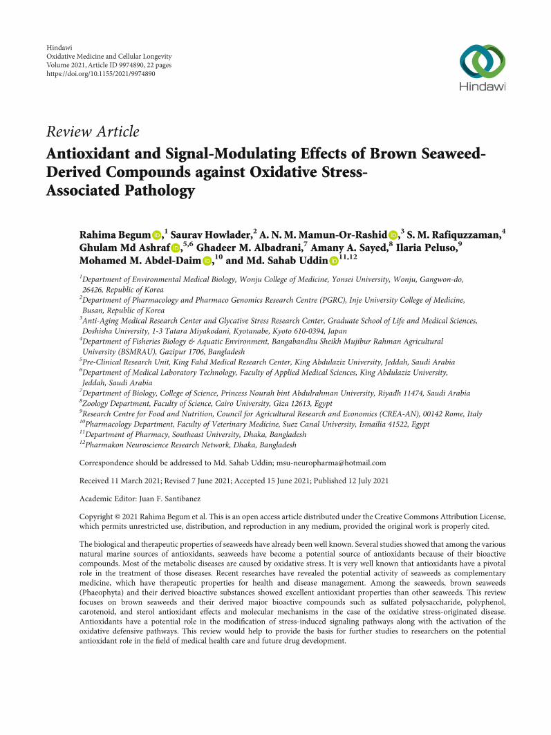
Review Article Antioxidant and Signal-Modulating Effects of Brown Seaweed- Derived Compounds against Oxidative Stress- Associated Pathology Rahima Begum , 1 Saurav Howlader, 2 A. N. M. Mamun-Or-Rashid , 3 S. M. Rafiquzzaman, 4 Ghulam Md Ashraf , 5,6 Ghadeer M. Albadrani, 7 Amany A. Sayed, 8 Ilaria Peluso, 9 Mohamed M. Abdel-Daim , 10 and Md. Sahab Uddin 11,12 1 Department of Environmental Medical Biology, Wonju College of Medicine, Yonsei University, Wonju, Gangwon-do, 26426, Republic of Korea 2 Department of Pharmacology and Pharmaco Genomics Research Centre (PGRC), Inje University College of Medicine, Busan, Republic of Korea 3 Anti-Aging Medical Research Center and Glycative Stress Research Center, Graduate School of Life and Medical Sciences, Doshisha University, 1-3 Tatara Miyakodani, Kyotanabe, Kyoto 610-0394, Japan 4 Department of Fisheries Biology & Aquatic Environment, Bangabandhu Sheikh Mujibur Rahman Agricultural University (BSMRAU), Gazipur 1706, Bangladesh 5 Pre-Clinical Research Unit, King Fahd Medical Research Center, King Abdulaziz University, Jeddah, Saudi Arabia 6 Department of Medical Laboratory Technology, Faculty of Applied Medical Sciences, King Abdulaziz University, Jeddah, Saudi Arabia 7 Department of Biology, College of Science, Princess Nourah bint Abdulrahman University, Riyadh 11474, Saudi Arabia 8 Zoology Department, Faculty of Science, Cairo University, Giza 12613, Egypt 9 Research Centre for Food and Nutrition, Council for Agricultural Research and Economics (CREA-AN), 00142 Rome, Italy 10 Pharmacology Department, Faculty of Veterinary Medicine, Suez Canal University, Ismailia 41522, Egypt 11 Department of Pharmacy, Southeast University, Dhaka, Bangladesh 12 Pharmakon Neuroscience Research Network, Dhaka, Bangladesh Correspondence should be addressed to Md. Sahab Uddin; [email protected] Received 11 March 2021; Revised 7 June 2021; Accepted 15 June 2021; Published 12 July 2021 Academic Editor: Juan F. Santibanez Copyright © 2021 Rahima Begum et al. This is an open access article distributed under the Creative Commons Attribution License, which permits unrestricted use, distribution, and reproduction in any medium, provided the original work is properly cited. The biological and therapeutic properties of seaweeds have already been well known. Several studies showed that among the various natural marine sources of antioxidants, seaweeds have become a potential source of antioxidants because of their bioactive compounds. Most of the metabolic diseases are caused by oxidative stress. It is very well known that antioxidants have a pivotal role in the treatment of those diseases. Recent researches have revealed the potential activity of seaweeds as complementary medicine, which have therapeutic properties for health and disease management. Among the seaweeds, brown seaweeds (Phaeophyta) and their derived bioactive substances showed excellent antioxidant properties than other seaweeds. This review focuses on brown seaweeds and their derived major bioactive compounds such as sulfated polysaccharide, polyphenol, carotenoid, and sterol antioxidant effects and molecular mechanisms in the case of the oxidative stress-originated disease. Antioxidants have a potential role in the modification of stress-induced signaling pathways along with the activation of the oxidative defensive pathways. This review would help to provide the basis for further studies to researchers on the potential antioxidant role in the field of medical health care and future drug development. Hindawi Oxidative Medicine and Cellular Longevity Volume 2021, Article ID 9974890, 22 pages https://doi.org/10.1155/2021/9974890
-
Upload
khangminh22 -
Category
Documents
-
view
3 -
download
0
Transcript of Derived Compounds against Oxidative Stress - Hindawi.com
Review ArticleAntioxidant and Signal-Modulating Effects of Brown Seaweed-Derived Compounds against Oxidative Stress-Associated Pathology
Rahima Begum ,1 Saurav Howlader,2 A. N. M. Mamun-Or-Rashid ,3 S. M. Rafiquzzaman,4
Ghulam Md Ashraf ,5,6 Ghadeer M. Albadrani,7 Amany A. Sayed,8 Ilaria Peluso,9
Mohamed M. Abdel-Daim ,10 and Md. Sahab Uddin 11,12
1Department of Environmental Medical Biology, Wonju College of Medicine, Yonsei University, Wonju, Gangwon-do,26426, Republic of Korea2Department of Pharmacology and Pharmaco Genomics Research Centre (PGRC), Inje University College of Medicine,Busan, Republic of Korea3Anti-Aging Medical Research Center and Glycative Stress Research Center, Graduate School of Life and Medical Sciences,Doshisha University, 1-3 Tatara Miyakodani, Kyotanabe, Kyoto 610-0394, Japan4Department of Fisheries Biology & Aquatic Environment, Bangabandhu Sheikh Mujibur Rahman AgriculturalUniversity (BSMRAU), Gazipur 1706, Bangladesh5Pre-Clinical Research Unit, King Fahd Medical Research Center, King Abdulaziz University, Jeddah, Saudi Arabia6Department of Medical Laboratory Technology, Faculty of Applied Medical Sciences, King Abdulaziz University,Jeddah, Saudi Arabia7Department of Biology, College of Science, Princess Nourah bint Abdulrahman University, Riyadh 11474, Saudi Arabia8Zoology Department, Faculty of Science, Cairo University, Giza 12613, Egypt9Research Centre for Food and Nutrition, Council for Agricultural Research and Economics (CREA-AN), 00142 Rome, Italy10Pharmacology Department, Faculty of Veterinary Medicine, Suez Canal University, Ismailia 41522, Egypt11Department of Pharmacy, Southeast University, Dhaka, Bangladesh12Pharmakon Neuroscience Research Network, Dhaka, Bangladesh
Correspondence should be addressed to Md. Sahab Uddin; [email protected]
Received 11 March 2021; Revised 7 June 2021; Accepted 15 June 2021; Published 12 July 2021
Academic Editor: Juan F. Santibanez
Copyright © 2021 Rahima Begum et al. This is an open access article distributed under the Creative Commons Attribution License,which permits unrestricted use, distribution, and reproduction in any medium, provided the original work is properly cited.
The biological and therapeutic properties of seaweeds have already been well known. Several studies showed that among the variousnatural marine sources of antioxidants, seaweeds have become a potential source of antioxidants because of their bioactivecompounds. Most of the metabolic diseases are caused by oxidative stress. It is very well known that antioxidants have a pivotalrole in the treatment of those diseases. Recent researches have revealed the potential activity of seaweeds as complementarymedicine, which have therapeutic properties for health and disease management. Among the seaweeds, brown seaweeds(Phaeophyta) and their derived bioactive substances showed excellent antioxidant properties than other seaweeds. This reviewfocuses on brown seaweeds and their derived major bioactive compounds such as sulfated polysaccharide, polyphenol,carotenoid, and sterol antioxidant effects and molecular mechanisms in the case of the oxidative stress-originated disease.Antioxidants have a potential role in the modification of stress-induced signaling pathways along with the activation of theoxidative defensive pathways. This review would help to provide the basis for further studies to researchers on the potentialantioxidant role in the field of medical health care and future drug development.
HindawiOxidative Medicine and Cellular LongevityVolume 2021, Article ID 9974890, 22 pageshttps://doi.org/10.1155/2021/9974890

1. Introduction
Brown seaweeds are photosynthetic aquatic algae whichbelong to the domain of Eukarya, kingdom of Chromista,and class of Phaeophyta [1]. There are 1500 species of brownseaweeds all over the world [2]. In particular, in Asian coun-tries, seaweed has been used as a traditional herbal medicinefor the treatment of gastrointestinal problems, cough, boils,ulcers, asthma, cough, and headache as well as vegetablestoo [3]. Lately, several studies revealed that dietary seaweedsnot only are a good source of carbohydrates, dietary fiber,proteins and peptides, vitamins, minerals, and fats but alsocontain a large concentration of functionally bioactive com-pounds such as carotenoids, polysaccharides, polyphenols,and sterols, which have potential antioxidant properties aswell as antimicrobial, anticoagulant, antithrombotic, anti-inflammatory, antitumor, and antiviral properties for severaldiseases [4–6]. Therefore, nowadays, seaweeds have beenpaid attention to for the development of medicine, food, cos-metics, dietary supplements, fertilizer, and bioenergy [7].
Oxidative stress is widely involved in the development ofmany chronic diseases such as cardiovascular disease, neuro-degenerative, cardiovascular, cancer, inflammation, diabetes,obesity, and aging and many of the elderly diseases [8–10].Antioxidants are the only therapeutic molecules capable ofblocking oxidative stress by their excellent reactive oxygenspecies (ROS) scavenging activity with low or no toxicity[11]. An antioxidant optimizes the human physiologicalfunction that helps to protect against disease or disease pro-gression as well as maintains a healthy state [12]. Naturally,endogenous antioxidants are present in our body; the addi-tional exogenous supplement can also be obtained from var-ious natural sources and chemically synthetic antioxidantssuch as butylated hydroxyanisole (BHA), butylated hydroxy-toluene (BHT), and tert-butylhydroquinone (TBHQ) [13–15]. However, studies proved chemically synthesized antiox-idants to be toxic and carcinogenic, whether the natural anti-oxidant is safe, more effective, and easily absorbed by thebody [15]. The dietary antioxidants such as α-tocopherol,ascorbic acid, carotenoids, amino acids, peptides, proteins,flavonoids, and other phenolic compounds were found effec-tive in the boosting of the antioxidant mechanism [16]. Themarine world is a rich source of bioactive molecules. Amongthe various natural sources of antioxidants, marine seaweedsand their bioactive compounds are gaining worldwide atten-tion [4, 17] in industry and drugs since 1980 [18]. Numerousevidences have shown that the brown seaweed-derived com-pounds are capable of improving the oxidative stress-induced diseases including neurodegenerative disease [19],cardiovascular-associated disorders [20], and obesity [21],as well as cancer protection [22]. But unfortunately, we donot have much information about the antioxidant effectsand molecular mechanisms of brown seaweeds in oxidativestress diseases.
It has been reported that brown seaweeds have higherantioxidant properties comparatively than red and green sea-weeds [23]. Brown seaweeds contain one of the most abun-dant pigment carotenoid compounds, fucoxanthin, and areestimated to contain around 10% of total carotenoids found
in nature [24, 25]. Fucoxanthin has a great antioxidant activ-ity as well as anti-inflammatory, antidiabetic, antiphotoaging,and neuroprotective properties [26]. Polyphenol compoundsin brown algae such as phlorotannins are the unique andmost dominant complex group of polymers named phloro-glucinol (1,3, 5-trihydroxybenzene). It is mainly formed assecondary metabolites in the acetate-malonate pathway.Phlorotannin bioactive compounds exist as soluble com-pounds or cell-bound forms, mainly produced by brown sea-weeds to protect themselves from herbivores and stressconditions, minimizing the oxidative damage caused bynutrient deprivation and ultraviolet radiation. Phlorotanninsisolated from brown algae Ecklonia cava, one of the mostabundant sources of polyphenolic compounds, have beenrevealed to have higher antioxidant activity in vitro and invivo [6, 7, 20, 27]. Another bioactive compound of brownseaweeds is sulfated polysaccharides (SPs), which generallycomprise a high content of major groups of sugar, e.g.,fucose, galactose, uronic acid, and sulfate [28]. The antioxi-dant activities of SPs depend on their major sugar, degreeof sulfating, low molecular weight, and glycosidic branching[29–31]. The major SPs found in brown seaweeds are fucoi-dan [32, 33]. Over the past decade, the antioxidant com-pound sterols called fucosterol have shown greatantioxidant property as well as important contributions tohuman health and wellness [34].
This present review summarizes and discusses brownseaweeds and their major bioderived compound’s role as anantioxidant in oxidative stress and their action in the man-agement of disease-related signaling pathways. In Table 1,we show the antioxidant activity of brown seaweed-derivedmajor compounds from the literature review and researcharticles.
2. Oxidative Stress, Antioxidant Defense, andSignal Transduction Pathway in the Cell
Generally, oxidative stress is the imbalance between the pro-duction of ROS and the body’s own antioxidant defensivesystem [50]. ROS, usually generated through various extra-cellular and intracellular processes, are involved in cellulargrowth, differentiation, progression, and cell death as wellas cell signaling [51, 52]. Excessive production of ROS causesoxidative stress and modifies the structure of the cellularmacromolecules such as lipids, proteins, nucleic acids, andDNA. Consequently, the cellular and biological functionsare inactivated with the modification of cellular signaling.Generally, under the physiological condition, ROS maintainthe body’s homeostasis by the regulation of several cell sig-naling pathways which are involved in cellular processes suchas mitogen-activated protein kinase (MAPK), nuclear factor-kappa B (NF-κB), and phosphatidylinositol 3‑kinase (PI3K)(Figure 1) as well as the defensive pathway nuclear factor ery-throid 2–related factor 2 (Nrf2) signaling; therefore, ROS isconsidered as a secondary messenger for the activation of cel-lular signaling [50, 53–55]. Nrf2 is a redox-sensitive tran-scription factor that binds to antioxidant response elements(ARE) to regulate the expression of antioxidant enzymes thatprotect the cell against oxidative damage by inducing the
2 Oxidative Medicine and Cellular Longevity

antioxidant enzymes. Moreover, excessive ROS modulatesthe cellular antioxidant defense system that has eventuallyaltered the normal physiological signal and switched to theapoptosis or cell death signals. Therefore, oxidative stress isto be involved in the development of many diseases includingcancer, Parkinson’s disease, Alzheimer’s disease, atheroscle-rosis, heart failure, myocardial infarction, schizophrenia,and chronic fatigue syndrome (Figure 2) [56–58].
In addition, ROS has a big role in balancing the intracel-lular and extracellular Ca2+ levels [54, 59]. Cellular Ca2+ con-tent is also known as one of the most versatile signals for theactivation of protein kinase C (PKC) signal transduction cas-cades which are involved in the control of cellular processesand functions, such as contraction, secretion, metabolism,gene expression, cell survival, and cell death as well as themaintenance of plasma membrane fluidity by proton motiveforce [60] (Figure 2). ROS with free radical groups such assuperoxide anion (O2
⋅−), hydroxyl radical (⋅OH), and perox-ynitrite (ONOO−) and the cellular Ca2+ level are well knownto maintain the redox homeostasis and signaling events dur-ing normal physiological processes. Therefore, it is consid-ered that the interaction between ROS and Ca2+ can be
bidirectional [59–62]. Excessive and uncontrolled ROS signalor oxidative stress directly damages the plasma membranefluidity and redox homeostasis [61, 62]. As a result, Ca2+
influx into the cytoplasm from the extracellular environmentand disrupts the ion exchanging balance between the intra-and extra-cellular plasma membranes as well as mitochon-dria, which then leads to cell death or the apoptotic pathwayby increasing the cytochrome c protein, which then in turnactivates caspase 3 and caspase 9. The accelerated Ca2+ leveldisrupted the Ca2+ signal in the cellular cytoplasm, whichdephosphorylates the protein and modulates the PKC signaltransduction cascades, and is associated with many diseasesand the aging process [60–62].
Primarily, antioxidants have the greatest defensive role inprotecting the cell against oxidative damage [63]. Generally,antioxidants have a vital role in keeping optimal cellularfunctions and systemic health and well-being. However,under oxidative stress conditions, endogenous antioxidantsin humans, although highly efficient, are not sufficient to pro-tect the cell from the harmful effects of ROS [64]. Therefore,dietary antioxidants are required to maintain the optimal cel-lular defensive functions. The most efficient enzymatic
Table 1: Antioxidant activity of brown algal-derived compounds.
General category Antioxidant compound Antioxidant activity reported
Carotenoids Fucoxanthin [25, 35–39]
Polyphenols
PhlorotanninsPhlorofucofuroeckol A, dieckol, phloroglucinol,
eckol, 7 pholoroeckol, and 2-phloroeckol[27, 40–43]
Sulfated polysaccharide Fucoidans [16, 32, 44–47]
Sterols Fucosterol [48, 49]
Low level of ROS
PKC
MAPK NF-𝜅b PI3k/AKT
Altered cell signalsCa2+ influx
Ca2+ channel
Cytoplasm
Cell proliferation, differentiation, cell
survival, development, apoptosis and stress-response
Cell proliferation, differentiation, immune-
regulation, inflammation, gene expression
Metabolism, cellproliferation, cell
survival, growth and gene expression
NRF2
High level of ROS
Plasma membrane
Cyto C
Caspase-3
Caspase-7
Apoptoticcell death
ROS scavenging enzyme(SOD, Catalase, GSH, GPx)
O2–, ·OH, ONOO–
O2–, ·OH, ONOO–
O2–, ·OH, ONOO–
Mitochondrion
Redox homeostasisARE
NRF2
Nucleus
P
Altered geneexpression
Fenton reaction
Disrupts the ionic balance between intra- and extra-plasma
membrane and mitochondria
Redo
x im
bala
nce
Redox imbalance
Organelle dysfunction
Cancer, obesity,dementia,
osteoporosis, atherosclerosis
Mitochondrial ROS also has role to activate the PKC signaling pathway
Ca2+ channel
Ca2+ efflux
Figure 1: Redox homeostasis with cell signaling pathway under lower and higher levels of ROS.
3Oxidative Medicine and Cellular Longevity

antioxidants contain glutathione peroxidase (GPx), catalase(CAT), and superoxide dismutase (SOD), present in cellularcytoplasm and mitochondria. Nonenzymatic exogenousantioxidants are mainly derived in nature from photosyn-thetic organisms, fruits, vegetables, and plants and belongto different families such as vitamins E and C, thiol antioxi-dants (glutathione, thioredoxin, and lipoic acid), melatonin,carotenoids, and natural flavonoids [64–67] (Figure 3).
Antioxidants are considered as stable molecules that candonate an electron to a free radical and neutralize them,thereby scavenging the free radical and stopping the free rad-ical from causing future damage [68]. Originally, antioxi-dants protect the cell from oxidative stress by applying oneof the two mechanisms such as (a) the chain-breaking mech-anism by which the primary antioxidant donates an electronto the free radical. (b) the second mechanism is the removal
of ROS/reactive nitrogen species (RNS) initiators (secondaryantioxidants) by quenching a chain-initiating catalyst. Anti-oxidants may provide their defensive action on biologicalsystems at different levels including electron donation, metalion chelation, radical scavenging, and repair or by geneexpression regulation as well as prevent lipid peroxidation[69]. Moreover, antioxidants protect the cells by applyingtheir strong multiphase efficacy system into the cell and cellmembrane. It can strongly diffuse in both the aqueous andoil phases because of its polar and nonpolar paradox systems[70], known as a lipophilic, hydrophobic, and amphiphilicantioxidant (Figure 4). The efficacy of lipophilic antioxidantsor polar antioxidants is to be more efficient than nonpolar orhydrophilic antioxidants. The lipophilic antioxidant can ori-ent itself to the oil-water interface, where lipid peroxidation isinduced whereas the hydrophilic antioxidant diffuses in the
Oxidative stress
Immune disease
Neurological disorders
Diabetes
Age-related maculardegeneration Cataract
LungProstate
Colon
Esophageal
Oral, pharynx, laynx
Erytheme
PhotocarcinogenesisArthritis
Psorisis
Photoageing
Polymorphous light eruptions
Coronary heart diseaseStroke
Circulatory shock
Myocardial infraction
Eye-relateddisorders
Cancer
Joints
Cardiovascular disease
Photosensitive disorders
Ischemia-reperfusion
Erythropoieticprotoporphyria
Figure 2: Examples of ROS and oxidative stress-induced diseases.
Antioxidants
Endogenous antioxidant Exogenous or dietary antioxidant
• Superoxide dismutase (SOD)• Catalase (CAT)• Glutathione peroxidase (GPx)• Glutathione reductase (GR)
Dietary antioxidants from fruits, vegetables and grain
• Vitamins: vitamin C, vitamin E• Trace elements: zinc, selenium• Carotenoids: β-carotene, zeaxanthin• Phenolic acids: gallic acid, caffeic acid• Flavonoids• Anthocyanidins
Enzymatic Non-enzymatic• Glutathione (GSH)• Uric acid• Lipoic acid• NADPH• Coenzyme Q• Albumin• Bilirubin
Figure 3: Classification of enzymatic and nonenzymatic antioxidants.
4 Oxidative Medicine and Cellular Longevity

water phase, and therefore, it is less efficient [71]. Addition-ally, their activity also depends on the concentration of anti-oxidants, free radical types, and both the chemical structureand the reaction condition [69].
Nowadays, a large number of natural antioxidant com-pounds such as carotenoids, ascorbic acid, flavonoids, andphenolic compounds are derived from seaweed sources [22,72–75]. The antioxidative efficiency of seaweeds is higherthan that of plants and fruits [76–78]. It is already wellreported that the brown seaweeds are the richest sources ofthese antioxidant compounds with a unique structure, whichhas a greater level of hydrophilic and lipophilic nature [14,17, 79] (Table 2).
The redox equilibrium plays a pivotal role in the cell’sphysiological and pathological functions by balancing theROS and antioxidant as well as ROS stability. ROS stabilityneeds to activate or deactivate a variety of receptors, proteins,ions, and other signaling molecules [50, 53]. The excessiveaccumulation or depletion of ROS leads to instability of theredox balance that may influence many cellular signalingpathways and confers the cellular dysfunction as well as sub-sequently developing various pathologies [50]. To equilibratethe redox balance, antioxidants not only produce the antiox-idant but also have a great ability to modulate the ROS sensi-tive cell signaling with the activation of the antioxidantresponsive element ARE/Nrf2 pathway [54, 55]. However,numerous studies have already elucidated that the phyto-chemicals or natural compounds of antioxidants successivelymodulate the ROS-sensitive signaling pathway and restockthe antioxidant into the cell through maintaining the redoxbalance [86]. The activation of such signaling pathwaysincreased the expression of gene-encoding cytoprotective
proteins, including phase II enzymes, antioxidant enzymes,growth factors, and proteins involved in the regulation of cel-lular energy metabolism. The activation of the Nrf2 pathwayintersects with other intracellular signaling pathways such asthe MAPK, PI3k/Akt, and NF-κB pathways [54, 55, 87](Figure 5)
On the other hand, antioxidants actively reduce the cellu-lar ROS which may help to balance the cellular redox balanceas well as the intra- and extracellular cytoplasmic Ca2+levels[87]. Antioxidants reduce the intracellular ROS level by bal-ancing the ionic exchange between the plasma membraneand the cytoplasmic membrane as well as modulate theCa2+ channel called IP inositol 1,4,5-trisphosphate receptor(IP3R) [54, 55, 59, 87]. As a result, the Ca2+ ion binds tothe PKC enzyme and activates the PKC signaling cascades,as well as activates various signaling pathways and also trig-gers the release and translocation of Nrf2 from the cytosolto the nucleus, which is responsible for the expression of anti-oxidant genes, thus maintaining the cellular redox homeosta-sis [55, 59] (Figure 5).
Brown seaweeds are the largest type of macroalgae thatbelong to the phylum of Phaeophyta, which means “duskyplants.” Naturally, it is brown or yellow-brown and foundin temperate or arctic waters. Brown algae typically have aroot-like structure called a “holdfast” to anchor the algae toa surface. There are about 1500-2000 species of brown algaeworldwide [3]. The huge biodiversity of algae and the rich-ness of physiological traits with their unique adaptive proper-ties are considered as a potential target in the research area[88]. Most of the algae are oxygenic autotrophs; they canquickly and continuously adapt themselves to extreme envi-ronments by synthesizing their bioactive compounds with
Amphiphilic antioxidant
Non-polar antioxidant Polar antioxidant
Hydrophilic antioxidant Lipophilic antioxidant
Oil
Air
Hydrophilic antioxidant
Water
Lipophilic antioxidant
Amphiphilic antioxidant
Water
Oil
Oil
Figure 4: Efficacy of active antioxidants at different phases.
Table 2: Antioxidant efficacy of brown seaweeds compounds.
Antioxidant efficacy in the compound of brown seaweedsLipophilic Hydrophilic Amphiphilic
Carotenoids [22, 78, 80] Phlorotannins [22, 81, 82] Sterols [22, 83, 84]
Sulfated polysaccharide [22, 85]
5Oxidative Medicine and Cellular Longevity

protective functions, limiting or repairing potential damagefrom a harmful environment. These bioactive compoundsact as primary and secondary sources of metabolites that pro-vide the antioxidant and other bioactive functions as well asmaintain their cellular homeostasis [3, 88]. It has beenalready proven in numerous research and clinical studies thatthe compound of brown algae species has antioxidative activ-ity [23–25, 78, 81].
A large number of enzymatic and nonenzymatic antioxi-dants have been involved to detoxify the ROS and prevent theformation of highly reactive radicals such as hydroxyl radical(⋅OH) [88]. Enzymatic antioxidants mainly produced in a cellare usually high molecular weight substances such as SOD,glutathione reductase (GR), CAT, and ascorbate peroxidase.Primarily, enzymatic antioxidants inactivate the ROS inter-mediates and detoxify the ROS by supplying the NADPHfor proper functioning (Figure 6). On the other hand, the die-tary substances known as exogenous antioxidants (such ascarotenoids, flavonoids, phenolic compounds, and ascorbicacid) scavenge the free radicals to break down the chain reac-tion responsible for lipid peroxidation [66–69]. The com-pounds of brown algae such as carotenoids, polyphenols,sulfated polysaccharides, and fucosterols have belonged asexogenous antioxidants, which may apply two defensiveactions to cope with ROS before they can damage the cellulardeference mechanism [89, 90] such as
(1) The primary action is to break down the chain reac-tion, which results in free radicals becoming less reac-tive [66, 69]
(2) The second mechanism is scavenging activity againstsuperoxide and hydroxyl radicals by chelating/deacti-vating metals. The metal-binding proteins (such asalbumin, ferritin, and myoglobin) inactivate the tran-sition metal ions (such as copper, iron, manganese,zinc, and selenium act to upregulate the antioxidantenzyme activities) that catalyze the production of freeradicals, quenching/scavenging the singlet and tripletoxygen (highly toxic) and removing ROS [66, 91, 92].
3. Antioxidant Effects of Brown Seaweed-Derived Compounds
3.1. Carotenoids.Carotenoids are a family of pigmented com-pounds that are naturally synthesized by plants, algae, fungi,bacteria, and archaea [29]. Over 1100 naturally occurringcarotenoids are produced from those sources but not fromanimals [93]. Carotenoids are hydrophobic highly conju-gated 40-carbon (with up to 15 conjugated double bonds)molecules. About more than 500 species of natural caroten-oids are known and categorized into two groups: primarycarotenoids which have pure hydrocarbons (those without
Gene transcription
NRF2
MAPK NF-𝜅b PI3k/AKT
Ca2+
Cytoplasm
Antioxidant responsephase II detoxifying &
antioxidant enzymes(HO-I, SOD, GPx,
catalase)
PKC
Keep 1
Degradation
Keep 1
Cross-talk
Antioxidant neutralize the free
radical and release it as water from the
cell and mitochondria
Mitochondrion
SOD
GPxCatalase
Ca2+ influence toactivate the PKC
signaling pathway
Ca2+ influx
Ca2+ channel
Nrf2 phosphorylation
NRF2P
NRF2
Nucleus
P
Plasma membrane
H2O+O2
H2O2
O2–, ·OH, ONOO–
O2–, ·OH, ONOO–
O2–, ·OH, ONOO–
Nrf2
phosphorylation
ARE
Antioxidant (phytochemicals)
Figure 5: Antioxidant mechanism in the regulation of the Nrf2 signaling pathway with the crosstalk of the different signaling pathways.
6 Oxidative Medicine and Cellular Longevity

any oxygen molecule), e.g., α-carotene, β-carotene, andlutein, and secondary carotenoids which are carbon (C),hydrogen (H), oxygen-containing xanthophylls (e.g., fuco-xanthin, astaxanthin, canthaxanthin, and echinenone) aregenerally produced during the photosynthesis of seaweedsafter exposure to specific environmental stimuli such as lightand osmotic shock [39, 94–96]. The brown seaweeds are therich source of carotenoids, which have been reported as pow-erful antioxidants with numerous beneficial health effects(Figure 7) [96, 97]
Carotenoids (Crt) are very potent natural antioxidants.The antioxidant actions of carotenoids are based on their sin-glet oxygen quenching properties and the ability to trap freeradicals, which mainly depend on the number of conjugateddouble bonds [98]. Furthermore, carotenoids can scavengeoxidizing free radicals via three primary reactions (Equations(1) to (3)), by its addition, electron transfer, addition, andhydrogen atom transfer.
The carotenoid antioxidant reactions are as follows: (i)electron transfer between the free radical (R⋅) and Crt, result-ing in the formation of a Crt radical cation (Crt⋅+) (Equation(1)) or Crt radical anion (Crt⋅−) (Equation (2)); (ii) they cantransfer the electrons forming a radical cation (RCrt⋅) (Equa-
tion (3)); and (iii) hydrogen atom transfer leading to a neu-tral Crt radical (Crt⋅) (Equation (4)) [38, 99].
R⋅ + Crt⟶ R−+Crt⋅ + 1ð Þ Addition½ � ð1Þ
R⋅ + Crt⟶ R++Crt⋅ − 2ð Þ Addition½ � ð2Þ
R⋅ + Crt⟶ RCrt⋅ 3ð Þ Electron transfer½ � ð3Þ
R⋅ + Crt⟶ RH + Crt⋅ 4ð Þ Hydrogen ion transfer½ � ð4ÞGenerally, the principal defense process in carotenoids is
their lipophilic nature that allows them to penetrate throughthe cellular lipid bilayer membrane and cross to the blood-brain barrier, carrying out its biological function also in dif-ferent regions of the human body, including the brain [39].Because as a lipid-soluble molecule, carotenoids activelyscavenge the lipid and aqueous phase radicals, mostly theyare located in the apolar core of lipid membranes. Thus,carotenoids are associated with various types of membraneswithin the cell such as the outer cell membrane, but alsothe mitochondria and the nucleus. They also work in lyso-somes. As a consequence, carotenoids play a major
O2–
H2O2
H2O+O2
Catalase(cytoplasm)
Gpx(mitochondria)
SOD(mitochondria)
.OHFenton reaction
Lipid peroxidation
GSSG
GSH
NADPH
NADP+
GSH reductase (GR)
Carotenoids, vitamin C, tocopherol
Regenerate the GSSGand back to the GSH aswell as donate thehydrogen to directlyscavenges the lipidsperoxides and terminatethe lipid chain reaction.
Fe2+1
2
3
4
5
6
2
6
Fe3+
O2 O2.–
Figure 6: The enzymatic and nonenzymatic antioxidant protection mechanism. (1) Superoxide radical (O2⋅−) is formed by a single-electron
reduction of oxygen. In a reaction catalyzed by superoxide dismutase (Cu/ZnSOD or MnSOD), superoxide radical binds an electron, whichleads to the formation of hydrogen peroxide (H2O2). (2) In the further reduction of hydrogen peroxide (H2O2) to water and oxygen bycatalase (CAT) and glutathione peroxidase (GPx) enzymes. (3) In the Fenton reaction, the H2O2 is then transformed into hydroxyl radical(HO⋅) by catalyzing the transition metal, which is further participating in the free radical chain reactions. (4) H2O2 is reduced byglutathione (GSH) and produced glutathione disulfide (GSSH). (5) The glutathione disulfide is then reduced by glutathione reductase(GR) using the hydrogen of NADPH which is oxidized to NADP+. (6) Besides, the nonenzymatic antioxidants such as carotenoids,vitamin C, and tocopherol support the regeneration of GSSG and back to GSH. Vitamin C, carotenoids, and tocopherol donate thehydrogen to free radicals that directly scavenges the free radical and terminates the lipid peroxidation chain reaction [66, 91, 92].
7Oxidative Medicine and Cellular Longevity

protecting role in the cell membranes and lipoproteins fromdamage by free radicals [100].
Several evidences suggested that carotenoids can act asredox agents in the case of oxidative stress due to their elec-trophile character and their conjugated double bonds thatenhance the endogenous antioxidant Nrf2-Keap1 systems[101] which eventually help to maintain the redox homeosta-sis [80]. The redox homeostasis mainly depends on thenucleophile compounds, e.g., glutathione (GSH) and thiol(–SH) as well as their reductase enzymes GR, hemeoxygenase-1 (HO-1), glutathione S-transferases (GSTs), andNAD(P)H quinine oxidoreductase 1 (NQO1). The GSH or–SH (thiol-) containing enzymes play a pivotal role in thefree radical chain reaction that promotes to sustain the redoxhomeostasis (Figure 6) [102, 103]. Studies demonstrated thatthe carotenoids could increase the GSH level through themodulation of redox-sensitive –SH groups of Kelch-likeECH-associated protein 1 (Keap1). The Keap-Nrf2 signalingpathway plays an important role in the cellular defenseagainst oxidative stress [104, 105]. Generally, under the nor-mal physiological condition, Nrf2 is bound with their repres-sor protein Keap1 in the cytosol; which leads to itsdegradation by the ubiquitinylation process. Keap1 is acysteine-rich protein, and its –SH residue conformation canbe modified by different oxidants and electrophiles, therebyleading to the liberation of Nrf2 and transfer to the nucleus[80, 106]. At a higher oxidative stress condition, excessiveROS promotes the disassociation of Keap1 and Nrf2 eithervia the activation of PKC that assists to phosphorylate theNrf2 or by oxidation of the cysteine residues of Keap1 thatmainly govern the Keap1 activity [54–57, 107]. The caroten-oids act as antioxidants through their electrophile C=C
groups which conjugate with the aldehyde group (–CHO)of Keap1 modifying their thiol residues allowing the Nrf2release into cytosol and translocation to the nucleus. Thus,Keap1 is inactivated, and newly synthesized Nrf2 binds tothe ARE, which then leads to the expression of phase IIenzymes and cytoprotective enzymes, e.g., GR, hemeoxygenase-1 (HO-1), GSTs, NAD(P)H quinine oxidoreduc-tase 1 (NQO1), SOD, and GPx [108] (Figure 8).
The scavenging function of carotenoids also has a signal-modulating role at the cellular signaling cascades such as theNF-κB, MAPK [109, 110], and PI3/Akt survival pathways aswell the caspase pathway [111, 112]. Several studies suggestthat carotenoids can act as modulators of redox-sensitive sig-naling cascades also able to progress the cell cycle through thedirect modulation of cell cycle-related proteins as well as dif-ferent signaling pathways which are usually involved in cellproliferation [108]. According to the studies, it can behypothesized that β-carotene can modify the cellular redoxstatus by changing the intracellular antioxidant status [112].Studies showed that it can also delay the cell cycle G2/Mphase by decreasing the expression of cyclin A in humancolon adenocarcinoma cells [113] with increase of theantioxidant level which might help in decreasing the apo-ptotic protein as well as modifying the cellular growth[111]. It has been also reported that cell cycle progressionand differentiation are dependent on the carotene dose,because it has a prooxidant role under high oxygen ten-sion [113, 114]. Several indications suggested that caroten-oids may also alter the expression of the apoptosis-relatedprotein including the Bcl-2 and the caspase protein by aredox mechanism [111, 115]. Free radical species such assinglet oxygen [116] and nitric acid [117] have been
Natural carotenoids
UsesFood and feed industry Cosmetology
Medicine
Carotenes
Xanthophylls
Application Neuroprotective
Immune enhancers
Pigments
Antioxidants
Signal modulator
Anti-inflammatory
Anti-pathogenic
Anti-mutagenic
Anti-diabetic
𝛽-Carotene
Lycopene
FucoxanthinAstaxanthin
Zeaxanthin
Anti-angiogenic
Anti-carcinogenic
Beauty-enhancing effect
Figure 7: Distribution, biological functions, and application of natural carotenoids derived from brown seaweeds.
8 Oxidative Medicine and Cellular Longevity

reported to activate the caspase 8, an important proteindegrading enzyme involved in the apoptotic cascade[108], whereas studies found carotenoids can initiate thecaspase 3 activities in several cell lines, mainly by interact-ing with a single complex located on the cell membraneand inducing caspase 8. It was also reported that caroten-oids protect the colon cancer cell from apoptosis bydecreasing the expression of antiapoptotic protein Bcl-2[112]. Such effects are mainly strictly related to apoptosisinduction and ROS production by the carotenoids. Thisfinding interestingly demonstrated that carotenoids havea role in an antioxidant pathway, whereby this proteincan prevent programmed cell death by decreasing theROS formation and lipid peroxidation products [115]. Invitro studies observed that β-carotene may increase theexpression of the proapoptotic protein Bax in U-937 cellsand HUVEC cells [112].
The NF-κB pathway is generally thought to be a primaryoxidative-response pathway [112]. Studies have shown thatcarotenoids can inhibit the oxidative stress-induced NF-κBpathway with the addition of their electrophile groups andthe cysteine residues of IκB kinase IKK and NF-κB subunits(p65) [118]. It has been reported that β-carotene preventsthe phosphorylation of ERK, JNK, and P38 in oxidative stress[119]. Recently, it has been reported that β-carotene mayaffect cellular growth through the modulation of redox-sensitive transcription factor AP-1 [112]. Carotenoids may
also modulate the antioxidant response regulatory gene(ARE) and can alter the gene expressions of phase II enzymes(i.e., HO-1, GSTs, NQO1, SOD, and GPx), resulting in cellu-lar redox homeostasis [112] (Figure 9).
Some of the studies indicate that carotenoids can alsomodulate oxidative stress-induced calcium signaling [120].Carotenoids can decrease the calcium/calmodulin-depen-dent protein kinase IV (CAMKIV) enzyme by binding withthe active site of CAMKIV; that form allows the cancer cellapoptosis [121]. CAMKIV is an enzyme belonging to theSer/Thr kinase family. Generally, it plays a role in cell prolif-eration, migration, angiogenesis, and inhibition of apoptosisas well as calcium-dependent cell signaling. Elevated intracel-lular calcium ion concentration makes a complex formbetween Ca2+/calmodulin, which induces the phosphoryla-tion of the transcription factor and causes various types ofcancer [122, 123].
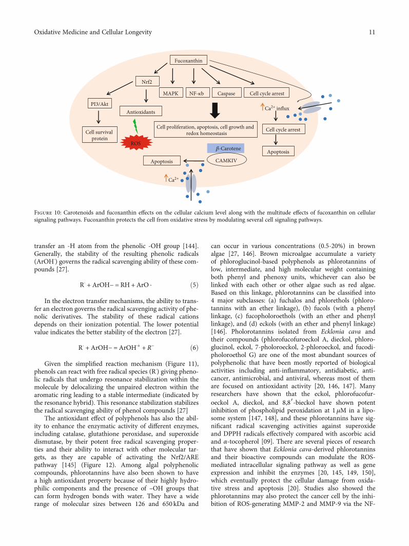
One of the well-known carotenoids is fucoxanthin, con-sidered as a potential antioxidant because of its unique chem-ical structure including an allelic bond (C=C), epoxide group,and hydroxyl group [124]. It is well-known, and uniquestructural carotenoid pigments are present in brown seaweedthat have not been found in other carotenoids [35, 125].Fucoxanthin accounts for around 10% of the total naturalproduction of seaweed carotenoids [126], which showsstrong antioxidative properties by the inhibition of intracel-lular ROS formation, DNA damage, and apoptosis as well
Keap1
Keap1
SS SS
Nrf2 disassociate from the cytoplasm and migrate into the nucleus
Keap1
Keap1
Nrf2 SH SHSH SH
Nrf2
Keap1
S-adduct
Carotenoids modified the-SHresidues conformation of Keap1protein by the conjugationwiththeir C
Normal redox state Keap1
Keap1
Nrf2 SH SHSH SH
Protein degradation
Nrf2PKCP
ROS/RNS
ROS/RNS
Nrf2
Nrf2ARE
Antioxidants & detoxifying enzymese.g.HO-1, SOD, GPx, NQO1
Cell defense
Nucleus
PNrf2
PNrf2 translocation
to the nucleus
1
2
3
C bond
Carotenoids
Figure 8: Electrophile activation of carotenoids of the Keap1-Nrf2 systems.
9Oxidative Medicine and Cellular Longevity

as exhibiting strong enhanced cell viability against H2O2-induced oxidative stress [127]. Several studies have demon-strated their experiments that fucoxanthin is safe and non-toxic to cells even at repeated doses [128–131]. In vivostudies also have shown that a fucoxanthin diet elevated theantioxidant activities including CAT, SOD with the mRNAexpression of Nrf2 and its target genes such as NQO1[132]. It has an active role in the modulation of signalingpathways including MAPK [133], NF-κB [134], and apopto-sis caspase pathways [135] as well as induces cell cycle arrest[136]. Recently, it was shown that fucoxanthin suppressesH2O2-induced inflammation and oxidative damage in micro-glial cells via the attenuation of the phosphorylation of theMAPK signaling pathway, as well as by the free radical scav-enging capacity of fucoxanthin and its ability to regulate theendogenous antioxidant system [137] It can also exert thecytoprotective effects against H2O2-induced oxidative stressthrough the activation of the PI3K-dependent Nrf2-signaling pathway along with the expression of mRNA andprotein relative cytoprotective gene LO2 cells [138]. San-geetha and collaborators in vivo analyzed the properties offucoxanthin compared with β-carotene [139]. These two
carotenoids decreased lipid peroxidation as well as enhancedthe CAT and GST activities, showing the protective effectagainst Na+K+-ATPase activity. However, fucoxanthinexhibited higher antioxidant and protection properties thanβ-carotene [39, 139]. Many studies suggested that fucoxan-thin increases the intracellular cytosolic Ca2+ that triggers cellapoptosis through modulation of the cell cycle arrest [135,136] (Figure 10).
3.2. Polyphenols and Phlorotannins. Algal polyphenols are alarge and diverse class of secondary metabolites that consistof around 8000 naturally occurring compounds that possessvital biological functions including antioxidant and free rad-ical scavenging properties [27, 140]. These biological proper-ties mainly shared by this molecule are their phenol groups[27]. The antioxidant radical scavenging activity of thesecompounds generally precedes through the hydrogen atomtransfer or electron transfer mechanisms [141–143]. Thepolyphenol compounds have one or more hydroxyl groups(-OH), which directly bond to a phenolic or aromatic hydro-carbon group, and the hydrogen atom transfer mechanismindicates the ability of the phenolic derivatives (ArOH) to
Carotenoids
Modulation of ROS production
ERK,JNK,P38
NF-𝜅B AP-1 ARE
Gene expression changes
Redox-sensitiveproteins
Proteins for survival and proliferation
Proteins for cell growth
Bcl-2,Bax
Proteins for apoptosis
Caspase 8
Caspase 3PI3k/Akt
Figure 9: Scheme representing the multitude effects of carotenoids on the ROS-dependent signaling pathway and their related target proteingene expression. Carotenoid molecules may modulate the intracellular ROS production and can activate the redox-sensitive transcriptionfactors or directly affect DNA damage which in turn may modify the gene expression.
10 Oxidative Medicine and Cellular Longevity
transfer an -H atom from the phenolic -OH group [144].Generally, the stability of the resulting phenolic radicals(ArOH⋅) governs the radical scavenging ability of these com-pounds [27].
R⋅ + ArOH− = RH + ArO ⋅ ð5Þ
In the electron transfer mechanisms, the ability to trans-fer an electron governs the radical scavenging activity of phe-nolic derivatives. The stability of these radical cationsdepends on their ionization potential. The lower potentialvalue indicates the better stability of the electron [27].
R⋅ + ArOH− = ArOH⋅+ + R− ð6Þ
Given the simplified reaction mechanism (Figure 11),phenols can react with free radical species (R⋅) giving pheno-lic radicals that undergo resonance stabilization within themolecule by delocalizing the unpaired electron within thearomatic ring leading to a stable intermediate (indicated bythe resonance hybrid). This resonance stabilization stabilizesthe radical scavenging ability of phenol compounds [27]
The antioxidant effect of polyphenols has also the abil-ity to enhance the enzymatic activity of different enzymes,including catalase, glutathione peroxidase, and superoxidedismutase, by their potent free radical scavenging proper-ties and their ability to interact with other molecular tar-gets, as they are capable of activating the Nrf2/AREpathway [145] (Figure 12). Among algal polyphenoliccompounds, phlorotannins have also been shown to havea high antioxidant property because of their highly hydro-philic components and the presence of –OH groups thatcan form hydrogen bonds with water. They have a widerange of molecular sizes between 126 and 650 kDa and
can occur in various concentrations (0.5-20%) in brownalgae [27, 146]. Brown microalgae accumulate a varietyof phloroglucinol-based polyphenols as phlorotannins oflow, intermediate, and high molecular weight containingboth phenyl and phenoxy units, whichever can also belinked with each other or other algae such as red algae.Based on this linkage, phlorotannins can be classified into4 major subclasses: (a) fuchalos and phlorethols (phloro-tannins with an ether linkage), (b) fucols (with a phenyllinkage, (c) fucopholoroethols (with an ether and phenyllinkage), and (d) eckols (with an ether and phenyl linkage)[146]. Pholorotannins isolated from Ecklonia cava andtheir compounds (phlorofucofuroeckol A, dieckol, phloro-glucinol, eckol, 7-pholoroeckol, 2-phloroeckol, and fucodi-pholoroethol G) are one of the most abundant sources ofpolyphenolic that have been mostly reported of biologicalactivities including anti-inflammatory, antidiabetic, anti-cancer, antimicrobial, and antiviral, whereas most of themare focused on antioxidant activity [20, 146, 147]. Manyresearchers have shown that the eckol, phlorofucofur-oeckol A, dieckol, and 8,8′-bieckol have shown potentinhibition of phospholipid peroxidation at 1μM in a lipo-some system [147, 148], and these phlorotannins have sig-nificant radical scavenging activities against superoxideand DPPH radicals effectively compared with ascorbic acidand α-tocopherol [09]. There are several pieces of researchthat have shown that Ecklonia cava-derived phlorotanninsand their bioactive compounds can modulate the ROS-mediated intracellular signaling pathway as well as geneexpression and inhibit the enzymes [20, 145, 149, 150],which eventually protect the cellular damage from oxida-tive stress and apoptosis [20]. Studies also showed thephlorotannins may also protect the cancer cell by the inhi-bition of ROS-generating MMP-2 and MMP-9 via the NF-
𝛽-Carotene
Fucoxanthin
CAMKIV
Ca2+
Apoptosis
ROS
MAPK NF-𝜅b
Nrf2
Antioxidants
Caspase
PI3/Akt
Cell survival protein
Cell cycle arrest
Cell proliferation, apoptosis, cell growth andredox homeostasis
Ca2+ influx
Cell cycle arrest
Apoptosis
Figure 10: Carotenoids and fucoxanthin effects on the cellular calcium level along with the multitude effects of fucoxanthin on cellularsignaling pathways. Fucoxanthin protects the cell from oxidative stress by modulating several cell signaling pathways.
11Oxidative Medicine and Cellular Longevity

κB pathway [151]. It has been reviewed that phlorotanninscan block the Ca2+ channel as well as reduce the Ca2 +
influx which are stimulated by ROS (Figure 13) [20].Due to several profound capabilities of E. cava, derivedcompounds are interesting to use as functional ingredientsin pharmaceuticals and cosmeceuticals [147, 152, 153]
3.3. Sulfated Polysaccharides. Marine SPs are polymeric car-bohydrates with large hydrophilic molecules. They have afundamental role in living marine organisms such as energystorage and protection or as a structural molecule, whichare mainly produced by photosynthesis [85]. The algal poly-saccharides possess potent bioactivities because of theirunique physicochemical properties, such as high content of
fucose, galactose, uronic acid, and sulfate [154]. Microalgaecan accumulate carbohydrates to more than 50% of theirdry weight, due to the high photoconversion efficiency ofthe photosynthetic process [155]. These compounds arepresent in high concentrations and vast diversity in microal-gae [156]. A vast number of researchers have reported thatthe SPs and its compounds could alleviate oxidative stress-mediated diseases, such as liver injury, diabetes, obesity, neu-rodegenerative disease colitis, and cancer [155–161]. Thiseffect could be explained by three distinct mechanisms,including scavenging the ROS and regulating the antioxidantsystem or oxidative stress-mediated signaling pathways andalso implying the complicated interactions of marine-derivedantioxidant polysaccharides in reducing the oxidative stress[46, 47] (Figure 14). The antioxidant activity of SPs dependson their structural property, such as the degree of sulfating,their molecular weight, types of the major sugars, and glyco-sidic branching [46, 162]. The low molecular weight SPs havebeen shown to be potent antioxidants rather than the highmolecular weight SPs [163]. It has been also reported thatlowmolecular weight (~30kDa) SPs can promote cell prolifer-ation [164] as well as cell protection from the oxidative stress-induced apoptosis, which are directly and indirectly related totheir antioxidant properties [46, 165]. Currently, SP hasbecome a hot research area in the field of regenerative medi-cine and tissue engineering application due to its unique struc-ture and specific features as antioxidants, antitumors,immunomodulatory, inflammation, anticoagulant, antiviral,antiprotozoal, and antibacterial [46, 47, 162, 165].
Among the three types of SPs extracted from seaweeds,the brown seaweeds are the richest source of antioxidants[46, 47]. The major bioactive SP extracted from brown sea-weeds is fucan which is usually defined as fucoidan; it iswater-soluble and composed of significant quantities of L-fructose and sulfated ester groups accompanied with a littlepercentage of other monosaccharides, e.g., arabinose, xylose,glucose, galactose, and mannose. These constitutes of poly-saccharides are known to be one of the major componentsof the brown algae cell wall that protects the cell in stress orlow tide environments [46, 165].
H
HC
OO
CH
···· ······
· ·
·
OORHO
C
O𝛿·
𝛿·
𝛿·
𝛿·
Figure 11: The proposed reaction between phenol and a free radical, which undergoes resonance stabilization; indicates the radicalscavenging ability of compounds (i.e., curved half-headed arrows represent the transfer of a single electron).
Nrf2 activation, NF-𝜅B inhibition
Oxidative DNA damage, protein oxidativedamage, lipid peroxidation reaction
Oxidative cell damage
Antioxidant effects of polyphenols
Antioxidative enzymes
GPxCAT
SOD
ROS/RNS
Figure 12: Potential mechanisms of polyphenols to the protectionof the cell against oxidative stress.
12 Oxidative Medicine and Cellular Longevity

Numerous in vivo and in vitro studies have revealedthat fucoidan derived from brown algae can alleviate bodydamage from oxidative stress through the regulation of theantioxidant system in the body [45, 165, 166]. The excel-lent antioxidant activity of fucoidan is mainly dependenton their desulfation or oversulfation that allows the devel-opment of derivatives of fucoidans [46, 165, 166]. TheDPPH free radical scavenging assay is widely used to eval-uate the in vitro antioxidant activity [167]. Polysaccharidesprovide the hydrogen or electrons to DPPH free radicalsto form stable molecules (DPPH-H) [168]. The electron-withdrawing group of polysaccharides and the specificstructures activate the hydrogen atoms on sugar residues
[169]. Many studies have already found that sulfated poly-saccharides from brown algae have strong DPPH free rad-ical scavenging activity and reduction ability. Furthermore,SPs also have strong ferrous ion-chelating and reducingpower activities [170, 171]. Many studies have revealedthat fucoidan inhibits oxidative stress by downregulatingthe malondialdehyde (MDA) level and upregulating theSOD level [172]. Moreover, studies have documented thatfucoidan extract can reduce the levels of lipid peroxidationmarkers MDA and TBARS in alcoholic rats and increasethe levels of the nonenzymatic antioxidant GSH and theenzymatic antioxidant SOD, CAT, and GPx in the liver,ultimately reducing the oxidative damage caused by
Has higher protective activity from photoprotection
Protect the cell from apoptosis by thesuppression of ROS mediated
signaling pathway
Block the Ca2+ channel and ACE inhibition through their sequestrated activity of enzyme factor as well as the
antioxidant properties; which protect the cell from an oxidative stress-inducing
disease like hypertension
Modulate the intracellular signaling pathway such as AMPK,
NF-𝜅B, PI3K-AkT
Protect the cell from oxidative stress by the expression of cytoprotective enzymes e.g., catalase, SOD and GPx
Cell growth and proliferationActive the Nrf2 /ARE pathway
Inhibits the oxidative stress associated MMP-1 and MMP -9 to maintain the
normal physiological process
Phlorotannins
Figure 13: The biological role of phlorotannins on the function of the cell signaling pathway.
Sulfated polysaccharides(fucoidan)
Excess ROS(O2–, ·OH, ONOO–)
Antioxidant system dysfunction
(enzyme: SOD, CAT, GPx)
Oxidative stress-related signaling pathway
(PI3K/Akt, MAPK, Nrf2/ARE)
Regulate
Control
ScavengeLiver injury,
Obesity,Atherosclerosis,
Neurodegenerative disease,Colitis and gastric cancer,
Breast cancer
Brown seaweeds
Figure 14: Overview of sulfated polysaccharide (fucoidan) derived from brown seaweeds in alleviating oxidative stress-mediated disease.
13Oxidative Medicine and Cellular Longevity

alcohol. In addition, fucoidan also reduces the mRNAexpression of TNF-α, IL-1β, and MMP-2 to inhibit theproduction of ROS in the liver, thus alleviating the devel-opment of nonalcoholic fatty liver disease [173]. Similarly,fucoidans inhibit the proliferation and induced apoptosisthrough the downregulation of the PI3K/AKT pathwayas well as the reduction of Bcl-2 and Bcl-xl and the riseof the expression of Bax, caspase 3, and caspase 9 activa-tion [174]. It can also inhibit the epidermal growth factor(EGF) signaling in several cancer cell types, by preventingbinding their receptor site that promotes to block the acti-vation of activator protein-1 (AP-1) which also diminishesthe transactivation activity of ERK1/ERK2 and JNK signal-ing protein [175]. Another study also showed that fucoi-dan can inhibit apoptosis and improve the cognitiveability in Alzheimer’s disease model mice by upregulatingthe expression of the apoptosis-inhibiting proteins by acti-vating the SOD activity and increasing the GSH levels[176]. Recently, a study found that fucoidan can improveatherosclerotic cardiovascular disease by reducing the pro-duction of ROS by inhibiting NADPH oxidase (Figure 15)[177]. Besides, fucoidan can also inhibit ROS productionthrough the regulation of the antioxidant defense system,thereby alleviating oxidative stress-related diseases. A studyrevealed that low molecular weight fucoidan regulates theactivity of SOD and CAT by activating the SIR-T1/AMPK/PGC1α signaling pathway, inhibiting the super-oxide production and the lipid peroxidation, reducingTNF-α and transcription factor NF-κB, and then inhibit-ing oxidative stress in the liver (Figure 15) [164]. Further-more, fucoidan significantly increased the expression of
MnSOD and decreased the ROS level through thePI3/AKT pathway and finally enhanced the survival andangiogenesis of mesenchymal stem cells in the ischemiamodel [178]. Interestingly, it was reported that it couldupregulate the expression of HO-1 and SOD-1 genes byactivating Nrf2 and then attenuate the oxidative stress inHaCaT cells [163]. Research also showed that the Turbi-naria ornata protects the human carcinoma cell throughthe modulation of PPAR gamma, NF-κB, and oxidativestress [179]. Another study also proved that the Turbi-naria ornata protects the hepatic injury fromazoxymethane-induced oxidative stress by exerting multi-ple pathways including the abolishment of inflammationand oxidative damage and the activation of PPAR gammawith increase of the antioxidant enzymes like SOD andGPx activities [180]. Thus, we could be review by all ofthese studies that fucoidan derived from brown algae notonly has significant antioxidant activity but also modulatesthe oxidative stress-mediated diseases by regulating theantioxidant defense systems and the oxidative stress-related signaling pathway in cellular and experimental ani-mal models [22].
3.4. Sterols. There are abundant sterols in brown seaweeds.These compounds occur in the free form, esterified with fattyacids, or are involved in glycosylated conjugates [49, 181].Sterols are amphipathic and triterpene compounds that canreach about 5.1% of the total biomass in the microalgae[181, 182]. Most of them contain 28 or 29 carbons and 1 or2 carbon-carbon double bonds, typically one in the sterolnucleus and sometimes a second in the alkyl side chain
Fucoidan
SOD, CAT, GSH, GPx TNF-𝛼, IL-1𝛽, MMP-2NADPH oxidase
Oxidative stress
SIRT1/AMPK/PGC1𝛼Nrf2
ActivationMnSOD ROS
PI3/AKT
EFGR
Blocking
AP-1ERK1/ERK2, JNK
Inhibition
Apoptosis
H3C
H3C
OSO2H
OSO2H
HO3SO
O
O
O
O
OH
Figure 15: Fucoidan prevents oxidative stress and cell apoptosis by regulating the antioxidant system and signal-modulating pathway.
14 Oxidative Medicine and Cellular Longevity

[181]. Moreover, fucosterol displays a high diversity of theunique compounds of phytosterols, such as brassicasterol,sitosterol, and stigmasterol [182, 183]. Some species containa mixture of ten or even more phytosterols [183]. Sterol com-position varies depending on the algae strain and can bemodulated by light intensity, temperature, or growth phase[83]. Sterols have received much attention in the last fewyears because of their cholesterol-lowering properties [181].Fucosterol is structural and functionally similar to choles-terol; however, it contains an alkyl; fucosterol, a phytosterolfound in brown seaweeds, is well recognized for its variousbeneficial biological activities [49].
Studies showed that fucosterol elevates the activities offree radical scavenging enzymes such as SOD, CAT, andGPx against hydrogen peroxide. It can also restore theSOD and GPx cellular defensive enzymes which help toprevent cell membrane oxidation [48]. It can also suppressthe nitric oxide (NO) and ROS generation through thesuppression of inducible nitric oxide synthase (iNOS)and cyclooxygenase 2 (COX-2) expression because increas-ing iNOS and COX-2 is responsible for inflammatory dis-ease [49]. Moreover, fucosterol exhibits inhibitory activityagainst acetyl- and butyryl-cholinesterases (AChE andBChE, respectively) [184, 185] and β-secretase enzymewhich is responsible for Alzheimer’s disease, although thisenzyme production level is mainly associated with increas-ing the oxidative stress [186].
In the study of pharmacology and in silico analysis, itwas revealed that the fucosterol action could intervene inthe disease progression through modulating the biologicalprocesses as well as the signaling pathway, such ascytokine-mediated signaling pathway, apoptotic process,transcription regulation, inflammatory response, aging,response to lipopolysaccharide, NF-κB activity, and also
cellular response to ROS, which are closely associated withthe disease pathophysiology. Studies showed that fucos-terol could pass through the cell membrane to reachdirectly to various intracellular targets. The pharmacologi-cal network data reported that fucosterol showed a closeassociation with the target proteins of many crucial path-ways at the molecular and cellular levels [187]. Anotherreport demonstrated that fucosterol attenuates the oxida-tive stress through the upregulation of antioxidantenzymes such as GPX1, SOD, CAT, and HO-1 viaNrf2/ARE and PI3/Akt signaling activation [49, 188].
Fucosterol treatment can inhibit the oxidative stress-induced MAPK and NF-κB signaling pathway along withreducing the levels of inflammatory mediators PGE2 andCOX-2, as well as proinflammatory cytokines TNF-α, IL-1β, IL-6, and IL-8 [188]. Studies represented an excellentrole of fucosterol in cancer cell proliferation, cell cycle,and apoptosis [189]. It was found that fucosterol causesthe arrest of the cancer cells at the stage of the G2/Mphase that prevents the cancer cells to enter the mitosisstage which can prevent cell growth and proliferation[190]. The Raf/MEK/ERK signaling pathway contributesto cancer cell proliferation. It also showed that fucosterolinduced cell cycle arrest, via the inhibition of Raf/ME-K/ERK signaling pathways that ultimately prevents cellproliferation [191]. Fucosterol can induce cancer cell apo-ptosis by increasing the expression of cleaved caspase 3,caspase 9, and Bax and decreasing the expression of Bcl-2, MMP-2, and MMP-9 [189, 192] (Figure 16). Researchshowed that the species of fucosterol named Padina pavo-nica exerted an excellent antidiabetic and antioxidanteffect via the activation of PPARγ mechanisms [193].Fucosterol treatment can protect the cell from DNA dam-age after oxidative stress [188]
Fucosterol
ROS
MAPK NF-𝜅B
Nrf2/ARE
SOD, CAT, GPX, HO-1
Caspase-3,Caspase-9, Bax
PI3/Akt Cell cycle arrest at G2/M phase
Cell proliferation, apoptosis, Cell growth
Protect the cell from DNA damage
PGE2 , COX-2, TNF-𝛼, IL-1𝛽, IL-6 ,IL-8
Raf/MEK/ERK
Bcl-2, MMP-2,MMP-9
Figure 16: Overview of the biological role of fucosterol derived from brown seaweeds in alleviating the oxidative stress-mediated signalingpathway.
15Oxidative Medicine and Cellular Longevity

4. Conclusion
The research on brown seaweeds and their bioactive com-pounds’ biological spectrum has been widely raised in recentyears. Brown seaweeds and their derived compounds such asphlorotannins, SPs, fucosterol, and carotenoid pigmentsincluding fucoxanthin radical scavenging property are nowclinically emphasized as a novel therapeutic compound.Antioxidants not only scavenge the free radical but also havenumerous roles in the signal transduction pathway. Fromthis point of view, we have attempted to gather plenty ofinformation on how brown seaweeds and their antioxidantcompounds modulate the oxidative stress signaling pathwayas well as regulate the antioxidant system. However, themechanisms of seaweeds are still obscure in the case ofstress-causing disease. Thus, more studies should be focusedon the investigation of activation and modulation as well asspecific effects on the signaling pathway. The potentialityand structural features of the seaweed compound can havedifferent effects on the cell. Additionally, long-term toxicityand drug delivery approaches should be evaluated.
Conflicts of Interest
The authors declare no conflict of interest.
Authors’ Contributions
All authors contributed to the article and approved the sub-mitted version.
Acknowledgments
This work was funded by the Deanship of Scientific Researchat Princess Nourah bint Abdulrahman University throughthe Fast-Track Research Funding Program.
References
[1] L. Pereira, “Biological and therapeutic properties of the sea-weed polysaccharides,” International Biology Review, vol. 2,no. 2, 2018.
[2] C. Hoek, D. Mann, H. M. Jahns, and M. Jahns, Algae: anIntroduction to Phycology, Cambridge University Press, 1995.
[3] A. Kandale, A. K. Meena, M. M. Rao et al., “Marine algae: anintroduction, food value and medicinal uses,” Journal ofPharmacy Research, vol. 4, no. 1, pp. 219–221, 2011.
[4] D. Kelman, E. K. Posner, K. J. McDermid, N. K. Tabandera,P. R. Wright, and A. D. Wright, “Antioxidant activity ofHawaiian marine algae,” Marine Drugs, vol. 10, no. 12,pp. 403–416, 2012.
[5] S. C. Jeong, Y. T. Jeong, S. M. Lee, and J. H. Kim, “Immune-modulating activities of polysaccharides extracted frombrown algaeHizikia fusiforme,” Bioscience, Biotechnology,and Biochemistry, vol. 79, no. 8, pp. 1362–1365, 2015.
[6] S. Heo, E. Park, K. Lee, and Y. Jeon, “Antioxidant activities ofenzymatic extracts from brown seaweeds,” Bioresource Tech-nology, vol. 96, no. 14, pp. 1613–1623, 2005.
[7] M. L. Wells, P. Potin, J. S. Craigie et al., “Algae as nutritionaland functional food sources: revisiting our understanding,”
Journal of Applied Phycology, vol. 29, no. 2, pp. 949–982,2017.
[8] M. T. Lin and M. F. Beal, “Mitochondrial dysfunction andoxidative stress in neurodegenerative diseases,” Nature,vol. 443, no. 7113, pp. 787–795, 2006.
[9] M. S. Uddin, A. Al Mamun, M. T. Kabir et al., “Neuroprotec-tive role of polyphenols against oxidative stress-mediatedneurodegeneration,” European Journal of Pharmacology,vol. 886, article 173412, 2020.
[10] N. Khansari, Y. Shakiba, and M. Mahmoudi, “Chronicinflammation and oxidative stress as a major cause of age-related diseases and cancer,” Recent Patents on Inflammation& Allergy Drug Discovery, vol. 3, no. 1, pp. 73–80, 2009.
[11] H. Sies, “Biochemistry of oxidative stress,” Angewandte Che-mie International Edition in English, vol. 25, no. 12, pp. 1058–1071, 1986.
[12] Z. Liu, Z. Ren, J. Zhang et al., “Role of ROS and nutritionalantioxidants in human diseases,” Frontiers in Physiology,vol. 9, 2018.
[13] S. J. Padayatty, A. Katz, Y. Wang et al., “Vitamin C as an anti-oxidant: evaluation of its role in disease prevention,” Journalof the American College of Nutrition, vol. 22, no. 1, pp. 18–35,2003.
[14] K. Chakraborty, A. Maneesh, and F. Makkar, “Antioxidantactivity of brown seaweeds,” Journal of Aquatic Food ProductTechnology, vol. 26, no. 4, pp. 406–419, 2017.
[15] R. Kahl and H. Kappus, “Toxicology of the synthetic antiox-idants BHA and BHT in comparison with the natural antiox-idant vitamin E,” Zeitschrift für Lebensmittel-Untersuchungund -Forschung, vol. 196, no. 4, pp. 329–338, 1993.
[16] G. M. Jose and G. M. Kurup, “In vitro antioxidant propertiesof edible marine algae Sargassum swartzii, Ulva fasciata andChaetomorpha antennina of Kerala coast,” PharmaceuticalBioprocessing, vol. 4, no. 6, pp. 100–108, 2016.
[17] C. Sansone and C. Brunet, “Promises and challenges ofmicroalgal antioxidant production,” Antioxidants, vol. 8,no. 7, p. 199, 2019.
[18] C. X. Chan, C. L. Ho, and S. M. Phang, “Trends in seaweedresearch,” Trends in Plant Science, vol. 11, no. 4, pp. 165-166, 2006.
[19] M. Alghazwi, Y. Q. Kan, W. Zhang, W. P. Gai, M. J. Garson,and S. Smid, “Neuroprotective activities of natural productsfrom marine macroalgae during 1999–2015,” Journal ofApplied Phycology, vol. 28, no. 6, pp. 3599–3616, 2016.
[20] M. Gómez-Guzmán, A. Rodríguez-Nogales, F. Algieri, andJ. Gálvez, “Potential role of seaweed polyphenols incardiovascular-associated disorders,” Marine Drugs, vol. 16,no. 8, p. 250, 2018.
[21] S. Y. Koo, J. H. Hwang, S. H. Yang et al., “Anti-obesity effectof standardized extract of microalga Phaeodactylum tricor-nutum containing fucoxanthin,” Marine Drugs, vol. 17,no. 5, p. 311, 2019.
[22] C. Galasso, A. Gentile, I. Orefice et al., “Microalgal derivativesas potential nutraceutical and food supplements for humanhealth: a focus on cancer prevention and interception,”Nutri-ents, vol. 11, no. 6, p. 1226, 2019.
[23] M. M. el-Sheekh, R. A. E. K. el-Shenody, E. A. Bases, andS. M. el Shafay, “Comparative assessment of antioxidantactivity and biochemical composition of four seaweeds,Rocky Bay of Abu Qir in Alexandria, Egypt,” Food Scienceand Technology, vol. 41, Supplement 1, pp. 29–40, 2020.
16 Oxidative Medicine and Cellular Longevity

[24] S. Cox, N. Abu-Ghannam, and S. Gupta, “An assessment ofthe antioxidant and antimicrobial activity of six species ofedible Irish seaweeds,” International Food Research Journal,vol. 17, no. 1, pp. 205–220, 2010.
[25] J. Peng, J. P. Yuan, C. F. Wu, and J. H. Wang, “Fucoxanthin, amarine carotenoid present in brown seaweeds and diatoms:metabolism and bioactivities relevant to human health,”Marine Drugs, vol. 9, no. 10, pp. 1806–1828, 2011.
[26] S. K. Kim and R. Pangestuti, “Biological activities and poten-tial health benefits of fucoxanthin derived from marinebrown algae,” in Advances in Food and Nutrition Research,Vol. 64, pp. 111–128, Academic Press, 2011.
[27] I. S. Fernando, M. Kim, K. T. Son, Y. Jeong, and Y. J. Jeon,“Antioxidant activity of marine algal polyphenolic com-pounds: a mechanistic approach,” Journal of Medicinal Food,vol. 19, no. 7, pp. 615–628, 2016.
[28] V. Stiger-Pouvreau, N. Bourgougnon, and E. Deslandes,“Carbohydrates from seaweeds,” in Seaweed in Health andDisease Prevention, pp. 223–274, Academic Press, 2016.
[29] Y. X. Li and S. K. Kim, “Utilization of seaweed derived ingre-dients as potential antioxidants and functional ingredients inthe food industry: an overview,” Food Science and Biotechnol-ogy, vol. 20, no. 6, pp. 1461–1466, 2011.
[30] H. Qi, Q. Zhang, T. Zhao et al., “Antioxidant activity of differ-ent sulfate content derivatives of polysaccharide extractedfrom Ulva pertusa (Chlorophyta) in vitro,” InternationalJournal of Biological Macromolecules, vol. 37, no. 4,pp. 195–199, 2005.
[31] Q. Zhang, N. Li, G. Zhou, X. Lu, Z. Xu, and Z. Li, “In vivoantioxidant activity of polysaccharide fraction from Porphyrahaitanesis (Rhodephyta) in aging mice,” PharmacologicalResearch, vol. 48, no. 2, pp. 151–155, 2003.
[32] I. Wijesekara, R. Pangestuti, and S. K. Kim, “Biological activ-ities and potential health benefits of sulfated polysaccharidesderived from marine algae,” Carbohydrate Polymers, vol. 84,no. 1, pp. 14–21, 2011.
[33] B. Tanna and A. Mishra, “Nutraceutical potential of seaweedpolysaccharides: structure, bioactivity, safety, and toxicity,”Comprehensive Reviews in Food Science and Food Safety,vol. 18, no. 3, pp. 817–831, 2019.
[34] K. K. Sanjeewa, I. P. S. Fernando, K. W. Samarakoon et al.,“Anti-inflammatory and anti-cancer activities of sterol richfraction of cultured marine microalga Nannochloropsis ocu-lata,” Algae, vol. 31, no. 3, pp. 277–287, 2016.
[35] X. Yan, Y. Chuda, M. Suzuki, and T. Nagata, “Fucoxanthin asthe major antioxidant inHijikia fusiformis, a common edibleseaweed,” Bioscience, Biotechnology, and Biochemistry,vol. 63, no. 3, pp. 605–607, 1999.
[36] Z. L. Kong, S. Sudirman, Y. C. Hsu, C. Y. Su, and H. P. Kuo,“Fucoxanthin-rich brown algae extract improves male repro-ductive function on streptozotocin-nicotinamide-induceddiabetic rat model,” International Journal of Molecular Sci-ences, vol. 20, no. 18, p. 4485, 2019.
[37] S. Rattaya, S. Benjakul, and T. Prodpran, “Extraction, antiox-idative, and antimicrobial activities of brown seaweedextracts, Turbinaria ornata and Sargassum polycystum,grown in Thailand,” International Aquatic Research, vol. 7,no. 1, pp. 1–6, 2015.
[38] J. Fiedor and K. Burda, “Potential role of carotenoids as anti-oxidants in human health and disease,” Nutrients, vol. 6,no. 2, pp. 466–488, 2014.
[39] C. Galasso, C. Corinaldesi, and C. Sansone, “Carotenoidsfrom marine organisms: biological functions and industrialapplications,” Antioxidants., vol. 6, no. 4, p. 96, 2017.
[40] A. R. Kim, T. S. Shin, M. S. Lee et al., “Isolation and identifi-cation of phlorotannins from Ecklonia stolonifera with anti-oxidant and anti-inflammatory properties,” Journal ofAgricultural and Food Chemistry, vol. 57, no. 9, pp. 3483–3489, 2009.
[41] H. M. Mwangi, J. Van Der Westhuizen, J. Marnewick, W. T.Mabusela, M. M. Kabanda, and E. E. Ebenso, “Isolation, iden-tification andradical scavenging activity of phlorotanninderivatives from brown algae, Ecklonia maxima: an experi-mental and theoretical study,” Free Radicals and Antioxi-dants, vol. 3, pp. S1–S10, 2013.
[42] Y. Li, Z. J. Qian, B. M. Ryu, S. H. Lee, M. M. Kim, and S. K.Kim, “Chemical components and its antioxidant propertiesin vitro: An edible marine brown alga, Ecklonia cava,” Bioor-ganic & Medicinal Chemistry, vol. 17, no. 5, pp. 1963–1973,2009.
[43] T. Shibata, K. Ishimaru, S. Kawaguchi, H. Yoshikawa, andY. Hama, “Antioxidant activities of phlorotannins isolatedfrom Japanese Laminariaceae,” Journal of Applied Phycology,vol. 20, no. 5, pp. 705–711, 2008.
[44] R. Chitra, M. S. Ali, V. Anuradha, and M. Shantha, “Antiox-idant activity of polysaccharide from Sargassum sp,” IOSRJournal of Pharmacy, vol. 8, no. 8, 2018.
[45] L. Wang, J. Y. Oh, J. Hwang, J. Y. Ko, Y. J. Jeon, and B. M.Ryu, “In vitro and in vivo antioxidant activities of polysac-charides isolated from celluclast-assisted extract of an ediblebrown seaweed, Sargassum fulvellum,” Antioxidants, vol. 8,no. 10, p. 493, 2019.
[46] S. Patel, “Therapeutic importance of sulfated polysaccharidesfrom seaweeds: updating the recent findings,” 3 Biotech,vol. 2, no. 3, pp. 171–185, 2012.
[47] Q. Zhong, B. Wei, S. Wang et al., “The antioxidant activity ofpolysaccharides derived from marine organisms: an over-view,” Marine Drugs, vol. 17, no. 12, p. 674, 2019.
[48] S. Lee, Y. S. Lee, S. H. Jung, S. S. Kang, and K. H. Shin, “Anti-oxidant activities of fucosterol from the marine algae Pelvetiasiliquosa,” Archives of Pharmacal Research, vol. 26, no. 9,pp. 719–722, 2003.
[49] Q. A. Abdul, R. J. Choi, H. A. Jung, and J. S. Choi, “Healthbenefit of fucosterol from marine algae: a review,” Journalof the Science of Food and Agriculture, vol. 96, no. 6,pp. 1856–1866, 2016.
[50] M. S. Uddin and A. B. Upaganlawar, Oxidative Stress andAntioxidant Defense Biomedical Value in Health and Dis-eases, Nova Science Publishers, USA, 2019.
[51] H. Zhang, A. M. Gomez, X. Wang, Y. Yan, M. Zheng, andH. Cheng, “ROS regulation of microdomain Ca2+ signallingat the dyads,” Cardiovascular Research, vol. 98, no. 2,pp. 248–258, 2013.
[52] L. A. Sena and N. S. Chandel, “Physiological roles of mito-chondrial reactive oxygen species,” Molecular Cell, vol. 48,no. 2, pp. 158–167, 2012.
[53] J. Zhang, X. Wang, V. Vikash et al., “ROS and ROS-mediatedcellular signaling,” Oxidative Medicine and Cellular Longev-ity, vol. 2016, Article ID 4350965, 18 pages, 2016.
[54] P. D. Ray, B. W. Huang, and Y. Tsuji, “Reactive oxygen spe-cies (ROS) homeostasis and redox regulation in cellular sig-naling,” Cellular Signalling, vol. 24, no. 5, pp. 981–990, 2012.
17Oxidative Medicine and Cellular Longevity

[55] J. Yin, W. K. Ren, X. S. Wu et al., “Oxidative stress-mediatedsignaling pathways: a review,” Journal of Food, Agricultureand Environment, vol. 11, no. 2, pp. 132–139, 2013.
[56] T. W. Kensler, N. Wakabayashi, and S. Biswal, “Cell survivalresponses to environmental stresses via the Keap1-Nrf2-AREpathway,” Annual Review of Pharmacology and Toxicology,vol. 47, no. 1, pp. 89–116, 2007.
[57] J.-M. Lee and J. A. Johnson, “An important role of Nrf2-AREpathway in the cellular defense mechanism,” Journal of Bio-chemistry and Molecular Biology, vol. 37, no. 2, pp. 139–143, 2004.
[58] K.-A. Jung and M. K. Kwak, “The Nrf2 system as a potentialtarget for the development of indirect antioxidants,” Mole-cules, vol. 15, no. 10, pp. 7266–7291, 2010.
[59] Y. Yan, C. L. Wei, W. R. Zhang, H. P. Cheng, and J. Liu,“Cross-talk between calcium and reactive oxygen species sig-naling,” Acta Pharmacologica Sinica, vol. 27, no. 7, pp. 821–826, 2006.
[60] N. Hempel and M. Trebak, “Crosstalk between calcium andreactive oxygen species signaling in cancer,” Cell Calcium,vol. 63, pp. 70–96, 2017.
[61] S. Feno, G. Butera, D. Vecellio Reane, R. Rizzuto, andA. Raffaello, “Crosstalk between calcium and ROS in patho-physiological conditions,” Oxidative Medicine and CellularLongevity, vol. 2019, Article ID 9324018, 18 pages, 2019.
[62] H. Honda, T. Kondo, Q. L. Zhao, L. B. Feril Jr., andH. Kitagawa, “Role of intracellular calcium ions and reactiveoxygen species in apoptosis induced by ultrasound,” Ultra-sound in Medicine & Biology, vol. 30, no. 5, pp. 683–692,2004.
[63] K. J. Davies, “Oxidative stress, antioxidant defenses, anddamage removal, repair, and replacement systems,” IUBMBLife, vol. 50, no. 4, pp. 279–289, 2000.
[64] D. Wilson, P. Nash, H. Buttar et al., “The role of food antiox-idants, benefits of functional foods, and influence of feedinghabits on the health of the older person: an overview,” Anti-oxidants, vol. 6, no. 4, p. 81, 2017.
[65] D. P. Xu, Y. Li, X. Meng et al., “Natural antioxidants in foodsand medicinal plants: extraction, assessment and resources,”International Journal of Molecular Sciences, vol. 18, no. 1,p. 96, 2017.
[66] I. Mirończuk-Chodakowska, A. M. Witkowska, and M. E.Zujko, “Endogenous non-enzymatic antioxidants in thehuman body,” Advances in Medical Sciences, vol. 63, no. 1,pp. 68–78, 2018.
[67] J. Bouayed and T. Bohn, “Exogenous antioxidants—double-edged swords in cellular redox state: health beneficial effectsat physiologic doses versus deleterious effects at high doses,”Oxidative Medicine and Cellular Longevity, vol. 3, no. 4,p. 237, 2010.
[68] N. F. Santos-Sánchez, R. Salas-Coronado, C. Villanueva-Cañongo, and B. Hernández-Carlos, “Antioxidant com-pounds and their antioxidant mechanism,” in Antioxidants,IntechOpen, 2019.
[69] N. I. Krinsky, “Mechanism of action of biological antioxi-dants,” Experimental Biology and Medicine, vol. 200, no. 2,pp. 248–254, 1992.
[70] E. N. Frankel, S. W. Huang, J. Kanner, and J. B. German,“Interfacial phenomena in the evaluation of antioxidants:bulk oils vs emulsions,” Journal of Agricultural and FoodChemistry, vol. 42, no. 5, pp. 1054–1059, 1994.
[71] M. Laguerre, C. Bayrasy, A. Panya et al., “What makes goodantioxidants in lipid-based systems? The next theoriesbeyond the polar paradox,” Critical Reviews in Food Scienceand Nutrition, vol. 55, no. 2, pp. 183–201, 2015.
[72] D. J. Newman and G. M. Cragg, “Marine natural productsand related compounds in clinical and advanced preclinicaltrials,” Journal of Natural Products, vol. 67, no. 8, pp. 1216–1238, 2004.
[73] M. Aklakur, “Natural antioxidants from sea: a potentialindustrial perspective in aquafeed formulation,” Reviews inAquaculture, vol. 10, no. 2, pp. 385–399, 2018.
[74] S. S. A. L. al-Saif, N. Abdel-Raouf, H. A. el-Wazanani, andI. A. Aref, “Antibacterial substances from marine algae iso-lated from Jeddah coast of Red sea, Saudi Arabia,” Saudi Jour-nal of Biological Sciences, vol. 21, no. 1, pp. 57–64, 2014.
[75] J. K. Volkman, “Sterols in microalgae. In The Physiology ofMicroalgae,” in Developments in Applied, Phycology, M. A.Borowitzka, J. Beardall, and J. A. Raven, Eds., pp. 485–505,Springer International Publishing, Dordrecht, the Nether-lands, 2016.
[76] L. Chen, X. Xin, H. Zhang, and Q. Yuan, “Phytochemicalproperties and antioxidant capacities of commercial rasp-berry varieties,” Journal of Functional Foods, vol. 5, no. 1,pp. 508–515, 2013.
[77] M. F. G. Assunção, R. Amaral, C. B. Martins et al., “Screeningmicroalgae as potential sources of antioxidants,” Journal ofApplied Phycology, vol. 29, no. 2, pp. 865–877, 2017.
[78] H. E. A. Ali, S. M. M. Shanab, E. A. A. Shalaby,U. Eldmerdash, andM. A. Abdullah, “Screening ofmicroalgaefor antioxidant activities, carotenoids and phenolic contents,”in Applied Mechanics and Materials, vol. 625, pp. 156–159,Trans Tech Publications, Stafa-Zurich, Switzerland, 2014.
[79] I. Ak and G. Turker, “Antioxidant activity of five seaweedextracts,” New Knowledge Journal of Science, vol. 7, no. 2,pp. 149–155, 2018.
[80] M. P. Barros, M. J. Rodrigo, and L. Zacarias, “Dietary carot-enoid roles in redox homeostasis and human health,” Journalof Agricultural and Food Chemistry, vol. 66, no. 23, pp. 5733–5740, 2018.
[81] D. B. Hermund, “Antioxidant properties of seaweed-derivedsubstances,” Bioactive Seaweeds for Food Application - Natu-ral Ingredients for Healthy Diets, pp. 201–221, 2018.
[82] J. Cotas, A. Leandro, P. Monteiro et al., “Seaweed phenolics:from extraction to applications,” Marine Drugs, vol. 18,no. 8, p. 384, 2020.
[83] J. Fábregas, J. Arán, E. D. Morales, T. Lamela, and A. Otero,“Modification of sterol concentration in marine microalgae,”Phytochemistry, vol. 46, no. 7, pp. 1189–1191, 1997.
[84] K. A. Sanjeewa and Y. J. Jeon, “Edible brown seaweeds: areview,” Journal of Food Bioactives., vol. 2, pp. 37–50, 2018.
[85] M. F. Raposo, R. de Morais, and A. Bernardo deMorais, “Bio-activity and applications of sulphated polysaccharides frommarine microalgae,” Marine Drugs, vol. 11, no. 1, pp. 233–252, 2013.
[86] V. K. Gupta, R. Singh, and B. Sharma, “Phytochemicals medi-ated signalling pathways and their implications in cancer che-motherapy: challenges and opportunities in phytochemicalsbased drug development: a review,” Biochemical Compounds,vol. 5, no. 1, pp. 1–5, 2017.
[87] B. Marengo, M. Nitti, A. L. Furfaro et al., “Redox homeostasisand cellular antioxidant systems: crucial players in cancer
18 Oxidative Medicine and Cellular Longevity

growth and therapy,” Oxidative Medicine and Cellular Lon-gevity, vol. 2016, Article ID 6235641, 16 pages, 2016.
[88] T. A. Olasehinde, A. O. Olaniran, and A. I. Okoh, “Macroal-gae as a valuable source of naturally occurring bioactive com-pounds for the treatment of Alzheimer’s disease,” MarineDrugs, vol. 17, no. 11, p. 609, 2019.
[89] M. Barbosa, P. Valentão, and P. Andrade, “Bioactive com-pounds from macroalgae in the new millennium: implica-tions for neurodegenerative diseases,” Marine Drugs,vol. 12, no. 9, pp. 4934–4972, 2014.
[90] R. C. Vinayak, A. S. Sabu, and A. Chatterji, “Bio-prospectingof a few brown seaweeds for their cytotoxic and antioxidantactivities,” Evidence-based Complementary and AlternativeMedicine, vol. 2011, Article ID 673083, 9 pages, 2011.
[91] K. Goiris, K. Muylaert, and L. De Cooman, “Microalgae as anovel source of antioxidants for nutritional applications,” inHandbook of Marine Microalgae, pp. 269–280, AcademicPress, 2015.
[92] S. K. Mehta and S. J. Gowder, “Members of antioxidantmachinery and their functions,” Basic Principles and ClinicalSignificance of Oxidative Stress, vol. 11, pp. 59–85, 2015.
[93] J. Yabuzaki, “Carotenoids database: structures, chemical fin-gerprints and distribution among organisms,” Database,vol. 2017, 2017.
[94] K. Miyashita and M. Hosokawa, “Health impact of marinecarotenoids,” Journal of Food Bioactives, vol. 1, pp. 31–40,2018.
[95] M. M. Poojary, F. Barba, B. Aliakbarian et al., “Innovativealternative technologies to extract carotenoids from microal-gae and seaweeds,” Marine Drugs, vol. 14, no. 11, p. 214,2016.
[96] L. Novoveská, M. E. Ross, M. S. Stanley, R. Pradelles,V. Wasiolek, and J. F. Sassi, “Microalgal carotenoids: a reviewof production, current markets, regulations, and future direc-tion,” Marine Drugs, vol. 17, no. 11, p. 640, 2019.
[97] S. Takaichi, “Carotenoids in algae: distributions, biosynthesesand functions,” Marine Drugs, vol. 9, no. 6, pp. 1101–1118,2011.
[98] A. J. Young and G. L. Lowe, Carotenoids—antioxidantproperties.
[99] D. Noviendri, R. F. Hasrini, and F. Octavianti, “Carotenoids:sources, medicinal properties and their application in foodand nutraceutical industry,” Journal of Medicinal PlantResearch, vol. 5, no. 33, pp. 7119–7131, 2011.
[100] G. Britton, “Structure and properties of carotenoids in rela-tion to function,” The FASEB Journal, vol. 9, no. 15,pp. 1551–1558, 1995.
[101] K. Linnewiel, H. Ernst, C. Caris-Veyrat et al., “Structureactivity relationship of carotenoid derivatives in activationof the electrophile/antioxidant response element transcrip-tion system,” Free Radical Biology & Medicine, vol. 47,no. 5, pp. 659–667, 2009.
[102] F. Ursini, M. Maiorino, and H. J. Forman, “Redox homeosta-sis: the golden mean of healthy living,” Redox Biology, vol. 8,pp. 205–215, 2016.
[103] H. Sies, C. Berndt, and D. P. Jones, “Oxidative Stress,”AnnualReview of Biochemistry, vol. 86, no. 1, pp. 715–748, 2017.
[104] T. Akaboshi and R. Yamanishi, “Certain carotenoids enhancethe intracellular glutathione level in a murine cultured mac-rophage cell line by inducing glutamate-cysteine-ligase,”
Molecular Nutrition & Food Research, vol. 58, no. 6,pp. 1291–1300, 2014.
[105] M. P. Barros, D. P. Marin, A. P. Bolin et al., “Combined astax-anthin and fish oil supplementation improves glutathione-based redox balance in rat plasma and neutrophils,” Che-mico-Biological Interactions, vol. 197, no. 1, pp. 58–67, 2012.
[106] A. Kaulmann and T. Bohn, “Carotenoids, inflammation, andoxidative stress–implications of cellular signaling pathwaysand relation to chronic disease prevention,” NutritionResearch, vol. 34, no. 11, pp. 907–929, 2014.
[107] S. K. Niture, R. Khatri, and A. K. Jaiswal, “Regulation ofNrf2–an update,” Free Radical Biology & Medicine, vol. 66,pp. 36–44, 2014.
[108] P. Palozza, S. Serini, M. Ameruso, and S. Verdecchia, “Mod-ulation of intracellular signalling pathways by carotenoids,”in Carotenoids, pp. 211–234, Birkhäuser, Basel, 2009.
[109] A. Ben-Dor, M. Steiner, L. Gheber et al., “Carotenoids activatethe antioxidant response element transcription system,”Molec-ular Cancer Therapeutics, vol. 4, no. 1, pp. 177–186, 2005.
[110] P. Palozza, S. Serini, A. Torsello et al., “β-Carotene regulatesNF-κB DNA-binding activity by a redox mechanism inhuman leukemia and colon adenocarcinoma cells,” The Jour-nal of Nutrition, vol. 133, no. 2, pp. 381–388, 2003.
[111] P. Palozza, “Can β-carotene regulate cell growth by a redoxmechanism? An answer from cultured cells,” Biochimica etBiophysica Acta (BBA) - Molecular Basis of Disease,vol. 1740, no. 2, pp. 215–221, 2005.
[112] P. Palozza, S. Serini, and G. Calviello, “Carotenoids as modu-lators of intracellular signaling pathways,” Current SignalTransduction Therapy, vol. 1, no. 3, pp. 325–335, 2006.
[113] P. Palozza, S. Serini, N. Maggiano et al., “Induction of cellcycle arrest and apoptosis in human colon adenocarcinomacell lines by beta-carotene through down-regulation of cyclinA and Bcl-2 family proteins,” Carcinogenesis, vol. 23, no. 1,pp. 11–18, 2002.
[114] P. Palozza, S. Serini, A. Torsello et al., “Regulation of cell cycleprogression and apoptosis by ?-carotene in undifferentiatedand differentiated HL-60 leukemia cells: possible involve-ment of a redox mechanism,” International Journal of Can-cer, vol. 97, no. 5, pp. 593–600, 2002.
[115] D. J. Kane, T. Sarafian, R. Anton et al., “Bcl-2 inhibition ofneural death: decreased generation of reactive oxygen spe-cies,” Science, vol. 262, no. 5137, pp. 1274–1277, 1993.
[116] S. Zhuang, M. C. Lynch, and I. E. Kochevar, “Caspase-8mediates caspase-3 activation and cytochrome c release dur-ing singlet oxygen-induced apoptosis of HL-60 cells,” Exper-imental Cell Research, vol. 250, no. 1, pp. 203–212, 1999.
[117] K. Chlichlia, M. E. Peter, M. Rocha et al., “Caspase activationis required for nitric oxide–mediated, CD95 (APO-1/Fas)–dependent and independent apoptosis in human neoplasticlymphoid cells,” Blood, vol. 91, no. 11, pp. 4311–4320, 1998.
[118] K. Linnewiel-Hermoni, Y. Motro, Y. Miller, J. Levy, andY. Sharoni, “Carotenoid derivatives inhibit nuclear factorkappa B activity in bone and cancer cells by targeting keythiol groups,” Free Radical Biology & Medicine, vol. 75,pp. 105–120, 2014.
[119] S. H. Jang, J. W. Lim, and H. Kim, “Beta-carotene inhibitsHelicobacter pylori-induced expression of inducible nitricoxide synthase and cyclooxygenase-2 in human gastric epi-thelial AGS cells,” Journal of Physiology and Pharmacology,vol. 60, Supplement 7, pp. 131–137, 2009.
19Oxidative Medicine and Cellular Longevity

[120] K. S. Cho, M. Shin, S. Kim, and S. B. Lee, “Recent advances instudies on the therapeutic potential of dietary carotenoids inneurodegenerative diseases,”Oxidative Medicine and CellularLongevity, vol. 2018, Article ID 4120458, 13 pages, 2018.
[121] H. Naz, A. Islam, F. Ahmad, and M. I. Hassan, “Calcium/cal-modulin-dependent protein kinase IV: a multifunctionalenzyme and potential therapeutic target,” Progress in Bio-physics and Molecular Biology, vol. 121, no. 1, pp. 54–65,2016.
[122] H. Naz, P. Khan, M. Tarique et al., “Binding studies and bio-logical evaluation of β-carotene as a potential inhibitor ofhuman calcium/calmodulin-dependent protein kinase IV,”International Journal of Biological Macromolecules, vol. 96,pp. 161–170, 2017.
[123] J. Rzajew, T. Radzik, and E. Rebas, “Calcium-involved actionof phytochemicals: carotenoids and monoterpenes in thebrain,” International Journal of Molecular Sciences, vol. 21,no. 4, article 1428, 2020.
[124] M. Hosokawa, T. Okada, N. Mikami, I. Konishi, andK. Miyashita, “Bio-functions of marine carotenoids,” FoodScience and Biotechnology, vol. 18, no. 1, p. 1, 2009.
[125] V. M. Dembitsky and T. Maoka, “Allenic and cumuleniclipids,” Progress in Lipid Research, vol. 46, no. 6, pp. 328–375, 2007.
[126] T. Matsuno, “Aquatic animal carotenoids,” Fisheries Science,vol. 67, no. 5, pp. 771–783, 2001.
[127] S. J. Heo, S. C. Ko, S. M. Kang et al., “Cytoprotective effect offucoxanthin isolated from brown algae Sargassum siliquas-trum against H2O2-induced cell damage,” European FoodResearch and Technology, vol. 228, no. 1, pp. 145–151, 2008.
[128] F. Beppu, Y. Niwano, T. Tsukui, M. Hosokawa, andK. Miyashita, “Single and repeated oral dose toxicity studyof fucoxanthin (FX), a marine carotenoid, in mice,” The Jour-nal of Toxicological Sciences, vol. 34, no. 5, pp. 501–510, 2009.
[129] K. Iio, Y. Okada, andM. Ishikura, “Bacterial reverse mutationtest and micronucleus test of fucoxanthin oil from microal-gae,” Shokuhin Eiseigaku Zasshi, vol. 52, no. 3, pp. 190–193,2011.
[130] J. W. Rijstenbil, “Effects of UVB radiation and salt stress ongrowth, pigments and antioxidative defence of the marinediatom Cylindrotheca closterium,” Marine Ecology ProgressSeries, vol. 254, pp. 37–48, 2003.
[131] F. Beppu, Y. Niwano, E. Sato et al., “In vitro and in vivo eval-uation of mutagenicity of fucoxanthin (FX) and its metabolitefucoxanthinol (FXOH),” The Journal of Toxicological Sci-ences, vol. 34, no. 6, pp. 693–698, 2009.
[132] A. W. Ha, S. J. Na, and W. K. Kim, “Antioxidant effects offucoxanthin rich powder in rats fed with high fat diet,” Nutri-tion Research and Practice, vol. 7, no. 6, pp. 475–480, 2013.
[133] R. Pangestuti, T. S. Vo, D. H. Ngo, and S. K. Kim, “Fucoxan-thin ameliorates inflammation and oxidative Reponses inmicroglia,” Journal of Agricultural and Food Chemistry,vol. 61, no. 16, pp. 3876–3883.
[134] M. Briglia, S. Calabró, E. Signoretto et al., “Fucoxanthininduced suicidal death of human erythrocytes,” CellularPhysiology and Biochemistry, vol. 37, no. 6, pp. 2464–2475,2015.
[135] S. R. Kumar, M. Hosokawa, and K. Miyashita, “Fucoxanthin:a marine carotenoid exerting anti-cancer effects by affectingmultiple mechanisms,” Marine Drugs, vol. 11, no. 12,pp. 5130–5147, 2013.
[136] Y. Satomi and H. Nishino, “Implication of mitogen-activatedprotein kinase in the induction of G1 cell cycle arrest andgadd45 expression by the carotenoid fucoxanthin in humancancer cells,” Biochimica et Biophysica Acta (BBA) - GeneralSubjects, vol. 1790, no. 4, pp. 260–266, 2009.
[137] H. Zhang, Y. Tang, Y. Zhang et al., “Fucoxanthin: a promis-ing medicinal and nutritional ingredient,” Evidence-basedComplementary and Alternative Medicine, vol. 2015, ArticleID 723515, 10 pages, 2015.
[138] X. Wang, Y. J. Cui, J. Qi et al., “Fucoxanthin exerts Cytopro-tective effects against hydrogen peroxide-induced oxidativedamage in L02 cells,” BioMed Research International,vol. 2018, Article ID 1085073, 11 pages, 2018.
[139] R. K. Sangeetha, N. Bhaskar, and V. Baskaran, “Comparativeeffects of β-carotene and fucoxanthin on retinol deficiencyinduced oxidative stress in rats,” Molecular and Cellular Bio-chemistry, vol. 331, no. 1-2, pp. 59–67, 2009.
[140] Y. Frere-Pelegrin and D. Robledo, “Bioactive phenolic com-pounds from algae,” in bioactive compounds from marinefoods: plant and animal sources, B. Hernândez-Ledesma andM. Herrero, Eds., John Wiley & Sons Ltd, Chichester, UK,2013.
[141] D. Kozlowski, P. Trouillas, C. Calliste, P. Marsal,R. Lazzaroni, and J. L. Duroux, “Density functional theorystudy of the Conformational, electronic, and antioxidantproperties of natural Chalcones,” The Journal of PhysicalChemistry. A, vol. 111, no. 6, pp. 1138–1145, 2007.
[142] P. Trouillas, P. Marsal, A. Svobodová et al., “Mechanism ofthe antioxidant action of silybin and 2,3-dehydrosilybin Fla-vonolignans: a joint experimental and theoretical study,”The Journal of Physical Chemistry A, vol. 112, no. 5,pp. 1054–1063, 2008.
[143] J. S. Wright, E. R. Johnson, and G. A. DiLabio, “Predicting theactivity of phenolic antioxidants: theoretical method, analysisof substituent effects, and application to major families ofantioxidants,” Journal of the American Chemical Society,vol. 123, no. 6, pp. 1173–1183, 2001.
[144] H. M. Mwangi, J. van der Westhuizen, J. Marnewick, W. T.Mabusela, M. M. Kabanda, and E. E. Ebenso, “Isolation, iden-tification and radical scavenging activity of phlorotanninderivatives from brown algae, Ecklonia maxima: an experi-mental and theoretical study,” Free Radicals and Antioxi-dants, vol. 3, 2013.
[145] M. Sánchez, M. Romero, M. Gómez-Guzmán, J. Tamargo,F. Pérez-Vizcaino, and J. Duarte, “Cardiovascular effects offlavonoids,” Current Medicinal Chemistry, vol. 26, no. 39,pp. 6991–7034, 2019.
[146] Y. X. Li, I. Wijesekara, Y. Li, and S. K. Kim, “Phlorotannins asbioactive agents from brown algae,” Process Biochemistry,vol. 46, no. 12, pp. 2219–2224, 2011.
[147] A. Kojima-Yuasa, “Biological and pharmacological effects ofpolyphenolic compounds from Ecklonia cava,” in Polyphe-nols: Mechanisms of Action in Human Health and Disease,pp. 41–52, Academic Press, 2018.
[148] T. Shibata, K. Ishimaru, S. Kawaguchi, H. Yoshikawa, andY. Hama, “Antioxidant activities of phlorotannins isolatedfrom Japanese Laminariaceae,” in Nineteenth InternationalSeaweed Symposium, pp. 255–261, Springer, Dordrecht,2007.
[149] I. Wijesekara, N. Y. Yoon, and S. K. Kim, “Phlorotanninsfrom Ecklonia cava (Phaeophyceae): biological activities and
20 Oxidative Medicine and Cellular Longevity

potential health benefits,” BioFactors, vol. 36, no. 6, pp. 408–414, 2010.
[150] L. Valdés, A. Cuervo, N. Salazar, P. Ruas-Madiedo,M. Gueimonde, and S. González, “The relationship betweenphenolic compounds from diet and microbiota: impact onhuman health,” Food & Function, vol. 6, no. 8, pp. 2424–2439, 2015.
[151] C. Zhang, Y. Li, Z. J. Qian, S. H. Lee, Y. X. Li, and S. K. Kim,“Dieckol from Ecklonia cava regulates invasion of humanfibrosarcoma cells and modulates MMP-2 and MMP-9expression via NF-κB pathway,” Evidence-based Complemen-tary and Alternative Medicine, vol. 2011, Article ID 140462, 8pages, 2011.
[152] N. V. Thomas and S. K. Kim, “Beneficial effects of marinealgal compounds in cosmeceuticals,” Marine Drugs, vol. 11,no. 12, pp. 146–164, 2013.
[153] S. L. Holdt and S. Kraan, “Bioactive compounds in seaweed:functional food applications and legislation,” Journal ofApplied Phycology, vol. 23, no. 3, pp. 543–597, 2011.
[154] T. M. Bernaerts, L. Gheysen, C. Kyomugasho et al., “Compar-ison of microalgal biomasses as functional food ingredients:focus on the composition of cell wall related polysaccha-rides,” Algal Research, vol. 32, pp. 150–161, 2018.
[155] S. Zhu, Y. Wang, W. Huang et al., “Enhanced accumulationof carbohydrate and starch in Chlorella zofingiensis inducedby nitrogen starvation,”Applied Biochemistry and Biotechnol-ogy, vol. 174, no. 7, pp. 2435–2445, 2014.
[156] S. Guzmán, A. Gato, M. Lamela, M. Freire-Garabal, and J. M.Calleja, “Anti-inflammatory and immunomodulatory activi-ties of polysaccharide from Chlorella stigmatophora andPhaeodactylum tricornutum,” Phytotherapy Research,vol. 17, no. 6, pp. 665–670, 2003.
[157] M. Huleihel, V. Ishanu, J. Tal, and S. M. Arad, “Antiviraleffect of red microalgal polysaccharides on Herpes simplexand Varicella zoster viruses,” Journal of Applied Phycology,vol. 13, no. 2, pp. 127–134, 2001.
[158] J. H. Yim, E. Son, S. Pyo, and H. K. Lee, “Novel sulfated poly-saccharide derived from red-tide microalga Gyrodiniumimpudicum strain KG03 with immunostimulating activityin vivo,”Marine Biotechnology, vol. 7, no. 4, pp. 331–338, 2005.
[159] B. Chen, W. You, J. Huang, Y. Yu, and W. Chen, “Isolationand antioxidant property of the extracellular polysaccharidefrom Rhodella reticulata,” World Journal of Microbiologyand Biotechnology, vol. 26, no. 5, pp. 833–840, 2010.
[160] I. Dvir, A. H. Stark, R. Chayoth, Z. Madar, and S. M. Arad,“Hypocholesterolemic effects of nutraceuticals producedfrom the red microalga Porphyridium sp. in rats,” Nutrients,vol. 1, no. 2, pp. 156–167, 2009.
[161] E. Gardeva, R. Toshkova, K. Minkova, and L. Gigova, “Can-cer Protective Action of Polysaccharide, Derived from RedMicroalgaPorphyridium Cruentum—A Biological Back-ground,” Biotechnology and Biotechnological Equipment,vol. 23, Supplement 1, pp. 783–787, 2009.
[162] S. Imjongjairak, K. Ratanakhanokchai, N. Laohakunjit,C. Tachaapaikoon, P. Pason, and R. Waeonukul, “Biochemi-cal characteristics and antioxidant activity of crude and puri-fied sulfated polysaccharides fromGracilaria fisheri,”Bioscience, Biotechnology, and Biochemistry, vol. 80, no. 3,pp. 524–532, 2016.
[163] M. J. Ryu andH. S. Chung, “Fucoidan reduces oxidative stressby regulating the gene expression of HO-1 and SOD-1
through the Nrf2/ERK signaling pathway in HaCaT cells,”Molecular Medicine Reports, vol. 14, no. 4, pp. 3255–3260,2016.
[164] Y. Y. Zheng, T. Liu, Z. Wang, Y. Xu, Q. Zhang, and D. Luo,“Low molecular weight fucoidan attenuates liver injury viaSIRT1/AMPK/PGC1α axis in db/db mice,” InternationalJournal of Biological Macromolecules, vol. 112, pp. 929–936,2018.
[165] C. Juárez-Portilla, T. Olivares-Bañuelos, T. Molina-Jiménezet al., “Seaweeds-derived compounds modulating effects onsignal transduction pathways: a systematic review,” Phytome-dicine, vol. 63, p. 153016, 2019.
[166] K. Senthilkumar, P. Manivasagan, J. Venkatesan, and S. K.Kim, “Brown seaweed fucoidan: biological activity and apo-ptosis, growth signaling mechanism in cancer,” InternationalJournal of Biological Macromolecules, vol. 60, pp. 366–374,2013.
[167] H. Qi, T. Zhao, Q. Zhang, Z. Li, Z. Zhao, and R. Xing, “Anti-oxidant activity of different molecular weight sulfated poly-saccharides from Ulva pertusa Kjellm (Chlorophyta),”Journal of Applied Phycology, vol. 17, no. 6, pp. 527–534,2005.
[168] J. Wang, Q. Zhang, Z. Zhang, J. Zhang, and P. Li, “Synthe-sized phosphorylated and aminated derivatives of fucoidanand their potential antioxidant activity in vitro,” Interna-tional Journal of Biological Macromolecules, vol. 44, no. 2,pp. 170–174, 2009.
[169] J. Wang, L. Liu, Q. Zhang, Z. Zhang, H. Qi, and P. Li, “Syn-thesized oversulphated, acetylated and benzoylated deriva-tives of fucoidan extracted from Laminaria japonica andtheir potential antioxidant activity in vitro,” Food Chemistry,vol. 114, no. 4, pp. 1285–1290, 2009.
[170] W. Mak, N. Hamid, T. Liu, J. Lu, andW. L. White, “Fucoidanfrom New Zealand Undaria pinnatifida: Monthly variationsand determination of antioxidant activities,” CarbohydratePolymers, vol. 95, no. 1, pp. 606–614, 2013.
[171] O. Berteau and B. Mulloy, “Sulfated fucans fresh perspectives:structures functions and biological properties of sulfatedfucans and an overview of enzymes active toward this classof polysaccharide,” Glycobiology, vol. 13, no. 6, pp. 29R–240, 2003.
[172] Q. Wang, Y. Song, Y. He et al., “Structural characterisation ofalgae Costaria costata fucoidan and its effects on CCl4-induced liver injury,” Carbohydrate Polymers, vol. 107,pp. 247–254, 2014.
[173] G. H. Heeba and M. A. Morsy, “Fucoidan ameliorates steato-hepatitis and insulin resistance by suppressing oxidativestress and inflammatory cytokines in experimental non-alcoholic fatty liver disease,” Environmental Toxicology andPharmacology, vol. 40, no. 3, pp. 907–914, 2015.
[174] M. Xue, X. Ji, C. Xue et al., “Caspase-dependent and caspase-independent induction of apoptosis in breast cancer by fucoi-dan via the PI3K/AKT/GSK3β pathway in vivo and in vitro,”Biomedicine & Pharmacotherapy, vol. 94, pp. 898–908, 2017.
[175] N. Y. Lee, S. P. Ermakova, H. K. Choi et al., “Fucoidan fromLaminaria cichorioides inhibits AP-1 transactivation and celltransformation in the mouse epidermal JB6 cells,” MolecularCarcinogenesis, vol. 47, no. 8, pp. 629–637, 2008.
[176] H. Y. Wei, Z. Gao, L. Zheng et al., “Protective effects of fucoi-dan on Aβ25–35 and d-gal-induced neurotoxicity in PC12cells and d-gal-induced cognitive dysfunction in mice,”Marine Drugs, vol. 15, no. 3, p. 77, 2017.
21Oxidative Medicine and Cellular Longevity

[177] X. Wang, L. L. Pei, H. B. Liu et al., “Fucoidan attenuates ath-erosclerosis in LDLR-/- mice through inhibition of inflam-mation and oxidative stress,” International Journal ofClinical and Experimental Pathology, vol. 9, pp. 6896–6904,2016.
[178] Y. S. Han, J. H. Lee, J. S. Jung et al., “Fucoidan protects mes-enchymal stem cells against oxidative stress and enhancesvascular regeneration in a murine hindlimb ischemia model,”International Journal of Cardiology, vol. 198, pp. 187–195,2015.
[179] A. M. Mahmoud, E. M. Abdella, A. M. el-Derby, and E. M.Abdella, “Protective effects of Turbinaria ornata and Padinapavonia against azoxymethane-induced colon carcinogenesisthrough modulation of PPAR gamma, NF-κB and OxidativeStress,” Phytotherapy Research, vol. 29, no. 5, pp. 737–748,2015.
[180] E. M. Abdella, A. M. Mahmoud, and A. M. el-Derby, “Brownseaweeds protect against azoxymethane-induced hepaticrepercussions through up-regulation of peroxisomeproliferator-activated receptor gamma and attenuation ofoxidative stress,” Pharmaceutical Biology, vol. 54, no. 11,pp. 2496–2504, 2016.
[181] R. A. Moreau, B. D. Whitaker, and K. B. Hicks, “Phytosterols,phytostanols, and their conjugates in foods: structural diver-sity, quantitative analysis, and health-promoting uses,” Prog-ress in Lipid Research, vol. 41, no. 6, pp. 457–500, 2002.
[182] F. Ahmed, W. Zhou, and P. M. Schenk, “Pavlova lutheri is ahigh-level producer of phytosterols,” Algal Research, vol. 10,pp. 210–217, 2015.
[183] J. K. Volkman, S. M. Barrett, and S. I. Blackburn, “Eustigma-tophyte microalgae are potential sources of C29 sterols, C22-C28n-alcohols and C28-C32n-alkyl diols in freshwater envi-ronments,” Organic Geochemistry, vol. 30, no. 5, pp. 307–318, 1999.
[184] W.-K. Jung, Y. W. Ahn, S. H. Lee et al., “Ecklonia cava etha-nolic extracts inhibit lipopolysaccharide-inducedcyclooxygenase-2 and inducible nitric oxide synthase expres-sion in BV2 microglia via the MAP kinase and NF- κB path-ways,” Food and Chemical Toxicology, vol. 47, no. 2, pp. 410–417, 2009.
[185] M. Heneka and M. Obanion, “Inflammatory processes inAlzheimer's disease,” Journal of Neuroimmunology, vol. 184,no. 1-2, pp. 69–91, 2007.
[186] W.-K. Jung, S. J. Heo, Y. J. Jeon et al., “Inhibitory effects andmolecular mechanism of dieckol isolated from marine brownalga on COX-2 and iNOS in microglial cells,” Journal of Agri-cultural and Food Chemistry, vol. 57, no. 10, pp. 4439–4446,2009.
[187] M. Hannan, R. Dash, A. A. M. Sohag, and I. S. Moon, “Deci-phering molecular mechanism of the neuropharmacologicalaction of fucosterol through integrated system pharmacologyand in silico analysis,” Marine Drugs, vol. 17, no. 11, p. 639,2019.
[188] I. S. Fernando, T. U. Jayawardena, H. S. Kim et al., “Beijingurban particulate matter-induced injury and inflammationin human lung epithelial cells and the protective effects offucosterol from Sargassum binderi (Sonder ex J. Agardh),”Environmental Research, vol. 172, pp. 150–158, 2019.
[189] Z. Mao, X. Shen, P. Dong et al., “Fucosterol exerts antiprolif-erative effects on human lung cancer cells by inducing apo-ptosis, cell cycle arrest and targeting of Raf/MEK/ERKsignalling pathway,” Phytomedicine, vol. 61, p. 152809, 2019.
[190] J. I. Park, “Growth arrest signaling of the Raf/MEK/ERKpathway in cancer,” Frontiers of Biology, vol. 9, no. 2,pp. 95–103, 2014.
[191] C. C. Su, “Tanshinone IIA inhibits gastric carcinoma AGScells by decreasing the protein expression of VEGFR andblocking Ras/Raf/MEK/ERK pathway,” International Journalof Molecular Medicine, vol. 41, 2018.
[192] W. Mo, C. Wang, J. Li et al., “Fucosterol protects againstconcanavalin A-induced acute liver injury: focus on P38MAPK/NF-κB pathway activity,” Gastroenterology Researchand Practice, vol. 2018, Article ID 2824139, 13 pages,2018.
[193] M. O. Germoush, H. A. Elgebaly, S. Hassan, E. M. Kamel,M. Bin-Jumah, and A. M. Mahmoud, “Consumption ofterpenoids-rich Padina pavonia extract attenuates hypergly-cemia, insulin resistance and oxidative stress, and upregulatesPPARγ in a rat model of type 2 diabetes,”Antioxidants, vol. 9,no. 1, p. 22, 2020.
22 Oxidative Medicine and Cellular Longevity