A Removable Appliance with a Dist - Hindawi.com
10
Research Article A Comparison of Lower Dental Arch Changes Using Two Types of Space Regainers: A Removable Appliance with a Distalizing Screw and a Fixed Double-Banded Appliance Shabnam Enteghad , 1 Farzaneh Golfeshan , 1 Ahmadreza Sardarian , 1 and Hooman Navaei 2 1 Orthodontics Research Center, School of Dentistry, Shiraz University of Medical Sciences, Shiraz, Iran 2 Dental Students’ Research Committee, Department of Oral and Maxillofacial Surgery, School of Dentistry, Isfahan University of Medical Sciences, Isfahan, Iran Correspondence should be addressed to Farzaneh Golfeshan; [email protected] Received 18 January 2022; Accepted 31 March 2022; Published 16 April 2022 Academic Editor: Luca Testarelli Copyright © 2022 Shabnam Enteghad et al. is is an open access article distributed under the Creative Commons Attribution License, which permits unrestricted use, distribution, and reproduction in any medium, provided the original work is properly cited. Aim. e aim of this study was to compare lower dental arch changes using two types of space regainers, including a removable appliance with a distalizing screw and a fixed double-banded appliance. Methods and Materials. In this case-control study, the study sample was comprised of thirty-eight children with mixed dentitions, all of whom had unilateral space deficiency due to premature loss of the second deciduous molar in the mandibular arch. Patients were treated with either a removable appliance with a distalizing screw or a fixed double-banded space regainer (DBSR) (n � 19). Pre- and posttreatment dental casts and lateral cephalograms of patients were evaluated to compare the effects of the two space-regaining devices on the mandibular dental arch. e data were analyzed using paired and independent t-tests. Results. Available space, molar angle, IMPA, and the first molar distance to the mandibular plane and symphysis increased significantly in both groups (P < 0.001). e mean amount of IMPA changes was significantly greater in the distalizing screw group than in the DBSR group (P < 0.05). But, there were no statistically significant differences between the mean changes of available space, molar angle, and the first molar distance to the mandibular plane and symphysis in the distalizing screw and the DBSR group (P < 0.05). e DBSR group’s treatment time was significantly shorter (P < 0.001). Conclusion. e removable device with a distalizing screw and the DBSR were both able to regain mild-to- moderate unilateral space loss, achieving an increase in molar angle, IMPA, and molar extrusion. However, treatment time with the DBSR was shorter and with less incisor tipping as a side effect. 1. Introduction e proper management of space loss due to the pre- mature loss of primary teeth is an important aspect of preventive and interceptive orthodontics. Dental caries is the most common cause of early loss of primary teeth. Other causes include trauma, ectopic eruption, congenital disorders, and early root resorption of primary teeth. Studies indicate that when a primary second rather than a primary first molar is lost, or if tooth loss happens at an earlier age, or in crowded dentitions, greater space loss is expected [1, 2]. e premature loss of primary teeth, particularly pri- mary molars, allows mesial movement of the first permanent molar and distal drifting of the primary canine, which can disturb the integrity of the arch and may cause impaction or ectopic eruption of the second premolar [3, 4]. It has been seen that arch length decrease is greater in maxilla in comparison to the mandible, and distal drifting of the canine happens only in the lower arch [5]. Early loss of deciduous molars may also result in crowding, midline shift, and al- tering the development of normal occlusion [6]. ere is an increased possibility of sagittal, vertical, as well as transversal malocclusion when premature loss of deciduous teeth Hindawi International Journal of Dentistry Volume 2022, Article ID 4699516, 10 pages https://doi.org/10.1155/2022/4699516
-
Upload
khangminh22 -
Category
Documents
-
view
1 -
download
0
Transcript of A Removable Appliance with a Dist - Hindawi.com

Research ArticleA Comparison of Lower Dental Arch Changes Using Two Types ofSpace Regainers: A Removable Appliance with a Distalizing Screwand a Fixed Double-Banded Appliance
Shabnam Enteghad ,1 Farzaneh Golfeshan ,1 Ahmadreza Sardarian ,1
and Hooman Navaei 2
1Orthodontics Research Center, School of Dentistry, Shiraz University of Medical Sciences, Shiraz, Iran2Dental Students’ Research Committee, Department of Oral and Maxillofacial Surgery, School of Dentistry,Isfahan University of Medical Sciences, Isfahan, Iran
Correspondence should be addressed to Farzaneh Golfeshan; [email protected]
Received 18 January 2022; Accepted 31 March 2022; Published 16 April 2022
Academic Editor: Luca Testarelli
Copyright © 2022 Shabnam Enteghad et al. +is is an open access article distributed under the Creative Commons AttributionLicense, which permits unrestricted use, distribution, and reproduction in any medium, provided the original work isproperly cited.
Aim. +e aim of this study was to compare lower dental arch changes using two types of space regainers, including a removableappliance with a distalizing screw and a fixed double-banded appliance. Methods and Materials. In this case-control study, thestudy sample was comprised of thirty-eight children with mixed dentitions, all of whom had unilateral space deficiency due topremature loss of the second deciduous molar in the mandibular arch. Patients were treated with either a removable appliancewith a distalizing screw or a fixed double-banded space regainer (DBSR) (n� 19). Pre- and posttreatment dental casts and lateralcephalograms of patients were evaluated to compare the effects of the two space-regaining devices on the mandibular dental arch.+e data were analyzed using paired and independent t-tests. Results. Available space, molar angle, IMPA, and the first molardistance to the mandibular plane and symphysis increased significantly in both groups (P< 0.001). +e mean amount of IMPAchanges was significantly greater in the distalizing screw group than in the DBSR group (P< 0.05). But, there were no statisticallysignificant differences between the mean changes of available space, molar angle, and the first molar distance to the mandibularplane and symphysis in the distalizing screw and the DBSR group (P< 0.05). +e DBSR group’s treatment time was significantlyshorter (P< 0.001). Conclusion. +e removable device with a distalizing screw and the DBSR were both able to regain mild-to-moderate unilateral space loss, achieving an increase in molar angle, IMPA, and molar extrusion. However, treatment time withthe DBSR was shorter and with less incisor tipping as a side effect.
1. Introduction
+e proper management of space loss due to the pre-mature loss of primary teeth is an important aspect ofpreventive and interceptive orthodontics. Dental caries isthe most common cause of early loss of primary teeth.Other causes include trauma, ectopic eruption, congenitaldisorders, and early root resorption of primary teeth.Studies indicate that when a primary second rather than aprimary first molar is lost, or if tooth loss happens at anearlier age, or in crowded dentitions, greater space loss isexpected [1, 2].
+e premature loss of primary teeth, particularly pri-mary molars, allows mesial movement of the first permanentmolar and distal drifting of the primary canine, which candisturb the integrity of the arch and may cause impaction orectopic eruption of the second premolar [3, 4]. It has beenseen that arch length decrease is greater in maxilla incomparison to the mandible, and distal drifting of the caninehappens only in the lower arch [5]. Early loss of deciduousmolars may also result in crowding, midline shift, and al-tering the development of normal occlusion [6]. +ere is anincreased possibility of sagittal, vertical, as well as transversalmalocclusion when premature loss of deciduous teeth
HindawiInternational Journal of DentistryVolume 2022, Article ID 4699516, 10 pageshttps://doi.org/10.1155/2022/4699516

happens [7]. It has been shown that early loss of deciduousmolars can incite negative effect on oral health-relatedquality of life [8].
When space loss occurs, space regainers are required ona routine basis. Space regainers are the appliances that movethe tooth mesially or distally to recover the lost space. +eseappliances help the permanent teeth erupt properly in theirright place [2, 9]. If performed in carefully selected cases withcorrectly designed biomechanics, molar distalization cancorrect mild-to-moderate arch length discrepancies and alsocorrect molar relations [10]. It is currently possible toevaluate the orthodontic treatment plan for the managementof the lack of space in the arch with appropriate diagnosticand instrumental tests such as a single 3D survey to executethe complete treatment plan and a final rendering [11].
Different removable and fixed appliances are used forspace regaining. Fixed space regainers are sliding loopregainer, open coiled space regainer, Gerber space regainer,double-banded space regainer, pendulum appliance, distaljet appliance, and lip bumper [2, 12]. Removable spaceregainers are as follows: C-space regainer, Hawley appliancewith helical spring, fixed-removable Hawley appliance,lower Hawley appliance with split-acrylic spring, and lowerHawley appliance with sling-shot elastic [2, 12].
Removable devices may be used in the mandibular archfor moderate quantities of space recovery just as they areused in the maxillary arch, but they are generally less sat-isfying because, compared to the removable plate in themaxillary arch, they are more fragile and vulnerable tobreakage. Lower removable plates do not have palatal an-chorage support and may not fit as well [13], and it is hard tocontrol their displacement. Also, due to the tenderness of thegingival tissues and the undercuts on the lingual side of themandible, the acrylic component must be small in the lowerarch. Consequently, there is limited space available forspring construction. +ese aspects explain why there isbetter control over individual teeth and more can be ac-complished in the maxilla than in the mandible with re-movable plates. In addition, the reason that so many lowerplates are irregularly worn and patient cooperation is lowcan become clear [14].
+erefore, numerous intra-arch devices have beenimplemented that have reduced dependency on patientcooperation. One of these devices is the double-bandedspace regainer, which is composed of two bands and a nickeltitanium (NiTi) spring, which can recover space due to thesuperelasticity of the NiTi wire. For the first time in 2012,Chalakkal et al. [15] reported the use of “Double-bandedspace regainer” in a case where early exfoliation of theprimary maxillary molar had resulted in mesial migration ofthe permanent first molar and space deficiency for premolareruption. Later in 2016, Patil et al. [16] described a case ofspace regaining, achieved with the distal movement of twoteeth using the double-banded space regainer with somemodifications.
+e removable plate with a distalizing screw has beenshown by da Costa et al. [17] to recover space loss due toearly loss of primary molars in the lower arch. +ey did,however, report issues with the appliance, such as soft tissue
irritations and lesions. On the other hand, just a few casereports [15, 16] about the double-banded space regainerhave been published, and this device has only been employedin the upper arch. Due to the lower need for patient co-operation and activations in the double-banded device thanin the removable plate, and also the problems associatedwith the removable plate in the lower arch, we sought tocompare these two types of devices in the form of a case-control study. +e present study aimed to compare lowerdental arch changes using two types of space regainers,including a removable appliance with a distalizing screw anda fixed double-banded appliance, to select the best device interms of efficiency in the clinic.
2. Materials and Methods
+e protocol for this study was approved by the ShirazUniversity of medical science ethics committee with numberIR.SUMS.DENTAL.REC.1398.111.
+e study sample was comprised of two groups of 19children between the ages of 7 and 11. One group was treatedwith a removable appliance with a distalizing screw and theother with a fixed double-banded space regainer for molardistalization. +e subjects were selected retrospectively, andthe treatments were provided by one clinician. All subjectsmet the following criteria:
(1) +e children were 7 to 11 years of age, all of whomhad mixed dentitions.
(2) All of the children had 3–5mm reduced mandibularperimeter due to premature loss of second primarymolar.
(3) In all subjects, the mandibular permanent incisorsand first molars had erupted, and primary or per-manent canine had erupted on the space loss side.
(4) +e space loss was unilateral in all cases.(5) Patients had normal skeletal growth patterns.(6) Second permanent molar was in a position apical to
the first molar’s cementoenamel junction (CEJ).(7) No history of orthodontic treatment before.(8) Good oral hygiene and cooperation.(9) +e mesial angulation of none of the first permanent
molars differed by more than 10 degrees fromnormal.
+e above inclusion criteria were confirmed according tothe information in the patients’ records and the pretreat-ment documents, including intraoral photographs, ortho-pantomograms, lateral cephalograms, dental models, andthe results of space analysis. However, the measurements forspace analysis were done again on pretreatment dentalmodels, and the results of space analysis in patients’ recordswere confirmed.
Sample size calculation was based on the study of daCosta et al. [17]. With an alpha level of 0.05 and a statisticalpower of 90%, a minimum of 19 subjects in each group wasrequired to detect a minimum difference of 1.5mm betweengroups for the amount of regained space as the primary
2 International Journal of Dentistry

outcome variable, with a standard deviation (SD) of1.39mm.
2.1. $e Design of the Removable Appliance with a DistalizingScrew. +e device was fabricated on the working cast andfeatured a Hawley’s loop (labial bow), three or four retentionclasps, one distalizing screw on the side of space loss, and anacrylic lingual baseplate (Figure 1). Information was given topatients and their parents about the appliance usage, itsplacement and removal, and how to maintain oral hygiene.Patients were advised to wear the space regainer full-time,except when eating, brushing their teeth, and cleaning theappliance, in order to maximize the device’s effectiveness.For a week, the device stayed passive in the oral cavity so thatthe children could overcome the initial difficulties. Afterthat, patients and parents were instructed to activate thedistalizing screw a quarter of a turn (0.25mm) two times aweek. Patients were recalled every month, and the retentionclasps were adjusted at each appointment until the requiredspace for the eruption of the second premolar was regained.
2.2. $e Design of the Double-Banded Space Regainer. +edesign of this appliance was similar to that described byChalakkal et al. [15], with the exception of some modifi-cations. +e permanent first molar was banded (AmericanOrthodontics Inc.) with buccal and palatal welded molartubes (1.1mm in diameter, 4.2mm in length). +e firstpremolar, or primary first molar, was also banded. An al-ginate impression was taken. Impression, along with bands,was washed and disinfected with sodium hypochlorite so-lution. +e model was made with dental stone, keeping thebands in the impression. On the working model, twostainless steel (0.9mm) wires were adapted and soldered tothe bands of the first premolar or primary molar buccallyand palatally, extending posteriorly to insert into the molartubes of the permanent first molar. +e Ni-Ti open coilsprings (G&H® Inc., USA; 0.010 in diameter; 0.045 in lu-men) were cut 4mm longer than the distance between theanterior stops, which were the solder joints, and the molartubes were cut posteriorly and incorporated into the stainlesssteel wires. +e device (DBSR) was cemented to the teethusing glass ionomer cement while the springs were held incompression (Figure 2). Patients were recalled every month,and the springs were reactivated if needed, until the requiredspace for the eruption of the second premolar was regained.+is was assessed clinically. At this time, posttreatmentdental casts and radiographs were provided. After that, thedevices remained passive as space maintainers until theeruption of the second premolar happened.
2.3. Cast Analysis. Alginate impressions (Tropicalgin;Zhermack SpA, Badia Polesine, Italy) were taken beforeplacement of space regainers (T1) and at the end of activespace-regaining treatment (T2), and stone models weremade using type IV dental gypsum (GC Corp., Tokyo, Ja-pan). To determine the required space for the eruption ofpermanent canine, first and second premolar, the modified
Tanaka-Johnston equation was used [18, 19]. +e availablespace for the eruption of these teeth in the hemiarch of spaceloss was determined on the stone models. For this purpose,the distance between the distal surface of the lateral incisorand the mesial surface of the first permanent molar wasmeasured using a digital caliper. In the case of incisorcrowding, to measure the available space, a laterally adjustedpoint to the distal surface of lateral incisor was determined.To determine this point, the calipers was applied on themidline with the measurement of the sum of the mesiodistalwidths of central and lateral incisors in the hemiarch of thespace loss (Figures 3 and 4) [17]. Using this method, theavailable space was measured on pre- and posttreatmentstone models, and the difference determined the recoveredspace.
2.4. Cephalometric Analysis. All lateral cephalograms wereacquired using one imaging system (Planmeca X Prolinecephalostat, Instrumentarium corp. Imaging Division,Tuusula, Finland) at 80–85 kV with a source-midsagittalplane distance of 1.5m and a film-midsagittal plane distanceof 15 cm so that the radiographic magnification factor wassimilar in all cephalograms.
Pre- (T1) and posttreatment (T2) cephalograms wereused for cephalometric measurements. +e lateral cepha-lograms were traced on acetate paper sheets using a leadpencil. +e lower incisor angulation was measured as the
Figure 1: Removable plate with a distalizing screw.
Figure 2: +e double-banded space regainer.
International Journal of Dentistry 3

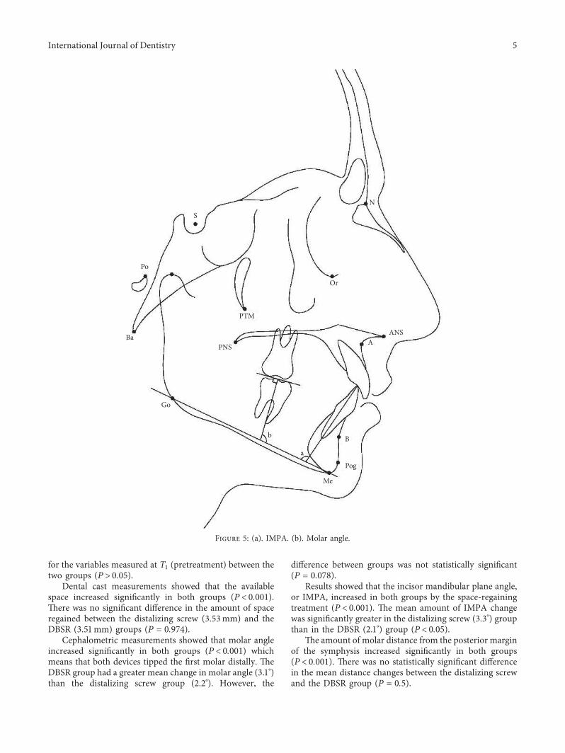
angle formed by the long axis of the incisor to the man-dibular plane (IMPA). +e difference between the IMPAmeasurements on pre- and posttreatment cephalogramsindicated the proclination of the lower permanent incisorsthat worked as part of the anchorage for the space-regainingdevice (Figure 5). To determine the molar angle, first atangent was formed to the occlusal surface of the firstpermanent molar, then a perpendicular line passing throughthe furcation was drawn. +e superior-anterior angle wasformed by this line, and the mandibular plane was measuredto determine the molar angle (Figure 5) [20]. Molar anglechanges demonstrated the first molar’s inclination. Molarbodily movement was measured as the distance from tworeference planes: one was a tangent to the symphysis pos-terior margin that was perpendicular to the mandibularplane, and the other was a parallel line that passed throughthe furcation of the first permanent molar. +e changes inthe distance between these two reference points before andafter treatment showed the amount of bodily movement ofthe first molar by the space-regaining appliance (Figure 6)[20]. +e vertical position of the first permanent molar wasdetermined by measuring the distance between the occlusalsurface of the mesiobuccal cusp of the first permanent molarand the mandibular plane (Figure 6). Molar vertical changesdetermined the extrusion effect of the appliance. +emeasurements shown in Table 1 were used to analyze thechanges in the dental arches in the pre- and posttreatmentphases.
+e duration of treatment was determined in months foreach patient. +is was the period of time between the first
activation of each device and when the required space for theeruption of the second premolar was regained, and thetreatment was completed.
2.5. Statistical Analysis and Error of the Method. +e datawere analyzed using SPSS version 26. +e mean and stan-dard deviation (SD) were calculated for each variable used inanalyzing the casts and lateral cephalograms. +e distri-butions of all variables were normally based on the Shapiro-Wilk test. Paired t-test was used to compare pre- to post-treatment variables, and independent t-test was used toassess whether differences in means between the groupsachieved statistical significance. +e level of significance wasset at 0.05.
In order to determine the error of the method, two weeksafter the first measurements, records of 10 randomly selectedsubjects were remeasured by the same examiner. +eintraexaminer error was assessed using the intraclass cor-relation coefficient (ICC). +e ICC was in the range of0.92–0.98. Hence, the measurements enjoyed sufficientreliability.
3. Results
+e study sample consisted of 19 subjects (11 girls and 8boys) with a mean age of 10.0 (SD 1.38) in the distalizingscrew and 19 subjects (10 girls and 9 boys) with amean age of9.9 (SD 0.84) years in the DBSR group. +e two groups werehomogeneous because no significant difference was found
Figure 3: Measuring the mesiodistal width of the central (left) and lateral (right) incisor with digital calipers.
Figure 4: Determining the laterally adjusted point to the distal surface of the lateral incisor with calipers (left). Measuring the distancebetween the laterally adjusted point to the distal surface of the lateral incisor to the mesial surface of the first permanent molar to determinethe available space (right).
4 International Journal of Dentistry
for the variables measured at T1 (pretreatment) between thetwo groups (P> 0.05).
Dental cast measurements showed that the availablespace increased significantly in both groups (P< 0.001).+ere was no significant difference in the amount of spaceregained between the distalizing screw (3.53mm) and theDBSR (3.51mm) groups (P � 0.974).
Cephalometric measurements showed that molar angleincreased significantly in both groups (P< 0.001) whichmeans that both devices tipped the first molar distally. +eDBSR group had a greater mean change in molar angle (3.1°)than the distalizing screw group (2.2°). However, the
difference between groups was not statistically significant(P � 0.078).
Results showed that the incisor mandibular plane angle,or IMPA, increased in both groups by the space-regainingtreatment (P< 0.001). +e mean amount of IMPA changewas significantly greater in the distalizing screw (3.3°) groupthan in the DBSR (2.1°) group (P< 0.05).
+e amount of molar distance from the posterior marginof the symphysis increased significantly in both groups(P< 0.001). +ere was no statistically significant differencein the mean distance changes between the distalizing screwand the DBSR group (P � 0.5).
Ba
Po
S
PTM
PNS
Or
ANSA
b
a
B
Pog
Me
Go
N
Figure 5: (a). IMPA. (b). Molar angle.
International Journal of Dentistry 5

Po
Ba
S
PTM
PNS
Go
Or
N
ANSA
B
Pog
Mc
a
b
Figure 6: (a). Molar distance to symphysis. (b). Distance between the mesiobuccal cusp tip of the first permanent molar and the mandibularplane.
Table 1: Measured variables and their definitions.
Variable Definition
Available space (mm) Space measured between the distal surface of the lateral incisor and the mesial surface of the permanentfirst molar
Required space (mm) Sum of mesiodistal width of permanent canine and premolars estimated by Tanaka-Johnston methodMolar angle (degree) Angle formed by the long axis of the mandibular first permanent molar and mandibular planeIMPA (degree) Angle formed by the long axis of mandibular incisors and the mandibular planeMolar to symphysis (mm) Distance between mandibular first permanent molar and posterior border of symphysisMolar to mandibular plane(mm) Distance between mesiobuccal cusp tip of mandibular first permanent molar and the mandibular plane
+e definitions given in this table are derived from Werner et al. [21] and da Costa et al. [17].
6 International Journal of Dentistry

+ere was a significant increase in the distance betweenthe mesiobuccal cusp tip of the first molar and the man-dibular plane in both the distalizing screw and DBSR groups(P< 0.001). +e average increase was 2.0mm in the dis-talizing screw and 2.3mm in the DBSR group, with nosignificant difference between the groups (P � 0.317).
Means, standard deviations, and comparison of the pre-and posttreatment measurements are shown in Table 2.Changes in cast and cephalometric variables for the twotreatment groups are compared in Table 3.
+e average molar distalization time for the distalizingscrew group was 11.6± 1.96 months, whereas in the DBSRgroup, the corresponding time was 8.1± 2.21 months. +us,the treatment time for the distal molar movement wassignificantly shorter for the DBSR than for the distalizingscrew group (P< 0.001).
4. Discussion
+e present study evaluated the effects of two types of space-regaining devices on the mandibular dental arch.
Early loss of primary teeth can cause a variety ofproblems, including space loss, one of the most important[22]. Space regaining, as the main treatment for thisproblem, can be done in both upper and lower arches.However, space regaining in the lower arch is more difficultthan in the upper arch [10]. +e mandibular molar is knownto be the hardest tooth to move, which is due to the broadroot area and root morphology, as well as the higher densityof bone in the lower jaw compared with the upper jaw[23, 24]. Lip bumpers, lingual arches, and removable deviceswith screws or springs are the most widely used intraoraldevices for space regaining in the mandible [10].
A removable appliance with a jackscrew is the mostcommonly used appliance for space regaining in the clinic,which relies on patient cooperation for its effectiveness. Inthe mandible, it is known that removable appliances withactive springs or screws can cause patient complianceproblems due to lack of adequate retention and easy dis-lodgement as a result of tonguemovement and also irritationof the lingual gingival tissues [14]. On the other hand, theseappliances have some advantages over fixed devices. One ofthe most important is that the appliance and the teeth couldbe better cleaned. Patients treated with removable appliancesdisplay better oral hygiene, less plaque, and less gingivalinflammation [25] while, in patients with fixed orthodonticappliances, oral hygiene becomes more difficult and aprevalent finding is the decalcification of the enamel surface(manifested as a white spot lesion) adjacent to these ap-pliances [26]. Studies have shown that salivary bacterial andcandida colonization causing vulnerability to developingcaries and candida infection is higher in patients with fixedrather than removable appliances [27, 28].
Since the use of removable appliances requires consid-erable patient cooperation, in the present study, we com-pared a simpler noncompliance appliance (the double-banded space regainer) with the conventionally used re-movable plate. +e DBSR uses Ni-Ti coil springs for molardistalization, which can generate continuous light forces
over a wide range of activation [29]. In addition, this ap-pliance does not need as many activations as the distalizingscrew does. However, in this study, one or more springreactivations were necessary in some patients as the mag-nitude of forces applied to the teeth decreases when the teethmove and springs decompress [30].
In the present study, to determine the required space, themodified Tanaka-Johnston equation was used, which hasbeen shown to accurately estimate the size of uneruptedcanines and premolars in the Iranian population [19]. +eresults indicated no statistically significant difference be-tween the mean recovered spaces of the distalizing screw andthe double-banded space regainer. +e mean amount ofspace regained in both groups was 3.5mm which means thatboth devices seem to be effective in regaining mild-to-moderate unilateral space loss.
+e amount of molar distance from the posterior marginof the symphysis increased significantly in both groups, andthere was no statistically significant difference between thetwo study groups. +ese results mean that a noticeableamount of space recovered in both groups was due to thedistal bodily movement of the first permanent molar (2.7and 2.4mm in the distalizing screw and the DBSR groups,respectively). It should be noted that in the analysis of lateralcephalometry images, evaluating bilateral structures such asfirst molars can be challenging due to the presence of twoshadows of these structures. In the current study, the se-lection of subjects with unilateral space loss made it possibleto distinguish between the right and left first molars moreeasily. +is means that the mesially located first molar in thelateral cephalogram was considered as the molar on thespace loss side. Moreover, unlike the molar on the space lossside, the position of the molar on the opposite side did notchange considerably between the pre- and posttreatmentcephalograms. Another helpful criterion for differentiatingthe two molars was that the image of the left molar wasusually less distorted as it was closer to the film.
Intraoral molar distalization has some side effects, in-cluding tipping of incisors or premolars (or both) in dif-ferent amounts depending on the choice of distalizationappliance [31]. In the present study, the mandibular incisorsin both study groups experienced an increase in proclina-tion. Forward movement of mandibular incisors has beenseen in studies of molar distalization with lip bumper [32],nickel titanium open-coil springs [33, 34], and removableplates with distalizing screws [17].
While there was a significant increase in IMPA aftertreatment with DBSR in the current study, there are noprevious data in the literature about changes in IMPA aftertreatment with this appliance. Looking at the present results,by using DBSR, molar distalization seems to have occurredwith less proclination of the lower anteriors with respect tothe distalizing screw. It can be due to the fact that in thedistalizing screw, the acrylic plate is in direct contact with thelingual surfaces of incisors and transmits the force generatedby the Jack screw to the teeth in a labial direction. It can alsobe related to the probable misfit and displacement of theacrylic plate. When using mandibular removable plates inthe clinic, it is often observed that after some time, the acrylic
International Journal of Dentistry 7

plate may not fit well; this is perhaps because the molar doesnot distalize as much as the Jack screw is opened, which leadsto a misfit of the plate. In this situation, the anterior part ofthe acrylic plate contacts the incisors closer to the incisaledge and further from the center of rotation of the tooth,causing more tipping of these teeth. Another reason thatmay explain this result is the fact that when lighter forces areapplied, anchorage loss is expected to be less [35]. +e Ni-Ticoil springs used in the DBSR produce light continuousforces which are optimal for orthodontic tooth movementand probably cause less anchorage loss. +us, the double-banded space regainer might be favorable in cases ofmandibular incisor protrusion.
Molar tipping is another common finding in cases ofmolar distalization. In the present study, the removable platewith a distalizing screw and the double-banded spaceregainer both tipped the first permanent molar distally andincreased the molar angle. In the study of da Costa et al. [17],distal tipping of mandibular molars by the distalizing screwwas reported as well. However, in the studies of Chalakkal[15] and Patil [16] on the DBSR, no observable tipping on thepermanent first molar was seen. +e difference betweenthese two studies and the present study can be related to thefact that in their studies, the DBSR was used in the maxillarydental arch, which has lower bone density than the mandible[36]. +e compact bone and oblique ridges in the mandiblemake bodily movement of molar roots difficult, leading totipping of the tooth [37]. In our study, the amount of firstmolar tipping was greater in the DBSR group (3.1°) than inthe distalizing screw group (2.2°). However, the differencethat existed between the two groups was not statisticallysignificant.
Considering the vertical changes, both appliances ex-truded the mandibular molar and no significant difference
was observed between the amounts of extrusion in the twostudy groups. Extrusion of the first molar was also observedin the study of Byloff et al. [33] with the Franzulum ap-pliance, which uses nickel titanium coil springs for man-dibular molar distalization.+e side effect of tooth extrusionoccurs with all simple tip-back-uprighting springs whencorrecting the axial inclination of the molar. +erefore,controlling vertical movements (extrusion) of molars shouldbe considered in molar uprighting. Extrusion of mandibularmolar may be acceptable when the tipped molar is below thefunctional occlusal plane in some early orthodontic treat-ments [38] and desirable in patients with deep bite.
+e basis of an efficient orthodontic treatment is agood understanding of the tooth movement rate and theamount of anchorage loss as well [35]. In the presentstudy, statistically significant differences between thedistalizing screw and DBSR groups were observed whencomparing the period of use of the space-regaining device.+e DBSR was used for a shorter time than the distalizingscrew. +is can be related to the fact that the DBSR ap-pliance cannot be removed that ensures continuous forcesapplied to the target teeth with no reliance on specialpatient compliance. However, in comparison to otherstudies using the DBSR for maxillary molar distalization[15, 16], space recovery with the DBSR took more time inour study. +is may be due to the increased trabecularstructure and resilient spongy bone in maxilla [37]. +ecortical bone of the mandible is denser than that of themaxilla [24], and the remodeling rate is slower in themandible [39]. +erefore, it has been observed that teethmove faster and further in the upper arch than in the lowerarch [37]. Another factor is the patients’ age, which waslower in those studies. +e advantage of earlier molardistalization is that the permanent first molar roots are
Table 2: Comparison of the pre- and posttreatment mean values in each group.
VariableDistalizing screw (n� 19) DBSR (n� 19)
HomogeneityPretreatment PosttreatmentP value Pretreatment Posttreatment
P valueMean± SD Mean± SD Mean± SD Mean± SDRequired space (mm) 24.53± 1.87 23.32± 1.65 0.43Available space (mm) 21.40± 2.64 24.93± 2.75 <0.001∗ 20.17± 2.31 23.69± 2.32 <0.001∗ 0.52Molar angle (degree) 88.07± 8.83 90.30± 8.91 <0.001∗ 91.68± 9.31 94.80± 9.22 <0.001∗ 0.43IMPA (degree) 90.60± 6.87 93.93± 7.26 <0.001∗ 88.00± 7.21 90.14± 7.27 <0.001∗ 0.29Molar to symphysis (mm) 20.00± 4.95 22.70± 4.58 <0.001∗ 22.36± 2.73 24.76± 2.71 <0.001∗ 0.33Molar to MP (mm) 28.07± 3.97 30.07± 4.39 <0.001∗ 27.71± 3.67 30.06± 3.68 <0.001∗ 0.26DBSR: double-banded space regainer; SD: standard deviation; IMPA: incisor mandibular plane angle; MP: mandibular plane; ∗P< 0.05.
Table 3: Comparison of cast and cephalometric changes (T2 −T1) between two groups.
Variable Distalizing screw (n� 19) DBSR (n� 19) 95% CIP value
T2 −T1 mean± SD T2 −T1 mean± SD Lower UpperAvailable space (mm) 3.53± 1.16 3.51± 0.82 −0.75 0.87 0.974Molar angle (degree) 2.23± 1.40 3.12± 1.19 −1.88 0.11 0.078IMPA (degree) 3.33± 1.71 2.14± 1.05 0.10 2.28 0.033∗Molar to symphysis (mm) 2.70± 1.45 2.41± 0.71 −0.58 1.17 0.5Molar to MP (mm) 2.00± 0.73 2.35± 1.09 −1.05 0.35 0.317DBSR: double-banded space regainer; CI: confidence interval; SD: standard deviation; IMPA: incisor mandibular plane angle; MP: mandibular plane; T1:pretreatment; T2: posttreatment; ∗P< 0.05.
8 International Journal of Dentistry

incomplete, and it is easier to tip or move the tooth bodily[16].
4.1. Limitations. +is was a retrospective study and also hadthe limitation of sample size, which should be overcome byfuture prospective studies with larger sample sizes. Also, it issuggested to evaluate movements of the tooth mesial to thespace loss region in future studies.
5. Conclusion
Within the limitations of this study, it was concluded thatthe removable device with a distalizing screw and the DBSRare both able to regain mild-to-moderate unilateral spaceloss when used in patients with mixed dentition, resulting inan increase in molar angle, IMPA, and molar extrusion. But,the DBSR seemed to be more efficient since it had a shortertreatment time and a lower side effect of incisor tipping.However, the removable device can be implemented inpatients with low incisor protrusion and good cooperation,with the advantage of better oral hygiene maintenance.
Data Availability
Data are available from the corresponding author uponrequest.
Conflicts of Interest
+e authors declare that they have no conflicts of interest.
Acknowledgments
+e authors thank the vice-chancellery of Shiraz Universityof Medical Sciences for supporting the research (grantnumber 97-01-37-18918). +is manuscript is based on thethesis by Dr. Shabnam Enteghad.
References
[1] W. Tunison, C. Flores-Mir, H. ElBadrawy, U. Nassar, andT. El-Bialy, “Dental arch space changes following prematureloss of primary first molars: a systematic review,” PediatricDentistry, vol. 30, pp. 297–302, 2008.
[2] P. Chandak, S. Baliga, and N. +osar, “Space regainers inpediatric dentistry,” International Dental and Medical Journalof Advanced Research, vol. 1, no. 1, pp. 1–5, 2015.
[3] K. Negi, “NiTi bonded space regainer/maintainer,” Journal ofIndian Society of Pedodontics and Preventive Dentistry, vol. 28,no. 2, p. 113, 2010.
[4] P. Ngan and H. Fields, “Orthodontic diagnosis and treatmentplanning in the primary dentition,”ASDC Journal of Dentistryfor Children, vol. 62, pp. 25–33, 1995.
[5] A. K. Rao and S. Sarkar, “Changes in the arch length fol-lowing premature loss of deciduous molars,” Journal ofIndian Society of Pedodontics and Preventive Dentistry,vol. 17, pp. 29–32, 1999.
[6] C. Nidhi, R. L. Jain, M. Neeraj, K. Harsimrat, B. Samriti, andC. Anuj, “Evaluation of the clinical efficacy of glass fiberreinforced composite resin as a space maintainer and itscomparison with the conventional band and loop space
maintainer. An in vivo study,”Minerva Stomatologica, vol. 61,pp. 21–30, 2012.
[7] J. Pedersen, K. Stensgaard, and B. Melsen, “Prevalence ofmalocclusion in relation to premature loss of primary teeth,”Community Dentistry and Oral Epidemiology, vol. 6, no. 4,pp. 204–209, 1978.
[8] A. S. Monte-Santo, S. V. C. Viana, K. M. S. Moreira,J. C. P. Imparato, F. M. Mendes, and G. A. V. C. Bonini,“Prevalence of early loss of primary molar and its impact inschoolchildren’s quality of life,” International Journal ofPaediatric Dentistry, vol. 28, no. 6, pp. 595–601, 2018.
[9] B. C. Kirtaniya, A. Singla, K. K. Gupta, A. Khanna, andG. Kaur, “Space regainer cum space maintainer—a new ap-pliance for paediatric dentistry,” Indian Journal of DentalSciences, vol. 6, pp. 20–22, 2014.
[10] D. N. Kapoor, A. Razdan, and S. Kannan, “Effective means ofintraoral molar distalization—an overview,” $e Journal ofIndian Orthodontic Society, vol. 36, no. 3, pp. 131–142, 2002.
[11] G. Perrotti, G. Baccaglione, T. Clauser et al., “Total face ap-proach (TFA) 3D cephalometry and superimposition inorthognathic surgery: evaluation of the vertical dimensions in aconsecutive series,” Methods and Protocols, vol. 4, no. 2, 2021.
[12] R. Jaai and W. Jasmin, “Space regainers–a review,” EuropeanJournal of Biomedical and Pharmaceutical Sciences, vol. 5,pp. 212–217, 2018.
[13] W. R. Proffit, H. W. Fields, and B. E. Larson, ContemporaryOrthodontics, Elsevier, Amsterdam, Netherlands, 2019.
[14] F. P. G. M. van der Linden, “+e removable orthodonticappliance,” American Journal of Orthodontics, vol. 59, no. 4,pp. 376–385, 1971.
[15] P. Chalakka, A. +omas, F. Akkara, and R. Pavaskar, “Newdesign space regainers: “lingual arch crossbow” and “doublebanded space regainer”,” Journal of Indian Society of Pedo-dontics and Preventive Dentistry, vol. 30, no. 2, pp. 161–165,2012.
[16] D. Patil, K. P. Bharath, P. Poornima, and P. Bali, “Doublebanded space regainer (DBSR) in distalising two teeth-a casereport,” International Journal of Oral Health and MedicalResearch, vol. 3, pp. 140–142, 2016.
[17] C. C. da Costa, I. C. Almeida, A. Locks, and L. C. da CostaFilho, “Clinical comparative study of the effects of two types ofmandibular space-regaining devices,” General Dentistry,vol. 51, pp. 120–126, 2003.
[18] M. M. Tanaka and L. E. Johnston, “+e prediction of the sizeof unerupted canines and premolars in a contemporary or-thodontic population,” Journal of the American Dental As-sociation, vol. 88, no. 4, pp. 798–801, 1974.
[19] M. Khanehmasjedi and L. Bassir, “Prediction of the size ofunerupted canines and premolars in an Iranian population,”Indian Journal of Dental Research, vol. 24, pp. 493–497, 2013.
[20] H. Babacan and C. Doruk, “Essix-based molar distalizationappliance,” Journal of Orthodontics, vol. 32, no. 4,pp. 229–234, 2005.
[21] S. P. Werner, P. K. Shivapuja, and E. F. Harris, “Skeletodentalchanges in the adolescent accruing from use of the lipbumper,” $e Angle Orthodontist, vol. 64, pp. 13–22, 1994.
[22] R. Abdullah, A. Gupte, A. Patel, and A. Gupte, “Spaceregaining with limited orthodontic intervention—a case re-port,” International Journal of Dentistry Case Reports, vol. 2,pp. 97–101, 2012.
[23] T. Dang, J.-P. Forestier, and B. +ebault, “Is mandibularmolar distalization feasible?” Journal of Dentofacial Anomaliesand Orthodontics, vol. 18, no. 1, p. 104, 2015.
International Journal of Dentistry 9

[24] H.-S. Park, Y.-J. Lee, S.-H. Jeong, and T.-G. Kwon, “Density ofthe alveolar and basal bones of the maxilla and the mandible,”American Journal of Orthodontics and Dentofacial Orthope-dics, vol. 133, no. 1, pp. 30–37, 2008.
[25] G. M. Abbate, M. P. Caria, P. Montanari et al., “Periodontalhealth in teenagers treated with removable aligners andfixed orthodontic appliances,” Journal of Orofacial Or-thopedics/Fortschritte der Kieferorthopaedie, vol. 76, no. 3,pp. 240–250, 2015.
[26] S. E. Bishara and A.W. Ostby, “White spot lesions: formation,prevention, and treatment,” Seminars in Orthodontics, vol. 14,no. 3, pp. 174–182, 2008.
[27] S. Mummolo, M. Tieri, A. Nota et al., “Salivary concentrationsof Streptococcus mutans and Lactobacilli during an ortho-dontic treatment. An observational study comparing fixedand removable orthodontic appliances,” Clinical and Exper-imental Dental Research, vol. 6, no. 2, pp. 181–187, 2020.
[28] E. Khanpayeh, A. A. Jafari, and Z. Tabatabaei, “Comparison ofsalivary candida profile in patients with fixed and removableorthodontic appliances therapy,” Iranian Journal of Micro-biology, vol. 6, pp. 263–268, 2014.
[29] A. Bourke, J. Daskalogiannakis, B. Tompson, and P. Watson,“Editor’s comment and Q&A,” American Journal of Ortho-dontics and Dentofacial Orthopedics, vol. 138, no. 2,pp. 142-143, 2010.
[30] G. Steinbach, D. Armstrong, O. P. Kharbanda, P. Petocz, andM. A. Darendeliler, “An investigation of the forces used byclinicians to open spaces with coil springs,” Australian Or-thodontic Journal, vol. 22, pp. 115–120, 2006.
[31] I. Feldmann and L. Bondemark, “Orthodontic anchorage: asystematic review,” $e Angle Orthodontist, vol. 76,pp. 493–501, 2006.
[32] W. S. Osborn, R. S. Nanda, and G. F. Currier, “Mandibulararch perimeter changes with lip bumper treatment,”AmericanJournal of Orthodontics and Dentofacial Orthopedics, vol. 99,no. 6, pp. 527–532, 1991.
[33] F. Byloff, M. A. Darendeliler, and F. Stoff, “Mandibular molardistalization with the franzulum appliance,” Journal ofClinical Orthodontics, vol. 34, pp. 518–523, 2000.
[34] O. Uner and S. Haydar, “Mandibular molar distalization withthe ones jig appliance,” American Journal of Orthodontics andDentofacial Orthopedics, vol. 109, p. 337, 1996.
[35] C. M. Andre da, L. G. Gandini Jr., A. P. Vianna,R. P. Martins, and H. B. Jacob, “Tooth movement rate andanchorage lost during canine retraction: a maxillary andmandibular comparison,” $e Angle Orthodontist, vol. 89,no. 4, pp. 559–565, 2019.
[36] H. Devlin, K. Horner, and D. Ledgerton, “A comparison ofmaxillary and mandibular bone mineral densities,”$e Journalof Prosthetic Dentistry, vol. 79, no. 3, pp. 323–327, 1998.
[37] L. Furstman, S. Bernick, and D. Aldrich, “Differential re-sponse incident to tooth movement,” American Journal ofOrthodontics, vol. 59, no. 6, pp. 600–608, 1971.
[38] B. U. Zachrisson and H. P. Bantleon, “Optimal mechanics formandibular molar uprighting,” World Journal of Orthodon-tics, vol. 6, pp. 80–87, 2005.
[39] T. Deguchi, T. Takano-Yamamoto, T. Yabuuchi, R. Ando,W. E. Roberts, and L. P. Garetto, “Histomorphometricevaluation of alveolar bone turnover between the maxilla andthe mandible during experimental tooth movement in dogs,”American Journal of Orthodontics and Dentofacial Orthope-dics, vol. 133, no. 6, pp. 889–897, 2008.
10 International Journal of Dentistry