Recurrence of Choledocholithiasis - Hindawi.com
12
Diagnostic and Therapeutic Endoscopy, 1996, Vol. 3, pp. 81-91 Reprints available directly from the publisher Photocopying permitted by license only (C) 1996 OPA (Overseas Publishers Association) Amsterdam B.V. Published in The Netherlands by Harwood Academic Publishers Printed in Malaysia Clinical Study on Causative Factors and Recurrence of Choledocholithiasis HAJIME HOSHI* and YOSHIHIRO SAKAI Third Department of Internal Medicine, Ohashi Hospital, Toho Universi. School of Medicine, 2-17-60hashi, Meguro-ku, Tokyo 153, Japan (Received 8 November 1995; In final form 1 June 1996) To identify factors involved in choledocholithiasis, clinical characteristics were studied using univariate and multivariate analyses. Factors involved in recurrence were also investigated. The subjects consisted of 51 patients with calcium bilirubinate stones (B group) and 52 patients with cholesterol stones (C group). All patients had choledo- cholithiasis and underwent lithotripsy by endoscopic sphincterotomy (EST) during the past 9 years. Twenty variables, including clinical symptoms and endoscopic retrograde cholangiopancreatography (ERCP) findings, were analyzed using a Statistical Analysis System (SAS) software package. Univariate analysis were done using Student’s t-test and the chi-square test. Multivariate analyses were done by stepwise logistic regression analysis. In univariate analyses, there were significant differences between the B group and C group in nine variables: age, common bile duct diameter, common hepatic duct diameter, common bile duct stone diameter, cystic duct diameter, and the presence of gallbladder stones, atypical arrangement of the hepatic duct, parapapillary diverticu- lum, and large parapapillary-diverticulum. In multivariate analysis, the four variables of no gallbladder stone, large parapapillary diverticulum, cystic duct less than 8 mm, and atypical arrangement of the hepatic duct were significant independent factors for the development of stones in the B group, with relative risks of 37.75, 16.73, 5.56, and 5.49, respectively. The results indicated that calcium bilirubinate stones were frequently asso- ciated with parapapillary diverticulum and abnormal arrangement of the bile duct. The formation of these stones was attributed to chronic biliary stasis caused by dysfunction of the biliary tract, including the papilla. In contrast, most cholesterol stones found in the common bile duct had apparently descended from the gallbladder. Common bile duct stones recurred after EST in 9 patients, all of whom had calcium bilirubinate stones. On ERCP, recurrence was found to be frequently associated with gallbladder stones, large parapapillary diverticula, and atypical arrangement of the hepatic duct. Patients with these characteristics on initial ERCP should therefore receive appropriate treatment and undergo strict follow-up observations owing to the increased risk of recurrence caused by dysfunction of the biliary tract. Keywords: Choledocholithiasis, endoscopic retrograde cholangiopancreatography, recurrent stones *Corresponding author. Tel.: 03-3468-1251. Fax: 03-3468-1269. 81
-
Upload
khangminh22 -
Category
Documents
-
view
0 -
download
0
Transcript of Recurrence of Choledocholithiasis - Hindawi.com
Diagnostic and Therapeutic Endoscopy, 1996, Vol. 3, pp. 81-91Reprints available directly from the publisherPhotocopying permitted by license only
(C) 1996 OPA (Overseas Publishers Association)Amsterdam B.V. Published in The Netherlands
by Harwood Academic PublishersPrinted in Malaysia
Clinical Study on Causative Factors andRecurrence of Choledocholithiasis
HAJIME HOSHI* and YOSHIHIRO SAKAI
Third Department ofInternal Medicine, Ohashi Hospital, Toho Universi. School ofMedicine,2-17-60hashi, Meguro-ku, Tokyo 153, Japan
(Received 8 November 1995; Infinalform 1 June 1996)
To identify factors involved in choledocholithiasis, clinical characteristics were studiedusing univariate and multivariate analyses. Factors involved in recurrence were alsoinvestigated. The subjects consisted of 51 patients with calcium bilirubinate stones (Bgroup) and 52 patients with cholesterol stones (C group). All patients had choledo-cholithiasis and underwent lithotripsy by endoscopic sphincterotomy (EST) during thepast 9 years. Twenty variables, including clinical symptoms and endoscopic retrogradecholangiopancreatography (ERCP) findings, were analyzed using a Statistical AnalysisSystem (SAS) software package. Univariate analysis were done using Student’s t-test andthe chi-square test. Multivariate analyses were done by stepwise logistic regressionanalysis. In univariate analyses, there were significant differences between the B groupand C group in nine variables: age, common bile duct diameter, common hepatic ductdiameter, common bile duct stone diameter, cystic duct diameter, and the presence ofgallbladder stones, atypical arrangement of the hepatic duct, parapapillary diverticu-lum, and large parapapillary-diverticulum. In multivariate analysis, the four variables ofno gallbladder stone, large parapapillary diverticulum, cystic duct less than 8 mm, andatypical arrangement of the hepatic duct were significant independent factors for thedevelopment of stones in the B group, with relative risks of 37.75, 16.73, 5.56, and 5.49,respectively. The results indicated that calcium bilirubinate stones were frequently asso-ciated with parapapillary diverticulum and abnormal arrangement of the bile duct. Theformation of these stones was attributed to chronic biliary stasis caused by dysfunctionof the biliary tract, including the papilla. In contrast, most cholesterol stones found in thecommon bile duct had apparently descended from the gallbladder. Common bile ductstones recurred after EST in 9 patients, all of whom had calcium bilirubinate stones. OnERCP, recurrence was found to be frequently associated with gallbladder stones, largeparapapillary diverticula, and atypical arrangement of the hepatic duct. Patients withthese characteristics on initial ERCP should therefore receive appropriate treatmentand undergo strict follow-up observations owing to the increased risk of recurrencecaused by dysfunction of the biliary tract.
Keywords: Choledocholithiasis, endoscopic retrograde cholangiopancreatography, recurrent stones
*Corresponding author. Tel.: 03-3468-1251. Fax: 03-3468-1269.
81
82 H. HOSHI and Y. SAKAI
I. INTRODUCTION
There have been many basic and clinical studies on
cholecystolithiasis. A better understanding of the fac-tors responsible for cholecystolithiasis his contributedto more accurate diagnosis and to improved therapeu-tic techniques [1-5]. However, relatively few studieshave focussed on the etiology and diagnosis of chole-docholithiasis, and the clinical characteristics of thisdisease are still poorly understood. The authors there-fore studied the factors involved in the genesis ofcholedocholithiasis by univariate and multivariate
analyses in patients undergoing endoscopic sphinc-terotomy (EST). These factors included clinical signsand symptoms of patients with choledocholithiasiswho underwent transpapillary lithotomy, cholangio-graphic findings on endoscopic retrograde cholan-giopancreatography (ERCP), and the configuration ofthe duodenal papilla. In addition, the characteristics ofthe bile duct and causative factors were studied in
patients with recurrent stones.
II. SUBJECTS AND METHODS
1. Subjects
During the 9-year period between January 1986 andDecember 1994, 159 patients with choledocholithiasisunderwent stone removal by EST. Sixty-two of thesepatients had stones of the common bile duct alone, and97 had stones of the gallbladder and the common bile
duct. Seventy of the patients were men, and 89 werewomen. Their mean age was 69.6 + 12.9 years.Mechanical lithotripsy was performed using a
lithotripter in 53 patients. Lithotripsy using a basketand balloon catheter was performed in 90 patients.Electrohydraulic lithotripsy (EHL) under peroralcholangioscopy was performed in 3 patients.Transpapillary lithotomy could not be performed in 10
patients, who instead underwent percutaneous tran-
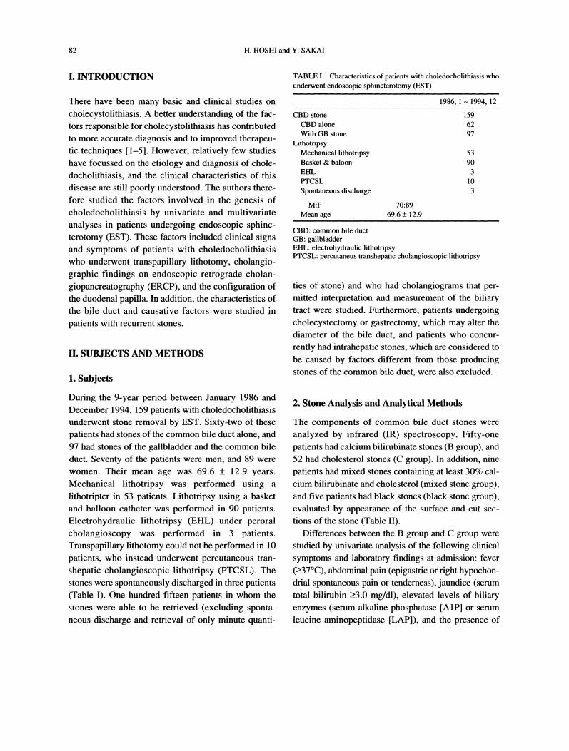
shepatic cholangioscopic lithotripsy (PTCSL). Thestones were spontaneously discharged in three patients(Table I). One hundred fifteen patients in whom thestones were able to be retrieved (excluding sponta-neous discharge and retrieval of only minute quanti-
TABLE Characteristics of patients with choledocholithiasis whounderwent endoscopic sphincterotomy (EST)
1986, 1994, 12
CBD stoneCBD aloneWith GB stone
LithotripsyMechanical lithotripsyBasket & baloonEHLPTCSLSpontaneous discharge
M:FMean age
70:8969.6 + 12.9
1596297
53903103
CBD: common bile ductGB: gallbladderEHL: electrohydraulic lithotripsyPTCSL: percutaneus transhepatic cholangioscopic lithotripsy
ties of stone) and who had cholangiograms that per-mitted interpretation and measurement of the biliarytract were studied. Furthermore, patients undergoingcholecystectomy or gastrectomy, which may alter thediameter of the bile duct, and patients who concur-
rently had intrahepatic stones, which are considered to
be caused by factors different from those producingstones of the common bile duct, were also excluded.
2. Stone Analysis and Analytical Methods
The components of common bile duct stones were
analyzed by infrared (IR) spectroscopy. Fifty-onepatients had calcium bilirubinate stones (B group), and52 had cholesterol stones (C group). In addition, nine
patients had mixed stones containing at least 30% cal-cium bilirubinate and cholesterol (mixed stone group),and five patients had black stones (black stone group),evaluated by appearance of the surface and cut sec-tions of the stone (Table II).
Differences between the B group and C group were
studied by univariate analysis of the following clinical
symptoms and laboratory findings at admission: fever(>37C), abdominal pain (epigastric or right hypochon-drial spontaneous pain or tenderness), jaundice (serumtotal bilirubin >3.0 mg/dl), elevated levels of biliaryenzymes (serum alkaline phosphatase [ALP] or serumleucine aminopeptidase [LAP]), and the presence of
CAUSATIVE FACTORS AND RECURRENCE OF CHOLEDOCHOLITHIASIS 83
TABLE II Type of common bile duct stones
No. of patients
Calcium bilirubinate stone (B group) 51Cholesterol stone (C group) 52Mixed stone 9Black stone 5
117
cholangitis (abdominal pain + elevated biliary enzymes+ leukocyte count >10,000 or C-reactive protein [CRP]positive) or cholecystitis (enlargement of the gallblad-der and thickening of its wall on abdominal ultrasonog-raphy + leukocyte count >10,000 or CRP positive).B group and C group were similarly compared by
univariate analysis for variables determined in cholan-giograms on ERCP and endoscopic findings of theduodenal major papilla (the presence or absence of
gallbladder stones, gallbladder stone diameter, thediameter and number of common bile duct stones,common bile duct diameter, common hepatic ductdiameter, cystic duct diameter, the presence or absenceof bile duct stenosis, the presence or absence of an
atypical arrangement of the cystic duct, the structure ofthe hepatic duct confluence, and the presence or
absence of a parapapillary diverticulum and a largeparapapillary diverticulum). For cholangiograms, films
that included the intrahepatic duct and gallbladder inthe initial ERCP images were selected, in principle. Ifthe initial images were unsatisfactory, the cholan-giograms taken after EST treatment were used. The
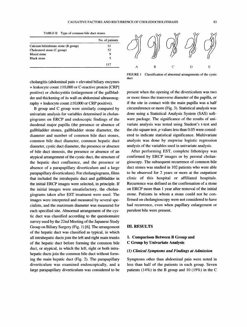
images were interpreted and measured by several spe-cialists, and the maximum diameter was measured foreach specified site. Abnormal arrangement of the cys-tic duct was classified according to the questionnairesurvey used by the 22nd Meeting of the Japanese StudyGroup on Biliary Surgery (Fig. 1) [6]. The arrangementof the hepatic duct was classified as typical, in which
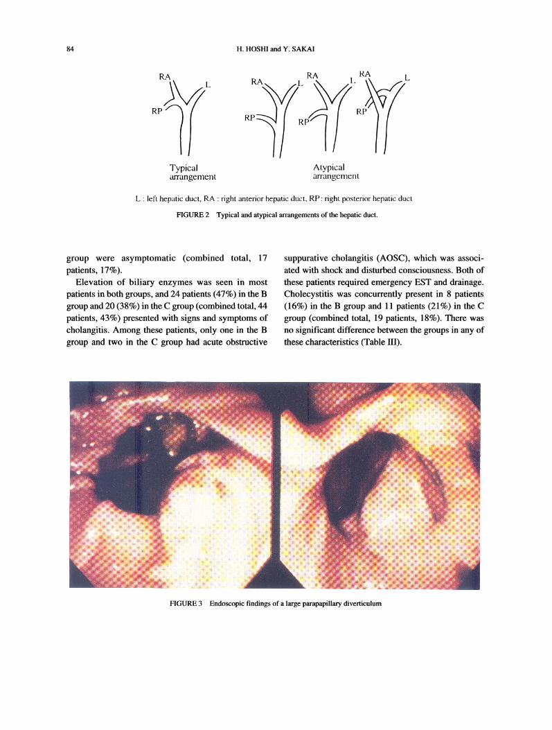
all intrahepatic ducts join the left and fight main trunksof the hepatic duct before forming the common bile
duct, or atypical, in which the left, fight or both intra-
hepatic ducts join the common bile duct without form-ing the main hepatic duct (Fig. 2). The parapapillarydiverticulum was examined endoscopically, and a
large parapapillary diverticulum was considered to be
A B C D E
FIGURE Classification of abnormal arrangements of the cysticduct
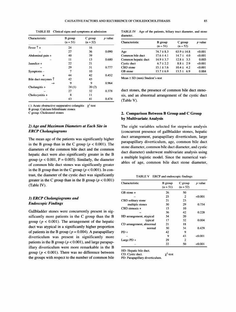
present when the opening of the diverticulum was two
or more times the transverse diameter of the papilla, or
if the site in contact with the main papilla was a halfcircumference or more (Fig. 3). Statistical analysis wasdone using a Statistical Analysis System (SAS) soft-ware package. The significance of the results of uni-
variate analysis was tested using Student’s t-test andthe chi-square test. p values less than 0.05 were consid-ered to indicate statistical significance. Multivariate
analysis was done by stepwise logistic regressionanalysis of the variables used in univariate analysis.
After performing EST, complete lithotripsy wasconfirmed by ERCP images or by peroral cholan-
gioscopy. The subsequent recurrence of common bile
duct stones was studied in 102 patients who were ableto be observed for 2 years or more at the outpatientclinic of this hospital or affiliated hospitals.Recurrence was defined as the confirmation of a stone
on ERCP more than 1 year after removal of the initialstone. Patients in whom a stone could not be con-firmed on cholangioscopy were not considered to havehad recurrence, even when papillary enlargement or
purulent bile were present.
III. RESULTS
1. Comparison Between B Group andC Group by Univariate Analysis
(1) Clinical Symptoms and Findings at Admission
Symptoms other than abdominal pain were noted inless than half of the patients in each group. Sevenpatients (14%) in the B group and 10 (19%) in the C
84 H. HOSHI and Y. SAKAI
RAL RA
RPRP
RA
Rp,R
Typical Atypicalarrangement arrangement
L left hepatic duct, RA right anterior hepatic duct, RP: right posterior hepatic duct
FIGURE 2 Typical and atypical arrangements of the hepatic duct.
group were asymptomatic (combined total, 17patients, 17%).
Elevation of biliary enzymes was seen in most
patients in both groups, and 24 patients (47%) in the Bgroup and 20 (38%) in the C group (combined total, 44patients, 43%) presented with signs and symptoms ofcholangitis. Among these patients, only one in the Bgroup and two in the C group had acute obstructive
suppurative cholangitis (AOSC), which was associ-ated with shock and disturbed consciousness. Both ofthese patients required emergency EST and drainage.Cholecystitis was concurrently present in 8 patients(16%) in the B group and 11 patients (21%) in the Cgroup (combined total, 19 patients, 18%). There wasno significant difference between the groups in any ofthese characteristics (Table HI).
FIGURE 3 Endoscopic findings of a large parapapillary diverticulum
CAUSATIVE FACTORS AND RECURRENCE OF CHOLEDOCHOLITHIASIS 85
TABLE Ill Clinical signs and symptoms at admission
Characteristic
TABLE IV Age of the patients, biliary tract diameter, and stone
diameter.B group C group p value(n 51) (n 52) Characteristic
0.090
0.680
0.777
0.452
0.964
0.378
0.474
Fever "l" + 24 1627 36
Abdominal pain + 40 3911 13
Jaundice + 22 2129 31
Symptoms 7 10+ 44 42
Bile duct enzymes "l" 42 43---> 9 9
Cholangitis + 24 (1) 20 (2)27 32
Cholecystitis + 8 1143 41
): Acute obstructive suppurative colangitis 22-testB group: Calcium bilirubinate stonesC group: Cholesterol stones
2) Age and Maximum Diameters at Each Site inERCP Cholangiograms
The mean age of the patients was significantly higherin the B group than in the C group (p < 0.001). Thediameters of the common bile duct and the common
hepatic duct were also significantly greater in the Bgroup (p < 0.001, P 0.005). Similarly, the diameterof common bile duct stones was significantly greaterin the B group than in the C group (p < 0.001). In con-
trast, the diameter of the cystic duct was significantlygreater in the C group than in the B group (p < 0.001)(Table IV).
3) ERCP Cholangiograms andEndoscopic Findings
Gallbladder stones were concurrently present in sig-nificantly more patients in the C group than the Bgroup (p < 0.001). The arrangement of the hepaticduct was atypical in a significantly higher proportionof patients in the B group (p 0.004). A parapapillarydiverticulum was present in significantly more
patients in the B group (p < 0.001), and large parapap-illary diverticulum were more remarkable in the Bgroup (p < 0.001). There was no difference betweenthe groups with respect to the number of common bile
B group C group p value(n 51) (n 52)
Age 74.7 + 8.3 63.9 + 14.8 <0.001Common bile duct 17.6 + 4.1 14.7 + 4.0 <0.001Common hepatic duct 14.9 + 3.7 12.8 + 3.5 0.005Cystic duct 6.7 + 2.2 8.8 + 2.9 <0.001CBD stone 15.1 + 7.6 10.4 + 4.2 <0.001GB stone 13.7 + 6.9 13.5 + 6.9 0.884
Mean + SD (mm) Student’s-test
duct stones, the presence of common bile duct steno-
sis, and an abnormal arrangement of the cystic duct(Table V).
2. Comparison Between B Group and C Groupby Multivariate Analysis
The eight variables selected for stepwise analysis(concurrent presence of gallbladder stones, hepaticduct arrangement, parapapillary diverticulum, largeparapapillary diverticulum, age, common bile ductstone diameter, common bile duct diameter, and cysticduct diameter) underwent multivariate analysis usinga multiple logistic model. Since the numerical vari-
ables of age, common bile duct stone diameter,
TABLE V ERCP and endoscopic findings
Characteristic B group C group p value(n 51) (n 52)
GB stone + 26 5025 2 <0.001
CBD solitary stone 21 23multiple stones 30 29 0.754
CBD stenosis + 15 1036 42 0.228
HD arrangement, atypical 34 20typical 17 32 0.004
CD arrangement, abnormal 21 18normal 30 34 0.429
PD + 42 99 43 <0.001
Large PD + 29 222 50 <0.001
HD: Hepatic bile duct.CD: Cystic duct. 2-testPD: Parapapillary diverticulum.
86 H. HOSHI and Y. SAKAI
common bile duct diameter, and cystic duct diameter
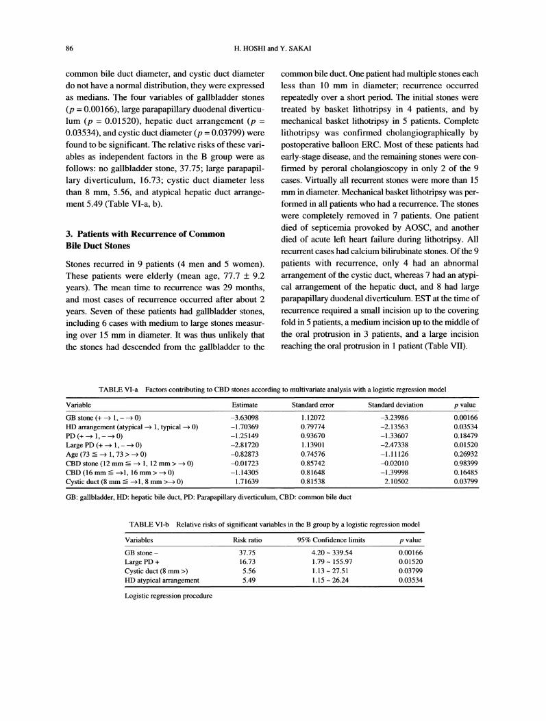
do not have a normal distribution, they were expressedas medians. The four variables of gallbladder stones
(p 0.00166), large parapapillary duodenal diverticu-lum (p 0.01520), hepatic duct arrangement (p0.03534), and cystic duct diameter (p 0.03799) were
found to be significant. The relative risks of these vari-ables as independent factors in the B group were as
follows: no gallbladder stone, 37.75; large parapapil-lary diverticulum, 16.73; cystic duct diameter lessthan 8 mm, 5.56, and atypical hepatic duct arrange-ment 5.49 (Table VI-a, b).
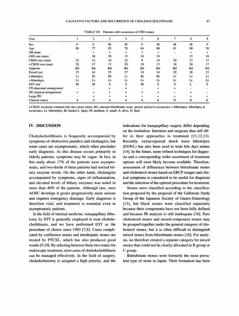
3. Patients with Recurrence of CommonBile Duct Stones
Stones recurred in 9 patients (4 men and 5 women).These patients were elderly (mean age, 77.7 + 9.2years). The mean time to recurrence was 29 months,and most cases of recurrence occurred after about 2
years. Seven of these patients had gallbladder stones,
including 6 cases with medium to large stones measur-
ing over 15 mm in diameter. It was thus unlikely thatthe stones had descended from the gallbladder to the
common bile duct. One patient had multiple stones eachless than 10 mm in diameter; recurrence occurredrepeatedly over a short period. The initial stones were
treated by basket lithotripsy in 4 patients, and bymechanical basket lithotripsy in 5 patients. Completelithotripsy was confirmed cholangiographically bypostoperative balloon ERC. Most of these patients hadearly-stage disease, and the remaining stones were con-
firmed by peroral cholangioscopy in only 2 of the 9cases. Virtually all recurrent stones were more than 15mm in diameter. Mechanical basket lithotripsy was per-formed in all patients who had a recurrence. The stones
were completely removed in 7 patients. One patientdied of septicemia provoked by AOSC, and anotherdied of acute left heart failure during lithotripsy. Allrecurrent cases had calcium bilirubinate stones. Ofthe 9patients with recurrence, only 4 had an abnormalarrangement of the cystic duct, whereas 7 had an atypi-cal arrangement of the hepatic duct, and 8 had largeparapapillary duodenal diverticulum. EST at the time ofrecurrence required a small incision up to the coveringfold in 5 patients, a medium incision up to the middle ofthe oral protrusion in 3 patients, and a large incision
reaching the oral protrusion in 1 patient (Table VII).
TABLE VI-a Factors contributing to CBD stones according to multivariate analysis with a logistic regression model
Variable Estimate Standard error Standard deviation p value
GB stone (+ --> 1, --> 0)HD arrangement (atypical ---> 1, typical --> 0)PD (+ --> 1,---> 0)Large PD (+ --> 1, --> 0)Age (73 _<-- --> 1, 73 > --> 0)CBD stone (12 mm _--< --> 1, 12 mm > --> 0)CBD (16 mm --< -->1, 16 mm > ---> 0)Cystic duct (8 mm _--< --> 1, 8 mm >- 0)
-3.63098 1.12072 -3.23986 0.00166-1.70369 0.79774 -2.13563 0.03534-1.25149 0.93670 -1.33607 0.18479-2.81720 1.13901 -2.47338 0.01520-0.82873 0.74576 -1.11126 0.26932-0.01723 0.85742 -0.02010 0.98399-1.14305 0.81648 -1.39998 0.164851.71639 0.81538 2.10502 0.03799
GB: gallbladder, HD: hepatic bile duct, PD: Parapapillary diverticulum, CBD: common bile duct
TABLE VI-b Relative risks of significant variables in the B group by a logistic regression model
Variables Risk ratio 95% Confidence limits p value
GB stone- 37.75 4.20 339.54 0.00166Large PD + 16.73 1.79 155.97 0.01520Cystic duct (8 mm >) 5.56 1.13 27.51 0.03799HD atypical arrangement 5.49 1.15 26.24 0.03534
Logistic regression procedure
CAUSATIVE FACTORS AND RECURRENCE OF CHOLEDOCHOLITHIASIS 87
TABLE VII Patients with recurrence of CBD stones
Case 2 3 4 5 6 7 8 9
Sex F F M M F M M M FAge 86 77 85 78 64 80 81 90 58GB stone + + + + + + +GBS size (mm) 30 28 15 10 19 15 18CBDS size (mm) 22 12 10 22 8 14 20 27 15r-CBDS size (mm) 20 17 13 20 18 15 18 20 17Analysis Bil Bil Bil Bil Bil Bil Bil Bil BilPeriod (m) 23 44 25 57 24 24 20 28 15Lithotripsy Lt Bt Bt Lt Bt Bt Lt Lt Ltr-lithotripsy Lt Lt Lt Lt Lt Lt Lt Lt LtEST size M M S S M S S L SCD abnormal arrangement + + + +HD atypical arrangement + + + + + + +Large PD + + + + + + + +Clinical course A A A A A A D D A
r-CBDS: recurrent common bile duct stone (mm), Bil: calcium bilirubinate stone, period: period to recurrence, r-lithotripsy: lithotripsy atrecurrence, Lt: lithotripter, Bt: basket L: large, M: medium, S: small, A: alive, D: died
IV. DISCUSSION
Choledocholithiasis is frequently accompanied bysymptoms of obstructive jaundice and cholangitis, butsome cases are asymptomatic, which often precludesearly diagnosis. As this disease occurs primarily in
elderly patients, symptoms may be vague. In fact, inthis study about 17% of the patients were asympto-matic, and two-thirds of these patients had normal bil-
iary enzyme levels. On the other hand, cholangitisaccompanied by symptoms, signs of inflammation,and elevated levels of biliary enzymes was noted inmore than 40% of the patients. Although rare, once
AOSC develops it grows progressively more serious
and requires emergency drainage. Early diagnosis is
therefore vital, and treatment is essential even in
asymptomatic patients.In the field of internal medicine, transpapillary litho-
tomy by EST is generally employed to treat choledo-cholithiasis, and we have performed EST as the
procedure of choice since 1985 [7,8]. Cases compli-cated by confluence stones and intrahepatic stones are
treated by PTCSL, which has also produced goodresults [9,10]. By selecting between these two routes forendoscopic treatment, most cases ofcholedocholithiasiscan be managed effectively. In the field of surgery,choledochotomy is assigned a high priority, and the
indications for transpapillary surgery differ dependingon the insttufion. Internists and surgeons thus still dif-
fer in their approaches to treatment [11,12,13].Recently, extracorporeal shock wave lithotripsy(ESWL) has also been used to treat bile duct stones
[14]. In the future, more refined techniques for diagno-sis and a corresponding wider assortment of treatment
options will most likely become available. Therefore,assessment of differences between bilirubinate stones
and cholesterol stones based on ERCP images and clin-ical symptoms is considered to be useful for diagnosisand the selection ofthe optimal procedure for treatment.
Stones were classified according to the classifica-
tion proposed by the proposal of the Gallstone StudyGroup of the Japanese Society of Gastro-Enterology[15], but black stones were classified separatelybecause their components have not been fully defined
and because IR analysis is still inadequate [16]. Purecholesterol stones and mixed-component stones maybe grouped together under the general category ofcho-lesterol stones, but it is often difficult to distinguishmixed stones from bilirubinate stones 16]. For analy-sis, we therefore created a separate category for mixedstones that could not be clearly allocated to B group or
C group.Bilirubinate stones were formerly the most preva-
lent type of stone in Japan. Their formation has been

88 H. HOSHI and Y. SAKAI
widely accepted to involve the bacterial [I-glu-curonidase theory, and the mechanism for stone
biosynthesis has been demonstrated in vitro [17,18].Formerly, bilirubinate stones were found predomi-nantly in elderly patients and accounted for the major-ity of common bile duct stones. Recently, however,there has been a trend toward a relative decrease inbilirubinate stones [19], and in this study there were
comparable numbers of patients with bilirubinatestones (51) and those with cholesterol stones (52).Furthermore, although concurrent gallbladder stones
were not uncommon in the B group, over half thepatients in this group had common bile duct stones
alone. In contrast, most patients in the C group con-
currently had gallbladder stones, and cases of solitarycommon bile duct stones were extremely rare.Therefore, the high number of cholesterol stones in thecommon bile duct was attributed to gallbladder stones
descending to the common bile duct rather than a highrate of biosynthesis of cholesterol stones inside thecommon bile duct.The significantly greater diameter of common bile
duct stones in the B group than in the C group wasconsidered to reflect the gradual formation of bilirubi-nate stones within the bile duct; these stones onlyrarely descend from the gallbladder. The significantlygreater caliber of the bile duct in the B group wasapparently caused by prolonged elevation of pressurewithin the bile duct and biliary stasis, but the effects ofage-related changes and papillary dysfunction alsomerit consideration. In contrast, the smaller stone andcommon bile duct diameters and the significantlygreater cystic duct diameter in the C group, revealedby univariate analysis, and the fact that the cystic ductdiameter was related to stone formation in the Cgroup, as demonstrated by multivariate analysis, indi-cate that stones in this group descended from the gall-bladder and passed through the cystic duct to thecommon bile duct.
Since the description of the "Papillensyndrom" byLemmel [20], parapapillary diverticulum have beenconsidered to be associated with cholelithiasis, and a
particularly strong relation with common bile ductstones has been suggested [21,22,23]. Many reports onthe mechanism of stone formation have claimed that
mechanical retraction of the end of the common bileduct [20,21 or Oddi’s muscle, or papillary dysfunctioncaused by continuous physical stimulation [22,23] ele-vate bile duct pressure, promote biliary stasis, and dis-turb biliary flow, but the details are still unclear owingto the lack of adequate basic research. Kimura et al.
[24] reported no association between pathological evi-dence of parapapillary duodenal diverticula pathologi-cally and histological changes of the papillary region.Clinically, however, diverticula were considered to beinvolved in stone formation. In addition, although mostdiverticula have been found on the oral side of thepapilla, there have been few studies on diverticulumsize [23,25], and measurement methods have differeddepending on the institution. We have designated a
large parapapillary diverticulum to be present when theopening of the diverticulum was two or more times thetransverse diameter of the main duodenal papilla, or ifthe site in contact with the main papilla was a half cir-cumference or more. In multivariate analyses, a largepapillary diverticulum was the second most stronglyassociated factor with stone formation in the B group,following no gallbladder stone. With regard to the rela-tion between parapapillary diverticulum and stone
recurrence, Fuji et al. [26] reported that parapapillaryduodenal diverticula were seen in all patients who hadrecurrent stones of the common bile duct, but there wasno mention of size. In the present study, the presence oflarge diverticulum in the majority of patients withrecurrence clearly indicates long-term functional or
organic disturbances of the lower biliary tract, includ-
ing the papillary region. These patients were thereforeat greater risk for the development of severe biliary sta-sis and infection.The biliary tract is known to show a diverse variety
of arrangements but a concept defining abnormalarrangement has yet to be established [27]. Recently,Kuji et al. [6] classified abnormal arrangement of thecystic duct based on the results of a questionnaire sur-
vey of members of the Japanese Study Group forBiliary Surgery (Fig. 1). The results of this surveyindicated the incidence of abnormal patterns of thecystic duct to be about 5%, which is distinctly lowerthan in North America and Europe [28,29]. We previ-ously reported on abnormal arrangement of the cystic

CAUSATIVE FACTORS AND RECURRENCE OF CHOLEDOCHOLITHIASIS 89
duct in patients with cholelithiasis [30], and in chole-
cystolithiasis the incidence an abnormal arrangementof the cystic duct was relatively high (13%). In thepresent study, there was a remarkably high rate ofabnormal arrangement of the cystic duct, found in 39(38%) of the 103 patients with common bile ductstones in the B group and C groups. This suggests thatabnormal arrangement of the cystic duct is associated
with some dysfunction of the cystic duct, which maybe involved in not only the formation of gallbladderstones but also stones descending from the gallblad-der. Many reports have described abnormal configu-rations of the bile ducts, particularly the hepatic duct
[27,31 ]. However, in Japan there are considerable dif-
ferences among institutions concerning the nomencla-ture for the accessory hepatic ducts, and nostandardized classification is available. We thereforeconsidered a typical arrangement to be present whenthe intrahepatic bile ducts united in one left and one
right main trunk, and all other arrangements were
defined as atypical. In other words, these latter caseswere classified as atypical arrangement of the hepaticduct. There has been no study of the associationbetween the arrangement of the hepatic duct and func-tion of the papillary region. In our series, an atypicalarrangement of the hepatic duct was present in 54(52%) of 103 cases of common bile duct stones. In a
series of 318 cases of gallbladder stones diagnosedduring the same period as the present study, an atypi-cal arrangement of the hepatic duct was found in only87 patients (27%). The incidence of atypical hepaticduct arrangement was thus distinctly higher in
patients with common bile duct stones. Moreover,multivariate analysis indicated that an atypicalarrangement was associated with stone formation inthe B group. This suggested that an atypical arrange-ment of the hepatic duct was involved in some type ofdysfunction of the bile ducts as a whole, including the
papillary region.EST and other types of endoscopic therapy for
common bile duct stones have been demonstrated to
produce excellent therapeutic results [32,33].Recently, techniques such as mother-baby-type per-oral cholangioscopy and biliary drainage have been
routinely used for the diagnosis and treatment of
cholelithiasis and other biliary tract diseases.
Particularly remarkable progress has been made with
ERCP and its applied techniques [7,8]. On the otherhand, as more long-term follow-up results of ESTtherapy become available, late-stage complications,such as stone recurrence and late-stage cholangitis,have emerged as problems [32-34]. Many previousreports have assessed the relation with recurrence
rate or concurrent gallbladder stones. As stated byIkeda et al. [32], it is inappropriate to evaluate recur-rence by merely surveying a series of patients at a
given point in time, and late-stage cholangitis mayoften occur in patients in whom gallbladder stones
are left untreated [32,34]. We studied stone recur-rence based principally on ERCP images. Among the
patients with recurrence, all had bilirubinate stonesand a particularly high proportion had an atypicalarrangement of the hepatic duct or large parapapillaryduodenal diverticula. Furthermore, the high numberof recurrent cases with gallbladder stones implies thatthe risk of recurrence must be considered in the Bgroup (in patients with bilirubinate stones), which
has a risk of late-stage cholecystitis as well as con-current papillary dysfunction in case that gallbladderstones are allowed to remain. Since many patientswith common bile duct stones are elderly and haveunderlying diseases, stone recurrence may triggerserious complications. In the present study, two
patients died during lithotripsy; follow-up is there-fore extremely important. These findings indicatethat patients found at the time of initial treatment to
have large parapapillary diverticula, an abnormalarrangement of the hepatic duct, and gallbladderstones have an increased risk of recurrence.Gallbladder stones in such patients should thereforebe treated by cholecystectomy or other suitable pro-cedures. All patients in whom gallbladder stones areleft untreated require strict follow-up.
V. CONCLUSION
Clinical symptoms, ERCP images, and endoscopic find-
ings of the duodenal papillary region in patients with
choledocholithiasis who endoscopically underwent

90 H. HOSHI and Y. SAKAI
lithotripsy by EST were studied using univariate andmultivariate analysis to identify factors involved in theformation of stones. The following conclusions were
obtained:
1. As clinical symptoms and findings, symptoms ofacute cholangitis were noted in 24 patients (47%)in the B group and 20 patients (38%) in the Cgroup. Although AOSC developed in only one
patient in the B group, and two in the C group, allof these patients required emergency EST. Overall,about 17% of the patients were asymptomatic.
2. The age of the patients was significantly higher inthe B group than in the C group. Common bile ductdiameter, common hepatic duct diameter, and com-mon bile duct stone diameter were significantlygreater in the B group than in the C group. In con-
trast, cystic duct diameter was significantly greaterin the C group.
3. In cholangiograms on ERCP and endoscopicimages, the absence of gallbladder stones, atypicalarrangement of the hepatic duct, parapapillary duo-denal diverticula, and large parapapillary divertic-ula were significantly more frequent in the Bgroup. There was no difference between the groupsin the number of common bile duct stones or the
presence of common bile duct stenosis and abnor-mal arrangement of the cystic duct.
4. In multivariate analysis, the four variables of no
gallbladder stone, large parapapillary duodenaldiverticulum, cystic duct less than 8 mm, and atyp-ical arrangement of the hepatic duct were signifi-cant independent factors for the development ofstones in the B group, with relative risks of 37.75,16.73, 5.56, and 5.49, respectively.
5. Common bile duct stones recurred in 9 patients.The mean period to relapse was 29 months. Allcases of recurrent stones were treated by mechan-ical basket lithotripsy. Complete lithotripsy was
possible in seven patients, and the other two
patients died. All recurrent cases had bilirubinate
stones and the majority were associated with gall-bladder stones, atypical arrangement of the
hepatic duct, and large parapapillary duodenaldiverticula.
References
[1] Carey, M. C. and Cahalane, M. J. (1988). Whither biliarysludge? Gastroenterol., 95, 508-523.
[2] Burnstein, M. J., Ilson, R. G., Petrunka, C. N. et al. (1983).Evidence for a potent nucleating factor in the gallbladder bileof patients with cholesterol gallstones. Gastroenterol., 85,801-807.
[3] Isa, T. and Muto, Y. (1991). Morphological changes of thegallbladder and gallstone formation. J. Bil. Tract and Panc.,12, 963-968 (in Japanese).
[4] Micami, S., Tsuchiya, Y., Natsuki, Y. et al. (1991).Ultrasonographic Imaging of the types of gallstones. J. Bil.Tract and Panc., 12, 1199-1204 (in Japanese).
[5] Matsumoto, T., Amano, Y. and Kiribuchi, Y. (1991).Diagnosis of the types of gallstones by CT. J. Bil. Tract andPanc., 12, 1205-1212 (in Japanese).
[6] Kuji, T. (1991). Abnormal arrangement of the extra hepaticbiliary tract and surgery: The questionnaire survey by the 22ndmeeting of the Japanese Study Group on Biliary Surgery, (inJapanese).
[7] Igarashi, Y., Hasegawa, A., Anzai, T. et al. (1993). Clinicalevaluation of endoscopic lithotomy for stones in the commonbile duct. Endoscopia Digestiva, 5, 909-914 (in Japanese).
[8] Hoshi, H., Anzai, T., Ohashi, S. et al. (1991). Transpapillaryendoscopic treatment for choledocholithiasis; role of peroralcholedochoscopy. Prog. Digest. Endosc., 39, 25-30 (inJapanese).
[9] Maetani, I., Ogawa, S., Hoshi, H. et al. (1990). Endoscopictreatment using percutaneus transhepatic cholangioscopy(PTCS). Prog. Digest. Endosc., 36, 50-55 (in Japanese).
[10] Ohashi, S., Anzai, T., Hoshi, H. et al. (1991). Endoscopictreatment for bile duct stone under percutaneous transhepaticcholangioscopy. Prog. Digest. Endosc., 39, 36-40 (inJapanese).
[11] Tanaka, M., Matsumoto, S., Yoshimoto, H. et al. (1985).Fifteen to twenty years’ results of surgical sphincterotomy.Stomach and intestine, 20, 1215-1221 (in Japanese).
[12] Umezono, A. and Uematsu, S. (1990). Indication for choledo-choduodenostomy. Surgery, 52, 40-44 (in Japanese).
[13] Yokomizo, S., Nakayama, K., Kinoshita, T. et al. (1989).Surgical treatment of gallstone diseases and its long-termresults. J. Bil. Tract and Panc., 10, 1221-1226 (inJapanese).
[14] Otani, Y., Tanaka, Y., Goto, K. et al. (1993). ESWL: PracticalGastroenterology, 4th Edition. Tokyo Bunkoudo Ltd.,162-167 (in Japanese).
15] Yamagata, K., Maki, T., Osuga, T. et al. (1986). New classifi-cation for gallstones in Japan. J. J. Gastroenterol., $3,309-316 (in Japanese).
[16] Suzuki, N., Ise, H., Shinya, S. et aL (1993). Classification ofthe cholelithiasis. In: Tsuchiya, H., Matsumoto, Y.: New trendin treatment for cholelithiasis. Tokyo Kinbara Ltd., 1-14 (inJapanese).
17] Maki, T., Matsumoto, T. and Suzuki, N. (1971). Role of sul-phated glycoprotein in gallstone-formation. Surg. Gyn. Obst.,132, 846.
[18] Suzuki, N., Ise, H., Shinya, S. et al. (1993). Pigmental gallstone. In: Tsuchiya, H. and Matsumoto, Y.: New trend in treat-ment for cholelithiasis, Tokyo Kinbara Ltd., 32-42 (inJapanese).
[19] Kameda, H. (1991). Present status: the types of gallstones byclassification in japan. J. Bil. Tract and Panc., 12, 1179-1183(in Japanese).
CAUSATIVE FACTORS AND RECURRENCE OF CHOLEDOCHOLITHIASIS 91
[20] Lemmel, G. (1934). Die Klinische Bedeutung der Duodenaldivertikel. Arch fur Verdauungskrankheit, 56, 59 (in Germany).
[21] Van Hee, R. H. G. G., Van Vooren, W. H. A., Van Hee, W. R.O. P. et al. (1979). Vaterian diverticula as a cause of acute pan-creatitis. Acta Hepato Gastroenterol., 26, 170-175.
[22] Culver, Gordon, J. and Pirson, Herbert, S. (1966). Theroentgenographic findings in 3 cases of termination of thecommon bile duct in duodenal diverticula. Amer. J. Roentg.,96, 370-374.
[23] Yasuda, K., Nakajima, M., Cho, E. et al. (1993). Relationshipbetween parapapillary diverticula and biliary tract diseases.Endoscopia Digestiva, 5, 1437-1444 (in Japanese).
[24] Kimura, W., Nagai, H., Kuroda, A. et al. (1992). No signifi-cant correlation between histologic changes of the papilla ofVater and juxtapapillary diverticulum. Scand. J.Gastroenterol., 27, 951-956.
[25] Noda, Y., Fujita, N., Kobayashi, G. et al. (1993). Parapapillaryduodenal diverticulum and endscopic treatment. EndoscopiaDigestiva, 5, 1447-1454 (in Japanese).
[26] Fuji, S. and Okita, K. (1990). Common bile duct stones withparapapillary diverticula. Surgery, 52, 8-13 (in Japanese).
[27] Nakamura, H., Kinoshita, H., Hirohashi, K. et al. (1994).Suitability of the phrase "accessory hepatic ducts" evaluatedfrom results of direct cholangiography. J. J. BiliaryAssociation, $, 22-28 (in Japanese).
[28] Hara, H., Isozaki, H., Morita, S. et al. (1994). Clinical study ofabnormal arrangement of cystic duct. J. J. Biliary Association,$, 204-208 (in Japanese).
[29] Uchiyama, K., Tanimura, H. and Ishimoto, K. (1993). Typesand incidence of abnormal arrangement of the extrahepatic bil-iary tract assessed by ERCP. Proceedings of the 22nd StudyGroup of the Japanese Association of Biliary Surgery, 46-47(in Japanese).
[30] Hoshi, H., Kishi, H., Katagiri, K. et aL (1990). The site ofjunction of the cystic duct and the common bile duct and itsconfiguration in the cases with biriary stones. Prog. Digest.Endosc., 37, 191-195 (in Japanese).
[31] Kune, G. A. and Sali, A. (1972). Surgical Anatomy. CurrentPractice of Biliary Surgery, 2nd Edition. Boston, Brown andCompany Ltd., 1-14.
[32] Ikeda, S., Tanaka, M., Matsumoto, S. et al. (1985). Long-termresults of endoscopic sphincterotomy. Stomach and Intestine,20, 1169-1180 (in Japanese).
[33] Tsuyuguchi, T., Saisho, H. and Kurita, S. (1989). Endoscopicpapillotomy in the treatment of choledocholithiasis-long termresults after small cutting. J. Bil. Tract and Panc.,1199-1205 (in Japanese).
[34] Hirata, N. and Fujita, R. (1992). Long-term prognosis ofendoscopic sphincterotomy. M. B. Gastro., 2, 35-40 (inJapanese).

Submit your manuscripts athttp://www.hindawi.com
Stem CellsInternational
Hindawi Publishing Corporationhttp://www.hindawi.com Volume 2014
Hindawi Publishing Corporationhttp://www.hindawi.com Volume 2014
MEDIATORSINFLAMMATION
of
Hindawi Publishing Corporationhttp://www.hindawi.com Volume 2014
Behavioural Neurology
EndocrinologyInternational Journal of
Hindawi Publishing Corporationhttp://www.hindawi.com Volume 2014
Hindawi Publishing Corporationhttp://www.hindawi.com Volume 2014
Disease Markers
Hindawi Publishing Corporationhttp://www.hindawi.com Volume 2014
BioMed Research International
OncologyJournal of
Hindawi Publishing Corporationhttp://www.hindawi.com Volume 2014
Hindawi Publishing Corporationhttp://www.hindawi.com Volume 2014
Oxidative Medicine and Cellular Longevity
Hindawi Publishing Corporationhttp://www.hindawi.com Volume 2014
PPAR Research
The Scientific World JournalHindawi Publishing Corporation http://www.hindawi.com Volume 2014
Immunology ResearchHindawi Publishing Corporationhttp://www.hindawi.com Volume 2014
Journal of
ObesityJournal of
Hindawi Publishing Corporationhttp://www.hindawi.com Volume 2014
Hindawi Publishing Corporationhttp://www.hindawi.com Volume 2014
Computational and Mathematical Methods in Medicine
OphthalmologyJournal of
Hindawi Publishing Corporationhttp://www.hindawi.com Volume 2014
Diabetes ResearchJournal of
Hindawi Publishing Corporationhttp://www.hindawi.com Volume 2014
Hindawi Publishing Corporationhttp://www.hindawi.com Volume 2014
Research and TreatmentAIDS
Hindawi Publishing Corporationhttp://www.hindawi.com Volume 2014
Gastroenterology Research and Practice
Hindawi Publishing Corporationhttp://www.hindawi.com Volume 2014
Parkinson’s Disease
Evidence-Based Complementary and Alternative Medicine
Volume 2014Hindawi Publishing Corporationhttp://www.hindawi.com