A comparison of aphasia therapy outcomes before and after a Very Early Rehabilitation programme...
13
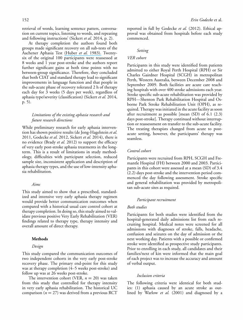
INT J LANG COMMUN DISORD, MARCH–APRIL 2014, VOL. 49, NO. 2, 149–161 Research Report A comparison of aphasia therapy outcomes before and after a Very Early Rehabilitation programme following stroke Erin Godecke†‡, Natalie A. Ciccone†‡, Andrew S. Granger§, Tapan Rai¶, Deborah West§, Angela Cream§, Jade Cartwright and Graeme J. Hankey# †Faculty of Health, Engineering and Science, Edith Cowan University, Joondalup, WA, Australia ‡Clinical Centre of Research Excellence in Aphasia Rehabilitation, Brisbane, QLD, Australia §Osborne Park Hospital, North Metropolitan Health Service, Osborne Park, WA, Australia ¶School of Mathematical Sciences, University of Technology Sydney, Sydney, NSW, Australia School of Psychology and Speech Pathology, Curtin University and Curtin Health Innovation Research Institute, Bentley, WA, Australia #School of Medicine and Pharmacology, University of Western Australia; Department of Neurology, Sir Charles Gairdner Hospital, Perth, WA, Australia (Received January 2013; accepted November 2013) Abstract Background: Very early aphasia rehabilitation studies have shown mixed results. Differences in therapy intensity and therapy type contribute significantly to the equivocal results. Aims: To compare a standardized, prescribed very early aphasia therapy regimen with a historical usual care control group at therapy completion (4–5 weeks post-stroke) and again at follow-up (6 months). Methods & Procedures: This study compared two cohorts from successive studies conducted in four Australian acute/sub-acute hospitals. The studies had near identical recruitment, blinded assessment and data-collection protocols. The Very Early Rehabilitation (VER) cohort (N = 20) had mild–severe aphasia and received up to 20 1-h sessions of impairment-based aphasia therapy, up to 5 weeks. The control cohort (n = 27) also had mild–severe aphasia and received usual care (UC) therapy for up to 4 weeks post-stroke. The primary outcome measure was the Aphasia Quotient (AQ) and a measure of communicative efficiency (DA) at therapy completion. Outcomes were measured at baseline, therapy completion and 6 months post-stroke and were compared using Generalised Estimating Equations (GEE) models. Outcomes & Results: After controlling for initial aphasia and stroke disability, the GEE models demonstrated that at the primary end-point participants receiving VER achieved 18% greater recovery on the AQ and 1.5% higher DA scores than those in the control cohort. At 6 months, the VER participants maintained a 16% advantage in recovery on the AQ and 0.6% more on DA scores over the control cohort participants. Conclusions & Implications: A prescribed, impairment-based aphasia therapy regimen, provided daily in very early post-stroke recovery, resulted in significantly greater communication gains in people with mild–severe aphasia at completion of therapy and at 6 months, when compared with a historical control cohort. Further research is required to demonstrate large-scale and long-term efficacy. Keywords: Very Early Rehabilitation, aphasia, stroke. What this paper adds? What is already known on the subject? The outcomes of very early aphasia therapy are the subject of ongoing debate. Given the potentially devastating impact of aphasia, very early rehabilitative therapy for people with aphasia requires further investigation. Address correspondence to: Erin Godecke, School of Psychology and Social Science, Building 4, Edith Cowan, University, Joondalup Campus, 270 Joondalup Drive, Joondalup 6027, WA, Australia; e-mail: [email protected] International Journal of Language & Communication Disorders ISSN 1368-2822 print/ISSN 1460-6984 online C 2014 Royal College of Speech and Language Therapists DOI: 10.1111/1460-6984.12074
Transcript of A comparison of aphasia therapy outcomes before and after a Very Early Rehabilitation programme...

INT J LANG COMMUN DISORD, MARCH–APRIL 2014,VOL. 49, NO. 2, 149–161
Research Report
A comparison of aphasia therapy outcomes before and after a Very EarlyRehabilitation programme following stroke
Erin Godecke†‡, Natalie A. Ciccone†‡, Andrew S. Granger§, Tapan Rai¶, Deborah West§, Angela Cream§,Jade Cartwright‖ and Graeme J. Hankey#†Faculty of Health, Engineering and Science, Edith Cowan University, Joondalup, WA, Australia‡Clinical Centre of Research Excellence in Aphasia Rehabilitation, Brisbane, QLD, Australia§Osborne Park Hospital, North Metropolitan Health Service, Osborne Park, WA, Australia¶School of Mathematical Sciences, University of Technology Sydney, Sydney, NSW, Australia‖School of Psychology and Speech Pathology, Curtin University and Curtin Health Innovation Research Institute, Bentley,WA, Australia#School of Medicine and Pharmacology, University of Western Australia; Department of Neurology, Sir Charles GairdnerHospital, Perth, WA, Australia
(Received January 2013; accepted November 2013)
Abstract
Background: Very early aphasia rehabilitation studies have shown mixed results. Differences in therapy intensityand therapy type contribute significantly to the equivocal results.Aims: To compare a standardized, prescribed very early aphasia therapy regimen with a historical usual care controlgroup at therapy completion (4–5 weeks post-stroke) and again at follow-up (6 months).Methods & Procedures: This study compared two cohorts from successive studies conducted in four Australianacute/sub-acute hospitals. The studies had near identical recruitment, blinded assessment and data-collectionprotocols. The Very Early Rehabilitation (VER) cohort (N = 20) had mild–severe aphasia and received up to 201-h sessions of impairment-based aphasia therapy, up to 5 weeks. The control cohort (n = 27) also had mild–severeaphasia and received usual care (UC) therapy for up to 4 weeks post-stroke. The primary outcome measure wasthe Aphasia Quotient (AQ) and a measure of communicative efficiency (DA) at therapy completion. Outcomeswere measured at baseline, therapy completion and 6 months post-stroke and were compared using GeneralisedEstimating Equations (GEE) models.Outcomes & Results: After controlling for initial aphasia and stroke disability, the GEE models demonstrated thatat the primary end-point participants receiving VER achieved 18% greater recovery on the AQ and 1.5% higherDA scores than those in the control cohort. At 6 months, the VER participants maintained a 16% advantage inrecovery on the AQ and 0.6% more on DA scores over the control cohort participants.Conclusions & Implications: A prescribed, impairment-based aphasia therapy regimen, provided daily in very earlypost-stroke recovery, resulted in significantly greater communication gains in people with mild–severe aphasiaat completion of therapy and at 6 months, when compared with a historical control cohort. Further research isrequired to demonstrate large-scale and long-term efficacy.
Keywords: Very Early Rehabilitation, aphasia, stroke.
What this paper adds?What is already known on the subject?The outcomes of very early aphasia therapy are the subject of ongoing debate. Given the potentially devastatingimpact of aphasia, very early rehabilitative therapy for people with aphasia requires further investigation.
Address correspondence to: Erin Godecke, School of Psychology and Social Science, Building 4, Edith Cowan, University, Joondalup Campus,270 Joondalup Drive, Joondalup 6027, WA, Australia; e-mail: [email protected]
International Journal of Language & Communication DisordersISSN 1368-2822 print/ISSN 1460-6984 online C© 2014 Royal College of Speech and Language Therapists
DOI: 10.1111/1460-6984.12074

150 Erin Godecke et al.
What this study adds?Very early aphasia intervention, provided as 20 sessions over 4–5 weeks, resulted in significantly greater communica-tion gains than usual care. It adds to evidence suggesting increased aphasia therapy (intensity, frequency and amount)in the very early and early recovery phases are important for augmenting the effects of spontaneous recovery.
Introduction
Aphasia is a devastating condition that affects up to42% of first-ever stroke survivors (Engelter et al. 2006).Up to 50% of these people may still suffer aphasia at18 months (Davidson et al. 2008). The negative ef-fects of aphasia have wide-ranging social and financialimplications with few people with aphasia regaining fullindependence (Davidson et al. 2008). People with apha-sia consume greater healthcare resources (Morris et al.2011), report higher levels of social and emotional iso-lation (Davidson et al. 2004, Kauhanen et al. 2000) andhave higher incidences of major depression within thefirst 12 months post-stroke than stroke survivors with-out aphasia (Kauhanen et al. 2000).
The mainstay for aphasia recovery is rehabilitation.The ‘earlier is better’ concept is supported by small ran-domized controlled trials (Bakheit et al. 2007, de Jong-Hagelstein et al. 2011, Godecke et al. 2012) and compar-ative studies, which suggest that 2–5 h of impairment-based therapy per week provides improved recovery(Bhogal et al. 2003, Robey 1998). The first 90 dayspost-stroke are believed to be the ‘window of opportu-nity’ for neuronal changes to occur as part of neuroplas-ticity (Meyer et al. 2010). The positive effects of earlyaphasia rehabilitation are thought to be underpinned byneural substrates that enable brain recovery in the im-mediate post-stroke period. Very early (acute) and early(sub-acute) post-stroke aphasia rehabilitation is believedto harness the effects of spontaneous recovery throughtherapeutic activities that include high levels of ‘repeti-tion and intensity’, ‘task-specific practice’ and therapy‘saliency’ (Raymer et al. 2008). The process of recoveryis thought to be driven by the strengthening of neuralnetworks. Neural strengthening requires repeated, syn-chronous firing of a group of neurons (Berthier andPulvermuller 2011). This repeated synchronous neu-ronal firing is achieved through high-frequency taskrepetition, and is thought to minimize independentneuronal activation that potentially produces maladap-tive behaviours (Berthier and Pulvermuller 2011). Im-plementation of the above neuroplasticity principles isaimed at strengthening the neural networks, used forcommunication, when the brain has its greatest poten-tial for recovery (Kreisel et al. 2006). It is believed thatthese activities lead to significant changes in commu-nication abilities when delivered in manageable dosesstarting within the first 2 weeks post-stroke (Godeckeet al. 2012).
The clinical effects, however, of very early aphasiatherapy are equivocal. This is due to methodologicaldifficulties related to the implementation of large-scaletrials to address the aphasia efficacy debate. In particu-lar, issues in early recovery include identifying: (1) theoptimum therapy intensity, (2) the ideal therapy typeto accommodate the enormous variability in aphasiapresentation and (3) selecting people with aphasia whowill benefit from rehabilitation. The Cochrane Review(Brady et al. 2012) reported a trend in evidence to sup-port aphasia intervention, with further research requiredto confirm these findings. There is little evidence fromthe Cochrane Review regarding the efficacy of aphasiatherapy started in the very early or early recovery phase.
Current research on early and very early aphasiatherapy
The following summary provides an overview of themost recently published clinical trials in early apha-sia rehabilitation. For a comprehensive overview, seeBrady et al. (2012). de Jong-Hagelstein et al. (2011)conducted a randomized control trial (RCT) to inves-tigate the role of therapy type in early aphasia recovery,with aphasia therapy commencing within 3 weeks ofstroke onset. Participants (N = 80) received an equalamount of therapy (mean of 45.4 h) at a low inten-sity level (2.1 h per week). The primary outcome mea-sure was the Amsterdam–Nijmegen Everyday LanguageTest (ANELT) (Blomert et al. 1995) at 3 and 6 monthspost-stroke. The intervention group received a cogni-tive linguistic approach to treatment consisting of asemantically based treatment, BOX, and/or a phono-logical treatment programme, FIKS. The control groupreceived PACE therapy (Promoting Aphasic Com-munication Effectiveness) (Davis and Wilcox 1985),role-playing and conversational coaching. Both groups(N = 80) made positive change on the ANELT at 3and 6 months post-stroke, with the majority of recov-ery occurring within the first 3 months. There were nobetween-group differences at 3 and 6 months. de Jong-Hagelstein et al. (2011) suggested that the low level ofearly aphasia therapy intensity and the fact that the twotherapy types were not sufficiently different may havecontributed to the findings.
Two published RCTs showed that very early apha-sia therapy is feasible (Godecke et al. 2012, Laska et al.2011). The Scandinavian study by Laska et al. (2011)

Outcomes in early aphasia rehabilitation in stroke 151
and the Australian equivalent study by Godecke et al.(2012) were strikingly similar in design, baseline apha-sia severity, therapy intensity and overall amount of in-tervention. The studies differed in the type of therapyprovided to the intervention groups and the overall out-come of these studies was disparate. The difference inthe nature of the treatment approaches and the outcomemeasures used in these studies may explain some of thediscrepancy in the reported results.
Stroke severity in the Scandinavian study appearsto be slightly less than that of the Australian study, de-spite participants being recruited and treated within thefirst 2 weeks post-stroke in both studies. Participants inthe Laska et al. (2011) trial received either daily Lan-guage Enrichment Therapy (LET) or no therapy forthe period of intervention. LET comprised exercises forcomprehension with some (though limited) naming tasksincluded. The intervention group in this study receiveda minimum of 10 h and a maximum of 12.5 h of di-rect therapy over 15 working days. The control groupreceived no speech and language therapy (SLT) for theintervention period of the study. After controlling forbaseline aphasia and stroke severity, Laska et al. foundno significant difference between those who receivedLET and those who received usual care (UC), on theANELT.
In comparison, the Australian study (Godeckeet al. 2012) provided impairment-based therapy thattargeted increased verbal production and connectedspeech using predominantly Semantic Feature Ther-apy (SFT) (Boyle and Coelho 1995) or a combinationof SFT and BOX therapy (Visch-Brink et al. 1997),or SFT and Mapping therapy (Schwartz et al. 1994).Each therapy session also included a picture descriptiontask that was used to enhance verbal output througha structured and supported conversational approach.The intervention group in Godecke et al. (2012) re-ceived an average of 5.6 h of direct therapy providedby a speech–language therapist over an average of 7.5working days (overall mean length of stay = 22 days).Participants (and families) received education, coun-selling, case management and discharge planning; how-ever, these data were not included in the 5.6 h of directtherapy. Twenty-three (85%) of the 27 participants inthe UC group in this trial received no therapy dur-ing the intervention phase (22 days) (Godecke et al.2012). The four participants in UC who were treatedreceived on average less than 14 min of one-to-one di-rect therapy per week and therapy tasks matched thoseused in the intervention group.
Godecke et al. reported that those receiving dailytherapy showed immediate and positive effects of veryearly aphasia therapy when compared with UC partic-ipants. Unfortunately, due to the application of mixedtherapy types for participants in this study, little can be
said about the effects of discrete therapy types in thisrecovery phase.
The pragmatic randomized controlled trial (ACT-NoW) by Bowen et al. (2012a) compared enhancedSLT with attention control (AC) in stroke survivorswith aphasia and dysarthria. The intervention in thistrial commenced on average 15 days after hospital ad-mission for acute stroke and lasted for 16 weeks. The in-tervention group received a less intensive (or dispersed)therapy regimen of a mean of 1.3 h of SLT per week over16 weeks (18 h total) when compared with Laska et al.(2011) and Godecke et al. (2012) who provided moreintensive therapy over a shorter duration. More impor-tantly, the 1.3 h of intervention time recorded in theACTNoW study incorporated all elements of communi-cation intervention that included assessments, provisionof communication materials and education, carer con-tact, indirect patient contact and direct therapy (Bowenet al. 2012b). Consequently, when compared with otherstudies that demonstrated a positive therapy effect (deJong-Hagelstein et al. 2011, Godecke et al. 2012, Sick-ert et al. 2014), participants in the intervention arm ofACTNoW received substantially reduced direct therapyintensity.
The AC group in ACTNoW received an averageof 15 h of social contact from a paid visitor over 16weeks (Bowen et al. 2012b). The intervention for theAC group consisted of a structured program including:(1) rapport building of two to four sessions, (2) regu-lar contact sessions involving conversation, the visitorreading from books, magazines and newspapers to cre-ate conversation topics, watching television, listening tomusic, playing games of tactics and strategy or creativeactivities such as crafts of gardening and (3) windingdown sessions (two to four sessions), which includedpreparation for the visitor’s time with the participant tocome to an end. At 6 months post-stroke, this studyfound no significant difference in communication out-comes between those in the SLT intervention group andthose who received the AC intervention.
More recently, Sickert et al. (2014) completed asingle-blind RCT investigating a modified dose of inten-sive aphasia therapy in first-ever stroke survivors whentherapy was started in the sub-acute phase (mean of34.8 days post-stroke). One hundred participants withmild to global aphasia were randomized to either Con-straint Induced Aphasia Therapy (CIAT) or standardtreatment. Treatment for both groups was provided for2 h per day over 15 (working) days for a total of 30 hof therapy or a mean of 10 h per week. CIAT wasprovided as per Pulvermuller et al. (2001) with theadditional use of multi-modality cues and prompts.The conventional treatment was provided in a groupsetting ‘Standard treatment consisted of aphasia exer-cises including sentence completion, improving patients’
152 Erin Godecke et al.
retrieval of words, learning sentence patters, conversa-tion on current topics, listening to words, and repeatingand following instructions’ (Sickert et al. 2014, p. 2).
At therapy completion the authors found bothgroups made significant recovery on all sub-tests of theAachener Aphasia Test (Huber et al. 1983). Twenty-six of the original 100 participants were reassessed at8 weeks and 1 year post-stroke and the authors reportfurther significant gains at both time points with nobetween-group significance. Therefore, they concludedthat both CIAT and standard therapy lead to significantimprovements in language function and that people inthe sub-acute phase of recovery tolerated 2 h of therapyeach day for 3 weeks (5 days per week), regardless ofaphasia type/severity (classification) (Sickert et al. 2014,p. 5).
Limitations of the existing aphasia research andfuture research directions
While preliminary research for early aphasia interven-tion has shown positive results (de Jong-Hagelstein et al.2011, Godecke et al. 2012, Sickert et al. 2014), there isno evidence (Brady et al. 2012) to support the efficacyof very early post-stroke aphasia treatments in the long-term. This is a result of limitations in study method-ology, difficulties with participant selection, reducedsample size, inconsistent application and description ofaphasia therapy types, and the use of low-intensity apha-sia rehabilitation.
Aims
This study aimed to show that a prescribed, standard-ized and intensive very early aphasia therapy regimenwould provide better communication outcomes whencompared with a historical usual care control cohort attherapy completion. In doing so, this study aimed to val-idate previous positive Very Early Rehabilitation (VER)findings related to therapy type, therapy intensity andoverall amount of direct therapy.
Methods
Design
This study compared the communication outcomes oftwo independent cohorts in the very early post-strokerecovery phase. The primary end-point for this studywas at therapy completion (4–5 weeks post-stroke) andfollow up was at 26 weeks post-stroke.
The intervention cohort (VER, n = 20) was takenfrom this study that controlled for therapy intensityin very early aphasia rehabilitation. The historical UCcomparison (n = 27) was derived from a previous RCT
reported in full by Godecke et al. (2012). Ethical ap-proval was obtained from hospitals before each studycommenced.
Setting
VER cohort
Participants in this study were identified from patientsadmitted to either Royal Perth Hospital (RPH) or SirCharles Gairdner Hospital (SCGH) in metropolitanPerth, Western Australia, between December 2008 andSeptember 2009. Both facilities are acute care teach-ing hospitals with over 400 stroke admissions each year.Stroke specific sub-acute rehabilitation was provided byRPH—Shenton Park Rehabilitation Hospital and Os-borne Park Stroke Rehabilitation Unit (OPH), as re-quired. Therapy was initiated in the acute facility as soonafter recruitment as possible [mean (SD) of 6.1 (2.3)days post-stroke]. Therapy continued without interrup-tion or reassessment on transfer to the sub-acute facility.The treating therapists changed from acute to post-acute setting, however, the participants’ therapy wasseamless.
Control cohort
Participants were recruited from RPH, SCGH and Fre-mantle Hospital (FH) between 2000 and 2003. Partici-pants in this cohort were assessed at a mean (SD) of 3.4(2.2) days post-stroke and the intervention period com-menced the day following assessment. Stroke specificand general rehabilitation was provided by metropoli-tan sub-acute sites as required.
Participant recruitment
Both studies
Participants for both studies were identified from thehospital-generated daily admissions list from each re-cruiting hospital. Medical notes were screened for alladmissions with diagnoses of stroke, falls, headache,confusion and seizures on the day of admission or thenext working day. Patients with a possible or confirmedstroke were identified as prospective study participants.Prior to enrolling in each study, all candidates and theirfamilies/next of kin were informed that the main goalof each project was to increase the accuracy and amountof verbal output.
Inclusion criteria
The following criteria were identical for both stud-ies: (1) aphasia caused by an acute stroke as out-lined by Warlow et al. (2001) and diagnosed by a

Outcomes in early aphasia rehabilitation in stroke 153
Table 1. Variation in selection criteria for the VER and controlcohorts
VER Controlcohort cohort
Admission to hospital (days) < 14 < 10Frenchay Aphasia Screening test (days) < 14 < 10Conscious/medically stable (days) < 14 < 10Wakeful state > 30 min (days) < 14 < 10Aphasia severity Mild–severe Mild–severeEnglish as a second language Yes No
neurologist or stroke physician, (2) a clinical strokediagnosis confirmed by computer tomography and/ormagnetic resonance imaging within 48 h of hospitaladmission, (3) aphasia identified by a score of less than13/20 on the shortened Frenchay Aphasia Screening Test(FAST) (Enderby et al. 1987), which is a reliable andvalid aphasia screening tool comprising auditory com-prehension and verbal expression tasks used to identifyaphasia in the early phase of recovery, (4) medical sta-bility (measured by the Glasgow Coma Scale score of> 10 which indicates moderate level of alertness), (5)wakefulness—able to maintain sufficient alertness to in-teract for 30 min, and (6) aphasia severity score (lessthan 93.8 on the Aphasia Quotient (AQ) of the West-ern Aphasia Battery (WAB; Kertesz 1982). The VERcohort selection criteria were broadened slightly fromthat of the control cohort in Godecke et al. (2012) toinclude people with fluent English and those who wereappropriate for the study up to 14 days post-stroke on-set. A comparison of selection criteria for each trial ispresented in table 1.
The exclusion criteria for both trials were: (1) a pre-vious diagnosis of aphasia, mental illness or dementia,(2) a previous history of sub-arachnoid and/or sub-duralhaemorrhage or neurosurgical intervention and (3) un-corrected hearing or vision impairment.
Baseline data
Patient characteristics including demographic factors,stroke features, stroke classification according to Ox-fordshire Community Stroke Project Classification(Bamford et al. 1991) and the modified Rankin Scale(mRS) (Rankin 1957) were collected at baseline for bothstudies. Table 2 shows the baseline characteristics andcomparisons for the VER and control cohorts.
Speech and language service delivery and directaphasia therapy intensity
Participants in each study received dysphagia manage-ment, patient and family education (e.g. education re-garding stroke, secondary prevention, aphasia and dys-
phagia), counselling and support, case management,discharge planning and all other interventions as theyrequired. These data were collected but only the directaphasia therapy data are presented in this paper.
VER cohort
Therapy was commenced as soon as possible after re-cruitment and assessment. Intervention started on orbefore day 14 post-stroke for all participants. The targettherapy regimen was defined as between 900 and 1200min (15–20 h) of therapy, provided over 5 days per weekfor a total of 20 sessions in 4 weeks (4–5 h of therapy perweek). All attempts were made to complete the 20 ther-apy sessions within 4 working weeks (Monday–Friday),the fifth week was made available to complete any out-standing sessions. If more than two sessions were missedin any week, the participant was deemed as not toler-ating the intervention. Six trained non-assessing speechpathologists treated the participants.
UC cohort
This cohort (n = 27) received an average of 11 min oftherapy per week for an average of 3 weeks (22 days).
Type of aphasia therapy provided in each trial
The therapeutic approaches used in both studies adheredto the general principles of neurorehabilitation. Ther-apy types were designed for use with people with mildaphasia through to severe global aphasia. The therapiesprovided in each trial were impairment-based and be-lieved to harness neural recovery through restitution viahigh frequency (massed practice), use-dependent, task-specific repetition (Berthier and Pulvermuller 2011).Strategies to minimize speech production errors and topromote self-correction were used to enhance correctproduction of speech and language targets. The speechpathologist pre-empted and avoided consistent task fail-ure to ensure participants were supported as needed.This support included prompting, modelling and cue-ing to produce an appropriate verbal response. Thesestrategies are thought to enhance ‘synchronous neuronalfiring’ to strengthen neural networks and minimize in-dependent neuronal activation that could potentiallyproduce maladaptive behaviours (Berthier and Pulver-muller 2011).
VER cohort
Participants in this study received either group or one-to-one therapy. Group therapy consisted of CIAT (Pulver-muller et al. 2001) in a modified dose. Individual ther-apy consisted of SFT (Boyle and Coelho 1995), Cued

154 Erin Godecke et al.
Table 2. Baseline demographic and stroke characteristics and comparisons for the VER and control cohorts
VER cohort Control cohort(n = 20) (n = 27) p value
Age Mean (SD) 70.7(14.3) 67.7(15.4) 0.499‡
Female (%) 8 (40) 12 (44) 0.761†
Previous StrokeYes 3 3No 17 24
Stroke type 0.903†
Ischaemic (%) 18 (90) 24 (89)Haemorrhagic (%) 2 (10) 3 (11)
Stroke classification 0.298†
PACI (%) 1 (5) 9 (33.5)TACI (%) 17 (85) 14 (52)PoCI (%) 0 1 (3.5)LACI (%) 0 0Non-classified 2 (10) 3 (11)
Stroke Hemisphere:Left (%) 18 (90) 26 (96)Right (%) 2 (10) 1 (4)
Mortality within 28 daysNumber (%) 1 (5) 3 (11) 0.458†
Admission mRS score: Number (%) 0.676†∗
2 1 (5) 1 (3)3 3 (15) 4 (15)4 9 (45) 8 (30)5 7 (35) 14 (52) 0.898†∗∗
Admission to assessmentMean days (SD) 6.1 (2.3) 3.4 (2.2) 0.000‡
Notes: PACI, partial anterior circulation infarct; TACI, total anterior circulation infarct; PoCI, posterior circulation infarct; LACI, Lacunar infarct; non-classified = haemorrhage†Chi-square test for independence.‡Two-tailed t-test comparing Very Early Rehabilitation cohort with Control cohort.∗Overall mRS score comparison.∗∗mRS categorized comparisons: Categories 2–3 indicate mild to moderate disability; and Categories 4–5 indicate severe disability.
Naming therapy (Nettleton and Lesser 1991), Lexical–semantic (BOX) therapy (Visch-Brink et al. (1997),Mapping therapy (Schwartz et al. 1994) and/or Phono-logical Feature Therapy (Raymer et al. 1993).
Treatment integrity was addressed by training alltreating therapists to ensure equivalent use of stimuli andtherapy type and therapy targets were identical acrossall therapy sites. All therapy tasks (CIAT and one-to-one) were structured to promote and scaffold connectedspeech production.
CIAT (group)
Therapy was based on the CIAT outlined by Pulver-muller et al. (2001). The therapy took place in smallgroups of two to four participants and one speechpathologist who provided language support appropri-ate to each participant’s needs. The stimuli and lan-guage support were designed to accommodate all levelsof aphasia severity in the same group. The group mem-bers were dealt a set of picture cards, the aim beingto collect pairs of cards. Each pictured item allowed averbal response ranging from a single word to complexsentences. Barriers prevented the participants from see-
ing each other’s cards. The ‘constraint’ of the therapywas provided by the requirement of verbal-only inter-action. The group language dynamics included polite-ness markers, requesting items, listening, clarification ofother’s responses and negating requests. The complex-ity of verbal output required from each participant andthe level of cueing and support provided was tailored toeach individual’s communicative ability as establishedat assessment and through ongoing performance withintherapy sessions.
Individual therapy (one-to-one)
Each participant receiving one-to-one therapy had a pro-gramme tailored to suit their needs. Based on the partic-ipant’s assessment results, the treating therapist selectedthe appropriate therapy from those previously listed.All one-to-one therapies were provided as per publishedinstructions (Boyle and Coelho 1995, Nettleton andLesser 1991, Visch-Brink et al. 1997, Schwartz et al.1994, Raymer et al. 1993). Participants received eithera single therapy, or a combination of therapy types suchas cued naming therapy and semantic feature therapy.

Outcomes in early aphasia rehabilitation in stroke 155
Control cohort
As previously outlined, 85% of participants in thiscohort received no direct therapy. When participantsreceived therapy it consisted of individual (one-to-one) cognitive–neuropsychological and neurolinguisti-cally based therapy. Therapists used one or more of thefollowing therapies: BOX therapy (Visch-Brink et al.1997), Mapping therapy (Schwartz et al. 1994) andSFT (Boyle and Coelho 1995). Participants also at-tempted a picture-description task aimed at increas-ing connected speech in a supported and structuredenvironment. See Godecke et al. (2012) for furtherdetail.
Recording of speech pathology service delivery data
The type and duration of speech pathology interventionsfor both studies were recorded via the Allied Health Sys-tem (AHS). This software package records interventionin 5-min units. All interventions for each participantwere coded for aphasia/dysphagia/dyspraxia/dysarthriaand time spent in each activity was categorized intoassessment, therapy, education/counselling, case plan-ning/consultation and documentation.
Outcome assessment
Both cohorts were assessed at acute hospital admission,immediately following intervention (after 4–5 weeks oftherapy) and 26 weeks post-stroke. All assessments werecompleted and analysed by blinded assessors.
Primary outcome measures
The primary outcome measures were the AQ score ofthe Western Aphasia Battery (WAB) (Kertesz 1982) andthe Discourse Analysis (DA) score at therapy comple-tion. All discourse samples were collected as per Godeckeet al. (2012) and consisted of picture descriptions, per-sonal and procedural narratives (Kertesz 1982, Nicholasand Brookshire 1995). DA is the total per cent CorrectInformation Units produced per min (% CIU/MIN)per sample and is calculated by dividing the total per-centage CIUs (Nicholas and Brookshire 1995) by thetotal time taken to produce the discourse. The measureadds a communication efficiency/temporal element thatis considered an important communicative measure notcaptured in the AQ scores. CIUs are topic specific wordsthat provide detail and are not repetitious, exclamatory,additive or commentary in nature (Nicholas and Brook-shire 1995). A count of 200 or more intelligible wordsacross picture description, personal and procedural nar-rative tasks was required for a representative and reliablespeech sample.
All discourse samples for the VER cohort wereaudio-recorded using a lapel microphone and digitalrecorder (Olympus-DM550). The recordings were tran-scribed verbatim and analysed as per Godecke et al.(2012).
Secondary outcome measures
Secondary outcomes were the AQ (Kertesz 1982) scoreand DA score at 6 months post-stroke.
Statistical analyses
The VER and UC cohorts were compared at baselineusing two-tailed t-tests and chi-square tests for indepen-dence. Generalised estimating equations (GEE) mod-els were developed to compare the cohorts on AQ andDA scores at therapy completion and at 26 weeks post-stroke. Generalized linear models are a class of statisticalmodel that extend the general linear model (regression,ANOVA and ANCOVA) to handle non-normal data.Generalized estimating equations are a further exten-sion of generalized linear models to account for longi-tudinal and clustered data. GEE models are preferred torepeated-measures ANOVA approaches because of theirflexibility in modelling complex covariance structuresthat arise in longitudinal and clustered data. Further-more, GEE models are robust against misspecificationof the covariance structure and are also more robustthan repeated measures ANOVA in handling missingdata (Ballinger 2004). GEE have also been found to bemore efficient in that they are able to achieve higherpower with smaller sample size or lower number of re-peated measurements in both complete and missing datascenarios (Ma et al. 2012). In the current work, separateGEE models were developed to compare the cohorts onthe primary outcome measure (AQ) and the secondaryoutcome measure (DA). Missing data were treated withthe last observation carried forward approach where ap-propriate. This complies with the intention to treat prin-ciple (Jansen et al. 2006).
Due to the possible ceiling effect in the AQ score,the AQ model involved transforming these scores to theper cent of maximum potential recovery (AQ%MPR)as per Lazar et al. (2010). The score for each participantwas calculated as the ratio of the achieved improvementin AQ to the maximum achievable improvement at base-line. The ratio was then multiplied by 100 to convert itto a percentage score.
There was a larger number of people with mildaphasia in the VER cohort (n = 6; 30%) than inthe UC cohort (n = 3; 11%) and therefore thetwo cohorts were expected to differ significantly onbaseline aphasia severity. Since baseline aphasia andstroke severity/disability are universal predictors of

156 Erin Godecke et al.
aphasia recovery, the baseline AQ and mRS (Rankin1957) scores were included as covariates in bothGEE models to control for these differences. BaselineDA was not included as a covariate in the modelsbecause it was correlated with baseline AQ (r = 0.6,p < 0.001).
Results
Baseline
Over the 10-month recruitment period for the VERcohort, the medical notes of 1006 admissions to RPHwere screened for collapse, falls, seizures, headache, con-fusion and stroke. A total of 236 people were admittedwith an acute stroke with 88 (37.2%) of these peo-ple having confirmed aphasia. We recruited 18 (20.4%)of the people with confirmed aphasia from RPH tothis study. Two participants from Sir Charles Gaird-ner Hospital met the selection criteria and were re-cruited taking the total number of participants to 20.The majority (n = 17; 85%) of participants in theVER cohort required full assistance (including apha-sia friendly forms) to complete the informed consentprocedure.
The baseline characteristics between VER and UCcohorts were not significantly different except for thetime to initial assessment and baseline AQ score (table2). The VER cohort had a mean (±SD) age of 70.7(±14.3) years and was slightly older than the UC cohortwho had a mean age of 67.7 (±15.4). Ninety per centof the VER cohort had an ischaemic stroke comparedwith 89% of the UC cohort. The majority of the VERcohort (80%) and the UC cohort (82%) had severestroke-related disability as indicated by a score of 4 or 5on the mRS. A clinical but non-significant difference inthe mRS is noted. The difference in the time to initialassessment reflects an increase in the length of time torecruitment for the VER cohort, which included peoplewith aphasia who were recruited up to 14 days post-stroke.
The difference in baseline AQ scores is reflective ofthe increased number of people with mild aphasia inthe VER cohort (n = 6; 30%) when compared with thenumber of people with mild aphasia (n = 3; 11%) inthe UC cohort. Table 2 shows the baseline characteristicsand comparisons for the VER and UC cohorts.
Two (10%) of the 20 participants in the VER cohortdid not reach the minimum intervention requirement(both participants were randomized to receive one-to-one intervention). One participant suffered a furtherstroke after completing a single 40-min session of ther-apy. The second completed 13 sessions and 780 min(13 h) of therapy, then suffered a gastric haemorrhageand was unable to complete the intervention. Table 3
outlines the therapy compliance for participants in theVER and control cohorts. Table 4 shows group raw andtransformed scores at baseline, therapy completion andfollow-up. The participants in the VER study toleratedapproximately 4.25 h of therapy per week for an averageof 4.5 weeks (32 days). During this time, the averagenumber of therapy sessions was 18.65, and each sessionaveraged 53.5 min.
Twenty-three of the 27 participants (85%) in the UCcohort received no direct therapy during the interven-tion period (Godecke et al. 2012) (table 3). The collec-tive amount of direct therapy time provided to the four(table 4) UC participants who received aphasia therapywas 295 min (4.9 h) over seven sessions, equating to anaverage of 11 min of therapy per week for each of theUC participants.
Primary end-point
The GEE models showed (tables 5 and 6) that after con-trolling for baseline aphasia severity and stroke-relateddisability, VER was a significant predictor of recovery onthe AQ (p = 0.006) and on the DA score (%CIU/Min)(p = 0.034). Specifically, participants receiving VERachieved 18% greater recovery on AQ and 1.5% moreon the DA score (%CIU/Min) than those in the UC Co-hort at therapy completion. These results indicate thatpeople who received VER had improved communica-tion impairment measures (AQ%MPR) on standardizedtesting (AQ scores) and produced significantly more ac-curate and efficient verbal language (DA scores) thanthose who received UC at therapy completion. Table 4outlines the descriptive data for the AQ, DA and the AQper cent of maximum potential recovery (AQ%MPR)scores for the VER and UC cohorts at baseline, post-therapy and 26 weeks post-stroke. As expected, thesedata indicate that the historical UC cohort had moresevere communication impairment at baseline than theVER cohort. When the AQ data are transformed to(AQ%MPR), baseline scores are returned to zero corre-sponding to a maximum potential recovery of 100% foreach participant.
Follow-up
At follow-up assessment 26 weeks post-stroke, the GEEmodel (tables 5 and 6) for AQ showed that after con-trolling for baseline aphasia severity and stroke relateddisability, participants receiving VER maintained a sta-tistically significant advantage in recovery (p = 0.017)over those receiving UC, with the VER cohort scor-ing 16% higher on the AQ than those who receivedUC (figure 1). The difference between the cohorts onDA scores was not statistically significant (p = 0.249)

Outcomes in early aphasia rehabilitation in stroke 157
Table 3. Descriptive data for intervention compliance, total therapy time (min) and number of sessions in the intervention phase
VER cohort Control cohortTherapy details (n = 20) (n = 27)
Met intervention compliance 18 (90%) n/aReceived therapy 20 (100%) 4 (15%)Intervention phase language therapy (total min)
Mean (SD) 1070.25 (267.44) 10.92 (38.73)∗
Median (IQR) 1167.5 (131.25) 47.5(86.25)Intervention phase language number of therapy sessions
Mean (SD) 18.65 (4.44) 0.3 (.99)Median (IQR) 8 (5.25) 1 (.75)
Note: IQR, interquartile range.∗The overall mean for all participants (n = 27) in control cohort.
Table 4. Group raw-score comparisons for communication outcomes between Very Early Rehabilitation participants and the ControlCohort
VER cohort Control cohort p-value∗
Baseline AQNumber of participants 20 27Mean (SD) 43.53 (27.02) 19.62 (26.26) 0.009
Therapy completion AQNumber of participants 17 24Mean (SD) 67.55 (30.16) 31.37 (32.83) 0.001
Follow-up (26 weeks) AQNumber of participants 8 23Mean (SD) 89.01 (11.96) 45.62 (39.64) 0.001
Baseline AQ%MPRNumber of participants 20 27Mean (SD) n/a n/a n/a
Therapy completion AQ%MPRNumber of participants 17 24Mean (SD) 46.72 (35.43) 17.92 (23.18) 0.007
Follow up (26 weeks) AQ%MPRNumber of participants 8 23Mean (SD) 77.60 (20.45) 38.77 (37.08) 0.002
Baseline DANumber of participants 20 27Mean (SD) 3.26 (5.0) 1.18 (4.06) 0.123
Therapy completion DANumber of participants 17 24Mean (SD) 8.36 (8.54) 2.37 (5.60) 0.012
Follow up (26 weeks) DANumber of participants 8 23Mean (SD) 33.90 (17.9) 7.74 (12.87) 0.004
Notes: These scores are unadjusted for baseline differences in aphasia severity and stroke disability.∗t-test; significance p = 0.005.
Table 5. GEE model for per cent of maximal potential recovery on AQ
Confidence limits
Parameter Estimate SE Lower Upper Wald Chi-square p-value
Baseline AQ 0.32 0.09 0.15 0.49 13.96 0.000Baseline mRS –2.64 2.94 –8.41 3.14 0.801 0.371Primary end-point 17.70 4.58 8.73 26.68 14.94 0.000Follow-up 39.21 7.46 24.59 53.82 27.65 0.000VER –8.27 3.19 –14.52 –2.01 6.71 0.010Primary end-point∗VER 26.19 9.52 7.52 44.86 7.56 0.006Follow-up∗VER 24.22 10.17 4.28 44.15 5.67 0.017
Note: Primary end-point: therapy completion (4–5 weeks post-stroke); follow-up: 6 months post-stroke; VER: Very Early Rehabilitation group; Primary end-point∗VER: Very EarlyRehabilitation group compared with Usual Care group at therapy completion; Follow-up∗VER: Very Early Rehabilitation group compared with Usual Care Group at 6 months.

158 Erin Godecke et al.
Table 6. GEE model for recovery on DA
Confidence limits
Parameter Estimate SE Lower Upper Wald Chi-square p-value
Baseline AQ 0.20 0.03 0.146 0.26 49.62 0.000Baseline mRS –0.51 0.94 –2.34 1.33 0.29 0.590Primary end-point 0.53 0.81 –1.06 2.12 0.43 0.514Follow-up 6.21 1.85 2.59 9.83 11.31 0.001VER –3.05 1.48 –5.96 –0.14 4.21 0.040Primary end-point∗VER 4.56 2.15 0.34 8.78 4.49 0.034Follow-up∗VER 3.61 3.13 –2.52 9.73 1.33 0.249
Note: Primary end-point: therapy completion (4–5 weeks post-stroke); follow-up: 6 months post-stroke; VER: Very Early Rehabilitation group; Primary end-point∗VER: Very EarlyRehabilitation group compared with Usual Care group at therapy completion; Follow-up∗VER: Very Early Rehabilitation group compared with Usual Care Group at 6 months.
Figure 1. AQ%MPR comparisons at baseline, therapy completion and follow-up (26 weeks).
at the 6-month follow up. However, the VER cohortshowed 0.6% greater recovery on DA than the UC co-hort (figure 2). The dosage of aphasia therapy was notcontrolled after the 4–5-week intervention period, andparticipants from both cohorts received UC therapy asper their available services.
Discussion
The present study showed that impairment-based apha-sia therapy provided by a speech pathologist for 45–
60 min per day for up to 20 sessions, commencingin the first 2 weeks of stroke recovery, improved com-munication outcomes at therapy completion in peo-ple with mild, moderate and severe aphasia when com-pared with a historical usual care control. Interestingly,the communication gains achieved for those treatedin the very early recovery period were sustained at 6months when measured by the AQ score, but despitea clinically meaningful improvement in the DA scoreat 6 months, this outcome did not reach statisticalsignificance.

Outcomes in early aphasia rehabilitation in stroke 159
Figure 2. DA comparisons at baseline, therapy completion and follow-up (26 weeks).
Therapy intensity
Only the hours of direct patient therapy have been re-ported in this study, in Laska et al. (2011) and inGodecke et al. (2012). In comparison, Bowen et al.(2012b) reported on all aspects of speech and languageintervention as contributing to therapy intensity. Whenconsidering direct aphasia therapy only, Laska et al.(2011) and Godecke et al. (2012) provided an aver-age of 3.75 and 3.6 h per week, respectively; this studyprovided an average of 4.25 h per week; Bowen et al.(2012b) provided 1.3 h (combined intervention) perweek and Sickert et al. (2014) provided 10 h per weekin the sub-acute recovery phase. The total amount andduration of the intervention in each trial varied, andthe outcomes of these studies were mixed. Previous re-views (Bhogal et al. 2003, Robey 1998) have foundthat an average of 2.5–5 h of aphasia therapy per weekproduced significantly better communication outcomesthan therapy provided at lower intensity level. The re-sults of this study support previous work that highlightsthe importance of therapy intensity in enhancing spon-taneous recovery in very early aphasia after stroke. Aninteresting point also to consider here is that people withaphasia in very early recovery can and do tolerate dailyaphasia therapy of up to 60 min per day if the service isprovided.
Therapy type
Therapy for the VER participants was structured, tai-lored therapy, based on psycholinguistic principles andtargeted verbal production (output) at the impairmentlevel of communication. People in this study who re-ceived very early intervention, consisting of either one-to-one or group therapy demonstrated superior com-munication outcomes at therapy completion and at6 months (AQ scores) when compared with the his-torical control group. Results of this study support deJong-Hagelstein et al. (2011), Godecke et al. (2012) andSickert et al. (2014) which show positive results using avariety of therapy types. No research to date has deter-mined the most beneficial therapy type for the variousaphasia severities and types. A definitive answer to ad-dress this complex issue is expected to be some time offand will likely require the pooled efforts of multiple datasources.
DA score at 6 months
The very large clinically meaningful but non-significantimprovement in the mean DA scores in the VER groupwhen compared with the UC group at the same pe-riod is an interesting finding (table 4). Seeing a 25%improvement in any score is a welcome result (figure 2),

160 Erin Godecke et al.
especially if that score represents: (1) the accuracy andefficiency of connected speech and (2) the amount ofimprovement that was made when recovery is thoughtto be all but completed (Lazar et al. 2010). The factorsthat contribute to this recovery remain unclear. The factthat this amount of clinical change does not represent astatistical difference is likely to be multifactorial and isbest explained through the large natural variation seen inthe standard deviations, coupled with the small samplesize in the study.
Study limitations
Due caution regarding the comparative use of a historicalcontrol group is warranted given that the original pop-ulation from which the samples in the VER and controlcohorts are taken are at least 5 years apart. We acknowl-edge improvements in the systems of hospital care arelikely to have occurred in the period 2000–09 whenthese data were collected. These changes are likely toinclude increased rates of thrombolysis, improved man-agement of stroke-related complications and reductionof stroke-related morbidity and mortality. It is also possi-ble that these changes will have contributed in some wayto the results presented here. Unfortunately, the large-scale systems audit required for monitoring service de-livery and organizational changes in stroke services wasnot available at the time these data were collected. Somereassurance is provided, given the fact that there wasno statistical between group significance in stroke type,stroke classification and mRS score between the VERand control cohorts at baseline.
The VER rehabilitation study design would havebeen strengthened by including a third arm of ran-domization that measured usual care alone and withstratification of aphasia severity. Additionally, the resultsof the VER cohort should be interpreted with cautiondue to the small number of participants who completedthe 6-month follow-up. Whilst the statistical modellingfor this paper was conservative and vigilant, the highlyvariable nature of aphasia requires large-scale clinicaltrials before clinical efficacy and/or effectiveness can beestablished.
Conclusions and future directions
Very early, impairment-based aphasia therapy resultedin superior communication outcomes which were sus-tained at 6 months post-stroke when compared witha historical control cohort. This paper provides sup-port for very early impairment-based aphasia therapyprovided between 45 and 60 min per day, when com-menced within the first 2 weeks post-stroke. Importantlythis study found the timing and intensity of very earlytherapy is feasible and beneficial for people with mild
to severe aphasia (Godecke et al. 2012). It also addsevidence to suggest that increased aphasia therapy (in-tensity, frequency and amount) in the very early andearly recovery phases is important for augmenting theeffects of spontaneous recovery.
Given the ongoing debate surrounding the benefitsof very early aphasia intervention, we must focus fur-ther research attention to unpacking the elements ofthe optimal aphasia therapy type, intensity and timingto best facilitate the natural mechanisms of very earlyaphasia recovery. If healthcare funding bodies are to beconvinced of the benefits of very early aphasia reha-bilitation we must determine the right combination ofaphasia therapy for the right person, provided at theright time.
Acknowledgements
The authors sincerely thank the study participants who supportedthese studies. They also gratefully acknowledge the flexibility and re-organization of the speech pathology and ward services throughoutthis study. In particular, thanks to our research assistants, CamillaMcElhone, Shari Serjeant and Adrienne Miles, and the ward staff atWellington Street and Shenton Park Campuses of Royal Perth Hospi-tal and Osborne Park Hospital. This work was partially supported bythe State Health and Research Advisory Council (SHRAC) ResearchTranslation Project (Grant Number RSD-02720); Department ofHealth, Western Australia, Australia. Declaration of interest: Theauthors report no conflicts of interest. The authors alone are respon-sible for the content and writing of the paper.
References
BAKHEIT, A. M. O., SHAW, S., BARRETT, L., WOOD, S., CARRING-TON, S., GRIFFITHS, S., SEARLE, K. and KOUSTSI, F., 2007, Aprospective, randomised, parallel group, controlled study ofthe effect of intensity of speech and language therapy on earlyrecovery from post-stroke aphasia. Clinical Rehabilitation, 21,885–894.
BALLINGER, G., 2004, Using Generalized Estimating Equations forLongitudinal Data Analysis. Organisational Research Methods,7, 127–150.
BAMFORD, J., SANDERCOCK, P., DENNIS, M., BURN, J. and WAR-LOW, C., 1991, Classification and natural history of clini-cally identifiable subtypes of cerebral infarction. Lancet, 337,1521–1526.
BERTHIER, M. L. and PULVERMULLER, F., 2011, Neuroscience in-sights improve neurorehabilitation of poststroke aphasia. Na-ture Reviews Neurology, 7, 86–97.
BHOGAL, S. K., TEASELL, R. and SPEECHLEY, M., 2003, Intensity ofaphasia therapy, impact on recovery. Stroke, 34, 987–993.
BLOMERT, L., KOSTER, C. H. and KEAN, M.-L., 1995, Amsterdam–Nijmegen Test Voor Alledaagse Taalvaardigheden Lisse (Amster-dam: Swets & Zeitlinger).
BOWEN, A., HESKETH, A., PATCHICK, E., YOUNG, A., DAVIES, L.,VAIL, A., LONG, A., WATKINS, C., WILKINSON, M., PEARL,G., LAMBON RALPH, M. A., TYRRELL, P., 2012a, Effective-ness of enhanced communication therapy in the first fourmonths after stroke for aphasia and dysarthria: a randomisedcontrolled trial. British Medical Journal, 345, 34407(1–15).

Outcomes in early aphasia rehabilitation in stroke 161
BOWEN, A., HESKETH, A., PATCHICK, E., YOUNG, A., DAVIES, L.,VAIL, A., LONG, A., WATKINS, C., WILKINSON, M., PEARL,G., LAMBON-RALPH, M., and TYRRELL, P., 2012b, Clini-cal effectiveness, cost-effectiveness and service users’ percep-tions of early, well-resourced communication therapy fol-lowing stroke: a randomised controlled trial (the actnowstudy). Health Technology Assessment, 16, 26 (available at:http://www.hta.ac.uk/1390).
BOYLE, M. and COELHO, C. A., 1995, Application of semantic fea-ture analysis as a treatment for aphasic dysnomia. AmericanJournal of Speech and Language Pathology, 4, 135–138.
BRADY, M. C., KELLY, H., GODWIN, J. and ENDERBY, P., 2012,Speech and language therapy for aphasia following stroke.Cochrane Database of Systematic Reviews, Issue 5, Art. No.CD000425.
DAVIDSON, B., HOWE, T., WORRALL, L., HICKSON, L. andTOGHER, L., 2008, Social participation for older peoplewith aphasia: the impact of communication disability onfriendships. Topics in Stroke Rehabilitation, 15(4), 325–340.
DAVIS, G. A. and WILCOX, M. J., 1985, Adult Aphasia Rehabilitation:Applied Pragmatics (San Diego, CA: Singular).
DE JONG-HAGELSTEIN, M., VAN DE SANDT-KOENDERMAN, W. M.E., PRINS, N. D., DIPPEL, D. W., KOUDSTAAL, P. J. andVISCH-BRINK, E. G., 2011, Efficacy of earlycognitive–linguistic treatment and communicative treatmentin aphasia after stroke: a randomised controlled trial (RATS-2). Journal of Neurology, Neurosurgery and Psychiatry, 82(4),399–404.
ENDERBY, P. M., WOOD, V. A. and WADE, D. T., 1987, The FrenchayAphasia Screening Test (London: Whurr).
ENGELTER, S. T., GOSTYNSKI, M., PAPA, S., FREI, M., BORN,C., AJDACIC-GROSS, V., GUTZWILLER, F., PHILLIPE, F. andLYRER, A., 2006, Epidemiology of aphasia attributable tofirst ischemic stroke incidence, severity, fluency, etiology, andthrombolysis. Stroke, 37, 1379–1384.
GODECKE, E., HIRD, K., LALOR, E. E., RAI, T. and PHILLIPS, M.R., 2012 Very early aphasia therapy: a pilot randomized con-trolled efficacy trial. International Journal Of Stroke. DOI:10.1111/j.1747–4949.2011.00631.x
HUBER, W., POECK, K., WENIGER, D. et al., 1983, Aachener Aphasietest (Gottingen: Hogrefe).
JANSEN, I., HENS, N., MOLENBERGHS, G., AERTS, M., VERBEKE,G., KENWARD, M. G., and WILLMES, K., 2006, The natureof sensitivity in monotone missing not at random models.Computational Statistics and Data Analysis, 50(3), 830–858.
KAUHANEN, M. L., KORPELAINEN, J. T., HILTUNEN, P., MAATTA, R.et al., 2000, Aphasia, depression and non-verbal cognitive im-pairment in ischaemic stroke. Cerebrovascular Disease, 10(6),455.
KERTESZ, A., MONONEN, H., BRUSIN, E., SOTANIEMI, K. A., andMYLLYLA, V. V., 1982, Western Aphasia Battery (New York,NY: Grune & Stratton).
KREISEL, S. H., BAZNER, H. and HENNERICI, M. G., 2006,Pathophysiology of stroke rehabilitation: temporal aspectsof neurofunctional recovery. Cerebrovascular Disease, 21,6–17.
LASKA, A. C., KAHAN, T., HELLBLOM, A., MURRAY, V. and VON
ARBIN, M., 2011, A randomized controlled trial on very early
speech and language therapy in patients with acute stroke andaphasia. Cerebrovascular Disease Extra, 1, 66–74.
LAZAR, R. M., MINZER, B., ANTONIELLO, D., FESTA, J. R.,KRAKAUER, J. W. and MARSHALL, R. S., 2010, Improvementin aphasia scores after stroke is well predicted by initial sever-ity. Stroke, 41, 1485–1488.
MA, Y., MAZUMDAR, M. and MEMTSOUDIS, S. G., 2012, Beyondrepeated measures ANOVA: advanced statistical methods forthe analysis of longitudinal data anesthesia research. RegionalAnesthesia and Pain Medicine, 37(1), 99–105.
MEYER, M. J., MEGYESI, J., MEYTHALER, J., MURIE-FERNANDEZ,M., AUBUT, J., FOLEY, N., SALTER, K., BAYLEY, M., MAR-SHALL, S. and TEASELL, R., 2010, Acute management of ac-quired brain injury Part I: An evidence-based review of non-pharmacological interventions. Brain Injury, 24(5), 694–670.
MORRIS, J., FRANKLIN, S., and MENGER, F., 2011, Returning towork with aphasia: a case study. Aphasiology, IFIRST 25(8),890–907.
NETTLETON, J. and LESSER, R., 1991, Therapy for naming difficul-ties in aphasia: application of a cognitive neuropsychologicalmodel. Journal of Neurolinguistics, 6(2), 139–157.
NICHOLAS, L. and BROOKSHIRE, R. H., 1995, Presence completenessand accuracy of main concepts in the connected Speech ofnon-brain injured in adults. Journal of Speech and HearingResearch, 38, 145–156.
PULVERMULLER, F., NEININGER, B., ELBERT, T., MOHR, B., ROCK-STROH, B., KOEBBEL, P., and TAUB, E., 2001, Constraint-induced therapy of chronic aphasia after stroke. Stroke, 32,1621–1626.
RANKIN, J., 1957, Cerebral vascular accidents in patients over theage of 60. Scottish Medical Journal, 2, 200–215.
RAYMER, A. M., BEESON, P., HOLLAND, A., KENDALL, D., MAHER,L. M. MARTIN, N., MURRAY, L., ROSE, M., THOMPSON,C. K., TURKSTRA, L., ALTMANN, L., BOYLE, M., CONWAY,T., HULA, W., KEARNS, K., RAPP, B., SIMMONS-MACKIE,N., and GONZALEZ ROTHI, L. J. 2008, Translational re-search in aphasia: from neuroscience to neurorehabilitation.Journal of Speech, Language, and Hearing Research, 51(1),S259–S279.
RAYMER, A. M., THOMPSON, C. K., JACOBS, B. and LE GRAND, H. R.,1993, Phonological treatment of naming deficits in aphasia:model based generalisation analysis. Aphasiology, 7(1), 27–53.
ROBEY, R. R., 1998, A meta-analysis of clinical outcomes in thetreatment of aphasia. Journal of Speech, Language and HearingResearch, 41(1), 172–187.
SCHWARTZ, M. F., SAFFRAN, E. M., FINK, R. B., MYERS J. L. andMARTIN, N., 1994, Mapping therapy: a treatment programmefor agrammatism. Aphasiology, 8(1), 19–54.
SICKERT, A., ANDERS, L. C., MUNTE, T. and SAILER, M., 2014, Con-straint induced aphasia therapy following sub-acute stroke: asingle-blind, randomised clinical trial of modified therapyschedule. Journal Of Neurology, Neurosurgery And Psychiatry,85(1), 51–55.
VISCH-BRINK, E. G., BAJEMA, I. M. and SANDT-KOENDERMAN, V.,1997, Clinical Forum: Lexical semantic therapy: BOX. Apha-siology, 11(11), 1057–1115.
WARLOW, C. P., DENNIS, M. S., VAN GIJN, J., HANKEY, G. J., BAM-FORD, B. M., and WARDLAW, J. M., 2001, Stroke. A PracticalGuide to Management (Oxford: Blackwell).