Altered detrusor contractility and voiding patterns in mice ...
15
RESEARCH ARTICLE Open Access Altered detrusor contractility and voiding patterns in mice lacking the mechanosensitive TREK-1 channel Ricardo H. Pineda 1 , Joseph Hypolite 1 , Sanghee Lee 2 , Alonso Carrasco Jr 3 , Nao Iguchi 1 , Randall B. Meacham 4 and Anna P. Malykhina 1* Abstract Background: Previously published results from our laboratory identified a mechano-gated two-pore domain potassium channel, TREK-1, as a main mechanosensor in the smooth muscle of the human urinary bladder. One of the limitations of in vitro experiments on isolated human detrusor included inability to evaluate in vivo effects of TREK-1 on voiding function, as the channel is also expressed in the nervous system, and may modulate micturition via neural pathways. Therefore, in the present study, we aimed to assess the role of TREK-1 channel in bladder function and voiding patterns in vivo by using TREK-1 knockout (KO) mice. Methods: Adult C57BL/6 J wild-type (WT, N = 32) and TREK-1 KO (N = 33) mice were used in this study. The overall phenotype and bladder function were evaluated by gene and protein expression of TREK-1 channel, in vitro contractile experiments using detrusor strips in response to stretch and pharmacological stimuli, and cystometry in unanesthetized animals. Results: TREK-1 KO animals had an elevated basal muscle tone and enhanced spontaneous activity in the detrusor without detectable changes in bladder morphology/histology. Stretch applied to isolated detrusor strips increased the amplitude of spontaneous contractions by 109% in the TREK-1 KO group in contrast to a 61% increase in WT mice (p ≤ 0.05 to respective baseline for each group). The detrusor strips from TREK-1 KO mice also generated more contractile force in response to electric field stimulation and high potassium concentration in comparison to WT group (p ≤ 0.05 for both tests). However, cystometric recordings from TREK-1 KO mice revealed a significant increase in the duration of the intermicturition interval, enhanced bladder capacity and increased number of non-voiding contractions in comparison to WT mice. Conclusions: Our results provide evidence that global down-regulation of TREK-1 channels has dual effects on detrusor contractility and micturition patterns in vivo. The observed differences are likely due to expression of TREK-1 channel not only in detrusor myocytes but also in afferent and efferent neural pathways involved in regulation of micturition which may underly the “mixed” voiding phenotype in TREK-1 KO mice. Keywords: Urinary bladder, TREK-1 channel, Detrusor mechanosensitivity, Voiding, Micturition cycle © The Author(s). 2019 Open Access This article is distributed under the terms of the Creative Commons Attribution 4.0 International License (http://creativecommons.org/licenses/by/4.0/), which permits unrestricted use, distribution, and reproduction in any medium, provided you give appropriate credit to the original author(s) and the source, provide a link to the Creative Commons license, and indicate if changes were made. The Creative Commons Public Domain Dedication waiver (http://creativecommons.org/publicdomain/zero/1.0/) applies to the data made available in this article, unless otherwise stated. * Correspondence: [email protected] 1 Division of Urology, Department of Surgery, University of Colorado Denver,Anschutz Medical Campus, 12700 E 19th Ave, M/S C317, Aurora, CO 80045, USA Full list of author information is available at the end of the article Pineda et al. BMC Urology (2019) 19:40 https://doi.org/10.1186/s12894-019-0475-3
-
Upload
khangminh22 -
Category
Documents
-
view
0 -
download
0
Transcript of Altered detrusor contractility and voiding patterns in mice ...

RESEARCH ARTICLE Open Access
Altered detrusor contractility and voidingpatterns in mice lacking themechanosensitive TREK-1 channelRicardo H. Pineda1, Joseph Hypolite1, Sanghee Lee2, Alonso Carrasco Jr3, Nao Iguchi1, Randall B. Meacham4 andAnna P. Malykhina1*
Abstract
Background: Previously published results from our laboratory identified a mechano-gated two-pore domain potassiumchannel, TREK-1, as a main mechanosensor in the smooth muscle of the human urinary bladder. One of the limitations ofin vitro experiments on isolated human detrusor included inability to evaluate in vivo effects of TREK-1 on voidingfunction, as the channel is also expressed in the nervous system, and may modulate micturition via neural pathways.Therefore, in the present study, we aimed to assess the role of TREK-1 channel in bladder function and voiding patterns invivo by using TREK-1 knockout (KO) mice.
Methods: Adult C57BL/6 J wild-type (WT, N = 32) and TREK-1 KO (N = 33) mice were used in this study. The overallphenotype and bladder function were evaluated by gene and protein expression of TREK-1 channel, in vitro contractileexperiments using detrusor strips in response to stretch and pharmacological stimuli, and cystometry in unanesthetizedanimals.
Results: TREK-1 KO animals had an elevated basal muscle tone and enhanced spontaneous activity in thedetrusor without detectable changes in bladder morphology/histology. Stretch applied to isolated detrusorstrips increased the amplitude of spontaneous contractions by 109% in the TREK-1 KO group in contrast to a61% increase in WT mice (p ≤ 0.05 to respective baseline for each group). The detrusor strips from TREK-1 KOmice also generated more contractile force in response to electric field stimulation and high potassium concentrationin comparison to WT group (p≤ 0.05 for both tests). However, cystometric recordings from TREK-1 KO mice revealed asignificant increase in the duration of the intermicturition interval, enhanced bladder capacity and increased number ofnon-voiding contractions in comparison to WT mice.
Conclusions: Our results provide evidence that global down-regulation of TREK-1 channels has dual effects ondetrusor contractility and micturition patterns in vivo. The observed differences are likely due to expression ofTREK-1 channel not only in detrusor myocytes but also in afferent and efferent neural pathways involved inregulation of micturition which may underly the “mixed” voiding phenotype in TREK-1 KO mice.
Keywords: Urinary bladder, TREK-1 channel, Detrusor mechanosensitivity, Voiding, Micturition cycle
© The Author(s). 2019 Open Access This article is distributed under the terms of the Creative Commons Attribution 4.0International License (http://creativecommons.org/licenses/by/4.0/), which permits unrestricted use, distribution, andreproduction in any medium, provided you give appropriate credit to the original author(s) and the source, provide a link tothe Creative Commons license, and indicate if changes were made. The Creative Commons Public Domain Dedication waiver(http://creativecommons.org/publicdomain/zero/1.0/) applies to the data made available in this article, unless otherwise stated.
* Correspondence: [email protected] of Urology, Department of Surgery, University of ColoradoDenver,Anschutz Medical Campus, 12700 E 19th Ave, M/S C317, Aurora, CO80045, USAFull list of author information is available at the end of the article
Pineda et al. BMC Urology (2019) 19:40 https://doi.org/10.1186/s12894-019-0475-3
BackgroundThe urinary bladder undergoes slow mechanical stretchduring the storage phase of the micturition cycle withoutsignificant changes in intravesical pressure [5, 29]. Previ-ous animal studies provided evidence that bladderstretch can activate mechanosensitive two-pore domainpotassium (K2P) channels [29, 46, 49]. The family ofmechano-gated K2P channels is highly expressed in thesmooth muscle of visceral hollow organs where theyregulate smooth muscle excitability by controlling theresting membrane potential [3, 4, 29, 52, 53]. Our previ-ous studies confirmed that bladder capacity and detrusorrelaxation in the human urinary bladder depends on theexpression and function of TREK-1 channel, one of themembers of the K2P channel family [34]. TREK-1 hasalso been detected in the human myometrium, where itparticipates in the maintenance of uterine relaxationduring pregnancy [3, 10, 55]. Further, expression ofTREK-1 channels in vascular smooth muscle suggests arole in the regulation of the vascular tone and endothe-lial production of nitric oxide [44, 45].A decrease in functional expression of TREK-1 channel
in the bladder smooth muscle was shown to be associatedwith detrusor overactivity (DO) in the animal model ofpartial bladder outlet obstruction [4]. In humans, in-creased expression of TREK-1 was detected in pregnantwomen’s myometrium. Interestingly, TREK-1 expressionlevels declined by the time of labor [3, 10, 55]. Experimen-tal data obtained by our group in bladder specimens frompatients with idiopathic DO confirmed a decreased ex-pression of TREK-1 channels along with an altered re-sponse to pharmacological stimulation and diminishedsmooth muscle relaxation. Additionally, DO specimenshad an increased basal tone and increased spontaneouscontractile activity suggestive that TREK-1 channels affectbladder compliance during storage phase of the mictur-ition cycle [48].Limitations of in vitro experiments on isolated human
bladders included the inability to assess the fullspectrum of TREK-1 related effects on voiding functionas the channel is also expressed in the nervous systemand therefore, may indirectly participate in micturitionmodulation via a neural pathway. Members of themechanosensitive K2P channel family are abundantlyexpressed in the brain and sensory neurons of the dorsalroot ganglia in humans [18, 20, 41]. However, the studiesof functional changes in human neurons and nerve fi-bers have several ethical challenges making the use ofanimal models necessary for this type of investigations.Additionally, the absence of TREK-1 selective activatorsand inhibitors makes it difficult to isolate TREK-1 fromother K2P channels in primary cultured cells. Therefore,in the present study, we aimed to evaluate the effects ofTREK-1 gene knockout on bladder contractile function
and voiding patterns by comparing wild-type (WT) andTREK-1 KO mice. We comprehensively assessed bladderphenotype in vivo and in vitro, measured detrusor basaltone, spontaneous activity, contractile function and re-laxation in response to nerve and muscle-mediatedstimulation of bladder smooth muscle (BSM), as well asdetermined bladder responses to different pharmaco-logical stimuli.
MethodsAnimals and experimental groupsAdult male and female C57BL/6 J wild-type (WT, N = 32)and TREK-1 knockout (Kcnk2−/−, TREK-1 KO, N = 33)mice were used in this study. Wild type mice (10–12weeksold) were obtained from Jackson Laboratories (Bar Harbor,ME, USA). TREK-1 KO mice, a generous gift from the la-boratory of Dr. Min Zhou (Department of Neuroscience,The Ohio State University, Columbus, OH), were bred inthe Breeding Core Barrier Facility at the University of Col-orado Anschutz Medical Campus. Originally, the Kcnk2 −/−
mice were generated as described in [45]. Briefly, theTREK-1 gene, Kcnk2, spans approximately 136 kbp of theM. musculus genome and includes eight exons. The knock-out mice were generated by replacing 4 kbp of the Kcnk2gene including the second exon (excluding the first 23 bp),the second intron, the third exon and the first 23 bp of thethird intron with a β-galactosidase/Neomycin selection cas-sette producing a truncated, nonfunctional protein.All animals were housed in a regulated environment
on a 12:12-h light/dark cycle with free access to foodand water. There were no overt differences in feedingbehavior, litter size, growth rate and body weight be-tween the WT and TREK1 KO mice. All protocols wereapproved by the University of Colorado InstitutionalAnimal Care and Use Committee.
RT-PCR for genotyping and PKC isoform expressionGenomic DNA was extracted using theREDExtract-N-Amp Tissue PCR Kit (Sigma-Aldrich, StLouis, MO. USA) from the urinary bladders of WT andTREK-1 KO mice. Genotyping was performed as previouslydescribed [45]. Briefly, the WT Kcnk2 allele was detectedby using the following primer pairs: forward 5′-GCTGGGTGAAGTTCTTCAGC-3′, and reverse: 5′- CATTACCTGGATGAGTTCGTC-3′. The Kcnk2 KO allele wasdetected using the primer pairs: forward 5′-GCAGCGCATCGCCTTCTATC-3′ and reverse 5′-AGGAGATGAA-GACCTCTGCAAAGG-3′. End-point RT-PCR productsfrom WT and TREK-1 KO specimens were subsequentlyrun on 2% agarose gels. Gel images were taken and ana-lyzed using the Doc-It LS Image Acquisition & AnalysisSystem (UVP, LLC, Upland, CA, USA).To compare the gene expression levels of several pro-
tein kinase C (PKC) isoforms expressed in the bladder,
Pineda et al. BMC Urology (2019) 19:40 Page 2 of 15
whole bladder tissues were removed from WT andTREK-1 KO mice, and snap-frozen in liquid nitrogen.Posteriorly, total RNA was extracted and purified usingTRIzol® Plus RNA Purification Kit (Ambion, ThermoFisher Sci. Waltham, MA, USA) according to the manu-facturer’s instructions. RNA concentrations and puritywere determined spectrophotometrically on a Nanodropdevice (Thermo Fisher Sci. Waltham, MA, USA). RNAintegrity was evaluated by formaldehyde agarose gelelectrophoresis, stained with ethidium bromide, and vi-sualized under UV light. Isolated RNAs were stored at −80 °C until use. First-strand complementary DNA(cDNA) was synthesized using the Qiagen OneStepRT-PCR kit (Qiagen, Valentia, CA. USA) by the manu-facturer’s guidelines. Products were stored at − 20 °Cuntil the use. End-point PCR was performed using theQiagen Multiplex PCR plus kit (Qiagen, Valentia, CA.USA) following the manufacturer’s instructions. The fol-lowing PKC isoforms were selected for comparison dueto their significant level of expression in the urinarybladder: PKC-α (alpha), PKC-β (beta), PKC-γ (gamma),PKC- δ (delta), PKC-ε (epsilon), PKC-μ (mu) and PKC-τ(tau). Primer sequences for each isoform, gene accessionnumbers and predicted RT-PCR product sizes are listedin Table 1. Non-template control reactions were in-cluded in each reaction to test for possible RT-PCR con-tamination and were run in parallel with theexperimental samples. End-point PCR products were an-alyzed by electrophoresis in 1.5% agarose gel, stainedwith ethidium bromide, and visualized under UV light.
Western blottingTotal protein was isolated from the urinary bladderby conventional tissue lysis with T-PER Tissue Protein
Extraction Reagent (ThermoFisher Sci, Waltham, MA.USA) containing protease and phosphatase inhibitors(Sigma Aldrich, St Louis, MO. USA). Western blotexperiments were performed with equal loads of thetotal protein per lane (20 μg). WT and KO sampleswere size fractionated by SDS-PAGE electrophoresisusing 4–10% polyacrylamide Mini Protein gels(BioRad, Hercules, CA. USA). Proteins were thenelectrotransferred to polyvinylidene difluoride mem-branes (LICOR Biotechnology, Lincoln, NW. USA)and incubated for one hour at room temperature(RT) in Odyssey Blocking Buffer (TBS, LICOR Bio-technology, Lincoln, NW. USA). Membranes weresubsequently washed three times in PBST for 10 mineach and incubated with Anti-TREK-1 (F6, 1:1000,Santa Cruz Biotechnology, Dallas, TX. USA) andanti-Histone H3 as a house-keeping gene (1:2000,Abcam, Cambridge, MA. USA) in blocking bufferunder constant agitation. We also tested other com-mercially available antibodies from the same companywhich included the C20, E-19, and H-75 anti-TREK-1variants. The C20 and E19 anti-TREK-1 antibodiesdid not work for Western blotting, while H-75 anti-body (aa352–426 within the C-terminus) showed twobands in WT animals and one band in TREK-1 KO(data not shown). Therefore, we used F6 anti-TREK-1for Western blotting. The next day, membranes wererinsed three times with PBST for 10 min each time,and incubated with anti-mouse and anti-rabbit VRDyesecondary antibodies (1:10000, LICOR Biotechnology,Lincoln, NW. USA) for 2 hrs at RT. Afterwards,membranes were rinsed three times with PBST for 10min each and imaged using the Odyssey CLx imagingsystem (LICOR Biotechnology, Lincoln, NW. USA).
Table 1 Primers for PKC isoforms expressed in the urinary bladder
Name Sequence Fragment Size (bp) Accession Number
PKCα-F AGGAGCCACAAGCAGTATTC 93 NM_011101.3
PKCα-R CCAGCTTCAGATCCCTGTAAAT
PKCβ-F GCAGAGCAAGGGCATTATTTAC 114 NM_001316672.1
PKCβ-R CCATCCCAGATGTTCTCCTTAC
PKCγ-F GCACCTGAGATCATTGCCTATC 90 NM_011102.4
PKCγ-R CTGTCCTGCCAACATCTCATAC
PKCδ-F TAGTGAGGAGGAGGCAAAGT 105 NM_001310682.1
PKCδ-R CCGAAGAAGGTGGCGATAAA
PKCε-F GCTCGGAAACACCCTTATCT 100 NM_001310682.1
PKCε-R ACATGAGGTCTCCACCATTTAC
PKCμ-F GCAGTGGAGTTAGAAGGAGAAG 123 XM_006515590.3
PKCμ-R GGCTCACAGGAGACAGTAAAG
PKCτ-F CAGGGACCTGAAGCTTGATAAT 96 NM_008859.2
PKCτ-R GCATCTCCTAGCATGTTCTCTT
Pineda et al. BMC Urology (2019) 19:40 Page 3 of 15
Tissue immunohistolabelingWT and TREK-1 KO animals were anesthetized withisofluorane (2.5%) and perfused via the left cardiac ven-tricle with phosphate-buffered-saline (PBS, pH 7.4)followed by 4% paraformaldehyde in PBS (PFA) for 10min. The colon, bladder, and kidneys were removed andplaced in PFA for two hours at 4o C. Subsequently; thetissues were placed in 70% alcohol for at least 2 days be-fore being dehydrated and embedded in paraffin. 4 μm-thick sections were cut and mounted on “frosted” slides(Fisher Scientific Co, Waltham, MA. USA) at theMorphology and Phenotypic Core of the University ofColorado Anschutz Medical Campus. Before immunola-beling, sections were washed in Xylene (Sigma-Aldrich,St Louis, MO. USA) to remove paraffin, re-hydrated in agraded ethanol series, washed with distilled water, andrinsed in 50mM Tris-buffered saline (TBS, pH 7.4) for5 min. Heat-induced epitope retrieval was performed byincubating the slides at 85–900 C for 15 min in TargetRetrieval Solution (DAKO, Agilent Pathology Solutions,Santa Clara, CA. USA). Once cold down to roomtemperature, the sections were washed twice in TBScontaining 0.1% Tween 20 (TBST, Sigma-Aldrich, StLouis, MO. USA), and incubated for 30 min in caseinblocking solution (CBS, DAKO, Agilent Pathology Solu-tions, Santa Clara, CA. USA). Slides were incubatedovernight at 40 C (ON) in CBS with mouse monoclonalantibodies against the TREK-1 protein (F6, 1:300; SantaCruz Biotechnology, Dallas, TX. USA). The next day, sec-tions were rinsed three times with TBST for 5min eachtime, and then incubated for 2 h at RT with a secondaryantibody conjugated with Alexafluor 488-labeledanti-mouse IgG (1:500; Fisher Scientific Co, Waltham, MA.USA) in CBS. After incubation with secondary antibody,sections were washed three times with TBST for 5mineach, rinsed with TBS and counterstained with 200 nM 4′6-diamidino-2-phenylindoledihydrochloride (DAPI, FisherScientific Co, Waltham, MA. USA) in TBS for 3–5min atRT and rinsed twice in TBS. Coverslips were mounted withFluoromount-G (SoutherBiotech, Birmingham, AL. USA).Imaging was performed on an Olympus FV1000 confocalmicroscope with 40X plan-apo/1.4 numerical aperture ob-jective and FV-viewer software (Olympus, Tokyo, JP). Forvisualization, three-dimensional z-stack images of x-ysections at 0.5 μm steps were collected. Two-dimensionalaverage intensity projection images were generated for ana-lysis in FIJI (Fiji Is Image J, [50].
In vitro studies of bladder smooth muscle contractilefunctionBladder smooth muscle (BSM) strips were isolated fromWT (N = 17) and TREK-1 KO (N = 17) mice followingthe previously described procedures [23, 34]. Briefly,BSM strips (∼8–10 mg each, 2–3 mm wide and 7–8mm
long) were placed in individual organ baths (RadnotiLLC, Monrovia, CA, USA), containing 7 ml of normalTyrode Buffer (TB, in mM: NaCl 130.0, KCl 5.0, CaCl21.7, MgCl2 1.0, NaH2PO4 1.3, NaHCO3 17.0, Glucose,10.0. pH 7.4) maintained at 37 °C and equilibrated with aconstant supply of 95% O2–5% CO2. Strips were sub-jected to 1 h equilibration followed by determination ofthe length of optimal force development (Lo). 1 μM oftetrodotoxin (TTX, Sigma-Aldrich) was added to freshTB to minimize neural effects on BSM contractility. Thestrips were subjected to different pharmacological treat-ments including either arachidonic acid (AA; 10 μM), aTREK-1 channel agonist, or L-methionine (LM, 1mM),a TREK-1 channel blocker [5, 34, 42]. Untreated musclestrips served as controls. At the end of the incubationperiod, in order to assess the effects of TREK-1 channelson stress-relaxation, all strips were subjected to an add-itional 30% stretch to the initially established Lo, andallowed for complete relaxation. Following the stretchprotocol, a KCl test (bath solution was replaced with TBcontaining 125 mM KCl) was performed to ensure thatthe BSM could maintain the force after stretch [23, 24].After KCl stimulation, muscle strips were washed threetimes (10 min each time) in fresh TB and allowed to re-cover for 30 min. After recovery, all strips were incu-bated with a non-specific PKC activator, Phorbol12,13-Dibutyrate (PDBu, 1 μM: Sigma; Greenwood Vil-lage, CO), for 45 min.A separate set of experiments was performed to evalu-
ate the contribution of active and passive basal tonecomponents in BSM from WT and TREK-1 KO blad-ders. After establishing L0 in normal Ca2+ TB, the bufferwas changed to Ca2+ free TB followed by 30min incuba-tion and measurements of the basal tone and spontan-eous contractile activity of BSM strips. Some strips weresubjected to an additional 30% stretch to evaluatestress-relaxation under Ca2 free conditions. At the endof the testing protocol, all strips were treated with 2 μMof nifedipine, an L-type Ca2+ channel blocker, to assessthe role of Ca2+ entry via voltage-gated Ca2+ channelson BSM tone and spontaneous activity.All BSM experiments were recorded and analyzed
using LabChart 8 Pro (AD Instruments, ColoradoSprings, CO, USA), pClamp 10 (Molecular Devices,LLC. San Jose CA, USA) and Matlab R2014b (Math-Works, Natick, MA, USA). The following parameterswere measured and analyzed: basal muscle tone (in g/g,g of force per g of muscle strip weight), amplitude ofspontaneous contractions (g/g, measured as amplitudefrom the baseline level of each strip and normalized toits weight), peak force (PF; in g or g/g when normalizedto the weight of the muscle strip), and integral force (IF)which reflects how long a muscle strip can maintain thecontractile force before it starts relaxing [33].
Pineda et al. BMC Urology (2019) 19:40 Page 4 of 15

Urodynamic evaluation of bladder function (awakecystometry)Animals assigned for urodynamic evaluation of urinarybladder function (awake cystometry) underwent a sur-vival surgical procedure to insert bladder catheters aspreviously described [35]. Briefly, the animals were anes-thetized with isofluorane (VEDCO, St.. Albans, VT) andthe bladder was exposed through a lower midline ab-dominal incision. A polyethylene tubing (PE-50, I.D.0.58 mm, O.D. 0.96 mm; Intramedic, Becton Dickinson.Parsippany, NY) with a flared end was inserted througha puncture at the bladder dome, and sutured in placewith purse string suture and 7.0 silk (Ethicon, Somer-ville, NJ). The catheter was then tunneled subcutane-ously and exteriorized at the scapular region where itwas sutured to the skin and filled with sterile saline solu-tion. After confirming no leakage in the bladder, thecatheter was plugged with a metal rod, and the muscleand skin layers were closed with a 5.0 silk suture (Ethi-con, Somerville, NJ). Particular care was taken not tostretch the bladder during the procedure or to restrictthe normal bladder movement once the catheter was inplace.Mice were allowed to recover from surgery (4–5 days)
before the urodynamic study. Unanesthetized animalswere placed in the cystometry cage, and allowed to accli-mate for 30–40 min. Saline was slowly infused into theurinary bladder at a rate of 10 μl/min, and micturitioncycles were recorded using the MED-CMG Small Ani-mal Cystometry acquisition software (Catamount Re-search and Development). The following cystometricparameters were evaluated in this study: bladder cap-acity, pressure at the start of micturition, micturitionrate, intravesical pressure, inter-micturition interval, andnumber of non-voiding contractions. Non-voiding con-tractions were defined as the increased values in de-trusor pressure not associated with voiding, equal orlarger than twice the mean value of baseline. The num-ber of non-voiding contractions was measured for eachmicturition cycle, and then averaged per cycle based onthe number of recorded cycles (e.g. voiding episodes)per animal. Normal voiding contractions had amplitudesof at least a third of maximal pressure recorded during asingle micturition event.
Statistical analysesThe results are expressed as the mean ± standard errorof the mean (SEM) with N reflecting the number of ani-mals in each group and n being the number of record-ings. Statistical significance between two groups wasassessed by the Student two-way t-test followed by acomparison between groups using the Bonferroni’s cor-rection. When comparing more than two groups,one-way ANOVA with the post hoc Newman-Keuls test
was reported (Prism 7. GraphPad Software, La Jolla, CA.USA). Plots were made with OriginLab Data analysissoftware v 7.0 (OriginLab Co. Northampton, MA). Dif-ference between the groups and treatments was consid-ered statistically significant at p ≤ 0.05.
ResultsGenotyping of TREK-1 KO miceIn TREK-1 KO mice used for this study, the four trans-membrane domains and two-pore forming regions weregenetically truncated by replacing most of exon 2 and allexon 3 by a LacZ/Neo cassette [45]. We confirmed thedeletions by performing end-point RT-PCR in mRNAextracted from TREK-1 KO and WT bladder tissue sam-ples (N = 5 in each group). The WT primers were de-signed, as previously described, to include parts ofintron 2 and exon 3 sequences, while the KO primers in-cluded part of the LacZ/Neo cassette and intron 3 [14].The results of RT-PCR genotyping from TREK-1 KOand WT mice are shown in Fig. 1a. The sizes of thebands corresponding to TREK-1 KO and WT alleleswere around ~ 200 and ~ 450 bp, respectively. Figure 1brepresents a Western Blot image of the total protein iso-lated from WT, and TREK-1 KO mouse bladders probedwith an anti-TREK-1 antibody showing one band ~ 50kDa in WT but not in TREK-1 KO specimens (N = 5 ineach group). As previously reported [14, 45], no signifi-cative changes were observed in fertility, number of theoffspring or growth rate between the WT and TREK-1KO groups.
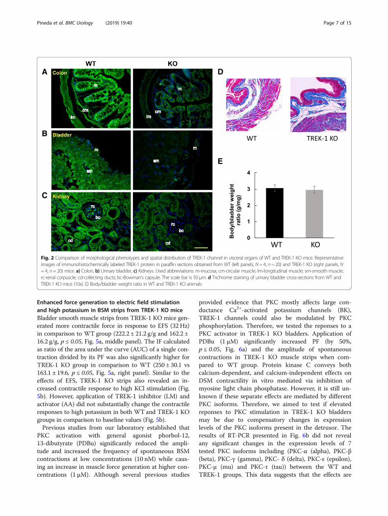
Expression of TREK-1 in the visceral organs of WT andTREK-1 KO miceImmunofluorescent tissue labeling was used to comparethe presence and distribution of TREK-1 proteins in thevisceral organs of WTand TREK-1 KO mice including theurinary bladder, colon, and kidneys. As shown in Fig. 2a(left panel), TREK-1 was expressed in the colon of WTan-imals with stronger labeling present in the colonic mucosain comparison to the muscle staining. Intense TREK-1 im-munoreactivity was also found in the muscularis mucosalayer. In contrast, TREK-1 KO animals showed a signifi-cantly reduced TREK-1 immunoreactivity across the co-lonic tissue (Fig. 2a, right panel). Confocal images of thebladder sections from WT group (Fig. 2b, left panel) dis-played a homogeneous TREK-1 like staining across the de-trusor with faint signal in the urothelium and serosa. Incontrast, TREK-1 KO animals showed a significant reduc-tion in TREK-1 immunofluorescence. Histologically, theurinary bladders showed no visible morphological alter-ations (Fig. 2d), and no significant differences were ob-served in bladder weight between WT and TREK-1 KOanimals (9.3 ± 0.7 vs. 10.2 ± 0.6mg, respectively). Thebody/bladder weight ratio (g/mg) in WT and TREK-1 KO
Pineda et al. BMC Urology (2019) 19:40 Page 5 of 15

mice was also similar between the groups (Fig. 2e). Thekidneys were bean-shaped, ~ 1.0 cm in length andweighted individually 0.50 ± 0.1 g and 0.46 ± 0.1 g (WTand TREK-1 KO groups, respectively). Histological imagestaken at the cortex level showed no visible histologicalchanges in TREK-1 KO mice compared with WT aminals.In WT mice, most of the tubules in the kidney nephronswere positively labeled with TREK-1 antibody (Fig. 2c, leftpanel) with very faint or no positive reaction at the glom-erular level. A substantial reduction in TREK-1 immuno-reactivity was observed in the kidneys from TREK-1 KOanimals (Fig. 2c, right panel).
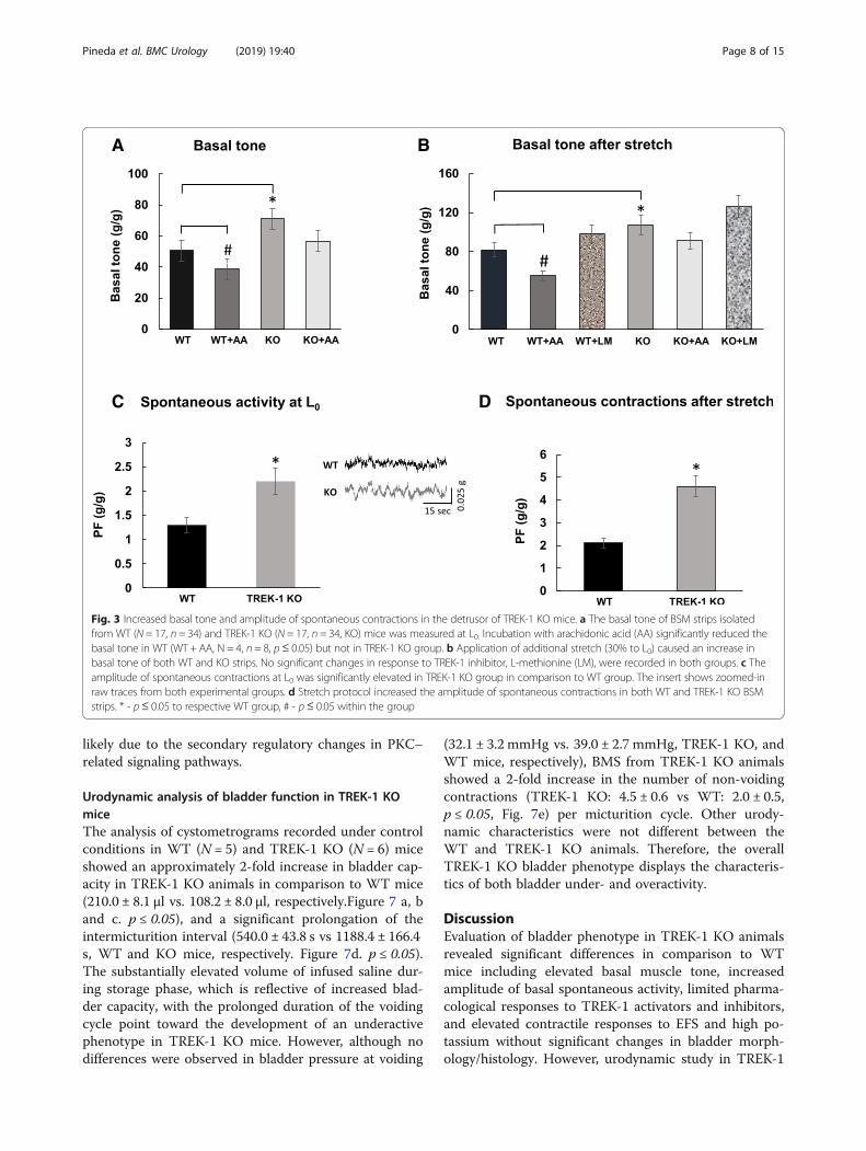
Deficiency of TREK-1 channel is associated with increasedbasal muscle tone and spontaneous contractions in thedetrusorWe next evaluated the role of TREK-1 channels in themaintenance of the basal smooth muscle tone and spon-taneous contractions in vitro. The weight of BSM stripsisolated from WTand TREK-1 KO mice and used for con-tractile function studies was similar between the groups(8.0 ± 1.5mg and 9.1 ± 2.1mg, respectively, N = 17 and n= 34 in each group). The basal muscle tone of the strips attheir optimal length (L0) was 50.7 ± 6.1 g/g in WT and
71.2 ± 8.5 g/g in TREK-1 KO mice (Fig. 3 a, N = 17; n = 34in each group, p ≤ 0.01). Application of AA caused 24% re-duction in basal tone of WT muscle strips (from 50.7 ±6.1 g/g to 38.5 ± 4.6 g/g, p = 0.025) whereas the strips fromTREK-1 KO bladders had a similar tendency in responseto AA, however, did not reach a level of statistical signifi-cance (Fig. 3a). In a different set of experiments, BSMstrips underwent 30% stretch in addition to L0 in order toevaluate stress-relaxation of the detrusor when the blad-der undergoes a significant stretch upon reaching its max-imal capacity. After the stretch protocol, BSM strips fromWT mice experienced an increase in the basal tone up to81.5 ± 7.3 g/g (58.3% to L0 level, p ≤ 0.05, Fig. 3b), whereasthe strips from TREK-1 KO mice had an increase of 50%(from 71.2 ± 8.5 g/g to 107.2 ± 9.6 g/g, p ≤ 0.05 to L0 level).Application of LM did not significantly affect the basalmuscle tone of the stretched strips in either group, how-ever, incubation with AA led to 32% decrease in WTgroup (N = 8, n = 8, p≤ 0.05 to WT) without causing sig-nificant changes in TREK-1 KO group (N = 8, n = 9, Fig. 3b). Analysis of spontaneous activity of BSM strips at L0 re-vealed the presence of spontaneous contractions inTREK-1 KO group (2.2 ± 0.2 g/g, Fig. 3c, p ≤ 0.05 to WT)in comparison to lower contractile activity in WT group(1.3 ± 0.1 g/g, Fig. 3 c, an insert shows representative rawtraces of the spontaneous activity in both groups). Add-itional stretch increased the amplitude of spontaneouscontractions by 61% in WT group (Fig. 3d, p ≤ 0.05 to re-spective L0 level) with the amplitude of spontaneous con-tractions in BSM strips being doubled in TREK-1 KOgroup (109% increase to respective L0, Fig. 3d).The differences observed in the basal muscle tone and
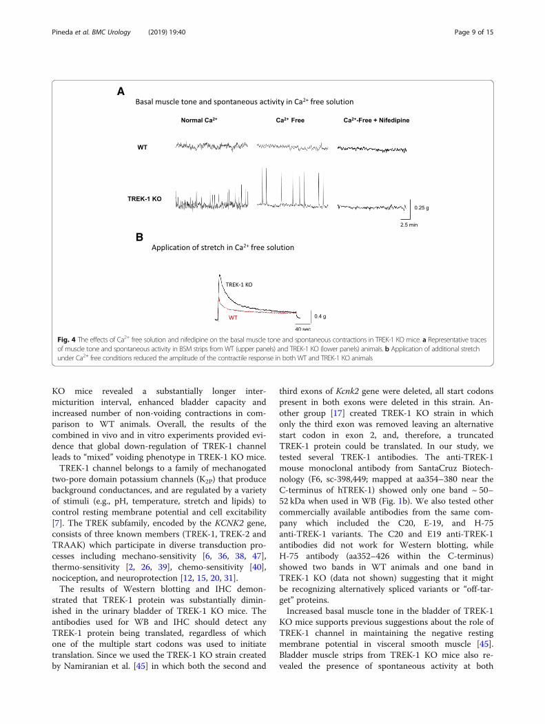
frequency of spontaneous activity between WT andTREK-1 KO BSM strips were further tested in Ca2+ freeTB to evaluate the role of active contractile processes dueto Ca2+ influx into the cells. First, we incubated BSM fromWT and TREK-1 KO animals in Ca-free solution for 30min followed by either 30% additional stretch or incuba-tion with nifedipine, a general L-type calcium channelblocker (2 μM, 30min). Representitave recordings fromWT and TREK-1 KO BSM strips are included in Fig. 4.Substitution of normal TB by Ca2+-free TB reduced thebasal muscle tone in both WT and TREK-1 KO BMSstrips when compared to normal Ca2+ solution (Fig. 4a,middle traces). However, BSM from TREK-1 KO miceshowed a ~ 2.0 fold increase in the amplitude of spontan-eous contractions (from 2.2 ± 0.1 g/g in normal Ca2+ to4.1 ± 0.1 g/g in Ca2+-free TB, N = 4,n = 8 in each group,p ≤ 0.05 to normal Ca TB). Application of additionalstretch under Ca2+ free conditions showed decreasedvalues of PF in both WT and TREK-1 KO groups by ~30–32% when compared to normal Ca2+ solution. Thecontractile reponses also had a significantly faster initialdecline phase of stress-relaxation in both groups (Fig. 4b).
A
B
Fig. 1 Genotyping of WT and TREK-1 KO mice. a Representative agarosegel showing the amplicons of wild-type (C57BL/6 J, WT, N= 1) and TREK-1 KO (KO, N= 3) alleles. The first column represents the molecular weightmarker (MW) with the following three lines corresponding to theamplicon products generated by KO mRNA. The fourth line illustrates theamplicon produced by WT mRNA for comparison. The size of the bandscorresponding to the TREK-1 KO (KO) and WT alleles are around ~ 200and ~ 450 bp, respectively, as previously reported [45]. b Westernblotting of total bladder protein from WT and TREK-1 KO animalsshowing a prominent band at ~ 50–52 kDa in WT mice but not inTREK-1 KO group (KO). Histone H3 was used as loading control
Pineda et al. BMC Urology (2019) 19:40 Page 6 of 15
Enhanced force generation to electric field stimulationand high potassium in BSM strips from TREK-1 KO miceBladder smooth muscle strips from TREK-1 KO mice gen-erated more contractile force in response to EFS (32 Hz)in comparison to WT group (222.2 ± 21.2 g/g and 162.2 ±16.2 g/g, p ≤ 0.05, Fig. 5a, middle panel). The IF calculatedas ratio of the area under the curve (AUC) of a single con-traction divided by its PF was also significantly higher forTREK-1 KO group in comparison to WT (250 ± 30.1 vs163.1 ± 19.6, p ≤ 0.05, Fig. 5a, right panel). Similar to theeffects of EFS, TREK-1 KO strips also revealed an in-creased contractile response to high KCl stimulation (Fig.5b). However, application of TREK-1 inhibitor (LM) andactivator (AA) did not substantially change the contractileresponses to high potassium in both WT and TREK-1 KOgroups in comparison to baseline values (Fig. 5b).Previous studies from our laboratory established that
PKC activation with general agonist phorbol-12,13-dibutyrate (PDBu) significantly reduced the ampli-tude and increased the frequency of spontaneous BSMcontractions at low concentrations (10 nM) while caus-ing an increase in muscle force generation at higher con-centrations (1 μM). Although several previous studies
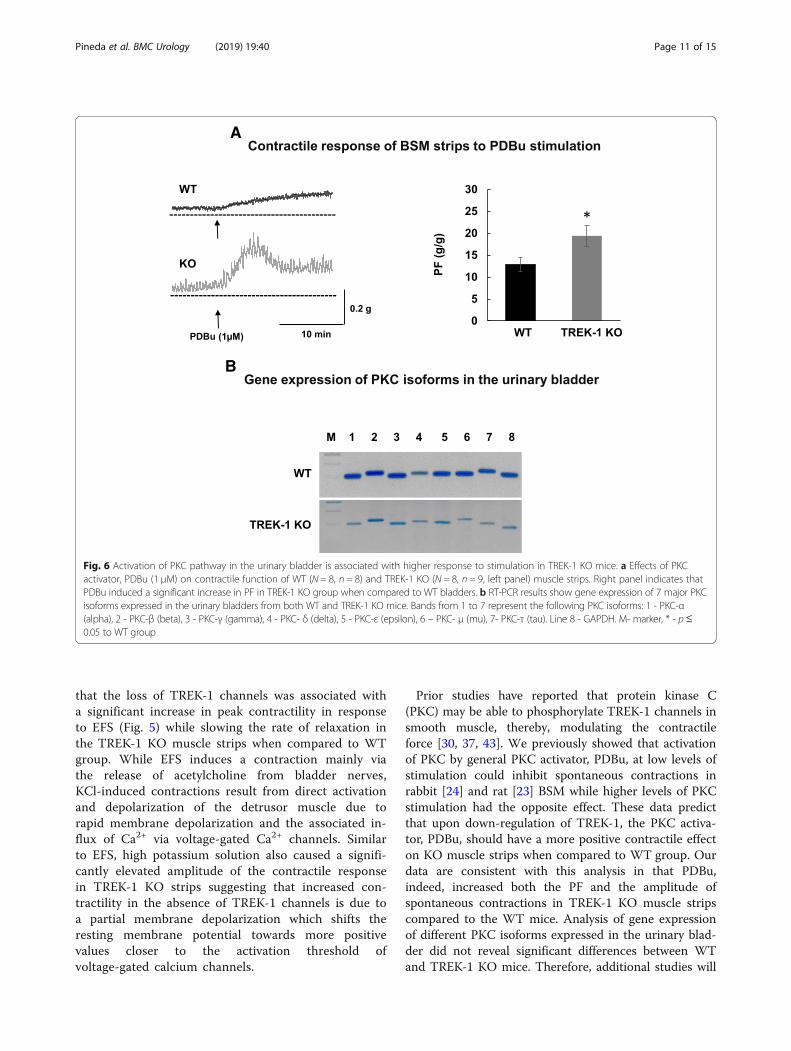
provided evidence that PKC mostly affects large con-ductance Ca2+-activated potassium channels (BK),TREK-1 channels could also be modulated by PKCphosphorylation. Therefore, we tested the reponses to aPKC activator in TREK-1 KO bladders. Application ofPDBu (1 μM) significantly increased PF (by 50%,p ≤ 0.05, Fig. 6a) and the amplitude of spontaneouscontractions in TREK-1 KO muscle strips when com-pared to WT group. Protein kinase C conveys bothcalcium-dependent, and calcium-independent effects onDSM contractility in vitro mediated via inhibition ofmyosine light chain phosphatase. However, it is still un-known if these separate effects are mediated by differentPKC isoforms. Therefore, we aimed to test if elevatedreponses to PKC stimulation in TREK-1 KO bladdersmay be due to compensatory changes in expressionlevels of the PKC isoforms present in the detrusor. Theresults of RT-PCR presented in Fig. 6b did not revealany significant changes in the expression levels of 7tested PKC isoforms including (PKC-α (alpha), PKC-β(beta), PKC-γ (gamma), PKC- δ (delta), PKC-ε (epsilon),PKC-μ (mu) and PKC-τ (tau)) between the WT andTREK-1 groups. This data suggests that the effects are
A D
E
B
C
Fig. 2 Comparison of morphological phenotypes and spatial distribution of TREK-1 channel in visceral organs of WT and TREK-1 KO mice. Representativeimages of immunohistochemically labeled TREK-1 protein in paraffin sections obtained from WT (left panels, N= 4, n= 20) and TREK-1 KO (right panels, N= 4, n= 20) mice. a) Colon, b) Urinary bladder, c) Kidneys. Used abbreviations: m-mucosa; cm-circular muscle; lm-longitudinal muscle; sm-smooth muscle;rc-renal corpuscle; cd-collecting ducts; bc-Bowman’s capsule. The scale bar is 50 μm. d Trichrome staining of urinary bladder cross-sections from WT andTREK-1 KO mice (10x). E) Body/bladder weight ratio in WT and TREK-1 KO animals
Pineda et al. BMC Urology (2019) 19:40 Page 7 of 15
likely due to the secondary regulatory changes in PKC–related signaling pathways.
Urodynamic analysis of bladder function in TREK-1 KOmiceThe analysis of cystometrograms recorded under controlconditions in WT (N = 5) and TREK-1 KO (N = 6) miceshowed an approximately 2-fold increase in bladder cap-acity in TREK-1 KO animals in comparison to WT mice(210.0 ± 8.1 μl vs. 108.2 ± 8.0 μl, respectively.Figure 7 a, band c. p ≤ 0.05), and a significant prolongation of theintermicturition interval (540.0 ± 43.8 s vs 1188.4 ± 166.4s, WT and KO mice, respectively. Figure 7d. p ≤ 0.05).The substantially elevated volume of infused saline dur-ing storage phase, which is reflective of increased blad-der capacity, with the prolonged duration of the voidingcycle point toward the development of an underactivephenotype in TREK-1 KO mice. However, although nodifferences were observed in bladder pressure at voiding
(32.1 ± 3.2 mmHg vs. 39.0 ± 2.7 mmHg, TREK-1 KO, andWT mice, respectively), BMS from TREK-1 KO animalsshowed a 2-fold increase in the number of non-voidingcontractions (TREK-1 KO: 4.5 ± 0.6 vs WT: 2.0 ± 0.5,p ≤ 0.05, Fig. 7e) per micturition cycle. Other urody-namic characteristics were not different between theWT and TREK-1 KO animals. Therefore, the overallTREK-1 KO bladder phenotype displays the characteris-tics of both bladder under- and overactivity.
DiscussionEvaluation of bladder phenotype in TREK-1 KO animalsrevealed significant differences in comparison to WTmice including elevated basal muscle tone, increasedamplitude of basal spontaneous activity, limited pharma-cological responses to TREK-1 activators and inhibitors,and elevated contractile responses to EFS and high po-tassium without significant changes in bladder morph-ology/histology. However, urodynamic study in TREK-1
A
C D
B
Fig. 3 Increased basal tone and amplitude of spontaneous contractions in the detrusor of TREK-1 KO mice. a The basal tone of BSM strips isolatedfrom WT (N = 17, n = 34) and TREK-1 KO (N = 17, n = 34, KO) mice was measured at L0. Incubation with arachidonic acid (AA) significantly reduced thebasal tone in WT (WT + AA, N = 4, n = 8, p≤ 0.05) but not in TREK-1 KO group. b Application of additional stretch (30% to L0) caused an increase inbasal tone of both WT and KO strips. No significant changes in response to TREK-1 inhibitor, L-methionine (LM), were recorded in both groups. c Theamplitude of spontaneous contractions at L0 was significantly elevated in TREK-1 KO group in comparison to WT group. The insert shows zoomed-inraw traces from both experimental groups. d Stretch protocol increased the amplitude of spontaneous contractions in both WT and TREK-1 KO BSMstrips. * - p≤ 0.05 to respective WT group, # - p≤ 0.05 within the group
Pineda et al. BMC Urology (2019) 19:40 Page 8 of 15
KO mice revealed a substantially longer inter-micturition interval, enhanced bladder capacity andincreased number of non-voiding contractions in com-parison to WT animals. Overall, the results of thecombined in vivo and in vitro experiments provided evi-dence that global down-regulation of TREK-1 channelleads to “mixed” voiding phenotype in TREK-1 KO mice.TREK-1 channel belongs to a family of mechanogated
two-pore domain potassium channels (K2P) that producebackground conductances, and are regulated by a varietyof stimuli (e.g., pH, temperature, stretch and lipids) tocontrol resting membrane potential and cell excitability[7]. The TREK subfamily, encoded by the KCNK2 gene,consists of three known members (TREK-1, TREK-2 andTRAAK) which participate in diverse transduction pro-cesses including mechano-sensitivity [6, 36, 38, 47],thermo-sensitivity [2, 26, 39], chemo-sensitivity [40],nociception, and neuroprotection [12, 15, 20, 31].The results of Western blotting and IHC demon-
strated that TREK-1 protein was substantially dimin-ished in the urinary bladder of TREK-1 KO mice. Theantibodies used for WB and IHC should detect anyTREK-1 protein being translated, regardless of whichone of the multiple start codons was used to initiatetranslation. Since we used the TREK-1 KO strain createdby Namiranian et al. [45] in which both the second and
third exons of Kcnk2 gene were deleted, all start codonspresent in both exons were deleted in this strain. An-other group [17] created TREK-1 KO strain in whichonly the third exon was removed leaving an alternativestart codon in exon 2, and, therefore, a truncatedTREK-1 protein could be translated. In our study, wetested several TREK-1 antibodies. The anti-TREK-1mouse monoclonal antibody from SantaCruz Biotech-nology (F6, sc-398,449; mapped at aa354–380 near theC-terminus of hTREK-1) showed only one band ~ 50–52 kDa when used in WB (Fig. 1b). We also tested othercommercially available antibodies from the same com-pany which included the C20, E-19, and H-75anti-TREK-1 variants. The C20 and E19 anti-TREK-1antibodies did not work for Western blotting, whileH-75 antibody (aa352–426 within the C-terminus)showed two bands in WT animals and one band inTREK-1 KO (data not shown) suggesting that it mightbe recognizing alternatively spliced variants or “off-tar-get” proteins.Increased basal muscle tone in the bladder of TREK-1
KO mice supports previous suggestions about the role ofTREK-1 channel in maintaining the negative restingmembrane potential in visceral smooth muscle [45].Bladder muscle strips from TREK-1 KO mice also re-vealed the presence of spontaneous activity at both
A
B
Fig. 4 The effects of Ca2+ free solution and nifedipine on the basal muscle tone and spontaneous contractions in TREK-1 KO mice. a Representative tracesof muscle tone and spontaneous activity in BSM strips from WT (upper panels) and TREK-1 KO (lower panels) animals. b Application of additional stretchunder Ca2+ free conditions reduced the amplitude of the contractile response in both WT and TREK-1 KO animals
Pineda et al. BMC Urology (2019) 19:40 Page 9 of 15

resting level and after additional stretch. Spontaneouscontractions have been previously recorded in a varietyof bladder smooth muscle types including human [21,58], rabbit [11, 24], rat [23], guinea pig [56], pig [25, 56],and mouse [28, 54]. It is possible that knockdown ofTREK-1 may affect calcium mobilization and modify thethreshold for smooth muscle excitability and/or con-tractility, thereby, increasing spontaneous activity, as ob-served in our study. To test this hypothesis, weincubated BMS strips in Ca2+-free TB followed by theapplication of nifedipine, a L-type calcium channelblocker (Fig. 4). Incubation of TREK-1 KO strips in Ca2+ − free solution effectively reduced the basal tone to thelevels similar to those observed in WT BMS strips.Interestingly, the reduced muscle tone observed in Ca2+-free solution was associated with a 2-fold increase inthe amplitude of spontanous contractions in TREK-1
KO group. The total elimination of spontaneous con-tractions and further reduction in basal tone by nifedi-pine suggests that TREK-1 channels participate inregulation of spontaneous contractions by stabilizing themembrane potential at voltages closer to the potassiumresting potential. Overall, in addition to previously pub-lished data on human [3, 34] and animal [22, 42] visceralsmooth muscle, our data provide additional evidencethat TREK-1 channel plays a critical role in maintainingbladder basal tone, bladder compliance and modulatesthe response of the detrusor to stretch.The contractile responses of BSM to EFS are mainly
mediated by the release of neurotransmitters fromintramural nerve terminals in the bladder wall causingdetrusor contraction. We compared the responses ofTREK-1 KO and WT muscle strips in their ability togenerate force in response to EFS. The data showed
A
B
Fig. 5 Effect of electric field stimulation and high potassium on WT and TREK-1 KO bladder detrusor strip contractile function. a Isolated BSM stripsfrom TREK-1 KO mice showed significantly higher contractile force in response to EFS (80 V, 32 Hz, 1ms duration). The left panel shows overlappedexamples of raw traces from both groups. Middle panel represents the summary of PF normalized to the weight of tissue strips whereas right panelshows the integral force for both groups. b Effect of KCl stimulation on PF in WT (N = 17, n = 34) and TREK-1 KO (N = 17, n = 34) groups before andafter pharmacological activation (by AA) and inhibition (by ML) of TREK-1 channels. Left panel represents raw traces of recordings. * - p≤ 0.05 toWT group
Pineda et al. BMC Urology (2019) 19:40 Page 10 of 15
that the loss of TREK-1 channels was associated witha significant increase in peak contractility in responseto EFS (Fig. 5) while slowing the rate of relaxation inthe TREK-1 KO muscle strips when compared to WTgroup. While EFS induces a contraction mainly viathe release of acetylcholine from bladder nerves,KCl-induced contractions result from direct activationand depolarization of the detrusor muscle due torapid membrane depolarization and the associated in-flux of Ca2+ via voltage-gated Ca2+ channels. Similarto EFS, high potassium solution also caused a signifi-cantly elevated amplitude of the contractile responsein TREK-1 KO strips suggesting that increased con-tractility in the absence of TREK-1 channels is due toa partial membrane depolarization which shifts theresting membrane potential towards more positivevalues closer to the activation threshold ofvoltage-gated calcium channels.
Prior studies have reported that protein kinase C(PKC) may be able to phosphorylate TREK-1 channels insmooth muscle, thereby, modulating the contractileforce [30, 37, 43]. We previously showed that activationof PKC by general PKC activator, PDBu, at low levels ofstimulation could inhibit spontaneous contractions inrabbit [24] and rat [23] BSM while higher levels of PKCstimulation had the opposite effect. These data predictthat upon down-regulation of TREK-1, the PKC activa-tor, PDBu, should have a more positive contractile effecton KO muscle strips when compared to WT group. Ourdata are consistent with this analysis in that PDBu,indeed, increased both the PF and the amplitude ofspontaneous contractions in TREK-1 KO muscle stripscompared to the WT mice. Analysis of gene expressionof different PKC isoforms expressed in the urinary blad-der did not reveal significant differences between WTand TREK-1 KO mice. Therefore, additional studies will
A
B
Fig. 6 Activation of PKC pathway in the urinary bladder is associated with higher response to stimulation in TREK-1 KO mice. a Effects of PKCactivator, PDBu (1 μM) on contractile function of WT (N = 8, n = 8) and TREK-1 KO (N = 8, n = 9, left panel) muscle strips. Right panel indicates thatPDBu induced a significant increase in PF in TREK-1 KO group when compared to WT bladders. b RT-PCR results show gene expression of 7 major PKCisoforms expressed in the urinary bladders from both WT and TREK-1 KO mice. Bands from 1 to 7 represent the following PKC isoforms: 1 - PKC-α(alpha), 2 - PKC-β (beta), 3 - PKC-γ (gamma), 4 - PKC- δ (delta), 5 - PKC-ε (epsilon), 6 – PKC- μ (mu), 7- PKC-τ (tau). Line 8 - GAPDH. M- marker, * - p≤0.05 to WT group
Pineda et al. BMC Urology (2019) 19:40 Page 11 of 15

be required to evaluate if the elevated response to PKCactivation in TREK-1 KO bladder is due to the changesin PKC phosphorylation or activation of different down-stream signaling pathways.In contrast to in vitro contractility results, the uro-
dynamic evaluation of voiding function in vivo byawake cystometry revealed a significant increase inthe duration of inter-micturition interval and en-hanced bladder capacity, both of which usually definean underactive bladder phenotype. Despite thesechanges, the intravesical pressure during micturitionwas not affected by the TREK-1 knockdown, and thenumber of non-voiding contractions was almost2-fold higher in these animals which is in line with
the increased contractility of the detrusor observedduring the in vitro studies discussed above. Taken to-gether, these results suggest that knock-down ofKcnk2 gene has dual or “mixed” effects on detrusorcontractility and micturition patterns. One of the pos-sible explanations could be associated with expressionof the channel on vascular and visceral smoothmuscle cells as well as on the fibers and neurons ofthe peripheral and central nervous system [19, 41].The functional role of TREK-1 in smooth muscle cellsis mainly to contribute to the “leak” current respon-sible for the maintenance of hyperpolarized restingmembrane potential to keep the cells more relaxedduring resting state [5, 10].
A
B
C
D
E
Fig. 7 Urodynamic evaluation of bladder function in WT and TREK-1 KO mice. a Representative cystometrogram traces recorded in freely movingWT (upper panels) and TREK-1 KO (bottom panels) mice. The volume of the infused saline and bladder pressure are included in thetraces. b Analysis of bladder capacity (BC) in WT and TREK-1 KO groups. c Comparison of the total volume infused before micturition inWT and TREK-KO mice. d Duration of inter-micturition interval in WT and genetically modified mice. e The average number of non-voiding contractions (per micturition cycle) recorded in WT and TREK-1 KO mice. Zoomed-in segments of the traces indicated by squaresare shown in the inserts. * - significance level of p ≤ 0.01 to WT group
Pineda et al. BMC Urology (2019) 19:40 Page 12 of 15
In the nervous system, TREK-1 was shown to controlelectrogenesis, differentiation, axonal migration, synapto-genesis, and neural response to temperature and mechan-ical stretch [1, 9, 13, 16, 19, 27, 31, 51]. In comparison tosmooth muscle cells, where action potentials are driven bythe influx of Ca2+ via L-type Ca2+ channels, excitability ofneurons is regulated mainly by voltage-gated Na+ channels.The lack of TREK-1 channel in neurons would shift theresting membrane potential towards more positive values(just like in smooth muscle cells) allowing for the rapid in-flux of Na+ inside the cell, and, therefore, making both af-ferent and efferent neurons more excitable. The final resultof this neuronal activity would depend on what neurons aremore affected by these changes. An increase in afferent ac-tivity from the urinary bladder to the brain is usually associ-ated with an increase in urgency and frequency ofmicturition associated with bladder overactivity. However,the efferent output from the brain to the bladder is mainlyinhibitory to allow for the voluntary control of micturition.Therefore, increased excitability of efferent neurons wouldlead to increased inter-micturition interval and bladder un-deractivity, which we observed in our study in vivo. Futurestudies focused on manipulations of either afferent or effer-ent neural pathways are warranted to test this hypothesis.Another possible explanation for the differences be-
tween in vitro and in vivo bladder phenotypes could be as-sociated with some unknown alterations occurring duringgenetic modifications of the parental strain. TREK-1 wasdetermined to mediate changes in the actin cytoskeletonindependently of its channel activity in fetal neurons dur-ing development in a different strain of TREK-1 KO mice[32]. The same mouse strain showed an increased efficacyof 5-HT neurotransmission and resistance to depression[20]. Previous studies from our [35] and other [57] groupsprovided evidence that lower urinary tract function inmice varies in a consistent manner with strain and/or sex.For instance, voiding spot assay and cystometry performedin male and female C57BL/6 J, 129S1/SvImJ, NOD/ShiLtJ,and CAST/EiJ mice [8] to evaluate bladder function,established a significantly prolonged duration of the mic-turition cycle in genetically modified animals (129S1/SvImJ, NOD/ShiLtJ, and CAST/EiJ) in comparison toC57BL/6 J strain [8]. These results are in line with ourcystometric recordings from TREK-1 KO mice. The differ-ences between our study and the published results in-cluded the use of both sexes (males and females), the useof WT controls which were not littermates, and perform-ance of cystometry without anesthesia in our experiments,as well as the absence of in vitro experiments in the previ-ously published study.The choice of the approach to create a genetically
modified strain also seems to affect the function of dif-ferent organs and tissues in genetically modified animals.For instance, the TREK-1 KO strain we adopted and
used in our study, provided no evidence that TREK-1 isinvolved in the regulation of arterial diameter in cerebralarteries [45]. However, another TREK-1 KO strain [32]did present with the vascular phenotype. There is also apossibility that genetic knockdown of TREK-1 expres-sion could trigger compensatory changes in othermechano-gated ion channels or related signaling cas-cades, thereby, affecting the final voiding phenotype aswell as the response of the detrusor to stretch. Furtherstudies are warranted to address these questions.
ConclusionsOur study provided evidence that global down-regulationof TREK-1 channels has dual effects on detrusor contract-ility and micturition patterns in vivo. The integrative ef-fects of TREK-1, likely, depend on the expression andfunction of the channel not only in detrusor myocytes butalso in afferent and efferent neural pathways regulatingmicturition. Future studies are warranted to identify theprecise mechanisms of TREK-1 associated mechanotrans-duction between detrusor myocytes and afferent nerves inthe bladder wall, as well as the role of TREK-1 in efferentfibers and central nervous system centers controlling void-ing. This knowledge would provide a foundation for thedevelopment of novel therapeutic approaches to treatvoiding dysfunction in patients with detrusor overactivity,overactive bladder, and bladder pain syndrome.
AbbreviationsAA: Arachidonic acid; AUC: Area under the curve; BK: Ca2+-activatedpotassium channels; BSM: Bladder smooth muscle; DO: Detrusor overactivity;EFS: Electric field stimulation; IF: Integral force; IHC: Immunohistochemistry;KO: Knockout; L0 : The length of optimal force development; LM: L-methionine; PBS: Phosphate-buffered saline; PDBu: Phorbol 12,13-Dibutyrate;PF: Peak force; PFA: Paraformaldehyde; PKC: Protein kinase C; RT: Roomtemperature; TB: Tyrode Buffer; TBS: Tris-Buffered saline; TREK-1 channel: Two-pore domain potassium channel encoded by Kcnk2 gene; TTX: Tetrodotoxin;WB: Western blotting; WT: Wild type
AcknowledgementsWe thank Dr. Min Zhou and his lab members for providing the TREK-1 KOmice for our study, as well as Dr. John A Thompson from the Department ofNeurosurgery, University of Colorado Denver for help with data analyses. Im-aging experiments were performed in the University of Colorado AnschutzMedical Campus Advance Light Microscopy Core supported in part by RockyMountain Neurological Disorders Core (P30 NS048154) and by NIH/NCATSColorado CTSI Grant (UL1 TR001082).
FundingThe research was supported by the NIH/NIDDK grant R01 DK095817 (A.P.M.).
Availability of data and materialsThe datasets used and/or analysed during the current study available fromthe corresponding author on request.
Authors` contributionsR.H.P. and A.P.M. conception and design of the research; R.H.P., J.H., S.L., A.C.,and N.I. performed the experiments; R.H.P., J.H., S.L. and N.I. analyzed thedata; R.H.P., R.B.M., J.H., S.L., N.I. and A.P.M interpreted the results of theexperiments; R.H.P., J.H., S.L., and A.P.M prepared the figures; R.H.P. and A.P.Mdrafted the manuscript; R.H.P., R.B.M. and A.P.M edited and revised themanuscript; R.H.P., J.H., S.L., A.C., N.I., R.B.M., and A.P.M. approved the final
Pineda et al. BMC Urology (2019) 19:40 Page 13 of 15

version of the manuscript; all authors are accountable for all aspects of thework.
Ethics approval and consent to participateNot applicable.
Consent for publicationThe preliminary results of this work were presented at the AUA 2018meeting, and the abstract was published in the Journal of Urology (https://doi.org/10.1016/j.juro.2018.02.1236) [59]. The written permission wasobtained from the publisher to reuse the content of the abstract.
Competing interestsThe authors declare that they have no competing interests.
Publisher’s NoteSpringer Nature remains neutral with regard to jurisdictional claims inpublished maps and institutional affiliations.
Author details1Division of Urology, Department of Surgery, University of ColoradoDenver,Anschutz Medical Campus, 12700 E 19th Ave, M/S C317, Aurora, CO80045, USA. 2Department of Urology, University of California San Diego, 3855Health Science Drive, Room 4345, Bay 4LL, La Jolla, CA 92093, USA.3Children’s Mercy Hospital, 2401 Gillham Rd, Kansas City, MO 64108, USA.4Division of Urology, Department of Surgery, University of Colorado Denver,Academic Office One Bldg., Rm 5602, 12631 East 17th Ave., M/S C319,Aurora, CO 80045, USA.
Received: 23 April 2018 Accepted: 13 May 2019
References1. Afzali AM, Ruck T, Herrmann AM, Iking J, Sommer C, Kleinschnitz C,
Preubetae C, Stenzel W, Budde T, Wiendl H, Bittner S, Meuth SG. Thepotassium channels TASK2 and TREK1 regulate functional differentiation ofmurine skeletal muscle cells. Am J Physiol Cell Physiol. 2016;311:C583–95.
2. Alloui A, Zimmermann K, Mamet J, Duprat F, Noel J, Chemin J, Guy N,Blondeau N, Voilley N, Rubat-Coudert C, Borsotto M, Romey G, Heurteaux C,Reeh P, Eschalier A, Lazdunski M. TREK-1, a K+ channel involved inpolymodal pain perception. EMBO J. 2006;25:2368–76.
3. Bai X, Bugg GJ, Greenwood SL, Glazier JD, Sibley CP, Baker PN, Taggart MJ,Fyfe GK. Expression of TASK and TREK, two-pore domain K+ channels, inhuman myometrium. Reproduction. 2005;129:525–30.
4. Baker SA, Hatton WJ, Han J, Hennig GW, Britton FC, Koh SD. Role of TREK-1potassium channel in bladder overactivity after partial bladder outletobstruction in mouse. J Urol. 2010;183:793–800.
5. Baker SA, Hennig GW, Han J, Britton FC, Smith TK, Koh SD. Methionine andits derivatives increase bladder excitability by inhibiting stretch-dependentK(+) channels. Br J Pharmacol. 2008;153:1259–71.
6. Bang H, Kim Y, Kim D. TREK-2, a new member of the mechanosensitivetandem-pore K+ channel family. J Biol Chem. 2000;275:17412–9.
7. Bayliss DA, Barrett PQ. Emerging roles for two-pore-domain potassiumchannels and their potential therapeutic impact. Trends Pharmacol Sci.2008;29:566–75.
8. Bjorling DE, Wang Z, Vezina CM, Ricke WA, Keil KP, Yu W, Guo L, Zeidel ML,Hill WG. Evaluation of voiding assays in mice: impact of genetic strains andsex. Am J Physiol Renal Physiol. 2015;308:F1369–78.
9. Bockenhauer D, Zilberberg N, Goldstein SA. KCNK2: reversible conversion ofa hippocampal potassium leak into a voltage-dependent channel. NatNeurosci. 2001;4:486–91.
10. Buxton IL, Singer CA, Tichenor JN. Expression of stretch-activated two-porepotassium channels in human myometrium in pregnancy and labor. PLoSOne. 2010;5:e12372.
11. Chang S, Hypolite JA, Mohanan S, Zderic SA, Wein AJ, Chacko S. Alterationof the PKC-mediated signaling pathway for smooth muscle contraction inobstruction-induced hypertrophy of the urinary bladder. Lab Investig. 2009;89:823–32.
12. Chemin J, Patel AJ, Duprat F, Lauritzen I, Lazdunski M, Honore E. Aphospholipid sensor controls mechanogating of the K+ channel TREK-1.EMBO J. 2005;24:44–53.
13. Devader C, Khayachi A, Veyssiere J, Moha Ou Maati H, Roulot M, Moreno S,Borsotto M, Martin S, Heurteaux C, Mazella J. In vitro and in vivo regulationof synaptogenesis by the novel antidepressant spadin. Br J Pharmacol. 2015;172:2604–17.
14. Du Y, Kiyoshi CM, Wang Q, Wang W, Ma B, Alford CC, Zhong S, Wan Q,Chen H, Lloyd EE, Bryan RM Jr, Zhou M. Genetic deletion of TREK-1 or TWIK-1/TREK-1 potassium channels does not Alter the basic electrophysiologicalproperties of mature hippocampal astrocytes in situ. Front Cell Neurosci.2016;10(13).
15. Duprat F, Lesage F, Patel AJ, Fink M, Romey G, Lazdunski M. Theneuroprotective agent riluzole activates the two P domain K(+) channelsTREK-1 and TRAAK. Mol Pharmacol. 2000;57:906–12.
16. Fink M, Duprat F, Lesage F, Reyes R, Romey G, Heurteaux C, Lazdunski M.Cloning, functional expression and brain localization of a novelunconventional outward rectifier K+ channel. EMBO J. 1996;15:6854–62.
17. Guyon A, Tardy MP, Rovere C, Nahon JL, Barhanin J, Lesage F. Glucoseinhibition persists in hypothalamic neurons lacking tandem-pore K+channels. J Neurosci. 2009;29:2528–33.
18. Hervieu GJ, Cluderay JE, Gray CW, Green PJ, Ranson JL, Randall AD,Meadows HJ. Distribution and expression of TREK-1, a two-pore-domainpotassium channel, in the adult rat CNS. Neuroscience. 2001;103:899–919.
19. Heurteaux C, Guy N, Laigle C, Blondeau N, Duprat F, Mazzuca M, Lang-Lazdunski L, Widmann C, Zanzouri M, Romey G, Lazdunski M. TREK-1, a K+channel involved in neuroprotection and general anesthesia. EMBO J. 2004;23:2684–95.
20. Heurteaux C, Lucas G, Guy N, El Yacoubi M, Thummler S, Peng XD, Noble F,Blondeau N, Widmann C, Borsotto M, Gobbi G, Vaugeois JM, Debonnel G,Lazdunski M. Deletion of the background potassium channel TREK-1 resultsin a depression-resistant phenotype. Nat Neurosci. 2006;9:1134–41.
21. Hristov KL, Afeli SA, Parajuli SP, Cheng Q, Rovner ES, Petkov GV. Neurogenicdetrusor overactivity is associated with decreased expression and functionof the large conductance voltage- and ca(2+)-activated K(+) channels. PLoSOne. 2013;8:e68052.
22. Hwang SJ, O'Kane N, Singer C, Ward SM, Sanders KM, Koh SD. Block ofinhibitory junction potentials and TREK-1 channels in murine colon by Ca2+store-active drugs. J Physiol. 2008;586:1169–84.
23. Hypolite JA, Chang S, Wein AJ, Chacko S, Malykhina AP. Protein kinase Cmodulates frequency of micturition and non-voiding contractions in theurinary bladder via neuronal and myogenic mechanisms. BMC Urol. 2015;15(34).
24. Hypolite JA, Lei Q, Chang S, Zderic SA, Butler S, Wein AJ, Malykhina AP,Chacko S. Spontaneous and evoked contractions are regulated by PKC-mediated signaling in detrusor smooth muscle: involvement of BK channels.Am J Physiol Renal Physiol. 2013;304:F451–62.
25. Isogai A, Lee K, Mitsui R, Hashitani H. Functional coupling of TRPV4 channelsand BK channels in regulating spontaneous contractions of the Guinea pigurinary bladder. Pflugers Arch. 2016;468:1573–85.
26. Kang D, Choe C, Kim D. Thermosensitivity of the two-pore domain K+channels TREK-2 and TRAAK. J Physiol. 2005;564:103–16.
27. Kanjhan R, Anselme AM, Noakes PG, Bellingham MC. Postnatal changes inTASK-1 and TREK-1 expression in rat brain stem and cerebellum.Neuroreport. 2004;15:1321–4.
28. Kobayter S, Young JS, Brain KL. Prostaglandin E2 induces spontaneousrhythmic activity in mouse urinary bladder independently of efferent nerves.Br J Pharmacol. 2012;165:401–13.
29. Koh SD, Sanders KM. Stretch-dependent potassium channels in murinecolonic smooth muscle cells. J Physiol. 2001;533:155–63.
30. Kreneisz O, Benoit JP, Bayliss DA, Mulkey DK. AMP-activated protein kinaseinhibits TREK channels. J Physiol. 2009;587:5819–30.
31. Lauritzen I, Blondeau N, Heurteaux C, Widmann C, Romey G, Lazdunski M.Polyunsaturated fatty acids are potent neuroprotectors. EMBO J. 2000;19:1784–93.
32. Lauritzen I, Chemin J, Honore E, Jodar M, Guy N, Lazdunski M, Jane Patel A.Cross-talk between the mechano-gated K2P channel TREK-1 and the actincytoskeleton. EMBO Rep. 2005;6:642–8.
33. Lee H, Koh BH, Peri LE, Sanders KM, Koh SD. Purinergic inhibitory regulationof murine detrusor muscles mediated by PDGFRalpha+ interstitial cells. JPhysiol. 2014;592:1283–93.
34. Lei Q, Pan XQ, Chang S, Malkowicz SB, Guzzo TJ, Malykhina AP. Response ofthe human detrusor to stretch is regulated by TREK-1, a two-pore-domain(K2P) mechano-gated potassium channel. J Physiol. 2014;592:3013–30.
Pineda et al. BMC Urology (2019) 19:40 Page 14 of 15
35. Lei Q, Pan XQ, Villamor AN, Asfaw TS, Chang S, Zderic SA, Malykhina AP.Lack of transient receptor potential vanilloid 1 channel modulates thedevelopment of neurogenic bladder dysfunction induced by cross-sensitization in afferent pathways. J Neuroinflammation. 2013;10(3).
36. Lesage F, Terrenoire C, Romey G, Lazdunski M. Human TREK2, a 2P domainmechano-sensitive K+ channel with multiple regulations by polyunsaturatedfatty acids, lysophospholipids, and Gs, Gi, and Gq protein-coupled receptors.J Biol Chem. 2000;275:28398–405.
37. Liu H, Enyeart JA, Enyeart JJ. Angiotensin II inhibits native bTREK-1 K+channels through a PLC-, kinase C-, and PIP2-independent pathwayrequiring ATP hydrolysis. Am J Physiol Cell Physiol. 2007;293:C682–95.
38. Maingret F, Honore E, Lazdunski M, Patel AJ. Molecular basis of the voltage-dependent gating of TREK-1, a mechano-sensitive K(+) channel. BiochemBiophys Res Commun. 2002;292:339–46.
39. Maingret F, Lauritzen I, Patel AJ, Heurteaux C, Reyes R, Lesage F, LazdunskiM, Honore E. TREK-1 is a heat-activated background K(+) channel. EMBO J.2000;19:2483–91.
40. Maingret F, Patel AJ, Lesage F, Lazdunski M, Honore E. Mechano- or acidstimulation, two interactive modes of activation of the TREK-1 potassiumchannel. J Biol Chem. 1999;274:26691–6.
41. Medhurst AD, Rennie G, Chapman CG, Meadows H, Duckworth MD, KelsellRE, Gloger II, Pangalos MN. Distribution analysis of human two pore domainpotassium channels in tissues of the central nervous system and periphery.Brain Res Mol Brain Res. 2001;86:101–14.
42. Monaghan K, Baker SA, Dwyer L, Hatton WC, Sik Park K, Sanders KM, KohSD. The stretch-dependent potassium channel TREK-1 and its function inmurine myometrium. J Physiol. 2011;589:1221–33.
43. Murbartian J, Lei Q, Sando JJ, Bayliss DA. Sequential phosphorylationmediates receptor- and kinase-induced inhibition of TREK-1 backgroundpotassium channels. J Biol Chem. 2005;280:30175–84.
44. Namiranian K, Brink CD, Goodman JC, Robertson CS, and Bryan RM, Jr.Traumatic brain injury in mice lacking the K channel, TREK-1. J Cereb BloodFlow Metab 31: e1–e6, 2011.
45. Namiranian K, Lloyd EE, Crossland RF, Marrelli SP, Taffet GE, Reddy AK,Hartley CJ, and Bryan RM, Jr. Cerebrovascular responses in mice deficient inthe potassium channel, TREK-1. Am J Physiol Regul Integr Comp Physiol299: R461–R469, 2010.
46. Patel AJ, Honore E. Properties and modulation of mammalian 2P domain K+ channels. Trends Neurosci. 2001;24:339–46.
47. Patel AJ, Honore E, Maingret F, Lesage F, Fink M, Duprat F, Lazdunski M. Amammalian two pore domain mechano-gated S-like K+ channel. EMBO J.1998;17:4283–90.
48. Pineda RH, Nedumaran B, Hypolite J, Pan XQ, Wilson S, Meacham RB,Malykhina AP. Altered expression and modulation of the two-pore-domain(K2P) mechanogated potassium channel TREK-1 in overactive humandetrusor. Am J Physiol Renal Physiol. 2017;313:F535–46.
49. Sanders KM, Koh SD. Two-pore-domain potassium channels in smoothmuscles: new components of myogenic regulation. J Physiol. 2006;570:37–43.
50. Schindelin J, Arganda-Carreras I, Frise E, Kaynig V, Longair M, Pietzsch T,Preibisch S, Rueden C, Saalfeld S, Schmid B, Tinevez JY, White DJ,Hartenstein V, Eliceiri K, Tomancak P, Cardona A. Fiji: an open-sourceplatform for biological-image analysis. Nat Methods. 2012;9:676–82.
51. Thomas D, Plant LD, Wilkens CM, McCrossan ZA, Goldstein SA. Alternativetranslation initiation in rat brain yields K2P2.1 potassium channelspermeable to sodium. Neuron. 2008;58:859–70.
52. Tichenor JN, Hansen ET, Buxton IL. Expression of stretch-activated potassiumchannels in human myometrium. Proc West Pharmacol Soc. 2005;48:44–8.
53. Wellner MC, Isenberg G. Stretch effects on whole-cell currents of Guinea-pigurinary bladder myocytes. J Physiol. 1994;480 ( Pt 3:439–48.
54. White RS, Zemen BG, Khan Z, Montgomery JR, Herrera GM, Meredith AL.Evaluation of mouse urinary bladder smooth muscle for diurnal differencesin contractile properties. Front Pharmacol. 2014;5:293.
55. Wu YY, Singer CA, Buxton IL. Variants of stretch-activated two-porepotassium channel TREK-1 associated with preterm labor in humans. BiolReprod. 2012;87:96.
56. Xin W, Li N, Fernandes VS, Petkov GV. Constitutively active PKA regulatesneuronal acetylcholine release and contractility of Guinea pig urinarybladder smooth muscle. Am J Physiol Renal Physiol. 2016;310:F1377–84.
57. Yu W, Ackert-Bicknell C, Larigakis JD, MacIver B, Steers WD, Churchill GA, HillWG, Zeidel ML. Spontaneous voiding by mice reveals strain-specific lower
urinary tract function to be a quantitative genetic trait. Am J Physiol RenalPhysiol. 2014;306:F1296–307.
58. Zagorodnyuk VP, Gregory S, Costa M, Brookes SJ, Tramontana M, Giuliani S,Maggi CA. Spontaneous release of acetylcholine from autonomic nerves inthe bladder. Br J Pharmacol. 2009;157:607–19.
59. Pineda RH, Hypolite J, Lee S, Carrasco A, Iguchi N, Meacham RB, MalykhinaAP. The lack of mechanosensitive K2P channel is associated with mixedvoiding phenotype in mice. J Urology. 199(4S):e503–4.
Pineda et al. BMC Urology (2019) 19:40 Page 15 of 15