Inactivation by omeprazole of the carnitine transporter (OCTN2) reconstituted in liposomes
Upload
independentCategory
view
3download
0
LCMP-00247-2007-FINAL ACCEPTED
1
Altered Carnitine Homeostasis is Associated with Decreased Mitochondrial Function
and Altered Nitric Oxide Signaling in Lambs with Pulmonary Hypertension
Shruti Sharma1,
Neetu Sud1,
Dean A. Wiseman1,
A. Lee Carter2,
Sanjiv Kumar1,
Yali Hou1,
Thomas Rau 3,
Jason Wilham3,
Cynthia Harmon4,
Peter Oishi4,
Jeffrey R. Fineman4,5,
Stephen M. Black1
1Program in Pulmonary Vascular Disease, Vascular Biology Center and the
2Department of Biochemistry and Molecular Biology, Medical College of Georgia,
Augusta, GA 30912, 3Department of Biomedical & Pharmaceutical Sciences, The
University of Montana, Missoula, MT 30912, Department of 4Pediatrics, and the
5Cardiovascular Research Institute, University of California, San Francisco, San
Francisco, CA, 94143
Short Title: Mitochondrial dysfunction and pulmonary hypertension
Page 1 of 60Articles in PresS. Am J Physiol Lung Cell Mol Physiol (November 16, 2007). doi:10.1152/ajplung.00247.2007
Copyright © 2007 by the American Physiological Society.

LCMP-00247-2007-FINAL ACCEPTED
2
Please address correspondence and proofs to:
Stephen M. Black, PhD
Vascular Biology Center
1459 Laney Walker Blvd, CB3210B,
Medical College of Georgia
Augusta, GA 30912
[email protected] (email)
KEY WORDS: mitochondrial dysfunction, pulmonary hypertension, carnitine
metabolism, oxidative stress
Page 2 of 60

LCMP-00247-2007-FINAL ACCEPTED
3
ABSTRACT
Utilizing aortopulmonary vascular graft placement in the fetal lamb, we have
developed a model (shunt) of pulmonary hypertension that mimics congenital heart
disease with increased pulmonary blood flow. Our previous studies have identified a
progressive development of endothelial dysfunction in shunt lambs that is
dependent, at least in part, on decreased NO signaling. The purpose of this study
was to evaluate the possible role of a disruption in carnitine metabolism in shunt
lambs, and to determine the effect on NO signaling. Our data indicate that at 2-
weeks of age shunt lambs have significantly reduced expression (P<0.05) of the key
enzymes in carnitine metabolism: carnitine palmitoyltransferases 1 and 2 as well as
carnitine acetyltransferase (CrAT). In addition, we found that CrAT activity was
inhibited due to increased nitration. Further, free carnitine levels were significantly
decreased while acylcarnitine levels were significantly higher in shunt lambs
(P<0.05). We also found that alterations in carnitine metabolism resulted in
mitochondrial dysfunction as shunt lambs had significantly decreased pyruvate,
increased lactate, and a reduced pyruvate:lactate ratio. In pulmonary arterial
endothelial cells cultured from juvenile lambs we found that mild uncoupling of the
mitochondria led to a decrease in cellular ATP levels and a reduction in both
eNOS/HSP90 interactions and NO signaling. Similarly, in shunt lambs we found a
loss of eNOS/HSP90 interactions that correlated with a progressive decrease in NO
signaling. Our data suggest that mitochondrial dysfunction may play a role in the
development of endothelial dysfunction and pulmonary hypertension and increased
pulmonary blood flow.
Page 3 of 60
LCMP-00247-2007-FINAL ACCEPTED
4
INTRODUCTION
The development of pulmonary hypertension and its associated increased vascular
reactivity are common accompaniments of congenital heart disease with increased
pulmonary blood flow (3). Endothelial dysfunction is thought to be an early hallmark
of pulmonary hypertension (4). There is increasing histological and physiologic
evidence that endothelial injury and the resulting aberration in the balance of its
regulatory mechanisms play an important role in the development of pulmonary
hypertension (4). Children with pulmonary hypertension have evidence of endothelial
dysfunction as indicated by impaired endothelium-dependent relaxation in early
disease and decreased endothelial nitric oxide synthase (eNOS) protein levels in
late disease (11, 23). However, data delineating the role of endothelium-derived NO
in the disease have been less clear. For example, we and others, have shown that
pulmonary expression of NOS can be paradoxically increased during the
development of pulmonary hypertension (5, 72). However, it has also been reported
that pulmonary NO production can be decreased when eNOS expression is elevated
(1, 56). Therefore, the lack of correlation between elevated eNOS expression and
elevated NO activity suggests the presence of other regulatory elements that may be
important in the development of pulmonary hypertension.
Disruption of mitochondrial function is acknowledged as a critical event in a number
of pathologic conditions including hypoxia-ischemic injuries (6), stroke (59), and
diabetes (16, 45, 47, 50). The carnitine acyltransferase pathway has recently been
Page 4 of 60

LCMP-00247-2007-FINAL ACCEPTED
5
shown to be of critical importance for maintaining normal mitochondrial function.
This pathway consists of the carnitine palmitoyltransferases (CPT1 & 2) and
carnitine acetyltransferase (CrAT) where CPT transesterify medium- and long-chain
fatty acyl chains and CrAT transesterfies short-chain acyl chains. Derangements in
these pathways have previously been known to underlie fatty acid oxidation
disorders (25, 34, 60, 65). The principal enzyme affected is thought to be CrAT
which catalyses a reversible reaction between short-chain acyl-CoA and CoA, and
acylcarnitine and carnitine. However, the contribution of these integral mitochondrial
processes to the pathophysiology of pulmonary hypertension has not been actively
investigated. Thus, the purpose of this study was to determine if the abnormalities in
flow and pressure in lambs with pulmonary hypertension disrupts lung mitochondrial
function and to determine if this plays a role in disrupting NO signaling.
Using a lamb model that mimics a congenital heart defect with increased pulmonary
blood flow due to the in utero placement of an aorta-to-pulmonary artery vascular
graft (53) we identified alterations in carnitine metabolism secondary to a nitration
mediated decrease in CrAT activity. The decrease in CrAT abundance and activity
was associated with mitochondrial dysfunction. Further, lamb pulmonary artery
endothelial cells with disrupted mitochondrial activity were observed to have a
decrease in NO signaling through a reduction in cellular ATP levels and HSP90
activity. Together, our data support the conclusion that mitochondrial dysfunction
secondary to a disruption of carnitine homeostasis may play a role in the progressive
Page 5 of 60

LCMP-00247-2007-FINAL ACCEPTED
6
loss of NO signaling and the development of endothelial dysfunction and pulmonary
hypertension with increased pulmonary blood flow.
Page 6 of 60
LCMP-00247-2007-FINAL ACCEPTED
7
MATERIALS AND METHODS
Surgical preparations and care
Twelve mixed-breed Western pregnant ewes (137-141 days gestation, term = 145
days) were operated on under sterile conditions with the use of local anesthesia (2%
lidocaine hydrochloride), and inhalational anesthesia (1-3% isoflorane). A midline
incision was made in the ventral abdomen and the pregnant horn of the uterus was
exposed. Through a small uterine incision, the left fetal forelimb and chest were
exposed, and a left lateral thoracotomy was performed in the third intercostal space.
Additional fetal anesthesia consisted of local anesthesia with 1% lidocaine
hydrochloride and ketamine hydrochloride (5 mg IM). With the use of side biting
vascular clamps, an 8.0 mm Gore-tex® vascular graft (~2 mm length) (W.L. Gore
and Assos., Milpitas, CA) was sutured between the ascending aorta and main
pulmonary artery with 7.0 proline (Ethicon Inc., Somerville, NJ), using a continuous
suture technique. The thoracotomy incision was then closed in layers. This
procedure is previously described in detail (53).
Two- or four-weeks after spontaneous delivery, these lambs, and twelve age-
matched control lambs were fasted for 24 hours with free access to water. The
lambs were then anesthetized with ketamine hydrochloride (15 mg/kg IM). Under
additional local anesthesia with 1% lidocaine hydrochloride, polyurethane catheters
were placed in an artery and vein of a hind leg. These catheters were advanced to
the descending aorta and the inferior vena cava, respectively. The lambs were then
anesthetized with ketamine hydrochloride (~0.3 mg/kg/min), diazepam (0.002
Page 7 of 60

LCMP-00247-2007-FINAL ACCEPTED
8
mg/kg/min), and fentanyl citrate (1.0 µg/kg/hr), intubated with a 7.0 mm OD cuffed
endotracheal tube, and mechanically ventilated with 21% oxygen using a
Healthdyne pediatric time-cycled, pressure-limited ventilator. Utilizing strict aseptic
technique, a midsternotomy incision was then performed. Patency of the vascular
graft was confirmed by inspection and changes in oxygen saturation. A side-biting
vascular clamp was utilized to isolate peripheral lung tissue from randomly selected
lobes, and the incisions were cauterized. Approximately 300 mgs of peripheral lung
were obtained for each biopsy; four biopsies were obtained. Blood was obtained
from the femoral artery.
At the end of the protocol, all lambs were killed with a lethal injection of
sodium pentobarbital in accordance with the NIH Guidelines for the Care and Use of
Laboratory Animals. All protocols and procedures were approved by the Committees
on Animal Research at the University of California, San Francisco and the Medical
College of Georgia.
Pulmonary arterial endothelial cell cultures
Primary cultures of juvenile ovine pulmonary endothelial cells (PAECs) were isolated
as detailed previously (68). All cultures were maintained in DMEM supplemented
with 10% fetal calf serum (Hyclone, Logan, UT), antibiotics/antimycotic solution (500
IU Penicillin, 500ug/ml Streptomycin, 1.25 ug/ml Amphotericin B MediaTech,
Herndon, VA) at 37°C in a humidified atmosphere with 5% CO2 and 95% air. Cells
were used at passage 9 -10, seeded at ~50% confluence, and utilized when fully
confluent.
Page 8 of 60

LCMP-00247-2007-FINAL ACCEPTED
9
Western blot analysis
Lung protein extracts were prepared by homogenizing sheep lung tissues in lysis
buffer (50mM Tris-HCl, pH 7.6, 0.5%Triton X-100, 20% glycerol) containing HaltTM
protease inhibitor cocktail (Pierce Laboratories, Rockford, IL). Extracts were then
clarified by centrifugation (15,000 g x 15 min at 4°C). Supernatant fractions were
assayed for protein concentration using the Bradford reagent (Bio-Rad, Richmond,
CA) and used for Western blot analysis. Protein extracts (25-50µg) were separated
on Long-Life 4-20% Tris-SDS-Hepes gels (Frenchs Forest, Australia) and
electrophoretically transferred to Immuno-BlotTM PVDF membrane (Bio-Rad
Laboratories, Hercules, CA). The membranes were blocked with 5% nonfat dry milk
in Tris-buffered saline containing 0.1% Tween 20 (TBST). After blocking, the
membranes were probed with antibodies to CrAT (Santa Cruz Biotechnology, Inc.),
CPT-1B (Affinity Bioreagents), CPT2 (Affinity Bioreagents), HSP90 (BD
Transduction Lab), MnSOD (Upstate, Lake Placid, NY) and UCP2 (Calbiochem).
Reactive bands were visualized using chemiluminescence (SuperSignal® West
Femto Substrate Kit, Pierce Laboratories, Rockford, IL) on a Kodak 440CF image
station (Kodak, Rochester, NY). Band intensity was quantified using Kodak 1D
image processing software. Expression of each protein was normalized by stripping
the blots with Restore Western blot stripping buffer (Pierce Laboratories, Rockford,
IL) then reprobing them with β-actin as a loading control (Sigma, St.Louis, MO).
Measurement of carnitine homeostasis
Page 9 of 60

LCMP-00247-2007-FINAL ACCEPTED
10
Detection of carnitines was performed using an Amersham Biosciences AKTA
purifier system (GE Healthcare, Piscataway, NJ) with a 5µm Omnispher C18 column
(250 x 4.6mm OD), equipped with a Jasco FP-2020 fluorescence detector (Jasco,
Tokyo, Japan). Total and free carnitines levels were quantified in lung tissue
homogenates by fluorescence detection at 248 nm (excitation) and 418 nm
(emission) as described (39, 43).
Sample purification and derivatization before HPLC detection: For free
carnitine (L-carnitine and acetyl L-carnitine) determination, 100µl samples, 300µl
water and 100µl of internal standard (Sigma ST 1093) were mixed. For total carnitine
determination 100µl samples were hydrolyzed with 0.3 M KOH, heated at 45°C, pH
neutralized using perchloric acid, the volume was made to 400µl and 100µl internal
standard was added. All samples were purified using solid phase extraction
columns, SAX 100mg/ml (Varian, Harbor City, CA) and derivatized using
aminoanthracene in presence of EDCI (catalyst) and kept at 30°C for 1 hour to
complete reaction of carnitines. Separation was carried out with an isocratic elution
in 0.1M Tris-acetate buffer (pH 3.5): acetonitrile (68:32, v/v) at a flow rate of 0.9
ml/min as described (39, 43).
Measurement of CrAT activity
Peripheral lung tissue was homogenized in 50 mM Tris-HCl (pH 7.5), 2 mM EDTA,
5mM MgCl2, 0.8 mM DTT, 0.25 mM PMSF with protease inhibitor cocktail. The
homogenates were then centrifuged at 3500 x g for 5 min and Coomassie protein
estimation carried out. Crat activity was then determined using a modification of that
Page 10 of 60

LCMP-00247-2007-FINAL ACCEPTED
11
described by Liu et al (38). Briefly, the assay mixture consisted of 50 mM Tris-HCl
(pH 7.5), 2 mM EDTA, 25 mM Malate, 0.25 mM NAD, 12.5 g/ml Rotenone, 0.04%
Triton X-100, 12.5 g/ml malic dehydrogenase, 50 g/ml citrate synthase, 6.25 µM CoA
6.25, 200 µM CoA 200, 400 µM CoA 400 and 2 mM ALCAR. 10 µl of sample with
protein concentration of 0.5 mg/ml was added to the assay mixture along wth CoA
(400 µM) with a constant concentration of 2 mM ALCAR (the Km for CoA). Reactions
were monitored at room temperature for 5 min in a Beckman DU series 600
spectrophotometer. The absorbance was measured at time intervals of 30 sec.
giving ten readings per samples. The rate (v) was calculated using linear regression
to determine the best fit line.
Immunoprecipitation analysis for nitrated CrAT
To determine nitrated CrAT levels, lung tissues were homogenized in
immunoprecipitation buffer (25 mM Hepes, pH 7.5, 150 mM NaCl, 1% NP-40, 10
mM MgCl2, 1 mM EDTA, 2% glycerol supplemented with protease inhibitor cocktail
(Pierce Laboratories, Rockford, IL). Tissue homogenates (1000 µg protein) were
precipitated with a rabbit antibody against 3-nitrotyrosine (5 µg, Upstate
Biotechnology) in 0.5 ml final volume at 4°C overnight. Protein G plus/ Protein A
agarose (40 µl, Calbiochem) was added and rotated at 4°C for additional 2 hrs. The
precipitated protein was washed 3 times in 2x volume of IP buffer, the pellet
resuspended in Laemelli buffer (20 µl), boiled and separated on a 4-20% SDS-
PAGE gel (Longlife). CrAT protein levels were then detected by Western blot
analysis.
Page 11 of 60

LCMP-00247-2007-FINAL ACCEPTED
12
Determination of lactate and pyruvate levels
Lung tissues were homogenized in ice-cold 0.5M perchloric acid and were
centrifuged at 14,000 rpm for 20 min. The supernatant were then neutralized with
3M KHCO3 and used for lactate and pyruvate assays. The relative changes in
lactate levels were measured by using lactate assay kit (Biovision). Pyruvate levels
were determined using the spectrophotometric enzymatic measurement assay at
340 nm. NADH was used as a cofactor and lactate dehydrogenase (LDH) as the co-
enzyme. Experimental conditions were as previously published (40).
Immunoprecipitation analysis for HSP90 interaction with eNOS
For the experiments in PAEC, cells were treated with 2,4- dinitrophenol (2,4-DNP,
50 µM, Sigma) for up to 8h. The cells were then lysed in ice cold lysis buffer. For
immunoprecipitation, cell lysates were incubated with anti-eNOS antibody (BD
Transduction Laboratories) for 2h at 4o C and then with Protein G plus/Protein A
agarose suspension (Calbiochem) for 1h at 4o C. The immune complexes were
washed three times with the lysis buffer and boiled in SDS-PAGE sample buffer for 5
min. Agarose beads were pelleted by centrifugation, and protein supernatants were
loaded and run on (4-20%) polyacrylamide gels followed by transfer of the proteins
to nitrocellulose membranes. Membrane was blocked with 2% BSA in Tris-buffered
saline containing 0.05% Tween 20 (TBST) for 2h at room temperature, incubated
with an anti-HSP90 (BD transduction Laboratories) for 2h at room temperature,
washed three times with TBST (room temperature, 10 min.), then incubated with a
HRP conjugated secondary antibody (Pierce Laboratories, Rockford, IL). The
Page 12 of 60
LCMP-00247-2007-FINAL ACCEPTED
13
reactive bands were visualized with the SuperSignal West Femto Maximum
Sensitivity Substrate Kit (Pierce, Rockford, IL) and Kodak 440CF image station
(Kodak, New Haven, CT). The same blot was reprobed with anti-eNOS antibody to
normalize for the levels of eNOS immunoprecipitated in each sample.
To determine eNOS-HSP90 interactions in the peripheral lung, tissues were
homogenized in immunoprecipitation buffer as described above for nitrated CrAT.
Then tissue homogenates (1000 µg protein) were analyzed as described for PAEC.
Shear stress
Laminar shear stress was applied using a cone-plate viscometer that accepts six-
well tissue culture plates, as described previously (15, 67, 69). This method
achieves laminar flow rates that represent physiological levels of laminar shear
stress in the major human arteries, which is in the range of 5–20 dyn/cm2 with
localized increases to 30–100 dyn/cm2.
Determination of mitochondrial dysfunction in pulmonary arterial endothelial cells
We utilized two measures to identify mitochondrial dysfunction in PAEC exposed to
2,4-DNP. First we determined the effect of 2,4-DNP (50µM, 0-8h) on PAEC ATP
generation. ATP levels were estimated using the firefly luciferin-luciferase method
using a commercially available kit (Invitrogen). Luminescence was determined using
a Fluoroscan Ascent FL plate luminometer (Thermo Electron, Corp). Values for
untreated cells at T=0 were set to 100% and time-dependent changes in ATP levels
reported as a % of T=0.
Page 13 of 60

LCMP-00247-2007-FINAL ACCEPTED
14
Second, we determined the effect on mitochondrial superoxide production
using the MitoSOX™ Red mitochondrial superoxide indicator (Molecular Probes), a
fluorogenic dye for selective detection of superoxide in the mitochondria of live cells.
MitoSOX Red reagent is live-cell permeant, and is rapidly and selectively targeted to
the mitochondria. Once in the mitochondria, MitoSOX Red reagent is oxidized by
superoxide and exhibits bright red fluorescence upon binding to nucleic acids.
Briefly, following treatment with 2,4-DNP, cells were washed with fresh media, and
then incubated in media containing MitoSOX Red (2 µM), for ~10 min at 37°C in
dark conditions. Cells were washed with fresh serum-free media and imaged using
fluorescence microscopy at an excitation of 510 nm and an emission at 580 nm. A
PC-based imaging system consisting of the following components was used for the
fluorescent analyses: an Olympus IX51 microscope equipped with a CCD camera
(Hamamatsu Photonics) was used for acquisition of fluorescent images. The
average fluorescent intensities (to correct for differences in cell number) were
quantified using ImagePro Plus version 5.0 imaging software (Media Cybernetics).
Detection of NOx in pulmonary arterial endothelial cells
NO generated by PAECs in response to shear was measured using an NO-sensitive
electrode with a 2-mm diameter tip (ISO-NOP sensor, WPI) connected to an NO
meter (ISO-NO Mark II, WPI) as described previously (70).
Measurement of tissue NOx levels
Page 14 of 60

LCMP-00247-2007-FINAL ACCEPTED
15
In order to quantify bioavailable NO, NO and its metabolites were determined in the
peripheral lung tissue from shunt and age matched control lambs at 4-weeks of age.
In solution, NO reacts with molecular oxygen to form nitrite, and with oxyhemoglobin
and superoxide anion to form nitrate. Nitrite and nitrate are reduced using vanadium
(III) and hydrochloric acid at 90°C. NO is purged from solution resulting in a peak of
NO for subsequent detection by chemiluminescence (NOA 280, Sievers Instruments
Inc. Boulder CO). The detection limit is 1 nM/ml of nitrate.
EPR spectroscopy and spin trapping
To detect superoxide generation in intact cells, EPR measurements were performed
as described previously (70). Following overnight serum starvation of the cells, 20 µl
of spin-trap stock solution consisting of 1-hydroxy-3-methoxycarbonyl-2,2,5,5-
tetramethylpyrrolidine·HCl (CMH, Alexis Biochemicals, San Diego, CA) 20 µM in
DPBS +25 µM desferrioxamine (Calbiochem, La Jolla, CA) and 5 µM
diethyldithiocarbamate (Alexis Biochemicals, Lausen, Switzerland) + 2 µl DMSO
were added to each well prior to shear stimulation. Adherent cells were trypsinized
and pelleted at 500 g after 45 min incubation at 37°C post shear to allow entrapment
of superoxide by the spin trap. Cell pellet was washed and suspended in a final
volume of 35 µl DPBS (+desferrioxamine, diethyldithiocarbamate), loaded into a 50-
µl capillary tube and analyzed with a MiniScope MS200 EPR Magnettech, Berlin,
Germany) at a microwave power of 40 mW, modulation amplitude of 3,000 mG, and
modulation frequency of 100 kHz. EPR spectra were analyzed measured for
amplitude using ANALYSIS software (version 2.02; Magnettech) and experimental
Page 15 of 60
LCMP-00247-2007-FINAL ACCEPTED
16
groups were compared using statistical analysis described below.
Measurement of superoxide levels in peripheral lung tissue
Approximately 0.2 g of peripheral lung tissue was sectioned from fresh-frozen and
immediately immersed in either normal EPR Buffer (PBS supplemented with 5 µM
diethydithiocarbamate [DETC, Sigma-Aldrich], and 25 µM desferrioxamine [Def
MOS, Sigma-Aldrich]), or EPR buffer supplemented with 100 U/ml PEG-SOD
(Sigma), or 100 µM ETU (3-ethylisothiourea, Sigma), an inhibitor of human nitric
oxide synthases (21). Samples were incubated for 30 minutes on ice. During
incubation, samples were analyzed for protein content using Bradford analysis (Bio-
Rad). Sample volumes were then adjusted with EPR Buffer + 25 mg/ml CMH
hydrochloride (Axxora) in order to achieve equal protein content and a final CMH
concentration of 5 mg/ml. Samples were homogenized for 30 seconds with a VWR
PowerMAX AHS 200 tissue homogenizer, incubated for 60 minutes on ice, and then
centrifuged at 14,000 x g for 15 minutes at RT. 35 µl of supernatant was loaded into
a 50 µl capillary tube and analyzed for superoxide generation as described above.
In order to demonstrate that ETU was quenching of superoxide in a non-specific
manner within our experimental system, we utilized an in vitro superoxide-creating
reaction of xanthine oxidase (XO,1 U/ml, Sigma) with xanthine (X,1 mM, Sigma), as
previously described (29) in the presence of CMH-hydrochloride. Reactions were
run in a total volume of 1 ml (PBS, pH 7.5) for 20 minutes at 25°C. Two control
reactions, one lacking X and the other lacking XO were included in order to ensure
absence of non-specific reactions of either reagent with the EPR spin-trap.
Page 16 of 60
LCMP-00247-2007-FINAL ACCEPTED
17
Following incubation, 35 µl of supernatant was loaded into a 50 µl capillary tube and
analyzed for superoxide generation as described above.
Statistical analyses
Statistical analysis was performed using GraphPad Prism version 4.01 for Windows
(GraphPad Software, San Diego, CA). The mean ± SEM were calculated for all
samples and significance was determined either by the unpaired t-test (for 2 groups)
and ANOVA (for ≥ 3 groups). A value of P <0.05 was considered significant.
Page 17 of 60

LCMP-00247-2007-FINAL ACCEPTED
18
RESULTS
Increased pulmonary blood flow in shunt lambs
All shunt lambs had a palpable thrill and an increase in the oxygen saturation
between the right ventricle and pulmonary artery, demonstrating patency of the graft.
In fact, the pulmonary-to-systemic blood flow ratio of shunt lambs was 2.9±0.9.
Compared to 2-week-old control lambs, 2-week-old shunt lambs had increased
mean pulmonary arterial pressure (21.7±3.2 vs. 15.3±2.3), left pulmonary blood flow
(147.3±23.4 vs. 55.1±13.2), and left atrial pressure (6.8±3.7 vs. 2.7±1.6) (P<0.05).
Right atrial pressure, heart rate, and mean systemic arterial pressure were similar
between the groups.
Reduced expression of the enzymes involved in carnitine homeostasis in shunt
lambs
Initially, we measured the expression of the three important mitochondrial enzymes
involved in carnitine metabolism: CPT-1B, CPT2 (involved in the transport of long
chain fatty acids from cytosol to mitochondrial matrix), and CrAT (transesterfies
short-chain acyl chains) in two-week old shunt- and age matched control-lambs.
There was a significant decrease in the expression of both CPT1-B (Fig 1. A & B)
and CPT2 enzymes (Fig. 1 C & D) in shunts as compared to age matched control-
lambs. Similarly, both CrAT expression (Fig. 2 A & B) and activity (control: 152.9 ±
46.1 units/mg vs shunt: 7.43 ± 3.33, P<0.05 vs control, Fig. 2 C) were significantly
decreased in shunt- compared to age matched control-lambs.
Page 18 of 60

LCMP-00247-2007-FINAL ACCEPTED
19
Increased nitration decreases CrAT activity in shunt lambs
Although our data indicated that CrAT expression was decreased ~2-fold, the CrAT
activity was decreased ~20-fold. This suggested that a post-translational
modification was altering CrAT activity. As we have previously found that there is an
increase in oxidative stress in the shunt lambs we focused on the potential role of
enzyme nitration. The levels of nitrated CrAT were determined using
immunoprecipitation with a specific antiserum raised against 3-NT residues and
were found to be significantly increased in shunt- compared to age-matched control
lambs (Fig. 3 A & B). Further we found that exposing purified CrAT to peroxynitrite
(10µM, 5 min) was sufficient to significantly reduce CrAT activity (Vehicle: 99.9 +
17.74 units/µg vs Peroxynitrite: 0.6481+ 0.24 units/µg, P<0.05 vs vehicle, Fig. 4).
Decreased CrAT activity leads to alterations in carnitine metabolism
CrAT plays an important role in maintaining carnitine metabolism (10). Therefore, we
next determined total carnitine and free carnitine levels in the peripheral lung of
shunt and control lambs. Using HPLC analysis we found that total carnitine levels
were unchanged in shunt lambs (60.54 ± 8.45 nmol/gm wet wt vs 72.37 ± 10.14
nmol/ gm wet wt, Table 1). However, free carnitine levels were decreased (34.86 ±
3.35 nmol/gm wet wt vs 67.57 ± 13.43 nmol/gm wet wt, P<0.05 vs control, Table 1).
Further, L-carnitine levels were significantly lower in shunt compared to age
matched control lambs (20.21 ± 4.29 nmol/gm wet wt vs 56.41 ± 12.27 nmol/ gm wet
wt, P<0.05 vs control, Table 1). As free carnitines and other acylcarnitines
Page 19 of 60

LCMP-00247-2007-FINAL ACCEPTED
20
contribute to total carnitine levels, these data indicate that the percentage of
carnitine present as acylcarnitine (calculated from total carnitine-free carnitine) is
significantly higher in shunt lambs (40.71 + 9% vs 6.2 + 4.7%, P<0.05 vs control,
Table 1).
Altered carnitine metabolism is associated with mitochondrial dysfunction in shunt
lambs
As the carnitine acyltransferase pathway has been shown to be of critical importance
for maintaining normal mitochondrial function we next determined whether shunt
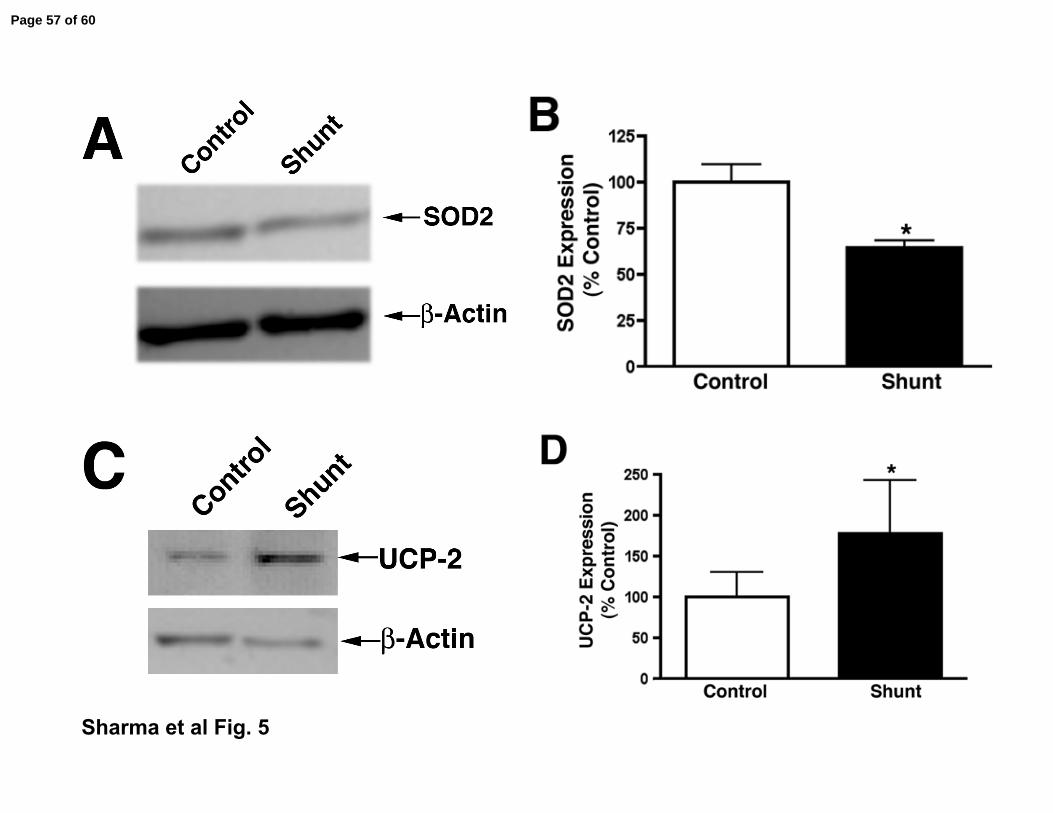
lambs present with markers of mitochondrial dysfunction. As loss of SOD2 (8, 13,
18, 26, 35, 44, 48) and increases in UCP-2 (2, 27, 42, 46, 62) have been shown to
be markers of mitochondrial dysfunction in other systems we examined these
markers. Our data indicate that SOD2 expression was significantly decreased (Fig.
5 A & B) in shunt lambs while UCP-2 expression was significantly increased (Fig. 5
C & D). In addition, we determined the lung levels of lactate and pyruvate in shunt
and control lambs to quantify mitochondrial activity. Under normal mitochondrial
function pyruvate levels are higher than lactate and thus any increases in lactate
levels decreases the pyruvate/lactate ratio and can be extrapolated to suggest a
reduction in ATP generation by the mitochondria (57). As shown in Table 2 shunt
lambs have significantly decreased pyruvate (0.059 ± 0.007 µmol/gm wet wt vs
0.132 ± 0.028 µmol/gm wet wt, P<0.05) and increased lactate levels (0.568 ± 0.838
µmol/gm wet wt vs 1.348 ± 0.214 µmol/gm wet wt, P<0.05) as well as a significant
reduction in the pyruvate/lactate ratio (Table 2).
Page 20 of 60

LCMP-00247-2007-FINAL ACCEPTED
21
Mitochondrial dysfunction attenuates HSP90-eNOS interaction and shear induced
NO production in pulmonary arterial endothelial cells
To further investigate the effect of mitochondrial dysfunction on NO signaling we
utilized cultured pulmonary arterial endothelial cells isolated from juvenile lambs.
Mitochondrial dysfunction was induced in PAECs using 2,4-dinitrophenol (2,4-DNP,
50µM, 0-6h). Our data indicate that 2,4-DNP significantly decreased ATP levels in
PAEC (Fig. 6 A) associated with an increase in oxidative stress within the
mitochondria (Fig. 6 B). Further, we found that this mitochondrial dysfunction was
associated with decrease in the association of HSP90 with eNOS (Fig. 7 A & B) and
shear induced NO production (Fig. 7 C) and an increase in eNOS-derived
superoxide (Fig. 7 D), indicating that impaired mitochondrial function reduces NO
production.
Decreased HSP90-eNOS interaction and altered NO signaling in shunt lambs
To determine the effect of decreased mitochondrial function in shunt lambs on NO
signaling, we carried out immunoprecipitation studies to determine eNOS-HSP90
interactions in both 2- and 4-week old shunt and control lambs. Our data indicate
that there is a progressive decrease in eNOS-HSP90 interactions between 2- and 4-
weeks of age in shunt compared to control lambs (Fig. 8 A & B) and that this is
associated with a progressive decrease in relative eNOS activity (as determined by
tissue NOx levels as a fraction of calcium dependent 3H-arginine metabolism, Fig. 8
C) and increased NOS-dependent superoxide levels indicative of eNOS uncoupling
Page 21 of 60

LCMP-00247-2007-FINAL ACCEPTED
22
(Fig. 8 D). We also confirmed the specificity of ETU for NOS-derived superoxide by
demonstrating that ETU does not quench the superoxide generated by a
xanthine/xanthine oxidase superoxide generating system (Fig. 8 E).
Page 22 of 60

LCMP-00247-2007-FINAL ACCEPTED
23
DISCUSSION
The important findings of this study are: (i) Decreased expression and activity
of mitochondrial enzymes involved in carnitine metabolism in two week old shunt
lambs causes disruption of carnitine homeostasis leading to mitochondrial
dysfunction; (ii) Decreased mitochondrial function leads to a progressive disruption
of HSP90-eNOS interaction leading to decreased NO signaling in shunt lambs.
Thus, this study provides insight into a novel mechanism that may be involved in the
development of pulmonary hypertension whereby the disruption of carnitine
metabolism leads to mitochondrial dysfunction and endothelial dysfunction.
The mitochondrial membrane is impermeable to long-chain fatty acids and
carnitine is required for transportation of these fatty acids into the mitochondria for β-
oxidation. Carnitine is present in the form of either free carnitine (nonesterified
molecule; FC), or acylcarnitines (esterified form; AC). Cytosolic long chain fatty acids
which are present as CoA-esters are trans-esterified to L-carnitine in a reaction
catalyzed by the carnitine palmitoyl transferase-1 (CPT-1) enzyme at the inner
aspect of mitochondrial outer membrane converting long-chain acyl-CoA to long-
chain acylcarnitine (64). At the inner mitochondrial membrane acylcarnitine is trans-
esterified back to free carnitine and long-chain acyl CoA, a reaction catalyzed by
carnitine palmitoyl transferase-2 (CPT-2) situated on the matrix side of the inner
mitochondrial membrane (71). Thus, transfer of fatty acids into the mitochondria is
dependent on the function of the CPT enzymes. In this study we have found
Page 23 of 60
LCMP-00247-2007-FINAL ACCEPTED
24
decreased expression of both CPT-1 and CPT2 enzymes in shunt lambs suggesting
that β-oxidation may be disrupted. Indeed, CPT deficiencies are common disorders
of mitochondrial fatty acid oxidation. For example, CPT2 deficiency in infants
causes severe attacks of hypoketotic hypoglycemia, occasionally associated with
cardiac damage and is commonly responsible for sudden death before 1 year of age
(9).
Another important mitochondrial enzyme CrAT, which resides in the matrix, is
able to reconvert the short and medium chain acyl CoAs into acylcarnitines using
intra-mitochondrial carnitine. Through this mechanism of reversible acylation
carnitine is able to modulate the intracellular concentrations of free CoA and acyl
CoA (64). Cell based and in vivo studies from Ames group implicate damage to the
CrAT enzyme that may decrease its affinity for carnitine (36-38). Decreased CrAT
activity leads to increased levels of acyl CoA that leads to inhibition of multiple
enzymatic processes involved in oxidative metabolism. We found that CrAT
expression was decreased ~2-fold in shunt lambs but CrAT activity was decreased
~20-fold suggesting a post-translational inhibitory mechanism was also involved.
Utilizing immunoprecipitation analysis we also investigated CrAT nitration levels. We
found that CrAT nitration was higher in shunt lambs relative to controls.
Furthermore, in vitro we observed that exposure to peroxynitrite reduced the activity
of the native protein. Taken together, our data suggests that the nitration of CrAT is
likely to be more important with respect to the decrease in CrAT activity than
decreased gene expression. However, it should be noted that although decreased
Page 24 of 60

LCMP-00247-2007-FINAL ACCEPTED
25
CrAT activity will decrease carnitine metabolism, other substrates are also involved
in this reaction, such as acetyl-CoA, whose generation requires fatty acid oxidation.
Thus it is possible that the alterations we have observed in CrAT activity may only
partly account for the alterations in carnitine homeostasis in the shunt lung.
However, further studies will be required to investigate this possibility.
Carnitine availability becomes a limiting step for β-oxidation in certain
physiological and pathological diseases and carnitine supplementation enhances
fatty acid metabolism in the mitochondria restoring normal mitochondrial function by
maintaining the equilibrium between acyl CoA and free CoA (63). The levels of acyl
carnitines are increased in plasma of patients with several inherited metabolic
diseases. In these cases, the endogenous pool of carnitine becomes insufficient to
cope with the required acyl transfer and plasma AC:FC ratio increases (10). The
increase in AC:FC ratio reflects changes in the intra mitochondrial equilibrium
between acyl CoA and free CoA (7, 12). Increase in this ratio indicates accumulation
of acyl CoA or reduction in FC and is associated with compromise of mitochondrial
metabolism (7). More recent data have shown that under conditions of metabolic
stress, mitochondria accumulate acyl-CoA, which is normally maintained in
homeostasis with carnitine. High acyl-CoA levels inhibit multiple enzymatic
processes involved in oxidative metabolism (14). This product can inhibit multiple
other mitochondrial enzymes downstream from its own synthesis resulting in a
metabolic roadblock within the mitochondrial matrix (41, 49, 58, 61). In our lamb
model, AC:FC ratio was higher in 2 week shunt- compared to age matched control-
Page 25 of 60

LCMP-00247-2007-FINAL ACCEPTED
26
lambs indicating decreased mitochondrial function and impaired fatty acid
metabolism. If disruption in carnitine metabolism is an important contributing factor in
the development of endothelial dysfunction, then treatment with carnitine may
attenuate at least in part any endothelial injury associated with the development of
pulmonary hypertension and could be used as an effective therapeutic agent for the
prevention of the mitochondrial dysfunction associated in pulmonary hypertension.
Indeed a recent study has shown that in neonatal hypoxia-ischemia is associated
with a significant increased acyl-CoA:CoA ratio and this imbalance can be prevented
by treatment with exogenous carnitine (66).
SOD2 is an important mitochondrial enzyme regulating the redox level within
the mitochondrion by scavenging the free radicals produced in the mitochondria.
Reduction of SOD2 protein expression is widely considered to be a hallmark of
decreased mitochondrial function (8, 13, 18, 26, 35, 44, 48). Our data show a
significant reduction in SOD2 expression in shunt- compared to age matched
control-lambs again supporting the notion of mitochondriaitochondrill dysfunction in
the shunt lambs. In addition to SOD2, we examined expression of another important
mitochondrial protein, UCP-2, in both control and shunt lambs. UCP-2 is a
mitochondrial membrane proton transporter that mediates proton leak across the
inner mitochondrial membrane reducing energy force for cellular ATP production
(32, 51). As with decrease in SOD2, an increase in UCP-2 expression is thought to
be a marker for mitochondrial dysfunction (2, 27, 42, 46, 62). For example, UCP-2 is
found to be up-regulated in islets of mouse model of obesity-induced diabetes where
Page 26 of 60

LCMP-00247-2007-FINAL ACCEPTED
27
superoxide-mediated activation of UCP-2 has been shown to play an important role
in the pathogenesis of β cell dysfunction (33). Our data demonstrated a significant
increase in UCP-2 expression in shunt- compared to age matched control-lambs
again supporting the notion of mitochondrial dysfunction in the shunt lambs. Further
support for this comes from our data where we examined lactate and pyruvate levels
in lung tissue in shunt- and age matched control-lambs. When pyruvate is not
utilized by mitochondria for energy production, it gets converted in to lactate. Higher
lactate/pyruvate ratio is correlated with many pathological conditions related to
mitochondria. We found higher lactate levels and increased lactate/pyruvate ratio in
our shunt lamb model. Taken together our data demonstrating decreases in SOD2
and increases in UCP-2-expression and the increased lactate/pyruvate ratio
indicates that shunt lambs have decreased mitochondrial function.
Mitochondria play essential physiological roles in cells and are the major site
of cellular ATP production. Decreased mitochondrial function results in reduced ATP
production and increased ROS generation with deleterious effects. Disruption of
mitochondrial function is a critical event in a number of pathologic conditions
including hypoxia-ischemic injuries (6), stroke (59), and diabetes (16, 45, 47, 50).
There is also considerable evidence for decreased mitochondrial function in aging
related neurodegenerative disorders (17, 22). Further, previous studies have shown
the importance of ATP in the pulmonary system as demonstrated by its key role in
the birth related pulmonary vasodilation in fetal lambs (30, 31) while a study in
poultry has shown that lung mitochondrial dysfunction is present in broilers with
Page 27 of 60

LCMP-00247-2007-FINAL ACCEPTED
28
pulmonary hypertension syndrome associated with oxidative stress (28). The role of
ATP in endothelial function may be to be due, at least in part, through its ability to
stimulate NO release via the activation of eNOS (30, 31). HSP90 a molecular
chaperone modulates the eNOS activity (19, 20). It has been demonstrated that
eNOS can interact with a 90kD heat shock protein (HSP90). HSP90 is part of a
family that acts as molecular chaperones that can modulate protein activity. HSP90
appears to increase eNOS activity by facilitating the calmodulin induced
displacement of caveolin 1 from eNOS (24) which is inhibited with the HSP90
inhibitor geldamycin (20). HSP90 is ATP dependent and the ATPase site of the
chaperone is responsible for the auto phosphorylation required to enable HSP90 to
interact with client proteins (54, 55). Interaction of HSP90 with eNOS have been
shown to increase eNOS activity and NO production whereas decreased association
of eNOS with HSP90 leads to enhanced eNOS-dependent production of superoxide
(52). Here, we show that mild mitochondrial inhibition with the mitochondrial
uncoupler, 2,4-DNP, caused a sustained decrease in ATP in endothelial cells that
was reflected in a reduction in eNOS-HSP90 interaction. Further, this was
associated with decreased NO- and increased superoxide- generation when the
cells were exposed to fluid shear stress. Further, this could be observed in vivo
where our data indicate that in the shunt lambs there is a progressive decrease in
eNOS-HSP90 interactions in the shunt lambs. Further, the decrease in eNOS-
HSP90 interactions correlate with a progressive decrease in NO signaling and
increases in NOS-derived superoxide. The progressive loss of NO signaling is a
classic marker of endothelial dysfunction.
Page 28 of 60
LCMP-00247-2007-FINAL ACCEPTED
29
In conclusion our data indicate that lambs with pulmonary hypertension
secondary to increased pulmonary blood flow by 2-weeks of age have developed
mitochondrial dysfunction within the pulmonary system that is associated with
progressive decreases in the interaction of eNOS with HSP90 and in NO generation.
Further, as our data indicate that this dysfunction is due to disruption of the carnitine
homeostasis within the lung the use of L-carnitine, or other carnitine analogs, may
be of potential therapeutic benefit to help maintain eNOS-HSP90 interactions, NO
signaling, and endothelial dysfunction.
Page 29 of 60

LCMP-00247-2007-FINAL ACCEPTED
30
ACKNOWLEDGMENTS
This research was supported in part by grants HL60190 (to SMB), HL67841 (to
SMB), HL72123 (to SMB), HL70061 (to SMB), and HL61284 (to JRF) all from the
National Institutes of Health, and by a grant from the Fondation LeDucq (to SMB and
JRF).
Page 30 of 60

LCMP-00247-2007-FINAL ACCEPTED
31
Table 1
Total Carnitines Free Carnitine L-carnitine Acylcarnitine(nmol/gww) (nmol/gww) (nmol/gww) (% total)
Control 60.54+ 8.45 67.57+ 13.43 56.42+ 12.27 6.2+ 4.7%
Shunt 72.37+ 10.14 34.86+ 3.35* 20.21+ 4.29* 40.71+ 9%*
Data is mean + SE; N=5; *P<0.05 vs Control. gww= gram wet weight.
Page 31 of 60

LCMP-00247-2007-FINAL ACCEPTED
32
Table 2
Lactate Pyruvate Lactate: Pyruvate(µmols/gww) (µmols/gww) Ratio
Control 1.79+ 0.34 0.144+ 0.03* 15.25+ 4.35: 1
Shunt 3.1+ 0.86* 0.057+ 0.008* 51.8+ 10.92: 1*
Data is mean + SE; N=5; *P<0.05 vs Control. gww= gram wet weight.
Page 32 of 60

LCMP-00247-2007-FINAL ACCEPTED
33
FIGURE LEGENDS
Figure 1. Carnitine palmitoyltransferase expression in peripheral lung tissue from
control and shunt lambs at 2-weeks of age.
Panel A. Protein extracts (50 µg), prepared from peripheral lung of shunt and
control lambs were analyzed by Western blot analysis using a specific antiserum
raised against carnitine palmitoyltransferases 1B (CPT-1B) protein. CPT-1B
expression was also normalized for loading using β-actin. Shown is a representative
blot.
Panel B. There is a significant decrease in normalized densitometric values for CPT-
1B protein in peripheral lung tissue prepared from shunt compared to control lambs.
Values are mean ± SEM; n=6 control and n=6 shunt; *P <0.05 vs. control.
Panel C. Protein extracts (50 µg), prepared from peripheral lung of shunt or control
lambs were analyzed by Western blot analysis using a specific antiserum raised
against carnitine palmitoyltransferases 2 (CPT2) protein. CPT2 expression was also
normalized for loading using β-actin. Shown is a representative blot.
Panel D. There is a significant decrease in normalized densitometric values for
CPT2 protein in peripheral lung tissue prepared from shunt compared to control
lambs. Values are mean ± SEM; n=6 control and n=6 shunt; *P <0.05 vs. control.
Page 33 of 60

LCMP-00247-2007-FINAL ACCEPTED
34
Figure 2. Carnitine acetyltransferase expression and activity in peripheral lung
tissue from control and shunt lambs at 2-weeks of age.
Panel A. Protein extracts (50 µg), prepared from peripheral lung of shunt and
control lambs were analyzed by Western blot analysis using a specific antiserum
raised against carnitine acetyltransferase (CrAT) protein. CrAT expression was also
normalized for loading using β-actin. Shown is a representative blot.
Panel B. There is a significant decrease in normalized densitometric values for CrAT
protein in peripheral lung tissue prepared from shunt compared to control lambs.
Values are mean ± SEM; n=6 control and n=6 shunt; *P <0.05 vs. control.
Panel C. CrAT activity was determined in protein extracts (40 µg), prepared from
peripheral lung tissue from control and shunt lambs. There is as significant decrease
in CrAT activity in peripheral lung tissue prepared from shunt compared to control
lambs. Values are expressed as units of activity per µg protein. Values are mean ±
SEM; n=4 control and n=4 shunt. *P <0.05 vs. control.
Page 34 of 60
LCMP-00247-2007-FINAL ACCEPTED
35
Figure 3. Increased nitration of carnitine acetyltransferase in peripheral lung tissue
of shunt lambs at 2-weeks of age.
Panel A. Protein extracts (1000µg), prepared from peripheral lung of shunt and
control lambs were subjected to immunoprecipitation using an antibody specific to 3-
NT then analyzed by Western blot analysis using a specific antiserum raised against
carnitine acetyltransferase (CrAT) protein. Shown is a representative blot with the
CrAT expression. Minimal binding is observed in the beads alone or IgG pre-clear.
Panel B. There is a significant increase in nitrated CrAT protein in peripheral lung
tissue prepared from shunt compared to control lambs. Values are mean ± SEM;
n=6 control and n=6 shunt; *P <0.05 vs. control.
Page 35 of 60

LCMP-00247-2007-FINAL ACCEPTED
36
Figure 4. Peroxynitrite decreases carnitine acetyltransferase activity.
Purified pigeon breast muscle carnitine acetyltransferase was exposed to authentic
peroxynitrite or vehicle and then the effect of activity determined. Peroxynitrite
induces a significant decrease in CrAT activity. Values are mean ± SD; N=6; *P
<0.05 vs. vehicle.
Page 36 of 60
LCMP-00247-2007-FINAL ACCEPTED
37
Figure 5. Markers of mitochondrial dysfunction are increased in shunt lambs at 2-
weeks of age.
Panel A. Protein extracts (25 µg), prepared from peripheral lung of shunt and
control lambs were analyzed by Western blot analysis using a specific antiserum
raised against SOD2 protein. SOD2 expression was also normalized for loading
using β-actin. Shown is a representative blot.
Panel B. There is a significant decrease in normalized densitometric values for
SOD2 protein in peripheral lung tissue prepared from shunt compared to control
lambs. Values are mean ± SEM; n=6 control and n=6 shunt at each age; *P <0.05
vs. control.
Panel C. Protein extracts (25 µg), prepared from peripheral lung of shunt and control
lambs were analyzed by Western blot analysis using a specific antiserum raised
against UCP-2 protein. UCP-2 expression was also normalized for loading using β-
actin. Shown is a representative blot.
Panel D. There is a significant increase in normalized densitometric values for UCP-
2 protein in peripheral lung tissue prepared from shunt compared to control lambs.
Values are mean ± SEM; n=6 control and n=6 shunt at each age; *P <0.05 vs.
control.
Page 37 of 60

LCMP-00247-2007-FINAL ACCEPTED
38
Figure 6. 2, 4-Dinitrophenol decreases ATP levels and increases mitochondrial
oxidative stress in pulmonary arterial endothelial cells.
Panel A. PAEC were treated with 2,4-DNP (25µM, 0-8h) and the cellular ATP levels
were then determined. 2-4- DNP significantly decreases cellular ATP levels. N=6.
Values are mean ± SE. * P<0.05 vs control.
Panel B. PAEC were treated with 2,4-DNP (25µM, 4h) Cells were then exposed to
mitoSox (10µM, 15min) to measure mitochondrial superoxide levels (as a marker for
mitochondrial dysfunction). 2,4-DNP induced significant increase in mitoSox
fluorescence (representative images are shown as an insert). N=4. Values are
mean ± SE, * P<0.05 vs. no 2,4-DNP.
Page 38 of 60
LCMP-00247-2007-FINAL ACCEPTED
39
Figure 7. 2, 4-Dinitrophenol decreases NO-signaling in pulmonary arterial
endothelial cells.
Panel A. PAEC were treated with DNP (25µM, 4h) washed with PBS and lysates
prepared with modified RIPA buffer. Immunoprecipitation was performed using an
antibody to eNOS then analyzed by Western blot analysis using a specific antiserum
raised against HSP90 protein. Blots were also stripped and reprobed for eNOS to
normalize the immunoprecipitation. Shown is a representative blot.
Panel B. 2,4-DNP treatment causes a significant reduction in eNOS-HSP90
interaction. N=3. Values are mean ± SE. * P<0.05 vs untreated.
Panel C. Cells were treated with 2,4-DNP (25µM, 4h) exposed to laminar shear
stress (20dyn/cm2, 15min) then the media was assayed for NOx, as an indirect
determination of NO production. 2,4-DNP significantly decreases shear mediated
increase in NOx. N=6. Values are mean ± SE. * P<0.05 vs no shear; † P<0.05 vs
shear alone.
Page 39 of 60

LCMP-00247-2007-FINAL ACCEPTED
40
Figure 8. Progressive decreases in the interaction of eNOS with HSP90 in shunt
compared to control lambs.
Panel A. The interaction of eNOS with HSP90 was determined by
immunoprecipitation using specific antiserum raised against eNOS in tissue extracts
prepared peripheral lung of shunt and control lambs at 2-, and 4-weeks of age.
Immunoprecipitated extracts were analyzed using antisera against either eNOS or
HSP90. A representative image is shown. No specific protein bands are observed in
the beads alone or IgG pre-clear.
Panel B. The levels of eNOS protein associated with HSP90 relative to total eNOS
protein were calculated. The data obtained indicate that there was a progressive
decrease in the association of eNOS with HSP90 in shunt compared to control-
lambs between 2- and 4-weeks of age. Values are mean ± SE. N=5 shunt and N=5
control lambs at each age. *P<0.05 compared to age matched control; † P<0.05 vs
previous age.
Panel C. Relative eNOS activity was estimated in shunt and age matched control
lambs at 2- and 4-weeks of age by dividing peripheral lung tissue NOx levels by total
lung eNOS activity (determined by calcium dependent 3H-arginine to 3H-citrulline
conversion). Relative eNOS activity is significantly lower in the shunt lambs at both
2- and 4-weeks of age. Values are mean ± SE. *P<0.05 compared to age matched
control; † P<0.05 vs. 2-week shunt. N=4 shunt and N=4 control lambs at each age.
Panel D. Superoxide anion levels determined by electron paramagnetic resonance
in snap-frozen lung tissue from shunt and age matched control lambs at 4-weeks of
age in the presence and absence of the NOS inhibitor, 3-ethylisothiourea (ETU,
Page 40 of 60
LCMP-00247-2007-FINAL ACCEPTED
41
100µM). Shown is a bar graph representing the cumulative data. ETU-inhibitable
superoxide levels are significantly higher in the shunt lambs. Values are mean ± SD.
*P<0.05 compared to age matched control; † P<0.05 vs previous age. N=6 shunt
and N=6 control lambs at each age.
Panel E. Superoxide was generated in vitro with xanthine oxidase (XO, 1 U/) with
xanthine (X, 1mM), in the presence or absence of ETU (100µM). Two control
reactions, one lacking X and the other lacking XO were included in order to ensure
absence of non-specific reactions of either reagent with the EPR spin-trap. The
significant increase in superoxide generated by the X/XO reaction is not significantly
quenched by the presence of ETU. Values are mean ± SE. *P<0.05 compared to
xanthine alonel; † P<0.05 vs. xanthine oxidase alone. N=3 for each condition.
Page 41 of 60

LCMP-00247-2007-FINAL ACCEPTED
42
REFERENCES
1. Adnot S, Raffestin B, Eddahibi S, Braquet P, and Chabrier PE. Loss of
endothelium-dependent relaxant activity in the pulmonary circulation of rats exposed
to chronic hypoxia. J Clin Invest 87: 155-162, 1991.
2. Andrews ZB, Diano S, and Horvath TL. Mitochondrial uncoupling proteins
in the CNS: in support of function and survival. Nature reviews 6: 829-840, 2005.
3. Beghetti M, Black SM, and Fineman JR. Endothelin-1 in congenital heart
disease. Pediatr Res 57: 16R-20R, 2005.
4. Black SM, and Fineman JR. Oxidative and nitrosative stress in pediatric
pulmonary hypertension: roles of endothelin-1 and nitric oxide. Vascular
pharmacology 45: 308-316, 2006.
5. Black SM, Fineman JR, Steinhorn RH, Bristow J, and Soifer SJ.
Increased endothelial NOS in lambs with increased pulmonary blood flow and
pulmonary hypertension. Am J Physiol 275: H1643-1651, 1998.
Page 42 of 60

LCMP-00247-2007-FINAL ACCEPTED
43
6. Blomgren K, Zhu C, Hallin U, and Hagberg H. Mitochondria and ischemic
reperfusion damage in the adult and in the developing brain. Biochem Biophys Res
Commun 304: 551-559, 2003.
7. Bohles H, Evangeliou A, Bervoets K, Eckert I, and Sewell A. Carnitine
esters in metabolic disease. Eur J Pediatr 153: S57-61, 1994.
8. Bonawitz ND, Rodeheffer MS, and Shadel GS. Defective mitochondrial
gene expression results in reactive oxygen species-mediated inhibition of respiration
and reduction of yeast life span. Mol Cell Biol 26: 4818-4829, 2006.
9. Bonnefont JP, Djouadi F, Prip-Buus C, Gobin S, Munnich A, and Bastin
J. Carnitine palmitoyltransferases 1 and 2: biochemical, molecular and medical
aspects. Mol Aspects Med 25: 495-520, 2004.
10. Calvani M, Benatti P, Mancinelli A, D'Iddio S, Giordano V, Koverech A,
Amato A, and Brass EP. Carnitine replacement in end-stage renal disease and
hemodialysis. Ann N Y Acad Sci 1033: 52-66, 2004.
11. Celermajer DS, Cullen S, and Deanfield JE. Impairment of endothelium-
dependent pulmonary artery relaxation in children with congenital heart disease and
abnormal pulmonary hemodynamics. Circulation 87: 440-446, 1993.
12. Chace DH, Pons R, Chiriboga CA, McMahon DJ, Tein I, Naylor EW, and
De Vivo DC. Neonatal blood carnitine concentrations: normative data by
electrospray tandem mass spectometry. Pediatr Res 53: 823-829, 2003.
13. Chan PH. Mitochondrial dysfunction and oxidative stress as determinants of
cell death/survival in stroke. Ann N Y Acad Sci 1042: 203-209, 2005.
Page 43 of 60

LCMP-00247-2007-FINAL ACCEPTED
44
14. DeVivo D, and Tein I. Primary and secondary disoreders of carnitine
metabolism. Int J Pediatr 5: 135-141, 1990.
15. Dewey CF, Jr., Bussolari SR, Gimbrone MA, Jr., and Davies PF. The
dynamic response of vascular endothelial cells to fluid shear stress. J Biomech Eng
103: 177-185, 1981.
16. Duchen MR. Roles of mitochondria in health and disease. Diabetes 53 Suppl
1: S96-102, 2004.
17. Emerit J, Edeas M, and Bricaire F. Neurodegenerative diseases and
oxidative stress. Biomed Pharmacother 58: 39-46, 2004.
18. Esposito L, Raber J, Kekonius L, Yan F, Yu GQ, Bien-Ly N, Puolivali J,
Scearce-Levie K, Masliah E, and Mucke L. Reduction in mitochondrial superoxide
dismutase modulates Alzheimer's disease-like pathology and accelerates the onset
of behavioral changes in human amyloid precursor protein transgenic mice. J
Neurosci 26: 5167-5179, 2006.
19. Fontana J, Fulton D, Chen Y, Fairchild TA, McCabe TJ, Fujita N, Tsuruo
T, and Sessa WC. Domain mapping studies reveal that the M domain of hsp90
serves as a molecular scaffold to regulate Akt-dependent phosphorylation of
endothelial nitric oxide synthase and NO release. Circ Res 90: 866-873, 2002.
20. Garcia-Cardena G, Fan R, Shah V, Sorrentino R, Cirino G,
Papapetropoulos A, and Sessa WC. Dynamic activation of endothelial nitric oxide
synthase by Hsp90. Nature 392: 821-824, 1998.
21. Garvey EP, Oplinger JA, Tanoury GJ, Sherman PA, Fowler M, Marshall
S, Harmon MF, Paith JE, and Furfine ES. Potent and selective inhibition of human
Page 44 of 60

LCMP-00247-2007-FINAL ACCEPTED
45
nitric oxide synthases. Inhibition by non-amino acid isothioureas. J Biol Chem 269:
26669-26676, 1994.
22. Genova ML, Pich MM, Bernacchia A, Bianchi C, Biondi A, Bovina C,
Falasca AI, Formiggini G, Castelli GP, and Lenaz G. The mitochondrial
production of reactive oxygen species in relation to aging and pathology. Ann N Y
Acad Sci 1011: 86-100, 2004.
23. Giaid A, and Saleh D. Reduced expression of endothelial nitric oxide
synthase in the lungs of patients with pulmonary hypertension. N Engl J Med 333:
214-221, 1995.
24. Gratton JP, Fontana J, O'Connor DS, Garcia-Cardena G, McCabe TJ, and
Sessa WC. Reconstitution of an endothelial nitric-oxide synthase (eNOS), hsp90,
and caveolin-1 complex in vitro. Evidence that hsp90 facilitates calmodulin
stimulated displacement of eNOS from caveolin-1. J Biol Chem 275: 22268-22272,
2000.
25. Haworth JC, Demaugre F, Booth FA, Dilling LA, Moroz SP, Seshia SS,
Seargeant LE, and Coates PM. Atypical features of the hepatic form of carnitine
palmitoyltransferase deficiency in a Hutterite family. J Pediatr 121: 553-557, 1992.
26. Hinerfeld D, Traini MD, Weinberger RP, Cochran B, Doctrow SR, Harry J,
and Melov S. Endogenous mitochondrial oxidative stress: neurodegeneration,
proteomic analysis, specific respiratory chain defects, and efficacious antioxidant
therapy in superoxide dismutase 2 null mice. J Neurochem 88: 657-667, 2004.
Page 45 of 60
LCMP-00247-2007-FINAL ACCEPTED
46
27. Hoffstedt J, Folkesson R, Wahrenberg H, Wennlund A, van Harmelen V,
and Arner P. A marked upregulation of uncoupling protein 2 gene expression in
adipose tissue of hyperthyroid subjects. Horm Metab Res 32: 475-479, 2000.
28. Iqbal M, Cawthon D, Wideman RF, Jr., and Bottje WG. Lung mitochondrial
dysfunction in pulmonary hypertension syndrome. II. Oxidative stress and inability to
improve function with repeated additions of adenosine diphosphate. Poult Sci 80:
656-665, 2001.
29. Knowles PF, Gibson JF, Pick FM, and Bray RC. Electron-spin-resonance
evidence for enzymic reduction of oxygen to a free radical, the superoxide ion.
Biochem J 111: 53-58, 1969.
30. Konduri GG, and Mattei J. Role of oxidative phosphorylation and ATP
release in mediating birth-related pulmonary vasodilation in fetal lambs. Am J
Physiol Heart Circ Physiol 283: H1600-1608, 2002.
31. Konduri GG, and Mital S. Adenosine and ATP cause nitric oxide-dependent
pulmonary vasodilation in fetal lambs. Biol Neonate 78: 220-229, 2000.
32. Krauss S, Zhang CY, and Lowell BB. A significant portion of mitochondrial
proton leak in intact thymocytes depends on expression of UCP2. Proc Natl Acad
Sci U S A 99: 118-122, 2002.
33. Krauss S, Zhang CY, Scorrano L, Dalgaard LT, St-Pierre J, Grey ST, and
Lowell BB. Superoxide-mediated activation of uncoupling protein 2 causes
pancreatic beta cell dysfunction. J Clin Invest 112: 1831-1842, 2003.
Page 46 of 60

LCMP-00247-2007-FINAL ACCEPTED
47
34. Kurtz DM, Tian L, Gower BA, Nagy TR, Pinkert CA, and Wood PA.
Transgenic studies of fatty acid oxidation gene expression in nonobese diabetic
mice. J Lipid Res 41: 2063-2070, 2000.
35. Liang LP, and Patel M. Mitochondrial oxidative stress and increased seizure
susceptibility in Sod2(-/+) mice. Free Radic Biol Med 36: 542-554, 2004.
36. Liu J, Atamna H, Kuratsune H, and Ames BN. Delaying brain mitochondrial
decay and aging with mitochondrial antioxidants and metabolites. Ann N Y Acad Sci
959: 133-166, 2002.
37. Liu J, Head E, Gharib AM, Yuan W, Ingersoll RT, Hagen TM, Cotman CW,
and Ames BN. Memory loss in old rats is associated with brain mitochondrial decay
and RNA/DNA oxidation: partial reversal by feeding acetyl-L-carnitine and/or R-
alpha -lipoic acid. Proc Natl Acad Sci U S A 99: 2356-2361, 2002.
38. Liu J, Killilea DW, and Ames BN. Age-associated mitochondrial oxidative
decay: improvement of carnitine acetyltransferase substrate-binding affinity and
activity in brain by feeding old rats acetyl-L- carnitine and/or R-alpha -lipoic acid.
Proc Natl Acad Sci U S A 99: 1876-1881, 2002.
39. Longo A, Bruno G, Curti S, Mancinelli A, and Miotto G. Determination of
L-carnitine, acetyl-L-carnitine and propionyl-L-carnitine in human plasma by high-
performance liquid chromatography after pre-column derivatization with 1-
aminoanthracene. J Chromatogr B Biomed Appl 686: 129-139, 1996.
40. Marbach EP, and Weil MH. Rapid enzymatic measurement of blood lactate
and pyruvate. Use and significance of metaphosphoric acid as a common
precipitant. Clin Chem 13: 314-325, 1967.
Page 47 of 60

LCMP-00247-2007-FINAL ACCEPTED
48
41. Matsuishi T, Stumpf DA, Seliem M, Eguren LA, and Chrislip K. Propionate
mitochondrial toxicity in liver and skeletal muscle: acyl CoA levels. Biochem Med
Metab Biol 45: 244-253, 1991.
42. Mattson MP, and Liu D. Mitochondrial potassium channels and uncoupling
proteins in synaptic plasticity and neuronal cell death. Biochem Biophys Res
Commun 304: 539-549, 2003.
43. Minkler PE, Brass EP, Hiatt WR, Ingalls ST, and Hoppel CL. Quantification
of carnitine, acetylcarnitine, and total carnitine in tissues by high-performance liquid
chromatography: the effect of exercise on carnitine homeostasis in man. Anal
Biochem 231: 315-322, 1995.
44. Misawa H, Nakata K, Matsuura J, Moriwaki Y, Kawashima K, Shimizu T,
Shirasawa T, and Takahashi R. Conditional knockout of Mn superoxide dismutase
in postnatal motor neurons reveals resistance to mitochondrial generated superoxide
radicals. Neurobiol Dis 23: 169-177, 2006.
45. Moreira PI, Santos MS, Moreno AM, Proenca T, Seica R, and Oliveira CR.
Effect of streptozotocin-induced diabetes on rat brain mitochondria. J
Neuroendocrinol 16: 32-38, 2004.
46. Murray AJ, Anderson RE, Watson GC, Radda GK, and Clarke K.
Uncoupling proteins in human heart. Lancet 364: 1786-1788, 2004.
47. Nishio Y, Kanazawa A, Nagai Y, Inagaki H, and Kashiwagi A. Regulation
and role of the mitochondrial transcription factor in the diabetic rat heart. Ann N Y
Acad Sci 1011: 78-85, 2004.
Page 48 of 60

LCMP-00247-2007-FINAL ACCEPTED
49
48. Ohashi M, Runge MS, Faraci FM, and Heistad DD. MnSOD deficiency
increases endothelial dysfunction in ApoE-deficient mice. Arterioscler Thromb Vasc
Biol 26: 2331-2336, 2006.
49. Pande SV, and Blanchaer MC. Reversible inhibition of mitochondrial
adenosine diphosphate phosphorylation by long chain acyl coenzyme A esters. J
Biol Chem 246: 402-411, 1971.
50. Petersen KF, Dufour S, Befroy D, Garcia R, and Shulman GI. Impaired
mitochondrial activity in the insulin-resistant offspring of patients with type 2
diabetes. N Engl J Med 350: 664-671, 2004.
51. Porter RK. Mitochondrial proton leak: a role for uncoupling proteins 2 and 3?
Biochim Biophys Acta 1504: 120-127, 2001.
52. Pritchard KA, Jr., Ackerman AW, Gross ER, Stepp DW, Shi Y, Fontana
JT, Baker JE, and Sessa WC. Heat shock protein 90 mediates the balance of nitric
oxide and superoxide anion from endothelial nitric-oxide synthase. J Biol Chem 276:
17621-17624, 2001.
53. Reddy VM, Meyrick B, Wong J, Khoor A, Liddicoat JR, Hanley FL, and
Fineman JR. In utero placement of aortopulmonary shunts. A model of postnatal
pulmonary hypertension with increased pulmonary blood flow in lambs. Circulation
92: 606-613, 1995.
54. Schulte TW, Akinaga S, Murakata T, Agatsuma T, Sugimoto S, Nakano
H, Lee YS, Simen BB, Argon Y, Felts S, Toft DO, Neckers LM, and Sharma SV.
Interaction of radicicol with members of the heat shock protein 90 family of molecular
chaperones. Mol Endocrinol 13: 1435-1448, 1999.
Page 49 of 60

LCMP-00247-2007-FINAL ACCEPTED
50
55. Schulte TW, Akinaga S, Soga S, Sullivan W, Stensgard B, Toft D, and
Neckers LM. Antibiotic radicicol binds to the N-terminal domain of Hsp90 and
shares important biologic activities with geldanamycin. Cell Stress Chaperones 3:
100-108, 1998.
56. Shaul PW, Wells LB, and Horning KM. Acute and prolonged hypoxia
attenuate endothelial nitric oxide production in rat pulmonary arteries by different
mechanisms. J Cardiovasc Pharmacol 22: 819-827, 1993.
57. Shinde S, Glam K, Kumar P, Patil N, and Sadacharan K. Perioperative
blood lactae levels, pyruvate levels, and lactate-pyruvate ratio in children undergoing
cardiopulmonary bypass for congenital heart disease. Indian J Crit Care Med 9: 145-
150, 2005.
58. Shug AL, Shrago E, Bittar N, Folts JD, and Koke JR. Acyl-CoA inhibition of
adenine nucleotide translocation in ischemic myocardium. Am J Physiol 228: 689-
692, 1975.
59. Sims NR, and Anderson MF. Mitochondrial contributions to tissue damage
in stroke. Neurochem Int 40: 511-526, 2002.
60. Stanley CA. New genetic defects in mitochondrial fatty acid oxidation and
carnitine deficiency. Adv Pediatr 34: 59-88, 1987.
61. Stumpf DA, McAfee J, Parks JK, and Eguren L. Propionate inhibition of
succinate:CoA ligase (GDP) and the citric acid cycle in mitochondria. Pediatr Res
14: 1127-1131, 1980.
Page 50 of 60
LCMP-00247-2007-FINAL ACCEPTED
51
62. Sullivan PG, Springer JE, Hall ED, and Scheff SW. Mitochondrial
uncoupling as a therapeutic target following neuronal injury. J Bioenerg Biomembr
36: 353-356, 2004.
63. Szewczyk A, and Wojtczak L. Mitochondria as a pharmacological target.
Pharmacol Rev 54: 101-127, 2002.
64. Vaz FM, and Wanders RJ. Carnitine biosynthesis in mammals. Biochem J
361: 417-429, 2002.
65. Vianey-Saban C, Mousson B, Bertrand C, Stamm D, Dumoulin R, Zabot
MT, Divry P, Floret D, and Mathieu M. Carnitine palmitoyl transferase I deficiency
presenting as a Reye-like syndrome without hypoglycaemia. Eur J Pediatr 152: 334-
338, 1993.
66. Wainwright MS, Kohli R, Whitington PF, and Chace DH. Carnitine
treatment inhibits increases in cerebral carnitine esters and glutamate detected by
mass spectrometry after hypoxia-ischemia in newborn rats. Stroke 37: 524-530,
2006.
67. Wedgwood S, Bekker JM, and Black SM. Shear stress regulation of
endothelial NOS in fetal pulmonary arterial endothelial cells involves PKC. Am J
Physiol Lung Cell Mol Physiol 281: L490-498, 2001.
68. Wedgwood S, and Black SM. Endothelin-1 decreases endothelial NOS
expression and activity through ETA receptor-mediated generation of hydrogen
peroxide. Am J Physiol Lung Cell Mol Physiol 288: L480-487, 2005.
Page 51 of 60

LCMP-00247-2007-FINAL ACCEPTED
52
69. Wedgwood S, Mitchell CJ, Fineman JR, and Black SM. Developmental
differences in the shear stress-induced expression of endothelial NO synthase:
changing role of AP-1. Am J Physiol Lung Cell Mol Physiol 284: L650-662, 2003.
70. Wiseman DA, Wells SM, Wilham J, Hubbard M, Welker JE, and Black SM.
Endothelial response to stress from exogenous Zn2+ resembles that of NO-
mediated nitrosative stress, and is protected by MT-1 overexpression. Am J Physiol
Cell Physiol 291: C555-568, 2006.
71. Woeltje KF, Kuwajima M, Foster DW, and McGarry JD. Characterization of
the mitochondrial carnitine palmitoyltransferase enzyme system. II. Use of
detergents and antibodies. J Biol Chem 262: 9822-9827, 1987.
72. Xue C, and Johns RA. Upregulation of nitric oxide synthase correlates
temporally with onset of pulmonary vascular remodeling in the hypoxic rat.
Hypertension 28: 743-753, 1996.
Page 52 of 60

Sharma et al Fig. 1
A B
C D
Page 53 of 60

Sharma et al Fig. 2
A B
C
Page 54 of 60

Sharma et al Fig. 3
B
Page 55 of 60

Sharma et al Fig. 4
Page 56 of 60
Sharma et al Fig. 5
Page 57 of 60

Sharma et al Fig. 6
A
Page 58 of 60

Sharma et al Fig. 7
A
Page 59 of 60

Sharma et al Fig. 8
Page 60 of 60
Copyright © 2022 FDOKUMEN