Gene expression of prostaglandin EP4 receptor in three ...
10
RESEARCH ARTICLE Open Access Gene expression of prostaglandin EP4 receptor in three canine carcinomas Margaret L. Musser 1* , Austin K. Viall 2 , Rachel L. Phillips 2 , Jesse M. Hostetter 2,3 and Chad M. Johannes 1 Abstract Background: Chronic inflammation mediated by the cyclooxygenase enzymes, specifically their product prostaglandin E2 (PGE2), can result in the development of cancer. PGE2 promotes cell proliferation, apoptosis, and angiogenesis through interaction with its specific receptors (EP1 receptor - EP4 receptor [EP1R-EP4R]). In multiple human cancers, the expression of EP4R is associated with the development of malignancy and a poor prognosis. The expression of EP4R has not yet been evaluated in canine tumors. The aim of this study was to characterize the mRNA gene expression of EP4R (ptger4) in canine squamous cell carcinoma (SCC), apocrine gland anal sac adenocarcinoma (AGASACA), and transitional cell carcinoma (TCC). Archived tumor samples of canine cutaneous SCC (n = 9), AGASACA (n = 9), and TCC (n = 9), and matched archived normal tissue controls were evaluated for mRNA expression of canine EP4R using RNA in situ hybridization (RNAscope®). Quantification of RNAscope® signals in tissue sections was completed with an advanced digital pathology image analysis system (HALO). Data was expressed as copy number, H-index, and percent tumor cell expression of EP4R. Results: In all canine SCC, AGASACA, and TCC samples evaluated, strong universal positive expression of EP4R was identified. For SCC and AGASACA, mRNA EP4R expression was statistically higher than that of their respective normal tissues. The TCC tissues displayed significantly less mRNA EP4R expression when compared to normal bladder mucosa. Conclusions: These results confirm the mRNA expression of canine EP4R in all tumor types evaluated, with SCC and AGASACA displaying the highest expression, and TCC displaying the lowest expression. This study also represents the first reported veterinary evaluation of EP4R expression using the novel in situ hybridization technique, RNAscope®. Keywords: Cancer, Canine, Carcinoma, Cyclooxygenase enzyme 2, EP4 receptor, Inflammation, RNAscope® Background Chronic immune activation and subsequent inflamma- tion triggered by an infectious cause, foreign antigen, or carcinogenic stimulant, can promote the development of cancer [1]. Exposure to an inciting cause results in the upregulation of non-specific pro-inflammatory cytokines and enzymes, the most important of which is the cyclo- oxygenase enzyme 2 (COX-2). This enzyme stimulates angiogenesis, inhibits apoptosis, and promotes cell pro- liferation and motility, supporting the promotion and progression of cancer [1]. There is substantial evidence that COX-2 expression promotes tumor development [2] and progression in multiple human cancers including cutaneous squamous cell carcinoma [3–6], urothelial carcinoma [7], and colorectal carcinoma [8]. The primary function of COX-2 is to convert arachi- donic acid to prostaglandins (PGs). The most active and predominant product in this cascade is prostaglandin E2 (PGE2), which drives many normal physiological functions including inflammation, modulation of gastrointestinal © The Author(s). 2020 Open Access This article is licensed under a Creative Commons Attribution 4.0 International License, which permits use, sharing, adaptation, distribution and reproduction in any medium or format, as long as you give appropriate credit to the original author(s) and the source, provide a link to the Creative Commons licence, and indicate if changes were made. The images or other third party material in this article are included in the article's Creative Commons licence, unless indicated otherwise in a credit line to the material. If material is not included in the article's Creative Commons licence and your intended use is not permitted by statutory regulation or exceeds the permitted use, you will need to obtain permission directly from the copyright holder. To view a copy of this licence, visit http://creativecommons.org/licenses/by/4.0/. The Creative Commons Public Domain Dedication waiver (http://creativecommons.org/publicdomain/zero/1.0/) applies to the data made available in this article, unless otherwise stated in a credit line to the data. * Correspondence: [email protected] 1 Department of Veterinary Clinical Sciences, Iowa State University College of Veterinary Medicine, Ames, IA, USA Full list of author information is available at the end of the article Musser et al. BMC Veterinary Research (2020) 16:213 https://doi.org/10.1186/s12917-020-02431-2
-
Upload
khangminh22 -
Category
Documents
-
view
1 -
download
0
Transcript of Gene expression of prostaglandin EP4 receptor in three ...

RESEARCH ARTICLE Open Access
Gene expression of prostaglandin EP4receptor in three canine carcinomasMargaret L. Musser1* , Austin K. Viall2, Rachel L. Phillips2, Jesse M. Hostetter2,3 and Chad M. Johannes1
Abstract
Background: Chronic inflammation mediated by the cyclooxygenase enzymes, specifically their productprostaglandin E2 (PGE2), can result in the development of cancer. PGE2 promotes cell proliferation, apoptosis, andangiogenesis through interaction with its specific receptors (EP1 receptor - EP4 receptor [EP1R-EP4R]). In multiplehuman cancers, the expression of EP4R is associated with the development of malignancy and a poor prognosis.The expression of EP4R has not yet been evaluated in canine tumors. The aim of this study was to characterize themRNA gene expression of EP4R (ptger4) in canine squamous cell carcinoma (SCC), apocrine gland anal sacadenocarcinoma (AGASACA), and transitional cell carcinoma (TCC). Archived tumor samples of canine cutaneousSCC (n = 9), AGASACA (n = 9), and TCC (n = 9), and matched archived normal tissue controls were evaluated formRNA expression of canine EP4R using RNA in situ hybridization (RNAscope®). Quantification of RNAscope® signalsin tissue sections was completed with an advanced digital pathology image analysis system (HALO). Data wasexpressed as copy number, H-index, and percent tumor cell expression of EP4R.
Results: In all canine SCC, AGASACA, and TCC samples evaluated, strong universal positive expression of EP4R wasidentified. For SCC and AGASACA, mRNA EP4R expression was statistically higher than that of their respectivenormal tissues. The TCC tissues displayed significantly less mRNA EP4R expression when compared to normalbladder mucosa.
Conclusions: These results confirm the mRNA expression of canine EP4R in all tumor types evaluated, with SCCand AGASACA displaying the highest expression, and TCC displaying the lowest expression. This study alsorepresents the first reported veterinary evaluation of EP4R expression using the novel in situ hybridizationtechnique, RNAscope®.
Keywords: Cancer, Canine, Carcinoma, Cyclooxygenase enzyme 2, EP4 receptor, Inflammation, RNAscope®
BackgroundChronic immune activation and subsequent inflamma-tion triggered by an infectious cause, foreign antigen, orcarcinogenic stimulant, can promote the development ofcancer [1]. Exposure to an inciting cause results in theupregulation of non-specific pro-inflammatory cytokinesand enzymes, the most important of which is the cyclo-oxygenase enzyme 2 (COX-2). This enzyme stimulates
angiogenesis, inhibits apoptosis, and promotes cell pro-liferation and motility, supporting the promotion andprogression of cancer [1]. There is substantial evidencethat COX-2 expression promotes tumor development[2] and progression in multiple human cancers includingcutaneous squamous cell carcinoma [3–6], urothelialcarcinoma [7], and colorectal carcinoma [8].The primary function of COX-2 is to convert arachi-
donic acid to prostaglandins (PGs). The most active andpredominant product in this cascade is prostaglandin E2(PGE2), which drives many normal physiological functionsincluding inflammation, modulation of gastrointestinal
© The Author(s). 2020 Open Access This article is licensed under a Creative Commons Attribution 4.0 International License,which permits use, sharing, adaptation, distribution and reproduction in any medium or format, as long as you giveappropriate credit to the original author(s) and the source, provide a link to the Creative Commons licence, and indicate ifchanges were made. The images or other third party material in this article are included in the article's Creative Commonslicence, unless indicated otherwise in a credit line to the material. If material is not included in the article's Creative Commonslicence and your intended use is not permitted by statutory regulation or exceeds the permitted use, you will need to obtainpermission directly from the copyright holder. To view a copy of this licence, visit http://creativecommons.org/licenses/by/4.0/.The Creative Commons Public Domain Dedication waiver (http://creativecommons.org/publicdomain/zero/1.0/) applies to thedata made available in this article, unless otherwise stated in a credit line to the data.
* Correspondence: [email protected] of Veterinary Clinical Sciences, Iowa State University College ofVeterinary Medicine, Ames, IA, USAFull list of author information is available at the end of the article
Musser et al. BMC Veterinary Research (2020) 16:213 https://doi.org/10.1186/s12917-020-02431-2

mucosa, bone healing, and vasodilation [9]. In tumor tis-sues, high levels of PGE2 stimulate cell proliferation,apoptosis, and angiogenesis [10]. High levels of PGE2also suppress antitumor immunity in the tumormicroenvironment, allowing progression of disease [2,11]. This is accomplished through multiple mecha-nisms including inhibition of dendritic cell recruit-ment, inhibition of natural killer cells, decreasedinfiltration of cytotoxic T-lymphocytes, activation ofmyeloid-derived suppressor cells, and increasedtumor-infiltrating T-regulatory cells [2, 11, 12].The physiological activities of PGE2 are mediated
through its four currently recognized receptors (EP1 re-ceptor to EP4 receptor [EP1R-EP4R]) on the surface ofthe target cells [13]. Concurrent with the upregulation ofCOX-2 and thus PGE2 in multiple cancer types, it hasalso been found that the EP receptors are differentiallyexpressed and associated with the development of malig-nancy and poor prognosis in several human cancers [2,14]. In humans, EP1R has been shown to activate signal-ing cascades mediating cell migration and invasion, butis also associated with improved outcome through anti-metastatic functions. Reasons for these contrasting find-ings are unclear, but may be due to the tissue-specificfunctional activities of the EP receptors [15]. In specificmalignancies, activation of EP2R most commonly in-duces angiogenesis and suppression of the antitumor im-mune response [15]. The role of EP3R is unclear, withmultiple conflicting studies [15]. The EP4 receptor hasthe most robust data regarding its role in tumorigenesis.Its activation promotes the development of a pro-tumorigenic immune response, and has been shown tostimulate tumor cell migration, proliferation and metas-tasis [15]. EP4R has been shown to have increased ex-pression in human cutaneous squamous cell carcinoma[16], and to be the most abundant EP receptor subtypein human urinary tract transitional cell carcinoma [17],and colorectal cancer [18], among others [15].In addition to EP receptor overexpression and thus
upregulation of various signaling cascades associatedwith tumorigenesis, PGE2 modification of the tumormicroenvironment and evasion of the immune system isregulated through the EP receptors. Specifically, signal-ing through EP4R promotes immune evasion of cancercells through suppression of natural killer cells andcytotoxic T-lymphocytes, and activation of myeloid de-rived suppressor cells and T-regulatory cells [15]. Thesechanges in the microenvironment disrupt the concept ofthe cancer-immunity cycle (C-IC) proposed by Chenand Mellman [19]. The C-IC model describes the seriesof stepwise events required to initiate an anticancerimmune response, reliant upon the interactions oftumor-derived antigens, effector cytotoxic T-lymphocytes, dendritic cells, and tumor cells themselves
[19]. EP4R antagonists can reactivate antitumor immun-ity, stimulating the C-IC response by restoring thePGE2-mediated dysfunctions in the antitumor immuneresponse [11].Several human studies have investigated the thera-
peutic impact of COX-2 inhibitors for the prevention oftumorigenesis [20]. However, there is mounting evidencethat carcinogenesis and tumor progression is regulatedspecifically by EP receptors, and thus their targetedblockade may offer therapeutic advantage [15, 17]. Infact, antagonism of the EP1 and EP4 receptors has re-sulted in suppression of human tumor development andprogression across tumor type including tongue squa-mous cell carcinoma, skin tumors, and colonic carcin-oma. Due to the lack of selective EP2R antagonists,targeting this receptor is less desirable and successful.Similarly, EP3R antagonists have not been successfulclinically [9, 15]. Detailed data regarding expression ofeach EP receptor in various malignancies is necessary tofully understand the impact of COX-2 and EP receptorinhibition, and to design effective treatment and preven-tion strategies. These investigations are in the earlystages in human medicine, and are limited on the veter-inary side.Studies in dogs have revealed expression of COX-2 in
various tumor types including cutaneous squamous cellcarcinoma [21, 22], urinary transitional cell carcinoma[23], apocrine gland anal sac adenocarcinoma (AGASACA) [24], and mammary carcinoma [25], among others[26]. Expression of EP2R has been confirmed in caninemammary carcinoma and osteosarcoma [25, 27].Characterization of the canine EP4R has been completed[28], and only recently has the positive gene expressionof EP4R in canine osteosarcoma been reported [29]. Ex-pression of EP4R in other canine cancers has not beenevaluated [25].Multiple clinical, experimental, and epidemiological
studies indicate that COX-2 inhibitors show therapeuticpotential in canine malignancies [30, 31]. Blockade ofCOX-2 by non-steroidal anti-inflammatory drugs(NSAIDs), alone or in conjunction with chemotherapy,leads to tumor control and prolonged survival in severalcanine cancers expressing COX-2 including squamouscell carcinoma and urinary transitional cell carcinoma[26, 32]. Clinically, NSAIDs are often used for the treat-ment of AGASACA due to increased expression ofCOX-2 [24]. However, NSAIDs are not specific and at-tenuate the production of prostanoids other than PGE2that are important in homeostasis [33]. Thus, alternativetreatment options that are more specific, such as an EPreceptor antagonist, may provide an anti-cancer benefitwith an improved safety profile.Based on the human literature, and the sparse canine
literature, it appears reasonable to hypothesize that
Musser et al. BMC Veterinary Research (2020) 16:213 Page 2 of 10

expression of EP4R may be present and play a role inthe development and progression of common caninecancers. In addition, should EP4R be expressed, thera-peutic blockade with the FDA-approved canine EP4Rantagonist (grapiprant) may prove to be clinically benefi-cial. This study aims to be a pilot analysis of the gene ex-pression of EP4R in three common, aggressive caninecarcinomas that are typically treated with a combinationof surgery (if possible), chemotherapy, and NSAIDs (cu-taneous squamous cell carcinoma [SCC], AGASACA,and transitional cell carcinoma of the urinary bladder[TCC]). These cancers were chosen specifically due totheir frequency within the canine population [34], ex-pression of COX-2, and common clinical use and re-sponse to NSAID treatment. The EP4 receptor wastargeted for investigation as it appears to play a majorrole in the development of human malignancy [15], anda commercially available, highly specific EP4R antagonistis available in veterinary medicine (grapiprant). Thisstudy represents a proof-of-concept and first step in ana-lysis of EP4R expression in several canine malignancies.
ResultsFourteen SCC samples and 17 normal skin samples wereexamined to identify 9 samples of each (tumor and nor-mal tissues; Table 1) that had sufficient residual mRNAfor ptger4 expression analysis. All 9 SCC and 9 normalskin samples with sufficient mRNA expressed EP4RmRNA. The median copy number per cell, H-score, andpercent probe positive scores were statistically higher inthe SCC samples compared to the normal skin samples(Table 2; Figs. 1 and 2).Fourteen AGASACA and 13 normal anal sac samples
were examined to identify 9 samples of each (tumor andnormal tissues; Table 1) that had sufficient residualmRNA for ptger4 expression analysis. All 9 AGASACAand 9 normal anal sac samples with sufficient mRNAexpressed EP4R mRNA. The median copy number percell, H-score, and percent probe positive scores were sta-tistically higher in the AGASACA samples compared tothe normal anal gland (Table 2; Figs. 1 and 3).Twelve TCC and 13 normal bladder samples were ex-
amined to identify 9 samples of each (tumor and normaltissues; Table 1) that had sufficient residual mRNA forptger4 expression analysis. All 9 samples of both thetumor and normal tissues with sufficient mRNAexpressed EP4R mRNA. The median copy number percell, H-score, and percent probe positive scores were sta-tistically different between the TCC samples and thenormal bladder samples (Table 2; Fig. 1). However, incontrast to the SCC and AGASACA tissues, it was foundthat the TCC samples had statistically less EP4R mRNAwhen compared to the normal bladder (Fig. 4).
The copy number per cell, H-score, and percent probepositive scores for each of the three tumor types werevariable, with SCC and AGASACA displaying the high-est expression, and TCC displaying the lowest expres-sion (Table 2; Fig. 1).
DiscussionPositive expression of EP4R mRNA was found in threedifferent canine carcinomas and matching control tis-sues. With the exception of TCC, each tumor had statis-tically higher expression of EP4R mRNA whencompared to the matched normal tissue. These prelimin-ary results suggest that EP4R expression may play a rolein the pathogenesis and development of these tumors.The expression of EP receptors are associated with the
development of malignancy and poor prognosis in sev-eral human cancers [14]. In particular, EP4R has in-creased expression in human cutaneous squamous cellcarcinoma [16], and is the most abundant EP receptorsubtype in multiple human malignancies [15] includingurinary tract transitional cell carcinoma [17] and colo-rectal carcinoma [18]. Additionally, carcinogenesis andtumor progression are regulated in part by EP receptors[17]. The current study suggests that this may be alsotrue in canine cancer.In canine cutaneous SCC, EP4R mRNA expression
was detected and was significantly higher in malignanttissue compared to normal skin. Canine cutaneous SCCfrequently develops in areas of light pigmentation andsparse fur coat, most commonly on the ventral abdo-men, due to exposure to ultraviolet light [35]. Increasedexpression of COX-2 has also been identified as a pos-sible driver of carcinogenesis of canine cutaneous SCC[21, 22]. In humans, upregulated COX-2 expression isfound following acute ultraviolet B (UVB) exposure andin UVB-induced cutaneous SCC [36]. In addition, inmurine models of UVB-induced SCC and naturally oc-curring human UVB-induced cutaneous SCC, enhancedexpression of EP4R is present in cancerous SCC com-pared to adjacent non-tumor-bearing skin [16]. The ana-tomical location of all canine skin samples examined forEP4R expression was not known. At least 5 of the SCCsamples and at least 1 of the normal tissue samples werefrom areas of increased UV exposure. These data suggestthat canine and human UVB-induced SCC have similarpathogeneses and may be influenced by upregulation ofCOX-2, PGE2, and increased expression of EP4R.Similarly, canine AGASACA tissue had significantly
higher expression of EP4R. Anal gland adenocarcinomais rare in humans [37] and the impact of COX-2 orEP4R expression in the development of this tumor hasnot been evaluated. Increased expression of COX-2 hasbeen previously shown in canine AGASACA [24]. Theresults of this study suggest that COX-2 effects may be
Musser et al. BMC Veterinary Research (2020) 16:213 Page 3 of 10
mediated by EP4R, indicating therapeutic blockade ofEP4R may be a reasonable treatment strategy in thiscancer.In canine TCC, although EP4R expression was present,
it was found to be statistically lower than the expressionof EP4R in the normal bladder. This unexpected findingis similar to the pattern seen in some evaluations ofhuman colorectal carcinoma, where the high cellulardensity of advanced tumors induces the expression ofhypoxia inducible factor 1-alpha (HIF-1⍺), which ap-pears to have a negative feedback effect on the expres-sion of EP4R while increasing the expression of EP3R[38]. Conversely, other colorectal studies have shown in-creased EP4R mRNA expression with progressive
invasiveness and tumor grade [39, 40]. Divergent EP4Rexpression has also been observed in a murine model ofhuman high-grade invasive bladder cancer: early in thecourse of disease, expression of mRNA for EP4R is in-creased, but later in the course of disease, EP4R expres-sion decreased [41]. If HIF-1⍺ controls an EP4R negativefeedback loop, one would expect all tumors with in-creased HIF-1⍺ to display this pattern. This was not seenin the current study, despite the fact that AGASACAhas been shown to express HIF-1⍺ [42]; the expressionof HIF-1⍺ in canine SCC and TCC have not been evalu-ated. Possible hypotheses for these discrepant results in-clude changes in the expression of EP4 receptors due tocertain conditions in the tumor microenvironment,
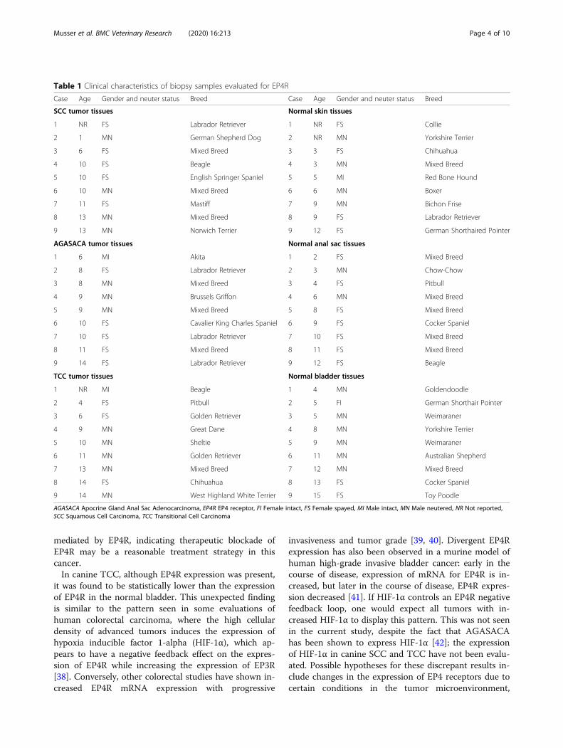
Table 1 Clinical characteristics of biopsy samples evaluated for EP4R
Case Age Gender and neuter status Breed Case Age Gender and neuter status Breed
SCC tumor tissues Normal skin tissues
1 NR FS Labrador Retriever 1 NR FS Collie
2 1 MN German Shepherd Dog 2 NR MN Yorkshire Terrier
3 6 FS Mixed Breed 3 3 FS Chihuahua
4 10 FS Beagle 4 3 MN Mixed Breed
5 10 FS English Springer Spaniel 5 5 MI Red Bone Hound
6 10 MN Mixed Breed 6 6 MN Boxer
7 11 FS Mastiff 7 9 MN Bichon Frise
8 13 MN Mixed Breed 8 9 FS Labrador Retriever
9 13 MN Norwich Terrier 9 12 FS German Shorthaired Pointer
AGASACA tumor tissues Normal anal sac tissues
1 6 MI Akita 1 2 FS Mixed Breed
2 8 FS Labrador Retriever 2 3 MN Chow-Chow
3 8 MN Mixed Breed 3 4 FS Pitbull
4 9 MN Brussels Griffon 4 6 MN Mixed Breed
5 9 MN Mixed Breed 5 8 FS Mixed Breed
6 10 FS Cavalier King Charles Spaniel 6 9 FS Cocker Spaniel
7 10 FS Labrador Retriever 7 10 FS Mixed Breed
8 11 FS Mixed Breed 8 11 FS Mixed Breed
9 14 FS Labrador Retriever 9 12 FS Beagle
TCC tumor tissues Normal bladder tissues
1 NR MI Beagle 1 4 MN Goldendoodle
2 4 FS Pitbull 2 5 FI German Shorthair Pointer
3 6 FS Golden Retriever 3 5 MN Weimaraner
4 9 MN Great Dane 4 8 MN Yorkshire Terrier
5 10 MN Sheltie 5 9 MN Weimaraner
6 11 MN Golden Retriever 6 11 MN Australian Shepherd
7 13 MN Mixed Breed 7 12 MN Mixed Breed
8 14 FS Chihuahua 8 13 FS Cocker Spaniel
9 14 MN West Highland White Terrier 9 15 FS Toy Poodle
AGASACA Apocrine Gland Anal Sac Adenocarcinoma, EP4R EP4 receptor, FI Female intact, FS Female spayed, MI Male intact, MN Male neutered, NR Not reported,SCC Squamous Cell Carcinoma, TCC Transitional Cell Carcinoma
Musser et al. BMC Veterinary Research (2020) 16:213 Page 4 of 10

alterations in cell density, or differences in tissue specificfunctional activities known to exist for EP receptors [15,40]. Further evaluation of additional tumor samples fromthroughout the urothelial tract, HIF-1⍺ levels, EP3R ex-pression, and the tumor microenvironment will beneeded to help clarify the role of EP4R in this tumortype.Multiple canine malignancies, including cutaneous
SCC, AGASACA, and TCC, have been shown to haveincreased COX-2 expression [24, 31, 43]. Blockade ofCOX-2 with NSAIDs, alone or in conjunction withchemotherapy, leads to tumor control and prolongedsurvival in many of these malignancies [26, 32]. How-ever, NSAIDs are not specific and attenuate the produc-tion of prostanoids other than PGE2 that are importantin homeostasis [33]. Thus, alternative treatment optionsthat are more specific, such as an EP4R antagonist, maybe clinically beneficial.In human medicine, the impact of EP4R antagonists
on cancer have been evaluated in murine models andnaturally occurring malignancies. Positive results havebeen noted in solid tumors including prostate, breast,and lung carcinoma, colorectal carcinoma, and melan-oma [9, 12]. In a cell line model of human urothelial car-cinoma, EP4R antagonists decreased cancer cellmigration and viability, and enhanced the effects of acommonly used chemotherapeutic, cisplatin, implyingthe potential role of EP4R antagonists in the treatmentof several human malignancies [44]. The in vitro andin vivo effects of an EP4R antagonist on canine cancerhas not been evaluated. However, the successful anti-inflammatory effects of the specific EP4R antagonist gra-piprant have been proven in both murine models [33]and dogs with naturally occurring osteoarthritis [45]. Indogs, grapiprant was proven to be safe and effective inlimiting inflammation and controlling pain. The
information gleaned from this study suggests that if thegene expression of EP4R correlates to protein expres-sion, contributing to the development of malignancy,blockade of EP4R with a piprant drug, such as grapipr-ant, may be a therapeutic approach for multiple caninetumors [33].Statistically significant differences in the expression of
EP4R were found for canine cutaneous SCC, AGA-SACA, and TCC. While this is exciting preliminary data,it must be interpreted in the light of a small sample sizeand recognition that gene expression does not necessar-ily correlate to significant protein expression. Similarly,confirmation that EP4R is the major isoform in caninemalignancy will need to be proven with evaluation of theexpression of EP1R, EP2R, and EP3R. As this representsa pilot and proof of concept study, additional tumorspecimens, histologic analysis of EP4R, evaluation ofEP4R protein expression, and correlation with case out-come and treatment, will be necessary to confirm theseresults and to make significant inferences regardingblockade of EP4R as a therapeutic strategy.
ConclusionsThe novel mRNA in situ hybridization platform, RNA-scope®, was successfully used to characterize EP4R ex-pression in three canine malignancies. This is the firstreported evaluation of EP4R in veterinary medicinebased on RNAscope®, and serves as a scientific basis forfuture evaluations of all EP receptors in both researchand clinical settings.The current study revealed positive gene expression of
EP4R in three common, aggressive canine malignancies.In canine cutaneous SCC and AGASACA, higher ex-pression of EP4R mRNA was identified when comparedto normal tissues. Canine TCC was found to have lessEP4R mRNA expression when compared to the normal
Table 2 EP4R mRNA expression levels in three canine malignancies
Tissuea Median copy number/Cell Median H-score Median % probe positive
SCC 3.1 (range: 0.2–10.0) 69.51 (range: 6.001–167.2) 32.45 (range: 3.485–59.54)
Skin 0.5 (range: 0.1–3.3) 15.56 (range: 5.071–56.80) 9.67 (range: 4.812–24.01)
p-value 0.0106* 0.0078* 0.0106*
AGASACA 2.9 (range: 1.1–32.64) 68.79 (range: 25.62–317.4) 32.06 (range: 12.55–89.59)
Anal Sac 1.5 (range: 0.2–4.8) 34.76 (range: 8.439–103.4) 18.28 (range: 7.118–56.19)
p-value 0.0142* 0.0188* 0.0400*
TCC 0.4 (range: 0.1–1.3) 11.62 (range: 3.671–32.23) 6.80 (range: 2.505–16.25)
Bladder 5.1 (range: 0.7–9.8) 80.00 (range: 23.06–139.6) 30.78 (range: 13.84–52.19)
p-value < 0.0001* < 0.0001* < 0.0001*
SCC Squamous Cell Carcinoma, AGASACA Apocrine Gland Anal Sac Adenocarcinoma, TCC Transitional Cell CarcinomaaNon-normally distributed data was evaluated with the Mann Whitney test between neoplastic cells and corresponding normal tissue type; the median and rangeare reported*Statistically significant
Musser et al. BMC Veterinary Research (2020) 16:213 Page 5 of 10

bladder. These results suggest that EP4R may be in-volved in the development of certain canine cancers. IfEP4R proves to be the major isoform in these tumortypes, therapeutic blockade with an EP4R antagonist,such as the commercially available grapiprant, may beadvantageous, changing the treatment paradigm in veter-inary medicine.
MethodsTumor and normal tissue samplesArchived biopsy and necropsy tissue specimens main-tained by the Department of Veterinary Pathology atIowa State University were searched for formalin-fixed,paraffin embedded tumor and normal tissue samples.
Based upon preliminary data, a power calculation indi-cated that 9 specimens in each group would be statisti-cally adequate to identify a difference of 2 transcriptcopy number/cell between tumors (SCC, AGASACA,and TCC) and respective normal tissue (skin, anal sac,and bladder mucosa) (alpha = 0.05, beta = 0.2). The insti-tutional animal care and use committee of Iowa StateUniversity did not require prior approval for retrospect-ive studies utilizing data and tissue specimens generatedthrough routine clinical assessment and care of patientanimals. The authors had permission from the Veterin-ary Teaching Hospital and Department of VeterinaryPathology to use the clinical data and samples, such thatowner and patient information remains anonymous.
Fig. 1 EP4R mRNA Expression Metrics in Squamous Cell Carcinoma vs. Normal Skin, Apocrine Gland Anal Sac Adenocarcinoma vs. Normal AnalSac, and Transitional Cell Carcinoma vs. Normal Bladder. Data presented as median and interquartile range, with all data points visualized; *denotes differences was statistically significant. SCC: Squamous Cell Carcinoma; AGASACA: Apocrine Gland Anal Sac Adenocarcinoma; TCC:Transitional Cell Carcinoma
Musser et al. BMC Veterinary Research (2020) 16:213 Page 6 of 10

Fig. 3 Analysis of ptger4 transcription in a section of normal anal sac (a-c) and apocrine gland anal sac adenocarcinoma (d, e) followingRNAscope® mRNA in situ hybridization with hematoxylin counter stain. a & d: Native photomicrograph to be analyzed for copy number/cell, H-Score, and percentage transcript expression using HALO software with RNAscope® Modules. b: Target cells were manually gated (yellow line) foranalysis; no gated cells for tumor image since all displayed cells were neoplastic. c & e: HALO generated probe markup from which copynumber/cell, H-Score, and percentage transcript expression are calculated
Fig. 2 Analysis of ptger4 transcription in a section of normal skin (a-c) and squamous cell carcinoma (d-f) following RNAscope® mRNA in situhybridization with hematoxylin counter stain. a & d: Native photomicrograph to be analyzed for copy number/cell, H-Score, and percentagetranscript expression using HALO software with RNAscope® Modules. b & e: Target cells were manually gated (yellow line) for analysis. c & f:HALO generated probe markup from which copy number/cell, H-Score, and percentage transcript expression are calculated
Musser et al. BMC Veterinary Research (2020) 16:213 Page 7 of 10

EP4R mRNA expression in tumor and normal tissuesamplesThe RNAscope® mRNA in situ hybridization platform(Advanced Cell Diagnostics, Hayward California USA)was used to evaluate mRNA expression of EP4R geneptger4 in the tumor and respective normal tissue sam-ples. A single-plex, manual chromogenic RNAscope®analysis was performed per the manufacturer’s instruc-tions and as previously reported with an RNAscope® 2.5High Definition (HD) – RED Assay (Catalog #322350,Advance Cell Diagnostics) [46–49]. Briefly, three sec-tions of each paraffin embedded tissue specimen werecut to a 5 μm depth. Preparations were baked for 1 h at60 °C, deparaffinized, and subsequently protease treatedto expose RNA. The three sections were then hybridizedseparately with a test probe targeting canine ptger4(Probe CI-PTGER4, Catalog # 499011, Advanced CellDiagnostics), a positive control probe targeting caninehouse-keeping gene ubc (CI-UBC Positive Control, Cata-log # 409851, Advanced Cell Diagnostics), and a negativecontrol probe targeting Bacillus subtilis dapB (DapBNegative Control, Catalog # 310043, Advance Cell Diag-nostics). Unlike traditional quantitative real time poly-merase chain reaction, normalization of EP4R expressionto the housekeeping gene is not required [46, 47].Hybridization to target mRNA was performed by incu-bating the preparation with the respective probe at 40 °Cfor 2 h in a HybEZ hybridization oven (Advanced Cell
Diagnostics). Subsequent wash and signal amplificationsteps were performed according to the manufacturer’sprotocol. Target mRNA was detected using alkalinephosphatase Fast Red chromogenic stain (Catalog #320701, Advanced Cell Diagnostics). Samples were alsostained with hematoxylin (American Master Technology,California USA) to permit visualization of nuclei. To ini-tially assess the performance of the ptger4 RNAscope®experimental probe, ptger4 expression was assessed intwo sections each of normal canine heart, lung, and kid-ney tissue. Expression of ptger4 expression has been pre-viously reported in these canine tissues [28] and wasidentified in all three tissues types with the RNAscope®analysis with this ptger4 probe.
Quantification of mRNA expressionFor each hybridized slide, ten 400x magnified non-overlapping microscopic field views were digitallyphotographed. Digitized photomicrographs were thenevaluated with the RNAscope® image analysis softwareHALO with the RNAscope® Modules (Indica Labs,Albuquerque New Mexico USA) [50]. For tumor sam-ples, the neoplastic cells were manually identified andgated for analysis. For normal tissue samples, the appro-priate normal cell population was manually identifiedand gated for analysis. Samples were considered to haveadequate residual RNA for ptger4 expression analysis ifthe corresponding positive control ubc hybridization
Fig. 4 Analysis of ptger4 transcription in a section of normal bladder (a-c) and transitional cell carcinoma (d-f) following RNAscope® mRNA in situhybridization with hematoxylin counter stain. a & d: Native photomicrograph to be analyzed for copy number/cell, H-Score, and percentagetranscript expression using HALO software with RNAscope® Modules. b & e: Target cells were manually gated (yellow line) for analysis. c & f:HALO generated probe markup from which copy number/cell, H-Score, and percentage transcript expression are calculated
Musser et al. BMC Veterinary Research (2020) 16:213 Page 8 of 10
yielded > 15 transcript dots/cell in > 90% of the targetcells. The following metrics were then calculated forptger4 expression based upon the cumulative analysis re-sults of the ten digitized photomicrographs for the slide:average transcript copy number/cell, percent probe posi-tive expression (percent of cells positive for EP4RmRNA), and H-score (a weighted expression scale usedto evaluate heterogeneity in marker expression; A Guidefor RNAscope® Data Analysis, Advanced Cell Diagnos-tics). These combined techniques allow for quantifica-tion of the gene expression within a specific cell typeand tissue context, and comparison of expression acrosstumor types.
Statistical analysisA D’Agostino Pearson test was used to evaluate normal-ity of copy number per cell, H-score, and percent probepositive expression for each tumor type and normal tis-sues; all data was non-normally distributed. A Wilcoxonrank-sum test was used to assess differences in theaforementioned parameters between tumors and re-spective normal tissue types. Statistical significance wasdefined as p < 0.05. Where applicable, nonparametricdata is presented as median ± range. Statistical compari-sons were performed using a commercially availablesoftware package (Prism 6. GraphPad Software, Inc. SanDiego CA USA). Power calculation was performed witha separate software package (MedCalc. MedCalc Soft-ware BV. Ostend, Belgium).
AbbreviationsAGASACA: Apocrine Gland Anal Sac Adenocarcinoma; COX2: Cyclooxygenaseenzyme 2; EP4R: EP4 Receptor; FI: Female Intact; FS: Female Spayed; HIF-1⍺: Hypoxia inducible factor 1-alpha; MI: Male Intact; MN: Male Neutered;NR: Not Reported; PG: Prostaglandins; PGE2: Prostaglandin E2;SCC: Squamous Cell Carcinoma; TCC: Transitional Cell Carcinoma;UVB: Ultraviolet B
AcknowledgementsPortions of the data presented herein were previously presented via an oralabstract at the Veterinary Cancer Society Annual Conference in Louisville, KYin October, 2018. Data describing the expression of EP4R in canineosteosarcoma was presented via a poster presentation at the EuropeanCollege of Veterinary Internal Medicine - Companion Animals AnnualCongress in Rotterdam, The Netherlands in September, 2018.
Authors’ contributionsAll authors (MM, AV, CJ, JH and RP) participated in the initial study planningand literature review. AV identified appropriate biopsy specimens for analysis.JH and RP completed RNAscope®, HALO, and subsequent analysis on allspecimens. AV completed statistical analysis. MM an CJ interpreted the datain light of known literature and prepared the manuscript. All authors (MM,AV, CJ, JH and RP) reviewed the manuscript and had input on the finalversion. The author(s) read and approved the final manuscript.
FundingThe research described herein was generously supported by Fetch-a-Cureand internal funding opportunities from Iowa State University. Neither entityhad any role in the design of the study, or in the collection, analysis, and in-terpretation of the data, or in writing the manuscript.
Availability of data and materialsThe data that support the findings of this study are available from thecorresponding author upon reasonable request.
Ethics approval and consent to participateThe institutional animal care and use committee of Iowa State University didnot require prior approval for retrospective studies utilizing data and tissuespecimens generated through routine clinical assessment and care of patientanimals. The authors had permission from the Veterinary Teaching Hospitaland Departments of Veterinary Pathology to use the clinical data andsamples, such that owner and patient information remains anonymous.
Consent for publicationNot applicable.
Competing interestsMM and CJ receive funding from Elanco for clinical studies investigating theuse of grapiprant. CJ is a consultant and speaker for Elanco and receiveshonorarium for these services.
Author details1Department of Veterinary Clinical Sciences, Iowa State University College ofVeterinary Medicine, Ames, IA, USA. 2Department of Veterinary Pathology,Iowa State University College of Veterinary Medicine, Ames, IA, USA. 3Presentaddress: University of Georgia College of Veterinary Medicine, 501 D.W.Brooks Drive, Athens, GA 30602, USA.
Received: 2 March 2020 Accepted: 16 June 2020
References1. O'Byrne KJ, Dalgleish AG. Chronic immune activation and inflammation as
the cause of malignancy. Br J Cancer. 2001;85(4):473–83.2. Liu B, Qu L, Yan S. Cyclooxygenase-2 promotes tumor growth and
suppresses tumor immunity. Cancer Cell Int. 2015;15:106.3. Yan W, Wistuba II, Emmert-Buck MR, Erickson HS. Squamous cell carcinoma
- similarities and differences among anatomical sites. Am J Cancer Res.2011;1(3):275–300.
4. Moon H, White AC, Borowsky AD. New insights into the functions of cox-2in skin and esophageal malignancies. Exp Mol Med. 2020;52:538–47.
5. Muller-Decker K. Cyclooxygenase-dependent signaling is causally linked tonon-melanoma skin carcinogenesis: pharmacological, genetic, and clinicalevidence. Cancer Metastasis Rev. 2011;30(3–4):343–61.
6. Higashi Y, Kanekura T, Kanzaki T. Enhanced expression of cyclooxygenase(COX)-2 in human skin epidermal cancer cells: evidence for growthsuppression by inhibiting COX-2 expression. Int J Cancer. 2000;86(5):667–71.
7. Al-Maghrabi B, Gomaa W, Abdelwahed M, Al-Maghrabi J. Increased COX-2Immunostaining in Urothelial carcinoma of the urinary bladder is associatedwith invasiveness and poor prognosis. Anal Cell Pathol (Amst). 2019;2019:5026939.
8. Elzagheid A, Emaetig F, Alkikhia L, Buhmeida A, Syrjanen K, El-Faitori O,Latto M, Collan Y, Pyrhonen S. High cyclooxygenase-2 expression isassociated with advanced stages in colorectal cancer. Anticancer Res. 2013;33(8):3137–43.
9. Markovic T, Jakopin Z, Dolenc MS, Mlinaric-Rascan I. Structural features ofsubtype-selective EP receptor modulators. Drug Discov Today. 2017;22(1):57–71.
10. Shao J, Jung C, Liu C, Sheng H. Prostaglandin E2 stimulates the beta-catenin/T cell factor-dependent transcription in colon cancer. J Biol Chem.2005;280(28):26565–72.
11. Take Y, Koizumi S, Nagahisa A. Prostaglandin E receptor 4 antagonist incancer immunotherapy: mechanisms of action. Front Immunol. 2020;11:324.
12. Konya V, Marsche G, Schuligoi R, Heinemann A. E-type prostanoid receptor4 (EP4) in disease and therapy. Pharmacol Ther. 2013;138(3):485–502.
13. Coleman RA, Smith WL, Narumiya S. International Union of Pharmacologyclassification of prostanoid receptors: properties, distribution, and structureof the receptors and their subtypes. Pharmacol Rev. 1994;46(2):205–29.
14. Yokoyama U, Iwatsubo K, Umemura M, Fujita T, Ishikawa Y. The prostanoidEP4 receptor and its signaling pathway. Pharmacol Rev. 2013;65(3):1010–52.
Musser et al. BMC Veterinary Research (2020) 16:213 Page 9 of 10
15. O'Callaghan G, Houston A. Prostaglandin E2 and the EP receptors inmalignancy: possible therapeutic targets? Br J Pharmacol. 2015;172(22):5239–50.
16. Lee JL, Kim A, Kopelovich L, Bickers DR, Athar M. Differential expression of Eprostanoid receptors in murine and human non-melanoma skin cancer. JInvest Dermatol. 2005;125(4):818–25.
17. Miyata Y, Ohba K, Kanda S, Nomata K, Eguchi J, Hayashi T, Kanetake H.Pathological function of prostaglandin E2 receptors in transitional cellcarcinoma of the upper urinary tract. Virchows Arch. 2006;448(6):822–9.
18. Mutoh M, Watanabe K, Kitamura T, Shoji Y, Takahashi M, Kawamori T, Tani K,Kobayashi M, Maruyama T, Kobayashi K, et al. Involvement of prostaglandin Ereceptor subtype EP(4) in colon carcinogenesis. Cancer Res. 2002;62(1):28–32.
19. Chen DS, Mellman I. Oncology meets immunology: the cancer-immunitycycle. Immunity. 2013;39(1):1–10.
20. Xu XC. COX-2 inhibitors in cancer treatment and prevention, a recentdevelopment. Anti-Cancer Drugs. 2002;13(2):127–37.
21. Millanta F, Andreani G, Rocchigiani G, Lorenzi D, Poli A. Correlation betweencyclo-oxygenase-2 and vascular endothelial growth factor expression incanine and feline squamous cell carcinomas. J Comp Pathol. 2016;154(4):297–303.
22. Bardagi M, Fondevila D, Ferrer L. Immunohistochemical detection of COX-2in feline and canine actinic keratoses and cutaneous squamous cellcarcinoma. J Comp Pathol. 2012;146(1):11–7.
23. Khan KN, Knapp DW, Denicola DB, Harris RK. Expression of cyclooxygenase-2in transitional cell carcinoma of the urinary bladder in dogs. Am J Vet Res.2000;61(5):478–81.
24. Knudsen CS, Williams A, Brearley MJ, Demetriou JL. COX-2 expression incanine anal sac adenocarcinomas and in non-neoplastic canine anal sacs.Vet J. 2013;197(3):782–7.
25. Millanta F, Asproni P, Canale A, Citi S, Poli A. COX-2, mPGES-1 and EP2receptor immunohistochemical expression in canine and feline malignantmammary tumours. Vet Comp Oncol. 2016;14(3):270–80.
26. Dore M. Cyclooxygenase-2 expression in animal cancers. Vet Pathol. 2011;48(1):254–65.
27. Millanta F, Asproni P, Cancedda S, Vignoli M, Bacci B, Poli A.Immunohistochemical expression of COX-2, mPGES and EP2 receptor innormal and reactive canine bone and in canine osteosarcoma. J CompPathol. 2012;147(2–3):153–60.
28. Castleberry TA, Lu B, Smock SL, Owen TA. Molecular cloning and functionalcharacterization of the canine prostaglandin E2 receptor EP4 subtype.Prostaglandins Other Lipid Mediat. 2001;65(4):167–87.
29. Musser ML, Viall AK, Phillips RL, Hostetter JM, Johannes CM. Analysis ofprostaglandin EP4 receptor gene expression in canine osteosarcoma. Can JVet Res. in press.
30. Boonsoda S, Wanikiat P. Possible role of cyclooxygenase-2 inhibitors asanticancer agents. Vet Rec. 2008;162(5):159–61.
31. Spugnini EP, Porrello A, Citro G, Baldi A. COX-2 overexpression in caninetumors: potential therapeutic targets in oncology. Histol Histopathol. 2005;20(4):1309–12.
32. Knapp DW, Ruple-Czerniak A, Ramos-Vara JA, Naughton JF, Fulkerson CM,Honkisz SI. A nonselective cyclooxygenase inhibitor enhances the activity ofvinblastine in a naturally-occurring canine model of invasive urothelialcarcinoma. Bladder Cancer. 2016;2(2):241–50.
33. Kirkby Shaw K, Rausch-Derra LC, Rhodes L. Grapiprant: an EP4 prostaglandinreceptor antagonist and novel therapy for pain and inflammation. Vet MedSci. 2016;2(1):3–9.
34. Vail D, Thamm D, Liptak J. Withrow and MacEwen’s small animal clinicaloncology. 6th ed. Philadelphia: Saunders; 2019.
35. Waropastrakul S, Munday JS, French AF. Infrequent detection ofpapillomaviral DNA within canine cutaneous squamous cell carcinomas,haemangiosarcomas and healthy skin on the ventrum of dogs. VetDermatol. 2012;23(3):197–e141.
36. An KP, Athar M, Tang X, Katiyar SK, Russo J, Beech J, Aszterbaum M,Kopelovich L, Epstein EH Jr, Mukhtar H, et al. Cyclooxygenase-2 expressionin murine and human nonmelanoma skin cancers: implications fortherapeutic approaches. Photochem Photobiol. 2002;76(1):73–80.
37. Valvo F, Ciurlia E, Avuzzi B, Doci R, Ducreux M, Roelofsen F, Roth A, Trama A,Wittekind C, Bosset JF. Cancer of the anal region. Crit Rev Oncol Hematol.2019;135:115–27.
38. Fujino H. The roles of EP4 Prostanoid receptors in cancer malignancysignaling. Biol Pharm Bull. 2016;39(2):149–55.
39. Chell SD, Witherden IR, Dobson RR, Moorghen M, Herman AA, QualtroughD, Williams AC, Paraskeva C. Increased EP4 receptor expression in colorectalcancer progression promotes cell growth and anchorage independence.Cancer Res. 2006;66(6):3106–13.
40. Otake S, Yoshida K, Seira N, Sanchez CM, Regan JW, Fujino H, Murayama T.Cellular density-dependent down-regulation of EP4 prostanoid receptors viathe up-regulation of hypoxia-inducible factor-1alpha in HCA-7 human coloncancer cells. Pharmacol Res Perspect. 2015;3(1):e00083.
41. Taylor JA 3rd, Ristau B, Bonnemaison M, Voznesensky OS, Hegde P, KuchelGA, Pilbeam CC. Regulation of the prostaglandin pathway duringdevelopment of invasive bladder cancer in mice. Prostaglandins Other LipidMediat. 2009;88(1–2):36–41.
42. Yamazaki H, Tanaka T, Mie K, Nishida H, Miura N, Akiyoshi H. Assessment ofpostoperative adjuvant treatment using toceranib phosphate againstadenocarcinoma in dogs. J Vet Intern Med. 2020;34:1272–81.
43. Mohammed SI, Bennett PF, Craig BA, Glickman NW, Mutsaers AJ, SnyderPW, Widmer WR, DeGortari AE, Bonney PL, Knapp DW. Effects of thecyclooxygenase inhibitor, piroxicam, on tumor response, apoptosis, andangiogenesis in a canine model of human invasive urinary bladder cancer.Cancer Res. 2002;62(2):356–8.
44. Kashiwagi E, Inoue S, Mizushima T, Chen J, Ide H, Kawahara T, Reis LO, BarasAS, Netto GJ, Miyamoto H. Prostaglandin receptors induce urothelialtumourigenesis as well as bladder cancer progression and cisplatinresistance presumably via modulating PTEN expression. Br J Cancer. 2018;118(2):213–23.
45. Rausch-Derra L, Huebner M, Wofford J, Rhodes L. A prospective,randomized, masked, placebo-controlled multisite clinical study ofGrapiprant, an EP4 prostaglandin receptor antagonist (PRA), in dogs withosteoarthritis. J Vet Intern Med. 2016;30(3):756–63.
46. Wang F, Flanagan J, Su N, Wang LC, Bui S, Nielson A, Wu XY, Vo HT, Ma XJ,Luo YL. RNAscope a novel in situ RNA analysis platform for formalin-fixed,paraffin-embedded tissues. J Mol Diagn. 2012;14(1):22–9.
47. Bingham V, McIlreavey L, Greene C, O'Doherty E, Clarke R, Craig S, Salto-Tellez M, McQuaid S, Lewis C, James J. RNAscope in situ hybridizationconfirms mRNA integrity in formalin-fixed, paraffin-embedded cancer tissuesamples. Oncotarget. 2017;8(55):93392–403.
48. Karger C, Machura K, Schneider A, Hugo C, Todorov VT, Kurtz A. COX-2-derived PGE2 triggers hyperplastic renin expression and hyperreninemia inaldosterone synthase-deficient mice. Pflugers Arch. 2018;470(7):1127–37.
49. Yokoyama N, Ohta H, Yamazaki J, Kagawa Y, Ichii O, Khoirun N, Morita T,Osuga T, Lim SY, Sasaki N, et al. Localization of toll-like receptor (TLR) 2 andTLR4 mRNA in the colorectal mucosa of miniature dachshunds withinflammatory colorectal polyps. J Comp Pathol. 2017;156(2–3):183–90.
50. Anderson CM, Zhang B, Miller M, Butko E, Wu X, Laver T, Kernag C, Kim J,Luo Y, Lamparski H, et al. Fully automated RNAscope in situ hybridizationassays for formalin-fixed paraffin-embedded cells and tissues. J CellBiochem. 2016;117(10):2201–8.
Publisher’s NoteSpringer Nature remains neutral with regard to jurisdictional claims inpublished maps and institutional affiliations.
Musser et al. BMC Veterinary Research (2020) 16:213 Page 10 of 10