Terutroban, a Thromboxane/Prostaglandin Endoperoxide Receptor Antagonist, Increases Survival in...
29
JPET#165787 1 TERUTROBAN, A TP RECEPTOR ANTAGONIST, INCREASES SURVIVAL IN STROKE-PRONE RATS BY PREVENTING SYSTEMIC INFLAMMATION AND ENDOTHELIAL DYSFUNCTION. COMPARISON WITH ASPIRIN AND ROSUVASTATIN. Paolo Gelosa, Rossana Ballerio, Cristina Banfi, Elena Nobili, Anita Gianella, Alice Pignieri, Maura Brioschi, Uliano Guerrini, Laura Castiglioni, Vanessa Blanc-Guillemaud, Laurence Lerond, Elena Tremoli, and Luigi Sironi Department of Pharmacological Sciences, University of Milan (P.G., A.P., U.G., L.C., E.T., L.S.); Monzino Cardiologic Centre IRCCS, Milan, Italy (R.B., C.B., E.N., A.G., M.B., E.T., L.S.); Institut de Recherches Internationales Servier (IRIS), Coubevoie Cedex, France (V.G.B., L.L.) JPET Fast Forward. Published on March 23, 2010 as DOI:10.1124/jpet.110.165787 Copyright 2010 by the American Society for Pharmacology and Experimental Therapeutics.
-
Upload
independent -
Category
Documents
-
view
3 -
download
0
Transcript of Terutroban, a Thromboxane/Prostaglandin Endoperoxide Receptor Antagonist, Increases Survival in...
JPET#165787
1
TERUTROBAN, A TP RECEPTOR ANTAGONIST, INCREASES SURVIVAL IN
STROKE-PRONE RATS BY PREVENTING SYSTEMIC INFLAMMATION AND
ENDOTHELIAL DYSFUNCTION. COMPARISON WITH ASPIRIN AND
ROSUVASTATIN.
Paolo Gelosa, Rossana Ballerio, Cristina Banfi, Elena Nobili, Anita Gianella, Alice Pignieri,
Maura Brioschi, Uliano Guerrini, Laura Castiglioni, Vanessa Blanc-Guillemaud, Laurence
Lerond, Elena Tremoli, and Luigi Sironi
Department of Pharmacological Sciences, University of Milan (P.G., A.P., U.G., L.C., E.T.,
L.S.); Monzino Cardiologic Centre IRCCS, Milan, Italy (R.B., C.B., E.N., A.G., M.B., E.T.,
L.S.); Institut de Recherches Internationales Servier (IRIS), Coubevoie Cedex, France
(V.G.B., L.L.)
JPET Fast Forward. Published on March 23, 2010 as DOI:10.1124/jpet.110.165787
Copyright 2010 by the American Society for Pharmacology and Experimental Therapeutics.
JPET#165787
2
Running title: Terutroban increases survival in stroke-prone rats
Address correspondence to:
Luigi Sironi,
Dipartimento di Scienze Farmacologiche, Università degli Studi di Milano,
Via Balzaretti 9,20133 Milano, ITALY.
Tel +39 0250318388, Fax +39 0250318250
N. of text pages: 23
Table: 1
Figures: 5
N. of References: 26
N. of words in Abstract: 227
N. of words in Introduction: 390
N. of words in Discussion: 1027
Abbreviations: SHRSP, spontaneously hypertensive stroke-prone rats; MRI, magnetic
resonance imaging; TPr, thromboxane/prostaglandin endoperoxide receptor; ASA, aspirin;
RSV, rosuvastatin; eNOS, Endothelial Nitric Oxide Synthase; TXB2, Thromboxane B2; IL
1beta, Interleukin 1, beta; CRP, C-reactive protein; sICAM-1, soluble intercellular adhesion
molecule-1
Recommended section: Cardiovascular
JPET#165787
3
Abstract
This study investigated the efficacy of terutroban, a specific thromboxane/prostaglandin
endoperoxide receptor (TPr) antagonist, on stroke incidence in spontaneously hypertensive
stroke-prone rats (SHRSP). The effects of terutroban were compared to those of aspirin, an
another anti-platelet agent, and rosuvastatin, known to exert end-organ protection in SHRSP.
Salt-loaded male SHRSP were treated orally once a day with vehicle, terutroban (30
mg/kg/day), aspirin (60 mg/kg/day) or rosuvastatin (10 mg/kg/day). Compared with vehicle,
and regardless of any effect on blood pressure or serum TXB2 levels, terutroban significantly
increased survival (p<0.001) as a consequence of a delayed brain lesions occurrence
monitored by magnetic resonance imaging (MRI) (p<0.001), and a delayed increase of
proteinuria (p<0.001). Terutroban decreased cerebral mRNA transcription of IL-1beta, TGF-
beta and MCP-1 after 6 weeks of dietary treatment. Terutroban also prevented the
accumulation of urinary acute-phase proteins at high molecular weight (HMW), identified as
markers of systemic inflammation, and assessed longitudinally by one-dimensional
electrophoresis. Terutroban has also protective effects on the vasculature as suggested by the
preservation of endothelial function and endothelial nitric oxide synthase (eNOS) expression
in isolated carotid arteries. These effects are similar to those obtained with rosuvastatin, and
superior to those of aspirin. Terutroban increases survival in SHRSP by reducing systemic
inflammation as well as preserving endothelial function. These data support clinical
development of terutroban in the prevention of cerebrovascular and cardiovascular
complications of atherothrombosis.
JPET#165787
4
Introduction
Several clinical and experimental studies (Widlansky et al., 2003; Huang and Vita, 2006)
support the hypothesis that endothelial dysfunction and systemic inflammation play key roles
in the pathogenesis of vascular diseases, including myocardial and brain ischemia. Human
studies have demonstrated positive association between systemic inflammation induced by
endotoxin infusion and marked endothelial dysfunction as well as impaired responses to
vasoactive compounds (Pleiner et al., 2004). An analysis of the Framingham Heart Study
Offspring cohort found that serum CRP, IL-6 and sICAM-1 levels inversely correlated with
brachial artery flow-mediated dilation and reactive hyperemia in the forearm, although this
relationship was weakened after adjusting for traditional risk factors (Vita et al., 2004).
Spontaneously hypertensive stroke-prone rats (SHRSP) develop hypertension and proteinuria
and die after the onset of cerebrovascular damage, which is invariably preceded by systemic
inflammation and endothelial dysfunction (Sironi et al., 2001; Ballerio et al., 2007). Notably,
systemic inflammation is characterized by an accumulation - in serum and urine - of acute-
phase high molecular weight (HMW) proteins such as thiostatin, the most common marker of
inflammation in rat (Sironi et al., 2001). In SHRSP, brain lesions have a vasogenic origin due
to the blood-brain barrier impairment (Guerrini et al., 2002). Therefore, this model is
particularly suited to reveal cerebrovascular benefits of drugs acting on the inflammatory
cascade and/or endothelial dysfunction.
The purpose of this study was to evaluate in SHRSP the effects of terutroban, a highly
selective and long-acting TP receptor (TPr) antagonist with antithrombotic,
antivasoconstrictive and anti-inflammatory/antiatherosclerotic properties (Cimetière et al.,
1998). Previous experimental studies have demonstrated that terutroban prevents vascular
wall proliferation and atherogenesis (Cheng et al., 2002; Worth et al., 2005; Viles-Gonzalez
et al., 2005), increases anti-oxidant enzymes like glutathione peroxidase (Sebekova et al.,
JPET#165787
5
2007) and has anti-inflammatory actions in vitro and in vivo (decreased macrophage
infiltration and ICAM-1) (Cayatte et al., 2000). Terutroban also improved endothelial
function in patients with coronary artery disease treated with aspirin (Belhassen et al., 2003).
Terutroban is developed in secondary prevention of cerebrovascular and cardiovascular
events in patients with an history of ischemic stroke or transient ischemic attack (Bousser et
al., 2009a and 2009b).
In this study, the optimally effective dose of terutroban established in previous works was
compared to those of aspirin (ASA) and rosuvastatin (RSV) to provide comparative data on
end-organ protection and anti-inflammation in SHRSP (Sironi et al., 2005).
JPET#165787
6
Methods
Animals and protocol
Male SHRSP aged 4-5 weeks were obtained from Charles River, Italy (Calco, Lecco, Italy)
and were cared for in accordance with our Institution’s guidelines. Fifty-two SHRSP switched
to the Japanese permissive low-potassium, low-protein and high-sodium diet (Japanese
permissive diet, JPD; Laboratorio Dr. Piccioni, Gessate, Italy: 18.7% protein, 0.63%
potassium, 0.37% sodium) plus 1% NaCl in drinking water, were randomly divided into four
groups (n=13 each group) and treated orally (gavage) with vehicle (1% hydroxy-ethyl
cellulose), terutroban (S 18886) 30 mg/kg/day, ASA 60 mg/kg/day, or RSV 10 mg/kg/day.
The dose of terutroban (Servier, France) and rosuvastatin (a kind gift from Astra Zeneca, UK)
were chosen on the basis of previous studies performed in the lab (Sironi et al., 2005; Nobili
et al., 2006; Gianella et al., 2007). ASA (Sigma, St Louis, Mo) dosage was chosen on the
basis of published studies (Qiu et al., 2003; Knight and Johns, 2005).
Baseline measurements were made before the onset of the diet. Systolic arterial blood
pressure measured by means of tail-cuff plethysmography (PB Recorder 8006, Ugo Basile,
Varese, Italy) and weight were evaluated weekly, then rats were individually housed in
metabolic cages for 24 hours to collect urine for proteinuria determinations (Bradford’s
method) and proteomic studies. Blood was drawn every week from the tail vein; serum was
obtained and stored at -20°C until analysed. All rats underwent weekly magnetic resonance
imaging (MRI) until 24-h proteinuria reached 100 mg/day, and then every two days until
cerebrovascular damage was detected. After six weeks (i.e when the vehicle-treated rats
developed brain lesions) five animals from each group were sacrificed to collect the brain as
well as the carotid artery.
MRI evaluation of brain damage
JPET#165787
7
The rats were anesthetised with 1.5% isofluorane (Merial, Toulose, France) in 70% N2/30%
O2, and placed inside a Bruker AvanceII 4.7T with a micro-imaging accessory. After a scout
image, sixteen contiguous 1 mm thick slices were analyzed caudally to the olfactory bulb
using a field of view (FOV) of 4 x 4 cm2, and a turbo spin echo sequence with 16 echoes per
excitation, 10 ms inter-echo time, 85 ms equivalent echo time, and 4 s repetition time. Eight
T2-weighted images of 128 x 128 pixels (zero-filled to 256x256) were averaged in 8’30”. The
occurrence of lesions was defined as the presence of areas of high signal intensity on T2-
weighted images.
Proteinuria studies
One-dimensional electrophoresis (1-DE) of urine proteins (50 μg) was run in the presence of
SDS without sample reduction in a discontinuous buffer system on 4-12% polyacrylamide
gels stained with Colloidal Blue. Densitometry was performed using Quantity One version
4.5.2 (Biorad, Hercules, CA) to evaluate the percentage of low molecular weight (LMW) and
high molecular weight (HMW) proteins density.
Determination of TXB2 and 11-dehydro-TXB2
The serum levels of TXB2 and urinary levels of 11-dehydro-TXB2 were measured using
commercial kits (Cayman Chemical Co., Ann Arbor, MI).
Brain tissue expression of inflammatory markers
After six weeks of dietary treatment, five animals from each group were sacrificed to collect
the brains. Total RNA was prepared by means of guanidium thiocyanate denaturation from
forebrain homogenates. Reverse transcription polymerase chain reaction (RT-PCR) was used
JPET#165787
8
to evaluate the expression of IL-1beta, TGF-beta, and MCP-1. All the reagents used were
purchased from Invitrogen (Carlsbad, CA)
Expression of eNOS
eNOS expression was evaluated by RT-PCR on carotid artery homogenates from animals
sacrificed after six weeks of treatment.
Endothelial dysfunction
Isolated carotid artery rings (3 mm) were suspended in an individual organ bath filled with
Krebs solution and their vascular reactivity was evaluated as previously described (Ballerio et
al., 2007). Indomethacin (10-5 mol/L; Chiesi Farmaceutici S.p.A., Parma) was added to Krebs
solutions in order to inhibit prostanoid synthesis. Arteries were challenged with KCl (100
mM/L) to check the viability of tissues; vessels not responding to KCl were discarded.
Vascular smooth muscle function was determined by cumulative addition of L-phenylephrine
(L-Phe; Sigma-Aldrich, St. Louis, MO, USA) (10-9–10-5 mol/L), the contraction response
being expressed as the percentage of KCl response. Subsequently, the rings were constricted
to their individual EC80 value for L-Phe, and maximum smooth muscle relaxation to sodium
nitroprusside was determined (SNP; Sigma-Aldrich, St. Louis, MO, USA) (10-10–3x10⁻⁶
mol/L). After wash-out, the rings were constricted to their individual EC80 value for L-Phe,
and endothelium-dependent relaxation in response to acetylcholine (Ach; Sigma-Aldrich, St.
Louis, MO, USA) (10-9–10-5 mol/L) was studied both in the absence or presence of L-NAME
10-4 mol/L. The relaxation responses were expressed as the percentage of L-Phe-induced
contraction.
Statistics
JPET#165787
9
Between-group differences were computed by means of analysis of variance (ANOVA)
followed by an appropriate post hoc test; the between-group differences in proteinuria, LMW
and HMW protein density were computed by means of ANOVA for repeated measurements
over time followed by Tukey’s post hoc test. An unpaired t test was used to compare baseline
and vehicle-treated group data. Concentration–response curves were statistically analysed
using ANOVA followed by Tukey’s or Tamhane’s T2 post hoc test. Sensitivity to the
antagonists (pD2) was expressed as the negative logarithm of half-maximal effective
concentration (EC50) calculated from individual curves. Results are expressed as means ± S.D.
P<0.05 was considered statistically significant.
JPET#165787
10
Results
Physiological parameters and survival of SHRSP
Body weight increased similarly in all experimental groups. The severe hypertension that
developed was not affected by any of the drug treatments (Fig. 1A). Plasma total cholesterol
and triglyceride levels (42.38±3.89 and 68.5±8.96 mg/dl respectively at baseline), did not
significantly change during the treatment period in any groups. Vehicle-treated animals
developed cerebral lesions 42.4±10.8 days after starting salt loading. All treatments
significantly delayed the appearance of cerebrovascular damages (Fig. 1B and 1D). However,
the delay of occurrence observed under terutroban (87.6±19.2 days; p<0.001) was greater
than that induced by aspirin (63.9±9.01 days; p<0.05) and comparable to that observed after
RSV (85.6±16.9 days; p<0.001). Comparison of survival clearly shows the effectiveness of all
treatments (Fig.1C). Compared to the vehicle group, survival was similarly increased by
terutroban and RSV (p<0.001) and this effect was significantly superior to that observed after
aspirin treatment (RSV p<0.05; terutroban p< 0.01).
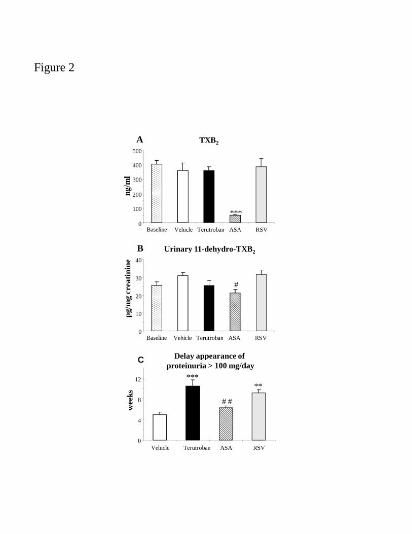
Serum TXB2 and urinary 11-dehydro-TXB2 levels
As expected, serum TXB2 and urinary 11-dehydro-TXB2 levels were significantly decreased
by ASA while the levels were not affected by salt loading, nor by RSV or terutroban
treatment (Fig. 2A and 2B).
Proteinuria and composition of urinary proteins
The SHRSP receiving vehicle developed progressively a severe proteinuria. After 4.7±1.3
weeks of salt loading, proteinuria was higher than 100 mg/day and increased rapidly and
linearly to reach an average of 266±28.9 mg/day after 7 weeks. Treatment with terutroban and
RSV delayed significantly the increase in proteinuria (10.5±3.4 weeks, p<0.001, and 9.1±1.9
JPET#165787
11
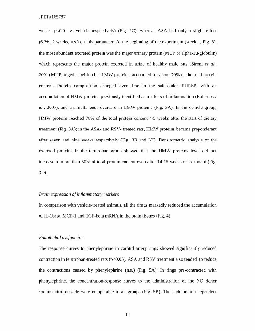
weeks, p<0.01 vs vehicle respectively) (Fig. 2C), whereas ASA had only a slight effect
(6.2±1.2 weeks, n.s.) on this parameter. At the beginning of the experiment (week 1, Fig. 3),
the most abundant excreted protein was the major urinary protein (MUP or alpha-2u-globulin)
which represents the major protein excreted in urine of healthy male rats (Sironi et al.,
2001).MUP, together with other LMW proteins, accounted for about 70% of the total protein
content. Protein composition changed over time in the salt-loaded SHRSP, with an
accumulation of HMW proteins previously identified as markers of inflammation (Ballerio et
al., 2007), and a simultaneous decrease in LMW proteins (Fig. 3A). In the vehicle group,
HMW proteins reached 70% of the total protein content 4-5 weeks after the start of dietary
treatment (Fig. 3A); in the ASA- and RSV- treated rats, HMW proteins became preponderant
after seven and nine weeks respectively (Fig. 3B and 3C). Densitometric analysis of the
excreted proteins in the terutroban group showed that the HMW proteins level did not
increase to more than 50% of total protein content even after 14-15 weeks of treatment (Fig.
3D).
Brain expression of inflammatory markers
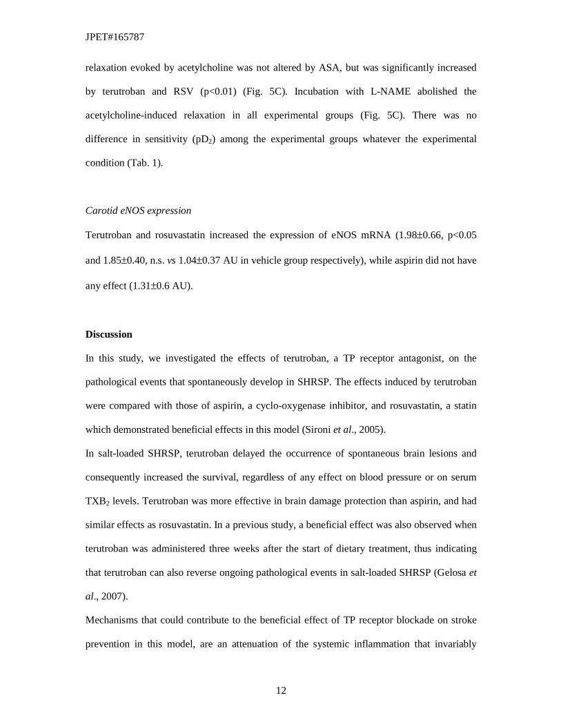
In comparison with vehicle-treated animals, all the drugs markedly reduced the accumulation
of IL-1beta, MCP-1 and TGF-beta mRNA in the brain tissues (Fig. 4).
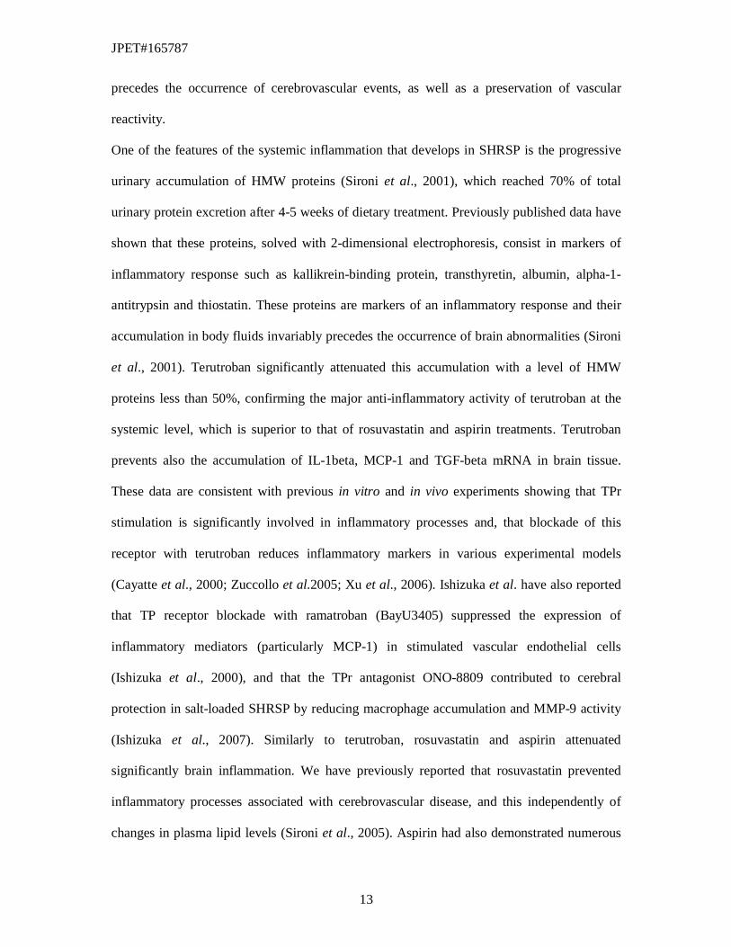
Endothelial dysfunction
The response curves to phenylephrine in carotid artery rings showed significantly reduced
contraction in terutroban-treated rats (p<0.05). ASA and RSV treatment also tended to reduce
the contractions caused by phenylephrine (n.s.) (Fig. 5A). In rings pre-contracted with
phenylephrine, the concentration-response curves to the administration of the NO donor
sodium nitroprusside were comparable in all groups (Fig. 5B). The endothelium-dependent
JPET#165787
12
relaxation evoked by acetylcholine was not altered by ASA, but was significantly increased
by terutroban and RSV (p<0.01) (Fig. 5C). Incubation with L-NAME abolished the
acetylcholine-induced relaxation in all experimental groups (Fig. 5C). There was no
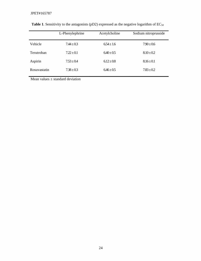
difference in sensitivity (pD2) among the experimental groups whatever the experimental
condition (Tab. 1).
Carotid eNOS expression
Terutroban and rosuvastatin increased the expression of eNOS mRNA (1.98±0.66, p<0.05
and 1.85±0.40, n.s. vs 1.04±0.37 AU in vehicle group respectively), while aspirin did not have
any effect (1.31±0.6 AU).
Discussion
In this study, we investigated the effects of terutroban, a TP receptor antagonist, on the
pathological events that spontaneously develop in SHRSP. The effects induced by terutroban
were compared with those of aspirin, a cyclo-oxygenase inhibitor, and rosuvastatin, a statin
which demonstrated beneficial effects in this model (Sironi et al., 2005).
In salt-loaded SHRSP, terutroban delayed the occurrence of spontaneous brain lesions and
consequently increased the survival, regardless of any effect on blood pressure or on serum
TXB2 levels. Terutroban was more effective in brain damage protection than aspirin, and had
similar effects as rosuvastatin. In a previous study, a beneficial effect was also observed when
terutroban was administered three weeks after the start of dietary treatment, thus indicating
that terutroban can also reverse ongoing pathological events in salt-loaded SHRSP (Gelosa et
al., 2007).
Mechanisms that could contribute to the beneficial effect of TP receptor blockade on stroke
prevention in this model, are an attenuation of the systemic inflammation that invariably
JPET#165787
13
precedes the occurrence of cerebrovascular events, as well as a preservation of vascular
reactivity.
One of the features of the systemic inflammation that develops in SHRSP is the progressive
urinary accumulation of HMW proteins (Sironi et al., 2001), which reached 70% of total
urinary protein excretion after 4-5 weeks of dietary treatment. Previously published data have
shown that these proteins, solved with 2-dimensional electrophoresis, consist in markers of
inflammatory response such as kallikrein-binding protein, transthyretin, albumin, alpha-1-
antitrypsin and thiostatin. These proteins are markers of an inflammatory response and their
accumulation in body fluids invariably precedes the occurrence of brain abnormalities (Sironi
et al., 2001). Terutroban significantly attenuated this accumulation with a level of HMW
proteins less than 50%, confirming the major anti-inflammatory activity of terutroban at the
systemic level, which is superior to that of rosuvastatin and aspirin treatments. Terutroban
prevents also the accumulation of IL-1beta, MCP-1 and TGF-beta mRNA in brain tissue.
These data are consistent with previous in vitro and in vivo experiments showing that TPr
stimulation is significantly involved in inflammatory processes and, that blockade of this
receptor with terutroban reduces inflammatory markers in various experimental models
(Cayatte et al., 2000; Zuccollo et al.2005; Xu et al., 2006). Ishizuka et al. have also reported
that TP receptor blockade with ramatroban (BayU3405) suppressed the expression of
inflammatory mediators (particularly MCP-1) in stimulated vascular endothelial cells
(Ishizuka et al., 2000), and that the TPr antagonist ONO-8809 contributed to cerebral
protection in salt-loaded SHRSP by reducing macrophage accumulation and MMP-9 activity
(Ishizuka et al., 2007). Similarly to terutroban, rosuvastatin and aspirin attenuated
significantly brain inflammation. We have previously reported that rosuvastatin prevented
inflammatory processes associated with cerebrovascular disease, and this independently of
changes in plasma lipid levels (Sironi et al., 2005). Aspirin had also demonstrated numerous
JPET#165787
14
pharmacological activities including anti-oxidant and anti-inflammatory effects. Recently,
Ishizuka et al. (Ishizuka et al., 2008) revealed that aspirin may inhibit the cerebrovascular
inflammation in SHRSP through anti-oxidative properties.
In addition to its anti-inflammatory activity, terutroban significantly preserves vascular
reactivity to a greater extent than aspirin and rosuvastatin. Analysis of the concentration-
response curves of carotid artery rings showed that terutroban reduces the contraction elicited
by phenylephrine, without affecting pD2, thus indicating that the adrenergic receptor signal
transduction mechanisms are not altered. Aspirin and rosuvastatin have beneficial but lesser
effect on this parameter.
The endothelium-dependent relaxation induced by acetylcholine was significantly improved
by terutroban and rosuvastatin in comparison with vehicle and aspirin. This is consistent with
clinical data showing that a single oral dose of terutroban significantly improved
endothelium-dependent vasodilation in the peripheral arteries of patients with coronary artery
disease treated with aspirin, thus strengthening the hypothesis that terutroban has additional
therapeutic benefits such as (1) allowing the production of vasodilating prostanoids (e.g.
prostacyclin), which is impaired by COX inhibition and (2) inhibiting the production of
vasoconstrictor prostanoids other than TXA2 (Cayatte et al., 2000). Moreover, the
improvement of endothelium-dependent relaxation was inhibited after incubation with L-
NAME, suggesting a partial restoration of NO release or synthesis by terutroban. This
hypothesis is strengthened by the increased expression of eNOS mRNA in the carotid arteries
of animals treated with terutroban, whereas expression of eNOS mRNA was not changed
significantly in animals treated by aspirin. This is in agreement with previous results showing
an increase in eNOS expression in aorta of diabetic mice treated by terutroban (Zuccollo et
al., 2005).
JPET#165787
15
Benefits on survival induced by terutroban were independent of modifications in TXB2 levels,
which remained unchanged after terutroban administration, contrary to aspirin, which almost
suppressed the production of TXA2 (as reflected by a reduction of its serum metabolite TXB2)
but with an effect on survival that was significantly inferior to that obtained with terutroban.
The greater effect of TPr antagonism with terutroban on brain protection could be attributed
to a greater effect in inflammation processes at systemic level, and is probably due to ligands
other than TXA2 and prostaglandins-endoperoxides (PGG2 and PGH2). It was beyond the
scope of our study to identify the eicosanoids potentially responsible for activating the
inflammatory cascade involved in end-organ damage in SHRSP, but possible candidates are
the isoprostanes, produced from arachidonic acid by non enzymatic oxidation, and whose
formation is not influenced by COX inhibitors. This hypothesis is corroborated by the results
obtained by Ishizuka et al. (Ishizuka et al., 2007) who suggested that cerebrovascular
inflammation in salt-loaded SHRSP may be due to TP receptor stimulation by 8-iso- PGF2α.
In this study, the effect of terutroban was similar to that of rosuvastatin, an effective drug in
preventing end-organ damage in a model of SHRSP (Sironi et al., 2005). Terutroban
increased survival to a greater extent than aspirin, probably due to its greater effects on
systemic inflammation and endothelial dysfunction.
The benefits of terutroban on survival and pathological events occurring in SHRSP may
therefore be attributed to its anti-inflammatory activity, along with the improvement of
endothelial function.
Controlling inflammation and preserving endothelial function are key factors for preventing
the development of the spontaneous brain damage occurring in SHRSP. In addition to platelet
aggregation inhibition, terutroban also offers the therapeutic benefit of anti-inflammatory and
vascular protective properties, which support its clinical development in the prevention of
cerebrovascular and cardiovascular complications of atherothrombosis.

JPET#165787
16
Acknowledgements
The authors thank Loredana Bonacina and Andrea Mangolini for animal care.
JPET#165787
17
References
Ballerio R, Gianazza E, Mussoni L, Miller I, Gelosa P, Guerrini U, Eberini I,
Gemeiner M, Belcredito S, Tremoli E, Sironi L (2007) Gender differences in endothelial
function and inflammatory markers along the occurrence of pathological events in stroke-
prone rats. Exp Mol Pathol 82:33-41.
Belhassen L, Pelle G, Dubois-Rande JL, Adnot S (2003) Improved endothelial
function by the thromboxane A2 receptor antagonist S 18886 in patients with coronary artery
disease treated with aspirin. J Am Coll Cardiol 41:1198-1204.
Bousser MG, Amarenco P, Chamorro A, Fisher M, Ford I, Fox K, Hennerici MG,
Mattle HP, Rothwell PM (2009a) Rationale and design of a randomized, double-blind,
parallel-group study of terutroban 30 mg/day versus aspirin 100 mg/day in stroke patients: the
Prevention of cerebrovascular and cardiovascular Events of ischemic origin with teRutroban
in patients with a history oF ischemic strOke or tRansient ischeMic attack (PERFORM)
study. Cerebrovasc Dis 27:509-518.
Bousser MG, Amarenco P, Chamorro A, Fisher M, Ford I, Fox K, Hennerici MG,
Mattle HP, Rothwell PM (2009b) The Prevention of cerebrovascular and cardiovascular
Events of ischemic origin with teRutroban in patients with a history oF ischemic strOke or
tRansient ischeMic attack (PERFORM) study: baseline characteristics of the population.
Cerebrovasc Dis 27:608-613.
Cayatte AJ, Du Y, Olivier-Krasinski J, Lavielle G, Verbeuren TJ, Cohen RA 2000 The
thromboxane receptor antagonist S 18886 but not aspirin inhibits atherogenesis in apo E-
deficient mice. Evidence that eicosanoids other than thromboxane contribute to
atherosclerosis. Arterioscler Thromb Vasc Biol 20:1724-1728.
JPET#165787
18
Cheng Y, Austin S, Rocca B, Koller B, Coffman T, Grosser T, Lawson J, FitzGerald
G (2002) Role of prostacyclin in the cardio and cerebrovascular response to TXA2. Science
296:539-41.
Cimetière B, Dubuffet T, Muller O, Descombes JJ, Simonet S, Laubie M, Verbeuren
TJ, Lavielle G (1998) Synthesis and biological evaluation of new tetrahydronaphthalene
derivatives as thromboxane receptor antagonists. Bioorg Med Chem Lett 8:1375-1380.
Gelosa P, Nobili E, Gianella A, Blanc-Guillemaud V, Lerond Laurence, Guerrini U,
Sironi L, Tremoli E (2007) S 18886, a thromboxane A2 receptor antagonist, prevents
occurrence of spontaneous brain damage in stroke-prone rats via anti-inflammatory activities.
Cerebrovasc Dis 23:137 (abstract).
Gianella A, Nobili E, Abbate M, Zoja C, Gelosa P, Mussoni L, Bellosta S, Canavesi
M, Rottoli D, Guerrini U, Brioschi M, Banfi C, Tremoli E, Remuzzi G, Sironi L (2007)
Rosuvastatin treatment prevents progressive kidney inflammation and fibrosis in stroke-prone
rats. Am J Pathol 170:1165-1177.
Guerrini U, Sironi L, Tremoli E, Cimino M, Pollo B, Calvio AM, Paoletti R, Asdente
M (2002) New insights into brain damage in stroke-prone rats: a nuclear magnetic imaging
study. Stroke 33:825-30.
Huang AL, Vita JA (2006) Effects of systemic inflammation on endothelium-
dependent vasodilation. Trends Cardiovasc Med 16:15-20.
Ishizuka T, Sawada S, Sugama K, Kurita A (2000) Thromboxane A2 (TXA2) receptor
blockade suppresses monocyte chemoattractant protein-1 (MCP-1) expression by stimulated
vascular endothelial cells. Clin Exp Immunol 120:71-8.
Ishizuka T, Niwa A, Tabuchi M, Nagatani Y, Ooshima K, Higashino H (2007)
Involvement of thromboxane A2 receptor in the cerebrovascular damage of salt-loaded,
stroke-prone rats. J Hypertens 25:861-870.
JPET#165787
19
Ishizuka T, Niwa A, Tabuchi M, Ooshima K, Higashino H (2008) Acetylsalicylic acid
provides cerebrovascular protection from oxidant damage in salt-loaded stroke-prone rats.
Life Sciences 82:806-815.
Knight S, Johns EJ (2005) Effect of COX inhibitors and NO on renal hemodynamics
following ischemia-reperfusion injury in normotensive and hypertensive rats. Am J Physiol
Renal Physiol 289:F1072-1077.
Nobili E, Gianella A; Gelosa P, Guerrini U, Blanc-Guillemaud V, Lerond L, Banfi C,
Brioschi M, Tremoli E, Sironi L (2006) Effect of S 18886, a thromboxane A2 receptor
antagonist, on the occurrence of spontaneous brain damage in stroke-prone rats. Cerebrovasc
Dis 21(suppl 4):52 (abstract).
Pleiner J, Schaller G, Mittermayer F, Zorn S, Marsik C, Polterauer S, Kapiotis S,
Wolzt M(2004) Simvastatin prevents vascular hyporeactivity during inflammation.
Circulation 110:3349-3354.
Qiu LY, Yu J, Zhou Y, Chen CH (2003) Protective effects and mechanism of action of
aspirin on focal cerebral ischemia-reperfusion in rats. Yao Xue Bao;38:561-4.
Sebeková K, Eifert T, Klassen A, Heidland A, Amann K. (2007) Renal effects of S
18886 (terutroban), a TP receptor antagonist, in an experimental model of type 2 diabetes.
Diabetes 56:968-974.
Sironi L, Tremoli E, Miller I, Guerrini U, Calvio AM, Eberini I, Gemeiner M, Asdente
M, Paoletti R, Gianazza E (2001) Acute-phase proteins before cerebral ischemia in stroke-
prone rats: identification by proteomics. Stroke 32:753-760.
Sironi L, Gianazza E, Gelosa P, Guerrini U, Nobili E, Gianella A, Cremonesi B,
Paoletti R, Tremoli E (2005) Rosuvastatin, but not simvastatin, provides end-organ protection
in stroke-prone rats by antiinflammatory effects. Arterioscler Thromb Vasc Biol 25:598-603.
JPET#165787
20
Viles-Gonzalez J, Fuster V, Corti R, Valdiviezo C, Hutter R, Corda S, Anand S,
Badimon J (2005) Atherosclerosis regression and TP receptor inhibition: effect of S 18886 on
plaque size and composition. A magnetic resonance study. Eur Heart J 26:1557-61.
Vita JA, Keaney JF Jr, Larson MG, Keyes MJ, Massaro JM, Lipinska I, Lehman BT,
Fan S, Osypiuk E, Wilson PW, Vasan RS, Mitchell GF, Benjamin EJ (2004) Brachial artery
vasodilator function and systemic inflammation in the Framingham Offspring Study.
Circulation 110:3604-3609.
Widlansky ME, Gokce N, Keaney JF Jr, Vita JA (2003) The clinical implications of
endothelial dysfunction. J Am Coll Cardiol 42:1149-60.
Worth NF, Berry CL, Thomas AC, Campell JH (2005) S 18886, a selective TP
receptor antagonist, inhibits development of atherosclerosis in rabbits. Atherosclerosis
183:65-73.
Xu S, Jiang B, Maitland KA, Bayat H, Gu J, Nadler JL, Corda S, Lavielle G,
Verbeuren TJ, Zuccollo A, Cohen RA (2006) The thromboxane receptor antagonist S 18886
attenuates renal oxidant stress and proteinuria in diabetic apolipoprotein E-deficient mice.
Diabetes 55:110-119.
Zuccollo A, Shi C, Mastroianni R, Maitland-Toolan KA, Weisbrod RM, Zang M, Xu
S, Jiang B, Oliver-Krasinski JM, Cayatte AJ, Corda S, Lavielle G, Verbeuren TJ, Cohen RA
(2005) The thromboxane A2 receptor antagonist S18886 prevents enhanced atherogenesis
caused by diabetes mellitus. Circulation 112:3001-3008.

JPET#165787
21
Footnotes
This work was supported by Institut de Recherches Internationales Servier, France [Protocol
code PHA-18886-033-ITA]. Drs. Blanc-Guillemaud and Lerond are employees of Servier.
JPET#165787
22
Legends for figures
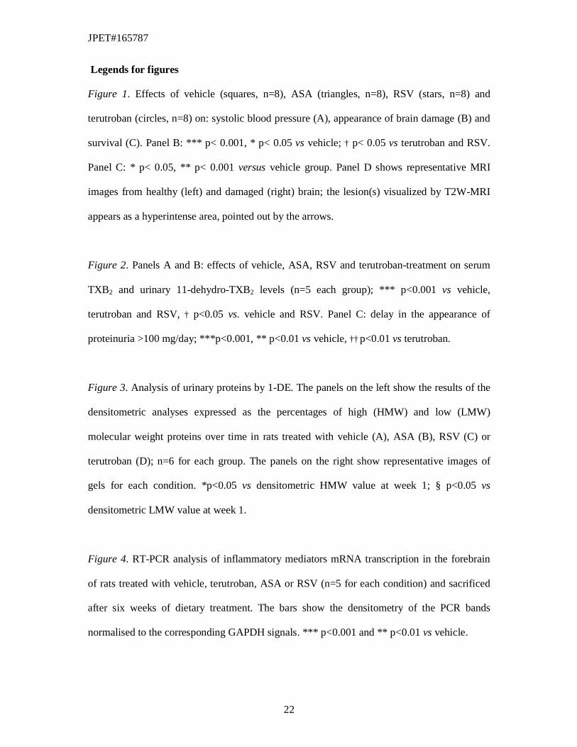
Figure 1. Effects of vehicle (squares, n=8), ASA (triangles, n=8), RSV (stars, n=8) and
terutroban (circles, n=8) on: systolic blood pressure (A), appearance of brain damage (B) and
survival (C). Panel B: *** p< 0.001, * p< 0.05 vs vehicle; † p< 0.05 vs terutroban and RSV.
Panel C: * p< 0.05, ** p< 0.001 versus vehicle group. Panel D shows representative MRI
images from healthy (left) and damaged (right) brain; the lesion(s) visualized by T2W-MRI
appears as a hyperintense area, pointed out by the arrows.
Figure 2. Panels A and B: effects of vehicle, ASA, RSV and terutroban-treatment on serum
TXB2 and urinary 11-dehydro-TXB2 levels (n=5 each group); *** p<0.001 vs vehicle,
terutroban and RSV, † p<0.05 vs. vehicle and RSV. Panel C: delay in the appearance of
proteinuria >100 mg/day; ***p<0.001, ** p<0.01 vs vehicle, †† p<0.01 vs terutroban.
Figure 3. Analysis of urinary proteins by 1-DE. The panels on the left show the results of the
densitometric analyses expressed as the percentages of high (HMW) and low (LMW)
molecular weight proteins over time in rats treated with vehicle (A), ASA (B), RSV (C) or
terutroban (D); n=6 for each group. The panels on the right show representative images of
gels for each condition. *p<0.05 vs densitometric HMW value at week 1; § p<0.05 vs
densitometric LMW value at week 1.
Figure 4. RT-PCR analysis of inflammatory mediators mRNA transcription in the forebrain
of rats treated with vehicle, terutroban, ASA or RSV (n=5 for each condition) and sacrificed
after six weeks of dietary treatment. The bars show the densitometry of the PCR bands
normalised to the corresponding GAPDH signals. *** p<0.001 and ** p<0.01 vs vehicle.
JPET#165787
23
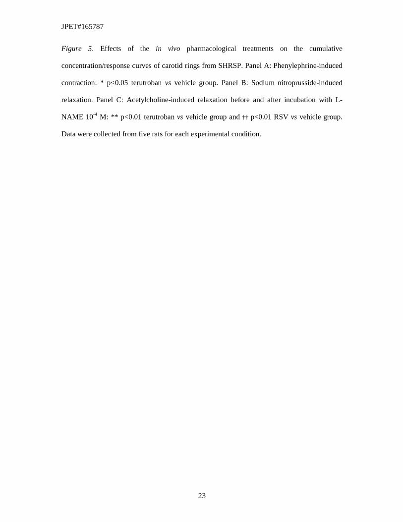
Figure 5. Effects of the in vivo pharmacological treatments on the cumulative
concentration/response curves of carotid rings from SHRSP. Panel A: Phenylephrine-induced
contraction: * p<0.05 terutroban vs vehicle group. Panel B: Sodium nitroprusside-induced
relaxation. Panel C: Acetylcholine-induced relaxation before and after incubation with L-
NAME 10-4 M: ** p<0.01 terutroban vs vehicle group and †† p<0.01 RSV vs vehicle group.
Data were collected from five rats for each experimental condition.
JPET#165787
24
Table 1. Sensitivity to the antagonists (pD2) expressed as the negative logarithm of EC50
L-Phenylephrine Acetylcholine Sodium nitroprusside
Vehicle 7.44 ± 0.3 6.54 ± 1.6 7.90 ± 0.6
Terutroban 7.22 ± 0.1 6.40 ± 0.5 8.10 ± 0.2
Aspirin 7.53 ± 0.4 6.12 ± 0.8 8.16 ± 0.1
Rosuvastatin 7.38 ± 0.3 6.46 ± 0.5 7.83 ± 0.2
Mean values ± standard deviation
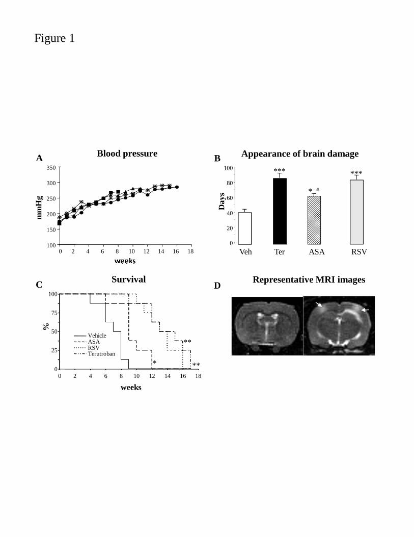
Figure 1
A Blood pressure
weeks
0 2 4 6 8 10 12 14 16 18100
150
200
250
300
350
mm
Hg
0
25
50
75
100
0 2 4 6 8 10 12 14 16 18
VehicleASARSVTerutroban
weeks
* **
**
SurvivalC
%
Appearance of brain damageB
0
20
40
60
80
100
Veh Ter
***
RSV
***
ASA
* #
Day
s
DRepresentative MRI images
ng/m
l
T
100
200
300
400
500
0Vehicle TeBaseline
A
Urinary 11B
0
10
20
30
40
Vehicle TeBaseline
pg/m
g cr
eati
nine
Delay approteinuria
C
0
4
8
12
Vehicle Terutrob
***
wee
ks
Figure 2
TXB2
Terutroban ASA RSV
***
11-dehydro-TXB2
Terutroban ASA RSV
#
appearance of ria > 100 mg/day
roban ASA RSV
**
# #
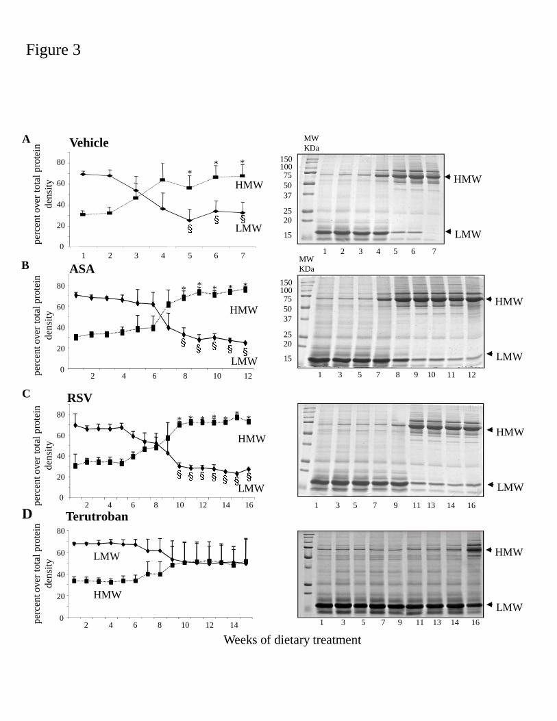
Figure 3
Weeks of d
0
20
40
60
80
2 4 6 8 10 12 14 16
Vehicle
ASA
RSV
perc
ent o
ver
tota
l pro
tein
de
nsity
0
20
40
60
80
2 4 6 8 10 12 14
Terutroban
0
20
40
60
80
1 2 3 4 5 6 7
LMW
HMW
A
B
C
D
perc
ent o
ver
tota
l pro
tein
de
nsity
perc
ent o
ver
tota
l pro
tein
de
nsity
HMW
LMW
LMW
HMW
** *
§§§
LMW
HMW
2 4 6 8 10 120
20
40
60
80
perc
ent o
ver
tota
l pro
tein
de
nsity
* * * * *
§ §§ § §
§ §§ § §§ §
* * * * ** *
of dietary treatment
HMW
LMW
1 2 3 4 5 6 7
1 3 5 7 9 11 13 14 16
1 3 5 7 8 9 10 11 12
HMW
LMW
HMW
LMW
1 3 5 7 9 11 13 14 16
HMW
LMW
75100150
5037
2520
15
MW KDa
75100150
5037
2520
15
MW KDa
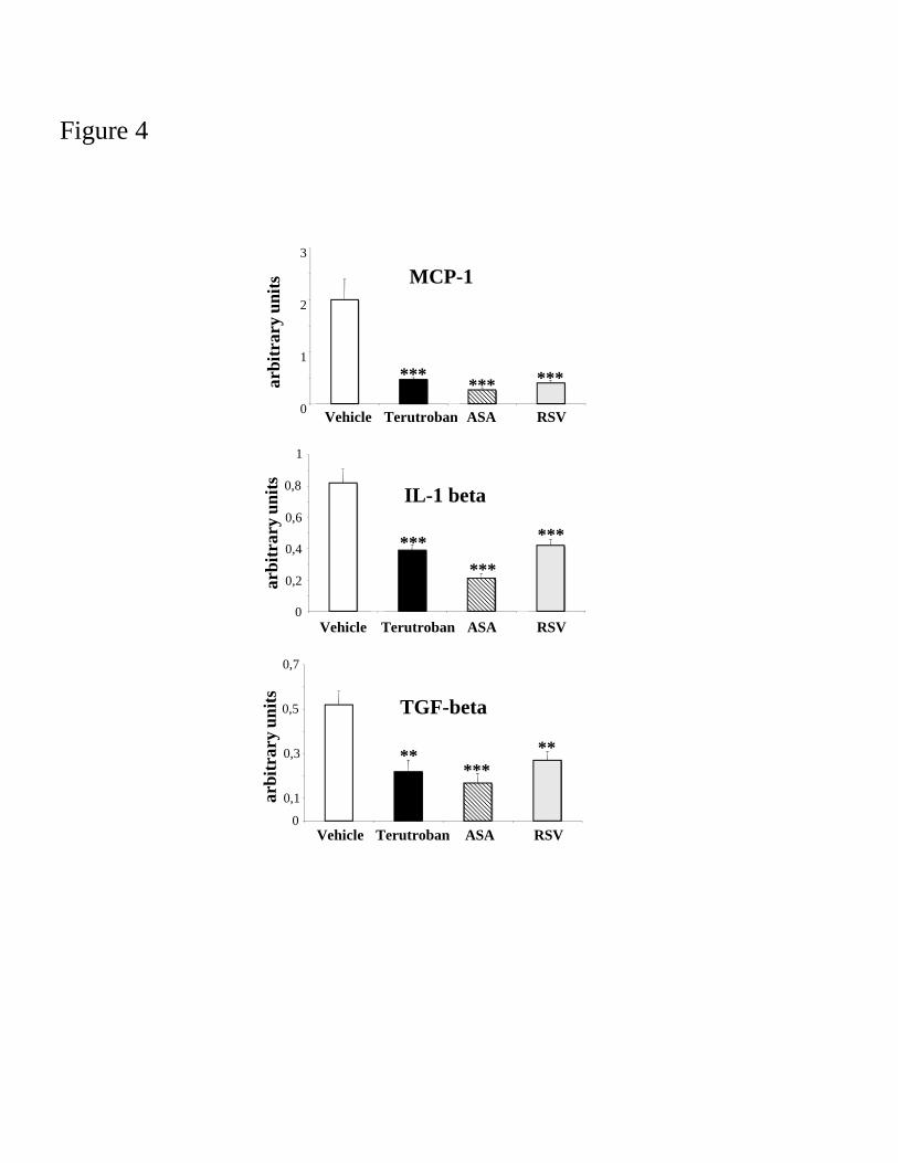
Figure 4
MC
0
1
2
3
Vehicle Terutrob
***arbi
trar
y un
its
IL-1
0
0,2
0,4
0,6
0,8
1
***
arbi
trar
y un
its
Vehicle Terutroba
TGF
0
0,1
0,3
0,5
0,7
**
Vehicle Terutroban
arbi
trar
y un
its
CP-1
oban ASA RSV
******
1 beta
***
***
ban ASA RSV
F-beta
** ***
ban ASA RSV

Figure 5
A
L-phenylephrine (-Log M)
10 -10 10 -8 10 -6 10 -40
50
100
150
200
250
*
Con
trac
tion
(% v
s K
Cl)
B
C
10-10
10-8
10-6 10-4
-100
-80
-60
-40
-20
0
20
Acetylcholine (-Log M)
**
$$
Rel
axat
ion
(% v
s L
-Phe
)
+L-NAMEVehicle
Terutroban
ASA
Rosuvastatin
Vehicle + L-NAME
Terutroban + L-NAME
ASA + L-NAME
Rosuvastatin + L-NAME
B
10 -11 10 -10 10 -9 10 -8 10 -7 10 -6 10 -5 10 -4-150
-110
-70
-30
10
Sodium Nitroprusside (-Log M)R
elax
atio
n (%
vs
L-P
he)
E