Purification of the human blood platelet thromboxane A2/prostaglandin H2 receptor protein
Upload
independentCategory
view
3download
0
JPET #79301
1
In vitro and in vivo pharmacological characterization of BM-613,
a novel dual thromboxane synthase inhibitor and thromboxane
receptor antagonist
Julien Hanson*, Stephanie Rolin*, Denis Reynaud, Na Qiao, Leanne P. Kelley, Helen M.
Reid, François Valentin, John Tippins, B. Therese Kinsella, Bernard Masereel, Cecil Pace-
Asciak, Bernard Pirotte, and Jean-Michel Dogné
Natural and Synthetic Drugs Research Centre, Department of Pharmacy, Laboratory of
Medicinal Chemistry, University of Liège, 1, Av. de l'Hôpital, B-4000 Liège, Belgium. (J.H.,
J-M.D., B.P.)
Department of Pharmacy, University of Namur, 61, rue de Bruxelles, B-5000 Namur,
Belgium. (S.R., B.M.)
Programme in Integrative Biology, Research Institute, The Hospital for Sick Children, 555
University Avenue, Toronto, Ontario, Canada M5G 1X8. (D.R., N.Q., C.P-A.)
Department of Biochemistry, Conway Institute for Biomolecular and Biomedical Research,
University College Dublin, Ireland. (L.P.K., H.M.R., B.T.K.)
Department of Biological Sciences, Imperial College, Exhibition Road, London SW7 2AZ,
United Kingdom. (F.V., J.T.)
JPET Fast Forward. Published on February 2, 2005 as DOI:10.1124/jpet.104.079301
Copyright 2005 by the American Society for Pharmacology and Experimental Therapeutics.
JPET #79301
2
a) Running title
BM-613, an original antiplatelet and antithrombotic agent
b) Corresponding author :
Julien Hanson
Natural and Synthetic Drugs Research Centre, Department of Pharmacy, Laboratory of
Medicinal Chemistry, University of Liège, 1, Av de L'hôpital, 4000 Liège, Belgium.
Phone : +3243664382
Fax : +3243664362
E-mail : [email protected]
c) Number of text pages : 37
Number of Tables : 4
Number of Figures : 5
Number of References : 40
Number of words in abstract : 249
Number of words in introduction : 674
Number of words in Discussion : 1493
List of nonstandard abbreviation : G-protein coupled receptor (GPCR), thromboxane receptor
antagonist (TXRA), thromboxane synthase (TXS), thromboxane synthase inhibitor (TXSI),
prostaglandin endoperoxide H2 (PGH2);
e) Recommended section : cardiovascular
JPET #79301
3
Abstract
Thromboxane (TXA2) is a key mediator of platelet aggregation and smooth muscle
contraction. Its action is mediated by its G-protein coupled receptor of which two isoforms,
termed TPα and TPβ, occur in humans. TXA2 has been implicated in pathologies such as
cardiovascular diseases, pulmonary embolism, atherosclerosis and asthma. This study
describes the pharmacological characterization of BM-613, a new combined TXA2 receptor
antagonist and TXA2 synthase inhibitor. It exhibits a strong affinity for human platelet TP
receptors (IC50: 1.4 nM), TPα and TPβ expressed in COS-7 cells (IC50: 2.06 nM and 3.1 nM,
respectively) and TPs expressed in human coronary artery smooth muscle cells (IC50: 29 µM).
BM-613 shows a weak ability to prevent contraction of isolated rat aorta (ED50: 1.52 µM) and
guinea-pig trachea (ED50: 2.5 µM) induced by TXA2 agonist U-46619. Besides, BM-613
antagonizes TPα (IC50: 0.11 µM) and TPβ (IC50: 0.17 µM) calcium mobilization induced by
U-46619 and inhibits human platelet aggregation induced by U-46619 (ED50: 0.278 µM),
arachidonic acid (ED50: 0.375 µM) and the second wave of ADP. BM-613 also dose-
dependently prevents TXA2 production by human platelets (IC50: 0.15 µM). In a rat model of
ferric chloride induced thrombosis, BM-613 significantly reduces weight of formed thrombus
by 79%, 49% and 28% at 5, 2 and 1 mg/kg (i.v.), respectively. In conclusion, BM-613 is a
dual and potent TP receptor antagonist and TXA2 synthase inhibitor characterized by a strong
antiplatelet and antithrombotic potency. These results suggest that BM-613 could be a
potential therapeutic drug for thrombotic disorders.
JPET #79301
4
Introduction
Thromboxane A2 (TXA2) is a key lipid mediator characterized by several implications in
physiological homeostasis including platelet aggregation and vascular and bronchial smooth
muscle constriction (Hamberg et al., 1975; Moncada and Vane, 1978). An overproduction of
TXA2 has been associated with many pathological states such as myocardial infarction,
thrombosis and thrombotic disorders, unstable angina, pulmonary embolism, shock,
atherosclerosis, preeclampsia and asthma (Dogne et al., 2004a).
TXA2 is a metabolite of arachidonic acid (AA), a 20-carbon essential fatty acid stored in
membrane phospholipids. Free AA can be released mainly by phospholipase A2 (PLA2) upon
stimulation. TXA2 is synthesized in two steps: first the action of cyclooxygenase (COX) on
free AA leads to the formation of prostaglandin H2 (PGH2) which can be subsequently
metabolized by thromboxane synthase (TXS) into TXA2.
COX exists in two main isoforms, COX-1 and COX-2, which are encoded by two separate
genes. COX-1 is expressed constitutively in most tissues mediating “housekeeping” functions,
whereas COX-2 expression is mainly induced at sites of inflammation by various stimuli
(Dogne et al., 2004a). The second is also expressed in a constitutive manner in endothelium
(Cheng et al., 2002). TXA2 synthesis mainly occurs in platelets where COX-1 and
thromboxane synthase are highly expressed.
TXA2 actions are mediated by its G protein coupled receptor (GPCR), referred to as the TP
receptor (Coleman et al., 1994). In 1991, Hirata et al. reported that human TP receptor was
coded by one gene (Hirata et al., 1991), and in 1994, Raychowdhury et al. highlighted the
existence of an isoform generated by alternative splicing (Raychowdhury et al., 1994). The
two isoforms, named TPα and TPβ, share the same first 328 amino acids but differ by the
length of their carboxy-terminal tails (15 amino acids of the α isoform being replaced by 79
amino acids in the β isoform).

JPET #79301
5
Given the wide implications of TXA2 in several pathologies, studies have been undertaken in
order to find therapeutic agents able to counteract the negative effects of TXA2. Indeed,
therapeutic strategies include blocking the synthesis of TXA2 by the inhibition of TXS,
antagonizing its action at the receptor level, or both. Thus, several TP receptor antagonists
(TXRA) such as sulotroban (Stegmeier et al., 1984), thromboxane synthase inhibitors (TXSI)
such as furegrelate (Gorman et al., 1983) and drugs combining both properties such as
ridogrel and terbogrel have been developed. Dual compounds proved to be more interesting in
therapeutics and as promising antithrombotic agents (Dogne et al., 2004a).
Besides, acetylsalicylic acid (ASA, aspirin) is a non-steroïdal anti-inflammatory drug exerting
its effects by irreversibly acetylating the active site of COX. In platelets, the inhibition is
effective throughout the platelet life-time because they lack a nucleus. Since TXA2 is mainly
produced by platelets, low-dose ASA has become the most common antiplatelet therapy used
for secondary prevention of cardiovascular disease. Nevertheless, ASA is far from being a
panacea since it has been associated with Reye's syndrome and allergic reaction (asthma) and
almost one third of patients receiving low-dose ASA do not respond, demonstrating a link
between platelet function and the persistence of TXA2 production (Patrono, 2003). These later
developments have strengthened the interest for development of other antithrombotic drugs
such as TXA2 modulators.
Herein we report the in vitro and in vivo pharmacological profile of BM-613 (Fig. 1), a new
TXA2 modulator synthesized in our laboratory. BM-613 is a chemical derivative of BM-573,
another thromboxane modulator combining TXRA and TXSI activities that has been widely
described in literature (Rolin et al., 2001; Dogne et al., 2004b; Ghuysen et al., 2004;
Lambermont et al., 2004). BM-613 has been developed as a dual TXRA and TXSI
characterized by high activity on platelets compared to smooth muscle. In this study, we
present several in vitro tests performed to characterize BM-613 effects on TXS, platelets and
JPET #79301
6
smooth muscle. We have also carried out specific binding and antagonism of [Ca2+]i
mobilization tests to compare the affinity and activity of our two compounds on the distinct
TP receptors isoforms. Finally, an in vivo test is presented to evaluate the potential of BM-613
as an antithrombotic drug.
JPET #79301
7
Materials and Methods
Materials
BM-613 (N-n-pentyl-N'-[2-(4'-methylphenylamino)-5-nitrobenzenesulfonyl]urea) and BM-
573 (N-tert-butyl-N’-[2-(4'-methylphenylamino)-5-nitrobenzenesulfonyl]urea) were
synthesized in our laboratory. The sodium salts were dissolved in propyleneglycol and diluted
with physiological saline. Furegrelate sodium salt (5-(3-pyridinylmethyl)-2-
benzofurancarboxylate), SQ-29548 [[1S-[1, 2(Z), 3, 4]]-7-[3-[[2-
[(phenylamino)carbonyl]hydrazino]methyl]-7-oxabicyclo[2.2.1]hept-2-yl]-5-heptenoic acid],
[3H]SQ-29548 and U-46619 (9.11-dideoxy-9.11-methanoepoxy-prostaglandin F2) were
purchased from Cayman Chemical (MI, USA). They were dissolved in ethanol and diluted
with physiological saline. Phosphate Buffer Saline (PBS) was purchased from Biowhitaker
(Petit-Rechain, Belgium). dRhodamine terminator cycle sequencing kit was from Applied
Biosystems (Foster City, CA). TPα and TPβ cDNA were as described previously (Allan et al.,
1996; Becker et al., 1998). Human coronary artery smooth muscle cells (HCASMc) were
obtained from CellWorks (Buckingham, U.K.). Dulbecco's modified Eagle's medium
(DMEM) supplemented with glucose (4.5 g/l), foetal bovine serum (FBS) and
antibiotic/antimycotic solution were purchased from Invitrogen life technologies (Carlsbad,
CA).
Animals
Male Sprague–Dawley rats, weighing 250–300 g were housed in a temperature-controlled
room before being used in the present experiments. All experimental procedures and protocols
used in this investigation have been carried out in accordance with the Declaration of Helsinki
(Publication No. 85-23. revised 1985) and were reviewed and approved by the Ethics
Committee of the Medical Faculty of the University of Liège.
JPET #79301
8
Cell Culture and Transfection
COS-7 cells were maintained in Dulbecco’s modified Eagle’s medium (Hyclone Laboratories,
Logan, UT) supplemented with 10% fetal bovine serum and 1% of solution containing 10,000
units/ml penicillin G, 10,000 µg/ml streptomycin, and 25 µg/ml amphotericin (Cellgro;
Mediatech, Herndon, VA). Cells were grown at 37°C in a humidified atmosphere of 95% O2
and 5% CO2. cDNAs for the TPα and TPβ were subcloned into pcDNA3, the resultant
plasmid, pcDNA3:TPα and pcDNA3:TPβ were introduced into COS-7 cells by the DEAE-
dextran/chloroquine method. After 48 h post-transfection, cells were harvested by
centrifugation at 500g for 5 min and washed three times in phosphate-buffered saline. Protein
concentrations were determined using the Bradford assay (Bradford, 1976). Cells were
resuspended in buffer containing 25 mM HEPES/125 mM NaCl/10 µM indomethacin, pH
7.4, and kept on ice for the binding study. TP expression was determined according to Miggin
and Kinsella (Miggin and Kinsella, 1998) using [3H]SQ-29548 (10 nM) and a single
saturating amount of SQ-29548 (10 µM).
Sequence Analysis
TPα, TPβ, and TPtailless sequences were confirmed on both strands using the ABI Big Dye
Terminator Cycle Sequencing Ready Reaction kit, and the products were resolved on an ABI
Prism 310 genetic analyzer (PerkinElmer/Applied Biosystems). The sequences were
assembled and analyzed using the ClustalW Sequence analysis.
Radioligand Binding Assay
The binding experiments were performed on whole cells.
Human platelet
The TXA2 receptor binding study was carried out on human washed platelets suspended in a
calcium- and magnesium-free Tyrode-Hepes buffer (mM: NaCl 137, KCl 2.7, NaH2PO4 0.4,
NaHCO3 12, D-glucose 5, Hepes; pH 7.4) to a concentration 2.108 cells/ml (Dogne et al.,
JPET #79301
9
2000; Dogne et al., 2001). Freshly prepared samples of this suspension (0.5 ml) were
incubated with [3H]SQ-29548 (5 nM final concentration, 0.1 ml) for 60 min at 25 °C. The
displacement was initiated by addition of the studied ligand dissolved in the same buffer (0.4
ml). After incubation (30 min, 25°C), ice-cold Tris-HCl buffer (10 mM, pH 7.4; 4 ml) was
added, the sample was rapidly filtered through a glass-fibre filter (Whatman GF/C) and the
tube rinsed twice with ice-cold buffer (4 ml). The filters were then placed in plastic
scintillation vials containing an emulsion-type scintillation mixture (4 ml) and the
radioactivity was counted. The amount of [3H]SQ-29548 specifically bound to human platelet
TXA2 receptors (Bs, %) was calculated from the following equation: Bs=100 * (B-NSB)/Bt
where Bt (total binding) and NSB (non-specific binding) are the radioactivity of [3H]SQ-
29548 (5 nM) bound to the platelets incubated in the absence of any competing ligand and in
the presence of unlabeled SQ-29548 (50 µM), respectively. B is the radioactivity of the
filtered platelets incubated with [3H]SQ-29548 (5 nM) and the studied compound at a
concentration ranging from 10-5 to 10-10 M. In each experiment, NSB varied between 5 and
7% of Bt. For each drug, three concentration-response curves were performed in triplicate.
The drug concentration which reduced the amount of specifically bound [3H]SQ-29548 by
50% (IC50) was determined by nonlinear regression analysis (GraphPad Prism software).
TPα and TPβ
Competition binding curves were carried out on COS-7 cells expressing TP receptor isoforms.
Binding reactions were carried out on 5.105 cells in a total volume of 0.2 ml in the above
buffer with 10 nM [3H]SQ-29548 added to all tubes in triplicate, containing various
concentration of BM-613 and BM-573 (10-6 to 10-11 M) in 1 µl of ethanol. Additional tubes
containing excess unlabeled SQ-29548 (10 µM) were included to assess the extent of non-
specific binding. Binding was allowed to take place for 30 min at 37°C; free radioligand was
removed by rapid vacuum filtration through Whatman (Maidstone, UK) GF/B glass fiber
JPET #79301
10
filters pre-washed with the cell suspension buffer. The tubes and the filters were rapidly
washed with ice-cold 10 mM Tris buffer, pH 7.4 (three times with 3 ml). The radioactivity on
the filters containing the ligand-receptor complexes was counted in 10 ml of Ecolite
scintillation fluid (ICN, St. Laurent, QC, Canada) in a Beckman (model LS 3800) liquid
scintillation counter.
Human coronary artery smooth muscle cells
HCASM cells were maintained in HCASMc basal medium supplemented with HCASMc
growth supplement (CellWorks) at 37 °C in a humidified atmosphere of 95% air and 5% CO2.
HCAM cells were re-suspended in the binding buffer (25 mM Tris-HCl, pH 7.4, 5 mM CaCl2,
10 µM indomethacin, 50 µg/ml PMSF). For the binding assay, 50 µg of protein was incubated
with [3H]SQ-29548 (30 Ci/mol, 1 µM, PerkinElmer Life Sciences) in the presence of the drug
at the indicated concentrations (0-100 µM) in a 0.1 ml reaction volume with vigorous shaking
at room temperature for 60 min. The reaction was then terminated by adding 1 ml of ice-cold
washing buffer (25 mM Tris-HCl, pH 7.4). The unbound ligand was filtered under vacuum
through a Whatman GF/C glass filter (Whatman, Clifton, NJ) presoaked with the ice-cold
washing buffer. The radioactivity of the TP receptor-bound [3H]SQ-29548 remaining on the
glass filter was counted in 8 ml of scintillation mixture (NEN) using a Beckman counter
(Fullerton, CA).
Human ex vivo platelet aggregation
The anti-aggregant potency has been determined according to the turbidimetric Born’s
method (Born and Cross, 1963). The blood was drawn from ten healthy donors of both
genders, aged 20-30. The subjects were free from medication for at least 14 days. No
significant differences in the results were observed between the donors in our experiments.
Platelet-rich plasma (PRP) and platelet-poor plasma (PPP) were prepared as previously
described (Dogne et al., 2000; Dogne et al., 2001). Platelet concentration of PRP was adjusted
JPET #79301
11
to 3.108 cells/ml by dilution with PPP. Platelet aggregation of PRP was studied using a double
channel aggregometer (Chronolog Corporation, Chicago, IL) connected to a linear recorder as
previously described (Harris et al., 1979). PRP (294 µl) was added in a silanised cuvette and
stirred (1000 rpm). Each drug was diluted (1 mM) in dimethylsulfoxide (DMSO)/PBS (30/70)
and preincubated in PRP for three minutes at 37°C before the aggregating agent was added.
Platelet aggregation was initiated by addition of a fresh solution of arachidonic acid (600 µM
final) or U-46619 (1 µM final). To evaluate platelet aggregation, the maximum increase in
light transmission was determined from the aggregation curve 6 minutes after addition of the
inducer. The drug concentration preventing 50% of platelet aggregation (ED50) induced by
arachidonic acid and U-46619 was calculated by non-linear regression analysis (GraphPad
Prism software) from at least three dose-response curves.
Rat aorta relaxation
Endothelium-denuded thoracic aorta rings, obtained from rats (Wistar, 250–300 g)
anaesthetised with sodium pentobarbital (80 mg/kg, i.p.), were suspended under a tension of
one gram in a Krebs solution (mM: NaCl 118, KCl 5.4, CaCl2 2.5, MgCl2.6H2O 1.5, NaHCO3
25,NaH2PO4 1.2 and glucose 10; pH 7.4) which was bubbled with O2–CO2 (95–5%) at 37°C
in a 20 ml tissue bath (EMKA Technologies, Paris, France). The muscle tension of the aortic
rings was isometrically recorded with a force-displacement transducer IT1 (EMKA
Technologies, Paris, France). The buffer was renewed every 15 minutes during the
equilibration period (1 h) before exposing the rings to U-46619 (20 nM). When a stable
tension was obtained (15 min), cumulative increasing concentrations of potential antagonist
were added to the bath until tension returned to the base-line value. The ED50 value of each
drug was assessed for at least 4 concentration-response curves obtained from separate
preparations and corresponded to the concentration which reduced to 50% the tension induced
JPET #79301
12
by U-46619 (20 nM). The ED50 were calculated by non-linear regression analysis (GraphPad
Prism software).
Guinea-pig trachea relaxation
Trachea was removed from guinea pigs (Hartley, 250–300 g) anesthetized with sodium
pentobarbital (80 mg/kg, i.p.), and carefully cleaned of connective tissue. Trachea strips were
suspended in the organ bath (20 ml) and the experiment progressed in the same conditions as
those described above for the rat aorta except for the concentration of U-46619 (10 nM), the
contraction inducer.
Calcium measurements
HEK.TPα and HEK.TPβ cell lines, stably over-expressing HA-tagged forms of TPα and TPβ
in human embryonic kidney (HEK) 293 cells have been previously described (Walsh et al.,
2000). HEK 293 cells or their stable cell line equivalents were routinely grown in minimal
essential Eagle’s medium (MEM) containing 10% foetal bovine serum (FBS). For
measurements of intracellular calcium ([Ca2+]i) mobilization, approximately 48 hr prior to
transfection, HEK.TPα and HEK.TPβ stable cell lines were plated in 10-cm culture dishes at
a density of 2x10⁶ cells/dish in 8 ml media; thereafter, cells were transiently transfected with
10 mg of pADVA and 25 µg pCMV:Gαq, coding for Gαq, using the calcium phosphate
/DNA co-precipitation procedure essentially as previously described (Kinsella et al., 1997;
Hayes et al., 1999). Measurement of [Ca2+]i mobilization either in Gαq-transfected HEK.TPα
and HEK.TPβ cells was carried out in FURA2/AM preloaded cell lines cells, essentially as
previously described (Kinsella et al., 1997). Briefly, cells were stimulated at 50 s with either
U-46619 (1 µM) or, for competition studies using either BM-573 and BM-613, cells were pre-
stimulated with 10-9 - 10-3 M BM-573 or BM-613 for 5 min prior to stimulation with U-46619
(1 µM). In all cases, the stock solution (100 mM BM 573 or BM 613 in DMSO) was diluted
with PBS to achieve the desired working solution and 20 µl of the vehicle or drug in vehicle
JPET #79301
13
was added to 2 ml of cells; the vehicle had no effect on [Ca2+]i mobilization by either TP
isoform and had no effect on experimental data. The ratio of the fluorescence at 340 nm to
that at 380 nm is a measure of [Ca2+]i (Grynkiewicz et al., 1985), assuming a Kd of 225 nM
Ca2+ for FURA2/AM. The results presented in the figures 3 and 4 are either representative or
mean data from three/four independent experiments. Alternatively, mean percentage
reductions in U-46619-mediated [Ca2+]i mobilization in the presence of BM-573 (10-9 to 10-3
M) are expressed as a percentage (%) of U-46619 mediated [Ca2+]i mobilization in the
absence of BM 573.
Thromboxane synthase activity
PRP preparation is identical to that described for the platelet aggregation experiments. Each
drug was dissolved in dimethylsulfoxide (DMSO) and diluted with a Tyrode-Hepes buffer
(mM: NaCl 137, KCl 2.7, NaH2PO4.- H2O 0.4, D-glucose 5, NaHCO3 12, Hepes; pH 7.4). To
900 µl of PRP, 50 µl NaCl 0.9% and 10 µl of drug solution were added. After 6 minutes
incubation at 37°C under stirring (600 rpm), aggregation was induced by 40 µl sodium
arachidonate (0.6 mM final). After 4 minutes, the reaction was stopped by adding 50 µl of
indomethacin (0.02 M in ethanol). The sample was immediately centrifuged (17500 g, for 10
s) and the supernatant was removed and frozen (-80 °C) until assayed for TXB2. Basal and
maximal production of TXB2 was estimated in the absence and in the presence of arachidonic
acid, respectively. Evaluation was done in triplicate on concentrations ranging from 10 to 0.1
µM. Thromboxane synthase activity was expressed as the TXB2 production which was
measured by using a competitive enzyme immunoassay (TXB2 EIA Kit, Cayman Chemical
Company, MI).
Ferric chloride-induced rat arterial thrombosis
The experiments were carried out according to the modification of the method described by
Kurz et al. (Kurz et al., 1990).Rats were anesthetized with sodium pentobarbital (50 mg/kg,
JPET #79301
14
i.p.). After an abdominal midline incision, the abdominal aorta was exposed carefully. A filter
paper disk (diameter 8 mm) saturated with 50% (w/v) ferric chloride solution was placed on
the surface of the artery for 10 min. The artery was isolated 10 min after removing the disk
and then opened lengthwise. The thrombus was scraped out and placed on a filter paper to
remove any water and its wet weight was measured immediately. Results are expressed in mg
of thrombus weight by kg of rat weight. BM-613 (5, 2, 1, 0.5 mg/kg) and placebo were
injected intravenously 5 minutes prior to application of ferric chloride solution. The results
are expressed as the mean of results from four separate experiments.
Statistical analysis
Results are expressed as the mean ± standard error of the mean (SEM) and statistical
significance was determined by a Mann-Whitney test. Probability values of less than 0.05
were considered to be significant.
JPET #79301
15
Results
In vitro Functional assays
Inhibition of human platelet aggregation
The ability of BM-613 to prevent human platelet aggregation has been evaluated in vitro with
three different agonists of platelet aggregation. BM-613 was able to inhibit (ED50 = 0.278 ±
0.186 µM) platelet aggregation induced by the stable TXA2 agonist U-46619 (1 µM). This
effect is in the same order of potency as results obtained with BM-573 (ED50 = 0.240 ± 0.013
µM). In these experiments, BM-613 produced a shift to the right of the concentration-
response curve of U-46619 (Data not shown). BM-613 also inhibited platelet aggregation
induced by 600 µM arachidonic acid (ED50 = 0.375 ± 0.05 µM) in the same concentration
range as BM-573 (ED50 = 0.125 ± 0.015 µM). ADP is a weak inducer of platelet aggregation
by itself, and it induces secretion of TXA2 among other pro-aggregating substances. BM-613
(1 and 10 µM) inhibited the second wave of platelet aggregation induced by ADP, which is
due to the secretion of TXA2 (Table 1).
Thromboxane synthase inhibitory potency
Thromboxane synthase is the enzyme converting PGH2 into TXA2. BM-613 was evaluated in
a thromboxane activity test and showed an inhibitory activity (IC50 = 0.15 ± 0.13 µM)
comparable to that obtained with BM-573 (IC50= 0.053 ± 0.028 µM). It was 100-fold more
potent than the thromboxane synthase inhibitor furegrelate (IC50 = 10.2 ± 6 µM) chosen as
reference drug.
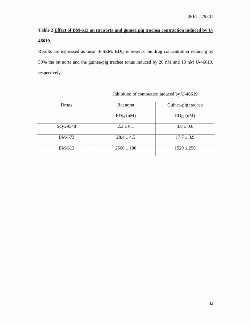
Inhibition of rat aorta and guinea-pig trachea contraction induced by U-46619
The TXA2 receptor antagonism properties of BM-613 were studied on isolated rat thoracic
aorta precontracted by the TXA2 agonist, U-46619 (20 nM) (Table 2). The concentration
(ED50) that caused 50% of relaxation was calculated from concentration-response curves
(Table 2). On rat aorta, BM-613 (ED50 = 2.4 ± 0.18 µM) was 100-fold less active than BM-
JPET #79301
16
573 (ED50: 28.4 ± 4.5 nM) and 1000-fold less active than SQ-29548 (ED50: 2.3 ± 0.07 nM)
which was the most potent compound in these experiments. In order to evaluate the potential
therapeutic interest of BM-613 in the treatment of asthma, the relaxing activity of BM-613
was measured on the guinea pig trachea contracted by U-46619 (10 nM) (Table 2) which is
considered as a potent constrictor of the bronchopulmonary tract (Coleman et al., 1981).
Cumulative increasing concentrations of BM-613 caused a concentration-dependent
relaxation of the contracted isolated trachea. The drug concentration (ED50) required to
decrease by 50% the muscular tonus induced by U-46619 was calculated from these curves
(Table 2). BM-613 (ED50 = 1.52 ± 0.25 µM) was much less active than BM-573 (ED50 = 17.7
± 3.9 nM) and than SQ-29548 (ED50 = 3.8 ± 0.5 nM) which remains the most potent
compound.
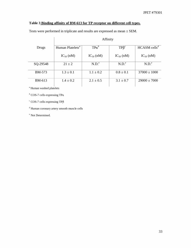
Radioligand binding assay
Affinity of BM-613 for TP receptors has been evaluated by measuring its ability to displace
radiolabeled SQ-29548, a strong TXRA, using different cells types (Table 3). Binding
experiments have been conducted on human washed platelets, human coronary artery smooth
muscle cells and COS-7 cells where TPα and TPβ were expressed. BM-613 showed strong
affinity (IC50= 1.4 ± 0.2 nM) for human platelet TP receptor. It was as active as BM-573
(IC50= 1.3 ± 0.1 nM) and ten-fold more potent than SQ-29548 (IC50 = 21 ± 2 nM). Affinities
of BM-613 for either TPα (IC50= 2.1 ± 0.5 nM) or TPβ (IC50= 3.1 ± 0.7 nM) expressed alone
in COS-7 cells are not significantly different (p=0.28). BM-573 was slightly more potent in
this test but again there was little difference in affinity for the two isoforms. In binding studies
performed on HCASM cells, BM-613 (IC50 = 29 ± 7 µM) showed an affinity comparable to
the reference drug, BM-573 (IC50 = 37 ± 1 µM).
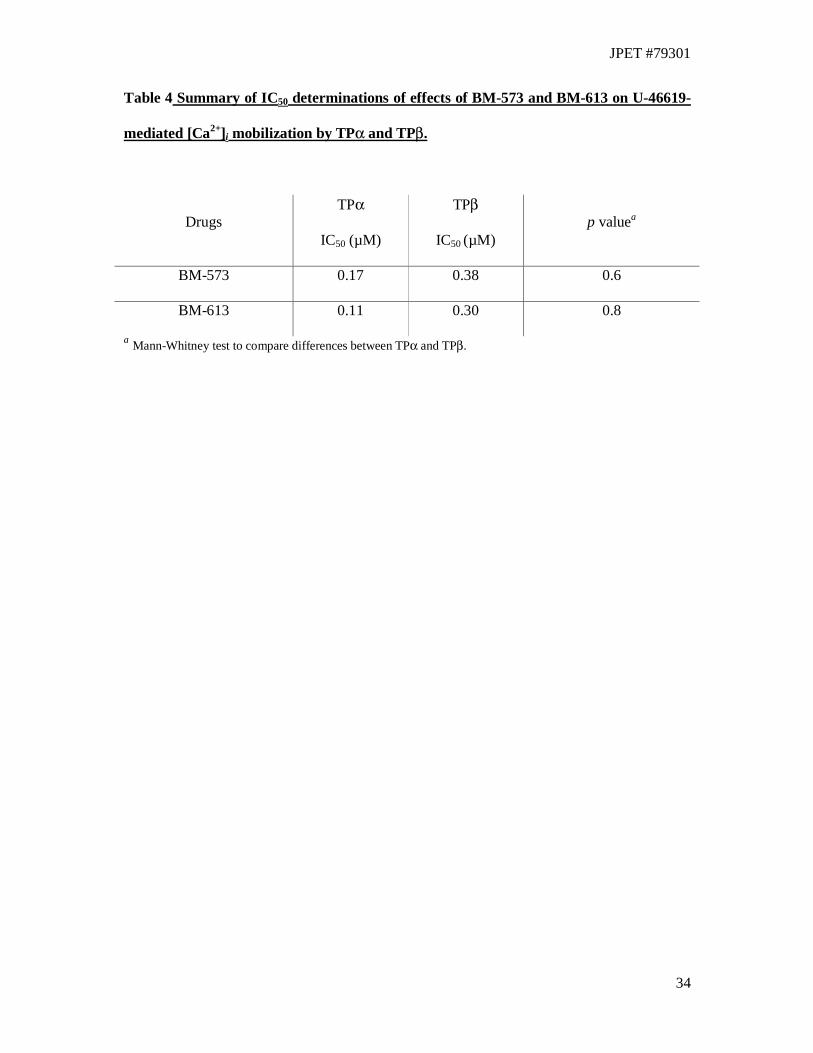
Intracellular signaling by assessment of intracellular calcium mobilization
JPET #79301
17
The effect of BM-613 on intracellular signalling by the TP isoforms was investigated by
comparing its effect to BM-573 in intracellular calcium ([Ca2+]i) mobilization mediated by
TPα and TPβ stably, over-expressed in HEK 293 cells, in response to the selective TXA2
mimetic U-46619. It has been reported that for efficient TPα and TPβ coupling to
phospholipase Cβ activation in HEK 293 cells, it is necessary to co-transfect cells with a
member of the Gq family of heterotrimeric G proteins (Kinsella et al., 1997; Walsh et al.,
1998). Hence, throughout these studies, HEK.TPα and HEK.TPβ cells were routinely co-
transfected with pCMV:Gαq, encoding Gαq. In HEK.TPα cells, both BM-573 (IC50 = 0.17
µM) and BM-613 (IC50 = 0.11 µM) exhibited concentration dependent antagonism of U-
46619 –mediated [Ca2+]i mobilization (Fig. 3 and 4). Similarly, in HEK.TPβ cells, both BM-
573 (IC50 = 0.38 µM) and BM-613 (IC50 = 0.30 µM) antagonised U-46619 –mediated [Ca2+]i
mobilization to levels that were not significantly different from those observed in HEK.TPα
cell lines (BM-573, p = 0.6 ; BM-613, p = 0.8, Table 4).
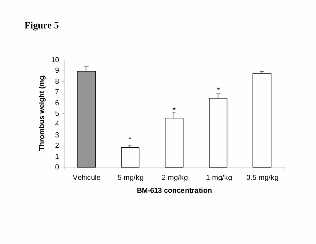
In vivo antithrombotic activity of BM-613
To further test the hypothesis that BM-613 was a potential antithrombotic agent acting on TP
receptors and TXS, we evaluated its effect in a rat model of arterial thrombosis. It has been
previously described that application of ferric chloride solution on vascular vessels induced
thrombus formation (Kurz et al., 1990). In this model, topical application of ferric chloride
(50% w/v) for 10 min to the abdominal aorta induced marked thrombi in vehicle-treated rats
(8.95 ± 0.92 mg/kg). Intravenously injected at 5, 2 and 1 mg/kg, BM-613 significantly
reduced the thrombus weight by 79%, 49% and 28%, respectively (Fig. 5). At 0.5 mg/kg,
BM-613 did not significantly reduce the thrombus weight. Intraperitoneally injected, BM-573
tested in the same model showed similar activity (Dogne et al., 2004b).
JPET #79301
18
Discussion
TXA2 is a key mediator involved in platelet aggregation and smooth muscle contraction. It is
synthesized by the action of TXS on PGH2, an unstable endoperoxide formed by action of
COX on free AA. TXA2 mediates its action by a specific GPCR named TP receptor. In
humans but not in non primates, this receptor is expressed as two isoforms, termed TPα and
TPβ, that arise by alternative splicing. Since TXA2 has been implicated in several pathologies
such as myocardial infarction, thrombosis and thrombotic disorders, unstable angina,
pulmonary embolism, shock, atherosclerosis, preeclampsia and asthma (Dogne et al., 2004a),
efforts have been made to develop agents able to antagonize TXA2 actions.
Aspirin is the antiplatelet drug commonly used for secondary prevention of cardiovascular
disease. Nevertheless, ASA has been associated with Reye's syndrome and allergic reaction
(asthma) and almost one third of patients receiving low-dose ASA do not respond,
demonstrating a link between platelet function and the persistence of TXA2 production
(Patrono, 2003). Although the aetiology of this "aspirin resistance" is still not fully explained,
some authors have pointed out the possible role of oxidative products such as the non-
enzymatic isoprostane derivatives of AA acting on TP receptor (Cipollone et al., 2000) or
COX-2 dependent TXA2 formation (Patrono, 2003). Besides the "aspirin resistance", there is
growing evidence that TP receptor is deeply involved in growing of atherosclerotic plaque.
Recently, Cayatte et al. have shown that the TP receptor antagonist S18886 reduces
atherogenesis while ASA has no effect, thus demonstrating the superiority of TP receptor
antagonist in these tests (Cayatte et al., 2000). These results were explained by the TP
receptor activation by other mediators that can still promote growth of the atherosclerotic
plaque.
These recent developments in our knowledge on TXA2 biology have highlighted the
limitations of ASA and the growing interest of TXA2 modulators in several pathologies. Here
JPET #79301
19
we describe in vitro and in vivo pharmacological characterisation of BM-613, a novel dual
TXRA and TXSI developed in our lab. It is a close derivative of BM-573, another TXRA and
TXSI described in the literature (Rolin et al., 2001; Dogne et al., 2004b; Ghuysen et al., 2004;
Lambermont et al., 2004).
Our first aim was to determine the in vitro TP receptor antagonistic potency of BM-613 in a
test of inhibition of human platelet aggregation. BM-613 was able to inhibit platelet
aggregation induced by AA in the same concentration range as BM-573 (Table 1). Because
COX inhibitors are also able to inhibit the AA-induced aggregation by blocking TXA2
production, BM-613 activity was evaluated with U-46619 which is a stable TP agonist. We
have shown in this experiment that BM-613 completely inhibited platelet aggregation induced
by U-46619, thus demonstrating its action as TXRA (Table 1). Moreover, BM-613 produced
a shift to the right of the concentration-response curve of U-46619, suggesting a competitive
type antagonism of platelet TP receptors (data not shown). ADP provokes platelet aggregation
by acting on specific purine receptors. Platelet aggregation induced by ADP is characterised
by two waves. The first wave is due to a weak and reversible aggregation, which is the
consequence of ADP action on its specific receptors. The second wave is due to TXA2
synthesis and release, which provokes irreversible and complete aggregation. Our compound
only inhibited the second wave (Table 1) of ADP-induced platelet aggregation like other
TXRAs (Reynaud et al., 2002). This result highlights the lack of action of BM-613 on ADP
receptors and confirms specific activity on TP receptors.
Compounds combining both actions on TP receptors and TXS have proved to be more
therapeutically interesting and promising as antithrombotic agents (Gresele et al., 1987).
Indeed, stopping exclusively the production of TXA2 provokes the accumulation of PGH2,
another TP receptor agonist (Coleman et al., 1981). On the other side, with pure TP receptor
antagonists, the benefit of redirecting production of other prostanoids (such as prostacyclin)
JPET #79301
20
by accumulation of PGH2 is lost. Thus, we also evaluated the potency of BM-613 as TXSI by
investigating the production of the stable TXA2 metabolite, thromboxane B2 (TXB2), by
human platelets activated by arachidonic acid. The IC50 determined for BM-613 was 100-fold
lower than that of furegrelate, a clinically evaluated TXSI used as reference drug. These
results demonstrated the efficacy of BM-613 as a TXSI (Mohrland et al., 1990). Combined
with the effect on platelet aggregation, BM-613 can be considered as a well-balanced TXRA
and TXSI. This is of great importance since the lack of efficacy of ridogrel was explained by
its strong effect on TXS compared to TP receptors (Soyka et al., 1999).
Further functional experiments were conducted on isolated rat aorta and guinea-pig trachea
precontracted by U-46619 (Fig. 2). In these experiments, BM-573 has already shown good
potency compared to SQ-29548 (Rolin et al., 2001). BM-613 showed a 100-fold decreased
activity in these experiments compared to BM-573 (Table 2). This discrepancy between
potency of BM-613 to inhibit platelet aggregation and to inhibit smooth muscle cell
contraction could find several explanations. First, the TP receptor may present conformational
differences between species used in the test (human, rat and guinea-pig). Secondly, several
authors initially stated in the eighties that at least two pharmacological subtypes of TP
receptor existed (Mais et al., 1985; Saussy et al., 1985). A first subtype was thought to be
present at the platelet surface (Takahara et al., 1990), responsible for platelet aggregation. The
second subtype, detected in smooth muscle cells was thought to be responsible for smooth
muscle contraction (Mais et al., 1988). However, it is still controversial whether the two
isoforms are responsible for pharmacological differences described in some studies because
none of the initial tissue-selective antagonists of TP receptors were able to discriminate
between the two isoforms. Moreover, the exact physiological significance of the existence of
two isoforms is still unclear although it has been shown that TPα was the only isoform
expressed in platelets (Habib et al., 1999), and that both TPα and TPβ were expressed in
JPET #79301
21
smooth muscle cells (Miggin and Kinsella, 1998). Recently, Qiao et al. showed for the first
time that the hepoxilin stable analogue, PBT-3 could selectively bind the TPα isoform in
transfected COS-7 cells (Qiao et al., 2003). Thus, we postulated that BM-613 could have a
greater affinity for TPα than TPβ and that this could be coupled with higher activity as an
antiplatelet agent.
To test this second hypothesis, we have performed binding experiments on several cell types.
BM-613 and BM-573 affinity was evaluated on washed platelets, COS-7 cells expressing
either TPα or TPβ and human coronary artery smooth muscle cells (HCASMc). In these
experiments, BM-613 showed high affinity for human platelet TP receptor. BM-613 was also
characterised by great affinity for both TPα and TPβ expressed in COS-7 cells. This result
confirmed the potent affinity of BM-613 for human platelet TP receptor of which TPα is
predominant (Habib et al., 1999). Since TPα and TPβ are both expressed in smooth muscle
cells (Miggin and Kinsella, 1998), lower affinity for TPβ should appear when performing
tests on smooth muscle cells. BM-613 and BM-573 exhibited quite similar affinities for
HCASMc TP receptors, which is in accordance with their TPβ binding properties.
Since the binding affinity of a compound for different receptors cannot reflect its antagonistic
or agonistic properties, we performed studies on intracellular signalling by TPα and TPβ.
BM-613 dose-dependently inhibits [Ca2+]i mobilization upon stimulation by U-46619 in HEK
293 cells co-transfected with Gαq and either TPα or TPβ (Figures 3 and 4). Nevertheless,
BM-613 exhibited no significant differences in antagonistic potency against the two isoforms
in these experiments (Table 4). We conclude from these results that the weak activity of BM-
613 in inhibiting smooth muscle cell contraction was due to interspecies differences in TP
receptor configuration.
We further used a rat model of ferric chloride-induced thrombus formation to test the
antithrombotic properties of BM-613. The drug, injected intravenously, was able to
JPET #79301
22
significantly reduce the weight of thrombus formed in the lumen of the aorta (Figure 5).
These results are in accordance with in vitro antiplatelet activity of BM-613 and with results
obtained with BM-573 in similar conditions (Dogne et al., 2004b).
In conclusion, we have presented herein a new compound, BM-613, which is characterised by
high affinity for human platelets and HCASM cells TP receptors, TPα and TPβ receptors.
These binding affinities are comparable to those of BM-573, our compound used as reference.
Moreover, our compound is an antagonist of human platelet TP receptors, TPα and TPβ and
an inhibitor of TXS. Moreover, it is characterized by antagonist properties for platelet
aggregation and calcium mobilisation in the same range of concentration than the potent BM-
573. Nevertheless, BM-613 is less powerful than reference BM-573 in counteracting rat and
guinea-pig smooth muscle contraction. It seems that this difference is due to interspecies
variations. However, given the therapeutic interest of tissue-selective compounds, these
results need to be confirmed in further experiments. BM-613 is also an in vivo antithrombotic
agent, suggesting that it could be a potential therapeutic agent for cardiovascular diseases and
thrombotic disorders.

JPET #79301
23
Acknowledgements
We are grateful to Prof. Perry V. Halushka, University of South Carolina at Charleston for
providing us with cDNA for the TPα and TPβ receptor isoforms. The authors want to thank
Philippe Neven for excellent technical assistance.
JPET #79301
24
References
Allan CJ, Higashiura K, Martin M, Morinelli TA, Kurtz DT, Geoffroy O, Meier GP, Gettys TW and Halushka PV (1996) Characterization of the cloned HEL cell thromboxane A2 receptor: evidence that the affinity state can be altered by G alpha 13 and G alpha q. J Pharmacol Exp Ther 277:1132-1139. Becker KP, Ullian M and Halushka PV (1998) Cloning and characterization of an endogenous COS-7 cell thromboxane A2 receptor. Biochim Biophys Acta 1403:109-114. Born GV and Cross MJ (1963) The Aggregation of Blood Platelets. J Physiol 168:178-195. Bradford MM (1976) A rapid and sensitive method for the quantitation of microgram quantities of protein utilizing the principle of protein-dye binding. Anal Biochem 72:248-254. Cayatte AJ, Du Y, Oliver-Krasinski J, Lavielle G, Verbeuren TJ and Cohen RA (2000) The thromboxane receptor antagonist S18886 but not aspirin inhibits atherogenesis in apo E-deficient mice: evidence that eicosanoids other than thromboxane contribute to atherosclerosis. Arterioscler Thromb Vasc Biol 20:1724-1728. Cheng Y, Austin SC, Rocca B, Koller BH, Coffman TM, Grosser T, Lawson JA and FitzGerald GA (2002) Role of prostacyclin in the cardiovascular response to thromboxane A2. Science 296:539-541. Cipollone F, Ciabattoni G, Patrignani P, Pasquale M, Di Gregorio D, Bucciarelli T, Davi G, Cuccurullo F and Patrono C (2000) Oxidant stress and aspirin-insensitive thromboxane biosynthesis in severe unstable angina. Circulation 102:1007-1013. Coleman RA, Humphrey PP, Kennedy I, Levy GP and Lumley P (1981) Comparison of the actions of U-46619, a prostaglandin H2-analogue, with those of prostaglandin H2 and thromboxane A2 on some isolated smooth muscle preparations. Br J Pharmacol 73:773-778. Coleman RA, Smith WL and Narumiya S (1994) International Union of Pharmacology classification of prostanoid receptors: properties, distribution, and structure of the receptors and their subtypes. Pharmacol Rev 46:205-229. Dogne JM, de Leval X, Hanson J, Frederich M, Lambermont B, Ghuysen A, Casini A, Masereel B, Ruan KH, Pirotte B and Kolh P (2004a) New developments on thromboxane and prostacyclin modulators part I: thromboxane modulators. Curr Med Chem 11:1223-1241. Dogne JM, de Leval X, Neven P, Rolin S, Wauters J, David JL, Delarge J and Massereel B (2000) Effects of a novel non-carboxylic thromboxane A2 receptor antagonist (BM-531) derived from torasemide on platelet function. Prostaglandins Leukot Essent Fatty Acids 62:311-317. Dogne JM, Hanson J, de Leval X, Kolh P, Tchana-Sato V, de Leval L, Rolin S, Ghuysen A, Segers P, Lambermont B, Masereel B and Pirotte B (2004b) Pharmacological characterization of N-tert-butyl-N'-[2-(4'-methylphenylamino)-5-nitrobenzenesulfonyl]urea (BM-573), a novel thromboxane A2 receptor antagonist and thromboxane synthase inhibitor in a rat model of arterial thrombosis and its effects on bleeding time. J Pharmacol Exp Ther 309:498-505.
JPET #79301
25
Dogne JM, Wouters J, Rolin S, Michaux C, Pochet L, Durant F, Delarge J and Masereel B (2001) Design, synthesis and biological evaluation of a sulfonylcyanoguanidine as thromboxane A2 receptor antagonist and thromboxane synthase inhibitor. J Pharm Pharmacol 53:669-680. Ghuysen A, Lambermont B, Dogne JM, Kolh P, Tchana-Sato V, Morimont P, Magis D, Hanson J, Segers P and D'Orio V (2004) Effect of BM-573 [N-terbutyl-N'-[2-(4'-methylphenylamino)-5-nitro-benzenesulfonyl]urea], a dual thromboxane synthase inhibitor and thromboxane receptor antagonist, in a porcine model of acute pulmonary embolism. J Pharmacol Exp Ther 310:964-972. Gorman RR, Johnson RA, Spilman CH and Aiken JW (1983) Inhibition of platelet thromboxane A2 synthase activity by sodium 5-(3'-pyridinylmethyl)benzofuran-2-carboxylate. Prostaglandins 26:325-342. Gresele P, Arnout J, Deckmyn H, Huybrechts E, Pieters G and Vermylen J (1987) Role of proaggregatory and antiaggregatory prostaglandins in hemostasis. Studies with combined thromboxane synthase inhibition and thromboxane receptor antagonism. J Clin Invest 80:1435-1445. Grynkiewicz G, Poenie M and Tsien RY (1985) A new generation of Ca2+ indicators with greatly improved fluorescence properties. J Biol Chem 260:3440-3450. Habib A, FitzGerald GA and Maclouf J (1999) Phosphorylation of the thromboxane receptor alpha, the predominant isoform expressed in human platelets. J Biol Chem 274:2645-2651. Hamberg M, Svensson J and Samuelsson B (1975) Thromboxanes: a new group of biologically active compounds derived from prostaglandin endoperoxides. Proc Natl Acad Sci U S A 72:2994-2998. Harris DN, Asaad MM, Phillips MB, Goldenberg HJ and Antonaccio MJ (1979) Inhibition of adenylate cyclase in human blood platelets by 9-substituted adenine derivatives. J Cyclic Nucleotide Res 5:125-134. Hayes JS, Lawler OA, Walsh MT and Kinsella BT (1999) The prostacyclin receptor is isoprenylated. Isoprenylation is required for efficient receptor-effector coupling. J Biol Chem 274:23707-23718. Hirata M, Hayashi Y, Ushikubi F, Yokota Y, Kageyama R, Nakanishi S and Narumiya S (1991) Cloning and expression of cDNA for a human thromboxane A2 receptor. Nature 349:617-620. Kinsella BT, O'Mahony DJ and Fitzgerald GA (1997) The human thromboxane A2 receptor alpha isoform (TP alpha) functionally couples to the G proteins Gq and G11 in vivo and is activated by the isoprostane 8-epi prostaglandin F2 alpha. J Pharmacol Exp Ther 281:957-964. Kurz KD, Main BW and Sandusky GE (1990) Rat model of arterial thrombosis induced by ferric chloride. Thromb Res 60:269-280.
JPET #79301
26
Lambermont B, Kolh P, Ghuysen A, Segers P, Dogne JM, Tchana-Sato V, Morimont P, Benoit P, Gerard P, Masereel B and D'Orio V (2004) Effect of a novel thromboxane A2 inhibitor on right ventricular-arterial coupling in endotoxic shock. Shock 21:45-51. Mais DE, DeHoll D, Sightler H and Halushka PV (1988) Different pharmacologic activities for 13-azapinane thromboxane A2 analogs in platelets and blood vessels. Eur J Pharmacol 148:309-315. Mais DE, Saussy DL, Jr., Chaikhouni A, Kochel PJ, Knapp DR, Hamanaka N and Halushka PV (1985) Pharmacologic characterization of human and canine thromboxane A2/prostaglandin H2 receptors in platelets and blood vessels: evidence for different receptors. J Pharmacol Exp Ther 233:418-424. Miggin SM and Kinsella BT (1998) Expression and tissue distribution of the mRNAs encoding the human thromboxane A2 receptor (TP) alpha and beta isoforms. Biochim Biophys Acta 1425:543-559. Mohrland JS, Vander Lugt JT and Lakings DB (1990) Multiple dose trial of the thromboxane synthase inhibitor furegrelate in normal subjects. Eur J Clin Pharmacol 38:485-488. Moncada S and Vane JR (1978) Pharmacology and endogenous roles of prostaglandin endoperoxides, thromboxane A2, and prostacyclin. Pharmacol Rev 30:293-331. Patrono C (2003) Aspirin resistance: definition, mechanisms and clinical read-outs. J Thromb Haemost 1:1710-1713. Qiao N, Reynaud D, Demin P, Halushka PV and Pace-Asciak CR (2003) The thromboxane receptor antagonist PBT-3, a hepoxilin stable analog, selectively antagonizes the TPalpha isoform in transfected COS-7 cells. J Pharmacol Exp Ther 307:1142-1147. Raychowdhury MK, Yukawa M, Collins LJ, McGrail SH, Kent KC and Ware JA (1994) Alternative splicing produces a divergent cytoplasmic tail in the human endothelial thromboxane A2 receptor. J Biol Chem 269:19256-19261. Reynaud D, Hinek A and Pace-Asciak CR (2002) The hepoxilin analog PBT-3 inhibits heparin-activated platelet aggregation evoked by ADP. FEBS Lett 515:58-60. Rolin S, Dogne JM, Michaux C, Delarge J and Masereel B (2001) Activity of a novel dual thromboxane A(2)receptor antagonist and thromboxane synthase inhibitor (BM-573) on platelet function and isolated smooth muscles. Prostaglandins Leukot Essent Fatty Acids 65:67-72. Saussy DL, Jr., Mais DE, Knapp DR and Halushka PV (1985) Thromboxane A2 and prostaglandin endoperoxide receptors in platelets and vascular smooth muscle. Circulation 72:1202-1207. Soyka R, Guth BD, Weisenberger HM, Luger P and Muller TH (1999) Guanidine derivatives as combined thromboxane A2 receptor antagonists and synthase inhibitors. J Med Chem 42:1235-1249.
JPET #79301
27
Stegmeier K, Pill J, Muller-Beckmann B, Schmidt FH, Witte EC, Wolff HP and Patscheke H (1984) The pharmacological profile of the thromboxane A2 antagonist BM 13.177. A new anti-platelet and anti-thrombotic drug. Thromb Res 35:379-395. Takahara K, Murray R, FitzGerald GA and Fitzgerald DJ (1990) The response to thromboxane A2 analogues in human platelets. Discrimination of two binding sites linked to distinct effector systems. J Biol Chem 265:6836-6844. Walsh MT, Foley JF and Kinsella BT (1998) Characterization of the role of N-linked glycosylation on the cell signaling and expression of the human thromboxane A2 receptor alpha and beta isoforms. J Pharmacol Exp Ther 286:1026-1036. Walsh MT, Foley JF and Kinsella BT (2000) The alpha, but not the beta, isoform of the human thromboxane A2 receptor is a target for prostacyclin-mediated desensitization. J Biol Chem 275:20412-20423.
JPET #79301
28
Footnotes
* Both authors have equally contributed to this work.
Julien Hanson is funded by the « Fonds pour la Formation à la Recherche dans l'Industrie et
dans l'Agriculture » (F.R.I.A.). This work was supported by the “Fonds National de la
Recherche Scientifique” (F.N.R.S.) and Wellcome Trust.
JPET #79301
29
Legends for figures
Figure 1
Chemical structure of BM-613.
Figure 2
Dose-dependent antagonistic effect of BM-613 and BM-573 on U-46619 induced smooth
muscle cell contraction. Experiments have been carried out on rat aorta and guinea-pig
trachea. Data presented are representative of four independent experiments.
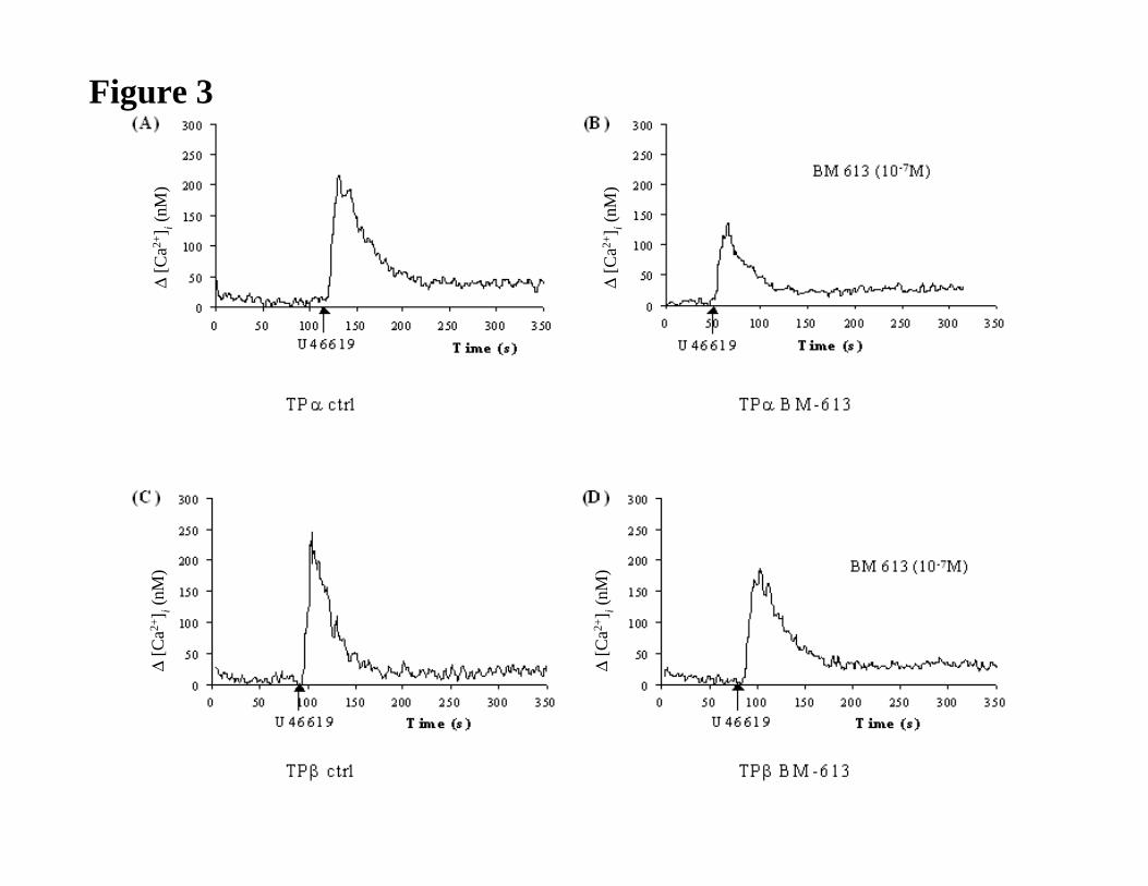
Figure 3
Panels A & B: HEK.TPα cells, transiently co-transfected with pCMV : Gαq, were either
stimulated with 1 µM U-46619 alone (Panel A) or were pre-incubated with BM-613 (10-7) for
5 min prior to stimulation with 1 µM U-46619 (Panel B). Data presented in Panels A & B are
representative profiles from three independent experiments.
Panels C & D: HEK.TPβ cells, transiently co-transfected with pCMV:Gαq, were either
stimulated with 1 µM U-46619 alone (Panel C) or were pre-incubated with BM-613 (10-7) for
5 min prior to stimulation with 1 µM U46619 (Panel D). Data presented in Panels C & D are
representative profiles from three independent experiments.
Figure 4
Panel A & C: HEK.TPαcells, transiently co-transfected with pCMV:Gαq, were incubated
with either BM-573 (Panel A, 10-9 to 10-3 M) or BM-613 (Panel C, 10-9 to 10-3 M) for 5 min
followed by stimulation with 1 µM U46619.
JPET #79301
30
Panel B & D: HEK.TPβ cells, transiently co-transfected with pCMV:Gαq, were incubated
with either BM-573 (B, 10-9 to 10-3 M) or BM-613 (D, 10-9 to 10-3 M) for 5 min followed by
stimulation with 1 µM U-46619.
Mean changes in U-46619-mediated [Ca2+]i mobilization in the presence of the drug were
expressed as a percentage (%) of U-46619-mediated [Ca2+]i mobilization in the absence of the
drug.
Figure 5
Dose-dependent effects of BM-613 on thrombus weight (mg/kg) induced by application of
ferric chloride solution (50% m/v) on rat abdominal aorta. Results are expressed as the mean
± SEM * p<0.05. Data presented are representative of four independent experiments.

JPET #79301
31
Table 1 Inhibition of human platelet aggregation induced by arachidonic acid, U-46619
and ADP.
Compounds were evaluated at concentration of 1 and 10 µM in experiments using ADP as an
inducer. Results are expressed as mean ± SEM. ED50 represents the drug concentration
inhibiting 50% of human platelet aggregation.
Drugs
U-46619
1 µM
ED50 (µM)
Arachidonic acid
600 µM
ED50 (µM)
ADP
2 µM
SQ-29548 0.034 ± 0.012 0.035 ± 0.003 Inhibition of second wave
BM-573 0.240 ± 0.013 0.125 ± 0.015 Inhibition of second wave
BM-613 0.278 ± 0.186 0.375 ± 0.05 Inhibition of second wave
JPET #79301
32
Table 2 Effect of BM-613 on rat aorta and guinea pig trachea contraction induced by U-
46619.
Results are expressed as mean ± SEM. ED50 represents the drug concentration reducing by
50% the rat aorta and the guinea-pig trachea tonus induced by 20 nM and 10 nM U-46619,
respectively.
Inhibition of contraction induced by U-46619
Drugs Rat aorta
ED50 (nM)
Guinea-pig trachea
ED50 (nM)
SQ-29548 2.3 ± 0.1 3.8 ± 0.6
BM-573 28.4 ± 4.5 17.7 ± 3.9
BM-613 2500 ± 180 1520 ± 250
JPET #79301
33
Table 3 Binding affinity of BM-613 for TP receptor on different cell types.
Tests were performed in triplicate and results are expressed as mean ± SEM.
Affinity
Drugs Human Plateletsa
IC50 (nM)
TPαb
IC50 (nM)
TPβc
IC50 (nM)
HCASM cellsd
IC50 (nM)
SQ-29548 21 ± 2 N.D.e N.D.e N.D.e
BM-573 1.3 ± 0.1 1.1 ± 0.2 0.8 ± 0.1 37000 ± 1000
BM-613 1.4 ± 0.2 2.1 ± 0.5 3.1 ± 0.7 29000 ± 7000
a Human washed platelets
b COS-7 cells expressing TPα
c COS-7 cells expressing TPβ
d Human coronary artery smooth muscle cells
e Not Determined.
JPET #79301
34
Table 4 Summary of IC50 determinations of effects of BM-573 and BM-613 on U-46619-
mediated [Ca2+]i mobilization by TPα and TPβ.
Drugs TPα
IC50 (µM)
TPβ
IC50 (µM) p valuea
BM-573 0.17 0.38 0.6
BM-613 0.11 0.30 0.8
a Mann-Whitney test to compare differences between TPα and TPβ.
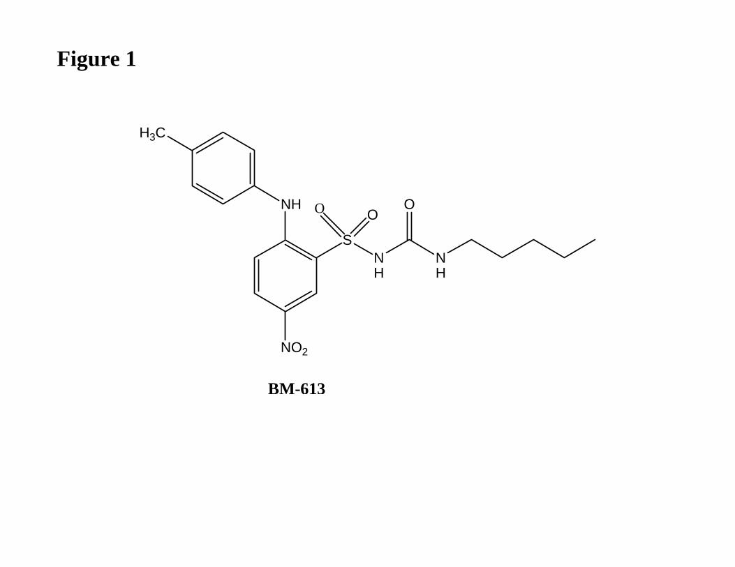
Figure 1
NH
NO2
SNH
NH
O
BM-613
H3C
OO
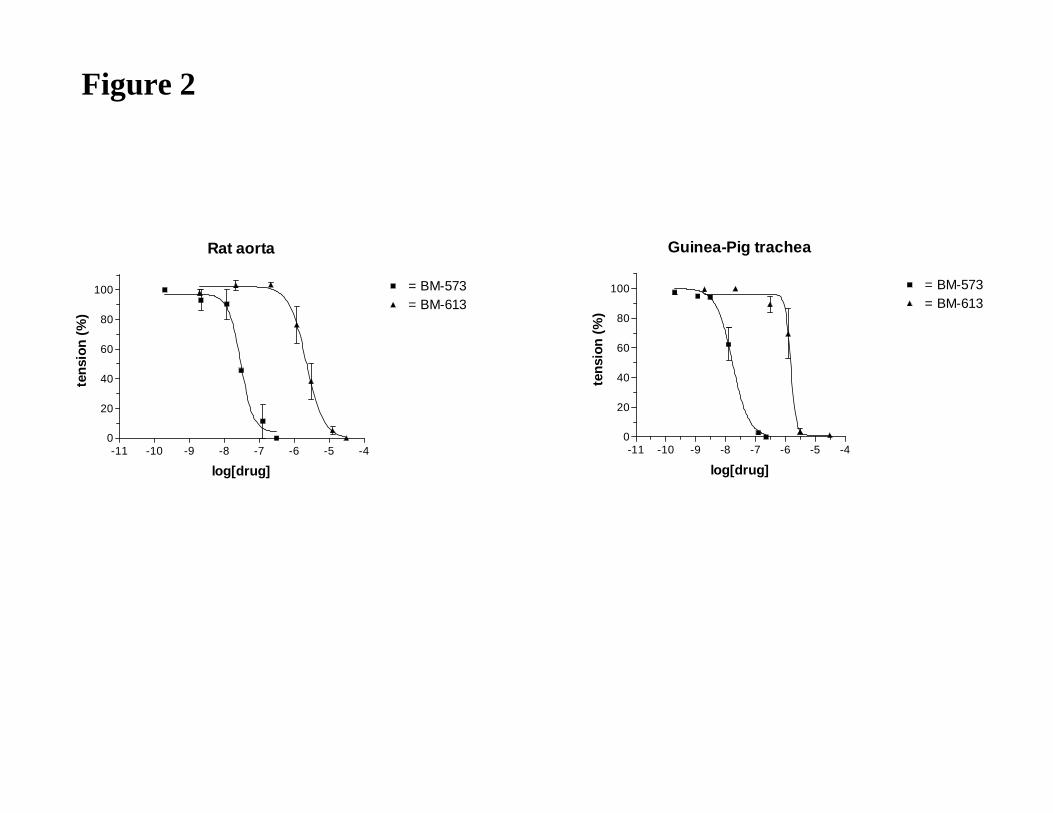
Figure 2
Rat aorta
-11 -10 -9 -8 -7 -6 -5 -40
20
40
60
80
100 = BM-573= BM-613
log[drug]
ten
sion
(%
)
Guinea-Pig trachea
-11 -10 -9 -8 -7 -6 -5 -40
20
40
60
80
100 = BM-573= BM-613
log[drug]
ten
sion
(%
)
∆[C
a2+] i
(nM
)
∆[C
a2+] i
(nM
)
∆[C
a2+] i
(nM
)
∆[C
a2+] i
(nM
)
Figure 3
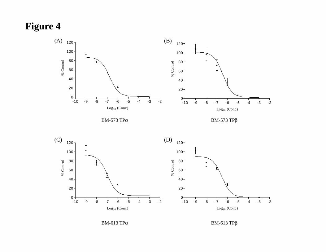
Figure 4
-10 -9 -8 -7 -6 -5 -4 -3 -20
20
40
60
80
100
120
Log10 (Conc)
% C
ontr
ol
-10 -9 -8 -7 -6 -5 -4 -3 -20
20
40
60
80
100
120
Log10 (Conc)
% C
ontr
ol
-10 -9 -8 -7 -6 -5 -4 -3 -20
20
40
60
80
100
120
Log10 (Conc)
% C
ontr
ol
-10 -9 -8 -7 -6 -5 -4 -3 -20
20
40
60
80
100
120
Log10 (Conc)
% C
ontr
ol
(A) (B)
(C) (D)
BM-573 TPα BM-573 TPβ
BM-613 TPα BM-613 TPβ
*
*
*
0
1
2
3
4
5
6
7
8
9
10
Vehicule 5 mg/kg 2 mg/kg 1 mg/kg 0.5 mg/kg
BM-613 concentration
Th
rom
bu
s w
eig
ht
(mg
/
Figure 5
Copyright © 2022 FDOKUMEN