Electroacupuncture promotes peripheral nerve regeneration ...
Upload
independentCategory
view
0download
0
Molecular Neurobiology Copyright �9 1994 Humana Press, Inc. All rights of any nature whatsoever reserved, ISSN0893-7648/93/7(3-4): 293-334/$9.40
as an Receptor-Receptor Interactions Integrative Mechanism in Nerve Cells
Michele Zoli,2 Luigi F. Agnati,2 Peter B. Hedlund, i Xi Ming Li, i Sergi FerrY, i and Kjefl Fuxe*1
7Department of Histology and Neurobiology, Karolinska Insfitutet, Box 60400, S- 10401, Stockholm, Sweden; and 2Institute of Human Physiology.
University of Modena, Via Campi 287, 41100, Modena, Italy
Contents Introduction Different Levels of Receptor-Receptor Interactions
Type A Interactions--Interactions Between Binding Sites on the Same Receptor Molecule
Type B and C Interactions--The Membrane Level of Receptor-Receptor Interactions: G Protein-Mediated and G Protein-Independent Mechanisms
Type D Interactions--Interactions Between Nonadjacent Receptor Molecules in the Membrane via Intracytoplasmic Loops
An Example of Receptor-Receptor Interactions: Dopamine D2 Receptors and Their Modulators in the Basal Ganglia
Reciprocal D1/D2 Receptor Interactions Reciprocal Neurotensin/D2 Receptor Interactions in the Basal Ganglia CCK-8/DA Receptor Interactions A2/D2 Receptor Interactions in the Basal Ganglia Overall View
Integrative Aspects of Receptor-Receptor Interactions Integration of Signals Through Receptor-Receptor Interactions Receptor-Receptor Interactions and Neuronal Plasticity
Acknowledgments References
Abstract Several lines of evidence indicate that interactions among transmission lines can take place at the level of the
cell membrane via interactions among macromolecules, integral or associated to the cell membrane, involved in signal recognition and transduction. The present view will focus on this last subject, i.e., on the interactions between receptors for chemical signals at the level of the neuronal membrane (receptor-receptor interaction).
*Author to whom all correspondence and reprint requests should be addressed.
Molecular Neurobiology 293 Volume 7, 1993

294 Zofi et al.
By receptor-receptor interaction we mean that a neurotransmitter or modulator, by binding to its receptor, modifies the characteristics of the receptor for another transmitter or modulator. Four types of interactions among transmission lines may be considered, but mainly intramembrane receptor-receptor interactions have been dealt with in this article, exemplified by the heteroregulation of D2 receptors via neuropeptide receptors and A2 receptors. The role of receptor-receptor interactions in the integration of signals is discussed, especially in terms of filtration of incoming signals, of integration of coincident signals, and of neuronal plasticity.
Index Entries: Transmitter; receptor; transmission line; heteroregulafion; homoregulation.
Introduction
Interneuronal communication can take place via electrical or chemical signals reaching their target cells via either synaptic contacts or the extraceUular fluid (ECF) (Agnati et al., 1986,1992; Fuxe and Agnati, 1991~. The neuronal membrane is capable of converting chemical into electrical signals and vice versa, as well as of transducing these signals into proper messages for the cellu- lar biochemical machinery. Thus, thanks to this role as an interface between the ECF and the intracellular environment, the neuronal mem- brane subserves highly sophisticated informa- tional tasks.
The neuronal membrane by means of recep- tors recognizes and transduces into proper intra- cellular signals a specific subset of the huge number of extracellular signals continuously impinging on it. Different transmission lines* allow the information to flow from the chemical signal-receptor complex down to the cytoplas- mic target molecules and even to the genome. However, this is not a straight through process, since at several levels of the intracellular cascade feedback circuits can affect previous steps. More- over, these parallel transmission lines can recip- rocally interact at mult iple levels, forming a complex network of signals stemming from the membrane and going back to it. Several lines of ev idence ind ica te that in teract ions a m o n g transmission lines can take place at the level of
*Transmitter = molecule that activates a receptor; modulator = molecule that changes the activity of a transmitter on its receptor; transmission line = the cascade of molecular events at membrane, cytoplasmic and, possibly, nuclear levels triggered by the binding of a transmitter to its receptor.
the cell m e m b r a n e via in teract ions a m o n g macromolecules, integral or associated to the cell membrane, involved in signal recognition and transduction. The present review will focus on this last subject, i.e., on the interactions between receptors for chemica l s ignals at the level of the neuronal membrane ("receptor-receptor interaction").
By receptor-receptor interaction we mean that a neurotransmitter or a modulator, by binding to its receptor, modifies the characteristics of the receptor for another transmitter or modulator. This type of regulatory phenomenon is then part of the broader class of receptor heteroregulation phenomena. Whereas receptor homoregulation (e.g., sensitization and desensitization) represents mechanisms by which a receptor is modulated by the molecules belonging to its own transmis- sion line (ligand, second messengers, and so forth), receptor heteroregulation represents a direct or indirect modulation of a certain recep- tor by molecules (including other receptors) belonging to different transmission lines or by other intracellular signals.
From theoretical considerations and experi- menta l evidence, four types of interact ions among transmission lines involving receptor regulation may be surmized (see Fig. 1):
A. Allosteric changes of a receptor macromolecu- lar complex caused by the binding on it of a modulator.
B. Interactions between physically distinct but adjacent receptors involving the extracellular loops (i.e., taking place in the glycocalyx milieu, mechanism type B1), the transmembrane hel- ices (i.e., taking place in the lipid bilayer milieu, mechanism type B2), or the intracyto- plasmic loops (i.e., taking place at the interface between the membrane and the cytoplasm,
Molecular Neurobiology Volume 7, 1993

Receptor-Receptor Interactions 295
TYPE OF INTERACTION
A
B
C
D
BINDING SITE- BINDING SITE INTERACTION INSIDE A RECEPTOR MACROMOLECULE
INTRAMEMBRANE INTERACTION BETWEEN ADJACENT RECEPTORS
INTRAMEMBRANE INTERACTION BETWEEN NON-ADJACENT RECEPTORS
INTERACTION BETWEEN RECEPTORS THROUGH INTRACELLULAR PROCESSES
SCHEMATIC REPRESENTATION
7, RS2 7
I / I I I I II
2 ;: '// / // 2
OUTLINE OF POSSIBLE INFORMATIONAL FLOW
T~ T~
T~ T 2
RSI " ~ ~ RS2
El E 2
T, T 2
RS 1 RS 2
""* M M A P
T~ T 2
k NS,,
Fig. 1. Schematic representation of the possible types (A-D) of receptor-receptor interactions as outlined in the text. For the sake of simplicity, only unidirectional interactions are shown. Abbreviations: AP = associating protein, CS = cytoplasmic signals, E = effector portion of the receptor, MMAP = mobile membrane-associated protein (e.g., G protein), NS = nuclear signals, KS = recognition site, T = transmitter.
mechanism type B3) of the receptor molecules. These interactions either can be direct or can involve associated proteins.
C. Interactions between physically distinct and nonadjacent receptors involving the activation of mobile membrane-associated proteins, such as G proteins.
D. Interactions between physically distinct and nonadjacent receptors through intraceUular pro-
cesses, such as protein phosphorylation and pro- tein biosynthesis (i.e., taking place in the inter- nal milieu of the cell and often involving the gene level).
Interact ion type A is an in t ramolecular phe- n o m e n o n and canbe defined asbinding site-bind- ing site interaction inside a receptor molecule. Interaction types B and C are true membrane
Molecular Neurobiology Volume 7, 1993
296 Zoli et al.
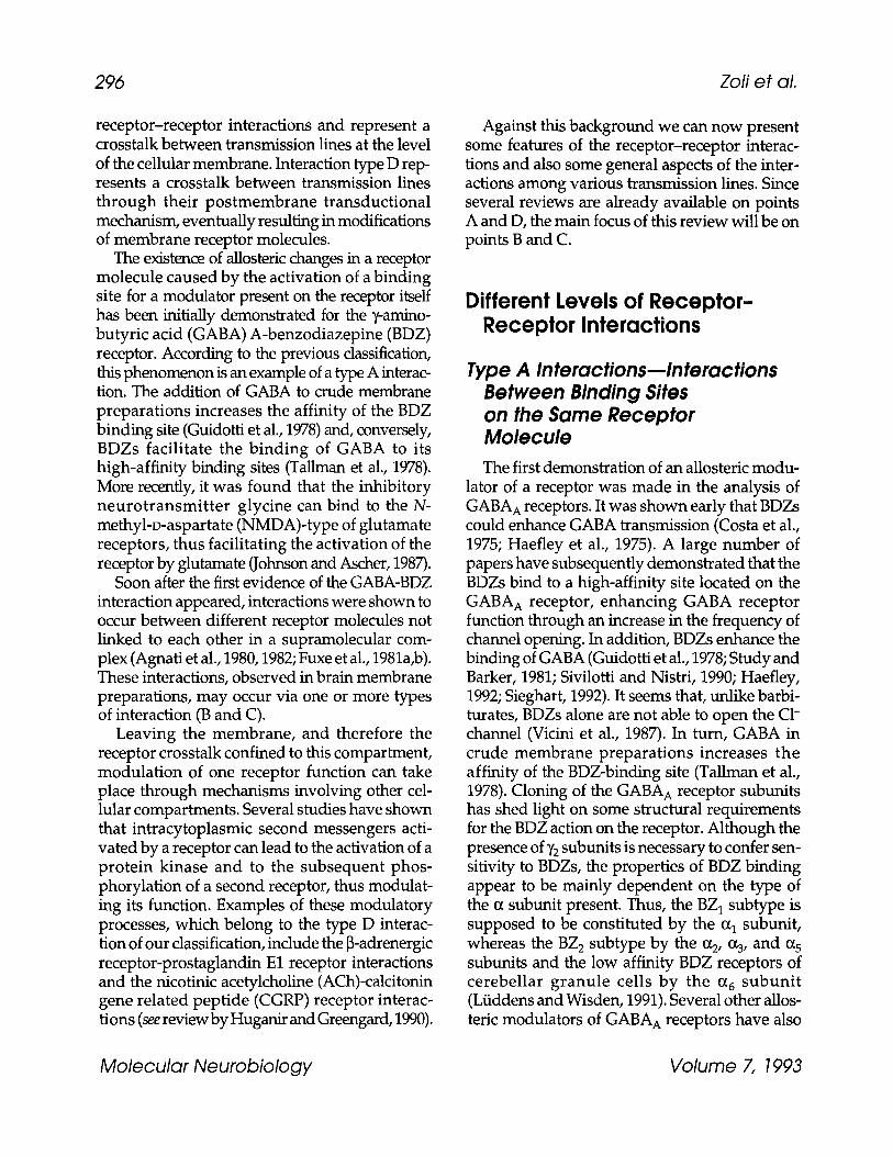
receptor-receptor interactions and represent a crosstalk between transmission lines at the level of the cellular membrane. Interaction type D rep- resents a crosstalk between transmission lines through their postmembrane transductional mechanism, eventually resulting in modifications of membrane receptor molecules.
The existence of allosteric changes in a receptor molecule caused by the activation of a binding site for a modulator present on the receptor itself has been initially demonstrated for the y-amino- butyric acid (GABA) A-benzodiazepine (BDZ) receptor. According to the previous classification, this phenomenon is an example of a type A interac- tion. The addition of GABA to crude membrane preparations increases the affinity of the BDZ binding site (Guidotti et al., 1978) and, conversely, BDZs facilitate the binding of GABA to its high-affinity binding sites (Tallman et al., 1978). More recently, it was found that the inhibitory neurotransmit ter glycine can bind to the N- methyl-D-aspartate (NMDA)-type of glutamate receptors, thus facilitating the activation of the receptor by glutamate (Johnson and Ascher, 1987).
Soon after the first evidence of the GABA-BDZ interaction appeared, interactions were shown to occur between different receptor molecules not linked to each other in a supramolecular com- plex (Agnati et al., 1980,1982; Fuxe et al., 1981a,b). These interactions, observed in brain membrane preparations, may occur via one or more types of interaction (B and C).
Leaving the membrane, and therefore the receptor crosstalk confined to this compartment, modulation of one receptor function can take place through mechanisms involving other cel- lular compartments. Several studies have shown that intracytoplasmic second messengers acti- vated by a receptor can lead to the activation of a protein kinase and to the subsequent phos- phorylation of a second receptor, thus modulat- ing its function. Examples of these modulatory processes, which belong to the type D interac- tion of our classification, include the [3-adrenergic receptor-prostaglandin E1 receptor interactions and the nicotinic acetylcholine (ACh)-calcitonin gene related peptide (CGRP) receptor interac- tions (see review by Huganir and Greengard, 1990).
Against this background we can now present some features of the receptor-receptor interac- tions and also some general aspects of the inter- actions among various transmission lines. Since several reviews are already available on points A and D, the main focus of this review will be on points B and C.
Different Levels of Receptor- Receptor Interactions
Type A Interactionsmlnteractions Between Binding Sites on the Same Receptor Molecule The first demonstration of an allosteric modu-
lator of a receptor was made in the analysis of GABA A receptors. It was shown early that BDZs could enhance GABA transmission (Costa et al., 1975; Haefley et al., 1975). A large number of papers have subsequently demonstrated that the BDZs bind to a high-affinity site located on the GABA A receptor, enhancing GABA receptor function through an increase in the frequency of channel opening. In addition, BDZs enhance the binding of GABA (Guidotti et al., 1978; Study and Barker, 1981; Sivilotti and Nistri, 1990; Haefley, 1992; Sieghart, 1992). It seems that, unlike barbi- turates, BDZs alone are not able to open the C1- channel (Vicini et al., 1987). In turn, GABA in crude membrane preparations increases the affinity of the BDZ-binding site (Tallman et al., 1978). Cloning of the GABA A receptor subunits has shed light on some structural requirements for the BDZ action on the receptor. Although the presence of ~/2 subunits is necessary to confer sen- sitivity to BDZs, the properties of BDZ binding appear to be mainly dependent on the type of the 0~ subunit present. Thus, the BZ 1 subtype is supposed to be constituted by the al subunit, whereas the BZ 2 subtype by the (z 2, %, and % subunits and the low affinity BDZ receptors of cerebellar granule cells by the a 6 subunit (Ltiddens and Wisden, 1991). Several other allos- teric modulators of GABA A receptors have also
Molecular Neurobiology Volume 7, 1993

Receptor-Receptor Interactions 297
been characterized, including barbiturates, neurosteroids, and Zn 2§ (Sieghart, 1992).
Also, the NMDA receptor complex contains a number of binding sites for allosteric modulators controlling the glutamate-binding site and the gating of the ion channel, including glycine, Zn 2+, and polyamines (Williams et al., 1990). As initially demonstrated by Johnson and Ascher (1987), the activation of the NMDA channel by glutamate is facilitated by the binding of glycine (Kemp and Leeson, 1993). After the cloning of the functional NMDA receptor, it has been possible to obtain direct evidence that a glycine-binding site is present in the same protein carrying the NMDA recognition site (Moriyoshi et al., 1991). In addi- tion, different NMDA receptor subunits have dis- tinct affinities for glycine (Kemp and Leeson, 1993). The case of glycine modulation of the NMDA receptor is, however, very peculiar. In fact, contrary to the other known allosteric modu- lators, the NMDA receptor cannot be activated by glutamate in the absence of glycine. Glycine should then be considered a coagonist at NMDA receptors (Kemp and Leeson, 1993).
As in the case of the GABA-BDZ interaction (see below), binding of glycine appears to regu- late the properties of glutamate binding and vice versa. Thus, glutamate enhances 3H-glycine bind- ing, and glycine enhances 3H-glutamate binding (Kessler et al., 1989; Monaghan et al., 1988). The partial agonist for the glycine binding site HA- 966 increases the dissociation rate of glutamate compared with the dissociation rate in the pres- ence of glycine (Kemp and Priestley, 1991). HA- 966 also noncompetitively inhibits 3H-glutamate binding, while enhancing the binding of NMDA antagonists of the competitive type (Danysz et al., 1989). These data are at variance with elec- trophysiological experiments indicating that glycine and glutamate-binding sites are nega- tively coupled (Benveniste et al., 1990; Kemp and Leeson, 1993). It must, however, be remembered that equilibrium-binding experiments cannot discriminate among (multiple) open and desensi- tized states and do not reflect the fast kinetic changes observed in electrophysiological experiments.
Several modulators have been shown for muscle type nicotinic ACh receptor, another
member of the ligand-gated ion channel super- family (L6na and Changeux, 1993). Some evi- dence suggests that neuronal nicotinic ACh receptors also may have binding sites for allos- teric modulators, such as substance P (SP), atrial natriuretic factor (ANF), and steroids. The abil- ity of SP to inhibit nicotinic ACh receptor func- tion in PC12 and chromaffin cells appears to be related to a stabilization of the desensitized state of the receptor (Boyd and Leeman, 1987). Struc- ture-activity analysis revealed that the action of SP on nicotinic ACh receptors takes place at a binding site different from the G-protein-linked SP receptor (Geraghty et al., 1990), suggesting that the effect may be caused by the binding of SP to the nicotinic receptor. In addition, SP may directly block the flux of ions through the channel (Boyd and Leeman, 1987). In similar studies, it has been observed that ANF can also reduce nicotine- induced currents (Bormann et al., 1989). How- ever, in this case it appears more likely that the gating of the ion channel of the nicotine receptor is brought about by an interaction between inde- pendent receptors (interaction types B or C). Fur- ther experiments using ANF and SP receptor antagonists and cloned neuronal nicotinic recep- tors will help differentiate between these two types of mechanistic models for peptide modu- lation of gating of nicotinic channels.
Receptor changes on agonist and modulator binding have been interpreted (Changeux, 1990) according to the model of allosteric transitions between conformational states proposed by Monod et al. (1965). The allosteric modulators bind to sites different from that of the agonist and can affect the equilibrium and/or the kinetics of the transitions between the conformational states. It is assumed that changes in the properties of the conformational states caused by the modulators are of negligible entity when compared with the differences existing between the properties of the main conformational states themselves (for a detailed discussion, see L6na and Changeux, 1993). This theory has been successfully applied to the explanation of the properties of muscle nicotinic ACh receptor on binding of ACh and some modulators (Heidmann and Changeux, 1979a,b). Recent studies are starting to charac-
Molecular Neurobiology Volume 7, 1993

298 Zofi et al.
terize conformational states and their transitions in the presence of the agonist or modulators for the other ligand-gated ion channels (Macdonald et al. 1989; Weiss and Magleby, 1989; Ito et al., 1990; Sieghart, 1992).
Since discussed above, a wealth of allosteric modulators have been demonstrated for ligand- gated ion channels. Although the pharmacolog- ical actions of these modulators are clearcut (see, e.g., the case of BDZs and the search for allos- teric antagonists of glutamate receptors), their physiological role is still debated. Some questions seem to be relevant in this respect:
1. Are there endogenous substances capable of binding to the aUosteric modulator site in the effective range of concentrations?
2. Are there physiological mechanisms for regu- lating the concentration of the endogenous ligands? If this is the case, it is interesting to know if the substance can be released by neu- rons, i.e., acting like a neuromodulator or derive from other sources.
3. Since the same modulator can affect several re- ceptor types present on a postsynaptic cell how can its action be differently regulated?
Several claims have been made for the exist- ence of endogenous BDZs, or endozepins (see, e.g., Barbaccia et al., 1988; Rothstein et al., 1992). Evidence for their physiological roles is, however, not conclusive. Among others, a strong case for an action as a neuromodulator has been made for Zn 2+. This molecule is able to modulate NMDA, AMPA/kainate, and GABA receptor gating properties (Mayer et al., 1989; Legendre and Westbrook, 1991). Interestingly, Zn 2+ is highly enriched in the synaptic vesicles of the hip- pocampal mossy fibers, and can be released during neuronal activity (Perez-Clausell and Danscher, 1985; Assaf and Chung, 1984; Howell et al., 1984). Also, polyamines (positive allosteric modulators of glutamate binding on NMDA receptors; Williams et al., 1991) have been detected in the brain ECF (Grimaldi et al., 1991). In particular, they have been shown to increase after lesion (Desiderio et al., 1988; Paschen et al., 1988), which led to the suggestion that they may be involved in modulating glutamate receptor neurotoxicity (Paschen et al., 1988; Zoli et al.,
1993). Finally, cytoskeletal proteins, such as fodrin, should be considered as a regulator (Simon et al., 1985).
The mechanism proposed for the physiolog- ical action of glycine on the NMDA receptor is peculiar. Glycine levels in the ECF are supposed to be sufficient to saturate the glycine site in basal conditions (Kemp and Leeson, 1993). The NMDA receptor would therefore be always activatable by the endogenous ligand. However, the glycine modulation of NMDA receptor function may occur through a local decrease in glycine extra- cellular levels via a neuron-specific glycine transporter , a protein h ighly enr iched in areas containing high levels of NMDA recep- tors (D'Angelo et al., 1990; Smith et al., 1992). This view is supported by electrophysiological evidence showing that glycine can potentiate some glutamate responses in vivo (Kemp and Leeson, 1993).
Type B and C Interactions-- The Membrane Level of Receptor-Receptor Interactions: G Protein-Mediated and G Protein-lndependent Mechanisms
A huge amount of experimental evidence has accumulated in the last 10 yr on the existence of changes in receptor characteristics induced by the administration of a modulator in crude mem- brane preparations (for reviews see Fuxe and Agnati, 1985, 1987). Intramembrane modulatory processes between neuropeptide and monoamine receptors were early described in the central ner- vous system (CNS), including interactions between SP/5-HT 1 receptors, vasoactive intes- tinal polypeptide (VIP)/5-HT 1 receptors, chole- cystokinin (CCK)/dopamine (DA) D2 receptors, neurotensin (NT)/DA D2 receptors, and the neuropeptide (NPY)/0~2 adrenergic receptor interactions (Agnati et al., 1980,1982,1983a,b,c, d,e, 1984; Agnati and Fuxe, 1983; Fuxe et al., 1981a,b, 1983c; Rostene et al., 1983a,b). Similar modula- tory events have been described for interactions between monoamine receptor subtypes (Seeman
Molecular Neurobiology Volume 7, 1993

Receptor-Receptor Interactions 299
et al., 1989), peptide receptors (Rothman et al., 1988), monoamine and adenosine receptors (Ferr6 et al., 1991d; Murayama et al., 1990), glutamate receptor subtypes (Fuxe et al., 1983b), and mono- amine or peptide and ligand-gated ion channels (Fuxe et al., 1983a, 1984b, 1989b).
Since these interactions have been found in crude membrane preparations, they do not involve cytoplasmic loops (second messengers, phosphorylations, and so forth). These phenom- ena have therefore been explained on the basis of an interaction between receptor molecules, possibly including G proteins and other mol- ecules associated with the membrane.
Direct binding of the modulator to the recep- tor cannot be, in principle, ruled out. As already discussed, a huge number of modulatory sites has been shown in ligand-gated ion channels and it cannot be excluded that similar sites are present in G-protein-linked receptors. However, several lines of evidence indicate that the interaction takes place between independent receptor molecules. Independent receptors for the modulatory sub- stances have been demonstrated biochemically and sometimes also cloned. These receptors are known to be present in the areas, and sometimes even in the same cells, where the modulation is demonstrated. The effects are present when the modulator is administered at concentrations close to the Ke of the known modulator receptors. Experiments on reconstituted systems will help clarify the possible existence of modulatory sites on the receptors.
In a number of studies it has been shown that receptor-receptor interactions are specific for receptor isotypes (see Table 1). For instance, sev- eral modulators (CCK, NT, adenosine) adminis- tered in the nanomolar range are able to change the binding characteristics of D2, but not D1 dopamine receptors in the basal ganglia (see below). On the other hand, NT in the micromolar range affects D1 binding. Interestingly, the NT effect on D2 receptors appears to be G protein- independent (von Euler, 1991), whereas that on D1 receptors is G protein-dependent (Miyoshi et al., 1989). Since both D1 and D2 receptors are highly enriched in striatum, the selective changes in the binding characteristics of one dopamine
receptor subtype could, in different physiologi- cal states, switch DA transmission from one to the other receptor isotype (Fig. 2).
The evidence of intramembrane receptor- receptor interactions in vivo is still indirect. In several cases, the same modulation of receptor characteristics has been demonstrated in vitro and after intracerebroventricular (icv) adminis- tration of the modulator (see Table 1). Colocal- ization of the two interacting receptors is usually prominent in areas where the interaction is dem- onstrated. Finally, in many instances receptor crosstalk has been correlated with functional changes at neurochemical and/or behavioral lev- els (GAL [Fuxe et al., 1988a; Hedlund et al., 1991a,b], NPY [H/irfstrand et al., 1989; Aguirre et al., 1990], and A1-D2; see following article on DA receptor modulation).
For instance, a powerful inhibitory interaction has been shown between the NPY receptor (Wahlenstedt and Hakanson, 1987) and one adrenergic receptor subtype, the (z2 receptor, in the dorsal medulla oblongata, an area where the two receptors are highly concentrated (Fuxe et al., 1989a). The modulation was not present when other adrenergic receptor subtypes were tested. The interaction was first observed in vitro and then on icv injections of the peptide (Agnati et al., 1989). Receptor autoradiography experiments confirmed that the site of interaction was in the subregions of the nucleus tractus solitarius (nTS) where the distribution of the two receptors mark- edly overlaps (H~irfstrand et al., 1989). Finally, NPY and clonidine injected icv have a vaso- depressor action that is diminished when the two substances are coadministered. Overall, these data indicate that NPY receptors have an antago- nistic interaction with the c~2 transmission in the dorsal medulla oblongata and that an interaction between NPY and c~2 receptors is likely to be the mechanism mediating this action.
Although the presence of membrane receptor- receptor interactions can give a functional counterpart to the coexistence of two or more sub- stances in the nerve terminals, these interactions are not restricted to cases of coexistence (see, e.g., the interaction between NT and D2 receptors in the basal ganglia, and Table 1). It can then be
Molecutar Neurobiology Volume 7, 1993

t,2
02
0
02
i l =: o ~ ,,,wl
"~1 ~l ~
~ ~2 ~
if)
~ ~ . ~ o o ~
~ o <
~~ ~ ~ i.)
0 ~
~.~
~ ~ ~ . ~ . ~- ~ ~ ~-~ " 0 2
�9 ~ < ~ ~ ~< ~
~ ~ ~ ~~ ~oo~
"~ II "~ I! ~ l~ -< - - - II I1 II li II ~1 II
Z
0 0 ~ 0 0 0 0
• b13 02
'-a o Ei ~ 02 :,.,.r II 0 ~ < ~ 02 0 2 ~ II
~SeN
u .. . 02 ~
~.~ ~,.~
.~oo~ ~ ~,~ ,,
m t,~ ~ 02
b
. II ,I~ �9 ~ O ~ ~ ' '
6 . ~ ' U
0 ~ i
0 ~ - ~ ~
u m ~ a s ~
~ M ; . ~ :::1 u
~ 02 II I-~ t.l "
H ~ Z �9 ~ ~ -
�9 ~ k~::l
Molecular Neurobiology 300 Volume 7, 1993

Receptor-Receptor Interactions 301
z 0
z
DIVERGENCE AND CONVERGENCE OF DA TRANSMISSION LINES
D~ DS(DIB)
\ / I
D2L, 2S
\ ~ [ ~ . . . . . . . . . . . .
[ ~ . . . . . . . . . . . .
D4 D~
Fig. 2. Divergence and convergence of dopamine transmission lines at the level of receptor subtypes, G proteins and second messenger systems. Abbreviations: AA = arachidonic acid, AC = adenylate cyclase, PLC = phospholipase C.
hypothesized that receptors interacting with each other belong to the same synapse or to adjacent synapses. A further possibility is that interacting receptors may be located extrasynaptically, acti- vated by transmitters diffusing in the ECF (Fig. 3) (volume transmission, Fuxe and Agnati, 1991).
Mechanistic Aspects At the present time it is only possible to spec-
ulate about what may be the mechanisms for interactions between the G-protein coupled receptors within the membrane. Two main mechanistic models of intramembrane inter- actions may be discussed. One mechanism is represented by direct interactions between physically distinct but adjacent receptors, possi- bly also involving some associated proteins (type B). The other mechanism is based on the use of mobile proteins associated to the membrane, such as G proteins , as messengers for the inter- actions between physically distinct receptors (type C). However, multiple types of interaction are likely to be present for each receptor.
The available data give support to the exist- ence of both types of mechanisms. For instance, NT modulation of the D2 receptor binding char- acteristics in membrane preparations of the neostriatum is maintained unchanged after a marked inact ivat ion of G i and G O proteins induced by N-ethylmaleimide (NEM) or pertus- sis toxin (PTX) treatment (von Euler et al., 1991). PTX-sensitive G i and G o proteins are coupled to D2 receptors and are known to mediate its physi- ological effects (Cote et al., 1986; Fujita et al., 1985). Furthermore, an A2 agonist can counteract the action of GTP at D2 receptors at an independent site of action (for a deta i led explanat ion, see below) (Ferr6 et al., 1993). On the other hand, many receptor-receptor interactions observed in membrane preparations seem to involve a G pro- tein as an essential link (see, e.g., von Euler, 1989, Miyoshi et al., 1989; Murayama et al., 1990, and Table 1). For instance, Gi/G o proteins seem to be involved in the reduction of affinity of o~ 2 recep- tors induced by NPY in membranes of the medulla oblongata (von Euler et al., 1989; Fuxe
Molecular Neurobiology Volume 7, 1993

302 Zoli et al.
WT iil '! ,,,
MEMBRANE
PRESYNAPT1C TERMINAL
WIRING TRANSMISSION
VT VOLUME TRANSMISSION
~ INTERACI~)NS TYPE A, B, C
.el- - - - --~ INTERACTIONS TYPE D
I ~ ASSOCIATING PROTEIN
Fig. 3. Schematic drawing showing receptor-receptor interactions in synaptic (wiring transmission, WT) and nonsynaptic (volume transmission, VT) sites on neuronal membranes.
et al., 1989a). NPY effects were, in fact, blocked by prior intracisternal treatment of the animals with PTX. This treatment also counteracted the cardio- vascular actions of both NPY and clonidine (an a2 agonist) on intracisternal injection. The block- ade was not caused by a loss of high-affinity 125I- NPY 1-36 binding sites by the PTX treatment, since this treatment rather produced an increase in 125I-NPY binding. Another example of G pro- tein mediation of receptor crosstalk is the i n t e r a c t i o n between adenosine A1 and D2 receptors in rat neocortex. PTX treatment was able to prevent the DA agonist apomorphine- induced reduction of adenosine A1 binding (Murayama et al., 1990).
MECHANISM TYPE B A possible basis for interactions between
receptors is dimerization of structurally differ-
ent receptors (heterologous dimerization; Fig. 4). Dimerization (and oligomerization) of recep- tor molecules on activation by the agonist seems to be a general phenomenon (Hollenberg, 1991). According to the type of receptor, dimerization would be a necessary condition for activation or a means leading to response potentiation or meta- bolic stabilization of the receptors.
The best known case of receptor dimerization is that of the tyrosine kinase receptor superfamily (Schlessinger, 1988; Ullrich and Schlessinger, 1990). This property, which is present both in preparations of purified receptors and in living cells, is essential for signal transduction, includ- ing autophosphorylation. In addition, dimer- ization increases the affinity for the agonist. Although for some receptors, like insulin and IGF-1 receptors, stable dimerization is attained
Molecular Neurobiology Volume 7, 1993

Receptor-Receptor Interactions
MONOMERS HOMODIMERS HETERODIMERS
303
NO EFFECT EFFECT A EFFECT B EFFECT C
T A ~ EFFECT A
T B ~ EFFECT B
T A + T s ~ EFFECTS A + B + C
Fig. 4. Homo- and heterodimerization of membrane receptors. Heterodimers induce a cellular effect different from homodimers. The relative proportion of the two transmitters, the concentration of the two receptor populations, and the characteristics of receptor-receptor interactions will determine the amount of homo- vs heterodimers and thus the overall effect on the target cell.
through disulfide bridges, in most cases transient dimerization is induced by conformational changes on agonist binding.
Some evidence of dimerization and oligomer- ization is also available for other types of recep- tors. In the family of ligand-gated ion channels, the occurrence of functional dimers as the prev- alent active form in vivo has been demonstrated for nicotinic receptors in both rat myotubes (Yeramian et al., 1986) and Torpedo californica elec- tric organ (Schindler et al., 1984). Again, although in some instances receptors can be stably dimer- ized through covalent SH bridges, in other cases the interaction is noncovalent. Formation of large clusters of nicotinic receptors occurs during matu- ration of the neuromuscular junction, involving changes in receptor metabolism and function (Laufer and Changeux, 1989). In this case, proteins associated with nicotinic receptors, such as the 43K protein, may have an important role (Hill, 1992).
Functional G protein-linked receptors also appear to be in a dimeric form. Purification of several receptors of this family demonstrated that the native receptor protein is a dimer (see, e.g.,
Venter and Fraser, 1983; Roche and Ryan, 1989). Interestingly, studies on the activation of the luteinizing hormone-releasing hormone (LHRH) receptor showed that dimerization is a necessary step in receptor function and that an antagonist is transformed into an agonist when it is capable of bringing two receptor molecules within a criti- cal distance (Conn et al., 1982).
If molecular mechanisms for dimerization are conserved within receptor superfamilies, the pos- sibility exists that dimerization occurs between different members of the family (i.e., heterodimer- ization vs homodimerization). Heterologous dimerization has been clearly demonstrated for tyrosine kinase receptors, even if its physiologi- cal properties remain to be clarified (Ullrich and Schlessinger, 1990). Recent work on opiate recep- tors, belonging to the family of G protein-linked receptors, demonstrated that certain subtypes of ~t- and 3-binding sites are functionally and physically associated (Rothmann et al., 1988; Schoffelmeer et al., 1990). This phenomenon has not yet been explained on a molecular basis. However, association of independent ~t and 3 receptors (mechanism type B) remains a distinct
Molecular Neurobiology Volume 7, 1993

304 Zofi et al.
possibility as an alternative to the presence of a single receptor complex with two binding sites (mechanism type A).
In conclusion, several cases of receptor-recep- tor interactions in crude membrane preparations may be based on a process of heterodimerization. The density of the two or more interacting recep- tors and the number of receptors activated by the agonist in each population would then determine the proportion of homo- vs heterodimerizations and, then, the overall effect on target cell func- tion (Fig. 4).
In the case of tyrosine kinase receptors, dim- erization occurs through an interaction between the glycosilated extracellular domains (mecha- nism type B1). In this context, it is interesting to remember that many peptide receptors are gly- coproteins; for example the NPY/PYY receptor subtypes are structurally distinct glycoproteins (Sheikh and Williams, 1990). Also, monoamine receptors such as (zl (Sawutz et aI., 1987), ~ (Con- vents et al., 1988), [32 (Benovic et al., 1987b), and D2 (Clagett-Dame and McKelvy, 1989) receptors have been characterized as glycoproteins. In several instances deglycosylation has no major effects on receptor binding characteristics. Instead, it has been supposed to be important for the appropriate posttranslational routing of the receptor to the cell surface (Clagett-Dame and McKelvy, 1989). A further functional meaning of receptor g lycosyla t ion might be to favor the interactions among receptors via the forma- tion of hydrogen bonds. If so, these interactions should also, be modulated by glycosidase activity. In agreement with this proposal is the detection of sialidase activity in brain synaptic membranes (Miyagi et al., 1990).
MECHANISM TYPE C A second class of molecular mechanisms that
may underlie interactions between receptors in the membrane is dependent on membrane-asso- ciated G proteins (Fig. 5).
The G proteins are heterotrimeric (consisting of (z-, [3-, and y-subunits) guanine nudeotide bind- ing proteins bound to the internal surface of the plasma membrane. They relay the signal from some types of receptors to the various membrane
m
G - PROTEIN CYCLE OF ACTIVATION
T
TR
GDP.ak j GTP
W
/~r P|
EFFECrORI
Fig. 5. Schematic drawing showing the activation of G proteins after the formation of the transmitter- receptor complex.
bound effectors (Gilman, 1987; Linder and Gilman, 1992). Evidence exists for a high diver- sity of G proteins and especially of the (z-sub- units. In fact, at least 20 distinct forms of (z-subunits, five y-subunits, and at least 10 ~- subunits have been shown to exist, giving a strong structural basis for the formation of G protein networks within the nerve cell mem- brane (Simon et al., 1991).
G proteins appear to be anchored to the inter- nal surface of the cellular membrane (Spiegel, 1992). Covalent interactions with lipids are responsible for this phenomenon. The ~yhetero- dimer is anchored through an isoprenoid mol- ecule covalently bound to the carboxyl-terminal of the y-subunit. Mutations that block isoprenyl- ation prevent association to the membrane (Simonds et al., 1991). The mechanism for mem- brane association of the (z-subunits is less well understood. Some classes of (z-subunits (G i and Go) require a myristoylation of the amino-termi- nal portion to bind to the membrane (Spiegel et
Molecular Neurobiology Volume 7, 1993

Receptor-Receptor Interactions 305
al., 1991). However, there is also evidence that in some situations a-subunits can be released from the membrane.
In the resting state, GDP is bound to the a- subunit that forms a heterotrimer with the [3- and 7-subunits. When an agonist activates the receptor, the receptor changes its conformational state and binds to the G protein (Fig. 5; Ross, 1989). This coupling facilitates the exchange of GDP for GTP, leading to a disassembly of the G protein. As a consequence, the ~-subunit, which contains the GTP-binding site, becomes free to move along the internal surface of the plasma membrane (Fig. 5) and activates membrane- bound effectors, such as adenylate cyclase and calcium channels. In this way the G protein can transduce the signal from the receptor across the nerve cell membrane. The duration of activation depends on the rate of hydrolysis of GTP, which depends on the GTPase activity of the a-subunit. Usually, the activated {x-subunits again become associated with the 13- and ~/-subunits within a few seconds as a result of the GTPase action.
Also, [3- and y-subunits seem to have a role in the activation of membrane effectors. The most frequently indicated action is the ability of 13- and ~,-subunits to counteract the actions of GTP acti- vated ~-subunits by binding to this subunit (Fig. 6) ("subunit exchange" phenomenon; Gilman, 1987; Axelrod et al., 1987; Simon et al., 1991; Linder and Gilman, 1992). In this way the GTP activated o~-subunit can no longer activate its effector (see, e.g., the G i mediated inhibition of adenylate cyclase activity via 13- and ~/-subunit binding to activated Go, i, Ross, 1989). 13- and ~/- subunits can also, directly effect various effectors inter alia leading to alterations of adenylate cyclase activity (Federman et al., 1992). Thus, it seems possible that the 13- and ~'-subunits can act as coordinators of activity in parallel trans- duction lines over the nerve cell membranes by interacting with ac t iva ted a - subun i t s and poss ib ly by directly regulating membrane bound effectors.
Four major classes of G proteins exist (Gs, Gi, Gq, and G12 ) based on the degree of sequence identities between the ~x-subunits (Simon et al.,
1991). It must be emphasized that the same ~- subunit can activate a number of different effec- tors and the same effector can be activated by several 0~-subunits. For instance, Gs can activate adenylate cyclase as well as calcium channels, whereas G%_ 3 and G%-subunits all inhibit cal- cium channels and activate potassium channels (Fredholm, 1991). On the other hand, other subunits appear to be quite specific in switching on one type of effector in response to activation of a specific receptor subtype (Simon et al., 1991). As already mentioned, there also exists a diver- sity of the 13- and y-subunits. The 134 seems to be highly expressed in brain together with y3. In addition, when discussing the G protein networks in the membrane, we must consider not only the diversity of receptors and of G proteins but also the fact that cloning studies demonstrate the exis- tence of different types of effectors, such as adenylate cyclase, phospholipase C, and phos- pholipase A 2. In conclusion, each cell likely has a specific network of G-protein coupled receptors, G proteins, and membrane effectors. In this network, at every level of the cascade a certain degree of convergence and divergence between the different transmission lines will occur, allowing a wide range of crossactivations and signal integrations.
In light of the previous discussion, several mechanisms can be proposed for an involvement of G proteins in receptor-receptor interactions. A first mechanism can be the direct influence of a-subunit and/or [3y-subunits on a receptor mol- ecule. Therefore, the target receptor molecule would be analogous to other G protein-depen- dent membrane effectors. This type of phenom- enon has already been shown for some integral membrane proteins (ionic channels and trans- porters) other than classical enzyme effectors (Hille, 1992; Sternweis and Pang, 1990). Several types of ionic channels seem to be controlled by transmitter receptors through G proteins with- out involvement of a cytoplasmic second mes- senger system (Brown and Birnbaumer, 1990; Hille, 1992). This type of interaction has been called "membrane del imited" modula t ion (Brown and Birnbaumer, 1990). Thus, a direct
Molecular Neurobiology Volume 7, 1993

306 Zofi et al.
G - PROTEIN SUBUNIT EXCHANGE
T1 + R1 1"2 + 112
T1 R1 T2R2
~ . :::::::::::::::::::::::::::::::::::::::::::::::::::::::::::
, 4 I I
r-rm
....................... :NN'NN~. ~ " , - ' " " �9 . . . .
ACTIVATION ( ~ ,,A" "(~ INHIBITION BLOCKADE I EFFECtORI BLOCKADE
Fig. 6. Schematic drawing showing a possible mechanism for type C receptor-receptor interaction (G protein subunit exchange).
action of a G protein activated by one receptor on the functional state of another receptor may be the basis of receptor crosstalk in the mem- brane.
Another mechanism of receptor modulation may depend on the changes in the affinity for the agonist induced by the binding of the G pro- tein to the receptor. It is known that the affinity for the agonist of a G-protein coupled receptor is decreased by GTP (switch from the high- to the low-affinity state). This phenomenon depends on the fact that the coupling of the G protein to the receptor increases its affinity for the agonist and GTP causes the release of the G protein from the receptor (Ross, 1989). Therefore, the proportion of receptors bound to a G protein determines the apparent affinity of the overall receptor popu- lation. This may represent a negative feedback mech- anism to turn off the activation of the receptor molecule at the same time as the effectors are switched on by the released GTP-activated 0~-subunit.
As several receptors converge on the same G proteins, there will be a competition among the receptor populations for the pool of G proteins. The activation of one receptor population will change the equilibria in the network and con- sequently the affinity for the agonist of some other receptor populations.
A correlated mechanism would be dependent on the subunit exchange phenomenon. As already discussed, the release of J3- and T-subunits on activation of the G-protein coupled receptor can bind to other GTP- activated o~-subunits and block their action (Fig. 6).
Thus, J3T-subunits activated by one receptor population can sequester the o~-subunits acti- vatable by a second receptor population. In this way, the number of G proteins available for the second receptor population will be changed and thus its affinity for the agonist.
In conclusion, the presence of convergence and divergence in the receptor/G protein/effector
Molecular Neurobiology Volume 7, 1993

Receptor-Receptor Interactions 307
network can be the basis of receptor-receptor interactions within the membrane.
Type D Interactions m Interactions Between Nonadjacent Receptor Molecules in the Membrane via Intracytoplasmic Loops Receptor-receptor type D interactions are
based on long loops involving intracytoplasmic and also nuclear mechanisms. These loops have not only several steps, where crosstalks can take place, but they also consist of multiple feedback controls that allow a continuous tuning of the entire transmission line, thus controlling the pro- gressive internalization of the signals. Details on some type D interactions have been provided by Huganir and Greengard (1987, 1990).
Shortly after the introduction of the concept of receptor-receptor interactions and the indica- tion of the existence of intramembrane recep- tor-receptor interactions (Agnati et al., 1980, 1982; Fuxe et al., 1981a,b) evidence was obtained that protein phosphorylation may be an impor- tant mediator of receptor-receptor interactions (Huganir and Greengard 1983,1986,1987, 1990). The major function of receptor phosphorylation appears to be the regulation of the rate of desen- sitization of the neurotransmitter receptors. Protein phosphorylation takes place both at the ligand-gated ion channel receptors and at G- protein coupled receptors by modification of the hydroxyl groups of tyrosine, serine, and threo- nine residues.
It was early demonstrated by Huganir and Greengard (1983) that cAMP-dependent protein kinase phosphorylates muscle-type nicotinic ACh receptors. The phosphorylation takes place on the T- and 3-subunits and leads to a marked increase in the rate of desensitization of the nicotinic receptor (Huganir et al., 1986). Further studies showed that CGRP, which coexists with ACh in the nerve terminals of the motor endplates, increases cAMP levels in the myotubes, and is the major regulator of phosphorylation of nico- tinic ACh receptors in muscle. Thus, CGRP can
mimic the actions of forskolin and phospho- rylates the o~-, T-, and 3-subunits of the nicotinic ACh receptors, leading to a desensitization of these receptors (Miles et al., 1989; Mulle et al., 1988). Thus, cotransmission via neuropeptides appears to regulate the desensit izat ion of synaptic receptors.
In the case of the nicotinic ACh receptors on skeletal muscle, the protein kinase C appears mainly to be involved in the phosphorylation induced by ACh of its own receptor (see Huganir and Greengard, 1990). It has also been proposed by Ross et al. (1987,1988) that the subunit assem- bly of the ACh receptor is under the regulation of protein phosphorylation processes. Thus, it seems possible that a cotransmitter, in this case CGRP, is involved in the regulation of the organi- zation of the nicotinic ACh receptors.
Protein kinase C is also, involved in the phos- phorylation of the neuronal ACh nicotinic recep- tors. In this case, however, the protein kinase C appears to be activated by SP released from adjacent nerve terminals (see Stallcup and Patrick, 1980; Role, 1984). Thus, the receptor-receptor interaction appears to involve an activation of SP receptors leading to an increase in phospho- inositide turnover, and a subsequent activation of protein kinase C. SP has also, been shown to modulate the single channel properties of the neuronal nicotinic ACh receptors (Simmons et al., 1990). It seems likely that muscarinic cholinergic receptors via receptor-receptor interactions involving activation of protein kinase C also reg- ulate the desensitization processes of the neu- ronal ACh nicotinic receptors.
Evidence also exists that other ligand-gated ion channels, such as GABA A receptors and glycine receptors, are regulated by cAMP-dependent protein kinase, by protein tyrosine kinase, and by protein kinase C (Huganir and Greengard, 1990). Thus, one important level of interaction in the case of metabotropic and ionotropic recep- tors is the receptor-receptor interaction taking place via second messengers and activation of protein kinases leading to the phosphorylation of the receptor subunits of the ligand-gated ion channels.
Molecular Neurobiology Volume 7, 1993

308 Zofi et al.
Interactions between different types of metabo- tropic receptors (G-protein coupled receptors) may involve protein phosphorylation. Activation of cAMP-dependent protein kinase has been shown to desensitize ~-adrenergic receptors. However, in this case a direct phosphorylation of the G proteins and of the adenylate cyclase may also be involved in the desensitization process (Benovic et al., 1987a, 1988). Also, receptors acti- vating phospholipase C are involved in the regu- lation of ~-adrenergic receptor desensitization by stimulation of protein kinase C. Thus, different types of metabotropic receptors may participate in the control of the ~-adrenergic receptors. In addition, other types of G-protein coupled recep- tors, such as the o~ 1 and ~ adrenergic receptors and the muscarinic receptors, can be regulated via these types of receptor-receptor interac- tions, involving phosphorylation by cAMP- dependent protein kinase or by protein kinase C (see Rosenbaum et al., 1987).
Taken together, receptor-receptor interactions involving the second messengers leading to a subsequent protein phosphorylation appear to be the major mode to produce desensitization of synaptic ligand-gated ion channels as well as of other metabotropic G-protein coupled receptors (see Huganir and Greengard, 1990).
Another mechanism based on protein phos- phorylation may be the basis of type D receptor- receptor interactions. For instance, the same cytosolic phosphoprotein, such as dopamine and cAMP-regulated phosphoprotein-32 (DARPP- 32), is regulated by protein kinases and protein phosphatases activated by different receptors. Such an interaction at the effector level involv- ing protein phosphorylation and dephospho- rylation has been discovered for the interaction between the D1 and NMDA receptors in the basal ganglia (Halpain et al., 1990). D1 receptor activa- tion leads to the phosphorylation of DARPP-32, which becomes a phosphatase inhibitor. In con- trast, activation of NMDA receptors increases intracellular calcium ions leading to the activa- tion of a calcium--calmodulin dependent phos- phatase. This phosphatase will antagonize the action of D1 by favoring a dephosphorylation of DARPP-32 into an inactive form. Activation or
inactivation of phosphatases can in turn regulate the phosphorylation state of receptors.
Type D interactions between receptors can also take place through regulation of receptor biosyn- thesis. It has been shown that the activation of cAMP-dependent protein kinase and of protein kinase C as well as increases of intracellular cal- cium levels can control the transcriptional activ- ity of genes for receptors (for review see Laufer and Changeux, 1989). It has, for example, been shown that calcium ions and protein kinase C reduce the synthesis of nicotinic ACh receptor in the muscle. As discussed by Laufer and Changeux (1989), transcriptional regulation may only take place when a number of DNA-binding proteins (transacting factors) combine to the specific DNA sequences so that coordination may take place in their transcriptional regulation of the structural genes for neurotransmitter receptors, which may involve also anchoring proteins (see Laufer and Changeux, 1989). At the gene level also protein phosphorylation appears to play an important role in receptor-receptor interactions by their strong participation in the regulation of gene expression. As an example it may be mentioned that the CGRP can increase the number of ACh receptors in primary cultures, and appears to be involved in the regulation of ACh receptor o~-sub- unit mRNA levels (Fontaine et al., 1986, 1987).
An Example of Receptor- Receptor Interactions: Dopamine D2 Receptors and Their Modulators in the Basal Ganglia
In the following we will analyze in more detail the case of interactions between DA receptor isotypes with other receptors in the basal ganglia.
In mesostriatal DA pathways, two main types of DA receptors have been detected; the D1 and the D2. In the dorsal, somatomotor part of the striatum, these receptors appear to be substan- tially segregated, D1 receptors being expressed mainly in striatal medium-sized spiny neurons
Molecular Neurobiology Volume 7, 1993

Receptor-Receptor Interactions 309
projecting to the substantia nigra and D2 recep- tors mainly in those projecting to the globus pallidus and in the cholinergic interneurons (Le Moine et al., 1990, 1991; Gerfen, 1992). However, some DAceptive neurons contain both types of receptors. In addition, D2, but not D1, receptors are also expressed by DA cells and are located in cell bodies at the nigral level and in nerve termi- nals in the striatum. A similar distribution of DA receptors is thought to occur in the ven- tral, limbic part of the striatum, the nucleus accumbens, and the tuberculum olfactorium, although the cellular localization has not yet been clearly defined. In some DAergic neurons of the lateral substantia nigra and in some DAceptive neurons of the ventral striatum, D3 receptors have also been detected, although at a relatively lower level compared with D1 and D2 receptors (Bouthenet et al., 1992). D3 recep- tors have a pharmacology similar to that of D2 receptors and thus several studies on D2 binding and D2-controlled behaviors, especially in limbic areas, may partly reflect effects on D3 receptors.
The striatum is a useful model system for receptor-receptor interactions since alterations of striatal circuitry result in movement disorders and make it possible to investigate the behavioral outcome of modulations of striatal transmitters. Many transmitters and modulators have been identified in striatal circuits (see, e.g., Graybiel, 1990). We will describe the interaction between DA receptors and some modulators of DA trans- mission, i.e., CCK, NT, and adenosine. Cholecys- tokinin is costored with DA in selected groups of mesolimbic neurons and also appears to be weakly expressed in a few striatal DAceptive neurons. Neurotensin is present in some striatal DA target neurons and adenosine is released by neuronal and glial cell populations, especially following nervous tissue insults.
Reciprocal D I/D2 Receptor Interactions All known DA receptors belong to the super-
family of G-protein coupled receptors. The gen- eral features of the G-protein-coupled receptors have recently been described (see Lefkowitz and
Caron, 1988; O'Dowd et al., 1989). These proteins are integral membrane proteins with seven trans- membrane spanning regions or domains, con- nected via hydrophilic loops, three of them facing the ECF and three of them facing the intracellu- lar space. The N-terminal part is located in the extracellular space and the C-terminal part in the intracellular space (Lefkowitz and Caron, 1988; Lomasney et al., 1991). These extracellular loops are exposed to a ligand-rich environment, where hydrogen bonding interactions alter the ligand- binding site as well as the G-protein coupling (see Findlay and Eliopoulos, 1990). The transmem- brane parts of the receptors span the membrane as (z-helices. The ligand-binding site appears to be located within the intramembrane region among the seven transmembrane bundles, where a putative binding cavity or pocket may be formed (see Findlay and Eliopoulos, 1990). Instead, parts of the intracellular receptor surface appear to be involved in the coupling to the G protein.
Five subtypes (D1 to D5) (Fig. 2) of DA recep- tors (Bunzow et al., 1988; Van Tol et al., 1991) have been cloned in mammalian species. D1 receptors are linked to a G s protein-activating adenylate cyclase and phospholipase C. The D2 receptors are known for their multiple transduction path- ways mediated by the activation of a G i protein, involving an inhibition of adenylate cyclase and of phospholipase C, opening of potassium chan- nels, and increases of arachidonic acid release (see Vallar and Meldolesi, 1989; Strange, 1990). D2 receptors may also reduce the permeability of some calcium channels (see Vallar and Meldolesi, 1989).
As already mentioned, both D1 and D2 recep- tors are highly enriched in the striatum. Since the availability of selective ligands, a wealth of bio- chemical, electrophysiological, and behavioral studies have shown that DA effects in the striatum are often mediated by a positive or nega- tive cooperation between D1 and D2 receptors (Clarke and White, 1987). For instance, a syner- gism between D1 and D2 receptors have been demonstrated for the protein phosphorylation- induced inhibition of sodium/potassium ATPase (Bertorello et al., 1990), and in the regulation of
Molecular Neurobiology Volume 7, 1993

310 Zoli et al.
electrical activity of neurons as well as of DA- induced behaviors (Clarke and White, 1987). This cooperation may, in part, be mediated by inter- actions between D1 and D2 receptors. Seeman et al. (1989) showed that in homogenates of striatal tissue from several mammalian species, includ- ing humans, DA was a noncompetitive antago- nist for D2 binding, an effect that was prevented by D1 antagonist pretreatment. DA was also a noncompetitive antagonist for D1 binding, an effect that was blocked by pretreatment with a selective D2 antagonist. These results suggest that striatal D1 and D2 receptors can reciprocally modula te the b inding characteristics of one another. Interestingly, the effect of D1 antagonists on D2 binding could be mimicked by a stable guanine nucleotide analog, which indicates that the interaction is mediated by G proteins.
Reciprocal Neurotensin/ D2 Receptor Interactions in the Basal Ganglia
NT/DA Coexistence and Codistribufion of NT and D2 Receptors Evidence exists that NT is costored in some
nerve cell bodies and nerve terminals of the mesolimbocortical DA neurons originating in the ventromedial tegmental area of the midbrain (H6kfelt et al., 1984; Bean et al., 1989; Fuxe et al., 1990, 1992b,c). However, in the mesostriatal DA neurons projecting to the neostriatum, the nucleus accumbens and the olfactory tubercle NT is not a cotransmitter. Current evidence indicates that striatal NT content mainly, if not exclusively, derives from striatal intrinsic neurons. A few NT immunoreactive neurons and moderately dense plexa of NT immunoreactive nerve terminals have been detected in both the ventral and dor- sal striatum. Treatments with D2 antagonists or striatal DA depleting agents markedly increased NT levels revealing a rich population of positive neurons in striatum (Merchant et al., 1992). Colocalization studies carried out in animals treated with a D2 antagonist showed that NT immunoreactivity is almost selectively contained
in striatopallidal GABA/enkephalin (ENK) neu- rons (see Fuxe et al., 1992b).
Within the dorsal and ventral striatum the 3H- NT binding sites (Kd value in the order of 2-5 nM) are codistributed with the very high densities of D2 and D1 receptors (Fig. 7) (see Fuxe et al., 1990). In addition, a high density of very high-affinity 125I-NT receptors (Ka values from 0.1-0.2 nM) were also found within the DA cell body region of the substantia nigra where they are directly located on the DA cell bodies. These receptors are thought to function as autoreceptors regulat- ing electrical activity (Shi and Bunney, 1992) and DA release (Tanganelli et al., 1989).
As previously discussed, the D2 receptors are coupled to adenylate cyclase, to phospholipase C, and to potassium channels, whereas the NT receptors, which are also G-protein coupled, can stimulate phospholipid hydrolysis in rat brain slices and increase intracellular calcium levels (Goedert et al., 1984a; Turner et al., 1990).
NT Modulation of DA Receptors It is well known that centrally administered
NT produces neuroleptic-like actions, probably as a result of an interaction with the DA path- ways (see Nemeroff et al., 1983a,b, 1986). For instance, NT injected into the nucleus accumbens markedly counteracts the DA-induced loco- motion and reward-related behaviors as well as the act ivat ion produced by amphe tamine and cocaine (Agnati et al., 1986; Kalivas et al., 1984; Jolicoeur et al., 1985; Nemeroff et al., 1983a,b; Fuxe et al., 1992a).
Several lines of evidence indicate that NT-DA D2 receptor interactions are involved in NT-DA interactions in the basal ganglia. In vitro studies have demonstrated that NT in the lower nano- molar range (peak action at 3 nM) increases the K d value (by 20-50%) without a modulation of the Bma x value of the 3H-N-propyl-norapomor- phine (3H-NPA) binding (Titeler and Seeman, 1979) in membrane preparations of both the dor- sal and ventral striatum (Agnati et al., 1983c; von Euler and Fuxe, 1987; yon Euler 1991; Fuxe et al., 1990; von Euler et al., 1990a, 1991). Kinetic analy- sis showed that this change is because of an increase of D2 agonist dissociation rate (von
Molecular Neurobiology Volume 7, 1993

Receptor-Receptor Interactions 311
Fig. 7. Representative autoradiograms of the rat forebrain (Bregrna 1.0-1.5 mm) showing the distribution of dopamine D2 receptors labeled with [125I]sulpiride (0.5 nM, top panel), [3H]neurotensin (NT) binding sites (10 nM, middle panel), and [125I]cholecystokinin-8 binding sites (0.5 nM, bottom panel). Bar = 2 mm.
Euler, 1991). In contrast, NT was found not to influence the D2 antagonist binding and the D1 antagonist and agonist binding (see Fuxe et al., 1984a, 1992b; von Euler, 1991). A decrease of D2 agonist binding in basal ganglia was also found on NT (0.3-3 nM) intraventricular injections, demonstrating that NT-D2 receptor interactions are also present in vivo (von Euler et al., 1990b).
In competi t ion exper iments with DA vs 3H-raclopride binding, NT (10 nM) produced marked increases in the dissociation constant of
both high- and low- affinity binding sites (K H and KL) of D2 receptors without influencing the pro- portion of high vs low affinity binding sites (RH value) (von Euler, 1991; Li et al., 1993a). Since K H values may in part reflect changes at high-affin- ity receptors located outside the synapses, whereas the K L values may in part reflect changes at synaptic D2 receptors, D2 receptors for both synaptic and volume transmission may be influ- enced by NT receptors. The K H and K c state of the D2 receptors probably also reflects receptors
Molecular Neurobiology Volume 7, 1993
312 Zofi et al.
with a high and low G-protein binding, respec- tively, suggesting that D2 receptors with differ- ent coupling to G proteins are both affected by NT receptor activation.
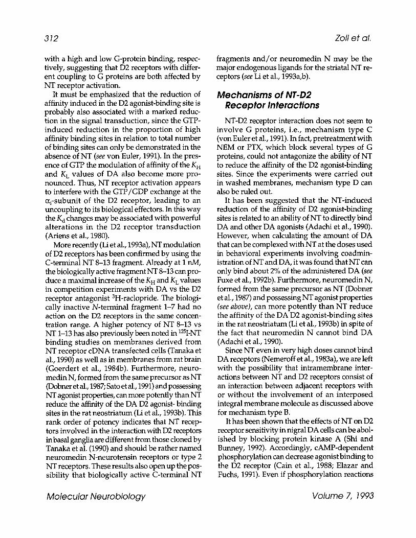
It must be emphasized that the reduction of affinity induced in the D2 agonist-binding site is probably also associated with a marked reduc- tion in the signal transduction, since the GTP- induced reduction in the proportion of high affinity binding sites in relation to total number of binding sites can only be demonstrated in the absence of NT (see von Euler, 1991). In the pres- ence of GTP the modulation of affinity of the K H and K L values of DA also become more pro- nounced. Thus, NT receptor activation appears to interfere with the GTP/GDP exchange at the oh-subunit of the D2 receptor, leading to an uncoupling to its biological effectors. In this way the K d changes may be associated with powerful alterations in the D2 receptor transduction (Ariens et al., 1980).
More recently (Li et al., 1993a), NT modulation of D2 receptors has been confirmed by using the C-terminal NT 8-13 fragment. Already at I nM, the biologically active fragment NT 8-13 can pro- duce a maximal increase of the K H and K L values in competition experiments with DAvs the D2 receptor antagonist 3H-raclopride. The biologi- cally inactive N-terminal fragment 1-7 had no action on the D2 receptors in the same concen- tration range. A higher potency of NT 8-13 vs NT 1-13 has also previously been noted in 125I-NT binding studies on membranes derived from NT receptor cDNA transfected cells (Tanaka et al., 1990) as well as in membranes from rat brain (Goerdert et al., 1984b). Furthermore, neuro- medin N, formed from the same precursor as NT (Dobner et al., 1987; Sato et al., 1991) and possessing NT agonist properties, can more potently than NT reduce the affinity of the DA D2 agonist- binding sites in the rat neostriatum (Li et al., 1993b). This rank order of potency indicates that NT recep- tors involved in the interaction with D2 receptors in basal ganglia are different from those cloned by Tanaka et al. (1990) and should be rather named neuromedin N-neurotensin receptors or type 2 NT receptors. These results also open up the pos- sibility that biologically active C-terminal NT
fragments and/or neuromedin N may be the major endogenous ligands for the striatal NT re- ceptors (see Li et al., 1993a,b).
Mechanisms of NT-D2 Receptor Interactions
NT-D2 receptor interaction does not seem to involve G proteins, i.e., mechanism type C (von Euler et al., 1991). In fact, pretreatment with NEM or PTX, which block several types of G proteins, could not antagonize the ability of NT to reduce the affinity of the D2 agonist-binding sites. Since the experiments were carried out in washed membranes, mechanism type D can also be ruled out.
It has been suggested that the NT-induced reduction of the affinity of D2 agonist-binding sites is related to an ability of NT to directly bind DA and other DA agonists (Adachi et al., 1990). However, when calculating the amount of DA that can be complexed with NT at the doses used in behavioral experiments involving coadmin- istration of NT and DA, it was found that NT can only bind about 2% of the administered DA (see Fuxe et al., 1992b). Furthermore, neuromedin N, formed from the same precursor as NT (Dobner et al., 1987) and possessing NT agonist properties (see above), can more potently than NT reduce the affinity of the DA D2 agonist-binding sites in the rat neostriatum (Li et al., 1993b) in spite of the fact that neuromedin N cannot bind DA (Adachi et al., 1990).
Since NT even in very high doses cannot bind DA receptors (Nemeroff et al., 1983a), we are left with the possibility that intramembrane inter- actions between NT and D2 receptors consist of an interaction between adjacent receptors with or without the involvement of an interposed integral membrane molecule as discussed above for mechanism type B.
It has been shown that the effects of NT on D2 receptor sensitivity in nigral DA cells can be abol- ished by blocking protein kinase A (Shi and Bunney, 1992). Accordingly, cAMP-dependent phosphorylation can decrease agonist binding to the D2 receptor (Cain et al., 1988; Elazar and Fuchs, 1991). Even if phosphorylation reactions
Molecular Neurobiology Volume 7, 1993

Recep tor-Recep tor In teracfions 313
cannot explain the effects observed in membrane homogenates, it seems quite likely that NT receptor activation in vivo can regulate the phosphorylation of the third intracytoplasmatic loop of the D2 receptor. Thus, as suggested by Shi and Bunney (1992), under in vivo conditions a type D interaction may also be involved in the NT/D2 interaction.
Possible Functional Relevance of the NT/D2 Receptor Interactions Functional correlates of NT-D2 receptor inter-
actions have been found in experiments on trans- mitter release and electrophysiology. In a series of experiments using intracerebral microdialysis in striatum, NT antagonism on D2 receptor-medi- ated effects has been demonstrated in both DAceptive GABA-containing neurons and DA- ergic nerve terminals. NT increases extracellular levels of GABA in the dorsal striatum of the halothane-anesthetized (O'Connor et al., 1992) as well as in the awake unrestrained male rat (Fuxe et al., 1992a). In the awake rat, local perfusion with NT, at a dose that is ineffective alone, counter- acted the inhibitory effects of the D2 agonist pergolide on extracellular levels of GABA (Fuxe et al., 1992a). In addition, combined treatment with the D1 agonist SKF38393 and NT but not with the D1 agonist alone led to significant increases in the extracellular striatal levels of GABA. In the same animal model, NT could also antagonize the inhibitory effects of D2 agonists on extracel- lular levels of DA, DOPAC, and HVA (Tanganelli et al., 1989; Fuxe et al., 1992a). On the other hand it enhanced D1 receptor-mediated inhibition of DA release.
Electrophysiological studies demonstrated that NT is able to increase nigral DA cell firing (Kalivas, 1993). This effect is, at least, partly dependent on a desensitization of D2 auto- receptors. In fact, NT has been found to counter- act the D2 agonist-induced inhibition of nigral DA cell firing (Shi and Bunney, 1991).
Overall, microdialysis and electrophysiological studies suggest that, in agreement with binding studies, NT receptors inhibit D2 receptor-medi- ated functions in DAceptive neurons and DA- ergic terminals and cell bodies. The interpretation
of the NT effects on D1 receptor-mediated effects is less straightforward. Since D1 receptors are exclusively located in DAceptive cells, D1 ago- nist effects on DA release are thought to be medi- ated by the activation of a striatonigral feedback loop on DA nigral cells. Thus, NT may influence D1 receptor-mediated GABA and DA release through an indirect effect, although not via receptor crosstalk at the membrane level (see above) on D1 transmission and/or through inhi- bition of D2 transmission. Whatever mechanism is involved, both binding and functional evidence indicates that a major effect of NT on DA trans- mission in basal ganglia is a switch from D2 receptor-mediated to D1 receptor-mediated transmission, and thus from striopallidal to stri- onigral output pathways. This effect would be further potentiated by the NT-induced increase in DA release.
Evidence for DA/NT Receptor Interactions Some evidence also points to the existence of a
modulation of NT receptors by DA receptors in basal ganglia. DA was able to reduce the affinity and increase the number of 3H-NT binding sites in the rat neostriatal membrane preparations, possibly involving an action via D1 receptors since D1 but not D2 agonists modula te the 3H-NT binding (Agnati et al., 1985a,b). This inter- action was found to be enhanced by the devel- opment of DA receptor supersensivity induced by a 6-OHDA-induced lesion of the nigrostriatal DA pathway (Fuxe et al., 1986, 1990). These results probably reflect an intramembrane DA/ NT receptor interaction. In this case the increase of the Bmax value is substantial and may reflect an unmasking of the 3H-NT binding sites in their interaction with the DA receptors, possibly of the D1 type. The relevance of this interaction for the control by NT receptors of D2 receptor transduc- tion remains to be determined but it has been suggested that it may represent part of an inhibi- tory feedback loop controlling D2 transmission (see Fuxe et al., 1992b).
When discussing the role of NT receptor mechanisms in the control of D2 receptor trans- duction it must, however, also be mentioned that
Molecular Neurobiology Volume 7, 1993

314 Zoli et al.
the D2 receptors appear to exert a restraining influence on gene expression of NT and that D2 blockade results in increased NT synthesis in the GABA/ENK neurons of the neostriatum, prob- ably associated with an enhancement of NT release. Such a continuous release may then result in a desensitization of the NT receptors restrain- ing the D2 receptor sensitivity. Such a mechanism may be involved in producing the D2 receptor supersensitivity phenomenon after D2 receptor antagonist treatment (see Fuxe et al., 1990, 1992).
CCK-8/DA Receptor Interactions
CCK/DA Coexistence and Codistribufion of CCK and D2 Receptors
The CCK innervation of the striatum is heter- ogeneous (H6kfelt et al., 1980; Gilles et al., 1983). In the rat, only a minor CCK-positive input derives from mesencephalic DA cells, most of which costore CCK (H6kfelt et al., 1980; Gilles et al., 1983). In addition, some cells con- taining CCK mRNA can be visualized in the striatum (Ding and Mocchetti, 1992). In the CNS, CCK binds to two types of G protein- linked receptors, the CCK-A and CCK-B recep- tors, with specific spectra of ligand selectivity (Moran et al., 1986). The former is present only in a few areas, which possibly include the ventral str iatum and the midbrain DA cells (Hill et al., 1987), whereas the latter is largely distributed all over the brain (Moran et al., 1986).
CCK Modulation of DA Receptors Neuroleptic-like actions have been demon-
strated following central CCK-8 administration. Thus, antagonism of stereotypes induced by DAergic drugs, catalepsy, and ptosis are demon- strated as well as a reduction of self-stimulation behaviors and inhibition of conditioned avoid- ance responses (Zetler, 1985). Available evidence indicates that such actions of CCK-8 may be related to antagonistic CCK-D2 receptor interac- tions in the striatum.
Studies in striatal membranes from both the ventral and dorsal parts (Agnati and Fuxe 1983; Agnati et al., 1983a,b, 1985b; Li et al., 1993c) showed that CCK-8 (at concentrations ranging from 0.1-1 nM) selectively reduces the affinity without any change in the B~x value of the D2 agonis t -binding site, the change being in the order of 20-40% depending on the concen- tration and the area studied. In a kinetic analy- sis, it was found that CCK-8 increased the K a value by reducing the association of 3H-NPA to binding sites in rat neostriatal membranes. Instead, NT induced an increase in the K a value (see above) by increasing the dissociation rate con- stant (von Euler, 1991). CCK-D2 receptor inter- action seems to be mediated by CCK-B receptors (Li et al., 1993c), since the increase of the K~ value of 3H-NPA binding sites in rat striatal membranes can be counteracted by the selective CCK-B antagonist PD34308 (Hughes et al., 1990) but not by the selective CCK-A antagonist L364718 (Freidinger et al., 1990). These results are also in line with previous studies showing that CCK-4, a selective CCK-B agonist, can reduce the affin- ity of the D2 agonist-binding sites in striatal mem- branes (Agnati et al., 1983a,b).
It has been shown (Dumbrill-Ross and Seeman, 1984; Agnati et al., 1987) that, after 24 h intraven- tricular infusion, CCK-8 induces a long-term (up to 14 d) increase of 3H-D2 receptor antagonist binding in dorsal and ventral striatum. These observations are apparently at variance with the results obtained in in vitro studies. How- ever, a recent paper (Li et al., 1993c) gives some hints to reconcile these apparently contrasting findings. This study showed that, in competition experiments with DAvs 3H-raclopride binding, CCK-8 increases D2 binding site affinity unless a D1 receptor antagonist is added. In this latter case CCK-8 causes a decrease in the affinity of DA for D2 binding sites. Thus, D1 receptor activation, which is likely to be present in vivo, appears to shift the CCK-8 action from a decrease to an increase of D2 receptor binding. An alternative hypothes i s ma in t a in s tha t ac t iva t ion of CCK receptors in the nucleus accumbens can lead to an increase of D2 receptor biosynthesis. It
Molecular Neurobiology Volume 7, 1993
Receptor-Receptor Interactions 315
is likely that several CCK-8-induced alterations of D2 receptor transmission occur in vivo.
Mechanisms of CCK-D2 Receptor Interactions Up until now, no mechanistic study has been
carried out on CCK-D2 receptor interactions. In vivo studies suggest that CCK-8 may increase D2 receptor biosynthesis (mechanism type D). The presence of the modulation in membrane prepa- rations implies that intramembrane receptor- receptor interactions take place (mechanism types B and C). It is not yet known if this interaction involves a G protein or not. Recent studies show that CCK-8-induced and NT-induced decreases of D2 receptor affinity in striatal membranes are additive at low, but not high, concentrations, which suggests that the CCK and NT operate via the same mechanism, i.e., without an interven- tion of G proteins (see above).
Possible Functional Relevance of the CCK/D2 Receptor Interactions The presence of an antagonistic interaction
between CCK and D2 receptors in striatum can be correlated with several electrophysiological and behavioral data. A functional antagonism between DA and CCK-8 has been found in single unit recordings from the n. accumbens (Wang and Hu, 1984). In intracerebral microdialysis experi- ments (see Tanganelli et al., 1990), it was shown that CCK-8 locally perfused in the neostriatum is able, in a concentration-related way, to coun- teract the inhibition of DA release caused by sys- temically given apomorphine acting on D2 autoreceptors. Finally, it is well known that CCK- 8 has an antipsychotic agent profile, i.e., it mim- ics the actions of D2 receptor blockers (Van Ree, 1983). On the other hand, some behavioral (Crawley et al., 1989) and electrophysiological findings on nigral DA neurons (Kalivas, 1993) show that CCK-8 is able to increase DA trans- mission in some experimental paradigms, prob- ably owing to coactivation of D1 and D2 receptors in these experiments (see above). It must also be
considered that act ivat ion of D2 receptors increase CCK mRNA content in striatum (Ding and Mocchetti, 1992) and that D1 and D2 recep- tors increase the affinity of CCK-8 receptors (von Euler et al., 1992). It is then likely that CCK pep- tides in vivo have multiple influences on the DA mesostriatal system according to the subtype of CCK receptor activated and to the predominance of D2 or D1 transmission in the DA pathways.
A2/D2 Receptor Interactions in the Basal Ganglia
Codistribution of Adenosine A2 and DA D2 Receptors in the Brain Adenosine has been shown to be a neuro-
modulator in the CNS (Fredholm and Hedqvist, 1979; Dunwiddie, 1985; Snyder, 1985). Its actions are mediated by receptors that have been subdi- vided into A1 and A2 according to agonist and antagonist potencies (Sattin and Rail, 1970; Daly et al., 1983; Bruns et al., 1988) and to their action on adenylate cyclase (Premont et al., 1979; Van Calker et al., 1979). Based on agonist potencies, the A2 receptors have been subdivided into high-affinity (A2a) and low-affinity (A2b) recep- tors (Bruns et al., 1986). Direct proof for the valid- ity of this receptor classification lies on molecular cloning experiments, because A1 receptors and two forms of A2 receptors have recently been cloned (Maenhaut et al., 1991; Libert et al., 1991).
With radioligand-binding techniques, using membrane preparations or autoradiography, it has been found, both in rodents and in humans, that high-affinity adenosine A2 receptors are highly enriched in the striatum (caudate nucleus and putamen in humans), nucleus accumbens and olfactory tubercle (Fig. 8) (Bruns et al., 1986; Alexander and Reddington, 1989; Jarvis et al., 1989; Jarvis and Williams, 1991; Parkinson and Fredholm, 1990; Martinez-Mir et al., 1991), areas with very high numbers of DA receptors (Boyson et al., 1986). Accordingly, A2 receptor mRNA is exclusively localized in the basal ganglia of the rat, dog, and human brain (Schiffmann et al., 1991a,b). Further studies at the cellular level showed that adenosine A2 receptor mRNA is
Molecular Neurobiology Volume 7, 1993

316 Zofi et al.
lk21
Fig. 8. Representative autoradiograms of the rat forebrain (Bregma 1.6-1.7 mm) showing the distribution of dopamine D2 receptors labeled with [125I]sulpiride (top panel) and adenosine A2 receptors labeled with [3H]CGS 21680 (bottom panel). Modified from Ferr6 et al. (1992). High densities of both D2 and A2 receptors codistribute in the ventral and dorsal striatum.
selectively expressed by GABAergic-ENK striatal neurons, which also contain D2 recep- tors (Schiffmann et al., 1991b; Fink et al., 1992).
Adenosine Modulation of D2 Binding In rat striatal membrane preparations, the spe-
cific A2 adenosine agonist CGS 21680 induces a decrease in the affinity of DA D2 receptors for DA agonists, like DA and NPA, but not for DA antagonists, like raclopride (Ferr6 et al., 1991d).
This CGS 21680 effect was inhibited by the adeno- sine antagonist 8-phenyltheophylline and was mimicked by L-PIA (Bruns et al., 1986), provided the concentration used was high enough to stimu- late high-affinity A2 receptors in addition to A1 ones. Furthermore, CGS 21680 was ineffective in altering D1 binding (Ferr6 et al., 1991d).
In subsequent studies it was found that, in addition to the increase in both K H and KLvalues of DA D2 receptors, stimulation of adenosine A2 receptors is associated with an increase in the
Molecular Neurobiology Volume 7, 1993

Receptor-Receptor Interactions 317
C
e -
- " t00-
"~ 75-
t .J ,o 50" r
- r -
"-" 25"
"~ 0 q J
--~l~qp-....~.~.. ~CGS K,~: 8.5 nM - 6 -,"~ Kx: 333.3 nM
a ' ~ h : 55.7I
Kz: t58.2 nM , , '~_
I I ,
-n-i0-b-8- 4- -4 Oopa=ine (log M)
Fig. 9. Representative competition-inhibition curves illustrating the effect of the adenosine A2 agonist CGS 21680 (30 nM) on DA-induced inhibition of the binding of the DA D2 antagonist 3H-raclopride. The MgC12 concentration was 1 mM (for further details on methods, see Ferr6 et al. 1991). The presence of CGS 21680 causes an increase in the dissociation constants of both high- and low-affinity states (K H and K L, respectively) and an increase in the proportion of the high-affinity state (RH) of the D2 receptors. This suggests that adenosine A2 receptor stimulation causes a decrease of both the affinity of D2 receptors and the transduction of the signal from the D2 recep- tor to the G protein (for further details, see text).
proportion of high-affinity binding sites (RH) (Fig. 9) (Ferr6 and Fuxe, 1992; Ferr6 et al., 1993). In view of the role of guanine nucleotides and divalent cations (like Mg 2§ to regulate in oppos- ing ways the proportion of high-affinity D2 recep- tors (Sibley and Creese, 1983), interaction experiments with CGS 21680, the GTP analog Gpp (NH)p, and Mg 2+ were performed. It was found that these three factors play significant although independent roles on R H values of the DA D2 receptors, CGS 21680, and Mg 2§ increas- ing and Gpp(NH)p decreasing R H (Ferr6 et al., 1993). Both the GTP analog and Mg 2+ did not alter the effect of CGS 21680 on K H and K L values of the dopamine D2 receptor (Ferr6 et al., 1993).
GTP binding to the ~-subunit causes its disso- ciation from the receptor, which changes to a low- affinity state, the GTP-(x-subunit complex being
responsible for the changes in adenylate cyclase activity, ion channel permeability, and so on (Gilman, 1987). Consequently, an increase in the proportion of high-affinity D2 binding sites is expected to be associated with an impairment in the transduction of the signal from the D2 recep- tor to the G protein.
Mechanisms of A2-D2 Receptor Interactions
Both adenosine A2 and DA D2 receptors belong to the family of G protein-coupled recep- tors (Bunzow et al., 1988; Libert et al., 1989). The A2 receptor is coupled to G s proteins, which link A2 receptors to adenylate cyclase (Libert et al., 1989), and the D2 receptor is coupled to G i pro- teins, which l ink D2 receptors to adenyla te cyclase, ion channels, and phospholipase C (Vallar and Meldolesi, 1989). Consequently, A2 and D2 transmission lines are likely to interact at the signal transduction level, since activation of the adenosine A2 receptor stimulates and acti- vation of the DA D2 receptor inhibits adenylate cyclase activity (Premont et al., 1979; Van Calker et al., 1979; Onali et al., 1985).
One possible mechanism underlying the A2- induced increase in the R H values of D2 recep- tors could be an interaction between G s and G i proteins (Gilman, 1987). An increased formation of ~T-subunits from the A2-1inked Gs, after A2 receptor activation, could bind to the ~-subunits from the D2-1inked Gi, leading to an inhibition of the G i protein dissociation from the D2 recep- tor (mechanism type C). However, our results suggest that the A2-mediated increase in R H val- ues of the D2 receptors is independent of the effect of GTP. In fact, this A2 adenosine effect was not affected by the GTP antagonist GDP-~S (Ferr6 et al., 1993). Also, our biochemical data, in membrane preparations, rule out the involve- ment of adenylate cyclase in the A2-mediated modulation of D2 receptors, because adenylate cyclase activity depends on factors, such as ATP, that were not included in the incubating medium (mechanism type D). It is therefore likely that mechanism type B is involved in A2-D2 recep- tor interaction in the cell membrane. Neverthe- less, the abovementioned mechanisms, the Gs-G i
Molecular Neurobiology Volume 7, 1993

318 Zofi et al.
protein interaction and the interaction at the level of adenylate cyclase, are very probably additional mechanisms that amplify the intra- membrane allosteric A2/D2 receptor-receptor interaction in vivo.
Possible Functional Relevance for Adenosine A2 Receptor-DA D2 Receptor Interaction In the experimental animal, adenosine ago-
nists produce psychomotor depressant effects (Snyder et al., 1981; Vapaatalo and McGuffin- Clineschmidt, 1981; Barraco et al., 1984; Durcan and Morgan , 1989a; Heffner et al., 1989) whereas adenosine antagonists, which include the methylxanthines caffeine and theophylline, not only inhibit the adenosine agonist-induced psychomotor effects, but, when administered alone, also exert psychomotor stimulant effects suggesting the existence of a basal adenosin- ergic tone in vivo (Boissier and Simon, 1965; Thithapandha et al., 1972; Snyder et al., 1981; Coffin and Carney, 1986; Spealman and Coffin, 1986). The potencies of different adenosine agonists and antagonists to produce their psy- chomotor effects correlate with their affinities for A2 but not for A1 adenosine receptors (Spealman and Coffin, 1986; Durcan and Mor- gan, 1989b; Nikodijevic et al., 1990). A large amount of experimental data suggest that cen- tral DAergic neurotransmission is involved in the mediation of the psychomotor effects of adenos- ine agonists and antagonists. In rats adenosine agonists inhibit and methylxanthines potentiate the locomotor activity induced by DA agonists (Fuxe and Ungerstedt, 1974; Fredholm et al, 1976, 1983; Green et al., 1982; Heffner et al., 1989; Brown et al., 1991; Ferr6 at al., 1991a,b). Also, methyl- xanthine-induced locomotor activity in rodents is antagonized by treatments that cause DA depletion or blockade of DA receptors (Waldeck, 1973; White et al., 1978; Erinoff and Snodgrass, 1986; Herrera-Marschitz et al., 1988; Heffner et al., 1989; Josselyn and Beninger, 1991; Ferr6 et al., 1991b).
Thus, a central adenos ine-DA interac- t ion exists by which activation of adenosine
receptors leads to an inhibition of DA neuro- transmission and methylxanthines, by blocking the effect of endogenous adenosine, produce an enhancement of this transmission (see Ferr6 et al., 1992).
A presynaptic adenosine-DA interaction has been shown, with adenosine agonists causing a decrease and methylxanthines an increase of striatal DA release in rodents (Harms et al., 1979; Michaelis et al., 1979; Fredholm, 1985; Morgan and Vestal, 1989; Wood et al., 1989; Lupica et al., 1990). However this interaction seems to involve adenosine A1 receptors (Lupica et al., 1990) and the evidence regarding the behavioral effects rather suggests that A2 receptors are the major subtype involved in the interaction with DA neu- rotransmission (see above).
Recently, it has been shown that adenosine analogs inhibit and methylxanthines potentiate a DA D2-mediated behavior in short- term reserpinized mice (Ferr6 et al., 1991a,b). Conse- quently, a postsynaptic adenosine A2/DA D2 interaction was suggested as being the main mechanism of action mediating the behavioral effects of adenosine agonists and antagonists. In line with this hypothesis, it was found that stimu- lation of central adenosine A2 receptors by the icv administration of CGS 21680 induces cata- lepsy in the rat, which can be counteracted both by an adenosine antagonist and by a specific DA D2 agonist (Ferr6 et al., 1991d). These behavioral data have a neurochemical counter- part (Schiffman et al., 1993). In fact, A2 antago- nists, like D2 agonists, decrease the expression of ENK mRNA in striatal neurons containing both A2 and D2 receptors.
Striatal DA denervation induces both a higher sensitivity to DA agonists and a higher sensitivity to methylxanthines. Thus, systemic or intrastriatal administration of DA agonists or methylxan- thines have a stronger effect in the denervated striatum (Pycock, 1980; Herrera-Marschitz et al., 1985, 1988; Josselyn and Beninger, 1991). The increased effect of DA agonists is most probably owing to the development of a denervation- induced supersensi t ivi ty of DA receptors (Ungerstedt, 1971; Creese et al., 1977). The increased effect of methylxanthines after DA den-
Molecular Neurobiology Volume 7, 1993

Receptor-Receptor Interactions 319
ervation could be explained by an increased interaction between adenosine A2 and DA D2 receptors. In fact, it has been recently found that, in membrane preparations from the DA dener- vated neostriatum, the DA D2 receptors are more sensitive to the modulatory effect of CGS 21680 on D2 receptors (Ferr6 and Fuxe, 1992).
Overall View The set of phenomena presented in this sec-
tion clearly shows that a complex network of interactions between transmission lines takes place in striatal cells (Fig. 10). It must, however, be underlined that this picture is highly simpli- fied, since several major modulators (SP, ENK, dynorphin, and so forth) and the transmitter of intrinsic striatal neurons (GABA) as well as that of the main striatal input (glutamate) have not been taken into account.
The interactions appear to be at the same time specific (e.g., in the case presented the modula- tions are selective for D2 receptors) but not exclu- sive (several modulators have the same effect on the same molecular target). In addition, they can take place at different levels of the informational cascade (receptor molecules, G proteins, second messenger systems, gene expression). It is likely that complex equilibria within this network of transmissions will take place according to the degree of activation of each receptor population. It must, therefore, be stressed that it is the out- come of multiple receptor activations that finally determine the sensitivity and transduction of each individual receptor population.
In the frame of a reductionistic approach, the next steps in the comprehension of the function of striatal network will be, inter alia, to analyze the coordination between multiple modulatory systems. An example of synergism between modulators is that of CCK and NT on D2 recep- tors. It has been shown (see above) that CCK-8 reduces the dissociation rate constant, whereas NT increases the association rate constant of the D2 agonist-binding sites. This neurochemical syn- ergism has a functional counterpart. In fact, NT and CCK-8 have a synergistic inhibitory interac- tion on D2 autoreceptors provided very low con- centrations of NT and CCK-8 are used (Fig. 11)
(see Tanganelli et al., 1993). The interaction was not observed at higher concentrations, suggest- ing that the two neuropeptide receptors modify D2 receptor transduction via a common regula- tory mechanism.
At the neuronal network level in the neo- striatum, the synergism of the two peptides may have a major integrative function inter alia by allowing a switching from D2 receptor-mediated transmission (striopallidal GABA/ENK path- way) toward a D1 receptor-mediated transmis- sion (strionigral GABA/SP pathway). Thus, CCK and NT would favor the restraining influence of the indirect pathway on movements, since move- ments become elicited via the direct pathway when the cortical-striatal glutamate pathway becomes activated. In this way discrete move- ments will be elicited.
Integrative Aspects of Receptor-Receptor Interactions
Experimental evidence has been presented for the presence in the brain of interactions among dif- ferent transmission lines at the level of membrane receptors. In this last section, our goal will be to discuss, often on a speculative basis, what is the importance of receptor-receptor interactions for the integrative mechanisms in the CNS. Pre- viously, we have underlined their pharmacological importance by representing a new site of action for drug development (Fuxe et al., 1989b). Thus, our efforts will be focused on showing the possi- ble links between these molecular mechanisms and nerve cell and neural network functions.
Integration of Signals Through Receptor-Receptor Interactions Receptor-receptor interactions belong to the
broader class of the interactions between trans- mission lines. Therefore, we will first discuss some general properties of these latter phenom- ena and then some distinctive features of the crosstalk at receptor level.
Molecular Neurobiology Volume 7, 1993

320 Zofi et al.
IDAceptive striatal cell I
g',g',2"
Fig. 10. Schematic representation of some receptor-receptor interactions, and their consequences on target cells, occurring in striatal membranes. Adenosine present in the extracellular space can derive from several cellular sources, including intrinsic striatal neurons and glial cells. For further details and discussion, see text .
Abbreviations: CCK = cholecystokinin, CCK R = cholecystokinin receptor, DA = dopamine, GABA = 7- aminobutyric acid, NM = neuromedin, NT = neurotensin, NT8_13 = neurotensin fragment 8-13, NT R = neurotensin receptor.
The presence of interactions between transmis- sion lines leads to the formation of molecular networks within and between different levels in the transduction pathways: membrane receptors, G proteins, second messenger systems, and so forth. Each informational molecule in the network has a set of specific interactions with other mol- ecules. The more these events are studied the more is emerging a kind of molecular grammar: molecules that block or potentiate the action of other molecules, molecules with permissive roles, molecules that converge to the same target or diverge to a number of different targets, mol- ecules that are necessary events for a certain func- tion or that are only modulating it, and so on.
In most instances these molecular events repro- duce the integrative phenomena known at the neuronal network level: The idea is therefore emerging that they constitute the basic compu- tational units of the integration occurring in the
cell. In comparison with neuronal circuits, these molecular circuits are characterized by an extraor- dinary miniaturization of computat ional ele- ments (Agnati et al., 1984, 1988). It has been proposed (Agnati et al., 1990) that intracellular reactions elicited by neurotransmitters should be considered as a computation (microcomputation) in a series with respect to the computation tak- ing place at the network level (macrocompu- tation). Actually, the information handling in the CNS should be considered the result of integra- tions at several successive networks, i.e., neural network, receptor network, G protein network, and intracellular chemical network.
The existence of networks implies that the reading out of the extracellular information car- ried out by the intracellular mechanisms is a holistic process. In this process it may be conve- nient to distinguish on the one hand fast electri- cal events, and on the other hand, slow electrical
Molecular Neurobiology Volume 7, 1993

Receptor-Receptor Interactions 321
6
5'
4"
3
2
1
0
O0
"0
"0
m ~
200
150
100
50
0
KL (nM) * *
8O
60
4 0
20-
0
CCK-8 0.01nM NT lnM CCK-8 + NT
RH (% of total)
+
+
+
Fig. 11. Effects of NT plus CCK-8 on the K H (upper panel), K L (middle panel), and R H (bottom panel) values of [3H]racloprlde binding in rat neostriatal membranes. The K H values were 2.65 _+ 0.47, 2.38 + 0.63, 2.65 + 0.75, and 4.44 + 0.65, the K L values were 65 + 7, 77 + 8, 62 + 8, and 135 + 13 nM, and the R H values were 55 _+ 2, 55 + 5, 55 + 4, and 61 + 4, for the control, 1 nM of NT, 0.01 nM of CCK-8, and NT + CCK, respectively. Each treatment represents the mean + SEM of nine separate experiments. Statistical analysis according to one-way ANOVA with Fischer's PLSD test, *p < 0.05, **p < 0.01.
events and metabolic events. The first set of phe- nomena poses fewer problems of interpretation, since the integration takes place at the mem- brane level according to chemicophysical rules that are largely understood. A more difficult task is the interpretation of the second set of phenomena, since no rules are known of how segregation and integration of the different sig- nals, many times affecting the same intraceUular effectors, take place. Segregation of transmission lines from a membrane receptor to the nucleus is certainly possible but seems rare.
The specific characteristic of receptor-recep- tor interactions is to intervene at the beginning of the transduction cascade. From a general standpoint, we can distinguish the interactions occurring at the membrane level (types A, B, and C) from those involving intracellular loops (type D).
The former ones can achieve a fast setting of activity in a transmission line in the light of ongoing activity in other transmission lines. In this way, physically distinct receptor molecules can behave as a "functional supramolecular com- plex" in the cell membrane (Agnati et al., 1982). The latter ones allow receptor heteroregulation at relatively long (phosphorylation) to very long (receptor synthesis) time scale.
Because of their location at the cell interface with the ECF, receptor crosstalk mechanisms con- stitute a first molecular network for the cellular integration. For this reason they seem particularly suited for a number of integrative tasks.
Filtration of Incoming Signals Thanks to the existence of in t ramembrane
receptor-receptor interactions it is possible to attain a p redominance of some signals over others if they are contemporaneously presented to the cell. A signal will be potentiated, inhibited, or left unchanged in relation to the state of acti- vation of the other receptors wi th which its receptor interacts. Inhibitory interactions make it possible to hinder incoming signals that are irrelevant or redundant in a particular cellular state. The advantage of this mechanism over blocking the signal at the source, i.e., blocking transmitter release via presynaptic inhibition, is
Molecular Neurobiology Volume 7, 1993
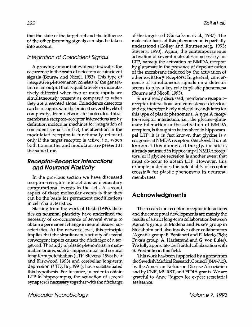
322 Zoli et al.
that the state of the target cell and the influence of the other incoming signals can also be taken into account.
Integration of Coincident Signals
A growing amount of evidence indicates the occurrence in the brain of detectors of coincident signals (Bourne and Nicoll, 1993). This type of integrative phenomenon consists of the genera- tion of an output that is qualitatively or quantita- tively different when two or more inputs are simultaneously present as compared to when they are presented alone. Coincidence detectors can be recognized in the brain at several levels of complexity, from network to molecules. Intra- membrane receptor-receptor interactions are by definition molecular machines for integration of coincident signals. In fact, the alteration in the modulated receptor is functionally relevant only if the target receptor is active, i.e., when both transmitter and modulator are present at the same time.
Receptor-Receptor Interactions and Neuronal Plasticity
In the previous section we have discussed receptor-receptor interactions as elementary computational events in the cell. A second aspect of these molecular events is that they can be the basis for permanent modifications in cell characteristics.
Starting from the work of Hebb (1949), theo- ries on neuronal plasticity have underlined the necessity of co-occurrence of several events to obtain a permanent change in neural tissue char- acteristics. At the network level, this principle implies that the simultaneous activity of several convergent inputs causes the discharge of a tar- get cell. The study of plastic phenomena in mam- malian brains, such as hippocampal and cortical long-term potentiation (LTP, Stevens, 1993; Bear and Kirkwood 1993) and cerebellar long-term depression (LTD, Ito, 1991), have substantiated this hypothesis. For instance, in order to obtain LTP in hippocampus, the activation of several synapses is necessary together with the discharge
of the target cell (Gustafsson et al., 1987). The molecular basis of this phenomenon is partially understood (Colley and Routtenberg, 1993; Stevens, 1993). Again, the contemporaneous activation of several molecules is necessary for LTP, namely the activation of NMDA receptor by glutamate in the presence of depolarization of the membrane induced by the activation of other excitatory receptors. In general, conver- gence of simultaneous signals on a detector seems to play a key role in plastic phenomena (Bourne and Nicoll, 1993).
Since already discussed, membrane receptor- receptor interactions are coincidence detectors and are therefore likely molecular candidates for this type of plastic phenomena. A type A recep- tor-receptor interaction, i.e., the glycine-gluta- mate interaction in the activation of NMDA receptors, is thought to be involved in hippocam- pal LTP. It is in fact known that glycine is a coagonist at NMDA receptors (see above). It is not known at this moment if the glycine site is al ready saturated in hippocampal NMDA recep- tors, or if glycine secretion is another event that must co-occur to obtain LTP. However, this example underlines the potentiality of receptor crosstalk for plastic phenomena in neuronal membranes.
Acknowledgments
The research on receptor-receptor interactions and the conceptual developments are mainly the results of a strict long-term collaboration between Agnati's group in Modena and Fuxe's group in Stockholm and also involve other collaborators (Agnati's group: F. Benfenati and E. Merlo-Pich; Fuxe's group: A. H/irfstrand and G. von Euler). We fully appreciate the fruitful collaboration with B. Fredholm in this field.
This work has been supported by a grant from the Swedish Medical Research Council (04X-715), by the American Parkinson Disease Association and by CNR, MURST, and FIDIA grants. We are grateful to Anne Edgren for expert secretarial assistance.
Molecular Neurobio logy Volume 7, 1993

Receptor-Receptor Interactions 323
References
Adachi D. K., Kalivas P. W., and Schenk J. O. (1990) Neurotensin binding to dopamine. J. Neurochem. 54, 1321-1328.
Agnati L. F., Fuxe K., Zini I., Lenzi P., and H6kfelt T. (1980) Aspects on receptor regulation and isorecep- tor identification. Med. Biol. 58,182-187.
Agnati L. F., Fuxe K., Zoli M., Rondanini C., and Ogren S.-O. (1982) New vistas on synaptic plasti- city: mosaic hypothesis on the engram. Med. Biol. 60, 183-190.
Agnati L. F. and Fuxe K. (1983) Subcortical limbic [3H]N-propylnorapomorphine binding sites are markedly modulated by cholecystokinin-8 in vitro. Biosci. Rep. 3, 1101-1105.
Agnati L. F., Fuxe K., Benfenati F., Celani M. F., Battistini N., Mutt V., Cavicchioli L., Galli G., and H6kfelt T. (1983a) Differential modulation by CCK- 8 and CCK-4 of [3H]spiperone binding sites linked to dopamine and 5-hydroxytryptamine receptors in the brain of the rat. Neurosci. Lett. 35, 179-183.
Agnati L. F., Celani M. F., and Fuxe K. (1983b) Cholecystokinin peptides in vitro modulate the characteristics of striatal [3H]N-propylnorapo- morphine binding sites. Acta Physiol. Scand. 118, 79-81.
Agnati L. F., Fuxe K., Benfenati F., and Battistini N. (1983c) Neurotensin in vitro markedly reduces the affinity in subcortical limbic [3H]N-propylnorapo- morphine binding sites. Acta Physiol. Scand. 119, 459-461.
Agnati L. F., Fuxe K., Benfenati F., Zini I., and H6kfelt T. (1983d) On the functional role of coexistence of 5-HT and substance P in bulbospinal 5-HT neu- rons. Substance P reduces affinity and increases density of [3H]5-HT binding sites. Acta Physiol. Scand. 117, 299-301.
Agnati L. F., Fuxe K., Benfenati F., Battistini N., H/irfstrand A., Tatemoto K., H6kfelt T., and Mutt V. (1983e) Neuropeptide Y in vitro selectively increases the number of a2-adrenergic binding sites in membranes of the medulla oblongata of the rat. Acta Physiol. Scand. 118, 293-295.
Agnati L. F., Fuxe K., Benfenati F., Battistini N., Zini I., Camurri M., and H6kfelt T. (1984) Postsynaptic effects of neuropeptide comodulators at central monoamine synapses, in Neurology and Neuro- biology, vol 8B: Cathecolamines, Part B: Neuro- pharmacology and Central Nervous System Theoretical Aspects (Usdin E., Carlsson A., Dahlstr6m A., and Engel J., eds.), Liss, New York, p. 191.
Agnati L. F., Fuxe K., Battistini N., Giardino L., Benfenati F., Martire M., and Ruggeri M. (1985a) Further evidence for the existence of interactions between receptors for dopamine and neurotensin. Dopamine reduces the affinity and increases the number of [3H]neurotensin binding sites in the subcortical limbic forebrain in the rat. Acta Physiol. Scand. 124, 125-128.
Agnati L. F., Fuxe K., Giardino L., Catza L., Zoli M., Battistini N., Benfenati F., Vanderhaeghen J. J., Guidolin D., Ruggeri M., and Goldstein M. (1985b) Evidence for cholecystokinin-dopamine receptor interactions in the central nervous system of the adult and old rat. Ann. NY Acad. Sci. 448, 315-333.
Agnati L. F., Fuxe K., Zoli M., Merlo P., Benfenati F., Zini I., and Goldstein M. (1986) Aspects on the information handling by the central nervous system: focus on cotransmission in the aged brain, in Progress in Brain Research, vol. 68, H6kfelt T., Fuxe K., and Pernow B., eds.), Elsevier, Amsterdam, pp. 291-301.
Agnati L. F., Fuxe K., Merlo-Pich E., Zoli M., Zini I., Benfenati F., H/irfstrand A., and Goldstein M. (1987) Aspects on the integrative capabilities of the central nervous system: evidence for "volume transmission" and its possible relevance for recep- tor-receptor interactions in Receptor-Receptor Interactions (Fuxe K. and Agnati L. F., eds.), Macmillan, London, pp. 236-249.
Agnati L. F., Zoli M., Merlo-Pich E., Ruggeri M., and Fuxe K. (1988) The emerging complexity of the brain. Limits of brain-computer analogy, in Traffic Engineering for ISDN Design and Planning (Bonatti M. and Decina M., eds.), Elsevier, Amsterdam, pp. 209-230.
Agnati L. F., Zoli M., Pich E. M., Benfenati F., Grimaldi R., Zini I., Toffano G., and Fuxe K. (1989) NPY receptors and their interactions with other trans- mitter systems, in Neuropeptide Y (Mutt V., Fuxe K., H6kfelt T., and Lundberg J., eds.), Raven, New York, pp. 103-114.
Agnati L. F., Zoli M., Merlo-Pich E., Benfenati F., and Fuxe K. (1990) Aspects of neural plasticity in the central nervous system. VII. Theoretical aspects of brain communication and computation. Neuro- chem. Int. 16, 478-500.
Agnati L. F., Bjelke B., and Fuxe K. (1992) Volume transmission in the brain. Am. ScL 80, 362-373.
Aguirre J. A., Fuxe K., Agnati L. F., and von Euler G. (1990) Centrally injected neuropeptide Y (13-36) produces vasopressor effects and antagonizes the vasodepressor action of neuropeptide Y (1-36) in the awake male rat. Neurosci. Lett. 118, 5-8.
Molecular Neurobiotogy Volume 7, 1993
02~- Zofi et al.
Aguirre J. A., Fuxe K., Hedlund P., Narv~ez J. A., Cintra A., Ros6n L., and Fuxe K. (1991) Neuro- peptide Y/angiotensin II interactions in central cardiovascular regulation of the rat. Brain Res. 566, 61-69.
Alexander S. P. and Reddington M. (1989) The cellular localization of adenosine receptors in rat neo- striatum. Neuroscience 28, 645-651.
Ari4ns E. J., Beld A. J., Rodrigues de Miranda J. F., and Simonis A. M. (1980) The pharmacon-receptor- effector concept. A basis for understanding the transmission information in biological systems, in The Receptors: A Comprehensive Treatise, vol. 1 (O'Brien R. D., ed.), Plenum, New York, pp. 33-91.
Assaf S. Y. and Chung S. (1984) Release of endogenous Zn 2§ from brain tissue during activity. Nature 308, 734-736.
Axelrod J., Burch R. M., and Jelsema C. L. (1987) Receptor mediated activation of phospholipase A2: arachidonic acid and its metabolites as second messengers in Receptor-Receptor Interactions (Fuxe K. and Agnati L. F., eds.), Macmillan, London, pp. 298-307.
Barbaccia M. L., Costa E., and Guidotti A. (1988) Endogenous ligands for high-affinity recognition sites of psychotropic drugs. Ann. Rev. Pharmacol. Toxicol. 28, 451-476.
Barraco R. A., Aggarwal A. K., Phillis J. W., Boron M. A., and Wu P. (1984) Dissociation of the locomotor and hypotensive effects of adenosine analogues in the rat. Neurosci. Lett. 48, 139-144.
Bean A. J., Adrian T. E., Modline I. M., and Roth R. H. (1989) Storage of dopamine and neurotensin in colocalized and noncolocalized neuronal popu- lations. J. Pharmacol. Exp. Ther. 249, 681-684.
Bear M. F. and Kirkwood A. (1993) Neocortical long- term potentiation. Curr. Opin. Neurobiol. 3,197-202.
Benovic J. L., Regan J. W., Caron M. G., and Lefkowitz R. J. (1987a) Agonist-dependent phosphorylation of the a2-adrenergic receptor by the b-adrenergic receptor kinase. J. Biol. Chem. 262, 17,251-17,253.
Benovic J. L., Staniszewski C., Cerione R. A., Codina J., Lefkowitz R. J., and Caron M. G. (1987b) The mammalian beta-adrenergic receptor: structural and functional characterization of the carbohydrate moiety. J. Recept. Res. 7, 257-281.
Benovic J. A., Bechtel P. J., and Krebs E. G. (1988) Regulation of adenylyl cyclase-coupled b-adren- ergic receptor. Ann. Rev. Celt Biol. 4, 405-428.
Benveniste M., Clements J., and Mayer M. (1990) A kinetic analysis of the modulat ion of NMDA receptors by clycine in mouse cultured hippocam- pal neurons. J. Physiol. 428, 333-357.
Bertorello A. M., Hopfield J. F., Aperia A., and Greengard P. (1990) Inhibition of dopamine of (NA§ activity in neostriatal neurons through D1 and D2 dopamine synergism. Nature 347, 386-388.
Boissier J. R. and Simon P. (1965) Action de la caffeine sur la motilit6e spontan6e de la souris. Arch. Int. Pharmacodyn. Ther. 158, 212-221.
Bormann J., Flugge G., and Fuchs E. (1989) Effect of atrial natriuretic factor (ANF) on nicotinic ace- tylcholine receptor channels in bovine chromaffin cells. Pflugers Arch. Eur. J. Physiol. 414, 11-14.
Bourne H. R. and Nicoll R. (1993) Molecular machines integrate coincident synaptic signals. Cell 72/ Neuron 10 (Suppl.), 65-75.
Bouthenet M. L., Souil E., Martres M., Sokolof P., Giros B., and Schwartz J. C. (1992) Localization of dopamine D3 receptor mRNA in the rat brain using in situ hybr id iza t ion his tochemistry: comparison with dopamine D2 receptor. Brain Res. 564, 203-219.
Boyd N. D. and Leeman S. E. (1987) Multiple actions of substance P that regulate the functional prop- erties of acetylcholine receptors of clonal rat PC12 cells. J. Physiol. 389, 69-97.
Boyson S. J., McConigle P., and Scatton P. B. (1986) Quantitative autoradiographic localization of the D1 and D2 subtypes of dopamine receptors in rat brain. J. Neurosci. 6, 3177-3182.
Brown A. M. and Birnbaumer L. (1990) Ionic channels and their regulation by G protein subunits. Ann. Rev. Physiol. 52, 197-213.
Brown S. J., Gill R., Evenden J. I., Iversen S. D., and Richardson P. J. (1991) Striatal A2 receptor regu- lates apomorphine-induced tuning in rats with uni- lateral dopamine denervation. Psychopharmacology 103, 78-82.
Bruns R. F., Lu G. H., and Pugsley T. A. (1986) Characterization of the A2 adenosine receptor labeled by 3H-NECA in rat striatal membranes. Mol. Pharmacol. 29, 331-346.
Bruns R. F., Davis R. E., Ninteman F. W., Poschel B. P. H., Wiley J. N., and Hefner T. G. (1988) Adeno- sine antagonists as pharmacological tools in Adenosine and Adenine Nucleotides: Physiology and Pharmacology (Paton D. M., ed.), Taylor and Francis, Basingstoke, UK, pp. 39-49.
Bunzow J. R., Van Tol H. H. M., Grandy D. K., Albert P., Sal6n J., Christie M. C., Machida C. A., Neve K. A., and Civelh O. (1988) Cloning and expression of a rat D2 dopamine receptor cDNA. Nature 336, 783-787.
Molecular Neurobiology Volume 7, 1993

Receptor-Receptor Interactions 325
Cain S. T., Abramson M., and Nemeroff C. B. (1988) Neurotensin stimulates the phosphorylation of caudate nucleus synaptosomal proteins. Ann. N Y Acad. Sci. 537, 488-490.
Changeux J. P. (1990) Functional architecture and dynamic of the nicotinic acetylcholine receptor: an allosteric ligand-gated ion channel, in Fidia Research Foundation Neuroscience Award Lectures (Changeux J.-P., Llin~s R. R., Purves D., and Bloom F. E., eds.), Raven, New York, pp. 21-168.
Clagett-Dame M. and McKelvy J. F. (1989) N-linked oligosaccharides are responsible for rat striatal dopamine D2 receptor heterogeneity. Arch. Bio- chem. Biophys. 274, 145-154.
Clark D. and White F. J. (1987) D1 dopamine recep- t o r - t he search for a function: a critical evaluation of the D1/D 2 dopamine receptor classification and functional implications. Synapse 1, 347-388.
Coffin V. L. and Carney J. M. (1986) Effects of selected analogs of adenosine on schedule controlled behavior in rats. Neuropharmacology 25,1141-1147.
Colley P. A. and Routtenberg A. (1993) Long-term potentiation as synaptic dialogue. Brain Res. Rev. 18, 115-122.
Conn P. M., Rogers D. C., Stewart J. M., Niedel J., and Sheffield T. (1982) Conversion of a gonadotropin- releasing hormone antagonist to an agonist. Nature 296, 653-655.
Convents A., De Backer J. P., Van Driessche E., Convents D., Beeckmans S., and Vauquelin G. (1988) Glycoprotein nature of alpha 2-adrenergic receptors labeled with p-azido:3H: clonidine in calf retina membranes. FEBS Lett. 234, 480-484.
Costa E., Guidotti A., and Mao C. C. (1975) Evidence for i nvo lvemen t of GABA in the action of benzodiazepines: studies on rat cerebellum in Mechanism of Action of Benzodiazepines (Costa E. and Greengard P., eds.), Raven, New York, pp. 113-130.
Cote T. E., Felder R., Kebabian J. W., Sekura R. D., Reisine T., and Affolter H.-U. (1986) D-2 dopamine receptor mediated inhibition of proopiomelano- cortin synthesis in rat intermediate lobe. J. Biol. Chem. 261, 4555-4561.
Crawley J. N. (1989) Microinjection of cholecystokinin into the rat ventral tegmental area potentiates dopamine-induced hypolocomotion. Synapse 3, 346-355.
Creese I., Burt D. R., and Snyder S. H. (1977) Dopa- mine receptor binding enhancement accompanies lesion-induced behavioural supersensitivity. Science 197, 596-598.
D'Angelo E., Rossi P., and Garthwaite J. (1990) Dual- component NMDA receptor currents at a single central synapse. Nature 346, 467-470.
Daly J. W., Butts-Lamb P., and Padgett W. (1983) Subclasses of adenosine receptors in the central nervous system. Interactions with caffeine and related methylxanthines. Cell. Mol. Neurobiol. 1, 69--80.
Danysz W., Fadda E., Wroblewski J. T., and Costa E. (1989) Different modes of action of 3-amino-1- hydroxy-2-pyrrolidone(HA-966) and 7-chloro- kynurenic acid in the modulat ion of NMDA- sensitive glutamate receptors. Mol. Pharmacol. 36, 912-916.
Desiderio M. A., Zini I., Davalli P., Zoli M., Corti A., Fuxe K., and Agnati L. F. (1988) Polyamines, ornithine decarboxylase, and diamine oxidase in the substantia nigra and striatum of the male rat after hemitransection. J. Neurochem. 51, 25-31.
Ding X. Z. and Mocchetti I. (1992) Dopaminergic regulation of cholecystokinin mRNA content in rat striatum. Mol. Brain Res. 12, 77-83.
Dobner P. R., Barber D. L., Villa-Komaroff L., and McKiernan C. (1987) Cloning and sequence analy- sis of cDNA for the canine neurotensin/neuro- medin N precursor. Proc. Natl. Acad. Sci. USA 84, 3516-3520.
Dumbrill-Ross A. and Seeman P. (1984) Dopamine receptor elevation by cholecystokinin. Peptides 5, 1207-1212.
Dunwiddie T. V. (1985) The physiological role of adenosine in the central nervous system. Int. Rev. Neurobiol. 27, 63-139.
Durcan M. J. and Morgan D. F. (1989a) Evidence for adenosine A2 receptor involvement in the hypo- mobility effects of adenosine analogues in mice. Eur. J. Pharmacol. 168, 285-288.
Durcan M. J. and Morgan D. F. (1989b) NECA- induced hypomotili ty in mice: evidence for a predominantly central site of action. Pharmacol. Biochem. Behav. 32, 487-491.
Elazar Z. and Fuchs S. (1991) Phosphorylation by cAMP-dependent protein kinase modulates agon- ist binding to the D 2 dopamine receptor. J. Neuro- chem. 56, 75-80.
Erinoff L. and Snoddgrass S. R. (1986) Effects of adult or neonatal treatment with 6-hydroxydopamine or 5,7 dihydroxytryptamine on locomotor activity monoamine levels and response to caffeine. Pharmacol. Biochem. Behav. 24, 1039-1043.
Federman A. D., Conklin B. R., Reed R. R., and Bourne H. R. (1992) Hormonal stimulation of adenylyl cyclase through Gi-protein beta gamma subunits. Nature 356, 159-161.
Molecular Neurobiology Volume 7, 1993

(1991a) Fostsynaptic dopamine/adenosine inter- action: I. Adenosine analogues inhibit a D2 medi- ated behaviour in short-term reserpinized mice. Eur. J. Pharmacol. 192, 30-35.
Ferr6 S., Herrera-Marschitz M., Gabrowska-And6n M., Casas M., Ungerstedt U., and And6n N.-E. (1991b) Postsynaptic dopamine/adenosine inter- action: II. Dopamine agonism and adenosine antagonism of methylxanthines in short term reserpinized mice. Eur. f. Pharmacol. 192, 36-42.
Ferr6 S., Rubio A., and Fuxe K. (1991c) Stimulation of adenosine A2 receptors induces catalepsy. Neuro- sci. Lett. 130, 162-164.
Ferr6 S., von Euler G., Johansson B., Fredholm B. B., and Fuxe K. (1991d) Stimulation of adenosine A2a receptors decreases the affinity of dopamine D 2 receptors in rat striatal membranes. Proc. Natl. Acad. Sci. USA 88, 7238--7241.
Ferr6 S. and Fuxe K. (1992) Dopamine denervation leads to an increase in the membrane interaction between adenosine A2 and dopamine D2 receptors in the neostriatum. Brain Res. 594, 124-130.
Ferr6 S., Fuxe K., yon Euler G., Johansson B_, and Fredholm B. (1992) Adenosine-dopamine inter- actions in the brain. Neuroscience 51, 501-512.
Ferr6 S., Snaprud P., and Fuxe K. (1993) Opposing actions of an adenosine A2 agonist and a GTP analogue on the regulation of dopamine D2 recep- tors in rat neostriatal membranes. Eur. J. Pharmacol. 244, 311-315.
Findlay J. and Eliopoulos E. (1990) Three-dimensional modelling of G protein-linked receptors. Trends PharmacoL Sci. 11, 492-498.
Fink J. S., Weaver D. R., Rivkees S. A., Peterfreund R. A., Pollack A., Adler E. M., and Reppert S. M. (1992) Molecular cloning of the rat A2 adenosine receptor: selective coexpression with D2 dopamine receptors in rat striatum. Mol. Brain Res. 14, 186-195.
Fontaine B., Klarsfeld A., H6kfelt T., and Changeux J. P. (1986) Calcitonin gene-related peptide, a peptide present in spinal cord motoneurons, increases the number of acetylcholine receptors in primary cultures of chick embryo myotubes. Neurosci. Lett. 71, 59-65.
Fontaine B., Klarsfeld A., and Changeux J. P. (1987) Calcitonin-gene-related peptide and muscle activ- ity regulate acetylcholine receptor a-subunit mRNA levels by distinct intracellular pathways. ]. Cell Biol. 105, 1337-1342.
Fredholm Bo B., Fuxe K., and Agnati L. F. (1976) Effect of some phosphodiesterase inhibitors on
Fredholm B. B. and Hedqvist P. (1979) Modulation of neurotransmission by purine nucleotides and nucleosides. Biochem. Pharmacol. 29, 1635-1643.
Fredholm B. B., Herrera-Marschitz M., Jonzon B., Lindstr6m K., and Ungerstedt U. (1983) On the mechanism by which methylxanthines enhance apomorphine induced rotation behaviour in the rat. Pharmacol. Biochem. Behav. 19, 535-541.
Fredholm B. B. (1985) Adenosine and central cathe- cholamine neurotransmission, in Adenosine: Recep- tors and Modulation ~f Celt Function (Stefanovich V., Rudolphi K., and Schubert P., eds.), IRL, Oxford, pp. 91-104.
Fredholm B. B. (1991) Diversity in receptor signalling: cellular individuality and the search for selective drugs. J. Int. Med. 229, 391-406.
Freidinger R. M., Bock M. G., DiPardo R. M., Evans B. E., Rittle K. E-, Whitter W. L., Veber D. F., Anderson P. S., Chang R. S. L., and Lotti V. J. (1990) Development of selective nonpeptide CCK-A and CCK-B/gastrm receptor antagonists, in The Neuro- peptide Cholecystokinin (CCK) (Hughes J., Dockray G., and Woodruff G., eds.), Wiley, New York, pp- 123-132.
Fujita N., Nakahiro M., Fukush I., Sato K., and Yoshida H. (1985) Effects of pertussis toxin on D 2 dopamine receptors in rat striatum: evidence for coupling of Ni regulatory protein with D2-recep- tor. Brain Res. 333, 231-236.
Fuxe K. and Ungerstedt U. (1974) Action of caffeine and theophyllamine on supersensitive dopamine receptors: considerable enhancement of recep- tor response to treatment with dopa and dopa r mine receptor agonists. Med. Biol. 52, 48-54.
Fuxe K., Agnati L. F., Berdenati F., Cirnmino M., Algeri S., and H6k/elt T (1981a) Modulation by chole - cystokinins of [3H]spiroperidol binding in rat stria~ turn: evidence for increased affinity and reduction in the number of binding sites. Acta Physiol. Scand_ 113, 567-569.
Fuxe K., Agnati L. F., K6hler C., Kuonen D., Ogren S.-O., Andersson K., and H6kfelt T. (1981b) Char- acterization of normal and supersensitive dopa- mine receptors: effects of ergot drugs and neuropeptides. J. Neural. Transm. 51, 3-37.
Fuxe K., Agnati L. F., and Cetani M. F. (1983a) Evidence for interactions between striatal chole- cystokinin and glutamate receptors. CCK-8 in vitro produces a marked downregulation of 3H-gluta- mate binding sites in striatal membranes. Acta Physiol. Scand. 118, 75-77.
Molecular Neurobiology Volume 7, 1993

Receptor-Receptor Interactions 327
Fuxe K., Agnati L. F., and Celani M. F. (1983b) Evidence for interactions between 3H-glutamate and 3H-kainic acid binding sites in rat striatal membranes. Possible relevance for kainic acid neurotoxicity. Neurosci. Lett. 35, 233-238.
Fuxe K., Agnati L. F., Benfenati F., Celani M. F., Zini I., Zoli M., and Mutt V. (1983c) Evidence for the existence of receptor-receptor interactions in the central nervous system. Studies on the regulation of monoamine receptors by neuroleptics. J. Neural Transm. 18, 165-179.
Fuxe K., Agnati L. F., Andersson K., Eneroth P., H~irfstrand A., Goldstein M., and Zoli M. (1984a) Studies on neurotensin-catecholamine interactions in the hypothalamus and in the forebrain of the male rat. Neurochem. Int. 6, 737-750.
Fuxe K., Celani M. F., Martire M., Zini I., Zoli M., and Agnati L. F. (1984b) 1-Glutamate reduces the affinity of 3H-N-propylnorapomorphine binding sites in striatal membranes. Eur. J. Pharmacol. 100, 127-130.
Fuxe K. and Agnati L. F. (1985) Receptor-receptor interactions in the central nervous system. A new integrative mechanism in synapses. Med. Res. Rev. 5, 441-482.
Fuxe K., Agnati L. F., Martire M., Neumayer A., Benfenati F., and Frey P. (1986) Studies of neuro- tensin-dopamine receptor interactions in striatal membranes of the male rat. The influence of 6- hydroxydopamine- induced dopamine receptor supersensitivity. Acta Physiol. Scand. 126, 147-149.
Fuxe K. and Agnati L. F. (1987) Receptor-Receptor Interactions. A New Intramembrane Integrative Mechanism. McMillan, London.
Fuxe K., von Euler G., Agnati L. F., and Ogren S. -O. (1988a) Galanin selectively modulates 5-hydroxy- tryptamine 1A receptors in the rat ventral limbic cortex. Neurosci. Lett. 85, 163-167.
Fuxe K., von Euler G., and Agnati L. F. (1988b) Angiotensin II reduces the affinity of [3H]para- aminoclonidine binding sites in membrane prep- a ra t ions f rom the rat d o r s o m e d i a l medu l l a oblongata. Acta Physiol. Scand. 134, 317,318.
Fuxe K., H~irfstrand A., Agnati L. F., von Euler G., Svensson T., and Fredholm B. (1989a) On the role of NPY in central cardiovascular regulation, in Neuropeptide Y (Mutt V., ed.), Raven, New York, pp. 201-214.
Fuxe K., Agnati L. F., von Euler G., Benfenati F., Zoli M., H~fst rand A., and Fredholm B. (1989b) Recep- tor-receptor interactions, and development of psychoactive drugs, in Neurochemical Pharmacology (Costa E., ed.), Raven, New York, pp. 211-227.
Fuxe K., Agnati L. F., von Euler G., Benfenati F., and Tanganelli S. (1990) Modulation of dopamine D1 and D2 t ransmiss ion lines in the central nervous system, in Current Aspects of the Neuro- sciences (Osborne N. N., ed.), MacMillan, London, pp. 203-243.
Fuxe K. and Agnati L. (1991) Two principal modes of electrochemical communica t ion in the brain: volume versus wiring transmission, in Advances in Neurosciences, Volume Transmission in the Brain. Novel Mechanisms for Neural Transmission (Fuxe K. and Agnati L., eds.), Raven, New York, pp. 1-9.
Fuxe K., O'Connor W. T., Antonelli T., Osborne P. G., Tanganelli S., Agnati L. F., and Ungerstedt U. (1992a) Evidence for a substrate of neuronal plasticity based on pre- and postsynaptic neuro- tensin-dopamine receptor interactions in the neo- striatum. Proc. Natl. Acad. Sci. USA 89, 5591-5595.
Fuxe K., von Euler G., Agnati L. F., Pich E. M., O'Connor W. T., Tanganelli S., Li X. M., Tinner B., Cintra A., Carani C., and Benfenati F. (1992b) Intramembrane interactions between neurotensin receptors and dopamine D 2 receptors as a major mechan i sm for the neurolept ic- l ike action of neurotensin. Ann. NY Acad. Sci., in press.
Fuxe K., Agnati L. F., von Euler G., Tanganelli S., O'Connor W. T., Ferr6 S., Hedlund P., and Zoli M. (1992c) Neuropeptide, excitatory amino acid and adenosine A2 receptors regulate D2 receptors via intramembrane receptor-receptor interactions. Relevance for Parkinson's disease and schizo- phrenia. Neurochem. Int. 20(Suppl.), 215S-224S.
Geraghty D. P., Livett B. G., and Burcher E. (1990) A novel substance P binding site in bovine adrenal medulla. Neurosci. Lett. 112, 276-281.
Gerfen C. R. (1992) D1 and D2 dopamine receptor regulat ion of str iatonigral and str iatopall idal neurons. Sem. Neurosci. 4, 109-118.
Gilles C., Lotstra F., and Vanderhaeghen J.-J. (1983) CCK nerve terminals in rat striatum and limbic areas originate partly in the brainstem and partly in telencephalic areas. Life Sci. 32, 1683-1690.
Gilman A. G. (1987) G proteins: t ransducers of receptor-generated signals. Ann. Rev. Biochem. 56, 615-627.
Goedert M., Pinnock R. D., Downes C. P., Mantyh P. W., and Emson P. C. (1984a) Neurotensin stimu- lates inositol phospholipid hydrolysis in rat brain slices. Brain Res. 323, 193-197.
Goedert M., Pittaway K., Williams B. J., and Emson P. C. (1984b) Specific binding of tritiated neoro- tensin to rat brain membranes: characterization and regional distribution. Brain Res. 304, 71-81.
Molecular Neurobiology Volume 7, 1993

328 Zofi et al.
Graybiel A. M. (1990) Neurotransmitters and neuro- modulators in the basal ganglia. Trends Neurosci. 13, 244-254.
Green R. G., Proudfit H. K., and Yeung S.-M. H. (1982) Modulation of striatal dopaminergic function by local injection of 5'-N-ethylcarboxamide adenosine. Science 218, 58-61.
Grimaldi R., Zini I., Zoli M., Merlo Pich E., Ferraguti F., Davalli P., Toffano A., Fuxe K., and Agnati L. F. (1991) Neuron-astroglia interactions in physiopathological conditions: possible role of volume transmission, in Volume Transmission in the Brain: Novel Mechanisms for Neural Transmission (Fuxe K. and Agnati L. F., eds.), Raven, New York, pp. 247-256.
Guidotti A., Toffano G., and Costa E. (1978) An endog- enous protein modulates the affinity of GABA and B2 receptors in rat brain. Nature 257, 553-555.
Gustafsson B., Wigstr6m H., Abraham W. C., and Huang Y.-Y. (1987) Long-term potentiation in the hippocampus using depolarizing current pulses as the conditioning stimulus to single volley synaptic potentials. J. Neurosci. 7, 774-780.
Haefely W., Kulcsar A., M6hler H., Pieri L., Pole P., and Schaffner R. (1975) Possible involvement of GABA in the central actions of benzodiazepines, in Mechanism of Action of Benzodiazepines (Costa E. and Greengard P., eds.), Raven, New York, pp. 131-151.
Haefely W. (1992) Ligands of the GABA A receptor- associated benzodiazepine receptor. Neurosci. Facts 3, 69,70.
Halpain S., Girault J. A., and Greengard P. (1990) Activation of NMDA receptors induces dephos- phorylation of DARPP-32 in rat striatal slices. Nature 343, 369-372.
Harms H. H., Warden G., and Mulder A. H. (1979) Effects of adenosine on depolarization-induced release of various radiolabelled neurotransmitters from slices of rat corpus striatum. Neuropharm- acology 18, 577-580.
Hebb D. O. (1949) The Organization of Behavior. Wiley, New York.
Hedlund P., von Euler G., and Fuxe K. (1991a) Activation of 5-hydroxytryptaminelA receptors increases the affinity of galanin receptors in di- and telencephalic areas of the rat. Brain Res. 560, 251-259.
Hedlund P. B., Aguirre J. A., Narvaez J. A., and Fuxe K. (1991b) Centrally coinjected galanin and a 5- HT1A agonist act synergistically to produce vaso- depressor responses in the rat. Eur. J. Pharmacol. 2O4, 87-95.
Heffner T. G., Wiley J. N., Williams A. E., Bruns R. F., Coughenour L. L., and Downs D. A. (1989) Com- parison of the behavioural effects of adenosine agonists and dopamine antagonists in mice. Psychopharmacology 98, 31-37.
Heidmann T. and Changeux J.-P. (1979a) Fast kinetic studies on the interaction of a fluorescent agonist with the membrane-bound acetylcholine receptor from Torpedo marmorata. Eur. J. Bio- chem. 94, 255-279.
Heidmann T. and Changeux J.-P. (1979b) Fast kinetic studies on the allosteric interactions between acetylcholine receptor and local anesthetic binding sites. Eur. J. Biochem. 94, 281-296.
Herrera-Marschitz M., Forster C., and Ungerstedt U. (1985) Rotational behaviour elicited by intra- cerebral injections of apomorphine and pergolide in 6-hydroxydopamine-lesioned rats. h comparison between systemic and intrastriatal injections. Acta Physiol. Scand. 125, 519-527.
Herrera-Marschitz M., Casas M., and Ungerstedt U. (1988) Caffeine produces contralateral rotation in rats with unilateral dopamine denervation: com- parisons with apomorphine-induced responses. Psychopharmacology 94, 38-45.
Hill J. A., Jr. (1992) Nicotinic receptor-associated 43K protein and progressive stabilization of the post- synaptic membrane. Mol. Neurobiol. 6, 1-17.
Hilt D. R., Campbell N. J., Shaw T. M., and Woodruff G. N. (1987) Autoradiographic localization and biochemical characterization of peripheral type CCK receptors in the rat CNS using highly selective non- peptide CCK antagonist. J. Neurosci. 7, 2967-2976.
Hille B. (1992) G protein-coupled mechanisms and nervous signaling. Neuron 9, 187-195.
Hollenberg M. D. (1991) Structure-activity relation- ships for transmembrane signaling: the receptor's turn. FASEB J. 5, 178-186.
Howell G. A., Welch M. G., and Fredrichson C. J. (1984) Stimulation induced uptake and release of zinc in hippocampal slices. Nature 308, 736--738.
Huganir R. L. and Greengrad P. (1983) cAMP-depend- ent protein kinase phosphorylates the nicotinic acetylcholine receptor. Proc. Natl. Acad. Sci. USA 80, 1130-1134.
Huganir R. L., Delcour A. H., Greengard P., and Hess G. P. (1986) Phosphorylation of the nicotinic acetylcholine receptor regulates its rate of desensiti- zation. Nature 321, 774-776.
Huganir R. L., and Greengard P. (1987) Regulation of receptor function by protein phosphorylation. Trends Pharmacol. Sci. 8, 472-477.
Molecular Neurobiology Volume 7, 1993

Receptor-Receptor Interactions 329
Huganir R. L., and Greengard P. (1990) Regulation of neurotransmit ter receptor desensitization by protein phosphorylation. Neuron 5, 555-567.
Hughes J., Boden P., Costa]_[ B., Domeney A., Kelly E., Horwell D. C., Hunter J. C., Pinnock R. D., and Woodruff G. N. (1990) Development of a class of selective cholecystokinin type B receptor antag- onists having potent anxiolytic activity. Proc. Natl. Acad. Sci. USA 87, 6728--6732.
H/irfstrand A., Fuxe K., Agnati L., and Fredholm B. (1989) Reciprocal interactions between alpha 2- adrenoceptor agonist and neuropeptide Y binding sites in the nucleus tractus solitarius of the rat. A biochemic and autoradiographic analysis. J. Neural. Transm. 75, 83-99.
H6kfelt T., Skiboll L., Rehfeld M. F., Goldstein M., Markey K., and Dann O. (1980) A subpopulation of mesencephalic dopamine neurons projecting to limbic areas contain a cholecystokinin-like peptide: evidence from immunohistochemistry combined with retrograde tracing. Neuroscience 5, 2093-2124.
H6kfelt T., Everitt B. J., Theodorsson-Norheim E., and Goldstein M. (1984) Occurrence of neurotensin-like immunoreactivity in subpopulations of hypo- thalamic, mesencephalic, and medullary catechol- amine neurons. J. Comp. Neurol. 222, 543-559.
Ito I., Tanabe S., Kohda A., and Sugiyama H. (1990) Allosteric potentiation of quisqulate receptors by a nootropic drug aniracetam. J. Physiol. 424, 533- 543.
Ito M. (1991) The cellular basis of cerebellar plasticity. Curr. Opin. Neurobiol. 1, 616--620.
Jarvis M. F., Jackson R. H., and Williams M. (1989) Autoradiographic characterization of high affinity adenosine A2 receptors in the rat brain. Brain Res. 484, 111-118.
Jarvis M. F. and Williams M. (1991) Direct auto- radiographic localization of adenosine A2 recep- tors in the rat brain. Eur. J. Pharmacol. 168, 243-246.
Johnson J. W. and Ascher P. (1987) Glycine potentiates the NMDA response in cultured mouse brain neurons. Nature 325, 529-531.
Jolicoeur F. B., Rivest R., St. Pierre S., Gagne M. A., and Dumais M. (1985) The effects of neurotensin and [D-Tyr11]-NT on the hyperactivity induced by intra-accumbens administration of a potent dop- amine receptor agonist. Neuropeptides 6, 143-156.
Josselyn S. A. and Beninger R. J. (1991) Behavioural effects of intrastriatal caffeine mediated by adeno- sinergic modulat ion of dopamine. Pharmacol. Biochem. Behav. 39, 97-103.
Kalivas P. W., Nemeroff C. B., and Prange A. J., Jr. (1984) Neurotensin microinjections into the nucleus accumbens antagonizes dopamine- induced increase in locomotion and rearing. Neuroscience 11, 919-930.
Kalivas P. W. (1993) Neurotransmitter regulation of dopamine neurons in the ventral tegmental area. Brain Res. Rev. 18, 75-113.
Kemp J. A. and Priestley T. (1991) Ifenprodil blocks N-methyl-D-aspartate receptors by a two-com- ponent mechanism. Mol. Pharmacol. 39, 666-670.
Kemp J. A. and Leeson P. D. (1993) The glycine site of the NMDA receptor--five years on. Trends Pharmacol. Sci. 14, 20-25.
Kessler M., TerramaniT., Lynch G., and Baudry M. (1989) A glycine site associated with NMDA receptors: characterization and identification of a new class of antagonists. J. Neurochem. 52,1319-1328.
Laufer R. and Changeux J. P. (1989) Activity-depend- ent regulation of gene expression in muscle and neuronal cells. Mol. Neurobiol. 3, 1-53.
Le Moine C., Normand E., Guitteny A. F., Fouque B., Teoule R., and Bloch B. (1990) Dopamine receptor gene expression by enkephalin neurons in rat forebrain. Proc. Natl. Acad. Sci. USA 87, 230-234.
Le Moine C., Normand E., and Bloch B. (1991) Phenotypical characterization of the rat striatal neurons expressing the D1 dopamine receptor gene. Proc. Natl. Acad. Sci. USA 88, 4205-4209.
Lefkowitz R. J. and Caron M. G. (1988) Adrenergic receptors. J. Biol. Chem. 263, 4993-4996.
Legendre P. and Westbrook G. L. (1991) Purification and characterization of naturally occurring benzo- diazepine receptor ligands in rat and human brain. Mol. Pharmacol. 39, 267-274.
L6na C. and Changeux J.-P. (1993) Allosteric modu- lations of the nicotinic acetylcholine receptor. Trends Neurosci. 16, 181-186.
Li X.-M., von Euler G., Hedlund P. B., Finnman U.- B., and Fuxe K. (1993a) The C-terminal neuro- tensin-(8-13) f ragment potent ly modu la te s rat neostriatal dopamine D 2 receptors. Eur. J. Pharmacol. 234, 125-128.
Li X. M., Finnman U. B., von Euler G., Hedlund P. B., and Fuxe K. (1993b) Neuromedin N is more potent than neurotensin in modulating DA D 2 receptor agonist binding in the rat neostriatum. Neurosci. Lett., in press.
Li X. M., Hedlund P. B., von Euler G., and Fuxe K. (1993c) Cholecystokinin B receptors and D 1 receptors interact in the regulation of striatal D 2 receptors. J. Neurosci., submitted.
Molecular Neurobiology Volume 7, 1993

330 Zofi et al.
Libert F., Parrnentier M., Lefort A., Dinsart C., Van Sande J., Maenhaut C., Simons M.-J., Dumont J. E., and Vasart G. (1989) Selective amplification and cloning of four new members of the G-protein- coupled receptor family. Science 244, 569-572.
Libert F., Passage E., Parmentier M., Simons M.-J., Vassart G., and Mattei M.-G. (1991) Chromosomal mapping of A1 and A2 adenosine receptors, VIP receptor, and a new subtype of serotonin receptor. Genomics 11, 225-227.
Linder M. E. and Gilman A. G. (1992) G proteins. Sci. Am. 267, 36-43.
Lomasney J. W., Cotecchia S., Lefkowitz R. J., and Caron M. G. (1991) Molecular biology of (x- adrenergic receptors: implications for receptor classification and for structure-function rela- tionships. Biochim. Biophys. Acta 1095,127-139.
Lupica C. R., Cass W. A., Zahniser N. R., and Dunwiddie T. V. (1990) Effects of the selective adenosine A2 receptor agonist CGS 21680 on in vitro electrophysiology, cAMP formation and dopamine release in rat hippocampus and stria- turn. J. Pharmacol. Exp. Ther. 252, 1134-1141.
Lfiddens H. and Wisden W. (1991) Function and pharmacology of multiple GABA A receptor sub- units. Trends Pharmacol. Sci. 12, 49-51.
Macdonald R. L., Rogers C. J., and Twyman R. E. (1989) Kinetic properties of the GABA A receptor main conductance state of mouse spinal neurons in culture. J. Physiol. (Lond.) 410, 479-499.
Maenhaut C., Van Sande J., Libert F., Abramowicz M., Parmentier M., Vanderhaeghen J.-J., Dumont J. E., Vassart G., and Schiffman S. N. (1991) RDC8 codes for an adenosine A2 receptor with physio- logical constitutive activity. Biochem. Biophys. Res. Comm. 173, 1169-1178.
Martinez-Mir M. I., Probst A., and Palacios J. M. (1991) Adenosine A2 receptors: selective localization in the human basal ganglia and alterations with disease. Neuroscience 42, 697-706.
Mayer M. L., Vicklicky L., and Westbrook G. W. (1989) Open channel block of NMDA receptor responses evoked by tricyclic antidepressants. J. Physiol. (Lond.) 415, 329-350.
Merchant K. M., Dobner P. R., and Dorsa D. M. (1992) Differential effects of haloperidol and clozapine on neurotensin gene transcription in rat neostriatum. J. Neurosci. 12, 652-663.
Michaelis M. I., Michaelis E. K., and Myers S. L. (1979) Adenosine modulation of synaptosomal dopamine release. Life Sci. 24, 2083-2092.
Miles K., Greengard P., and Huganir R. L. (1989) Calcitonin gene-related peptide regulates phos-
phorylation of the nicotinic acetylcholine receptor in rat myotubes. Neuron 2, 1517-1524.
Miyagi T., Sagawa J., Konno K., Handa S., and Tsuiki S. (1990) Biochemical and immunological studies on two distinct ganglioside-hydrolyzing sialidases from the particulate fraction of rat brain. J. Biochem. (Tokyo) 107, 787-793.
Miyoshi R., Kito S., Ishida H., and Nakashima M. (1989) Modulat ion of dopamine D1 receptor binding by neurotensin in the rat striatum. Neuro- chem. Int. 15, 493-496.
Monaghan D. T., Olverman H. J., Nguyen L., Watkins J., and Cotman C. W. (1988) Two classes of NMDA recognition sites: differential distribution and differential regulation by glycine. Proc. Natl. Acad. Sci. USA 85, 9836-9840.
Monod J., Wyman J., and Changeux J.-P. (1965) On the nature of allosteric transitions: a plausible model. J. Mol. Biol. 12, 88-118.
Moran T. H., Robinson P. H., Goldrich M. S., and McHugh P. R. (1986) Two brain cholecystokinin receptors: implications for behavioural actions. Brain Res. 362, 175-179.
Morgan M. E. and Vestal R. E. (1989) Methylxanthine effects on caudate dopamine release as measured by in vivo electrochemistry. Life Sci. 45, 2025-2039.
Moriyoshi K., Masu M., Ishii T., and Nakanishi S. (1991) Molecular cloning and characterization of the rat NMDA receptor. Nature 354, 31-37.
Mulle C., Benoit P., and Changeux J. P. (1988) Calcitonin gene-related peptide enhances the rate of desensitization of the nicotinic acetylcholine receptor in cultured mouse muscle cell. Proc. Natl. Acad. Sci. USA 85, 5728-5732.
Murayama T., Itahashi Y., and Nomura Y. (1990) Possible involvement of pertussis toxin-sensitive G proteins and D 2 dopamine receptors in the A1 adenosine receptor-adenylate cyclase system in rat cerebral cortex. J. Neurochem. 55,1631-1638.
Nemeroff C. B., Luttinger D., Hernandez D. E., Mailman R. B., Mason G. A., Davis S. D., Widerl6v E., Frye G. D., Kilts C., Beaumont K., Breese G. R., and Prange A. J., Jr. (1983a) Interactions of neuro- tensin with brain dopamine systems: biochemical and behavioural studies. J. Pharmacol. Exp. Ther. 225, 337-345.
Nemeroff C. B., Youngblood W., Manberg P. J., Prange A. J., Jr., and Kizer J. S. (1983b) Regional brain concentrations of neuropeptides in Huntington's chorea and schizophrenia. Science 221, 972-975.
Nemeroff C. B. (1986) The interaction of neurotensin with dopaminergic pathways in the central ner- vous system: basic neurobiology and implications
Molecular Neurobiology Volume 7, 1993
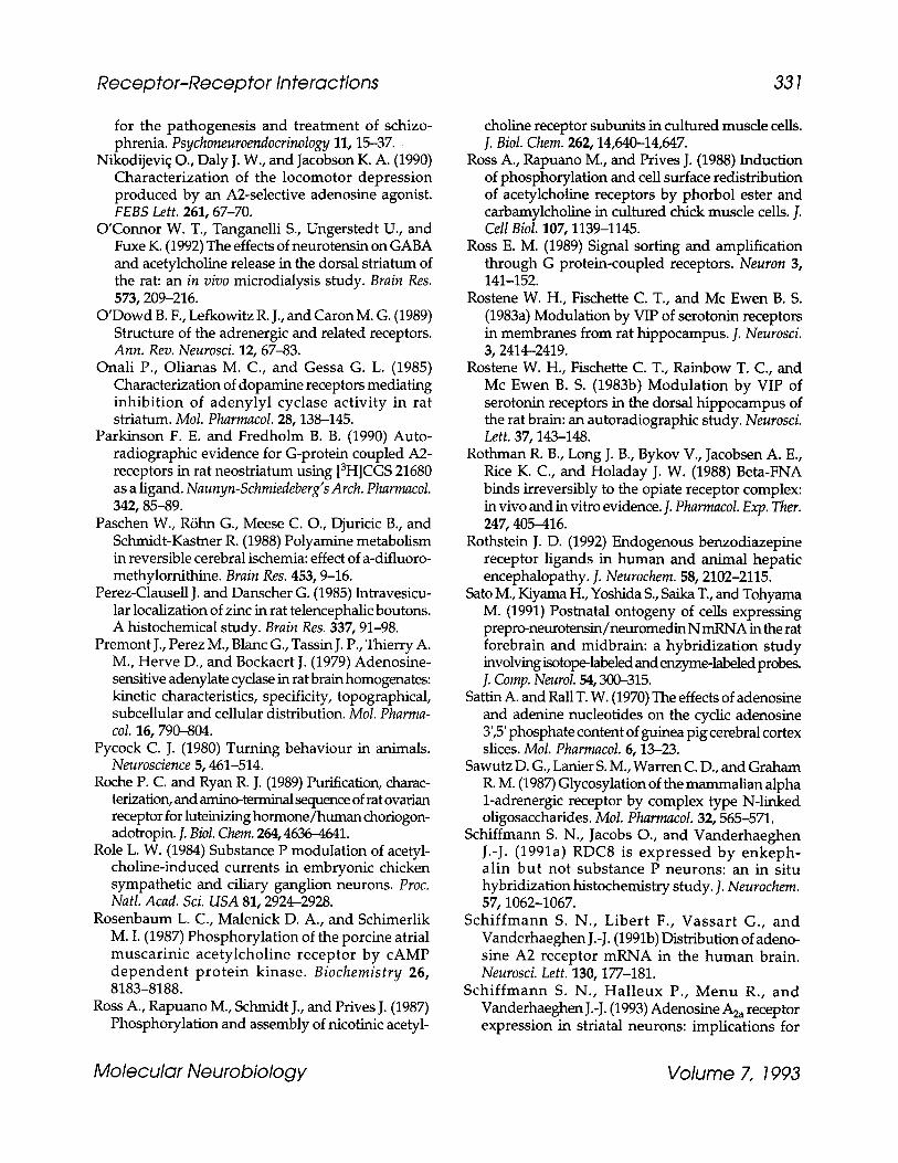
Receptor-Receptor Interactions 331
for the pathogenesis and treatment of schizo- phrenia. Psychoneuroendocrinology 11, 15-37.
Nikodijevi~ O., Daly J. W., and Jacobson K. A. (1990) Characterization of the locomotor depression produced by an A2-selective adenosine agonist. FEBS Lett. 261, 67-70.
O'Connor W. T., Tanganelli S., Ungerstedt U., and Fuxe K. (1992) The effects of neurotensin on GABA and acetylcholine release in the dorsal striatum of the rat: an in vivo microdialysis study. Brain Res. 573, 209-216.
O'Dowd B. F., Lefkowitz R. J., and Caron M. G. (1989) Structure of the adrenergic and related receptors. Ann. Rev. Neurosci. 12, 67-83.
Onali P., Olianas M. C., and Gessa G. L. (1985) Characterization of dopamine receptors mediating inhibition of adenylyl cyclase activity in rat striatum. Mol. Pharmacol. 28, 138-145.
Parkinson F. E. and Fredholm B. B. (1990) Auto- radiographic evidence for G-protein coupled A2- receptors in rat neostriatum using [3H]CGS 21680 as a ligand. Naunyn-Schmiedeberg's Arch. Pharmacol. 342, 85-89.
Paschen W., R6hn G., Meese C. O., Djuricic B., and Schmidt-Kastner R. (1988) Polyamine metabolism in reversible cerebral ischemia: effect of a-difluoro- methylomithine. Brain Res. 453, 9-16.
Perez-Clausell J. and Danscher G. (1985) Intravesicu- lar localization of zinc in rat telencephalic boutons. A histochemical study. Brain Res. 337, 91-98.
Premont J., Perez M., Blanc G., Tassin J. P., Thierry A. M., Herve D., and Bockaert J. (1979) Adenosine- sensitive adenylate cyclase in rat brain homogenates: kinetic characteristics, specificity, topographical, subcellular and cellular distribution. Mol. Pharma- col. 16, 790-804.
Pycock C. J. (1980) Tuming behaviour in animals. Neuroscience 5, 461-514.
Roche P. C. and Ryan R. J. (1989) Purification, charac- terization, and amino-terminal sequence of rat ovarian receptor for luteinizing hormone/human choriogon- adotropin. J. Biol. Chem. 264, 4636-4641.
Role L. W. (1984) Substance P modulation of acetyl- choline-induced currents in embryonic chicken sympathetic and ciliary ganglion neurons. Proc. Natl. Acad. Sci. USA 81, 2924-2928.
Rosenbaum L. C., Malenick D. A., and Schimerlik M. I. (1987) Phosphorylation of the porcine atrial muscarinic acetylcholine receptor by cAMP dependent protein kinase. Biochemistry 26, 8183-8188.
Ross A., Rapuano M., Schmidt J., and Prives J. (1987) Phosphorylation and assembly of nicotinic acetyl-
choline receptor subunits in cultured muscle ceils. J. Biol. Chem. 262, 14,640-14,647.
Ross A., Rapuano M., and Prives J. (1988) Induction of phosphorylation and cell surface redistribution of acetylcholine receptors by phorbol ester and carbamylcholine in cultured chick muscle ceils. J. Cell Biol. 107, 1139-1145.
Ross E. M. (1989) Signal sorting and amplification through G protein-coupled receptors. Neuron 3, 141-152.
Rostene W. H., Fischette C. T., and Mc Ewen B. S. (1983a) Modulation by VIP of serotonin receptors in membranes from rat hippocampus. J. Neurosci. 3, 2414-2419.
Rostene W. H., Fischette C. T., Rainbow T. C., and Mc Ewen B. S. (1983b) Modulation by VIP of serotonin receptors in the dorsal hippocampus of the rat brain: an autoradiographic study. Neurosci. Left. 37, 143-148.
Rothman R. B., Long J. B., Bykov V., Jacobsen A. E., Rice K. C., and Holaday J. W. (1988) Beta-FNA binds irreversibly to the opiate receptor complex: in vivo and in vitro evidence. J. Pharmacol. Exp. Ther. 247, 405-416.
Rothstein J. D. (1992) Endogenous benzodiazepine receptor ligands in human and animal hepatic encephalopathy. J. Neurochem. 58, 2102-2115.
Sato M., Kiyama H., Yoshida S., Saika T., and Tohyama M. (1991) Postnatal ontogeny of cells expressing prepro-neurotensin/neuromedin N mRNA in the rat forebrain and midbrain: a hybridization study involving isotope-labeled and enzyme-labeled probes. ]. Comp. Neurol. 54, 300-315.
Sattin A. and Rall T. W. (1970) The effects of adenosine and adenine nucleotides on the cyclic adenosine 3',5' phosphate content of guinea pig cerebral cortex slices. Mol. Pharmacol. 6, 13-23.
Sawutz D. G., Lanier S. M., Warren C. D., and Graham R. M. (1987) Glycosylation of the mammalian alpha 1-adrenergic receptor by complex type N-linked oligosaccharides. Mol. Pharmacol. 32, 565-571.
Schiffmann S. N., Jacobs O., and Vanderhaeghen J.-J. (1991a) RDC8 is expressed by enkeph- alin but not substance P neurons: an in situ hybridization histochemistry study. ]. Neurochem. 57, 1062-1067.
Schiffmann S. N., Libert F., Vassart G., and Vanderhaeghen J.-J. (1991b) Distribution of adeno- sine A2 receptor mRNA in the human brain. Neurosci. Left. 130, 177-181.
Schiffmann S. N., Halleux P., Menu R., and Vanderhaeghen J.-J. (1993) Adenosine A2a receptor expression in striatal neurons: implications for
Molecular Neurobiology Volume 7, 1993
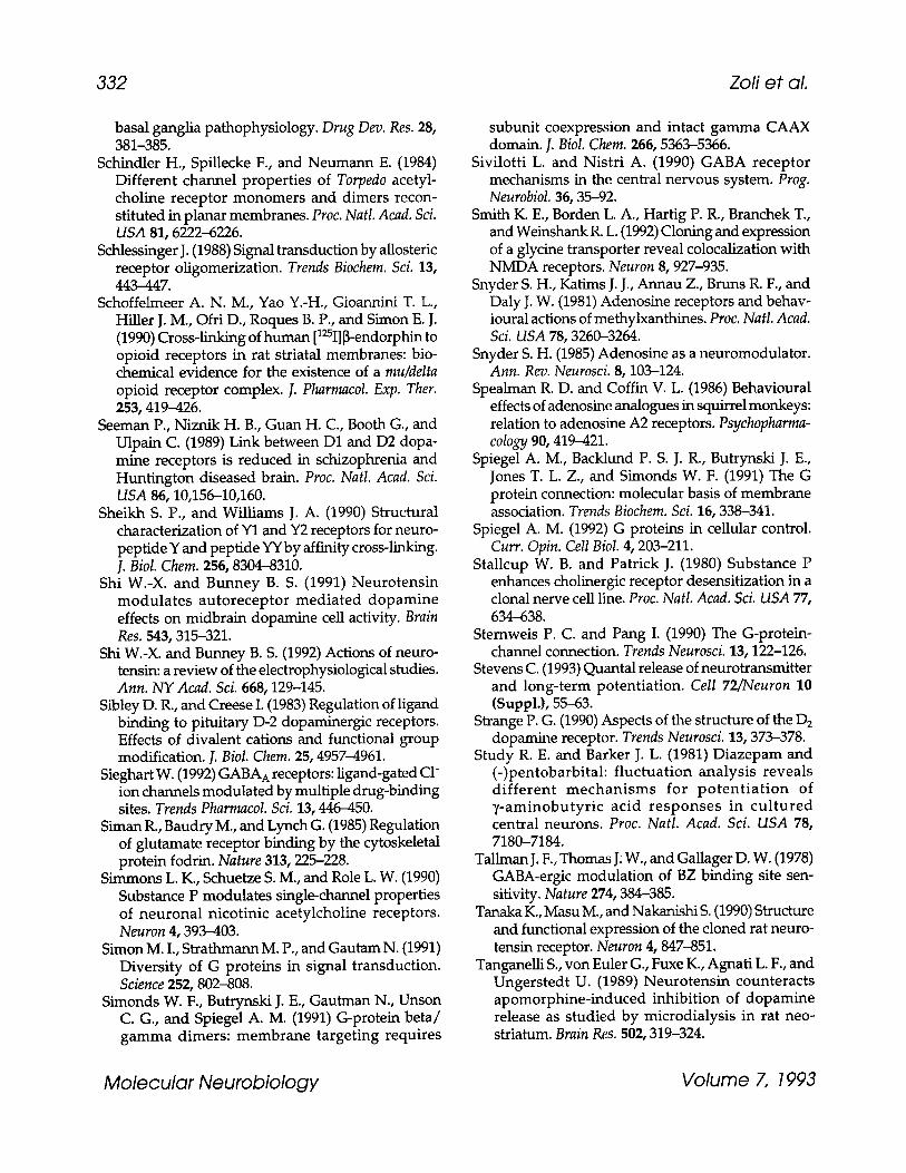
332 Zoli et al.
basal ganglia pathophysiology. Drug Dev. Res. 28, 381-385.
Schindler H., Spillecke F., and Neumann E. (1984) Different channel properties of Torpedo acetyl- choline receptor monomers and dimers recon- stituted in planar membranes. Proc. Natl. Acad. Sci. USA 81, 6222-6226.
Schlessinger J. (1988) Signal transduction by aUosteric receptor oligomerization. Trends Biochem. Sci. 13, 443-447.
Schoffelmeer A. N. M., Yao Y.-H., Gioannini T. L., Hiller J. M., Ofri D., Roques B. P., and Simon E. J. (1990) Cross-linking of human [125I] ~-endorphin to opioid receptors in rat striatal membranes: bio- chemical evidence for the existence of a mu/delta opioid receptor complex. J. Pharmacol. Exp. Ther. 253, 419-426.
Seeman P., Niznik H. B., Guan H. C., Booth G., and Ulpain C. (1989) Link between D1 and D2 dopa- mine receptors is reduced in schizophrenia and Huntington diseased brain. Proc. Natl. Acad. Sci. USA 86, 10,156-10,160.
Sheikh S. P., and Williams J. A. (1990) Structural characterization of Y1 and Y2 receptors for neuro- peptide Y and peptide YY by affinity cross-linking. J. Biol. Chem. 256, 8304-8310.
Shi W.-X. and Bunney B. S. (1991) Neurotensin modulates autoreceptor mediated dopamine effects on midbrain dopamine cell activity. Brain Res. 543, 315-321.
Shi W.-X. and Bunney B. S. (1992) Actions of neuro- tensin: a review of the electrophysiological studies. Ann. NY Acad. Sci. 668, 129-145.
Sibley D. R., and Creese I. (1983) Regulation of ligand binding to pituitary D-2 dopaminergic receptors. Effects of divalent cations and functional group modification. J. Biol. Chem. 25, 4957-4961.
Sieghart W. (1992) GABA A receptors: ligand-gated C1- ion channels modulated by multiple drug-binding sites. Trends Pharmacol. Sci. 13, 446-450.
Siman R., Baudry M., and Lynch G. (1985) Regulation of glutamate receptor binding by the cytoskeletal protein fodrin. Nature 313, 225-228.
Simmons L. K., Schuetze S. M., and Role L. W. (1990) Substance P modulates single-channel properties of neuronal nicotinic acetylcholine receptors. Neuron 4, 393-403.
Simon M. I., Strathmann M. P., and Gautam N. (1991) Diversity of G proteins in signal transduction. Science 252, 802-808.
Simonds W. F., Butrynski J. E., Gautman N., Unson C. G., and Spiegel A. M. (1991) G-protein beta/ gamma dimers: membrane targeting requires
subunit coexpression and intact gamma CAAX domain. J. Biol. Chem. 266, 5363-5366.
Sivilotti L. and Nistri A. (1990) GABA receptor mechanisms in the central nervous system. Prog. Neurobiol. 36, 35-92.
Smith K. E., Borden L. A., Hartig P. R., Branchek T., and Weinshank R. L. (1992) Cloning and expression of a glycine transporter reveal colocalization with NMDA receptors. Neuron 8, 927-935.
Snyder S. H., Katims J. J., Annau Z., Bruns R. F., and Daly J. W. (1981) Adenosine receptors and behav- ioural actions of methylxanthines. Proc. Natl. Acad. Sci. USA 78, 3260--3264.
Snyder S. H. (1985) Adenosine as a neuromodulator. Ann. Rev. Neurosci. 8, 103-124.
Spealman R. D. and Coffin V. L. (1986) Behavioural effects of adenosine analogues in squirrel monkeys: relation to adenosine A2 receptors. Psychopharma- cology 90, 419-421.
Spiegel A. M., Backlund P. S. J. R., Butrynski J. E., Jones T. L. Z., and Simonds W. F. (1991) The G protein connection: molecular basis of membrane association. Trends Biochem. Sci. 16, 338-341.
Spiegel A. M. (1992) G proteins in cellular control. Curr. Opin. Cell Biol. 4, 203-211.
Stallcup W. B. and Patrick J. (1980) Substance P enhances cholinergic receptor desensitization in a clonal nerve cell line. Proc. Natl. Acad. Sci. USA 77, 634--638.
Sternweis P. C. and Pang I. (1990) The G-protein- channel connection. Trends Neurosci. 13, 122-126.
Stevens C. (1993) Quantal release of neurotransmitter and long-term potentiation. Cell 72~Neuron 10 (Suppl.), 55-63.
Strange P. G. (1990) Aspects of the structure of the D 2 dopamine receptor. Trends Neurosci. 13, 373-378.
Study R. E. and Barker J. L. (1981) Diazepam and (-)pentobarbital: fluctuation analysis reveals different mechanisms for potent iat ion of 7-aminobutyric acid responses in cul tured central neurons. Proc. Natl. Acad. Sci. USA 78, 7180-7184.
Tallman J. F., Thomas J. W., and Gallager D. W. (1978) GABA-ergic modulation of BZ binding site sen- sitivity. Nature 274, 384-385.
Tanaka K., Masu M., and Nakanishi S. (1990) Structure and functional expression of the cloned rat neuro- tensin receptor. Neuron 4, 847-851.
Tanganelli S., von Euler G., Fuxe K., Agnati L. F., and Ungerstedt U. (1989) Neurotensin counteracts apomorphine-induced inhibition of dopamine release as studied by microdialysis in rat neo- striatum. Brain Res. 502, 319-324.
Molecular Neurobiology Volume 7, 1993

Receptor-Receptor Interactions 333
Tanganelli S., Fuxe K., von Euler G., Agnati L. F., Ferraro L., and Ungerstedt U. (1990) Involvement of cholecystokinin in the control of striatal dopa- mine autoreceptors. Naunyn-Schmiedeberg's Arch. Pharmacol. 342, 300-304.
Tanganelli S., Li X.-M., Ferraro L., von Euler G., O'Connor W. T., Bianchi C., Beani L., and Fuxe K. (1993) Neurotensin and cholecystokinin octapep- tide control synergistically dopamine release and dopamine D 2 receptor affinity in rat neostria~un. Eur. J. Pharmaeol. 230, 159-166.
Thithapandha A., Maling H. M., and Gillette J. R. (1972) Effects of caffeine and theophyll ine on activity of rats in relation to brain xanthine con- centrations. Proc. Exp. Biol. Med. 139, 582-586.
Titeler M. and Seeman P. (1979) Selective labelling of different DA receptors by a new agonist 3H-ligand: 3H-NPA. Eur. J. Pharmacol. 56, 291,292.
Turner J. T., James-Kracke M. R., and Camden J. M. (1990) Regulation of neurotensin receptor and intracellular calcium in HT29 cells. J. Pharmacol. Exp. Ther. 253, 1049-1056.
Ullrich A. and Schlessinger J. (1990) Signal trans- duction by receptors with tyrosine kinase activity. Cell 61, 203-212.
Ungerstedt U. (1971) Postsynaptic supersensitivity after 6-OH-DA induced degenera t ion of the nigrostriatal DA system in the rat brain. Acta Physiol. Scand. 367, 69-93.
Vallar L. and Meldolesi J. (1989) Mechanisms of signal transduction at DA D2 receptors. Trends Pharmacol. Sci. 10, 74-77.
Van Calker D., Muller M., and Hamprecht B. (1979) Adenosine regulates via two different types of receptors the accumulation of cAMP in cultured brain cells. J. Neurochem. 33, 999-1005.
Van Ree J. M., Gadfori O., and DeWied D. (1983) In rats, the behaviora l profile of CCK-8 related peptides resembles that of antipsychotic agents. Eur. J. Pharmacol. 93, 63-78.
Van Tol H. H. M., Bunzow J. B., Guan H. C., Sunahara R. K., Seeman P., Niznik H. B., and Civelli O. (1991) Cloning of the gene for a human D4 receptor with high affinity for the antipsychotic clozapine. Nature 350, 610-614.
Vapaatalo G. C. and McGuffin-Clineschmidt J. C. (1981) Stereospecif ic i ty in some central and circulatory effects of phenylisopropyl-adenosine (PIA). Drug Res. 25, 407-410.
Venter J. C. and Fraser C. M. (1983) Beta-adrenergic receptor isolat ion and character izat ion wi th immobilized drugs and monoclonal antibodies. Fed. Proc. 42, 273-278.
Vicini S., Mienville J.-M., and Costa E. (1987) Actions of benzodiazepine and ~-carboline derivatives on y -aminobu ty r ic ac id-ac t iva ted C1- channe l s recorded from membrane patches of neonatal rat cortical neurons in culture. J. Pharmacol. Exp. Ther. 243, 1195-1201.
von Euler G. and Fuxe K. (1987) Neurotensin reduces the affinity of D2 dopamine receptors in rat striatal membranes. Acta Physiol. Scand. 131, 525,526.
von Euler G., Fuxe K., van der Ploeg I., Fredholm B. B., and Agnati L. F. (1989) Pertussis toxin treatment counteracts intramembrane interactions between neuropeptide Y receptors and a2-adrenoceptors. Eur. J Pharmacol. 172, 435--441.
von Euler G., Mailleux P., Vanderhaeghen J. J., and Fuxe K. (1990a) Neurotensin reduces the affinity of dopamine D 2 receptors in membranes from post mortem human caudate-putamen. Neurosci. Lett. 109, 325-30.
von Euler G., Meister B., H6kfelt T., Eneroth P., and Fuxe K. (1990b) In t raventr icular injection of neurotensin reduces dopamine D2 agonist binding in rat forebrain and in termedia te lobe of the pituitary gland. Relationship to serum hormone levels and nerve terminal coexistence. Brain Res. 531, 253-262.
von Euler G. (1991) Biochemical characterization of the intramembrane interaction between neuro- tensin and dopamine D 2 receptors in the rat brain. Brain Res. 561, 93-99.
von Euler G., van der Ploeg I., F r e d h o l m B. B., and Fuxe K. (1991) Neurotensin decreases the affinity of dopamine D 2 agonist b inding by a G- protein- independent mechanism. J. Neurochem. 56, 178-183.
von Euler G., Mailleux P., von Euler M., Schiffmann S. N., Vanderhaeghen J. J., and Fuxe K. (1992) Coactivation of dopamine D 1 and D 2 receptors increases the affinity of cholecystokinin-8 receptors in membranes from pos tmor t em human caudate- putaman. Brain Res. 584, 157-162.
Wahlestedt C. and HAkanson R. (1987) Effects of NPY at sympathetic neuroeffector junctions: existence of Y1 and Y2 receptors, in Neuronal Messengers in Vascular Function (Nobin A., O w m a n C., and Areldo-Nobin B., eds.), Elsevier, Amsterdam, pp. 231-242.
Waldeck B. (1973) Sensitization by caffeine of central catecholamine receptors. J. Neural. Transm. 34, 61-72.
Wang R. Y. and Hu X.-T. (1984) Does cholecystokinin potentiate dopamine action in the nucleus accum- bens? Brain Res. 390, 363-367.
Molecular Neurobiology Volume 7, 1993

334 Zofi et al.
Weiss D. S. and Magleby K. L. (1989) Gating scheme for single GABA-activated C1- channels determined from stability plots, dwell-time distributions and adjacent- interval durations.J. Neurosci. 9,1314-1324.
White B. C., Simpson C. C., and Harkins D. (1978) Monoamine synthesis and caffeine-induced loco- motor activity. Neuropharmacology 27, 511-513.
Williams K., Dawson V. L., Romano C., Dichter M. A., and Molinoff P. B. (1990) Characterization of polyamines having agonist, antagonist, and inverse agonist effects at the polyamine recognition site of the NMDA receptor. Neuron 5, 199-208.
Williams K., Romano C., Dichter M. A., and Molinoff P. B. (1991) Modulation of NMDA receptor by polyamines. Life Sci. 48, 469-498.
Wood P. L., Kim H. S., and Hutchison A. (1989) Inhibition of nigrostriatal release of DA in the rat by adenosine receptor agonist: A1 receptor medi- ation. Neuropharmacology 28, 21-24.
Yeramian E., Trautmann A., and Claverie P. (1986) Acetylcholine receptors are not functionally inde- pendent. Biophys. J. 50, 253-263.
Zetler G. (1985) Neuropharmacological profile of cholecystokinin-like peptides. Ann. NY Acad. Sci. 448, 448-469.
Zoli M., Zini I., Grimaldi R., Biagini G., and Agnati L. F. (1993) Effects of putrescine synthesis blockade on neuronal loss and astroglial reaction after transient forebrain ischemia. Int. J. Dev. Neurosci., in press.
Molecular Neurobiology Volume 7, 1993
Copyright © 2022 FDOKUMEN