Prostaglandin synthesis in the male and female reproductive ...

296
Eckmann et al.
J. Clin. Invest.© The American Society for Clinical Investigation, Inc.0021-9738/97/07/0296/14 $2.00Volume 100, Number 2, July 1997, 296–309
Role of Intestinal Epithelial Cells in the Host Secretory Response to Infection by Invasive Bacteria
Bacterial Entry Induces Epithelial Prostaglandin H Synthase-2 Expression and ProstaglandinE
2
and F
2
a
Production
Lars Eckmann,* William F. Stenson,
‡
Tor C. Savidge,
§
David C. Lowe,
§
Kim E. Barrett,* Joshua Fierer,* Jennifer R. Smith,*and Martin F. Kagnoff*
*
Department of Medicine, University of California, San Diego, La Jolla, California 92093-0623;
‡
Division of Gastroenterology, Department of Internal Medicine, Washington University, St. Louis, Missouri 63110; and
§
Department of Cellular Physiology, The Babraham Institute, Babraham, Cambridge CB2 4AT, United Kingdom
Abstract
Increased intestinal fluid secretion is a protective host re-sponse after enteric infection with invasive bacteria that isinitiated within hours after infection, and is mediated byprostaglandin H synthase (PGHS) products in animal mod-els of infection. Intestinal epithelial cells are the first hostcells to become infected with invasive bacteria, which enterand pass through these cells to initiate mucosal, and ulti-mately systemic, infection. The present studies character-ized the role of intestinal epithelial cells in the host secretoryresponse after infection with invasive bacteria. Infection ofcultured human intestinal epithelial cell lines with invasivebacteria, but not noninvasive bacteria, is shown to inducethe expression of one of the rate-limiting enzymes for pros-taglandin formation, PGHS-2, and the production of PGE
2
and PGF
2
a
. Furthermore, increased PGHS-2 expression wasobserved in intestinal epithelial cells in vivo after infectionwith invasive bacteria, using a human intestinal xenograftmodel in SCID mice. In support of the physiologic impor-tance of epithelial PGHS-2 expression, supernatants frombacteria-infected intestinal epithelial cells were shown to in-crease chloride secretion in an in vitro model using polar-ized epithelial cells, and this activity was accounted for byPGE
2
. These studies define a novel autocrine/paracrinefunction of mediators produced by intestinal epithelial cellsin the rapid induction of increased fluid secretion in re-sponse to intestinal infection with invasive bacteria. (
J. Clin.Invest
. 1997. 100:296–309.) Key words: cyclooxygenase
•
in-flammation
•
Salmonella
•
diarrhea
•
pathogenesis
Introduction
Pathogenic enteric bacteria, such as
Salmonella
and
Shigella
,are a significant health problem worldwide. Approximately1% of the population in the United States is estimated to be
infected with
Salmonella
annually (1). Infections with thispathogen are the most frequent cause of food-borne outbreaksof gastroenteritis in adults and children in this country (2, 3),and additionally, can cause invasive infections, mostly in thevery young and elderly, and in immunosuppressed individuals.In developing countries, diarrheal disease due to infection witha variety of pathogenic bacteria is common in children, and hasa high mortality secondary to dehydration (4, 5).
Pathogenic bacteria cause diarrhea by multiple mecha-nisms. For example, bacteria such as
Vibrio cholerae
reside inthe lumen of the small intestine and produce toxins that alterion absorption and/or secretion (6, 7). Other bacteria, such as
Shigella
and enteroinvasive
Escherichia coli
, invade and de-stroy the colonic epithelium, leading to dysentery (8).
Salmo-nella
and
Yersinia enterocolitica
pass through the intestinal epi-thelium and invade the mucosa (9–11). The ability of thesebacteria to invade epithelial cells is important for diseasepathogenesis since invasion mutants of
Salmonella
do notcause disease after oral infection (12), although production ofan enterotoxin by some strains of
Salmonella
may also play arole in inducing diarrhea (13, 14). Studies in rabbits haveshown that the onset of diarrhea occurs rapidly (within 2–4 h)in response to infection with some invasive enteric bacteriasuch as
Salmonella
(15). These data suggest that early eventsassociated with bacterial invasion are important for initiatingdiarrhea.
Prostaglandins are important regulators of gastrointestinalfluid secretion (16, 17). PGE
1
stimulates chloride secretion inpolarized cultured epithelial cells (18), and PGE analogues in-duce diarrhea in vivo (19). Moreover, prostanoids are commonmediators of diarrhea of different etiologies, including radia-tion-induced diarrhea (20), and diarrhea after bacterial infec-tion (21, 22). Prostaglandins are formed from free arachidonicacid through the conversion of arachidonic acid to PGH, whichis catalyzed by the enzyme prostaglandin H synthase (PGHS)
1
(17, 23). PGH is subsequently converted by specific synthasesto PGE, PGF, thromboxanes, or prostacyclins. PGHS exists intwo isoforms: constitutively expressed PGHS-1, and induciblePGHS-2 (23). These isoforms have both overlapping andunique functions in the host, as demonstrated in geneticallydeficient mice. Thus, mice lacking PGHS-2 manifested renaldysplasia, cardiac fibrosis, and infertility, and were prone toperitonitis (24, 25). In contrast, PGHS-1–deficient mice hadreduced platelet aggregation and a decreased inflammatory
Address correspondence to Lars Eckmann, Laboratory of MucosalImmunology, University of California, San Diego, Department ofMedicine 0623D, 9500 Gilman Drive, La Jolla, CA 92093-0623. Phone:619-534-0683; FAX: 619-534-5691; E-mail: [email protected]
Received for publication 25 February 1997 and accepted in revisedform 15 April 1997.
1.
Abbreviations used in this paper:
CM, conditioned media; I
SC
,short-circuit current; PGHS, prostaglandin H synthase; RT, reversetranscription.

Invasive Bacteria Induce Prostaglandin H Synthase-2 Expression in Intestinal Epithelial Cells
297
response to arachidonic acid (26). Neither PGHS-1– norPGHS-2–deficient mice had overt gastrointestinal abnormali-ties (24–26). Whereas PGHS-1 is expressed constitutively inmany cell types, PGHS-2 normally is expressed at low levels inmany tissues (23). High-level expression, however, can be in-duced in macrophages, and in epithelial cells in the intestine,kidney, and lungs (23, 27, 28). Known stimulators of PGHS-2expression in epithelial cells include IL-1, TNF
a
, TGF
a
, andEGF (23, 29, 30).
Intestinal epithelial cells form a tight barrier that preventsmucosal penetration by most luminal bacteria. Some patho-genic enteric bacteria, however, invade the epithelium, andgain access to the underlying mucosa. Intestinal epithelial cellsrespond to bacterial invasion with the production of a range ofchemoattractant and proinflammatory cytokines (31–33), sug-gesting that these cells, which are the first host cells to be en-tered by invasive enteric bacteria, produce early signals for theactivation of the host inflammatory response.
In the present studies, we have characterized the role of in-testinal epithelial cells in the rapid fluid response that is ob-served after infection with invasive bacteria. We show thatbacterial invasion of intestinal epithelial cells induces the ex-pression of PGHS-2, and stimulates the increased release ofPGE
2
and PGF
2
a
. Moreover, we show that PGE
2
, released bythese cells, can cause increased chloride secretion in an in vitromodel of polarized colonic epithelial cells.
Methods
Cell lines.
The human colon adenocarcinoma cell line HT-29 (ATCCHTB 38) and the embryonic intestinal cell line I407 (ATCC CCL 6)were obtained from the American Type Culture Collection (Rockville,MD), and were grown in DME supplemented with 10% FCS, 2 mMglutamine, and 10 mM Hepes (growth medium). T
84
human colonic ep-ithelial cells were grown as described before, and were cultured on per-meable supports for Ussing chamber experiments (34).
Bacteria, cytokines, and other reagents.
The following bacteria wereused in these studies:
Salmonella dublin
lane (31),
Salmonella typh-imurium
(ATCC 14028),
Salmonella typhi
Ty2
aroA
aroC
(strainBRD691) (35),
Yersinia enterocolitica
08 (31),
Shigella dysenteriae
(31),
Listeria monocytogenes
(serotype 4b, ATCC 19115), enteroinva-sive
Escherichia coli
(serotype O29:NM, ATCC 43892), enterohem-orrhagic
E. coli
(serotype O157:H7, ATCC 43894), enteropathogenic
E. coli
(serotype O111, ATCC 33780),
Streptococcus bovis
(ATCC9809), and three nonpathogenic
E. coli
strains: DH5
a
(GIBCO BRL,Gaithersburg, MD), SC13 (36), and HB101 (GIBCO BRL). The re-combinant human cytokines TNF
a
and IL-1
a
, and goat antihumanTNF
a
(IgG isotype), were obtained from R & D Systems (Minneapo-lis, MN). Normal goat IgG, monoclonal mouse IgG
1
(MOPC-31c),LPS from
E. coli
serotype O111, and indomethacin were obtainedfrom Sigma Chemical Co. (St. Louis, MO). Monoclonal mouse anti-PGE
2
(37) was a gift from Dr. J.P. Portanova (G.D. Searle & Co., St.Louis, MO). PGE
2
, PGF
2
a
, NS-398, and valerylsalicylate were ob-tained from Cayman Chemical Co. (Ann Arbor, MI).
Infection protocol.
Colonic epithelial cells were seeded into 6-welltissue culture plates (Costar Corp., Cambridge, MA), and grown toconfluence. Bacteria were prepared for infections as described previ-ously (31, 38). Bacteria were added in a 1-ml vol to each well, and in-cubated for 1 h to allow bacterial entry to occur. Monolayers werewashed three times to remove extracellular bacteria, and the cultureswere further incubated for up to 29 h in the presence of 50
m
g/ml gen-tamicin to kill any remaining extracellular bacteria.
To assay prostaglandin production, cultures were washed threetimes with DME, and were incubated for 15 min at 37
8
C in 1 ml/wellof DME, 10 mM Hepes, 2 mg/ml BSA, 50
m
g/ml gentamicin, and 20
m
M arachidonic acid. Supernatants were collected, centrifuged to re-move debris, and stored at
2
80
8
C.To test conditioned media from bacteria-infected epithelial cell
cultures for their ability to induce prostaglandin production by unin-fected cells, supernatants were removed from cultures infected with
S. dublin
or
Y. enterocolitica
for 8 h, filtered through a 0.22
m
m filter,and kept frozen at
2
80
8
C. Increasing doses of the 8-h supernatantsfrom bacteria-infected and control epithelial cultures were added toconfluent HT-29 cell monolayers in 6-well plates, and cultures wereincubated for 8 h before prostaglandin production was determined asdescribed above.
Prostaglandin immunoassays, [
3
H]arachidonic acid labeling, andHPLC analysis of labeled cell supernatants.
Levels of PGE
2
and PGF
2
a
were determined in culture supernatants by enzyme immunoassay(Cayman Chemical Co.). To confirm the identity of the prostaglan-dins, confluent I407 or HT-29 monolayers, in 10-cm plates, were in-fected for 1 h with 2.5
3
10
8
S. dublin
, washed, and incubated for 7 hwith gentamicin. Subsequently, cultures were incubated for 15 min ina 2.5-ml vol of DME containing 10 mM Hepes, 2 mg/ml BSA, and 20
m
Marachidonic acid, as well as 5
m
Ci or 50
m
Ci of [5,6,8,9,11,12,14,15-
3
H(N)]arachidonic acid (100 Ci/mmol; NEN Life Science Products,Boston, MA) for I407 or HT-29 cells, respectively. Supernatants wereextracted with chloroform, and the chloroform-soluble phase wasdried under nitrogen, reconstituted in methanol, and analyzed by re-verse-phase HPLC (model 1090M; Hewlett-Packard Co., Palo Alto,CA) using an analytical C-18 Microsorb column (4.6
3
100 mm, Rai-nin Instrument Co., Woburn, MA). A flow rate of 1.0 ml/min wasused with two solvents (A and B) set at 33.5% B for 0–10 min, 58.6%B for 11–25 min, and 92.0% B for 26–33 min. Solvent A was 100:0.01(vol/vol) water/acetic acid, and solvent B was 100:0.01 (vol/vol) aceto-nitrile/acetic acid. The eluate was monitored with a spectrophotome-ter set at 205 nm, and the outflow from the spectrophotometer wasrouted to a Flo-One-beta detector (Radiomatic Instruments andChemical Co. Inc., Tampa, FL) for radioactivity measurements. Pros-tanoids were identified by their elution times relative to known stan-dards. The prostaglandin recovery after chloroform extraction andHPLC analysis was
z
75%, as determined by parallel analysis ofknown amounts of [
3
H]PGE
2
.
RNA extraction and RNase protection assays.
Total cellular RNAwas extracted using an acid guanidinium-phenol-chloroform method(Trizol; GIBCO BRL). PGHS-2 mRNA levels were determined byRNase protection assay using a
32
P-labeled antisense RNA probe spe-cific for human PGHS-2 (39). The probe was prepared by in vitrotranscription of a pGEM5Z plasmid containing a PGHS-2 specific in-sert (a gift from K. Seibert, G.D. Searle & Co.) to yield a 400-nt prod-uct. Subsequently, 10–20
m
g total RNA was mixed with 10
5
cpm ofspecific RNA probe in hybridization buffer (Ambion Inc., Austin,TX), and the mixtures were incubated overnight at 45
8
C. Reactionswere digested with optimal concentrations of RNase A and T1, andrun on a 6% polyacrylamide/urea gel. Gels were dried, exposed tox-ray film, and band intensities were quantitated with a densitometer(model GS-670; Bio-Rad Laboratories, Hercules, CA). AbsolutePGHS-2 mRNA levels were derived from a standard curve that wasestablished from the band intensities of reactions containing knownamounts of in vitro–transcribed PGHS-2 sense RNA.
Reverse transcription (RT)-PCR analysis.
Reverse transcriptionand PCR amplification were performed as described before (32). Thefollowing primers were used to amplify a 305-bp fragment of PGHS-2from human cDNA: 5
9
-TTC AAA TGA GAT TGT GGG AAAATT GCT-3
9
(sense) and 5
9
-AGA TCA TCT CTG CCT GAG TATCTT-3
9
(antisense). The primer sequences are located on differentexons of the human PGHS-2 gene. They are specific for humanPGHS-2, and do not amplify a product from mouse RNA or DNA.The primers for amplification of human
b
-actin mRNA were de-scribed before (32). Annealing temperatures were 60
8
C for PGHS-2,and 72
8
C for
b
-actin.The general strategy used for quantitative RT-PCR analysis using
internal standard RNAs, and the construct for the quantitation of hu-
298
Eckmann et al.
man
b
-actin mRNA were described before (32). For the quantitationof human PGHS-2 mRNA, a DNA fragment was constructed as fol-lows: a 660-bp
Pvu
II fragment of a cDNA clone from the mouse im-munoglobulin
a
constant region (40) was amplified with the primers5
9
-TGA GAT TGT GGG AAA ATT GCT
CTA CAG TGT GTCCAG CGT CC
-3
9
(sense) and 5
9
-TCA TCT CTG CCT GAG TATCTT
GCA ACA CGC TTG TCA CCA GG
-3
9
(antisense). The re-gions homologous to the
a
constant region are depicted in bold. Theresulting 392-bp DNA fragment was reamplified with the primers5
9
-ATT GTA ATA CGA CTC ACT ATA GGG TTC AAA TGAGAT TGT GGG AAA ATT GCT-3
9
(sense) and 5
9
-TTT TTT TTTTTT TTT TTT GAC AGA TCA TCT CTG CCT GAG TAT CTT-3
9
(antisense) to yield a 446-bp DNA fragment. The sense primer con-tains a promoter for T7 RNA polymerase (underlined), and the anti-sense primer a stretch of dT (underlined) to provide a poly(A) tailafter in vitro transcription. The latter DNA fragment was column-purified (QIA quick-spin PCR purification Kit; QIAGEN Inc., Chats-worth, CA), and in vitro–transcribed using T7 RNA polymerase toyield a 425-nt RNA. The reaction was digested with RNase-freeDNase, extracted with phenol/chloroform, precipitated, and resus-pended in TE. The OD260 was determined, and the number of stan-dard RNA molecules per microliter was calculated based on theRNA concentration and the size of the standard RNA. Dilutions ofstandard RNA were made in 10 mM Tris pH 7.6, 1 mM EDTA con-taining 1 mg/ml tRNA to avoid nonspecific loss of transcripts at lowconcentrations. Reverse transcription and PCR amplification of thestandard RNA using the PGHS-2 primers described above yield a401-bp DNA fragment.
Immunoblot analysis.
Confluent epithelial monolayers in 6-wellplates were washed with ice-cold PBS, and lysed in 0.5 ml/well lysisbuffer (150 mM NaCl, 20 mM Tris, pH 7.5, 0.1% Triton X-100, 1 mMPMSF, 10
m
g/ml aprotinin). Lysates were sonicated for 5 s on ice, andcentrifuged at 12,000
g
for 2
3
20 min. Protein concentrations in thelysates were determined by the Bradford method (Bio-Rad Lab-oratories) using BSA as a standard. 15
m
g protein/lane was size-frac-tionated on a denaturing, nonreducing 6% polyacrylamide minigel(Mini-PROTEAN II; Bio-Rad Laboratories), and electrophoreticallytransferred to a nitrocellulose membrane (0.1-
m
m pore size). Specificproteins were detected using optimal concentrations of rabbit antihu-man PGHS-2 (COOH terminus) (Oxford Biomedical Research, Ox-ford, MI), rabbit antihuman PGHS-1 (Oxford Biomedical Research),or rabbit antiactin (Sigma Chemical Co.) as primary antibodies, andperoxidase-conjugated donkey anti–rabbit Ig (Amersham Corp., Ar-lington Heights, IL) as secondary antibody. Specifically bound perox-idase was detected by enhanced chemiluminescence (ECL system;Amersham Life Science) and exposure to x-ray film (XAR5; East-man Kodak Company, Rochester, NY) for 10–30 s.
Immunocytochemistry.
HT-29 cells were grown on 13-mm glasscoverslips in 24-well plates, infected for 1 h with 10
8
/well
S. typhi
BRD691 in a 1-ml vol, washed, and further incubated for 4 or 24 hwith gentamicin. Cultures were washed with PBS, and fixed at roomtemperature with 2% formalin in PBS for 30 min. After further wash-ing, cultures were incubated sequentially for 1 h each at room tem-perature with optimal concentrations of rabbit antihuman PGHS-2 orantihuman PGHS-1 (Oxford Biomedical Research), biotin-labeleddonkey anti–rabbit IgG (Amersham), and streptavidin-FITC (Amer-sham). Coverslips were mounted on glass slides using an aqueousmounting medium. For colocalization experiments, cultures were firststained for PGHS-2, and were subsequently incubated with optimalconcentrations of goat anti-
Salmonella
common structural antigen-1(CSA-1; Kirkegaard & Perry Laboratories, Gaithersburg, MD), andTRITC-labeled rabbit anti–goat IgG (Sigma Chemical Co.). Speci-mens were analyzed by confocal microscopy (MRC BioRad 500; Bio-Rad Laboratories). Control and infected specimens were analyzedunder identical conditions so that the relative computer-generatedcolor intensities (see Fig. 5) would be representative of the actualstaining intensities, but not of the fluorochromes used.
Human fetal intestinal xenografts.
The human fetal intestinal xe-
nograft model used in the present work has been described in detailpreviously (41). Briefly, human fetal small intestine (
n
5
6, gesta-tional age 10–14 wk) was transplanted subcutaneously into C.B-17 se-vere-combined immunodeficient (SCID) mice. Xenografts were al-lowed to develop for 10 wk before use, at which time the epitheliumand underlying mucosa are fully differentiated (41). Xenografts wereinfected with
z
5
3
10
7
bacteria in DME/F12 medium in a 100-
m
l volinjected intraluminally by subcutaneous injection. Controls were in-jected with 100
ml sterile medium. Xenograft tissue was removed 6 hafter infection, extensively washed, embedded in OCT compound,and snap-frozen in isopentane. Frozen sections (8 mm) of xenograftintestine were prepared, fixed and stained by indirect immunofluo-rescence, as described above for HT-29 cells, and counterstained with5 mg/ml propidium iodide. To extract RNA from xenografts, tissueswere removed 6 h after infection, and mucosal scrapings were pre-pared and immediately frozen in liquid nitrogen. Scrapings were ho-mogenized on ice in 2 ml Trizol (GIBCO BRL) using a Potter-Elve-hjem tissue grinder, and total RNA was extracted. The xenograftstudies were performed with full approval from the Cambridge LocalEthics Committee, and in accordance with the Home Office guide-lines specified in the Polkinghorne Report (1989) (38, 41).
Ussing chamber experiments. To test conditioned media for theirability to induce chloride secretion by polarized intestinal epithelialcells, confluent HT-29 cell monolayers in 162-cm2 flasks were infectedfor 1 h with 1.5 3 109 S. dublin/flask in a 15-ml vol, washed, and fur-ther incubated for 7 h with gentamicin. Cultures were washed 33
with DME, and incubated for 15 min at 378C with 7.5 ml/flask ofDME, 10 mM Hepes, 2 mg/ml BSA, 50 mg/ml gentamicin, and 100mM arachidonic acid. Supernatants were collected and filteredthrough a 0.22-mm filter. Ussing chamber experiments using polar-ized T84 cell monolayers as the target cell were then performed as de-scribed before (34).
Results
Salmonella infection of the human intestinal epithelial cell linesI407 and HT-29 increases PGE2 and PGF2a production. Oral Sal-monella infection of monkeys rapidly induces increased intesti-nal fluid secretion through an indomethacin-sensitive pathway(22), suggesting that products of PGHS pathways are involvedin this host response. To determine whether intestinal epithe-lial cells (which are the first host cells that are infected by Sal-monella after oral ingestion) are involved in this host responseto the infection, we determined if any, and which, products ofthe PGHS pathways are formed after Salmonella infection ofthese cells.
The human intestinal epithelial cell line I407 was infectedfor 1 h with S. dublin, and was cultured for an additional 7 h inthe presence of gentamicin. Subsequently, infected and controlcells were incubated for 15 min with [3H]arachidonic acid, andthe radioactive products in the culture supernatants were ana-lyzed by HPLC. Control I407 cells constitutively producedPGE2 and PGF2a, with PGE2 being z 10-fold more abundantthan PGF2a (Fig. 1, top). The production of both prostaglan-dins increased by greater than fourfold in response to S. dublininfection of I407 cells (Fig. 1, middle). The absence of addi-tional peaks suggests that PGE2 and PGF2a were the onlyPGHS products produced by the cells, since this HPLC systemallows the separation and quantitation of all known PGHSproducts. The PGHS inhibitor indomethacin completelyblocked production of PGE2 and PGF2a in S. dublin–infectedI407 cells (Fig. 1, bottom), confirming that the arachidonic-derived products detected in the HPLC analysis were formedthrough a PGHS-dependent pathway. Consistent with the

Invasive Bacteria Induce Prostaglandin H Synthase-2 Expression in Intestinal Epithelial Cells 299
data obtained from the HPLC analysis, S. dublin–infected I407cells produced ninefold more PGE2 than did control cells(73.3623.1 vs. 8.462.6 ng/ml, values are means6SEM of threeexperiments) and fivefold more PGF2a than control cells (6.7vs. 1.3 ng/ml, means of two experiments), as determined by im-munoassays of supernatants from S. dublin–infected and con-trol I407 cultures that were incubated for 15 min with 20 mMarachidonic acid.
A second human intestinal epithelial cell line, HT-29, wasused to confirm that the above findings were not restricted to asingle cell line. As shown in Fig. 2, S. dublin–infected HT-29cells produced up to 50-fold more PGE2 and PGF2a than con-trols during a 15-min incubation period with 20 mM arachi-donic acid. In contrast to I407 cells, S. dublin–infected HT-29cells produced similar levels of PGE2 and PGF2a. Further-more, two additional Salmonella strains, S. typhimurium andan invasive, but replication-deficient strain of S. typhi (strainBRD691), also induced increased PGE2 and PGF2a production
by HT-29 cells, indicating that different Salmonella strainsinduce this response. For all three Salmonella strains, the in-crease in prostaglandin production was dependent on the bac-terial inoculum, with maximal production at a bacterial inocu-
Figure 1. HPLC analysis of supernatants from [3H]arachidonic acid–labeled I407 cells infected with S. dublin. Confluent I407 monolayers in 10-cm plates were infected with S. dublin, washed, and further in-cubated for 7 h with gentamicin. Cultures were washed, incubated for 15 min with 5 mCi [3H]arachidonic acid, and supernatants were ana-lyzed by HPLC using an on-line radioactivity detector as described in Methods. Uninfected cultures were used as a control. Indomethacin (3 mM) was added 1 h before the addition of [3H]arachidonic acid, and was present during the 15-min incubation period with arachi-donic acid. Peaks were identified by comparison with a range of pros-taglandin standards. The y-axis reflects [3H]radioactivity, measured as area under the curve of the radiochromatogram, with identical ar-bitrary units used in all three panels. In the middle panel (S. dublin), the PGE2 and PGF2a peaks represent 6 and 1% of the total recovered counts, respectively.
Figure 2. Relationship of bacterial inoculum and increased prosta-glandin production in Salmonella-infected HT-29 cells. Confluent HT-29 monolayers in 6-well plates (containing z 2 3 106 cells/well) were infected for 1 h with the indicated doses of S. dublin lane (top), S. typhimurium 14028s (middle), or S. typhi BRD691 (bottom), washed, and further incubated for 7 h with gentamicin. Cultures were incubated for 15 min in a 1-ml vol of DME containing 2 mg/ml BSA, and 20 mM arachidonic acid. PGE2 (d) and PGF2a (s) levels were analyzed by enzyme immunoassays. The identity of these prostaglan-dins was confirmed by HPLC analysis of supernatants from control and S. dublin–infected HT-29 cultures incubated for 15 min with [3H]arachidonic acid (data not shown). Cultures infected with the three Salmonella strains contained similar numbers of intracellular bacteria at the end of the experiment, e.g., after infection with108 CFU/well, monolayers had 1.8 3 107, 1.3 3 107, and 1.0 3 107 in-tracellular CFU/well for S. dublin, S. typhimurium, and S. typhi BRD691, respectively. Data represent means6SD of the results of triplicate cultures from one experiment. Similar results were obtained in at least two additional experiments for each bacterial strain.

300 Eckmann et al.
lum/cell ratio of z 50:1 (Fig. 2). Maximal prostaglandinproduction was fivefold lower after infection with S. typhimu-rium compared to S. dublin and S. typhi, although similar inoc-ulum–response relationships were observed for all three Sal-monella strains.
Increased PGE2 and PGF2a production by Salmonella-infected epithelial cells depends on increased PGHS-2 activity.Production of PGE2 and PGF2a depends on PGHS, which ex-ists in two isoforms. To determine which PGHS isoform wasimportant for increased prostaglandin production after Salmo-nella infection of epithelial cells, infected cells were preincu-bated for 60 min with several specific inhibitors before addi-tion of 20 mM arachidonic acid. Incubation with indomethacin,an inhibitor of both PGHS-1 and PGHS-2, completely inhib-ited PGE2 production in S. dublin–infected HT-29 cells (datanot shown) and I407 cells (Fig. 1). In contrast, indomethacindid not affect increased IL-8 secretion by S. dublin–infectedHT-29 cells, a previously reported response of these cells to in-fection with invasive bacteria (31), indicating that indometha-cin had no general effects on the ability of these cells to re-spond to Salmonella (data not shown). Addition of NS-398, aspecific PGHS-2 inhibitor (42), also completely inhibited in-creased PGE2 production by S. dublin–infected HT-29 cells(S. dublin–infected, 22.662.7 ng PGE2/ml; S. dublin–infected 11 mM NS-398, , 0.2 ng/ml; controls, , 0.2 ng/ml; values aremeans6SD of triplicate cultures). Both indomethacin and NS-398 exhibited half-maximal inhibition of PGE2 productionafter S. dublin infection of HT-29 cells at a concentration ofz 30 nM. In contrast, valerylsalicylate, an inhibitor that is rel-atively specific for PGHS-1 (43), decreased PGE2 productionby S. dublin–infected HT-29 cells only slightly (S. dublin–infected, 22.662.7 ng PGE2/ml; S. dublin–infected 1 10 mMvalerylsalicylate, 17.761.0 ng/ml; controls, , 0.2 ng/ml; valuesare means6SD of triplicate cultures). Thus, of the two PGHSisoforms, PGHS-2 is mostly, if not exclusively, responsible forincreased PGE2 production by HT-29 cells after Salmonella in-fection. This conclusion was also true for PGF2a production,since indomethacin and NS-398 completely blocked increasedPGF2a production after S. dublin infection of HT-29 cells,whereas valerylsalicylate had no effect (data not shown).
Salmonella infection of intestinal epithelial cells upregulatesprotein and mRNA levels for PGHS-2, but not PGHS-1. Sub-sequent studies focused on mechanisms that govern epithelialexpression of PGHS-2 in response to Salmonella infection. Atime course of increased PGHS activity after infection of HT-29cells with S. dublin is shown in Fig. 3 A. The ability of the cellsto convert arachidonic acid to PGE2 increased 3 h after S. dub-lin infection, reached maximal levels 12 h after infection, andremained increased until the end of the experiment (29 h).Similarly, PGF2a production first increased 3 h after S. dublininfection of HT-29 cells, and reached a maximum 12 h after in-fection (controls, , 0.05 ng PGF2a/ml; 3 h after infection,0.4960.08 ng/ml; 12 h after infection, 10.761.0 ng/ml; valuesare means6SD of triplicate cultures).
To determine whether the increase in PGHS activity corre-lated with increased PGHS levels, lysates of S. dublin–infectedand control HT-29 cells were analyzed by immunoblot analysis(Fig. 3 B). Increased levels of PGHS-2 were first detected 3 hafter S. dublin infection, were maximal 6–10 h after infection,and remained increased thereafter. As a comparison, TNFaalso stimulated increased PGHS-2 levels, and time course andmaximal extent of the increase was similar to that after S. dub-
Figure 3. Time course of increased PGE2 levels and PGHS-2 levels after S. dublin infection of HT-29 cells. Confluent monolayers ofHT-29 cells in 6-well plates were infected for 1 h with 108 S. dublin/well, washed, and further incubated with gentamicin. (A) PGE2 lev-els. At the indicated times after infection, cultures were washed, incu-bated for 15 min in a 1-ml vol of DME containing 2 mg/ml BSA, and 20 mM arachidonic acid, and PGE2 levels were analyzed by enzyme immunoassay. Data represent means6SD of the results of triplicate cultures. Data shown are combined from two separate experiments. Parallel uninfected control cultures produced , 0.2 ng/ml PGE2 at all time points. (B) PGHS-2 levels. At the indicated times after S. dublin infection or TNFa addition (20 ng/ml), cell lysates were prepared, size-fractionated, and blotted onto a nitrocellulose membrane. PGHS-2 was detected with a specific antibody and enhanced chemi-luminescence. Representative examples of x-ray films are shown. To obtain quantitative data, x-ray films were scanned with a densitome-ter. PGHS-2 levels are expressed relative to the maximum levels in each experiment. Data from two separate experiments are depicted in the graph. d, PGHS-2 levels after S. dublin infection; s, PGHS-2 levels after TNFa stimulation.

Invasive Bacteria Induce Prostaglandin H Synthase-2 Expression in Intestinal Epithelial Cells 301
lin infection (Fig. 3 B). In contrast, levels of PGHS-1, as wellthe control protein actin, were not altered after S. dublin infec-tion of HT-29 cells or after TNFa stimulation (PGHS-1 levelswere 98, 97, and 81% of controls, and actin levels were 99, 91,and 81% of controls at 4, 10, and 24 h after S. dublin infection,respectively, as determined by immunoblot analysis and scan-ning with a densitometer; n 5 2). Infection of I407 cells with S.dublin also increased PGHS-2 levels, and the time course andrelative magnitude of the increase were similar to those in HT-29cells (data not shown).
Increased PGHS-2 levels were paralleled by increasedPGHS-2 mRNA levels in S. dublin–infected HT-29 cells, asdemonstrated by qualitative RT-PCR and confirmed quantita-tively by RNase protection assays (Fig. 4). PGHS-2 mRNA
levels first increased 60 min after S. dublin infection (Fig. 4 A),reached a maximum 2–4 h after infection, and decreased there-after (Fig. 4 B). Similarly, S. dublin infection increased PGHS-2mRNA levels in I407 cells (2–4 h after S. dublin infection,535 fg PGHS-2 mRNA/mg total RNA; controls, 92 fg/mg; val-ues are means, n 5 2). In summary, PGHS-2 mRNA levels in-creased rapidly, but transiently, after S. dublin infection ofHT-29 and I407 cells, whereas the increase in PGHS-2 levelsand PGHS activity occurred more slowly after infection, butwas more sustained.
PGE2 production by HT-29 colonic epithelial cells is in-creased in response to infection with invasive but not noninva-sive bacteria. The above studies focused on the PGE2 andPGF2a response of cultured intestinal epithelial cells to infec-tion with different Salmonella strains. To determine whetherother bacteria can induce a similar epithelial response, HT-29cells were infected with a range of invasive and noninvasivegram-negative and gram-positive bacteria, and PGE2 produc-tion was determined. As shown in Table I, infection of HT-29cell cultures with the invasive gram-negative bacteria Y. en-terocolitica, S. dysenteriae, and enteroinvasive E. coli (serotypeO29:NM) increased PGE2 production 17–54-fold. In contrast,infection with several noninvasive gram-negative bacteria,including enteropathogenic and enterohemorrhagic E. colistrains, or addition of bacterial LPS, increased PGE2 produc-tion by HT-29 cells to a much lesser extent (less than fivefold).Moreover, infection of HT-29 cell cultures with the gram-posi-tive invasive bacteria L. monocytogenes also increased PGE2
production, although the increase was lower than that seen af-ter infection with gram-negative invasive bacteria. Infectionwith the noninvasive gram-positive S. bovis had little effect onPGE2 production by HT-29 monolayers. Thus, invasive bacte-ria increase epithelial PGE2 production, while noninvasive donot, or to a much lesser extent. In addition, levels of PGF2a
production paralleled those of PGE2 in all cases tested (datanot shown), further underlining that PGE2 and PGF2a produc-tion are likely regulated by identical mechanisms in bacteria-infected epithelial cells.
PGHS-2 is expressed mostly by bacteria-invaded epithelialcells early after infection, while at later times noninfected cellsalso express PGHS-2. To characterize the relationship be-tween invaded bacteria and increased PGHS-2 expression,HT-29 cells were infected with a replication-deficient S. typhi(strain BRD691) and analyzed by double immunofluorescencefor Salmonella staining and PGHS-2 expression (Fig. 5). Rela-tively early after infection (5 h), PGHS-2 expression was con-fined to a small proportion of cells, and was found mostly incells which also contained Salmonella, suggesting that the in-duction of PGHS-2 resulted from a direct interaction betweeninvading bacteria and infected cells. A small proportion ofPGHS-2–expressing cells, however, did not appear to harborbacteria, although these cells were located mostly in the imme-diate vicinity of Salmonella-infected cells.
At later times after infection (24 h) with S. typhi BRD 691,essentially all epithelial cells expressed PGHS-2 (Fig. 6 B).This observation was comparable to that in HT-29 cells stimu-lated with TNFa (Fig. 6 C), and both were in striking contrastto the findings in unstimulated cells (Fig. 6 A). Since the mono-layers were infected with a replication-deficient strain of S.typhi, and gentamicin was present throughout the culture pe-riod, it is unlikely that bacterial replication or spread couldhave occurred in these cultures. This observation suggests that
Figure 4. Kinetics of increased PGHS-2 mRNA expression in S. dub-lin–infected HT-29 cells. Confluent HT-29 monolayers in 6-well plates were infected for 1 h with 108 S. dublin/well, washed, and fur-ther incubated with gentamicin. Parallel cultures were stimulated with 20 ng/ml TNFa. Total cellular RNA was extracted at the indi-cated times after S. dublin infection or TNFa stimulation. Unstimu-lated, uninfected cultures were used as controls (designated as 0 min or 0 h in the figure). PGHS-2 mRNA levels were assessed by RT-PCR and RNase protection assays. (A) RT-PCR analysis. Total RNA (250 ng for PGHS-2, 100 ng for b-actin) was reverse-transcribed using an oligo(dT) primer, and amplified by PCR for 33 (PGHS-2) or 32 cy-cles (b-actin). One-fifth of the PCR reactions was run on a 1.2% aga-rose gel, which was stained with ethidium bromide and photographed using Polaroid 667 film. As a control, RNA was omitted from reverse transcription and PCR amplification (No RNA). (B) RNase protec-tion assay. Levels of PGHS-2 mRNA were determined by RNase protection assays as described in Methods. Data points represent means of two experiments.

302 Eckmann et al.
the increased proportion of PGHS-2–expressing cells at latertimes after infection, in contrast to the early period after infec-tion, did not result solely from a direct interaction betweenbacteria and infected cells, but was in part due to indirect stim-ulation of noninfected cells. In support of this notion, 8-h su-pernatants from S. dublin– or Y. enterocolitica–infected HT-29cultures, but not from control cells, stimulated a . 30-fold in-crease in PGE2 production by uninfected HT-29 monolayers(e.g., addition of 30% conditioned media from S. dublin–infected cells, 1.0160.03 ng PGE2/ml; from controls cells,, 0.03 ng/ml; values are means6SD of triplicate cultures).
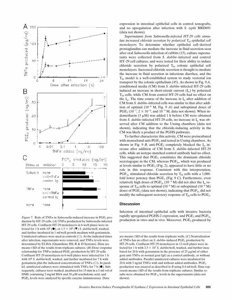
Release of TNFa after Salmonella infection of intestinal epi-thelial cells is not important for mediating increased PGE2 pro-duction. The studies above suggest that Salmonella-infectedHT-29 cells release an activity that induces uninfected cells inthe monolayer to express PGHS-2. Infection of colonic epithe-lial cell lines with invasive bacteria is known to induce the se-cretion of several proinflammatory cytokines, including IL-8and TNFa (32). Since the latter is a potent stimulus for in-creased PGHS-2 expression (Fig. 3), we tested the possibilitythat TNFa release is important for mediating increasedPGHS-2 expression in bacteria-infected intestinal epithelialcells.
As shown in Fig. 7 A, TNFa levels in the supernatants be-gan to increase within 1 h after S. dublin infection of HT-29cells, reached a maximum 2 h after infection, and remained in-creased thereafter. Maximal TNFa levels were dependent onthe bacterial dose, and were as high as 125 pg/ml after S. dublininfection of HT-29 cells (Fig. 7 A). This TNFa level, however,was 5–10-fold lower than would be required (e.g., 0.5–1 ng/ml)to mediate the increase in PGE2 production observed after S.dublin infection of HT-29 cells, as shown by a comparison ofthe dose–response curve for TNFa-induced PGE2 productionby HT-29 cells, with the PGE2 response observed after S. dub-
lin infection (Fig. 7 B). Furthermore, the dose–response rela-tionship for TNFa-induced PGE2 production was similar inuninfected and S. dublin–infected HT-29 cells, and no syner-gistic increase in PGE2 production was observed in cells dou-bly stimulated with optimal numbers of S. dublin and optimallevels of TNFa, indicating that S. dublin infection neither sen-sitized the cells nor potentiated the cellular response to TNFastimulation. Consistent with the interpretation that TNFa didnot mediate the PGE2 response to Salmonella infection, addi-tion of anti-TNFa had no effect on S. dublin–induced PGE2
production (Fig. 7 C). The identical concentration of anti-TNFa completely blocked the PGE2 response to 5 ng/ml TNFa(Fig. 7 C).
In addition to TNFa, infection of HT-29 cells with invasivebacteria also increases IL-1b mRNA levels (32). IL-1b levels,however, were extremely low after S. dublin infection ofHT-29 cells (, 3 pg/ml), and HT-29 cells do not express IL-1amRNA nor contain IL-1a (32, 44), suggesting that neitherIL-1a nor IL-1b were responsible for the increase in PGE2
production after Salmonella infection.Increased PGHS-2 expression by intestinal epithelial cells in
vivo in a human intestinal xenograft model infected with Salmo-nella. We next asked whether PGHS-2 can also be induced inintestinal epithelial cells in vivo. To infect acutely human intes-tinal epithelial cells with invasive bacteria, we used a humanfetal intestinal xenograft model where human fetal intestine(gestational age 10–14 wk) is transplanted subcutaneously intoSCID mice. The xenografts develop a fully differentiated epi-thelial layer of entirely human origin over a 10–20-wk period(41). Fully differentiated xenografts were infected intralumi-nally with an invasive S. typhi, and PGHS-2 expression in thexenografts was analyzed by quantitative RT-PCR and immuno-fluorescence. As shown in Table II, human PGHS-2 mRNAlevels increased . 40-fold after infection of the xenografts with
Table I. Increased PGE2 Production by HT-29 Colon Epithelial Cells in Response to Infection with Invasive Bacteria
Exp. Additions to culture Inoculum
Intracellular bacteria PGE2-produced
No./well % of inoculum ng/ml Ratio infected/control
1 S. dublin 9.2 3 107 7.3 3 106 7.9 7.0560.55* 54.2Y. enterocolitica 4.0 3 108 1.2 3 108 30.0 6.6660.24 51.2S. dysenteriae 3.8 3 108 4.0 3 106 1.1 2.2260.28 17.1None 0.1360.01 1.0
2 E. coli O29:NM (enteroinvasive) 6.4 3 108 7.2 3 106 1.1 2.2060.34 44.0E. coli O157 (enterohemorrhagic) 4.6 3 108 3.5 3 104 0.0076 0.1260.01 2.4E. coli O111 (enteropathogenic) 3.3 3 108 1.0 3 104 0.0030 0.1460.01 2.8E. coli DH5a (nonpathogenic) 2.4 3 108 3.6 3 103 0.0015 0.2360.03 4.6E. coli HB101 (nonpathogenic) 3.2 3 108 1.4 3 103 0.00044 0.2460.01 4.8LPS (10 mg/ml from E. coli O111) 0.0960.01 1.8None 0.0560.01 1.0
3 L. monocytogenes 7.5 3 107 1.9 3 107 25.3 1.0760.07 7.6S. bovis 9.0 3 107 1.9 3 104 0.021 0.1060.02 0.7None 0.1460.02 1.0
Confluent HT-29 monolayers in 6-well plates were infected for 1 h with the indicated doses of bacteria, washed, and further incubated for 6 h withgentamicin. Uninfected cultures were used as controls. Cultures were washed and incubated for 15 min in a 1-ml vol of DME containing 2 mg/mlBSA, and 20 mM arachidonic acid. PGE2 levels were analyzed by specific enzyme immunoassay. Monolayers were lysed in water, and Nos. of intracel-lular bacteria were determined by plating of serial dilutions on tryptic soy agar plates and overnight incubation. Exp., experiment. *Data representmean6SD of the results from triplicate cultures.

Invasive Bacteria Induce Prostaglandin H Synthase-2 Expression in Intestinal Epithelial Cells 303
Figure 5. Colocalization of invaded S. typhi BRD691 and increased PGHS-2 expression early after infection of HT-29 epithelial cells. HT-29 monolayers on coverslips in 24-well plates were infected for 1 h with S. typhi BRD691, washed, and further incubated with gentamicin for 4 h. Cells were fixed, and stained by indirect immunofluorescence for PGHS-2 (red) and Salmonella common structural antigen-1 (green), as de-scribed in Methods. Overlapping of red PGHS-2 staining and green Salmonella staining results in a yellow color. As shown in two representative examples, PGHS-2 expression is mostly observed in cells which also contain Salmonella. In addition, a few cells express PGHS-2 but do not con-tain bacteria. These cells are located predominantly in the immediate vicinity of Salmonella-infected cells. The scale bars represent 25 mm. Stain-ing for PGHS-1 yielded a weak signal, and PGHS-1 staining did not increase after S. typhi BRD691 infection (not shown).
Figure 8. Salmonella infection induces increased PGHS-2 expression in intestinal epithelial cells in a human intestinal xenograft model. Mature human fetal intestinal xenografts in SCID mice were infected for 6 h with S. typhi BRD691, and 8-mm frozen sections were prepared as described in Methods. Sections were stained with rabbit anti–PGHS-2 (green) and counterstained with propidium iodide (red). A and B are sections from uninfected control xenografts at two different magnifications, and C and D are from S. typhi–infected xenografts at two different magnifications. The scale bars in A and C represent 25 mm, and those in B and D represent 10 mm. Staining with a control antibody (normal rabbit serum) yielded no specific staining (not shown).

304 Eckmann et al.
live S. typhi, whereas infection with killed S. typhi, or a livenonpathogenic E. coli, increased human PGHS-2 mRNA lev-els less than fivefold.
Because PGHS-2 expression was analyzed early after infec-tion (6 h), at a time when Salmonella mostly reside inside epi-thelial cells, the RT-PCR data suggested that epithelial cellswere mostly responsible for the increase in PGHS-2 mRNA
expression in the xenografts. To document this directly, tissuesections were prepared and stained for PGHS-2 by indirect im-munofluorescence. As shown in Fig. 8, C and D, intestinal epi-thelial cells stained positively for PGHS-2 after S. typhi infec-tion of the xenograft, whereas little epithelial PGHS-2 stainingwas observed in uninfected control xenografts (Fig. 8, A andB). In contrast, staining for PGHS-1 showed weak constitutive
Table II. Increased Expression of Human PGHS-2 mRNA in Bacteria-infected Human Fetal Intestinal Xenografts
Additions
PGHS-2 b-actin
No. of mRNAmolecules/mg RNA
Ratioinfected/control
No. of mRNAmolecules/mg RNA
Ratioinfected/control
S. typhi BRD691 1.2 3 105 44.4 1.2 3 107 0.9S. typhi BRD691 (formalin-fixed)* 1.3 3 104 4.8 1.5 3 107 1.2E coli SC13 9.2 3 103 3.4 1.4 3 107 1.1None 2.7 3 103 1.0 1.3 3 107 1.0
Mature human fetal intestinal xenografts in SCID mice were infected with 5 3 107 bacteria in a 0.1-ml vol. Xenografts were removed 6 h later, totalRNA was extracted from individual xenografts, and equal amounts of RNA were pooled from four to five mice for each group. Levels of the mRNAsfor PGHS-2 and b-actin were determined by quantitative RT-PCR using internal RNA standards as described in Methods. *Bacteria were killed byincubation in 10% phosphate-buffered formalin (pH 7.2) for 30 min at room temperature. Killed bacteria were washed twice with PBS before injec-tion into xenografts.
Figure 6. PGHS-2 expression in S. typhi BRD691–infected HT-29 cells 24 h after infection. HT-29 monolayers on coverslips were infected with S. typhi BRD691, and incubated with gentamicin for 24 h. Parallel cultures were stimulated for 24 h with 20 ng/ml TNFa. Cells were fixed, and stained by indirect immunofluorescence for PGHS-2. (A) Control cells stained with rabbit anti–PGHS-2; (B) S. typhi–infected cells stained with rabbit anti–PGHS-2; (C) TNFa-stimulated cells stained with rabbit anti–PGHS-2; and (D) S. typhi–infected cells stained with a control antibody (rabbit antihuman CD3). The scale bar in A represents 25 mm.
Invasive Bacteria Induce Prostaglandin H Synthase-2 Expression in Intestinal Epithelial Cells 305
expression in intestinal epithelial cells in control xenografts,and no upregulation after infection with S. typhi BRD691(data not shown).
Supernatants from Salmonella-infected HT-29 cells stimu-late increased chloride secretion by polarized T84 epithelial cellmonolayers. To determine whether epithelial cell–derivedprostaglandins can mediate the increase in fluid secretion seenafter oral Salmonella infection of rabbits (15), culture superna-tants were collected from S. dublin–infected and controlHT-29 cell cultures, and were tested for their ability to inducechloride secretion by polarized T84 colonic epithelial cellmonolayers. Increased chloride secretion is thought to mediatethe increase in fluid secretion in infectious diarrhea, and theT84 model is a well-established system to study vectorial iontransport by the colonic epithelium (45). As shown in Fig. 9 A,conditioned media (CM) from S. dublin–infected HT-29 cellsinduced an increase in short-circuit current (Isc) by polarizedT84 cells, while CM from control HT-29 cells had no effect onthe Isc. The time course of the increase in Isc after addition ofCM from S. dublin–infected cells was similar to that after addi-tion of optimal (1026 M, Fig. 9 A) and suboptimal doses ofPGE2 (1027, 2 3 1028, and 1029 M, data not shown). When in-domethacin (3 mM) was added 1 h before CM were obtainedfrom S. dublin–infected HT-29 cells, no increase in Isc was ob-served after CM addition to the Ussing chambers (data notshown), indicating that the chloride-inducing activity in theCM was likely a product of the PGHS pathways.
To further characterize this activity, CM were preincubatedwith monoclonal anti-PGE2 and tested in Ussing chambers. Asshown in Fig. 9 B, anti-PGE2 completely blocked the Isc in-crease after addition of CM from S. dublin–infected HT-29cells, while an isotype-matched control antibody had no effect.This suggested that PGE2 constitutes the dominant chloridesecretagogue in the CM, whereas PGF2a, which was producedat levels similar to PGE2 (Fig. 2), appeared to have little or norole in this response. Consistent with this interpretation,PGF2a stimulated chloride secretion by T84 cells with a 1,000-fold lower potency than PGE2 (Fig. 9 C). Furthermore, evenrelatively high doses of PGF2a (1024 M) did not alter the Isc re-sponse of T84 cells to optimal (1026 M) or suboptimal (1028 M)doses of PGE2 (data not shown), indicating that PGF2a did notmodify the subsequent secretory response of T84 cells to PGE2.
Discussion
Infection of intestinal epithelial cells with invasive bacteriarapidly upregulated PGHS-2 expression, and PGE2 and PGF2a
production in vitro and in vivo. Moreover, PGE2 produced byFigure 7. Role of TNFa in Salmonella-induced increase in PGE2 pro-duction by HT-29 cells. (A) TNFa production by Salmonella-infected HT-29 cells. Confluent HT-29 monolayers in 6-well plates were in-fected for 1 h with 108 (d), or 1.5 3 106 (.) S. dublin/well, washed, and further incubated in 1 ml/well growth medium with gentamicin. Uninfected cultures were used as controls (s). At the indicated times after infection, supernatants were removed, and TNFa levels were determined by ELISA (Quantikine HS; R & D Systems). Data are means6SD of the results from triplicate cultures. (B) Dose–response relationship for TNFa-induced PGE2 production by HT-29 cells. Confluent HT-29 monolayers in 6-well plates were infected for 1 h with 108 S. dublin/well, washed, and further incubated for 7 h with gentamicin plus the indicated concentrations of TNFa (s). In paral-lel, uninfected cultures were stimulated with TNFa for 7 h (d). Sub-sequently, cultures were washed, incubated for 15 min in a 1-ml vol of DME containing 2 mg/ml BSA and 20 mM arachidonic acid, and PGE2 levels were analyzed by specific enzyme immunoassay. Data
are means6SD of the results from triplicate wells. (C) Neutralization of TNFa has no effect on S. dublin–induced PGE2 production byHT-29 cells. Confluent HT-29 monolayers in 12-well plates were in-fected for 1 h with 2.5 3 107 S. dublin/well, washed, and further incu-bated for 20 h with gentamicin in the presence of 25 mg/ml of either goat anti-TNFa or normal goat IgG as a control antibody, or without added antibodies. Parallel uninfected cultures were incubated for20 h with 5 ng/ml TNFa with and without added antibodies. PGE2 production was assayed as described in B using 0.4 ml/well. Data rep-resent means6SD of the results from triplicate cultures. Similar re-sults were obtained for PGF2a levels in the supernatants (data not shown).

306 Eckmann et al.
bacteria-infected intestinal epithelial cells constituted the pre-dominant activity capable of inducing chloride secretion by po-larized intestinal epithelial cell monolayers. The rapid induc-tion of epithelial PGE2 production after bacterial entry couldprovide a mechanistic explanation for previous observations inanimal models that infection of the intestine with invasive bac-teria, including Salmonella and Shigella, rapidly induced in-creased fluid secretion mediated by products of the PGHSpathways (15, 21, 22). This possibility is supported by the timecourse of the observed host secretory response in the presentstudy, since increased fluid secretion after Salmonella infectionoccurred within 4 h after infection in previous studies (15), atime consistent with the present finding of increased epithelialPGE2 production within 3 h after infection. Furthermore, in-creased PGE2 secretion by polarized intestinal epithelial cellswas reported to occur predominantly from the basolateral sur-face of the cells (30), which is consistent with the previous ob-servation that increased epithelial chloride secretion was mostefficiently elicited by basolateral stimulation of polarized in-testinal epithelial cells with prostaglandins (18). Although cellsin the underlying mucosa, such as macrophages, can also re-spond to bacteria with increased PGE2 production (46), it isunlikely that bacterial penetration of the epithelium andinduction of prostaglandin production by these cells occurssufficiently rapidly to account for the early phase of the hostsecretory response to bacterial infection. Nonetheless, the pro-duction of PGE2 (and possibly other prostaglandins) by cellsother than intestinal epithelial cells likely contributes to thehost secretory response at later stages of the infection.
Prostaglandins can induce diarrhea in humans. Thus, ad-ministration of PGE analogues caused increased fluid secre-tion in volunteers (19, 47), and diarrhea caused by several non-infectious causes, including radiation therapy, was amelioratedby PGHS inhibitors (20). The role of prostaglandins in humaninfections with invasive bacteria, however, has not been estab-lished, although one report suggested that treatment of infantsand young children with the PGHS inhibitor aspirin decreasedintestinal fluid loss after infection with several enteric patho-gens, including Salmonella (48). The apparent lack of a largernumber of clinical reports linking the use of PGHS inhibitorswith improvements of bacterially induced diarrhea may be re-lated to the pleiotropic functions of prostaglandins in the intes-tinal tract. These include the induction of epithelial ion secre-tion and diarrhea (16, 19, 47) and also promotion of epithelial
Figure 9. Supernatants from Salmonella-infected HT-29 cells induce chloride secretion by polarized T84 colon epithelial cell monolayers. (A) Time course of increased Isc after addition of CM from S. dublin–infected HT-29 cells. CM from S. dublin–infected (d) and control HT-29 cultures (.) were obtained as described in Methods. CM from S. dublin–infected cultures contained 36.5 and 57.0 ng/ml PGE2 and PGF2a, respectively. CM from control cultures contained , 0.5 ng/ml PGE2. Media containing 1.76 mg/ml (5 3 1026 M) PGE2 (s) or media alone (,) were used as controls. Polarized T84 cell monolayers were mounted in Ussing chambers, equilibrated at 378C, and background Isc readings were taken for at least 20 min. Stimuli were added at0 min on the basolateral side in a 1-ml vol to the 4-ml vol already present in the chamber (e.g., a 1:5 final dilution), and Isc readings were taken initially every 2 min, later every 5 min. Data are means6SEM from the results of three independent experiments. (B) Anti-PGE2 inhibits increased Isc after addition of CM from S. dublin–infected HT-29 cells. 200 ml of CM from S. dublin–infected cells, or
200 ml media containing 35 ng/ml (1027 M) PGE2 was mixed with800 ml PBS containing 75 mg monoclonal anti-PGE2 (IgG1 isotype), or 75 mg monoclonal mouse IgG1 (MOPC 31c) as a control, or no added antibody. The molar ratio of antibody to PGE2 was z 25:1. The mixtures were incubated for 20 min at room temperature, added to the basolateral side of polarized T84 cells mounted in Ussing cham-bers, and Isc recordings were taken, as described in A. Isc values from unstimulated control T84 monolayers were subtracted. Data are means6SEM of the peak Isc values obtained in three separate experi-ments. (C) Dose–response relationships for prostaglandin-induced Isc in polarized T84 cells. Polarized T84 cells were mounted in Ussing chambers, stimulated with the indicated doses of PGE2 (d) or PGF2a (s), and peak Isc values were determined. Isc values from unstimu-lated control T84 monolayers were subtracted. Each point represents a single measurement. Data from three separate experiments are shown.

Invasive Bacteria Induce Prostaglandin H Synthase-2 Expression in Intestinal Epithelial Cells 307
wound healing (49, 50). The former would be prevented byPGHS inhibitors, while inhibition of the latter by PGHS inhib-itors could lead to the exacerbation of epithelial injury, whichwould be likely to promote diarrhea.
Epithelial-derived prostaglandins and epithelial PGHS-2expression may have functions during the host response tobacterial infection other than increasing fluid secretion.PGHS-2 expression is induced in other models of gastrointesti-nal injury, including TNBS-induced colitis in the rat (50) andgastric ulcer models in mice (51, 52), although PGHS-2 expres-sion was not localized to epithelial cells in these studies. Prod-ucts of the PGHS-2 pathway appeared to be protective inthese models, since PGHS-2 inhibitors delayed wound healingin the ulcer model, and exacerbated injury in TNBS-inducedcolitis (50, 51). These observations raise the possibility thatPGHS inhibitors might, under some circumstances, exacerbatemucosal injury after infection with invasive bacteria. It is notclear how prostaglandins exert mucosa-protective effects (49),but they are known to downregulate the production of someproinflammatory cytokines, such as IL-1, in macrophages (53).Thus, prostaglandins derived from the epithelium early in in-fection could function to antagonize the action of proinflam-matory cytokines released from epithelial and inflammatorycells in response to infection with pathogenic bacteria. An-other possible mechanism by which increased epithelialPGHS-2 expression and prostaglandin production could pro-tect against mucosal injury is suggested by the mitogenic andantiapoptotic activity of PGHS-2 expressed in epithelial cells(30, 54). Rapid epithelial reconstitution is likely to be impor-tant for reducing mucosal access of inflammatory luminal com-pounds (e.g., LPS) after bacterially induced damage to the epi-thelium. Consistent with a mitogenic function of epithelialPGHS-2 expression, increased epithelial proliferation wasseen rapidly after Salmonella infection of human fetal intesti-nal xenografts transplanted onto SCID mice (Savidge, T.C.,unpublished observation). Other roles of epithelial cell-derivedprostaglandins could involve the induction of mucin secretionfrom epithelial cells as a protective response against furtherbacterial invasion (55), and an increase in mucosal blood flowallowing increased numbers of inflammatory cells to reach thesite of infection (56).
PGHS-2 was not constitutively expressed in intestinal epi-thelial cells, but increased rapidly after bacterial invasion. Incontrast, PGHS-1 was constitutively expressed at low levels,and expression was not affected by bacterial infection. Thesedata suggest that PGHS-2 is likely to be more important thanPGHS-1 in mediating specific aspects of the host response tobacterial invasion. In contrast, PGHS-1 appears to be moreimportant under normal conditions, since PGHS-1 is constitu-tively expressed in the normal intestinal tract, while PGHS-2levels are low (57), but detectable with sensitive methods (58).PGHS-1 may also be more important than PGHS-2 in the hostresponse to radiation injury (39). The respective roles of thetwo PGHS isoforms in the intestinal tract, however, remain tobe established. Data from mice with targeted deletions of thetwo PGHS isoforms suggest that neither is absolutely requiredfor normal intestinal function, since the knock-out mice had noapparent pathological changes in the gastrointestinal tract (24–26). It is unknown, however, whether these mice show abnor-mal gastrointestinal changes in response to mucosal injury orchallenge with pathogenic enteric bacteria.
Increased epithelial PGHS-2 expression after bacterial in-
fection initially resulted from the direct interaction of invadingbacteria with the cells, and later was amplified by indirect ef-fects, since cells not invaded by bacteria also expressed in-creased PGHS-2 levels, and supernatants from infected cellscould stimulate increased PGE2 production by uninfectedcells. Both mechanisms probably also act in vivo, since the pro-portion of intestinal epithelial cells in the xenografts that ex-pressed increased PGHS-2 levels after Salmonella infection islikely to be greater than the fraction of cells infected by Salmo-nella. Even after in vitro infection of intestinal epithelial cellswith relatively high doses of Salmonella, a substantial fractionof cells remains uninfected (data not shown), and electron mi-croscopic observations in experimentally infected intestinalloops suggested that Salmonella localized to a small proportionof intestinal epithelial cells (9). The identity of the PGHS-2–inducing activity is currently unknown, but based on thepresent studies it is unlikely to be TNFa or IL-1. Possible can-didates for this activity are TGFa and amphiregulin, both ofwhich are expressed by intestinal epithelial cells, and can in-duce epithelial PGE2 production (29, 30).
In addition to PGHS-2, intestinal epithelial cells upregulategenes that encode chemokines (e.g., IL-8, MCP-1), proinflam-matory cytokines (TNFa, GM-CSF), and adhesion molecules(ICAM-1) in response to infection with invasive bacteria (31–33, 38). The mechanisms underlying the coordinate expressionof these genes are not known, but probably involve activationof gene transcription, since expression of reporter constructsfor at least one of these genes, IL-8, was upregulated after bac-terial invasion (Eckmann, L., unpublished data). Consistentwith this notion, the stability of PGHS-2 mRNA was not al-tered after Salmonella infection of I407 epithelial cells (datanot shown). Genes of the epithelial proinflammatory programshare common features in their promoter regions, namely thepresence of DNA-binding regions for the NF-kB transcriptionfactor complex, raising the possibility that this complex is acti-vated after infection with invasive bacteria. Consistent withthis hypothesis, increased NF-kB DNA binding activity was re-ported in HeLa epithelial cells after infection with the invasivebacteria S. flexneri (59). Although such a common regulatorymechanism could account for the coordinate induction of theepithelial proinflammatory program, additional mechanismsare probably important for regulating individual genes of theprogram. For example, epithelial PGHS-2 induction after bac-terial invasion was, in part, mediated by a released factor asshown herein, whereas such a paracrine-acting factor playedlittle or no role in the upregulation of epithelial ICAM-1 ex-pression after bacterial invasion (38).
The ability of the infecting bacteria to invade cells was animportant determinant for the epithelial prostaglandin re-sponse, since invasive bacteria induced far greater increases inPGE2 production than noninvasive bacteria. Infection withgram-negative, but not gram-positive, noninvasive bacteria,however, induced a small but significant increase in epithelialPGE2 production. This increase shows that bacterial proper-ties other than the ability to invade cells contribute to the epi-thelial prostaglandin response, albeit to a lesser degree thaninvasiveness. Data obtained in the in vivo model of normal hu-man intestinal epithelial cells suggested that components ofthe gram-negative cell wall were responsible for this response,since killed (and therefore noninvasive) S. typhi and a livenoninvasive E. coli were equally efficient in inducing epithelialPGHS-2 expression. Bacterial LPS may be one of the cell wall

308 Eckmann et al.
components responsible for the increased epithelial expressionof PGHS-2, as well as other proinflammatory genes (60). Theintestinal epithelial cell PGE2 response to LPS, however, wasless than that to infection with gram-negative noninvasive bac-teria, suggesting that cell wall components other than LPScould be important for this response. Such was the case alsowith gram-negative bacterial infections of bladder and lungepithelial cell cultures (61, 62). In any case, a low-level epithe-lial prostaglandin response to noninvasive enteric bacteriapresent in the normal luminal flora, such as nonpathogenic E.coli, suggests the concept that intestinal epithelial cells sensethe presence of the normal luminal flora, and in response com-municate with other epithelial cells and cells in the underlyingmucosa. Such an epithelial response could contribute to thephysiologic inflammation present in conventional, but notgerm-free animals (63–66).
Acknowledgments
We thank Jane Smitham and Sharon Okamoto for expert technicalhelp, and Dr. J. Portanova for providing monoclonal anti-PGE2.
L. Eckmann is a recipient of a Career Development Award fromthe Crohn’s and Colitis Foundation of America. D.C. Lowe is anMRC-Collaborative Student. This work was supported by NationalInstitutes of Health grants DK35108 (M.F. Kagnoff), DK33165 (W.F.Stenson), DK28305 and DK47756 (K.E. Barrett), and the Biotech-nology and Biological Sciences Research Council (T.C. Savidge).
References
1. Chalker, R.B., and M.J. Blaser. 1988. A review of human salmonellosis.III. Magnitude of Salmonella infection in the United States. Rev. Infect. Dis. 10:111–124.
2. Centers for Disease Control. 1990. Foodborne disease outbreaks, a 5 yearsummary, 1983–87. MMWR (Morb. Mortal. Wkly Rep.). 39:15–57.
3. Stutman, H.R. 1994. Salmonella, Shigella, and Campylobacter: commonbacterial causes of infectious diarrhea. Pediatr. Ann. 23:538–543.
4. Teka, T., A.S. Faruque, and G.J. Fuchs. 1996. Risk factors for deaths inunder-age-five children attending a diarrhoea treatment centre. Acta Paediatr.85:1070–1075.
5. DuPont, H.L. 1995. Diarrheal diseases in the developing world. Infect.Dis. Clin. N. Am. 9:313–324.
6. Asakura, H., and M. Yoshioka. 1994. Cholera toxin and diarrhoea. J.Gastroenterol. Hepatol. 9:186–193.
7. Spangler, B.D. 1992. Structure and function of cholera toxin and the re-lated Escherichia coli heat-labile enterotoxins. Microbiol. Rev. 56:622–647.
8. Parsot, C., and P.J. Sansonetti. 1996. Invasion and the pathogenesis ofShigella infections. Curr. Top. Microbiol. Immunol. 209:25–42.
9. Jones, B.D., N. Ghori, and S. Falkow. 1994. Salmonella typhimurium ini-tiates murine infection by penetrating and destroying the specialized epithelialM cells of the Peyer’s patches. J. Exp. Med. 180:15–23.
10. Galan, J.E. 1996. Molecular and cellular bases of Salmonella entry intohost cells. Curr. Top. Microbiol. Immunol. 209:43–60.
11. Isberg, R.R. 1996. Uptake of enteropathogenic Yersinia by mammaliancells. Curr. Top. Microbiol. Immunol. 209:1–24.
12. Galan, J.E., and R. Curtiss. 1989. Cloning and molecular characteriza-tion of genes whose products allow Salmonella typhimurium to penetrate tissueculture cells. Proc. Natl. Acad. Sci. USA. 86:6383–6387.
13. Wallis, T.S., W.G. Starkey, J. Stephen, S.J. Haddon, M.P. Osborne, andD.C. Candy. 1986. Enterotoxin production by Salmonella typhimurium strainsof different virulence. J. Med. Microbiol. 21:19–23.
14. Chopra, A.K., J.W. Peterson, P. Chary, and R. Prasad. 1994. Molecularcharacterization of an enterotoxin from Salmonella typhimurium. Microb.Pathog. 16:85–98.
15. Giannella, R.A., S.B. Formal, G.J. Dammin, and H. Collins. 1973.Pathogenesis of Salmonellosis. Studies of fluid secretion, mucosal invasion, andmorphologic reactions in the rabbit ileum. J. Clin. Invest. 52:441–453.
16. Eberhart, C.E., and R.N. DuBois. 1995. Eicosanoids and the gas-trointestinal tract. Gastroenterology. 109:285–301.
17. Appleton, I., A. Tomlinson, and D.A. Willoughby. 1996. Induction ofcyclo-oxygenase and nitric oxide synthase in inflammation. Adv. Pharmacol. 35:27–78.
18. Weymer, A., P. Huott, W. Liu, J.A. McRoberts, and K. Dharm-
sathaphorn. 1985. Chloride secretory mechanism induced by prostaglandin E1in a colonic epithelial cell line. J. Clin. Invest. 76:1828–1836.
19. Lanza, F.L., R.L. Kochman, G.S. Geis, E.M. Rack, and L.G. Deysach.1991. A double-blind, placebo-controlled, 6-day evaluation of two doses of mi-soprostol in gastroduodenal mucosal protection against damage from aspirinand effects on bowel habits. Am. J. Gastroenterol. 86:1743–1748.
20. Mennie, A.T., and V. Dalley. 1973. Aspirin in radiation-induced diar-rhea. Lancet. i:1131.
21. Gots, R.E., S.B. Formal, and R.A. Giannella. 1974. Indomethacin inhi-bition of Salmonella typhimurium, Shigella flexneri, and cholera-mediated rab-bit ileal secretion. J. Infect. Dis. 130:280–284.
22. Giannella, R.A., W.R. Rout, and S.B. Formal. 1977. Effect of in-domethacin on intestinal water transport in Salmonella-infected rhesus mon-keys. Infect. Immun. 17:136–139.
23. Smith, W.L., and D.L. DeWitt. 1996. Prostaglandin endoperoxidase Hsynthases-1 and -2. Adv. Immunol. 62:167–215.
24. Morham, S.G., R. Langenbach, C.D. Loftin, H.F. Tiano, N. Voulou-manos, J.C. Jennette, J.F. Mahler, K.D. Kluckman, A. Ledford, and C.A. Lee.1995. Prostaglandin synthase 2 gene disruption causes severe renal pathology inthe mouse. Cell. 83:473–482.
25. Dinchuk, J.E., B.D. Car, R.J. Focht, J.J. Johnston, B.D. Jaffee, M.B.Covington, N.R. Contel, V.M. Eng, R.J. Collins, and P.M. Czerniak. 1995. Re-nal abnormalities and an altered inflammatory response in mice lacking cy-clooxygenase II. Nature (Lond.). 378:406–409.
26. Langenbach, R., S.G. Morham, H.F. Tiano, C.D. Loftin, B.I. Gha-nayem, P.C. Chulada, J.F. Mahler, C.A. Lee, E.H. Gouldin, and K.D. Kluck-man. 1995. Prostaglandin synthase 1 gene disruption in mice reduces arachi-donic acid-induced inflammation and indomethacin-induced gastric ulceration.Cell. 83:483–492.
27. Harris, R.C., J.A. McKanna, Y. Akai, H.R. Jacobson, R.N. DuBois, andM.D. Breyer. 1994. Cyclooxygenase-2 is associated with the macula densa of ratkidney and increases with salt restriction. J. Clin. Invest. 94:2504–2510.
28. Holtzman, M.J. 1992. Arachidonic acid metabolism in airway epithelialcells. Annu. Rev. Physiol. 54:303–329.
29. DuBois, R.N., J. Awad, J. Morrow, L.J. Roberts, and P.R. Bishop. 1994.Regulation of eicosanoid production and mitogenesis in rat intestinal epithelialcells by transforming growth factor-a and phorbol ester. J. Clin. Invest. 93:493–498.
30. Coffey, R.J., C.J. Hawkey, L. Damstrup, R. Graves-Deal, V.C. Daniel,P.J. Dempsey, R. Chinery, S.C. Kirkland, R.N. DuBois, T.L. Jetton, and J.D.Morrow. 1997. Epidermal growth factor receptor activation induces nuclear tar-geting of cyclooxygenase-2, basolateral release of prostaglandins, and mitogen-esis in polarizing colon cancer cells. Proc. Natl. Acad. Sci. USA. 94:657–662.
31. Eckmann, L., M.F. Kagnoff, and J. Fierer. 1993. Epithelial cells secretethe chemokine interleukin-8 in response to bacterial entry. Infect. Immun. 61:4569–4574.
32. Jung, H.C., L. Eckmann, S.K. Yang, A. Panja, J. Fierer, E. Morzycka-Wroblewska, and M.F. Kagnoff. 1995. A distinct array of proinflammatory cy-tokines is expressed in human colon epithelial cells in response to bacterial in-vasion. J. Clin. Invest. 95:55–65.
33. McCormick, B.A., S.P. Colgan, C. Delp-Archer, S.I. Miller, and J.L.Madara. 1993. Salmonella typhimurium attachment to human intestinal epithe-lial monolayers: transcellular signalling to subepithelial neutrophils. J. Cell Biol.123:895–907.
34. Barrett, K.E., and T.D. Bigby. 1993. Involvement of arachidonic acid inthe chloride secretory response of intestinal epithelial cells. Am. J. Physiol. 264:C446–C452.
35. Chatfield, S.N., N. Fairweather, I. Charles, D. Pickard, M. Levine, D.Hone, M. Posada, R.A. Strugnell, and G. Dougan. 1992. Construction of a ge-netically defined Salmonella typhi Ty2 aroA, aroC mutant for the engineeringof a candidate oral typhoid-tetanus vaccine. Vaccine. 10:53–60.
36. Hicks, S., D.C. Candy, and A.D. Phillips. 1996. Adhesion of enteroag-gregative Escherichia coli to pediatric intestinal mucosa in vitro. Infect. Immun.64:4751–4760.
37. Mnich, S.J., A.W. Veenhuizen, J.B. Monahan, K.C. Sheehan, K.R.Lynch, P.C. Isakson, and J.P. Portanova. 1995. Characterization of a mono-clonal antibody that neutralizes the activity of prostaglandin E2. J. Immunol.155:4437–4444.
38. Huang, G.T., L. Eckmann, T.C. Savidge, and M.F. Kagnoff. 1996. Infec-tion of human intestinal epithelial cells with invasive bacteria upregulates apicalintercellular adhesion molecule-1 (ICAM-1) expression and neutrophil adhe-sion. J. Clin. Invest. 98:572–583.
39. Cohn, S.M., S. Schloemann, T. Tessner, K. Seibert, and W.F. Stenson.1997. Crypt stem cell survival in the mouse intestinal epithelium is regulated byprostaglandins synthesized through cyclooxygenase-1. J. Clin. Invest. 99:1367–1379.
40. Eckmann, L., G.T. Huang, J.R. Smith, E. Morzycka-Wroblewska, andM.F. Kagnoff. 1994. Increased transcription and coordinate stabilization ofmRNAs for secreted immunoglobulin a heavy chain and k light chain followingstimulation of immunoglobulin A expressing B cells. J. Biol. Chem. 269:33102–33108.
41. Savidge, T.C., A.L. Morey, D.J. Ferguson, K.A. Fleming, A.N. Shma-

Invasive Bacteria Induce Prostaglandin H Synthase-2 Expression in Intestinal Epithelial Cells 309
kow, and A.D. Phillips. 1995. Human intestinal development in a severe-com-bined immunodeficient xenograft model. Differentiation. 58:361–371.
42. Futaki, N., S. Takahashi, M. Yokoyama, I. Arai, S. Higuchi, and S.Otomo. 1994. NS-398, a new anti-inflammatory, selectively inhibits prostaglan-din G/H synthase/cyclooxygenase (COX-2) activity in vitro. Prostaglandins. 47:55–59.
43. Bhattacharyya, D.K., M. Lecomte, J. Dunn, D.J. Morgans, and W.L.Smith. 1995. Selective inhibition of prostaglandin endoperoxidase synthase-1(cyclooxygenase-1) by valerylsalicylic acid. Arch. Biochem. Biophys. 317:19–24.
44. Eckmann, L., S.L. Reed, J.R. Smith, and M.F. Kagnoff. 1995. Enta-moeba histolytica trophozoites induce an inflammatory cytokine response bycultured human cells through the paracrine action of cytolytically released in-terleukin-1a. J. Clin. Invest. 96:1269–1279.
45. McRoberts, J.A., and K.E. Barrett. 1989. Hormone-regulated ion trans-port in T84 colonic cells. In Functional Epithelial Cells In Culture. K.S. Matlinand J.D. Valentich, editors. Alan R. Liss, Inc., New York. 235–265.
46. Yusof, W.N.W., M. Nagaratnam, C.L. Koh, S. Puthucheary, and T.Pang. 1993. Release of prostaglandin E2 by human mononuclear cells exposedto heat-killed Salmonella typhi. Microbiol. Immunol. 37:667–670.
47. Matuchansky, C., J.Y. Mary, and J.J. Bernier. 1976. Further studies onprostaglandin E1-induced jejunal secretion of water and electrolytes in man,with special reference to the influence of ethacrynic acid, furosemide, and aspi-rin. Gastroenterology. 71:274–281.
48. Gracey, M., M.A. Phadke, V. Burke, S.K. Raut, and B. Singh. 1984. As-pirin in acute gastroenteritis: a clinical and microbiological study. J. Pediatr.Gastroenterol. Nutr. 3:692–695.
49. Wallace, J.L., and A.W. Tigley. 1995. Review article: new insights intoprostaglandins and mucosal defence. Aliment. Pharmacol. Ther. 9:227–235.
50. Reuter, B.K., S. Asfaha, A. Buret, K.A. Sharkey, and J.L. Wallace.1996. Exacerbation of inflammation-associated colonic injury in rat through in-hibition of cyclooxygenase-2. J. Clin. Invest. 98:2076–2085.
51. Mizuno, H., C. Sakamoto, K. Matsuda, K. Wada, T. Uchida, H. Nogu-chi, T. Akamatsu, and M. Kasuga. 1997. Induction of cyclooxygenase 2 in gas-tric mucosal lesions and its inhibition by the specific antagonist delays healingin mice. Gastroenterology. 112:387–397.
52. Stenson, W.F. 1997. Cyclooxygenase 2 and wound healing in the stom-ach. Gastroenterology. 112:645–648.
53. Knudsen, P.J., C.A. Dinarello, and T.B. Strom. 1986. Prostaglandinsposttranscriptionally inhibit monocyte expression of interleukin 1 activity by in-creasing intracellular cyclic adenosine monophosphate. J. Immunol. 137:3189–3194.
54. Tsujii, M., and R.N. DuBois. 1995. Alterations in cellular adhesion and
apoptosis in epithelial cells overexpressing prostaglandin endoperoxidase syn-thase 2. Cell. 83:493–501.
55. McCool, D.J., M.A. Marcon, J.F. Forstner, and G.G. Forstner. 1990.The T84 human colonic adenocarcinoma cell line produces mucin in cultureand releases it in response to various secretagogues. Biochem. J. 267:491–500.
56. Brown, J.A., and R.D. Zipser. 1987. Prostaglandin regulation of colonicblood flow in rabbit colitis. Gastroenterology. 92:54–59.
57. Kargman, S., S. Charleson, M. Cartwright, J. Frank, D. Riendau, J. Man-cini, J. Evans, and G. O’Neill. 1996. Characterization of prostaglandin G/H/ syn-thase 1 and 2 in rat, dog, monkey, and human gastrointestinal tracts. Gastroen-terology. 111:445–454.
58. O’Neill, G.P., and A.W. Ford-Hutchinson. 1993. Expression of mRNAfor cyclooxygenase-1 and cyclooxygenase-2 in human tissues. FEBS (Fed. Eur.Biochem. Soc.) Lett. 330:156–160.
59. Dyer, R.B, C.R. Collaco, D.W. Niesel, and N.K. Herzog. 1993. Shigellaflexneri invasion of HeLa cells induces NF-kB DNA-binding activity. Infect.Immun. 61:4427–4433.
60. Eckmann, L., H.C. Jung, C. Schurer-Maly, A. Panja, E. Morzycka-Wroblewska, and M.F. Kagnoff. 1993. Differential cytokine expression by hu-man intestinal epithelial cell lines: regulated expression of interleukin 8. Gas-troenterology. 105:1689–1697.
61. Hedges, S., M. Svensson, and C. Svanborg. 1992. Interleukin-6 responseof epithelial cell lines to bacterial stimulation in vitro. Infect. Immun. 60:1295–1301.
62. DiMango, E., H.J. Zar, R. Bryan, and A. Prince. 1995. DiversePseudomonas aeruginosa gene products stimulate respiratory epithelial cells toproduce interleukin-8. J. Clin. Invest. 96:2204–2210.
63. Van Oudenaren, A., J.J. Haaijman, and R. Benner. 1984. Frequencies ofbackground cytoplasmic Ig-containing cells in various lymphoid organs of athy-mic and euthymic mice as a function of age and immune status. Immunology.51:735–742.
64. Klaasen, H.L., P.J. Van der Heijden, W. Stok, F.G. Poelma, J.P. Koop-man, M.E. Van den Brink, M.H. Bakker, W.M. Eling, and A.C. Beynen. 1993.Apathogenic, intestinal, segmented, filamentous bacteria stimulate the mucosalimmune system of mice. Infect. Immun. 61:303–306.
65. Meslin, J.C., J.M. Wal, and V. Rochet. 1990. Histamine and mast celldistribution in the intestinal wall of the germ free and conventional rats. Influ-ence of the mode of sterilization of the diet. Agents Actions. 29:131–137.
66. Umesaki, Y., H. Setoyama, S. Matsumoto, and Y. Okada. 1993. Expan-sion of alpha beta T-cell receptor-bearing intestinal intraepithelial lymphocytesafter microbial colonization in germ-free mice and its independence from thy-mus. Immunology. 79:32–37.
Copyright © 2022 FDOKUMEN