Bacterial biopolymers: from pathogenesis to advanced materials
Upload
independentCategory
view
3download
0
COMPREHENSIVE INVITED REVIEW
Amyotrophic Lateral Sclerosis Pathogenesis:A Journey Through the Secretory Pathway
Melissa Nassif,1,2 Soledad Matus,1,2 Karen Castillo,1,2 and Claudio Hetz1–4
Abstract
Amyotrophic lateral sclerosis (ALS) is the most common adult-onset motoneuron degenerative disease charac-terized by the selective loss of motoneurons in the spinal ventral horn, most brainstem nuclei, and the cerebralcortex. Although approximately 90% of ALS cases are sporadic (sALS), analyses of familial ALS (fALS)-causativegenes have generated relevant insight into molecular events involved in the pathology. Here we overview anemerging concept indicating the occurrence of secretory pathway stress in the disease process. These alterationsinclude a failure in the protein folding machinery at the endoplasmic reticulum (ER), engagement of the unfoldedprotein response (UPR), modifications of the Golgi apparatus network, impaired vesicular trafficking, inhibition ofprotein quality control mechanisms, oxidative damage to ER proteins, and sustained activation of degradativepathways such as autophagy. A common feature predicted for most of these alterations is abnormal proteinhomeostasis associated with the accumulation of misfolded proteins at the ER, possibly leading to chronic ERstress and neuronal dysfunction. Signs of ER stress are observed even during presymptomatic stages in fALSmouse models, and pharmacological strategies to alleviate protein misfolding slow disease progression. Becausethe secretory pathway stress occurs in both sALS and several forms of fALS, it may offer a unique common targetfor possible therapeutic strategies to treat this devastating disease. Antioxid. Redox Signal. 13, 1955–1989.
I. Introduction 1956II. First Stop: The Endoplasmic Reticulum 1957
A. Endoplasmic reticulum: Primary functions 1957B. An emergency exit from ER: The ERAD pathway 1957C. Mild ER stress and the unfolded protein response: Adaptive phase 1957D. Chronic ER stress and the unfolded protein response: Apoptosis phase 1959E. ER alterations in sALS 1960F. ER abnormalities in fALS induced by SOD1 mutations 1960G. Chronic ER stress and apoptosis in fALS 1962H. Blocking the first exit: ERAD dysfunction in fALS 1963I. ER stress in fALS induced by VAP mutations 1964J. TDP-43, FUS=TLS, and abnormal mRNA metabolism in ALS 1964
K. Oxidative protein damage in ALS 1965III. Second Stop: Golgi Apparatus 1967
A. Golgi apparatus: Main functions 1967B. GA fragmentation in sALS 1967C. GA alterations in fALS -induced by SOD1 mutations 1968D. A nonclassical secretory pathway for mutant SOD1 1968E. GA disturbance and the Wobbler mouse 1969F. GA alterations in fALS induced by dynactin mutations 1969
1Program of Cellular and Molecular Biology, Institute of Biomedical Sciences, Faculty of Medicine, NEMO Millennium Nucleus, Santiago,Chile.
2The FONDAP Center for Molecular Studies of the Cell, University of Chile, Santiago, Chile.3NeuroUnion Biomedical Foundation, Santiago, Chile.4Department of Immunology and Infectious Diseases, Harvard School of Public Health, Boston, Massachusetts.
Reviewing Editors: Jeffrey N. Agar, Vittorio Calabrese, Lavinia Cantoni, Maria Teresa Carri, Nikolay V. Dokholyan, Alvaro
Estevez, Jordi Magrane, Lee J. Martin, Gabriele Siciliano, and Ivan Spasojevic
ANTIOXIDANTS & REDOX SIGNALINGVolume 13, Number 12, 2010ª Mary Ann Liebert, Inc.DOI: 10.1089=ars.2009.2991
1955

IV. Third Stop: Endocytosis and Vesicle Transport 1969A. Endocytosis and multivesicular endosomes 1969B. Multivesicular endosomes in fALS 1970C. Vesicle transport and fALS: Alsin 1970D. Vesicle transport and fALS: Dynactin 1971
V. Final Stop: Autophagy 1971A. Autophagy pathways 1971B. Regulation of autophagy 1971C. Autophagy in fALS 1974D. A role of Fig4 mutations in autophagy and ALS 1974E. Homeostatic balance between autophagy and the UPR in ALS 1974F. Autophagy in sALS 1974G. Autophagy as a therapeutic target to treat ALS? 1975
VI. Conclusion 1976
I. Introduction
Amyotrophic lateral sclerosis (ALS), also known asLou Gehrig’s disease, is the most common adult-onset
motoneuron neurodegenerative disease characterized bymuscle weakness, atrophy, and paralysis. Premature death isoften observed as a result of respiratory muscle failure. Thepathological hallmark of ALS is the selective degeneration ofupper and lower motoneurons projecting from the spinalcord, brainstem, and cortex (268). The average disease onsetoccurs around age 50, with an incidence estimated of 1–2 casesper 100,000 individuals, where men are more frequently af-fected than women (243). Approximately 90% of ALS casesare referred to as sporadic (sALS), lacking a clear geneticcomponent. Ten percent of the remaining cases are familial(fALS) and many disease-causative genes have been identi-fied by linkage analysis and positional cloning (examples in35, 63, 94, 136, 160, 243, 327). Those genes include: Als1, Als2,Vapb, Chmp2b, Dctn1, Tardbp, Fus, and Fig4 which encodefor superoxide dismutase-1 (ALS1=SOD1), alsin (ALS2),VAMP-associated protein B (VAPB), charged multivesicularbody protein 2 B (CHMP2B), dynactin (DCTN1), TAR DNA-binding protein 43 (TDP-43), fused in sarcoma protein (FUS),and lipid phosphatase (FIG4), respectively (Table 1).
There is currently no primary therapy for this disorder, andthe only available drug for treatment is an antiglutamatergiccompound, riluzole, which has a marginal effect on improv-ing symptoms and only extends lifespan for a few months(14). Several pharmacological agents, including caspase in-hibitor zVAD-fmk, creatine, minocycline, and celecoxib havebeen tested in animal models of ALS, showing promising ef-fects, but with no effects on human patient trials (42, 92, 267).Other drugs such as arimoclomol (43) or talampanel (240) arecurrently being tested in clinical trials as potential therapy forALS (5). An area of active therapeutic research is focused onfALS cases caused by dominant mutation in the gene encod-ing SOD1. Experimental strategies to knockdown mutant sod1expression have been already tested in fALS mouse models.Viral-mediated delivery of interfering RNA targeting sod1mutant mRNA or direct infusion of antisense oligonucleotideinto the nervous system showed surprising therapeutic ef-fects, slowing disease progression and onset, and extendingmouse survival (201, 252, 255, 300). Although these results arepromising, optimizing gene delivery and addressing safetyissues related to viral-mediated gene therapy remains an es-
sential challenge to move the strategy forward and apply it tohuman patients.
The primary mechanism contributing to progressivemotoneuron loss observed in both sALS and fALS remainscontroversial. Multiple perturbations of cellular function=processes have been uncovered in ALS affected motoneurons.These alterations include impaired energy metabolism, oxi-dative stress, mitochondrial dysfunction, abnormal calciumhomeostasis, cytoskeletal disturbances, altered axonal trans-port, protein misfolding, accumulation of ubiquitinated pro-tein inclusions, among other events (see examples in 20, 37, 68,187, 193, 254, 310). In addition, neuroinflammatory and neu-rotoxic responses related to activation of astrocytes and mi-croglia are also proposed as relevant factors contributing toALS pathogenesis, supporting the nonautonomous moto-neuron cell death hypothesis (see examples in 10, 30, 181, 215,337). In this article, we specifically focus on relevant reportsaddressing the role of protein misfolding and secretorypathway stress to ALS. Specialized reviews on the role ofneuroinflammation and the participation of non-motoneuroncells in ALS can be found elsewhere (10, 20, 113).
Since sporadic and familial cases of ALS are clinicallysimilar, progress in elucidating the mechanisms underlyingdifferent fALS forms may provide insight for an efficienttreatment for both disease forms. The contribution of mito-chondrial dysfunction in the pathogenesis of ALS had re-ceived special attention in ALS research in the last decadesand has been extensively reviewed elsewhere (8, 67, 114). Theimpact of secretory pathway stress is recently becoming a newimportant aspect of ALS pathogenesis (284). In this review,we aim to give a global view and discuss converging evidencesupporting a role of disturbances on protein homoeostasis(hereon referred to as proteostasis) networks and organellestress in fALS and sALS by illustrating a journey throughdifferent steps of the secretory pathway. We will start this tripwith a first stop in the ER compartment, summarizing its basicfunctions in protein folding, and then discuss evidence sug-gesting an involvement of ER stress in ALS pathogenesis. Thefollowing stops include a description of alterations reported inthe Golgi apparatus (GA) network, axonal=vesicle transportmachinery, and the involvement of degradative lysosomalcompartments in ALS. In addition, we provide some hintssuggesting a crosstalk between secretory pathway stress andthe mitochondria through certain forms of oxidative stressand how this could relate to abnormal protein folding.
1956 NASSIF ET AL.

II. First Stop: The Endoplasmic Reticulum
A. Endoplasmic reticulum: Primary functions
The ER is an essential subcellular compartment responsiblefor protein and lipid synthesis. Folded proteins that pass‘‘quality control’’ are transported through the secretorypathway to reach their final destination. After translocation tothe ER lumen through the translocon complex channel, na-scent proteins are assisted in their folding by a complex familyof chaperones, foldases and co-factors (see reviews in 69, 144,256). ER key folding mediators are the glucose regulatedprotein 78 (GRP78; also known as immunoglobulin bindingprotein: BiP) and 94 (GRP94), protein disulfide isomerasesincluding PDI and ERp57 (also known GRP58), calnexin, andcalreticulin, among many other foldases (101, 218, 235).
ERp57 is part of a large family of oxidoreductases respon-sible for catalyzing the formation, isomerization, and reductionof disulfide bonds (323). ERp57 is classically described as aglycoprotein-specific disulfide isomerase that has a crucial rolein the calnexin cycle of protein quality control. By direct asso-ciation with calnexin and=or calreticulin, ERp57 is recruited tofold substrates bound to these lectin-like chaperones. It is likelythat most glycol-polypeptides are released from calnex-in=calreticulin=ERp57 in a native and transport competentstate. When folding is not completed in a single round of thecalnexin cycle, polypeptides enter into an additional foldingcycle consisting of a disulfide rearrangement. Glycopolypep-tides released from the calnexin cycle that display major fold-ing defects are recognized by BiP=GRP78 and are translocatedto the cytosol for proteasome-mediated degradation by theER-associated degradation (ERAD) machinery.
B. An emergency exit from ER: The ERAD pathway
ERAD is a mechanism employed by the ER protein qualitycontrol system and the calnexin cycle to eliminate misfolded orunassembled proteins generated during the folding process(69). The ERAD machinery consists of chaperones, transmem-brane proteins, and ubiquitin-associated enzymes that select,target, and retrotranslocate misfolded proteins to the cyto-plasm for degradation by the proteasome system (339). Theseinclude the ER degradation-enhancing a-mannosidase-likelectins (EDEMs), a class of mannosidase-like proteins directlyinvolved in the recognition and targeting of unfolded proteinsfor degradation (287). Three EDEM genes, EDEM-1, -2, and -3,have been identified that may share similar functions in de-livering ERAD substrates to the retrotranslocation channel(339). EDEM1 associates with the Der1-like domain (DERLIN)family of proteins (124), which are components of the retro-translocation channel. Disulfide reductases such as ERdj5cleave the disulfide bonds of misfolded proteins, acceleratingERAD through a physical interaction with EDEM and BiP.ERAD impairment has been implicated in some neurodegen-erative diseases associated to protein misfolding, such asHuntington’s, Parkinson’s, and ALS (see below) (228, 270, 299).
C. Mild ER stress and the unfolded protein response:Adaptive phase
It is estimated that the total protein concentration withinthe ER lumen reaches * 100 mg=ml, a concentration highlyprone to protein aggregation which is avoided by an effectivefolding quality control system (218, 304). Conditions thatinterfere with ER function consequently lead to abnormal
Table 1. Main Genes Linked to Familial ALS
Gene Protein Affected compartment Effects
als1 Superoxide dismutase-1(ALS1=SOD1)
ER UPR activationGA Interactions with ER-chaperonesAutophagy Induction of ER stress pro-apoptotic genesVesicle traffic GA fragmentation
Altered secretionAltered anterograde transportEnhanced autophagyERAD inhibitionER aggregatesAbnormal secretion interaction with chromagraninsAbnormal ubiquitinated protein inclusions
als2 Alsin (ALS2) MVE Altered vesicle trafficVesicles traffic
vapb VAMP-associateprotein B (VAPB)
ER Interaction with ATF6 sensor, inhibition of XBP-1sGA ER aggregates
GA dispersiondctn1 Dynactin (DCTN1) GA GA fragmentation
vesicles traffic Altered retrograde transportchmp2b Charged multivesicular
body protein 2 B(CHMP2B)
MVE Accumulation of autophagosomesvesicles traffic
tardbp TAR DNA-bindingprotein 43 (TDP-43)
GA and RNAmetabolism
Dysfunction on GA morphologyAbnormal ubiquitinated protein inclusionsAssociation with stress granulesEnhanced autophagy-mediated degradation
fus Fused in sarcoma protein (FUS) RNA metabolism Abnormal ubiquitinated protein inclusionsfig4 Lipid phosphatase FIG4 (FIG4) Autophagy Abnormal autophagy, accumulation of p62
SECRETORY PATHWAY STRESS IN ALS PATHOGENESIS 1957

protein folding and accumulation of unfolded=misfoldedproteins in the lumen, a cellular condition referred to as ‘ERstress’ (116, 264). ER stress can be originated by diverse cel-lular alterations, such as abnormal calcium homeostasis, al-tered redox status, glucose=energy deprivation, expression ofmutant genes, and demand for a high secretory activity,among other conditions (287). To alleviate protein foldingstress, cells activate an adaptive signal transduction pathwayknown as the Unfolded Protein Response (UPR) (Fig. 1) (116,287). Activation of the UPR has diverse cellular consequences,affecting the expression of proteins involved in nearly everyaspect of the secretory pathway including genes related toprotein entry into the ER, protein folding, ERAD, proteinmaturation=modification, lipid metabolism, autophagy, andredox metabolism.
UPR signaling transduces information about the proteinfolding status at the ER lumen to the nucleus by controllingthe expression of specific transcription factors through threedistinct ER-located stress sensors. These sensors includedouble-stranded RNA-activated protein kinase (PKR)-likeendoplasmic reticulum kinase (PERK), activating transcrip-tion factor 6 (ATF6) a=b, and inositol requiring kinase 1(IRE1a=b) (reviewed in 116, 264, 287). Activation of PERKleads to the phosphorylation and inhibition of eukaryotictranslation initiation factor 2 (eIF2a), attenuating general
protein translation into the ER, thus decreasing unfoldedprotein load (19, 105). Additionally, eIF2a phosphorylationaugments the specific translation of the mRNA encodingActivation of Transcription-4 (ATF4), a UPR transcriptionfactor essential for the upregulation of many UPR-associatedgenes that function in amino acid metabolism, redox ho-meostasis, and apoptosis (1, 106, 167). Therefore, a cellularconsequence of PERK activation is the control of genes thathelps the cell adapt to the pleiotropic consequences of proteinmisfolding stress (Fig. 1).
Upon activation, ATF6 translocates from the ER membraneto the GA where it is proteolytically processed, releasing thecytosolic domain which translocates to the nucleus acting as atranscription factor that upregulates several ER chaperonesand ERAD-related genes (Fig. 1) (48, 110). IRE1a and itsdownstream target X-Box-binding protein 1 (XBP1), initiate thethird and more conserved adaptive response of the UPR. IRE1ais a serine=threonine protein kinase and endoribonuclease that,upon activation, initiates the unconventional splicing of themRNA encoding the transcription factor XBP1 (28, 170, 361).This mRNA maturation event promotes the translation of amore stable protein, XBP1s (for spliced XBP1), which translo-cates to the nucleus controlling the upregulation of a subsetof UPR-related genes linked to protein quality control, fold-ing, the ERAD system, and ER=GA biogenesis (264). In the
FIG. 1. The unfolded protein re-sponse (UPS): adaptive phase.Accumulation of misfolded or un-folded proteins at the endoplasmicreticulum (ER) lumen triggers anadaptive stress response known asthe unfolded protein response(UPR). In mammals, there are atleast three types of ER stress sen-sors: IRE1, PERK, and ATF6. In cellsundergoing ER stress, IRE1a di-merizes and autophosphorylates,leading to the activation of itsendoribonuclease activity at the cy-tosolic domain. Active IRE1a pro-cesses the mRNA encoding XBP1,which is a transcription factor thatupregulates many essential UPRgenes involved in folding, organ-elles biogenesis, ERAD, and proteinquality control. The RNAse domainof IRE1a also controls the mRNAdecay of several ER located pro-teins. In addition, active IRE1a ac-tivates alarm responses such as theASK1=JNK pathway through thebinding of the adaptor proteinTRAF2. Alternatively, activation ofPERK decreases the general proteinsynthesis rate through phosphory-lation of the initiation factor eIF2a.
eIF2a phosphorylation, in contrast, increases the specific translation of the atf4 mRNA, which encodes a transcription factorthat induces the expression of genes involved in amino acid metabolism, antioxidant responses, and apoptosis includingCHOP=GADD153. A third UPR pathway is initiated by ATF6, a type II ER transmembrane protein encoding a bZIPtranscriptional factor on its cytosolic domain and localized at the ER in unstressed cells. Upon ER stress induction, ATF6 isprocessed at the GA releasing its cytosolic domain, which then translocates to the nucleus increases the expression of someER chaperones, ERAD-related genes, and protein involved on ER=GA expansion. The IRE1a and PERK pathways also controlthe induction of adaptive pathways such as autophagy.
1958 NASSIF ET AL.

transcriptional control of ERAD genes, ATF6 heterodimerizeswith XBP-1s to form an active transcription factor. In additionto controlling xbp1 mRNA processing, the IRE1a RNAse ac-tivity degrades a subset of mRNAs encoding certain ER pro-teins predicted to be difficult to fold (103, 125, 126).
IRE1a has additional functions in cell signaling. The cyto-solic domain of activated IRE1a binds to the adaptor proteinTNFR-associated factor 2 (TRAF2), triggering the activation ofthe apoptosis signal-regulating kinase 1 (ASK1) and c-Jun-Nterminal kinase ( JNK) pathway (151, 228, 330). IRE1a alsomodulates the activation of the p38, ERK (224) and NF-kBpathways (128), possibly by the binding of the SH2=SH3containing adaptor proteins Nck and a protein complex be-tween inhibitor kB kinase (IKK)=TRAF2, respectively. How-ever, the function of these UPR signaling branches in thecontext of protein misfolding is still unclear. The amplitudeand kinetics of IRE1a signaling are modulated by the forma-tion of a protein complex where different regulators assemble,a scaffold that we have termed the UPRosome (reviewed in88a, 116). We and others have identified several componentsof the UPRosome that can both modulate the activation andattenuation of IRE1a activity (97, 115, 179, 183).
Accumulating reports have provided insight into the po-tential mechanisms underlying UPR-sensor activation. Undernormal conditions, the ER chaperone BiP=GRP78 binds toIRE1a maintaining the protein in an inactive monomericstate (16, 153, 233). In ER stressed cells, BiP preferentiallybinds to unfolded proteins and thus stops repression ofIRE1a, which allows for multimerization and subsequent
autophosphorylation, leading to activation of its RNase ac-tivity. The structure of the ER luminal domain of yeast andhuman IRE1a protein has been solved. Evidence in the yeastprotein suggested that misfolded proteins at the ER lumenmay directly bind to the N-terminal region of IRE1a, facili-tating its oligomerization through a binding motif similar toan MHC-like groove (38). In vitro binding assays also favorthis hypothesis (152). It is not clear if this mechanism alsooperates in mammalian systems (116, 233), but the regulationof IRE1a dimerization through BiP binding as a sensingmechanism is the most widely accepted model. BiP releaseand dimerization has been also proposed to be essential forPERK and ATF6 activation (reviewed in 264). In summary,the UPR is an integrated signaling response that orchestratesadaptive processes against ER stress to allow transcriptionalreprogramming to maintain proteostasis (Fig. 1).
D. Chronic ER stress and the unfoldedprotein response: Apoptosis phase
Under chronic ER stress cells undergo cell death by apo-ptosis (262, 263, 287). Different regulators have been identi-fied, with the BCL-2 family of proteins playing a major role(Fig. 2).The BCL-2 family is composed by both pro- and anti-apoptotic members classified by the presence of at least fourconserved BCL-2 homology (BH) domains (57). Intrinsic ap-optosis signals converge into the activation of pro-apoptotic‘‘multidomain’’ members BAX and BAK at the mitochondria,leading to cytochrome c release and apoptosis.
FIG. 2. The unfolded protein re-sponse: apoptosis phase. Underchronic ER stress, the UPR inducesseveral redundant mitochondrialindependent- and dependent-apoptotic pathways. Activated IRE1ainteracts with the adaptor proteinTRAF2, leading to the activationof the pro-apoptotic JNK pathway.Activated IRE1a may be also in-volved in the activation of pro-caspase-12=pro-caspase-4 throughinteractions with TRAF2. ActivatedPERK and ATF6 lead to the tran-scriptional activation of gadd153(CHOP) that induces apoptosispossibly by inhibiting expression ofthe anti-apoptotic gene bcl-2 and=or through regulation of the pro-apoptotic genes, including gadd45and bim. PERK=elf2a may controlpuma and noxa upregulation throughp53. All these events finally pro-mote the release of cytochrome cfrom the mitochondria controlledby BAX=BAK oligomerization, lead-ing to cell death by apoptosis. Thus,related injuries can initiate tran-scriptional and cytosolic responsesto engage the mitochondrial in-trinsic cell death pathway.
SECRETORY PATHWAY STRESS IN ALS PATHOGENESIS 1959

BCL-2 family members that only contain the BH3 domain,termed ‘‘BH3-only’’ proteins, are upstream regulators of BAXand BAK-dependent cell death (24, 150, 362). Two BH3-onlyproteins, PUMA and NOXA, are strongly induced at the tran-scriptional level by p53 in cells undergoing prolonged ER stress(Fig. 2) (174, 258). In addition, activation of the BH3-only pro-tein BIM through transcriptional and post-translational acti-vation is essential to induce apoptosis under chronic ER stressresponses in cellular and animal studies (155, 208, 250). Thus,direct regulation of the BCL-2 protein family by the UPR pro-vides a direct crosstalk between the ER and mitochondriawhere protein misfolding-related injuries triggers the activationof the classical intrinsic death pathway at the mitochondria.
Activation of ASK1 and its downstream target JNK regu-late apoptosis, at least in part, under irreversible ER stress inan analogous fashion to TNF receptor signaling (139, 195).This model was confirmed by a high-throughput chemicalscreen searching for inhibitors of ER stress-induced cell death(151). Sustained PERK signaling is also proposed as a pro-apoptotic effecter (177). Expression of ATF4, and possiblyATF6, regulates induction of pro-apoptotic genes such as theCCAAT=enhancer binding protein (C=EBP) homologous(CHOP), also identified as a growth arrest and DNA damage-inducible gene (gadd153) (19). The mechanism by whichCHOP leads to cell death is not completely understood. Someproposed mechanisms for the induction of apoptosis byCHOP includes downregulation of BCL-2 expression (196),induction of bim transcription (250), and the transcriptionalcontrol of gadd34 (189) (Fig. 1).
In murine cells, the proteolytic processing of the ER-residentpro-caspase-12 has been indirectly associated to the UPR by aninteraction with TRAF2 and possibly with active IRE1a (360),but a protein complex between pro-caspase-12=TRAF2=IRE1ahas not been reported. Although caspase-12 processing, and itshuman homologue caspase-4, are well accepted markers of ERstress, their role in apoptosis is still under debate (231, 274).Indeed, the amino acid sequence of caspase-12 clusters withinflammatory caspases and have been shown to participate ininflammatory responses (274, 275). In addition, an alternativecaspase, identified as caspase-2, regulates ER stress-mediatedapoptosis in different cell types in vitro by activating the BH3-only protein BID (56, 329). Many other components of the ERstress apoptosis machinery had been described and the subjectis reviewed elsewhere (112, 353).
E. ER alterations in sALS
The etiology of sALS is still unclear. Structural studies haverevealed alterations in the structure of subcellular organelles inspinal cord and brainstem motoneurons of sALS patients.Decreased levels of cytoplasmic RNA and rough ER content(chromatolysis), has been described in sALS, suggesting gen-eral abnormalities in the protein synthesis system (Table 2)(236). Recent studies from different laboratories suggest thatER stress responses may contribute to both sALS and fALSpathogenesis (2, 121, 129, 149). For example, increased phos-phorylation of eIF2a and upregulation of both PDI andBiP=GRP78 have been reported in spinal cord tissue of sALSpatients (129, 148), correlating with increased accumulation ofubiquitinated protein inclusions (129). This data was confirmedby a different group that additionally showed increased levelsof total PERK, IRE1a and ATF6 together with CHOP, ERp57,
and caspase-4 expression in spinal cord tissue derived fromsALS cases (3). However, direct evidence for the activation ofATF6, PERK, and IRE1a were not provided in the study. Thepresence of PDI was reported in cerebrospinal fluid (CSF) fromsALS patients, suggesting that UPR-related components couldbe used as disease biomarkers (3). Our group recently con-firmed some of these observations and further demonstratedupregulation of the UPR transcription factors ATF4 and XBP1sin spinal cord tissue from sALS patients (121). We also de-scribed the upregulation of EDEM1 in the same samples (121),suggesting that the ERAD pathway is altered in the patho-logical condition. Additionally, a recent report confirmed theupregulation of CHOP in sALS spinal cord tissue (131). Over-all, these correlative reports offer new insight into sALS path-ogenesis and suggest a relevant role of protein folding stress inthe disease process. Whether ER stress is a cause or a secondaryconsequence in the pathogenesis of sALS remains to be deter-mined. Nevertheless, recent studies in animal models of fALShave demonstrated a functional role of the UPR in diseaseinitiation and progression (see next sections).
F. ER abnormalities in fALS inducedby SOD1 mutations
Approximately 20% of the typical fALS (adult-onset) casesare linked to more than 100 dominant mutations in the geneencoding for the antioxidant protein SOD1 (Als1), the best-characterized form of fALS (20, 248, 266). These mutationstrigger misfolding and abnormal intracellular aggregation ofSOD1, which is associated with neuronal dysfunction and
Table 2. Effect Observed at the Secretory
Pathway on Sporadic ALS
Compartment=pathway Effects Reference
ER Activation UPRcomponents(PERK, IRE1a,(ATF6,XBP-1,ATF4)
Atkin et al., 2008Hetz et al., 2009
Chaperonesupregulation(BiP, PDI, GRP58)
Kieran et al., 2007Ilieva et al., 2007Atkin et al., 2008Hetz et al., 2009
PDI nitrosylation Walker et al., 2010Apoptotic proteins
(CHOP, Caspase-4)Atkin et al., 2008
Ribosomedetachment
Oyanagi et al., 2008
ER dilatation Oyanagi et al., 2008GA Fragmentation Mourelatos
et al., 1990Gonatas et al., 1992
Cytosol Ubiquitin positiveaggregates
Kieran et al., 2007Hetz et al., 2009
EDEM upregulation(ERAD)
Hetz et al., 2009
Autophagy LC3-II levels increase Hetz et al., 2009ATG5-12 levels
increaseHetz et al., 2009
BECLIN-1upregulation
Hetz et al., 2009
1960 NASSIF ET AL.

cytotoxicity. Mice and rats expressing human fALS-linkedmutant SOD1 develop a motoneuron disease that is symp-tomatically and pathologically similar to human ALS,suffering paralysis, motoneuron degeneration, and age-dependent protein aggregation (20, 245, 248). Interestingly,mutant SOD1 variants with a higher propensity to aggregateare associated with a faster and more aggressive pathology inhumans (41, 265, 343), in addition to animal (59, 132) andcellular (248) models of the disease. In contrast, other correl-ative studies have suggested that SOD1 aggregation may notbe directly associated with neurotoxicity (see examples in 261,301, 346, 347). A recent report on a C. elegans fALS model (85)also described that the toxicity of different mutant SOD1 vari-ants may not be explained by their misfolding=aggregation,but rather by influence on genetic interactions related to ge-netic backgrounds (85). Increasing evidence suggests thatsoluble small oligomers may operate as the more neurotoxicspecies in fALS (141, 363), similar to findings described inAlzheimer’s disease and other related pathologies (see re-views in 246, 290, 320). Intense research is needed to clarify theexact relationship between mutant SOD1 aggregation and itstoxicity. However, the data discussed in this review suggest amore complex scenario where mutant SOD1 alters essentialcellular processes related to protein folding, leading to a drasticalteration to general proteostasis. Consequently, unfoldedproteins accumulate, triggering chronic stress and neuronaldysfunction.
Upregulation of UPR markers have been described in dif-ferent mutant SOD1 transgenic mice, observing activation ofthe three major UPR signaling branches (Fig. 3 and Table 1) (2,149, 228). An early report described the upregulation of ATF4in addition to activation of ATF6, IRE1a, and xbp1 mRNAsplicing in spinal cord of symptomatic SOD1G93A transgenicmice (149), and these data have been confirmed by many
groups (2, 131, 148, 217, 228, 284, 342, 351). A recent elegantstudy performed a systematic analysis of ER stress markers ontwo subgroups of motoneurons by laser microdissection fromseveral mutant SOD1 transgenic mouse models: a group ofneurons that die early during the course of the disease and asecond group that is resistant to the disease. This studyshowed that only affected motoneurons of fALS mousemodels were selectively prone to ER stress. Furthermore, alarge set of ER stress markers were observed from birth for-ward in mutant SOD1 transgenic mice, with a clear activationa month before the earliest denervation detected during theasymptomatic phase of the disease (284). In this study, accu-mulation of ubiquitin-positive inclusions was observed inboth vulnerable and resistant motoneurons, suggesting thatprotein misfolding occurs only in affected neurons presentingearly chronic ER stress (284). Moreover, a proteomic analysisof spinal cord tissue from transgenic mice expressing humanSOD1G93A revealed that the most induced proteins in symp-tomatic animals were the UPR target genes erp57 and pdi,placing the UPR as a major signaling pathway activated in thedisease (2). These data have been also recapitulated in cellularmodels of fALS where the overexpression of SOD1 mutantsactivate the UPR, leading to ER stress-mediated apoptosis (2,121, 232, 319, 354).
Although wild-type SOD1 is a cytosolic protein synthe-sized by free ribosomes and lacks a classical organelle-targeting sequence, a subpopulation of both wild-type andmutant SOD1 are also observed at the ER and GA compart-ments (149). Biochemical and histological studies revealedthat a fraction of insoluble-high molecular weight species ofmutant SOD1 accumulates inside these two organelles in vivoas shown by subcellular fractionation and immunofluores-cence analysis (2, 149, 332, 335). Cell-free translocation assaysprovided evidence indicating that monomeric SOD1 is a
FIG. 3. ER stress and fALS.Various models to explain theoccurrence of ER stress in fALShave been proposed. Under nor-mal conditions, ER foldases andchaperones maintain the correctfolding status at the ER. Thepresence of mutant SOD1 at theER lumen may activate the UPRby different mechanisms: a) therecruitment or mislocation ofchaperones and foldases throughinteractions with mutant SOD1(i.e., PDI and BiP=GRP78) mayimpair protein folding of otherER proteins; b) The interactionbetween mutant SOD1 andDERLIN-1 could block ERADactivity, leading to accumulationof ERAD substrates at the ERlumen, inducing ER stress; c)VAPB, a protein that functions invesicle transport between the ERand GA, modulates the UPRpossibly through a direct inter-action with signaling components; and d) Salubrinal, a small molecule that modulates the PERK pathway, gives protectionagainst the disease by alleviating protein misfolding stress, suggesting that the attenuation of unfolded protein load may bebeneficial in ALS.
ER lumen
Misfolded proteins
Mutant SOD1
Proteasome
AT
F6
PE
RK
IRE
1 a
? ? ?
AT
F6
PE
RK
ER Retrotranslocation channel ER chaperones
Derlin-1
Sensorsactivation
Salubrinal
UPR Activation
Proteasome
VAPB
?
??
IRE
1a
VAPB
Phospate
Folded proteins
elf2a
elf2a
1)2)
3)
4)
5)
SECRETORY PATHWAY STRESS IN ALS PATHOGENESIS 1961

molecular form that can translocate into luminal structures inthe presence of ATP, suggesting an active redistributionprocess of the protein (332). It has been hypothesized thatmutant SOD1 translocation from the cytosol into the ER oc-curs through the exposition of hydrophobic patches (332), butthe mechanism explaining the presence of SOD1 at the ERlumen is not well understood.
Chemical cross-linking studies also revealed an age-dependent aggregation of mutant SOD1, but not of wild-type SOD1, prominently in ER-enriched fractions (332).Remarkably, a direct interaction between mutant SOD1 andPDI or with BiP=GRP78 was observed in microsomal fractionsof spinal cord extracts (2, 149). Similarly, a co-localizationbetween SOD1 aggregates and PDI has been observedthrough histological analysis. These observations correlatewith the finding indicating that mutant SOD1 oligomers arepossibly generated by abnormal intra- and intermoleculardisulfide bonds through oxidation of two free cysteines on theSOD1 tertiary structure (see Fig. 6) (9, 36, 59, 75, 82, 141, 230).In favor of this idea, mutation in cysteines involved in interdisulfide bond formation abrogates mutant SOD1 aggrega-tion and neurotoxicity (36, 230). In addition, in vitro studiesdemonstrated that inhibition of PDI activity with bacitracinenhances the accumulation of mutant SOD1 aggregates(2, 342), and ectopic expression of PDI decreases ER stressinduction and toxicity induced by mutant SOD1 expression(342). Interestingly, a small molecule mimicking the proteindisulfide isomerase active site protected against mutant SOD1inclusion formation (342). Finally, it was recently shown that amember of the reticulon family of proteins, Reticulon-4A(NOGO-A), is a novel regulator of PDIs (359). Using geneticapproaches, the authors demonstrated that expression ofNOGO-A provides protection against fALS, correlating witha redistribution of PDI into a punta pattern (359). Deletion of asingle copy of the NOGO-A gene (rtn4) accelerates diseaseonset and progression, while deletion of both copies furtherworsens disease. Previously, Dupuis and co-workers identi-fied increased levels of NOGO-A at early asymptomaticstages in ALS transgenic mice in addition to both postmortemand biopsy samples from ALS patients (66). Overall, thesedata suggest a protective role of NOGO-A on disease pro-gression, which may be associated with PDI function. How-ever, most of the studies linking PDIs and other ERchaperones with fALS in vivo are correlative and the potentialrole of PDIs in the pathogenesis of disease remains to be es-tablished with direct studies using genetic or pharmacologicalmanipulation.
A recent report evaluated the possible effects of decreasingER stress levels in ALS using pharmacological approaches.Salubrinal, a small molecule described to modulate the PERKpathway (22), was shown to suppress aggregation of mutantSOD1 as well as SOD1 toxicity on a neuroblastoma cell line(232). Salubrinal selectively engages the translational controlfunctions of the UPR by inducing eIF2a phosphorylationthrough inhibition of its phosphatase (22). Remarkably,treatment of mutant SOD1 transgenic mice with salubrinalleads to significant protection against disease progression,associated with an improvement of muscle strength and asignificant increase in lifespan. The protective effects of salu-brinal treatment correlated with attenuated levels of ER stressin vulnerable motoneurons and decreased glial activation(284). These results demonstrated for the first time a causal
role of ER stress in ALS in vivo, suggesting a potential bene-ficial effect of targeting the PERK=eIF2a pathway to alleviateprotein folding stress in ALS (284). Small compounds termedchemical chaperones have been shown to stabilize proteinstructure, attenuating ER stress levels in many different dis-ease models (see examples in 145, 237, 262). The chemicalchaperone 4-phenylbutyrate (4-PBA) decreases motoneuronapoptosis in mutant SOD1 transgenic mice, improving motorperformance and lifespan. However, the effect of this drug onER stress levels and its activity as a chemical chaperone in theALS model employed was not tested in the study (232).Chemical chaperones such as tauroursodexycholic acid(TUDCA) and 4-PBA have been shown to decrease ER stresslevels in models of diabetes (237), and brain ischemia (262), inaddition to protect against neurodegeneration and proteinaggregation in Huntington’s disease mouse models (145).A chemical-chaperone approach to treat ALS remains tobe systematically evaluated.
Our group recently investigated the contribution of ERstress to ALS using a genetic approach (121). We knockeddown components of the three major UPR branches in a cel-lular model of fALS. Reduction in the expression levels ofATF4 and ATF6 increased the rate of mutant SOD1 aggre-gation, consistent with the common idea indicating that theUPR has a protective role in attenuate unfolded=misfoldedprotein accumulation (186). Reduced expression of IRE1a orXBP1 unexpectedly reduced the generation of mutant SOD1aggregates and improved survival of cultured motoneurons.To test the role of XBP1 in fALS in vivo, we employed a con-ditional knockout mouse for xbp1 that we recently generatedtogether with Laurie Glimcher’s laboratory (117). XBP1 defi-ciency in the nervous system bypassed the embryonic le-thality observed in the full knockout mice (259) without anyspontaneous disease phenotype and did not affect the pro-gression of a prion disease model (117). Using this mousemodel, we generated mutant SOD1 mice with xbp1 deletedspecifically in the nervous system (121). Similar to our cellularstudies, these mice exhibited delayed ALS disease onset andincreased lifespan in female mice, uncovering an unexpectedbeneficial effect of targeting the IRE1a branch of the UPR.Both cellular and in vivo approaches revealed an enhancementof mutant SOD1 clearance due to increased levels of autop-hagy in motoneurons, a cellular pathway involved in thedegradation of mutant protein aggregates in many neuro-logical diseases (121) (see below for detailed discussion).
G. Chronic ER stress and apoptosis in fALS
The predicted functional significance of ER stress in thedisease process is complex due to two paradoxical interpre-tations. First, activation of the UPR could result in protectiveresponses to increased protein folding and quality controlmechanisms (the adaptive phase). On the other hand, ERstress may represent a deleterious signaling event duringchronic stress due to an irreversible disturbance of ER ho-meostasis (pro-apoptotic phase). Many groups have de-scribed upregulation of ER stress related pro-apoptotic factorsin the spinal cord of late and end stage disease SOD1 animalsand sALS-derived human patient samples, suggesting theoccurrence of chronic or irreversible ER stress (Tables 1 and 2).For example, the upregulation of the pro-apoptotic genegadd153 (CHOP), has been described by different groups in
1962 NASSIF ET AL.

fALS mouse models (131, 341). Processing of pro-caspase-12was described for the first time by Wootz and co-workers inthe spinal cord of mutant SOD1 transgenic mice (352). Ad-ditionally, processing of pro-caspase-12 or pro-caspase-4 inrodents and humans, respectively, was correlated with mo-toneuron loss in ALS by others groups (2, 341, 351, 352). Thecontribution of caspase-12 to ALS has not been addressed yet.We have described the activation of caspase-12 in animalmodels of prion-related disorders, observing a close correla-tion with the extension of neuronal loss in different brain areas(118, 119). However, when we addressed the functional role ofcaspase-12 during the prion disease process using caspase-12knockout mice, no alterations on disease progression orneuronal loss were observed (303). A similar study is neededin ALS mouse models.
Alternatively, ER stress-inducible BCL-2 pro-apoptoticgenes, including bim and puma, are upregulated in symp-tomatic mutant SOD1 transgenic mice (120, 148). We haveperformed a systematic analysis to quantify the expressionlevels of the majority of BCL-2 family members in the spinalcord of symptomatic fALS mice and monitored the expressionof the mRNA of the BH3-only proteins BIM, BMF, BAD,PUMA, NOXA, BID, and BIK, in addition to the pro-apoptoticBAX and BAX, and anti-apoptotic proteins (120). From the setof pro-apoptotic BCL-2-related genes analyzed, we observeda marked upregulation of bim mRNA, and a slight increase inthe mRNA levels of bid, puma, and noxa. At the protein level,we confirmed the induction of BIM and PUMA in the spinalcord of two different mutant SOD1 transgenic mouse modelsat the symptomatic stage (120). No changes on BAX or BIDprotein expression were observed in the same experiments. Totest the possible contribution of BIM to fALS pathogenesis, wecrossed bim knockout mice with a mutant SOD1 transgenicmodel and demonstrated that the ablation of bim expressionlead to a slight delay in disease onset and increased lifespan,likely due to a marked decrease of cellular apoptosis in thespinal cord ventral horn (120). The pro-apoptotic function ofbim was also observed by knocking down its expression incellular models of fALS using RNAi approaches and mutantSOD1 expression (120).
Another report confirmed the upregulation of BIM andPUMA expression in a mutant SOD1 mouse model (148). Inthis work, the specific contribution of puma was assessed to thedisease process. Analysis of the lifespan of cross-bred micedemonstrated that deletion of puma in SOD1G93A mice did notsignificantly improve survival. However, further analysisshowed a significant delay in disease progression in pumaknockout mice (148). Disease progression was monitored usingfunctional assessments of motor performance and body weightloss. At 90 days, sciatic motoneuron survival of puma deficient-SOD1G93A mice increased significantly compared with SOD1-G93A littermates. Gliosis and microglial activation were alsoreduced in puma deficient mice (148). Taken together, activatorBH3-only proteins BIM and PUMA are important mediators ofneuronal apoptosis in fALS mouse models.
Early studies demonstrated that overexpression of BCL-2through various strategies as well as deletion of bax, twocomponents of the core apoptosis machinery, significantlyaffected disease onset in experimental fALS models (6, 93, 356,357). Interestingly, Robert H. Brown’s group described aphysical interaction between mutant SOD1 and BCL-2 at themitochondrial membrane, and it was proposed as an inter-
esting mechanism to trigger apoptosis due to inactivation ofthe anti-apoptotic function of BCL-2 (242). The selective in-duction of the anti-apoptotic protein BCL2a1 was also re-ported in fALS motoneurons in mouse models with a highdegree of specificity. However, its possible functional role onALS has not been addressed directly (39, 249). Althoughsignificant beneficial effects are reported by targeting theBCL-2 family of proteins in fALS models, the contribution ofthe apoptosis process to the disease is in general minor, andonly prolongs mouse survival for a few days. In addition, therole of apoptosis as a death mechanism in ALS is still de-bated (17, 148, 191, 228). Alteration of motoneuron functionand nerve degeneration is proposed to be a more criticalevent leading to ALS-related pathology. Then, cell deathmay be a secondary event related to irreversible damage tomotoneuron function.
H. Blocking the first exit: ERAD dysfunction in fALS
Activation of the UPR transcription factor XBP1s controlsthe upregulation of a subset of genes related to protein qualitycontrol, ER=GA biogenesis, and ERAD (Fig. 1) (264). One ofthe known XBP1s target genes is edem1, an essential com-ponent of ERAD. As mentioned, we have described that xbp1deficiency triggers autophagy-mediated degradation ofmutant SOD1 (121). This phenotype was also associated withdecreased ERAD activity when xbp1 was knocked-down in amotoneuron cell line. Similarly, decreasing the expression ofedem1 lead to autophagy-mediated degradation of mutantSOD1 (121). We proposed that the accumulation of abnor-mally folded proteins at the ER due to mild ERAD impair-ment might operate as a compensatory signal to triggerautophagy and recover homeostasis (194). A similar modelwas proposed to explain the occurrence of ER stress inmodels of Huntington’s disease (270). However, the con-nection provided between ERAD and the upregulation ofautophagy activity by xbp1 deficiency was correlative anddifferent alternative models to explain the engagement ofautophagy remain to be tested (see discussion of modelsin 194).
Of note, a novel pathogenic mechanism of mutant SOD1-induced ER stress was recently proposed. The cytosolic poolof mutant SOD1 was shown to inhibit ERAD activity relatedto decreased retro-translocation of ERAD substrates to thecytosol, correlating with the induction of ER stress (228).SOD1 mutants specifically interact with the cytoplasmic re-gion of DERLIN-1, an essential component of the ERADmachinery (Fig. 3) (228). This interaction resulted in impairedERAD activity, leading to ER stress possibly due to the ac-cumulation of ERAD substrates at the ER lumen. The authorsshowed that knocking down derlin-1 or derlin-2 completelyinhibited xbp1 mRNA splicing and the activation of the IR-E1a=JNK pathway in the context of ALS. Similarly, bio-chemical disruption of the SOD1=DERLIN-1 complexprotected from mutant SOD1-mediated neurotoxicity (228),suggesting a critical role of this interaction in the occurrence ofER stress and motoneuron loss in fALS cellular models.
In the same study, ERAD impairment in fALS models waslinked to the engagement of ER stress pro-apoptotic eventssuch as ASK1=JNK activation (228). Remarkably, ask1 defi-cient mice were markedly protected against SOD1 patho-genesis in vivo (228). The mean survival of mutant SOD1 mice
SECRETORY PATHWAY STRESS IN ALS PATHOGENESIS 1963

was increased by more than 4 weeks when ask1 expressionwas ablated. However, disease onset was not significantlyaffected. The protective effects of ask1 deficiency correlatedwith an enhancement of motoneuron survival in symptomaticmice. Similarly, the expression of the ERAD-related ubiquitinligase C-terminus of HSP70 interacting protein (CHIP) at-tenuates the neurotoxicity of mutant SOD1 in vitro. Theseeffects were due to SOD1 ubiquitination followed by protea-somal-mediated degradation (130, 229, 333). Mutant SOD1interacts with HSP=HSC70 in vivo (333), and ubiquitinatedprotein inclusions in spinal cord motoneurons of SOD1G93A
transgenic mice are CHIP-immunoreactive (333).Although the study from Ichijo’s group suggested a critical
role of ERAD impairment on the induction of ER stress andmotoneuron death in fALS (228), the potential contribution ofthe ER-located pool of mutant SOD1 to the occurrence of ERstress remains to be determined. Since aggregated mutantSOD1 physically interacts with essential ER luminal chaper-ones (i.e., PDI and BiP), this association may affect chaperoneactivity by an abnormal recruitment mechanism. Alter-natively, a direct recognition of misfolded mutant SOD1 byUPR stress sensors at the ER lumen could also provide apossible explanation for the engagement of ER stress re-sponses in fALS models. Finally, other mechanisms of ERstress engagement are open for investigation, including al-terations in calcium homeostasis, lipid metabolism, or inhi-bition of vesicular trafficking.
I. ER stress in fALS induced by VAP mutations
Zatz and co-workers identified a new locus for ALS=mo-toneuron disease at 20q13.3 (als8) in a large Brazilian family(202), caused by a missense mutation in the vesicle-associatedmembrane protein (VAMP)=synaptobrevin associated membraneprotein B gene (VAPBP56S protein) (227). Vapb encodes aubiquitously expressed homodimer, which belongs to afamily of intracellular vesicle-associated membrane-boundproteins that presumably regulate vesicle transport betweenthe ER and GA (243, 298). VAPB function is closely related tothe traffic of several lipid-binding proteins through the se-cretory pathway (138, 173). VAPBP56S-associated pathology ischaracterized by tubular VAPB aggregates that are in closeproximity to the ER (317). Expression of mutant VAPB orknocking down vapb in primary neurons causes distortion tothe GA network and cell death (317). VAPBP56S expressionleads to motoneuron degeneration possibly via a dominant-negative mechanism whereby mutant aggregates may trapwild-type VAPB, thus impairing its lipid-binding proteinfunction (317). Moreover, the VAPBP56S mutation was shownto directly affect ATF6- and XBP1-dependent transcriptionalresponses (88). A specific domain of VAPA and VAPB wasfound to physically interact with ATF6 at the ER. Over-expression of VAPB attenuates the activity of ATF6=XBP-1-mediated transcription, an effect enhanced by the P56Smutation (88). These data indicate that VAPB proteins maydirectly interact with components of UPR signaling. Con-sistent with these observations, expression of VAPBP56S in-creases the vulnerability of NSC34 motoneuron cells to ERstress-induced death (312). A reduction in the expression ofVAPA and VAPB has been described in tissue from somesALS cases, in addition to mutant SOD1 transgenic mice (317),suggesting a wider role for VAPB in sporadic and familial
forms of ALS. More research is needed to better address thepathogenic mechanism of VAPBP56S.
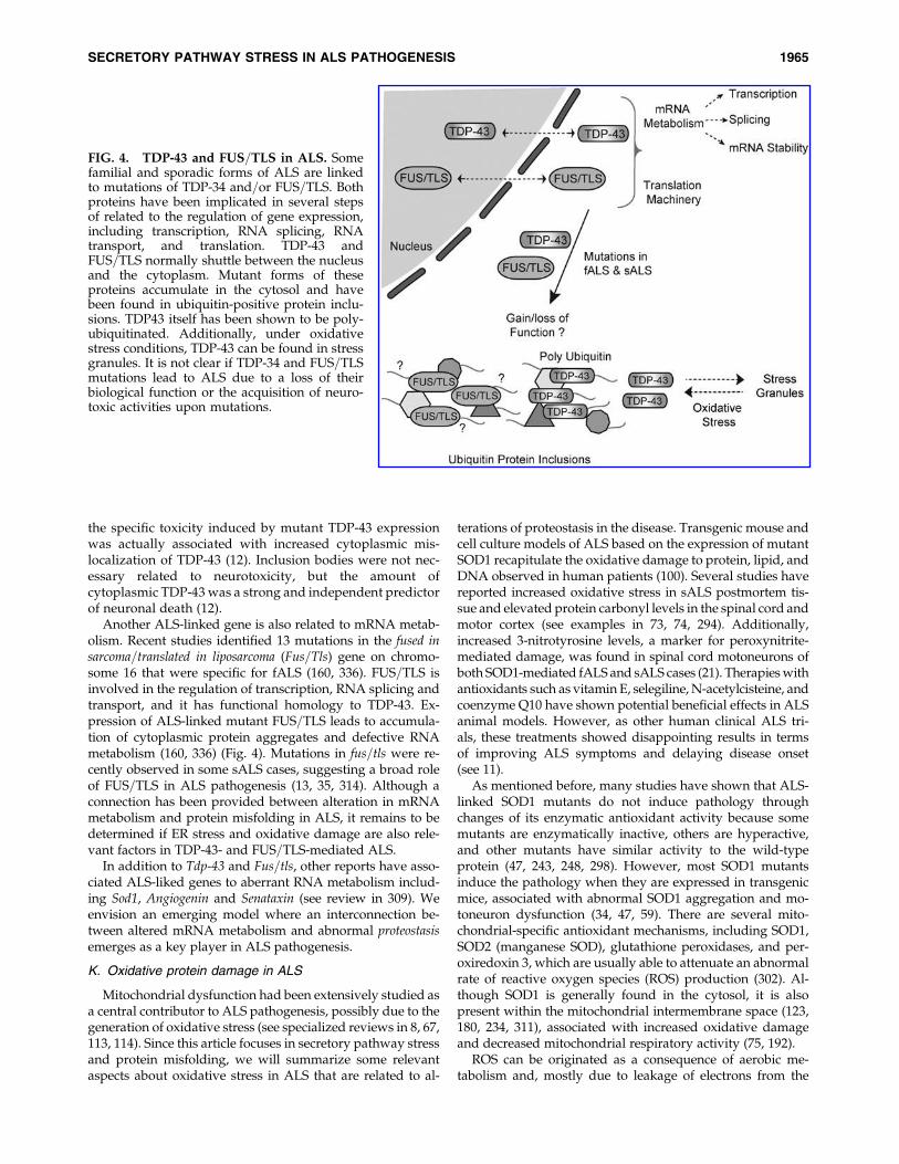
J. TDP-43, FUS=TLS, and abnormalmRNA metabolism in ALS
Accumulating evidence indicates that alterations in pro-teins related to mRNA metabolism is a salient feature of ALSpathogenesis (46, 309), a phenomena also linked to proteinmisfolding. For example, abnormal subcellular distributionand cytoplasmic aggregation of TDP-43 is widely reported insALS and fALS cases, in addition to frontotemporal lobardegeneration (223, 345). TDP-43 regulates different processesrelated to gene expression, including transcription, splicing,and mRNA stability through a RNA and DNA binding ac-tivities (Fig. 4) (46, 87, 309). A recent proteomic analysis of theTDP-43 interactome revealed associations with proteins re-lated to RNA metabolism (78), and disease-related TDP-43mutants were not affected in their interaction profile. Re-markably, TDP-43 interacts with components of stress gran-ules, in addition to be itself recruited to these subcelularstructures (78). Similarly, oxidative stress induces TDP-43redistribution to stress granules (33), however the biologicalrelevance of these observations is not clear yet. TDP-43 wasfound in ubiquitinated protein inclusions, and it was shownto be hyperphosphorylated and ubiquitinated. Most ALS-linked mutations in TDP-43 are mapped to the C-terminalglycine-rich region, which is involved in protein–protein in-teractions between TDP-43 and other ribonuclear proteins(162). Of note, C-terminal fragments of TDP-43 are specificallyaccumulated in ALS and frontotemporal lobar degeneration-derived tissue, and these fragments have a high tendency toaggregate into intracellular inclusions (223).
Mice expressing a disease-linked mutant form of humanTDP-43 develop a progressive and fatal neurodegenerativedisease reminiscent of ALS, showing motoneuron loss, motorimpairment, muscular atrophy, and axonal degeneration(345). Remarkably, this mouse model present pathologicubiquitinated protein aggregates that accumulate only inspecific neuronal populations in the frontal cortex and thespinal cord. These results suggest that expression of TDP-43mutants alter general proteostasis, resulting in accumulationof misfolded proteins (345). However, the role of ER stress inthe pathogenesis of TDP-43 has not been addressed. A zeb-rafish model to study TDP-43 function was also recentlygenerated (136). Expression of disease-related human TDP-43as well as the wild-type protein, albeit to a lesser extent,caused a motor phenotype in zebrafish embryos consisting ofshorter motoneuron axons, premature and excessive branch-ing, as well as movement deficits (136). Remarkably, knock-down of zebrafisfh TDP-43 with morpholinos lead to a similarphenotype, which was rescued by the ectopic expression ofwild-type human TDP-43 and not with the ALS-linked mu-tants (136). This data suggested that both a toxic gain offunction due to protein aggregation=mislocation togetherwith a loss of the normal biological function upon mutationscontribute to disease pathogenesis.
TDP-43 shuttles between nucleus and cytoplasm in atranscription-dependent manner (4). The C-terminal regionof TDP-43 is essential for its cellular localization since itsdeletion results in the formation of large nuclear and cyto-plasmic aggregates (4). A recent report suggested that
1964 NASSIF ET AL.
the specific toxicity induced by mutant TDP-43 expressionwas actually associated with increased cytoplasmic mis-localization of TDP-43 (12). Inclusion bodies were not nec-essary related to neurotoxicity, but the amount ofcytoplasmic TDP-43 was a strong and independent predictorof neuronal death (12).
Another ALS-linked gene is also related to mRNA metab-olism. Recent studies identified 13 mutations in the fused insarcoma=translated in liposarcoma (Fus=Tls) gene on chromo-some 16 that were specific for fALS (160, 336). FUS=TLS isinvolved in the regulation of transcription, RNA splicing andtransport, and it has functional homology to TDP-43. Ex-pression of ALS-linked mutant FUS=TLS leads to accumula-tion of cytoplasmic protein aggregates and defective RNAmetabolism (160, 336) (Fig. 4). Mutations in fus=tls were re-cently observed in some sALS cases, suggesting a broad roleof FUS=TLS in ALS pathogenesis (13, 35, 314). Although aconnection has been provided between alteration in mRNAmetabolism and protein misfolding in ALS, it remains to bedetermined if ER stress and oxidative damage are also rele-vant factors in TDP-43- and FUS=TLS-mediated ALS.
In addition to Tdp-43 and Fus=tls, other reports have asso-ciated ALS-liked genes to aberrant RNA metabolism includ-ing Sod1, Angiogenin and Senataxin (see review in 309). Weenvision an emerging model where an interconnection be-tween altered mRNA metabolism and abnormal proteostasisemerges as a key player in ALS pathogenesis.
K. Oxidative protein damage in ALS
Mitochondrial dysfunction had been extensively studied asa central contributor to ALS pathogenesis, possibly due to thegeneration of oxidative stress (see specialized reviews in 8, 67,113, 114). Since this article focuses in secretory pathway stressand protein misfolding, we will summarize some relevantaspects about oxidative stress in ALS that are related to al-
terations of proteostasis in the disease. Transgenic mouse andcell culture models of ALS based on the expression of mutantSOD1 recapitulate the oxidative damage to protein, lipid, andDNA observed in human patients (100). Several studies havereported increased oxidative stress in sALS postmortem tis-sue and elevated protein carbonyl levels in the spinal cord andmotor cortex (see examples in 73, 74, 294). Additionally,increased 3-nitrotyrosine levels, a marker for peroxynitrite-mediated damage, was found in spinal cord motoneurons ofboth SOD1-mediated fALS and sALS cases (21). Therapies withantioxidants such as vitamin E, selegiline, N-acetylcisteine, andcoenzyme Q10 have shown potential beneficial effects in ALSanimal models. However, as other human clinical ALS tri-als, these treatments showed disappointing results in termsof improving ALS symptoms and delaying disease onset(see 11).
As mentioned before, many studies have shown that ALS-linked SOD1 mutants do not induce pathology throughchanges of its enzymatic antioxidant activity because somemutants are enzymatically inactive, others are hyperactive,and other mutants have similar activity to the wild-typeprotein (47, 243, 248, 298). However, most SOD1 mutantsinduce the pathology when they are expressed in transgenicmice, associated with abnormal SOD1 aggregation and mo-toneuron dysfunction (34, 47, 59). There are several mito-chondrial-specific antioxidant mechanisms, including SOD1,SOD2 (manganese SOD), glutathione peroxidases, and per-oxiredoxin 3, which are usually able to attenuate an abnormalrate of reactive oxygen species (ROS) production (302). Al-though SOD1 is generally found in the cytosol, it is alsopresent within the mitochondrial intermembrane space (123,180, 234, 311), associated with increased oxidative damageand decreased mitochondrial respiratory activity (75, 192).
ROS can be originated as a consequence of aerobic me-tabolism and, mostly due to leakage of electrons from the
FIG. 4. TDP-43 and FUS=TLS in ALS. Somefamilial and sporadic forms of ALS are linkedto mutations of TDP-34 and=or FUS=TLS. Bothproteins have been implicated in several stepsof related to the regulation of gene expression,including transcription, RNA splicing, RNAtransport, and translation. TDP-43 andFUS=TLS normally shuttle between the nucleusand the cytoplasm. Mutant forms of theseproteins accumulate in the cytosol and havebeen found in ubiquitin-positive protein inclu-sions. TDP43 itself has been shown to be poly-ubiquitinated. Additionally, under oxidativestress conditions, TDP-43 can be found in stressgranules. It is not clear if TDP-34 and FUS=TLSmutations lead to ALS due to a loss of theirbiological function or the acquisition of neuro-toxic activities upon mutations.
SECRETORY PATHWAY STRESS IN ALS PATHOGENESIS 1965

mitochondrial respiratory chain, result in incomplete reduc-tion of molecular oxygen during oxidative phosphorylation.This event generates the superoxide radical anion (O2
�) andhydrogen peroxide (H2O2). A portion of cellular ROS is pro-duced by cellular oxidative enzymes in the cytoplasm andthe ER (Fig. 5). Oxidative stress can enhance various patho-genic events including an increase in intracellular Ca2þ levelsrelated to glutamate excitotoxicity (75, 192). Increases in cy-tosolic calcium levels are buffered by mitochondria, aug-menting ROS production, opening of the permeabilitytransition pore, an event recently linked to ALS in vivo (Fig. 5)(142, 146, 190, 191). Alterations to mitochondrial morphologyhave been observed in motoneurons from ALS patients andanimal models (157, 191, 283, 296, 349), in addition to de-creased electron transport chain activity, altered mitochon-drial membrane potential (29, 134, 192, 199, 313), disruptedcalcium homeostasis (29, 313), increased mitochondrial DNAdamage (191), and reduction in general mitochondrial anti-oxidant defense mechanisms (81, 350).
Notably, a gene expression profile study carried on moto-neurons expressing mutant SOD1 reported the down-regulation of genes involved in antioxidant responses,including the transcription factor nrf2 (nuclear factor (ery-throid-derived 2)-like 2), several members of the glutathioneS-transferase family, and two peroxiredoxins (154). More-over, reduced Nrf2 mRNA and protein levels have beenreported in spinal cord neurons from ALS patients (282).Nrf2 is known to interact with promoter sequences con-taining the antioxidant-response element (ARE) sequence,increasing the expression of proteins involved in antioxi-dant defense systems (225). It is predicted that down-regulation of Nrf2 expression in ALS may reduce the ability
of cells to remove ROS, resulting in a gradual increase inoxidative stress over time.
The relationships between oxidative stress and ER stress insALS was first suggested by a correlative study describing theco-existence of oxidative damage to ER proteins and chaper-ones with the occurrence of ER stress in the spinal cord ofpostmortem samples from sALS patients (129). These eventswere also correlated to increased accumulation of abnormalubiquitin-positive protein inclusions (129). A recent studydemonstrated the occurrence of early mRNA oxidation inspinal cord from ALS patients, in addition to pre-symptom-atic mutant SOD1 transgenic mice (46). A group of mRNAwas identified to be more susceptible to oxidation through agene expression profile analysis of spinal cord tissue of pre-symptomatic SOD1G93A transgenic mice. These mRNAs en-code proteins involved in essential cellular process related toprotein biosynthesis (ribosome protein S6, eukaryote trans-lation initiation factor 5), protein folding (heat shock protein70KD protein 5, heat shock protein 90KDa alpha, class Amember 1), ubiquitin-proteasome system (26S subunit AT-Pase2, proteasome subunit beta type 4, ubiquitin-conjugatingenzyme E2D), and lysosomal function (lysosomal membraneglycoprotein 1) (46), suggesting that oxidative stress may leadto broad alterations to proteostasis (293). Of note, knownfALS-related genes such as als1=sod1, dctn1, and vapb were alsoidentified in the study (46).
Recent evidence in Parkinson’s disease and Alzheimer’sdisease suggest that oxidation of ER chaperones is an im-portant event in the pathology, triggering chaperone inacti-vation, and thus protein misfolding and ER stress (52, 55, 328).Nitrosylation of the cysteines present at the active site of PDIwas shown to be a critical pathological event (52, 55). A recent
ROS
ER stress
Mitochondria
mRNA oxidation
Protein oxidative modification
PDI nitrosylation
SHSH
PDIERp57
SH SH
Respiratory chain
* ** *
*
***
*
*
*
**
*Reactive oxygen species
***
* **
Ero1
DNA damage
SH SH
SH
SH
SOD1 aggregate
FIG. 5. Oxidative stress in ALS.In addition to the mitochondria,the ER is emerging as an impor-tant source of ROS under ERstress conditions. When the ERfunction is perturbed, an unbal-ance between the protein disulfideisomerases (i.e, ERp57 and PDI)and Ero1 redox functions gener-ates excessive ROS as a result ofdisulfide bond formation. Thegeneration of ROS has a broadimpact on proteostasis initiatedby modifications of key biologi-cal molecules including mRNAoxidation, protein modification,and chaperone inactivation,among other effects.
1966 NASSIF ET AL.

report from Julie Atkin’s group described for the first time theoccurrence of PDI nitrosylation in sALS-derived tissue (342).Increased levels of S-nitrosylated forms of PDI were observedin mutant SOD1 transgenic mice and human sALS spinal cordtissue, providing for the first time a direct link between gen-eral oxidative stress and ER stress=protein misfolding in thepathogenesis of ALS (Fig. 6) (342).
The ER can also operate as a source of oxidative stress,which is highly enhanced during sustained ER stress (seereview in 276). When disulfide bonds are formed in proteinsby PDIs, electrons are passed from thiol groups on the pro-tein substrates to Ero1 and finally to molecular oxygen, re-sulting in the generation of ROS (200) (Fig. 6) (reviewedin 324). Any unstable disulfide bonds are reduced by glu-tathione in a reaction catalyzed by Ero1 that depletes cellularreservoirs of reduced glutathione and increases the numberof thiol groups available for disulfide bond formation, con-sequently increasing ROS (44). Activation of the UPR in-creases Ero1 expression, which may reflect an imbalancedredox status at the ER (Fig. 6) (109). The actual contributionof the ER to the generation of ROS in ALS remains tobe determined. In summary, increasing evidence indicatesa relationship between oxidative stress and ER stress inthe pathogenesis of ALS, possibly contributing to proteinmisfolding.
III. Second Stop: Golgi Apparatus
A. Golgi apparatus: Main functions
The Golgi apparatus (GA) is composed of a series of flat-tened parallel, interconnected cisternae organized around themicrotubule-organizing center in the perinuclear region. TheGA is a highly dynamic structure and plays a key role inthe transport, processing, and targeting of proteins to their
final destination, including the plasma membrane, ER, andlysosomes (70). In neurons, the GA is involved in the axo-plasmic flow of numerous endogenous proteins and of exog-enous macromolecules transported by orthograde, retrograde,and transynaptic routes (102, 260). Therefore, GA abnormali-ties may have detrimental consequences on axon and pre-synaptic terminal function (79).
Alterations in GA function or inhibition of ER-GA traf-ficking trigger ER stress (184). GA fragmentation and dis-persal precede neuronal cell death induced by excitotoxins,oxidative=nitrosative insults, ER stress among other cell deathstimuli (219). In addition, recent reports suggest the existenceof specific cell death pathways initiated by irreversible GAdamage, which may be downstream of ER stress in neurons(219). It has been also reported that during apoptotic celldeath, Golgi stacks disperse and disassemble into tubulove-sicular clusters (184). Interestingly, GA fragmentation duringapoptosis is proposed to be independent to alterations in thecytoskeleton, suggesting an intrinsic GA-regulated process(211). Of note, the pro-apoptotic protease caspase-2 is local-ized in the GA and the nucleus. Caspase-2 -deficient mousefibroblasts are resistant to apoptosis induced by drugs thatactivate secretory pathway stress (188). Caspase cleavage ofdifferent GA proteins have been implicated in apoptosis (51,165, 166, 182, 185, 188), suggesting an important role of GA-resident proteins in the initiation and fine regulation of stressresponses and cell death signaling, in addition to the execu-tion of organelle disassembly during apoptosis.
B. GA fragmentation in sALS
Fragmentation of the GA has been extensively reported inanterior horn motoneurons of spinal cord tissue derived fromsALS patients and other neurological disorders (see examples
FIG. 6. PDI function and mu-tant SOD1 aggregation. (A)Protein disulfide isomerase (PDI)catalyzes the formation andbreakage of disulfide bonds be-tween cysteine residues duringproteins folding at the ER. Gen-eration of excessive reactive ox-ygen or nitrogen species resultsin protein misfolding due to PDIinactivation by S-nitrosylation ofthe active site of the enzyme. (B)fALS linked to mutations ofSOD1 is characterized by theaccumulation of stable SOD1aggregates in motoneurons. Ab-normal disulfide bonds formedbetween cysteines at positions 6and 111 of different SOD1monomers have been proposedto mediate the generation ofoligomeric polymers at the ER,leading to the generation of largemutant SOD1 aggregates. Pro-tein disulfide isomerases such asERp57 and PDI may be involvedin decreasing this pathologicalprocess.
SECRETORY PATHWAY STRESS IN ALS PATHOGENESIS 1967

in 80, 91, 209, 305). Namely, GA fragmentation refers to a lossof the normal network-like configuration, which is replacedby disconnected vesicular pattern (Tables 1 and 2) (40,90). Inan early study, Gonatas and co-workers performed histo-pathological analysis of tissue from a small group of sALScases (91). This study indicated that approximately 30% ofmotoneurons in five sALS cases showed a fragmented GA,whereas only * 1% of motoneurons from seven control withneurologic or systemic disease showed a similar change.Analysis of the distribution of cytoskeleton proteins did notdepict any differences in the staining of neurons with frag-mented or normal GA (91), suggesting that the morphologicalalteration of the organelle are not secondary to a gross lesionof the cytoskeleton. Similarly, other studies have shown thatneurons with abnormal TDP-43 immunoreactivity are asso-ciated with dysfunction on GA morphology in sALS (79).
Interestingly, intrathecal injections of CSF from sALS pa-tients into rodents using pup-mediated delivery causedmarked fragmentation of the GA in spinal cord motoneurons(253). This phenotype was widely spread through the spinalcord, suggesting a putative transmissible pathological ele-ment present in CSF from these sALS patients in addition toan intrinsic sensitivity of motoneurons in accumulating GA-related defects (253). Whether GA network impairment insALS is an indicator of pathogenesis or is representative of adisease consequence remains to be determined.
C. GA alterations in fALS-induced by SOD1 mutations
Similar to sALS, fragmentation of the GA is a consistentneuropathological feature observed in motoneurons frompre-symptomatic mutant SOD1 transgenic mice (Figs. 7 and 8)(210). In addition, GA pathology is enhanced with diseaseprogression (306, 307). Interestingly, this event correlates withan impairment of secretory activity as measured by moni-toring the transit of CD4 to the cell surface, a glycoproteinprocessed through the GA (308).
Wild-type SOD1 is secreted to the extracellular space, butthe efficiency of secretion is decreased for most ALS-linkedSOD1 mutants (326). ATP-dependent secretion of wild-typeSOD1 has been described using a variety of cell types otherthan motoneurons, including human hepatocytes, fibroblasts,neuroblastomas, thymic-derived cell lines (31, 205, 206), andmouse astrocytes (161). The presence of extracellular endog-enous wild-type and mutant SOD1 was observed in CSF oftransgenic fALS rats (214). Turner and co-workers suggestedthat a diminished secretion of mutant SOD1 was associatedwith intracellular toxic protein inclusions and GA fragmen-tation in a motoneuron cell line (326). SOD1 secretion wassensitive to brefeldin A treatment, suggesting a classical routeof secretion. Interestingly, although secretion was observed inneuronal and non-neuronal cell lines, only neuronal cells wereshown to be sensitive to mutant SOD1 toxicity (326). Enforcedsecretion through addition of an extracellular targeting se-quence to mutant SOD1 attenuated cytoplasmic aggregatesand toxicity in transfected motoneuron cell lines (326), sug-gesting that intracellular accumulation of mutant SOD1 hasdeleterious effects. The same study demonstrated that chronicintraspinal infusion of wild-type SOD1 significantly pro-longed survival of transgenic rats expressing mutant SOD1-G93A for approximately 25 days without affecting diseaseonset, measured by no effect on the decline of motor perfor-
mance (326). This protective effect was reproduced by anti-oxidant therapy in transgenic fALS mice, similar to the resultsobserved by the injection of SOD1-catalase, suggesting thatmutations in SOD1 may decrease any neuroprotective activityof extracellular wild-type SOD1 (135).
Extracellular accumulation of mutant SOD1 has also beensuggested as a potential pathogenic effect in fALS (Fig. 7)(335), where secretion of mutant SOD1 results in nonauton-omous effects related to neuroinflammation and glial activa-tion (335). Through a yeast two-hybrid screening of a cDNAlibrary from total spinal cord of pre-symptomatic mutantSOD1G93A mice, Urushitani and co-workers identified mutantSOD1 interacting partners. This study revealed a specific as-sociation between mutant SOD1 and chromogranins, an in-teraction not observed with wild-type SOD1. Chromograninsare soluble proteins transported through the trans-Golginetwork (TGN), and are part of large dense-core vesicles(LDCV) related to regulated exocytosis in neurons and en-docrine cells (316, 335). In fact, it was shown that the secretionof mutant SOD1 is partially regulated by chromogranin ex-pression and occurs as a protein complex with SOD1 frommotoneurons, astrocytes, and interneurons. Through cellculture experiments, it was demonstrated that secretion ofmutant SOD1 triggers microglial activation. These eventscontributed to microgliosis and finally enhanced neuronal celldeath, possibly due to inflammatory and oxidative stresscomponents (335). These observations emphasize the rele-vance of the motoneuron milieu in ALS and support the non-cell-autonomous theory of the disease (32, 247, 292, 335). Apartial co-localization of mutant SOD1 and chromograninswas observed in fALS mouse models by immunofluorescenceand electron microscopy studies. This data was also con-firmed through subcellular fractionation and purificationof GA- and microsome-enriched fractions followed by co-immunoprecipitation experiments (335).
Due to the occurrence of SOD1 secretion in vivo, the pos-sible therapeutic benefits of immunization protocols on fALSwere tested, aiming to reduce the burden of extracellularSOD1 mutants in nervous tissue (331). The authors em-ployed recombinant mutant SOD1 to immunize mutantSOD1G37R transgenic mice, and observed protective effectsassociated with a delay of disease onset and an extension oflifespan for more than four weeks (331). Evidence for aclearance of SOD1G37R species in the spinal cord of vacci-nated mice was also provided. In addition, the authorsshowed that intraventricular infusion of purified anti-human SOD1 antibody had beneficial effects on SOD1G93A
transgenic mice (331). These results provided the first evi-dence for a possible role of extracellular mutant SOD1 infALS pathogenesis.
D. A nonclassical secretory pathway for mutant SOD1
One report indicated that wild-type and mutant SOD1 isalso secreted via exosomes. Exosomes are small lipid mem-brane microvesicles formed by fusion of multivesicularendosomes (MVEs) with the plasma membrane, followedby the release of their cargo into the extracellular environ-ment (89). Initially, exosomes were thought to operate as acellular mechanism for releasing unnecessary proteins, butrecent studies have revealed further functions of exosomesin cell-to-cell communication. Proteins associated with
1968 NASSIF ET AL.

neurodegenerative diseases, such as the prion protein andamyloid precursor protein (APP) can be selectively incorpo-rated into MVE and subsequently released into the extracel-lular space within exosomes (338). Remarkably, the presenceof exosome-containing proteins in body fluids has been pro-posed as an interesting candidate strategy for diagnosis ofdiseases such as brain cancer (338, 348). This nonclassical se-cretion pathway provides a potential explanation for extra-cellular prion spread and amyloid deposition, which mayhelp understand the non-cell-autonomous aspects of ALSpathology and other neurodegenerative diseases (338).
E. GA disturbance and the Wobbler mouse
The first animal model of motoneuron degeneration wasthe Wobbler mouse, which was related to the occurrence ofabnormalities in RNA metabolism (23). Motoneuron survivalis severely compromised in the Wobbler mouse. However, themutation responsible for the ALS-like phenotype in this mousewas later described as a missense mutation in vacuole proteinsorting 54 gene (vps54L967Q) (286). VPS54 is a component of theGA-associated retrograde protein (GARP) complex of vesiclesorting of proteins (198), suggesting that VPS54 has an essentialrole in trafficking (286). Interestingly, the presence of intracel-lular ubiquitin inclusions and abnormal distribution of TDP-43protein was revealed in the cytoplasm of neurons from Wobblermouse. However, efforts to find mutations in the gene corre-sponding to chromosome 2p14-15 (related to vps54) after DNAsequencing of 96 individuals with sporadic ALS, 96 individualswith familial ALS, and 96 controls subjects lead to no conclu-
sive results to consider vps54 as an ALS–linked gene (198).Analysis of a larger number of patients is need to furtheraddress the role of VPS54 in ALS.
F. GA alterations in fALS inducedby dynactin mutations
The dynactin complex has relevant functions in axonalretrograde transport. Several studies have described the oc-currence of microtubule-based axonal transport abnormalitiesin mutant SOD1 transgenic mice (Fig. 8) (164, 176). Teulin andco-workers determined the consequences of targeting the dy-nein=dynactin complex in motoneurons in the context of ALSby generating transgenic mice with neuronal-specific expres-sion of the N-terminus of bicaudal D2 (BICD2-N). BICD2-N isa motor-adaptor protein that is involved in dynein-mediatedtransport of cargoes (318). The fragment expressed of BICD2-N strongly binds and inhibits the dynein=dynactin complex(127). Interestingly, the expression of BICD2-N in motoneu-rons caused GA fragmentation and axonal neurofilamentswellings (318). Unexpectedly, BICD2-N expression increasedthe lifespan of a fALS mouse model, suggesting that an un-known event related to dynein=dynactin-dependent transportmay be relevant to ALS pathogenesis (147, 364).
IV. Third Stop: Endocytosis and Vesicle Transport
A. Endocytosis and multivesicular endosomes
Many cell-surface proteins, including receptors andbound ligands, are internalized via clathrin-coated pits and
FIG. 7. Mutant SOD1 in the secretorypathway. Mutant SOD1 forms abnormalprotein aggregates that are found in thecytosol, ER and GA. GA fragmentationhas been observed in fALS and sALSmotoneurons. In addition to wild-typeSOD1, fALS-linked mutants are secretedto the extracellular space. This secretoryroute involves the translocation of SOD1monomers to the ER by an unknownATP-dependent mechanism, and thenfollows a classical secretory route. Mu-tant SOD1 physically interacts withchromogranins, an essential step fortheir secretion. Extracellular mutantSOD1 may have deleterious effects byinducing a drastic glial reaction, leadingto the generation of neurotoxic species.In contrast, secretion of wild-type SOD1may have protective effects due to itsantioxidant activity. During its transitthrough the secretory pathway, vesiclescontaining mutant SOD1 may be tar-geted to lysosomal=autophagy mediateddegradation.
Nuc
?
Aggregation SOD1 WTMutant SOD1
Motoneuron
Microglia
Microgliaactivation
cytokines secretion
Neuronal damage?
ER
?
Ribosomes
Chromogranin
SOD1 aggregate
GA
?
Autophagosome
Fragmented GA
SECRETORY PATHWAY STRESS IN ALS PATHOGENESIS 1969

successively transferred to early and late endosomes beforedelivery to lysosomes for degradation. During this process,proteins are also ubiquitinated as a signal for sorting into theluminal vesicles of MVEs, often referred to as ’multivesicularbodies’ (MVBs) (96, 143). The formation of MVEs occurs by theinward vesiculation of endosomal membranes, a process drivenby the endosomal sorting complexes required for transport(ESCRT)-0, I, II, and III. ESCRT-0, -I, and -II contain ubiquitin-binding subunits and mediate the sorting of the ubiquitylatedcargo. Another component, ESCRT-III, is the principal ma-chinery for the formation of intralumenal vesicles in MVEs(Fig. 9) (96, 143, 271). As a degradative sorting compartment,the MVEs are structural and functionally related with lysosomeand autophagy-related vesicles (see below for detailed de-scription of autophagy) (271). Deletion of different ESCRTsubunits results in abnormal accumulation of autophagosomesand sometimes accumulation of cytoplasmic protein aggregatescontaining ubiquitinated proteins, suggesting a defect in au-tophagy-dependent degradative processes (76, 168). In the nextsection, we describe evidence linking the alterations to vesiculartransport with three different genes associated with ALS.
B. Multivesicular endosomes in fALS
The most direct evidence involving the participation ofMVEs in ALS comes from the identification of autosomaldominant mutations in the Chmp2b gene in a subset of fALSpatients (Fig. 9) (239). Chmp2b gene codifies for the chargedmultivesicular body protein 2 B (CHMP2B) that is part ofESCRT III complex. Further studies demonstrated that theoverexpression of mutant Chmp2b causes dominant-negativephenotypes associated with the accumulation of autophago-somes and intracellular protein aggregates, followed byneuronal death (76, 168, 272). Given the known role of basalautophagy in the homeostasis of the nervous system and the
clearance of toxic misfolded proteins (104, 156), ESCRT dys-function may be a relevant factor contributing to ALS and thealterations of proteostasis observed in motoneurons.
C. Vesicle transport and fALS: Alsin
Mutations in als2, the gene encoding ALSIN, have beenlinked to the onset of autosomal recessive motoneuron diseases,including juvenile-onset ALS, hereditary spastic paraplegia,and primary lateral sclerosis (99, 358). The primary structure ofalsin is very complex, with many structural domains. Alsin ispredicted to have three putative guanine regulators of chro-mosome condensation 1 (RCC1)-like domains (termed RLD); adiffuse B cell lymphoma (Dbl) homology=pleckstrin homology(DH=PH) domain; and a vacuole protein sorting 9 (VPS9) do-main (45, 99). Alsin expression in neurons is observed in smallpunctuate pattern in dendrites, axons, cell body, membraneruffles, and growth cones (322, 325). In addition, its expressionhas been reported to affect cytoskeleton dynamics through in-teractions with Rho subfamily members (325). Rho subfamilymembers such as Rac GTPases, Rho, and Cdc42, have beenshown to regulate actin dynamics and neuritic outgrowth (61).Binding assays revealed a specific interaction between Rac1 andalsin through the DH=PH domains (322). It has also been shownthat the VPS9 domain of alsin acts as an activator of GEF for thesmall GTPase Rab5, regulating endosome vesicle sorting (324).Together, these data suggest a role for alsin in axonal guidancein addition to the regulation of cytoskeleton and vesicle dy-namics. Recently, alsin was demonstrated to exert a neuropro-tective function via its Rho-GEF domain, inhibiting theneurotoxicity of mutant SOD1 in vitro (140). Although muta-tions in als2 are associated with motor dysfunction in humans,mice deficient in als2 have largely failed to recapitulate symp-toms of motoneuron disease (26, 60, 98, 355). als2 knockout miceappear to be normal, with a lifespan similar to wild-type
Nuc
ER
GA
SOD1Mutant SOD1
BiPRibosomes
SOD1 aggregate
Microtubule
Retrograde cargoDynein-dynactin
Synaptic Vesicle
Retrograde transport
Fragmented GA
Anterograde transport
FIG. 8. Abnormal axonal trans-port in ALS. The dynein-dynactincomplex constitutes essential motorproteins for retrograde fast axonaltransport. Vesicles are loaded ontotransport motors both in the cellbody and the distal nerve terminal.Mutations in dynactin in humansand dynein in mice provoke moto-neuron degeneration and motorimpairment. Perturbation of neuro-filaments through mutations andalterations in phosphorylation andthe physical structure of the axoncould also adversely affect axonaltransport. In transgenic mousemodels, mutant SOD1 impairsanterograde axonal transport.
1970 NASSIF ET AL.

littermates (60). However, Guy Rouleau’s group described twonew animal models to address ALS2 function (95). Mice lackingexon 2 and part of exon 3 of als2 developed mild signs of neu-rodegeneration compatible with the occurrence of axonaltransport deficiency. Similarly, knockdown of als2 in zebrafishresults in severe developmental abnormalities, swimming def-icits, and motoneuron perturbation (95). The role of als2 in ALSneeds further investigation to solve these differences betweenanimal models.
D. Vesicle transport and fALS: Dynactin
Retrograde axonal transport of cargoes such as vesicles,endosomes, signaling complexes, degradation products, andorganelles all involve interaction with the molecular motordynein and the multiprotein complex dynactin. The dynactincomplex includes the subunits p150 and dynamitin, provid-ing a link between the transported cargo, microtubules, andcytoplasmic dynein (reviewed in 288). Mutations in thep150 subunit (the dctn1 gene) have been reported in bothfALS and sALS cases (212, 213) in addition to atypical forms
of the disease (Fig. 8) (243). A direct link between impaireddynactin=dynein function and motoneuron disease camefrom the finding that alterations on dynactin function resultsin motoneuron degeneration in vivo (163). Disease phenotypesobserved by the expression of mutant dynactin in mice arecharacterized by drastic defects in vesicular trafficking, lyso-somal proliferation, and distal degeneration in motoneurons(50, 318). Thus, the alteration in vesicle transport is a relevantfactor in some forms of ALS, reinforcing the role of secretorypathway stress in the disease process.
V. Final Stop: Autophagy
A. Autophagy pathways
The proteasome and lysosomal pathways are the two mainroutes of protein clearance. Proteasomes predominantly de-grade short-lived proteins, and their substrates need to beunfolded to pass through the narrow pore of the proteasomebarrel (reviewed in 269). Increasing evidence indicates thatthe clearance of proteins through autophagy correlate withtheir propensity to aggregate (reviewed in 108, 269). Macro-autophagy, here referred to as autophagy, is a highly con-served and regulated eukaryotic intracellular degradativeprocess (Fig. 10) (171, 172, 204). Autophagy is regulated by alarge family of genes, termed atgs, and it is involved in thecatabolism of cytoplasmic components including damagedor superfluous organelles, toxic protein aggregates and intra-cellular pathogens. The autophagy process involves encapsu-lation of cargo into a double-membrane vesicle to form theautophagosome vesicle. Autophagosomes then fuse withlysosomes to form the autolysosomes where cargoes are de-graded. In addition, hybrid intermediates called amphisomesare formed when autophagosomes fuse with the endosomes orMVEs before fusing with lysosomes (Fig. 10) (297, 321). ERstress and proteasome inhibition also trigger autophagy (62).For example, PERK=eIF2a and IRE1a=JNK signaling have beenreported to initiate autophagy (158, 244, 273), acting as animportant survival mechanism to cope with protein misfolding(49, 222). Unregulated or excessive autophagy is thought to bedetrimental to cell survival due to uncontrolled organelle andmacromolecular catabolism (172).
B. Regulation of autophagy
The mechanisms underlying the initial activation steps ofautophagy are very complex and poorly understood. Atg pro-teins regulate different sequential steps in the autophagy pro-cess, starting with the formation of a protein kinase–autophagyregulatory complex and a lipid kinase–signaling complex that isinvolved in vesicle nucleation. Ubiquitin-like protein conjuga-tion pathways are required for vesicle expansion and closure,and for the retrieval pathway for disassembling Atg proteincomplexes from mature autophagosomes. The core of the au-tophagic machinery is built around two ubiquitin-like conju-gation systems (Fig. 10): ATG12 and microtubule-associatedprotein 1 light chain 3 (LC3). When ATG12 is activated, it formsa covalent bond with ATG5, which then forms a multimericcomplex with other components, translocating to membranes ofearly autophagosomes. Additionally, LC3 is cleaved and thenconjugated to phosphatidylethanolamine (PE), generating LC3-II. The unconjugated LC3-I remains in the cytosol while the
FIG. 9. An ALS mutation involved in multivesicular en-dosomes. Endocytosed proteins can undergo lysosomal-mediated degradation. This process requires sorting fromearly endosomes (EE) to mutivesicular endosomes by theendosomal sorting complex required for transport (ESCRT).Mutations in chmp2b, a component of the ESCRT complex,have been described in some ALS cases. Mutation in chmp2binduces an accumulation of autophagosomes and accumu-lation of protein aggregates, suggesting an alteration in ve-sicular trafficking and protein degradation mechanisms.
SECRETORY PATHWAY STRESS IN ALS PATHOGENESIS 1971

conjugated LC3-II form redistributes to the autophagosomalmembrane after the formation of the active ATG12-ATG5complex, leading to autophagosome membrane extension(172). Monitoring the generation of LC3-II is the gold standardto measure autophagy levels. p62 (also called sequestosome 1)serves as a bridge between poly-ubiquitination of proteins andthe autophagy-degradative machinery (7, 289), operating as ashuttle factor for substrates of the proteasome to undergo au-tophagy-mediated degradation (18, 238).
One of the main autophagy regulatory pathways involvesthe participation of mTOR. mTOR signaling represses au-tophagy by the inhibition of a downstream class III phos-phatidyl inositol 3-kinase (PI3K) complex (Fig. 11). Activationof autophagy by nutrient deprivation involves the de-repression of the mTOR kinase. An initial step for vesiclemembrane nucleation is the activation of a complex con-
taining VPS34, a class III PI3K (111). VPS34 activation de-pends on the formation of a multiprotein complex includingbeclin-1 (the mammalian orthologue of ATG6). In addition,recent evidence suggests existence of a regulatory networkwhere inositol-1,4,5-triphosphate receptor (IP3R) mediatesthe initiation of autophagy through a protein complex withbeclin-1 at the ER membrane, acting as a scaffold proteinindependent of its calcium channel activity (340). Autophagyis critical for the maintenance of neuronal homeostasis andcontributes to basal elimination of misfolded proteins in thenervous system. Brain specific ablation of atg5 and atg7 leadsto spontaneous neurodegeneration with pathological fea-tures closely resembling Alzheimer’s disease and Parkinson’sdisease, consisting of neurological=motor dysfunction, ac-cumulation of poly-ubiquitinated protein aggregates, neu-ronal loss, and premature death (104, 156).
FIG. 10. Autophagy-related ves-icles in eukaryotic cells. A varietyof physiological and noxious sig-nals (i.e., nutrient starvation, pro-tein misfolding, and organelledamage) induce autophagy. Theautophagy process begins with thenucleation of a protein complex toform the growing phagophoremembrane. Then the vesicle ex-pands and encapsulates sequesteredmaterials including cytosol, organ-elles, and protein aggregates, form-ing the double membrane vesicleautophagosome. Autophagosomesthen fuse with endocytic compart-ments including endosomes, multi-vesicular bodies, and lysosomes.Amphisomes are organelles con-taining both autophagosome andendosomal constituents. Amphi-somes and autophagosomes matureby fusing with lysosomes, formingthe autolysosome in which encap-suled cellular content is degraded byhydrolytic enzymes and proteases.
1972 NASSIF ET AL.

Recent studies suggest that upstream mTOR-independentpathways, previously undefined, modulate the core autop-hagy machinery. This pathway is becoming relevant for treat-ment of neurological conditions, and recent drug screeningshave uncovered regulatory networks of this pathway (257, 277–279, 281). mTOR-independent autophagy is induced by agentsthat lower or deplete inositol or IP3 levels such as lithium,valproate, and carbamazepine (Fig. 11) (278, 280). Inhibition ofinositol monophosphatase (IMPase), an enzyme participatingon inositol monophosphate turnover, triggers autophagy (278,280). In addition, small sugars such as the disaccharide trehalosehave been also shown to trigger mTOR-independent autophagyby an unknown mechanism, having therapeutic effects onmodels of Huntington’s disease, Alzheimer’s disease, and priondisorders, among other pathologies (15, 58, 251, 315).
Finally, a recent study added more complexity to the reg-ulation of autophagy. Nishida and co-workers described theoccurrence of ATG5- and ATG7-independent autophagy incells undergoing stress triggered by DNA damage associatedwith the appearance of classical morphological and functionalautophagy features (226). However, this form of autophagywas independent of LC3, but depends on the expression ofbeclin-1 and unc-51-like kinase 1 (the human ATG1 ortholog).Moreover, unlike conventional autophagy, autophagosomeswere proposed to be generated by the fusion of isolationmembranes with vesicles derived from the trans-Golgi andlate endosomes and not the ER (226). These results suggest theoccurrence of autophagy through at least two different corepathways: an ATG5=ATG7-dependent conventional pathwayand an ATG5=ATG7-independent pathway. It remains to be
FIG. 11. General regulatory mech-anisms of autophagy. Two mainpathways are involved in autophagyactivation: The mTOR-dependentand mTOR-independent pathways.The first occurs upon de-repressionof mTOR Ser=Thr kinase (mamma-lian target of rapamycin) that istriggered by nutrient conditions orexperimentally by rapamycin treat-ments. A second pathway is initi-ated by a decrease in global inositolphosphate (IP3) levels. This pathwayis modulated by IMPases, and canbe induced by lithium, valproate orcarbamazepine treatments. Recruit-ment of many protein complexesoccurs during vesicle nucleation thatinvolves the activation of the au-tophagy regulator Beclin-1 and PI3kinases. BCL-2=BCL-XL negativelyregulates this nucleation step bybinding to Beclin-1. Atg5 and Atg7have been shown to be essentialregulators of autophagy. The conju-gation of phosphatidylethanolamine(PE) to LC3=Atg8 leads to the con-version of the soluble form of LC3(named LC3-I) to the autophago-some-associated form (LC3-II). Au-tophagosomes mature by fusionwith lysosomes, creating autolyso-somes, in which the luminal contentof the autophagic vacuoles is de-graded. Alternatively, autophagy canproceed through a mechanism inde-pendent of atg5=atg7 and LC3, butdependent on Beclin-1 expression.
AUTOPHAGY INDUCTION
mTOR-dependent pathway(canonical)
mTOR-independent pathway
mTOR
P
P
PP
IMPase
Ins
IP
IPLithium
Rapamycin
Carbamazepine
Valproate
Reduced IP levels3
3
1
P
IP2
P
Beclin-1Bcl2/Bcl-XL
Vesicle Nucleation
Atg5/Atg7 dependent pathway Atg5/Atg7 independent pathway
LC3 LC3LC3
Gly PE
AutophagosomeAutophagosome H+ H+
Lysosome
Mitochondria
LC3-II
Lysosome hydrolases
SECRETORY PATHWAY STRESS IN ALS PATHOGENESIS 1973
determined how beclin-1 differentially senses distinct autop-hagy stimuli (i.e., nutrient deprivation versus DNA damage)to engage these two mechanistically independent autophagypathways.
C. Autophagy in fALS
Different studies in fALS models indicate that autophagymay operate as an active cellular response to clear out mutantSOD1 aggregates (Fig. 12). Kabuta and colleagues initiallyreported that activation of autophagy reduces mutantSOD1G93A-mediated toxicity in a neuroblastoma cell line(137). Mutant SOD1 degradation was suppressed by treat-ments with PI3K inhibitors, and by the administration of themTOR inhibitor rapamycin. Inhibition of autophagy leads to adrastic accumulation of mutant SOD1 aggregates, enhancingits neurotoxicity (137). We have recently confirmed this dataand further demonstrated a crucial role of ATG5 and beclin-1in the degradation of mutant SOD1 oligomers in cellularmodels of fALS (121).
p62 has been observed in ubiquitin-positive inclusions inALS (83, 203, 220, 239), in addition to a variety of neurode-generative diseases such as Alzheimer’s disease (7, 159), Hun-tington’s disease (18, 216), and Parkinson’s diseases (159, 221).It has been reported that p62 preferentially interacted withmutant SOD1 (83), facilitating its sequestration into inclusions(83, 84). p62 modulates the targeting of mutant SOD1 for au-tophagy- and proteasome-dependent degradation in bothubiquitin-dependent and -independent manners (Fig. 12) (84).
Studies in mutant SOD1 transgenic mice have provided acorrelation between disease progression and autophagy in-duction. The levels of LC3-II are increased in SOD1G93A
transgenic mice at the symptomatic stage (207). Moreover,phosphorylation of mTOR at Ser2448 was shown to be en-hanced in the same experiments. Another group confirmedthe presence of LC3-positive vesicles in fALS mouse models(175). We have also described the activation of autophagy inthe spinal cord of SOD1G86R transgenic mice as monitored bythe presence of LC3-positive vacuoles and the co-localizationof SOD1 and LC3 into autophagosomes by electron micros-copy and immunogold staining (121). This event correlatedwith the upregulation of beclin-1 in the same tissue. It hasbeen also suggested, using skeletal muscle-specific mutantSOD1G93A transgenic mice, that autophagy may have a nox-ious role (64). The authors suggested that the occurrence ofoxidative stress in the disease induces abnormal activation ofFoxO3, NF-kB, and autophagic-related signaling events, re-sulting in muscular atrophy.
Finally, recent reports suggested a role of autophagy in thedegradation of cytoplasmic aggregates of ubiquitinated TDP-43 (Fig. 4) (133, 334, 344). In addition, inhibition of mTOR orPI3K reduces the accumulation of TDP-43 C-terminal frag-ments and restores TDP-43 normal localization (25), whereastrehalose treatment decreased TDP-43 levels (344), suggesting amore general role of autophagy in different forms of ALS. Fi-nally, expression of human TDP-43 protein in Drosophila mo-tor neurons led to motor dysfunction and dramatic reduction oflifespan and the coexpression of ubiquilin-1 reduced TDP-43expression. Ubiquilin-1 was previously identified as a TDP-43-interacting protein with suspected functions in autophagy andproteasome targeting. All these reports together suggest aninterconnection between protein aggregation and autophagy in
ALS, reflecting clear disturbances of proteostasis in the diseaseprocess.
D. A role of Fig4 mutations in autophagy and ALS
Mutations of the lipid phosphatase Fig4 that regulate thegeneration of PI(3,5)P2 are responsible for the recessive pe-ripheral nerve disorder Charcot–Marie–Tooth disease type 4(CMT4J). Interestingly, a nonsynonymous variant of Fig4 wasdescribed in *1% of ALS patients (53). Mutations of Fig4resulted in a 50%–75% reduction in the levels of PI(3,5)P2. Fig4mutations trigger progressive neurodegeneration and pre-mature death in mice (54, 132, 365). PI(3,5)P2 is synthesized onendosomes and lysosomes in response to specific cargo mol-ecules (65). Of note, mutant mice on Fig4 or vac14 (anothercomponent of the PI(3,5)P2 regulatory pathway) depicted aclear accumulation LC3-II, p62 and LAMP-2 in neurons andastrocytes (Fig. 4) (72). Cytoplasmic protein inclusions con-taining p62 and ubiquinated proteins were present in brainregions that undergo degeneration. Unexpectedly, the ma-jority of p62 accumulated in the brain was located within as-trocytes. The authors indirectly suggested that Fig4 mutationsmay lead to impaired autophagy, triggering neurodegenera-tion similar to the phenotype described for autophagy-deficient mice (104, 156).
E. Homeostatic balance between autophagyand the UPR in ALS
As mentioned in previous sections, a crosstalk between theUPR and autophagy has been described in many experimentalsystems, suggesting that both stress pathways are inter-connected to decrease protein misfolding levels. For example, adrastic reduction in the generation of mutant SOD1 aggregateswas observed in cells and mice where xbp1 expression was in-hibited. Using pharmacological and interfering RNA strategies(i.e., targeting beclin-1 or atg5), we showed that upregulation ofautophagy was responsible for the reduced accumulation ofmutant SOD1 aggregates in XBP1 deficient cells (121). Throughcomplementary assays, such as LC3-flux assays, measurementsof lysosomal activity, electron microscopy (EM) studies, andpharmacological approaches, we demonstrated increased basalautophagy in xbp1 knockdown cells without any additionalautophagic stimuli. In vivo, xbp1 deficiency in the nervous sys-tem resulted in significant increase in the lifespan of mutantSOD1 female mice associated with a marked increase of LC3-positive neurons and autophagosomes in spinal cord ventralhorn. Interestingly, most LC3-positive cells were neurons andnot glial cells, indicating a specific contribution of XBP1 defi-ciency to the enhancement of autophagy in neurons (121). En-hanced induction of beclin-1 was also observed in the spinalcord of xbp1-deficient mutant SOD1 strain. This study sug-gested a complex homeostatic rheostat where XBP1 expressionmodulates the basal activity of autophagy in the nervous sys-tem, providing protection against ALS potentially due to theelimination of abnormal protein aggregates (194) and maybedamaged organelles.
F. Autophagy in sALS
We recently reported an analysis of human postmortemsamples from patients affected with sALS and fALS revealed
1974 NASSIF ET AL.
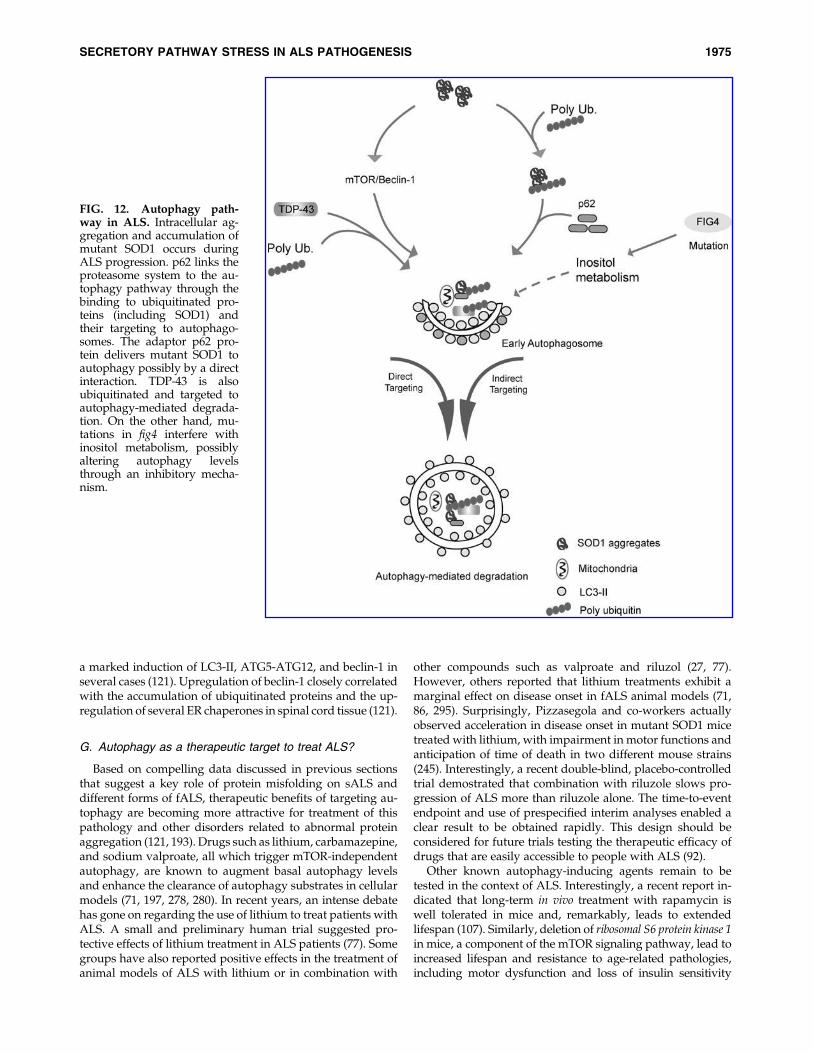
a marked induction of LC3-II, ATG5-ATG12, and beclin-1 inseveral cases (121). Upregulation of beclin-1 closely correlatedwith the accumulation of ubiquitinated proteins and the up-regulation of several ER chaperones in spinal cord tissue (121).
G. Autophagy as a therapeutic target to treat ALS?
Based on compelling data discussed in previous sectionsthat suggest a key role of protein misfolding on sALS anddifferent forms of fALS, therapeutic benefits of targeting au-tophagy are becoming more attractive for treatment of thispathology and other disorders related to abnormal proteinaggregation (121, 193). Drugs such as lithium, carbamazepine,and sodium valproate, all which trigger mTOR-independentautophagy, are known to augment basal autophagy levelsand enhance the clearance of autophagy substrates in cellularmodels (71, 197, 278, 280). In recent years, an intense debatehas gone on regarding the use of lithium to treat patients withALS. A small and preliminary human trial suggested pro-tective effects of lithium treatment in ALS patients (77). Somegroups have also reported positive effects in the treatment ofanimal models of ALS with lithium or in combination with
other compounds such as valproate and riluzol (27, 77).However, others reported that lithium treatments exhibit amarginal effect on disease onset in fALS animal models (71,86, 295). Surprisingly, Pizzasegola and co-workers actuallyobserved acceleration in disease onset in mutant SOD1 micetreated with lithium, with impairment in motor functions andanticipation of time of death in two different mouse strains(245). Interestingly, a recent double-blind, placebo-controlledtrial demostrated that combination with riluzole slows pro-gression of ALS more than riluzole alone. The time-to-eventendpoint and use of prespecified interim analyses enabled aclear result to be obtained rapidly. This design should beconsidered for future trials testing the therapeutic efficacy ofdrugs that are easily accessible to people with ALS (92).
Other known autophagy-inducing agents remain to betested in the context of ALS. Interestingly, a recent report in-dicated that long-term in vivo treatment with rapamycin iswell tolerated in mice and, remarkably, leads to extendedlifespan (107). Similarly, deletion of ribosomal S6 protein kinase 1in mice, a component of the mTOR signaling pathway, lead toincreased lifespan and resistance to age-related pathologies,including motor dysfunction and loss of insulin sensitivity
FIG. 12. Autophagy path-way in ALS. Intracellular ag-gregation and accumulation ofmutant SOD1 occurs duringALS progression. p62 links theproteasome system to the au-tophagy pathway through thebinding to ubiquitinated pro-teins (including SOD1) andtheir targeting to autophago-somes. The adaptor p62 pro-tein delivers mutant SOD1 toautophagy possibly by a directinteraction. TDP-43 is alsoubiquitinated and targeted toautophagy-mediated degrada-tion. On the other hand, mu-tations in fig4 interfere withinositol metabolism, possiblyaltering autophagy levelsthrough an inhibitory mecha-nism.
SECRETORY PATHWAY STRESS IN ALS PATHOGENESIS 1975
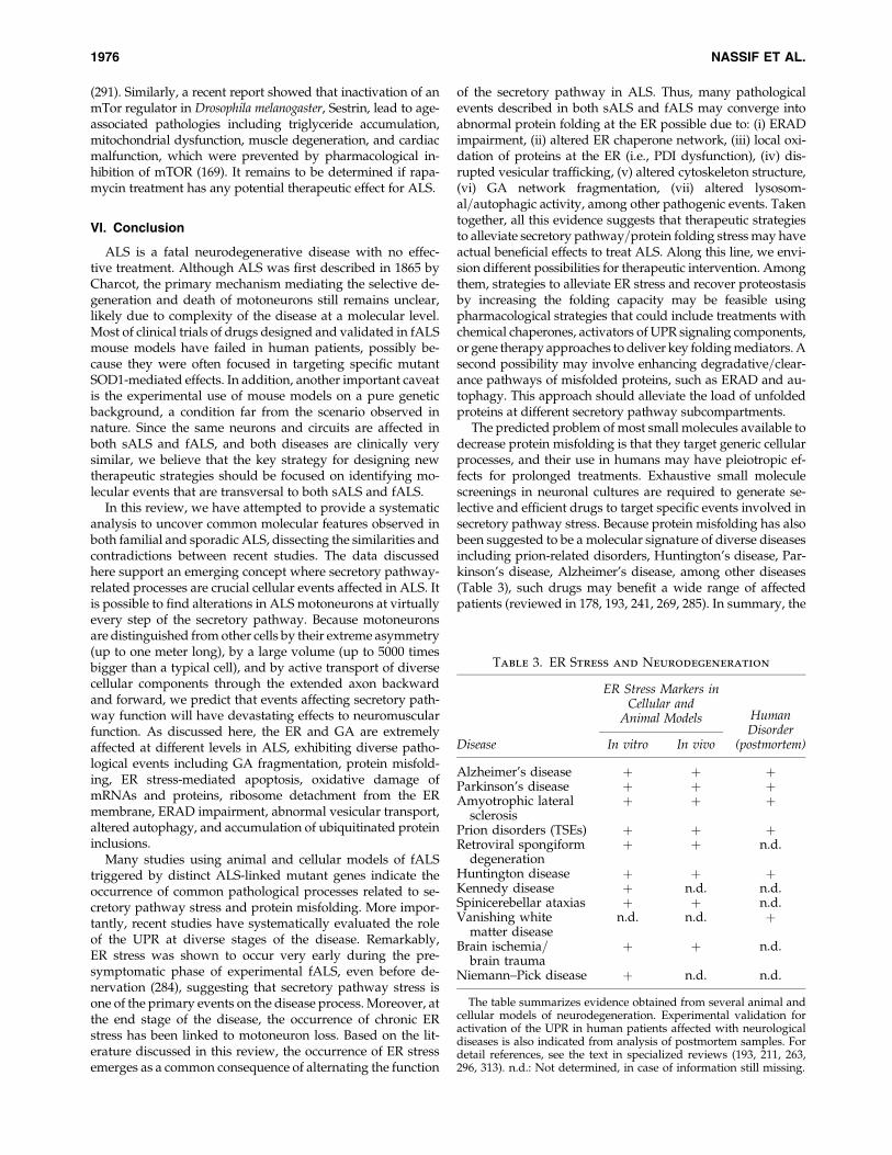
(291). Similarly, a recent report showed that inactivation of anmTor regulator in Drosophila melanogaster, Sestrin, lead to age-associated pathologies including triglyceride accumulation,mitochondrial dysfunction, muscle degeneration, and cardiacmalfunction, which were prevented by pharmacological in-hibition of mTOR (169). It remains to be determined if rapa-mycin treatment has any potential therapeutic effect for ALS.
VI. Conclusion
ALS is a fatal neurodegenerative disease with no effec-tive treatment. Although ALS was first described in 1865 byCharcot, the primary mechanism mediating the selective de-generation and death of motoneurons still remains unclear,likely due to complexity of the disease at a molecular level.Most of clinical trials of drugs designed and validated in fALSmouse models have failed in human patients, possibly be-cause they were often focused in targeting specific mutantSOD1-mediated effects. In addition, another important caveatis the experimental use of mouse models on a pure geneticbackground, a condition far from the scenario observed innature. Since the same neurons and circuits are affected inboth sALS and fALS, and both diseases are clinically verysimilar, we believe that the key strategy for designing newtherapeutic strategies should be focused on identifying mo-lecular events that are transversal to both sALS and fALS.
In this review, we have attempted to provide a systematicanalysis to uncover common molecular features observed inboth familial and sporadic ALS, dissecting the similarities andcontradictions between recent studies. The data discussedhere support an emerging concept where secretory pathway-related processes are crucial cellular events affected in ALS. Itis possible to find alterations in ALS motoneurons at virtuallyevery step of the secretory pathway. Because motoneuronsare distinguished from other cells by their extreme asymmetry(up to one meter long), by a large volume (up to 5000 timesbigger than a typical cell), and by active transport of diversecellular components through the extended axon backwardand forward, we predict that events affecting secretory path-way function will have devastating effects to neuromuscularfunction. As discussed here, the ER and GA are extremelyaffected at different levels in ALS, exhibiting diverse patho-logical events including GA fragmentation, protein misfold-ing, ER stress-mediated apoptosis, oxidative damage ofmRNAs and proteins, ribosome detachment from the ERmembrane, ERAD impairment, abnormal vesicular transport,altered autophagy, and accumulation of ubiquitinated proteininclusions.
Many studies using animal and cellular models of fALStriggered by distinct ALS-linked mutant genes indicate theoccurrence of common pathological processes related to se-cretory pathway stress and protein misfolding. More impor-tantly, recent studies have systematically evaluated the roleof the UPR at diverse stages of the disease. Remarkably,ER stress was shown to occur very early during the pre-symptomatic phase of experimental fALS, even before de-nervation (284), suggesting that secretory pathway stress isone of the primary events on the disease process. Moreover, atthe end stage of the disease, the occurrence of chronic ERstress has been linked to motoneuron loss. Based on the lit-erature discussed in this review, the occurrence of ER stressemerges as a common consequence of alternating the function
of the secretory pathway in ALS. Thus, many pathologicalevents described in both sALS and fALS may converge intoabnormal protein folding at the ER possible due to: (i) ERADimpairment, (ii) altered ER chaperone network, (iii) local oxi-dation of proteins at the ER (i.e., PDI dysfunction), (iv) dis-rupted vesicular trafficking, (v) altered cytoskeleton structure,(vi) GA network fragmentation, (vii) altered lysosom-al=autophagic activity, among other pathogenic events. Takentogether, all this evidence suggests that therapeutic strategiesto alleviate secretory pathway=protein folding stress may haveactual beneficial effects to treat ALS. Along this line, we envi-sion different possibilities for therapeutic intervention. Amongthem, strategies to alleviate ER stress and recover proteostasisby increasing the folding capacity may be feasible usingpharmacological strategies that could include treatments withchemical chaperones, activators of UPR signaling components,or gene therapy approaches to deliver key folding mediators. Asecond possibility may involve enhancing degradative=clear-ance pathways of misfolded proteins, such as ERAD and au-tophagy. This approach should alleviate the load of unfoldedproteins at different secretory pathway subcompartments.
The predicted problem of most small molecules available todecrease protein misfolding is that they target generic cellularprocesses, and their use in humans may have pleiotropic ef-fects for prolonged treatments. Exhaustive small moleculescreenings in neuronal cultures are required to generate se-lective and efficient drugs to target specific events involved insecretory pathway stress. Because protein misfolding has alsobeen suggested to be a molecular signature of diverse diseasesincluding prion-related disorders, Huntington’s disease, Par-kinson’s disease, Alzheimer’s disease, among other diseases(Table 3), such drugs may benefit a wide range of affectedpatients (reviewed in 178, 193, 241, 269, 285). In summary, the
Table 3. ER Stress and Neurodegeneration
ER Stress Markers inCellular and
Animal Models
Disease In vitro In vivo
HumanDisorder
(postmortem)
Alzheimer’s disease þ þ þParkinson’s disease þ þ þAmyotrophic lateral
sclerosisþ þ þ
Prion disorders (TSEs) þ þ þRetroviral spongiform
degenerationþ þ n.d.
Huntington disease þ þ þKennedy disease þ n.d. n.d.Spinicerebellar ataxias þ þ n.d.Vanishing white
matter diseasen.d. n.d. þ
Brain ischemia=brain trauma
þ þ n.d.
Niemann–Pick disease þ n.d. n.d.
The table summarizes evidence obtained from several animal andcellular models of neurodegeneration. Experimental validation foractivation of the UPR in human patients affected with neurologicaldiseases is also indicated from analysis of postmortem samples. Fordetail references, see the text in specialized reviews (193, 211, 263,296, 313). n.d.: Not determined, in case of information still missing.
1976 NASSIF ET AL.

evidence discussed support the involvement of secretorypathway stress in both sporadic and most familial ALS vari-ants, highlighting the notion that future therapeutic strategiesto alleviate organelle damage and improve secretory pathway-related functions may lead to effective and transversal treat-ments for ALS and other protein misfolding disorders.
Acknowledgments
The authors wish to acknowledge Benjamin Caballero andDiego Rojas–Rivera for help in designing the figures, and PeterThielen for editing. This work was supported primarily by theThe Muscular Dystrophy Association and Genzyme SponsorResearch Agreement (CH) and ALSA-The Milton SafenowitzPost-Doctoral Fellowship for ALS Research 1829 (SM). In ad-dition, we thank FONDECYT no. 1100176, FONDAP grant no.15010006, Millennium Nucleus no. P07-048-F, The Michael J.Fox Foundation for Parkinson’s Research, National ParkinsonFoundation, High Q Foundation, and ICGEB (CH); CONICYTPh.D fellowship no. D-21070046 (MN); FONDECYT no.3085017 (SM); FONDECYT no. 3100112 (KC).
References
1. Ameri K and Harris AL. Activating transcription factor 4.Int J Biochem Cell Biol 40: 14–21, 2008.
2. Atkin JD, Farg MA, Turner BJ, Tomas D, Lysaght JA, Nu-nan J, Rembach A, Nagley P, Beart PM, Cheema SS, andHorne MK. Induction of the unfolded protein response infamilial amyotrophic lateral sclerosis and association ofprotein-disulfide isomerase with superoxide dismutase 1. JBiol Chem 281: 30152–30165, 2006.
3. Atkin JD, Farg MA, Walker AK, McLean C, Tomas D, andHorne MK. Endoplasmic reticulum stress and induction ofthe unfolded protein response in human sporadic amyo-trophic lateral sclerosis. Neurobiol Dis 30: 400–4007, 2008.
4. Ayala YM, Zago P, D’Ambrogio A, Xu YF, Petrucelli L,Buratti E, and Baralle FE. Structural determinants of thecellular localization and shuttling of TDP-43. J Cell Sci 121:3778–3785, 2008.
5. Azzouz M. Gene therapy for ALS: Progress and prospects.Biochim Biophys Acta 1762: 1122–1127, 2006.
6. Azzouz M, Hottinger A, Paterna JC, Zurn AD, Aebischer P,and Bueler H. Increased motoneuron survival and im-proved neuromuscular function in transgenic ALS miceafter intraspinal injection of an adeno-associated virus en-coding Bcl-2. Hum Mol Genet 9: 803–811, 2000.
7. Babu JR, Geetha T, and Wooten MW. Sequestosome 1=p62shuttles polyubiquitinated tau for proteasomal degrada-tion. J Neurochem 94: 192–203, 2005.
8. Bacman SR, Bradley WG, and Moraes CT. Mitochondrialinvolvement in amyotrophic lateral sclerosis: Trigger ortarget? Mol Neurobiol 33: 113–131, 2006.
9. Banci L, Bertini I, Boca M, Girotto S, Martinelli M, Va-lentine JS, and Vieru M. SOD1 and amyotrophic lateralsclerosis: Mutations and oligomerization. PLoS One 3:e1677, 2008.
10. Barbeito LH, Pehar M, Cassina P, Vargas MR, Peluffo H,Viera L, Estevez AG, and Beckman JS. A role for astrocytesin motor neuron loss in amyotrophic lateral sclerosis. BrainRes Brain Res Rev 47: 263–274, 2004.
11. Barber SC, Mead RJ, and Shaw PJ. Oxidative stress in ALS:A mechanism of neurodegeneration and a therapeutic tar-get. Biochim Biophys Acta 1762: 1051–1067, 2006.
12. Barmada SJ, Skibinski G, Korb E, Rao EJ, Wu JY, andFinkbeiner S. Cytoplasmic mislocalization of TDP-43 istoxic to neurons and enhanced by a mutation associatedwith familial amyotrophic lateral sclerosis. J Neurosci 30:639–649, 2010.
13. Belzil VV, Valdmanis PN, Dion PA, Daoud H, Kabashi E,Noreau A, Gauthier J, Hince P, Desjarlais A, Bouchard JP,Lacomblez L, Salachas F, Pradat PF, Camu W, Meininger V,Dupre N, and Rouleau GA. Mutations in FUS cause FALSand SALS in French and French Canadian populations.Neurology 73: 1176–1179, 2009.
14. Bensimon G, Lacomblez L, and Meininger V. A controlledtrial of riluzole in amyotrophic lateral sclerosis.ALS=Riluzole Study Group. N Engl J Med 330: 585–591,1994.
15. Beranger F, Crozet C, Goldsborough A, and Lehmann S.Trehalose impairs aggregation of PrPSc molecules andprotects prion-infected cells against oxidative damage.Biochem Biophys Res Commun 374: 44–48, 2008.
16. Bertolotti A, Zhang Y, Hendershot LM, Harding HP, andRon D. Dynamic interaction of BiP and ER stress trans-ducers in the unfolded-protein response. Nat Cell Biol 2:326–332, 2000.
17. Bigini P, Atzori C, Fumagalli E, Cagnotto A, Barbera S,Migheli A, and Mennini T. Lack of caspase-dependent ap-optosis in spinal motor neurons of the wobbler mouse.Neurosci Lett 426: 106–110, 2007.
18. Bjorkoy G, Lamark T, Brech A, Outzen H, Perander M,Overvatn A, Stenmark H, and Johansen T. p62=SQSTM1forms protein aggregates degraded by autophagy and hasa protective effect on huntingtin-induced cell death. J CellBiol 171: 603–614, 2005.
19. Blais JD, Addison CL, Edge R, Falls T, Zhao H, Wary K,Koumenis C, Harding HP, Ron D, Holcik M, and Bell JC.Perk-dependent translational regulation promotes tumorcell adaptation and angiogenesis in response to hypoxicstress. Mol Cell Biol 26: 9517–9532, 2006.
20. Boillee S, Vande Velde C, and Cleveland DW. ALS: Adisease of motor neurons and their nonneuronal neighbors.Neuron 52: 39–59, 2006.
21. Bowling AC and Beal MF. Bioenergetic and oxidativestress in neurodegenerative diseases. Life Sci 56: 1151–1171, 1995.
22. Boyce M, Bryant KF, Jousse C, Long K, Harding HP,Scheuner D, Kaufman RJ, Ma D, Coen DM, Ron D,and Yuan J. A selective inhibitor of eIF2alpha dephos-phorylation protects cells from ER stress. Science 307: 935–939, 2005.
23. Bradley WG and Krasin F. A new hypothesis of the eti-ology of amyotrophic lateral sclerosis. The DNA hypothe-sis. Arch Neurol 39: 677–680, 1982.
24. Brunelle JK and Letai A. Control of mitochondrial apo-ptosis by the Bcl-2 family. J Cell Sci 122: 437–441, 2009.
25. Caccamo A, Majumder S, Deng JJ, Bai Y, Thornton FB, andOddo S. Rapamycin rescues TDP-43 mislocalization andthe associated low molecular mass neurofilament instabil-ity. J Biol Chem 284: 27416–27424, 2009.
26. Cai H, Lin X, Xie C, Laird FM, Lai C, Wen H, Chiang HC,Shim H, Farah MH, Hoke A, Price DL, and Wong PC. Lossof ALS2 function is insufficient to trigger motor neurondegeneration in knock-out mice but predisposes neurons tooxidative stress. J Neurosci 25: 7567–7574, 2005.
27. Caldero J, Brunet N, Tarabal O, Piedrafita L, Hereu M,Ayala V, and Esquerda JE. Lithium prevents excitotoxic cell
SECRETORY PATHWAY STRESS IN ALS PATHOGENESIS 1977

death of motoneurons in organotypic slice cultures of spi-nal cord. Neuroscience 165: 1353001369, 2009.
28. Calfon M, Zeng H, Urano F, Till JH, Hubbard SR, HardingHP, Clark SG, and Ron D. IRE1 couples endoplasmic re-ticulum load to secretory capacity by processing the XBP-1 mRNA. Nature 415: 92–96, 2002.
29. Carri MT, Ferri A, Battistoni A, Famhy L, Gabbianelli R,Poccia F, and Rotilio G. Expression of a Cu,Zn superoxidedismutase typical of familial amyotrophic lateral sclerosisinduces mitochondrial alteration and increase of cytosolicCa2þ concentration in transfected neuroblastoma SH-SY5Ycells. FEBS Lett 414: 365–368, 1997.
30. Cassina P, Peluffo H, Pehar M, Martinez–Palma L, RessiaA, Beckman JS, Estevez AG, and Barbeito L. Peroxynitritetriggers a phenotypic transformation in spinal cord astro-cytes that induces motor neuron apoptosis. J Neurosci Res67: 21–29, 2002.
31. Cimini V, Ruggiero G, Buonomo T, Seru R, Sciorio S, ZanziC, Santangelo F, and Mondola P. CuZn-superoxide dis-mutase in human thymus: Immunocytochemical localisationand secretion in thymus-derived epithelial and fibroblast celllines. Histochem Cell Biol 118: 163–169, 2002.
32. Clement AM, Nguyen MD, Roberts EA, Garcia ML, BoilleeS, Rule M, McMahon AP, Doucette W, Siwek D, FerranteRJ, Brown RH, Jr., Julien JP, Goldstein LS, and ClevelandDW. Wild-type nonneuronal cells extend survival of SOD1mutant motor neurons in ALS mice. Science 302: 113–117,2003.
33. Colombrita C, Zennaro E, Fallini C, Weber M, Sommacal A,Buratti E, Silani V, and Ratti A. TDP-43 is recruited to stressgranules in conditions of oxidative insult. J Neurochem 111:1051–1061, 2009.
34. Cookson MR, Menzies FM, Manning P, Eggett CJ, Figle-wicz DA, McNeil CJ, and Shaw PJ. Cu=Zn superoxidedismutase (SOD1) mutations associated with familialamyotrophic lateral sclerosis (ALS) affect cellular free rad-ical release in the presence of oxidative stress. AmyotrophLateral Scler Other Motor Neuron Disord 3: 75–85, 2002.
35. Corrado L, Del Bo R, Castellotti B, Ratti A, Cereda C, PencoS, Soraru G, Carlomagno Y, Ghezzi S, Pensato V, ColombritaC, Gagliardi S, Cozzi L, Orsetti V, Mancuso M, Siciliano G,Mazzini L, Comi GP, Gellera C, Ceroni M, D’Alfonso S, andSilani V. Mutations of FUS gene in sporadic amyotrophiclateral sclerosis. J Med Genet 47: 190–194, 2010.
36. Cozzolino M, Amori I, Pesaresi MG, Ferri A, Nencini M,and Carri MT. Cysteine 111 affects aggregation and cyto-toxicity of mutant Cu,Zn-superoxide dismutase associatedwith familial amyotrophic lateral sclerosis. J Biol Chem 283:866–874, 2008.
37. Cozzolino M, Ferri A, and Carri MT. Amyotrophic lateralsclerosis: From current developments in the laboratory toclinical implications. Antioxid Redox Signal 10: 405–443, 2008.
38. Credle JJ, Finer–Moore JS, Papa FR, Stroud RM, and WalterP. On the mechanism of sensing unfolded protein inthe endoplasmic reticulum. Proc Natl Acad Sci USA 102:18773–18784, 2005.
39. Crosio C, Casciati A, Iaccarino C, Rotilio G, and Carri MT.Bcl2a1 serves as a switch in death of motor neurons inamyotrophic lateral sclerosis. Cell Death Differ 13: 2150–2153, 2006.
40. Croul S, Mezitis SG, Stieber A, Chen YJ, Gonatas JO, GoudB, and Gonatas NK. Immunocytochemical visualization ofthe Golgi apparatus in several species, including human,and tissues with an antiserum against MG-160, a sialogly-
coprotein of rat Golgi apparatus. J Histochem Cytochem 38:957–963, 1990.
41. Cudkowicz ME, McKenna–Yasek D, Sapp PE, Chin W,Geller B, Hayden DL, Schoenfeld DA, Hosler BA, HorvitzHR, and Brown RH. Epidemiology of mutations in super-oxide dismutase in amyotrophic lateral sclerosis. AnnNeurol 41: 210–221, 1997.
42. Cudkowicz ME, Shefner JM, Schoenfeld DA, Zhang H,Andreasson KI, Rothstein JD, and Drachman DB. Trial ofcelecoxib in amyotrophic lateral sclerosis. Ann Neurol 60:22–31, 2006.
43. Cudkowicz ME, Shefner JM, Simpson E, Grasso D, Yu H,Zhang H, Shui A, Schoenfeld D, Brown RH, Wieland S, andBarber JR. Arimoclomol at dosages up to 300 mg=day iswell tolerated and safe in amyotrophic lateral sclerosis.Muscle Nerve 38: 837–844, 2008.
44. Cuozzo JW and Kaiser CA. Competition between glutathi-one and protein thiols for disulphide-bond formation. NatCell Biol 1: 130–135, 1999.
45. Chandran J, Ding J, and Cai H. Alsin and the molecularpathways of amyotrophic lateral sclerosis. Mol Neurobiol 36:224–231, 2007.
46. Chang Y, Kong Q, Shan X, Tian G, Ilieva H, Cleveland DW,Rothstein JD, Borchelt DR, Wong PC, and Lin CL. Mes-senger RNA oxidation occurs early in disease pathogenesisand promotes motor neuron degeneration in ALS. PLoSOne 3: e2849, 2008.
47. Chattopadhyay M and Valentine JS. Aggregation of cop-per-zinc superoxide dismutase in familial and sporadicALS. Antioxid Redox Signal 11: 1603–1614, 2009.
48. Chen X, Shen J, and Prywes R. The luminal domain ofATF6 senses endoplasmic reticulum (ER) stress and causestranslocation of ATF6 from the ER to the Golgi. J Biol Chem277: 13045–13052, 2002.
49. Cheong H and Klionsky DJ. Dual role of Atg1 in regulationof autophagy-specific PAS assembly in Saccharomyces cere-visiae. Autophagy 4: 724–726, 2008.
50. Chevalier–Larsen ES, Wallace KE, Pennise CR, and Holz-baur EL. Lysosomal proliferation and distal degenerationin motor neurons expressing the G59S mutation in thep150Glued subunit of dynactin. Hum Mol Genet 17: 1946–1955, 2008.
51. Chiu R, Novikov L, Mukherjee S, and Shields D. A caspasecleavage fragment of p115 induces fragmentation of theGolgi apparatus and apoptosis. J Cell Biol 159: 637–648,2002.
52. Cho DH, Nakamura T, Fang J, Cieplak P, Godzik A, Gu Z,and Lipton SA. S-nitrosylation of Drp1 mediates beta-amyloid-related mitochondrial fission and neuronal injury.Science 324: 102–105, 2009.
53. Chow CY, Landers JE, Bergren SK, Sapp PC, Grant AE, JonesJM, Everett L, Lenk GM, McKenna–Yasek DM, Weisman LS,Figlewicz D, Brown RH, and Meisler MH. Deleterious vari-ants of FIG4, a phosphoinositide phosphatase, in patientswith ALS. Am J Hum Genet 84: 85–88, 2009.
54. Chow CY, Zhang Y, Dowling JJ, Jin N, Adamska M, ShigaK, Szigeti K, Shy ME, Li J, Zhang X, Lupski JR, WeismanLS, and Meisler MH. Mutation of FIG4 causes neurode-generation in the pale tremor mouse and patients withCMT4J. Nature 448: 68–72, 2007.
55. Chung KK, Thomas B, Li X, Pletnikova O, Troncoso JC,Marsh L, Dawson VL, and Dawson TM. S-nitrosylation ofparkin regulates ubiquitination and compromises parkin’sprotective function. Science 304: 1328–1331, 2004.
1978 NASSIF ET AL.

56. Dahmer MK. Caspases-2, -3, and -7 are involved in thap-sigargin-induced apoptosis of SH-SY5Y neuroblastomacells. J Neurosci Res 80: 576–583, 2005.
57. Danial NN and Korsmeyer SJ. Cell death: Critical controlpoints. Cell 116: 205–219, 2004.
58. Davies JE, Sarkar S, and Rubinsztein DC. Trehalose reducesaggregate formation and delays pathology in a transgenicmouse model of oculopharyngeal muscular dystrophy.Hum Mol Genet 15: 23–31, 2006.
59. Deng HX, Shi Y, Furukawa Y, Zhai H, Fu R, Liu E, GorrieGH, Khan MS, Hung WY, Bigio EH, Lukas T, Dal CantoMC, O’Halloran TV, and Siddique T. Conversion to theamyotrophic lateral sclerosis phenotype is associated withintermolecular linked insoluble aggregates of SOD1 in mi-tochondria. Proc Natl Acad Sci USA 103: 7142–7147, 2006.
60. Devon RS, Orban PC, Gerrow K, Barbieri MA, Schwab C,Cao LP, Helm JR, Bissada N, Cruz–Aguado R, DavidsonTL, Witmer J, Metzler M, Lam CK, Tetzlaff W, Simpson EM,McCaffery JM, El–Husseini AE, Leavitt BR, and HaydenMR. Als2-deficient mice exhibit disturbances in endosometrafficking associated with motor behavioral abnormalities.Proc Natl Acad Sci USA 103: 9595–9600, 2006.
61. Dickson BJ. Rho GTPases in growth cone guidance. CurrOpin Neurobiol 11: 103–110, 2001.
62. Ding WX and Yin XM. Sorting, recognition and activation ofthe misfolded protein degradation pathways through mac-roautophagy and the proteasome. Autophagy 4: 141–150, 2008.
63. Dion PA, Daoud H, and Rouleau GA. Genetics of motorneuron disorders: New insights into pathogenic mecha-nisms. Nat Rev Genet 10: 769–782, 2009.
64. Dobrowolny G, Aucello M, Rizzuto E, Beccafico S, Mam-mucari C, Boncompagni S, Belia S, Wannenes F, Nicoletti C,Del Prete Z, Rosenthal N, Molinaro M, Protasi F, Fano G,Sandri M, and Musaro A. Skeletal muscle is a primarytarget of SOD1G93A-mediated toxicity. Cell Metab 8: 425–436, 2008.
65. Dove SK, Dong K, Kobayashi T, Williams FK, and Michell RH.Phosphatidylinositol 3,5-bisphosphate and Fab1p=PIKfyveunder PPI n endo-lysosome function. Biochem J 419: 1–13, 2009.
66. Dupuis L, Gonzalez de Aguilar JL, di Scala F, Rene F, deTapia M, Pradat PF, Lacomblez L, Seihlan D, Prinjha R,Walsh FS, Meininger V, and Loeffler JP. Nogo provides amolecular marker for diagnosis of amyotrophic lateralsclerosis. Neurobiol Dis 10: 358–365, 2002.
67. Dupuis L, Gonzalez de Aguilar JL, Oudart H, de Tapia M,Barbeito L, and Loeffler JP. Mitochondria in amyotrophiclateral sclerosis: A trigger and a target. Neurodegener Dis 1:245–254, 2004.
68. Dupuis L, Muller A, Meininger V, and Loeffler JP. [Mole-cular mechanisms of amyotrophic lateral sclerosis: Recentcontributions from studies in animal models]. Rev Neurol(Paris) 160: 35–43, 2004.
69. Ellgaard L and Helenius A. Quality control in the endo-plasmic reticulum. Nat Rev Mol Cell Biol 4: 181–191, 2003.
70. Farquhar MG. Progress in unraveling pathways of Golgitraffic. Annu Rev Cell Biol 1: 447–488, 1985.
71. Feng HL, Leng Y, Ma CH, Zhang J, Ren M, and ChuangDM. Combined lithium and valproate treatment delaysdisease onset, reduces neurological deficits and prolongssurvival in an amyotrophic lateral sclerosis mouse model.Neuroscience 155: 567–572, 2008.
72. Ferguson CJ, Lenk GM, and Meisler MH. Defective au-tophagy in neurons and astrocytes from mice deficient inPI(3,5)P2. Hum Mol Genet 18: 4868–4878, 2009.
73. Ferrante RJ, Browne SE, Shinobu LA, Bowling AC, Baik MJ,MacGarvey U, Kowall NW, Brown RH, Jr., and Beal MF.Evidence of increased oxidative damage in both sporadicand familial amyotrophic lateral sclerosis. J Neurochem 69:2064–2074, 1997.
74. Ferrante RJ, Shinobu LA, Schulz JB, Matthews RT, ThomasCE, Kowall NW, Gurney ME, and Beal MF. Increased 3-nitrotyrosine and oxidative damage in mice with a humancopper=zinc superoxide dismutase mutation. Ann Neurol42: 326–334, 1997.
75. Ferri A, Cozzolino M, Crosio C, Nencini M, Casciati A,Gralla EB, Rotilio G, Valentine JS, and Carri MT. FamilialALS-superoxide dismutases associate with mitochondriaand shift their redox potentials. Proc Natl Acad Sci USA 103:13860–13865, 2006.
76. Filimonenko M, Stuffers S, Raiborg C, Yamamoto A, Mal-erod L, Fisher EM, Isaacs A, Brech A, Stenmark H, and Si-monsen A. Functional multivesicular bodies are required forautophagic clearance of protein aggregates associated withneurodegenerative disease. J Cell Biol 179: 485–500, 2007.
77. Fornai F, Longone P, Cafaro L, Kastsiuchenka O, FerrucciM, Manca ML, Lazzeri G, Spalloni A, Bellio N, Lenzi P,Modugno N, Siciliano G, Isidoro C, Murri L, Ruggieri S,and Paparelli A. Lithium delays progression of amyo-trophic lateral sclerosis. Proc Natl Acad Sci USA 105: 2052–2057, 2008.
78. Freibaum BD, Chitta RK, High AA, and Taylor JP. Globalanalysis of TDP-43 interacting proteins reveals strong as-sociation with RNA splicing and translation machinery. JProteome Res 9: 1104–1120, 2010.
79. Fujita Y, Mizuno Y, Takatama M, and Okamoto K. Anteriorhorn cells with abnormal TDP-43 immunoreactivities showfragmentation of the Golgi apparatus in ALS. J Neurol Sci269: 30–34, 2008.
80. Fujita Y and Okamoto K. Golgi apparatus of the motorneurons in patients with amyotrophic lateral sclerosis and inmice models of amyotrophic lateral sclerosis. Neuropathology25: 388–394, 2005.
81. Fukada K, Zhang F, Vien A, Cashman NR, and Zhu H.Mitochondrial proteomic analysis of a cell line model offamilial amyotrophic lateral sclerosis. Mol Cell Proteomics 3:1211–1223, 2004.
82. Furukawa Y, Fu R, Deng HX, Siddique T, and O’HalloranTV. Disulfide cross-linked protein represents a significantfraction of ALS-associated Cu, Zn-superoxide dismutaseaggregates in spinal cords of model mice. Proc Natl Acad SciUSA 103: 7148–7153, 2006.
83. Gal J, Strom AL, Kilty R, Zhang F, and Zhu H. p62 accu-mulates and enhances aggregate formation in model sys-tems of familial amyotrophic lateral sclerosis. J Biol Chem282: 11068–11077, 2007.
84. Gal J, Strom AL, Kwinter DM, Kilty R, Zhang J, Shi P, FuW, Wooten MW, and Zhu H. Sequestosome 1=p62 linksfamilial ALS mutant SOD1 to LC3 via an ubiquitin-inde-pendent mechanism. J Neurochem 111: 1062–1073, 2009.
85. Gidalevitz T, Krupinski T, Garcia S, and Morimoto RI.Destabilizing protein polymorphisms in the genetic back-ground direct phenotypic expression of mutant SOD1toxicity. PLoS Genet 5: e1000399, 2009.
86. Gill A, Kidd J, Vieira F, Thompson K, and Perrin S. Nobenefit from chronic lithium dosing in a sibling-matched,gender balanced, investigator-blinded trial using a stan-dard mouse model of familial ALS. PLoS One 4: e6489,2009.
SECRETORY PATHWAY STRESS IN ALS PATHOGENESIS 1979

87. Gitcho MA, Bigio EH, Mishra M, Johnson N, Weintraub S,Mesulam M, Rademakers R, Chakraverty S, Cruchaga C,Morris JC, Goate AM, and Cairns NJ. TARDBP 3’-UTRvariant in autopsy-confirmed frontotemporal lobar degen-eration with TDP-43 proteinopathy. Acta Neuropathol 118:633–645, 2009.
88. Gkogkas C, Middleton S, Kremer AM, Wardrope C, Han-nah M, Gillingwater TH, and Skehel P. VAPB interacts withand modulates the activity of ATF6. Hum Mol Genet 17:1517–1526, 2008.
88a. Glimcher LH, Hetz C. XBP-1 and the UPRosome: Masteringsecretory cell function. Curr Immunol Rev 4: 1–10, 2008.
89. Gomes C, Keller S, Altevogt P, and Costa J. Evidence forsecretion of Cu,Zn superoxide dismutase via exosomesfrom a cell model of amyotrophic lateral sclerosis. NeurosciLett 428: 43–46, 2007.
90. Gonatas NK. Rous–Whipple Award Lecture. Contributionsto the physiology and pathology of the Golgi apparatus.Am J Pathol 145: 751–761, 1994.
91. Gonatas NK, Stieber A, Mourelatos Z, Chen Y, GonatasJO, Appel SH, Hays AP, Hickey WF, and Hauw JJ. Frag-mentation of the Golgi apparatus of motor neurons inamyotrophic lateral sclerosis. Am J Pathol 140: 731–737,1992.
92. Gordon PH, Moore DH, Miller RG, Florence JM, VerheijdeJL, Doorish C, Hilton JF, Spitalny GM, MacArthur RB,Mitsumoto H, Neville HE, Boylan K, Mozaffar T, Belsh JM,Ravits J, Bedlack RS, Graves MC, McCluskey LF, BarohnRJ, and Tandan R. Efficacy of minocycline in patients withamyotrophic lateral sclerosis: A phase III randomised trial.Lancet Neurol 6: 1045–1053, 2007.
93. Gould TW, Buss RR, Vinsant S, Prevette D, Sun W,Knudson CM, Milligan CE, and Oppenheim RW. Completedissociation of motor neuron death from motor dysfunctionby Bax deletion in a mouse model of ALS. J Neurosci 26:8774–8786, 2006.
94. Gros-Louis F, Gaspar C, and Rouleau GA. Genetics of fa-milial and sporadic amyotrophic lateral sclerosis. BiochimBiophys Acta 1762: 956–972, 2006.
95. Gros-Louis F, Kriz J, Kabashi E, McDearmid J, MillecampsS, Urushitani M, Lin L, Dion P, Zhu Q, Drapeau P, JulienJP, and Rouleau GA. Als2 mRNA splicing variants detectedin KO mice rescue severe motor dysfunction phenotype inAls2 knock-down zebrafish. Hum Mol Genet 17: 2691–2702,2008.
96. Gruenberg J and Stenmark H. The biogenesis of multi-vesicular endosomes. Nat Rev Mol Cell Biol 5: 317–323, 2004.
97. Gu F, Nguyen DT, Stuible M, Dube N, Tremblay ML, andChevet E. Protein-tyrosine phosphatase 1B potentiates IRE1signaling during endoplasmic reticulum stress. J Biol Chem279: 49689–49693, 2004.
98. Hadano S, Benn SC, Kakuta S, Otomo A, Sudo K, Kunita R,Suzuki–Utsunomiya K, Mizumura H, Shefner JM, Cox GA,Iwakura Y, Brown RH, Jr., and Ikeda JE. Mice deficient inthe Rab5 guanine nucleotide exchange factor ALS2=alsinexhibit age-dependent neurological deficits and alteredendosome trafficking. Hum Mol Genet 15: 233–250, 2006.
99. Hadano S, Hand CK, Osuga H, Yanagisawa Y, Otomo A,Devon RS, Miyamoto N, Showguchi-Miyata J, Okada Y,Singaraja R, Figlewicz DA, Kwiatkowski T, Hosler BA, SagieT, Skaug J, Nasir J, Brown RH, Jr., Scherer SW, Rouleau GA,Hayden MR, and Ikeda JE. A gene encoding a putativeGTPase regulator is mutated in familial amyotrophic lateralsclerosis 2. Nat Genet 29: 166–173, 2001.
100. Hall ED, Andrus PK, Oostveen JA, Fleck TJ, and GurneyME. Relationship of oxygen radical-induced lipid perox-idative damage to disease onset and progression in atransgenic model of familial ALS. J Neurosci Res 53: 66–77,1998.
101. Hamman BD, Hendershot LM, and Johnson AE. BiPmaintains the permeability barrier of the ER membrane bysealing the lumenal end of the translocon pore before andearly in translocation. Cell 92: 747–758, 1998.
102. Hammerschlag R, Stone GC, Bolen FA, Lindsey JD, andEllisman MH. Evidence that all newly synthesized proteinsdestined for fast axonal transport pass through the Golgiapparatus. J Cell Biol 93: 568–575, 1982.
103. Han D, Lerner AG, Vande Walle L, Upton JP, Xu W, HagenA, Backes BJ, Oakes SA, and Papa FR. IRE1alpha kinase ac-tivation modes control alternate endoribonuclease outputs todetermine divergent cell fates. Cell 138: 562–575, 2009.
104. Hara T, Nakamura K, Matsui M, Yamamoto A, NakaharaY, Suzuki–Migishima R, Yokoyama M, Mishima K, Saito I,Okano H, and Mizushima N. Suppression of basal autop-hagy in neural cells causes neurodegenerative disease inmice. Nature 441: 885–889, 2006.
105. Harding HP, Zhang Y, and Ron D. Protein translation andfolding are coupled by an endoplasmic-reticulum-residentkinase. Nature 397: 271–274, 1999.
106. Harding HP, Zhang Y, Zeng H, Novoa I, Lu PD, Calfon M,Sadri N, Yun C, Popko B, Paules R, Stojdl DF, Bell JC,Hettmann T, Leiden JM, and Ron D. An integrated stressresponse regulates amino acid metabolism and resistanceto oxidative stress. Mol Cell 11: 619–633, 2003.
107. Harrison DE, Strong R, Sharp ZD, Nelson JF, Astle CM,Flurkey K, Nadon NL, Wilkinson JE, Frenkel K, Carter CS,Pahor M, Javors MA, Fernandez E, and Miller RA. Rapa-mycin fed late in life extends lifespan in genetically het-erogeneous mice. Nature 460: 392–395, 2009.
108. Hartl FU and Hayer–Hartl M. Converging concepts ofprotein folding in vitro and in vivo. Nat Struct Mol Biol 16:574–581, 2009.
109. Haynes CM, Titus EA, and Cooper AA. Degradation ofmisfolded proteins prevents ER-derived oxidative stressand cell death. Mol Cell 15: 767–0776, 2004.
110. Haze K, Yoshida H, Yanagi H, Yura T, and Mori K.Mammalian transcription factor ATF6 is synthesized as atransmembrane protein and activated by proteolysis inresponse to endoplasmic reticulum stress. Mol Biol Cell 10:3787–3799, 1999.
111. He C and Klionsky DJ. Regulation mechanisms and signal-ing pathways of autophagy. Annu Rev Genet 43: 67–93, 2009.
112. Heath–Engel HM, Chang NC, and Shore GC. The endo-plasmic reticulum in apoptosis and autophagy: role of theBCL-2 protein family. Oncogene 27: 6419–6433, 2008.
113. Hensley K, Mhatre M, Mou S, Pye QN, Stewart C, West M,and Williamson KS. On the relation of oxidative stress toneuroinflammation: Lessons learned from the G93A-SOD1mouse model of amyotrophic lateral sclerosis. AntioxidRedox Signal 8: 2075–2087, 2006.
114. Hervias I, Beal MF, and Manfredi G. Mitochondrial dys-function and amyotrophic lateral sclerosis. Muscle Nerve 33:598–608, 2006.
115. Hetz C, Bernasconi P, Fisher J, Lee AH, Bassik MC, An-tonsson B, Brandt GS, Iwakoshi NN, Schinzel A, GlimcherLH, and Korsmeyer SJ. Proapoptotic BAX and BAK mod-ulate the unfolded protein response by a direct interactionwith IRE1alpha. Science 312: 572–576, 2006.
1980 NASSIF ET AL.

116. Hetz C and Glimcher LH. Fine-tuning of the unfoldedprotein response: Assembling the IRE1alpha interactome.Mol Cell 35: 551–561, 2009.
117. Hetz C, Lee AH, Gonzalez-Romero D, Thielen P, Castilla J,Soto C, and Glimcher LH. Unfolded protein responsetranscription factor XBP-1 does not influence prion repli-cation or pathogenesis. Proc Natl Acad Sci USA 105: 757–762, 2008.
118. Hetz C, Russelakis-Carneiro M, Maundrell K, Castilla J,and Soto C. Caspase-12 and endoplasmic reticulum stressmediate neurotoxicity of pathological prion protein. EMBOJ 22: 5435–5445, 2003.
119. Hetz C, Russelakis–Carneiro M, Walchli S, Carboni S, Vial–Knecht E, Maundrell K, Castilla J, and Soto C. The disulfideisomerase Grp58 is a protective factor against prion neu-rotoxicity. J Neurosci 25: 2793–2802, 2005.
120. Hetz C, Thielen P, Fisher J, Pasinelli P, Brown RH, KorsmeyerS, and Glimcher L. The proapoptotic BCL-2 family memberBIM mediates motoneuron loss in a model of amyotrophiclateral sclerosis. Cell Death Differ 14: 1386–1389, 2007.
121. Hetz C, Thielen P, Matus S, Nassif M, Court F, Kiffin R,Martinez G, Cuervo AM, Brown RH, and Glimcher LH.XBP-1 deficiency in the nervous system protects againstamyotrophic lateral sclerosis by increasing autophagy.Genes Dev 23: 2294–2306, 2009.
122. This reference has been deleted.123. Higgins CM, Jung C, Ding H, and Xu Z. Mutant Cu, Zn
superoxide dismutase that causes motoneuron degenera-tion is present in mitochondria in the CNS. J Neurosci 22:RC215, 2002.
124. Hirao K, Natsuka Y, Tamura T, Wada I, Morito D, NatsukaS, Romero P, Sleno B, Tremblay LO, Herscovics A, NagataK, and Hosokawa N. EDEM3, a soluble EDEM homolog,enhances glycoprotein endoplasmic reticulum-associateddegradation and mannose trimming. J Biol Chem 281: 9650–9658, 2006.
125. Hollien J, Lin JH, Li H, Stevens N, Walter P, and WeissmanJS. Regulated Ire1-dependent decay of messenger RNAs inmammalian cells. J Cell Biol 186: 323–331, 2009.
126. Hollien J and Weissman JS. Decay of endoplasmic reticu-lum-localized mRNAs during the unfolded protein re-sponse. Science 313: 104–107, 2006.
127. Hoogenraad CC, Wulf P, Schiefermeier N, Stepanova T,Galjart N, Small JV, Grosveld F, de Zeeuw CI, and AkhmanovaA. Bicaudal D induces selective dynein-mediated microtubuleminus end-directed transport. EMBO J 22: 6004–6015, 2003.
128. Hu P, Han Z, Couvillon AD, Kaufman RJ, and Exton JH.Autocrine tumor necrosis factor alpha links endoplasmicreticulum stress to the membrane death receptor pathwaythrough IRE1alpha-mediated NF-kappaB activation anddown-regulation of TRAF2 expression. Mol Cell Biol 26:3071–3084, 2006.
129. Ilieva EV, Ayala V, Jove M, Dalfo E, Cacabelos D, Pove-dano M, Bellmunt MJ, Ferrer I, Pamplona R, and Portero–Otin M. Oxidative and endoplasmic reticulum stress in-terplay in sporadic amyotrophic lateral sclerosis. Brain 130:3111–3123, 2007.
130. Ishigaki S, Niwa J, Yamada S, Takahashi M, Ito T, Sone J,Doyu M, Urano F, and Sobue G. Dorfin-CHIP chimericproteins potently ubiquitylate and degrade familial ALS-related mutant SOD1 proteins and reduce their cellulartoxicity. Neurobiol Dis 25: 331–341, 2007.
131. Ito Y, Yamada M, Tanaka H, Aida K, Tsuruma K, Shima-zawa M, Hozumi I, Inuzuka T, Takahashi H, and Hara H.
Involvement of CHOP, an ER-stress apoptotic mediator, inboth human sporadic ALS and ALS model mice. NeurobiolDis 36: 470–476, 2009.
132. Jonsson PA, Graffmo KS, Brannstrom T, Nilsson P, AndersenPM, and Marklund SL. Motor neuron disease in mice ex-pressing the wild type-like D90A mutant superoxide dis-mutase-1. J Neuropathol Exp Neurol 65: 1126–1136, 2006.
133. Ju JS, Fuentealba RA, Miller SE, Jackson E, Piwnica–WormsD, Baloh RH, and Weihl CC. Valosin-containing protein(VCP) is required for autophagy and is disrupted in VCPdisease. J Cell Biol 187: 875–888, 2009.
134. Jung C, Higgins CM, and Xu Z. Mitochondrial electrontransport chain complex dysfunction in a transgenic mousemodel for amyotrophic lateral sclerosis. J Neurochem 83:535–545, 2002.
135. Jung C, Rong Y, Doctrow S, Baudry M, Malfroy B, and XuZ. Synthetic superoxide dismutase=catalase mimetics re-duce oxidative stress and prolong survival in a mouseamyotrophic lateral sclerosis model. Neurosci Lett 304: 157–160, 2001.
136. Kabashi E, Lin L, Tradewell ML, Dion PA, Bercier V,Bourgouin P, Rochefort D, Bel Hadj S, Durham HD, VeldeCV, Rouleau GA, and Drapeau P. Gain and loss of functionof ALS-related mutations of TARDBP (TDP-43) cause mo-tor deficits in vivo. Hum Mol Genet 19: 3102–3110, 2010.
137. Kabuta T, Suzuki Y, and Wada K. Degradation of amyo-trophic lateral sclerosis-linked mutant Cu,Zn-superoxidedismutase proteins by macroautophagy and the protea-some. J Biol Chem 281: 30524–30533, 2006.
138. Kaiser SE, Brickner JH, Reilein AR, Fenn TD, Walter P, andBrunger AT. Structural basis of FFAT motif-mediated ERtargeting. Structure 13: 1035–1045, 2005.
139. Kanda H and Miura M. Regulatory roles of JNK in pro-grammed cell death. J Biochem 136: 1–6, 2004.
140. Kanekura K, Nishimoto I, Aiso S, and Matsuoka M.Characterization of amyotrophic lateral sclerosis-linkedP56S mutation of vesicle-associated membrane protein-associated protein B (VAPB=ALS8). J Biol Chem 281: 30223–30233, 2006.
141. Karch CM, Prudencio M, Winkler DD, Hart PJ, andBorchelt DR. Role of mutant SOD1 disulfide oxidation andaggregation in the pathogenesis of familial ALS. Proc NatlAcad Sci USA 106: 7774–7779, 2009.
142. Karlsson J, Fong KS, Hansson MJ, Elmer E, Csiszar K, andKeep MF. Lifespan extension and reduced neuronal deathafter weekly intraventricular cyclosporin injections in theG93A transgenic mouse model of amyotrophic lateralsclerosis. J Neurosurg 101: 128–137, 2004.
143. Katzmann DJ, Odorizzi G, and Emr SD. Receptor down-regulation and multivesicular-body sorting. Nat Rev MolCell Biol 3: 893–905, 2002.
144. Kaufman RJ. Regulation of mRNA translation by proteinfolding in the endoplasmic reticulum. Trends Biochem Sci 29:152–158, 2004.
145. Keene CD, Rodrigues CM, Eich T, Chhabra MS, Steer CJ,and Low WC. Tauroursodeoxycholic acid, a bile acid, isneuroprotective in a transgenic animal model of Hun-tington’s disease. Proc Natl Acad Sci USA 99: 10671–10676,2002.
146. Keep M, Elmer E, Fong KS, and Csiszar K. Intrathecal cy-closporin prolongs survival of late-stage ALS mice. BrainRes 894: 327–331, 2001.
147. Kieran D, Hafezparast M, Bohnert S, Dick JR, Martin J,Schiavo G, Fisher EM, and Greensmith L. A mutation in
SECRETORY PATHWAY STRESS IN ALS PATHOGENESIS 1981

dynein rescues axonal transport defects and extends thelifespan of ALS mice. J Cell Biol 169: 561–567, 2005.
148. Kieran D, Woods I, Villunger A, Strasser A, and Prehn JH.Deletion of the BH3-only protein puma protects motoneu-rons from ER stress-induced apoptosis and delays moto-neuron loss in ALS mice. Proc Natl Acad Sci USA 104:20606–20611, 2007.
149. Kikuchi H, Almer G, Yamashita S, Guegan C, Nagai M, XuZ, Sosunov AA, McKhann GM, 2nd, and Przedborski S.Spinal cord endoplasmic reticulum stress associated with amicrosomal accumulation of mutant superoxide dismutase-1 in an ALS model. Proc Natl Acad Sci USA 103: 6025–6030,2006.
150. Kim H, Rafiuddin–Shah M, Tu HC, Jeffers JR, Zambetti GP,Hsieh JJ, and Cheng EH. Hierarchical regulation of mito-chondrion-dependent apoptosis by BCL-2 subfamilies. NatCell Biol 8: 1348–1358, 2006.
151. Kim I, Shu CW, Xu W, Shiau CW, Grant D, Vasile S,Cosford ND, and Reed JC. Chemical biology investigationof cell death pathways activated by endoplasmic reticulumstress reveals cytoprotective modulators of ASK1. J BiolChem 284: 1593–1603, 2009.
152. Kimata Y, Ishiwata-Kimata Y, Ito T, Hirata A, Suzuki T,Oikawa D, Takeuchi M, and Kohno K. Two regulatorysteps of ER-stress sensor Ire1 involving its cluster formationand interaction with unfolded proteins. J Cell Biol 179: 75–86, 2007.
153. Kimata Y, Kimata YI, Shimizu Y, Abe H, Farcasanu IC,Takeuchi M, Rose MD, and Kohno K. Genetic evidence fora role of BiP=Kar2 that regulates Ire1 in response to accu-mulation of unfolded proteins. Mol Biol Cell 14: 2559–2569,2003.
154. Kirby J, Halligan E, Baptista MJ, Allen S, Heath PR, HoldenH, Barber SC, Loynes CA, Wood–Allum CA, Lunec J, andShaw PJ. Mutant SOD1 alters the motor neuronal tran-scriptome: Implications for familial ALS. Brain 128: 1686–1706, 2005.
155. Klee M, Pallauf K, Alcala S, Fleischer A, and Pimentel–Muinos FX. Mitochondrial apoptosis induced by BH3-onlymolecules in the exclusive presence of endoplasmic retic-ular Bak. EMBO J 28: 1757–1768, 2009.
156. Komatsu M, Waguri S, Chiba T, Murata S, Iwata J, TanidaI, Ueno T, Koike M, Uchiyama Y, Kominami E, and TanakaK. Loss of autophagy in the central nervous system causesneurodegeneration in mice. Nature 441: 880–884, 2006.
157. Kong J and Xu Z. Massive mitochondrial degeneration inmotor neurons triggers the onset of amyotrophic lateralsclerosis in mice expressing a mutant SOD1. J Neurosci 18:3241–3250, 1998.
158. Kouroku Y, Fujita E, Tanida I, Ueno T, Isoai A, Kumagai H,Ogawa S, Kaufman RJ, Kominami E, and Momoi T. ERstress (PERK=eIF2alpha phosphorylation) mediates thepolyglutamine-induced LC3 conversion, an essential stepfor autophagy formation. Cell Death Differ 14: 230–239, 2007.
159. Kuusisto E, Salminen A, and Alafuzoff I. Ubiquitin-bindingprotein p62 is present in neuronal and glial inclusions inhuman tauopathies and synucleinopathies. Neuroreport 12:2085–2090, 2001.
160. Kwiatkowski TJ, Jr., Bosco DA, Leclerc AL, Tamrazian E,Vanderburg CR, Russ C, Davis A, Gilchrist J, Kasarskis EJ,Munsat T, Valdmanis P, Rouleau GA, Hosler BA, Cortelli P,de Jong PJ, Yoshinaga Y, Haines JL, Pericak–Vance MA,Yan J, Ticozzi N, Siddique T, McKenna–Yasek D, Sapp PC,Horvitz HR, Landers JE, and Brown RH, Jr. Mutations in
the FUS=TLS gene on chromosome 16 cause familialamyotrophic lateral sclerosis. Science 323: 1205–1208, 2009.
161. Lafon–Cazal M, Adjali O, Galeotti N, Poncet J, Jouin P,Homburger V, Bockaert J, and Marin P. Proteomic analysisof astrocytic secretion in the mouse. Comparison with thecerebrospinal fluid proteome. J Biol Chem 278: 24438–24448,2003.
162. Lagier–Tourenne C and Cleveland DW. Rethinking ALS:The FUS about TDP-43. Cell 136: 1001–1004, 2009.
163. Lai C, Lin X, Chandran J, Shim H, Yang WJ, and Cai H. TheG59S mutation in p150(glued) causes dysfunction of dy-nactin in mice. J Neurosci 27: 13982–13990, 2007.
164. LaMonte BH, Wallace KE, Holloway BA, Shelly SS, AscanoJ, Tokito M, Van Winkle T, Howland DS, and Holzbaur EL.Disruption of dynein=dynactin inhibits axonal transport inmotor neurons causing late-onset progressive degenera-tion. Neuron 34: 715–727, 2002.
165. Lane JD, Lucocq J, Pryde J, Barr FA, Woodman PG, AllanVJ, and Lowe M. Caspase-mediated cleavage of the stack-ing protein GRASP65 is required for Golgi fragmentationduring apoptosis. J Cell Biol 156: 495–509, 2002.
166. Lane JD, Vergnolle MA, Woodman PG, and Allan VJ.Apoptotic cleavage of cytoplasmic dynein intermediatechain and p150(Glued) stops dynein-dependent membranemotility. J Cell Biol 153: 1415–1426, 2001.
167. Lange PS, Chavez JC, Pinto JT, Coppola G, Sun CW,Townes TM, Geschwind DH, and Ratan RR. ATF4 is anoxidative stress-inducible, prodeath transcription factor inneurons in vitro and in vivo. J Exp Med 205: 1227–1242,2008.
168. Lee JA, Beigneux A, Ahmad ST, Young SG, and Gao FB.ESCRT-III dysfunction causes autophagosome accumula-tion and neurodegeneration. Curr Biol 17: 1561–1567,2007.
169. Lee JH, Budanov AV, Park EJ, Birse R, Kim TE, Perkins GA,Ocorr K, Ellisman MH, Bodmer R, Bier E, and Karin M.Sestrin as a feedback inhibitor of TOR that prevents age-related pathologies. Science 327: 1223–1228, 2010.
170. Lee K, Tirasophon W, Shen X, Michalak M, Prywes R,Okada T, Yoshida H, Mori K, and Kaufman RJ. IRE1-mediatedunconventional mRNA splicing and S2P-mediated ATF6cleavage merge to regulate XBP1 in signaling the unfoldedprotein response. Genes Dev 16: 452–466, 2002.
171. Levine B and Deretic V. Unveiling the roles of autophagy ininnate and adaptive immunity. Nat Rev Immunol 7: 767–777,2007.
172. Levine B and Kroemer G. Autophagy in the pathogenesisof disease. Cell 132: 27–42, 2008.
173. Levine T and Loewen C. Inter-organelle membrane contactsites: Through a glass, darkly. Curr Opin Cell Biol 18: 371–378, 2006.
174. Li J, Lee B, and Lee AS. Endoplasmic reticulum stress-induced apoptosis: Multiple pathways and activation ofp53-up-regulated modulator of apoptosis (PUMA) andNOXA by p53. J Biol Chem 281: 7260–7270, 2006.
175. Li L, Zhang X, and Le W. Altered macroautophagy in thespinal cord of SOD1 mutant mice. Autophagy 4: 290–293, 2008.
176. Ligon LA, LaMonte BH, Wallace KE, Weber N, Kalb RG,and Holzbaur EL. Mutant superoxide dismutase disruptscytoplasmic dynein in motor neurons. Neuroreport 16: 533–536, 2005.
177. Lin JH, Li H, Zhang Y, Ron D, and Walter P. Divergenteffects of PERK and IRE1 signaling on cell viability. PLoSOne 4: e4170, 2009.
1982 NASSIF ET AL.
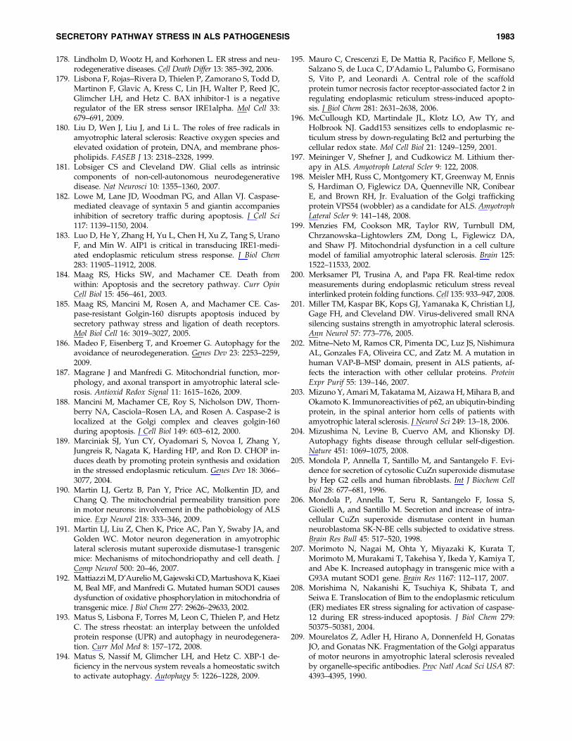
178. Lindholm D, Wootz H, and Korhonen L. ER stress and neu-rodegenerative diseases. Cell Death Differ 13: 385–392, 2006.
179. Lisbona F, Rojas–Rivera D, Thielen P, Zamorano S, Todd D,Martinon F, Glavic A, Kress C, Lin JH, Walter P, Reed JC,Glimcher LH, and Hetz C. BAX inhibitor-1 is a negativeregulator of the ER stress sensor IRE1alpha. Mol Cell 33:679–691, 2009.
180. Liu D, Wen J, Liu J, and Li L. The roles of free radicals inamyotrophic lateral sclerosis: Reactive oxygen species andelevated oxidation of protein, DNA, and membrane phos-pholipids. FASEB J 13: 2318–2328, 1999.
181. Lobsiger CS and Cleveland DW. Glial cells as intrinsiccomponents of non-cell-autonomous neurodegenerativedisease. Nat Neurosci 10: 1355–1360, 2007.
182. Lowe M, Lane JD, Woodman PG, and Allan VJ. Caspase-mediated cleavage of syntaxin 5 and giantin accompaniesinhibition of secretory traffic during apoptosis. J Cell Sci117: 1139–1150, 2004.
183. Luo D, He Y, Zhang H, Yu L, Chen H, Xu Z, Tang S, UranoF, and Min W. AIP1 is critical in transducing IRE1-medi-ated endoplasmic reticulum stress response. J Biol Chem283: 11905–11912, 2008.
184. Maag RS, Hicks SW, and Machamer CE. Death fromwithin: Apoptosis and the secretory pathway. Curr OpinCell Biol 15: 456–461, 2003.
185. Maag RS, Mancini M, Rosen A, and Machamer CE. Cas-pase-resistant Golgin-160 disrupts apoptosis induced bysecretory pathway stress and ligation of death receptors.Mol Biol Cell 16: 3019–3027, 2005.
186. Madeo F, Eisenberg T, and Kroemer G. Autophagy for theavoidance of neurodegeneration. Genes Dev 23: 2253–2259,2009.
187. Magrane J and Manfredi G. Mitochondrial function, mor-phology, and axonal transport in amyotrophic lateral scle-rosis. Antioxid Redox Signal 11: 1615–1626, 2009.
188. Mancini M, Machamer CE, Roy S, Nicholson DW, Thorn-berry NA, Casciola–Rosen LA, and Rosen A. Caspase-2 islocalized at the Golgi complex and cleaves golgin-160during apoptosis. J Cell Biol 149: 603–612, 2000.
189. Marciniak SJ, Yun CY, Oyadomari S, Novoa I, Zhang Y,Jungreis R, Nagata K, Harding HP, and Ron D. CHOP in-duces death by promoting protein synthesis and oxidationin the stressed endoplasmic reticulum. Genes Dev 18: 3066–3077, 2004.
190. Martin LJ, Gertz B, Pan Y, Price AC, Molkentin JD, andChang Q. The mitochondrial permeability transition porein motor neurons: involvement in the pathobiology of ALSmice. Exp Neurol 218: 333–346, 2009.
191. Martin LJ, Liu Z, Chen K, Price AC, Pan Y, Swaby JA, andGolden WC. Motor neuron degeneration in amyotrophiclateral sclerosis mutant superoxide dismutase-1 transgenicmice: Mechanisms of mitochondriopathy and cell death. JComp Neurol 500: 20–46, 2007.
192. Mattiazzi M, D’Aurelio M, Gajewski CD, Martushova K, KiaeiM, Beal MF, and Manfredi G. Mutated human SOD1 causesdysfunction of oxidative phosphorylation in mitochondria oftransgenic mice. J Biol Chem 277: 29626–29633, 2002.
193. Matus S, Lisbona F, Torres M, Leon C, Thielen P, and HetzC. The stress rheostat: an interplay between the unfoldedprotein response (UPR) and autophagy in neurodegenera-tion. Curr Mol Med 8: 157–172, 2008.
194. Matus S, Nassif M, Glimcher LH, and Hetz C. XBP-1 de-ficiency in the nervous system reveals a homeostatic switchto activate autophagy. Autophagy 5: 1226–1228, 2009.
195. Mauro C, Crescenzi E, De Mattia R, Pacifico F, Mellone S,Salzano S, de Luca C, D’Adamio L, Palumbo G, FormisanoS, Vito P, and Leonardi A. Central role of the scaffoldprotein tumor necrosis factor receptor-associated factor 2 inregulating endoplasmic reticulum stress-induced apopto-sis. J Biol Chem 281: 2631–2638, 2006.
196. McCullough KD, Martindale JL, Klotz LO, Aw TY, andHolbrook NJ. Gadd153 sensitizes cells to endoplasmic re-ticulum stress by down-regulating Bcl2 and perturbing thecellular redox state. Mol Cell Biol 21: 1249–1259, 2001.
197. Meininger V, Shefner J, and Cudkowicz M. Lithium ther-apy in ALS. Amyotroph Lateral Scler 9: 122, 2008.
198. Meisler MH, Russ C, Montgomery KT, Greenway M, EnnisS, Hardiman O, Figlewicz DA, Quenneville NR, ConibearE, and Brown RH, Jr. Evaluation of the Golgi traffickingprotein VPS54 (wobbler) as a candidate for ALS. AmyotrophLateral Scler 9: 141–148, 2008.
199. Menzies FM, Cookson MR, Taylor RW, Turnbull DM,Chrzanowska–Lightowlers ZM, Dong L, Figlewicz DA,and Shaw PJ. Mitochondrial dysfunction in a cell culturemodel of familial amyotrophic lateral sclerosis. Brain 125:1522–11533, 2002.
200. Merksamer PI, Trusina A, and Papa FR. Real-time redoxmeasurements during endoplasmic reticulum stress revealinterlinked protein folding functions. Cell 135: 933–947, 2008.
201. Miller TM, Kaspar BK, Kops GJ, Yamanaka K, Christian LJ,Gage FH, and Cleveland DW. Virus-delivered small RNAsilencing sustains strength in amyotrophic lateral sclerosis.Ann Neurol 57: 773–776, 2005.
202. Mitne–Neto M, Ramos CR, Pimenta DC, Luz JS, NishimuraAL, Gonzales FA, Oliveira CC, and Zatz M. A mutation inhuman VAP-B–MSP domain, present in ALS patients, af-fects the interaction with other cellular proteins. ProteinExpr Purif 55: 139–146, 2007.
203. Mizuno Y, Amari M, Takatama M, Aizawa H, Mihara B, andOkamoto K. Immunoreactivities of p62, an ubiqutin-bindingprotein, in the spinal anterior horn cells of patients withamyotrophic lateral sclerosis. J Neurol Sci 249: 13–18, 2006.
204. Mizushima N, Levine B, Cuervo AM, and Klionsky DJ.Autophagy fights disease through cellular self-digestion.Nature 451: 1069–1075, 2008.
205. Mondola P, Annella T, Santillo M, and Santangelo F. Evi-dence for secretion of cytosolic CuZn superoxide dismutaseby Hep G2 cells and human fibroblasts. Int J Biochem CellBiol 28: 677–681, 1996.
206. Mondola P, Annella T, Seru R, Santangelo F, Iossa S,Gioielli A, and Santillo M. Secretion and increase of intra-cellular CuZn superoxide dismutase content in humanneuroblastoma SK-N-BE cells subjected to oxidative stress.Brain Res Bull 45: 517–520, 1998.
207. Morimoto N, Nagai M, Ohta Y, Miyazaki K, Kurata T,Morimoto M, Murakami T, Takehisa Y, Ikeda Y, Kamiya T,and Abe K. Increased autophagy in transgenic mice with aG93A mutant SOD1 gene. Brain Res 1167: 112–117, 2007.
208. Morishima N, Nakanishi K, Tsuchiya K, Shibata T, andSeiwa E. Translocation of Bim to the endoplasmic reticulum(ER) mediates ER stress signaling for activation of caspase-12 during ER stress-induced apoptosis. J Biol Chem 279:50375–50381, 2004.
209. Mourelatos Z, Adler H, Hirano A, Donnenfeld H, GonatasJO, and Gonatas NK. Fragmentation of the Golgi apparatusof motor neurons in amyotrophic lateral sclerosis revealedby organelle-specific antibodies. Proc Natl Acad Sci USA 87:4393–4395, 1990.
SECRETORY PATHWAY STRESS IN ALS PATHOGENESIS 1983

210. Mourelatos Z, Gonatas NK, Stieber A, Gurney ME, and DalCanto MC. The Golgi apparatus of spinal cord motorneurons in transgenic mice expressing mutant Cu,Zn su-peroxide dismutase becomes fragmented in early, preclin-ical stages of the disease. Proc Natl Acad Sci USA 93: 5472–5477, 1996.
211. Mukherjee S, Chiu R, Leung SM, and Shields D. Frag-mentation of the Golgi apparatus: an early apoptotic eventindependent of the cytoskeleton. Traffic 8: 369–378, 2007.
212. Munch C, Rosenbohm A, Sperfeld AD, Uttner I, Reske S,Krause BJ, Sedlmeier R, Meyer T, Hanemann CO, StummG, and Ludolph AC. Heterozygous R1101K mutation of theDCTN1 gene in a family with ALS and FTD. Ann Neurol 58:777–780, 2005.
213. Munch C, Sedlmeier R, Meyer T, Homberg V, Sperfeld AD,Kurt A, Prudlo J, Peraus G, Hanemann CO, Stumm G, andLudolph AC. Point mutations of the p150 subunit of dy-nactin (DCTN1) gene in ALS. Neurology 63: 724–726, 2004.
214. Nagai M, Aoki M, Miyoshi I, Kato M, Pasinelli P, Kasai N,Brown RH, Jr., and Itoyama Y. Rats expressing human cy-tosolic copper-zinc superoxide dismutase transgenes withamyotrophic lateral sclerosis: Associated mutations developmotor neuron disease. J Neurosci 21: 9246–9254, 2001.
215. Nagai M, Re DB, Nagata T, Chalazonitis A, Jessell TM,Wichterle H, and Przedborski S. Astrocytes expressingALS-linked mutated SOD1 release factors selectively toxicto motor neurons. Nat Neurosci 10: 615–622, 2007.
216. Nagaoka U, Kim K, Jana NR, Doi H, Maruyama M, MitsuiK, Oyama F, and Nukina N. Increased expression of p62 inexpanded polyglutamine-expressing cells and its associa-tion with polyglutamine inclusions. J Neurochem 91: 57–68,2004.
217. Nagata T, Ilieva H, Murakami T, Shiote M, Narai H, OhtaY, Hayashi T, Shoji M, and Abe K. Increased ER stressduring motor neuron degeneration in a transgenic mousemodel of amyotrophic lateral sclerosis. Neurol Res 29: 767–771, 2007.
218. Naidoo N. ER and aging. Protein folding and the ER stressresponse. Ageing Res Rev 8: 150–159, 2009.
219. Nakagomi S, Barsoum MJ, Bossy-Wetzel E, Sutterlin C,Malhotra V, and Lipton SA. A Golgi fragmentation path-way in neurodegeneration. Neurobiol Dis 29: 221–231, 2008.
220. Nakano T, Nakaso K, Nakashima K, and Ohama E. Ex-pression of ubiquitin-binding protein p62 in ubiquitin-immunoreactive intraneuronal inclusions in amyotrophiclateral sclerosis with dementia: Analysis of five autopsycases with broad clinicopathological spectrum. Acta Neu-ropathol 107: 359–364, 2004.
221. Nakaso K, Yoshimoto Y, Nakano T, Takeshima T, Fuku-hara Y, Yasui K, Araga S, Yanagawa T, Ishii T, and Na-kashima K. Transcriptional activation of p62=A170=ZIPduring the formation of the aggregates: Possible mecha-nisms and the role in Lewy body formation in Parkinson’sdisease. Brain Res 1012: 42–51, 2004.
222. Nakatogawa H, Suzuki K, Kamada Y, and Ohsumi Y.Dynamics and diversity in autophagy mechanisms: Les-sons from yeast. Nat Rev Mol Cell Biol 10: 458–467, 2009.
223. Neumann M, Sampathu DM, Kwong LK, Truax AC, Mic-senyi MC, Chou TT, Bruce J, Schuck T, Grossman M, ClarkCM, McCluskey LF, Miller BL, Masliah E, Mackenzie IR,Feldman H, Feiden W, Kretzschmar HA, Trojanowski JQ,and Lee VM. Ubiquitinated TDP-43 in frontotemporal lobardegeneration and amyotrophic lateral sclerosis. Science 314:130–133, 2006.
224. Nguyen DT, Kebache S, Fazel A, Wong HN, Jenna S, EmadaliA, Lee EH, Bergeron JJ, Kaufman RJ, Larose L, and Chevet E.Nck-dependent activation of extracellular signal-regulatedkinase-1 and regulation of cell survival during endoplasmicreticulum stress. Mol Biol Cell 15: 4248–4260, 2004.
225. Nguyen T, Nioi P, and Pickett CB. The Nrf2-antioxidantresponse element signaling pathway and its activation byoxidative stress. J Biol Chem 284: 13291–13295, 2009.
226. Nishida Y, Arakawa S, Fujitani K, Yamaguchi H, Mizuta T,Kanaseki T, Komatsu M, Otsu K, Tsujimoto Y, and ShimizuS. Discovery of Atg5=Atg7-independent alternative mac-roautophagy. Nature 461: 654–658, 2009.
227. Nishimura AL, Mitne–Neto M, Silva HC, Richieri–Costa A,Middleton S, Cascio D, Kok F, Oliveira JR, Gillingwater T,Webb J, Skehel P, and Zatz M. A mutation in the vesicle-trafficking protein VAPB causes late-onset spinal muscularatrophy and amyotrophic lateral sclerosis. Am J Hum Genet75: 822–831, 2004.
228. Nishitoh H, Kadowaki H, Nagai A, Maruyama T, Yokota T,Fukutomi H, Noguchi T, Matsuzawa A, Takeda K, andIchijo H. ALS-linked mutant SOD1 induces ER stress- andASK1-dependent motor neuron death by targeting Derlin-1. Genes Dev 22: 1451–1464, 2008.
229. Niwa J, Ishigaki S, Hishikawa N, Yamamoto M, Doyu M,Murata S, Tanaka K, Taniguchi N, and Sobue G. Dorfinubiquitylates mutant SOD1 and prevents mutant SOD1-mediated neurotoxicity. J Biol Chem 277: 36793–36798, 2002.
230. Niwa J, Yamada S, Ishigaki S, Sone J, Takahashi M, Kat-suno M, Tanaka F, Doyu M, and Sobue G. Disulfide bondmediates aggregation, toxicity, and ubiquitylation of fa-milial amyotrophic lateral sclerosis-linked mutant SOD1. JBiol Chem 282: 28087–28095, 2007.
231. Obeng EA and Boise LH. Caspase-12 and caspase-4 are notrequired for caspase-dependent endoplasmic reticulumstress-induced apoptosis. J Biol Chem 280: 29578–29587, 2005.
232. Oh YK, Shin KS, Yuan J, and Kang SJ. Superoxide dis-mutase 1 mutants related to amyotrophic lateral sclerosisinduce endoplasmic stress in neuro2a cells. J Neurochem104: 993–1005, 2008.
233. Oikawa D, Kimata Y, Kohno K, and Iwawaki T. Activa-tion of mammalian IRE1alpha upon ER stress depends ondissociation of BiP rather than on direct interaction withunfolded proteins. Exp Cell Res 315: 2496–2504, 2009.
234. Okado–Matsumoto A and Fridovich I. Subcellular distri-bution of superoxide dismutases (SOD) in rat liver: Cu,Zn-SOD in mitochondria. J Biol Chem 276: 38388–38393, 2001.
235. Oliver JD, Roderick HL, and Llewellyn DH, High S. ERp57functions as a subunit of specific complexes formed withthe ER lectins calreticulin and calnexin. Mol Biol Cell 10:2573–2582, 1999.
236. Oyanagi K, Yamazaki M, Takahashi H, Watabe K, WadaM, Komori T, Morita T, and Mizutani T. Spinal anteriorhorn cells in sporadic amyotrophic lateral sclerosis showribosomal detachment from, and cisternal distention of therough endoplasmic reticulum. Neuropathol Appl Neurobiol34: 650–658, 2008.
237. Ozcan U, Yilmaz E, Ozcan L, Furuhashi M, Vaillancourt E,Smith RO, Gorgun CZ, and Hotamisligil GS. Chemicalchaperones reduce ER stress and restore glucosehomeostasis in a mouse model of type 2 diabetes. Science313: 1137–1140, 2006.
238. Pankiv S, Clausen TH, Lamark T, Brech A, Bruun JA,Outzen H, Overvatn A, Bjorkoy G, and Johansen T.p62=SQSTM1 binds directly to Atg8=LC3 to facilitate deg-
1984 NASSIF ET AL.
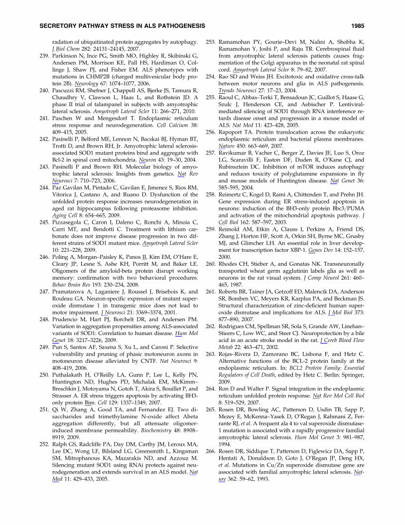
radation of ubiquitinated protein aggregates by autophagy.J Biol Chem 282: 24131–24145, 2007.
239. Parkinson N, Ince PG, Smith MO, Highley R, Skibinski G,Andersen PM, Morrison KE, Pall HS, Hardiman O, Col-linge J, Shaw PJ, and Fisher EM. ALS phenotypes withmutations in CHMP2B (charged multivesicular body pro-tein 2B). Neurology 67: 1074–1077, 2006.
240. Pascuzzi RM, Shefner J, Chappell AS, Bjerke JS, Tamura R,Chaudhry V, Clawson L, Haas L, and Rothstein JD. Aphase II trial of talampanel in subjects with amyotrophiclateral sclerosis. Amyotroph Lateral Scler 11: 266–271, 2010.
241. Paschen W and Mengesdorf T. Endoplasmic reticulumstress response and neurodegeneration. Cell Calcium 38:409–415, 2005.
242. Pasinelli P, Belford ME, Lennon N, Bacskai BJ, Hyman BT,Trotti D, and Brown RH, Jr. Amyotrophic lateral sclerosis-associated SOD1 mutant proteins bind and aggregate withBcl-2 in spinal cord mitochondria. Neuron 43: 19–30, 2004.
243. Pasinelli P and Brown RH. Molecular biology of amyo-trophic lateral sclerosis: Insights from genetics. Nat RevNeurosci 7: 710–723, 2006.
244. Paz Gavilan M, Pintado C, Gavilan E, Jimenez S, Rios RM,Vitorica J, Castano A, and Ruano D. Dysfunction of theunfolded protein response increases neurodegeneration inaged rat hippocampus following proteasome inhibition.Aging Cell 8: 654–665, 2009.
245. Pizzasegola C, Caron I, Daleno C, Ronchi A, Minoia C,Carri MT, and Bendotti C. Treatment with lithium car-bonate does not improve disease progression in two dif-ferent strains of SOD1 mutant mice. Amyotroph Lateral Scler10: 221–228, 2009.
246. Poling A, Morgan–Paisley K, Panos JJ, Kim EM, O’Hare E,Cleary JP, Lesne S, Ashe KH, Porritt M, and Baker LE.Oligomers of the amyloid-beta protein disrupt workingmemory: confirmation with two behavioral procedures.Behav Brain Res 193: 230–234, 2008.
247. Pramatarova A, Laganiere J, Roussel J, Brisebois K, andRouleau GA. Neuron-specific expression of mutant super-oxide dismutase 1 in transgenic mice does not lead tomotor impairment. J Neurosci 21: 3369–3374, 2001.
248. Prudencio M, Hart PJ, Borchelt DR, and Andersen PM.Variation in aggregation propensities among ALS-associatedvariants of SOD1: Correlation to human disease. Hum MolGenet 18: 3217–3226, 2009.
249. Pun S, Santos AF, Saxena S, Xu L, and Caroni P. Selectivevulnerability and pruning of phasic motoneuron axons inmotoneuron disease alleviated by CNTF. Nat Neurosci 9:408–419, 2006.
250. Puthalakath H, O’Reilly LA, Gunn P, Lee L, Kelly PN,Huntington ND, Hughes PD, Michalak EM, McKimm–Breschkin J, Motoyama N, Gotoh T, Akira S, Bouillet P, andStrasser A. ER stress triggers apoptosis by activating BH3-only protein Bim. Cell 129: 1337–1349, 2007.
251. Qi W, Zhang A, Good TA, and Fernandez EJ. Two di-saccharides and trimethylamine N-oxide affect Abetaaggregation differently, but all attenuate oligomer-induced membrane permeability. Biochemistry 48: 8908–8919, 2009.
252. Ralph GS, Radcliffe PA, Day DM, Carthy JM, Leroux MA,Lee DC, Wong LF, Bilsland LG, Greensmith L, KingsmanSM, Mitrophanous KA, Mazarakis ND, and Azzouz M.Silencing mutant SOD1 using RNAi protects against neu-rodegeneration and extends survival in an ALS model. NatMed 11: 429–433, 2005.
253. Ramamohan PY, Gourie–Devi M, Nalini A, Shobha K,Ramamohan Y, Joshi P, and Raju TR. Cerebrospinal fluidfrom amyotrophic lateral sclerosis patients causes frag-mentation of the Golgi apparatus in the neonatal rat spinalcord. Amyotroph Lateral Scler 8: 79–82, 2007.
254. Rao SD and Weiss JH. Excitotoxic and oxidative cross-talkbetween motor neurons and glia in ALS pathogenesis.Trends Neurosci 27: 17–23, 2004.
255. Raoul C, Abbas–Terki T, Bensadoun JC, Guillot S, Haase G,Szulc J, Henderson CE, and Aebischer P. Lentiviral-mediated silencing of SOD1 through RNA interference re-tards disease onset and progression in a mouse model ofALS. Nat Med 11: 423–428, 2005.
256. Rapoport TA. Protein translocation across the eukaryoticendoplasmic reticulum and bacterial plasma membranes.Nature 450: 663–669, 2007.
257. Ravikumar B, Vacher C, Berger Z, Davies JE, Luo S, OrozLG, Scaravilli F, Easton DF, Duden R, O’Kane CJ, andRubinsztein DC. Inhibition of mTOR induces autophagyand reduces toxicity of polyglutamine expansions in flyand mouse models of Huntington disease. Nat Genet 36:585–595, 2004.
258. Reimertz C, Kogel D, Rami A, Chittenden T, and Prehn JH.Gene expression during ER stress-induced apoptosis inneurons: induction of the BH3-only protein Bbc3=PUMAand activation of the mitochondrial apoptosis pathway. JCell Biol 162: 587–597, 2003.
259. Reimold AM, Etkin A, Clauss I, Perkins A, Friend DS,Zhang J, Horton HF, Scott A, Orkin SH, Byrne MC, GrusbyMJ, and Glimcher LH. An essential role in liver develop-ment for transcription factor XBP-1. Genes Dev 14: 152–157,2000.
260. Rhodes CH, Stieber A, and Gonatas NK. Transneuronallytransported wheat germ agglutinin labels glia as well asneurons in the rat visual system. J Comp Neurol 261: 460–465, 1987.
261. Roberts BR, Tainer JA, Getzoff ED, Malencik DA, AndersonSR, Bomben VC, Meyers KR, Karplus PA, and Beckman JS.Structural characterization of zinc-deficient human super-oxide dismutase and implications for ALS. J Mol Biol 373:877–890, 2007.
262. Rodrigues CM, Spellman SR, Sola S, Grande AW, Linehan–Stieers C, Low WC, and Steer CJ. Neuroprotection by a bileacid in an acute stroke model in the rat. J Cereb Blood FlowMetab 22: 463–471, 2002.
263. Rojas–Rivera D, Zamorano BC, Lisbona F, and Hetz C.Alternative functions of the BCL-2 protein family at theendoplasmic reticulum. In: BCL2 Protein Family: EssentialRegulators of Cell Death, edited by Hetz C. Berlin: Springer,2009.
264. Ron D and Walter P. Signal integration in the endoplasmicreticulum unfolded protein response. Nat Rev Mol Cell Biol8: 519–529, 2007.
265. Rosen DR, Bowling AC, Patterson D, Usdin TB, Sapp P,Mezey E, McKenna–Yasek D, O’Regan J, Rahmani Z, Fer-rante RJ, et al. A frequent ala 4 to val superoxide dismutase-1 mutation is associated with a rapidly progressive familialamyotrophic lateral sclerosis. Hum Mol Genet 3: 981–987,1994.
266. Rosen DR, Siddique T, Patterson D, Figlewicz DA, Sapp P,Hentati A, Donaldson D, Goto J, O’Regan JP, Deng HX,et al. Mutations in Cu=Zn superoxide dismutase gene areassociated with familial amyotrophic lateral sclerosis. Nat-ure 362: 59–62, 1993.
SECRETORY PATHWAY STRESS IN ALS PATHOGENESIS 1985

267. Rosenfeld J, King RM, Jackson CE, Bedlack RS, Barohn RJ,Dick A, Phillips LH, Chapin J, Gelinas DF, and Lou JS.Creatine monohydrate in ALS: Effects on strength, fatigue,respiratory status and ALSFRS. Amyotroph Lateral Scler 9:266–272, 2008.
268. Rowland LP and Shneider NA. Amyotrophic lateral scle-rosis. N Engl J Med 344: 1688–1700, 2001.
269. Rubinsztein DC. The roles of intracellular protein-degradation pathways in neurodegeneration. Nature 443:780–786, 2006.
270. Rubio I, Rodriguez–Navarro JA, Tomas–Zapico C, Ruiz C,Casarejos MJ, Perucho J, Gomez A, Rodal I, Lucas JJ, MenaMA, and de Yebenes JG. Effects of partial suppressionof parkin on huntingtin mutant R6=1 mice. Brain Res 1281:91–100, 2009.
271. Rusten TE and Stenmark H. How do ESCRT proteinscontrol autophagy? J Cell Sci 122: 2179–2183, 2009.
272. Rusten TE, Vaccari T, Lindmo K, Rodahl LM, Nezis IP,Sem–Jacobsen C, Wendler F, Vincent JP, Brech A, Bilder D,and Stenmark H. ESCRTs and Fab1 regulate distinct stepsof autophagy. Curr Biol 17: 1817–1825, 2007.
273. Sado M, Yamasaki Y, Iwanaga T, Onaka Y, Ibuki T, Nish-ihara S, Mizuguchi H, Momota H, Kishibuchi R, Ha-shimoto T, Wada D, Kitagawa H, Watanabe TK. Protectiveeffect against Parkinson’s disease-related insults throughthe activation of XBP1. Brain Res 1257: 16–24, 2009.
274. Saleh M, Mathison JC, Wolinski MK, Bensinger SJ, Fitz-gerald P, Droin N, Ulevitch RJ, Green DR, andNicholson DW. Enhanced bacterial clearance and sepsisresistance in caspase-12-deficient mice. Nature 440: 1064–1068, 2006.
275. Saleh M, Vaillancourt JP, Graham RK, Huyck M, Sriniva-sula SM, Alnemri ES, Steinberg MH, Nolan V, Baldwin CT,Hotchkiss RS, Buchman TG, Zehnbauer BA, Hayden MR,Farrer LA, Roy S, and Nicholson DW. Differential modu-lation of endotoxin responsiveness by human caspase-12polymorphisms. Nature 429: 75–79, 2004.
276. Santos CX, Tanaka LY, Wosniak J, and Laurindo FR. Me-chanisms and implications of reactive oxygen species gen-eration during the unfolded protein response: Roles ofendoplasmic reticulum oxidoreductases, mitochondrialelectron transport, and NADPH oxidase. Antioxid RedoxSignal 11: 2409–2427, 2009.
277. Sarkar S, Davies JE, Huang Z, Tunnacliffe A, and Ru-binsztein DC. Trehalose, a novel mTOR-independentautophagy enhancer, accelerates the clearance of mutanthuntingtin and alpha-synuclein. J Biol Chem 282: 5641–5652,2007.
278. Sarkar S, Floto RA, Berger Z, Imarisio S, Cordenier A, PascoM, Cook LJ, and Rubinsztein DC. Lithium induces autop-hagy by inhibiting inositol monophosphatase. J Cell Biol170: 1101–1111, 2005.
279. Sarkar S, Krishna G, Imarisio S, Saiki S, O’Kane CJ, andRubinsztein DC. A rational mechanism for combinationtreatment of Huntington’s disease using lithium and ra-pamycin. Hum Mol Genet 17: 170–178, 2008.
280. Sarkar S and Rubinsztein DC. Inositol and IP3 levels reg-ulate autophagy: Biology and therapeutic speculations.Autophagy 2: 132–134, 2006.
281. Sarkar S and Rubinsztein DC. Huntington’s disease: De-gradation of mutant huntingtin by autophagy. FEBS J 275:4263–4270, 2008.
282. Sarlette A, Krampfl K, Grothe C, Neuhoff N, Dengler R, andPetri S. Nuclear erythroid 2-related factor 2-antioxidative
response element signaling pathway in motor cortex andspinal cord in amyotrophic lateral sclerosis. J NeuropatholExp Neurol 67: 1055–1062, 2008.
283. Sasaki S and Iwata M. Ultrastructural study of synapses inthe anterior horn neurons of patients with amyotrophiclateral sclerosis. Neurosci Lett 204: 53–56, 1996.
284. Saxena S, Cabuy E, and Caroni P. A role for motoneuronsubtype-selective ER stress in disease manifestations ofFALS mice. Nat Neurosci 12: 627–636, 2009.
285. Scheper W and Hoozemans JJ. Endoplasmic reticulumprotein quality control in neurodegenerative disease: Thegood, the bad and the therapy. Curr Med Chem 16: 615–626,2009.
286. Schmitt–John T, Drepper C, Mussmann A, Hahn P, Kuhl-mann M, Thiel C, Hafner M, Lengeling A, Heimann P,Jones JM, Meisler MH, Jockusch H. Mutation of Vps54causes motor neuron disease and defective spermiogenesisin the wobbler mouse. Nat Genet 37: 1213–1215, 2005.
287. Schroder M and Kaufman RJ. The mammalian unfoldedprotein response. Annu Rev Biochem 74: 739–789, 2005.
288. Schroer TA. Dynactin. Annu Rev Cell Dev Biol 20: 759–779,2004.
289. Seibenhener ML, Babu JR, Geetha T, Wong HC, KrishnaNR, and Wooten MW. Sequestosome 1=p62 is a poly-ubiquitin chain binding protein involved in ubiquitin pro-teasome degradation. Mol Cell Biol 24: 8055–8068, 2004.
290. Selkoe DJ. Soluble oligomers of the amyloid beta-proteinimpair synaptic plasticity and behavior. Behav Brain Res192: 106–113, 2008.
291. Selman C, Tullet JM, Wieser D, Irvine E, Lingard SJ,Choudhury AI, Claret M, Al–Qassab H, Carmignac D,Ramadani F, Woods A, Robinson IC, Schuster E, BatterhamRL, Kozma SC, Thomas G, Carling D, Okkenhaug K,Thornton JM, Partridge L, Gems D, and Withers DJ. Ribo-somal protein S6 kinase 1 signaling regulates mammalianlifespan. Science 326: 140–144, 2009.
292. Sendtner M. Damaging secretions: Chromogranins team upwith mutant SOD1. Nat Neurosci 9: 12–14, 2006.
293. Shan X, Chang Y, and Lin CL. Messenger RNA oxidation isan early event preceding cell death and causes reducedprotein expression. FASEB J 21: 2753–2764, 2007.
294. Shaw PJ, Ince PG, Falkous G, and Mantle D. Oxidativedamage to protein in sporadic motor neuron disease spinalcord. Ann Neurol 38: 691–695, 1995.
295. Shin JH, Cho SI, Lim HR, Lee JK, Lee YA, Noh JS, Joo IS,Kim KW, and Gwag BJ. Concurrent administration ofNeu2000 and lithium produces marked improvement ofmotor neuron survival, motor function, and mortality in amouse model of amyotrophic lateral sclerosis. Mol Phar-macol 71: 965–975, 2007.
296. Siklos L, Engelhardt J, Harati Y, Smith RG, Joo F, andAppel SH. Ultrastructural evidence for altered calcium inmotor nerve terminals in amyotropic lateral sclerosis. AnnNeurol 39: 203–216, 1996.
297. Simonsen A and Tooze SA. Coordination of membraneevents during autophagy by multiple class III PI3-kinasecomplexes. J Cell Biol 186: 773–782, 2009.
298. Skehel PA, Fabian–Fine R, and Kandel ER. Mouse VAP33 isassociated with the endoplasmic reticulum and microtu-bules. Proc Natl Acad Sci USA 97: 1101–1106, 2000.
299. Slodzinski H, Moran LB, Michael GJ, Wang B, Novoselov S,Cheetham ME, Pearce RK, and Graeber MB. Homo-cysteine-induced endoplasmic reticulum protein (herp) isup-regulated in parkinsonian substantia nigra and present
1986 NASSIF ET AL.

in the core of Lewy bodies. Clin Neuropathol 28: 333–343,2009.
300. Smith RA, Miller TM, Yamanaka K, Monia BP, Condon TP,Hung G, Lobsiger CS, Ward CM, McAlonis-Downes M,Wei H, Wancewicz EV, Bennett CF, and Cleveland DW.Antisense oligonucleotide therapy for neurodegenerativedisease. J Clin Invest 116: 2290–2296, 2006.
301. Son M, Puttaparthi K, Kawamata H, Rajendran B, Boyer PJ,Manfredi G, and Elliott JL. Overexpression of CCS inG93A-SOD1 mice leads to accelerated neurological deficitswith severe mitochondrial pathology. Proc Natl Acad SciUSA 104: 6072–6077, 2007.
302. Starkov AA. The role of mitochondria in reactive oxygenspecies metabolism and signaling. Ann NY Acad Sci 1147:37–52, 2008.
303. Steele AD, Hetz C, Yi CH, Jackson WS, Borkowski AW,Yuan J, Wollmann RH, and Lindquist S. Prion pathogenesisis independent of caspase-12. Prion 1: 243–247, 2007.
304. Stevens FJ and Argon Y. Protein folding in the ER. SeminCell Dev Biol 10: 443–454, 1999.
305. Stieber A, Chen Y, Wei S, Mourelatos Z, Gonatas J, Oka-moto K, and Gonatas NK. The fragmented neuronal Golgiapparatus in amyotrophic lateral sclerosis includes thetrans-Golgi-network: functional implications. Acta Neuro-pathol 95: 245–253, 1998.
306. Stieber A, Gonatas JO, Collard J, Meier J, Julien J,Schweitzer P, and Gonatas NK. The neuronal Golgi appa-ratus is fragmented in transgenic mice expressing a mutanthuman SOD1, but not in mice expressing the human NF-Hgene. J Neurol Sci 173: 63–72, 2000.
307. Stieber A, Gonatas JO, and Gonatas NK. Aggregates ofmutant protein appear progressively in dendrites, in peri-axonal processes of oligodendrocytes, and in neuronal andastrocytic perikarya of mice expressing the SOD1(G93A)mutation of familial amyotrophic lateral sclerosis. J NeurolSci 177: 114–123, 2000.
308. Stieber A, Gonatas JO, Moore JS, Bantly A, Yim HS, YimMB, and Gonatas NK. Disruption of the structure of theGolgi apparatus and the function of the secretory pathwayby mutants G93A and G85R of Cu, Zn superoxide dis-mutase (SOD1) of familial amyotrophic lateral sclerosis. JNeurol Sci 219: 45–53, 2004.
309. Strong MJ. The evidence for altered RNA metabolism inamyotrophic lateral sclerosis (ALS). J Neurol Sci 288: 1–12, 2010.
310. Strong MJ, Kesavapany S, and Pant HC. The pathobiologyof amyotrophic lateral sclerosis: A proteinopathy? J Neu-ropathol Exp Neurol 64: 649–664, 2005.
311. Sturtz LA, Diekert K, Jensen LT, Lill R, and Culotta VC. Afraction of yeast Cu,Zn-superoxide dismutase and its me-tallochaperone, CCS, localize to the intermembrane spaceof mitochondria. A physiological role for SOD1 in guardingagainst mitochondrial oxidative damage. J Biol Chem 276:38084–38089, 2001.
312. Suzuki H, Kanekura K, Levine TP, Kohno K, OlkkonenVM, Aiso S, and Matsuoka M. ALS-linked P56S-VAPB, anaggregated loss-of-function mutant of VAPB, predisposesmotor neurons to ER stress-related death by inducing ag-gregation of co-expressed wild-type VAPB. J Neurochem108: 973–985, 2009.
313. Swerdlow RH, Parks JK, Cassarino DS, Trimmer PA, MillerSW, Maguire DJ, Sheehan JP, Maguire RS, Pattee G, JuelVC, Phillips LH, Tuttle JB, Bennett JP, Jr., Davis RE, andParker WD, Jr. Mitochondria in sporadic amyotrophic lat-eral sclerosis. Exp Neurol 153: 135–142, 1998.
314. Talbot K. Another gene for ALS: Mutations in sporadiccases and the rare variant hypothesis. Neurology 73: 1172–1173, 2009.
315. Tanaka M, Machida Y, Niu S, Ikeda T, Jana NR, Doi H,Kurosawa M, Nekooki M, and Nukina N. Trehalose alle-viates polyglutamine-mediated pathology in a mousemodel of Huntington disease. Nat Med 10: 148–154, 2004.
316. Taupenot L, Harper KL, and O’Connor DT. The chromo-granin-secretogranin family. N Engl J Med 348: 1134–1149,2003.
317. Teuling E, Ahmed S, Haasdijk E, Demmers J, SteinmetzMO, Akhmanova A, Jaarsma D, and HoogenraadCC. Motor neuron disease-associated mutant vesicle-associated membrane protein-associated protein (VAP) Brecruits wild-type VAPs into endoplasmic reticulum-derived tubular aggregates. J Neurosci 27: 9801–9815,2007.
318. Teuling E, van Dis V, Wulf PS, Haasdijk ED, AkhmanovaA, Hoogenraad CC, and Jaarsma D. A novel mouse modelwith impaired dynein=dynactin function develops amyo-trophic lateral sclerosis (ALS)-like features in motor neu-rons and improves lifespan in SOD1-ALS mice. Hum MolGenet 17: 2849–2862, 2008.
319. Tobisawa S, Hozumi Y, Arawaka S, Koyama S, Wada M,Nagai M, Aoki M, Itoyama Y, Goto K, and Kato T. MutantSOD1 linked to familial amyotrophic lateral sclerosis, butnot wild-type SOD1, induces ER stress in COS7 cells andtransgenic mice. Biochem Biophys Res Commun 303: 496–503,2003.
320. Tomic JL, Pensalfini A, Head E, and Glabe CG. Solublefibrillar oligomer levels are elevated in Alzheimer’s diseasebrain and correlate with cognitive dysfunction. NeurobiolDis 35: 352–358, 2009.
321. Tooze SA and Schiavo G. Liaisons dangereuses: Autop-hagy, neuronal survival and neurodegeneration. Curr OpinNeurobiol 18: 504–515, 2008.
322. Topp JD, Gray NW, Gerard RD, and Horazdovsky BF.Alsin is a Rab5 and Rac1 guanine nucleotide exchangefactor. J Biol Chem 279: 24612–24623, 2004.
323. Tu BP, Ho-Schleyer SC, Travers KJ, and Weissman JS.Biochemical basis of oxidative protein folding in the en-doplasmic reticulum. Science 290: 1571–1574, 2000.
324. Tu BP and Weissman JS. Oxidative protein folding in eu-karyotes: Mechanisms and consequences. J Cell Biol 164:341–346, 2004.
325. Tudor EL, Perkinton MS, Schmidt A, Ackerley S, BrownleesJ, Jacobsen NJ, Byers HL, Ward M, Hall A, Leigh PN, ShawCE, McLoughlin DM, and Miller CC. ALS2=Alsin regulatesRac-PAK signaling and neurite outgrowth. J Biol Chem 280:34735–34740, 2005.
326. Turner BJ, Atkin JD, Farg MA, Zang DW, Rembach A,Lopes EC, Patch JD, Hill AF, and Cheema SS. Impairedextracellular secretion of mutant superoxide dismutase 1associates with neurotoxicity in familial amyotrophic lat-eral sclerosis. J Neurosci 25: 108–117, 2005.
327. Turner BJ and Talbot K. Transgenics, toxicity and thera-peutics in rodent models of mutant SOD1-mediated fa-milial ALS. Prog Neurobiol 85: 94–134, 2008.
328. Uehara T, Nakamura T, Yao D, Shi ZQ, Gu Z, Ma Y,Masliah E, Nomura Y, and Lipton SA. S-nitrosylatedprotein-disulphide isomerase links protein misfolding toneurodegeneration. Nature 441: 513–517, 2006.
329. Upton JP, Austgen K, Nishino M, Coakley KM, Hagen A,Han D, Papa FR, and Oakes SA. Caspase-2 cleavage
SECRETORY PATHWAY STRESS IN ALS PATHOGENESIS 1987
of BID is a critical apoptotic signal downstream of en-doplasmic reticulum stress. Mol Cell Biol 28: 3943–3951,2008.
330. Urano F, Wang X, Bertolotti A, Zhang Y, Chung P, HardingHP, and Ron D. Coupling of stress in the ER to activation ofJNK protein kinases by transmembrane protein kinaseIRE1. Science 287: 664–666, 2000.
331. Urushitani M, Ezzi SA, and Julien JP. Therapeutic effects ofimmunization with mutant superoxide dismutase in micemodels of amyotrophic lateral sclerosis. Proc Natl Acad SciUSA 104: 2495–2500, 2007.
332. Urushitani M, Ezzi SA, Matsuo A, Tooyama I, and JulienJP. The endoplasmic reticulum-Golgi pathway is a targetfor translocation and aggregation of mutant superox-ide dismutase linked to ALS. FASEB J 22: 2476–2487,2008.
333. Urushitani M, Kurisu J, Tateno M, Hatakeyama S, Na-kayama K, Kato S, and Takahashi R. CHIP promotes pro-teasomal degradation of familial ALS-linked mutant SOD1by ubiquitinating Hsp=Hsc70. J Neurochem 90: 231–244,2004.
334. Urushitani M, Sato T, Bamba H, Hisa Y, and Tooyama I.Synergistic effect between proteasome and autophagosomein the clearance of polyubiquitinated TDP-43. J Neurosci Res88: 684–797, 2010.
335. Urushitani M, Sik A, Sakurai T, Nukina N, Takahashi R,and Julien JP. Chromogranin-mediated secretion of mutantsuperoxide dismutase proteins linked to amyotrophic lat-eral sclerosis. Nat Neurosci 9: 108–118, 2006.
336. Vance C, Rogelj B, Hortobagyi T, De Vos KJ, NishimuraAL, Sreedharan J, Hu X, Smith B, Ruddy D, Wright P,Ganesalingam J, Williams KL, Tripathi V, Al–Saraj S, Al-Chalabi A, Leigh PN, Blair IP, Nicholson G, de Belleroche J,Gallo JM, Miller CC, and Shaw CE. Mutations in FUS, anRNA processing protein, cause familial amyotrophic lateralsclerosis type 6. Science 323: 1208–1211, 2009.
337. Vargas MR, Pehar M, Cassina P, Beckman JS, and BarbeitoL. Increased glutathione biosynthesis by Nrf2 activation inastrocytes prevents p75NTR-dependent motor neuron ap-optosis. J Neurochem 97: 687–696, 2006.
338. Vella LJ, Sharples RA, Nisbet RM, Cappai R, and Hill AF.The role of exosomes in the processing of proteins associ-ated with neurodegenerative diseases. Eur Biophys J 37:323–332, 2008.
339. Vembar SS and Brodsky JL. One step at a time: En-doplasmic reticulum-associated degradation. Nat Rev MolCell Biol 9: 944–957, 2008.
340. Vicencio JM, Ortiz C, Criollo A, Jones AW, Kepp O, Gal-luzzi L, Joza N, Vitale I, Morselli E, Tailler M, Castedo M,Maiuri MC, Molgo J, Szabadkai G, Lavandero S, andKroemer G. The inositol 1,4,5-trisphosphate receptor regu-lates autophagy through its interaction with Beclin 1. CellDeath Differ 16: 1006–1017, 2009.
341. Vlug AS, Teuling E, Haasdijk ED, French P, HoogenraadCC, and Jaarsma D. ATF3 expression precedes death ofspinal motoneurons in amyotrophic lateral sclerosis-SOD1transgenic mice and correlates with c-Jun phosphoryla-tion, CHOP expression, somato-dendritic ubiquitinationand Golgi fragmentation. Eur J Neurosci 22: 1881–1894,2005.
342. Walker AK, Farg MA, Bye CR, McLean CA, Horne MK,and Atkin JD. Protein disulphide isomerase protectsagainst protein aggregation and is S-nitrosylated inamyotrophic lateral sclerosis. Brain 133: 105–116, 2010.
343. Wang Q, Johnson JL, Agar NY, and Agar JN. Protein aggre-gation and protein instability govern familial amyotrophiclateral sclerosis patient survival. PLoS Biol 6: e170, 2008.
344. Wang X, Fan H, Ying Z, Li B, Wang H, and Wang G. De-gradation of TDP-43 and its pathogenic form by autophagyand the ubiquitin-proteasome system. Neurosci Lett 469:112–116, 2010.
345. Wegorzewska I, Bell S, Cairns NJ, Miller TM, and BalohRH. TDP-43 mutant transgenic mice develop features ofALS and frontotemporal lobar degeneration. Proc Natl AcadSci USA 106: 18809–18814, 2009.
346. Witan H, Gorlovoy P, Kaya AM, Koziollek–Drechsler I,Neumann H, Behl C, and Clement AM. Wild-type Cu=Znsuperoxide dismutase (SOD1) does not facilitate, but im-pedes the formation of protein aggregates of amyotrophiclateral sclerosis causing mutant SOD1. Neurobiol Dis 36:331–342, 2009.
347. Witan H, Kern A, Koziollek–Drechsler I, Wade R, Behl C,and Clement AM. Heterodimer formation of wild-typeand amyotrophic lateral sclerosis-causing mutant Cu=Zn-superoxide dismutase induces toxicity independentof protein aggregation. Hum Mol Genet 17: 1373–1385,2008.
348. Wolfers J, Lozier A, Raposo G, Regnault A, Thery C, Ma-surier C, Flament C, Pouzieux S, Faure F, Tursz T, AngevinE, Amigorena S, and Zitvogel L. Tumor-derived exosomesare a source of shared tumor rejection antigens for CTLcross-priming. Nat Med 7: 297–303, 2001.
349. Wong PC, Pardo CA, Borchelt DR, Lee MK, CopelandNG, Jenkins NA, Sisodia SS, Cleveland DW, and Price DL.An adverse property of a familial ALS-linked SOD1 mu-tation causes motor neuron disease characterized byvacuolar degeneration of mitochondria. Neuron 14: 1105–1116, 1995.
350. Wood–Allum CA, Barber SC, Kirby J, Heath P, Holden H,Mead R, Higginbottom A, Allen S, Beaujeux T, AlexsonSE, Ince PG, and Shaw PJ. Impairment of mitochon-drial anti-oxidant defence in SOD1-related motor neuroninjury and amelioration by ebselen. Brain 129: 1693–1709,2006.
351. Wootz H, Hansson I, Korhonen L, and Lindholm D. XIAPdecreases caspase-12 cleavage and calpain activity in spinalcord of ALS transgenic mice. Exp Cell Res 312: 1890–1898,2006.
352. Wootz H, Hansson I, Korhonen L, Napankangas U, andLindholm D. Caspase-12 cleavage and increased oxidativestress during motoneuron degeneration in transgenicmouse model of ALS. Biochem Biophys Res Commun 322:281–286, 2004.
353. Xu C, Bailly–Maitre B, and Reed JC. Endoplasmic reticulumstress: Cell life and death decisions. J Clin Invest 115: 2656–2664, 2005.
354. Yamagishi S, Koyama Y, Katayama T, Taniguchi M, Hitomi J,Kato M, Aoki M, Itoyama Y, Kato S, and Tohyama M. An invitro model for Lewy body-like hyaline inclusion=astrocytichyaline inclusion: Induction by ER stress with an ALS-linkedSOD1 mutation. PLoS One 2: e1030, 2007.
355. Yamanaka K, Miller TM, McAlonis-Downes M, Chun SJ,and Cleveland DW. Progressive spinal axonal degenerationand slowness in ALS2-deficient mice. Ann Neurol 60: 95–104, 2006.
356. Yamashita S, Mita S, Arima T, Maeda Y, Kimura E, NishidaY, Murakami T, Okado H, and Uchino M. Bcl-2 expressionby retrograde transport of adenoviral vectors with Cre-loxP
1988 NASSIF ET AL.

recombination system in motor neurons of mutant SOD1transgenic mice. Gene Ther 8: 977–986, 2001.
357. Yamashita S, Mita S, Kato S, Okado H, Ohama E, andUchino M. Effect on motor neuron survival in mutantSOD1 (G93A) transgenic mice by Bcl-2 expression usingretrograde axonal transport of adenoviral vectors. NeurosciLett 328: 289–293, 2002.
358. Yang Y, Hentati A, Deng HX, Dabbagh O, Sasaki T, HiranoM, Hung WY, Ouahchi K, Yan J, Azim AC, Cole N, GasconG, Yagmour A, Ben–Hamida M, Pericak–Vance M, HentatiF, and Siddique T. The gene encoding alsin, a protein withthree guanine-nucleotide exchange factor domains, is mu-tated in a form of recessive amyotrophic lateral sclerosis.Nat Genet 29: 160–165, 2001.
359. Yang YS, Harel NY, and Strittmatter SM. Reticulon-4A(Nogo-A) redistributes protein disulfide isomerase to pro-tect mice from SOD1-dependent amyotrophic lateral scle-rosis. J Neurosci 29: 13850–13859, 2009.
360. Yoneda T, Imaizumi K, Oono K, Yui D, Gomi F, KatayamaT, and Tohyama M. Activation of caspase-12, an endoplasticreticulum (ER) resident caspase, through tumor necrosisfactor receptor-associated factor 2-dependent mechanism inresponse to the ER stress. J Biol Chem 276: 13935–13940,2001.
361. Yoshida H, Matsui T, Yamamoto A, Okada T, and Mori K.XBP1 mRNA is induced by ATF6 and spliced by IRE1 inresponse to ER stress to produce a highly active tran-scription factor. Cell 107: 881–891, 2001.
362. Youle RJ and Strasser A. The BCL-2 protein family: Op-posing activities that mediate cell death. Nat Rev Mol CellBiol 9: 47–59, 2008.
363. Zetterstrom P, Stewart HG, Bergemalm D, Jonsson PA,Graffmo KS, Andersen PM, Brannstrom T, Oliveberg M,and Marklund SL. Soluble misfolded subfractions of mu-tant superoxide dismutase-1s are enriched in spinal cordsthroughout life in murine ALS models. Proc Natl Acad SciUSA 104: 14157–14162, 2007.
364. Zhang F, Strom AL, Fukada K, Lee S, Hayward LJ, and ZhuH. Interaction between familial amyotrophic lateral scle-rosis (ALS)-linked SOD1 mutants and the dynein complex.J Biol Chem 282: 16691–16699, 2007.
365. Zhang Y, Zolov SN, Chow CY, Slutsky SG, Richardson SC,Piper RC, Yang B, Nau JJ, Westrick RJ, Morrison SJ, MeislerMH, and Weisman LS. Loss of Vac14, a regulator of thesignaling lipid phosphatidylinositol 3,5-bisphosphate, re-sults in neurodegeneration in mice. Proc Natl Acad Sci USA104: 17518–17523, 2007.
Address correspondence to:Claudio Hetz
Institute of Biomedical SciencesUniversity of Chile
Independencia 1027P.O. Box 70086
SantiagoChile
E-mail: [email protected]
Date of first submission to ARS Central, November 10, 2009;date of final revised submission, May 25, 2010; date of ac-ceptance, June 19, 2010.
Abbreviations Used
4-PBA¼ 4-phenylbutyrateALS¼ amyotrophic lateral sclerosis
ASK1¼ apoptosis signal-regulating kinase 1ATF4¼ activating transcription factor 4ATF6¼ activating transcription factor 6BAK¼Bcl-2 homologous antagonist=killerBAX¼Bcl-2 associated X protein
BCL-2¼B cell lymphoma 2BID¼BH3 interacting domain death agonistBIM¼Bcl-2 interacting mediator of cell deathBiP¼ immunoglobulin binding proteinCg¼ chromagranin
CHMP2B¼ charged multivesicular body protein 2BCHOP¼C=EBP homologous protein (see GADD153)
DERLIN¼Der1-like domaineIF2a¼ eukaryotic translation initiation factor 2a
ERAD¼ER-associated degradationERK¼ extracellular signal-regulated kinase
ERp57¼ER protein 57 (also named GRP58)fALS¼ familial ALSFIG4¼ lipid phosphatase FIG4FUS¼ fused in sarcoma protein
GADD34¼ growth arrest and DNA damage induc-ible protein 34
GADD45¼ growth arrest and DNA damage induc-ible protein 45
GADD153¼ growth arrest and DNA damage induc-ible protein 153 (also named CHOP)
GEFs¼ guanine-nucleotide-exchange factorsGRP78¼ glucose-regulated protein 78GRP94¼ glucose-regulated protein 94
IRE1¼ inositol requiring kinase 1JNK¼ c-jun N-terminal kinase
LDCV¼ large-dense core vesiclemTOR¼mammalian target of rapamycinNF-kB¼nuclear factor-kappa BNOXA¼Latin for damage
PDI¼protein disulfide isomerasePERK¼PRKR-like endoplasmic reticulum kinase
PUMA¼p53-upregulated modulator of apoptosisROS¼ reactive oxygen species
sALS¼ sporadic ALSSOD1¼ superoxide dismutase 1
TDP-43¼ tar DNA binding protein 43TRAF2¼TNF receptor-associated factor 2
TUDCA¼ tauroursodexycholic acidUPR¼unfolded protein response
VAMP¼vesicle-associated membrane proteinVAPB¼vesicle-associated membrane protein=
synaptobrevin-associated membraneprotein B
XBP1¼X box-binding protein 1XBP1s¼X box-binding protein 1 spliced
SECRETORY PATHWAY STRESS IN ALS PATHOGENESIS 1989

Copyright © 2022 FDOKUMEN