Purification and Characterization of Tobacco Pathogenesis ...
9
Plant CellPhysiol. 38(7): 783-791 (1997) JSPP © 1997 Purification and Characterization of Tobacco Pathogenesis-Related Protein PR-5d, an Antifungal Thaumatin-like Protein Hisashi Koiwa 1 , Hiroaki Kato 2 , Toru Nakatsu 2 , Junichi Oda 2 , Yasuyuki Yamada 3 and Fumihiko Sato 1 ' 4 1 Department of Agricultural Chemistry, Faculty of Agriculture, Kyoto University, Kyoto, 606-01 Japan 2 Institute for Chemical Research, Kyoto University, Uji, Kyoto, 611 Japan 3 Graduate School of Biological Science, Nara Institute of Science and Technology, Ikoma, Nara, 630-01 Japan Cultured tobacco cells accumulate several pathogene- sis-related proteins. A neutral PR-5 protein, PR-5d, was pu- rified to homogeneity from such cells. PR-5d has highly hy- drophobic characteristics, but hydropathy analysis of its primary structure did not show a hydrophobic domain. In a series of bioassays, purified PR-5d showed inhibitory ac- tivity against several phytopathogenic and non-phytopatho- genic fungi as do other members of the PR-5 protein fami- ly. To study the antifungal mechanism based on three dimensional structure of PR-5d, purified PR-5d was crys- tallized. The preliminary X-ray analysis of the crystal re- vealed that the crystals belong to space group C2, with cell dimensions a=80.2 A, b=63.8 A, c=45.7 A, and B= 107.2°, and diffract at least 1.8 A resolution. Key words: Antifungal activity — Crystallization — Nico- tiana tabacum cv. Samsun NN — Osmotin — Pathogenesis-related protein PR-5d — Thaumatin. Plants accumulate several proteins when attacked by a pathogen. These pathogenesis-related (PR) proteins were originally grouped into five families (Bol and van Kan 1988, van Loon 1985, van Loon et al. 1987), but an expan- sion into eleven families has now been proposed (van Loon et al. 1994). Recent investigations have shown that many PR-proteins have antifungal activities (Alexander et al. 1993, Hejgaard et al. 1991, Malehorn et al. 1994, Mauch et al. 1988, Niderman et al. 1995, Ponstein et al. 1994, Roberts, and Selitrennikoff 1990, Schlumbaum et al. 1986, Sela-Buurlage et al. 1993, Vigers et al. 1990, 1992, Vu and Huynh 1994, Woloshuk et al. 1991). PR-2 and PR-3 are known to encode the hydrolytic enzymes /M,3-glucanase, and chitinase, which can degrade fungal cell walls (Kauff- man et al. 1987, Legrand et al. 1987). The actual antifungal mechanisms of many PR-proteins, however, remain unknown. Several proteins of different origins (e.g., permatin, osmotin, thaumatin), are grouped into the PR-5 protein Abbreviations: CD, circular dichroism; PR, pathogenesis related; TMV, tobacco mosaic virus. 4 Correspondence should be addressed. family. Permatin has been purified from the seeds of sever- al crops (Roberts and Selitrennikoff 1990) and osmotin, from salt-adapted tobacco cells (Singh et al. 1987). These proteins have potent antifungal activity in vitro and in vivo. Because permatin causes the release of intracellular materials and hyphal rupture of target microorganisms, permeabilization of the fungal plasmamembrane has been proposed as the action mechanism of these proteins (Roberts and Selitrennikoff 1990). However, thaumatin, a PR-5 protein of Thaumatococcus danielli, has no an- tifungal activity. The three dimensional structures of zea- matin and thaumatin (Batalia et al. 1996, de Vos et al. 1985, Ogata et al. 1992) verify their structural similarity despite their functional differences. Thus, more detailed in- formation is needed to identify the structural basis for the antifungal activity of PR-5 proteins as well as sweet taste of thaumatin. PR-5d, previously called neutral osmotin-like protein (OLP), accumulates in cultured tobacco cells and roots (Koiwa et al. 1994, Sato et al. 1992, 1995, Takeda et al. 1990, 1991). Although PR-5d protein has been reported in TMV-infected leaves, cultured tobacco cells, and proto- plasts (Grosset et al. 1990, Singh et al. 1987), this protein has had little attention. It was not purified to date because of its low levels in TMV-infected leaves and salt-adapted tobacco cells, the major sources of acidic and basic PR-5 proteins. IEF-immunoblot analysis, a method suitable for the detection of individual isoforms, shows that PR-5d is the major PR-5 protein in the root, particularly in the cor- tex tissues, and that the expression of these neutral iso- forms differs from those of known PR-5s (Koiwa et al. 1994). We report the characteristics of PR-5d protein puri- fied from cultured tobacco cells and a preliminary crystallo- graphic study of the PR-5d crystal. Materials and Methods Biological materials—Cultured tobacco cells (Nicotiana tabacum cv. Samsun NN) were maintained as described elsewhere (Takeda et al. 1990). Three non-phytopathogenic fungi (Candida albicans, Tricoderma reesei and Neurospora crassd) were provid- ed by Dr S. Shimizu of Kyoto University. Rice blast (Pyricularia oryzae), rice brown spot (Cochiliobolus miyabeanus), anthrac- nose of cucurbits (Colletotrichum lagenarium) and early blight of potato (Alternaria solani) were gifts from Dr. Y. Kubo of Kyoto 783 Downloaded from https://academic.oup.com/pcp/article/38/7/783/1849122 by guest on 08 January 2022
-
Upload
khangminh22 -
Category
Documents
-
view
0 -
download
0
Transcript of Purification and Characterization of Tobacco Pathogenesis ...

Plant CellPhysiol. 38(7): 783-791 (1997)JSPP © 1997
Purification and Characterization of Tobacco Pathogenesis-Related ProteinPR-5d, an Antifungal Thaumatin-like Protein
Hisashi Koiwa1, Hiroaki Kato2, Toru Nakatsu2, Junichi Oda2, Yasuyuki Yamada3 and FumihikoSato1'4
1 Department of Agricultural Chemistry, Faculty of Agriculture, Kyoto University, Kyoto, 606-01 Japan2 Institute for Chemical Research, Kyoto University, Uji, Kyoto, 611 Japan3 Graduate School of Biological Science, Nara Institute of Science and Technology, Ikoma, Nara, 630-01 Japan
Cultured tobacco cells accumulate several pathogene-sis-related proteins. A neutral PR-5 protein, PR-5d, was pu-rified to homogeneity from such cells. PR-5d has highly hy-drophobic characteristics, but hydropathy analysis of itsprimary structure did not show a hydrophobic domain. Ina series of bioassays, purified PR-5d showed inhibitory ac-tivity against several phytopathogenic and non-phytopatho-genic fungi as do other members of the PR-5 protein fami-ly. To study the antifungal mechanism based on threedimensional structure of PR-5d, purified PR-5d was crys-tallized. The preliminary X-ray analysis of the crystal re-vealed that the crystals belong to space group C2, withcell dimensions a=80.2 A, b=63.8 A, c=45.7 A, and B=107.2°, and diffract at least 1.8 A resolution.
Key words: Antifungal activity — Crystallization — Nico-tiana tabacum cv. Samsun NN — Osmotin —Pathogenesis-related protein PR-5d — Thaumatin.
Plants accumulate several proteins when attacked by apathogen. These pathogenesis-related (PR) proteins wereoriginally grouped into five families (Bol and van Kan1988, van Loon 1985, van Loon et al. 1987), but an expan-sion into eleven families has now been proposed (van Loonet al. 1994). Recent investigations have shown that manyPR-proteins have antifungal activities (Alexander et al.1993, Hejgaard et al. 1991, Malehorn et al. 1994, Mauchet al. 1988, Niderman et al. 1995, Ponstein et al. 1994,Roberts, and Selitrennikoff 1990, Schlumbaum et al. 1986,Sela-Buurlage et al. 1993, Vigers et al. 1990, 1992, Vu andHuynh 1994, Woloshuk et al. 1991). PR-2 and PR-3 areknown to encode the hydrolytic enzymes /M,3-glucanase,and chitinase, which can degrade fungal cell walls (Kauff-man et al. 1987, Legrand et al. 1987). The actual antifungalmechanisms of many PR-proteins, however, remainunknown.
Several proteins of different origins (e.g., permatin,osmotin, thaumatin), are grouped into the PR-5 protein
Abbreviations: CD, circular dichroism; PR, pathogenesisrelated; TMV, tobacco mosaic virus.4 Correspondence should be addressed.
family. Permatin has been purified from the seeds of sever-al crops (Roberts and Selitrennikoff 1990) and osmotin,from salt-adapted tobacco cells (Singh et al. 1987). Theseproteins have potent antifungal activity in vitro and invivo. Because permatin causes the release of intracellularmaterials and hyphal rupture of target microorganisms,permeabilization of the fungal plasmamembrane has beenproposed as the action mechanism of these proteins(Roberts and Selitrennikoff 1990). However, thaumatin, aPR-5 protein of Thaumatococcus danielli, has no an-tifungal activity. The three dimensional structures of zea-matin and thaumatin (Batalia et al. 1996, de Vos et al.1985, Ogata et al. 1992) verify their structural similaritydespite their functional differences. Thus, more detailed in-formation is needed to identify the structural basis for theantifungal activity of PR-5 proteins as well as sweet taste ofthaumatin.
PR-5d, previously called neutral osmotin-like protein(OLP), accumulates in cultured tobacco cells and roots(Koiwa et al. 1994, Sato et al. 1992, 1995, Takeda et al.1990, 1991). Although PR-5d protein has been reported inTMV-infected leaves, cultured tobacco cells, and proto-plasts (Grosset et al. 1990, Singh et al. 1987), this proteinhas had little attention. It was not purified to date becauseof its low levels in TMV-infected leaves and salt-adaptedtobacco cells, the major sources of acidic and basic PR-5proteins. IEF-immunoblot analysis, a method suitable forthe detection of individual isoforms, shows that PR-5d isthe major PR-5 protein in the root, particularly in the cor-tex tissues, and that the expression of these neutral iso-forms differs from those of known PR-5s (Koiwa et al.1994). We report the characteristics of PR-5d protein puri-fied from cultured tobacco cells and a preliminary crystallo-graphic study of the PR-5d crystal.
Materials and Methods
Biological materials—Cultured tobacco cells (Nicotianatabacum cv. Samsun NN) were maintained as described elsewhere(Takeda et al. 1990). Three non-phytopathogenic fungi (Candidaalbicans, Tricoderma reesei and Neurospora crassd) were provid-ed by Dr S. Shimizu of Kyoto University. Rice blast (Pyriculariaoryzae), rice brown spot (Cochiliobolus miyabeanus), anthrac-nose of cucurbits (Colletotrichum lagenarium) and early blight ofpotato (Alternaria solani) were gifts from Dr. Y. Kubo of Kyoto
783
Dow
nloaded from https://academ
ic.oup.com/pcp/article/38/7/783/1849122 by guest on 08 January 2022
784 Antifungal activity of tobacco PR-5d
Prefectural University. Fusarium wilt (Fusarium oxysporum) ofcucumber or of tomato and potato stem rot {Rhisoctonia solani)were provided by the Takeda Chemical Industries (Kyoto). Blackshank (Phytophtora parastica var nicotiana) and black root rot{Thielaviopsis basicola) of tobacco were obtained from the JapanTobacco Corporation (Iwata). These fungi were maintained onpotato dextrose agar (Difco, MI) plates at 25°C tn the dark.
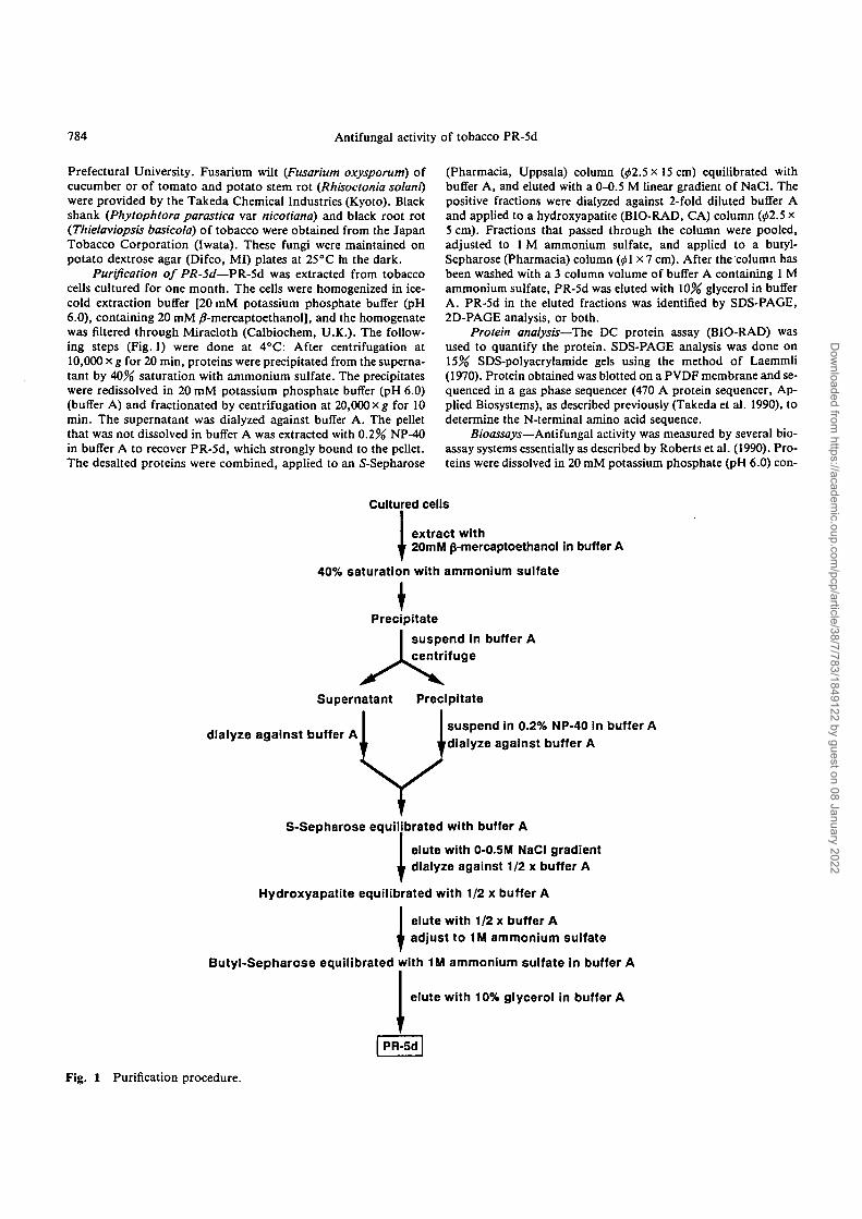
Purification of PR-5d—PR-5d was extracted from tobaccocells cultured for one month. The cells were homogenized in ice-cold extraction buffer [20 mM potassium phosphate buffer (pH6.0), containing 20 mM /?-mercaptoethanol], and the homogenatewas filtered through Miracloth (Calbiochem, U.K.). The follow-ing steps (Fig. 1) were done at 4°C: After centrifugation at10,000 x g for 20 min, proteins were precipitated from the superna-tant by 40% saturation with ammonium suifate. The precipitateswere redissolved in 20 mM potassium phosphate buffer (pH 6.0)(buffer A) and fractionated by centrifugation at 20,000 xg for 10min. The supernatant was dialyzed against buffer A. The pelletthat was not dissolved in buffer A was extracted with 0.2% NP-40in buffer A to recover PR-5d, which strongly bound to the pellet.The desalted proteins were combined, applied to an S-Sepharose
(Pharmacia, Uppsala) column (02.5x15 cm) equilibrated withbuffer A, and eluted with a 0-0.5 M linear gradient of NaCl. Thepositive fractions were dialyzed against 2-fold diluted buffer Aand applied to a hydroxyapatite (BIO-RAD, CA) column (02.5 x5 cm). Fractions that passed through the column were pooled,adjusted to 1 M ammonium suifate, and applied to a butyl-Sepharose (Pharmacia) column (01x7 cm). After the column hasbeen washed with a 3 column volume of buffer A containing 1 Mammonium suifate, PR-5d was eluted with 10% glycerol in bufferA. PR-5d in the eluted fractions was identified by SDS-PAGE,2D-PAGE analysis, or both.
Protein analysis—The DC protein assay (BIO-RAD) wasused to quantify the protein. SDS-PAGE analysis was done on15% SDS-polyacrylamide gels using the method of Laemmli(1970). Protein obtained was blotted on a PVDF membrane and se-quenced in a gas phase sequencer (470 A protein sequencer, Ap-plied Biosystems), as described previously (Takeda et al. 1990), todetermine the N-terminal amino acid sequence.
Bioassays—Antifungal activity was measured by several bio-assay systems essentially as described by Roberts et al. (1990). Pro-teins were dissolved in 20 mM potassium phosphate (pH 6.0) con-
Cultured cells
Iextract with20m M p-mercaptoethanol in buffer A
40% saturation with ammonium suifate
Precipitatesuspend in buffer Acentrifuge
Supernatant Precipitate
dialyze against buffer Asuspend in 0.2% NP-40 in buffer Adialyze against buffer A
S-Sepharose equilibrated with buffer A
elute with 0-0.5M NaCl gradient1 dialyze against 1/2 x buffer A
Hydroxyapatite equilibrated with 1/2 x buffer A
Ielute with 1/2 x buffer Aadjust to 1M ammonium suifate
Butyl-Sepharose equilibrated with 1M ammonium suifate in buffer A
elute with 10% glycerol in buffer A
PR-5d
Fig. 1 Purification procedure.
Dow
nloaded from https://academ
ic.oup.com/pcp/article/38/7/783/1849122 by guest on 08 January 2022

Antifungal activity of tobacco PR-5d 785
taining 10% glycerol and filter sterilized. In the hyphal ruptureassay with Tricoderma reesei, 0.18 M mannitol was included in thereaction to avoid PR-5d independent rupture.
Circular dichroism measurement—CD spectra of the purifiedPR-5 proteins were measured with J-720W spectropolarimeter(JUSCO) using 0.1 cm cells. Proteins were dissolved in 10 mM po-tassium phosphate buffer (pH 6.2) and passed through a PD-10column (Pharmacia) equilibrated with the same buffer to removelow molecular weight compounds which would interfere with themeasurements.
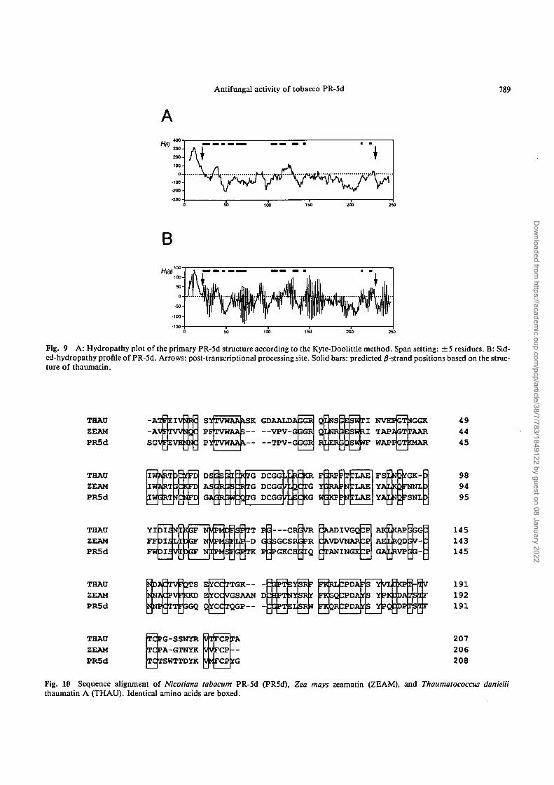
Structural prediction—A Kyte & Doolittle hydropathy profile(//(/)) (Kyte and Doolittle 1982) and a sided-hydropathy profile(Vogel and Jahnig 1986) of amphipathic /?-strands (////)) were cal-culated on the basis of the amino acid sequence of PR-5d cDNA(Takeda et al. 1991). Hp(i) was determined using the followingequation and the slightly modified method of Vogel et al. (1986).
Hfi(i)=fi(i±4)/2+Ki±2)+h(i) h: hydrophobicity index
Crystallization of PR-5d—The first experiment seekingcrystallization conditions was performed with Crystal Screen®(Hampton Research Laboratory). Crystallization was performedby hanging-drop vapor diffusion method. The best crystallizingconditions were the following: Two microliters of protein solutioncontaining 10 nig ml"1 PR-5d, 10% glycerol, and 10 mM MESbuffer (pH 6.0) were mixed with 2 (i\ of reservoir solution contain-ing 0.7 M MgCl2 and 50 mM HEPES (pH7.5); then the dropswere equilibrated at 20°C for 3 to 4 weeks against 1 ml of the reser-voir solution.
X-ray data collection—The PR-5d crystals were mounted inquartz capillaries coated with prosil® 28 (PCR, FL). To take theprecession photographs, we used a Huber precession goniometerand Ni-filtered CuKa radiation from a Rigaku RU300 rotataring-anode operated at 40 kV and 100 mA. The diffraction data werealso collected on an R-AXIS IIC imaging plate detector (Rigaku)with monochromatrazed CuKa raditation. The space group andunit cell parameters were determined from the precession photo-graphs and refined with the diffraction data collected by R-AXISIIC.
Results
Purification and chemical properties of the PR-5d—PR-5d was purified by a combination of ammonium sulfateprecipitation and conventional column chromatography(Fig. 1). PR-5d was precipitated with a relatively low con-centration of ammonium sulfate. When the pellet wasredissolved with buffer, half the PR-5d remained undissolv-ed in the pellet. Treatment with mild detergent succesfullyrecovered PR-5d from this insoluble precipitate. Therecovered fraction was loaded onto an S-Sepharose col-umn. PR-5d was eluted as a rather broad peak as expectedfrom the 2D-PAGE results (Koiwa et al. 1994). The PR-5dfractions were combined and applied to a hydroxyapatitecolumn. All the proteins except PR-5d and 21 kDa proteinwere retained on the column. PR-5d and 21 kDa proteinwere successfully separated in a butyl-Sepharose column.Phenyl-Sepharose (or Superose), used to purify osmotin(data not shown) and other thaumatin-like proteins, couldnot be used because PR-5d bound to this ligand too tightly
AMW 1 2 3 4
42—51
30 — ^
20 —
14 —
BM\
42
30
20
14
MW 1 2 3 4
Fig. 2 A: Purity of PR-5d analyzed by SDS-PAGE. Lane 1:total soluble proteins from cultured tobacco cells. After am-monium sulfate precipitation, PR-5d was purified on S-sepharose(lane 2), hydroxylapatite (lane 3), and butyl-sepharose (lane 4) col-umn. B: SDS-PAGE of commercial thaumatin (lane 1), purifiedPR-5d (lane 2), osmotin (lane 3) and 21 kDa protein (lane 4).Each lane contains 0.5 fig protein. Proteins were stained withCoomassie Brilliant Blue.
even in buffer without salts, and protein recovery with 50%ethyleneglycol was low. These results clearly show that PR-5d is much more hydrophobic than osmotin and the otherrelatively hydrophobic thaumatin-like proteins.
The purities of the PR-5d were confirmed by SDS-PAGE (Fig. 2A, B). As shown in Fig. 3B, careful SDS-PAGE analysis can distinguish between PR-5d, osmotin,and thaumatin. Analysis of the N-terminal amino acid se-quence confirmed the identity of the purified PR-5d (datanot shown).
Antifungal activity of PR-5d—A series of assays wasused to test the antifungal activities of the purified PR-5d.Hyphal extension analysis showed antifungal activity ascrescent-shaped growth retardation of the mycelial front(Fig. 3). PR-5d effectively inhibited the growth of non-phytopathogenic fungi (N. crassa and T. reesei) in thisassay, as has been reported for purified zeamatin (Robertsand Selitrennikoff 1990). Growth of some phytopathogenicfungi (C. miyabeanus, F. oxysporum and Al. solani) werealso considerably inhibited. The protein showed similar in-hibition of spore germination in liquid media (Table 1).
PR-5d induced a rapid burst of hyphal tips in the fungiN. crassa, T. reesei, and C. miyabeanus (Fig. 4). When 0.6M mannitol was added to the hyphal rupture test, thePR-5d-induced burst was suppressed (data not shown).The Tricoderma spore germination assay showed that thegrowth inhibition produced by 50 fig ml"1 PR-5d was main-tained at concentrations of up to 0.6 M mannitol, althoughhyphal rupture was suppressed. On the other hand, the ad-
Dow
nloaded from https://academ
ic.oup.com/pcp/article/38/7/783/1849122 by guest on 08 January 2022

786 Antifungal activity of tobacco PR-5d
N. crassaBuffer
Fusarlum oxysporumf. sp. cucumerium
Fusanum oxysporumt. sp. lycopersici
T. reeseiBuffer PR-5d 10
C. miyabeanusBuffer PR-5d 50 |ig/ml
Fig. 3 Fig. 4
Fig. 3 Inhibitory effects of PR-5d on hyphal growth of fungi. Filter papers (clockwise) contain 0 (top), 10, 30, 50 and 100//g protein.Fungi were cultured on potato dextrose agar plates (90 mm diameter) for 2 d.
Fig. 4 PR-5d-induced hyphal rupture of mycelia. The fungi N. crassa, T. reesei, and C. miyabeanus were treated with buffer alone, 10/ig ml"1 PR-5d, or 50//g ml"1 PR-5d. Hyphal rupture of mycelia was observed within one minute after addition of PR-5d solution.
dition of salts to the spore germination medium inhibitedthe antifungal activity of PR-5d (Table 2), although thistreatment did not inhibit the hyphal rupture activity of PR-5d (data not shown). The effect of salts depended on theconcentration and was saturated at 50 mM. These results in-dicate that hyphal rupture was not the primary cause of theantifungal activity in some cases and that the ionic interac-tion between PR-5d and the target molecule(s) is also im-portant for the activity.
Our results show that PR-5d inhibits the growth of avariety of fungi at different titers. To clarify the antifungalmechanism of PR-5d, the antifungal activities of the PR-5proteins, osmotin and thaumatin, as well as the activity ofthe 21 kDa protein [identified as CBP20 by peptide sequenc-ing (data not shown)] were determined. Synergistic growthinhibition of C. albicans with nikkomycin Z, a inhibitor ofchitin synthase, is a characteristic of antifungal PR-5 pro-
teins. As shown in Fig. 5A, osmotin was less inhibitorythan PR-5d to C. albicans in the synergistic inhibitionassay, but more inhibitory than PR-5d in the liquid suspen-sion assay without nikkomycin Z (Fig. 5B). We sometimesobserved precipitation of osmotin around the paper disc inthe agar plate assay, which may explain the difference in theeffectiveness of osmotin under the assay conditions used.These results suggest that osmotin may be more potentin its antifungal function but less stable than PR-5d.Thaumatin and the 21 kDa protein showed no antifungal ac-tivity against these fungi.
Circular dichroism—The CD spectra of PR-5d andosmotin were analyzed and compared with thaumatin to an-alyse their secondary structures. Figure 6 shows a singlenegative peak at about 210 nm, indicating that the yS-struc-ture is the dominant secondary structure of these PR-5 pro-teins, as it is for thaumatin. Furthermore, all parts of the
Dow
nloaded from https://academ
ic.oup.com/pcp/article/38/7/783/1849122 by guest on 08 January 2022
Antifungal activity of tobacco PR-5d 787
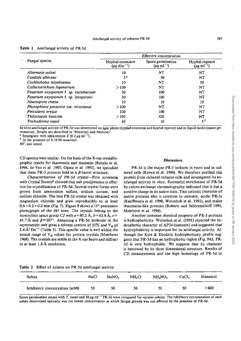
Table 1 Antifungal activity of PR-5d
Fungal species
Alternaria solaniCandida albicansCochliobolus miyabeanusCollectotrichum lagenariumFusarium oxysporum f. sp. cucumeriumFusarium oxysporum f. sp. lycopersiciNeurospora crassaPhytophtora parastica var. nicotianaePiricularia oryzaeThilaviopsis basicolaTrichoderma reesei
Hyphal extension(Hg disc"1)
105"
10>100
303010
>100100
>10010
Effective concentrationSpore germination
UigmV1)NT50
NTNT10010010
NT100300
10
Hyphal rupture(jigm\~l)
NTNT50NTNTNT10
NTNTNT
5*
In vitro antifungal activity of PR-5d was determined on agar plates (hyphal extension and hyphal rupture) and in liquid media (spore ger-mination). Details are described in 'Materials and Methods'.° Synergistic with nikkomycin Z (0.2/ig ml"1).* In the presence of 0.18 M mannitol.NT: not tested.
CD spectra were similar. On the basis of the X-ray crystallo-graphic results for thaumatin and zeamatin (Batalia et al.1996, de Vos et al. 1985, Ogata et al. 1992), we speculatethat these PR-5 proteins fold in a /?-barrel structure.
Characterization of PR-5d crystal—First screeningwith Crystal Screen® showed that salt precipitation is effec-tive for crystallization of PR-5d. Several crystal forms weregrown from ammonium sulfate, sodium acetate, andsodium chloride. The best PR-5d crystal was obtained withmagnesium chloride and grew reproducibly to at least0.6 x 0.2 x 0.2 mm (Fig. 7). Figure 8 shows a 15° precessionphotograph of the Okl zone. The crystals belong to themonoclinic space group C2 with o=80.2 A, 6 = 63.8 A, c=45.7 A and /?=107°. Assuming a PR-5d molecule in theasymmetiric unit gives a solvent content of 50% and VM pf2.4 A3 Da"1 (Table 3). This specific value is well within thenomal range of VM values for protein crystals (Matthews1968). The crystals are stable in the X-ray beam and diffractto at least 1.8 A resolution.
Discussion
PR-5d is the major PR-5 isoform in roots and in cul-tured cells (Koiwa et al. 1994). We therefore purified thisprotein from cultured tobacco cells and investigated its an-tifungal activity in vitro. Successful enrichment of PR-5dby cation-exchange chromatography indicated that it has apositive charge in its native state. This cationic character ofnative proteins also is common to osmotin, acidic PR-5s(Kauffmann et al. 1990, Woloshuk et al. 1991), and maizethaumatin-like proteins (Roberts and Selitrennikoff 1990,Malehorn et al. 1994).
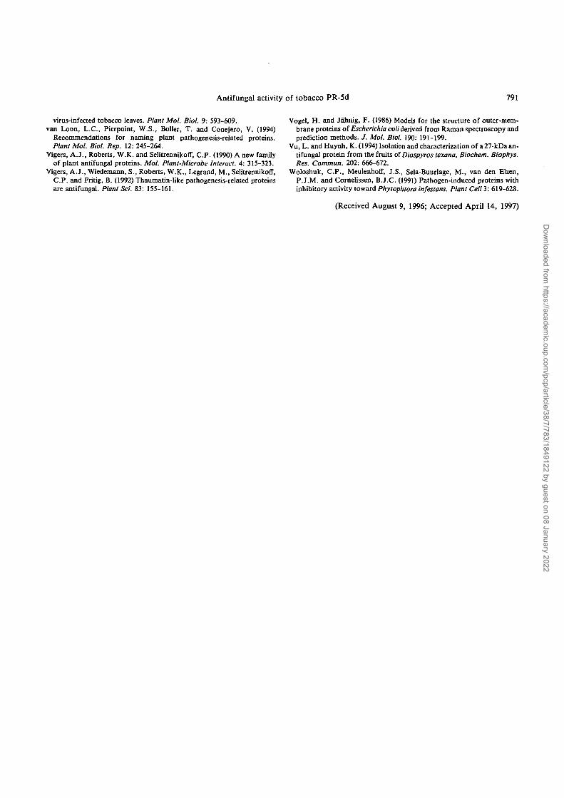
Another common chemical property of PR-5 proteinsis hydrophobicity. Woloshuk et al. (1991) reported the hy-drophobic character of AP24 (osmotin) and suggested thathydrophobicity is important for its antifungal activity. Al-though the Kyte & Doolittle hydrophobicity profile sug-gests that PR-5d has no hydrophobic region (Fig. 9A), PR-5d is very hydrophobic. We suppose that its characteris bestowed by its three dimensional structure. Results ofCD measurements and the high homology of PR-5d to
Table 2 Effect of solutes on PR-5d
Solute
Inhibitory concentration (mM)
antifungal
NaCl
50
activity
NaNO3
50
NH4C1
50
NH4NO3
50
CaCl2
50
Mannitol
>600
Spore germination assays with T. reesei and 50 fjg ml ' PR-5d were compared for various solutes. The inhibitory concentration of eachsolute determined optically was the lowest concentration at which fungal growth was not affected by the presence of PR-5d.
Dow
nloaded from https://academ
ic.oup.com/pcp/article/38/7/783/1849122 by guest on 08 January 2022

788
A
Antifungal activity of tobacco PR-5d
B
0 SO 100
Protein Oig/ml)
Fig. 5 Inhibitory effects of PR-5d and some PR-proteins on thegrowth of Candida albicans. A: In the presence of nikkomycin Z.The filter papers contain 30fig of (1) PR-5d, (2) osmotin, (3)thaumatin, and (4) 21 kDa protein and (B) an equal volume ofbuffer. B: Growth inhibition of Candida albicans by PR-proteinsin liquid culture. Symbols: PR-5d (•), osmotin (O), thaumatin( x), 21 kDa protein (•). Growth of Candida was monitored as thechange of absorbance of 600 nm after 24 hours.
thaumatin and zeamatin (Fig. 10) suggest that PR-5d alsofolds into a yS-barrel structure (Batalia et al. 1996, de Vos etal. 1985, Ogata et al. 1992). This amphiphilic structurecould produce a hydrophobic region on the surface of themolecule. Analysis of the amphipathic /?-structure of PR-5d (Fig. 9B) showed oscillation of the H(l)P value from hy-drophobic to hydrophilic every second residue, indicativeof the high probability of the existence of this structure.Crystallographical findings from homologous proteins,thaumatin and zeamatin (Batalia et al. 1996, de Vos et al.1985, Ogata et al. 1992), showed that the position of thepredicted /J-strands of PR-5d match this oscillation well.
The proposed mechanism of PR-5 proteins is the for-
Fig. 7 Photograph of PR-5d crystal on a microscope.
mation of transmembrane pores in the fungal plasma mem-brane. Woloshuk et al. (1991) speculated that hydrophobicinteraction is important for antifungal activity because themore hydrophobic tobacco AP24 was more antifungalthan was tomato AP24. However, the interference in PR-5d antifungal activity by salts indicates that an ionic interac-tion rather than a hydrophobic interaction between PR-5and the fungal membrane component is probably the pri-mary factor for antifungal activity. The crystal structure ofzeamatin and our recent data for PR-5d (Batalia et al.1996, Koiwa et al. in preparation), showed a highly acidiccleft in these molecular surfaces, which is not present inthaumatin. It is still possible that a subsequent hydro-phobic interaction acts as the common antifungal mechan-ism after the specific ionic interaction. The final determina-tion of the PR-5d structure will be a great help in studyingthe antifungal activity of various PR-5 proteins.
2.0 -
I"'0 •
-1.0 -
A1
HHbd
OSM
THAU
^^f
180 200 220 240
wavelength (nm)
260
Fig. 6 Circular dichroism spectra of PR-5d (PR5d), osmotin(OSM), and thaumatin (THAU).
Fig. 8 A 15° precession photograph of the Okl zone of PR-5dcrystal.
Dow
nloaded from https://academ
ic.oup.com/pcp/article/38/7/783/1849122 by guest on 08 January 2022
Antifungal activity of tobacco PR-5d 789
B
100-
50-
0-
•50-
100-
150-
f \ in ilk iliii,
1 VTPif v | nil i'1
50
111 A
100
I illlllI Ilpll1"
150
• •
ill
j il. iM U.otjin;- imirv"ili •
'I(I IK I'lll'
200 250
Fig. 9 A: Hydropathy plot of the primary PR-5d structure according to the Kyte-Doolittle method. Span setting: ±5 residues. B: Sid-ed-hydropathy profile of PR-5d. Arrows: post-transcriptional processing site. Solid bars: predicted /?-strand positions based on the struc-ture of thaumatin.
THAU
ZEAM
PR5d
-ATFEIVfJRC-AVFTWUQCSGVFEVHUNC
PFTVWAA3
PYTVWAAV
GDAALDA 3GRStfTVWAAASK!— —VPV-GJ3GRL— —TPV-G3GR
NVEPGTMGGKTAPAGTTAARWAPPGTO4AR
494445
THAU
ZEAM
PR5dIW R RT
989495
THADZEAMPR5d
YIDISNEXGFF F D I S L E 3 S FFWDISVt3GF
•PMSELF
JPWDESPTT PG CR3VR- D GGSGCSH3?R
:TANINGE ^P
AKLECAPAELRQDGALRVP
145143145
THAD
ZEAM
PR5d
EifCCrTGK—dNAldPVFKKD EprCCyGSAAtlKJNPPfTT F GGQ
FK R l 3PDA • S YVX3KPI-1VYPK:DAISIFYPQDDPTS1F
191192191
THAU
ZEAM
PR5d
TC PG-SSNYR
TC PA-GTNYK
TC TSWTTDYK
V IFCP TA
VVFCP
V M FCP If G
207
206
208
Fig. 10 Sequence alignment of Nkotiana tabacum PR-5d (PR5d), Zea mays zeamatin (ZEAM), and Thaumatococcus danieliithaumatin A (THAU). Identical amino acids are boxed.
Dow
nloaded from https://academ
ic.oup.com/pcp/article/38/7/783/1849122 by guest on 08 January 2022

790 Antifungal activity of tobacco PR-5d
Table 3 Summary of crystallographic data of PR-5dcrystal determined by RAXIS lie
Crystal system: monoclinicSpace group: C2a = 80.2 (A)fc = 63.8(A)c=45.7(A)j?= 107.2 (°)Z = 4 (molecule)
Our results indicate that PR-5d also functions in thedefense mechanism against fungi, as do other PR-5s intobacco and permatins in plant seeds. The accumulation ofPR-5d in healthy plant roots suggests that it is an activedefense protein. Rapid mRNA accumulation of the PR-5din response to ethylene (Sato et al. 1992) supports this.However, tobacco pathogenic fungi might also develop thetolerance to this antifungal activity (Table 1). Therefore,overexpression of PR-5d in heterologous plant species, likecucumber, is being conducted to evaluate the in vivo func-tion of this PR-5d.
We thank Dr. I. Furusawa and Dr. T. Okuno of Kyoto Uni-versity for their valuable advice and discussions. We appreciateDr. H. Ueno and Dr. S. Ozawa of Kyoto University for their helpin CD measurement. We also thank Dr. S. Shimizu of Kyoto Uni-versity for his helpful advice on manipulating the fungi. Thanksalso due Dr. Y. Kubo, Japan Tobacco Co., and the Takeda Chemi-cal Industries for their kind gifts of fungi. This research was sup-ported in part by a Grant-in-Aid for Scientific Research on Priori-ty Areas (No. 04273103) from the Ministry of Education, Scienceand Culture, Japan, by NEDOs International Joint Research Pro-gram (FS), and by a JSPS Fellowship for Japanese Junior Scien-tists (HK).
References
Alexander, D., Goodman, R.M., Gut-Rella, M., Glascock, C , Weymann,K., Friedrich, L., Maddox, D., Ahl-Goy, P., Luntz, T., Ward, E. andRyal, J. (1993) Increased tolerance to two oomycete pathogens in trans-genic tobbaco expressing pathogenesis-related protein la. Proc. Natl.Acad. Sci. USA 90: 7328-7331.
Batalia, M.A., Monzingo, A.F., Ernst, S., Roberts, W. and Robertus,J.D. (1996) The crystal structure of the antifungal protein zeamatin, amember of the thaumatin-like, PR-5 protein family. Nat. Struct. Biol. 3:19-23.
Bol, J.F. and van Kan, J.A.L. (1988) The synthesis and possible functionof virus-induced proteins in plants. Microbiol. Sci. 5: 47-52.
de Vos, A.M., Hatada, M., Wei, H.v.d., Krabbendam, H., Peerdeman,A.F. and Kim, S.-H. (1985) Three-dimensional structure of thaumatin I,an intensely sweet protein. Proc. Natl. Acad. Sci. USA 82: 1406-1409.
Grosset, J., Meyer, Y., Chartier, Y., Kauffmann, S., Legrand, M. andFritig, B. (1990) Tobacco mesophyll protoplasts synthesize 1,3-/?-glucanase, chitinase, and "osmotins" during in vitro culture. PlantPhysiol. 92: 520-527.
Hejgaard, J., Jacobsen, S. and Svendsen, I. (1991) Two antifungal
thaumatin-like proteins from barley grain, FEBS Lett. 291: 127-131.Kauffmann, S., Legrand, M. and Fritig, B. (1990) Isolation and characteri-
zation of six pathogenesis-related (PR) proteins of Samsun NN tobacco.Plant Mol. Biol. 14: 381-390.
Kauffman, S., Legrand, M., Geoffrey, P. and Fritig, B. (1987) Biologicalfunction of 'pathogenesis-related' proteins: four PR proteins of tobaccohave l,3-/?-glucanase activity. EMBO J. 6: 3209-3212.
Koiwa, H., Sato, F. and Yamada, Y. (1994) Characterization of accumula-tion of PR-5 proteins by IEF-immunoblot analysis. Plant Cell Physiol.35: 821-827.
Kyte, J. and Doolittle, R.F. (1982) A simple method for displaying the hy-dropathic character of a protein. J. Mol. Biol. 157: 105-132.
Laemmli, U.K. (1970) Cleavage of structual proteins during the assemblyof the head of bacteriophage T4. Nature 227: 680-685.
Legrand, M., Kauffmann, S., Geoffroy, P. and Fritig, B. (1987) Biologicalfunction of pathogenesis-related proteins: four tobacco pathogenesis-related proteins are chitinases. Proc. Natl. Acad. Sci. USA 84: 6750-6754.
Malehorn, D.E., Borgmeyer, J.R., Smith, C.E. and Shah, D.M. (1994)Characterization and expression of an antifungal zeamatin-like protein(Zip) gene from Zea mays. Plant Physiol. 106: 1471-1481.
Matthews, B.W. (1968) Solvent content of protein crystals. / . Mol. Biol.33: 491-497.
Mauch, F., Mauch-Mani, B. and Boiler, T. (1988) Antifungal hydrolasesin pea tissue. II. Inhibition of fungal growth by combination of chitinaseand /3-1,3-glucanase. Plant Physiol. 88: 936-942.
Niderman, T., Genetet, I., Bruyere, T., Gees, R-, Stinzi, A., Legrand, M.,Fritig, B. and Mosinger, E. (1995) Pathyogenesis-related PR-1 proteinsare antifungal. Plant Physiol. 108: 17-27.
Ogata, CM., Gordon, P.F., Vos, A.M.D. and Kim, S.-H. (1992) Crystalstructure of a sweet tasting protein, thaumatin I, at 1.65 A resolution. J.Mol. Biol. 228: 893-908.
Ponstein, A.S., Bres-Vloemans, S.A., Sela-Buurlage, M.B., van denElzen, P.J.M., Melchers, L.S. and Cornelissen, B.J.C. (1994) A novelpathogen- and wound-inducible tobacco (Nicotiana tabacum) proteinwith antifungal activity. Plant Physiol. 104: 104-118.
Roberts, W.K. and Selitrennikoff, C.P. (1990) Zeamatin, an antifungal pro-tein from maize with membrene-permealizing activity. J. Gen. Micro-biol. 136: 1771-1778.
Sato, F., Koiwa, H., Sakai, Y., Kato, N. and Yamada, Y. (1995) Synthesisand secretion of tobacco neutral PR-5 protein by transgenic tobacco andyeast. Biochem. Biophys. Res. Commun. 211: 909-913.
Sato, F., Takeda, S. and Yamada, Y. (1992) Accumulation of stress-pro-teins in cultured tobacco cells and ethylene. In Plant Tissue Culture andGene Manupulation for Breeding and Formation of Phytochemicals.Edited by Oono, K., Hirabayashi, T., Kikuchi, S., Handa, H. and Kaji-hara, K. pp. 377-389. National Institute of Agrobiological Resources,Tsukuba,Japan.
Schlumbaum, A., Mauch, F., Vogeli, U. and Boiler, T. (1986) Plantchitinases are potent inhibitors of fungal growth. Nature 324: 365-367.
Sela-Buurlage, M.B., Ponstein, A.S., Vloemans, S.A., Melchers, L.S.,Elzen, P.J.M.V.D. and Cornelissen, B.J.C. (1993) Only specific tobaccochitinases and /?-glucanases exibit antifungal activity. Plant Physiol. 101:857-863.
Singh, N.K., Bracker, C.A., Hasegawa, P.M., Handa, A.K., Hermodson,M.A., Pfankoch, E., Regnier, F.E. and Bressan, R.A. (1987) Characteri-zation of osmotin. Plant Physiol. 85: 529-536.
Singh, N.K., LaRosa, C , Handa, A.K., Hasegawa, P.M. and Bressan,R.A. (1987) Hormonal regulation of protein synthesis associated withsalt tolerance in plant cells. Proc. Natl. Acad. Sci. USA 84: 739-743.
Takeda, S., Sato, F., Ida, K. and Yamada, Y. (1990) Characterization ofpolypeptides that accumulated in cultured Nicotiana tabacum cells.Plant Cell Physiol. 31: 215-221.
Takeda, S., Sato, F., Ida, K. and Yamada, Y. (1991) Nucleotide sequenceof a cDNA for osmotin-like protein from cultured tobacco cells. PlantPhysiol. 97: 844-846.
van Loon, L.C. (1985) Pathogenesis-related proteins. Plant Mol. Biol. 4:111-116.
van Loon, L.C, Gerritsen, Y.A.M. and Ritter, C.E. (1987) Identification,purification, and characterization of pathogenesis-related proteins from
Dow
nloaded from https://academ
ic.oup.com/pcp/article/38/7/783/1849122 by guest on 08 January 2022
Antifungal activity of tobacco PR-5d 791
virus-infected tobacco leaves. Plant Mol. Biol. 9: 593-609.van Loon, L.C., Pierpoint, W.S., Boiler, T. and Conejero, V. (1994)
Recommendations for naming plant pathogenesis-related proteins.Plant Mol. Biol. Rep. 12: 245-264.
Vigers, A.J., Roberts, W.K. and Selitrennikoff, C.P. (1990) A new familyof plant antifungal proteins. Mol. Plant-Microbe Interact. 4: 315-323.
Vigers, A.J., Wiedemann, S., Roberts, W.K., Legrand, M., Selitrennikoff,C.P. and Fritig, B. (1992) Thaumatin-like pathogenesis-related proteinsare antifungal. Plant Sci. 83: 155-161.
Vogel, H. and Jahnig, F. (1986) Models for the structure of outer-mem-brane proteins of Escherichia coli derived from Raman spectroscopy andprediction methods. J. Mol. Biol. 190: 191-199.
Vu, L. and Huynh, K. (1994) Isolation and characterization of a 27-kDa an-tifungal protein from the fruits of Diospyros texana. Biochem. Biophys.Res. Commun. 202: 666-672.
Woloshuk, C.P., Meulenhoff, J.S., Sela-Buurlage, M., van den Elzen,P.J.M. and Cornelissen, B.J.C. (1991) Pathogen-induced proteins withinhibitory activity toward Phytophtora infestans. Plant Cell 3: 619-628.
(Received August 9, 1996; Accepted April 14, 1997)
Dow
nloaded from https://academ
ic.oup.com/pcp/article/38/7/783/1849122 by guest on 08 January 2022