MOLECULAR PATHOGENESIS OF RARE ANEMIAS - Theses ...
170
MOLECULAR PATHOGENESIS OF RARE ANEMIAS – ERYTHROENZYMOPATHIES Ph.D. Thesis PAVLA KOŘALKOVÁ (BORN POSPÍŠILOVÁ) DEPARTMENT OF BIOLOGY FACULTY OF MEDICINE AND DENTISTRY PALACKY UNIVERSITY OLOMOUC OLOMOUC 2017
-
Upload
khangminh22 -
Category
Documents
-
view
2 -
download
0
Transcript of MOLECULAR PATHOGENESIS OF RARE ANEMIAS - Theses ...
MOLECULAR PATHOGENESIS OF
RARE ANEMIAS –
ERYTHROENZYMOPATHIES
Ph.D. Thesis
PAVLA KOŘALKOVÁ (BORN POSPÍŠILOVÁ)
DEPARTMENT OF BIOLOGY
FACULTY OF MEDICINE AND DENTISTRY
PALACKY UNIVERSITY OLOMOUC
OLOMOUC 2017
DECLARATION/PROHLÁŠENÍ:
I hereby declare that this Ph.D. thesis entitled “MOLECULAR PATHOGENESIS OF RARE
ANEMIAS – ERYTHROENZYMOPATHIES” was carried out by me under the guidance and
supervision of Renáta Mojzíková, Ph.D., and all used literature is cited accordingly.
Tímto prohlašuji, že předloženou práci s názvem “MOLEKULÁRNÍ PATHOGENEZE VZÁCNÝCH
ANÉMIÍ – ENZYMOPATIÍ” jsem vypracovala samostatně pod vedením školitelky
Mgr. Renáty Mojzíkové, Ph.D., a s použitím citované literatury.
ABSTRACT:
The aim of this research is to make a contribution to the molecular pathogenesis of
erythroenzymopathies, rare hereditary disorders causing nonspherocytic hemolytic anemia. This work
represents an advanced and up-to-date knowledge in the field of red blood cells enzyme deficiencies in
the Czech and Slovak populations since the nineteen-eighties when mutations in Czech and Slovak
subjects resulting in glucose-6-phosphate dehydrogenase (G6PD) and pyruvate kinase (PK)
deficiencies were described. Both deficiencies are the most common erythroenzymopathies
worldwide. Since 2013, our laboratory has focused on the introduction of direct enzyme assays
followed by genetic testing in patients with hemolytic anemia with suspected erythroenzymopathy.
To date, several different enzyme defects were diagnosed in twenty-four patients. Except G6PD and
PK deficiencies (9 and 12 cases), 2 families with glucose phosphate isomerase (GPI) defect and 1
family with very rare hexokinase (HK) deficiency have been identified. The last two deficiencies were
diagnosed in the Czech and Slovak populations for the first time. Among the 22 identified mutations,
5 of them, namely G6PD p.(Phe216Tyr), PK p.(Arg518Leufs*12), p.(Asp293Val) and GPI
p.(Ser160Pro), p.(Arg472Cys) were novel and have not been previously reported in literature.
Furthermore, this thesis describes a pathogenic mechanism of several novel mutations responsible for
deficiencies of hexokinase (HK), phosphoglycerate kinase (PGK) and phosphofructokinase (PFK)
in more detail. HK deficiency is a very rare disorder that has been rarely studied at molecular level.
In collaboration with other research centers, the largest cohort of HK deficient patients was selected
and extensively studied. Using molecular techniques and in silico analyses, potential pathogenic
mechanisms of novel mutations p.(His867Tyr), p.(Thr600Met), c.873-2A>G and c. 493-1G>A have
been elucidated. Further, we contributed to the identification and characterization of a novel intronic
mutation c.756+3A>G in PGK1 in two brothers with PGK deficiency. Although PGK deficiency
is mostly associated with hemolytic anemia and neurological impairment, muscle weakness and
myopathy were the only clinical features in these cases. Finally, we contributed to the detection
of a novel mutation p.(Asp309Gly) in PFKM gene, the first case of a rare PFK deficiency diagnosed
in a 65-year-old female of Spanish origin.
Characterization of erythroenzymopathies by biochemical and molecular analyses can contribute
to a better understanding of pathophysiology of this rare disorder. Furthermore, the direct enzyme
assays may have a wide variety of scientific/diagnostic applications; it may improve diagnostic testing
of other disorders or it may be useful to study the mechanism of drug's actions.

ABSTRAKT:
Tato práce se zabývá studiem molekulární patogeneze enzymopatíí - vzácného onemocnění
způsobující vrozenou nesférocytární hemolytickou anémii. Deficit glukosa-6-fosfátdehydrogenasy
(GPD) a pyruvátkinasy (PK) byl v české a slovenské populaci popsán již v 80. letech. Nicméně až tato
studie představuje zatím chybějící ucelený přehled o výskytu tohoto onemocnění v české a slovenské
populaci, zvláště pak jeho vzácnější formy. Od roku 2013 se naše laboratoř zabývá metodou přímého
stanovení enzymů s následným genetickým vyšetřením u pacientů se suspektní enzymopatií.
V současné době jsme jedinou specializovanou laboratoří zabývající se touto diagnostikou v České
republice resp. ve střední Evropě. Doposud byl enzymový deficit diagnostikován u dvaceti čtyř
pacientů. Vedle nejčastějších deficitů G6PD a PK (9 a 12 případů), byly identifikovány 2 rodiny
s deficitem glukosafosfátisomerasy (GPI) a 1 rodina s velmi vzácným deficitem hexokinasy (HK).
Deficit GPI a HK byl v české a slovenské populací diagnostikován poprvé. Celkově bylo
identifikováno dvacet dva kauzálních mutací, z nichž pět nebylo doposud v literatuře popsáno - G6PD
p.(Phe216Tyr), PK p.(Arg518Leufs * 12), p.(Asp293Val), GPI p.(Ser160Pro) a p.(Arg472Cys).
Současně tato práce studuje patogenní mechanismy vybraných mutací vedoucí k deficitu HK,
fosfoglycerátkinasy (PGK) a fosfofruktokinasy (PFK). Deficit HK byl pouze výjimečně studován na
molekulární úrovni. Na základě zahraniční spolupráce jsme vytvořili a detailněji prostudovali největší
soubor pacientů s tímto deficitem. Patogenní mechanismus kauzálních mutací p.(His867Tyr),
p.(Thr600Met), c.873-2A>G a c.493-1G>A byl objasněn pomocí molekulárních technik a in silico
analýz. Dále jsme identifikovali a charakterizovali novou intronovou mutaci c.756+3A>G v genu
PGK1 vedoucí k PGK deficitu. Ve většině případů je deficit PGK spojen s hemolytickou anémií
a neurologickými poruchami. Avšak v tomto případě byla svalová slabost a myopatie jediným
klinickým příznakem. Dále jsme u pacientky španělského původu identifikovali a charakterizovali
novou mutaci p.(Asp309Gly) v genu kódující PFK. Jedná se o první případ PFK deficitu
ve Španělsku.
Charakterizace erytroenzymopatií pomocí biochemických a molekulárních analýz může přispět
k porozumění patofyziologie tohoto vzácného onemocnění. Navíc metoda přímého stanovení
erytrocytárních enzymů může vést ke zlepšení diagnostiky jiných onemocnění či je možné ji využít při
studiu farmakologických sloučenin.
ACKNOWLEDGEMENT:
I would like to thank my supervisor Renáta Mojzíková, Ph.D. for her endless guidance,
encouragement and advice she has provided throughout my PhD studies.
My special thanks belong to Richard van Wijk, Ph.D. for his patient guidance, research funding and
his enthusiasm in research which motivated me during my fellowship at the Department of Clinical
Chemistry and Hematology, UMC Utrecht.
I would like to thank Assoc. Prof. Vladimír Divoký, Ph.D. for his critical comments and for providing
the funding which allowed me to do the research and to attend many interesting conferences.
I also wish to thank all my colleagues (from the Department of biology and from UMC Utrecht) for
creating a friendly and working atmosphere.
I appreciate good collaboration with all co-authors of the publications, who gave me important advice.
Finally, I must express my gratitude to Petr, my husband, for his continued support, encouragement
and endless patience. Completing this work would be difficult without support of my family and
friends.
This work was supported by Ministry of Health of the Czech Republic, grants: NT/13587, NT11208;
Internal Grant Agency of Palacky University, grants: LF_2012_16, LF_2013_010,
IGA_LF_2015_015, IGA_LF_UP_2016_014; and Czech Science Foundation, project P301_12_1503.

CONTENTS:
1 INTRODUCTION ....................................................................................................................... 1
1.1 ERYTHROPOIESIS ................................................................................................................ 1
1.2 THE RED BLOOD CELL METABOLISM ............................................................................ 2
1.3 ANEMIA ................................................................................................................................. 5
1.3.1 HEMOLYTIC ANEMIA ......................................................................................................... 6
1.3.1.1 HEMOGLOBINOPATHIES ................................................................................................... 6
1.3.1.2 RED BLOOD CELL MEMBRANE DISORDERS ................................................................ 9
1.3.1.3 RED BLOOD CELL ENZYMOPATHIES ........................................................................... 12
2 AIMS OF THE THESIS ........................................................................................................... 23
3 MATERIALS AND METHODS ............................................................................................. 24
3.1 ISOLATION OF RBCS FROM PERIPHERAL BLOOD AND HEMOLYSATE
PREPARATION .................................................................................................................... 26
3.2 ENZYME ANALYSIS .......................................................................................................... 26
3.2.1 DETERMINATION OF MICHAELIS-MENTEN CONSTANT (KM) FOR GLUCOSE AND
ATP ........................................................................................................................................ 27
3.2.2 ENZYME STABILITY ASSAYS ......................................................................................... 27
3.3 CELL CULTURE OF HUMAN NUCLEATED ERYTHROID CELLS ............................... 28
3.3.1 ISOLATION OF MONONUCLEAR CELLS FROM HUMAN PERIPHERAL BLOOD BY
DENSITY GRADIENT CENTRIFUGATION ..................................................................... 28
3.3.2 SELECTION OF PRO-ERYTHROBLASTS ........................................................................ 28
3.3.3 EXPANSION OF PRO-ERYTHROBLAST ......................................................................... 29
3.4 GENOMIC DNA ISOLATION FROM WHITE BLOOD CELLS ........................................ 29
3.5 PCR REACTION ................................................................................................................... 30
3.6 CYCLE SEQUENCING ........................................................................................................ 30
3.7 RESTRICTION ANALYSIS ................................................................................................. 31
3.8 TOTAL RNA ISOLATION FROM HUMAN PRO-ERYTHROBLAST CELLS ................ 31
3.9 REVERSE TRANSCRIPTION POLYMERASE CHAIN REACTION (RT-PCR) .............. 32
3.10 QUANTIFICATION OF HK-R AND PGK1 TRANTSCRIPTS IN PATIENTS’ PRO-
ERYTHROBLAST CELLS ................................................................................................... 32
3.11 “TOPO CLONNING” OF ABERRANT PGK1 TRANSCRIPTS ......................................... 33

3.11.1 TRANSFORMATION OF ONE SHOT® TOP10 .................................................................. 33
3.11.2 ANALYSIS OF TRANSFORMANTS .................................................................................. 33
3.11.3 PLASMID DNA ISOLATION .............................................................................................. 33
3.12 IMMUNOBLOT ANALYSIS ............................................................................................... 34
3.13 IN SILICO ANALYSIS, 3D STRUCTURAL ANALYSIS, MULTIPLE SEQUENCE
ALIGNMENT ........................................................................................................................ 34
4 RESULTS AND DISCUSSION ............................................................................................... 35
4.1 THE LITERATURE REVIEW .............................................................................................. 35
4.2 DIFFERENTIAL DIAGNOSIS OF CZECH AND SLOVAK PATIENTS WITH
HEMOLYTIC ANEMIA OF UKNOWN ETIOLOGY ......................................................... 37
4.3 STRUCTURAL AND FUNCTIONAL CHARACTERIZATION OF VERY RARE
HEXOKINASE MUTANTS CAUSING HNSHA ................................................................ 47
4.4 MOLECULAR CHARACTERIZATION OF A NOVEL NON-HEMOLYTIC ANEMIA
VARIANT OF PGK ............................................................................................................... 55
4.5 IN SILICO MODELLING OF A NOVEL MUTATION OF PFKM GENE ASSOCIATED
WITH PFK DEFICIENCY .................................................................................................... 60
5 SUMMARY ............................................................................................................................... 63
6 LIST OF ALL PUBLICATIONS ............................................................................................ 64
7 REFERENCES .......................................................................................................................... 68
8 SUPPLEMENTS ....................................................................................................................... 77
9 ACRONYMS ............................................................................................................................. 82
10 APPENDICES ........................................................................................................................... 84

Page | 1
1 INTRODUCTION
A major part of the theoretical background related to erythroenzymopathies was quoted verbatim from
my first-authored publication “Rare hereditary red blood cell enzymopathies associated with
hemolytic anemia – pathophysiology, clinical aspects, and laboratory diagnosis” (Koralkova et
al., 2014). Several sections were expanded and updated to complete current knowledge. All references
were cited accordingly.
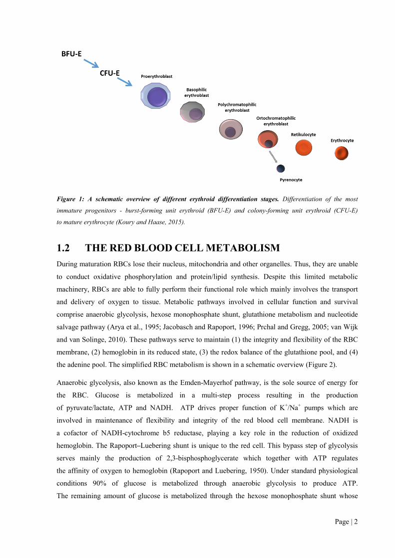
1.1 ERYTHROPOIESIS
Erythropoiesis is a highly regulated process characterized by proliferation and progressive maturation
of hematopoietic stem cells through lineage–committed progenitors and erythroblast precursors
to mature enucleated red blood cells (RBCs). Erythroid progenitors emerge from two distinct sources
during the mammalian development. The early erythropoiesis, primitive erythropoiesis, occurs
in “blood islands” within the yolk sack. Primitive erythroblasts arise from mesodermal cells and
provide a transient pool of unique erythroid progenitors (EryP–CFC) (Palis, 2014; Palis et al., 2010).
Definitive erythropoiesis takes place in fetal liver and postnatal bone marrow. Burst-forming unit
erythroid (BFU–E) and colony-forming unit erythroid (CFU–E) are the most immature erythroid
progenitors. They further progress to nucleated pro-erythroblast, basophilic, polychromatophilic
and orthochromatic erythroblast. The erythroid maturation results in enucleation and reticulocytes
formation (Figure 1). Whereas reticulocytes undergo rapid maturation, the extruded nuclei in form
of pyrenocytes are engulfed by macrophages (Migliaccio, 2010; Palis, 2014). The erythroid
differentiation is regulated at multiple levels by many hormones/cytokines, transcription factors and
nutritional supply. The most important cytokine of erythropoiesis is erythropoetin (EPO). At early
stages of erythroid differentiation (CFU–E stage to basophilic pro-erythroblasts), EPO induces
erythropoiesis by promoting proliferation and inhibiting apoptosis. Several transcription factors, such
as GATA–1 (GATA–binding factor 1), KLF1 (Krüppel like factor 1) or NF–E2 (nuclear factor-
erythroid-derived 2), are implicated in erythroid specific gene expression (Chateauvieux et al., 2011;
Tsiftsoglou et al., 2009). Furthermore, many nutritions, such as folate, vitamin B12 and iron, are
important for hemoglobin synthesis and normal RBC maturation (Koury and Ponka, 2004).
Page | 2
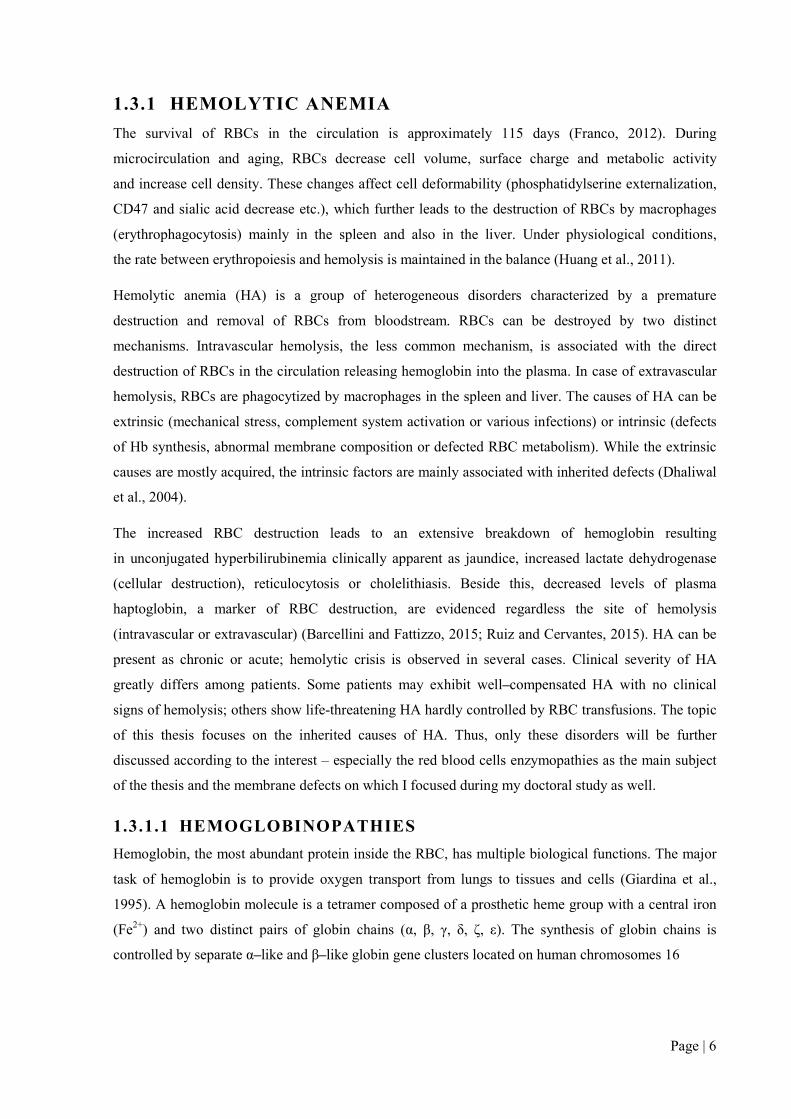
Figure 1: A schematic overview of different erythroid differentiation stages. Differentiation of the most
immature progenitors - burst-forming unit erythroid (BFU-E) and colony-forming unit erythroid (CFU-E)
to mature erythrocyte (Koury and Haase, 2015).
1.2 THE RED BLOOD CELL METABOLISM
During maturation RBCs lose their nucleus, mitochondria and other organelles. Thus, they are unable
to conduct oxidative phosphorylation and protein/lipid synthesis. Despite this limited metabolic
machinery, RBCs are able to fully perform their functional role which mainly involves the transport
and delivery of oxygen to tissue. Metabolic pathways involved in cellular function and survival
comprise anaerobic glycolysis, hexose monophosphate shunt, glutathione metabolism and nucleotide
salvage pathway (Arya et al., 1995; Jacobasch and Rapoport, 1996; Prchal and Gregg, 2005; van Wijk
and van Solinge, 2010). These pathways serve to maintain (1) the integrity and flexibility of the RBC
membrane, (2) hemoglobin in its reduced state, (3) the redox balance of the glutathione pool, and (4)
the adenine pool. The simplified RBC metabolism is shown in a schematic overview (Figure 2).
Anaerobic glycolysis, also known as the Emden-Mayerhof pathway, is the sole source of energy for
the RBC. Glucose is metabolized in a multi-step process resulting in the production
of pyruvate/lactate, ATP and NADH. ATP drives proper function of K+/Na+ pumps which are
involved in maintenance of flexibility and integrity of the red blood cell membrane. NADH is
a cofactor of NADH-cytochrome b5 reductase, playing a key role in the reduction of oxidized
hemoglobin. The Rapoport–Luebering shunt is unique to the red cell. This bypass step of glycolysis
serves mainly the production of 2,3-bisphosphoglycerate which together with ATP regulates
the affinity of oxygen to hemoglobin (Rapoport and Luebering, 1950). Under standard physiological
conditions 90% of glucose is metabolized through anaerobic glycolysis to produce ATP.
The remaining amount of glucose is metabolized through the hexose monophosphate shunt whose
Page | 3
main function is the production of redox potential in the form of NADPH. It is the only way
of NADPH formation in RBC. NADPH acts as a cofactor for glutathione reductase which maintains
glutathione in its reduced state, thereby provides protection against oxidative stress (van Zwieten et
al., 2014). RBC nucleotide metabolism contributes to maintaining the energy balance in erythrocytes.
Nucleotide content in mature erythrocytes is predominantly regulated by the intracellular purine
metabolic cycle (Dudzinska et al., 2006). Pyrimidines are degraded by pyrimidine–5’–nucleotidase
during reticulocyte maturation and are present only as trace amounts in the mature RBC. In contrast,
adenine derivatives (AMP, ADP, ATP) represent about 97% of the total nucleotide pool (Valentine
and Paglia, 1980a). Because mature RBCs lack phosphoribosyl–1–pyrophosphate, an essential enzyme
for de novo synthesis of purines, purine derivatives are reconstituted from intermediates of purine
catabolism. This mechanism is known as a salvage pathway (Dudzinska et al., 2006).

Page | 4
Figure 2: A schematic overview of red blood cell metabolic pathways. Hb – hemoglobin, MetHb –
methemoglobin, GSH – reduced glutathione, GSSG – oxidized glutathione
Page | 5
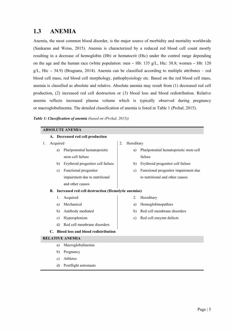
1.3 ANEMIA
Anemia, the most common blood disorder, is the major source of morbidity and mortality worldwide
(Sankaran and Weiss, 2015). Anemia is characterized by a reduced red blood cell count mostly
resulting in a decrease of hemoglobin (Hb) or hematocrit (Htc) under the control range depending
on the age and the human race (white population: men – Hb: 135 g/L, Htc: 38.8; women – Hb: 120
g/L, Htc – 34.9) (Brugnara, 2014). Anemia can be classified according to multiple attributes – red
blood cell mass, red blood cell morphology, pathophysiology etc. Based on the red blood cell mass,
anemia is classified as absolute and relative. Absolute anemia may result from (1) decreased red cell
production, (2) increased red cell destruction or (3) blood loss and blood redistribution. Relative
anemia reflects increased plasma volume which is typically observed during pregnancy
or macroglobulinemia. The detailed classification of anemia is listed in Table 1 (Prchal, 2015).
Table 1: Classification of anemia (based on (Prchal, 2015))
ABSOLUTE ANEMIA
A. Decreased red cell production
1. Acquired 2. Hereditary
a) Pluripotential hematopoietic
stem cell failure
b) Erythroid progenitor cell failure
c) Functional progenitor
impairment due to nutritional
and other causes
a) Pluripotential hematopoietic stem cell
failure
b) Erythroid progenitor cell failure
c) Functional progenitor impairment due
to nutritional and other causes
B. Increased red cell destruction (Hemolytic anemias)
1. Acquired 2. Hereditary
a) Mechanical
b) Antibody mediated
c) Hypersplenism
d) Red cell membrane disorders
a) Hemoglobinopathies
b) Red cell membrane disorders
c) Red cell enzyme defects
C. Blood loss and blood redistribution
RELATIVE ANEMIA
a) Macroglobulinemia
b) Pregnancy
c) Athletes
d) Postflight astronauts
Page | 6
1.3.1 HEMOLYTIC ANEMIA
The survival of RBCs in the circulation is approximately 115 days (Franco, 2012). During
microcirculation and aging, RBCs decrease cell volume, surface charge and metabolic activity
and increase cell density. These changes affect cell deformability (phosphatidylserine externalization,
CD47 and sialic acid decrease etc.), which further leads to the destruction of RBCs by macrophages
(erythrophagocytosis) mainly in the spleen and also in the liver. Under physiological conditions,
the rate between erythropoiesis and hemolysis is maintained in the balance (Huang et al., 2011).
Hemolytic anemia (HA) is a group of heterogeneous disorders characterized by a premature
destruction and removal of RBCs from bloodstream. RBCs can be destroyed by two distinct
mechanisms. Intravascular hemolysis, the less common mechanism, is associated with the direct
destruction of RBCs in the circulation releasing hemoglobin into the plasma. In case of extravascular
hemolysis, RBCs are phagocytized by macrophages in the spleen and liver. The causes of HA can be
extrinsic (mechanical stress, complement system activation or various infections) or intrinsic (defects
of Hb synthesis, abnormal membrane composition or defected RBC metabolism). While the extrinsic
causes are mostly acquired, the intrinsic factors are mainly associated with inherited defects (Dhaliwal
et al., 2004).
The increased RBC destruction leads to an extensive breakdown of hemoglobin resulting
in unconjugated hyperbilirubinemia clinically apparent as jaundice, increased lactate dehydrogenase
(cellular destruction), reticulocytosis or cholelithiasis. Beside this, decreased levels of plasma
haptoglobin, a marker of RBC destruction, are evidenced regardless the site of hemolysis
(intravascular or extravascular) (Barcellini and Fattizzo, 2015; Ruiz and Cervantes, 2015). HA can be
present as chronic or acute; hemolytic crisis is observed in several cases. Clinical severity of HA
greatly differs among patients. Some patients may exhibit well–compensated HA with no clinical
signs of hemolysis; others show life-threatening HA hardly controlled by RBC transfusions. The topic
of this thesis focuses on the inherited causes of HA. Thus, only these disorders will be further
discussed according to the interest – especially the red blood cells enzymopathies as the main subject
of the thesis and the membrane defects on which I focused during my doctoral study as well.
1.3.1.1 HEMOGLOBINOPATHIES
Hemoglobin, the most abundant protein inside the RBC, has multiple biological functions. The major
task of hemoglobin is to provide oxygen transport from lungs to tissues and cells (Giardina et al.,
1995). A hemoglobin molecule is a tetramer composed of a prosthetic heme group with a central iron
(Fe2+) and two distinct pairs of globin chains (α, β, γ, δ, ζ, ε). The synthesis of globin chains is
controlled by separate α–like and β–like globin gene clusters located on human chromosomes 16

Page | 7
and 11, respectively (Thom et al., 2013). The proportion of individual globin chains in a hemoglobin
molecule differs with prenatal gestation time and postnatal age (Forget and Bunn, 2013). The overall
composition of hemoglobin in adults is Hb A1 (95–98%), Hb A2 (2–3%) and Hb F (<1%) (Brugnara,
2014).
Up to date, over 1000 globin mutations associated with aberrant hemoglobin synthesis
and/or structure/function have been reported. Hemoglobin disorders can be divided into two major
groups 1) thalassemia syndromes and 2) structural hemoglobin variants. Thalassemia syndromes are
characterized by an imbalance in production of globin chains, thus, are referred as quantitative defects.
The most common thalassemia syndromes are α- and β-thalassemias, however, defects in delta (δ)– or
gamma (γ)–globin chains have been described. Although majority of these defects cause inherited
autosomal recessive disorders, several patients exhibit acquired form of α-thalassemia in association
with myelodysplastic syndrome (Forget and Bunn, 2013). Several families with thalassemia
syndromes have been previously reported in the Czech and Slovak populations (Indrak et al., 1992).
Recent molecular–genetic screening of patients from the Czech Republic and the Slovak Republic
revealed 29 β–thalassemia mutations in 356 heterozygotes from 218 unrelated families. Most of
the mutations were of Mediterranean origin (82% of cases); five mutations have not been previously
reported (Divoka et al., 2016a). In most cases the phenotype corresponds to β-thalassemia minor;
rarely to β–thalassemia intermedia or major. In addition, deletions of variable size were identified in
alpha-globin gene cluster of eighty Czech patients or immigrants living in the Czech Republic.
Besides common Mediterranean and Asian deletions (especially -α3.7 and –SEA), large deletions
encompassing entire alpha globin locus or only the HS-40 regulatory region were detected in several
cases (Divoka et al., 2016b).
Structural hemoglobin variants are qualitative defects in which mutations predominantly alter
hemoglobin structure and/or its biochemical properties, further affecting oxygen affinity and/or
hemoglobin stability (Thom et al., 2013). The endemic hemoglobin variants HbS, HbC and HbE are
among the most studied and frequent pathologies. In the HbS variant (β7 Glu–Val), deoxygenated
HbS polymerizes inside the RBCs and decreases RBC deformability. It damages a RBC membrane
cytoskeleton which accounts for the irreversibly sickled RBCs seen in the peripheral blood.
Furthermore, sickle RBCs enable adhesive interactions with endothelial cells and together with other
factors may result in vasoocclusions. The homozygous form of HbS is responsible for the most
common and most severe variant of the sickle cell disease. On the other hand, heterozygosity for HbS
has a protective effect to malaria parasitization of RBCs (Frenette and Atweh, 2007; Steinberg, 2008).
The HbE variant (β26 Glu–Lys) is extremely frequent in Asian population (Fucharoen and
Weatherall, 2012; Orkin et al., 1982). The mutation slightly affects oxygen affinity, thermal stability
and sensitivity to oxidation stress. In addition, G to A substitution activates a cryptic splice site that
further affects mRNA processing (Orkin et al., 1982). A homozygous form of HbE is associated with

Page | 8
mild anemia and marked morphological abnormalities. Both HbS and HbE variants have been detected
in Czech population, but mainly in immigrants living in the Czech Republic (Divoky, 2005). The HbC
(β7 Glu-Lys) variant forms insoluble crystals which in RBCs, however, do not polymerize as seen
in HbS. Thus, while HbC crystals reduce red cell deformability, HbC itself cannot induce
vasoocclusion. There are many other unstable Hb variants (Hb Boras, Hb Bristol, Hb Brockton, etc.)
or hemoglobin M variants (HbM Boston, Hb Milwaukee-I). However, they are less prevalent in the
populations (http://globin.cse.psu.edu/hbvar/menu.html) (Patrinos et al., 2004).
Although hemoglobin disorders predominantly occur in malaria areas, several families with structural
hemoglobin variants have been detected in the Czech and Slovak populations. The presence of
endemic variants is likely a consequence of migration (similarly to thalassemias). However, unstable
variants or variants with altered oxygen affinity result from de novo mutations. Previously reported
unstable hemoglobin variants – Hb Sydney, Hb Köln, Hb Nottingham, Hb Santa Ana, HbM-Saskatoon
and Hb Saint Louis – have been identified among these families. Moreover, three unique Hb variants
were characterized in three Czech families and were detaily studied (Divoky, 2005; Indrak et al.,
1998). The first variant, unstable Hb Hradec Kralové, manifests as severe transfusion dependent HA.
It is caused by an Ala-Asp substitution at position 115 in the β-globin chain resulting in high
instability of the β-globin chain (Divoky et al., 1993). The second unstable Hb variant, Hb Haná,
is a result of a His to Asn substitution at residue 63 in the β-globin chain. The clinical manifestation of
this unstable Hb variant was worsened by partial glutathione reductase deficiency (Mojzikova et al.,
2010). The third variant is caused by an Ala to Asp substitution in the β-globin chain leading to Hb
Olomouc. Unlike the previous variants, the mutation does not affect the stability but it increases
oxygen affinity resulting in erythrocytosis (Indrak et al., 1998; Indrak et al., 1987).
Affected individuals with mild anemia or compensated hemolysis do not require any treatment. In case
of chronic hemolysis, supplementation of folic acid is recommended. Supportive therapy (blood
transfusions) and chelation therapy to avoid the adverse effects of iron overload are required in severe
hemolytic crisis.
High-performance liquid chromatographic methods and hemoglobin electrophoresis are commonly
used as screening methods for diagnosis of thalassemias and hemoglobin variants (Ou and Rognerud,
2001). Direct sequencing is considered the golden standard to screen for all mutations including
common, rare, and yet unknown mutations (Harteveld, 2014). However, many other molecular
techniques have been developed to identify a pathological mutation causing hemoglobinopathies at
DNA level. Multiplex Ligation-dependent Probe Amplification (MLPA) assays enable to detect large
deletions or duplications (known or unknown) within α- and β-globin gene clusters (Gallienne et al.,
2009). In endemic hemoglobinopathy regions or in isolated ethnic groups, detection of the most
prevalent mutations may be performed using restriction enzyme digestion of PCR products or other
Page | 9
PCR-based techniques, such as amplification refractory mutation system (ARMS) or reverse dot-
blotting (Harteveld, 2014).
1.3.1.2 RED BLOOD CELL MEMBRANE DISORDERS
RED BLOOD CELL MEMBRANE STRUCTURE
The biconcave shape, cytoplasmic viscosity and large membrane deformability enable RBCs
to provide an adequate gas transport between blood and tissues (Mohandas and Gallagher, 2008).
The majority of critical features of their biological function are mainly determined by the structural
organization of the RBC membrane (Da Costa et al., 2013). The RBC membrane is composed of
a lipid bilayer and a spectrin-based cytoskeleton. The lipid bilayer consists of two asymmetrical
phospholipid leaflets and various membrane proteins which together act as a selective barrier for
certain molecules and ions. In addition to the transport function, the membrane proteins are
responsible for the mechanical capacity and antigenic properties of the RBC membrane. The inner
cytoskeleton network, mainly composed of spectrin and actin, is connected to the lipid bilayer via the
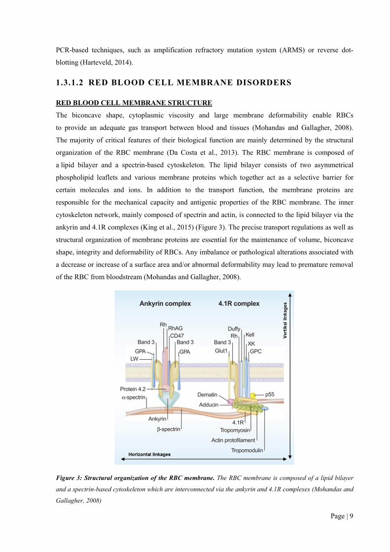
ankyrin and 4.1R complexes (King et al., 2015) (Figure 3). The precise transport regulations as well as
structural organization of membrane proteins are essential for the maintenance of volume, biconcave
shape, integrity and deformability of RBCs. Any imbalance or pathological alterations associated with
a decrease or increase of a surface area and/or abnormal deformability may lead to premature removal
of the RBC from bloodstream (Mohandas and Gallagher, 2008).
Figure 3: Structural organization of the RBC membrane. The RBC membrane is composed of a lipid bilayer
and a spectrin-based cytoskeleton which are interconnected via the ankyrin and 4.1R complexes (Mohandas and
Gallagher, 2008)
Page | 10
HEREDITARY SPHEROCYTOSIS
Hereditary spherocytosis (HS) is one of the most common RBC membrane disorders in the world with
a prevalence of 1 out of 2000–3000 affected cases in North America and Northern European countries
(Da Costa et al., 2013). In 75% of cases, HS is inherited in an autosomal dominant manner (An and
Mohandas, 2008). The remaining cases are associated with a recessive pattern of inheritance
and de novo mutations (Barcellini et al., 2011). HS is caused by mutations in genes encoding for
various red cell membrane proteins including ankyrin, band 3, protein 4.2, α or β-spectrin and Rh-
associated glycoprotein (RhAG) (Da Costa et al., 2013). The defective protein destabilizes the vertical
linkages between the phospholipid bilayer and the membrane cytoskeleton, which results in a loss of
surface area and an associated decreased surface area–to–volume ratio. These changes account for the
spheroidal shape of RBCs, the common characteristic of HS (Da Costa et al., 2013; Mohandas and
Gallagher, 2008). Patients with HS exhibit a broad clinical phenotype and variable disease severity
ranging from mild to very severe according to the decrease of RBC surface area. However, “typical”
HS consists of evidence of hemolysis with anemia, jaundice, reticulocytosis, gallstones, splenomegaly
as well as spherocytes on peripheral blood smear (An and Mohandas, 2008).
HEREDITARY ELLIPTOCYTOSIS
Hereditary ellipctocytosis (HE) is an autosomal dominant disorder resulting from a protein 4.1R
deficiency or spectrin malformations, affecting the horizontal interaction of the red cell cytoskeleton
(King et al., 2015).The world-wide prevalence has been estimated to be 3 to 5 in 10 000 individuals
but a higher prevalence occurs in malaria endemic regions (Da Costa et al., 2013). A majority of HE
patients are asymptomatic (no anemia or hemolysis) and the only laboratory finding is the presence of
elliptocytes on peripheral blood smear. Approximately 10% of HE patients exhibit mild to moderate
HA with reticulocytosis. The laboratory findings include the presence of elliptocytes and fragmented
cells on peripheral smear, positive markers of hemolysis and abnormal EMA binding test and osmotic
fragility test (Gallagher, 2013).
HEREDITARY STOMATOCYTOSIS
Hereditary stomatocytosis (HSt) is an autosomal dominant disorder characterized by the presence of
variable numbers of stomatocytes on peripheral blood smear (Barcellini et al., 2011). Affected
individuals usually exhibit mild to moderate HA with reticulocytosis and marked macrocytosis. The
HSt results from an increase of red cell membrane cation permeability and the cell’s inability to
regulate its cation homeostasis (Barcellini et al., 2011; Delaunay et al., 1999; Flatt and Bruce, 2009).
The changes in cation content affect water balance and, thus, red cell volume homeostasis. According
to the red cell hydration, HSt is classified into two major types: A) overhydrated stomatocytosis and
B) dehydrated stomatocytosis.
Page | 11
Overhydrated stomatocytosis (OhSt) is characterized by increased intracellular sodium and a slight
decrease in intracellular potassium. In addition to increased cation permeability, RBCs of many
affected individuals lack the membrane protein stomatin (Fricke et al., 2003; Glogowska and
Gallagher, 2015). Nevertheless, no mutation in gene encoding stomatin (EPB72 or STOM) has been
identified. Thus, whether the stomatin loss is a consequence or a cause of the red cell membrane defect
have not been clearly established yet (Badens and Guizouarn, 2016). Recently, mutations in RhAG
have been considered the cause of this sydrome. Other two proteins, Band 3 and Glut 1, are associated
with OhSt with cation leak at a low temperature.
Dehydrated stomatocytosis (DhSt) (also known as hereditary xerocytosis) is the second type of HSt.
RBCs of affected individuals exhibit reduced intracellular potassium and increased sodium content
(Badens and Guizouarn, 2016). Variable numbers of stomatocytes are seen on peripheral blood and in
mild cases can be easily overlooked. The definitive diagnosis of DHSt is ascertained by osmotic
gradient ektacytometry, which shows a leftward shift of the bell–shaped curve (Andolfo et al., 2013).
To date, a total of 2 membrane proteins have been reported to be responsible for DhSt. Several
mutations have been reported in FAM38A gene encoding mechanosensitive channel PIEZO1
(Albuisson et al., 2013; Andolfo et al., 2013; Bae et al., 2013; Beneteau et al., 2014; Shmukler et al.,
2014; Zarychanski et al., 2012). More recently, defects in the Gardos channel have been linked to
DhSt (Andolfo et al., 2015; Rapetti-Mauss et al., 2015).
Treatment of anemia (transfusion and chelation therapy) depends on the cause and severity.
Splenectomy is useful.
In 2015, International Council for Standardization in Hematology released a new guideline for the
laboratory diagnosis of nonimmune hereditary red cell membrane disorders. In addition to “classical”
blood smear examination, several screening methods are currently available for identification of
membrane disorders. Typical HS cases with positive family history can be confirmed by osmotic
fragility test (OF), acid glycerol lysis time test (GLT) or eosin–5’–maleimide binding test (EMA).
Quantitative membrane defects, mainly associated with typical HS and HE, can be identified using
SDS–polyacrylamide gel electrophoresis (SDS-PAGE). All three defects (typical HS, typical HE and
HSt) can be detected using ektacytometry (or Laser-assisted optical rotational red cell analyzer), the
only simple and reliable screening test for the diagnosis of HSt. Molecular genetic analysis is usually
applied in recessive mode of inheritance, and in cases of suspected de novo mutation or compound
heterozygosity (King et al., 2015).
Several novel mutations in band 3 or spectrin genes associated with HS have been described in
patients of Czech origin (Hassoun et al., 1997; Jarolim et al., 1996; Jarolim et al., 1995). Currently,
only a couple of laboratories in the Czech Republic offer screening tests (EMA test, OF test, GLT,
Page | 12
cryohemolysis test and Pink test) for membrane defects. Nevertheless, lack of molecular analysis
hampers the diagnosis of membrane defects in atypical cases.
1.3.1.3 RED BLOOD CELL ENZYMOPATHIES
Hereditary red blood cell enzymopathies are disorders arising from mutations (point mutations,
insertions, deletions, splice defects, etc.) in genes coding for red cell metabolic enzymes (Figure 2).
Deficiencies or malfunctions of these enzymes generally impair cellular energy balance and/or
increase the levels of oxidative stress. A lack of energy and reduced reductive power of red blood cells
ultimately affects cellular integrity, leading to premature removal in the spleen and, consequently,
decreased red blood cell survival (Arya et al., 1995; van Wijk and van Solinge, 2010). Most enzyme
disorders are inherited in an autosomal recessive form with hemolysis occurring only in homozygous
or compound heterozygous individuals. Some enzyme deficiencies are X-linked; adenosine deaminase
deficiency is autosomal dominant. A summary of clinically relevant erythroenzymopathies and their
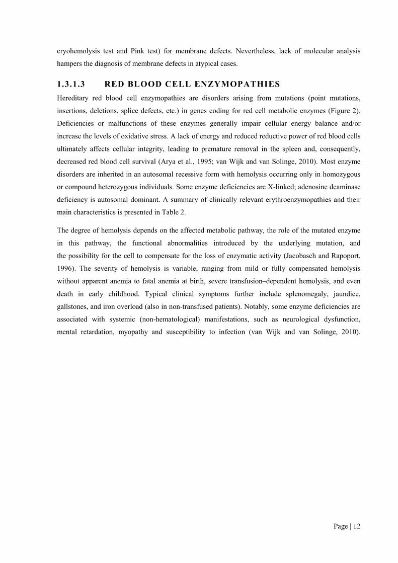
main characteristics is presented in Table 2.
The degree of hemolysis depends on the affected metabolic pathway, the role of the mutated enzyme
in this pathway, the functional abnormalities introduced by the underlying mutation, and
the possibility for the cell to compensate for the loss of enzymatic activity (Jacobasch and Rapoport,
1996). The severity of hemolysis is variable, ranging from mild or fully compensated hemolysis
without apparent anemia to fatal anemia at birth, severe transfusion–dependent hemolysis, and even
death in early childhood. Typical clinical symptoms further include splenomegaly, jaundice,
gallstones, and iron overload (also in non-transfused patients). Notably, some enzyme deficiencies are
associated with systemic (non-hematological) manifestations, such as neurological dysfunction,
mental retardation, myopathy and susceptibility to infection (van Wijk and van Solinge, 2010).
Page | 13
Table 2: A summary of clinically relevant enzyme disorders
Enzyme Clinical manifestation Neurological
symptoms Myopathy
Genetic transmission
No. of reported cases, mutations
Embden-Meyerhof pathway Hexokinase HNSHA; chronic - - AR 30 cases, 10 mutations
Glucose phosphate isomerase HNSHA; chronic +/- - AR > 55 families, 31 mutations Phosphofructokinase HNSHA; chronic (mild) - + AR 100 families, 23 mutations
Aldolase HNSHA; chronic +/- +/- AR 8 cases, 8 mutations Triosephosphate isomerase HNSHA; chronic (severe) + - AR 40 cases, 19 mutations
Phosphoglycerate kinase HNSHA; chronic + + X–linked 40 cases, 23 mutations
Bisphosphoglyceratemutase erythrocytosis - - AR 4 cases, 4 mutations Pyruvate kinase HNSHA; chronic - - AR > 500 families, > 260 mutations
Hexose monophosphate shunt Glucose-6-phosphate dehydrogenase HNSHA; induce by oxidant
drugs/infections, favism - - X–linked 400 million cases, 186 mutations
Glutathione metabolism Glutathione synthetase HNSHA; chronic + - AR >50 families, 33 mutations
γ–glutamylcysteine synthetase HNSHA; chronic + - AR 12 families, 5 mutations
Glutathione reductase HNSHA; induce by oxidant drugs/infections, favism
- - AR 2 families, 3 mutations
Nucleotide metabolism Adenosine deaminase (hyperactivity) HNSHA; chronic - - AD 3 families, no mutations Adenylate kinase HNSHA; chronic - - AR 12 families, 7 mutations
Pyrimidine–5’–nucleotidase HNSHA; chronic - - AR > 60 families, 26 mutations
HNSHA - chronic nonspherocytic hemolytic anemia; AR - autosomal recessive, AD - autosomal dominant.
Page | 14
ENZYME DISORDERS OF THE EMBDEN - MEYERHOF PATHWAY
The Embden-Meyerhof pathway (Figure 2) transforms glucose to pyruvate or lactate, and generates
ATP. This metabolic pathway is regulated by three rate-limiting steps, involving the catalytic action
of hexokinase (HK), phosphofructokinase (PFK), and pyruvate kinase (PK). Cellular NADH and ATP
levels also control glycolytic flux. Most enzyme disorders of this pathway are associated with chronic
nonspherocytic hemolytic anemia (HNSHA). Except for phosphoglycerate kinase (PGK) deficiency,
which is X-linked, all enzyme disorders of anaerobic glycolysis are transmitted in the autosomal
recessive manner (van Wijk and van Solinge, 2005).
Pyruvate kinase deficiency. The most prevalent enzyme disorder of anaerobic glycolysis is PK
deficiency. The estimated frequency of PK deficiency is 1:20,000 in general white population (Beutler
and Gelbart, 2000). PK is an allosteric enzyme which catalyzes the irreversible transfer of high-energy
phosphate group from phosphoenolpyruvate to ADP, generating ATP and pyruvate. The enzyme is
allosterically regulated by fructose–1,6–bisphosphate (positive regulation) and inhibited by its product
ATP. There are four mammalian isozymes of PK: M1, M2, L and R, encoded by 2 different genes.
PKLR, localized on chromosome 1 (1q21) (Kanno et al., 1992), directs the expression of PK-R and
PK–L in red blood cells and liver, respectively. The deficiency of PK–R has two major metabolic
consequences: depletion of ATP, and accumulation of 2,3-bisphosphoglycerate (Valentine and Paglia,
1980b). The lack of ATP eventually leads to premature removal of red blood cells from the circulation
whereas increased levels of 2,3–bisphosphoglycerate decrease the affinity of hemoglobin for oxygen.
The latter partly compensates for tissue hypoxia due to anemia (Oski et al., 1971).
More than 260 different mutations in PKLR gene associated with PK deficiency are known
(http://www.hgmd.org/) (Canu et al., 2016). Most of the mutations are single nucleotide substitutions
(72%) leading to mainly amino acid substitutions; in twelve cases a stop codon formation. The clinical
features of PK deficiency are highly variable, ranging from a fully compensated hemolysis to severe
transfusion–dependent hemolytic anemia (Canu et al., 2016; Zanella et al., 2005). A few cases of
hydrops fetalis and death in neonatal period due to severe PK deficiency have been reported (Ferreira
et al., 2000). The severity of anemia is usually stable in adulthood but may worsen during infections or
other forms of physiological stress (Zanella et al., 2007). Iron overload, occurring in the most PK
deficient patients, is considered to be multifactorial (Zanella et al., 1993). There is no link among the
residual PK activity, degree of hemolysis and clinical severity of PK deficiency. A high rate of
reported mutations (60%) are distributed in Europe (Canu et al., 2016). Among these, the 1456T
mutation and the 1529A mutation are the most prevalent (Lenzner et al., 1997; Zanella et al., 2005).
Several mutations p.(Gly332Ser), 1060delAAG, p.(Asn361Asp), p.(Arg486Trp), p.(Arg498His),
p.(Arg532Trp) and 101-1G>A have been previously identified in Czech population (Lenzner et al.,
1997). Distribution of other identified mutations in the Czech and Slovak populations will be
discussed in section 4.2.

Page | 15
Glucose-6-phosphate isomerase (GPI) deficiency. The second most frequent glycolytic enzyme
disorder is GPI deficiency. To date, 34 mutations in more than 55 families with GPI deficiency have
been identified (Adama van Scheltema et al., 2015; Manco et al., 2016). In the most affected
individuals, residual GPI activity is less than 25%. Low enzyme activity results from impaired kinetic
properties, reduced thermostability of the mutant enzyme, or defective protein folding (Lin et al.,
2009). Clinical features in GPI activity range from mild to severe hemolytic anemia. Hydrops fetalis
appears to occur more often in GPI deficiency than in other erythroenzymopathies (Matthay and
Mentzer, 1981). Most affected individuals are diagnosed during the neonatal and childhood period
(Adama van Scheltema et al., 2015). GPI deficiency may also be associated with non-hematologic
symptoms, in particular neurologic impairment or mental retardation (Schroter et al., 1985; van Wijk
and van Solinge, 2005).
Phosphofructokinase (PFK) deficiency, aldolase deficiency (ALD) and triosephosphate (TPI)
deficiency. PFK, ALD and TPI deficiencies are more rare enzyme disorders. Erythrocyte PFK is
composed of two types of subunits: PFKL (liver) and PFKM (muscle) that may form 5 different
isozymes (M4, M3L1, M2L2, M1L3, L4). In case of a mutated PFKM subunit, only a functional PFKL
subunit is expressed in red blood cells, causing a partial deficiency of PFK. Thus, patients with PFK
deficiency usually exhibit mild or fully compensated hemolytic anemia. Since muscle cells contain
only PFKM, the deficiency is more pronounced in muscle cells, leading to myopathy (Nakajima et al.,
2002). TPI deficiency is the most severe enzyme defect of anaerobic glycolysis, frequently leading to
death in early childhood. TPI catalyzes the interconversion between dihydroxyacetone phosphate
(DHAP) and glyceraldehyde–3–phosphate (GAP). In red blood cells the deficiency leads to hemolytic
anemia which may occur due to accumulation of DHAP (Ahmed et al., 2003). However, the
deficiency is associated with more severe consequences, such as mental retardation and other
neurological dysfunctions. These may occur because of the formation of toxic protein aggregates,
induced by misfolded TPI (Ahmed et al., 2003; Orosz et al., 2009). Moderate to severe hemolytic
anemia is observed in patients with aldolase deficiency. As in TPI deficiency, defects of aldolase are
associated with neurological disorders (Beutler et al., 1973). A few affected individuals have been
reported, showing recurrent episodes of rhabdomyolysis and muscle weakness (Kreuder et al., 1996;
Yao et al., 2004). Severe defects of aldolase probably lead to death in embryonic state (Esposito et al.,
2004).
Hexokinase (HK) deficiency. HK catalyzes the initial step of the Embden-Meyerhof pathway, the
transfer of a phosphoryl group from ATP to glucose. Hexokinase is the enzyme with the lowest
in vitro activity of all glycolytic enzymes (van Wijk and van Solinge, 2005). Two different HK
isozymes, HK–1 and HK–R, are present in human RBCs. Both HK isozymes originate from the same
gene HK1, however, their transcriptions are controlled by alternative promoters. While HK-R isozyme
is present only in RBCs, HK–1 isozyme is expressed in various mammalian tissues (Murakami et al.,

Page | 16
2002). Deficiency of this key regulatory enzyme is generally associated with severe hemolytic anemia,
and may lead to death in the neonatal period (Kanno et al., 2002). To date, 30 cases of hexokinase
deficiency have been described (de Vooght et al., 2009; Koralkova et al., 2016; van Wijk et al., 2003).
Phosphoglycerate kinase (PGK) deficiency. PGK is the only enzyme of glycolysis whose gene is
X-linked. PGK deficiency is associated with chronic hemolysis, neurologic impairment (including
mental retardation and ataxia) and myopathy (exercise intolerance or muscle weakness) (Fermo et al.,
2012). There are only a few affected individuals who suffer the full spectrum of clinical features.
Thus, PGK deficiency shows a very wide clinical phenotype. Some cases of PGK deficiency with
myopathy but without hemolysis have been described (Beutler, 2007; Cohen-Solal et al., 1994; Fermo
et al., 2012; Sotiriou et al., 2010; Spiegel et al., 2009).
Deficiencies of other glycolytic enzymes glyceraldehyde-3-phosphate dehydrogenase,
monophosphoglycerate mutase, and enolase have been described but they appear not to be associated
with hemolysis (Beutler, 1979).
ENZYME DISORDERS OF THE HEXOSE MONOPHOSPHATE SHUNT AND
GLUTATHIONE METABOLISM
The red blood cell is continuously exposed to various forms of oxidative stress. To protect cellular
hemoglobin and other macromolecules against oxidative damage, reductive power is required. The
reduced form of glutathione (GSH) constitutes the cell’s main source of reductive power. NADPH,
generated by the hexose monophosphate shunt, is required to maintain glutathione in its reduced form.
The key enzymes of this defense mechanism are glucose-6-phoshate dehydrogenase,
glutamate cysteine ligase, glutathione synthetase, and glutathione reductase (van Zwieten et al., 2014).
Generally, disorders of these enzymes cause hemolysis only under conditions of increased levels
of oxidative stress (acute hemolytic anemia).
Glucose-6-phosphate dehydrogenase (G6PD) deficiency. G6PD catalyzes the first step of the
hexose monophosphate shunt thereby producing NADPH. The human G6PD monomer (515 amino
acids) consists of two domains: the coenzyme (N-terminal) domain, and the other, larger, α+β domain.
Both domains are connected by an α helix consisting in fully conserved region, which serves as
a substrate binding site. The enzyme is active as a tetramer or dimer, in a pH-dependent equilibrium
(Cappellini and Fiorelli, 2008). G6PD deficiency is the most common enzyme defect with estimated
400 million individuals affected worldwide (Nkhoma et al., 2009). The highest prevalence is observed
in Asia and sub-Saharan Africa (Howes et al., 2012). G6PD deficiency is an X-linked disorder
associated with mutations in G6PD gene consisting of 13 exons. This gene is highly polymorphic;
more than 400 G6PD variants have been described, among which 186 mutations are associated with
G6PD deficiency. Majority of the mutations are single nucleotide substitutions, however, deletions

Page | 17
and intronic mutations (G6PD Varnsdorf, G6PD Zurich) have also been identified (Efferth et al.,
2004; Minucci et al., 2012; Xu et al., 1995). G6PD-deficient variants are divided into five classes
according to the level of residual activity and severity of phenotype. Most of the variants belong to
classes II, III, or IV and are associated only with acute hemolysis. In contrast, the rare class I variants,
characterized by a very low activity of G6PD (<1%), are associated with chronic HNSHA. The class I
mutations are usually located in exons6, 10 and 13 encoding the regions that bind the enzyme
substrate, dimer interface and NADP+ structural site, respectively (Minucci et al., 2012). Class II and
III mutations are highly distributed in malaria-endemic regions, among which the G6PD variant A-
(c.376A>G and c.202G>A) has the highest frequency (90%) in Africa. The second most frequent
G6PD variant, G6PD Mediterranean (c.563C>T), is present in the Mediterranean countries. However,
it is also widespread in the Middle East, including Israel, where it accounts for almost all G6PD
deficiency in Kurdish Jews, India, and Indonesia (Cappellini and Fiorelli, 2008). Most G6PD-deficient
individuals are asymptomatic but may develop acute hemolytic crises upon conditions that increase
the levels of oxidative stress, such as the intake of specific drugs, fava beans, or the occurrence of
infections (Cappellini and Fiorelli, 2008; Minucci et al., 2009). Although, in general,
no morphological abnormalities are associated with enzyme disorders, Heinz bodies (precipitated
hemoglobin) are typically found in G6PD-deficient individuals. Two single nucleotide substitutions
(G6PD Praha, G6PD Olomouc) and one intronic mutation (G6PD Varnsdorf) have been previously
described in Czech population (Xu et al., 1995). Distribution of other mutations in both the Czech
Republic and Slovak Republic will be discussed in section 4.2.
Figure 4: A peripheral blood smear of a G6PD deficient patient showing a presence of Heinz bodies
Page | 18
Glutathione reductase (GR) deficiency. GR is important in the regeneration of GSH from oxidized
glutathione (GSSG). Hereditary GR deficiency is a very rare autosomal recessive disorder
characterized by increased susceptibility to oxidative stress. The clinical phenotype resembles
a deficiency of G6PD with affected individuals being generally asymptomatic and with the induction
of acute hemolytic crisis by the intake of drugs or fava beans (Kamerbeek et al., 2007; van Zwieten et
al., 2014). Importantly, acquired deficiency of GR may also occur as a result of insufficient flavin
intake. GR requires flavonoids for enzymatic activity. Such acquired deficiencies do not themselves
lead to a clinical phenotype (Beutler, 1979). A partial GR deficiency has been described in two Czech
patients who were concomitantly carriers of the unstable hemoglobin variant Haná. The combination
of both defects resulted in methemoglobinemia and Heinz body hemolytic anemia (Mojzikova et al.,
2010).
γ–glutamylcysteine synthetase (γ–GCS) and glutathione synthetase (GS) deficiencies.
The tripeptide glutathione is synthesized in red blood cells from cysteine, glutamine, and glycine in a
2–step process catalyzed by γ–GCS and GS, respectively. Defects of both enzymes decrease cellular
GSH content and, hence, increase cellular susceptibility to oxidative stress. Disorders caused by
deficiencies of bothof these enzymes are rare: >50 families with GS deficiency have been described
whereas only 12 families with γ–GCS deficiency have been reported. Apart from acute hemolytic
episodes, the deficiency may display additional non-hematological symptoms, such as mental
retardation (γ–GCS deficiency) and 5-oxyprolinuria (GS deficiency) (Ristoff and Larsson, 2007).
Deficiencies of glutathione peroxidase and other enzymes involved in red blood cell anti-oxidant
defense, such as catalase, have been reported. However, they are not associated with hemolysis
(Beutler, 1979).
ENZYME DISORDERS OF NUCLEOTIDE METABOLISM
Purine metabolism maintains the red blood cell ATP and GTP levels. Ninety-seven per cent of total
nucleotides are adenine derivatives (AMP, ADP, ATP). Guanine derivatives constitute 1–3%.
Pyrimidines are removed during the maturation process and are therefore only present in trace
amounts (Valentine et al., 1974; Valentine and Paglia, 1980a). There is a number of enzymes involved
in nucleotide metabolism. The most important ones are pyrimidine-5’-nucleotidase (pyrimidine
metabolism), and adenylate kinase and adenosine deaminase (purine metabolism) (Figure 1). Defects
of all three enzymes are associated with HNSHA.
Pyrimidine-5’-nucleotidase (P5N) deficiency. Erythrocyte P5N is a cytosolic enzyme which
catalyzes the hydrolysis of cytidine/uridine monophosphates, rendering diffusible cytidine/uridine and
inorganic phosphates (Rees et al., 2003; Valentine et al., 1974). P5N deficiency is probably the third
most prevalent enzyme disorder associated with HNSHA. P5N deficiency is an autosomal recessive
disorder associated with mild to severe chronic hemolytic anemia, splenomegaly and jaundice.
Page | 19
P5N deficiency results in the accumulation of pyrimidine nucleotides. They form insoluble aggregates
which are visible as basophilic stippling on a peripheral blood smear. Although not specific, prominent
basophilic stippling is characteristic of P5N deficiency (Zanella et al., 2006). To date, it is not known
how the accumulation of pyrimidine nucleotides contributes to the hemolytic process. However,
extensive studies of P5N deficient patients revealed that other laboratory parameters, such as GSH
levels, ribose-phosphate pyrophosphokinase activity and transketolase levels, are markedly altered
(Barasa et al., 2016; Magni et al., 2013).
Figure 5: A pronounced basophilic stippling in P5N deficient RBCs (Koralkova et al., 2014)
Adenylate kinase (AK) deficiency and adenosine deaminase (ADA) hyperactivity. AK and ADA
are both important in maintaining the adenine pool. Deficiency of both AK and hyperactivity of ADA
probably deplete the cell from adenine nucleotides and, consequently, ATP, eventually resulting in
chronic hemolytic anemia. AD deficiency is an autosomal recessive disorder which has been described
only in 12 families. Affected individuals exhibit moderate or severe hemolytic anemia. Some patients
with psychomotor impairment have been reported (Abrusci et al., 2007). Hyperactivity of ADA is
a very rare autosomal dominant disorder for which no causative mutations have been described yet
(Chen and Mitchell, 1994). The elevated ADA activity is also seen in Diamond-Blackfan anemia and
in patients with AIDS or tuberculosis. On the other hand, adenosine deaminase deficiency is one cause
of severe combined immunodeficiency.
DIAGNOSTIC APPROACH AND THERAPY
Red blood cell enzymopathies are associated with normocytic normochromic anemia and common
signs of hemolysis, i.e. increased plasma levels of bilirubin and lactate dehydrogenase, low levels of
haptoglobin and high reticulocyte count (Tefferi, 2003). The diagnosis of HNSHA due to a red blood
cell enzymopathy is generally a diagnosis that is based on exclusion: a negative direct Coombs test,
a normal osmotic fragility/cryohemolysis test, no specific morphological abnormalities, and no
evidence for abnormal hemoglobin. A differential diagnosis of a red blood cell enzymopathy requires

Page | 20
a demonstration of reduced enzymatic activity and confirmation of the diagnosis on the DNA level.
Importantly, there is no relationship between the degree of loss of enzymatic activity and clinical
severity.
Nowadays, there are several quantitative or semi-quantitative screening assays for determination of the
most prevalent erythroenzymopathies (deficiencies of G6PD and PK). G6PD deficiency screening
tests, such as fluorescent spot tests or cytochemical assays, are based on the direct/indirect detection
and/or quantification of NAPDH produced by G6PD (Cappellini and Fiorelli, 2008). Direct tests
measure the production of NAPDH which has it absorption maximum at 340 nm and fluorescence
emission at 460 nm. Indirect tests translate NADPH production into a colorimetric readout using
chromophores like brilliant cresyl blue, resazurin, or formazan derivatives (Shah et al., 2012).
PK deficiency screening tests are usually indirect colorimetric assays based on the detection of
chromophore products. Although these assays are usually rapid and inexpensive, they are able to
detect only the most common enzyme defects. Thus, the classical methods described by Beutler still
represent the method of choice for the diagnosis of erythroenzymopathies (Beutler, 1984). The general
basis comprises the determination of a specific enzyme activity using a spectrophotometric assay.
Enzymatic activity is directly related to changes in absorbance over time. Specific reactions need to be
prepared for each enzyme and changes in substrate (either NAD+, NADP+, NADH, or NADPH) are
monitored at 340 nm for a set amount of time. The specific enzymatic activity of each particular
enzyme is expressed as U/gHb (Beutler, 1984). In red blood cell enzymopathies, enzyme activity of
affected individuals is usually reduced to 25% of the normal. As stated, there is no correlation between
the residual activity of the enzyme and severity of the clinical picture. Despite the many benefits of
these assays (high sensitivity and high specificity), there are several pitfalls which may influence
results and their interpretation. Enzymatic activities are influenced by factors such as (1) the
conditions during storage and shipping of blood samples (i.e. in vitro stability of enzymes), (2) the
proper removal of leukocytes and platelets from the red blood cell sample (leukocytes and platelets
may express isozymes, encoded by other genes, which interfere with red cell enzyme activity
measurements), (3) blood transfusions (blood sample will contain donor erythrocytes), (4) the age of
the patient (the expression of many enzymes is age-related), and (5) the presence of a high number of
reticulocytes in the patient’s blood sample. The activity of many enzymes (HK, PK, G6PD and P5N)
is red blood cell age-related. Thus, the youngest cells have the highest enzymes activity. In the case of
a high reticulocyte count, any deficient enzyme may show a normal or even increased in vitro activity
(Arya et al., 1995; Beutler, 1984). This masking effect can be eliminated when activity of age-related
enzymes (PK, G6PD, P5N) is normalized to activity of HK.
As stated, morphological abnormalities of red blood cells associated with red blood cell
enzymopathies are generally nonspecific. Prominent basophilic stippling is present in P5N deficiency.
In addition, Heinz bodies (precipitated hemoglobin) may occur in G6PD deficiency or enzyme

Page | 21
deficiencies of glutathione metabolism. However, the occurrence of both abnormalities is also related
with other disorders. Pronounced basophilic stippling can also be seen in patients with sideroblastic
anemia, thalassemia, or lead poisoning, whereas Heinz bodies may be present in red blood cells from
patients with unstable hemoglobin or thalassemia. Besides both mentioned morphological
abnormalities, other various abnormal types of red blood cells associated with erythroenzymopathies
can be seen on blood smear. “Bite” cells, keratocytes or blister cells may occur in G6PD deficiency as
the result of oxidative stress and acute hemolysis (Bain, 2005); anisocytosis, poikilocytosis and
contracted echinocytes can be present in PK deficiency (Zanella et al., 2005).
A number of methods (radioactive, capillary electrophoresis or high-perform liquid chromatography)
have been described for determination of red cell P5N activity (Zanella et al., 2006). However, the
reproducibility of most of these methods does not meet the standards for use in clinical practice.
A simple screening test of P5N deficiency, or any other enzyme involved in nucleotide metabolism,
comprises the spectrophotometric measurement of pyrimidine and purine nucleotides in deproteinized
hemolysate. This method uses the differences in absorption maxima of purines (260 nm) and
pyrimidines (280 nm) (Beutler, 1984). The ratio between these reflects overall cellular purine and
pyrimidine content. For instance, an accumulation of pyrimidine nucleotides, as seen in P5N
deficiency, decreases the purine/pyrimidine nucleotide ratio.
In case of a suspected red blood cell enzyme deficiency the molecular characterization of the defect on
the DNA level is essential for confirmation of the diagnosis. Mutations are usually unique, which
makes it necessary to sequence the entire coding region, including flanking intronic splice site
sequences. Importantly, cis-acting, often erythroid-specific, regulatory regions may harbour mutations
interfering with gene expression and thereby causing a red cell enzyme disorder (de Vooght et al.,
2009; van Wijk et al., 2003). The identification of a causative mutation enables proper genetic
counseling and prenatal diagnosis. DNA investigations may be the only way to establish the diagnosis
in neonatal patients or patients who are transfusion-dependent. Moreover, molecular characterization
is required for a better understanding of the complex genotype-to-phenotype correlation in
erythroenzymopathies.
No curative therapy is available for enzyme defects. Affected individuals with mild anemia or
compensated hemolysis do not require any treatment. In more severe cases, treatment mainly consists
of supportive therapy (blood transfusions) and chelation therapy to avoid the adverse effects of iron
overload. In some cases, splenectomy may be considered. Generally, spleen removal can increase
hemoglobin levels but the effect is variable and difficult to predict (Zanella et al., 2005). Patients with
deficiency of enzymes involved in the protection against oxidative stress should avoid the intake of
oxidative drugs or food.
Page | 22
Interestingly, novel drugs, AG–348 and AG–519, have recently been introduced as a potential therapy
of PK deficiency. Both compounds act as activators which increase activity of mutated PK enzyme
and restore ATP levels. Although the study is ongoing, the preliminary data show rapid and sustained
Hb increase in treated patients (Hixon et al., 2016; Hixon et al., 2013; Chen et al., 2016).
Page | 23
2 AIMS OF THE THESIS
The presented work is focused on molecular pathogenesis of rare anemias – enzymopathies.
It was aimed to identify genetic background, etiology and pathogenesis of enzyme disorders
associated with HNSHA.
Specific aims:
1. Critically review the literature concerning congenital hemolytic anemias.
2. Perform direct enzyme assays and genetic testing in selected Czech and Slovak patients
with hemolytic anemia of unknown etiology.
3. Investigate the impact of selected HK mutations to the pathogenesis of HK deficiency.
4. Describe the molecular mechanism of a novel non-hemolytic anemia variant of PGK.
5. Characterize a novel mutation in PFKM gene using in silico modelling.

Page | 24
3 MATERIALS AND METHODS
Laboratory equipment and software
Thermocycler MJ Mini™ Personal Thermal Cycler (BioRAD); ThermoblockBio TDB-100 (Biosan);
Vortex Bio V1 (Biosan); Centrifuge Hettich Mikro 200R (Hettich); Centrifuge 5415 D (Eppendorf);
Centrifuge Jouan BR4i (Thermo Fisher Scientific); PowerPac™ Basic Power Supply (BioRAD);
UV-transilluminator (UltraLum); Genetic analyzer ABI PRISM® 3100 (Applied Biosystems); ABI
3130 automated sequencer (PE Applied Biosystems); Spectrophotometer Infinite 200 Nanoquant
(Tecan); Software i-control 1.6 (Tecan), pH meter 3510 (Jenway); Odyssey Infrared Imaging System
(LI-COR Biosciences); ViiA™ 7 Real-Time PCR System (Life Technologies); Casy Cell Counter and
Analyzer (Roche Life Science); NanoVueTM (GE Healthcare Life Sciences); The PyMOL Molecular
Graphics System (Version 1.7.4 Schrödinger, LLC); PolyPhen-2
(http://genetics.bwh.harvard.edu/pph2/); GraphPad® Prism (GraphPad Software, Inc.); SIFT
(http://sift.jcvi.org/); Human Splicing Finder (http://www.umd.be/HSF3/).
Biological material
Peripheral blood samples from patients and donors were collected into EDTA and/or heparin tubes.
Each study was performed according to the Helsinki international standards. The informed consent
was obtained from all patients, family members and donors.
Enzymes, substrates, cofactors, nucleotides
Glucose-6-phosphate dehydrogenase (S. cerevisiae, Sigma-Aldrich, USA); Lactate dehydrogenase
(rabbit muscle, Sigma-Aldrich, USA); D-glucose-6-phosphate (Sigma-Aldrich); D-fructose-6-
phosphate (Sigma-Aldrich); Phosphoenolpyruvate (Sigma-Aldrich); D-fructose-1,6-biphosphate
(Sigma-Aldrich); 6-phosphogluconate (Sigma-Aldrich); D-(+)-glucose (Sigma-Aldrich); NADH
(Serva); NADP+(Serva); NADPH (Serva); NAD+(Serva); ATP (Sigma-Aldrich); ADP (Sigma-
Aldrich); AMP (Sigma-Aldrich).
Other chemicals
TRIzol Reagent (Thermo Fisher Scientific); Chloroform (Penta); Phenol(Serva); Hydrochloric acid
(Penta); SDS (Serva); Agarose (Invitrogen); Microcrystalline cellulose (Sigma-Aldrich); α-cellulose
(Sigma-Aldrich); Tris (Promega); Na2EDTA (Serva); MgCl2 (Chemos); KCl (Fischer Scientific);
NaCl (LachNer); Ethanol pro UV (LachNer); 2-mercaptoethanol, 55 mM (Serva); Specific primers for
HK1, G6PD, PKLRand PGK1 (Eastport, Sigma-Aldrich); Proteinase K (Sigma-Aldrich, USA); 2-log
DNA ladder (Qiagen); DNA loading buffer (Qiagen); Triton X-100 (Sigma-Aldrich); GelRed
(Biotium); NuSieve Agarose 3:1 (Lonza); RIPA buffer (Teknova), Protease inhibitors (100 ×, Thermo
Scientific); 8% precast SDS-PAGE gel (Bolt® Bis-Tris Plus gel, Life Technologies); PVDF
membrane (Immobilion-FL, Millipore); Antibody anti-HK-1 (1:250; Abcam); Antibody Anti-actin
Page | 25
(Merck Millipore); Alexa Fluor® 680-conjugated goat anti-mouse and goat anti-rabbit (1:7500;
Invitrogen); Odyssey® Blocking Buffer (Li-COR); Ficoll-Paque (GE Healthcare); StemSpan SFEM
(Stemcell Technology); Human stem cell factor (Amgen); Erythropoietin (Eprex); Interleukin-3
(Sigma-Aldrich); Dexamethasone (Sigma-Aldrich); SyntheChol™ NS0 Supplement (Sigma-Aldrich);
Percoll (GE Healthcare).
Commercial kits
Geneaid Gel/PCR DNA Fragments Extraction Kit (Geneaid); QIAquick Gel Extraction Kit, MinElute
PCR Purification Kit (Qiagen); NucleoSpin Gel and PCR Clean-up (Macherey-Nagel); HotStarTaq
Master Mix Kit – HotStarTaq DNA Polymerase, PCR Buffer with 3 mM MgCl2, and 400 µM of each
dNTP (Qiagen); MasterAmp Taq DNA Polymerase (Eppicentre); PCR buffer (Applied Biosystems);
dNTP (Applied Biosystems); BigDye® Terminator v1.1 Cycle Sequencing Kit (Applied Biosystems,
USA); (BigDye Xterminator purification kit, Applied Biosystems); GeneAmp® RNA PCR Core Kit
(Applied Biosystems); SuperScriptTM III First-Strand Synthesis SuperMix (Invitrogen); FastStart
SYBR Green Master (Roche); ROX Passive Reference (BioRad); TOPO® TA Cloning® Kit for
Sequencing (Invitrogen); High-Speed Plasmid Mini Kit (Geneaid); CutSmart buffer and restriction
endonuclease MslI (NEB).
General laboratory reagents and buffers
0.9% M sodium chloride (NaCl); 0.1 M MgCl2; 1 M KCl; 10 mM Tris (pH=8.0); 10% SDS (w/v,
pH=7.2); Tris – HCl buffer (pH=8.0) – 1 M Tris, 5 mM Na2EDTA; 1% SDS (w/v); 2 mM EDTA (pH
8.0); RBC lysis solution – 1.5 M NH4Cl, 100 mM NH4HCO3, 10 mM Na2EDTA; 1xTBE buffer
(pH=8.3) – 0.89 M Tris, 0.89 M boric acid, 0.2 mM Na2EDTA; Stabilization solution – 2.7 mM
Na2EDTA, 0.7 mM 2-merkaptoethanol; 10 mM phosphate buffered saline(PBS, pH=7.4) – 0.137 M
NaCl, 2.7 mM KCl, 8.2 mM Na2HPO4, 1.8 mM KH2PO4; Blotting buffer – 25 mM Tris, 192 mM
glycine; TBS – 10 mM Tris, 150 mM NaCl, pH=7.4; 1xTBS (pH= 7.5) – 25mM Tris, 140mM NaCl,
and 3.0mM KCl;

Page | 26
3.1 ISOLATION OF RBCS FROM PERIPHERAL BLOOD
AND HEMOLYSATE PREPARATION
A filtration column was prepared from a 5 mL syringe tube, filter paper and a mixture of equal
weights of microcrystalline cellulose and α-cellulose. A small piece of filter paper was placed at the
bottom of the syringe tube which was then fixed in a vertical position in 15 mL tube. The mixture
of celluloses was mixed with 0.9% NaCl and poured to the 2 mL mark of the syringe tube.
The column was washed with 2 mL of 0.9% NaCl and then 1 mL of whole blood was pipetted onto the
column. The column was washed with additional 2-5 mL of 0.9% NaCl. Isolated RBCs were collected
in the 15 mL tube, then spinned down at 590g for 5 min at 4°C. The leukocyte/platelet-free RBC pellet
was washed with 5 mL of 0.9% NaCl, centrifuged at 590 g for 5 min at 4°C and aliquots were stored
at -80°C(Beutler et al., 1977). Prior to the analyses, 50 μL of each RBC aliquot was mixed with 950
μL of stabilizing solution.
3.2 ENZYME ANALYSIS
All enzyme assays were performed in leukocyte/platelet-free erythrocyte lysates as described above.
The composition of each assay mixture is listed below. After 10 min incubation, the reactions were
started by adding substrate or hemolysate and mixing properly. Then, 200 μL of each reaction mixture
was transferred into a 96-well plate and changes in absorbance were recorded at 340 nm at 1 min
intervals for 20–30 min. All assays were run in triplicates and specific enzyme activity was calculated
using the Lambert-Beer law.
activity = (ΔA x V/(ε x l x t)) x f
(A: changes in absorbance, V: volume of reaction mixtures – 200 μL, ε: absorption coefficient of
NAD(P)H at 340 nm – 6220 l.mol-1.cm-1, l: inside depth of well – 0.58 cm, t: time – min, f: dilution
factor)
activity [U=μmol.min-1] = ΔA x 0,055 x f
activity [U] = ΔA x 0,028 x f
specific activity [U/g Hb] = (ΔA x 0,028 x f )/mHb
(mHb: hemoglobin levels in leukocyte/platelet-free erythrocyte lysates was determined
spectrophotometrically at 414 nm) (Magnotti et al., 2009)
Page | 27
Composition of assay mixtures
HK assay – 0.1 M Tris-HCL/0.5 mM EDTA, pH=8.0; 0.01 M MgCL2; 2 mM glukose; 2 mM ATP;
0.2 mM NADP+; 0.1 U/mL G6PD; 50 μL 1:20 hemolysate; 10 min incubation at 37°C; 30 min
measurement; blank without ATP.
GPI assay – 0.1 M Tris-HCl/0.5 mM EDTA, pH=8.0; 0.01 M MgCl2; 0.2 mM NADP+; 0.1 U/mL
G6PD; 2 mM F6P; 10 min incubation at 37°C; start by addition of 5 μL 1:20 hemolysate; 20 min
measurement; blank without F6P.
6PGA assay – 0.1 M Tris-HCl/0.5 mM EDTA, pH=8.0; 0.01 M MgCL2; 0.2 mM NADP+; 20 μL of
1:20 hemolysate; 10 min incubation at 37°C; start by addition of 0.6 mM 6PGA; 20 min measurement,
blank without 6PGA.
G6PD assay – 0.1 M Tris-HCl/0.5 mM EDTA, pH=8.0; 0.01 M MgCl2; 0.2 mM NADP+; 20 μL of
1:20 hemolysate; 10 min incubation at 37°C; start by addition of 0.6 mM 6PGA + 0.6 mM G6P;
20 min measurement, blank without 6PGA+G6P.
PK assay – 0.1 M Tris-HCl/0.5 mM EDTA, pH=8.0; 0.01 M MgCl2; 0.1 M KCl; 1.5 mM ADP;
0,2 mM NADH; 6 U/mL LDH; 20 μL of 1:20 hemolysate; 10 min incubation at 37°C; start by
addition of 5 mM PEP; blank without ADP and PEP; 20 min measurement.
A complete panel of RBC enzyme assays (including aldolase, phosphofructokinase, triosephosphate
isomerase, phosphoglycerate kinase, bisphosphoglyceromutase, adenylate kinase) was applied
in several cases to exclude other but very rare enzymopathies causing HA. The detailed composition
of these assay mixtures is described in a research paper by Beutler and colleagues (Beutler et al.,
1977).
3.2.1 DETERMINATION OF MICHAELIS-MENTEN
CONSTANT (KM) FOR GLUCOSE AND ATP
All assays were run in the standard HK reaction mixture containing different concentrations of
glucose: 0.0125–2.0 mM and/or ATP 0.25–10 mM. The reactions were run in a 1 mL disposable
plastic cuvette and changes in absorbance at 340 nm were recorded at 20 s intervals for 30 min.
The KM values were determined by non-linear regression using the Michaelis-Menten model
(GraphPad® Prism).
3.2.2 ENZYME STABILITY ASSAYS
The thermal stability assays of HK mutants involved determining the half-life (T1/2) and the half-
inactivation temperature (T50). Both thermal stability assays were performed in the standard HK
reaction mixture, routinely containing 2 mM glucose and 10 mM ATP. For the T1/2 assay, RBC lysate
Page | 28
was pre-heated to 40°C for 5–40 min followed by cooling on ice (2 min). After the pre-heating step,
50 μL of the RBC lysate was used in the reaction mixture as described above. T1/2 was defined as the
time needed to reach a 50% reduction in the initial activity (Rijksen and Staal, 1978). For the T50
assay, RBC lysate was pre-incubated at different temperatures (37–44°C) for 5 min, followed by
a cooling period (2 min) on ice. The T50 was defined as the temperature at which HK maintains 50%
of its activity. The pH stability assay was carried out in a 0.1 M Tris-HCl/0.5 mM EDTA buffer of
different pH values (6.0–10.0) (Beutler et al., 1977; Rijksen et al., 1983).
3.3 CELL CULTURE OF HUMAN NUCLEATED
ERYTHROID CELLS
Human peripheral blood mononuclear cells (PBMCs) were isolated from individuals carrying
a heterozygous splice site c.873-2A>G mutation in HK1 or a hemizygous c.756+3A>G mutation in
PGK1 and healthy donors.
3.3.1 ISOLATION OF MONONUCLEAR CELLS FROM
HUMAN PERIPHERAL BLOOD BY DENSITY
GRADIENT CENTRIFUGATION
50–100 mL of peripheral blood was diluted with PBS buffer (1:2). The resulting cell suspension was
layered over an equal volume of Ficoll-Paque (GE Healthcare) in a 50 mL conical tube. The tubes
were centrifuged at 470g for 30 min at 20°C in a swinging-bucket rotor without brake. The upper layer
was aspirated and the interphase containing the mononuclear cells was transferred into a new 50 mL
conical tube and filled with PBS. The tube was mixed by inverting and centrifuged at 600g for 5 min
at 20°C. The supernatant was completely removed and the cell pellet was resuspended in 50 mL of
PBS and centrifuged at 220g for 5 min at 20°C. The supernatant was completely discarded and the
washing step was repeated. After the second wash, the pellet was resuspended in 12.5 mL of cold red
cell lysis buffer and incubated for 10 min at room temperature (RT). The tube was then centrifuged
at 600g for 5 min at 4°C and the supernatant was removed. The isolated PBMCs were resuspended
in 10 mL of StemSpan SFEM (Stemcell Technology) and counted using the Casy Cell Counter and
Analyzer (Roche Life Science).
3.3.2 SELECTION OF PRO-ERYTHROBLASTS
The cells were resuspended to a density 5–10x106 cells/mL in StemSpan supplemented with human
stem cell factor (hSCF, 100 ηg/mL; Amgen), erythropoietin (EPO, 2 U/mL; Eprex), interleukin-3
(IL-3, 1 ng/mL; Sigma-Aldrich), dexamethasone (Dex, 1 μmol/L; Sigma-Aldrich) and SyntheChol™
NS0 Supplement (Chol, 200-times diluted; Sigma-Aldrich). The cells were cultured at 37°C in a
humidified atmosphere of 5% CO2 in air. After 24 h, the cells were counted and then the culture

Page | 29
medium was replaced with StemSpan supplemented with hSCF (100 ηg/mL), EPO (2 U/mL), Dex (1
μmol/L) and Chol (200-times diluted). At day 5–6, the cell suspension was layered over 20 mL of
Percoll (GE Healthcare) in a 50 mL conical tube and centrifuged at 470g for 30 min at 20°C in
swinging-bucket rotor without brake. The upper layer was discarded and the interphase layer was
transferred into a new 50 mL conical tube. The cell suspension was washed twice with PBS and
centrifuged at 600g for 5 min at 20°C.
3.3.3 EXPANSION OF PRO-ERYTHROBLAST
The cell pellet was resuspended in StemSpan, supplemented with hSCF (100 ng/mL), EPO (2 U/mL),
Dex (1 μmol/L) and Chol (200-times diluted), to get a final cell concentration of 1.5x10⁶ cells/mL.
The cell suspension was plated in a culture dish and incubated at 37°C with 5% CO2. The cells were
maintained at 1.5×10⁶ cells/mL by daily cell counts and partial medium changes. At day 7–8,
the erythroblasts were harvested without a disturbance of the monocyte and macrophage layer. The
cell suspension (mainly pro-erythroblasts) was centrifuged at 600g for 5 min at 20°C and one half of
the medium volume was replaced by StemSpan supplemented with hSCF (100 ng/mL), EPO (2
U/mL), Dex (1 μmol/L) and Chol (200-times diluted). The cells were maintained at 1.5 × 10⁶ cells/mL
by daily cell counts and partial medium changes. After 10–12 days of erythroid proliferation, cells
(mainly pro-erythroblasts) were harvested (600g, 5 min at 20°C) to isolate total RNA and/or carry out
a Western blot analysis.
The PBMC isolation and maintenance of the cell culture of the patients’ samples were done by my
colleague Brigitte van Oirschot, Department of Clinical Chemistry and Hematology, University
Medical Center Utrecht. Both procedures were done by me for a donor sample which was used in the
section 4.4.
3.4 GENOMIC DNA ISOLATION FROM WHITE BLOOD
CELLS
Genomic DNA was isolated from peripheral blood using Gentra-Puregene Kit (Qiagen). Lymphocytes
from all the blood were separated by lysing RBCs using ice-cold lysis buffer.
5–10 mL of peripheral blood was filled up to 50 mL with ice-cold lysis buffer and mixed by vortexing
and inverting. The tube was incubated on ice for 30–40 minutes with occasional mixing. The tube was
then centrifuged at 590g for 4 min (4°C). The supernatant was mostly discarded and the pellet was
resuspended in 15 mL of ice-cold lysis buffer and kept on ice for 10 min. After incubation, the
centrifugation step was repeated. The supernatant was discarded, the pellet was resuspended in 15 mL
of ice-cold PBS buffer and centrifuged at 590g for 4 min (4°C). After the final wash, the supernatant
was discarded leaving approximately 10–200 μL of the residual liquid. The supernatant was
Page | 30
resuspended in the residual liquid and placed into a new sterile 1.5 mL tube. Then, 300–600 μL of cell
lysis solution was added and mixed by vortexing for 10 s. The tube was kept for 1–14 days at RT.
Then, 100–600 μL of protein precipitation solution was added and mixed by vortexing for 20 s at high
speed. The tube was then centrifuged at 16 000g for 2 min. The supernatant was then transferred into
a new 1.5 mL tube with 300 μL of isopropanol. The tube was gently mixed by inverting until the DNA
was visible. The tube was centrifuged at 16 000g for 2 min and the supernatant was completely
discarded. The pellet was resuspended in 500 μL of 70% ethanol and inverted several times. The tube
was centrifuged at 16 000g for 2 min. The supernatant was carefully discarded and dried for 2–5 min
at 50°C. Then, 100 μL of DNA hydratation solution was added and the tube was mixed by vortexing
for 5 s. The tube was incubated at 65°C for 5 min and then gently shaked overnight at RT. The DNA
samples were stored at -80°C.
3.5 PCR REACTION
PCR reactions were performed in a volume of 50 μL (for HK and PGK primers) or 25 μL (for PK
primers). The 50 μL PCR reaction was composed of 0.3 μM of each forward and reverse primer,
1.25 U of MasterAmp Taq DNA Polymerase (Eppicentre), 1x PCR buffer (Applied Biosystems),
200 μM of each dNTP (Applied Biosystems) and 100–400 ng of genomic DNA. The 20μL PCR
reaction was prepared using 1x HotStarTaq Master mix (Qiagen), 0.5 μM of each forward and reverse
primer and 100–400 ng of genomic DNA. All PCR reactions were performed using GeneAmp ® PCR
System 2700 (Applied Biosystems) or MJ Mini (Bio-Rad). The list of primers and reaction conditions
are listed in the section 8 (Table S 1, Table S 2, Table S 3,Table S 4 and Table S 5).
The PCR products (5–10 μL) were separated in 1–1.5% agarose gel and visualized using ethidium
bromide or GelRed (Biotum). The rest of reactions was purified using PCR purification kit (Qiaquick
PCR purification kit, MinElute PCR Purification Kit, Qiagen; NucleoSpin Gel and PCR Clean-up;
Macherey-Nagel) and forwarded for cycle sequencing.
3.6 CYCLE SEQUENCING
Each 20 μL sequencing reaction contained 1x Ready Reaction Premix, 1x BigDye Sequencing Buffer,
2.3 pM of either forward or reverse PCR primer and 3–10 ng of PCR product. The sequencing reaction
conditions are described inTable S 6.
The sequencing products were purified using commercial purification kit (BigDye Xterminator
purification kit, Applied Biosystems) according to the manufacture protocol or ethanol precipitation as
described further down.
Each sequencing reaction was transferred into a new 1.5 mL tube. Then 64 μL of cold 96% ethanol
and 16 μL of cold water were added and the tubes were mixed by vortexing. The tubes were incubated
Page | 31
in the dark for 20 min at RT. After the incubation, the tubes were centrifuged at 16 000g for 20 min
(RT). The supernatant was carefully discarded and the pellet was washed with 250 μL of cold 75%
ethanol. The tubes were mixed by vortexing and centrifuged at 16 000g for 10 min (RT). The
supernatant was drained off and the unsealed tubes were placed in a heat block at 90°C for 1 min.
Then, the pellet was dissolved in 25 μL of formamide at 95°C in the heat block for 2 min. Immediately
after that, the tubes were placed on ice for 5 min. The sequencing analysis was performed on ABI
3130 automated sequencer (PE Applied Biosystems) or ABI PRISM® 3100 analyzer (Applied
Biosystems). The electropherograms were analyzed using sequencing analysis software (Finch TV,
version 1.4.0), genome browser BLAST and genome database ENSEMBL.
3.7 RESTRICTION ANALYSIS
A restriction analysis was performed to estimate the frequency of the mutation IV7+3A>G in a control
population of >100 normal alleles (53 control subjects). A region encompassing intron 7 of PGK1 was
amplified using PCR according to the previously described conditions (the section 3.5). Then, 25 μL
of each PCR product was mixed with 1x CutSmart buffer (NEB) and 10 U/mL of restriction
endonuclease MslI (NEB). After 60 min digestion at 37°C, the restriction reaction was terminated by
heating at 80°C for 20 min. The products were resolved on 1% agarose gel and visualized using
GelRed (Biotum). The patients carrying the mutation IV7+3A>G served as a positive control for each
set of samples.
3.8 TOTAL RNA ISOLATION FROM HUMAN
PRO-ERYTHROBLAST CELLS
The human pro-erythroblasts were lysed by adding 0.75 mL of TRIzol (Thermo Fisher Scientific)
reagent per 5–10x106 cells and resuspended by pipetting up and down several times. After 5 min
incubation at RT, 0.2 mL of chloroform per 1 mL of TRIzol was added. The tube was vigorously
shaked by hand for 15 s and incubated for 2–3 min at RT. The tube was centrifuged at 12 000g for
15 min at 4°C. The upper aqueous phase of the sample was placed into a new 1.5 mL tube and 0.5 mL
of 100% isopropanol per 1 mL of TRIzol was added into the tube. The tube was incubated at RT for
10 min. Then, the tube was centrifuged at 12 000g for 10 min at 4°C. The supernatant was removed
and the pellet was washed with 1 mL of 75% ethanol. The tube was briefly mixed by vortexing and
then centrifuged at 7500g for 5 min at 4°C. The wash was removed and the RNA pellet was dried at
55°C in a heat block for 5–10 min. The RNA pellet was resuspended in Rnase-free water (50–800 μL)
and incubated in the heat block at 55–60°C for 10–15 min. Then, the RNA purity and concentration
was determined by NanoVueTM (GE Healthcare Life Sciences). All RNA samples were stored at -80°C
until the use.
Page | 32
3.9 REVERSE TRANSCRIPTION POLYMERASE CHAIN
REACTION (RT-PCR)
Reverse transcription was carried out using a two-step GeneAmp® RNA PCR Core Kit (Applied
Biosystems) or SuperScriptTM III First-Strand Synthesis SuperMix (Invitrogen)/HotStarTaq Master
mix (Qiagen) according to the manufacturers’ procedures. First-strand cDNA was synthesized
in a 20 µL volume and the reaction mix contained 1 mM each of deoxynucleotide (dATP, dCTP,
dGTP, dTTP), 5 mM MgCl2, 1x PCR buffer, 1 U/mL RNAase inhibitor, 2.5 U/mL reverse
transcriptase (MuLV, Moloney Murine Leukemia Virus), 2.5 µM of random hexamers and 1–2 µg of
RNA. The reaction conditions are listed in Table S 7.
The PCR was carried out in 50 µL reaction volume containing 10 µL of cDNA, 2 mM MgCl2, 1 x PCR
buffer, 0.3 μM of each forward and reverse primer (Table S 8) and 25 U/mL of AmpliTaq DNA
polymerase. The reaction conditions are listed in Table S 9.
The RT-PCR products (40 μL) were resolved on a high agarose gel (NuSieve Agarose 3:1, Lonza) and
visualized using GelRed (Biotum). The RT-PCR products of HK1 and the most abundant product
of PGK1 were extracted from the gel, purified (NucleoSpin Gel and PCR Clean-up; Macherey-Nagel)
and DNA sequences were determined according to the previously described protocol (the section 3.6).
3.10 QUANTIFICATION OF HK-R AND PGK1
TRANTSCRIPTS IN PATIENTS’ PRO-ERYTHROBLAST
CELLS
Quantitative PCR (qPCR) was performed using FastStart SYBR Green Master (Roche) and ROX
Passive Reference Dye (BioRad). Each reaction contained 1x FastStart SYBR Green Master mix,
1x ROX dye, 0.5 μg of cDNA and a pair of specific primers (300 nM) (Table S 10) to avoid the
contribution of aberrantly spliced RT-PCR variants in the tested samples. A real-time analysis was
performed using ViiA™ 7 Real-Time PCR System (Life Technologies) according to the following
cycling conditions: 10 min 95°C, 40 cycles: [30 s 94°C, 30 s 63°C, 45 s 72°C].qPCR specificity was
verified by melting curve analysis and agarose electrophoresis. Beta-actin (ACTB) was used as
an internal standard. Relative quantification was evaluated on the basis of the comparative CT method
(2−ΔΔCT).
Page | 33
3.11 “TOPO CLONNING” OF ABERRANT PGK1
TRANSCRIPTS
3.11.1 TRANSFORMATION OF ONE SHOT® TOP10
The less abundant RT products encompassing exons 7 and 8 of PGK1, produced in the section 3.9,
were subcloned into pCR™4-TOPO vector (TOPO® TA Cloning® Kit for Sequencing, Invitrogen)
according to the manufacturer’s protocol. Briefly, the fresh RT-PCR product (2 μL) was mixed with
salt solution (1 μL), water (2 μL) and TOPO vector (1 μL). After gentle mixing, the mixture was
incubated for 5 min at RT and then chilled on ice. Then 2 μL of the cloning mixture were added into
a vial of One Shot® chemically competent E. coli, mixed gently and incubated for 5 min on ice. The
competent cells were transformed by a heat-shock method (30 s in a 42°C water bath) and
immediately placed on ice. Further, 250 μL of S.O.C medium was added into the vial which was
shaken at 37°C for 1 h. After shaking, 40 μL of the vial content was mixed with 20 μL of S.O.C
medium and the mixture was spread on pre-warmed LB (Luria broth) plates containing kanamycin
(50 µg/mL) and incubated overnight at 37°C.
3.11.2 ANALYSIS OF TRANSFORMANTS
Each colony was analyzed for the specific region encompassing exons 7 and 8 of PGK1 using colony
PCR. Each colony was picked with a sterile pipette tip and resuspended in PCR reaction mixture
containing 1x HotStarTaq Master mix (Qiagen) and 0.5 μM of each forward and reverse primer
(5’-CCGAGCTTCACTTTCCAAGC-3’; 5’-CCAGTGCTCACATGGCTGAC-3’). PCR was
performed using the previously described PCR profile (Table S 1).
The PCR products were separated on 1.5% agarose gel and visualized using GelRed. Each positive
colony was transferred into 3 mL of LB media containing 50 µg/mL of kanamycinand cultured
overnight at 37°C.
3.11.3 PLASMID DNA ISOLATION
After 24 h, plasmid DNA isolation was performed using High-Speed Plasmid Mini Kit (Geneaid)
according to the manufacturer’s protocol. Briefly, the harvested bacterial cells were resuspended in
200 µL of PD1 buffer and mixed by vortexing. The resolved bacterial cells were lysed using 200 µl of
PD2 buffer and gently mixed by inverting the tube. After 2 min incubation, 300 µl of PD3 buffer was
added into the tube and again gently mixed by inverting. The tube was centrifuged at 14–16 000g for
3 min. The supernatant was placed on a PD column with a 2 mL collection tube and centrifuged
at 14–16 000g for 30 s. The column was washed with 600 µL of wash buffer and then centrifuged
at 14–16,000 g for 30 s. Finally, the column was dried at 14–16 000g for 3 min and then placed
Page | 34
into a new 1.5 mL tube. Plasmid DNA was eluted using 30 µL of elution buffer and the sequence was
determined as previously described.
3.12 IMMUNOBLOT ANALYSIS
The cultured pro-erythroblasts of a heterozygous individual carrying mutation c.873-2A>G and
a healthy donor were treated with RIPA buffer (Teknova) containing protease inhibitors (100x;
Thermo Scientific) and incubated for 45 min on ice. A protein concentration was determined using
Bradford method according to the standard procedures. Total cell lysates (23 μg of proteins) were
loaded onto a 8% precast SDS-PAGE gel (Bolt® Bis-Tris Plus gel, Life Technologies) and
subsequently transferred to a PVDF membrane (Immobilion – FL, Millipore) using a wet tank method.
The membrane was further blocked in Odyssey blocking buffer for 2 h and incubated with primary
antibodies (anti-HK-1, 1:250, Abcam; anti-actin 1:10 000, Merc Millipore) overnight at 4°C. After the
overnight incubation, the membrane was washed out two times with 1xTBS + 0.1% Tween and
1xTBS. The membrane was incubated with secondary antibodies Alexa Fluor® 680-conjugated goat
anti-mouse and goat anti-rabbit (1:7500; Invitrogen) for 1.5 hour at RT. After the subsequent washing
steps (3-times, 1xTBS), the blot was imaged with an Odyssey Infrared Imaging System.
3.13 IN SILICO ANALYSIS, 3D STRUCTURAL ANALYSIS,
MULTIPLE SEQUENCE ALIGNMENT
In silico analysis and multiple sequence alignment of mutations (p.(Asp293Val), PKLR gene;
p.(Phe216Tyr), G6PD gene; p.(Ser160Pro), p.(Arg472Cys), GPI gene; p.(His867Tyr), p.(Thr600Met),
HK1gene) were generated by PolyPhen (http://genetics.bwh.harvard.edu/pph2/) and SIFT
(http://sift.jcvi.org/). Possible deleterious effects of novel splice site mutations c.873-2A>G,
c.493-1G>A (HK1 gene) and mutation c.756+3A>G (PGK1gene) were assessed using Human
Splicing Finder (http://www.umd.be/HSF3/). To investigate the structural and functional consequences
of four novel missense mutations (p.(His867Tyr), p.(Thr600Met), p.(Ser160Pro), p.(Arg472Cys)),
3-dimensional (3D) structural modeling of the mutants (PDB entry 1DGK (Aleshin et al., 2000); 1IAT
(Read et al., 2001)) was performed using the PyMOL program (The PyMOL Molecular Graphics
System, Version 1.7.4 Schrödinger, LLC.).

Page | 35
4 RESULTS AND DISCUSSION
4.1 THE LITERATURE REVIEW
The presented article provides a comprehensive overview of the current knowledge regarding red
blood cell enzymopathies. It summarizes clinically relevant enzymes, their role in RBC metabolism,
pitfalls and prospects in the diagnosis of these rare disorders. The meeting abstracts, presented as oral
presentations, summarized the current knowledge of the molecular pathophysiology of enzymopathies,
highlighted the advances in diagnosis of enzymopathies in our laboratory and evaluated the prevalence
of these rare disorders in the Czech and Slovak populations.
Page | 36
PUBLICATIONS RELATED TO THIS AIM:
Articles:
Koralkova P, van Solinge WW, van Wijk R. Rare hereditary red blood cell enzymopathies
associated with hemolytic anemia-pathophysiology, clinical aspects, and laboratory diagnosis.
Int J Lab Hematol. 2014; 36(3):388-97.
Meeting abstracts:
Mojzíková R, Pospíšilová P, Doležel P, Pospíšilová D, Indrák K, Divoký V. Biochemická
charakterizace enzymopatií způsobujících vrozenou hemolytickou anémii. Transfuze Hematol Dnes.
2011; 17(S2): 23. (ORAL PRESENTATION)
Kořalková P, Mojzíková R, Divoká M, Pospíšilová D, Čermák J, Suková M, Divoký V. Vrozené
erytrocytární enzymopatie v ČR. Konference vědeckých prací DSP, Olomouc (December 16–17,
2012), Abstract book. (ORAL PRESENTATION)
Kořalková P, Mojzíková R, Pospíšilová D, Divoká M, Čermák J, Indrák K, Suková M, Timr P,
Striežencová Laluhová Z, van Wijk R, Divoký V. Rare forms of congenital anemias in the Czech
and Slovak populations: Advances in the diagnosis of red blood cell enzymopathies. XXIX.
Olomoucké hematologické dny s mezinárodní účastí, Olomouc, Czech Republic (May/June 31– 2,
2015), Abstract book, p.8. (ORAL PRESENTATION)
Kořalková P, Mojzíková R, Pospíšilová D, Divoká M, Čermák J, Indrák K, Suková M, Timr P,
Striežencová Laluhová Z, van Wijk R, Divoký V. Vzácné vrozené anémie v české a slovenské
populaci: pokroky v diagnostice erytrocytárních anémií. XXV. Konference dětských hematologů
a onkologů České a Slovenské republiky – Brno, Czech Republic (October 16 – 18, 2015), Abstract
book, p.54. (ORAL PRESENTATION)
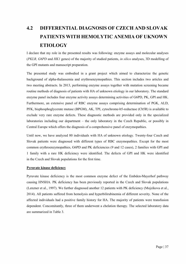
Page | 37
4.2 DIFFERENTIAL DIAGNOSIS OF CZECH AND SLOVAK
PATIENTS WITH HEMOLYTIC ANEMIA OF UKNOWN
ETIOLOGY
I declare that my role in the presented results was following: enzyme assays and molecular analyses
(PKLR, G6PD and HK1 genes) of the majority of studied patients, in silico analyses, 3D modelling of
the GPI mutants and manuscript preparation.
The presented study was embodied in a grant project which aimed to characterize the genetic
background of alpha-thalassemia and erythroenzymopathies. This section includes two articles and
two meeting abstracts. In 2013, performing enzyme assays together with mutation screening became
routine methods of diagnosis of patients with HA of unknown etiology in our laboratory. The standard
enzyme panel includes four enzyme activity assays determining activities of G6PD, PK, GPI and HK.
Furthermore, an extensive panel of RBC enzyme assays comprising determination of PGK, ALD,
PFK, bisphosphoglycerate mutase (BPGM), AK, TPI, cytochrome-b5-reductase (Cb5R) is available to
exclude very rare enzyme defects. These diagnostic methods are provided only in the specialized
laboratories including our department – the only laboratory in the Czech Republic, or possibly in
Central Europe which offers the diagnosis of a comprehensive panel of enzymopathies.
Until now, we have analyzed 80 individuals with HA of unknown etiology. Twenty-four Czech and
Slovak patients were diagnosed with different types of RBC enzymopathies. Except for the most
common erythroenzymopathies, G6PD and PK deficiencies (9 and 12 cases), 2 families with GPI and
1 family with a rare HK deficiency were identified. The defects of GPI and HK were identified
in the Czech and Slovak populations for the first time.
Pyruvate kinase deficiency
Pyruvate kinase deficiency is the most common enzyme defect of the Embden-Mayerhof pathway
causing HNSHA. PK deficiency has been previously reported in the Czech and Slovak populations
(Lenzner et al., 1997). We further diagnosed another 12 patients with PK deficiency (Mojzikova et al.,
2014). All patients suffered from hemolysis and hyperbilirubinemia of different severity. None of the
affected individuals had a positive family history for HA. The majority of patients were transfusion
dependent. Concomitantly, three of them underwent a chelation therapy. The selected laboratory data
are summarized in Table 3.

Page | 38
Table 3: Selected laboratory parameters of the studied patients
Origin Sex
Age
[year]
RBCs
[x1012] Hb [g/L]
Ret
[0.5-3.0 %]
Bili
[0-17 µmol/L]
Regular
transfusions
1 CZ M 6 m 2.76 77 7.2 33 Yes
2 CZ M 7 3,12 97 8.1 39 Yes*
3 CZ F 7 2.82 95 10.6 35 Yes*
4 CZ M 10 m 2.56 75 7.5 59 Yes
5 SK M 9 2.65 79 32 38 Yes
6 CZ F 42 2.95 103 5.9 50 Yes+
7 CZ F 22 1.81 65 80.5 139 Yes
8 CZ F 31 3.05 95 6.6 34 Yes
9 CZ M 3 m 2.49 70 2.1 172 Yes
10 CZ F 25 2.3 73 25.3 124 Yes
11 CZ M 50 3.4 121 5.2 49 No
12 CZ M 21 2.10 72 60.6 ND Yes
CZ – the Czech Republic; SK – the Slovak Republic; Age = age at the diagnosis; Ret = reticulocytes; Bili = bilirubin
All but two patients showed a decrease in PK activity below 50% of the control range. In patients 7
and 12, a high reticulocyte count masked a low PK activity, thus the PK deficiency was suspected
based upon the PK/HK ratio (Table 4). Mutation screening identified eleven mutations in the PKLR
gene (Table 4). The mutations p.(Arg510Gln), p.(Arg498His), p.(Arg532Trp), p.(Ser332Gly),
p.(Arg486Trp) have been previously reported in the Czech and Slovak populations (Lenzner et al.,
1997). The most common mutations in our patient group were mutations p.(Arg510Gln) and
p.(Arg532Trp). The substitution p.(Arg510Gln) is also one of the most frequent PKLR mutation in
Northern and Central Europe (Zanella et al., 2005). The mutations p.(Arg116-L117delinsGlnHisCys),
p.(Gly275Arg), p.(Ala154Thr) and c.1618+1delG have been described in the literature (Pissard et al.,
2006; van Wijk et al., 2009; van Zwieten et al., 2015; Zanella et al., 1997), however, none of these has
been detected in the Czech or Slovak populations. Furthermore, the mutations p.(Asp293Val) and
p.(Arg518Leufs*12) have not been previously described in the literature.

Page | 39
Table 4: Biochemical and molecular data of patients with PKLR mutation
The novel mutation p.(Asp293Val) was present together with the mutation p.(Arg510Gln) in patient 8.
Multiple sequence alignments revealed that Asp293 is located in a highly conserved region. Detailed
in silico analyses by PolyPhen and SIFT predicted that Asp to Val substitution may be deleterious.
The pathogenic nature of this mutation is supported by the fact that both amino acids display different
physical-chemical properties, therefore Asp to Val replacement may affect protein folding and/or
catalytic activity of the enzyme.
The second novel mutation p.(Arg518Leufs*12) was associated with atypical severe transfusion
dependent HA and neonatal hyperferritinemia. The patient was found to be homozygous for this
Patient PK
activity
PK/HK
ratio PKLR mutation Exon
1 1.63 1.63 c.1553delG/1553delG
11/11 p.(Arg518Leufs*12/Arg518Leufs*12)
2 1.73 1.75 c.1493G>A/1529G>A
11/11 p.(Arg498His/Arg510Gln)
3 1.43 0.51 c.1529G>A/1594C>T
11/11 p.(Arg510Gln/Arg532Trp)
4 1.14 0.46 c.1529G>A/1594C>T
11/11 p.(Arg510Gln/Arg532Trp)
5 2.64 0.78
c.347-350delinsAACATTG/347-350delinsAACATTG
4/4 p.(Arg116-L117delinsGlnHisCys/Arg116-
L117delinsGlnHisCys)
6 1.16 0.84 c.1456C>T/1594 C>T
11/11 p.(Arg486Trp/Arg532Trp)
7 6.58 1.6 c.1594C>T/1594C>T
11/11 p.(Arg532Trp/Arg532Trp)
8 1.56 0.81 c.878A>T/1529G>A
7/11 p.(Asp293Val/Arg510Gln)
9 1.72 2.07 c.823G>A/1594C>T
7/11 p.(Gly275Arg/Arg532Trp)
10 2.32 1.12 c.1529G>A/1529G>A
11/11 p.(Arg510Gln/Arg510Gln)
11 1.54 1.14 c.994G>A/1456C>T
8/11 p.(Ser332Gly/Arg486Trp)
12 6.09 1.40 c. 460G>A/1618+1delG
5/intr 11 p.(Ala154Thr/-)
PK [4.6-6.28 U/g Hb]; PK/HK ratio 3.6-6.3 U/g Hb; intr = intron; novel mutations in bold

Page | 40
mutation. His mother was a heterozygous carrier and the father was unknown. The mutation resulted
in a shift of the reading frame, leading to the formation of a premature stop codon. The PK protein (if
produced) is predicted to lack the C-terminal end (45 amino acids), which includes the activator
binding site Arg532, facilitating enzyme catalysis. Although iron overload is the major factor
contributing to pathogenesis of PK deficiency (Zanella et al., 2005), in this case dramatic
hyperferritinemia suggested an additional congenital defect. However, mutation screening of genes
associated with primary hemochromatosis (HFE, HFE2, HAMP, SCL40A1 and TFR2) did not show
any causative mutation. The patient was only a carrier of several single nucleotide polymorphisms
(SNPs) in these genes which, however, were also present in other PK-deficient patients. This fact ruled
out their contribution to the severe phenotype of this patient and could not explain his iron overload.
To exclude other causes of severe hyperferritinemia, such as sideroblastic anemia and hereditary
hyperferritinemia cataract syndrome, we evaluated the number of ring sideroblasts in the patient’s
bone marrow and performed molecular screening for the mutation in the IRE (iron responsive
element) region of the L-ferritin gene (FTL). However, we did not detect the presence of either ring
sideroblasts or a causal mutation in FTL gene. At the age of 2.5 years, surprisingly, the patient
exhibited normal ferritin levels. This may be a result of a combined chelation therapy and a prolonged
interval between transfusions. Thus, the severe phenotype involving neonatal hyperferritinemia was
likely caused by the p.(Arg518Leufs*12) mutation itself.
Other enzyme deficiencies
As mentioned above, other enzyme defects were detected among the 80 individuals with idiopathic
HA. We further diagnosed 9 cases of G6PD deficiency (manuscript in preparation), 2 families with
GPI deficiency (manuscript accepted for publication) and 1 patient with very rare HK deficiency
(a detailed description is provided in the section 4.3).
G6PD deficiency is the most frequent enzyme disorder in the world. The majority of G6PD mutations
are asymptomatic, only small percentage of G6PD deficient patients suffers from HNSHA. Based on
the severity of clinical manifestations, G6PD deficiency is classified into five classes, from the mildest
(Class V) to the most severe (Class I). Class I mutations more frequently affect the exons 6, 10 and 13
encoding the regions that bind the enzyme substrate, dimer interface and NADP+ structural site,
respectively (Minucci et al., 2012).
A few mutations in G6PD gene have been previously identified in Czech population (Xu et al., 1995).
All these G6PD variants (G6PD Praha, G6PD Olomouc, G6PD Varnsdorf – the first described splice
site mutation in the G6PD gene) belong to the class I mutations which are associated with HNSHA
(Minucci et al., 2012).
Our patient group consisted of 7 men and 2 women. All analyzed patients displayed a reduced G6PD
activity suggesting G6PD deficiency as the underlying cause of HNSHA. Clinical features of the
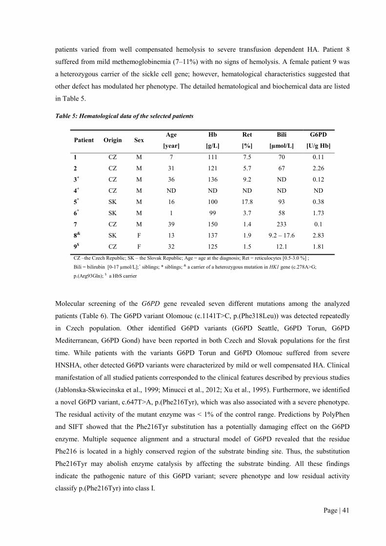
Page | 41
patients varied from well compensated hemolysis to severe transfusion dependent HA. Patient 8
suffered from mild methemoglobinemia (7–11%) with no signs of hemolysis. A female patient 9 was
a heterozygous carrier of the sickle cell gene; however, hematological characteristics suggested that
other defect has modulated her phenotype. The detailed hematological and biochemical data are listed
in Table 5.
Table 5: Hematological data of the selected patients
Patient Origin Sex Age
[year]
Hb
[g/L]
Ret
[%]
Bili
[µmol/L]
G6PD
[U/g Hb]
1 CZ M 7 111 7.5 70 0.11
2 CZ M 31 121 5.7 67 2.26
3+ CZ M 36 136 9.2 ND 0.12
4+ CZ M ND ND ND ND ND
5* SK M 16 100 17.8 93 0.38
6* SK M 1 99 3.7 58 1.73
7 CZ M 39 150 1.4 233 0.1
8& SK F 13 137 1.9 9.2 – 17.6 2.83
9$ CZ F 32 125 1.5 12.1 1.81
CZ –the Czech Republic; SK – the Slovak Republic; Age = age at the diagnosis; Ret = reticulocytes [0.5-3.0 %] ;
Bili = bilirubin [0-17 µmol/L];+ siblings; * siblings; & a carrier of a heterozygous mutation in HK1 gene (c.278A>G;
p.(Arg93Gln); $ a HbS carrier
Molecular screening of the G6PD gene revealed seven different mutations among the analyzed
patients (Table 6). The G6PD variant Olomouc (c.1141T>C, p.(Phe318Leu)) was detected repeatedly
in Czech population. Other identified G6PD variants (G6PD Seattle, G6PD Torun, G6PD
Mediterranean, G6PD Gond) have been reported in both Czech and Slovak populations for the first
time. While patients with the variants G6PD Torun and G6PD Olomouc suffered from severe
HNSHA, other detected G6PD variants were characterized by mild or well compensated HA. Clinical
manifestation of all studied patients corresponded to the clinical features described by previous studies
(Jablonska-Skwiecinska et al., 1999; Minucci et al., 2012; Xu et al., 1995). Furthermore, we identified
a novel G6PD variant, c.647T>A, p.(Phe216Tyr), which was also associated with a severe phenotype.
The residual activity of the mutant enzyme was < 1% of the control range. Predictions by PolyPhen
and SIFT showed that the Phe216Tyr substitution has a potentially damaging effect on the G6PD
enzyme. Multiple sequence alignment and a structural model of G6PD revealed that the residue
Phe216 is located in a highly conserved region of the substrate binding site. Thus, the substitution
Phe216Tyr may abolish enzyme catalysis by affecting the substrate binding. All these findings
indicate the pathogenic nature of this G6PD variant; severe phenotype and low residual activity
classify p.(Phe216Tyr) into class I.

Page | 42
Table 6: Molecular screening of the G6PD gene among the studied patients
Patient G6PD mutation Exon G6PD variant
1 c.647T>A
6 New variant p.(Phe216Tyr)
2 c.844G>C
8 G6PD Seattle p.Asp282His
3+ c.1141T>C
10 G6PD Olomouc p.(Phe318Leu)
4+ c.1141T>C
10 G6PD Olomouc p.(Phe318Leu)
5* c.1006A>G
9 G6PD Torun p.(Thr336Ala)
6* c.1006A>G
9 G6PD Torun p.(Thr336Ala)
7 c.563C>T
6 G6PD Mediterranean p.(Ser188Phe)
8 c.477G>C/477G>C
5 G6PD Gond p.(Met159Ile/Met159Ile)
9 c.202G>A/376A>G
4/5 - p.(Vale68Met/Asn126Asp)
G6PD 5.36-7.04 U/g Hb; novel mutations in bold; + siblings; * siblings
GPI deficiency is the second common enzyme defect of the Embden-Meyerhof pathway. Clinical
features in GPI deficiency range from mild to severe hemolytic anemia. GPI deficiency may also be
associated with non-hematologic symptoms, in particular with neurologic impairment or mental
retardation.
We diagnosed three GPI-deficient patients from two unrelated families. All patients suffered from
severe transfusion dependent HA. No neurological defects were present in any of the studied patients.
Patients’ parents have no signs of chronic hemolysis. All analyzed patients exhibited reduced GPI
activity, which reached 15% of the control range. Molecular screening of the GPI gene showed that
patient 1 was a compound heterozygote for mutations c.478T>C, p.(Ser160Pro) and c.1414C>T,
p.(Arg472Cys). Both patients 2 and 3 were homozygotes for the mutation c.1414C>T, p.(Arg472Cys)
(Table 7). Both identified mutations are novel, however, substitution of the same residue Arg472
(Arg472His) has been previously identified in a Hispanic patient. A homozygous mutation
p.(Arg472His) was associated with a severe phenotype and residual activity of this mutant GPI was
around 25% of the control range. A detailed characterization of the recombinant GPI mutant
demonstrated increased KM for F6P, decreased kcat and reduced thermal stability (Beutler et al., 1997;
Lin et al., 2009).
Page | 43
Table 7: Summary of the studied GPI-deficient patients
Patient 1 Patient 2* Patient 3*
Origin CZ SK SK
Sex M M F
Age [years] 11 19 10
Hb [g/L] 90 101 86
Ret [%] 16.3 33.4 45.6
Bili [µmol/L] 84 127 83
GPI [U/g Hb] 8.51 5.94 6.75
GPI mutations c.478T>C/1414C>T
p.(Ser160Pro/Arg472Cys)
c.1414C>T/1414C>T
p.( Arg472Cys/Arg472Cys)
c.1414C>T/1414C>T
p.( Arg472Cys/Arg472Cys)
CZ – the Czech Republic; SK – the Slovak Republic; Age = age at the diagnosis; Ret = reticulocytes [0.5-3.0 %] ; Bili = bilirubin
[0-17 µmol/L];* siblings, GPI [ 53 – 62 U/g Hb]
Results from in silico analyses (Polyphen and SIFT) revealed that both detected mutations
p.Arg472Cys and p.Ser160Pro are not tolerated. To study the probable mechanism by which both
mutations affect the protein function, we performed in silico modelling of the GPI mutants.
The analyses demonstrated that the residue Arg472 is a part of the dimer-domain at the edge of the
large external loop domain, a major contact region contributing to the dimer stability. The Arg472Cys
substitution likely destabilizes interactions within the dimer interface, and thus may affect either the
dimer formation and/or dimer stability. The residue Ser160 is located in the close proximity of the
active site, facilitating the binding of the substrate phosphate group. This suggests that the substituted
proline likely impairs the binding of the substrate, and thus inhibits catalytic activity of the mutant
enzyme (Read et al., 2001).

Page | 44
Figure 6: In silico modelling of the Ser160Pro and Arg472Cys GPI mutants. A) Ser160 (corresponding to Ser 160, shown in purple) is a part of the phosphate-binding site (blue) responsible for the potential hydrogen bond interaction with phosphate group of the GPI substrate (the sulphate was used to mimic the substrate phosphate group). B) Ser160Pro substitution likely results in a disruption of the hydrogen bond (a black dotted line) between the residue at position 159 and the substrate phosphate group and, thus, destabilizes the substrate-enzyme complex. C) Arg472 (corresponding to Arg471, shown in purple) is located at the edge of the large external loop (LEL, 438–468 residues) directly interacting (hydrogen bonding) with neighboring Gly469 residue and with Ile394 of the second monomer, and thus D) disruption of these interactions by Arg472Cys mutationleads to conformational changes within the LEL, serving as a dimer stabilizing hook, and thus to generation of a more labile GPI-dimer. The amino acid numbering begins with alanine as residue 1 instead of initial methionine. The structure illustrations were created with PyMOL program based on Protein data bank ID 1IAT. Black dot lines represent hydrogen bonds.
Kinetic studies of the mutation p.(Arg472Cys) revealed decreased affinity to F6P and strongly reduced
thermal stability. Furthermore, the enzyme activity was almost abolished after short incubation at
45°C. These findings are consistent with the previous functional study of p.Arg472His GPI mutant
(Lin et al., 2009) and all together correspond to the model of the proposed pathogenic mechanism.
The Ser160Pro mutation was characterized in the individual who was a compound heterozygous with
p.(Arg472Cys) mutation. Thus, the contribution of the second mutation has to be expected.
Biochemical characteristics of the compound heterozygous mutant showed decreased KM values and
its thermal stability was only slightly affected which may indicate that Arg472Cys homodimers
represent only a minor proportion of the mixed dimers pool.
Page | 45
HK deficiency was diagnosed in a patient with severe transfusion dependent HA. The patient was
found to be homozygous for mutation c.278A>G, p.(Arg93Gln). Interestingly, another two cases with
HK1 mutation were identified. One patient was a heterozygous carrier of mutation c.493-1G>A.
However, phenotype more severe than expected for an HK1 mutation carrier was probably caused by
Gilbert syndrome. The second patient possessed mutation c.278A>G, p.(Arg93Gln). The female
patient was also homozygous for mutation c.477G>C p.(Met159Ile) in the G6PD gene (Table 6).
Detailed description of HK deficiency is in the section 4.3.
In general, enzyme defects are associated with classical hallmarks of hemolysis with no abnormalities
in RBC morphology. Routinely used hematology methods are unable to point out a particular enzyme
defect. Due to the lack of adequate diagnostic methods, enzymopathies have been previously
considered as medical conditions belonging to the category of diagnosis by exclusion. In 1977, the
International Committee for Standardization in Hematology established “recommended methods” for
the assaying of RBC enzymes (Beutler et al., 1977). These direct activity assays followed by an
identification of a causative mutation at DNA level are the only worldwide accepted procedures for
erythroenzymopathies diagnosis in current clinical practice. Our laboratory is the only laboratory in
the Czech Republic, or possibly in Central Europe which offers a diagnosis of the whole panel of
enzymopathies.
In addition to diagnosis of erythroenzymopathies, the direct enzyme assays have been also found to be
an effective method for diagnosis of Diamond Blackfan anemia (DBA) and/or to test, for example, the
effects of therapeutics on cellular processes. Alterations of erythrocyte ADA activity are observed in
the majority of “classical” DBA patients (Fargo et al., 2013). Recently, we have established a direct
enzyme assay for erythrocyte ADA in our laboratory. Upregulated ADA activity has become a useful
diagnostic maker in a “classical” form of DBA. With regard to testing the effects of potential
therapeutics, we studied (in collaboration with a research group from Tübingen), the effect of
pharmacologic NF-κB inhibitors, Bay 11-7082, parthenolide and dimethyl fumarate DMF on red
blood cell survival and on mechanism of GSH depletion. The inhibitory effects of these
pharmacologic compounds were evaluated by monitoring enzymatic activities of G6PD, 6PGD and
GR, the key players of cellular redox homeostasis in red blood cells (Ghashghaeinia et al., 2016).
Furthermore, the enzyme assays were used for the assessment of anti-oxidative defense in erythrocytes
with deficiency of the divalent metal transporter 1 (Zidova et al., 2014).
Taken together, characterization of RBC enzymopathies by biochemical and molecular analyses
contributed to a better understanding of pathophysiology of these rare disorders. The direct enzyme
assays have a large variety of scientific applications which may help to improve diagnostic testing as
well as therapeutic interventions.

Page | 46
PUBLICATIONS RELATED TO THIS AIM:
Articles:
Ludíková B, Mojzíková R, Pospíšilová P, Houda J, Sulovská L, Divoká M, Hak J, Procházková D,
Divoký V, Pospíšilová D. Deficit pyruvátkinázy v dětském věku. Čes-slov Pediat. 2012; 67(3): 6–11.
Mojzikova R*, Koralkova P*, Holub D, Zidova Z, Pospisilova D, Cermak J, Striezencova Laluhova
Z, Indrak K, Sukova M, Partschova M, Kucerova J, Horvathova M, Divoky V. Iron status in patients
with pyruvate kinase deficiency: neonatal hyperferritinemia associated with a novel frameshift
deletion in the PKLR gene (p.Arg518fs), and low hepcidin to ferritin ratios. Br J Haematol.
2014;165(4):556–63.(*Authors contributed equally to this work)
Mojzikova R*, Koralkova P, Holub D, Saxova Z, Pospisilova D, Prochazkova D, Dzubak P,
Horvathova M, Divoky V Two novel mutations (p.(Ser160Pro) and p.(Arg472Cys)) causing glucose-
6-phosphate isomerase deficiency are associated with erythroid dysplasia and inappropriately
suppressed hepcidin. Blood Cells Mol Dis., manuscript accepted for publication.
Meeting abstracts:
Pospisilova P, Mojzikova R, Partschova M, Zidova Z, Horvathova M, Pospisilova D, Divoky V.
Hemolytic anemia with hyperferritinemia in Czech pyruvate kinase deficient patient – a novel
homozygous frameshift deletion in PKLR gene (p. R518fs) combined with TFR2 (p. R752H) and
SLC40A1 (IVS1-24 C>G) gene polymorphisms. 19th Meeting of the European Red Cell Society,
Ijmuiden, The Netherlands, 10–13 October, 2013: Abstract book, p. 47. (ORAL PRESENTATION)
Kořalková P, Mojzíková R, Pospíšilová D, Divoká M, Čermák J, Indrák K, Suková M, Timr P,
Striežencová Laluhová Z, van Wijk R, Divoký V. Vzácné vrozené anémie v české a slovenské
populaci: pokroky v diagnostice erytrocytárních anémií. XVII. SLOVENSKO-ČESKÝ
HEMATOLOGICKÝ A TRANSFUZIOLOGICKÝ ZJAZD – Bratislava, Slovak Republic (24–27
September, 2015), Abstract book, p.84. (ORAL PRESENTATION)

4.3 STRUCTURAL AND FUNCTIONAL
CHARACTERIZATION OF VERY RARE HEXOKINASE
MUTANTS CAUSING HNSHA
I declare that my role in the presented results was following: summarization of the hematological and
biochemical data from all the studied patients, enzyme assays and molecular analyses of the studied
patients except the patients from Denmark and the Netherlands, kinetic studies of the selected patients,
RNA isolation, RT-PCR, qPCR, western blot analysis, in silico analyses, 3D modelling of selected HK
mutants and manuscript preparation.
The major part of the experimental work was carried out during my fellowship at the Department of
Clinical Chemistry and Hematology, University Medical Center Utrecht, the Netherlands. This section
contains one original research paper and three meeting abstracts. The publications presented here
resulted from collaboration with research groups from the Netherlands, Ireland, Spain and the Slovak
Republic. The article describes molecular analysis of six HK-deficient patients representing the largest
group studied thus far. It summarizes the current knowledge about HK deficiency and unifies
numbering for previously detected mutations according to the erythroid specific HK-R transcript (HK-
R NM_033496.2).
Hexokinase is the key enzyme of the Embden-Meyerhof pathway that catalyzes the ATP-dependent
phosphorylation of glucose, on which red cell energy metabolism is fully dependent. HK deficiency is
an autosomal recessive disorder associated with chronic HSNHA. Affected individuals exhibit mild to
severe lifelong HA. However, a complete loss of HK1 is apparently incompatible with life (Kanno et
al., 2002).
We analyzed seven patients from 6 unrelated families with a suspected enzyme deficiency. Standard
methods including peripheral blood smear examination, cryohemolysis/Pink test, osmotic fragility test,
(EMA) binding test, hemoglobin stability test (isopropanol test), and hemoglobin SDS-PAGE were
used to exclude hemoglobinopathies and membrane defects. The majority of the patients suffered from
chronic hemolysis with variable clinical severity (Table 8). All but one patient showed a decreased HK
activity below 50% of the control range and/or increased PK/HK ratio (Table 8). So far, 24 cases of
HK deficiency associated with HNSHA have been reported (Mentzer and William, 2014) and only
6 mutations as an underlying cause of HK deficiency have been detected (Bianchi et al., 1998; de
Vooght et al., 2009; Kanno et al., 2002; van Wijk et al., 2003). Molecular analysis of our patient group
revealed 6 different mutations in HK1 gene, four of them were novel: c.2599C>T p.(His867Tyr),
c.1799C>T p.(Thr600Met), c.873-2A>G and c. 493-1G>A (Table 9, novel mutations in bold).
To clarify the pathogenic nature of the novel mutations we performed biochemical and/or RNA
studies, a 3-D structural analysis and in silico analyses by PolyPhen and SIFT.

Table 8: Hematological and biochemical data of the studied patients
Patient 1S Patient 2S Patient 3 Patient 4 Patient 5 Patient 6 Patient 7
Origin Ireland Ireland Czech Republic Denmark Netherlands Czech Republic Slovak Republic
Sex M M M M M F F
Age [year] 2 4 8 m 2 22 12 12
RBCs [x1012] 2.57; (*--) 2.76; (*--) 2.40; (*--) 2.90; (*--) 4.40; (*--) 4.25; (-*-) 4.58; (-*-)
Hb [g/L] 79; (*--) 95; (*--) 74; (*--) 86; (*--) 160; (-*-) 135; (-*-) 137; (-*-)
Ret [%] 8.8; (--*) 6.2; (--*) 6.3; (--*) 4.7; (--*) 6.0; (--*) 3.1; (--*) 1.9; (--*)
Bili [µmol/L] 54; (--*) 62; (--*) 120; (--*) 124; (--*) 24; (--*) 100; (--*) 17.6; (-*-)
Transfusion therapy Yes$ No Yes Yes No No No
Other diagnosis ND ND ND ND GS GS G6PD Gond
Enzyme activities
HK 0.64¥ 0.58¥ 0.71# 1.14+ 0.65¥ 0.37# 0.33#
PK 12.3¥ 12.6¥ 3.3# 12.5+ 12.2¥ 7.2# 6.2#
PK/HK 19.2¥ 21.7¥ 9.5# 10.90+ 18.7¥ 19.3# 18.7#
S – siblings; Age – age at diagnosis of HK deficiency, m – months; RBCs – red blood cells; Hb – hemoglobin, M – male; F – female; ND – not determined, GS – Gilbert syndrome. G6PD – glucose-6phosphate deficiency, variant – GOND; $Transfusion therapy has been stopped at the age 3; comparison of hematological data with a reference range: (*--) – low; (-*-) – normal; (--*) – high. ¥HK [0.80-1.50 U/g Hb], PK[6.1-12.3 U/g Hb], PK/HK [4.8-11.9]; #HK [0.80-1.65], PK[5.1-5.8], PK/HK [3.6 -6.3]; +HK [0.62-1.26], PK [8.4-15.2], PK/HK [7.2-15.6].
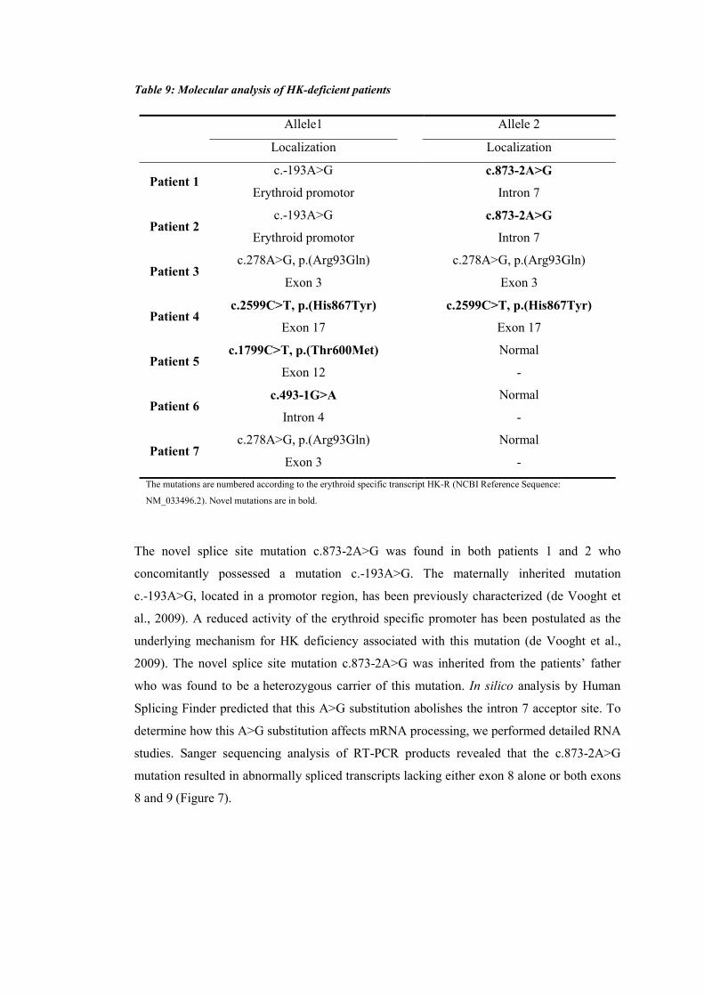
Table 9: Molecular analysis of HK-deficient patients
Allele1 Allele 2
Localization Localization
Patient 1 c.-193A>G c.873-2A>G
Erythroid promotor Intron 7
Patient 2 c.-193A>G c.873-2A>G
Erythroid promotor Intron 7
Patient 3 c.278A>G, p.(Arg93Gln) c.278A>G, p.(Arg93Gln)
Exon 3 Exon 3
Patient 4 c.2599C>T, p.(His867Tyr) c.2599C>T, p.(His867Tyr)
Exon 17 Exon 17
Patient 5 c.1799C>T, p.(Thr600Met) Normal
Exon 12 -
Patient 6 c.493-1G>A Normal
Intron 4 -
Patient 7 c.278A>G, p.(Arg93Gln) Normal
Exon 3 -
The mutations are numbered according to the erythroid specific transcript HK-R (NCBI Reference Sequence:
NM_033496.2). Novel mutations are in bold.
The novel splice site mutation c.873-2A>G was found in both patients 1 and 2 who
concomitantly possessed a mutation c.-193A>G. The maternally inherited mutation
c.-193A>G, located in a promotor region, has been previously characterized (de Vooght et
al., 2009). A reduced activity of the erythroid specific promoter has been postulated as the
underlying mechanism for HK deficiency associated with this mutation (de Vooght et al.,
2009). The novel splice site mutation c.873-2A>G was inherited from the patients’ father
who was found to be a heterozygous carrier of this mutation. In silico analysis by Human
Splicing Finder predicted that this A>G substitution abolishes the intron 7 acceptor site. To
determine how this A>G substitution affects mRNA processing, we performed detailed RNA
studies. Sanger sequencing analysis of RT-PCR products revealed that the c.873-2A>G
mutation resulted in abnormally spliced transcripts lacking either exon 8 alone or both exons
8 and 9 (Figure 7).

Figure 7: RNA studies of a splice site mutation c.873-2A>G. Agarose electrophoresis. Visualization of RT-PCR products (a region spanning exons 7 to 9) revealed two additional fragments in the heterozygous subject carrying the mutation c.873-2A>G (line 1) compared to the healthy donor (line 2). Lines 3 and 4 served as negative controls for RT and PCR. Bands of 834 bp (lines 1 and 2) corresponded to a normally spliced variant A. Fragments of 735 bp and 470 bp (line 1) corresponded to the abnormal transcripts B and C lacking either exon 8 or both exons 8 and 9.
The aberrant proteins (if produced) would lack either the substrate binding site or the
substrate binding site and the G6P binding site. Furthermore, pre-mRNA expression of the
normally spliced variant of the heterozygous c.873-2A>G carrier was more than 50% lower
than in controls (Figure 8). Subsequently, western blot analysis on in-vitro cultured
pro-erythroblast revealed a marked down-regulation of HK at protein level (Figure 8). Based
on these findings, we could conclude that the c.873-2A>G mutation affects pre-mRNA
processing, which results in a down-regulation of HK on the protein level.
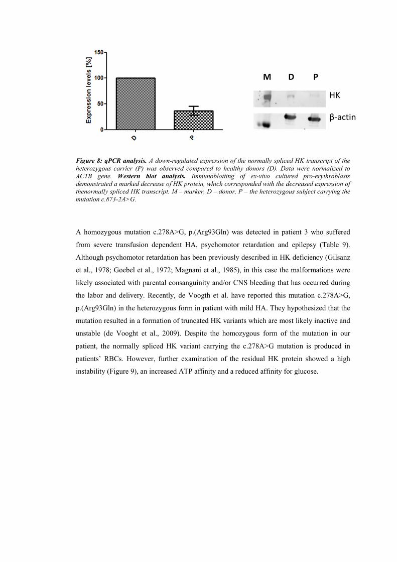
Figure 8: qPCR analysis. A down-regulated expression of the normally spliced HK transcript of the heterozygous carrier (P) was observed compared to healthy donors (D). Data were normalized to ACTB gene. Western blot analysis. Immunoblotting of ex-vivo cultured pro-erythroblasts demonstrated a marked decrease of HK protein, which corresponded with the decreased expression of thenormally spliced HK transcript. M – marker, D – donor, P – the heterozygous subject carrying the mutation c.873-2A>G.
A homozygous mutation c.278A>G, p.(Arg93Gln) was detected in patient 3 who suffered
from severe transfusion dependent HA, psychomotor retardation and epilepsy (Table 9).
Although psychomotor retardation has been previously described in HK deficiency (Gilsanz
et al., 1978; Goebel et al., 1972; Magnani et al., 1985), in this case the malformations were
likely associated with parental consanguinity and/or CNS bleeding that has occurred during
the labor and delivery. Recently, de Voogth et al. have reported this mutation c.278A>G,
p.(Arg93Gln) in the heterozygous form in patient with mild HA. They hypothesized that the
mutation resulted in a formation of truncated HK variants which are most likely inactive and
unstable (de Vooght et al., 2009). Despite the homozygous form of the mutation in our
patient, the normally spliced HK variant carrying the c.278A>G mutation is produced in
patients’ RBCs. However, further examination of the residual HK protein showed a high
instability (Figure 9), an increased ATP affinity and a reduced affinity for glucose.
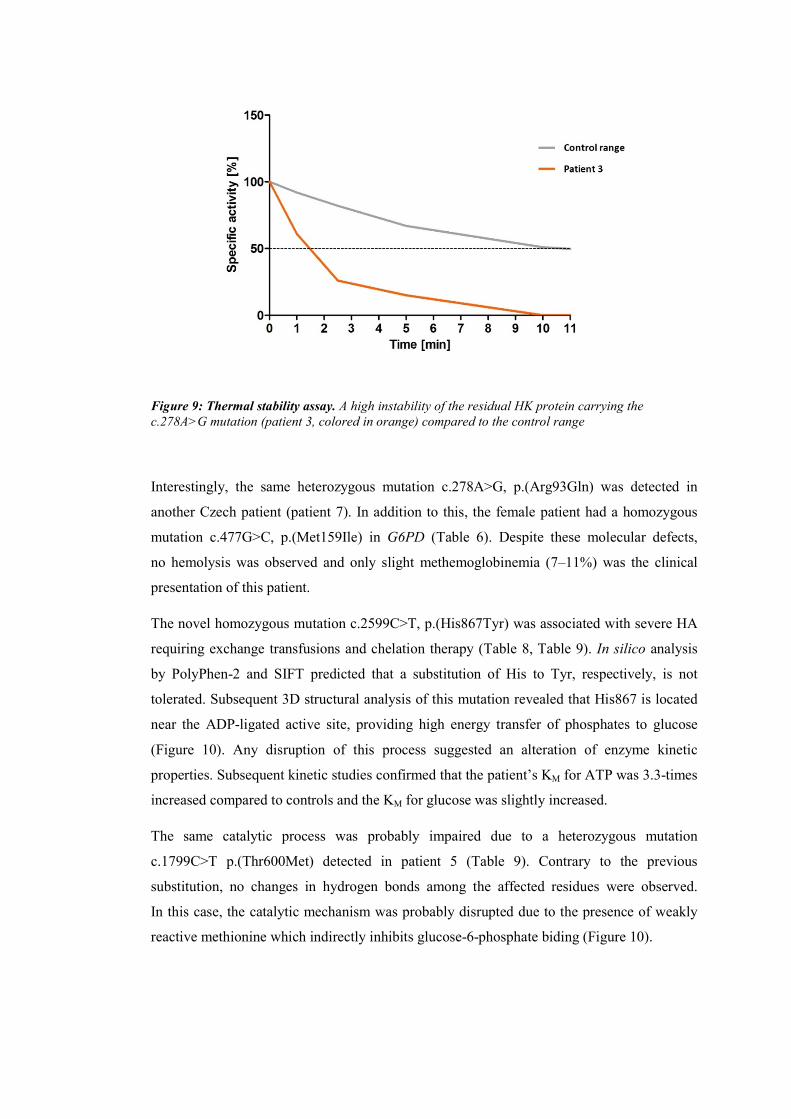
Figure 9: Thermal stability assay. A high instability of the residual HK protein carrying the c.278A>G mutation (patient 3, colored in orange) compared to the control range
Interestingly, the same heterozygous mutation c.278A>G, p.(Arg93Gln) was detected in
another Czech patient (patient 7). In addition to this, the female patient had a homozygous
mutation c.477G>C, p.(Met159Ile) in G6PD (Table 6). Despite these molecular defects,
no hemolysis was observed and only slight methemoglobinemia (7–11%) was the clinical
presentation of this patient.
The novel homozygous mutation c.2599C>T, p.(His867Tyr) was associated with severe HA
requiring exchange transfusions and chelation therapy (Table 8, Table 9). In silico analysis
by PolyPhen-2 and SIFT predicted that a substitution of His to Tyr, respectively, is not
tolerated. Subsequent 3D structural analysis of this mutation revealed that His867 is located
near the ADP-ligated active site, providing high energy transfer of phosphates to glucose
(Figure 10). Any disruption of this process suggested an alteration of enzyme kinetic
properties. Subsequent kinetic studies confirmed that the patient’s KM for ATP was 3.3-times
increased compared to controls and the KM for glucose was slightly increased.
The same catalytic process was probably impaired due to a heterozygous mutation
c.1799C>T p.(Thr600Met) detected in patient 5 (Table 9). Contrary to the previous
substitution, no changes in hydrogen bonds among the affected residues were observed.
In this case, the catalytic mechanism was probably disrupted due to the presence of weakly
reactive methionine which indirectly inhibits glucose-6-phosphate biding (Figure 10).

Figure 10: 3D crystal structure of HK-R mutants His867Tyr and Thr600Met (PDB 1DGK – protein databank). A) His867 (corresponding to His868 in HK-1, shown in red) is located between ADP-ligated active sites (green). No direct interaction between ADP (blue) and His has been described. However, His substitution with Tyr probably leads to a disruption of multiple hydrogen bonds (black dotted lines) among residues Phe871, His870 and to a formation of new hydrogen bonds with Glu791 and Ser827. The changes in hydrogen bonding likely affect electrostatic interactions through the ADP-ligated active site, leading to interaction impairments within the ADP/glucose-complex. B) Met601 (corresponding to Met600 in HK-1, shown in red) is in close proximity of loop 532–539 (green) which is involved in productive binding of ADP (blue) and Ser603 (orange) directly interacting with a molecule of glucose (purple). Although no changes in hydrogen bonding were observed, the Met substitution with Thr in this position may impair electrostatic interactions within an enzyme catalysis site affecting the mechanism of phosphate transfer.
The novel mutation c.493-1G>A in the heterozygous form was identified in patient 6. The
female patient was first diagnosed with Gilbert syndrome, but persistent mild hemolysis and
reticulocytosis pointed to the presence of an additional modulator. In silico analysis of the
mutation c.493-1G>A predicted an activation of a cryptic splice site within intron 4,
resulting in formation of aberrant HK variant(s). Based on current findings we can conclude
that the aberrant splicing is likely the major factor contributing to HK deficiency in this
patient. However, it is not clear whether or not heterozygosity for this mutation is sufficient
to modulate patient’s phenotype.

This work characterizes genetic background of six patients with a very rare HK deficiency,
to our knowledge the largest cohort of studied HK deficient patients. Detailed investigations
on the selected mutations clarify their pathogenic nature and thus this work contributes to
better understanding of the genotype-phenotype correlation in HK deficiency.
PUBLICATIONS RELATED TO THIS AIM:
Articles:
Koralkova P, Mojzikova R, van Oirschot B, Macartney C, Timr P, Vives Corrons JL,
Striezencova Laluhova Z, Lejhancova K, Divoky V, van Wijk R. Molecular
characterization of six new cases of red blood cell hexokinase deficiency yields four novel
mutations in HK1. Blood Cells Mol Dis. 2016; 59:71-6.
Meeting abstracts:
Pospisilova P, Mojzikova R, Pospisilova D, Divoky V, Vives-Corrons JL, van Wijk R. Two
novel homozygous mutations (Agr94Gln and His868Tyr) in HK1 gene associated with
hereditary non-spherocytic hemolytic anemia described in Spain and Czech patients. 19th
Meeting of the European Red Cell Society, Ijmuiden, The Netherlands, October 10–13,
2013. Abstract book, p. 60. (POSTER PRESENTATION)
Timr P, Kořalková P, Mojzíková R, van Wijk R, Divoký V. První případ populace
hexokinasy v ČR způsobující nesférocytární hemolytickou anémii. XXIV. Konference
dětských hematologů a onkologů České a Slovenské republiky, Olomouc, Česká republika
October 10–12, 2014. Abstact book, p. 37. (ORAL PRESENTATION)
Koralkova P, Mojzikova R, Timr P, Vives Corrons JL, Macartney C, Divoky V, van Wijk
R. Five new cases of hexokinase deficiency: biochemical and molecular characterization
of a novel splice site mutation and 2 novel missense mutations in HK1. 20thCongress of the
European Hematology Association, Vienna, Austria, June 12–14, 2015. Abstract book –
Haematologica, p. 128. (POSTER PRESENTATION)

Page | 55
4.4 MOLECULAR CHARACTERIZATION OF A NOVEL
NON-HEMOLYTIC ANEMIA VARIANT OF PGK
I declare that my role in the presented research was following: molecular analysis of the studied
patients, cell culture of a donor sample, restriction analysis, RNA isolation, RT-PCR, qPCR
analysis, TOPO cloning, in silico analyses and participation in the manuscript preparation.
The major part of the experimental work was carried out during my fellowship at the Department
of Clinical Chemistry and Hematology, University Medical Center Utrecht, the Netherlands.
The project was conducted in collaboration with research centers from Belgium and the
Netherlands. This section contains one research paper and one meeting abstract. They describe
molecular characterization of PGK deficiency in two patients with myoglobinuria, mental
retardation, but no signs of hemolysis.
PGK deficiency is a rare X-linked disorder associated with multiple clinical manifestations
including hemolytic anemia, mental retardation, exercise intolerance, myoglobinuria,
rhabdomyolysis and/or parkinsonism. The majority of patients suffer from hemolytic anemia
combined with neurological defects. Myopathy and muscle weakness as the predominant clinical
features have been described in several cases. Conversely, a full spectrum of clinical manifestations
is rarely present (Beutler, 2007; Sotiriou et al., 2010).
The studied patients were two brothers from healthy, non-consanguineous parents. They suffered
from mild retardation, recurrent episodes of muscle weakness and pain after moderate exercise
since the age of 4. The younger brother experienced an episode of myoglobinuria and seizures.
In the further clinical course, laboratory parameters did not show any signs of hemolysis but
a markedly decreased activity of PGK (<15% of the reference range) was detected in both patients.
A slight reduction in PGK activity was observed in patients’ mother; father was within the control
range. To confirm PGK deficiency at the molecular level, we performed mutation screening of the
PGK1 gene. The analysis revealed the presence of hemizygous mutation c.756+3A>G in intron 7
in both patients. The heterozygous form of this mutation was identified in their mother; no
mutation was detected in the patients’ father. To exclude this mutation as neutral polymorphism,
we performed screening of >100 alleles from control subjects. The mutation was not present in the
control population. Thus, we analyzed the possible effect of the A>G substitution on the mRNA
splicing processing using Human Splicing Finder. This in silico analysis predicted that the affected
region abolishes the intron 7 donor splice site. To confirm the particular effect of the mutation, we
isolated total RNA from ex-vivo cultured proerythroblasts of the respective patients and conducted
detailed RNA studies. Visualization of RT-PCR fragments of both patients showed two additional
fragments compared to a control subject (Figure 11). Unfortunately, the low concentration of both

Page | 56
fragments precluded direct Sanger sequencing. Therefore, to analyze the specific sequence of these
abnormal fragments, we subcloned them into pCRTM4-TOPO vector.
Figure 11: Visualization of RT-PCR fragments of hemizygous c.756+3A>G mutation carriers and a control subject. A region spanning exons 7 and 8 of PGK1 were amplified. Presence of two additional fragments was observed in both patients (P1 and P2) compared to healthy donor (D). Lines N1 and N2 served as negative control for reverse transcription and PCR.
The sequencing analysis of positive colonies revealed the presence of two additional aberrant
transcripts. Thus, a total of four different PGK variants was detected in the hemizygous carriers of
c.756+3A>G mutation compared to the control subject (Figure 11). The most abundant PGK
variants A and B consist of 52 bp of intron 7 and/or a complete sequence of intron 7. Both variants
result in the introduction of a premature stop codon. The predicted protein (if translated) lacks the
C-terminal end, containing 165 amino acids, which facilitates nucleotide binding. The aberrant
PGK transcript A, encompassing 52 bp of intron 7, has been previously reported in patients with
PGK Fukuroi (c.756+5G>A) and with PGK Antwerp (c.755A>C, p.(Glu252Ala)). Both patients
also exhibited recurrent episodes of rhabdomyolysis, myoglobinuria without HA. However,
additional clinical manifestations (abnormal brain activity and/or slight changes in muscle fiber
morphology) have been reported in both patients (Ookawara et al., 1996; Shirakawa et al., 2006).
The transcripts C and D were characterized by a loss of either 18 bp of exon 7 and/or a complete
region of exon 7. The variant lacking 18bp of exon 7 results in deletion of six amino acids (residues
247–252). The loss of complete exon 7 causes a premature codon formation and the predicted
protein (if translated) lacks the C-terminal end. None of these PGK variants has been described in
the literature (Figure 12).
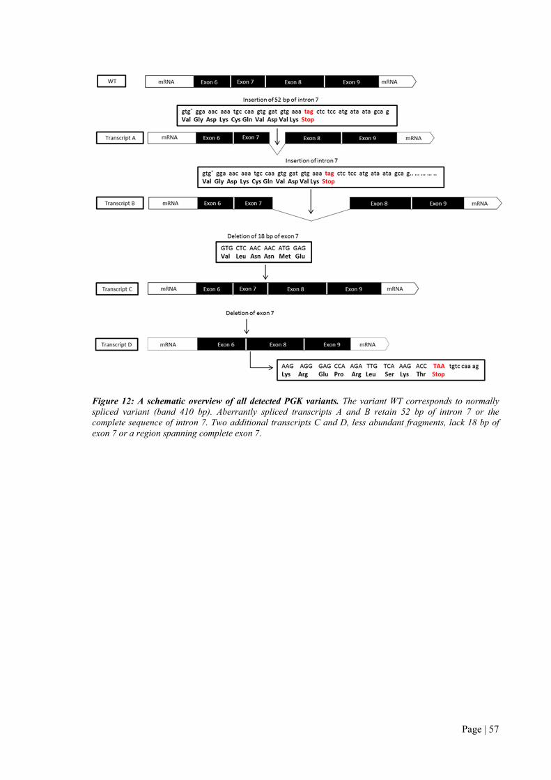
Page | 57
Figure 12: A schematic overview of all detected PGK variants. The variant WT corresponds to normally spliced variant (band 410 bp). Aberrantly spliced transcripts A and B retain 52 bp of intron 7 or the complete sequence of intron 7. Two additional transcripts C and D, less abundant fragments, lack 18 bp of exon 7 or a region spanning complete exon 7.

Page | 58
Despite of all these aberrant PGK variants, normal pre-mRNA processing does occur. To quantify
the expression levels of the normally spliced variant, we designed a specific pair of primers to
avoid the contribution of the additional aberrant PGK variants, and performed qPCR. The analysis
showed that the level of the normally spliced PGK variant was 8.5 times down-regulated compared
to the healthy individual (Figure 13).
Figure 13: Expression levels of normally spliced PGK pre-mRNA. ACTB was used as an internal standard.
Relative quantification was evaluated based on the comparative CT method (2-ΔΔCT). D – donors,
P – patients.
Taken together, this study showed that c.756+3A>G mutation results in aberrant splicing and
ineffective protein production, most likely the underlying cause of PGK deficiency in both patients.
Page | 59
PUBLICATIONS RELATED TO THIS SPECIFIC AIM:
Articles:
Coppens S, Koralkova P, Aeby A, Mojzikova R, Deconinck N, Kadhim H, van Wijk R. Recurrent
episodes of myoglobinuria, mental retardation and seizures but no hemolysis in two brothers
with phosphoglycerate kinase deficiency. Neuromuscul Disord. 2016; 26(3):207-10.
Abstract meetings:
Coppens S, Deconinck N, van Wijk R, Koralkova P, Van Bogaert P, Aeby A. Recurrent episodes
of myoglobinuria, mental retardation and epilepsy but no haemolysis in two brothers with
phosphoglycerate kinase deficiency. 19th International Congress of The World Muscle Society –
Berlin, Germany (October 7–11, 2014), Neuromuscular Disorders Volume 24, Issues 9–10, p. 873.
(POSTER PRESENTATION)

Page | 60
4.5 IN SILICO MODELLING OF A NOVEL MUTATION
OF PFKM GENE ASSOCIATED WITH PFK
DEFICIENCY
I declare that my role in the presented research was following: in silico analyses, 3D structural
modelling of the PFK mutant and participation in the manuscript preparation.
This section includes one article. The publication resulted from collaboration of Dr. van Wijk
laboratory with research centers from Spain and the Netherlands. The presented article describes
the first case of a rare PFK deficiency diagnosed in a 65-year-old female of Spanish origin.
PFK deficiency is a very rare disorder associated with multiple clinical manifestations including
muscle cramps, exercise intolerance, myoglobinuria and/or hemolysis. Erythrocyte PFK is
composed of PFKL (liver) and PFKM (muscle) subunits which form their hybrids (M4, L4, M3L,
M2L2, and ML3). In case of a mutated PFKM subunit, only PFKL subunits remain fully functional
in erythrocyte PFK leading to only a partial deficiency of PFK. Thus, patients with PFK deficiency
usually exhibit mild or fully compensated hemolytic anemia (Nakajima et al., 2002).
The female patient suffered from recurrent episodes of spasm with muscle weakness and pain after
moderate exercise since her childhood. She had a laboratory evidence of moderate anemia. Direct
enzyme assays revealed a reduced PFK activity (<30% of the control range) and subsequent
molecular screening of PFKM gene identified a point homozygous mutation c.929A>G in exon 11.
The mutation resulted in the substitution of Asp to Gly at position 309. The mutation has not been
described in the literature, thus, in silico analyses (SIFT, Polyphen, 3D structural modelling) were
performed to test whether or not the substitution p.(Asp309Gly) is the cause of decreased PFK
activity and HA due to PFK deficiency.
Both in silico tools predicted that the substitution p.(Asp309Gly) is likely deleterious. Subsequent
in silico 3D structure analysis (a model of rabbit muscle type PFK, PDB entry 3O8L) showed that
the residue Asp309 is located in close proximity of the nucleotide (ADP) binding site in the center
of PFKM subunit (Figure 14). Interaction between ADP molecule and the ADP binding site
initiates a positive allosteric modulation of PFKM enzyme (Banaszak et al., 2011). Although no
direct interactions between Asp309 and ADP molecule are observed, the substitution of aspartic
acid to glycine likely disrupted multiple hydrogen bonds (Gly177, Gly179, Ser180 and Ser306)
closely located to the ADP binding site. This loss of hydrogen bonds may cause a change of

Page | 61
polarity and electrostatic interactions which may affect side-chains of amino acids facilitating ADP
binding within the activated allosteric site of the PFKM enzyme; in particular with the neighboring
Phe308, which makes stacking interactions with the adenine ring of ADP molecule. This proposed
pathogenic mechanism corresponds to the previous study of PFKM Asp543Ala mutation.
The Tarui disease-causing mutation Asp543Ala blocks ADP binding and mediates its disastrous
effect by increasing allosteric inhibitory efficiency of ATP (Bruser et al., 2012).
Based on these findings, we suggest that p.(Asp309Gly) mutation reduces PFK activity due to
decreasing binding of the allosteric activator ADP and increasing the allosteric inhibitory efficiency
of ATP.
Figure 14: A) 3D crystal structure of PFK subunit from rabbit skeletal muscle. The ADP binding site (purple) and ADP molecule as positive modulator (atom coloring: carbon – green, nitrogen – blue, oxygen – red, phosphor – orange) in the center of the rabbit subunit of PFKM. B) 3D model of mutation Asp309Gly in PFK subunit. The residue Asp309Gly is located in close proximity of the allosteric nucleotide (ADP) binding site. The substitution Asp to Gly disrupts multiple hydrogen bonds – Gly177, Gly179, Ser180 and Ser306 (black dotted lines). Purple-colored amino acids directly interact with molecule ADP. (PDB 3O8L – protein databank)

Page | 62
PUBLICATIONS RELATED TO THIS SPECIFIC AIM:
Articles:
Vives-Corrons JL, Koralkova P, Grau JM, Mañú Pereira Mdel M, van Wijk R. First description
of phosphofructokinase deficiency in Spain: identification of a novel homozygous missense
mutation in the PFKM gene. Front Physiol. 2013; 4:393.
Page | 63
5 SUMMARY
Erythroenzymopathies are a group of inherited disorders arising from deficiency or malfunction of
RBC enzymes participated mainly in the Embden-Meyerhof pathway and the hexose
monophosphate shunt (Beutler, 1979)
In 2013, direct enzyme assays followed by genetic testing became a routine part of methods applied
to patients with HA of unknown etiology in our laboratory. These diagnostic methods are
performed only in the specialized laboratories including our department – the only one laboratory
in the Czech Republic, or possibly in Central Europe which offers diagnostic testing for the
comprehensive panel of enzymopathies. Until now, we have analyzed eighty Czech and Slovak
patients with HA of unknown etiology; twenty-four patients were diagnosed with different types of
enzyme defect. Except for the most common enzymopathies, G6PD and PK deficiencies (9 and 12
cases), 2 families with GPI and 1 family with a rare HK deficiency were identified. Among the 22
identified mutations, 5 of them have not been described in literature yet. The novel mutations were
identified in genes encoding PK (p.(Arg518Leufs*12), p.(Asp293Val)), G6PD p.(Phe216Tyr) and
GPI ((p.(Ser160Pro), p.(Arg472Cys)). Furthermore, several G6PD variants (G6PD Seattle, G6PD
Torun, G6PD Mediterranean, G6PD Gond) and PKRL mutations (p.(Arg116-
L117delinsGlnHisCys), p.(Gly275Arg), p.(Ala154Thr), c.1618+1delG)) have been reported in the
Czech and Slovak populations for the first time.
In collaboration with research centers from the Netherlands, Spain, Ireland and the Slovak
Republic, we have established the molecular basis of HK deficiency in a relatively large number of
patients. Among the 6 detected mutations, four of them were novel: p.(His867Tyr), p.(Thr600Met),
c.873-2A>G and c.493-1G>A. Further investigations of the selected mutant HKs confirmed their
pathogenic nature and indicated possible mechanisms in the pathogenesis of HK deficiency.
Through other European collaborations (Belgium, the Netherlands, Spain), we contributed to
a characterization of a novel PGK variant c.756+3A>G and to a description of the first case of PFK
deficiency in Spain (mutation p.(Asp309Gly)).
Although most enzyme disorders are very rare disorders, these results suggest that the true
incidence of these disorders may be higher, and thus direct enzyme activity assays should be
considered gold standard procedure in current clinical practice. This work summarizes current
knowledge of the molecular pathophysiology of enzymopathies, highlights pitfalls and prospects in
the diagnosis of these rare disorders and, as we believe, helps to raise awareness of the prevalence
of these rare disorders in the Czech and Slovak populations.
Page | 64
6 LIST OF ALL PUBLICATIONS
Articles:
Ludíková B, Mojzíková R, Pospíšilová P, Houda J, Sulovská L, Divoká M, Hak J, Procházková D,
Divoký V, Pospíšilová D. Deficit pyruvátkinázy v dětském věku. Čes-slov Pediat. 2012; 67(3):
6-11.
Vives-Corrons JL, Koralkova P, Grau JM, Mañú Pereira Mdel M, van Wijk R. First description
of phosphofructokinase deficiency in Spain: identification of a novel homozygous missense
mutation in the PFKM gene. Front. Physiol. 2013; 4:393.
Mojzikova R*, Koralkova P*, Holub D, Zidova Z, Pospisilova D, Cermak J, Striezencova
Laluhova Z, Indrak K, Sukova M, Partschova M, Kucerova J, Horvathova M, Divoky V. Iron
status in patients with pyruvate kinase deficiency: neonatal hyperferritinemia associated with
a novel frameshift deletion in the PKLR gene (p.Arg518fs), and low hepcidin to ferritin ratios.
Br J Haematol. 2014; 165(4):556-63.
Koralkova P, van Solinge WW, van Wijk R. Rare hereditary red blood cell enzymopathies
associated with hemolytic anemia – pathophysiology, clinical aspects, and laboratory diagnosis.
Int J Lab Hematol. 2014; 36(3):388-97.
Zidova Z, Kapralova K, Koralkova P, Mojzikova R, Dolezal D, Divoky V, Horvathova M.
DMT1-Mutant Erythrocytes have Shortened Life Span, Accelerated Glycolysis and Increased
Oxidative Stress. Cell Physiol Biochem. 2014; 34:2221-2231.
Coppens S, Koralkova P, Aeby A, Mojzikova R, Deconinck N, Kadhim H, van Wijk R. Recurrent
episodes of myoglobinuria, mental retardation and seizures but no hemolysis in two brothers
with phosphoglycerate kinase deficiency. Neuromuscul Disord. 2016; 26(3):207-10.
Koralkova P, Mojzikova R, van Oirschot B, Macartney C, Timr P, Vives Corrons JL, Striezencova
Laluhova Z, Lejhancova K, Divoky V, van Wijk R. Molecular characterization of six new cases
of red blood cell hexokinase deficiency yields four novel mutations in HK1. Blood Cells Mol Dis.
2016; 59:71-6.
Ghashghaeinia M, Giustarini D, Koralkova P, Köberle M, Alzoubi K, Bissinger R, Hosseinzadeh
Z, Dreischer P, Bernhardt I, Lang F, Toulany M, Wieder T, Mojzikova R, Rossi R, Mrowietz U.
Pharmacological targeting of glucose-6-phosphate dehydrogenase in human erythrocytes by Bay
11-7082, parthenolide and dimethyl fumarate. Sci Rep. 2016; 6:28754.
Page | 65
Mojzikova R*, Koralkova P, Holub D, Saxova Z, Pospisilova D, Prochazkova D, Dzubak P,
Horvathova M, Divoky V Two novel mutations (p.(Ser160Pro) and p.(Arg472Cys)) causing
glucose-6-phosphate isomerase deficiency are associated with erythroid dysplasia and
inappropriately suppressed hepcidin. Blood Cells Mol Dis., manuscript accepted for publication.
Meeting abstracts:
Mojzíková R, Pospíšilová P, Doležel P, Pospíšilová D, Indrák K, Divoký V. Biochemická
charakterizace enzymopatií způsobujících vrozenou hemolytickou anémii. Transfuze Hematol
Dnes. 2011; 17(S2): 23.
Kořalková P, Mojzíková R, Divoká M, Pospíšilová D, Čermák J, Suková M, Divoký V. Vrozené
erytrocytární enzymopatie v ČR. Konference vědeckých prací DSP, Olomouc (December 16–17,
2012). Abstract book, p.28.
Pospisilova P, Mojzikova R, Pospisilova D, Divoky V, Vives-Corrons JL, van Wijk R. Two novel
homozygous mutations (Agr94Gln and His868Tyr) in HK1 gene associated with hereditary non-
spherocytic hemolytic anemia described in Spain and Czech patients. 19th Meeting of the
European Red Cell Society, Ijmuiden, The Netherlands, October 10–13, 2013. Abstract book, p.
60.
Pospisilova P, Mojzikova R, Partschova M, Zidova Z, Horvathova M, Pospisilova D, Divoky V.
Hemolytic anemia with hyperferritinemia in Czech pyruvate kinase deficient patient – a novel
homozygous frameshift deletion in PKLR gene (p. R518fs) combined with TFR2 (p. R752H) and
SLC40A1 (IVS1-24 C>G) gene polymorphisms. 19th Meeting of the European Red Cell Society,
Ijmuiden, the Netherlands, October 10–13. 2013. Abstract book, p. 47.
Coppens S, Deconinck N, van Wijk R, Koralkova P, Van Bogaert P. A Recurrent episodes of
myoglobinuria, mental retardation and epilepsy but no haemolysis in two brothers with
phosphoglycerate kinase deficiency. 19th International Congress of The World Muscle Society –
Berlin, Germany (October 7–11, 2014), Neuromuscular Disorders Volume 24, Issues 9–10, Pages
873.
Timr P, Kořalková P, Mojzíková R, van Wijk R, Divoký V. PRVNÍ PŘÍPAD DEFICITU
HEXOKINASY V ČR ZPŮSOBUJÍCÍ NESFÉEROCYTÁRNÍ HEMOLYTICKOU ANÉMII.
XXIV. Konference dětských hematologů a onkologů České a Slovenské republiky, Olomouc,
Česká republika, October 10–12, 2014. Abstact book, p.37.

Page | 66
Koralkova P, van Wijk R. Two novel missense mutations in piezo1 in six patients with
dehydrated hereditary stomatocytosis – implications for genotype-to-phenotype correlation. ESH
– Training course on red blood cells: Genesis and pathophysiology – Chantilly, France (April
23–26, 2014). Abstract book.
Koralkova P, van Wijk R.Two novel missense mutations in piezo1 in six patients with
dehydrated hereditary stomatocytosis – implications for genotype-to-phenotype correlation.
XXVIII. Olomoucké hematologické dny s mezinárodní účastí: Olomouc, Česká republika,
(June 1-3, 2014). Abstract book, p.5.
Kořalková P, Mojzíková R, Pospíšilová D, Divoká M, Čermák J, Indrák K, Suková M, Timr P,
Striežencová Laluhová Z, van Wijk R, Divoký V. Rare forms of congenital anemias in the Czech
and Slovak populations: Advances in the diagnosis of red blood cell enzymopathies. XXIX.
Olomoucké hematologické dny s mezinárodní účastí, Olomouc, Czech Republic (May/June 31-2,
2015). Abstract book, p.8.
Koralkova P, Mojzikova R, Timr P, Vives Corrons JL, Macartney C, Divoky V, van Wijk R. Five
new cases of hexokinase deficiency: biochemical and molecular characterization of a novel
splice site mutation and 2 novel missense mutations in HK1. 20th Congress of the European
Hematology Association, Vienna, Austria, June 12–14, 2015. Abstract book – Haematologica,
p.128.
Kořalková P, Mojzíková R, Pospíšilová D, Divoká M, Čermák J, Indrák K, Suková M, Timr P,
Striežencová Laluhová Z, van Wijk R, Divoký V. Vzácné vrozené anémie v české a slovenské
populaci: pokroky v diagnostice erytrocytárních anémií. XXV. Konference dětských hematologů
a onkologů České a Slovenské republiky –– Brno, Czech Republic (October 16–18, 2015). Abstract
book, p.54.
Kořalková P, Mojzíková R, Pospíšilová D, Divoká M, Čermák J, Indrák K, Suková M, Timr P,
Striežencová Laluhová Z, van Wijk R, Divoký V. Vzácné vrozené anémie v české a slovenské
populaci: pokroky v diagnostice erytrocytárních anémií. XVII. SLOVENSKO-ČESKÝ
HEMATOLOGICKÝ A TRANSFUZIOLOGICKÝ ZJAZD – Bratislava, Slovak Republic
(September 24–27, 2015). Abstract book, p.84.
Koralkova P, van Wijk R. Two novel missense mutations in PIEZO1 with dehydrated hereditary
stomatocytosis. 19th Congress of the European Hematology Association – Milan, Italy (June 12–14,
2014). Abstract book – Haematologica, p.182
Page | 67
Koralkova P, Huisjes R and van Wijk R. Three novel missense mutations in PIEZO1 in eight
patients with dehydrated hereditary stomatocytosis – implications for genopye-to-phenotype
correlation. 19th Meeting of the European Red Cell Society, Roscoff, France (April 16-20, 2015).
Abstract book, p.58.
Page | 68
7 REFERENCES
Abrusci, P., Chiarelli, L.R., Galizzi, A., Fermo, E., Bianchi, P., Zanella, A., Valentini, G., 2007. Erythrocyte adenylate kinase deficiency: characterization of recombinant mutant forms and relationship with nonspherocytic hemolytic anemia. Exp Hematol 35, 1182-1189.
Adama van Scheltema, P.N., Zhang, A., Ball, L.M., Steggerda, S.J., van Wijk, R., Fransen van de Putte, D.E., van Kamp, I.L., 2015. Successful treatment of fetal hemolytic disease due to glucose phosphate isomerase deficiency (GPI) using repeated intrauterine transfusions: a case report. Clin Case Rep 3, 862-865.
Ahmed, N., Battah, S., Karachalias, N., Babaei-Jadidi, R., Horanyi, M., Baroti, K., Hollan, S., Thornalley, P.J., 2003. Increased formation of methylglyoxal and protein glycation, oxidation and nitrosation in triosephosphate isomerase deficiency. Biochim Biophys Acta 1639, 121-132.
Albuisson, J., Murthy, S.E., Bandell, M., Coste, B., Louis-Dit-Picard, H., Mathur, J., Feneant-Thibault, M., Tertian, G., de Jaureguiberry, J.P., Syfuss, P.Y., Cahalan, S., Garcon, L., Toutain, F., Simon Rohrlich, P., Delaunay, J., Picard, V., Jeunemaitre, X., Patapoutian, A., 2013. Dehydrated hereditary stomatocytosis linked to gain-of-function mutations in mechanically activated PIEZO1 ion channels. Nat Commun 4, 1884.
Aleshin, A.E., Kirby, C., Liu, X., Bourenkov, G.P., Bartunik, H.D., Fromm, H.J., Honzatko, R.B., 2000. Crystal structures of mutant monomeric hexokinase I reveal multiple ADP binding sites and conformational changes relevant to allosteric regulation. J Mol Biol 296, 1001-1015.
An, X., Mohandas, N., 2008. Disorders of red cell membrane. BJH 141, 367-375.
Andolfo, I., Alper, S.L., De Franceschi, L., Auriemma, C., Russo, R., De Falco, L., Vallefuoco, F., Esposito, M.R., Vandorpe, D.H., Shmukler, B.E., Narayan, R., Montanaro, D., D'Armiento, M., Vetro, A., Limongelli, I., Zuffardi, O., Glader, B.E., Schrier, S.L., Brugnara, C., Stewart, G.W., Delaunay, J., Iolascon, A., 2013. Multiple clinical forms of dehydrated hereditary stomatocytosis arise from mutations in PIEZO1. Blood 121, 3925-3935, S3921-3912.
Andolfo, I., Russo, R., Manna, F., Shmukler, B.E., Gambale, A., Vitiello, G., De Rosa, G., Brugnara, C., Alper, S.L., Snyder, L.M., Iolascon, A., 2015. Novel Gardos channel mutations linked to dehydrated hereditary stomatocytosis (xerocytosis). Am J Hematol 90, 921-926.
Arya, R., Layton, D.M., Bellingham, A.J., 1995. Hereditary red cell enzymopathies. Blood reviews 9, 165-175.
Badens, C., Guizouarn, H., 2016. Advances in understanding the pathogenesis of the red cell volume disorders. BJH 174, 674-685.
Bae, C., Gnanasambandam, R., Nicolai, C., Sachs, F., Gottlieb, P.A., 2013. Xerocytosis is caused by mutations that alter the kinetics of the mechanosensitive channel PIEZO1. Proceedings of the National Academy of Sciences of the United States of America 110, E1162-1168.
Bain, B.J., 2005. Diagnosis from the blood smear. N Engl J Med 353, 498-507.
Banaszak, K., Mechin, I., Obmolova, G., Oldham, M., Chang, S.H., Ruiz, T., Radermacher, M., Kopperschlager, G., Rypniewski, W., 2011. The crystal structures of eukaryotic phosphofructokinases from baker's yeast and rabbit skeletal muscle. J Mol Biol 407, 284-297.
Barasa, B.A., van Oirschot, B.A., Bianchi, P., van Solinge, W.W., Heck, A.J., van Wijk, R., Slijper, M., 2016. Proteomics reveals reduced expression of transketolase in pyrimidine 5'-nucleotidase deficient patients. Proteomics Clin Appl 10, 859-869.
Barcellini, W., Bianchi, P., Fermo, E., Imperiali, F.G., Marcello, A.P., Vercellati, C., Zaninoni, A., Zanella, A., 2011. Hereditary red cell membrane defects: diagnostic and clinical aspects. Blood transfusion = Trasfusione del sangue 9, 274-277.

Page | 69
Barcellini, W., Fattizzo, B., 2015. Clinical Applications of Hemolytic Markers in the Differential Diagnosis and Management of Hemolytic Anemia. Dis Markers 2015, 635670.
Beneteau, C., Thierry, G., Blesson, S., Le Vaillant, C., Picard, V., Bene, M.C., Eveillard, M., Le Caignec, C., 2014. Recurrent mutation in the PIEZO1 gene in two families of hereditary xerocytosis with fetal hydrops. Clin Genet 85, 293-295.
Beutler, E., 1979. Red cell enzyme defects as nondiseases and as diseases. Blood 54, 1-7.
Beutler, E., 1984. Red cell metabolism. A manual of biochemical methods, 3rd ed. Grune & Stratton, Orlando.
Beutler, E., 2007. PGK deficiency. BJH 136, 3-11.
Beutler, E., Blume, K.G., Kaplan, J.C., Lohr, G.W., Ramot, B., Valentine, W.N., 1977. International Committee for Standardization in Haematology: recommended methods for red-cell enzyme analysis. BJH 35, 331-340.
Beutler, E., Gelbart, T., 2000. Estimating the prevalence of pyruvate kinase deficiency from the gene frequency in the general white population. Blood 95, 3585-3588.
Beutler, E., Scott, S., Bishop, A., Margolis, N., Matsumoto, F., Kuhl, W., 1973. Red cell aldolase deficiency and hemolytic anemia: a new syndrome. Trans Assoc Am Physicians 86, 154-166.
Beutler, E., West, C., Britton, H.A., Harris, J., Forman, L., 1997. Glucosephosphate isomerase (GPI) deficiency mutations associated with hereditary nonspherocytic hemolytic anemia (HNSHA). Blood Cells Mol Dis 23, 402-409.
Bianchi, M., Serafini, G., Bartolucci, E., Palma, F., Magnani, M., 1998. Expression, purification, and characterization of a recombinant erythroid-specific hexokinase isozyme. Blood Cells Mol Dis 24, 401-411.
Brugnara, C., 2014. Reference Values in Infancy and Childhood in: Stuart H. Orkin, M., David E. Fisher, M., PhD, David Ginsburg, M., A. Thomas Look, M., Samuel E. Lux, M., David G. Nathan, M. (Eds.), NATHAN AND OSKI’S HEMATOLOGY AND ONCOLOGY OF INFANCY AND CHILDHOOD 8th Edition.
Bruser, A., Kirchberger, J., Schoneberg, T., 2012. Altered allosteric regulation of muscle 6-phosphofructokinase causes Tarui disease. Biochem Biophys Res Commun 427, 133-137.
Canu, G., De Bonis, M., Minucci, A., Capoluongo, E., 2016. Red blood cell PK deficiency: An update of PK-LR gene mutation database. Blood Cells Mol Dis 57, 100-109.
Cappellini, M.D., Fiorelli, G., 2008. Glucose-6-phosphate dehydrogenase deficiency. Lancet 371, 64-74.
Cohen-Solal, M., Valentin, C., Plassa, F., Guillemin, G., Danze, F., Jaisson, F., Rosa, R., 1994. Identification of new mutations in two phosphoglycerate kinase (PGK) variants expressing different clinical syndromes: PGK Creteil and PGK Amiens. Blood 84, 898-903.
Da Costa, L., Galimand, J., Fenneteau, O., Mohandas, N., 2013. Hereditary spherocytosis, elliptocytosis, and other red cell membrane disorders. Blood reviews 27, 167-178.
de Vooght, K.M., van Solinge, W.W., van Wesel, A.C., Kersting, S., van Wijk, R., 2009. First mutation in the red blood cell-specific promoter of hexokinase combined with a novel missense mutation causes hexokinase deficiency and mild chronic hemolysis. Haematologica 94, 1203-1210.
Delaunay, J., Stewart, G., Iolascon, A., 1999. Hereditary dehydrated and overhydrated stomatocytosis: recent advances. Curr Opin Hematol 6, 110-114.
Dhaliwal, G., Cornett, P.A., Tierney, L.M., Jr., 2004. Hemolytic anemia. American family physician 69, 2599-2606.

Page | 70
Divoka, M., Partschova, M., Kucerova, J., Mojzikova, R., Cermak, J., Pospisilova, D., Fabryova, V., Prochazkova, D., Indrak, K., Divoky, V., 2016a. Molecular Characterization of beta-Thalassemia in the Czech and Slovak Populations: Mediterranean, Asian and Unique Mutations. Hemoglobin 40, 156-162.
Divoka, M., Partschova, M., Pospisilova, D., Orvinska, M., Lapcikova, A., Indrak, K., Cermak, J., Divoky, V., 2016b. Alfa-talasemia u 45 českých rodin a 37 rodin cizinců žijích v České republice: přehled literatury a molekulárně-genetická diagnostika. Transfuze Hematol. Dnes. ,22, 2016, No. 3, p. 201-209.
Divoky, V., Svobodova, M., Indrak, K., Chrobak, L., Molchanova, T.P., Huisman, T.H., 1993. Hb Hradec Kralove (Hb HK) or alpha 2 beta 2 115(G17)Ala-->Asp, a severely unstable hemoglobin variant resulting in a dominant beta-thalassemia trait in a Czech family. Hemoglobin 17, 319-328.
Divoky, W.D., Pospisilova D., Priwitzerova M., Takacova S., Kostelecka I., Divoka M., Rozmanova S., Jarosova M., Cermak J., Indrak K., 2005. Některé vzácnější formy hereditárních anémií vyskytujících se v dospělé populaci v ČR a SR - B-talasemie a nestabiliní hemoglobinnové varianty. Vnitr Lek. 2005; 91(7 a 8): 886-893
Dudzinska, W., Hlynczak, A.J., Skotnicka, E., Suska, M., 2006. The purine metabolism of human erythrocytes. Biochemistry (Mosc) 71, 467-475.
Efferth, T., Bachli, E.B., Schwarzl, S.M., Goede, J.S., West, C., Smith, J.C., Beutler, E., 2004. Glucose-6-phosphate dehydrogenase (G6PD) deficiency-type Zurich: a splice site mutation as an uncommon mechanism producing enzyme deficiency. Blood 104, 2608.
Esposito, G., Vitagliano, L., Costanzo, P., Borrelli, L., Barone, R., Pavone, L., Izzo, P., Zagari, A., Salvatore, F., 2004. Human aldolase A natural mutants: relationship between flexibility of the C-terminal region and enzyme function. Biochem J 380, 51-56.
Fargo, J.H., Kratz, C.P., Giri, N., Savage, S.A., Wong, C., Backer, K., Alter, B.P., Glader, B., 2013. Erythrocyte adenosine deaminase: diagnostic value for Diamond-Blackfan anaemia. BJH 160, 547-554.
Fermo, E., Bianchi, P., Chiarelli, L.R., Maggi, M., Mandara, G.M., Vercellati, C., Marcello, A.P., Barcellini, W., Cortelezzi, A., Valentini, G., Zanella, A., 2012. A new variant of phosphoglycerate kinase deficiency (p.I371K) with multiple tissue involvement: Molecular and functional characterization Mol Genet Metab. 2012 Aug;106(4):455-61.
Ferreira, P., Morais, L., Costa, R., Resende, C., Dias, C.P., Araújo, F., Costa, E., Barbot, J., Vilarinho, A., 2000. Hydrops fetalis associated with erythrocyte pyruvate kinase deficiency. Eur J Pediatr 159, 481-482.
Flatt, J.F., Bruce, L.J., 2009. The hereditary stomatocytoses. Haematologica 94, 1039-1041.
Forget, B.G., Bunn, H.F., 2013. Classification of the disorders of hemoglobin. Cold Spring Harb Perspect Med 3, a011684.
Franco, R.S., 2012. Measurement of red cell lifespan and aging. Transfus Med Hemother 39, 302-307.
Frenette, P.S., Atweh, G.F., 2007. Sickle cell disease: old discoveries, new concepts, and future promise. J Clin Invest 117, 850-858.
Fricke, B., Argent, A.C., Chetty, M.C., Pizzey, A.R., Turner, E.J., Ho, M.M., Iolascon, A., von During, M., Stewart, G.W., 2003. The "stomatin" gene and protein in overhydrated hereditary stomatocytosis. Blood 102, 2268-2277.
Fucharoen, S., Weatherall, D.J., 2012. The hemoglobin E thalassemias. Cold Spring Harb Perspect Med 2.
Gallagher, P.G., 2013. Abnormalities of the erythrocyte membrane. Pediatr Clin North Am 60, 1349-1362.
Gallienne, A.E., Dreau, H.M., McCarthy, J., Timbs, A.T., Hampson, J.M., Schuh, A., Old, J.M., Henderson, S.J., 2009. Multiplex ligation-dependent probe amplification identification of 17 different beta-globin gene deletions (including four novel mutations) in the UK population. Hemoglobin 33, 406-416.
Page | 71
Ghashghaeinia, M., Giustarini, D., Koralkova, P., Koberle, M., Alzoubi, K., Bissinger, R., Hosseinzadeh, Z., Dreischer, P., Bernhardt, I., Lang, F., Toulany, M., Wieder, T., Mojzikova, R., Rossi, R., Mrowietz, U., 2016. Pharmacological targeting of glucose-6-phosphate dehydrogenase in human erythrocytes by Bay 11-7082, parthenolide and dimethyl fumarate. Sci Rep. 2016 29;6:28754.
Giardina, B., Messana, I., Scatena, R., Castagnola, M., 1995. The multiple functions of hemoglobin. Crit Rev Biochem Mol Biol 30, 165-196.
Glogowska, E., Gallagher, P.G., 2015. Disorders of erythrocyte volume homeostasis.IJLH 37 Suppl 1, 85-91.
Harteveld, C.L., 2014. State of the art and new developments in molecular diagnostics for hemoglobinopathies in multiethnic societies. IJLH 36, 1-12.
Hassoun, H., Vassiliadis, J.N., Murray, J., Njolstad, P.R., Rogus, J.J., Ballas, S.K., Schaffer, F., Jarolim, P., Brabec, V., Palek, J., 1997. Characterization of the underlying molecular defect in hereditary spherocytosis associated with spectrin deficiency. Blood 90, 398-406.
Hixon, J., A Kosinski, P., Clasquin, M., Johnson, K., Popovici-Muller, J., Zahler, B., Cianchetta, G., Silverman, L., Dang, L., Kung, C., 2016. AG-519 is a potent activator of mutant pyruvate kinase associated with hemolytic anemia, in: Cools, J. (Ed.), EHA, Copenhagen, Denmark.
Hixon, J., Kosinski, P., Histen, G., Hill, C., Si, Y., Kernytsky, A., Chen, Y., DeLaBarre, B., Clasquin, M., Ho, H., Cossor, F.I., Neufeld, E.J., Salituro, F., Popovici-Muller, J., Agresta, S., Silverman, L., Dang, L., 2013. Small Molecule Activation Of Pyruvate Kinase Normalizes Metabolic Activity In Red Cells From Patients With Pyruvate Kinase Deficiency-Associated Hemolytic Anemia. Blood 122, 2180-2180.
Howes, R.E., Piel, F.B., Patil, A.P., Nyangiri, O.A., Gething, P.W., Dewi, M., Hogg, M.M., Battle, K.E., Padilla, C.D., Baird, J.K., Hay, S.I., 2012. G6PD deficiency prevalence and estimates of affected populations in malaria endemic countries: a geostatistical model-based map. PLoS Med 9, e1001339.
Huang, Y.X., Wu, Z.J., Mehrishi, J., Huang, B.T., Chen, X.Y., Zheng, X.J., Liu, W.J., Luo, M., 2011. Human red blood cell aging: correlative changes in surface charge and cell properties. J Cell Mol Med 15, 2634-2642.
Chateauvieux, S., Grigorakaki, C., Morceau, F., Dicato, M., Diederich, M., 2011. Erythropoietin, erythropoiesis and beyond. Biochem Pharmacol 82, 1291-1303.
Chen, E.H., Mitchell, B.S., 1994. Hereditary overexpression of adenosine deaminase in erythrocytes: studies in erythroid cell lines and transgenic mice. Blood 84, 2346-2353.
Chen, Y., Nagaraja, R., Le, K., A Kosinski, P., Histen, G., Kung, C., Kim, H., Prakash, C., Dang, L., Popovici-Muller, J., Hixon, J., Silverman, L., Biller, S., Yang, H., 2016. Preclinical pharmacokinetics and pharmacodynamics of AG-519, an allosteric pyruvate kinase-r activator, in: Cools, J. (Ed.), EHA, Copenhagen, Denmark.
Indrak, K., Brabec, V., Indrakova, J., Chrobak, L., Sakalova, A., Jarosova, M., Cermak, J., Fei, Y.J., Kutlar, F., Gu, Y.C., et al., 1992. Molecular characterization of beta-thalassemia in Czechoslovakia. Hum Genet 88, 399-404.
Indrak, K., Divoky, V., Kynclova, E., Divoka, M., Melicharkova, R., Indrakova, J., 1998. [Hemoglobin Sydney--alpha beta 2 67 (E11) Val-Ala and hemoglobin Olomouc alpha 2 beta 2 86 (F 2) Ala-Asp in Czech families. DNA sequence analysis for a more accurate diagnosis of hemoglobinopathies]. Vnitr Lek 44, 347-349.
Indrak, K., Wiedermann, B.F., Batek, F., Wilson, J.B., Webber, B.B., Kutlar, A., Huisman, T.H., 1987. Hb Olomouc or alpha 2 beta 2(86)(F2)Ala----Asp, a new high oxygen affinity variant. Hemoglobin 11, 151-155.
Jablonska-Skwiecinska, E., Lewandowska, I., Plochocka, D., Topczewski, J., Zimowski, J.G., Klopocka, J., Burzynska, B., 1999. Several mutations including two novel mutations of the glucose-6-phosphate

Page | 72
dehydrogenase gene in Polish G6PD deficient subjects with chronic nonspherocytic hemolytic anemia, acute hemolytic anemia, and favism. Hum Mutat 14, 477-484.
Jacobasch, G., Rapoport, S.M., 1996. Hemolytic anemias due to erythrocyte enzyme deficiencies, Mol Aspects Med, pp. 143-170.
Jarolim, P., Murray, J.L., Rubin, H.L., Taylor, W.M., Prchal, J.T., Ballas, S.K., Snyder, L.M., Chrobak, L., Melrose, W.D., Brabec, V., Palek, J., 1996. Characterization of 13 novel band 3 gene defects in hereditary spherocytosis with band 3 deficiency. Blood 88, 4366-4374.
Jarolim, P., Rubin, H.L., Brabec, V., Chrobak, L., Zolotarev, A.S., Alper, S.L., Brugnara, C., Wichterle, H., Palek, J., 1995. Mutations of conserved arginines in the membrane domain of erythroid band 3 lead to a decrease in membrane-associated band 3 and to the phenotype of hereditary spherocytosis. Blood 85, 634-640.
Kamerbeek, N.M., van Zwieten, R., de Boer, M., Morren, G., Vuil, H., Bannink, N., Lincke, C., Dolman, K.M., Becker, K., Schirmer, R.H., Gromer, S., Roos, D., 2007. Molecular basis of glutathione reductase deficiency in human blood cells. Blood 109, 3560-3566.
Kanno, H., Fujii, H., Miwa, S., 1992. Structural analysis of human pyruvate kinase L-gene and identification of the promoter activity in erythroid cells. Biochem Biophys Res Commun 188, 516-523.
Kanno, H., Murakami, K., Hariyama, Y., Ishikawa, K., Miwa, S., Fujii, H., 2002. Homozygous intragenic deletion of type I hexokinase gene causes lethal hemolytic anemia of the affected fetus. Blood 100, 1930.
King, M.J., Garcon, L., Hoyer, J.D., Iolascon, A., Picard, V., Stewart, G., Bianchi, P., Lee, S.H., Zanella, A., International Council for Standardization in, H., 2015. ICSH guidelines for the laboratory diagnosis of nonimmune hereditary red cell membrane disorders. IJLH.
Koralkova, P., Mojzikova, R., van Oirschot, B., Macartney, C., Timr, P., Vives Corrons, J.L., Striezencova Laluhova, Z., Lejhancova, K., Divoky, V., van Wijk, R., 2016. Molecular characterization of six new cases of red blood cell hexokinase deficiency yields four novel mutations in HK1. Blood Cells Mol Dis 59, 71-76.
Koralkova, P., van Solinge, W.W., van Wijk, R., 2014. Rare hereditary red blood cell enzymopathies associated with hemolytic anemia - pathophysiology, clinical aspects, and laboratory diagnosis. IJLH 36, 388-397.
Koury, M.J., Haase, V.H., 2015. Anaemia in kidney disease: harnessing hypoxia responses for therapy. Nat Rev Nephrol 11, 394-410.
Koury, M.J., Ponka, P., 2004. New insights into erythropoiesis: the roles of folate, vitamin B12, and iron. Annu Rev Nutr 24, 105-131.
Kreuder, J., Borkhardt, A., Repp, R., Pekrun, A., Gottsche, B., Gottschalk, U., Reichmann, H., Schachenmayr, W., Schlegel, K., Lampert, F., 1996. Brief report: inherited metabolic myopathy and hemolysis due to a mutation in aldolase A. N Engl J Med 334, 1100-1104.
Lenzner, C., Nurnberg, P., Jacobasch, G., Gerth, C., Thiele, B.J., 1997. Molecular analysis of 29 pyruvate kinase-deficient patients from central Europe with hereditary hemolytic anemia. Blood 89, 1793-1799.
Lin, H.-Y., Kao, Y.-H., Chen, S.-T., Meng, M., 2009. Effects of inherited mutations on catalytic activity and structural stability of human glucose-6-phosphate isomerase expressed in Escherichia coli [in process citation]. Biochim Biophys Acta 1794, 315-323.
Magni, G., Amici, A., Orsomando, G., 2013. The enzymology of cytosolic pyrimidine 5'-nucleotidases: functional analysis and physiopathological implications. Curr Med Chem 20, 4304-4316.
Magnotti, R.A., Connell, J.L., Marietta, P.M., 2009. Automated colorimetric gadolinium assay for verification of clearance and estimation of glomerular filtration rate. Clin Chim Acta 399, 59-63.

Page | 73
Manco, L., Bento, C., Victor, B.L., Pereira, J., Relvas, L., Brito, R.M., Seabra, C., Maia, T.M., Ribeiro, M.L., 2016. Hereditary nonspherocytic hemolytic anemia caused by red cell glucose-6-phosphate isomerase (GPI) deficiency in two Portuguese patients: Clinical features and molecular study. Blood Cells Mol Dis 60, 18-23.
Matthay, K.K., Mentzer, W.C., 1981. Erythrocyte enzymopathies in the newborn. Clin Haematol 10, 31-55.
Mentzer, W.J., William, C., 2014. Nathan and Oski's Hematology and Oncology of Infancy and Childhood. Elsevier Health Sciences.
Migliaccio, A.R., 2010. Erythroblast enucleation. Haematologica 95, 1985-1988.
Minucci, A., Giardina, B., Zuppi, C., Capoluongo, E., 2009. Glucose-6-phosphate dehydrogenase laboratory assay: How, when, and why? IUBMB Life 61, 27-34.
Minucci, A., Moradkhani, K., Hwang, M.J., Zuppi, C., Giardina, B., Capoluongo, E., 2012. Glucose-6-phosphate dehydrogenase (G6PD) mutations database: review of the "old" and update of the new mutations. Blood Cells Mol Dis 48, 154-165.
Mohandas, N., Gallagher, P.G., 2008. Red cell membrane: past, present, and future. Blood 112, 3939-3948.
Mojzikova, R., Dolezel, P., Pavlicek, J., Mlejnek, P., Pospisilova, D., Divoky, V., 2010. Partial glutathione reductase deficiency as a cause of diverse clinical manifestations in a family with unstable hemoglobin (Hemoglobin Hana, beta63(E7) His-Asn). Blood Cells Mol Dis 45, 219-222.
Mojzikova, R., Koralkova, P., Holub, D., Zidova, Z., Pospisilova, D., Cermak, J., Striezencova Laluhova, Z., Indrak, K., Sukova, M., Partschova, M., Kucerova, J., Horvathova, M., Divoky, V., 2014. Iron status in patients with pyruvate kinase deficiency: neonatal hyperferritinaemia associated with a novel frameshift deletion in the PKLR gene (p.Arg518fs), and low hepcidin to ferritin ratios. BJH 165, 556-563.
Murakami, K., Kanno, H., Tancabelic, J., Fujii, H., 2002. Gene expression and biological significance of hexokinase in erythroid cells. Acta Haematol 108, 204-209.
Nakajima, H., Raben, N., Hamaguchi, T., Yamasaki, T., 2002. Phosphofructokinase deficiency; past, present and future. Curr Mol Med 2, 197-212.
Nkhoma, E.T., Poole, C., Vannappagari, V., Hall, S.A., Beutler, E., 2009. The global prevalence of glucose-6-phosphate dehydrogenase deficiency: a systematic review and meta-analysis. Blood Cells Mol Dis 42, 267-278.
Ookawara, T., Dave, V., Willems, P., Martin, J.J., de Barsy, T., Matthys, E., Yoshida, A., 1996. Retarded and aberrant splicings caused by single exon mutation in a phosphoglycerate kinase variant. Arch Biochem Biophys 327, 35-40.
Orkin, S.H., Kazazian, H.H., Jr., Antonarakis, S.E., Ostrer, H., Goff, S.C., Sexton, J.P., 1982. Abnormal RNA processing due to the exon mutation of beta E-globin gene. Nature 300, 768-769.
Orosz, F., Olah, J., Ovadi, J., 2009. Triosephosphate isomerase deficiency: new insights into an enigmatic disease. Biochim Biophys Acta 1792, 1168-1174.
Oski, F.A., Marshall, B.E., Cohen, P.J., Sugerman, H.J., Miller, L.D., 1971. The role of the left-shifted or right-shifted oxygen-hemoglobin equilibrium curve. Ann Intern Med 74, 44-46.
Ou, C.N., Rognerud, C.L., 2001. Diagnosis of hemoglobinopathies: electrophoresis vs. HPLC. Clin Chim Acta 313, 187-194.
Palis, J., 2014. Primitive and definitive erythropoiesis in mammals. Front Physiol 5.
Palis, J., Malik, J., McGrath, K.E., Kingsley, P.D., 2010. Primitive erythropoiesis in the mammalian embryo. Int J Dev Biol 54, 1011-1018.
Page | 74
Patrinos, G.P., Giardine, B., Riemer, C., Miller, W., Chui, D.H., Anagnou, N.P., Wajcman, H., Hardison, R.C., 2004. Improvements in the HbVar database of human hemoglobin variants and thalassemia mutations for population and sequence variation studies. Nucleic Acids Res 32, D537-541.
Pissard, S., Max-Audit, I., Skopinski, L., Vasson, A., Vivien, P., Bimet, C., Goossens, M., Galacteros, F., Wajcman, H., 2006. Pyruvate kinase deficiency in France: a 3-year study reveals 27 new mutations. BJH 133, 683-689.
Prchal, J., 2015. CLINICAL MANIFESTATIONS AND CLASSIFICATION OF ERYTHROCYTE DISORDERS, in: Kaushansky, K. (Ed.), Williams hematology, Ninth edition. ed. McGraw-Hill, New York, pp. p.503-511.
Prchal, J.T., Gregg, X.T., 2005. Red cell enzymes. Hematology / the Education Program of the American Society of Hematology. Am Soc Hematol, 19-23.
Rapetti-Mauss, R., Lacoste, C., Picard, V., Guitton, C., Lombard, E., Loosveld, M., Nivaggioni, V., Dasilva, N., Salgado, D., Desvignes, J.P., Beroud, C., Viout, P., Bernard, M., Soriani, O., Vinti, H., Lacroze, V., Feneant-Thibault, M., Thuret, I., Guizouarn, H., Badens, C., 2015. A mutation in the Gardos channel is associated with hereditary xerocytosis. Blood 126, 1273-1280.
Rapoport, S., Luebering, J., 1950. The formation of 2,3-diphosphoglycerate in rabbit erythrocytes: the existence of a diphosphoglycerate mutase. J Biol Chem 183, 507-516.
Read, J., Pearce, J., Li, X., Muirhead, H., Chirgwin, J., Davies, C., 2001. The crystal structure of human phosphoglucose isomerase at 1.6 A resolution: implications for catalytic mechanism, cytokine activity and haemolytic anaemia. J Mol Biol 309, 447-463.
Rees, D.C., Duley, J.A., Marinaki, A.M., 2003. Pyrimidine 5' nucleotidase deficiency. BJH 120, 375-383.
Rijksen, G., Akkerman, J.W., van den Wall Bake, A.W., Hofstede, D.P., Staal, G.E., 1983. Generalized hexokinase deficiency in the blood cells of a patient with nonspherocytic hemolytic anemia. Blood 61, 12-18.
Rijksen, G., Staal, G.E., 1978. Human erythrocyte hexokinase deficiency. Characterization of a mutant enzyme with abnormal regulatory properties. J Clin Invest 62, 294-301.
Ristoff, E., Larsson, A., 2007. Inborn errors in the metabolism of glutathione. Orphanet J Rare Dis 2, 16.
Ruiz, E.F., Cervantes, M.A., 2015. Diagnostic approach to hemolytic anemias in the adult. Rev Bras Hematol Hemoter 37, 423-425.
Sankaran, V.G., Weiss, M.J., 2015. Anemia: progress in molecular mechanisms and therapies. Nat Med 21, 221-230.
Shah, S.S., Diakite, S.A., Traore, K., Diakite, M., Kwiatkowski, D.P., Rockett, K.A., Wellems, T.E., Fairhurst, R.M., 2012. A novel cytofluorometric assay for the detection and quantification of glucose-6-phosphate dehydrogenase deficiency. Sci Rep 2, 299.
Shirakawa, K., Takahashi, Y., Miyajima, H., 2006. Intronic mutation in the PGK1 gene may cause recurrent myoglobinuria by aberrant splicing. Neurology 66, 925-927.
Shmukler, B.E., Vandorpe, D.H., Rivera, A., Auerbach, M., Brugnara, C., Alper, S.L., 2014. Dehydrated stomatocytic anemia due to the heterozygous mutation R2456H in the mechanosensitive cation channel PIEZO1: a case report. Blood Cells Mol Dis 52, 53-54.
Schroter, W., Eber, S.W., Bardosi, A., Gahr, M., Gabriel, M., Sitzmann, F.C., 1985. Generalised glucosephosphate isomerase (GPI) deficiency causing haemolytic anaemia, neuromuscular symptoms and impairment of granulocytic function: a new syndrome due to a new stable GPI variant with diminished specific activity (GPI Homburg). Eur J Pediatr 144, 301-305.
Page | 75
Sotiriou, E., Greene, P., Krishna, S., Hirano, M., DiMauro, S., 2010. Myopathy and parkinsonism in phosphoglycerate kinase deficiency. Muscle & nerve 41, 707-710.
Spiegel, R., Gomez, E.A., Akman, H.O., Krishna, S., Horovitz, Y., Dimauro, S., 2009. Myopathic form of phosphoglycerate kinase (PGK) deficiency: A new case and pathogenic considerations. Neuromuscul Disord 19, 207-211.
Steinberg, M.H., 2008. Sickle cell anemia, the first molecular disease: overview of molecular etiology, pathophysiology, and therapeutic approaches. ScientificWorldJournal 8, 1295-1324.
Tefferi, A., 2003. Anemia in adults: a contemporary approach to diagnosis. Mayo Clin Proc 78, 1274-1280.
Thom, C.S., Dickson, C.F., Gell, D.A., Weiss, M.J., 2013. Hemoglobin variants: biochemical properties and clinical correlates. Cold Spring Harb Perspect Med 3, a011858.
Tsiftsoglou, A.S., Vizirianakis, I.S., Strouboulis, J., 2009. Erythropoiesis: model systems, molecular regulators, and developmental programs. IUBMB Life 61, 800-830.
Valentine, W.N., Fink, K., Paglia, D.E., Harris, S.R., Adams, W.S., 1974. Hereditary hemolytic anemia with human erythrocyte pyrimidine 5'-nucleotidase deficiency. J Clin Invest 54, 866-879.
Valentine, W.N., Oski, F.A., Paglia, D.E., Baughan, M.A., Schneider, A.S., Naiman, J.L., 1967. Hereditary hemolytic anemia with hexokinase deficiency. Role of hexokinase in erythrocyte aging. N Engl J Med 276, 1-11.
Valentine, W.N., Paglia, D.E., 1980a. Erythrocyte disorders of purine and pyrimidine metabolism. Hemoglobin 4, 669-681.
Valentine, W.N., Paglia, D.E., 1980b. The primary cause of hemolysis in enzymopathies of anaerobic glycolysis: a viewpoint. Blood Cells 6, 819-829.
van Wijk, R., Huizinga, E.G., van Wesel, A.C., van Oirschot, B.A., Hadders, M.A., van Solinge, W.W., 2009. Fifteen novel mutations in PKLR associated with pyruvate kinase (PK) deficiency: structural implications of amino acid substitutions in PK. Hum Mutat 30, 446-453.
van Wijk, R., Rijksen, G., Huizinga, E.G., Nieuwenhuis, H.K., van Solinge, W.W., 2003. HK Utrecht: missense mutation in the active site of human hexokinase associated with hexokinase deficiency and severe nonspherocytic hemolytic anemia. Blood 101, 345-347.
van Wijk, R., van Solinge, W.W., 2005. The energy-less red blood cell is lost: erythrocyte enzyme abnormalities of glycolysis. Blood 106, 4034-4042.
van Wijk, R., van Solinge, W.W., 2010. Disorders of red cells resulting from enzyme abnormalities, in: Kaushansky, K.J., Lichtman, M.A., Beutler, E., Kipps, T.J., Seligsohn, U., Prchal, J.T. (Eds.), Williams Hematology, 8 ed. McGraw-Hill, New York, pp. 647-674.
van Zwieten, R., van Oirschot, B.A., Veldthuis, M., Dobbe, J.G., Streekstra, G.J., van Solinge, W.W., Schutgens, R.E., van Wijk, R., 2015. Partial pyruvate kinase deficiency aggravates the phenotypic expression of band 3 deficiency in a family with hereditary spherocytosis. Am J Hematol 90, E35-39.
van Zwieten, R., Verhoeven, A.J., Roos, D., 2014. Inborn defects in the antioxidant systems of human red blood cells [in process citation]. Free Radic Biol Med 67, 377-386.
Xu, W., Westwood, B., Bartsocas, C.S., Malcorra-Azpiazu, J.J., Indrak, K., Beutler, E., 1995. Glucose-6 phosphate dehydrogenase mutations and haplotypes in various ethnic groups. Blood 85, 257-263.
Yao, D.C., Tolan, D.R., Murray, M.F., Harris, D.J., Darras, B.T., Geva, A., Neufeld, E.J., 2004. Hemolytic anemia and severe rhabdomyolysis caused by compound heterozygous mutations of the gene for erythrocyte/muscle isozyme of aldolase, ALDOA(Arg303X/Cys338Tyr). Blood 103, 2401-2403.

Page | 76
Zanella, A., Berzuini, A., Colombo, M.B., Guffanti, A., Lecchi, L., Poli, F., Cappellini, M.D., Barosi, G., 1993. Iron status in red cell pyruvate kinase deficiency: study of Italian cases. BJH 83, 485-490.
Zanella, A., Bianchi, P., Baronciani, L., Zappa, M., Bredi, E., Vercellati, C., Alfinito, F., Pelissero, G., Sirchia, G., 1997. Molecular characterization of PK-LR gene in pyruvate kinase-deficient Italian patients. Blood 89, 3847-3852.
Zanella, A., Bianchi, P., Fermo, E., Valentini, G., 2006. Hereditary pyrimidine 5'-nucleotidase deficiency: from genetics to clinical manifestations. BJH 133, 113-123.
Zanella, A., Fermo, E., Bianchi, P., Chiarelli, L.R., Valentini, G., 2007. Pyruvate kinase deficiency: the genotype-phenotype association. Blood rev 21, 217-231.
Zanella, A., Fermo, E., Bianchi, P., Valentini, G., 2005. Red cell pyruvate kinase deficiency: molecular and clinical aspects. BJH 130, 11-25.
Zarychanski, R., Schulz, V.P., Houston, B.L., Maksimova, Y., Houston, D.S., Smith, B., Rinehart, J., Gallagher, P.G., 2012. Mutations in the mechanotransduction protein PIEZO1 are associated with hereditary xerocytosis. Blood 120, 1908-1915.
Zidova, Z., Kapralova, K., Koralkova, P., Mojzikova, R., Dolezal, D., Divoky, V., Horvathova, M., 2014. DMT1-mutant erythrocytes have shortened life span, accelerated glycolysis and increased oxidative stress. Cell Physiol Biochem 34, 2221-2231.

Page | 77
8 SUPPLEMENTS
The PCR reactions were performed according to two standard protocols:
Table S 1: PCR conditions for mutation screening
PK primers HK & PGK primers
95 °C 15 min 95 °C 10 min
95 °C 30 s
30 cycles
95 °C 30 s
35 cycles Ta 30 s Ta 30 s
72 °C 30 s 72 °C 45 s
72 °C 2 min 72 °C 10 min
Table S 2: List of PK primers
Primer
name Exon 5´-3´ sequence Ta [°C] Product length [bp]
PK pr F promoter TCAAGGTACCATCTTGGGTTCAAAC 60 500
PK pr R promoter GCTGCGGGACCATGGAAT 60
PK 1 F 1 TGGGTGTGCCCCTTTTCTCT 65 430
PK 1 R 1 CCGTGGCTTACATGCTGTGG 65
PK 2 F 3 ACGAAGGCATGGGGAGGAAG 65 409
PK 2 R 3 TCTACTCCCAGGGCCCAAAA 65
PK 3_4 F 4,5 AATGGGTTGGATTTGGTTGC 65 499
PK 3_4 R 4,5 TGAGTTCTGAGCCCCGGAGT 65
PK 5 F 6 GAGCAGTGGGGCTGGGACT 65 405
PK 5 R 6 ATCGGTCTGAGGGCTGATGG 65
PK 6 F 7 TCCAAGCCCCAGTGTCCTCT 65 476
PK 6 R 7 CGTCGCTCACCTCCAGGATT 65
PK 7 F 8 CCACGAAGGCGTGAAGAGGT 65 499
PK 7 R 8 CGGTCACCTGTAATCCCCACT 65
PK 8_9 F 9,10 GGGTGTCAGAGAAGTAGCTTGG 65 600
PK 8_9 R 9,10 TGGACACTCTTCACCCCTGGT 65
PK 10 F 11 TGACACCTGGAACTGGAACAAAGA 65 385
PK 10 R 11 AGGGAAAACCTGGGACCACA 65
PK 11 F 12 CTGCAACCTCTACCTCCTGA 65 464
PK 11 R 12 GGACTTAAAGGTGGGGCTTTGG 65

Page | 78
Table S 3: List of PGK1 primers
Primer name Exon 5´-3´ sequence Ta [°C] Product length [bp]
PGK1_5UTR_F 5UTR GCCAATAGCGGCTGCTCA
63 378 PGK1_5UTR_R 5UTR GGCCCAGCCAAAAGCTAAAA
PGK1-EX1F 1 CGGTGTTCCGCATTCTGCAA
63 290 PGK1-EX1R 1 CTTAGAGCACGAGAGAACA
PGK1-EX2F 2 CCCATGACTAACCACTATTTTG
63 300 PGK1-EX2R 2 ACATTTCATCCTGTGTACCTAC
PGK1-EX3F 3 CAATAAGCTAGTTCCCACTAATT
60 320 PGK1-EX3R 3 AAACCAGTAACAACGTGCTTGA
PGK1-EX4F 4 TGCTGGTAGACTTTGTGGCG
63 423 PGK1-EX4R 4 TGTGTCGGCATTGTATGCTTC
PGK1-EX5F 5 AGAGGTAAAGCCATCGTTCAGGA
63 505 PGK1-EX5R 5 AGAGCCATAGTGGCAGTTCCAAT
PGK1-EX6F 6 ATTTTAGTGATAAGGAGCTGGC
63 371 PGK1-EX6R 6 CACCATGCCCAGCCTGTAGTT
PGK1-EX7F 7 GTGCCTTGAAATAGAAACTCAG
63 332 PGK1-EX7R 7 GGGAATTTTCAGGGCTCATGT
PGK1-EX8F 8 ACATGAGCCCTGAAAATTCCC
63 424 PGK1-EX8R 8 ATGCTTGTTGATTGGCTGAATC
PGK1-EX9F 9 CAGCCCTGAGTTCTGGTCTT
63 352 PGK1-EX9R 9 AAGTTCTCCATTCCACCTTCC
PGK1-EX10F 10 GGAGCACACTGCCTTACAGT
63 303 PGK1-EX10R 10 CACTCCATTCTACCCACCTC
PGK1-EX11F 11 TGGCATGTTATTGGGAAGATAAA
63 256 PGK1-EX11R 11 ACTAGCTGAATCTTGACATGGA
Table S 4: List of HK-R primers
Primer name Exon 5´-3´ sequence Ta
[°C]
Product length
[bp]
HK-R pr F 5UTR TTGGGGTCTTCCTGAGGATCC 65 470
HK-R pr R 5UTR GCACCTCGACAGGGCAAACT
HK-R 1F 1 CAAGATGGCCCACCTGTGG 65 241

Page | 79
HK-R 1R 1 TCGCCGCTCATATTCCCTTC
HK-R 2F 2 GTGCGTGTTCCTGTGCCTGT 65 399
HK-R 2R 2 TGGATGGGAGAGCAGATTGATG
HK-R 3F 3 GGGAGGCAGACAGGGCCTAC 65 340
HK-R 3R 3 TTGGGATGCCAGTGCTTTGA
HK-R 4F 4 TGTGTGTGGGAGGGAATGGTT 65 297
HK-R 4R 4 CAACAAAGGGGAAAAGTTGTGGAG
HK-R 5F 5 GCCCCTATGGGCTTCTGCTT 65 331
HK-R 5R 5 ACTCTCCCCTGCCTGGGTCT
HK-R 6_7F 6 CACTTCTGCCAAGCGCTGTT 65 555
HK-R 6_7R 6 TGCCCTTTTCATCTCCCATCC
HK-R 8F 8 GTCTGTCCCAGCCCTCGTGT 65 353
HK-R 8R 8 CACGGACCCAGAAAGCCATG
HK-R 9F 9 GCTGTCCCTTTCCAGCACCT 65 444
HK-R 9R 9 CCCCGACCACCATTGTCTTG
HK-R 10F 10 AAGAAAGGGTGGCCGGAGGT 65 471
HK-R 10R 10 GAAGGCATCCACTGGCGAAT
HK-R 11F 11 TCCTAAAGGGAGGAGTTGCAGCT 65 368
HK-R 11R 11 GGCGGATTTTGGTCATCACG
HK-R 12F 12 TTTTGCCATGCCTGGCTTGT 65 389
HK-R 12R 12 GGCTGGAGAAGGCCTCAAGG
HK-R 13F 13 TGAACTCCGTCTCCGATGTTG 65 400
HK-R 13R 13 TATTGCCCGGGCTGGTCTAG
HK-R 14F 14 GGCTCTCTGACTGCAGTGCAAC 65 278
HK-R 14R 14 CCAAGCTGCTGGGCCAAG
HK-R 15F 15 CCCTGCCTGTGGAACCTCAG 65 399
HK-R 15R 15 CTTGAAACCTCTGTTCAGACCTCTCC
HK-R 16F 16 ACGCGGAGTGACCGTGAGAC 65 384
HK-R 16R 16 TGCCCAGGGTCTGCTATTCC
HK-R 17F 17 GGGCGTCCTTTGGACGTG 65 399
HK-R 17R 17 TCCCCAGCGTAAACCACACC
HK-R 18F 18 CCCATCACAAAGTCGAAGAATCC 65 368
HK-R 18R 18 GCGTCTCCCAGCGCAACT

Page | 80
Table S 5: List of G6PD primers
Primer name Exon 5´-3´ sequence Ta [°C] Product length [bp]
G6PD 2 F 2 AATGCCCAGCCGTTCACAAG 65 411
G6PD 2 R 2 TGATCCTGGCGCACTAGCAG
G6PD 3_4 F 3,4 CCCAGATCACCAAGGGTGGA 65 444
G6PD 3_4 R 3,4 GGGCAGGAGAGGAGGAGAGC
G6PD 5 F 5 GTGGGACGGGGACACTGACT 65 441
G6PD 5 R 5 CCCCGGACACGCTCATAGA
G6PD 6_7 F 6,7 GCGAGGAGGTTCTGGCCTCT 65 874
G6PD 6_7 R 6,7 GGACCCCAAACAAGGCTTCC
G6PD 8 F 8 CCAAGCACCCCACGGTTTTA 65 403
G6PD 8 R 8 CTTCTCCGGGGTTGAGGACA
G6PD 9 F 9 GCAGGGTGAGCAGAGCCAAG 65 411
G6PD 9 R 9 GGTGTGGACCAGTGCGTGAG
G6PD 10_11 F 10,11 CACTCACGCACTGGTCCACA 65 617
G6PD 10_11 R 10,11 TGAAAATACGCCAGGCCTCA
G6PD 12_13 F 12,13 GGGAGCCAGATGCACTTCGT 65 575
G6PD 12_13 R 12,13 GCTGGGCTCGGGTAGTAGCA
Table S 6: Cycle sequencing conditions
t [°C] Time
Initial denaturation 96 1 min
Denaturation 95 10 s
25 cycles Hybridization 50 5 s
Elongation 60 4 min
Table S 7: Conditions of the first step of RT-PCR
t [°C] Time [min]
Extension of hexameric primers RT 10
1 cycle Reverse transcription 42 15
Denaturation 99 5
Cooling 5 5

Page | 81
Table S 8: List of primers for RT-PCR
Primer name Exon 5´-3´ sequence Ta [°C] Product length [bp]
HK1_7-9F 7 – 9 TGCCAACATCGTAGCTGTGG 63 834
HK1_7-9R 7 – 9 TGGAAATGAGCCAGGGTCTC 63
PGK1_6-9F 6 – 9 CCGAGCTTCACTTTCCAAGC 63 735
PGK1_6-9R 6 – 9 CCAGTGCTCACATGGCTGAC 63
Table S 9: Conditions of the second step of RT-PCR
t [°C] Time
Initial denaturation 95 95 s
Denaturation 95 30 s
35 cycles Annealing 63 30 s
Extension 72 1 min
Final extension 72 7 min
Table S 10: List of primers used for qPCR
Primer name Exon 5´-3´ sequence Ta
[°C]
Product length
[bp]
qPCR_HK1_EXON7_8F 7 – 8 TGCCAACATCGTAGCTGTGG 63 339
qPCR_HK1_EXON7_8R 7 – 8 TGGAAATGAGCCAGGGTCTC
qPCR_PGK1_EX/EX7F 7 – 9 CCGAGCTTCACTTTCCAAGC 63 250
qPCR_PGK1_EX/EX7R 7 – 9 CCAGTGCTCACATGGCTGAC
qPCR-ACTB_F 2 – 4 CATGGAGAAAATCTGGCACCA 63 440
qPCR-ACTB_R 2 – 4 CCATCTCTTGCTCGAAGTCCA
Page | 82
9 ACRONYMS
6PGA 6-phosphogluconolactone dehydrogenase
ACTB Actin β
AD Autosomal dominant
ADA Adenosine deaminase
AK Adenylate kinase
ALD Aldolase
AR Autosomal recessive
ARMS Amplification refractory mutation system
BFU-E Burst-forming unit erythroid
Bili Bilirubin
CFU-E Colony-forming unit erythroid
Chol SyntheChol™ NS0 Supplement
CZ The Czech Republic
Dex Dexamethasone
DHAP Dihydroxyacetone phosphate
DhSt Dehydrated stomatocytosis
EMA Eosin eosin-5’-maleimide binding test
EPO Erythropoietin
F Female
G6PD Glucose-6-phosphate dehydrogenase
GAP Glyceraldehyde-3-phosphate
GATA-1 GATA-binding factor 1
GLT Acid glycerol lysis time test
GPI Glucose phosphate isomerase
GR Glutathione reductase
GS Glutathione synthetase
GSH Reduced form of glutathione
GSSG Oxidized form of glutathione
HA Hemolytic anemia
Hb Hemoglobin levels
HE Hereditary elliptocytosis
HK Hexokinase
HNSHA Nonspherocytic hemolytic anemia
HS Hereditary spherocytosis
Page | 83
hSCF Human stem cell factor
HSt Hereditary stomatocytosis
Htc Hematocrit
IL-3 Interleukin 3
Intr Intron
KLF1 Krüppel like factor 1
KM Michaelis-Menten constant
M Male
metHb Methemoglobin
MLPA Multiplex ligation-dependent probe amplification
MuLV Moloney Murine Leukemia Virus
NaCl Sodium chloride
NF-E2 Nuclear factor-erythroid-derived 2
OF Osmotic fragility
OhSt Overhydrated stomatocytosis
P5N Pyrimidine-5’-nucleotidase
PBMCs Peripheral blood mononuclear cells
PBS Phosphate buffered saline buffer
PCR Polymerase chain reaction
PFK Phosphofructokinase
PGK Phosphoglycerate kinase
PK Pyruvate kinase
qPCR Quantitative PCR
RBC Red blood cell
Ret Reticulocytes
RhAG Rh associated glycoprotein
RT Room temperature
RT-PCR Reverse transcription polymerase chain reaction
S Siblings
SDS-PAGE SDS–polyacrylamide gel electrophoresis
SK The Slovak Republic
T1/2 The half-life of enzymes
T50 The half inactivation temperature
TPI Triosephosphate isomerase
γ-GCS γ-glutamylcysteine synthetase

Page | 84
10 APPENDICES

APPENDICES 4.1
THE LITERATURE REVIEW
Rare hereditary red blood cell enzymopathies associated with
hemolytic anemia – pathophysiology, clinical aspects, and
laboratory diagnosisP. KORALKOVA*, W. W. VAN SOLINGE†, R. VAN WIJK†
*Department of Biology, Faculty
of Medicine and Dentistry,
Palacky University, Olomouc,
Czech Republic†Department of Clinical
Chemistry and Haematology,
University Medical Center
Utrecht, Utrecht,
the Netherlands
Correspondence:
Richard van Wijk, Department
of Clinical Chemistry and
Haematology, University Medi-
cal Center Utrecht, Room
G.03.550, PO Box 85500, 3508
GA Utrecht, The Netherlands.
Tel.: +31 88 755 8483;
Fax: +31 88 755 5418;
E-mail: r.vanwijk@umcutrecht.
nl
doi:10.1111/ijlh.12223
Received 24 January 2014;
accepted for publication 28 Feb-
ruary 2014
Keywords
Anemia, RBC, RBC enzymes,
RBC metabolism, laboratory
practice
SUMMARY
Hereditary red blood cell enzymopathies are genetic disorders
affecting genes encoding red blood cell enzymes. They cause a spe-
cific type of anemia designated hereditary nonspherocytic hemo-
lytic anemia (HNSHA). Enzymopathies affect cellular metabolism,
which, in the red cell, mainly consists of anaerobic glycolysis, the
hexose monophosphate shunt, glutathione metabolism, and nucle-
otide metabolism. Enzymopathies are commonly associated with
normocytic normochromic hemolytic anemia. In contrast to other
hereditary red cell disorders such as membrane disorders or hemo-
globinopathies, the morphology of the red blood cell shows no
specific abnormalities. Diagnosis is based on detection of reduced
specific enzyme activity and molecular characterization of the
defect on the DNA level. The most common enzyme disorders are
deficiencies of glucose-6-phosphate dehydrogenase (G6PD) and
pyruvate kinase (PK). However, there are a number of other
enzyme disorders, often much less known, causing HNSHA. These
disorders are rare and often underdiagnosed, and the purpose of
this review. In this brief review, we provide an overview of clini-
cally relevant enzymes, their function in red cell metabolism, and
key aspects of laboratory diagnosis.
METABOLISM OF THE RED BLOOD CELL
During maturation red blood cells lose their nucleus,
mitochondria, and other organelles. Thus, they are
unable to conduct oxidative phosphorylation and pro-
tein/lipid synthesis. Despite this limited metabolic
machinery, red blood cells are able to fully perform
their functional role which mainly involves the trans-
port and delivery of oxygen to tissue. Metabolic path-
ways involved in cellular function and survival
comprise anaerobic glycolysis, the hexose mono-
phosphate shunt, glutathione metabolism, and the
388 © 2014 John Wiley & Sons Ltd, Int. Jnl. Lab. Hem. 2014, 36, 388–397
REVIEW INTERNATIONAL JOURNAL OF LABORATORY HEMATOLOGY
International Journal of Laboratory HematologyThe Official journal of the International Society for Laboratory Hematology
nucleotide salvage pathway [1–4]. These pathways
serve to maintain (i) the integrity and flexibility of the
red blood cell membrane, (ii) hemoglobin in its reduced
state, (iii) the redox balance of the glutathione pool, and
(iv) maintenance of the adenine pool. Red cell metab-
olism is shown in schematic overview in Figure 1.
Anaerobic glycolysis, also known as the Embden–
Meyerhof pathway, is the sole source of energy for
the red blood cell. Glucose is metabolized in a multi-
step process resulting in the production of pyruvate/
lactate, ATP, and NADH. ATP drives proper function
of K+/Na+ pumps, which are involved in maintenance
of flexibility and integrity of the red blood cell mem-
brane. NADH is a cofactor of NADH-cytochrome b5
reductase, playing a key role in the reduction of oxi-
dized hemoglobin. Unique to the red cell is the Rapo-
port–Luebering shunt. This bypass step of glycolysis
serves mainly the production of 2,3-bisphosphoglycer-
ate, which together with ATP regulates the affinity of
oxygen to hemoglobin [5].
Under standard physiological conditions, 90% of
glucose is metabolized through anaerobic glycolysis to
produce ATP. The remaining amount of glucose is
metabolized through the pentose phosphate pathway
whose main function is the production of redox
potential in the form of NADPH. NADPH acts as a co-
factor of glutathione reductase, which maintains glu-
tathione in its reduced state (GSH), thereby providing
protection against oxidative stress [6].
Red cell nucleotide metabolism contributes to main-
taining the energy balance in erythrocytes. Nucleotide
content in mature erythrocytes is predominantly regu-
lated by the intracellular purine metabolic cycle [7].
Pyrimidines are degraded by pyrimidine-50-nucleotid-ase (P50N) during reticulocyte maturation and are pres-
ent only as trace amounts in the mature red blood cell.
In contrast, adenine derivatives (AMP, ADP, and ATP)
represent about 97% of the total nucleotide pool [8].
Because mature red blood cells lack phosphoribosyl-1-
pyrophosphate, an essential enzyme for de novo synthe-
sis of purines, purine derivatives are reconstituted from
intermediates of purine catabolism. This mechanism is
known as the salvage pathway [7].
RED BLOOD CELL ENZYMOPATHIES
Hereditary red blood cell enzymopathies are disorders
arising from mutations (point mutations, insertions,
deletions, splice defects, etc.) in genes coding for red
cell metabolic enzymes. Deficiencies or malfunctions
of these enzymes generally impair cellular energy bal-
ance and/or increase the levels of oxidative stress.
Lack of energy and reduced reductive power of red
blood cells ultimately affects cellular integrity, leading
to premature removal in the spleen and, conse-
quently, decreased red blood cell survival [1, 3]. Most
enzyme disorders are inherited in an autosomal reces-
sive form with hemolysis occurring only in homozy-
gous or compound heterozygous individuals. Some
enzyme deficiencies are X-linked. A summary of red
blood cell enzymopathies and some of its main
characteristics is presented in Table 1.
The degree of hemolysis depends on the affected
metabolic pathway, the role of the mutated enzyme
in this pathway, the functional abnormalities intro-
duced by the underlying mutation, and the possibility
for the cell to compensate for the loss of enzymatic
activity [2]. The severity of hemolysis is variable,
ranging from mild or fully compensated hemolysis
without apparent anemia to fatal anemia at birth,
severe transfusion-dependent hemolysis, and even
death in early childhood. Typical clinical symptoms
further include splenomegaly, jaundice, gallstones,
and iron overload (also in nontransfused patients).
Notably, some enzyme deficiencies are associated with
systemic (nonhematological) manifestations such as
neurological dysfunction, mental retardation, myopa-
thy, and susceptibility to infection [3].
No curative therapy is available for enzyme defects.
Affected individuals with mild anemia or compensated
hemolysis do not require any treatment. In more
severe cases, treatment mainly consists of supportive
therapy (blood transfusions) and chelation therapy to
avoid the adverse effects of iron overload. In some
cases, splenectomy may be considered. Generally,
spleen removal can increase hemoglobin levels but
the effect is variable and difficult to predict [9].
Patients with deficiency of enzymes involved in the
protection against oxidative stress should avoid the
intake of oxidative drugs or food.
ENZYME DISORDERS OF THE EMBDEN–MEYERHOF PATHWAY
The Embden-Meyerhof pathway transforms glucose to
pyruvate or lactate and generates ATP. This metabolic
© 2014 John Wiley & Sons Ltd, Int. Jnl. Lab. Hem. 2014, 36, 388–397
P. KORALKOVA, W. W. VAN SOLINGE AND R. VAN WIJK | RED BLOOD CELL ENZYMOPATHIES 389

Figure 1. Schematic depiction of the main red blood cell metabolic pathways: the Embden-Meyerhof pathway
(anaerobic glycolysis), Rapoport–Luebering Shunt, hexose monophosphate shunt, glutathione pathway, and
nucleotide metabolism.
© 2014 John Wiley & Sons Ltd, Int. Jnl. Lab. Hem. 2014, 36, 388–397
390 P. KORALKOVA, W. W. VAN SOLINGE AND R. VAN WIJK | RED BLOOD CELL ENZYMOPATHIES

Table
1.Redbloodcellenzymedisorders
Enzyme
Role
inRBCs
Clinicalmanifestations
Neurological
symptoms
Myopathy
Genetic
transm
ission
No.ofreportedcases,
mutations
Adenosinedeaminase,
hyperactivity
Nucleotidemetabolism
HNSHA;ch
ronic
��
AD
3families,
nomutations
Adenylate
kinase
Nucleotidemetabolism
HNSHA;ch
ronic
��
AR
12families,
7mutations
Aldolase
Embden-M
eyerhofpathway
HNSHA;ch
ronic
��
AR
6cases,
4mutations
Phosphofructokinase
Embden-M
eyerhofpathway
HNSHA;ch
ronic
(mild)
�+
AR
50–1
00cases,
17mutations
Phosphoglycerate
kinase
Embden-M
eyerhofpathway
HNSHA;ch
ronic
++
X-linked
40cases,
19mutations
Glucose-6-phosphate
dehydrogenase
Hexose
monophosphate
shunt
HNSHA;inducedby
oxidantdrugs/infection,
favism;ch
ronic
��
X-linked
>4009
106;>60
families(class
I)
Glucosephosphate
isomerase
Embden-M
eyerhofpathway
HNSHA;ch
ronic
��
AR
>50families,
31
mutations
Glutathionereductase
Glutathionemetabolism
HNSHA;inducedby
oxidantdrugs/infection,
favism.
��
AR
2families,
3mutations
Glutathionesynthetase
Glutathionemetabolism
HNSHA;ch
ronic
+�
AR
>50families,
32
mutations
Hexokinase
Embden-M
eyerhofpathway
HNSHA;ch
ronic
��
AR
20cases,
5mutations
Pyrimidine-5
0 -nucleotidase
Nucleotidemetabolism
HNSHA;ch
ronic
��
AR
>60families,
26
mutations
Pyruvate
kinase
Embden-M
eyerhofpathway
HNSHA;ch
ronic
��
AR
>500families,
>200
mutations
Triosephosphate
isomerase
Embden-M
eyerhofpathway
HNSHA;ch
ronic
+�
AR
50–1
00cases,
18
mutations
RBC,redbloodcell;HNSHA,Hereditary
NonspherocyticHemolyticAnemia;AR,autosomalrecessive;AD,autosomaldominant.
© 2014 John Wiley & Sons Ltd, Int. Jnl. Lab. Hem. 2014, 36, 388–397
P. KORALKOVA, W. W. VAN SOLINGE AND R. VAN WIJK | RED BLOOD CELL ENZYMOPATHIES 391
pathway is regulated by three rate-limiting steps,
involving the catalytic action of hexokinase (HK),
phosphofructokinase (PFK), and PK. Cellular NADH
and ATP levels also control glycolytic flux. Most
enzyme disorders of this pathway are associated with
chronic HNSHA. Except for phosphoglycerate kinase
(PGK) deficiency, which is X-linked, all enzyme disor-
ders of anaerobic glycolysis are transmitted in an
autosomal recessive manner [10].
The most prevalent enzyme disorder of anaerobic
glycolysis is PK deficiency. The estimated frequency of
PK deficiency is 1 : 20 000 in the general white popu-
lation [11]. PK is an allosteric enzyme, which cata-
lyzes the irreversible transfer of high-energy
phosphate group from phosphoenolpyruvate to ADP,
generating ATP and pyruvate. The enzyme is allosteri-
cally regulated by FBP (positive regulation) and inhib-
ited by its product ATP. There are four mammalian
isozymes of PK: M1, M2, L, and R, encoded by 2 dif-
ferent genes. PKLR, localized on chromosome 1
(1q21) [12], directs expression of PK-R and PK-L in,
respectively, red blood cells and liver. Deficiency of
PK-R has two major metabolic consequences: deple-
tion of ATP and accumulation of 2,3-bisphosphogly-
cerate [13]. Lack of ATP eventually leads to
premature removal of red blood cell from the circula-
tion, whereas increased levels of 2,3-bisphosphogly-
cerate decreases the affinity of hemoglobin for
oxygen. The latter partly compensates for tissue
hypoxia due to the anemia [14]. More than 220 dif-
ferent mutations in PKLR gene associated with PK
deficiency are known (http://www.hgmd.org/). Most
of the mutations are missense mutations (70%) lead-
ing to single amino acid substitutions. The clinical fea-
tures of PK deficiency are highly variable, ranging
from a fully compensated hemolysis to severe transfu-
sion-dependent hemolytic anemia [9]. Few cases of
hydrops fetalis and death in neonatal period due to
severe PK deficiency have been reported [15]. The
severity of anemia is usually stable in adulthood but
may worsen during infections or other forms of physi-
ological stress [16]. Iron overload, occurring in the
most PK-deficient patients, is considered to be multi-
factorial [17]. There is no link among the residual PK
activity, degree of hemolysis, and clinical severity of
PK deficiency.
The second most frequent glycolytic enzyme disor-
der is glucose-6-phosphate isomerase deficiency (GPI).
More than 50 cases with GPI deficiency have been
described. In most affected individuals, residual GPI
activity is less than 25%. Low enzyme activity results
from impaired kinetic properties, reduced thermo sta-
bility of the mutant enzyme, or defective protein fold-
ing [18]. Clinical features in GPI activity range from
mild-to-severe hemolytic anemia. Hydrops fetalis
appears to occur more often in GPI deficiency than in
other enzymopathies [19]. GPI deficiency may also be
associated with nonhematological symptoms, in par-
ticular neurological impairment or mental retardation
[10, 20].
Phosphofructokinase (PFK), aldolase, and triose-
phosphate (TPI) deficiencies are more rare enzyme
disorders. Erythrocyte PFK is composed of two types
of subunits: PFKL (liver) and PFKM (muscle) that
may form 5 different isozymes (M4, M3L1, M2L2,
M1L3, and L4). In case of a mutated PFKM subunit,
only a functional PFKL subunit is expressed in red
blood cells, causing only a partial deficiency of PFK.
Thus, patients with PFK deficiency usually exhibit
mild or fully compensated hemolytic anemia. As mus-
cle cells contain only PFKM, the deficiency is more
pronounced in muscle cells, leading to myopathy
[21]. TPI deficiency is the most severe enzyme defect
of anaerobic glycolysis, frequently leading to death in
early childhood. TPI catalyzes the interconversion
between dihydroxyacetone phosphate (DHAP) and
glyceraldehyde-3-phosphate (GAP). In red blood cells,
the deficiency leads to hemolytic anemia, which may
be due to accumulation of DHAP [22]. However, the
deficiency is associated with more severe conse-
quences such as mental retardation and other neuro-
logical dysfunctions. These may occur because of the
formation of toxic protein aggregates, induced by
misfolded TPI [22, 23].
Moderate-to-severe hemolytic anemia is observed
in patients with aldolase deficiency. As in TPI defi-
ciency, defects of aldolase are associated with neuro-
logical disorders [24]. Few affected individuals have
been reported, showing recurrent episodes of rhabd-
omyolysis and muscle weakness [25, 26]. Severe
defects of aldolase probably lead to death in embry-
onic state [27].
Hexokinase is the enzyme with the lowest in vitro
activity of all glycolytic enzymes. Deficiency of this
key regulatory enzyme is generally associated with
severe hemolytic anemia and may lead to death in
© 2014 John Wiley & Sons Ltd, Int. Jnl. Lab. Hem. 2014, 36, 388–397
392 P. KORALKOVA, W. W. VAN SOLINGE AND R. VAN WIJK | RED BLOOD CELL ENZYMOPATHIES

the neonatal period [28, 29]. To date, only 20 cases of
hexokinase deficiency have been described, making
this deficiency a rare red cell enzyme disorder.
Phosphoglycerate kinase (PGK) is the only enzyme
of glycolysis in which gene is X-linked. PGK defi-
ciency is associated with chronic hemolysis, neurologi-
cal impairment (including mental retardation and
ataxia), and myopathy (exercise intolerance or muscle
weakness) [30]. There are only few affected individu-
als who suffer the full spectrum of clinical features.
Thus, PGK deficiency shows a very wide clinical phe-
notype. Some cases of PGK deficiency with myopathy
but without hemolysis have been described [30–34].
Deficiencies of other glycolytic enzymes glyceralde-
hyde-3-phosphate dehydrogenase, monophosphogly-
cerate mutase, and enolase have been described but
they appear not to be associated with hemolysis [35].
Enzyme disorders of the hexose monophosphate shunt
and glutathione metabolism
The red blood cell is continuously exposed to various
forms of oxidative stress. To protect cellular hemoglo-
bin and other macromolecules against oxidative dam-
age, reductive power is required. The reduced form of
glutathione (GSH) constitutes the cell’s main source
of reductive power. NADPH, generated by the hexose
monophosphate shunt, is required to maintain gluta-
thione in its reduced form. Key enzymes of this
defense mechanism are G6PD, glutamate cysteine
ligase, glutathion synthetase, and glutathione reduc-
tase [6]. Generally, disorders of these enzymes cause
hemolysis only under conditions of increased levels of
oxidative stress (acute hemolytic anemia).
G6PD catalyzes the first step of the hexose mono-
phosphate shunt thereby producing NADPH (Fig-
ure 1). G6PD deficiency is an X-linked disorder and
the most frequent enzyme disorder worldwide with
more than 400 million people affected worldwide
[36]. G6PD-deficient variants are divided into five
classes according to the level of residual activity and
the severity of phenotype. Most variants belong to
classes II, III, or IV and are associated only with acute
hemolysis. In contrast, the rare class I variants, char-
acterized by very low activity of G6PD (<1%), are
associated with chronic HNSHA. Most G6PD-deficient
individuals are asymptomatic but may develop acute
hemolytic crises upon conditions that increase the
levels of oxidative stress such as the intake of specific
drugs, fava beans, or the occurrence of infections. For
further reading on G6PD-deficiency, we refer the
reader to excellent reviews [37, 38] and references
herein.
Glutathione reductase (GR) is important in the
regeneration of GSH from oxidized glutathione
(GSSG) (Figure 1). Hereditary GR deficiency is a very
rare disorder characterized by increased susceptibility
to oxidative stress. The clinical phenotype resembles a
deficiency of G6PD with affected individuals being
generally asymptomatic and the induction of acute
hemolytic crisis by the intake of drugs or fava beans
[6, 39]. Importantly, acquired deficiency of GR may
also occur, as a result of insufficient flavin intake. GR
requires flavonoids for enzymatic activity. Such
acquired deficiencies are without clinical conse-
quences [35].
The tripeptide glutathione is synthesized in red
blood cells from cysteine, glutamine, and glycine in a
2-step process catalyzed by, respectively, glutamate
cysteine ligase (GCL) and glutathione synthetase (GS).
Defects of both enzymes decrease cellular GSH con-
tent and, hence, increase its susceptibility to oxidative
stress. Both disorders are rare: >50 families with GS
deficiency have been described, whereas only 12 fam-
ilies with GCL deficiency have been reported. Apart
from acute hemolytic episodes, the deficiency may
display additional nonhematological symptoms such
as mental retardation (GCL deficiency) and 5-oxypro-
linuria (GS deficiency) [40].
Deficiencies of glutathione peroxidase and other
enzymes involved in red blood cell anti-oxidant
defense such as catalase have been reported. They do
not cause hemolysis [35].
Enzyme disorders of nucleotide metabolism
Purine metabolism maintains red blood cell ATP and
GTP levels. Ninety-seven percent of total nucleotides
are adenine derivatives (AMP, ADP, and ATP). Guan-
ine derivatives constitute 1–3%. Pyrimidines are
removed during the maturation process and are there-
fore only present in trace amounts [8, 41]. There are
a number of enzymes involved in nucleotide metabo-
lism. The most important ones are pyrimidine-50-nucleotidase (pyrimidine metabolism) and adenylate
kinase and adenosine deaminase (purine metabolism)
© 2014 John Wiley & Sons Ltd, Int. Jnl. Lab. Hem. 2014, 36, 388–397
P. KORALKOVA, W. W. VAN SOLINGE AND R. VAN WIJK | RED BLOOD CELL ENZYMOPATHIES 393
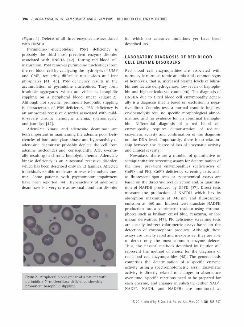
(Figure 1). Defects of all three enzymes are associated
with HNSHA.
Pyrimidine-50-nucleotidase (P5N) deficiency is
probably the third most prevalent enzyme disorder
associated with HNSHA [42]. During red blood cell
maturation, P5N removes pyrimidine nucleotides from
the red blood cell by catalyzing the hydrolysis of UMP
and CMP, rendering diffusible nucleosides and free
phosphates [41, 43]. P5N deficiency results in the
accumulation of pyrimidine nucleotides. They form
insoluble aggregates, which are visible as basophilic
stippling on a peripheral blood smear (Figure 2).
Although not specific, prominent basophilic stippling
is characteristic of P5N deficiency. P5N deficiency is
an autosomal recessive disorder associated with mild-
to-severe chronic hemolytic anemia, splenomegaly,
and jaundice [42].
Adenylate kinase and adenosine deaminase are
both important in maintaining the adenine pool. Defi-
ciencies of both adenylate kinase and hyperactivity of
adenosine deaminase probably deplete the cell from
adenine nucleotides and, consequently, ATP, eventu-
ally resulting in chronic hemolytic anemia. Adenylate
kinase deficiency is an autosomal recessive disorder,
which has been described only in 12 families. Affected
individuals exhibit moderate or severe hemolytic ane-
mia. Some patients with psychomotor impairment
have been reported [44]. Hyperactivity of adenosine
deaminase is a very rare autosomal dominant disorder
for which no causative mutations yet have been
described [45].
LABORATORY DIAGNOSIS OF RED BLOODCELL ENZYME DISORDERS
Red blood cell enzymopathies are associated with
normocytic normochromic anemia and common signs
of hemolysis, that is, increased plasma levels of biliru-
bin and lactate dehydrogenase, low levels of haptoglo-
bin and high reticulocyte count [46]. The diagnosis of
HNSHA due to a red blood cell enzymopathy gener-
ally is a diagnosis that is based on exclusion: a nega-
tive direct Coombs test, a normal osmotic fragility/
cryohemolysis test, no specific morphological abnor-
malities, and no evidence for an abnormal hemoglo-
bin. Differential diagnosis of a red blood cell
enzymopathy requires demonstration of reduced
enzymatic activity and confirmation of the diagnosis
on the DNA level. Importantly, there is no relation-
ship between the degree of loss of enzymatic activity
and clinical severity.
Nowadays, there are a number of quantitative or
semiquantitative screening assays for determination of
the most prevalent enzymopathies (deficiencies of
G6PD and PK). G6PD deficiency screening tests such
as fluorescent spot tests or cytochemical assays are
based on the direct/indirect detection and/or quantita-
tion of NAPDH produced by G6PD [37]. Direct tests
measure the production of NAPDH which has its
absorption maximum at 340 nm and fluorescence
emission at 460 nm. Indirect tests translate NADPH
production into a colorimetric readout using chromo-
phores such as brilliant cresyl blue, resazurin, or for-
mazan derivatives [47]. PK deficiency screening tests
are usually indirect colorimetric assays based on the
detection of chromophore products. Although these
assays are usually rapid and inexpensive, they are able
to detect only the most common enzyme defects.
Thus, the classical methods described by Beutler still
represent the method of choice for the diagnosis of
red blood cell enzymopathies [48]. The general basis
comprises the determination of a specific enzyme
activity using a spectrophotometric assay. Enzymatic
activity is directly related to changes in absorbance
over time. Specific reactions need to be prepared for
each enzyme, and changes in substrate (either NAD+,
NADP+, NADH, and NADPH) are monitored at
Figure 2. Peripheral blood smear of a patient with
pyrimidine-50-nucleotidase deficiency showing
prominent basophilic stippling.
© 2014 John Wiley & Sons Ltd, Int. Jnl. Lab. Hem. 2014, 36, 388–397
394 P. KORALKOVA, W. W. VAN SOLINGE AND R. VAN WIJK | RED BLOOD CELL ENZYMOPATHIES
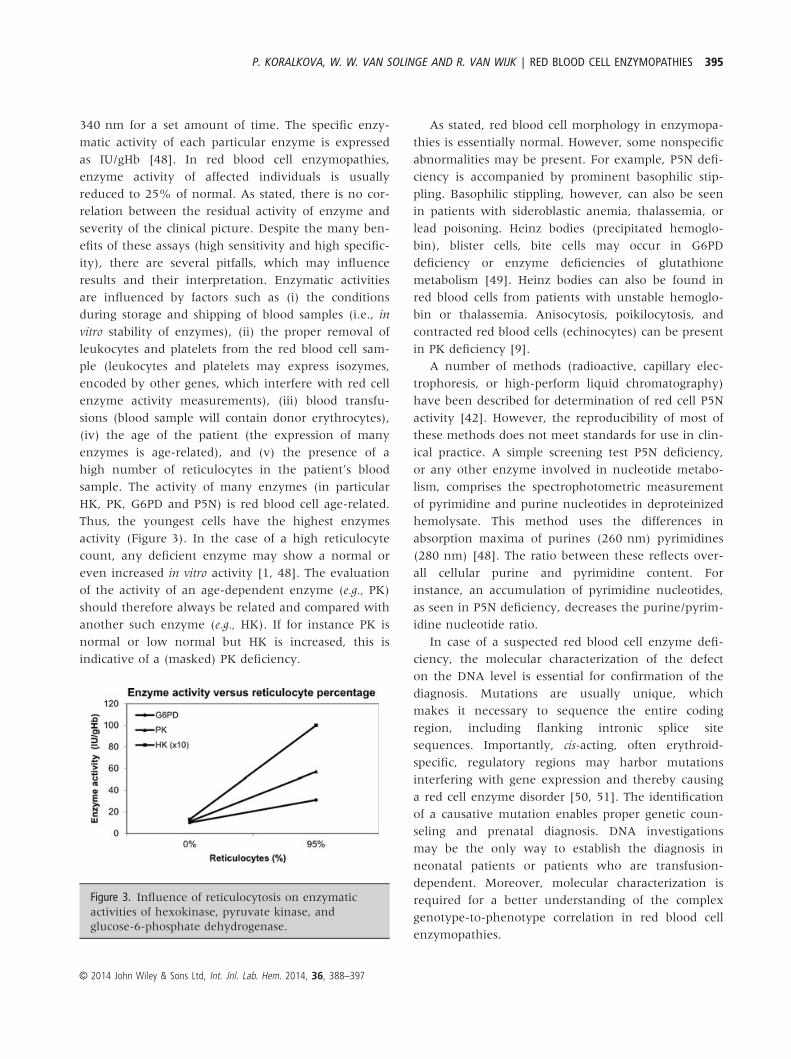
340 nm for a set amount of time. The specific enzy-
matic activity of each particular enzyme is expressed
as IU/gHb [48]. In red blood cell enzymopathies,
enzyme activity of affected individuals is usually
reduced to 25% of normal. As stated, there is no cor-
relation between the residual activity of enzyme and
severity of the clinical picture. Despite the many ben-
efits of these assays (high sensitivity and high specific-
ity), there are several pitfalls, which may influence
results and their interpretation. Enzymatic activities
are influenced by factors such as (i) the conditions
during storage and shipping of blood samples (i.e., in
vitro stability of enzymes), (ii) the proper removal of
leukocytes and platelets from the red blood cell sam-
ple (leukocytes and platelets may express isozymes,
encoded by other genes, which interfere with red cell
enzyme activity measurements), (iii) blood transfu-
sions (blood sample will contain donor erythrocytes),
(iv) the age of the patient (the expression of many
enzymes is age-related), and (v) the presence of a
high number of reticulocytes in the patient’s blood
sample. The activity of many enzymes (in particular
HK, PK, G6PD and P5N) is red blood cell age-related.
Thus, the youngest cells have the highest enzymes
activity (Figure 3). In the case of a high reticulocyte
count, any deficient enzyme may show a normal or
even increased in vitro activity [1, 48]. The evaluation
of the activity of an age-dependent enzyme (e.g., PK)
should therefore always be related and compared with
another such enzyme (e.g., HK). If for instance PK is
normal or low normal but HK is increased, this is
indicative of a (masked) PK deficiency.
As stated, red blood cell morphology in enzymopa-
thies is essentially normal. However, some nonspecific
abnormalities may be present. For example, P5N defi-
ciency is accompanied by prominent basophilic stip-
pling. Basophilic stippling, however, can also be seen
in patients with sideroblastic anemia, thalassemia, or
lead poisoning. Heinz bodies (precipitated hemoglo-
bin), blister cells, bite cells may occur in G6PD
deficiency or enzyme deficiencies of glutathione
metabolism [49]. Heinz bodies can also be found in
red blood cells from patients with unstable hemoglo-
bin or thalassemia. Anisocytosis, poikilocytosis, and
contracted red blood cells (echinocytes) can be present
in PK deficiency [9].
A number of methods (radioactive, capillary elec-
trophoresis, or high-perform liquid chromatography)
have been described for determination of red cell P5N
activity [42]. However, the reproducibility of most of
these methods does not meet standards for use in clin-
ical practice. A simple screening test P5N deficiency,
or any other enzyme involved in nucleotide metabo-
lism, comprises the spectrophotometric measurement
of pyrimidine and purine nucleotides in deproteinized
hemolysate. This method uses the differences in
absorption maxima of purines (260 nm) pyrimidines
(280 nm) [48]. The ratio between these reflects over-
all cellular purine and pyrimidine content. For
instance, an accumulation of pyrimidine nucleotides,
as seen in P5N deficiency, decreases the purine/pyrim-
idine nucleotide ratio.
In case of a suspected red blood cell enzyme defi-
ciency, the molecular characterization of the defect
on the DNA level is essential for confirmation of the
diagnosis. Mutations are usually unique, which
makes it necessary to sequence the entire coding
region, including flanking intronic splice site
sequences. Importantly, cis-acting, often erythroid-
specific, regulatory regions may harbor mutations
interfering with gene expression and thereby causing
a red cell enzyme disorder [50, 51]. The identification
of a causative mutation enables proper genetic coun-
seling and prenatal diagnosis. DNA investigations
may be the only way to establish the diagnosis in
neonatal patients or patients who are transfusion-
dependent. Moreover, molecular characterization is
required for a better understanding of the complex
genotype-to-phenotype correlation in red blood cell
enzymopathies.
Figure 3. Influence of reticulocytosis on enzymatic
activities of hexokinase, pyruvate kinase, and
glucose-6-phosphate dehydrogenase.
© 2014 John Wiley & Sons Ltd, Int. Jnl. Lab. Hem. 2014, 36, 388–397
P. KORALKOVA, W. W. VAN SOLINGE AND R. VAN WIJK | RED BLOOD CELL ENZYMOPATHIES 395
ACKNOWLEDGEMENTS
PK, WS, and RW wrote the paper. Pavla Koralkova
is supported by the Grants NT11208 (Ministry of
Health, Czech Republic) and LF_2013_010 (Internal
Grant Agency of Palacky University). Richard van
Wijk is supported by ENERCA, the European NEt-
work for Rare and Congenital Anaemias (EU Public
Health Executive Agency (DG-SANCO, PHEA
101227)).
REFERENCES
1. Arya R, Layton DM, Bellingham AJ. Hered-
itary red cell enzymopathies. Blood Rev
1995;9:165–75.
2. Jacobasch G, Rapoport SM. Hemolytic ane-
mias due to erythrocyte enzyme deficien-
cies. Mol Aspects Med 1996;17:143–70.
3. van Wijk R, van Solinge WW. Disorders of
red cells resulting from enzyme abnormali-
ties. In: Williams Hematology. Kaushansky
KJ, Lichtman MA, Beutler E, Kipps TJ,
Seligsohn U, Prchal JT. (eds). New York:
McGraw-Hill, 2010: 647–74.
4. Prchal JT, Gregg XT. Red cell enzymes.
Hematology Am Soc Hematol Educ Pro-
gram 2005;19–23. PMID:16304354.
5. Rapoport S, Luebering J. The formation of
2,3-diphosphoglycerate in rabbit erythro-
cytes: the existence of a diphosphoglycer-
ate mutase. J Biol Chem 1950;183:507–16.
6. van Zwieten R, Verhoeven AJ, Roos D.
Inborn defects in the antioxidant systems
of human red blood cells. Free Radic Biol
Med 2014;67:377–86.
7. Dudzinska W, Hlynczak AJ, Skotnicka E,
Suska M. The purine metabolism of human
erythrocytes. Biochemistry (Mosc) 2006;
71:467–75.
8. Valentine WN, Paglia DE. Erythrocyte dis-
orders of purine and pyrimidine metabo-
lism. Hemoglobin 1980;4:669–81.
9. Zanella A, Fermo E, Bianchi P, Valentini G.
Red cell pyruvate kinase deficiency: molec-
ular and clinical aspects. Br J Haematol
2005;130:11–25.
10. van Wijk R, van Solinge WW. The energy-
less red blood cell is lost: erythrocyte
enzyme abnormalities of glycolysis. Blood
2005;106:4034–42.
11. Beutler E, Gelbart T. Estimating the preva-
lence of pyruvate kinase deficiency from
the gene frequency in the general white
population. Blood 2000;95:3585–8.
12. Kanno H, Fujii H, Miwa S. Structural
analysis of human pyruvate kinase L-gene
and identification of the promoter activity
in erythroid cells. Biochem Biophys Res
Commun 1992;188:516–23.
13. Valentine WN, Paglia DE. The primary
cause of hemolysis in enzymopathies of
anaerobic glycolysis: a viewpoint. Blood
Cells 1980;6:819–29.
14. Oski FA, Marshall BE, Cohen PJ, Sugerman
HJ, Miller LD. The role of the left-shifted or
right-shifted oxygen-hemoglobin equilib-
rium curve. Ann Intern Med 1971;74:44–6.
15. Ferreira P, Morais L, Costa R, Resende C,
Dias CP, Ara�ujo F, Costa E, Barbot J, Vilar-
inho A. Hydrops fetalis associated with
erythrocyte pyruvate kinase deficiency. Eur
J Pediatr 2000;159:481–2.
16. Zanella A, Fermo E, Bianchi P, Chiarelli
LR, Valentini G. Pyruvate kinase defi-
ciency: the genotype-phenotype associa-
tion. Blood Rev 2007;21:217–31.
17. Zanella A, Berzuini A, Colombo MB, Guf-
fanti A, Lecchi L, Poli F, Cappellini MD,
Barosi G. Iron status in red cell pyruvate
kinase deficiency: study of Italian cases. Br
J Haematol 1993;83:485–90.
18. Lin H-Y, Kao Y-H, Chen S-T, Meng M.
Effects of inherited mutations on catalytic
activity and structural stability of human
glucose-6-phosphate isomerase expressed
in Escherichia coli. Biochim Biophys Acta
2009;1794:315–23.
19. Matthay KK, Mentzer WC. Erythrocyte en-
zymopathies in the newborn. Clin Haema-
tol 1981;10:31–55.
20. Schroter W, Eber SW, Bardosi A, Gahr M,
Gabriel M, Sitzmann FC. Generalised glu-
cosephosphate isomerase (GPI) deficiency
causing haemolytic anaemia, neuromuscu-
lar symptoms and impairment of granulo-
cytic function: a new syndrome due to a
new stable GPI variant with diminished
specific activity (GPI Homburg). Eur J Pedi-
atr 1985;144:301–5.
21. Nakajima H, Raben N, Hamaguchi T, Yama-
saki T. Phosphofructokinase deficiency;
past, present and future. Curr Mol Med
2002;2:197–212.
22. Ahmed N, Battah S, Karachalias N, Babaei-
Jadidi R, Hor�anyi M, Bar�oti K, Hollan S,
Thornalley PJ. Increased formation of
methylglyoxal and protein glycation, oxida-
tion and nitrosation in triosephosphate
isomerase deficiency. Biochim Biophys
Acta 2003;1639:121–32.
23. Orosz F, Ol�ah J, Ov�adi J. Triosephosphate
isomerase deficiency: new insights into an
enigmatic disease. Biochim Biophys Acta
2009;1792:1168–74.
24. Beutler E, Scott S, Bishop A, Margolis N,
Matsumoto F, Kuhl W. Red cell aldolase
deficiency and hemolytic anemia: a new
syndrome. Trans Assoc Am Physicians
1973;86:154–66.
25. Kreuder J, Borkhardt A, Repp R, Pekrun A,
G€ottsche B, Gottschalk U, Reichmann H,
Schachenmayr W, Schlegel K, Lampert F.
Brief report: inherited metabolic myopathy
and hemolysis due to a mutation in aldol-
ase A. N Engl J Med 1996;334:1100–4.
26. Yao DC, Tolan DR, Murray MF, Harris DJ,
Darras BT, Geva A, Neufeld EJ. Hemolytic
anemia and severe rhabdomyolysis caused
by compound heterozygous mutations of
the gene for erythrocyte/muscle isozyme of
aldolase, ALDOA(Arg303X/Cys338Tyr). Blood
2004;103:2401–3.
27. Esposito G, Vitagliano L, Costanzo P, Borr-
elli L, Barone R, Pavone L, Izzo P, Zagari A,
Salvatore F. Human aldolase A natural
mutants: relationship between flexibility of
the C-terminal region and enzyme func-
tion. Biochem J 2004;380:51–6.
28. Kanno H. Hexokinase: gene structure and
mutations. Baillieres Best Pract Res Clin
Haematol 2000;13:83–8.
29. Kanno H, Murakami K, Hariyama Y, Ishik-
awa K, Miwa S, Fujii H. Homozygous intra-
genic deletion of type I hexokinase gene
causes lethal hemolytic anemia of the
affected fetus. Blood 2002;100:1930.
30. Fermo E, Bianchi P, Chiarelli LR, Maggi M,
Mandar�a GM, Vercellati C, Marcello AP,
Barcellini W, Cortelezzi A, Valentini G,
Zanella A. A new variant of phosphoglycer-
ate kinase deficiency (p. I371K) with multi-
ple tissue involvement: molecular and
functional characterization. Mol Genet
Metab 2012;106:455–61.
31. Cohen-Solal M, Valentin C, Plassa F,
Guillemin G, Danze F, Jaisson F, Rosa R.
Identification of new mutations in two
phosphoglycerate kinase (PGK) variants
expressing different clinical syndromes:
PGK Creteil and PGK Amiens. Blood
1994;84:898–903.
32. Spiegel R, Gomez EA, Akman HO, Krishna
S, Horovitz Y, DiMauro S. Myopathic form
of phosphoglycerate kinase (PGK) defi-
© 2014 John Wiley & Sons Ltd, Int. Jnl. Lab. Hem. 2014, 36, 388–397
396 P. KORALKOVA, W. W. VAN SOLINGE AND R. VAN WIJK | RED BLOOD CELL ENZYMOPATHIES
ciency: a new case and pathogenic consid-
erations. Neuromuscul Disord 2009;19:207–
11.
33. Sotiriou E, Greene P, Krishna S, Hirano M,
DiMauro S. Myopathy and parkinsonism in
phosphoglycerate kinase deficiency. Muscle
Nerve 2010;41:707–10.
34. Beutler E. PGK deficiency. Br J Haematol
2007;136:3–11.
35. Beutler E. Red cell enzyme defects as non-
diseases and as diseases. Blood 1979;54:1–
7.
36. Nkhoma ET, Poole C, Vannappagari V, Hall
SA, Beutler E. The global prevalence of
glucose-6-phosphate dehydrogenase defi-
ciency: a systematic review and meta-
analysis. Blood Cells Mol Dis 2009;42:267–
78.
37. Cappellini MD, Fiorelli G. Glucose-6-phos-
phate dehydrogenase deficiency. Lancet
2008;371:64–74.
38. Luzzatto L, Seneca E. G6PD deficiency: a
classic example of pharmacogenetics with
on-going clinical implications. Br J Haema-
tol 2014;164:469–80.
39. Kamerbeek NM, van Zwieten R, de Boer
M, Morren G, Vuil H, Bannink N, Lincke
C, Dolman KM, Becker K, Schirmer RH,
Gromer S, Roos D. Molecular basis of gluta-
thione reductase deficiency in human
blood cells. Blood 2007;109:3560–6.
40. Ristoff E, Larsson A. Inborn errors in the
metabolism of glutathione. Orphanet J
Rare Dis 2007;2:16.
41. Valentine WN, Fink K, Paglia DE, Harris
SR, Adams WS. Hereditary hemolytic ane-
mia with human erythrocyte pyrimidine
50-nucleotidase deficiency. J Clin Invest
1974;54:866–79.
42. Zanella A, Bianchi P, Fermo E, Valentini G.
Hereditary pyrimidine 50-nucleotidase defi-
ciency: from genetics to clinical manifesta-
tions. Br J Haematol 2006;133:113–23.
43. Rees DC, Duley JA, Marinaki AM. Pyrimi-
dine 50 nucleotidase deficiency. Br J Hae-
matol 2003;120:375–83.
44. Abrusci P, Chiarelli LR, Galizzi A, Fermo
E, Bianchi P, Zanella A, Valentini G.
Erythrocyte adenylate kinase deficiency:
characterization of recombinant mutant
forms and relationship with nonspherocy-
tic hemolytic anemia. Exp Hematol
2007;35:1182–9.
45. Chen EH, Mitchell BS. Hereditary overex-
pression of adenosine deaminase in ery-
throcytes: studies in erythroid cell lines
and transgenic mice. Blood 1994;84:2346–
53.
46. Tefferi A. Anemia in adults: a contempo-
rary approach to diagnosis. Mayo Clin Proc
2003;78:1274–80.
47. Shah SS, Diakite SA, Traore K, Diakite M,
Kwiatkowski DP, Rockett KA, Wellems TE,
Fairhurst RM. A novel cytofluorometric
assay for the detection and quantification
of glucose-6-
phosphate dehydrogenase deficiency. Sci
Rep 2012;2:299.
48. Beutler E. Red Cell Metabolism. A Manual
of Biochemical Methods. Orlando: Grune &
Stratton; 1984: 188.
49. Bain BJ. Diagnosis from the blood smear. N
Engl J Med 2005;353:498–507.
50. van Wijk R, van Solinge WW, Nerlov C,
Beutler E, Gelbart T, Rijksen G, Nielsen FC.
Disruption of a novel regulatory element in
the erythroid-specific promoter of the
human PKLR gene causes severe pyruvate
kinase deficiency. Blood 2003;101:1596–
602.
51. de Vooght KMK, van Solinge WW, van
Wesel AC, Kersting S, van Wijk R. First
mutation in the red blood cell-specific pro-
moter of hexokinase combined with a
novel missense mutation causes hexokinase
deficiency and mild chronic hemolysis.
Haematologica 2009;94:1203–10.
© 2014 John Wiley & Sons Ltd, Int. Jnl. Lab. Hem. 2014, 36, 388–397
P. KORALKOVA, W. W. VAN SOLINGE AND R. VAN WIJK | RED BLOOD CELL ENZYMOPATHIES 397
APPENDICES 4.2
DIFFERENTIAL DIAGNOSIS OF CZECH AND SLOVAK PATIENTS WITH HEMOLYTIC ANEMIA OF UKNOWN ETIOLOGY
Deficit pyruvátkinázy v dětském věku
Ludíková B.1, Mojzíková R.2, Pospíšilová P.2, Houda J.1, Sulovská L.1, Divoká M.3,Hak J.4, Procházková D.5, Divoký V.2, Pospíšilová D.1
Dětská klinika Lékařské fakulty Univerzity Palackého a Fakultní nemocnice, Olomouc1
přednosta prof. MUDr. V. Mihál, CSc.Ústav biologie Lékařské fakulty Univerzity Palackého, Olomouc2
ředitel doc. RNDr. V. Divoký, Ph.D.Hemato-onkologická klinika Lékařské fakulty Univerzity Palackého a Fakultní nemocnice,Olomouc3
přednosta prof. MUDr. K. Indrák, DrSc.Dětská klinika Lékařské fakulty Univerzity Karlovy a Fakultní nemocnice, Hradec Králové4
přednosta prof. MUDr. M. Bayer, CSc.Dětské oddělení, Krajská zdravotní, a.s. – Masarykova nemocnice, Ústí nad Labem5
primář MUDr. J. Škvor, CSc.
Souhrn
Účel studie: Deficit pyruvátkinázy (PK) je druhý nejčastější enzymatický defekt vedoucí k hemolytickéanemii. Cílem práce je demonstrovat klinické a laboratorní nálezy u deficitu PK u prvních dětských pa-cientů v České republice definovaných na molekulární úrovni.
Pacienti a metody: Čtyři děti (10 měsíců – 7 let) byly vyšetřeny pro závažnou nesférocytární hemolytic-kou anemii s hladinou hemoglobinu 64–97 g/l. Všechny děti vyžadovaly po porodu fototerapii a transfuzi,dvě dokonce výměnnou transfuzi. V klinickém nálezu dominovala bledost, subikterus sklér a kůže. Stan-dardními metodami byla vyloučena porucha membrány erytrocytu, hemoglobinopatie a kongenitálnídyserytropoetická anemie. Byla stanovena aktivita PK a provedena sekvenace genu kódujícího PK. Prozvýšení hladin feritinu byla vyšetřena hladina hepcidinu.
Výsledky: Všechny děti mají makrocytární anemii. Hodnoty bilirubinu se pohybují v rozmezí38–89 µµmol/l. Aktivita PK je u všech dětí snížená (23–32 %). Dva pacienti jsou smíšení heterozygoti promutace c.1529C>A (p.Arg510Gln) a c.1594C>T (p.Arg532Trp) a jeden pro mutace c.1493G>A(p.Arg498His) a c.1529C>A (p.Arg510Gln). V jednom případě se jedná o homozygotní deleci v exonu11, která nebyla dosud popsána. Tento pacient s nejtěžším průběhem nemoci má vysokou hladinu fe-ritinu, avšak sníženou hladinu hepcidinu, což se může podílet na přetížení železem. Byla vyloučena ko-incidence s hemochromatózou typu I.
Závěr: Deficit PK může být u novorozence příčinou závažné anemie a hyperbilirubinemie vyžadující ně-kdy výměnnou transfuzi a opakované podání transfuzí v kojeneckém věku, jejichž frekvence se většinoupostupně snižuje. Deficit PK může být provázen i přetížením železem s nutností chelatační léčby. V česképopulaci je onemocnění zatím poddiagnostikováno.
Klíčová slova: glykolýza, pyruvátkináza, nesférocytární hemolytická anemie, přetížení železem, hepci-din
Summary
Pyruvate kinase deficiency in children
Purpose of the study: Pyruvate kinase deficiency (PK) is the second most common enzymatic defectleading to hemolytic anemia. The aim is to demonstrate the clinical and laboratory findings in PK defi -ciency in the first pediatric patients in the Czech Republic who were defined at the molecular level.
Patients and methods: Four children (10 months – 7 years) were examined for severe nonspherocytic he-molytic anemia with hemoglobin levels 64–97 g/l. All the children required transfusions and phototherapyafter delivery, two of them even an exchange transfusion. The clinical finding was dominantly characte-rized by pallor, subicterus of sclerae and skin. Erythrocyte membrane disorder, hemoglobinopathies andcongenital dyserythropoietic anemia were excluded using standard methods. PK activity was determi-ned and sequencing of the gene encoding PK was done. Because of increased ferritin levels, hepcidin le-vel was measured.
Results: All children have macrocytic anemia with bilirubin values from 38 to 89 micromol/l. PK activityis decreased in all children (23–32%). Two patients are mixed heterozygotes for mutations c.1529C>A(p.Arg510Gln) and c.1594C>T (p.Arg532Trp) and one for the c.1493G>A (p.Arg498His) and c.1529C>A
pů
vod
ní p
rác
e
152
PED_3.2012:Sestava 1 24.5.2012 7:52 Stránka 152

(p.Arg510Gln) mutations. In one case, a homozygous deletion in exon 11, which has not yet been descri-bed, was found. This patient with the most severe course of the disease has a high ferritin level, but a re-duced level of hepcidin, which may contribute to iron overload. Coincidence with type I hemochroma-tosis was excluded.
Conclusion: PK deficiency in the newborn may cause severe anemia and hyperbilirubinemia someti-mes requiring an exchange transfusion and repeated administration of transfusions in infancy; their fre-quency is usually gradually reduced. PK deficiency may also be associated with iron overload and theneed for chelation therapy. In the Czech population this disease is still underdiagnosed.
Key words: glycolysis, pyruvate kinase, nonspherocytic hemolytic anemia, iron overload, hepcidin
L.
Čes-slov Pediat 2012; 67 (3): 152–159 pů
vod
ní p
rác
e
153
ÚVOD
Deficit pyruvátkinázy (PK) je nejčastější enzyma-tickou abnormalitou glykolytické dráhy a spolus deficitem glukózo-6-fosfátdehydrogenázy (G6PD)nejčastější příčinou nesférocytární hemolytickéanemie [1, 2]. Poprvé byl popsán počátkem šedesá-tých let minulého století Valentinem a spolupra-covníky [3]. Jedná se o vrozené autozomálně rece-sivně dědičné metabolické onemocnění s prevalencí1:20 000 [2].
Pyruvátkináza hraje klíčovou roli v glykolytickédráze. Katalyzuje jeden ze dvou kroků při tvorběadenozintrifosfátu (ATP).
Vzhledem k tomu, že zralé červené krvinky ne-obsahují mitochondrie, je glykolýza jedinou dráhougenerující ATP, který je nezbytný pro další metabo-lické pochody, pro zpětnou regulaci glykolýzy,membránový transport a pro zachování flexibilitya integrity membrány erytrocytu. Mimo jiné zde do-chází ke generaci redukované formy nikotinamida-denindinukleotidu (NADH) nezbytného pro redukcimethemoglobinu na hemoglobin a 2,3 difosfoglyce-rátu (2,3-DPG), regulátoru afinity hemoglobinu kekyslíku. Deficit PK se tak projeví neefektivní glyko-lýzou vedoucí ke snížené životaschopnosti erytro-cytů a extravaskulární hemolýze.
PK je aktivní ve formě tetrameru. U savců bylypopsány čtyři různé izoenzymy PK (M1, M2, L, R),které jsou specifické pro jednotlivé tkáně [4], z nichžpouze R forma se vyskytuje výhradně v červenýchkrvinkách [5]. Jaterní (PK-L) a erytrocytární (PK-R)izoenzymy jsou kódovány genem PKLR, který se na-chází na chromozomu 1 (1q21) [4]. Kódující oblast jerozdělena do dvanácti exonů [6]. Exon 1 je speci-fický pro transkripci erytrocytární formy, exon 2 projaterní formu, zbývající exony pak jsou společné.Dva další izoenzymy PK-M1 a PK-M2 jsou kódoványgenem PKM nacházejícím se na 15. chromozomu.PK-M1 se vyskytuje převážně v srdci, mozku a kos-terním svalstvu, PK-M2 ve fetálních tkáních, leuko-cytech a trombocytech. Dosud bylo popsáno přes180 mutací PKLR genu, které jsou asociovány s de-fektem PK. Většinou se jedná o mutace měnící
smysl kodonu a způsobující jednoduchou aminoky-selinovou záměnu, která má za následek snížení en-zymové aktivity na různých úrovních [7]. Mezi nej-častější mutace patří: 1) c.1529G>A typická proUSA a severní a střední Evropu, 2) c.1456C>T častáv jižní Evropě a 3) c.1468C>T vyskytující se v Asii[2, 3, 7].
Klinické symptomy deficitu pyruvátkinázy se ob-jevují u jedinců s homozygotní mutací nebo u slo-žených heterozygotů. Heterozygoti jsou přenašečibez projevů onemocnění, výjimečně se u nich mo-hou projevit příznaky mírné hemolýzy. Průběh one-mocnění je velmi variabilní, od kompenzované ane-mie bez klinických projevů přes lehkou anemiis exacerbacemi hemolýzy při infekcích až po těžkouanemii s nutností opakovaného podávání transfuzí.Vždy je přítomna retikulocytóza. U novorozencůs deficitem PK se poměrně často objevuje významnáanemie a hyperbilirubinemie s rizikem rozvoje já-drového ikteru, která si v některých případechmůže vyžádat výměnnou transfuzi. Byly popsányi případy hydropsu plodu [8] a život ohrožující ane-mie u novorozence. U nemocných s chronickouanemií se mohou vyvinout žlučové kameny, a to jižv první dekádě života. U pacientů s deficitem PKbyly popsány i tranzientní aplastické krize způso-bené parvovirem B19. K vzácným komplikacím pa-tří chronické vředy na dolních končetinách, akutnípankreatitidy, abscesy slinivky, útlak míchy extra-medulární hematopoetickou tkání a tromboembo-lická onemocnění [9]. Hemolýza nebývá provoko-vána léky, někteří autoři však popisují zvýšeníhemolýzy po podání vyšších dávek salicylátů.
Podezření na enzymový deficit obecně podporujeobraz normocytární, méně často makrocytární ane-mie. Biochemické parametry, jako je zvýšení ne-konjugovaného bilirubinu a laktátdehydrogenázy(LD) a snížení hladiny haptoglobinu, odpovídajíznámkám extravaskulární hemolýzy. Při morfolo-gickém hodnocení erytrocytů v nátěru periferníkrve bývá přítomná polychromazie, anizocytóza,v některých případech ojedinělé normoblasty (ja-derné erytroidní buňky), vzácně echinocyty a akan-tocyty.
Vzhledem k absenci jednoznačného klinického
PED_3.2012:Sestava 1 24.5.2012 7:52 Stránka 153
154
Ludíková B. a spol. Čes-slov Pediat 2012; 67 (3): 152–159
Obě děti měly protrahovaný novorozenecký ikte-rus s nutností fototerapie, u chlapce bylo dokoncenutné provést výměnnou transfuzi. Makrocytárníanemie s výraznou retikulocytózou je u obou dětíprovázena mírnou hyperbilirubinemií a v prvnímroce života si u obou vyžádala opakované podánítransfuze erytrocytární masy. V klinickém nálezuu dětí dominuje bledost a subikterus sklér a kůže,mírná hepatosplenomegalie a systolický šelestv prekordiu bez průkazu vrozené srdeční vady přiechokardiografickém vyšetření.
Po prvním roce věku byly u dívky podáványtransfuze erytrocytární masy již méně často, větši-nou při poklesu hemoglobinu v souvislosti s infekcí.U dívky a obou rodičů nebyly překvapivě při scree-ningovém vyšetření aktivity G6PD a PK na jinémpracovišti prokázány patologické hodnoty. Rodičedětí jsou zdraví, netrpí žádným hematologickým animetabolickým onemocněním.
Pacient č. 3
Třetím nemocným je 11měsíční chlapec, kterýbyl po propuštění z novorozeneckého odděleníodeslán na hematologickou ambulanci pro su-spektní vrozenou hemolytickou anemii. Je z prv -ního těhotenství šestnáctileté matky. Má závažnouperinatální anamnézu. Byl narozen v termínu s po-rodní hmotností 3020 g, po porodu byl těžce hypo-xický a byla nutná intubace a napojení na umělouplicní ventilaci po dobu 6 dnů. Ihned po porodubyla zjištěna vysoká hladina bilirubinu (90 µmol/l),těžká anemie a trombocytopenie. Byla provedenavýměnná transfuze. V dalším průběhu byla nutnápo dobu 5 dní fototerapie a opakovaně byla podánatransfuze erytrocytární masy.
Při klinickém vyšetření v hematologické ambu-lanci byl nápadný mírný ikterus s šedým nádechemkůže dítěte, lehce ikterické skléry a výrazná hepa-tosplenomegalie. Dítě bylo přijato k hospitalizacik vyšetření pro anemii a pro podezření na vrozenoumetabolickou vadu. Otec nežije s rodinou a o jehozdravotním stavu nelze získat dostačující infor-mace.
Pacient č. 4
Poslední pacient je 7letý chlapec odeslaný k do-vyšetření anemie nejasného původu. Chlapec bylanemický již v novorozeneckém věku, po poroduměl časnou nekonjugovanou hyperbilirubinemii,bez sérologického konfliktu, která si vyžádalaosmi denní fototerapii. Vzhledem k postupné ane-mizaci v měsíci věku byla nutná další transfuzníléčba. Koncentrace hemoglobinu (Hb) se pohybo-vala kolem 60 g/l, hyperbilirubinemie (224 µmol/l)přetrvávala. K vyloučení poruchy membrány ery-trocytu byl proveden test kryohemolýzy s negativ-
i laboratorního diagnostického znaku, který by od-povídal deficitu PK, je vždy nutné nejprve vyloučitběžně se vyskytující vrozené hemolytické anemie,zejména hereditární sférocytózu, případně nesta-bilní hemoglobinopatie. K odlišení sférocytózy jemožné použít test kryohemolýzy a test s eosin-5--maleimidem, jejichž výsledky jsou u enzymovéhodefektu normální. K vyloučení hemoglobinopatií jevhodné provést elektroforézu hemoglobinu, isopro-panolový test či test tepelné stability erytrocytů,které jsou u enzymopatií negativní. Výskyt Heinzo-vých tělísek může odpovídat nestabilní hemo -globinopatii nebo souviset s enzymopatiemi v me-tabolismu glutathionu (G6PD aj.). Pomocnýmvyšetřením může být test autohemolýzy, který jeu většiny pacientů patologický a v případě deficituPK se upravuje pouze po přidání ATP, nikoli všakglukózy. K přesné diagnóze deficitu PK však stejnětak jako ostatních erytrocytárních enzymopatiíslouží průkaz snížené kinetické aktivity enzymu.Diagnózu definitivně potvrzuje molekulárně gene-tické vyšetření na DNA/RNA úrovni prokazujícíkauzální mutaci genu PKLR.
Terapie onemocnění je symptomatická, kauzálníléčba zatím neexistuje. Někteří nemocní jsou závislína transfuzní léčbě. Splenektomie je vyhrazena pronejtěžší případy onemocnění s těžkou anemií,u kterých může vést k omezení nutnosti transfuzníléčby a ke stabilizaci hodnot hemoglobinu. U částipacientů s deficitem PK se rozvíjí přetížení orga-nismu železem, které si může vyžádat léčbu chelá-tory. Byla popsána i úspěšná transplantace kostnídřeně u malého dítěte [10]. Je zkoumána možnostgenové léčby.
V české populaci bylo popsáno jen několik pří-padů dospělých pacientů s deficitem PK s prokáza-nou mutací genu pro PK [11]. V tomto článku pre-zentujeme klinický a laboratorní profil prvníchdětských pacientů s průkazem kauzální mutacepro PK.
PACIENTI
Soubor pacientů tvoří čtyři děti ve věku 10 mě-síců – 7 let s diagnózou chronické hemolytické ane-mie nejasné etiologie. U všech pacientů byla při zá-kladním vyšetření krevního obrazu a markerůhemolýzy zjištěna různě závažná anemie s projevyextravaskulární hemolýzy.
Pacienti č. 1 a 2
První dva pacienti jsou sourozenci, děvče a chla-pec ve věku 4 roky a 10 měsíců. Obě děti se naro-dily v termínu, dívka byla hypotrofická s porodníhmotností 2100 g a porodní délkou 44 cm, chlapecse narodil s hmotností 3130 g a délkou 51 cm.
PED_3.2012:Sestava 1 24.5.2012 7:52 Stránka 154
MCV: stfiední objem erytrocytÛMCH: stfiední koncentrace hemoglobinuLD: laktátdehydrogenázaU v‰ech pacientÛ je pfiítomna kolísavá makrocytóza a v˘razná retikulocytóza, mírná hyperbilirubinemie a v˘razné zv˘‰eníLD. U pacientÛ ã. 2 a 3 je v˘raznû zv˘‰ená hladina feritinu.
155
Ludíková B. a spol. Čes-slov Pediat 2012; 67 (3): 152–159
ním nálezem. Byl vyšetřen test stability Hb, fetálníhemoglobin (HbF) alkalickou denaturací a autohe-molýza, vše s negativním výsledkem.
Pacient v roce věku podstoupil punkci kostnídřeně, při které byla zjištěna aktivovaná erytro-poeza bez dalších specifických patologických ná-lezů. V dalším sledování měl středně těžkou ma-krocytární anemii s hladinou Hb kolem 100 g/l,retikulocytózu a lehkou nekonjugovanou hyperbili-rubinemii (30 µmol/l). Při screeningu enzymopatiína jiném pracovišti rovněž nebylo potvrzeno sníženíaktivity PK nebo G6PD. Matka 7letého chlapcedříve užívala preparáty železa pro sideropenickouanemii.
METODY
U všech dětí bylo provedeno opakované stan-dardní vyšetření krevního obrazu včetně nátěru pe-riferní krve a biochemické vyšetření markerů he-molýzy a metabolismu železa. Test kryohemolýzy,elektroforéza hemoglobinů, průkaz Heinzových tělí-sek a test tepelné stability byly provedeny podlestandardních postupů.
Stanovení aktivity pyruvátkinázy a G6PD
Aktivita G6PD byla stanovena spektrofotome-tricky pomocí komerčního kitu (The Microplate Ne-onatal G6PD Screening Assay, Bio-Rad Laborato-ries, UK).
Aktivita PK byla stanovena následovně: Pro pří-pravu enzymových lyzátů byla použita periferníkrev (heparin) s následným odstraněním leukocytůa destiček filtrací přes směs mikrokrystalické celu-lózy s α-celulózou. Po odstranění plazmy centrifu-gací byla připravena buněčná suspenze ve fyziolo-gickém roztoku (1:1). Výsledný enzymový lyzát bylpřipraven přidáním buněčné suspenze do stabili-začního roztoku obsahujícího EDTA a merkapto -ethanol (1:9).
Aktivita pyruvátkinázy (E.C.2.7.1.40) byla stano-
vena spektrofotometricky v temperované směsi(37 °C) obsahující laktátdehydrogenázu, ADP, fos-foenolpyruvát a NADH sledováním poklesu absor-bance při 340 nm (Infinite 200 NanoQuant, Tecan,Švýcarsko). Specifická aktivita (U/g), která se uvádív literatuře, byla vztažena k množství hemoglobinuv lyzátu stanoveném rovněž spektrofotometricky při414 nm [12].
Sekvenace genů PKLR a HFE1
Genomická DNA byla izolována z periferní krvepomocí QIAamp DNA kitu (Qiagen, USA). Všechnyexony byly amplifikovány pomocí PCR (PCR primerya podmínky jsou k dispozici na vyžádání). Pro se-kvenační reakci odpovídajících PCR fragmentů bylpoužit BigDye terminator kit v 1.1 (Applied Biosys-tem, USA) a sekvenační analýzy byly provedeny nasekvenátoru ABI Prism 3100 (Applied Biosystems,USA).
Stanovení hladiny hepcidinu
Hladiny hepcidinu (bioaktivní forma hepcidinu--25) byly stanoveny v plazmě. Část plazmy určenák měření hladiny hepcidinu byla rozdělena do ali-kvotů, aby se předešlo opakovanému rozmrazování,a zamražena při -80 °C. K měření hladin hepcidinubyl použit komerční ELISA kit (DRG Instruments,Německo).
Parametry metody podle výrobce byly následu-jící: dynamický rozsah 0,9–140 ng/ml, senzitivitatéto metody byla stanovena na 0,9 ng/ml, repro-dukovatelnost – intra assay CV 4,86 %, inter assaytest CV 11,42 %.
VÝSLEDKY
U 3 dětí (pacienti 1, 2 a 4) se jednalo o makrocy-tární, v jednom případě o přechodně normocytárníanemii (výsledek zkreslen předchozí aplikací ery-trocytární masy – pacient č. 3). Hladina Hb se po-
Tab. 1. Vy‰etfiení krevního obrazu a biochemick˘ch nálezÛ u pacientÛ.
Vûk/ Hemo- Erytrocyty MCV MCH Retiku- Bilirubin LD FeritinPacient ã. pohlaví globin locyty
(mûsíce) (g/l) (109/l) (fl) (pg) (µµmol/l) (µµkat/l) (µµg/l)1 50/Î 71 2,20 102,3 32,3 0,124 54 15,40 19
2 10/M 64 2,46 92,6 31,5 0,190 81 13,16 716
3 11/M 77 2,76 90,5 27,9 0,101 89 3,75 4853
4 86/M 97 3,12 88,8 31,1 0,081 38 5,58 205
PED_3.2012:Sestava 1 24.5.2012 7:52 Stránka 155

hybovala v rozmezí 62 až100 g/l a retikulocyty bylyv rozmezí 10–19 %. Při bio-chemickém vyšetření bylau všech pacientů výrazně zvý-šená hladina LD a jen lehcevyšší hladina nekonjugova-ného bilirubinu (tab. 1). U pa-cientů č. 2 a 3 byla prokázánavýrazně zvýšená hladina feri-tinu: 716 µg/l a 4853 µg/l.
V nátěru periferní krve bylyu všech pacientů patrné ne-
156
Ludíková B. a spol. Čes-slov Pediat 2012; 67 (3): 152–159
specifické změny morfologie erytrocytů: polychro-mazie, anizocytóza a u pacientů č. 2 a 3 rovněž ter-čovité erytrocyty a stomatocyty. U pacienta č. 3 bylynavíc přítomny echinocyty a ojedinělé normoblasty.
Porucha membrány erytrocytů a nestabilní he-moglobinopatie byly u všech dětí vyloučeny testemkryohemolýzy, elektroforézou hemoglobinů, průka-zem Heinzových tělísek a testem tepelné stability,které byly ve všech případech negativní.
Vyšetření kostní dřeně u pacientů č. 1, 3 a 4 pro-kázalo výraznou akcentaci prekurzorových buněkerytropoezy (40 až 74,1 %) se známkami mírné dy-splazie, avšak bez nálezu zvýšeného procenta více-jaderných erytroblastů a prstenčitých sideroblastů.Byla tedy vyloučena kongenitální dyserytropoetickáanemie i sideroblastická anemie.
Obr. 1. Schéma glykolytické dráhy.PGK: fosfoglycerátkináza, G3PD: glyceraldehyd-3-fosfát -dehydrogenáza, TPI: triózofosfátizomeráza, DPGM: difos-foglycerátmutázaGlykol˘za je metabolická dráha pfiemûny glukózy na dvû mo-lekuly pyruvátu za ãistého v˘tûÏku dvou molekul ATP a dvoumolekul NADH. Probíhá v cytosolu bunûk. Skládá se z de-seti krokÛ, kaÏd˘ z nich katalyzuje jin˘ enzym. Posledním ne-vratn˘m krokem je pfiemûna fosfoenolpyruvátu na pyruvátza vzniku ATP. Tato pfiemûna je katalyzovaná pyruvátkiná-zou. Pfienosem vodíku z NADH na poslední produkt glykol˘zy– pyruvát dochází k redukci pyruvátu na laktát.
Fig. 1. Diagram of glycolytic pathway.PGK: phosphoglycerate kinase, G3PD: glyceraldehyde-3-phosphate dehydrogenase, TPI: triosaphosphateisome-rase, DPGM: diphosphogylcerate mutaseGlycolysis is the metabolic pathway of conversion of glucoseinto two molecules of pyruvate, a pure yield of two molecu-les of ATP and two molecules of NADH. It takes place in thecytosol of cells. It consists of ten steps, each is catalysinganother enzyme. The last step is the irreversible conversionof phosphoenolpyruvate to pyruvate to produce ATP. Thistransformation is catalyzed by py-ruvatekinase. The hydrogen trans -fer from NADH to the last productof glycolysis – pyruvate is causinga reduction of pyruvate to lactate.
Obr. 2. Nátûr periferní krve pacienta ã. 3 – anizocytóza,polychromazie (a), ojedinûlé stomatocyty (b) a terãovitéerytrocyty (c).Fig. 2. The No. 3 patient’s blood smear – anisocytosis,polychromasia (a), isolated stomatocytes (b) and targeterythrocytes (c).
Obr. 3. Detail nátûru periferní krve pa-cienta ã. 3 s nálezem jaderné erytroid -ní buÀky.Fig. 3. Detail of the patient’s peripheralblood smear No. 3 with finding of nuc-leated erythroid cells.
Obr. 4. Detail nátûru periferní krve pa-cienta ã. 2 s ojedinûl˘mi echinocyty.Fig. 4. Detail of the patient’s peripheralblood smear No. 2 with isolated echi-nocytes.
PED_3.2012:Sestava 1 24.5.2012 7:52 Stránka 156

G6PD – glukózo-6-fosfátdehydrogenáza; PK – pyruvátkináza; aktivity G6PD a PK a koncentrace hepcidinu byly stanovenyv tripletech, normální hladiny: 13,3–54,4; rozmezí kontrolních hodnot aktivit G6PD a PK bylo stanoveno u 10 zdrav˘chkontrol
157
Ludíková B. a spol. Čes-slov Pediat 2012; 67 (3): 152–159
U všech nemocných byla stanovena aktivita py-ruvátkinázy, která byla ve všech případech sníženáa odpovídala přibližně 30 % hodnoty kontrolníchaktivit. Sekvenováním genu pro PK byla u všech4 pacientů prokázána kauzální mutace. Oba sou-rozenci (pacienti č. 1 a 2) jsou smíšenými heterozy-goty pro známé mutace c.1529C>A (p.Arg510Gln)a c.1594C>T (p.Arg532Trp) nacházející se v exonu11. U pacienta č. 3 se jedná o dosud nepopsanouhomozygotní aminokyselinovou deleci v exonu 11,která vede k posunu čtecího rámce a tvorbě pro-teinu, kterému chybí část aktivační domény. Tatomutace nebyla doposud popsána a její funkční stu-die dále probíhají. Pacient č. 4 je také smíšený he-terozygot pro dvě již popsané mutace, a to mutacic. 1493G>A (p.Arg498His) a opět c.1529C>A(p.Arg510Gln).
Vzhledem ke zvýšení hladiny feritinu bylau všech pacientů vyšetřena hladina hepcidinu,která byla u pacienta č. 3 snížená (11,3 ng/ml),u pacienta č. 2 na dolní hranici normy. U pacientůč. 1 a 4 byla hodnota hepcidinu normální.
Vzhledem k neobvykle vysoké hladině feritinubylo u pacienta č. 3 provedeno i molekulárně gene-tické vyšetření k vyloučení současně přítomné mu-tace pro hemochromatózu typu 1 (HFE), aby se vy-loučila nebo potvrdila možná koincidence deficituPK s některou z dalších poruch metabolismu železa– vrozenou hemochromatózou. Nejčastější mutacespojené s HFE1 – c.845C>A (p.Cys 282Tyr),c.187C>G (p.His63Asp), c.193A>T (p.Ser65Cys) –však nebyly sekvenační analýzou prokázány.
DISKUSE
Na popsaném souboru pacientů poukazujeme narůznorodost klinických projevů při deficitu PK. Za-jímavé je, že mutace c.1529C>A, kterou jsme dete-kovali u 3 pacientů, je svým nálezem sice typickápro střední a severní Evropu, ale ve slovanské po-pulaci, pro kterou je charakteristický nález mutacec.1594C>T, se objevuje jen zřídka [11].
I přesto, že není známa přímá korelace mezi ge-
notypem a fenotypem u různých mutací, dá sepředpokládat, že mutace měnící čtecí rámec, pro-motorovou oblast nebo větší delece, které vedouk výrazné redukci aktivity PK, mohou vést k závaž -nému fenotypu, jako je intrauterinní smrt plodunebo život ohrožující anemie u novorozence. Na zá-kladě studie italských autorů [7], která se zabývávztahem mezi závažností anemie a konkrétnímimutacemi, se dá předpokládat, že některé homozy-gotní mutace, které mohou být i důsledkem kon-sanguinity, odpovídají lehkému, střednímu nebotěžkému stupni anemie. U pacientů s mutacíc.1456C>T a c.1594C>T jde tak často o mírnouanemii (více než 100 g/l Hb). Mutace c.1529C>A byměla podle uvedených autorů vést ke středně těžkéanemii (Hb 80–100 g/l, 10 a méně transfuzí) [3, 7].U smíšených heterozygotů se dá fenotyp odhadnoutjen obtížně, což potvrzují i klinické projevy u obousourozenců (pacienti č. 1 a 2). I přesto, že oba jsousmíšenými heterozygoty pro mutace c.1529C>Aa c.1594C>T, které jsou zmiňované spíše s mírněj-šími projevy anemie, oba sourozenci mají těžkouformu anemie s nutností opakovaných transfuzí.
Na patogenezi rozvoje přetížení železem u pa cien -tů s deficitem PK se mohou podílet opakované trans-fuze, hemolýza, ale i neefektivní erytropoeza. K pře-tížení železem může přispívat i koincidence deficituPK s vrozenou poruchou metabolismu železa. Až20 % evropské populace má mírnou alteraci v meta-bolismu železa spojovanou s heterozygotním přena-šečstvím mutací c.187C>G (p.His63Asp) a c.845C>A(p.Cys282Tyr) v HFE genu způsobujících vrozenouhemochromatózu. Poslední studie prokázaly abnor-mality v HFE genu u 35 % pacientů s PK deficitem,kteří nejsou závislí na transfuzích [13]. Tyto abnor-mality jsou spojeny se zvýšením hladiny železa (Fe)v séru a feritinu [14]. Přetížení železem i bez mutacegenu pro hemochromatózu bylo popsáno u poly-transfundovaných pacientů a pa cien tů s PK defici-tem po splenektomii [14].
Diskutuje se i o možné roli snížení hladiny hep-cidinu, klíčové molekuly regulující přísun železa doorganismu [15]. Hladina hepcidinu je kontrolovánazejména erytropoetickou aktivitou prostřednictvím
Tab. 2. V˘sledky biochemick˘ch a molekulárnû genetick˘ch anal˘z.
Aktivita G6PD Aktivita PK Mutace Erytropoezev kostní dfieni
Hepcidin
(U/g Hb) (U/g Hb) (%) (ng/ml)
Pacient ã. 1 8,48 1,43 c.1529C>A/c.1594C>T 74,1 54,3
Pacient ã. 2 8,13 1,14 c.1529C>A/ c.1594C>T - 53,2
Pacient ã. 3 5,83 1,63 homozygotní delece 73,2 11,8
Pacient ã. 4 5,77 1,73 c.1493G>A/c.1529C>A 40 24,5
Kontroly 5,0–9,93 4,6–6,28 -
PED_3.2012:Sestava 1 24.5.2012 7:52 Stránka 157

158
Ludíková B. a spol. Čes-slov Pediat 2012; 67 (3): 152–159
stanovená aktivita PK zkreslená, zejména pokud sestanovuje v odběru bezprostředně po transfuzi nebov krátkém intervalu od transfuze, protože se tak měřívlastně i aktivita PK dárcovských erytrocytů. Je protodůležité, aby se v těchto případech aktivita PK sta-novovala až ve vzorku odebraném těsně před dalšítransfuzí, kdy už prakticky nehrozí „kontaminace“dárcovskými erytrocyty. Dalším faktorem zkreslují-cím vyšetření může být přítomnost leukocytů a trom-bocytů ve vyšetřovaném materiálu, protože jak leu-kocyty, tak trombocyty obsahují jinou izoformuenzymu (PK-M2), která má až 300krát vyšší aktivitunež erytrocytární forma PK-R [7]. Vzhledem k tomu,že aktivita řady enzymů souvisí se stářím populacečervených krvinek, může mít i zvýšený počet retiku-locytů za následek zvýšení aktivity některých en-zymů, zejména hexokinázy a pyruvátkinázy.
ZÁVĚR
Cílem tohoto sdělení, které popisuje souborprvních dětských pacientů s průkazem kauzálnímutace genu PKLR, je upozornit na variabilitu kli-nických i laboratorních příznaků u pacientů s defi-citem PK, která může být vzácně provázena zá važ -ným přetížením železem, a na úskalí diagnostikytéto enzymopatie.
U pacientů s deficitem PK a s rozvojem záva-žného přetížení železem je vždy nutno zvážit mož-nost výskytu přidružené vrozené hemochromatózy.Je třeba pečlivě monitorovat stav zásob železaa v indikovaných případech včas zahájit chelatačníléčbu jako prevenci rozvoje cirhózy jater při nelé-čené hemosideróze jater [14].
Práce byla podporována grantem NS9951 [IGAMZ ČR], NT11208 [IGA MZ ČR] a Studentskýmiprojekty LF-2011-006 a LF-2011-011 UniverzityPalackého v Olomouci.
signálních molekul GDF15 a TWSG1, které reagujína anemii. Stav erytropoezy je pravděpodobně nad-řazeným faktorem všem ostatním mechanismůmovlivňujícím metabolismus Fe a hladinu železa v or-ganismu. Hladina hepcidinu se snižuje u anemií,především u stavů se zvýšenou potřebou Fe, jakoje sideropenická anemie, což vede ke zvýšenémuvstřebávání železa ze střeva. Zvyšuje se naopaku stavů s vysokou hladinou železa v organismu,např. u vrozených hemochromatóz nebo chorobspojených se sekundárním přetížením železema nezávisle u akutního zánětu. Bylo prokázáno, žeu deficitu PK se kombinují oba faktory regulujícíprotichůdně produkci hepcidinu: na jedné straněanemie s vyššími nároky na přísun železa při vět-ším obratu erytropoezy a na straně druhé zvýšenánabídka železa s vysokou hladinou feritinu. Těžkáanemie je v tomto případě nadřazena signálůmo přetížení organismu železem a produkce hepci-dinu je negativně regulována cestou aktivace GDF15. Výsledkem je potom snížení jeho hladiny s ná-sledným zvýšením resorpce Fe i přes jeho zvýšenézásoby, což může přispívat ke zvyšování zásobníhoželeza a k rozvoji přetížení železem. U pacienta č. 3jsme také prokázali snížení hladiny hepcidinu,která se mohla spolupodílet na neobvykle vysokéhladině feritinu. U pacienta č. 2 byla hladina hep-cidinu na dolní hranici normy i při vysoké hladiněferitinu. Při přetrvávající výrazné hyperferitinemiibude potřeba u pacienta č. 3 zvážit sekvenováníHFE2A genu kódujícího hemojuvelin a HFE3 genukódujícího transferinový receptor 2 [16].
I když je deficit pyruvátkinázy nejčastější enzyma-tickou abnormalitou glykolytické dráhy, která způ-sobuje dědičnou nesférocytární hemolytickou ane-mii, bylo v České republice u dětí zatím popsánopouze několik případů. A to i přesto, že podle celo-světové prevalence 1:20 000 [2, 7] by v České repub-lice mělo být teoreticky diagnostikováno 500 osobtrpících touto nemocí. Jednou z příčin může být ur-čení nesprávné diagnózy, a to z několika mož nýchdůvodů. U polytransfundovaných pacientů může být
LITERATURA
1. Beutler E, Gelbart T. Estimationthe prevalence of pyruvate kinase de-ficiency from the gene frequency inthe general white population. Blood2000; 95: 3585–3588.2. Zanella A, Fermo E, Bianchi P, etal. Red cell pyruvate kinase defici-ency: molecular and clinical aspects.Br J Hematol 2005; 130: 11–25.3. Valentine WN,Tanaka KR, MiwaS. A specific erythrocyte gylcolyticenzyme defekt [pyruvate kinase] in
free subjects with congenital non-spherocytic hemolytic anemia. Tran-sactions of the Association of Ameri-can Physicians 1961; 74: 100–110.4. Satoh H, Tani K, Yoshida MC, etal. The human liver-type pyruvate ki-nase [PKL] gene is on chromosome 1at band q21. Cytogenet Cell Genet1988; 47: 132–133.5. Takegawa S, Fuji H, MiwaS. Change of pyruvate kinase isozy-mes from M2- to L-type during deve-
lopment of the red cell. Br J Hematol1983; 54: 467–474.6. Tani K, Yoshida MC, Satoh H, etal. Human M2-type pyruvate kinase:cDNA cloning, chromosomal assign-ment and expression in hepatoma.Gene 1988; 20: 509–516.7. Zanella A, Fermo E, Bianchi P, etal. Pyruvate kinase deficiency: Thegenotype-phenotype association.Blood Rev 2007; 21: 217–231.8. Ferreira P, Morais L, Costa R, et
PED_3.2012:Sestava 1 24.5.2012 7:52 Stránka 158
159
Ludíková B. a spol. Čes-slov Pediat 2012; 67 (3): 152–159
al. Hydrops fetalis associated witherythrocyte pyruvate kinase defici-ency. Eur J Pediatr 2000; 159: 481–482.9. Tanaka KR, Zerez CR. Red cellenzymopathies of the glycolytic path -way. Semin Haematol 1990; 27:165–185.10. Tanphaichitr VS, Suvatte V, Is-saragrisil S, et al. Successful bonemarrow transplantation in a childwith red blood cell pyruvate kinasedeficiency. Bone Marrow Transplant2000; 26: 689–690.11. Lenzner C, Nurnberg P, Thiele
BJ, et al. Mutations in the pyruvatekinase L gene in patients with here-ditary hemolytic anemia. Blood1994; 83: 2817–2822.12. Beutler E, Blume KG, KaplanJC, et al. International committeefor standardization in haematology:recommended methods for red-cellenzyme analysis. Br J Haematol1977; 35: 331–340.13. Zanella A, Bianachi P, Iurlo A,et al. Iron status and HFE genotypein erytrocyte pyruvate kinase defici-ency: Study of Italian cases. BloodCells Mol Dis 2001; 27: 653–661.
14. Hilgard P, Gerken G. Liver cir-rhosis as consequence of iron overloadcaused by hereditary nonspherocytichemolytic anemia. World J Gastroen-terol 2005; 11 (8): 1241–1244.15. Viatte L, Vaulont S. Hepcidin,the iron watcher. Biochimie 2009;91: 1223–1228.16. Finkenstedt A, Bianchi P,Theurl I, et al. Regulation of ironmetabolism through GDF15 andhepcidin in pyruvate kinase defici-ency. Br J Haematol 2009; 144: 789–793.
Došlo: 30. 11. 2011Přijato: 30. 3. 2012
MUDr. Barbora LudíkováNa Zákopě 15
772 00 Olomouce-mail: [email protected]
Nové knihy z Edice Lékař a pacient
LÉČBA DIABETU INZULINOVOU PUMPOU U DĚTÍ KROK ZA KROKEM– nejen pro rodiče a edukační sestry
MUDr. David Neumann, Ph.D.
Edice Lékař a pacientISBN: 978-80-204-2480-8Cena: doporučená cena 250 KčFormát: A5, 140 x 200 mm, 140 stran, barevně,
vazba brožovaná
Rodiče dětí s�diabetem, stejně jako i edukační sestry, se setkávají s řadou si-tuací, které musí správně vyřešit, aby předešli akutnímu nebo dlouhodobémuzhoršení nemoci. Právě jim je určena naše knížka – tedy především těm, kteří se k léčbě inzuli-novou pumpou u dětí chystají anebo právě začali. Měla by jim být každodennímpomocníkem při zvládání nejrůznějších problémů tak, aby byl dítěti umožněn conejbohatší výběr činností a cukrovka nezasahovala do jeho psychického, do-vednostního a sociálního vývoje. Krok za krokem jsou probrány jednotlivé fáze a možné situace, počínaje úvod-
ním nastavením přes zvládnutí glykemií po jídlech a v�noci až po období „experimentů“, kdy dítě a rodič zjišťují, covšechno je s „pumpičkou“ možné. Důraz klade autor na pochopení bezpečnosti léčby a nástrah, které nese. Podrobněproto čtenáře seznamuje mj. se specialitami moderních pump, jak pomáhají, ve�kterých situacích jsou prospěšné, kdyne a jaké jsou alternativní možnosti léčby.Text je bohatě dokumentován dětskými obrázky, pomocí nichž může rodič či sestra lépe objasnit dítěti danou proble-matiku.
Objednávky zasílejte e-mailem nebo poštou: Nakladatelské a tiskové středisko ČLS JEP, Sokolská 31, 120 26Praha 2, fax: 224 266 226, e-mail: [email protected]. Na objednávce laskavě uveďte i jméno časopisu, v němž jste seo knize dozvěděli.
PED_3.2012:Sestava 1 24.5.2012 7:52 Stránka 159

Copyright of Czecho-Slovak Pediatrics / Cesko-Slovenska Pediatrie is the property of Nakladatelske Stredisko
C L S J.E. Purkyne and its content may not be copied or emailed to multiple sites or posted to a listserv without
the copyright holder's express written permission. However, users may print, download, or email articles for
individual use.

Iron status in patients with pyruvate kinase deficiency:neonatal hyperferritinaemia associated with a novelframeshift deletion in the PKLR gene (p.Arg518fs), and lowhepcidin to ferritin ratios
Renata Mojzikova,1* Pavla Koralkova,1*
Dusan Holub,2 Zuzana Zidova,1 Dagmar
Pospisilova,3 Jaroslav Cermak,4 Zuzana
Striezencova Laluhova,5 Karel Indrak,6
Martina Sukova,7 Martina Partschova,1
Jana Kucerova,1 Monika Horvathova1
and Vladimir Divoky1,6
1Department of Biology, Faculty of Medicine and
Dentistry, Palacky University, 2Institute of
Molecular and Translational Medicine, Faculty
of Medicine and Dentistry, Palacky University,3Department of Paediatrics, University Hospital
and Faculty of Medicine and Dentistry, Palacky
University, Olomouc, 4Institute of Haematology
and Blood Transfusion, Prague, Czech Republic,5Children’s Faculty Hospital with Policlinic, Bra-
tislava, Slovak Republic, 6Department of Hae-
mato-Oncology, University Hospital and Faculty
of Medicine and Dentistry, Palacky University,
Olomouc, and 7Department of Paediatric Hae-
matology and Oncology, University Hospital Mo-
tol, Prague, Czech Republic
Received 25 October 2013; accepted for
publication 18 December 2013
Correspondence: Monika Horvathova
Department of Biology, Faculty of Medicine
and Dentistry
Palacky University, Hnevotinska 3, 77515
Olomouc, Czech Republic.
E-mail: [email protected]
*These authors contributed equally to this
work.
Summary
Pyruvate kinase (PK) deficiency is an iron-loading anaemia characterized
by chronic haemolysis, ineffective erythropoiesis and a requirement for
blood transfusion in most cases. We studied 11 patients from 10 unrelated
families and found nine different disease-causing PKLR mutations. Two of
these mutations - the point mutation c.878A>T (p.Asp293Val) and the
frameshift deletion c.1553delG (p.(Arg518Leufs*12)) - have not been previ-
ously described in the literature. This frameshift deletion was associated
with an unusually severe phenotype involving neonatal hyperferritinaemia
that is not typical of PK deficiency. No disease-causing mutations in genes
associated with haemochromatosis could be found. Inappropriately low lev-
els of hepcidin with respect to iron loading were detected in all PK-defi-
cient patients with increased ferritin, confirming the predominant effect of
accelerated erythropoiesis on hepcidin production. Although the levels of a
putative hepcidin suppressor, growth differentiation factor-15, were
increased in PK-deficient patients, no negative correlation with hepcidin
was found. This result indicates the existence of another as-yet unidentified
erythroid regulator of hepcidin synthesis in PK deficiency.
Keywords: red blood cell, pyruvate kinase deficiency, iron overload, hepci-
din, ferritin.
Erythrocyte pyruvate kinase (PK) deficiency, the most com-
mon cause of nonspherocytic haemolytic anaemia in central
and northern Europe, is characterized by a variable degree of
haemolysis and increased risk of iron overload. The mecha-
nism of haemolysis is not clearly understood; however, the
disruption of glycolysis leading to ATP depletion is one of
the major contributing factors (Zanella et al, 2005). An
appropriate diagnosis can be made only by measuring the
erythrocyte PK enzyme activity and by direct mutation analy-
sis of the gene encoding PK (PKLR). More than 220 muta-
tions in the PKLR gene (1q22) have been identified
worldwide since the first mutation was described in 1961
(Valentine et al, 1961). The disease is transmitted as a reces-
sive trait, usually affecting only homozygotes or compound
heterozygotes. The clinical manifestations vary from mild to
severe anaemia requiring repeated transfusions. Rare cases of
research paper
First published online 18 February 2014doi: 10.1111/bjh.12779
ª 2014 John Wiley & Sons LtdBritish Journal of Haematology, 2014, 165, 556–563
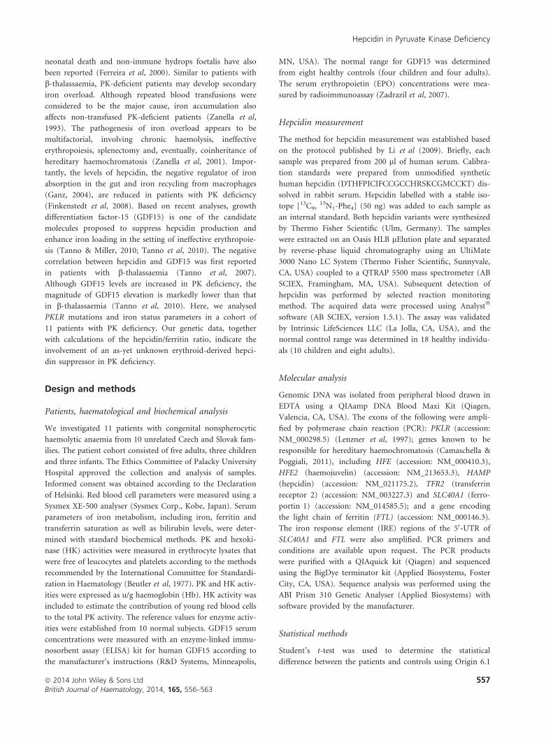
neonatal death and non-immune hydrops foetalis have also
been reported (Ferreira et al, 2000). Similar to patients with
b-thalassaemia, PK-deficient patients may develop secondary
iron overload. Although repeated blood transfusions were
considered to be the major cause, iron accumulation also
affects non-transfused PK-deficient patients (Zanella et al,
1993). The pathogenesis of iron overload appears to be
multifactorial, involving chronic haemolysis, ineffective
erythropoiesis, splenectomy and, eventually, coinheritance of
hereditary haemochromatosis (Zanella et al, 2001). Impor-
tantly, the levels of hepcidin, the negative regulator of iron
absorption in the gut and iron recycling from macrophages
(Ganz, 2004), are reduced in patients with PK deficiency
(Finkenstedt et al, 2008). Based on recent analyses, growth
differentiation factor-15 (GDF15) is one of the candidate
molecules proposed to suppress hepcidin production and
enhance iron loading in the setting of ineffective erythropoie-
sis (Tanno & Miller, 2010; Tanno et al, 2010). The negative
correlation between hepcidin and GDF15 was first reported
in patients with b-thalassaemia (Tanno et al, 2007).
Although GDF15 levels are increased in PK deficiency, the
magnitude of GDF15 elevation is markedly lower than that
in b-thalassaemia (Tanno et al, 2010). Here, we analysed
PKLR mutations and iron status parameters in a cohort of
11 patients with PK deficiency. Our genetic data, together
with calculations of the hepcidin/ferritin ratio, indicate the
involvement of an as-yet unknown erythroid-derived hepci-
din suppressor in PK deficiency.
Design and methods
Patients, haematological and biochemical analysis
We investigated 11 patients with congenital nonspherocytic
haemolytic anaemia from 10 unrelated Czech and Slovak fam-
ilies. The patient cohort consisted of five adults, three children
and three infants. The Ethics Committee of Palacky University
Hospital approved the collection and analysis of samples.
Informed consent was obtained according to the Declaration
of Helsinki. Red blood cell parameters were measured using a
Sysmex XE-500 analyser (Sysmex Corp., Kobe, Japan). Serum
parameters of iron metabolism, including iron, ferritin and
transferrin saturation as well as bilirubin levels, were deter-
mined with standard biochemical methods. PK and hexoki-
nase (HK) activities were measured in erythrocyte lysates that
were free of leucocytes and platelets according to the methods
recommended by the International Committee for Standardi-
zation in Haematology (Beutler et al, 1977). PK and HK activ-
ities were expressed as u/g haemoglobin (Hb). HK activity was
included to estimate the contribution of young red blood cells
to the total PK activity. The reference values for enzyme activ-
ities were established from 10 normal subjects. GDF15 serum
concentrations were measured with an enzyme-linked immu-
nosorbent assay (ELISA) kit for human GDF15 according to
the manufacturer’s instructions (R&D Systems, Minneapolis,
MN, USA). The normal range for GDF15 was determined
from eight healthy controls (four children and four adults).
The serum erythropoietin (EPO) concentrations were mea-
sured by radioimmunoassay (Zadrazil et al, 2007).
Hepcidin measurement
The method for hepcidin measurement was established based
on the protocol published by Li et al (2009). Briefly, each
sample was prepared from 200 ll of human serum. Calibra-
tion standards were prepared from unmodified synthetic
human hepcidin (DTHFPICIFCCGCCHRSKCGMCCKT) dis-
solved in rabbit serum. Hepcidin labelled with a stable iso-
tope [13C9,15N1-Phe4] (50 ng) was added to each sample as
an internal standard. Both hepcidin variants were synthesized
by Thermo Fisher Scientific (Ulm, Germany). The samples
were extracted on an Oasis HLB lElution plate and separated
by reverse-phase liquid chromatography using an UltiMate
3000 Nano LC System (Thermo Fisher Scientific, Sunnyvale,
CA, USA) coupled to a QTRAP 5500 mass spectrometer (AB
SCIEX, Framingham, MA, USA). Subsequent detection of
hepcidin was performed by selected reaction monitoring
method. The acquired data were processed using Analyst�
software (AB SCIEX, version 1.5.1). The assay was validated
by Intrinsic LifeSciences LLC (La Jolla, CA, USA), and the
normal control range was determined in 18 healthy individu-
als (10 children and eight adults).
Molecular analysis
Genomic DNA was isolated from peripheral blood drawn in
EDTA using a QIAamp DNA Blood Maxi Kit (Qiagen,
Valencia, CA, USA). The exons of the following were ampli-
fied by polymerase chain reaction (PCR): PKLR (accession:
NM_000298.5) (Lenzner et al, 1997); genes known to be
responsible for hereditary haemochromatosis (Camaschella &
Poggiali, 2011), including HFE (accession: NM_000410.3),
HFE2 (haemojuvelin) (accession: NM_213653.3), HAMP
(hepcidin) (accession: NM_021175.2), TFR2 (transferrin
receptor 2) (accession: NM_003227.3) and SLC40A1 (ferro-
portin 1) (accession: NM_014585.5); and a gene encoding
the light chain of ferritin (FTL) (accession: NM_000146.3).
The iron response element (IRE) regions of the 50-UTR of
SLC40A1 and FTL were also amplified. PCR primers and
conditions are available upon request. The PCR products
were purified with a QIAquick kit (Qiagen) and sequenced
using the BigDye terminator kit (Applied Biosystems, Foster
City, CA, USA). Sequence analysis was performed using the
ABI Prism 310 Genetic Analyser (Applied Biosystems) with
software provided by the manufacturer.
Statistical methods
Student’s t-test was used to determine the statistical
difference between the patients and controls using Origin 6.1
Hepcidin in Pyruvate Kinase Deficiency
ª 2014 John Wiley & Sons Ltd 557British Journal of Haematology, 2014, 165, 556–563

software (OriginLab Corporation, Northampton, MA, USA).
The Spearman coefficient for correlation analyses was calcu-
lated using Statistica 10 software (StatSoft, Inc., Tulsa, OK,
USA). P values <0�05 were considered statistically significant.
Results
Patients’ characteristics and PK activity
The clinical findings of nine unrelated patients and two sib-
lings obtained at the time of diagnosis are summarized in
Table I. All patients suffered from mild to severe haemolytic
anaemia (with Hb levels ranging from 65 to 121 g/l) and
hyperbilirubinaemia (33 to 172 lmol/l). All but one of these
patients had increased reticulocyte counts (4�8 to 80�5%).
Neonatal jaundice was reported in most of the patients.
Seven patients were transfusion-dependent; three were
concomitantly treated with chelators and two underwent
splenectomy. Two paediatric patients received transfusions
during infancy, one adult patient was on intermittent trans-
fusion therapy and only one patient had never received a
transfusion in his lifetime. In 10 patients, erythrocyte
PK activity was below the normal reference values (4�60–6�28 u/g Hb) (Table II). In one case (Patient 7), the activity
was false normal (6�58 u/g Hb) due to substantial reticulocy-
tosis (80�5%) and PK deficiency was therefore established
based on low PK/HK activity ratio (1�6; normal range 3�6–6�3) (Table II). Reduced PK activity compared with the
controls was also detected in the available healthy parents,
confirming a congenital defect in the child. All parents tested
were later shown to be heterozygous carriers of specific PKLR
mutations (Table II).
Mutational analysis of the PKLR gene
Direct sequence analysis of the PKLR gene in the patient
cohort revealed nine different mutations in either the homo-
zygous state (four patients) or the compound heterozygous
state (seven patients) (Table II). Seven of the mutations are
known; five of these (c.1594C>T, c.1529G>A, c.1493G>A,c.1456C>T, and c.994G>A) have previously been identified
in Czech and Slovak populations (Lenzner et al, 1997).
The c.823G>A substitution and the insertion-deletion
(c.347_350delinsAACATTG) were identified in these popula-
tions for the first time. The clinical picture and the severity
of the disease in our patients bearing the above-mentioned
PK mutations were comparable to the phenotype previously
reported for patients with identical mutations (Baronciani
et al, 1995; Zanella et al, 1997, 2005, 2007). The c.878A>Tand c.1553delG mutations are novel and have not been
previously described in the literature.
The maternally inherited c.878A>T missense mutation was
detected in a heterozygous state with the previously repor-
ted, paternally inherited c.1529G>A substitution (Fig 1A,
Table II), which is the most common PKLR mutation in the
northern and central European population (Zanella et al,
2005). The c.878A>T mutation involves the alteration of pos-
itively charged aspartic acid to hydrophobic valine on the
Table I. Clinical and haematological data of the patients.
Patient Sex Age
Neonatal
jaundice ExTx Spleen
Hb,
g/l
Ret,%
[0�5–3�0]
Bilirubin,
lmol/l
[0–23]
Ferritin,
lg/l[6–200]*
TfS
[0�21–0�48]
Hepcidin,
lg/l[13�1–104�8]
GDF15,
ng/l
[166–344]
EPO,
iu/l
[4�3–29]
1 M 9 years Yes Yes Out 79 32 38 2000§ 0�79 n.d. 3052 n.d
2 M 6 months Yes Yes In 77 7�2 33 4852 0�97 90�7 935 46�233 months 98 4�8 49 145§ n.d. 16 1399 78�1
3 F 7 years Yes Yes† In 95 10�6 35 16 0�26 5�7 851 454
4 M 10 months Yes Yes In 75 7�5 59 303 n.d. 15�5 876 123
5 M 7 years Yes Yes† In 97 8�1 39 205 0�50 24�5 386 n.d
6 F 42 years No Yes‡ In 103 5�9 50 645 0�37 11�4 2581 46�87 F 22 years Yes Yes Out 65 80�5 139 372§ 0�88 9�1 514 122
8 F 31 years Yes Yes In 95 6�6 34 948 0�54 29�1 805 126
9 M 3 months Yes Yes In 67 2�1 172 802 n.d. n.d. 2163 n.d
10 F 25 years Yes Yes In 73 25�3 124 29�7 0�20 5�7 1259 n.d
11 M 50 years No No In 121 5�2 49 906 0�34 68�8 854 51�5
Patients 3 and 4 are siblings; ExTx: exchange transfusion; TfS: transferrin saturation; GDF15: growth differentiation factor-15; EPO (serum eryth-
ropoietin); n.d.: not determined.
*normal range for ferritin levels, women: <200, men: <300, children: <70 lg/l (Brugnara, 2009). The control range for GDF15 was determined
from eight healthy controls (four children and four adults).
†Dependent on blood transfusion during the first year of life due to severe anaemia.
‡Intermittent ExTx.
§Patients on chelation therapy. Two sets of data are shown for Patient 2; the first set represents values at the time of diagnosis, the second corre-
sponds to values after 6 months of chelation therapy.
R. Mojzikova et al
558 ª 2014 John Wiley & Sons LtdBritish Journal of Haematology, 2014, 165, 556–563

Table II. Biochemical and molecular data of the patients and their family members.
Patient
PK activity,
u/g Hb PK/HK PKLR mutation Amino acid alteration
1 2�64 0�78 c.[347_350delinsAACATTG];
[347_350delinsAACATTG]
p.[Arg116_Leu117delinsGlnHisCys];
[Arg116_Leu117delinsGlnHisCys]
1 (mother) 2�19 2�92 c.[347_350delinsAACATTG];[=] p.[Arg116_Leu117delinsGlnHisCys];[=]
1 (father) 2�19 1�97 c.[347_350delinsAACATTG];[=] p.[Arg116_Leu117delinsGlnHisCys];[=]
2 1�63 1�63 c.[1553delG];[1553delG] p.[Arg518Leufs*12];[Arg518Leufs*12]
2 (mother) 2�57 2�3 c.[1553delG];[=] p.[Arg518Leufs*12];[=]
3 1�43 0�51 c.[1529G>A];[1594C>T] p.[Arg510Gln];[Arg532Trp]
4 1�14 0�46 c.[1529G>A];[1594C>T] p.[Arg510Gln];[Arg532Trp]
4 (mother) 1�33 1�75 c.[1529G>A];[=] p.[Arg510Gln];[=]
4 (father) 1�57 1�51 c.[1594C>T];[=] p.[Arg532Trp];[=]
5 1�73 1�75 c.[1493G>A];[1529G>A] p.[Arg498His];[Arg510Gln]
6 1�16 0�84 c.[1456C>T];[1594C>T] p.[Arg486Trp];[Arg532Trp]
7 6�58 1�6 c.[1594C>T];[1594C>T] p.[Arg532Trp];[(Arg532Trp)]
8 1�56 0�81 c.[878A>T];[1529G>A] p.[Asp293Val];[Arg510Gln]
8 (father) 3�41 3�48 c.[1529G>A];[=] p.[Arg510Gln];[=]
8 (mother) 4�04 3�98 c.[878A>T];[=] p.[Asp293Val];[=]
9 1�72 2�07 c.[823G>A];[1594C>T] p.[Gly275Arg];[Arg532Trp]
9 (father) 2�45 2�62 c.[823G>A];[=] p.[Gly275Arg];[=]
9 (mother) – – c.[1594C>T];[=] p.[Arg532Trp];[=]
10 2�32 1�12 c.[1529G>A];[1529G>A] p.[Arg510Gln];[(Arg510Gln)]
11 1�54 1�14 c.[994G>A];[1456C>T] p.[Gly332Ser];[Arg532Trp]
PK: pyruvate kinase (normal activity range: 4�60–6�28 u/g Hb); PK/HK: pyruvate kinase and hexokinase activity ratio (normal range: 3�6–6�3).The normal activity range was determined in 10 healthy controls.
, exon 7 , exon 11
Patient 8
Mother
Father
C A C T T A T G C CN A G G A G T C T T C
C A C T T A T G C C G A G G A G T C T T C
C A C T T A TG C C N A G G A G T C T T C
, exon 11
Wild type
Patient 2
Mother
(B)(A)
Fig 1. Detection of two novel PKLR mutations by sequencing amplified DNA. (A) In the family of Patient 8, the propositus carried the newly
described c.878A>T mutation in exon 7 and the c.1529G>A mutation in exon 11; the mother was heterozygous for the c.878A>T mutation, and
the father was heterozygous for the c.1529G>A mutation. (B) In the family of Patient 2, the propositus was homozygous for the second newly
described mutation in the PKLR gene, a single nucleotide deletion (c.1553delG) in exon 11 leading to a frameshift (p.(Arg518Leufs*12)) and a
premature stop codon. One allele was inherited from his heterozygous mother; his father was unavailable for the analyses.
Hepcidin in Pyruvate Kinase Deficiency
ª 2014 John Wiley & Sons Ltd 559British Journal of Haematology, 2014, 165, 556–563
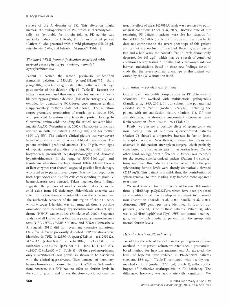
surface of the A domain of PK. This alteration might
increase the hydrophobicity of PK, which is thermodynami-
cally less favourable for protein folding. PK activity was
markedly reduced to 1�56 u/g Hb in an affected patient
(Patient 8) who presented with a mild phenotype (Hb 95 g/l,
reticulocytes 6�6%, and bilirubin 34 lmol/l; Table I).
The novel PKLR frameshift deletion associated withatypical severe phenotype involving neonatalhyperferritinaemia
Patient 2 carried the second previously unidentified
frameshift deletion, c.1553delG (p.(Arg518Leufs*12), short
p.Arg518fs), in a homozygous state; the mother is a heterozy-
gous carrier of this deletion (Fig 1B, Table II). Because the
father is unknown and thus unavailable for analyses, a possi-
ble hemizygous genomic deletion (loss of heterozygosity) was
excluded by quantitative PCR-based copy number analysis
(Supplementary methods; data not shown). The mutation
causes premature termination of translation at codon 529,
with predicted formation of a truncated protein lacking 46
C-terminal amino acids including the critical activator bind-
ing site Arg532 (Valentini et al, 2002). The activity of PK was
reduced in both the patient (1�63 u/g Hb) and his mother
(2�57 u/g Hb). The patient’s clinical picture was very severe
from birth, with a need for repeated blood transfusions. The
patient exhibited profound anaemia (Hb, 77 g/l), with signs
of hypoxia, neonatal jaundice (bilirubin, 90 lmol/l), throm-
bocytopenia, prominent hepatosplenomegaly and dramatic
hyperferritinaemia (in the range of 3500–5000 lg/l), and
transferrin saturation reaching almost 100%. Elevated levels
of liver enzymes (not shown) suggested possible liver damage,
which led us to perform liver biopsy. Massive iron deposits in
both hepatocytes and Kupffer cells corresponding to grade IV
haemosiderosis were detected. Taken together, these findings
suggested the presence of another co-inherited defect in the
child aside from PK deficiency. Sideroblastic anaemia was
ruled out by the absence of sideroblasts in the bone marrow.
The nucleotide sequence of the IRE region of the FTL gene,
which encodes L-ferritin, was not mutated; thus, a possible
association with hereditary hyperferritinaemia cataract syn-
drome (HHCS) was excluded (Brooks et al, 2002). Sequence
analysis of all known genes that cause primary haemochroma-
tosis (HFE, HFE2, HAMP, SLC40A1 and TFR2) (Camaschella
& Poggiali, 2011) did not reveal any causative mutations.
Only five different previously described SNP variations were
identified in TFR2 (c.2255G>A (p.Arg752His) - rs41295942),
SLC40A1 (c.44-24G>C - rs1439816, c.-330CGG[8] -
rs16836041, c.663T>C (p.Val221 = ) - rs2304704) and FTL
(c.163T>C (p.Leu55 = )) (Table SI). Of these polymorphisms,
only rs1439816:G>C was previously shown to be associated
with the clinical aggressiveness (liver damage) of hereditary
haemochromatosis 1 caused by the p.Cys282Tyr HFE muta-
tion; however, this SNP had no effect on ferritin levels in
the control group, and it was therefore concluded that the
negative effect of the rs1439816:C allele was restricted to path-
ological conditions (Alt�es et al, 2009). Because nine of our
remaining PK-deficient patients were also homozygous for
the rs1439816:C allele (Table SI), this polymorphism probably
does not contribute to the severe phenotype of this patient
and cannot explain his iron overload. Recently, at an age of
two and a half years, the patient’s ferritin levels dramatically
decreased (to 145 lg/l), which may be a result of combined
chelation therapy lasting 6 months and a prolonged interval
between transfusions. Based on these new findings, we con-
clude that the severe neonatal phenotype of this patient was
caused by the PKLR mutation itself.
Iron status in PK-deficient patients
One of the main health complications in PK deficiency is
secondary iron overload with multifactorial pathogenesis
(Zanella et al, 1993, 2001). In our cohort, nine patients had
elevated serum ferritin (median, 724 lg/l), including the
patient with no transfusion history (Patient 11). Of nine
available cases, five showed a concomitant increase in trans-
ferrin saturation (from 0�50 to 0�97) (Table I).
Firstly, we assessed a possible effect of splenectomy on
iron loading. One of our two splenectomized patients
(Patient 7) showed a progressive increase in ferritin levels
after spleen removal. Nevertheless, sustained haemolysis was
observed in this patient after spleen surgery, which probably
contributed to a further increase in her ferritin levels. On the
other hand, no significant difference in ferritin was recorded
for the second splenectomized patient (Patient 1); splenec-
tomy improved this patient’s anaemia, nevertheless his pre-
splenectomy ferritin levels were already dramatically elevated
(2213 lg/l). This patient is a child; thus, the contribution of
spleen removal to iron loading may become more apparent
over time.
We next searched for the presence of known HFE muta-
tions (p.His63Asp, p.Cys282Tyr), which have been proposed
as a condition that may predispose a patient to increased
iron absorption (Arruda et al, 2000; Zanella et al, 2001).
Abnormal HFE genotypes were identified in four of our
patients (Table SI). One of these patients (Patient 3), who
was a p.[His63Asp];[Cys282Tyr] HFE compound heterozy-
gote, was the only paediatric patient from the group with
normal ferritin levels.
Hepcidin levels in PK deficiency
To address the role of hepcidin in the pathogenesis of iron
overload in our patient cohort, we established a proteomics-
based method for hepcidin measurement. As expected, the
levels of hepcidin were reduced in PK-deficient patients
(median, 15�8 lg/l) (Table I) compared with healthy age-
matched controls (median, 27�6 lg/l) (Table I), reflecting the
impact of ineffective erythropoiesis in PK deficiency. The
difference, however, was not statistically significant. We
R. Mojzikova et al
560 ª 2014 John Wiley & Sons LtdBritish Journal of Haematology, 2014, 165, 556–563

therefore calculated the hepcidin/ferritin ratio which repre-
sents a more accurate estimation of proper hepcidin produc-
tion with respect to iron loading, and found it to be very
low in our PK-deficient patients (median, 0�06) compared
with a healthy age-matched group of controls (median, 0�35)(Fig 2). Even the patient with the highest hepcidin level
(Patient 2) showed a dramatically reduced ratio, which was
detected in repeated measurements (ranging from 0�01 to
0�04) and eventually increased to 0�11 as a response to
6 months of chelation therapy and improved anaemia (Hb,
98 g/l) (Table I and Fig 2). The direct effect of chelation
therapy on hepcidin levels and hepcidin/ferritin ratio is diffi-
cult to address in our cohort, given the complexity of posi-
tive and negative signals regulating hepcidin, the small
number of patients receiving chelation therapy, and the lack
of hepcidin pre-chelation values for remaining patients.
Candidate erythroid signals regulating hepcidin
To clarify the involvement of erythropoiesis in iron metabo-
lism in PK deficiency, serum levels of GDF15 and EPO, two
markers of erythropoietic activity known to be associated
with hepcidin production, were evaluated. The levels of
GDF15, a putative negative regulator of hepcidin production
in the setting of ineffective erythropoiesis (Tanno & Miller,
2010), were determined using a commercial enzyme-linked
immunosorbent assay. PK-deficient patients showed elevated
GDF15 compared with healthy age-matched controls
(median, 905�5 lg/l and 223 lg/l, respectively) (Table I),
confirming previously reported results (Zanella et al, 2005).
Importantly, no correlation between GDF15 and hepcidin or
GDF15 and hepcidin/ferritin ratio was observed. In addition,
all available patients from our cohort showed increased levels
of EPO (median, 100�1 iu/l) (Table I), probably reflecting
tissue hypoxia. Also, EPO levels did not correlate with hepci-
din or hepcidin/ferritin ratio.
Lastly, as inflammatory processes are known to indepen-
dently influence hepcidin levels (Ganz, 2004), it is important
to note that none of our patients had signs of inflammation
at the time of the analyses. In concordance, a substantial
increase in hepcidin (from 90�7 lg/l to 386 lg/l) was
observed in one of our patients (Patient 2) during bacterial
infection.
Discussion
In this report we characterized PKLR mutations and systemic
iron metabolism in eleven patients with PK deficiency from
ten unrelated families. Nine different disease-causing PKLR
mutations were identified; two of these mutations - the point
mutation c.A878T (p.D293V) and the frameshift deletion
c.1553delG (p.R518fs) - were novel. The patient affected by
the frameshift deletion presented with an unusually severe
phenotype involving neonatal hyperferritinaemia, which
eventually ameliorated by two and a half years of age, proba-
bly in response to combined chelation therapy and a pro-
longed interval between transfusions.
PK-deficient patients usually develop secondary iron over-
load over time as a consequence of chronic haemolysis, inef-
fective erythropoiesis and transfusion therapy; splenectomy
and inheritance of HFE mutations have been proposed as
additional risk factors (Zanella et al, 2001). Consistently, all
our transfusion-dependent patients presented iron overload;
one patient has developed hyperferritinaemia independently
of blood transfusions. Similar findings, iron overload inde-
pendent of blood transfusions in PK deficiency, were previ-
ously reported by Zanella et al (1993). Although
splenectomy reduces the symptoms of haemolysis, it is also
known to be a risk factor for iron loading. Due to a low
number of splenectomized patients in our cohort, however,
the involvement of spleen surgery cannot be adequately
addressed. The contribution of co-inherited HFE mutations
to excessive iron accumulation is also difficult to assess,
especially because three of the HFE mutant patients were
children and HFE-related haemochromatosis is an adult-
onset disease (Pietrangelo, 2010). Moreover, one paediatric
patient with abnormal HFE genotype did not have any signs
of iron overload. Based on these observations we can con-
clude that the HFE genotype does not play a major role in
determining iron loading in PK-deficient paediatric patients;
however, it may be important to follow up these patients
due to a potential higher risk of iron overload in adulthood
(Zanella et al, 2001).
0·0
0·5
1·0
1·5
2·0
2·5 < 0·05
PKD patientsControls
Hep
cidi
n/fe
rriti
n ra
tio
Fig 2. Hepcidin/ferritin ratio in PK-deficient patients. The hepcidin/
ferritin ratio was significantly lower in PK-deficient patients com-
pared to healthy controls (P < 0�05). The red dots in the graph indi-
cate the hepcidin/ferritin ratio calculated from five repeated
measurements (1�5 months, 9�5 months, 10 months, 20�5 months,
and 33 months) for Patient 2 with a p.(Arg518Leufs*12) mutation
in the PKLR gene, which is associated with hyperferritinaemia occur-
ring from the neonatal period through to infancy (2 years of age).
Only two hepcidin/ferritin ratios (calculated from those hepcidin
and ferritin values shown in Table I) were included into the statisti-
cal analysis for this patient. The thick horizontal lines represent the
medians for each group. PKD, pyruvate kinase-deficient.
Hepcidin in Pyruvate Kinase Deficiency
ª 2014 John Wiley & Sons Ltd 561British Journal of Haematology, 2014, 165, 556–563
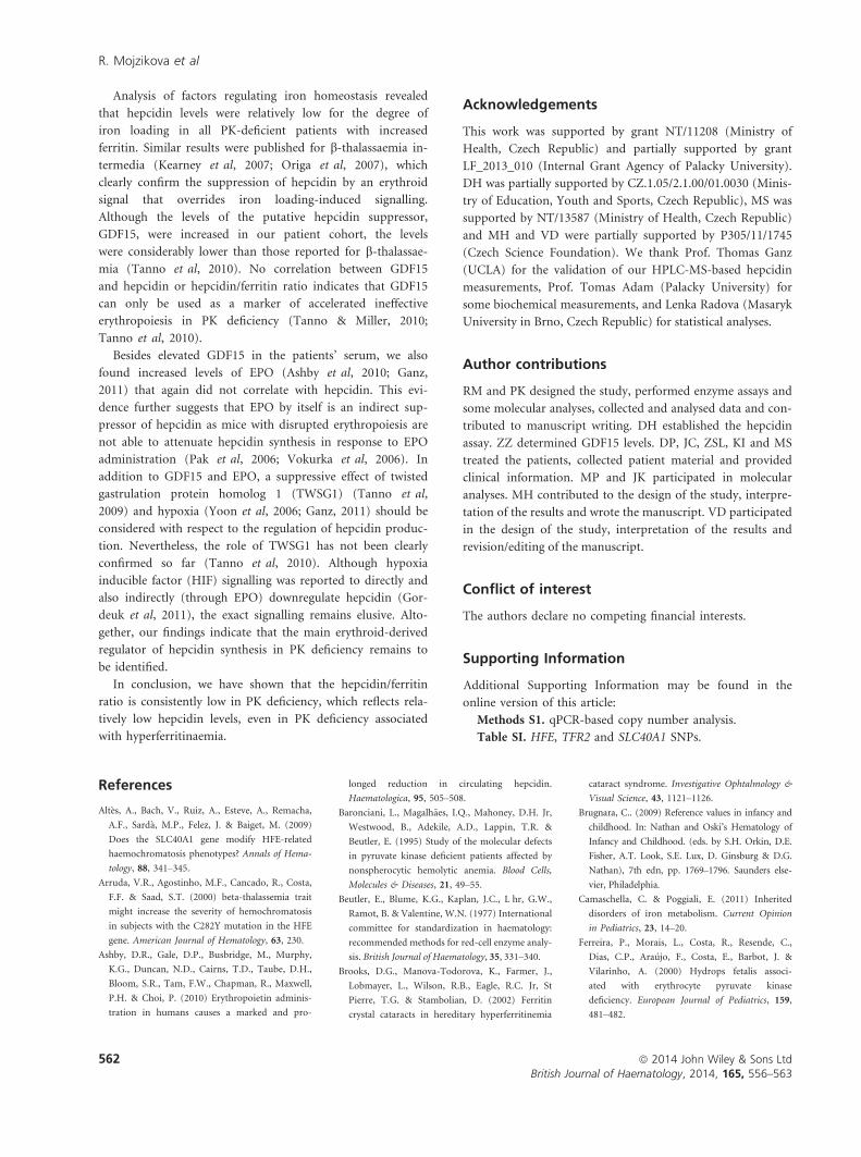
Analysis of factors regulating iron homeostasis revealed
that hepcidin levels were relatively low for the degree of
iron loading in all PK-deficient patients with increased
ferritin. Similar results were published for b-thalassaemia in-
termedia (Kearney et al, 2007; Origa et al, 2007), which
clearly confirm the suppression of hepcidin by an erythroid
signal that overrides iron loading-induced signalling.
Although the levels of the putative hepcidin suppressor,
GDF15, were increased in our patient cohort, the levels
were considerably lower than those reported for b-thalassae-mia (Tanno et al, 2010). No correlation between GDF15
and hepcidin or hepcidin/ferritin ratio indicates that GDF15
can only be used as a marker of accelerated ineffective
erythropoiesis in PK deficiency (Tanno & Miller, 2010;
Tanno et al, 2010).
Besides elevated GDF15 in the patients’ serum, we also
found increased levels of EPO (Ashby et al, 2010; Ganz,
2011) that again did not correlate with hepcidin. This evi-
dence further suggests that EPO by itself is an indirect sup-
pressor of hepcidin as mice with disrupted erythropoiesis are
not able to attenuate hepcidin synthesis in response to EPO
administration (Pak et al, 2006; Vokurka et al, 2006). In
addition to GDF15 and EPO, a suppressive effect of twisted
gastrulation protein homolog 1 (TWSG1) (Tanno et al,
2009) and hypoxia (Yoon et al, 2006; Ganz, 2011) should be
considered with respect to the regulation of hepcidin produc-
tion. Nevertheless, the role of TWSG1 has not been clearly
confirmed so far (Tanno et al, 2010). Although hypoxia
inducible factor (HIF) signalling was reported to directly and
also indirectly (through EPO) downregulate hepcidin (Gor-
deuk et al, 2011), the exact signalling remains elusive. Alto-
gether, our findings indicate that the main erythroid-derived
regulator of hepcidin synthesis in PK deficiency remains to
be identified.
In conclusion, we have shown that the hepcidin/ferritin
ratio is consistently low in PK deficiency, which reflects rela-
tively low hepcidin levels, even in PK deficiency associated
with hyperferritinaemia.
Acknowledgements
This work was supported by grant NT/11208 (Ministry of
Health, Czech Republic) and partially supported by grant
LF_2013_010 (Internal Grant Agency of Palacky University).
DH was partially supported by CZ.1.05/2.1.00/01.0030 (Minis-
try of Education, Youth and Sports, Czech Republic), MS was
supported by NT/13587 (Ministry of Health, Czech Republic)
and MH and VD were partially supported by P305/11/1745
(Czech Science Foundation). We thank Prof. Thomas Ganz
(UCLA) for the validation of our HPLC-MS-based hepcidin
measurements, Prof. Tomas Adam (Palacky University) for
some biochemical measurements, and Lenka Radova (Masaryk
University in Brno, Czech Republic) for statistical analyses.
Author contributions
RM and PK designed the study, performed enzyme assays and
some molecular analyses, collected and analysed data and con-
tributed to manuscript writing. DH established the hepcidin
assay. ZZ determined GDF15 levels. DP, JC, ZSL, KI and MS
treated the patients, collected patient material and provided
clinical information. MP and JK participated in molecular
analyses. MH contributed to the design of the study, interpre-
tation of the results and wrote the manuscript. VD participated
in the design of the study, interpretation of the results and
revision/editing of the manuscript.
Conflict of interest
The authors declare no competing financial interests.
Supporting Information
Additional Supporting Information may be found in the
online version of this article:
Methods S1. qPCR-based copy number analysis.
Table SI. HFE, TFR2 and SLC40A1 SNPs.
References
Alt�es, A., Bach, V., Ruiz, A., Esteve, A., Remacha,
A.F., Sard�a, M.P., Felez, J. & Baiget, M. (2009)
Does the SLC40A1 gene modify HFE-related
haemochromatosis phenotypes? Annals of Hema-
tology, 88, 341–345.
Arruda, V.R., Agostinho, M.F., Cancado, R., Costa,
F.F. & Saad, S.T. (2000) beta-thalassemia trait
might increase the severity of hemochromatosis
in subjects with the C282Y mutation in the HFE
gene. American Journal of Hematology, 63, 230.
Ashby, D.R., Gale, D.P., Busbridge, M., Murphy,
K.G., Duncan, N.D., Cairns, T.D., Taube, D.H.,
Bloom, S.R., Tam, F.W., Chapman, R., Maxwell,
P.H. & Choi, P. (2010) Erythropoietin adminis-
tration in humans causes a marked and pro-
longed reduction in circulating hepcidin.
Haematologica, 95, 505–508.
Baronciani, L., Magalh~aes, I.Q., Mahoney, D.H. Jr,
Westwood, B., Adekile, A.D., Lappin, T.R. &
Beutler, E. (1995) Study of the molecular defects
in pyruvate kinase deficient patients affected by
nonspherocytic hemolytic anemia. Blood Cells,
Molecules & Diseases, 21, 49–55.
Beutler, E., Blume, K.G., Kaplan, J.C., Lӧhr, G.W.,
Ramot, B. & Valentine, W.N. (1977) International
committee for standardization in haematology:
recommended methods for red-cell enzyme analy-
sis. British Journal of Haematology, 35, 331–340.
Brooks, D.G., Manova-Todorova, K., Farmer, J.,
Lobmayer, L., Wilson, R.B., Eagle, R.C. Jr, St
Pierre, T.G. & Stambolian, D. (2002) Ferritin
crystal cataracts in hereditary hyperferritinemia
cataract syndrome. Investigative Ophtalmology &
Visual Science, 43, 1121–1126.
Brugnara, C.. (2009) Reference values in infancy and
childhood. In: Nathan and Oski’s Hematology of
Infancy and Childhood. (eds. by S.H. Orkin, D.E.
Fisher, A.T. Look, S.E. Lux, D. Ginsburg & D.G.
Nathan), 7th edn, pp. 1769–1796. Saunders else-
vier, Philadelphia.
Camaschella, C. & Poggiali, E. (2011) Inherited
disorders of iron metabolism. Current Opinion
in Pediatrics, 23, 14–20.
Ferreira, P., Morais, L., Costa, R., Resende, C.,
Dias, C.P., Ara�ujo, F., Costa, E., Barbot, J. &
Vilarinho, A. (2000) Hydrops fetalis associ-
ated with erythrocyte pyruvate kinase
deficiency. European Journal of Pediatrics, 159,
481–482.
R. Mojzikova et al
562 ª 2014 John Wiley & Sons LtdBritish Journal of Haematology, 2014, 165, 556–563
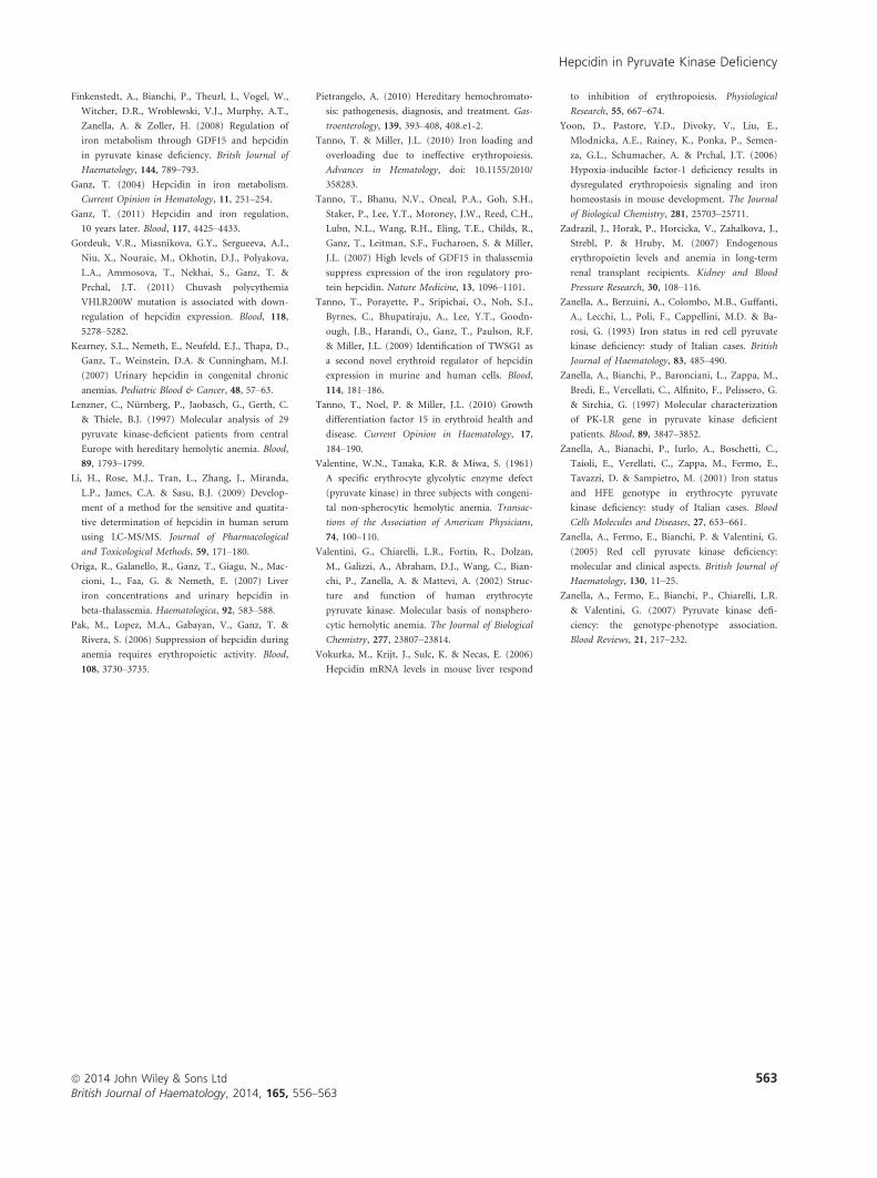
Finkenstedt, A., Bianchi, P., Theurl, I., Vogel, W.,
Witcher, D.R., Wroblewski, V.J., Murphy, A.T.,
Zanella, A. & Zoller, H. (2008) Regulation of
iron metabolism through GDF15 and hepcidin
in pyruvate kinase deficiency. Britsh Journal of
Haematology, 144, 789–793.
Ganz, T. (2004) Hepcidin in iron metabolism.
Current Opinion in Hematology, 11, 251–254.
Ganz, T. (2011) Hepcidin and iron regulation,
10 years later. Blood, 117, 4425–4433.
Gordeuk, V.R., Miasnikova, G.Y., Sergueeva, A.I.,
Niu, X., Nouraie, M., Okhotin, D.J., Polyakova,
L.A., Ammosova, T., Nekhai, S., Ganz, T. &
Prchal, J.T. (2011) Chuvash polycythemia
VHLR200W mutation is associated with down-
regulation of hepcidin expression. Blood, 118,
5278–5282.
Kearney, S.L., Nemeth, E., Neufeld, E.J., Thapa, D.,
Ganz, T., Weinstein, D.A. & Cunningham, M.J.
(2007) Urinary hepcidin in congenital chronic
anemias. Pediatric Blood & Cancer, 48, 57–63.
Lenzner, C., N€urnberg, P., Jaobasch, G., Gerth, C.
& Thiele, B.J. (1997) Molecular analysis of 29
pyruvate kinase-deficient patients from central
Europe with hereditary hemolytic anemia. Blood,
89, 1793–1799.
Li, H., Rose, M.J., Tran, L., Zhang, J., Miranda,
L.P., James, C.A. & Sasu, B.J. (2009) Develop-
ment of a method for the sensitive and quatita-
tive determination of hepcidin in human serum
using LC-MS/MS. Journal of Pharmacological
and Toxicological Methods, 59, 171–180.
Origa, R., Galanello, R., Ganz, T., Giagu, N., Mac-
cioni, L., Faa, G. & Nemeth, E. (2007) Liver
iron concentrations and urinary hepcidin in
beta-thalassemia. Haematologica, 92, 583–588.
Pak, M., Lopez, M.A., Gabayan, V., Ganz, T. &
Rivera, S. (2006) Suppression of hepcidin during
anemia requires erythropoietic activity. Blood,
108, 3730–3735.
Pietrangelo, A. (2010) Hereditary hemochromato-
sis: pathogenesis, diagnosis, and treatment. Gas-
troenterology, 139, 393–408, 408.e1-2.
Tanno, T. & Miller, J.L. (2010) Iron loading and
overloading due to ineffective erythropoiesis.
Advances in Hematology, doi: 10.1155/2010/
358283.
Tanno, T., Bhanu, N.V., Oneal, P.A., Goh, S.H.,
Staker, P., Lee, Y.T., Moroney, J.W., Reed, C.H.,
Lubn, N.L., Wang, R.H., Eling, T.E., Childs, R.,
Ganz, T., Leitman, S.F., Fucharoen, S. & Miller,
J.L. (2007) High levels of GDF15 in thalassemia
suppress expression of the iron regulatory pro-
tein hepcidin. Nature Medicine, 13, 1096–1101.
Tanno, T., Porayette, P., Sripichai, O., Noh, S.J.,
Byrnes, C., Bhupatiraju, A., Lee, Y.T., Goodn-
ough, J.B., Harandi, O., Ganz, T., Paulson, R.F.
& Miller, J.L. (2009) Identification of TWSG1 as
a second novel erythroid regulator of hepcidin
expression in murine and human cells. Blood,
114, 181–186.
Tanno, T., Noel, P. & Miller, J.L. (2010) Growth
differentiation factor 15 in erythroid health and
disease. Current Opinion in Haematology, 17,
184–190.
Valentine, W.N., Tanaka, K.R. & Miwa, S. (1961)
A specific erythrocyte glycolytic enzyme defect
(pyruvate kinase) in three subjects with congeni-
tal non-spherocytic hemolytic anemia. Transac-
tions of the Association of American Physicians,
74, 100–110.
Valentini, G., Chiarelli, L.R., Fortin, R., Dolzan,
M., Galizzi, A., Abraham, D.J., Wang, C., Bian-
chi, P., Zanella, A. & Mattevi, A. (2002) Struc-
ture and function of human erythrocyte
pyruvate kinase. Molecular basis of nonsphero-
cytic hemolytic anemia. The Journal of Biological
Chemistry, 277, 23807–23814.
Vokurka, M., Krijt, J., Sulc, K. & Necas, E. (2006)
Hepcidin mRNA levels in mouse liver respond
to inhibition of erythropoiesis. Physiological
Research, 55, 667–674.
Yoon, D., Pastore, Y.D., Divoky, V., Liu, E.,
Mlodnicka, A.E., Rainey, K., Ponka, P., Semen-
za, G.L., Schumacher, A. & Prchal, J.T. (2006)
Hypoxia-inducible factor-1 deficiency results in
dysregulated erythropoiesis signaling and iron
homeostasis in mouse development. The Journal
of Biological Chemistry, 281, 25703–25711.
Zadrazil, J., Horak, P., Horcicka, V., Zahalkova, J.,
Strebl, P. & Hruby, M. (2007) Endogenous
erythropoietin levels and anemia in long-term
renal transplant recipients. Kidney and Blood
Pressure Research, 30, 108–116.
Zanella, A., Berzuini, A., Colombo, M.B., Guffanti,
A., Lecchi, L., Poli, F., Cappellini, M.D. & Ba-
rosi, G. (1993) Iron status in red cell pyruvate
kinase deficiency: study of Italian cases. British
Journal of Haematology, 83, 485–490.
Zanella, A., Bianchi, P., Baronciani, L., Zappa, M.,
Bredi, E., Vercellati, C., Alfinito, F., Pelissero, G.
& Sirchia, G. (1997) Molecular characterization
of PK-LR gene in pyruvate kinase deficient
patients. Blood, 89, 3847–3852.
Zanella, A., Bianachi, P., Iurlo, A., Boschetti, C.,
Taioli, E., Verellati, C., Zappa, M., Fermo, E.,
Tavazzi, D. & Sampietro, M. (2001) Iron status
and HFE genotype in erythrocyte pyruvate
kinase deficiency: study of Italian cases. Blood
Cells Molecules and Diseases, 27, 653–661.
Zanella, A., Fermo, E., Bianchi, P. & Valentini, G.
(2005) Red cell pyruvate kinase deficiency:
molecular and clinical aspects. British Journal of
Haematology, 130, 11–25.
Zanella, A., Fermo, E., Bianchi, P., Chiarelli, L.R.
& Valentini, G. (2007) Pyruvate kinase defi-
ciency: the genotype-phenotype association.
Blood Reviews, 21, 217–232.
ª 2014 John Wiley & Sons Ltd 563British Journal of Haematology, 2014, 165, 556–563
Hepcidin in Pyruvate Kinase Deficiency

Supplementary Method.
Copy number of PKLR exon 10 was assessed by SYBR Green I based absolute quantification,
using standard curve method. Concentration values of PKLR were normalized to RPPH1
housekeeping gene that is commonly used as a copy number variation (CNV) reference, as
it’s an autosomal gene with no reported CNV in population. The reaction was run in
triplicates. The copy number value of Patient 2 was compared to six non-PK-deficient patient
samples. The primer sequences are available upon request.

Table SI. HFE, TFR2 and SLC40A1 SNPs.
Rem.: HFE screened for p.Cys282Tyr, p.His63Asp, p.Ser65Cys, and p.Val256Ile; TFR2 for p.Arg752His and SLC40A1 for microsatellite polymorphism c.-330CGG(7_8) and c.44-24G>C. CGG[7];[8] in SLC40A1column is short for c.-330CGG[7];[8]. [C];[C] and [C];[G] in SLC40A1column is short for c.[44-24G>C];[ 44-24G>C] and c.[44-24G>C];[=], respectively.
Patient HFE mutation TFR2
p.Arg752His
SLC40A1
CGG(7_8)
SLC40A1
c.44-24G>C
1 wt wt CGG[7];[8] [C];[C]
1 (mother) wt wt CGG[8];[8] [C];[C]
1 (father) wt wt CGG[7];[7] [C];[C]
2 wt [Arg752His];[=] CGG[8];[8] [C];[C]
2 (mother) wt wt CGG[8];[8] [C];[C]
3 [His63Asp];[Cys282Tyr] wt CGG[8];[8] [C];[C]
4 [His63Asp];[=] wt CGG[8];[8] [C];[C]
3,4 (mother) [Cys282Tyr];[=] wt CGG[8];[8] [C];[C]
3,4 (father) [His63Asp];[=] wt CGG[7];[8] [C];[C]
5 [His63Asp];[=] wt CGG[8];[8] [C];[C]
6 [His63Asp];[(His63Asp)] wt CGG[8];[8] [C];[C]
7 wt wt CGG[7];[8] [C];[C]
8 wt wt CGG[7];[8] [C];[C]
8 (mother) wt wt CGG[7];[7] [C];[G]
8 (father) wt wt CGG[7];[8] [C];[C]
9 wt wt CGG[7];[8] [C];[G]
9 (mother) wt wt CGG[8];[8] [C];[C]
9 (father) wt wt CGG[7];[8] [C];[G]
10 wt wt CGG[8];[8] [C];[C]
11 wt wt CGG[8];[8] [C];[C]

1
Title:
Two novel mutations (p.(Ser160Pro) and p.(Arg472Cys)) causing glucose-6-phosphate
isomerase deficiency are associated with erythroid dysplasia and inappropriately
suppressed hepcidin
Short title: glucose-6-phosphate isomerase deficiency causing hemolytic anemia
Renata Mojzikovaa, Pavla Koralkovaa,b, Dusan Holubb, Zuzana Saxovaa, Dagmar
Pospisilovac, Daniela Prochazkovad, Petr Dzubakb, Monika Horvathovaa, Vladimir Divokya
aDepartment of Biology, Faculty of Medicine and Dentistry, Palacky University, Olomouc,
Czech Republic; bInstitute of Molecular and Translational Medicine, Faculty of Medicine and
Dentistry, Palacky University, Olomouc, Czech Republic; cDepartment of Pediatrics,
University Hospital and Faculty of Medicine and Dentistry, Olomouc, Czech Republic dDepartment of Pediatrics, Masaryk Hospital in Usti nad Labem, Czech Republic.
Correspondence:
Renata Mojzikova, Department of Biology, Faculty of Medicine and Dentistry, Palacky
University, Hnevotinska 3, 77515 Olomouc, Czech Republic. E-mail:
[email protected], phone: +420-585632151, fax: +420-585632966.
2
Abstract
Glucose-6-phosphate isomerase (GPI) deficiency, a genetic disorder responsible for chronic
nonspherocytic hemolytic anemia, is the second most common red blood cell glycolytic
enzymopathy. We report three patients from two unrelated families of Czech and Slovak
origin with macrocytic hemolytic anemia due to GPI deficiency. The first patient had 15% of
residual GPI activity resulting from two new heterozygous missense mutations c.478TC and
c.1414CT leading to substitutions p.(Ser160Pro) and p.(Arg472Cys). Two other patients
(siblings) inherited the same c.1414CT p.(Arg472Cys) mutation in a homozygous
constitution and lost approximately 89% of their GPI activity. Erythroid hyperplasia with
dysplastic features was observed in the bone marrow of all three patients. Low
hepcidin/ferritin ratio and elevated soluble transferrin receptor detected in our GPI-deficient
patients suggest disturbed balance between erythropoiesis and iron metabolism contributing to
iron overload.
Key words: Glucose-6-phosphate isomerase deficiency, Hemolytic anemia, Dysplastic
erythropoiesis, Hepcidin to ferritin ratio, Soluble transferrin receptor, Iron loading
Introduction
Glucose-6-phosphate isomerase (GPI, EC 5.3.1.9) is a homodimeric enzyme, which
participates in the second step of glycolysis in interconversion of glucose-6-phosphate (G6P)
and fructose-6-phosphate (F6P). The GPI gene is located at the 19q13.1 and has 18 exons
spanning 59 kb. It encodes 558-aminoacid monomer with a molecular weight of 63 kDa [1].
The human GPI crystal structure [2] provides a framework for modeling of GPI mutations.
GPI deficiency is the third most common cause of hereditary nonspherocytic hemolytic
anemia (HNSHA), after glucose-6-phosphate dehydrogenase (G6PD) and pyruvate kinase
(PK) deficiencies, and is inherited in autosomal recessive fashion [3]. Thirty five mutations
(29 missense, 3 nonsense, 2 splice site and 1 frameshift) [1,4,5,6,7] have been reported since
GPI deficiency was first described in 1967 [8]. The phenotype varies from mild to severe
anemia associated with splenomegaly in homozygotes and compound heterozygotes simple
heterozygotes are hematologically normal. GPI deficiency may be associated with hydrops
fetalis during the neonatal period [9]. Some patients may also exhibit neurological
dysfunction [10,11,12], which is explained by neuroleukin function of GPI in its monomeric
3
form [13,14,15], and granulocyte dysfunction associated with rare GPI variants with very low
residual GPI activity 10,16. Coincidence of GPI and G6PD deficiencies has been reported in
some cases [6,17,18]. The clinical and biochemical features of mouse model of GPI
deficiency resemble those present in human patients [19]. GPI deficiency is one of the few
erythroenzymopaties which benefits from splenectomy just as pyruvate kinase,
phosphoglycerokinase and hexokinase deficiencies 20].
Analyses of diverse erythroid and iron metabolism defects have shown that disrupted
erythropoiesis negatively affects iron homeostasis and vice versa. In many cases, the
molecular pathophysiology involves disordered production of hepcidin; the key molecule
inhibiting iron absorption, mobilization, and recycling [21]. It has been shown that anemias
characterized by ineffective erythropoiesis, like β-thalassemia [22] and congenital
dyserythropoietic anemia [23], are associated with hepcidin suppression. As a result, iron
absorption and recycling are augmented, leading to the development of secondary iron
overload independently of transfusions. We recently published on disturbed balance between
erythropoiesis and iron metabolism in two other types of hereditary hemolytic anemia: PK
deficiency [24] and erythrocyte membrane defects [25]. Also in these disease states
stimulated, ineffective erythropoiesis causes disordered production of hepcidin. Active
erythropoiesis in the bone marrow is essential for hepcidin suppression as patients with
Diamond Blackfan anemia (DBA), characterized by red cell aplasia, or mice with ablated
bone marrow hematopoiesis are unable to attenuate hepcidin production [26,27,28]. The
information on iron homeostasis in GPI deficiency has been limited; several cases with
ferritinemia developed independently of transfusions have been reported in the literature
[7,29].
Here we present, for the first time in the Czech and Slovak populations, two families with
three cases of GPI deficiency caused by two novel GPI mutations, p.(Ser160Pro) and
p.(Arg472Cys). We performed kinetic, thermostability and molecular modeling study to
determine how these mutations could affect enzyme structure and function. In addition, we
evaluated patients' bone marrow and revealed erythroid hyperplasia with dysplastic changes
and reduced granulopoiesis. We also demonstrate disrupted balance between erythropoiesis
and iron metabolism in these patients; this imbalance should be considered as a contributing
factor for the development of iron overload in GPI deficiency.

4
Patients and methods
We investigated three patients with chronic nonspherocytic macrocytic hemolytic anemia
from two unrelated families of Czech and Slovak origin (Table 1). Patient 1 is a 10-year-old
Czech boy who suffered from severe transfusion-dependent hemolytic anemia (Hb 90 g/L,
reticulocytes 4.5%) since birth till the age of 6 years when he was splenectomized and regular
blood transfusions were eliminated. Bone marrow biopsy showed hypercellular bone marrow
with reduced granulopoiesis, erythroid hyperplasia and dysplastic changes in late erythroid
precursors; however, no mutations in CDAN1 gene (for codanin-1) were found. Patients 2 and
3 are 19- and 11-year old siblings (boy and girl born to non-consanguineous parents) of
Slovak origin with chronic hemolysis (Hb 76-101 g/L and reticulocytes 13-14%) and severe
neonatal jaundice treated with exsanguination transfusion. Normo-/hypercellular bone marrow
with erythroid hyperplasia showing dysplastic features and reduced granulopoiesis were also
noted in these patients (Figure 1). Both patients were regularly transfused until the
splenectomy was performed at the age of his 6 and her 7 years. Due to persistent
hyperferritinemia, both patients are subjected to Exjade-chelation therapy. Before
splenectomy, all patients presented with splenomegaly. Neurological functions of the patients
were normal and no clinical signs of neutrophil dysfunction were observed. The parents, free
of any signs of chronic hemolysis or other clinical symptoms, were undertaken for
investigations as well.
All participants gave informed consent to this study which was approved by the Ethics
Committee of Palacky University Hospital.
Hematological and enzyme studies
Routine hematological and biochemical studies were performed according to standard
methods. Erythrocyte enzyme activities were determined based on previously described
standardized procedures [30]. For the platelet-leukocyte assay, 5 µl of packed 200-times
diluted platelet-leukocyte cells were added to the same 1 mL reaction mixture and the specific
leukocyte GPI activities were expressed as the enzyme activity per milligram of total protein
measured by the Biuret assay.
KM values for GPI wild-type and mutant enzymes for G6P and F6P were measured as
described previously [5]; G6P concentration varied between 0.1 and 2.5 mM and F6P between
25 and 1000 µM. Lineweaver-Burk plot was used to analyze enzyme data.

5
Heat stability of GPI variants was determined in hemolysate in 0.1 M Tris/0.5 mM EDTA
buffer (pH 8.0) with 0.5% BSA, incubated at 37ºC, 40ºC and 45ºC for 10-60 minutes. The T50
(incubation temperature leading to 50% enzyme inactivation in 10 minutes) was elevated
from incubation conducted between 30ºC and 90ºC. A control sample exposed to 30ºC for 10
minutes has served as the reference value for 100% activity.
Growth differentiation factor 15 and hepcidin measurements
Serum growth differentiation factor-15 (GDF15) and hepcidin concentrations were measured
by commercial ELISA kit and by previously described quantitative SPE-LC-MS/MS assay,
respectively [24].
Molecular analysis and enzyme modeling
DNA analyses are described in the Supplemental methods (Online supplemental documents).
A ribbon plot of the structure of GPI monomer was obtained from the crystallographic
structure of the dimer (protein data bank 1IAT). The 3D models of the mutant forms of GPI
were generated using program PyMOL (www.pymol.org).
Results
Patients’ characterization
The patients exhibited severe macrocytic hemolytic anemia associated with reticulocytosis,
hyperferritinemia (Table 1) and erythroid hyperplasia in the bone marrow (Figure 1).
Although marked dysplastic changes were noted in the erythroid precursors of all patients, no
mutation in genes known to be associated with congenital dyserythropoietic anemia including
CDAN1, SEC23B, and KLF1 has been detected. Serum vitamin B12 and folic acid levels were
within normal limits in all studied patients (not shown). Other tests and values, such as direct
Coombs’ test, Ham’s test, sucrose lysis test, heat instability test, cryohemolysis test, Hb-
electrophoresis and HbA2 and HbF values were negative/normal. No Heinz bodies were seen
with brilliant cresyl blue staining.
Glycolytic enzyme activities
The peripheral blood glycolytic enzyme activities in the patients and their parents are
summarized in Table 2. Elevation in erythrocyte enzyme activities, such as hexokinase (HK),
G6PD, gluconolactone dehydrogenase (GLD) and PK is likely caused by contribution of
6
relatively young erythrocyte populations. Both, PK/HK and G6PD/GLD ratios were within
the control range. All three patients have low residual erythrocyte GPI activities – reduced by
more than 85%. The parents showed decreased GPI activities as well, corresponding to about
30% of normal control activity. The patients’ platelet-leukocytes GPI activities reached
approximately 30%, whereas the parents’ ones showed intermediate enzyme activities (data
not shown).
GPI sequence analysis
DNA sequencing revealed that Patient 1 (family 1) is a compound heterozygote for c.478TC
(exon 5) and c.1414CT (exon 16) mutations in GPI gene that lead to p.(Ser160Pro) and
p.(Arg472Cys) substitutions, respectively (Figure S1, Online supplemental documents). His
father is a heterozygote for c.478TC and his mother is a heterozygote for c.1414CT
mutation. Patients 2 and 3 (family 2) are homozygotes for the same c.1414CT mutation
p.(Arg472Cys) (Figure S1, Online supplemental documents). Their parents are carriers of this
mutation. Both mutations were confirmed by HaeIII and MluCI digestion. Mutation
c.478TC creates a new restriction site for HaeIII, whereas c.1414CT mutation for MluCI
endonuclease (Figure S2, Online supplemental documents).
GPI kinetic studies and in silico modeling
The results from kinetic experiments of GPI variants are shown in Table 3. While in the
Patient 1 the kinetic properties and thermostability resulted from the putative heterogeneous
mixture of the enzyme hetero- or homodimers, the homozygosity of p.(Arg472Cys)
substitution in the Patient 2 and 3 allowed precise characterization of this variant. The GPI
mutant from the compound heterozygous Patient 1 showed an increased affinity for G6P
and/or F6P. Its thermal stability was only slightly affected; T1/2 was within normal limits,
whereas T50 was decreased. The homozygous p.(Arg472Cys) mutant had a decreased affinity
for F6P and a slightly increased G6P affinity. Its thermal stability was strongly reduced.
Furthermore, the enzyme activity was almost abolished after short incubation at 45°C.
Analysis of the 3D GPI structure using in silico modelling (Figure 2) showed that Ser160
interacts with the substrate phosphate group and directly participates in the enzyme catalysis.
The Ser160Pro substitution likely alters binding to the substrate, and thus affects catalytic
activity of the mutant enzyme. Arg472 is a part of a dimer-domain located at the edge of the
large external loop (LEL), which stabilizes both GPI monomers into a dimer conformation.
7
Arg472 forms hydrogen bonds with residues Gly470 and Ile395 (located on other monomer).
The Arg472Cys mutation likely leads to a disruption of these interactions which probably
destabilizes the GPI dimer conformation (Figure 2).
Erythropoietic activity, iron metabolism and hepcidin
The information on iron status and erythropoietic activity in GPI deficiency has been limited
[7,29]. Two our patients showed signs of iron overload i.e. increased serum iron, transferrin
saturation (TSAT) and ferritin (Table 4). One patient had normal iron status parameters
(Table 4) as a result of chelation therapy, which had been initiated because of persistent
hyperferritinemia, before and after splenectomy (ferritin 540±70 µg/L, TSAT 77%).
Nevertheless, the analysis of hepcidin and hepcidin/ferritin ratio revealed inappropriately low
hepcidin levels with respect to the iron stores in all three patients (0.089, 0.039 and 0.001;
control range: 0.2-2.2) (Table 4). This suggests pathological hepcidin suppression likely by
stimulated erythropoiesis as documented by increased levels of soluble transferrin receptor
(sTfR – 17, 24.5 and 21.6; control range: 1.9-4.4 mg/L) (Table 4), erythropoietin (EPO; 34.1,
63.8 and 147; control range: 4.3-29 IU/L) and in agreement with erythroid hyperplasia in their
bone marrow (Figure 1). In addition, two patients showed marked elevation of GDF15 (777
and 989; control range: 166-344 ng/L) (Table 4), a marker of ineffective erythropoiesis [31].
This is also consistent with dyserythropoiesis and dysplastic changes in erythroid precursors
detected in patients' bone marrow (Figure 1).
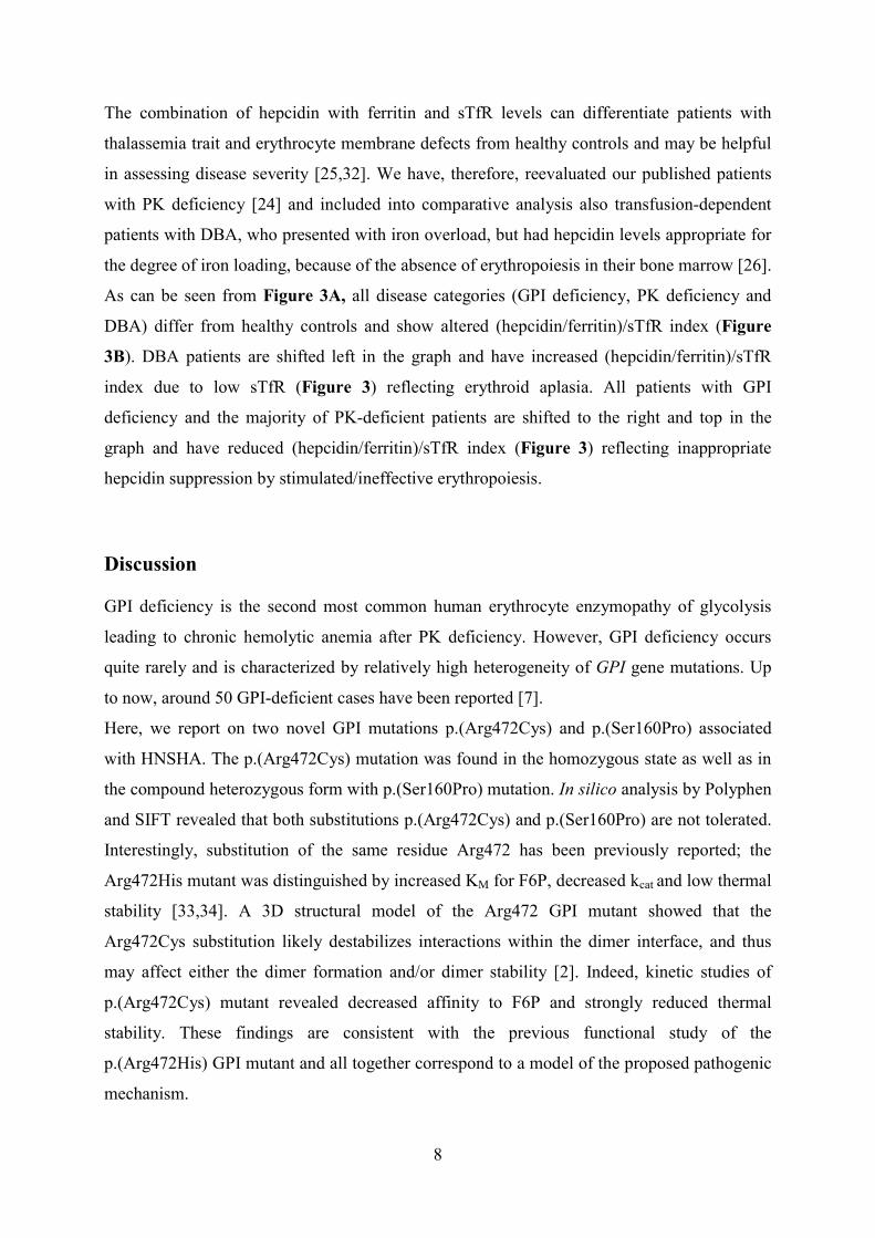
8
The combination of hepcidin with ferritin and sTfR levels can differentiate patients with
thalassemia trait and erythrocyte membrane defects from healthy controls and may be helpful
in assessing disease severity [25,32]. We have, therefore, reevaluated our published patients
with PK deficiency [24] and included into comparative analysis also transfusion-dependent
patients with DBA, who presented with iron overload, but had hepcidin levels appropriate for
the degree of iron loading, because of the absence of erythropoiesis in their bone marrow [26].
As can be seen from Figure 3A, all disease categories (GPI deficiency, PK deficiency and
DBA) differ from healthy controls and show altered (hepcidin/ferritin)/sTfR index (Figure
3B). DBA patients are shifted left in the graph and have increased (hepcidin/ferritin)/sTfR
index due to low sTfR (Figure 3) reflecting erythroid aplasia. All patients with GPI
deficiency and the majority of PK-deficient patients are shifted to the right and top in the
graph and have reduced (hepcidin/ferritin)/sTfR index (Figure 3) reflecting inappropriate
hepcidin suppression by stimulated/ineffective erythropoiesis.
Discussion
GPI deficiency is the second most common human erythrocyte enzymopathy of glycolysis
leading to chronic hemolytic anemia after PK deficiency. However, GPI deficiency occurs
quite rarely and is characterized by relatively high heterogeneity of GPI gene mutations. Up
to now, around 50 GPI-deficient cases have been reported [7].
Here, we report on two novel GPI mutations p.(Arg472Cys) and p.(Ser160Pro) associated
with HNSHA. The p.(Arg472Cys) mutation was found in the homozygous state as well as in
the compound heterozygous form with p.(Ser160Pro) mutation. In silico analysis by Polyphen
and SIFT revealed that both substitutions p.(Arg472Cys) and p.(Ser160Pro) are not tolerated.
Interestingly, substitution of the same residue Arg472 has been previously reported; the
Arg472His mutant was distinguished by increased KM for F6P, decreased kcat and low thermal
stability [33,34]. A 3D structural model of the Arg472 GPI mutant showed that the
Arg472Cys substitution likely destabilizes interactions within the dimer interface, and thus
may affect either the dimer formation and/or dimer stability [2]. Indeed, kinetic studies of
p.(Arg472Cys) mutant revealed decreased affinity to F6P and strongly reduced thermal
stability. These findings are consistent with the previous functional study of the
p.(Arg472His) GPI mutant and all together correspond to a model of the proposed pathogenic
mechanism.
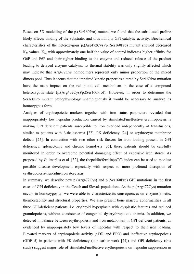
9
Based on 3D modelling of the p.(Ser160Pro) mutant, we found that the substituted proline
likely affects binding of the substrate, and thus inhibits GPI catalytic activity. Biochemical
characteristics of the heterozygous p.(Arg472Cys)/p.(Ser160Pro) mutant showed decreased
KM values. KM with approximately one half the value of control indicates higher affinity for
G6P and F6P and their tighter binding to the enzyme and reduced release of the product
leading to delayed enzyme catalysis. Its thermal stability was only slightly affected which
may indicate that Arg472Cys homodimers represent only minor proportion of the mixed
dimers pool. Thus it seems that the impaired kinetic properties altered by Ser160Pro mutation
have the main impact on the red blood cell metabolism in the case of a compound
heterozygous state (p.(Arg472Cys)/p.(Ser160Pro)). However, in order to determine the
Ser160Pro mutant pathophysiology unambiguously it would be necessary to analyze its
homozygous form.
Analyses of erythropoietic markers together with iron status parameters revealed that
inappropriately low hepcidin production caused by stimulated/ineffective erythropoiesis is
making GPI deficient patients susceptible to iron overload independently of transfusions,
similar to patients with β-thalassemia [22], PK deficiency [24] or erythrocyte membrane
defects [25]. In connection with two other risk factors for iron loading present in GPI
deficiency, splenectomy and chronic hemolysis [35], these patients should be carefully
monitored in order to overcome potential damaging effect of excessive iron stores. As
proposed by Guimarães et al. [32], the (hepcidin/ferritin)/sTfR index can be used to monitor
possible disease development especially with respect to more profound disruption of
erythropoiesis-hepcidin-iron store axis.
In summary, we describe new p.(Arg472Cys) and p.(Ser160Pro) GPI mutations in the first
cases of GPI deficiency in the Czech and Slovak populations. As the p.(Arg472Cys) mutation
occurs in homozygosity, we were able to characterize its consequences on enzyme kinetic,
thermostability and structural properties. We also present bone marrow abnormalities in all
three GPI-deficient patients, i.e. erythroid hyperplasia with dysplastic features and reduced
granulopoiesis, without coexistence of congenital dyserythropoietic anemia. In addition, we
detected imbalance between erythropoiesis and iron metabolism in GPI-deficient patients, as
evidenced by inappropriately low levels of hepcidin with respect to their iron loading.
Elevated markers of erythropoietic activity (sTfR and EPO) and ineffective erythropoiesis
(GDF15) in patients with PK deficiency (our earlier work [24]) and GPI deficiency (this
study) suggest major role of stimulated/ineffective erythropoiesis on hepcidin suppression in
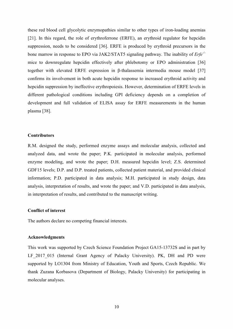
10
these red blood cell glycolytic enzymopathies similar to other types of iron-loading anemias
[21]. In this regard, the role of erythroferrone (ERFE), an erythroid regulator for hepcidin
suppression, needs to be considered [36]. ERFE is produced by erythroid precursors in the
bone marrow in response to EPO via JAK2/STAT5 signaling pathway. The inability of Erfe-/-
mice to downregulate hepcidin effectively after phlebotomy or EPO administration [36]
together with elevated ERFE expression in β-thalassemia intermedia mouse model [37]
confirms its involvement in both acute hepcidin response to increased erythroid activity and
hepcidin suppression by ineffective erythropoiesis. However, determination of ERFE levels in
different pathological conditions including GPI deficiency depends on a completion of
development and full validation of ELISA assay for ERFE measurements in the human
plasma [38].
Contributors
R.M. designed the study, performed enzyme assays and molecular analysis, collected and
analyzed data, and wrote the paper; P.K. participated in molecular analysis, performed
enzyme modeling, and wrote the paper; D.H. measured hepcidin level; Z.S. determined
GDF15 levels; D.P. and D.P. treated patients, collected patient material, and provided clinical
information; P.D. participated in data analysis; M.H. participated in study design, data
analysis, interpretation of results, and wrote the paper; and V.D. participated in data analysis,
in interpretation of results, and contributed to the manuscript writing.
Conflict of interest
The authors declare no competing financial interests.
Acknowledgments
This work was supported by Czech Science Foundation Project GA15-13732S and in part by
LF_2017_015 (Internal Grant Agency of Palacky University). PK, DH and PD were
supported by LO1304 from Ministry of Education, Youth and Sports, Czech Republic. We
thank Zuzana Korbasova (Department of Biology, Palacky University) for participating in
molecular analyses.
11
References
1 W. Kugler, M. Lakomek, Glucose-6-phosphate isomerase deficiency, Bailliere's Clin.
Haematol. 13 (2000) 89-101.
2 J. Read, J. Pearce, X. Li, et al., The crystal structure of human phosphoglucose isomerase
at 1.6A resolution: implications for catalytic mechanism, cytokine activity and haemolytic
anaemia, J. Mol. Biol. 309 (2001) 447-463.
[3] X.T. Gregg and J.T. Prchal, Red blood cells enzymopathies. In: R. Hoffman et al. (Eds.),
Hematology: Basic principles and practice, Elsevier, PA, US, 2009, p. 611-622.
4 A. Repiso, B. Oliva, J.L. Vives-Corrons, et al., Red cell glucose phosphate isomerase
(GPI): a molecular study of three novel mutations associated with hereditary nonspherocytic
hemolytic anemia, Hum. Mutat. 27 (2006) 1159-1167.
5 A. Repiso, B. Oliva, J.L. Vives Corrons, et al., Glucose phosphate isomerase deficiency:
enzymatic and familial characterization of Arg346His mutation, Biochim. Biophys. Acta.
1740 (2005) 467-471.
6 J.L. Clarke, T.J. Vulliamy, D. Roper, et al., Combined glucose-6-phosphate
dehydrogenase and glucosephosphate isomerase deficiency can alter clinical outcome, Blood
Cells Mol. Dis. 30 (2003) 258-263.
7 L. Manco, C. Bento, B.L. Victor, et al., Hereditary nonspherocytic hemolytic anemia
caused by red cell glucose-6-phosphate isomerase (GPI) deficiency in two Portuguese
patients: Clinical features and molecular study, Blood Cells Mol. Dis. 60 (2016) 18-23.
8 M.A. Baugham, W.N. Valentine, D.E. Paglia, et al., Hereditary hemolytic anemia
associated with glucosephosphate isomerase (GPI) deficiency. A new enzyme defect of
human erythrocytes, Blood. 32 (1968) 236-249.
9 Y. Ravindranath, D.E. Paglia, I. Warrier, et al., Glucose phosphate isomerase deficiency
as a cause of hydrops fetalis, N. Engl. J. Med. 316 (1987) 258-261.
10 W. Schröter, S.W. Eber, A. Bardosi, et al., Generalized glucose-phosphate isomerase
(GPI) deficiency causing haemolytic anaemia, neuromuscular symptoms and impairment of
granulocytic function: a new syndrome due to a new stable GPI variant with diminished
specific activity (GPI Homburg), Eur. J. Pediatr. 144 (1985) 301-305.
[11] S.W. Eber, M. Gahr, M. Lakomek, et al., Clinical symptoms and biochemical properties
of three new glucosephosphate isomerase variants, Blut. 53 (1986) 21-28.
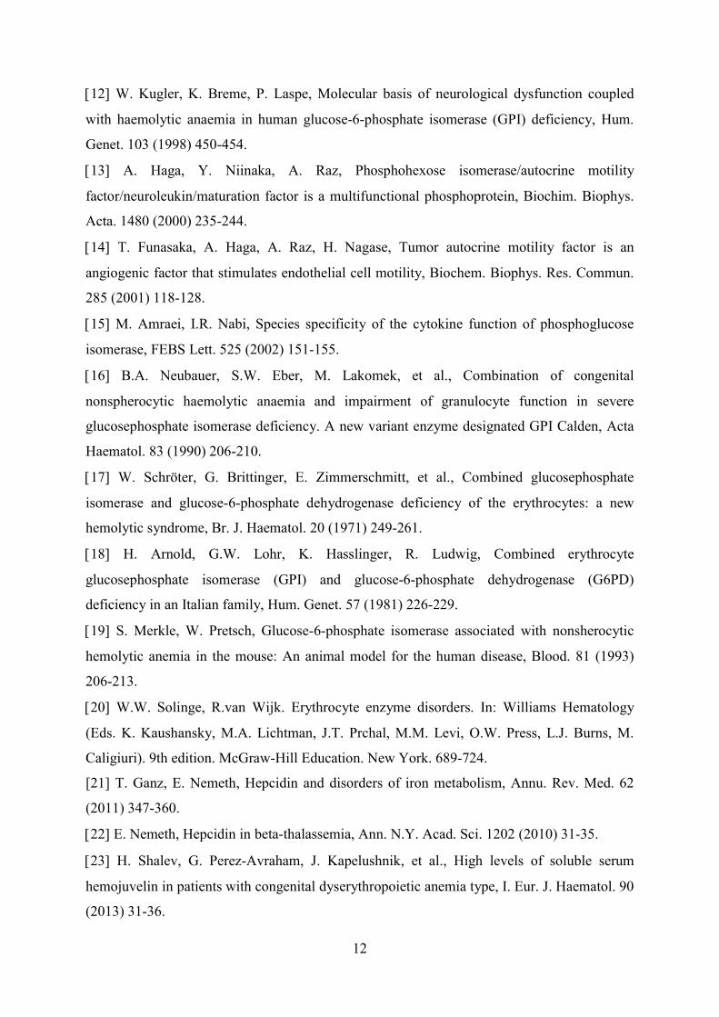
12
12 W. Kugler, K. Breme, P. Laspe, Molecular basis of neurological dysfunction coupled
with haemolytic anaemia in human glucose-6-phosphate isomerase (GPI) deficiency, Hum.
Genet. 103 (1998) 450-454.
13 A. Haga, Y. Niinaka, A. Raz, Phosphohexose isomerase/autocrine motility
factor/neuroleukin/maturation factor is a multifunctional phosphoprotein, Biochim. Biophys.
Acta. 1480 (2000) 235-244.
14 T. Funasaka, A. Haga, A. Raz, H. Nagase, Tumor autocrine motility factor is an
angiogenic factor that stimulates endothelial cell motility, Biochem. Biophys. Res. Commun.
285 (2001) 118-128.
15 M. Amraei, I.R. Nabi, Species specificity of the cytokine function of phosphoglucose
isomerase, FEBS Lett. 525 (2002) 151-155.
16 B.A. Neubauer, S.W. Eber, M. Lakomek, et al., Combination of congenital
nonspherocytic haemolytic anaemia and impairment of granulocyte function in severe
glucosephosphate isomerase deficiency. A new variant enzyme designated GPI Calden, Acta
Haematol. 83 (1990) 206-210.
17 W. Schröter, G. Brittinger, E. Zimmerschmitt, et al., Combined glucosephosphate
isomerase and glucose-6-phosphate dehydrogenase deficiency of the erythrocytes: a new
hemolytic syndrome, Br. J. Haematol. 20 (1971) 249-261.
18 H. Arnold, G.W. Lohr, K. Hasslinger, R. Ludwig, Combined erythrocyte
glucosephosphate isomerase (GPI) and glucose-6-phosphate dehydrogenase (G6PD)
deficiency in an Italian family, Hum. Genet. 57 (1981) 226-229.
19 S. Merkle, W. Pretsch, Glucose-6-phosphate isomerase associated with nonsherocytic
hemolytic anemia in the mouse: An animal model for the human disease, Blood. 81 (1993)
206-213.
20 W.W. Solinge, R.van Wijk. Erythrocyte enzyme disorders. In: Williams Hematology
(Eds. K. Kaushansky, M.A. Lichtman, J.T. Prchal, M.M. Levi, O.W. Press, L.J. Burns, M.
Caligiuri). 9th edition. McGraw-Hill Education. New York. 689-724.
[21] T. Ganz, E. Nemeth, Hepcidin and disorders of iron metabolism, Annu. Rev. Med. 62
(2011) 347-360.
22 E. Nemeth, Hepcidin in beta-thalassemia, Ann. N.Y. Acad. Sci. 1202 (2010) 31-35.
23 H. Shalev, G. Perez-Avraham, J. Kapelushnik, et al., High levels of soluble serum
hemojuvelin in patients with congenital dyserythropoietic anemia type, I. Eur. J. Haematol. 90
(2013) 31-36.

13
24 R. Mojzikova, P. Koralkova, D. Holub, et al., Iron status in patients with pyruvate kinase
deficiency: neonatal hyperferritinaemia associated with a novel frameshift deletion in the
PKLR gene (p.Arg518fs), and low hepcidin to ferritin ratios, Br. J. Haematol. 165 (2014) 556-
563.
25] L. Sulovska, D. Holub, Z. Zidova, et al., Characterization of iron metabolism and
erythropoiesis in erythrocyte membrane defects and thalassemia traits, Biomed. Pap. Med.
Fac. Univ. Palacky Olomouc Czech Repub. 160 (2016) 231-237.
26] D. Pospisilova, D. Holub, Z. Zidova, et al., Hepcidin levels in Diamond-Blackfan anemia
reflect erythropoietic activity and transfusion dependency, Haematologica. 99 (2014) e118-
121.
27] M. Vokurka, J. Krijt, K. Sulc, E. Necas, Hepcidin mRNA levels in mouse liver respond
to inhibition of erythropoiesis, Physiol. Res. 55 (2006) 667-674.
28] M. Pak, M.A. Lopez, V. Gabayan, et al., Suppression of hepcidin during anemia requires
erythropoietic activity, Blood. 108 (2006) 3730-3735
[29] L. Baronciani, A. Zanella, P. Bianchi, et al., Study of the molecular defects in glucose
phosphate isomerase-deficient patients affected by chronic hemolytic anemia, Blood. 88
(1996) 2306-2310.
30 E. Beutler, K.G. Blume, J.C. Kaplan, et al., International committee for standardization
in haematology: recommended methods for red-cell enzyme analysis, Br. J. Haematol. 35
(1977) 331-340.
[31] L. Ronzoni, L. Sonzogni, L. Duca, et al., Growth Differentiation Factor 15 expression
and regulation during erythroid differentiation in non-transfusion dependent thalassemia,
Blood Cells Mol. Dis. 54 (2015) 26-28.
[32] J.S. Guimarães, J.G. Cominal, A.C. Silva-Pinto, et al., Altered erythropoiesis and iron
metabolism in carriers of thalassemia, Eur. J. Haematol. 94 (2015) 511-518.
[33] E. Beutler, C. West, H.A. Britton, et al., Glucosephosphate isomerase (GPI) deficiency
mutations associated with hereditary nonspherocytic hemolytic anemia (HNSHA), Blood
Cells Mol. Dis. 23 (1997) 402-409.
[34] H-Y. Lin, Y-H. Kao, et al., Effect of inherited mutations on catalytic activity and
structural stability of human glucose-6-phosphate isomerase expressed in Escherichia coli,
Biochim. Biophys. Acta. 1794 (2009) 315-323.
[35] A. Zanella, P. Bianchi, A. Iurlo, et al., Iron status and HFE genotype in erythrocyte
pyruvate kinase deficiency: Study of Italian cases, Blood Cells Mol. Dis. 27 (2001) 653-661.

14
[36] L. Kautz, G. Jung, E.V. Valore, et al., Identification of erythroferrone as an erythroid
regulator of iron metabolism, Nat. Genet. 46 (2014) 678-684.
[37] L. Kautz, G. Jung, X. Du, et al., Erythroferrone contributes to hepcidin suppression and
iron overload in a mouse model of β-thalassemia, Blood. 126 (2015) 2031-2037.
[38] H. Han, K. Westerman, V. Ostland, et al., A Novel Dual Monoclonal Sandwich ELISA
for Human Erythroferrone, Blood. 128 (2016) 1272.
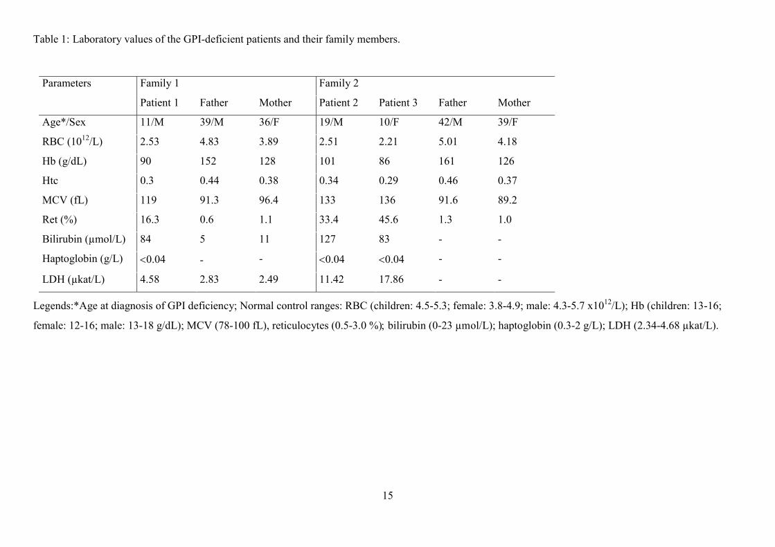
15
Table 1: Laboratory values of the GPI-deficient patients and their family members.
Legends:*Age at diagnosis of GPI deficiency; Normal control ranges: RBC (children: 4.5-5.3; female: 3.8-4.9; male: 4.3-5.7 x10¹²/L); Hb (children: 13-16;
female: 12-16; male: 13-18 g/dL); MCV (78-100 fL), reticulocytes (0.5-3.0 %) bilirubin (0-23 µmol/L); haptoglobin (0.3-2 g/L); LDH (2.34-4.68 µkat/L).
Parameters Family 1 Family 2
Patient 1 Father Mother Patient 2 Patient 3 Father Mother
Age*/Sex 11/M 39/M 36/F 19/M 10/F 42/M 39/F
RBC (1012/L) 2.53 4.83 3.89 2.51 2.21 5.01 4.18
Hb (g/dL) 90 152 128 101 86 161 126
Htc 0.3 0.44 0.38 0.34 0.29 0.46 0.37
MCV (fL) 119 91.3 96.4 133 136 91.6 89.2
Ret (%) 16.3 0.6 1.1 33.4 45.6 1.3 1.0
Bilirubin (µmol/L) 84 5 11 127 83 - -
Haptoglobin (g/L) 0.04 - - 0.04 0.04 - -
LDH (µkat/L) 4.58 2.83 2.49 11.42 17.86 - -

16
Table 2: Panel of red blood cell enzymes assayed in both patients’ families.
Legends: G6PD: glucose-6-phosphate dehydrogenase; GLD: gluconolactone dehydrogenase; PK: pyruvate kinase; GPI: glucosephosphate
isomerase; P5´N: pyrimidine-5´-phosphate nucleotidase; HK: hexokinase; enzyme activities are shown as average ± SD of three independent
measurements.
Enzyme Control range Family 1 Family 2
Patient 1 Father Mother Patient 2 Patient 3 Father Mother
G6PD [IU/g Hb] [5.36-7.04] 10.50±0.478 6.34±0.066 6.44±0.456 8.43±0.815 10.86±1.760 6.69±0.833 7.09±1.045
GLD [IU/g Hb] [5.87-6.71] 10.54±0.494 6.05±0.42 7.23±0.32 9.81±0.635 10.39±0.660 6.05±0.114 8.86±0.611
PK [IU/g Hb] [5.12-5.78] 10.55±0.241 7.01±0.314 6.65±0.234 16.28±0.373 15.5±1.962 5.84±0.414 7.31±0.194
GPI [IU/g Hb] [53-62] 8.51±1.404 29.05±4.13 21.56±1.25 5.94±0.3112 6.75±0.570 21.18±0.733 33.42±1.400
P5´N (pyr/pur) [1.8] 2.29 2.21 1.95 2.04 2.37 2.01 2.16
HK [IU/g Hb] [0.81-1.65] 2.74±0.768 1.59±0.061 1.29±0.071 5.62±0.048 5.85±0.284 0.93±0.078 1.29±0.022
G6PD/GLD [0.80-1.20] 1.00 1.05 0.89 0.86 1.05 1.11 0.80
PK/HK [3.60-8.17] 3.85 4.41 5.16 2.90 2.65 7.48 5.67
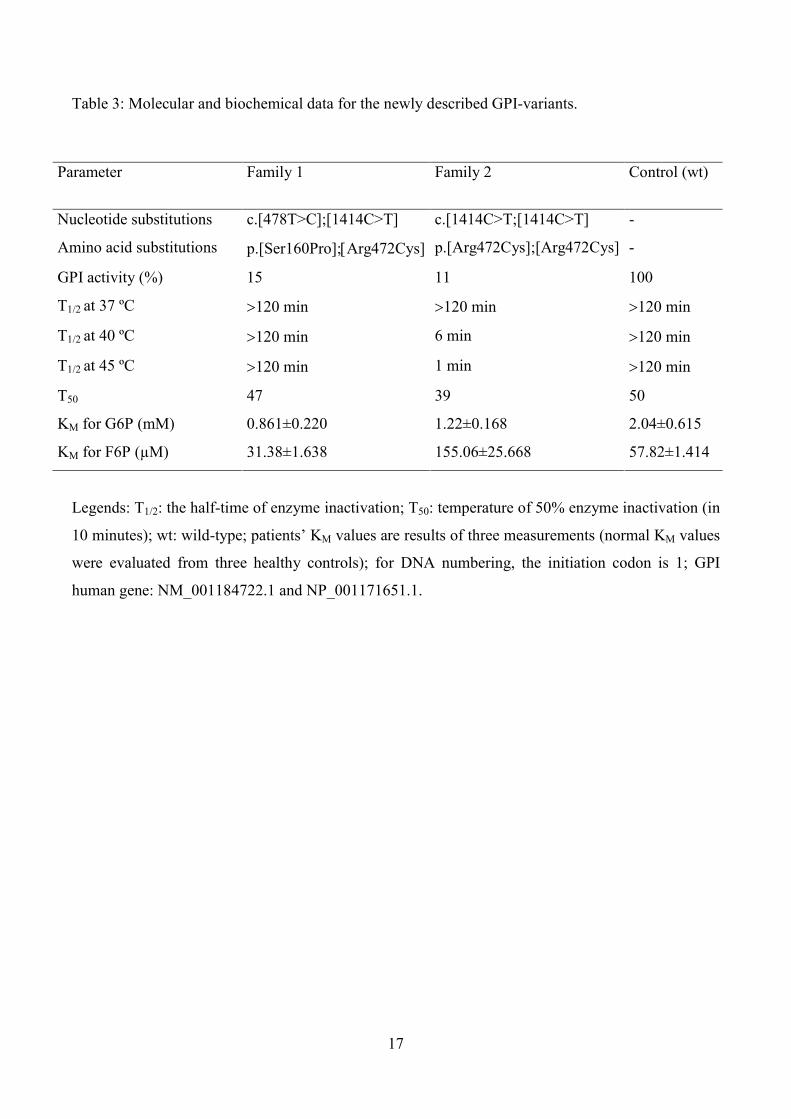
17
Table 3: Molecular and biochemical data for the newly described GPI-variants.
Legends: T1/2: the half-time of enzyme inactivation; T50: temperature of 50% enzyme inactivation (in
10 minutes); wt: wild-type; patients’ KM values are results of three measurements (normal KM values
were evaluated from three healthy controls); for DNA numbering, the initiation codon is 1; GPI
human gene: NM_001184722.1 and NP_001171651.1.
Parameter Family 1 Family 2 Control (wt)
Nucleotide substitutions c.[478T>C];[1414C>T] c.[1414C>T;[1414C>T] -
Amino acid substitutions p.[Ser160Pro];Arg472Cys] p.[Arg472Cys];[Arg472Cys] -
GPI activity (%) 15 11 100
T1/2 at 37 ºC 120 min 120 min 120 min
T1/2 at 40 ºC 120 min 6 min 120 min
T1/2 at 45 ºC 120 min 1 min 120 min
T50 47 39 50
KM for G6P (mM) 0.861±0.220 1.22±0.168 2.04±0.615
KM for F6P (µM) 31.38±1.638 155.06±25.668 57.82±1.414

18
Table 4: Parameters of iron status and erythropoietic activity.
Legends: Serum Fe (10.6-28.3 µmol/L) TSAT - transferrin saturation (21-48%) Ferritin (<200 female, <300 male and <70 μg/L children) Hepcidin (13.1-
104.8 μg/L) Hepcidin/ferritin ratio (0.2-2.2) [24] sTfR: Soluble transferrin receptor (1.9-4.4 mg/L) GDF15: Growth differentiation factor-15 (166-344 ng/L)
[24] EPO: Serum erythropoietin (4.3-29 IU/L).
Parameters Family 1 Family 2
Patient 1 Father Mother Patient 2 Patient 3 Father Mother
Serum Fe (µmol/L) 51 - - 20.4 33.1 - -
TSAT (%) 86 - - 39 84 - -
Ferritin (µg/L) 327 32 68 143 1006 144 11
Hepcidin (µg/L) 28.88 15.74 95.99 5.6 7.9 75.8 2.8
Hepcidin/ferritin ratio 0.089 0.492 1.412 0.039 0.001 0.526 0.255
sTfR (mg/L) 17 - - 24.5 21.6 - -
GDF15 (ng/L) 465 - - 989 777 - -
EPO (IU/L) 34.1 - - 63.8 147 - -

19
Legends to figures
Fig. 1. Bone marrow abnormalities in GPI-deficient Patient 1. Erythroid hyperplasia with
dysplastic changes in erythroid precursors: binuclearity and cytoplasmic bridging were
detected in Patient 1. Bone marrow of Patient 2 and 3 showed similar features (see the
description in Patients and Methods).
Fig. 2. Ribbon plot of the 3D GPI-mutant structures suggested putative changes caused
by the mutations. A) Ser159 (shown in purple) is a part of the phosphate-binding site (blue)
responsible for the potential hydrogen bond interaction with phosphate group of the GPI
substrate (the sulfate was used to mimic the substrate phosphate group). and B) Ser159Pro
substitution likely results in a disruption of the hydrogen bond (a black dotted line) between
the residue at position 159 and the substrate phosphate group and thus destabilizes the
substrate-enzyme complex C) Arg471 is located at the edge of the large external loop (LEL,
438-468 residues) directly interacting (hydrogen bonding) with neighboring Gly469 residue
and with Ile394 of the second monomer, and thus D) disruption of these interactions by
Arg471Cys mutation leads to conformational changes within the LEL, serving as a dimer
stabilizing hook, and thus to generation of a more labile GPI-dimer. The amino acid
numbering in the figures begins with alanine as residue 1 instead of initial methionine. The
structure illustrations were created with PyMOL program based on Protein data bank ID
1IAT. Black dot lines represent hydrogen bonds.
Fig. 3. Differentiation of different disease categories based on the plot of
hepcidin/ferritin ratio against sTfR (A) and (hepcidin/ferritin)/sTfR index (B). Patients
with GPI deficiency (GPI def., n=3) and PK deficiency (PK def., n=8) are shifted to the right
and top in the graph compared to healthy controls (n=10) and show reduced
(hepcidin/ferritin)/sTfR index due to hepcidin suppression by stimulated/ineffective
erythropoiesis. The difference between controls and GPI deficiency in (B) is not statistically
significant because of low number of GPI-deficient patients. The combination of PK- and
GPI-deficient patients clearly shows that (hepcidin/ferritin)/sTfR index is significantly
reduced in glycolytic enzymopathies (enzyme def.) and distinguishes them from healthy
population. Also transfusion dependent DBA patients (DBA_TD, n=7) differ from healthy
controls, but they are shifted to the left in the graph and show increased
(hepcidin/ferritin)/sTfR index due to the absence of erythroid precursors in the bone marrow,
which makes hepcidin level appropriate to increased iron stores.
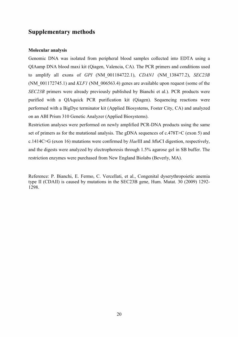
20
Supplementary methods
Molecular analysis
Genomic DNA was isolated from peripheral blood samples collected into EDTA using a
QIAamp DNA blood maxi kit (Qiagen, Valencia, CA). The PCR primers and conditions used
to amplify all exons of GPI (NM_001184722.1), CDAN1 (NM_138477.2), SEC23B
(NM_001172745.1) and KLF1 (NM_006563.4) genes are available upon request (some of the
SEC23B primers were already previously published by Bianchi et al.). PCR products were
purified with a QIAquick PCR purification kit (Qiagen). Sequencing reactions were
performed with a BigDye terminator kit (Applied Biosystems, Foster City, CA) and analyzed
on an ABI Prism 310 Genetic Analyzer (Applied Biosystems).
Restriction analyses were performed on newly amplified PCR-DNA products using the same
set of primers as for the mutational analysis. The gDNA sequences of c.478T>C (exon 5) and
c.1414C>G (exon 16) mutations were confirmed by HaeIII and MluCI digestion, respectively,
and the digests were analyzed by electrophoresis through 1.5% agarose gel in SB buffer. The
restriction enzymes were purchased from New England Biolabs (Beverly, MA).
Reference: P. Bianchi, E. Fermo, C. Vercellati, et al., Congenital dyserythropoietic anemia type II (CDAII) is caused by mutations in the SEC23B gene, Hum. Mutat. 30 (2009) 1292-1298.

21
Supplementary figures
Supplementary Figure S1. DNA sequencing of the GPI gene. Left panels: Patient 1
(Family 1) was a compound heterozygote for c.478T>C (exon 5) and c.1414C>T (exon 16)
mutation in the GPI gene. His father was a heterozygote for the c.478T>C and his mother was
a heterozygote for the c.1414C>T mutation Right panels: Patients 2 and 3 (Family 2) were
homozygotes for the c.1414C>T mutation. Both parents were carriers of this mutation.
Supplementary Figure S2. Restriction enzyme-based detection of two mutations in the
amplified DNA samples from the patients and their parents.
The 550-bp PCR amplified product from the wild-type allele contains five recognition sites
for HaeIII that yields fragments of 279, 142, 68, 50, 7 and 4 bp (the last two small fragments
are not visible on the gel). The mutation creates another recognition sequence for this enzyme
resulting in additional fragments of 160 bp and 119 bp. Heterozygosity for the c.478T>C
mutation was confirmed in Patient 1 (PAT 1, family 1) and also detected in his father (F1),
whereas it is absent in the patient’s mother (M1). As father is a carrier of heterozygous
c.489A>G (SNP, p.Gly163Gly, rs1801015) mutation, which curiously generates another
restriction site, another fragment (100 bp) is produced after digestion of the father's mutant
allele.
Mutation c.1414C>G creates a new restriction site for MluCI and consequently 431 bp and
198 bp fragments are produced after digestion of the mutant allele, whereas 629 bp fragment
is the result of digestion of the wild-type allele with this restriction enzyme. Heterozygosity
for this mutation was confirmed in Patient 1 (PAT1, family 1) and his mother (M1) and both
parents (M2 and F2) of Patient 2 and 3 (PAT2 and PAT3, family 2), whereas both Patients 2
and 3 (PAT2 and PAT3) are homozygotes for c.1414C>G mutation. C, healthy control.

22

23

24
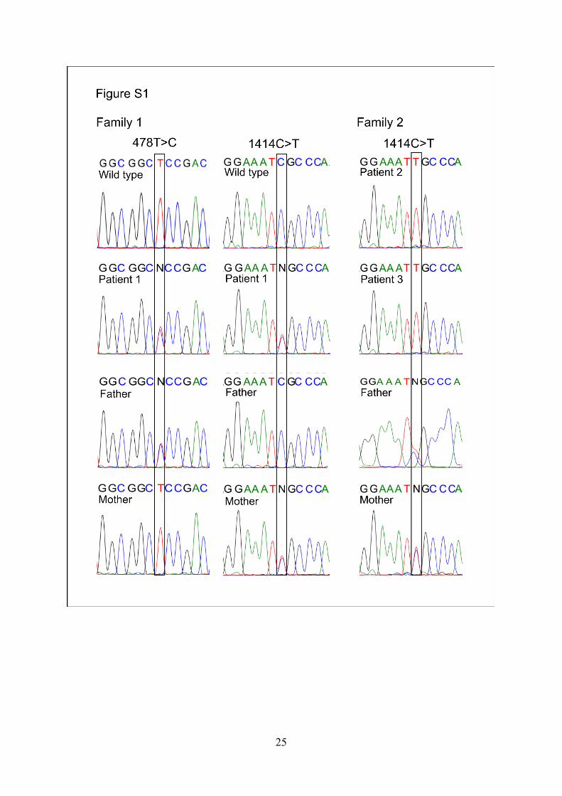
25

26

APPENDICES 4.3
STRUCTURAL AND FUNCTIONAL CHARACTERIZATION OF VERY RARE HEXOKINASE MUTANTS CAUSING HNSHA
Blood Cells, Molecules and Diseases 59 (2016) 71–76
Contents lists available at ScienceDirect
Blood Cells, Molecules and Diseases
j ourna l homepage: www.e lsev ie r .com/ locate /bcmd
Molecular characterization of six new cases of red blood cell hexokinasedeficiency yields four novel mutations in HK1
Pavla Koralkova a,b, Renata Mojzikova a, Brigitte van Oirschot b, Christine Macartney c, Pavel Timr d,Joan Lluis Vives Corrons e, Zuzana Striezencova Laluhova f, Katerina Lejhancova g,Vladimir Divoky a,h, Richard van Wijk b,⁎a Department of Biology, Faculty of Medicine and Dentistry, Palacký University, Olomouc, Czech Republicb Department of Clinical Chemistry and Haematology, University Medical Center Utrecht, Utrecht, The Netherlandsc Royal Belfast Hospital for Sick Children, Belfast, Northern Ireland, United Kingdomd Department of Pediatrics, Ceske Budejovice Hospital, Ceske Budejovice, Czech Republice Hospital Clinic I Provencial, Barcelona, Spainf Children's Faculty Hospital with Policlinic, Bratislava, Slovak Republicg Department of Paediatrics, Faculty of Medicine in Hradec Kralove, Charles University in Prague, Hradec Kralove, Czech Republich Department of Hemato-Oncology, University Hospital and Faculty of Medicine and Dentistry Palacký University, Olomouc, Czech Republic
⁎ Corresponding author at: Department of ClinicalUniversity Medical Center Utrecht, RoomG.03.550, PO BoNetherlands.
E-mail address: [email protected] (R. van Wijk
http://dx.doi.org/10.1016/j.bcmd.2016.04.0021079-9796/© 2016 Elsevier Inc. All rights reserved.
a b s t r a c t
a r t i c l e i n f oArticle history:Submitted 27 February 2016Revised 8 April 2016Accepted 10 April 2016Available online 22 April 2016
Edited by: Mohandas Narla
Hexokinase (HK) is a key enzymeof glycolysis, the onlymetabolic pathway able to provide the red blood cellwithATP. HK deficiency is a very rare hereditary disorder with severe chronic nonspherocytic hemolytic anemia(HNSHA) as a major clinical feature. To date, only 24 patients with HK deficiency have been identified. Here,we report themolecular analysis of six new cases of HK deficiency. A total of six different mutations were detect-ed in HK1, four of them described here for the first time: c.2599C N T p.(His867Tyr), c.1799C N T p.(Thr600Met),c.873-2A N G and c.493-1G N A. The pathogenic nature of the identified missense mutations was confirmed bybiochemical and 3-dimensional structural analysis. The effects of the novel splice site mutation c.873-2A N Gwere studied at the level of pre-mRNA processing, and confirmed at the protein level. All together, these resultsprovide a better insight into the pathogenesis of this rare red cell disorder, and contribute to a better understand-ing of the genotype-phenotype correlation in HK deficiency.
© 2016 Elsevier Inc. All rights reserved.
Keywords:Red blood cellsGlycolysisHexokinase deficiencyHemolytic anemia
1. Introduction
Hexokinase (ATP: D-hexose-6-phosphotransferase, EC 2.7.1.1) is akey regulatory enzyme for the Embden-Meyerhof pathway (or glycoly-sis). Hexokinase (HK) catalyzes the irreversible transfer of high-energyphosphate fromATP to glucose, onwhich red blood cells' (RBCs) energymetabolism is fully dependent [1].
Red blood cells contain twoHK isozymes –HK-1 andHK-R. Both iso-zymes are encoded by the same HK1 gene located on chromosome 10and their expression is regulated by the use of alternative promoters.The HK-R isozyme is exclusively expressed in erythroid cells, whereasHK-1 is present in various mammalian tissues [2].
HK deficiency is a very rare autosomal recessive disorder belongingto a group of very rare red cell enzymedefects (erythroenzymopathies).Generally, HK deficiency is associated with chronic nonspherocytic
Chemistry and Haematology,x 85500, 3508GA Utrecht, The
).
hemolytic anemia (HNSHA) [3]. Affected individuals exhibit mild to se-vere lifelong hemolytic anemia (HA) as the major clinical hallmark. Inexceptional cases, however, multiple malformations including psycho-motor retardation have been also reported [4–6]. A complete loss ofHK1 expression leads to intrauterine fetal death [7].
The first case of HK deficiency was described by Valentine et al. in1967 [8]. Up to now, only 24 cases of HK deficiency have ever been re-ported [9]. This apparently low prevalence may indicate that in manycases HK deficiency is incompatible with life, perhaps because of theenzyme's crucial role of in glycolysis. Alternatively, many (mild) casesmay remain undiagnosed due to the complicated clinical and laboratorydiagnosis, i.e. the lack of appropriate diagnostic tools (only a few labora-tories performHKenzymatic assays). Only 4 cases of HKdeficiency havebeen characterized at the molecular level [9], and very little is knownabout the genotype-to-phenotype correlation in HK deficiency.Six distinct mutations have been identified thus far: three missensemutations, two deletions, and one mutation located in the erythroid-specific promotor [10]. The first patient characterized at the molecularlevel was a compound heterozygote for a missense mutationc.1583T N C p.(Leu528Ser), in trans to a 96-bp deletion (HK – Melzo)

72 P. Koralkova et al. / Blood Cells, Molecules and Diseases 59 (2016) 71–76
[11]. The third reported mutation concerned a large deletion, c.493-481_1028+ 454del in HK1, found in the homozygous state and associ-ated with severe HA and fetal death [7]. In another patient with HKdeficiency and mild HA [12], a homozygous missense mutationc.2036C N G p.(Thr679Ser) (HK – Utrecht) was found to be the underly-ing cause [13]. The last patient characterized at the molecular levelshowed a mutation in the erythroid specific promotor −193A N G, intrans to a missense mutation c.278A N G p.(Arg93Gln). The latter muta-tion mainly exerted its effect by affecting pre-mRNA processing [14].We describe six new cases of HK deficiency here. All of themwere stud-ied at the molecular level, and the causative mutations were identifiedand studied at different levels to establish the pathogenic nature. Oneadditional patient was a G6PD deficient female with heterozygosityfor HK1 gene mutation.
2. Materials and methods
2.1. Patients
This study was performed according to the Helsinki internationalstandards; informed consent was obtained from all patients and familymembers. We analyzed seven patients from 6 unrelated families with adifferent country of origin – Ireland, the Czech Republic, Denmark, theNetherlands and the Slovak Republic. The selected laboratory parame-ters are summarized in Table 1.
Therewas noHA family history in any of the families concerned. Themajority of patients exhibited common signs of chronic hemolysis. Redcell membrane defects and hemoglobinopathies were excluded as apossible cause of HA. All but one of the patients were pediatric patientsat the time of analysis. Patient 1 underwent transfusion and chelationtherapy until age 3. However, his brother, patient 2, exhibited compen-sated HAwith no clinical symptoms. Patient 3 was born with a massivereticulocytosis (99%) after an uneventful pregnancy from consanguine-ous parents (first cousins). Further examination of the newbornrevealed a central nervous system bleeding (CNS), likely as a resultof hypoxia. Severe hemolysis was also observed in patient 4. Both pa-tients 3 and 4 required regular blood transfusions from birth andunderwent chelation therapy. Patients 5 and 6 were first diagnosedwith Gilbert syndrome. Because of an accompanying mild hemolysiswith reticulocytosis, additional examinations were performed. Patient7 exhibited only mild methemoglobinemia (7–11%) with no signs ofhemolysis.
2.2. Red blood cell enzyme activities
A standard panel of enzyme activity assays (including HK, glucose-6-phosphate dehydrogenase (G6PD), glucose-6-phosphate isomerase(GPI) and pyruvate kinase (PK)) was performed in leukocyte/platelet-free erythrocyte lysates according to standard methods [15]. The PK/HK
Table 1Hematological data of the studied patients.
Origin Patient 1S Patient 2S Patient 3
Ireland Ireland Czech Republic
Sex M M MAge [year] 2 4 8 mRBCs [×1012] 2.57; (*--) 2.76; (*--) 2.40; (*--)Hb [g/L] 79; (*--) 95; (*--) 74; (*--)Ret [%] 8.8; (--*) 6.2; (--*) 6.3; (--*)Bili [μmol/L] 54; (--*) 62; (--*) 120; (--*)Transfusion therapy Yes+ No YesOther diagnosis ND ND ND
S – siblings; Age – age at diagnosis of HK deficiency, m –months; RBCs – red blood cells; Hb – hglucose-6-phosphate deficiency, variant – GOND; +Transfusion therapy has been stoppednormal; (–*) – high.
ratio was used to evaluate the influence of high reticulocyte numberson total enzymatic activity.
2.3. Molecular analysis of HK1
DNAwas isolated from peripheral blood using standard procedures.HK-R transcript (accession number: NM_033496.2) encompassing thespecific erythroid promoter region and erythroid-specific exon 1 wereamplified using PCR, according to the previously described conditions[13]. DNA sequencing analysis was performed using BigDye Terminatorv1.1 Cycle Sequencing Kit (Applied Biosystems).
2.4. Hexokinase
2.4.1. Kinetic properties and stability assaysTo determine KM values for glucose and ATP, different concentra-
tions of glucose: 0.0125–2.0 mM and/or ATP 0.25–10 mM were usedin a reaction mixture containing: 0.1 M Tris-HCL/0.5 mM EDTA, pH =8.0; 0.01MMgCl2; 0.2mMNADP+; 0.1 IU/mLG6PD and 50 μL of RBC ly-sate (1:20). After 10 min incubation at 37 °C, changes in absorbance at340 nmwere recorded at 20 s intervals for 30min. The thermal stabilityassays of HK mutants involve determining the half-life (T1/2) and thehalf-inactivation temperature (T50). Both thermal stability assays wereperformed in the previously described mixture, routinely containing2 mM glucose and 10 mM ATP. For the T1/2 assay, RBC lysate was pre-heated to 40 °C for 5–40 min followed by cooling on ice (2 min). Afterthe pre-heating step, 50 μL of the RBC lysate was used in the reactionmixture as described above. T1/2 was defined as the time needed toreach a 50% reduction in the initial activity [16]. For the T50 assay, RBClysate was pre-incubated at different temperatures (37–44 °C) for5 min, followed by a cooling period (2 min) on ice. The T50 was definedas the temperature atwhichHKmaintains 50%of its activity. ThepHsta-bility assay was carried out in a 0.1 M Tris-HCL/0.5 mM EDTA buffer ofdifferent pH values (6.0–10.0) [12,15]. Data to determine KM were ana-lyzed with non-linear regression using GraphPad® Prism.
2.4.2. Cell culture of human nucleated erythroid cellsBriefly, human peripheral blood mononuclear cells (PBMCs) were
isolated from a heterozygous individual carrying the splice site muta-tion c.873-2A N G and a healthy control using Ficoll-Paque density gra-dient centrifugation (GE Healthcare). Cells were grown in StemSpanSFEM (Stemcell Technology), supplemented with human stem cell fac-tor (100 ng/mL; Amgen), erythropoietin (2 U/mL; Eprex), interleukin-3(1 ng/mL; Sigma-Aldrich), dexamethasone (1 μmol/L; Sigma-Aldrich)and SyntheChol™ NS0 Supplement (200-times diluted; Sigma-Aldrich). The cells were cultured at 37 °C in a humidified atmosphereof 5% CO2 in air. The Casy Cell Counter and Analyzer (Roche Life Science)was used tomonitor cell viability and proliferation. After 10 days of ery-throid proliferation, cells (mainly pro-erythroblasts) were harvested toisolate total RNA and carry out a Western blot analysis.
Patient 4 Patient 5 Patient 6 Patient 7
Denmark Netherlands Czech Republic Slovak Republic
M M F F2 22 12 122.90; (*--) 4.40; (*--) 4.25; (-*-) 4.58; (-*-)86; (*--) 160; (-*-) 135; (-*-) 137; (-*-)4.7; (--*) 6.0; (--*) 3.1; (--*) 1.9; (--*)124; (--*) 24; (--*) 100; (--*) 17.6; (-*-)Yes No No NoND GS GS G6PD Gond
emoglobin,M –male; F – female; ND – not determined, GS – Gilbert syndrome; G6PD –at age 3; comparison of hematological data with a reference range: (*–) – low; (-*-) –
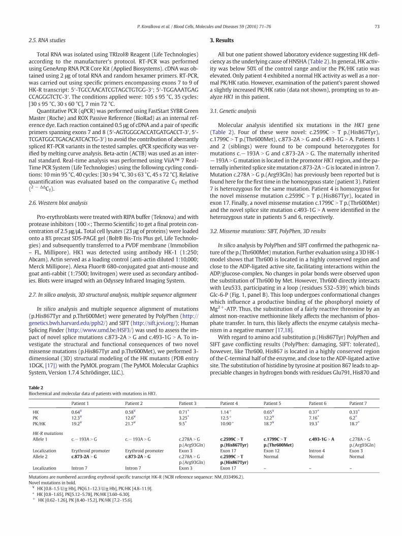
73P. Koralkova et al. / Blood Cells, Molecules and Diseases 59 (2016) 71–76
2.5. RNA studies
Total RNA was isolated using TRIzol® Reagent (Life Technologies)according to the manufacturer's protocol. RT-PCR was performedusing GeneAmp RNA PCR Core Kit (Applied Biosystems). cDNAwas ob-tained using 2 μg of total RNA and random hexamer primers. RT-PCR,was carried out using specific primers encompassing exons 7 to 9 ofHK-R transcript: 5′-TGCCAACATCGTAGCTGTGG-3′; 5′-TGGAAATGAGCCAGGGTCTC-3′. The conditions applied were: 105 s 95 °C, 35 cycles:[30 s 95 °C, 30 s 60 °C], 7 min 72 °C.
Quantitative PCR (qPCR) was performed using FastStart SYBR GreenMaster (Roche) and ROX Passive Reference (BioRad) as an internal ref-erence dye. Each reaction contained 0.5 μg of cDNA and a pair of specificprimers spanning exons 7 and 8 (5′-AGTGGGCACCATGATGACCT-3′, 5′-TCGATGGCTGACACATCACTG-3′) to avoid the contribution of aberrantlyspliced RT-PCR variants in the tested samples. qPCR specificity was ver-ified by melting curve analysis. Beta-actin (ACTB) was used as an inter-nal standard. Real-time analysis was performed using ViiA™ 7 Real-Time PCR System (Life Technologies) using the following cycling condi-tions: 10min 95 °C, 40 cycles: [30 s 94 °C, 30 s 63 °C, 45 s 72 °C]. Relativequantification was evaluated based on the comparative CT method(2 − ΔΔCT).
2.6. Western blot analysis
Pro-erythroblasts were treatedwith RIPA buffer (Teknova) andwithprotease inhibitors (100×; Thermo Scientific) to get a final protein con-centration of 2.5 μg/μL. Total cell lysates (23 μg of proteins) were loadedonto a 8% precast SDS-PAGE gel (Bolt® Bis-Tris Plus gel, Life Technolo-gies) and subsequently transferred to a PVDF membrane (Immobilion– FL, Millipore). HK1 was detected using antibody HK-1 (1:250;Abcam). Actin served as a loading control (anti-actin diluted 1:10,000;Merck Millipore). Alexa Fluor® 680-conjugated goat anti-mouse andgoat anti-rabbit (1:7500; Invitrogen) were used as secondary antibod-ies. Blots were imaged with an Odyssey Infrared Imaging System.
2.7. In silico analysis, 3D structural analysis, multiple sequence alignment
In silico analysis and multiple sequence alignment of mutations(p.His867Tyr and p.Thr600Met) were generated by PolyPhen (http://genetics.bwh.harvard.edu/pph2/) and SIFT (http://sift.jcvi.org/); HumanSplicing Finder (http://www.umd.be/HSF3/) was used to assess the im-pact of novel splice mutations c.873-2A N G and c.493-1G N A. To in-vestigate the structural and functional consequences of two novelmissense mutations (p.His867Tyr and p.Thr600Met), we performed 3-dimensional (3D) structural modeling of the HK mutants (PDB entry1DGK, [17]) with the PyMOL program (The PyMOL Molecular GraphicsSystem, Version 1.7.4 Schrödinger, LLC.).
Table 2Biochemical and molecular data of patients with mutations in HK1.
Patient 1 Patient 2 Patient 3
HK 0.64¥ 0.58¥ 0.71⁎
PK 12.3¥ 12.6¥ 3.25⁎
PK/HK 19.2¥ 21.7¥ 9.5⁎
HK-R mutationsAllele 1 c.−193A N G c.−193A N G c.278A N G
p.(Arg93Gln)Localization Erythroid promoter Erythroid promoter Exon 3Allele 2 c.873-2A N G c.873-2A N G c.278A N G
p.(Arg93Gln)Localization Intron 7 Intron 7 Exon 3
Mutations are numbered according erythroid specific transcript HK-R (NCBI reference sequencNovel mutations in bold.
¥ HK [0.8–1.5 U/g Hb], PK[6.1–12.3 U/g Hb], PK/HK [4.8–11.9].⁎ HK [0.8–1.65], PK[5.12–5.78], PK/HK [3.60–6.30].+ HK [0.62–1.26], PK [8.40–15.2], PK/HK [7.2–15.6].
3. Results
All but one patient showed laboratory evidence suggesting HK defi-ciency as the underlying cause of HNSHA (Table 2). In general, HK activ-ity was below 50% of the control range and/or the PK/HK ratio waselevated. Only patient 4 exhibited a normal HK activity as well as a nor-mal PK/HK ratio. However, examination of the patient's parent showeda slightly increased PK/HK ratio (data not shown), prompting us to an-alyze HK1 in this patient.
3.1. Genetic analysis
Molecular analysis identified six mutations in the HK1 gene(Table 2). Four of these were novel: c.2599C N T p.(His867Tyr),c.1799C N T p.(Thr600Met), c.873-2A N G and c.493-1G N A. Patients 1and 2 (siblings) were found to be compound heterozygotes formutations c.−193A N G and c.873-2A N G. The maternally inherited−193A N Gmutation is located in the promotorHK1 region, and the pa-ternally inherited splice sitemutation c.873-2ANG is located in intron 7.Mutation c.278A N G p.(Arg93Gln) has previously been reported but isfound here for thefirst time in the homozygous state (patient 3). Patient7 is heterozygous for the same mutation. Patient 4 is homozygous forthe novel missense mutation c.2599C N T p.(His867Tyr), located inexon 17. Finally, a novel missense mutation c.1799C N T p.(Thr600Met)and the novel splice site mutation c.493-1G N A were identified in theheterozygous state in patients 5 and 6, respectively.
3.2. Missense mutations: SIFT, PolyPhen, 3D results
In silico analysis by PolyPhen and SIFT confirmed the pathogenic na-ture of the p.(Thr600Met)mutation. Further evaluation using a 3DHK-1model shows that Thr600 is located in a highly conserved region andclose to the ADP-ligated active site, facilitating interactions within theADP/glucose-complex. No changes in polar bonds were observed uponthe substitution of Thr600 by Met. However, Thr600 directly interactswith Leu533, participating in a loop (residues 532–539) which bindsGlc-6-P (Fig. 1, panel B). This loop undergoes conformational changeswhich influence a productive binding of the phosphoryl moiety ofMg2+-ATP. Thus, the substitution of a fairly reactive threonine by analmost non-reactive methionine likely affects the mechanism of phos-phate transfer. In turn, this likely affects the enzyme catalysis mecha-nism in a negative manner [17,18].
With regard to amino acid substitution p.(His867Tyr) PolyPhen andSIFT gave conflicting results (PolyPhen: damaging, SIFT: tolerated),however, like Thr600, His867 is located in a highly conserved regionof the C-terminal half of the enzyme, and close to the ADP-ligated activesite. The substitution of histidine by tyrosine at position 867 leads to ap-preciable changes in hydrogen bonds with residues Glu791, His870 and
Patient 4 Patient 5 Patient 6 Patient 7
1.14+ 0.65¥ 0.37⁎ 0.33⁎
12.5+ 12.2¥ 7.16⁎ 6.2⁎
10.90+ 18.7¥ 19.3⁎ 18.7⁎
c.2599C N Tp.(His867Tyr)
c.1799C N Tp.(Thr600Met)
c.493-1G N A c.278A N Gp.(Arg93Gln)
Exon 17 Exon 12 Intron 4 Exon 3c.2599C N Tp.(His867Tyr)
Normal Normal Normal
Exon 17 – – –
e: NM_033496.2).
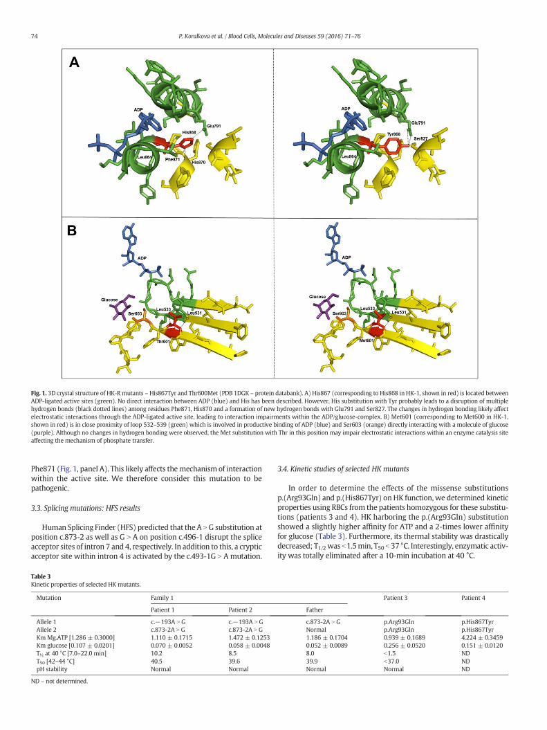
Fig. 1. 3D crystal structure of HK-R mutants – His867Tyr and Thr600Met (PDB 1DGK – protein databank). A) His867 (corresponding to His868 in HK-1, shown in red) is located betweenADP-ligated active sites (green). No direct interaction between ADP (blue) and His has been described. However, His substitution with Tyr probably leads to a disruption of multiplehydrogen bonds (black dotted lines) among residues Phe871, His870 and a formation of new hydrogen bonds with Glu791 and Ser827. The changes in hydrogen bonding likely affectelectrostatic interactions through the ADP-ligated active site, leading to interaction impairments within the ADP/glucose-complex. B) Met601 (corresponding to Met600 in HK-1,shown in red) is in close proximity of loop 532–539 (green) which is involved in productive binding of ADP (blue) and Ser603 (orange) directly interacting with a molecule of glucose(purple). Although no changes in hydrogen bonding were observed, the Met substitution with Thr in this position may impair electrostatic interactions within an enzyme catalysis siteaffecting the mechanism of phosphate transfer.
74 P. Koralkova et al. / Blood Cells, Molecules and Diseases 59 (2016) 71–76
Phe871 (Fig. 1, panel A). This likely affects themechanism of interactionwithin the active site. We therefore consider this mutation to bepathogenic.
3.3. Splicing mutations: HFS results
Human Splicing Finder (HFS) predicted that the A NG substitution atposition c.873-2 as well as G N A on position c.496-1 disrupt the spliceacceptor sites of intron 7 and 4, respectively. In addition to this, a crypticacceptor site within intron 4 is activated by the c.493-1G N A mutation.
Table 3Kinetic properties of selected HK mutants.
Mutation Family 1
Patient 1 Patient 2
Allele 1 c.−193A N G c.−193A N GAllele 2 c.873-2A N G c.873-2A N GKm Mg.ATP [1.286 ± 0.3000] 1.110 ± 0.1715 1.472 ± 0.1253Km glucose [0.107 ± 0.0201] 0.070 ± 0.0052 0.058 ± 0.0048T½ at 40 °C [7.0–22.0 min] 10.2 8.5T50 [42–44 °C] 40.5 39.6pH stability Normal Normal
ND – not determined.
3.4. Kinetic studies of selected HK mutants
In order to determine the effects of the missense substitutionsp.(Arg93Gln) and p.(His867Tyr) on HK function, we determined kineticproperties using RBCs from the patients homozygous for these substitu-tions (patients 3 and 4). HK harboring the p.(Arg93Gln) substitutionshowed a slightly higher affinity for ATP and a 2-times lower affinityfor glucose (Table 3). Furthermore, its thermal stability was drasticallydecreased; T1/2 was b1.5min, T50 b 37 °C. Interestingly, enzymatic activ-ity was totally eliminated after a 10-min incubation at 40 °C.
Patient 3 Patient 4
Father
c.873-2A N G p.Arg93Gln p.His867TyrNormal p.Arg93Gln p.His867Tyr1.186 ± 0.1704 0.939 ± 0.1689 4.224 ± 0.34590.052 ± 0.0089 0.256 ± 0.0520 0.151 ± 0.01208.0 b1.5 ND39.9 b37.0 NDNormal Normal ND
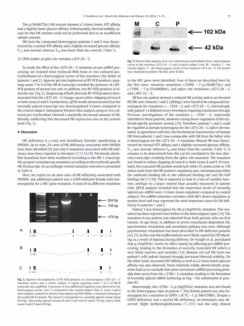
Fig. 3. Western blot analysis of ex-vivo cultured pro-erythroblasts from a heterozygouscarrier of the mutation c.873-2A N G and a control subject. Line: M – marker; C – thecontrol subject; P – the heterozygous carrier of the mutation c.873-2A N G. HK proteinwas visualized in position 102 kDa; actin 42 kDa.
75P. Koralkova et al. / Blood Cells, Molecules and Diseases 59 (2016) 71–76
The p.(His867Tyr) HK mutant showed a 3-times lower ATP affinityand a slightly lower glucose affinity. Unfortunately, thermal stability as-says for this HK mutant could not be performed due to an insufficientsample amount.
HK from the compound heterozygous patients 1 and 2 was charac-terized by a normal ATP affinity and a slightly increased glucose affinity.T1/2 was normal, whereas T50 was lower than the controls (Table 3).
3.5. RNA studies of splice site mutation c.873-2A N G
To study the effect of the c.873-2A N G mutation on pre-mRNA pro-cessing, we isolated total erythroid RNA from ex vivo cultured pro-erythroblasts of a heterozygous carrier of this mutation (the father ofpatients 1 and 2). Agarose gel electrophoresis of RT-PCR products span-ning exons 7 to 9 of the HK-R transcript revealed the presence of a RT-PCR product of normal size and, in addition, two RT-PCR products of al-ternate size (Fig. 2). Sequencing of both alternate RT-PCR products dem-onstrated that the c.873-2A N G change causes either skipping of exon 8or both exons 8 and 9. Furthermore, qPCR results demonstrated that thenormally spliced transcript was downregulated 3-times compared tothe control subject. SubsequentWestern blot analysis using ex vivo cul-tured pro-erythroblast showed a markedly decreased amount of HK,thereby confirming this decreased HK expression also at the proteinlevel (Fig. 3).
4. Discussion
HK deficiency is a very rare hereditary disorder manifesting asHNSHA. Up to now, 24 cases of HK deficiency associated with HNSHAhave been identified [9] and only 6 mutations associated with HK defi-ciency have been reported in literature [7,13,14,19]. Previously identi-fied mutations have been numbered according to the HK-1 transcript.We propose renumberingmutations according to the erythroid specificHK-R transcript. An accordingly revisedmutation overview is presentedin Table 4.
Here, we report on six new cases of HK deficiency associated withHSNHA; one additional patient was a G6PD deficient female with het-erozygosity for a HK1 gene mutation. A total of six different mutations
Fig. 2. Agarose electrophoresis of RT-PCR products of a heterozygous c.873-2A N Gmutation carrier and a control subject. A region spanning exons 7 to 9 of HK-Rtranscript was amplified. A presence of two additional fragments was observed in theheterozygous carrier (line 1) compared to the control subject (line 2). Lines 3 and 4were negative controls for reverse transcription and PCR. Below: a schematic drawing ofall created HK-R variants. The variant A corresponds to a normally spliced variant (band834 bp). Aberrantly spliced variants B and C lack exon 8 (band 735 bp) and/or bothexons 8 and 9 (band 470 bp).
in the HK1 gene were identified. Four of them are described here forthe first time: missense mutations c.2599C N T p.(His867Tyr) andc.1799C N T p.(Thr600Met), and splice site mutations c.873-2A N Gand c.493-1G N A.
All but one patient showed a reduced HK activity and/or an elevatedPK/HK ratio. Patients 1 and 2 (siblings)were found to be compound het-erozygous for mutations c.−193A N G and c.873-2A N G. Interestingly,only patient 1 exhibited severe hemolysis requiring transfusion therapy.Previous investigations of the mutation c.−193A N G, maternallyinherited in these patients, showed a strong down-regulation of the ery-throid specific promoter activity [14]. Therefore, patients 1 and 2 couldbe regarded as pseudo homozygous for the c.873-2A N G splice site mu-tation. In agreementwith this, the biochemical characteristics ofmutantHK from patients 1 and 2 was comparable with HK from the father whois heterozygous for the c.873-2A N G mutation. Mutant HK was charac-terized by normal ATP affinity and a slightly increased glucose affinity.T1/2 was normal, whereas T50 was lower than the controls (Table 3). Itremains to be determined how this can be correlated to the two aber-rant transcripts resulting from the splice site mutation. The mutationwas found to induce skipping of exon 8 or both exons 8 and 9. If trans-lated, the truncated HK protein would lack either 52 amino acids or 130amino acids from the HK protein's regulatory part, encompassing eitherthe substrate binding site or the substrate binding site and the G6Pbinding site [17,20]. This is expected to lead to a loss of catalytic func-tion, perhaps to a larger extend than actually measured in patientcells. QPCR analysis revealed that the expression levels of normallyspliced pre-mRNA were 3-times down-regulated compared to controlsubjects. Pre-mRNA reduction correlates with HK's down-regulation atprotein level and may represent the most important cause for HK defi-ciency in patients 1 and 2.
Patient 3 was homozygous for the p.(Arg93Gln) mutation. This mu-tation has been reported once before in the heterozygous state [14]. Themutation in our patient was inherited from both parents who are firstcousins. At age three, in addition to severe transfusion dependent HA,psychomotor retardation and secondary epilepsy was seen. Althoughpsychomotor retardation has been described in HK deficient patients[4,5,21], in this case themalformationswere likely caused by CNSbleed-ing as a result of hypoxia during delivery. De Vooght et al. postulatedthat p.(Arg93Gln) exerts its effect mainly by affecting pre-mRNA pro-cessing, leading to the formation of severely truncated HK which isvery likely inactive and unstable [14]. Mutant red cell HK from ourpatient's cells indeed showed strongly decreased thermal stability. Onthe other hand, increased ATP affinity aswell as a 2-times lower glucoseaffinity was also observed. These relatively mildly altered kinetic prop-erties lead us to conclude that somenormal pre-mRNAprocessing prob-ably does occur from the c.278A N Gmutation, leading to the formationof normally spliced mRNA harboring an Arg N Gln substitution at posi-tion 93.
Interestingly, the c.278A N G p.(Arg93Gln) mutation was also foundin the heterozygous state in patient 7. This female patient was also ho-mozygous for a mutation in G6PD: c.477G N C p.(Met159Ile). Despite aG6PD deficiency and a partial HK deficiency, no hemolysis was ob-served. Slight methemoglobinemia (7–11%) was the only clinical

Table 4Overview of mutations associated with HK deficiency.
Accession number Transcript HK-1 NM_000188.2 Transcript HK-R NM_033496.2 Localization References
Mutation AA substitution Mutation AA substitution
CR099859 – −193ANG – Erythroid promoter [14]CS099858 c.291A N G p.(Arg94Gln) c.278ANG p.(Arg93Gln) Exon 3 [14]CG024909 c.496-481_1031 + 454del – c.493-481_1028 + 454del – Intron 4–8 [7]CG952319 c.496_del592 p.(166-197) c.493_589del p.(165-196del) Exon 5 [11]CM950628 1586T N C p.(Leu529Ser) c.1583T N C p.(Leu528Ser) Exon 11 [11]CM030044 c.2039C N G p.(Thr680Ser) c.2036C N G p.(Thr679Ser) Exon 15 [13]
c.496-1G N A – c.493-1G N A – Intron 4 This studyc.876-2A N G – c.873-2A N G – Intron 7 This studyc.1802C N T p.(Thr601Met) c.1799C N T p.(Thr600Met) Exon 12 This studyc.2602C N T p.(His868Tyr) c.2599CNT p.(His867Tyr) Exon 17 This study
76 P. Koralkova et al. / Blood Cells, Molecules and Diseases 59 (2016) 71–76
symptom present in this patient. A G6PD deficiency is mostly asymp-tomatic, however, mild methemoglobinemia has been described inseveral G6PD deficiency cases [22]. Although G6PD and partial HK defi-ciencies are well tolerated in this patient, potential negative factors asinfections, drug administration or aging could induce hemolysis laterin life.
The novel c.2599C N T p.(His867Tyr)mutation in patient 4was asso-ciated with severe transfusion-dependent HA. Based on 3D structuralmodeling of this HK mutant, we found that His867 is located in theproximity of the ADP-ligated active site and, thus may impair the cata-lytic process of high-energy phosphate's transfer to glucose. This wassupported by a strongly increased Michaelis-Menten constant for ATP(Table 3) as calculated frommutant HK from this patient's red cells. Pa-tient 4′s severe phenotype illustrates the residue 867′s importance inproper HK function. The same catalytic process is likely affected in pa-tient 5 heterozygous for the p.(Thr600Met) mutation. The substitutedthreonine, also located near the ADP-ligated active site, directly affectsLeu533 residue in a loop (residues 532–539), which binds Glc-6-P[17]. Although no changes in polar bonds were observed, the substitu-tion of fairly reactive threonine to almost non-reactive methionine like-ly affects enzyme catalysis as described for the His867Tyr change. Thedecreased HK activity in this patient did not cause hemolysis, and wasan accidental finding during the diagnostic analysis for unexplainedjaundice, later attributed to Gilbert syndrome.
Gilbert syndrome was also confirmed in patient 6. However, persis-tent mild hemolysis, reticulocytosis and low HK activity lead us to iden-tify heterozygosity for the c.493-1G N A mutation. In silico analysispredicted disruption of the splice acceptor site of intron 4, causing acti-vation of a cryptic site in intron 4. Althoughwe conclude that aberrantlyspliced pre-mRNA probably represents the major mechanism by whichthis mutation leads to deficient enzyme function, additional investiga-tions of this mutation at the RNA and protein level are required. Also,at this moment it is unclear whether or not heterozygosity for this spe-cific mutation is sufficient to cause hemolysis in this patient.
Taken together, we have established the molecular basis for de-creased HK activity and hemolytic anemia due to HK deficiency in a rel-atively large number of patients suffering from this very rare disorder inred blood cell metabolism. These results contribute to a better under-standing of the pathophysiology of HK deficiency.
Acknowledgment
PK, RM, VD are supported by Czech Science Foundation, projectP301-12-1503 and IGA_LF_2016_014.
References
[1] R. vanWijk, W.W. van Solinge, The energy-less red blood cell is lost: erythrocyte en-zyme abnormalities of glycolysis, Blood 106 (2005) 4034–4042.
[2] K. Murakami, H. Kanno, J. Tancabelic, et al., Gene expression and biological signifi-cance of hexokinase in erythroid cells, Acta Haematol. 108 (2002) 204–209.
[3] P. Koralkova, W.W. van Solinge, R. van Wijk, Rare hereditary red blood cellenzymopathies associated with hemolytic anemia — pathophysiology, clinical as-pects, and laboratory diagnosis, Int. J. Lab. Hematol. 36 (2014) 388–397.
[4] F. Gilsanz, E. Meyer, D.E. Paglia, et al., Congenital hemolytic anemia due to hexoki-nase deficiency, Am. J. Dis. Child. 132 (1978) 636–637.
[5] K.M. Goebel, W.D. Gassel, F.D. Goebel, et al., Hemolytic anemia and hexokinase de-ficiency associated with malformations, Klin. Wochenschr. 50 (1972) 849–851.
[6] M. Magnani, V. Stocchi, F. Canestrari, et al., Human erythrocyte hexokinase deficien-cy: a new variant with abnormal kinetic properties, Br. J. Haematol. 61 (1985)41–50.
[7] H. Kanno, K.Murakami, Y. Hariyama, et al., Homozygous intragenic deletion of type Ihexokinase gene causes lethal hemolytic anemia of the affected fetus, Blood 100(2002) 1930.
[8] W.N. Valentine, F.A. Oski, D.E. Paglia, et al., Hereditary hemolytic anemia with hexo-kinase deficiency. Role of hexokinase in erythrocyte aging, N. Engl. J. Med. 276(1967) 1–11.
[9] W.J. Mentzer, C. William, Nathan and Oski's Hematology and Oncology of Infancyand Childhood, Elsevier Health Sciences, 2014.
[10] P.D. Stenson, M. Mort, E.V. Ball, et al., The human genemutation database: 2008 up-date, Genome Med. 1 (2009) 13.
[11] M. Bianchi, R. Crinelli, G. Serafini, et al., Molecular bases of hexokinase deficiency,Biochim. Biophys. Acta 1360 (1997) 211–221.
[12] G. Rijksen, J.W. Akkerman, A.W. van den Wall Bake, et al., Generalized hexokinasedeficiency in the blood cells of a patient with nonspherocytic hemolytic anemia,Blood 61 (1983) 12–18.
[13] R. van Wijk, G. Rijksen, E.G. Huizinga, et al., HK Utrecht: missense mutation in theactive site of human hexokinase associated with hexokinase deficiency and severenonspherocytic hemolytic anemia, Blood 101 (2003) 345–347.
[14] K.M. de Vooght, W.W. van Solinge, A.C. van Wesel, et al., First mutation in the redblood cell-specific promoter of hexokinase combined with a novel missense muta-tion causes hexokinase deficiency and mild chronic hemolysis, Haematologica 94(2009) 1203–1210.
[15] E. Beutler, K.G. Blume, J.C. Kaplan, et al., International Committee for Standardizationin Haematology: recommended methods for red-cell enzyme analysis, Br. J.Haematol. 35 (1977) 331–340.
[16] G. Rijksen, G.E. Staal, Human erythrocyte hexokinase deficiency. Characterization ofa mutant enzyme with abnormal regulatory properties, J. Clin. Invest. 62 (1978)294–301.
[17] A.E. Aleshin, C. Kirby, X. Liu, et al., Crystal structures of mutant monomeric hexoki-nase I reveal multiple ADP binding sites and conformational changes relevant to al-losteric regulation, J. Mol. Biol. 296 (2000) 1001–1015.
[18] A.E. Aleshin, C. Zeng, H.D. Bartunik, et al., Regulation of hexokinase I: crystal struc-ture of recombinant human brain hexokinase complexed with glucose and phos-phate, J. Mol. Biol. 282 (1998) 345–357.
[19] M. Bianchi, G. Serafini, E. Bartolucci, et al., Expression, purification, and characteriza-tion of a recombinant erythroid-specific hexokinase isozyme, Blood Cells Mol. Dis.24 (1998) 401–411.
[20] A.E. Aleshin, C. Zeng, G.P. Bourenkov, et al., The mechanism of regulation of hexoki-nase: new insights from the crystal structure of recombinant human brain hexoki-nase complexed with glucose and glucose-6-phosphate, Structure 6 (1998) 39–50.
[21] M. Magnani, V. Stocchi, L. Cucchiarini, et al., Hereditary nonspherocytic hemolyticanemia due to a new hexokinase variant with reduced stability, Blood 66 (1985)690–697.
[22] M.H. Odievre, N. Danekova, B. Mesples, et al., Unsuspected glucose-6-phosphate de-hydrogenase deficiency presenting as symptomatic methemoglobinemia with se-vere hemolysis after fava bean ingestion in a 6-year-old boy, Int. J. Hematol. 93(2011) 664–666.
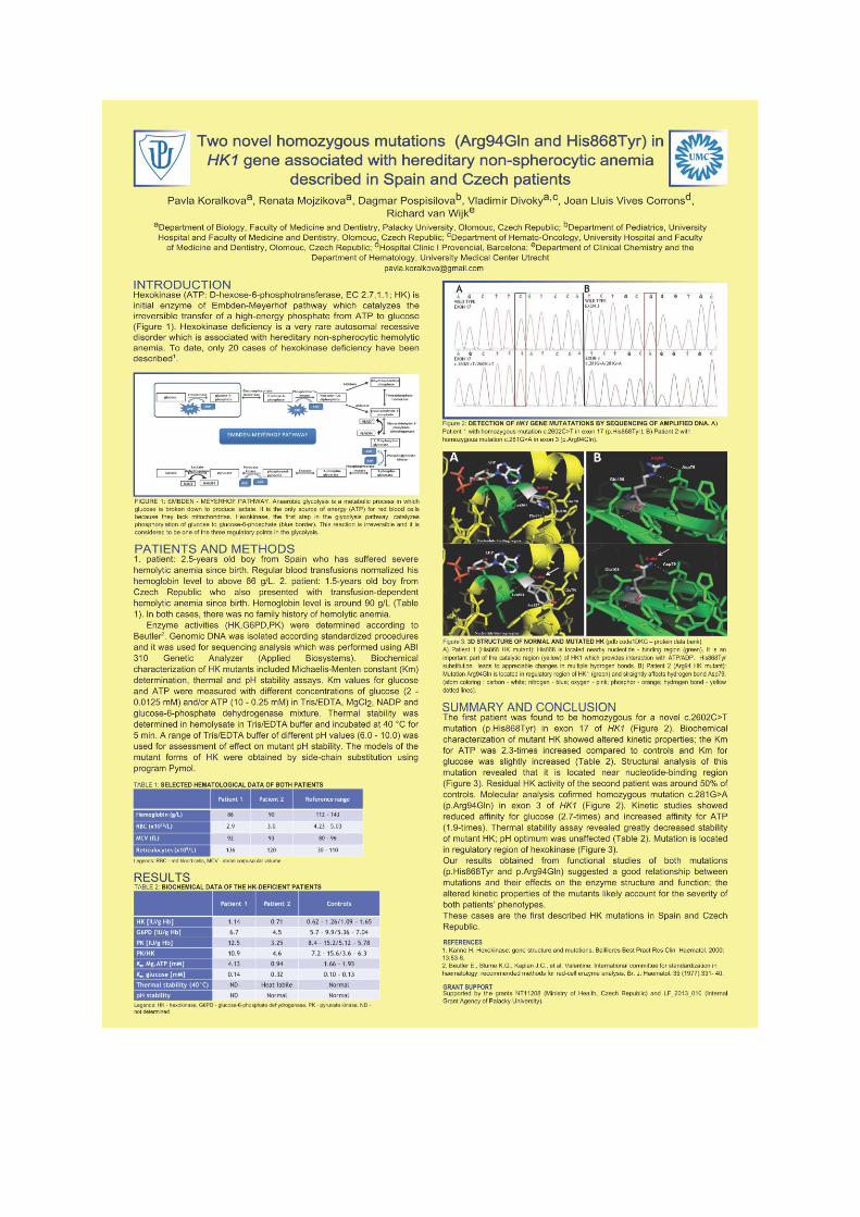
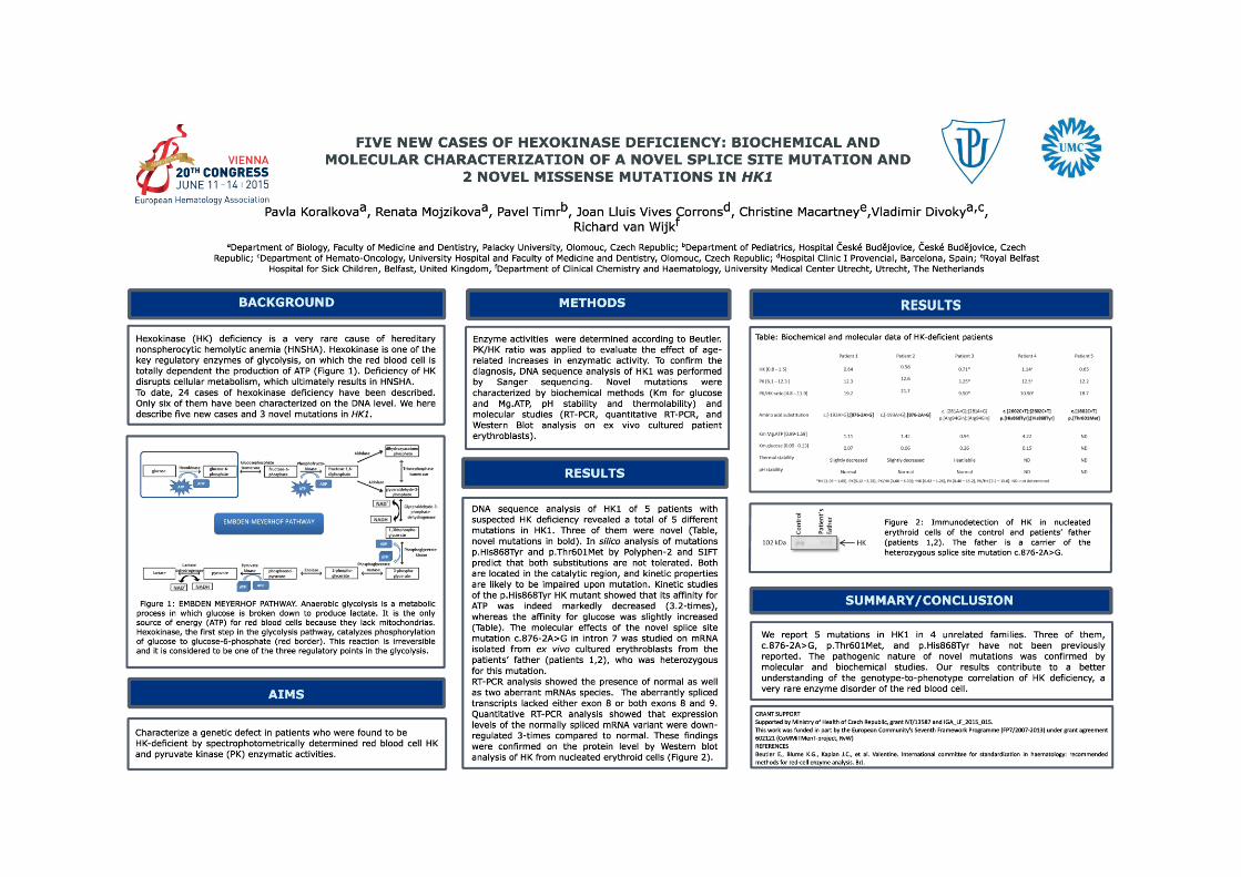

APPENDICES 4.4
MOLECULAR CHARACTERIZATION A NOVEL NON-HEMOLYTIC ANEMIA VARIANT OF PGK
Case report
Recurrent episodes of myoglobinuria, mental retardation and seizures butno hemolysis in two brothers with phosphoglycerate kinase deficiency
Sandra Coppens a,b,*, Pavla Koralkova c,d, Alec Aeby a, Renata Mojzikova d, Nicolas Deconinck b,Hazim Kadhim e, Richard van Wijk a
a Neuromuscular Reference Centre, Department of Pediatric Neurology, Hôpital Erasme, Université Libre de Bruxelles (U.L.B.), Brussels, Belgiumb Neuromuscular Reference Centre, Department of Pediatric Neurology, Queen Fabiola Children’s University Hospital, Université Libre de Bruxelles (U.L.B.),
Brussels, Belgiumc Department of Clinical Chemistry and Haematology, University Medical Center Utrecht, Utrecht, The Netherlandsd Department of Biology, Faculty of Medicine and Dentistry, Palacký University Olomouc, Olomouc, Czech Republic
e Department of Neuropathology and Reference Centre for Neuromuscular Pathology, Brugmann University Hospital (CHU-Brugmann), Université Libre deBruxelles (U.L.B.), Brussels, Belgium
Received 25 July 2015; received in revised form 24 September 2015; accepted 23 November 2015
Abstract
We report two brothers with mild intellectual deficiency, exercise intolerance, rhabdomyolysis, seizures and no hemolysis. Phosphoglyceratekinase (PGK) activity was strongly decreased in their red blood cells. Subsequent molecular analysis of PGK1 revealed hemizygosity for a novelmutation c.756 + 3A > G, in intron 7. Analysis of the effect of this mutation on pre-mRNA processing demonstrated markedly decreased levels ofnormal PGK1 mRNA. In addition, the c.756 + 3A > G change resulted in abnormally spliced transcripts. If translated, these transcripts mostlyencode for C-terminally truncated proteins. The consequences of the c.756 + 3A > G mutation is discussed, as well as the genotype-to-phenotypecorrelation with regard to previously described mutations (PGK Fukuroi and PGK Antwerp), which also result in C-terminal truncated proteins.© 2016 Elsevier B.V. All rights reserved.
Keywords: Phosphoglycerate kinase; Metabolic myopathy; PGK
1. Introduction
Phosphoglycerate kinase (PGK) is a key glycolytic enzymedirectly involved in cellular generation of ATP. PGK deficiencyis a very rare X-linked disorder associated with a triad ofclinical manifestations involving different cell types: muscle(exercise intolerance, rhabdomyolysis), red blood cells(hemolytic anemia), and the central nervous system (seizures,mental retardation, parkinsonism) [1,2]. In general, mutationsof PGK resulting in marked protein instability and mildimpairment of kinetic properties are usually associated withneurological disorders and hemolysis, but not with myopathy[3]. On rare occasions, affected individuals show the fullspectrum of clinical features. Thus, wide clinical heterogeneityis seen in patients with PGK deficiency [2].
2. Case report
Two brothers, aged 14 and 16, born after an uneventfulpregnancy, presented with slightly delayed walking (18 months).They subsequently developed mild intellectual deficiency (totalIQ: 60 at WPPSI-III for the oldest brother), accompanied byrecurrent episodes of generalized weakness following effortfrom the age of 4. At the age of 8, after spending twentyminutes playing in a swimming pool, the younger brotherexperienced an episode of weakness, together with a painfulswelling of the left thigh and myoglobinuria (max CK540.000 UI/l). He was hospitalized on the intensive care unitand treated with intravenous fluids and loop diuretics. At theage of 13, he presented with refractory status epilepticus,complicated by rhabdomyolysis and acute renal failure (maxCK 710.000 UI/l). Status epilepticus required the administrationof several doses of lorazepam, phenytoin and continuousintravenous midazolam. He had no recurrence of seizures underlevetiracetam monotherapy. Currently, both brothers still complainof muscle pain and sometimes weakness after moderate exercise.They still avoid over-exertion to lessen the pain. Muscle strength
* Corresponding author. Department of Pediatric Neurology, Hôpital Erasme,Université Libre de Bruxelles, 808 Route de Lennik, 1070 Bruxelles, Belgium.Tel.: +32 25554364; fax: +32 25558333.
E-mail address: [email protected] (S. Coppens).
http://dx.doi.org/10.1016/j.nmd.2015.11.0080960-8966/© 2016 Elsevier B.V. All rights reserved.
Available online at www.sciencedirect.com
Neuromuscular Disorders 26 (2016) 207–210www.elsevier.com/locate/nmd
ScienceDirect
is completely normal between episodes of rhabdomyolysis oracute weakness. Their level of creatine kinase (CK) isapproximately 500 UI/l (N < 308 UI/l) between episodes ofrhabdomyolysis.
An electroencephalogram (EEG) showed an excess of deltaslow waves but no epileptiform discharges in the younger brother.EEG of the older brother showed generalized spike-wavedischarges, but no clinical seizure was observed. Completeblood count of both brothers showed no evidence of hemolysis.(younger brother: RBCs: 4.46 × 10⁻¹²/l Hb:13.3 g/dl Htc: 39.8%MCH: 29.8 pg MCV : 89.2 fl; older brother: RBCs: 5.64 × 10⁻¹²/l,16.1 g/dl Htc: 47.1% MCH: 28.5 pg MCV: 83.6 fl). Serumurate level was normal (4.2 mg/dl).
Brain MRI was normal in both brothers. In the younger brothera muscle MRI was performed during the first rhabdomyolysisepisode and showed T2 signal hyperintensities, compatible withedema, mainly in the anterior compartment of the thigh and in thesemi-tendinosus, with the left thigh being much more affectedthan the right. Muscle MRI was normal in the older brother.
2.1. Histopathology, biochemical and moleculargenetic analyses
Muscle-biopsy (Quadriceps), was done four months afteran episode of rhabdomyolysis. Routine stains and mosthistochemical techniques (including HE, PAS, modified Gomori-trichrome, ATP-ase, and Acid phosphatase), were unremarkable.
Oxidative-enzymes staining (COX, SDH, combined COX/SDH, NADH-TR) showed no defective histochemical reactivity(and no evidence for mitochondriopathy). In addition,spectrophotometry showed normal mitochondrial respiratory-complexes activity.
The only salient feature was a slight increase in intra-cytoplasmic content of neutral lipids as detected by ORO stain.
Electron-microscopy showed foci of vacuolization; vacuoleswere partly lipidic in origin but many were of undeterminednature.
Altogether, histo-morphological findings (Fig. 1) suggesteda metabolic vacuolar myopathy with discrete to moderate lipidoverload.
Lactate levels determined on plasma and cerebrospinal fluidas well as total free carnitine were normal. Urinary organicacids, acylcarnitines profiles and fatty acid oxidation studieson lymphocytes were unremarkable. Sequencing of CPT2 geneencoding carnitine palmitoyltransferase 2 revealed noabnormalities.
Determination of red blood cell PGK enzyme activityrevealed a markedly decreased PGK activity (<15% ofcontrols). PGK activity of the patient’s mother was only slightlydecreased; the activity of father was normal (Table 1).
Subsequent DNA analysis of the PGK1 gene by Sangersequencing demonstrated a hemizygous A > G single basesubstitution in intron 7 (c.756 + 3A > G) in both brothers. Thismutation was inherited from the patient’s mother who wasfound to be heterozygous for this mutation. The mutation wasnot detected in a control population of >100 normal alleles (53control subjects). Because the mutation alters the consensussequence of the donor splice site of intron 7, we investigatedthe effect of the mutation on PGK1 pre-mRNA processing. Forthis, total RNA was isolated from ex vivo cultured erythroblastsof both patients and a control subject, and reverse-transcribedaccording to standardized procedures. A region encompassingexons 7 and 8 of PGK1 was amplified from cDNA. Onesingle-size transcript was present in the control subject andboth patients (Fig. 2), whilst additional RT-PCR products,representing splice variants, were amplified from patient-specificRNA. RT-PCR products were subcloned into pCR™4-TOPOvector (TOPO® TA Cloning® Kit for Sequencing, Invitrogen).Positive colonies were screened by colony PCR. Plasmid DNAwas isolated and DNA sequencing analysis was performedusing BigDye Terminator v1.1 Cycle Sequencing Kit (AppliedBiosystems). This procedure identified a number of aberrantlyspliced transcript variants (Fig. 2). The two most abundanttranscripts were characterized by either the insertion of 52 bpof intron 7, or the complete sequence of intron 7. Two other,less abundant, transcripts lacked either 18 bp of exon 7 orskipping of complete exon 7 (Fig. 2).
In order to quantify transcript levels of the normally processedPGK1 pre-mRNA, quantitative RT-PCR (qPCR) analysis wasapplied to both patient-specific and normal control RNA. Thisrevealed a marked down-regulation (8.5-times) of the normallyspliced pre-mRNA in patients compared to the control subject.
3. Discussion
The patients reported here exhibited mild intellectualdeficiency, exercise intolerance, rhabdomyolysis, seizures, andno evidence of hemolysis. The clinical phenotype of the patientsraised the suspicion of PGK deficiency as the underlying cause.This was confirmed by the finding of decreased red cell PGKactivity in both patients and their mother, as well as theidentification of a novel splice site consensus sequence change(c.756 + 3A > G) in intron 7 of PGK1. In silico analysis (HumanSplicing Finder) [4] predicts that the A > G substitution abolishesthe intron 7 donor site. To confirm this, RNA studies wereconducted, which revealed the presence of four differentabnormally spliced pre-mRNA transcripts. We find that thec.756 + 3A > G causes: a) the retention of the first 52 bp ofintron 7, b) the retention of intron 7, c) the deletion of 18 bpfrom the 3′-end of exon 7, and d) skipping of exon 7 (Fig. 2).
Table 1Enzyme analysis and PGK1 genotypes of the family.
Son 1 Son 2 Mother Father
PGK [188–302 U/g Hb] 34 35 181 212HK [0.5–1.5 U/g Hb] 1.0 1.3 1.6 1.0Nucleotide substitution c.756 + 3A > G c.756 + 3A > G c.756 + 3A > G –
208 S. Coppens et al. /Neuromuscular Disorders 26 (2016) 207–210

The retention of 52 bp of intron 7, as well as the retentionof intron 7, both result in a shift of the reading frame, leadingto the formation of a premature stop codon. Consequently, iftranslated, the resulting protein is predicted to lack the C-terminalpart (165 amino acids), which is responsible for nucleotidebinding. The loss of 18 bp from the 3′-end of exon 7 encodesthe deletion of six amino acids (residues 247–252). The skippingof exon 7 also disrupts the reading frame, causing a prematurestop at amino acid position 269. Thus, the resulting predictedprotein here, too, would lack the C-terminal end. RNA analysisalso revealed that normal splicing is not completely abolished.However, qPCR analysis showed that normally processed mRNAlevels were markedly down-regulated (8.5-times) compared tothe control subject. It is likely this will result in markedlydecreased PGK enzyme levels.
It has been hypothesized that different clinical manifestationsof PGK1 deficiency can be correlated with the type of alterations
caused by mutations in the PGK1 gene, highlighting the needfor determination of the molecular properties of PGK variantsto assist in prognosis and genetic counseling [5].
Aberrantly processed pre-mRNA transcripts containing thefirst 52 bp of intron 7 have been reported previously in patientswith PGK variants Fukuroi (c.756 + 5G > A) and PGK Antwerp(c.755A > C, p.(Glu252Ala)). In both cases, no other aberrantmRNAs were identified. Phenotypically, both patients displayedrecurrent episodes of myoglobinuria with rhabdomyolysis, withoutsigns of hemolysis. This resembles the phenotype of our twopatients. The patient with PGK Fukuroi, in addition, displayedintellectual deficiency and abnormal EEG. In both cases qualitativechanges to muscle fiber were minimal and no accumulation ofglycogen was observed [6,7].
A phenotype similar to that of our patients (myopathy,mental retardation and no hemolysis) has also been described inthe patient with PGK Hamamatsu (c.758T > C, p.(Ile253Thr))
Fig. 1. Muscle Biopsy Observations. Histomorphological findings with “routine” stainings, namely, Hematoxylin-eosin (A), Periodic acid Schiff (B), modifiedGomori (C), Acid phosphatase (D), and Oil-Red-O staining (E) were generally unremarkable. Transmission electron microscopy (F) showed foci of vacuolizationin many fibers. Original magnification: ×320 (A); ×400 (B, C, D, E); and ×4400 (F).
209S. Coppens et al. /Neuromuscular Disorders 26 (2016) 207–210

[8,9]. This mutation affects the same region that is predicted tobe deleted by at least part of the aberrant transcripts identifiedin our patients.
These observed similarities may point to a correlationbetween the genotype and phenotype, but definite conclusionscannot be drawn at this moment. It seems reasonable toassume, however, that additional, yet-unknown factors, arelikely to contribute to the phenotypic expression of PGKdeficiency.
In conclusion, phosphoglycerate kinase (PGK) deficiency, arare defect in glycolysis pathway, has its place in the differentialdiagnosis of metabolic myopathies. The association of exerciseintolerance and recurrent episodes of myoglobinuria withhemolytic anemia, intellectual deficiency and other neurologicaldisorders is highly suggestive of the diagnosis.
Acknowledgement
Pavla Koralkova and Renata Mojzikova are supported byMinistry of Health of the Czech Republic, grant NT/13587and Internal Grant Agency of Palacký University, grantIGA_LF_2015_015.
References
[1] Sotiriou E, Greene P, Krishna S, Hirano M, DiMauro S. Myopathy andparkinsonism in phosphoglycerate kinase deficiency. Muscle Nerve2010;41(5):707–10.
[2] Beutler E. PGK deficiency. Br J Haematol 2007;136(1):3–11.[3] Pey AL, Maggi M, Valentini G. Insights into human phosphoglycerate
kinase 1 deficiency as a conformational disease from biochemical, biophysical,and in vitro expression analyses. J Inherit Metab Dis 2014;37(6):909–16.
[4] Desmet FO, Hamroun D, Lalande M, Collod-Béroud G, Claustres M,Béroud C. Human Splicing Finder: an online bioinformatics tool to predictsplicing signals. Nucleic Acids Res 2009;37(9):e67.
[5] Chiarelli LR, Morera SM, Bianchi P, et al. Molecular insights on pathogeniceffects of mutations causing phosphoglycerate kinase deficiency. PLoSONE 2012;7:e32065.
[6] Ookawara T, Davé V, Willems P, et al. Retarded and aberrant splicingscaused by single exon mutation in a phosphoglycerate kinase variant. ArchBiochem Biophys 1996;327(1):35–40.
[7] Shirakawa K, Takahashi Y, Miyajima H. Intronic mutation in the PGK1gene may cause recurrent myoglobinuria by aberrant splicing. Neurology2006;66(6):925–7.
[8] Sugie H, Sugie Y, Ito M, Fukuda T. A novel missense mutation (837T→C)in the phosphoglycerate kinase gene of a patient with a myopathic form ofphosphoglycerate kinase deficiency. J Child Neurol 1998;13(2):95–7.
[9] Sugie H, Sugie Y, Nishida M, et al. Recurrent myoglobinuria in a child withmental retardation phosphoglycerate kinase deficiency. J Child Neurol1989;4(2):95–9.
Fig. 2. A schematic drawing of the aberrantly spliced transcript variants arising due to the mutation c.756 + 3A > G. The wild type (WT) variant is normallyprocessed mRNA. Arrows indicate the retention or the deletion of the particular sequence of PGK1. The stop codons are in bold and colored in red.
210 S. Coppens et al. /Neuromuscular Disorders 26 (2016) 207–210

APPENDICES 4.5
IN SILICO MODELLING OF A NOVEL MUTATION OF PFKM GENE ASSOCIATED WITH PFK DEFICIENCY

ORIGINAL RESEARCH ARTICLEpublished: 30 December 2013
doi: 10.3389/fphys.2013.00393
First description of phosphofructokinase deficiency inspain: identification of a novel homozygous missensemutation in the PFKM geneJoan-Lluis Vives-Corrons1*, Pavla Koralkova2, Josep M. Grau3, Maria del Mar Mañú Pereira1 and
Richard Van Wijk4
1 Red Cell Pathology Unit, Biomedical Dianostic Centre, University Hospital Clínic de Barcelona, Barcelona, Spain2 Faculty of Medicine and Dentistry, Department of Biology, Palacky University, Olomouc, Czech Republic3 Service of Internal Medicine and Muscle, University Hospital Clínic de Barcelona, Barcelona, Spain4 Department of Clinical Chemistry and Haematology, University Medical Center Utrecht, Utrecht, Netherlands
Edited by:
Anna Bogdanova, University ofZurich, Switzerland
Reviewed by:
Michael White, Drexel UniversityCollege of Medicine, CanadaLillian DeBruin, Wilfrid LaurierUniversity, Canada
*Correspondence:
Joan-Lluis Vives-Corrons, Red CellPathology Unit, BiomedicalDianostic Centre, UniversityHospital Clínic de Barcelona,c/ Villarroel 170, 08036-Barcelona,Spaine-mail: [email protected]
Phosphofructokinase deficiency is a very rare autosomal recessive disorder, which belongsto group of rare inborn errors of metabolism called glycogen storage disease. Here wereport on a new mutation in the phosphofructokinase (PFK) gene PFKM identified in a65-years-old woman who suffered from lifelong intermittent muscle weakness and painfulspasms of random occurrence, episodic dark urines, and slight haemolytic anemia. Afterruling out the most common causes of chronic haemolytic anemia, the study of a panel of24 enzyme activities showed a markedly decreased PFK activity in red blood cells (RBCs)from the patient. DNA sequence analysis of the PFKM gene subsequently revealed a novelhomozygous mutation: c.926A>G; p.Asp309Gly. This mutation is predicted to severelyaffect enzyme catalysis thereby accounting for the observed enzyme deficiency. This caserepresents a prime example of classical PFK deficiency and is the first reported case ofthis very rare red blood cell disorder in Spain.
Keywords: phosphofructokinase deficiency, glycogen storage disease, PFKM gene, missense mutation, enzyme
catalysis
INTRODUCTIONPhosphofructokinase (ATP: D- fructose-6-phosphate-1-phosphotransferase; EC 2.7.1.11; PFK) is a key regulatoryenzyme of the glycolytic cycle and catalyses the conversion offructose-6-phosphate to fructose-1,6-diphosphate (Figure 1).Human PFK is composed of three isoenzymes, muscle (M), liver(L), and platelet (P) (Vora, 1983; Nakajima et al., 2002). The Ptype is also known as Fibroblast type (F). Mammalian PFK isa tetrameric enzyme that is subjected to allosteric regulation.Tissue isozymes randomly aggregate to form homotetramersor heterotetramers depending on the relative abundance of thesubunits in a particular tissue. PFK-M is the sole subunit inmuscle cells whereas red blood cells (RBCs) contain both Land M subunits and form their hybrids (M4, L4, M3L, M2L2,and ML3).
Phosphofructokinase deficiency (OMIM 171 850) is a veryrare autosomal recessive condition with heterogeneous clinicalsymptoms, mainly characterized by myopathy and/or haemolysis(Hirano and Di Mauro, 1999). Myopathy is caused by the accu-mulation of glycogen in muscle tissue due to the metabolic defectand is also known as glycogenosis type VII or Tarui disease. It ischaracterized by muscle pain, exercise-induced fatigue, cramps,and myoglobinuria. The observed clinical symptoms reflect lackof muscle PFK activity and partial reduction of enzymatic activ-ity in erythrocytes. The latter usually is associated with mildhaemolysis.
Up to now, only about 100 patients with PFK deficiency havebeen reported worldwide and 22 PFK-deficient PFKM alleles have
been characterized. The gene encoding the M subunit (PFKM)has been assigned to chromosome 12q13.3 and spans 30 kb. Itcontains 24 exons and at least 3 promoter regions (Elson et al.,1990; Yamada et al., 2004). Among the detected mutations aremostly missense mutations and splicing defects.
We now describe here a Spanish patient with a clinical his-tory of anemia, haemolysis, and intermittent muscle weakness,who was found to be homozygous for a novel mutation in thePFKM gene (c.926A>G). This mutation encodes the substitutionof aspartic acid by glycine at residue 309 (p.Asp309Gly). This isthe first description of PFK deficiency in Spain.
CASE REPORTA 65-years-old woman with long standing hypertension and type2 diabetes was referred to our Unit because of intolerance to exer-cise and chronic fatigue. From youth she suffered from spasmsof random occurrence associated with muscle weakness, painfulintolerance to small efforts, and intermittent dark urines, espe-cially after exercise due to myoglobinuria, as revealed by urinaly-sis. Physical examination showed no weakness or muscle atrophyand only a moderate splenomegaly without hepatomegaly or lym-phadenopathy. Parents were unavailable for study and there wasno known consanguinity. The same clinical picture, however,was present in a non-smoker brother whereas the three patient’sdaughters were normal. Patient’s clinical condition has remainedstable in follow-up.
Complete blood count (CBC) showed moderate anemia(Hb: 115 g/L), with slight macrocytosis (105 fl) and increased
www.frontiersin.org December 2013 | Volume 4 | Article 393 | 1
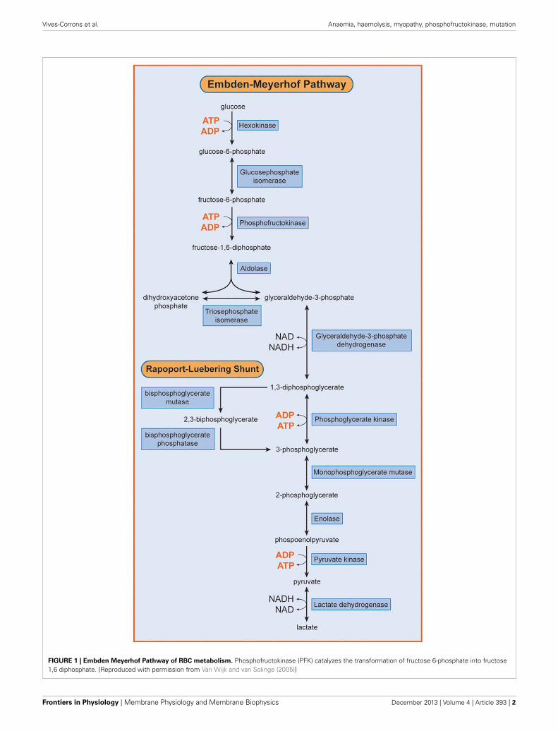
Vives-Corrons et al. Anaemia, haemolysis, myopathy, phosphofructokinase, mutation
FIGURE 1 | Embden Meyerhof Pathway of RBC metabolism. Phosphofructokinase (PFK) catalyzes the transformation of fructose 6-phosphate into fructose1,6 diphosphate. [Reproduced with permission from Van Wijk and van Solinge (2005)]
Frontiers in Physiology | Membrane Physiology and Membrane Biophysics December 2013 | Volume 4 | Article 393 | 2

Vives-Corrons et al. Anaemia, haemolysis, myopathy, phosphofructokinase, mutation
number of circulating reticulocytes (110 × 109/L). Leukocyteand platelet counts, as well as general serum biochemical anal-ysis, were within normal range, except for a moderate risein non-conjugated bilirubin, lactate dehydrogenase (LDH) anduric acid (hyperuricemia). Biological signs of diabetes melli-tus type 2 were also present. The studies performed to ruleout the origin of the anemia, discarded nutritional deficiencies(serum iron tests, cobalamin and serum folate were all nor-mal), haemoglobinopathies (HPLC and thermal stability), andparoxysmal nocturnal haemoglobinuria (normal flow cytome-try measurement of CD45 and CD49 in leukocytes and RBCs).Hereditary RBC membrane defects were ruled out by morpholog-ical observation of May-Grünwald-Giemas stained blood smears,and a normal osmotic fragility test. Extensive study of RBCenzyme activity measurements demonstrated a marked decrease(<30% of normal) in PFK activity (Table 1). DNA sequence anal-ysis of individual exons of PFKM, including flanking splice sites,revealed that the patient was homozygous for a missense muta-tion in exon 11: c.929A>G. This mutation, that has not beenpreviously reported in the literature, encodes the substitution ofaspartic acid by glycine at residue 309 (p.Asp309Gly). Mutationprediction programs PolyPhen-2 (Adzhubei et al., 2010) and SIFT(Kumar et al., 2009) predict this mutation to be pathogenic (i.e.,disease causing).
The complete lack of PFK activity in muscle was confirmedon both histological preparations and muscle extracts. Muscleabnormality was also confirmed by electromyography (EMG),that showed mild myopathic changes and by the forearm test,characterized by a plane lactate curve with normal increase ofammonium. As usual in patients with glycogenosis, a painfulspasm occurred at the end of the forearm test. Muscle biopsyshowed slight amounts of polysaccharide (PAS) not digested bydiastase and abnormalities in NADH-TR reaction.
DISCUSSIONIn this report, we describe, for the first time, the occurrence ofPFK deficiency in Spain in an adult woman of 65 years of age. Hermoderate haemolysis was associated with the accumulation ofmuscle glycogen comparable to that of Tarui disease. Muscle painand exercise-induced fatigue and weakness associated with darkurines were the most relevant clinical manifestations. She wasfound to be homozygous for a novel missense mutation in PFKM,the gene that encodes the PFK-M subunit. On the amino acidlevel, this mutation (c.929A>G) causes the substitution of aspar-tic acid by glycine at residue 309. Asp309 is part of an α-helix, and
Table 1 | Red blood cell enzyme activity measurements.
Patient Reference value
G6PD activity (IU/g Hb) 7.0 6.2–9.9
HK activity (IU/g Hb) 1.2 0.6–1.3
PGI activity (IU/g Hb) 61.9 46.0–66.0
PK activity (IU/g Hb) 15.3 9.5–15.6
PFK activity (IU/g Hb) 3.07 6.4–13.9
Glucose-6-phosphate dehydrogenase; HK, Hexokinase; PGI, Phosphoglucose
isomerase; PK, Pyruvate kinase; PFK, Phosphofructokinase.
replacement with glycine could disrupt this α-helix. Furthermore,structural analysis using the 3D molecular model of rabbit mus-cle type PFK (PDB entry 3O8L) showed that the p.Asp309Glysubstitution is located in direct vicinity of the nucleotide (ADP)binding site in the center of the PFKM subunit (Figure 2A). PFKis allosterically activated by ADP, and the ADP binding site hasbeen recently identified (Banaszak et al., 2011). Asp309 does notdirectly interact with ADP, however, substitution of Asp309 byglycine is likely to disrupt multiple hydrogen bonds with Gly177,Gly179, Ser 180, and Ser306 (Figure 2B). This loss of hydrogenbonds and the change of polarity and electrostatic interactionsupon mutation of Asp309 will likely affect correct positioning ofthe side-chains of amino acids directly involved in ADP bind-ing (Figure 2B), in particular neighboring residue Phe308, whichmakes stacking interaction with the adenine ring of ADP. Thishypothesis is further supported by recent functional studies ofthe Asp543Ala change in PFKM. This mutation is associated withTarui disease, and substitution of Asp543 by Ala was shown to dis-rupt hydrogen bonds with ADP (Brüser et al., 2012). We thereforepostulate that decreased binding of the allosteric activator ADPwill inhibit PFK enzymatic activity, in particular under low energylevels. These findings support the physiological importance ofAsp390 for the recently identified ADP binding site.
The missense mutation is thus predicted to lead to a less func-tional PFK-M subunit. In accordance with this, the complete lackof PFK activity in muscle was confirmed on both histologicalpreparations and muscle extracts. The muscle abnormality wasalso confirmed by electromyography, ischemic exercise testing,histochemistry and electron microscopy. Furthermore, partialred blood cell PFK deficiency was reflected by the moderatelydecreased enzymatic activity in red blood cells, leading to mildhaemolytic anemia
PFK is a key regulatory enzyme for glycolysis (Van Wijk andvan Solinge, 2005) and catalyzes the irreversible transfer of phos-phoryl from ATP to fructose-6-phosphate, and converts it tofructose-1,6-bisphosphate. Thus, tissues deficient in PFK cannotuse free or glycogen-derived glucose as a fuel source and accu-mulate glycogen (glycogenosis). PFK deficiency (Tarui disease)was the first disorder recognized to directly affect glycolysis (Taruiet al., 1965). Since this first description of the disease, a wide rangeof biochemical, physiological and molecular studies have greatlycontributed to our knowledge concerning not only PFK functionin normal muscle, but also on the general control of glycolysisand glycogen metabolism. So far, more than one 100 patientshave been described with prominent clinical symptoms charac-terized by muscle cramps, exercise intolerance, rhabdomyolysisand myoglobinuria, often associated with haemolytic anemia andhyperuricemia. In classic Tarui disease, the genetic defect involvesthe M isoform, resulting in the absence of enzymatic activity inthe muscle. Erythrocytes lack the M4 and hybrid isozymes andonly express the L4 homotetramers, resulting in about 50% ofnormal PFK activity (Figure 3). Thus, haemolysis is a result ofpartial erythrocyte PFK deficiency.
Despite PFK deficiency is a very rare autosomal recessive dis-ease; its true incidence may be higher due to lack of recognition, assymptoms may be quite mild. In fact, our case highlights the lat-ter, because the mild clinical presentation led her to be diagnosed,
www.frontiersin.org December 2013 | Volume 4 | Article 393 | 3
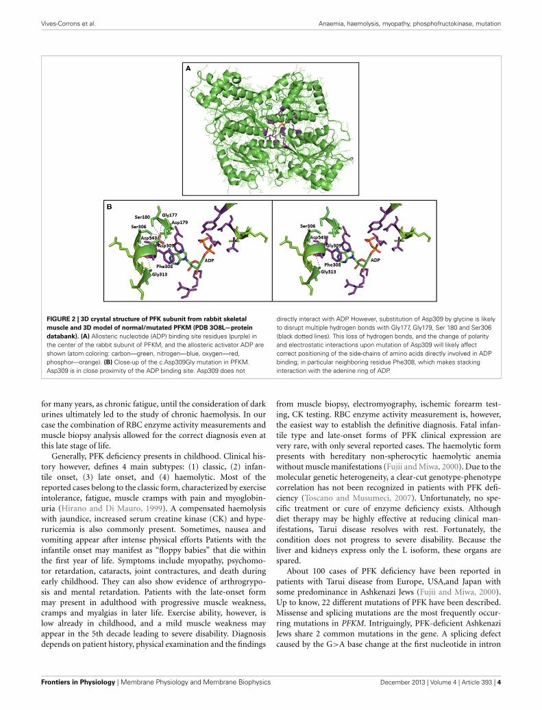
Vives-Corrons et al. Anaemia, haemolysis, myopathy, phosphofructokinase, mutation
FIGURE 2 | 3D crystal structure of PFK subunit from rabbit skeletal
muscle and 3D model of normal/mutated PFKM (PDB 3O8L—protein
databank). (A) Allosteric nucleotide (ADP) binding site residues (purple) inthe center of the rabbit subunit of PFKM, and the allosteric activator ADP areshown (atom coloring: carbon—green, nitrogen—blue, oxygen—red,phosphor—orange). (B) Close-up of the c.Asp309Gly mutation in PFKM.Asp309 is in close proximity of the ADP binding site. Asp309 does not
directly interact with ADP. However, substitution of Asp309 by glycine is likelyto disrupt multiple hydrogen bonds with Gly177, Gly179, Ser 180 and Ser306(black dotted lines). This loss of hydrogen bonds, and the change of polarityand electrostatic interactions upon mutation of Asp309 will likely affectcorrect positioning of the side-chains of amino acids directly involved in ADPbinding; in particular neighboring residue Phe308, which makes stackinginteraction with the adenine ring of ADP.
for many years, as chronic fatigue, until the consideration of darkurines ultimately led to the study of chronic haemolysis. In ourcase the combination of RBC enzyme activity measurements andmuscle biopsy analysis allowed for the correct diagnosis even atthis late stage of life.
Generally, PFK deficiency presents in childhood. Clinical his-tory however, defines 4 main subtypes: (1) classic, (2) infan-tile onset, (3) late onset, and (4) haemolytic. Most of thereported cases belong to the classic form, characterized by exerciseintolerance, fatigue, muscle cramps with pain and myoglobin-uria (Hirano and Di Mauro, 1999). A compensated haemolysiswith jaundice, increased serum creatine kinase (CK) and hype-ruricemia is also commonly present. Sometimes, nausea andvomiting appear after intense physical efforts Patients with theinfantile onset may manifest as “floppy babies” that die withinthe first year of life. Symptoms include myopathy, psychomo-tor retardation, cataracts, joint contractures, and death duringearly childhood. They can also show evidence of arthrogrypo-sis and mental retardation. Patients with the late-onset formmay present in adulthood with progressive muscle weakness,cramps and myalgias in later life. Exercise ability, however, islow already in childhood, and a mild muscle weakness mayappear in the 5th decade leading to severe disability. Diagnosisdepends on patient history, physical examination and the findings
from muscle biopsy, electromyography, ischemic forearm test-ing, CK testing. RBC enzyme activity measurement is, however,the easiest way to establish the definitive diagnosis. Fatal infan-tile type and late-onset forms of PFK clinical expression arevery rare, with only several reported cases. The haemolytic formpresents with hereditary non-spherocytic haemolytic anemiawithout muscle manifestations (Fujii and Miwa, 2000). Due to themolecular genetic heterogeneity, a clear-cut genotype-phenotypecorrelation has not been recognized in patients with PFK defi-ciency (Toscano and Musumeci, 2007). Unfortunately, no spe-cific treatment or cure of enzyme deficiency exists. Althoughdiet therapy may be highly effective at reducing clinical man-ifestations, Tarui disease resolves with rest. Fortunately, thecondition does not progress to severe disability. Because theliver and kidneys express only the L isoform, these organs arespared.
About 100 cases of PFK deficiency have been reported inpatients with Tarui disease from Europe, USA,and Japan withsome predominance in Ashkenazi Jews (Fujii and Miwa, 2000).Up to know, 22 different mutations of PFK have been described.Missense and splicing mutations are the most frequently occur-ring mutations in PFKM. Intriguingly, PFK-deficient AshkenaziJews share 2 common mutations in the gene. A splicing defectcaused by the G>A base change at the first nucleotide in intron
Frontiers in Physiology | Membrane Physiology and Membrane Biophysics December 2013 | Volume 4 | Article 393 | 4

Vives-Corrons et al. Anaemia, haemolysis, myopathy, phosphofructokinase, mutation
FIGURE 3 | Molecular basis of PFK deficiency. In classic Tarui disease, thegenetic defect involves the M isoform of PFK enzyme, resulting a severeenzyme deficiency in muscle. Erythrocytes that normally have two
homotetramers (M4 and L4) and three hybrid isozymes (M3L, M2L2 andML3), lack the M4 isoform and the hybrid isozymes, and only express the L4homotetramers, resulting in about 50% of normal PFK activity
5 (c.237+1G>A) accounts for 68% of mutant Ashkenazi alleles,and a single base deletion in exon 22 (c.2003delC) accounts forabout 27% of mutant Ashkenazi alleles (Raben and Sherman,1995). The here described homozygous patient is the first Spanish
case described to be affected by this very rare disease. The iden-tification of a novel homozygous missense mutation furtherextends the repertoire of PFK deficiency-associated mutations inPFKM.
www.frontiersin.org December 2013 | Volume 4 | Article 393 | 5
Vives-Corrons et al. Anaemia, haemolysis, myopathy, phosphofructokinase, mutation
ACKNOWLEDGMENTSThis study has been partially supported by a Research Grantof the Spanish Ministry of Health (FIS Ref PI10/01460).We are indebted to the European Network for Rare andCongenital Anaemias (ENERCA) that has facilitated theconnection and information exchange between the centersparticipating in this study. Pavla Koralkova is supported bythe grants NT11208 (Ministry of Health, Czech Republic)and LF_2013_010 (Internal Grant Agency of PalackyUniversity).
REFERENCESAdzhubei, I. A., Schmidt, S., Peshkin, L., Ramensky, V. E., Gerasimova,
A., Bork, P., et al. (2010). A method and server for predicting dam-aging missense mutations. Nat. Methods. 7, 248–249. doi: 10.1038/nmeth0410-248
Banaszak, K., Mechin, I., Obmolova, G., Oldham, M., Chang, S. H., Ruiz, T.,et al. (2011). The crystal structures of eukaryoticphosphofructokinases frombaker’s yeast and rabbit skeletal muscle. J. Mol. Biol. 407, 284–297. doi:10.1016/j.jmb.2011.01.019
Brüser, A., Kirchberger, J., and Schöneberg, T. (2012). Altered allosteric regulationof muscle 6-phosphofructokinase causes tarui disease. Biochem. Biophys. Res.Commun. 427, 133–137. doi: 10.1016/j.bbrc.2012.09.024
Elson, A., Levanon, D., Brandeis, M., Dafni, N., Bernstein, Y., Danciger, E., et al.(1990). The structure of the human liver-type phosphofructokinase gene.Genomics 7, 47–56. doi: 10.1016/0888-7543(90)90517-X
Fujii, H. and Miwa, S. (2000). Other erythrocyte enzyme deficiencies associatedwith non-haematological symptoms: phosphoglycerate kinase and phospho-fructokinase deficiency. Baillieres Best Pract Res. Clin. Haematol. 13, 141–148.doi: 10.1053/beha.1999.0062
Hirano, M. and Di Mauro, S. (1999). “Metabolic myopathies,” in MotorDisorders123–137.
Kumar, P., Henikoff, S., and Ng, P. C. (2009). Predicting the effects of coding non-synonymous variants on protein function using the SIFT algorithm. Nat. Protoc.4, 1073–1081. doi: 10.1038/nprot.2009.86
Nakajima, H., Raben, N., Hamaguchi, T., and Yamasaki, T. (2002).Phosphofructokinase deficiency; past, present and future. Curr. Mol. Med.2, 197–212. doi: 10.2174/1566524024605734
Raben, N., and Sherman, J. B. (1995). Mutations in muscle phosphofructokinasegene. Hum. Mutat. 6, 1–6. doi: 10.1002/humu.1380060102
Tarui, S., Okuno, G., Ikura, Y., Tanaka, T., Suda, M., and Nishikawa, M.(1965). Phosphofructokinase deficiency in skeletal muscle: a new type ofglycogenosis. Biochem. Biophys. Res. Commun. 19, 517–523. doi: 10.1016/0006-291X(65)90156-7
Toscano, A., and Musumeci, O. (2007). Tarui disease and distal glycogenoses:clinical and genetic update. Acta Myol. 26, 105–107.
Van Wijk, R., and van Solinge, W. W. (2005). The energy-less red blood cell islost: erythrocyte enzyme abnormalities of glycolysis. Blood. 106, 4034–4042. doi:10.1182/blood-2005-04-1622
Vora, S. (1983). Isozymes of human phosphofructokinase: biochemical and geneticaspects. Isozymes Curr. Top. Biol. Med. Res. 11, 3–23.
Yamada, S., Nakajima, H., and Kuehn, M. R. (2004). Novel testis- and embryo-specific isoforms of the phosphofructokinase-1 muscle type gene. Biochem.Biophys. Res. Commun. 316, 580–587. doi: 10.1016/j.bbrc.2004.02.089.
Conflict of Interest Statement: The authors declare that the research was con-ducted in the absence of any commercial or financial relationships that could beconstrued as a potential conflict of interest.
Received: 02 November 2013; accepted: 13 December 2013; published online: 30December 2013.Citation: Vives-Corrons J-L, Koralkova P, Grau JM, Mañú Pereira MdM and Van WijkR (2013) First description of phosphofructokinase deficiency in spain: identification ofa novel homozygous missense mutation in the PFKM gene. Front. Physiol. 4:393. doi:10.3389/fphys.2013.00393This article was submitted to Membrane Physiology and Membrane Biophysics, asection of the journal Frontiers in Physiology.Copyright © 2013 Vives-Corrons, Koralkova, Grau, Mañú Pereira and Van Wijk.This is an open-access article distributed under the terms of the Creative CommonsAttribution License (CC BY). The use, distribution or reproduction in other forums ispermitted, provided the original author(s) or licensor are credited and that the originalpublication in this journal is cited, in accordance with accepted academic practice. Nouse, distribution or reproduction is permitted which does not comply with these terms.
Frontiers in Physiology | Membrane Physiology and Membrane Biophysics December 2013 | Volume 4 | Article 393 | 6
, Vol 10 (Philadelphia, PA: Lippincott Williams and Wilkins),