10907194.pdf - Enlighten: Theses
163
https://theses.gla.ac.uk/ Theses Digitisation: https://www.gla.ac.uk/myglasgow/research/enlighten/theses/digitisation/ This is a digitised version of the original print thesis. Copyright and moral rights for this work are retained by the author A copy can be downloaded for personal non-commercial research or study, without prior permission or charge This work cannot be reproduced or quoted extensively from without first obtaining permission in writing from the author The content must not be changed in any way or sold commercially in any format or medium without the formal permission of the author When referring to this work, full bibliographic details including the author, title, awarding institution and date of the thesis must be given Enlighten: Theses https://theses.gla.ac.uk/ [email protected]
-
Upload
khangminh22 -
Category
Documents
-
view
0 -
download
0
Transcript of 10907194.pdf - Enlighten: Theses

https://theses.gla.ac.uk/
Theses Digitisation:
https://www.gla.ac.uk/myglasgow/research/enlighten/theses/digitisation/
This is a digitised version of the original print thesis.
Copyright and moral rights for this work are retained by the author
A copy can be downloaded for personal non-commercial research or study,
without prior permission or charge
This work cannot be reproduced or quoted extensively from without first
obtaining permission in writing from the author
The content must not be changed in any way or sold commercially in any
format or medium without the formal permission of the author
When referring to this work, full bibliographic details including the author,
title, awarding institution and date of the thesis must be given
Enlighten: Theses
https://theses.gla.ac.uk/

AN INVESTIGATION OF THE MECHANISMS BY WHICH OPIATES AFFECT THE
MOTILITY OF THE GUT
A thesis presented for the degree of Doctor of Philosophy
in the University of Glasgow by
ADEBAYO ADEYINKA LANIYONU, B.Sc.
Department of Pharmacology University of Glasgow
OCTOBER 1985

ProQuest Number: 10907194
All rights reserved
INFORMATION TO ALL USERS The quality of this reproduction is dependent upon the quality of the copy submitted.
In the unlikely event that the author did not send a com p le te manuscript and there are missing pages, these will be noted. Also, if material had to be removed,
a note will indicate the deletion.
uestProQuest 10907194
Published by ProQuest LLC(2018). Copyright of the Dissertation is held by the Author.
All rights reserved.This work is protected against unauthorized copying under Title 17, United States C ode
Microform Edition © ProQuest LLC.
ProQuest LLC.789 East Eisenhower Parkway
P.O. Box 1346 Ann Arbor, Ml 48106- 1346

CONTENTS ;

PAGE NOS.
TABLE OF CONTENTS
ACKNOWLEDGEMENTS 'i - ii
DEDICATION . iii
SUMMARY . iv - v.i
INTRODUCTION 1 - 20
METHODS , 21 - 34
Recording responses of rat isolated colon 21
Field stimulation of the isolated colon . 2 1
Assay of 5-Hydroxytryptamine (5-HT) 2 1 - 2 3
Radioactive (^H)-L-Noradrenaline experiments 23 - 24
(1) Incubation procedure and ( H) efflux 2 3 - 2 4
(2) ( H) uptake by the rat colon 24
High performance liquid chromatography experiments 24 - 26
(1) Chromatographic system 2 4 - 2 5
(2) Chromatography mobile phase 2 5 - 2 6
Extraction of catecholamines from tissues 26.- 27
In vitro experiments 2 7 - 2 8
Types of samples 2 8 - 2 9
(1) Krebs1 solution blank ,28
(2) Spontaneous release sample 28
(3) Stimulation sample , 28
(4) Tissue recovery sample 29
(5) Krebs* solution recovery sample 29
Problems encountered with the electrochemical detection 29 - 31
(1) Artefacts occurring after injections of large 29concentrations of catecholamines
(2) Artefact associated with extracted Krebs* solution 30

PAGE NOS.
(3) Problems associated with the chromatography system 30 - 31
Histochemistry - Falck technique 3 1 - 3 2
Animal pretreatment schedules 32
Preparation of haemolysate 33
Analysis of results 33
Drugs 34
RESULTS 3 5 - 53
EFFECTS OF MORPHINE AND OF OPIOID PEPTIDES ON THE MOTILITY 35 - 37OF THE ISOLATED COLON
Morphine 3 5- 3 6
Enkephalins 36
Comparison of the response of the colon to morphine, 36 - 375-HT, ACh and KC1
Effects of temperature on morphine-induced rhythmic 37contractions
Effects of Ca++ removal. 37
ANALYSIS OF THE MECHANISM OF OPIATE-INDUCED WAVES OF RHYTHMIC 3 7 - 4 0 CONTRACTIONS IN THE ISOLATED COLON
5-HT hypothesis 3 8 - 39
5-HT autodesensitisation 38
Depletion studies : Effects of PCPA or reserpine 3 8 -3 9pretreatment
Effects of 5-HT and other drugs 39
(a) Methysergide 39
.(b) Adrenergic and cholinergic antagonists 39
(c) Quinidine 39
The tonic inhibitory nerve hypothesis 39 - 40
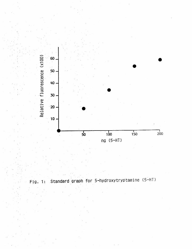
Response of the colon to apamin or TTX 40
The innervation of the rat colon 40
Responses of the rat colon to electrical field 41stimulation

PAGE NOS.
Effects of drugs on responses of the rat colon to 41electrical field stimulation
Atropine 41
Apamin 41
Morphine 4*1-42
6-OHDA pretreatment 42
Effects of drugs on post-stimulus contractions 42
Inhibitory response of the rat colon to electrical 42 - 43stimulation
Investigation of the possible existence of an NANC 43mechanism in the rat colon
Effects of drugs on the NANC inhibitory response 43 - 44
(a) Reserpine and 6-OHDA pretreatment 43 - 44
(b) Effect of TTX 44
(c) Effect of haemolysate, apamin and methylene 44 - 45blue on NANC inhibitory response
Methylene blue 45
Apamin 45
Adrenergic mechanism as possible 45mediator of tonic inhibitory influence affected by the opiates
Responses of the isolated colon to 45 - 46adrenergic receptor antagonists and neurone-blocking drugs
Effect of clonidine 46
Effect of tyramine on apamin-induced 46waves
HPLC AND RADIOACTIVE (3H)-NA EXPERIMENTS 4 7 - 5 3
Factors affecting chromatogram separation 47 - 48
Effects of working electrode potential 48
The catecholamine content of the vas deferentia and 48the rat colon
Spontaneous release of noradrenaline from the guinea- 48 - 49pig vas deferens
Noradrenaline overflow evoked by electrical stimulation 49in the guinea-pig vas deferens

PAGE NOS.
Spontaneous electrical stimulation-evoked release of 49 - 50NA, DA and DOPEG from the mouse vas deferens
Effect of phenoxybenzamine (PB) on the pattern of NA, 50-51DA, DOPEG released from the mouse vas deferens
Effect of PB oh the mechanical response to nerve 51stimulation in the mouse vas deferens
Spontaneous release of NA from the rat colon 52
Tritium accumulation in the rat. colon 52
Efflux of tritium from the rat colon 52
Effect of drugs on tritium efflux and mechanical response 53of the rat colon
DISCUSSION 5 4- 7 5
REFERENCES 76 - 92
PUBLICATIONS 93

ACKNOWLEDGEMENTS

I would like to thank my supervisor, Dr. David Pollock, who initially
suggested the topic for this project and for his continued interested and
guidance throughout the period of the study. His constructive criticism,
stimulating discussion, and readiness to help and his unrivalled ability to
put a smile on my face when the experiments did not, helped in no small way
to see me through the project.
I must also express my gratitude to Professor John Spence Gillespie for
. providing me With the opportunity to work in his department; and for the time
spent in his laboratory. I wiil always remember his inspiring comments and
alternative analysis of "The results of the day". I would also like to
acknowledge the encouragement, advice and friendship of Professor and
• Mrs. F.M. Tayo.
I am also grateful to my friends and colleagues who have all helped me at
some point in my studies. Special thanks go to: "The Whizz Kids", Sunny
Ohia and Ade Ajayi for much pharmacological discussion and the occasional
pharmacological arguments; my flat-mates Tunde Borode and Olusegun Olufemi
^or providing a homely environment to return to in the evenings; Mr. John
Thomson for expert technical assistance and for telling me stories that made
Glasgow famous as the "2nd City of the Kingdom"; Hannah McCaffery for
expert technical assistance- and for making my stay in the Professor's
laboratory a pleasant one; tp my colleagues Cameron McCulloch, Anita
McDonald, Lorraine Anderson and Siew Peng Lim for their friendship, Mrs.
Marjory Wright for her advice and willingness to do my typing jobs for
me; Karyn Forsyth for brightening up lab.354 and making my come-back to the
• lab. a pleasant one; the staff of Medical Illustration for their assistance
in preparing the illustrations; Jim Younger and Ian Gibson for taking such
good care of the animals. I would also like to thank Mrs. Edith McNab for
typing this thesis.
i

The financial help of the Commonwealth Postgraduates Scholarship Commission
is gratefully acknowledged.
Finally, I must thank my-family for their love, support and encouragement.
Special thanks to my fiancee Abosede for the much needed love, understanding
and patience.
ii

DEDICATION

This thesis Is dedicated to those scientists In the;developing nations, who, despite
all the odds are making meaningful contributions to the advancement of science.
iii

SUMMARY

(1) The object of this study was to investigate the effect and mechanisms
by which morphine and the opioid peptides affect gut motility. Emphasis
was placed on the examination of the neuronal basis of these effects,
particularly the involvement of a tonic non-adrenergic, non-cholinergic
inhibitory mechanism postulated to be responsible for the suppression of
myogenic activity and the release of 5-hydroxytryptamine (5-HT) and
acetylcholine (ACh) by these drugs. Alternative explanations of these
effects were also sought. .
(2) The preparation chosen for this study was the rat isolated colon, which
permits demonstration of the responses to opioids and other drugs in vitro.
(3) The isolated colon of the rat contracts rhythmically to morphine and
other opioid peptides. These rhythmic contractions could be divided into the
initial contraction and the subsequent waves of contractions. The 5-HT
antagonist, methysergide, non-competitively antagonised the initial response
but had no effect on the waves of rhythmic contractions. In contrast, the
specific opioid antagonist,, naloxone, competitively antagonised the initial
cpntraction and abolished the rhythmic contractile activity.
(4) The rhythmic waves of contractions were unaffected by pretreatment
with parachlorophenylalanine (PCPA) which depleted the intestinal 5-HT as
measured spectrofluorometrically. Contractions were still produced in
tissues made subsensitive to 5-HT by a process of autodesensitisation and
were not abolished by atropine, casting doubt on the 5-HT/ACh hypothesis.
.The ineffectiveness of reserpine in depleting the 5-HT content of the colon
was also confirmed, in the study.

(5) Several other drugs having in common the ability to block conductance
in neural pathways or neuro-effector transmission, i.e. tetrodotoxin (TTX),
apamin, tolazoline, phentolamine, oxprenolol and clonidine, produced similar
patterns of rhythmic contractile activity in the rat colon. This suggested
that the inherent myogenic activity of the colonic muscle might normally be
suppressed by nervous influence.
(6) Electrical field stimulation of the colon provided evidence about the
innervation of this tissue. It was demonstrated that there is a motor
cholinergic response to nerve stimulation which was reduced or abolished by
atropine or morphine and potentiated by 6-hydroxydopamine pretreatment or
apamin. Indirect evidence for the presence of an inhibitory adrenergic
influence was provided. The inability of adrenergic and cholinergic
antagonists to block inhibitory responses of the colon to nerve stimulation
provided evidence for the existence of non-adrenergic, non-cholinergic
(NANC) inhibitory nerves in the colon. In addition, the optimum frequency
of stimulation of the inhibitory response was less than that characteristic
of either an adrenergic or cholinergic mechanism.
(7) The observation that this NANC inhibitory, nerve-mediated response to
electrical field stimulation could still be elicited in the presence of
drugs producing rhythmic waves of contractions, made it unlikely that the
removal of a non-adrenergic, non-cholinergic inhibitory mechanism was
responsible for producing the. rhythmic contractile activity in the colon.
(8) The similarity between the effects of the opioids, the adrenergic
neurone blocker and adrenoceptor antagonists, clonidine and apamin, raised
thje possibility that the actions of these drugs might be mediated through
adrenergic neurones. This possibility was examined using the techniques of
High Performance Liquid Chromatography (HPLC) with electrochemical detection
and also in tritium efflux studies.

(9) Preliminary experiments with the HPLC were concerned with the
optimisation of the conditions necessary for chromatographic separation.
It was demonstrated that changes in the electrode potential voltage, mobile
phase composition and flow rate affected the detection and separation of
catecholamines. The catecholamine content of the rat colon, mouse, guinea-
pig and rat vasa deferentia were also measured. Transmitter overflow from
the mouse and guinea-pig vasa deferentia occurring spontaneously and in
response to electrical field stimulation were measured. No spontaneous
release of noradrenaline or its metabolites was demonstrated in the rat
colon.
(10) Morphine, clonidine and TTX did not affect tritium efflux at
concentrations at which they produce rhythmic waves of contractions in the
colon.
:(11) The implications of these results for the hypothesis previously
postulated and the one suggested in this study to explain the rhythmic
contractions are discussed.

INTRODUCTION

The importance of the gastro-intestinal tract to the body's homeostatic
control mechanisms cannot be over-emphasised. For contraction of
intestinal muscle, integrated and co-ordinated by the nervous system,
promotes the transport of ingested foodstuff along the alimentary tract and
in the process ensures adequate mixing and exposure to absorptive surfaces
(Hirst, 1979). The mammalian gastro-intestinal tract handles ingested
materials in different ways, adapting its response to the different
characteristics of food consumed, thereby ensuring efficient utilisation of
nutrient for the body's homeostatic mechanisms.
It is no exaggeration to say that.higher animals are the differences
between what their small intestines absorb and their kidneys excrete.
With the exception of oxygen, every substance gains entrance across a
digestive - absorptive surface. The movement of material within this
lumen is complex, and unlike the cardiovascular system, the intestine acts
both as the conduit and the pump, transporting its content in an aboral
direction (Weisbrodt, 1981). .
The small intestine is the most-studied part of the gastro-intestinal tract
(Weisbrodt, 1981). The reason for this may perhaps be the easier
accessibility of the small intestine compared to the colon or as bluntly
put by Spior (1975)
"The small intestine after all,
provides the main reason for
the existence of the gut, all
the rest is a prologue or
epilogue, for man can live
without his stomach or
oesophagus and may thrive
without his colon."

However, this judgement may be too harsh. Although the mammalian colon is
not actively involved in absorbing the principal products of our foodstuffs
such as sugar, amino acids, small peptides (Phillips, 1969). The human
colon is nevertheless responsible for the final modification of the
500-1,000 ml of fluid that enters it daily and in so doing appears to be
responsive to body's requirements (Phillips, 1969; Cummings, 1975;
Schultz, 1981).
It Is difficult to determine when the first studies of intestinal motility
were conducted. According to previous reviews (Texter, 1964;
Christensen, 1971; Bortoff, 1972; Daniel & Sarna, 1978), detailed
investigation began about the middle of the last century. It is, however,
apparent that the study of motor activity of the gastro-intestinal tract
has had a long and distinguished history. Despite the long interest in
the study of gastro-intestinal motility, the interaction of the factors
controlling motility are in many ways still poorly understood. The
statement, by Bayliss and Starling (1899) that:
"On no subject of physiology do we meet so many
discrepancies of facts and opinion as in the
physiology of the intestinal movement"
is even true today.
The movements of the intestinal muscle depend on three primary factors:-
(a) The intrinsic properties of the musculature itself(myogenic, factors)
(b) Intrinsic and extrinsic nervous influences
(c) Local and systemic chemical control
Myogenic factors are properties intrinsic to the smooth muscle that are
involved in control Of contraction. They include the electrical
activities of smooth muscle cells, the communication between muscle layers,
the metabolism and transduction of energy in smooth muscle and the way in
which the myo-electric events are superimposed on the energy metabolism of
muscle to produce contraction (Weisbrodt, 1981). Intestinal muscle, like
cardiac muscle, is capable of generating electrical signals that are
responsible for both the initiation and integration of contraction.
Integration of contraction is achieved by a slow electrical transient (a
slow change in the electrical properties of the membrane) called the
electrical slow wave, while the initiation of contraction is accomplished
by a burst of much more rapid electrical activity called the spike burst or
action potential (Tomita, 1981). Smooth muscle may also generate brief
potentials known as prepotentials (Gillespie, 1968). These slow waves
defined as spontaneous, slow, periodic fluctuations in transmembrane
potential of smooth muscle cells (Papsova, Nagai & Prosser, 1968; Prosser
& Bortoff, 1968) have been given various names such as basic electrical
rhythm (Bass, Code & Lambert, 1961), pace-setter or synchronizer potentials
(Code & Carlson, 1968), basic organ specific rhythm (Golenhofen & Lammel,
1972), control potentials (El-Sharkawy & Daniel, 1975a). The generation
and propagation of slow waves occur in the presence of tetrodotoxin or
atropine and thus these processes are likely to be myogenic (Tomita, 1981).
They are responsible both for the rhythmicity and polarity of intestinal
contraction (Bortoff, 1976).
There appear to be considerable regional and species differences in the
frequency, amplitude and the rate of propagation of slow waves (Prosser &
Bortoff, 1968; Ruckebusch & Fioramonti, 1975; Stoddart & Duthie, 1968;

McCoy & Baker, 1979). The site of origin of slow waves in the small
intestine differs from that in the colon.
In the small intestine, slow waves are generated by the longitudinal muscle
and spread electrotonically into the circular layer (Bortoff, 1961;
Kobayashi, Nagai & Prosser, 1966), while in the colon, they are generated
by the circular muscle and spread into the longitudinal muscle
(Christensen, Caprilli & Lund, 1969; Caprilli & Onori, 1972). The muscle
cells of the two layers seem to be electrically coupled by means of low
resistance pathways provided by nexus or gap junctions (Bortoff, 1976).
Slow waves function not only to control the pattern of contractions at any
one locus but also influence the pattern of adjacent loci. They give rise
to both segmenting (localised) and peristalsis (propagated) contractions
(Bortoff, 1976).
The second type of electrical activity which can be recorded from
intestinal muscle is the spike potential. Spike potentials are rapid
membrane depolarizations, which occur primarily during the depolarising
phase of the slow waves. Each spike is followed by a small increment of
tension and the frequency of spike discharge determines the degree of
tension development (Bulbring, 1955). Bozler (1945) was first to suggest
that spontaneous fluctuations in membrane potential, associated with
mechanical changes, could result from fluctuations in cellular metabolism.
Although the process involved remains to be clarified, it appears that a
smooth muscle may have more than one mechanism for generating oscillations
in membrane potential (Prosser, 1973; Tomita & Watanabe, 1973a).
The interaction of electrogenic transport systems and/or slow voltage-
dependent changes in ionic conductance appear to be primarily reponsible

(Connors, Prosser & Weems, 1974). Bolton (1971, 1972), however, showed
that in the guinea-pig, the intermittent release of acetylcholine may be
involved in the initiation of rhythmic activity.
An increment in intracellular calcium (Ca++) concentration is related to
the appearance of spikes and is considered to be due both to its release by
intracellular stores and to a rapid calcium influx from outside the cell
(Job, 1969; Syson & Huddart, 1973). Smooth muscles vary in their
requirement for extracellular Ca++; for example, the taenia coli is almost
totally dependent on extracellular Ca++ and rapidly ceases contracting in a
Ca^-free media. In contrast, some vascular smooth muscles, e.g. rabbit
main pulmonary artery which utilise principally intracellular stores of
Ca++, remain functional for a considerably longer period of time in Ca++-
free media (Devine, Somlyo & Somlyo, 1972).
The gut receives its extrinsic innervation from both branches of the
autonomic nervous system. Sympathetic nerves are inhibitory except for
those to the sphincters whereas parasympathetic nerves contain two distinct
nerve fibhe populations, one excitatory and the other inhibitory (Youmans,
1968). The sympathetic post ganglionic fibres to the gut end mainly at
the intramural nerve plexuses, mostly the myenteric plexus with few if any
adrenergic fibres visible within the layer of smooth muscle (Norberg, 1964;
Jacobowitz, 1965; Costa, Furness & Gabella, 1971; Furness & Costa, 1974).
This observation may, however, be species-dependent. In the guinea-pig
caecum both the longitudinal (taenia coli) and circular muscle are
profusely innervated. Fluorescent adrenergic fibres are also quite common
among the muscle cells of the small and large intestine of rabbits and rats
(Holland & Vanov, 1965) although Gillespie (1968) could not confirm the

innervation of the colonic musculature reported by Holland and Vanov in
rabbit or guinea-pig.
The concentration of noradrenergic axons within the enteric plexuses has
led to a radical change of the view of how these axons actually affect
:gastro-intestinal motility. The concept of reciprocal control of motility
by inhibitory adrenergic and excitatory cholinergic fibres may not be
correct. Norberg and Sjoqvist (1966) proposed that the action of the
sympathetic transmitter is an indirect one, to cause the inhibition of
excitatory ganglionic transmission rather than a direct relaxant action on
smooth muscle. The relaxation thus produced was due to removal of an
excitatory cholinergic tone (Paton & Visi, 1969; Wikberg, 1977; Gershon &
Erde, 1981). It would appear that adrenergic fibres terminating at the
intramural ganglion cells are primarily concerned with modulation of local
reflex activity (Furness & Costa, 1974).
That the intestinal muscle can function independently of its extrinsic
innervation is evident from continued activity of intestine after both
vagotomy and sympathectomy (Kosterlitz, 1968).
This relatively benign result of severing the extrinsic intestinal
innervation cannot contrast more with the intestinal obstruction and the
inability of the bowel to propel luminal content that occur when a segment
of the gut is aganglionic as a result of congenital defects in
Hirschsprung's disease (Bodian, Stephens & Ward, 1949) and in certain
piebald and spotted strains of mice (Bolande, 1975). The aganglionic
segments are not denervated as they contain both cholinergic (Kamijo, Halt
& Koelle, 1953) and adrenergic axons (Bennett, Garrett & Howard, 1968;
Gannon, Noblett & Burnstock, 1969; Garrett, Howard & Nixon, 1969). The

absence of ganglion cells and the processes of at least some of the
intrinsic enteric neurones reduce the activity of the local nervous system
or enteric nervous system (E.N.S.). The E.N.S. it would seem, is
apparently more involved in regulating the motility of the bowel than are
the brain and spinal cord. Ultrastructural and electrophysiological
studies of enteric neurones indicate synaptic mechanisms similar to those
of the central nervous system. Both acetylcholine (ACh) (Dale, 1973) and
noradrenaline (NA) (Finkleman, 1930; Gillespie & Mackenna, 1961) are
enteric neuho-transmitters but they are not the only ones. Electro-
physiological, histochemical and immunocytochemical studies have
demonstrated the presence of nerves utilising transmitters other than NA
and ACh (Burnstock, 1972; Gershon & Erde, 1981). Transmission in such
nerves has been given the negative, cumbersome but descriptive name
non-adrenergic, non-cholinergic (NANC).
In retrospect, pharmacological evidence for control of intestinal motility
by transmitters other than NA and ACh abound in the literature. As early
as 1898, Bayliss and Starling reported that in the dog small intestine,
vagal stimulation caused relaxation followed by a powerful contraction and
neither component was abolished by atropine.
The inhibitory response to transmural nerve stimulation in the guinea-pig,
kitten or mouse stomach were also unaffected by atropine and adrenergic
neurone blocking agents (Paton & Vane, 1963). Such responses have also
been reported in the stomach, duodenum, ileum, caecum and colon, in the
lower oesophageal, pyloric, ileo-caecal and internal anal sphincters.
These reports have been extensively documented (Burnstock, 1972;
Burnstock, 1979; Gillespie, 1982).

The study of NANC transmission has revealed new putative neurotransmitters
in the peripheral nervous system, especially the E.N.S. The idea of there
being a bipartite division of the ANS with only two opposing transmitters
ACh and NA seems misguided today as an oversimplification (Gershon & Erde,
1981). However, although the literature abounds with putative neuro
transmitters, established neurotransmitters are far less common. A
; formidable case has been made for adenosine triphosphate (ATP) or related
nucleotides by Burnstock (Burnstock, 1972; Burnstock, 1975; Burnstock,
1979) but the evidence is riot impregnable (Gillespie, 1982).
For example, rabbit distai colon and rat stomach are supplied by non-
adrenergic inhibitory nerves but in these tissues ATP produces a
contractile response (Burnstock, Campbell, Satchell & Smythe, 1970; Mackay
& McKirdy, 1972). Weston (1973) also showed that in the guinea-pig ileum,
stimulation of the non-adrenergic inhibitory nerves in the longitudinal
muscle either directly or as part of the peristaltic reflex produced an
inhibitory response, unaffected by the presence of large desensitising
doses of ATP. Moreover, the concentration of ATP sometimes required to
produce an effect was occasionally very high (Ambache & Zar, 1970;
Ambache, Kiliick & Woodley, 1977). Rapid breakdown of ATP to adenosine
may not be a. sufficient reason to explain this discrepancy (Ambache e£_ al. -
1977). Other putative neurotransmitters include 5-hydroxytryptamine
(5-HT), vaso-active intestinal peptide (VIP), substance P, somatostatin,
enkephalin, neurotensin and gama aminobutyric acid (Furness & Costa, 1982).
It seems likely that more than one neurotransmitter may be involved in
transmission of the wide array of responses gathered under the umbrella of
NANC transmission.

I would like to end this section on putative neurotransmitters with the
cautionary note voiced by Charles F. Code (1982):-
"Finally the perspective of years should provide some
warnings, of past mistakes to be avoided, things along
the road of progress to be wary of. In the area of
chemotransmitter substances, I see a prospective
complication, a cloud in the distant horizon.
Experience has led me to the conclusion that in the
development of a species, the forces at work tend to
keep all options open.
A mechanism, a chemical compound useful at one stage in
the evolutionary process but superseded by another may
exist in vestigial form. It becomes a reminder of the
past, a footprint on the way, an option available but
little used. Do some of the chemotransmitters being
identified these days in the gut represent such vestiges
of the past? It is a disturbing thought. It could be
true.n
The importance of another control mechanism in the regulation of intestinal
muscle motility, namely the role of locally-released and systemic hormones,
has been recognised. In addition to their effect on secretion (Burks,
1976), gastrin, cholecystokinin, caerulein and secretin all have diverse
effects on gastrointestinal motility (Walsh, 1981). Gastrin can increase
the tone of the lower oesophageal sphincter (Cohen & Lipshutz, 1971)>
increase both the antral slow waves and force of contraction (Cooke, ,
Chvasta & Weisbrodt, 1972) and in relatively high doses increase spike
bursts and contractile.activity, of intestine (Waterfall, Duthie & Brown,

1973). Secretin reduces gastric motility, delays gastric emptying,
decreases intestinal motility and causes relaxation of the lower
oesophageal sphincter (Waterfall si. si, 1973). Cholecystokinin and
caerulein also exhibit diverse effects on gastro-intestinal motility
(Burks, 1976)., They are potent stimulants of gall bladder contraction and
relaxation of the sphincter of Oddi (Lin, 1975). Cholecystokinin causes
relaxation of the human.lower esophageal sphincter and has been shown to
antagonise the contracting action of gastrin. It is also a strong
stimulant of pancreatic en?yme secretion in vivo (Walsh, 1981).
Other hormones may also have profound effects on intestinal musculature.
Clinicians have for a long time been aware of a link between gastro
intestinal symptoms and thyroid disorders. Hyperthydroidism is
characterised by increased intestinal motility while hypothyroid states can
induce atony and intestinal obstruction (Middleton, 1971). It both
Addison’s disease and severe diabetes mellitus, gastro-intestinal symptoms
are common and may include diarrhoea (Truelove, 1966). Thus it would
appear that the systemic hormones may have a general role in regulating the
responsiveness of smooth muscle to neural and possibly local chemical
control (Gibson, 1981).
In summary, it thus becomes clear that there are many potential mechanisms
for control of intestinal motility. It seems likely that all these
mechanisms operate simultaneously and their operations are integrated with
one another to produce the various patterns of contraction seen in the
intact animals. It is only by: studying these mechanisms separately and in
the presence of other related mechanisms that we can ever hope to gain
complete understanding of what controls intestinal motility.

Opiates and the gastro-intesbinal tract
The discovery in both brain and gut of specific opioid receptors that
mediate opiate activity has inspired intense research interest (Pert &
Synder, 1973; Ambinder & Schuster, 1979; Konturek, 1980). The reasons
for the interest are probably three-fold. First, better understanding of
the pharmacology of the opiates in the intestine may improve our knowledge
of the physiological control of motility. Secondly, such studies may
provide a better understanding of the relationship between neurotransmitter
.functions and the possible endocrinological role of the opioids. Thirdly,
such studies may elucidate the mechanisms involved in opiate withdrawal.
With the, discovery of opioid receptors, it was apparent that some
endogenous substances, different from any known neurotransmitter, could
exist and that these might bihd to those receptors. That such substances
exist was first demonstrated in pig brain extract by Kosterlitz and his
colleagues (Hughes, Smith & Kosterlitz, 1975). Two penta-peptides were
identified. These two enkephalins differed only by the presence of a
methionine or leucine residue at their C-terminus. Pituitary extracts
were also found to have opioid activity (Goldstein, 1976). This activity
resides in three long-chain polypeptides known as alpha, beta and gamma
endorphins, all of which were derived from beta-1ipotropin discovered in
the mid-1960s (Li, Barnafi, Ctoetien & Chung, 1965).
Radio-immunoassay and immunocytochemistry techniques have also demonstrated
the presence in humans, of enkephalins in nerve fibres of myenteric
plexuses of the stomach, intestine, gall bladder and cystic duct as well as
irt special endocrine ceils (Amine Precursor Uptake and Decarboxylase
(APUD)) in the gastric, antral and duodenal mucosa as well as the pancreas
(Polak, Sullvan, Bloom, Facer & Pearse, 1977). Similar techniques have

been employed in various species to demonstrate enkephalinergic nerves in
the myenteric plexus and circular muscle (Elde, Hokfelt, Johansson,
Terenius, 1976; Schultzberg, Dreyfus, Gershon, Hokfelt, Elde, Nilsson,
Said & Goldstein, 1978; Furness & Costa, 1980). In contrast to most of
the other brain - gut peptides such as bombesin, neurotensin, and substance
P, many of the central and peripheral actions of the opioids are indirectly
familiar because of the remarkable similarity between their biological
activities and those of the opium alkaloids and their congeners, which have
been subjected to physiological and pharmacological scrutiny since the
1800s.
Opium and later morphine have been used through the ages for relief of
diarrhoea and dysentery, and investigation of the mechanism by which
morphine and its surrogates exert their constipating action dates from the
19th century. These studies have been reviewed several times (see Krueger,-
1937; Vaughan Williams, 195 J Kosterlitz & Lees, 1964; Weinstock, 1971;
Daniel, 1982; Furness & Costa, 1982). A number of factors complicate
analysis of the action of opiate agonists* First, the effect of morphine
on the motility of the intestinal tract is dependent on the species, the
dose, the region of intestine, under investigation, and the choice of
experimented method (Reynolds & Randall, 1957). Secondly, propulsion is
modified both by central actions of morphine and by its direct effect on
the intestine.. Thirdly, the ileum of the guinea-pig which is the organ
most extensively examined (e.g. Kosterlitz.& Lees, 1964; North & Tonini,
1977; North, Katayama& Williams, 1979) is clearly not a universal model
for investigating the effect of opiates on the gut motility. In the
guinea-pig ileum, opiates inhibit acetylcholine release but in most
species, including humans, there is no evidence that morphine or other
opiates act by inhibiting acetylcholine release (Daniel, 1982).

Morphine and related drugs relax the smooth muscle of the distal portion
of the oesophagus both in normal individuals and in patients with diffuse
oesophageal spasm (Schmidt, 1939)* They also exert profound effects on
gastric motility, tone, peristalsis and emptying, increasing the amplitude
but decreasing the rate of gastric emptying. Electrical activity in terms
of slow waves and spike activity are also increased by morphine and the
enkephalins (Silbiger & Donner, 1968). Early investigators attributed
this delay to pylorospasm (Krueger, 1937) but duodenal spasm is now
generally accepted as the mechanism of delay (Konturek, 1980). Plant and
Miller (1926) originally demonstrated that the primary effect of morphine
on the intestine of man and dog was an increase in muscular tone, frequency
and amplitude of peristaltic waves and an increase in the amplitude but a
decrease or no change in the frequency of the rhythmic contraction. After
a time, the frequency of the peristaltic waves decreased while the tone
remained high. These observations have been repeatedly confirmed using a
variety of experimental techniques (Kreuger, 1937; Silbiger & Donner,
1968). Early radiological studies showed delayed passage of radio-opaque
contrast materials, demonstrating the non-propulsive nature of the small
intestine motility induced by morphine (Pancoast & Hopkins, 1915).
Studies in patients with ileostomies in which morphine produces a marked
reduction in collected ileal effluent, also provide further evidence
(Adler, Atkinson & Ivy, 1942).
In contrast to other mammalian species, gastro-intestinal tone and
: contractile activity are consistently diminished in the guinea-pig
(Schultz, 1978) thus emphasizing species differences in the nature of
. response to morphine.

14
Plant and Miller (1928) observed that the most pronounced and lasting
effect of morphine in the human colon was an increase in tone. Morphine
causes an increase in basal luminal pressure and stimulates segmenting
motility. This segmenting motility has been invoked as the principal
explanation for morphinefs constipating action. Painter and Truelove
(1964)and Garrett, Sauer and Moertel (1967) reported increased sensitivity
to morphine in patients with diverticulosis or ulcerative colitis. The
high pressure caused by morphine in such colons may be a factor
contributing to perforation in ulcerative colitis.
In dogs and cats, intravenous administration of morphine produces marked
and sustained contractions of internal and external anal sphincters.
These contractions were not affected by high thoracic transection of the
cord or vagotomy, suggesting that the effect may be peripheral (Koppanyi &
Murphy, 1933).
Many studies have sought to explain the mechanism of the gastro-intestinal
effects of morphine. The constipating effect of morphine has been
attributed variously to its effect on gastric emptying, small intestinal
and colonic motility. Morphine and the enkephalins may also act on the
CNS to reduce the urge to defaecate in spite of accumulation of faeces in
the large bowel (Jaffe & Martin, 1980). The intestinal mucosa may be an
additional peripheral site at which opiates can produce their anti-diarrohea
effect, unrelated to changes in intestinal motility (Powell, 1981). In
vitro studies have shown that opiate agonists can stereo-specifically
enhance electrolyte absorption by the intestinal mucosa and that this
effect is naloxone-sensitive (Racusen, Binder & Dobbins, 1978; McKay,
Linaker & Turnberg, 1981). Electrolyte absorption would also promote
water reabsorption by the mucosa, decreasing the volume of intraluminal
content and faecal output.

Controversy still exists as to whether the primary site of action for the
effects of morphine in the intestine is central or peripheral. For
example, the spasmogenic response of the small intestine has been
attributed to an action of morphine directly on the smooth muscle or
indirectly on Auerbach*s plexus (Plant & Miller, 1926, 1928; Burks & Long,
1967a,b; Burks, 1973, 1976).
The former explanation seems likely since this excitatory effect persisted
after administration of ganglionic blockers (Vaughan Williams & Streeten,
1950), decapitation (Burks, 1976) and vagal sectioning (Stahl, Van Bever &
Janssen, 1977). A peripheral mechanism has also been suggested by studies
in which low doses of morphine given intraperitoneally inhibited movement
along the intestine of an orally administered charcoal meal. The same
doses of morphine administered intravenously had no effect on gastro
intestinal transit (Tavani, Bianchi, Geretti & Manara, 1980).
A peripheral mechanism is also suggested by the antagonistic effect of a
quaternary analogue of nalorphine, diallyl-morphine, on morphine-induced
slowing of intestinal transit without affecting morphine-induced analgesia
(Tavani et al. 1979). The effectiveness of loperamide, which exerts
peripheral opiate activity without any central effect, in the control of
diarrhoea is also a strong argument in favour of a peripheral site of
action (Stahl et alf 1977).
Morphine-induced changes in gastro-intestinal motility resulting from an
effect within the CNS have.been reported in a number of species including
rht (Margolin, 1963; Parolaro, Sala & Gori, 1977; Stewart, Weisbrodt &

Burks, 1977, Schultz, Wuster & Hertz, 1979; Galligan & Burks, 1983), cat
(Stewart si. 1977) and dog (Bueno & Fioramonti, 1982). Evidence for a
central site of morphine action in the rat is based on the ability of low
doses of morphine given intracerebrally to inhibit intestinal transit while
much larger systemic doses are required to produce a comparable anti
transit effect.
Burleigh, Galligan and Burks (1981) used a quaternary opiate receptor
antagonist, diallyl-morphine, to block central opioid receptors. The
intestinal effects of subcutaneously administered morphine were inhibited
by this pretreatmentj whereas diallyl-morphine did not alter the intestinal
effect of centrally-administered morphine. The spinal cord has recently
.been identified as an additional site, where opioids act to influence
gastro-intestinal motor activity (Porreca & Burks, 1983). Thus, the
relative contributions of centrally- and peripherally-mediated constipating
effects remain unresolved.

Mechanism of action of morphine on •jMtrorJtotiytinal tract
With the possible exception of the guinea-pig ileum, where opiate agonists
inhibit peristalsis and reduce the spontaneous and stimulated release of
acetylcholine, morphine and.the enkephalins have spasmogenic effect,on
isolated pieces of gut in vitro (Weinstook, 1971). The inost thoroughly
studied species are the dog and rat. In the dog, Burks and Long. (1967a)
and Burks (1973, 1976) have demonstrated that the direct excitatory action
of morphine and related agents is due to the release of 5-HT (from ah
unknown source) which, in turn, stimulates cholinergic nerves that act upon
the muscle. The involvement of 5-HT was deduced from the observations
that 5-HT antagonists, cyproheptadine and cinnaserin, inhibit the motor
response to 5-HT and morphine. Also pretreatment with reserpine, which
supposedly depletes the tissue level of 5-HT, decreases the motor, response
to morphine but not exogenous 5-HT. Daniel, Gonda, Donnoto, Oki and
Yanaihara (1981), however, found that doses of 5-HT sufficient to produce
tachyphylaxis did not affect the response to enkephalin. Thus the role of
5-HT in the action of opiates in the dog intestine may . require, further
studies.
In the isolated colon of rat intestine, morphine and opioid peptides were
spasmogenic (Kaymakcalan & Temelli, 1964; Weinstock, 1971; Gillan'&
Pollock, 1980; Nijkamp & Van Ree, 1980; Scheurer, Drack, Varga & Halter,
1981; Huidobro-Toro & Way, 1981; Moritoki, Takei, Kotani, Kiso, Ishida &
Endoh, 1984). In addition to this excitatory effect, the opiates also
produce rhythmic waves of contractile activity in situ, in the pithed rat
and in isolated segments of the colon.

The mechanisms underlying this motor action is unknown but two hypotheses
have been postulated to explain it. The first, similar to the one
proposed for the dog intestine, suggests that morphine has an excitatory
effect because it releases ACh and 5-HT, which then act on intestinal
smooth muscle to cause it to contract (Burks, 1976). The second
hypothesis suggests that opiates inhibit a tonically-active neural
inhibitory mechanism that normally suppresses myogenic activity/ Thus
opiates acting at a pre-synaptic site to inhibit the tonic release of an
inhibitory transmitter could reveal myogenic activity that is normally
restrained by this tonic inhibitory influence (Gillan & Pollock, 1980).
The nature of this inhibitory influence is still equivocal. Gillan and
Pollock (1980) suggested that it is a non-adrenergic, non-cholinergic
influence whereas Nijkamp arid Van Ree (1980) suggested the adrenergic
mechanism as the inhibitory influence. In addition, Gillan and Pollock
(1980) proposed as an alternative hypothesis a direct excitatory effect of
opiates on intestinal smooth muscle.
Weaknesses exist in these hypotheses, e.g. the ACh/5-HT hypothesis is
contradicted by evidence that morphine causes the colon to contract in the
presence of an ACh antagonist and in tissue rendered unresponsive to 5-HT
by repeated exposure to 5-HT (Gillan & Pollock, 1980). The 5-HT
hypothesis also does not explain why morphine is constipating and 5-HT
generally is not. The myogenic hypothesis was based on the proposition
by Wood (1972, 1975) and Tonini, Secchinini, Frigo and Crema (1974) that
intrinsic neurones are tonically active in intestine, that this activity is
predominantly inhibiting and that the natural state of intestinal muscle
after withdrawal of this inhibition is electrical activity with
contraction. The data cited to support this hypothesis, which include

increased muscle motility after, administration of tetrodotoxin (TTX),
atropine and local anaesthetic, have been interpreted on the basis of two
assumptions (a) that the excitatory effects of these agents result from
withdrawal of nervous inputs and (b) that these drugs inhibit release of
iierve transmitters or the effects of these transmitters. So far no-one
has found such strips to have continous release of mediators or to have
junctional potentials affecting most circular smooth muscle cells as this
hypothesis requires (Daniel & Sarna, 1978).
It becomes obvious that in spite of the many extensive studies that have
been published, there are many gaps yet to be filled "before a clear and
complete account can be given of the effect of morphine on the gastro
intestinal tract.
This study, therefore, examined these two hypotheses and sought to answer
the following questions:-
Are the excitatory effects of opioids in the colon affected when the
intestinal, content of 5-HT is reduced by pretreatment of rats with
parachlorphenylalanine (PCPA) to inhibit the synthesis of 5-HT?
Are the excitatory effect of opioids in the colon inhibited by 5-HT- and
ACh-antagonists?
How similar are the effects of opioids and TTX which blocks sodium
conductance and consequently abolishes the nerve action potential, and
apamin which inhibits inhibitory mechanisms that depend on increased
potassium permeability in smooth muscle?

:Could clonidine, reported to possess anti-diarrhoea activity (Shearman,
Lai & Ursillo, 1980; Lai, Shearman & Ursillo, 1981; Lai & Shearman, 1981)
through d2-receptor stimulation, produce rhythmic waves of activity similar
to morphine?
What is the nature of the hypothetical neural inhibitory mechanism that is
inhibited by morphine to reveal myogenic activity in the colon?

MATERIALS and METHODS

Recording Responses of Rat Isolated Colon
: Male; Wistar rats (230-300 g) were stunned and killed by bleeding. From
each rat, one 3-4 cm length of terminal colon was excised, emptied of
contents and suspended in an organ bath containing 20 ml of Krebs'
bicarbonate solution (mM NaCI 118.1, KC1 4.7, MgSO^ 1.0, KHgPO^ 1.2,
CaCl2 2.5, NaHCOg 2.5 and glucose 11.1), maintained at 37°C and gassed
continuously with 9556 02 and 536 C02. The lower end of the colon was
anchored to the hook of a ring electrode, and the upper end attached by
a thread to a Grass FT03 force-displacement transducer mounted
vertically above the organ bath. The initial resting force of 2 g
applied to each tissue gradually fell to 1 g during the 30 minute
equilibration period, throughout which the Krebs' solution was changed
at 15 minute intervals but no drugs were added to the organ bath.
Responses were recorded isometrically and displayed on a Grass
Polygraph.
Field Stimulation of the Isolated Colon
The responses of the colon to electrical field stimulation of the
intramural nerves and the effects of various drugs on these responses
were investigated. Segments of colon, suspended in organ bath
containing Krebs' solution (37°C) were stimulated electrically through
silver ring electrode with square wave pulses of supramaximal voltage,
0.5 ms duration and variable frequency (1—50 Hz) supplied either by a
Palmer stimulator or a Grass S88. stimulator.
Assay of 5-Hvdroxvtrvptaaine (5-HT)
The 5-HT content of the colon was assayed flurometrically using
o-phthalaldehyde (OPT), which forms a fluorescent complex with 5-HT
(Ciirzon, Kantamaneni and Tricklebank, 1981).

Control rats and rats pretreated with either parachlorophenylalanine
(PCPA) or reserpine, were stunned and killed by bleeding. From each
rat, a segment (0.5 g) of terminal colon was removed, dissected free of
mesentery and blood vessels and transferred to cold Krebs1 solution.
. Each segment of colon was homogenised in 5 ml of acidified butanol (850
il HC1 in 1 litre butanol, 0°C) in a glass-tube by a motor-driven teflon
pestle. The volume of each homogenate was adjusted to 25 ml and the
homogenate centrifuged (3,000 g, 10 min, 4°C)*
.The 5-HT content of each supernatant was determined by a slightly
modified version of the method of Curzon and Green (1970). The
; modification was necessary for the preparation of tissue blank. A
tissue blank was prepared by adding 10 ill of potassium ferricyanide
(0.2J6 W/V) to an aliquot of the supernatant. This procedure oxidises
all of the 5-HT present (Anden & Magnhson, 1967).
A 2 ml aliquot of each supernatant was transferred to a tube containing
5 ml of n-heptane and 600 1 of an acid solution of cysteine (136 W/V in
HC1 0.1 N). The contents of each tube were mixed for 2 minutes and
centrifuged (3,000 g, 5 min, 4°C).
From each tube the upper organic phase together with the disc at the
organic/aqueous interphase were removed by suction and discarded. A
200 jjI aliquot of the lower acidic aqueous phase was incubated for 15
mins at 77°C with 20 jil 1J6 cysteine and 800 jil cone HC1 containing
0.00456 OPTi The fluorescence that developed was measured, when the
tubes had cooled to room temperature, in an Aminco-Bowman
spectrophotofluorimeter at an activation wavelength of 370 nm and an
emission wavelength of 480 nm. .

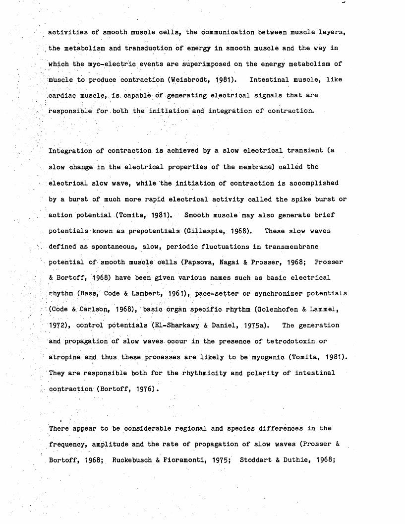
Standards were prepared by dissolving 5-HT in distilled water so that
200 yUl volume containing between 50 and 200 ng 5-HT could be added to
tissue extracts to serve as internal standards, which were carried
through the entire assay procedure. The relationship between
concentration and fluorescence that developed was linear (Figure 1).
Radioactive (3HY-L-Moradrenaiine Experiments
(1) Incubation procedure and 3H efflux
The rat terminal colon was dissected out, free of adhering blood
vessels. Each tissue (2—3 cm) was then incubated at 37°C in Krebs1
solution containing 444 KBq/ml of (3H)-NA and incubated for 20 minutes
at 37°C, Uptake of (3H)-NA was stopped by transferring tissues from
the amine-containing solution into tubes containing normal ice-cold
Krebs* solution.
Having washed off the extracellular and loosely bound radioactivity in
. the cold Krebs* solution, the colon was then set up for in vitro
recording of mechanical activity as previously described. Krebs*
solution bathing the tissue was removed and replaced at ten-minute
intervals and was dissolved in 10 mis of toluene-Triton X scintillant.
The radioactivity present in these samples was counted in a liquid
scintillation spectrometer. The effects of drugs on the efflux from
and mechanical activity of the colon were determined by adding drugs
from the 80th minute of incubation onwards.
The 3H remaining in the tissue at the end of each experiment was
determined by digesting the tissue in 1 ml of 4M KOH at 60°C for 1 hour
before adding 10 ml of toluene Triton X scintillant and counting.
Relati
ve flu
oresce
nce
OO 60
50
40
30
20
10
nr50 100
ng (5-HT)150 200
Fig. 1: Standard graph for 5-hydroxytryptamine (5 HT)

24
Efflux was expressed as fractional tritium release of the total tritium
content of the tissue. No correction for tritium in the extracellular
space was made.
(2) (3H) Uptake by the rat colon
The uptake of (3H)-NA by the rat colon was carried out essentially
as described by Hermann and Graefe (1977) for isolated rat tissues.
The colon, dissected free of adhering blood vessels was blotted dry, cut
into pieces and weighed. Each tissue sample was mounted on the tip of
a stainless steel rod and incubated at 37°C in tubes containing Krebs*
solution.
After 30 minutes, tissues were transferred into media containing
444 KBq/ml of (3H)-NA for various time intervals (1-60 minutes).
Uptake of (3H) was stopped by transferring the tissues from the amine-
containing solution into tubes containing ice-cold Krebs* solution and
by chilling (0°C) them for 1 minute. Thereafter the tissues were
rapidly removed from the stainless steel rod and blotted dry. Each
tissue was digested in 1 ml of 4M KOH at 60°C for 1 hour. Toluene
scintillant was added to each vial and the radioactivity counted by a
liquid scintillation counter. Uptake was expressed as activity per mg
/tissue. Correction for quenching was carried out by plotting external
standard ratio (E.S.R.) against counting efficiency and sample counts
. were converted from CPM to DPM.
High Performance Liquid Chromatography Experiments
(1) Chromatographic System
The technique of High Performance Liquid Chromatography with
electrochemical detection was used to measure the catecholamine contents

of the rat colon and the vasa deferentia of the guinea-pig, rat and
mouse. Both spontaneous and electrical-stimulation-induced release of
noradrenaline from the vasa deferentia of the guinea-pig and mouse were
similarly measured.
The apparatus consisted Of a pressure pump (Laboratory Data Control
•Model .709) with a pulse dampener to deliver solvent to the column at
precise flow rates with a relatively pulse-free output at pressures up
to 1,200 p.s.i., an injection valve Rheodyne 7125 (Berkeley, CA USA)
with a 100^11 loop, a stainless steel reverse-phase Hypersil column (150
x 5 mm 1D) prepacked with octadecyl bonded silica (HPLC Technology) and
an electrochemical detector combined with a battery or mains operated
potentiostat amplifier. The detector was operated at +0.55 or +0.7 V
with a glassy carbon working electrode, a platinum wire auxiliary
electrode and a silver/silver chloride reference electrode, all housed
in a Faraday cage to avoid electrical disturbance (Figure 2). A
mixture of solutes introduced into the system through the injection port
is separated into components on travelling down the column. The
individual solutes are measured as they pass through the detector, the
resultant voltage signals, once amplified, are recorded as peaks on a
chart recorder. Peak heights are proportional to quantity.
(2) Chromatography Mobile Phase
The composition of the mobile phase provided the most powerful factor
affecting chromatographic separation. As the column aged, the
retention capacity and, hence, good separation decreased.

Buffer
Pump Pressure Gauge
I N J E C T I O N S o l v e n t NA fr o o t_ _ _ _
J V. OA
InjectionPort
C18Column
ElectrochemicalDetector
Potentiostat Amplifier
Fig. 2: Diagramatic representation of a High Performance LiquidChromatography system.

By altering one of the constituents of the buffer, the separation could
be improved. Phosphate buffer was used and a typical composition used
towards the end of the project is shown below:
Potassium dihydrogen orthophosphate 13*6 g
Sodium octylsulphonate 0.0864 g
EDTA 0.0336 g
Methanol 40 ml
Deionised Water to 1 litre
The pH was adjusted to 3.2 with concentrated orthophosphoric acid. The
water used for the mobile phase was deionised and filtered through a
Millipore Q reagent grade water system. Prior to use, the mobile phase
was filtered and degassed using an Edwards single stage high vacuum pump
(Model ISC 50) and a Millipore solvent clarification kit with 0.45 Jim
aqueous filters.
Extraction of Catecholamines from Tissues
The NA and DA contents of the guinea-pig, rat and mouse vasa deferentia
and rat colon were extracted and measured essentially as described for
the guinea-pig vas deferens by Macrae (1983).
Appropriate tissues were removed, stripped of adhering blood vessels and
connective tissues and individually weighed. They were then finely
chopped with scissors and transferred to a 20 ml glass homOgeniser with
2 ml of ice-cold 0.1 N perchloric acid containing sodium metabisulphite
(4 x 10"4 M).
Homogenisation was carried out for 2 periods of 30 seconds with a
motor-driven (TRI-R-STIRRER) teflon-glass tissue grinder used at full

27
power. During homogenisation the temperature of the homogenate rose
from 0°C to 4°C. The homogenates were decanted into centrifuge tubes
and a final 3 ml of perchloric, acid (0.1 N) added to transfer the last
traces of the homogenates to the centrifuge tubes. The samples were
centrifuged (2,000 g, 15. minutes, 4°C) in a Christ centrifuge and the
supernatants decanted. Aliquots of 0.5 ml were added together with 50
mg of acid-washed alumina and 1.0 ml of Tris buffer 1 M (pH 8.6) to the
:polystyrene extraction tubes. The tubes were stoppered, vortexed for
30 seconds and;then put onto a Luekham (Model R100) horizontal shaking
machine for 10 minutes. Once the alumina had settled, the supernatant
was aspirated off and the alumina washed 3 times with 3 ml of a dilute 5
mM Tris buffer. Between washes the alumina was again allowed to settle
and wash fluid aspirated off. Catecholamines were then eluted from the
alumina with 300 }il of 0.1 N perchloric acid containing 4 x 10⁻²* M
sodium metabisulphite. After adding the acid, the alumina-acid mixture
was vortexed for 30 seconds to ensure maximum elution, the alumina was
allowed.to settle and the supernatant acid aspirated and stored on ice
prior to analysis.
In Vitro Experiments
Isolated pairs of either guinea-pig or mouse vasa deferentia were
dissected free of adhering blood vessels and connective tissues.
Tissues, were then set up in a 2 ml or 5 ml plastic syringe bath inserted
in a conventional 10 ml organ bath to permit the maintenance of constant
temperature. Pairs of vasa were connected to the hook of a ring
electrode at the prostatic, end,and to a Grass FT0 3 strain gauge at the
epididymal end to measure longitudinal tension. One gram tension was
initially applied to each tissue. Responses were displayed on a Grass
polygraph. Krebs* solution was delivered to each organ bath by a

Watson-Marlow constant flow pump. The system allowed Krebs* solution
to be aspirated from the top of the inverted syringe or to be washed out
from the bottom of organ bath.
Types of Samples
(1) Krebs* solution blank
At the.beginning of the experiment, Krebs* solution was passed through
the heating coil and into the plastic bath, from which it was withdrawn,
extracted and analysed. From.this sample it was possible to detect any
contaminant in the system.
(2) Spontaneous release sample
This provided a measure of the spontaneous release of catecholamines
from the tissue. After the initial equilibration period, the Krebs*
solution in contact with the tissue was washed off and replaced with
fresh Krebs* solution.
After a; period of 6 minutes, equivalent to the duration subsequently
used for electrical stimulation and collection, the Krebs' solution was
removed and its catecholamine content measured.
(3) Stimulation sample
The vasa were stimulated at a frequency of 20 Hz, with pulses of 1 msec
duration, and supramaximal voltage for a period of 4 minutes. The
released catecholamines were allowed a further period of 2 minutes for
diffusion to be completed. The Krebs* solution was then collected.

(4) Tissue recovery sample
A mixture containing 500 pg NA, DA and DOPEG was added to the tissue and
was allowed to remain in contact with the tissue for the same duration
as was used for electrical stimulation. The recovery of this sample
was, therefore, influenced not only by the extraction losses but also by
the. tissue inactivation processes.
(5) Krebs? solution recovery sample
A known amount of both NA and DA was added to the Tris buffer and
alumina and then extracted. This provided a measure of the extraction
.efficiency and the values obtained were used to correct experimental
results for losses incurred during extraction.
Probleas encountered with electrochemical detection
(1) Arte/acts occurring after, injections of large concentrations of catecholamines
Initially, after one or two injections of a high concentration of the
catecholamines into the HPLC, subsequent injections of perchloric acid,
distilled water or the mobile phase through the injection port often
produced two peaks, occurring at the same positions as the injected
catecholamines. These peaks became smaller with repeated injections of
perchloric acid until they disappeared completely. These artefacts
Were due to contamination of either the injection ports or the syringe
used for injections; Subsequently, extra care was taken to wash both
the injection port and the plastic syringe used for injection and these
artefacts no longer occurred..

(2) Artefact associated with extracted Krebs1 solution
At the working potential of + 0.7. V, Krebs' solution, which had passed
through the reservoir, tubing and heating system and into the plastic
syringe organ bath or had passed only through the reservoir and tubing,
or Krebs' solution taken directly from the reservoir and then extracted,
all produced an artefact at a position between DOPEG and NA. This
artefact was initially thought to be either dihydroxymandelic acid
(DOMA) or 3-methoxyphenylglycol but neither of these eluted at this
position on the chromatogram.
When the working electrode potential was reduced from + 0.7 to + 0.55 V,
;this artefact disappeared. This observation explains why this peak was
not noticed either earlier in this study or by Macrae (1983) since the
working potential previously used was + 0.55 V. This peak probably
represents an unidentified oxidisable. product of Krebs' solution.
Macrae (1983) warned against the periodic changing of the tubing system
but the arrangement of the apparatus in the present study made this
inevitable. However, as long as the separation was good, this
unidentified peak could be distinguished from either DOPEG or NA and,
therefore, posed no problem. Such unidentified peaks have frequently
been reported in HPLC systems (Honma, 1982).
(3) Problems associated with the chromatography system
Various problems were associated with the different components of the
system.. For: example, decreased sensitivity and drifting base lines
were usually associated with the working electrode becoming "poisoned"
by oxidation products or with the presence of contaminants and trapped
air bubbles. In this case the glassy-carbon electrode could be

cleaned in a few minutes by scouring the surface of the electrode with
a slurry of fine alumina. Once the alumina has been washed off the
electrode, the complete cell was re-assembled and was ready for use.
On one occasion, when no signal was obtained to injections of increasing
concentrations of the catecholamines, it was found to be caused by a
"dead” reference electrode.
Air bubbles in the mobile, phase appeared as spikes on the baseline.
Spikes were also caused by faulty electrical grounding of the equipment.
Histochemistry - Falok technique
The method was similar to that described by Gillespie and Kirpekar
(1966) and is based on the principle that catecholamines can be
transformed into fluorescent isoquinoline derivatives by condensation
with formaldehyde (Figure 3)» Small sections of tissues were removed
and immediately frozen in isopehtane which had been cooled in liquid
nitrogen. The tissues were then freeze-dried in a Pearce Speedivac
freeze-drier at -40°C, 0.01 torr overnight. Next day, the temperature
was raised to +35°C to prevent condensation and reduce water absorption
before breaking the vacuum. The tissues were then removed, wrapped in
gauze and pinned to the underside of the lid of a jar containing
paraformaldehyde which had been heated to 80°C in a Griffin 1/200 oven.
The jar and the tissues were returned to the oven and exposed to
formaldehyde vapour for one hour. From the oven the tissues were
returned to the freeze drier and dried for a further hour in small
vessels containing de-gassed wax.

NORADRENALINEOH
HO ' ^
Formaldehyde
OHA,6,7 - Trihydroxy- 1, 2,3,4 - Tetrahydro' Isoqulnollne
Dry protein
it,6,7 -TRIHYDROXY- 3, it - DIHYDRO- ISOQUINOLINE
3: The Falck Hillarp reaction.

After drying, the temperature, was slowly raised to melt the wax, then
tissues were left in vacuo for 10 minutes to allow them to embed in the
wax. Tissues were then removed and aligned in a wax embedding pan and
allowed to cool, blocked and placed in the refrigerator for several
hours to maintain wax at lower temperature and obtain optimal conditions
for tissue sectioning. The blocks were sectioned to give 6 pm sections
in a Leitz microtome. These were mounted dry on heated slides with hot
liquid paraffin to dissolve the wax and act as mounting medium.
The fluorescing specimens were viewed and photographed on a Carl Zeiss
ACM photomicroscope, equipped With a 1 V FI epi-fluorescene system.
The light source was an Osram, HB50 mercury lamp, the filters used were:
exciter-interference BP 405/8* barrier - LP 418 and dichromatic beam
splitter FT 420. Photomicrographs were taken on Ektachrome ASA 400
film using a MC 63 photomicrographic camera.
Animal pretreatment schedules
Reserpihe (2 mg/kg, i.p. daily for 4 days) was dissolved in glacial
acetic acid (0.2 ml) and diluted with water to 20 ml. Rats received
0.2 ml of this solution/100 g body weight. Control rats received an
equivalent volume of an appropriate dilution of acetic acid.
6-Hydroxydopamine (6-0HDA, 2 x 50 mg/kg, i.p. on day 1; then 2.x 100
mg/kg, i.p. on day 5). The concentration of the 6-0HDA solution was
adjusted so that rats received 0.2 ml of solution/100 g body weight*
Control rats received saline at appropriate intervals and tissues were
examined on day 6.

P-Chlorophenylalanine (PCPA, 200 mg/kg, i.p. daily for 4 days).
Control rats received saline for a similar period and tissues were
examined on day 5.
Preparation of haemolvsate
Male rats (250-350 g) were anaesthetised (Nembutal) (65 mg/kg, i.p.) and
blood was collected from a cannulated carotid artery. The blood was
collected in heparinised tubes, centrifuged (1,000 x g, 20 min, 4°C) and
the plasma and buffy coat removed by aspiration.: The erythrocytes was
washed twice and resuspended in phosphate buffered isotonic saline to
restore the volume to 3 ml; this constituted the washed erythrocyte
. suspension from which the haemolysate was prepared. 1 ml of the
; suspension was pipetted intp centrifuge tubes containing 19 ml of
hypotonic phosphate buffer (20 mM, pH 7.4). This suspension was then
. centrifuged (20,000 g, 40 min, 4°C) and the supernatant from this
procedure Constituted the crude haemolysate. Crude haemolysate (15 ml)
was dialysed overnight (4°C) against distilled water or hypotonic
phosphate buffer (pH 7.4) to remove low molecular weight components.
The effect of a 1:100 dilution of the dialysed haemolysate on the
inhibitory response of the isolated colon to electric field stimulation
was determined.
Analysis of results
All the results on the graphs show the mean - standard error of the mean
(S.E.M.).
- The Student*s t-test was used for statistical analysis of results.
A level of probability of P ^0.05 is taken to indicate statistical
significance.

Pru&g
Acetylcholine Chloride (Koch-Light); Adrenaline bitartrate (Sigma);
Apamin (Sigma);. Ascorbic acid (B.D.H.); Atropine sulphate (B.D.H.);
Carbamoylcholine chloride (Sigma); Clonidine (Boehringer Ingelheim);
D-alaglymepheglyol (DAGO) (Sigma); Dihydroxyphenylglycol (Sigma);
Diltiazem (Sigma); Dopamine (Sigma); Ethylenediaminetetracetic acid
(E.D.T.A.) (Sigma); Flurbiprofen (Boots); Guanethidine monosulphate
(Ciba); 6-Hydroxydopamine hydrobromide (Sigma); 5-Hydroxytryptamine
creatinine sulphate (Sigma); Leucine enkephalin (Sigma); Methionine
enkephalin (Sigma); Methysergide bimaleate (Sandoz); Morphine
hydrochloride (Macarthys); Naloxone hydrochloride (Winthrop); L-
noradrenaline bitartrate (Koch-Light); Normetanephrine hydrochloride
(Sigma); Oxprenolol hydrochloride (Ciba); P-Chlorophenylalamine
methylester (Sigma); Phentolamine mesylate (Ciba); Prazosin (Sigma);
Propranolol hydrochloride (I.C.I.); Quinidine (B.D.H.); Reserpine
(Koch-Light); S.K.F. 525A (Smith, Kline & French); Substance P
(Sigma); Tetrodotoxin (TTX) (Sankyo); Tyramine hydrochloride (Sigma);
Verapamil (Sigma); Yohimbine hydrochloride (Koch-Light).

RESULTS

1. Effects of morphine and of opioid peptides on the motility of the isolated colon
1.1 Morphine
The isolated colon of the rat generally shows a low level of
spontaneous motor activity. The addition of morphine M) to
the colon caused it. to oontract, usually immediately but sometimes only
after a delay of several minutes. The motor response to morphine was
not a sustained contraction and was followed by a relaxation to the
baseline, with morphine still in the bath. This initial contraction was
followed by waves of contraction and relaxation which continued at
intervals thereafter (Fig. 4). The frequency of these rhythmic waves,
of contraction was variable both between animals and in tissues from the
same animal during the course of an experiment. These waves of
contraction and relaxation were abolished by the specific opiate
antagonist naloxone (10“ M) (Table 1a).
Above the threshold required to produce the rhythmic contractions,
no dose-response relationship was observed. Increasing the dose merely
increased the probability that the rhythmic waves of contraction would
occur. In most studies of the excitatory effects of morphine in the
gut, the drug is usually allowed to remain in contact with.the tissue
briefly and then is washed from the bath. In this study the initial
contraction was, therefore, examined separately from the rhythmic . .
contractions subsequently produced by prolonged exposure to morphine.
A very steep concentration-response relationship was obtained. This
contraction was competitively antagonised by the specific opiate;
antagonist naloxone. In contrast, the 5-HT antagonist methysergide

■ 2 min
FIGURE 4 Excitatory effect of morphine on the isolated colon of the
rat. Morphine (M, 5 x 10"^ M) produced an immediate contraction
followed by rhythmic waves of contractions. Naloxone (N, 10” M)
abolished the contractions.

shifted the dose response curve for morphine non-competitively (Fig. 5).
Other agonists, which produce doserdependent contractile responses in the
rat colon, included carbachol, 5-HT and substance P. The mean E,C,50
values for these agonists and the mean maximum force developed, by the
colon in response to these drugs are shown in Table 1.
1.2 Enkephalins
The ability of morphine to produce rhythmic waves of contraction
and relaxation was shared by the synthetic opioid agonists DAGO, leucine
enkephalin and methionine enkephalin. When compared with morphine, the
enkephalins were more potent and, therefore, lower concentrations of
these agonists were required to cause contractions. DAGO (2 x 10“® M),
leucine enkephalin (10“® M) and methionine enkephalin (2 x 10“ M)
caused the colon to contract rhythmically (Fig. 6).
The waves of contraction produced by the synthetic agonists
sometimes gradually diminished in amplitude probably due to destruction
by tissue enkephalinase and were abolished by low concentrations of the
opiate antagonist naloxone (10“^-2x10“ M).
1.3 Comparison of the response of the colon tomorphine. 5-HT. ACh and KC1
The pattern of rhythmic waves of contraction produced by morphine
was unlike the responses of the rat colon to other agonists. KC1
produced a rapidly-developed contraction which was well-maintained
until the KC1 was washed out of the organ bath (Fig. 7). ACh also
caused the colon to contract and remain contracted for several minutes.
Morphine-induced rhythmic contractions were also unlike those produced
by 5-HT, which caused the colon to contract and relax irregularly and

Tension (g)
J-fMorphine (M)
FIGURE 5 Effect of methysergide and naloxone on the contraction of
the rat .colon induced by morphine. o o, control response,
o o, response in the presence of methysergide (3 x 10“ M),
e - e, response in the presence of naloxone (3 x 10"^ M). Values
(mean - S.E.M.), n = 5.

Sensitivity of the rat colon to different agonists and the
mean force (g) developed to the agonists.
Mean 1 (S.E.M.) Max response (g)
Agonist (x 10"8 M) p Value* Mean t (S.E.M.) p Value*
Morphine 5.63 - 1.40 3.18 - 0.27
DAGO 0.45 - 0.09 <0.01 4.60 - 0.67 NS
Leucine Enkephalin 0.01 - 0,03 <0.01 5.40 t 0.47 <0.01
Methionine Enkephalin 5.29-3.00 NS 3.30 t 0.30 NS
5-Hydroxytryptamine 67.00 - 8.70 <0.001 6.28 - 0.66 <0.001
Carbachol 9.45 - 2.29 NS 7.27 - 1.03 <0.01
Substance P 0.70 - 0.11 <0.01 3.57 - 0.50 NS
■Each, value, is the mean of 8 observations. The p values refer to comparisons
between the responses obtained with the drugs listed and morphine.
. ’NSr indicates that there is no significant difference between the EC^q or
maximum response obtained with a particular drug and the EC^q value on maximum
response obtained with morphine.

3
a >>0 o x>XI4-5 CO4-5*o0G O o X) O•H 0 3 G PG aa *8 G GO 0 G c 0i—1 a bo aO 0 3 0o X! G G G4-5 o Q0 •H E —-X! w P r-l 0P s O M0 Go G Oa XI G 0 Xo 0 P £ oG P 1—1>» o O 0 Gp 0 O X3 G*H 1—1 Wi—1 G a TJ•H o G GP 0 O G
XIcd-pCO•H
X3£h
bps o IW oSG 0 Ob- G H | O O O X O t- O
g a32 -••H <u . o
G >o o(d *rHa bOea t, bo 0 0 3O 0 £-.>» Q O XI C -P 4-5a) 0£■< a x30 4-5Cm —0 0 *U G G G •H cd G CU 8 O W 3 G G P 3
cd 3
T30 p cdr—IO CO •H
P O Cd P G •H VO 0 I X! O P *—
Cd0 G G•H -Oa GG GaG £hsG(XoIOs *-
<M Xo CM >>^4-5•H Xr—C Em•H H 4-5O - -a -s s sgXXO vo P I I o oCO T~ T- 0 wGG 0a. c a -hO X3o cu Go a
o o - ot 0 obo a3 *H 0 G r-i 0 Q G 0 c CO X3 4-50X3 0 a P G OM 0C 3 X) H O m O G0 O 0 03 G G rH 0 0Cd -G 0 > p s~ o PCX <D „ H X
0 G
§ "gGD.Cd cx
G 0 GG O ■H CO
•H CO G 0 G 3 Q.
0XOGP
CO•HXJH
0G•HaoGPG
•> eG ox: +5 a c2 ° a e
a . oo 3 CO w1 W . uu |0 G 0 G 0 GG o > •H G > •H G > ,*H. G0 i— G •H G •H G •HP £ n 3 CO S W0 G 0 0 0 M 0 Wa 0 a -■ n > 0 a > 0 a > 0 oG > o s G S > s o s G S > S o s G S > SG O w > W G W w > a g a a 3: a g aG >> CO CO > CO >» CO co 3c co >» CO CO 3 CO
a •8 g a o a , o a G+lc+« o+i G + l c+l 0 + 1 G+l d + l O-HG p 0 v-y __' 0 w 0 V-- v_- ■ <D __ 0 v_x . 0 wG G 3 G 'S 3 G TJ 3 G T30 rH O' O G O' O G o* ,o GS 3 0 •H X3 0 •H 3 0 •H 3O G W G G ft G Wi—1 a G a G G a .G GG 0 0 0 0 <D 0O H G H G Eh G
0 G •H i—IGCO
bO3GQ
rHoGPGOO
bO3GQ
in T— Oo o : o
o • 'r * • o • on CM ■=3’ VO st ao CM O VO O b - co in co 0 5 CO c o co CD CO a co
0 ^ • V • V • V • 2 • 2 ; • ^ • 2 ; • .5 5 ' •. 55G S o a o a r - a O o o O o nO ^—- /—- yr-X r - \X vo + 1 + 1 < -v + 1 O +1 in + 1 oo + 1 . b - + 1 in + 1 in + 1 voO I CO CO 0 5
rH O on in 05 O II st II o n i i O II O II v o IIG T - CO II v o i i CO 11 CO 05 St b - b - T—
z w • • . • • G • G • G • G • G • GO G O G CM G -= f ^ - r CM H -r o n w v o — ° w VO
v- ' S_-r v_' CM
0 CM o b - o VO in in O■o CM CO 4=t- CO 0 5 CO CO 5=f CO T - CO VO co o co a co•H • 55 • 25 • 55 • 2 ; • 55 • 2 • 2 ; • 2 } • • 55bO S T— O CM o O CM O T—G /-- *—\ -—\. -—N ■H /—* *■—s /■—\dX o 0 1
+ 1 co + 1 CO + 1 b - + 1 vo + 1 b— + 1 b - +1 in + i in + 1 vo
>> O b - II 05 || 0 5 || o n n * - II o n i i O II CO II b - IIX I t - on CM CM co VO St a VO v oP W • a • G * G • G • G • G • G • G • G0 v o a » CM 05 On H_r CM O V -r V O ---- - ° in w
S T— CM
t— O CM CM in o o on aCM CO E f CO VO CO St CO o n co b - CO a co co -=r co
0 » • S i • 25 • • 2 ; • 2 : • 55 • 55 • 2 ; •' 2 ; ■ • 55G S t— O on o O o o . • ■=r
•H /—V /—- • *—- r"H>a oO 1
+ 1 05 + 1 b - + 1 CO + 1 b— + 1 E— + 1 b - + 1 + 1 in + i in
G O CM II O II O II CO II O || st II o n i i CO II O IIP r - CM -=b in CM VO in id -
w • G • G • G • G • G *"• G • G .. G • Gin '-r CM ^ St v ^ —r CM v -r on H-r b -H - r O v -r b - v -rCM CM
05 b- o vo on 05 05 on b-O 00 on co co b- CO b- co on co in co co ■st CO• 25 • 25 • 2; .. • 55 • 55 • 55 • 5 5 .• ■• 55 • 55a O in O O T— T— O in' -* N /—\ \ /—s /—s ^ \
+ 1 CO + 1 b- + 1 0 + • + 1 vo + 1 b- + 1 VO +«' zt. + i inb- II 05 || O II O II co ll b- II O II • 05 II on iiCO id* in in 05 in o ' VO CO• G • G • G • G • G • G • G • G • Gi n ---- a p O w On '-r a — id- '— b- w O v_^ id". —r
on a ' a
* - T - i n T - r—O O •-=r o• V*- a+ i COt—in ii
•in G
on• NO CO+ 1 IIb- d on wo
0■SExXo a i G O O r-s
in4T CO• 55 O
/■— » + I b—O IIon• GCM w
CMo co+ 1 II•=r gCM w
Oon oCM •• oid" Va+ 1<— sb- CO CO
• IIonCM G
ooCM 05 + 1 II O GO w
o o oid* •• oO Va+ ib- VOT—
• II
o on • >o b-
+ i ii
CM G
O VO •VO o• Vo a + 1
VOCOon n CM G
oCMo in
+ 1 iiO G CM v-'
CM
O CO O• o
CM Va+ I/— * oo b- CM
• IIaCM Goo• r—\cm in
+ 1 i i
O GO WCM
O in • o o • v «- a+1 vob-T“ IIvo G
Oon • /■> O b-
+ 1 IIon clst '
CM . «“ CO
• 55 O+ i
CO IIin
voIo
HEh CM
c s•Hb—^ Oa *-«aj w
O b- •in o • \/ in a+ 1vost
ini i
GCM
b- -=rr- cmO b- CM b-+ 1 II +1 IIb- G O GX— V-< O H—r

. •Met-enkepbalin 2 x 10 H
IMNaloxone 2 x 10”7M
m i n
]2g1min
NaloxoneLeucineEnkephalin 10"8M
Naloxone
FIGURE 6 Excitatory effect of opioid peptides in the rat colon.
Met-enkephalin (2 x 10™^ M), leu-enkephalin (10™® M) and DAGOo(2 x 10“ M) all produced naloxone-sensitive rhythmic contractions in
the rat colon. Each opioid peptide was added to the organ bath at
and remained in contact with the tissue until the end of the experiment.
Naloxone added at N in each experiment either abolished or reduced the
size of these contractions.

min
30mH kci
Morphine
ACh,-6i
5 X 10 H
FIGURE 7 Comparison of the responses of the isolated colon to
potassium chloride (KCI, 30 mM), morphine (5 x 10“ M), acetylcholine
(ACh, 5 x 10“ M) and 5-hydroxytryptamine (5-HT, 5 x 10"^ M). The
responses, of the colon to morphine were quite unlike the responses to
KCI, ACh and 5-HT.

briefly, after which the colon relaxed to its initial resting tension
despite the continued presence of 5-HT. Thereafter the tissue was
unresponsive to 5-HT.
1.4 Effects of temperature on morphine-induced rhythmic contractions
Between 30-38°C temperature changes had little effect on the
occurrence and characteristics, of morphine-induced rhythmic
contractions. Lowering the temperature of the bathing medium from 37°C
to 26°C reduced the frequency and increased both the amplitude and
duration of morphine-induced contractions (Fig. 8). Increasing the
temperature above 38°C generally increased the frequency and decreased
the amplitude and duration of the waves.
1.5 Effect of Ca** removal
The ability of morphine and the opioid peptides to produce
contractions in the rat colon was reduced in Ca++-free medium. The
addition of SKF 525A (10~^ M), which blocks Ca++ influx (Kalsner, Nickerson
& Boyd, 1970), diltiazem or verapamil (10"^-10“ M) reduced the size of the
rhythmic waves of contraction (Fig;. 9).
2. Analysis of the mechanism of opiate-induced waves of rhythmic contractions in the isolated colon
Two hypotheses proposed to explain the mechanism of the rhythmic
waves of contractions seen in the rat colon (Fig. 10) were examined.
The 5-HT/ACh hypothesis was tested in three ways:-

A 35°C
Morphine
c 35°C
D 26°C
FIGURE 8 Effect of temperature on the amplitude, duration and
frequency of morphine (5 x 10“ M)-induced rhythmic waves of
contractions in the rat colon. The first two panels (A and B) are.from
one experiment. Increasing the temperature from 35°C to 41°C increased
the frequency and reduced the amplitude of the contractions. The two
lower panels (C and D) are from a different experiment. Reducing the
temperature from 35°C to 26°C reduced the frequency and increased the
amplitude of the contractions. . In trace C field stimulation at S
(10 Hz, pulse width 0.5 msec) during a morphine-induced contraction
produced an inhibition for the duration of the period of stimulation.
Morphine
im in

SKF 525A
BJvJvA j A. —
Dlltlazem10'5H
^ 319I __ IImln
C /V -JVMorphine 5 x 10'6M Verapaml1
10"5M
FIGURE 9 Effect of calcium antagonists on morphine-induced rhythmic
contractions SKF 525A (10"^ M), diltiazem (1CT^ M) and verapamil
(10“ M) reduced the size of the rhythmic waves of contractions produced
by morphine (5 x 10“ M). Traces A, B and C are from different
experiments but the same calibration applied to all three traces.

5-HT V store y 5-HTMorphine
< AChor SmoothMuscle
FIGURE 10 Models of possible mechanisms of the excitatory effect of
morphine on the rat colon. Morphine releases intestinal 5-HT which
then acts directly on the smooth muscle and indirectly through ACh
release to cause muscle contraction or morphine by inhibiting an
intrinsic spontaneously-active inhibitory neurone, unmasks the inherent
myogenic activity of the smooth muscle.

(a) 5-HT autodesensitisation
(b) Depletion studies
(c) Use of antagonists of 5-HT and other drugs
2.1 5-HT hypothesis
2*1a 5-HT autodesensitisation
The effects of repeated exposure of the rat colon to increasing
concentrations of 5-HT on responses to opiates was investigated (Fig.
11). These experiments examined the effect of adding the same high
concentration of 5-HT repeatedly to the colon without washing out the ‘
previously-administered 5-HT. 5-HT (5 x 10“ M) was added three times
and with each administration, the size of the response diminished.
When the concentration of 5-HT was increased to 5 x 10” M, a response
could still be obtained but the addition of a higher concentration of
5-HT now produced no response.
At this point, with 5-HT still in the bath, morphine (5 x 10“ M)
caused an immediate contraction which was followed by further
contractions and relaxations. These contractions were inhibited by
naloxone (10"^ M).
2.1b Depletion studies
. Effects of PCPA or resemine pretreatment
Pretreatment of rats with PCPA reduced the 5-HT content of the
colon. Figure 12 shows the 5-HT content of colons from control, PCPA-
and reserpine-pretreated rats. PCPA was effective in reducing the 5-HT
content of the rat colon but reserpine was less effective. Despite the
marked effect of PCPA on the 5-HT content of the colon, morphine still

• ••5-HT
5 x 1 0 '6H
OMorphine5-HT
5 x10'5M
NalMe
]2g
1 min
FIGURE 11 Excitatory effect of morphine in colon desensitised to
5-hydroxytryptamine (5-HT). Desensitisation was produced by repeated
application of increasing concentrations of 5-HT. 5-hydroxytryptamine
(^5-HT, 5 x 10“ M) was added three times without washing out the
preceding dose. A higher concentration of 5-HT ( ^ 5 x 10“* M) was
again added. The second cumulative addition of 5-HT ( 0 5 x 10“ M)
no longer produced any response. Without washing out the 5-HT, addition
of morphine (5 x 10” M) produced rhythmic waves of contractions which
persisted in the presence of methysergide (Me, 10"^ M). These waves of
contraction were apparently inhibited by naloxone (Nal, 5 x 10“ M).
Both panels are from the same experiment.

5-HT ipg /g )
4 -i
3 -
2 -
1 -
Control
K0-05
R0-001
Reserplne PCPA
FIGURE 12 Effect of reserpine- or PCPA-pretreatment on the 5-HT content
of the rat colon. Each histogram is a mean of 5 observations. The
I-bars represent the standard error of the mean. The p values refer to
comparisons with the control.

caused such colon to contract and these rhythmic waves of contractions
were inhibited by naloxone (10“ M) (Fig. 13).
2.1c Effects of 5-HT and other drugs
(a) Methysergide
The rhythmic waves of contractions produced by opioids were
unaffected by methysergide (10“5 M) in control tissues (Figs. 15, 16)
or in tissues rendered subsensitive to the effect of 5-HT by the process
of autodesensitisation (Fig. 11).
(b) Adrenergic and cholinergic antagonists
The rhythmic waves of contraction produced by morphine and
the opioid peptides were unaffected by both the cholinergic antagonist
atropine (10“ M) and the adrenergic antagonists phentolamine (10"^ M)
and propranolol (10“ M) (Figs. 14, 15, 16).
(c) Quinidine
Quinidine (10“ M), which non-specifically blocks the effect
of pUrinergic nerves in spme tissues (Burnstock, 1972), did not affect
the waves Of rhythmic contractions produced by morphine.
2.2 The, tonic inhibitory nerve hypothesis
The possibility that morphine might remove a tonic inhibitory
influence that normally suppresses myogenic activity was considered.
If this hypothesis is correct, then rhythmic waves of contraction should
not only be produced by opioids, but also by other drugs that inhibit
neuronal mechanisms or inhibit inhibitory mechanisms in smooth muscle.

5-HT pg/g
432
1
0
control
—mMMMm ** >i min
pcpa
a i
FIGURE 13 Excitatory effect of morphine on PCPA-pretreated colon.
The upper panel shows the 5-HT content of the control colon and the
rhythmic waves of contractions produced by morphine (5 x 10“ M) in such
a colon. The contractions were antagonised by naloxone (10“ . M) .added
at N. The lower panel shows the 5-HT content of a PCPA-pretreated
colon and the naloxone (10~^ M)-sensitive contractions produced by
morphine (5 x 10"^ M) in such a colon.

B® _ A« nPhentolaalne 10
Propranolol 10~°H
Atropine
Naloxone10"7M
1mln
] 19_i
FIGURE 14 Effect of adrenergic and cholinergic antagonists on
morphine-induced rhythmic contractions in the isolated colon. Morphine
(5 x 10“ M) produced waves Of contractions (A). These were not
inhibited; by propranolol (10” M) and phentolamine (10” M) (B). The
cholinergic antagonist atropine (10~ . M) did not abolish morphine-
induced waves of contractions (C). Naloxone (10”*7 M) inhibited the
Waves of contractions (D). Traces A, B, C and D are from the same
experiment.

LeucineEnkephalin 1(T8M
BPhentolamine 10”6M Propranolol 10"6M
Jig1 min
Methysergide10‘5M
Naloxone10“7M
TV.
FIGURE 15 Effect of adrenergic and 5-HT antagonists on contractions
produced by leucine enkephalin in the colon. Leucine enkephalinQ(10“ M,) produced rhythmic waves of contractions in the colon (A).
These contractions were unaffected by both propranolol (10“ M) and
phentolamine (10“ M) (B). The 5-HT antagonist methysergide (10“ M)
did not affect these waves of contractions (C). Naloxone (10“ M)
inhibited the rhythmic contractions (C). Traces A, B and C are from
the same experiment.

A .J N J V *DAGO 2 x 10"8M
B J 19Phentolanlne Propranolol 10’® each
i — , -iImln
Methyserglde lO’^M
Naloxone 2 x 10 “7M
FIGURE 16 Effect of antagonists on the rhythmic waves of contractions
produced by DAGO. DAGO (2 x 10"® M) produced rhythmic waves of
contractions in the isolated colon of the rat (A). The combination of
phentolamine (10"® M) and propranolol (10“® M) did not inhibit the
contractions (B). Instead, the frequency of the contractions increased
after these drugs. Methyserglde (10"® M) did not inhibit the
contractions (C) but the frequency of the waves of contractions was
reduced by naloxone (2 x 10"^ M). A, B, C and D are consecutive traces
from the same experiment.

Such drugs might be expected to have an excitatory effect similar to
that of morphine.
Two drugs are of interest
(a) TTX which blocks sodium conductance and consequently
abolishes the nerve action potential
(b) Apamin which inhibits inhibitory mechanisms that depend ,
on increased potassium permeability in smooth muscle
2.2a Response of the colon to apamin or TTX
O f\Apamin (10 M) or TTX (10 M) produced rhythmic contractions
similar to those produced by morphine and the opioid peptides (Fig.
17). The rhythmic contractions continued for as long as the drug:
remained in contact with the tissue. The opiate antagonist naloxone,
(5 x 10“® M) did not affect contractions produced by apamin or TTX1 .
(Table 1a).
Comparison of the rhythmic contractions produced by apamin or
TTX with those produced by opioids revealed that the amplitude and. wave
length of the contractions produced by a single drug varied in different
tissues. However, the durations of the waves of contractions produced
by apamin or TTX were remarkably similar to those produced by the
opioids (Fig. 17).
3. The innervation of the rat colon
The results obtained with both TTX and apamin suggested that there is
an inhibitory mechanism in the rat colon which could be NANC or adrenergic.
The nature of this inhibitory mechanism and the innervation of the 'colon
was investigated by electrical field stimulation of the isolated colon.
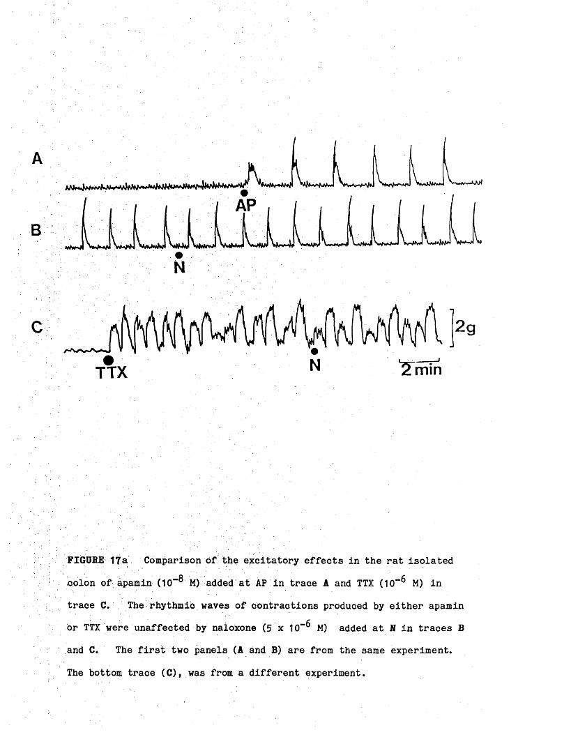
FldUHE 17a Comparison of the excitatory effects in the rat isolated
colon of apamin (10“® M) added at AP in trace A and TTX (10” M) in
trace C. The rhythmic waves of contractions produced by either apamin
or TTX were unaffected by naioxone (5 x 10"^ M) added at N in traces B
and C. The first two panels (A and B) are from the same experiment.
The bottom trace (C), was from a different experiment.

Wave d
uratio
n (mi
ni^ 3 -
to>o4-»c
<u><0
2 -
1 -
Mean 2‘56+0-9
1 J0;W
0.5 -
CDC.£=CLL.o
CDJZCLCDczUJI4-»CD
CO-CClCDcH*3
CDCCDO4-»CZCDszQ.
CDCZTDCoo
CDC L<c
ocz<DL_C LXo
— Mean0-72
+009
FIGURE 17b Comparison of the wave duration and wave interval produced
by different drug treatments (mean — S.D.). The mean wave durations
were more similar than the wave intervals. n = 9 in each experiment.

3.1 Responses of the rat colon to electrical field stimulation
Field stimulation of isolated segments of rat colon with supra
maximal voltage, pulses of 0.5 ms duration for periods of up to 10
seconds produced complex responses which varied according to the tone of
the preparation and the frequency of stimulation (Figs. 18, 20). At
frequencies of stimulation below 5 Hz, there was either no response or a
small inhibitory response or occasionally an excitatory response.
Stimulation at frequencies above 5 Hz produced during stimulation a
contraction which was not well-maintained. Following stimulation, a
powerful post-stimulus contraction occurred at all frequencies but was
greater at lower frequencies.
3.2 Effect of drugs on responses of the rat colon toelectrical field stimulation .
3.2a Atropine
A typical field stimulation-induced response in the presence of
atropine is shown in (Fig. 18). Atropine (5 x 10"^ M) reduced the
contractile responses previously observed at high frequencies of
stimulation.
3.2b Apamin
QApamin (5 x 10 M) potentiated the field stimulation-induced motor
responses particularly those seen at higher frequencies of stimulation
(Fig. 19).
3.2c Morphine
Morphine (5 x 10” M) reduced or abolished the motor responses to
nerve stimulation without affecting the post-stimulation contraction

A a A A (V L 1“
7 W .
50Hz
FIGURE 18 The response of the rat colon to electrical field
stimulation and the effect of atropine on these responses. (A) Motor
responses were obtained to electrical field stimulation (frequency as
indicated, supramaximal voltage, 0.5 ms pulse width for 10 secs).
Following stimulation there was a post-stimulation contraction,
especially at frequencies between 1-10 Hz. (B) In the presence of
atropine (5 x 10*" M) motor responses to field stimulation were
inhibited. .
BAtropine
1Hz 5Hz 10Hz 20Hz

sad
5 x10‘8M
B K
' ' ' » I M I
1 mln
S s
FIGURE 19 Effect of apamin on the isolated colon of the rat. In
trace A apamin (AP, 5 x 10"8 M) potentiated the motor response to
electrical field stimulation (10 Hz, 0.5 ms for 10 sec) applied at S.
Trace B shows rhythmic waves of contraction produced by apamin.
Electrical field stimulation at the peak of an apamin-induced
contraction produced a slight inhibitory motor response in trace C.
The addition of atropine (10'"5 M) converted this small inhibitory
response into.a well-maintained inhibition and also inhibited the motor
response to nerve stimulation. Traces A, B and C are from the same . experiment.

(Fig. 20). The inhibitory response observed at low frequencies of
stimulation became more pronounced.
3.2d 6-OHDA pretreatment
6-OHDA pretreatment potentiated the motor response to field
stimulation. This effect of 6-OHDA was more pronounced at low
frequencies of stimulation (Fig. 21).
3.3 Effects of drugs on post-stimulus contractions
The post-stimulus contraction obtained following the cessation of
nerve stimulation was not abolished by the cholinergic antagonist
atropine (5 x 10“ M), the adrenergic antagonist guanethidine (10“ M),
propranolol (10"^ M) and phentolamine (10"^ M) (Figs. 18 and 26). in
view of the possible involvement of prostaglandins in this post-stimulus
contraction (Burnstook et alf 1Q75). the effects of the water soluble
prostaglandin synthetase inhibitor flurbiprofen were investigated.
Flurbiprofen (10"^ M) had no effect on the post-stimulus contractions
obtained following intramural nerve stimulation at 5 Hz for 10 secs
(Fig. 22).
3.4 Inhibitory response of the rat colon to electrical stimulation
The resting colon develops little maintained tone on its own and,
hence, the inhibitory response to electrical field stimulation was
usually very small. When the tone increased during spontaneous
rhythmic contractions, electrical field stimulation at the peak of a
wave produced an inhibition. Spontaneously-generated waves of rhythmic
contractions offer the best opportunity for studying the inhibitory
response, uncomplicated by the presence of drug. The occurrence of

J
l\l'
Morphinee 1n“Ou
V-Ov'A
1Hz 5Hz 10HZ 20Hz 50HZ
FIGURE 20 Effect of morphine on the responses of the rat colon to
electrical field stimulation. Trace A shows the responses of the colon
to field stimulation in the absence of morphine; during stimulation
contractile responses were obtained, followed by a post-stimulus
contraction. In trace B in the presence of morphine (5 x 10"^ M),
contractile responses produced during stimulation were reduced. Traces
A and B are from the same experiment.

F O R C E (g)10-.
8-
6-
4-
2-
UD Control N S6-hydroxydopa«lne
pretreated rats
P < 0 0 5
w§>
J jiiiill
N S N S
I
2Hz 5HZ 10Hz 20Hz
FIGUBE 21 Effect of 6-hydroxydopamine pretreatment on the motor
response of the colon to electrical field stimulation (100 pulses,
0.5 ms, supramaximal voltage, frequency as indicated). 6-OHDA
pretreatment potentiated the motor response to field stimulation
especially at low frequency. Each histogram is a mean of 6 obser
vations. The I bars represent the S.E.M. The p value refers to the
comparison between the responses of colon from 6-OHDA pretreated rats
and responses of the control colon at the same frequency of stimulation,

Guanethldlne*^ 10_5M Atropine 5 x 10"®M
30 alnutesu
Flurbiprofen 5 x l O ^ M
V.
]ig
FIGURE 22 The effect of flurbiprofen on the post-stimulus contraction
of the rat colon to electrical field stimulation. Post-stimulus
contractions obtained following electrical field stimulation (5 Hz,
0.5 ms for 10 sec) in the presence of guanethidine (10** M) and atropine
(5 x 10“6 M) was unaffected by prior incubation with flurbiprofen (5 x
10“6 M) for 30 minutes. The post-stimulus contraction (mean 1 S.E.M.)
after flurbiprofen was 102 (16) percent of the response obtained before
flurbiprofen and was not significantly different from the control.

these waves was, however, rare. Advantage was also taken of the .
rhythmic waves of contractipns produced by various drugs. The colon
was stimulated by electrical field stimulation at the peak of these
waves of contractions. In this way the nature of the inhibitory
response was studied.
3.4a Investigation of the possible, existence, of an NANC mechanism in the rat colon
In the presence of both guanethidine (10"^ M) and atropine (2 x
10"^ M) to block adrenergically- and choiinergically-mediated responses
respectively, electrical field stimulation at the peak of a morphine-
induced wave produced a well-maintained inhibition during stimulation,
with a return to original (elevated) tone when stimulation ceased {Fig.
23). The optimum frequency of this NANC inhibitory response was less
than 5 Hz.
3.4b Effects of. drugs on the NANC inhibitory response
(a) Resernine and 6-OHDA nretreatment
Both reserpine and 6-OHDA were used to deplete the colon Of
noradrenaline. Comparison of the fluorescence in tissues from control
and from 6-0HDA-pretreated rats confirmed the sparse adrenergic inner
vations of the rat colon and the effectiveness of these treatments in
eliminating catecholamines from the rat colon. In such 6-OHDA- '
pretreated tissues, stimulation between morphine-induced contractions
produced a powerful motor response. Field stimulation applied at the
peak of a morphine-induced wave produced a brief, small ihhibiton, which
was followed during stimulation by a powerful contraction. The absence
of a well-maintained inhibitory response at the peak of a contraction

Morphine 5 x 10"*M Guanethldlnt KT^M Atroolne 2 x 10"®M
FIGURE 23 Effect of apamin on the non-adrenergic, non-cholinergic
inhibitory response to electrical field stimulation of the isolated ,
colon (10 pulses, 0.5 ms, frequency as indicated). In traces B and C
inhibitory responses were obtained to electrical field stimulation
applied at the peak of morphine (5 x 10“ M)-induced waves in the
presence of both guanethidine (10"^ M) and atropine (2 x 10"^ M). The
optimum frequency of stimulation was less than 5 Hz. In trace C apamin
(2 x 10⁻⁷ m ) did not inhibit these inhibitory responses. Traces A, B
and C are from the same experiment.

44
might suggest that the inhibitory responses was adrenergic, since it had
apparently been reduced by 6-OHDA pretreatment.
However, the addition of atropine (10“** M), which blocked the effect
of the released ACh and of exogenously administered ACh (Fig. 24) revealed
that the inhibitory response persisted in these 6-QHDA-pretreated tissues.
This inhibitory response also persisted in reserpine pretreated
tissues (Fig. 24).
(b) Effect of TTX
TTX (10“** M) produced waves of contraction in the colon and
abolished motor responses to electrical field stimulation (Fig. 25)
without any effect on the inhibitory response. This inhibitory
response was reduced by increasing concentrations of TTX (3 x 10”**’ M)
(Fig. 25). Generally three times the concentration required, to
inhibit the motor response was required to reduce the inhibitory
response of the colon to field stimulation.
(c) Effect of haemolysate. apamin and methvlene blue on NANC inhibitory response
1. Haemolysate derived from lysed red blood cells inhibits
inhibitory responses of the bovine retractor penis muscle to field
stimulation and to certain agonists (Bowman & Gillespie, 1982). ■
Haemolysate also potentiated the response of perfused rabbit ear artery
and the rat anococcygeus muscle to NA (Wylie, 1981). Oxyhaemoglobin is'
responsible for these effects of the haemolysate (Bowman, Gillespie &
Pollock, 1982).

6-OHDA
Reserpine
M3g
Av _ K /v1 min
FIGURE 24 Effect of 6-hydroxydopamine and reserpine pretreatment on
the inhibitory response to nerve stimulation. In trace A addition of
morphine (M,. 5 x 10“ M) produced rhythmic waves of contractions in the
colon. Field stimulation (S ) between waves of contraction produced a
. motor response. Field stimulation (S2) at the peak of a morphine-
induced contraction produced a slight inhibitory response followed by a
motor response. Addition of atropine (Atrop, 5 x 10“ M) converted
this small inhibitory response to field stimulation into a well-
maintained inhibition (Sg). .Trace B shows that in colon from
reserpinised rats, morphine (M, 5 x 10” M) produced rhythmic waves
of contractions. Stimulation at the peak of a contraction (S ) still
produced an inhibitory response.
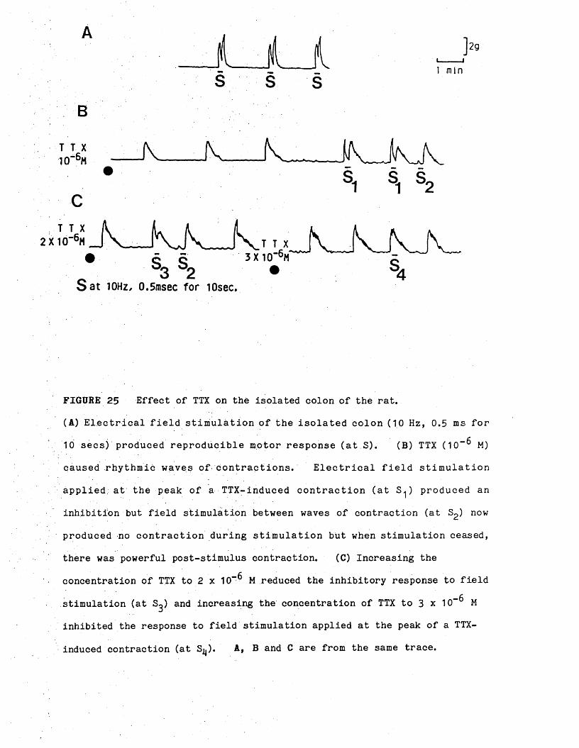
ysj _ j\— _j v . v h v J n j v• SL S. s„
* ^ s2S a t 10Hz, 0.5msec fo r lOsec.
FIGURE 25 Effect of TTX on the isolated colon of the rat.
(A) Electrical field stimulation of the isolated colon (10 Hz, 0.5 ms for
10 secs) produced reproducible motor response (at S). (B) TTX (10"^ M)
caused rhythmic waves of contractions. Electrical field stimulation
applied; at the peak of a TTX-induced contraction (at S^) produced an
inhibition but field stimulation between waves of contraction (at S2) now
produced no contraction during stimulation but when stimulation ceased,
there was powerful post-stimulus contraction. (C) Increasing the
concentration of TTX to 2 x 10”^ M reduced the inhibitory response to field
stimulation (at S^) and increasing the concentration of TTX to 3 x 10“^ M
inhibited the response to field stimulation applied at the peak of a TTX-
induced contraction (at S^). A, B and C are from the same trace.

Haemolysate did not. inhibit the inhibitory responses to
field stimulation applied at the peak of a morphine-induced contraction
(Fig. 26).
2. Methylene, blueI
Cyclic guanosine mono-phosphate (cGMP) may be involved in
neurogenic relaxation of some smooth muscle (Bowman & Drummond, 1984).
The effect of methylene blue, which blocks the activation of guanylate
cyclase (Ignarro & Kadowitz, 1985), was, therefore, tested on the field
stimulation-induced inhibitory response obtained in the rat colon at the
peak of a wave of contraction. Methylene blue (3 x 10“ M) did nbt
inhibit the NANC inhibitory response to field stimulation at frequencies
between 1 and 10 Hz (Fig. 27).
3. Apamin
Apamin (2 x 10“^ M) did not inhibit the inhibitory response
to field stimulation applied at the peak of a morphine-induced
contraction (Fig. 23).
4. Adrenergic mechanism as possible mediator of tonic inhibitory, influence affected by the opiates
The possibility that an adrenergic mechanism was responsible
for the inhibitory tone removed by morphine to reveal myogenic activity,
was examined using both the adrenoceptor antagonists and elonidine.
4.1 Responses of the isolated colon to adrenergic receptor antagonists and neurone blocking drugs
Various adrenergic receptor antagonists and neurone-blocking .
drugs had excitatory effects similar to those of the opiates.

A
B
JK.10HZ •
1 win
C
J k10Hz
FIGURE 26 Effect of haemolysate on the non-adrenergic non-cholinergic
Inhibitory response of the isolated colon to electrical field
stimulation (10 pulses, 0.5 ms, frequency as indicated). In traces B
and C inhibitory responses were obtained to electrical field stimulation
at the peak of a morphine (5 x 10“ M)-induced contraction in the
presence of atropine, guanethidine, phentolamine and propranolol
(10“5 m) each. In trace G 1$ haemolysate had no effect on the
inhibitory responses. Because the size of the inhibitory response
depended upon the size of the contraction, upon which the field-
stimulation-induced contraction was superimposed, inhibitions in the
presence and absence of haemolysate were expressed as percentages.
Inhibitory responses (mean - S.E.M.) to stimulation at 2 Hz in the"f*presence of haemolysate were 98-7$ (n = 6) of the control response
previously obtained in the absence of haemolysate. Traces A, B and C
are from the same experiment.
Horphlne 5 x 10 M
Atropine 1Hz Guanethidine Phentolanlne Propranolol lO'^H each
IXHaeaolysate
1Hz
2HZ 5Hz
t k .2HZ 5Hz

Atropine Guanethidine Propranolol Phentolamine 10”5H each
Methylene Blue 3 x 10'5M
T.t.X. 3 x 1 0 ^ M
1Hz 2Hz
1HZ
5Hz
2HZ 5Hz
1Hz 2Hz 5Hz
A ]1BI ..-»
10Hz 1 mln
10Hz
D
10Hz
FIGURE 27 Effect of methylene blue on the non-adrenergic, non-
cholinergic inhibitory response of the isolated colon to electrical
field stimulation (10 pulses, 0.5 ms, frequency as indicated). In
traces B and C inhibitory responses were obtained to electrical field
stimulation at the peak of morphine (5 x 10“ M)-induced contraction in
the presence of atropine, guanethidine, propranolol and phentolamine
(10’“-* M) each. In trace C methylene blue (3 x 10“ M) did not inhibit
these inhibitory responses. In trace D the inhibitory responses were
reduced but not abolished by TTX (3 x 10“ M). Traces A, B, C and D
are a continuation of the same experiment.

For example, colon from reserpine-pretreated rats showed a
22% increased incidence of spontaneous rhythmic activity (results were
analysed by the chi-square test, P<0.01). The addition of the
a .adrenoceptor antagonist phentolamine (10“ M) to a control colon
caused it to contract rhythmically. The effect of phentolamine was
unaffected by yohimbine (10”*. M). The specific -adrenoceptor
antagonist prazosin did not produce rhythmic waves in the colon.
Either propranolol (10” M) or oxprenolol (10“ M) also produced regular
waves of contraction (Fig. 28).
4.2 Effect of clonidine
QClonidine (2 x 10 M) caused the colon to contract usually
immediately and this contraction was followed by regular waves of
contraction and relaxation (Fig.. 29). This excitatory effect of
clonidine occurred at low concentrations and were unaffected by naloxone
(10“ M) and the specific a.j-adrenoceptor antagonist prazosin (10” M).
In contrast, the excitatory effect of clonidine was readily inhibited by
; low concentration of the specific a2-adrenoceptor antagonist yohimbine
(10-7 M) (Fig. 29).
4.3 Effect of tvramine on apamin-Induced waves
Rhythmic waves of contractions produced by apamin were
reduced in size and frequency by the indirect sympathomimetic tyramine
(10“ M). This effect of tyramine (10"^ M) was antagonised by
propranolol (5 x 10"^ M) (Fig. 30).

A
prop 2 aln
Pheni , .'—i
1 aln
1 aln
FIGURE 28 Excitatory effect of adrenergic antagonists on the rat isolated
colon. (A) Propranolol (Prop, 10"^ M), (B) Oxprenolol (OX, 10” M), (C)
Phentolamine (P, 10“ M) and (D) Tolazoline (Tolaz, 10“** M) all produced
rhythmic waves of contractions in the colon. Traces A, B, C and D are
from different experiments but the tension calibration applies to all four
traces.

2mln
Cion
YohPraz
FIGURE 29 Excitatory effect of clonidine on the isolated rat cplon.oClonidine (Cion, 2 x 10 M) produced rhythmic waves of contractions.
These contractions were not inhibited by naloxone (N, 10"^ M) or
prazosin (Praz, 10“ M) but were inhibited by yohimbine (Yoh, 10“7 M).
Trace B is a continuation of trace A.

4minProp
FIGUBE 30 Inhibitory effect of tyramine (T, TO M) on apamin-induced
rhythmic contractions. The inhibitory effect of tyramine was
antagonised by the P-adrenoceptor antagonist propranolol (Prop, 5 x

HPLC and radioactive (3H) experiments
At the end of the last section the possibility was considered and
: results were presented to suggest that the inhibitory mechanism affected
by morphine might be adrenergic in nature. Implicit in this suggestion
is the supposition that this adrenergic inhibitory mechanism is still
tonically active jji vitro hours after the preparation has been set up.
The primary objective of the HPLC study was, therefore, two-fold:-
i) To determine whether such a tonic release of noradrenaline
occurred
ii) To examine the effects of drugs producing rhythmic waves of
contraction on such a tonic release.
As a prelude to these experiments and in an effort to become
familiar with the experimental techniques involved, an attempt was made
to demonstrate spontaneous and electrical field stimulation-induced
release of noradrenaline from both the guinea-pig and the mouse vasa
deferentia. That such releases occur is well-documented (Macrae, 1983;
Gillespie & Macrae, 1983)* The noradrenaline and dopamine content of
the guinea-pig, rat and mouse yas deferentia as well as the rat colon
were also measured.
Factors, affecting chromatogram separation
A typical chromatogram is shown in Figure 31. At the working
electrode potential of +0.7V a mixture containing 100 pg each of
dihydroxyphenylglycol (DOPEG), noradrenaline (NA) and dopamine (DA) was
detected and separated. DOPEG and NA eluted very near the solvent
front, while DA was retained for a longer period on the column.
The relationship between concentration and signal was linear in the
range of from 50-300 pg (Fig. 32). At higher concentrations a linear
relationship also existed but the slope differed.

X to
1 nln
FIGURE 31 Chromatographic resolution of a standard mixture of dihydro-
oxyphenylglycol (DOPEG), noradrenaline (NA) and dopamine (DA) solvent
front (SF). Mobile phase: 100 mM phosphate buffer pH 3.2. Electrode
potential was + 0.7 V vs Ag/AgCl reference electrode. 100 pg each of
the standard was injected.

Signal
(cm)
Noradrenaline
3 0 -
Dopeg
20- Dopamlne
100 '■ 200
(pg) Catecholamine300
FIGURE 32 Standard graph for noradrenaline, dihydroxyphenylglycol arid
dopamine. 50-300 pg each of standard was injected. See Methods for
details of chromatographic conditions.

As the column aged, its retention capacity decreased and separation
became difficult and incomplete. Table 2 shows the effect of adjusting
the composition of the mobile phase on retention time of NA and DA. A
'decrease in either pH, phosphate ion or methanol concentration or an
increase in concentration of the ion-pairing agent (octanesulphonic
acid) increased, the retention time and improved separation.
Effects of working electrode potential
Figure 33 shows the voltammograms of NA, DOPEG, DA and NM. Peak
signals occurred at +0.55V for NA, DOPEG and DA. Voltages higher than
+0.7V increased the signal-to-noise ratio and the extent of interfering
compounds. At working electrode potentials greater than 0.4V, the
signal obtained for an equal amount of NA, DOPEG, DA was highest for NA.
At potentials between 0.3V and 0.4V, the DA signal was higher than the
NA signal. No signal was obtained for NM at working electrode
potentials of less than 0.6V.
1.1 The catecholamine content of the vasa deferentiaand the rat colon
The catecholamine cpntent of the rat, mouse and guinea-pig vasa
deferentia and the rat colon a.re shown in Table 3. The noradrenaline
content Of the rat colon was small in comparison with the densely
innervated vasa. In addition, no dopamine was detected in the rat
colon. Despite the differences in the noradrenaline content of the
vasa, their dopamine contents were very similar.
1.2 Spontaneous release of_ noradrenaline from theguinea-pig vas deferens
Spontaneous overflow of noradrenaline was detected in stop-flow
Samples collected and analysed at the beginning of experiments (Fig. 34).

The effect of changes in.flow rate and the composition of the
mobile phase on noradrenaline and dopamine retention time in a
single experiment.
Noradrenaline DopamineMobile phase Flow Rate retention time retention time
a 100 mM phosphate buffer 1.5 ml/min 2 min 3 secs 3 min 54 secs
10$ methanol pH 3.2 1.0 ml/min 3 min 6 secs 5 min 44 secs
b 100 mM phosphate buffer 1.0 ml/min 3 min 45 secs 9 min 44 secs
5$ methanol pH 3.2 0.8 ml/min 5 min 10 secs 13 min 47 secs
0.5 ml/min 9 min 6 secs 24 min 10 secs
c 100 mM phosphate buffer
pH 3.2 10$ methanol
0.5 ml/min not determined 7 min 45 secs
,d 50 mM phosphate buffer
pH 3*2 10$ methanol0.5 ml/min not determined 12 min 15 secs
a and b were done separately from c and d

p - O NA
Dopeg
0.80.3Working potential
FIGURE 33 Effects of detector electrode potential variation on the
detection of catecholamines and metabolites. 100 pg each of
noradrenaline (NA), dihydroxyphenylglycol (DOPEG), dopamine (DA) and
normetanephrine (NM) were injected. # —# The crossing over of NA and
DA signals extended to 0.45V.

xajpur,
Catecholamine content of rat, mouse and guinea-pig vasa deferentia
and the rat colon. Mean i S.E.M., n = 7.
TissueNA (- SEM)
Content (ug/gm)
p Value DA (- SEM) p Value
Rat vas deferens 8.20 1.06 0.64 - 0.16
Mouse vas deferens 6,90 - 0.71 NS 0.65 - 0.17 NS
Guinea-pig vas deferens 13.57 - 3.84 NS . 0.60 * 0.15 NS
Rat colon 0.06 - 0.01 <0.001 ND -
The p values refer to comparisons between the contents of NA and DA in the
mouse vas deferens, guinea-pig vas deferens, rat colon and the content of
these amines in the rat vas deferens.
ND means none detected.

To eliminate the possibility that this spontaneous release was due to
non-specific leakage caused by adrenergic nerves during preparation
(Macrae, 1983)* at least 45 minutes was allowed between set up and
collection of the stop-flow sample. No dopamine was detected in the
spontaneous sample.
1.3 Noradrenaline overflow evoked by electrical stimulation in the guinea-pig vas, deferens
Electrical stimulation at a frequency of 20 Hz (4,800 stimuli)
produced an increase in noradrenaline overflow from the guinea-pig vas
deferens (Fig. 34). The histogram showed that with repeated
stimulation the evoked-release of noradrenaline declined gradually from
the 1st period. The mechanical responses to nerve stimulation are also
shown and it is clear that both the initial contractile response and the
slow secondary component of the mechanical response also declined with
repeated periods of stimulation.
U4 Spontaneous and electrical stimulation-evoked release of NA. DA and DOPEG from the mouse vas defereng
Before the experiments described for the mouse vas deferens were
: performed the chromatography conditions were investigated and optimised
to make the system capable of detecting the NA metabolites which were
mainly DOPEG and normetanephrine (NM) (see Methods). However, NM was
not adsorbed on to alumina, thus only DOPEG, NA and DA, which were
adsorbed and could be eluted, were analysed.
Unlike the guinea-pig vas deferens, where spontaneous and
electrical field stimulation-evoked release of NA was consistently
demonstrated and followed a pattern, the results obtained with the mouse

ThirdSecondFirstP<0001
■ I — W J W S .M MSpontaneous 1st 2nd 3rdrelease
Stimulation periods
FIGURE 34 Mechanical response of the guinea-pig vas deferens to 3
periods of transmural electrical stimulation (4,800 pulses, 1 ms, 20 Hz,
supramaximal voltage) and the overflow of noradrenaline evoked by
stimulation. Both the mechanical response and the overflow of
noradrenaline evoked by stimulation decreased from the 1st period.4 .Means - S.E.M., n = 8. P values refer to comparisons between the
amount of NA released during each period of stimulation with the amount
of NA released spontaneously.

vas deferens following electrical stimulation (20 Hz, 4,800 pulses)
varied from one experiment to another not only in the quantities of
catecholamines released but also in the proportion of their release.
The various patterns of release are enumerated below.
(a) In some experiments no spontaneously-released catecholamine
was detected nor was there any release as a result of field stimulation,
despite the fact that the vas contracted in response to stimulation.
(b) Only DOPEG was detected both during nerve stimulation and
spontaneous release, thie proportion of DOPEG being higher during
spontaneous release than during nerve stimulation (Table 4).
(c) Noradrenaline was the only major catecholamine detected.
However, unlike the guinea-pig vas deferens where the electrical
stimulation-evoked release of NA was greater than the spontaneous
release, and decreased from the first stimulation period to the third,
no clear pattern was seen. In fact, the amount of NA released during
the third stimulation period was more than the first and second period
(Table 4b).
(d) In some experiments both DOPEG and NA were released
spontaneously. However, during the first and second stimulation, no NA
was detected. In addition, similar amounts of NA were released
spontaneously and during electrical field stimulation (Table 4C and D).
1.5 Effect of Dhenoxvbenzamine (PB) on the pattern of NA. DA and DOPEG released in the mouse vas deferens
The effect of PB, a potent pre- and post-junctional a-adrenoceptor
antagonist, which also blocks both uptake 1 and 2 mechanisms, was
examined on the transmitter overflow in the mouse vas deferens. The
pattern of results varied from one preparation to another.

Spontaneous iand stimulation-induced catecholamine overflow from the
mouse vas deferens.
Stimulation period DOPEG (ng) NA (ng) DA (ng)
A 1st spontaneous release 1.59 ND ND
1st stim. 0.36 ND ND
2nd stim. ND ND ND
3rd stim. 0.28 ND ND
2nd spontaneous release 0.28 ND ND
B 1st spontaneous release ND 0.42 ND
1st stim. ND 0.67 ND
2nd stim. 0.17 0.67 ND
3rd stim. ND 0.86 ND
2nd spontaneous release ND 0,58 ND
C 1st spontaneous release 0.24 0.12 ND
1st stim. 0.24 ND ND
2nd stim. ND ND ND
3rd stim. 0.16 0.12 ND
2nd spontaneous release 0.16 0.12 ND
D 1st spontaneous release ND 0.31
1st stim. 0.90 0.20
2nd stim. ND ND
3rd stim. . ND ND
2nd spontaneous release ND ND
At Bf C and D were separate experiments. ND means none detected.

(a) In experiments where no amine was detected either as a result
of spontaneous release or as a result of field stimulation, the addition
of PB (10“5 M) after the first stimulation and thirty minutes before the
second stimulation, caused an increase in the overflow of both NA and DA
(Table 5A). •
(b) In other experiments where DOPEG, NA and DA were detected as a
result of spontaneous release and as a result of nerve stimulation, PB
(10“5 M) added after the second stimulation period increased the amount
Of NA released but decreased the amount of DOPEG released. There was
an increase in the amount of DA released during the third stimulation,
followed by a decrease (Table 5B and C). Insufficient data was
available to permit a statistical analysis to be performed or any
conclusion to be reached other than that the results were very variable.
(c) No increase in spontaneous overflow and only a slight increase
in field stimulation-induced overflow was apparent following the
addition of PB (1<T5 M) (Table 5D).
1.6 Effect of PB on the mechanical response to nerve stimulation in the mouse vas deferens
The response of the mouse vas deferens to electrical field
stimulation (4,800 pulses at 20 Hz) Consisted of two components.
First, there was a rapidly-developed response and, secondly, a slower,
more prolonged contraction when stimulation was continued for several
minutes. This second component disappeared almost completely after
about 1 minute and was replaced by a low level of maintained tone
(Fig* 35)» PB (1CT5 M) inhibited both components of the mechanical
response irrespective of the pattern of overflow obtained after
phenoxybenzamine.
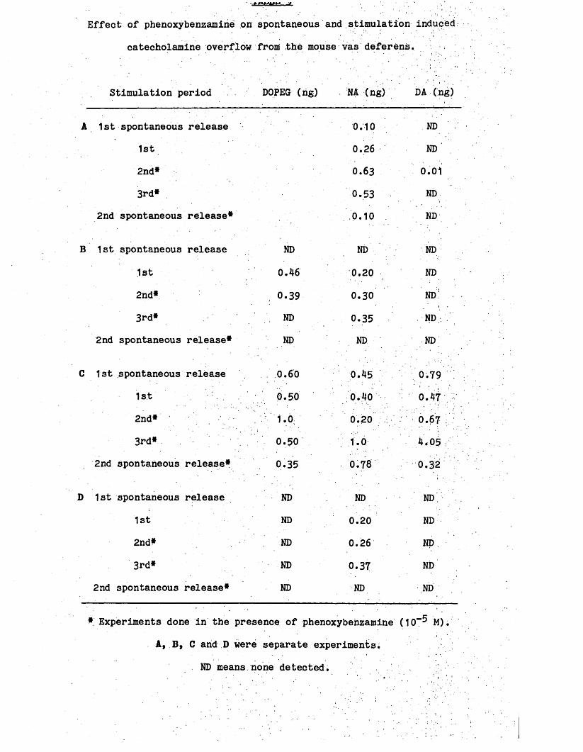
Effect of phenoxybenzamirie on spontaneous and stimulation induced
catecholamine overflow from the mouse vas deferens.
Stimulation period DOPEG (ng) NA (ng) DA (ng)
A 1st spontaneous release 0.10 ND
1st 0.26 ND
2nd* 0.63 0.01
3rd* 0.53 ND
2nd spontaneous release* 0.10 ND
B 1st spontaneous release ND ND ND
1st 0.46 0.20 ND
2nd* 0.39 0.30 ND-
3rd* . ND 0.35 ND
2nd spontaneous release* ND ND ND
C 1st spontaneous release . 0.60 0.45 0.79
1st 0.50 0.40 0.47
2nd* 1.0 0.20 i 0.67
3rd* 0.50 1.0 4-05
2nd spontaneous release* 0.35 0.78 0.32
D 1st spontaneous release ND ND ND.
1st ND 0.20 ND
2nd* ND 0.26 ND
3rd* ND 0.37 ND
2nd spontaneous release* ND ND ND
* Experiments done in the presence of phenoxybenzamine (10“ M).
A, B, C arid D were separate experiments,
ND means none detected.
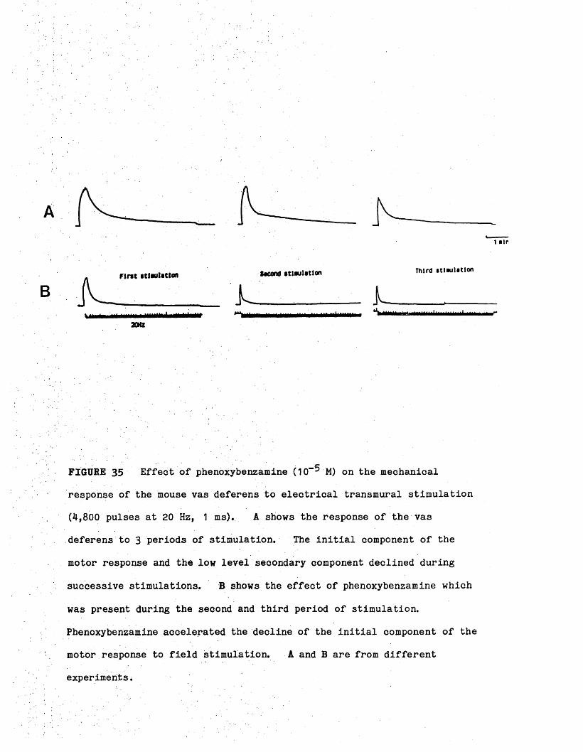
A -
First stimulation **»nd stlwlstlon Third stl.ul.tlon
■ L }___________ _
20HZ
FIGURE 35 Effect of phenoxybenzamine (10“ M) on the mechanical
response of the mouse vas deferens to electrical transmural stimulation
(4,800 pulses at 20 Hz, 1 ms). A shows the response of the vas
deferens to 3 periods of stimulation. The initial component of the
motor response and the low level secondary component declined during
successive stimulations, B shows the effect of phenoxybenzamine which
was present during the second and third period of stimulation.
Phenoxybenzamine accelerated the decline of the initial component of the
motor response to field stimulation. A and B are from different
experiments.

1.7 Spontaneous release of NA from the rat colon
Examination of Table 4 reveals the low content of NA in the rat
colon. The primary objective of the HPLC study was to determine
whether NA was released spontaneously in segments of the rat colon. In
the experiments where this was attempted, no spontaneous release could
be demonstrated. This was not surprising since the NA content of the
tissue was.low.
2.1 Tritium accumulation in the rat colon
The time course of the accumulation of (^H)-NA was determined in
the isolated colon by incubating the tissue with 444 KBq/ml (^H)-NA for
time intervals varying from 30 sec to 1 hr. Accumulation was linear
up to 20 minutes, thereafter remaining fairly constant (Fig. 36).
In order to study the relationship between (%) accumulation and
substrate concentrations, accumulation was measured after 2 minutes
incubation with various substrate concentrations. The accumulation of
(^H)-NA was saturable (Fig. 37).
2.2 Efflux of tritium from the rat colon
Rat colon was incubated, in 444 KBq/ml (^H)-NA for 20 minutes and
then set up for mechanical recording. Analysis of the Krebs1 solution
: collected at . 10 minute intervals showed that over the first 40 minutes
there was a fast efflux of loosely bound label from the tissue and a
second* slow efflux probably related to spontaneous release of (^H)-NA
from nerve endings. There was little mechanical activity in the
absence of drugs throughout the collecting period (Fig. 38).

dm/m
tis
sue900
800
700
600
500
300
200
100
Time (minutes)
FIGURE 36 Time course of accumulation of (^H)-NA into the rat colon.
Accumulation expressed as disintegrations per minute (dpm) per mg
tissue. Duration of incubation in minutes.

3H-noradrenallne
FIGURE 37 Uptake of (%) by the rat colon. The colon was exposed to
different concentrations of (^H)-NA for 2 minutes, washed briefly in a
large volume of ice-cold Krebs1 blotted dry and thereafter counted for
radioactivity. Each point is the mean (- S.E.M.) of at least 8
observations.
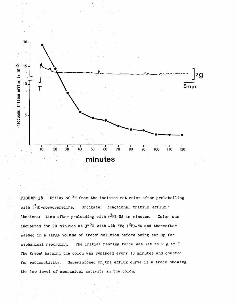
Fracti
onal
trltlu
« eff
lux
(x 10"
30
5 m i n
20 30 50 6010 70 80 90 100 110 120
minutes
FIGURE 38 Efflux of from the isolated rat colon after prelabelling
with (^H)-noradraneline. Ordinate: fractional tritium efflux.
Abscissa: time after preloading with (^H)-NA in minutes. Colon was
incubated for 20 minutes at 37°C with 444 KBq (^H)-NA and thereafter
washed in a large volume of Krebs’ solution before being set up for
mechanical recording. The initial resting force was set to 2 g at T.
The Krebs’ bathing the colon was replaced every 10 minutes and counted
for radioactivity. Superimposed on the efflux curve is a trace showing
the low level of mechanical activity in the colon.

2.3 Effect of drugs on tritium efflux and mechanical response of, the rat colon
This study examined the effects of drugs, which produced rhythmic
waves of contraction in colon, on the efflux of ( H). The Krebs*
solution used for efflux was changed after 90 minutes to one containing
appropriate concentration of agonists.
Morphine (5 x 10“® M), clonidine (2 x 10“® M) and TTX (10“* M)
produced rhythmic waves of contraction without having any effect on the
efflux of (3h ) (Figs. 39> 40 and 41).

Fra
ctio
nal
trlt
iui
efflu
x (x
10“
5 m i nM O R P H1 0 -
Morphine' 5" x !0 " bM
100 110 120
minutes
F IG U R E 39 Effect of morphine on the efflux of tritium from the rat
colon and mechanical activity of the colon. Experimental conditions
were as described for Fig. 38 except that the normal Krebs* solution was
changed to one containing morphine (5 x 10“ M) at the 90th minute.
Morphine produced rhythmic waves of contractions without affecting the
tritium efflux.
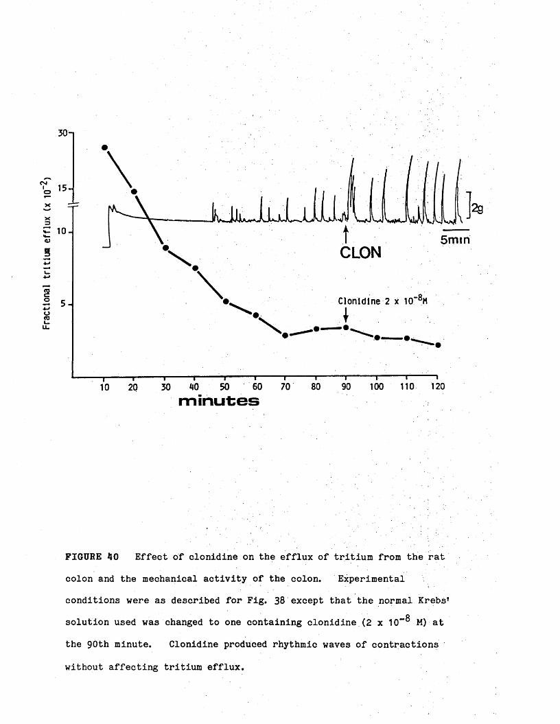
Fra
ctio
na
l tr
ltlu
a
eff
lux
(x 10
"
30n
C -J 15-
1 0 - 5 m m
Clonidine 2 x 10‘BM
12070minutes
FIGURE 40 Effect of clonidine on the efflux of tritium from the rat
colon and the mechanical activity of the colon. Experimental
conditions were as described for Fig. 38 except that the normal Krebs1
solution used was changed to one containing clonidine (2 x TO”® M) at
the 90th minute. Clonidine produced rhythmic waves of contractions
without affecting tritium efflux.

Fracti
onal
tritlu
m eff
lux (
x 10“
30 n
5 m i n10 - T T X
TTX 10“'M
“ 1120
T T I100 110
minutes
FIGURE 41 Effect of TTX on the efflux of tritium from the rat colon
and the mechanical activity of the colon. Experimental conditions Were
as described for Fig. 38 except that the normal Krebs1 solution was
changed to one containing TTX (10“ M) at the 90th minute. TTX
produced waves of rhythmic contractions without affecting tritium
efflux.

DISCUSSION

The effects of morphine: and the opioid peptides on the gut motility
are still incompletely understood. The aim of this study was, first,
to investigate the effects of the opioids on the intestine. Secondly,
to elucidate some of the mechanisms and the possible neuronal basis of
these effects. In particular, this study examined the possible
involvement of 5-HT and ACh in these effects (Burks, 1973» 1976;
Huidobro-Toro & Way, 1981) and the possible existence of a tonically
active, non-adrenergic non-cholinergic inhibitory mechanism (Gillan,
1977; Gillan & Pollock, 1980; Scheurer .§£. 1981). Electrical
field stimulation of the isolated intestine was also employed to provide
further evidence about the role of excitatory and inhibitory nerves in
these responses.
The preparation used for this investigation was the isolated colon
of the rat. The colon was chosen primarily because (a) morphine as
well as the enkephalins have excitatory effects on this tissue (Gillan & Pollock, 1980; Nijkamp & Van Ree, 1980; Boura & Olley, 1981; Scheurer
£Ji Mh 1981) and (b) since specific opioid receptors and also
enkephalins are present in the gastro-intestinal tract (Manara &
Bianchetti, 1985) and are. located in or near the myenteric plexus
(Hughes, kosterlitz & Smith, 1977). Moreover, since the enkephalins
are released in response to nerve stimulation (Schulz, Wuster, Simantov,
Snyder & Hertz, 1977* Corbett, Sosa, McKnight & Kosterlitz, 1980),
their physiological significance may be expected to be primarily local
and, therefore, apparent J j i vitro.
. The results obtained in this study confirmed some of the results
obtained by other workers (Gillan & Pollock, 1980; Nijkamp & Van Ree,
1980; Scheurer e£_ al. 1981) that morphine and the opioid peptides

produce rhythmic contractile activity in the terminal colon of the rat,
but. cannot, be explained by any single hypothesis previously postulated.
This rhythmic contractile activity is receptor-mediated but the
concentration response relationship is difficult to establish.
Morphine-induced rhythmic contractions were abolished by the specific
opioid antagonist naloxone. While the rhythmic waves of contraction
produced by the opioids were "all or none" in nature, the size of the
initial response was concentration-related.
. The ability of opioids to initiate waves of rhythmic contraction
has either been ignored in the past or the experiments were carried out
in conditions which minimised the chances of their occurrence (Huidobro-
Toro & Way, 1981; Moritoki 1984).
Although the excitatory effect of opioids on the intestine is well-
documentedf the exact mechanisms by which the opioids produce this
effect is far from being clear. This is further complicated by the
fact that, in vivo, morphine and the opioid peptides can also influence
gastro-intestinal motility by centrally-mediated effects (Parolaro e£.
al. 1977; Burleigh et al. 1981; Burks, 1980). Evidence for central
opioid-sensitive sites for inhibition of gastro-intestinal transit comes
primarily from animal studies, in which morphine-like drugs were
administered directly into the CNS. Results of such studies are of
considerable interest, since they provide evidence for the existence of
specific action sites within the CNS influencing the gut. However,
centrally-mediated actions on bowel by narcotic drugs injected directly
into the CNS may not explain entirely their effects.on the intestine
following systemic administration (Manara & Bianchetti, 1985). Since
morphine does not pass the blood brain barrier easily (Oldendorf, Hyman,

Braun & Oldendorf, 1972; , Jaffe & Martin, 1980), therapeutic doses
cannot be expected to have, a predominantly central component on
intestinal motility. Recently, the spinal cord has also been
considered a potential locus of opioid action in the production of
gastro-intestinal motor effects.
Morphine and the opioid peptides administered intrathecally in the
spinal cord of mice effectively inhibit the passage of radiolabelled
marker through the gastro-intestinal tract (Porrecca & Burks, 1983;
Porrecca, Filla & Burks, 1983) but a similar study in the rat did not
confirm the spinally-elicited constipating effect of morphine in this
species (Vaught, Cowan & Gimerek, 1983)* It may be that the rat is a
better predictive model for humans than mice because constipation does
not seem to occur in patients given morphine to produce spinal analgesia
.(Zenz, Schappler-Scheele, Neuhaus, Pipeubrock & Hilfrich, 1981;
Chirubasik, 1984).
An additional peripheral site, at which opioids might act to
produce their anti-transit effects is the intestinal mucosa, where they
can affect water and electrolyte reabsorption (Powell, 1981). However,
the ability of morphine and related drugs to produce rhythmic
contractile activity in isolated intestine eliminates the possibility
that the CNS or the extrinsic autonomic innervation are the only sites
of action.
A mechanism postulated to explain the stimulant action of morphine
on the intestine involves the release of intestinal 5-HT and ACh. From
experiments on the small intestine of rat and dog Burks (1973i 1976) and
Huidobro-Toro and Way (.1981) suggested that morphine released 5-HT

which then stimulated the smooth muscle both directly and indirectly by
releasing ACh from cholinergic nerve in the intestinal wall.
This hypothesis was based on (1) the observation that responses to
morphine were abolished by the 5-HT antagonists methysergide and
cyproheptadine, and reduced by atropine; (2) that responses to morphine
were absent in tissues from animals pretreated with reserpine to deplete
the 5-HT content of the intestine. In this study, the 5-HT antagonist
methysergide non-competitively inhibited the conpentration-response
relationship to morphine, while it was ineffective against the rhythmic.
waves of contraction produced. by the opioids. In contrast to the non
competitive nature of methysergide antagonism, naloxone competitively
inhibited this initial response and abolished the waves of rhythmic
contraction produced by morphine. At first sight, the inhibitory
effect of methysergide might be taken as an indication of the
involvement of 5-HT in this initial response to morphine. The result
obtained with 5-HT is complicated by the lack of specific 5-HT
antagonist. It bears emphasising that methysergide has a spectrum of
effects which become more apparent as its concentratipn is raised. At
concentrations that are effective in blocking neural. 5-HT receptors, it
is comparably effective in antagonising the contractile effects of
carbachol, dimethylphenylpiperazinium and histamine in the intestine !
(Furness & Costa, 1982). Morphine may be added to the list of diverse
drugs whose effects in the intestine are antagonised by methysergide
(Nijkamp & Van Ree, 1980; Huidobro-Toro & Way, 1981; this study). It
has been suggested that attempts to use this drug in the analysis of the
actions of 5-HT on intestinal nerves should profitably be abandoned
because of its non-specificity (North, Henderson, Katayama & Johnston,
1980).

Indeed, the ability of another 5-HT antagonist, cyproheptadine, to
inhibit morphine-induced contractions, albeit non-competitively (Gillan
& Pollock, 1980; Nijkamp & Van Ree, 1980), may not be related to
antagonism at the 5-HT receptor site. Cyproheptadine also inhibits the
membrane translocation of calcium in smooth muscle by reducing the flow
of Ca++ ions through voltage-dependent Ca++ channels (Lowe, Matthews &
Richardson, 1981), hence great care must be taken in the interpretation
of the results with these 5-HT antagonists.
In contrast to the equivocal nature of the results with
methysergide, the results obtained with PCPA, which depleted the tissue
of 5-HT (Koe & Weissman, 1966; Weber, 1969)» provided evidence against
the involvement of 5-HT in the excitatory effects of opioids in the.
colon. Since, in colon from PCPA pretreated rats, morphine was still
capable of producing naloxone-sensitive rhythmic waves of contractions,
it seems unlikely that 5-HT is required for this response. !. However,
since a small residual quantity of 5-HT remained in the colon from PCPA-
treated rats, this observation alone does not eliminate the possible
involvement of 5-HT in this phenomenon.
This study also confirmed the ineffectiveness of reserpine in
depleting the 5-HT content of the rat intestine (Erspamer, 1966b;
Carlsson, 1969). However, it is interesting that in an attempt to link
the excitatory effect of morphine with the release and effect of 5-HT,
reserpine .was used to deplete the 5-HT level, although the 5-HT content
was not measured. Furthermore, in this study, the ability of morphine
to cause the colon to contract.rhythmically was unaffected in tissues
rendered insensitive to 5-HT by prolonged exposure to high concentration
of this amine.

More convincing evidence against the involvement of 5-HT/ACh in the
response of the rat colon to morphine was obtained from experiments
which demonstrated that the characteristic excitatory effect of opioids
could not.be mimicked by administered ACh or 5-HT. In addition,
atropine did not interfere with the ability of morphine to cause the
colon to contract rhythmically. Perhaps the most striking
characteristics of the opioid-induced excitatory response were its
rhythmici.ty and persistence, neither of which is compatible with the
ACh/5-HT release hypothesis.
These results and conclusions differ from those of Huidobro-Toro
and Way (1981), who obtained concentration-related contractions to
morphine in the rat colon and concluded that the excitatory effects of
opioids were mediated by 5-HT. These workers pointed out that their
Long Evans rats were more sensitive to opioids than other strains such
as the Wistar rats used in this study. However, it is unlikely that
this difference could explain the discrepancies between the results
: obtained in the two studies. The present results are not consistent
with the idea that a 5-HT/ACh mechanism is involved in the effects of
opioids in the rat colon. Fontaine and Reuse (1985) recently reached a
similar conclusion about the non-involvement of 5-HT and/or ACh in the
excitatory response of the mouse colon to opioids.
An alternative explanation of the excitatory effects of opioids is
that morphine removes a tonic inhibitory neural influence that normally
suppresses myogenic activity so that the contractions produced by
morphine are myogenic (Gillan & Pollock, 1980; Scheurer ££. al, 1981;
Moritoki.gi.al, 1984; Fontaine & Reuse, 1985). This hypothesis was
based on that proposed by Wood to explain the stimulant action of

atropine, TTX and local aneaesthetic in cat small intestine (Wood, 1972,
1975). From studies of the electrical activity of neurones in
Auerbach's plexus and the mechanical activity of the circular layer in
cat small intestine, Wood proposed that the circular muscle was
maintained in a state of inhibition by the continuous release of an
inhibitory transmitter from spontaneously active neurones and that
removal of this inhibitory influence unmasked the inherent myogenic
activity of the circular muscle. Evidence that blockade of neural
activity by atropine, local anaesthetic or TTX (Wood, 1972; Wood &
Marsh, 1973; Ohkawa & Prosser, 1972b; Biber & Fara, 1973; Tonini,
Secchini, Frigo & Crema, 1974), cold storage (Wood, 1972), surgery
(Schiller, Suriyapa, Mutchler & Anderson, 1973) or congenital absence of
ganglion cells (Wood, 1973) produce similar effects on the mechanical
activity of intestinal muscle were cited in support of this hypothesis
(Wood, 1975). Despite the attractiveness of this hypothesis, the
continuous release of such an inhibitory transmitter in vitro, has not
been demonstrated (Daniel & Sarna, 1978). However, if this explanation
is correct, then other drugs, that interfere with neuronal inhibitory
mechanisms should have effects similar to morphine in the colon.
The results obtained in this study with TTX, which abolishes the
nerve action potential by blocking sodium conductance (Narahashsi, 1974)
and with apamin, which inhibits potassium-dependent inhibitory mechanism
in smooth muscle (Banks, Brown, Burgess, Burnstock, Claret, Cocks &
Jenkinson, 1979), are consistent with such an explanation. Both TTX
and apamin produced regular rhythmic contractions which resembled
morphine-induced contractions but were unaffected by naloxone, atropine
or methysergide.

The question that arises concerns the possible nature of this
inhibitory mechanism that is affected by opioids to reveal myogenic
activity* Gillan and Pollock (1989), Scheurer jgJt. Si. (1981) and
Moritoki et al (1Q84) proposed that morphine acts presynaptically to
inhibit the final release of an NANC inhibitory transmitter, thereby
: unmasking intrinsic myogenic activity, whereas Nijkamp and Van Ree
(1980) suggested that an adrenergic mechanism could be inhibited by
morphine. To clarify which of these transmitters was involved in the
tonic inhibitory mechanism, responses of the colon to electrical field
stimulation and the effects of drugs on these responses were
investigated.
Electrical field stimulation of the colon provided evidence about
the nature of the morphine-induced excitatory response and the possible
role of inhibitory nerves in this response. Initial experiments
confirmed the results of previous workers that there are cholinergic
motor nerves in the colon (Stockley & Bennett, 1973; Bennett &
Stockley, 1975; Gillan & Pollock, 1980). Field stimulation of the
colon produced an excitatory response which was maximal at frequencies
: between 10 and 20 Hz. This excitatory response was antagonised by
atropine, TTX or morphine. Since morphine did not affect responses to
administered ACh (Cherubini, Morita & North, 1985), it is likely that
morphine acted on presynaptic nerves to inhibit ACh release (Down &
Szerb, 1980; North, 1982) either by hyperpolarizing the soma of the
enteric neurones (Wood, 1980) or by blocking conduction in nerve cell
processes (Morita & North, 1981)7 .
On cessation of stimulation, a post-stimulus contraction was
observed. This contraction is thought to be myogenic in origin and to

arise from activation of non-adrenergic inhibitory nerves, which in turn
trigger the release of prostaglandins (Burnstock §X. 2X3 1975).
However, the involvement of prostaglandins in this post-stimulus
contraction is controversial. Kadlec, Masek and Seferna (1974) found
no evidence of an inhibitory effect of indomethacin on this post
stimulus contraction. Bauer, Matusak, Bezekwa, Benes and Kuriyama
: (1982) showed that indomethacin dissolved in ethanol markedly depressed
, the rebound contraction. • On the other hand, indomethacin dissolved in
. a weakly alkaline bicarbonate, solution doeS not inhibit but rather
increased the response.
Thus the discrepancies in the literature concerning the role of
prostaglandins in the rebound contraction may be the result of different
solvents used to dissolve indomethacin. In addition, indomethacin has
also been reported to reduce the availability of calcium ions within the
muscle cell, independently of prostaglandin synthesis (Northover, 1977).
In this study, a water-soluble prostaglandin synthesis inhibitor
flubiprofen (Adams & Buckler, 1979) had no effect on the post-stimulus
contraction suggesting at least that prostaglandins are not involved in
the rebound contraction in the rat colon. .
In addition to the cholinergic excitatory motor innervation of the
colon, field stimulation at frequencies below 5 Hz caused a small
inhibitory response rather than a contraction and this response too was
also followed by a post-stimulus contraction. Since this inhibitory
response was not abolished by atropine or any adrenergic receptor
antagonist or neurone blocking drug or by pretreatment with reserpine or
6-OHDA1 it appears that it was neither adrenergic nor cholinergic (NANC)
in nature. However, because the rat colon rarely develops and

maintains tone on its own, advantage was taken of the rhythmic waves of
contractions produced by various drugs. Further evidence for the
existence of an NANC inhibitory response was obtained in experiments in
which field stimulation applied at the peak of a morphine-induced
contraction, produced an immediate and maintained inhibitory response.
This response was maximal at low frequencies of stimulation and was
: unaffected by atropine, quinidine, adrenergic receptor- or neurone-
blocking drugs; It appears that this response is NANC in nature but
there remains doubt about whether it is neurally-mediated, since it was
unaffected by concentrations of TTX that readily abolish the cholinergic
motor response. Similar doubts have been expressed about the basis of
inhibitory NANC response elsewhere (Crossley & Gillespie, 1983).
However, it is possible that NANC nerve terminals may be directly
stimulated by field stimulation without the need for axonal conduction.
There is evidence that this occurs in other tissues (Douglas &
Taraskevich, 1980; Salzberg, Obaid, Senseman & Gainer, 1983).
Electrophysiological studies indicate that focal depolarization of nerve
terminals releases transmitter by a calcium-dependent mechanism even
under complete blockade of the inward sodium current by TTX (Katz &
Miledi, 1969; Illes & Thesleff, 1978). Alternatively, large
electrical currents may open channels distinct from the voltage-
sehsitive Ca+* channels or may even create new routes for the entry of
Ca++ into nerves (Illes, Meier & Starke, 1984). The difference in
sensitivity to TTX may, however, simply reflect the different
Susceptibility of the cholinergic motor nerves and the NANC inhibitory
nerves to TTX blockade.

The possibility of whether morphine and other drugs affect the NANC
inhibitory mechanism to produce the myogenic rhythmic contractions can
now be considered. Inhibitory responses to electrical field
stimulation were obtained at the peak of morphine-induced contractions.
This suggests that if morphine causes rhythmic contractions by removing
a tonic inhibitory neural influence then it is unlikely to be this NANC
inhibitory influence since this NANC response can be more clearly
demonstrated when morphine is present and has inhibited ACh release.
Previously, when it was sugigested that the tonic inhibitory influence
removed by morphine might be NANC (Gillan & Pollock, 1980), the paradox
of the persistence of this inhibitpry response at the peak of an opiate-
induced contraction was explained by postulating that morphine acted at
a ganglionic site preceding the inhibitory nerve. In such
circumstances, the tonic inhibitory influence of the nerve could be
removed but the capacity to stimulate the nerve distal to the blockade
to produce an inhibitory response would remain (Gillan & Pollock, 1980).
However, the fact that morphine depresses non-adrenergic inhibitory
response to transmural nerve stimulation in the guinea-pig taenia coli
is not compatible with this explanation (Shimo & Ishi, 1978).
Alternatively the mechanism of spontaneous transmitter release and the
mechanism of electrically-evoked transmitter release may not be equally
susceptible to blockade by morphine. In addition, it could be argued
that electrical excitation of the inhibitory nerves may release
sufficient transmitter to overcome blockade by morphine and effectively
suppress the contraction of the muscle.
In this study, experiments with apamin, which, unlike morphine,
acts on the smooth muscle directly, makes this interpretation less
likely. Apamin caused contractions similar to those produced by

morphine and this observation supports the view that morphine-induced
contractions are caused by the inhibition of an inhibitory mechanism
that suppresses myogenic activity. However, field stimulation applied
at the peak of an apamin induced-contraction produced either no response
or only a small inhibition. The ability of atropine to convert this
small inhibitory response into a large and well-maintained inhibitory
response, suggests that ACh released by field stimulation normally
masked the inhibitory response. Thus it appears that NANC transmission
survives both morphine and apamin. An important distinction between
apamin and morphine is that morphine inhibited motor responses to field
stimulation by inhibiting ACh release; whereas apamin potentiated such
. responses probably by inhibiting a neural mechanism that normally
restrains the size of the cholinergic motor response. The study of
NANC inhibitory mechanisms is complicated since there are few substances
that block responses to stimulation of NANC nerves. Apamin is one such
substance and it blocks relaxation of the guinea-pig taenia caeci in
response both to NANC stimulation and to ATP (Banks &L, 1979) and to
VIP-induced and neurogenic vasodilation in the cat small intestine
(Sjoqvist alj 1980). However, the failure of apamin to block the
NANC inhibitory response in this study suggests that the mechanism
underlying relaxation of the smooth muscle in rat colon differs from
that of the guinea-pig taenia caecum and cat intestine blood vessels.
Likewise, apamin failed to block the inhibitory responses of the
retractor penis muscle to nerve stimulation (Bowman & Gillespie, 1982).
The role of cyclic GMP in relaxant responses to NANC nerve
stimulation has been elucidated only recently (Bowman & Drummond, 1984).
It has been proposed that in the bovine penile artery and certain
related structures in the rat and the dog there may be a novel,

inhibitory transmitter which on release activates guanylate cyclase in
the smooth muscle cells and that it is the cyclic GMP thus formed that
mediates neural relaxation (Bowman & Drummond, 1985). Evidence in
support of this hypothesis included (a) The rise in cyclic GMP level
following stimulation of the NANC inhibitory nerves in the bovine
retractor penis muscle, (b) Haemolysate and N-methylhydroxylamine, which
blocks guanylate cyclase, also blocked the relaxation and the rise in
cyclic GMP produced by inhibitory nerve stimulation, and (c) 8-Bromo-
cyclic GMP, which is a stable derivative of cyclic GMP, produced a
relaxation that was not blocked by haemolysate (Bowman & Drummond,
1984).
In this study, both methylene blue, which blocks the activation of
guanylate cyclase (Ignarro & Kadowitz, 1985) and haemolysate, derived
from lysed red blood cells, had no effect on the inhibitory response to
NANC nerve stimulation in the rat colon, suggesting that this inhibitory
response is similar to that obtained in the NANC inhibition of tracheal
ring, the guinea-pig taenia caeci and the anal sphincter, but unlike the
neurogenic vasodilation in bovine and canine retractor penis muscles and
penile arteries (Bowman, personal communications).
The above discussion strongly suggests the presence in the rat
colon of NANC inhibitory responses, which are unaffected by the opioids
and apamin and, therefore, are unlikely to be involved in the waves of
rhythmic contractions produced by these drugs.
Such a conclusion would be more in accordance with evidence in the
literature that in tissues where there are NANC inhibitory nerves,
morphine has no effect on the release of the NANC inhibitory
transmitter. Thus, morphine has no effect on NANC transmission in the

rabbit rectococcygeus muscle (Ambache, Killick & Zar, 1974), bovine
retractor penis muscle (Klinge & Sjostrand, 1974), rat anococcygeus
muscle (Tilmisany, 1975), guinea-pig stomach and taenia (Huizinga &
Hertog, 1979), guinea-pig ileum (Ito & Tajima, 1980), guinea-pig anal
sphincter (Lim, personal communication).
Clearly, if the effect of the drugs producing the rhythmic waves of
contraction in the rat colon is to be explained on the basis of an
inhibitory effect on transmitter release, then it becomes obvious that
neither the Cholinergic motor nor the NANC inhibitory nerves are
involved and a second tonically-active inhibitory transmitter would have
to be proposed. Such a transmitter might be the adrenergic inhibitory
mechanism although such an explanation will not be without difficulties.
First, the NA content of the colon as measured by HPLC in this study was
very low compared with other densely adrenergic innervated tissues such
as the vas deferens. However, the tonic inhibition need only be
concentrated on few pace-maker cells. Secondly, fluorescence
histochemical studies have demonstrated that in the intestinal tract of
most mammalian species, the post-ganglionic adrenergic nerves terminate
in the region of the ganglion cells of Auerbach's plexus, with few
fibres innervating the longitudinal and circular muscle layers directly
(Norberg, 1964; Jacobwitz, 1965; Hollands & Vanov, 1965; Furness &
Costa, 1978). Thus the effect of this tonically-active adrenergic
mechanism might have to be indirect, exerted primarily at the level of
intramural ganglion cells* Thirdly, there was little evidence of any
significant contribution from' the adrenergic nerves in the inhibitory
response of the colon to field stimulation since adrenergic neurone and
adrenoceptor blockers did not affect the inhibitory response. However,
this observation is not unique to the rat colon. Stockley and Bennett
(1977) showed that in human taenia coli, which is densely innervated
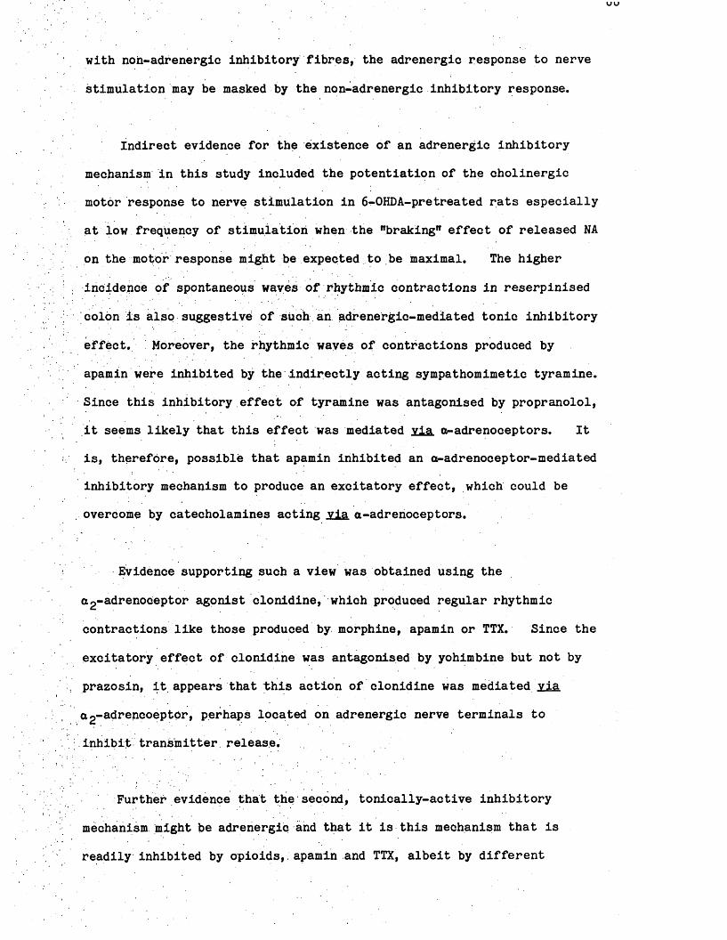
with non-adrenergic inhibitory fibres, the adrenergic response to nerve
stimulation may be masked by the non-adrenergic inhibitory response.
Indirect evidence for the existence of an adrenergic inhibitory
mechanism in this study included the potentiation of the cholinergic
motor response to nerve stimulation in 6-OHDA-pretreated rats especially
at low frequency of stimulation when the "braking" effect of released NA
on the motor response might be expected to be maximal. The higher
incidence of spontaneous waves of rhythmic contractions in reserpinised
colon is also suggestive of such an adrenergic-mediated tonic inhibitory
effect. ! Moreover, the rhythmic waves of contractions produced by
apamin were inhibited by the indirectly acting sympathomimetic tyramine.
Since this inhibitory effect of tyramine was antagonised by propranolol,
it seems likely that this effect was mediated via Qp-adrenoceptors. It
is, therefore, possible that apamin inhibited an a-adrenoceptor-mediated
inhibitory mechanism to produce an excitatory effect, which could be
overcome by catecholamines acting via a-adrenoceptors.
Evidence supporting such a view was obtained using the
ag-adrenoCeptor agonist clonidine, which produced regular rhythmic
contractions like those produced by morphine, apamin or TTX. Since the
excitatory effect of clonidine was antagonised by yohimbine but not by
prazosin, it appears that this action of clonidine was mediated via
ag-adrencoeptpr, perhaps located on adrenergic nerve terminals to
; inhibit transmitter release.
Further evidence that the second, tonically-active inhibitory
mechanism might be adrenergip and that it is this mechanism that is
readily inhibited by opioids,. apamin and TTX, albeit by different

mechanisms and at different sites, was obtained with o-adrenoceptor
antagonists and adrenergic neurone blockers. These drugs also produced
contractions similar to those produced by apamin, morphine and TTX. In
addition, Gillan and Pollock (1980) and Nijkamp and Van Ree (1980)
reported the potentiating effect of an a-adrenoceptor antagonist on
opioid-induced contractions in the rat intestine.
It is a paradox that the apparent inability of reserpine
pretreatment, adrenergic neurone blockers and adrenoceptor antagonists
to antagonise the excitatory effect of the opioids (Scheurer sL,
1981; Moritoki ilL, 1984; Fontaine & Reuse, 1985) could be taken as
evidence for the non-involvement of an adrenergic mechanism in the
tonic inhibitory mechanisms affected by the opioids. Additonal
blockade of this adrenergic mechanism by drugs affecting adrenergic
transmission should theoretically either have no effect or potentiate
the response.
The suggestion that the hypothetical tonic inhibitory mechanism
affected by these diverse drugs is adrenergic immediately; offered the
possibility that the inhibitory effect of these drugs on tonically
released NA might be investigated. Unlike the NANC inhibitory
mechanism, whose transmitter is unknown and, therefore, more difficult
to analyse, NA overflow and inhibition of this overflow can be measured.
Both HPLC with electrochemical detection and tritium efflux were used in
this study to determine whether drugs which had an excitatory action in
the colon inhibited NA release.
Liquid chromatographic methods, using either cation exchange or
reversed phase chromatography with electrochemical detection have been

used for the determination of catecholamines in both brain and smooth
muscle (Felice, Felice & Kissinger, 1978; Wagner, Vatali, Palfreyman,
Zraika & Huot, 1982; Maruyama, Oshima & Nakajima, 1980; Macrae, 1983)
as well as in plasma samples (Hallman, Fernebo, Hamberger & Johnsson,
1978; Goldstein, Ferneistien, Izzo, Koplin & Keiser, 1981; Eriksson &
Persson, 1982). HPLC proved to be a fast and efficient method for
separating the amines following their extraction. Problems of amine
separation and artefact production were often experienced during this
study. Poor separation resulted in either the NA and DOPEG peaks being
superimposed on the falling phase of the solvent front or both NA and
DOPEG peaks becoming joined together. This resulted in difficulties in
measuring peak height which in turn led to inaccuracies in quantifying
amine levels.
The occurrence of artefact was successfully controlled by improved
methodological practise. Catecholamine separation was improved by
altering the composition of the mobile phase. For example, a decrease
in.phosphate ion, pH and methanol content of the mobile phase caused an
increase in retention time and thus improved separation. This also
caused an increase in the retention of DA, giving a very lengthy
chromatogram. Better separation was thus gained only at the expense of
time. The necessity for mobile phase adjustment is clear when one
considers the variability between columns (Hansson, Agrup, Rorsman,
Rosengren, Rosengre & Edholm, 1979) and the cost of new columns relative
to manipulation of the mobile phase. Thus, Goldberg (1982) showed that
the reteritivity and selectivity of columns with similar packings is not
constant due to differences in (a) the material used as the support,
'(b) the technique used to bond the functional groups to the support,
(c) the method used to pack the. column, and (d) the amount of end
capping to mask unbonded sites.
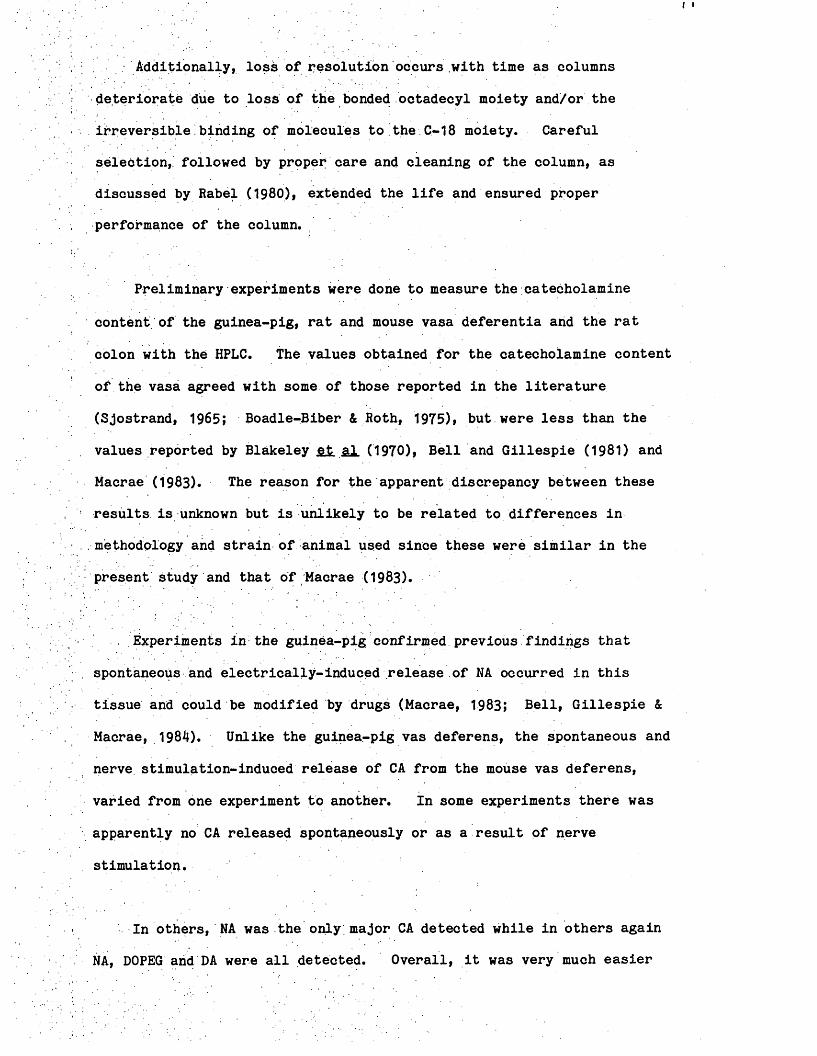
Additionally, loss of resolution occurs with time as columns
deteriorate due to loss of the bonded, octadecyl moiety and/or the
irreversible!binding of molecules to the C-18 moiety. Careful
selection, followed by proper care and cleaning of the column, as
discussed by Rabel (1980), extended the life and ensured proper
performance of the column.
Preliminary experiments were done to measure the catecholamine
content of the guinea-pig, rat and mouse vasa deferentia and the rat
colon with the HPLC. The values obtained for the catecholamine content
of the vasa agreed with some of those reported in the literature
(Sjostrand, 1965; Boadle-Biber & Roth, 1975), but were less than the
values reported by Blakeley fit sL (1970), Bell and Gillespie (1981) and
Macrae (1983). The reason for the apparent discrepancy between these
results is unknown but is unlikely to be related to differences in
methodology and strain of animal used since these were similar in the
present study and that of Macrae (1983).
. Experiments in the guinea-pig confirmed previous findings that
spontaneous and electrically-induced release of NA occurred in this
tissue and could be modified by drugs (Macrae, 1983; Bell, Gillespie &
Macrae, 1984). Unlike the guinea-pig vas deferens, the spontaneous and
nerve stimulation-induced release of CA from the mouse vas deferens,
varied from one experiment to another. In some experiments there was
apparently no CA released spontaneously or as a result of nerve
stimulation.
In others, NA was the only major CA detected while in others again
NA, DOPEG and DA were all detected. Overall, it was very much easier

to demonstrate amine release in the guinea-pig than in the mouse vas
deferens.
‘ With the HPLC study, no spontaneous release of NA and/or metabolite
was demonstrated in the rat colon, casting doubt on the NA hypothesis.
However, the possibility exists that the inability to demonstrate
spontaneous release of NA in the rat colon might simply be a result of
the low amine content of this tissue. The amount of NA released may,
therefore, be below the detection limit of the HPLC system used.
Efflux experiments with (^H)-NA were performed as an alternative to
the HPLC studies. In these experiments the effects of drugs on the
motility of the colon and on the efflux of tritium were examined. No
effect of drugs on the spontaneous effleet of ^H was demonstrated in the
colon despite the fact that these drugs produced rhythmic waves of
contraction in the same experiments. The inability of these drugs to
affect spontaneous ^H confirmed the results of previous workers that in
other systems these drugs do not affect spontaneous ^H efflux (Hughes
al. 1975; Vizi, Harsing & Knoll, 1977; Arbilla, Langer & Gonzalez,
1983) . ..
The results of the biochemical studies seem to cast serious doubts
on the validity of the hypothesis that NA is the transmitter whose
release is tonically inhibited by these drugs. However, failure to
demonstrate the tonic inhibition of NA release by the drugs which
produce rhythmic waves of contraction in vitro does not eliminate the
possibility of the existence of such a mechanism in vivo (Daniel, 1968;
Drummond, personal communications). The inability to demonstrate tonic
release and its inhibition may simply reflect the limitations of both

the pharmacological tools used and the experimental system studied.
For example, the possible existence of TTX-insensitive nerves reduces
the value of this drug as a tool to identify neuronal mechanisms (Katz &
Miledi, 1968; Illes & Thesleff, 1978).
One possibility that still cannot be excluded is that these drugs
may have a direct excitatory action on intestinal smooth muscle. At
first sight this seems unlikely since morphine does not modify resting
membrane potential in the guinea-pig ileum and mouse vas deferens (Ito &
Tajima, 1980), the cat colon (Blanquet, Bouvier & Gonella, 1982) or the
rat portal vein (Yamamoto, Hotta & Matsuda, 1984). However, mechanical
activation in the absence of electrical activity is known to occur in
the rat anococcygeus (Creed, Gillespie & Muir, 1975), cat trachea (Ito &
Takeda, 1982), bovine retractor penis (Byrne & Muir, 1984) and rat iris
sphincter (Banno, Imaizuma & Watanabe, 1985), and has been described as
pharmaco-mechanical coupling (Somlyo & Somlyo, 1968). Indeed, such a
direct effect of the Opioids has been demonstrated in isolated gastric
smooth muscle cells devoid of neural elements (Bitar & Makhlouf, 1982,
1984; Makhlouf, 1985) and was not ruled out in the studies of Gillan
and Pollock O 98O), Yau (1981) and Moritoki sX (1984).
A direct action has also been suggested to explain the excitatory
effect of. atropine on chick amnion which is devoid of nerves (Cuthbert,
1963), on oesophageal muscle (Christensen & Lund, 1968) and cat small
intestine (Bortoff & Miller, 1975). Furthermore, the excitatory action
of local anaesthetics on rabbit uterus (Daniel & Wolowyk, 1966), rat vas
deferens (Hay & Wadsworth, 1982), cat small intestine (Bortoff & Muller,
1975) and of tetrodotoxin on cat sphincter of Oddi (Persson, 1971) are
all thought to involve direct effects on the smooth muscle.

The question then arises as to whether this excitatory action of
the opioids on the terminal colon of the rat observed in vitro is
related to the anti-diarrhoeal effect of opiates. Analysis of sites of
drug action in vitro is slightly simpler than la vivo because often only
smooth muscle and axons of intrinsic neurones are present. However,
one must be Cautious in interpreting in vitro data to explain the
possible local functional role of endogenous opioids in animals under
physiological conditions or to explain the origin of the constipating
action of morphine. For instance, how does local application of a
substance to the isolated intestine influence its effects and are these
effects different from those of systemically-administered drugs in vivo?
Does removal of other controlling mechanisms as a consequence of
isolation of the preparation show up responses to opioids normally
absent and/or of little significance in the intact animal (Manara &
Bianchetti, 1985)?
Despite these obvious limitations, however, the use of in vitro
studies has provided valuable information on the pharmacology of
Opioids. it may be argued that the rhythmic peristalsis of the colon
is destroyed by the burst of fast excitatory waves elicited by the
opiates or. that the opiaties Increase the proportion of non-propulsive
segmenting contraction of the gut, leading to a slowing of the
intestinal transit. In a recent review, Manara and Bianchetti (1985)
provided evidence to support the hypothesis that the primary site of
morphine's constipating action is peripheral in both the intestinal
smooth muscle and mucosa. However, a central action in vivo may
contribute to this effect of opiates (Burks, 1980; Galligan & Burks,
1983; Porreca, Cowan, Raffa & Tallarida, 1983). Thus it seems
possible that the constipation produced by morphine has local and
central sites of action.

In conclusion, the results of this study confirmed the complexity
of the effects of drugs on the isolated colon of the rat and provided
evidence abput the nature of innervation of this tissue. The results
do not agree with the hypothesis that morphine produces its excitatory
effect in the rat colon solely; by the release of 5-HT and ACh.
This study has also provided evidence inconsistent with the
proposition that morphine and other drugs may produce their excitatory
action by inhibiting NANC inhibitory mechanisms. A direct effect on
smooth muscle by these drugs was not ruled out. The similarity between
the effects of the opioids, the adrenoceptor antagonists, the adrenergic
neurone blockers, clonidine, apamin and TTX suggested that the actions
of these drugs might be mediated through adrenergic neurones albeit by
different mechanisms, but the biochemical studies failed to confirm
this.
The results of this study make it clear that no impregnable
unifying hypothesis is available to explain the mechanism of the
excitatory action of the diverse groups of drugs producing rhythmic
contractile activity in the colon. It is more likely, therefore, to be
a combination of direct excitatory effect on smooth muscle and an
indirect neurally-mediated effect, both of which are capable of
functioning together or independently of each other.

REFERENCES

ADAMS,-S.'S. & BUCKLER, J.W. (1979). Ibuprofen and flurbiprofen. ■QJ-lli,. Rj?gHfiL-Blg., 5., 359-379.
ADLER, H.F., ATKINSON, A.J. & IVY, A.C. (1942). Effect of morphine and dilaudid on the ileum and of morphine, dilaudid and atropine on the colon of man. Arch, intern. Med.f 69. 974-985.
AMBACHE, N., KILLICK, S.W. & WOODLEY, J.P. (1977). Evidence againstpurinergic motor transmission in guinea-pig urinary bladder.Br. J. Pharmac.. 61.. 464P.
AMBACHE, N*; KILLICK, S.W. &ZAR, M.A. (1974). The rabbit rectococcygeus: A ganglion-free parasympathetlcally innervated preparation.
. Br. J. Pharmac.. 52.,175-190.
AMBACHE, N. & ZAR, M.A. (1970). Non-cholinergic transmission by postganglionic motor neurones in the mammalian bladder.J. Physiol.. 210. 761-783.
AMBINDER, R.F. & SCHUSTER, M.M. (1979). Endorphins: New gut peptideswith a familiar face. Gastroenterologyf 77. 1132-1140.
ANDEN, N.E. & MAGNUSSON, T. (1967). An improved method for the fluorimetric determination of 5-hydroxytryptarnine in tissues.Acta Physiol. Soand.. 51, 87-94.
ARBILLA, S., LANGER, S.Z. & RAMIREZ GONZALEZ, M.D. (1983). Inhibition by opiates of the evoked release of (^H)-ACh: Differences between corpusstriatum and tuberculum olfactorium. Br. J. Pharmac., 80. 717P.
BANKS, B.E.C., BROWN, C., BURGESS, G.M., BURNSTOCK, G., CLARET, M.,COCKS, T.M. & JENKINSON, D.H. (1979). Apamin blocks certain neuro- transmitter- induced increases in potassium permeability.Nature. 2Q2., 415-417.
BANNO, H., IMAIZUMI, Y. & WATANABE, M. (1985). Pharmaco-mechanical coupling in the response to acetylcholine and substance P in the smooth muscle of the rat iris sphincter.Br. J. Pharmac., 35., 905-911.
BASS, P., CODE, C.F. & LAMBERT, E.H. (1961). Motor and electric activity of the duodenum. Am. J. Phvsiol.. 201. 287-291.
BAUER, V., MATUSAK, 0., BEZEK0VA, M., BENES, L. & KURIYAMA, H. (1982).Role of Ca^+ in non-cholinergic, non-adrenergic responses to nerve stimulation of the guinea-pig small intestine.Naunvn-Schmiedebergs Arch. Pharmacol. f 319., 115-120.
BAYLISS, W.M. & STARLING, E;H. (1899)» The movements and innervation of the small intestine. J. Phvsiol.. 24f 99-143.
BELL, C. & GILLESPIE, J.S. (1981). Dopamine and noradrenaline levels in the peripheral tissues of several mammalian species.J. Neurochem.. 36. 703^706.
BELL, C., GILLESPIE, J.S. & MACRAE, I.M. (1984). Release of noradrenaline and dopamine by nerve stimulation in the guinea-pig and rat vas deferens. Br. J. Pharmac.. _8J . 563-569.

BENNETT, A., GARRETT, J.R. & HOWARD, E.R. (1968). Adrenergic myenteric nerves in Hirschsprung's disease. Br. med. J.. J_, 487-489.
BENNETT, A. & STOCKLEY, H.L. (1975). The intrinsic innervation of the human alimentary tract and its relation to function.Gui., 16., 443-453.
BIBER, B. & FARA, J. (1973). Intestinal motility increased bytetrodotoxin, lidocaine and procaine. Experientia. 29. 551-552.
BITAR, K.N. .& MAKHLOUF, G.M. (1982). Specific opiate receptors onisolated mammalian gastric smooth muscle cells. Nature. 297. 72-74.
BITAR, K.N. & MAKHLOUF, G.M, (1984). Measurement of function in single smooth muscle cells of the gut. Gastroenterology. 86. 1028.
BLAKELEY, A.G.H., DEARNALEY, D.P. & HARRISON, V. (1970). Thenoradrenaline content of the vas deferens of the guinea-pig.
! Proc. R. Soc. B.. 174. 491-502.
BLANQUET, F., BOUVIER, M. & GONELLA, J. (1982). Effects of enkephalins and morphine on spontaneous electrical activity and on junction potentials elicited by parasympathsetic nerve stimulation in cat andrabbit colon. Br. J._ Pharmac.. 77. 419-429.
. BOADLE-BIBER, M.C. & ROTH, R.H. (1975). Formation of dopamine andnoradrenaline in rat vas deferens: comparison with guinea-pig vasdeferens. Br. j. Pharmac.. 55. 73-78.
BODIAN, M., STEPHENS, F.D. & WARD, B.C.H. (1949). Hirschsprung's disease and idiopathic megacolon. Lancet. J_, 6-11.
BQLANDE, R.p. (1975). Animal model, of human disease. Hirschsprung's disease, Aganglionic or hypoganglionic megacolon; animal model: aganglionic megacolon in piebald and spotted mutant mouse strains.Am. J. Pathol.. 79. 189-192.
BOLTON, T.B. (1971). On the nature of the oscillations of the membranepotential (slow waves) produced by acetylcholine or carbachol in intestinal smooth muscle. J. Phvsiol.. 216. 403-418.
BOLTON, T.B. (1972). The effects of varying the concentrations of ions in the external solution on the oscillations of the membrane potential (slow waves) produced by carbachol in longitudinal ileal muscle. PflUgers Arch, ges. Phvsiol.. 335. 85-96.
BORTOFF, A. (1961). Slow potential variations of small intestine.Am. J. Phvsiol.. 201.203-208.
BORTOFF, A. (1972). Digestion: Motility.Ann. Rev. Phvsiol.. 34b. 261-290.
BORTOFF, A* (1976). Myogenic control of. intestinal motility.Phvsiol. Rev.. 56. 418-434.
BORTOFF, A. & MULLER, R. (1975). Stimulation of intestinal smooth muscle by atropine, procaine and tetrodotoxin.Am. J. Phvsiol.. 223, 1609-1613.

BOURA, A.L.A. & OLLEY, J.E. (1981).. Characterization of the excitatory opiate receptors in the rat large intestine.Br. J. Pharmac.. 74. 246P.
BOWMAN, A. & DRUMMOND, A.H. (1984). Cyclic GMP mediates neurogenic relaxation in the bovine retractor penis muscle.Br,_ J,. P.h rmag., £l, 665-674.
BOWMAN, A. & DRUMMOND, A.H. (1985). The role of cyclic GMP in neurogenicrelaxation. Trends in Autonomic Pharmacology (In press).
BOWMAN, A, & GILLESPIE; J.S. (1982). Block of some non-adrenergicinhibitory responses of smooth muscle by a substance from haemolysed erythrocytes. J. Phvsiol.. 328, 11-25.
BOWMAN, A., GiLLESPIE, J.S. & POLLOCK, D. (1982). Oxyhaemoglobin blocksnon-adrenergic non-cholinergic inhibition in the bovine retractor penis muscle. EUr. J, Pharmac.. 85f 221-224.
BOZLER, E, (1945). The action potentials of the stomach.Am. J. Phvsiol.. JM, 693-700.
BYRNE, N.G. & MUIR, T.C. (1984). Electrical and mechanical responses ofthe bovine retractor penis to nerve stimulation and to drugs.J^ autonomic Pharmac.f 4 f 261-271.
BUENO, L. & FIORAMONTI, J. (19.82). A possible central serotonergicmechanism involved in.the effects of morphine on colonic motility in the dog. Eur. J. Pharmac., 82. 147-153.
BULBRING, E. (1955). Correlation between membrane potential, spikedischarge arid tension in smooth muscle. J. Phvsiol.r 128. 200-221.
BURLEIGH, D.E., GALLIGAN, J.J. & BURKS, T.F. (1981). Subcutaneousmorphine reduces intestinal propulsion in rats partly by a central action. Eur.t. J A larm s.. > 15., 283-287.
BURNSTOCK, G. (1972). Purinergic nerves. Pharmac. Rev.r 24. 509-581.
BURNSTOCK, G. (1975). Purinergic Transmission. In Handbook ofPsychopharmacology, pp 131-193. Ed. Iversen, L.L., Iversen, S.D. & Snyder, S.H. Plenum Press: New York.
BURNSTOCK, G. (1979). Past and current evidence for the purinergic nerve hypothesis. In Physiological and Regulatory Functions of Adenosine arid Adenine Nucleotides, pp 2-32. Ed. Baer, H.P. & Drummond, G.I. Raven Press: New! York. .
. BURNSTOCK, G., CAMPBELL, G., SATCHELL, D. & SMYTHE, A. (1970). Evidencethat adenosine triphosphate or a related nucleotide is the transmitter substance released by non-a.drenergic inhibitory nerves in the gut.Br. J. Pharmac.. 40f 668-688..
BURNSTOCK, G., COCKS, T., PADDLE, B. & STASZEWSKA-BARCZAK, J. (1975). Evidence that prostaglandin is responsible for the "rebound contraction" following stimulation of non-adrenergic, non-cholinergic (purinergic) inhibitory nerves. Eur. J. Pharmac.. 31, 360-362.

BURKS, T.F. (1973). Mediation by 5-hydroxytryptamine of morphine stimulant actions in dog intestine.J. Pharmac. exp. Ther.. lfiJi, 530-539.
BURKS, T.F. (1976). Gastrointestinal pharmacology.Ann. Rev. Pharmac. Toxicol., 16. 15-31.
BURKS, T.F. (1976). Acute effects of morphine on rat intestinal motility. Eur. J. Pharmac.. M , 279-283.
BURKS, T.F. (1980). Central effects of ^-endorphin on intestinal transit. In Endogenous and Exogeneous Opiate Agonists and Antagonists, pp 147.-149. Ed. Way, E.L. Pergamon Press: New York.
BURKS, T.F. & LONG, J.P; (1967a). Responses of isolated dog smallintestine to analgesic agents. J. Pharmac. exp. Ther.. 158. 264-271.
BURKS, T.F. & LONG, J.P. (1967b). Release of intestinal . 5-hydrbxytryptamine by morphine and related agents.J. Pharmac. exp. Ther.. 156 . 267-276.
CAPRILLI, ,R. & ONORI, L. (1972).. Origin, transmission and ionic dependence of colonic electrical slow waves.Scand. J. Gastroenterology, 7. 65-74.
CARLSSON, A. (1966). Drugs which block the storage of 5-hydroxytryptamine and related amines. In Handbook of Experimental. Pharmacology.Vol.XIX, pp 529-592. Ed. Eichler, 0. & Farah, A. Plenum Press:New York.
CHERUBINI, E., MORITA, K. & NORTH, R.A. (1985). Opioid inhibition of synaptic transmission in the guinea-pig myenteric plexus.Br. J. Pharmac.. 85f 805-817.
CHIRUBASIK, J. (1984). Epidural, on-demand, low-dose morphine infusion for postoperative pain. Lancet. 1. 107-108.
CHRISTENSEN, J. (1971). The controls of gastro-intestinal movements:Some old and new views. N. Eng. J. Med.f 285f 85-98.
CHRISTENSEN, J. & LUND, G.F. (1968). Atropine excitation of esophageal. smooth muscle. J. Pharmac. exp. Ther. . 163. 287-289.
CHRISTENSEN, J., CAPRILLI, R. & LUND, G.F. (1969). Electric slow waves in circular muscle of cat colon. Am. J. Phvsiol.. 217. 771-776.
CODE, C.F. (1982). Historical perspective. In Mediators and Drugs in Gastro Intestinal Motilitv. Handbook of Experimental Pharmacology 59/1. Ed. Betarccini, G. Springer Verlag: Berlin.
CODE, C.F. & CARLSON, H.C. (1968). Motor activity of the stomach.In Handbook of: Physiology. Section 6. Vol.4. pp 1903-1916.Ed. Code, C.F. American Physiological Society: Baltimore.
COHEN, S., LIP SHU TZ, W. &.HUGHES, W. (1971). Role of gastrin supersensitivity in the pathogenesis of lower esophageal sphincter hypertension in achalasia. J. Clin. Invest.. 50f 1241-1247.

CONNOR, J.A., PROSSER, C.L. & WEEMS, W.A. (1 974). A study of pace-maker activity in.intestinal smooth muscle. J. Phvsiol.. 240. 671-701.
COOKE, A.R., CHVASTA, T.E. & WEISBRODT, N.W. (1972). Effect, ofpentagastrin on emptying and electrical and motor activity of the dog stomach. Am. J. Phvsiol.. 223. 934-938.
CORBETT, A.D., SOSA, R.P., MCKNIGHT, A.T.& KOSTERLITZ, H.W. (1980).Effects of electrical stimulation on the enkephalins in guinea-pig small intestine. Eur. J. Pharmac.. 65. 113-117.
COSTA, M., FURNESS, J.B. & GABELLA, G. (1971). Catecholamine containingnerve cells in the mammalian myenteric plexus.Histochemie. 25. 103-106. .
CREED, K.E., GILLESPIE, J.S. & MUIR, T.C. (1975). The electrical basis of excitation and inhibition in the.rat anococcygeus muscle.J*_£hysifil., 2M5., 33-47. .
CR0SSLEY, A.W.A. & GILLESPIE, J.S. (1983). The effect of an inhibitory : factor^from the bovine retractor.penis on the gastro-intestinal tract
. and gall bladder of the guinea-pig.. Br. J. Pharmac.. 78. 213-220.
CUMMINGS, J.H. (1975). Absorption and secretion by the colon.GUI, 1L, 323-329.
CU.RZQN, G. & GREEN, A.R. (197-0). Rapid method for the determination of5-hydroxytryptamine and 5-hydroxyindole-acetic acid in small regions of rat brain. Br. J. Pharmac.. 39. 653-655.
CURZON, G., KANTAMANENI, B.D. & TRICKLEBANK, M.D. (1981). A comparison ofan improved o-phthalaldehyde fluorometrie method and high-pressure liquid chromatography in the determination of brain 5-hydroxyindoles of rats treated with 1-tryptophan and p-chlorophenylalanine.Br. J. Pharmac.. 73. 555-561.
CUTHBERT, A.W. (1963). Some effects of atropine on smooth muscle Br. J. Pharmac.. 21, 285-294.
DALE, H.Hi (1937). Acetylcholine as a chemical transmitter of the effects of nerve impulses 1. History of ideas and evidence. Peripheral autonomic actions. Functional nomenclature of nerve fibres.Journal of Mount Sinai Hospital. 1, 401-415.
DANIEL, E.E. (1968). Pharmacology of the gastro-intestinal tract.In Handbook of Physiology. Section 6, Vol. 4, pp 2267-2324.Ed. Code, C.F. American Physiological Society: Baltimore.
DANIEL, E.E. (1982). Pharmacology of adrenergic, cholinergic and otherdrugs acting on other receptors in gastro-intestinal muscle.In Handbook Exb.; Pharmac.. 59/11. pp 249-322. Ed. Bertaccini, G..Springer Verlag: Berlin.
DANIEL, E.E., G0NDA, T., D0M0T0, T., OKI, M. & YANAIHARA, N; (1981).Effects of substance P in dog ileum. Relation to enkephalins. Gastroenterology. M > 1131.

DANIEL, E.E.. & SARNA, S. (1978). The generation and conduction ofactivity in smooth muscle. Ann. Rev. Pharmac. Toxicol.. 18. 145-166.
DANIEL; E.E. & WOLOWYK, M. (1966). The contractile response of the uterus to cocaine. Can. J. Phvsiol. Pharmac.. 44. 721-730.
DEVINE, C.E., SOMLYO, A.V. & SOMLYO, A.P. (1972). Sarcoplasmic reticulum and excitation-contraction coupling in mammalian smooth muscles.J. cell Biol.. 52., 690-718.
DOUGLAS, W.W. & TARASKEVICH, P.S. (1980). Calcium component to actionpotentials in rat pars intermedia cells. J. Phvsiol.. 309. 623-630.
DOWN, J.A. & SZERB, J.C. (1980). Kinetics of morphine-sensitive( h)-acetylcholine release from the guinea-pig myenteric plexus.Br. J. Pharmac.. ££.» 47-55.
ELDE, R., HOKFELT, T., JOHANSSON, 0. & TERENIUS, L. (1976). Immunohisto- chemical studies using antibodies to leucine enkephalin: initialobservations on the nervous system of the rat. Neuroscience. ±, 349-351. .
EL-SHARKAWY, T.Y. & DANIEL, E;E. (1975). Electrical activity of small intestinal; smooth muscle and its temperature dependence.Am. J.;Phvsiol.. 221, 1268-1276.
ERIKS0QN, B. & PERSS0N, B. (1982). Determination of catecholamines in rat heart tissue and plasma samples by liquid chromatography with electrochemical detection; J. Chromatogr.f 228. 143-154.
ERSPAMER, V. (1956). Observations on the 5-hydroxytryptamine (enteramine) release caused by reserpine in rat. Experientia. 12. 63-64.
. FELICE, L.J., FELICE, J.D. & KISSINGER, P.T. (1978). Determination of catecholamines in rat brain by reverse-phase ion-pair liquid chromatography. J. Neurochem.. 31T 1461-1465.
FINKLEMAN, B. (1930). On the nature of inhibition in the intestine.J. Phvsiol.. 70. 145-157.
FONTAINE, J. & REUSE, J. (1985). Contractor responses of the isolated colon of the mouse to morphine and some opioid peptides.Br. J. Pharmac.. 85. 861-867.
FURNESS, J.B. & COSTA, M. (1974). The adrenergic innervation of thegastro-intestinal tract. Ergeb. Phvsiol., 51, 1—51.
FURNESS; J.B. & COSTA, M. (1980). Types of nerves in the enteric nervoussystem. Neuroscience. 5., 1-20.
FURNESS, J.B. & COSTA, M. (1982). Identification of gastro-intestinalneurotransmitters. In Mediators, and Drugs in Gastro Intestinal Motility 1. Handbook of Experimental Pharmacology, 59/1, PP 383-462.
. Ed. Bertaccini,. G. Springer-Verlag: Berlin.
GALLIGAN, J.J. & BURKS, T.F. (1983). Centrally mediated inhibition of small intestinal transit and motility by morphine in the rat. j. Pharmac. exp. Ther... 221, 356-361.

GANNON, B.J., NOBLET, H.R. & BURNSTOCK, G. (1969). Adrenergic innervationof bowel in Hirschsprung*s disease. Br. med. J.. 3., 338-340.
GARRET, J.M., HOWARD, E.R. & NIXON, H.H. (1969). Autonomic nerves.in rectum and colon in Hirschsprung* s disease.Arch. Pis. Child.. 44. 404-4i7.
GARRETT, J.M., SAUER, W.G. & MOERTEL, C.G. (1967). Colonic motility in ulcerative colitis after opiate administration.Gastroenterology. 53. 93-100.
GERSHON, M.D. & ERDE, S.M. (1981) . The: nervous system of the gut.: Gastroenterology. BO. 1571-1594.
GIBSON, A. (1981). The influence of endocrine hormones on the autonomic nervous system. J. autonomic. Pharmac.f 1, 331-358.
GILLAN, M.G.C. (1977). Factors influencing intestinal smooth muscle motility and its control mechanisms. Ph.D. Thesis, University of Glasgow.
GILLAN, M.G.C. & POLLOCK, D. (1980). Acute effects of morphine and opioid peptides on the motility and responses of rat colon to electrical stimulation. Br. J. Pharmac.. 68. 381-392.
GILLESPIE, J.S. (1968). Electrical activity in the colon. In Handbook of Physiology. Section 6, Vol.4, pp 2093-2120. Ed. Code, C.F. American Physiological Society: Baltimore.
GILLESPIE, J.S. (1982). Non-adrenergic, hon^cholinergic inhibitorycontrol of gastro-intestinal motility. In Motility of the Digestive Tract, pp 51-66. Ed. Wienbeck, M. Raven Press: New York.
GILLESPIE, J.S. & KIRPEKAR, S.M. (.1966). The histological localization of noradrenaline in the cat spleen. J. Phvsiol., 187 f 69-79.
GILLESPIE, J.S. & MACKENNA, B.R. (1961). The inhibitory action of the sympathetic nerves On the smooth muscle of the rabbit gut, its ; reversal by reserpine and restoration by catechol amines and by dopa. J. Phvsiol.. 156. 17-34.
GOLDBERG, A.P. (1982). Comparison of columns for reversed-phase liquidchromatography. Anal. Chem., 5iL> 342-345.
GOLDSTEIN, A. (1976). Opioid peptides (endorphins) in pituitary andbrain. Science. 1Q3. 1081-1086.
GOLDSTEIN, D.S., FEUERSTEIN, G., IZZO, J.L., KOPIN, I.J. & KEISER, H.R. (1981). Validity and reliability of liquid chromatography with electrochemical detection for measuring plasma levels of norepinephrine.and epinephrine in man. Life Sciences. 467-475.
GOLDSTEIN, A., LOWNEY, L.I. & PAL, B.K. (1971). Stereo^specific and non-specific interactions of the morphine congener levorphanol in subcellular fractions of mouse brain.Proc. Nat. Acad. Soi.. M » 1742-1747.

GOLENHOFEN, K. & LAMMEL, E. (1972). Selective suppression of somecomponents of spontaneous activity in various types of smooth muscle by ipoveratril (verapamil).Pfugers. Arch, ges. Phvsiol.. 311, 312-328.
GROSSMAN, M.I. (1973). The role of hormones in regulation of gastrointestinal motility. In Proceedings of Fourth InternationalSympQglttflL.gJi Gas trp.-. Intestinal Motility, pp 217-221.Ed. Daniel, E.E. Mitchell Press: Vancouver.
HALLMAN, H.,FARNEBO, L., HAMBERGER, B. & JONSSON, G. (1978).A sensitive method for the determination of plasma catecholamines using liquid chromatography with electrochemical detection.Life Sciences. 21 , 1049-1052.
. HANSSON, C,, AGRUP, G.,; RORSMAN, H., ROSENGREN, A.M., ROSENGREN, L.E. & EDHOLMy L.E. (1979). Analysis of cysteinyldopas, dopa, dopamine, noradrenaline and adrenaline, in serum and urine using High Performance Liquid Chromatography and,electrochemical detection.J. Chromatogr.. 162., 7-22.
HAY, D.W.P. & WADSWORTH, R.M. (1982). Local anaesthetic activity of organic calcium antagonists: relevance to their actions on smoothmuscle. Eur. J. Pharmacy. 77f 221-228.
HERMANN, W.+& GRAEFE, K.H. (1977). Relationship between the uptake of C H)-( - ) metaraminol and the density of adrenergic innervation in isolated rat tissues.Naunvn-Schmiedeberg!s Arch. Pharmac.. 296. 99-110.
HIRST, G.D.S. (1979). Mechanisms of peristalsis.Br. med. Bull., 35., 263-268.
HOLLANDS, B.C.S. & VANOV, S. (1965). Localisation of catechol amines in visceral organs and ganglia of the rat, guinea-pig and rabbit.Br. J. Pharmac.. 25., 307-316.
HONMA, T. (1982). Simultaneous determination of biogenic amines and metabolites in small brain tissues by a High Performance Liquid Chromatograph connected to an electrochemical detector.Indvstrlal-Heallfr, 2&, 247-258.
HUGHES, J., KOSTERLITZ, H.W. & LESLIE, F.M. (1975). Effect of morphine on adrenergic transmission in the mouse vas deferens. Assessment of agonist and antagonist potencies of narcotic analgesics,Br. J. Pharmac.. 51, 371-381.
. HUGHES, J., KOSTERLITZ, H.W. & SMITH, T.W. (1977). The distribution of methionine-enkephalin and leucine-enkephalin in the brain and peripheral tissues. P r harmfrQ., 61, 639-647.
HUGHES, J.,. SMITH, T.W., KOSTERLITZ, H.W,, FOTHERGILL, L.A., MORGAN, B.A. & MORRIS, H.R.. (1975). Identif ication of two related pentapeptides from the brain with potent opiate agonist activity.Nature. 258. 577-579.

HUIDOBRO-TORO,. J.P. & WAY, L.E. (1981). Contractile effect of morphine and related opioid alkaloids, {3-endorphin and methionine enkephalin on the isolated colon from Long Evans rats.Br. J. Pharmac.. 74. 681-694.
HUIZINGA, J.D. & HERTOG, A.D. (1979). The effect of enkephalins on theintramural inhibitory non-adrenergic nerve responses of smooth muscle. Eur. J. Pharmac.. 51, 389-391.
ILLES, P., MEIER, C. & STARKE, K. (1984). Tetrodotoxin-resistantrelease of (^H)-noradrenaline from the mouse vas deferens by high intensity electrical stimulation. Neuroscience. 11. 715-721.
ILLES, P. & THESLEFF, S. (1978). 4-aminopyridine and evoked transmitter release from motor nerve endings. Br. J. Pharmac.. 64. 623-629.
ITO,. Y. & TAKEDA, K. (1982). Non-adrenergic inhibitory nerves and putative transmitters in the smooth muscle of cat trachea.J. Phvsiol.. 33d, 497-511.
ITO, Y.;& TAJIMA, K. (1980). Action of morphine on the neuro-effectortransmission in the guinea-pig ileum and in the mouse vas deferens.J. Phvsiol.. 307. 367-383.
IGNARRO, L.J. & KADOWITZ, P.J. (1985). The pharmacological andphysiological role of cyclic GMP in vascular smooth muscle relaxation. Ann. Rev. Pharmac. Toxicol.. 25. 171-191.
JACOBOWITZ, D. (1965). Histochemical studies of the autonomic innervationof the gut. J. Pharmacexp_._ Ther.. 1M, 358-364 .
JAFFE, J.H. & MARTIN, W.R. (1980); Opioid analgesics and antagonists.In The Pharmacological Basis of Therapeutics, pp 494-534.Ed. Gillman, A.G., Goodman, L.S. & Gilman, A. Macmillan Press:New York.
JOB, D.D. (1969). Ionic basis of intestinal electrical activity..An,, j , f hygibl ., 2XL, 1534-1541.KADLEC, 0., MASEK, K., & SEFERNA, I. (1974). A modulating role of
prostaglandins in contractions of the guinea-pig ileum.Br. J. Pharmac.. 51, 565-57.0.
KALSNER, S., NICKERSON, M. & BOYD, G.N. (1970). Selective blockade of potassium-induced contractions of aortic strips by p-diethylaminoethyldiphenylpropylacetate (SKF 525A).J.. Pharmac. exp. Ther.. 174. 500-508.
KAMI JO, K., HIATT, R.B. & KOELLE, G.B. (1953). Congenital megacolon.A comparison of the spastic.and hypertrophied segments with respect to cholinesterase activities and sensitivities to acetylcholine, DFP and barium ion. Gastroenterology. 21, 173-185.
KATZ, B. &.MILEDI, R. (1969). Tetrodotoxin-resistant electric activity in presynaptic terminals. J. Phvsiol.. 203. 459-487.

KAYMAKCALAN, S. & TEMELLI, S. (1964). Response of the isolated intestine of normal and morphine-tolerant rats to morphine and nalorphine.Aroh. int. Pharmacodyn.. 151. 136-141.
KLINGE,; E, & SJOSTRAND, N.O. (1974). Contraction and relaxation of the retractor penis muscle and the penile artery of the bull.Acta Phvsiol. Scand.. Su d d .420. 1-88.
KOBAYASHI, M., NAGAI, T. & PROSSER, C.L. (1966). Electrical interaction between muscle layers of cat intestine.Am. J. Phvsiol.. 211. 1281-1291.
KOE, B.K. & WEISSMAN, A. (1966). p-chlorophenylalanine: A specificdepletor of brain serotonin. J. Pharmac. exp. Ther.r 154f 499-516.
KONTUR, P., DAWSON, R. & MONJAN,. A. (1984). Manipulation of mobile phase parameters for the HPLC separation of endogenous monoamines in rat brain tissue. J. Neuroscience Methods. Jl, 5-18.
KONTUREK, S.J. (1980). Opiates and the gastro-intestinal tract.Am. J_. Gastroenterology. H , 284-291.
KOPPANYI, T. & MURPHY, W.S. (1933). The effect of morphine on the anal sphincters. Science. 78. 14.
KOSTERLITZ, H;W. (1968). Intrinsic? and extrinsic nervous control of motility of the stomach arid the intestines.in Handb.Qpk gf. Phygiolpgy, .section 6, vol. 4, pp 2147-2171.Ed. Code, C.F. American Physiological Society: Baltimore.
KOSTERLITZ, H.W. & LEES, G.M. (1964), Pharmacological analysis of intrinsic intestinal reflexes. Pharmac. Rev.. 16. 301-339.
KREUGER, H. (1937). The action of morphine on the digestive tract. Phvsiol. Rev.. 17.618-645.
LAL, H. & SHEARMAN, G.T. (1981). A comparison of the antidiarrhoea andother pharmacological effects of clonidine, lidamidine and loperamidein the rat. Drug Dev. Res.. 1, 37-41.
LAL, H., SHEARMAN, G.T. & URSILLO, R.C. (198.1). Non-narcoticanti diarrhoea action of clonidine and lofexidine in the rat.J, clin. Pharmac.. 21, 16-19.
LI, C.H., BARNAFI, L., CHRETIEN, M. & CHUNG, D. (1965). Isolation and aniino-acid sequence of p-LPH from sheep pituitary glands.Nature. 208. 1093-1094.
LIN, T.M. (1975). Actions of gastrointestinal hormones and related peptides on the motor function of the biliary tract.Gastroenterologyf 59., 1006-1022.
LOWE, D.A., MATTHEWS, E.K. & RICHARDSON, B.P. (1981). The calcium antagonistic effects of cyproheptadine! on contraction, membrane electrical events and calcium influx in the guinea-spig taenia coli. Br. J. Pharmac.. 2JL, 651-663.

MACKAY, D. & McKIRDY, H.C. (1972). Effect of vasopressin and adenosine triphosphate on the flat preparation of rabbit rectum.Br. J. Pharmac.. M , 366-367P.
MACRAE, M. (1983). The release of catecholamines by nerve stimulationfrom the guinea-pig vas deferens. Ph.D. Thesis, University of Glasgow.
MARUYAMA, Y., OSHIMA, T. & NAKAJIMA, E. (1980). Simultaneousdetermination of catecholamines in rat brain by reversed-phase liquid chromatography with electrochemical detection.Life Sciences. 26. 1115-1120.
McKAY, J.S., LINAKER, B.D. & TURNBERG, L.A. (1981). Influence of opiates on ion transport across rabbit ileal mucosa.Gastroenterology. M , 279-284.
McCOY, E.J. & BAKER, R.D. (1969). Intestinal slow waves: decrease inpropagation velocity along upper small intestine.Am. J._Digestive Diseases. 14. 9-13.
MAkHLOUF, G.M. (1985). Enteric neuropeptides: Role in neuromuscularactivity of the gut. Trends in Pharmacological Sciences. 5, 214-218.
MANARA, L. & BIANCHETTI, A. (1985). The central and peripheral influences of;opioids on gastrointestinal propulsion.Ann. Rev. Pharmac. Toxicol.. 25. 249-273.
MARGOLIN, S. (1963). Centrally mediated inhibition of gastro-intestinal propulsive motility by morphine over a non-neural pathway.Proc. Soc. exp. Biol. Med.. 112. 311-315.
MIDDLETON, W.R.J. (1971). Thyroid hormones and the gut.fiufc. 12., 172-177.
MILENOV, K. (1976). Effect of oestradiol, progesterone and oxytocin on smooth muscle activity. in Physiology of Smooth Muscle, pp 395-402. Eds. Bulbring, E. & Shuba, M.F. Raven Press: New York.
MORITA, K. & NORTH, R.A. (1981). Opiates and enkephalin reduce the excitability of neuronal processes. Neuroscience. 5, 1943-1951.
M0RIT0KI, H,, TAKEI, M., KOTANI, M., KISO, Y., ISHIDA, Y. & ENDOH, K.(1984). Tripeptides acting on opioid receptors in rat colon.Eur. J. Pharmac.. 1Q5, 29-39.
NARAHASHX, T. (1974). Chemicals as tools in the study of excitable membranes. Phvsiol. Rev.. 54. 813-889.
NIJKAMP, F.P. & VAN REE, J.M. (1980). Effects of endorphins on different parts of the gastro-intestinal tract of rat and guinea pig in vitro.Br. J. Pharmac.. 68. 599-606.
NORBERGj K. (1964). Adrenergic innervation of the intestinal wall studied by fluorescence microscopy. Int. J. Neuropharmac.. 3, 379-382.

NORBERG, K. & SJOQVIST, F. (1966). New possibilities for adrenergicmodulation of ganglionic transmission. Pharmac. Rev.. 18. 743-751.
NORTH, R.A., (1982). Electrophysiology of the enteric neurones. In Mediators and Drugs, in Gastro-Intestinal Motilitv. Handbook of Experimental Pharmacology, 59/1, PP 145-175. Ed. Bertaccini, G. Springer-Verlag: Berlin.
NORTH, R.A., HENDERSON, G., KATAYAMA, Y. & JOHNSON, S.M. (1980).Presynaptic inhibition of acetylcholine release by 5-HT on the enteric nervous system. Neuroscience. 5, 581-586.
NORTH, R.A., KATAYAMA, Y. & WILLIAMS, J.T. (1979). On the mechanism and site of action of enkephaline on single myenteric neurons.BraiiLHeg., 165, 67-77.
NORTH, R.A. & TONINI, M. (1977). The mechanism of action of narcotic analgesics in the guinea-pig ileum. Br. J. Pharmac.f 61. 541-549.
N0RTH0VER, B.J. (1977). Indpmethacin - a. calcium antagonist.fisjii-JEharmas.., 5, 293-294.
OLDENDpRG, W.H., HYMAN, S., BRAUN, L. & 0LDEND0RF, S.Z. (1972). Blood-brain barrier: Penetration of morphine, codeine, heroin and methadoneafter carotid injection. Science. 178r 984-986.
OHKAWA, H. & PROSSER, C.L. (1972). Functions of neurons in enteric plexuses of cat intestine. Am. J. Physiol.f 222. 1420-1426.
PAINTER, N.S. & TRUELOVE, S.C. (1964). The intraluminal pressure patterns in diverticulosis of the colon. Gut. 5, 201-213.
PANCOAST, H.K. & HOPKINS, A*H. (1915). Effect of moderate doses of some opium derivatives on gastro-intestinal tract of man.J. Am. Med^ Assoc.. 5JL, 2220-2224.
PAPASOVA, M.P., NAGAI, T. & PROSSER, C.L. (1968). Two component slowwaves in smooth muscle of cat stomach. Am. J. Phvsiol.. 214. 695-702.
PAR0LAR0, D., SALA, M, & G0RI, E. (1977). Effect of intracerebro-ventricuiar administation of morphine upon intestinal motility in rat and its antagonism with naloxone. Eur. J. Pharmac.r 46r 329-338.
PAT0N, W.D.M. & VANE, J.R. (1963). An analysis of the responses of the isolated stomach to electrical stimulation and to drugs. j,. Physiol.. 165. 10-46.
PATON, W.D.M. & VIZI, E.S. (1969). The inhibitory action of noradrenaline and adrenaline on acetylcholine output by guinea-pig ileum longitundinal muscle strip. Br. J. Pharmac.. 35., 10-28.
PERSS0N, C.G.A. (1971). Excitatory effect of tetrodotoxin on an isolated . smooth muscle organ. J . Pharm. Pharmac ., 23., 986-987.
PERT, C.B. & SNYDER, SH. (1973). Opiate receptor: demonstration innervous tissue. Science. 179. 1011-1014.

PHILLIPS, S.F. (1969). Absorption and secretion by the colon. Gastroenterology. 56. 966-971.
PLANT, O.H. & MILLER, G.H. (1926). Effects of morphine and some other opium alkaloids on the muscular activity of the alimentary canal 1. Action on the small intestine in unanaesthetised dogs and man.J. Pharmac. exp. Ther.. 2Z, 361-383.
PLANT, O.H. & MILLER, G.H. (1928). The effect of morphine and some of the other opium alkaloids on the muscular activity of the alimentary canal. Action of morphine on the colon of unanaesthetised dogs and man. J. Pharmac. exp. Ther.. 32f 437-450.
POLAK, J.M., SULLIVAN, S.N., BLOOM, S.R., FACER, P. & PEARSE, A.G.E.(1977). Enkephalin-like immunoreactivity in the human gastrointestinal tract. Lancet. J_, 972-974.
PORRECA, F. & BURKS, T.F. (1983). The spinal cord as a site of opioid effects on gastrointestinal transit in the mouse.J. Pharmac. exp. Ther.. 227, 22-27.
PORRECA, F., COWAN, A.,. RAFFA, R.B. & TALLARIDA, R.J. (1983).Ketazocines and morphine: Effects on gastro intestinal transit aftercentral and peripheral administration. Life Science. 32. 1785-1790.
PORRECA, F., FILLA, A. & BURKS, T.F. (1983). Spinal cord-mediated opiateeffects on gastro-intestinal transit in mice.Eur. J. Pharmac.. £5, 135-136;
POWELL,: D.W. (1981). Muscle or Mucosa: The site of action of anti-diarrheal opiates? Gastroenterology. 80. 406-407.
PROSSER, C.L. (1973). Diversity of electrical activity in gastro intestinal muscles. In Proceedings of Fourth International Symposium on Gastrointestinal, Motility, pp 21-37. Ed. Daniel, E.E. Mitchell Press: Vancouver.
PROSSER, C.L. & B0RT0FF, A. (1968); Electrical activity of intestinal muscle under in vitro conditions. In Handbook of Physiology. IV Alimentary Canal Section 6 Motiiity, Vol. 4, pp 2025-2050.Ed. Code, C.F. American Physiological Society: Baltimore.
RABEL, F.M. (1980). Use and maintenance of microparticulate HighPerformance Liquid Chromatography columns.J. Chromatogr. Sci.. Ig., 394-408.
RACUSEN, L.C., BINDER, H.J. & DOBBINS, J.W. (1978). Effects of exogenous and endogenous opiate compounds on ion transport in rabbit ileum in vitro. Gastroenterology. 74. 1081.
REYNOLDS,. A.K. & RANDALL, L.0. (1957). Morphine and Allied Drugs. University of Toronto Press: London.
RUCKEBUSCH, M. & FI0RAM0NTI, J. (1975). Electrical spiking activity and propulsion in small intestinb in fed and fasted rats. Gastroenterology. 68;. 1500-1508.

SALZBURG, B.M., OB AID, A.L., SENSEMAN, D.M. & GAINER, H. (1983). Optical recording of action potentials from vertebrate nerve terminals using potentiometric probes provides evidence for sodium and calcium components. Nature. 306. 36-40.
SCHEURER, U., DRACK, E., VARGA, L. & HALTER, F. (1981). Morphine-likeaction of enkephalin analog F K 33-824 on motility of the isolated rat colon. J. Pharmac. exp. Ther.. 219, 534-539.
SCHILLER, W.R., SURIYAPA, C., MUTCHLER, J.H.W. & ANDERSON, M.C. (1973). Surgical alteration of intestinal motility.Am. J. Surg.. 125., 122-128.
SCHMIDT, H.W. (1939). Diffuse spasm of the lower half of the esophagus.Am. J. Digestive Disease. 5, 693-700.
SCHULTZ, R. (1978). The use of isolated organs to study the mechanism of action of narcotics analgesics. ■: In Development in opiate research.Chapter 5. Ed. Hertz, A. New York.
SCHULTZ, R., WUSTER, M. & HERZ, A. (1979). Centrally and peripherally mediated inhibition of intestinal motility by opioids. Naunyn-Schmmiedeberg1s Arch. Pharmac.. 308, 255-260.
SCHULTZ, R., WUSTER, M., SIMANT0V, R., SYNDER, S. & HERZ, A. (1977) Electrically stimulated release of opiate-like material from the myenteric plexus of the guinea pig ileum.Eur. J. Pharmac.. M , 347-348.
SCHULTZ, S.G. (1981). Ion transport by mammalian large intestine; In .Physiology of the Gastro Intestinal Tract, pp 991-1002.Ed. Johnson, L.R. Raven Press: New York.
SCHULTZBERG, M., DREYFUS, C.F., GERSH0N, M.D., HOKFELT, T., ELDE, R.P., NILSSON, G., SAID, S. & GOLDSTEIN, M. (1978). VIP-, enkephalin-, substance P-, and somatostatin-like immunoreactivity in neurons intrinsic to the intestine: immunohistochemical evidence from theorganotypic tissue cultures. Brain Research. 155. 239-248.
SHEARMAN, G.T., LAL, H. & URSILLO, R.C. (1980). Effectiveness of lofexidine in blocking morphine-withdrawal signs in the rat.Pharmac. Biochem. Behav., 12. 573-575.
SHIM0, Y. & ISHII, T. (1978). Effects of morphine on non-adrenergic inhibitory responses.of the guinea-pig taenia coli.J. Pharm. Pharmac.. 30. 596-597.
SILBIGER, M.L. & DONNER, M.W. (1968). Morphine in the evaluation of.gastro-intestinal diseases. Radiology. 90. 1086^1090.
SJOQVIST, A., DELBRO, D., J0DAL, M. & LUNDGREN, 0. (1980). The effect pf apamin on nonadrenergic, noncholinergic nervous vasodilations in the cat small intestine. Experientia. 36, 1202.
SJOSTRAND, N.O. (1965). The adrenergic innervation of the yas deferensand the accessory male genital glands.Acta Phvsiol. Soand.. Su p p 65. 257. 1-82.

SOMLYQ, A.P. & SOMLYO, A.V. (1968), Vascular smooth muscle 1. Normal structure, pathology, biochemistry and biophysics.Pharmac. Rev.. 20. 197-272.
SPIRO, H.M. (1975). Visceral viewpoints: The rough and the smooth - Somereflections on diet therapy. New.. Eng. J. Med.. 293, 83-85.
STAHL, K.D., VAN BEVER, W., JANSSEN, P. & SIMON, E.J. (1977). Receptor affinity and pharmacological potency of a series of narcotic analgesic, anti-diarrheal and neuroleptic drugs.Eur. J. Pharmac.. 46, 199-205.
STEWART, J.J., WEISBRODT, N.W. & BURKS, T.F. (1977). Centrally mediated intestinal stimulation by morphine.J. Pharmac. exp. Ther.. 202, 174-181.
STEWART, J.J., WEISBRODT, N.W. & BURKS, T.F. (1978). Central and peripheral actions of morphine on intestinal transit.J_. Pharmac. exp. Ther., 205., 547-555.
STOCKLEY, H.L. & BENNETT, A. (1973). The intrinsic innervation of human sigimoid colonic muscle. In Proceedings of the Fourth International -Symposium on Gastro Intestinal Motilitv. pp 165-175.Ed. Daniel, E.E. Mitchell Press: Vancouver.
STOCKLEY, H.L. & BENNETT, A. (1977). Relaxations mediated by adrenergic and non-adrenergic nerves in human isolated taenia coli.J. Pharm. Pharmac., 29, 533-537.
STODDART, C.J. & DUTHIE, H.L. (1973). The changes in gastroduodenalmyoelectrical activity after varying degrees of vagal denervation.Gut. 14, 824.
SYSON, A;J. & HUDDARTj H. (1973)* Contraction tension in rat vas deferens and ileal smooth muscle and its modification by external calcium and the tonicity of the medium. Comp. Bioohem. Phvsiol., 217. 1757-1763.
TAVANI, A.y.BIANCHI, G., FERRETTI, P. & MANARA, L. (1980). Morphine is most effective on gastrointestinal propulsion in rats by intraperitoneal route. Evidence for local action.Life Science. 21, 2211-2217.
TAVANI, A., BIANCHI, G. & MANARA, L. (1979). Morphine no longer blocks gastrointestinal transit but retains antinociceptive action in diallylnormorphine-pretreated rats. Eur. J. Pharmac., 59., 151-154.
TEXTER, E.C. (1964). The control of gastrointestinal motor activity.Am. J. Digestive Diseases. 9. 585-598.
TILMISANY, A.K. (1976). Studies on some pre-synaptically acting drugs on the anococcygeus muscle of the rat. Ph.D. Thesis, University of Glasgow.
TOMITA, T. (1981). Electrical activity (spikes and slow waves) in gastrointestinal smooth muscle. In Smooth Muscle, pp 127-156.Ed. Bulbririg, E., Brading, A.F., Jones, A.W. & Tomita, T.Edward Arnold: London.

TOMITA, T,. & WATANABE,; H, (1973)? Factors controlling myogenic activity in smooth muscle. Phil. Trans. Boy. Soc. Lond. B. 26*5. 73-85.
TONINI, M., LECCHINI, S., FRIGO, G. & CREM A, A. (1974). Action oftetrpdotoxin on spontaneous electrical activity of some smooth muscle preparations. Eur. J. Pharmac . £9., 236-240.
TRUELOVE, S.C. (1966). . Movements of the large intestine.Physiol. Rev.. 46. 457-512.
UDDMAN, R., ALUMETS, J., HAKANSON, R., SUNDLER,F. & WALLES, B. (1980). Peptidergic (enkephalin) innervation of the mammalian oesophagus. Gastroenterology. I&, 732-737.
VAUGHAN WILLIAMS, E.M. (1954). The mode of action of drugs upon intestinal motility. Pharmac. Rev.r £., 159-190.
VAUGHAN WILLIAMS, E.M. & STREETEN, D.H.P. (1950). The action of morphine, pethidine and amidone upon the intestinal motility of conscious dogs. Br. J. Pharmac.. 5. 584-603.
VAUGHT, J.L., COWAN, A. & GMEREK, D.E. (1983). A species difference in the slowing effect of intrathecal morphine on gastrointestinal transit. Eur. J. Pharmac.. 94f 181-184.
VIZI, E.S., HARSING, L.G. & KNOLL, J. (1977). Presynaptic inhibitionleading to disinhibition of acetylcholine release from interneurons of the caudate nucleus: Effect of dopamine, B-endorphins, D-ala2-Pro^-Enkephalinamide. Neuroscience, £, 953-961.
WAGNER, J., VITALI, P., PALFREYMAN, M.G., ZRAIKA, M. & HUOT, S. (1982). Simultaneous determination of 3»4-dihydroxyphenylalanine,5-hydroxy tryptophan, dopamine, 4-hydroxy-3-methoxy-phenylalanine, norepinephrine, 3>4-dihydroxyphenylacetic acid, homovanillic acid, serotonin and 5-hydroxyindole acetic acid in rat cerebrospinal fluid and brain by high-performance liquid chromatography with electrochemical detection. J. Neurochem.f 8. 1241-1254.
WALSH* J.H. (1981). Endocrine ceils of the digestive system.In Physiology of the Gastro-intestinal Tract 1. pp 59-144.Ed. Johnson* L.R. Raven Press: New York.
WATERFALL, W.E., DUTHIE, H.L. & BROWN, B.H. (1973). The electrical andmotor actions of gastrointestinal hormones on the duodenum in man.Gut, 14, 689-696.
WEBER, L.J. (1970). p-chlorophenylalanine depletion of gastrointestinal 5-hydroxytryptamine. Biochem. Pharmac.. 19., 2169-2172.
WEBSTER, W. (1974). Aganglionic megacolon in piebald-lethal mice.Arch. Pathol.. 97, 111-117.
WEINSTOCK, M. (1971). Sites of action of narcotic analgesic drugs -peripheral tissues. In Narcotic Drugs - Biochemical Pharmacology, pp 394-407. Ed. Clouet, D.H. Plenum Press: New York.

WEISBRODT, ? (1981). Motility of the small intestine. In Physiology of_ the Gastro-intestinal Tractf pp 411-443.Ed. Johnson, L.R. Raven Press: New York.
WESTON, A.H.. (1973a). The effect of desensitization to adenosine triphosphate on.the peristaltic reflex in the guinea-pig ileum.Br,«. jj. f termag. ,itl, 606-608.
WESTON, A.H? (1973b). Nerve-mediated inhibition of mechanical activity rabbit duodenum arid the effects of desensitization to adenosine and several of its derivatives. Br. J. Pharmac.f 48f 302-308.
WIKBERG* J. (1977). Localization of adrenergic receptors in guinea pig ileum and rabbit jejunum to cholinergic neurons and to smooth musclecells. Agla_fhy&jgl,. Bgaiid., Rl, 190-207.
WOOD, J.D. (1972). Excitation of intestinal muscle by atropine,tetrodotoxin and xylocaine. Am. J. Phvsiol., 222. 118-125.
WOOD, J.D. (1973). Electrical; activity of the intestine of mice withhereditary megacolon and absence of enteric ganglion cells.Am. J. Digestive Disease. 18f 477-488.
WOOD, j.D. (1975). Neurophysiology of Auerbach*s plexus and control ofintestinal motility. Physiol. Rev.T 55f 307-324.
WOOD, J.D. (1980). Intracellular study of effects of morphine onelectrical activity of myenteric neurons in cat small intestine.gag.trg.eat.e r o 19 gy, 19., 1222-1230.
WOOD, J.D. & MARSH, D.R. (1973). Effects of atropine, tetrodotoxin andlidocaine on rebound excitation of guinea-pig small intestine. J.i-PMrffl&.gJ....9xp,-.Th§r., 590-598.
WYLIE, H.F. (1981). Modification of the responses of isolated bloodvessels to noradrenaline and adrenergic nerve stimulation by anerythrocyte factor. B.Sc, Thfesis, Department of Pharmacology, University of Glasgow.
YAMAMOTO, Y.j H0TTA, K. & MATSUDA, T. (1984). Effect of methionine-enkephalin on the spontaneous electrical and mechanical activity of the rat portal vein. Life Science, 34f 993-999.
YAU, W. (1981). Sites of action of enkephalin on rat colon la vitro.ffa-g.trggntprglQgy,, M , 1321.
YOUMANS, W.B. (1968). Innervation of the gastro-intestinal tract.In Handbook of Physiology. Vol.IV. Section 6: Motility,PP 1655-1664. Ed. Code, C.F. American Physiological Society: Baltimore.
ZENZ, M., SCHAPPLER-SCHEELE, B., NEUHAUS, R., PIEPENBROCK, S. &HILFRICH, J. (1981). Long-term peridural morphine analgesia in cancer pain. Lancet. 1. 91.

Various aspects of the work presented in this thesis have been presented at conferences or submitted for publication. Reprints where available may be
found inside the back cover of this thesis. The references are:-
FARMER, S.G. & LANIYONU, A.A. (19$3). The effect of p-chlorophenylalanine on subsensitivity of rat intestine tp 5-hydroxytryptamine during a nematode infection. Br. J. Pharmac.. M , 62P.
LANIYONU, A.A., MCCULLOCH, C.R. & POLLOCK, D. (1984). The excitatory : effect of morphine on isolated segments of rat colon.Br. J. Pharmac.. £l, 60P.
LANIYONU, A.A. & POLLOCK, D. (1984). Mechanism of the excitatory effect of morphine in isolated segments of rat colon. In Proceedings 9th IUPHAR Congress of. Pharmacology. 1104P. Macmillan Press:Basingstoke.
FARMER, S.G. & LANIYONU, A.A. (1984). Effects of p-chlorophenylalanine on the sensitivity of rat intestine to agonists and on intestinal 5-hydroxytryptamine levels during Nippostrongylus brasiliensis infection. Br. J. Pharmac.. ££, 883-889.
LANIYONU, A.A., MCCULLOCH, C.R. & POLLOCK, D. (1986). The excitatory effect of morphine in the rat isolated colon.Eur._ J. Pharmac. (submit ted).