10646352.pdf - Enlighten: Theses
271
https://theses.gla.ac.uk/ Theses Digitisation: https://www.gla.ac.uk/myglasgow/research/enlighten/theses/digitisation/ This is a digitised version of the original print thesis. Copyright and moral rights for this work are retained by the author A copy can be downloaded for personal non-commercial research or study, without prior permission or charge This work cannot be reproduced or quoted extensively from without first obtaining permission in writing from the author The content must not be changed in any way or sold commercially in any format or medium without the formal permission of the author When referring to this work, full bibliographic details including the author, title, awarding institution and date of the thesis must be given Enlighten: Theses https://theses.gla.ac.uk/ [email protected]
-
Upload
khangminh22 -
Category
Documents
-
view
2 -
download
0
Transcript of 10646352.pdf - Enlighten: Theses

https://theses.gla.ac.uk/
Theses Digitisation:
https://www.gla.ac.uk/myglasgow/research/enlighten/theses/digitisation/
This is a digitised version of the original print thesis.
Copyright and moral rights for this work are retained by the author
A copy can be downloaded for personal non-commercial research or study,
without prior permission or charge
This work cannot be reproduced or quoted extensively from without first
obtaining permission in writing from the author
The content must not be changed in any way or sold commercially in any
format or medium without the formal permission of the author
When referring to this work, full bibliographic details including the author,
title, awarding institution and date of the thesis must be given
Enlighten: Theses
https://theses.gla.ac.uk/

• ’ , ■
- I. ,

ProQuest Number: 10646352
All rights reserved
INFORMATION TO ALL USERSThe quality of this reproduction is dependent upon the quality of the copy submitted.
In the unlikely event that the author did not send a complete manuscript and there are missing pages, these will be noted. Also, if material had to be removed,
a note will indicate the deletion.
uest.ProQuest 10646352
Published by ProQuest LLO (2017). Copyright of the Dissertation is held by the Author.
All rights reserved.This work is protected against unauthorized copying under Title 17, United States Code
Microform Edition © ProQuest LLO.
ProQuest LLO.789 East Eisenhower Parkway
P.Q. Box 1346Ann Arbor, Ml 48106- 1346

<Veknowleagn*»te
-I shwid- like‘s to s-toftd. "ty gratitude- to
■roissaor J.'\ Bavidson, F. R.s. > for providing
the facilities for carrying out this rdSiirn. and
to Dr. B. VS* SttGlli* f -r hi a Iniflirhle advice
sna unfailing aneour*j^nent a« puoervieor.
I an also indebt ad to Hr. R. O 11*nder for the prepertti on .of Figs. t o Mr.lBo otlgglns md
7r. 0. Hueeeil for —-na o ff active- - technic*! esci --.t-’noe
to Mrs. . . fyon for- typing, end to ell leoiiys of1 . - ’ - • .. ***'* *
ty I e da p<oMtnent» in ™rti culer '0*. H. . Kelr, Dr.
R.^» Thorneon, ®r. t<« roll, Dr. s.T. Twteta end r.
J. f’nui , f or thsir heln *nd -odvioo 30 fra elg glinn.

fbbTravietlong■.<■■■ ■ him ■ ■ ■-■«—— ,— . . . ' - .
The following abbreviations will bo used in this
thesis
riOrtuGleic »cid
deoxyr bonucleiG Ml
TJH uridine
CR. eyti'Une - ' ■ .V1
AMP »den<sin~-* •• _ * - <leniiini-3 * (or2*) mmtntoiHpMite
AD© ■«’ieioslne-*, dinhoeph.ate
- AT? adenosine-5* triphosohste
Ctf?- •«?v
cytiiln*--* monoohofitePhte
CMp-3»(or3') c/tunee^* • (orS) monorhog’hmte
j CD? i eytidne©*? 1 diohogohstezW'. CT* cym^M* tri’ohoinhn to
otp » no«i.^irn--5 • nononbo-aohate
OMP-V (or2‘) cuinosiie-3f(or2#) moioihosoh*»te
OD ,. gannoslne-^1 diphosphate
OER '• liiaipsln©-*• rriofoshhat© ■UMH1-. SK UTddine-* • riilwOoeohr»ne
tr?P.3« (OP2') uridlne-MOrr2f) moiophoiohateUD ut r ifi nn e-5 • dinh-snhs de
UT? • urldin—3* 1 trinhon-h-* t©
IMP meanne-* • ro;nrfrnhoioml»Oe
dAM? do» y<ll<rnoBrte5*, • mhnopOogrO«ne

dC
dO
deoxy*deno sine—R mnnnohosnh*te
deoxyoy tidlm-—* monnoho oolite
deoxycytidinu——• . toiihopph*te
deoxy nienoeino—R monooho®oh*te
d—soxyguunopinne-4* ioiphceoheto
t hyonidine—R mononho tphnte
triyn ti.i e—S * toi oho sohs te
uoidine-R toiphosohate labelledwith <3 T*.o> i <o* ♦ ive ohoBohoruse* «*vr**i • (see t /ovt'«?» ” —vM*» ) » -'
uo y y. xnooR diyfiiyaptoto laboilod withOft yt e,/ <9 text)
hoaohoru • ®t on.
O| r i f3>yi /s •in e-R Li? iphosnhat e 1* incitedw4 ♦ V> O Rflt O -4 4 fw ivo oho sphorus<t orn. (see t0X1\)t
uri di yn /ii» * t o Xphosph^te labailed h tthe C& poet t with * or dior. o tiveooroon *ton* (“ee text)
F- ?ynnet'ool pho •phftte.
y^w ^|F ‘ noethyi phoapteta conth1hing rI* n .yi io*ctiveSohosohorus ptor%
d i cyoloh exyj c odiimide.
/d n r»y o *1 flh AV V *! 11ys a<>aUuwAj vu •
nie ot x n ' ^n d €!■• 1 <71nine dinucleotid©.o o* *11 ed nieot innmt de—' fliniivs
d i: tuelectite
n ieo t 4 a t vw 4 * *1tn ®mn. u d% h y< Anine dtnuolootido
Inoop’fxnic ooto
inoocxm ig ovo^ohosoh® to
tris(hydroxymethyl) aminoneth*ne

Contents~ / .. • < -
Section I. introduction.
1.1 The Nucleio Acids.
1.21
Biosjynhesls' of rnuines «»nd their derivatives. •,. 14
1.3 Biioyntthelg of oyniisiiines end their derivative®, 18
1.4 Biioyntteel© of nuuleie *eida. j2& r
(n) Gemum! aonBidemtioni. 20
(h) TQiynuclieoide phosohorylsie.■ - 2)(c) Biosynthesis of lHA. 25 f;
(d) HUK biosynthesis in nnimn! tissues. 27
1.5 The intraoceiulsr of ' '.HAsynthesising systems. 30
Beet, ion II. ^nieHiimsfnt*!.
2.1 Biologic*! 35
2.2 Hrepr^ftio d crude eytoolnsmio extracts from • mtimilaa celie. 36
(n> from necitcs etreimm calls . 36 '
(b) from tissues oT limns vue mbbite. 37
2.3 Prappritlm oof ftubolllul*s> fractions jfiom #«cite« cells. 37
(#) nuclei. 37
(b) Mitochondri**. . .m^erler’o<a #nd cell enp. 39(c) Nucleoli and chromtin mn^^ni, 39
2.4 ?irepr*tion of fircoHene from easctee cnrcinorns cell nuclei. 42
(n) Soluble finction (or nuelenr e^mct) 42
(b) An otiromte fraction. 42

AX.
2.5 Isolation of nucleic acids and rmoloo-nroteine from ascites cells. g 43
* • ■■ (m) UNA froa whole cells, |t43?S1|;'
(b) RNA from cytonl^am^i nucleus and' .s lC5>,OCX?xg sediment. 44 SO
(c)'Hr, * .
Two distinot type# of ritonucleo-protein from a seitee oell nuclei. ; 45 fa^|3|
(d) nRNA» and nHfJAg from ascites cellnuclei. ‘ 46
(e) Native and . denatured WA. 47 H
2.6 FFrctlonntlon of nuulanr and cytoplasmic^, extracts of nsaitos calls.- A. * \ ” / - ’ 4 .2U •• • .*— ‘iA ?. ‘ J» T1 - .' ’ ? . ■/ 50
(«) with ammonium sulphata alone. 50 J
(b) * with ammmium aulohate, acetone and caleim nhosnhate gel. 52 S
2.7 {Cenieal synthe«s» of lisotooidllylabelled riDon -ulse side-51 tripPi©^mh*tce• 55
(«) 5-?-i«bellen UTP ' »n4 UDP. 55(h)|>^’llsbelled AT”. ■63
(o) ■^C-mbelina. UT?. 64 :
2.8 IncuMt ions. 65 - ?
2.9 Anniinics! proceduues. 66
(a)3
for ■H-uridine incorporation experimirnts. 66
(b) for 32^_U71\ .■■-W JP, ^°-A7? nnd14c-U?T - incorporation experiments 69
2.10 Use of paper eOh*omao|gaR^hle techiiou0c. J1
?< M3- • -■ *' .* (a) IsciatCon of radioactive scid- eolublo comoonents of incubation mixtures. n

pa<re(b) Analysis of the products at
various stages of the chemical synthesis of nucleotides. 72
(c) The isolation of oroducsg orom alkaline hydrolysates of labelled --A. 72
(d) To isolation of HN tases. 732.11 Paper iononhooesis. 74
2.12 Issirtions. 75
(s) Protein. 75
(b) RINA. . - ’ : •• 76
(c) DM A. 77
(d) Inora-nis orn^nle phosphorus. 78
(e) RNA bases. 79
2.13 Sntyre assays. , 3o
(a) Ribonuclease. 3o
(b) Phosphatase . 80
2.14 M7asUrarreI 0f rsdioaativity. 81
2.15 Microautoraaio0raIhy. 83
2.15 Usi<«rialt. 37
Section HI lesuits.3.1 Incorporation of ^-uridine InOo NA
by cytoplasmic extracts of ascites c«rdnora cells. 88'/3
3.2 Incorporation of H-uridine Inno NAby cytoplasmic extracts of irwature rabbit tissues. 93
3.3 Syyttieeis of polyribonucleotider« Serial by Qytrolariie extracts of ascites car^^ni^^ma cells. 93

X V
Page3.4 Incorporation of U?P into RNA by enzyme
fractions prepared from cytoplasmio extracts of ascites carcinoma cells. 94
3.5 Incorporation of UTP into HNA by enzymefractions prepared fron ascites cell nuclei. 100
3.6 Nucleic acid requirements of UT? incorporating systems. 102
■*23.7 Incorporation of into polyribo
nucleotide by an enzymo fraction from ascite? cell nuclei. 103
3.8 Incorporation of ^‘“n-UT? into RNA bymitochondria and microsomea of ascites cells. 106
323.9 Incorporation of n-UTP into RNA by
isolated nuoleoli and chromosomalmaterial of ascites cells. 107
3.10 Incorporation of ^'“-uridine into nuclearcomponents of intacts ascites tumourcells. ' 107
3.11 Effect of nEJl-ij *nd nRN* on ->,i<>-trTP incorporation into RNA by cytoplasmic fraction*} of nsciteo cells. 108
3.12 partial purification of a cytoplasmicenzyme catalysing the RNA dependent incorporation of into RNA, requiring the presence of ATP,GT? and CTP. 109
Section IV Discussion.4.1 The use of ^H-uridine in a preliminary
study of RNA biosynthesis in mammalian cells. 112
4.2 Attempts to demonstrate net synthesis ofpolyribonucleotide by cytoplasmicextracts of ascites tumour cells. 119
4.3 The incorporation of 52?-xsbelled uridinenucleotides into RNA by ammonium sulphate fractions prepared from cytoplasmic extracts of ascites cells. 120

4.4 Incorporation of 32p-U?p into polyribonucleotide by fractions pronaredfrom ascites ceil nuclei.** •_
4.5 Formtion of a sequence of adenylate unitsfrom ATP by an enzyme fraction from a so‘ties tumour cell nuclei.
4.6 The dependence of ascites tumour cell32?-VTP incorporating systems uoon the presence of UNA.
4.7 The dependence of ascites tumour cell32p*uTP incorporating systems upon added RNA.
4.8 The initial site of RINA synthesis in thenucleus of aacites tumour cells.
4.9 Synthesis of oolyuridylate moieties bycytoplamic Fraction A «nd its possible bearing on the genetic code.
Summary.
References.
V.
P»S»
12)
127
128
1)5
139
143
145
150
- i v

ona. x

1•1 The Vucleic *cid8.
To #t«te that the heart of oresent day biochemistry
lies in the field of nucleic acids and their derivatives is
prohebiy no et^i^^ser^'ti^^n. N<erertheiess the cornm^ir^n^^ion of
chernmcd, oytologioal and genetic studies hag now led to the
realisation that nucleic «cidc are of fundamental Importance
in controlling the metabolic, reproduction and growth of
living systems.
The discovery of the nucleic acids was the result of
the investigations of Mescher whilst in the laboratory of
Hopps* "eyler in Tubingen (Mieschw, 1871). From the
nuclei of pus cells he isolated « substance which he termed
"nuclein* - now known to be nucleoprotein. Due to its
high phosphorus content and its acidity it vamranted
special attention, because st that time the only known
phosphorus containing organic compound in tissue was
lecithin. His work was soon to be repeated and confirmed
by others including Hooopageyler himself, who eventually
published the results in his own journal. 1t w*»s *Itmann,
one of Mesoher’s students, in 1889 wio, having developed
methods for the isolation of protein-free "nuclein", called
this substance nucleic acid (Altmann, 18^9), Another of
Mescher1 a students wan Piccnra ?ho first isolated the purine
bases guanine and hypoxanthine from an acid hydrolysate of
salmon sperm nucleic acid (Picsard, l874), and ’iescher*®

successor, Koosel, another of Hoooo-Seyler’g moils, wag
responsible for the Isolation of xanthine from yeagt and
adenine from beef pancreas (Koosel, 1879). His splendid
eraurte wag quickly foioowed by other groups, and around
the turn of the century other pyrimidine bases guch as
thyulne (Kossel »nd Neuuun, 1894). uracil (Asooli, 1900
1901), and cytogine (Kossel and Steudel, 1902-03; Levene,
1902-03) were Identified in nucleic acid hydrolysates.
However, it was fossel (1891) who flrgt detected carbo-
hydirate in a ye«st nucleic acid hydrolysate. Tils wag
subsequently identified by one of hig gtudentg ag n»riboge,
a hitherto unknown pentose. Levene and Jacobs (1909)
went on to denun»grate D-rlboge ac s component of the
nucleoside adenosine.
This work of ’He sober, which am>ly demoostrated the
polymeric character of nucleic acid, was regrettably over
looked by later workers, who made use of maaerlal which
had been extracted from tissues by methods involving heat
treatment and the use of acid or alkali for investigation,
using the degradative methods of organic chemistry. For
example, in l899 Neuman described a variation of the
Altmann procedure for the isolation of nucleic acid in
volving heating of the minced tissue in a 3^ solution of
sodium hydroxide. Undoubbedly the product wan suitable
for degradative studies, but it bore little resemblance
to the n*tlve nucleic acid. Such extraction procedures
set the pattern for the ensuing thirty yours or so, and
were almost certainly resnonslble for the acceptance of
a highly erroneous value for the m&ewlar weight of the
nucleic acid. Although the commnexlty and lability of
nucleic acid were appreciated by Miescher and fossel,
later workers, using degraded material, were led to
fallacious conclusions, some of which seriously hindered
the development of ideas on the structure and function of
nucleic acid. An outstanding example of this was the
original conception of a nucleic acid molecule as a
tetlalnuolettide. This was based on such misleading
mooecular weight studies on degraded maerM, ®nd the
fact that a hy<drolysate of the same maerlal contained
four nucleotides in approximately equimolar proportions,
which aopeared to indicate a molecule composed of four
nucleotides.
However, despite this obvious drawback, the hydro
lytic studies involving the classical methods of organic
chermstry were of fuodaienOal importance in the determin
ation of the elementary mooecules which together con
stitute nucleic acid. Indeed, by 193°» a definite
picture had emerged of two distinct types of nucleic acid.
One of these, the nucleic acid from yeast, on hydrolysis
yields the bases adenine, cytosine and uracil,

4.
along with phosphoric add and a pentose, which had been
previously identified by Levene and Jacobs (1909) as
tvribose. The other, the nucleic acid from thymus gland,
yields adenine, guanine, cytosine, thymine, phosphoric add
and s deoxyowitoge, which was later shown to be D-deoxy-
ribose (Levene, Mlkoe&a and Mr!., 1930• As a result of
these studies, the two nucleic acids were n*>med ribonucleic
acid (HNA) and deox^yribonmceic acid (I^A) respectively.
Mooeover, since most nucleic acid of animal origin was
similar to that of thymus gland, whilst that of plant origin
resembled the nucleic acid of yeast, it was believed for a
time that RNA only occurred in plant tissues, and DNA only
in animal tissues. However, within a short time, many
exceptions to this classification came to lipfrt. By 1924,
the idea began to develop that RNA was also distributed in
antmQ tissues. Conclusive evidence of this came from the
histochemical studies of Brachet (1933, 1937; 1940a,b), and
the ultraviolet snectrrrhotrmetric examination of tissues,
Pioneered by Cspersson, which demoontrated the presence of
RNA in the cytoplam of rapidly proliferating cells
(C^tsoeersson, 1936; 1940; 1941; Cspotssoc and Schults, 1939;
1940; Carowsson and Th^rei, 1941; Csoersson, Nystrcm
and Santesson, 1942). These data were soon confirmed
chemically by Dvidson and Weymouth (1943; 1944a). From
these results it was concluded that RNAt whilst minly found

in plants, could also be detected in the embryonio tissues
of higher finals. Therefore it seemed reasonable to
postulate that the occurrence of RNA was characteristic of
rapidly proliferating tissues, such as those of embryonic
origin. wcoewr, Davidson and myrn>uth (194^4b,c) were
soon to isolate RNA from various adult tissues, and so
that theory had to be abandoned.
Originally, the nucleic acids were thought to be
essentially nuclear conssi.tuen'ts, however RNA was for
some time suspected to occur also in the cytoplnm. The
development of the histochemical tests such as the Feulgpn
stain (Feulgen and Rosssnbeck, 1924), specific for DIA, has
been of considerable value in deciding the location of the
nucleic acids within the cell. By this rnmne, for instance,
it was demonsSrated that DNA is confined to the cell nucleus
of plants and higher animals, and indeed it appears exclus
ively associated with the chromosomes, or chromatin mtwlal,
Similarly the Brechet histochemical test, involving ribo
nuclease, has demoisSrated RNA in the cell cytoplnm, as
mentioned earlier. The general conclusion frem such researches
is that the cytoplasmic cim:PMl«^ts contain RNA, whilst the
nuclei contain DNA and a small amount of RNA.
The concept that the carrier of genetic information is
nucleic acid arises from the discovery of Avery et al.
(Avery, MdLeod and McOrty, 1944) that the "trangfoxming

o.
p'inolnle*, first discovered by Griffith in 1928 (Griffith
, 1928), which will induce the transformation of
unencapmilated oneumoocccl into fully capsulnted cells, is
a highly polymerised DNA, Outstanding as this work
appeared, its importance was not fully reali^d at the time, as ideas concerning the structure of nucleic acids were
highly prejudiced in favour of the tetranuc-ieotide theory.
Nevertheless, this discovery did well to demonstrate that
milder extraction methods produced a more highly polymerised maerr^^L, which was biologically active, and it soon
encouraged a slow but sure return to milder extraction
methods, which eventually m»de it possible to estimate
the mo^^c^^ar weight of nucleic acids on a more realistic
basis.Conoornitant with this growing realisation was the
development of oaoer and ion-exchange chrn^Mt<npi»phio
techniques, such that very precise analyses of the chemical
constituents of the nucleic acids could be carried out.As a result, it was soon evident that nucleic acids in their
native state are indeed highly polymiriaed, and most certainly
did not exist as tetranuoleotides.with such a stimulus, research in the nucleic acid
field was greatly accelerated and with continued investig
ation < using these new techniques it was soon recognised
that the RNA exists in the cell in comiinstios with protein

f •
ns vert of the nuQleue, microsomaa end cell »»p> end oen
be ^^mct^d in e relatively undegreded atmte with *
variety of gentle oeoeedlref (Maigeanik, 1955)» Stelr‘t-urelly RNA i# begioelly # large linear copolymer built up
from the four ribonucleotide unite, namely the 5•-fn<rc<r•*phoaptMtea of adenosine, guanoslne, eytidtoe and uridine.
(AMP, OW, cap and UM?) in 3*“5* pho eptwo letter links,
so that the compete TOo<^^i^j.e could be termed more accurately
a polyribrnuoceotide (Markham, 1957). ^ddr^s^ by
dilute alkali yields 2 isomera of each nuoleotlde, the
2'-phosphate and the J'-phosphate (Cohn 1950} 1951). The
involv^en't of O'^ in the intemK3leotide linkage was
further confirmed by dhn and Volkin (195i) who treated
polyribonucleotide maerhl with snake ven<m diesterase
and obtained a mixture of 5'-pU°s>h*tet of all four ribo
nucleotides. Tie possibility that the linkage is O'^-C'c
rather than CS-O'^ is smoluded tqr the observation that
hydrolysis with soleen diesterase yields nudeoside
3’- pUost^!^^^«is (W^^eff^^ld, Hepr»l and Msefchsm, 1955).As mendoned previously, RNA exists as rn^<^^eop^(^ltein
in the cell, and it is very probably heterogeneous in
character, varying from one tutb^ellular component to another.since it is highly conceivable that many prepar
ations of RNA consist of fragments tom out of the cell,
some degradation is inevitable, and so it is not surprising

0.
that eutiuutes of uooecular wight vary over quite u large
range. Values of between 20,000 end 2,000,000 have beenrecorded using various techniques, uioh us sedinentution,
intrinsic viscosity, diffusion ®nd light scattering (Ochoa
and H«e>rpl, 1957), whilst a particular type of RHA, terued
soluble RHA, found in the cytool#su of uunuulian cells,
such as liver cells, has a nauttrnilarly low uooecular
wight of around 10,000 (Eaueoiik, Stephenson and Heoht,
1958). However oonfusoon still exists, due to the
varying conditions that are presently used. for Instance,
Jordan (1952) has pointed out that the Affusion constant and
the sedimentation constant of RNA vary quite considerably
with ctncentratlot and with the ionic strength of the solvent,
so that It Is extremely difficult to obtain reliable figures.
Also it Is clear that for highly accurate mooeoular weight
determinations an analysis for heterogeneity and s fraction
ation mist be -- Tied out. Very recent data Indicate that
RHA, although linear in part, m»y well have a secondary
structure consisting of small helical regions involving up
to half its nucleotides (Doty, 1961).
From the functional point of view, It has become
abundantly clear that RHA is Intimately concerned In the
process of cellular protein biosynthesis. This arose from
the work of Caep«e•sttn (1950) and Brachet (1950) who
demottSrated, using histochemical techniques, that RHA is

7#
particularly abundant in cells engaged in the synthesis of. ■- <
protein for growth and secretion. Thus RNA can be found%
in fairly high concentration in embryonic tissues, in
tumours and in rapidly growing bacterial cultures. in
microorgan 1 sms, a correlation has been dviotoSratvd between
growth rate and RNA content per cell (Caldwell and
Kinshelwood, 1950). Os.le and Folkes (1953) found that the
synthesis of protein by disrupted staphylococci depends on
their nuclelc acid content. Later work showed that ribo
nuclease inhibits the incorporation of amino acids into
cytoplasmic components of animal cells (Allfrey, Daly and
Mir sky, 1953).
Neveetheless, it is impootant to consider the formation
of protein from the genetic point of view. From the work
of Avery et al. (1944), already discussed, there is evidence
that in bacterial transformation DNA acts as a hereditary
determinant in producing a permanent change in the inherited
characteristics of the cell similar to that produced by a
mitation. Histochemical evidence demitotratvs that in
higher plants and animals DNA is localised on the chromosomes,
or chromtin maaerial, and bacteria are known to contain
nuclear maerial and a genetic system analagous to that
found in higher organisms (Roblnow, 1947). Therefore, it
seems reasonable to interpret bacterial transformation as
indicating that DNA la the active imperial of the gene; that

10
It own be extracted and purified while still retalnlngs its
genetic function and that it can enter a homologous cell
and become a permanent mt of the genetic equipment of that
cell. it was suggested that if DNA were the active mt^^*l
of the genie, then in any given species the DNA content per
set of chromosomes should be constant. Boivin, Vewderly
and V<md«ly (1948) showed that while the mean amooun’t of
DNA in the nucleus varies quite widely from species to
species, it is apparently constant for the nuclei of the
different sotmaic tlenes of a given species. On the
other hand, the amount of DNA in sperm nuclei, which contain
the hapldid number of chromosomes, is approximaely half
that found in the sornaaic cell nuclei of the same species.
Further evidence for the genetic funotion of DNA comes from
a study of the metabolim of DNA. Although DNA may not be
coml^tely inart, it is much more stable than other comoorn
rents of the cell (smile, 1955) • Accor^ir^n- t0 Hujgies
(1959), this is precisely whuat ooe wwold expect if DNA
^ere the hereditary maerial of the cell.
if DNA is to convey genetic information, then it must
have some sort of code ieooreorttei into its cherni<c*l
structure. DNA, like RNA, is a linen coror^mr, made up
of four nucleotide units in phoophorielter liikages.
in the case of DNA, these are the 5*-nonopholgPhltls of
thymidine, deoxy adenosine, deoxy cy^dim and deoxyguano sine

xx.
(TM), dAMP, dCMP and dCHC), «nd it is conceivable that the
genetic code citoriset the sequen^a! variation of these
compounds along the length of the polydetxyribonucleotide
chain. However it is difficult to visualise DNA in the
role of genetic transfer in the form of a single stranded
polynucleotide, since any structural model must provide
for (a) the coding of genetic information (b) its re
duplication at each cell division and (c) the translation
of information into protein structures.
On the basis of X-ray analysis techniques, Watson and
Crick in 1953 described a structure which is now widely .
accepted as the basic structure of DNA. This structure
would appear to fulfil the above requirements. They en
visaged DNA as comprising two complemeetary ptlydeoxy-
ribonucleotlde chains, which are coiled around one another
and held together In the form of a double helix through
specific ^drogen bonds between their respective bases.
By constructing scale models they were able to demottSrate
that the bases could only fit if they were arranged In
pairs, one purine base opposite one pyrimidine base. Under
these circumstances, when the formation of hydrogen bonds
between the pairs of bases was considered In detail, it
became evident that the only pairs which would fit together
were adenine with thymine and guanine with cytosine, Accord
ing to Delbruck and Stent (1957)* shortly before cell division

ic.
the two strands unwind end oaoh str^c subsequently mete
ma m templete for the formition of m new helix, suoh thmt the
DNA content of the cell nucleus mt this stage is mpprox-
immely doubled.
if it cmn be assumed, in mccordm-nce with the generally
accepted view first put forward by Bemdle (1951) . thmt the
synthesis of emch enzyme (or for thmt utter protein) is
controlled by m partlculmr gene, voC if it is mesamed thmt
the motive w^m^!‘lml of the gene is DNA, it follows thmt
any theory of protein synthesis must mccount for the
partioixmtion not only of RNA but also of DHA*^ By 1957
it wms cornu only supoosec thmt specific DNA nslecules,
corresponding to genes, give rise to the formmion of
complementary molecules of RNA, which in turn induce the
synthesis of specific proteins (Bmchet, 1958) • in other
words, it is possible thmt the information contminec in
DNA is trmnsf Tred to the nucleotide sequence of HRA, and
it is this setondiry code which controls the synthesis of
protein.
By using only four nucleotides, Crick found it theoxu
etiomlly possible to code the twenty or so «nino odds
found in normal proteins, if three successive bases on the
ch^^n are required to code emch rn^mno m,ciC (crick, 1958).
Nevertheless, the precise mechmnlsm by which DNA exercises
its influence on the cell is still obscure, although the

wateon «nd Crick model for DNA la supported from aaver*! fields
of study, such ms hydrolytic Investigations, X-ray crystall-
ogr*tiy end ultra oentriugat ion methods (Jordan I960),
Moreover the X-ray patterns obtained fr<m intact biological
mnaerial suggest that the helical structure is present not
merely In DNA extracted from the cell but In the Xlving
cell Itself, From the genetic point of view It is of con
siderable Interest to note that If the number of genetic
units in a bacteriophage Is determined by the classical
methods of genetics and compared with the numlbar of nucleo
tide pairs available In the DNA of the organ!m, rough
calculation indicates that there are only ” tew nucleotide
pairs per genetic unit (Benter, 1957).
However It has only been In very recent years that any
real advance has been made towards understanding the enzymes,
or enzyme systems. Involved In the biosynthesis of RNA and
DNA by living organisms, Suoh studies have contributed
greatly to our usd«s•statding of the intricate processes
through which the nucleic acid rnjoe^cn^^les exert their control
over the growth, and reproduction of living cells.
It Is the purpose of this thesis to deal with one important
aspect of the biosynthetic problem, namely the building up
of RNA mooecules by enzymes of m.nmmlian cells. At the mme
time, an attempt will be made to Illustrate the effect that DNA
h«s on this process, and Its bearing on protein synthesis.

1.2 Biosynthesis of . ru^lc«s ^nd ,ehfle dwlvatlVB.For r very long tin® it Ur® boon known timt higher
orf^nwevs ere oapablw of a^theaing purines de novo from
emller mr^l:^^l^s, but the most im>ortant work on tie
bioey^^h^^l.s of tie purine ring sy^ra has been ffeeied out
during tie lest ten yeere by Bucbanen end Oreentberg end
their eolleft£uef. The work wse initiated by ftudief onthe distributions of within the urio sold mooeoule es
excreted by pigeons after tie a<&ninCgtration of small mole- 13oules Is belled with C.
B^c^u^i^an, Sonne end Delluva. (1948) worked out r series
of reactions for the degradation of tie urio acid molecule in
such a way that the individual carbon atcm of uric acid could
be isolated and assayed for La>tfpf content. By this means it
^s found that OOg was incorporated chiefly into carbon 6 of
tie purine ring, the carboxyl group of glycine into carbon 4»
and that fre,m^te mg utilised almost exclusively for carbons
2 and 8 (Sonne, Buchanan and Delluva, 1948). The observations
have been am^ly confirmed by reiee workers ('gwryn and Sprinson, 1950; salwani, 1948; Sprinson and Riet<Klbeeg 1952;
Peyser and Sprlntrn, 1952)« Most of tie subsequent work
was performed on pigeon liver prepase^tlocs, in which tie
meohaniim of biosynthesis of iypoxanthlne was studied, there
being no xanthine oxidase present to degrade tie iypoxanthlne
to uric acid. GrfiffcCbrg, using a pigeon liver system.

observed that ^Gfommte, -^C-bicarbonate and
eould be inooroomted Into the byooxanthine mobeeule
(Greentorg, 1948; Greentorg 1950). Subsequent work with
extraeta of pigeon liver confirmed that glycine, Og and
fonmte provide the precursor units frcm Wilch hypojxinthine
is synthesised (Oreenterg, 194181; Schulman, Sonne end
Buchanan, 1952), combining in the mo1«r proportions 1»1»2
respectively.
it has only been recently that the oreeursors of the
nitrogen stoms of the purine ring have been established
conclusively, shcnln end Rittenberg (1947) found that the
nitro^n 7 of the uric wcid was derived fr<a glycine
end this was confiiaed by Buchanan et si. (1948). Father
investigations reveaied th»t the nitrogenous precursor of
positions 3 end 9 vas the a-Hde group of glutamine and that
either glutarnio sold or aa-prtio Rcid dotted the nitrogen
of position ] in hypoxannhlne synthesis in extracts of
pigeon liver (sonne, Lin and Buohannn, 1953* Sonne, Lin and
BuohaiMn, 1956). Reoently Buchsnan, Flaks, Hartasn,Leventerg, Lukens and warren (1957) conclusively dernaonsrated
asTMrtio sold as the specific donor of nitrogen atorn 1.Diring the tlrne when the precursors of the purine ring
systa wots being investigated, other relevant observations
were ade. For instance, in 1945, Stetten and Fox, uaing
1 coll grown in the presence of subohcnsniae8t isolated froa

lb
the culture tediut m dim^otismb^le mine, which wee later
shown by 3hive, MotkIman, Gordon, $01^^12001? and 3akin
(1947) to be 4-amin<o.»5-inld»*ole carboxamide. On account
of the resemblance of this comoound to the purine a, it
attracted much attention, in view of the possibility that
it mifht function as a precursor of the compete purine
ring system. Indeed, it was found to serve *s a pre
cursor of rat nucleic acid o^rines (Miller, Gu^in and
Wilson, 1950), and it was subsequently deraoittr*ted that
it combined with formate in equimolar proportions to form
hypoxan thine (schulman and Buchanan, 1952). Howoww,
Greenberg (1951b) and schulman and Buchanan (1952) con
ducted experiments to show that 4-»e ^^--1111^X012
carboxamide itself was not on the direct pathway of nunne
biosynthesis. In view of this finding, and from evidence from Greenberg (1950, 1951a) that itoslne 5^^'ttopiosphate
( IMP) w«s formed before iyoox«nthite in tigett liver
extracts, it became evident that 4-rmito-5-lmidegole
carboxamide rlbotide (AICAH) was an intermediate in the
synthesis of IMP fron smll molecule precursors and that
IMP was the first murine compound to be formed, being con
verted to inosine, and subsequently to iyooxnnthine in the
liver extracts.
With the discovery of a new intermediate in nuolectlde
biosynthesis, ^phosphoribosyl pyrophosphate (PHPP) by
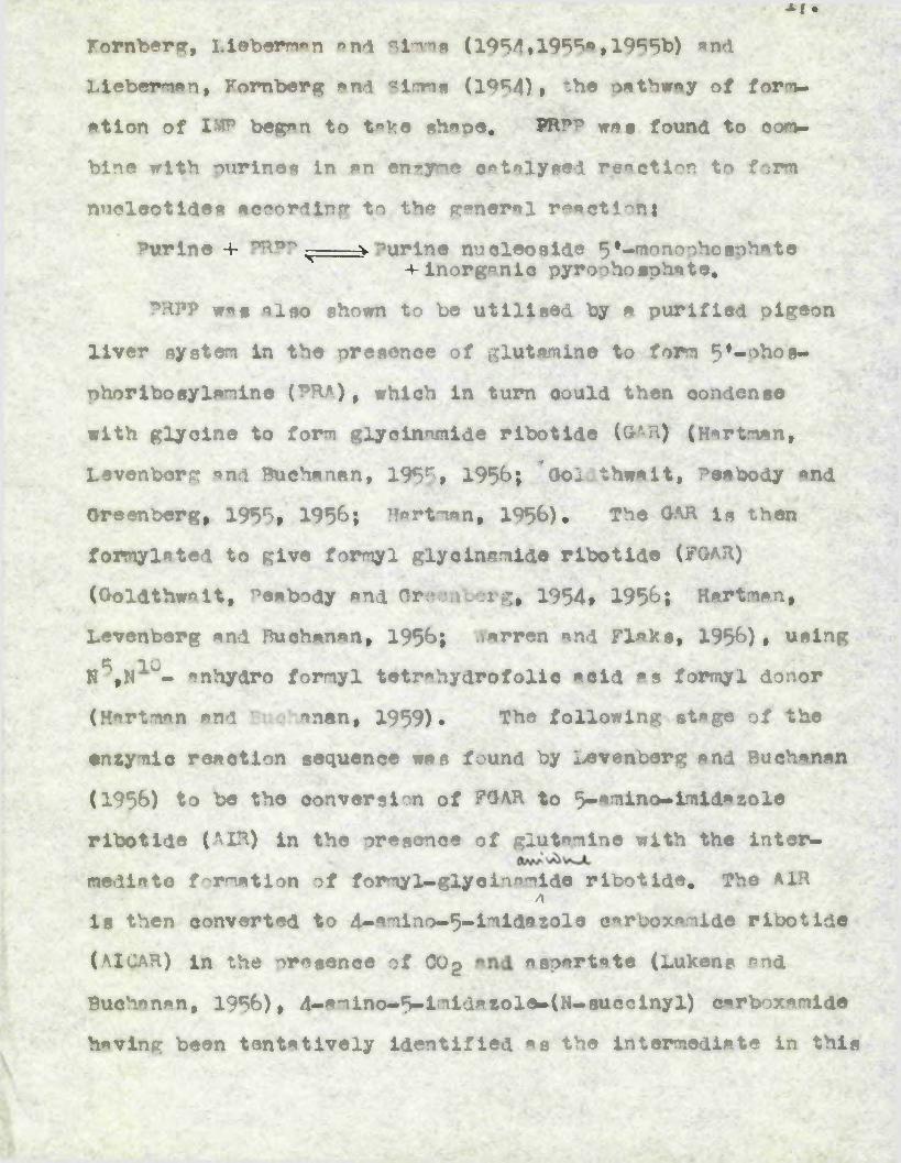
*f •
Kornberg, Liabornon and Simms ( 1954,195®*, 1955b) and
Ll^^bemian, Kornberg and ®lrnns (1954), the oathway of form
ation of IMF began to t«ke ahaoe. PRPP was found to com
bine with nurinee in an enzyme catalysed reaction to form
nucleotides a.eoording to the general reaction:
Purine f O^po -— vourlne nucleoside 5l-monnohosohate 4- inorganic pyroohosphate,
PHPP was also ahown to be utilised by a purified pigeon
liver system in the presonoe of glutamine to form 5t«phos<»
phorlbosylanine (?RA), which in turn could then condense
with glycine to form glycln amide ribotide (GAH) (Hartman,
Levenberg and Buchanan, 1955, 1956; *G>ld.thwalt, Peabody and
Greenberg, 1955, 1956; Hartman, 1956). The OAR is then
formyl*ted to give formyl glycinamide ribotide (?GAR)
(Ooldthwalt, Peabody and Greenberg, H54» 1956; Hartman,
Levenberg and Buchanan, 1956; Wwaren and Flaks, 1956), using ® TON ,N - anhydro formyl tetnhydrofolic acid as formyl donor
(Hartman and Buchanan, 1959). The following • stage of the
entymic reaction sequence was found by Levenberg and Buchanan
(1956) to be the conversion of FGAh to 5-amlno-lmidatole
ribotide (AIR) in the presence of glutamine with the inter
mediate f omatlon of formyl-glyolnmide ribotide. The AIRA
is then converted to d-aminn^-imldasole carcoxanide ribotide
(AX<AH) in the oncgence of CCg aspartate (Lukens and
Buobanan, 1956), 4-lmin<i-9imldatoll-(N-succLnyl) carboxamide
having been tentatively identified as the intermediate in this

Tie'ensymlo eynthaaia of inoelnle «old (XUP) de novo.
(From Dvidson, J.”., The Biochemistry of the Woeale Aeida, 4th % Methuen
Co.Ltd.• London, i960.)

Figure 1

xo
transformation. The introduction of the final carbon atom
is brought about by the formylation of AIGAR by N^-formyl
tetrahydrofolic acid (Hartman and Buchanan, 1959). The formfl-
AIGAR is finally cyclised to IMP. Thus the mode of IMP bio
synthesis has been clarified by the use of pigeon liver
extracts and is illustrated in Fig. 1.
Further in vitro work illustrated how AMP and <KP,
nucleotide constituents of RNA, could be derived from IMP.
AMI? is oroduced by the reaction of IMP with L-aspartate to
form the intermediate adenylosuccinic acid (Garter and Gohen,
1956; Abrams and Bentley, 1955# Lieberman, 19560 * b and c)
guanosine 5’-triphosohate (GTP) is required as a oofactor.
The adenylosuccinic acid formed thus then breaks down to AMP
and fumarate (see Fig. 2.)
The first steo in the enzymic conversion of IMP to GUP
involves the oxidation of IMP to xanthosine 5’-flwoooooephnte
(XMP) in the presence of nicotinamide-adenine dinucleotide (NAD).
The XMP is then enzynically aminated in the presence of
glutamine and adenosine 5’-triphosphate to yield GMP, (see
Fig. 2.)
1.5. Biosynthesis of the pyrimidines and their derivatives.
The biosynthetic pathway to the pyrimidines (see Fig.3)
has been elucidated minly on the basis of work involving
microorganisms, but the same system mears to operate in
mammaHm systems.

V
rlaure 2.
**•.
; 'r<< W**
Snxjmle synthesis of * denylla and gusnyllo
molds from inoslnlo mold.
(lTroi Dmldson, J.N., The Bloohoslatry
of the Wuelelo Acids, 4th 14.# Methuen 4
Co.Ltd., London, I960.)
si. £Er-;^.

xanthosine monophosphate
GM
P
n-z-
2“Io
Figure 2
•\nX8 °

^7.
Deegradation of labelled pyrimidines has shown that N1
of the pyrimidine ring is derived from NH-j, C2 from CO2 end
C4»C5 end N3 from aspartic acid. Experiments involving the
incorporation of possible labelled precursors have revealed
that ureidosuccinic acid (carboy 1 aspartic acid) and orotic
acid (uracil 4-carboxylic acid) lie on the biosynthetic path
way (Reichard, 1955» Lieberman and Kornberg, 1954» Cooper,
wu and Wilson, 195J» Wu and Wilson, 195&)
The first step involves the interaction of COg and NKj
under the influence of ATP to form carbamylohosph»te (CP)
(Jones, Spector and Lipmann, 1955). The carbamylphosphate
then reacts with aspartic acid under the influence of aspartate
carbamyl transferase to form ureidosuccinate (US) (Schulman
and Badger, 1954» Smith and Stetten, 1954; Heinrich, Dewey
and Kidde*, 1954). Then, under the influence of the ensyme
dihydroorotase, which has been isolated by Lieberman and
Kornberg (1954)» ring closure of US is effected to yield
dihydroorotic acid (DHO), which in turm is oxidised by
dihydroorotic dehydrogenase to yield orotic acid (OA).
Under the influence of orotidine 5’-phosphate pyrophosphorylase
and magnesium ions,OA reacts with PRPP to form orotidine’5’-
monophoephate (UM) ,' which, according to Hulbert and Reichard
(1954)» is either degraded to uridine and uracil, or phosphor-
ylmted to uridine 5’-pyrophosphate compounds.
Although formate does not serve as a precursor of

Figure 3. The pethwsy of de novo synthesis of pyrimidines.
(From Dn rid son, J.r., The Biochemistry of the Nucleic Acids, 4th td., Methuen A Co.Ltd.» London» l%Oe)

COOH HjN \h2
OC\ Z
NH
£O -2H /JO
KN CH2 dihydroorotic ^N CH—*■ I | dehydrogenase I II
CH-COOH OC /CH-COOH OC\ ZC-
NH
KAo
asparticacid
urcidosuccinicacid
dihydrooroticacid
COOH NH
orotic acid
Ml•HPa
Uracil-*— Uridine -*
COHNZ CH
COj
OC .CHN-ribose-5 - PO4
UMP
OC C-COOHN H-ibosc-5 - PO4
orotidi phosphate

2G.
nucleic mcid ur«oil, it does serve «« m precursor for the
^methyl proup of thymine mnd the recent work of Kornberg
(1957) inplioited hydroxynethoytetr«hydroPolio amod in this
context. Trom m review by doable (1961) it Ropers thmt
uridine serves ms m precursor of the thymine ring snd there
is evidence to suggest thmt the glypogldie linkage remmins
intmct during conversion to the deoxyriboslde which is then
metlhflated in the 5 position with the involvement of this
folic mold derivative, hydroxyr!mtholteerahydrofolic scld.
1.4. Blo«mthegl« of fluolele soldi.
With the knowledge that nurine and pyrimidine derivatives
ean be synthesised from sml^l molecciles, many of the early
investigations in this field were carried out using smi.ll mooecules such as ’*~OfpJmite mnd ^ogiycine ms
1 so tool c tracers (Brown ana Roll,. 1955) • Although the
present knowl=u~/o of the mechanisms by which HNA mnd DMA
are synthesised in vivo is derived almost entirely frcm
experiments using cell-free systems, the early work perfomed
on whole animals and surviving tissue prepirations has been
of great value*
Muoh of the basic work in the field of polynucleotide
biosynthesis was carried out by Brown and his oolli]lb^r*'tprs
(Brown, 19M; 1955; 1956) who used isptopicilly labelleu bases,
nucleosides and nucleotides to investigate the mnnter in
21*
which various organisms can utilise exogenous suoolies of
these compounds for the synthesis of RNA and DNA* Their
experiments indicated that in the mt, for exemole, adenine
is incorporated into polynucleotide adenine and guanine,
whilst adenosine and AUP are utilised to a lesser degree.
On the other hand guanine and guanosine are not incorpor
ated to an appreciable extent although 01 is moderately
well utliseed* free nyriraidlnes are not utilised extens
ively in polynucleotide biosynthesis but their nucleosides
and nucleotides aooear to' be readily incorporated*
The work of Rose and Rchweeigrt (19>3) using cytldlne unifoxmly labelled with ^--c drmon3Srstrd that the ribose
was incorporated into RNA as extensively as the base, and
they concluded that the nucleoside was incorporated without
breakage of the glycosldlc bond*
Because nucleosides are so readily incorporated into
nucleic mold, irotooically labelled thymidine h«s been used
widely in studies of the biosynthesis of DM*. Labelled
thymidine has been shown to be readily incorporated into
DNA in the r#t (Relchard and istborn, 19R1), the chick
embryo (Frledkin, Tllaon and Rockets, 1956), onion root tips
(^ouade, Frledkln and Atchison, 1956) and tissue culture
(Lu and «innlck, 1954).
However although bases and nucleosides are readily
incorporated into nucleic acid, the mechanism of these

ZV.
reactions is uncertain. In 1954, Marrian reported thato 148- C-admine injected into rats was rapidly and extensively
incorporated into the AM, ADP and ATP of liver, intestine and
mscle, thereby confirming the observations of Goldwasser
(1953) on pigeon liver homogenates• and by Bennet (1953) with
mice. Further work of a similar nature (Bennet and Kreuckel,1955) in which 4,6-WCc-adenine was administered to mice,
proved that the labelled adenine was extensively utilised
for the formation of acid-soluble AMI5, ADP, ATP and for the
formation of RNA and DNA in such a manner as to suggest that
the acid-soluble nucleotides serve as precursors of the
nucleic acids. Thus It became of Interest to ascertain, If
possible, the level of ohosphorylation at which a nucleotide
would become a direct nucleic acid precursor.
Attempts to Incorporate nucleotides labelled Isotopically
In all three com^c^^e^tfs, that Is base, ribose and phosphite
ester group, deiocttrated that they w^ire extensively dephos-
ohorylated after Injection Into the rat (Roll, Weenneld,
Carroll and Brown, 195&) , and no evidence could be obtained
for the Incorporation of Intact nucleotides Into RNA. However,
despite this, the adenosine moiety of labelled AMP is largely
Ieccrpcrrtid Intact Into RnA, and the conversion of adenosine
Into RNA w&ano8iee and DNA deoxyguanosine occurs without the
rupture of the glycosidlc link, LeUman and Heidelberger
(1955) incubated Wl-Pplabelled ribonucleoside 5l-^loRnchocphates

23.with rat liver, Flexner-Jobllng c»rclnoma siloes, and euspanslone
of ihrlich «Bcite3 caroinoma calls, K study of the distribution
of the isotone in the acid-soluble and nuclelo aold fractions
proved that the nucleotides wwe dephosphorylated and that the random ^®P incorporation into the nucleic molds wag due
to the uptake of labelled inorgmnio phor"'hte resulting frcm
nucleotide breakdown,
(b) PpL£nepi^Jbjd2_P0pSPOPl^aSI*in 1956, the first indications of » rmactlon which could
lead to the entymio synthesis of polyribonucleotides w«re
obtained through the notable researches of Ochoa and his :
colleagues (Grunberg—Manago, ortlt and OatoR, 1956; Ochoa mnd
Hiepoel, 1957)> These workers found that the synthesis of
polyribonucleotide material can occur by condensation of ribo
nucleoside 5•-diphp0nh«tes under the influence of the entyme
polynucleotide pho8ohop’yl*se, which they prepared from
Asotobaoter vlnelandll. , This ensyme requires the presence of
magnesium ions and proceeds with the liberation of inor^rnic
orthophosphate, according to the foUowl ng equation : where
R stands for ribose, ^P for pyrophosphate, p for orthophosphate
and B for one of the following bases; adenine, guanine, uracil,«•cytosine or hypoxanthine,
nBRPP —> (BRP)„ 4- n?i
By incubating the entyme with the appropriate rlbonucleoslde
5’-diphosphates OpmooolyTisrs containing Al?, ITO1, CMP and iMP
have been obtained, but polymers containing (PiP «re much more

24#
difficult to prepare. Msgradatlve ^turtles of these synthetic
polymer a have eho^ th«t like RNA they oonslet of r^J^tonucleo-
Aide 5•-monophosphate un It a linked together by )•, 5*-nhoa«-
phodtester bonds.
This ensyme appears to be mainly of bacterial origin,
occurring in a variety of microorganisms (llttauer and
Kornberg, 1957# Beers, 1957# Olmsted and Lowe 1959),
although Its presence has been reported In spinach leaves
(Brummond, Staehlln and Ochoa, 1957) # and in a rather tent
ative way in a sear is lummbliQldes ( %tner and Gonslex,
1959) , atypical epit heUna of the rat (Yagl, Ozawa and
Konogi, 1959) *nd in nuclei prepared from guinea nig liver
(Kllmoe and Hen pel, 1957). Such work suggested that the
ribonucleoside St-dipheahhatea might be the direct precursors
of RNA in mnevnli«n cells, Hurlbert (1954) , Scholiz et al,
(195?*) and Bumm, Potter and SlekcVltt (1956) carried out
in vivo studies on the livers of rats which had received 6-^^C-orotlc acid, 1-^C-glueose and as inorganic phos
phate prior to killing. The results of specific activity
determinations on the isolated acid-soluble nucleotides
indicated that the labelling of base, ribose and ester
phos5lite is very rapid, and nearly the same for all
rn<embn*s of eaeh ribonucleotide aeries, so that it was not
oosalble to establish the ribonucleoside 5•-diphosphates
as the proximal precursors of the RHA,

25.
As mentioned In a preHrlv\x& section (1.4a) leotonlcelly
labelled thymidine ha# been used extensively in studies on
the blosyntbesis of DNA In Intact cells. Subsequently
it was extended to studies with cell-free-?rrpnratlon#. in
1956 Koimberg and his sssooiates, using cell free extracts 1/
orepared from i, ooli demoetreted that C»thymtdine in the
presence of ATP »nd inorganic ions was s specific precursor
of DU, This process required the prior conversion of the
thymidine to thymidine ^-triphosphate (TOP), and it wee
evident that these crude extracts contained a kinase
system capable of eatslysing this eoovvrtlon. in inter
work TT? srts used at substrate, «nd this made possible
the purification of the enzyme responsible for its incor-
oorstlon into DN. This enzyme, teimed DNA polymerase,
or, using systematic nornmedlaure, deoxyiueleoside^
trinh0ir>htte : . * deoxynuoceotidyltransferm se, requires
the presence of primer DNAt ea^gnesiue ions mnd the 5f—t^:rl—
nhopphates of deoxy»denoaine, deoxy£^00^1,^^ deoxycytidlne
mnd thymidine (dAT*, dOT? dCPF mnd TTP) for optimal
activity. - 'Net synthesis of up to twenty times the amount
of primer added has been obtained (Kornberg, Lehman,
nesoman and Mwm, 1956* Xolttllberg, 1959; Lehman, Besaman,
Sims and romberg, 1959). The overall reaction catalysed
by this enzyme can be represented as follows :

26 .
n. 4AW r iab
n. dOBB?■+■ dna°==°dna
n. dCPBB
da?v4(n) F?°
n, d???B L dT°-
From this work it seamed resaonmble to postulate
that the biosynthesis of DNA takes place by three steps:
(i) formition of deoxynudeo sides, or their
(ii) their polymerisation to the triphosphate level by
the appropriate kinase system (iii) the enzymic polymer
isation of the triphosphates to form DNA. The DNA so
prepared in vitro using the purified enzyme from X coll
has the «.-rne physical and chemical clhr^dsristica as
the calf thymus DNA used as primer (Kornbrg, 1959)*
If Tcall DNA is used as pri ■ per, the newly synthesised
DNA has the relative proportion of bases chraderistic
of coll DNA.
Dring 1957-60, severe! groups demonssr-ted similar
systems in osH-free extracts of regenerating rat liver
(Bollum and °otter, 1958; Manteavinos and Cneeiakis,
1959) and dhrlieh ascites careinma of the mouse
(Davidson, Smelie, Keir and MdAdle, 1958; Smeile,
Keir and Davidson, 1959; Bwelie, Gray, Keir, Richards,
Bell and Davidson, I960). Bollum (1960s, 1960b) whilst
studying this type of system in calf thymus extracts
observed that the priming ability of many DNA samples wn be enhanced by heating to 100° for 10 minutes. 3uch
• ■ 27.
treatment is known to bring about the dlssoclation of the
DNA double helix Into two single strand polynucleotides
(Doty, 1961)> and It seems likely, therefore, that single
stranded DMA may be the true criming agent. This Is con
firmed by the observation that the single stranded D'4A of
bacteriophage #X174 la * particularly good primer of the
polymerase reaction.
U) blft^yathaala in animal
Up till 195% ^e mechanism of HNA biosynthesis In
animal tissues was still obscure. As discussed In section
1.4(**)> early research Involving whole anlmtXc and intaot
cells seemed discouraging, since such workers as Lelhman
and Heidelberger (1955) and Boll, ^einfeld, Carroll and
Brown (1956) hal shown that Intact nucleotides were not /
Incorporated Into H«A. However, research In this field
received a stimulus when Heidelberger, Harbers, Leihman,
Takagi and Potter (1956) showed that cell-free preparations
of rat liver incorporate AMP specifically into KNA without
randomisation of its ohoeohat© group, so confirming earlier
observations by Goldwasser (1954; 1955) on cell^free pre
parations from pigeon liver. Thus, it appeared that with
the intact cell, the access of intact nucleotides to the
cell may be prevented due to premeability difficulties, with
the result that the phosphate ester group is split from the
nucleoside at the cell membrane and appears randomly

28
distributed among *ll four nucleotides subsequently pre
pared frrm the RNAe
This work was extended by Herbert and Heidelberger
(1959) Who used rat liver homoogerntes to study the inemw
inr’atioi of isotopioally labelled AMP into RNA* Degrad
ation of the RNA so labelled suggested a preferential
linkage AMP to CM in the RNA* similar results have been
obtained from a variety of other biologies1 systems (Paterson
and Lepage, 1957; Us, 1957» 1959; Ed^c^i^da and
Abrams, 1957; Herbert, 1958; Preiss and Berg, I960; Huiryltz,
Bresler and Kaye, 1959). These observations, together with
the finding that most of the iioor’o<orated adenine is released
as the nucleoside on alkaline hy&rolysis (see section)
indicate that the AMP has been added in a terminal location
to the RNA chain* The significance of these findings has
been elucidated by ^ameonik, Hern gland and their colleagues,
working with soluble or transfer RNA (sRNA) of rat liver
cytoplasm, which has the peculiar capacity to act as
acceptor for the terminal attachment ribonucleotide units
(Hecht, ^amec^lc, Stephen son and Scott, 1958; Hecht,
Stephenson and Zamedtnic, 1959; &meelik, I960; Hoagland,
I960). such terminal attachment reactions are an obligatory
prelude to amino acid attachment in the process of protein
synthesis. The precursors of the terminally added nudeotide
units ere the ri^lmiullenside 5•-aripho8phates, and their

29.
attaohmont involves a roverslbis pyrophosphate splitting.
inorganic pyrophosphate inhibits the attachment, and Hertbsrt
(1959) found that it accumulated during the incorporation of
nucleotides.
Although the compete base sequence of e-RNA ie unknown,
Stephenson and Heoht (195b) described reactions
whereby one or two CMP residues and an AUP residue are
successively attasohe< to the end of an existing molecule
of sRHA. The reactions are as follows ;
sRNA - CTP____ > ^A-GPPa PP1
bRMA-CMP f -AT------ > RNA-.C
fiA.-tf-CCP v -A------ > aHNA-CMP-CMF-A^P A PP
SRNA ?ith such a nucleotide end sequence can then act as
acceptor for the transfer of aminoaoyl residues from their
resodtive activating enzymes to the site of incorporation
into protein (Hoagland, i960).
However, such limited terminal incorporation can hardly
be regarded as representing true HHA biosynthesis and is quite
different from the extensive polymerisation reaction catalysed
by polynucleotide phosphorylasc, an enzyme not widely dis
tributed in mammUan tissue.
Thus, when the , present study was undertaken, the eee^halir
of true biosynthesis in mummltan cells wis far from clear, an
so the initial aim of the experiments was to obtain from
ma mlian tissue s cell-free systm capable of HKA synthesis

30.
and to study the involved. Previous work hms
shown thmt shrlich meltes tumour cells readily utilise ^Cc-fonnte, ^(-mdenine mnd ^^-inorgsnio phosphite for
the biosynthesis of RNA (Smeeiie, Thomson and Davidson,
1958; Thomson, amellie and Davidson, 1958). The work
described in this thesis denonnsxr^^ee that extracts of
ascites tpmour cells, and other mammUM cells, besides
containing enzymes responsible for the ohosohory 3mtion
of uridine to the triphosohate level appear to have an
enzyme capable of polyribonucleotide biosynthesis which
will use ribonucleoside 5*-triphosphates as substrates.
Thus for the incorooration of UM? unite into polyribo
nucleotide mtcrial this enzyme will utilise UTP as sub
strate. The conditions and ch*rroteristics of these
reactions are examined in an attempt to elucidate the
precise ^chanim of RNA biosynthesis in mommllan cells.
1.5. Ihfl -in‘ r~<yiiui3r .xo.Q.ntA'jn thcslslng systems.
Mhle the min bulk of cellular RNA is found in the
cytoplasmic fractions (mitochondria, microtomes and cell
sao) a smU but inpontant port is present in the cell
nucleus (Swllie, 1955). Since these morohiOongLeal
fractions probably olay very different roles in the functioning
of the cell it is important to consider the relation between
the RNA of these different sources especially in connection
with its biosynthesis.

31.yhan Bergstrand et «1. (1948) adrnmnistered a°Uglycine
to rats, the isotope content of the RNA frcm the liver cell
nuclei was greatly in excess of that in the RNA of the
cytoplasm. Using radioactive phosphate, Marshak (1941;
194°) showed that the RNA of the nucleus assimilated ^2p
mich more than did the RNA of the cytoplasm. Mooeover, while
the radioactivity of the nuclear RNA rose quickly after
ad?neniBtr«tion of the isotope and fell rapidly afterwards,
the activity of the cytoplasmic RNA increased and reached a
e*xieui level much later than the nuclear RNA. Tils type
of finding has been confirmed by a number of workers (Ramaum
and Huseby, 1950; Barnum, Huseby and Vermund, 1953» Davidson,MeXndoe and Srnmeiie, 1951; iaellie, Molndoe, Logan, Davidson
and Dawson, 1953; Maimoe ana Davidson, 1952). Such an
approach has been extended using labelled precursors of the
nuriie and pyrimidine bases. Potter, Reoknagel and Hurlbert
(1951) and Huulbrrt «nd Potter (1952) have investigated theincorporation of b-^C-orotic acid into the RNA of the
nucleus and cytopl»si of rat liver. They found that the14uptake of C by the pyrimidines was much more rapid in the
nuclear RNA than in cytoolaimio RNA. Using ^C-formte
Srnmeiie, M^J^^ioe and ^vidson (1953) also observed greater
assimilation of isotope into the RNA of the nucleus than
into that of the cytopla^^. Since the amount of RNA in the
nucleus is smai, these isotope incorporation experiments

indicate that it must have quite a high me^^'hoXic activity.
Recent investigations, using micronutoradiograohie
techniques suggest that the nucleus and the nucleolus might
be the mjor site of the synthesis of RNA for both the nucleus
and cytoplasm. Preecott(1957) in experiments with amoeba demooesrated that labelling of the nucleus with --—uracil
precedes the apoenranee of irotopir label in the lytopl*e.
The exu^iri^^r^’ts of Goldstein and Rlaut (1955) have shown the
movement of newly synthesised RNA from the nucleus to the cytoolasm in amoeba, -h-uuidine was employed by ■Ztloktr
(1959) in studies of geuroioora tanhrae. He showed that
labelling appeared initially in the nucleus and subsequently
migrated to the microeomes. qholtdssek, Schneider and hotter
(1958) and SoVuteider (1959) carried out experiments on a
much larger scale and observed an RNA in the nuclei of rat
liver c^11s which is raoidly transferred to the cytoplasm.
'Within tuo nucleus, the RNA particularly associated with
the nucleolus shows a rapid rate of labelling. in the
nuclei isolated from thymus tissue a fraction, said to contain
nucleolar RNA, incort>oratri acid-soluble RNA precursors
faster than any other RNA containing fraction from these
nuclei (Allfrey and Hirshy, 1957)# indeed evidence for the
existence of at least two types of nuclear RNA (nRNA- and
nRHAg) has been obtained by several authors (Logan and
Davidson, 1957; Vincent, 19*7% Osawa, Totata and Hotta,1958)

33.
A3oc^i*<d^ng to these authors one of the nuclear ribonucleic
acids, nRN.Ag, can be tentatively identified with the RNA
associnted with the nucleolus.
That nucleolar RNA can be a. precursor of at least s
fraction of the RNA in the cytoplasm has been indicated
from work on Acetalblarla (Stich and Hummrling, 1953) salivary
glands of DrnenO^il« (Taylor, McMaater and Cayula, 1955) » star
fish oocytes (Vinceent, 195% » bean root tips (woods and
Taylor, 1959) and pancreas cells (Fitzgerald and
1959)e By enucleation with ultraviolet rays, Berry and
Smra (I960) found that in HeLa cells t thirds of the in
corporation of precursors into RNA is nucleolar dependent.
woods (I960) is of the opinion that any incorporated isotopic
label passing from the nucleolus to the cytoplamic RNA does
so via soluble intermediates which do not exchange with the
pool of acid-soluble RNA precursors. Un nil the nuclear or
cytoplasmic origin of these in termed iates is known, any claim
that all the cytoplasmic RNA comes from the nucleus mist remain
open.
toniSd<«r»blt attention has been focussed on the very
smll amount of RNA on the chromosomes. Autoradiographic
evidence suggests that eiromosnml RNA could be the precursor
of a fraction of nucleolar RNA in the starfish oocyte (Fioq,
1955) » bean root tips (wocois, I960), He2La and leukaemic cells
(Feinendegen, Bond, Shireeve and ^sinter, I960) and in liver
and pancreatic cells of mice (Amno and Leblond, I960).

34.Sven by I960, de#Bit« the obvious sbundsnee of muto-
radiographic evidence, there crnc little real evidence mt
the level to cuooort these claim. Thuc it w*c
the purpose of the present study# not only to elucidste
the ent^lc mechoni!i of RNA biosynthesis in msmMiCiftn cell*
but Also to determine thc intracellulsr location of thc
oroocBa.


2.1 Biological.
Ihrlich nseltes carcinoma* originally supplied by pr. 0.
Poojsk of H«>mnmr«Aith Horoital, Londoi?w*»e ornpng»tld by
serici trrnfc^l®ntatloi in clbino mice of the dnoArtment«l
colony, Neither the strain nor sex of the nice used was
critical, cnd m pure cross-bred, strain derived from strong *
was found very suitable. For the growing of this tumour it
wf Advantageous to use mice «ged frcm eight to tcn weeks cnd
weighing between 2Rg snd ?0 g, mince younger mice hmd m high
mtallty r«te «nd older mice produced «acitic fluid cen
ts ining relmtively few cells.
Tumour for tr*’n<nlent*ti^n ws® withdreAi by syringe under
sterile conditions from mice inoculsted seven dnys previously.
0.2 mi, of tumour cell suspension, containing in the region of
1x10* cells, wss inoculated intr«xeritineally into the recipient
mice «nd gmvc a high yield of aacitic fluid within scvcn to
eight d«ys. For eiierimcitrl nur^nses, *scitic fluid was
withdrawn frcm mice cfter seven d«ys. The ususT yield of fluid
was within thc renge 5 to 10 m,, and the cell count About 5 to
10x10* per n,
'”he LAAdsdaits sscitcs crrciorr was obtained from Dr.L,
Crawford, Department of Virology, Unnversity of Glasgow, This
tumour w®s oroo*gitcd by serial trrnsolrni*tini in a pure *orton 7
st^in of mice, when inoculated with About 2x10 cells, these
mi.cc, weighing between 40g and 5Og g*ve # maximum- yield of asnitic

fluid (About 5 m..) After nine to ten days. The cell pop
ulation of the acetic fluid from these tumours wee substantially
greater than with the Xhrlich aecites caronnoma and the tumour
was much less susceptible to contamination with erythrocytew.
For the experiments on immature rabbit tie^ies, young
rabbits weighing from 1500g. to 2000g. w^e selected from the
departmental colony.
2.2 Preparation of crude cytoplasmic extracts from mammaimcel Is.____________ . . . , ... ... ,'._ '
(m) from 880^^ eirclnoma cells.
Crude cytoplasmic extracts were prepared frcm both 3hhlich
and Landschutz ascites earcinQmt cells by the method of Smeite,
Xeir and Davidson (1959)* The appropriate ascitic fluid was
withdrawn from several mice under aseptic conditions, pooled
and the tusoen8ion of cells centrifuged at low speed (200-300xg)
for five minutes at 0° to separate the cells from the ascitic
plasma. The sediment w»s resuspended in S-lOvo!* of chilled
0.1M phosphate buffer pH 8.1 and again centrifuged at 2Q0-3C0xg
for five minutes at 0® to separate tumour cells from trstipjestes.
This washing procedure was repeated several times until the
sediment of tumour cells was freed from contamination with
erythrocytes.
The cells were then disrupted using an osmotic procedure.
For this the washed cells were resuspended in chilled 0,1m
phosphate buffer and centrifuged at fOO x g to pack them
tightly in the centrifuge tube, 10-12 vol, ice-cold distilled

37.
—ter wore then cdded mnd the eueoeieloi gently homo gen iced in
a -^romoled Potter type homogenisor (Potter and Elvehjea# 1936)
Three or four pacaee of the maerial were ruffiaieit for
adequate disnerai0i. ainToaonpic examination of wet ameeira
with the aid of crystal violet in 0.-M citric acid was used
to control thie process which appeared to runture most of the
cells without causing much nuclear breakdown. The resulting
suspension was centrifuged in the Spinco aodel L preparative
ultraoentriuge at 105;000 x g for 6o minutes at 0® to yield
a clear cytoplasmic extract,
(b) from t.Uiu».a of Immtmpq rabbit,.Etracts of ircmaure rabbit tissues were prepared from
thymus, spleen, brain, inte8tine, testes, bone marrow# appendix
kidney end misole of young rabbits by homon©nieing the cpprop-
rimte tissue for 2 minutes in 10vol. ioe-cold distilled —ter
using s cooled "otter tyoe homrn©nifier, end centrifuging the
resulting suspension for 60 minutes at 105,000 x g,
2.5 EroMratlcrn of suUdlular from artltei oam.For some exneriments, ascites carcinoma cells • were •
fractionated into subcellular fractions (nuclei, mitochondria
and microsomes) in order to conduct incorporation studies on
these individual cell fractions.(a) Nuclei.Because both the ihrlioh and L*ndschutz ascites tumour
cells are relatively robust, homogeni8ation in a Potter type
38.
hn'nnnati•er with a Buern8t-etleiu'n chloride medium wee found' Al,?!...’ -- jiAgy^pa''.S ^****3?’•"'J ■'Sere*
insufficient to cauae their disruption. {< Therefore before„ • ??’ ' ■ ' '• -!.. • X '~<1?' 'J- -•< * '■ .
nuclei could be isolated frm these ascites cells the methods
of Ho^nbnnn, Setmeider and Striebich (1952) and KL^rey, Mraky-
and Osawa (1957) for the isolation of nuclei fr<m rat liver
cells and thymus gland cells respectively, had to be mOiified..
The initial step in such a modf ice tion w»s to subjeot theAS\ •• „ -•/ . • ’ - ' • - ■ * < - \v- *.' i 1 - s ' ‘’v-i’ * . > " •?*. 'v <•'* *i*4r .•***•/-ascites cells, of the appropriate type# to osmotic diaruntion
in precisely the manner described in 2.2(a), This treatment
leaves the nuclei intact. The suspension of osmn01calls die-
rupted cells was then centrifuged at 6oo x g for ten minutes
at 0°, The supernatant fluid was then removed for the sub
sequent preparation of mitochondria! and microsomal fractions
and the sediment# witch contains nuclei heavily contaminated
with !^,top3^am^c fmaerlul, was suspended in 5 vole, ict-enlr
sucrose-calcium chloride (0.2<M - 0,003M*) by gentle hnrann«tiiatlnn
in a Potter homnnetieer. This suspension was centrifuged at
600 x g for 10 minutes to collect the nuclei. Further washing
of the nuclei was achieved by resuspension in 5 vol. ice-cold
auo^nst-ealciml chloride (0.25M - 0,0033°) followed by centri
fugation, One to two wahlnga was usually sufficient, aa. * •, * ske i-. ‘ • ." . • - . . . . •’ ‘ •• A.*•*; - 4 • f\ • . .'.-'A- 'the supernatant was quite clear by that time. Such a proced
ure yielded nuclei free from any obvious cytoplasmic contam
inants as Judged by microecopio examination of wet smearsft ji&rei?■ ■V*V•’*><£>
stained with 1° crystal violet in 0,1M citric a.elr.l*

(b) Mitochondria, mllrnsores and cell gap
After thc removal of thc nuclear fraction from thc
suspension of osronicslly disrupted cells, by centrifuging
at 600 x g (see 2,3), thc supernatant fluid was then made
0,25M with respect to sucrose and 0*0033* with respect to
calcfum chloride, a mitochondrial fraction was prepared
from this by centrifugation at 10,000 x g for 10 minutes ato > > * s-. ■■ ' ' „
0 and was washed by re suspension in sucrose- calcium chloride
(0.2JM - 0,0033*) foioowed by centrifugation.
After the removal of mitochondria from this suspension,
ricro8orcs were collected by further centrifugation at
l05,000^:ii thc Spinm Mooel L preparative ultracentrUgc.
The suoeraatant fraction from this centrifugation was mn-
sidered to be free from most of the cytoplasmic particulate
naerial and to consist minly of the soluble lnmponents of the
cell sap,
(0) Nudeoli and chromatin maerial.
An attempt was made to isolate nuclcoli from ascitcs cell
nuclei applying - thc principle used successfully by Mority, Litt,
Kay and Bounce (1956) for thc isolation of nuclcoli from rat
livcr cells. Thc ascitcs cell nuclei (see 2,3a) from 20ml,
packed cells were suspended in apornxintcly 35 ml, of 1^
gum arable (w/v) pH 6,3, and the pH of the resulting suspension
readjusted to pH 6.25 - 6.30 with very dilute NaOH, Careful
stirring was required to avoid local excesses. This operation

40.
and eubsequent operations were carried out as close to 0®
as possible.
The suspension was than exposed in batches of 10-15ml,
to vibration by a Mu Hard ultra sonic drill (50W. ; $Okc.)
for 7 to 7.5 mjiutes. The maaerial after ultrasonic
vibration was centrifuged in two 50^^, centrifuge tubes (each
tube containing about l9ml.) for 20 minutes at 400 x g in an
international refrigerated centrifuge. The supernatant fluid
contained the bulk of the chromosomal maerial, much of which
was in a very finely divided form. it was set aside for
further fractionation.
The sediment from the first centrifugation was next sus
pended in 25 ml, of ice-cold distilled water, and allowed to
sediment by gravity in a 50ml. measuring cylinder for 15 hours.
The too 15ml, were withdrawn carefully with a Pasteur pipette,
leaving nuclear membrane mtteial, unbroken nuclei, fibrous
maerial »nd other unidentifiable fragments in the remaining
2ml, The top 25ml, was carefully and evenly resuspended
(without further change in volume) and again subjected to sedi
mentation by gravity in a 50 nl. cylinder, this time for 7 hours.
The top 21rKl. were then drawn off and transferred to a 50m.,
centriUge tube, and the concentration with respect to gum
arable adjusted to —C by the addition of the appropriate amount
of 10- gum arable solution at pH 6,0, This 8usiie8ion was
centrifuged at 300 x g for 20 minutes. The pellet was

41.
suspended in 8 to lomi, of 2^ gum srabio at pH 6.0, and the
resulting suspension centri^gcd in s 15n. conical centrifuge
tube for 15 minutes at 200 x g. Those centrifugations in gum
arabio caused the sedimentation of the nucleoli, leaving the
finely divided chromosomal contamination in suspension. The
gum arabio was removed from the nucleoli by two washings with
smll volumes of water. Microscopic cxairrnation of the wet
smears of these nucleoli, using 1$ crystal violet in 0,M
citric acid and pyronine methyl grccn stain (D.D.H.), suggested
that while kbninntini of the nuclei had broken off the main
bulk of the chromosomes frtm the nucleoli, traces of chromosomal
malarial still remained attached to the nucleoli.
The original supernatant suspension remaining after the
first centrifugation of nucleoli was fractionated in an erbitweyy
method by centrifugation at 25»000 x g for 30 minutes at 0*,
The sediment, termed "chromosomal fraction" is said by Mooxy,
Litt, Kay and Dounce (1956) to consist minly of whole and
fragnented chrorosomeB, chiefly the latter. The supernatant
fluid, which had a slightly milky apnearaile, is believed to
contain finely divided chromosomal material in colloidal sus
pension and presumably a smll amount of maerial brought into
solution by sonic trea'elcita No particulate mtter could be
seen on microscopic examination.
The "chromnanrl^l fraction" and nudeoH were then sus
pended in O.OLM Tris buffer at pH 7.5 for subsequent experimental
work.

42.
2.4 Preparation of fractions from atoitea oarotomA cellQUSl&L____________________________ .___________________.(") soluble fraction (or nuclaur extract)..
Nuelei.prepared frem the appropriate varlety of rtoiter
cell# ee described previously (2.3 e)» were surirnded in three
t^rnej their volume of ice-cold OplM Trie buffer pH 7*4 by
gentle homoggrtertiot in m waiter homooertaer. The sue-
penaion of walel was then subjected to ultrasonie vibration,
using a MUjard ultra sonic drill (50W ; 20ko.). Batches of
nuclear suspension of about 15m.. were exposed to vibration for
periods of about two minutes. The length of exposure was
determined by following nuclear disintegration in wet smears
stained with crystal violet. The disintegrated nuclear
suspension was then centrifuged in a Splnco Model L preparative
lltru^orttrlfiigr for 6o minutes at 105*000 x g to yield a clear
nuolear extract, containing 1—3*%. protei^m.
For use in some experiments, the sediment obtained after
ultraoentrllupaltlon, was suspended in 3*ol. of chilled O.O1M
Tris buffer at pH 7.5 end divided into two oortlons. One
of these was termed "nuclear sediment" and the other (1m.)
after treatment with img. crystalline deoxyribonuclease in
the presence of 2umooes of MgClg for 4 hours at 37°* **s
termed "D'Jase treated nuclear sediment".
(b) aggregate lCtaltli.ln..
By eoOifyitg the method of Weiss (i960) an aggregate
myne fraction was prepared from nuclei of ascites cells.

43.
Packed ascites cell nuolel were suspended in twenty times their volume of ice-cold O.O5M Tris buffer at pH 7.4 and exposed in small batches of 10ml. to sonic vibration by a Mullard ultrasonic drill (50W. ; 20kc.) for periods of 2 minutes. This gave a suspension of disrupted nuolel which was centrifuged at 10,000 x g for 10 minutes at 0°. The sediment obtained in this manner was suspended in 0.05M Tris buffer of pH 7*4 to a final volume of 16ml. 2M KC1 was carefully added to the suspension, with adequate stirring, to a final concentration of 0.4M KC1. within a few minutes a white aggregate formed whioh was washed twice with 0.05M Tris buffer pH 7.4 - 0.4M KC1 medium and finally susoended in 0.05& Trie buffer of pH 8.1 by vigorous homogenisation in a Potter homogeniser, to give a protein concentration of 8-10mg. per ml.2.5 I sola tion of nucleic acids and nucleoprotelns from ssoitOs
—.. . ■ ■<*) IW* whole aella.washed ascites cells were paoked and susoended in 10vola.
loe-oold distilled water and subjected to osmotle disruption as desoribed in 2.2a. RNA was then extracted from thia disrooted cell nreoaratlon using a modification of the Kirby procedure (Kirby, 1956).
An equal volume of 9Q£ (w/v) phenol (B.D.H.) was added quickly to a portion of the disruoted ascites cell preparation. The mixture was shaken mechanically for 60 minutes, and then

44«
centrifuged at 600 x g for 60 minutes *t 0°. The cloudy
nqueous layer was removed by suction and the residue wsshed
twice with water. The aqueous layer was separated each time
by centrifuging for 45 minutes and removed by suction. The
aqueous extracts were pooled and extracted five times with
ether to remove phenol. Nitrogen was blown through the
solution to evaporate the ether and the solution was made uo
to with respect to ootasslum acetate. The RNA was pre
cipitated by the addition of 2 vol. of ethanol* washed twioewith ethanol and twice with ether and allowed to dry before
ostoring at -10 . Prior to use It was dissolved in distilled
water or in 0.01M NaCl to give a concentration in the region
of 2rag, RNA/ml, No MU oould be detected in this preparation,
(b) RNA fron cytoplasm. nucleus end 1Q5.QQQ-.X E eedimant.,
Uslng precisely the method described above, RNA w»s extracted from crude cytodasmic extract*of ascites cells, and
termed cytoolagmlc Rna. similarly the method was directly
applied to the sediment obtained after centrifuging suspensions
of osmotloally disrupted ascites cells at 105*000 xg. To
facilitate the extraction the sediment was suspended in 5 vol,
of distilled water. The RNA thus extracted was referred to
• • 105,000 x g sediment RN*.To prepare RNA from ascites oell nuclei, the packed nuclei
were first suspended in three times their volume of ioe-cold
distilled water and subjected to ultrasonic vibration for 2

45.
minutes *« described in 2.4». A portion of this sUBooneion of
B)o»i<^»^l.ly dieruoted nuolai was treated with phenol exactly as
described above and the RNA extracted in preolsely the same
munner. Although this RNA (2mg'«l. distills water or
O.O1M NaCl) was then referred to as nuolear RNA it mist be
em^P^as.sed that such a phenol extraction prooedure failed to
remove «n the RNA from the asoites oell nucleus. Suoh a
finding is consistent with the observations of Sibstani, Yamuna,
Xlrmura and Taka'PeW. (I960) that treatment of asoites cell
nuclei with phenol will release only one type of RNA, while
another type remains unaffected in the residue.
(o) Two distinct types of ribonuoleoprotein from asoitess&ULnuOsl.
Two tynea of ribonuclooprooeln were prepsrod from ascites cell nuolel by oppllaation of the methods previously described cy
Osawe, Takata *nd Htt» (1958) and Logon and Dvidson (1957)*
Paoked asoites cell nuoloi were hrn^^^er^iaM for 1 minute in
five times their volume of ice-cold O,1M potassium phosphate
buffer at pH 7*1 and the resulting suspension centriHged at
20,000 x g for 20 minutes in a refrigerated Seirvll centrifuge. The oplescent supernatant fraction obtained was termed the
"pH 7.1 fraction* and contained type i rlbonuoOeoproCeli. The
sedlmrnt was resuspOTded in the same buffer, homogenised and
centri^ged as before. After discarding the supernatant fluid,
the residual pellet was suspended in )0 vols, of ice-eold iM
Had, iomgeeiBed with careful cooling in an S.9.% Nelco blender

•t top sneod for 30 seconds, «nd stirred vigorously with m
meohsnlG*l stirrer for 2 hours, sgsln with o»reful cooling.
The resulting viscous solution ws centrifuged mt 30,00Q x g
for bo minutes st 0® in s Spinco Model L ultraoentrifuge.
After discerning the viscous sunernstmnt fluid, which contains
most of the cellular deoxyribonucleoproteln, the residual
pellet ws resuspended in 10 vols, of cold IN NaCl, using
the Nelco blender «nd centrifuged «s before. The neHet
finally obtained ws referred to «s the "IM N«C1 fmctoon"
end contained tyoe II ritlncoleloroteii. For experimental
use this pellet wrns dispersed in 5 vols, of ice-cold die- tiUM water by vigorous ilmmleniLUttili in a cooled Potter homogr^ii^ier. In some experiments ^--uridine was incorpor
ated into this ribonucleoprotein (see 2.8)
(4) tWA »nd nRNA from mectes o*U nicIei.Having omared two tyoes of ribonucleoprotein from
ascites cell nuclei, an attmpt was made to isolate the RNA
from each. Thus tyoe I ritlicclelproteii would be expectedto yield nRNAj, and tyoe II ribonucleoprotein, nRNA^. This
w»s of particular interest because according to Alfrey,
Mirsky and Osawa (19*57) # the RNA associated with tyoe II
ribonucle^^le^ was at least pertly derived from the nucleoli
wiile the use of the phenol technique (see 2.5) was succeesful for the isolation of RNA (nRNA^) froo tyye I ribonuclecerolelo,
it proved useless for the isolation of RNA from type II

Hf
r ibonu cloopro toin.
In order to obtain RNA (nRNAg) from tyoe II ribonudeo-
protein it w*s neoeaanry to treat the suspension not with
phenol, but with »n equal volume of 10# NaCl at 6o° for 20
minutes. The results of Signer, Boedtker and Michaels
(1961) sugjgest that longer treatment will destroy the RNA,
The resulting mixture was centrifuged at 20,000 x g for 10 minutes at 0° and the sunernatant fluid containing nRNAg
dialysed for 18 hours against two 71# changes of distilled
water »t 4° to remove as much salt as possible. At this
stage it ws found that the nRNAg contained a large amount of
DNA as contaminant. Treatment with phenol and subsequent
extraction of the RNA failed to remove this contamination.
Sven treatment with DNase (as described in 2,4» for nuclear
sediment) foioowed by subsequent treatment with phenol and
precipitation with alcohol in the presence of potassium
acetate failed to remove DNA entirely.
For experimental purposes both nRNA° and nRNAg were
dissolved in distilled water to give a concentration of 2mg,
RNA/ml, based on ultraviolet and pentose estimations,
(e) Native and denatured DNA.DNA w«s orepared from ascites cells following the method
of Kay, Simmonds and Dounce (1952), 1G0ml, of ascites
carcinma cell suspension was centrifuged at 1,500 x g for 10
minutes to pack the cells, and the supernatant fluid discarded.

48
The oells were wsshed repeatedly with 0.1M phosohete buffer,
pH 8.0, to remove contmoilnetlng red blood oells end were lysed
by the addition of twelve times the peeked sell volume of die-
tilled water followed by homogenisation in a Potter homo
gen leer. The resulting su»ponsion was oentrifuged at 4»5OO
x g for 30 minutes. The supernatant fluid was discarded and
the residue was homogenised in 0.9% sodium chloride - 0.01M
sodium citrate solution and made up to 200ml. with same solution
The resulting mixture was centrifuged nt 900 x g for 10 minutes
and the supernatant fluid discarded. This m renested three
tines. The sediment finally obtained was suspended In 0.9JS
NaCl, homogenised In a ’’otter homogeniser and made up to Jooml.
with 0e9?J NaCl. 27ml. of Duoonal (5/ sodium dodecyl sulphate
In 45? ethanol) were added with stirring. This stirring woe
continued for 30 minutes, «nd then l6.5g. of NaCl were added
to give a IM solution. Stirring was continued until all
the NaCl had dissolved and then the mixture was centrifuged
at 20,000 x g for 30 minutes. The supernatant fluid was
decanted and 2 vol. of ethanol added with swirling. The DNAf
which precipitated in thread-like strands* wag collected on a
glass rod, washed three tines with ethanol, three tines with
acetone and allowed to dry. The DM* thus obtained wag further
purified. Firstly it was cut into small pieoes and dissolved
in 300ml. of water at room temperature using a Vibromix vib
ratory stirrer. Then, 27ml. of puponal were added and the

49.
mature stirred for 3c minutes before 20 - 25g, of N«C1 were
added. The stirring was continued until the NaCl had dissolved
and then the mixture was centrifuged at 20,000 x g for 30
minutes. The DNA was precipitated, washed and dried as before.
Then the DNA obtained in this way rediseolved again, this
tme in 200ml, of water, and 1,8g, of NaCl were added and dissolved. The resulting mixture was centrifuged as before
and 10g. of NaCl dissolved in the suoernatant fluid. 2vol, of
ethanol were added to precinitate the DNA which was collected,
washed and dried, as before. The procedure yieXded about
120mg, of DNA which were dissolved in distilled water to a
concentration 2mg, DNAArnl,
A 200 ml, portion of this solution was then incubated with
400pg, RNse (crystalline) and 20^., of 1M Tris buffer pH 7.6
for 2 hours at 37°. An equal volume of w«ter saturated phenol was then added and the mixture gently shaken for 60 minutes at 20°. The supernatant aqueous layer which ws obtained on
centrifuging the mixture at 2,000 x g for 20 minutes was drawn
off, leaving the phenol layer as a thin interfacial film of
insoluble maeer*!, which was carefully removed. The phenol
layer was treated twice more with equal volumes of distilled
water in the mnner described above, the aqueous layer being
collected. The pooled aqueous layers were extracted three
ttaes with equal volumes of ether, to remove the ohenol.
The viscous solution was then made 1,0M with respect to NaCl,

and DNA was precipitated by the addition of ethanol to 5° (▼/▼)•The precipitate was washed three times with ethanol, and three
otimes with acetone, dried in air and stored at -10 . Prior to
use, it was dissolved In O.OlM NaC and dialysed for 24 hours at 4° against four 41. changes of O.OlM NaCl, the final concent-
rat ion being 2. Obu;. D’iVml.
For some experiment., solutions of DNA were subjected to
heat treatoent. This was carried out by i-mroreing 1ml.
portions contained in l°ml. conical centrifuge tubes in a water- bath at 100° for ten minutes, and rapidly cooling these
solutions by shaking the tubes in an ice-bath. This treatmentis referred to as thermal denaturation of DNA, end DNA not
submitted to this heating prooeea is termed native UN..
2.6 rrn ozonation of nuolear end cytopleimie extracts ofubIV.s .gyUa.(«) 'fth ammonium sylphey slona.
o oAH steps were carried out between 0 and 4 . The extract,
nuclear or cytoplasmic (40m.), was brought to 0.20 saturation
by the slow addition, with stirring, of 4.4g. of amium
sulphate. The solution was allowed to stand for JO minutes,
with continuous stirring, and the precipite was removed by
centrifuging at 8,000 x g for 8 minutes. This process was
repeated five times, the degree of saturation with an^iniuim
sulphate being adjusted to 0.JO, 0.40, 0,50, 0.60, and 0.70
saturation by the successive addition of 2,Sg, 2.8g,° J.2g and

51
3*2g of solid ammonium nulohate. After caoh addition the
solution was stirred for 30 minutes and the precipitate
collected by centrifugation. 3ach of the precipitates
was dissolved in ice-cold distilled water to give t> protein
concentration in the region of 2-4 nig./ral* and the solutions
were dialysed for 18 hours at 4 against two 11. changes
of distilled water to remove ammonium sulphate.
Thia procedure was subsequently modified when it became
evident that only two orotein fractions* those obtained
between 0«30 - 0*40 *»nd 0.^5 • 0.70 saturation were active.
In this modified procedure all steps were carried out between o .00 and 4 • 40ml. of the soproprlate extract were brought to
0.30 saturation by the slow addition* with constant stirring,
of 7»2g. ammonium sulphate. The solution was allowed to
stand for 30 minutes, with continuous mechanical stirring «n4
the precipitate removed by oentrlfuglng at 8,000 x g for 30
minutes and discarded. The suoematant fluid mi then
brought to 0.40 saturation by the addition of 2.8g. ammonium
sulphate and after stirring for 30 minutes the preolpltate
was eolleoted by oentrlfuglng as above. It was dissolved
In 0.01M Tris buffer pH 9.5 to give a orotein concentration of
2-3mg./ml. and dialysed for 12 hours against two 11. changes
of 0.01’jf Tris buffer oH 9.5 to yield either nuolear or
eytoolasmle Fraction H, deoendlng on the souroe of the Initial
extraot. The supernatant fluid after the removal of Fraction

A at 0.40 saturation was brought to 0.55 saturation by the
addition of 4.8g. ammonium sulphate and the precipitate
discarded. The supernatant fluid from this step was brought
to 0.70 saturation by the addition of 4.4g» ammonium sulphate.
The precipitate was collected, dissolved in O.OIM Tris
buffer oH 7.7 to give a orotein concentration of 2 - 3mg/mL.
and dialysed against two 11. changes of the same buffer for 12
hours to yield nuclear and cytoplasmic Fraction B as the case
may be. Although both types of fraction could be stored
at -10° and retained their activities for a few days, they
wore none the less highly suso<e>tible to any freezing and
thawing,
(b) with ammonium sulohats. sostone »nd oaloinw Btosahsts gel.
in order to achieve partial purification of a polyTlbo-
nucleotide synthesing system, cytoplasmic extracts were
fractionated with am^r^ium sulphate, acetone and calciim phos
phate gel. As before, unless otherwise s^eified, all steps
were carried out between 0° and 4°» A crude oytoplewnic
extract was pr ‘pared from a preparation of osmotically disrupted
Landschutz ascites cells, following the method described in
2.2a. This cytoolaimic extract (C3) was then made O.OIM with
respect to Tris buffer at pH7.5> »nd brought to 0.25
saturation by the slow addition with continuous meoTwnXoiaX
stirring of the appropriate volume of saturated ammonium
sulphate solution, oreviously adjusted to pH 7#5# The

53.
solution w»s allowed to stand for JO minutes, with continuous
stirring, and the precipitate was removed by centrifugation
at 8,000 % g for 8 minutes, »nd discarded. The supernatant
fluid was then brought to 0.45 saturation by the addition
of a further of saturated ammonium sulphate (pH 7*5)s
After stirring for JO minutes, this solution was centrifuged
at 3,000 x g for 8 minutes, and the resulting sediment
dissolved in O.ODM Tria buffer at pH 7.5 to give a protein
concentration between 6 and iOrng./ml. and dialysed overnight
against two 71. changes of the same buffer. The protein
fraction was termed the ammonium sulphate fraction (AMSiT)
and to every iOml. was added 1ml. of W Trie buffer at pH
6.5. Then, after immersion in a bath of ethyiene giycol-
at -15° enough acetone (previously cooled to -15°) was added
to make the fraction 1% with respect to acetone. The
solution wes stirred gontiy for 5 minutes at -15°, and then
spun at 1,500 x g for 20 minutes at -15° to remove the
precipitate which was discarded. Agin at -15°, the super
natant fluid was m»de 25° with respect to acetone by the
further addition of the appropriate volume of acetone (cooled
to -15°). As before, the solution was stirred for 5 minutes
at -15° and then centrifuged. The r°i»iltirg sediment was
dissolved in O.OlM potassium phosphate buffer at pH 6.8 and
dialysed against two 21. changes of the same buffer for 4 hours
(or in one experiment, 18 hours). The fraction was termed

54-
the acetone fraction (ACF) and contained in the region of
4 - bmg. nroteln/mi.
The next step involved the use of c«lciun phosphate gel#
which was prepared following the method of Keilln and Hrtree
(1938). 150ml. calcuum chloride solution (lJ2g. Ne^PO^ZHgO
per litre). The mixture wee brought to pH 7.4 with dilute
rcetic rcid rnd the precipitate washed three or four times
by decantation with large volumes of distilled water (15 -
201.). The precipitate was finally washed with distilled
water by centrifugation. At this stage the yield was around
9g. dry weight. The precipitate was then suspended in die-tilled water and allowed to stand for about a month. After removing the clear water layer, the gel was well shaken and
the dry weight per m.. determined, and was found to be l3mg./ml.
The gel was then ready for use, and was stored in the dark.for use in the»e experiments, the gel was initially
equilibrated with 0.01M potassium phosphate buffer at pH 6.3
by repeated suspension and centrifugation, followed finally
by suspension in the original volume of buffer, A portion of
the dialysed acetone fraction was mixed with an equal volume
of this suspension of c^cuum phosphate gel# now containing
l3mg. dry maorisl/ml. of potass^m phosphate buffer at pH 6,3,
The mixture was then centrifuged at 1,0<to x g for 5 minutes and
the supernatant fluid discarded. The g*l was then extracted
with 0.05M potassium phosphate buffer at pH 6.3 for ten

55.
minutes with stirring. The volume of buffer used was
equivalent to the volume of acetone fraction originally
submitted to this process. Tiis extract was removed by
centrifugation »t lt(X>o x g for 5 minutes and discarded.
Using the same procedure, the gel was further extracted with
O.1M potassium phosphate buffer at pH 6.8, The extract wee
collected and dialysed for 4 hours against two 11. changes
of O.OIM potass^m phosphate oH 6.8 and wag referred to as
the calcum ohosiohate gel fraction (CPpglF)*
2.7 Chemical synthesis of isotooioally labelled ribonucleoside.
(a) Z&tofraaLgWp .and ..UP?.
Both HTP «nd UDP labelled with in the o(-phosphorus
(uridine-5 2p-P°p and urid^—-2p-°) were preoared ftocm
labelled UMP using a modification of the procedure of Smith and Khorana (1958). The UMP labelled with ^2p oreoared
by reacting isopropy 1 idene uridine with 5°p..lsbell^ei
cyanoethyl phosphate according to the method of Tener (1961).
Thus in m typical preparation the initi"1 st©p was the oreoaration of °°"-labelled cyanoethyl phosphate (CIP).
The reactions involved can be outlined an follows :
32^.° DCC NcHg.^g^.ijJ^Jc.gac ^cN^2CN.Cai2.CH2 .OH+ (^.O^C0).1),,pyridine
waterheat
CN.CIL.OL.O.P.OZJ 2 (TZ Ba
^^2__ (CN.CH2.CH2O)oPoOO9

56.
A carrier free solution of disodium hydrogen phos
phate (PBS. 1. ,1.5ml. and 25mc at time of dispatch from The
Radiochemical Centre, Amersham.) was mixed with 2mmoles
of unlabelled orthophosphoric acid and concentrated to
dryness in vacuo at 40° to remove traces of hydrochloric
acid. Anhydrous pyridine (20ml., dried with CaH2) and 2-
cyanoethanol(2ml.) were added and the solution concentrated
in vacuo at 40° to an oil. The crystalline pyridinium
phosphate which sometimes separated, redissolved as the
solution became more concentrated. A second oortion of
anhydrous pyridine (20 ml.) was added and the solution again
concentrated to an oil. Anhydrous pyridine (10ml.) and 4- dieyclcj|c$rbodiimide(DCC) were added and the reaction
mixture set aside at room temperature overnight. 10ml. of
distilled water were then added to stoo the reaction* and the resulting solution was heated in a water bath at 100° for
jOmin. The solution was then concentrated to dryness
in vacuo and 20ml. distilled water and 20ml. barium hydrox
ide added to the residue. After 5roin. room temoerature
the solution was adjusted to pH 7*5 with acetic acid (a few
drops of glacial acetic acid were found to suffice), and
filtered to remove dicyclohexyl urea(DCU) and barium phos
phate. Two volumes of ethanci were added to precipitate
the barium 2-cyanoethyl phosphate which after standing for

57.
lhr, , was ooHooted by centrifugation. The crystals were
redissolved in water (5m., plus a tr«oe of glacial acetic
acid to aid solution) , centriuxged to remove a trace of
insoluble maerial and recrystall!sed by adding 10ml. ethanol.
The product was then collected by centrifugation, washed with
ethanol, acetone and finally ether. The yield after air
drying was 411mg. (1.28rnnooee), and equivalent to 64/.
Yields as high as 7^?f have been achieved.
For use in the phosphorylation of 2;3f-i8Opropylidene
ribonucleosides the barium salt of CJffiO was dissolved in dis-
water (SoO., plus trace of glacial acetic acid) and
passed through a column of D^o^^3^c^^^y^ro^en resin (3x 0.8am).
The column of resin was washed with distilled water until all
the radioactivity was removed. Sim!. anhydrous pyridine was
added to the effluent and the mixture was taken down to dry
ness (in vacuo at 40°) giving the pyridinuum salt of C8P.
This was used for subsequent phosphorylation reactions.
For the phosphorylntion of 2’ >3,<iisrpropylidene uridine to 2* ,3•<iigrprooylidene uM-P, lmrnole of the pyridinuum salt
of 3^ •ffiSP was used. 2mwoes of 2*, p-isoprooylidene uridine
(568mg.) were dissolved in anhydrous pyridine (10ml.) and lmmole of the pyridinuum salt of ^P-C !P was added. The
solution was concentrated to an oil in vacuo at 40® and then
anhydrous pyridine (10mL.) was added and the solution again
concentrated to dryness. The process was reoeated once more

50.
»nd the residue wee dissolved in 5°* enhydroue pyridine mnd
680«g, DCC mdded. The solution wes keot in e well stoppered
fl.msk for 17hr. mt roorn tenner*ture, efter which 1ml. dis
tilled wamer was sddod. After Ihr. the solution wm corn-
centrmted to dryness in vsouo end the residue hydrolysed
for 90min. in 40mi. i£j. noetic acid «t 100° to remove
isopropylidene grouns. The noetic ncid wee removed hy
evepoxr*tii2 the solution to dryness, dissolving the residue
in distilled water end evni^orsiti^ng for e seoond time, The
residue wes then heated with 40mL. 9% aam>nlua hydroxide et
6o for 90^1^ to elenve the phosnho*ml.de bonds that rnay have
fumed during the course of the reaction. The anmnla was
removed by cone antra ting the mixture to dryness. lOml. dla-
tllled water were added to the residue and the insoluble
DCH removed by filtration, care being taken to wash the pre
cipitate with water.
The filtrate was adjusted to pH 7»5» diluted to approx-
imtely 2C0ml. and applied to a column (2.2xlbem.) of
D>wwx-l-ehlorids resin. The column was rnshed with dis
tilled w*ter to remove unreacted uridine, the elution being
followed by measurement of the optical density of the effluent
at 262mi. O.O1N HC was used to disolace any 5°p-lrthophos-
phate from the column, and this was foUowed by O.1N HC1 to
elute the -UMP. The yield was usually in the region of
327pmooes. Thus about y$ of the uridine was converted to

59.
52P-UMP i.e. m 66, yield bmsed on ^2p_ c.iP. Ohrommtogreohy
on paper using the isobutyrate solvent (see 2.10a) indloated
that the mterial In the water wish was uridine, and the O.W
HCl eluate contained UMP free frtm 2* ,3*<» isopropylidene UMP.
Routinely the paper ohmmato&rams were dipped as desorlteid
in 2.10b to test for the presence of contaminating inorganic
phosphates. such a prooedure showed the UMP to be free from
contaminating phosphates.The 02p_uip ims then used to prepare )2PuUlP and )2P-UTP.
The seqtMnnial phosphorylation of ^2P-UMP by non-radioactive
inorganic orthophosphate to fora high energy pyrophosptote
bonds occurs by a condensation reaction under the influence
of DCC, and tri-n-luitylaaine whioh^according to smith and
Khorana (1958), is also necessary. The reaction aty be
tentatively represented thus 1 .
UMP i- H3PO4 ■ UDP t HjPO^-------UTP
DC DCU DCC ipCU
To the 327M’aoes ^2p-U‘UP, previously prepared, were
added 19.2ml. anhydrous pyridine, 9.6g.PCC, 0.82a.. tri-n-
butylaaine and 0.64a.. 8%/ orthopho sphoric acid. The
reaction was shaken mochaioally for 48hr. at room tempceature,
after wCdch the reaction was stooped by the addition of an
equal volume of distilled wtter. The mixture, after
shaki-ig, wis allowed to stand at 0® for JOmin and the
precipitated DCU rrnoved by filtration. The filtrate and

wz.
watw w*8hings were extracted ’ with other (5x20ml.) to remove
pyridine. The ether extracte were wished with water end
the wwehlnffB o^^ned with the equeoue leyer which wae then
concentrated by freeze drying# The resulting ooi^c^<^j^ibr^^e,
which wae npnroxinwtely pH 7 *nd did not mdl of pyridine,
wae Applied to o Dowee%iQ-soditm column ( 15i4w.) to remove
tri-n-lwityeAmine. The column wae washed with distilled
water until the effluent, exflmined at 262mp, was free frem
Any ultraviolet absorbing rmOeeial to ensure that «11 the
ribonucleotides hed been eluted from the resin. The
effluent after dilution to about 400 mi. was applied to a
column (2x20cm.) of Do%ee-1-chloride resiir(( AQwi-X8,100-200
mesh). The column was w# shed with water to remove sny
uridine that might he ve been formed during the react ion iod !%-UUP, ^^P-UDP and ^P-UT? were eluted by gradient elution
using a HCC-lithium chloride mixture. Initially the mixing
vessel oontained 1.51. O.OlN HC1 and the reservoir 21. Ofi lithuua chloride in O.OlN HC1. 32uuaes were collected in an
automatic fraction collector, the extinction at 262rny of
alternate tubes being manured. The elution was continued
until three peaks of absorbing maerial had been eluted frem
the resin. The largest and last of these three peaks to be
eluted from the column corresponded to UTP, and it was
evident that a considerable amount of ^ "-UD? was formed as
a by-nroduct of the reaction (see Fig.4.)

32figure 4. separation of ^^urldlne nuolootldee by
Anion exahnngo chrom»togr«phy on Dowox-1-
chloride column using HCC-lithlum chloride
gradient elution (for details see text).

Fraction number
mro
UTP

61.
5~ j>_ . _-a-UTP were pooled end
immmdiltely neutralised to pH 7 with lithiun hydroxide, end
the oroeedure repeated for fractions corresponding to
52p.upp.
The prooeduree involved in the subsequent nurificetion of ^n-UTr were elso applicable to the purification of other
5‘-ribonucleotides such e« WW-uDp, end they will therefore
be desoribed es e genwal technique. Chrcoul columns .
were orepured using *20/60 grist* charooal obtained frtm
British Charcoals end MuDD^^e^s, Greenock. This charoosl
was ground and sieved to 6o-100 mesh end stirred with 5N
HCl until evolution of HpS censed. it was then poured into
e wide chromatography column, washed with more HC1(5 ool.vol.)
end then with water till neutre.l. A large excess (20-30 ool.
vol.) of O.OlM sodium bioerbonate wes then passed through the
oolumn followed by w»ter until the effluent was neutral
again. The charooal was then washed with Jof ethanol~
0.14M flmmnie solution (about 5o0l.vol.) , end oneo more w.th
water until the effluent was neutral. After washing the
column with IN HCl end then with water the colimn we s reedy
for use.
The pooled ribonucleotide fraction from the Dower-1-
chloride was adjusted to pH 3 poured on to a column of
charooal (about IOml. wet bed vol. per lO jimmies of nucleo
tide) prepared in the mrnner described above. The

62
charcoal was washed with witter until no chloride could be
detected in the effluent (using silver nitrate teat) and then
with O.O1H sodium bicarbonate (about l0-15col.vol.) to remove
the inorganic phosphates which adhere to the charcoal before
such treatment. One or two column volumes of distilled
water were then passed through the oolumn to remove any
remaining bicarbonate and the ribonucleotide was then eluted
with 70>«thanol-0.14H ammrnia solution (Hecht, Zaneenik,
Stephenson and Scotft, 1958). This eluate was concentratedin vacuo at 40o to remove ethanol and ammona, and then
diluted to about 100ml. with distilled water. The nucleotide
was then converted to its sodium salt by passage through a
nolumi of D>w«e-50 resin in the sodium form. The column
was washed with water until no further ultraviolet absorbing
wig eluted and the eluate freete dried. The
nucleotide was stored at -70 and before use was dissolved
in an annrooriete amount of distilled water.Wner chrom*togranhy in the isobutyrste systm (see
2.10a) of the ^^P—ITPP and ^T-UDP so prepared showed them to
be identical with oura commrccal samooes of nom-radioactive
and UDB and no contamination with ortho- or pyrophosphate
could be detected by mewans of the nhocnhorus dip procedure.A sample of each nucleotide was diluted with dittilled waterand taken for measurement of extinction at in a 1cm.
light path to estimate the yield. From 327pmoles of

6).
72prnoles ^-UT19 nnd ^OumoIxb ^2?-UI)P wore
obtained (i.e. 21/; and 15X yields reaoeotively. The
aoecific activities of the nucleotides were in the region
of 2-4x10° counta/min./pmole. 'In some cases, samples of ^^P*UTP and 'r-UDP were
t*ken for estimation of bmse(2.12e) and phosphorus content
(2.12d). The saamle of gave a base to phosphorus
ratio of sooroximtely 1 to 3, and the 32p-UDP n ratio close
to 1 to 2.(b) ^-laliMlll*! ACT :«■
32p_atp w*s nrxparxd from a°?-orthoohosmhmte in n manor
annlagous to that used for 3&-jtp, Gradient elution of
32r_AMPt 32p_AD? rnd 3®p_atp was carried out using a mixing
vessel containing 21.O.O1N HC1 and a reservoir containing
0.24 Hthum chloride, ° The fractions corresponding to 3°p-^ATT^I(sxx Fig,5) were pooled and neutralised to pK7 with
Ilham hydroxide. Lith!M chloride and inorganic phos
phate were removed from this solution using exactly,,the came
charcoal procedure as previously described for OTP purification (2.7e). Finally the O^-tTP was converted to its
sodium salt by passage through a Dowwx-50-sodium column.
Paper chromatographic and ultraviolet techniques showed the
final product to be free of contamination and to behave,,m
pure ATP. A sample was diluted in distilled water and the
amount of AT synthesised wap calculated on the assumption

Figure 5 32separation of y ^-adenosine nucleotides
by anion exchange chromatography using
HCl-lithim chloride gradient elution
(for details see text).

O
mrvu\VO
O O OK3 d" do O KJ
ATP

04.
that the molar extinction coefficient at for AT*
w*g 14>000 In acid solution (Bpcclficatione *nd Criteria for
Biochemical oomooundg, I960). The yield of from32
T^JOpmoles P-AMP w«a 2&0amoleg, or 34.6£. The specific activity wag 1.2*1x10^ counts/min./^mole. 5stim«tion of bag©
and nhogohorus content gave a base to phoenhorua ratio of
approximately 1 to
(«) vtp6-^^C-UTP wag prepared from 6-^'*C-tT*P by the methods
outlined in ?.7®« The £>- C—UMP wa« a gift from Dr. ;.Reid,
Cheater Beatty Institute, London. The initial reaction mixture contained 4r>umoleg i.^C-W*P, Jml. anhydrous oyvidine,
1,5 g. DCC, 0.1ml. orthoohoanhoric acid and 0.75ml. tri-n- butylamine. After isolation and pur if ica tion, the ^C-
UTP wag subjected to neper chromatography »nd compared with
pure gamolee of non-r«dio«ctive UTP. All the radioactivity
wag confined to the UTP gnotg and no contamination with
inorganic ortho- or pyroohosoh*te could be detected. Assuming
the molar coefficient for UTP at 2&2mp to be lOxlo\ the net yield of ^C-UTP from 46pmolea ^c-U‘~' wag 9.4pmoleg, or
20.5>'. The specific activity wag 1.906*10^ count a/min./
pmole. According to Soeclficatlons and Criteria of Bio-
chemical Compounds (i960) the game molar extinction coeffic
ient eg used for UTP egtimation mugt be used for UD^ (eoe
algo 2.7*).

65.
2.8 Inwtwtloae.
Incubation of extmcts mnd enzyme frmctionm with the
RToroprimte additions wms carried out in air, in stoppered
reaction vessels at 37° with continuous shaking. where the
total mm^t^on volumes were , the incubations were
carried out in 25°. ’Trlenmeyer flasks. Other reaction
mixtures of 1ml. volume were incubated in 15ml. graduated
round bottmed oentrifu-r° tubes, and for the reaction
mixtures of 0.25mi.° incubation was carried out in 4* %
rimless test tubes. AH reactions were terminated by
insrneroion of the vessels in a mixture of solid carbon
dioxide and ethanol.
In more specialised experiments where it wms the mira to Incorporate °Huridine into the nuclear ribonucleoproteins
of intact ascites tumour cells, m more com°ex incutmtion
procedure was necessary. °nough ascites cells were washed
(according to the method outlined in 2.2m) to give a packed
cell volume of 5°. These cells were then euspe nded in
0.% WaCl to final volume of 25ml. This asoites cell
suspension was then incubated, without any additions, in a
lOOnl. 3rl<n,mey«r flask at 37° with shaking for 2 minutes.Then °H-urldine was quickly added with swirling to n final
concentration of lOpe/me, and the incubation continued for
a further 2 minutes. The reaction mixture was quickly
cooled to 1° and nuclei oreomred (see 2.3*) for the subsequent

66
Isolation of triiim l«beHed nuclear ribonucleooroteina
(see 2.5c).
29 in*miyip»ojadureg.
(a) poor °Muui.dine incorporation evperlmente.
The froson 5 ml. incubation mixtures were allowed tothaw at 0° «nd were transferred to 15°1. centrifuge tubes
in which subsequent procedures were carried out. The
maerial was first treated with 0.5vol. ice-cold 2.1N
perchloric «cid to precipitate protein and nucleic acid,
and was centrifuged. The supernatant fluid w»a decanted
and the sediment extracted twice more with ie°-cold 0.7N
perchloric "eld. The extracts won combined, neutralised
carefully with 7N potassium hydroxide, centrifuged at o® for
45 minutes to remove the potassum perchlorate, and were
termed the acid-soluble fraction. The sediment after acid
extraction was treated successively with acetone, ethanol,
ethanol-chlorofom (3:1), ethanol-ether (3:1) and ether to
remove lipid maerial. The residue consisting of protein
and nucleic acid was then suspended in 1ml. of 0.3° ootassuum
hydroxide and incubated for l8 hours at 37° to hydrolyse the
RNA to its constituent mnnnuileotldes. After incubation,
the mterinl w»s acidified to oH 1 in the cold with 2.1N
perchloric acid, to precipitate protein and D1H, and was
centrifuged. The supernatant fluid, containing the RNA
in the form of ribonucleoside 3’(or 2l)-monooohaohatea, was

67.
removed, neutralised to pH 7 *ith potaesuum hydroxide and
centrifueed "t low soeed at 0° for 6o minutes to remove ag
mich of the potaasuim perchlorate as possible. The super
natant fluid was decanted into a clean tube and diluted
with distilled water to 10ml« 0.2ml. portions of this
labelled fraction (corresponding to RNA) were taken for
radioactivity measurements (aoe 2.14) while a second portion
was taken for measurement of the extinction at 2b8mp in a
1-om. li;dit path using « Beckmann spectrophotometer. The
RHA content of the maerial wa a then calculated on the
assumption that a solution of RNA containing lpg. RNA
phosohoru%/ml. would give an ^^69 of 0,285 (Paul, unpublished
result s) •
In some experiments portions of the labelled RNA
hy&rolysate were subjected to paper chromatography to
separate the pyrimidine nucleosides and nuoleotldes (see
2.10b), and in others portions of the acid-soluble fractions
were taken for paper chromatography to separate uridine, UMP,
UDP and UT (See 2.10a)
As mentioned in 2,3, more specialised experiments werecarried out in which °H—uridine was incorporated into nuclear
rlbonucleoprotelns of intact ascites cells. After carrying
out the incubation as described, the two rltonucleoprotelns 3
labelled with H were isolated from ascites cell nuclei
according to the method described in 2.5c.

DO.
For the aetemination of the soocifie activity of the
RNA in each of these two olbt>nueleoproteins, sa-oles were
treated in the game manner ag cello t«ken for autoradiography
(oee 2.15)* Thus, nucleoprotein was orecioitated from
2ml. gambles by treatment with «n equal volume of ice-cold
4N perchloric acid-absolute ethanol (1:1) for ten minutes.
After centriUuea tion, the orecipitate wag washed twice with
7<5 ethanol, then successively with 50/» ethanol, 2%° ethanol
and distilled water, followed by three extractions with ice-
cold 0.7° perchloric acid. After perchloric acid extraction,
the oreeipitate was successively washed with distilled wat«r,
25X ethanol, 50° ethanol, 70° ethanol, 95/ ethanol, absolute
ethanol, ethanol- chloroform (3:1)# ethanol-ether (3*1),
ether, absolute ethanol, 95> ethanol, 70° ethanol, 50%
ethanol, 25X ethanol, distilled water. The precipitate was
extracted once more with ice-cold 0.7N RCA and washed success
ively with distilled water, 25/ ethanol, 5O* ethanol, 70>
ethanol, 95/ ethanol, absolute ethanol, ether and allowed
to dry in air. The residue was then digested in 1m.. Q.5H
potassium hydroxide for 18 hours at 37°, "rotein and slight
traces of DNA were removed by acidification to pH 1 with
perchloric acid, ^1^^^ by centrifugation. The super
natant, containing the RNA hydrolysate w®’s neutralised cwre-
fully with potassiim hydroxide, and the potassium perchlorate
removed by centrifuging as already described. Since the
oy.
specific activity of the RNA w«s expected to be very low,
the suuomatant fraction containing the ribonucleotides
from the RNA was diluted with distilled water to 2ml.
rather than 10 ml. 1ml# portions of this were taken for the
estimation of radioactivity (2.14) and 0.2mL. samples used
for the determination of RNA. content by ultraviolet
absorption measurements.(b) T>r ^.UTP, ^^-UDP, ^2P-AT? mud ^C-UTP lnoor-
ooration WperLrtlnts.For reaction mixtures of total volume 5rnl. the
analytical procedure was identical to that employed for °H-uridine incorporation experiments. when the reaction
volumes were only 1 ml., the same analytical procedure was
used, but suitably scaled down.
Pilot experiments were carried out in order to de
termine how much radioactivity was incorporated from 32p_uTR and ^^P-ATP into DNA during the course of these
incubations. As the amount was found to be negligible,
a more rapid analytical procedure was devised, requiring
only small volume reaction mixtures (0.25ml).
After incubation of these mil reaction mixtures,
1ml. of yeast RNA solution (2mg./mL.) was first added to
act as a carrier of RNA in the subsequent precipitations.
ImwHately thereafter, 1.25ml. of 10% (w/v) trichloroacetic
acid was added to precipitate the proteins and nucleic acids
of the reaction mixture, along with the carrier RNA. The

70.
mixture was thoroughly stirred and centrifuged at iaco x g
in the cold for 15 The sediment was further ex
tracted twice with 5ml. quantities of 5° (w/v) trichloro-
scotic acid to remove all acid-noluble material. There
after the sediment wa# • ex tra oted with ethanol and ether to
remove trichloroacetic acid, and allowed to dry at rocmi
temperature. The dry residue, containing the radioactive
RNA plus protein and WA w*s then dissolved in 1ml. of 0*14
sodium hydroxide. 0.4ml. sample# of this solution were then
taken for measurement of radioactivity. The even spreading
of thia solution on platohettce was facilitated by the use
of discs of lens naner.
From time to time a m>re rapid, but less sensitive,
procedure was also ©moloyed. Thia was bn?ed on that
originally doscribed by Bollum (1959) for the assay of DNA
polymer* ee. 50V1. sarnies of the incubation mixture (o. 25ml.)
wore oi net tod on to tu.m'WMlrcd discs of .V'^atmat Nool filter
paper, 2.5cm. in diameter. The discs wore dried and dropped
into a largje beaker containing 1m.. ice-cold 5° (w/v) tri
chloroacetic acid per disc. After 3 minutes the discs were
washed by swirling in the beaker »nd the trichloroacetic acid
removed by suction. Fresh trichloroacetic acid was added
and the procedure repeated twice more. The discs were
finally washed in the beaker with ethanol and ether, and
after drying were assayed for radioactivity. For reasons

71.
which will b@ eiolained in 2.14 thia method is only suitable
for use with la belled RN*.
2.10 y«e of paper chromo.to kt® oh i o technlquer.
(*) Isolation of oodio«ctive acid-soluble tomoon<mtuIncuteanIon mlxtture s.
A? mentioned in 2.9* and 2.9b, «^mpXes of the acid-
soluble fractions were «ubjeoted to oaoer chromatography to
set*om.te the acid-soluble nucleotides. 0.2ml. portions of
the me id-soluble fractions were applied to sheet* of .vntn
Nod chromatography oaoer* Since the quantity of uridine
nucleotide* was too om*H to detect by scanning the paper
1n ultraviolet light, « mixture of non-radio^ctive uridine,
UMP, TTP* and OTR was applied to these spots, so that the
regions containing these substances could subsequently be
located. The com>otSte aoots were dried in cold-air, end
the oaoer developed for 18 hours ms a. descending chromatogram
in the solvent of Krebs and Hems (1953) , mooilied by Xeir and
Brneiie (1959) to give a pH of 4.6. The solvent was made
up as follows : 3^^^. i9tbutyrlt acid, 55.3ml. distilled
water, 4.2nl. 35/ (w/v) dl^ct^^• solution and 1.6ml. of
ethylene diamine tetraacotic acid. The R^ values of some
nucleosides and nucleotides are given in Table 1.
5nots were located by scanning the paper in a H*novia
Chrom°ooitee Those spots corresponding to the nom-radio-
active i^t^1^<mos were cut out and eluted from the paper with!
wter by capillary flow, a volume of 0.5ml. was collected

Compound
uridine
cytidine
af-yJLBeJfl A«W>*? g<?^
adenosine
U'%T.V(2()
CCR-3M2M
ATP
A DP
AMP
UT°
UDP
U5P
2% 3 *-ieopr'rpylldme adenosine
2’, 3'-ilopr•opylidlne uridine •
inorganic or thonho eroh* te
inorganic pyropho aoh* te
inorganic trloolyoho aph^te
0.490.67
0,95
0.420.580.540.42
0.340.180.24
0.32
0.95
0.86
0.J8 (alightatre«king)
0.25 ( -do- )
0.17 ( -do- )

12.
from each soot, and 0.2ml. a^moles of the eluate were
taken for the measurement of radioactivity.
(b) Analysis of the products at various stares in the ghomlc^l synthesis of labelled nucleotides.
Paper chromatograms, using the isobutyric solvent,
were run at various stages in the chemical synthesis of
labelled ribonucleotides, (see 2.7)• In Table 1 are the
Rf values of compounds of Interest in this respect.
As mentioned, sections of these chromatograms were
dipped to test for the oresenoe of inorganic phosphate
as a possible impurity. such a reagent for the detection
of phosphate on chromatograms was made uo according to
Furrows, Grylls and Harrison (1952). lg. ammonium molybdate
was dissolved in 8ml. water and to this were added 3ml.
concentrated HC1, 3^* 12N perchloric acid and 86ml.acetone.
The chromatogram was dipped in this reagent, allowed to dry
and exposed to U.V. light for at least 30 minutes, after
which the phosphate containing soots show up as blue areas
on a white ground.
(c) The isolation of products from alkalineayjsg.lyg.?.l?.r
As mentioned in 2.9a and 2.9b,. portions of the alkaline
hydrolysatesof the labelled RNASwere subjected to paper chromatography in order to isolate the pyrimidine nucleo
sides and nucleotides. 0.2ml. portions of the hydrolysates
were applied to the papers and a mixture of non-radioactive

73.
uridine, cytldlne, UT.y (or £•) and CMR-3* (or 21) w«s
applied with each hydrolysate to sot «b internal markers.
The papers were then developed in two solvents in the same
direction; firstly butanol-H^O (36:14) (Markham and smith,
1949) for 48 hours to separate the nucleotides (R^» 0) from
the nucleotides, uridine (Rf*12) and cytidine (R^« 17) *8
in Fig.bA, followed by the isobutyrate solvent (see 2.10a)
for 18 hours to complete the separation uridine, cytldlne,
<ni CUP-5*(2») (see rtg.66). The spots
corresponding to the markers were located using the Hsnovia
Chromatolite, cut out and eluted with water by caoillary flow.
0.5ml. eluatds were collected *nd 0.4ml. samples of these
were taken for the measurement of radioactivity.
(d) The Isolation of n;>>. bnaea.
•olutions containing NRA,er the nucleotides derived
from RHA by alkaline hydrolysis followed by acidification ere
evaporated to dryness in vacuo over concentrated sulphuric
acid. The dry residue was then treated with 0.1ml. of 12H
perchloric acid at 100° for 1 hour. The resulting
hydrolysate was adjusted to pH 8iwlth 7N potassium hydroxide,
acidified to pH 2 with HC1 and centrifuged. A portion of
the supernatant fluid was then applied tc hatman Mo.l
chromatography paper for two dimensional chromatography.
The spots were applied 12cm. from the edge on the long axis
and 4cm. from the edge on the short a> is. The solvents
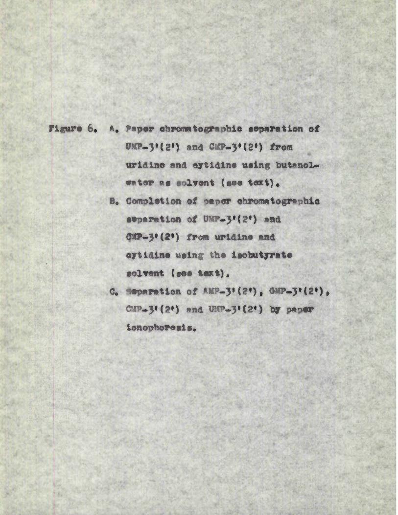
figure 6« K« Paper ehromatographio separation of UMP-3'(2*) and CSK-3«(2«) from uridine and oytldlna using butanol- water as aolvant (aaa taxt).
B. Cceroleaion of paper ohrormaoepnohla separation of U^^3,(2*) and O»-3»(2’) frcm uridine and eytidlne ualng the isobutyrate aolwent (aee text).
C reparation of AMP-3)(2’), 0M-3»(2«)t <C4P-3»(2*) and WM-3»<2») by paper lonophoresla.
I* e*
A B
a UMP-3'for2')’B,CMP-3(br2)© URIDINE O CYTIDINE
ORIGIN --- w
© UMP-3'(or2')• CMP-3'(or2’)
© URIDINE
© CYTIDINE
C
CMP-3'(or2’)
AMP-3'(or2')
GMP-3'(or2')
UMP-3’(or2‘)
ORIGIN
Directionof
Flow
Figure 6
74.
employed were leopron*nol/HCl (descending; 170ml, ifoprooRnol,
41ml. concentrated HCl nlus water to 250rnl.) (.vy«tt, 1951)
and butnnol/HHj (Mcendlng; lOOrnl. n-buts.no! s# turmted
with water «t room temoarm ture, nlus imL. 15N ammonium
hydroxide) (Mccutt, 195?.) The chrome to grams were run
in both directions meh th»t the solvent reached about
10on. from the end ( of the n*ner. The srem.s correanonding
to the bases (see Fig.7) were cut out after location in
ultraviolet light, and eluted by capmnry flow using u.H
HU for adenine, cytosine #nd urseil, #nd 1.6n HCl for
guanine. 0.5mL, eluates were collected in each cage and
0.2ml. samples of these taken for the rneesurement of radio
activity, or diluted for g^ectrophoto^etx^:tc exemim.tion as
the exioe^i^nent demanded.2.11 P»per ionoporo»is
For the isolation of - P-lebellM AIM-3# (or 2« , GM-J(
(or 2(, #?.)• (or 2») nnd UP-3( (or 2») from alkaline
hydrolysates of -labellM RNA the technique of oaper
ionoohoresis, as desoribed by Davidson and Smeelie (1952).
wag employed. A portion (0,5ml.) of the radioactive
hydrolysate was apolied in a narrow band of almost 3®-* wide
5em. from one end of a strip of nhetman 3:fM filter oap«r,
72om, x 7 cm. To the band of hydro lysate was added a mixture
of non-radioactive A«r_3» (or 2*) , GM-3»(or 2*) , JP-S#
(or 2#) ®nd U'?-3’ (or 2*) to aot es mrkers. T.>3 oeoer

4■'.. . • •*'-■■ ’"•• t X'/'T"'•■'•.’■.’ - ■■ ' ■ ” M ' • ’/ '■& i
' " • ' A-’. • •' ’ .' ”< J».V
? Jr-■,*>'•■• .jP
/S$f J
i •:• V
■ -*■ < •!?'
• >
-4.
'-■• >L ’ '•■ J*: Zl."'"^ • JA( - *?■• ' '■^2* •’’ "
Iti* -■ :.-J >•.*Figure 7. Paper chromatogranhla separation of the RNA bases adenine, guanine, uraoil and ejrtoslne.
r m ’• cvZ. ' •>•■■’’■ x -•*/ - •> ‘ '??i ■ s

Rf in n-butanol/NH3o o oK> > d>
i---- 1------ I---------- 1---I--------- 1--- r
Odo1----- r
# 0_ ro □ '(A0"J0
T>Qj□0
o
oO'
Xn o
CD
Qj
G
ljure nJL
75
was then moistened with 0.021! citric acid - trisodium
citrate buffer at oH }.5, and suspended over a glass rod so
that the two ends were Vnersed to a depth of lcm. in two
generate glass dishes containing the sane buffer. Carbon
electrodes! placed in each dish, were connected to a souroe
of direct current so that the anode was in the vessel
further away from the soot on the oaper. Complete sep
aration of adenylic aci > cytidylic acid, uanylic acid
and uridylio acid was obtained by running at a potential grad-
lent of llv/em. for 18 hours, the current flowing being about
0.5 to l.Oma./cm.
On completion of the run the paper was dried by
'orsure to a pair of 25OW. 125V. infra-red lamps. Spots
were located in a Hanovla chromatolite, and aopewred as
indicated in Fig.6c. Once the soots were located, they
were cut out and eluted with water by capillary flow. 0.
of eluate being collected. 0.2ml. samples were used for
the measurement of radioactivity.
2.12 'Estimations.
(a) Rroteln.
Sattmatlon of orotein was oarried out by the method
of Lewry, Roaebrough, Parr »nd Randall (1951). Thia
involves the uae of a modified type of biuret reagent and
the method ia in fact a modification of one first introduced
by WU (1922). The reagents used were ag follows :

76.
* s sodlun onrconwte in o.lH sodlun hydroxide.
B : 0.54 cooper sulphate in sodium or potassium
tartrate. ‘ 5;v. 2 ' *
C : 50m1. of 4 mixed with 1m3L. of R.
D s Folin-Ciocalteau reagent (Folin and Clocalteau,
1927’^. v
The Folin-Ciocalteau reagent obtained from BD. H. was
titrated with sodium hydroxide to a oheiolohthalein end
point. On the basis of this titiation the Folin reagent
was diluted to mike it IN with resoect to acid. The
standard used was bovine serum albumin (Mmour Fhaxmaeeut-
icals, Eastbourne).
To a sample of 5 to 400 fig. of orotein in 1mL. of
solution was added 5ml. of reagent C. The solution was
mixed well, and allowed to stand for 10 minutes- at room
temownture. C.5ml. of reagent D was added very quickly and
mixed within f few seconds. After 50 minutes the colour
intensity of the sample was read in a spectrophotometer. For
solutions containing low amounts of protein it was desirable
to take readings at 750m*. , the absorption maximum. For
stronger solutions, readings were taken nt 5OOom., which
allows a greater working range.
(b), RNA. . - / . — ■ /• < " . :•
RNA w*s determined 8oectroihot#netrically on the
ion that a solution of RNA containing lpg. RNA phosphorus
per ml. would give an extinction of 0.285 at 268m*. in a

77.
lem. cell (Paul, unpublished results)
RNA wao also estimated by the orcinol reaction for
pentose (Kerr and seraidarian, 1945). The orcinol reagent,
which mist be made up just prior to use, contained O.bmg.
orcinol dissolved in lOOml, 0.02* (w/v) FeClj.&H'C in con
centrated HCl. 3ml. of ribose containing solution (in the
case of RNA, containing in the region of 1 - 6 pg. RNA
phosphornnv/mL*) were treated with 3mL. of fresh orcinol
reagent in a. clean pyrex test tube. This mixture was then
placed in a boiling wter bath for 30 minutes, cooled rapidly,
and the colour intensity measured at 66Fpm. A blank was
nrenared similarly using distilled water in place of RJA
hydrolysate. As standard, a solution of ribose, containing
lopg. ner mi. was oreoarei and this wag equivalent to 4.13pg.
RN A oho gohoru s/ml.
(o) DA
DNA was d^te^ined spectrophotornetrically on the
assumption that a solution of WA containing Ipg. DNA phos
phorus ner ml. would give an extinction of 0,230 at 2b8mj in a lorn, cell (Ceeiotti, 1955)
DNA was also estimated quantltttivtly using the indole
reaction of Ceeiotti (1952, 1955). To 2ml. of a solution
containing from 2.5 to 15pg. DNA per ml. waa added 1mi. of
O.O4X (w/v) indole solution and 1mL. concentrated HCl. The
tubes ^ere then immersed in boiling water for 10 minutes

78
and then cooled under running t«n water. The cooled
solutions were then extracted three times with 4»l. portions
of chloroform, the chloroform extract being discarded each
time. The yellow colour remaining in the aqueous ohase
was due to the DMA pigment and was measured in a speetri-
photometerl*aginst a. blank, containing water instead of DMA
solution, prepared in a similar manner. standard DHA sol
utions were prepared in the department from calf thymus DMA.
(d) morgens . ,».na ornnUPhosphorus was estimated by a mooification of the method
of Allen (1940). The first step in this procedure was to
convert all phosphorus to inorganic orthophosphate by
digestion with sulohuric aold and hydrogen peroxide. However,
when en estimate of inorganic phosphate alone was required,
this digestion steo was omitted, although sulphuric acid
was routinely added.
A s»mole of the solution to be assayed containing 4 -
30pg, phosohorus was pipetted into a pyrex tube, and 0.24ml.
of lON sulphuric acid were added. The resulting solution
was heated in a sand bath at about 130° until the volume had
drooped to around 0.1ml. The solution was cooled and 2 drops
of hydrogen peroxide (H.Mm loo vols.) were added. The
solution was heated for a further 30 minutes after which it
was cooled and a small portion of water, 2 - 3°1. » was
added. lf the experiment only demanded the estimation of

79.
inorganic nhoanhorua in the solution, it was necessary only
to add Q.24rtl.lQN sulphuric acid to the solution, no subseq
uent digestion being required.0.2ml. 8. V (w/w) solution of ammonium molybdate and
0.4ml. 1/ (w/v) solution of 2:4 diamino-phenol hydrochloride
(Analsr) in 20/ (w/v) sodium meeabisulohite solution were
added, and the mixture allowed to stand for H minutes at room tempwrature before the ootical density was mmeaured at
640rnp, against a reagent blank. A standard phosphorus
solution was prepared by dilution of a stock phosphate
standard (Analar ; 2.19%. n^*>PO^ in 500ml. water, i.e, lm.g
lmg. *)•
(e) RNA. bases.After isolation by chromatography (see 2.lOd) and
elution from the paper in dilute HCC, samples of the eluate
wwe diluted with HC of the appropriate concentration for
spectroscopic estimation.
Adenine ms estimated in O.1N HC at 262mfi and the
m>l*r extinction coefficient used 13,'OQO.
UraodiL was estimated in O.M HC at 260mp and the
molar extinction coefficient used 8,200,
Ctosine was estjLmated in O.1N HC at 275wp and the
mlar extinction coefficient used 10,400.
Specifications and Criteria for Bilchemical compounds, I960)

8o,
2.13 Enzyme assays.(a) °Tponuclea_ se.
A rough ^thod for the assay of rilonuclea.se was de
vised which depended on the degradation of yeast RNA
(Nutritional Biochemieals) to acid-soluble fragments which
are then assayed speQtroohotcp^etrioally. A solution of
yeast RNA (2ng. per ml. of O.O1M Na(d) was prepared and
dialysed for 24 hours against a large excess of O.O1M NaCl.
The reaction mixture (1,0ml) containing 0.25 mi. of this
RNA solution, 0,15ml. IM Tris buffer of the appropriate pH
value and 0.6 ml. of the enzyme fraction to be assayed was
incubated for 30 minutes at 37°. 0.5ml. of ice-cold IN
perchloric acid was added to precipitate nucleic acid and
proteins, which were removed by centrifugation in the cold.
The suownata.nt fluid was diluted with 5 vol. distilled water
and the optical density at 260 mp determined.
(b) Pho so^tase,
The assay depends on the estimation of the phosphatase
catalysed release of inorganic phosphate from a solution of
ribonucleoside triphosphates. The reaction mixtures (1.0)
contained Ijrmole each of UTR, CTR, AT? and OTP, 2ynoles
MgOLg, 0,6^^, of the enzyme fraction to be assayed and was
O.1M with respect to Tris buffer of the appropriate pH,
Incubation was carried out «t 37® for 30 minutes after which
0.5ml, IN perchloric was added. The precipitated nucleic

81.
sold and proteins were removed by centrifugation. The in
organic phosphate present in the supernatant fluid was
measured as in 2.13°, giving an estimate of phosphatase
activity.
2.14. Meaqureemn* of radioactivity.
Except in one specific case, measurements of radio
activity were carried out using a Nuclear Chicago scaling
unit and automatic sa ' ole changer fitted to a windowless
gas flow counter operating in the Geiger region.
The counting of 3h and was carried out on samples
which had been plated, with drying under infra-red lamps,
on 1.25* diameter aluminitm planchettes which were first
thoroughly cleaned by rubbing with "Ajax" cleaner. The
mjor difficulty in counting and is that of self-(
absoimtion. rreimminary cxoerments indioatdd that3
counting of H samples at infintae Hhiknness coudd give
rise to very largo errors due to non-uniform distribution
of maaerial on the plan^ettes. At the same time, it was3
almost impossible to achieve infinitely thin containing
samples. However, by serial dilution it proved possible
to attain samples that approached infinite thinness and
this technique was adopted for and containing sampLesu
As a further safeguard all experiments were carried out in
duplicate or triplicate, repeat dilutions and counts always
being perfoxmed where any serious disagreement between

82.
dunlieatfs was observed. The greatest osre had to be
taken to remove as much potassiim perchlorate as possible
from solution before oreparin? the samples on planohettes
for counting. Under these conditions an efficiency for^H of 34X was achieved. For ^C. the effiee^c/ achieved
was somewhat greater, being in the region of 40>. In all
cases, at least 1000 counts were collected.
For the counting of the same procedure was adopted
although the oroblema of self-absorption and efficiency were
lest critical. Wien the samoles were dlasoXved in O.N
NaGH^stainless steel planchettes w^e used, and even spreading
was achieved with the use of 1" diameter discs of lens 32tissue. ? labelled, trichloroacetic acid wished, filter
paper discs were placed on clean stainless plan^ettes prior
to counting.
In one particular series of experiments where it was
necessary to measure very low levels of in aMA samples from
m»nuoleoproteiis of ascites cell nuclei (2.9a) a Nuclear
Chicago Model 6000 ( 50 cycle) Dynacon Electrometer was
employed. This is a dynamic condenser electrometer de
signed for precise measurement of srmai ion currents origin^
a ting in ionisation chamtber. Men^^Wiomeit of rrmdi dLoa cit XviZy
was carried out on a rate of change basis. For atinfinite thinness the ionisation chamber constant was 4*1<j^
amperees/iuo -fc. and each sample was measured at least six

83.
for maximum attu^ney.
2. 15 'iloroautooadlogrnphy.After intmot ascites cells had been a lowed to incor
porate ^^uridine over p period of 2 minutes (see 2.9a), a
sample of cells was withdrawn for sutoradiogrsphy. The
cell suspension was treated with an equal volume of s
mixture of ice-cold dM perchloric soid - absolute ethanol
(1t1). After 10 minutes treatment with this fixative in
the cold, the cells were wa shed successively with 7oX
ethanol, 50° ethanol, 2^ ethanol and water. This ^s
followed by two 3 minute extractions with ica-cold 0.7N
perchloric soid to remove any radioactive acid-soluble
mterlal. The cells ^re further washed with 25° ethanol,
50° ethanol and JO ethanol st which stage they could be
conveniently stored.
The next step was to embed the cells in paraffin wax
prior to sectioning. Thus it was necessary to dehydrate theIcells further in order to infiltrate them with paraffin wax.
The suspension of cells in 70# ethanol was centrifuged and
re suspended in 95^- ethanol, followed by absolute ethanol,
absolute ethanol-xylene (3j1), absolute ethanol-xylene (1:1),
absolute ethanol-xylene (1:3) *flM then xylene alone. The
cell susotnailn in xylene wap allowed to warm in an oven st o
61 and then an equal volume of mooten, filtered paraffin wax
(M.°.58°) was added. The cells were allowed to sit until
the paraffin w«x (which partially tlliaifie8) ro-melts.

84.
Then the oells were mixed «e thoroughly *s possible in the
paraffin wax - xylene mixture #t 61°. A convenient tech
nique wms to guck up the olemr mixture on top with e
previously hestod Pasteur oipette, and quickly eject this
back into the tube, thus mixing the cells which have settled
to the bottom. It is imMo»tant, frcm the point of view
of suspending cells during paraffin wax infiltratoon to use
tubes which, although conic*!, are not too sharply pointed
st the bottom. The cellg were then left overnight mt 61®
in the oaraffin wax - xylene mixture. On the following
day the oaraffin wax - xylene was removed from the cells
which had settled to the bottom, and fresh paraffin wtax
added. [muing the day the paraffin wax was ihai{tsd twice
more. Finally when the cells Had settled to % pellet at
the bottom of the tube the emtb>dding was completed by re
moving the tube and placing in an ice-batH. The em1b2ii^d
cells were tH . sectioned. 3p sections wnre taken which
gave 50-bOj compassion (thinner sections gave even greater
ciroieasion.),
THe glass slides on which these sections were mounted
w^e "subbed* to ensure good wet adhesion of the film
eLailsion when this was applied. dean glass slides were
dipped in m 0.5° (w/v) solution of gelatine containing 0.05/
(w/v) of chrome alum. THe slides were allowed to dry
after which they were resdy for use.

85.
The section# prepared from the imbedded ceils were
then mounted on the "subbed" sides end washed successively
with xylene, xylene - absolute ethanol (3*1) • xylene -
absolute ethanol (1:1) xylene - absolute ethanol (1:3),
absolute ethanol, boiling absolute ethanol - ether (3:1)
for 5 minutes to remove absolute ethanol, 95# ethanol,
70$ ethanol, 50$ ethanol, 35/ ethanol, water, ice-cold 0.7H
perchloric acid for 3 °inutes, and finally twice with wile?.
The sectioned cells were then covered with AR 10 autoradio
graphic stripping fim (Kodak l*td., Londonn, by the light
of a erat ten Series 1 red safelight.
Using « sharp scalpel blade, a out was made in the
amllsiLi layer of the AR 10 elate around an area sufficient
to cover the whole of a snodmen with n margin of at least
a quart t of an inch all round. The slide bearing the
sectioned cells was then placed in a bath of distilled
water at least one inch deep. with the tip of the scaloel
blade, the emulsion layer was stripped from the plate,
turned over, and thrown on to the surface of the water so
that the sensitive side of the emulsion layer was facing
downwards. The ^mission sweia, crumples and then
stretches out tight and flat. It was left for two minutes
more and lifted frcm the water by raising the slide mder- oneath it. By holding the slide at 30 from the horizontal
one edge touched the emjlsion first and the emulsion then

86
droned Itself snugly over the specimen »nd most of the
water drained away *s the slide ^s gradually lifted olear
of the water. The cell sections with su-°r-
imposed were then dried overnight in a drying box. Once
dry, the slides were placed in a black light-tight box
containing silica gel as desiccant. Before exposure at
3 - 5 (in refrigerator) this box was carefully sealed in
several layers of thl^k paper.
After an exposure time of 2-3 weeks, the slides were
then processed with the emulsion layer in permanent con
tact with the specimen. After letting the slides come
to room temperature they wiere allowed to develop in un
diluted D.19b Kodak developer slides) for 6
minutes at 18° with the ^inmu^m of red light, The slides
were then rinsed in tap wnter for yo seconds and treated
with Kodak acid fixer (diluted 1 part to 2 parts with
water) for 8 minutes at 18 , After rinsing in tap water
for 2 hours, they were finally washed with distilled water.
However before mouuting th a spec^en it was necessary to
dehydrate the film. This was achieved by treating the
slide successively with 50/ ethanol, absolute ethanol,
absolute - ethanol - xylene (1:1), absolute ethanol -
xylene (1:3) xylene. The cell sections and super imposed
emulsion were then mounted using Dernx mouttitg m&itm.
(G.T.Gurr Ltd., London).

87.
2.16 M»tevl*lr.
Nominialy 5,6 - Jh - uridine of «pe<iifli aotlvlty
6?}iC/pLmoe, and carrier free digodium hydrogen phosphate
(T’PS.l>1.5ml. and 25«C) of specific activity greater than
lmC/mg. were niroHurod from the R*dtoiHemi<i»l Cer^re,
AMe'shamt
2»,3° iBO'prilyliiene uridine and 2*,p_lBopripyliaeie
adenosine 'were obtained from the AldrlcH Chemo*l Co, Inc#;
NAD, HAD*, ’lADHg from C. F, nieHringer und Boehne ; uridine,
cytidine W°-y<2«), CKV.-3>(2») AM-3’(?»), W-JIU#),
crystalline DN*ase, crystalline RN*se an'i yeast RNA from
Nutritlonal Blochernc*!* Corpoi*tlin. Other ri’Ciiiilei-
slde 5t-iHo^e:PH»tes were obtained
Company or °abst Laboratories.
from the Sigma Chemical


88
3«1 Itlolmoration of °H-uridine Into RNA by lytoplaatic extracts of ascites caroinona cells.________________The incorporation of ^H-uridine into the RNA of intact
E^h^^ch ascites cells On vitro Os illustrated On Table ii.
Oisnotic disrnotOon of the cells produced a preparation which
showed diminished incorporation, but the ability of thOs
preparation to incorporate urOdOne was found to be orkedly
Oncrea.sed by supplement*! Oon of the incubation medium with
AT?, opgiesiuo ions, NAo and glucose. High speed centrOfug-ation of the oarnooteally disrupted cell suspensions gave
rise to a crude cytoplasmic extract which when supplemented
as above readily incorporated labelled uridine into RNA and
a sediment which showed little activity in this respeot,
ExamOnation of the acid-soluble fractions showed that, with
the exception of the 105,000 x g sediment, all the fractions
shown in "*ble ii were capable of phosphoryla ting uridine
to IJVP, UDP and UTP. The 105,000 x g sediment appeared
to lack the kinases required for this process.
These preliminary results were obtained using a reaction
medium buffered with O.LM Tris nH7.9. A series of incubation
mixtures were set uo to determine the optimum pH for the in
corporation of uridine into RNA and the results shown in Fig,
8 indicate an optimal pH of 7«9« Aiwaysis of the acid-
soluble fractions from incubations carried out at nHb.0
indicated that there was little phosphoryl*lion of uridine:
at pH 7.9 however there was extensive conversion to U.1P, UDP

Table II
Incorporation of r^-uridlne into the RNA of
fractions of ShrlicH a motes iarcinamu cells
Fraction RHA specific activity (Couni9/iin./piole R*JA-^)
Intact ascites cells 5b,boo
O <e^iiie*,lly disrupted ascites 5,700cell preparation
O!UMiLlilly disrupted cell 26,000preparation nlus ,NAD «nd glucose.
Cytoplasmic extract (105,00Cxg 20,600su:l<vrn«atunt fraction from disrupted cell ) plus ATp, Mg4*,NAD and glucose.
sediment (10S,000xr) plus rT*, 1,900Mg44, M*d and glucose.
THe reaction mixtures were O.1M with renoed to
Tris buffer »t pH 7*9, «nd contained whiere indicated
Spiolrs AT^/ml., Fpnoles MegC^/nme, lpmole Nja. ,3
gp-moes glucose/ml. and Jpc H-uridine/uL. Incubation
time was 2 hr.

figure 8, The effect of variation in pH on the incorporation of i--uridine into RNA by oytopleemio
extraote of 'Eirlieh "eo^tea oarolnore celle,
end on the dlstrltuition of radioactivity in
the acid-soluble fractions*
The medium was 0.1 M with respect to tris
buffer end contained 5 jamOes ATT/zm., 5
)rnmOes Mg/l^/nl.. 1 ^ole NAD/mL., 5 jwmOss glucose/mu and 3 jxC ^Huridine/mL.
Incubation tire t 2 hours.
RNA specific aotivity expressed as counter in./
pmole RHAup x 10~\

Figure, 8

89.
•nd ur* (rig.8).
St«>mrwtlon of the other aofnotore required showed that,fV '■ , 5 * v4- "AT? is required in oonasutrrtions of at least ^urnles per mi. (fig.9). In the absenoe of ATP negligible smounts of
uridine were phosnhorylnted nnd there wes m corresponding
reduction In the amount of yHuuidlne ineorporsted into -'the■jr vBANA.
The effect of inor^^te eat ions is shown in Fig.lo. - The only eombiirntion mrc effective than rngnesiim ions
alone ws ragnesimm-.ions plus potassium ions. » All other
aomiinatlcns were less effeatlva e socially those entwining
ealeiim ions which were sonewhst inhibitory.
If the ineorpoir*tion of uridine into RNA by these .i.,
eytopls sale ext mots in wives the addition of UDP to sn
existing polyribonucleotide with the concomitant liberation
of inorganic orthophophmte, the cddition of -inorganic
orthophosphate to the reaction medium should decrees the
reaction. Similarly Inorganic pyrophoaohtte might be
expected to inhibit any reaction involving the addition of
UTP with the release of inorganic pyropho apthte. Tie effect
of adding inorganic ortho-or pyrophosphate to ths incubation
mixture (Tlg.H) wi to inhibit uridine incorporation into
RBA. Toother inorganic ortho- and pyroohosphate had an
even greater inhibitory effect on this reaction but either
singly or together they had little influence on the

% Th* effect of varying concentrations of ATP on the incorporation of ^Hurldlne Into RNA
by cytoplasmic extraot* of -hhlich esclte* cardnioe cells, end on th# distribution of radioactivity in the scld-*duble fraction*.
The medium contained 5 pedes VgCl^nO.,1 pnole MD/ra^, , 5 yoole* gluooMVn..,3 pc ^HuuUlne/ol. and wns 0.1 M with
respect to tris buffer at pH 7.9.Incubation tins • 2 hour*. RND specific activity expressed as counts/ltlt./ptdo RMA-p x 10"\
r*

RNAQ/o
Rad
ioac
tivity
R
NA s
peci
fic a
ctiv
ity
0 I-------------- 1-------------- 1 1------------- 1------------ 10 1 2 3 4 5
yu moles ATP/ml
No ATP
Jiaure

Fleur® 10, Th® cffcot of different lone on the Incorporation of \-urldlnc Into RMA hy cy topic eHe
extraote of 3to*lloh cccltcc ecrelnuc cells*
The Ulum was 0*1 * with respect to trie
buffer at pH 7.9 end contained $ proles AAT/nl., lpmole NAD/tnl*, 5pmil«c glueoee/ml. end 3 pC 'n-urldlnc/hil. MgCC_, KCl, MnClg
end Cadg when added were et e lonoenlratlln of 5 junol^Sy^mn. XnautaCion time t 2 hour®*HHA lrttelfle cotiTlty expressed os counta/mln./
pmolc RHA-p x 10”-)*
• •

0

RNA
Pl two 11. Th# effect of addition of lnorganio orth>• <» ■or oyrophoaTpMte on the lnooroowtlon of
/H-uriaine into RNA by eytoolarnlo extreota
of Swlleh eeoitee oarelntme oella.
s"-'"' The mllum wob 0.1 M with reepeot to trie
buffer at pH 7*9 end oontained AT?*, MgClg, KCl end glueoae eeeh et e Gonco-i tra tion of
5 umlae/h,. , 1 jr-wle NAD/nQ., ) juc ^-uuiddne/m.. and where lndLeat^d lnorBenie
ortho- end pynphoaptMte et a oonerntirttion
of 20

Total counts/min incorporated x 1O"3C
ontrol

90
phosphoryl"tton of uridine to UD? or UTP.
Fig.12 illustrates the possible locnlisstion ofs J.’*’!*1"'•' ’*• -k. <,
in RNA labelled in these experiments either (s) in UMU'rife'
moieties incorporated terminally, so thmt slkaline hydroly
sis (indicated by the broken Unee) would give rise to ?n-
uridine or (b) in UN? mooeties located in non^tejminei
positions so th#t slkmline treatment would yield 3*
(or 2*). The possibility mist slso be oonsidered th«t
during incubetlon some uridine is eonwwrted to oytidine
derivatives (Lieberman, 19S5| 1956; -uulbiert snd Karnnen;
I960) in which os se would else be found in the CM
moieties of the pol nucleotide ohm in attached terminally(c) or (d) end would give rise to \-labelled
oytidine end CMP-3’(or ?•) resneotively on slkall^ne hydroly
sis.
Table III shows the distribution of radiiwitivity between
the ribonucleoslde end ribonucleotide fractions formed during elkellne hy<d*olysis of the RNA. ^r-uridine wes incirporcted
into both terminal »nd .norn-terra insl positions but occupied
nrlncipally a nom-termlnal oosition. \ In the presence of
pyrophesobwte the iroportion of totsl RRA rsdii-
sctivity mowed ss UU.}’ (or 2*) was greatly incrcssed
indiostin^ that the terminal iniorTioration of uridine had
been greatly depressed. Inorganic orthophocphcte seemed to
h?we sn opposite effect; since s higher proportion of the

figure 12. Possible loo*Hone of *R In Wl eater incub- etlon with ^-uridine (see text).
0 end 0 represent wmell and cytoelne respectively, and B any other ribonueleotide teae.The vertlo*l lines represent ribose moetles and the oblique lines, p-M pht^is^Pwidi^s^^er lidkages.

U*R f nBMP-3'(2')
C*R + nBMP-3'(2)
I

BR* nBMP-3 (2') + U*MP-3'(2)
B B B C* B
1
BR * nBMP-3'(2') f C*MP-3'(2')

III■“ “ '“r
The effect of addition of orthcw and pymhosohh’te on
the localisation of in alkaline hydrolysate of RNA.
■aw
A^d^lltions Er cant total JM-ll radioactivity
Uridine Cy tt ldlne irr^-^» c5®^-3‘ (or 2»)(or 2*)
Nil 24 6 56 14
Inorganic ^yroihocihate 12a
0 82 6
Inorganic irthonhocih« te 65 13 12 10 St
rTr T1 •
Incubation con<!lit1iis sane «s those for Flg.11.
Incubation time s 2 hours.

91
activity wag recovered as uridine, inhibition of non-terminal
incorporation must have occurred. Since approximately 20/
of the total radioactivity of RNA w*g recovered ag oytidine
or CM-ytor 2*, appreciable conversion of uridine to
oytidine derivatives took ol»oe during incubation.
The inclusion of nom-radio-active UUP or UTP in the
incubation mixtures at concentrations of ipmole per ml. in
an attempt to dilute the pool of RNA precursors led to
enhanced un take of uridine int a the RNA, while together they
had little effect (Table iV). Since the systrn under investig
ation ^s nrobably quite complex, the*e observations could be
due to the sixes of the UUP and UT® oools influencing the
ultimate incorporation into RNA, or to some coenxyaic role
of the uridine nucleotides. fo investigate the former
possibility, experiments were set up with varying concen
trations of added UD* or UTP. The results are illustrated
in Fig.l) and show that at concentrations above 2urnoles perm. , UDP and UTP diluted the incorporation of ^H-urldine into
•P: ■ .#RNA but enhanced the uptake at concentrations, having
optimal effect at ipmole ner ml. in view of the fact that
uridine nucleotides might be coneerned with the avail#oility
of the various forms of pyridine nucleotides, experiments were
carried out in which the requirements for glucose, NAU,
NADH2, NAD°, UDP and UTP were followed. From the results
presented in Table V, it was clear that the optimal inoorpontion

Table IV
The effect of UD? and UT^ onthe incorporation of ^H-uridine
into RMA by cytoplasmic extracts of Srlich ascites
carcinoma cells*
Additions (1 fraooe/ol. of each) RNa specific activity (counts/oin./uoole RN’)
wn 19.000
UD/ 31.500 aa ' . s ./
UTP 36,000
UD/ nnd UT/ 15,000
The reaction mixtures were O.1M with respect to
Tris buffer at pH 7.9 and contained 5 /ooolae ATT//O. ,
5 pnolea WgC./^O. , 5 /rodes KCl/O., 1 fxmle N-A/zO. , 3
5 pmoles glrcose/ol. and 3 pc x-^rr^idn^^^ol*
Incrb*tion tOe ; 2 hours.

figure 13. The effect of added UDP end UTP on the uptake of g-uuldlne into RNA by e’ytonlaarnlc
eotraota of Stolich eacltea cerelncmn oella.
The meilun wee 0.1 M with reepeet to trie••• *. " • ’ ■ •
buffer et pH 7.9 end contained AT,
XC1 end glueoee each et e concentration of
5 pmole^/m., 1 /mole MAD/rnl., and 3 pC gHur ldlne/m..
incubation time s 2 hr. RN* specific
activity expreaaed et counte/raln.Dimole
RNDP x 10“T,
•'

RNA
o oO|------------------------r
yu m
ole s/m
l
U)

specific activity
o o>o

Tmble V
The effect of glucose, NAD, NADHg, MADP *nd UTP on 3
the inooroorAtion of ^H-uridlne into RNA by cytool«smic
extracts of %rlich Recites carcioona cells.
Additions RNA soecific activity ( count e/min./pinole RnA—p)
Nil 8,500- ’
glucose 11,000
NAD 18,500
glucose olus NAD 20,000
glucose olus UTP lb,500
NAD olus UTP 28,000
UTP olus NAD plus glucose 39,600
nadh_ 27,000
NA0P 30,000
NADP plus nadh 57,700
The reaction media were O.PM with respect to tris
buffer «t oH 7*9 ond contained 5 /im^^ea ATP//Ql, 5 jumlea M^CC^mL. , 5 /rnolea KCl/ml. ond 3 pC /--uridine/ml., and
alao where indicated 5 /mdes glnuse/ml. , 1 pmole
NAd/oI. , 1 pmole UTP/ml,, 1 jum>le MADD^/ral, and 1 pmole
NADD/ml, incubation time ; 2 hours.

92.
of ^H-uridine into RNA required either m mixture of glucose,
NAD and UTP or an equimolar mixture of NADP and NADHp. in
the orfeenoe of these two substances, UDP or UTP added to the
reaction mixture at con^c?r^tratl^ons of ipmole per ml, depressed
the incorporation of uridine into RNA (Table VI),
Annlysis of the nroducts of alkaline hydrolysis of
RNA showed that the increased incorporation of uridine
obtained in the presence of NADP and NADH^ was due to an increase in the amount of ^H-uridine attached terminally
to the RNA (T«ble Vli) and investigation of the acid-soluble
(Table Vlli) demonntrated that NADP and NAjWp promoted the formation of larrer amounts of UTP from ^H-uridine.
if the incorporation of "H-uurdine represents true
synthesis of RNA, it mist be assumed that the other three
ribonucleotides are present in the tissue extract or can be
formed during the incubation. in either event, the
avaliability of these might limit the extent of the reaction.
As might be expected, therefore, supDlement*tion of the
reaction mixture with low concentrations of ADI?, GDP and
CDP, or of CTP and CTP, stimulated the incorporation of ^H-uridlne (Table iX).
Wile the crude oytonlaRmic extracts contained apprec
iable quan'ities of RNA, the addition of further amounts of
RNA to incubation mixtures stimulated the uptake of uridine
(Fig,14). This was particularly true of the RNA (microsomal

Table vi
The effect of UDn »nd IFT on the uotnke of ^H-uridine
into RNA, by oytopl«Riic extracts of Tirlioh ascites
carcinoma cell' in the ore sen ce of NADHo and NAD?.c
Additions (1 jimmle/il. of each) RNA soeciflc activity ( count s/n in. /pmole RNA ——
Nil 23,500
UD? 17,500
UTP 15,000
UDP olus UT 10,500
The reaction mixtures were O»1M with respect to
tris buffer at pH 7*9 and contained 5 pmolee each of
AT', tfftfJlg and KCl/mii , 1 pmole each of andNAD/ml. «nd 3 pcXC-uuidine/ml.
Incubation time ; 2 hours.

T«ble VII
The effect of NADHg and NADP on the lloo1itati<m of
5h in alkaline hydrolyg*te9 of RNA,
Additions .. Per cent totalRNA r*iioootiviiy..........uridine oytidine U’P-ybr 2') C%-3’(or 2')
Nil 4; 28 ,,, 2 . 66 4
1 LIJeols each of NAdHp and NA.DP * 48 2 48 2
incubation conditions same as in T*ble V,

T*ble VUi
The effect of NAD? and NADH^ on the distribution of
radioactivity in the uridine derivatives of the acid-
soluble fraotl^on after incomor^tlon of ^H-uridine into
RNA by cytool^aimic extracts of 3hrlich ascites carcinoma
cells.
Additions Per cent of »c id-soluble rsAioeottvitJUridine UUP UDP UTP
Nil 6 10 26 58
1 ummle each of NADH and NADP
2 3 2 15 80
incubation conditions same as in T*ble V.

Table IX
The effect of adding di- and trinhoeohates of adenosine, guanosine and cytosine, on the incorporation of ^H-uuidine
into RNA by cytoplasmic extracts of ihhrich ascites
car^ma cells.
Additions RNA specKlo nctivity( count/min./pmols RMA—/
Ml 20,500
AD?, GH/ «nd CH" 33.5CCOT? md CTP 43,ccc
AD?, OH?, CIH», GT/ «nd CT/ 27,000
The reaction mixtures were C.lf with resoeot to tris
buffer at p- 7,9 and contained AA7\ MgClg, KCl each at a
concentration of 5 pmole/rnl., also 1 jirmooe MfcDP/nQ., 1
pmole NADH—/ml., 1 pmole 2-mwcaptoethanol/ll. and 3 PC ^-uridine/H. where indicated the ribonucleoside di- and
triphosphates were present «t a concentration of 1 jimole/ml.
Incubation time % 2 hours.

Figure 14# The effect of adding varying amounts of
ascites RNA on the incorporation of
^H-uridine into RHA by cytonlasmlo extract#
of %rllch esoltes carcinoma cells.
The medium was 0.1 with respect to tris
buffer at pH 7»9 and contained ATP,
«gCl_, KCL and glucose each at a concent
ration of 5 pm>le s/mli, 1 pmole NAc/na. and 3 pC -Suuridlne/rnl.
Incubation ttoe t 2 hrs.

UUJDZ>3s
Total count^/mm. incorporated xio-3CM -h Lno o o
/O5,O
OQ
*y S<zd/m
<zr)t BN
A
ure 14

9 ).mnd nuclear) nrepared from the 105,000yg sediment of the
lsmililMl1y disrupted ascites cells; the addition RNA ore-
pared froto the cell s°o b°d a similar but less ^m^nced
effect.
Dialysis of the crude cytoplasmic extracts against
O.O1T4 Tris buffer o° 7.9 for l8hr, at 4° had little influence
on their ability to catalyse the Incorporation of uridine
into Hn/. °rior incubation of the extracts at 37° for 3°
minutes was inhibitory (Table X),3.2 Incorporation of °H«-uridine into RNA by cytoplasmic
extracts of lmmtur5 rabbit tissues. ,,
The results described so far have been obtained with
cytoplasmic extracts of ascites cells. Cytoplasmic
extracts, orepared in a similar fashion from a series of
iramaure rabbit tissues, showed considerable variation in
the ability to catalyse the incorporation of uridine into
RNA (Trble XI). In most tissues the r«tio of uridineterminal
incorporated in a/positlon to that in a non-terminal
location was very low (about 0.1), but in brain and muscle
a very much higher oroportion of terminal addition was
observed (Table XII).
3.3. Synthesis of polyribonucleotide msaerial by cytll1ftsmilextracts of ascites carcinoma ^lli.
ihidence that cytoplasmic extracts of ascites cells
are capable of net synthesis of smld amounts of polyribo
nucleotide mmaerial is presented in Table XIII. In the

Table X
The effect of dialysis and of preinculontion at 37° for 30 mn, on the incorporation of ^H-urLdlna into by
cytoolasmio extracts of %hrlich ascites tumour cells.
Type of extract /NA specific activity (count s/lin./pioles /NA-.?)
norul 33.600dtalyR«d 32,100
prelnor'oated 7.600
The reaction mixtures were 0.1 M with respect to tris
buffer at pH 7*9 and contained 5 pmoOes ATT/rnl., 5 pules
Mcgll/nml, 5 pules FCl/H., 1 umile NADD/hiIl » 1 piole NADH—gml.| 1 uiile 3-iercaptoethanol/il. and 3 pl ^H-uridine/
u..
Incubtion tie : 2 hours.

T*ble XI
The incorporation of uridine into RNA by cytoplaemic
extract® prepared from innmture rabbit tissue®.
lytoplaanlc extraot oounts/min. 1noorporatni/ag. protein
thymus 14*000
spleen ■ 13,000
tor» in 10,800
Intestine 8,500
teste® 7,800
tone rwrrow 6,500 ,
anpendix 5,200
liver 4,200
kidney 4,000
mi sole 1,600
The reaction mixtures were 0.1M with resoect to tris
buffer et pH 7.9 and contained 5 /moles ATP/zq. , 5 /imoles
MgClg/naL. > 5 pmole® KCl/m., 1 pmole NAD, 5 pmole® glucose/ mLe , 1 pmole 2-mercaptoethenol/ml. and J pl^H-uridine/ml.
Incubation time t 2 hours.

Table XII
The loo«lifc«tion of Io alkaline hydrolysates of RNA frem
lytopl*8mic extracts of various rabbit tissues after Incor- 3poratim of xH-uridioe.
Cyt oplssmic Percent total RflA radioactivityextract Uridine Cytidine BUP-J’ OJP-}1
(or 2’) (or 2*
thymus 13 9 67 11mUeen 5 4 68 23brain 27 23 27 23
-intestine 9 2 81 8testes 5 5 77 13bone marrow 8 U 63 15appendix 12 6 72 10liver 6 3 82 9kidney 11 7 79 3muscle 54 10 25 11
Incubation condittoos same «s for Table XI.

Tmie_XH
Synthesis of oolynudeotide by cytoplasmio extraots of
Whlioh Ascites oArd-norns cells.
Additions R^/reactlon vessel (mr)
nil (control) jO.b
AID, GDP, U/P «nd Cl/ 34.3
ACT, GT», UTP «nd Cl7 38.9
The reaction mixtures were 0.1M with respect to tris
buffer at pH 7*9 and contained 5 jimooes Megll/ml, , 1 jumole
NAD/h^»^l, 1 pmde HA///1l, 1 pmde 2«MrGAptoethAlol/ml.
and the di- and triohosohatesof adenosine, gUAnosine,
uridine and cytidine where indicated 1 ^maoe/ml.
Incubation time j 4 hours.

94.
trttetce of rnngwaiun Iona, NADP, NAuHg, ATP, GT?, UT> ind
IT about 2$ more RNA we® recovered than in control s fr<m
which ATP, GTn, UTP and 1TP were omitted. 1n simLlar
e> periments in which the four rieonuoleotide triphosnhatea
were renlaced by the four ribonucleoside ■’-diphosphatee,
AIP, GD*, UDP and lD°, a much omnier difference was
obeervoi between teat and control tubea.
5.4. liccmoreaion of UTP into RNA- by en g yme fractions prepare; fr-m cxi -i' *i* exlracts o; mcIUrcareinma cells.
Wen tylool*flmic extracts of ascitas cardntma cells
were fractionated with ammonium ^ulohete (2.6a) and the
fractions subeeiu ently assayed for ability to incoimo^ate -^-UT/ into RNA, two min active fractions were obtained
( Pig. 15). However neither these fractions, nor any other
ammonium sulohate fractions, were capable of incorporating
32-„u dp.
Fig.,16 ehowa the oF activity of these fraction®, A and
B, from which it is clear that the optimum pH for the incorporation of aO-uLTP by m*rction A was in the region of
9.B while that for Fraction 3 was much lower, around pH 7®7»
1n the case of either fraction, the uotnke of labeled
UTP ccdd renr©ieet the incorporation of «H tnr ribonucleo
side moieties (adenosine, ruanoaine, cytidine and uridine)
forming « new section of bolyribotucltotide or merely, the
tfe-ir-Inal addition of uridyl-ate rtsi2.uts to an existing
Figure 15. The ineornoratlon of into RHA byfmotiont preoared from cytoolnrnio extrude of Rrlioh esoitei colle by anraoniim
St •%-'? ’
sulphate frsotionetion.
The essay mixtures, of total volume 0.25ml., contained O.^imole MgCO2, 0,25irrole NAD, 50pg. tsoltes oell RNA, o.l proles ^°--UTP,
tpproxlmtely 75opg nrotein end were 0,1M'-jj*' >>i-£&, •? .A;'£
with respeot to Trie hiffer At pH 7*9, til tubes were ineuteted st 37° for 10
minutes.

npumoles UTP incorfporated/mg. protein 0-4O O O— to w
Figure 15

The effeot of variation In pH on the inoor- poratton of Into oolprlbonucleotldesby eytopLaeralo enspne fixations from
ffltflieh meltee cells.
In thia experiment both enspm fractlonewere dlaaol▼ed In lee-cold distilledwater end diolpeed e^ainet distilled miterfor 18 hr. The asaay medium for bothfreeti^ons wus O.W with respect to Triebuffer end contained 0.5 pole PpCl,, 0.25pmole HAD, 50 pp whole ascites cell RHA, 100
32mpmooes '-UTP end 750 fig protein. Ineach ease, the total reaction volume was 0.^ OL. and lneWtatlon was carried out at 37® for 10 min.

nyumo\a UTP incorporated//^.protein/1O min

95.
polyribonucleotide oh*in. ts «hown in T^ble XK, eupplenent,-
mtion of the remct ion mixture with quoniities of ATP,
07P mnd CT? stimulated the incorporation of UT? by Fraction t
but not by Fraction B while the addition of ribonucleoside
5’-diohoBohrtes (tDP, OD and CDn) had no effect on U7P
incorporation by either fraction. 1n aubsciuont experiments
a mixture of ATP, GTP and CT? was added in the assay of
Fraction t, but not in the assay of Fraction B. These pr-e-
lininary experiments suggested that the incorporation of
U7P catalysed by Fraction t might represent true synthesis
of polyribonucleotide, while that catalysed by Fraction B might
only indicate the terminal addition of uridylic acid residues.
The mechanism of incorporation of UTP into polyribo
nucleotide by these fractions probably proceeds by the
attachment of UMP to the nucleoside end of an existing
polyribonucleotide chain with the elimination of inorganic
pyrophosphate. The addition of inorganic pyrophosphate to
the incubation mixture would therefore be expected to depress
UTP incorporation. Similarly the addition of inorganicforthophosphate would be likely <o depress any reaction
involving the addition of UD° to the end of a polyribonucleotide
chain with the elimination of inorganic orthophosphate. The
effects of inorganic ortho- »nd pyrophosphate on the incorpor
ation of UT? by Fractions ° and B are shown in Table XV.
Wile pyrophosphate strongly inhibited UTP utilisation by both

The effect of ribonucleoside 5f-trinhosoh«te« on the incorporation of ^p-UTp into polyribonucleotide by
cytoolasmic ensyne fractions from Shrlioh escltos
cells.
Fraction Additions °°MJTP incorporated (mP‘mlle/mg.protein/10 min.)
fraction A Hil 0.64
AT GT? CT? 1.17
Fraction B Mil 0.25AT? GT? CTP 0.19
For the assay of Fraction A, the reaction medium in a total
vol. of 0.25 mL. was O*W with respect to Tris buffer pH 9.5
and contained 0.25 pmole NADH^, 0,5pmole MgtfJl.°, 50 pg whole ascites cell RNA, 100 rnymoles °°°-UTP and 1250 pg protein.
For Fraction B, the medium also in a total vol. of 0.25 m
was O.OW with resoect to Tris buffer pH 7#7 #nd contained
0.25 pmole had, 0.5 pmole M^gJlg, 50 pg whole ascites cell
RMA, 100 mimics ^^^-UTP nnd 1800 pg protein. where
indicated 0.25 pmole each of ATP, GTP and CTP were added.
The tubes were all incubated at 37° for 10 min.

Table XV
The effect of inorganic ortho- and pyrophosphate on the 32incorporation of p-UT? into polyribonucleotides by
oytoplssmic enzyme fractions from ascites cells.
Fraction/
Additions Incorporated,
(mpfltolaa/mg.protein/10 min.)
Fraction A Nil 0.64orthophosphate 0.51pyrophosphate o.o6
Fraction B Mil 0.25orthophosphate 0.23
pyroohosphate 0.11
A«s*y conditions for fraction A and fraction B as in Table
XIV. There indicated 5 pnoles of inorganic ortho- or
pyrophoBohste were added.

96*
fractions, orthophosphate had little effect on either.
Fig.17 shows the time course of the incorporation of
TTP by fractions t and B «nd it it clear that TOXimal inco^
poration occurred in about 25 minute® for Fraction t and in
about 20 minutes for Fraction B. These times proved to be
rather variable, the older the enzyme preparation the
shorter the optimal time of incorporation. The extent of
the reaction was also reduced wJth older entyme fraction®.
The sharp fall in the amount of uridine nucleotide incor
porated after 25 minute® by Fraction t wa® at first puzzling;
however this fraction showed marked ribonuclease activity
(which declined on increase of oR of incubation medium) °nd
contained phosphatase® which attack UTP with the for^^tion
of UDP and UTP. The explanation of the peak seems to
be that uridine nucleotides are incorporated until the
supply of UTP i® exhausted (by the syittlhe’fcie reaction, or
by phosphatase action) and thereafter the attack of ribo-
nuclea ®e on the polynucleotide results in the breakdown of
the newly formed polynucleotide.
The effect of varying magnesium ion concentration on the
two fractions is shown in Fig.lS. optimum conceniration
for Fraction t was about 3jumoles per ml. while that for
Faction B was rather lower.
Since previous experiment® on the incorporation of
^wuridine into HNt by crude cytoplasmic extracts of ascites

52Figure 17. The time course of P-UTP incorporation
into polyribonucleotide by oytoplaemio
enzyme fractions of ihrlich ascites cells.
For the assay of Fraction A, the incubation
medium was 0.11 with respect to Tris buffer
pH 9.5 end contained 0,25 /xrole each of
NADH , ATP, OTP «nd CT\ 9.5 pmole MgClg,50 pg ascites cell RNA, 100 m^mles °°p-IJT?
and 790 pg enzyme in a total volume of 0,
ml. For Fraction B, the assay medium was
O.W with respect to Tris buffer pH 7.7
and contained 0.25 pmoles UgCl?* 50 pg. ascites cell RNA, loo TOpnooeg °°P-UTP and
520 ysg. protein in a total volume of 0.25 m All incutations were carried nut at 37°.

Fraction An^
umoi
as U
TP32
inco
rpor
ated
/rng.
pro
tein
15

Fraction B
Figu
re 1?

Figure 18,'■ '”r
This effect of variation in Mg ion concentration on the incorporation of ’^-UT?
into polyribonucleotidee by eytoplaamio
entyme fractions of 3hrlioh aicitea calls.
Fraction A wae essayed in a medium which vaa O.1M with rceocot to Trie buffer pH 9*6 and contained 0.25pmole each of HAIO-, ATP, OT and CT, 50 jug asoltea cell RNA, loo m^olee end 520 jug ensjme in a volume
of 0,25 m..fraation B me aeaayed in a medium O.1M with
respect to Trie buffer pH 7.7 and contained
0«25jpa>le NAp, 50ug ascites cell RNA, 100 ’
rnurnoles P-OTP and 580 fig orotein In avolume of 0. 25 m.. incutatlons were carried
out at 57° for 10 mnutes.

|Uj/*
oE>
W s»
|ou
j»''
|uj/Z
o6^
sai
ouux
^nyumolas UTPMincorpp>rated/mg. protein/IOmic.

57.
^rcinon# oells had shown a requirement for NADHp (see ).l),
the effect# of NAD nnd HApHg on these fractions were tested.
It was found thnt Fraction A w® 9 stimulated nbout fourfold
by NAUHg at ljLmle oer nl., Wile Fraction B was stimulated
twofold by NAD et lunole oer ml. Both fractions contain
soil amounts of RNA (1u*U of orotein cono ©nt”*tion) , but
the addition of HKA frem Whole asoites cell* we# found to
enhance the incorporat1-n of U?P by both fractions (fig.19)» nresuoably by acting a# * primer*
In order to determine the intramolecular location of
the uridine nucleotide# incorporated into the RNA, the dis- 32tribution of 'r amongst the ribonuoleo#idc-3l (or 2f) mono-
phosohate# obtained on alkaline hydrolysis of the polyribo
nucleotides, labelled in such e >periment#, was determined.4hen V,’n labelled in the o( -phosphors# with ^^P is incorpor
ated into a polyribonucleotide, the ^*P-TP*P residue may
amo^r adjacent to other residues a# a poiyuridylic
acid extension (Fig.OOa) , or m»y be randomly distributed in
the colyribcnuolcotide (F1\1oS, The effect of alkaline
hydrolysis of these hypothetical "ooryuclleoides is
indicated by the broken lines. If UTP t^s been incorporated
as a oolywidylic acid extension to an existing chain (Fig.
20*) the radioactivity of the ribonucleotide# obtained on
alkaline hydrolysis will be predominantly in 1^.31 (or 21).
If, however, the trrn has been incorporated’ randomly (Fig. 20b) ,
19.
r " \ ’ 3" ‘ ■
'-♦’S « .?? • f.- <f- i/f '
The effect of adding ascites cell RNA on
the incorporation of into polyrlbo-
nucleotide by cytoplasmio enzyme fractions
from Ih’lloh ascites cells.
Fraction A was assayed in a medium O.lM with
respect to Trie buffer pH 9.6 containing
0.25 pmole eaoh of NADH-,, ATP, GTP and CT*,
0.5 pmole MgClg, 100 m^moes )2--lTP and
270pg fraction A in a volume of 0.25 m..Fraction B wae assayed in a medium O.1Mwith respect to Trie buffer pH 7.7.containing
0.25 pmole each of HAD and MgC^, and 100
mpm>oes and iao pg ensyme in a totalvolume of 0.25 ml. Were indicated 50 pg
ascites cell RNA w«*e added. Incutation waso
carried out at 37 •

Minutes
Minutes
ppumoles UTPincorporated/mq. protein
Fraction A Fraction B
ffiggure ly
. V- */ .. . . v .
Flftnre 20. Possible locations of ln polyribonucleotide a following the inoonioimtion of ^^n-^UTP,
U, C» A and 0 reoreacnt uraoll, oytoalne,
adcnlnc and guanine rcBncotlvcly. The
vertical lines renreeent rHtooe mletlee end
the oblique Unoa, 3*-5* phoeohdlester
llnkegee.

Figure 20
(P >
z vX
TJ*±
z n
o■D
■ *
*s.
■D
TJ
D* X
13*J
o
o
■ VX
o
o
D»X
■X
o
o
"0*J"0*J"O
I"0*J"D*J“O »JLTJX
Polyribonucleotide— o ^A-
T>\
- o 1
> "D ■DK& O
<0<
3 2 3 3A C* CT 6)<
O3CO
O3C0
o r -k<
ao A A 0
*< Osh 0rt K(A(AO a a Q«-+r-tA a A A
>
o
o
X?A
XVD»
vAT3»
Ano*
v2^"O*
“D*xVDX“
V7
X
X
C
c
c
c
c
c
c
>
0
o
Polyribonucleotide

98.
the redloeotivlty will be distributed ell four
ribcnucleotidll in the elbline hydrolysate. Table XVI
shows the dietrlbutlon of radio*otivity in the nucleotides
obtained from alkaHne hydro lysetes of RNA let belled in
experiments with Faction A in the presence *nd »bsenoe of
AT*, GTP end CT?, end with Fnctlon B in tb" »beence of
ATP, 6TP end C?P. With Fraction A in the i^ioos of the
other three triphosphates, the bulk of the redloaotivity
w*s recovered in UMP-3* (or ?•) indloating thet moat of the
incorporated uridine residues in the, polynucleotide ere
ldjeoent to one another. About 20l of the aodvlty is
essocleted with C’^-3* (or 2*) showing that about e fifth
of the uridine residues ere edjecent to cytisine nucleotides.
Wien ATP, GTP mnd CTP were added to the inoubatlon mixture along with the lt-UTP, however, there wee e worked shift in
the distribution of rldicsotivity emongst the nucleotides of
the «lk*line hydirolysete; much more ectivit^y was recovered
in OH-yOr 3*, GW-3# (or 2») end ASP-3« (or indicating
e morn tandem distribution of the uridine residues. with
Faction B on the other hand, moot of the recovered radio
activity was present in AMP-3® (or 2*) end a smeier oroportlon
was found in the U%P-3* (or ?*). No ectlvity was associated
with CMM or GMt Thus in this instance bOj of the incor
porated W was adjacent to AMP residues, 43J was adjacent
to T?P »nd none was adjacent to CM* or GMP«

Table XVI
The distribution of '^p the nudeoslde-3'
(or 2*) monoohosppates formed on the alkaline hydrolysis
of RNA after the Incorporation of )°°-UTP by cytoplasmic
enzyme fractions from asoites colls.
Fraction Additions ?er cent total polynucleotide radio-_ , Activity
uw-------2'P’---- "TAP”
Fr otlon A ’2m»UT” 75 21 5 152p_UT?,ATP, OTP »nd CT? 52 26 16 6
Fraotlon B ^^P-UTP 40 0 60 0
9.6 mg. of Faction a were incubated for 10 min. at 37° 1°
5 ml. reaction medium O.1M with respect to Tris buffer pH
9.5» which confined 5 °moles HADH„, 10 °mole« Mg^lg and
8oo pg ascites cell RNA; 5,1 mg. Fraction B were
incubated for 10 min. at 37° in 5 ml. reaction medium, O.1M
with respect to Trie buffer pH 7.7 which contained 5 pmoles
NAD, 5 pm^l®s Mfgll° »nd 800 pg ascites cell RNA. .hereindicated 5 pmoles each of *2r,-UTP, ATI?, OTP and CTP were
Added.

Similar experiments were carried out using ^^C-UTPe
In this case, uridine nucleotides incorporated at the end
of a chain would be released as nucleosides on alkaline
hydrolysis while those located in non-terminal positions
would be recovered as UPP-j3(or 2*) (see 3.I). The results
of these experiments (Table XVII) showed that with Fraction n 14A, 80% of the C-uridine nucleotide was located in non
terminal positions in the polyribonucleotide chain and
only a smll proportion was terminal. with Fraction B
on the other hand, there was a much higher proportion of
terminal uridine. There was also evidence of considerable
conversion of uridine to cytidine nucleotides, much of which
is incorporated in non-termin«l positions.
The possibility has been considered that the mechanism
of attachment of uridine nucleotides to polyribonucleotides
might be concerned with a role related to amino acid transfer
The effects of adding a mixture of a.mino acids to the systems
were therefore examined. Fig. 21 shows that Fraction A was
un®ffected by such an addition whereas the incorporation of
UTP catalysed by Fraction B was greatly enhanced in the
presence of amLno acids.
About the time of these experiments, an infection
developed in the departmental mouse colony which effected the
supplies of the Bhrlich ascites tumour. As a result, it
was necessary to use a different strain of mice which would

Table XVII
The distribution of in alkaline hydrolysate® of RNA after the in corporation of *^C-OT? by oytonlasmlc ensyme
fractions’ from ascites cells.
Fraction Ter sent total re^^ftu^eotUe jnaioiativitjr.
Uridine 4ytldine tf.:x_5«(or c*) 4MP-3’ (or 2M
Fraction A 4 6 80 10
Fraction B 37 11 26 2b
In the exnerlment with Fraction A, 720 fig ascites cell RNA
were incubated with 19.5 mg. fraction A and 0.8b umole ^qu’
(specific activity 1.9 % 106 counts/min./pmole) at 37® for
10 min. with the following additions: 5 pmoles each of ATP,
OTP, 4T? and 4.14pmoles UTP, 10 pmoles MfgJlg and 500
pmoles Tris buffer pH 9.5 in a total volume of 5 ml.
For Fraction B, 520 pg ascites cell RNA were incubated with 14 /14.1 mg. fraction B and 0.8b pmoles 4-UTP (specific
oactivity as above) at 37 for 10 min. with the following
additions: 5p,moles NAD, 10 pmoles Mfg&g 4.14 pmoles UTP
and 500 pmoles Tris buffer pH 7.7 in a total volume of 5

Figure 21. The effect of an amino acid mixture on thelneoimoretlon of °°»«IWP into nolyrlhonuoleo-
tlde by eytonla^io enxyme fractions fr<m
thri.oh aeeHes oelle.
The reaetion mixture for Freetion k contained
620 fiff en^jme nrotein, 0.25 pmole eaoh of
KAWg, ATP, OTP »nd CTT», 0.5 pmole Hfign^,50 pg aec^et cell RNA, loo pmoles )°?-UTP
end 25 proles Trie buffer pH 9.5 in e totel
volume of 0.25 ml.
For Freetlon B, the reaction mixture contained
390 pg. entjme orotein 0.25 prode each of
NAD and Mgglg, 50 ug ascites cell RNA, 100 32. ruraoles "-UT? and 25 pmoles Tris buffer oH
7.7 in a volume of 0.25 m.. Xncutations
were carried out at 37°, and where indicated
a mixture containing 0.3 pmole each of the
following amino acids wts added : arginine,
histidine, isoleucine, leucine, lysine,
oeahionlne, phenylalanine, orollne, serine,
threonine, tryptophan and valine.

32;
Fraction A
15

Fraction B
Figu
re 21
J.UU.
be more resistant to Infection. Also it was decided to
oropagate a different variety of ascites tumour to avoid
any further inconvenience due to the possible transmission
of mny Intent virus infection via the tumour cells themselves.
Pilot e^er^ents, similar to those described above,
were carried out with fractions orepared from Lnndschutz
ascites cells which are quite similar to the 3hrlich cells.
The results of such erterimcnts indi-ented that the only
differences between the systems from these two tumour cell
varieties were quannitative rather than qualitative, in
as much as the enzymes from the Landsohutz ascites tumour
always seemed to be a little legs active than those from
the rhrlich ascites tumour.3.5 Incorporation of ^-UT0 into RNA by enzyme fractions
prepared from ascites cell nuclei. ■
As mentioned in a previous section (2.2a), the osmo^c
disruption of ascites cells does not aopear to damage many
of the cell nuclei, and the extract prepared by high-speed
centrifugation may be regarded as being essentially cyto
plasmic in origin. However, in view of the possibility
that the enzymes responsible for the incorporation of
uridine nucleotides might be of nuclear origin, soluble
ex-tracts were prepared from ascites cell nuclei (2.4»).
These nuclear ex-tracts were fractionated with ammonium
sulphate (2.6a) and each fraction was assayed for ability
to incorporate rrP-^uTP into RNA. As can be seen in Fig.22,

Figure 22. The incorporation of UT” into RNA by
fraetiont prepared from nuclear extraota'
of Landechuts aaol-te, oella by ammonium
sulphate fraotionation.
The assay mixture*, of total volume 0.25m.,
contained 0. pinole B^l.,, 0.25pmole NAD?,
50pg. aseltes eeli RNA, o.lpnole
approximately 750ps. protein and ewe 0.1K
with reopeot to Tria buffer at pH 9.5. All
tube, were inoubated at 37° for 10 minutes.

c 2 0ny
xmol
cs U
TP32
inco
rpor
ated
/mg.
pro
tein
/IO m
i
1-5
10
05
NuclearA
NuclearB
........... *»■ • / ♦ • • •— ;j r .......................... •. T................................................• — ■ • ii--.•••yr::-;
O IO 20 30 40 50 60 70% (NH^SO^. saturation
Figu
re 22

lol.
thle procedure grve rise to a nuclear Fraotion A and «
nuclear Fraction H both capable of lactrtoratinE
into HHA. These nuclear A and B Fraction# were found to
have similar properties to the cytoplasmic a and B Fraction*:
UT*r uotake by the nuclear A Faction w»g optimal at pH 9.5»
stimulated by the addition of a mixture of ATP, GTP and
4TP and inhibited by inorganic pyrophoeplhte, whereas the
nuclear Faction B lnioranrated UTP best at pH 7*7» in the
absence of added ribonucleoside 5’-triohosphates. Howwesr,
from the results illustrated in Fig. 23, it can be seen that
the nuclear Fraction A w*s much more active than the
corresponding ® Fraction from the cytoplasm, under the same
assay conditions. *!so under assay conditions appropriate
for B Fractions, it was clear that the nuclear Fraction B
was slightly less active than the cytoplasmic Faction B
(Fig. 23).
In order to determine the extent to which these uridine
nucleotide incorporating systems had been solubilised by the
process of sonic disruption of the ascites cell nuclei in
dilute buffer, the sediment obtained after high-speed
centrifugation of a susaensioa of sonically disrupted ascited
cell nuclei (2.4e) was assayed. This sediment was alsofound to be canable of iniomtrating 3rr«.uTp into RHA in the
oresence of added AT*, GTn *nd 4TP at oH 9.5 but to a much
lesser extent than the soluble nuclear fractions.

ttgure 23# The ineoroor*tion of into poly
ribonucleotide by nudeer nnd oytoplaamic
fractions of Lands-chutt e©cites cells.
The reaction mixtures for the assey of the
crude extracts and i Frictions contained.< g? jJ -■ 5 • 'rS. i
between Joo and 400/ug. orotein# 0#25/cmole
each of HADH . KTP, QT and CT”, 0.5p-noleCo
MgClg, 50pg. eseiteg eall RHA, Q.lpmole ^^p-UT? end 25imoles Trig buffer pH 9.5 in
m total To^ume of 0.2^0.. For B Factions,the assay medium contained between 200 and
300/»g. orotein, 0. 2^po1o eaoh of Nf D and
MgClg, 50pg. ascites oeie RNA, O.lpnole
3^»-OTP and 25jroles Tris buffer pH 7*7 in
a total volume of 0. 250.. AH incubations
were carried out at 37 for 10 minutes.

o
3o 20
30
40
50les U
TP32 m
corporated/mq.protein/lO
fnii
Q
Figure 23
-n£ z
nO q § Oj CP n
TT77
*>•!
Labelled UTP incorporation by Ascites
cell fractions
102.
oreinoutoetion of the sediment with IF»*se (see 2*4*) » resulted
In a loss of approximately 8oy. of the original ITT* incorpor
ating activity. Thue these results, while suggesting that
sonic disintegration effectively solubilised most of the 32 ’-entyneg capable of F—UTP incorporation into polyribo
nucleotide material, revealed a possible dependence on UNA
for this process. Indeed similar experiments with, and
without, DNase carried out on the "aggregate* entyme from
ascites cell nuclei, known to contain appreciable amounts
of DNA, substantiated this hypothesis.
3. & Eu.si(: u aMlstna*.« ? < u >7. ,1.Prom the results presented in 3.4, it was evident that
both cytoplasmic fraction A and Fraction B showed a require
ment for RNA. However, the precise nature of this require
ment for RNA was not clear. Indeed the question of possible
DKX nvoivenent,as n*lin above (}• 5)? waa another complic
ating feature. Experiments carried out using cytoplasmic
Fraction A showed nuclerr RN4, long recognised as a metabolic-
ally active RNA (see Introduction), to be a much better primer
than RNA derived from the ascites cell cytoplasm, (see Fig.24).
The same was true in the case of UTP uptake catalysed by
nuclear Fraction A (see Fig.24)• Nevertheless DNA also
pl«ys * very importnnt role in the uptake of UT? by nuele*r
Fraotion A. Fron T»ble XVIII, it is evident thwt in the
& se of nueleer Fraction A the addition of hent-denatured

P^utc 24. i’J» effect of moluw und cyto^licMic 11% 52 ’o i,nllrpl.^Btl2n into {ojrrrbo-
mullcltidl by nuclear Hod oy^piav^aLc A ifTHQlomt pxejaued f^wn i«ldoo^.ut3 nadtn
2hb xbMcixun uixUines in 5 wluzo of
0.25nL. owitt^jnfcft g«2!>|.iuMi cuch ox HaDh_« . . ...... . . .*
AR , Gu'X tnu G!ii, u«*|iO0le i%D12, 5otHd«
iSA ua indicated, luLoapiaoles ,■ ■ ■♦ , ’ - ? ?
2°yittAec x*ria buffer pi 9*5 wm 55uim protein. HA tubee woz« iatcubateu ot - -37° for lu oi^uin.
■-?-
f6 ’*' - I r <" • ’ - -
- 1. 4s&< jL-zi?*US
- *♦
/T • -

O
1
30 -*omCHX
UJ10 k—•.3o0nX0
nrQCL
3U3X0 * g 0 3^
"H
0'D
Or-t-
ox_Qj0)3 O'
D
C&$sn
KJO
eu
O
OO
n
Ribonucleic Acid Requirem
ent

Tame xvnu^
Yse effect of D"* on -the 'Incorporation of ^P-UT3
Into polyribonucleotide by nuclear ^r^ction A prepared
from Landechutt ascites carcinoma cells.
Addition ^'a-UTP incorporated1 • io.) ,
nil (control) C > ,;,A. 1.01 7'
native DNA .. 1.15,^- d',.bb'\hc«t-tre«ted DNA 1. 6yWNpe tr.~te4 DM* « O.feJ
The assay medium in each case was 1mL. and contained 2ymole« MrCU, ljwole esch of °P»-UTP, N°DHg, ATP, GT” sni
CT», 3,img. protein and were 0.V. with respect to Tris
buffer at pV 9,5. .here indicated 2QOf&% of ®scit cpn
DMA were added. *11 tubes were incubated at 37° for
10 mLnuteg.
M ^e treatment ws carri d out a a described in
Table XIX

asoites oell D!4A to the inoubtion mixture will cause a marked
stimulation to UT? uptake, even in the absence of nuclear
RNA. In this respect native SNA or DNase treated DMA
proved less effeotive.
On the other hand, the situation appears to be somewhat
different in the ease of cytoplasmic Fraction A. with
this cytoplasmic fraction, DNA seemed only aoli to exert an
effect on UT? incorporation in the presence of added nuclear
RNA (Table XIX). In this connection, it seems that thecytoplasmic systm requires the native, double-stranded
molecule rather than the single-strandsheat-denatured
molecule. •.
lon X?on..».ge..V e g . tyigu ,.g«U .nuclei.Flg.25 sho*e the tine course (Indicated by the continuous
line) of -ATP incorporation into polyribonucleotide at
pH 9.5 when incubated at 37° with the ascites nuclear FractionA in the presence of added nuclear RNA, ’magnesium ions,
NADHp, and the three ribonucleoside 5*-triph^on^h^«tte« (GTP,
CT° and UT). ATP uptake appeared to occur, under these
conditions, at a fairly linear rate, reaching a maximumlevel after about 10 minutes. Subsequently the amount of
radioactivity in the isolated polyribonucleotide mterial
decreases in a similar manner to that found in the ex-ori- 32manta ore’sriousli described with P-UT?. If the incubation
was continued for a further ;30 minutes, a second period of

The effect of RNA and DMA on the Iieorooratloi of
into polyribonucleotide by cytoplasmic Fraction A prepared
from Lendschute ascites carcinoma cells.
*2Adddtions ^1*7? incorporated(mp°l°8/nn. prote in/Odin e.)
nil (control) 0.21
nuclear RHA 0.42*
native DNA 0.12
he*^-treated WA o.o6Olnee treeted Dk ** 0.22
nuclear RNf native DMA 0.6)
nuclear RNA he«1t»treated DMA 0.42
nuclear RNA DNase treaty DMA 0.36
The reaction mixtures (total volume 1ml.) contained
2>moleR MgClg, 1^^’^o^Lg each of AT*, OTP, IT" ond
WADHg, 2.12°p. nrotein «nd were O.IM with respect to Tris
buffer at pH 9.5, Vhere indicated 20'pg ascites cell DN"
and I60pg ascites cell nuclear RNA were added. All tubes
were incubated at 37° for 10 Uns.
1ml. DM* solution containing 2mg./mi. w*s treated
with lopg crystalline DH»se in the presence of 2°noles M$g;i°
for 4hr. at 37°# the reaction medium being O.O1M with respect
to Tris buffer at pH7»5«

12 • figure 25» The time nurse of y "-AT" inoorlorotiln into
polyribonucleotide et pH 9.5 hy nudeer
yr»etion K from L*ndrlhutr arolter oelle.
The reaction mixtures, of total volume 0# 25m.,
contained 0. 5pmol° TgClp, 0.25pnole each of HADHg and °°-ACT, 50pg# a nites nuclear RNA,
2<9(sg.protein and were O.HM with respect to
Trie buffer at pH 9.5# Nirnre indicated C-\
uooIc each of 07", CT' and UT? were added. incubation wag carried out at 37°#


in omr sod AT? inooroormtion occurred. On the other hand,
when the time course of J^-AT^ incomor^. tion was followed
under the same conditions, but in the absence of . GTP, CT?
and VTP ( see Fig. Td", broken line) , the first peak of activity
me somewhat depressed.
A series of incubation mixtures were then set up to' ' > *. W z’J pi **
detemine the op^mu^m pH values for the incoyporo.tion of
ATP into lolyriblnucleotide by nuclear Fraction A after 10
minutes incubation, and after 40 minutes incubation. b The
results shown in Fig#2b3 illustrate that when the incubation
was cmir'ied out for 40 minutes in the absence of QTP, CT?
and UlT, oM 8.0 was utim*!. However for incubation in
the presence of GTP, CTP and UT? for 10 minutes, nil 9#5 was s optimal (Fig.2bxA). The ti no o urge of AT° incorporation
was then foUowed at pH 8.0 (Fig.27). in the absence ofadded GT?, CTP and UTP, the level of AT° incorporation after
40 minutes is greatly enhanced (indicated by broken line).
Addition of GT?, CTp and UT? mixtures to the incubationr
medium i. found to depress this activity (indicated by con.
tinuous line).Thus it aopeera that two systems are operative in
nude«r Fraction A. One of these catalyses the initial
uptake of AT? ' into polyribonucleotide (occurring in the
first 10 minutes of the incutbtion) , which is stimulated in
the presence of the other three ribonucleoside S’triphosnhstes

Figure 26# The effect of variation in pH on )°?-ATP
incorporation into polyribonucleotide in
the presence and absence of a mixture of
OTP, CTP and UT*, by nuclear Fraction A
prepared frcm Landschuts ascites cells.
In A, the reaction mixtures (0.29%1.) contained
O.°Lamle Mg£Ci>, 0.25pnole each of NADH2 and
32^_AT»t5o°g. ascites nuclear RNA,
protein and w«re 0. 1M with respect to Tris
buffer. ’.here indicatedoj^w^ole each of 'CTP ,
CTP and UTP w«re added. Incubation wee
carried out at 37° for 10 minutes. The
situation in B was identical with that in A,
except that incubation was carried out at 37°
for 40 minutes, rather than 10 minutes.

Figure 26
npumoles ATP 32 incorporated / mg. protein

figure ?7. The time eourge of 52,-ATP Incorporation
into polyribonucleotide mt pH 8.0 by
nuelear fraction k flom Landschuts asclteecelle.
The resotlon mixtures, of total volume
0.25ml., eontained 0.5pmole MgCCg, 0.25 prnole each of MDHg and 3?P-ATP, 50pg.
aecltes nuclear RNA, 2lOug. protein and
were O.W with recpeet to "Trie buffer at
pH 8.0. Were indioatedolpmole each of
OT*, CT and UT* were added. ; InculMtion
wee carried out at 37 .

2 0
32 •
mol
es AT
P incor
pora
ted/
m
g. p
rote
in
at pH 8 0
1-5
O 1O 20 30 40 50 60Minutes
Figu
re 2_

105 *
(OTh CT’ mnd UTs) And oroaeeds beat »t • pH value in the
region of 9.5« The second of these systems, which shows
a oH optimum of 8*0 o«t«lyses the uot»ke of ATP with a
pa«k incoroorstion after AO minutes incubation and is
markedly inhibited (bo/') by the presence of a mixture of
GTn, CTP and UTP. Close examination of this inhibitory
effect of ribonucleoside S’-trlphosohetes on ATP uptake
by the second system was carried out. The results in Table XX
indicate that it was UTP alone which was inhibitory rather
than the mixture of GT’, OTP and UTP.
To determine the intramolecular location of the
adenosine nucleotides incorporated into the polyribonucleotides by these systems the .tritution of *HP amongst the
ribonucleoside 5’ (or 2*) monoohosohwtes obtained on alkaline
hydrolysis of the polyribonucleotides labelled in the above
exoerimentewas studied. Table XXI shows the distribution
of radioaotivity in the nuoleotides obtained from the
alkaline hydrolysate* of polyribonucleotides following 32experiments with p-ATP under the influence of nuclear
Faction A at pH 8,0 in the absence of GTP, CTP and UTP
(after 4o minutes incubation’ and at pH 9#5 in the presence
of OT”, CTP and UTs (after 10 minutes incubation). These
results are compared with those obtained using a cytoplasmic
Faction A.
After incubation at pH 8,0 for 40 minutes in the absence

T"ble XX
The effect of OTn, CTn >nd UT* on the incorporation ofip
-* ?n Into polyribonucleotide nt pu p>.0> nfter 40 *ninutea
incubation, c«t*lysed by nuclear Fraction * oreppred fron
Lsndschuta ascites carcinoma cell*.
Additions >2^.ATT* in co roars ted( / . ' i .
nil (control) 1.31OT> o.4l
CT"’ 1.59
OTP 1.74’ M- +
The reaction nirtures, of tot*l volume IpI, ,
pmoles MgCl?, l6o ^ug <»acites oell mtel^r * * ,
Ipmole l^umole WADHg, 3.65»g* > tein end -were 0.1M
with renoect to Tris buffer nt pH 8.0* there indicated
Ipoole Meh of WR*f ' CWF were added.
were incube ted st 37° for 40 minutes.
All tubes

Table
The distribution of arangst the ribonucleoside
3* (or 2’)-oonoobog’oh«tes found on alkaline hydrolysis of
polyribonucleotides after the incorporation of
by cytonl*soio and nuclear a Factions nrcoared from
L’ndoc’htUis ascites carcinoma cells.
Function oH Incubation tioeCrnin.)
Additions f of total polyribonucleotide radioactivityWd*” Cr-.T-^r-
CytonlasrnioA 1
9.5 OT?CTPUT
13 32 12 43
Nuclear A 9.5 & 10... OT?CTUT?
11b 23 2?43 S
Nuclear A 8.0 40 • nil 57 lb 14 13
In each case 9.8 mg. nrotein were incubat i with fpmoles
)2n_^7P pt 37® in 5n1. re«ction mixtures containing 5pnoles
NADHg, lOpmoles MgCl^, 8OGpg *scites cell nuclear RNA and
O.W with respect to Tri? buffer. .-here indicated 5/moles
each of GT\ CTP «nd UT* were added.

J.UD
of GT?, CTP And UTP, the bulk of the radioactivity in the
polynucleotide was recovered in AMP-31 (or 2f), indicating
that At least 54/S of the incoroorated adenosine residues in
the polynucleotide chAin are adjacent to one another. On
the other hand after incubation at pH 9.5 in the presence of
GTP, CTP and UTP, there was a marked shift in the distribution
of radioactivity amongst the nucleotides of the alkaline
hydrolysate; much more activity was recovered with the
UMP—3* (or 21) and CMP-J (or 2*) fractions suggesting a
tendency for the adenosine residues to be incorporated
adjacent to UMP «nd CMP units. Thus these results are further
evidence to substantiate the possibility that two separate
ATP incorporating systems exist in the nuclear Fraction A,
one of which has characteristics similar to the system already
described (3.5) in nuclear Fraction A resoonsible for UTP
incorporation.
Although the precise nucleic acid requirement of the
second type of ATP incorporating system in the nuclear Fraction
A was not determined, a definite requirement for nuclear RNA
could be demonstrated.
3.8 IncorporateIncorporation; of Zmicrosomes of ascites cells.
Although a detailed study of -UTP incorporation into
RNA by microsomal and mitochondrial fractions from ascites
cells was not carried out, preliminary experiments indicated
that both of these fractions were active in this respect,

AUf •
the -^itoohondr ial fraction being slightly more active then
the microsomal fraction.
3.9 Incorporation of ^ntoji^._bi_lfiali.t2i_aucleollmi chromosomal material of aacitep cello.
Nucleoli and chromosomal material were 1soleted from
sonic«lly disrupted ascites tumour cells by the method of
onty, Litt, X*y and Bounce (195k)• *nd essayed for abilityto incorporate ^P-UT^ into HNA in the presence of a mixture
of AT*, GTn and CTn. The results are indicated in Table
XXII from which it is evident that the most active fraction
in this respect w»g the so-called •chromosomal supernatant*.«
This fraction was supposed to contain chromosomes in a very
finely divided form (2. ^c). Although the isolated ascites
cell nucleoli also showed considerable activity, analysis
showed them to contain a considerable quantity of DNA (about
25£) » which is suggestive of considerable chromosomal
contamination. Thus, the results of these experiments, whilst
possibly indicative of a trend* should be interpreted with
great caution.3.10 Incorporation of ?K-urUlna-into nuclawr .comoonentfi of
Intact ascites tumour cells were inoubated for 2 minutes in a medium containing ^K-uridine, after which two samples of
the cells were taken, one for autoradiography (2.15») > and
the other for isolation (at 0°) of the nuclear ribonucleonrotein
types I and II (2.5<*)« These ribonucleoproteins were then

Tbl* . xxiffi. < • ' • ' • * v j._ •
Incomoratlon of ***-U?t into RNA by fractions
prepared from Landschuts ascites o«rcinona cell nuclei.
— -mm.
fraction RN* specific activity (count ivTain./omole RR.p)
Nucleolar fraction
•Chromosomal fraction*
•Chromosomal suon^nma^en^*
1.437
798
1,985
•■> & •' «?■ » —
Pooh reaction mixture, of total volume 1ml., contained
S^moes MgClg, Ipnole each of NADH^, AT9, DTP, CTP and 52*>VURP approximately 5*g nrotein and were O«IM with
respect to Tris buffer mt oH 9,5,
inoub*ted at 37* for 10 minutes*
Ml tubes were

108.
washed and extracted using m ^rooe-lure (2.9e) analogous to
that used for the whole cells prior to autoradiography, and
the snedfic activity of their RNA components est mated.
As shown in Table XXIII, nRNAg (derived frcm rilonucleo-
protein type II) , which is said to be associated with the
cell nucleoli, has the higher activity.
The process was also followed by autoradiographic
techniques which showed that during the short 2 minutes exposure of the intact ascites cells to ^H-uridine incor
poration takes place mainly in the nucleus (see Fig.28).
Although some nucleoli are labelled, it would be erroneous
to suggest that there was no labelling of other nuclear
comoplents, such as chromosomes.
Time only permitted preliminary experiments to be
carried out in an attempt to obtain more precise information
on the actual tyne of nuclear RNA which acted a# orimer
in the incorporation of UT? by cytoplasmic Fraction A. Theeffect of addition of either nRNA^ or nHNAg on ^r-UTP
inooohpooation by cytolls^mic Fraction A, in the presence of
ATP, GTP and CTP, is shewn in Table XCIV. Both types of
RNA were found to stimulate the reaction to approximately
the same extent.
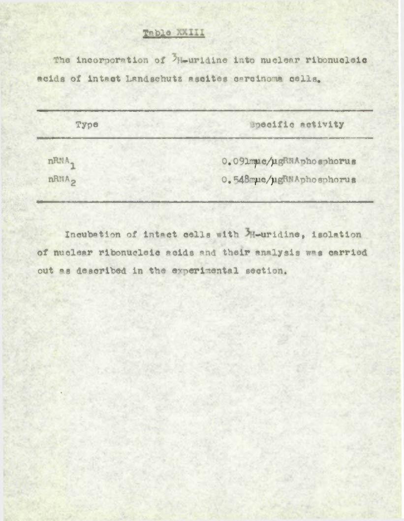
Sable
The IncoMorntlon of PH-urldine into nuclear ribonucleic
acids of intact Lsndschut2 ascites carcnnoma cells.
Typo 9oeoific activity
nHNA. 0.09lryc/jigKN Aoho sohoru s
; nRNAg 0, 548"Uc//igBNAnpo8Tnioru8
Incubation of intact Mils with -uridine, isolation
of nuclear ribonucleic aoids and their analysis was carried
out as described in the ammerimental section.

Flrure 28. Phase oontrest photomiorograoh of a seotlon(with eorresoondlng autoi*edlogr»ph) of lansohuts ascites oarelnoma calls treated with ^H-urldine
for 2 minutee (a) In the focal plena of the oells, (h) In the fooal plena of the envisionover cells shown In (a), division six posed for
14 days.

"ijure 28,

TABLE XXIV
The effect of nHNA^ and nHNAg derived from Ascites cell nuclei on the incorporation of ^^-UT^ into RNA by
cytoolpemic Fraction A orepmred from Landschutz ascites
cells.
Addition -UJTP incorporated (umles/mg. orotein/lOmJLns.)
nil(coitrol) 1.14" / - '’ a. 1.97
nRNA 2 2.02
The reaction mixtures of total volume 1ml. each con
tained ppnmleg MgClg» Im^le each of NAM , ATP, GT?, CP
»nd i.9mg. protein and were O.1M with respect to
Trie buffer at oH 9.S. Where indicated, 480pig RNA were
added (see 2. 5d) • All tubes were incubated for 10 mins.«t 37°.

109.
3.12rctmiiaing .t>2t>-U7P
■on of . _» _c.ytoal«.galc enmreencoraori^on.a^
I?.’’ .".nft.l2l2.!HLL requiring the preaanoe of ATP.
Having gathered some Information eoncerning the
olhraoterietios of incorporation as catalysed by
cytoplasmic Fraction A, attempts were made to puriiy the
enzymes resnonslble for this process. The procedure
em^oyed involved the successive use of ammonium sulphate
fractionation, acetone fractionation followed by adsorption
and elution from cal dm phosphate gel, as is described
els<where (2.bb).
Amtonlum sulphate fractionation was retained as an
initial step because of th* ease with which the separation
of the enzyme under investigation from the other UTR
incorporating system, in cytoplasmic Fraction B, could be
achieved. Howeevr, frcm the point of view of enzyme
recovery, it was found preferable to add saturated ammonium
sulphate solution of ptff.5 to the cytopla«mlc extract
already adjusted to the same .pH, and to collect the pre
cipitate beenee 0.25 *an 0..5 saturation with renecc to
ammonium flulphaae< The yields on emyrne were naceatly
improved over those obtained by fractionation with solid
ammonium suloPaae< Moreevee the nrooHure uinng a^luragdd
ammonium flulphtte solution was much more reprollcable,
presumably because of the more controlled conditions.

110.
The second step Involved the use of acetone (chilled to
-15° to minimise denaturation of the enzyme) «nd proved more
troublesome, being more difficult to reproduce. The solution
appeared to lie in the control of the ionic strength of the
ammonium sulphate fraction on each occasion just prior to the
addition of the cold acetone. This procedure facilitated
the precipitation of the enzyme at complrlaiveiy low con
centrations of acetone »nd so minimised the danger of the
enzyme being denatured by acetone. However one disadvantage
with this procedure lay in the fact that it was necessary to
remove the ammonium sulphate by dialysis before a final
adjustment of the ionic strength could be made* thereby
lengthening the procedure. However repeatable results could
be obtained if the ionic strength of the dialyaed ammonium
sulphate fraction was adjusted by the addition of 1ml. 1* Tris
buffer pH 6.5 for each 10ml.
As soon as the acetone fraction was collected, it wasa
dissolved in 0.01'* potassium phosphate buffer pH 6.8 so as to
dilute any acetone present in the precipitate, and dialysed
against O.O1M potassium phosphate buffer pH 6.8 for A hr. Any
further dialysis appeared to influence the subsequent behaviour
of the enzyme with respect to calcum phosphate ^1.
The enzyme, dissolved in O.O1M potassium phosphate buffer
pH 6.8, was completely adsorbed on to the calcum phosphate
gel which had been previously equilibrated with the same buffer.

111.
Slution of the gel with 0.05M potassium phosphite buffer
pH 6.8 succeeded in removing * fair quantity of inactive
protein leaving the enzyme still adsorbed to thegel. The protein fraction containing the enzyme was then
eluted with n.lM potassium phosphate buffer pH 6.8. This
purification step, involving the use of calelm phosphate
gel, proved to be extremely efficient and gave a very hi^i
yield of enzyme.A typical purification is shown in Table XXV, the overall
purification of the enzyme being in the region of 32-fold
and 2^. of the enzyme was recovered. ATiilst both the
ammonium sulphate and acetone fractions still contained
appreciable quanttties of nucleic acid, fractionation with
calcum phosphate gel effectively ranoved most of the
nuoleic acid from the en*yme, suggesting that the enzyme
is not a nudeoprotein.

e • • • • j 3cc »* n ;» cs A* Ao 3 O 8 A J V 3 ! O -Oft M H >-? <♦ I ► O 3pk (1 »-% A r» O ’ H 4« h3 3 w o 3 3 3 1 ► '4 O CA •) 3* 3 d 3 8 H M A • 3 33 • 9 A A 3» A A d 3 8 3 •*9 O co* r* a ^c4 <♦ ►9
J LT 0 i HO M) M* 3 ' H H M*H 3 as o cr 3 0 a A3 o O 3 o a 3 O 3 3 8o <♦ M* a T 0» A 8 M «♦c D K <♦ U c+ *r 3 A ] or O•— 25 . H 3* H* M* U M 1 c* U5 3 3 £ O > o o •a <♦ - • A 3c> V H 3 H 3 3 c* >•] 3 W43 8 X A 8 A 8 O& 3 O «♦ 1 <+ '' A - ■’J* w « s ft r* cr »• 3 O H* 8*♦ 4 9* A H 3 5 O •—i, C *-3 ■9 ?j 3 Asz4 9'- j v-4 9 # O r* c*o ► J < 3 0So'k 4* H OQ 1 «+ >.■ Jr* * • cf M M V4 3 M O C
o m O -j vn ,, vn cj M C »3 w r+ • J c3
9 A aj|B -H 9 Mo H 3 w . Art U < i3 o * I 3 AH \j4 H C 9 t A 33 3 ru 0 d V* M rv \x 3 A sc & A j O > O rs h r a Mc+ L A AJ sO o O c* w H inO J A 4 J 3 A1 A *• H —— 1 M*• A s d 3 «* 3 Ac* « M 1 *< wQ'- 19• Hc* ro c*| J>
O A • — 9 LH 3* Ho O M OCA 3 «<jS 3 ru ro o 3 3** AOO c+ • • • • t O H HO b>H • 9 H CO O» <♦ < r* 3A H O <n oo IS A or k* *%
g*9
< Oft3A
H\H H3 JM A 1 4 A
r ■ *♦£ O 0* 3 cr$— * < OK>A
v'-r*A
o»-% . i
A3l
ft 3 6B “O ‘O'• -3 H 3»9 = 3 H«♦ C A j "5»
O A K M r i A•a M M z-s-H J% A £fc* **' » rv c* ■ 'rt* Aa flfe * 00 Ho "1 f* P>« H Hf: Oui 3 ro• w *♦>
wJrMH 1 i
1 b€s£
MipMAXX


112,
TM va* Qf ^urtHao in ? or«lVa^n«i»y study of Rka
biopynt^-f* in calls.
It is an parent th*t the material Into which the
’’-.uridine is inooxhoorntei by cytoplasmic extracts of "
ascites tumour cells is HHA since it is insoluble in
acid *nd liberates on «lk«line hydrolysis the four
ribonucleoside -3*(or ?*) monophosphaten. These m*y
be degraded to yield the four bases adenine, frusnine,
cytosine and uracil of which only cytosine and uracil
are radioactive. The "TOunt incorporated into DNA is
negligible. •j •/'
The evidence available from several laboratories
suggests that the biosynthesis of Mil* occurs in two
steos: phosphorylation of ribonucleosides or ribo-
nucleoside-.^’ mononhoanhstes to the corresponding di
or triohoaohates followed by condensation of either the
di- or triohosohmte to form pert of » polynucleotide
chain (Fhor*n*, i960).
Several workers have shewn that the four rlbo-
nueleoside-51 moon., di- »nd triphoslh«tes are widely
distributed in biological maaerial (Schmits, ?ooter,- 5. ‘
Hurlbert ani shite, 1954; Huuilbrt, Sctenits, >Tun.n and
Totter, 1954; schmitz, Huuloert and -'otter, 1954;
"•rrdkviat and deutsh, 1954a ,b) ^nd these c»u. ; therefore
act as a source of RHA lrecu^eors.

113
anzymeo catalysing the -hosohorylRtion of uridine
and uridine nucleotides h»ve been observed in cytoplasmic
fractions fr<m r#t liver cells (Cunellakis, 1957*»b;
Curiirni, Paladini, Oouto and Leloir, 1950; Paege and
5chlenk, 1954; Herbert, Potter and Tdoegi, 1955), yea at
cells (Liebeman, Kornberg and Simms, 1955# Berg and
Joklik, 1954)# and Ehrlich ascites carcinma cells (Heichard
and Sfcold, 1957; "kold, I960). AMP, G'M, CM end UMPkina sc b have been daaoniCrated in extracts of acetone
powder of c*lf liver cells (Strominger, Hepoel «nd
MaxweH, 1959; H^ppel, Strominger end MaxweH, 1959)» «nd
similar OM and «M kinases have been found in •. cell fractions
of rat liver (Herbert and kOtter, 1956). Other workups
(C«nell«kie, G^tteansn «nd Ktrnmont I960) have studied the
four ribonucleoside monioho«olh^tl kinases in preparations
from brewers* yeast and 1. coll While the deoxiyribonucleoside
mononhoeptate kinases have been observed in oytoolasmio
extracts of Shrlich mdites carenomia cells (1 dr and
gaelUe, 1959).
There is little doubt thmt, in the case of cytoplasmic
ex-tracts of ascites tumour cells, phosohorylatld derivatives
of uridine are the precursors of HNA uridine mince in
conditions in which uridine . is not phoiciho^yl*ted, such ns
low pH (Flg.8) or in the absence of AfP (Fig.9) there is
little incorporation of uridine into HHA.

Buoh findings would «one*r to indicate the existence- . *. - - *r* a* • . " ■ - ' * ■
of kin» see responsible for the format ion of ITP, Ud? «nd
UTP fr*m uridine in cytooloamic extracts of ascites tumour
cells and it seems likely that the corresponding enzymes
required for GDP «nd OT”», CDP »nd CTP and MP »nd AT?
formation are else present. However these kinases ereabsent from the. mitoehonnrt«lt microsomal »nd nualcar
fractions of ascites tumour cells »nd thus a limit is imposed on the usefulness of hlHuridine for the investig
ation of RNA biosynthesis In these cells.
Tie work of Ochoa »nd others on polynucleotide
ohosnhrrylsse (see 1.4b) suggests that HHA biosynthesis
might proceed by condensation of ribonucleoside-^" diphos-
ohates under the influence of this particular enzyme. As
already mentioned, ^polynueleotidephosohorylase hsaa been
shown to occur in # v«riety of microorganisms, in plants,
in yeasts and, in e rather tentative way, in a limited
number of nammll^an tissues. . It seemed quiteliik<y
therefore polynucleotide phosohorylaao might occur in
cytoolamio extracts of . Trlich ascites cells. These
cytoplasmic extracts are certainly capable of forming Ul>
from uridine and the observation that uridine incorporatio
into HA is Inhibited by inorganic orihoohosph*ie a op>ars
to lend suoo>rt to this view.hn alternative meecwnlsn by whloh BNA biosynthesis

might be achieved involves the ter-ilnnl addition of rlbo-
nucl5oeiae-5f triohosohates* Thin type of reaction ®nd
its relation to protein biosynthesis have already boon
discussed (see l*4d). /bctraots of Shrlieh ascites cells ( d« *nd Abrams, 1967; Hecht, Xamecnil, teohenson and
Bcott, 1968), embryonic chick liver a **rt (Chung and
*<ahler, 1953), rat liver (tfeidelberger, Harbere, heibmnn,
?»kagi r otter, 1 *66; Barbers &A idalberger, 1959;
Canellakls, 1967c* o-rt, 1953) and c»Poll (Preies and
Borg, i960) can catniy-r - r-'oratlon of an AMPresidue into RHA at a terminal position*
Other workers have demonstrated systems promoting
the terminal incorporation of C P; residues from CTP into
WIA in oslf thymun nuclei (Hurwitz, Bresler And K«ye,
1959; Krakow *nd Kamnen, 1^60; ods and 'brame, i960*;Hurwits ••nd Bresler, 1961) and in the nuclear fre at ion of
mt liver ( veins and Gladstone, 1959). In •noet of these
studies inorganic pyroohoaph«te was found to inhibit the
reaction, In 1 -ase of cytools«mio extracts of .’hrlich
ascites colls, the observation that « nronortioh of the
5«-uridine incorporated into the RNA is located terminally
and that this reaction is subject to inhibition by inorganic
oyroohoeohate suggests that this pathway too may olay a part in the observed incorporation of ^H-Airidine*
However the possibility exists that there may ^ell be

116
other neeh^niris for tha inoonor*tlon of riDonuoleotides
into HM* in m»mm*li«n systems. Canellekis (1957h) has reported the non-terminal incorporation of ^c-U’. p into
thrt '■ * of ret liver cell cytoplasm »n! >inond® end Abrams
(I960) have described a system which promotes the non-
♦•r-iinsl lnoorr>orstion of an ‘•”r’ noiety terlved fro-i ATP.
In the ohlolt enbryo oyste-n doscribed by Chung and Ashlar
(1959) there appears to be one ieehanl«n for tho tor-iinal
Incorporation of adenine nuoleotidee into RKA.and another
for the non-terminal incorporation. while incorporation
of ribonucleotide units on the ends of existing R8A chains
clearly does not represent true polyribonucleotide bio
synthesis, incorporation of ribonucleotides into non-terminal
positions in polyribonucleotide chains must represent a more
radical extensl' a or f nation of new chains.
The results presented in the orevi otion (3.1)
suggest that there might be two pathways in oytoplaemio
extracts of Erlich ascites cells, one inhibited by inorganic
orthophosphate, catalysing’the non-terminal incorporation of
UUP into HNA, «nd tho oth-^r, inhibited by inorganic pyro
phosphate, o"t«lyging the ter^inel addition of UMP to RRA.2c#« *
In:the presence of either Inhibitor, incorporation proceeds
predominately by the '•ltomstlvo oethwsy. Since the poly-
neri3«tion ^f rlbonuol3oeide-5* diphosphates by polynucleotide
phosphorylase is inhibited by inorganic orthophosphate it

117
wag thought that the non-terminal incorporation of UMR might
be brou ht about by such an enzyme. Similarly the in
hibition by inorganic pyrophosphate suggesta that the
terminal addition is promoted by an enzyme utilising ribo
nucleoside triphosphates. However the enzyme system
employed in these experiments is cample and it would be
ooaroely justifiable to dr*w such conclusions. Indeed the
work of Vaokenzie (I960) demonstrated the absenoe of the
enzyme polynucleotide phosphorylase from cytoplasmic extracta
of Ihrlieh ascites tumour cells.
The requirement for glucose, HAD and UTP or NADHg and
HAm is extremely difficult to explain. It may well be
that in this complex system UD^ and UT° are preferentially
required in small quantities for a series of enzymic con
versions leading to the formation of BADHg and NADP and it
mipfct be expected therefore that low concentrations would
fail to dilute the incorporation of uridine into KNA. Tho
addition of NApHg and HApp would obviate the need for such
reactions and the added UTP and UD° would increase the pool
of RHA precursors leading to dilution of the uridine in
corporated. The possibility cannot be excluded however
that the addition of UDP and facilitatesthe incorporation
of uridine by increasing the RHA precursor pools to an
optimal size.If the incorporation of V-uridine by cytoplasmic

118.
extract e o f^o sc 11 e 8 colls represent s 1 rue bi osynt heel e oJT
M it nust be ssatmei th*t the other three ribonucleotidesr
ere nresent in the tissuer’extr*et or wn he fornclluring
the Ineubation* T^e etimletion uridine uptake by the
addnlinn af emixture^of WK?f GD? end C£h or GT* »nd C¥n■ X
sunnorts thia view t end tho gre*1 er stinsulation obtained by
addiff on of the r ib^nudeoside tri ohoehei,g suggeots that
tMf' triphosphates micht be the cure ictportanh precursors of
IW in the jnt«a.
while dialysis of ey tapistadc extrp o tg of recites ttutour■’• ■ 5* ■, "<«•$» ■
cells. h«s littit effect on the upt *ke of ' ur idles in to BAA,
preinou bet ion ^ig inhibit ory (Table X). ^f «aht, Stephen son
and Zarnecnifc (1159) have shown ttot the HMA Isola ted feol « frehhy prepared pH l proreipitste of the soluble fiction of
>brlicl •scitc- raila generally containa tho oytpalnc and
oderine nucleotide en1 grottos ana is -t least p-rUielly ;
en tumt-M wl tfc "r.ino »eidc. On pre Incut* tier, however , the
ter-’ln»l nu elretlies *n4 stefne «cld b *>re re-roved fren the
Hn% v n mey be that in t he ascites cyt oplseni c system
o terainni nuflestl de seqrenue existn on the KHA ntfhi nh
allows » residues to be tnforpoxntM initiating the TlC-
synthetic pr oeeoe anti that ou preineubntt l or thl ta t urtiint 1
rrirnirr^nt of tf)y KM 1 n lost.
The diatribution of ensymea reaponsibje^for tho
ineolrro^n t i on of ll-urid lne ( Twble X 1) is qui t« dllferent

119.
from the distribution of en-ymee responsible for the
eyithe«l« of DNA (Bellie, Keir end T"vlason, 1959). This
is consistent with results of earlier experiment# on the
in Wo. incorporation of w^^ch have" chown that many
r«'bbit tissuee »re capable of RWA synthesis and that there
is little correlation between synthesis of RMA and I^A
(f^^mil^e, Bumhwy, F«y *nd Davidson, 1955) •
4«2 rnglTlB Ao .arBailVTAa ,tt?" of . oomrltonuciwo..
tue-in oyt.pip^ie".e^geflt? ..o.f. taeitat tu^ur oei . i.
Wet amtheein of very small amounts"of .polyrlbpnucleo-^
ijtide h«s been dempossrated only in the . oresence of NADHn,■~W£SAD’", m^^^g^^esl^um ions "and *TP, CTT, Of «nd VT? or ADP,
GD-, CD «nd UDP. .J” One of the effects of WMJSL and SADP
is to increase the . formation of WP (Table XIII) end since
the highest figures for net synthesis are obtained on
adding the rib»nucleollde-5•triphotohates rather then the
corresponding dio^eo-hatas it seemed oossible that the
system utilising the ribonu oleo-side-S •triohosoho tec is of
considerable.imoortence in the synthesis of RNA in this
oytoplmprie extract. It could be that the utilisation of
the r ibonu oleoai&e-•^*,dlhhoe>h«t es for net rsyythesit is due
merely to the conversion of these to tho corresponding
triphosphates by nucleoside diphosphate kinsses. Howferor
more satisfactory results from these attempts to demontSrste
net synthesis of polyribonucleotide could not be obtained

12v.
due to the high nucleim content of these cytopl« onic
extract m.
4.3 2? e Inc or- ormt 1 on of -"la - urj dine nu"l eutideaOatq.mu by qmonlu- B»Inh»te fractions Orr
cytonlftsnOo extraetnoX aoeltnf dis.
Frryo the results described On the - previous seotion
(3^4), Ot On clear th«t cytooOftsnOo extracts contain at
lea at two different system® ©at* lysing the Oncorpo^i^'tOonO J. ;
of * -UT' Onto HN\ These two system *ro obtained
undcr different fractionation conditions with anmniiOn
sulphate and show quite different char*cteristOc# wOth
reeoect to the reactions they catalyse. Thus the optim
pH (Fig. 16), the Onfluence of a mixture of AT”, OTP «nd
CT? (Table XIV), the tine course of the reaction ( Fig. 1% ,
the Ontranolecular location of the Onc radOo-
actOvOty (Tables IVI and XVII) «nd the O nfluence of amino
acOds (FOg.21) Oc quite different for the two fractions,
• red ?. The only similarities between the t%v fractions lie in the utiliaton of the e*i precursor (2- bi XlV ) f the Inhibition of both syttmne by inorganic pyroohoanlhate (TC.e XV ) «nd the stimulation by added -V.
Ho evidence has been obtained On any of these exeerlnents
for a systen utilising UDP as a precursor of polyribo
nucleotides. Such s ^^chanO^^) cannot however be excluded
cmpeetely since Onorganic orthophoQohato faOlM to produce

121.
any guUgt-tnnial inhibition In these experiments wheresa lt
had previously been observed to Inhibit the Incorporation of
3h -widine by cytoolasmic .extracts of 3h*lich aaciteo cells
(3.1).
The results in Tables XIV, X)X and XVII suggest that
iFraction A is concerned in a system Which incorporates all
four ribonuoleoside ioeties into oolyr ibonu cleotides since
it is stimulated by the addition of ATTP, GT^ and CT* and in
the presence of these nucleotides the uridine moiety is
more randomly distributed in the oolyrlbonucleotide than
in their absence, when it is incorporated ms oolyuridylic
acid. The observation (Table XiV) that Fraction B is not
stimulated by tha addition of ATP, GT? and CTP and that most
of the uridine moieties incorporated are adjacent to eithw
uridine or adenosine nucleotides in the polynucleotide,
suggests a different function for this system, and this is
borne out by the 'finding that Fraction B is stimulated by
the addition of an amino acid mixture (Fig. 21),
The reaction catalysed by Fraction A prepared frcm
cytoplaimic extracts of Ihrlich ascites tumour cells closely''
resembles that of the Tyno i enzyme of Cnwg, Mnimr end -
irione (I960) nrsnmred from the cytoolsamie fraction of
chick emtb’yo cells. This enzyme cstalyoes the incoroor-at ion of ^C-AT? into internucleotido linkages in the
presence of GT°, CT? and UTP and RHA at « oH between 9*0 *nd
122.
9.5. Similar system# utilising riconueleoside-5*triphos-
phsta# have since be*! inscribed in extracts of l.coli
(Steven#, I960) , in t>e soluble fraction of oe® embryos
(Hua- , —esshwri and Bonner, I960) and i- pigeon liver
raicrosones (straus -and Ooldmtseer, 1961). Oi the other
hand, the results show that Fraction A is also capable of
attaching UOT units derived from UT lreferentitlly to
UW residues in nolyribonuQleotiies a# a oolyiridylic acid
sequence (Table XVI) and thus also resembles a more highly
purified UTP inconor*ting system recently obtained by
FlemiOTer and Karanon (1962) from the oH 5 supernatant
fraction of a rat liver homopgmete subjected to centrifuo
ation at 105,000 . x g. This cytoolasnic enzyme system
requires the presence of added RNA for the incorporation of
uridine nucleotides a# a lllyuriiylil acid sequence. Thus,
even in cytopldmic -Fraction A itself, there miy actually be
two systems responsible for the UTP uptake; one catalysing
the randem incorooration of UTP into polyribonucleotide in
the aresence of ATP, GT and CTP indicative of possible true
RNA synthesis and the other responsible for the of
polyuridylic acid sequences in polyribonucleotide chains.
Such cytoplasmic polyribonucleotide synthesising
systems are of particular interest since sutter, Witeman
and Webster (1961) suggest, on the basis of work with
Acetabbilaria, that the RNA of ribosomes is synthesised in

123
the cytoplam and the cell nucleus was not directly required.
However as will be discussed later (4# 1 ) the ribosorml
Wi, even fl lormed under the influence ol cytooi^* ©mic
enzymes, probably has a nucleotide sequence which is dic
ta ted in some manner by the cell nucleus.
The reset ion catalysed by cytoplasmic emotion B more
closely resembles that ol the Type II enzyme ol Chung,
and 2rione (186e). , It is not stimulated by the
addition ol the other three ribonucleoside-S* triphosphates
(Table XIV) and a considerable oroportion ol uridine nucleo
tide is fnooImeratei »t terminal positions ol polyribonucleo
tides (Table XVVI). It appears therelore, that the
reaction catalysed by Fraction B whilst giving rise to n product ol possible importance in the transler ol amino acyl
residues lrom their respective activating enzyme# to the
site ol their linal incorporation into or^^ein, cannot be
regarded as responsible lor true RNA biosynthesis.
4.4 Ineoiraoritton at into polmXMueigoHi» .tar
flptctlone prepared f»«i laolatM aeoltee eell nwQel.The mjor limitation in the use ol aqueous media lor
the isolation ol cell nuclei is that both hip! and low
molecular wight maerial is undoubtedly extracted lrom the
nuclei during the course ol their isolation lBounce,1955).
However the degree ol permeability ol the nuclear memtbmno
is still a mt^er ol come controversy. Also according to

124.
Bounce (1955)# when nuclei are orepered in aqueous madia,
in the absence of nuclear mutooytis, the nuclei should be
capable of forming a oecular tyoe of structural gel upon
the addition of alkali or strong sodium chloride solution.
This gel formation is considered by Bounce (1949) to be
an indication that little or no autolytie degradation of
nuclear structure has occurred.
By 1950# it wan found possible to isolate liver cell
nuclei in isotonic sucrose to which calcim chloride had
been added. This use of calcbm chloride was first
introduced by Schneider and Betermnu (1950) whose era.mole
was soon foioowed by H Sboxmh Sc^hneider and sriebich
(1952). Apparently calolum chloride inhibits autolytic
degradation and permits the isolation of liver cell nuclei
at pH values sufficient to allow the formation of structural
gels with a.lkali or strong saline. ConeeoUr*tlons of
caleum chloride as 1uw as C.OO18M suffice under certain
conditions, but higher concentrations are desirable for
obtaining clean nuclei. For erm’moe, AHfrey, Mirsky «nd
Osewa (1957) used 0.0033M to isolate nuclei from calf thymus
tissue. for the isolation of ascites tumour cell nuclei
this higher co-'centrs.t ton of calcum chloride was empLoyed.
from the results described in the previous section
(3.5) » it is clear that nuclear ^tr*ots prepirM from
isolated ascites tumour cell nuclei contain at least two

125.
systems catalysing the incorporation of ^’-.UTR into RMAe
These two systems,nam*ly nuclear Fraction A «nd nuclear
Frrotton B, are obtained under the same fractionation
conditions with ammnium sulphate as are the A and B
fractions from cytoplasmic extracts. As rith the cyto-
plaimic A and B fractions, the nuclear A and B fractions
show quite different characteristics with reatect to the
incorporation of into HNA, althouf^ the reactions
cataloged by the A fractions from nudear and cytoplasmic
extracts reeemble one another, as do the reactions catalysed
by the S Fractions.
Reactions similar to those catalysed by nuclear A
fraction, involving the incorporation of any one labelled
ribonucleotide in the form of the triphosphate which are
greatly augmented by the presence of the other three have
been described in nuclear fractions from rat liver cells
(WI^s, I960), in isolated nuclei of pea seedlings (Rho and
Bonner, 1961) and in the nuclear fractions frem HeLa cells
and mouse fibroblasts (Goldberg, 1961). However, unlike
nuclear fraction A, these enzyme preparations, although
derived from ceix nuclei, are particulate it nature. In
the case of ascites tumour cell nuclei it is naceseary to
employ sonic vibration as a mans of causing their dis
ruption and the results suggest that it is this process
of sonic disintegration which is responsible for making

126.
soluble most of the enxymo which subsequently •poears in
nuclear Fraction A, *1though a certain amount of 3TTA
synthesising activity still remains in particulate form.
The f*ct that nuclear Fraction A has an activity four
to five times greater than that of cytoplasmic Fraction A
(Fig*23) does not necessarily imoly that the nucleus is the
only site of HHA biosynthesis, *nd that any activity
occurring in the cytoolagm is due to the ensymea responsible
for this process being leached from the nucleus during cell
disruption in aqueous media.
The occurrence of a incorporating system in
nuclear Fraction B with characteristics similar to cy to
rn l*rmic Fraction 3 is at first eight nuzzling when the cyto
plasm is considered by many to be the site of protein
synthesis (Hoaglam, i960). However Allfrey (I960) has
described not only amino notivating systems in nuclei
isolated from calf thymus tissue but also the transfer of
these activated amino acids to carrier ribonucleic acids of
nuclear origin. Furthermore he has provided some evidence
to suggest that HNA«.rioh particles which exist in calf thymus
nuclei are sites of active amino acid incorporation into
protein. Indeed the isolation from calf thymus nuclei
of ribonucleoproteln particles, thought to be nuclear
ribosomes, has recently been achieved by >ang (1961).

127
4.5 Forrwtlon of « «oqueno-> >x Menrlwte units fron by an entmw fraction froa na.lt*. tumour cell nuelal.
The eh»»p«eteri8tic« of incorporation into
polyribonucleotide by MfclMf Fraetlon A prepared from
ascites tumour cell nuclei have been described (J«7)•
The results suggest that in thia ammonium sulphate
fraction there are two discrete ay at erne responsible for
ATP incorporation into polyribonucleotide* One of these
systems oromotea the incorporation of ATT- into poly
ribonucleotide at pH 9.5 *nd requires the oresence of
the other throe ribonucleosid^-51 triphoephates (CTP*
CTP and UTP). In this respect it closely resembles
the system already discussed (4.4) which exists in
nuclear Fraction A and requires the presence of *TP* GT? and CTP for the optimal incorporation of ^^P-UTP into
polyribonucleotide at pH 9.5. However only further
purification of these syatoms will reveal whether or not
they are under the control of one «nd the same ensyme.
Nevertheless the other system which promotes the
incorporation of ATP into polyribonucleotide in the
absence of added ribonucleotides* at positions adjacent
to other AMP residues* is fundamentally different. The
function of such a system is at present difficult to
rationalise but a similar system capable of forming a
sequence of adenylate units from ATP hag been described

128,
in m soluble inparatim from calf thymus nuclei ( ’5i^lnis and Abrams, 196cb),
4.6 Tho deleti<ee■oe of «agUes_^2£-
wt-Aig aratoma u.nn tho oniam, of jd*.
Prom the results in 8lltiln J.6 it is clear that the inolrDlstio>n of itp-UTP into polyritonuelvltiil catalysed
by nuclear and cytl^ia(mic A Fractions shles a requirement for DNAe Hooweer, whereas UTP ntshe in the presence ofthe other three ribonucleoside.^1 tri^>hl8ohatlt by nuclear Fraction A is enhanced by hlat-ilnaturvi (singlestranded) WA in the orltlncl, or absence, of aiivi nuclear RNA (T«ble XVIII), the incorporation of UTP in the orltencl of at*, a:? and CT” by cytlpie^^eic Fraction A is enhanced by native (ilubll-stranili) DNA mly in the prltlncl of aiiei nuclear HHA (Table XIX). Such an observation ^^8 to strengthen the view that the system synthesising polyribonucleotide in the cytlplam is tlev- wh*t different fom that synthesising polyribonucleotide in the nucleus, and is not merely a portion of the nuclear system leached into the l>tlplamic preparation during the isolation of the sutxseHular fractions from ascites tumour cells, ' ' A - . “; / ” -
Such a requirement for DKA in HNA biosynthesis has bun
reported by other worRere, For instance the Inhibition of

129
the biosynthesis of RN* in cflH-free systeme by the nnyyme
DNase has been reported too a system in o«,lf thymus nuclei
(Krakow and C&nellakle» H6l), tfr a soluble system nn
j%coli (HurrWis, Bresler and Dirinrer, 19(lo), for systems in
L# arebinosus and A ▼inejandii (Ochoa, Burma, Kroger and
v^eil, 1911) and for systems from rat liver nuclei (aeiss,
) )4 ■- ysaoatuumg ( .eiss and H<lkrmota, ) i4.
^Correaoandlngly *lthe presence of WA is reported to be
essential for RN* synthesis by enzymes in rat liver nuclei
(fteiss, 19l1a »nd b) in u.lys-odeikti<us (veeas and Nakomto,
19l1a end b) in 1,coli (Stevens, 196la »nd b; Hurwitz,
Bresler and Stringer, 1910) and in both 1^lrablnagut and
^d^jnelandLi (Ochoa, Burma, Kroger and lill, 1911; Burna,
Kroger, Ochoa, earner and 4elll, 1961). Acoirline to
some of these authors DN* must apparently be in the native
form; heating and partial degradation with DJase abolishes
its activity. However, weias (1961a) suggests that DNA
need not necessarily be double-strinded,
Using .partially purified preparations from ,,lyfiadelktkU8
fceiss and his co-workers ( veiss «nd Kak<onoto, 1961b and c;
Geiduschek, Nakomoto and' ^eese, 1911) carried out further
investigations of the involvement of DhA in RhA biosynthe<*it.
Initially, they were able to show that ’♦primer* D*As from
different sour cos altered the ooaitian of cytiaylate residues
in the newly assembled R^A chain and that the polyribo
nucleotide had »n average base ea^noai^ion similar

1)u.
to the *prieer* XJA used. Further work on nearest
neighbour b»se frequencies suggested that the sequential
arrangement of nucleotides in the HNA is probably analagous
to that in the fp^im* DNA. More conclusive evidence c«me
from caesium chloride density gradient centrifugation which
demonasrnted th«ht the HNA synthesised c«n form specific
complexes with the OTA •primer*. According to HU and
Spiegelman (1961) such an interaction between DKA and HNA
indicates that there are entire nucleotide sequences in the
DNA «nd HNA chains that are simultaneously capable of
binding to each other in the face of the cornpeeing tendency
of connllmentary sequences on OTA strands to recombine.
Accordingly it appears therefore that RNA strands are
synthesised with a base sequence 'complementary* to the DNA
•primer* and the tern *oomKement*ry* is defined as a
specific fit of portions of 'primer* and product by appropriate
base interactions.
Other evidence of the directing role of DNA in RNA
synthesis has been obtained with purified preparations of
Tj, coli in the presence of the four ribonucleoside^* tri
phosphates (Furth, Hunitz and Goldmann, 1961#, b «nd c).
When OTA preparations of widely varying base cnmponStinn
(T2-DNA, calf thymus gland DNA, coli DNA and
M.lysodeiktious DNA) were added to the system, in all c«ees
the base ratios of the RNA produced was determined by the
DNA added and the results were consistent with the

131.
hyoothelis thmt tbo Incorporation of ribl>nulllotiies is
dotem-iined by the ApHity of the new polymer to form
hydrogen bond os ire with the be ses in the r^r^mer” DMA
80 thit the product is some sort of DNA-RnA complex. This
view is supported by the observation thst when polyi2oxy«.
thymidylAte is used as "primer" in plsle of It A only ATP
is used for polyribonucleotide synthesis ind the product
is s Polydeoxythyneliy*’t^tl.oOyydevnya'te hybrid, with
the copolymer of ieoxy»<ienyl*te #nd delxythyaiiylatv
the product wm » polymer made uo of MiP mid UMP unite in
mltem-mting sequence. Hence not only do the deoxyi'n-
nucleotides in the "primer" determine the nature of the
ribonucleotides in the HHA produced, but the sequence of the
ieoxH'ibonucleotiivs in the "primer" determines the sequence
of the ribonucleotides in the HM* produced.
3ich results with oertislly purified enzyme mstems
suggest thmt nmrt of the product m»y exist st lemst
tempor^rily ms a complex between RNA mid iiA, gevermi
resemrchers hmve chemicAl evidence thmt such complexes
cah occur (Rich# i960; Schildkrmut, '■imrmurj rrepito «nd Doty,
1961), THU mnd Splegelavn (1961) showed thmt infection of
j, coll with T2eycceriooh age results in the production of
m frmetion of RMA with mt be se rmtlo tneiegous
to thmt of the T2-tacterlophmge DNAa This RNA, which is
synthesised immedimely following infectoon, thlet m specific

152
complex formation with «ingle-str«nded ?2-b°cterlooh«ge
DNA whereas no hybrid formation occurs with heterologous
□’ even If it has the p«me base comoosition as ?2-DNA.
It Is oresumably because 72-DNA and T2-soeciflc HNA
?sess " Kmentrnry* nucleotide sequences that they
form hybrid®. Hecently Schulman onner (1962) have
demonstrated a dn^-R:;a comolex to occur naturally in
Neurogpora ora sea.
Having discussed the directing role of DNA in HNA
synthesis lt is of considerable Interest that more
evidence is accumulating to strengthen the concept that
DNA is the aotive material of the gene directing’ protein
synthesis. For some time it hag been known that irradiation
of organisms with ultraviolet light will induce mutations
(ftrb and Owen, 19^°). In 1961, Litman was able t
demonstrate that treatment of isolated' ’•tr«nsfarming* DNA
of pneumococcus with ultraviolet light results in its
chemical and genetic alteration.
striking evidence for a DNA mediated genetic control over
protein synthesis has been demonstrated in a cell-free
system from coll ^reviusly induced to synthesise
de nov a enzyme ^•galaotoaidase (Novell!, Sisenstadt
•»nd Fameyama, 1961). then tho system b supplemented r ith
inducer, amino acids, » suitable energy source and nucleo
side trinhoachates, it is c»pable of effecting an increase

133.
ln B-gilactosld•te activity. An inhibition of enzyme
tynthe<slt occurs uoon treatment of the eyotam with tWase or
with ultraviolet light. < This Inhibition c*n be overcome
by addition of native DMA from normal cells to the system.
Thus, from the point of view of genetic control over
protein synthesis, it is possible to envisage a single
strand of DNA directing the synthesis of a single stnnd
of RNA (uracil pairing with adenine and cytosine with
guanine etc.) so as to form a hybrid duplex. ln the
living cell this might then dissociate into s single
strand of DMA, which could repeat the process, and e
single str^- d of RNA of ca:nnai.lentlry base campoaStion
w>lch would presumably aot as a •moB.enjw** HNA directing
the assembly of amino acids into the protein molecules,
'*vid«tlct for such a HNA in coli has recently
been obtained (Brenner, Jacob end Meeelson, 1911; Gros,
Gilbert, Kurland, Hifle'braugh and AAwaon, 1961).
The results of experiments on g, coli indicated that a very
sm.ai HNA fraction, characterised by a high rate of
remove! and a base ca!n?oaition analagous to >, coli UN.,
*?9aciatep specifically with certain ribosomal particles,
the site of protein synthesis. After infection of the
l,coli cells with T^-bacterlophage only phage proteins are
synthesised and according to Vonod, J*cob and Gros (1911)
it is possible to demastr^te that although the actual

134.
synthesis of phage orotein takes ol«co in the ribosomes
of the bacteria the *messenger* RNA has a base ratio
similar to that of the T2-bacterioohage DMA. They con
clude that the *meesenger*, which carries the genetic
information from the UNA of the genes to orotein synthes
ising centres, is a short lived intermediate and cannot
be identified with ribosomal RNAe
However it must be anoreciated that at present these
theories are only applicable to certain bacterial systems.
The situation is still far from clear when mammalian cells
like ascites tumour cells are considered in this respect.
The oossible function of single-stranded (beet-denatured)
DMA in the nuclear polyribonucleotide synthesising system
could be envisaged in terms of oromoting the formation of
a DMA-RNA hybrid with the subsequent production of a
•messenger* RNA but work along these lines is hindered
because the enzymes are difficult to purify and normally
have quite low activities. However the situation in the
cytoplasmic fraction of ascites tumour cells seems quite>
different. As already mentioned, DNA only exerts an
effect in the oresenoe of added nuclear RNA (Table XIX)
and thus it could be the case that DNA only acts indirectly
in the cytoplasmic system, possibly functioning as an
in vitro inhibitor of endogenous RNase oresent in the
cytoplasmic extracts. However other explanations must

135
be considered. Double-mtranded DNA could direct the
synthesis of RNA as # third strand. Ruch a triolet-.etmnded
mlecule has been envisaged by %ibiy (1958). Alternatively,
in the aytoplalm, it msy be the os se that the HNA double
helices do not need to unwind completely in serving ss
templatee for RNA synthesis. Nevettheless theories
based on the direct o^irti^ci^psit^J^cin of DNA, double or single-
gtrsnded, in cytopiasmic RNA synthesis sre difficult to
rationalise since DNA does not occur normally in the cyto-
plc sm.4.7 The delendtnat of »agU». tumour oell -5? tnoowomUng
■ sterna unon the nregenea of added RHA.
The results presented in the previous section (3*6)
indicate that -OTP uotske into polyribonucleotide by -
nuclear #nd cytoolasrnic A motions is augmented not only
by the addition of the other three r ibonu cleo side-5 • tri-
phoenhates but «lso by the sddition of RNA derived from
the nuclei of ascites tumour cells (Fig.24) by the pro
cess of phenol extraction (2.5b). The addition of RNA
derived from ascites cell cytoolaimic extracts has a
similar but much less pronounced effect (Fig.24). Horner
it must be tmphaalsed that phenol extraction does not
remove all the RNA from the sscites twour cell nuclei
(3ib&tani, Yam^m, Kimurs and Takstoshi, 1910). In this
respect it appears th#t the HNA which is extracted from

136.
ascites cell nuclei is similar to nHNAj, whilst that
which remains unextracted is similar to the RNA in the
"1U ReCl fraction", namely nRNAg
The RNA dependent incorporation of ribonucleotides
has since been described in other laboratories. For
examle, Huxrrits, Furth, Anders, ortis «nd Auugugt (1961)
demoonsrated a system in ribosomes of S.coli leading to
the incorporation of all four ribonucleotides. This
reaction, they found, was inhibited by RNase but not by
Dase, In addition, Reddi (1961) has described a cyto
plasmic syet<m from soinach leaves which is capable of
polyribonucleotide biosynthesis in the preaonce of added
RNA and as already mentioned ^lemoorer and Knmraen (1968)
have described a syst<m capable of incorporating UTP into
polyribonucleotide as oolyiridyla te and have found it to
be dependent on the presence of added RNA,
However as already ^discussed i (4.6), the addition of
single-stranded (heat-denatured) DNA can also augment
incorporation into polyribonucleotide catalysed by
nuclear Fraction A. Thus in ascites tumour cells there pro
bably exists in the nucleus two RNA synthesising systems,
one dependent on single-^tr^nded DNA nnd the other dependent
on a type of RNA derived from the nucleus. In the cyto-
plaem there appears to be s systm which is also dependent
on nuclear RNA, although this syst<m could conceivably have

137.
been leeched from the nucleus during . the preparation of the
oytaplasrnie extract.
It is possible to .envi wag© a system -operating in■ “• .s" •■'• «*• . ’■* ..ascttm cells Wsreby the native DNA double helix in the
chromosomal of the coll nucleus unwinds with the^
simultaneous formation of RH* chains on both single strands
of DNA so formed.A' Such a process could be-brought about
by the DNA dependent H-A synthesising system in nuclear
fraction A. This process would give rise to two RNA-dhA
hybrid molecules. aAA-Ap psenent there ±s no conclusive
evidence for the existence of such - hybrids in.*»«citea
cells. However in the experimental seeti .-n (2.Bd) it
was mentioned that the yhic'i »«g not extr»ateo from
the nuclei by the nhenol process, namely nRu”always
contained s proportion of DRA Which could not be removed
by DNsge treatment. $ud ha finding is of interest #s
Schildkraut, Mrw, fresco and Doty (1961) have demon
strated that their bMA-RRA complex was resistant to
pancreatic DNee. InlcM It is this nRIHg which ie mostI3
highly labelled with after b two minute exposure of3 . - ■ •
intact aaeitcs tumour cells to H-i^r’idine (?#pie WLUj
4.9) • '_-O s A Waa •; ? W^^A,r-;A;A A- ~
3ach of the RHA-WA hybrid molecules in turn. could
give rise to # single strand of SKA and « single strand
of RNA which could set «? « "template" in the subsequent

synthoBLs of RR in the cytoplasm or nudeus. The
existence of such a "template" Is im>oied in the theories
of Leslie (1961) for vthe transference of information from
nucleus to cytonlaga, although he considers that the
formation of single-stranded "template" RNA in mammalian
cells involves 'the genetic activity of the double helix
form of DNA,
It is conceivable th«t this RNA "template" could S
remain in the nucleus #nd there direct the synthesis of
RN for the formation of nudmr ribosomes, the process
being oatalygeo by the • RHA dependent polyribonucleotide
synthesising systm present in nuclear fraction a, on
the other hand, the RNA "template" could p»«s into the ;
oytoplosm rather like a "messenger* and there direet the
oytonlasroic synthesis of any ribosomal RNA, However
although nuclear #nd cytoplasmic ex tracts prepared from
ascites tumour colls almost certainly contain trace
amounts of ribosomes, or ribonucIeoorotein particles,
further work is required to determine whether s "template*
tyne of RNA Wll associate with them or not. In fact
attempts to discover which variety of nuclear RNA proved
most effective in the cytoplasmic RNA synthesising systim
were unsuccessful (Table XXIV) probably due to DNA
contaminated preparations of nRHA^o

iy.
4.8 The lnlU"l site of HK* Py nt Met e In t^c ffiolcv ?._ogLSSiltS. tumour cells.
As ’Mentioned in the Introduction (1.5) there remains
consider-ole controversy op to the ®ctuni site of initial
hesis in t'-'e cell nucleus. micro^uto-
rodio^r^ohic procedures many workers are of the opinion
that the nucleolus is the primary site of RKA synthesis
(for references see 1.5), however others using similar
techniques are convinced that HNA is first built up in
intimate association v ith the chromosomes end could be
a precursor of a fraction of nucleolar KN A (for
references see 1.5). uuch e theory, teaed icroaeopy
end autoradiography, that RNA is built up in close
association with the genetic materiel is consistent rith
the results discu^s^d in the previous sections (4.6 and
4.7).Attempts to isolate «»nd study the enr.ynes of
'Tsnmslisn cell nucleoli hnve not net ith nuoh success.
In 1952, rrakauer reported th*»t nucleoli could be con-
centrated from homogenates of liver cells made in very
strong sucrose solution. Photographs were not given
and the degree of purity of these nucleolar fractions
could not be stated with any certainty.
Subsequently Vincent (1952) reported the isolation
of nucleoli from the egi s of starfish, hotograohs of

the isolate! n»teri"l showel « very high degree of
ourity. sever the sixe and morphology of the star
fish egg nucleoli are such as to orcclude the use of
Vincent1- procedure for the isolation of nucleoli from
mammalian cells.
Nevertheless -onty, Litt, Kay and Dounce (1956)
described » method whereby nucleoli could be obtained
from rat liver nuclei. However analysis of these
•nucleoli* showed then to contain a high proportion of
DBA and a low proportion of SNA and in composition they
resembled whole chromosomes (Thorell, 1955).
Reliable information concerning the nucleolus of
the cell is scanty. According to Casoersson (X95 )
and Rehultx (19^7) nucleoli are formed near •chromocentres
which consist of •heterochromatin* or portions of
chromosomes, which tend not to disperse during interphase,
but remain in « more or less condensed at^te. Thus
according to these early theories, the nucleolus appears
to be structurally independent of chromogomes but probably
dependent on •heterochromatinw for the synthesis of some
of its constituents such as HJC .
Evidence is available that the ergs of amphibia
possess large numbers of nucleoli which migrate to the
nuclear membrane to empty their contents through the
rnenbr«ne into the eytoolewn (bounce, 1955) • However

141. ‘
in tha nwclocli of rnanmnlian cell# tha aitwtlon la moreu \x"
Comdex, Wo " nucleolar mippfntion h# # apparently been
observed in m**mrnli«n gomtlo cells and the number of
nuclear inoluel^on bodies comronly termed nucleoli denends
on the degree of nolynloidy of the cell (mende, Poyner\.<A; , . - ■• ‘ •* ';v •- ’ ■
end ’’•inter, 1942, nine# # relation shin exists between
the degree of nolynloidy #nd the aun^nr of nucleoli.
Lewi# (1940) oarriei out clnnnhotoqr®ohic studies on the
nucleoli of rat fibrrobl# atm end found that nucleoli arc
intregel narrts of chromosomes,. and that they become
disoereod during mitosis and in condensed form
during intcmplm se when chromosomes are dltner«ed• Thus
inmefmlitn sommic cells, nucleoli may be defined «8♦intranuclear inclusion bodies Wiich sre attached to
chromosomes ,
it is therefore not surorisinR that the nucleoli
^isolated by Moity, Lltt, F**y md Pounce (1956) resemble
whole chromosomes in corn^ossiti^^ since ?uoh nucleoli
sonear to be woeis! parts of chmm^somea. Nucleoli
isolated. by # similar method from nuclei of ascites
tumour cells also have a high DNA content (3.9) and
mlcrosnonio examination reveals an association with
chromosomal ms a aerial (2,3c).
The oo^i^l^i^i^i^^iti^es have been mentioned by Vincent
( 195%) *nd ^oods «nd Taylor (1959) that the nucleolus

142
functions a.s a mess-production and finishing machine for
different RNA "templates” from the chromosomes. The
results of 9chultz, Cmsoersson end Aquilonius (194S) end
Lin (1955) suggest that the na^ZLe<^;ii are attached to
cert°in chromosomes, the so-called "nucleolar organisers"
which determine their cornposstion. Prenucleola.r bo^^es
have been found amidst chromosomes and these bodies
eventually combine to form nucleoli at the sites of these
organising chromosomes (Lafontain, 1958; Tadler, 1959).
In mutant cells lacking "nucleolar organiser” chromosomes
these orenucleolar bodies remain dispersed (Tisdale,
Fischberg and Smith, 1958). It is likely therefore
that chromosomal products such as RNA "templates" become
associated with the p^udeo^ bodies, and a function
of the "nucleolar organisers” is to "organise" parts
contributed from various chromosomes into a nucleolus.
Table XCH shows that the "chromosomal supernatant"
fraction obtained from ascites tumour cell nuclei (a
fraction consisting of highly fragmented chromosomal material) incorporates 52p-_ypp into HNA to a slightly
greater extent than the nucleoli themselves. This
is of interest in view of observation that nRNAg, the
type of RNA in the nucleus in apparent association with
DNA, becomes most extensively labelled when intact ascites tumour cells are exHosed to Stf-uridine for short

143.
period# suoh me two minutes. Indeed mutormdioFpmphy of
Intact azotes tumour cells, «fter the emmc length of
exposure to ^v_uridine, would seen to support the concept thmt
the nucleoli #re not the Initial' site of RNA synthesis,
Ixxt rather that other structures of the nucleus, such me
chromosomes, mm^ also be involved. In the mscite# cells
the initial site of RNA synthesis might well be at the
chromosomes. The f«ct that nucleoli also incornormte
32?-1T? could be either due to contamination, or to an
ability to modify, or comp***9, mole<mles of RNA initially
produced in association with the chromosomes. However
this is obvious speculation and much work has yet to be
done to clarify this situation.
4.9 Sratheaia of oolyuridylatc jaaftlon to eytaolnanlflrr»otion 4 uni. If. poaaible bearing on the genetla code. .
From the results in section 3*3 it ie evident that
cytoplasmic Fraction A o*n catalyse the incorporation of
urldylate units into polyribonucleotide in such a fashion
that several urldylate units arc adjacent to one another. The
very recent work of Nlrenberg and Mrnathrnei (1961) indicated
that # synthetic polyuridylic »cid "tomolnte* will direct the
inclusion of nolyohenyalanine in protein maerial by a cell-
free system from 3»coll. ^ojyrohenyalnninr-qNN-’ has
been isolated and shown to be an intemedi<»te in this
process (Nirenberg, Maathaei and Jones, 1962). Theae

results Imply that # sequence of uridylate units in «n HNA
•template” will direct the inclusion of a phony-alanine
residue in a solyoeotias chain* The genetic studies
of C*ick, Barnett, Brenner and Wstt^Tobin (1961) suggest
that only a triplet of adjacent uridylate unit* is required
for this purpose and this is substantiated by the more
biochoral c»l studies of Lenoral, Speyer and Ochoa (19b1)
and Speyer, Len^el, B*slUo and Ochoa (1962). Thus it
ie of interest that in ascites tumour colls there is a
system which is capable of forming a sequence of uridylate
units (Table XV!) although its significance is as yet
■far from clear.

145.
■ I— !■!■■■1. Cytool« anlc sKtrmsts of fflhalieh aadtas tumour cells are
capable of Inewporatlng ^^'.-uuldine Into ribonucleic
acid (RMA), M<onfclno-5* trphoeohatd (AT), magnesium
ions, oot««siim ions, nlootlnamlde-adenino dinucleotldo
(NAD), glucose and added W are required, #nd the
process involves the formation of radioactive uridlne-5*
mono-, li- and trlohesohmte (U'J), UDP and UT), Alkaline
degradation of the RNA reveals that about 20. of |the
rsdioaetivity is in terminal uridine-residues ani-about
5%. in uridine located noTn^^omlnalljf. Inartgnlo
ojrophosqhate inhibits tornilna! ^incoloration and,-?
Inorganle orthonhoaohate degreases non-tamlnal. Inoor-
nontion.
The aubatiiutlnn of reduced
ainucleotidc (NAUR2) "nd nlootlnamide-gdenine dinucleotide
phosphate (NAD)) for NAD mnd glucose in the reaction
mixture le#da to incmmad tonmlnpl incorooratlon of
uridine and stlmuleto#UTP formation. ;ftoe incorporation
of uridine is also stimulated by suog^en^ntt^^ion with
guanodtne-^* triphosphate (GT?) and cyti^ine-^’ tri-
nhos^^1^«^<ts (CTP), and to s le seer extant ty a mixture
of mid^T^t^^ine^6^* dichoschatet guanosinc-S* diphosphate
and eytidinn-5• diphosphate. Dialysis of the extraeta
has little effect on the reaction mnd prcinoubftticn

Mb.
Inhibits uridine incoroorotlon.
2. The oresenoe of similar uridine inccroor«ting systems
in cytoollsmic extracts of a number of tiecues of
immature rabbits has also been domooetTnted.
3* Evidence ie presented that cytodssmlc extracts pre
pared fr<m 3hjrllch ascites tumour cells are capable of
net synthesis of small amocutt of cclyribonueleotlde
ma<e*JL*l in the presence of all four r ibonu cleo# ide-5 *
. triphosphates (AT^CTFP, W** -nd UTP) , magnesium ions,
NAPHg and MA1W.
4. Fractionation of cytoplarnie extracts of Jhrlich (or
Lnndischuts) ascites tumour cells with sulphate
yields two fractions which will cdslyno the incorporation of UT? labelled with or Mo into RNA. ;lth one
of these, cytools«mic Fraction A, the incorooration of
UT? is greatly increased by the addition of a mixture of
AT>, OTP and CT #nd in these conditions most of the
incorporated radioactivity is distributed randomly in
intemiucleotide linkages. In the absence of the
other three ribonucleoside—.5* triphosphates UW? units
derived from UT are Incorporated adjacent to one
another in the form of a oolyuridylic acid se^uenoa.

147.
Cytoplftmio Frrotion B ent«lysoa less extanalve
incorporation of UTn much of which is located terminally.
This renotion is unaffected toy the addition of a mixture
of ATF, GT? and C7? tout is stimulated toy a mixture of
amino acids*
Both systems are inhibited toy inorganic pyrophos
phate and require the presence of added WA for optimal
activity.
5« Fractionation of soluble extracts prepared from Landschutz
ascites tumour cell nuclei also yields two fractions capable of the incorporation of ^^P-UTP into HNA. These
are termed nuclear Fraction A and nuclear Fraction B and
have similar properties to the cytoplasmic A and B
Fractions: UT? uptake toy the nuclear Fraction A is
stimulated toy a mixture of GT^, CTP and VT? whereas UT?
incorporation toy nuclear Fraction 9 is unaffected toy the
addition of such a mixture of GT?, CTP «nd UTP. Nuclear
Fraction A 1s four to five times as active as cytoplasmic
Fraction A under the same ass°y conditions and under
assay conditions appropriate for B Fractions nuclear
Fraction B was slightly less active than the cytoplasmic
B fraction.
6. vhereag UT’ uptake into RNA by nuele«r Fraction A e^n

148.
be augmented by either RNA derived from ascites tumour
cell nuclei or slngln-atr*]ded ascites cell DNA, TCP
itco^r^r^l^f’t<on into rna by citonltsmic Fraction A
etimulated by native D5A only in the presence of RNA.
7* Two en^zyme are present in nud^«r Fr-stcpi n32which catalyse the iioor5cratCof of "'-AT? into
polyribonucleotide. One of these catalyses the
initial uotake of AT"* - into polyribonucleotide (occurring
in the first lOmin. of incubation) which is stimulated
by the cregenie of a mixture of OTP, TTP end UTP. The
second systoi catalyses the lniouorstlon of AT? after
about 40 min* incubstion end is markedly inhibited by
the presence of n mixture of OTP, CTP and UTP. her cma
the first system in the presence of OTP, CTP and TTP,
incorporatee the ATP in a random fashion, the second
system incorporates the AT? as a sequence of adenylate
units.
8.Both nucleoli and finely divided chromosomal uteerli^^,
isolated from agsites cell nuclei, will incorporate
?2p_n?p - into RNA, the latter to a greater extent than
the former,
9. bcoosure of intact Landschuts ascites tumour cells to
f

149.
^Huridlna for anin, re suite In extensive labelling
of nRNAg, the type of RNA of the nucleus which cannot
be extracted with phenol and is sl^weys contsminated
with s smll amount of DNA which ie resietant to
DNase treatment.
10. Autoradiography of intact Landeehutz ascites cell exposed to *H-uridine for 2rin. suggests that nuclear structures
other than nucleoli are involved in the initial synthesis
of RNA in the ascites cell nucleus.
11. Attempts to purify the cytoplaimie enzyme catalysing
the RNA dependent incorporation of -UTP requiring
the presence of ATP, OTP and CTP resulted in only a
32-fold purification.
15—r i? -sr inc ?9
Ator«ng,R., «nd Bentley,*. (1955)J.aner.ohen.soo., 7i,4179
; All-n.R.J.L. (1940) BiocCao.J. 853.
A.lfpey,v.o. (99(o) In "The Ceil Nucleus" p.170 M.'itehell, J.S. LondooiTuttervorthe Scientific .Piblie"tonns.
Ul^^V,0. ,D«ly,*'.y. end Tiriky,A. t. (1955) J, ~or..”hy8it0.,
. 2Z» 155. fl' < . ; . ' 7 ' ' ' Aend MlrefcJkA. A. (1957) Proc^nfi^l^Acti^. Sel.,
sh. , 45. 821
Mlfpey,V.O. , Ulr«ky,A.7, »nd Os«w,S. (1957) J.6«n•P•1t’«eoO.,
491. ' s A i ' "' - •,• -•I ‘
Altn?*nn,R. (1899)Aoh.Aa*t.u."'hyAtto*, Suppl.524.
Annno,M. end Leblond,C..\(19to) 5xppl« Cell* Ree., /u, 250.
Ageooi, A (1900—01) Joit. ohy stol,Ch<en. .31,161.
Avve*y,O.T. ,McCeod, C.Y. end Moerty, M (1944)J.exptl.Wed. ,
22*137. *B*mtrn, C.P. end HUeeby,R.A. l( J^ac^o„. .29.7.
3ernunfCe?. ,Huseby ,H, A. end Venund,H. (1953)0nncer Rea.,
12,880.Bowdle, o. s. (1951) in •GeneHc- of the 20th Cennurv", :SX.
Dnn,L.C., New York I "nClllea end Co.
Beere,Jr,R.F. (19575 Biooherr.J., 66.636.Bennet, :.L. ( I9"-3) Biochin. biophye. Aotet .11,487.
Bennnt,'3L. end Fnjeckee,' .J.(1955)Siochi’t.btephys.Acte,
H* 503.

151.Bennr!,-i.L. rnd Kx?lUokel,B.S. (1955)Bioohin.bioteyt.Actlr,
H»515.Benzer,3, (1957) In "The Chmio»l Baals of Heredity" p.70
•ids.MeJCroy.ft.D. mnd G1*bb,B. , Baltimore: Johns
Hopkins nreas.
Berg,P. end Joklie, w, k. (1954) J.biol.Cam., 210.657
Bergetrrnd,A., ^lrBs^or^N. a. ,HR"n’mirraen, 3. .Nartwg.B.,
Reiohard»P. mnd von Ubiaeh,H.(1948)Cold spring
Harb. 9ym>. quant.Biol. , l5, 22.
Berjgiet ,n. and D«ltlch,A,(1954l) Aotr Chm. Sooan.,
8, 1880.
Bergrlst,R. and Detaoh^.d^^b) Acta Can.Scond.,
8, 1889.
Bieaele,J..J,,Bonier,H. and Painter,T. 3.(1942) Univ.of
Texas, Publ.No.4243.
Bolvir.A. .Venderly,R. »rd Verdrly ,C. (1948) CoTrot.rerd.
Aoid.Sol., 226,1061.
Bollum.F.J. and Potter.V.R.(1958) J.biol.Chem., 233. 479.
Bollum.F.J. (I960) J.biol.<hem., 235, 2399.
Bollun.P. J. (1961) In "The Cell Nueces* ,p. 60, M.Miiehell,
J.3., London{BBtterworthe 3eientific Publications.
Brachet,J.
BracCh!,J
Br»chea,J
Bmehe! ,J.
(1933) A*ch.Biol.(Liege), 44., 519.
(1937) Areh.Biol.(Liege) , 48, 529.
(1940a) .Blixo. ,51, 151.
(1940a) Arch.Biol.(Liege),51, 167.

152.Braohrt, J. (191o1) Comt.rend. :oo»biol., 133. 38.
Sfaeeee^J.(^^^0^) Commt.Trend’oocblol., 133. 90,
era sheet J. (3.950) •CmciIwI ^^al‘yology*. Neo York:
intorscllW^cn Publi^hcrs inc,
Slbhe.J. (15"8) "Biochemical Cytology", Row York:
Academic Proas Inc.
Brannem.S., Jaool^,F. and Mroelson,M(1961) Hatum,Lond«,
120, 576.Brooi.O.P. (1954) Amv-.Y."AcadPec,, 60, 185.
Browi,(J.3. (l955)Proc. intomttl.oonf)Peaceful eee a of Atomic
Enrgy, 12, 485.
erow^,(^.’’>. (1956) federation Proe., 15, 823.
erom^O.e. and Bool,?.*’. (1955) In .‘The Niccei-c ’clde"
Vo.^I,p,341 Sde.Chrfgff, 3, and D^▼lL(sln,J.N.,
Now York: Academic Pmea Inc.
Brum, A.F. ,Potter,V.e. and SiekeTitt.P. (1956) J.blol.
^horn., 220. 713.
erumoddD.O. , StaehlUiM* and Ochoa,s. (1957) ^biol.
Them, 225. 835.
.Flaks, J.C,. ,Rartmrn,9. C., Levrnberg,?.,
Lukena.L.K. and mrre-,1.(1957) In "T^^i^itry and
Biology of Purines, p.233, 3ds«wolet<m holme,
0. 3. ,y and C’Conner, C.'%, Cibe Foundation
Symposium.
euoOh>tst, J.m. ,sonne,J.C. and Oelluva,A.M. (1948) J.biol.
173. 81. ; :

153.
Burrar,D.P. # Kro<ger,H., Ochoas., Aarner,H.C« ond 7ieill,J.D.
(1961) Proc.nftl. Ac«d. 9ci. , toih, , 47 > 749.
BrrrowR,9., Grylle,F.3.H. mnd Harrison,J.S. (1952) Nature,
Lond., 17Q» 800.
C»ldwen,P, end Hinshelwood>C, (1950) J.chen.Sco, 315&.
CnneHRls, ?. 9. (1957*) J.bioleChrn. , 22%, 329.
Cenellakis, S.S. (1957b) ^^oohi^mbiophye. Acte, 23> 217, C*noll#kiB,S*% (1957c) BLochimbioohye.A^'t#, 25, 217*
Cnnnei*rkls/\ 8, (1959) Ann.N.l.Amd.Sci. , 8l, 675.
Cenneinkis, 3.9«, Cottman,M. nnd Kmmee,H.O. (19bO)
Blochimbionhye. Aotn. , 39 > 82.
C«ndini,C. 3., Polndini,A.C. , ^^nuto,H, nnd Leloir ,1. F.
(1950) Acte ohyj?iol,Lrtin nmnr., 1, 57.
CnrterC. C nnd Cohen,!.?* ( 1955) J.nmer. chem 9oe. ,
IL 499.
Cm«nrg8on,T. ( 1936) SkenndArch.Phhylol. , 74» oupDi.8*
C«soeronon,T. (1940) J.roy.microflooo.80c., 60, 8.
CmeorB8on,T. (1950) "Cell Growth nnd Cell Function,"
New York: Norton «nd Co.
C*nrer8Soo,T. , Nygtrom,C. nnd Senteseon,L. (1942)
Actn Rediili, Supo,. 46.
CnnoeegsontT. m ThoreHre. (19/91) Chromonm, 2> 132.
Cnolrrooon,T. ent Schults,J* (1939) Nntunt, Lond., 143> 660,
C®speroaot,T. enn Schults,J. (X94O) Prhc.n*td. AlAd.3c1.,
Wnh,, 26, 5^7 •

154.G•rloOti,l, (1952) J.biol.Che-. , lg8, 297.
CniotU,0. (1955) J.biol,Chrrn., 214, 59.
Chhug,C,”>, and Maher,(1958) J. "m. chan. Soc. , 8o,
3165.
ChunpgC.’;., M.ao.«r,H.R. and Tlone,.. (I960) J.biolC (ham., 235. 1448. •'
Cohn, w, % (195c) J.'mar.chim.flco,, 72. 1471 ard 2811
Cohn, .. 3. (1951) J. cell. eo'mtSPe•iol•. , 29, SUPpl'1,21,
Cohn, H. 5. and Volli, % (1953) J*biolC<hiem , ^3, 519.
Cooper, C, u,R. and .Heon,!). „ (1955) J.biol.Chin.,
216, 37.C*ice,F.H.C. (1958) Ir "The biolorlcal replication of
mGrcomleGuule," 3ym. Soo. 61x11.5101. 12 138.
Crice,F.’4. C. , Burnert,L. , 3r«inai,S, and »«tta-Tobin,R. J.
(1961) Nature,,nnd., 192 1227.
Crorbir,G. .(19(0) In "The Muddo Acids," Voo.!!!, p.323,
ldr.Ch»xrg»ff, B« ard Dvidron, J.K, New York;
rendemic ®ress Inc.
Dvidson,!.(i960) "The BlocheniBtry of the Nucleic rccds,"
4th 3d., London: Mthuonr Ltd.
, M Indoor w*-. and 3nellie,R. '.3. (1951)
Biochdn. J, 42»
Devidson.J.H. and 9»ellii»R.”.8. (1952) BloohwmJj , 52.
594.DaideoTJ. ’. and SnelliijR. . 3. Kelr,H. S, prd nArade,., P.
(1953) H.turo,Lond. ,182, 589

155.D»vi4son,J.’J.
47.
#nd gAymoot^C. (194?) Hntttm,Xond., 152.
D*vl4«on,J.N. and g!»yiiuth,C. (1944m) Bloch^i.J., 33.39.
and ,.osy^octtl,0. (1944b) Bioeh(rn.J., 38, 375.
D*vldson,J.N, end waaymottii.C, (1944c) Blochem.J., ^3, 379.
Delbruck,M. and 8tent,G. (1957) In "The Ch©'!©*! BaaIs of
Heeedity," p.699, gde.Mc$lroy,W.D. and G1a0h,B*,
Baltimore: Johns Hopkins Press.
Doty,?. (1961) Bicihni.:'-oQ» symtp , 21, 8.
Dounce,A.L. (1949) Science, 110, 442.
Dounce,A.L. (1955) In "The Nucleic AcMe,’ Voo.II, p.93, 3da,CiArgaf f, 1. and Davidson, J.N. , Neo York: AcAdemic Prnfls.Iti.
tdondsM. and Abrn'mjR. (1957) eicchimbic.hyB.ActA,
26, 226."Mmcd8,M. *nd Atar*me,R. (I960a) Federation Proc., 19,
317. \ ..
Sdmonds,R. and Abrama,R. (1960b) J.biol. ChOT. , 235, 1142.
, FiBchberg,*!. and <mith, S. (1958) Ixptl.Cell
Rea*, 1g, 642.
SloyniN, and Bnrinson,D,5. (1950) J.biol.ChftF, I84, 465.
2ntner,N. and OonsAlex,C. (1959) Biochen♦biclOh.••R•s.
Comm. , 1, 333.
Feinendeg?n,L. \ , Bond,’,'.. , Sleeve, v »v, and Pointer,R.Be
(i960) lxptl.Cell Res. , 19, 443.

15b.
venlren^.. »nd Hopeenbeck,H. (19£4) 2. physiol. Chern, ,
135, 203.
nicq. A. (1945) yhnt.Cell Reo. , £, 286.
Fitagernld)?. j. ond dlni Jcho.Jkul,X. (1959) L*b. Invest. ,
8, 319.
moling. hnd Cilcflteiu.,d. (1927) J. blol. Chen. , 73, 627.
FriedkinM. , nilaon,! *nd Roberts, D.W. (195b) J.biol.
Chern, 22Q, 627.
Furth, J. I, HirwiigJ. nnd Goldn»nn,M. (1961n) Federation
hroc., 20, 363.
Firth, J. J. ,Hurwit«,J. ond rrldtonn,M. (19610) Blochm,
bionhya.Re*. Comm, , 4, 3b2.
Furth, J. J. ,Huzrrita,J. »nd Gol<drnnn,M. (19610) Bnlched.
billhyp.Re^rldm. , 4, 431.
G^HA. F. end Follcee,J.'. (1953) Biochen. J., 5£, 483
nnd 493.
Geiduechek ,'2.1, nAhdrnoojn. nnd weese,9.P. (1961) hroc.
n*t1. Ac^d. 9clY>sh. , 4?, 1405.
Ooldb«'g,I.H. (1961) Blochi‘4.bL.lohys.Actn, 51, 2G1.
Ooldflteit,L. #nd Ainut, w. (1955) hroc.nntl.AcM. 9ci.,
W sh. ,4r, 874.
roldthilt ,D.A„ , reobody,R. A. a,. Greenberg,O.R. (1954)
J.ener. chen.So,. , 26, 5258.
Goldthwnii,D.A., hetbodyA.4. 4,. Greenberg,G.H. (1955)
Billhid.bilphys. Actr*, 18, I48.

157
Coldthoit,D. A. , Peabody,R.A. nnd Oreenll5rgtG.H. (1956)
J.biol.Chem., 221, 555 end 559.
Coldo*»e©r,3. (195)) Watum>Lon<d, 171, 126.
ColdToaaer, 3. (1954) Bioohil.bicphyc. Acta, 1J, 341.
G-oldo^'meae, 3 (1955) J. o^iee. i’lari. Sooc , 77, 6083.
amenberg9C.H. (1948) Areh.Bioehom., 19, 3)7»
Greenberg,G.R. (1950) Federation RroOc, 2, 179.
Gmenberg,G.R. (1951e) Federation Rroo., 10, 192.
Greonb«rg,G.R. (1951b) J.biol.Chrn., 190, 6ll.
Griffith,F. (1928) J.Hyg. , Camb. , 2%. II3.
Gros,F., Hiett,H., Gilbert,?/., Kuul*nd,C.G., Rinelrcugh,
R. A. and Waatwon, J. D. (1961) Nature, Lond., 190!
537.
Grunbnrg-Minego,M. , Otia,P.J. and 0eho*,5. (1955) eiochim.
biophys. Aot», 20, 269.
Hnll,B.D. and 3pi3gelmftn,9. (1961) Proc.natl. Aoed. 3oi.,
WetOn 42, 137.
H^rb^e,'-]. end Heidelberger, C. (1959) Biochiinbioohya.ActA,
iS* 381.H*rtmm,3. C. (1956) Federation trc., 3g^, 269.
H»rtmn,3,C. and Bich*nan,J.M. (1959) J.biole Chm. , 234.
1812.
Hertm-*nt9.C. , Levenberg.B, and Buahl,nan,J,M. (1955) J«amr.
chm. SoCc, ZL 551.
Hnrtm»n,9.C., Lovenberg,B. and Buoh*n»n,J.M. (1956) J.biol. Chem., 221, 1057.

158.
Hecht,L.I., Stephenson ,l'.L. «nd Sameonik,p. c. (1959) Proc.
natl.Aoad.Sci., Wash., 45. 505.
Hecht,L. I. , Tameonlk,1'. C. , Stephenson,M.L. «nd 3eott,J.F.
(1958) J.biol.Chen., 233. 954.
Heidelberger,C., Herbere, 5,, Leibmsn,X.C., T«k»gl,y, "nd
Potter,V.H. (1956) Biochin.bionhys.Act*, 20, 445.
Heindrich.M.R,, Dewey,V.C. «nd Kidder,0, >. (1954) J.sner.
ehen.Soc., 76, J102.
Heppel.L.A., Stoninger,J.L, end Maxwell, J.3. (1959)
Rloehim.biophys.Acts, 32, 422.Herbert,5. (1958) J.biol.Chem., 231. 975.
Herbert,3. (1959) Ann.N.Y. Acad.Bel., 8l, 679.
Herbert,3. end Potter,t.a. (1956) J.biol.Chan. 222. 453*
Herbert,3., Potter,V.R. end T*kegi,Y. (1955) J.biol.
Chem.213. 923.
Hilnoe,4.J. end Henpel,L.A. (1957) J.araer. ehen. 3oe., 79.
4810.
Hoagland,M.3, (I960) In *?he Nucleic Acids," Vo.III, p.349»
■Jde. Chargaff ,3 end Davidson,J.N., New York:
Acndenic Press Ine.
Hogenboon,0.H. , Schneider, .0. end 9triebich,M. J. (1952)
J.biol.Chen., 196, 111.
Hogenboom.G.H., Schneider, ..C. end Striebioh.M.J.(1953)
Cnnoer Pee., 13, 617.
Huang,H.C., Maheahwari,N. and Bonner,J. (I960) Bioehen.
biophye.Hee.Conra., 689.

159.
Hughes,ft.L. (1959) In "I Inetiea of Cellular Proliferation,*
id. Stohlman,F., New York: Qrune and Stratton.
Hurl art,n.B. ®nd men>H.0w (i960) J.biol.Chen. , 22s. 445.
Hurlbert,R.S. *nd Potter, V.R. (1952) J.biol.Chen. , 257.Hurlbert,R.3. hotter,VaE. (1954) J.biol.Chen., £_2, l.Hurlbert,R.3, «nd Reioh*r<l,P. (1954) AotR Chen.se*nd.,
8, 701.
Hurlbert,5.B., <?ohmitt,H., Brunra, A.F. and "otter, v.R.
(1954) J.biol.Chen., 2C9, 2J.
Hurwits, J. and “resler, A. (19(>1) J.biol.Chen., 256. 542.
Hurwits, J. nnd iresler, and Olringer,R. (I960) Bioche-n.bio 'ty 9, Res. Conn., J, 15.
Hur-’itj, J, and Eresler, /. nnd Kaye,A. (1959) Biochen.
bioohya.Res.Conn., 1., 5.Hurwits, J. Furth,J.J., Anders,::., Ortis.P.J. and August,
J.T. (1961) J.chin.Phys., ^3, 954.
Jones,?'.'., Soector,;.. -"id Lionann,F. (1955) J.aner.ehen.
Soo., 21. 319.
Jordan,D.O. (1952) Ann,Hev.31oohem., 21. 209.
Jordan,D.O. (I960) *7he Chemistry of the Nucleic Acids,*
London: Butterwortha scientific Publications.
Kay, l.R.v., innons,''. and Bounce,A.L. (1952) J.aner. ehen.Soc., 74. 1724.
reilin,D. and Hartree, i. (1958) Proo.roy*3oc.3., 124 . 597.
Keir,H.’:. and 3nellle,R. :. s. (1959) Bioehin.bioohys.feta,
405.

l6o.
Kerr,3. and seraIderian,K. (1945) J.biol.Chen., 159. 211.
Khorana,H.G. (I960) In "The Nucleio Acid*", Vol.Ill,
p.105, Ido. Ch»rgeff, ;. and Davidson,J.N., New
York: Academic “pens Inc.
Kirby,K. 9, (1956) Bioohem,J., 64, 405.
Klenperer,H.G. and Kammen,H.O, (1962) Bioehem.biophys.Res.
Comm., 6, 544.
Kornberg,A. (1957) In ’The Chemical Basis of Heredity,*
P.579. Ids. UcJlroy, .. D. and Glass,3,, Baltimore:
Johns Hopkins Prose.
Kornberg,A. (1958-9) In the "Harvey Lectures", Series 55,
P.83, York: Academic ’’Peas.
Kornberg,A., Lehmpn,I.R,, Reesman, ’f.J. »nd simns, 1.9.
(1956o) Bioohim.biomhys.Actp, 21, 197.
Kornberg, A.,Lieberman, J and imns, 2, 3. (1954) J.amor,
chem,Boo., Z£. 2027.
Kornberg,A. ,Lieberman,I. and Ilmms, 5. 3, (1955*) J.biol.
Chem., 215. 589.
Kornberg,A.,Lieberman,I. and Simms, 2. 3. (1955b) J.biol.Chem.,
215, 417.
Zossel,A, (1879) 1.physiol.Chem., 2» 284.
Kossel,A. and Neuman,A. (1894) Bor., 37. 2215.
}.09sel,A. and 3teudel,H, (1902-05) £.physiol.Chan., 37, 177.
Krakauer,R. (1952) J.oell.como.Physiol., 59. 3upol.2, 66.
Krakow,J.S, and Canellakis, 2.3. (1961) federation Proo.,
20, 561.

161
Krakow, J.S. and Fa~tmen, H.O. (I960) Federation Proo., 19,
507.
Fraona, A.I., Peyeer, P. ”nd Sorinaon, D.B. (1952) J.biol.
Chan., 198, 421.
Frebe, H.A. end Hens, B. (1953) Bioohim.biophya.Act*, 12,
172.
Lafontaine, J.n. (1958) J.l ys.biocham.Cytol., £, 777* Lehman, I.R., laaaraan, K. ",, Sima, 3,3, end Kornberg, A.
(1958) J. blol, Chen., 233, I63.
Leibn*n, K.C. nn Heide lberger, C. (I95r) I.blol. Chara.,
216, 823.
lengyel, P., Speyer, J.P. mnd. Oeho*, 9. (1961) Proe.natl.
Acad.Sei., Wash., ?7, 1936*
Leslie, I. (1961) Nature, Lond., 13°, 260.
Levene, P. A. (1902-03) i. physiol. Cham., 37. 402.
Levene, ’.A. (1909) »«T., /£, 210.", 2469 ' n I "707.
Levene, r'. A., Mikeaka, L. . end "orl, ?. (1930) J.biol,
Chen., S£, 785.
Levenbcrg, B, end Buchanan, J.”. (19CS) J. ener.che~i.Soe.,
29» 504.
Lewis, ,i.H. (1940) Bull. J-ihne Kopkina Hoap., 66. 60.
Lieberman, I. (1955) J. enor. chem. 3oc., 77, 266l,
; l»bem*n, I. (1256a) J.biol. Chen., 222, 765.
Liebemnn, I. (1956b) J.aner.ehen.Soc,, 78. 251.
Lieberman, I. (1956c) J.blol,Chan., 223. 327*

162.
Lieberman, I. and Xorberg, A. (1954) J.bioleChem., 207»
9U.Lieberman, I*, Kornberg, A. *nd Sinme, :.S. (19^4) J.
amer. chom. Soo., 76» 2844.
Lieberman, I., Kornberg, A, *nd Sinme, £.3. (IT) J.
biol. Chert,, 215. 42%
Lin, Me (1955) Chromoeomr, J, 340.Litman, He (1961) J. chIm.Phye., 58, 997.
LltV»uer, V.l. end *<ornberg, A. (1957) j^biol.Chem.,
g26, 1077.
Log^n, *nd Davidson, J.-*. (1957) Bioohi^.bioohye.Acta,
2£, 196.
Lowry, . *, Rosebrough, , «, Karr, .- ♦ and Randall*
R< J. (1051) J. biol. Chem., 193. 2&5.
Lu, X.He wnd Innick, T. (1^4) *xptl. Coll Rea. , £> 238.
Luksng, L.N. and uohenan, J.”. (1956) Federation Proc. ,
i£> 305.
M»gR9*niek,B. (1955) In •The Nucleic Aside,*• Volel P.373> *d». Chargaff, \ and Pevideon, J»N., New York :
Academic Preaa Inc.
Manteavinoe, R. »nd Canellakie, 3. (1959) J. biol.Chem.
234, 6?8.
Markham, R. (1957) Blochern. Soc. 3ynn., 14, 5.
Markham, R. and Smith, J.D. (1949) Rlochen.J., 45, 294.
Merrian, D,H. (1954) Biochim.bioohy e.Acta, 13, 283.

163
Marchuk, A. (1941) J. gen. Physiol., 25, 275*
Marshak, A. (1948) J. cell. comp. Physiol., 32, 381.
F. (1871) Hoooe-geyler1 a med. Chem. Untere., 441.
MiHer, O.S. l Cur in, 9. «nd Wilson, D.W. (1950) Science,
112, 654.
Monod, J., Jpoob, F. and Gros, F. (19&1) Biochem. Soc,
Symp., 21, 104.
Monty, KJ. , Litt, M. , K»y, 1.HM. and Dounce, A.L. (1956)
J, bioohis.biochem. Cylol., 2, 127.
Me Indoe, W.M, and Davidson, J,?, (1952) Brit. J,Canoewr,
6, 200.
Mackenzie, J. (i960) Unoublished results.
M<Cutt, W. 9, (1952) Biochem. J., 50, 384,
McQuade, HA., Friedkin, M. and Atchison, A.A. (1956)
3xptl. Cell Res. , 11, 249.
Neumn, A, (1899) Arch. Anna.u.P^iioo. , Supol.552-
Nirenberg, MW, and Maathael, J.H. (1961) Proc. natl. Accd.
Soi. , Wash., 1588.
Nirenberg, M. e , Matthaei, J.H, and Jones, O.W, (1962) Proc,
natl, Acad, Sci, , Waah., jB, 104.
Rovelli, G.D., ^isenstadt, J.M. and Kameyarna, T. (1961)
Science, 133, 1369.
Ochoa, S., D.B. , Kroger, H. and will, J.D, (1961)
Proc. natl, Acad. Sci., •Vaeh, , 47, 670.
Ochoa, 3. and Heooel, L.A. (1957) in ”The Chemical Basis of
Heredity," p,6X5» 1^13.^5 2^13^1, W.B. and Glass,B. , Baltimore; Johns HooUkina Press.

164.
Olmsted, P. S. «nd Lowe, 3.1. (19P9) J. blol. Chem,
33. > 2965.
Qeaw*, „ , ?nk#9®, H. -nd Hotta, Y. (1953) Biochim. bioohyg.• J Tnk#9s, H
k cta , 28, 271
!-. • «n<l - Schl
c-\ .88 4
Pasge, !. . n Schlcnk, F. (195/.) -rch. ioc^of . ''lo ^hys. ,
Paterson, A. and Lena;<?, . % (1957) Cancer Bee., 17,
409.
Paul, J. (1959) Unpublished result-.
Perry, H. . and 3rr*r«, . (196c) In "The Cell Nucleus,"
o, ?4, - . Mtchell, J. . , London ; Butterworths
Scientific Publications.
Piccard, J. (1374) Ber. , 2* 1714.
Politer, V.p. -nd klvehjem, G.A. (1936) J. biol. Chem* ,
114, 495.
Politer, V. '. , Recknagel, R. 1. «nd Huulbert, . (1951)
Federation Proc., 10, 646.
Preiss, J. «nd Berg, P. (196c) Federation P*oc., 19, 317*
Prescott, l. - ( 1957) j&ptl. Cell Res. , 12, 196.
Heddi, K.L. (1961) Science, 133, 1367.
Heichnrd, P. (195k) In "The nucleic Acids", Vol.II, p.277>
Ids. Cherrgaff, . «nd Davidson, J. "., Nev; York:
Academic Press Inc.
Rcich*rd, \ and Istborn, . (1951) J. biol, Chem.,
198. 839

J-OO.
Reicherd, .. «nd Skdd, 0. (1957) Aeta. Chem. 9c«nd.,
11, 17. ; X ' ' '
Rho, J.H. »nd Bonner, J. (1961) Proo. nntl, Ae»>d. 8ei.,
•’.'a ah., 4£, 1611.
Rich, A. (I960) Proc. n»tl. *c»d 8 ci., 'Vsthh. 46. 1044,
Robinow, C.i'. (1947) In *Th® P«clcrirl Cell*, 5d. Diboa,
R. J., H««v»rd Univ. tbss,
Roll, P.M., leinfald, H» , Crroll, 3. «nd Brow!, G.3.
(1996) J. bid. Chem., 220, 439.
Rose, I.A. »nd s^wersest, Rs. (1953) J. hid. Cham.,
202 . 636.
Snkitni, i . (1948) J. bid. ChCm., 176. 995.
Schildkraut, C.L. , Mmr, J. , Fresco, J.' . «nd Doty, P.
(1961) J. bid. Chem.. 236. PC 4.
Sehive, ., Aofcexrnan, . ., Gordon, H., Get.mdanar, * 1.
end Irkin, R.'I. (1947) J. «*nr. chon. Soe., 6°, 725.
Sc>h»ltz, H,, Hulbert, R. . -nd "otter, i. . (1954) J. bid
Chwn , 209, 41.
Sctanitz, H., Potter, i.R. , Huulbert, R. . »nd ihitr, D.".
(1954) Caneor R®b., 14. 66.
Schneider, J. J. (1959) J. bid. Chem., 234. 2728.
Sohneidwr, R, . «nd Pet em, ' ,L. (1950) Cancer Roe.,
10, 751.
Scholtissek, 0., Setoeider, J.h. ind Pottrr, V. R. (1958)
Feler«ticn "roe., 17. 306.

166
Schulrv»n, H. . and Bonner, D. . (1962) Proc. natl, And.
Sci., ah., 43, 53»
Schulman, V. «nd Badger, RJ, (19M) Federation Proc. ,
12, 292. >
Schulman, MP* «nd Buchanan, J. '. (1952) j. biol. 'hem. ,
196, 513.
Schulman, MP. and Buchanan, J.' . and filler, c, S. (1950)
Federation Proc., 9, 2225
Schulman, . ., Sonne, J. . and Buuhcnnn, J . '. (1552)
J. biol. Chen, 196, 499.
Schultz, J, (1947) Cold nrring H*rb. symo. quant. Biol;,
12, 179. •
Schultz, J., Caarpersson, T. ond Aquilmius, 1. 19900) Proc.
natl. 'c«d. Sci., «sh., 26, 515.
Shemin, r. ®n1 Rittenberg, . (1047) l biol. Ch<am. ,
167, 875.Sibitani, R , Yamnna, K. , Kimur*, X. and Takahashi, 7.
(I960) Nature, Lond., 186, 215.
Skold, 0. (1960) J. biol. Chem, 235, 3273.
smllie, R .$. (1955) In "The Nucleic Abide", Vol. ll,
n. 393, Ma. Cwaggff, 1. and Davidson, J.A., New
York: Academic rre?e inc.
Snellie, R .S. , Gray, 31. , Keir, H. . , Richards, J., Bell,
T end Davidson, J.''. (i960) Biochim. bionhya. Acts.
21, 243.

167
Snnllia, 2.2. s. , Hiunphray, 0. F, , Xny, S.R.H. and Davidson,
J.k., (1955) Biochem. J. , 60, 177.SamlUe, R. 2.5., Keir, H.V. «nd DWdson, J.N. (19S9)
Bioohim. bionhyo. *etk, 55 > 389.
smolliT, R.8.3. , Mccndoa, w.M. and Davidson, J.H. (1953)
Bioohim. bioohys. Acta, 12, 559.
Smeilo, H.H.3. , Rclndoe, W.H., Logan, R., Dvidson, J.N,
and Dawson, I. M. (3-953) Biochem. J. , 54, 280.
Smallie, R. -.S. , Thomson, .Y. and Davidson, J.H. (1958)
Bioohim. bionhya. Acta, 29, 5k*
Snith, . nn -L JDorana, H.O. (195k) J. amar. chem. 3oc.,
80, 1141.
Smith, L.«nd Statten, D. (1954) J. a nar. chem. Soc.,
26 3864.Sonne, J.C. , BuChanan, J, . end Del^vm, H ( 1948) J.
biol. Cham., 173. 69.
Sonna, J.C. , Lin, I. «nd Buchanan, J. . (1953) J. *mar. chem
3oc., 75, 1516.
Sonne, J.C,, Lin, I, and Buoh»rarn, J.', (1956) J. biol,
Chtem., 220, 369.
S^^e;y^nr9 J.F. , Langrel, n. , Ba«.gllio, C. and Ochoa, 3. (1962)
kroc. natl. Acad 9ci. , Wash. , 4k, 63.
Sprinson, D.B. and dittenborg, D. (1952) J. biol. Chem.,
193, 655. '
Srb, A.M. fnd Owen, k.D. (1953) "farnam! nineties," San Francisco: a.H. Freeman C Co.

168,
Stetten, M.H, and Fox, -.L. (1945) J# biol, Chem., 161,
333. , -t *vens, A, (I960) Biocham. bioohys. -Reea Coom., J, 92,
Stevens, A, ((L9fol1*) Feddr»aion 15oo, . 20, 363.
Stevens. A, (1961b) J. biol, Chem. , 2J6, PC 43.
Stich, H. and H*imrnrling, J. (1955) NattUM, 8b, 329.
Straus, D, . and Goldrwsaer, *" (1961) J. biol. Chem. ,
849.
Stromin^er, J.L. . Hlpel, L.A. *nn >xwell, 3.8, (1195)
^^oohi^m. blonhyp. Acta, J2, 412.
Sutter, R.p. , Arttemm, s.5. and »ebsser, o. (1961)
Bioohim, bioohys. Acta, 49, 233#
Tandler, C. J. (1959) SiPl. C e21 Res. , 15, 56c.
Taylor, J.H., MCMaser, R.D. and Cmile, M.F. (1955)
axptl. Cell Rem., 2, 46c.
Tener, G\. (19611 J. amer. chem. Sooc, & 159.
Thomson,, .Y., Sneelie, R. .3. a nd Davidson, J. ’. (1958)
Blochim, bioohy s. A^t-a, 39, 308.
THooeXH, 3. (1955) in "The Nucleic Acids", Vol,Il, 0.131,
'ids. Chhrjgff, and Davidson, J.M, New York :
Academic 5^^ss inc.
Vincent, W.3. (1952) Proc. natl. Acad, Si., A ash.,
139.
Vlnoent, «,a, (1957") Science, 12b. 306,
Vincent, *,S. (19575) In "The Beglnnlnge of iiibryonlc

169.'Drh^1cnment*, p.1, Cl a. Tylor, A, von Borstal,
R.C. and "lats, CB,, Air, Aeeoe. for *dvinerment
of Solen cr, Wish,
Wang, T-w, (1961) Blochlm. biophis. .’eti, 51. 1^.
Warm, L, and Flaks, J.G, (1956) Federation Proe., 15. 379.
Wa^on, J.D. ind Wick, F.H.C, (1953) Nature, Lond.,
171, 757. C-. -WI
Wrias, 9.R. (I^c>6>0) .roc, natl. a aid. Sci., ish., 46. 1020.
Weiss, ,3. (1911j») Tedrritlon "roc., fv. >62.
elas, S. ■. ind NBkalldCo, T, (196li) J, bid. C.era., 236.
■C 13.
selss, 3, ■ . ind Matemaoo, T, (19GH) Pmo. natl. Acid, 9ki.,
W*B., £7, 694.
ielflB, 9.?. ind Nak»“oto, T, (196lo) Proc. natl. Acid. Sel
i»eh. . £, 1400.welse, 3,9, ind Gladstone, C. (1959) J. is^<rr, chem. Soc,,
3l, 4113,
Whliafiold, P.M., Heopei, 1. ». and Martkhao, R. (1955)
slcchrd. J,, 6c, 1B,
woods, p.9. (1960) in "T Cll NuelauB*, o. 127, ’l.'‘ltehellt
J.S., London : Bittrrworths Selentlflo Publications.
Woods, “.S, ar i Taylor, J.H, (1959) Lab, Inviics., 8, 319.
iu, H, (1922) J. blol. Choi., 51, 33.
■hi n. end .<ilscn, n. ,. (1956) J. Hoi. Chem., r?3. 195.
Ihiat, OP. (1951) Biochem. J., £3, 584.

I76
Yagi, K. , OMm, T. and Konogi, H. (1959) Nature, Lond.,
183. 1939.
Zamecnlk, P.C. (I960) In the "Harvey Lectures*, Series 54.
256
Zoneenik, .C., Stephenson, ML. and Hecht, L.I. (1958)
Proo. natl. Acad. Sel., Wah., 44 , 73*
Zalokar, M. (1959) Nature, Lond., 183. 1330.
Zubay, 0. (1958) Nature, Lond., 182. 1290.
Specifications and Criteria for Bloehemirnl Cormounds -
I960 Ntl, Ae«d. Sciences - Natl. Res. Councci.a^w# sh.