10647544.pdf - Enlighten: Theses
145
https://theses.gla.ac.uk/ Theses Digitisation: https://www.gla.ac.uk/myglasgow/research/enlighten/theses/digitisation/ This is a digitised version of the original print thesis. Copyright and moral rights for this work are retained by the author A copy can be downloaded for personal non-commercial research or study, without prior permission or charge This work cannot be reproduced or quoted extensively from without first obtaining permission in writing from the author The content must not be changed in any way or sold commercially in any format or medium without the formal permission of the author When referring to this work, full bibliographic details including the author, title, awarding institution and date of the thesis must be given Enlighten: Theses https://theses.gla.ac.uk/ [email protected]
-
Upload
khangminh22 -
Category
Documents
-
view
0 -
download
0
Transcript of 10647544.pdf - Enlighten: Theses

https://theses.gla.ac.uk/
Theses Digitisation:
https://www.gla.ac.uk/myglasgow/research/enlighten/theses/digitisation/
This is a digitised version of the original print thesis.
Copyright and moral rights for this work are retained by the author
A copy can be downloaded for personal non-commercial research or study,
without prior permission or charge
This work cannot be reproduced or quoted extensively from without first
obtaining permission in writing from the author
The content must not be changed in any way or sold commercially in any
format or medium without the formal permission of the author
When referring to this work, full bibliographic details including the author,
title, awarding institution and date of the thesis must be given
Enlighten: Theses
https://theses.gla.ac.uk/

S0M33 OBSEHVATîOBS ON THE UTKIRUS OF mm BAT BUBING- THE PÜEBPERIUM,
*byJames Grenville V/arbriclt M.D. (Liverpool)*
This thesis deals with some of the changes that occur in the Hat’s uterus during the poet-partum period. In the first part the behaviour of the endometrium is described with particular attention being paid to that of the epithelium. The area of endometrium left devoid of epithelium by the separation of the placenta was re-epithelialised by cells which spread inwards from the existing marginal epithelium. There was no evidence of the bare area being covered by a new epithelium arising from stromal cells. Re-epithelialisation was rapid and was completed within thirty-six hours* Glycogon was absent from both the normal and the spreading epithelium* Hibonuoleic acid was present in the cytoplasm of the normal epithelial cells but was much reduced in the spreading epithelial cells* There was a vacuolar degeneration of the epithelium, similar to that which occurs during oestrus, and which was most marked at thirty-six/
Sp;-:;:,V , " : '

ProQuest N um ber: 10647544
All rights reserved
INFORMATION TO ALL USERS The quality of this reproduction is dependent upon the quality of the copy submitted.
In the unlikely event that the author did not send a com p le te manuscript and there are missing pages, these will be noted. Also, if material had to be removed,
a note will indicate the deletion.
uesL
ProQuest 10647544
Published by ProQuest LLO (2017). Copyright of the Dissertation is held by the Author.
All rights reserved.This work is protected against unauthorized copying under Title 17, United States C ode
Microform Edition © ProQuest LLO.
ProQuest LLO.789 East Eisenhower Parkway
P.Q. Box 1346 Ann Arbor, Ml 48106- 1346

- 2
thirty-eix hours# There wae no evMenoe of exfoliation of the placental site and the oridometx*iaX portion of the placental artery appeared to be absorbed insitu#
In the aeoond part the involution of the metrlal gland is ooneidored# The metrial glande of laotating and non-lactating animals involute in the semie way and at the same speed* Involution of the gland is rapid in the early stages, is nearly complete by fifteen days, and is finished by the twentieth day of the puorpox^ium# Typical granulated mo trial gland cells f o m only a email propox'tlon of the total cell population of the gland* They disappear by the fifth day, presumably disintegrating* They do not beoome lipoid containing or phagocytose pigment. The specific granules of the metrial gland cells are probably formed by a muoppolyeacoharida joined to an alkaline protein. "EaeapBulated*' giant cells derived from the placeatail artery were present in the glands and the adjacent endometrium* These colls were multinucleate, basophilic and surrounded by a *hmpsule** of neutral mucopolysaccharides. They persist until the fourteenth day. The
glandplacental artery within the metrial Avaa absorbed by the/

3 "*
the fifteenth day.The tbiid section is devoted to a consideration
of the characteristics of a pigment which is to be found in the metrial glands and the adjoining part of the endometrium from the second day of the puer- perium onwards. The pigment was yel3owij?h-brown in colour and was intra-oollular. It contained ferric iron and can therefore be regarded as a haemosiderin. However, it had other properties, which suggest that there is also a lipid component present. This lipid component behaved in many ways like the lipogenic pigments. Lipid is not usually associated with haemo- siderin.
SOME OBSERVATIONS ON THE UTERUS OF THE RAT DURING
THE PUERPERIIÎM.
by
JAMES G. V/AHBRÎCK
From The Dopartment of Anatomy»
A Thesis submitted to the University of Glasgow for the
degree of Doctor of Philosophy.

PREFACE
Eocoe of the changes that occur in the rat’s uterus
during the post-partum period are described in this thesis» ♦
Particular attention has been given to the repair of the
uterine epithelium; to the involution of the met rial gland;
and to a pigment which appears on the second day of the
puerporium.
Part of the substance of the thesis has boon published
as follows:-
1. *1»ost-Partum Changes in the Uterus of the Rat".
Journal of Embryology and Experimental Morphology,
1955, g, ÎÎ56.
2, "A Pigment in the Hat’s Uterus". Quarterly Journal
of Microscopical Eoience, 1956, 97., 11.
Î wish to thank Professor O.M. Wybum for the
encouragement and advice that he has given me during the
course of the work reported here.

CONTENTSRage.
Part I. The repair of the endometrium with special reference to the uterine epithelium, 1,
Part 2. The involution of the metrial gland 55,
Part 3. Some characteristics of theuterine pigment 81.
BIBLIOGRAPHY 91.
ILLUSTRATIONS.

PART I.
The repair of the endometrium with special
reference to the uterine epithelium.

INTROOUCTlûN
Relatively little attention, es|?eeially in recent
yearn, has been given to the repair of the endometrium
of the post-parturient uterus. Perhaps rather ourprisingly,
in view of the great difficulty in obtaining suitable
npecitnene, most work has been carried out on the human
uterus# Beoftuse of this, although the present investigation
deals with the rat, the findings in man will bo deecribed
first and those in other mammals second# Later, in the
disouBsion, a ccmparleon will be made between some aspects
of repair in the human and in the rat#
The scarcity of human material, its autolysis after
death, and its poBSible involvement by pathological
processes during life, make it easy to understand how
differences of opinion as to the course of endometrial
regeneration, in this species, have arisen#
Before the reparative processes themselves could be
understood it was necessary that the nature of the decidual
lining of the uterus should be recognised and that the
thickness of decidua remaining after the separation of the
placenta bo known# An early statement on this topic is
that/

/that of William Hunter (1774) who wrote that the docidua
"ia un efflorescence of the internal coat of the uterus
itself, and le, therefore, ehed ub often as a woman hears a
child or suffers a miscarriage. It ie of considerable
thicknoas and one stratum of it in always left upon the
uterus after delivery, most of which dissolves and comes
away with the lochia. Frequently a thicker stratum
separates from the uterus in one part and a thinner in
another", On the other hand, Cruvoilhier (IBM) was of
the opinion that the whole of the doeidua was shed during
labour and that the muscular coat was left naked to the
uterine lumen. Others, particularly Robin (IB#), held the
view that the new mucous membrane started to foxtn in the
fourth month of pregnancy and replaced the original one after
labour,
A study of the involution of the uterus was made by
lîûm hl (1852) but he paid little attention to the repair of
the mucous membrane. He did, however, believe that it was not
until the uterus bad regAinod its normal sise that the repair
of the non-plmcental mucosa was completed, while that of the
placental site took an even greater length of time. He
noted/

/noted that the vessels at the placental site bvb thrcmboecd
and he regarded this as typical of that roglan,
Ih irm (1859) writing ahmit the decidua said "one
portion rmalns covering the muscular Btructure of the uterus,
but is in parte so thin that the latter appeara to be nearly
bare"* Ea also described the doveloimemt of hypertrophic
maaaea of new material In the area of the placental site and
their subsequent separation and shedding into the uterine
lumen setae months after the end of pregnancy*
A major contribution was made by FriedlBider (1S70,
1376) who was responsible for the first clear account of the
division of the decidua into cwipact and spongy layers. He
was of the opinion that separation of the placenta took
place in the deeper part of the compact layer and that thus
the whole of the spongy layer, which contained the fundio
parts of the uterine glands, remained in situ. He thought
that the fundi of the glands played a major role in the
regeneration of the mucosa. He showed that the non-placental
and placental regions of the mucosa were restored at different
times, Repair of the fomer was ecmipXeted by the end of four
weeks W t the latter required several weeks more, The most
distinctive/

-4-/distinctive efearaoterlatic of the placental region was the
thrombosed vessels which were being organised. He thought
that this process began In the final week of pregnancy and
that its onset was marked by hyaline degeneration of the
Vessel walls,
Hbndrat and Kngelmnxm (1873) and Wheeler (1875) agreed
with Friedl^nder that placental separation occurred in the
deeper part of the compact layer. The fowacr also oonflrmed
his observation that the non-placental mucosa was repaired
by the fourth week post-partum, at which time the area of
the placental site was still devoid of tin epithelial covering.
Incidentally, it was these authors who first recognised cyclical
changes in the uterine mucosa although they were unaware of
their significance.
The next advance was due to Langhans (1875) who showed
that the docidua consists, not of two layers as maintained
by Friedl^nder, but of three layers. The extra layer described
by Langhans was the basal layer which lies next to the muscular
coat. The stroma and the glands of this layer retain their
non-pregnant appearance during pregnancy. He thought that
separation took place through the spongy layer and not through
the compact layer as described by Friedlander, With separation
thrcHigh/

/through the spongy layer the deepest part of this layer and
the whole of the basal layer are retained. He believed that
regeneration of the mueosa occurred In the Bmm way throughout
the whole of the uterus Ce,g, placental and nou-plaeental site
areas) and that the greatest eontriWtlen to the restoration
of the mueosa was made by the basal layer,
be Eindty (IS76) maintained that the decidua was devoid
of epithelial structures - including glands - and was thus
forced to the view that during the repair of the aiuooea
new epithelial cells ares# fraft embryon!o cells present in
the decidua,
Leopold (1878) devoted much attention to the behaviour
of the placental vessels, Like Friedlander he found that
the vessels at the placental site began to show hyalinisation
during the eighth month of pregnancy, that their walls were
infiltrated by foetal giant cello and that they become
thrombosed, either wholly or in part, During the puerporium
the thrombi at the placental elte were made up partly from
these vessels and partly by thrombi which arose during the first
few days poet-partum. He believed that the hyaXinleed and
thrombosed vessels underwent organisation, vascularisation,
and/

/m û eventually disappeared. In the non-plaoental areas the
decidua, which is retained after labour, degenerated and wae
ehed into the uterine lumen. It was replaced by a new
endCRietrium which arose from the residual stroma and from
glandular epltholiim. Thin proceee was completed by tho end
of the third week. On the other hand, repair of the mucoea
at the placental site took until tfeo sixth week, As only a
very small number of glands was to be found in the decidua
baaalie of this region, he maintained that new epithelium and
glands arose fr#& cello which he tomcd serotinal cells,
These were to bo found between btmdlee of mucole fibres,
A histological study by Mayor (1887) was mainly devoted to
the involution of the Uterine muscle but he does nay that
repair of the mucosa was not finished until the second month
and that lie was unable to detect glands in the region of the
placental site even at twenty**four days post-partum.
It was Klein’s (1891) opinion that, small pockets of
decidua, which persisted in the intervals between bundles of
muscle fibres, gave rise to the new mucous membrane. He thought
that the regenerative process might occur as early as the
third day of the puerpariiaa,
in/

/ I n 1892 a valuable contribution was made by Emil Blea,
m o x m l m d the placental ml# manually mû removed fragmenta of tlosuo with hia finger for hlatoKogleal atudy* Be doaoriUad
the fate of the thromboood -Vaaaela* Those he Wlloved separated
frm the uterine wall and totmû oleta of varying olm in # o
utorlno Itmon fr#i whieh. they wore eventually emtrudod, He thought that the separation of the thrombooad veoaela was
brought about "by the epithelium frosi the glands gradually
spreading over the aurfaoe and beginning to seoroto again, eo
that eventually a layer forme between the muoooa and the clot,
detaohing it from Its bane and causing its exfoliation".
Unfortunately, although him obaervatione wore based upon
thirty-six patients, they did not extend beyond the twelfth
day of the puori^erium and thus he oauld only deal with the
early otages of mucosal' repair. However, the material used
was obtained from nomal wmion and the picture v/ae not
aemplioated by infect ion or other pathological conditions as
In ao many of the earlier Investigations.*
Weith (1895) made an experimental study ©f the repair of
the uterine muooea. He removed uteri at varying ^mrlods after
ho had curetted them, and examined bin material by histological
methods, He found that a new mucosa was fomed very rapidly and
that/

—3 —
/that this was brought about by the tissue which remained In
the intervals between bundles of muscle fibres*
tousden (1897) was impressed by the scarcity of glands in
the decidua basalls, He notedthe presence of giant cells - the
so-called sarotinal giant cells - in the decidua basalis in
the early stages of the puerporium and thought that these played
a major role in the subsequent formation of new glandular
structures. This concept was, however, vigorously challenged
by n’Erohia (1897), Likewise Aschoff (1099) was opposed to
this idea. He held that the giant cells were of foetal origin
rather than maternal and that they were not reaponsiblo for
the regeneration of the uterine glands. Ho maintained that
the decidua basalis retained a sufficiently largo number of
glands to effect subsequent glandular regeneration and that
no other explanation was retired for this process.
An account of the changes in the uterine mucous membrane
during the early post-partum period based upon normal material
was made by KrUnig (1901) who pointed out that, although the
lino of cleavage of the placenta and membranes is generally to
be found in the spongy layer of the decidua, it is not always so.
He demonstrated that in acme parts of the uterus a layer only a
few/

/few cells thick remains while in others most of the spongy
layer persists - this being particularly true of the cervix,
the mucous membrane of which is retained almost in its entirety.
In all parts of the uterus, including the placental site,
sufficient deoidua is left to ensure that a new mucous membrane
can be formed from it. He was of the opinion that the non-
placental mucous membrane was repaired by the eighteenth day
while that of the placental site took somewhat longer.
Infection appeared to have no effect upon the rate of mucosal
repair,
Wormser (1903) published a paper which for long remained
the standard work on the subject of endometrial repair in man,■ \ :
It was based upon a considerable quantity of material. Apart
from eight whole uteri which could be regarded as showing
little or no pathological change, he obtained specimens by
curettage from thirty-six wcmen and he also removed material
manually from the placental site in four others, A considerable
portion of the material obtained, both by curettage and
manually, came from patients who showed symptoms and signs of
infection or of haemorrhage* The majority of his specimens
cover the first eleven days of the puerpérium and the material
practically/

—10—/practically ends at the twenty-third day.
He was of the viev/ that a line of demarcation developed
between the superficially placed decidua, which was to become
necrotic and be shed into the uterine lumen, and the deeper
decidua which was to survive and form the basis from which a
new uterine mucosa arose. This process, with the formation
of the line of demarcation, can be detected by the second day
of the puerperium and is easily recognisable by the sixth
day* In the zone of the decidua vera the degenerative changes
are well marked by the tenth day when the proliferation of the
deeper layer giving rise to a new mucosa is clearly apparent,
although this mucosa is still basically, at this time, formed
by the remnants of the decidua. In places the surface of
the new raucous membrane is still not covered by epithelium and
these areas resemble granulating surfaces# Some of them are clean
while others have pieces of necrotic matter adhering to them.
Elsewhere the surface of the membrane is covered by an irregular
and thin epithelium* He called this a "provisional wound
cover**#
In general, by the fourteenth day the remaining decidua has
been replaced by the new raucous membrane. Some decidual cells
do/

—11—/do remain in the superficial parts of the mucosa but these
show evidence of degeneration, while the stroma of the mucous
membrane has for the most part regained its endometrial
characteristics. The newly fomed endometrium is exceedingly
thin - indeed in places it may be thinner than the docidua
from which it was formed. The surface of the endometrium is
irregular and is not yet entirely covered by epithelium,
although most of it has been epithelial!sed by a spread of
cells from the mouths of the uterine glands. Borne of this
epithelium is normal or almost normal in appearance. Only
a few uterine glands are present and these are of irregular
outline and make a great variety of angles with the surface.
By the end of the third week the endometrial characters of
the membrane are even more pronounced but it is still thinner
than usual. However, Wormser regarded the repair of the non-
placental regions, in all its essentials, as having been
completed by this time.
Bimilar degenerative and reparative processes occur in
the region of the placental site as in the decidua vera (non-
placental areas). However, in the region of the placental
site there is a greater abundance of necrotic material, some of
which is very resistant to the cleansing which takes place. As
a/

—12—/a result of this, regeneration at the site takes longer and
even under optimal conditions the site may still be recognised
two months after labour by the presence of remnants of the larger
blood vessels and of pigment containing phagocytes. Soon after
delivery the uterine lumen is filled by a large blood clot which
is readily separated from the decidua vera but which, at the
placental site, is continuous through the open ends of the
maternal placental vessels with the thrombi that fill them.
This results in the clot being more adherent in the placental
region. Although the line of separation forme here, as
elsewhere, the great vessels make it irregular and, after
the shedding of the surface layers, the muscle coat may be
laid bare in places. The pieces of clot and the necrotic matter,
which are shed, appear to be separated from the underlying
deoidua by a process which seems to involve digestion and
solution. This may be assisted by leucocytes which are always
present even in the absence of any Infection, Dead material
which is not shed is enveloped by newly formed endometrium and
after a time it is removed. In particular the terminal portions
of the great vessels behave in this way. The otîièr necrotic
material, which is shed into the lumen, may be in the form of
large sloughs or it may seem to dissolve away. Likewise blood
clot/

•13—/clot may be shed as large pieces or may soften and break down.
As already pointed out, some of Wormsér’s material came
from patients who had post-partum bleeding or showed evidence
of infection. He found that uterine repair is unaffected by
slight degrees of infection and may occur even when infection
is severe. Against this, the retention of some of the products
of conception within the uterus could cause considerable
variation in the time taken for repair to occur and even groat
delay could result. He found no evidence of the very rapid
repair of the uterine mucosa as postulated by Klein (1891)
who claimed that a new mucous membrane was formed in the first
few days post-parturn,
la 1910 a long monograph by Goodall appeared. This dealt,
for the most part, with the behaviour of the uterine vessels
and was based upon material stained by Van Gieson’s method
and Welgert’s elastic technique, Goodall was of the view that
the post-partum uterus required less blood than during pregnancy
and that this could be Correlated with changes in all the uterine
vessels. These changes affected, especially, the vessels in
the inner two-thirds of muscle coat and were well marked in
those lying deep to the placental site. He thought that during
the/i

-14-/the puerperium all the arteries were replaced by the growth
of a new, small vessel inside the lumen of the original one.
After this the wall of the original vessel broke down by
hyaline and olastoid degeneration and was absorbed. This
absorption was accomplished quickly and was complete in young
individuals but was slower and less complete in older women,
Bchiokele (1916) was also interested in the behaviour of
the vessels but he limited himself to studying those at or
near to the placental site. He thought that the vessels were
finally absorbed and that their walls were prepared for this
by an infiltration of trophoblastic cells which caused a
hyaline degeneration to develop* He also believed that this
made their walls more easily compressible by the contracting
and retracting muscularis after labour and that this resulted
in haemostasis occurring*
Hinselmann (1913) described the thrombosis of vessels at
the placental site* He did not find any evidence that thrombosis
occurred early in pregnancy. He noted that thrmnbi were present
in both arteries and veins so that these were oftep difficult
to distinguish*
The next work of importance is that of Teacher (1927) who
based /

—3,5—/based his conclusions upon a study of seven uteri obtained
at post-mortem and one which was removed at operation. These
covered, the period from the fourteenth to the forty-second
day of the puerperium. He also obtained a few curettings during
the first fourteen days post-partum but those came from diseased
uteri so that he placed little reliance upon them and based
his account of this early period on the work of Wormser#
He agreed with Wormser that in the early phase the
superficial layer of the decidua is demarcated from the rest
and, after undergoing necrotic changes, is shed into the
uterine lumen to form part of the lochia* He differed from
Wormser in thin3i:ing that the restoration of the non-placental
mucosa took somewhat longer than fourteen days* Repair at the
placental site is slower than elsewhere and takes six weeks
as against about three weeks* The placental site is
characterised by thrombosed vessels which give it a nodular
appearance* Ho claimed that the thrombi become organised and
that the vessels are absorbed in situ* The site can also be
identified by the many pigment bearing phagocyte hi oh are
present at it along with giant cells of chorionic origin*
Like Wormser he found that slight degrees of infection
had/

-10-/had no effect upon the rat© of repair* Thia he thought was
an important observation from a medico-legal viewpoint*
Schroeder (1930), also, was of the opinion that repair at
the placental site took longer than in the extra-placental
region* He found that in the latter region it was completed
in three weeks while the former took six* He attributed the
delay in the placental region to the presence there of hyalinised
and thrombosed placental vessels, which seemed to interfere with
the formation of a new epithelium. The vessels themselves
disappear very slowly* Hyaline remnants could be found at
the site up to a year after delivery* It appears that the
vessels are absorbed in situ and that they are not shed into
the uterine lumen*
A major contribution w»s made by Williams (1931) who was
particularly concerned with the fate of the placental site,
and especially with that of the vessels in this region* He
obtained a large number of specimens which can be divided into
three groups. The uteri in the first group were obtained by
hysterectomy perfomed immediately after Caesarean Section*
Borne of those uteri were removed before and others after the
expulsion of the placenta, so that the conditions just before,
and/

—17—/and Just after, placental separation could be studied. In
the next group the specimens were obtained at post-mortem.
These specimens covered the first three days of the puerperium
and were removed from women who had died of pneumonia, heart
disease, or of a general toxaemia. No specimens from women
dying of uterine infection were placed in this group, so that
this material was not complicated by the presence of uteri
showing pathological changes. The third group was made up of
uteri which were removed by hysterectomy for the purpose of
sterilising the individual. The indications for sterilisation
included chronic nephritis, serious heart disease, feeble
mindedness, and other psychological disorders,» In all there
were eighteen specimens in this group which covers the period
of from one week to four months. The earlier part of this
period is better represented than the later - nine specimens
were obtained from between the seventh and twentieth day, seven
between the twenty-first and fifty-first day, one at three
months, and one at four months. The specimens in this group,
like those in the first, were fixed immediately after removal
in ten per cent formalin. After twenty-four hours they were
cut open by either a vertical or transverse incision to allow
greater penetration of the fixative,
Williams/

* 1 8 "
/Williams was in agreement with many of the previous
workers that the repair of the non^placental mucosa is rapid*
He found that the decidua vera is from 1 to 3 mm, thick during
the first few days* By the seventh day the surface layer is
infiltrated with blood, although the decidual cells show no
signs of degeneration* Immediately below the surface layer
the mucosa is infiltrated with leucocytes while deeper still
there are many glands which are embedded in a str<%aa that gives
no indication of a decidual reaction* By nine days the glandular
epithelium is showing changes * the epithelial cells are
irregular in size and shape with enlarged and poorly staining
nuclei* No mitotic figures are present* The surface
epithelium is replaced by cells from the fundi of the glands*
Re*epithelialiaation is rapid and is completed by about the
fourteenth day post^partum, but at this time the stroma is not
entirely normal and does not usually become so until about the
twentyfirst day*
On the other hand the placental site is not repaired until
the sixth or seventh week* Soon after labour the placental
site is characterised by numerous clot or fluid filled blood
vessels which are tightly packed together* The site is about
IQ cm* in diameter and is slightly elevated* Its surface is
irregular/

"10"
/irregular with raised nodules and there is much extruded
blood* Williams believed that the site and particularly the
blood vessels are shed into the uterine lumen by a process of
exfoliation* This is brought about by the undermining of the
placental site by the ingrowth of endometrium from round its
edges " this endometrium gradually spreading inwards deep to
the Bite and thus separating it from the still deeper structures*
He thought that the cellular remnants of the decidua basalis
played little or no part in the process of exfoliation, which
was brought about by the migration of the new non-placental
endometrium* He believed that the shedding of the degenerating
and thrombosed vessels by the process of exfoliation is highly
beneficial in that it results in the formation of a new mucosa
at the placental site much more rapidly than could occur if the
vessels and thrombi were absorbed in situ* Further, if the
vessels were absorbed in situ considerable ©earring of the
mucosa would result, so that after a few pregnancies the
scarring might be so great as to interfere with the menstrual
changes in the endometrium and with the embedding of a fertilised
ovum so that "the reproductive career would come to an untimely
end*"
He noted that a moderate degree of infection or the
retention/

- 2 0 "
/retention of fragments of membrane did not markedly alter
the time taken for repair of the non-plaoental site endometrium.
However, he felt that repair at the site itself was delayed,
Rutherford and Mezer (1942) studied the regeneration of
the uterine mucosa in normal women. In all twenty five women
were investigated and, from each, one sample of the uterine
lining was obtained by suction curettage. One specimen for
each of the first fourteen days of the puerperlum was obtained
while the remaining eleveh specimens covered from the third
to the fourteenth week. They are in general agreement with
earlier writers. They described the regeneration of both the
surface and the glandular epithelium and state that evidence
of this can be detected as early as the first day post"partuW*
Regeneration occurs initially without coll division but a few
mitoses are present by the fourth day. In a comparison of
lactating and non^laotating females they found no difference in
the rate of endometrial repair*
A study by 8lmrman (1953) was based upon an extonslve
collection of material. He employed ten uteri which wore
removed at post-mortem and which ranged from the day of delivery
to the twelfth day of the puerperium* He also obtained a
fifty-fourth day specimen by hysterectomy. Apart from these
whole/

- 21-/whole uteri he had 638 biopsy specimens of the endometrium
which were removed by curettage from 285 women. These
specimens cover every day from the fifth to the eighty-first.
After this each week is represented by several specimens up
to the thirty-ninth week post-partum. All the biopsy material
was taken frcMn women who showed no evidence of uterine
infection.
He was particularly concerned with the repair of the
endometrium outside the placental site but he includes
observations on the site when portions of this region happened
to be present in his sections. Repair is rapid. By the
eighth day a proliferative mucosa can be identified although
the surface epithelium is not quite completely restored at the
fourteenth day. However, the mucosa is entirely repaired by
the end of the sixteenth day. Cell division in the glandular
epithelium can be detected on the eighth day and mitosis continues
at a high rate while restoration is going on* He noted that
giant cells could be found up to the seventh day.
Shaman draws attention to decidual cells persisting in
the new endometrium* Some of these cells divide and he believes
that they may change intp typical endometrial cells of a
connective tissue type thus playing a part in the reconstruction
of/

—2 2 —
/of the new mucosa*
He notes that the str<ma immediately after labour contains
large numbers of polymorphonuclear leucocytes and of lymphocytes.
During the first ten days of the puerperlum the polymorphs
decline in number and after this title are rarely found* On the
other hafad, after the tenth day plasma cells may be found in
the stroma and these, along with lymphocytes may persist
until the third or fourth month* Indeed they do so in 37% of
the biopsies obtained during this period* As this infiltration
of plasma cells and lymphocytes occurs in normal women it should
not, in women who have had a recent pregnancy, be taken to
indicate the presence of infection, as has been the case*
Shaman makes no mention of the fate of the blood vessels
at the placental site and he does not describe the process of
exfoliation which tyhitridge Williams noted in this area*
Attention will now be turned from man to the mammals as a
whole# Surprisingly little work has been done in this field*
Of the earlier writers on the comparative side* Duval (1891)
and more particularly Strahl (1895, 1903, 1906a* 1906b) and
Strahl and Henneberg (1901, 1902) are prominent* Strahl, on
the results of his work, divided the Deoiduata into three
groups/

-23-/groups on the basis of the way in which uterine involution
occurs, more particularly that of the epithelium. In one
group, in which is to be found man and monkeys, there is no
uterine epithelium left covering the decidua after labour
so that during the puerperlum a completely new epithelium is
regenerated. In the next group, to which the Carnivora belong,
the non-plaCental regions of the mucosa are covered by
epithelium while this is absent deep to the placenta itself.
There is thus a smaller area than in the previous group to be
re-eplthelialised. The third gi?oup is represented by the
Rodent!a* in it not only is the non-placental mucosa covered
by epithelium but epithelium spreads inwards from the placental
margin to lie deep to the placenta itself so that ultimately
the placenta remains attached to the uterus by little more
than a stalk formed by the uterine vessels* This arrangement,
of course, leads to rapid epithelial and endometrial repair
during the puerperlum and to the establishment of conditions
which could support an early further pregnancy. In this regard
the immediate post-partuin oestrus of the rat wilT-be recalled.
Apart from Duval and Strahl, studies on rodents have been
made by Chipraan (1903) who carried out a general survey of the
group, by Stolper and Herrmann (1904), Hamilton (1933) and
Hicol/

—24—/NIcol (1933) on the guinea pig and Greonwald (1963) on
the mouse, while the gundi was investigated toy de Lange (1934).
The closely related rabbit was studied in detail by Bull (1949),
In other orders investigationa have been made on the oat by
Turner (1370), the mole by Moll (1908), and the cow by
Ellenbogen (1930),
The work reported in this thesis has been carried out on
the rat. Repair of the uterine epithelium in this animal was
described by Duval (1891), As previously noted in this species,
the area of endometrium left denuded of epithelium, after the
separation of the placenta, is small, this being due at least
in part to the undermining of the placenta by the uterine
epithelium* Duval believed that the denuded area was re-
Gpithelialieed by the transformation of endometrial stromal
cells into epithelial cells and ho claimed to have observed cells
in all the various stages of this process. On the otherkmd,
Strahl (1906b), who worked with the mouse, was of the opinion
that the bare area was recovered by a spread of cells from the
epithelium at Its margin. He considered that this also occurred
in the rat and in other niaRunalian species. Bull <1949),
studying the rabbit, was unable on the material available to
him to confirm either of these views, Hamilton (1933), who
investigated/

-25—/investigated the guinea pig, was of the opinion that the
epithelium la restored from pre-existing epithelium; although
he states that at the lower end of the uterus, where the horns
are in contact but the lining separate, the possibility of
epithelium arising from stromal cells does exist as there are
few or no glands in this region to provide a source of epithelial
cells, Sharman (1952) investigated the effect of ovariectomy
coupled with the administration of either oestradio! monobenzoate
or progesterone on the uterus of the post-partum rat and guinea-
pig but he makes no comment on the method of regeneration of
the epithelium at the placental site.
The investigation reported in this section was undertaken
in order to study;-
(1) The mode of regeneration of the epithelium of the
placental site in the rat, particularly to determine
whether or not epithelial cells arise from cells of the
endometrial stroma*
(2) The time at which regeneration is completed and
to correlate this with the early occurrence of a
now pregnancy,
(3)/

-26-(3) The distribution of glycogen and ribonucleic acid
in the regenerating epithelium as no reports of the
presence or absence of these substances in the epithelium
of the post-partum uterus have been made,
(4) The behaviour of the placental vessels in the
endometrium and to determine whether or not exfoliation
of the placental site occurs in the rat as described
by Williams in man,
CO) In addition certain unexpected observations were
made on some other changes that take place in the uterine
epithelium and on the behaviour of the endometrial stroma.
These will also foe reported and commented Upon,
MATERIALS AND METHODS.
To ensure standard conditions - as far as possible -
particularly with regard to the hormonal state, this work was
carried out on lactating animals with six or more offspring and
with the possibility of a further pregnancy excluded.
Virgin rats were mated and used for observation provided
they had a litter of six or more and were lactating. Either
just before or immediately after littering the females were
segregated from the males thus avoiding the chance of a new
pregnancy/

- 27-/pregnanoy* Seven animals were killed as soon as parturition
was complete (referred to here as O hours). Five animalb
were sacrificed six hours after parturition, three at twelve
hours, three at eighteen hours, two at twenty-four hours,
two at thirty hours, and three at thirty-six hours. Then five
animals were killed on the second day, three on the third
day, two on the fourth day, and four on the fifth day.
Portions of the uterine horns containing placental sites
were removed and fixed in either Bouln's fluid or ice cold
Rossman*s fluid (nine parts of absolute alcohol saturated with
picric acid and one part of neutral formalin). It was possible
to recognise the position of the placental sites without
opening the uterine horns, partly because the horns are thicker
in these regions and partly by means of the mstrial gland which
bulges into the mesometrium at each site. After embedding in
paraffin wax, serial sections ten microns thick were cut and
mounted* Those that had been fixed in Bouin*s fluid were
stained with haematoxylin and eosin. The material fixed in
Rosfiman*s fluid was treated to show the presence of either
glycogen or of ribonucleic acid* For the demonstration of
glycogen the periodic acid - Schiff <P,A,S,) reaction was used#
Control sections from which glycogen had been eliminated by
digestion/

- 2 8 -
/digestion with diastase (Gomori, 1952) prior to staining were
also prepared. These enabled glycogen to be distinguished from
other P,A,S« positive substances present in the sections.
To show cytoplasmic Ibasophilia seotinns were stained for
fifteen minutes in a 0.2 per cent aqueous solution of toluidine
blue and then dehydrated in butyl alcohol for ten minutes before
clearing and mounting. Dehydration can foe carried out in butyl
alcohol without appreciable loss of toluidine blue from the
sections, as occurs when ethyl alcohol is used for this purpose.
Cytoplasmic basophilia due to ribonucleic acid was recognised
by incubating control sections in ten per cent perchloric acid
for twelve hours at four degrees centigrade before staining*
This procedure extracts ribonucleic acid frcwn the sections and
a comparison can then foe made between treated and untreated
sections*
In a number of animals one uterine horn was opened and
the placental sites were examined with the naked eye*
OBSERVATIONS
The uterus at O hours.
Although the uterus collapses immediately after littering
and is reduced in size by contraction of its muscle coat, it
is/

— 29—/is still much larger than before pregnancy. Measurements
made on transverse sections taken through the placental site
showed the uterine horn to be about four and a quarter
millimetres in width# On opening a uterine horn, the placental
sites can be recognised along the mesometrial border of the
mucosa# They are about one to two millimetres in diameter,
appear as deep pits, and are usually filled with blood clot#
The uterine}mucosa is thrown into folds which vary in size
and shape and which run in the long axis of the horn# In the
region of the placental sites one or more large folds project
into the uterine lumen incompletely dividing it into a larger
anti-mesometrial portion and a smaller mesometrial one (Fig#l),
Duval <1B91) referred to these mucosal projections as the utero
placental folds#
The uterine mucosa, except at the placental sites, is lined
by a single layer of tall, non-oiliated, columnar epithelial
cells (Fig# 2) which tend to become cuboidal in shape at the
edge of the bare area of the placental site# The cell outlines
are distinct# The cytoplasm contains fine granules, especially
in the supranuclear region# At the free surface of the cell
there is a clear eosinophilic layer which was referred to by
Pritchard/

« 30-
/Pritchard (1940) as the distal band. The nuclei, which are
situated towards the base of the colls, are round to oval in
shape, vesicular in appearance, and contain one or two nucleoli*
When stained with toluidine blue the cells show marked cytoplasmic
basophilia, particularly In the supranuclear region (Fig* 2) *
In the control sections, treated with perchloric acid before
staining, there is a marked reduction in the amount of cytoplasmic
basophilia (Fig. 3) and it is, therefore, concluded that the
basophilia lost during perchloric acid incubation is due to
ribonucleic acid* With the P*A*S* technique a number of small
granules, mainly in the supranuclear region, are stained*
There is also a surface layer of P,A*S* positive material and
a distinct basement membrane can be made out (Fig* 4). The
appearance is similar in the control sections whis h were incubated
in diastase before staining* From this it may be concluded that
none of the P.A.S* positive material described above is glycogen*
The distribution of the P*A*8* positive material corresponds to
that described by leblond (1050) in the epithelium of the non
pregnant rat uterus*
Mitotic figures are numerous throughout the epithelium except
ih the regions adjacent to the placental sites where there are
only/

- 31-/only a few to bo found* In the nom^placantal regions as many
as forty mitoses can be seen in a single transverse section*
At intervals between the typical columnar cells a second
type of cell can be distibguished. These are narrow and
elongated with deeply staining cytoplasm and spindle shaped
nuclei (Fig* 6)* They resemble the "rod" cells described by
Horning (1942) in the uterine epithelium of the non-pregnant
rat and by Aykroyd and Gatenby (1941) in man* Mitotic figures
can occasionally be seen in these cells*
The epithelium terminates abruptly at the site of insertion
of the placental pedicle* At this stage the epithelial cells
at the margin of the bare area are cuboidal in shape* The
epithelium near the site is infiltrated by a large number of
polymorphonuclear leucocytes «
Glands, in small numbers, can be seen in the endometrium of
the side walls of the uterine horn and to a much leaser extent
along the antimesometrial border but they are absent from the
meeometrial border and thus from the placental site* Because of
this distribution they cannot be expected to play any direct part
in the re-epithelialisation of the placental site*
The endometrial stroma is most abundant in the mucosal folds,
while/

- 3 2 —
/while between the folds the epithelium is separated from the
myometrium by only a thin layer of stroma# The cell density of
the stroma varies# In the larger folds there are a few cells
scattered in an abundant ground substance giving the endometrium
here an oedematous appearance# Elsewhere, especially at the
bases of the folds, the cells are more numerous and the ground
substance reduced in amount* The stromal cells, which resemble
fibroblasts, show no evidence of a decidual reaction except
round the placental site where there are a few cells that contain
glycogen# In the subepithelial zone the strmnal cells are
orientated parallel to the surface but otherwise no regular
arrangement is apparent* The blood vessels passing through the
strtwaa are dilated and there is a well marked subepithelial
plexus*
At the placental site there is an absence of epithelium#
Here the stromal cells are more numerous than elsewhere and are
fairly closely packed together so that there is no indication
of the oedema which is so obvious in the mucosal folds# There
is usually one, but there may be two placental arteries at each
site (Young, 1956). A placental artery enters the stroma
through a gap in the circular layer of the myometrium, the fibres
of /

-33-/of which, in this region, are widely separated by cells, some
of which resemble those of the adjacent endometrial stroraa*
The arteries which pass through the condensed stroma towards tho
bare surface of the placental site contain a mass of thrcsnbus#
The wall of a placental artery, as it traverses the stroma, is
made up for the most part of a homogeneous eosinophilic staining
material which also colours readily with the P*A*8# technique
and stains blue with toluidine blue* The presence of this
material is taken to indicate a hyaline degeneration of the
vessel wall# V/ithin the vessel wall there are a large number
of multinucleate giant cells (Fig* 6)# These have a basophilic
cytoplasm and each cell is surrounded by a thick covering of
p*A#B. positive material which has the appearance of foxraing a
capsule for the cell# This "capsule" is eosinophilic and non-
metachromatic with toluidine blue* In some cases what seem to
be two or three separate smaller cells are present inside one
"capsule" and these give the impression of fusing together to
produce one large giant cell# On the surface of the bare stroma
there is usually a mass of blood clot which may,extend into the
stroma on either side of the placental artery# Blood clot may
also be present v/ithin the stroma Itself, or between the stroma
and the uterine epithelium. Polymorphonuclear leucocytes are already/
" ' . m

-34"
/already present in the blood clot and in the adjacent
endometrium*
The uterus from 6 to 12 hours post-pgrtuil#
By twelve hours poet-partum the uterine horns have decreased
slightly in size being now about four millimetres wide in the
placental regions# The Imien remains large and there is still
much folding of the endometrium *
The re-epltheXiaXiaation of the bare area of the placental
sit© coimnenoas soon after littering# By six hours the epithelial
colls at the margin of the bare area have become Variable in
shape (Fig* 7)* In seotion some appear round, others oval,
paar shaped, or flat* Their nuclei are enlarged with prominent
nucleoli and the cytoplasm is less basophilic than that of the
rest of the uterine epithelium# There is no glycogen within
these cells* The intercellular boundaries are very indistinct,
and no basement membrane is revealed by any of the staining
techniques used* These hypertrophic cells have a thin layer of
P*A#£# positive material on their free surface* They form a
layer which is one cell thick and which is directly continuous
with the adjacent columnar epithelial cells lining the uterine
lumen#/

—35—
/lumen* In some places they lie on the endometrial stroma tout
in others they are separated from the stroma toy tolood clot or
toy fitorin clot with which they are in contact* There was no
evidence of a transf03?mation of stromal cells into epithelial
cells* Cell types intermediate between the two could not be
identified* Similar appearances are present at twelve hours
although now the hypertrophic cells are more numerous*
The remainder of the epithelium shows little change* The
cells remain tall and columnar tout at twelve hours the bases of
the cells have toecmae more eosinophilic and in the same region
the intercellular boundaries arc less distinct# No mitoses
were found anywhere in the epithelium although a large number
of sections were examined. The polymorphonuclear leucooytic
infiltration now extends throughout the whole of the epithelium#
The rod-like celle could no longer toe discerned (Fig* 8)#
The portion of the uterine artery that crossed the
endometrial stroma is disintegrating^ presumably toy a continuation
of the hyaline degeneration that was found at the time of
littering* By twèlve hours the wall may toe extensively broken
down# In those cases the F*A*S* positive material forms a
collection of separate masses which lie along the line of the
arterial/

-36-/arterial wall - the masses being separated fr«n each other by
new fibrous tissue which is replacing the wall* The "encapsulated"
giant cells are to be found both within the remains of the wall
and to a lesser extent in the surrounding endometrium. Even
when separated from the wall they retain their covering of F,A,s*
positive material and can thus be readily identified* Blood
clot in the endometrium and in the artery show evidence of
organisation (Fig, 10)* The endometrial stroma in the region
of the site remains condensed and cellular while that of the
rest of the mucosa is still markedly oedematous* The glycogen
containing decidual cells have disappeared from the placental
region*
yhe uterus from 18 to 24 hours Dost-partum*
The reduction in size of the uterus continues although the
folding of til© endometrium persists (Fig# 9)* The uterine horns
in the placental region are now about 3,5 mm* wide* The placental
site is extensively re-apithalialised; in an occasional specimen
completely so* The new epithelium consists of hypertrophic cells
Which are similar to those that have already been described*
Fig* 10 and Fig* II are of a section taken through a placental
site of an animal killed twenty-four hours after littering* They
show/

- 37-
/show, in this case, that the site is completely re-epithelialised.
The new epithelium consists of a layer of hypertrophic cells
one cell thick* It covers over a mass of blood clot and is
continuous with the adjacent columnar epithelium that bordered
the site. The hypertrophic cells still retain their enlarged
nuclei* As before, they show a diminished cytoplasmic basophilia
while glycogen continues to be absent* There is no indication
of a basement membrane deep to those cells*
Further changes have occurred elsewhere in the uterine
epithelium* By twenty-four hours the eosinophilia at the bases
of the cells has become more marked than in the twelve hour
specimen and, in this region, the cell outlines are unrecognisable*
Many of the cells are vacuolated* The vacuoles occur mainly in
the supranuclear region* In size these may be as large or
even larger than the nucleus and many of them contain debris
(Fig,12)* The debris does not stain with the F*A*S, technique.
The degree of vacuolation is not uniform but varies indifferent
regions of the epithelium* In some places the cells have a
normal appearance while in adjacent regions vacUolation may be
extensive* In these regions the cell outlines are obscure
and in haematoxylin and eosin preparations it is difficult to
identify the boundary between epithelium and endometrial stroma
although/

- 3 8 -
/although with material a basement membrane can always
be seen. The cells remain tall but in some places the epithelium
appears to be pseudo-stratlfied. Mitoses were not found in
the epithelium.
The breakdown of the stromal part of the placental artery
continues. Hyaline material from the wall can still be
identified as can "encapsulated" giant cells, but the wall tends
to be thinner and in places is fragmented* Organisation of
blood clot and thrombi has progressed and in places smaller
clots have been almost completely replaced by cells. The
endometrial stroma is becoming mere cellular but the density
of cell population remains greater in the region of the
placental sites than elsewhere in the endometrium. The
dilatation of the blood vessels which was apparent at the time
of littering is still clearly visible (Fig,13), The leucocytic
infiltration remains well marked. Polymorphs are abundant
in the stroma immediately deep to the epithelium. Many are
present at the placental sites and are also to W found in the
blood clot that is present both in the strcmia apd in the uterine
lumen.
The/

—3 9 —
The uterus from 30 to 36 hours noet-partum#
The uterus has decreased further in size and by thirty-
six hours the horns are about three millimetres wide at the
placental regions. The folding of the endometrium persists but
the folds are fewer and larger (Fig, 14), In all the specimens
studied the bare area of the placental site was completely
re-epithelialised by thirty-six hours although many of the
covering cells have not attained a columnar shape and are still
oval or pear shaped.
In the rest of the epithelium the vacuolation is much more
marked, A greater number of cells show vacuoles, most of which
contain debris, but there are, hare and there, patches of
normal epithelium (Fig, 15), In the regions where the cells
show a great degree of vacuolation, the boundary between
epithelium and stroma is indefinite in material which is stained
with haematoxylin and eosin. The basement membrane is, however,
clearly visible in F*A*S* stained sections so that the distinction
between epithelium and stroma can be readily made (Fig, 16),
There were no mitoses visible within the epithelium.
Very little of the placental artery remains within the
stroma. Small amounts of hyaline material from the wall and a
number of "encapsulated" giant persist, but the track
of/

-40-/of the artery has been nearly entirely replaced by endometrium.
The endometrium of the placental zone is still denser than
that of the rest of the uterus, but the oedema of the remainder
of the stroma has decreased and its cellularity is greater
than in the eighteen to twenty-four group.
The leucocytic infiltration of the stroma and epithelium
continues but is less marked than in e&rlier groups. The
stromal vessels are less prominent and their dilatation is
subsiding.
The uterus at 48 hours oost-nartum.
The uterus has decreased considerably in sizy, and in the
region of the placental sitès the horns are now about two and
a quarter millimetres in width. The endcmetrium is no longer
folded and the lumen has been reduced to a narrow slit which
runs from the mesometrial to the antimesometrial border
(Fig, 17), At the placental site the epithelial cells are
now similar totthose lining the remainder of the lumen. The
epithelium is made up of low columnar cells which have central
nuclei and frcan which all trace of vacuolation his gone
(Fig, IS), A few of the cells show mitotic figures. The
"rod" like cells which were present at the time of littering
have reappeared.
The/

—41—/The stroma now forms a thick layer between the epithelium
and the myometrium. It is more cellular than before but
the cell density still remains greatest in the regions of the
placental sites. The part of the placental artery that lay
within the endometrium has now been extensively replaced by
stromal cells, Borne hyaline remnants of its wall can be
found and also "encapsulated" giant cells. These may lie
either near to or at some distance from the old course of
the vessel. Along the course of the vessel there are cells,
sometimes singly, sometimes in groups, which contain small
granules of a brown coloured pigment. These granules stain
with the F,A,B, technique. Granule containing cells are to
be found not only in the endometrium but also in the metrial
gland. In the third section of this thesis the nature of the
brown pigment will be described. There is little evidence
of the leucooytic infiltration and only an occasional
polymorphonuclear leucocyte can be found in the epithelium or
in the stroma. Likewise the dilatation of the stromal vessels
has subsided and the vessels are much more difficult to detect,
Tho uterus from 3 to 3 dava nost-oartum.
Few additional changes have occurred during this period.
The/

—42—/The uterus remains small. The cells lining the lumen
continue to he cuboidal in shape. By the fifth day the
stromal cell population has become the same at the placental
site as elsewhere. In some cases the remains of the placental
artery have completely disappeared froin the stroma in the
region of a site while in others a small fraient may remain.
Figure 19 shows one of the larger of these, ’ Encapsulated"
giant cells are to be found at nearly all of the sites within
the endometrium. These cells retain their covering of
eosinophilic P,A«S, positive material and they appear to be
quite healthy. There are more pigment containing cells
present and the individual pigment granules are larger in size
so that they can toe more easily detected in haematoxylin and
eosin preparations, Pig went containing cells are numerous at
the Bites where they tend to lie in the endometrium close to
the myometrium but small groups or single pigment cells may be
found scattered anywhere in the endometrium. As will be
described later similar cells are to bo found within the metrial
gland,
DISCUSSION/

"43*DI8CU8BI0N
In the material examined, the placental site clearly
seemed to be epitholialised by a spread of cells from the
surrounding uterine epithelium and not by a transformation of
stromal into epithelial cells. The former view is supported by
a number of the observations reported here. At no time were
there any Isolated patches of regenerating epithelium;
indeed, at all stages the epithelium covering the placental
site was continuous with the epithelium round it. The
hypertrophy and varied form of the cells, particularly the
flattening, are also characteristic of spreading epithelium
(Floi y 1954), Moreover, in many of the sections the new
epithelium was resting on blood or fibrin clot and not directly
on the stroma; an observation which in itself makes the theory
of stromal transformation appear unlikely to be true, Where
the regenerating epithelium was lying directly on the stroma
there vvae an abrupt demarcation between epithelial and stromal
cells and no difficulty v/as experienced in distinguishing the
one from the other# Finally, there was no evidence of any
transformation of strcaaal cells into epithelial cells, and despite
a careful search of the material no forms iiitemediate between
the two could be detected. It thus appears certain that Duval*s
view/

"44*/view of stromal cells changing into epithelial cells is
incorrect and that Strahl is right in claiming that the
placental site in the rat is re*epithelialised toy epithelium
spreading in from round its margin. In this respect uterine
re-epithelialisation in the rat is similar to that in other
species, and it resembles the general pattern of epithelial
repair found in mammals.
In passing, it should toe noted that as there are no glands
at the placental site there is no question of glandular
epithelium playing a role in the epithelialisation of the site.
In this connection it will be recalled that in the human, where
the surface layers of the decidua are shed, the new uterine
epithelium lining the lumen is derived from glandular
epithelium.
Although epithelial cells were dividing at the time of
littering, no evidence of cell division was found in subsequent
groups until forty*eight hours was reached when a few mitotic
figures could be detected. During this forty-eight hour period
the placental site was ra-eplthelialised and this occurred in
the absence of cell division in the uterine epithelium as a
whole and the spread epithelium in particular. These findings
may toe viewed in the light of the opinions put forward toy
MoMinn and Johnson (1955). These authors point out that, on
the/

—45—/the basis of mitotio activity under ordinary conditions, two
groups of epithelia can bo described. One group, into which
the epithelia of skin, cornea, and intestine can be placed,
shows a high level of mitotic activity while the other, which
is comprised of such epithelia as that of the ducts of
exocrine glands, the vascular tree, serous cavities, gall
bladder, and urinary bladder, shows little activity. The
behaviour during regeneration of these two types of epithelium
differs. In the first group, skin wounds are re-epithelialised
by epithelial spread, and this occurs without, at least
in the early stage, any significant increase in mitotic
activity (Arey, 1936), Likewise wounds of comeal epithelium
are repaired, primarily, by a spread of cells over the bare
area, with cell division taking a later and much smaller part
(Friedenwald, 1950), Similarly, wounds of the mucosa of both
the small and large intestines (MoMinn & Mitchell, 1954, McMinn
& Johnson, 1958b) are epitholialised by cell migration without
the occurrence of any significant increase in cell division.
In the other group mitosis begins early and mitotic figures
are to be found within the spreading epithelium. Thus,
Milstein (1950) found early proliferative activity during the
repair/
iTÿw. M

*46"/repair of the duct of the submaxlliary salivary gland, while
MoMinn & Johnson (1955, 195?) report a high mitotic activity,
with mitotic figures in both the normal and the migrating
epithelium, in healing "artificial" ulcers of the urinary
bladder and the gall bladder of the cat. These authors
suggest that epithelia that normally have a high mitotic rate
may rapidly repair defects by utilising cells that would
otherwise be shed at the surface. On the other hand, in
epithelia with a low resting mitotic rate, repair might be
facilitated by an increase in the rate, with more cells
becoming available for covering the raw area as a consequence.
The observations on mitotic activity reported in the present
paper would support this contention. While the mitotic
activity of uterine epithelium may vary according to the
endocrine state, and although no mitotic figures were found
in any part of the epithelium while epithelialisation of the
site was in progress, nevertheless large numbers of cells
are available for covering the site. The uterus is rapidly
decreasing in size during the first few days of the puerpérium
and the folding of the mucosa becomes less and less marked*
This must result in a decrease in the surface area of the
mucosa/

-47-Aiuoosa with a consequent surplus of epithelial cells.
Presumably some of these could migrate to cover the bare area
at the placental site, which is in fact small, without any
resort to cell division. Of course, in this case cells are
available irrespective of the normal mitotic rate of the
uterine epithelium.
Glycogen was absent both from the normal and from
regenerating uterine epithelium. On the other hand, it is
present in regenerating skin epithelium where it is abundant,
particularly in the superficial layer of the stratum spinosum
but is absent from the basal layer (Bradfield, 1951), A
comparable distribution of glycogen was found in regenerating
oesophageal epithelium where it is present in the superficial
layers, but again absent from the basal layer (McMinn &
Johnson, 1958aJ , These authors also found a relatively large
amount of glycogen in the new epithelium covering the floor
of an "artificial" ulcer in the wall of the gall bladder
(McMinn & Johnson 1957) and small quantities in regenerating
rectal epithelium (MoMinn & Johnson, 1958b)* The significance
of these findings is difficult to assess even if the possibility
of species variation is ruled out - the present work is on
the rat, Bradfield*s on the guinea pig, and MoMinn & Johnson's
on/

— 48*/on the cat. Even within a single species (cat) glycogen
accumulation in regenerating epithelium varies from large
quantities in the oesophagus to vary small quantities in
the rectum# So far, no theory has been advanced which
accounts satisfactorily for the accumulation of glycogen in
regenerating epithelial cells# Soothorne & Scothorne (1953),
reviewing this field; criticise the main views, namely5-
(a) that it is a meohanien of carbohydrate storage,
(b) that it is a degenerative phenomenon, and
(o) that it is an adaptation to conditions of reduced
oxygen tension.
They found none of these offered a satisfactory explanation
of the phoncmtenon. As the significance of glycogen accumulation
in regenerating epithelium is unknown, it is difficult to
drav/ any useful comparison between the epithelia in which it
occurs and uterine epithelium in which it does not*
In the migrating uterine epithelium cytoplasmic basophilia
was diminished* As the cytoplasmic basophilia of normal
uterine epithelium is due to ribonucleic acid, it may be
concluded that there is a diminution of this substance in the
migrating cells# It should, however, be noted that the
migrating cells are hypertrophied, and even if the ribonucleic/

r49-*/rdbonuolelo acid content remains unchanged, the substance
will he spread out through a greater volume of cell, and
thus the staining reaction to suitable dyes will be less
intense, However, the reduction in cytoplasmic basophilia
was so marked as to lead one to believe that there is an
actual reduction in the ribonucleic acid content of the cells.
In contrast, skin epithelium shows an increase in ribonucleic
acid (Scothorne & Scothorne, 1953) but this does not occur
until the epithelium contains mitotic figures, and it will
toe recalled thàt there is no evidence of mitosis in the
new uterine epithelium# On the other htoid, regenerating
oesophageal and gall bladder epithelia show no significant
change in ribonucleic acid content (McMinn & Johnson, 1957,
1968a)* Thus, as with glycogen different epithelia behave
in different ways and this may represent some basic difference
between the various epithelia, or may be due to species
differences*
In the material Investigated here all the bare areas
of the placental sites v/oro re-epithelialised within thirty-
six hours after littering. This rapid repair can be correlated
with the small size of the bare areas. The speed with which
epithelialisation/

—5 0 —
/epitholiaXlsation occurs, along with other changea, results
in the uterus returning qdlokly to its normal structure and
to being able to support a further pregnancy, la this
connection it will be recalled that in the rat there is an
early post-partum oestrus with ovulation occurring about
eighteen hours after littering. If fertilisation takes place,
nidation will occur about four days later, although in lactating
animals this is delayed. Nevertheless, there is a possibility
of the uterus having to support further embryos within a very
few days after littering and the rapid restoration of Its
normal structure enables it to do this,
During involution, areas of the uterine epltheliim show
vacuolation. This can first be found at about eighteen hours,
reaches its peak by thirty-six hours, and has subsided by
forty-eight hours poet-partum. There are two possible
explanations* It may be that the vaouolation indicates
degeneration of the epithelial cells consequent upon the
decrease in size of the uterus with the resulting excess of
cells. On the other hand, the vacuolation may be related to
the first post-parturn oestrus* Long & Evans (1922) described
a similar vacuolation of the epithelial cells during oestrus
and they referred to this as vacuolar degeneration. They
described/

—51—/described the vacuolation as appearing during their stage IV
of the cycle* As ovulation takes place during stage III and
vacuolar degeneration reaches its peak in stage IV, this
corresponds to the events in the post-partum animal in which
vacuolation reaches its maximum about eighteen hours after
ovulation takes place* It should he notéd that the vacuolar
degeneration of oestrus begins after the distended uterus
has collapsed so that it may be related to a surplus of
epithelial cells*
Althougîx vacuolar degeneration of the epithelial cells
occurs, at no time, except at the placental site, were areas
of endometrium found denuded of epithelium nor did there
appear to toe a universal shedding of epithelial cells into the
lumen* In this respect the rat differs from the guinea pig
(Hamilton, 1033)* In this animal the uterine epithelium that
lines th© lumen before parturition is shed and replaced toy
new epithelium derived frmi the uterine glands* This shedding
is apparent at about fifty-eight hours post-partum when
epithelium in all stages of detachment can toe found and when
some areas of the endometrium are completely denuded* Hew
epithelium can toe seen extending as a single layer of cells
from the mouths of the glands. This process of epithelial
shedding/

—5 2 —
/shedding and replacement can still be detected at eighty-
four hours post-partum and is completed by between four to
six and a half days after parturition, A similar process has
been described by Bull (1949) in the rabbit. In this species
degeneration of the epithelium commences late in pregnancy and
is well marked at the time of parturition when, in some
parts of the uterus, epithelium can be found separating into
the lumen. This process is accompanied by the growth of
a new layer of epithelium from that of the glands. By the
end of the first day of the puerperium the replacement of
the old by the new epithelium is complete. Thus, in three
fairly closely related species three different patterns of
behaviour of the uterine epithelium are to be discerned and
this illustrates that it would be unwise to make any but the
most general statement about species that have not already
been investigated.
No evidence of the clearing of the placental site by the
process described by Williams (1931) as exfoliation could be
found. No undermining of the site by ingrowing endometrium
with the subsequent separation and shedding of blood clot and
placental vessels could be detected. Blood clot appeared to
be/

—53—
/be organised and in many cases new epithelium was found
spreading over it. The placental artery within the endometrium
undergoes hyaline degeneration and is absorbed in situ. This
is accomplished rapidly, usually by the fifth day, but it should
be realised that only a small amount of the vessel lies within
the endometrium. Bull (1949), in the rabbit, was unable to
find any evidence of the undermining of thrombi and vessels
by endometrium with their subsequent exfoliation. However,
Deno (1937) in the mouse, states that there is "an exfoliation
of polypoid masses of thrombosed vessels and other necrotic
residium through undermining by the endometrium during the
first few days post-partum". These findings, along with
those noted in the previous paragraph. Indicate that the
pattern of post-partum repair varies considerably in different
species. This may be related, at least in part, to differences
in the details of placental development and to the time of
occurrence of the first post-partum ovulation. Certainly it
would be foolish to make any but the most general assumptions
about post-partum repair in a species that has not been studied,
SUMMARY/

—54—SUMMARY
1, Rats were killed at intervals from 0 hours to 5 days
after littering. Some of the shanges that occur in the
uterus during this period were studicc with particular
attention being paid to the uterine epithelium,
2, The area of endtmetrium left devoid of epithelium by
the separation of the placenta was re-epithelialised
by cells which spread in frcrni the existing marginal
epithelium. There was no evidence of a transformation
of stromal into epithelial cells*
3, The spreading epithelial cells did not contain glycogen
and their cytoplasmic content of ribonucleic acid was
reduced,
4, Re-epithelialisation of the site was completed within
thirty-six hours,
5, There was vacuolation of the epithelial cells which
was most marked at 36 hours post-partum. This is
compared with the vacuolar degeneration that occurs
during normal oestrus,
6, The behaviour of the non-placontal epithelium in the
rat is contrasted with that of the guinea pig and rabbit*7 No evidence of exfoliation of the placental site was
found.

PART II.
The involution of the metrial gland,
-■ 3-;: -V

—55—
INTRODUCTION
It is to Salye & MoKeown (1935) that one must
turn for the first clear description of the structure
in the rat to which they gave the name of 'metrial gland*.
The metrial gland is "an enlargement in the wall of the
uterus just below the placenta". It is situated in the
mescmtetrial aspect of the uterine wall and in the adjacent
part of the mesometrium. The typical cells of the metrial
gland are arranged round the blood vessels which pass to
the placenta. These cells are large and contain many
granules. The granules are usually eosinophilic but sometimes
there are also basophilic ones. Glycogen is always present
in the cytoplasm. These characteristic cells will be
referred to, for convenience, as metrial gland cells.
The gland begins to develop from about the 3th to 10th
day of pregnancy. The metrial gland cells may arise from
endothelium, frmi smooth muscle cells, or from fibroblasts.
During the puerperium metrial gland cells may behave
in one of several different ways. Many of them degenerate;
others may phagocytose pigment derived from blood clots or
thrombi/

-56-/thrombi, while still others become so loaded with lipoid
material as to resemble cells of the corpus luteum or adrenal
cortex. Hound these lipoid containing cells the connective
tissue bacanes more and more hyaline in appearance. Lipoid
containing cells are more numerous in lactating than in non-
lactating animals, Likewise the hyaline ooimective tissue is
better developed in lactating animals.
They suggest that the metrial gland should be regarded
as a holocrine - endocrine gland. In applying the term
endocrine they do not imply that the gland produces a hormone
- indeed they were unable to find any true hormonal function
although they think that its special development during
lactation may indicate such activity - but us© the term in
its literal sense to imply liberation of products into the
blood stream. They believe that the gland supplies nourishment
to the embryo,. They base this on the observation that if the
embryo is removed and the placenta left in situ, then metrial
gland cells accumulate in the lumen of the blood vessels and
in the blood spaces of the maternal part of the placenta*
They suggest thàt the cells accumulate because they would
normally supply nourishment to the embryo but, that when
the embryo is removed, there is nothing to use them up,
-, A/

-57"/A structure similar to the metrial gland develops in
the uterine wall during the involution of deoiduomata - one
metrial gland for each deciduoma. This finding indicates
that the metrial gland cells are of maternal origin*
These original observations were extended by Belye,
Borduas & Masson (1942) in an experimental study into the
hormonal control of declduoma and metrial gland development*
On the morphological side, they noted that metrial gland cells
are vacuolated* They found that the life span of the gland
was markedly prolonged by the influence of progesterone.
Indeed, evidence of proliferation of the gland could be
found in animals treated with this homone. On the other
hand oestrogen, in tho fom of oestradiol, increased the
rate at which involution of the gland took place* It thus
appears that the metrial gland is under hormonal influence*
Bridgman (1948), in a paper devoted to the development
of the rat placenta, notes the presence of metrial gland calls
on the seventh day of pregnancy* They are few in number and
are to be found in the central mesomatrial region* In
describing these cells, in addition to the characteristics
already listed in the preceding paragraphs, she states that
they/

—58—
/they are larger than other meeometrial cells, spherical and
frequently binucleate. By the ninth day metrial gland cells
show a tendency to cluster round the arteries* At sixteen
days the gland is so large that it extends deeply into the
mesometrium and forms a white coloured swelling on the surface*
On the seventeenth day fat can be detected in the metrial
gland cells* These cells are diminishing in number near to
the placenta* A few of the cells, on the nineteenth day,
took up trypan blue, a phenomenon which is, perhaps, related
to their ageing*
Bridgman suggests that metrial gland cells may have two
functions; one related to their high content of glycogen
and the other to their granules* Like Belye and AîcKeown
she states that the gland cells are to be found in the lumina
of maternal arteries and that they may be carried down in
the blood to the placenta where they give up their glycogen*
This in turn may be stored in the placenta or transferred
directly to the embryo* On the basis of their granularity
she suggests that the cells may be hormone producers although
there is no experimental evidence to support this* Both
these views are similar to those put forward by Selÿë & McKeown
on/

**59—
/on the function <£ the organ*
A study of the histochemistry of the gland, both during
pregnancy and in the post-partum period, was reported by
Baker (1948)* He divides the life-cycle of the gland into
three phases which overlap each other* The first phase is
characterised by the presence of basophilia, the second by
eosinophilic granules and glycogen, and the third by lipids*
The basophilic phase begins on the sixth day of pregnancy when
many of the connective tissue cells of the metrial gland area
become round and begin to enlarge* At the same time the
cytoplasm becomes basophilic and this is due to the accumulation
of ribonucleic acid* Both the basophilia and the cell size
increase during the next few days. Eosinophilic granules
can first be detected in the cells on the eighth day and after
the tenth day they accumulate rapidly* At this time the cells
begin to fom cuffs round the blood vessels in the mesometrium*
Glycogen appears in the cells shortly after the granules* The
glycogen is first found round the periphery of the cell while
the eosinophilic granules lie near the centre, rpdnd the nucleus*
Both granules and glycogen reach their maximum between the
thirteenth to fifteenth,days* As the second phase develops the
cytoplasmic/

-60-/cytoplasmic basophilia lessens and, by the time it reaches
its peak, only a few scattered clumps of basophilic material
are present and no young basophilic cells are to be found.
After the fifteenth day, glycogen and granules are reduced
in amount and by the twenty-first day the loss is significant*
During thiB' period, metrial gland cells may necrose in situ
or be swept away in the arteries.
The third or lipid phase begins on the seventeenth day of
pregnancy* Baker states that "lipid appeared at the periphery
of the cuffs of granulated cells surrounding the metrial gland
vessels"* Thus lipid begins to appear as the eosinophilic
granules and glycogen are declining* Baker does not make it
clear whether lipid accumulates in metrial gland cells which
have lost their granules and glyoogei% or whether it is to be
found in a second type of cell* This phase continues on into
the puerperium when the lipid containing cells are located
mainlyiin "the connective tissue between the blood vessels"*
From about the fifth to the twenty-second day of lactation the
gland did not show any major change*
Velarde, Dawson, Olsen & Hisaw (1053) Included an account
of the life of the metrial gland in their study of the
prolongation/

—01—/prolongation of pseudopregnancy associated with deoiduomata
formation. They found that the metrial gland began to develop
during the seventh or eighth day of pseudopregnancy and that
it reached its maximum degree of development on the fourteenth
day of pseudopregnancy. The gland maintained its size until
the eighteenth day when small atrophic areas began to appear
within it. By the twentieth day some regression of the gland
was apparent and by the next day it was largely atretic with
only a few typical metrial gland cells remaining. These
writers do not mention any other type of cell in the gland.
Like Belye, Borduas & Masson (1942) they found that the
administration of progesterone prolonged the life of the gland
for approximately three days* They suggest that the gland
may produce a homone and that this may play some part in
promoting the development of the mammary gland and thus
enhancing lactation ,
Little has been written about the comparative anatomy
of the metrial gland, Belye & McKeown (1935) state that cells
comparable to typical metrial gland cells have bëëh^described,
under the names of "laonster cells", "nephrophagocytes" and the
like, in the uteri of many pregnant mammals including man, but
that/

-62-
/that nearly always the cells are scattered throughout the
uterus and do not foim a compact organ as in the rat, Whether
or not those scattered cells are identifiable with metrial
gland colls Is a matter of conjecture, Recently a comparative
study has been made hy Duke (1057), Unfortunately, he has not
yet published a detailed account of his work and only a very
short abstract is available. He found that there were
histological change© in the meso-rayometrial zone in a number
of lagomorphs and rodents. The type, degree, and exact location
of the changes varied from species to species. In some
species a localised mass of large cells was found, while in
others there waa a hypertrophy of the cells of the perivascularI
connective tissue.
The present investigation ie concerned with the metrial
gland during the poet*partum period. It will be recalled that
Selye & McKeosn (1035) thought that during the puerperium
typical metrial gland cells either Cl) degenerated,
(2) phagocytosed pigment, or <3) became filled with lipoid.
They also thought that tliax’o were more lipoid containing cells
in lactating than non-lactating animals and that hyaline
connective tissue was more abundant in the former. Apart
from these observations on the puerperal gland there are only
those/

—0 3 —
/those of Baker (1948) who described the aeoumulation of
lipid in ©cm© of the cello and who claimed that in lactating
animals the gland of the twenty-second day was similar to that
of the fifth day* The work reported here wae carried out
with the following object in view:-
1* To follow the structural changes that take place
in the gland during its involution and to compare the
findings with those already reported*
2* To determine the time taken for involution of
the gland to occur*
3* To emparé the glands in lactating and non-lactating
animals in order to ascertain whether or not involution
of the gland is influenced by lactation*
4* To study the staining reactions of typical metrial
gland cells with the P*A*S* technique and with tolûidino
blue*
MATERIAL M P mTllOOB
For histological purposes the material employed in th^
last section was utilised* This was supplemontod as follows*
Virgin rats were mated and then removed from the male* After
littering some animals were allowed to feed their offspring
while/

-64-/while from others the babies were removed# These rapidly
ceased to lactate* Both lactating and non-lactating rats
wore killed at 9, 10, 15, 20, and 25 days post-partum*
Portions of uteri and mesometrium containing metrial glands
were excised and fixed in either Bouin's fluid or Rossman's
fluid* Transverse sections were prepared and mounted in
series* Series fixed in Benin's fluid were stained either
with haematoKylin and eosin or by Van Gieson's method*
Rosaman fixed series were stained with *5% toluidine blue
or by the P*A*B* technique* Controls were prepared in the
same way as in the previous section* A few specimens were
fixed in absolute alcohol and After sectioning were subjected
to the Gcmiori technique for alkaline phosphatase*
In a second set of experiments virgin rate were mated
and then taken from the males* After littering the offspring
were removed from some of the females so that both lactating
and non-lactating animals were available* Animals of both
types were killed at 5, 10, 15, 20 and 25 days post-partum*
A number of animals were also killed immediately after littering*
A minimum of three animals was employed in each group* As
soon as possible after killing an animal, the uterus was
removed and spread out on a cork mat* The size of the metrial
gland©/

—65—/glands was then measured using a binocular dissecting
microscope with a micrometer eye-pioco on one side* The
measurements are recorded in the units of the micrometer
eÿe-pieco's scale* At the magnification used, twelve of
these units correspond to one millimetre* Two measurements
wore made on each gland* One was made of its length (i*e*
in a direction parallel to the uterine horn) and one of its
width (i*e* in a direction at right angles to the uterine horn)*
OBSERVATIONS
1* The structure of the gland*
The metrial æland at the time of littering
At the time of littering the metrial gland is a more or
less spherical structure, about four millimetres in diameter,
which extends from the mosometrial aspect of the uterine wall
into the adjoining mesometrium* It thus forms a substantial
swelling which usually, like the rest of the uterine wall, is
greyish in colour*
In the rat the outer, longitudinal muscle coat not only
surrounds the uterus, but is continued for a considerable
distance into the mesometrium* This occurs in such a way
that there is a layer of muscle Immediately deep to each
surface/

-60"/surface of the mesometrium, while separating the two muscle
layers there is a layer of connective tissue* Nearly all
the uterine blood vessels, particularly the larger ones as
they pass through the mescMoaetrium inwards the uterus, come to
lie in the connective tissue between the two layers of smooth
muscle* Near to the uterus there is an increase in the amount
of connective tissue between the two layers of the longitudinal
muscle coat* This zone of connective tissue is limited at
its uterine end by the intact circular muscle coat* In
transverse section this mass of connective tissue is triangular
in outline, and for this reason the zone has been called the
mosometrial triangle (Belye & McKeown, 1935)* It will be
understood that the connective tissue, of the triangle is
bounded on two sides by the longitudinal muscle coat and on
tie third side or base by the circular muscle coat, (Fig* 20).
It is in the mosometrial triangle that the metrial gland
chiefly develops* Here it surrounds the blood vessels
traversing the mesometrium but it overflows from this region,
and extends into the adjoining circular muscle coat (Fig* 21)*
At a placental site, fibres or small groups of fibres of the
circular muscle coat are separated from each other, sometimes
widely, by cells of the metrial gland* In transverse sections,
these/

/thes© ittueole fibres appear to form a trellis although in
three dimensions it is probabl# that they have an arrangement
which re semble 8 rather a sponge with the spaces of the sponge
filled with gland cells* The placental artery (or arteries)
passes through a gap in the circular coat* Hound the margin
of the gap the muscle fibres have turned at right angles to
their usual direction and run parallel to the course of the
artery*
The placental artery can he differentiated from the other
large blood vessels passing through the metrial gland by the
abundance of eosinophilic, positive material in its
wall* *Tlncapsulat©d** giant cells, similar to those found in
its wall as it runs through the ©ndometrim, and described in
the previous part of this work, are present in or near to its
wall* The endothelial cells lining its lutiien are often large,
as those of other vessels in the region may be* The artery,
characteristically, has a mass of thrombus in its lumen* Other
large and dilated vessels can be seen in the gland.
Typical granulated metrial gland cells, as described by
Eelye &McXeown, are reâêlily identifiable within the gland*
They can be recognised by their large size, vaouolation, and
eosinophilic/
/eosinophilic granules while they are frequently binueleat©
(Pig. 2â). No basophilic granules were found in the
haematbxylin and edsin material* With toluidine blue, all
the granules stain metachromatioally (Pig. 23), The P.A.S.
reaction colours many granules of varying size in the cell
(Pig* 24). In control sections treated with diastase before
staining, the cells still contain F .A.S. positive granules
(Fig* 25) taut these are less numerous than in the untreated
sections. The residual P.A.E* positive granules correspond in
size and number to the eosinophilic granules which are so
characteristic of the cell, and it is undoubtedly these which
stain in the control sections. The F.A.S. positive material
that is lost by treatment with diastase is, of course, glycogen.
In the specimens treated by the (Aomori technique for alkaline
phosphatase, no evidence of this enzyme was found in the metrial
glahd cells.
Typical metrial gland cells form only a relatively email
proportion of the total cell content of the gland (Fig. 26).
While they are more numerous round the vessels, liSy are to
be found scattered irregularly throughout the gland, large
numbers of highly vacuolated cells, which are smaller than
the/

— 69—
/the metrial gland cells, are to be seen in the intervals
between the vessels (Fig. 27), These cells, certainly,
correspond to the lipid containing cells described by Baker
(1948) and their vaouolation is undoubtedly due to the loss
of lipid during histological processing. It is presumably
to these cells that Selye and McKeown refer when they describe
epitheloid cells in the gland. Scattered throughout the gland
there are other cells which resemble those of the endometrium.
These cells are not vacuolated nor do they have granules in
their cytoplasm. They are particularly numerous in that part
of the gland which extends into the circular muscle coat.
An occasional mast cell is encountered in the gland. These
are easily distinguished from metrial gland cells* The mast
cells are smaller, show no indication of vaouolation, and are
tightly packed with granules which are most readily discerned
when stained motachromatically with toluidine blue (Fig, 28),
The metrial gland at one day nost-partum.
The metrial gland is much smaller at twonty' four hours
than it was at the time of parturition* This can be seen by
comparing figures 21 and 9, although allowance must be made
for the fact that figure 21 is at a greater magnification than
figure 9,
The/

#70#
/The blood vessels passing through the gland are large
and dilated, as at the time of littering. The placental
artery is easily identified and is filled with thrombus.
The cells present in the gland are the same as those already
described. Typical metrial gland cells are still most
numerous near to the vessels, although they may occur anywhere
within the gland. Many vacuolated cells are present, and as
before these lie in the intervals between the blood vessels.
There are also many of the smaller non-vacuolated cells and
a few mast cells.
The metrial gsXand at two dava oost#partum
The doorcase in size of the gland continues (compare
figures 9 and 17 which are at the same magnification). The
dilatation of the blood vessels, which was so marked earlier,
is subsiding. The wall of the placental artery is undergoing
destruction and there is organisation of the blood clot within
its lumen. The fibres of the circular muscle coat in the
neighbourhood of the gland are now nearer together. The hole
in this coat for the placental artery is still present and the
remains of the distal end of the vessel lie in it, "Encapsulated"
giant cells are present in or near to the vessel*s wall. The
sajno cell types persist in the gland. However, typical metrial
gland/

-?x-/gland cells by this stage are almost completely limited to
the half of the gland farthest away from the uterus* They
are still, in this region, fairly numerous and they remain
close to the vessel walls (Fig# 29)* Email granules of a
brownish-yellow pigraent are present in the cytoplasm of
3om© of the email non-vacuolated cells# Similar pigment
containing cells are present in the endometrium at the placental
Bite and were mentioned in Fart I# A full account of the
pigment is given in Part III,
The metrial ^land at four days X)oBt-partum
The gland has still further decreased in size# The blood
vessels passing through the gland are leas engorged# The
disintegration of the placental artery is well advanced# In
places the wall has entirely disappeared, while in others it is
still complete (Fig* 31)# The remains of the Vessel wall
consist largely of P*A*S# positive material# The distribution
of "encapsulated" giant cells is unchanged# The fibres of the
circular muscle coat are nearer together although the coat is
still wider than in the intersite regions#
Typical metrial gland cells are much lose numerous than
in the earlier stages* They ar© now limited to the end of

-72-/the gland farthest from the uterus (Fig, 30)• Those that
persist retain their close relationship to the vessels. There
are many vacuolated cells present but the individual vacuoles
appear to be smaller. There are also many more planent
containing bèlls. They are to be found, especially, round
the remains of the placental artery and lie along the track
of the artery through the circular muscle coat. The individual
pigment granules are larger and many of the cells are more
densely packed with them, ■
fhe, metrial ..Kl.gM. at. ftye..Aaya. .PPRtymiftwti.
Specimens from both lactating and non-lactating animals
ymro available for this and for subsequent stages. At five
days the glands from the two types of animal are of about the
same size and are similar to each other in structure, which
closely resembles that of the four day glands. The Involution
of the gland is well advanced and the great reduction In size
that has taken place since the time of littering can be seen
by comparing figures 21 and 32, Apart from a slight decrease
in size the main difference between five and four day glands
is that the former contain many fewer metrial gland cells. The
few that remain are limited to the part of the gland distant
from the uterine wall.
The/

-73-The metrial æland at ten days Dost-nartum
The glands from laotating and non-lactating animals at
this stage are similar to each other (Fig* 33). Very few
blood vessels, none of a large size, are present in the gland.
This forms a marked contrast with the original highly vascular/nature of the organ. The placental artery has almost completely
disappeared but a few fragments of its wall - formed of F,A,S.
positive material - remain in both lactating and non-lactating
animals (Fig, 34), No typical metrial gland cells could be
found and vacuolated cells are much fewer In number, Figment
containing cells are by far the commonest type of cell in the
gland. They may contain only a few granules but, for the most
part, they are densely filled, frequently with large particles.
Their distribution is as before and they can be traced from the
gland through the opening in the circular muscle coat for the
placental artery into the endometrium. An occasional "encapsulated"
giant cell was found within the gland but they were more often
encountered in the adjacent endometrium.
The metrial æland at fifteen days npat-nartum
Glands from lactating and non-lactating animals continue
to resemble each other (Fig, 35), Involution is nearly complete
and the glands are very much reduced in size. Viewed with the
naked/

-74-/naked eye a gland forms a very small brown swelling in the
mesomotrium adjacent to the uterine wall. They consist almost
entirely of pigment containing cells, taut a very small number
of vacuolated cells are present. There ar© also one or two
"encapsulated" giant cells in the gland although these may be
fairly numerous in the endometrium (Fig, 36), When present
these appear to be healthy and they retain their F,A,6, positive
"capsule". The placental artery has completely disappeared
although its course is marked, at least in part, by new
connective tissue round which pigment cells are congregated.
The metrial aland at twenty days nost-nartum
There is no difference between lactating and non-lactating
animals (Fig, 37), The glanda have completely involuted but
the position they occupied in the mesometrlum is marked by a
mass of brown pigment* The sections show very large numbers
of planent containing cells in these zones, No vacuolated or
"encapsulated" giant cells were found.
The metrial Kland at twentv-fiv© days oost-nartum
The appearances are the same as those at twenty days,
Indeed, the pigment containing zones which mark the sites of
metrial glands may persist for very long periods, possibly for
the rest of the animal’s life,
2 , /
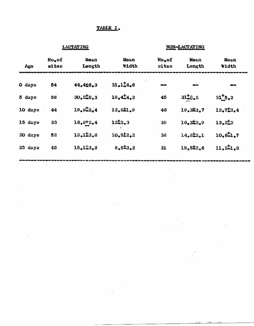
TABLE I.
LACTATING Nm'LAg^ING
AgoNo, of sites
Mean Mean Length Width
No,o£sites
MeanLength
rnmwmimmmmtmmmmtmmmmm
MeanWidth
W i M «««ei M «««■ M«B1
0 days 54 44,4t6«3 31.1+4.6 mmm me##
6 days &H 30.2±6,3 19.4±4,2 45 31w^,S 2ll6,2
XO days 44 19.W±2,4 12,8±1,0 46 19,3±3,7 12.7±2,4
15 days 33 16,8+2,4 12±2,3 39 16.3&2,D 13.2+8
20 days 52 16,1±2,6 10,5&2.2 38 14,8i2.1 10,6±1,7
23 days 46 1S.1&3.6 8,9±2,3 31 10,St2,6 ll,2tl,6

-75-2, Measurements of the pet rial aland
These are given in Table I* It should be noted that
the measurements for the twenty and twenty-five day animals refer
not BO much to the metrial gland taut to the dimensions of the
pigment zone which mark its site.
niSGHESÏÔN
It is clear, both from the histologicjtl sections and
from the measurements, that the involution of the metrial
gland is the same in both lactating and non-lactating animals.
At any given time the histological appearance of the glands
is similar in the two groups. There is no evidence to support
Bely© & McKeown’s view that lipoid containing cells are more
plentiful in the glands of lactating animals or that hyaline
connective tissue is more abundant in this group. An inspection
of Table I shows that the mean length and width of glands of
lactating and non-lactating animals of the same post-partum
age closely correspond to each other. This suggests that
involution is occurring at the same rate in the two groups.
The involution of the gland takes place quickly. This
is illustrated by the measurements given in Table I and by
the sections. The gland rapidly decreases in size during the
first/

-70-/first few days post-partum and by the tenth day it is about
half as long and wide as at the time of littering# By fifteen
days involution is almost complete although a few "encapsulated"
giant cells and occasional lipoid containing cells still
persist# At twenty days involution has finished and the site
of the gland is now marked by a brov*nish patch of pigment#
These findings do not support Baker’s contention that the fifth
and twenty-second day glands-in lactating^animals closely
resemble each other#
Typical metrial gland cello contain glycogen, the loss
of which during histological preparation presumably results
in the vaouolation which is so often found in these cells#
The specific metrial cell granules are eosinophilic, metachromatic
with toluidine blue, and strongly p#A,0# positive# The intense
staining of the granules with the F#A#B# technique suggests
that they contain a mucopolysaccharide# This could also account
for their metachromasia# The marked eoainophilia of the granules
is presumably due to the present of basic groups# Since these
conclusions were reached the papers of Kills (1057) and Wislocki,
Weiss^ Burgos^ and Kills (1057) have appeared# They also found
the specific granules to be p,A.S# positive and metachromatlo
with toluidine blue and suggest that they contain a mucopolysaccharide/

-77-/mucopolysaccharide conjugated with an alkaline protein.
All the specific granules in metrial gland cells were
eosinophilic. Ho basophilic ones were found. Selye &.McKeown
(1935), however, claimed that both eosinophilic and basophilic
granules could occur together in the same cell when stained
with eosin and aniline blue* As Wislocki et al. (1957) point
out, this assertion was based on the misconception that aniline
blue Is a basic dye. In fact, aniline blue is an acid dye
and will thus stain basic aubstanoes* That it stains sosie of
the granules, whilè moat are stained by eosin, merely Indicates
that the iso-electric-point of the individual granules varies
somewhat from one to another - so that their affinity for
individual acid dyes varies*
At the time of littering typical me tria), gland cells form
only a small proportion of the total cell population of the
gland, while none is found after the fifth day of the puerpérium,
They disappear frmi the uterine end of the gland first and
persist longest at the opposite end* There was no indication
that typical metrial gland cells ever; phagocytose pigment or
become lipoid bearing in the way suggested by Selye and MoKeown.
While disintegrating and dying cells of this type were not
Identified, it must be presumed that they degenerate as the
gland/

-78-
/gland regresses. The lipoid containing cells are quite
distinct from the metrial gland cells. They are smaller, never
contain granules, and are always uninucleate, Ellis (1957)
suggests that they may arise from fibroblasts or fibroblast-like
cells within the gland. The third main type of cell found in
the gland is that which takes up the pigment. Those should
be classified as macrophages and are similar to the macrophages
described in the uterus of the mouse by Deno (1937),
It is not possible, with the present material, to determine
whether the "encapsulated" giant cells are of foetal or maternal
origin. They arise, in particular, from the wall of the
disintegrating placental artery but, as they are to be found
at the time.of littering, it is possible that they may have
arisen from trophoblast that has extended along the vessel,
Mossman (1937) states that giant cells may remain in the uterine
wall for long periods after parturition, "Encapsulated" giant
cells persist for about two weeks. The intensive F»A,S, positive
reaction of the "capsule" (which is not abolished by diastase)
and the fact that it is non^aetachromatic with toluidine blue
indicates that it is formed by a neutral mucopolysaccharide.
Cells of this type have not, hitherto, been described.
It will be noted that the placental artery, as it passes
through/

—79—
/through the gland, disintegrates and la replaced by connective
tissue* There is no evidence of the recanalisation of the
vessel in the way described by Goodall (1910) in man.
The present investigation offers no suggestion as to the
function of the gland. As, however, the gland regresses
rapidly during the puerperium it may be presumed that it is
Concerned with the pregnancy itself rather than with the
conditions occurring in the post-partum period, such as
lactation,
SUMMARY
1, Metrial glands of lactating and non-lactating rats
involute in the same way,
2* Involution of the gland is rapid in the early stages,
is nearly complete by fifteen days, and is finished by
the twentieth day of the puerperium,
3é The specific granules of the metrial gland cells possibly
contain a mucopolysaccharide joined to an alkaline protein,
4* Kïetrial gland cells form only a small proportion of the
total cell population of the gland* They do not
phagocytose pipient or become lipoid containing*
S,/

- 80-
5# ’*Encapsulated" giant cells persist for about two
weeks,
6, The placental artery is absorbed in situ.

PMË I I I
ÊcMne characteristics of the uterine pigment

—81—
INTRODUCTION
Selyo & McKeown (1935) and Baker (1948) have noted
the presence of pigment in the metrial glands of the uterus
of the rat after littering. They found that it contained
iron and concluded that it was haemosiderin. Baker also stated
that soma of it stained with Sudan black in frozen sections.
Attention has been drawn to this pigment in the earlier
sections of this thesis. It has been pointed out that the
pigment appears i|i the region of the placental sites, both
in the endometrium and in the metrial glands, on about the
second day post-partum and that it is positive to the P«A«S,
test. As haemoslderin is not usually considered to be
©udanophil or F.A#G, positive this investigation was undertaken
in order to study the characteristics of the pigment in
greater detail. Of the various techniques Employed, many
are such as could be expected to give a positive result with
a substance containing lipid,
MATERIAL AND METHODS
Two rats were killed on the 10th day after littering and
two on the 20th, On examining their uteri there was small
brown discolouration of the mesometrial part of the uterine
horn/

—82—
/h om and meaomotrium at each placental site* Fort lone of
tU© horns containing placental sites were removed and fixed
in neutral 10% formalin for 24 hours* They v/er© dehydrated
in alcohol, cleared in codarwood oil followed by benzene,
and embedded in paraffin wax# Sections were cut, mounted,
and treated with one or more of the following techniques:-
1* For routine histological study haomatoxylin and eosin
staining was used.
2* jBtaining for 10 minutes in 0*5% aqueous toluidine blue*
3* Perl’s reaction for ferric iron*
4# The F*A*S* test, with diastase-treated seotiona as
controls#
5# The perform!© aoid/Bohiff <FFA) reaction, as described
by Pears© (1951)*
6* The peracetic acid/Sohlff reaction, by Lillie’s
technique (1954)*
7* Oxidation for one hour in 4% chromic acid followed by
45 minutes in Sohiff’s reagent*
8* Exposure of a section without previous oxidation to
Echiff’s reagent for periods up to 18 hours*
9* Staining in a saturated solution of Sudan black in 70%
alcohol for 3 hours, rinsing in 70% alcohol,»and mounting
in/
— 83—
/in glycerine jelly*
10, The long Ziehl-Neelaen method as employed by Fearse
(1953)*
11* Gomori’s chrome alum/haematoxylin method*
12* Mallory’s technique for haemofuscins*
13* The ferric ferricyanide reduction test, by Lillie’s
method (1954)*
14* (a) Bromlnation, and (b) treatment with 5% chromic acid
for 1 hour, followed by the ferric ferricyanide
reduction test*
15* The Masson-Fontana alkaline silver technique*
10* The Gmelin test for hàématoidln*
17* Bleaching in hydrogen peroxide for 48 hours*
18* Exposure to ultra-violet light for fluorescence*
OBSERVATIONS
The distribution, appearance, and properties of the
pigment were the same in all the specimens that were examined,
whether from the 10 day or 20 day rats* Pigment was found
in large quantities at each of the old placental sites* All
the pigment was intracellular and the cells containing
pigment/

-84-/pigment were grouped either in the remains of the metrial
gland situated in the mes<»netrial triangle or in the
mesometrial half of the endometrium, where they lay close
to the myometrium (Fig# 34)# The pigment was yellow to
light brown and formed granules that varied in size from fine
particles to those that were 10 or more microns in diameter#
In assessing the results of the various techniques that
were employed, allowance must be made for the original colour
of pigment because it is on to this that any second colour
is superimposed# For instance, a method which gives a blue
colour as a positive may with the pigment produce a green
or greenish-blue shade# The observations described in detail
below are summarised in Table II#
The pigment was not stained by either haematoxylin or
eosin, but with toludine blue it became a deep greenish-blue,
in parts appearing almost black* Ferl’s test for ferric
iron gave a strong positive reaction and resulted in all
the pigment granules becoming a deep blue, while a lighter
bluish-green shade was imparted to the cytoplasm of the cells
containing pigment# The pigment reacted strongly with the
F*A*B* technique, the granules becoming a deep brownish-red
which could be readily distinguished from their normal colbur#
The/

-85/The same colouring of the pigment was also found in the
diastase controls, and thus the possible presence of glycogen
was excluded# The pigment remained unooloured after exposure
to Schiff’s reagent for 18 hours when no previous oxidation
was employed# This shows that the F#A*S. method was giving
a true positive reaction and was not responding to the presence
of preformed aldehyde groups in the pigment# The P#F#A#
reaction, the peracetic acid/Sohiff reaction, and oxidation
with chromic acid followed by Schiff’s reagent all failed to
produce any colouring of the pigment# It was sudanophil,
most of the particles giving a greyish colour with Sudan
black, although some of the larger ones became a deep black#
It was acid-fast, resisting the acid alcohol differentiation
in the long Eiehl Heelsen method, so that while the background
was a pale pink the granules were a deep brownish-red# With
the chrcme alum/haematoxyXin technique of Oomori the pigment
took up some of the staln and although the original yellow
colour was not completely abolished, the effect of the
haematoxylin could be easily recognised# After Alcoholic
differentiation in Mallory’s technique for haemofuscins some
of the basic fuohsin was retained by the pigment granules
which/

—80—
/which became a light reddish-brown# With the ferric
ferricyanide reduction teat the section as a whole v/aa coloured
a pale green but the pigment was more markedly affected#
Nearly all the granules were greenish-blue# A positive
reaction with this test is normally indicated by a blue
colour# Nevertheless, the granules are regarded as being
positive because they colour (although for the most part
in green) much more deeply than the background# After
foromination and the chromic acid treatment the granules
remain uncoloured by the ferric ferricyanide method#
Treatment with alkaline silver resulted in slight darkening
of the granules which beoame browner# The Gmelin test for
haem&toidin was negative while the pigment was not bleached
byihydrogen peroxide# There was no flviorescence under the
microscope when viltra vlolet light was employed#
DISCUSSION
This investigation confirms the findings of Selye &
McKeown and of Baker that the pigment contains ferric iron#
These writers were also undoubtedly right in regarding the
pipient as haemoslderin as this substance is by definition
a pigment which exhibits one or more of the reactions of
ferric/

TABLE II
Teohnlau© employed
Haematoxylin and eosin Toluidine blue
Perl’s method Periodic acid/Bchiff Bchiff’s reagent i
performic acid/Bchiff Paraoetic acid/Bchiff Chromic acid/Schlff Long SSiehl-Neelsen
Sudan black
Gomori’s chrome alum/ haematoxylin Mallory’s technique Ferric ferricyanide Bromlnation and ferric ferricyanide
Chromic acid and ferric ferricyanide
Alkaline silver Gmelin test
Hydrogen peroxide
Fluorescence (Ü#V*>
Colour of Piament
unchanged deep greenish-blue
deep bluebrownish-redunchanged
unchangedunchangedunchangedbrownish-red
grey to deep black
greenish-blue
pale reddish-brown green to greenish-blue unchanged
unchanged
brownunchanged
unchanged
Comments,
basophil with a basic dye ferric iron present a strong positive no preformed aldehyde group present negative negative negative acid fastsuggesting a lipid suggests presence of a lipid a positive reaction a weak positive a positive reaction negative
negative
a weak positive no haematoidln present an absence of bleaching no fluorescence observed#

-87-/ferric iron (Lillie, 1964), Also, the uterine pigment
resists bleaching by hydrogen peroxide and this is a
characteristic of haemoslderin# According to Lillie (1954)
three varieties of haemosiderin can be recognised, one of
which stains with basic dyes as does the uterine pigment with
toluidine blue# Haemoslderin is one of the breakdown products
of haemoglobin (Florey, 1054) and the pigment in the rat’s
uterus is certainly derived from the blood, as it is found
almost entirely at the old placental sites into which
haemorrhage occurs at the time of separation of the placenta#
Unlike the pigment in the rat’s uterus, human haemoslderin
is not P«A#B# positive (Lillie, 1050)# According to Foarae
(1953) haemoslderin is not acid-fast, does not stain with
Sudan black nor colour with Gomori’s chrome alum/haematoxylin
technique, is negative in the ferric ferricyanide test,
and does not darken alkaline silver# All these techniques, as
well as Mallory’s technique for haemofuscins give positive
results when applied to the uterine pigment* I# is thus clear
that while the uterine pigment may be regarded aa a haemoslderin,
it possesses several properties that ar© not usually associated
with this substance*
The colouring with Budan black suggests that there is a
lipid/

-88-
/lipid element present, and other positive reactions, as
with the P#A«B. reaction and the Kiehl-Neelsen technique,
support this# A comparison may be made between the uterine
pigîïiont and the lipogenio pigments, the properties of which
have been summarised by Gomori (1962), Pears© (1953), and
Lillie (1954). These pigments, which are fairly widely
distributed, are known by a variety of names such as ceroid,
luteolipin, wear and tear pigment, and lipofuscin. They
all arise as oxidation products of a lipid precursor
(Fearse, 1953)» Their reactions with different histological
and histochemioal techniques vary and this may be related
to the degree of oxidation that they have undergone* Like
the pigment in the rat’s uterus they withstand lipid solvents
and can be identified in paraffin sections. Agsin, many of
the lipogenio pigfiients such as adrenal lipofuscin are acid-
fast and F.A.B. positive, react with ferric ferricyanide,
and colour with oil-soluble colouring agents in paraffin
sections* They may darken with alkaline silver, colour with
Gomori’a chrcane alum/haematoKylin, and retain basic fuchsln
in Majlory’s technique# In these respects the uterine pigment
resembles them# It differs from scans such as ceroid which is
positive/
'v-j

—80*
/positive to peracetic acld/Schlff and fXuoresoee in ultra
violet light* Hor are the llpogenlc pigments usually
aoBoclated with Iron, although ceroid may occasionally
contain a few granules of pigments containing iron*
The negative Gmelin reaction indicates that haematoidin,
the Iron free breakdown product of haemoglobin, is absent*
Although the uterine plient in some respects resembles
melanin it is not melanin, for granules of the latter
substance are darker in colour, do not stain with Budan black,
nor give the F*A*B« reaction, and are usually bleached within
48 hours by hydrogen peroxide.
It thus appears that the uterine pigment should be
regarded as a variety of haemosiderin in which the substance
containing iron is mixed or perhaps combined with a lipid
component that has many properties in cmmion with the lipogenic
pigments*
BbMMARY
1* A yellowish-brown pigment was found at the old placental
sites in rats killed at 10 and 20 days after littering*
2* The pigment contained ferric iron and therefore may
be regarded as hae ioslderin*
3*/

-00-
3# Other properties of the pigment suggest that there
Is also a lipid component present# Lipid Is not usually
associated with haemoaiderin#
4# The lipid component behaves in many ways like the
lipogenic pigments#

—91—
BIBLIOGRAPHY
ABEY, L.B. (1936), Physiol, Rev,, IjS, 327,
ASCHOPP, L, (1899), Beltr. path, Anat,, 2, 275,
AYKROYD, 0,E, and GATENBY, J.B, (1941), Quart, J, mlor.Sol,, 1 2, 641,
BAKER, 0,L. (1948), Proo, Soo, exp, Biol, N,Y,, ^ 492,
BRADFIBLO, J,R,6, (1051), Nature, Lend,, 167. 40,
BRIDGMAN, 3. (1948), 3. Morph,, 83, 61,
BULL, M.M, (1949), J, Obstet, Gynaeo,, SU» 799,
CIIIPMAN, W, (1903), Lab, Rep, Roy. Coll, Phys,, Edlnb., a, 225
CRUVEILHIER, J, (1334), Anatomie Descriptive, Paris, ^ 780,
D'ERCHIA, F, (1897), Monatesohr, £, Qeburtsh, u, GynSk,St 596,
DeLANCæ, O, (1934), E. mikr,-anat. Poreoh., Jg, 488.
DeSIMÉTY, L, (1876), Ann, Oyneo, g, 217,
DKNO, R,A, (1937), Amer, J. Anat,, gg, 433,
DUKE, K.L., (1957), Anat, Rec,, 127. 411,
DUVAL, M, (1891), J, Anat, Paris, gg, 515,
ELLENBOGEN, V, (1930), Z, Anat, Entw-Geach,, ^ 749.
ELLIS, R,A. (1967), Anat, Rec,, 129. 39,
FARB£^ A* (1860), in Todd*is Cyclopaedia of Anatomy and physiology, ^ 600#
ÿliOREY/

«92-
FLOREY, H. (1954), Lectures In General Pathology, Lond,
FRIEDENWALD, J.8. (1950), Candor Res., Ig, 461,
FRIEDL%NDER, C, (1870), Physiol-anat. Untoreuchungen Uber den uterus, Leipzig.
FRIEDlXnDER, C, (1876), Arch, Gynaek,, g, 23,
GOMORl, Q, (1952), Microscopic Histochemistry, Chicago,
GOODALL, J.R'. (1910), Studies frc«i the-Royal Victoria )'Hosp, Montreal,, 11,. 3,
GREBNWALD, 0,8. (1958), J, Endocrinol,, jV, 7,
HAMILTON, W.J. (1933), Trans, Boy, Soo, Edin,, 57, 593,
HESCHL, R, (1352), Zeit, £,d, K,K, Geaellsoh, d, Aertze in Wien., 8, 228,
IIIN8ELMANN, H, (1913), Zeit, £, Geburtsh, u, Gynak,, 73, 146,
HORNING, E,8, (1942-4), J, Endocrinol,, g, 260,
HUNIER, W, (1774), Anatomy o£ the Gravid Uterus, Birmingham,
KLEIN, E,E, (1891), Belt, t, Geburtsh, u. Gunak,, g2, 247,
KR&IG, Quoted by J,«, Williams, 1931,
KUNDRAT, H. and ENGELMANN, Aid. (1873), Gtrickers med Jahrb,, Wien,, 3, 135.
LANGHANS, T, (1875), Arch, Gynaek,, g, 287,
LBBLOND, C,P, (1950), Amer, J, Anat,, ag, 1,
LEOPOLD, Q, (1878), Arch, Gynaek,, 11, 443,
lEUSDEN, P, (1397), Zeit, f, Geburtsh, u, OynKk,, gg, 1,
LILLIE, H,E. (1950), Anat, Rec,, 108. 239,
LILLIE/

%63-
LILLIB, KéD. (1954), Hiatopathologlcal technique and practical histochemistry. New York,
LONG, J.A. and EVANS, K.M, (1922), Mem, Unlv, Calif, g, 1,
MAYOR, A, (1887), Ann, de Phys, Nona, et Path,, g, 560,
MoMlNN, R,M,H, and JOHNSON, F,S. (1955), Brit, J, Surg,43, 99,
MoMlNN, R,M,H, and JOHNSON, F,R, (1957), Brit, J, Surg,, 45, 76,
MCMINN, R,M,H, and JOHNSON, F,R, (1958a), J, Bmbryol, exp. Morph,, g, 288,
MoMlNN, H,Mj,H. and JOHNSON, F,R, (1058b), J, Embryol, exp. Morph,4 6, 509,
McMINN, R.M4H, and MITCHELL, J,E, (1954), J, Anat, Lond,, 99*,
MILSTEIN, B,B, (1950), Brit, J, exp. Path, gi, 664,
MOLL, J,M, (1806), Arb, anat, Inst, Wiesbaden, 40^ 613,
M0S6MAN, H,w, (1937), Contr, Embryol, Cameg, Inatn,, gg, 133
NICOL, T, (1933), Trans, Roy, Soo, Edin,, 765,
PEARSB, A,G,E. (1951), Quart, J, micr. Soi,, 92, 393,
PEARSË, A.G.E, (1953), Histochemistry, theoretical and applied, London,
PRITCHARD, J,J, (1949), J, Anat. Lond,, gg, 10,
RIBS, R, (1892), Zeit. f, Geburtsh, u, GynKk,, 2 ^ 33,
ROBIN, M, (1848), Arch, Gen, Med, Ig, 186,
RUTHERFORD, R,N, and MEZER, J, (1942), J, Amer, Med, Asa,, 119, 124,
GCHICKELE/

*94*
SCillCKBLE, G. (1910), Beltr. Geburtsh. Gynak., 18, 530.
SCHROEDER, B. (1030), «The Uterus in the Puerperium" in Mollendor££*e llandbuch.
GCOTHORNE, R.J. and SG0ÏH0RNE, AM. (1953), J. Anat. Lend., 87. 38.
SELYE, II., BOBDUAS, A. and MAGGON, G. (1042), Anat. Rec. 82. 199,
SELYE, H. and McKBOWN, T. (1935), Proc, Roy, Soo,, 8,119, 1,
BHABMm, A. (1952), J, Endocrinol,, g, 162.
BHARMAK, A. (1953),; d. Anat. Lond., 87, 1.
STOLPER, L. and HERRMANN, E. (1904), Arch. mikr. Anat.,63. 748.
BTRAHL, II. (1895), Arb, anat, Inst, Wiesbaden, 5, 337.
STRAHL, a. (1903), K. Akad. ¥/etensoh, Amsterdam, Proo. Sect Sol,, 6, 1, 302.
STRAHL, H. (1906a), K. Akad, Wetensch. Amsterdam, Proo. Sect, Sol,, g, 1, 812.
STRAHL, H, (1906b), Ergobn. Anat. Entwgesch^, IS. 581.
STRAHL, H. and HENNBBERG, G. (1901), Anat, Antei, gg, 20,
STRAHL, JI. and HENNEBERG, G. (1902), Anat, Ann,, gi, 644,
TEACHER, dill. (1927), d, Obstet. Gynaeo,, gg, 1.
TURNER, W. (1876), d, Anat. Lond,, gg, 433.
VELARDO, J.J., DAWSON, A.B., OLSEN, A.G., and HISAW, P.L, (1963), Amer. J. Anat., eg, 273.
WBRTH/
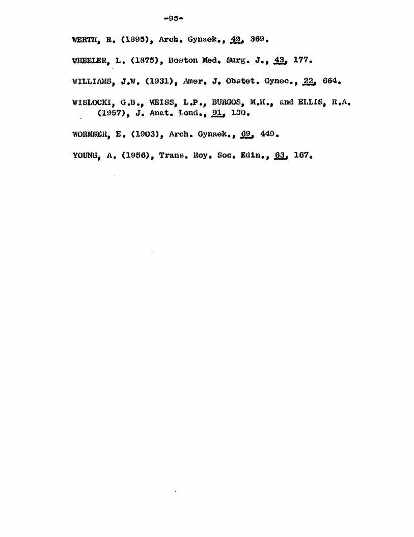
-98-
WBBTH, B. (1305), Arch. Gynaek., J9, 309.
-WHBEUEH, L. (1875), Boston Med. Surg. d., 43, 177.
WILLIAMS, d.W. (1931), Amer, d, Obstet. Oyneo,, 22, 664,
WISLOCEl, G.B., WEISS, L.P., BURGOS, M.H., and ELLIS, R.A. (1957), d, Anat, Lond,, 91, 130.
WORMSBH, E. (1903), Arch. Gynaek., ^ 449.
YOUNG, A. (1986), Trans. Boy, Soc, Edin,, Jg, 167,

Pig# 1 # Transverse section of the uterusimmediately after littering# Note the blood clot at the placental site and the folding of the mucosa# H# & £#
X 6*5#

Fig# 2. Part of a transverse section of uterusat 0 hours post-parturn stained with toluidine blue. Note the intense staining of the cytoplasm of tkie epithelial cells.
X 485.

4
FI 3. Portion of a trrmeverBe section ofuterus from the same animal as Fig. 2, stained with toluidine blue after extraction with perchloric acid. Note the decrease in cytoplasmic basophilia.
X 485.

- — I
Fig. 4. Transverse section of uterus from the same animal as Fig. 2, stained by the P.A.S. technique and showing cytoplasmic granules, surface P.A.S. positive layer and basement membrane.
X 485.

6
Pig. 5. Transverse section of uterus at 0 hour post-partum. Note the darkly stained "rod" cells. H. & E#
X 1050.

Fig. 6 . An "encapsulated” raaltinucleate giant cell. H. & E.
X 1050.

ms
i
Plg. 7. Part of a transverse section of uterus 6 hours after littering showing the edge of the "bare area” at the placental site. H. & B.
X 105.

M
Pig. B. Transverse section of utoruo 12 houropopt-partura. Note the infiltration of polymorphonuclear leucocytes in the epithelium. H. k E.
X 485.

'•\V
?lr. 9e Tranoveri?e nection of u uterus at 5t hours pof3t-par turn. The folding of the endometrium is clearly chown. H. & E.
X 12.

m
w i m
' . t : ' ' ' ,/Ai\ V :
Flg» lü. Transverse section of a placentalsite at 24 hours post-partum. The site has been re-epithelialized and the hyper- brophic epithelial cells are resting on a mass of blood and fibrin clot. Toluidine blue.
X 105.

*
11. Part of Fig. 10 at a higher magni- "^ication. Normal epithelium is to the left and hype rtrophic epithelium is to the right. Note the reduced cytoplasmic basophilia of the hypertrophic cells. Toluidine blue.
X 485.

%
Fig# 12. Transverse section of uterus from the same animal as Fig. ^0. The epithelial cells show early vaouolation. H. & E#
X 485.

m
Tip, 13» Bndometrlum of a 24 hour post-partumanimal to ohow dilatation of the blood veaaela*H. 8r B.
X 75.

-'S’
Fig. 14. Transverse section of the uterus at 36 hours post-partum# to show the folding of the endometrium. h. & E.
X 12.

Fie. 15. Transverse section of the uterine epithelium at 36 hours to show the exten- olve vacuolatlon of the cells. H. & E.
X 485.
f i €'■TA-
q
# e
''4W .% % 4ë
Flg. 16# Note the basement membrane revealedhy the P#f\.S. technique. Tranaveree oection.
X 2fX).

Pig. 17. Traneveroe section of a uterus at43 hours post-partum. Note the ellt-like lumen and absence of folding of the endometrium. H. & E.
X 12.

Plg. 18. Transverse section of uterineepithelium at 48 hours post-partum. Note the low columnar shape of the cells and the absence of vacuolatlon. H. & E.
X 485.

SffS'.ài'
Flf. 19# The remnento cf tlio placental arteiy in the endometrium at 4 e poet-partum.P«A«S•
X 100.

Fig. 20. Tranoverse section of uterus at the time rfFterine in an interoite region to snow the meoometrial triangle. H. & E.
X 50.

Fig. 21. A metiial gland at 0 hours. Transverse section. H. & E.
X 20.

s1 %&
Fig, 22# Typical caetrlal gland cells from a ft hours specimen. H. & E.
X 800.

&
Pig# 23# Motrlal gland cells from a ceotlon stained with toluldine blue# Note the presence of the granules#
X 800#

Pig. 24# Metrlal gland cells stained by the ^A.S. reaction.
X 800.

Fl£e_25j^ Me trial gland cells stained by the•A*S. reaction after treatment with diastase.
X 800.

Pig, 26. Transverse section of a metrlal gland at the time of littering to show the distribution of metrlal gland cells. Stained P.A.S.
X 180.

Fig. 27. Vacuolated cells in a metrlal gland at 0 hours. H. & E.
X 1050.

f
m .
PlK* 28. A maat cell in eano section asrip. 23. Note that the cell ie so densely filled with f^runules am to appear as a black maes. Toluldine 'Rlue.
X 8ÜÜ.

Fig. 29. A P.A.S. stained section to show metrial gland cells at 2 days post-partum^
X 180.
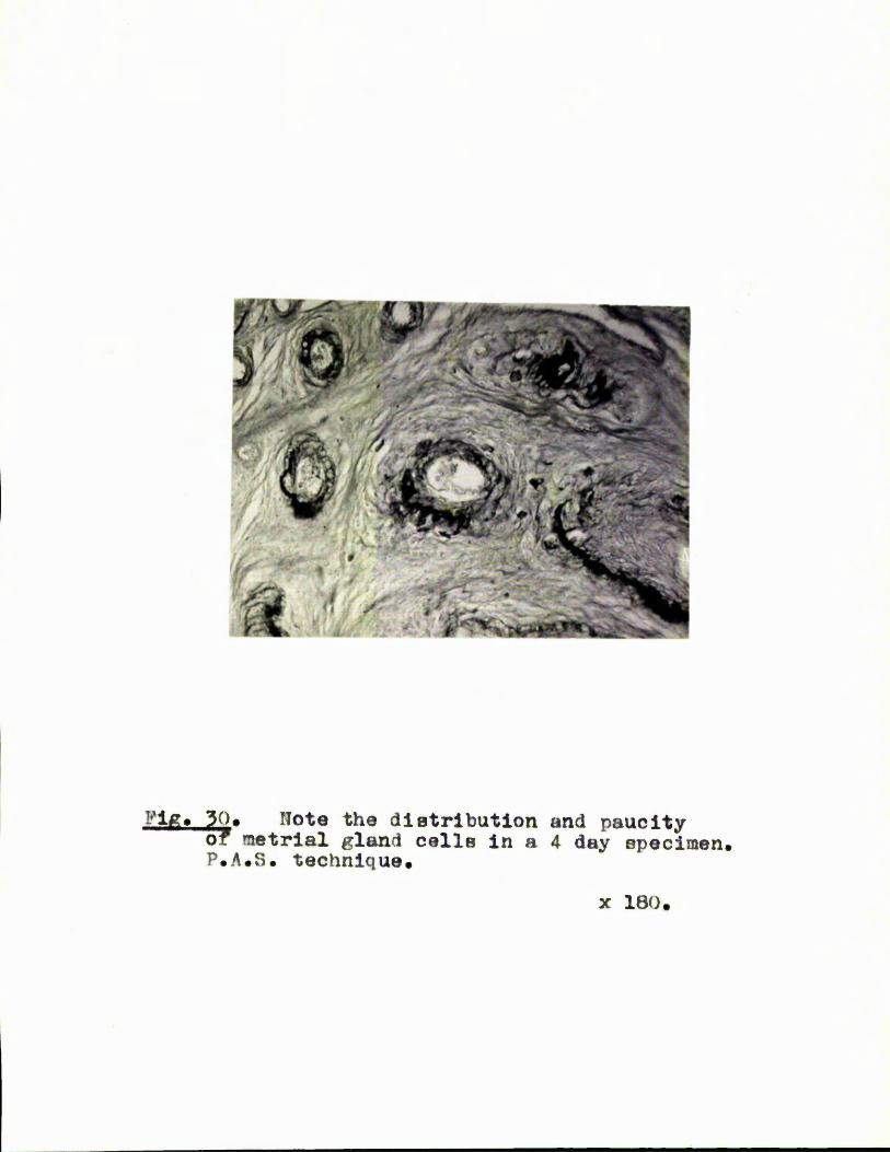
Fig. 30. Note the distribution and paucityof metrial gland cells in a 4 day epecimen, P.A.8. technique#
X 180.

y
A' w
SdfM,
Pig. 31. The placental artery in the metrlal ^ a n d shown by the P.A.S. technique in a 4 day specimen.
X 100.

Pig. 32. Transverse section through a metrial gland at 5 days post-partum in A lactating and B non-lactaxing animals. H. & E.
X 25.

g \'K ■■ ')
Pig. 33. Transverse sections through metrial glands in A lactating suid B non-lactating animals at 10 days post-partum. The greater size of the uterine horn in the non-lactating animal is due to the recommencement of oestrus cyclea H. & K.
X 25.

m mi. ‘
Ë
Flg. 34. Transverae oectiona of metrial glanda at 10 day8 poax-partum In A lactating and B non-lactating animais to snow the reomants of the placental artery. P.A.S.
X 80.

- 0
Pig. 35. Metrial glanda of A lactating and Bnon-lactating animale at 15 days post-partum, T. S. H. & E.
X 25.

Fig. 36. An ”encapsulated” giant cell in the endometrium at 15 days post-partum. Note the P.A.S. positive capsule. P.A.S.
X 1Ü5Ü.

Fig. 37. Metrial glands from A lactating and B non-lactating animals at 20 days post-partum. T. S. H. & É.
X 25.