2006McGregorPhD.pdf - Enlighten: Theses
238
https://theses.gla.ac.uk/9064/ Theses digitisation: https://www.gla.ac.uk/myglasgow/research/enlighten/theses/thesesdigitisation/ This is a digitised version of the original print thesis. Copyright and moral rights for this work are retained by the author A copy can be downloaded for personal non-commercial research or study, without prior permission or charge This work cannot be reproduced or quoted extensively from without first obtaining permission in writing from the author The content must not be changed in any way or sold commercially in any format or medium without the formal permission of the author When referring to this work, full bibliographic details including the author, title, awarding institution and date of the thesis must be given Enlighten: Theses https://theses.gla.ac.uk/ [email protected]
-
Upload
khangminh22 -
Category
Documents
-
view
0 -
download
0
Transcript of 2006McGregorPhD.pdf - Enlighten: Theses

https://theses.gla.ac.uk/9064/
Theses digitisation:
https://www.gla.ac.uk/myglasgow/research/enlighten/theses/thesesdigitisation/
This is a digitised version of the original print thesis.
Copyright and moral rights for this work are retained by the author
A copy can be downloaded for personal non-commercial research or study,
without prior permission or charge
This work cannot be reproduced or quoted extensively from without first
obtaining permission in writing from the author
The content must not be changed in any way or sold commercially in any
format or medium without the formal permission of the author
When referring to this work, full bibliographic details including the author,
title, awarding institution and date of the thesis must be given
Enlighten: Theses
https://theses.gla.ac.uk/

Studies on a Thiol-Dependent Reductase and Ascorbate Metabolism of Leishmania
Joanne Catherine McGregor
This thesis is presented in submission for the degree of Doctor of
Philosophy
Division of Infection and Immunity
Faculty of B iom edical an d Life S c ie n c e s
University of G lasgow
A ugust 2006

P roQ uest N um ber: 10390434
All rights reserved
INFORMATION TO ALL USERS The quality of this reproduction is dependent upon the quality of the copy submitted.
In the unlikely event that the author did not send a com p le te manuscript and there are missing pages, these will be noted. Also, if material had to be removed,
a note will indicate the deletion.
uestProQuest 10390434
Published by ProQuest LLO (2017). Copyright of the Dissertation is held by the Author.
All rights reserved.This work is protected against unauthorized copying under Title 17, United States C ode
Microform Edition © ProQuest LLO.
ProQuest LLO.789 East Eisenhower Parkway
P.Q. Box 1346 Ann Arbor, Ml 48106- 1346

CLASGOW^ UNIVERSITY LIBRARY; ^

AbstractThe intracellular protozoan parasite Leishmania causes leishmaniasis, a disease which is
most prevalent in tropical and sub-tropical countries where it infects some two million
people every year and kills around 60,000 of them. For decades pentavalent antimonial
compounds have been the standard first-line drugs used to treat the disease and this
remains the case despite increasing reports of drug-resistance. The mode of action of these
drugs is not entirely understood, although it is generally accepted that in vivo reduction of
the compounds from the pentavalent to a trivalent form is required for antileishmanial
activity. The site of antimonial conversion and whether the reaction is catalysed by an
enzyme remain controversial points. However, it was recently reported that L. donovani amastigotes were capable of reducing pentavalent antimonials to the trivalent form and that
drug-resistant parasites were deficient in this activity, suggesting that a parasite enzyme
did mediate drug toxicity. The identity of such an enzyme was investigated in this study.
Arsenical and antimonial compounds are similar and several classes of proteins that exhibit
arsenate reductase activity have been previously identified in other organisms. Whether
Leishmania possessed an enzyme akin to one of these was assessed by attempting to purify
enzymes from parasite lysates and by searching the L. major genome database for similar
sequences to the arsenate reductases. The latter approach was successful and a gene
fragment was identified that shared similarity with omega glutathione S-transferases
(oGSTs), a class of glutaredoxin-like GSTs which are capable of reducing pentavalent
methylated arsenicals in vitro. The sequence of the complete L. major gene was elucidated
by 5’ RACE, and was found to encode a protein tvfice the expected size with similar 3’ and
5’ halves. The protein was named thiol-dependent reductase, or TDRl. Active recombinant
protein was successfrilly produced and its biochemical activities were found to coincide
with oGSTs: TDRl was capable of reducing pentavalent arsenical and antimonial compounds to trivalent species, and possessed thioltransferase and dehydroascorbate
reductase activities usually associated with glutaredoxins. TDRl, which was shown to
probably reside in the parasite cytosol but may also be secreted, was found to be more
abundant in amastigote than promastigote forms, which correlates with the antileishmanial
stage-specificity of pentavalent antimonials. L. major TDRl knockout mutants were
generated, and the protein was also over-expressed in parasites. Both these genetic
manipulations resulted in mutants with enhanced inactivity.
TDRl knockout parasites were more susceptible than wild type parasites to paraquat,
which induces the production of intracellular superoxide. As its glutaredoxin-like in vitro

Ill
activities suggest, this implies TDRl has a role in protecting the parasites from oxidative stress, although re-expression of TDRl did not reinstate resistance. Whether TDRl has a
role in susceptibility to pentavalent antimonials was investigated by studying the effect of the drug on L. major in macrophages. There appeared to be little difference in the effect of
the drug on TDRl knockout, over-expressing and wild type parasites, although variation in
inactivity to macrophages and the insensitivity of L. major to the drug complicated the
situation.
Whether trivalent antimonials could be oxidised to non-trivalent species by hydrogen
peroxide was assessed. This was indeed found to be the case, demonstrating that in
principle antimonial metabolism may be more complex than straightforward reduction
from the pentavalent to trivalent form. However, the physiological relevance of this finding
is uncertain due to oxidation of the antimonials being inhibited by glutathione.
The dehydroascorbate reductase activity of TDRl was of interest as the presence of an
enzyme capable of maintaining ascorbate in its reduced form may imply that this low- molecular weight thiol is important in the parasite. The recent identification of an
ascorbate-dependant peroxidase in Leishmania added further credence to this hypothesis.
Many organisms produce ascorbate de novo although, due to the loss of an important synthesis enzyme, humans cannot and have to scavenge ascorbate from their diet. If
Leishmania does require ascorbate and rely on de novo production, enzymes that mediate
synthesis could feasibly be exploitable drug targets. Whether Leishmania was capable of producing ascorbate and its importance in the parasite was investigated. The L major
genome database was searched for sequences similar to those of enzymes known to be
involved in ascorbate synthesis in other organisms. Several candidate protein sequences
were identified including that of one which is similar to L-gulono lactone oxidase (GLO),
the enzyme that mediates the final step in ascorbate production in a variety of organisms
and is the protein humans no longer possess. This L major sequence was named LmGLO. L. major LmGLO knockout mutants were generated and the protein was also over
expressed in parasites. While over-expression resulted in parasites being more infective,
loss of LmGLO resulted in decreased infectivity, both in vitro and in vivo. In addition,
LmGLO knockout promastigotes displayed a slight growth defect. Although these results
need extending, they suggest that Leishmania parasites do indeed synthesise ascorbate and
that this ability is important for optimal virulence and infectivity to mammals.

IV
Table of Contents
ABSTRACT II
TABLE OF CONTENTS IV
LIST OF TABLES VIII
LIST OF FIGURES IX
ACKNOWLEDGEMENTS XII
DECLARATION XIII
ABBREVIATIONS XIV
1 INTRODUCTION 1
1.1 The Leishmania parasite 11.1.1 The Leishmania life cycle 11.1.2 The Leishmania genome and regulation of gene expression 31.1.3 Redox regulation in trypanosomatids 3
1.2 Leishmaniasis 5
1.3 Chemotherapy and resistance 81.3.1 Pentavalent antimonials 91.3.2 Miltefosine 91.3.3 Pentamidine 121.3.4 Amphotericin B preparations 131.3.5 Drugs not yet licensed: paromomycin, azoles and sitamaquine 14
1.4 Pentavalent antimonials 151.4.1 Synthesis and structures 161.4.2 Uptake of antimonials 161.4.3 Antimonial metabolism 181.4.4 Modes of actions o f antimonials 281.4.5 Resistance mechanisms and exclusion o f antimonials from Leishmania 30
Aims of this study 32
2 MATERIALS AND METHODS 33
2.1 Parasites 332.1.1 Leishmania culture 332.1.2 Preparation o f Leishmania from mice 332.1.3 Isolation of metacyclic forms of Leishmania major 342.1.4 Leishmania harvest and lysis 342.1.5 Leishmania major cryo-preservation 3 42.1.6 Bioassays for leishmanicidal activities 352.1.7 jL. wq/or infectivity 36
2.2 Molecular biology techniques 362.2.1 Isolation of genomic DNA from Leishmania 362.2.2 Ethanol precipitation of DNA 372.2.3 Isolation of RNA from ZeisAwart/a 372.2.4 Rapid amplification o f cDNA ends (RACE) 382.2.5 Polymerase chain reaction (PCR) 392.2.6 DNA gel electrophoresis 40

V
2.2.7 DNA quantification 402.2.8 Cloning of PCR products 402.2.9 Subcloning of DNA fragments 402.2.10 Restriction digests 412.2.11 Ligations 412.2.12 Plasmid DNA extraction 422.2.13 DNA sequencing 422.2.14 Competent cells 422.2.15 Transformation of competent cells 432.2.16 Cryo-preservation of bacterial cultures 432.2.17 Creation of transgenic Z. major promastigotes 442.2.18 Southern blot analysis 45
2.3 Biochemical methods 472.3.1 Recombinant protein expression in E. coli 472.3.2 Recombinant protein purification 472.3.3 His-tag cleavage 482.3.4 SDS-PAGE 482.3.5 Detection of proteins on polyaciylamide gels 492.3.6 Protein concentration determination 492.3.7 Confirmation o f axenic amastigotes by protease expression profile 492.3.8 Antibody production 502.3.9 Antibody purification 502.3.10 Western blot analysis 512.3.11 Immuno-localisation of cellular protein 522.3.12 Isolation of protein using S-hexyl-GSH agarose 532.3.13 BPR assay for measuring trivalent antimonials 53
2.4 Statistical analysis 54
2.5 Bioinformatic analyses 54
3 PURSUIT OF LEISHMANIA GENES AND PROTEINS INVOLVED IN
PENTAVALENT ANTIMONIAL ACTIVATION 56
3.1 Arsenate reductase homologues in L, major 573.1.1 Microbial Arsenate Reductases 583.1.2 Mammalian arsenate reductases 64
3.2 Analysis of omega glutathione S-transferase-like genes in Z. major 68
3.3 Omega GST-like sequences in the Z, major genome 703.3.1 lm l6 and lm34: analysis of two oGST-like sequence fragments 713.3.2 Genes annotated as glutaredoxins and thioredoxins in the Z. major genome 723.3.2 Genes annotated as glutaredoxins and thioredoxins in the Z. major genome 73
3.4 Analysis of S-hexyl-GSH~binding proteins in Leishmania 773.4.1 Isolating Leishmania proteins using S-hexyl-GSH sepharose 773.4.2 The range o f Leishmania proteins isolated using S-hexyl-GSH sepharose is reproducible 783.4.3 Similar proteins are isolated from different species of Leishmania with S-hexyl-GSH sepharose
803.4.4 Identification of proteins isolated from Leishmania with S-hexyl-GSH sepharose 81
3.5 Identification and analysis of TDRl (lm33) 823.5.1 Identification of the complete TDRl gene 823.5.2 Tc52: Identification o f a TDRl homologue in T. cruzi 843.5.3 Amplification and cloning of TDRl 8 6
3.6 Discussion 87
4 ANALYSIS OF TDR1 : CHARACTERISATION OF RECOMBINANT PROTEIN AND
EXPRESSION PROFILE IN LEISHMANIA 92

VI
4.1 Production of TDRl4.1.1 Expression o f TDRl and 5 ’ TDRl in E. coli4.1.2 Purification of recombinant TDRl4.1.3 Cleavage of the His-tag from recombinant TDRl4.1.4 Isolation of TDRl from Leishmania using S-hexyl-GSH Sepharose
9393959697
4.2 Biochemical characterisation of recombinant TDRl 98
4.3 Crystalisation of recombinant TDRl 98
4.4 Temporal Expression of TDRl 984.4.1 L. mexicana Axenic Amastigote-lDce Forms Display a Similar Protease Expression Profile asLesion Amastigotes 984.4.2 Detection o f TDRl in the Soluble Fraction of Leishmania 994.4.3 TDRl is uniformly expressed in L. major promastigotes 1004.4.4 Increased expression of TDRl in Z. wq/or amastigotes 101
4.5 Spatial Expression of TDRl4.5.1 Analysis of Secretion of TDRl from Z. major4.5.2 Immuno-localisation of TDRl in Z. major Promastigotes
102102103
4.64.6.14.6.24.6.3 H 2 O 2
4.6.44.6.5
Analysis of oxidation of trivalent antimonials by hydrogen peroxide 105Trivalent potassium antimonyl tartrate is oxidised by H2O2 106The effect of pH and recombinant TDRl on oxidation o f KAT by H2O2 107The trivalent product formed upon reaction of TDRl with sodium stibogluconate is oxidised by 108GSH Inhibits Oxidation of KAT by H2O2 while GSSG has no effect on the reaction 109Analysis of KAT and Sodium Stibogluconate upon Incubation with Leishmania 111
4.7 Discussion 112
FUNCTIONAL STUDY OF L MAJOR TDR1 116
5.1 Introduction 116
5.2 Over-expression of TDRl in Leishmania5.2.1 Creation of TDRl over-expressing Z. major promastigotes5.2.2 Analysis of over-expression5.2.3 Phenotypic analysis of TDRl over-expressing Z. major
118118119119
5.3 Knocking-out of TDRl In Z. major5.3.1 Creation of Z. major TDRl knock-out parasite lines5.3.2 Analysis of knock-out lines5.3.3 Phenotypic analysis of TDRl knock-out lines
125125126 127
5.4 Re-expressing TDRl in TDRl knock-out parasite lines5.4.1 Analysis of re-expression5.4.2 Growth of parasites re-expressing TDRl, with paraquat
143144 144
5.5 Discussion 146
6 FUNCTIONAL STUDY OF L-GULONO LACTONE OXIDASE IN L MAJOR 152
6.1 Introduction6.1.1 The role o f ascorbate in the cell6.1.2 Ascorbate synthesis6 .1.3 Uptake of ascorbate6 .1.4 Ascorbate recycling6.1.5 Ascorbate in die trypanosomatids6 .1.6 Aims o f this study
152152154160161163164
6.2 Results 165

VII
6 .2 .1 Analysis o f ascorbate biosynthesis homologues in L. major 1656 .2 .2 LmGLO expression in Leishmania 1776,2.3 Creation of LmGLO over-expressing Z. major promastigotes 1786.2.4 Phenotype analysis o f Z. major putative LmGLO over-expressing lines 1796.2.5 Creation of Z. major LmGLO knock-out parasite lines 1826 .2 .6 Phenotype analysis of Z. major LmGLO knock-out parasite lines 183
6.3 Discussion 189
7 DISCUSSION 193
8 REFERENCES 198
SUPPLEMENTARY MATERIAL
Denton, H., J. C. McGregor and G. H. Coombs (2004). "Reduction o f anti-leishmanial pentavalent antimonial drugs by a parasite-specific thiol-dependent reductase, TDRl." Biochem /381(P t 2): 405-12

VIII
List of tables
IntroductionTable 1.1: Drugs for leishmaniasis treatment 9Table 1.2: Arsenate reductases 20
Materials and MethodsTable 2,1 ; Compendium of primers used, their sequences and Tm (melting temperatures) 3 9Table 2.2: Summary of vectors used, their origins and applications 42Table 2.3: Antibiotic drugs used for selection o f transgenic parasites 4 5Table 2.4: Antibodies used in western blots 5 2
Pursuit of Leishmania genes and proteins involved In pentavalent antimonial activation
Table 3.1: Z. major sequences similar to arsenate reductases 58
Analysis of TDRl; characterisation of recombinant protein and expression profile in Leishmania
Functional study of L. major TDR1Table 5.1: IC50 values of paraquat and H202 against WT and pGL 102TDR1 Z. major 124
promastigotesTable 5.2: IC50 values of H202, cumene hydroperoxide and tert-butyl hydroperoxide against 141
WT, KOTDRl 1 and KOTDRl 8 Z. major promastigotes Table 5.3 : IC50 values o f different compounds against WT, KOTDRl 1 and KOTDRl 8 Z. 141
major promastigotes
Functional study of L-gulono lactone oxidase in L. majorTable 6.1: Enzymes involved in ascorbate biosynthesis 160Table 6.2: Z. major sequences similar to ascorbate biosynthesis enzymes in fungi 1 7 4Table 6.3: Z. major sequences similar to ascorbate biosynthesis enzymes in animals 1 75Table 6.4: Z. major sequences similar to ascorbate biosynthesis enzymes in plants 176

IX
List of figures
IntroductionFigure 1.1: Life cycle o f Leishmania 2Figure 1.2: Global distribution of leishmaniasis 7
Figure 1.3 : Structures of pentavalent antimonial drugs used to treat leishmaniasis \ 7Figure 1.4: Putative pathway of biotransfonnation of inorganic arsenate 24
Materials and MethodsFigure 2.1: Schematic diagram of target locus and constructs introduced when generating a 44
gene knockout in Leishmania major
Pursuit of Leishmania genes and proteins involved in pentavalent antimonial activation
Figure 3.1: Alignment o f E. coli R773 ArsC with the most similar L. major sequence, 60LmjF05.0510
Figure 3.2; Alignment o f S. aureus and B. subtilis ArsC with the most similar L. major 62sequence, LmjFO 1.0200
Figure 3.3 : Alignment of S. cerevisiae Acr2p with the most similar L. major sequence, 63LmjF32.2740
Figure 3.4: Alignment o f Human PNP and MTAP sequences with the most similar L. major 65sequence, LmjF05.0830
Figure 3.5: Alignment o f human GAPDH with the most similar L. major sequences, 67LmjF30.2970, LmjF30.2980, LmjF35.4750 and LmjF36.2350
Figure 3.6: Alignment of various oGST amino acid sequences 69Figure 3.7: Alignment of L. major oGST-like sequences with human oGSTl 71Figure 3.8: Alignment of hnl 6 and lm34 with bacterial amino acid sequences 72Figure 3.9: Alignment of S. pombe trxl and trx2 with the annotated L. major trx sequences, 74
LmjFO 1.0270 and LmjF35.1250 Figure 3.10: Alignment o f S, pombe grxl, grx2, grx3, grx4 and grx5 with the annotated L. 76
major grx sequences, LmjF27.0810, LmjF20.10I0, LmjFOl.OllO and LmjF05.0310; plus LmjF35.1250
Figure 3.11: Silver-stained SDS-PAGE gels of Leishmania proteins eluted from S-hexyl-GSH 79sepharose
Figure 3.12: SDS-PAGE analysis o f S-hexyl-GSH binding-proteins in different species o f 80Leishmania
Figure 3.13: Coomassie-stained gel of L, infantum S-hexyl-GSH-binding proteins 81Figure 3.14: Elucidation of the complete TDRl (lm33) open reading frame 83Figure 3.15: The N- and C- terminal halves of TDRl are both similar to human oGST and to 84
each otherFigure 3.16: Alignment of TDRl with similar sequences from L. infantum and T. cruzi 85Figure 3.17: Amplification of TDRl and 5’TDRl, and expression constructs for production of 8 6
recombinant protein
Analysis of TDRl : characterisation of recombinant protein and expression profile in Leishmania
Figure 4.1: SDS-PAGE analysis o f TDRl and 5’TDRl expression in E. coli soluble and 9 3insoluble fractions
Figure 4.2: Vaiying expression conditions in an attempt to obtain soluble 5’TDRl 9 4Figure 4.3 : Schematic diagram representing the forms of recombinant protein produced and 9 5
their propertiesFigure 4.4: Cleavage of His-tag from recombinant TDRl 9 6Figure 4.5: Isolation of TDRl from Leishmania major using S-hexyl-GSH 9 7Figure 4.6; Gelatin gel showing protease activities in different L. mexicana life-cycle stages 9 9Figure 4.7: TDRl expression in Leishmania 100Figure 4.8: Western blot analysis o f TDRl expression in different L major life-cycle stages lOlFigure 4.9: Western blot analysis o f secretion of TDRl from L. major \ 0 3Figure 4.10: Immunolocalisation of TDRl in L. major promastigotes 104Figure 4.11: Trivalent KAT is depleted by H2O2 106

Figure 4.12: Recombinant TDRl bas no effect on the oxidation of KAT by H2O2 107Figure 4.13; The trivalent antimonial formed upon reaction o f TDR1 and sodium 108
stibogluconate is oxidised by H2O2 Figure 4.14: GSH reduces the amount of oxidation of KAT by H2O2 109Figure 4.15: GSSG has no effect on the oxidation of KAT by H2O2 110
Functional study of L. major TDR^F igure 5.1: Plasmids for the over-expression of TDRl in L. major promastigotes 118Figure 5.2: Western blot analysis of TDRl expression in pGL102 and pGL102TDRl L. major 119
promastigotesF igure 5.3 : Growth curve of pGL 102 and pGL 102TDR1 L. major promastigotes 120Figure 5.4: Infectivity of WT, pGL102 and pGL102TDRl L. major promastigotes to 121
macrophagesFigure 5.5: Infectivity of pGL102 and pGL102TDRl, L. major metacyclic promastigotes to 122
miceFigure 5.6: Effect of sodium stibogluconate on the infectivity of pGLI02 and pGL102TDRl L 123
major promastigotes to macrophages Figure 5.7: Effect of paraquat on the growth of WT, pGL102 and pGL102TDRl L. major 124
promastigotesFigure 5.8: Construct for the gene knock-out of TDRl in L. major promastigotes 126Figure 5.9: Western blot analysis o f TDRl expression in WT, KOTDRl 8A, KOTDRl 8 B, 127
KOTDRl 1A and KOTDRl IB L. major promastigotes Figure 5.10: Growth curve of WT, KOTDRl lA, KOTDRl IB, KOTDRl 8A and KOTDRl 8 B 127
L major promastigotesFigure 5.11: Metacyclic formation in WT, KOTDRl lA, KOTDRl IB, KOTDRl8 A and 128
KOTDRl8 BZ. /nq/orpromastigotes overtime Figure 5.12: Infectivity of WT, KOTDRl lA, KOTDRl IB, KOTDRl 8A and KOTDRl 8B L. 129
major promastigotes to macrophages Figure 5.13: Infectivity of WT, KOTDRl lA, KOTDRl IB, KOTDRl 8A and KOTDRl 8B L. 130
major promastigotes to macrophages and the number of parasites per infected cell, when incubated at different temperatures
Figure 5.14: Infectivity o f two independent WT lines and KOTDRl lA, KOTDRl IB, 131KOTDRl 8A and KOTDRl 8B L. major promastigotes to macrophages using different infection ratios and when incubated at different temperatures
Figure 5.15: Infectivity o f two independent WT lines and KOTDRl 1 A, KOTDRl IB, 132KOTDRl 8A and KOTDRl 8 B L. major promastigotes to macrophages over time
Figure 5.16: Infectivity o f WT, KOTDRl 8A and KOTDRl lA L. major metacyclic 133promastigotes to mice
Figure 5.17: Infectivity of WT, KOTDRl 8 B, KOTDRl lA and KOTDRl IB L. major 134metacyclic promastigotes to mice
Figure 5.18: Effect o f sodium stibogluconate on the infectivity of WT and KOTDRl 1A L. 135major promastigotes to macrophages
Figure 5.19: Effect of sodium stibogluconate on the infectivity of WT, KOTDRl 8 B, 137KOTDRl 1A and KOTDRl IB Z. major promastigotes to macrophages
Figure 5.20: Effect of sodium stibogluconate on the infectivity of WT, KOTDRl 8 B, 138KOTDRl 1A and KOTDRl IB Z. major promastigotes to macrophages at varying temperatures
Figure 5.21 : Effect of sodium stibogluconate on the average number o f parasites per infected 139macrophage of infections with WT, KOTDRl 8B, KOTDRl 1A and KOTDRl IB Z. major promastigotes at varying temperatures
Figure 5.22: Western blot analysis of the effect of H2O2 and paraquat on TDRl expression in 142Z. major promastigotes
Figure 5.23: Effect of paraquat on the growth of WT, KOTDRl 8B, KOTDRl 1A and 143KOTDRl IB Z. major promastigotes
Figure 5,24: Western blot analysis o f TDRl expression in pGL102 Z. major promastigotes and 144KOTDRl Z. mq/or promastigotes transformed with pGL102TDRl
Figure 5.25: Effect of paraquat on the growth of WT Z. major promastigotes and KOTDRl Z. 145major promastigotes transformed with pGL102 or pGL102TDRl
Functional study of L-gulono lactone oxidase in L. majorF igure 6.1: The biosynthesis o f ascorbate in animals, plants and fungi 15 5Figure 6.2: Ascorbate recycling via the ascorbate-glutathione cycle 162

XI
Figure 6.3 : Alignment of A. thaliana L-galactose-1 -phosphate phosphatase with the most 168similar L. major sequence, LmjF17.1390, and a putative mouse L-galactose-1- phosphate phosphatase
Figure 6.4: Plasmids Alignment of mouse aldono lactonase with the most similar L. major 171sequence, LmjF28.1230
Figure 6.5: Growth Alignment o f A. thaliana GALDH, S. cerevisiae ALO and pig GLO with 173the most similar Z. major sequence, LmjF17,1360, and the similar trypanosome enzyme, T. brucei ALO
Figure 6 .6 : Western blot analysis o f LmGLO expression in the soluble fraction of Leishmania 177Figure 6.7: Infectivity Plasmids for the over-expression of LmGLO in Z. major promastigotes 178Figure 6 .8 : Growth curves of pGLl 02 and pGL 102LmGLO Z. major promastigotes 179Figure 6.9: Infectivity of WT, pGLl 02 and pGL102LmGLO Z. major promastigotes to 180
macrophagesFigure 6.10: o f WT, pGL 102 and pGL 102LmGLO, Z. major promastigotes to mice 181Figure 6.11: Construct for the gene knock-out o f LmGLO in Z. major promastigotes 182Figure 6 .12: Southern blot analysis of the LmGLO locus of Z. major KOLmGLO lines 184Figure 6.13: Growth curve of WT, K0LmGL03+asc, K0LmGL08+asc and K0LmGL08-asc 185
Z. major promastigotesFigure 6.14: Growth curves showing K0LmGL03+asc, K0LmGL08+asc and KOLmGLOS- 186
asc with and without exogenous ascorbate Figure 6.15: Infectivity o f WT, KOLmGLOS and KOLmGLOS Z. major promastigotes to 187
macrophagesFigure 6.16: Infectivity of WT, KOLmGLOAl and KOLmGLOB Z. major promastigotes to 188
mice

XII
Acknowledgements
Firstly I would like to thank Prof. Graham Coombs, my supervisor, for his constant
guidance and integrity; and Prof. Jeremy Mottram, my assessor, for his support and advice.
Thanks also to Dr. Sylke Muller for her encouragement and stimulating discussions. In
addition I thank Dr. Sean Colloms and Dr. Stephen Leach for inspiring me to undertake my
PhD in the first instance. I also thank the Wellcome Trust for funding this research.
I am indebted to everyone in the North labs and others in the Division of Infection and
Immunity for such an enjoyable and rewarding three years. I would especially like to thank
my wonderful lab one colleagues for their invaluable help and advice: in particular Jane
Munday, Dr. Sanya Sanderson, Dr. Marie Schaeffer, Dr. Gareth Westrop and Dr. Rod
Williams. A special thank you goes to Dr. Helen Denton for being a perpetual source of
knowledge, advice and inspiration - and for her timely pregnancy allowing me to be
funded! I am also exceptionally gratefiil to Dorothy Armstrong, Susan Bailie and Denise
Candlish for technical support and advice, but also for great chats! Thanks also to Maurice
Dixon and Alan Scott for technical help.
A massive thank you goes to the other post-grad students (and actual employee) that made
the last few years so fun, while simultaneously helping to preserve my sanity (though
perhaps not my liver). Among others they are Adele, Anne, Dan, Jane, Maja, Matt, Morag
and Walt - cheers guys! Special thanks to Mo for being such a brilliant friend and the best
flatmate ever - to think it all started at SUMP! I am also indebted to the all the team Walt
members for enabling me to be part of one of the most successful quiz teams in Glasgow
during my PhD - quite an accolade! Also thanks to mum, dad and Tim for supporting me
during my undergraduate studies and PhD and never (quite) losing faith in me.
To Jon - thank you so much for everything over the years. There are so many aspects of
my PhD that you were instrumental in helping me through and I am eternally grateful. You
have been, and still are, a continual source of knowledge and support and above all you are
an inspiration to me.
This thesis is dedicated to my granny, Catherine McGinley, who would have been the
proudest of all.

XIII
Declaration
The research reported in this thesis is my own, original work, except where otherwise
stated, and has not been submitted for any other degree.
fl
Joanne Catherine

XIV
Abbreviations
AAAADALOALPAFXARAlAs(III)As(V)ATPbpBPR“CCcDNACDNBcmCSDAPIDEPCDHADHARDHFRDNADTTE-64
EDTAERggGGALDHGAPDHGLOGRGRXGSHGSSGGSThH 2 O 2
HEPESHIFCSHRPIC 50IPTGKATkbkDkDNA
angstromadenosineAscorbate-dependent dioxygenase D-arabinono-1,4-lactone oxidase alkyl-lysophospholipid ascorbate-dependent peroxidase D-arabinose dehydrogenase trivalent arsenical pentavalent arsenical adenosine triphosphate base pairbromopyrogallol red degrees celcius cytosinecomplimentary DNA1 -chloro-2,4-dinitrobenzenecentimetrecysteine synthase4’,6-diamidino-2-phenylindolediethylpyrocarbonatedehydroascorbatedehydroascorbate reductasedihydrofolate reductasedeoxyribonucleic aciddithiothreitol( 2 S , 3 i S ) - 3 [A-(4-guanidinobutyl)carbamoyl] 3 ■methylbutyl} carbamoyl)oxirane~2-carboxylic acidethylenediamine tetraaceticendoplasmic reticulumgravity accelerationgramguanineL-galactono-1,4-lactone dehydrogenaseglyceraldehyde-3 -phosphate dehydrogenaseL-gulonolactone oxidaseglutathione reductaseglutaredoxinglutathioneglutathione disulphideglutathione S-transferasehourshydrogen peroxide4-(2-hydroxyethyl)-1 -piperazineethanesulfonic acidheat-inactivated foetal calf serumhorseradish peroxidase50% maximal inhibitory concentrationisopropylthiol-P-D galactosidepotassium antimony tartratekilobasekiloDaltonkinetoplastid DNA

XV
KO knockoutkV kilovoltLmGLO Leishmania major L-gulonolactone oxidasepCi microcuriejxF microfaradug microgram\û microlitregm micrometerpM micromolarM molarMDHA monodehydroascorbateMDHAR monodehydroascorbate reductasemg milligramMGA meglumine antimoniateml millilitremm millimetremM millimolarMMA(V) methylarsonateMOPS 3-(N-morpholino)propanesulfonic acidmRNA messenger RNAMTAP methylthioadenosine phosphorylaseNADPH Nicotinamide Adenine Dinucleotide Phosphate Hydrogenng nanogramnm nanometreOD optical densityoGST omega GSTORF open reading framePBS phosphate buffered salinePCR polymerase chain reactionPMSF phenylmethylsulfonyl fluoridePNP purine nucleoside phosphorylaseRACE rapid amplification of cDNA endsROS reactive oxygen speciesRNA ribonucleic acidrTDRl recombinant TDRlSb(III) trivalent antimonialSb(V) pentavalent antimonialSD standard deviationSDS dodium dodecylsulphateSE standard errorSSC saline sodium citrateSSG sodium sticogluconateT thymineTCEP tris(2-carboxyethyl)phosphine hydrochlorideTDRl thiol-dependent reductase 1TK transketolaseTR trypanothione reductaseTRX tryparedoxinTS trypanothione synthetaseT(SH)2 trypanothioneTS2 trypanothione disulphideV voltWT wild typeX-gal 5-bromo-4-chloro-3-indolyl- isopropylthiol-p-D galactoside

1 Introduction
1.1 The Leishmania parasite
The genus Leishmania belongs to the family Trypanosomatidae, of the order
Kinetoplastida, a reference to the unusual kinetoplast organelle that the parasite contains in
its single mitochondrion. Over 20 species of the protozoan parasites are known to exist,
which cause different manifestations of the disease leishmaniasis in a variety of
mammalian hosts, such as canids and rodents, as well as humans. Leishmania parasites are
most closely related to trypanosomatids such as Trypanosoma brucei and Trypanosoma
cruzi, the causative agents of African sleeping sickness and Chagas disease, respectively.
The vector responsible for spreading the eukaryotic Leishmania parasite is the female sand
fly of the genus Phlebotomus in the old world and Lutzomyia in the new world. Leishmania
parasites eause the disease leishmaniasis, which is most prevalent in tropical and sub
tropical regions where the sand flies thrive, and in more than 80 countries the parasite is endemic.
1.1.1 The Leishmania life cycle
The parasite can exist in several different states during its life cycle, which is illustrated in
figure 1.1. While in the sand fly vector they exist as promastigotes, transforming from
procyclic to metacyclic forms. The motile promastigotes are an elongated oval shape with
an anterior flagellum, approximately 10-20 pM in length, which replicate by asexual
reproduction. The infectious metacyclic forms differ from procyclics in several ways: they
have narrower bodies and longer flagella, are more motile, do not divide and have different
biochemical compositions (Mallinson and Coombs 1989) and protein expression profiles
(Nugent et al, 2004). As such, they are considered to be a distinct life-cycle stage.
Following metacyclogenesis, the promastigotes migrate from the midgut to the proboscis
of the insect vector, and are transmitted to the animal host when the sand fly bites.
Thereafter the promastigotes are phagocytosed by host macrophages (and other cells
(Bogdan et al, 2000)) where they transform to amastigote forms and proliferate, dividing
again by binary fission, in the phagolysosome. Amastigotes are morphologically and
biochemically distinct from promastigotes: they are much smaller (2-6 pm in diameter),
immobile, ovoid forms lacking prominent flagella, and have very different protein
expression profiles (Walker et al, 2006). Parasite-containing macrophages rupture and
release the amastigotes, which then go on to infect more cells. Amastigotes are then taken
up in a subsequent sandfly bite. Once in the vector, the amastigotes transform to procyclic

Joanne C. McGregor, 2006 Chapter 1 2
promastigotes, recommencing the cycle. Leishmania promastigotes can be grown in
culture, facilitating research into the parasites. In addition, amastigotes of some species can
also be grown axenically (Gupta et al, 2001).
Intracellular amastigote
Transformation
Proliferation
Uptake
Uptake
Lysis/ \Phagolysosome bursting)
Attachment
MacrophageAttachmentMammalian host
Sandfly bite Sandfly biteSandfly
Amastigotes
MetacycHc promastigotes
FYocydic promastigotes
Migration to the mouthparts
fransformation
FYoliferation in the midgut
Figure 1.1: Life cycle of Leishmania. The amastigote (Intracellular In mammalian host) and promastigote (extracellular In Insect vector) life cycle stages are depicted. This Image Is taken from the website www.wehl.edu.au/medla/lmages/lelshmanla_cycle.glf.

Joanne C. McGregor, 2006 Chapter 1 3
1.1.2 The Leishmania genome and regulation of gene expression
The genomes of several Leishmania species, which are diploid organisms, are currently
being sequenced and the complete, annotated genome sequence of Leishmania major
Freidlin was recently published (Ivens et al, 2005). Z. major has a 32.8 megabase haploid
genome divided into 36 chromosomes and is predicted to contain 8272 protein-encoding
genes and 911 RNA genes. In addition, the kinetoplast contains its own DNA (kDNA)
which is the equivalent of mitochondrial DNA. However, kDNA is arranged in an unusual
structure comprising catenated minicircles of which there are several thousand per
kinetoplast, and maxicircles of which there are several dozen. Approximately 20 proteins
are encoded by maxicircle kDNA, most of which are thought to be mitochondrial proteins
involved in energy transduction,
Leishmania have an unusual method of gene expression with chromosomal protein-
encoding genes being arranged in directional gene clusters (Myler et al, 1999) which
undergo polycistronic transcription (Worthey et al, 2003). Accordingly, mRNA
abundance does not necessarily reflect the level of a given protein in the parasites (Holzer
et al, 2006; McNicoll et al, 2006), meaning that analysis of the expression level of
Leishmania proteins by northern blotting is problematic. Like in other trypanosomatids, a
conserved RNA sequence of 39 nucleotides, which is known as the splice-leader sequence,
is trans-spliced onto the 5’ end of all Leishmania mRNAs; the 3’ end of most mRNAs are
polyadenylated by trans-splicing also. These events are required for successful translation.
Gene expression and resulting protein levels in trypanosomatids are thought to be mediated
in several non-transcriptional ways: RNA degradation, control of translation and post-
translational events are all thought to contribute, as reviewed in Clayton, 2002.
1.1.3 Redox regulation in trypanosomatids
upon infection of an organism, parasitic protozoa encounter high levels of reactive oxygen
species due to the oxidative burst response of the host’s immune system and it is therefore
of interest that trypanosomatids exhibit atypical mechanisms for dealing with oxidative
stress. Distinctive thiol-based systems for regulating the redox environment have evolved
in many protozoan parasites (reviewed in Muller et al, 2003b). In trypanosomatids these
differences are underpinned by the presence of two unusual thiols that have been
identified: ovothiol and trypanothione (T(SH)2). The precise function of ovothiol is not yet
fully understood as, despite its abundance in Leishmania^ it is not known to participate in
any enzymatic reactions and may simply act as a scavenger of reactive oxygen species

Joanne C. McGregor, 2006 Chapter 1 4
(ROS) (Ariyanayagani and Fairlamb 2001). Meanwhile, T(SH)2 has a pivotal role in the
thiol-based redox metabolism of Leishmania because it is responsible for keeping other
thiols reduced. Although trypanosomatids also contain high levels of the almost ubiquitous
thiol glutathione (GSH), they lack glutathione reductase which in other systems reduces
glutathione disulphide (GSSG), the oxidised form of GSH. Instead they possess
trypanothione reductase (TR), an essential enzyme in Leishmania (Tovar et al, 1998),
which regenerates oxidised trypanothione (TS2) to the reduced T(SH)2 , which in turn
reduces GSSG (Fairlamb et al„ 1985). Although this reaction occurs non-enzymatically,
the Trypanosoma cruzi enzyme Tc52 is also able to catalyse the reduction of GSSG by
T(SH) 2 (Montiez et al„ 1995). Ovothiol is also dependant on T(SH)2 for maintenance in its reduced state (Ariyanayagam and Fairlamb 2001).
T(SH)2 is comprised of two molecules of GSH that are linked by a molecule of spermidine,
the conjugation of which is catalysed by trypanothione synthetase in L. major (Oza et al,
2005), T brucei (Oza et al, 2003) and T cruzi (Oza et al, 2002). In the non-pathogenic
insect trypanosomatid Crithidia fasiculata, which has been used as a model organism for
investigating T(SH)2 synthesis, two enzymes were thought to regulate the formation of
T(SH)2 : glutathionylspermidine synthetase and trypanothione synthetase (TS) (Tetaud et
al, 1998). However, more recent findings have shown that only the latter enzyme is
required for T(SH)2 synthesis (Comini et al, 2005) as is the case in pathogenic
trypanosomatids. T(SH)2 participates in many enzymatic and non-enzymatic reactions
including the reduction of dehydroascorbate (Krauth-Siegel and Ludemann 1996) and acts
as a co-factor with trypanothione S-transferases (which have been postulated to replace
glutathione S-transferases in trypanosomatids) in the detoxification of xenobiotics (Vickers
et al, 2004). In addition T(SH)2 reduces tryparedoxin, a trypanosomatid-specific
thioredoxin-like protein which reduces peroxiredoxins which in turn enzymatically
detoxify hydroperoxides (Nogoceke et al, 1997). Peroxidases are thought to be of
particular importance in some parasitic protozoa due to the absence of catalase (Muller et al, 2003b). The reducing equivalents for DNA synthesis are also provided by T(SH) 2 as it
reduces ribonucleotide reductase (the enzyme required to synthesise nucleotide
precursors), either directly or via tryparedoxin (Dormeyer et al, 2001).
Reflecting the diversity of the reactions T(SH)2 is involved in and its role in regulating the
redox environment of the parasite, enzymes known to be involved in T(SH)2 synthesis and regeneration - namely TR and TS - are thought to be essential in trypanosomes. When
RNAi was performed on T. brucei parasites resulting in reduced levels of TS, growth
defects and increased sensitivity to oxidative stress, together with elevated levels of TR,

Joanne C. McGregor, 2006 Chapter 1 5
were observed (Comini et al, 2004; Ariyanayagam et al, 2005). Attempts to create L.
donovani TR null-mutants have been unsuccessful and both L. donovani and T. brucei
parasites engineered to have reduced levels of the protein exhibited diminished viability in
vivo (reviewed in Krauth-Siegel and Inhoff, 2003). These findings, coupled with the
parasite-specific nature of the thiol, has lead to T(SH)2 metabolism being considered a
valid drug-target and inhibitors of both TR and TS being sought. Indeed trivalent
antimonial drugs, the reduced form of the most common first-line treatment against
leishmaniasis, have been shown to inhibit TR in vitro (Cunningham and Fairlamb, 1995)
and both the administered pentavalent antimonials and the trivalent form exert effects suggestive of inhibition of TR in vivo (Wyllie et al, 2004).
1.2 Leishmaniasis
The World Health Organisation collates epidemiological information on leishmaniasis and
much of the forthcoming information was obtained from their relevant website at
http://www.who.int/leishmaniasis/. Disease resulting from infection with the parasite
LeishmaniahdiS three predominant forms: visceral, cutaneous and mucocutaneous
leishmaniasis. Visceral leishmaniasis is the most severe form: symptoms include fever,
diarrhoea, hepatosplenomegaly, pancytopenia, epistaxis, cachexia and peripheral
lymphodenopathy, and, if left untreated, this form of the infection is almost always fatal.
Visceral leishmaniasis affects many different bodily organs; macrophages become infected
with parasites throughout the reticuloendothelial system and they ultimately reach the liver,
spleen and bone marrow. The incubation period of the disease can be months or even
longer, with death usually occurring in untreated sufferers approximately two years later.
Infection with several species of Leishmania causes the visceral form of the disease; these
include the old world species L. tropica, L. donovani, and L. infantum, andZ. chagasi in the new world (which is very similar to Z. infantum).
The least severe form of the disease is cutaneous leishmaniasis, a form that affects the skin.
It causes isolated ulcers on exposed parts of the body, which are often disfiguring and
leave scars. However, given enough time, spontaneous healing can occur which can result
in leishmaniasis immunity in the patient. Cutaneous leishmaniasis is caused by infection
with old world species Z. major, L. tropica and Z. aethiopica; and new world species Z.
mexicana, Z. amazonensis, L. braziliensis, L. peruviana, Z. guyanensis and Z. panamensis.
Z. aethiopica and Z. amazonensis can also cause a more severe form of the skin disease
known as diffuse cutaneous leishmaniasis which requires treatment. As well as a lesion

Joanne C. McGregor, 2006 Chapter 1 6
forming at the site of infection, chronic satellite lesions occur as the parasites metastasise
to other areas of the skin.
The third distinct form of the disease, known as mucocutaneous leishmaniasis, also affects
the skin but also facial mucosal tissue. A cutaneous infection can descend into the
mucocutaneous variety months after the initial lesion has healed. Infection can lead to
complete degradation of the nose, mouth and throat, which is both highly debilitating and
disfiguring, and fatality can result from secondary infections. Mucocutaneous
leishmaniasis is caused by infection with L. braziliensis and, less frequently, Z.
panamensis', accordingly this form of the disease is only prevalent in the new world.
Although each form of the disease is distinct, it should be noted that a cutaneous or
mucosal infection can occasionally deteriorate into visceral leishmaniasis, and that
sufferers of Z. donovani-màxiQQà visceral leishmaniasis can develop cutaneous lesions.
Leishmaniasis is prevalent in 88 countries worldwide and of these, declaration is
obligatory in just 32. Infections are common in remote areas where access to medical care
and facilities is limited. Consequently, incidence of the disease is under-reported and
therefore it is difficult to determine the global burden and what the mortality rate is. It is
currently estimated that there are approximately 50,0000 new cases of visceral
leishmaniasis and 2 million new cases of cutaneous leishmaniasis each year; although in
2001 only 600,000 cases were reported in total. Reportedly, 59,000 people died from
visceral leishmaniasis in 2001. It was estimated that 2.4 million DALYs (disability-
adjusted life years, the number of years lost to disability and premature mortality) were lost
due to leishmaniasis (http://www.who.int/tdr/diseases/leish/). However, these numbers are subject to fluctuation due to epidemics: during an outbreak in Sudan in the early 1990s,
Médecins sans Frontières reported that 100000 people died from leishmaniasis, which was
more than 10% of the at-risk population.
As mentioned, leishmaniasis occurs where the sand fly vectors thrive: mainly in tropical
and sub-tropical regions of Europe, Africa, Asia and Central and South America (figure
1.2). Among these are 16 European countries; the remainder are developing nations,
reflecting the nature of leishmaniasis as being a disease of the poverty-stricken. Over 90%
of visceral infections reportedly occur in Bangladesh, Brazil, India, Nepal and Sudan and
over 90% of reports of cutaneous disease are in Afghanistan, Brazil, Iran, Peru, Saudi
Arabia and Syria. Unfortunately, the most recent comprehensive statistics on leishmaniasis
epidemiology - summarised in the 2002 World Health Report - are from 2001, and
therefore changes in the distribution and incidence of leishmaniasis in recent years are

Joanne C. McGregor, 2006 Chapter 1 7
unknown. However, it is accepted that leishmaniasis is an increasing problem in many
parts of the world (Desjeux, 2001). This is illustrated by the increase in cutaneous
leishmaniasis cases in certain countries between the 1990s and the 2000s: in 1998 there
were 21800 cases reported in Brazil but in 2002 the figure was 40000, while reported
incidents of the disease in Kabul, Afghanistan rose from 14200 in 1992 to 65000 in 2002.
The same is true of visceral leishmaniasis: the number of cases in north-eastern Brazil rose
from 1840 in 1998 to 6000 in 2002. In recent years there has been an increase in the
number of infections in Southern Europe, as is discussed below.
3. ^
Figure 1.2: Global distribution of leishmaniasis. Regions where the visceral form of the disease persists is in green and areas where the cutaneous and mucocutaneous forms exist are in red. The image is taken from the website http://www.wehi.edu.au/media/images/handman/world_map.jpg.
There are several reasons for the changing epidemiology of the disease (Desjeux, 2004).
Immuno-compromised patients are at greater risk of developing clinical leishmaniasis (as
opposed to passive infections that go undetected) than healthy individuals and this has
resulted in HIV co-infection (Desjeux et al, 2001). This has been a particular problem
with increasing infections of recurring visceral leishmaniasis in parts of Southern Europe

Joanne C. McGregor, 2006 Chapter 1 8
including Spain, Italy, France and Portugal. However, improvements in HIV chemotherapy
have led to a concomitant decrease in leishmaniasis in HIV sufferers in Europe (Lopez-
Velez, 2003). In many developing countries HIV/leishmaniasis co-infection remains a
growing problem. Another reason for the change in disease pattern is population migration:
urbanisation and military unrest have both caused recent large-scale movement of people
from uninfected areas, who lack innate immunity, to endemic regions. The challenges
presented by the increasing prevalence and changing distribution of the disease mean that
effective chemotherapy against leishmaniasis is imperative.
1.3 Chemotherapy and resistance
There are several Leishmania vaccine candidates currently under investigation that include
the use of whole killed cells, live attenuated cells, recombinant Leishmania proteins and
peptides and DNA vaccines (reviewed in Ghosh and Bandyopadhyay, 2003). However, no
prophylactic is currently available and accordingly, effective chemotherapy is of utmost
importance. Treatment of leishmaniasis is dependant upon several factors including the
economic situation in the country where the infection has occurred, whether drug
resistance is a problem in the area, and what form of leishmaniasis is present in the
individual. Over 90% of cutaneous leishmaniasis infections heal over time and accordingly
are often not treated (Davies et al, 2003) while visceral leishmaniasis is always treated if
possible, due to infection being fatal. Mucocutaneous infections are usually treated as this
form can be damaging also. There are several different chemotherapeutic options available
to treat leishmaniasis, as well as promising new drug candidates. These are summarised in
table 1.1, and discussed in this section. Widespread drug resistance has been reported in
parts of Northern India to pentavalent antimonials and this has presented a finther
challenge to leishmaniasis treatment; accordingly, issues surrounding drug resistance are
addressed in this section. Appreciating why the drugs are toxic to the parasites is crucial in
understanding how they develop ways to evade the toxicity and this is also discussed.
Several promising new compounds are currently in the advanced stages of clinical trials
and, as the potential future of leishmaniasis chemotherapy, these too are described.

Joanne C. McGregor, 2006 Chapter 1 9
Type of leishmaniasis
Status of drug
Drugs available
visceral first-line pentavalent antimonials (sodium stibogluconate (Pentostam), meglumine antimoniate (Glucantime)); amphotericin B; pentamidine; miltefosine (India only)
clinical trials miltefosine; paromomycin; sitamaquine
cutaneous first-line pentavalent antimonials (sodium stibogluconate (Pentostam), meglumine antimoniate (Glucantime)); amphotericin B; pentamidine; miltefosine (Columbia only)
clinical trials miltefosine; paromomycin; azoles
Table 1.1: Drugs for leishmaniasis treatment. Table redrawn, with modifications, from a previous report (Croft et ai, 2006).
1.3.1 Pentavalent antimonials
There are two pentavalent antimonial compounds that have been used to treat leishmaniasis
for over 60 years: sodium stibogluconate (Pentostam) and meglumine antimoniate
(Glucantime), as well as generic varieties made in India and China. Antimonial compounds remain the standard first-line treatment for both visceral and cutaneous forms of
leishmaniasis in almost all parts of the world, although emerging drug-resistance has
resulted in the licensing of Miltefosine in some areas (see section 1.3.2). Pentavalent
antimonials will be discussed in detail in section 1.4.
1.3.2 Miltefosine
Miltefosine (hexadecylphosphocholine) is a relatively new antileishmanial drug that has
the major advantage of being administered orally. A member of a family of compounds
called alkyl-lysophospholipids (ALPs), it was originally developed as an anti-cancer drug
and was found to have an anti-proliferative effect on Leishmania in vitro and in vivo (Croft
et at., 1987). It is active against many Leishmania species although there is variation in the
sensitivity of these: L. donovani has been shown to be the most susceptible (Escobar et al,
2002; Yardley et al, 2005). Currently in phase IV clinical trials, Miltefosine has been used
successfully to treat visceral leishmaniasis with phase II clinical trial cuie rates of 95% and
98% (Jha et al, 1999; Sundar et al, 1999) and has been licensed for use in India since
2002. Following reports showing that Miltefosine was toxic against cutaneous leishmaniasis likely to be caused by L. panamensis (Soto et al, 2001), the drug has been

Joanne c . McGregor, 2006 Chapter 1 10
recently licensed for use against this form of the disease in Columbia. A topical
formulation of Miltefosine (Miltex) is also effective in treating cutaneous leishmaniasis
(Schmidt-Ott et al, 1999). Miltefosine may be useful in treating infections of other
parasites including Trypanosoma cruzi (Croft et al, 1996; Santa-Rita et al, 2000),
Entamoeba histolytica (Seifert et al, 2001) Acanthamoeba spp. (Walochnik et al, 2002).
The mode of action of Miltefosine has not been fully elucidated. Most work in this area has
been carried out on cancerous mammalian cells as opposed to trypanosomatids and it is not
known if the mechanism of toxicity in these is similar (Croft et al, 2003). It is known that
Miltefosine induces apoptosis in cells (Konstantinov et al, 1998) and more recently this has been shown to occur in both L. donovani promastigotes (Paris et al, 2004) and
amastigotes (Verma and Dey, 2004). The induction of apoptosis in mammalian cells has
been attributed to several mechanisms. These include inhibition of phosphocholine
biosynthesis by disrupting the translocation of CTPiphosphocholine-cytidylyltransferase
(Geilen et al, 1992); stimulation of the stress-activated protein kinase/c-Jun NH2-terminal
kinase (SAPK/JNK) pathway (Ruiter et al, 1999); disruption of signal transduction via
inhibition of protein kinase C (Uberall et al, 1991) and stimulation of cellular ceramide
formation (Wieder et al, 1998). However the role of inhibition of phosphatidylcholine
synthesis in apoptosis has been recently questioned due to apoptosis being induced by
Miltefosine in cells impaired in phosphatidylcholine synthesis by an alternative mechanism
(van der Sanden et al, 2004). Significantly less research has been carried out into the
affect of ALPs on trypanosomatids; the limited investigations that have been conducted
were reviewed recently by Croft et al, 2003. The treatment of Trypanosoma cruzi with
various ALPs including Miltefosine caused extensive blebbing of the flagellar membrane
(Santa-Rita et al, 2000) and the affect of Miltefosine on parasite membrane lipids has been
a focus of interest. It was originally shown that Miltefosine affected ether-lipid metabolism
and glycosylphosphatidylinositol (GPI) anchor biosynthesis (Lux et al, 1996). The same
group have since shown that the drug inhibits alkyl-specific-acyl-CoA acyltransferase, an
enzyme involved in lipid-remodellihg (Lux et al, 2000). Recently it has been reported that
laboratory-derived Miltefosine-resistant L. donovani promastigotes displayed altered
membrane lipid composition (Rakotomanga et al, 2005).
Resistance to ALPs has so far only been observed in the laboratory, although this is
expected to occur in the field in time due to several factors (Berman et al, 2006). The drug
has a narrow therapeutic index and long half-life (Sundar, 2001b): both are factors
considered to favour the emergence of resistance. In recent domiciliary clinical trials the

Joanne C. McGregor, 2006 Chapter 1 11
relapse rate doubled as compared to supervised in-patient trials (Sundar and Murray,
2005), suggesting that non-supervised Miltefosine administration could result in conditions
that would favour resistance. Moreover, the ease of generating resistance in the laboratory
has given cause for concern (Seifert et ah, 2003). It has been suggested that in order to
prevent Miltefosine resistance becoming problematic in the field, the drug should always
be used in combination with a second unrelated antileishmanial compound such as
paromomycin or amphotericin B (Bryceson 2001).
Several different mechanisms of Miltefosine resistance have been proposed in Leishmania,
as reviewed in Croft et ah, 2006. Leishmania tropica engineered to over-express a P-
glycoprotein-like transporter displayed more than nine times increased tolerance to
Miltefosine than wild type parasites (Perez-Victoria et al, 2001), The engineered parasites
showed a reduced accumulation ofbodipy-Cg-PC, a fluorescent analogue of miltefosine.
The protein, which is a pump responsible for efflux and sequestration of compounds firom
the cell, has also been implicated in resistance to antimonials in Leishmania (reviewed in
Ullman 1995), However, the fact that Miltefosine has been used successfully to treat
antimonial-resistant cases of leishmaniasis (Sundar et al, 1999) suggests resistance occurs
via separate mechanisms. Recently it was shown that Miltefosine-resistant laboratory-
derived L. donovani were deficient in uptake of the drug (Perez-Victoria et al, 2003a), An aminophospholipid translocase transporter was subsequently shown to mediate Miltefosine
influx and different point mutations in the gene encoding the translocase, LDMT, were
responsible for conferring resistance (Perez-Victoria et al, 2003b). Thirdly, as mentioned,
the membrane-lipid composition and metabolism has been shown to be altered in resistant
lines (Rakotomanga et al, 2005). The authors suggest that interactions between
Miltefosine and the cell membrane may be important for parasite susceptibility to the drug at higher concentrations.
Despite the high efficacy of Miltefosine in treating leishmaniasis and the benefits of its oral
administration, the threat of drug-resistance developing in the field cannot be ignored.
Moreover, the drug may not be suitable for treating leishmaniasis caused by some species
of Leishmania: an in vitro study has shown a lack of sensitivity to Miltefosine of L. braziliensis, L. mexicana and L. guyanensis clinical isolates (Yardley et al., 2005), while a
cure rate of just 53% was achieved when the drug was used to treat cutaneous
leishmaniasis (likely to be caused by L. braziliensis) in a clinical trial in Guatemala (Soto
et al, 2004). The efficacy of Miltefosine in treating HIV-co-infected patients has also been
questioned (Berman et al, 2006) after the majority of patients relapsed in one study
(Sindermann et al, 2004). Although Miltefosine is well-tolerated, it is not suitable for

Joanne C. McGregor, 2006 Chapter 1 12
treating pregnant females as it is teratogenic and must be administered with contraception
to women of child-bearing age. These issues highlight the potential problems with
Miltefosine, and suggest it is not necessarily the “wonder-drug” it has been hailed as.
1.3.3 Pentamidine
Pentamidine has toxic side effects but was originally extremely effective against infections
of Leishmania and for several decades has been used as a second-line therapy for patients
not responding to treatment with antimonials. However, pentamidine unresponsiveness has
emerged (Giri, 1994) and in parts of India the cure-rate fell to less than 70% (Sundar,
2001a). Accordingly, its use as an antileishmanial drug has diminished. Together with
other diamidine compounds, pentamidine is also used to treat types of pneumonia and
sleeping sickness as it is active against both T. brucei and Pneumocystis carinii.
The characterisation of pentamidine-resistance Leishmania strains developed in the
laboratory has contributed to the understanding of how the drug may act on the parasite.
Pentamidine-resistant L. donovani and L. amazonensis had lower levels of putrescine but
higher levels of ornithine and arginine compared to wild type strains and lower levels of
the enzyme omitliine decarboxylase. Moreover, the affinity of spermidine synthase for
pentamidine was decreased in resistant strains and the enzyme had a higher affinity for
putrescine (Basselin et al, 1997a). Pentamidine has also been found to inhibit arginine,
putrescine and spermidine transport in Leishmania (Reguera et al, 1994; Kandpal et al,
1996). Pentamidine-mediated alterations in polyamine synthesis or uptake may therefore
be responsible for parasites’ susceptibility to the drug. Altered accumulation of
pentamidine has been observed in drug-resistant L. mexicana, L amazonensis and L. donovani parasites (Basselin et al, 1997b; Basselin et al, 2002). Uptake of pentamidine is
mediated by the P2 transporter in trypanosomes (reviewed by Bray et al, 2003) but this is
not the case in Leishmania and the route of entry into the parasites remains unclear.
However, increased efflux rather than decreased influx of the drug is responsible for
reduced accumulation of pentamidine in resistant Leishmania (Basselin et al, 1997b). This
has been attributed to resistant L. mexicana parasites not accumulating the drug in the
mitochondrion as sensitive parasites do, therefore rendering the drug available for efflux
(Basselin et al, 2002). This is thought to be due to reduced uptake into the organelle and
recently a P-glycoprotein-like translocase, PRPl, has been identified that may mediate
transport of pentamidine into the mitochondrion; part of PRPl was deleted in drug-
resistant parasites (Coelho et al, 2003). Pentamidine is thought to adversely affect this
organelle in Leishmania: treatment with the drug caused a decrease in the mitochondrial

Joanne C. McGregor, 2006 Chapter 1 13
membrane potential (Basselin and Robert-Gero 1998), disintegrated the organelle (Croft
and Brazil 1982) and altered kDNA minicircle structure (Basselin et al, 1998).
1.3.4 Amphotericin B preparations
Amphotericin B is a polyene antibiotic made by Streptomyces that interacts with sterols in
plasma membranes, creating transmembrane channels which alter cells' permeability to
cations, water and glucose and thus affecting the intracellular environment (Brajtburg and
Bolard, 1996). Also an anti-fungal, it has a greater affinity for ergosterol (the predominant
sterol in Leishmania) than cholesterol (the predominant sterol in mammalian cells) and
therefore is more toxic to the parasite. However, dose-limiting renal toxicity in humans is a
major problem and has resulted in the drug being a second line choice for treatment of
leishmaniasis. In recent years, liposomal preparations of the drug have been developed in
which the drug is delivered within a lipid bilayer (Ambisome) and these formulae are much
less toxic to the host (Sundar et al, 2002). However, the expense of Ambisome has
resulted in it being unavailable in poor regions where leishmaniasis is endemic.
Fungal infections have been reported that display drug resistance when treated with
amphotericin B (Rrcmery and Barnes, 2002). However, Leishmania resistance is not a
problem in the field: reports of multiple relapses after treatment with amphotericin B and
Ambisome in immuno-compromised patients are likely to be due to patient immune status rather than acquired parasite resistance (Durand et al, 1998; Di Giorgio et al, 1999).
Leishmania donovani promastigotes resistant to amphotericin B have been created in the
laboratory by selection after increasing drug pressure (Mbongo et al, 1998). Analysis of
these parasites revealed that amphotericin B uptake was decreased and efflux was
increased, and that rather than the major sterol being the ergosterol lipid found in drug-
sensitive parasites, an ergosterol precursor was present. However, the inability of these
mutants to infect animals in vivo suggests that this lipid composition is not conducive to
survival of Leishmania amastigotes and is therefore unlikely to be a problem in the field.
The S- adenosyl-l-methionine-C24-d-sterol-methyltransferase (SCMT) enzyme which
mediates méthylation of C-24 sterols was postulated to play a role in the phenotype and
defective transcripts of SCMT were identified in amphotericin B-resistant parasites (Pourshafie et al, 2004). More recently, laboratory-derived. Amphotericin B-resistant L.
mexicana parasites have been created that are insensitive to the drug in vitro and in vivo
(Al-Mohammed et al, 2005). The parasites again had an altered sterol composition
although they were infectious to animals, albeit causing attenuated disease symptoms. This
may be cause for concern if such a situation occurs in the field.

Joanne C. McGregor, 2006 Chapter 1 14
1.3.5 Drugs not yet licensed: paromomycin, azoles and
sitamaquine
Paromomycin (aminosidine) is another antibiotic which has antileishmanial activity. The
lack of published studies on mechanisms of action and resistance is perhaps surprising,
given that it is currently in phase III clinical trials and is likely to soon be available as
mainstream treatment for visceral leishmaniasis. Paromomycin is also useful in treating
cutaneous leishmaniasis (el-On et al, 1992) although it is not as effective as antimonial
treatment (Faghihi and Tavakoli-kia, 2003; Moosavi et al, 2005). The mode of action of
the drug in parasites has not been well characterised, although in bacteria the compound
inhibits protein synthesis by binding ribosomal RNA (Schroeder et al, 2000).
Paromomycin has been shown to inhibit RNA and protein synthesis in L. donovani promastigotes and also affected lipid composition, membrane fluidity, and macromolecule
uptake (Maarouf et al, 1998). In a separate study, the drug was reported to affect the
Leishmania mitochondria and respiration (Maarouf et al, 1997). Parasites exhibiting drug-
resistance to the compound have not yet been observed in the field, probably due to the
limited use of paromomycin so far (Croft et al, 2006). However, there are reports of drug-
resistant parasites having been created in the laboratory (el-On et al, 1991 ; Maarouf et al,
1998) which retained their inactivity. The mechanism of resistance has not been defined,
although a decrease in the uptake of the drug was observed (Maarouf et al, 1998).
Cutaneous leislimaniasis has been treated successfully with itraconazole (Consigli et al,
2006) and ketoconazole (Salmanpour et al, 2001), oral formulations in clinical trials
which inhibit ergosterol synthesis in the parasite. In addition, azoles may be useful in
treating mucocutaneous leishmaniasis (Amato et al, 2000). However, the efficacy of these
compounds is uncertain and one study showed that itraconazole was no better than the
placebo in treating cutaneous disease caused by L. major (Nassiri-Kashani et al, 2005). No
acquired resistance to azoles has been reported in the field although over-expression in L.
major of squalene synthase, which has a role in mediating ergosterol biosynthesis, resulted
in reduced sensitivity to itraconazole (Cotrim et al, 1999).
Sitamaquine (WR6026) is a second orally active drug which is currently in clinical trials
for efficacy in treating visceral leishmaniasis (Yeates, 2002). Although the drug has the
advantage of oral administration, the cure-rates of visceral leishmaniasis in a phase II
clinical trial conducted in Brazil were unimpressive (Dietze et al, 2001) and toxic side-
effects were observed. Sitamaquine was more effective at treating leishmaniasis caused by

Joanne C. McGregor, 2006 Chapter 1 15
L. donovani in India and Kenya (Wasnnna et al, 2005). The mode of action is unknown
and no studies have been published into sitamaquine-resistance in Leishmania.
1.4 Pentavalent antimonials
As mentioned, all forms of leishmaniasis are usually treated primarily with pentavalent
antimonials. Herein these will be referred to as Sb(V), and when necessary the different
drug compounds available will be referred to as SSG (sodium stibogluconate) and MGA
(meglumine antimoniate). Meanwhile trivalent antimonials will be referred to as Sb(III)
unless otherwise stated. Throughout this section, mechanisms which may confer resistance
to antimonials are also discussed as resistance to antimonials is an increasing problem in the field (Sundar, 2001a).
Despite their prolonged use, many aspects of antimonial function - including uptake,
metabolism, detoxification and mechanism of action of the drug - remain uncertain. The
relatively low level of antimony in the environment has resulted in minimal contamination
with this metal and subsequently antimonial poisoning is not a problem for humans. Therefore, little research has been carried out on the toxic effects of antimony in this aspect
as well. On the other hand, arsenic - a metalloid (or semimetal) element similar to
antimony - has been much more extensively characterised (probably due to its toxic effects
often being seen in humans due to its abundance in the ground and contamination of
drinking water) and more is known about its toxicology. Accordingly, it is often necessary
to consider research carried out on arsenical compounds when analysing how antimonial
compounds behave (Gebel 1997). The pentavalent form is known as arsenate (As(V))
while the trivalent form is arsenite (As(III)).
Promastigotes are not susceptible to Sb(V) although they are to Sb(III) (Ephros et al,
1999). As discussed in detail in section 1.4.3, it is generally accepted that Sb(V) is
effectively a pro-drug and that reduction to a trivalent form is necessary for antileishmanial
toxicity, although whether this reaction is carried out by the host cell or the parasite
remains controversial. Accordingly, investigations into both Sb(V) and Sb(III) have been
evaluated here.
The presence of the preservative chlorocresol in Sb(V) preparations has hindered research
into the drugs, as chlorocresol itself has antileishmanial activity (Roberts and Rainey
1993). Specifically, studies into drug-resistance have been affected: unlike Sb(V),
chlorocresol is toxic in vitro to promastigotes and therefore laboratory-derived Sb(V)-

Joanne C. McGregor, 2006 Chapter 1 16
resistant Leishmania strains may actually be resistant to the preservative rather than the
drug (Ephros et al, 1997). Accordingly, whether chlorocresol was present in Sb(V)
preparations has been considered where possible when reviewing literature on antimonials.
1.4.1 Synthesis and structures
Both formulations of the pentavalent antimonials are synthesised with chelating agents
which improve the solubility of the drugs. SSG is produced by reacting pentavalent
antimony with gluconic acid. The result is a complex mixture of antimonial and
carbohydrate species ranging in size and structure (Berman and Grogl, 1988). The mixture is assayed for its pentavalent antimony content and the drug is prepared using a standard
amount. There has been some debate over whether the quality of each batch remains
constant (Jackson et al, 1990) but fractionation of SSG by ion-exchange chromatography
revealed that each fraction had similar activity against L. panamensis amastigotes. The
antileishmanial activity of the drug is, however, probably due to several compounds in the
preparation (Roberts and Rainey, 1993). Despite this, the main component of the
polymeric structure has been proposed (figure 1.3A). The second antileishmanial form of
pentavalent antimony is MGA, which is made by reacting pentavalent antimony with N-
methyl-D-glucamine. Analysis of the drug revealed that it is composed of a number of
compounds, although a major component was identified that had a molecular mass of 507
atomic mass units, the structure of which was proposed by the authors (figure 1.3B)
(Roberts et al, 1998). Despite the differing structures of the drugs and reports of
Pentostam being used to treat Glucantime-resistant cases of leishmaniasis (Moreira et al,
1992), few differences have been reported in their efficacy and treatment outcomes are
similar.
1.4.2 Uptake of antimonials
How Sb(V) enters Leishmania is not well characterised. Studies using radioactive sodium
stibogluconate showed that amastigotes accumulate the drug more quickly and to a higher
concentration than either promastigotes or macrophages (Berman et al, 1987). The authors
suggest that uptake of the compound could be due to non-enzyme mediated diffusion of the
drug across the membrane due to macromolecule binding within the parasite. The
observation that amastigotes accumulate approximately three times the amount of Sb(V)
than promastigotes and macrophages could account for both the parasite- and stage-
specificity of the drug. Using a more recently developed technique known as inductive
coupled plasma mass spectrometry, similar results were obtained in L. tarentolae, L

Joanne C. McGregor, 2006 Chapter 1 17
infantum and L. donovani’. amastigotes accumulated several times the amount of Sb(V)
than promastigotes (Brochu et al, 2003). It was also observed in both studies that there
was no competition between Sb(III) and Sb(V) for entry into the parasites, indicating that
the drugs enter the cells by different mechanisms.
The uptake mechanism of Sb(V) has not been elucidated in other organisms (Tamas and
Wysocki, 2001) although limited research has been carried out on pentavalent arsenicals.
As(V) has a similar structure to inorganic phosphate and accordingly, they enter bacterial
cells via the same transporters. In Escherichia coU, this is an ABC type ATPase complex
formed by four separate proteins (reviewed in Gatti et al, 2000). Mutated phosphate
transporters have been linked to increased arsenate tolerance in E. coli (Willsky and
Malamy, 1980). In addition, in human cell lines, arsenate uptake is inhibited by phosphate
(Huang and Lee 1996).
An aquaglycerolporin (AQPl) has been recently identified in Leishmania which mediates
the uptake of Sb(III) (Gourbal et al, 2004). When AQPl was over-expressed in
Leishmania species, the parasites became hypersensitive to Sb(III) and expression of the
protein in resistant isolates induced sensitivity. Interestingly, over-expression of AQPl in a Sb(V)-resistant strain of L donovani conferred sensitivity to Sb(V) as well as Sb(III),
implying that reduction of the pentavalent drug to Sb(III) occurs, at least partially, in the
macrophage (Gourbal et al, 2004). The expression of AQPl was also found to be
decreased in Sb(V) resistant field isolates (Decuypere et al, 2005). AQPl is a member of
the family of aquaporins - channels which small, neutral solutes such as glycerol can pass
through - and similar proteins have been found in other organisms: Fpslp in S, cerevisiae
(Wysocki et al, 2001) and GlpF in E. coli (Sanders et al, 1997) mediate Sb(III) uptake.
B
HO — OH
HO — OHOKOH
3NaO—Sb— O— Sb-O
HN' OH
HO
HO
OH
O
OHO
OH
Figure 1.3: Structures of pentavalent antimonial drugs used to treat leishmaniasis. A,sodium stibogluconate. Image redrawn from previously reported image Croft etal., 2006. B, meglumine antimoniate. Image redrawn from previously published image Roberts et ai, 1998.

Joanne C. McGregor, 2006 Chapter 1 18
1.4.3 Antimonial metabolism
1.4.3.1 Reduction of pentavalent antimonials
Although antimonials are administered in a pentavalent form, it is hypothesised that the
drugs are reduced to a trivalent form, which is more toxic to Leishmania (Roberts et ah,
1995; Sereno and Lemesre, 1997). It is not known why reduction occurs, although in other
organisms it is the first stage in the detoxification of metalloid compounds: this will be
discussed in section 1.4.5. Since the extra-cellular promastigote life-cycle stage of the
parasite is not susceptible to Sb(V), two hypotheses have arisen: that reduction takes place
in the host cell and that therefore only amastigotes are exposed to the toxic trivalent form
(Sereno et al, 1998) or that reduction to the trivalent form is performed by the amastigotes
parasites themselves in a stage-specific manner (Shaked-Mishan et al, 2001). Supporters
of the concept that reduction is performed by the host point to the fact that mammalian
Leishmania hosts are indeed able to reduce Sb(V): when pentavalent drugs were
administered, Sb(III) was detected in both hamsters (Lugo de Yarbuh et al, 1994) and
humans (Goodwin and Page, 1943). It was recently shown that over-expression of a Sb(III)
transporter (AQPl) in Leishmania conferred sensitivity of the parasites to Sb(V) when
incubated in macrophages (Gourbal et al, 2004). This suggests that at least a proportion of
the pentavalent drug is metabolised to Sb(III) by the macrophage and that the subsequent
uptake of the trivalent form accounts for the increased susceptibility to the drug. However,
further investigation is required to show that that is indeed the case; in one recently
published study Sb(V), unlike Sb(III), was not found to be toxic to macrophages and hence
it was concluded that they are unable to mediate reduction of the drug (Wyllie and
Fairlamb 2006). It has been shown that unlike intracellular amastigotes, axenically cultured
Z. infantum amastigotes were not susceptible to Sb(V), suggesting that macrophages
mediated Sb(V) reduction (Sereno et al, 1998). However, this observation has been
challenged as several groups have shown that both L. mexicana and L. donovani axenic
amastigotes are indeed sensitive to Sb(V) (Coombs et al, 1983; Callahan et al, 1997;
Ephros et al, 1999; Goyard et al, 2003). Furthermore, it was recently found that the effect
of Sb(III) on L. donovani promastigotes - the simultaneous efflux of glutathione and
trypanothione and the accumulation of oxidised thiols due to inhibition of trypanothione
reductase - was the same as the effect of Sb(V) on Z. donovani axenic amastigotes (Wyllie
et al, 2004). These data imply that the macrophage is not required for the antileishmanial
activity of Sb(V) and that therefore the parasites themselves can, at least in part, reduce the
drugs. Compellingly, it has also been shown that Z. donovani parasites are indeed able to
reduce antimonials and that amastigotes reduce Sb(V) to Sb(III) to a much greater degree

Joanne C. McGregor, 2006 Chapter 1 19
greater degree than promastigotes (Shaked-Mishan et al, 2001). Furthermore, the reducing
activity correlated with Sb(V) sensitivity as laboratory-derived, Sb(V)-resistant L,
donovani amastigotes were unable to reduce the pentavalent compound.
The mechanism of Sb(V) reduction has not been fully elucidated. It is possible that the
reaction is enzymatically controlled (Shaked-Mishan et al, 2001) but it has recently been
shown that the reduction of pentavalent antimony can occur spontaneously in vitro upon
reaction witli various thiols including glutathione, trypanothione and cysteine (Frezard et al, 2001; Ferreira Cdos et al, 2003; Yan et al, 2003a; Yan et al, 2003b). An acidic
environment was required for these non-enzymatic reactions to occur, similar to that of the
parasitophorous vacuoles which Leishmania amastigotes reside in in vivo. It is therefore
possible that any reduction of Sb(V) that does occur in the macrophage is mediated non-
enzymatically in this way. It was observed that the rate of Sb(V) reduction was greater
upon reaction with the parasite-specific thiol trypanothione than with the ubiquitous glutathione (Ferreira Cdos et al, 2003) and, in theory, this could account for the greater
toxicity of Sb(V) towards the parasites than the host cell. However it does not explain the
stage-specificity of the drug as the levels of both trypanothione and glutathione are higher
in promastigotes than in Sb(V)-sensitive amastigotes (Ariyanayagam and Fairlamb, 2001).
It is also doubtful whether such reactions could proceed in the neutral intracellular parasite
environment. Accordingly, enzymes that may have the ability to mediate the reduction of
Sb(V) are discussed in the following section.
1.4.3.2 Arsenate reductases
Several enzymes capable of reducing arsenate have been characterised fi-om various
prokaryotic and eukaryotic species. The number of categories of proteins known to
mediate this reaction has doubled fi-om three to six in recent years, reflecting the relative
lack of knowledge in the area, particularly in higher eukaryotic metalloid metabolism. It is
possible that some or all of these enzymes are also able to reduce pentavalent antimonials
although there are no reports of this activity being sought. Here, enzymes currently known
to reduce arsenate - which can therefore be thought of as potential Sb(V) reductases - are described. The proteins are summarised in table 1.2,

Joanne C. McGregor, 2006 Chapter 1 20
Enzyme Name Organisms found in In vivo evidence of role in arsenate metabolism?
ArsC (R773 plasmid) The gram-negative bacteria E. coli YesArsC (pl258 plasmid) The gram-positive bacteria Suhtilis
aureus and Bacillus subtilis.Yes
Acr2p Yeast and plants: Saccharomyces cerevisiae, Arahidopsis thaliana, Pteris vittata.
Yes
Purine Nucleoside Phosphorylase (PNP)
Ubiquitous, but As(V) reducing activity characterised in mammalian enzymes (human, rat, calf, mouse, rabbit, hamster, guinea pig).
No
glyceraldehyde-3 - phosphate dehydrogenase (GAPDH)
Ubiquitous, but As(V) reducing activity characterised in mammalian enzymes (human and rat).
No
Omega GST (oGST) Variety of organisms including human, pig, S. mansoni, S. cerevisiae and Drosophila melanogaster. As(V) reducing activity found in human and S. cerevisiae enzymes.
No
Table 1.2: Arsenate reductases. All enzymes known to possess arsenate reductase activity, in vivo and in vitro (as indicated), are listed. Bacterial enzymes are in purple, the yeast and plant Acr2p is shown in blue, and mammalian enzymes are in red.
There are three main classes of microbial arsenate reductases known to exist which,
although they perform the same function, are completely different enzymes
(Mukhopadhyay and Rosen, 2002). The first of these, ArsC, is encoded for on the R773
plasmid of E. coli and by part of the ars operon. The entire operon confers resistance to
Sb(IIl) when over-expressed (Carlin et al, 1995), suggesting both compounds can be used
as substrates by the encoded proteins. Unfortunately the authors did not assay
susceptibility to Sb(V) so whether ArsC can reduce pentavalent antimonial compounds is
unknown. ArsC uses reduced glutathione (GSH) to convert arsenate to arsenite via the
following reaction:
H3ASO4 + 2GSH -► H3ASO3 + GS-SG + 2 H2O.
ArsC becomes oxidised when it catalyses the reaction and is reduced by E. coli
glutaredoxin 2 (Shi et al, 1999). In turn, the oxidised glutaredoxin 2 is reduced by GSH
which is converted to GSSG which in turn is reduced by glutathione reductase, with
NADPH serving as the source of reducing potential (Liu and Rosen, 1997). The crystal
structure of the complex has been elucidated and shows that the arsenate binds to the anion

Joanne C. McGregor, 2006 Chapter 1 21
site of ArsC, which is made up of Arg60, Arg94, Argl07, and then forms a covalent
arsenate thioester with Cysl2. After being reduced by glutathione and glutaredoxin, the
Cysl2-S-arsenite intermediate hydrolyses and the arsenite is released (Martin et al, 2001).
ArsC has low structural similarity to glutaredoxin and glutathione S-transferase.
Confusingly, the second class of arsenate reductases is also called ArsC, despite being
unrelated to the type of enzymes described above. Found in several prokaryotic species,
the protein is best characterised in Staphylococcus aureus and Bacillus subtilis and the
crystal structures have been solved (Bennett et al, 2001; Zegers et al, 2001). Like ArsC
arsenate reductase from E. coli, S. aureus ArsC is part of an operon encoded for on a
plasmid, called pI258. The reaction is dependant on thioredoxin and thioredoxin reductase
(as opposed to the former class which relies on glutaredoxin and glutaredoxin reductase)
(Ji and Silver, 1992; Ji et al, 1994). The catalysis is proposed to begin with Cys82
attacking CyslO and forming a disulphide bond. CyslO donates its electron pair to the
arsenate, which reduces it to arsenite. Cys89 attacks Cys82 to form a disulphide and
regeneration of the CyslO thiolate occurs. Finally, ArsC is restored by thioredoxin and
resumes its conformation. Similar mechanisms are present in the S. aureus and B. Subtilis proteins (Bennett et al, 2001; Zegers et al, 2001). As well as reducing arsenate, the pI258
and the B. subtilis protein also function as tyrosine phosphatases and the crystal structures
revealed that they have structural similarity to low molecular weight tyrosine phosphatases.
The first step in the catalysis of arsenate reduction is analogous to that of tyrosine
dephosphorylation (Bennett et al, 2001; Zegers et al, 2001).
The first eukaryotic arsenate reductase, Acr2p, was found in the yeast Saccharomyces
cerevisiae almost a decade ago (Bobrowicz et al, 1997; Mukhopadliyay and Rosen, 1998).
More recently, a similar enzyme has been identified in plants and A. thaliana Acr2p gene
knockouts lacked the ability to reduce arsenate and accumulated the pentavalent metalloid
(Duan et al, 2005; Dhankher et al, 2006). The & cerevisiae enzyme has been
characterised (Mukhopadhyay et al, 2000; Mukhopadhyay and Rosen, 2001) although not
to the extent of the previous two classes of prokaryotic arsenate reductases. Despite the
fact it is not related to either of these two classes, Acr2p is related to protein tyrosine
phosphatases albeit from a different class than that of the pI258 ArsC-related low
molecular weight tyrosine phosphatases. One phosphatase of this class is CDC25a, which
is involved in the human cell cycle and contains the HC(X)sR motif in its active site that is
also present in the S. cerevisiae Acr2p. When the Acr2p site is mutated, the enzyme no
longer has arsenate reductase activity in vitro (Mukhopadhyay and Rosen, 2001)
suggesting they are likely to have common active sites. Conversely, Acr2p does not have

Joanne C. McGregor, 2006 Chapter 1 22
tyrosine phosphatase activity and lacks the phosphate binding GXGXXG motif of
CDC25a. However, when this site is introduced by the addition of glycines into Acr2p, the
enzyme gains this activity (Mukhopadhyay et al, 2003). It is hypothesised that this class of
arsenate reductase evolved from protein-tyrosine phosphatases. Like E. coli R773 ArsC,
the enzyme activity is glutaredoxin- and glutathione reductase-dependant (Mukhopadhyay
et al, 2 0 0 0 ).
Until recently, the identity of an enzyme responsible for the reduction of arsenate in higher
eukaryotes has remained elusive. Although the mechanism is still largely unknown, the
activity has recently been attributed to purine nucleoside phosphorylase (PNP), an enzyme
that normally uses phosphate to cleave purine nucleosides (inosine or guanosine) into bases
(hypoxanthine or guanine) and ribose-1 -phosphate. In the presence of inosine and the
endogenous ditliiol, dihydrolipoic acid (DHLP), or a synthetic equivalent such as
dithiothreitol (DTT), calf spleen PNP was shown to reduce arsenate (Radabaugh et al,
2002). The work was supported by observations that specific PNP inhibitors also inhibit
rat liver cytosolic arsenate reductase activity, as do phosphate, the natural substrate of the
enzyme, and guanine and hypoxanthine, the natural products of PNP which promote the
reverse reaction (Gregus and Nemeti, 2002). Despite encouraging results showing that the
rate of arsenate reduction in erythrocytes could be stimulated by the addition of exogenous
DTT and inosine or guanosine, inhibition of PNP in both erythrocytes and rats with
blocked excretory routes (to prevent clearance of the compounds) did not alter the base rate
of arsenate reductase activity or the relative As(V) and As(III) levels (Nemeti et al, 2003).
This suggests that an alternative mammalian enzyme may be responsible for reducing arsenate in vivo.
Glyceraldehyde-3-phosphate dehydrogenase (GAPDH) is the second mammalian enzyme
purported to have a role in arsenate reduction (Gregus and Nemeti, 2005). Clearly the
primary function of this enzyme is not to reduce metalloids: GAPDH catalyses the
oxidative phosphorylation of D-glyceraldehyde-3-phosphate to 1,3-diphosphoglycerate
which is a step in the glycolysis pathway. However, purified GAPDH reduced As(V) in the
presence of GSH and NAD. Koningic acid (KA), a specific inhibitor of GAPDH, abolished
arsenate reductase activity in erythrocytes (Gregus and Nemeti 2005). Furthermore,
administration of (S)-alpha-cholorhydrin (ACH), which forms a GAPDH inhibitory
metabolite, resulted in a significant reduction in arsenate reductase activity in rat liver
(Nemeti et al, 2005). However KA failed to prevent arsenate reduction in rat liver
cytosolic extracts and rats treated with ACH were capable of reducing arsenate despite the
effect observed in the liver. These experiments were carried out with PNP inhibitors also

Joanne C. McGregor, 2006 Chapter 1 23
present: the fact that arsenate reduction still occurs when both GAPDH and PNP are
inhibited indicates that another protein is capable of mediating the reaction. Further
analysis is clearly required to determine whether the two enzymes do have an in vivo role in mammalian arsenate reduction.
The sixth type of arsenate reductase, omega glutathione S-transferase (oGST), was first
characterised as a methylated arsenical reductase (see section 1.4.3.3), Recently it has been
reported that the protein is also able to reduce inorganic arsenate in vitro (Aposhian et al,
2004) although the details of this have not yet been published. Human oGST was the first
to be characterised (Board et aL, 2000) and oGSTs fi*om a range of organisms including
pig (Rouimi et al, 2001), Drosophila melanogaster (Kim et al, 2006) Schistosoma
mansoni (Girardini et al, 2002), and S. cerevisiae (Garcera et al, 2006) have since been
studied. The oGSTs have a molecular mass of around 27.5 kD and have 20 residue N-
terminal extensions not present in other classes of GSTs. They are highly expressed in
human liver, macrophage, glial and endocrine cells and were found to be localised in the
nucleus (Yin et al, 2001). However, the pig oGST is reportedly cytosolic (Rouimi et al,
2001). In S. mansoni, the protein was detected in all life cycle stages but expression was
highest in the sporocysts (the parasitic stage of the intermediate host) and adult worms
(parasitic stage in human) (Girardini et al, 2002), All of the known oGSTs share an active
site motif of C-P-Y/F-A/V/S which resembles those of the monothiol glutaredoxins;
moreover, oGST shares structural similarity with E. coli glutaredoxin 2 (Xia et al, 2001). Despite being classed as a GST, the in vitro activities of all oGSTs characterised so far
correlate more closely with glutaredoxins: the proteins exhibit non-detectable or very low
GSH and l-chIoro-2,4-dinitrobenzene (CDNB) conjugating activity - the standard GST
activity - and liigher thioltransferase and dehydroascorbate activities - characteristic of
glutaredoxins (Board et al, 2000; Rouimi et al, 2001; Garcera et al, 2006). However, a
second human oGST displays high GSH and CDNB conjugating activity (Wang et al,2005), The role of oGST in metalloid reduction in vivo has not been unequivocally
demonstrated, although naturally-occurring polymorphisms in oGSThsiVC been linked to
arsenic-susceptibility in mammals (Schmuck et al, 2005) and alterations in excreted arsenical profiles in humans (Mamell et al, 2003).
1.4.3.3 Biomethylation
As described, the reduction of pentavalent arsenicals and antimonials creates more toxic
trivalent forms of the metalloids. However, further metabolism of these compounds is
possible and the in vivo transformation of arsenicals and antimonials from inorganic to

Joanne C. McGregor, 2006 Chapter 1 24
organic molecules via the addition of methyl groups (biomethylation) is well documented. The biomethylation pathway involves alternating reduction and oxidative méthylation of
arsenate (figure 1.4). A mammalian enzyme originally known as Cytl9 but more recently
referred to as AS3MT is responsible for the methyltransferase activity (Lin et al, 2002;
Wood et al, 2006). Although the recombinant protein is not thought to directly reduce
pentavalent, methylated arsenicals, the reduction step was performed when either
exogenous (DTT or tris(2-carboxyethyl)phosphine hydrochloride (TCEP)) or endogenous
(thioredoxin, thioredoxin reductase and NADPH) reductants were present (Thomas et al, 2004). However, at least one enzyme is capable of mediating reduction of the methylated
arsenicals: oGST displays this activity in vitro (Zakharyan et al, 2001), along with the
pentavalent inorganic arsenate reductase activity described in section 1.4.3.2.
SAM SAHCOH
0 = A S “ 0{H)
o “arsenate
GSHOH
-► HO—As-OH
OH(V)-► O—As—OH
arsenous acidO
methylarsonate (MMA'")
GSH
SAHC SAMOH
O zzÀ s^ C H g ^ I '
GH3dlmethylarsinate (DMA'')
OH
AK ch .
OHmethylarsonous acid (MMA'")
Figure 1.4: Putative pathway of biotransformation of inorganic arsenate. 1, arsenate reductase (ArsC (R773), ArsC (P1258), Acr2p, PNP, GAPDH, oGST); 2, arsenite methyltransferase (AS3MT); 3, MMA(V) reductase (oGST) 4, MMA(V) transferase. GSH, glutathione; SAM, S- adenosylmethionine; SAHC, S-adenosylhomocysteine. Figure redrawn from previously published image Zakharyan et a!., 2001.
Although many studies of biomethylation have addressed the méthylation of arsenicals in
mammalian systems (Zakharyan et al, 1999), a similar process is known to occur in
various microbes with antimonial species (reviewed in Bentley and Chasteen, 2002).
However, whether the mechanism is similar for antimonials and arsenicals and if similar

Joanne C. McGregor, 2006 Chapter 1 25
enzymes mediate the process is unknown. The purpose of the biomethylation pathway is
not clear. Originally thought to be a detoxification pathway, this hypothesis has been
discredited due to the greater toxicity of methylated, trivalent species than inorganic
arsenite (reviewed in Thomas et al, 2001). The effects of methylated arsenicals include the
inhibition of thioredoxin and glutathione reductase (Styblo et al, 1997; Lin et al, 1999),
genotoxicity (Mass et al, 2001) and cytotoxicity (Petrick et al, 2000; Styblo et al, 2000).
The occurrence of biomethylation of antimonials may be important when investigating
how antimonial drugs are metabolised by Leishmania. It is not known whether the
parasites are capable of performing these reactions, or whether after reduction of the drug
to the trivalent form no further metabolism takes place. However, the new data emerging
concerning the toxicology of biomethylated intermediates and products suggests that if
Leishmania can biomethylate antimonials, there could be important implications for how the drugs are lethal to the parasites.
1.4.3.4 Trivalent metalloid oxidation
Several species of bacteria capable of oxidising As(III) to As(V) - effectively the reverse
reaction to that mediated by the arsenate reductases - have been identified including
Alcaligenes faecalis (Philips and Taylor 1976), Thermus aquaticus (Gihring et al, 2001)
and Agrobacterium tumefaciens (Kashyap et al, 2006). Two enzymes which mediate
oxidation of the metalloid are the aoxA and aoxB proteins originally found in Alcaligenes
faecalis (Anderson et al, 1992). The characteristics of these proteins, together with
bacteria and archea which possess genes likely to encode similar enzymes, are reviewed in
Silver and Phung, 2005. The two genes, which are part of a larger “arsenic detoxification”
operon, encode a large subunit which contains a molybdenum-binding site and a smaller
iron-sulphur subunit which contains a Rieske domain. Expression of the operon, which
contains elements thought to regulate transcription of the encoded proteins, is upregulated
m response to exposure to As(III) (Kashyap et al, 2006). When the sequence of either of
the genes that encode aoxA or aoxB was disrupted in Cenibacterium arsenoxidans, the
arsenite oxidase activity usually displayed by the bacteria was abolished (Muller et al,
2003a). Reports of bacteria able to oxidise Sb(III) also exist (Lialikova 1974), although
whether the proteins that mediate arsenate oxidation can also oxidise Sb(III) is unknown.
It was recently reported that As(III) could be oxidised to As(V) non-enzymatically upon
reaction with hydrogen peroxide (H2O2) (Aposhian et al, 2003) and this has now also been
shown to occur with Sb(III) (Quentel et al, 2004). It has been postulated that arsenite

Joanne C. McGregor, 2006 Chapter 1 26
could be metabolised in such a way in vivo (Aposhian et al, 2003) although this may not
be the case as an alkaline environment was required (Quentel et al, 2004). It is unknown
whether non-enzymatic oxidation of trivalent metalloids can occur in vivo or whether
enzymatic oxidation takes place in eukaryotic species. If either of these mechanisms are
found to persist it would radically alter the field of metalloid metabolism in eukaryotes,
which has so far been perceived as unidirectional (figure 1.4).
1.4.3.5 Arsenate metabolising-like enzymes in the trypanosomatids
Of all the different enzymes known to mediate metalloid metabolism in different
organisms, homologues of only two - Acr2p and oGST - have been characterised in
trypanosomatids: Leishmania Acr2p and the oGST-like Tc52 of 7! cruzi. In addition, an
oGST-like enzyme, TDRl, has been identified in Leishmania, the characterisation of
which is described in this thesis.
Leishmania major Acr2p (LmACR2) has only recently been described (Zhou et al, 2004).
The purified protein was found to reduce both As(V) and Sb(V) to trivalent species in
vitro. It was observed that the enzyme has a low specific activity when used in conjunction
with glutaredoxin (required to keep LmACR2 reduced) and this observation has lead to its
physiological relevance being questioned (Croft et al, 2006). However, Zhou et al, 2004
propose that this may be due to tryparedoxin, a parasite-specific equivalent of glutaredoxin, being the enzyme that reduces LmACR2 in vivo. When the L. major gene
was expressed in As(V)-sensitive E. coli, arsenate resistance was conferred. This was
thought to be due to the fact that reduction is necessary for the detoxification of metalloids
in bacteria. Moreover, when LmACR2 was over-expressed in L. infantum the transgenic
parasites displayed slightly increased sensitivity to sodium stibogluconate. The existence
of LmACR2 was reported during the investigation reported in tins thesis: its relevance is further discussed in chapter two.
The T, cruzi protein Tc52 (so called because of the species it is found in and its size, 52
kD) is the only protein characterised from a trypanosomatid that shares significant amino
acid identity with any GST (Schoneck et al, 1994) and is most similar to the omega GSTs.
However, the protein has not been investigated in relation to metalloid susceptibility and so
whether it shares the arsenate-reducing in vitro activity of oGST is unknown. Like oGST,
Tc52 reportedly has no CDNB conjugating activity (Montiez et al„ 1995), but does
possess dehydroascorbate reductase and thioltransferase activities which are characteristic
of glutaredoxins (Montiez et al, 1997). These activities were strictly dependant on GSH

Joanne C. McGregor, 2006 Chapter 1 27
and Tc52 was unable to use the parasite-specific thiol trypanothione (T(SH)2) as a
hydrogen donor. In addition, Tc52 was found to enzymatically reduce oxidised GSH -
glutathione disulphide (GSSG) - using T(SH) 2 as the source of reducing potential
(Schoneck et al, 1994; Montiez et al, 1995; Montiez et al, 1997). Trypanosomatids do
not possess the glutathione reductase enzyme which reduces GSSG in other systems, and
instead contain trypanothione reductase which regenerates oxidised T(SH)2 . Despite the
similarities in activities between the two proteins, Tc52 is approximately 52 kD in size:
double the size of omega GST. Analysis of the sequence showed that Tc52 consists of two
similar halves which may have arisen fi-om a gene duplication event (Schoneck et al,1994), and which both share sequence identity with oGSTs. However, the active site motifs
in the N- and C- terminal halves of Tc52 are thought to be CPFC and SPFS, both of which
differ from the equivalent oGST motifs (section 1.4.3.2). However, the C-terminal active
site sequence was CPFS in T. marinkellei, a South American zoonotic parasite closely
related to T. cruzi (Oury et al, 2005); this motif is more similar to that of oGST.
Tc52 is most highly expressed in the epimastigote and amastigote life cycle stages and has
been localised to cytoplasmic organelles at the posterior end of the parasites, thought to be
reservosomes (Ouaissi et al, 1995b). However, the protein has also been shown to be
released into the culture media when the parasites are growing (Schoneck et al, 1994) and
microscopic analysis of T. crwzz-infected heart sections showed that Tc52 localised to the
parasite surface and aggregated in the host cell (Garzon et al, 2003). Secretion of Tc52
from parasites correlates with another observed function of the protein: modulation of the
host immune response. Tc52 has been shown to induce various immune responses
including the inhibition of T-cell proliferation in which a C-terminal region is thought to be
responsible (Borges et al, 2003), activation of macrophages characterised by increased
expression of nitric oxide synthase and various chemokines (Fernandez-Gomez et al,
1998) and dendritic cell maturation and chemokine expression upon binding to these cells
(Ouaissi et al, 2002). The immuno-regulatory role of Tc52 has also led to the protein
being proposed as a vaccine candidate: immunised mice infected with T. cruzi parasites
had significantly reduced mortality and parasitemia compared to non-immunised mice (Ouaissi et al, 2002).
Allaoui et al, 1999 attempted to create T. cruzi Tc52 null mutant parasites, although,
because it proved impossible to knock out both alleles of the gene, the protein is thought to
be essential. Parasites with only one copy of Tc52 remaining and a corresponding reduced
level of Tc52, exhibited an impaired ability to undergo metacyclogenesis (Allaoui et al,
1999). Moreover, the intracellular trypomastigote forms did not proliferate as well as wild

Joanne C. McGregor, 2006 Chapter 1 28
type parasites when incubated in macrophages. This effect was also apparent in vivo:
parasitemia was lower in mice infected with the Tc52 single-copy parasites than with wild
type parasites (Garzon et al, 2003). However, this observation has not yet been correlated
with altered changes in the immune response of the host.
1.4.4 Modes of actions of antimonials
Although antimonials have been used for more than six decades to treat leishmaniasis, the
mechanism of how they are toxic to cells remains unclear. Because toxicity of the drugs is
thought to be mediated by reduction from the pentavalent to the trivalent from, it may be
usefid to consider the effect of both forms of the drug on the parasite and accordingly both
are discussed here.
L. infantum amastigotes exposed to the trivalent antimonial potassium antimonial tartrate
(KAT) undergo a series of cellular changes including DNA fragmentation that result in
apoptosis (Sereno et al, 2001) although the events that precede this remained ambiguous.
Recently it was reported that treatment of intracellular L. donovani amastigotes with KAT
resulted in parasite death characterised by DNA fragmentation and extrusion of
phosphatidylserine (Sudhandiran and Shaha, 2003). High levels of reactive oxygen species
(ROS) were produced before cell death but in the presence of an antioxidant, levels of ROS
decreased and parasite survival increased. Following ROS production, a reduced parasite
mitochondrial membrane potential and an elevated Ca^ level was also observed, which did
not occur when the cells were treated with an antioxidant alongside KAT. Flufenamic acid,
a non-selective cation channel blocker, also inhibited these effects. Together, these results
imply that oxidative stress caused by treatment with KAT causes Ca^ influx through non-
selective cation channels, and that this directly or indirectly leads to apoptosis (Sudhandiran and Shaha, 2003).
An additional observation regarding the activity of antimony is that Sb(V) (MGA and
SSG) is capable of stabilising DNA-protein complexes in Leishmania promastigotes
(Lucumi et al, 1998) and inhibits the relaxation of supercoiled DNA (Chakraborty and
Majumder, 1988). These observations are associated with the inhibition of the enzyme
topoisomerase I which is involved in breaking and rejoining DNA. Furthermore, much
higher concentrations of meglumine antimoniate were required to induce the stabilisation
of DNA-protein complexes in Sb(V)-resistant L. (viannia) panamensis parasites than in
wild type parasites (Lucumi et al, 1998). However, whether Leishmania topoisomerase I is
indeed an antimonial target is unclear as it was recently shown that Sb(V) but not Sb(III)

Joanne C. McGregor, 2006 Chapter 1 29
inhibited the enzyme in L donovani promastigotes (Walker and Saravia, 2004). However,
given that the Leishmania topoisomerase I was inhibited to a greater extent than the human
enzyme by Sb(V), the enzyme may yet prove to be a drug target.
Enzymes from other organisms purported to be inhibited by antimonials include protein
tyrosine phosphatase in murine hemopoietic cells (Pathak and Yi, 2001), unspecified glutathione S-transferases in human erythrocytes (Poon and Chu, 2000) and S. mansoni
phosphofructokinase (Su et al, 1996). In addition, it was shown that at least six proteins
ranging in size from 14-68 kD were drug targets in L. mexicana amastigotes treated with
radioactive SSG (Berman and Grogl, 1988). However, the identity of these proteins has not
been elucidated. Glycolysis has also been proposed as a target of Sb(V) (Berman et al,
1985) but again, clarification of how this occurs has not been forthcoming.
However, the most convincing evidence for how antimonials exert their effect on
Leishmania concerns the modulation of the redox environment of the parasite. L. donovani
trypanothione reductase has been shown to be inhibited in vitro by Sb(III) (Cunningham
and Fairlamb, 1995). As mentioned, this enzyme regenerates oxidised T(SH)2 , the thiol
found only in trypanosomatids, and the analogous human enzyme, glutathione reductase,
was also inhibited. These experiments were performed in vitro and until recently it had not
been shown whether trivalent antimonials have an effect in these enzymes in vivo.
However, it has recently been shown that Sb(III) effects the redox metabolism of L.
donovani promastigotes in two distinct ways: treatment with the drug caused the rapid
efflux of GSH and T(SH) 2 from the parasites, together with the intracellular accumulation
of oxidised GSH and T(SH) 2 (Wyllie et al, 2004). The latter effect is thought to be due to
continuing oxidative metabolism in the parasite coupled with inhibition of trypanothione
reductase in vivo, which, due to the lack of glutathione reductase in parasites, effectively
reduces both thiols. The observation that Sb(V) induces similar effects in amastigotes but
not promastigotes adds further credence to the hypothesis that toxicity of Sb(V) is
dependant upon amastigote-specific reduction of the drug to Sb(III). Interestingly, T(SH)2
levels have been found to be elevated in As(III)-resistant L. tarentolae promastigotes
(Mukhopadhyay et al, 1996), although this is thought to contribute to increased removal of
metalloids from the parasites and will be discussed in section 1.4.5.

Joanne C. McGregor, 2006 Chapter 1 30
1.4.5 Resistance mechanisms and exclusion of antimonials from
Leishmania
Sb(III) and As(III) can form adducts with GSH and T(SH)z (Dey et al, 1996; Yan et al,
2003a) and it is these forms which are thought to be extruded from cells (Dey et al, 1996;
Legare et al, 2001). How these complexes are formed has not been completely elucidated:
they can occur spontaneously in vitro (Dey et al, 1996; Yan et al, 2003a) although it has
also been proposed that glutathione S-transferases (GSTs) may mediate their formation
(Mukhopadhyay et al, 1996) and GST levels were found to be increased in As(III)
resistant cells (Lo et al, 1992). Moreover, thiol levels, particularly T(SH)2, were also
elevated in As(in)-resistant L. tarentolae promastigotes although the increase of T(SH)2
alone is not sufficient to mediate resistance. Although formation and transport of the thiol
complexes has not been directly demonstrated for pentavalent metalloids, it is hypothesised
that this occurs upon reduction of the Sb(V) (Mukhopadhyay et al, 1996). When incubated
with GSH, Sb(V) inhibited the transport of the trivalent metalloid-thiol adducts suggesting
that this is indeed the case (Dey et al, 1996). Interestingly, expression of the thiol-
synthesis genes encoding gamma-glutamylcysteine synthetase and ornithine decarboxylase
were found to be elevated in Sb(V) resistant field isolates (Decuypere et al, 2005). These
proteins were also more abundant than in As(III) resistant Leishmania (Grondin et al,
1997; Haimeur et al, 1999), resulting in increased levels of T(SH) 2 (Haimeur et al, 1999).
How the parasite actually eliminates the metalloid-thiol complexes has not been fully
elucidated and, indeed, whether they are actually pumped out of the cell or sequestered in a
parasite organelle is also uncertain. Pgpa, an ATP-dependant ABC transporter, has been
shown to transport As(III) complexed with GSH (Legare et al, 2001). The location of
Pgpa within the parasite was determined by expressing a GFP-fusion protein in L.
tarentolae and rather than being plasma membrane bound, the protein was situated in an
intracellular membrane. This lead to the hypothesis, that Pgpa transports trivalent
metalloids bound to thiols into parasite vesicles (Legare et al, 2001). The fact that levels
of Pgpa have been shown to be amplified in several different studies of antimonial-
resistant Leishmania adds credence to this theory: MGA-resistant field isolates of L
viannia were found to over-express Pgpa on an extrachromosomal amplicon (Anacleto et
al, 2003) and Pgpa expression was increased in SSG-resistant L. donovani field isolates
(Singh, 2006) and in L. infantum amastigotes (El Fadili et al, 2005). Moreover, in this
latter study, L. panamensis amastigotes engineered to over-express Pgpa exhibited
increased resistance to Sb(V) when incubated in amastigotes. However, in separate studies
Pgpa has been show to not be the transporter responsible for transporting trivalent As(III)-

Joanne C. McGregor, 2006 Chapter 1 31
thiol adducts from Leishmania'. amplification of Pgpa did not correlate with increased
transport of the complexes (Dey et al, 1994) and membranes purified from a Pgpa
knockout L. tarentolae strain were able to transport the complexes at the same level as
those prepared from wild type cells (Dey et al, 1996).
Other transporters that may have a role in eliminating metalloids from Leishmania have
been identified in other organisms. Acr3p, one of a cluster of three genes (one of which is
the arsenate reductase Acr2p) found in yeast was found to be a membrane transporter
involved in arsenite transport (Wysocki et al, 1997). Over-expression of Acr3p resulted in
S. cerevisiae exhibiting increased tolerance to, and decreased accumulation of, arsenite.
Accordingly, disruption of the acr3 gene resulted in increased sensitivity to arsenite and an
increased cellular concentration of the trivalent metal was observed (Wysocki et al, 1997;
Ghosh et al, 1999). In addition to this, expression of Acr3p is induced by arsenite
(Wysocki et al, 2004). Another method of trivalent metalloid extrusion from cells was
identified in E. coli and is via an ATP-dependant efflux pump called ArsAB. The ArsA
subunit possesses the ATPase activity of the pump and is activated by arsenite or
antimonite (Hsu and Rosen, 1989). Antimonite binding triggers a conformational change in
ArsA and increases the activity of the ATPase 100 fold (Walmsley et al, 2001). The ArsB
subunit is a transmembrane protein that is situated in the imier membrane of E. coli and
translocates the substrate (Wu et al, 1992). No ArsB homologues are known in eukaryotes
but S. cerevisiae does encode an ArsA homologue termed YdllOOp. The growth of
mutants lacking this gene is impeded in the presence of arsenite and antimonite so it has
been hypothesised that it may have a role in metalloid export (Tamas and Wysocki, 2001).

Joanne C. McGregor, 2006 Chapter 1 32
Aims of this study
In this chapter it has been described that although pentavalent antimonial compounds have
been used for decades to treat leishmaniasis, many aspects of how the drugs function are
unknown. The principal aim of this investigation was to identify and characterise a
Leishmania enzyme able to reduce pentavalent antimonial compounds and therefore
mediate toxicity of the drug. Whether such a protein had an in vivo role in antimonial
susceptibility, and therefore drug-resistance, would be assessed. The following list of
objectives was set out in order for the aims to be met:
• Use the sequences of known arsenate reductases to search the L. major genome for
similar genes which may encode an antimonial reductase
• Elucidate the complete open reading frames of any promising sequences
• Clone potential antimonial reductase genes found and produce soluble, active,
recombinant protein
• Characterise the activities of the recombinant protein and raise antibodies to it
Investigate the in vivo spatial and temporal expression profile of any protein studied using the specific antibodies
Create Leishmania parasites that over-express the protein or lack it completely due
to the corresponding gene being amplified or knocked out, respectively
Use the transgenic parasites to ascertain whether the protein identified has a role in
susceptibility to pentavalent antimonial drugs and to study the physiological role of the protein in the cell
By using these approaches it was hoped that this investigation would yield a greater
understanding of the Leishmania proteins involved in antimonial metabolism and their role
in drug sensitivity. The findings are presented in chapters 3-5; chapter 6 contains an
account of a related yet separate study, the aims of which are described in the introduction to the chapter.

33
2 Materials and Methods
2.1 Parasites
2.1.1 Leishmania culture
L. major and L. mexicana promastigote cultures were grown and maintained in an identical
manner. Parasites were cultured in HOMEM medium (GibcoBRL, Paisley) with 10% (v/v)
heat inactivated foetal calf serum (HIFCS) at 25°C, with air as the gas phase and without
shaking. L infantum promastigotes were grown in the same way, except 20% (v/v) HIFCS
was used and cultures were incubated at 27 °C. Cells were routinely sub-passaged after the
parasites had existed in stationary growth phase ( ~ 2 x 1 0 cells/ml) for several days, so
approximately every two weeks. New cultures were inoculated at 10 -10 cells/ml
depending on the intended purpose for the parasites.
L. mexicana amastigotes were also grown axenically according to the method described
previously (Bates et al, 1992). Parasites extracted from lesions were deposited in
Schneider’s Drosophila Medium (GibcoBRL, Paisley) (pH altered to 5.5 with HCl) with
20% (v/v) HIFCS and 30 pg/ml gentamicin and incubated at 32 “C, again with air as the
gas phase and without shaking. Cultures were routinely sub-passaged approximately once a
week.
The densities of all cultures were determined by first diluting to approximately 1-2x10^
parasites/ml, then counting using an improved Neubauer haemocytometer (Weber
Scientific, Hamilton, NJ, US).
2-1.2 Preparation of Leishmania from mice
When fresh cultures that had not been previously sub-passaged were required, L major
parasites were extracted from mouse footpads several weeks after parasite innoculation.
Parasites were retrieved by first dissecting and then scraping the inside of the footpad, after
which the collected material was deposited in a flask containing HOMEM medium with
10% (v/v) HIFCS and gentamicin (Sigma, Poole, UK) at 30 pg/ml. Fresh cultures were
then incubated at 25°C as above.
Leishmania major amastigotes purified directly from mouse lesions (Hart et al, 1981)
were used to determine the expression of proteins in this particular stage of the life cycle.

Joanne C. McGregor, 2006 Chapter 2 34
Parasites were grown in Balb/c mice and purified by S. Baillie and D. Laughland of the University of Glasgow.
2.1.3 Isolation of metacyclic forms of Leishmania major
L major metacyclic promastigotes were isolated from stationary phase cultures by
exploiting the fact that procyclic but not metacyclic parasites are agglutinated in the
presence of peanut agglutinin (da Silva and Sacks, 1987). Cultures of known densities
were pelleted by centrifugation at 1300 ^ for 10 minutes at 4 °C, washed in PBS and
centrifuged again. Pellets were resuspended in PBS at a density of 1 x 10 cells/ml and
peanut agglutinin (Vector Laboratories, Burlingame, CA, US) was added to a final
concentration of 100 pg/ml. After incubation for 30-60 minutes at room temperature the
metacyclic-containing supernatant was removed and the parasite density was ascertained.
2.1.4 Leishmania harvest and lysis
Parasites were harvested by centrifugation at 1300 g for 10 minutes, washed twice in PBS
and the resultant pellets were stored at -80 °C until required. Leishmania pellets were
lysed by resuspending at approximately 1x10^ cells/ml in 1 x GST Bind/Wash Buffer
(Novagen, Nottingham, UK) comprised of 4.3 mM Na2HP0 4 , 1.47 mM KH2PO4 , 137 mM
NaCl and 2.7 mM KCl pH 7.3; with the addition of a cocktail of protease inhibitors and
chelators: 2 mM ethylenediamine tetraacetic acid (EDTA), 10 pM (2»S',35)-3-(A-{(*S)-l-[A-
(4-guanidinobutyl)carbamoyl]3-methylbutyl} carbamoyl)oxirane-2-carboxylie acid (E-64),
2 mM 1,10-phenanthroline, 4 pM Pepstatin A and 1 mM phenylmethylsulfonyl fluoride
(PMSF). The samples were kept on ice and immediately lysed by sonication at 20 pm on a
cycle of 6 X 10 seconds on, 50 seconds off, using a Soniprep 150 MSE. Successful lysis
was confirmed by analysing the samples microscopically and the samples were centrifuged
at 1300 g for 20 minutes at 4 °C. The soluble fraction (supernatant) and the insoluble
fraction (pellet, resuspended in sample buffer) were stored separately at -20 °C until
required.
2.1.5 Leishmania mayor cryo-preservatlon
In order to preserve L. major parasite lines for long periods of time - many years in some
cases - it was necessary to make stabilates of the parasites. L. major cultures in the
stationary phase of growth were mixed in a cryotube vial (Nunc, Roskilde, Denmark) with
a 1:1 mixture of glycerol and HIFCS in a proportion of 30% mixture and 70% parasites.

Joanne C. McGregor, 2006 Chapter 2 35
The vials were deposited in an isopropyl alcohol bath at room temperature, which was then
placed at -70 °C for at least 18 hours before transferring the vials to liquid nitrogen storage
(-196°C). The isopropyl bath causes a gradual, uniform temperature decrease to occur,
which improves parasite survival.
2.1.6 Bioassays for feishmanicidai activities
In order to determine L. major parasite viability following treatment with various
compounds, and to calculate the resultant IC50 values of these compounds, two different
assays were employed.
The acid phosphatase assay relies on Leishmania acid phosphatases converting the
substrate j9-nitrophenyl phosphate to -nitrophenol, which is yellow in colour and can be
measured using a spectrophotometer at 405 nm (Bodley et al, 1995). The level of acid
phosphatase activity is directly proportional to the amount of parasites present in the
sample; therefore the assay can be used to analyse the efficacy of compounds on killing
Leishmania or inhibiting parasite growth. L. major parasites were prepared with serial
dilutions of the desired compounds in 96-well microtitre plates at a final density of 10
parasites/ml, with a volume of 200 pl/well. After incubation at 25 °C for 5-6 days, 20 pi of
j9-nitrophenyl phosphate made up at a concentration of 40 mg/ml in 1 M sodium acetate pH
5.5 with 1% Triton-XlOO, was added to each well and mixed by aspiration. The microtitre
plates were subsequently incubated at 37 °C for 1-2 hours, depending on the progress of
the assay as determined by appearance of the yellow p-nitrophenol. The absorbance of
each sample at 405 nm as determined by reading the plates using a Titertek Multiskan
MCC/340 spectrophotometer was used to calculate IC50 values using the Grafit (Erithacus,
Staines, UK) IC 50 programme.
The Alamar Blue assay was also employed; this assay has the advantage of not interfering
with the viability of the parasites, is very simple and is also easier to judge visually as the
colour change involved is more pronounced than that seen in the acid phosphatase assay.
Metabolically active Leishmania parasites can reduce alamar blue, of which the active
ingredient is resazurin (O'Brien et al, 2000), to pink resofurin and the extent of reduction
achieved is proportional to the number of parasites present (Mikus and Steverding 2000).
Microtitre plates were seeded with Leishmania and the desired compounds as for the acid
phosphatase assay described above. After incubation at 25 °C for 48 hours, 20 pi of alamar
blue (Serotec, KidHngton, UK) or 5 mg/ml resazurin (Sigma, Poole, UK) was added to
each well and mixed by aspiration. The plates were subsequently incubated for a further

Joanne C. McGregor, 2006 Chapter 2 36
24-72 hours depending on the progress of the assay as determined by the appearance of the
pink resofurin. The absorbencies of the samples at 550 nm, using a reference wavelength
of 630 nm, were determined by reading the plates using a spectrophotometer and the IC50S
were calculated as above.
2.1.7 L mayor infectivity
Infectivity of L. major lines could be assessed by analysing how well they infected mice.
One footpad of each mouse, in groups of up to six animals, was inoculated with either 5 x
10 L. major promastigotes in stationary phase, or 10 L major purified metacyclics,
resuspended in 20 pi PBS. The thickness of each infected footpad, which was taken as a
direct correlation of parasite burden, was measured on a weekly basis with the help of M.
Dixon of the University of Glasgow.
Macrophages were also used to investigate the infectivity of X. major. The cells were
extracted from the peritoneal cavity of CD-I mice by S. Baillie of the University of
Glasgow and resuspended at a concentration of 5 x 10 cells/ml in RPMI medium with
10% (v/v) HIFCS and 30 pg/ml gentamicin. 200 pi of the cell-suspension was dispensed
into each well of a chamber slide (VWR, Lutterworth) and incubated at 37 °C in 5% CO2 ,
95% air for 24-48 hours. Thereafter, stationary-phase L. major diluted in RPMI with 10%
(v/v) HIFCS were added to the macrophages at a ratio of 2:1 or 3:1 and incubated as
before. After 24 hours the parasite-containing media was removed, the cells were washed
several times with media and 200 pi of fresh RPMI with 10% (v/v) HIFCS was added to
each chamber. If drugs were being assessed for their antileishmanial ability, they were
added to the media at this stage. After incubation for 2-3 days as before, the cells were
washed and the media was replaced; the slides were incubated for a total of 5 days. The
media was subsequently removed and the cells were fixed by flooding with methanol
before being stained with 10% Giemsa stain (Sigma, Poole, UK). The proportion of
infected cells, and/or the average number of L. major amastigotes infecting each cell, was
determined microscopically. At least 150 cells from each chamber were analysed.
2.2 Molecular biology techniques
2.2.1 Isolation of genomic DNA from Leishmania
A pellet of 1-2 x 10* Leishmania promastigotes was resuspended in 400 pi of TELT buffer
(50 niM Tris-HCl pH 8.5, 62.5 mM EDTA, 2.5 M LiCl, 4% (v/v) Triton X-100) (Medina-

Joanne C. McGregor, 2006 Chapter 2 37
Acosta and Cross 1993). Trypanosomatid DNA is soluble in this solution and this property
is exploited to separate the DNA from unwanted proteins. After five minutes of incubation
at room temperature, 400 pi of 1:1 phenol-chloroform (v/v) mixture was added, to give a
1:1 ratio of parasites in TELT buffer to phenol-chloroform, and the sample was mixed by
inversion. The sample was then centrifuged at 13000 g for five minutes before the aqueous
phase was removed and transferred to another microfuge tube. This was then repeated with
phenol-chloroform being added to give a 1:1 ratio of DNA-containing aqueous phase to
phenol-chloroform. After mixing and centrifugation as before, the aqueous phase was
again removed and transferred to a fresh tube and chloroform was added to give a 1 :1 ratio
of DNA-containing aqueous phase to chloroform. Again, the sample was mixed and
centrifuged as before, and the aqueous phase was removed and transferred to a fresh tube
for the DNA to be precipitated.
2.2.2 Ethanol precipitation of DNA
Despite commercial kits being used routinely to purify and concentrate DNA, for some
procedures ethanol precipitations were also carried out. The volume of the initial sample
was determined and 0.1 of the volume of 3 M sodium acetate pH 5.2 was added. The
sample was thoroughly mixed and two volumes of ice-cold ethanol were added. Again the
samples were well mixed by inversion and incubated at -20°C for at least 15 minutes. The
sample was then centrifuged at 13000 g for 15 minutes at 4 °C and the supernatant
discarded. If the DNA was required for in vivo purposes, all subsequent steps were
performed under sterile conditions. The DNA pellet was washed twice with 70% (v/v)
ethanol; after each wash the sample was centrifuged and the supernatant discarded as
before. Finally, after the pellet had been air-dried until no liquid was visible, the DNA was
resuspended in the desired volume of TE buffer (10 mM Tris-HCl pH 8 , 1 mM EDTA) and
stored at 4 °C.
2.2.3 Isolation of RNA from Leishmania
A pellet of 1-2 X 10 promastigotes was resuspended in 1 ml of TRlzol® reagent
(GibcoBRL, Paisley) and incubated at room temperature for five minutes. 200 pi of
chloroform was added and the suspension was vigorously mixed before being incubated at
room temperature for three minutes and then centrifuged at 12000 g for 15 minutes at 4 “C.
The aqueous phase was then removed and transferred into a fresh microfuge tube into
which 500 pi of isopropanol was added before mixing, and incubation at room temperature
for 10 minutes. The sample was centrifuged at 12000 for 10 minutes at 4 °C and the

Joanne C. McGregor, 2006 Chapter 2 38
supernatant was discarded leaving the RNA pellet intact. The pellet was washed twice with
1 ml of 70% (v/v) ethanol; after each wash the sample was centrifuged for five minutes at
12000 g and the liquid discarded. The pellet was air-dried and, when no liquid remained,
resuspended in 50 pi of double distilled water (ddHiO) which had been treated with 0.01%
(v/v) diethylpyrocarbonate (DEPC) in order to remove any trace of RNAse activity.
2.2.4 Rapid amplification of cDNA ends (RACE)
Due to the L major genome not having been fully sequenced and, as a result, the database
(www.genedb.org) being incomplete when this project commenced, it was necessary to perform 5’ RACE on the putative TDRl sequence in order to elucidate the entire gene. 5’
RACE was also attempted on putative genes designated LM16 and LM34 which were also
thought to be sequences of interest.
Gene-specific cDNA was synthesised using reagents and according to the protocol from
the kit ‘5’ RACE System for Rapid Amplification of cDNA Ends, Version2.0’ (Invitrogen,
Paisley, UK). Briefly, (using the gene-specific primers OGSTl for TDRl, LM16 GSPl for
LM16 and LM34 GSPl for LM34) cDNA was transcribed from total X. major RNA with
SUPERscript II reverse transcriptase. After RNAse treatment to eliminate the RNA, the
cDNA was purified using a Glassmax DNA Isolation Spin Cartridge. In Leishmania,
almost all mRNAs have an identical 18 nucleotides known as the splice leader sequence
spliced onto the 5’ end. Polymerase Chain Reaction (PCR) was performed to amplify the
5 ’ end of the putative genes using a primer complementary to the splice leader sequence
(SL primer) and second gene-specific primers (0GST2 for TDRl, LM16 GSP2 for LM16
and LM34 GSP2 for LM34). For TDRl, the product of this reaction was used as a template
in a second, semi-nested PCR using the SL primer and a third gene-specific primer,
OGST3.
For the putative gene LM34 there was no STOP codon in the partial sequence so 3’ RACE
was performed to elucidate the C-terminal of the gene. Total cDNA was synthesised from
RNA, and the 3’ RACE procedure was executed using the kit ‘3’ RACE system for rapid
amplification of cDNA ends’ (Invitrogen, Paisley, UK), according to the manufacturer’s
instructions. Briefly, X. major RNA was incubated with AP primer (which has an oHgo-dt
component) and total cDNA was synthesised using SUPERscript XI reverse transcriptase
before treatment with RNAse to eliminate RNA. To amplify the 3’ end of the gene, 3’
RACE was performed on the cDNA using the supplied AUAP primer and the gene-
specific primer LM34 3’ GSPl primer.

Joanne C. McGregor, 2006
2.2.5 Polymerase chain reaction (PCR)
Chapter 2 39
PCR was frequently used to amplify regions of Leishmania DNA. All oligonucleotides
(primers) were synthesised by MWG biotech (Ebersberg, Germany) and were all manually
individually designed. All primers used, their sequences and predicted melting
temperatures, are displayed in table 2.1. Different annealing temperatures, numbers of
cycles and elongation times were utilised depending on the PCR being performed; often
these variables were initially optimised. A PTC-200 DNA Engine Thermal Cycler (MJ
Research (BioRad), San Francisco, CA) was used to perform all reactions. Both the Taq
(Promega, Southampton, UK) and the Expand High Fidelity (Roche, Lewes, UK)
polymerase systems were used according to the manufacturers’ instructions.
RACE primers SEQUENCE 5’-3’ Tm C OOGSTl AACAATCAGÇTGCGACTCGTG 55.30GST2 TACAGCCTCGCCCCTCG 54.30GST3 AATAGCGCAGGCACCGTATC 54.4LM16GSP1 CCGTCCAAACCACTCC 45.4LM16 GSP2 GCGCAAACTGCCGCACAAACG 65.1LM34 GSPl GCGCTCGATCAGCG 46LM34 GSP2 GCGCCAGCATGTCATGCAGC 62.1LM34 3’ GSPl CCCTGCCGGTTCTCTACTGGTTTCG 64.5LM34 3’ GSP2 GAGCTGCGCGAAATCAACC 55.7AUAP PRIMER GGCCACGCGTCGACTAGTAC 54.7AP PRIMER GGCCACGCGTCGACTAGTACTTTTTTTTTTTTTTT 68.7SL PRIMER TAACGCTATATAAGTATCAGTTTC 65TDRl primersJOFLOGSTl TAGCGGCCGCTTACCCGCCCTGGGCCCTCCGTTG >75JOFLOGST2 GACATATGGCCGCGCGCGCGCTAAAGCTGTACG >75JOFLOGST3 TTGCGGCCGCTTACATTGGCGGCCTCTCCGGAAC >75New TDRl o/e PI GACCCGGGATGGCCGCGCGCGCGCTAAAGC >75New TDRl o/eP2 CTGGATCCTTACCCGCCCTGGGCCCTCC 74.7TDR13’FLANKP1 CGCCCGGGAGGCTCACCGAGTGGGTCG >75TDR13’FLANKP2 CGAGATCTCTTCACACGGGAGAGCACAGCCG 73.3TDRIS’FLANKPI GCAAGCTTCAGGAACTCGCTGCGCAGTGATCC 74.9LmGLO primersTDR15’FLANKP2 GCGTCGACCGCAGCGGGCGCACCTCTCTAACG >75L-gulo3’flankPl GACCCGGGGGTACCTAAGATTTTGTGC 65L-gulo3’flankP2 GCAGATCTCCTCCCCCTCCTTGAGGTG 67.4L-gulo5’flankPl GCAAGCTTGTGGGGTGCGGAGTTGAG 68.9L-gulo5’flankP2 GAGTCGACGGTCAAGGTACAATGCAC 61.7New L-gulo o/e PI GACCCGGGATGTCTGCTCATTCTGCGGCCCGTC >75New L-gulo o/e P2 CTGGATCCTTACGGCGTGCACGCGGTGCTG >75Table 2.1: Compendium of primers used, their sequences and Tm (melting temperatures). A,adenine: T, thymine; C, cytosine; G, guanine.

Joanne C. McGregor, 2006 Chapter 2 40
2.2.6 DNA gel electrophoresis
DNA fragments were resolved by electrophoresis on 0,5-1.5% (w/v) agarose gels
containing 0.5 pg/ml of ethidinm bromide in 0.5 x TBE (45 mM Tris-borate, 1 mM
EDTA). The percentage of agarose was dependent on the expected size of the DNA being
analysed, with higher percentages being used for smaller fragments. Gels were run in 0.5 x
TBE buffer. The 1 kb Plus DNA ladder (Invitrogen, Paisley, UK) was used and a
transillumnator (UVP Laboratory Products, Cambridge, UK) was used to visualise the
DNA.
2.2.7 DNA quantification
When it was necessary to calculate the concentration of DNA in a sample, serial dilutions
were prepared which were then subjected to DNA gel electrophoresis alongside 0.6 pg of 1
kb Plus DNA ladder. As 8 % of the mass is contained in the 1650 base pair (bp) band,
approximately 48 ng of DNA are present in this band. The intensities of the bands
containing the serial dilutions of the original sample were compared to the intensity of the
1650 bp band and the approximate amount of DNA present, and hence the concentration in the sample, was determined.
2.2.8 Cloning of PCR products
PCR products were generated using Taq and Expand High Fidelity polymerases which
both add an A (adenine) nucleotide to the 3’ end of the DNA fragment being synthesised
(known as an A-overhang). The pGEM-T Easy vector (Promega, Southampton, UK) can
be used to clone such fragments as it contains a T- (thymine) overhang at the 3’ end. PCR
products were either isolated using a QIAquick gel extraction kit (Qiagen, Crawley, UK)
after DNA gel electrophoresis, or purified using a QIAquick PCR purification kit (Qiagen);
in both cases according to the manufacturer’s instructions. Approximately 150 ng of the
purified product was ligated into 50 ng of the pGEM-T Easy vector using the supplied T4
ligase, usually incubating overnight at 16 “C. Subsequently about 4 pi of the ligation was
used to transform 50 pi DH5a competent cells (Invitrogen, Paisley, UK).
2.2.9 Subcloning of DNA fragments
Approximately 1-2 pg of donor and recipient plasmid DNA was digested with up to 50 U
of the appropriate restriction enzymes in 100 pi. If digesting with more than one enzyme
the most compatible buffer was used, as ascertained by the Promega restriction enzymes

Joanne C. McGregor, 2006 Chapter 2 41
resource (www.promega.com/guides/re_guide/RESearch.asp?search=buffer). If there was
no compatible buffer, after the first digest the DNA was recovered using a PCR
purification kit (Qiagen) and the second digest was performed on the purified sample. The
digested DNA Jftagments required were recovered by band extraction using a QIAquick gel
extraction kit (Qiagen) after separation on an agarose gel. Ligations of the donor and
recipient DNA were performed as described, before transformation into DH5a competent
cells (Invitrogen).
2.2.10 Restriction digests
All restriction digests were performed using enzymes from Promega, Southampton, UK,
according to the manufacturer’s instructions. For diagnostic digests approximately 250 ng
of DNA was digested with 10-20 U of one or two enzymes in a total reaction volume of 20
pi, usually for two hours at the temperature recommended by the manufacturer. Restriction
digests of DNA intended for cloning were performed in larger volumes with additional
enzyme and DNA (see section 2.2.9). These digests were performed overnight at the
appropriate temperature. Typically the buffer supplied with the enzyme by the
manufacturer was used; however, for digests with more than one enzyme a compatible
buffer was sought (see section 2.2.9).
2.2.11 Ligations
Ligations of PCR products into the pGEM-T Easy vector (Promega) were performed using
the supplied T4 ligase. For all other ligations the T4 DNA ligase from Roche Diagnostics,
Lewes, UK, was used. Ligations were performed at a 3:1 (w/w) ratio of donor (insert) to
recipient (plasmid) DNA using 2 U of ligase in a 20 pi volume. The reactions were usually
incubated overnight at 16 "C although occasionally ligations were performed at room
temperature for two hours. For a summary of vectors used, see table 2.2.

Joanne C. McGregor, 2006 Chapter 2 42
Vector Origin Information and Application
pGEM-T Easy Promega,
Southampton, UKA commercially available vector that contains T-
nucleotide overhangs for cloning PCR products.pET28a(+) Novagen,
Nottingham, UK
A commercially available E. coli expression
vector used to express recombinant TDRl.pGL345 Derivative of pXG
vector (Ha et al,
1996)
Contains cassette that can be used to create gene
knockouts in Leishmania by homologous
recombination. Hygromycin resistance marker.pGL842 Derivative of pXG
vector
As pGL345 but with blasticidin resistance
marker.pGL1033 Derivative of pXG
vectorAs pGL345 but with phleomycin resistance
marker.pGL102 Derivative of pXG
vectorA Leishmania expression vector containing a
neomycin resistance marker.Table 2.2: Summary of vectors used, their origins and applications
2.2.12 Plasmid DNA extraction
Plasmid DNA was extracted from E. coli using Qiaprep Spin Miniprep and midiprep
(when several micrograms of DNA were required) kits (Qiagen) according to the
manufacturer’s instructions. The volumes of the bacterial cultures used were 3 ml and 35
ml, respectively. The DNA obtained was stored at 4 °C for periods of up to several days, or
-20 “C for longer-term storage.
2.2.13 DNA sequencing
DNA sequencing reactions were carried out both by the Molecular Biology Support Unit
(University of Glasgow), and by GRI Genomics (Braintree, UK). Sequences were analysed
using the Vector NTI programme, version 6.0 (Informax, Bethedsa, MA, US).
2.2.14 Competent cells
For routine cloning, ready-made Library Efficiency DH5a competent cells (Invitrogen)
were used according to the manufacturer’s instructions.
For recombinant protein expression in E. coli, BL21(DE3) cells were made competent
manually, being freshly prepared prior to use. 5 ml LB broth was inoculated with a single
colony picked from a drug-free LB agar plate and was grown overnight at 37 °C.
Thereafter 500 pi of this culture was used to inoculate 50 ml LB broth which was then
Joanne C. McGregor, 2006 Chapter 2 43
grown again at 37 “C. The optical density (OD) of the culture at 600 nm was monitored
until the value was between 0 . 6 and 0 .8 ; the culture was then cooled on ice for 1 0 minutes
before being centrifuged at 2500 g for a further 10 minutes. The cells were resuspended in
25 ml of ice-cold 0.1 M CaCL, chilled on ice for an additional 20 minutes and centrifuged
at 2500 g for 10 minutes. The cells were finally resuspended in 2.5 ml of ice-cold CaCL
and stored on ice (or overnight at 4 °C) until required.
2.2.15 Transformation of competent ceils
The antibiotics used for selection purposes when transforming bacterial cells were
ampicillin, used at 100 pg/ml and kanamycin, used at 50 pg/ml. Both drugs were made up
in water at lOOOx the working concentration, and stored at -20 “C. Transformations were
spread onto LB agar (Sigma, Poole, UK) plates, which were made at 3.5% (w/v). Bacteria
were grown in LB broth (1% (w/v) NaCl, 1% (w/v) Tryptone (Sigma), 0.5% (w/v) yeast
extract (Sigma).
DH5a cells (Invitrogen) were transformed according to the manufacturer’s instructions,
with 50 pi cells being used per transformation, 300 pi of the supplied SOC media being
added after heat-shock and 140 pi of each reaction being spread on a LB agar plate
containing the appropriate antibiotic. When pGEM-T Easy was the plasmid used for the
transformation, a blue/white selection procedure was employed. 40 pg/ml isopropylthiol-p-
D galactoside (IPTG) and 40 pg/ml 5-bromo-4-chloro-3-indolyl- isopropylthiol-p-D
galactoside (X-gal) were added to the LB agar: resulting white colonies correspond to
positive clones containing a DNA insert.
When BL21(DE3) cells were being transformed, just 1 pi of a DNA miniprep which had
been diluted 1/10 was added to 100 pi of the competent cells. After being incubated for 30
minutes on ice, the samples were heat-shocked at 42 “C for 90 seconds before being cooled
on ice for two minutes. 900 pi of LB broth was added to each transformation and mixed,
before being transferred to a universal tube so better aeration would be achieved when the
samples were incubated at 37 °C for one hour while shaking at 250 rpm. Finally 100 pi of
each was spread on a LB agar plate that contained the appropriate antibiotic drug.
2.2.16 Cryo-preservation of bacterial cultures
In order for cloned genes and constructs to be preserved, glycerol stocks of bacterial
cultures carrying plasmids, which contained the DNA of interest, were made. A sterile

Joanne C. McGregor, 2006 Chapter 2 44
solution of 40% (v/v) glycerol, 60% LB broth was made. Bacterial cultures, grown
overnight in LB broth containing the necessary antibiotics, were mixed with this solution
in a 1:1 ratio (v/v) in 1.5 ml screw-cap tubes (Greiner Bio-one, Stonehouse, UK) and
stored at -80 'C for up to three years.
2.2.17 Creation of transgenic L. mayor promastigotes
The method used to transfect and generate L major parasites was broadly the same as the
high-voltage protocol developed by Steve Beverley and colleagues (Robinson and
Beverley, 2003).
For creating lines overexpressing a gene, the parasites were transfected with at least 2 pg
of plasmid DNA that contained the gene of interest. However, to knock out a gene from
Leishmania, linear DNA is used, comprising the 5* frank of the target gene, the 5’ frank of
the DHFR gene, a drug resistance gene, the 3’ frank of the DHFR gene and the 3’ frank of
the target gene (see figure 2.1). The gene-specific franks were cloned into the appropriate
vector and the total construct excised from the plasmid by restriction digest. The digested
sample was then subjected to gel electrophoresis and the desired DNA construct was
purified by gel extraction. At least 5 pg of the purified DNA was used for each
transfection. In both cases, the DNA to be used for transfecting the parasites was ethanol
precipitated before use and resuspended in sterile TE buffer (10 mM Tris-HCl pH 8, 1 mM
EDTA).
foreign DNA 5’ flank | DHFR [ OtHB I DHFR 3' flank
host DNAf 5’ flank Target gene ] 3‘ flank
t 5’ flank Target gene 3’ flank
foreign DNAt 5’ flank | DHFR | Drug» I DHFR | 3 ' t e K
Figure 2.1 : Schematic diagram of target locus and constructs introduced when generating a gene knockout in Leishmania major. The diagram shows the two copies of the native locus from this diploid parasite and the constructs which replace the genes after two rounds of homologous recombination. The DHFR flanks are included in the constructs as the correct upstream and downstream elements for successful expression of the drug resistance gene.
When the transfections were to be performed, L. major promastigotes from the late
log/early stationary phase of growth were centrifuged at 1300 g for 10 minutes at 4 °C,
washed in ice-cold elecroporation buffer (120 mM KCL, 0.15 mM CaCL, 10 mM K2HPO 4 ,

Joanne C. McGregor, 2006 Chapter 2 45
25 mM HEPES, 2 mM EDTA, 5 mM MgCL; pH 7.6) and centrifuged again under the
same conditions. The parasites were then resuspended in the chilled electroporation buffer
at a concentration of about 2x10^ cells/ml and kept on ice until required. The prepared
DNA was deposited into a 4 mm Gene Pulse cuvette (BioRad, Hemel Hempstead, UK) and
0.5 ml of the parasite was added; the sample was briefly aspirated to mix. The cuvette containing the mixture was electroporated twice at 25 pF, 1500 V (3.75 kV/cm) on the
BioRad Gene Puiser II apparatus, with a 10 second interval between each pulse. The cells
were then removed from the cuvette, transferred into 5 ml of HOMEM medium with 10%
(v/v) HIFCS and incubated overnight at 25 “C. The following day the parasites were
passaged into the medium containing the appropriate drug(s) (see table 2.3) for selection of
successful transformants. 1 ml of the original culture was passaged into 5-10 ml of the
fresh, drug-containing medium and the drug(s) were also added to the remainder of the
original culture. In all cases, the passaged rather than the original culture contained
transformants and was used for subsequent analysis.
For gene knockouts, it was desirable to obtain clones of successfully transformed parasites
so a monoclonal culture could be created. This allowed the ensuing parasitic culture to be
grown without the selection drugs, and ensured that all parasites within any culture were
missing the gene. Clonal populations were obtained by both serial dilution of the original
overnight culture from 1/2 to 1/1024 (v/v) in 24-well plates, and by dilution of the
successfully transformed passaged culture so that 0 . 1 parasite was added to each well of a 96-well plate. In both cases, the parasites were added to the plates with the appropriate
antibiotic drugs and wells containing growing populations of parasites were accepted as
clonal. When serial dilutions of the parasites were made, populations were accepted as
clonal when the previous two wells did not contain growing populations.
Drug Working concentration SupplierBlasticidin S Hydrochloride 1 0 - 2 0 pg/ml Invitrogen, Paisley, UKHygromycin B 50-100 pg/ml Roche, Lewes, UKNeomycin 50 pg/ml Calbiochem, Nottingham, UKBleomycin 1 0 pg/ml Calbiochem, Nottingham, UK
Table 2.3: Antibiotic drugs used for selection of transgenic parasites.
2.2.18 Southern biot analysis
Southern blotting allows detection of specific sequences witliin genomic DNA and was
employed to verify whether the LmGLO gene had been successfully knocked out of
transgenic L. major parasites. Using a combination of Artemis and Vector NTI software,
restriction enzymes were chosen that would generate different DNA fragment sizes

Joanne C. McGregor, 2006 Chapter 2 46
depending on whether the intact WT locus was present, or whether homologous
recombination had taken place and replaced the gene with DHFR flanking regions and a
drug resistance marker. Parasite genomic DNA was extracted and digested to completion
using the specific restriction enzymes and DNA fragments were separated on a 1% (w/v)
agarose gel in 0.5 x TBE buffer. After electrophoresis for 6 hours at 70 V, the gel was
incubated in 125 mM HCl for 20 minutes under gentle agitation, which results in the
depurination of the DNA. Subsequently the gel was incubated for 30 minutes in 1.5 M
NaCl, 0.5 M NaOH (for dénaturation of the DNA) and 30 minutes in 1.5 M NaCl, IM Tris,
pH 7.5 (for neutralisation). These incubations were interspersed by 10 minute washes in
ddH^O, and followed by a 20 minute pre-incubation in 20 x SSC (300 mM tri-sodium
citrate, 3 M NaCl, pH 7.0). The DNA was then transferred overnight by capillary action
onto Flybond N+ nylon membrane (Amersham) in 20 x SSC, using standard methods
(Sambrook et al, 1989). The DNA was fixed onto the membrane by UV cross-linking
(Spectrolinker XL-1000 UV linker, Spectronics Corporation, Westbury, NY, US). Until
this point, all procedures were performed at room temperature.
The membrane was then pre-incubated at 65 °C for 4 hours in 20 ml Church Gilbert
hybridisation solution (340 mM Na2HP0 4 , 158 mM NaH2P0 4 , 240 mM SDS, 1 mM
EDTA) supplemented with 0.2 mg/ml salmon sperm DNA (Invitrogen, Paisley, UK) for
the final 2 hours of the incubation. 30 ng of nucleic acid probe was prepared firom gel-
purified restriction fragments using the Prime-It II Random Primer Labelling Kit
(Stratagene, La Jolla, CA, US) according to the manufacturer’s instructions. With the
dATP buffer firom the kit, 50 pCi of a32dATP (Perkin Elmer, Beaconsfield, UK) was used
to label the A nucleotides of the probe. It was then purified using a Microspin S-200 HR
column (Amersham, Chalfont St. Giles, UK) and boiled for 5 minutes before incubation on
ice for 2 minutes. The membrane was hybridised by incubation with the labelled probe in
20 ml of Church-Gilbert solution at 65 '’C overnight. The membrane was then washed
twice under high stringency conditions in 2 x SSC, 0.1% (w/v) SDS, followed by 15
minutes in 1 x SSC, 0.1% SDS and finally twice for 10 minutes in 0.1 x SSC, 0.1% SDS.
All washes were performed at 65 °C, using pre-heated solutions. The membrane was then
sealed in polythene and exposed to X-ray film for 1-14 days. Hybridisation signals were
detected with an automatic film processor (X-Ograph imaging system Compact X4).

Joanne C. McGregor, 2006 Chapter 2 47
2.3 Biochemical methods
2.3.1 Recombinant protein expression in E. coli
As mentioned, the bacterial expression plasmid pET28a(+) (Invitrogen) was used to
express recombinant proteins; this vector adds an N-terminal His-tag to the expressed
polypeptide. The proteins were expressed in the E. coli strain BL21(DE3).
The primers FLOGSTl and FLOGST2 were used to amplify the complete TDRl gene
while the primers FLOGST2 and FLOGST3 were used to amplify the 5’ half of the gene.
These constructs were cloned into pET28a(+) and the BL21(DE3) cells were transformed
with the plasmids as described. Successfully transformed colonies were grown up
overnight with kanamycin at 50 pg/ml and 500 pi of each was used to inoculate 50 ml of
LB broth containing kanamycin as before. These were shaken at 37 “C and the ODgoo of
the growing cultures was monitored. When the ODeoo had reached 0.6-0.9, the cultures
were cooled on ice for several minutes before IPTG was added to a final concentration of 2
mM. These cultures were grown overnight at 15 "C before being centrifuged at 3000 g for
20 minutes at 4 “C. The pellets were resuspended in 5 ml of 20 mM Tris-HCl with 100 mM
NaCl and lysed by sonication at 22 pm, on a cycle of 5 x 10 seconds on, 30 seconds off,
using a Soniprep 150 MSE. 1 ml of each sample was then centrifuged at 13000 g for 20
minutes at 4 °C and the supernatant (soluble fraction) was separated from the pellet
(insoluble fraction). The pellet was resuspended in 0.5 ml of 1% SDS and both fractions
were analysed by SDS-PAGE for protein expression. In order to achieve optimal
expression of the proteins, trials were also conducted with different concentrations of IPTG
and growth times.
2.3.2 Recombinant protein purification
Purification of recombinant TDRl from E. coli cells was performed by Alan Scott and Dr.
Helen Denton, both University of Glasgow. Expression of TDRl was induced with 2 mM
IPTG and cells were grown overnight and harvested as before, prior to resuspending in
buffer A (20 mM Tris-HCl, 500 mM NaCl) containing 5 mM imidazole. The cells were
lysed by sonication (a cycle of 10 x 30s at 22 pm) before centrifugation at 13000 g for 30
minutes at 4 “C and the soluble supernatant was collected. The soluble fraction was applied
onto a 13 ml Ni^^-nitrilotriacetate BioCAD 700E workstation (PE Biosystems, Foster City,
CA, US) that had been pre-equilibrated in buffer A containing 5 mM hnidazole. The
column was washed with 100 ml containing 5 mM imidazole before washing again with 20

Joanne C. McGregor, 2006 Chapter 2 48
ml of buffer A containing 50 mM imidazole. The His-tagged recombinant TDRl was
eluted with 500 mM imidazole in buffer A. The fractions containing the majority of the
eluted proteins were pooled and buffer-exchanged into buffer B (25 mM Tris-HCl, pH 7.9)
using a PDIO column (Amersham, Chalfont St. Giles, UK) and applied to POROS 20 HQ
column (4.6 mm x 100 mm) pre-equilibrated in buffer B. The column was washed with 5
column volumes of buffer B and bound protein was eluted with 10 volumes of buffer B
containing a 0-100% gradient of 2 M NaCl. Fractions containing protein were stored at -80
“C.
2.3.3 His-tag cleavage
The pET28a(+) plasmid, used for expressing recombinant TDRl, adds an N-terminal His-
tag and tlirombin cleavage site to the protein produced. The His-tag can be removed by
incubating the purified recombinant protein with biotinylated thrombin, which can in turn
be removed by adding streptavidin agarose to the sample and isolating the supernatant after
centrifugation. This procedure was carried out using a Thrombin Cleavage Capture Kit
(Novagen, Nottingham, UK) according to the manufacturer’s instructions. Briefly, 8 U of
thrombin was added to 5 mg of TDRl in 1 x Thrombin Cleavage/Capture Buffer and
incubated for 18 hours at room temperature. 16 pi of streptavldin-coated beads were added
to the sample, incubated with gentle shaking for 30 minutes at room temperature and
centrifuged for 5 minutes at 500 g. The supernatant was collected and buffer-exchanged
into 25 mM Tris-HCl, pH 7.9 using a PDIO column (Amersham, Chalfont St. Giles, UK).
The successful removal of the His-tag was confirmed by a decrease in size of the
recombinant protein visible after SDS-PAGE.
2.3.4 SDS-PAGE
Proteins were separated according to their molecular masses under reducing conditions by
SDS-PAGE. The protein samples were mixed with 25% volume of 5 x protein sample buffer (60 mM Tris-HCl pH 6 .8 , 12% (v/v) glycerol, 2% (w/v) SDS, 5% (v/v) (3-
mercaptoethariol, 0,25% (w/v) bromophenol blue) and boiled for three minutes. The
samples were resolved by electrophoresis on 8 -1 2 % polyacrylamide resolving minigels
with 5% polyacrylamide stacking gels using the Mini-Protean II slab system (BioRad,
Hemel Hempstead, UK), according to the manufacturer’s instructions. The proteins were
then either detected by staining or transferred onto membranes for specific protein detection by western blotting.

Joanne C. McGregor, 2006 Chapter 2 49
2.3.5 Detection of proteins on polyacrylamide gels
In order to observe proteins that had been resolved by SDS-PAGE, gels were routinely
stained with 0.25% (w/v) Coomassie Brilliant Blue R250 in 10% (v/v) methanol and
12.5% (v/v) isopropanol, usually overnight although occasionally for just 1-2 hours. Gels
were destained with 1 0 % (v/v) acetic acid, 1 0 % (v/v) methanol, for several hours, usually
with several changes of destain. Destained gels were visualised with a light-box
(Hancocks, UK).
When increased detection of proteins was required, silver staining was employed. Gels
were initially fixed in 40% (v/v) methanol and 10% (v/v) acetic acid for 30 minutes at
room temperature, followed by two incubations for 15 minutes at room temperature in 1 0 %
(v/v) ethanol and 5% (v/v) acetic acid. To oxidise the proteins, the gels were incubated in
5% (v/v) glutaraldehyde solution, for five minutes at room temperature. The gels were
washed twice in distilled water for five minutes at room temperature before reducing in
0.4% (w/v) AgNOs for 20 minutes at room temperature. Gels were briefly washed in
distilled water and developed in 0.02% (v/v) formaldehyde and 2.5% (w/v) NaiCOs. When
the proteins became sufficiently visible the development was terminated in 5% (v/v) acetic
acid; the gels were also stored in this solution.
2.3.6 Protein concentration determination
Protein concentrations in both cell lysates and purified recombinant protein samples were
determined using the BioRad protein assay, based on the Bradford method (Bradford
1976). Bovine serum albumin (Promega, Southampton, UK) was used as the protein
standard; graphs and protein concentration calculations were made using Grafit software
(Erithacus, Staines, UK).
2.3.7 Confirmation of axenic amastigotes by protease expression
profile
Although it is possible to grow axenic amastigote-like forms of L. mexicana, it is necessary
to confirm their similarity to amastigotes obtained from animals, rather than promastigotes
grown in culture. This can be achieved by comparing the protease expression of the
different parasite forms profile by gelatin SDS-PAGE (Bates et al, 1992).
SDS-PAGE was performed as described in section 2.3.4, except in addition the gels
contained 0.2% (w/v) gelatin. Following electrophoresis, the gels were incubated for one

Joanne C. McGregor, 2006 Chapter 2 50
hour at room temperature in 2.5% (v/v) Triton X-100 and then in 0.1 M sodium acetate pH
5.0 with 10 mM DTT at 37 °C for 1.5 hours. The gels were stained in coomassie blue, as
described in section 2.3.5.
2.3.8 Antibody production
Before the complete TDRl gene had been identified, the 3’ half of the gene was assumed
to be the whole coding region and was cloned and expressed. The resulting N-terminal
His-tagged, purified recombinant protein was unstable, but was used as an antigen to
obtain antiserum from rabbit. This procedure was performed by the Scottish Antibody
Production Unit (Carluke, UK), using standard methods. The antibody obtained was
subsequently purified (see section 2.3.6).
Herein after it was decided a second anti-TDRl antibody should be generated, this time
using the recombinant protein obtained upon expression of the whole gene. Again, N-
terminal His-tagged, purified recombinant protein was used as the antigen, this time to
produce antiserum in sheep. This procedure was performed by Diagnostics Scotland (Edinburgh, UK), using standard methods.
For production of an antibody raised against the putative LmGLO protein, the facility used
was Biological Services, University of Glasgow. N-terminal His-tagged recombinant
protein was produced and purified by Dr. Helen Denton and Dr. Gareth Westrop (both
University of Glasgow); 50 pg protein diluted 1:1 (v/v) in Freund’s adjuvant (Sigma,
Poole, UK) in a final volume of 100 pi was inoculated into a rat. After one month, this was
followed by two more similar booster inoculations.
2.3.9 Antibody purification
As described in the previous section two different anti-TDRl antibodies were raised, both
of which were provided as crude serum. The first (rabbit) anti-serum did not recognise
TDRl from Leishmania lysates on western blots although it did recognise many other
Leishmania lysate proteins along with purified recombinant TDRl protein. These
observations suggested that the anti-serum was not specific to TDRl but that it did contain
anti-TDRl antibodies albeit at a low titre. Therefore, it was decided to purify and
concentrate the anti-TDRl antibodies using recombinant TDRl.
The protocol for purifying the anti-TDRl antibody was optimised, so here only the
optimised procedure involving an acidic column equilibration and final elution are

Joanne c . McGregor, 2006 Chapter 2 51
described. 10 mg of purified recombinant TDRl in 2 ml 20 mM MOPS, pH 8.0, was
incubated with 2 ml of Affi-gel 15 (BioRad, Hemel Hempstead, UK) overnight at 4 °C.
The incubation was centrifuged for one minute at 50 g and the supernatant (containing
unbound protein) was removed before resuspending the gel in 3 ml of 20 mM MOPS, pH
8.0.150 pi of 1 M ethanolamine, pH 8 . 0 was added (this blocks any uncoupled sites) and
incubated for a further 1 hour at 4 °C. The sample was applied to a PD 10 column
(Amersham, Chalfont St. Giles, UK) which was then washed with 10 ml each of; Tris-HCl,
pH 7.5; Tris-HCl, pH 7.5 with 500mM NaCl; and glycine, pH 2.5, all at 100 mM. The
column was then washed with Tris-HCl, pH 7.5, until the run-through was also pH 7.5,
before 5 ml of heat-inactivated antiserum (heated to 65 "C for 20 minutes) was applied to
the column; the run-through was re-applied twice to ensure optimal binding. The column
was washed with 20 ml of 10 mM Tris-HCl, pH 7.5 with and without 500 mM NaCl.
Finally, the bound antibodies were eluted in 6 ml of 100 mM glycine, pH 2.5.
2.3.10 Western blot analysis
Following separation by SDS-PAGE, proteins were transferred to Hybond-C nitrocellulose
membrane (Amersham, Chalfont St. Giles, UK) by electroblotting in transfer buffer (20
mM Tris, 15 mM glycine, 20% (v/v) methanol that had been pre-chilled to 4 °C. The
transfer was performed using a mini transblot cell (BioRad, Hemel Hempstead, UK) for 45 minutes at 100 V. A Seeblue protein standard (Invitrogen, Paisley, UK) was run alongside
the protein samples; this can be visualised without staining. However, the membrane was
stained with Ponceau S as this allowed observation of any air bubbles which could be
marked, and confirmation of equal protein content between lanes when required. The
membrane was blocked at 37 “C for 1 hour in 1 x TBS (20 mM Tris-HCl, pH 7.6, 13.7 mM
NaCl) with 0.1% (v/v) Tween-20 and 5% (w/v) dried milk, before incubating overnight at
4 °C with the appropriate primary antibody or antibodies in 1 x TBS with 0.1% (v/v)
Tween-20 and 1% (w/v) dried milk. The following day the membrane was washed for 20
minutes four times at room temperature with 1 x TBS with 1% (w/v) dried milk, before
application of the secondary antibody in 10 x TBS with 1% (w/v) dried milk for two hours
at room temperature. The membrane was further washed for 20 minutes three times in 1 x TBS with 1% (w/v) dried milk before rinsing briefly in lOx TBS and developing the blot.
All secondary antibodies were HRP-conjugated so blots were developed manually by
applying Supersignal West Pico substrate (Pierce, Cramlington, UK) and using Hyperfihn
ECL (Amersham, Chalfont St. Giles, UK) to expose them, according to the manufacturers’
instructions. Films were developed and fixed using Kodak GBX developer/replenisher and
fixer/replenisher (Sigma, Poole, UK), respectively.

Joanne C. McGregor, 2006 Chapter 2 52
Antibody Source Type Dilution
rabbit anti-TDRl (1) Scottish Antibody Production
Unit, Carluke, UK)
primary 1 / 2 0 (purified)
sheep anti-TDRl (2) Diagnostics Scotland,
Edinburgh, UK
primary 1/4000
rat anti-L-GULO Biological services. University
of Glasgow, UK
primary 1 / 1 0 0 0 -1 / 1 0 0 0 0
rabbit anti-
Transketolase
Gifted by Dr. Mike Barrett,
University of Glasgow, UK
primary 1 /2 0 0 0 - 1 / 1 0 0 0 0
rabbit anti-Cysteine
SynthaseGifted by Dr. Rod Williams,
University of Glasgow, UKprimary 1/5000
anti-sheep IgG HRP
conjugatedSanta Cruz Biotechnologies, CA, US)
secondary 1/5000
anti-rat IgG HRP
conjugated
Pierce, Cramlington, UK secondary 1 / 2 0 0 0
anti-rabbit IgG HRP
conjugated
Pierce, Cramlington, UK secondary 1 / 1 0 0 0 0
Table 2.4; Antibodies
2.3.11 Immuno-
used in Western Blots
-localisation of cellular protein
In order to analyse the distribution of TDRl in L. major, immuno-fluorescence was
performed using the anti-TDRl antiserum produced in sheep, using an adapted method
from one previously described (Field et al, 2004). Parasites were harvested by
centrifugation at 1300 g for 10 minutes at 4 “C, washed in vPBS (Voorheis’ PBS: 137 mM
NaCl, 3 mM KCl, 16 mM Na2HP0 4 , 3 mM KH2PO4 , 46 mM sucrose, 10 mM Glucose;
pH7.6 (Nolan et al, 2000)), and resuspended at -2x10^ cells/ml in vPBS. An equal
volume of 6 % (w/v) paraformaldehyde in vPBS was added and the mixture was incubated
on ice for one hour. Five volumes of PBS was added and the parasites were harvested and
washed as above, before resuspending at -2x10^ cells/ml in PBS. 200 pi of the parasite
suspension was applied to a glass slide that had been pre-treated with 1 ml of 0 . 0 1 (w/v)
poly-L-lysine solution (Sigma, Poole, UK) for five minutes and allowed to dry for > one
hour at room temperature. The parasites were incubated for 15 minutes on the slide before
the cells were permeabilised in 0.1% (v/v) Triton X-100 in PBS for 10 minutes at room
temperature. Slides were washed at room temperature three times for five minutes in PBS,
then blocked for one hour with 20% (v/v) HIFCS in PBS. The sheep anti-TDRl primary
antibody was applied at various dilutions in PBS with 20% (v/v) HIFCS for one hour at
room temperature before being washed three times in PBS as before. The secondary
antibody (Alexa Fluor 488 donkey anti-sheep IgG, Molecular Probes, Paisley, UK) was

Joanne C. McGregor, 2006 Chapter 2 53
applied at room temperature for one hour at a dilution of 1/500 in PBS with 20% (v/v)
HIFCS along with 5 pg/ml 4’,6-diamidino-2-phenylindole (DAPI) stain. Slides were
washed three times in PBS as before, dried and mounted with 1:1 (v/v) glycerol in PBS.
Nail varnish was used to seal the edges of coverslips and slides were stored in the dark
before analysis using an Axioplan Fluorescence microscope (Zeiss, Welwyn Garden City,
UK) and Hamamstsu digital camera with Openlab software (Improvision, University of
Warwick, UK).
2.3.12 Isolation of protein using S-hexyl-GSH agarose
It is possible to pull TDRl and other proteins out of Leishmania lysates by exploiting the
fact they bind to hexyl-glutathione. Leishmania parasites were harvested, resuspended at
-1 X 10 cells/ml, lysed and centrifuged as before. The resultant soluble fraction was
incubated with 100 pl/ml S-hexyl-glutathione sepharose (Sigma, Poole, UK) for two hours
at room temperature with gentle mixing before centrifugation at 2500 g for one minute to
sediment the sepharose. The sepharose pellet was vortexed and washed three times in 1 ml
of 1 x GST Bind/Wash Buffer (composition as before, Novagen, Nottingham, UK), once in
50 pi of 1 X GST Bind/Wash Buffer and once in 50 pi of 50 mM Tris-HCl, pH 8.0; the
latter two washes were retained for analysis. Finally, bound proteins were eluted by adding
50 pi of 10 mM S-hexyl-glutathione (Sigma, Poole, UK) in 50 mM Tris-HCl, pH 8.0,
vortexing briefly, centrifuging as before and isolating the supernatant.
A similar method was also used to isolate TDRl from spent culture media. The method
was broadly the same as described above except: 9 ml of spent media was filtered and
mixed with 1 ml of 10 x GSH Bind/Wash Buffer and used rather than lysate; incubation
with 100 pi of S-hexyl-glutathione sepharose was performed overnight at 4 “C and the final
washing step was omitted.
2.3.13 BPR assay for measuring trivalent antimonials
The BPR assay can be used to detect trivalent antimonial species (Sblll). This method used
was based on a procedure described previously (Frezard et al, 2001) and was employed to
analyse whether H2O2 reacted with trivalent antimonials. Briefly, 200 pi of analyte solution
comprising 20 mM sodium phosphate, pH 6 .8 , 0.1% (w/v) tartaric acid and 70 pM
Bromopyrogallol Red (Sigma, Poole, UK) was added to 20 pi of sample in a microtitre
plate. The absorbance was read at 540 nm using a Titertek Multiskan MCC/340
spectrophotometer. Potassium antimonyl tartrate (0-0.5 mM in 20 pi), made up in the same

Joanne C. McGregor, 2006 Chapter 2 54
buffer as the samples, was used to construct a calibration curve for each experiment
performed and tests were conducted to confirm that none of the sample components
interfered with the assay. Under the conditions used, this assay was shown to be specific
for trivalent antimonial compounds and gave no reaction with sodium stibogluconate,
GSH, GSSG or H2O2 . The standard reaction mixture contained, in a volume of 1 ml, 100
mM Tris-HCl, pH 8.0 (unless otherwise stated) and 1 mM Sblll in the form of potassium
antimonyl tartrate (Sigma, Poole, UK). Potential inhibitors along with TDRl were also
tested for their effect on the reaction; these were also present in the reaction mixture. The
reaction, performed at room temperature, was initiated by tlie addition of H2O2, and 2 0 pi
aliquots were removed at time-points between 2-180 minutes so the Sb(lII) content could
be calculated using the BPR assay. Potassium antimonyl tartrate (0-0.5 mM in 20 pi),
made up in the same buffer as the samples, was used to construct a calibration curve for
each experiment performed.
2.4 Statistical analysis
Values were expressed either as mean ± standard deviation (SD) when the number of
repetitions was more than two, or mean ± standard error (SE) when the number of
repetitions was two. Significance levels were calculated by unpaired t-tests using the t-test
function in the Microsoft Excel programme. Differences were considered significant when the p value was <0.05.
2.5 Bioinformatic anaiyses
For trypanosomatid database mining and subsequent analysis, the website
www.genedb.org was used extensively. The website www.ncbi.nlm.nih.gov and
particularly the tblastn and tblastx BLAST search facilities were used for additional
database mining and analysis. Examination of Leishmania DNA loci, generally of gene flanking regions, was performed using the Artemis programme.
The SignalP (version 3.0) programme (http://www.cbs.dtu.dk/services/SignalP/) was used
to determine if genes of interest included any target signalling regions. The related targetP
programme (version 1.1) was (www.cbs.dtu.dk/sevices/TargetP) was used to predict the
subcellular localisation of proteins deemed to possess a signalling tag.
Vector NTI (version 6,0) software was used to organise, edit and analyse DNA and protein
sequences as well as to facilitate the design of oligonucleotides. All sequence alignments

Joanne C. McGregor, 2006 Chapter 2 55
and sequence similarity comparisons were made with alignX. Sequenced DNA was viewed
and analysed using ContigExpress.

56
3 Pursuit of Leishmania genes and proteins involved in pentavalent antimonial activation
As discussed in chapter one, the toxicity of pentavalent antimonials, the standard first-line
treatment against visceral leishmaniasis, is thought to be dependent on their reduction to
trivalent forms. The site of the reduction remains controversial; the amastigote itself has
been reported to reduce the drug (Ephros et al, 1999) although conflicting accounts
regarding the lack of sensitivity that axenically grown amastigotes display to the drugs has
lead to speculation that the host macrophage mediates reduction (Sereno et al, 1998).
Whether reduction is enzymatically controlled is another contentious issue as pentavalent
antimonials can be directly reduced to trivalent forms upon reaction with low molecular
weight thiols such as trypanothione and glutathione in vitro (Frezard et al, 2001; Ferreira
Cdos et al, 2003; Yan et al, 2003b). However, shortly before this work was initiated it
was reported that L. donovani amastigotes possess antimonial-reducing activity which
coincides with sensitivity to sodium stibogluconate (Shaked-Mishan et al, 2001). Drug-
resistant parasites no longer had the capacity to reduce the pentavalent compound.
Although providing no direct proof that the observed activity was enzymatic, the stage-
specific nature of it combined with the apparent ability of the resistant parasites to dispense
with drug-reducing activity suggested that that may well be the case.
No reports exist of proteins that specifically reduce or methylate antimonials; for this
reason homologues of genes encoding proteins known to metabolise arsenate were
pursued. Arsenate reductase (ars) opérons can confer resistance to antimonite when over
expressed (Carlin et al, 1995) suggesting that both compounds can be used as substrates
by the encoded proteins. The metabolism of arsenic, which is adjacent to and belongs to
the same group as antimony in the periodic table, has been much more extensively studied
(probably due to its toxic effects often being seen in humans) and more is known about its
toxicology. Accordingly, it is often necessary to consider research carried out on arsenical
compounds when investigating antimonials (Gebel, 1997). In addition to the reduction of
inorganic, pentavalent metalloids, biomethylation has also been considered as a potential
route of pentavalent antimonial drug metabolism. As reviewed in chapter one, the pathway
is known to occur in a range of organisms and also involves reduction, albeit of methylated
intermediates. Like arsenate reduction, biomethylation of metalloids yields more toxic
products and may be considered as an activation pathway. As discussed, mammalian
enzymes able to mediate biomethylation have recently been elucidated and, in addition,
several different classes of arsenate reductases have been identified and characterised in

Joanne C. McGregor, 2006 Chapter 3 57
eukaryotes and prokaryotes. This chapter details an investigation into whether genes
encoding similar proteins to these, potentially involved in antimonial activation, are
present in the L. major genome.
Sequencing of the Leishmania major genome has recently been completed (Ivens et al,2005) but, together with those of other Leishmania species, the genomes have been
elucidated over several years with more data being made gradually available. The
databases - all found at www.genedb.org - have been frequently searched for sequences
similar to the known arsenate-metabolising proteins; results of the most recent searches are
summarised in this chapter. Moreover, genes and proteins potentially involved in
antimonial reduction have been pursued by additional methods. These, along with the
identification of one gene in particular, which is interesting because of both its significant
homology to the inorganic arsenical and methylated arsenical reductase omega glutathione
S-transferase (oGST) and its unusual sequence features, are also described.
3.1 Arsenate reductase homologues in L. major
Through frequent reviewing of available literature, several categories of proteins capable
of reducing pentavalent arsenicals were identified. Three separate classes of microbial (two
in bacteria and one in yeast) arsenate reductases had been previously characterised and
during this investigation two mammalian proteins were found to be capable of reducing
arsenate: Glyceraldehyde-3-phosphate dehydrogenase (GAPDH) and purine nucleoside
phosphorylase (PNP). In order to determine whether Leishmania may possess a protein
capable of reducing pentavalent antimonials, the amino acid sequences of these arsenate
reductase enzymes were used as enquiry sequences to search the L. major predicted protein
database. Searches for L. major sequences were conducted using the omniblast feature at
www.genedb.org and identified proteins were used as enquiry sequences to search known
proteins of the original organism using the blastp facility found at
http://www.ncbi.nlm.nih.gov/BLAST/. Alignments were performed using the alignX
feature of vector NTI. The results of these blast searches and the ensuing analysis of the
sequences identified are presented in this section and summarised in table 3.1.

Joanne C. McGregor, 2006 Chapter 3 58
Enquiry sequence: organism, enzyme name, and amino acid length
Leishmania homologues, p/n scores and amino acid length
% identity, % conservation and observations on alignments
Single organism back BLAST results
E. coli R773 ArsC (1I9DA), 141 amino acids
LmjF05.0150 0.94,441 amino acids
11%, 33%. L. maj seq 240 amino acid N-term ext and small C-term ext.
Enq seq not significantly similar.
1. S. aureus pI258 ArsC (AAA25638), 131 amino acids2. B. subtilis pI258 ArsC (NP390455), 139 amino acids
LmjFOl.02001.0.017, 2. O.Oe"* 300 amino acids
1. 21%, 37%. 2. 22%, 37%. L maj seq has small N- term and larger C-term ext, plus insertion.
1. N/A 2. Best hit to enq seq.
S. cerevisiae Acr2p (NPO15526) 130 amino acids
LmjF32.27400.011, 229 amino acids
16%, 31%. L. maj seq 99 amino acid N-term ext.
Best hit to enq seq.
Human PNP (NP000261)289 amino acids
LmjF05.08307.1e‘*, 306 amino acids
21%, 42%. L. maj seq small C-term ext & small N- term deletion.
10“ hit to enq seq. Best ismethylthioadenosinephosphorylase(MTAP).
Human GAPDH (NP002037) 335 amino acids
a. LnriF30.29702.3e'* , 361 amino acidsb. LmjF30.2980
5.3e‘* , 361 amino acidsc. LmjF36.23505.7e' ,215 amino acidsd. LmjF35.47509.3e'^\ 349 amino acids
a & b. 51%, 70%. Good similarity, c. 68%, 80%. L. maj seq 120 amino acid N- term deletion, d. 26%, 45%. Good similarity.
a & b. 2" hit to enq seq. Best is spermatogenic GAPDH.c. Best hit to enq seq.d. Best hit to enq seq.
Table 3.1: L. mayor sequences similar to arsenate reductases. Bacterial proteins are coloured in purple, yeast and plant Acr2p is shown in grey-blue, and mammalian sequences are in red. Abbreviations used are PNP - purine nucleoside phosphorylase, GAPDH - glyceraldehydes-3- phosphate dehydrogenase, L. maj - L. major, seq - sequence, ext - extension, N-term - N- terminal, C-term - C terminal, enq - enquiry.
3.1.1 Microbial Arsenate Reductases
The two unrelated classes of bacterial ArsC proteins are both encoded on extra-
chromosomal plasmids, the most extensively investigated of each being the Gram-positive
S. aureus pI258 ArsC and Gram-negative E. coli R773 ArsC. For clarification purposes
these proteins will be herein referred to as pI258 ArsC and R773 ArsC. Together with
Acr2p, first found in S. cerevisiae and for some time the only known eukaryotic arsenate
reductase, these proteins comprise the group designated here as microbial arsenate

Joanne C. McGregor, 2006 Chapter 3 69
reductases. It should be noted that recently Acr2p was identified in plants (Duan et al,
2005; DhanJkher et al, 2006) and therefore can no-longer be thought of as purely a
microbial enzyme. Although all three proteins have similarities with other enzymes, they
have no other identified enzymatic capabilities and are classified specifically as arsenate
reductases. This may be of importance when considering similar Leishmania sequences.
3,1.1.1 R773ArsC
The L. major genome was searched for sequences similar to the 141 amino acid E. coli
R773 ArsC protein. The most similar (p/n value - 0.94) predicted protein, LmjF05.0150,
was annotated as a hypothetical protein. Comprising 441 amino acids, it was predicted to
be more than three times the length of R773 ArsC and have a 240 amino acid N-terminal
extension as well as a smaller C-terminal extension, when the two sequences were aligned
(figure 3.1). The regions of the sequences that did align shared just 11% identity and 33%
conservation. When the amino acid sequence of LmjF05.0150 was used as an enquiry
sequence in a blastp search of E. coli proteins, the original ArsC sequence was not found to
be significantly similar. Other than trypanosomatid proteins, LmjF05.0150 is most similar
to N0D3, a caterpillar protein involved in T-cell activation. The crystal structure of E. coli R773 ArsC has been previously solved (Martin et al, 2001); Arg60, Arg94, and Argl07
are required to bind arsenate and the actual reduction is dependant on Cysl2. Of these
residues, none were conserved when the sequence was aligned with that of LmjF05.0150
although Arg60 and Argl07 aligned with the similarly basic amino acids Lys300 and
Lys347, respectively. The low percentage similarity together with the high p/n value score
given for the two proteins being related, imply that LmjF05.0150 is an unlikely R773 ArsC arsenate reductase, an observation supported by the lack of conservation between
important residues and the large difference in predicted protein size. Other sequences
identified in the search were less similar to R773 ArsC than LmjF05.0150 and
unconvincing matches; these have not been detailed here.

Joanne C. McGregor, 2006 Chapter 3 60
1 5 0M P A S A R K K D K V P A P G A K G E K S T G F Y T K K K W K S V L Q E T R V D L A G R A L G P R G5 1 1 0 0A L I V G T A L L K N T Y V T S L D L S Q N E L G D G G A I A I A S M L R T N T Q L Q H L N L S H N1 0 1 1 5 0D M T D I G G I A L A S A F I P N V S P S G Q P G Q W N R T L F S L V L M G N Q L G D D T L L A M S1 5 1 2 0 0N A AAC HRDLTRVDLSWNKVGQNGTKCLMRAYQRNPLCVYQLAANALGDEG2 0 1 2 5 0- - - - - - - - - - - - - - - - - - - - - - - - - - - - - - - - - - - - - - - - - - - - - - - - - m | n | t i y h n pT V Y L C E A L Q R Y G G K S Q T T L N L Y R N A I S C P G T E A V G R L L A N S | l g Q D V S L A
S I2 5 1 3 0 0| C G T S R N T § E M | R N S G | p j P T | i L Y L E N P P S R D E L V K f s ] l A | l M | l H R B ^ \ | n T I G F K G | Q A | Q R Q L i | A A ^ A S C S L R I L N L S N N W | G D | G | A ® A P ^ ]A ^ E I T D I I I D A S V A I I K3 0 1 _ _ 3 5 0K j j ^ E P Y | Q | G | ^ D | F j U D Q L I D F ^ L Q H P I L I N R P | I | V T P L G T R L C | P S | A|gPSL|R|D^pSl|lHvGATAliTAALQNTHLL|||NCEANHLGA|AV|
N I E L L A E K TD M I L K D3 5 1 “ ' " 4 0 0V § 'D, ]Qf l A Q K G A F T K E D | E K V V D j r ] A G | R i K - - - - - - - - - - - - - - - - - - - - - - -A g . H [ j T R T L T S L N V A | C V N S A | ; ’| H R [ | T | T I A V G E T D G L H V E L G P N P E D
VL I I D G D K L4 0 1 4 4 1A A G A E G T A L I E K I T E H L Q M L A D Q E A Q R Q K E S M A A K K L K K A R
Figure 3.1: Alignment of E. coli R773 ArsC with the most similar L. mayor sequence, LmjFOS.OSIO. E. coli R773 - E coli R773 ArsC. The residues known to be essential for arsenate binding and reduction in £. coli R773 ArsC are underlined in red; none of these residues are present in the L mayor sequence. Dashes indicate gaps in the alignment. Identical residues are printed in white on a black background and similar residues are printed in black on a grey background.
E . c o l i R 7 7 3 ( 1L m j F 0 5 . 0 1 5 0 ( 1
C o n s e n s u s ( 1E . c o l i R 7 7 3 ( 1L m j F 0 5 . 0 1 5 0 ( 5 1
C o n s e n s u s ( 5 1E . c o l i R 7 7 3 ( 1L m j F 0 5 . 0 1 5 0 ( 1 0 1
C o n s e n s u s ( 1 0 1E . c o l i R 7 7 3 ( 1L m j F 0 5 . 0 1 5 0 ( 1 5 1
C o n s e n s u s ( 1 5 1E . c o l i R 7 7 3 ( 1L m j F 0 5 . 0 1 5 0 ( 2 0 1
C o n s e n s u s ( 2 0 1E . c o l i R 7 7 3 ( 1 1L m j F 0 5 . 0 1 5 0 ( 2 5 1
C o n s e n s u s ( 2 5 1E . c o l i R 7 7 3 ( 6 1L m j F 0 5 . 0 1 5 0 ( 3 0 1
C o n s e n s u s ( 3 0 1E . c o l i R 7 7 3 ( 1 1 1L m j F 0 5 . 0 1 5 0 ( 3 5 1
C o n s e n s u s ( 3 5 1E . c o l i R 7 7 3 ( 1 4 2L m j F 0 5 . 0 1 5 0 ( 4 0 1
C o n s e n s u s ( 4 0 1
3.1.1.2 pl258ArsC
Using the amino acid sequence of the S, aureus pI258 ArsC as the inquiry sequence, the L
major genome was searched as above for similar proteins to this second class of ArsC
reductases. Again the most alike (p/n = 0.017) protein, LmjFO1.0200, was annotated as
being a hypothetical protein but was listed as exhibiting sequence similarities to low
molecular weight phosphotyrosine protein (LMW PTPs) phosphatases. This was of interest
as pI258 ArsC proteins are thought to have evolved horn genes encoding these enzymes
(Zegers et al, 2001) and the B, subtilis pI258 ArsC displays low-level PTP phosphatase
activity (Bennett et al, 2001). In order to determine whether LmjFO1.0200 was more
similar to pI258 ArsC or LMW PTPs, the amino acid sequence was used to search the

Joanne C. McGregor, 2006 Chapter 3 61
protein database at www.ncbi.nlm.nih.gov. Unfortunately the resultant list of alike proteins
was divided between predicted arsenate reductases and LMW PTPs. One of these was the
Bacillus subtilis pI258 ArsC, another experimentally confirmed pI258 ArsC (Sato and
Kobayashi, 1998). This amino acid sequence was used to search for similar L. major
predicted proteins and once again LmjFO1.0200 was the most alike. However, the p/n
value was given as 9.0e’° , considerably smaller than when the S. aureus protein was used
as the inquiry sequence. The sequence of LmjFO1.0200 was used as an enquiry sequence in
a blastp search of B. subtilis proteins and the most similar was the pI258 ArsC. It is not
possible to perform a single-organism search on the S. aureus genome at present.
Despite the improved p/n value, several factors suggested that the LmjFO 1.0200 protein
may not necessarily be a pI258 ArsC. The L major protein was predicted to contain 300
amino acids, more than twice the number of the S, aureus pI258 ArsC (130) or the B.
subtilis enzyme (139). Accordingly, when the three protein sequences were aligned (figure
3.2), LmjFOl.0200 contained a small N-terminal extension and mid-sequence insertion as
well as a C-terminal extension of almost 100 amino acids compared to the bacterial protein
sequences. Of the areas that align, LmjFO 1.0200 shares identity with the S. aureus and B.
subtilis pI258 ArsC proteins of 21% and 22% respectively, and conservation of 37% with
both proteins. Several residues have been previously identified as being important for
activity in both LMW PTP and pI258 ArsC enzymes: all known LMW PTPs contain a
CXXXXXRS/T motif (Fauman et al, 1996) while a comparable CTGNSCRS motif is
conserved between pI258 ArsC arsenate reductases (Bennett et al, 2001). In addition,
Cys82, Cys89 and Asp 105 are all required for the B. subtilis pI258 arsenate reductase
catalytic mechanism (Messens et al, 1999; Bennett et al, 2001). Although Cys82 and
Cys89 are conserved in LmjFOl.0200 (Cysl55 and Cysl63, respectively), there is no
corresponding aspartic acid. Furthermore, there is no intact CTGNSCRS or even
CXXXXXRS/T motif: although the penultimate and final residues are conserved in Arg51
and Thr52, the essential cysteine is absent. Due to the fact LmjFO1.0200 lacks the
necessary amino acid requirements for both arsenate reductase and PTPase activity, it is
unlikely to possess either of these enzymatic capabilities unless a modified or previously
unidentified catalytic mechanism is involved.

Joanne C. McGregor, 2006 Chapter 3 62
S , a u r p I 2 5 8 ( 1B . s u b p I 2 5 8 ( 1
L m j F O l . 0 2 0 0 ( 1C o n s e n s u s ( 1
S . a u r p l 2 5 8 ( 1 6B . s u b p l 2 5 8 ( 1 6
L m j F O l . 0 2 0 0 ( 5 1C o n s e n s u s ( 5 1
S . a u r p I 2 5 8 ( 6 5B . s u b p l 2 5 8 ( 6 5
L m j F O l . 0 2 0 0 ( 1 0 1C o n s e n s u s ( 1 0 1
S . a u r p l 2 5 8 ( 7 7B . s u b p l 2 5 8 ( 7 7
L m j F O l . 0 2 0 0 ( 1 5 1C o n s e n s u s ( 1 5 1
S . a u r p I 2 5 8 ( 1 2 1B . s u b p l 2 5 8 ( 1 2 7
L m j F O l . 0 2 0 0 ( 1 9 8C o n s e n s u s ( 2 0 1
S . a u r p I 2 5 8 ( 1 3 2B . s u b p I 2 5 8 ( 1 4 0
L m j F O l . 0 2 0 0 ( 2 4 8C o n s e n s u s ( 2 5 1
S . a u r p l 2 5 8 ( 1 3 2B . s u b p I 2 5 8 ( 1 4 0
L m j F O l . 0 2 0 0 ( 2 9 8C o n s e n s u s ( 3 0 1
VAGVTiTlM i;
m g l a c r s p l s v l h t p a c s p t d t g l q e h n h s k r r l m s *- g K g |MD k l Y F I C T G N S C
100
^ Q M A E G W A K FLGD W V Y S A G I E HG V N P AV A M K E V G I D I S N Q T S 1 0 1 1 5 0---------------------------------------------------------------------
,ISSLAGVRRQRETYDVYVSVDAPYTERTSDRYQRRYTDTQAAQQGS^GA^iG
D I I D N D I L NAD 200
4----1 ----KE§S Em |^|> .‘ I: »pjtoQ |TEEEK|AF^|l^-]V S Q D T T DGR Q S g r L W g P R N P A
G WS FQRVRDE2 5 0
1 5 1fc''yÿ^®b:;V:DNNg:iV4iNU'K
A.’f ]Kg i RW| s A P c ÿ ^ j S T s | F f D P [ j L A S AT P L VVT L C S D A A D C P I L P P VKKEHWGFDDPA 201iKLAj l jEKf , R - - - - - - - - - - - - - - - - - - - - - - - - - - - - - - - - - - - - - - - - - - - - - -Î g n r ï ï k e ) AETGK-----------------------------------------------------------------------------------| Y H E R S T R f QD HL YE GE PL FMR V R P H G L R K A C R V Q R R W E V P E L T Q R F A L I X F K L2 5 1 3 0 0
E T E A E Q K A R F V Q A R D LL GS LA LV LVR DLE R E YGE T ML DE AAV AAY EK VAA3 0 1
T EC
Figure 3.2; Alignment of S. aureus and B. subtilis ArsC with the m ost similar L. major sequence, Lmj FOI .0200. S. aur pl258 - S. aureus pl258 ArsC, B. sub pi258 - B. subtilis pl258 ArsC. The residues known to be essential in the S. aureus and B. subtilis sequences are underlined in blue and the essential motif is underlined in red. Although the L mayor sequence contains a cysteine residue which aligns with the Cys89 of the known pl258 ArsC sequences and contains another which aligns adjacent to the Gys82 residue, it lacks an aspartic acid residue that aligns with Asp 105 and the essential CTGNSCRS motif. Dashes indicate gaps in the alignment. Identical residues are printed in white on a black background, conserved residues are printed in white on a grey background and similar residues are printed in black on a grey background.
3.1.1.3 Acr2p
Acr2p - the third category of microbial arsenate reductases - was first identified in S.
cerevisiae, and the 130 amino acid sequence of the yeast protein was used here to search
the L major genome for similar predicted proteins. Once more the most similar sequence
was that of a hypothetical protein, LmjF32.2740, and the p/n score given was 0.011.
Predicted to comprise 229 amino acids, it was again a considerably larger protein than its
microbial counterpart, S. cerevisiae Acr2p, and when the two sequences were aligned
(figure 3.3) LmjF32.2740 was expected to have a 100 amino acid N-terminal extension.

Joanne C. McGregor, 2006 Chapter 3 63
The sections of the two proteins that did align shared 16% identity and 31% conservation.
When LmjF32.2740 was used as an enquiry sequence to search the S. cerevisiae database,
the most similar protein was Acr2p. As discussed in chapter 1, Acr2p proteins share
sequence similarities (including the essential HCXXXXXR motif) with Cdc25a protein
tyrosine phosphatases, a separate class of phosphatases from the pI258 ArsC-related low
molecular weight tyrosine phosphatases. This motif was also found in LmjF32.2740:
His 176, Cysl77 and Argl83, respectively. Meanwhile the Cdc25a protein GXGXXG
motif required for phosphatase activity is absent in both the Z. major and S. cerevisiae
Acr2p proteins.
LmjF32.2740 has recently been characterised and exhibits arsenate reductase activity in
vitro (Zhou et al, 2004); the protein has been designated LmACR2. In addition when
LmACR2 was over-expressed in L. infantum the transgenic parasites displayed increased
sensitivity to Pentostam; a similar phenotype was also observed when the Leishmania gene
was expressed in E. coli. Although it appears that LmACR2 is the first example of an
arsenate reductase in Leishmania, the protein that has been characterised is significantly
shorter than LmjF32.2740. The possible reasons for this, and the potential implications, are
discussed in section 1.5 of this chapter.
1 5 0L m j F 3 2 . 2 7 4 0 ( 1 ) M H R S P F S C E G I A I L A R V L L C H L T A F F F F V V L T V L L F E H S D G R R A L Y F A L R
S . c e v A c r 2 p ( 1 ) - - - - - - - - - - - - - - - - - - - - - - - - - - - - - - - - - - - - - - - - - - - - - - - - - - - - - - - - - - - -C o n s e n s u s ( 1 )
5 1 1 0 0L m j F 3 2 . 2 7 4 0 ( 5 1 ) R L S A S L G G G H D V L L T R E A V C T L W L R V V R E C V P F T G A A S Y A T L L L Y P L L G C
S . c e v A c r 2 p ( 1 ) - - - - - - - - - - - - - - - - - - - - - - - - - - - - - - - - - - - - - - - - - - - - - - - - - - - - - - - - - - - -C o n s e n s u s ( 5 1 )
1 0 1 1 5 0L m j F 3 2 . 2 7 4 0 ( 1 0 1 ) V A i | | N Y | Y I K P E E | a L L D N P D S | p | K A A V l | C R D S D R D C g l | N S l N M P |
S . c e v A c r 2 p ( 1 ) - i y i M F l | s R Q L K G ^ « N Q R K D F Q t a D L R R E | F A R D H I T N k ' 4 H | P V T A Q l |C o n s e n s u s ( 1 0 1 ) MS T L I E L V D A F V T
1 5 1 2 0 0L m j F 3 2 . 2 7 4 0 ( 1 5 1 ) I S C T E E ^ Y E K g K g L F E E K K E L A l g g A Q g L V g g g G g N g A L A Q K K L G Y
S . c e v A c r 2 p ( 5 0 ) E K Q L N Q g l K G g D j | | F S S S Q F V K V 0 g @ r G g K N g g v 0 A 0 | E T Y L Q E E D IC o n s e n s u s ( 1 5 1 ) L L A T I F H C S R AP K A KF
2 0 1 2 3 1L m j F 3 2 . 2 7 4 0 ( 2 0 1 ) V L P A V Y V | R G i W E A F | H M i G D V R P D L | Y | - -
S . c e v A c r 2 p ( 1 0 0 ) T S K F E S C | L v | g FYa | e t | c r E S N L K | i | s GC o n s e n s u s ( 2 0 1 ) I G W H L V
Figure 3.3: Alignment of 5. cerevisiae Acr2p with the m ost similar L. ma/or sequence, LmjF32.2740. S. cev Acr2p S. cerevisiae Acr2p. The motif known to be Important in the S. cerevisiae protein which Is also preset In the L mayor sequence is underlined in red. The residue that has previously been reported as the start methionine of the Leishmania protein (Zhou et al. 2004) Is underlined in blue. Dashes Indicate gaps in the alignment. Identical residues are printed in white on a black background and similar residues are printed In black on a grey background.

Joanne C. McGregor, 2006 Chapter 3 64
3.1.2 Mammalian arsenate reductases
As detailed in chapter one, the two recently identified mammalian enzymes capable of
reducing arsenate in vitro - purine nucleoside phosphorylase (PNP) and glyceraldehyde-3-
phosphate dehydrogenase (GAPDH) - may not be responsible for in vivo arsenate
reductase activity. It is therefore within this context that Leishmania proteins with similar
sequences to the mammalian arsenate reductases are evaluated: even very similar proteins,
likely to be present due to the highly conserved sequences of these enzymes due to their
important cellular roles, are treated with caution.
3.1.2.1 PNP
The sequence of the 289 amino acid human PNP was used to search the L. major genome
for similar predicted proteins. The most significant homologue - with a p/n score of 7.1e'^
- was that of LmjF05.0830, a predicted protein annotated as a methylthioadenosine
phosphorylase (MTAP) although this is inferred from homology rather than experimental
evidence. At 306 amino acids LmjF05.0830 is similar in size to human PNP and the
proteins share 21% identity and 42% conservation. When LmjF05.0830 was used as an
enquiry sequence in a blastp search of human proteins, nine sequences were more similar
to the Leishmania protein than the PNP, with the most alike being a MTAP. LmjF05.0830
is considerably more similar to this class of phosphorylase than to PNP (figure 3.4): the
identity between the human protein (identification number NP002442) and the L. major
sequence is 34%. Accordingly, the annotation in the database describing LmjF05.0830 as a
MTAP can probably be regarded as accurate as the amino acid sequences are so closely
related. It is interesting to note that there are no predicted proteins annotated as, or
displaying significant homology to, mammalian PNP included in the L major genome.

Joanne C. McGregor, 2006 Chapter 3 65
• H u m a n P N P ( 1L m j F 0 5 . 0 8 3 0 ( 1
H u m a n MTAP ( 1C o n s e n s u s ( 1H u m a n P N P ( 5 1
L m j F 0 5 . 0 8 3 0 ( 3 4H u m a n MTAP ( 3 4
C o n s e n s u s ( 5 1H u m a n P N P ( 1 0 0
L m j F 0 5 . 0 8 3 0 ( 7 8H u m a n MTAP ( 7 8
C o n s e n s u s ( 1 0 1H u m a n P N P ( 1 5 0
L m j F 0 5 . 0 8 3 0 ( 1 2 8H u m a n MTAP ( 1 2 8
C o n s e n s u s ( 1 5 1H u m a n P N P ( 1 9 6
L m j F 0 5 . 0 8 3 0 ( 1 7 7H u m a n MTAP ( 1 7 3
C o n s e n s u s ( 2 0 1H u m a n P N P ( 2 4 6
L m j F 0 5 . 0 8 3 0 ( 2 2 7H u m a n MTAP ( 2 2 3
C o n s e n s u s ( 2 5 1H u m a n P N P ( 2 9 0
L m j F O 5 . 0 8 3 0 ( 2 7 5H u m a n MTAP ( 2 7 2
C o n s e n s u s ( 3 0 1
1 5 0ME NG YT YE DYKNTAEWL LS HT Ki lMPOP i ü I C G S B L B G L T D K L T f A Q I E O M-------------------------------iY G N P ik E P r a |A 7 ^ S |iY K L N C [ ; 'D A #H D-------------------------------------- g !R S G T T il ;T S |K |ë l^ g r iC D D P E l7 EGRp?EK§
MA HK V A I A I I G G S G L I L Q A I F Y 5 1 1 0 0S i E ; i | N F P R § T V P G H A | R g V B V l F L N F . j R A g p i M Q G R F H M Y E G Y | - L W K V T F P
-------- 1 jQ|CVAl^VDÎ^gpBfiPR-JPH[iQYNgEI . 'RI i-------- DA|lL^T;IKN:3jDHiL|A^^JroIITIMBkV^QV ]
V D T P F G P S G L I L G K I GV C VL L RHG H P S I N Y AN1 0 1 1 5 0V R V F H L l j g | D T L V F ' ; ! 4 i J f g G i N P K F t e v g p M [ % i R D : H | N , L p : G E S G p E f P L ' ' R iMet k ^ Q « R Y I L A Î | g / | | a D i S ^ K a [ M «
I A L K LGV H I I V T N A G S L E F P G D I V L I D Q I D K T MR Q S F F D G 1 5 1 _ 2 0 0 P N | E R F | D R F P A j ^ . D A Y D R T M | Q R i k ^ S | | w i Q M | E Q | E L g E B | Y i M y ;G V V V H A |F f|H P T S R |F N fîIV IIE A L L iC F P D V Â p|% G T -F Q l|iÿsH lfed S H â c |l lV C H |p h A E P F C P K T B E V L IE lfe :^ L n -L [ :iC f3 ^ K B ^
D A G I MSD HD L R I TAKKLG R HS G T L V T I 2 0 1 2 5 0A B s B s i v M q m ^ L Q K i M i w i g s ^ M i v g i H c l r v f S f s l ï P n k v
| L L N k Q b # H E | a g k  T # A R a ^ J E ' l A [ ^ t y # # M 0;ci p i s g B y i
E G P F S T K A E S L K L G A D L I G M T T V P E V I L A R E A G I Y A S I A M I T D D 2 5 1 3 0 0I M D Y E S L | K A N H E E g / j A A G p Q A p Q | L E Q F V S I LMAT " P L P D K A S - - - - - - -A [ |s p — A PH |D A A Q |tI iV M ^t^jVEgQRYPPEI LAQNL;f'dPpa|HTL C | K | H - E | | ^ S V p R g ^ | TL :|El ' N g K S L L L T I I ^PQ G S T E k s k T L | N L K
W D E V D VL K L K NANKÂ F L I I S I A DFAD^ H 3 0 1 3 3 2e y | i v t k p e h i p a e t k q r i a p l v a s k y p q f a p n m | q f ;s ;v l l p r h - - - - - - - - - - - - - - - - - - - - - - - -
A SFigure 3.4: Alignment of Human PNP and MTAP sequences with the m ost similar L major sequence, LmjF05.0830. The L mayor sequence is more similar to human MTAP (identification number NP002442) than to PNP. Dashes indicate gaps in the alignment. Identical residues are printed in white on a black background, conserved residues are printed In white on a grey background and similar residues are printed In black on a grey background.
3.1.2.2 GAPDH
Using the sequence of the 335 amino acid human GAPDH protein as the inquiry sequence,
the L. major genome was searched for similar proteins to this second class of mammalian
putative arsenate reductases. Four Z. major protein sequences were found to share
significant similarity with human GAPDH and are all annotated as such. The first two of
these four homologues, LmjF30.2970 and LmjF30.2980, will be considered together as
they are tandemly arrayed and their sequences differ by just one amino acid. Both 361
amino acid proteins, which are similar in size to the human homologue, share 51% identity
and 70% conservation with human GAPDH; these high values are reflected in the p/n
scores which are 2.3e" for LmjF30.2970 and 5.3e'^ for LmjF30.2980. The predicted
proteins are annotated as glycosomal GAPDHs and are >95% similar to an experimentally

Joanne C. McGregor, 2006 Chapter 3 66
characterised L. mexicana glycosomal GAPDH (Hannaert et al., 1994). The glycosomal
location of both these proteins together with the cytosolic location of the third L major
GAPDH has also been experimentally proven (Hannaert et al, 1992).The third L. major
GAPDH, LmjF36.2350, is just 215 amino acids long: over 120 amino acids present at the
N-terminus of the human and other L. major GAPDH proteins are absent. However the p/n
score is highly significant - - and the identity and conservation between the
aligning regions of LmjF36.2350 and human GAPDH are 6 8 % and 80%, respectively. The
predicted protein, which is annotated as a cytosolic GAPDH, appears to be a partial
GAPDH. However when the upstream DNA sequence was retrieved and translated, it too
aligned with human GAPDH and proved to be the missing N-terminal part of the protein,
A putative start methionine codon at the point in the sequence that would result in the
missing portion being translated was also in frame, although a downstream stop codon
prevented this sequence from being included in the LmjF36.2350 open reading frame.
Whether a mutation has caused a stop codon to appear and for either a truncated protein to
be translated or for LmjF36.2350 to have effectively become a pseudo-gene (perhaps
possible due to degeneracy between the L major GAPDH enzymes), or whether this is a
straightforward error in the sequencing, is unclear. If the latter is true LmjF36.2350 is a
strong contender to be a third L. major GAPDH and possible arsenate reductase. The
fourth L major GAPDH-like protein, the 349 amino acid Lmj35.4750, is less similar to the
human enzyme with a p/n score of 9.3e'^\ However this is a relatively significant value
and the Leishmania sequence, which is annotated as a GAPDH, shares 26% identity and
45% conservation with the human protein. When each of the sequences of the L. major
proteins similar to GAPDH were used as enquiry sequences in blastp searches of the
human protein database, the most similar to each was GAPDH. All four L. major predicted
GAPDH sequences aligned with the human GAPDH sequence are shown in figure 3.5.

Joanne C. McGregor, 2006 Chapter 3 67
H u m a n GAPDH ( 1L m j F 3 0 . 2 9 7 0 {1L r a j F 3 0 . 2 9 8 0 ( 1L m j F 3 5 . 4 7 5 0 ( 1L m j F 3 6 . 2 3 5 0 ( 1
C o n s e n s u s ( 1H u m a n GAPDH ( 4 7L m j F 3 0 . 2 9 7 0 ( 5 1L m j F 3 0 . 2 9 8 0 ( 5 1L m j F 3 5 , 4 7 5 0 ( 4 6L m j F 3 6 . 2 3 5 0 (1
C o n s e n s u s ( 5 1H u m a n GAPDH ( 8 8L m j F 3 0 , 2 9 7 0 ( 1 0 1L m j F 3 0 . 2 9 8 0 ( 1 0 1L m j F 3 5 . 4 7 5 0 ( 9 2L m j F 3 6 . 2 3 5 0 ( 1
C o n s e n s u s ( 1 0 1H u m a n GAPDH ( 1 3 7L m j F 3 0 . 2 9 7 0 ( 1 5 1L m j F 3 0 . 2 9 8 0 ( 1 5 1L m j F 3 5 . 4 7 5 0 ( 1 4 1L m j F 3 6 . 2 3 5 0 ( 1 8
C o n s e n s u s ( 1 5 1H u m a n GAPDH ( 1 8 5L m j F 3 0 . 2 9 7 0 ( 2 0 1L m j F 3 0 . 2 9 8 0 ( 2 0 1L m j F 3 5 . 4 7 5 0 ( 1 8 9L m j F 3 6 . 2 3 5 0 ( 6 5
C o n s e n s u s ( 2 0 1H u m a n GAPDH ( 2 3 1 L m j F 3 0 . 2 9 7 0 ( 2 4 7L m j F 3 0 . 2 9 8 0 ( 2 4 7L m j F 3 5 . 4 7 5 0 ( 2 3 9L m j F 3 6 . 2 3 5 0 ( 1 1 2
C o n s e n s u s ( 2 5 1H u m a n GAPDH ( 2 8 0 L m j F 3 0 . 2 9 7 0 ( 2 9 6L m j F 3 0 . 2 9 8 0 ( 2 9 6L m j F 3 5 . 4 7 5 0 ( 2 8 9L m j F 3 6 . 2 3 5 0 ( 1 6 1
C o n s e n s u s ( 3 0 1H u m a n GAPDH ( 3 2 6L m j F 3 0 . 2 9 7 0 ( 3 4 6L m j F 3 0 . 2 9 8 0 ( 3 4 6L m j F 3 5 . 4 7 5 0 ( 3 3 5L m j F 3 6 . 2 3 5 0 ( 2 0 7
C o n s e n s u s ( 3 5 1
iGKVSMVày;:fe;rAgL§TRHAFNS---i-KV',I,5 0
iX N |P F ID L N [
PSlNiEiFPlQ LFMfM B vilvcflAiiWiv
MA I K V G I N G F G R I G R M V L A G IDVVAVVDMS A Y AY5 1 1 0 0FQfl. llSTg;..j-------------------- I f HGT? ,/|A E N G K L n iN G N P IT l;F Q E |D |S K fB fjM K [y-;TV |jjR |K Y TV EA V |STPSb « T p |jV L ^ ^ - |G H R i;K c |K |Q |N p D L P p MKp?r.V(:;.jRBKYTVEAV@STPS24TPnvl^|GHR%KC0KgQgN%DLpg 1 E Q Ë Y P[, ' ]R N |t G - - - - F F I R V ® D | Q k | q i | iJ n T H ^ I H | S | A Q D | â s S M |M YDT HG F K SVK D LVVN I V A R PA L W1 0 1 1 5 0( l D A | . ) A E | i i ^ a ^ y i ^ T M E K 0 G A k : i L Q ^ ^ R @ I ^ ^ S - A I
GK G D Y V I E S T G L F T K 1 5 1HEKgWS-LKllg^ QHEgÊF^SHHgÿgQHE0F@SHHgy|'
|N f - D TPMFg-^j
A S C T T N C LÎIA S C T T N C L
L E R L IA A B -L P [ lC A A G A F I GAV'HKAgbG— Q P I
YS S V I S N A S C T T N C L A P I V VL 201
I lq^ asB
H P I G A K S p D k R ^ B Q V l L Q F E l k C A j y i A 'S i R D N.™rW1-Bfa<5SWrB.T''"' KT-'i .■ -- ’’.... LiJ-HQKTVDG 2 5 1
A G H I G G A K K V V IS A P A ADA TIV M G V N200
A K ^ L H -p ir - ------------IVHgTK|I% ^ K TVpD/#\-KV3lE|
: K ^ N - |E N F G I E G L M T T IH A TAT
2 5 0
IliS Y ® liSSïl'l'-lLYGPQPQ
— liQlS K DWRGGRA a a n i i p s t t g a a k a v g v i p s l g k l t g3 0 0
W'EkP#:YDD"'K|^008;EG- PL|gG|g{ggYggEHF u s t H e t I i ô ë H d I É ï I k^ o t -- Yb4lDp P ^ FM f i I " E S T R E ir |lg H : |D ® ïiK i|Q T -Y M iD |^ F r é VYTK pA#ApVyASgFAF##\DSEFLVK ycl% G
R T P
M A F R V P T P N V S V V D L T R S K A S Q D I K A I K AA 3 0 1
#rH0#TFBGgG'#:%0D---H#,|::l 4RI>‘PSSYgSj^TLONfr.PGEKRiff|T
•R@lY|S@T;LQN|LPGEKRii|: :BG:S:S-gVIlgTSÏSgSgEG---KVg|#
'GVALTgE^gGgLgK- - - -
L K I L G F T N D 3 5 0
VS Y N
E L V S S D F I D 3 5 1
'"'SS'KE-
S S V F D A K A I S N 3 6 6
RFFK IV SW Y D N E W G Y SH R V V
#/Rg^Kb#A8^KM;S |A K Q V H S I H l F F S S -HfLHTsnH--—■—DLVRYMAA DA A
Figure 3,5: Alignment of human GAPDH with the most similar L. mayor, sequences, LmjP30.2970, LmjF30.2980, LmjF35.4750 and LmjF36.2350. LmjF30.2970 and LmjF30.2980 are almost identical in sequence. Dashes indicate gaps In the alignment. Identical residues are printed In white on a black background, conserved residues are printed in white on a grey background and similar residues are printed in black on a grey background.

Joanne C. McGregor, 2006 Chapter 3 68
3.2 Analysis of omega glutathione S-transferase-iike
genes in L. major
In section 3.1, the presence in L. major of amino acid sequences similar to known arsenate
reductase proteins was investigated. However, as is discussed in chapter one, reduction is
not the only form of metalloid metabolism known to occur in vivo: biomethylation of
arsenicals, and indeed antimonials, is also possible. Although most studies of
biomethylation have focused on the méthylation of arsenicals in mammalian systems
(Zakharyan et al, 1999), a similar process is known to occur in various microbes with
antimonial species (reviewed in Bentley and Chasteen, 2002). The mammalian enzyme
AS3MT is responsible for the methyltransferase activity (Lin et al, 2002; Wood et al,
2006). A search for similar enzymes to AS3MT in the L. major predicted proteins database
yielded one significant (p/n score ~ 7.1e‘ ) homologue, LmjF35.4250, although the protein
sequence is much more similar to, and is annotated as, 3-demethyIubiquinone-9,3-
methyltransferase, a methyltransferase involved in the synthesis of ubiquinone.
Meanwhile human enzyme MMA(V) reductase (which enzymatically reduces methylated
pentavalent arsenicals as opposed to inorganic arsenate) has recently been identified as
oGST (Zakharyan et al, 2001). oGST sequences have since been identified in a wide range
of organisms and have a conserved C-P-Y/F-A/V/S motif at their predicted active sites
(figure 3.6). The role of oGST in arsenic metabolism in vivo remains unclear although
naturally-occurring polymorphisms in oGSThmc been linked to arsenic-susceptibility in
mammals (Schmuck et al, 2005) and alterations in excreted arsenical profiles in humans
(Mamell et al, 2003). More recently oGST has been reported to reduce arsenate, as well as MMA(V), in vitro (Zakharyan et al, 2005) - an observation that effectively adds a new
class of proteins to the rapidly increasing number of arsenate reductases described earlier
in this chapter. In this section an investigation of L. major genes and proteins similar to
oGST is presented.

Joanne C. McGregor, 2006 Chapter 3 69
c h i c k e n h u m a n 1 h u m a n 2
m o s q u i t o m o u s e
p u f f e r f i s h s c h i s t o s o m e
C o n s e n s u sc h i c k e n h u m a n 1 h u m a n 2
m o s q u i t o m o u s e
p u f f e r f i s h s c h i s t o s o m e
C o n s e n s u sc h i c k e n h u m a n 1 h u m a n 2
m o s q u i t o m o u s e
p u f f e r f i s h s c h i s t o s o m e
C o n s e n s u sc h i c k e n h u m a n 1 h u m a n 2
m o s q u i t o m o u s e
p u f f e r f i s h s c h i s t o s o m e
C o n s e n s u sc h i c k e n h u m a n 1 h u m a n 2
m o s q u i t o m o u s e
p u f f e r f i s h s c h i s t o s o m e
C o n s e n s u sc h i c k e n h u m a n 1 h u m a n 2
m o s q u i t o m o u s e
p u f f e r f i s h s c h i s t o s o m e
C o n s e n s u s
(1(1(1(1(1(1(1(1
{5 2{ 5 2( 5 0( 5 0( 5 2( 5 2( 4 5( 5 2100100( 9 8101100100( 9 31 0 31 4 7 1 4 91 4 8 1 5 11 4 9 1 4 9 1 3 5 1 5 41 8 81 9 0 1 8 9 1 9 8 1 8 91 9 1 1 8 6 2 0 52 3 32 3 52 3 4 2 4 3 2 3 42 3 62 3 7 2 5 6
1
»— i--MËNGkHii s |yS.TEKCHAN
P P G P V P E G a iR M Y S MYSM
FAQ TFAQ TRLVÏRLYSM:VE|VkiliS;YYKVDYDb- - - - - - - - M H L | R M b | K P L Ï Ï D P N R Ë T | l g
MSGE R S L G K G S A P P G P V P E G IR L Y S M R F C P F A Q R T R L V L K A K 6 IR H E V 5 2 1 0 2
llF M aD B C T M i--T M oSlM P^iM m M FiG
I N IN L K N K P E W F F E K N P GLVPVLE S Q G ' l I Y E S IT C E Y L D E A Y P G 1 0 3 ^ _ 1 5 3- ï ® ? i S ^ H R l L ^ ^ L g H ^ | l T S V Ï S K A l i K E G G | L T Â g T A [ i | A E“ k S f | D | ^ j ^ ^ C ^ | ï g L ^ ® ; ^ S L V G S F i iR S Q N K - e | y a G | k E | F R K -'g##gY!gB^#R5aLgC#?|HI,TKECgvALRCGR)ECT#KAAgRQ
® F S ^ Q P R l L Î | R i A G S V l G P Y Y R $ ; L F A A D - G I P P G A l T | | Ë G A l F g D @ # k ^ R ^ ^ T g S ^ ^ 0 P l , Ï A S F ^ R S KRK“ E | S P H | R E  | E N ÊgST@F;GBagiaH|#^YgYKIVi^GKKNG-DgVS^#ET|#EŒ
i l F E Ï ÿ ^ K E | l T G I p | S Y M l M C y P E ï N - - - - - - - - - L C p V S L
I T G E LDEAY^^M lPGKÉiVTl, LV L S
--NlQlHiyT I T G E LDEAY
K K LLP DPYEKA QKMLLE FS K V P L I 1 5 4
D GL EL2 0 4
i G B P i ï iS Q R N |v iŸ i l ^ T |L a p 4M |B S B A E ;Q L -----------------
EiSNÜgiliEYQN j G L D I F i K E i K A R G | P Y Î
i i i s a ü L i iI C I
WFE LE
KLiTl|G M D N Y |S ; - |L g
_ _ _ _ _ %KL--------------^ S E ^ B L L^ ^ ^ ^j ^ j UjOVY G l;- - - - - - - - - - -l lg g c J y B Ë L K ^ L G D K Y ------
L Œ ÏÆ ÎH |lffiv |M G R IT G K K P E E I
KRVpT^
YRAACS KÏiNDA I | G P ^ 1 t | P DVp L7 F K L E E Î L NKKT FF G G D S IS M ID Y L IW P W F E R L E V L L
2 0 5 _ 2 5 5WiiEHRKllSflKpTIffiDTQTFRSilQiHFÏ^NSl
® a T | S S L L g S E K D W Q G g p : f f i L ( i N S | |RL#I###aT^gcÀLLMDKSIFQG|#qg0FgNNm | L Q | R E l g E ; K i D A | K Q S F f ;S TE D H T K g Q S R K N G E N
J -IgAlldQigVASkHKÏpAKTYREk^- - - - - - - F Ü 0 D D M 0 Ë g ^ K ^ % R g ^ E ^ A ^ I ^ T S YS L DT R K A | | ^ T | a S g k |NELGVNDËLRK|Fë^TEFLpT|iKâ|F|AkvTyPYRIHAQÉAASVLAGQéE C L H T PK L WMEAMR DP A I H FLNLY Q P2 5 6E ^ C ^ Y B L -sÉci
-ELD KER FGt - - - - - - - K ElLA il
NlFiF'gLCN Y D I L A " " 0C 0—— pYDYGL —Ë P D I E - - -EA D G
Figure 3.6; Alignment of various oGST amino acid sequences. oGST-llke sequences have now been discovered In a diverse range of organisms. The sequences all share the predicted active site motif of C“P“Y/F"A/SA/ situated at position 31-33 of human oGSTI (underlined in red). The Identification numbers for the oGST sequences are as follows: chicken, XP421747; human 1, NP004823; human 2, NP899062; mosquito, AAP13482; mouse, NP034492; pufferflsh, AAL08414; schistosome, AA049385. Dashes Indicate gaps In the alignment. Identical residues are printed in white on a black background, conserved residues are printed in white on a grey background and similar residues are printed in black on a grey background.

Joanne C. McGregor, 2006 Chapter 3 70
3.3 Omega GST-like sequences in the L. major genome
In order to identify Leishmania genes or proteins similar to oGST, the amino acid sequence
of human oGST 1 (identification number NP004823) was used as an inquiry sequence to
search the L. major databases available at www.genedb.org. This search was initially
performed in 2003 when the L. major genome had not been fully sequenced. Several
sequences, deemed 1ml 6, lm33 and lm34 based on the Leishmania chromosomes they
mapped to, displayed significant similarity to oGST, including in the active site motif (figure 3.7). As stated the sequencing of the genome at this time was incomplete and the
databases not yet annotated; for each fi*agmentary oGST-like sequence the specific codon
that translated to the start methionine was ambiguous. Furthermore lm34 lacked an in-
firame stop codon. Unfortunately analysis of the surrounding sequences in silico to
elucidate the open reading frames was impossible due to the non-assembled condition of
the genome. Consequently it was necessary to perform rapid amplification of cDNA ends
(RACE) on the sequences - 5’ RACE on all three sequences and 3’RACE on lm34 - in an
attempt to reveal the complete genes. This was successful for the sequence lm33, as detailed later in this chapter.

Joanne C. McGregor, 2006 Chapter 3 71
H u m a n o G S T l ( 1L m 3 3 ( 11 m l 6 ( 1l m 3 4 ( 1
C o n s e n s u s ( 1H u m a n o G S T l ( 2 4
L m 3 3 ( 5 21 m l 6 ( 2 3l m 3 4 ( 3 7
C o n s e n s u s ( 5 2H u m a n o G S T l ( 7 5
L m 3 3 ( 1 0 31 m l 6 ( 7 4l m 3 4 ( 8 8
C o n s e n s u s ( 1 0 3H u m a n o G S T l ( 1 2 5
L m 3 3 ( 1 5 4l m l 6 ( 1 2 3l m 3 4 ( 1 3 3
C o n s e n s u s ( 1 5 4H u m a n o G S T l ( 1 7 6
L m 3 3 ( 2 0 51 m l 6 ( 1 7 2l m 3 4 ( 1 3 3
C o n s e n s u s ( 2 0 5H u m a n o G S T l ( 2 2 2
L m 3 3 ( 2 5 61 m l 6 ( 2 1 7l m 3 4 ( 1 3 3
C o n s e n s u s ( 2 5 6
5 1liljG S -------------
V F C K A P R M K V L W A A A A Q R T S V i E T i P T l A Q 'C I Ë N Y R H L V iE g A P M M iA N G G---------------_ - - - Gl DQGQÎ ljRQWLl'lHPg| w !sË PRTPAAl‘iPA|lRLTLAPHLSrv^Fk'MYi’''RSRSMTHkL
A R s ' s S A S Â D N S A A P G P W P E G S102
W âm airRHgV iN Ïi»aM lF,FKK^FlLipj |CEigK:F:QgHf#;pggp;*##|KY:ig
iYSiVi5Ql|®VSB^ABM LAÏ.sB^B
5 2i # # # E a F # E hP K nlfB fY
V L Y S F R R C P F A I R A R L A L K H R G I A V E h e x NLKNKPEWM I N P K G T V P V 1 0 3 ^ 1 5 3iE N S Q iQ L iY a A T T C E iL D E |Y P G -k K L H D |P Y iiA C Q K M IL |L F S ::V P ■ F ff iP S iE O T a Q L iÿ 2 ilD C p .T E G T A L y iR G |^ E Y E V G F F V E N A G [3 F g S g D ;G -R ^ p g B A ^ R )W § Ï^ Q ;^ ^ r |-E @ 0 L Ë a # l!r :Q A L lE E N ^ ^ ' jO#V L E D gH ^ E Q B D ygL H 0L :R n e.M % -p ^ ---------L LD G V I H E S L I I M YAL A N D P DGW LPD DDG EK L I A I I E AKY 1 5 4 2 0 4SLV G SFIR SQ N K p|Y A ^LkE|FR K |FT|^rf'^E\^TN kkTTj |N S L p l |Y VGGLM SW IIRGGp|AkAELQW mG|0EQQLAKHPFG;]GPLW |KRM BAG0 :;qDRYI%AERYPEpPMEHYMS§EVF#SagLgA#|aYLLA ]—
L F E D A K E A E L S K L E A L L NKE F F G G I S L A D V2 0 5 2 5 5LpW iW FE|LE|iyiKLk E C V D |T |k |K L |M A |M K |D |T V S |L I:;T |E K DM L [ , L V p A K i k M p | F S G G Y b L F p l F | L | N Y L A E p G M ^ | E A K p V F R ¥ L E E :|LAl ' iVRpFAH DRgw FGRg%#RgQ#LQRF]%sgLFlglWP--
A I P F L R AM E F D A H F P K L N L W L A L E A P 2 5 6 2 7 5W Q G F L E L Y L Q N S .PE A C D Y G L YKEHiRQRQRRAQGë-------
A I L S D
W A AFigure 3.7: Alignment of L. mayor oGST-like sequences with human 0GSTI. All sequences are fragmentary; Im33 has a putative start methionine at position 8 but tm16 and lm34 do not, while Im34 also lacks an in-frame stop codon. However all sequences contain the conserved C-P-Y/F- AA//S active site motif present in oGSTs (underlined in red). Dashes indicate gaps in the alignment. Identical residues are printed in white on a black background, conserved residues are printed in white on a grey background and similar residues are printed in black on a grey background.
3.3.1 !m16 and Im34: analysis of two oGST-like sequence
fragments
As mentioned, RACE was performed on the fragmentary 1ml 6 and lm34 sequences to
discover the positions of the relevant start and stop codons and hence elucidate the
complete open reading frames. Frustratingly, despite repeated attempts, the reactions did
not yield any products of the expected size; accordingly the DNA that was sequenced was
found to be non-specific and unrelated to the sequences being analysed. Furthermore,
attempts to amplify 1ml 6 and lm34 by standard PCR also proved to be unsuccessful. At
this point the sequences were used as inquiry sequences in tblastx searches of the translated

Joanne C. McGregor, 2006 Chapter 3 72
database at www.ncbi.nlm.nih.gov. Both sequences, particularly 1ml 6, were found to share
very high similarity with proteins from rod-forming, gram-negative bacteria
Photobacterium profundum and Pseudomonas fluorescens (figure 3.8). This was
unexpected as the sequences of oGST-like sequences are not particularly highly conserved
between species (figure 3.6). The high level of similarity between the Leishmania and
bacterial sequences, together with the experimental difficulties encountered when trying to
amplify the genes, lead to the hypothesis that 1ml 6 and lm34 may not be L. major genes
and may have been included in the genome database due to bacterial contamination in the
sequencing material. Therefore no further analysis of 1ml6 and lm34 was attempted at this
time. Due to the fact that neither sequence appears in the now completed L. major genome,
the hypothesis was likely to have been correct and the decision to terminate investigation into these sequences justified.
I m 3 4P . p r o f u n d u m
C o n s e n s u sl m 3 4
P . p r o f u n d u m C o n s e n s u s
1 5 1( 1 ) MT HT D ^ L P g( 1 ) - -MPH( 1 ) P I L Y F R R C P Y A IR A R L A L A A L R E I LKNKP L A K
5 2 1 0 2( 5 2 ) | | X ^ ^ n E H H H p Q ^ n | L H H R A | | H D G W | A P T S G | P H D i y i | A L | E R - (50) # # ^ # # s g E # B Q # b # # N I# C ID N P g Q S D 0 D A 0 IA T ( 5 2 ) GT P V L I L DG VXD SL D IM A L NDP L SX I X
Bl m l 6 ( 1 )
f l u o r e s c e n s ( 1 )C o n s e n s u s ( 1 )
1 m l 6P . f l u o r e s c e n s
C o n s e n s u s1 m l 6 ( 9 8 )
P . f l u o r e s c e n s ( 1 0 3 )C o n s e n s u s ( 1 0 3 )
1 m l 6 ( 1 4 9 )P . f l u o r e s c e n s ( 1 5 4 )
C o n s e n s u s ( 1 5 4 )1 m l 6 ( 2 0 0 )
P . f l u o r e s c e n s ( 2 0 5 )C o n s e n s u s ( 2 0 5 )
)MA^g
1- P G G |V £M S T L p
Q A LYSFRRCPYAMRARLA LRYAGV M XXEVSLKAKPAEMLAL5 2 1 0 2
J E D | G AT( iHBBgHgBjOgRgTAgPTAOAQl^
S KGTVPVLALD RVX DESL XM WALAQ D PE W L D ALX1 0 3 1 5 3eIBH gH y
rnENDQ FK L
1 5 4ISI
RYKYAERYPE PMEHYRA GE FLAKED L L QR Y LLA2 0 4
IPRM A H |RBiE|PH »MR 8gE00ER #A #E#gm
H SLADMAL PFVRQFAHVDREWFA PY LQ WLQ L S L FIA X M A 2 0 5Ip d
iRAgl
I-KFigure 3.8: Alignment of Im16 and Im34 with bacterial amino acid sequences. A - im34 aligned with Photobacterium profundum sequence. B - Im16 aligned with Pseudomonas fluorescens sequence. The high level of similarity coupled with the failure to amplify the supposed Leishmania sequences from parasite DNA suggested the Im16 and Im34 may be bacterial. The sequences no longer appear in the L. major genome database. Dashes indicate gaps in the alignment. Identical residues are printed in white on a black background and similar residues are printed In black on a grey background.

Joanne C. McGregor, 2006 Chapter 3 73
Genes annotated as glutaredoxins and thioredoxins in the L
mayor genome
Previously oGST has been shown to share amino acid sequence similarity (including the
proposed active site motif), and enzymatic activities with glutaredoxins and thioredoxins
(Board et al, 2000) and structural identity with E. coli glutaredoxin 2 (Xia et al, 2001).
Indeed recombinant oGST is more similar to glutaredoxins in activity than to other classes
of GSTs. Whether glutaredoxins or thioredoxins are also capable of directly reducing
pentavalent metalloids - either inorganic or methylated - has never been explored. Such an
activity could explain the previous observation that the transformation of arsenite to
dimethylarsinate, which involves the reduction of methylarsonate, was possible in vitro
with just the arsenical-methylating protein AS3MT, thioredoxin, thioredoxin reductase and
NADPH present (Thomas et al, 2004). The likenesses between methylarsonate-reducing
oGST and these proteins, coupled with the possibility that they may directly reduce
arsenicals, was the basis for analysing the L major predicted proteins database for
glutaredoxins and thioredoxins.
There are five L .major predicted proteins annotated as being putative glutaredoxins
although in all cases the function has been inferred from homology rather than being based
on experimental analysis. They are LmjF14.1480 (311 amino acids), LmjF20.1010 (107
amino acids), LmjF05.0310 (180 amino acids), LmjF27.0810 (109 amino acids), and
LmjFO 1.0110 (195 amino acids). The largest of these predicted proteins, LmjF14.1480 is
annotated as being a putative glutathione S-transferase or glutaredoxin. Two L. major
predicted proteins, LmjFO1.0270 and LmjF35.1250, are also annotated as being putative
thioredoxins (again, function inferred from homology). When the amino acid sequences of
these predicted proteins were used as inquiry sequences for blastx (translated sequence
versus protein database) searches at www.ncbi.nlm.nih.gov, all were most similar to
predicted or experimentally characterised glutaredoxins or thioredoxins, again with the
exception of LmjF14.1480 which was most similar to prostaglandin E synthase 2. These
proteins have been previously found to possess glutathione S-transferase activity;
accordingly LmjF14.1480 is treated as such here and is discussed in section 3.4 of this chapter.
The L major amino acid sequences were compared to those of glutaredoxins 1-5 (grxl-5)
and thioredoxins 1 and 2 (trxl and trx2) of the fission yeast Schizosaccharomyces pombe,
and aligned using alignX (figures 3.9 and 3.10). The thioredoxin-specific motif WCGPCK is present in just one of the L. major predicted thioredoxins, LmjFO1.0270, and this protein

Joanne C. McGregor, 2006 Chapter 3 74
also shares the highest level of sequence identity with trxl and trx2 of S. pombe. Even
more strikingly, LmjFO1.0270 is 56% identical to T. brucei brucei thioredoxin which has
been experimentally characterised (Reckenfelderbaumer et al, 2000). However, the second
predicted thioredoxin, LmjF35.1250, does not contain the aforementioned motif but does
possess a comparable N-terminal WCEFCT sequence (figure 3.9). In addition the predicted
protein has a CGFT motif which is similar to, and aligns with, the CGFS active site motif
of the S. pombe glutaredoxins grx4 and grx5 (figure 3.10). X pombe grx4 also contains an
N-terminal thioredoxin-like motif, WAAPCK, potentially responsible for cellular nuclear
localisation rather than thioredoxin or glutaredoxin-like activities (Molina et al, 2004).
LmjF35.1250 shares considerably liigher levels of identity with grx4 and grx5 - 20% and
28%, respectively - than with trxl (11%) and trx2 (8%). It is reasonable to conclude that
LmjF35.1250 is a monothiol glutaredoxin tliat contains a thioredoxin-like motif, rather than an actual thioredoxin protein.
s . p o m b e t r x l ( 1S . p o m b e t r x 2 ( 1
L m j O l . 0 2 7 0 ( 1L m j 3 5 . 1 2 5 0 (1
C o n s e n s u s ( 1S . p o m b e t r x l { 3 4S . p o m b e t r x 2 ( 5 1
L m j 0 1 . 0 2 7 0 ( 3 5L m j 3 5 . 1 2 5 0 ( 3 6
C o n s e n s u s ( 5 1S . p o m b e t r x l ( 8 3S . p o m b e t r x 2 ( 1 0 0
L m j O l . 0 2 7 0 ( 8 4L m j 3 5 . 1 2 5 0 ( 8 6
C o n s e n s u s ( 1 0 1S . p o m b e t r x l ( 1 0 4S . p o m b e t r x 2 ( 1 2 2
L m j 0 1 . 0 2 7 0 ( 1 0 8L m j 3 5 . 1 2 5 0 ( 1 3 6
C o n s e n s u s ( 1 5 1S . p o m b e t r x l ( 1 0 4S . p o m b e t r x 2 ( 1 2 2
L m j 0 1 . 0 2 7 0 ( 1 0 8L m j 3 5 . 1 2 5 0 ( 1 8 6
C o n s e n s u s ( 2 0 1
1 5 0- - - - - - - - - - - - - - - - - - - - m ; 'KQ s d s s I ■ : S _ V C Q ( " ' ^ \M R S F A L R R S F T S S R I L R U h A ]SF G D Y N T R ISA l- - - - - - - - - - - - - - - - - - - M P FT E jYGVEQ'^^RD' mN[ ] p f--------------------------------- Mil.. -;SSKl.jH;YAl '11 KKl :E|
L LK VSAVE F A E F K S I A D D KLTV VDFSATWCGPC100
f v a y F ïSt p l
5 1I i|A[ Ik F î Q F jNTÎ ’S M - A T | I ^ V | Q [ " ! E ^ A E A ^ H ^|jYLKf]F, : lE Q N Q K - A S |l i iN is K F ^ ^ S 3 U N a Y i [■ T| ]EKD; d r ~TYI. 'FAS- VRMfltM feK lN N ÿg^ S B C R p iO ! ®KNEHjpHQ;^(<GlaNyy|sS^GEiGiy^K A I P L E K L S EYAD V S F IK V D A D N L S D IA A K G V H A L PT FM LFR GQ 1 0 1 1 5 0K f E E ^ j ^ , ^NPÏ |k[.i E h S 'KANE- - - - - - - - - - - - - - - - - - - - - - - - - - - - - - - - - -EfrRff rVîKTKËSLLAKYQE-----------------------------------------Mg&YVI YL^Q|KgKiREEANKS--------------------------------------YG^ R R R V E R JD yÀG A KFpQ TbM N TH SLFG EK G G N G V Q Q K D FA TV D D Y LH MLDDIVGADVA L A A A IK D N ID1 5 1 2 0 0
Y L T K R P G V V M F IT G T P S R P R C G F T G R L C ELV H QLGV PFIYY D VM TSD AV C 2 0 1 2 4 3
E R LK IY A N W P T Y PQ V Y V D G E L IG G W D IC R E L N E E G E L K S T L K H
Figure 3.9; Alignment of S. pombe trx1 and tnc2 with the annotated L. major irx sequences, LmjFOI.0270 and LmjF35.1250. The trx-specifio motif is present in LmjF01.0270 but not LmjF35.1250 (underlined in red). The grx-like motif in LmjF35.1250 Is underlined in blue. Dashes Indicate gaps in the alignment. Identical residues are printed in white on a black background, conserved residues are printed in white on a grey background and similar residues are printed in black on a grey background.

Joanne C. McGregor, 2006 Chapter 3 75
Of the annotated glutaredoxins (with the exception of LmjF14.1480), all aligned with S.
pombe glutaredoxins and had similar active site motifs to them (figure 3.10). Only one,
LmjF27.0810, had a conventional glutaredoxin dithiol active site (C-P-Y-C). This protein
also shared highest levels of identity, in both cases 35%, with the S. pombe dithiol
glutaredoxins grxl and grx2. The predicted active sites of the remaining L. major putative
glutaredoxins contain just one cysteine: C-A-F-S (LmjFOl.OllO), C-R-F-T (LmjF05.0310)
and C-Q-F-S (LmjF20.1010). LmjF20.1010 shares 24% sequence identity with both grx4
and grx5 but the aligned sequence is 28% identical to grxl. However, because of the single
cysteine residue in the active site motif of these L. major predicted proteins, all three of
them are likely to be monothiol glutaredoxins. Two of them, LmjFO 1.0110 and
LmjF05.0310, are predicted to have N-terminal signalling tags and when the amino acid
sequences were entered into targetP they were both predicted to be mitochondrial proteins.
In summary, L. major is likely to contain one thioredoxin, one dithiol glutaredoxin and
four monothiol glutaredoxins, up to three of which may localise to cellular organelles, such
as the mitochondria, or be secretory proteins. Although their primary role is unlikely to be
metabolising metalloids in the parasite, investigation into whether any of these predicted
proteins are capable of reducing and hence activating pentavalent antimonial drugs would
be worthwhile. However, these have not been analysed in this study due, in part, to the
presence of sequences sharing considerable similarity to those of the verified mammalian
arsenical reductase oGST in the L. major genome, as described previously.

Joanne C. McGregor, 2006 Chapter 3 76
55S . pom be g r x l S . pom be g r x 2 S . pom be g r x 3 S . pom be g r x 4 S . pom be g r x 5
L m j2 7 .0 8 1 0 L m j2 0 .1 0 1 0 L m j3 5 .1 2 5 0 L m jO l.O llO L m j0 5 .0 3 1 0
C o n s e n s u s
pom be pom be pom be pom be pom be Lmj 27 Lmj 20 Lmj 35 Lmj 01 Lmj 05
C ons
g r x l g r x 2 g r x 3 g r x 4 g r x 5
, 0 8 1 0 . 1 01 0 , 1 2 5 0 . 0 1 1 0 . 0 3 1 0 e n s u s
pom be g r x l pom be g r x 2 pom be g r x 3 pom be g r x 4 pom be g r x 5 Lmj2 7 .0 8 1 0 L m j2 0 .1 0 1 0 L m j3 5 .1 2 5 0 L m jO l.O llO L m j0 5 .0 3 1 0
C o n s e n s u s
pom be g r x l pom be g r x 2 pom be g r x 3 pom be g r x 4 pom be g r x 5 L m j2 7 .0 8 1 0 Lmj 2 0 .1 0 1 0 L m j3 5 .1 2 5 0 L m jO l.0 1 1 0 Lmj 0 5 . 0 3 1 0
C o n s e n s u s
pom be g r x l pom be g r x 2 pom be g r x 3 pom be g r x 4 pom be g r x 5 Lmj2 7 .0 8 1 0 Lmj2 0 .1 0 1 0 L m j3 5 .1 2 5 0 Lm j0 1 .0 1 1 0 L m j0 5 .0 3 1 0
C o n s e n s u s
(1(1(1(1(1(1(1(1(1(1(1
(1(1(1
(5 6(1(1(1
(40(1(1
(5 6
(1(1
(20(111
(1(1(1
(95(52(43
(111
(2 3(24(74
(1 6 6(45(33(2 9
(1 5 0(1 0 7
(98(1 6 6
(71(72
(1 2 5(2 1 5
(97(81(77
(1 9 9(1 5 6(1 4 7(221
MSVEITFVEQFQEILQNGKEQIILLNFYAPWAAPCKQMNQVFDQFAKDTKNAVFL
-MLKVSSKEHYAEIKKKAEDSLGLVVHFSATWCEPCTAVN
56 110
--------------------------------------------------------------------------- MANRKIFLITSLIISLLLIKIEAEKFSDIAESFDVNAVPLFVLIHGAKVLARISGANPQKLKAAIDEYIQPLIS
EHLTKQAAEYGDNVVFAEVDCGELGDVCEAEGVESVPFVAYFRTPLVGDDRRVER MLPFSYRVLMRGVSAVSGALSVALQHRALAGTTRSFSSSRPVWLAPAAPTK---------------------------MRSLSRGLPLAASGRLFCVSRRSPVTASTTLSATYASASAVP
111 _ 165------------------------------------------------------M ^ SY E SF V U SA V A D N D liiM -----------------------------------------------------MTEÏAKAFYÊKALSNNPgT^ÏSll-HIFIFSPLNEPEKNAKAGPLGLSDVpVPSAPKLËAKDSTDF'EWLENPillftSR- QISSTNASVETQVNSVQTTNTTSNTlECAPNGLDjsEÏiNEP;' ËTLTNAHN^Lî lS ] -----------------------MNSMFRFWI PKTSIpjMQLRMLSÏ'QTRQAÏ ;EQAVKEDPI|l F^ ' i----------------------------------------------m f s | r f l y r s s s ;t m p a t v a e l ït q h k ^ ^ sw-------------------------------------------------------MNQVLDPARAPQF: bSM LRRNRfifLISA-VADVAGAKFDQIDMNTHSLFGEKGGNGVQQKDFkTyDD;j!kHYLTKRPGg|Mir;iT,'J QAPPAMGGDTDAEETHPDFQPRMVs|jDLAQDEXAM|i'KKDIDDTIRDEDl;|^5lt. ' j CSSLSPSMAASAHRVSAVFFHTSRVHRAPSDVSbD'iAKDKnDIIGHDRLIWZ'LT !
S S L LD LI V'VVFSKG1 6 6 2 2 0 SY|PYGHATEKYmDKK-----X KAQViQSDLMNNGDgîQS Y|LKKTGQI MF PgCKAAKNTÜTK---- Y S AP YKAgEËDKXiENGM D I bAYgHEKTKQEl iG PY||AAAKKfs jTETLRLDPPA||VVEVTDYEHT;QiLi|DWpSS] 7T iS E |A |G ilR k L V & l jRE------------Q N R (# 3 ^ q L A D g % @ Q G @ V # iT iT R |M |G |Ü L k A I0 1 ;SLENVASDKLf.-|TLiNV;LSN----'DiLiEG;liEF| VH PÿCSR'AKEX ^KS-----LAKDXQV|.(|ECDQMDNGEi£|TQ;X;LQAYN’HI X!Y oj^T^LKM' ]IE ---- LKHRF[:jSLEI'DiæPNG%WFQE^VARTdVH|T|]SR|RiGiTGRÎ4'CE!.VHQLG-----------[ ]PFXYYDyMTSDAf C E R ^ IY A n |V|EAf|M A[pKRMXDyMEALG------------------|TSFDÿ;LAHPV{ >SYV|EVp2T iS Q iR |R iiA Q £V D |^ D Q L G -----------:p^jsEFN7MDDE, 'CCGg#AY#
P P C_FS KL LL V Y X X DEVR LK SDWPTVP2 2 1 “ 2 7 0
ISDFQA iFKKÜMlDSLFNTA---------------------------------------lM D£Nk|RSS|T|TKM XAELKENKS SXL----------------------
Is I s MrI I y q e ê^ I q s t | dew thnkvl XLPTD----------------2EM0PN-----------------------------------------
|HKi|KEINALAPEQPKDSEEETTKKD iK |A -------------------------------------------
| s e t | e k r ---------------------------------------■ cst| kh-----------------------------------------
3ML|DQKS X KHRDTKP--------------------
NX IHQKHSX f r n q I h NX VGeHSÏI I lyxk I l y x n
___
ÊVYYDMgL:
Lf YSEMXENl sfpLAsMHKsI ISDXjQAXQKS| P » H -HÎLKMAESir
f H cktmmld| t | ttm | k e k q l x -QXFI GEFXGG DXL AL GEL
Figure 3.10: Alignment of S. pombe grxl, grx2, grx3, grx4 and grx5 with the annotated L. mayor grx sequences, LmjF27.0810, LmjF20.1010, LmjFOI.0110 and LmjFG5.0310; plus LmjF35.12S0. The trx-like motif present in S. pombe grx4 is underlined in red. The predicted grx active sites, including monothiol and dithiol motifs, are underlined in blue. Dashes indicate gaps in the alignment. Identical residues are printed in white on a black background, conserved residues are printed in white on a grey background and similar residues are printed in black on a grey background.

Joanne C. McGregor, 2006 Chapter 3 77
3.4 Analysis of S-hexyi-GSH-binding proteins in
Leishmania
Glutathione S-transferases (GSTs) are a diverse family of detoxification enzymes present
in most life forms that catalyse the conjugation of glutathione (GSH) to both endogenous
and exogenous electrophillic compounds. They have been implicated in a wide range of
cellular reactions (Hayes et al, 2005) including drug-resistance, notably to anti-cancer
compounds (Townsend and Tew, 2003). It is thus possible that GSTs could be involved in
directly detoxifying antimonial compounds. However, the recently discovered involvement
of mammalian oGST in arsenical reduction (Zakharyan et al, 2001) was the most
compelling reason to look for GSTs in Leishmania.
In 2002 several known GST sequences were used as inquiry sequences in searches of the
various Z. major genome and proteome databases. With the exception of the oGST-like
sequences described previously, and with LmjF14.1480 being more similar to
prostaglandin E2 synthase, there were no highly significant similar sequences to any of the
classes of GST used in the searches. Prostaglandin E2 synthases are members of the
MAPEG (Membrane Associated Proteins in Eicosanoid and Glutathione metabolism)
family which include microsomal, membrane-bound GSTs. Many of these proteins
catalyse the conjugation of l-chloro-2,4-dinitrobenzene with GSH (Bresell et al, 2005),
generally considered as being the standard GST activity. Therefore it is plausible that
LmjF14.1480 could function as a cellular GST and, despite the associated problems in
analysing membrane-bound proteins, could be an interesting candidate for investigation.
Flowever, Z major sequences encoding cytosolic GSTs (like oGST) remained elusive. The
abundance of cytosolic GSTs in most life-forms suggested that GST-encoding genes would
be present in the Z. major genome; the lack of them found at this time was explained by
the difficulty in identifying GSTs due to the highly divergent nature of proteins belonging
to this large, diverse super-family (Sheehan et al, 2001), Therefore attempts were made to
isolate GST proteins directly fi*om Leishmania parasites using biochemical methods.
3.4.1 Isolating Leishmania proteins using S-hexyi-GSH sepharose
It has been reported that pig oGST binds to S-hexyl-glutathione (S-hexyl-GSH) and that
this property can be exploited to purify the protein from cellular extracts (Rouimi et al,
2001). This approach, together with mass-spectrometric analysis of the recovered proteins,
was used here in an attempt to find novel Leishmania GSTs. S-hexyl-GSH sepharose was
used as oGSTS were of specific interest due to their in vitro arsenate reductase activities.

Joanne C. McGregor, 2006 Chapter 3 78
Briefly, Leishmania parasites were harvested and lysed and the soluble fraction was
incubated with S-hexyl-GSH. Bound proteins were subsequently eluted from the sepharose
and analysed by SDS-PAGE; consistently appearing bands were analysed by mass- spectrometry so their identity could be ascertained.
3.4.2 The range of Leishmania proteins isolated using S-hexyl-
GSH sepharose is reproducible
Before pursuing the identity of S-hexyl-GSH binding-proteins by mass spectrometric
analysis it was important to optimise the elution conditions to enhance yield and
reproducibility. Large cultures of Leishmania promastigotes were harvested so that each
sample contained at least 1x10^ parasites, a volume sufficient for the resultant eluted
proteins analysed by SDS-PAGE to be visible following silver-staining. Both the amount
of S-hexyl-GSH used in the elution buffer and the pH of the elution buffer were varied in
order to determine whether bound proteins could be eluted gradually from the sepharose
(figure 3.11 A and 3.1 IB). However, it was found that this approach yielded similar protein
compositions in each elution and therefore the number of elutions performed in subsequent
experiments was limited. Using large volumes of parasites and silver-staining the SDS-
PAGE gels did, however, provide confirmation that reproducibility in proteins eluted
between different samples could be achieved (figure 3.11 A). Several proteins were eluted
in the range of 20-30 kD (figure 3.1 IB): these were of particular interest as cytosolic GSTs from other organisms are around this size.

Joanne C. McGregor, 2006
■
Chapter 3 79
A
B
V «-S'*
^ w
Figure 3.11: Silver-stained SDS-PAGE gels of Leishmania proteins eluted from S-hexyl-GSH sepharose. A1 and A2 - L. infantum proteins eluted with increasing concentrations of S-hexyl- GSH as lat)elled above the gel images. hex-GSH = S-hexyl-GSH. A1, 1.8 x 10® parasites used; A2, 2.6 X 10® parasites used. All elutions were performed in 50 mM Tris-HCI, pH 8.0. Protein molecular masses are indicated to the left of the gel image in kD (kilodaltons). B - L. major proteins eluted with different S-hexyl-GSH concentrations and high pH buffer, as lat>elled above as before. Final wash performed in bind/wash buffer pH 7.3. All elutions performed in Tris-HCI pH 8.0 except for final elution performed in same buffer but at pH 9.5 as indicated. Ladder shown is benchmark protein ladder; protein sizes are shown as t>efore. Proteins eluted from the S-hexyl-GSH sepharose are indicated with red arrows.

Joanne C. McGregor, 2006 Chapter 3 80
3.4.3 Similar proteins are isolated from different species of
Leishmania with S-hexyl-GSH sepharose
As well as achieving reproducibility between S-hexyl-GSH binding-proteins recovered
from different L. major samples, it was desirable to ascertain whether similar proteins
could be detected from different Leishmania species. A final wash with elution buffer
without S-hexyl-GSH was introduced that was performed with the same volume as the
elutions. This was to ensure that the proteins detected were being eluted specifically by the
S-hexyl-GSH in the buffer, rather than by a non-specific effect due to the change in buffer
and pH: proteins that were either only present or were more abundant in the elution
samples were of interest as they were being specifically eluted by the S-hexyl-GSH. In
each species - L. major, L. infantum and L. mexicana - one or two proteins were identified
that met these criteria (figure 3.12). Moreover the proteins detected were of comparable
sizes suggesting that the same proteins could be isolated from different Leishmania species
using S-hexyl-GSH sepharose. A protein of approximately 30 kD, similar in size to oGST,
was isolated from both L. major and L. infantum.
kD
50
36
22
L. major L. Infantum L. mexicanaFigure 3.12: SDS-PAGE analysis of S-hexyl-GSH binding-proteins in different species of Leishmania. For each different species (as labelled below the gel images) the proteins in the final, low-volume wash and either one or two different elutions have been subjected to SDS-PAGE; the proteins have been visualised by silver-staining. Protein molecular weights are indicated to the left of each gel image in kD (kilodaltons). Proteins more abundant in the eluted fractions than in the washes have been identified and marked with arrows: L major, green arrows, L infantum, blue arrows, L mexicana, red arrow. Similarly sized proteins were detected in each species.

Joanne C. McGregor, 2006 Chapter 3 81
3.4.4 Identification of proteins isolated from Leishmania with S-
hexyl-GSH sepharose
Using L infantum parasites the experiment was repeated, again using low-volume washes
with the different buffers so that proteins eluted by specifically by S-hexyl-GSH could be
recognised. This time the gel was stained with coomassie rather than silver nitrate as the
former is compatible with mass spectrometry while the latter is not. Following resolution
by SDS-PAGE, three proteins were visible that were significantly more abundant in the
eluate as compared to the final wash (figure 3.13). These protein bands were excised and
the contents of the approximately 30 kD protein band were analysed by mass spectrometry.
Unfortunately the proteins identified did not correspond to any of the putative GST
sequences already identified by searching the Leishmania genome databases. The most
significant match was to pyroline-5-carboxylate reductase which at 28.7 kD is similar in
size to the band excised for analysis. Alpha-tubulin, a commonly found contaminant due to
its abundance in the cell, was also detected. The failure of this technique to identify any
GSTs in Leishmania was, with hindsight not entirely unexpected: aside from those
predicted proteins mentioned elsewhere in this chapter, no GSTs exist within the now
fully-sequenced L. major genome.
. 4
201
y
‘■I *
Figure 3.13: Coomassie-stained gel of L. infantum S-hexyl-GSH-blnding proteins. Thedifferent fractions, as labelled at>ove the gel image, have been subjected to SDS-PAGE analysis. Wash 3, low-volume wash at pH 7.5; wash 4, low-volume wash at pH 9.5; eluate, proteins eluted with 10 mM S-hexyl-GSH, pH 9.5. Protein molecular weights are indicated to the left of the gel image in kD (kilodaltons). Proteins that were more abundant in the eluate are marked with arrows. The red arrow indicates the -30 kD band that was excised and analysed by mass spectrometry.

Joanne C. McGregor, 2006 Chapter 3 82
3.5 Identification and analysis of TDR1 {Êm33)
In section 3.3, the discovery of three oGST-like sequences in the L. major genome was
described. Two of these - 1ml 6 and lm34 - were shown to be probable bacterial sequences
and no longer appear in the genome. Like 1ml 6 and lm34 the third sequence - called lm33
after the chromosome the sequence was consigned to - was fragmentary and molecular
analysis was required to elucidate the complete coding sequence. That investigation and
subsequent identification of the gene is described in this section.
3.5.1 Identification of the complete TDR1 gene
5’RACE was performed on the lm33 in order to locate the position of the start methionine.
The sequence had originally been identified due to its similarity to human oGST and the
complete gene was predicted to be of a similar size to the human sequence: approximately
723 nucleotides. The position of the internal primer determined that the 5’RACE reaction
should yield a product of 200 base pairs plus the unknown 5’ untranslated region, likely to
be about another 100-200 base pairs. Unexpectedly, gel electrophoresis revealed four
distinct products of approximately 450 base pairs, 950 base pairs, 1100 base pairs and 2200
base pairs (figure 3.14A). Although the smallest was most likely to be the desired product,
all four bands were excised and ligated into the pGEM T-easy vector. Clones of the vector
containing each of the three smallest products were obtained and the DNA was harvested
and sequenced.
Surprisingly, sequencing revealed that the smallest product in the reaction was non-specific
(did not contain any of the lm33 sequence) while both the 950 and 1100 base pair products
contained the known sequence of lm33. Indeed, these two products transpired to be almost
identical, differing only in the lengths of their untranslated N-terminal regions
corresponding to two different transcripts. Both products contained approximately a further
625 base pairs of additional coding sequence as well as the 200 base pairs previously
predicted. As a result the lm33 coding sequence contained a large N-terminal extension
when compared to those of mammalian oGSTs; the complete gene was 1353 base pairs
long as opposed to the 723 base pairs of human oGSTl. The entire lm33 gene, including the variant 5’ untranslated regions, and resultant amino acid sequence is shown in figure
3.14B. The gene was named TDRI, an acronym for thiol-dependant reductase 1, alluding
to the activity of the protein it encodes. Analysis of the TDRl amino acid sequence showed
that the newly-revealed N-terminal half of the protein was also similar to oGST and
consequently the two halves of TDRl were similar to each other (figure 3.15).

Joanne C. McGregor, 2006 Chapter 3 83
B1 AACTAACGCT ATATAAGTAT CAGTTTCTGT ACTTTATTGC CGAGCACCAC AGCAAGGAGG TCCCCTGCAG
71 CCCTTTAGTG TCCGCTCCCC CTCGTCCACC TTCTTTTCCG TCTCGACTTG TTTTTCTGCT CTCCCACACG141 GGCTCTCACC GTCTGTGGCT CTCCCCCGTT AGAGAGGTGC GCCCGCTGCG TGTGTGTGTG TGCGTGTGTG2 11 TGTGTGTGTG TTGCTGCTGA AGCTTTCCGC TTATCTTGGC ACCGTTGGCC CACCACTGAA CCACTGAGTG
M A A R A L K L 'ï V S A T C P F C H R2 81 CCTGGCGTGT CGGGGATGGC CGCGCGCGCG CTAAAGCTGT ACGTGTCGGC GACGTGCCCG TTCTGTCACC
• V E I V A R E K Q V S Y D R V A V G L Fl E E M351 GCGTGGAGAT CGTTGCGCGG GAGAAGCAGG TCTCCTACGA TCGGGTTGCT GTTGGGCTTC GCGAGGAGAT
• P Q W Y K Q I N P R E T V P T L 1S V G N A E K421 GCCGCAATGG TACAAGCAGA TCAACCCGCG TGAGACAGTG CCGACGCTGG AGGTCGGCAA TGCGGAGAAG
R F V E E S M L I A Q Y L I3 N S G A P A G A L M491 CGGTTTGTGT TCGAGTCGAT GCTGATCGCG CAGTACCTGG ACAACAGTGG CGCGCGCGCG GGTGCGCTGA
• G A S S A G R H Q I E F F L A Q V G D E I A A561 TGGGTGCCTC GTCGGCGCAA CGACACCAGA TTGAGTTCTT CCTCGCCCAG GTCGGCGATT TCATTGCTGC
• A H G L L R D P L S G E K R K A ^ D D N A A Y631 TGCGCACGGG CTACTCCGCG ACCCGCTGAG TGGTGAGAAG CGCAAGGCCG TGGATGACAA CGCGGCGTAC
V D G Ij L A A N O T T G P 't Y C D G E F T M A D701 GTGGACGGGC TGCTCGCGGC GAACCAGACG ACGGGGCCAT ACTACTGCGA CGGCGAGTTC ACGATGGCGG
• V A L V P F L V R L K P A L M Y Y A G Y D V F771 ACGTTGCGCT TGTGCCGTTC CTGGTGCGAC TGAAACCTGC TCTGATGTAC TACGCCGGGT ACGACGTGTT
• C K A P R M K V L W A A A A Q R 'r S V R E T S841 CTGCAAGGCG CCACGAATGA AGGTGCTGTG GGCœCTGCT GCTCAACGCA CATCTGTGCG TGAGACGTCG
P T A ; L Q C I E N Y R H L / P E S A P M M G A N911 CCGACGGCGG CACAGTGCAT CGAGAACTAC CGCCACCTGG TTCCGGAGAG CGCGCCAATG ATGGGCGCCA
• G G H V L Y S N L E C P F V D R A R L A C E L981 ATGGCGGGCA CGTGCTGTAC AGCAATCTTT TCTGTCCTTT TGTGGACCGC GCACGCCTCG CGTGTGAGCT
• R K F Q V H 1 V E V P L H P E P ]E W Y K Y I N105 1 GCGCAAGTTC CAGGTGCACA CGGTGGAGGT GCCGCTGCAT CCAGAGCCQG AGTGGTACAA ATATATCAAT
P R D 1? V P A L F T P S G ] E A V H E S Q L I V Q112 1 CCCCGCGATA CGGTGCCTGC GCTATTTACG CCGAGCGGCG AGGCTGTACA CGAGTCGCAG CTGATTGTTC
• Y I D C V A T E G I A L V P R G D A E K E Y E '1191 AGTACATCGA CTGCGTGGCG ACGGAGGGTA CTGCGCTGGT GCCGCGTGGG GACGCGGAGA AGGAGTACGA
• V G F F V E N A G Y F V G G L M :S W I I R G G126 1 GGTGGGTTTC TTCGTGGAAA ACGCTGGGTA TTTCGTTGGA GGACTGATGT CGTGGATCAT CCGTGGTGGC
E D A K A E L Q W A A G E ] L E Q Q L A K H P F G1 3 3 1 GAGGATGCGA AGGCTGAGCT TCAGTGGGCT GCTGGCGAGC TCGAGCAGCA GCTGGCGAAA CACCCGTTTG
• E G P F F G G K R M N A G D V A I L P F L V R1401 GTGAGGGCCC CTTCTTCGGC GGCAAGAGGA TGAACGCTGG CGATGTGGCC ATTCTACCTT TCCTGGTCCG
• A K A F M P E F S G G Y D L F A 1H F P L L N V1 4 7 1 TGCAAAGGCG TTTATGCCGG AGTTTAGTGG CGGGTACGAT CTCTTCGCCC ACTTTCCGCT GCTGAATGTG
L A E A G M A A P E A K A '/ F R T L E E Y K E H1541 CTGGCAGAGG CTGGCATGGC GGCACCGGAG GCGAAGGCGG TGTTTCGTAC ACTCGAGGAG TACAAGGAGC
• I R Q R Q R R A Q G; G *1 611 ACATCCGTCA GCGTCAACGG AGGGCCCAGG GCGGGTAA
Figure 3.14: Elucidation of the complete TDR1 (im33) open reading frame. A - the amplified products of the 5’ RACE reaction subjected to gel electrophoresis. Sizes in bp (base pairs) are indicated to the left of the image. Four distinct bands were visible, labelled 1-4, which were excised, cloned and sequenced. Bands 2 and 3 were the amplified Im33 transcripts. 8 - the Im33 (now called TDR1) open reading frame. The spliced-leader sequence is shown in red and the N-terminal untranslated sequence exclusive to the longer transcript (band 2 in part A of this figure) is shown in blue. The three letter amino acid codes are shown for the translated part of the sequence which encodes 450 amino acids rather than the 241 predicted. Where codons extend over two lines, dots are shown at the end and beginning of lines to represent this.

Joanne C. McGregor, 2006
h u m a n o G S T ( 1 )T D R l C - t e r m ( 1 )T D R l N - t e r m ( 1 )C o n s e n s u s ( 1 )h u m a n o G S T ( 5 2 )
T D R l C - t e r m ( 3 4 )T D R l N - t e r m ( 3 4 )
C o n s e n s u s ( 5 2 )h u m a n o G S T ( 1 0 0 )
T D R l C - t e r m ( 8 2 )T D R l N - t e r m ( 8 5 )
C o n s e n s u s ( 1 0 3 )h u m a n o G S T ( 1 5 1 )
T D R l C - t e r m ( 1 3 1 )T D R l N - t e r m ( 1 3 3 )
C o n s e n s u s ( 1 5 4 )h u m a n o G S T ( 1 9 4 )
T D R l C - t e r m ( 1 8 2 )T D R l N - t e r m ( 1 8 3 )
C o n s e n s u s ( 2 0 5 )
Ie i f k :|Y
WGNKEgMgdNgSolV V L K E PEWYK I N P R D T V P L E 1 0 3
Chapter 3 84
5 1|MR^ȣ|Tf4,i;:;.iLKaj.jGIRHEV
i<3HVaiNL^»^p|Ai;’'ACELSKSQVt TM |A R A L K l | f / S A T j | J | c 'H § V E I ^ ] A R E |Q V S : Y p R MA G A IK L Y S F C P F DR RLV K K I HD
102 EAYPG cM te MAP
V H ES L I QYLD AA1 5 3
i N S M — L iY j FTPSli—
EG EK K L |P D D P Y E K f |C [ K M I L i L i S K V P g L V G ^ F L R | Q N K E D Y ^ L | E i F R K E F ;G T L § V PR --G D H E K E Y E V G M V E N A k ]Y [ '‘VGGLMj |WI 1 R G G E D A |^ |L Q W A AAG J mG A S# RHQlgjljAdvgDgiA&HÜL^DPLSgEgR^VDbNÀ ALM A A QKH l E F F L N G F I A A I S IK D AG K E D 1 5 4 2 0 4ITKT , E V fT N K K T | INS I SI ^ I|Y L |W § W F E |^ S |M K L N E # ----------GEf I 'QQ0AKHPFGEp^gKRMNAGgglg^AgFMPEF%G^L^j M YVDG|LARNQ[ lT[ .H y Y C D G E F T H A g ^ 'L V g g g g ' iPALMYYA'A LED L T G P F F G G IS M A D V A IL P F L V R L K A L L FAG YDLF2 0 5 2 5 2D |T |K l i l j W M i P 4 K E D |T |S |L L T S E K D W Q G F L E L Y L ; Q N |P E A C D Y G L AiHlLpB^E|GMAA| ’EAKi VFRtlLEEYjKlHllQRQRI QGjS-------cKA|RMSwa?^QRTSiRE!r,SPi#^ -----H PKLKVL AAA PSVKAL T DW E I R L S A
Figure 3.15: The N- and 0- terminal halves of TDR1 are both similar to human oGST and to each other. Dashes indicate gaps in the alignment and the predicted active site motifs in both the N- and C- terminals are underlined in red. Identical residues are printed in white on a black background, conserved residues are printed in white on a grey background and similar residues are printed in black on a grey background.
Comparisons of the amino acid sequences using the vector NTI alignX function showed
that human oGST shares 19% identity with the N-terminal half of TDRl and 22% identity
with the C-terminal half, while the halves are 28% identical to each other. It is interesting
that despite their similarity, the two halves contain different predicted active site motifs: C-
P-F-C in the N-terminal, and C-P-F-V in the C-terminal.
3.5,2 Tc52: Identification of a TDR1 homologue in T. cruzi
Using TDRl as an inquiry sequence allowed detection of a similar sequence of comparable
length in the Trypanosoma cruzi database (TcOO. 1047053503419.30) and more recently in
that of T. infantum (LinJ33.0270) (figure 3.16). When aligned as before, these sequences
were shown to share 45% and 96% identity with TDRl, respectively. Die predicted active
site motifs are conserved in the N-terminal halves but the T .cruzi motif differs in the C-
terminal: the cysteine residue at position 240 and the valine residue at position 243 in
TDRl are both replaced by serine residues in the T. cruzi protein. The Leishmania
sequences are identical in these regions. Interestingly, no full-length sequences

Joanne C. McGregor, 2006 Chapter 3 85
homologous to TDRl appear in the T. brucei, T. vivax, T. congolense, or T. gamhiense
predicted protein databases.
L . i n f . T D R l (1L . m a j . T D R l (1T . c r u . T C 5 2 ( 1
C o n s e n s u s (1L . i n f . T D R l ( 5 2L . m a j . T D R l ( 5 2T . c r u . T C 5 2 ( 5 0
C o n s e n s u s ( 5 2L . i n f , T D R l ( 1 0 3L . m a j . T D R l ( 1 0 3T . c r u . T C 5 2 ( 9 9
C o n s e n s u s ( 1 0 3L . i n f . T D R l ( 1 5 4L . m a j . T D R l ( 1 5 4T . c r u . T C 5 2 ( 1 5 0
C o n s e n s u s ( 1 5 4L . i n f . T D R l ( 2 0 5L . m a j . T D R l ( 2 0 5T . c r u . T C 5 2 ( 2 0 1
C o n s e n s u s ( 2 0 5L . i n f . T D R l ( 2 5 6L . m a j . T D R l ( 2 5 6T . c r u . T C 5 2 ( 2 5 2
C o n s e n s u s ( 2 5 6L . i n f . T D R l ( 3 0 7L . m a j . T D R l ( 3 0 7T . c r u . T C 5 2 ( 3 0 3
C o n s e n s u s ( 3 0 7L . i n f . T D R l ( 3 5 7L . m a j . T D R l ( 3 5 7T . c r u . T C 5 2 ( 3 5 4
C o n s e n s u s ( 3 5 8L . i n f . T D R l ( 4 0 8L . m a j . T D R l ( 4 0 8T . c r u . T C 5 2 ( 4 0 5
C o n s e n s u s ( 4 0 9
YVSAT F H E V R Q SYDR A G R EE-M n M iK Q R M B IV lfT tk M
MAARALKLYVSATC P F C H RVEIV AREKOVSYD RVAVGLREEM PQW YKQ INP 5 2 1 0 2
IP-R E TV PT LE V G N A D K R FM FE SM LIA Q Y L D N S G A P A G A LM G S SA A Q R H Q IEF F 1 0 3 1 5 3AQV P Ë KAQV F
LAQVGDFIAAAHGLLRDPLSGEKRKAVDDNAAYVDGLLAANQTTGPYYCDG1 5 4 2 0 4
7 R t ,K A M AiSgEgMVggACFRgvgSgC^glgH
EFTMADVALVPFLVRLKPALMYYAGYDVFCKAPRMK 2 0 5 _ _ _ _
I I # ii |i ii|i III ii|iis
VL|<PE^Y§IGFKRKHT|HVTW#AP|YB|VfKYS|
.. ÏVTSÎ LWAAAAQRTSVRET
2 5 5ENYRHL E APMMGANC H YS L F C V A CE RKi
SPTA A Q C IE N Y R H L V PE SA PM M G A N G G H V L Y SN L FC P F V D R A R L A C E L R K F 2 5 6 3 0 6
Q Iiir
Q L H IV E V P L H P P E W Y K Y IN P R D T V P A L F T P S G E A V H E S Q L IV Q Y ID C V A T 3 0 7 3 5 7
y p R G EKE E G EVE AG VGGLM W I|HgPAL-LPKjHl>Glillsi7ES|V|)BTpM^EG SA LV PR G D A E K E Y E V G FFV E N A G Y FV G G LM SW IIR GGEDAKAELQWAA3 5 8 4 0 8
— -1
^SH|AHSLy|^aHI«CT|pLTE|QHL G ELEQ Q LA K H PFG EG PFFG G K R M N A G D V A IL PFLV R A K A FM PEFSG G Y D LF 4 0 9 4 5 1
K M N # # # 0 5 # M A 0 K g F S g s g S S ; r ^ T F l g p ' S S - - A H FPL L N A L A E A G M Â S PE Â K A V F R T L E E Y K E H IR RQRRAq ’ G
Figure 3.16: Alignment of TDRl with similar sequences from L. infantum and T. cruzi.Dashes indicate gaps in the alignment and the predicted active site motifs in both the N- and 0- terminals are underlined in red. Identical residues are printed in white on a black background, conserved residues are printed in white on a grey background and similar residues are printed in black on a grey background. L. inf. TDR1, L infantum sequence (LlnJ33.0270): L. maj. TDR1, L major sequence {LmjF33.0240); T. cru. TC52, T. cruzi sequence (TcOO. 1047053503419.30).

Joanne C. McGregor, 2006
3.5.3 Amplification and cloning of TDRl
Chapter 3 86
Before the complete TDRl open reading frame (ORF) was elucidated. Dr. Helen Denton
of the University of Glasgow had cloned the 3’ half of the coding sequence into the
pET28a(+) vector which allowed expression of the C-terminal region of TDRl with an N-
terminal His-tag. Therefore, following the discovery of the remainder of the gene, it was
decided to clone both the 5’ half of the coding sequence (5’TDRl) as well as the full-
length TDRl ORF. Both sequences were amplified from L major genomic DNA (figure
3.17A) and sub-cloned into pGEM T-Easy before being cloned into pET28a(+)
(represented in figure 3.17B). The production of recombinant TDRl and 5’TDRl is
presented in chapter four.
B f1 origin Vo/I (167) f1 origin NoA (167)
6HIS-N-TDR16HIS-TDR1
Kan Kan Ndd (853)
pET28a+TDR16655 bp
pET28a+N-TDR15985 bp
lad
lad
Figure 3.17: Amplification of TDRl and 5TDR1, and expression constructs for production of recombinant protein. A - ethidium bromide-stained gel showing the products of PCR amplification using primers JOFLOGST1 and JOFLOGST2 to amplify TDR1 and JOFLOGST1 and JOFLOGST3 to amplify N-TDR1, as latielled above the image. Sizes in kb (kilo bases) are indicted to the left of the image. B - schematic representation of the pET28a(+) plasmids constructed for the expression of TDR1 and N-TDR1. 6-HIS-TDR1/6-HIS-N-TDR1, the cloned ORFs with N-terminal His-tags; the restriction sites used to clone these into the vector are also shown. Lad, lac repressor gene; f1 origin, origin of replication of the f1 phage; kan, kanamycin resistance gene.

Joanne C. McGregor, 2006 Chapter 3 87
3.6 Discussion
In this chapter the most similar Leishmania proteins to different classes of known arsenical
reductases have been elucidated and described. The three types of microbial arsenate
reductases, which are well characterised in other organisms (Rosen, 2002), are not well
represented in Leishmania; the most similar predicted proteins to both S. aureus pI258
ArsC and E. coli R773 ArsC are poor homologues of these proteins and lack residues
essential for arsenate reductase activity. A more convincing potential arsenate reductase is
LmjF32.2740, a Leishmania enzyme similar to S. cerevisiae Acr2p. This enzyme, which
has been characterised and designated LmACR2, has been shown to reduce metalloids in
vitro and increase sensitivity to pentavalent antimony when over-expressed in L. infantum
(Zhou et al, 2004). As described, the protein designated LmjF32.2740 in the geneDB
database comprises 229 amino acids, 99 amino acids more than S. cerevisiae Acr2p;
accordingly only 130 amino acids of the two proteins are similar and align. However Zhou
et al, reported that LmjF32.2740 (LmACR2) is just 128 amino acids long, ignoring the
non-aligned 101 amino acid N-terminal extension. There is no published account of
experimental evidence (5’ RACE, western blots) that would confirm an error in the
database of the size of the predicted protein. Furthermore, residues 13-35 of the complete
229 amino acid sequence of LmjF32,2740 are predicted to be a transmembrane targeting
sequence and a possible cleavage site exists between residues 35 and 36. When the
LmJFS2.2740 amino acid sequence was entered into a protein localisation prediction
programme, target?, it was predicted to be a secretory protein. Clearly the predicted size of
the LmjF32.2740 protein may be wrong: this could simply be an inaccuracy in the database
and the protein may indeed comprise just 128 amino acids. However, the existence of the
signal peptide near the beginning of the predicted 229 amino acid sequence suggests
otherwise. Zhou et al also found that LmACR2 is monomeric. This is in contrast to S. cerevisiae Acr2p which fonns a homodimer and is suggestive of either a different catalytic
mechanism or, as discussed, a discrepancy in the amino acid sequence of the recombinant
protein.
If the complete LmACR2 protein does include an additional 101 amino acids at its N-
terminal, there could be resulting implications for the published activities of LmACR2. It
is possible that the full-length protein is more active and stable, possibly adopting a
different quaternary structure from that of the monomeric formation found previously
(Zhou et al, 2004); indeed like S. cerevisiae Acr2p it may prove to be a dimer. In this case
both the in vitro metalloid reductase activity of the recombinant protein and the degree of
increased sensitivity to Pentostam achieved when the protein was over-expressed in L.

Joanne C. McGregor, 2006 Chapter 3 88
infantum may be improved by the inclusion of the additional residues. Conversely, if the
wild-type protein is targeted to a particular organelle, is a membrane-embedded protein or,
as predicted by target?, secreted from the cell, it may be entirely unable to reduce pentavalent metalloids in vivo. Further experimental evidence is required to clarify the
situation.
The absence of proteins significantly similar to bacterial arsenate reductases in Leishmania
is perhaps unsurprising. These proteins, together with eukaryotic Acr2p, apparently arose
separately by convergent evolution as a detoxification response to being exposed to arsenic
(Mukhopadhyay and Rosen, 2002) and have no other known functions other than reducing
arsenate. This is not the situation with recently identified mammalian proteins capable of
reducing arsenate; oGST, PNP and GAPDH all display other enzymatic activities and their
ability to reduce arsenate may be incidental.
For this reason the fact L. major does not appear to possess a protein likely to be a PNP
was unexpected. PNPs catalyze the cleavage of ribonucleosides and deoxyribonucleosides,
in the presence of inorganic orthophosphate (Pi) to generate the purine base and
ribose(deoxyribose)-1 -phosphate and in many organisms have a key role in the purine
salvage pathway (Bzowska et al, 2000). This pathway is essential in trypanosomatids as
they are incapable of synthesising purines and instead salvage nucleosides and nucleobases
from their host (reviewed in Landfear et al, 2004). Despite blast searches not detecting
any sequences similar to mammalian PNPs in Leishmania, a sequence annotated as a
nucleoside phosphorylase-like protein is present in the L major genome (LmjFlO.lOlO).
Analysis of this sequence showed that it was similar to the hexameric bacterial PNPs,
which share function and topology with the trimeric PNPs found mainly in mammals, but
are otherwise unrelated (Pugmire and Ealick, 2002). It is as yet unknown if bacterial PNPs
can reduce arsenate in vitro; therefore whether LmjFlO.lOlO could potentially reduce and
activate pentavalent antimonials drugs is also unclear.
As explained, the role of PNP and GAPDFI in arsenate metabolism remains unclear. The
situation is complicated by the fact that most of the in vivo work in regard to these
enzymes as arsenate reductases has been performed on rats, the suitability of which has
been questioned (Aposhian and Aposhian, 2006). However, the differences between
arsenic metabolism in rats and humans are not necessarily relevant when assessing the role,
if any, of these enzymes in unicellular Leishmania. If PNP does reduces metalloids in vivo
it may contribute to their detoxification; therefore the absence of a similar protein in
Leishmania could be related to their susceptibility to pentavalent antimonials. However

Joanne C. McGregor, 2006 Chapter 3 89
this is not in keeping with the findings of Shaked-Mishan et a l,, in which parasites
incapable of reducing Pentostam were less, rather than more, sensitive to the drug.
Likewise, if the arsenate reducing activity of GAPDH is shown to be relevant in vivo, antimonial susceptibility could be explained by the presence of several Leishmania
GAPDH proteins metabolising pentavalent antimonials into more toxic trivalent forms.
The situation will remain unclear until more is known about both the role of these proteins
and how antimonials are toxic to Leishmania. Although the arsenate-reducing capacity of
these mammalian enzymes observed in vitro has been described as ‘fortuitous’ (meaning
the proteins have other functions and that the reducing activity may be coincidental and
thus not relevant in vivo) (Waalkes and Liu, 2002) it is intriguing that potential metalloid
reductases continue to be identified. Presumably the dynamic nature of the field of
metalloid metabolism in other organisms reflects the situation in Leishmania: additional, as
yet unidentified proteins may well be involved in antimonial reduction together with the
ones examined here.
The most similar L. major proteins to all known classes of arsenate reductases have been
described and the sequences analysed. Most of the proteins presented here are unlikely to
function as arsenate reductases due to their low similarity to the enzymes including lack of
essential residues or because they are more like other classes of proteins. Whether LmACR2, the Leishmania Acr2p capable of reducing pentavalent antimonials, has an N-
terminal region previously unreported must be established for its in vivo role to be
clarified. Of all the remaining predicted and known proteins described here the GAPDH
homologues are perhaps the most promising as potential novel Leishmania arsenate
reductases; the high level of conservation between their amino acid sequences and those of
the mammalian enzymes may be indicative of a common function. However it is important
that the arsenate reductase activity exhibited by mammalian GAPDH be verified in vivo.
The fact that GAPDH was not implicated in metalloid metabolism until very recently,
together with the continued uncertainty surrounding the true role of this enzyme in the
reduction of arsenate, are the foremost reasons why the L. major enzymes were not
investigated as part of this study.
At one time thioredoxins were thought to be absent from trypanosomatids due to their lack
of thioredoxin reductase, required to reduce thioredoxin in other organisms. However the
discovery and characterisation of a classical thioredoxin in T. brucei brucei
(Reckenfelderbaumer et al, 2000) which can be reduced directly by trypanothione
(Krauth-Siegel and Schmidt, 2002) disproved this theory. The presence of glutaredoxin-
like sequences in Leishmania is of interest as no typical glutaredoxins have been

Joanne C. McGregor, 2006 Chapter 3 90
previously described in any trypanosomatid. As described, Leishmania are likely to
possess just one thioredoxin; this is probably the situation in other trypanosomatids.
Thioredoxin is not essential in T. brucei brucei (Schmidt et al, 2002) ^vhich has led to the
authors’ speculation that tryparedoxin, another small dithiol protein found in parasites with
similar activities to thioredoxin, could be able to perform the cellular functions of
thioredoxin. Certainly, degeneracy between glutaredoxins and thioredoxins is known to
exist in other organisms (Potamitou et al, 2002). The discovery of thioredoxin and
putative glutaredoxins, as well as there being several proteins annotated as tryparedoxins,
suggest degeneracy could also occur between these proteins in Leishmania. Indeed the
similarities between glutaredoxins and oGSTs mean that degeneracy between these and
TDRl may occur in Leishmania; this may be important for analysis of TDRl knock out
parasites, as detailed in chapter five.
No canonical, cytosolic GSTs have been found in Leishmania by either biochemical
methods or by searching the genome. This is perplexing as GSTs are found throughout
nature in a wide range of organisms (for review see Hayes et al, 2005). The recent
discovery that elongation factor IB from a variety of trypanosomatids display
trypanothione S-transferase activity (Vickers and Fairlamb, 2004; Vickers et al, 2004)
may help explain the apparent absence of GSTs in Leishmania: trypanothione S-
transferases could, in part, carry-out the functions usually performed by GSTs. However,
given the range of GSTs found in most organisms, with their different sub-cellular
localisations and subtrate specificities, it seems unlikely that just one protein could replace
them. The absence of GSTs in Leishmania and the other trypanosomatids remains intriguing. Although oGSTs are functionally more similar to glutaredoxins than to other
GSTs, they do exhibit low-level CDNB-conjugating activity typical of GSTs (Board et al,
2000); TDRl is therefore a candidate for fimctioning as a cellular GST.
The elucidation of the complete sequence of TDRl revealed several interesting features.
TDRl has two halves that are similar to each other and it is therefore possible that the
TDRl gene is a product of a smaller, ancestral gene that became duplicated. The
differences between the two putative active sites of the protein are also intriguing as that in
the C-terminal half contains just one cysteine (like GSTs and monothiol glutaredoxins)
while the equivalent motif in the N-terminal half is has two cysteines (as glutaredoxins and
thioredoxins do). Furthermore, these also differ from the predicted active site motifs from
T. cruzi Tc52, the only known full-length homologue of TDRl and may be suggestive of
different functions. Although TDRl was originally discovered because of its similarity to
mammalian oGST which reduces methylated arsenicals, the biomethylation pathway is

Joanne C. McGregor, 2006 Chapter 3 91
probably not present in Leishmania. There are no reports of biomethylated species having
been detected in parasites treated with antimonial compounds and no AS3MT homologue
(the methyltransferase implicit in this pathway) was detected in the L major genome.
However the recent observation that human oGST can reduce pentavalent inorganic
arsenate as well as methylated forms (Aposhian and Aposhian, 2006) suggests that TDRl
could be capable of reducing antimonial drugs. The role of TDRl as a metalloid reductase
and in antimonial susceptibility will be investigated in the forthcoming chapters.

92
4 Analysis of TDRl : characterisation of recombinant protein and expression profile in Leishmania
It chapter three it was demonstrated that the Leishmania major genome contains an
unusual gene, TDRl, which comprises two similar halves. When translated, both the 5’ and
3’ halves of TDRl share sequence similarity with omega GSTs but also with other classes
of GSTs, glutaredoxins and thioredoxins, as well as the only known full-length homologue,
Trypanosoma cruzi Tc52. Although the sequences of translated TDRl and Tc52 protein
(which has been characterised to some extent (Montiez et al, 1995; Femandez-Gomez et al, 1998; Ouaissi et al, 2002)) are considerably similar, it is notable that the predicted
active site regions are not identical.
As discussed in chapter one, the effectiveness of pentavalent antimonials drugs used to
treat leishmaniasis is likely to be dependent upon their reduction to a trivalent form
(Shaked-Mishan et al, 2001), a process which could be mediated by an arsenate reductase
like protein in Leishmania, However it seems likely that, with the exception of the recently
identified L. major ACR2-like protein (Zhou et al, 2004) and GAPDH, other known
classes of arsenate reductases are not represented in the Leishmania genome. TDRl was
originally of interest due to its resemblance to omega GST (oGST), a recently discovered
protein capable of reducing methylated arsenical compounds (Zakharyan et al, 2001), and
thus we hypothesised that it may be involved in the activation of pentavalent antimonials.
The work detailed in this chapter includes the production of recombinant TDRl so that its
in vitro functions, including whether it was able to reduce pentavalent metalloids, could be
analysed. In addition the protein was assayed for its ability to mediate the oxidation of
Sb(lll). Furthermore, production of recombinant TDRl would allow other interesting
information to be gathered. There is no known quaternary structure data on Tc52, whereas
human oGST forms a dimer (Board et al, 2000) and glutaredoxins can be monomeric or
dimeric (Kelley et al, 1997; Noguera et al, 2005). Other important considerations
included the expression profile and sub-cellular localisation of TDRl. Pentavalent
antimonials are toxic solely to the amastigote form o ïLeishmania (Ephros et al, 1999);
temporal variations in the level of a potential antimonial reductase could help explain this
phenomenon. Insight into the position of TDRl within the cell or the knowledge of
whether it is released - as is proposed to be the case with Tc52 (Ouaissi et al, 1995a;
Ouaissi et al, 1995b) - would also help establish a role for the protein.

Joanne C. McGregor, 2006 Chapter 4 93
4.1 Production of TDRl
4.1.1 Expression of TDRl and 5’ TORI in E. coli
Following the elucidation of the entire TDRl open reading frame by database mining and
3’ RACE, the gene had been cloned into the pET28a(+) expression plasmid. In addition,
the 5’ half of the gene (5 ’TDRl) was cloned into the same vector. The 3’ half of the gene
( i ’TDRl) had already been cloned into the expression vector by Dr. Helen Denton. The
intention was to express the full-length TDRl together with the 3’ and 5’ truncated
proteins in order to analyse the individual halves of TDRl and so clarify whether they had
separate functions or if they were both required for a functioning protein Thereafter the
plasmids were transformed into E. coli BL21(DE3) cells which were grown up to the
appropriate density and treated with 2 mM IPTG to initiate protein expression. The cells
were grown overnight at 15 'C before harvesting and lysing the cells, separating the
soluble and insoluble cellular fractions in the process. SDS-PAGE analysis confirmed that
the transformed cells were expressing proteins of 52 kD and 27 kD corresponding to the
translated sizes of TDRl and 5’TDRl, respectively. However, although approximately
50% of TDRl being expressed was present in the soluble fraction, it appeared that all of
the 5’TDRl was insoluble (Figure 4.1). 3’TDRl had been previously expressed and
purified under similar conditions by Dr. Helen Denton and although it was partially
soluble, the recombinant protein was highly unstable end therefore unsuitable for analysis.
kD
50
30
Pellet SupernatantFigure 4.1: SDS-PAGE analysis of TDRl and 5’TDRl expression in £. coli soluble and Insoluble fractions. Protein expression was induced and the cells were grown overnight at 15 °C. 1 ml of each was harvested and the insoluble and soluble fractions were separated by centrifugation following lysis by sonication. The insoluble pellet fractions were resuspended in a volume of 0.5 ml; 10 pi of each sample was mixed with 2 pi of 5 x protein sample buffer and loaded onto the 10% polyacrylamide gel for SDS-PAGE analysis. The gel was subsequently stained with coomassie blue. The 52 kD full length TDRl is indicated with the uppermost red arrow and is present in both the soluble (supernatant) and insoluble (pellet) fractions while the 27 kD 5TDR1 indicated with the lower red arrow is completely insoluble. In the left hand lane the Benchmark molecular weight marker is visible and sizes are indicated to the left in kD (kiloDaltons).

Joanne C. McGregor, 2006 Chapter 4 94
In order to obtain soluble 5’TDRl, a range of induction conditions and expression times
were attempted by varying the IPTG concentration and bacteria growth period,
respectively. The insoluble nature of the expressed protein may be due to inclusion bodies
forming; a less vigorous production rate or a decrease in the amount of protein being made
by the bacteria could prevent this. Unfortunately, none of the modified conditions yielded
any soluble matter (Figure 4.2) and the decision was made to continue working solely on
the full-length TDRl.
Supernatant Pellet
Time(h) 1 4 16 1 4 16 1 4 16 1 4 16 1 4 16 1 4 16kD
50
30
IPTG 0 O.ImM 2mM 0 0.1 mM 2mMFigure 4.2: Varying expression conditions in an attempt to obtain soluble 5TDR1. Protein expression was not induced or induced with 0.1 mM or 2 mM IPTG and the cells were grown at 15 “C for 1, 4 or 16 hours. 1 ml of each sample was harvested, resuspended in 250 pi and the insoluble and soluble fractions were separated by centrifugation following lysis by sonication. The insoluble pellet fractions were resuspended in a volume of 100 pi; 16 pi of each sample was mixed with 4 pi of 5 X protein sample buffer and loaded onto the 10% polyacrylamide gel for SDS-PAGE analysis. The gels were subsequently stained with Coomassie blue. Although more protein was produced with increased cell growth time and IPTG, the 27 kD 5TDR1, indicated with the red arrow, is only present in the insoluble pellet fractions. In the left hand lane of each gel the Benchmark molecular weight marker is visible and sizes of the standards are indicated to the left of the figure in kD (kiloDaltons).

Joanne C. McGregor, 2006
4.1.2 Purification of recombinant TDRl
Chapter 4 95
TDRl was purified from cellular lysates by Alan Scott and Dr. Helen Denton of University
of Glasgow. The His-tagged recombinant protein was primarily purified from soluble E.
coli lysates using a 13 ml -nitrilotriacetate affinity column and a BioCAD 700E
workstation. Although only His-tagged proteins should be eluted, small quantities of
contaminants can persist in the eluate from nickel-agarose columns and for this reason
further purification was deemed necessary. E. coli glutaredoxin 2 is known to possess
similar activities to oGST (Vlamis-Gardikas et al., 1997); its presence in the purified
TDRl samples could obscure subsequent analysis. Following buffer exchange into 25 mM
Tris-HCl, pH 7.9, the pooled TDRl-containing fractions were applied to a HQ column. At
this pH, E. coli glutaredoxin 2 (pi 9.17), but not TDRl (pi 6.3), was excluded from
binding; TDRl was eluted with a NaCl gradient and collected.
A yield of approximately 20 mg of protein was obtained from one litre of original bacterial
culture. Recombinant TDRl was found to be >95% pure as determined by SDS-PAGE and
could be stored in elution buffer for periods of at least six months without deterioration.
Unlike the truncated forms of TDRl that were produced, full-length TDRl was moderately
soluble, stable and, as was later demonstrated, active (figure 4.3).
TDR1 Full length CPFC
MTçmmwwmpar 'i'T*; w WWWCPFV ] Soluble
ActiveStable
TDR1C-terminal CPFV
SolubleActiveUnstable
TDR1N-terminal CPFC Insoluble
Figure 4.3: Schematic diagram representing the forms of recombinant protein produced and their properties. The predicted active site motifs are included on each similar half of the TDR1 protein. Only full-length recombinant TDR1 was soluble and stable.

Joanne C. McGregor, 2006 Chapter 4 96
4.1.3 Cleavage of the His-tag from recombinant TDRl
To facilitate purification, the pET28a(+) plasmid confers an N-terminal His-tag,
comprising six histidine residues to the protein being expressed. Between the His-tag and
the open reading frame lies a thrombin cleavage site, included so the His-tag can be
removed from the recombinant protein after it has been successfully purified. The His-tag
of recombinant TDRl was cleaved off in this way (Figure 4.4). The absence of the His-tag
results in a slight reduction in size of the protein, which corresponds to the loss of the 2 kD
His-tag and can be visualised following SDS-PAGE. Successful cleavage was achieved
after just two hours although subsequent reactions were performed for 18 hours as there
was no protein degradation apparent and this incubation time would ensure complete His-
tag removal. The loss of the His-tag did not affect the enzymatic characteristics of TDRl
and it was useful for ensuring that findings regarding the native molecular mass were due
to interacting protein subunits rather than an effect of the tag.
kD
50
30
Figure 4.4: Cleavage of His-tag from recombinant TDR1. Thrombin cleavages were carried out at room temperature with 2 U of thrombin for every mg of recombinant TDR1. At different time- points (indicated above the lanes, h - hours) samples were taken to ensure there was no protein degradation and that successful cleavage (judged by slight band shift reflecting the loss of the His- tag). 2 pg of protein was loaded per lane onto a 12% acrylamide gel which was subsequently stained with Coomassie blue.

Joanne C. McGregor, 2006 Chapter 4 97
4.1.4 Isolation of TDRl from Leishmania using S-hexyl-GSH
Sepharose
In addition to producing recombinant TDRl it was found that the native protein could be
separated from Leishmania lysates with S-hexyl-GSH, using a similar technique to that
employed in chapter three in an attempt to isolate Leishmania GSTs. A protein of 50 kD
could be detected as a distinct band following SDS-PAGE analysis of the eluate;
confirmation of its identity as TDRl was achieved by western blotting (figure 4.5). This
technique would prove useful as a means of effectively concentrating TDRl from large
volumes.
y y y
Coomassie WesternFigure 4.5: Isolation of TDRl from Leishmania mq/or using S-hexyl-GSH. In the first panel, fractions from a S-hexyl-GSH isolation of TDR1 from L major (as indicated) were loaded onto a 10% acrylamide gel and subjected to SDS-PAGE analysis. The eluate contains a clear band similar in size to the 50 kD recombinant TDRl. The panel labelled western' is a western blot of the eluate sample present on the coomassie-stained gel using rabbit anti-TDR1, confirming the identity of the 50 kD band as TDRl

Joanne C. McGregor, 2006 Chapter 4 98
4.2 Biochemical characterisation of recombinant TDR1
Recombinant protein produced, purified and stored as described above was used for all
investigations into the catalytic properties of TDRl of L major. For studies on the native
molecular mass of TDRl, protein that had had the His-tag removed was also used. The
biochemical characterisation of recombinant TDRl is presented and discussed in Denton et
al, 2004 and was carried out by Dr. Helen Denton of Glasgow University; accordingly it
has not been summarised here.
4.3 Crystailsation of recombinant TDR1
In collaboration with Professor Bill Hunter and Dr. Karen McLuskey (both University of
Dundee), TDRl crystalisation trials were attempted. This was in an effort to obtain TDRl
crystals suitable for X-ray diffi-action, so that structural information could be gathered. The
trials were set up with TDRl with and witliout GSH as well as with and without the His-
tag but crystals were obtained with the His-tagged form only. The crystals formed were
highly reproducible and measured approximately 0.15 mm x 0.15 mm x 0.05 mm in the
absence of GSH, and 0.15 mm x 0.15 mm x 0.15 mm with GSH. Unfortunately, both sizes
of crystals were not of particularly good quality, diffracting to just 8 Â. In order for
structural information to be obtained, the crystals have to diffract to 3 Â and so this
approach did not yield any information about regarding the native configuration of TDRl.
4.4 Temporal Expression of TDR1
Characterisation of when in the parasite life cycle TDRl was expressed would help clarify
whether the enzyme contributed to the amastigote-specific toxicity of pentavalent
antimonials. TDRl expression was analysed in several Leishmania species by western
blotting using purified anti-TDRl anti-serum from rabbit and anti-TDRl anti serum from
sheep. Both sera had been shown to recognise recombinant TDRl on dot-blots.
4.4.1 L mex/cana Axenic Amastigote-like Forms Display a Similar
Protease Expression Profile as Lesion Amastigotes
L. mexicana can be grown axenically as amastigote-like forms as well as promastigotes
(Bates et al, 1992). However, before using them in experiments it was considered
necessary to confirm the status of the forms as amastigotes by a means other than
microscopic analysis. The protease expression between L. mexicana amastigotes and

Joanne C. McGregor, 2006 Chapter 4 99
promastigotes differs substantially and is therefore a good indicator of life-cycle stage
(Bates et al, 1992). Using gelatine SDS-PAGE, the protease activities of the putative L.
mexicana axenic amastigotes were compared to those of L. mexicana promastigotes and
amastigotes purified from animals (figure 4.6). It is clear that the protease activities of the
axenic amastigotes resemble those of the animal amastigotes much more closely than those
of the promastigotes. Together with the amastigote-like morphology of these cells, this
result indicates these axenic parasites can be viewed as amastigotes with some degree of
confidence.
1 2 3Figure 4.6: Gelatin gel showing protease activities in different L. mexicana life-cycle stages.Axenic amastigotes were grown in SDM pH 5.5 at 32 °C. Protein samples were separated on a 12% acrylamide gel containing 0.2% gelatin by SDS-PAGE. The gel was incubated at 37X for 1.5 hours in sodium acetate pH 5.5 before staining with Coomassie blue. Lane 1, 12 pg axenic amastigote total protein; lane 2, 12 pg lesion amastigote total protein; lane 3, 60 pg promastigote total protein.
4.4.2 Detection of TDR1 in the Soluble Fraction of Leishmania
L. major, L mexicana and L infantum promastigotes harvested in the stationary growth
phase were all examined for TDRl expression. As described in chapter two, harvested
parasites were separated into soluble and insoluble fi-actions following lysis by sonication.
Western blotting revealed that the sheep anti-serum recognised a protein of approximately
50 kD in L major and L mexicana (Figures 4.7A and 4.7B), which corresponds to the
predicted size of TDRl. The protein was slightly smaller than recombinant TDRl, a
difference in size that was attributed to the His-tag being absent in the native form of the

Joanne c . McGregor, 2006 Chapter 4 100 )i
protein, and was present in the soluble fractions only. Similar amounts of TDRl were {
present in L mexicana promastigotes and axenic amastigotes. j]I
C p ' ^ C p ' \C fA
50 kD
30 kD
or a protein present in trace amounts in the original recombinant TDRl antigen sample.
The possibility of it being a truncated or processed version of TDRl has also been
considered.
4.4.3 TDR1 is uniformly expressed in L. major promastigotes
Although the L. major life-cycle has primarily two distinct stages, promastigote and
amastigote, several different naturally occurring developmental forms of promastigotes are
recognised that could potentially exhibit vaiying levels of protein expression. These can be
broadly divided into early log (when the parasites are dividing but remain at a relatively
low concentration of up to -4 x 10 cells/ml), late log (when the parasites are dividing but
are becoming more concentrated in the media at up to ~2 x 10 cells/ml) and stationary
phase (the parasites have ceased to divide and remain concentrated; metacyclic parasites
appear). Western blotting of L. major lysates obtained during these different promastigote
Promastigotes Axenic amastigotes Promastigotes Promastigotes iFigure 4.7: TDR1 expression in Leishmania. Westem blot analysis of TDR1 expression in ]different Leishmania lysates following protein separation by SDS-PAGE. In each case at 10-20 pg jof total protein was loaded per lane. (A) L mexicana soluble and insoluble extracts of both ]promastigotes and axenic amastigotes alongside 100 ng of recombinant TDR1 protein. (B) L. major ;soluble and insoluble promastigote extracts. (C) L. infantum soluble extract. j
1
IThe anti-sera also recognised a protein of a similar size in L infantum lysates (Figure j
14.7C). Following SDS-PAGE, the L. infantum protein ran slightly smaller than that of L j
major: this could be due to subtle differences between the protein amino acid sequences. j
The sheep anti-sera also recognised a protein of approximately 30 kD in the L. infantum i
lysates that was absent on the westem blots from the other Leishmania species. This \
unknown protein is probably recognised due to it having a similar epitope to either TDRl i

Joanne c . McGregor, 2006 Chapter 4 101
life cycle stages revealed that TDRl expression appeared to remain at a similar level
(Figures 4.8A). There was either no fluctuation in expression in promastigotes, or any
changes were too minor to detect using this method.
1
Figure 4.8: Westem blot analysis off TDRl expression in difffferent L major iiffe-cycle stages.(A) Different growth phases of L. major promastigotes. The densities of the parasites when they were harvested are indicated and after lysis and isolation of the soluble fraction approximately 10 pg of total protein was loaded per lane. (B) Comparison of TDRl expression between L. major promastigotes and amastigotes. Lane 1, 200 ng of recombinant TDR1; lane 2,100 ng of recombinant TDR1; lane 3, 50 ng of recombinant TDRl; lane 4, S-hexyl-GSH binding fraction recovered from 0.23 mg of protein from the amastigote lysate; lane 5, S-hexyl-GSH binding fraction recovered from 0.23 mg of protein from the promastigote lysate.
4.4.4 Increased expression of TDR1 in L. mayor amastigotes
As stated, only leishmania amastigotes are susceptible to pentavalent antimonials and this
phenomenon has been attributed to a life cycle-specific ability to reduce the drug to the
more toxic trivalent form (Shaked-Mishan et al, 2001). L major amastigotes were
extracted and purified fi’om animals and so TDRl expression could be compared between
these and promastigotes grown in culture. The lysates were equalised for initial protein
concentration before incubation with S-hexyl-GSH-agarose so the TDRl present in each
sample could be isolated. The eluate from each was subjected to SDS-PAGE and westem
blotting using the purified rabbit anti-serum revealed bands corresponding to the molecular
mass of TDRl (Figure 4.8B), although interestingly it was apparent that more TDRl was
present in the amastigote sample. It was estimated that approximately ten times more
TDRl was recovered from the amastigote lysate than from the equivalent promastigote
lysate.

Joanne c. McGregor, 2006 Chapter 4 102
4.5 Spatial Expression of TDR1
Analysis of whether TDRl is secreted and the cellular distribution of the protein were of
interest both to help identify a possible site of antimonial accumulation and reduction, and
to further clarify the endogenous role of TDRl. In addition, it was of interest to ascertain
whether the location of TDRl in the cell was similar to that of Tc52 or oGST, although
investigation into the spatial expression of the latter has mainly focused on tissue-level
rather than sub-cellular localisation. The sheep anti-TDRl serum was used for these
investigations
4.5.1 Analysis of Secretion of TDRl from L. major
Investigations into whether a protein is released from a cell can be problematic due to the
difficulty in detecting a given protein when it is dilute in large volumes. Here the fact that
TDRl binds to S-hexyl-GSH was exploited so that spent culture media could be analysed
for the presence of the protein. Westem blot analysis showed that TDRl could be detected
in samples eluted from the S-hexyl-GSH, when it had been initially incubated with 10 ml
of filtered, spent, L. major promastigote culture media (Figure 4.9). As a control, 10 ml of
fresh culture media was ‘spiked’ with 160 ng of recombinant TDRl, and subjected to the
same incubation with S-hexyl-GSH and elution conditions as the other sample. The
amount of TDRl used in the control was approximately the total amount of TDRl present
in 2 X 10 T. major promastigotes harvested while in the stationary phase of growth, as
determined by previous westem blot experiments. Several times more recombinant TDRl
was detected in the control sample than from the spent media, suggesting that the amount
of TDRl secreted by the cells is considerably less than that retained in the parasite.
However, this may not be die case as secreted protein may be subjected to proteolysis or
bound to other proteins, therefore hampering the recovery of the protein. The finding that
TDRl is secreted could implicate the protein as being potentially involved in host-parasite
interactions. As mentioned, Tc52 is also thought to be released from cells (Schoneck et ah,
1994) and has been attributed such a role.

Joanne C. McGregor, 2006 Chapter 4 103
50 kD —
Figure 4.9: Westem blot analysis of secretion of TDR1 from L. major. Seven-day old spent media and fresh, pH-adjusted media that had been ‘spiked’ with 160 ng of recombinant TDRl (necessary as a positive control) were filtered before incubating with S-hexyl-GSH (as previously described). The eluted fractions were subjected to SDS-PAGE and westem blot analysis using sheep anti-TDR1. A 50 kD was detected in both the L mayor spent media sample (labelled wild type) and the TDR1-spiked positive control sample (labelled rTDR1).
4.5.2 Immuno-localisation of TDR1 in L major Promastigotes
Immuno-localisation studies were performed on wild-type L. major promastigotes, as well
as on a TDRl KO L. major line that had been generated (see chapter five). The staining
was not solely associated with the cell nucleus or kinetoplast but was dispersed throughout
the cell (Figure 4.10 A). In some images there seemed to be some concentration of signal
occurring, particularly around the kinetoplast and between the nucleus and kinetoplast,
suggesting that the enzyme may be to some extent located in an as yet unidentified
organelle. However, the fluorescence was observed throughout the parasites and was quite
diffuse which is more indicative of TDRl being cytosolic. The vast reduction in intensity
of a signal from the TDRl KO line under the same experimental conditions provided
evidence that fluorescence visible in the wild-type line was largely TDRl-specific (Figure
4.1 OB). Previous reports have shown that Tc52 is localised in reservomeres (Ouaissi et ai,
1995b) but is also deemed to be a secreted protein (Ouaissi et al, 1995a). Meanwhile
human oGST2 has been found to be both cytosolic and nuclear (Wang et al, 2005).

Joanne C. McGregor, 2006
A
Chapter 4 104
Figure 4.10: Immunolocalisatlon of TDRl In L. ma/or promastigotes. (A) Distribution of TDR1 within wild-type promastigotes. The blue staining shows DAPI, highlighting the nucleus and kinetoplast; the green stain shows the secondary antibody, Alexa-Fluor 488, and corresponds to TDR1 distribution. Merged images are shown alone and within the context of the DIG parasite image. (B) Comparison of TDR1 immunolocalisatlon signals between wild type (WT) and TDR1 knock-out (KO) L major parasite lines. The Alexa-Fluor 488 secondary antibody signals (green staining) are comparable between panels, as experimental and image-processing conditions were equivalent. In all images the primary antibody (sheep anti-TDR1) was applied at a dilution of 1/100 and the secondary antibody (as above) was applied at a dilution of 1/500. The white scale bar shown on each image represents a distance of 10 pM.

Joanne C. McGregor, 2006 Chapter 4 105
4.6 Analysis of oxidation of trivalent antimonials by hydrogen peroxide
Although it is well established that pentavalent arsenicals and antimonials can be reduced,
both enzymatically and directly upon reaction with thiols, little is known about whether the reverse reaction also persists. If oxidation of trivalent metalloids was possible in cells this
could alter the perception of antimonial sensitivity: an increase in trivalent species oxidation rather than a decrease in pentavalent species reduction could result in drug
resistance. Recently it was reported that trivalent arsenite could be oxidised to pentavalent
arsenate by hydrogen peroxide (H2O2) (Aposhian et al, 2003). This was the first published
account that alluded to the potential importance of this mechanism in the detoxification of trivalent metalloids in a biological system. However, there are accounts of bacteria and
archaea that are capable of oxidising arsenite (and indeed antimonite (Lialikova, 1974)) and are deemed to possess arsenite oxidase enzyme activity. Several proteins have been
implicated in oxidising arsenite and the best characterised are the aoxA and aoxB proteins originally found in Alcaligenes faecalis (Anderson et al, 1992). However, the L major
genome does not contain sequences with significant homology to either of these genes and
accordingly, investigations into whether metalloid oxidation is possible in Leishmania
have focused on the role of H2O2 . As described, the BPR assay was used to detect trivalent antimonial species present. For each experiment, a standard curve of potassium antimonial
tartrate (KAT) versus the absorbance at 540 nm was constructed, and the amount of trivalent KAT remaining at each time point was calculated from that.
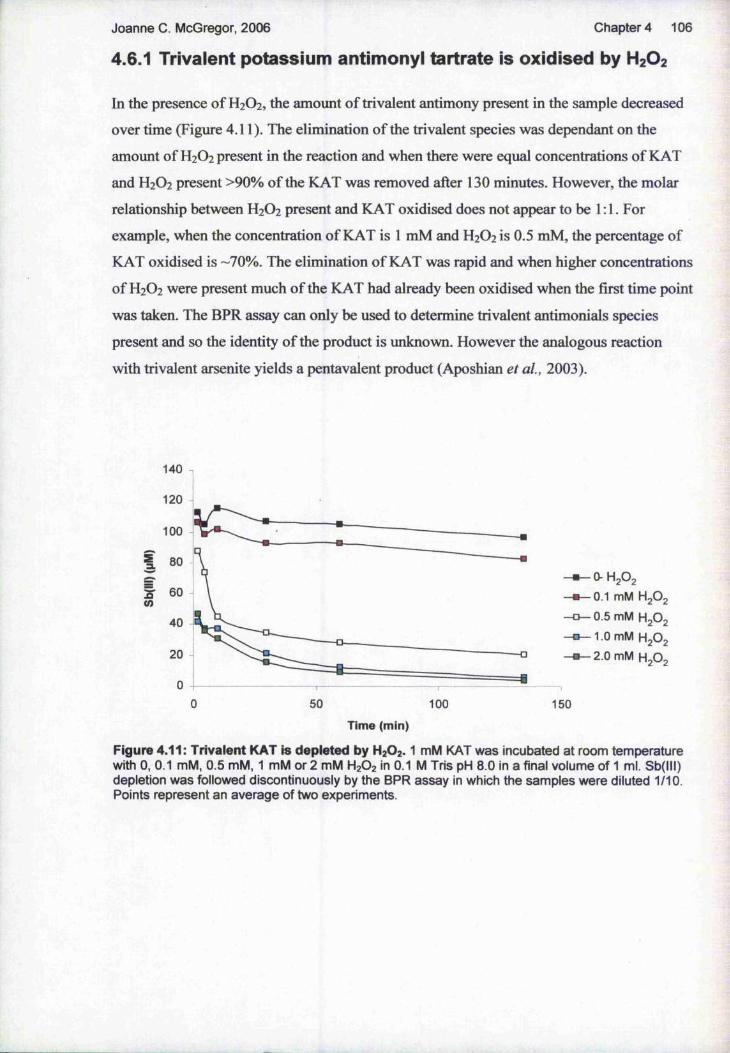
Joanne c. McGregor, 2006 Chapter 4 106
4.6.1 Trivalent potassium antimony! tartrate is oxidised by H2O2
In the presence of H2O2, the amount of trivalent antimony present in the sample decreased
over time (Figure 4.11). The elimination of the trivalent species was dependant on the
amount of H2O2 present in the reaction and when there were equal concentrations of KAT
and H2O2 present >90% of the KAT was removed after 130 minutes. However, the molar
relationship between H2O2 present and KAT oxidised does not appear to be 1:1. For
example, when the concentration of KAT is 1 mM and H2O2 is 0.5 mM, the percentage of
KAT oxidised is -70%. The elimination of KAT was rapid and when higher concentrations
of H2O2 were present much of the KAT had already been oxidised when the first time point
was taken. The BPR assay can only be used to determine trivalent antimonials species
present and so the identity of the product is unknown. However the analogous reaction
with trivalent arsenite yields a pentavalent product (Aposhian et al, 2003).
140
120
100
3L
60
40
20 -o
0 50 100Time (min)
O-H2O2 0.1 mM H2 O2
0.5 mM H2 O2
1.0 mM H2 O2
2.0 mM H2 O2
150
Figure 4.11: Trivalent KAT is depleted by H2O2 . 1 mM KAT was incubated at room temperature with 0, 0.1 mM, 0.5 mM, 1 mM or 2 mM H2O2 in 0.1 M Tris pH 8.0 in a final volume of 1 ml. Sb(lll) depletion was followed discontinuously by the BPR assay in which the samples were diluted 1/10. Points represent an average of two experiments.

Joanne C. McGregor, 2006 Chapter 4 107
4.6.2 The effect of pH and recombinant TDR1 on oxidation of KAT
by H2O2
In order to determine the optimal conditions for the oxidation of KAT by H2O2 the reaction
was set up at a range of pH values. It was found that the oxidation did not occur when the
pH was below 7.0 and as the pH was raised the reaction rate increased. It is worth noting
that a basic environment was also required for the oxidation of trivalent antimonials by
hydrogen peroxide in other studies (Quentel et al, 2004). In order to keep the experiments
as physiologically relevant as possible, all subsequent investigations were carried out at pH
8.0 .
It has been shown previously that some glutaredoxins can act as peroxidases in the cell by
detoxifying H2O2 (Collinson et al, 2002). Due to the sequence similarities that exist
between these proteins and TDRl, recombinant TDRl was added to the reactions at this
stage so any affect its presence caused could be observed. If TDRl could detoxify H2O2
then this may prevent any oxidation of trivalent antimony while it was also possible that
TDRl could act as an oxidase and catalyse the antimonial oxidation. The effect of adding GSH and GSSG as well as TDRl was also monitored (see below). However, the addition
of recombinant TDRl neither catalysed nor inhibited the oxidation of KAT (Figure 4.12);
this was also the case at lower pH values.
1 2 0
S'CO
100
80
60
40
20
050 100
Time (min)
» - K A T
— B - - K A T + T D R l
- K A T + H g O g
— e - - K A T + T D R l
+ H 2 O 2
150
Figure 4.12: Recombinant TDRl has no effect on the oxidation of KAT by H2 O2 1 mM KAT was incubated at room temperature with 1 mM H2O2 , 4 pg TDRl or both, in 0.1 M Tris pH 8.0 in a final volume of 1 ml. Sb(lll) depletion was followed discontinuously by the BPR assay in which the samples were diluted 1/10. Points represent an average of two experiments.

Joanne C. McGregor, 2006 Chapter 4 108
4.6.3 The trivalent product formed upon reaction of TDR1 with
sodium stibogluconate is oxidised by H2O2
KAT is a trivalent antimonial compound that is not used as an antileishmanial drug and
therefore it was desirable to establish whether H2O2 could oxidise additional trivalent antimonials that may be more relevant. As described, the product of the reaction between
sodium stibogluconate and TDRl is a trivalent species and is likely to actually be
encountered by the parasite. Therefore the product of TDRl activity on stibogluconate was
quantified (using the BPR assay) and added to the reaction rather than KAT. H2 O2 was also
found to be capable of oxidising this trivalent antimonial (Figure 4.13), indeed the rate of oxidation was faster than that achieved when KAT was the trivalent substrate. Again,
without more in depth analysis it is impossible to be certain of the identity of the species
formed but it is not unreasonable to speculate that it may have converted back to sodium
stibogluconate. This is the first evidence that the parasitic metabolism of antimonial drugs
does not necessarily consist of straightforward reduction from a pentavalent to a trivalent
form. If it is possible for more toxic, trivalent antimonials, formed by reduction of the pentavalent drugs administered, to be converted back to the less toxic pentavalent form this
has clear implications for the anti-parasitic activity of the drugs. Intracellular H2O2
concentration could be a factor in antimonial toxicity, as is discussed in section 4.7.
140
120
100
za.
y 60CO
40
20
6010 20 30 40 50 700
Time (min)
Figure 4.13: The trivalent antimonial formed upon reaction of TDRl and sodium stibogluconate is oxidised by H2O2 . Sodium stibogluconate was incubated overnight with TDRl and GSH and the trivalent product was quantified using the BPR assay. 1 mM of the product was incubated at room temperature with or without 1 mM H2O2 in 0.1 M Tris pH 9.0 in a final volume of 340 pi. The resulting pH of the assay was pH -8.0. Sb(lll) depletion was followed discontinuously by the BPR assay in which the samples were diluted 1/10. Points represent an average of two experiments.

Joanne c . McGregor, 2006 Chapter 4 109
4.6.4 GSH Inhibits Oxidation of KAT by H2 O2 while GSSG has no
effect on the reaction
GSH is known to behave as a cellular anti-oxidant and react with H2O2 to form 2 H2O.
Therefore its presence may prevent the oxidation of trivalent antimonials from proceeding,
especially when it exists at a high level as reported by Ariyanayagam and Fairlamb 2001.
GSH was added to the reactions and was found to decrease the amount of KAT oxidised
(Figure 4.14). This is probably due to H2O2 reacting with the two molecules of GSH to
form GSSG and H2O and therefore depleting the amount of H2O2 available to react with
the antimonial. Curiously, the initial speed of the reaction appeared to be increased in the
presence of GSH as in the first time point there is significantly less KAT remaining in the
sample with GSH added than in that without (figure 4.14), although the overall level of
oxidation was reduced. Due to the high intra-cellular concentrations of GSH, this result has
significant implications for whether the feasibility of cellular oxidation of trivalent
antimonials.
120
100«-KAT
t * - KAT + GSH60
40
# - KAT + GSH + H,0.
50 1000 150 200
Time (min)
Figure 4.14: GSH reduces the amount of oxidation of KAT by H2O2. 1 mM KAT was incubated at room temperature with 1 mM H2O2 , 1 mM GSH or both, in 0.1 M Tris pH 8.0 in a final volume of 1 ml. Sb(lll) depletion was followed discontinuously by the BPR assay in which the samples were diluted 1/10. Points represent an average of two experiments.

Joanne C. McGregor, 2006 Chapter 4 110
Due to the ability of the reduced from of glutathione - GSH - being capable of directly
reducing pentavalent antimonials, the possibility that the oxidised form - GSSG - could be
involved in oxidising trivalent antimonials was considered. It was also possible that GSSG
could affect the oxidation by H2O2 . However, GSSG appeared to have no effect on the
trivalent nature of KAT, or on the oxidation reaction (figure 4.15). However the rapid
oxidation of KAT by H2O2 resulted in much of the KAT being eliminated before the initial
rime point was taken, so any effect by GSSG on the reaction during this period cannot be
monitored. As mentioned, the presence of TDRl in these reactions resulted in no
significant further effect.
100908070 i 60 ! «— KAT + GSSG
a.
40
«— KAT + GSSG
KAT + GSSG + TDRl + H,0.
-10 0 250 300150 20010050
Time (min)
Figure 4.15: GSSG has no effect on the oxidation of KAT by H2O2. 1 mM KAT was incubated at room temperature with 1 mM H2O2 , 1 mM GSSG or both, with or without 3 pg TDRl. The reaction was in 0.1 M Tris pH 8.0 in a final volume of 1 ml. Sb(lll) depletion was followed discontinuously by the BPR assay in which the samples were diluted 1/10. Points represent an average of two experiments.

Joanne C. McGregor, 2006 Chapter 4 111
4.6.5 Analysis of KAT and Sodium Stibogiuconate upon
incubation with Leishmania
It was decided to employ the BPR assay to monitor whether any changes in the state
antimonials could be detected upon incubation with Leishmania. L. mexicana amastigote
and L. major promastigote cultures were grown as standard, except that either KAT or
sodium stibogluconate (or extra media as a negative control) were added to a final
concentration of 200 pM. Every day for 10 days a sample was removed from each culture,
the parasites were removed by centrifugation, and 50 pi of the spent media was subjected
to analysis in a final volume of 200 pi using the BPR assay. The assay only detects trivalent species and so any decrease in trivalent antimonial level detected could be
interpreted as either oxidation or as uptake by the parasites. However there was no detected
decrease in the level of trivalent antimonial species when KAT was administered or
formation of trivalent species when sodium stibogluconate was administered, when
incubated with amastigotes or promastigotes. However, it is likely that the assay is too
insensitive to detect any changes in the state of the drugs or uptake that was occurring.

Joanne c . McGregor, 2006 Chapter 4 112
4.7 Discussion
L. major TDRl was originally discovered - and was of interest - due to its similarity to omega GSTs, which was suggestive that the protein could be involved in reducing
antileishmanial antimonials and therefore mediating drug-susceptibility. Upon analysis of
the TDRl ORF it became clear that TDRl encoded a highly unusual protein containing
two omega GST-like domains with different predicted active sites. Together with the
observation that no other GSTs have been identified in Leishmania, these factors indicate
the potentially fascinating activities of TDRl. Characterisation of recombinant TDRl and
analysis of its cellular expression profile were intended to help elucidate which of the
proteins many inferred functions were accurate.
Due to the nature of the two-domain structure of TDRl, it was desirable to obtain separate
C- and N- terminal half-length proteins, as well as the full-length recombinant protein. It
was hoped that this approach would provide an insight into whether the protein functioned
as a whole or if each half was capable of acting independently. However, despite varying
the expression-inducing XPTG concentrations and growth times of the E. coli expression
cells, acquiring soluble and stable versions of these truncated proteins was problematic. At
this point it was considered whether further attempts could have been made to obtain the
half-length proteins. For example, re-cloning the gene fragments in expression vectors that
add differently placed expression tags or alternative types of tag; or expressing the protein
in a eukaryotic system. However, it was decided that these approaches could have been
time-consuming and were without guarantee of a successful outcome; therefore they were discounted.
Meanwhile, satisfactory levels of active full-length TDRl were produced quite easily with
approximately 20 mg of protein being recovered from one litre of bacterial culture. This
relatively high yield was achieved despite the additional purification steps taken to
eliminate any potentially contaminating proteins from the sample, specifically E. coli glutaredoxin 2. It is interesting to note that there is no published account of enzymatically
active recombinant Tc52 - the most similar known protein to TDRl - being produced. In
all reports native protein was characterised after purification from T. cruzi lysates with S- hexyl-GSH (Montiez et al, 1995) suggesting that production of recombinant Tc52 is not
straightforward. Analysis of the recombinant protein revealed several enzymatic
capabilities of TDRl, summarised in Denton et al, 2004.

Joanne C. McGregor, 2006 Chapter 4 113
All the species analysed by western blot - L major, L. mexicana and L. infantum -
expressed TDRl and the protein was detected in all life-cycle stages of L major. This
apparently ubiquitous expression, together with the highly conserved amino acid sequence
between the proteins, could indicate that TDRl has a vital role in the parasite. Indeed Tc52
has been reported to be essential (Allaoui et ah, 1999) with the authors being unable to
obtain Tc52 knock out T. cruzi strains. However, as will be shown in chapter five this is
not the case with TDRl, providing further evidence that TDRl and Tc52 have divergent
roles.
Another area in which the two proteins differ is in their cellular localisation. Within T,
cruzi epimastigotes, Tc52 is reported to reside in vesicles resembling the reservomere
(Ouaissi et al, 1995b), large acidic organelles found towards the posterior end of the
parasite. This contrasts with the distribution of TDRl in promastigotes, which was quite
diffuse and may be cytosolic, and certainly was more concentrated at the anterior end of
the parasite. However, the authors did not perform any co-localisation studies, using
antibodies to characterised marker proteins to directly compare the distribution signals, and
therefore the inferred localisation of Tc52 is not certain. This is also the case with the
TDRl localisation study, although the comparative lack of staining detected in the TDRl
knock out line at least indicates that the distribution pattern is specific to TDRl. Analysis
of TDRl localisation in amastigotes - either axenic or isolated from animals - would also
have been desirable as the protein may behave differently between life-cycle stages. For
example the protein may be secreted from metacyclics and amastigotes but not procyclics.
TDRl was detected by western blot in spent media from a stationary phase L. major
promastigote culture. Although the media was filtered prior to analysis, the possibility that
dead parasites may release TDRl upon degradation, rather than it being actively secreted,
cannot be ruled out. Analysis of secretion from L mexicana axenic amastigotes - spent
media from L. major lesion amastigotes would be difficult to obtain - would be potentially
interesting. Tc52 is reportedly secreted (Schoneck et al, 1994) from epimastigotes, having
an important role in aiding the parasite to modulate the mammalian host’s immune
response to infection (Garzon et al, 2003). Whether TDRl has a similar role in
Leishmania has not been investigated as part of this study, though if it is secreted by
intracellular stages of the parasite this could help explain it’s involvement in parasite
infectivity, discussed in chapter five.
The temporal expression pattern of TDRl is another area that potentially differs from that
of Tc52. TDRl expression is uniform throughout the various growth phases of T. major
promastigotes while an increase in expression was detected in lesion amastigotes relative

Joanne c . McGregor, 2006 Chapter 4 114
to the promastigotes. In T. cruzi, Tc52 is most highly expressed in the epimastigote and
amastigote stages, and is less so in trypomastigotes (Ouaissi et al, 1995b). In L mexicana
axenic amastigotes the same increase in TDRl expression was not observed: this could be
due to the axenic nature of the culture not entirely reflecting the natural environment
encountered by amastigotes and therefore affecting gene expression. However the lesion
amastigote-like protease expression profile of the axenic amastigotes suggested otherwise.
Equally, the differences in TDRl amastigote expression levels may be due to variations
between species. The implications of this apparent up-regulation may be significant.lt is
generally accepted that pentavalent antimonial drugs are toxic specifically to amastigotes.yet it is not known if this is due to increased parasite susceptibility or stage-specific
reduction - and hence activation - of the pentavalent compounds. Controversy also persists
over whether activation occurs in the macrophage or the parasite (Ephros et al, 1999,
Sereno et al, 1998). It has been reported that only amastigotes and not promastigotes are■f
capable of reducing pentavalent antimonials and that Pentostam-resistant L donovani
parasites were deficient in this activity (Shaked-Mishan et al, 2001). Although it has been
demonstrated that some thiols are capable of directly reducing pentavalent antimonials
(Frezard et al, 2001), the rate is very low. In chapter five, investigations on L. major
lacking or expressing an increased amount of TDRl are presented, which help clarify
whether TDRl is involved in mediating toxicity of pentavalent antimonial drugs.
Finally, the revelation that H2O2 can oxidise KAT and even more intriguingly, the trivalent
product formed upon reaction between TDRl and sodium stibogluconate, shows that the
metabolism of pentavalent antimonials may not be as straightforward as once thought. It is
clearly possible for trivalent antimonials. Therefore reduction of the pentavalent drug
administered could occur, but whether the product remains in the more toxic trivalent form
or is oxidised back to a pentavalent species may be dependant upon the concentration of
H2O2 present. Indeed it is possible that other reactive oxygen species found in Leishmania
could also be capable of causing any trivalent antimonial species formed to be oxidised.
However, the observation that GSH inhibits the oxidation of KAT brings in to question the
physiological relevance of the oxidation of trivalent antimonials. The concentration of
GSH in L. donovani promastigotes is approximately 4 mM while T(SH)2 , which also reacts
with H2O2 and is therefore likely to inhibit KAT oxidation in a similar way, is
approximately 5.2 mM (Ariyanayagam and Fairlamb 2001). Although no accounts of H2O2
concentration in Leishmania could be found, in T. brucei H2O2 is reportedly at a
intracellular concentration of 70 pM (Meshnick et al, 1977). However H2O2 exists
transiently in the cell: due to the reactive nature of the molecule it is constantly being
synthesised and removed. Assuming that the concentration of H2O2 in Leishmania is

Joanne C. McGregor, 2006 Chapter 4 115
of H2O2 in Leishmania is similar, the abundance of thiols means that newly produced
molecules will be rapidly reduced and therefore not available to oxidise Sb(III). Moreover
enzymes able to mediate the reduction of H2O2 have a similar effect: the oxidation of
As(III) by H2O2 is inhibited when catalase is added to the reaction (Aposhian et at., 2004).
Although Leishmania lack catalase, they do possess several peroxidases (Vickers et al,
2004; Adak and Datta, 2005) which perform the same function as catalase and are
therefore also likely to inhibit Sb(III) oxidation. However, this does not take into account
the possibility that Sb(III) could be localised in an area of the parasite that contains higher
levels of H2O2 . Indeed it has recently been shown that when X. donovani are treated with
Sb(III), intracellular GSH and T(SH)2 are depleted due to efflux of the molecules and
inhibition of trypanothione reductase, the enzyme responsible for maintaining the thiols in
their reduced state (WylUe et al, 2004). Similar effects were observed when human
macrophages were treated with Sb(III), as were increased level of reactive oxygen species
(Wyllie and Fairlamb, 2006), creating conditions favouring the oxidation of Sb(III). The
same effects also occurred in L. donovani amastigotes when treated with Sb(V). Although
these observations certainly do not provide evidence of Sb(III) oxidation in Leishmania, it
may be the case that conditions can arise within the parasites that enable some oxidation to
occur. Therefore, in vitro evidence provided in this chapter that under the right conditions
Sb(III) can be oxidised is potentially of great importance, although clearly in vivo studies
are required to ascertain whether it is indeed a factor in antimonial metabolism.

116
5 Functional study of L. ma/or TDR1
5.1 Introduction
In the previous chapter the expression of TDRl was described, although the analysis of the
recombinant protein (rTDRl), which was carried out by Dr. Helen Denton, was not
presented. The biochemical characterisation of rTDRl in vitro was an important step
towards understanding the functions of TDRl, including whether the protein is capable of
reducing pentavalent antimonials, and if, like the proteins it is similar to, it can catalyse
thioltransferase reactions and hence may have a role in protection against oxidative stress.
The properties of rTDRl are summarised below, while a full description is provided in
Denton et al, 2004.
Recombinant TDRl, shown by gel filtration analysis to be trimeric, exhibited several
interesting activities in vitro. In contrast to Tc52 which displays no GSH-conjugation
activity (Montiez et aï., 1995), but like human oGSTl (Board et al, 2000), rTDRl
exhibited low-rate conjugation of GSH with l-chloro-2,4-dinitrobenzene (CDNB), with a
specific activity of 1.9 nmol/min/mg/protein. However, no reaction could be detected vdth
the other conjugation substrates ethacrynic acid and l,2-epoxy-3(4-nitrophenoxy)propane.
Like glutaredoxin (GRX), dehydroascorbate reductase (DHAR) and thioltransferase
activities were evident, with TDRl capable of using GSH as an electron donor to reduce
both dehydroascorbate and the synthetic disulphide 2-hydroxyethyldisulphide. The specific
activities for these two substrates - 11.9 pmol/min/mg/protein and 11.2
pmol/min/mg/protein - were similar, and were significantly higher than those reported for
the human oGST protein when assayed under similar conditions (Board et al, 2000).
Taken together, these results indicate that the enzymatic capabilities of TDRl are more
similar to oGST and GRX than to other GSTs that typically have high GSH-conjugation
activity. The result that TDRl acts more like GRXs, proteins that share predicted active
site sequences with TDRl but are otherwise much smaller and rather dissimilar, than GSTs
was also observed to be the case for human omega GST (Board et al, 2000).
In addition to these activities, TDRl was also found to be capable of reducing sodium
stibogluconate and Glucantime, giving specific activities (pmol/min/mg/protein) of 6.3 and
1.4, respectively. Significantly higher specific activities were recorded with the pentavalent
arsenicals sodium arsenate and methylarsonate (MMA^). The BPR assay (Frezard et al, 2001), which detects trivalent antimonial species, was employed to analyse the product(s)
formed in the reactions with pentavalent antimonials. hi addition, using the BPR assay, it

Joanne c . McGregor, 2006 Chapter 5 117
was shown that a trivalent antimonial compound was produced in the reaction between
TDRl and the pentavalent antimonial drug, sodium stibogluconate. This was instrumental
in determining the relevance of the findings reported above, as it is the reduction of sodium
stibogluconate to a trivalent form that reportedly mediates toxicity (Shaked-Mishan et al.,
2001).
The fact that rTDRl is capable of reducing pentavalent antimonials to a trivalent form was
clearly of great interest. It is generally accepted tliat pentavalent antimonial drugs are toxic
specifically to amastigotes, yet it is not known if this is due to increased parasite
susceptibility or stage-specific reduction - and hence activation - of the pentavalent
compounds. Controversy also persists over whether activation occurs in the macrophage or
the parasite (Ephros et at., 1999, Sereno et al., 1998). It has been reported that only
amastigotes and not promastigotes are capable of reducing pentavalent antimonials and
that Pentostam-resistant L. donovani parasites were deficient in this activity Shaked-
Mishan et al, 2001. Although it has been demonstrated that some thiols are capable of
directly reducing pentavalent antimonials (Frezard et al, 2001), the rate is very low and
accordingly is unlikely to be as physiologic^ly relevant as the faster, enzyme-mediated
reaction reported here. As TDRl is the first Leishmania protein reported to be able of
reducing antimonials enzymatically, together with its increased abundance in amastigotes
demonstrated in the previous chapter, it is tempting to speculate that TDRl could play a
key role in antimonial susceptibility. However, further analysis is required. These studies
have not provided any in vivo evidence of the antimonial-reducing capability of TDRl, and
the affect this has on the parasite. For example, although reduction is thought to mediate
toxicity, it may be possible that upon reduction by TDRl antimonials are more easily
cleared by die cell, making them more quickly detoxified and therefore effectively less
toxic. In this chapter, investigations on L. major lacking or expressing an increased amount
of TDRl are presented, in an attempt to address whether TDRl has a bearing on
antimonial susceptibility in vivo.
There are implications of TDRl potentially behaving as a GRX in the parasite.
Glutaredoxins help maintain the redox balance of the cell by reducing protein and GSH
disulphides and have a role in stress-response in the cell (Potamitou et al, 2002). This may
point towards the natural role of TDRl in Leishmania'. after all, the protein is unlikely to
have evolved to improve the efficacy of antileishmanial chemotherapy; that aspect of the
protein’s function is more likely to be an unhappy coincidence for the parasite. In this
chapter, the production and analysis of TDRl knockout and over-expressing L. major
parasites is described. One of the important purposes of this was to investigate the cellular

Joanne C. McGregor, 2006 Chapter 5 118
role of TDRl, to elucidate what the protein does regardless of its potential as an antimonial
reductase.
5.2 Over-expression of TDR1 in Leishmania
It was hoped that manipulating the normal expression levels of TDRl in L major would
result in alterations in the parasites that could be detected. This would enable the
importance of TDRl in vivo to be assessed and help establish the protein’s functions. The
first approach was to increase the expression of TDRl, achieved by over-expressing the
protein from an episomal vector.
5.2.1 Creation of TDRl over-expressing L. mayor promastigotes
The sequence encoding TDRl was amplified from L major genomic DNA, sub-cloned
into the pGEM T-easy and then cloned into the pGL102 plasmid. The plasmid with the
TDRl gene was named pGL102TDRl ; maps of this and the empty vector are shown in
figure 5.1. The pGL102 plasmid is a Leishmania episomal expression vector and contains
the neomycin resistance gene as a selectable marker. Uncut pGL102 and pGL102TDRl
were transfected into L. major promastigotes which were subsequently incubated in culture
with neomycin so that only successful transformed parasites would grow. The transformed
parasite lines will be herein referred to by the names of the vectors they were transformed
with (pGL102 and pGL102TDRl).
neo
313K
PGL102TDR18147 b p
amp
5’DSTSma1(1366)
TDR1
BamHl (2723)
B neo
PGL10267 94 bp
amp
5‘DST
Smal ( 1366) BanéU (1370)
Figure 5.1: Plasmids for the over-expression of TDRl in L. major promastigotes. Schematic representation of the pGL102 plasmid for the over-expresslon of TDRl In Leishmania. A - pGL102 + TDRl (pGL102TDR1) for the over-expresslon of TDRl. amp, amplclllln resistance gene; neo, neomycin resistance gene. The restriction sites used to clone TDRl Into the vector are shown In dark red. B - empty pGL102 vector, for use as a negative control.

Joanne C. McGregor, 2006 Chapter 5 119
5.2.2 Analysis of over-expression
Whether pGL102TDRl parasites that grew in the presence of the selective drug had been
successfully transformed, resulting in increased levels of TDRl, was assessed by western
blot analysis. pGL102 and pGL102TDRl parasites grown with neomycin and in the
stationary phase of growth were harvested and lysed and the soluble fractions were
retained. The protein concentration of each sample was determined, and equal quantities
were separated by SDS-PAGE before western blotting. The sheep ani-TDRl antibody was
used to detect TDRl expression. To ensure equal loading of the samples, cysteine synthase
(CS) expression was examined in parallel with TDRl, using a rabbit anti-CS antibody
donated by Dr R. Williams of the University of Glasgow. While the expression of CS
remained constant, TDRl was significantly more highly expressed in pGL102TDRl than
in pGL102 L major promastigotes (figure 5.2); at least a four-fold increase in TDRl
expression was observed.
TDRl
CS
Figure 5.2: Western blot analysis of TDRl expression In pGL102 and pGL102TDR1 L. major promastigotes. 20 pg of soluble parasite lysate was loaded per lane. CS, cysteine synthase protein, used as a control to ensure equal loading. The relative positions of the proteins are indicated to the left of the image.
5.2.3 Phenotypic analysis of TDRl over-expressing L. major
Western blot analysis confirmed that pGL102TDRl parasites were over-expressing TDRl
as compared to pGL102 parasites, and therefore phenotypic tests could be conducted with
confidence that TDRl was at increased levels. In some experiments the TDRl over
expressing parasites were compared to WT as well as pGL102 parasites to help corroborate
any differences between data sets.

Joanne C. McGregor, 2006
5.2.3.1 Morphology and growth
Chapter 5 120
Parasites over-expressing TDRl were examined microscopically and no morphological
differences could be seen between them and the parasites containing the empty pGL102
vector or WT parasites. After several passages with neomycin present in the culture media,
the pGL102 and pGL102TDRl lines were counted and diluted to the same concentration.
Thereafter their growth was monitored by daily counting (figure 5.3). The parasite lines
were found to grow at similar rates.
1.00E+08
E 1.00E+07IC« 1.00E+06
1.00E+0520 64
pGL102pGL102TDR1
Number of days
Figure 5.3: Growth curve of pGL102 and pGL102TDR1 L. major promastigotes. Cultures were seeded at a concentration of 2 x 10® parasites/ml and were counted daily. Both lines were grown with 50 pg/ml neomycin.

Joanne C. McGregor, 2006
5.2.3 2 In vitro infectivity to macrophages
C hapters 121
The ability of WT, pGL102 and pGL102TDRl stationary phase promastigote parasite lines
to infect peritoneal macrophages was assessed (figure 5.4). pGL102TDRl infected a
higher percentage of macrophages than either the pGL102 or WT lines: approximately 4
times and twice as many, respectively. This suggests that the over-expression of TDRl in
parasites aids infection. However, the differences were not significant which reflects the
low number of replicates of this experiment that were performed: further analysis is
required to conclude whether over-expression of TDRl impacts upon the infectivity of the
parasites to macrophages. The variation between the infection rates between the WT and
pGL102 lines was also of concern as they would be expected to infect at a similar rate. It is
possible that the pGL102 line have been adversely affected by the transformation
procedure, causing them to be less infective in vitro.
70
8 60
Q .2%EI
50
40
30
I 20
10
pGL102 WT
■I
PGL102TDR1Figure 5.4: Infectivity of WT, pGL102 and pGL102TDR1 L. mayor promastigotes to macrophages. Stationary phase promastigotes were used to infect peritoneal macrophages at a rate of 2:1 and the slides were incubated for 5 days post-infection. Results are the means ± SE from at least two experiments. The infection rates of the different parasite lines were not significantly different (p > 0.05)

Joanne C. McGregor, 2006
5.2.3 3 In vivo infectivity to mice
Chapter 5 122
To assess the in vivo infectivity, metacyclic pGL102 and pGL102TDRl parasites were
purified from stationary phase cultures and inoculated into mouse footpads, the thicknesses
of which were then measured over time (figure 5.5). pGL102TDRl parasites were
responsible for significantly more rapid footpad growths than those that were induced by
pGL102. However, footpad infectivity experiments described elsewhere in this chapter
suggest that the infectivity of pGL102 parasites may be compromised: footpads infected
with WT parasites reach an average of 4 mm after 6 weeks (figure 5.16), while here
footpads infected with pGL102 parasites are an average of just 3.5 mm after 10 weeks.
However, although the experimental conditions are the same between these two
experiments, some parameters may be subject to change e.g. the amount of time between
metacyclic promastigote purification and inoculation. Therefore the differences observed
between the two experiments may not be due to the pGL102 line being defective.
Regardless of whether pGL102 parasites are infectious as WT, the footpad infections
caused by pGL102TDRl are particularly virulent as after just five weeks the footpads had
reached an average width of 4.9 mm. The difference between the footpad infections caused
by the pGL102TDRl parasites in this experiment and the WT parasites in the experiment
described in section 5.3.3.4 and demonstrated in figure 5.16 is significant (p < 0.02). As
described in the previous section, pGL102TDRl parasites were also more infective to
macrophages than both WT and pGL102 parasites.
i
pGL102pGL102TDR1
8 10 12Number of weeks
Figure 5.5: Infectivity of pGL102 and pGL102TDR1, L. mayor metacyclic promastigotes to mice. 1x10® metacyclic promastigotes were resuspended in 20 pi PBS and inoculated into one footpad of each BALB/C mouse. The footpad thicknesses were subsequently measured weekly, and the results are the means ± SD from five mice. The footpad thicknesses of mice infected with pGL102TDR1 parasites were significantly greater than those of mice infected with pGL102 five weeks post-infection (p < 0.0006).

Joanne C. McGregor, 2006
5.2.3 4 Effect of Sb(V) on macrophage infections
C hapters 123
The effect of sodium stibogluconate (Sb(V)) on macrophages infected with pGL102 and
pGL102TDRl parasites was assessed (figure 5.6). The parasites and macrophages were
incubated overnight, the excess parasites were washed off and dilutions of Sb(V) in media,
or drug-free media, were applied. The parasites were incubated with the drug for a total of
five days, with the drug being refreshed once during that time. At the highest drug
concentration (250 pg/ml) the percentage of macrophages infected by the pGL102 and
pGL102TDRl parasites had fallen to around 30% of the percentage of cells infected when
no drug was applied. There was no significant difference in the effect of Sb(V) between the
different parasite lines.
5 8 200
□ pGL102□ pGL102TDR1
Éb15.6 31.3 62.5 125 250
Sb(V) concentration (micrograms/ml)
Figure 5.6: Effect of sodium stibogiuconate on the infectivity of pGL102 and pGL102TDR1 L. major promastigotes to macrophages. Stationary phase promastigotes were used to infect peritoneal macrophages at a rate of 3:1 at 37 °C. After the parasites and macrophages had been incubated together overnight, the parasites were washed off and doubling dilutions of sodium stitx)gluconate (Sb(V)) were applied. Results are the means ± SE from at least two experiments. There is no significant difference between the effect of Sb(V) on the infectivity of pGL102 and pGL102TDR1 promastigotes to macrophages (p > 0.5). The results are expressed as the percentage of cells infected when no drug was present in order to normalize varying levels of infection.
5.2.3 5 ICso values when incubated with agents that induce oxidative stress
IC50 values - the concentration of a substance that causes half of the maximum inhibitory
effect it exerts to occur - were obtained for H2O2 and paraquat, against WT and
pGL102TDRl parasites, and are presented in table 5.1. These were obtained by incubating
parasites with doubling dilutions of the compounds in 96-well plates, and adding Alamar
Blue to indicate living parasites. H2O2 itself is a reactive oxygen species (ROS) while

Joanne C. McGregor, 2006 Chapter 5 124
paraquat can diffuse across cell membranes and react with dioxygen, resulting in the
intracellular production of superoxide (O2 ) (Hassan and Fridovich, 1978). As described in
the introduction to this chapter, TDRl displays glutaredoxin-like activities in vitro,
functioning as a thioltransferase and dehydroascorbate reductase. Therefore increasing the
abundance of TDRl in vivo may be expected to protect the parasites against oxidative
stress, and increase their tolerance to agents that induce it. However, the IC50 values
obtained for H2O2 and paraquat were similar for both WT and pGL102TDRl parasites.
The pGL102TDRl parasites are slightly more tolerant to H2O2 than WT, although
paradoxically they are slightly more sensitive to paraquat.
WT PGL102TDR1H2 0 2 253.3 ±2.9 301.7 ±7.6paraquat 5500 ±1004 2200 ±130
Table 5.1:10» values of paraquat and H2O2 against WT and pGL102TDR1 L. major promastigotes. All values were obtained using the Alamar Blue test; parasites were incubated with doubling dilutions of the compounds for a total of four days. The values and standard deviations were calculated using the Graft software I C 5 0 programme. All values are in pM.
5.2.3 6 Growth of promastigotes with paraquat
To further assess the effect of paraquat on pGL102TDRl parasites compared to L. major
promastigotes with normal TDRl levels, parasite cultures were grown with the compound
(figure 5.7). WT, pGL102 and pGL102TDRl promastigotes were seeded at 1 x 10 and in
standard media containing 5 mM paraquat. Over the course of seven days, the parasite
lines grew at similar rates, although between day 3 and day 6 the pGL102TDRl were at a
slightly lower concentration than the WT and pGL102 parasites.
1.00E+08
E 1.00E+07
$I« 1.00E+06
WTpGL102pGL102TDR1
1.00E+05 1 ,0 2 4 6 8
Number of Days
Figure 5.7: Effect of paraquat on the growth of WT. pGL102 and pGL102TDR1 L. major promastigotes. The parasites were seeded at 1 x 10 /ml with 50 pg/ml neomycin and 5 mM paraquat and were counted daily.

Joanne C. McGregor, 2006 Chapter 5 125
5.3 Knocking-out of TDR1 in L. major
It was anticipated that removing the TDRl gene from L. major by two rounds of
homologous recombination would affect the parasites and result in a phenotype that would
clarify the role of TDRl in vivo. Analysis of the knockout parasites’ susceptibility to Sb(V)
would also elucidate the role, if any, of TDRl in pentavalent antimonial activation.
5.3.1 Creation of L major TDRl knock-out parasite lines
The 3’ and 5’ flanking regions of TDRl were amplified from L. major genomic DNA, sub
cloned into pET28a(+) and cloned into vectors pGL345, pGL1033 and pGL842 which
contain hygromycin, bleomycin and blasticidin resistance genes, respectively (figure 5.8).
The constructs containing the flanking regions and drug resistance markers were cut out
and purified and the linear DNA was used to transform L major promastigotes. Following
the two independent second-round transfections and overnight recovery period, the
appropriate selective drugs were added and the cultures were divided: 1 ml of each was
used to set up serial dilutions in an attempt to derive clonal lines. Two independent lines,
KOTDRl 1 and KOTDR18, were generated, both having undergone transformation with
two constructs sequentially and grown up in the presence of the selection drugs.
KOTDRl 1 was transformed with DNA obtained from the pGL345 (hygromycin) and
pGL1033 (bleomycin) plasmids, while KOTDRl 8 was generated with DNA from the
pGL345 and pGL842 (blasticidin) plasmids. In addition, a clonal line was derived from the
initial serial dilutions of the KOTDRl population; it was named KOTDRl 1 A. After
several passages in the selective drugs, three further clonal lines were derived by setting up
KOTDRl 1 and KOTDRl 8 in 96-well plates at a concentration of approximately 0.1
parasites per well. One clone, KOTDRl IB, was isolated from KOTDRl 1 and two clones
were isolated from KOTDRl 8: KOTDRl 8A and KOTDRl 8B. The clones were derived in
the presence of selective drugs, but these were omitted from the media following the
isolation of the clones.

Joanne C. McGregor, 2006 Chapter 5 126
Hindin ( 1 )
amp
pGL345 TDR1 3* and 5' flanks
7485 bp
TDRl 5’ FLANK(409)
DHFR flank
5pel(1352)
Hyg
BamHl (2405)
fig/11 (4683)TDRl 3'FIANK DHFR flank
San (3794)
Smal (3775)
Figure 5.8: Construct for the gene knock-out of TDRl in L. major promastigotes. Schematic representation of the pGL345 plasmid containing the TDRl 3’ and 5’ flanking regions. The second plasmids used for the construction of the TDRl knock-out lines were pGL842 and pGLIOSS, which are exactly the same as pGL345 except that they contain blasticidin and bleomycin resistance genes respectively, rather than hygromycin resistance gene. Hyg, hygromycin resistance gene; amp, ampicillin resistance gene; DHFR flanks, flanking regions of the dihydrofolate reductase gene. The restriction sites used to clone the TDRl flanks and hygromycin resistance gene into the vector are shown in dark red. Hindlll and Bglll were used to cut out the flank-containing linear construct for transfection into L major.
5.3.2 Analysis of knock-out lines
Whether the TDRl gene had successfully been knocked out of KOTDRl lA, KOTDRl IB,
KOTDRl 8 A and KOTDRl 8B, resulting in the absence of TDRl, was assessed by western
blot analysis. WT and TDRl knockout parasites in the stationary phase of growth were
harvested and lysed and the soluble fractions were retained. The protein concentration of
each sample was determined, and equal quantities were separated by SDS-PAGE before
western blotting. The sheep ani-TDRl antibody was used to detect TDRl expression. To
ensure equal loading of the samples and as a positive control, CS expression was examined
in parallel with TDRl, using the rabbit anti-CS antibody as before. While CS expression
was apparent in all lines tested, TDRl was solely expressed in WT parasites (figure 5.9).
Therefore KOTDRl lA, KOTDRl IB, KOTDRl 8A and KOTDRl 8B were judged as being
successful TDRl knockouts.

Joanne C. McGregor, 2006 Chapter 5 127
4P 4-0 4-0TDRl
CS
Figure 5.9: Western blot analysis of TDR1 expression in WT, KOTDR18A, KOTDR18B, KOTDR11A and KOTDRl IB L. ma/or promastigotes. 15 pg of soluble parasite lysate was loaded per lane. CS, cysteine synthase protein, was used as a control to ensure equal loading. The relative positions of the proteins are indicated to the left of the image.
5.3.3 Phenotypic analysis of TDR1 knock-out lines
5.3.3.1 Morphology and growth
TDRl knockout parasites were examined microscopically and no morphological
differences could be seen between them and WT. The KOTDRl 1 A, KOTDRl IB,
KOTDRl 8A and KOTDRl 8B clonal lines were counted and diluted to the same
concentration. Thereafter their growth was monitored by counting at regular intervals
(figure 5.10). The KOTDRl8A and KOTDRl IB lines took slightly longer to reach
stationary phase than the other lines. KOTDRl 8A did not reach a density greater than 1.2 x
10 parasites/ml, while all other lines reached 2x10^ parasites/ml. Moreover, after 30 days
in culture, the KOTDRl 8A parasites began to die while the other lines did not. After the
KOTDRl lA, KOTDRl IB and KOTDRl8B lines had been passed through mice, the
growth curves were repeated and all three grew at similar rates to, and reached similar
densities in the stationary growth phase as WT parasites.
1.00E+08
E 1.00E+07
2« 1.00E+06
1.00E+050 10 20 30 40
-o—WT-♦-KOTDRl BA -•—KOTDRl SB -•—KOTDRl 1A -•—KOTDRl IB
Number of days
Figure 5.10: Growth curve of WT, KOTDR11A, KOTDRl IB, KOTDR18A and KOTDRl8B L ma/or promastigotes. Cultures were seeded at a concentration of 1 x 10® parasites/ml and were counted at regular intervals. All parasite lines were grown without any selective drugs added.

Joanne C. McGregor, 2006
5.3.3 2 Differentiation to metacyclic promastigotes
C hapters 128
The percentage of WT, KOTDRl 1 A, KOTDRl IB, KOTDRl 8 A and KOTDRl 8B
promastigotes that differentiated to metacyclic forms was monitored over time (figure
5.11). Parasite cultures were seeded at equal concentrations and, after they had reached the
stationary phase of growth, aliquots were removed and the percentage of metacyclic forms
in each sample was assessed. The percentage of metacyclic forms in each of the parasite
lines tested increased over time. The percentage of metacyclic forms in each culture was
similar at each time interval, although the highest percentage was consistently in the
KOTDRl 1A line. At 16 days after the cultures were initiated, a lower percentage of
metacyclogenesis was observed in the KOTDRl 8A sample, as compared to the other lines.
35MË
30S 25oo 20oa 150>E 10o5? 5
0
□ WT■ KOTDRl BA■ KOTDRl BB■ KOTDRl 1A■ KOTDRl IB
5 9 16Number of days
Figure 5.11: Metacyciic formation in WT, KOTDRl 1 A, KOTDRl IB, KOTDRl 8A and KOTDRl8B L major promastigotes over time. Parasite cultures were seeded at 1 x 10®/ml and metacyclogenesis was assessed after they had reached the stationary phase of growth. The parasites in each culture were counted, incubated with peanut agglutinin and re-counted. Metacyclogenesis was also assessed by analysis of parasite morphology. Number of days refers to the time since the cultures were initiated.
5.3.3 3 In vitro infectivity to macrophages
The ability of WT, KOTDRl 8B, KOTDRl 1A and KOTDRl 1B stationary phase
promastigote parasite lines to infect peritoneal macrophages was assessed (figure 5.12).
The KOTDRl 8A line was not used here, or in any other macrophage infectivity
experiments because, as discussed in section 5.3.3.4, this line was not infective to mice.
While the KOTDRl8B, KOTDRl lA and KOTDRl IB parasite infected macrophages at
similar levels, the WT parasites infected a significantly lower number of cells. This
suggests that the loss of TDRl enhances the macrophage infectivity of L major. This was

Joanne C. McGregor, 2006 Chapter 5 129
an unexpected result, as the over-expression o f TDRl in L. major promastigotes had a
similar effect (section 5.2.3.2).
70
600»« 50
I 40
I
30
20
10
0
□ WT■ KOTDRl 8B■ KOTDRl 1A■ KOTDRl IB
WT KOTDRl8B KOTDRl 1A KOTDRl IB
Figure 5.12: Infectivity of WT, KOTDRl 1 A, KOTDRl IB, KOTDRl8A and KOTDRl8B L. major promastigotes to macrophages. Stationary phase promastigotes were used to infect peritoneal macrophages at a rate of 2:1 and the slides were incubated for 6 days at 37 “C post-infection. Results are the means ± SE from at least two experiments. All three TDRl knockout lines are significantly more infectious to macrophages than WT parasites (p < 0.05).
In the previous experiment the macrophages were incubated at 37 °C. Whether the
incubation temperature affected the infectivity of WT, KOTDRl 8B, KOTDRl lA and
KOTDRl IB promastigotes was assessed (figure 5.13A). At both 35 °C and 35 °C, the WT
parasites infected a lower percentage of macrophages than the TDRl knockout lines.
However, this effect was much more pronounced at 37 °C: while the percentage of infected
macrophages for the three TDRl knockout lines remained constant between the two
temperatures, the WT parasites infected less than half the percentage of macrophages at 37
°C than they did at 35 °C. The effect of the different temperatures on the average number
of parasites in each infected cell was also determined (figure 5.13B). When incubated at 35
°C the average number of parasites in each infected cell varied between the KOTDRl 8B,
KOTDRl lA and KOTDRl IB lines (approximately 11, 14 and 17, respectively). However
these were all higher than the average of 8 parasites per cell observed in macrophages
infected with WT parasites. At 37 °C there was a decrease in the average number of
parasites per cell in all the lines tested. At the higher temperature the average number of
parasites was similar in cells infected by the three TDRl knockout lines, and once again
the number of WT parasites per cell was considerably less.

Joanne C. McGregor, 2006 Chapter 5 130
B
100
8n1 70g 60Ê 50 ■g 40 I 30 - - 20 ^ 10
20 18 16
= 14I 12
1 102S. 6
8
WT KOTDRl8B KOTDRl 1A KOTDRl IB
□ 35 degrees□ 37 degrees
□ 35 degrees□ 37 degrees
WT KOTDRl8B KOTDRl 1A KOTDRl IBFigure 5.13: Infectivity of WT, KOTDRl 1 A, KOTDRl IB, KOTDRl 8A and KOTDRl 8B L. major promastigotes to macrophages and the number of parasites per infected cell, when incubated at different temperatures. Stationary phase promastigotes were used to infect peritoneal macrophages at a rate of 3:1 and the slides were incubated for 6 days post-infection. A - the percentage of macrophages infected after 6 days. B - the average number of parasites present in each in infected cell. The number of parasites present in at least 20 infected cells was counted, and the average number calculated.
A second experiment was conducted to analyse the affect of temperature, but also the
infection ratio of parasites to cells, on the infectivity of WT, KOTDRl 1 A, KOTDRl IB,
KOTDRl 8A and KOTDRl 8B to macrophages (figure 5.14). A second L. major WT line
with a higher passage number was also used, to corroborate the observation in the previous
experiments that the WT parasites were less infective to macrophages than the TDRl
knockout lines. Parasites with a higher passage number were used as these had not been so
recently harvested from animals and had had longer to adjust to growing in vitro. The
percentage of infected macrophages by the WT lines were highly similar when infection
ratios of 2:1 and 3:1 parasites to macrophages were used, and at both 35 °C and 37 °C.
Both WT lines infected a lower percentage of macrophages than the three TDRl knockout
lines, which infected similar levels of cells. In all the parasite lines used in these
experiments and at both temperatures, a significantly higher percentage of macrophages

Joanne C. McGregor, 2006 Chapter 5 131
were infected when the infection ratio of parasites to cells was 3:1 as opposed to 2:1. Once
again, the percentage of macrophages infected by WT parasites was significantly lower at
37 °C than 35 °C; this effect was apparent with both WT lines used, and was regardless of
the infection ratio used. The percentages of macrophages infected with the three TDRl
knockout lines at each of the infection ratios used were not significantly different between
the two temperatures.
100 90 80 70 60 50
1 : • | 20
1055 0
'V V V '
□ 2 parasites/cell□ 3 parasites/cell
B
Æ 20
□ 2 parasites/cell ■ 3 parasites/cell
^ y y yy y y
Figure 5.14: infectivity of two independent WT lines and KOTDRl 1 A, KOTDRl IB,KOTDRl 8A and KOTDRl8B L. ma/or promastigotes to macrophages using different infection ratios and when incubated at different temperatures. Stationary phase promastigotes were used to infect peritoneal macrophages at a rate of 2:1 or 3:1 as indicated, and the slides were incubated for 6 days post-infection. A - incubated at 35 °C. B - incubated at 37 “C. Results are the means ± SE from at least two experiments. For both WT and TDRl knockout parasites and at both temperatures, the percentage of cells infected when an infection ratio of 3:1 was used was significantly higher than when an infection ratio of 2:1 was used (p < 0.05). For each infection ratio the percentage of macrophages infected with WT parasites when the cells were incubated at 35 °C was significantly higher than when incubated at 37 °C (p < 0.05). This was not the case for the TDRl knockout parasites (p > 0.1).

Joanne C. McGregor, 2006 Chapter 5 132
Finally, the effect of time on macrophage infections by both WT lines and the three TDRl
knockout lines was examined. The parasites were removed after being incubated with the
macrophages overnight, and the cells were incubated for a further one, three or five days
before the percentage of infected cells was assessed (figure 5.15). After one day, all the
parasite lines resulted in a similar percentage of infected macrophages. However, by the
fifth day both the WT lines infected significantly less cells than the three TDRl knockout
lines, which again infected very similar levels of macrophages.
90 80 70 60
w 50I 40
30 20 10 0
I
□ WT□ WT2■ KOTDRl 8B■ KOTDRl 1A■ KOTDRl IB
1 day 3 daysTime post infection
5 days
Figure 5.15: infectivity of two independent WT iines and KOTDRl 1 A, KOTDRl IB,KOTDRl BA and KOTDRl 8B L. mayor promastigotes to macrophages over time. Stationary phase promastigotes were used to infect peritoneal macrophages at a rate or 3:1 and the slides were incubated for 1, 3 or 5 days post-infection as indicated. Results are the means ± SD from at least three experiments. After five days the percentage of macrophages infected by the WT parasites was significantly less than the percentage infected by the TDRl knockout lines (p < 0.05).
Taken together, these experiments show that all three TDRl knockout lines consistently
infect a significantly higher percentage of macrophages than WT parasites do, and also
produce infections with a higher number of parasites per infected cell than WT. The WT
cell lines infected a lower percentage of cells when incubated at 37 °C as compared to 35
°C; this effect was not apparent in infections by the TDRl knockout lines. A similar
phenomenon was observed when the macrophages were incubated for varying amounts of
time post-infection: while all lines produced similar levels of infected macrophages after
one day, after five days the percentage of macrophages infected with the WT parasites was
significantly less than the percentage infected with the TDRl knockout lines. The three
TDRl knockout lines behaved similarly, causing comparable levels of cells to be infected
under all the conditions tested here. The two WT lines used also produced similar results.

Joanne C. McGregor, 2006 Chapter 5 133
For all the lines tested and at both 35 °C and 37 °C, a 3:1 infection ratio resulted in a
higher percentage of infected macrophages that when a 2:1 infection ratio was used.
5.3 3.4 In vivo infectivity to mice
To assess the in vivo infectivity of the TDRl knockout parasites, metacyclic promastigotes
were purified from stationary phase cultures and inoculated into mouse footpads, the
thicknesses of which were then measured over time. Initially only the KOTDRl 1A and
K0TDR18A lines were used, together with WT parasites (figure 5.16). Both the WT and
KOTDRl 1A parasites infected the mice footpads, with the KOTDRl 1A parasites causing
slightly more rapid footpad growths to occur than the WT parasites. However, the
KOTDRl 8A line was found to be completely non-infective to the mice; the footpad widths
did not increase in any of the mice. The same result was obtained in a second experiment
using this line. This was an unexpected result given the infectivity of the KOTDRl 1A line.
However, previous experiments described in section 5.3.3.1 show that KOTDRl 8A
promastigotes did not grow to as high densities as WT and the other TDRl knockout lines,
and expired before them. Metacyclogenesis was also reduced in the KOTDRl 8A line,
albeit only at the latest time point (section 5.3.3.2). Therefore the KOTDRl 8A line was
judged to have been damaged in some way - possibly during the transformation procedure
- and was therefore not used for subsequent phenotypic analysis.
EE
45■o) 3 I
1 : 1
WTKOTDRl 8A KOTDR11A
0 * T
0 2 4 6 8Number of weeks
Figure 5.16: Infectivity of WT, KOTDRl8A and KOTDR11A L. mayor metacyciic promastigotes to mice. 1x10^ metacyclic promastigotes were resuspended in 20 pi PBS and inoculated into one footpad of each BALB/C mouse. The footpad thicknesses were subsequently measured weekly, and the results are the means ± SO from three mice. The footpad thicknesses of mice infected with KOTDRl 1A parasites were significantly greater than those of mice infected with WT parasites five weeks post-infection (p < 0.025).

Joanne C. McGregor, 2006 Chapter 5 134
The infectivity of WT parasites and the remaining TDRl knockout lines KOTDRl 1 A,
KOTDRl IB and KOTDRl83 to mice footpads was investigated (figure 5.17). All the
lines infected the mice, although the three TDRl knockout lines caused significantly more
rapid footpad width increases to occur than the WT parasites did. This reflects the in vitro
data, with the TDRl knockout lines being consistently being more infective to
macrophages than the WT parasites. Following the experiment, parasites were recovered
from the footpads and cultured as normal for use in further investigations.
0
-0—WTKOTDRl 8B KOTDRl 1A K0TDR11B
Number of weeksFigure 5.17: infectivity of WT, KOTDR18B, KOTDRl 1A and KOTDRl IB L. mayor metacyclic promastigotes to mice. 1x10^ metacyclic promastigotes were resuspended in 20 pi PBS and inoculated into one footpad of each BALB/C mouse. The footpad thicknesses were subsequently measured weekly, and the results are the means ± SD from five mice. The footpad thicknesses of mice infected with all three TDRl knockout lines were significantly greater than those of mice infected with WT parasites five weeks post-infection (p < 0.05).
5.3.3 5 Effect of Sb(V) on infectivity to macrophages
The effect of Sb(V) on macrophages infected with WT, KOTDRl 1 A, KOTDRl IB and
KOTDRl 8B parasites was assessed, as before. Initially only the WT and KOTDRl 1A
lines were used (figure 5.18). The results are presented as both the percentage of infected
macrophages at the varying drug concentrations, and as of the percentage of the level of
infection achieved when no drug was applied; this is to normalise the data for the
anomalies in the infection levels between the lines. At the second-highest drug
concentration (250 pg/ml) the percentage of macrophages infected with WT parasites had
fallen to around 50% of the percentage of cells infected when no drug was applied, while
the infection level of macrophages infected with the KOTDRl 1A line remained
unchanged. This was the only drug concentration at which a significant difference was
observed and suggests that macrophage infections of KOTDRl 1A may be less susceptible

Joanne C. McGregor, 2006 Chapter 5 135
to Sb(V) than WT infections. This is in accordance with the hypothesis that TDRl is able
to reduce pentavalent antimonials to a more harmful trivalent form in vivo, effectively
mediating toxicity of the drug. However, the large standard errors obtained from the data,
caused by inconsistency between duplicate experiments, meant that further analysis was
required.
B
100 $ 90« 80 I 70Ï3 60 E 50
40 30 20
s5 10
1
» Po » | 1601 8 1402 9 120 n -D 100I o 80a s 6 05 ® 40
20
0 7.8 15.6 31.3 62.5 125 250 500Sb(V) concentration (micrograms/ml)
7.8 15.6 31.3 62.5 125 250 500Sb(V) concentration (micrograms/ml)
□ WT■ KOTDRl 1A
□ WTIK0TDR11A
Figure 5.18: Effect of sodium stibogluconate on the infectivity of WT and KOTDRl 1A L. major promastigotes to macrophages. Stationary phase promastigotes were used to Infect peritoneal macrophages at a ratio of 3:1. Doubling dilutions of sodium stibogluconate (Sb(V)) were applied as before and cells were Incubated for 5 days with the drug. A - % Infected macrophages. B - results expressed as the percentage of cells Infected when no drug was present In order to normalize varying levels of Infection. Results are the means ± SE from two experiments. When the drug concentration was 250 pg/ml, the percentage of macrophages Infected with WT parasites relative to the no drug control was significantly less than the percentage Infected with KOTDRl 1A parasites (p < 0.04). At all other drug concentrations there was no significant difference (p > 0.05).

Joanne C. McGregor, 2006 Chapter 5 136
In a second experiment the effect of Sb(V) on macrophages infected with WT,
KOTDRl 1 A, KOTDRl IB and KOTDRl 8 was investigated (figure 5.19). Again, the raw
data together with the normalised results are presented. In this experiment, the percentage
of macrophages infected with WT parasites was very low: only around 15% of cells were
infected when no drug was present. Meanwhile the infection levels achieved with the TDRl knockout lines was consistently around 50%. At the highest drug concentration (1
mg/ml) the percentage of macrophages infected with WT and KOTDRl 8B parasites had
fallen to around 50% and 55%, respectively, of the percentage of cells infected when no
drug was applied, while the level of macrophages infected with KOTDRl 1A and
KOTDRl IB were approximately 80%. However, there was no consistent significant
differences between the effect of Sb(V) on WT infections compared to TDRl knockout
infections. The low infection rate observed for the WT line when no drug was present is
problematic when analysing the data: the effect of Sb(V) on WT infection levels may be
far more dramatic if the initial infection rates were higher.
The effect of the incubation temperature on macrophages infected with WT, KOTDRl 1 A,
KOTDRl IB and KOTDRl 8 parasites, and treated with Sb(V) was assessed. Both the
percentage of infected macrophages (figure 5.20) and the average number of parasites per
cell (figure 5.21) were analysed. Only the normalised data is presented here (the
percentage of the infection levels and parasites/cell observed with no drug present); the
results correlate with the data presented in figure 5.13 as they are part of the same
experiment. Only two concentrations of Sb(V) were used in these experiments: 0.5 mg/ml
and 2.5 mg/ml and at the latter Sb(V) may also be slightly toxic to the macrophages. This
high concentration was used to assess whether improved toxicity to L. major could be
achieved. With all the parasite lines tested, Sb(V) was much more effective at reducing the
percentage of infected macrophages at 37 °C than at 35 °C, However, the effect of the drug
on the number of parasites per cell did not differ between the two temperatures (although,
as described in section 5.3.3.3, the average number of parasites per cell is less at 37 °C
than at 35 °C for all the lines tested). No macrophages infected with WT parasites could be
detected when the cells had been incubated at 37 °C, although cells were infected with all
three TDRl knockout lines at this temperature. With the exception of this observation,
there were no noticeable differences in the effect of Sb(V) on the infections between the
cell lines; this applies to both the percentage of infected macrophages and to the average
number of parasites per cell. As before, much higher levels of infection were achieved with
the TDRl knockout lines than the WT parasites and this may have affected this inference.

Joanne C. McGregor, 2006 Chapter 5 137
CL 50
% 40
B(0 o 160O) 140
(V "O
□ WTn KOTDRl 8B■ KOTDRl 1A■ KOTDRl IB
15.6 31.3 62.5 125 250 500 1000Sb(V) concentration (micrograma/ml)
o 120 2 100
80 60 40I -£ % 20 i i n n
15.6 31.3 62.5 125 250 500 1000Sb(V) concentration (micrograma/ml)
□ WTIK0TDR18B IKOTDRl 1A IKOTDRl18
Figure 5.19: Effect of sodium stibogluconate on the infectivity of WT, KOTDR18B,KOTDRl 1A and KOTDRl IB L. mayor promastigotes to macrophages. Stationary phase promastigotes were used to infect peritoneal macrophages at a rate of 2:1. Doubling dilutions of sodium stibogluconate (Sb(V)) were applied as before and cells were Incubated for 7 days at 37 “0 with the drug. Results are the means ± SE from two experiments. A - % Infected macrophages B - results expressed as the percentage of cells infected when no drug was present in order to normalize varying levels of Infection. At all drug concentrations used, there were no significant differences In the effect of Sb(V) on the percentage of macrophages Infected with the WT and TDRl knockout lines relative to the no drug control (p > 0.05).

Joanne C. McGregor, 2006 Chapter 5 138
2$1 £ _M(0 o o
Ifr
120
100
80
60
40
20
B120
^ 100
80
%1 £ _
IIIIÎ g
I
60
40
20
WT KOTDRl8B KOTDRl 1A KOTDRl IB
WTLL
KOTDRl8B KOTDRl 1A KOTDRl IB
□ 0 mg/ml■ 0.5 mg/ml■ 2.5 mg/ml
□ 0 mg/ml■ 0.5 mg/mi■ 2.5 mg/ml
Figure 5.20: Effect of sodium stibogluconate on the infectivity of WT, KOTDR18B, K0TDR11A and KOTDR11B L. mayor promastigotes to macrophages at varying temperatures. Stationary phase promastigotes were used to infect peritoneal macrophages at a rate of 3:1. Sodium stibogluconate (Sb(V)) at one of two concentrations, or no drug, was applied and cells were incubated for 5 days. The results are expressed as the percentage of cells infected when no drug was present In order to normalize varying levels of Infection. A - Incubated at 35 ®C. B - Incubated at 37 “C.

Joanne C. McGregor, 2006 Chapter 5 139
CD
ST3O
I
120
100
80
60
40
20 A A
□ 0 mg/ml■ 0.5 mg/ml■ 2.5 mg/ml
WT KOTDRl 8B KOTDRl 1A KOTDRl 1B
B 2■ooc2$
PgnCL
120
100
80
60
40
20
WT
\ l
KOTDRl8B KOTDRl 1A KOTDRl IB
□ 0 mg/ml■ 0.5 mg/ml■ 2.5 mg/ml
Figure 5.21: Effect of sodium stibogluconate on the average number of parasites per infected macrophage of infections with WT, KOTDRl SB, KOTDRl 1A and KOTDRl IB L. major promastigotes at varying temperatures. Stationary phase promastigotes were used to Infect peritoneal macrophages at a rate of 3:1. Sodium stibogluconate (Sb(V)) at one of two concentrations, or no drug was applied and cells were Incubated for 5 days. The number of parasites present In at least 20 Infected cells was counted, and the average number calculated. The results are expressed as the percentage parasites per cell that were present when no drug was applied. In order to normalize the data. A - Incubated at 35 ®C. B - Incubated at 37 °C.

Joanne c . McGregor, 2006 Chapter 5 140
5.3 3.6 IC50 values when incubated with toxic substances■■i
IUsing both the acid phosphatase and Alamar Blue assays, IC50 values were obtained for
various substances against WT and TDRl knockout parasites. Three different
hydroperoxides - H2 O2, cumene hydroperoxide and tert-butyl hydroperoxide - were tested
against the KOTDRl 1 and KOTDRl 8 parasite populations (table 5.2). Because of the
glutaredoxin-like in vitro activities of rTDRl, it was hypothesised that parasites lacking
TDRl may be more sensitive to these oxidising agents. However, the IC50 values obtained
were broadly similar for each substance between the WT and TDRl knockout parasites.
ÏC50 values were also calculated for several compounds against WT, KOTDRl 1 A,
KOTDRl IB and KOTDRl8B parasites (table 5.3). The substances tested were the
trivalent antimonial compound potassium antimonial tartrate (Sb(III)), which is toxic to
Leishmania and affects the redox balance of the parasites (Wyllie et al, 2004); juglone,
which is toxic to trypanosomes (Akerman and Muller 2005) and produces intracellular
ROS (Kampkotter et al, 2003); N-methylphenazinium methyl sulphate, which is toxic to
trypanosomes (Akerman and Muller 2005) and generates intracellular O2’ (Maridonneau et al, 1983) and p-mercaptoethanol, a chemical that reduces disulphide bonds and affects the
expression levels of& coli glutaredoxins (Potamitou et al, 2002). Both the KOTDRl 1A
and KOTDRl IB lines were considerably more sensitive to paraquat (IC50 values of 1.2 and *
1.4 mM, respectively) than WT parasites (IC50 value of 5.5 mM). Therefore the effect of
paraquat on the different parasite lines was investigated further.

Joanne C. McGregor, 2006 C hapters 141
WT KOTDRl 1 KOTDRl 8H2 0 2 234.5 ±10.7 328.7 ±29.9 227.8 ±13.8Cumene hydroperoxide 9.4 ±1.2 9.4 ±1.3 9.0 ±0.6Tert-butyl hydroperoxide 27.3 ±1.7 14.4 ±6.5 27.2 ±2.6
Table 5.2: IC50 values of H2O2, cumene hydroperoxide and tert-butyl hydroperoxide against WT, KOTDR11 and KOTDR18 L. mayor promastigotes. All values were obtained using the acid phosphatase assay. The values and standard deviations were calculated using the Grafit software IC5 0 programme. All values are in pM.
WT KOTDRl 1A KOTDRl IB KOTDRl 8BSb(III) 23.2 ±2.7 16.7 ±3.0 N/D N/DP-me 330.8 ±21.9 301.4 ±36.3 N/D N/Djuglone 2.3 ±0.7 2.4 ±0.3 N/D N/DH2O2 253.3 ±2.9 264.4 ±6.8 266.6 ±7.6 290.6 ±8.1paraquat 5500 ±1004 1200 ±100 1400 ±500 N/DNMPMS 10.1 ±0.4 7.8 ±0.54 N/D N/D
Table 5.3: IC50 values of different com pounds against WT, KOTDRl 1 and KOTDR18 L. major promastigotes. All values were obtained using the Alamar Blue assay. The values and standard deviations were calculated using the Grafit software I C 5 0 programme. All values are In pM. Sb(lll), potassium antimonial tartrate; |3-me, p-mercaptoethanol; NMPMS, N-methylphenazinium methyl sulphate; N/D, not determined.
5 3.3.7 Effect of agents that induce oxidative stress on TDR1 expression in
L major promastigotes
In chapter four the varying expression of TDRl between different life-cycle stages was
demonstrated. Given that TDRl displays thioltransferase and DHAR activities in vitro, it may have a role in protecting the parasite from oxidative stress and therefore TDRl
expression may be modulated by fluctuations in the intracellular redox environment of the
parasite. In order to determine whether exposure to oxidative stress could stimulate TDRl
expression in WT L, major promastigotes, parasites were incubated for varying times with
different concentrations of H2O2 or paraquat and western blots were performed on the
resultant parasite lysates (figure 5.22). The protein concentrations of the samples were
determined and normalised, but to ensure equal loading of the samples CS and/or
transketolase (TK) expression was examined in parallel with TDRl, using the rabbit anti-
CS and anti-TK antibodies. TDRl expression did not change in L major parasites
incubated with 125 pM H2O2 at any of the time-points tested, nor in parasites incubated
with 500 pM H2O2 after 4 hours (at later time-points the parasites were mostly dead so the
TDRl expression could not be determined) (figure 5.22A), It is possible any changes in
expression would take place before the initial four hour time-point when the parasites were

Joanne C. McGregor, 2006 Chapter 5 142
harvested. Accordingly, L major parasites were incubated with paraquat for less time, with
the parasites being harvested at three time-points within 24 hours of initial exposure to the
oxidant. However, no variations in TDRl expression were visible at any of these time-
points with the different paraquat concentrations (figure 5.22B). TDRl may have a role in
protecting the parasite against oxidative stress; however, its expression is not affected by
exposure to these oxidants under the conditions tested.
A H A (pM) 125 500
TDRl
Time (h) 4 24 72
B Paraquat (mM) 0 0.1 1 0.1 1 0.1
TK
TDRl mm
CS «—
Time (h) 0 1 4 24Figure 5.22: Western blot analysis of the effect of H2O2 and paraquat on TDRl expression in L. major promastigotes. A - parasites inbubated with H2O2 . 12 pg of soluble parasite lysate loaded per lane. B - parasites incubated with paraquat. 16 pg of soluble parasite lysate loaded per lane. The concentrations of the reagents are shown above each figure and the length of exposure time is shown below In h (hours). TK (transketolase) and CS (cysteine synthase) were used as loading control proteins. The Identities of the different proteins are Indicated to the left of the Image. There was no noticeable change In TDRl expression under the conditions tested.
5.3 3.8 Growth of promastigotes with paraquat
The IC50 data presented in section 5.3.3.6 showed that the TDRl knockout lines
KOTDRl 1A and KOTDRl IB are approximately four times more sensitive to paraquat
than WT parasites. To further investigate the effect of paraquat on these lines, WT,
KOTDRl 1 A, KOTDRl IB and KOTDRl8 parasite cultures were seeded at 1 x lOVml and
grown with 5 mM paraquat, and the growth of the parasites’ was monitored by counting
(figure 5.23). While the parasite density of the WT culture increased to approximately 2 x 10 after six days, over the same time period the parasite density of the three TDRl
knockout lines fell to 1.4-2.6 x 10 . This result reflects the IC50 data presented previously

Joanne C. McGregor, 2006 Chapter 5 143
and demonstrates a clear difference in the sensitivity of the TDRl knockout lines to
paraquat as compared to WT parasites.
1.00E+08
E 1.00E+07IC« 1.00E+06
1.00E+050 2 64
-0—WTKOTDRl 8B
KOTDR11A
K0TDR11B
Number of DaysFigure 5.23: Effect of paraquat on the growth of WT, KOTDR18B, KOTDRl 1A and KOTDR11B L. major promastigotes. The parasites were seeded at 1 xIO* with 5 mM paraquat and were counted daily.
5.4 Re-expressing TDR1 In TDRl knock-out parasite lines
In order to clarify whether any phenotype observed in the TDRl knockout lines was due to
the loss of TDRl as opposed to any secondary mutation or damage the parasites may have
accumulated, TDRl was re-expressed in the knockout lines. This was achieved by
transforming the KOTDRl lA, KOTDRl IB and KOTDRl8 parasites with the
pGL102TDRl plasmid used to over-express TDRl in WT L major, as described in section
5.2.1. In addition the same lines were transformed with the pGL102 plasmid so they could
be used as negative control samples. These plasmids were previously presented in figure
5.1. Using the names of the original lines and plasmids used to transform them, the new
lines were named KOTDRl 1A + pGL102, KOTDRl 1A + pGL102TDRl, KOTDRl IB +
pGL102, KOTDRl IB + pGL102TDRl, KOTDRl8B + pGL102 and KOTDRl8B +
pGL102TDRl.

Joanne C. McGregor, 2006 Chapter 5 144
5.4.1 Analysis of re-expression
Whether the TDRl knockout parasites transformed with pGL102TDRl that grew in the
presence of the selective drug had been successfully transformed, resulting in TDRl being
re-expressed, was assessed by western blot analysis. WT, KOTDRl 1A + pGL102TDRl,
KOTDRl IB + pGL102TDRl and KOTDRl8B + pGL102TDRl promastigotes grown
with neomycin were harvested and lysed, and the soluble fractions were separated by SDS-
PAGE before western blotting. The sheep ani-TDRl antibody was used to detect TDRl
expression. As a positive control, CS expression was examined in parallel with TDRl,
using the rabbit anti-CS antibody as before. TDRl was expressed in all three re-expressing
lines (figure 5.24)
TDRl
CS
Figure 5.24: Western blot analysis of TDRl expression In pGL102 L mayor promastigotes and KOTDRl L. mayor promastigotes transformed with pGL102TDR1.10-20 pg of soluble parasite lysate was loaded per lane. CS, cysteine synthase protein, was used as a positive control. The identities of the different proteins are indicated to the left of the image. TDRl was reexpressed in each of the lines tested.
5.4.2 Growth of parasites re-expressing TDRl, with paraquat
The IC50 data presented in section 5.3.3.6, together with the growth curves presented in
section 5.3.3.8, show that the TDRl knockout lines KOTDRl 1A and KOTDRl IB are
more sensitive to paraquat than WT parasites. To investigate whether the loss of TDRl
was responsible for this phenotype, the parasite lines re-expressing TDRl were grown with
paraquat. WT, KOTDRl 1 A, KOTDRl IB, KOTDRl 1A + pGL102, KOTDRl 1A +
pGL102TDRl, KOTDRl IB + pGL102, KOTDRl IB + pGL102TDRl parasite cultures
were seeded at 1 x lOVml and grown with 5 mM paraquat, and growth was monitored by

Joanne C. McGregor, 2006 Chapter 5 145
counting (figure 5.25). While the parasite density of the WT culture increased as before,
the density of all the other lines decreased in a similar rate to the TDRl knockout parasites.
Clearly, the re-expression of TDRl, which was confirmed by western blot analysis, does
not complement the paraquat-sensitive phenotype of the TDRl knockout cultures. Whether
this is due to the original phenotype being caused by a factor other than the loss of TDRl,
or whether TDRl re-expression does not reinstate the parasites’ relative insensitivity to
paraquat for some other reason, is currently unknown.
1.00E+08
E 1.00E+07
12« 1.00E+06
1.00E+052 4
Number of days
WT
-e—K0TDR11A
K0TDR11B
-o - KOTDRl 1A + pGL102
-O-K0TDR11B + pGL102
-A-K0TDR11A + PGL102TDR1
-&-K0TDR11B + pGL102TDR1
Figure 5.25: Effect of paraquat on the growth of WT L. major promastigotes and KOTDR1 L. major promastigotes transformed with pGL102 or pGL102TDR1. The parasites were seeded at 1 X 10®/ml with 5 mM paraquat and were counted daily. With the exception of WT, KOTDR11A and KOTDR11B, the parasites were also grown with 50 pg/ml neomycin.

Joanne C. McGregor, 2006 Chapter 5 146
5.5 Discussion
The in vitro characteristics of rTDRl described in the introduction to this chapter are
consistent with the observed activities of oGST. rTDRl exhibited GSH-CDNB conjugating
activity which is characteristic of other types of GSTs, but as for human oGST, it was at a
very low level. However, this activity should not be ignored as although an elongation IB
complex displaying trypanothione S-transferase activities was recently identified in L.
major (Vickers et al, 2004), no known GSTs have ever been found in Leishmania. Therefore TDRl is the first protein with GST activity to be identified in the parasite.
However, the more notable functions displayed by rTDRl were DHAR and
thioltransferase activities. Whether the DHAR activity displayed by TDRl is of relevance
is an interesting question. Although ascorbate has been detected in T. cruzi (Clark et al,
1994), it has never been detected in Leishmania although an ascorbate-dependant
peroxidase has also been identified (Adak and Datta, 2005). However, trypanothione is
able to reduce dehydroascorbate (Krauth-Siegel and Ludemann, 1996) and may be
sufficient to maintain ascorbate levels without the need for an enzymatic reductase. The
issue of ascorbate being necessary in Leishmania, and therefore whether TDRl may have a
role in maintaining it in the reduced from, is addressed in chapter six. The high
thioltransferase activity exhibited by TDRl is the only in vitro enzymatic capability that
was also displayed by the most closely related protein to TDRl, Tc52. CDNB-GSH
conjugating activity was not detected in tlie T. cruzi protein (Montiez et al, 1995) and it
was not assessed for DHAR (or indeed metal-reducing) activities. It is possible that the
active site differences between TDRl and Tc52 described in chapter three account for
these differences. As mentioned, DHAR and thioltransferase activities are commonly
exhibited by glutaredoxins, a class of proteins that share predicted active site sequences
with TDRl but are otherwise much smaller and rather dissimilar. As described in chapter
three, L. major contains several proteins that are annotated as, and share sequence
similarity to, glutaredoxins. Organisms often possess several GRXs that may display
redundancy towards each other due to their overlapping functions (Draculic et al, 2000).
Therefore the possibility of redundancy between TDRl and other Leishmania proteins
must be taken into account when considering the glutaredoxin-like role of TDRl in vivo.
To investigate the role of TDRl in vivo, parasites over-expressing TDRl (figure 5.2) and clonal TDRl knockout lines (figure 5.9) were created. The growth rates of all these lines
were assessed and were found to be similar to that of WT promastigotes (figures 5.3 and
5.10) with the exception of one of knockout lines, KOTDRl 8A, The grovyth rate of this

Joanne C. McGregor, 2006 Chapter 5 147
line, together with the parasite density in stationary-phase cultures, was slightly impaired
and analysis of the line was discontinued. The metacyclogenesis of the TDRl knockout
lines was assessed and was shown to be broadly similar to that of WT parasites in all the
lines tested. These findings contrast with the observations of T. cruzi parasites that had
reduced levels of Tc52: epimastigotes did not grow well in culture and were impaired in
their ability to differentiate into metacyclic forms (Allaoui et ah, 1999). Moreover, only a
mono-allelic Tc52 knockout line was obtained; knocking out both copies of Tc52 is
thought to be lethal.
The IC50 values of several compounds against WT TDRl knockout, and TDRl over
expressing lines were calculated in order to determine if the level of TDRl affected the
sensitivity of parasites to different types of stress (tables 5.1, 5.2 and 5.3). There was no
consistent significant difference in the IC50 values of most of the compounds tested between the different lines, including the hydroperoxides, potassium antimonial tartrate
and p-mercaptoethanol. However, several agents that induce the production of intracellular
ROS were tested and a significant difference in the IC50 value of one of these - paraquat -
was apparent between WT parasites and those lacking TDRl. This finding was reinforced
by the observation that WT cells were much more tolerant to paraquat than TDRl
knockout parasites were, when the lines were grown in the presence of the chemical (figure
5.23).
Glutaredoxins (GRXs) have been previously linked with protecting cells against paraquat-
induced oxidative stress. Knocking out the WT and expressing a mutated version of GRX5
in Schizosaccharomyces pombe resulted in increased sensitivity to paraquat (Chung et ah, 2005) and a similar phenotype was observed in S. pombe GRX2 null mutants (Chung et al, 2004). These reports reflect the increased sensitivity of TDRl knockout parasites to
paraquat that are described in this chapter, and support the hypothesis based on the in vitro
activities of the protein that TDRl functions as a glutaredoxin in Leishmania.
A possible reason for the increase in paraquat sensitivity of the GRX mutants could be that
they have depleted ascorbate levels (due to a lack of DHAR activity). Ascorbate is a low-
molecular weight antioxidant which can react directly with ROS, and is the essential co
factor for ascorbate-dependant peroxidases, proteins that enzymatically reduce H2O2 . Paraquat treatment caused dehydroascorbate reductase activity to increase in Conyza
bonariensis plants (Ye and Gressel, 2000) and TRXl levels to increase in mouse liver and
lung tissues (Jurado et al, 2003). Although the expression of TDRl in L. major was not
affected by exposure to sub-lethal concentrations of paraquat (figure 5.22), this was also

Joanne c . McGregor, 2006 Chapter 5 148
found to be the case elsewhere; although exposure to the chemical did not result in
increased GRX2 levels in S. pombe (Chung et al, 2004), or GRXl or GRX2 levels in
mouse liver or lung tissues (Jurado et al, 2003). Meanwhile, the expression of a human
DHAR gene in tobacco plants caused them to be less susceptible to paraquat-induced
damage (Kwon et al, 2003). Over-expression of TDRl did not cause the parasites to
become more tolerant to the compound (table 5.1 and figure 5.7). This would be the
expected result if ascorbate recycling is not the rate-limiting step in ROS detoxification in
Leishmania.
If the loss of TDRl is responsible for the increased sensitivity to paraquat of the TDRl
knockout lines, re-expressing TDRl in these lines should restore resistance to the chemical
as displayed by WT parasites. Despite confirmation that TDRl was being re-expressed by
parasites transformed with pGL102TDRl, the susceptibility of these lines to paraquat did
not decrease (figure 5.25). There are several possible reasons for this. It may be the case
that some other mutation or damage to the TDRl knockout lines is responsible for their
increased sensitivity to paraquat. However, this seems unlikely given that all three
independently derived knockout lines exhibit the same phenotype. Another possibility is
that an aspect of the TDRl re-expression does not comply with WT expression and that
this effectively inactivates the protein. For example, the expression level or the localisation
of TDRl may differ in the re-expressing lines. Further analysis is required to determine
which of these scenarios is correct. Re-expressing TDRl from the original gene locus or
fi*om another chromosomal location may also be a helpful approach.
L. major parasites over-expressing TDRl were more infective to macrophages than WT
and pGL102 control parasites (figure 5.4), and were also more infective to mice than the
control line (figure 5.5). Paradoxically, the TDRl knockout lines were also more infective
to macrophages (section 5.3.3.3) and, to a lesser extent, mice, than WT parasites (section
5.3.3.4). It was also notable that the percentage of macrophages with WT parasites
appeared to be more susceptible to a prolonged incubation time and an increase in
incubation temperature than the TDRl knockout parasites (figures 5.13-5.15). The reasons
for the apparent contradictions between the TDRl over-expressing and knockout infection
data are unclear. The pGL102 line which acted as a control for the TDRl over-expressing
line appears to be in some way damaged: the parasites were less infective than WT
parasites to both macrophages and mice. However, misinterpretation of the TDRl over
expressing line infection data due to it being compared to a faulty control is not to blame,
as the TDRl over-expressing line was also more infective than WT. Whether the WT line itself could also be impaired was considered. However, the utilisation of a second WT line

Joanne C. McGregor, 2006 Chapter 5 149
which was of a different passage number gave very similar macrophage infection data to
the original WT line (figures 5.14 and 5.15), suggesting that this was not the case. One
possibility is that when TDRl is knocked out, the levels of other proteins - possibly the
glutaredoxin-like proteins described in chapter three - are modulated to compensate for the
loss. This in turn could have a positive effect on the TDRl knockout parasites, improving
their viability in vivo. Meanwhile, given the glutaredoxin-like properties of TDRl, over
expression of the protein could have a more straightforward beneficial effect on L. major,
the increased infectivity may be due to them being better equipped to cope with the
oxidative stress encountered upon invasion of a cell.
Once again, the results do not correspond with the Tc52 knockout and over-expresser data.
Although there was no difference between the infectivity of WT T. cruzi and parasites
over-expressing Tc52, parasites with decreased Tc52 levels were significantly less
infective to both macrophages and mice than WT parasites (Allaoui et al, 1999). Moreover
Tc52 knockout parasites that do infect animals result in an attenuated form of Chagas’
disease with reduced parasitemia compared to WT infections (Garzon et al, 2003). Tc52 is
secreted and is thought to have a role in modulating the immune response of the host
(Fernandez-Gomez et al, 1998; Borges et al, 2001; Ouaissi et al, 2002; Garzon et al,
2003), which is hypothesised to be the reason behind the reduced infectivity displayed by
parasites with decreased levels of Tc52 (Garzon et al, 2003). It was shown in chapter four
that TDRl is also released from promastigote parasites but the increased infectivity of
parasites lacking TDRl suggests that the protein does not perform the same function as
Tc52.
As described in the introduction to this chapter, rTDRl was able to reduce pentavalent
antimonials to the more toxic trivalent forms in vitro. This was in keeping with the ability
of mammalian oGST to reduce pentavalent arsenicals (Zakharyan et al, 2001), and
supported the hypothesis that TDRl may be involved in mediating antimonial
susceptibility in Leishmania. To resolve whether TDRl did indeed perform this fimction in
vivo, macrophages infected with WT, TDRl over-expressing and TDRl knockout L. major
parasites were incubated with sodium stibogluconate (Sb(V)) and the impact of the drug on
the infections was assessed. The effect of Sb(V) on L. major promastigotes could not be
tested as they are not sensitive to the trivalent drug. However, it proved to be extremely
difficult to assess the impact of Sb(V) on L. major macrophage infections for two main
reasons: the difference in the basal macrophage infection rates of the various lines, and the
insensitivity of all the lines tested to the drug.

Joanne C. McGregor, 2006 Chapter 5 150
There appeared to be no significant difference between the susceptibility of TDRl over-
expressing and control parasites to the drug (figure 5.6). When considering the raw data on
the effect of Sb(V) on the macrophage infections of the WT and TDRl knockout, it looks
like the knockout parasites are highly insensitive to the drug, consistent with the idea that
TDRl mediates antimonial toxicity. Even at tlie highest concentrations of Sb(V), approximately 30-40% of macrophages were infected with the TDRl knockout parasites,
compared to only 5% infected with WT parasites (figure 5.19). Similar results were
achieved in other experiments (section 5.3.3.5). However, due to the different infectivity of
WT and TDRl knockout parasites to macrophages when no drug was present, in order to
directly compare the effect of Sb(V) between the lines, it was necessary to consider the
effect of the drug on the reduction of the initial infection level. Therefore, the data for the
infection levels achieved with the varying drug concentrations was converted to the
percentage of the infection levels achieved with no drug was present. This makes the data
look very different and shows that despite the impressive infection levels of the knockout
lines achieved with high concentrations of the drugs, the reduction in the infection level is
similar to that of the WT parasites. However, manipulating the data in this way is not an
ideal solution; similar basal infection levels between the lines would allow a proper
comparison of the effect of Sb(V). A useful approach may be to analyse the effect of Sb(V)
on in vivo infections of the different lines, as these were at a more similar basal level than
the macrophage infections. This would also circumvent the problem of the apparent
insensitivity of L. major parasites in macrophages to Sb(V).
While this study was in progress, another arsenate reductase-like protein that could reduce pentavalent metalloids in vitro was identified, LmACR2 (Zhou et al, 2004). Although
LmACR2 knockout parasites were not constructed, the protein was over-expressed in L.
infantum and this resulted in an increase in sensitivity to Sb(V). The effect of Sb(V) was
assessed using a similar macrophage infection assay to the one used in this investigation.
The approximate Sb(V) IC50 value, as calculated from their findings, was 120 pg/ml
against parasites over-expressing LmACR2, and 180 pg/ml against WT parasites. Clearly
this is a much lower value than in the experiments detailed in this chapter. Although there
was variation between experiments and the incubation temperature affected the Sb(V)
efficacy, the IC50 was approximately 500-1000 pg/ml, a concentration that also appeared
to be toxic to tlie macrophages. Therefore, manipulating the expression of TDRl in a
Leishmania strain that is more susceptible to Sb(V) may help to ascertain whether the
protein does indeed mediated antimonial sensitivity in vivo.

Joanne c . McGregor, 2006 Chapter 5 151
The TDRl over-expressing and knockout L. major lines generated were useful tools in
analysing the role of TDRl in vivo. The unexpected ease in constructing null mutants -
which could not be achieved for Tc52 in T. cruzi - together with the observations that the
resultant parasites proliferated well in vitro and were infectious to aimais and cells showed
that TDRl was non-essential in L. major. The protein is likely to function similar to a
glutaredoxin and may protect parasites from oxidative stress. The fact that L. major
contains several glutaredoxin-like sequences and that redundancy is known to occui-
between glutaredoxins may explain why the parasites were able to lose TDRl so easily.
Another reason could be that the protein is only essential when the parasites are residing in
the sandfly vector. Whether TDRl affects the Leishmania susceptibility to antimonials
remains unresolved. A recent study has shown that the expression of TDRl was not
modulated in antimonial resistant field-isolates (Decuypere et al, 2005), suggesting that
the protein is not responsible for drug-resistance. However, in this study protein expression
was interpreted from the RNA transcript levels; because DNA transcription in Leishmania
is polycistronic the relevance of this observation is unclear. However, in order to determine
whether the protein is able to reduce the pentavalent dugs in vivo, further experimentation
is required. Analysis of the effect of Sb(V) on animals infected with the different lines
generated here may help to clarify the impact of the protein.

152
6 Functional study of L-gulono lactone oxidase in L. major
6.1 Introduction
6.1.1 The role of ascorbate in the cell
Like GSH, ascorbate - commonly known as vitamin C - is a low-molecular weight
antioxidant which can react directly with ROS, donating an electron which stabilises the
free radical (Rose and Bode, 1993). In turn the ascorbate is oxidised and forms
monodehydroascorbate which can then be recycled back to the reduced form - directly or
via dehydroascorbate by enzymes that display dehydroascorbate reductase activity - as
discussed in section 6.1.4. The highly reactive hydroxyl radical, which is not thought to be
detoxified enzymatically, can be neutralised in this way. Ascorbate is present in high
concentrations in many types of both mammal (Rose and Bode, 1993) and plant (Agius et
al, 2003; Chen et al, 2003) tissues and is therefore an important antioxidant. As well as
participating in reactions with free radicals directly, ascorbate has a further role in
counteracting oxidative stress by acting as the electron donor in reactions mediated by
ascorbate-dependant peroxidases (APXs) which are involved in the detoxification of H2O2 . While the antioxidant enzyme catalase, which has a high turnover rate, is important in
removing H2O2 , it has a low affinity for the compound and is not suitable for managing
low concentrations of it. On the contrary, peroxidases - including APXs - have a high
affinity for H2O2 and can scavenge it even at low concentrations. The control of H2O2
levels is of the utmost importance, given its emerging role in the control of signal
transduction and gene expression (Noctor et al, 2000). Many APX isoenzymes with
differing sub-cellular localisations are known to exist (reviewed in Shigeoka et al, 2002)
and have been especially well characterised in plants. However APX has also been found
in bovine eye tissue (Wada et al, 1998) and several types of eukaryotic algae including
Euglena gracilis (Ishikawa et al, 1996) which, like the trypanosomatids, lacks catalase.
The absence of catalase m these organisms is interesting as it may be indicative of
peroxidases having even greater importance in the elimination of oxidative stress. As
discussed in section 6.1.5, APX has also been found in both T. cruzi and L major.
Ascorbate has a clear role in protecting cells from oxidative stress and this is demonstrated
by the phenotypes of various mutants of different organisms that have abnormal levels of
ascorbate. The lack of reports of plant lines that are completely deficient in ascorbate

Joanne C. McGregor, 2006 Chapter 6 153
suggests that it is an essential compound in plants. However, various A. thaliana lines
which have low levels of ascorbate - deemed vtc mutants - are hypersensitive to ozone
(Conklin et al, 2000) and salt stress (Huang et al, 2005), and have growth deficiencies
(Pavet et al, 2005). Plants engineered to over-express an ascorbate peroxidase were less
sensitive to various types of oxidative stress (Murgia et al, 2004). Meanwhile S. cerevisiae
engineered to lack D-arabinono-1,4-lactone oxidase (ALO) - one of the proteins required
for ascorbate biosynthesis in yeast - exhibited increased sensitivity to both H2O2 and
menadione, while yeast over-expressing the protein were less sensitive to these oxidants
(Huh et al, 1998). Ascorbate synthesis was enhanced in rat cells treated with oxidative-
stress-inducing agents; when this was prevented by simultaneous treatment with sorbinil
(which inhibits ascorbate synthesis) greater levels of ROS were found in the cells (Chan et
al, 2005).
Although ascorbate has an unequivocal role as an antioxidant, it is also involved in many
other cellular processes (reviewed in Arrigoni and De Tullio, 2002; De Tullio and
Arrigoni, 2004). These include synthesis of various cell compounds and cell-wall synthesis
(Smirnoff, 2000), cell division (Liso et al, 1984) and elongation (Smirnoff and Wheeler,
2000). The part ascorbate plays in many of these processes is explained by its role in
reactions mediated by dioxygenases, a large group of enzymes that catalyse the
incorporation of O2 into an organic substrate and are themselves responsible for a wide
range of reactions. Ascorbate-dependant dioxygenases (AADs) are a sub-group of these
proteins which can use ascorbate as a co-substrate: indeed some specifically require it
(Dong et al, 1992). An important reaction catalysed by an AAD called P4H is the
hydroxylation at carbon 4 of proline residues to be incorporated into polypeptide chains
such as collagen and the transcription factor HIFa (Myllyharju, 2003). Depletion of
ascorbate affects proline hydroxylation in guinea pigs and results in reduced collagen
synthesis, in turn leading to scurvy-like symptoms (Peterkofsky, 1991). In tobacco plants,
inhibition of P4H alters cell-wall synthesis and cell division (Cooper et al, 1994). Other
AADs include N-trimethyl-L-lysine hydroxylase and gamma-buytro-betaine hydroxylase
which together are essential for the synthesis of carnitine, an amino acid derivative
involved in fatty acid metabolism. Carnitine synthesis is affected by ascorbate levels (Ha et
al, 1991) and its depletion is responsible for several phenotypes including muscle
weakness in humans (http://www.emedicine.com/PED/topic321.htm). AADs are also
implicated in hormone and fiavonoid synthesis in plants (Arrigoni and De Tullio, 2002)
and in the control of the cell cycle (De Tullio and Arrigoni, 2004). Although this account
of the processes ascorbate is involved in is not exhaustive, the diverse range of activities
dependant on ascorbate and its resultant significance in the cell is clear.

Joanne C. McGregor, 2006 Chapter 6 154
6.1.2 Ascorbate synthesis
Ascorbate or the similar compound erythroascorbate are produced in a wide variety of
eukaryotic organisms albeit by several different biosynthetic pathways (see figure 6.1). In
most mammals ascorbate is synthesised from glucose via the uronic acid pathway, first proposed in the 1950s (Isherwood et al, 1954). Interestingly humans (along with other
primates and guinea pigs) lack the L-gulonolactone oxidase enzyme required for the final
step in ascorbate synthesis (Sato and Udenfriend, 1978). Instead, they obtain ascorbate
from plants, which contain high levels of ascorbate. Indeed plants employ several different
biosynthetic pathways to convert D-glucose-6-Phosphate, D-fructose-6-phosphate or D-
marmose-6-phosphate to ascorbate (Wheeler et al, 1998; Valpuesta and Botella, 2004).
Meanwhile, some fungi and yeast species synthesise D-erythroascorbate rather than
ascorbate (Smirnoff, 2001). These two compounds are very similar and are tliought to have
similar functions within the cell; indeed the characterised enzymes that mediate D-
erythroascorbate synthesis are similar to enzymes involved in ascorbate synthesis from
other organisms. The various known ascorbate (and D-erythroascorbate) synthesis pathways are diagrammatically shown in figure 6.1, and characterised enzymes known to
be involved in ascorbate synthesis are discussed below and summarised in table 6.1.

Joanne C. McGregor, 2006 Chapter 6 155
Glucose
OHhtN
COOH myo-lnositol
HO] f O-P OH OH
D-mannose-1-P
H O ] f O-GDPOH OH
GDP-D-mannose
Cell wall polymers
;t)HHO *— T O-UDP
OHUDP-D-glucuronic acid
COOH
J)HHO *— f O-P
OHD-glucuronic acid-1-P
COOH
gHHO^ f OH
OHD-glucuronic add
COOMe
O-GDP O-GDP
10
— OH GDP-L-gulose
— OH GDP-L-galactose
OHMe-D-galacturonate
COOH
H O ^ — ^ O - P HO O-P
-OH L-gulose-1-P
HO HO
— OH L-galactose-1-P
OH' oh
OHD-galacturonic add
11
fd ^ C O O HH O ^ — ^ O H HO Y OH H O T — ^ H O ] -------^HO
HO— OH
L-gulose— OH
L-galactose
OKNvvohHO
HO-OH
L-gatactonic add
9bCOOH/I OH
Æ - I W oHO^—
— OH L-guionic add
HO
OH D-arabinose
O.wo>=0
HOHO-
L-gulono-1,4-iactone
HO— HO-
OHL-galactono-1,4-iactone
OHD-arabinono 1,4-iactone
4lOH HQ)—O \ = r °HO OH
Ascorbate HO OH D-erytiiroascorbic add
Figure 6.1: The biosynthesis of ascorbate in mammals, plants and fungi. The proposed mammalian synthesis pathway is shown in red, the proposed plant pathways are shown in green and the proposed fungi pathway is shown in blue. The steps in the pathways are numbered where an enzyme able to catalyse the reaction has been identified and the numbers correspond with those in tables 6.1 and 6.2. 1, D-arabinose dehydrogenase; 2, D-arabinono-1,4-lactone oxidase (ALO); 3, Aldono-lactonase; 4. L-gulonolactone oxidase (GLO); 5, GDP-mannose pyrophosphorylase; 6, GDP-mannose-3’,5’-epimerase; 7, L-galactose-1-phosphate phosphatase; 8, L-galactose dehydrogenase; 9, L-galactono-1,4-lactone dehydrogenase (GALDH); 10, Inositol oxygenase; 11, D-galacturonic reductase.

Joanne c . McGregor, 2006 Chapter 6 156
6.1.2.1 Biosynthesis in mammals
Although the mammalian ascorbate synthesis pathway is well established, only two of the
enzymes involved have been purified and characterised to date: a very recently identified
aldono-lactonase, thought to catalyse the formation of L-gulono-1,4-lactone fi*om L-
gulonic acid, and L-gulonolactone oxidase (GLO), which catalyses the formation of ascorbate from L-gulono-1,4-lactone. These are the penultimate and final reactions in
ascorbate biosynthesis, respectively. It has been suggested that the recently identified
mouse aldono-lactonase is responsible for L-gulono-1,4-lactone formation (Kondo et al, 2006). However only the reverse reaction - hydrolysis of L-gulono-1,4-lactone - has been
detected and characterised. However, mice lacking this enzyme and fed an ascorbate-fiee
diet developed symptoms of scurvy and had greatly reduced ascorbate levels compared to
wild-type mice, showing that the protein is critical for normal ascorbate production. GLO,
which is absent in humans (the reason for susceptibility to scurvy), is expressed primarily
in liver and kidneys and is localised to the microsomes of cells (Kiuchi et al, 1982; Puskas
et al, 1998). Naturally occurring disruptions of the gene that encodes GLO in both pigs
and mouse cause ascorbate deficiency (Hasan et al, 2004; Mohan et al, 2005).
Surprisingly few reports have been recently published on this protein: there is no account
of a gene encoding mammalian L-gulonolactone oxidase being cloned.
6.1.2.2 Biosynthesis in plants
The production of ascorbate in plants is complicated by the apparent existence of several
different biosynthesis pathways (Valpuesta and Botella, 2004) that have not yet been
firmly established. Currently three different pathways are proposed to exist (see figure
6.1): the first, with L-galactose as an intermediate; the second, where ascorbate is
synthesised fi'om mj^o-inositol; and thirdly with galacturonic acid as a precursor.
The L-galactose pathway is the most extensively characterised pathway of the three and
most steps have been experimentally confirmed (Wheeler et al, 1998). Arabidopsis
thaliana leaves were shown to synthesise ascorbate from both glucose and mannose
(Wheeler et al, 1998), although a higher percentage of mannose is converted to ascorbate
as it participates in fewer additional cellular reactions. It is well established that GDP-D-
mannose and GDP-L-galactose are synthesised from D-glucose-6-phosphate via several
intermediates for participation in polysaccharide synthesis (Smirnoff, 2000). L-galactose is
thought to be formed by a two-step hydrolysis of GDP-L-galactose, the second step of
which has been recently found to be catalysed in kiwifruit and A. thaliana by L-galactose-

Joanne c . McGregor, 2006 Chapter 6 157
1-phosphate phosphatase (Laing et al, 2004). Thereafter L-galactose is converted to L-
galactono-1,4-lactone by L-galactose dehydrogenase (Gatzek et al, 2002) before ascorbate
is finally produced after catalysis by the mitochondrial L-galactono-1,4-lactone dehydrogenase (GALDH) (Ostergaard et al, 1997). GALDH performs an analogous
function to, and shares amino acid sequence similarity with, mammalian GLO: cauliflower
GALDH shares 27% amino acid identity with GLO from rat, although GALDH has a large
N-terminal extension relative to GLO. Meanwhile L-galactose dehydrogenase is not
particularly similar to mouse aldono-lactonase, the enzyme thought to catalyse the
penultimate step in ascorbate production in mammals: they share just 10% identity at the
amino acid level. The L-galactose pathway has been further complicated by the finding
that GDP-mannose-3 ’,5 ’-epimerase can catalyse the formation of GDP-L-gulose as well as
GDP-L-galactose (Wolucka and Van Montagu, 2003). It has also been proposed that L-
gulonic acid could be formed fi'om GDP-L-gulose, and that this is converted to ascorbate
via the same pathway that occurs in mammals (Valpuesta and Botella, 2004). This will be
discussed in more detail in relation to the wjo-inositol pathway below.
The conversion of GDP-L-galactose to L-galactose is considered to be the first reaction in
the pathway specific to ascorbate synthesis (Wheeler et al, 1998). However, both GDP-
mannose pyrophosphorylase and GDP-mannose-3’,5’-epimerase, which catalyse the
previous two steps in the pathway, have been postulated to have an important role in
ascorbate synthesis as they are inhibited and down-regulated, respectively, upon addition
of exogenous ascorbate in vivo (Tabata et al, 2002; Wolucka and Van Montagu, 2003).
Furthermore, mutations in. 4. thaliana lines defective in ascorbate synthesis mapped to the
GDP-mannose pyrophosphorylase gene (Conklin et al, 1999). Evidence also exists for
both L-galactose dehydrogenase and GALDH directly affecting in vivo ascorbate levels:
ascorbate levels decrease when antisense L-galactose dehydrogenase (Gatzek et al, 2002)
or GALDH (Tabata et al, 2001) RNA transcripts are expressed in A. thaliana.
The formation of ascorbate firom myo-inositol is a less well-characterised route of
biosynthesis. It was previously known that plants can derive mj/o-inositol from glucose-6-
phosphate (Naccarato et al, 1974) and that /Mj/o-inosftol could be in turn converted to D-
glucuronic acid by inositol oxygenase (Reddy et al, 1981). Recently this enzyme was
found in A. thaliana and when over-expressed was shown to significantly increase the
amount of ascorbate in the plant (Lorence et al, 2004). Because D-glucuronic acid is an
intermediate in the ascorbate biosynthesis pathway found in mammals (see figure 6.1),
there has been speculation that the mammalian pathway may be operating in plants, albeit
mediated by different enzymes. Indeed, over-expression of rat GLO in A. thaliana plants

Joanne C. McGregor, 2006 Chapter 6 158
deficient in ascorbate synthesis complemented the mutant phenotype (Radzio et al, 2003).
This is in accordance with the hypothesis that L-gulose, formed from GDP-D-mannose due
to GDP-mannose-3 % 5 ' -epimerase activity (see above), can be converted to L-gulonic acid
and then to ascorbate via the mammalian pathway. There is evidence to support the idea
that the mammalian ascorbate biosynthetic pathway is operating in plants: when L-gulose
and D-glucuronic acid were administered to A. thaliana cell culture, the rate of ascorbate
synthesis increased (Davey et al, 1999). In addition, L-gulonic acid has been detected in
plants (Wagner et al, 2003), and GLO activity has been detected in A thaliana (Wolucka
and Van Montagu, 2003). Clearly additional investigation is required to clarify whether
part or the entire ascorbate biosynthetic pathway is also present in plants.
The third putative pathway of ascorbate synthesis in plants is from D-galacturonic acid,
which is released in plants upon hydrolysis of cell wall breakdown products. It had been
previously shown that pea extracts could catalyse the formation of ascorbate firom methyl-
D-galacuronate and galacturonic acid (Mapson and Isherwood, 1954) and more recently
ascorbate synthesis was shown to be increased when these compounds were added to A. thaliana cell culture (Davey et al, 1999). Enzymatic confirmation of this third pathway
was provided when a D-galacturonic reductase cloned fi'om strawberry was shown to
convert D-galacturonic to L-galactonic acid, which is spontaneously converted to L-
galactono-1,4-lactone, the substrate for GALDH (which is also synthesised in the L-
galactose pathway) (Agius etal, 2003). Moreover, over-expression of the 4. thaliana
version of the gene resulted in a significant increase in ascorbate levels within the plants.
The authors of this paper have also suggested that the various pathways responsible for
ascorbate synthesis within the cell may be developmentally regulated (Agius et al, 2003),
perhaps providing in part an explanation for the presence of more than one ascorbate
biosynthetic pathway in the organism: perhaps different pathways operate at different
points in the plant life-cycle or in different tissue-specific localisations.
6.1.2.3 Biosynthesis in yeast
As mentioned, yeast and other lower eukaryotes have been shown to synthesise the
ascorbate analogue D-erythroascorbate from D-arabinose (see figure 6.1). However, when
given the alternative substrates L-galactono-1,4-lactone and L-gulono-1,4-lactone
(intermediate compounds from the plant and mammalian ascorbate biosynthetic pathways
respectively), S. cerevisiae could also produce ascorbate, although L-gulono-1,4-lactone
was a poor substrate (Spickett et al, 2000). These observations show that the enzymes that mediate the pathway can use various substrates, either because they have evolved from a

Joanne c . McGregor, 2006 Chapter 6 159
common ancestor to similar proteins in plants and mammals, or because ascorbate or
erythroascorbate are synthesised from whatever substrates are available,
D-arabinose is first converted to D-arabinono-1,4-lactone, possibly via D-arabinono-1,5-
lactone, by D-arabinose dehydrogenase (ARAl), which has been purified and characterised
from several organisms: Candida albicans (Kim et al, 1996), Neurospora crassa
(Carrasco et al, 1981) and S. cerevisiae (Kim et al, 1998). The C albicans and S. cerevisiae enzymes can also use L-galactose as substrates, showing that this step in the
biosynthetic pathway is similar to that in plants. However, when compared to the
analogous A. thaliana L-galactose dehydrogenase enzyme, the amino acid sequences were
just 17% identical. Moreover, S. cerevisiae ARAl shares just 11% identity with the
recently identified mouse aldono-lactonase. S. cerevisiae ARAl is more similar to glycerol
dehydrogenases and is 38% identical to glycerol dehydrogenase from Aspergillus nidulans. Whether plant L-galactose dehydrogenase displays similar substrate specificities as the
fungal enzymes and can convert D-arabinose has not yet been investigated.
The final step in D-erythroascorbate synthesis is the conversion of D-arabinono-1,4-
lactone, a reaction catalysed by D-arabinono-1,4-lactone oxidase (ALO). This has been
purified and characterised from S. cerevisiae (Huh et al, 1998) and C. albicans (Huh et al,
1994). Both reported ALO proteins can catalyse the production of ascorbate from L-
gulono-1,4-lactone and L-galactono-1,4-lactone (the penultimate compounds in the
mammalian and plant ascorbate biosynthesis pathways, respectively), as well as D-
erythroascorbate from D-arabmono-1,4-lactone. Indeed, ALO is similar to GLO and
GALDH: the amino acid sequence of S. cerevisiae ALO shares 32% and 21% identity with
those of rat GLO and cauliflower GALDH, respectively (Huh et al, 1998). Therefore, at
least at the amino acid level, ALO is significantly more similar to mammalian GLO than to
plant GALDH. This is perhaps surprising given that yeast ARAl is more similar to A.
thaliana L-galactose dehydrogenase than to the mouse aldono-lactonase thought to catalyse the penultimate reaction in ascorbate biosynthesis. Elucidation of which substrates
these plant and mammalian enzymes are capable of acting on will clarify which pathway
D-erythroascorbate synthesis resembles more.

Joanne C. McGregor, 2006 Chapter 6 160
Enzyme Name Organisms found in Reaction catalysed1. D-arabinose dehydrogenase (ARAl )
Yeast and fungi: N. crassa, C. albicans, S. cerevisiae
D-arabinose to D-arabinono- 1,4-lactone
2. D-arabinono-1,4- lactone oxidase (ALO)
Yeast and fungi:C. albicans, S. cerevisiae
D-arabinono-1,4-lactone to erythroascorbate
3. Aldono-lactonase Mammals: mouse, human, rat
L-gulonic acid to L-gulono- 1,4-lactone
4. L-gulonolactone oxidase (GLO)
Mammals: pig, mouse, rat. Also chicken.
L-gulono-1,4-lactone to ascorbate
5. GDP-mannose pyrophosphorylase
Plants: A. thaliana. Tobacco
D-mannose-1 -Phosphate to GDP-D-mannose
6. GDP-mannose-3%5'- epimerase
Plants: A. thaliana GDP-D-mannose to GDP-L- gulose OR GDP-L-galactose
7. L-galactose-1- phosphate phosphatase
Plants: A. thaliana, kiwi fruit
L-galactose-1-phosphate to L-galactose
8. L-galactose dehydrogenase
Plants: A. thaliana, pea L-galactose to L-galactono- 1,4-lactone
9. L-galactono-1,4- lactone dehydrogenase (GALDH)
Plants: Cauliflower, sweet potato, tobacco
L-galactono-1,4-lactone to ascorbate
10. Inositol oxygenase Plants: A. thaliana wyo-inositol to D-glucuronic acid
11. D-galacturonic reductase
Plants: A. thaliana, strawberry
D-galacturonic to L- galactonic acid
Table 6.1: Enzymes involved in ascorbate biosynthesis. Enzymes identified from fungi are coloured in blue, from mammals are coloured in red, and from plants are coloured in green.
6.1.3 Uptake of ascorbate
Cellular uptake of ascorbate is clearly of importance in humans and other species which
cannot synthesise ascorbate de novo. However, a wide variety of organisms, including
plants and mammals able to produce their own ascorbate, can also obtain ascorbate from
their surroundings. Specific transporters able to mediate uptake in plants are yet to be identified (Horemans et al, 2000). In addition to the mammalian ascorbate transporters
described below, a bacterial operon that encodes several proteins that regulate ascorbate
uptake and metabolism in E. coli has recently been identified (Yew and Gerlt, 2002; Zhang
et al., 2003). Although sgaT (UlaA), sgaA (UlaC) and sgaB (UlaB) are all required for
ascorbate uptake (Zhang et al., 2003), sgaT is thought be the actual transporter, which is
predicted to include 12 transmembrane regions. In mammalian cells, two known routes of
transport have been characterised: facilitated diffusion of dehydroascorbate primarily via
glucose transporters (followed by reduction to ascorbate upon entry to the cell), and active
transport of ascorbate itself, mediated by sodium-dependant transporters. These two
mechanisms have been reviewed in detail elsewhere (Wilson, 2005).

Joanne c. McGregor, 2006 Chapter 6 161
Three separate glucose transporters have been shown to mediate dehydroascorbate uptake:
GLUTl and GLUT3 (Rumsey et al, 1997) (from rat and human respectively, characterised
in hamster and xenopus cells) and rat GLUT4 (characterised in rat and xenopus cells)
(Rumsey et al, 2000). Dehydroascorbate was completely reduced to ascorbate following
uptake by GLUTl and GLUT3 (Rumsey etal, 1997). Intracellular ascorbate levels
increased when either of these transporters was over-expressed and transport was inhibited
by glucose. However, it should be noted that glucose does not completely inhibit
dehydroascorbate uptake in all cell types (Himmelreich et al, 1998; Daskalopoulos et al, 2002), leading to speculation that other, as yet unidentified, transporters can also transport
oxidised ascorbate (Wilson, 2005).
The second route of ascorbate uptake by mammalian cells is mediated by the sodium-
dependant transporters SVCTl and SVCT2, which exhibit high affinity transport of
ascorbate (Daruwala et al, 1999; Tsukaguclii et al, 1999). The amino acid sequences of
the two transporters, as well as the activities, are very similar, although the mammalian
expression profiles differ. Both transporters are highly specific for ascorbate and were
found not to significantly mediate the uptake of several compounds including
dehydroascorbate and L-gulono-1,4-lactone (Tsukaguchi et al, 1999). When SVCT2
expression was decreased by antisense RNA expression, ascorbate uptake decreased (Seno et al, 2004). These sodium-dependant channels are thought to be more important than the
GLUT channels for ascorbate uptake in vivo, as extracellular ascorbate is primarily in the
reduced from, and because uptake by GLUT transporters is inhibited by glucose
(Tsukaguchi et al, 1999; Liang et al, 2001).
6,1.4 Ascorbate recycling
As discussed, ascorbate molecules participate in a wide variety of cellular reactions, many
of which involve counteracting oxidative stress. When ascorbate is oxidised - for example
as a result of ascorbate peroxidase or oxidase activity - monodehydroascorbate (MDHA) is
formed. MDHA is an unstable free radical which can be enzymatically reduced by MDHA
reductase (MDHAR) to form ascorbate, or disproportionates to ascorbate and
dehydroascorbate (DHA). In turn DHA can be reduced by DHA reductase (DHAR)
enzymatically to ascorbate. These mechanisms, described in more detail below, have been
previously summarised (Arrigoni and De Tullio, 2002) and are represented
diagrammatically in figure 6.2. The reactions are often referred to as the ascorbate-
glutathione cycle as DHARs use GSH. If DHA is not reduced it is degraded, although the exact degradation pathway and products are not yet clear.

Joanne C. McGregor, 2006 Chapter 6 162
NADP NADPH + H'
GR> GSSG2GSH
DHAR
ascorbateDHAMDHAR
Non-enzymaticdisproportionation
NADPH + H'MDHA
APX
HA
Figure 6.2: Ascorbate recycling via the ascorbate-glutathione cycle. Enzymes are shown in red: GR, glutathione reductase; DHAR, dehydroascorbate reductase; MDHAR, monodehydroascorbate reductase; APX, ascorbate peroxidase. Not all reactions are depicted stoichiometrically. Figure redrawn from Noctor and Foyer 1998.
MDHAR is a relatively uncharacterised enzyme despite its important role in maintaining
reduced ascorbate in the cell. NADH/NADPH-dependant MDHAR activity was first
detected in plant chloroplast extracts (Marre and Arrigoni 1958) and has since been found
in a wide range of organisms. The enzyme responsible for the activity has been purified
from several species including soybean (Dalton et al, 1992) and cucumber (Hossain and
Asada 1985) and is a FAD-binding protein. More recently the gene encoding MDHAR has
been cloned (Murthy and Zilinskas 1994, Sano et ai, 2005) and analysed: the amino acid
sequences, especially those of plant MDHARs, share similarity with some prokaryotic
flavoenzymes (Murthy and Zilinskas 1994). Both N- and C- terminal targeting regions
have been found in the various sequences which explain the diverse range of sub-cellular
localisation attributed to the protein (Lisenbee et al, 2005; Sano et al, 2005). Treatment with various different sources of oxidative stress (including paraquat) was shown to up-
regulate the expression of cabbage MDHAR protein (Yoon et al, 2004), suggesting that
the protein may be important for maintaining optimal ascorbate levels.

Joanne c . McGregor, 2006 Chapter 6 163
DHAR activity has been attributed to several different categories of proteins, including
glutaredoxins and omega GSTs, as described in chapter one. In addition to these, the
activity has been reported in protein disulphide isomerase (Wells et al, 1990), thioredoxin (Tromper et al, 1994), 3 alpha-hydroxysteroid dehydrogenase (Del Bello et al, 1994),
GSH-peroxidase (Washbum and Wells, 1999), and a trypsin-inhibitor (Trumper et al,
1994). Interestingly, the CXXC motif found in dithiol glutaredoxins is present in the
majority of these proteins; however, their amino acid sequences are otherwise diverse.
Studies have shown that DHAR activity impacts intracellular ascorbate levels: when
DHAR (an omega-GST like enzyme) was over-expressed in tobacco and maize, ascorbate
levels rose accordingly (Chen et al, 2003). A similar result was obtained in a study in
which human omega GST was over-expressed in tobacco, and the plants also displayed
increased tolerance to H2O2 and paraquat-induced oxidative stress (Kwon et al, 2003).
6.1.5 Ascorbate in the trypanosomatids
The presence of both ascorbate and dehydroascorbate has been previously reported in T.
cruzi epimastigotes. Furthermore, in both trypanosomes and L. major^ ascorbate
peroxidases have been previously identified and characterised (Wilkinson et al, 2002a;
Adak and Datta, 2005), the presence of which may be suggestive of ascorbate having an
important role in the parasite. Indeed it was shown that the T. cruzi protein used ascorbate
as an electron donor and reacted with H2O2 but not t-butyl hydroperoxide or cumene
hydroperoxide in vitro. Wliether the recombinant protein could also use the ascorbate
analogue erythroascorbate, which is found in fungi, was not investigated. When the T. cruzi ascorbate peroxidase was over-expressed in the parasite, resistance was conferred to
exogenous H2O2 with the parasites displaying a two-fold increase in resistance. Despite
their exposure to high levels of oxidative stress due to the oxidative burst during invasion,
trypanosomatids do not possess catalase (Boveris et al, 1980), an important enzyme for
the detoxification of H2O2 in many organisms. Thus an important role in protection against
exogenous oxidative stress has been postulated for peroxidases, including this ascorbate-
dependant protein (Adak and Datta, 2005). However, the localisation of the T. cruzi ascorbate peroxidase - and possibly the similar Leishmania enzyme due to the presence of
a targeting sequence and transmembrane domain in the protein - to the endoplasmic
reticulum (ER), may also implicate the protein in protection against endogenous H2O2 . In
r. cruzU a second peroxidase has been localised to the ER (Wilkinson et al, 2002b) and it
has been postulated that it could protect the cell against reactive oxidant species generated
here.

Joanne c . McGregor, 2006 Chapter 6 164
As mentioned, the dehydroascorbate reductase activity of TDRl was one reason for
investigating ascorbate in Leishmania. Over-expressing or knocking out TDRl may have a
knock-on affect on cellular ascorbate levels therefore ascorbate-dependant processes. Moreover if TDRl was necessary for ascorbate recycling in Leishmania then loss of TDRl
could result in ascorbate biosynthesis being up-regulated to compensate for the lack of recycled material. However, it has previously been reported that the recycling of ascorbate
from dehydroascorbate in T. cruzi is not mediated enzymatically and instead occurs
spontaneously upon interaction of the oxidised compound with trypanothione (Krauth-
Siegel and Ludemann, 1996). The authors were imable to detect any increase in ascorbate
formation in vitro upon addition of crude parasite extract to the reaction mix which
contained glutathione and dehydroascorbate. Regardless of whether TDRl has a role in
ascorbate recycling, the relative lack of published research on this interesting molecule in
parasites was intriguing. Investigation into whether the parasites contained the machinery
for, and were capable of, synthesising ascorbate would be the first step in elucidating the
role of ascorbate in Leishmania, and is detailed in this chapter.
A very reeently published report details an investigation into ascorbate synthesis in T. brucei (Wilkinson et ah, 2005). A sequence similar to L-gulonolactone oxidase, L-
galactono-1,4-lactone dehydrogenase and D-arabinono-1,4-lactone oxidase, the final
enzymes in the ascorbate biosynthetic pathways of mammals, plants and fimgi respectively, was found in the parasite and designated TbALO. Recombinant TbALO was
produced which was able to synthesise ascorbate from both L-galactono-1,4-lactone and
D-arabinono-1,4-lactone, the plant and fungi substrates respectively. T. brucei bloodstream
form parasites engineered to lack TbALO were viable and able to differentiate and infect
mice, although they displayed a slight growth defect when cultured which was enhanced
when ascorbate was depleted from the serum.
6.1.6 Aims of this study
In chapters three, four and five the discovery and characterisation of L major TDRl was
described. Like oGST and glutaredoxins, with which TDRl shares sequence similarity and
active site motifs respectively, recombinant TDRl displayed dehydroascorbate reductase
activity in vitro. L. major TDRl knockout parasites were hyper-sensitive to paraquat; a
possible reason for this could be depleted ascorbate levels due to inefficient recycling of
the oxidised form, caused by the absence of TDRl. As described, the maintenance of
reduced ascorbate in trypanosomatids has been previously attributed to non-enzymatic

Joanne c . McGregor. 2006 Chapter 6 165
reduction by trypanothione; however, the possibility that TDRl was capable of ascorbate
recycling in vivo was intriguing.
Unlike many organisms, humans cannot synthesise ascorbate and instead scavenge it from
their diet. Whether Leishmania also scavenge ascorbate, or whether they contain one of the
several ascorbate biosynthetic pathways known to function in other life forms, was
unknown. In order to clarify the situation we aimed to determine whether proteins similar
to known ascorbate synthesis enzymes were present in L. major. In all ascorbate
production pathways reported, a similar enzyme catalyses the final step; GLO in mammals,
ALO in yeast and GALDH in plants. Therefore this protein in particular was of interest and
it was our aim to ascertain whether Leishmania possessed such a protein and if so, to help
determine its role in the parasite by altering its expression levels by genetic manipulation
and analysing any resultant phenotype of the resultant mutants.
6.2 Results
6.2.1 Analysis of ascorbate biosynthesis homologues in L. major
As discussed in section 6.1.2 and illustrated in figure 6.1, several ascorbate and
erythroascorbate biosynthetic pathways have been identified and various enzymes able to
mediate reactions in the synthesis of ascorbate have been characterised; these are
summarised in table 6.1. In order to determine whether Leishmania may possess such
pathways, the amino acid sequences of these enzymes were used as enquiry sequences to
search the L. major predicted protein database for putative ascorbate biosynthesis enzymes. As in chapter three, searches for L. major sequences were conducted using the omniblast
feature at www.genedb.org and identified proteins were used as enquiry sequences to
search known proteins of the original organism using the blastp facility found at
http://www.ncbi.nlm.nih.gov/BLAST/. For each search the most significantly similar
predicted protein to that of the enquiry sequence is described; less similar sequences are
not described unless they displayed particularly high similarity. Alignments were performed using the alignX feature of vector NTI. The results of these blast searches and
the ensuing analysis of the sequences identified are presented in this section and summarised in tables 6.2, 6.3 and 6.4.
6.2.1.1 GDP-mannose pyrophosphorylase
In the plant L-galactose pathway, the first enzyme known to be involved in ascorbate
synthesis is GDP-mannose pyrophosphorylase which is responsible for the production of

Joanne c . McGregor, 2006 Chapter 6 166
GDP-D-mannose. As well as being involved in ascorbate synthesis (Conklin et al, 1999;
Keller et al, 1999; Tabata et al, 2002), the protein has an important role in the
glycosyiation of proteins and lipids and polysaccharide synthesis as GDP-D-mannose is
required for these reactions. The L. major predicted protein database was searched for
similar proteins using the 361 amino acid .4. thaliana GDP-mannose pyrophosphorylase as
an enquiry sequence. The most similar protein (p/n value = 1.2e’ ' ) was the 379 amino acid
LmjF23.0110, a previously characterised GDP-mannose pyrophosphorylase (Garami and
Ilg 2001; Davis et al, 2004). The protein sequence shares 50% identity and 70%
conservation with that of the A. thaliana GDP-mannose pyrophosphorylase which was also
the most alike A. thaliana protein to LmjF23.0110. Despite clear evidence that
LmjF23.0110 is a GDP-mannose pyrophosphorylase, this is not implicit with it being
involved in ascorbate synthesis due to its other known functions and published reports of
LmjF23.0110 do not link it to ascorbate synthesis.
6.2.1.2 GDP-mannose-3%5-epimerase
The plant enzyme which catalyses the subsequent reaction in ascorbate biosynthesis in this
pathway - the conversion of GDP-D-mannose to GDP-L-galactose (or GDP-L-gulose) - is
GDP-mannose-3’,5’-epimerase. Again the activity of this protein may not be restricted to
catalysing ascorbate synthesis (Wolucka et al, 2001): GDP-L-galactose is a component of
N-glycans and xyloglucans in plants and polysaccharides in invertebrates. The A, thaliana
GDP-mannose-3 ’,5 ’-epimerase 337 amino acid sequence was used as the enquiry sequence in the blast search. The most similar L. major protein (p/n value = 9.9e’ ) was
LmjF26.2230, an uncharacterised predicted protein 446 amino acids in length which is
more than 100 amino acids longer than the A. thaliana protein. When the two sequences
were aligned LmjF26.2230 had an N-terminal extension and several small N-terminal
insertions as compared to the other protein. The protein sequence shares just 22% identity
and 40% conservation with that of A thaliana GDP-mannose-3 ’ ,5 ’ -epimerase. Moreover
when LmjF26.2230 was used as an inquiry sequence for a blastp search of A. thaliana
proteins, 41 sequences were more similar than the GDP-mannose-3’,5’-epimerase with the
most similar protein to LmjF26.2230 being annotated as a NADPH-dependant epimerase.
Considering the low identity and conservation scores together with the results of the blastp
search, it seems likely that LmjF26.2230 encodes a protein other than the GDP-mannose- 3’,5’-epimerase involved in ascorbate synthesis.

Joanne c . McGregor, 2006 Chapter 6 167
6.2.1.3 L-galactoôe-1-phosphate phosphatase
In this same plant pathway, the enzyme that converts GDP-L-galactose to L-galactose-1-
phosphate has not yet been elucidated. However, the recently identified L-galactose-1-
phosphate phosphatase catalyses the subsequent step: the dephosphorylation of L-
galactose-1-phosphate to L-galactose (Laing et al, 2004). As the authors allude to, this
step is thought to be specific to ascorbate synthesis as L-galactose is not known to
participate in any other cellular processes. The 279 amino acid A. thaliana sequence was
used to search the L. major predicted protein database. Two sequences were highly similar
to the enquiry protein: the 288 amino acid LmjF17.1390 (p/n value = 1.8e‘ ) and the 466
amino acid LmjF15.0880 (p/n value = 2.1e’ ). LmjF17.1390 shares 35% identity and 55%
conservation withv4. thaliana L-galactose-1-phosphate phosphatase while Lmj F15.0880
shares 32% identity and 49% conservation. Alignments between the amino acid sequences
and that of the A thaliana protein showed that while LmjF17.1390 was similar to the plant
protein, LmjF15.0880 displayed both N- and C-terminal extensions as well as several large
insertions, explaining the differences in amino acid lengths. Like the A, thaliana (and
kiwifruit) enzyme, the L. major sequences are both annotated as being putative myo
inositol-1-phosphate phosphatases although the plant enzymes have now been shown to
have much higher activity to L-galactose-1-phosphate than to myo-inositol-1-phosphate
(Laing et al, 2004). Given the high similarity between LmjF17.1390 and the A. thaliana
protein, together with the fact that the L. major protein is more similar to it than any other
A. thaliana protein, it is likely that LmjF17.1390 protein is a L-galactose-1-phosphate
phosphatase. Due to the differences in size between the plant protein and LmjF 15.0880 and
the large insertions that appear to be present in the L. major protein based on the alignment
with the A. thaliana sequence, it is unclear whether LmjF15.0880 may also act as a L-
galactose-1-phosphate phosphatase. Interestingly, a blast search of the mouse protein
database revealed there to be a very similar protein to the A. thaliana L-galactose-1-
phosphate phosphatase (identification number NP061352) and this will be discussed in
section 6.3 of this chapter. An alignment of this mouse sequence, together with A thaliana
L-galactose-1-phosphate phosphatase and LmjF17.1390 is shown in figure 6.3.

Joanne C. McGregor, 2006 Chapter 6 168
5 0L m j F l 7 . 1 3 9 0 ( 1
A . t h a l E G I P P ( 1M o u s e E G l P P ( 1C o n s e n s u s ( 1E i t i j F l 7 . 1 3 9 0 ( 5 1
A . t h a l E G I P P ( 4 2M o u s e E G I P P ( 4 1
C o n s e n s u s ( 5 1L m j F 1 7 . 1 3 9 0 ( 9 8
A . t h a l E G I P P ( 9 2M o u s e E G I P P ( 9 1
C o n s e n s u s ( 1 0 1L m j F I 7 . 1 3 9 0 ( 1 4 8
A . t h a l E G I P P ( 1 4 2M o u s e E G I P P ( 1 4 1
C o n s e n s u s ( 1 5 1E m j F 1 7 . 1 3 9 0 ( 1 9 8
A . t h a l E G I P P ( 1 8 9M o u s e E G I P P ( 1 9 1
C o n s e n s u s ( 2 0 1L m j F 1 7 . 1 3 9 0 ( 2 4 7
A . t h a l E G I P P ( 2 3 9M o u s e E G I P P ( 2 4 0
C o n s e n s u s ( 2 5 1
M T Q P T L T E D E p D | | | G L A I . R f e i N T 3 F l S N S ^ D D K T N | l V E A Q T K N N | N--------------MADNg0 |DQF'LAgl!DgKK[%lgKGFYETKl-#HgG-QV----------------- ||A|pWQECMDY§ilL|RQ^
L DALiE(#-EALK,jEMD«;MlKsSL%
A A I AAK AG I I R D A N MDV K P5 1
^FDE#Ï
MHSEA^g
/g ,QKV[jKM LM SSi' !EK Y gc}:1sZ% 9:g :^SvriG EK i:^nF ;^E;DLVT TDK C E EL V L N LK F P H A F I G E E S AA TVLTD PTW IVD 1 0 1 1 5 0
K P l B S M c a R & M y M f t i i N j^ T 1 G i v | ; v V B ® N [ r i l M E pL ^ #Fl##MEl!##'SCVEDKMYgRI
P I D G T T N F V H G F P F V C V S I G L I K E P I L G W Y A P I L D E L F T G V N G K G A F 1 5 1 2 0 0
jLVDS^ j R Q d V V L L H H S G T | S D | Z ^ | S p A M Q A E | A K L l g Q @ LHK—RIKAHsE LTA^^M *A{ i T K | D K S D D T T i R | N S | L i K ^ | R s i c B P 'K L Q ' , # Æ D l l K L i N ' ^ ^ jL[ 'SSgK PEF M ^ '^ i l i ' , jL SE M E K jjcS l^ - I l lM l
NG Q K Î VS QDDL SL L V T E G S S R A T L R V A l ' L S I P V GL 201| C N i i ® ^ J M M ^ ^ E L p K A | V N - i r l N V f t ^ V 5 i i M s B c i ^ y y g c E v y i E @ L | F G G $ i ^ i 'R GSAALDMCLVA G R A D IF Y E L G I P W D I A A G I I I V K E A G G V I 2 5 1 2 9 2S T t Is F D T g | ç | S | C C G C g L D V T î f H Î | v g s | K H N Y | S S V E N T S S[ M 'k |K 'lT [ ' ; jQ ^A ^lS i ' j -A SrjK E L F[ '^EAf U iTGA— - - - - - - - - -[ ;GPF[qjMr.}Rg=?lL W 4 s I T y j A K R l l I p q - j l E l I P L Q | D D E S - - - -G D D I E S R I I À A N A S E K A E E L R
Figure 6,3: Alignment of A. thaliana L-galactose-1-phosphate phosphatase with the most similar L. major sequence, LmjF17.1390, and a putative mouse L-galactose-1-phosphate phosphatase. A. thal LG1PP and mouse LG1PP-A. thaliana and putative Mus musculus L- galactose-l-phosphate phosphatase (identification number NP061352)i respectively. Dashes indicate gaps in the alignment. Identical residues are printed in white on a black background, conserved residues are printed in white on a grey background and similar residues are printed in black on a grey background.
2 5 0 ■VIlfpE l E f P S EM#':T
DVS
6.2.1.4 L-galactose dehydrogenase and D-arabinose dehydrogenase
The plant enzyme that catalyses the next step in ascorbate synthesis via the L-galactose
pathway is L-galactose dehydrogenase, which is similar in both sequence and function to
the yeast erythroascorbate synthesis protein D-arabinose dehydrogenase. These enzymes catalyse the conversion of L-galactose to L-galactono-1,4-lactone and of D-arabinose to D-
arabinono-1,4-lactone, respectively, although the yeast enzyme can use also L-galactose as
a substrate. The amino acid sequences of A thaliana L-galactose dehydrogenase and the S. cerevisiae D-arabinose dehydrogenase were used as enquiry sequences in blastp searches:
the two most similar proteins to the plant sequence were the 286 amino acid LmjFB 1.2880
(p/n value - 1.5e'^ ) and the 279 amino acid LmjF32.0460 (p/n value = 3.1e‘ ) and the two
most similar to the yeast sequence were LmjF32.0460 (p/n value = 1.2e" ) together with
the 285 amino acid LmjF31,2150 (p/n value - 1.5e" ). LmjF31.2880 and LmjF32.0460

Joanne C. McGregor, 2006 Chapter 6 169
share 20% and 23% identity, respectively, and 40% and 41% conservation, respectively,
with A. thaliana L-galactose dehydrogenase. Reflecting these relatively low scores, when
the L. major sequences were used as enquiry sequences to search the A. thaliana protein
database, there were 35 more similar proteins to LmjF32.0460 and 28 more similar
proteins to LmjF31.2150 than the L-galactose dehydrogenase. Meanwhile LmjF32.0460 and LmjF31,2150 share 37% and 35% identity, respectively, and both share 54%
conservation with S. cerevisiae D-arabinose dehydrogenase. However, again the single
organism blast searches show that these predicted proteins are more similar to S. cerevisiae
proteins other than D-arabinose dehydrogenase: 4 other proteins were more similar to
LmjF32.0460 and 3 were more alike LmjF31.2150. LmjF32.0460 is similar to both the
yeast and plant sequences; this was expected due to the similarity between the two proteins
although the p/n scores together with the identity and conservation percentages clearly
show that LmjF32,0460 is most similar to the yeast enzyme. LmjF31.2150 is also much
more like D-arabinose dehydrogenase than LmjF31.2880 is to L-galactose dehydrogenase.
However, the results of the single organism blast searches suggest that none of these four
L major sequences actually encode proteins involved in ascorbate biosynthesis as they are
all more similar to proteins with different fimctions.
6.2,1.5 D-galacturonic acid reductase
Interestingly, a blast search using strawberry D-galacturonic acid reductase as the enquiry
sequence identified the same L. major proteins as the searches using the A thaliana L-
galactose dehydrogenase and S. cerevisiae D-arabinose dehydrogenase sequences. D-
galacturonic reductase operates in a separate plant ascorbate synthesis pathway catalysing
the conversion of D-galacturonic to L-galactonic acid, which is then converted to L-
galactono-1,4-lactone, and has not previously been shown to be similar to these other
ascorbate synthesis proteins. The most similar proteins to the 319 amino acid strawberry
D-galacturonic reductase were LmjF31.2150 (p/n value = 2.0e'^^) and LmjF32.0460 (p/n
value = 9.5e'^^). LmjF31.2150 shares 37% identity and 56% conservation with strawberry
D-galacturonic reductase while LmjF32.0460 shares 38% identity and 59% conservation.
Alignments between the amino acid sequences and that of the strawberry protein showed
that the L. major sequences both contained small deletions as compared to the other
sequence, reflecting the 35/40 residue size difference. When the sequences were used to
blast search the A. thaliana protein database there were two proteins more similar to the L.
major sequences than the D-galacturonic reductase with the most alike protein in each case
being annotated as a putative oxidoreductase. Therefore, despite the fact that LmjF31.2150
and LmjF32.0460 are both more similar to D-galacturonic reductase than to L-galactose

Joanne c . McGregor, 2006 Chapter 6 170
dehydrogenase or D-arabinose dehydrogenase, they may encode proteins that are not
involved in ascorbate biosynthesis. This seems even more likely for LmF31.2150, which
has been experimentally shown to possess prostaglandin f2-alpha synthase activity
(Kabututu et al, 2003).
6.2.1.6 Aldono-lactonase
The enzyme that catalyses the penultimate step in ascorbate synthesis in mouse - and
presumably therefore in other mammals that synthesise ascorbate via the uronic acid
pathway - was recently identified as an aldono-lactonase which is not similar to the
proteins performing this step in other organisms described in the previous two paragraphs.
Accordingly, the L major sequence most similar to the 299 amino acid mouse sequence
was the 413 amino acid LmjF28.1230 (p/n value = 1.4e'^ ), not a protein identified in a
previous blast search. LmjF28.1230 shares 24% identity and 43% conservation with the
amino acid sequence of the mouse aldono-lactonase; an alignment of the two sequences is
shown in figure 6.4. Alignment of the two proteins showed that the L major sequence had
several small insertions, together with a small C-terminal extension in comparison to the
mouse sequence. However when LmjF28.1230 was used as an inquiry sequence in a blastp
search of mouse proteins, the most similar was the aldono-lactonase. This is intriguing, as
despite the relatively low identity and conservation scores, and the discrepancies between
the alignments of the two sequences, LmjF28.1230 is not more similar to any other mouse
protein. It is also annotated as a sequence orphan in the L, major database. Therefore it
remains possible that LmjF28.1230 does encode an aldono-lactonase involved in ascorbate
biosynthesis.

Joanne C. McGregor, 2006 Chapter 6 171
1 ^ 5 0MAAl|FQ§TARR|RAPAGIAPCQLH|H[Ti^EH^^|||HLGEAiYVGQ— g S s|K V E C |L R E N -Y R C g g g v g # . S g g L |g g P S K l g C R —
L I V G E S P WDE QAL F I D I I5 1 1 0 0p a p d n m h d f d h v a v l p l p g h r p g f i v h a a p g § a t r t g k p g a h s a B | y h | ^------------------------------------------------------- lX0 TVSNQVQRVAVDgvS^0
D A P AV1 0 1 1 5 0W G S |K i L |Y T | |Y S |T L P W R M S |p A C R P P K L H G L T P V S H Y Q I A R i P Q f ; ' r i F QA L R | L | G | V A j l G | K F C A L N W | g Q S - - - - - - - - - - - - - - - - - - - - V ^ V L ^ ^ V D
Q G Y S T EN F A L1 5 1 2 0 0l q e g | v y ^ ^ ^ B g | d n s l | c | g J m| r B d s f p r r d l r s g s l m r w v a g g pE D K - | | N N j j j j j j j j | l ^ ^ G R Y g A § T j | A § E | A P A V L E R H Q G - - - - - - - - -
K R FNDGK V P F G I E T 2 0 1 2 5 0A K A L S | A S P S V T S N G F S V E V P ^ T | - - - - - - | L Y S L F P D H S V K K Y F D | i D |
|P A G D pIl d h k ISm jflvM
iYTgDlNV I S N G I MS
L m j F 2 8 . 1 2 3 0 {1M o u s e A - L a c ( 1
C o n s e n s u s (1L m j F 2 8 . 1 2 3 0 ( 5 1M o u s e A - L a c ( 4 2
C o n s e n s u s ( 5 1L m j F 2 8 . 1 2 3 0 ( 1 0 1M o u s e A - L a c ( 6 2
C o n s e n s u s ( 1 0 1L m j F 2 8 . 1 2 3 0 ( 1 5 1M o u s e A - L a c ( 9 5
C o n s e n s u s ( 1 5 1L m j F 2 8 . 1 2 3 0 ( 2 0 1M o u s e A - L a c ( 1 3 3
C o n s e n s u s ( 2 0 1L m j F 2 8 . 1 2 3 0 ( 2 5 1M o u s e A - L a c ( 1 7 8
C o n s e n s u s ( 2 5 1L m j F 2 8 . 1 2 3 0 ( 3 0 1M o u s e A - L a c ( 2 1 1
C o n s e n s u s ( 3 0 1L m j F 2 8 . 1 2 3 0 ( 3 5 1M o u s e A - L a c ( 2 5 2
C o n s e n s u s ( 3 5 1L m j F 2 8 . 1 2 3 0 ( 4 0 1M o u s e A - L a c ( 3 0 0
C o n s e n s u s ( 4 0 1Figure 6.4: Alignment of mouse aldono lactonase with the most similar L. major sequence, LmjF28.1230. Mouse A-lac - Mus musculus aldono lactonase. Dashes Indicate gaps In the alignment Identical residues are printed In white on a black background and similar residues are printed In black on a grey background.
I YHIDS A I A2 5 1 3 0 0|P§QAVPSADGPA|1GLHHEDGTRDAGYVYWA| ] P G S g R E R G A T L ^ ^ ^ gI d I d l q t g q i s n r I - - - - - - - - - - - - - - - - - - - - 7Y k | e k d e q i p B M J IF y K I L DGLCIDA3 0 1 3 5 0S lA ^ M jL A G l|E |g B Q D R K V G A T V A E | A rGVVKLPEVALC|E iK flB |c Y N G |R [^ H |D P E T G K R L Q T \/i |P D-------------------- KT|
G LWVA G V IRL K l I TSCCFG3 5 1 4 0 0D A p L iT i^^ T p iG T S K E ||A T 5 C |a E G B w p iA A N M K p lA K G M g A g R L R L GK|Y|E|^jcBj|-DGLN|EGLL|§PDHNBKITGL^KGIAiY|yAG-
D S L Y IT AR A KQ AG I F G I P S4 0 1 4 1 3PESSLSALQQHHL
6.2.1.7 GLO, ALO and GALDH
The final step in ascorbate production in the mammalian, yeast and the plant L-galactose
and D-galacturonic acid pathways is catalysed by a similar enzyme: GLO in mammals,
ALO in yeast and GALDH in plants. Representative sequences of all three proteins - the
440 amino acid pig GLO, the 526 amino acid S. cerevisiae ALO and the 610 amino acid A. thaliana GALDH - were used in blast searches of L. major predicted proteins and the same
sequence was identified in each: Lmj*F17.1360 (p/n values = 9.6e‘ , 3.2e" and 6.7e" ,
respectively). LmjF17.1360, which is 502 amino acids in length, shares 28% identity and
54% conservation with pig GLO, 19% identity and 38% conservation with S. cerevisiae
ALO, and 20% identity and 42% conservation with A thaliana GALDH. Alignments
between the sequences, shown in figure 6.5, reveal that LmjF 17.1360 has a small C-

Joanne C. McGregor, 2006 Chapter 6 172
terminal extension and a 55 amino acid insertion in comparison to the pig GLO, is lacking
a 100 amino acid N-terminal extension when compared to the A. thaliana GALDH and
aligns poorly with 6". cerevisiae ALO in the middle of the sequences. When LmjF 17.1360 was used as an enquiry sequence to search the protein databases of pig, A. thaliana and S.
cerevisiae, the L. major sequence was most like the GLO, GALDH and ALO respectively.
This suggests that LmjF17.1360 is likely to encode a protein capable of mediating the final
step in ascorbate biosynthesis in Leishmania, Given the much higher p/n value and identity
and conservation scores between it and the pig sequence rather than those of plant or yeast,
it appears that LmjF17.1360 is most closely related to GLO and will be herein referred to
as LmGLO. This sequence shares 45% identity and 64% conservation with the recently
identified T. brucei enzyme TbALO (Wilkinson et al, 2005) described in section 6.1.5 of
this chapter. However, based on substrate preference, the authors concluded that the
enzyme is most similar to ALO or GALDH rather than GLO, which contrasts with which
enzyme it is most closely related to based on amino acid sequence similarity alone.
6.2.1.8 Inositol oxygenase
Inositol oxygenase is the only other protein involved in ascorbate synthesis to have been
identified and characterised to date. The protein is thought to participate in an alternative
pathway in plants, converting myo-inositol to D-glucuronic acid. The L, major predicted
protein database was searched with the 317 amino acid sequence ofW. thaliana inositol
oxygenase and the most similar protein (p/n value 0.51) was the 378 amino acid
LmjF18.1380. LmjF18.1380 shares just 14% identity and 32% conservation with the A,
thaliana protein, poorly aligns with the sequence and has N- and C- terminal extensions
compared to it. When the L, major sequence was used to search the A, thaliana protein
database there were 38 more similar proteins to it than the inositol oxygenase; the most
alike was a pyruvate dehydrogenase which was also what Lmj F18.1380 was annotated as
in the L. major database. Taking all this information together, it is highly unlikely that
LmjF 18.1380 encodes an inositol oxygenase involved in ascorbate synthesis.
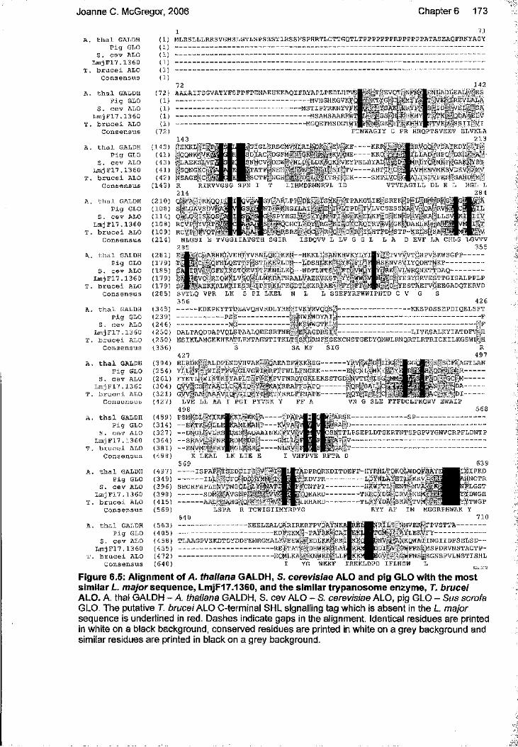
Joanne C. McGregor, 2006 Chapter 6 173
A . t h a l GALDH (1)P i g GLO (1)
S . c e v ALO (1)L m j F l 7 . 1 3 6 0 (1)
T . b r u c e i ALO (1)C o n s e n s u s (1)
A . t h a l GALDH ( 7 2 )P i g GLO (1)
S . c e v ALO (1)L m j F 1 7 . 1 3 6 0 (1 )
T . b r u c e i ALO (1)C o n s e n s u s (7 2 )
A . t h a l GALDH ( 1 4 3 )P i g GLO ( 4 1 )
S . c e v ALO ( 4 3 )L m j F l 7 . 1 3 60 ( 4 1 )
T . b r u c e i ALO ( 4 2 )C o n s e n s u s ( 1 4 3 )
A . t h a l GALDH ( 2 1 0 )P i g GLO ( 1 0 8 )
S . C ev ALO ( 1 1 4 )L m j F 1 7 . 1 3 6 0 ( 1 0 8 )
T . b r u c e i ALO ( 1 0 9 )C o n s e n s u s ( 2 1 4 )
A. t h a l GALDH ( 2 8 1 )P i g GLO ( 1 7 9 )
S . c e v ALO ( 1 8 5 )L m j F l 7 . 1 3 6 0 ( 1 7 9 )
T , b r u c e i ALO ( 1 7 9 )C o n s e n s u s ( 2 8 5 )
A. t h a l GALDH ( 3 4 5 )P i g GLO ( 2 3 9 )
S . c e v ALO ( 2 4 6 )L m j F 1 7 . 1 3 6 0 ( 2 5 0 )
T . b r u c e i ALO ( 2 5 0 )C o n s e n s u s ( 3 5 6 )
A . t h a l GALDH ( 3 9 4 )P i g GLO ( 2 5 4 )
S . c e v ALO ( 2 6 1 )L r a j F l 7 . 1 3 6Q ( 3 0 4 )
T . b r u c e i ALO ( 3 2 1 )C o n s e n s u s ( 4 2 7 )
A . t h a l GALDH ( 4 5 9 )P i g GLO ( 3 1 4 )
S . c e v ALO ( 3 2 7 )L m j F 1 7 . 1 3 6 0 ( 3 6 4 )
T , b r u c e i ALO ( 3 8 1 )C o n s e n s u s ( 4 9 8 )
A . t h a l GALDH ( 4 9 7 )P i g GLO ( 3 4 9 )
S . c e v ALO ( 3 9 6 )L m j F l 7 . 1 3 6 0 ( 3 9 8 )
T . b r u c e i ALO ( 4 1 5 )C o n s e n s u s ( 5 6 9 )
A . t h a l GALDH ( 5 6 3 )P i g GLO ( 4 0 5 )
S . c e v ALO ( 4 5 8 )L m jF I 7 . 1 3 6 0 ( 4 5 5 )
T. b r u c e i ALO ( 4 7 2 )C o n s e n s u s ( 6 4 0 )
1 71M LRSLLLRRSVGHSLGTLSPSSSTIRSSPSPHRTLCTTGQTLTPPPPPPPRPPPPPPATaSEaQFRKYAGY
72AALAlFSGVATYPSFPFPENAKHKKAQIFRYAPLPEDLHTVSj------------------------------------------------------------------MVHGHKGVKE’(--------------------------------------------------------------MSTIPFRKNYVF)------------------------------------------------------------------MSAHSAARPW'----------------------------------------------------------------MGQETMSDGTW;
^/S;/rHEVQ'lV-NITN;| ‘jKTYGL.CtEMYYL
UyCYSAKl ERYFj'f L p ' f
1 42SNLADLEAL' -ES n iVE 'I REV LALA L-jIO;riVELx,’SA
f'^TKnyQDAlELV 4TVEi-iviSSIIi::VI
1 43 p K K L f I |P| iQQNKfVKiÿ plLAEPSLVT isQNGKClî XSAGSgC[
|GifGL'SRSGMVNLALV.:DKa.iEVfV;KE----KKR gOSDgAc||DGFM:-*GK|Si5s .aKV;jME----KKQ "è;NMCvfiDEW|VVNLD-i)LDKp‘QKFVEYPELHYACg iT A T H |p E H g § g |E i^ ÿ lS S ; l i ç |T V------- AHTil' X p J
SMTg;%Cr-
FTNWAGIY C PR HMQPTSVEEV ELVKLA2 1 3
f R V Q Q l i V D A ^ D ^ ^ gîfL ÏîA lD |H P Q ||D K H |||A i'
RliYQl(NEFr,GAK3YS;i .VMEEVMRSVDKVP LïS5VFiBiSAÏÏC|iMi
RIRVVGSG SPN IR2 1 4 ^Q'SIFAL 'IR E Q Q Is'l'lLGAVSDV?Q [4r,G ";iSE Q S |RCVP|"]YVRt ;;BCVPLjFVQTa
LIHMDRMNRVL ID VTVEAGILL DL
1 SMKplT PAKGTI E\ > R E K |P iL p H [g R |G j âERT|LTPDnTVLVCSESSjÿA|TgQAiRVr t'N bT liN nK 'lE L K F ? |D |E N [ jp[ V #A @ L L
^ ‘CHCL;4DYi^RG|Kii|D| ;cAQIRTf|v|GKt JOAELRf @Al Sl SRSp-jDClgRlQ D X-llLHTPjolSTP-KELSfiSAi
XPPfiPE,., :g#3K:RGILAT:'
ÊPYHGLX.'IS''j
NLGSI TVGGIIATGTH S G IR ISDQVV L L V G G L L A D EVF LA CHLG LGVVV35 5
Z 71 v v v 7TgNPV;SKWSGPP----------SENVSVlYQDHTgKP------------------iRK||VWR(3NKT;DAQ---------m G gY ESY ^R V ESTT G ISA LPPLP^^v1yestSefv(|eegadqtkrvd
jpE'LVEHT:YVSNLQElKKN“ -HKKLL'SANKHVkYLY qFHLgETTFf]STLKEVLDN“ ~LDSHLKKl i 'YF î| f £ | : |GFNIKSTQEVITFENLLKQ--WDTLWTSXFli;;ivWiÂ/
EVQVjRIQWKLVSQ' LLMKDATNAALVAEKVKSTfYY[5WW^;' ?A.EKRRLWKi ESRI ^IPFRKLTE'GDTLKKRTAB-r'FY, If PŸ'
SVTLQ VPR LK S P I LKEL K 3 5 6---------- KDKPKYTTfcjEAVQHVRDLYRE; jlVKYRVQDS^ ---------------, p s g -------------------------bijWWFWDYAI j--------------------------EG-------------------------P :riK S WW GT K L « j—DALTAQDDAPVQlWp AALQEESRTNELIe RAGDRSIo -
L SSEFYRFWWIPHTD C V G S4 2 6
- “ KKSPDSSEPDIQELSFT
-LIVKS ALKYIAT DFV[|HESIKLAMGKKHEAteLPMTAGWTITKLTSSKLRNLSSEKCNSTGEDYQMWLRNQRTLRTRICKILKGSWl||H
SA KF S IG4 2 7ELRbKFii.'ALDPLNDVHVAKp.'jQAEAErWKK&EG----------- YRV W( îd[ |ILGYLLt’jFj ^;wiSTFVi;|GLVGWIii'|RFFFWiiLrNGKK----------- ENCNT' Bk / ,FYETL['ËlSTKIYAPLTfÎFvÆKFVFNRQYGKLEKSSTGD'^‘NVTD; 'ISG^N O W < ifC ifh Y A A rT .r i^ 'A T n r r-Y tL 'i 'iK A Y R R A F F R A P O ---------------1 O r I 'R A T ; x [ > ^QVV'J,CS|i]YAACLC!VAIQrr;Y[r'iiKAYRRAFFSAPQ G W , ’AAj W a V I 0 ■GIQ;'IYIpRTYRRT.F^NAl?E
LVE LL AA I P G l PYVNK Y FF A 498PSM'RD^iYÿEE|KKL->,EK|A-------- ÏP A P— E% TKgi|LEgRAM LBAHP------KVVAf— DiNGlgvdR s iO H SMa QAAINKEFYV!"- - S ^ V i l E N R|RDMVjDRi-------- GMLLÎ- -SN;vMP||Hy| rGL-î:Ss1----NLSViî
■ q r L s a l ; S-QYi ,11 iLf
VN G SLE
R 4 9 7
^ g s ë ^ G T L A N --------------------
|DA--------------------
FTFDCLFKQWV EWAIP5 6 8
------SP----------------------
SNTTLPSEPLDTSKRTNTSPGPVYGNVCRPFLDNTP
!< » ------------------------------------I VHFPVE RFTAK LEAL LK L IE E
5 6 9 _ ISPA Fi TSEDgH'Sl'j'M I "------------ I L l . f GFQpiDS' YMNxI'fSHCRFAPLENVTNSgLtÿYrîKÂTI --SDM ,'XAVGgp;i^ #'ÎÏV V iÇ--------a a l ' AHGgo;
LSPA R TCWÎGIIMYRPYG540----------------------------------K B ELB A LÇ ^R IR K RFPV PA ^------------------------------- Kd | s KM| j - PAFRKK|C A It l a a g p v k k d t d y d d f e m r g m a l k v e e w p ’. e d l k k Hr k i '------------------------------ REpSTAT(. '^DHWEPjiLA------------------------------ e q M lk a I ■•'.'’Knwe d ;.î |l l
T^DPRQRKDITDEFF-HYRHLTQKQLWDQFSAY%]KDVPR------------------L D Y W L A Y E Ïli^ K îd ^F gC N T P I------------------HKWFTLjpENlVj’MVA;^'P I q e a r d ----------------t r r c y d g f Ic r v (EEM'gi;^ÎVj'HELRHARD-------- TLRYYDAmÎSKAu/TVML.IT
RYY AF IM
6 3 9 IÉ IP K D
S n c t r | l g s t | dwgh I t w g p
MGGRPHWAK Y71 0
miLSjrNM VEKgFPVSTTA--------------GM AYLEKVjFY-------------------------
jblSIVL-'lA'NKQWAIINGIIDPSELSD— P irVM GW PRn | | mSPDRVN STACT P - GV: iG W FN SiSGN SPV LN STISH L
YG WEKF IRKKLDPD IFLNGW
Figure 6.6: Alignment of A. thaliana GALDH, S. cerevisiae ALO and pig GLO with the most similar L. mayor sequence, LmjF17.1360, and the similar trypanosome enzyme, T. brucei ALO. A. thal GALDH - A. thaliana GALDH, S. cev ALO - S. cerevisiae ALO, pig GLO - Sus scrofa GLO. The putative T. brucei ALO C-terminal SHL signalling tag which Is absent In the L major sequence is underlined In red. Dashes Indicate gaps In the alignment. Identical residues are printed In white on a black background, conserved residues are printed in white on a grey background and similar residues are printed in black on a grey background.

Joanne c . McGregor, 2006 Chapter 6 174
This analysis does not provide any definitive proof of which ascorbate pathways, if any,
are present in L. major. However, it seems reasonable to conclude that some of the
sequences identified may have a role. LmjF 17.1390 is very like the A. thaliana L-
galactose-1-phosphate phosphatase. Meanwhile LmjF28.1320 and LmjF 17.1360 are
similar to the only known enzymes that mediate the mammalian ascorbate biosynthesis
pathway: the recently identified mouse aldono-lactonase and pig GLO. Crucially, these L
major sequences were more similar to ascorbate biosynthesis enzymes, which have no
other known function, than any other proteins in the species the original enquiry sequences
were derived from. Thus, they were judged to be the most likely to be involved in
ascorbate synthesis in Leishmania. The importance of GLO in ascorbate synthesis in
mammals is paramount: humans lack this enzyme and therefore have to derive ascorbate
from their diet. Whether LmGLO has a similar role in the parasite was investigated, and is
discussed herein.
Enquiry sequence: organism, enzyme name, and amino acid length
Leishmania homologues, p/n scores and number of amino acids
% identity, % conservation and observations on alignments
Single organism back BLAST results
1. S. cerevisiae D- arabinonose dehydrogenase (NP009707), 344 amino acids
a. LmjF32.04601.2e" , 279 amino acidsb. LmjF31.21501.5e" , 285 amino acids
a. 37%, 53%. N- term more alike.b. 35%, 54%. L. maj seq 30 amino acid gap 137-167.
a. 5“* hit to enq seq. Best is glycerol dehydrogenase.b. 4* hit to enq seq. Best is as above.
2. S. cerevisiae D- arabinono-1,4-lactone oxidase (NPO13624), 526 amino acids
LmjF17.13603.2e' , 502 amino acids
19%, 38%.Middle of sequences display poor similarity.
Best hit to enq seq.
Table 6.2: L. major sequences similar to ascorbate biosynthesis enzymes in fungi.Abbreviations used are L. maj - L major, seq - sequence, ext - extension, N-term - N-terminal, C- term - C terminal, enq - enquiry. Numbers in first column correlate with those in figure 6.1.

Joanne C. McGregor, 2006 Chapter 6 175
Enquiry sequence: organism, enzyme name, and amino acid length
Leishmania homologues, p/n scores and number of amino acids
% identity, % conservation and observations on alignments
Single organism back BLAST results
3. Mouse aldono lactonase (NP033086), 299 amino acids
LmjF28.12301.4e'* , 413 amino acids
24%, 43%. Six small gaps in mouse seq. L. maj seq also has small C-term ext.
Best hit to enq seq.
4. Pig L-gulonolactone oxidase (Q8HXW0), 440 amino acids
LmjF17.13609.6e' , 502 amino acids
28%, 54%. Pig seq. contains a 55 amino acid gap. 235-290. L. maj seq has small C- term ext.
Best hit to enq seq.
Table 6.3: L. mayor sequences similar to ascorbate biosynthesis enzymes in animals.Abbreviations used are L. maj - L major, seq - sequence, ext - extension, N-term - N-terminal, 0- term - C terminal, enq - enquiry. Numbers in first column correlate with those in figure 6.1.

Joanne C. McGregor, 2006 Chapter 6 176
Enquiry sequence: organism, enzyme name, and amino acid length
Leishmania homologues, p/n scores and number of amino acids
% identity, % conservation and observations on alignments
Single organism back BLAST results
5. A. thaliana GDP-6- mannosepyrophosphorylase (AAC78474), 361 amino acids
LmjF23.01101.2e' ' , 379 amino acids
50%, 70%. Region between 250-270 displays poor similarity.
Best hit to enq seq.
6. A. thaliana GDP- mannose-3’,5’- epimerase (CAD70055), 377 amino acids
Lmj26.22309.9e'*, 446 amino acids
22%, 40%. L. maj seq has N-term ext. Also, gaps in A. thaliana N- term
42" hit to enq seq. Best is NADPH- dependant epimerase/dehydro genase.
7. A. thaliana L- galactose-1 -phosphate phosphatase (NPOO1030626), 269 amino acids
a. LmiF17.13901.8e" , 288 amino acidsb. LnijF15.08802.1e'^ , 466 amino acids
a. 35%, 55%. Good similarity.b. 32%, 49%. Large gaps in A. thaliana seq. N- and C- term ext. in L. maj seq.
a. Best hit to enq seq.b. Best hit to enq seq.
8. A. thaliana L- galactose dehydrogenase (CAD10386),319 amino acids
a. LmiF31.28801.5e*’ , 286 amino acidsb. LmjF32.04603.1e‘‘ 279 amino acids
a. 20%, 40%. Better similarity in N-term.b. 23%, 41%. Quite poor overall.
a. 36^ hit to enq seq. Best is oxidoreductase.b. 29^ hit to enq seq. Best is as above.
9. A. thaliana L- galactono-1,4-lactone dehydrogenase (NP190376),610 amino acids
LmjF17.13606.7e’ , 502 amino acids
20%, 42%. L. maj seq lacks first 100 amino acids.
Best hit to enq seq.
10. A. thaliana inositol oxygenase (AAP59548), 317 amino acids
LmjF18.13800.51, 378 amino acids
14%, 32%. L. maj seq N- and C- term ext. Also small gaps in A. thaliana seq.
39“* hit to enq. seq. Best is pyruvate dehydrogenase.
11. Strawberry D- galacturonic reductase (AAT76306), 319 amino acids
a. LnnF31.21502.0e'^ , 284 amino acidsb. LmjF32.04609.5e'^ , 279 amino acids
a. 37%, 56%. Small gaps in L. maj seq.b. 38%, 59%. Small gaps in L. maj seq.
a. 3* hit to enq seq. Best isoxidoreductaseb. 3*“ hit to enq seq. Best isoxidoreductase.
Table 6.4: L. mayor sequences similar to ascorbate biosynthesis enzymes in plants.Abbreviations used are L. maj - L major, seq - sequence, ext - extension, N-term - N-terminal, C- term - C terminal, enq - enquiry. Numbers in first column correlate with those in figure 6.1.

Joanne c . McGregor, 2006 Chapter 6 177
6.2.2 LmGLO expression in Leishmania
In order to clarify whether LmGLO expression could be detected, western blots were
performed on various Leishmania species promastigote lysates, using rat anti-sera that had
been raised to recombinant LmGLO (figure 6.6). As well as L. major, L. infantum and L
mexicana lysates were also used as sequences with 94% and 81% identity to LmGLO are
present in the L. infantum and L. braziliensis protein databases, respectively. Both the first
and final bleed batches of antisera were used for western blotting (figure 6.6A and 6.6B,
respectively) but unfortunately both antibodies detected several different proteins. The first
bleed antiserum detected the 57 kD 6-His LmGLO recombinant protein and in the L. major
promastigote lysate a slightly smaller band that may be LmGLO (untagged LmGLO is
predicted to be 56 kD). Unfortunately this could not be further investigated as all of the
antiserum obtained from the first bleed was used in this experiment and when re-used did
not detect either the recombinant protein or any proteins from Leishmania lysates. In
addition to the ~56 kD band the first bleed antibody detected several larger proteins, which
were presumed to be due to non-specific cross-reactivity of the anti-sera. The final bleed
antiserum detected many proteins, both in the recombinant protein fraction and in the
parasite lysates. One of the proteins detected appeared to be of around the expected size of
LmGLO, however, despite repeated attempts to optimise the conditions of the western blot
when using the antiserum, it was impossible to distinguish the band and discern any useful
information regarding endogenous LmGLO expression.
B
LmGLO
CS
1-bleed 2"" bleedFigure 6.6: Western blot analysis of LmGLO expression in the soluble fraction of Leishmania. A - first bleed antiserum. A protein of a slightly smaller size than the annotated LmGLO recombinant protein is visible in the L mayor lysates. B - second bleed antiserum. 10 pg- 30 pg of parasite lysate was loaded per lane. CS, cysteine synthase, (control).

Joanne c . McGregor, 2006 Chapter 6 178
6.2.3 Creation of LmGLO over-expressing L. major promastigotes
The sequence encoding LmGLO was amplified from L major genomic DNA, sub-cloned
into the pGEM T-easy and then cloned into the pGL102 plasmid. The plasmid with the
LmGLO gene was named pGL102LmGLO; maps of this and the empty vector are shown in
figure 6.7. The pGL102 plasmid is a Leishmania episomal expression vector and contains
the neomycin resistance gene as a selectable marker. Uncut pGL102 and pGL102LmGLO
were transfected into L. major promastigotes which were subsequently incubated in culture
with neomycin so that only successful transformed parasites would grow. The transformed
parasite lines will be herein referred to by the names of the vectors they were transformed
with.
pGL1026794 bp3’13K
amp
5'DST Sma \ (1366)
3*13K
pGL102LmGLO8303 bp LmGLO
amp
Figure 6.7: Plasmids for the over-expression of LmGLO in L. major promastigotes.Schematic representation of the pGL102 plasmid for the over-expresslon of LmGLO In Leishmania. A - empty pGL102 vector, for use as a negative control. B - pGL102 + LmGLO (pGL102LmGLO) for the over-expresslon of LmGLO. amp, amplclllln resistance gene; neo, neomycin resistance gene. The restriction sites used to clone LmGLO Into the vector are shown In dark red.

Joanne C. McGregor, 2006 Chapter 6 179
6.2.4 Phenotype analysis of L. major putative LmGLO overexpressing lines
Due to the failure of the anti-LmGLO antiserum to detect LmGLO specifically, it was not
possible to confirm the over-expression of LmGLO in the parasites and the phenotypic
analysis presented in this section must be regarded in this context.
6.2.4.1 Morphology and growth of promastigotes
Parasites putatively over-expressing LmGLO were examined microscopically and no
morphological differences could be seen between them and the parasites containing the
empty pGL102 vector. After several passages with neomycin present in the culture media,
the pGL102 and pGL102LmGLO lines were counted and diluted to the same
concentration. Thereafter their growth was monitored by daily counting (figure 6.8). The
parasite lines were found to grow at similar rates.
1.00E+08
E 1.00E+07
.12 & 1.00E+06
1.00E+05 1
PGL102pGL102LmGLO
0 2 4 6 8Number of days
Figure 6.8: Growth curves of pGL102 and pGL102LmGLO L. mayor promastigotes. Cultures were seeded at a concentration of 2 x 10® parasites/ml and were counted daily. Both lines were grown with 50 pg/ml neomycin.

Joanne C. McGregor, 2006
6.2 4.2 In vitro infectivity to macrophages
Chapter 6 180
The ability of the different stationary phase promastigote parasite lines to infect peritoneal
macrophages was assessed (figure 6.9). pGL102LmGLO infected a higher percentage of
macrophages than either the pGL102 or WT lines: approximately 4 times and twice as
many, respectively. This suggests that if pGL102LmGLO parasites are indeed over
expressing LmGLO then this aids infection. However, the differences observed were not
found to be significant, likely to be due to the lack of repetitions of this experiment: further
investigation is required. The variation between the infection rates between the WT and
pGL102 lines was also of concern as they would be expected to infect at a similar rate. It is
possible that the pGL102 line have been adversely affected by the transformation
procedure, causing them to be less infective in vitro.
60
I 40
I 30"So 20 0)
10
WT pGL102 pGL102LmGLO
Figure 6.9: Infectivity of WT, pGL102 and pGL102LmGLO L. mayor promastigotes to macrophages. Stationary phase promastigotes were used to infect peritoneal macrophages at a rate of 2:1 and the slides were incubated for 5 days post-infection. Results are the means ± SE from two experiments. The WT and pGL102LmGLO lines did not infect the macrophages at significantly different rates (p > 0.05).

Joanne C. McGregor, 2006
6.2.4 3 In vivo infectivity to mice
C hapters 181
To assess in vivo infectivity, pGL102LmGLO, pGL102 and WT stationary growth-phase
promastigotes were inoculated into mouse footpads, the thicknesses of which were then
measured over time (figure 6.10). The results broadly reflected those of the in vitro
infectivity experiments: pGL102LmGLO parasites were responsible for the most rapid
footpad growths which were slightly, but not significantly, faster than those infected with
the WT parasites, while footpads infected with pGL102 parasites took approximately 2
weeks longer to reach similar thicknesses. Over-expression of LmGLO is from an
episomal expression vector, the presence of which is assured by keeping the parasites
under neomycin pressure. This is not possible during in vivo work and the number of
episomes, and therefore the level of LmGLO expression, is likely decrease over time. This
may explain why the increased infectivity of pGL102LmGLO parasites as compared to the
WT and pGL102 lines is not as marked as in the macrophage infectivity experiment, where
infections are monitored over days as opposed to weeks. Once more the pGL102 parasites
caused less virulent infections than the WT parasites, which again may be indicative of
them being compromised in some way by the transformation procedure. This will be
discussed in more detail in section 1.3.
5.5
4.5
2 3.5
"O
2.5
u_
1 !
WT
pGL102
pGL102LmGLO
0 2 4 6 8
Number of weeks
Figure 6.10: Infectivity of WT, pGL102 and pGL102LmGLO, L. mayor promastigotes to mice.5x10® stationary-phase promastigotes were resuspended in 20 pi PBS and inoculated into one footpad of each BALB/C mouse. The footpad thicknesses were subsequently measured weekly, and the results are the means ± from five mice. The WT and pGL102LmGLO infections were not significantly different (p > 0.05).

Joanne c . McGregor, 2006 Chapter 6 182
6.2.5 Creation of L. major LmGLO knock-out parasite lines
The 3’ and 5’ flanking regions of LmGLO were amplified from L major genomic DNA,
sub-cloned into pET28a(+) and cloned into vectors pGL345 and pGL842 which contain
hygromycin and blasticidin resistance genes, respectively (figure 6.11). The constructs
containing the flanking regions and drug resistance markers were cut out and purified and
the linear DNA was used to transform L major promastigotes. Two independent lines,
K0LmGL03 and K0LmGL08, were generated, both having undergone transformation
with each of the constructs sequentially and grown up in the presence of the selection
drugs. In addition, following the second transfection and overnight recovery period, the
two independent cultures were divided and ascorbate was added to one of each to a final
concentration of 10 mM. This was to ensure that it would be available if the loss of
LmGLO resulted in the parasites having to exogenously source their ascorbate.
K0LmGL08 grew up to stationary phase regardless of whether ascorbate had been added
to the media, while only the K0LmGL03 culture that contained ascorbate grew up
quickly. After several passages in the selective drugs, K0LmGL03 and K0LmGL08
clones were derived by setting up the cultures with ascorbate in 96-well plates at a
concentration of approximately 0.1 parasites/well. Two clones were isolated from
K0LmGL03 - deemed KOLmGLOAl and KOLmGLOA2 - and one clone was derived
from K0LmGL08: KOLmGLOB. All three clones took several weeks to grow up.
LmGLO 5 'flank Sail {I)
HindWl (6808) DHFR flankI (944)
Hyg
amppGL345 LmGLO 3' and S' flanks
7301 bpfiomHI(l997)
DHFR flank
gg/H (4005) Sma\(3367)LmGLO 3' flank
Figure 6.11 : Construct for the gene knock-out of LmGLO in L major promastigotes.Schematic representation of the pGL345 plasmid containing the LmGLO 3' and 5" flanking regions. The second plasmid used for the construction of the LmGLO knock-out was the pGL842 plasmid, which is exactly the same except that it contains a blasticidin rather than hygromycin resistance gene. Hyg, hygromycin resistance gene; amp, ampicillin resistance gene; DHFR flanks, flanking regions of the dihydrofolate reductase gene. The restriction sites used to clone the LmGLO flanks and hygromycin resistance gene into the vector are shown in dark red. Hindlll and Bglll were used to cut out the flank-containing linear construct for transfection into L. major.

Joanne C. McGregor, 2006 Chapter 6 183
Because it was not possible to ascertain whether LmGLO had been successfully knocked
out by western blotting, a southern blot was performed on genomic DNA derived from
KOLmGLOAl and KOLmGLOA2 and KOLmGLOB, together with WT parasites. The
DNA was digested with restriction enzymes that, depending on whether the WT or
replacement allele was present, would yield fragments of different lengths containing the
LmGLO 3’ flanking region (figure 6.12A). The 3’ flanking region was used as a probe and
would hybridise to a fragment of approximately 1.4 kb if the WT allele was present, 2.4 kb
if the blasticidin resistance gene was present and 3.7 kb if the hygromycin resistance gene
was present. Unfortunately no fragments were detected in the WT and KOLmGLOAl and
KOLmGLOA2 digested DNA samples. However, fragments of approximately 2.4 kb and
3.7 kb were detected in the KOLmGLOB sample (figure 6.12B), corresponding to both
resistance markers that would be present in a bona fide LmGLO knockout. Therefore it
was concluded that KOLmGLOB lacks LmGLO. The absence of fragments detected in
KOLmGLOAl and KOLmGLOA2 suggest it is unlikely that they are genuine LmGLO
knockouts although the experiment was inconclusive.
6.2.6 Phenotype analysis of L major LmGLO knock-out parasite
lines
The parent lines of the clones, K0LmGL03 (which KOLmGLOAl and K0LmGL0A2
were derived from) and KOLmGLOB (which KOLmGLOB was derived from) were
analysed in some experiments as the clones took a long time to obtain. As well as
examining the phenotype of KOLmGLOB - deemed to be a genuine LmGLO knockout - KOLmGLOAl and KOLmGLOA2 and their parent lines were also included in
experiments. This was because, although unlikely to lack LmGLO, they would act as
useful controls due to them having undergone the same transformation procedure and
exposure to drugs as KOLmGLOB.

Joanne C. McGregor, 2006
AChapter 6 184
a /
B LmGLO 3" flank
LmGLOLmGLO 5' flank X h o l(6028)
Sa/ 1 (7416)
1.4 kb
X h o l(5528)
DHFR flank
BSD
LmGLO 5' flankATioI (6448)
DHFR flank
LmGLO 3' flankSail m o i )
2.4 kbX h o l(5528)
DHFR flank
LmGLO 5' flankflank \ Hyg
# 1 I
LmGLO 3' flankSa/1 (9268)
DHFR flankz ank I
# 1 *3 . 7 kb
Figure 6.12: Southern blot analysis of the LmGLO locus of L major KOLmGLO lines. A -Southern blot analysis of genomic DNA extracted from the different parasite lines as indicated above the gel image. The 2.4 kb and 3.7 kb bands visible in the lane containing DNA from the KOLmGLOB clone are indicated with red arrows. The blot was exposed to the film for 7 days. The positions and sizes of the molecular size markers are indicated to the left of the gel image; kb, kilo base pairs. B - schematic representation of the LmGLO locus on chromosome 17 (LmGLO shown in blue), and the altered locus following successful gene replacement with either the hyg (hygromycin) or bsd (blasticidin) resistance markers (shown in red). The expected sizes of the fragments following digestion of the genomic DNA with Xhol and Sa//, to be detected by southern blot using the 3’ flanking region as a probe, are indicated below each diagram and represented with a black line. The restriction sites and their relative positions are also shown.

Joanne C. McGregor, 2006 Chapter 6 185
6.2.6.1 Growth of promastigotes grown with and without exogenous
ascorbate
As described, K0LmGL03 and KOLmGLOS were originally divided and grown with and without ascorbate (+asc and -asc) added to the culture media although only KOLmGLOS
with exogenous ascorbate thrived. After several passages, the growth rates of the
remaining three cultures — K0LmGL03+asc, K0LmGL08-asc and K0LmGL08+asc -
were monitored as before (figure 6.13). The three parasite lines grew at similar rates although K0LmGL03+asc grew slightly faster than K0LmGL08+asc which grew faster
than K0LmGL08-asc. However the most marked difference between the cultures was in
the concentrations the parasites reached when in the stationary phase of growth. After several days K0LmGL03+asc grew to and remained at a density of ~2 x 10 parasites/ml,
as WT parasites do under similar conditions. Meanwhile K0LmGL08-asc and
K0LmGL08+asc reached concentrations of just -0.7 x lOVml and 0.9 x 10 /ml
respectively, even after 10 days in culture. KOLmGLOB, derived from K0LmGL08+asc, was the only clone found to lack the LmGLO gene while the clones derived from
K0LmGL03+asc were shown to be unlikely to lack the gene. Therefore it is possible that the loss of LmGLO causes a growth defect in L. major promastigotes, causing the parasites
to grow more slowly than usual and to fail to reach high densities. This is in accordance
with the growth defect observed in bloodstream form T. brucei lacking the homologous
gene to LmGLO (Wilkinson et ai, 2005). Also, the observation that K0LmGL08-asc
grows at a slightly slower rate and reaches lower parasite densities in culture than
K0LmGL08+asc suggests that the presence of exogenous ascorbate may have had a
beneficial effect in the early stages of transformation.
1.0E+08
1.0E+07
1.0E+06
1.0E+05
wrK0LmGL03+ascKOLmGLOB+ascKOLmGLOS-asc
1.0E+044 6 8
number of days
10 12
Figure 6.13: Growth curve of WT, K0LmGL03+asc, KOLmGLOB+asc and KOLmGLOS-asc L. major promastigotes. Cultures were seeded at a concentration of 2 x 10 parasites/ml and were counted daily. All lines except WT parasites were grown with 15 pg/ml blasticidin and 100 pg/ml hygromycin. K0LmGL03+asc and K0LmGL08+asc were also grown with 1 mM ascorbate present.

Joanne C. McGregor, 2006 Chapter 6 186
The three cultures were also grown with and without added ascorbate to ascertain if its inclusion in the culture media affected the growth rates of the parasites after they had been
in culture for several passages (figure 6.14). Exogenous ascorbate had no affect on the growth of any of the cultures at this stage and was subsequently omitted from the culture
media.
K0LmGL03+asc
1.00E+08 1
1.00E+08
E 1.00E+07II 1.00E+06S.
1.00E+0593 5 71
no ascorbate ascorbate
11
number of days
K0Lm G L08+asc
I 1.00E+07IIa 1.00E+06
1.00E+057 93 51
no ascortDate ascoitate
11
number of days
K0LmGL08-asc
1 .OGE+OS
1 .OOE+07
1 .OOE+06
S. 1.00E+05
1 .OOE+047 13 5 91 11
number of days
no ascorbate ascorbate
Figure 6.14: Growth curves showing K0LmGL03+asc, KOLmGLOB+asc and KOLmGLOS- asc with and without exogenous ascorbate. Cultures were seeded at a concentration of 2 x 10 parasites/ml and were counted daily. All lines were lines were grown with 15 pg/ml blasticidin and 100 pg/ml hygromycin. Ascorbate was added to cultures as indicated to a final concentration of 10 mM. Exogenous ascorbate had no effect on the growth rates of these L mayor lines.

Joanne C. McGregor, 2006
6.2.6 2 In vitro infectivity to macrophages
Chapter 6 187
The ability of KOLmGLOS and KOLmGLOS stationary phase promastigote parasite lines
to infect peritoneal macrophages was assessed (figure 6.15). Both these lines had
previously been grown with ascorbate, as described. KOLmGLOS infected a similar
percentage of macrophages as WT parasites: approximately 20%. However KOLmGLOS
parasites infected a lower number of cells: approximately just 5%. Once again, the
difference in phenotype is observed in the parent line of the clone found to be genuine
LmGLO knockouts. Therefore the loss of LmGLO may also cause reduced infectivity in L.
major in vitro. However, despite the variation in infection levels observed between the WT
and KOLmGLOS lines, the difference was not found to be significant. This is likely to be
due to the high standard errors and the low number of replicates performed and further
investigation is required to show conclusively that the LmGLO knockout parasites are
defective in their ability to infect macrophages.
30
I 25
I 20
I 15
I 10
wr KOLmGLOS KOLmGLOS
Figure 6.15: Infectivity of WT, KOLmGLOS and KOLmGLOB L. ma/or promastigotes to macrophages. Stationary phase promastigotes were used to infect peritoneal macrophages at a rate of 2:1 and the slides were incubated for 5 days post-infection Results are the means ± SE from at least two experiments. The WT and KOLmGLOB lines did not infect the macrophages at significantly different rates (p > 0.05).

Joanne C. McGregor, 2006
6.2.6 3 In vivo infectivity to mice
Chapter 6 188
In vivo infectivity of the clones KOLmGLOAl and KOLmGLOB were assessed. Parasites
in the stationary phase of growth were inoculated into mouse footpads, the thicknesses of
which were then measured over time (figure 6.16). Relative to the infections caused by WT
parasites, infections caused by KOLmGLOB were significantly much less rapid in growth,
as determined by the increase in footpad thicknesses. After five weeks the footpads
infected with WT parasites had increased from an average of 1.8 mm to 4.3 mm while
those infected with KOLmGLOB parasites had reached an average of just 2.1 mm. Similar
results were obtained with KOLmGLOAl parasites. After 10 weeks the KOLmGLOB
infected footpads were on average 4 mm thick while those infected with KOLmGLOAl
were on average just 2.5 mm although did continue to increase thereafter. So both clones
caused attenuated infections to occur as compared to WT parasites. Given that
KOLmGLOB was shown to lack LmGLO while KOLmGLOAl is unlikely to, this was an
unexpected result. In order to grow in the presence of the selective drugs, the
KOLmGLOAl parasites must either be supporting one or more extra-chromosomal
plasmids or have one or more constructs, intended for integration at the LmGLO locus,
integrated elsewhere in their genome. It is reasonable to consider that either of these
scenarios could be detrimental to the parasites, explaining their decreased infectivity to
mice, but that the reduced infectivity of the KOLmGLOB parasites may be due to the loss
of the LmGLO gene. However, it is also possible that both clones were damaged during
the transformation procedure, hence the similar phenotype observed.
E&I
Io
4.5 4
3.5 3
2.5 2
1.51 '
WTKOLmGLOAlKOLmGLOB
5 10Number of weeks
15
Figure 6.16: Infectivity of WT, KOLmGLOAl and KOLmGLOB L. mayor promastigotes to mice. 5x10^ stationary-phase promastigotes were resuspended in 20 pi PBS and inoculated into one footpad of each BALB/C mouse. The footpad thicknesses were subsequently measured weekly, and the results are the means ± SD from five mice. The footpad thicknesses of mice infected with WT parasites were significantly greater than those of mice infected with KOLmGLOB five weeks post-infection (p < 0.0004).

Joanne C. McGregor, 2006 Chapter 6 189
6.3 Discussion;:’ï
Ascorbate synthesis was originally of interest due to the ability of TDRl to reduce oxidised
ascorbate in vitro. Whether TDRl also functions as a dehydroascorbate reductase in I
Leishmania remains controversial. As detailed in the introduction to this chapter, reduction
of dehydroascorbate is thought to occur non-enzymatically by trypanothione (Krauth-
Siegel and Ludemann, 1996) as parasite lysates failed to increase ascorbate being formed
in in vitro experiments. However, in order to minimise spontaneous reaction the assay was
performed at pH 6.5 although the optimal pH for DEAR activity in vitro has previously
been reported to be -8.0 (Maellaro et al, 1997; Girardini et al, 2002). Therefore the pH may have prevented GSH-dependant DEAR activity being detected in the assay; the
absence of a positive control being performed means this cannot be ruled out.
Trypanosomatids do not possess glutathione reductase, which, as detailed in section 6.1.4,
is important for dehydroascorbate reduction by glutathione-dependant enzymes, as it in
turn recycles glutathione. As trypanosomatids instead possess trypanothione reductase, it
may be the case that trypanothione-dependant proteins would be responsible for any enzymatically mediated recycling of ascorbate. This would be analogous to Leishmania
containing trypanothione S-transferases rather than glutathione S-transferases, as has
recently been shown (Vickers et al, 2004). However, glutathione can be reduced by
trypanothione and glutathione-dependant enzymes do prevail in trypanosomatids
(Wilkinson et al, 2002b). Whether any enzymatic DEAR present in T. cruzi could have
been dependant on trypanothione rather than glutathione has not been investigated.
The recently published account of TbALO (Wilkinson et al, 2005), an ALO-like T. brucei
enzyme capable of converting L-galactono-1,4-lactone to ascorbate and D-arabinono-1,4-
lactone to erythroascorbate, to an extent addresses the initial questions postulated here of
whether trypanosomatids were capable of synthesising ascorbate and whether it was
important for viability. LmGLO shares 45% identity with TbALO although the region
between amino acid 235 and amino acid 320 poorly aligns and is of very low similarity. It
may be significant that LmGLO lacks the C-terminal SHL motif thought to target the T. brucei protein to the glycosome as differences in localisation of ascorbate synthesis
between the parasite species could be indicative of diverse roles for ascorbate.
TbALO is so called because it is most like yeast ALO in terms of substrate specificity
although investigation into the TbALO amino acid sequence revealed that, like LmGLO, it
is most similar to mammalian GLOs. However TbALO was unable to utilise the
mammalian substrate L-gulono-1,4-lactone, therefore diminishing the likelihood of the

Joanne c , McGregor, 2006 Chapter 6 190
mammalian synthesis pathway operating in trypanosomatids. Indeed the authors of the TbALO paper propose that it is the plant L-galactose pathway that is most likely to be
responsible for ascorbate synthesis in T. brucei, based on unnamed sequences that they
have identified in the genome. Several sequences similar to those of plant ascorbate
synthesis enzymes were identified in Leishmania (section 6.2.1). Due to the role of GDP-6-
mannose pyrophosphorylase in a range of cellular processes, the presence in Leishmania of
the similar sequence LmjF23.0110 may not be relevant to ascorbate synthesis and as such
has not been considered here.
Conversely, the activity of the plant L-galactose-1-phosphate phosphatase protein is
thought to be exclusive to the ascorbate synthesis pathway and therefore the discovery of
the similar Leishmania sequence LmjF17.1390 was intriguing. However, analysis of the
mouse - and indeed human- protein databases revealed that they too possess similar
sequences to L-galactose-1-phosphate phosphatase which were not more similar to any
other A, thaliana protein. Clearly experimental characterisation is required to ascertain
whether these mammalian proteins are involved in a different cellular process or whether
they too have a role in ascorbate synthesis. For example it is feasible that they could
dephosphorylate D-glucuronic acid-1 -phosphate (figure 6.1). The existence of the
mammalian sequences certainly cast doubt on whether LmjF17.1390 is indeed a dedicated
ascorbate synthesis protein exclusive to the plant L-galactose pathway. A set of proteins
similar to yeast and plant enzymes that mediate the penultimate ascorbate synthesis steps -
arabinose dehydrogenase, L-galactose dehydrogenase and galacturonic reductase were -
also identified in Leishmania. However, these were all more similar to other yeast and
plant proteins and one of them - LmjF31.2150 - has been experimentally characterised as
a prostaglandin f2 alpha synthase (Kabututu et al, 2003). Meanwhile LmjF28.1230, which
is similar to the mammalian aldono-lactonase which mediates the penultimate step in
ascorbate synthesis in mouse, is more similar to the aldono-lactonase than any other mouse
protein. Therefore the substrate specificity of TbALO is the only evidence of the plant
pathway being operational in trypanosomatids. Controversy already exists over whether
plant GALDH can utilise L-gulono-1,4-lactone as a substrate; A. thaliana cells used it as a
precursor for ascorbate synthesis (Wolucka and Van Montagu, 2003) while purified sweet
potato GALDH could only metabolise L-galactono-1,4-lactone (Oba et al, 1995). It is
possible that activity with L-gulono-1,4-lactone is difficult to reproduce in vitro. It is clear
that experimental characterisations of several enzymes - including LmGLO and the
putative ascorbate synthesis proteins identified and described in this chapter - are required
before any conclusions regarding which pathway(s) is responsible for ascorbate

Joanne c. McGregor, 2006 Chapter 6 191
biosynthesis in the trypanosomatids. In addition the potential for Leishmania to scavenge
different intermediates in the synthesis pathways should be addressed.
Indeed, the ability to scavenge ascorbate is the reason postulated to explain the exacerbated
growth defect of T. brucei TbALO null mutants in ascorbate-Ifee media compared to
normal media (Wilkinson et al, 2005). Serum is reported as having an ascorbate
concentration of 40 pM but it is not stated what percentage of the final media is comprised
by serum. Standard media is often 10% (v/v) serum, which would give a final
concentration of 4 pM ascorbate, much lower than the reported Kms for known ascorbate
and dehydroascorbate transporters (Rumsey et al, 1997; Daruwala et al, 1999; Rumsey et
al, 2000). Although not described in detail here, searches of the L. major genome for possible transporters were conducted using enquiry sequences of known ascorbate
transporters (as detailed in section 6.1.3). There were no significantly similar proteins to
the sodium-dependant transporters SVCTl and SVCT2, or to the E. coli sgaT transporter.
There was, however, a similar protein to the GLUT transporters, which was most like
GLUTS. The GLUT transporters are only known to transport oxidised ascorbate
(dehydroascorbate), and the method used by Wilkinson et al, to eliminate ascorbate fi-om
the medium was to treat it with ascorbate oxidase, meaning that only dehydroascorbate
would remain. Depleting the ascorbate in this way contributed to the growth defects of the
TbALO knockout mutants, suggesting that they could accumulate ascorbate but not
dehydroascorbate. Therefore, if trypanosomatids are able to uptake ascorbate from their
surrounding environment, it is unlikely to be via GLUT-like transporters and may occur by
a novel mechanism.
Ascorbate was added to a final concentration of 10 mM to L. major parasites that had just
undergone the second transfection to knockout the LmGLO gene. This at first seemed to be
beneficial as cultures that contained the added ascorbate grew better than those without.
However there was no noticeable effect on growth when ascorbate was withdrawn from
the cultures that had previously contained it, or added to the cultures that were previously
without it. There was also no correlation between the presence of ascorbate in the media
and successful LmGLO knockouts: all three clones were derived fi*om cultures that had
contained ascorbate and of these only one, KOLmGLOB, proved to be a genuine LmGLO
knockout.
The parent strain of KOLmGLOB exhibited several defects: promastigotes grew slowly in culture, did not reach high densities at the stationary phase of growth and were poorly
infective to peritoneal macrophages. Moreover KOLmGLOB parasites were poorly

Joanne c . McGregor, 2006 Chapter 6 192
infective to mice, taking several weeks longer than WT parasites to cause lesions of 4 mm
to develop. The inability to synthesise ascorbate in these lines appears to adversely affect
the parasites although further experimentation is required to address why the
KOLmGLOAl clone also exhibits an in vivo infection defect. A more in-depth phenotypic
analysis of the clones rather than the parent knockout lines, together with the creation and
analysis of KOLmGLO parasites engineered to re-express LmGLO, will help clarify the
role of GLO in L major. It is also of interest that parasites over-expressing LmGLO appear
to be more infective to both macrophages and mice than WT and control parasites.
However, until a method can be developed to prove that the parasites do indeed over
express LmGLO, these results must be treated with caution.

193
7 Discussion
This study aimed to identify and characterise a Leishmania protein capable of reducing
pentavalent antimonial compounds to the toxic trivalent form, and to ascertain whether any
protein identified was involved in antimonial susceptibility in vivo. L. major TDRl was
found and investigated due to its similarity to oGST and like this mammalian protein,
recombinant TDRl was capable of reducing pentavalent metalloids, including antimonial
drugs, in vitro (Denton et al, 2004). To elucidate the role of TDRl in antimonial
metabolism in vivo, L. major TDRl knockout and over-expressing lines were generated,
infected into macrophages and treated with pentavalent antimonials, and the effects of the
drugs on the parasites were assessed. Due to the different macrophage infection rates
displayed by the different parasites lines it proved difficult to compare the effects of the
pentavalent antimonials on the different parasites. However despite being inconclusive, the
studies suggested that altering the level of TDRl in L. major had little or no effect on the
parasites' sensitivity to pentavalent antimonial drugs.
If the TDRl does not have an impact upon the susceptibility of L. major to pentavalent
antimonials in vivo it is interesting to postulate why that may be. Since it is already well
established that toxicity is dependant on reduction of the drugs to the trivalent form, the
most obvious explanation is that in vivo the protein does not reduce pentavalent
antimonials and there could be several reasons for this. Firstly, as discussed in the
introductory chapter to this investigation, the site of reduction remains controversial. If
reduction occurs in the macrophage, which could be mediated enzymatically by one of the
classes of mammalian arsenate reductases discussed or by an as yet unidentified protein,
TDRl may only encounter the trivalent form of the drug. It has also been demonstrated
that pentavalent antimonials are reduced upon reaction with small thiols such as
glutathione and cysteine (Frezard et al, 2001; Ferreira Cdos et al, 2003); this could also
occur in the macrophage. Indeed the acidic environment of the phagolysosome where the
parasites reside promotes this reaction (Alexander and Russell, 1992). Reduction could
also be mediated in the macrophage by a secreted Leishmania protein and indeed, it was
shown as part of this investigation (chapter four) that TDRl is likely to be released. This
could also be the case for the recently identified Leishmania ACR2 protein which reduces
pentavalent antimonials (Zhou et al, 2004): although the published account of this protein
describes it as comprising 130 amino acids it is possible that it contains a further 100
amino acid N-terminal region which includes a signalling peptide that is predicted to direct
the protein to the extracellular space, as outlined in chapter three. In this situation the

Joanne C. McGregor, 2006 Chapter 7 194
macrophage or parasitophorous vacuole could act as a reservoir for the secreted parasite
protein meaning that although a Leishmania protein would mediate the reaction, reduction
and therefore toxicity would be dependant on the amastigote parasites being situated in
macrophages. This could help explain some of the controversy surromiding the site of
reduction, specifically the observation that axenic amastigotes are not susceptible to
pentavalent antimonials (Sereno et al, 1998). However, conflicting reports that axenic
amastigotes are indeed sensitive to these drugs (Ephros et al, 1999) imply that the
presence of macrophages is not necessary for reduction to occur, and that therefore this
happens within the parasites.
The second reason that would explain TDRl not reducing pentavalent antimonials in vivo
is that the drugs may be reduced by a different route inside the parasite. Given that there is
scant information on the localisation of antimonials within Leishmania, it may be that
TDRl and the pentavalent drugs do not co-localise in vivo, therefore explaining why the
protein is not able to reduce the compounds. This would be particularly likely if the
pentavalent antimonials were sequestered inside a parasite organelle given the likely
cytosolic distribution of TDRl described in chapter four. In this case, pentavalent
antimonials must be reduced by an alternative mechanism. Again, this could be mediated
by a protein other than TDRl - such as LmACR2 - or could occur non-enzymatically
upon reaction with thiols. As well as pentavalent antimonials being reduced by the thiols
mentioned, the trypanosomatid-specific thiol trypanothione is also capable of reducing
antimonials non-enzymatically (Ferreira Cdos et al, 2003). Indeed, reduction rates were
greater in the presence of trypanothione than glutathione, which may go some way to
explain why pentavalent antimonials are more toxic to parasites than the host. However,
the rates of reduction of antimonials achieved with thiols are a fraction of those achieved
with TDRl in vitro (Denton et al, 2004) so non-enzymatic, thiol-based reduction alone is
unlikely to account for why TDRl does not reduce the drugs in vivo. Therefore enzymatic
reduction by another Leishmania protein or several proteins may be a more likely
explanation. As well as LmACR2, other arsenate reductase-like sequences are present in
the parasite that may be capable of reducing antimonials, the most alike being similar to
GAPDH as explained in chapter three. However, the in vivo relevance of GAPDH in
metalloid reduction remains unclear. Three new classes of proteins with arsenate reductase
activity have been identified in the last five years and it is likely more will be elucidated. A
candidate could be the glutaredoxins, of which there are predicted to be several of in L. major. Despite the low-level sequence similarity that the proteins share, oGSTs and
glutaredoxins exhibit comparable activities in vitro including thioltransferase and
dehydroascorbate reductase activities. It is therefore pertinent to consider that the

Joanne c . McGregor, 2006 Chapter? 195
glutaredoxins may display the pentavalent arsenical reducing ability of oGST: there are no
published accounts of these activities being looked for in glutaredoxins. It would be
interesting to look for this activity in the predicted L. major glutaredoxins that are
described in chapter three.
As well as the possibility that TDRl does not reduce pentavalent antimonials in vivo, it is
possible that the protein does indeed carry out this reaction but that the experiments
described in this investigation failed to detect this. Although knockout and over-expressing
lines are useful tools that have been used to analyse the roles of other Leishmania proteins,
the approach may not have been successful here. If several proteins, including TDRl, are
able to reduce the drugs in vivo then knocking out or over-expressing TDRl may not result
in a big difference in the parasites’ ability to reduce the pentavalent compounds and
therefore render any changes in susceptibility to the drugs undetectable. This effect would
be particularly pronounced if the L. major parasites were able to up- or down-regulate the
expression of these proteins in response to the altered level of TDRl. As discussed in
chapter three, L. major parasites are thought to possess several glutaredoxins and
thioredoxins and degeneracy between these proteins has been reported in yeast (Potamitou
et al, 2002): it could be the case that degeneracy between these proteins and TDRl occurs
in Leishmania given the common activities of the proteins in vitro. It would be interesting
to test this hypothesis by comparing the levels of glutaredoxins and thioredoxins in
Leishmania wild type with the lines with altered TDRl levels. This could be achieved by individually by western blotting if the specific anti-sera to these proteins were available, or
by a more global proteomic approach.
There are other reasons why the approach used to elucidate the role of TDRl in antimonial
reduction and sensitivity in vivo may have failed. One possibility could be that the
peritoneal macrophages used in the experiments may have been unsuitable for assaying the
effects of the drug, for example if there expression profile was altered due to their artificial
growth conditions leaving them unable to accumulate the pentavalent antimonials. This
would also explain the lack of sensitivity to the drugs displayed by all the parasite lines
used in the study. A problem of this nature could be circumvented by infecting live animals
with the parasite lines and testing the efficacy of the drugs in treating the L, major
infections in a true in vivo setting. Another potential problem could be that L major
parasites that were used in this study: L, major causes cutaneous leishmaniasis whereas
visceral leishmaniasis, commonly caused by L. donovani or L. infantum, is more likely to
be treated with pentavalent antimonials. Indeed, several studies have shown that intrinsic
differences in species sensitivity to these drugs is common and cutaneous leishmaniasis

Joanne c . McGregor, 2006 Chapter 7 196
isolates are generally less susceptible to these drugs than those causing visceral disease
(reviewed in Croft et al, 2006). Creating and analysing TDRl knockout and over-
expressing forms of a different species of Leishmania, such as L. infantum, may be
worthwhile.
Very little is known about the enzyme(s) that mediate the reduction of pentavalent
metalloids in mammals. There are three classes of enzymes known to display this activity
in vitro - GAPDH, oGSTs and PNP - and yet evidence of the involvement of any of these
proteins in vivo remains elusive. Construction of a knockout mouse model of any of these
proteins may facilitate this area of investigation and help clarify whether the protein does
mediate reduction in vivo, although this may be impossible for GAPDH and PNP due to their ubiquity in cells. A greater understanding of mammalian reduction of metalloids is
required before the impact, if any, of the host metabolism on the sensitivity of Leishmania
to pentavalent antimonials can be assessed. It is important to identify the proteins that
regulate the reduction of pentavalent metalloids in higher eukaryotes for reasons other than
their potential involvement in leishmaniasis chemotherapy: Arsenical compounds continue
to contaminate drinking water in many parts of the world and are considered to be a serious
health risk. It is distressing to think that how the body metabolises and detoxifies these
compounds and how they exert their toxicity is still unknown. With arsenical- and
antimonial-based drugs being proposed for the treatment of cancer (Murgo, 2001; Wyllie
and Fairlamb, 2006), it is even more imperative that a greater insight into their metabolism
is reached. Elucidation of how pentavalent metalloids enter cells - both mammalian and
parasitic - is also required.
The recent licensing of the oral antileishmanial drug miltefosine together vrith the
impressive cure rates quoted in several clinical studies of the drug may have prompted
speculation that the days of antimonials in leishmaniasis chemotherapy are numbered.
However this is a controversial view. It was outlined in the introduction to this investigation that miltefosine, despite its obvious advantages of being orally administrated,
has several potential shortcomings: resistance to the drug has been easily generated in
Leishmania in the laboratory; it may not be effective in treating some South American
Leishmania species; patients co-infected with HIV tend to relapse when treated with
miltefosine; and the drug is teratogenic. Although resistance to pentavalent antimonials is
now a problem in some parts of India, these drugs have been used for decades successfully.
Although miltefosine is no doubt a welcome addition to the arsenal of antileishmanial
drugs, it has not stood the test of time. This, coupled with the infrastructure needed to treat leishmaniasis sufferers with pentavalent antimonials aheady being in place, m e^s that

Joanne C. McGregor, 2006 Chapter 7 197
antimonials are very much still required and that research into its reduction and activation
are valid and important. This research may also aid the understanding of additional
metalloid-based chemotherapies.
What the endogenous role is of TDRl in the parasite is an interesting point. Unlike its T.
cruzi homologue Tc52, TDRl was knocked out of L. major with relative ease showing that
is it is not an essential protein. This is an intriguing difference between the two proteins.
The different predicted active sites between TDRl and Tc52 could be indicative of
differing functions which may explain why Tc52 is essential while TDRl is not. Whatever
the reason, the fact that L. major can survive well without the protein implies that, as
discussed, degeneracy between proteins may be occurring. The in vitro activities of TDRl
coupled with the observation that the TDRl knockout lines were more susceptible to
paraquat stress suggest that the protein has a role in protecting the parasite ffom oxidative
stress. Maintaining the correct redox environment is of utmost importance in the cell and
this is particularly true of parasites that have to withstand high levels of reactive oxygen
species (ROS) generated during the oxidative burst. It would therefore make sense that the
parasites have evolved to possess several proteins that are capable of regulating levels of
ROS, and that partial degeneracy exists between them.
One of the potential roles of TDRl in L major is to maintain levels of ascorbate by reducing dehydroascorbate which occurs in vitro. This is controversial: it has been
described how tiypanothione alone is thought to be sufficient for performing this function
and that an enzyme is not required (Krauth-Siegel and Ludemann, 1996). However, this
observed activity prompted us to look for enzymes that could have a role in ascorbate
biosynthesis in L major and the discovery of LmGLO which, as explained in chapter six,
is likely to have such a role. The potential involvement of TDRl in reducing ascorbate
remains unresolved although it would be interesting to analyse this. For example, in the
TDRl knockout lines LmGLO could be up-regulated: if the ability to recycle ascorbate
was lost or reduced, a solution could be to produce more ascorbate. The reciprocal is true:
in the LmGLO line TDRl could be up-regulated to promote recycling of any available
ascorbate. This hypothesis could be tested by western blotting. Furthermore, it would be of
interest to attempt to generate a double mutant lacking both proteins: if the hypothesis was
correct this mutant may not be viable. Certainly, what both thèse proteins do have in
common is that directly or indirectly they are likely to impact upon the intracellular redox
environment of the parasite.

198
8 References
Adak, S. and A. K. Datta (2005). "Leishmania major encodes an unusual peroxidase that is a close homologue of plant ascorbate peroxidase: a novel role of the transmembrane domain," Biochem J390(Pt 2): 465-74.Agius, F., R, Gonzalez-Lamothe, J. L. Caballero, J. Munoz-Blanco, M. A. Botella and V. Valpuesta (2003). "Engineering increased vitamin C levels in plants by overexpression of a D-galacturonic acid reductase." Nat Biotechnol 21(2): 177-81.Akerman, S. E. and S. Muller (2005). "Peroxiredoxin-linked detoxification of hydroperoxides in Toxoplasma gondii." J Biol Chem 280(1): 564-70.Al-Mohammed, H. I., M. L. Chance and P. A. Bates (2005). "Production and characterization of stable amphotericin-resistant amastigotes and promastigotes of Leishmania mexicana." Antimicrob Agents Chemother 49(8): 3274-80.Alexander, J. and D. G. Russell (1992). "The interaction of Leishmania species with macrophages." Adv Parasitai 31: 175-254.Allaoui, A., C. Francois, K. Zemzoumi, E. Guilvard and A. Ouaissi (1999). "Intracellular grovyth and metacyclogenesis defects in Trypanosoma cruzi carrying a targeted deletion of a Tc52 protein-encoding allele." Mol Microbiol 32(6): 1273-86.Amato, V. S., A. R. Padilha, A. C. Nicodemo, M. I. Duarte, M. Valentini, D. E. Uip, M. Boulos and V. A. Neto (2000). "Use of itraconazole in the treatment of mucocutaneous leishmaniasis: a pilot study." Int J Infect Dis 4(3): 153-7.Anacleto, C., M. C. Abdo, A. V. Ferreira, S. M. Murta, A. J. Romanha, A. P. Fernandes and E. S. Moreira (2003). "Structural and fimctional analysis of an amplification containing a PGPA gene in a glucantime-resistant Leishmania (Viannia) guyanensis cell line," Parasitai Res 90(2): 110-8,Anderson, G. L,, J. Williams and R. Hille (1992). "The purification and characterization of arsenite oxidase from Alcaligenes faecalis, a molybdenum-containing hydroxylase." J Biol Chem 267(33): 23674-82.Aposhian, H. V. and M. M. Aposhian (2006). "Arsenic toxicology: five questions." Chem Res Toxicol 19(1): 1-15.Aposhian, H. V., R. A. Zakharyan, M. D. Avram, M. J, Kopplin and M. L, Wollenberg (2003). "Oxidation and detoxification of trivalent arsenic species." Toxicol Appl Pharmacol 193(1): 1-8.Aposhian, H. V., R. A. Zakharyan, M. D. Avram, A. Sampayo-Reyes and M. L. Wollenberg (2004). "A review of the enzymology of arsenic metabolism and a new potential role of hydrogen peroxide in the detoxication of the trivalent arsenic species." Toxicol Appl Pharmacol 198(3): 327-35.Ariyanayagam, M. R. and A. H. Fairlamb (2001). "Ovothiol and trypanothione as antioxidants in trypanosomatids." Mol Biochem Parasitai 115(2): 189-98.Ariyanayagam, M. R., S, L. Oza, M. L. Guther and A. FI. Fairlamb (2005), "Phenotypic analysis of trypanothione synthetase knockdown in the African trypanosome." Biochem J 391(Pt2): 425-32.Arrigoni, O. and M. C. De Tullio (2002), "Ascorbic acid: much more than just an antioxidant." Biochim Biophys Acta 1569(1-3): 1-9.

199
Basselin, M., M. A. Badet-Denisot, F. Lawrence and M. Robert-Gero (1997a). "Effects of pentamidine on polyamine level and biosynthesis in wild-type, pentamidine-treated, and pentamidine-resistant Leishmania." Exp Parasitai 85(3): 274-82.Basselin, M., M. A. Badet-Denisot and M. Robert-Gero (1998). "Modification of kinetoplast DNA minicircle composition in pentamidine-resistant Leishmania." Acta Trop 70(1): 43-61.Basselin, M., H. Denise, G. H. Coombs and M, P. Barrett (2002). "Resistance to pentamidine in Leishmania mexicana involves exclusion of the drug from the mitochondrion." Antimicrob Agents Chemother 46(12): 3731-8.Basselin, M., F. Lawrence and M. Robert-Gero (1997b). "Altered transport properties of pentamidine-resistant Leishmania donovani and L. amazonensis promastigotes." Parasitai Res 83(5): 413-8.Basselin, M. and M. Robert-Gero (1998). "Alterations in membrane fluidity, lipid metabolism, mitochondrial activity, and lipophosphogiycan expression in pentamidine- resistant Leishmania." Parasitai Res 84(1): 78-83.Bates, P. A., C. D. Robertson, L. Tetley and G. H. Coombs (1992). "Axenic cultivation and characterization of Leishmania mexicana amastigote-like forms." Parasitology 105 ( Pt 2): 193-202.Bennett, M. S., Z. Guan, M. Laurberg and X. D. Su (2001). "Bacillus subtilis arsenate reductase is structurally and functionally similar to low molecular weight protein tyrosine phosphatases." Proc Natl Acad Sci U SA 98(24): 13577-82.Bentley, R. and T. G. Chasteen (2002). "Microbial méthylation of metalloids: arsenic, antimony, and bismuth." Microbiol Mol Biol Rev 66(2): 250-71.Berman, J., A. D. Bryceson, S. Croft, J. Engel, W. Gutteridge, J. Karbwang, H.Sindermann, J. Soto, S. Sundar and J. A. Urbina (2006). "Miltefosine: issues to be addressed in the future." Trans R Soc Trap Med Hyg, 100(Suppl 1): S41-4Berman, J. D., J. V. Gallalee and B. D. Hansen (1987). "Leishmania mexicana: uptake of sodium stibogluconate (Pentostam) and pentamidine by parasite and macrophages." Exp Parasitai 64(1): 127-31.Berman, J. D. and M. Grogl (1988). "Leishmania mexicana: chemistry and biochemistry of sodium stibogluconate (Pentostam)." Exp Parasitai 67(1): 96-103.Berman, J. D., D, Waddell and B. D. Hanson (1985). "Biochemical mechanisms of the antileishmanial activity of sodium stibogluconate." Antimicrob Agents Chemother 27(6): 916-20.Board, P. G., M. Coggan, G. Chelvanayagam, S. Easteal, L. S. Jermiin, G. K. Schulte, D,E. Danley, L. R. Hoth, M. C. Griffor, A. V. Kamath, M. H. Rosner, B. A. Chrunyk, D. E. Perregaux, C. A. Gabel, K. F. Geoghegan and J. Pandit (2000), "Identification, characterization, and crystal structure of the Omega class glutathione transferases." J Biol Chem 275(32): 24798-806.Bobrowicz, P., R. Wysocki, G. Owsianik, A. Goffeau and S. Ulaszewski (1997). "Isolation of three contiguous genes, ACRl, ACR2 and ACR3, involved in resistance to arsenic compounds in the yeast Saccharomyces cerevisiae." Yeast 13(9): 819-28.Bodley, A. L., M. W. McGarry and T. A. Shapiro (1995). "Drug cytotoxicity assay for Afiican trypanosomes and Leishmania species." J Infect Dis 172(4): 1157-9.Bogdan, C., N. Donhauser, R. Doring, M. Rollinghoff, A. Diefenbach and M. G. Rittig(2000). "Fibroblasts as host cells in latent leishmaniosis." JExp Med 191(12): 2121-30.

200
Borges, M., A. C. Da Silva, D. Sereno and A. Ouaissi (2003). "Peptide-based analysis of the amino acid sequence important to the immunoregulatory function of Trypanosoma cruzi Tc52 virulence factor." Immunolo^ 109(1): 147-55.Borges, M., E. Guilvard, A. Cordeiro da Silva, B. Vergnes, K. Zemzoumi and A, Ouaissi(2001). "Endogenous Trypanosoma cruzi Tc52 protein expression upregulates the growth of murine macrophages and fibroblasts and cytokine gene expression." Immunol Lett 78(3): 127-34.Boveris, A., H. Sies, E. E. Martino, R. Docampo, J. F. Turrens and A. O. Stoppani (1980). "Deficient metabolic utilization of hydrogen peroxide in Trypanosoma cruzi." Biochem J 188(3): 643-8.Bradford, M, M. (1976). "A rapid and sensitive method for the quantitation of microgram quantities of protein utilizing the principle of protein-dye binding." Anal Biochem 72: 248-54.Brajtburg, J. and J. Bolard (1996). "Carrier effects on biological activity of amphotericin B." Clin Microbiol Rev 9(4): 512-31.Bray, P. G., M. P. Barrett, S. A. Ward and H. P. de Koning (2003). "Pentamidine uptake and resistance in pathogenic protozoa: past, present and future." Trends Parasitai 19(5): 232-9.Bresell, A., R. Weinander, G. Lundqvist, H. Raza, M. Shimoji, T. H. Sun, L. Balk, R. Wiklund, J. Eriksson, C. Jansson, B. Persson, P. J. Jakobsson and R. Morgenstem (2005). "Bioinformatic and enzymatic characterization of the MAPEG superfamily." Febs J 272(7): 1688-703.Brochu, C., J. Wang, G. Roy, N. Messier, X. Y. Wang, N. G. Saravia and M. Ouellette(2003). "Antimony uptake systems in the protozoan parasite Leishmania and accumulation differences in antimony-resistant parasites." Antimicrob Agents Chemother 47(10): 3073-9.Bryceson, A. (2001). "A policy for leishmaniasis with respect to the prevention and control of drug resistance." Trap Med Int Health 6(11): 928-34.Bzowska, A., E. Kulikowska and D. Shugar (2000). "Purine nucleoside phosphorylases: properties, functions, and clinical aspects." Pharmacol Ther 88(3): 349-425.Callahan, H. L., A. C. Portal, R. Devereaux and M. Grogl (1997). "An axenic amastigote system for drug screening." Antimicrob Agents Chemother 41(4): 818-22.Carlin, A., W. Shi, S. Dey and B. P. Rosen (1995). "The ars operon of Escherichia coli confers arsenical and antimonial resistance." J Bacterial 177(4): 981-6.Carrasco, A., G. Pincheira and T. Ureta (1981). "Genetic and biochemical characterization of D-arabinose dehydrogenase from Neurospora crassa." J Bacterial 145(1): 164-70.Chakraborty, A. K. and H. K. Majumder (1988). "Mode of action of pentavalent antimonials: specific inhibition of type I DNA topoisomerase of Leishmania donovani." Biochem Biophys Res Commun 152(2): 605-11.Chan, T. S., N. Shangari, J. X. Wilson, H. Chan, R. F. Butterworth and P. J. O'Brien (2005). "The biosynthesis of ascorbate protects isolated rat hepatocytes fi-om cumene hydroperoxide-mediated oxidative stress." Free Radie Biol Med 38(7): 867-73.Chen, Z., T. E. Young, J. Ling, S. C. Chang and D. R. Gallie (2003). "Increasing vitamin C content of plants through enhanced ascorbate recycling." Proc Natl Acad Sci U SA 100(6): 3525-30.Chung, W. FI., K. D. Kim, Y. J. Cho and J. H. Roe (2004). "Differential expression and role of two dithiol glutaredoxins Grxl and Grx2 in Schizosaccharomyces pombe."Biochem Biophys Res Commun 321(4): 922-9.

201
Chung, W. H., K. D. Kim and J. H. Roe (2005). "Localization and function of three monothiol glutaredoxins in Schizosaccharomyces pombe." Biochem Biophys Res Commun 330(2): 604-10.Clark, D., M. Albrecht and J. Arevalo (1994). "Ascorbate variations and dehydroascorbate reductase activity in Trypanosoma cruzi epimastigotes and trypomastigotes." Mol Biochem Parasitai 66(1): 143-5.Clayton, C. E. (2002). "Life without transcriptional control? From fly to man and back again," Embo /21(8): 1881-8.Coelho, A. C., S. M. Beverley and P. C. Cotrim (2003). "Functional genetic identification of PRPl, an ABC transporter superfamily member conferring pentamidine resistance in Leishmania major." Mol Biochem Parasitai 130(2): 83-90.Collinson, E. J., G. L. Wheeler, E. O. Garrido, A, M. Avery, S. V. Avery and C, M. Grant(2002). "The yeast glutaredoxins are active as glutathione peroxidases." JBiol Chem 277(19): 16712-7.Comini, M., U. Menge, J. Wissing and L. Flohe (2005). "Trypanothione synthesis in Crithidia revisited." JBiol Chem 280(8): 6850-60.Comini, M. A., S. A. Guerrero, S. Haile, U. Menge, H. Lunsdorf and L. Flohe (2004). "Valdiation of Trypanosoma brucei trypanothione synthetase as drug target." Free Radie Biol MedU{\0): 1289-302.Conklin, P. L., S. R. Norris, G. L. Wheeler, E. H. Williams, N. Smirnoff and R. L. Last (1999). "Genetic evidence for the role of GDP-mannose in plant ascorbic acid (vitamin C) biosynthesis." Proc Natl Acad Sci U SA 96(7): 4198-203.Conklin, P. L., S. A. Saracco, S. R. Norris and R. L. Last (2000). "Identification of ascorbic acid-deficient Arabidopsis thaliana mutants." Genetics 154(2): 847-56.Consigli, J., C. Danielo, V. Gallerano, M. Papa and A. Guidi (2006). "Cutaneous leishmaniasis: successful treatment with itraconazole." Int J Dermatol 45(1): 46-9.Coombs, G. H., D, T. Hart and J. Capaldo (1983). "Leishmania mexicana: drug sensitivities of promastigotes and transforming amastigotes." J Antimicrob Chemother 11(2): 151-62.Cooper, J. B., J. E. Heuser and J. E. Vamer (1994). "3,4-Dehydroproline inhibits cell wall assembly and cell division in tobacco protoplasts." Plant Physiol 104(2): 747-52.Cotrim, P. C., L. K. Garrity and S. M. Beverley (1999). "Isolation of genes mediating resistance to inhibitors of nucleoside and ergosterol metabolism in Leishmania by overexpression/selection." JBiol Chem 274(53): 37723-30.Croft, S. L. and R. P. Brazil (1982). "Effect of pentamidine isethionate on the ultrastructure and morphology of Leishmania mexicana amazonensis in vitro." Ann Trap Med Parasitai 76(1): 37-43.Croft, S. L., R. A. Neal, W. Pendergast and J. H. Chan (1987). "The activity of alkyl phosphorylcholines and related derivatives against Leishmania donovani." Biochem Pharmacol 36(16): 2633-6.Croft, S. L., K. Seifert and M. Duchene (2003). "Antiprotozoal activities of phospholipid analogues." Mol Biochem Parasitai 126(2): 165-72.Croft, S. L., D. Snowdon and V. Yardley (1996). "The activities of four anticancer alkyllysophospholipids against Leishmania donovani. Trypanosoma cruzi and Trypanosoma brucei." J Antimicrob Chemother 38(6): 1041-7.Croft, S. L., S. Sundar and A. H. Fairlamb (2006). "Drug resistance in leishmaniasis." Clin Microbiol Rev \9{\y. 111-26.

202
Cimnmgham, M. L. and A. H. Fairlamb (1995). "Trypanothione reductase from Leishmania donovani. Purification, characterisation and inhibition by trivalent antimonials." Eur J Biochem 230(2): 460-8.da Silva, R. and D. L. Sacks (1987). "Metacyclogenesis is a major determinant of Leishmania promastigote virulence and atténuation." Infect Immun 55(11): 2802-6.Dalton, D. A., L. Langeberg and M. Robbins (1992). "Purification and characterization of monodehydroascorbate reductase from soybean root nodules." Arch Biochem Biophys 292(1): 281-6.Daruwala, R., J. Song, W. S. Koh, S. C. Rumsey and M. Levine (1999). "Cloning and functional characterization of the human sodium-dependent vitamin C transporters hSVCTl andhSVCT2." FEBS Lett 480-4.Daskalopoulos, R., J. Korcok, L. Tao and J. X. Wilson (2002). "Accumulation of intracellular ascorbate from dehydroascorbic acid by astrocytes is decreased after oxidative stress and restored by propofol." Glia 39(2): 124-32.Davey, M. W., C. Gilot, G. Persian, J. Ostergaard, Y. Han, G. C. Bauw and M. C. Van Montagu (1999). "Ascorbate biosynthesis in Arabidopsis cell suspension culture." Plant Physiol 121(2): 535-43.Davies, C. R., P. Kaye, S. L. Croft and S. Sundar (2003). "Leishmaniasis: new approaches to disease control." Bmj 326(7385): 377-82.Davis, A. J., M. A. Perugini, B. J, Smith, J. D. Stewart, T. Ilg, A. N. Hodder and E. Handman (2004). "Properties of GDP-mannose pyrophosphorylase, a critical enzyme and drug target in Leishmania mexicana." JBiol Chem 279(13): 12462-8.De Tullio, M. C. and O, Arrigoni (2004). "Hopes, disillusions and more hopes from vitamin C." Cell Mol Life Sci 61(2): 209-19.Decuypere, S., S. Rijal, V. Yardley, S. De Doncker, T. Laurent, B. Khanal, F. Chappuis and J. C. Dujardin (2005). "Gene expression analysis of the mechanism of natural Sb(V) resistance in Leishmania donovani isolates from Nepal." Antimicrob Agents Chemother 49(11): 4616-21.Del Bello, B., E. Maellaro, L. Sugherini, A. Santucci, M. Comporti and A. F. Casini (1994). "Purification of NADPH-dependent dehydroascorbate reductase from rat liver and its identification with 3 alpha-hydroxysteroid dehydrogenase." Biochem J304 ( Pt 2): 385- 90.Denton, H,, J. C. McGregor and G. H. Coombs (2004). "Reduction of anti-leishmanial pentavalent antimonial drugs by a parasite-specific thiol-dependent reductase, TDRl." Biochem /381(Pt 2): 405-12.Desjeux, P. (2001). "The increase in risk factors for leishmaniasis worldwide." Trans R Soc Trop Med Hyg 95(3): 239-43.Desjeux, P. (2004). "Leishmaniasis: current situation and new perspectives." Comp Immunol Microbiol Infect Dis 27(5): 305-18.Desjenx, P., B. Piot, K. O'Neill and J. P. Meert (2001). "[Co-infections of leishmania/HIV in south Europe]." Med Trop (Mars) 61(2): 187-93.Dey, S., M. Ouellette, J. Lightbody, B. Papadopoulou and B. P. Rosen (1996). "An ATP- dependent As(III)-glutathione transport system in membrane vesicles of Leishmania tarentolae." Proc Natl Acad Sci U SA 93(5): 2192-7.Dey, S., B. Papadopoulou, A. Haimeur, G. Roy, K. Grondin, D. Don, B. P. Rosen and M. Ouellette (1994). "High level arsenite resistance in Leishmania tarentolae is mediated by an active extrusion system." Mol Biochem Parasitai 67(1): 49-57.

203
Dhankher, O. P., B. P. Rosen, E. Ç. McKinney and R. B. Meagher (2006). "Hyperaccxmmlation of arsenic in the shoots of Arabidopsis silenced for arsenate reductase (ACR2)." Proc Natl Acad Sci U SA 103(14): 5413-8.Di Giorgio, C., F. Faraut-Gambarelli, A. Imbert, P. Minodier, M. Gasquet and H. Dumon (1999). "Flow cytometric assessment of amphotericin B susceptibility in Leishmania infantum isolates from patients with visceral leishmaniasis." J Antimicrob Chemother 44(1); 71-6.Dietze, R., S. F. Carvalho, L. C. Valli, J. Berman, T. Brewer, W. Milhous, J. Sanchez, B. Schuster and M. Grogl (2001). "Phase 2 trial of WR6026, an orally administered 8- aminoquinoline, in the treatment of visceral leishmaniasis caused by Leishmania chagasi." Am J Trop Med Hyg 65(6): 685-9.Dong, J. G., J. C. Femandez-Maculet and S. F. Yang (1992). "Purification and characterization of 1 -aminocyclopropane-1 -carboxylate oxidase from apple fruit." Proc NatlAcadSci U SA 89(20): 9789-93.Dormeyer, M., N. Reckenfelderbaumer, H. Ludemann and R. L. Krauth-Siegel (2001). "Trypanothione-dependent synthesis of deoxyribonucleotides by Trypanosoma brucei ribonucleotide reductase." JBiol Chem 276(14): 10602-6.Draculic, T., I. W. Dawes and C. M. Grant (2000). "A single glutaredoxin or thioredoxin gene is essential for viability in the yeast Saccharomyces cerevisiae." Mol Microbiol 36(5): 1167-74.Duan, G. L., Y. G. Zhu, Y. P. Tong, C. Cai and R. Kneer (2005). "Characterization of arsenate reductase in the extract of roots and fronds of Chinese brake fern, an arsenic hyperaccumulator." Plant Physiol 138(1): 461-9.Durand, R., M. Paul, F. Pratlong, D. Rivollet, M. L. Dubreuil-Lemaire, R. Houin, A, Astier and M. Deniau (1998). "Leishmania infantum: lack of parasite resistance to amphotericin B in a clinically resistant visceral leishmaniasis." Antimicrob Agents Chemother 42(8): 2141-3.el-On, J., S. Halevy, M. H. Grunwald and L. Weinrauch (1992). "Topical treatment of Old World cutaneous leishmaniasis caused by Leishmania major: a double-blind control study." J Am Acad Dermatol 27(2 Pt 1): 227-31.el-On, J., A. Sulitzeanu and L. F. Schnur (1991). "Leishmania major: resistance of promastigotes to paromomycin, and susceptibility of amastigotes to paromomycin- methylbenzethonium chloride ointment." Ann Trop Med Parasitai 85(3): 323-8.El Fadili, K., N. Messier, P. Leprohon, G. Roy, C. Guimond, N. Trudel, N. G. Saravia, B. Papadopoulou, D. Legare and M. Ouellette (2005). "Role of the ABC transporter MRPA (PGPA) in antimony resistance in Leishmania infantum axenic and intracellular emastigotQ&J Antimicrob Agents Chemother 49(5): 1988-93.Ephros, M., A. Bitnun, P. Shaked, E. Waldman and D. Zilberstein (1999). "Stage-specific activity of pentavalent antimony against Leishmania donovani axenic amastigotes." Antimicrob Agents Chemother 43(2): 278-82.Ephros, M., E. Waldman and D. Zilberstein (1997). "Pentostam induces resistance to antimony and the preservative chlorocresol in Leishmania donovani promastigotes and axenically grown amastigotes." Antimicrob Agents Chemother 41(5): 1064-8.Escobar, P., S. Matu, C. Marques and S. L. Croft (2002). "Sensitivities of Leishmania species to hexadecylphosphocholine (miltefosine), ET-18-OCH(3) (edelfosine) and amphotericin B." Acta Trap 81(2): 151-7.

204
FaghiM, G. and R. Tavakoli-kia (2003). "Treatment of cutaneous leishmaniasis with either topical paromomycin or intralesional meglumine antimoniate." Clin Exp Dermatol 28(1): 13-6.Fairlamb, A. H., P. Blackburn, P. Ulrich, B. T. Chait and A. Cerami (1985). "Trypanothione: a novel bis(glutathionyl)spermidine cofactor for glutathione reductase in trypanosomatids." Science 227(4693): 1485-7.Fauman, E. B., C. Yuvaniyama, H. L. Schubert, J. A. Stuckey and M. A. Saper (1996)."The X-ray crystal structures of Yersinia tyrosine phosphatase with bound tungstate and nitrate. Mechanistic implications." JBiol Chem 271(31): 18780-8.Femandez-Gomez, R., S. Esteban, R. Gomez-Corvera, K. Zoulika and A. Ouaissi (1998). "Trypanosoma cruzi: Tc52 released protein-induced increased expression of nitric oxide synthase and nitric oxide production by macrophages." J Immunol 160(7): 3471-9.Ferreira Cdos, S., P. S. Martins, C. Demicheli, C. Brochu, M. Ouellette and F. Frezard(2003). "Thiol-induced reduction of antimony(V) into antimony(III): a comparative study with trypanothione, cysteinyl-glycine, cysteine and glutathione." Biometals 16(3): 441-6.Field, M. C., C. L. Allen, V. Dhir, D. Goulding, B. S. Hall, G. W. Morgan, P. Veazey and M. Engstler (2004). "New approaches to the microscopic imaging of Trypanosoma brucei." Microsc Microanal 10(5): 621-36.Frezard, F., C. Demicheli, C. S. Ferreira and M. A. Costa (2001). "Glutathione-induced conversion of pentavalent antimony to trivalent antimony in meglumine antimoniate." Antimicrob Agents Chemother 45(3): 913-6.Garami, A. and T. Ilg (2001). "Disruption of mannose activation in Leishmania mexicana: GDP-mannose pyrophosphorylase is required for virulence, but not for viability." Embo J 20(14): 3657-66.Garcera, A., L. Barreto, L. Piedrafita, J. Tamar it and E. Herrero (2006). "Saccharomyces cerevisiae cells have three Omega class glutathione transferases acting as 1-Cys thiol transferases." Biochem J,Garzon, E., M. C. Borges, A. Cordeiro-da-Silva, V. Nacife, N. Meirelles Mde, E. Guilvard, M. F. Bosseno, A. G. Guevara, S. F. Breniere and A. Ouaissi (2003). "Trypanosoma cruzi carrying a targeted deletion of a Tc52 protein-encoding allele elicits attenuated Chagas' disease in mice." Immunol Lett 89(1): 67-80.Gatti, D., B. Mitra and B. P. Rosen (2000). "Escherichia coli soft metal ion-translocating ATPases." JBiol Chem 275(44): 34009-12.Gatzek, S., G. L. Wheeler and N. Smirnoff (2002). "Antisense suppression of l-galactose dehydrogenase in Arabidopsis thaliana provides evidence for its role in ascorbate synthesis and reveals light modulated l-galactose synthesis." Plant JM(5): 541-53.Gebel, T. (1997). "Arsenic and antimony: comparative approach on mechanistic toxicology." Chem Biol Interact 107(3): 131-44.Geilen, C, C., T. Wieder and W. Reutter (1992). "Hexadecylphosphocholine inhibits translocation of CTP:choline-phosphate cytidylyltransferase in Madin-Darby canine kidney cells." JBiol Chem 267(10): 6719-24.Ghosh, M. and S. Bandyopadhyay (2003). "Present status of antileishmanial vaccines."Mol Cell Biochem 253{Y2): 199-205.Ghosh, M., J. Shen and B. P. Rosen (1999). "Pathways of As(III) detoxification in Saccharomyces cerevisiae." Proc Natl Acad Sci U SA 96(9): 5001-6.

205
Gihring, T. M., G. K. Druschel, R. B. McCleskey, R. J. Hamers and J. F. Banfield (2001). "Rapid arsenite oxidation by Thermus aquaticus and Thermus thermophilus: field and laboratory investigations." Environ Sci Technol 35(19): 3857-62.Girardini, J., A. Amirante, K. Zemzoumi and E. Serra (2002). "Characterization of an omega-class glutathione S-transferase ffom Schistosoma mansoni vrith glutaredoxin-like dehydroascorbate reductase and thiol transferase activities." Eur J Biochem 269(22): 5512- 21.
Giri, O. P. (1994). "Treatment of visceral Leishmaniasis unresponsive to pentamidine with amphotericin B." J Assoc Physicians India 42(9): 688-9.Goodwin, L. G. and J. E. Page (1943). "A study of the excretion of organic antimonials using a polarographic procedure." Biochem J37(2): 198-209.Gourbal, B., N. Sonuc, H. Bhattacharjee, D, Legare, S. Sundar, M. Ouellette, B. P. Rosen and R. Mukhopadhyay (2004). "Drug uptake and modulation of drug resistance in Leishmania by an aquaglyceroporin." JBiol Chem 279(30): 31010-7.Goyard, S., H. Segawa, J. Gordon, M. Showalter, R. Duncan, S. J. Turco and S. M. Beverley (2003). "An in vitro system for developmental and genetic studies of Leishmania donovani phosphoglycans." Mol Biochem Parasitol 130(1): 31-42.Gregus, Z. and B. Nemeti (2002). "Purine nucleoside phosphorylase as a cytosolic arsenate reductase." Toxicol Sci 70(1): 13-9.Gregus, Z. and B. Nemeti (2005). "The glycolytic enzyme glyceraldehyde-3-phosphate dehydrogenase works as an arsenate reductase in human red blood cells and rat liver cytosol." Toxicol Sci 85(2): 859-69.Grondin, K., A. Haimeur, R. Mukhopadhyay, B. P. Rosen and M. Ouellette (1997). "Coamplification of the gamma-glutamylcysteine synthetase gene gshl and of the ABC transporter gene pgpA in arsenite-resistant Leishmania tarentolae." Embo J 16(11): 3057- 65.Gupta, N., N. Goyal and A. K. Rastogi (2001). "In vitro cultivation and characterization of axenic amastigotes of Leishmania." Trends Parasitol 17(3): 150-3.Ha, D. S., J. K. Schwarz, S. J. Turco and S. M. Beverley (1996). "Use of the green fluorescent protein as a marker in transfected Leishmania." Mol Biochem Parasitol 77(1): 57-64.Ha, T. Y., M. Otsuka and N. Arakawa (1991). "The regulatory effect of ascorbate on the carnitine synthesis in primary cultured guinea pig hepatocytes." JNutr Sci Vitaminol (Tokyo) 37(4): 371-8.Haimeur, A., C. Guimond, S. Pilote, R. Mukhopadhyay, B. P. Rosen, R. Poulin and M. Ouellette (1999). "Elevated levels of polyamines and trypanothione resulting from overexpression of the ornithine decarboxylase gene in arsenite-resistant Leishmania." Mol Microbiol 34(4): 726-35.Hannaert, V., M. Blaauw, L. Kohl, S. Allert, F. R. Opperdoes and P. A. Michels (1992). "Molecular analysis of the cytosolic and glycosomal glyceraldehyde-3-phosphate dehydrogenase in Leishmania mexicana." Mol Biochem Parasitol 55(1-2): 115-26.Hannaert, V., M. Callens, F. R. Opperdoes and P. A. Michels (1994). "Purification and characterization of the native and the recombinant Leishmania mexicana glycosomal glyceraldehyde-3-phosphate dehydrogenase." Eur J Biochem 225(1): 143-9.Hart, D. T., K. Vickerman and G. H. Coombs (1981). "A quick, simple method for purifying Leishmania mexicana amastigotes in large numbers." Parasitology 82(Pt 3): 345-55.

206
Hasan, L., P. Vogeli, P. Stoll, S. S. Kramer, G. Stranzinger and S. Neuenschwander(2004). "Intragenic deletion in the gene encoding L-gulonolactone oxidase causes vitamin C deficiency in pigs." Mamm Genome 15(4): 323-33.Hassan, H. M. and I. Fridovich (1978). "Superoxide radical and the oxygen enhancement of the toxicity of paraquat in Escherichia coli." JBiol Chem 253(22): 8143-8.Hayes, J. D., J. U. Flanagan and I. R. Jowsey (2005), "Glutathione transferases." Annu Rev Pharmacol Toxicol 45: 51-88.Himmeheich, U., K. N. Drew, A. S. Serianni and P. W. Kuchel (1998). "13C NMR studies of vitamin C transport and its redox cycling in human erythrocytes." Biochemistry 37(20): 7578-88.Holzer, T. R., W. R. McMaster and J. D. Forney (2006). "Expression profiling by whole- genome interspecies microarray hybridization reveals differential gene expression in procyclic promastigotes, lesion-derived amastigotes, and axenic amastigotes in Leishmania mexicana." Mol Biochem Parasitol 146(2): 198-218.Horemans, N., C. H. Foyer and H. Asard (2000). "Transport and action of ascorbate at the plant plasma membrane." Trends Plant Sci 5(6): 263-7.Hossain, M. A. and K. Asada (1985). "Monodehydroascorbate reductase from cucumber is a flavin adenine dinucleotide enzyme." JBiol Chem 260(24): 12920-6.Hsu, C. M. and B. P. Rosen (1989). "Characterization of the catalytic subunit of an anion pump," JBiol Chem 264(29): 17349-54,Huang, C., W. He, J. Guo, X. Chang, P. Su and L. Zhang (2005). "Increased sensitivity to salt stress in an ascorbate-deficient Arabidopsis mutant," JExp Bot 56(422): 3041-9.Huang, R. N. and T. C. Lee (1996). "Cellular uptake of trivalent arsenite and pentavalent arsenate in KB cells cultured in phosphate-free medium." Toxicol Appl Pharmacol 136(2): 243-9.Huh, W. K., S. T. Kim, K. S. Yang, Y. J. Seok, Y. C. Hah and S. O. Kang (1994). "Characterisation of D-arabinono-1,4-lactone oxidase from Candida albicans ATCC 10231." Eur J Biochem 225(3): 1073-9.Huh, W. K., B. H. Lee, S. T. Kim, Y. R. Kim, G. E. Rhie, Y. W, Baek, C, S. Hwang, J. S. Lee and S. O. Kang (1998). "D-Erythroascorbic acid is an important antioxidant molecule in Saccharomyces cerevisiae," Mol Microbiol 30(4): 895-903.Isherwood, F. A., Y. T. Chen and L, W. Mapson (1954). "Synthesis of L-ascorbic acid in plants and animals." Biochem J 1-15.Ishikawa, T., T. Takeda, H. Kohno and S, Shigeoka (1996). "Molecular characterization of Euglena ascorbate peroxidase using monoclonal antibody." Biochim Biophys Acta 1290(1): 69-75.Ivens, A. C., C. S. Peacock, E. A. Worthey, L. Murphy, G. Aggarwal, M. Berriman, E. Sisk, M. A. Rajandream, E. Adlem, R. Aert, A. Anupama, Z. Apostolou, P. Attipoe, N. Bason, C. Bauser, A. Beck, S. M. Beverley, G. Bianchettin, K. Borzym, G. Bothe, C. V. Bruschi, M. Collins, E. Cadag, L. Ciarloni, C. Clayton, R. M. Coulson, A. Cronin, A. K. Cruz, R. M. Davies, J. De Gaudenzi, D, E, Dobson, A. Duesterhoeft, G. Fazelina, N. Fosker, A. C. Frasch, A. Fraser, M. Fuchs, C. Gabel, A. Goble, A. Goffeau, D. Harris, C. Hertz-Fowler, H. Hilbert, D. Horn, Y. Huang, S. Klages, A. Knights, M. Kube, N. Larke, L. Litvin, A. Lord, T. Louie, M. Marra, D. Masuy, K. Matthews, S. Michaeli, J. C. Mottram, S. Muller-Auer, H. Munden, S. Nelson, H. Norbertczak, K. Oliver, S. O'Neil, M. Pentony, T. M. Pohl, C. Price, B. PumeUe, M. A, Quail, E. Rabbinowitsch, R. Reinhardt, M. Rieger, J. Rinta, J. Robben, L. Robertson, J. C. Ruiz, S. Rutter, D. Saunders, M. Schafer, J. Schein, D. C. Schwartz, K. Seeger, A. Seyler, S. Sharp, H. Shin, D. Sivam, R.
207
Squares, S. Squares, V. Tosato, C. Vogt, G. Volckaert, R. Wambutt^ T. Warren, H. Wedler, J. Woodward, S. Zhou, W. Zimmermann, D. F. Smith, J. M. Blackwell, K. D. Stuart, B. Barrell and P. J. Myler (2005). "The genome of the kinetoplastid parasite, Leishmania major." Science 309(5733): 436-42.Jackson, J. E., J. D. Tally, W. Y. Ellis, Y. B. Mebrahtu, P. G. Lawyer, J. B. Were, S. G. Reed, D. M. Panisko and B. L. Limmer (1990). "Quantitative in vitro drug potency and drug susceptibility evaluation of Leishmania ssp. from patients unresponsive to pentavalent antimony therapy." Am J Trop Med Hyg 43(5): 464-80,Jha, T. K., S. Sundar, C. P. Thakur, P. Bachmann, J, Karbwang, C. Fischer, A. Voss and J. Berman (1999). "Miltefosine, an oral agent, for the treatment of Indian visceral leishmaniasis." N Engl J Med 341(24): 1795-800.Ji, G., E. A. Garber, L. G. Armes, C. M. Chen, J. A. Fuchs and S. Silver (1994). "Arsenate reductase of Staphylococcus aureus plasmid pI258." Biochemistry 33(23): 7294-9.Ji, G. and S. Silver (1992). "Reduction of arsenate to arsenite by the ArsC protein of the arsenic resistance operon of Staphylococcus aureus plasmid pI258." Proc Natl Acad Sci U SA 89(20): 9474-8.Jurado, J., M. J. Prieto-Alamo, J. Madrid-Risquez and C. Pueyo (2003). "Absolute gene expression patterns of thioredoxin and glutaredoxin redox systems in mouse." JBiol Chem 278(46): 45546-54.Kabututu, Z., S. K. Martin, T. Nozaki, S. Kawazu, T. Okada, C. J. Munday, M. Duszenko, M. Lazarus, L. W. Thuita, Y. Urade and B. K. Kubata (2003). "Prostaglandin production from arachidonic acid and evidence for a 9,11-endoperoxide prostaglandin H2 reductase in Leishmania." Int J Parasitol 33(2): 221-8.Kampkotter, A., T. E. Volkmann, S. H. de Castro, B. Leiers, L. O. Klotz, T. E. Johnson, C.D. Link and K. Henkle-Duhrsen (2003). "Functional analysis of the glutathione S- transferase 3 from Onchocerca volvulus (Ov-GST-3): a parasite GST confers increased resistance to oxidative stress in Caenorhabditis elegans." J Mol Biol 325(1): 25-37.Kandpal, M., B, L. Tekwani, P. M. Chauhan and A. P. Bhaduri (1996). "Correlation between inhibition of growth and arginine transport of Leishmania donovani promastigotes in vitro by diamidines." Life Sci 59(7): PL75-80.Kashyap, D. R., L. M. Botero, W. L, Franck, D. J. Hassett and T. R. McDermott (2006). "Complex regulation of arsenite oxidation in Agrobacterium tumefaciens." JBacteriol 188(3): 1081-8.Keller, R., F. S. Renz and J. Kossmann (1999). "Antisense inhibition of the GDP-mannose pyrophosphorylase reduces the ascorbate content in transgenic plants leading to developmental changes during senescence." Plant J 19(2): 131-41.Kelley, J. J., 3rd, T. M. Caputo, S. F. Eaton, T. M. Laue and J, H. Bushweller (1997). "Comparison of backbone dynamics of reduced and oxidized Escherichia coli glutaredoxin-1 using 15N NMR relaxation measurements." 36(16): 5029-44.Kim, J., H. Suh, S. Kim, K. Kim, C. Ahn and J. Yim (2006). "Identification and characteristics of the structural gene for the Drosophila eye color mutant sepia, encoding PDA synthase, a member of the Omega class glutathione S-transferases," Biochem J,Kim, S. T., W. K. Huh, J. Y. Kim, S. W. Hwang and S. O. Kang (1996). "D-arabinose dehydrogenase and biosynthesis of erythroascorbic acid in Candida albicans." Biochim Biophys Acta 1291 (I): 1-8.Kim, S. T., W. K. Huh, B. H. Lee and S. O. Kang (1998). "D-arabinose dehydrogenase and its gene from Saccharomyces cerevisiae." Biochim Biophys Acta 1429(1): 29-39.

208
Kiuchi, K., M. Nishikimi and K. Yagi (1982). "Purification and characterization of L- gulonolactone oxidase from chicken kidney microsomes." Biochemistry 21(20): 5076-82.Kondo» Y., Y. Inai, Y. Sato, S. Handa, S. Kubo, K. Shimokado, S. Goto, M. Nishikimi, N. Maruyama and A. Ishigami (2006). "Senescence marker protein 30 functions as gluconolactonase in L-ascorbic acid biosynthesis, and its knockout mice are prone to scurvy." Proc Natl Acad Sci USA.Konstantinov, S. M., H. Eibl and M. R. Berger (1998). "Alkylphosphocholines induce apoptosis in HL-60 and U-937 leukemic cells." Cancer Chemother Pharmacol 41(3); 210- 6.
Krauth-Siegel, R. L. and O. Inhoff (2003). "Parasite-specific trypanothione reductase as a drug target molecule." Parasitol Res 90 Suppl 2: S77-85.Krauth-Siegel, R. L. and H. Ludemann (1996). "Reduction of dehydroascorbate by trypanothione," Mol Biochem Parasitol 80(2): 203-8.Krauth-Siegel, R. L. and H. Schmidt (2002). "Trypanotliione and tryparedoxin in ribonucleotide reduction," Methods Enzymol 347: 259-66.Kwon, S. Y., S. M. Choi, Y. O. Ahn, H. S. Lee, H. B. Lee, Y. M. Park and S. S. Kwak(2003). "Enhanced stress-tolerance of transgenic tobacco plants expressing a human dehydroascorbate reductase gene." JPlant Physiol 160(4): 347-53.Laing, W. A., S. Bulley, M. Wright, J, Cooney, D. Jensen, D. Barraclough and E. MacRae(2004), "A highly specific L-galactose-1-phosphate phosphatase on the path to ascorbate biosynthesis." Proc A d r i U SA 101(48): 16976-81.Landfear, S. M., B. Ullman, N. S. Carter and M. A. Sanchez (2004). "Nucleoside and nucleobase transporters in parasitic protozoa." Eukaryot Cell 3(2): 245-54.Legare, D., D. Richard, R. Mukhopadhyay, Y. D. Stierhof, B. P. Rosen, A. Haimeur, B. Papadopoulou and M. Ouellette (2001). "The Leishmania ATP-binding cassette protein PGP A is an intracellular metal-thiol transporter ATPase." JBiol Chem 276(28): 26301-7,Lialikova, N. N. (1974). "[Stibiobacter senarmontii—a new microorganism oxidizing antimony]," Mikrobiologiia 43(6): 941-8.Liang, W. J., D. Johnson and S. M. Jarvis (2001), "Vitamin C transport systems of mammalian cells." Mol Membr Biol 18(1): 87-95.Lin, S., W. R. Cullen and D. J. Thomas (1999). "Methylarsenicals and arsinothiols are potent inhibitors of mouse liver thioredoxin reductase." Chem Res Toxicol 12(10): 924-30.Lin, S., Q. Shi, F. B. Nix, M. Styblo, M. A. Beck, K. M. Herbin-Davis, L. L. Hall, J. B. Simeonsson and D, J. Thomas (2002). "A novel S-adenosyl-L-methionine:arsenic(III) methyltransferase from rat liver cytosol." JBiol Chem 277(13): 10795-803,Lisenbee, C. S., M. J. Lingard and R. N. Trelease (2005), "Arabidopsis peroxisomes possess functionally redundant membrane and matrix isoforms of monodehydroascorbate reductase." Plant J 900-14.Liso, R., G. Calabrese, M. B. Bitonti and O. Arrigoni (1984). "Relationship between ascorbic acid and cell division." Exp Cell Res 150(2): 314-20.Liu, J. and B. P. Rosen (1997). "Ligand interactions of the ArsC arsenate reductase." JBiol Chem 272(34): 21084-9.Lo, J. P., H. F. Wang, M. F. Tam and T. C. Lee (1992). "Glutathione S-transferase pi in an arsenic-resistant Chinese hamster ovary cell line." Biochem J288 ( Pt 3): 977-82.Lopez-Velez, R, (2003). "The impact of highly active antiretroviral therapy (HAART) on visceral leishmaniasis in Spanish patients who are co-infected with HIV." Ann Trop Med Parasitol 97 Suppl 1: 143-7.

209
Lorence, A., B. I. Chevone, P. Mendes and C. L. Nessler (2004). "myo-inositol oxygenase offers a possible entry point into plant ascorbate biosynthesis." Plant Physiol 134(3): 1200- 5.Lucumi, A., S. Robledo, V. Gama and N. G. Saravia (1998). "Sensitivity of Leishmania viannia panamensis to pentavalent antimony is correlated with the formation of cleavable DNA-protein complexes." Antimicrob Agents Chemother 42(8): 1990-5.Lugo de Yarbuh, A., N. Anez, Y. Petit de Pena, J. L, Burguera and M. Burguera (1994). "Antimony determination in tissues and serum of hamsters infected with Leishmania gamhami and treated with meglumine antimoniate." Ann Trop Med Parasitol 88(1): 37-41,Lux, H., D. T. Hart, P. J. Parker and T. Klenner (1996). "Ether lipid metabolism, GPI anchor biosynthesis, and signal transduction are putative targets for anti-leishmanial alkyl phospholipid analogues." Adv Exp Med Biol 416: 201-11.Lux, H., N. Heise, T. Klenner, D. Hart and F. R. Opperdoes (2000). "Ether—lipid (alkyl- phospholipid) metabolism and the mechanism of action of ether—lipid analogues in Leishmania." Mol Biochem Parasitol 111(1): 1-14.Maarouf, M., M. T. Adeline, M. Solignac, D. Vautrin and M. Robert-Gero (1998). "Development and characterization of paromomycin-resistant Leishmania donovani promastigotes." Parasite 5(2): 167-73.Maarouf, M., Y. de Kouchkovsky, S. Brown, P. X. Petit and M. Robert-Gero (1997). "In vivo interference of paromomycin with mitochondrial activity of Leishmania," Exp Cell Res 232(2): 339-48.Maellaro, E., B, Del Bello, L. Sugherini, M. Comporti and A. F. Casini (1997). "Purification and characterization of glutathione-dependent dehydroascorbate reductase from rat liver." Methods Enzymol 279: 30-5.Mallinson, D. J. and G. H. Coombs (1989). "Biochemical characteristics of the metacyclic forms of Leishmania major and L. mexicana mexicana," Parasitology 98 ( Pt 1): 7-15,Mapson, L. W. and F. A. Isherwood (1954). "Biological synthesis of ascorbic acid: the conversion of derivatives of D-galacturonic acid into L-ascorbic acid by plant extracts." Process Biochem 59(335th Meeting): ix-x.Maridonneau, I., P. Braquet and R. P. Garay (1983). "Na+ and K+ transport damage induced by oxygen free radicals in human red cell membranes." JBiol Chem 258(5): 3107- 13.Mamell, L. L,, G. G. Garcia-Vargas, U. K. Chowdhury, R. A. Zakharyan, B. Walsh, M. D. Avram, M. J. Kopplin, M, E. Cebrian, E. K. Silbergeld and H. V. Aposhian (2003). "Polymorphisms in the human monomethylarsonic acid (MMA V) reductase/hGSTOl gene and changes in urinary arsenic profiles." Chem Res Toxicol 16(12): 1507-13.Marre, E. and O. Arrigoni (1958). "Ascorbic acid and photosynthesis. I. Monodehydroascorbic acid reductase of chloroplasts." Biochim BiophysActa 30(3): 453-7.Martin, P., S. DeMel, J. Shi, T. Gladysheva, D. L. Gatti, B. P. Rosen and B. F. Edwards (2001). "Insights into the structure, solvation, and mechanism of ArsC arsenate reductase, a novel arsenic detoxification enzyme." Structure 9(11): 1071-81.Mass, M. J., A. Tennant, B. C. Roop, W. R. Cullen, M. Styblo, D. J. Thomas and A. D. Kligerman (2001). "Methylated trivalent arsenic species are genotoxic." Chem Res Toxicol 14(4): 355-61.Mbongo, N., P. M. Loiseau, M. A. Billion and M. Robert-Gero (1998). "Mechanism of amphotericin B resistance in Leishmania donovani promastigotes." Antimicrob Agents Chemother 42(2): 352-7.
210
McNicoll, F., J. Drummelsmith, M. Muller, E. Madore, N. Bollard, M. Ouellette and B. Papadopoulou (2006). "A combined proteomic and transcriptomic approach to the study of stage differentiation in Leishmania infantum." Proteomics.Medina-Acosta, E. and G. A. Cross (1993). "Rapid isolation of DNA from trypanosomatid protozoa using a simple 'mini-prep' procedure." Mol Biochem Parasitol 59(2): 327-9.Meshnick, S. R., K. P. Chang and A. Cerami (1977). "Heme lysis of the bloodstream forms of Trypanosoma brucei." Biochem Pharmacol 26(20): 1923-8.Messens, J., G. Haybum, A. Desmyter, G. Laus and L. Wyns (1999). "The essential catalytic redox couple in arsenate reductase from Staphylococcus aureus." Biochemistry 38(51): 16857-65.Mikus, J. and D. Steverding (2000). "A simple colorimetric method to screen drug cytotoxicity against Leishmania using the dye Alamar Blue." Parasitol Int 48(3): 265-9.Mohan, S., A. Kapoor, A. Singgih, Z. Zhang, T. Taylor, H. Yu, R. B. Chadwick, Y. S. Chung, L. R. Donahue, C. Rosen, G. C. Crawford, J. Wergedal and D. J. Baylink (2005). "Spontaneous fractures in the mouse mutant six are caused by deletion of the gulonolactone oxidase gene, causing vitamin C deficiency." J Bone Miner Res 20(9): 1597-610.Molina, M. M., G. Belli, M. A. de la Torre, M. T. Rodriguez-Manzaneque and E. Herrero (2004). "Nuclear monothiol glutaredoxins of Saccharomyces cerevisiae can function as mitochondrial glutaredoxins." JBiol Chem 279(50): 51923-30.Moosavi, Z., A. Nakhli and S. Rassaii (2005). "Comparing the efficiency of topical paromomycin with intralesional meglumine antimoniate for cutaneous leishmaniasis." Int J Dermatol 44{\2): 1064-5.Moreira, E. S., J. B. Guerra and L. Petrillo-Peixoto Mde (1992). "Glucantime resistant Leishmania promastigotes are sensitive to pentostam." Rev Soc Bras Med Trop 25(4): 247- 50.Montiez, M., M. Aumercier, R. Schoneck, D. Meziane-Cherif, V. Lucas, P. Aumercier, A. Ouaissi, C. Sergheraert and A. Tartar (1995). "Purification and characterization of a trypanothione-glutathione thioltransferase from Trypanosoma cruzi." Biochem J310 ( Pt 2): 433-7.Montiez, M., E. Quemeneur, C. Sergheraert, V. Lucas, A. Tartar and E. Davioud-Charvet(1997). "Glutathione-dependent activities of Trypanosoma cruzi p52 makes it a new member of the thiol: disulphide oxidoreductase family." Biochem J322 ( Pt 1): 43-8.Mukhopadhyay, R., S. Dey, N. Xu, D. Gage, J. Lightbody, M. Ouellette and B. P. Rosen (1996). "Trypanothione overproduction and resistance to antimonials and arsenicals in LQishmamsL." Proc Natl Acad Sci U SA 93(19): 10383-7.Mukhopadhyay, R. and B. P. Rosen (1998). "Saccharomyces cerevisiae ACR2 gene encodes an arsenate reductase." FEMS Microbiol Lett 168(1): 127-36.Mukhopadhyay, R. and B. P. Rosen (2001). "The phosphatase C(X)5R motif is required for catalytic activity of the Saccharomyces cerevisiae Acr2p arsenate reductase." JBiol Chem 276(37): 34738-42.Mukhopadhyay, R. and B. P. Rosen (2002). "Arsenate reductases in prokaryotes and eukaryotes." Environ Health Perspect 110 Suppl 5: 745-8.Mukhopadhyay, R., J. Shi and B. P. Rosen (2000). "Purification and characterization of ACR2p, the Saccharomyces cerevisiae arsenate reductase." JBiol Chem 275(28): 21149- 57.

211
Mukhopadhyay, R., Y. Zhou and B. P. Rosen (2003). "Directed evolution of a yeast arsenate reductase into a protein-tyrosine phosphatase." JBiol Chem 278(27): 24476-80.Muller, D., D, Lievremont, D. D. Simeonova, J. C. Hubert and M. C. Lett (2003a). "Arsenite oxidase aox genes from a metal-resistant beta-proteobacterium." JBacteriol 185(1): 135-41.Muller, S., E. Liebau, R. D. Walter and R. L. Krauth-Siegel (2003b). "Thiol-based redox metabolism of protozoan parasites." Trends Parasitol 19(7): 320-8.Murgia, I., D. Tarantino, C. Vannini, M. Bracale, S. Carravieri and C. Soave (2004). "Arabidopsis thaliana plants overexpressing thylakoidal ascorbate peroxidase show increased resistance to Paraquat-induced photooxidative stress and to nitric oxide-induced cell death." Plant J3S(6): 940-53.Murgo, A. J, (2001). "Clinical trials of arsenic trioxide in hematologic and solid tumors: overview of the National Cancer Institute Cooperative Research and Development Studies." Oncologist 6 Suppl 2: 22-8.Murthy, S. S. and B. A. Zilinskas (1994). "Molecular cloning and characterization of a cDNA encoding pea monodehydroascorbate reductase." JBiol Chem 269(49): 31129-33.Myler, P. J., L. Audleman, T. deVos, G. Hixson, P. Kiser, C. Lemley, C. Magness, E. Rickel, E. Sisk, S. Sunkin, S. Swartzell, T. Westlake, P. Bastien, G. Fu, A. Ivens and K. Stuart (1999). "Leishmania major Friedlin chromosome 1 has an unusual distribution of protein-coding genes." Proc Natl Acad Sci U SA 96(6): 2902-6.Myllyharju, J. (2003). "Prolyl 4-hydroxylases, the key enzymes of collagen biosynthesis." Matrix Biol 22(1): 15-24.Naccarato, W. F„ R. E. Ray and W. W. Wells (1974). "Biosynthesis of myo-inositol in rat mammary gland. Isolation and properties of the enzymes." Arch Biochem Biophys 164(1): 194-201.Nassiri-Kashani, M., A. Firooz, A. Khamesipour, F. Mojtahqd, M. Nilforoushzadeh, H. Hejazi, N. Bouzari and Y. Dowlati (2005). "A randomized, double-blind, placebo- controlled clinical trial of itraconazole in the treatment of cutaneous leishmaniasis." J Eur Acad Dermatol Venereol 19(1): 80-3.Nemeti, B., I. Csanaky and Z. Gregus (2003). "Arsenate reduction in human erythrocytes and rats—testing the role of purine nucleoside phosphorylase." Toxicol Sci 74(1): 22-31.Nemeti, B., I. Csanaky and Z. Gregus (2005). "Effect of an Inactivator of Glyceraldehyde- 3-Phosphate Dehydrogenase, a Fortuitous Arsenate Reductase, on Disposition of Arsenate in Rats." Toxicol Sci.Noctor, G. and C. H. Foyer (1998). "ASCORBATE AND GLUTATHIONE: Keeping Active Oxygen Under Control." Annu Rev Plant Physiol Plant Mol Biol 49: 249-279.Noctor, G., S, Veljovic-Jovanovic and C. H. Foyer (2000). "Peroxide processing in photosynthesis: antioxidant coupling and redox signalling." Philos Trans R Soc LondB Biol Sci 3SS{U02): 1465-75.Nogoceke, E., D. U. Gommel, M. Kiess, H. M. Kalisz and L, Flohe (1997). "A unique cascade of oxidoreductases catalyses trypanothione-mediated peroxide metabolism in Crithidia fasciculata." Biol Chem 378(8); 827-36.Noguera, V., O. Walker, N, Rouhier, J. P. Jacquot, I. Krimm and J. M. Lancelin (2005). "NMR reveals a novel glutaredoxin-glutaredoxin interaction interface." J Mol Biol 353(3): 629-41.Nolan, D. P., D. G. Jackson, M. J. Biggs, E. D. Brabazon, A. Pays, F. Van Laethem, F. Paturiaux-Hanocq, J. F. Elliott, H. P. Voorheis and E. Pays (2000). "Characterization of a

212
novel alanine-rich protein located in surface microdomains in Trypanosoma brucei." JBiol Chem 275(6): 4072-80.Nugent, P. G., S. A. Karsani, R. Wait, J. Tempero and D. F. Smith (2004). "Proteomic analysis of Leishmania mexicana differentiation." Mol Biochem Parasitol 136(1): 51-62.O'Brien, J., I. Wilson, T. Orton and F. Pognan (2000). "Investigation of the Alamar Blue (resazurin) fluorescent dye for the assessment of mammalian cell cytotoxicity." Eur J Biochem 267(17): 5421-6,Oba, K., S. Ishikawa, M. Nishikawa, H. Mizuno and T. Yamamoto (1995). "Purification and properties of L-galactono-gamma-lactone dehydrogenase, a key enzyme for ascorbic acid biosynthesis, from sweet potato roots." J Biochem (Tokyo) 117(1): 120-4.Ostergaard, J., G. Persian, M. W. Davey, G. Bauw and M. Van Montagu (1997). "Isolation of a cDNA coding for L-galactono-gamma-lactone dehydrogenase, an enzyme involved in the biosynthesis of ascorbic acid in plants. Purification, characterization, cDNA cloning, and expression in yeast." JBiol Chem 272(48): 30009-16.Ouaissi, A., A. Guevara-Espinoza, F. Chabe, R. Gomez-Corvera and A. Taibi (1995a). "A novel and basic mechanism of immunosuppression in Chagas' disease: Trypanosoma cruzi releases in vitro and in vivo a protein which induces T cell unresponsiveness through specific interaction with cysteine and glutathione." Immunol Lett 48(3): 221-4.Ouaissi, A., E. Guilvard, Y. Delneste, G. Caron, G. Magistrelli, N. Herbault, N. Thieblemont and P. Jeannin (2002). "The Trypanosoma cruzi Tc52-released protein induces human dendritic cell maturation, signals via Toll-like receptor 2, and confers protection against lethal infection." J Immunol 168(12): 6366-74.Ouaissi, M. A., J. F. Dubremetz, R. Schoneck, R. Femandez-Gomez, R. Gomez-Corvera, O. Billaut-Mulot, A. Taibi, M. Loyens, A. Tartar, C. Sergheraert and et al, (1995b). "Trypanosoma cruzi: a 52-kD protein sharing sequence homology with glutathione S- transferase is localized in parasite organelles morphologically resembling reservosomes." Exp Parasitol 81(4): 453-61,Oury, B., F. Tarrieu, A. Monte-Alegre and A. Ouaissi (2005). "Trypanosoma cruzi: sequence polymoiphism of the gene encoding the Tc52 immunoregulatory-released factor in relation to the phylogenetic diversity of the species." Exp Parasitol 111(3): 198-206.Oza, S. L., M. R. Ariyanayagam, N. Aitcheson and A. H. Fairlamb (2003). "Properties of trypanothione synthetase fi-om Trypanosoma brucei." Mol Biochem Parasitol 131(1): 25- 33.Oza, S. L., M. P. Shaw, S. Wyllie and A. H. Fairlamb (2005). "Trypanothione biosynthesis in Leishmania major." Mol Biochem Parasitol 139(1): 107-16.Oza, S. L., E. Tetaud, M. R. Ariyanayagam, S. S. Wamon and A. H. Fairlamb (2002). "A single enzyme catalyses formation of Trypanothione from glutathione and spermidine in Trypanosoma cruzi." JBiol Chem 277(39): 35853-61.Paris, C., P. M. Loiseau, C, Bories and J. Breard (2004). "Miltefosine induces apoptosis- like death in Leishmania donovani promastigotes." Antimicrob Agents Chemother 48(3): 852-9.Pathak, M. K. and T. Yi (2001). "Sodium stibogluconate is a potent inhibitor of protein tyrosine phosphatases and augments cytokine responses in hemopoietic cell lines," J Immunol 167(6): 3391-7.Pavet, V., E. Olmos, G. Kiddle, S. Mowla, S. Kumar, J. Antoniw, M. E. Alvarez and C. H. Foyer (2005). "Ascorbic acid deficiency activates cell death and disease resistance responses in Arabidopsis." Plant Physiol 139(3): 1291-303.

213
Perez-Victoria, F. J., S. Castanys and F. Gamarro (2003a). "Leishmania donovani resistance to miltefosine involves a defective inv^ard translocation of the drug." Antimicrob Agents Chemother 47(8): 2397-403.Perez-Victoria, F. J., F. Gamarro, M. Ouellette and S. Castanys (2003b). "Functional cloning of the miltefosine transporter. A novel P-type phospholipid translocase from Leishmania involved in drug resistance." JBiol Chem 278(50): 49965-71.Perez-Victoria, J. M., F. J. Perez-Victoria, A. Parodi-Talice, I. A. Jimenez, A. G. Ravelo,S. Castanys and F. Gamarro (2001). "Aikyl-lysophospholipid resistance in multidrug- resistant Leishmania tropica and chemosensitization by a novel P-glycoprotein-like transporter modulator." Antimicrob Agents Chemother 45(9): 2468-74.Peterkofsky, B. (1991). "Ascorbate requirement for hydroxylation and secretion of procollagen: relationship to inhibition of collagen synthesis in scurvy." Am J Clin Nutr 54(6 Suppl): 1135S-1140S.Petrick, J. S., F. Ayala-Fierro, W. R. Cullen, D. E. Carter and H. Vasken Aposhian (2000). "Monomethylarsonous acid (MMA(III)) is more toxic than arsenite in Chang human hepatocytes." Toxicol Appl Pharmacol 163(2): 203-7.Philips, S. E. and M. L. Taylor (1976). "Oxidation of arsenite to arsenate by Alcaligenes faecalis." Appl Environ Microbiol 32(3): 392-9.Poon, R. and I. Chu (2000). "Effects of trivalent antimony on human erythrocyte glutathione-S-transferases," J Biochem Mol Toxicol 14(3): 169-76.Potamitou, A., A. Holmgren and A. Vlamis-Gardikas (2002). "Protein levels of Escherichia coli thioredoxins and glutaredoxins and their relation to null mutants, growth phase, and function." JBiol Chem 277(21): 18561-7.Pourshafie, M., S. Morand, A. Virion, M. Rakotomanga, C. Dupuy and P. M. Loiseau(2004). "Cloning of S-adenosyl-L-methionine:C-24-Delta-sterol-methyltransferase (ERG6) from Leishmania donovani and characterization of mRNAs in wild-type and amphotericin B-Resistant promastigotes." Antimicrob Agents Chemother 48(7): 2409-14.Pugmire, M. J. and S. E. Ealick (2002). "Structural analyses reveal two distinct families of nucleoside phosphorylases." 5/oc/zewJ 361 (Pt 1): 1-25.Puskas, F., L. Braun, M. Csala, T. Kardon, P. Marcolongo, A. Benedetti, J. Mandl and G. Banhegyi (1998). "Gulonolactone oxidase activity-dependent intravesicular glutathione oxidation in rat liver microsomes." FEBSLett 430(3): 293-6.Quentel, F., M. Filella, C. Elleouet and C. L. Madec (2004), "Kinetic studies on Sb(III) oxidation by hydrogen peroxide in aqueous solution." Environ Sci Technol 38(10): 2843-8.Radabaugh, T. R., A. Sampayo-Reyes, R. A. Zakharyan and H. V. Aposhian (2002). "Arsenate reductase II. Purine nucleoside phosphorylase in the presence of dihydrolipoic acid is a route for reduction of arsenate to arsenite in mammalian systems." Chem Res Toxicol 15(5): 692-8.Radzio, J. A., A. Lorence, B. I. Chevone and C. L. Nessler (2003). "L-Gulono-1,4-lactone oxidase expression rescues vitamin C-deficient Arabidopsis (vtc) mutants." Plant Mol Biol 53(6): 837-44.Rakotomanga, M., M. Saint-Pierre-Chazalet and P. M. Loiseau (2005). "Alteration of fatty acid and sterol metabolism in miltefosine-resistant Leishmania donovani promastigotes and consequences for drug-membrane interactions." Antimicrob Agents Chemother 49(7): 2677-86.Reckenfelderbaumer, N., H. Ludemann, H. Schmidt, D. Steverding and R. L. Krauth- Siegel (2000). "Identification and frmctional characterization of thioredoxin from Trypanosoma brucei brucei." JBiol Chem 275(11): 7547-52.

214
Reddy, C, C., J. S. Swan and G. A. Hamilton (1981). "myo-Inositol oxygenase from hog kidney. I. Purification and characterization of the oxygenase and of an enzyme complex containing the oxygenase and D-glucuronate reductase," JBiol Chem 256(16): 8510-8.Reguera, R., R. Balana Fouce, J. C. Cubria, M. L. Alvarez Bujidos and D. Ordonez (1994). "Putrescine uptake inhibition by aromatic diamidines in Leishmania infantum promastigotes." Biochem Pharmacol 47(10): 1859-66.Roberts, W. L., J. D. Berman and P. M. Rainey (1995). "In vitro antileishmanial properties of tri- and pentavalent antimonial preparations." Antimicrob Agents Chemother 39(6): 1234-9.Roberts, W. L., W. J. McMurray and P. M. Rainey (1998). "Characterization of the antimonial antileishmanial agent meglumine antimonate (glucantime)." Antimicrob Agents Chemother 42(5): 1076-82.Roberts, W. L. and P. M. Rainey (1993), "Antileishmanial activity of sodium stibogluconate fractions." Antimicrob Agents Chemother 37(9): 1842-6.Robinson, K. A. and S. M. Beverley (2003). "Improvements in transfection efficiency and tests of RNA interference (RNAi) approaches in the protozoan parasite Leishmania," Mol Biochem Parasitol 128(2): 217-28.Rose, R. C. and A. M. Bode (1993). "Biology of free radical scavengers: an evaluation of ascorbate." Faseb Jl{\2 ): 1135-42.Rosen, B. P. (2002). "Biochemistry of arsenic detoxification." FEBS Lett32%\): 86-92.Rouimi, P., P. Anglade, A. Benzekri, P. Costet, L. Debrauwer, T. Pineau and J. Tulliez (2001). "Purification and characterization of a glutathione S-transferase Omega in pig: evidence for two distinct organ-specific transcripts." Biochem J358(Pt 1): 257-62.Ruiter, G. A., S. F. Zerp, H. Bartelink, W. J. van Blitterswijk and M. Verheij (1999)."Alkyl-lysophospholipids activate the SAPK/JNK pathway and enhance radiation-induced apoptosis." Cancer Res 59(10): 2457-63.Rumsey, S. C., R. Daruwala, H. Al-Hasani, M. J. Zamowski, I. A. Simpson and M. Levine (2000). "Dehydroascorbic acid transport by GLUT4 in Xenopus oocytes and isolated rat adipocytes." J Bio/ Chem 275(36): 28246-53.Rumsey, S. C., O. Kwon, G. W. Xu, C. F. Burant, I. Simpson and M. Levine (1997). "Glucose transporter isoforms GLUTl and GLUT3 transport dehydroascorbic acid." JBiol Chem 272(30): 18982-9.Salmanpour, R,, F. Handjani and M. K. Nouhpisheh (2001). "Comparative study of the efficacy of oral ketoconazole with intra-lesional meglumine antimoniate (Glucantime) for the treatment of cutaneous leishmaniasis." JDermatolog Treat 12(3): 159-62.Sambrook, L, E. F. Fritsch and T. Maniatis (1989). Molecular cloning : a laboratory manual. Cold Spring Harbor, Cold Spring Harbor Laboratory Press.Sanders, O, I., C. Rensing, M. Kuroda, B. Mitra and B. P. Rosen (1997). "Antimonite is accumulated by the glycerol facilitator GlpF in Escherichia coli." JBacteriol 179(10): 3365-7.Sano, S., S. Tao, Y. Endo, T. Inaba, M. A. Hossain, C. Miyake, M. Matsuo, H. Aoki, K. Asada and K. Saito (2005). "Purification and cDNA cloning of chloroplastic monodehydroascorbate reductase from spinach." Biosci Biotechnol Biochem 69(4): 762- 72.Santa-Rita, R. M,, H. Santos Barbosa, M. N. Meirelles and S. L. de Castro (2000). "Effect of the alkyl-lysophospholipids on the proliferation and differentiation of Trypanosoma cmziJ Acta Trop 75(2): 219-28.

215
Sato, P. and S. Udenfriend (1978). "Scurvy-prone animals, including man, monkey, and guinea pig, do not express the gene for gulonolactone oxidase." Arch Biochem Biophys 187(1): 158-62.Sato, T. and Y. Kobayashi (1998). "The ars operon in the skin element of Bacillus subtilis confers resistance to arsenate and arsenite." JBacteriol 180(7): 1655-61.Schmidt-Ott, R., T. Klenner, P. Overath and T. Aebischer (1999). "Topical treatment with hexadecylphosphocholine (Miltex) efficiently reduces parasite burden in experimental cutaneous leishmaniasis." Trans R Soc Trop MedHyg 93(1): 85-90.Schmidt, A., C. E. Clayton and R. L. Krauth-Siegel (2002). "Silencing of the thioredoxin gene in Trypanosoma brucei brucei." Mol Biochem Parasitol 125(1-2): 207-10.Schmuck, E. M., P. G. Board, A. K. Whitbread, N. Tetlow, J. A. Cavanaugh, A. C. Blackburn and A. Masoumi (2005). "Characterization of the monomethylarsonate reductase and dehydroascorbate reductase activities of Omega class glutathione transferase variants: implications for arsenic metabolism and the age-at-onset of Alzheimer's and Parkinson’s diseases." Pharmacogenet Genomics 15(7): 493-501.Schoneck, R., B. Plumas-Marty, A. Taibi, O. Billaut-Mulot, M. Loyens, H. Gras-Masse, A, Capron and A. Ouaissi (1994). "Trypanosoma cruzi cDNA encodes a tandemly repeated domain structure characteristic of small stress proteins and glutathione S-transferases."Biol Cell m{\): 1-10.Schroeder, R., C. Waldsich and H. Wank (2000). "Modulation of RNA function by aminoglycoside antibiotics." EmboJ19{\y. 1-9.Seifert, K., M. Duchene, W. H, Wemsdorfer, H. Kollaritsch, O. Scheiner, G. Wiedermann, T. HottkoAvitz and H. Eibl (2001). "Effects of miltefosine and other alkylphosphocholines on human intestinal parasite Entamoeba histolytica." Antimicrob Agents Chemother 45(5): 1505-10.Seifert, K,, S. Matu, F. Javier Perez-Victoria, S. Castanys, F. Gamarro and S. L. Croft (2003). "Characterisation of Leishmania donovani promastigotes resistant to hexadecylphosphocholine (miltefosine)." Int J Antimicrob Agents 22{4): 380-7.Seno, T., N. Inoue, K. Matsui, J. Ejiri, K. Hirata, S. Kawashima and M. Yokoyama (2004). "Functional expression of sodium-dependent vitamin C transporter 2 in human endothelial cells." J Vase Res 41(4): 345-51.Sereno, D., M. Cavaleyra, K. ZemzPumi, S. Maquaire, A. Ouaissi and J. L. Lemesre(1998). "Axenically grown amastigotes of Leishmania infantum used as an in vitro model to investigate the peptavalent antimony mode of action," Antimicrob Agents Chemother 42(12): 3097-102.Sereno, D., P. Holzmuller, I. Mangot, G. Cuny, A. Ouaissi and J, L. Lemesre (2001). "Antimonial-mediated DNA firagmentation in Leishmania infantum amastigotes." Antimicrob Agents Chemother 45(7): 2064-9.Sereno, D. and J. L. Lemesre (1997). "Axenically cultured amastigote forms as an in vitro model for investigation of antileishmanial agents." Antimicrob Agents Chemother 41(5): 972-6.Shaked-Mishan, P., N. Ulrich, M. Ephros and D. Zilberstein (2001). "Novel Intracellular SbV reducing activity correlates with antimony susceptibility in Leishmania donovani." J Biol Chem 276(6): 3971-6.Sheehan, D., G. Meade, V. M. Foley and C. A. Dowd (2001). "Structure, function and evolution of glutathione transferases: implications for classification of non-mammalian members of an ancient enzyme superfamily." Biochem J360(Pt 1): 1-16.

216
Shi, J., A. Vlamis-Gardikas, F. Aslund, A. Holmgren and B. P. Rosen (1999). "Reactivity of glutaredoxins 1,2, and 3 from Escherichia coli shows that glutaredoxin 2 is the primary hydrogen donor to ArsC-catalyzed arsenate reduction." JBiol Chem 274(51): 36039-42.Shigeoka, S., T. Ishikawa, M. Tamoi, Y. Miyagawa, T. Takeda, Y. Yabuta and K. Yoshimura (2002). "Regulation and function of ascorbate peroxidase isoenzymes." JExp
53(372): 1305-19.Silver, S. and L. T. Phung (2005). "Genes and enzymes involved in bacterial oxidation and reduction of inorganic arsenic." Appl Environ Microbiol 71(2): 599-608.Sindermann, H., K. R. Engel, C. Fischer and W. Bommer (2004). "Oral miltefosine for leishmaniasis in immunocompromised patients: compassionate use in 39 patients with HIV infection." Clin Infect Dis 39{\0): 1520-3.Singh, N. (2006). "Drug resistance mechanisms in clinical isolates of Leishmania donovani." Indian J Med Res 123(3): 411-22.Smirnoff, N. (2000). "Ascorbic acid: metabolism and fimctions of a multi-facetted molecule." Curr Opin Plant Biol 3(3): 229-35.Smirnoff, N. (2001). "L-ascorbic acid biosynthesis." Vitam Horm 61: 241-66.Smirnoff, N. and G. L. Wheeler (2000). "Ascorbic acid in plants: biosynthesis and function." Crit Rev Biochem Mol Biol 35(4): 291-314.Soto, J., B. A. Arana, J. Toledo, N. Rizzo, J. C. Vega, A. Diaz, M. Luz, P. Gutierrez, M. Arboleda, J. D. Berman, K. Junge, J. Engel and H. Sindermann (2004). "Miltefosine for new world cutaneous leishmaniasis." Clin Infect Dis 38(9): 1266-72.Soto, J., J. Toledo, P. Gutierrez, R. S. Nicholls, J. Padilla, J. Engel, C. Fischer, A. Voss and J. Berman (2001). "Treatment of American cutaneous leishmaniasis vrith miltefosine, an oral agent." Clin Infect Dis 33(7): E57-61.Spickett, C. M., N. Smirnoff and A. R. Pitt (2000). "The biosynthesis of erythroascorbate in Saccharomyces cerevisiae and its role as an antioxidant." Free Radie Biol Med 28(2): 183-92.Styblo, M,, L. M. Del Razo, L. Vega, D. R. Germolec, E. L. LeCluyse, G. A. Hamilton, W. Reed, C. Wang, W. R. Cullen and D. J. Thomas (2000). "Comparative toxicity of trivalent and pentavalent inorganic and methylated arsenicals in rat and human cells." Arch Toxicol 74(6): 289-99.Styblo, M., S. V. Serves, W. R. Cullen and D. J. Thomas (1997). "Comparative inhibition of yeast glutathione reductase by arsenicals and arsenothiols." Chem Res Toxicol 10(1); 27- 33.Su, J. G., J. M. Mansour and T. E. Mansour (1996). "Purification, kinetics and inhibition by antimonials of recombinant phosphofructokinase from Schistosoma mansoni." Mol Biochem Parasitol 81(2): 171-8.Sudhandiran, G. and C. Shaha (2003). "Antimonial-induced increase in intracellular Ca2+ through non-selective cation channels in the host and the parasite is responsible for apoptosis of intracellular Leishmania donovani amastigotes." JBiol Chem 278(27): 25120- 32.Sundar, S. (2001a). "Drug resistance in Indian visceral leishmaniasis." Trop Med Int Health 849-54.Sundar S. (2001b). "Treatment of visceral leishmaniasis." Med Microbiol Immunol (Berl) 190(1-2): 89-92.

217
Sundar, S., L. B. Gupta, M. K. Makharia, M. K. Singh, A. Voss, F. Rosenkaimer, J. Engel and H. W. Murray (1999). "Oral treatment of visceral leishmaniasis with miltefosine," Ann Trop Med Parasitol 93(6): 589-97.Sundar, S., T. K. Jha, C. P. Thakur, M. Mishra, V. R. Singh and R. Buffels (2002). "Low- dose liposomal amphotericin B in refractory Indian visceral leishmaniasis: a multicenter study." Am J Trop Med Hyg 66(2): 143-6.Sundar, S. and H. W. Murray (2005). "Availability of miltefosine for the treatment of kala- azar in India." Bull World Health Organ 83(5): 394-5.Tabata, K., K. Oba, K. Suzuki and M. Esaka (2001). "Generation and properties of ascorbic acid-deficient transgenic tobacco cells expressing antisense RNA for L-galactono- 1,4-lactone dehydrogenase." Plant J 21(2): 139-48.Tabata, K., T. Takaoka and M. Esaka (2002). "Gene expression of ascorbic acid-related enzymes in tobacco." Phytochemistry 61(6): 631-5.Tamas, M. J. and R. Wysocki (2001). "Mechanisms involved in metalloid transport and tolerance acquisition." Curr Genet 40(1): 2-12.Tetaud, E., F. Manai, M. P. Barrett, K. Nadeau, C. T. Walsh and A. H. Fairlamb (1998). "Cloning and characterization of the two enzymes responsible for trypanothione biosynthesis in Crithidia fasciculata." JBiol Chem 273(31): 19383-90.Thomas, D. J., M. Styblo and S. Lin (2001). "The cellular metabolism and systemic toxicity of arsenic." Toxicol Appl Pharmacol 176(2): 127-44.Thomas, D. J., S. B. Waters and M. Styblo (2004). "Elucidating the pathway for arsenic méthylation." Toxicol Appl Pharmacol 198(3): 319-26.Tovar, J., S. Wilkinson, J. C. Mottram and A. H. Fairlamb (1998). "Evidence that trypanothione reductase is an essential enzyme in Leishmania by targeted replacement of the tryA gene locus." Mol Microbiol 29(2): 653-60.Townsend, D. M. and K. D. Tew (2003). "The role of glutathione-S-transferase in anticancer drug resistance." Oncogene 22(47): 7369-75.Trumper, S., H. Folhnann and I. Haberlein (1994). "A novel-dehydroascorbate reductase from spinach chloroplasts homologous to plant trypsin inhibitor." FEBS Lett 352(2): 159- 62.Tsukaguchi, H., T. Tokui, B. Mackenzie, U. V, Berger, X. Z. Chen, Y. Wang, R. F. Brubaker and M. A, Hediger (1999). "A family of mammalian Na+-dependent L-ascorbic acid transporters." Nature 399(6731): 70-5.Uberall, F., H. Oberhuber, K. Maly, J. Zaknun, L. Demuth and H. H. Grunicke (1991). "Hexadecylphosphocholine inhibits inositol phosphate formation and protein kinase C activity." Cancer Res 807-12.Ullman, B. (1995). "Multidrug resistance and P-glycoproteins in parasitic protozoa." J Bioenerg Biomembr 27{!): 77-84.Valpuesta, V. and M. A. Botella (2004). "Biosynthesis of L-ascorbic acid in plants: new pathways for an old antioxidant." Trends Plant Sci 9(12): 573-7.van der Sanden, M. H., M. Houweling, D. Dug sings, A. B. Vaandrager and L. M. van Golde (2004). "Inhibition of phosphatidylcholine synthesis is not the primary pathway in hexadecylphosphocholine-induced apoptosis." Biochim BiophysActa 1636(2-3): 99-107.Verma, N. K. and C. S. Dey (2004). "Possible mechanism of miltefosine-mediated death of Leishmania donovani." Antimicrob Agents Chemother 48(8): 3010-5.Vickers, T. J. and A. H. Fairlamb (2004). "Trypanothione S-transferase activity in a trypanosomatid ribosomal elongation factor IB." JBiol Chem 279(26): 27246-56.

218
Vickers, T. J., S. Wyllie and A. H. Fairlamb (2004). "Leishmania major elongation factor IB complex has trypanothione S-transferase and peroxidase activity." JBiol Chem 279(47): 49003-9.Vlamis-Gardikas, A., F. Aslund, G. Spyrou, T. Bergman and A. Holmgren (1997). "Cloning, overexpression, and characterization of glutaredoxin 2, an atypical glutaredoxin from Escherichia coli." J Biol Chem 272(17): 11236-43.Waalkes, M. P. and J. Liu (2002). "Purine nucleoside phosphorylase: a fortuitous cytosolic arsenate reductase?" Toxicol Sci 70(1): 1-3.Wada, N., S. Kinoshita, M. Matsuo, K. Amako, C, Miyake and K. Asada (1998), "Purification and molecular properties of ascorbate peroxidase from bovine eye." Biochem Biophys Res Commun 242(2): 256-61.Wagner, C., M, Sefkow and J, Kopka (2003). "Construction and application of a mass spectral and retention time index database generated from plant GC/EI-TOF-MS metabolite profiles." Phytochemistry 62(6): 887-900.Walker, J. andN. G. Saravia (2004). "Inhibition of Leishmania donovani promastigote DNA topoisomerase I and human monocyte DNA topoisomerases I and II by antimonial drugs and classical antitopoisomerase agents." J Parasitol 90(5): 1155-62.Walker, J., J. J. Vasquez, M. A. Gomez, J. Drummelsmith, R. Burclimore, I. Girard and M. Ouellette (2006). "Identification of developmentally-regulated proteins in Leishmania panamensis by proteome profiling of promastigotes and axenic amastigotes.” Mol Biochem Parasitol 147(1): 64-73.Walmsley, A. R., T. Zhou, M. I. Borges-Walmsley and B. P. Rosen (2001). "Antimonite regulation of the ATPase activity of ArsA, the catalytic subunit of the arsenical pump." Biochem J360(Pt 3): 589-97.Walochnik, J,, M. Duchene, K. Seifert, A. Obwaller, T. Hottkowitz, G, Wiedermann, H. Eibl and H. Aspock (2002). "Cytotoxic activities of alkylphosphocholines against clinical isolates of Acanthamoeba spp." Antimicrob Agents Chemother 46(3): 695-701.Wang, L., J. Xu, C. Ji, S. Gu, Y. Lv, S. Li, Y. Xu, Y. Xie and Y. Mao (2005). "Cloning, expression and characterization of human glutathione S-transferase Omega 2." Int J Mol
16(1): 19-27,Washburn, M. P. and W. W. Wells (1999). "Identification of the dehydroascorbic acid reductase and thioltransferase (Glutaredoxin) activities of bovine erythrocyte glutathione peroxidase." Biochem Biophys Res Commun 257(2): 567-71.Wasunna, M. K., J. R. Rashid, J. Mbui, G. Kirigi, D. Kinoti, H. Lodenyo, J. M. Felton, A. J. Sabin, M. J. Albert and J. Horton (2005). "A phase II dose-increasing study of sitamaquine for the treatment of visceral leishmaniasis in Kenya." Am J Trop Med Hyg 73(5): 871-6.Wells, W. W., D. P. Xu, Y. F. Yang and P. A. Rocque (1990). "Mammalian thioltransferase (glutaredoxin) and protein disulfide isomerase have dehydroascorbate reductase activity." JBiol Chem 265(26): 15361-4.Wheeler, G. L., M. A. Jones andN. Smirnoff (1998). "The biosynthetic pathway of vitamin C in higher plants." Nature 393(6683): 365-9.Wieder, T., C. E. Orfanos and C. C. Geilen (1998). "Induction of ceramide-mediated apoptosis by the anticancer phospholipid analog, hexadecylphosphocholine." JBiol Chem 273(18): 11025-31.Wilkinson, S. R , S. O. Obado, I. L. Mauricio and J. M. Kelly (2002a). "Trypanosoma cruzi expresses a plant-like ascorbate-dependent hemoperoxidase localized to the endoplasmic reticulum." Proc Natl Acad Sci U SA 99(21): 13453-8.

219
Wilkinson, S. R., S. R. Prathalingam, M. C. Taylor, D. Horn and J. M. Kelly (2005). "Vitamin C biosynthesis in trypanosomes: a role for the glycosome." Proc Natl Acad Sci U SA 102(33): 11645-50.Wilkinson, S. R , M. C. Taylor, S. Touitha, I. L. Mauricio, D. J. Meyer and J. M. Kelly (2002b). "TcGPXII, a glutathione-dependent Trypanosoma cruzi peroxidase with substrate specificity restricted to fatty acid and phospholipid hydroperoxides, is localized to the endoplasmic reticulum." Biochem J364(Pt 3): 787-94.Willsky, G. R. and M. H. Malamy (1980). "Effect of arsenate on inorganic phosphate transport in Escherichia coli." JBacteriol 144(1): 366-74.Wilson, J. X. (2005). "Regulation of vitamin C transport." Annu Rev Nutr 25: 105-25.Wolucka, B. A., G. Persian, J. Van Doorsselaere, M. W. Davey, H. Demol, J. Vandekerckhove, M. Van Montagu, M. Zabeau and W. Boerjan (2001). "Partial purification and identification of GDP-mannose 3",5"-epimerase of Arabidopsis thaliana, a key enzyme of the plant vitamin C pathway." Proc Natl Acad Sci U SA 98(26): 14843-8.Wolucka, B. A. and M. Van Montagu (2003). "GDP-mannose 3',5’-epimerase forms GDP- L-gulose, a putative intermediate for the de novo biosynthesis of vitamin C in plants." J Biol Chem 278(48): 47483-90.Wood, T. C., O. E. Salavaggione, B. Mukherjee, L. Wang, A. F. Klumpp, B. A. Thomae,B. W. Eckloff, D. J. Schaid, E. D. Wieben and R. M. Weinshilboum (2006). "Human arsenic methyltransferase (AS3MT) pharmacogenetics: Gene resequencing and functional genomics studies." JBiol Chem.Worthey, E. A., S. Martinez-Calvillo, A. Schnaufer, G. Aggarwal, J. Cawthra, G.Fazelinia, C. Fong, G. Fu, M. Hassebrock, G. Hixson, A. C. Ivens, P. Kiser, F, Marsolini,E. Rickel, R. Salavati, E. Sisk, S. M. Sunkin, K. D. Stuart and P. J. Myler (2003). "Leishmania major chromosome 3 contains two long convergent polycistronic gene clusters separated by a tRNA gene," Nucleic Acids Res 31(14): 4201-10.Wu, J., L. S. Tisa and B. P. Rosen (1992). "Membrane topology of the ArsB protein, the membrane subunit of an anion-translocating ATPase." JBiol Chem 267(18): 12570-6.Wyllie, S., M. L. Cunningham and A. H. Fairlamb (2004). "Dual action of antimonial drugs on thiol redox metabolism in the human pathogen Leishmania donovani." JBiol Chem 279(38): 39925-32.Wyllie, S. and A. H. Fairlamb (2006). "Differential toxicity of antimonial compounds and their effects on glutathione homeostasis in a human leukaemia monocyte cell line." Biochem Pharmacol 71(3): 257-67.Wysocki, R , P. Bobrowicz and S, Ulaszewski (1997). "The Saccharomyces cerevisiae ACR3 gene encodes a putative membrane protein involved in arsenite transport." JBiol Chem 272(48): 30061-6.Wysocki, R , C. C. Chery, D, Wawrzycka, M. Van Hulle, R Cornelis, J. M. Thevelein and M. J. Tamas (2001). "The glycerol channel Fpslp mediates the uptake of arsenite and antimonite in Saccharomyees cerevisiae." Mol Microbiol 4^{6): 1391-401.Wysocki, R , P. K. Fortier, E. Maciaszczyk, M. Thorsen, A. Leduc, A. Odhagen, G. Owsianik, S. Ulaszewski, D. Ramotar and M, J. Tamas (2004). "Transcriptional activation of metalloid tolerance genes in Saccharomyces cerevisiae requires the AP-l-like proteins Yaplp and Yap8p." Mol Biol Cell 15(5): 2049-60.Xia, B., A. Vlamis-Gardikas, A. Holmgren, P. E. Wright and H. J. Dyson (2001). "Solution structure of Escherichia coli glutaredoxin-2 shows similarity to mammalian glutathione-S- transferases." J Mol Biol 310(4): 907-18.
Î

220
Yan, S., F. Li, K. Ding and H. Sun (2003a). "Reduction of pentavalent antimony by trypanothione and formation of a binary and ternary complex of antimony(III) and trypanothione." JBiol Inorg Chem 8(6): 689-97.Yan, S., I. L. Wong, L. M. Chow and H. Sun (2003b). "Rapid reduction of pentavalent antimony by trypanothione: potential relevance to antimonial activation." Chem Commun (Camb)(2): 266-7.Yardley, V., S. L. Croft, S. De Doncker, J. C. Dujardin, S. Koirala, S. Rijal, C. Miranda, A. Llanos-Cuentas and F. Chappuis (2005). "The sensitivity of clinical isolates of Leishmania from Peru and Nepal to miltefosine." Am J Trop Med Hyg 73(2): 272-5.Ye, B. and J. Gressel (2000). "Transient, oxidant-induced antioxidant transcript and enzyme levels correlate with greater oxidant-resistance in paraquat-resistant Conyza bonariensis." Planta 211(1): 50-61.Yeates, C. (2002). "Sitamaquine (GlaxoSmithKline/Walter Reed Army Institute)." Curr Opin Investig Drugs 3(10): 1446-52.Yew, W. S. and J. A. Gerlt (2002). "Utilization of L-ascorbate by Escherichia coli K-12: assignments of functions to products of the yjf-sga and yia-sgb opérons." JBacteriol 184(1): 302-6.Yin, Z. L., J. E. Dahlstrom, D. G. Le Conteur and P. G. Board (2001), "Immunohistochemistry of omega class glutathione S-transferase in human tissues." J Histochem Cytochem 49(8): 983-7.Yoon, H. S., H. Lee, I. A. Lee, K. Y. Kim and J. Jo (2004). "Molecular cloning of the monodehydroascorbate reductase gene from Brassica campestris and analysis of its mRNA level in response to oxidative stress." Biochim BiophysActa 1658(3): 181-6.Zakharyan, R. A., F. Ayala-Fierro, W. R, Cullen, D. M. Carter and H. V. Aposhian (1999). "Enzymatic méthylation of arsenic compounds. VII. Monomethylarsonous acid (MMAIII) is the substrate for MMA methyltransferase of rabbit liver and human hepatocytes."Toxicol Appl Pharmacol 158(1): 9-15.Zakharyan, R. A., A. Sampayo-Reyes, S. M. Healy, G. Tsaprailis, P. G. Board, D. C. Liebler and H. V. Aposhian (2001). "Human monomethylarsonic acid (MMA(V)) reductase is a member of the glutathione-S-transferase superfamily." Chem Res Toxicol 14(8): 1051-7.Zakharyan, R. A., G. Tsaprailis, U. K. Chowdhury, A. Hernandez and H. V. Aposhian(2005). "Interactions of sodium selenite, glutathione, arsenic species, and omega class human glutathione transferase." Chem Res Toxicol 18(8): 1287-95.Zegers, I., J. C. Martins, R. Willem, L. Wyns and J. Messens (2001), "Arsenate reductase from S. aureus plasmid pI258 is a phosphatase drafted for redox duty." Nat Struct Biol 8(10): 843-7.Zhang, Z., M. Aboulwafa, M. H. Smith and M. H. Saier, Jr. (2003). "The ascorbate transporter of Escherichia coli." JBacteriol 185(7): 2243-50.Zhou, Y., N. Messier, M. Ouellette, B. P. Rosen and R, Mukhopadhyay (2004). "Leishmania major LmACR2 is a pentavalent antimony reductase that confers sensitivity to the drug pentostam." JBiol Chem 279(36): 37445-51.