10997934.pdf - Enlighten: Theses - University of Glasgow
245
https://theses.gla.ac.uk/ Theses Digitisation: https://www.gla.ac.uk/myglasgow/research/enlighten/theses/digitisation/ This is a digitised version of the original print thesis. Copyright and moral rights for this work are retained by the author A copy can be downloaded for personal non-commercial research or study, without prior permission or charge This work cannot be reproduced or quoted extensively from without first obtaining permission in writing from the author The content must not be changed in any way or sold commercially in any format or medium without the formal permission of the author When referring to this work, full bibliographic details including the author, title, awarding institution and date of the thesis must be given Enlighten: Theses https://theses.gla.ac.uk/ [email protected]
-
Upload
khangminh22 -
Category
Documents
-
view
0 -
download
0
Transcript of 10997934.pdf - Enlighten: Theses - University of Glasgow

https://theses.gla.ac.uk/
Theses Digitisation:
https://www.gla.ac.uk/myglasgow/research/enlighten/theses/digitisation/
This is a digitised version of the original print thesis.
Copyright and moral rights for this work are retained by the author
A copy can be downloaded for personal non-commercial research or study,
without prior permission or charge
This work cannot be reproduced or quoted extensively from without first
obtaining permission in writing from the author
The content must not be changed in any way or sold commercially in any
format or medium without the formal permission of the author
When referring to this work, full bibliographic details including the author,
title, awarding institution and date of the thesis must be given
Enlighten: Theses
https://theses.gla.ac.uk/

MOLECULAR ANALYSIS OF DUCHENNE MUSCULAR DYSTROPHY AND OTHER Xp MUTATIONS USING CLONED DNA SEQUENCES.
ELIZABETH FRANCES GILLARD©
Thesis submitted for the degree of Doctor of Philosophy to the University of Glasgow, Faculty of Medicine.
Department of Medical Genetics,Duncan Guthrie Institute, March 1988Yorkhill,Glasgow,G38SJ.
Page 1

ProQuest Number: 10997934
All rights reserved
INFORMATION TO ALL USERS The quality of this reproduction is dependent upon the quality of the copy submitted.
In the unlikely event that the author did not send a com p le te manuscript and there are missing pages, these will be noted. Also, if material had to be removed,
a note will indicate the deletion.
uestProQuest 10997934
Published by ProQuest LLC(2018). Copyright of the Dissertation is held by the Author.
All rights reserved.This work is protected against unauthorized copying under Title 17, United States C ode
Microform Edition © ProQuest LLC.
ProQuest LLC.789 East Eisenhower Parkway
P.O. Box 1346 Ann Arbor, Ml 48106- 1346

DECLARATION.
I certify that this thesis does not contain material previously published or written by any other person except where referred to in the text. Results included in this thesis are my own work, unless otherwise stated, and have not been submitted for any other degree or diploma.
Elizabeth Gillard.
Page 2

To my family: Margaret, Bernard, Bernard and Sue.
Page 3

ACKNOWLEDGEMENTS
Appreciation is extended to the following people for their assistance in this project:
Dr. N. A. Affara and Professor M.A. Ferguson-Smith for their advice and supervision of this project, and Professor J.M. Connor for his comments on this thesis.
Dr. D. Aitken for performing biochemical assays for STS deficiency.
Dr. S. Alexander and J.Alexander for their assistance in the layout of figures and for friendship and encouragement.
Dr. A.Ballabio and colleagues for (i) p422 results, (ii) blots of Italian patients (figure 11) and an Italian family (appendix 11) with X-linked ichthyosis and (iii) providing DNA probes p2a4 and STB14.
P. Batstone for establishing and maintaining fibroblast cell lines.
Dr. E.Boyd, j. Colgan and D. Weirs for high resolution cytogenetics on SS (5099, DMD), AM (5996, AHC) and AB (4076, XLI), respectively.
Dr. Y. Boyd for analysing X;autosome translocation breakpoints with GMGX11.
Dr M. Burmeister for performing pulse field gel analysis on GMGX10 and GMGXll.
Dr. A. Cooke for sorting X-chromosomes for construction of the library and for performing flow karyotypes on SS (5099), AM (5996) and AB(5076) and many other individuals with STS deficiency.
Dr K. Davies and colleagues for the lymphoblastoid cell line from MJ (DMD, GK and DMD) and for probes RC8, XUT23, OTC and HIP25.
Page 4

Dr. A. Findlay for collating clinical data on boys with DMD or BMD and their families.
Dr U. Francke for the gift of the "BB" lynphoblastoid cell-line.
Dr. A. Gal and colleagues for providing DNA samples from German families with steroid sulphatase deficiency.
J. Galt for advice on word-processing.
Dr. P. Goodfellow for the gifts of hybrids H0RL9X and AMIR2N and probes 782, pl9b and 29Ci.
Dr. D.R. Goudie for preparation of most of the DNA samples infamilies included in the linkage study with GMGX9 and for Dic56, 782 and pl9b results for these individuals.
Professor P. Harper for fibroblasts from DH (DMD,GK & AHC).
A. Hill for assistance in screening the library and in the initial characterisation of recombinants X23, X30 and X47, in partial fulfillment of her MSc.
D. Jamieson for technical advice, preparation of the XX maleHindlll blots for analysis with GMGX9, for an "oligo" stock of GMGY10 and for Dic56 and pl9b plasmid preparations.
Dr. H. Kingston who referred individuals SL(7568) and JH (7079).
Dr. L.M. Kunkel and colleagues for probes 99-6, D2, B24, pERT84,PERT87-1, pERT87-8, pERT87-15, pERT 87-30, Ll-4, J47 and JMD.
Dr. J.L. Mandel and colleagues for probes C7 and MIA.
Dr. M. Marcus and Dr. R. Voss for the gift of tiybrid 3E7.
M. Mitchell for (i) advice with regard to constructing and
Page 5

screening the library, (ii) results with GMGXXY3 and GMGXY19 in affected males with XY translocations and in families segregating for XLI and (iii) for preparing many of the DNA samples in TDH (normal three generation) families.
N. Morrison, Dr. L. Florentin and S. Loughlin for establishing and maintaining somatic cell hybrid lines.
Dr. R. Nussbaum and colleagues for probe p58.1.
Professor P. Pearson and colleagues for probes Dic56, Ll.28, 754,754-6, 754-11, JBir, J66H1 and p20.
L. Snaddon, M. Clarke and Dr. E. Boyd for establishing andmaintaining lympnoblastoid cell lines.
Dr S. Sushila for plasmid preparations of 754, pERT 87-1 and XJl.l.
C. Swindlehurst for assistance with photography and for herfriendship and support throughout this project.
Dr. P. Tippett who typed individuals for Xg in STS families.
Dr. T. Tonnesson who referred family 3307.
Dr. D.E. Wilcox for his enthusiasm, for the initial screen of boyswith DMD and BMD with PERT 87-8 and XJl.l, and for the preparation of DNA samples from many affected boys.
Dr. R.G. Worton and colleagues for probes XJl.l, XJ1.2, XJ2.3, XJ5.1 and XJ10.1.
Dr. J.R.W. Yates for clinical data and blood samples from families segregating for X-linked ichthyosis.
Gratitude is also extended to the Medical Research council who supported this work by a post-graduate research studentship.
Page 6

CONTENTS PAGE
TITLE 1
DECLARATION 2
ACKNOWLEDGEMENTS 4
CONTENTS 7
LIST OF FIGURES 15
LIST OF TABLES 16
LIST OF ABBREVIATIONS 17
SUMMARY 20
CHAPTER 1: INTRODUCTION 25
1:1 GENERAL INTRODUCTION. 261:1:1 Sex-linked inheritance and x-inactivation. 26
1:2 REGIONAL LOCALISATION OF X-LINKED TRAITS. 291:2:1 X;autosome translocations. 291:2:2 Deletions. 301:2:3 Somatic cell hybrid analysis. 321:2:4 Linkage analysis. 32
1:3 DUCHENNE MUSCULAR DYSTROPHY. 361:3:1 Phenotype. 361:3:2 Mapping DMD to the X-chromosome. 371:3:3 Evidence from X;autosome translocations for the 38
regional localisation of DMD on the x-chromosome.1:3:4 Linkage analysis and DMD. 39
Page 7

1:3:5 Application of pERT87 and XJ RFLPs to define the DMD 40
1:3:6locus.Complex phenotypes in Xp21. 41
1:4 STEROID SULPHATASE (STS) DEFICIENCY. 431:4:1 Steroid sulphatase (STS) and STS deficiency. 431:4:2 Regional localisation of STS to Xp22.3. 441:4:3 Linkage analysis and X-linked ichthyosis. 451:4:4 The maintenance and "spreading" of X-inactivation. 461:4:5 Evidence that Xg, STS and MIC2X escape x-inactivation. 47
1:5 AIMS OF PRESENT RESEARCH 49
CHAPTER 2: MATERIALS AND METHODS 51
2:1 MATERIALS 522:1:1 General 522:1:2 Solutions 52
2:2 LAMBDA PHAGE METHODOLOGY. 582:2:1 Preparation of .plating cells (Maniatis, Fritsch &
Sambrook, 1982).58
2:2:2 Titration of phage stocks (plate lysis), (Maniatis et al, 1982).
58
2:2:3 Optimisation of liquid lysis conditions. 582:2:4 Large scale Lysis (Blattner et al, 1977; Maniatis et
al, 1982).59
2:2:5 Purification of bacteriophage lamda (Yamamoto et al, 1970; Vande Woude, 1979; Maniatis et al, 1982).
59
2:2:6 Preparation of bacteriophage lambda DNA. 60
2:3 CONSTRUCTION OF THE X-CHROMOSOME LIBRARY (Fuscoe, Clark & van Dilla, 1986).
60
2:3:1 Preparation of bacteriophage (vector) arms. 612:3:2 Growth of cell-lines and sorting of X-chromosomes. 612:3:3 Preparation of Chromosomal DNA (Fuscoe et al, 1986). 61
Page 8

2:3:4 Digestion of chromosomal DNA (Fuscoe et al, 1986). 622:3:5 Co-precipitation and ligation of vector and insert 63
(Fuscoe et al, 1986).2:4:6 Packaging of the X-chromosome library. 63
2:4 SCREENING OF THE X-CHROMOSOME LIBRARY. 642:4:1 Plaque Transfers. 642:4:2 Screening plaque transfers. 652:4:3 Selection of plaques. 652:4:4 Plaque purification and preparation of high titre 66
stocks.2:4:5 Preparation of liquid lysates. 672:4:6 Small scale phage DNA preparations (Cameron,Phillipson 67
& Davies, 1977).2:4:7 Isolation of phage inserts. 68
2:5 PREPARATION OF RADIOACTIVELY LABELLED PROBES. 682:5:1 Nick translation(Kelly et al,1970;Maniatis et al.,1982). 68 2:6:2 Oligo-nucleotide primed ("oligo-") labelling (Feinberg 69
and Vogelstein, 1983, 1984).
2:6 PLASMID TECHNIQUES. 692:6:1 Storage and culture of bacterial strains and plasmid 69
recombinants.2:6:2 Preparation of competent cells and bacterial 71
transformation.2:6:3 Sub-cloning of phage inserts. 712:6:4 Selection of transfoments. 722:6:5 Rapid (small scale) plasmid preparation by the boiling 72
method (Holmes &_Quigley, 1981).2:6:6 Large scale cleared lysate plasmid preparation 73
(BRL protocol).
2:7: SOUTHERN ANALYSIS. 742:7:1 Preparation of genomic DNA from blood and cell lines 74
(Kunkel et al, 1977).
Page 9

2:7:2 Restriction digests, electrophoresis and Southern 75blots (Southern , 1975).
2:7:3 Hybridization of Southern Blots. 76
2:8 PHOTOGRAPHY. 77
2:9 SOMATIC CELL HYBRIDS. 78
CHAPTER 3: RESULTS 79
3:1 CONSTRUCTION OF AN X-CHROMOSOME SPECIFIC LIBRARY IN 80LAMBDA NM1149.
3:2 SCREENING OF THE LIBRARY. 80
3:3 REGIONAL MAPPING OF PROBES ON THE X-CHROMOSOME. 81
3:4 INITIAL CHARACTERISATION OF PROBES GMGX9-12: 823:4:1 Search for RFLPs. 823:4:2 The preliminary deletion screen with GMGX9, GMGX10, 83
GMGXll and GMGX12.3:4:3 Summary of initial findings. 84
3:5 FURTHER CHARACTERISATION OF GMGX10, GMGXll AND DMD 84DELETIONS:
3:5:1 Extension of the preliminary deletion screen to include 84other probes.
3:5:2 Fine mapping of GMGX10, GMGXll and GMGX12 arising from 6the extension of the deletion screen.
3:5:3 Screening of all DMD/BMD boys for deletions with GMGX10, 87GMGXll and GMGX12.
3:5:4 Extension of the deletion analysis to include probes in 88the vicinity of GMGXll.
3:5:5 Contiguous gene syndromes involving DMD, ordering of 90loci in Xp21 and the definition of a deletion in an individual with AHC.
Page 10

3:5:6 Summary of deletion data. 91
3:6:1 RFLP analysis of deletions at the DMD locus. 923:6:2 Summary of RFLP analysis at the DMD locus. 94
3:7 FURTHER CHARACTERISATION OF GMGX9 POLYMORPHISM AND 95DELETIONS AT THE STS LOCUS:
3:7:1 RFLP analysis with GMGX9 953:7:2 Deletion analysis with GMGX9 and other distal Xp 97
probes.3:7:3 Analysis of complex phenotypes associated with XLI. 993:7:4 Deletion analysis with GMGX9 and other probes in 99
individuals with XY translocations.3:7:4 A brief summary of STS deletion data. 99
8 COMPARATIVE MAPPING. 1008:1 Comparative mapping with GMGXll. 1008:2 Comparative mapping with GMGX9. 100
CHAPTER 4: DISCUSSION 1024:1 THE ISOLATION OF Xp SPECIFIC PROBES. 1034:1:1 Predicted success of random cloning strategy. 1034:1:2 Random and targeted approaches to cloning. 104
4:2 DUCHENNE MUSCULAR DYSTROPHY 1074:2:1 Physical mapping of the DMD gene. 1074:2:2 Cloning of the DMD cDNA and comparison to the genomic 109
map.4:2:3 The ordering of Duchenne muscular dystrophy, glycerol 110
kinase deficiency and congenital adrenal hypoplasia (AHC) and characterisation of a deletion associated with AHC.
4:2:4 Deletion analysis (genomic probes). 1124:2:5 Deletion analysis (PFGE). 1144:2:6 Deletion analysis (cDNA probes). 1154:2:7 Deletion/phenotype correlations. 116
Page 11

4:2:8 Causal nature of DMD deletions. 1184:2:9 Germ-line mosaicism and DMD. 1194:2:10 The origin of DMD mutations. 1204:2:11 Possible mechanisms for deletions and duplications at 121
the DMD locus.4:2:12 The DMD gene product, ''dystrophin", and its 123
implications for further research.
4:3 STEROID SULPHATASE (STS) DEFICIENCY. 1244:3:1 Linkage studies with GMGX9 and the relationship of 124
GMGX9 to STS CDNA clones.4:3:2 Deletion analysis of males with XLI. 1264:3:3 XY homology of probes which detect deletions in males 127
with XLI.4:3:4 Deletion analysis of additional individuals with 128
anomalies of distal Xp.4:3:5 FACS analysis of deletions . 1294:3:6 Comments on the high deletion frequency at the STS 130
locus and future work with GMGX9.
4:4 GENE CLONING AND REVERSE GENETICS. 133
BIBLIOGRAPHY: 135
LIST OF PUBLICATIONS: 169
APPENDICES (listed Overleaf) 170
Page 12

Appendix 1: Hybridisation of X-specific inserts to EcoRI 170somatic cell hybrid mapping panels.
Appendix 2: DELETION STUDIES IN DMD/BMD FAMILIES BASED ON 173DELETIONS OF THE pERT87 AND XJ PROBES.
2A: Pedigrees of nine boys with deletions detected 173 by pERT87 and/ or XJ probes.
2B: Hybridisation of GMGX9, GMGX10 and GMGXll to 174the deletion panel.
2C: Hybridisation of additional Xp probes to the 175deletion panel.
Appendix 3: DELETION STUDIES IN MALES WITH DMD/BMD WITH 179GMGXll.
3A: Hybridisation of GMGXll- to Hindlll digests of 179all boys.
3B: Hybridisation of GMGXll to EcoRI digests of boys 182 showing anomalous fragment sizes.
Appendix 4: DELETION STUDIES IN MALES WITH DMD/BMD WITH p20, 183JBir AND J66H1.
4A: Examples of hybridisation of p20 to Hindlll 183digests of boys with DMD/BMD (table 7).
4B: Examples of hybridisation of p20 to Mspl digests 184of boys with DMD/BMD (table 7).
4C: Examples of hybridisation of JBir to Hindlll 185digests of boys with DMD/BMD (table 7).
4D: Examples of hybridisation of J66HI to EcoRI 186digests of boys with DMD/BMD (table 7).
Appendix 5: Pedigrees of individuals with deletions or 187altered restriction fragment lengths detected byGMGXll, p20, JBir or J66HI.
Appendix 6: RFLP ANALYSIS OF THE DELETION IN NJ (5313). 1926A: Schematic representation of informative results. 193 6B: Autoradiographs of results obtained.
Appendix 7: RFLP ANALYSIS OF THE DELETION IN SJ (3485). 1947A: Schematic representation of informative results. 1947B: Autoradiographs of results obtained. 195
Appendix 8: RFLP ANALYSIS OF THE DELETION IN AB(0120). 1968A: Schematic representation of informative results. 196
Page 13

8B: Autoradiographs of results obtained. 197Appendix 9: RFLP RESULTS FOR THE SISTERS OF JB (5097), PE 198
(5311) AND AW (7866).9A: XJ1.2 (Bell) results for the sister of JB (5097). 1989B: p20 (Mspl) results for the sister of AW (7866). 1989C: pERT87.1 (Mspl), pERT87.30 (Bglll) and XJ5.1 198
(SphI) results for the sister of PE (5311).Appendix 10: RESULTS OBTAINED WITH GMGX9 IN TDH FAMILIES AND 199
SIX SCOTTISH PEDIGREES SEGREGATING FOR XLI.10A (i): Hybridisation of GMGX9 to mothers from TDH 199
families.10A (ii): Hybridisation of GMGX9 to family 3825 (De). 19910A (iii): Hybridisation of GMGX9 to family 3906 (Ja). 19910A (iv): Hybridisation of GMGX9 to family 5272 (Ni). 199
10B: Schematic illustration of results with GMGX9 in 200 TDH families informative for GMGX9.
10C: Schematic illustration of results with GMGX9 in 201 six of the Scottish families segregating for X-linked ichthyosis.
Appendix 11: HYBRIDISATION OF GMGX9 TO THE ITALIAN FAMILY 202 (ITAL).
11A: Schematic representation of the results obtained. 20211B: Autoradiograph of hybridisation with GMGX9. 202
Appendix 12: DELETION ANALYSIS WITH GMGX9 IN GERMAN AND DUTCH 203 FAMILIES SEGREGATING FOR XLI.
12A: Schematic representation of the segregation of 203 GMGX9 in German families.
12B: Deletion analysis with GMGX9 in four Dutch males 205 with XLI.
12C: Hybridisation with GMGX9 and GMGY10 to members 205 of three German families.
Appendix 13: Examples of hybridisations with distal Xp probes 206 to males with XLI.
Appendix 14: Deletion analysis with distal Xp probes in three 208 males SS (8012), JG (8049) and RR (8050), with Kallmann's syndrome.
Page 14

Appendix 15: Deletion analysis with distal xp probes in two 209 males, AM (1729) and JH (7079) with XY translocations.
LIST OF FIGURES: Following
Figure 1: Diagrammatical representation of the extent of the X-chromosomal region in the somatic cell hybrids studied.
Figure 2: Characterisation of the X-chromosome library.Figure 3: The GMGX9(Hindlll) polymorphism, shown in a normal
family.Figure 4: Results of hybridisation to Card(DH), Oxfo(MJ) and
to somatic cell hybrids W2A9 and W5A9.Figure 5: Preliminary localisation of GMGX10, GMGXll and
GMGX12 with respect to the deletion map and somatic cell hybrid mapping panel.
Figure 6: Gene localisations arising from the relativepositions of microdeletions in Xp21.
Figure 7: Results of deletion analysis of AM (5996) andhis family.
Figure 8: Results of deletion analysis of JR (5194) andhis family.
Figure 9: Result of hybridisation of GMGX9 to family 3496.Figure 10: Deletion analysis of family 3673 with GMGX9.Figure 11: Deletion analysis of Italian males.Figure 12: Hybridisation of GMGX9 to 3E7 (Y-chromosome
specific hybrid).Figure 13: Hybridisation of GMGX9 and GMGXll to "comparative
mapping" blots.Figure 14: Chromosomal localisation of Xp specific probes.Figure 15: Regional localisation of genes on Xp.Figure 16: Schematic representation of deletions described in
this study.Figure 17: "Reverse genetics" and the classical approach to
gene cloning.
page
78
8282
85
86
91
91
91
96969898
101
108110114
134
Page 15

LIST OF TABLES:
Table 1: Numbers of autosomal and x-linked assignments since 311958.
Table 2: Numbers of cloned genes, arbitary DNA segments and 35RFLPs reported for the X chromosome and total genome since 1981.
Table 3: Specifications of DNA probes. 70Table 4: Hybridisation of recombinants to the EcoRI somatic 82
cell hybrid panel.Table 5: Specifications of X-specific recombinants. 82Table 6: Results of hybridisation, of Xp probes to the DMD 83
deletion panel.Table 7: Deletions detected by JBir, p20, GMGXll or J66H1. 89Table 8: Results of the screen for heterozygosity in the 94
mothers of DMD boys with deletions.Table 9: Screening of Scottish XLI males with distal xp 98
probes.Table 10: Predicted success of a random approach in cloning 103
DNA sequences within specific intervals of interest on Xp21.
Table 11: Comparison of the x-chromosome library with two 106others constructed in different vectors.
Table 12: Summary of deletion data for X-linked ichthyosis. 127
Page 16

LIST OF ABBREVIATIONS:
AHC congenital adrenal hypoplasiaamp ampicillinATP adenosine tri-phosphateBMD Becker muscular dystrophybp base pairBRL Bethseda research laboratoriesBSA bovine serum albumincDNA complementary copy DNAcM centi-MorganCPDX chrondrodysplasia punctata, X-linked dominantCYBB cytochrome ^245 beta chain (chronic granulomatous
disease gene)D daltondATP 21deoxyadenosine 5'-triphosphatedCTP 2'deoxycytodine 5'-triphosphatedGTP 21deoxyguanidine 5'-tr iphosphateDMD Duchenne muscular dystrophyDMF Dimethyl formamideDNA deoxyribonucleic acidDNase deoxyribonucleasedTTP 2'deoxythymidine 5'-triphosphateEDMD Emery-Dreifuss muscular dystrophyEDTA ethylene diamine tetra-acetic acide.g. exempli gratia (for example)et al. et alia (and others)FACS fluorescence activated cell sorterFVIII Factor VIII locusg gramGK glycerol kinase (deficiency)G6PD glucose-6-phosphate dehydrogenaseHEPES N-2-Hydroxyethylpiperazine-N'-2-ethanesulfonic acidHPRT hypoxanthine-guanine phosphoribosyl transferasei.e. id est (that is)K thousand revolutions per minute
Page 17

KAL Kallmann's syndromek kilokb k i1o-basepa i r1 litreLOD logarithm of the oddsM molar (concentration) or Morgan (distance)m millip micro (or "mu", a measure of the prior risk)Mb mega-basepairMOPS morpholinopropanesulfonic acidMr relative molecular massmRNA messenger ribonucleic acidn nanoNDP Norrie's diseaseOA ocular albinismOD optical densityOTC ornithine transcarbamylasepoly(A) poly riboadenylic acidPFGE pulse field gel electrophoresispfu plaque forming unitsPGK phosphoglycerate kinase (deficiency)PIC polymorphic information contentpsi pounds per square inchRFLP restriction fragment length polymorphismRNA ribonucleic acidRNAse A ribonuclease Arpm revolutions per minuteRP3 retinitis pigmentosa type 3RS retinoschisisSDW sterile distilled waterSSC standard saline citrate.SDS sodium dodecyl sulphateSRO smallest region of overlapSTS steroid sulphataseTDF testes determining factorTDH "Tom, Dick and Harry"Tris tris (hydroxymethyl) aminoetnane
Page 18

VNTRs ' variable number of tandem repeatsXLI X-linked ichthyosisXLIHA X-linked ichthyosis, hypogonadotropic
hypogonadotropism and anosmia.Xce X chromosome inactivation centreXG Locus for xg blood groupXK Kell blood group, McLeod syndromeXp short arm of the x chromosomeXq long arm of the X-chromosome
Page 19

SUMMARY
Duchenne muscular dystrophy (DMD) is the most common, lethal X-linked mutation in the United Kingdom, affecting one in every three thousand male livebirths. Affected males have progressive muscle weakness and die as a result of respiratory or cardiac involvement in their late teens or early twenties. Whilst the X-linked nature of DMD has been recognised for many years, the regional assignment of DMD to Xp21 is relatively recent. This was first proposed on the basis of X,*autosome translocations and more recently has been supported by linkage analysis using polymorphic DNA probes from within and around the DMD locus.
X-linked ichthyosis (XLI) is the most common cause of early onset scaly skin (ichthyosis), affecting one in every five to six thousand male livebirths and is due to a deficiency of steroid sulphatase (STS). Although the X-linked nature of one form of ichthyosis had been recognised for many years, deficiency of STS in association with this condition was a relatively recent discovery. STS was assigned to Xp22 to Xpter on the basis of somatic cell hybrid mapping and to Xp22.3 to Xpter on the basis of a male with an XY translocation. The STS locus is of particular interest as it is one of the few known X-linked loci which escape inactivation in females.
The main aim of this project was to isolate DMA sequencesfrom the short arm of the X-chromosome (Xp) which could be used tostudy either DMD or XLI.
In order to achieve this, an x-chromosome specific EcoRI library was constructed from flow-sorted chromosomes. In the initial screening of this library, one hundred and twenty plaqueswere selected for further study of which twenty eight had humaninserts. Sixteen of these were single-copy or low-copy sequences with homology to the human x-chromosome, four were sequences with homology to the autosomes, six had homology to repeat sequences and a further recombinant had homology to multiple sequences in both man and mouse.
The X-specific inserts were further characterised by a somatic cell hybrid mapping panel and four were assigned to xp;
Page 20

GMGX9(DXS237) to Xp22.3-pter, GMGX10(DXS238) to Xp21-cen and GMGXll(DXS239) and GMGX12(DXS240) to Xp21-Xp22.3.
The ability of these four probes to detect restriction fragment length polymorphisms (RFLPs) was studied using panels of twelve to fourteen X-chromosomes digested with each of thirteen restriction enzymes. Only GMGX9(DXS237) detected an RFLP with Hindlll. This had alleles of 4kb and 2.5kb + 1.5kb, which occurred at frequencies of 0.67 and 0.33 respectively, and a Pic value of 0.44.
These four probes were also tested in eleven individuals with DMD, BMD or contiguous gene syndromes including DMD, who all had deletions of pERT87(DXS!64) and/or XJ(DXS206) (which are deleted in 6-10% of males with DMD/BMD). GMGXll(DXS239) was deleted in one individual with adrenal hypoplasia (AHC), glycerol kinase deficiency (GK) and DMD, whilst GMGX10(DXS238) was deleted in three individuals with large deletions.
These eleven individuals were also examined with twenty seven other Xp probes. Only two of these eleven deletions could not be resolved from the others by these probes and there was no smallest region of overlap. The deletion in one boy (pedigree number 5097) with BMD, apparently encompassed that of a second (5265) with DMD, thus excluding the notion of an unique BMD domain in Xp21.
Deletion studies in these eleven individuals and in somatic cell hybrids mapped GMGX10(DXS238) proximal to pERT84(DXS142), GMGX11(DXS239) between JBir(DXS270) and Ll-4(PXS68) and GMGX12(PXS240) between C7(DXS28) or B24(DXS67) and GMGX9(DXS239).
GMGX10(DXS238), GMGXll(DXS239), GMGX12(DXS240), p20(DXS269),JBir(DXS270) and J66HI(DXS268) were used to screen one hundred and three boys with DMD or BMD, including nine of the eleven deletions described above. GMGX10(DXS238) and GMGX12(DXS240) detected no additional deletions. Thirty six additional deletions or altered fragment sizes were detected with GMGXll(DXS239), p20(DXS269), JBir(DXS270) and J66HI(DXS268). Thus 46% of boys in this group had deletions or altered restriction fragment lengths visualised with DNA probes. 18% were detected by GMGXll (DXS239) and 22% by p20(DXS269), with a slight degree of overlap.
GMGXll(DXS239) detected fifteen novel deletions (and three
Page 21

altered band fragments) amongst EcoRI digests and fourteen novel deletions (and three altered band fragments) amongst Hindlll digests. p20(DXS269) detected eight deletions, five partial deletions and ten individuals with novel fragment sizes (or novel fragment sizes associated with partial deletions) amongst Hindlll digests. Notably four partial deletions and two complete deletions of p20(DXS269) were observed amongst the fifteen families with BMD. None of the mutations detected by GMGXll(DXS239) were associated with BMD.
Pulse field gel electrophoresis (PFGE) data and mapping with respect to translocation breakpoints in females with DMD has placed GMGXll(DXS239) between JBir(DXS270) and J66HI(DXS268). Both genomic and cDNA clones which detect deletions at high frequency have been assigned to this interval. Comparative mapping studies show that GMGXll(DXS239) is not conserved and is thus unlikely to be expressed. GMGXll(DXS239) is linked to J66HI(DXS268) and p20(DXS269) to JBir(DXS270) by PFGE although the two loci have not been compared directly.
The study of new mutations in DMD/BMD should help determine how deletions arise, although such studies are complicated by germ-line mosaicism which makes reliable definition of a new mutation difficult. The origin of the mutation was identified in three DMD families (5313, 3485 and 7866). In each family themothers were heterozygous for probes deleted in their sons, suggesting that, in the absence of gonadal mosaicism, these were new mutations in a maternal gamete.
A deletion of probes Ll-4(DXS68), B24(DXS67) and C7(DXS28) was detected in a male (5996) with AHC and mental retardation but neither GK deficiency nor DMD. His deletion was encompassed by that of a male with DMD, GK and AHC (whose deletion included probes from pERT84(DXS142) to C7(DXS28) / B24(DXS67)). A second individual with DMD, GK and AHC, was deleted from GMGX10(DXS238) to J66HI(DXS268) inclusive. Comparison of these three deletions mapped AHC distal to GK and both genes between J66H1(DXS268) and Ll-4(pXS68). A further patient (5194) with AHC but not mental retardation was probed with the same probes but no deletion was detected.
Page 22

The GMGX9(DXS237) Hindlll RFLP was studied in normal three generation families (in which it was shown to segregate as a Mendelian trait) and in families segregating for X-linked ichthyosis (XLI). GMGX9(DXS237) was deleted in males from thirty seven out of forty-four (84%) unrelated families with XLI and could be used to predict carrier status in some families. Multipoint linkage analysis showed tight linkage of GMGX9(DXS237) to STS deficency-.
Comparative mapping studies showed that GMGX9(DXS237) was not conserved and was thus unlikely to represent an exon of the STS gene. STS cDNA clones, p2a4 and STB14 were also studied in thirty one affected males and detected deletions in the same individuals with only one exception. The exceptional male was deleted for p2a4 but not the X-specific band of STB14. The X-specific band of GMGXY19 but not that of GMGXXY3 was also deleted in this individual.
The deletion in a male with XLI and Kallmann's syndrome (KAL) was apparently identical to those of patients with XLI only, whilst three patients with KAL only were not deleted for any probes tested. Two patients with XY translocations were additionally deleted for MlA(DXS31).
All but one of the deletions at the STS locus in this study were identical, in contrast to those at the DMD locus. Resolution of these deletions, however, was limited by the probes available and flow cytometry (FACS) suggested a range of deletion size up to 5.2140 in affected males.
Deletions in XLI arising by reciprocal XY translocations were excluded by inheritance of Xg(a) in many affected males, and the mechanism by which deletions are generated at high frequency is unclear. Molecular studies on the origin of new mutations might define possible deletion mechanisms but as yet none have been described.
The present study emphasises the value of "reverse genetics" in the study of human disease. This procedure entails identifying gene-specific DNA sequences, by virtue of their chromosomal location. These are then used to isolate (or predict the structure of) the protein product. This approach is best exemplified by
Page 23

current progress in the study of DMD, but "reverse genetics" has been successfully applied to other conditions and is expected to make an important contribution towards the cloning of the genes involved in more than four thousand two hundred mendelian phenotypes.
Page 24

CHAPTER 1: INTRODUCTION
Page 25

1:1 GENERAL INTRODUCTION.The primary aim of this project was to perform a molecular
analysis of two X-linked disorders; Duchenne muscular dystrophy (DMD) and X-linked ichthyosis (XLI). These two disorders therefore provide the main focus of the introduction (sections 1:3 and 1:4 respectively), following on from a general introduction.
This general section (section 1:1) introduces the concepts of X and Y linked inheritance, of X-inactivation and of mapping genes to the X-chromosome. Section 1:2 addresses the regional localisation of genes on the X-chromosome by X;autosome translocations, deletions, somatic cell hybridisation studies and linkage analyses.
1:1:1 Sex-linked inheritance and X-inactivation.The X and Y chromosomes are particularly amenable to a
molecular approach in the study of human development and inherited disorders in man, as both chromosomes lack homologues in males. Mutations of many genes on the X- and Y- chromosomes are thus expected to be evident in males, regardless of whether the traits are dominant or recessive. Phenotypic changes can thus be directly correlated with those changes evident at the molecular level.
The characteristic mode of inheritance of X-linked genes has resulted in recognition of the X-linked nature of many disorders. Both recessive and dominant X-linked traits have characteristic patterns of inheritance.
A recessive X-linked condition will affect hemizygous males, with a single X-chromosome, but not heterozygous females with two X-chromosomes (with only rare exceptions, as discussed below). "Carrier females" (who are asymptomatic) "carry" one x-chromosome with the defect and one X-chromosome without the defect. Half of the sons and half of the daughters of a carrier female receive each chromosome and thus half of the sons will be affected and half of the daughters will be carrier females. One consequence of this means of transmission is the characteristic "knight's move" pattern of affected males within a pedigree.
An X-linked dominant condition affects both males and females within a family. It can be distinguished from an autosomal dominant
Page 26

condition according to the sex of affected children born to an affected male. An affected male with an X-linked dominant condition will transmit this to all of his daughters but to none of his sons (whereas a male with an autosomal dominant trait would transmit the trait to both sons and daughters equally). Females heterozygous for an X-linked dominant trait are as severely affected as hemizygous males and transmit the condition to half their children, regardless of sex.
Occasionally an X-linked dominant disorder may be lethal in males. In such circumstances only affected females will be observed. If the disorder is also incompatible with female fertility every individual observed will represent a new mutation.
X-linked recessive conditions may occur in females with Turner's syndrome (45,X karyotype). It may also occur in females homozygous for a defective allele (who usually have consanguinous parents) providing the condition does not affect male fertility (either directly or by early death). Females with chromosome aberrations involving the x-chromosome (e.g. X;autosome translocations which disrupt specific loci) may also be _._affected as a result of non-random inactivation of the "normal" x-chromosome (section 1:2:1).
Inactivation of one or other X-chromosome is one means of ensuring the same relative dosage of X-specific and autosomal genes in males and females. The X-inactivation hypothesis was first proposed by Lyon in 1961. X-inactivation, reviewed by Gartler & Riggs (1983), generally occurs at random in cells of the inner cell mass (which give rise to adult somatic cells) in female embryos. X-inactivation "spreads" from a single inactivation centre, Xce, situated on the proximal long arm of the X chromosome (Xq). The inactivated X is stably inherited by cells of clonal origin, yet the process is reversible for example in the female germ-line.
Carrier females may manifest an X-linked recessive disorder if the X-chromosome with the normal allele is inactivated in a high proportion of cells of the appropriate type as a result of non-random x-inactivation. The phenotypic severity in such individuals is dependent on the proportion of cells inactivated. More rarely carrier females may manifest the disorder as a result
Page 27
of a second mutation in the normal allele.Y-linked (holandric) inheritance should be characterised by
limitation of a trait to males and transmission from father to son. No confirmed Y-linked diseases have been described although hairy ears was at one time suspected.
Difficulties can arise in distinguishing between a sex-linked disorder and an autosomal dominant condition whose expression is limited to males. These may be resolved for some X-linked conditions in which random X-inactivation (or "Lyonization") can be demonstrated in carrier females e.g. testicular feminisation (Meyer, Migeon & Migeon, 1975). Failure to observe random inactivation does not necessarily exclude X-linked inheritance however, as certain genes assigned • to the X-chromosome escape X-inactivation e.g. steroid sulphatase (STS). This phenomenon will be addressed in section 1:4:5.
Genes within "the pairing segment" which is responsible for the initiation of pairing of the x and Y chromosomes in male meiosis, will have homologues on both Xp and Yp. Certain loci within the pairing segment e.g. 29Ci (DXYS14) undergo obligatory recombination in male meiosis and therefore mimic autosomal inheritance (Cooke, Brown & Rappold, 1985). These loci have been termed "pseudoautosomal". Other more proximal pseudoautosoma1 loci recombine at frequencies less than 50% (Rouyer et al., 1986) and are more obviously X-Y homologous.
The importance of pedigree analysis in assigning genes to the X-chromosome can be be appreciated by the high proportion of (confirmed) loci known to be X-linked (table 1), given that the X-chromosome represents only 5-6% of the haploid genome (Mayall et al., 1984; Harris, Boyd & Ferguson-Smith, 1986). Almost half of the loci currently mapped to the X-chromosome were assigned before 1966 (table 1), prior to the development of chromosome banding techniques which could distinguish the x-chromosome.
The colour-blindness gene on the x-chromosome was the first to be assigned to a specific chromosome in man (Wilson, 1911). Since then, more assignments have been made to the human x-chromosome than to any other chromosome in metazoa, apart from those those of Drosophila (McKusick, 1986). One hundred and forty independent
Page 28

Mendelian phenotypes (expressed loci) have been assigned to the X-chromosome, whilst a further one hundred and sixty eight Mendelian phenotypes have tentatively been designated X-linked (McKusick, 1987).
1:2 REGIONAL LOCALISATION OF X-LINKED TRAITS.Over sixty genes have been regionally localised on the
X-chromosome (Read, 1987). Regional localisation can be acheived(i) by examination of breakpoints in translocations and/ or other chromosomal rearrangements in affected indviduals,' (ii) by association of a disorder with other well-localised phenotypes as a "micro-deletion syndrome" or "contiguous gene syndrome", (iii) by somatic cell hybrid analysis (for defined gene products) or (iv) by linkage to a suitable marker. The precision of any regional localisation is dependent largely on the breakpoints and markers available. These methods are considered in sections 1:2:1-1:2:4.
1:2:1 X;autosome translocations.Structural chromosome aberrations often provide the first means
of mapping a disorder to a specific locus. These generally fall into one of the following categories: translocations, inversions,duplications or deletions.
In general, translocations and inversions only disrupt loci at their breakpoints. De novo translocations (or inversions) in association with a "new mutation" can thus provide information to localise an autosomal dominant or X-linked disorder to a specific chromosome band.
X-chromosome inactivation generally occurs at random in undifferentiated cells within the inner cell mass, as described earlier. In individuals with X-chromosome abnormalities, however, the inactivation pattern observed is usually that which would result in the most balanced genotype. Thus, for abnormalities involving the X-chromosome alone, (e.g. ring-chromosomes and isochromosomes), the abnormal X-chromosome is generally inactivated (Therman & Patau, 1974).
The best genetic balance results from inactivation of the normal X-chromosome in balanced X;autosome translocations and from
Page 29
inactivation of the translocated x in unbalanced X;autosome translocations. Since the normal X-chromosome is inactivated in individuals with a balanced translocation, it follows that the expression of any X-linked genes disrupted by a balanced translocation will be adversely affected (section 1:3:3).
Clinically, a balanced X;autosome translocation is suspected when a female manifests an X-linked trait with the severity expected of an hemizygous male, and this provides an indication for chromosome analysis.
Mattei et al. (1982) summarised the data from one hundred and five X;autosome translocations. The normal x was consistently inactivated in 81% of individuals with balanced translocations, whilst the translocated X was consistently inactivated in 76% of individuals with unbalanced translocations. Most of the remaining individuals (15-18% in each category) had cells in which each X-chromosome was inactivated, 50% (or more) of which resulted in the most balanced genotype.
The translocated X-chromosome will be inactivated in individuals with an unbalanced translocation providing that it carries an inactivation centre. In such circumstances inactivation may spread to include autosomal sequences in some cells and result in partial monosomy for these loci. Somatic cell hybrids (section 1:2:4) were used to show inactivation of an autosomal gene (esterase D) by spreading of X-inactivation (Mohandas, Sparkes & Shapiro,1982). Derivative X-chromosomes which form part of an unbalanced translocation and lack an inactivation centre can not be inactivated, and will result in dosage imbalance.
1:2:2 DeletionsDuplications and deletions both disrupt gene dosage. Deletions
are generally more deleterious than duplications and are more frequently detected. They may occur in association with a variety of X-linked and autosomal diseases as either dominant or recessive mutations and as either microscopic (cytogenetically detectable) deletions or sub-microscopic deletions (detectable only by molecular methods).
Complex phenotypes (involving several disorders) may result
Page 30

from micro-deletions ("microdeletion syndromes") or other chromosomal abnormalities resulting in loss of contiguous genes ("contiguous gene syndromes"). These often form an heterogeneous group in which the region(s) altered or deleted in all individuals define(s) essential sequences in the genome. These regions are referred to as the smallest region of overlap or "SRO". This principle will be applied to ordering genes around DMD in this project.
The largest well documented micro-deletion in which random X-inactivation still occurred, was that of KC, a female with mild mental retardation, who had partial ornithine transcarbamylase (OTC) deficiency and was heterozygous for chronic granulomatous disease. Her deletion was estimated to represent less than 10% of Xp by cytogenetic means (Francke, 1984). This deletion, together with other deletions of Xp21 will be discussed more fully below (section 1:3:6).
Clinically, a deletion is indicated when several X-linked disorders co-exist or when mental retardation occurs in association with an X-linked condition when this is not a usual feature.
The majority of sub-microscopic deletions on the X-chromosome, described in the literature, were derived from regions of the genome which are well characterised at the molecular level e.g. deletions of hemophilia A (Factor VIII), (Gitschier et al., 1985), hemophilia B (Factor IX), (Gianelli et al., 1983), OTC (Old et al.,1985) and Lesch-Nyhan disease (HPRT), (Yang et al., 1984). It is thus likely that sub-microscopic deletions will be described for other disorders as these are characterised more completely at the molecular level.
Males with interstitial deletions of their (unique) X chromosome are especially valuable for localisation of DNA markers, as they can be studied directly avoiding the necessity to construct somatic cell hybrids (section 1:2:3). Large deletions of the X-chromosome are less well tolerated in males than in females, however, so that deletions in males are likely to be more appropriate to refining the position of a probe than to its initial localisation.
Page 31

YEAR
PHENOTYPE 1958 1966 1978 1987
Autosomal dominant 285 269(+568)
736(+753)
1390(+1084)
Autosomal recessive 89 237(+294)
521(+596)
623(+852)
X-linked 38 68(+51)
107(+98)
140(+168)
Proportion of "confirmed" 10% 13% 8.5% 7.0%loci assigned to the X- Chromosome.
Table 1: Numbers of autosomal and X-linked assignments since 1958.
(After McKusick 1987. Figures in parenthesis represent loci which are not fully identifed or validated).

1:2:3 Somatic cell hybrid analysis.Somatic cell hybrids are invaluable in separating the two
derivative chromosomes of an X;autosome translocation. Such hybrids were instrumental in the assignment of PGK, G6PD and HPRT to the long arm of the X-chromosome (e.g. Shows & Brown, 1973,1974) and the construction of the earliest gene maps of the X-chromosome (Pearson et al., 1974).
The earliest such assignment was that of PGK to Xq (Grzeschik et al., 1972). This study failed to assign HPRT and G6PD to Xq due to a rearrangement in the hybrid used. The positions of HPRT and G6PD were resolved by 1975 (Brown et al., 1975).
A panel of somatic cell hybrids is an effecient way of localising X-linked (or autosomal) biochemical markers which are expressed in mouse cells. This technique can also be applied to the localisation of DNA sequences. Somatic cell hybrids have also been derived from females heterozygous for interstitial deletions of the X-chromosome (such as that of KC, described above) and provide a valuable contribution to mapping panels.
1:2:4 Linkage analysis.In the absence of other indications, linkage of an X-linked
disorder to a defined marker is often the first step towards its regional localisation. Suitable markers include (i) other disorders, (ii) expressed antigens e.g. Xg, (iii) enzymatic activities e.g. Glucose-6-phosphate dehydrogenase (G6PD) or (iv) restriction length polymorphisms ("RFLPs", described below).
Two loci are "linked" when they fail to show independent assortment at meiosis. Recombination between two loci is expressed as a recombination fraction ,theta, with values from 0-0.5 or 0-50%, and is related to the distance between them measured in centi-Morgans (0-50cM). This relationship is only linear over short distances (theta less than 0.25) due to double cross-overs. The maximum measurable distance between two loci by linkage analysis is comparable to that of two loci on separate chromosomes, which assort independently (i.e. greater than or equal to 50cM). The X-chromosome has been estimated at 200cM (Renwick & Schulze, 1964).
Linkage between two loci is measured in terms of the most
Page 32
likely recombination fraction between them. The probability of linkage (as opposed to that of obtaining the same results by chance) at any given recombination fraction is expressed as a logarithm, "DOD" score (or logarithm of odds). Probabilities from separate investigations can be most readily combined in logarithmic form (by addition as opposed to multiplication). It is accepted that two loci are linked if the LOD score is greater than 3 and excluded if the LOD score is lower than -2 (Maynard-Smith, Penrose & Smith, 1961).
Linkage is normally expressed as a maximum LOD score at the appropriate recombination fraction with confidence limits at the appropriate values of theta at a LOD score of the maximum LOD score minus one.
Linkage of two loci on the X-chromosome can be implied from lower LOD scores than for autosomal conditions, as the X-linked nature of a disorder (and thus linkage to an X-specific marker) can be readily discerned from its mode of inheritance.
Prior to 1958, colour blindness, which affects only 6% of males, was the only useful marker for mapping loci on the X-chromosome. In 1958, Childs et al. demonstrated the X-linked nature of G6PD deficiency. Unfortunately, since G6PD deficiency was extremely rare amongst Northern Europeans, G6PD was not a very useful marker for linkage studies in Northern European families.
The Xg blood group described by Mann et al. (1962), in contrast, was heterozygous in 45% of Northern European females (data summarised by Sanger et al., 1971). Linkage of Xg was established to X-linked ichthyosis (XLI), ocular albinism (OA) and retinoschisis (RS), and excluded with G6PD, hemophilia A, hemophilia B, deutan and protan colour blindness and Duchenne muscular dystrophy. These findings are summarised in Race & Sanger (1975).
Restriction fragment length polymorphisms (RFLPs) reflect small differences between alleles at a restriction enzyme recognition site, which result in different fragment lengths on DNA digestion with the appropriate enzyme. These differences are detected by DNA sequences which share (or partly share) sequence homology to the restriction fragments in question and segregate as
Page 33

mendelian traits. RFLPs can be considered as genetic markers and greatly enhance the reference points available for linkage analysis in the localisation of genetic disorders.
The first two X-linked RFLPs (RB6 and RC8) were reported atthe Sixth International Human Gene Mapping workshop in 1981(Skolnick & Francke, 1981). One hundred and twenty five x-linked RFLPs have now been described (Ninth International Human Gene Mapping workshop (Paris), (table 2)).
As at least one hundred and forty genes are presently assignedto the X chromosome, it follows that any DNA marker isolated on the X chromosome is likely to be closely linked to a number of disorders and to prove a valuable resource for the study of many conditions.
Linkage is difficult to establish at a distance of more than 20cM, by virtue of the number of analyses required to obtain a significant LOD score. Ideally, the closest marker should be no further than 10cm from the locus under study. Thus, for the entire human genome (of approximately 30M), one hundred and fifty evenly spaced loci would be required (Botstein et al., 1980). Theoretically over ten times this number of randomly spaced probes are required to acheive this (Lange & Boehnke, 1982). Even then, RFLPs would not be informative in all families studied.
If the X-chromosome is assumed to represent 2M then 100 randomly spaced RFLPs should ensure that no X-linked gene is further than lOcM from an RFLP. One hundred and twenty five X-linked RFLPs have now been described (table 2) and thus linkage studies are now theoretically possible for any locus on the X-chromosome.
For an X-linked condition, "Phase" (i.e. the determination of the combination of alleles derived from each chromosome) may be demonstrated by analysis of the maternal grandfather, mother and either an affected or an unaffected son. Alternatively it may be deduced using other relatives. This situation is considerably simpler than the situation for autosomal recessive conditions, which normally require results from an affected individual within the family. Thus diagnosis of the carrier state and prenatal or antenatal diagnosis (where appropriate) are often possible for
Page 34

X-linked disorders even in the absence of a surviving affected individual. The reliability of such a diagnosis is dependent on the distance of the disorder from the closest informative marker(s). The most reliable results will be obtained from multiple closely linked markers flanking and/or within the gene.
Individuals are more likely to be multiply informative if high frequency RFLPs (with a rare allele frequency greater than 20%) are applied. Such individuals enable linkage and family studies to be performed more readily. One measure of the value of a probe is the polymorphic information content or "PIC" (Botstein et al., 1980). For X-specific loci the PIC value is considered formally equivalent to heterozygosity i.e. 2pq for di-allelic probes, where p and q are the allele frequencies (Willard et al., 1985). The closer the PIC value is to one the more useful a given marker will be.
Multi-allelic probes, for which a greater proportion of individuals will be heterozygous, are especially valuable. The first multi-allelic locus to be described was that of a randomly isolated probe detecting fifteen alleles (Wyman & White, 1980). Subsequently, many additional loci have been described e.g. 5' to the insulin gene (Bell, Selby & Rutter, 1982). These may occur as multiple independent restriction site alterations or as variable numbers of short tandem repeats ("minisatellites" or "VNTRs"), 11 to 60bp in length (Nakamura et al., 1987), in which the numerical variation may be revealed using any restriction endonuclease v/hich lacks a restriction site(s) within the repeat unit._______________
Hypervariable probes will also prove invaluable in new applications e.g. mapping of human disease by linkage to specific bands in "DNA fingerprints" (multiple fragments detected by a consensus core repeat sequence), (Jeffreys, Wilson & Thein, 1985; Jeffreys et al., 1986).
Page 35

DNA SEGMENTS
YEAR LOCATION GENES ARBITARY DNA TOTAL
1981 X-specific NONE 10 (2) 10 (2)(HGM6) Total 16 (6) 35 (18) 51 (24)
1983 X-specific 7 (3) 75 (23) 82 (26)(HGM7) Total 104 (35) 215 (95) 319 (130)
1985 X-specific 12 (5) 202 (63) 214 (68)(HGM8) Total 249 (88) 559 (245) 808 (333)
1987 X-specific 32 (11) 273 (114) 305 (125)(HGM9) Total 610 (216) 2057 (977) 2667 (1193)
Table 2: Numbers of cloned genes, arbitary DNA segments and reported for the X chromosome and total genome since 1981.
RFLPS
(After Willard et al (1985) and Pearson et al (1987). Numbers in parenthesis indicate the number of polymorphic DNA segments in each category).

1:3 DUCHENNE MUSCULAR DYSTROPHY (DMD).This section focusses on Duchenne muscular dystrophy with
reference to its phenotype, its X-linked nature and its regional assignment on the X-chromosome by means of both cytogenetic and linkage data. Contiguous gene syndromes which include DMD and a preliminary characterisation of the DMD locus using restriction fragment length polymorphisms are also presented.
1:3:1 Phenotype.Duchenne muscular dystrophy (reviewed by Moser, 1984 and
Emery, 1987) is a progressive myopathy which occurs at a frequency of 1 per 3000-4000 male live-births. DMD has an onset in the first five years of life and is characterised by pseudohypertrophy of the calves with wasting and weakness of the proximal muscles. Affected individuals become progressively weaker and are generally confined to a wheelchair by their twelfth birthday. In the terminal stages of DMD, muscle involvement is generalised and most boys with DMD will die as a result of respiratory or cardiac involvement in their teens or early twenties. Becker muscular dystrophy (BMD), which occurs in 1 in 30 000 male live-births is similar to DMD but has a milder clinical course.
Emery & Skinner (1976) reported that 97% of DMD patients were wheel-chair bound by age eleven and 94% died before attaining their sixteenth birthday. A child with DMD/BMD is thus usually defined as BMD if he is still ambulant at his sixteenth birthday. As both "mild Duchenne" and "severe Becker" mutations have been described, the division is somewhat arbitary and growing evidence from linkage analysis and deletion studies (see later) suggest that the two disorders are allelic.
DMD/BMD may occur in females (i) with Turner's syndrome (e.g.Walton, 1957), (ii) as a result of a second mutation in a carrierfemale (iii) as a result of non-random X-inactivation in carrier females (e.g. Emery, 1963) or (iv) in girls with x-autosome translocations disrupting the DMD gene (see below).
Inactivation of the normal X in the majority of cells incritical tissue(s) is thought to be responsible for the DMD phenotype in obligate carriers of Duchenne muscular dystrophy
Page 36

(Dubowitz, 1982). Considerable heterogeneity in the severity of phenotype amongst manifesting carriers was attributed to the variation in the proportion of muscle cells in which the normal-x was inactivated (Moser & Emery, 1974).
Many muscle enzymes, notably creatine kinase, are elevated in individuals with DMD or BMD. These may also be elevated in individuals with other conditions but not to as great an extent as in males with BMD/DMD. Raised creatine kinase levels are evident in affected males before other symptoms become apparent. Many obligate carriers of DMD also have elevated levels of creatine kinase.
Prior to the application of RFLP analysis to the study of Duchenne muscular dystrophy (section 1:3:4), carrier analysis was dependent solely on the detection of elevated levels of creatine kinase in the heterozygous state and on pedigree analysis. Fetal sexing (and termination of all male pregnancies) was the only option available to those desiring prenatal diagnosis.
A third X-linked muscular dystrophy, Ernery-Dreifuss muscular dystrophy (EDMD), (Emery & Dreifuss, 1966), with onset in childhood and slow progression, is characterised by wasting and weakness of muscles predominantly affecting peroneal muscles. It can be distinguished from DMD and BMD by contractures involving the neck, elbows and ankles which are recognisable at an early stage and by the absence of pseudohypertrophy which is indicative of DMD/BMD. EDMD is frequently associated with cardiac involvement and sudden death.
1:3:2 Mapping DMD to the X-chromosome.Duchenne muscular dystrophy was first described in detail by
Duchenne in 1868 and subsequently (in English) by Gowers in 1879. The majority of affected individuals were male and many pedigrees were clearly consistent with X-linked inheritance (e.g. Kostakov, 1934, cited in Gates 1946). This strongly suggested X-linkage but autosomal inheritance with limitation to males could not be excluded (section 1:1:1).
Emery (1964) reviewed three hundred and forty three families (including many previously described in the literature) with
Page 37

sufficient clinical information to indicate a diagnosis of Duchenne muscular dystrophy. Affected females were described in only seven of two hundred and thirty one sporadic cases, three of fifty three familial cases (in which affected individuals occurred in more than one branch of the family) and five of fifty nine isolated affected sibships.
Further evidence for the X-linked nature of Duchenne muscular dystrophy was provided by brothers with DMD with the same mother but different fathers (e.g. Walton, 1955), females with Turner's syndrome (e.g. Walton, 1957; Ferrier, Bamater & Klein, 1965) and clinical manifestations in obligate carriers of DMD (Emery, 1963).
1:3:3 Evidence from X;autosome translocations for the regional localisation of DMD on the X-chromosome.
A female with a de novo X;1 translocation-inversion rearrangement and a myopathy with the severity and clinical characteristics of DMD (but with no family history of DMD), was a vital lead in the localisation of DMD on the X-chromosome (Lindenbaum et al., 1979).
Lindenbaum et al. (1979) proposed that the de novo translocation was likely to be responsible for DMD in this girl (unless the DMD locus was smaller than 3kb) and postulated two locations on Xp for the DMD locus (Xpll.06 and Xp21.07) based on the breakpoints in this patient. Distal Xp, broken at Xp21.07, was exchanged with distal lp, broken at lp34.00 whilst proximal Xp was simultaneously paracentrically inverted between Xp21.07 and Xpll.06.
Twenty females with DMD/BMD and X;autosome translocations have been described to date (reviewed in Boyd et al., 1986). The clinical course in females with X;autosome translocations is often less severe than in affected males although considerable variation in phenotypic severity is observed. This may reflect the heterogeneity found in males and depend on the exact position of the breakpoint or it could result from variation in the proportion of cells (up to 10%) in which the normal X is expressed and the translocation chromosome is inactivated (Worton, 1986).
Boyd & Buckle (1986) examined translocation breakpoints in
Page 38

prometaphase chromosomes of nine females with X;autosome translocations. They were able to show cytogenetic heterogeneity of the breakpoints amongst these individuals, suggesting that the DMD gene was large, but could not correlate the differences observed with those evident at the phenotypic level. Six breakpoints were defined within Xp21.2, two within Xp21.1 and a third within either Xp21.2 or Xp21.3. These bands were estimated to represent 5 Mega-basepairs (Mbp), 2Mb and 4Mbp respectively, assuming the X-chromosome is equivalent to 150Mbp or 5% of the haploid genome.
1:3:4 Linkage analysis and DMD.Linkage of DMD to both Xg (reviewed in Race & Sanger, 1975)
and colour-blindness (Emery, 1966; Greig, 1977) was excluded. Localisation of DMD to Xp21 was supported by linkage to the DNA probes Ll.28 and RC8 on Xp (Murray et al., 1982; Davies et al.,1983).
RC8 and Ll.28 each lie about 15cM from DMD but on oppositesides (Davies et al., 1983) and were used in the first RFLPdetermination of carrier status (Wieacker et al., 1983a). Individuals doubly heterozygotes for both these probes are rare, and account for only 10% of the population, which severely limits their usefulness.
By 1985, however, seven additional xp markers flanking DMD which were closer to DMD than either Ll.28 (754 and OTC) or RC8 (pXUT23, 99.6, D2, B24 and C7) had been isolated. At least one of these probes was heterozygous in 95% of individuals and a diagnostic reliability of greater than 96% for greater than 80% of potential carriers was predicted, (Bakker et al., 1985). Thepositions of these two probes and many others described throughout this project are illustrated in the discussion (figure 14).
A maximum LOD score of 1.25 at theta = 0.25 gave a suggestionof linkage of BMD to colour-blindness on distal Xq (Skinner, Smith& Emery, 1974), which would have excluded the two phenotypes being allelic. BMD was subsequently linked to Ll.28 (Kingston et al.,1983), suggesting a close association with, or allelism to, DMD. Linkage of EDMD has been shown to both the factor VIII gene andDX13 (PXS15) in distal Xq (Boswinkel et al., 1985; Hodgson et al.,
Page 39

1986b; Thomas et al., 1986b; Yates et al., 1986).
1:3:5 Application of pERT 87 and XJ RFLPs to define the DMD locus.The pERT87 locus (Kunkel et al., 1985a) was cloned from a
male,"BB", with a microdeletion of Xp21 (Francke et al., 1985). The XJ locus (Ray et al., 1985) was cloned from a female with an X;21 translocation which disrupted both the DMD/BMD locus and theribosomal RNA cluster on chromosome 21 (Verellin-Dumoulin et al.,1984). The methods applied to clone both these loci will be summarised in the discussion (section 4:1)
Probes derived from the pERT87 locus (DXS164, Kunkel et al., 1985a) and from the XJ locus (DXS206, Ray et al., 1985) were shown to detect deletions in 6-10% of boys with either DMD or BMD (Monacoet al., 1985; Kunkel et al., 1986). It was soon apparent that noprobe which detected deletions was consistently missing in allindividuals with deletions.
X;autosome translocations were also studied with probes from these two loci. Three breakpoints were described proximal to both PERT87 and XJ, two breakpoints distal to both pERT87 and XJ and one (from which the XJ locus was derived) to the distal edge of the XJ locus but proximal to pERT87 (Boyd et al., 1986). These results indicated heterogeneity at the DMD locus, consistent with the findings of high resolution chromosome analysis (Boyd & Buckle, 1986).
RFLPs at the pERT87 (Kunkel et al., 1985a) and XJ (Ray et al., 1985) loci recombined with the DMD phenotype at a frequency of 5% (Berteleson et al., 1986; Davies (report on Fifth Muscular dystrophy group workshop), 1986; Fischbeck et al., 1986; Thompson et al., 1986). These probes were heterozygous in greater than 90% of individuals, but their relative positions with respect to the DMD mutation was dependent on the families studied, (Kunkel et al.,1986).
It thus became apparent that it was advisable to use a combination of intragenic probes and probes flanking DMD, unless the precise site of the mutation in the family under study was known. The closest proximal marker is 754 (Hofker et al., 1985), approximately 6cM from DMD, whilst the closest distal markers are
Page 40

C7 (de Martinvilie et al., 1985) and B24 (Aldridge et al., 1984), more than lOcM from DMD.
The high recombination frequency, together with the detection of deletions in 5-10% of boys with DMD/BMD, indicated that the pERT87 and XJ loci were close to "the DMD mutation" and probably intragenic. The DMD gene was thus required to be either a single gene (larger than any previously described gene), or a large gene complex, which could encompass all translocation breakpoints and both XJ and pERT87 (which both recombined at a frequency of 5% with DMD). Either hypothesis could explain the phenotypic heterogeneity found amongst boys with DMD/BMD if certain domains or certain genes could exert specific effects and it was thus apparent that additional DNA markers were required to help define the gene.
1:3:6 Complex phenotypes in Xp21.A number of individuals with complex phenotypes (or
"contiguous gene syndromes") including Duchenne muscular dystrophy (i.e. with several distinct phenotypes, one of which is DMD) have been described. Many are associated with micro-deletions of Xp21 and represent deletions of adjoining genes.
The microscopic deletion in KC, a girl with mild mental retardation, partial ornithine transcarbamylase (OTC) deficiency and heterozygous for chronic granulomatous disease (CYBB), was estimated (by cytogenetics) at less than 10% of Xp (Francke,1984). Random X-inactivation was observed in KC which explains her partial OTC deficiency.
BB, a male with an interstitial deletion of Xp21, was affected with chronic granulomatous disease (CYBB), McLeod red blood cell phenotype (Kell antigen), (XK), Duchenne muscular dystrophy (DMD)and one form of retinitis pigmentosa (RP3), (Francke et al., 1985).His deletion confirmed that DMD mapped to Xp21 and was central to molecular studies of Xp21 (see section 4:1). As KC's deletion encompassed that of BB (Francke et al., 1985), it must also have included the loci for DMD, XK and RP3.
A similar deletion was reported in a male child withoroticaciduria, lacticaciduria and glyceroluria (consistent with OTC deficiency and glycerol kinase (GK) deficiency) and
Page 41

histological alterations of his adrenal glands, consistent with a phenotype of congenital adrenal hypoplasia (AHC). A cytogenetic deletion was observed in his phenotypically normal mother, with presumptive breakpoints at Xpll.2 and Xp21 (Hammond et al., 1985). It can be assumed by comparison to the deletions in BB and KC that this deletion also included the loci for DMD and CYBB.
Many patients have been described with (infantile) GK deficiency in association with AHC, mental retardation and myopathy (DMD), (Renier et al., 1983). These were shown to have microscopic (Saito et al., 1986) and/ or molecular (Wieringa et al., 1985; Clarke et al., 1986; Dunger et al., 1986; Francke et al., 1987) deletions in Xp21. GK in association with AHC, without myopathy, has also been reported (Bartley, Miller & Hayford, 1982; Francke et al., 1987). One of these patients, YB, had a small microscopic deletion of Xp21 (Francke et al., 1987).
These deletions assign OTC, CYBB, XK, RP3, GK and AHC (and DMD) to Xp21. The clinical course of all these disorders are described by McKusick (1986). The chromosomal position of these genes and of others described throughout this project are illustrated in the discussion (figure 15).
Molecular techniques have permitted more detailed comparisons of micro-deletions and have enabled the relative order of many of these genes to be established (see discussion). Interstitial deletions of Xp21, particularly those in males, which avoid the need to construct somatic cell hybrids, are also of value in localising new probes.
Page 42

1:4 STEROID SULPHATASE (STS).This section introduces X-linked ichthyosis (XLI) associated
with steroid sulphatase (STS) deficiency. It focusses on the aetiology of XLI and the regional mapping of STS to theX-chromosome by means of somatic cell hybridisation studies, chromosome aberrations and linkage analyses, it concludes with mechanisms of inactivation and evidence that STS escapes from inactivation.
1:4:1 Steroid sulphatase (STS) and STS deficiency.Steroid sulphatase (STS), (reviewed by Shapiro, 1985) is a
microsomal enzyme, present in all mammalian tissues, involved inthe hydrolysis of 3- beta- hydroxy steroid sulphates(dehydroepiandrosterone sulphate, estrone sulphate and cholesterol sulphate) and necessary for the conversion of sulphated steroid precursors to estrogen during pregnancy in man.
Within microsomes, STS occurs as a polymer (Mr 533 000), (Noel et al., 1983). The largest form of the monomer found in fibroblasts is a glycoprotein (Mr 63 000), which is subject topost-translational processing by removal of N-linked oligosaccharide chains within two days of its synthesis. The mature form of the monomer (Mr 61 000) has a half-life of four days(Conary et al., 1986).
STS is deficient in individuals with X-linked ichthyosis and also deficient or severely reduced in individuals with multiple sulphatase deficiency, an autosomal recessive disorder in which arylsulphatases A, B and C are also deficient or reduced. Purified STS protein has itself been shown to have arylsulphatase C activity.
Complementation experiments indicated that these two disorders represent discrete defects, as indicated by their modes of inheritance (Ballabio et al., 1985). The primary defect in multiple sulphatase- deficiency has not been determined, but may involve post-translational processing or a co-factor (Steckel, Hasilik & von Figura, 1985).
X-linked ichthyosis, associated with scaly skin, occurs in 1
Page 43

in 5000-6000 male live-births. The rare female individuals with X-linked ichthyosis are either Turner's (45X) individuals (Solomon & Schoen, 1971) or homozygous for the deficiency e.g. as a result of consanguinity (Stern, 1973). STS escapes inactivation (see below) and thus affected females are not observed as a result of non-random inactivation.
Scaling in males with X-linked ichthyosis is believed to be due to an accumulation of cholesterol sulphate, a substrate of STS, which can induce similar scaling in mice when applied directly. Onset of ichthyosis usually occurs at about 3-6 months of age although it may be present from birth (Shapiro, 1985).
Deficiency of STS is also associated with lack of placental production of estriol in pregnancy and prolonged and delayed labour. These prenatal manifestations may often be the first to be appreciated.
The link between placental sulphatase deficiency and X-linked ichthyosis remained unnoticed until relatively recently (Jobsis et al., 1976; Koppe et al., 1978; Shapiro et al., 1978). Individuals with X-linked ichthyosis enjoy good health and fertility although some patients have been observed with testicular anomalies. Shapiro (1985) postulated that these patients may have deletions involving additional loci.
1:4:2 Regional localisation of STS to Xp22.3.Cockayne (1933) recognised the x-linked nature of one form of
ichthyosis but was unable to distinguish this clinically from the autosomal dominant form. This clinical distinction was first made by Kerr & Wells in 1965.
STS was localised in the region Xp22 to Xpter using a panel of human-Chinese hamster somatic cell hybrids (Mohandas et al., 1979) and to Xp22.3 on the basis of a boy with an XY translocation and X-linked ichthyosis, whose translocation breakpoint was at Xp22.3 on his X-chromosome (Tiepolo et al., 1980).
Males with XY translocations are effectively nullisomic for distal xp and generally have x-linked ichthyosis and short stature. Most of these are not associated with additional disorders (see below), although chondrodysplasia punctata (CPDX) was not
Page 44

excluded in several cases. Metaxotou et al. (1983) described a male with a familial XY translocation and ichthyosis, hypogonadism and mental retardation.
Male fertility, but not female fertility is lost as a result of XY translocations. Female XY translocation carriers demonstrate prefential transmission of the translocated X-chromosome to their progeny, as demonstrated by the 13:3 ratio of translocation bearing to non-translocation bearing siblings (Allderdice et al., 1983). There is no evidence, as yet, to suggest that such anomalous segregation is found in the progeny of obligate carriers of X-linked ichthyosis.
A female who carried such an XY translocation was central to the assignment of Xg to distal Xp (Xp22.3 to Xpter). Her mother was Xg(a-) and her father Xg(a+) whilst she was Xg(a-). Her failure to inherit her father's Xg(a) allele, allowed Xg to be assigned to the distal portion of the x chromosome lost in the translocation (Ferguson-Smith et al., 1981).
Deletions were cytogenetically detectable in Xp22.3 in four affected individuals, obligate carriers and some potential carriers from two kindreds segregating for X-linked ichthyosis and chondrodysplasia punctata (CPDX), all of whom had short stature (Curry et al., 1982, 1984). The deletion in UCLAB2, a hybridderived from one of these individuals (Curry et al., 1984) was estimated at 5Mb (Mondello et al., 1987) and encompassed Xp22.3 including MIC2, STS and probe MlA (DXS31).
X-linked ichthyosis has also been described in association with hypogonadism, anosmia and neurological defects (Sunohara et al., 1986) and with Kallmann's syndrome (hypogonadotropic hypogonadism and anosmia) alone (Ballabio et al., 1986).
1:4:3 Linkage analysis and X-linked ichthyosis.Early studies showed linkage of Xg to X-linked ichthyosis, the
first of which was by Kerr,Wells & Sanger in 1964. This data is summarised in Race & Sanger (1975) and in Keats et al. (1979) and placed XLI llcM and 15cM from Xg respectively.
STS levels in XX males (which arise from the interchange of Yp material, including the testis determining factor (TDF), with xp
Page 45

material, Ferguson-Smith, 1966) were typical of female levels with few exceptions, whilst the distribution of Xg phenotypes wasmale-like. This suggested that Xg, but not STS, had been lost inthe interchange of Xp and Yp sequences, and thus that STS wasproximal to Xg (Ropers et al., 1981).
Wieacker et al. (1983b) examined sixteen families for linkage with RC8 (which was known to have positive LOD scores with xg). They found only one informative family which resulted in a maximum LOD score of 0.45 at 25cM. Ballabio et al. (1986) were able to show a suggestion of linkage to both Xg and Dic56 (DXS143) in a family segregating for X-linked ichthyosis, hypogonadotropic hypogonadism and anosmia (XLIHA). Maximum LOD scores of 1.44 for XLIHA-Xg and 1.55 for XLIHA-Dic56 were obtained respectively, at theta = 0(since no recombinants were observed).
1:4:4 The maintenance and "spreading" of X-inactivation.One of the most interesting features of STS is that it escapes
X-inactivation. Evidence for this will be presented in section 1:4:5. Methylation is considered to be the most likely mechanism by which the pattern of X-inactivation is maintained, since methylation patterns are somatically heritable in mammalian cells and many genes have been reported which are underexpressed whenmethylated at their 5' ends. (Other possible mechanisms are reviewed in Gartler & Riggs, 1983).
This is supported by experiments with 5-azacytidine (which inhibits methyltransferase activity and hence promotes demethylation of DNA). 5-azacytidine treatment is most effective in late S phase when the inactive X chromosome is replicating. It requires two cell cycles before a phenotypic effect is observed (which presumably allows complete demethylation).
5-azacytidine can cause reactivation of both HPRT and PGK in mouse-human hybrids and results in a stable revertant phenotype in the absence of selection, and in spite of the lack of spreading of inactivation in tissue culture (Mohandas et al., 1981).
X-inactivation may spread to adjacent autosomal loci in human X,*autosome translocations (Mohandas, Sparkes & Shapiro, 1981). Similar spreading of inactivation to autosomal sequences has been
Page 46

observed for Cattanach's translocation in the mouse, in which part of chromosme 6 with several well-defined markers, has been inserted into the X-chromosome. Coat colours are more frequently inactivated at either edge of the inserted segment than at its centre, and are maintained more stably. It may be that proximity to inactivated X-linked sequences stabilises inactivation and that those autosomal sequences at the centre of the insert are furthest from thisinfluence. This predicts that sequences (or structures) on the X-chromosome can in some way enhance X-inactivation or thatsequences (or structures) on the autosomes can hinder inactivation (reviewed in Gartler & Riggs, 1983).
The STS gene may thus lack sequences which enhanceinactivation or contain sequences which hinder it. Schorderet et al. (1987) have reported mouse cell revertants which demonstrate STS activity following treatment of two STS deficient cell-lines (which themselves arose from STS proficient lines) with5-azacytidine. It would thus appear that, in these deficient cell-lines, STS deficiency was due to aberrant methylation and that inactivation can spread to regions of the X-chromosome which normally escape inactivation.
STS was shown to escape x-inactivation (Keitges et al., 1985; Keitges & Gartler, 1986) in the mouse, but to be pseudoautosomal with a functional Y-linked allele (Keitges et al., 1985, 1986).
The pattern of inheritance of X-linked ichthyosis precluded a functional Y-linked allele for STS in man. This begged the question as to how much homology (if any) there was to STS on the human Y-chromosome. This will be addressed further in the discussion.
1:4:5 Evidence that Xg, STS and MIC2X escape X-inactivation.Xg, STS and MIC2X (which codes for a polypetide recognised by
12E7 antibody), in distal Xp, are all known to escape (or partially escape) X-inactivation. Only MIC2X has a functional Y-homologue (MIC2Y).
The first indication that Xg escaped inactivation came with the realisation that all cells from Xg(a)/ xg individuals reacted with anti-Xg(a) antibody (Gorman et al., 1963). Similarly, both cell populations in females doubly heterozygous for X-linked
Page 47

sideroblastic anemia and xg, expressed Xg(a) equally (Weatherall et al., 1970). Furthermore Xg(a) was expressed from the inactive (maternal) X in individuals heterozygous for both HPRT and xg, (Fialkow, 1970).
Escape of STS from inactivation was demonstrated by both fibroblast and somatic cell hybrid studies. Fibroblast clones derived from obligate carriers of STS deficiency, and from individuals doubly heterozygous for G6PD variants and STS deficiency, all demonstrated STS activity, (Shapiro et al., 1979). Mohandas et al. (1980) demonstrated that human STS was expressed in somatic cell hybrids which contained an X-chromosome in an otherwise inactive state.
Normal males, who possess one dOse of STS, express STS at approximately half the level of normal females (with two doses). The ratio of expression in females to that in males is consistently less than two throughout a range of tissues e.g. fibroblasts and lymphocytes (Muller et al., 1980) and placenta (Bedin et al., 1981). This implied that STS may not completely escape X-inactivation. This contention was supported by somatic cell hybrid studies in which the STS activity of the inactive X appeared lower than that of the active x (Migeon et al., 1982).
The MIC2 locus was cloned by Darling et al. (1986). Evidence that MIC2X escapes X-inactivation was provided by the expression of 12E7 antigen in two independent human-rodent cell-lines with a single inactivated X-chromosome, in which three unrelated X-linked enzymes were not expressed (Goodfellow et al., 1984). However, 12E7 antigen was expressed less strongly than in control hybrids, with either a Y chromosome or an active x-chromosome. This indicated that either the inactive X-chromosome had been lost from a proportion of cells, or that 12E7 was expressed at a lower level from the inactived X-chromosome, as proposed for STS.
Page 48

1:5 AIMS OF PRESENT RESEARCH.The aim of this project was to perform a molecular analysis of
Duchenne muscular dystrophy (dmd) and of X-linked ichthyosis (XLI) and of disorders found in association with either condition. It was thus necessary to isolate X-specific probes from within Xp21 (for DMD) or Xp22.3 (for XLI).
The aims, outlined below, involve the isolation of xp-specific sequences, an assessment of their value in the study of DMD or XLI, and the applications of suitable probes. They can be summarised as follows:
1) To construct an X-chromosome specific library using flow- sorted X-chromosomes.
2) To screen the library for single copy human recombinants.
3) To use somatic cell hybrids to determine which of these sequences were X-linked and to regionally localise these sequences on the X-chromosome.
4) To submit sequences assigned to Xp to further analysis by(i) searching for restriction fragment length polymorphisms
(RFLPs)(ii) screening for deletions in affected males.
5) To study the DMD locus by(i) using appropriately placed probes which detected RFLPs to
undertake family studies and perform a linkage analysis.(ii) using probes which detected deletions in males with DMD to
examine the frequency at which deletions occur.
6) To study the XLI locus by(i) using appropriately placed probes which detected RFLPs to
undertake family studies and perform a linkage analysis.(ii) using appropriately placed probes to study the molecular
pathology of X-linked ichthyosis.
Page 49

7) To compare data obtained using these probes with that obtainedusing pre-existing probes, and to determine the extent of DMDdeletions.
8) To investigate conservation between mammalian species of anyXp sequences of particular interest, which could serve toindicate whether or not they might be expressed.
Page 50

CHAPTER 2: MATERIALS AND METHODS.
Page 51

MATERIALS AND METHODS.
2:1 MATERIALS
2:1:1 GeneralCentrifuge tubes and other "disposable plastics" were supplied
by Sarsdedt, with the exceptions of 50ml Falcon 2070 tubes, SW41 ultra-centrifuge tubes (Sorvall), universals and bijoux (Sterilin 128C and 129B), 1ml (blue) pipette tips (Treff), plastic pastettes (Alpha-Labs) and plastic petri dishes ("plates"), (Nunc 140mm and 100mm).
The centrifuges used were (i) Sorvall RC-5B (HB4, SM24 and HS-4 rotors), (ii) IEC DPR-6000, (iii) Sorvall ultra-centrifuge and (iv) IEC centra-4X microfuge.
Other equipment and consumables are described at appropriate points in the text. Details of the solutions used are presented in section 2:1:2.
2:1:2 SolutionsAll solutions were prepared with Sterile distilled water in
clean glass-ware and were sterilised in a pressure cooker at 15psi (pounds per square inch) for fifteen minutes unless otherwise stated. These are listed in the order of their first appearance in the methods section which follows.
SM /litre: NaCl 5.8gMgS04.7H20 2g1M Tris.Cl (pH7.5) 50ml2% gelatin 5ml.
L (Luria-Bertani)Broth /litre: Bacto-tryptone lOgBacto-yeast extract 5g NaCl lOg
L-plates were made 1.5% in Difco Bacto-agar, whilst top agar for preparing phage was 0.7% in Bacto-agar (or 0.5% in Bacto-agar for scraping protocol).
Page 52

TM: 50mM Tris.Cl (pH7.8)lOmM MgS04
Phenol/ Chloroform: 50% Phenol,(Not sterilised) 50% Chloroform,
0.1% 8-hydroxyqu inoline.Phenol/chloroform was saturated and equilibrated to pH7.5 with TE buffer.
DNA isolation buffer: 150mM NaCl (Chromosomal) 10mM Tris.Cl (pH8.0)
lOmM EDTA
TE Buffer: lOmM Tris (pH7.5)ImM EDTA (pH8.0)
0.5M Tris.Cl (PH7.4)0.1M MgCl2-0.1M Dithriothreitol (DTT)
lmg/ml Bovine serum albumin (BSA)
Depurination solution: 0.2M HCl.
Denaturation Solution: 0.5M NaOH1.5M NaCl.
Neutralisation Solution: 3M NaCl0.5M Tris.Cl (pH 7.4).
20xSSC: 3M NaCl300mM Na Citrate (pH7.4)
10 x Ligase buffer: (DTT was filter sterilised)
Page 53
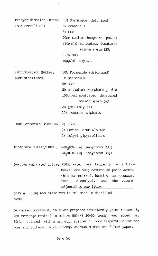
Prehybridisation Buffer: 50% Formamide (deionised)(Not sterilised) 5x Denhardts
5x SSC50miyi Sodium Phosphate (pH6.8)380pg/ml sonicated, denatured
salmon sperm DNA0.2% SDS 10pg/ml Poly(A).
50% Formamide (deionised) lx Denhardts 5x SSC20 mM Sodium Phosphate pH 6.8 100pg/ml sonicated, denatured
salmon sperm DNA,20pg/ml Poly (A)10% Dextran Sulphate.
lOOx Denhardts Solution: 2% Ficoll2% Bovine Serum Albumin 2% Polyvinylpyrrolidone
Phosphate buffer/500ml: NaH2P04 37g (anhydrous 28g)Nd2HP04 44g (anhydrous 35g)
Dextran sulphate/ litre: 750ml water was boiled in a 2 litrebeaker and 500g dextran sulphate added. This was stirred, heating as necessary until dissolved, and the volumeadjusted to one litre._______________
poly A: lOOmg was dissolved in 5ml sterile distilled water.
Deionised formamide: This was prepared immediately prior to use. 5g ion exchange resin (Bio-Rad Ag 501-X8 20-50 mesh) was added per 50ml, stirred with a magnetic stirrer at room temperature for one hour and filtered twice through Whatman Number one filter paper.
Hybridisation Buffer: (Not sterilised)
Page 54

TES buffer: 0.3M Tris (pH9.0) 0.15M EDTA 1.5% SDS
"Oligo" reaction mix: solutions A:B:C in a 100:250:150 ratio:
Solution A: 1ml solution 0(stored at-20°C) 18pl 2-mercaptoethanol
5pl each of lOOmM dATP,dGTP and dTTP (in TE).
Solution O: 1.25M Tris.Cl (pH8.0),(stored at 4°C) 0.125M MgCl2
Solution B: 2M HEPES (pH6.6)(stored at 4°C)
SolutionC: Hexadeoxyribonucleotides (Pharmacia(stored at -20°C) PL number 2166) evenly suspended at
90 OD units per ml.
lx "oligo" stop mix: 20mM NaCl20mM Tris.Cl pH7.5 2mM EDTAlpM dCTP (normally omitted)
0.25% SDS
SOB: Bacto tryptone 2%Bacto yeast extract 0.7%NaCl lOmMKCl 2.5mM
Filter sterilised 1M MgCl2 and 1M MgS04 were each added to lOmM prior to use. SOB botttom agar (for plates) was prepared by the addition of Difco bacto-agar to 1.5%.
Page 55

RFl : RbCl lOOmMMnCl2.4H20 50mM
Potassium acetate (pH7.5) 30mMCaCl2.2H20 lOmMglycerol 15%w/v
The pH was adjusted to pH5.8sterilised by filtration (priorglycerol).
RF2: MOPS (Morpholinopropanesulfonic acid) lOmMRbCl lOmM
CaCl2.2H20 75mM glycerol 15%w/v
The pH was adjusted to pH6.8 with NaOH. RF2 was sterilised by filtration (prior to the addition of sterile glycerol).
Ampicillin: Ampicilin was dissolved in steriledistilled water at 25mg/ml and stored in 1ml aliquots at -20°C.
X-gal: lOOmg x-gal was dissolved in 5ml Dimethyl formamide (DMF) and stored at -20°C.
STET: 50mM Tris.Cl (pH8.0)50mM EDTA 8% sucrose5% Triton X-100 (BDH Chemical Ltd.)
Bacterial lysis mix: 50mM Tris.Cl (pH7.5)50mM EDTA 15% sucrose
Anticoagulant: 4.5% (w/v) EDTA,0.7% (w/v) NaCl
Page 56

Lysis Buffer: 0.32M SucroselOmM Tris (pH 7.5)20mM Mgcl21% Triton X100
NaCl/ EDTA: 0.075M NaCl(proteinase K buffer) 0.024M EDTA (pH 7.5)
Loading mix: 50% Glycerol 2% Ficoll 50mM EDTA Pinch of Orange G.
50x EB/ litre: Tris baseglacial acetic acid 0.5M EDTA (pH8.0)
Prehybridisation/ hybridisation solution: 50% formamide
3x Denhardt's 5xSSC25mM sodium phosphate (pH6.8) 250pg/ml sonicated denatured
salmon sperm DNA1% SDS20)Jig/ml poly A10% dextran sulphate.
242g57.1ml100ml
Page 57

2:2 LAMBDA PHAGE METHODOLOGY.
2:2:1 Preparation of plating cells (Maniatis, Fritsch & Sainbrook, 1982).
100ml cultures in L-broth (see section 2:5:1) of theappropriate plating cells were incubated overnight, with vigorousshaking, at 37°C. The cells were pelleted in sterile Falcon 2970tubes by centrifugation (IEC DPR-6000) at 2.8K for ten minutes at 4°C and resuspended in 10-20mls SM buffer. The optical density at 600nm (OD^qq) was read on a Perkin-Elmer 6000 spectrophotometer and the cell concentration adjusted to an OD600 orapproximately 1600 million cells/ ml).
2:2:2 Titration of Phage stocks (plate lysis) (Maniatis et al, 1982).
Serial dilutions (eg 10“ , 10-~* and lO-" dilutions) of thephage stock to be titred were prepared in SM buffer. lOOpl of each dilution was added to 100pl of the appropriate plating cells and left to adsorb for twenty minutes at 37°C in a bijou (Sterilin). 3mls of 0.7% top agar were added to each bijou, the contents mixed gently and poured on L-plates. Control plates with (i) cells only and (ii) cells + SM buffer were also set up. The plates were inverted and incubated at 37°C overnight. The titre per ml was then calculated by multiplying the dilution factor by ten times the number of plaques obtained.
2:2:3 Optimisation of liquid lysis conditions.A series of small scale liquid lysis preparations were
performed in order to determine the optimum ratio of phage to cells. Ratios from 1:200 to 1:20 bacteriophage per cell aregenerally required to obtain lysis, dependent on the bacteriophage strain used. Ratios from 1:50 to 1:1000 were prepared. Theappropriate amount of a 10 dilution of the bacteriophage stockwas added to 100pl plating cells in a universal and the volumeadjusted to 200pl with SM buffer. After twenty minutes at 37°C in order to allow adsorption to occur, 10ml L-Broth was added, and the universal taped in position in the shaking incubator overnight (or
Page 58

until lysis occurred). The ratio(s) at which optimum lysis occurred (eg 1:500 for NM1149 with NB78) were then used for large scale preparations (section 2:2:4).
2:2:4 Large scale Lysis (Blattner et al,1977; Maniatis et al, 1982).
The optimum ratio, determined as described in section 2:2:3 was scaled up to 6.25mls cells (ie 101® cells) and 2 x 10® phage (for NM1149). Adsorption was performed at 37°C for twenty minutes. 500mls L-broth (prewarmed to 37°C) was added and the culture incubated at 37°C with vigorous shaking for six hours. Lysis was generally apparent. 10ml chloroform (Analar reagent grade) was added to each flask and incubation continued for a further thirty minutes.
2:2:5 Purification of bacteriophage lamda (Yamamoto et al, 1970; Vande Woude et al, 1979; Maniatis et al, 1982).
The culture was transferred to a second sterile flask and left to equilibrate at room temperature. DNasel (Boehringer Mannheim) and RNaseA (Sigma) were each added to a final concentration of lpg/ml and the culture incubated at room temperature for thirty minutes. Solid sodium chloride was added to a final concentrationof 1m and dissolved by swirling. The culture was left for one houron ice. The debris was pelleted by centrifugation at 2.8K for ten minutes at 4°C (Sorvall RC-5B, HS-4 rotor) and the supernatant transferred to a third sterile flask. Solid polyethene glycol (Sigma) was added to 10% (weight/ volume) and dissolved by slow stirring at room temperature. The bacteriophage suspension was then cooled on ice water overnight to enable the bacteriophage toprecipitate.
Bacteriophage were recovered by centrifugation (Sorvall RC-5B, HS4 rotor) at 2.8K for ten minutes at 4°C. The supernatant was discarded and the pellet drained thoroughly. The phage pellet was resuspended in 2.5-5ml TM (50mM Tris 7.8, lOmM MgS04) andextracted once with chloroform. The suspension was then loaded on top of a glycerol step gradient prepared in a Sorvall SW41 tube. The step comprised 3ml 40% glycerol in TM, followed by 4ml 5%
Page 59

glycerol in TM. The volume was "made up" with TM. The step gradient was subjected to centrifugation at 35 000 rpm for one hour at 4°C (Sorvall OTP-65 ultracentrifuge, SW41 rotor). The supernatant was discarded and the bacteriophage pellet resuspended in 1ml TM.
2:2:6 Preparation of bacteriophage lambda DNA.Pure DNasel and boiled RNAse A were added to the bacteriophage
suspension to 5pg/ml and 10pg/ml respectively. Incubation for thirty minutes at 37°C was performed. 0.5M EDTA (pH 8.0) was added to 20mM, lOmg/ml proteinase K (Gibco, Bethesda Research Laboratories (BRL)) to 50pg/ml and 10% SDS (Sigma) to 0.5%, prior to incubation at 65°C for one hour. The phage suspension was then combined with an equal volume of phenol in a 13ml Sorvall tube, mixed by gentle inversion and the phases separated by centrifugation (Sorvall RC-5B, HB4 rotor, 6K, five minutes, room temperature).
The aqueous phase was removed with a plastic pastette (Alpha Labs), extracted once with equilibrated phenol:chloroform (50 parts : 50 parts) and then once more with chloroform. Phage DNA was precipitated at-20°C overnight, by the addition of sodium acetate (pH7) to 0.2M and an equal volume of isopropanol. Phage DNA was pelleted by centrifugation (10K, lOminutes, 4°C, Sorvall RC-5B, HB4 or SM24 rotors), washed twice with 70% ethanol and resuspended in 500pl TE.
2:3 CONSTRUCTION OF X-CHROMOSOME LIBRARY (Fuscoe, Clark & van Dilla, 1986).
The X-chromosome specific library was derived from complete (EcoRl) digestion of flow sorted X-chromosomes. This avoided the need to subclone recombinants prior to their localisation on Xp.
The library was constructed in bacteriophage lambda NM1149 which accepts inserts from 0-llkb in size. The host bacteria NM514 permitted only those phage disrupted at the cl gene (ie at the EcoRl or Hindlll insertion sites of the vector) to enter a lytic cycle and thus selected for recombinants. Non-recombinant phage were constrained to undergo a lysogenic cycle. NM514 was recombination proficient, however which is likely to have selected
Page 60

against inserts containing inverted repeats.
2:3:1 Preparation of bacteriophage (vector) arms.50pg phage DNA was digested with 30U EcoRl (BRL) using BRL
core buffer for three hours at 37°C. 25pg were treated with lu CIP (calf intestinal phosphatase, Boehringer Mannheim (22U/pl), diluted in core buffer) for thirty minutes at 37°C. The CIP was inactivated by heating to 70°C for five minutes and cooling on ice, twice.
Three phenol/chloroform extractions and a chloroform extraction were performed and the vector arms prepipitated by the addition of 0.5 volume of 7.5M ammonium acetate and 2.5 volumes of 95% ethanol overnight at -20°C. The arms were pelleted by centrifugation (twenty minutes, 12 000 rpm, IEC Centra-4Xmicrofuge, room temperature), washed with 70% ethanol, vacuum dried (HETO CD3 vacuum drier) and resuspended at 100pg/pl in TE.
2:3:2 Growth of cell-lines and sorting of X-chromosomes.Lymphoblastoid cell lines were established and maintained by
L. Snaddon or M. Clarke, esssentially as described by Neitzel (1986). Samples for flow karyotype analysis were prepared and analysed by A. Cooke, essentially as described in Harris et al (1985).
X-chromosomes were sorted by virtue of their relative fluoresence after staining with ethidium bromide. The X-chromosomes used in his study were derived from the lymphoblastoid cell line of MD, a patient with a 4;7 translocation. This translocation alters the relative fluorescence of one chromosome seven and removes it from the chromosome peak which contains two chromosome sevens and two X-chromosomes (and some chromosome 8) in normal female individuals, such that the relative proportion of X-chromosomes obtained in this peak is correspondingly increased.
2:3:3 Preparation of Chromosomal DNA (Fuscoe et al, 1986).The sorted chromosomes were combined in a 1.5ml eppendorf tube
(Sarsdedt). This was placed within a 13ml Sarsdedt tube and spun at 20K (SM24 rotor) for one hour at 4°C (Sorvall RC-5B centrifuge).The supernatant was removed and the chromosomal pellet resuspended
Page 61

in lOOp'l DNA isolation buffer (150 mM NaCl, lOmM TrisCl pH 8.0, lOmM EDTA). 10% SDS was added to 0.5% and lOmg/ml proteinase K to lOOpg/ml prior to overnight incubation at 37°c.
The sample was transferred to. a 0.5ml eppendorf tube (Sarsdedt), an equal volume of phenol/chloroform added and the tube mixed by gentle inversion for ten minutes at room temperature. The phases were seperated by a two minute spin in a microfuge (IEC centra-4X) and the aqueous layer transferred to a fresh tube. The phenol/ chlorofom layer was extracted with 50pl TE (lOmM TrisCl PH8.0, 1 mM EDTA pH8.0) in the same manner and the aqueous phasescombined. A further phenol/ chloroform extraction was performed with 150pl phenol/ chloroform, the organic layer extracted with 50pl TE and the aqueous phases combined.
The aqueous phase was then extracted with 200^1 chloroform and transferred to a mini-collodion bag (Sartorius). Dialysis against TE was performed for seven hours at room temperature with two changes of buffer. The DNA solution was then transferred to an 0.5ml eppendorf and stored at 4°C.
2:3:4 Digestion of chromosomal DNA (Fuscoe et al, 1986).Chromosomes were digested with 50U EcoRl in a reaction volume
of 200pl under the manufacturer's recommended conditions (BRL lx core buffer) but without the addition of spermidine. Digestion was for four hours at 37°C. EcoRl was inactivated by placing the reaction at 65°C for fifteen minutes.
The digest was mixed with an equal volume of phenol/chloroform for ten minutes at room temperature and the phases seperated by a two minute spin in a microfuge. The aqueous phase was transferred to a 1.5ml eppendorf and the organic phase extracted with 50pl TE. The aqueous phases were combined and extracted with an equal volume of phenol/ chloroform. The aqueous phase was again transferred to a 1.5ml eppendorf and the organic phase which remained extracted with 50pl TE. The aqueous phases were combined and extracted once with an equal volume of chloroform.
The aqueous phase from this extraction was transferred to a mini-collodion bag and dialysed against sterile distilled water overnight at 4°C with two changes of buffer.
Page 62

2:3:5 Co-precipitation and ligation of vector and insert (Fuscoe etal, 1986).
The X-chromosome digest was transferred to an 0.5ml eppendorf tube and combined with 2pg (lOOmg/ml) lambda phage EcoRl arms togive a total volume of 400pl. The sample was then placed in thefreeze dryer for two hours to reduce the volume to 50-100pl. One-tenth volume of 3M sodium acetate (pH5.0) and two volumes ofcold 95% ethanol were added and the sample mixed well. The samplewas then subjected to three freeze/thaw cycles for thirty minutes each at -70°C. The DNA was then pelleted in the Centra-4x microfuge using a horizontal rotor (10 minutes, room temperature), washed once with 70% ethanol, vacuum dried for three minutes and resuspended in lOpl TE overnight at 4°C.
lpl lOx ligase buffer (0.5M Tris.Cl (pH 7.4), 0.1M MgCl2, 0.1M dithiothreitol, lmg/ml BSA), 2pl 5mM ATP (Sigma) and lpl lU/pl T4 ligase (Boehringer Mannheim) were added and the reaction incubated at 15°C overnight. A further 0.5pl each of lU/pl T4 ligase, lOx ligase buffer and 5mM ATP were added to give a reaction volume of 16pl and the reaction incubated for a further three hours. 4pl of the ligated reaction was then packaged as described below.
2:3:6 Packaging of the x-chromosome library.Packaging of DNA into bacteriophage particles (Becker & Gold,
1975) was done using Amersham packaging kits (AmershamInternational, Code 334), derived from two strains of mutant phage with complementary defects in phage assembly.
The packaging effeciency was tested by packaging lpg wild type lambda. Serial dilutions of the packaged phage were made in SM
Qbuffer and the titre/ pg packaged phage determined as 1.8 x 10 pfu/pg, which compared well with the packaging effeciency of 2 x 10® plaque-forming units (pfu) per pg wild-type lambda DNA claimed by the manufacturers (Amersham batch analysis).
The phage extracts were thawed on ice. Ipg (4pl) of the ligated reaction was added to extract B and 15pl of extract A added immediately. The extracts were mixed gently with a yellow tip and the contents collected by a brief (ten second) spin in a microfuge. The reaction was then incubated at room temperature for two hours,
Page 63

after which time 0.5mls SM buffer and lOpl chloroform were added. The packaged phage were then stored at 4°C as normal.
Aliquots of the X -library were plated on both NB78 and NM514 to determine the proportion of recombinants in the library and the approximate titre. Phage were then plated out on NM514 at a density which allowed the optimum number of plaques to be screened at any given time. This number approached 2-300 for a small plate (87mm in diameter) and three times this number for a large plate (137 mm in diameter) and represented the maximum plaque density at which plaques were well resolved.
2:4 SCREENING OF THE X-CHBOMOSOME LIBRARY.Plaque lifts of the X-chromosome' library were hybridised with
®^P labelled total human (female) DNA in order to select againstrecombinants which contained repeated sequences (and would thus give a strong signal with total human DNA). The single (or low-copy) clones which did not result in a signal were then selected for further study (see sections 2:4:1-2:4:7 and 2:5:1).
2:4:1 Plaque Transfers.Plaque transfers (Benton & Davis, 1977) to Hybond-N membrane
(Amersham International pic) were performed according to the manufacturers recommended conditions (Amersham International, 1985). Hybond-N membranes (RPN.87N (87mm) or RPN.132N (132mm)) were placed on the plates and orientated by corresponding ink markings on both the membrane and the plates. Membranes were removed after one minute and placed (plaque-side up) on a pad of Whatmann 3MM paper saturated with denaturation solution (0.5 M NaOH, 1.5M NaCl). After seven minutes they were transferred to a pad saturated with neutralisation solution (3M NaCl, 0.5M TrisCl pH7.4), left for three minutes and then transferred to a fresh pad saturated with neutralisation solution for a further three minutes. The membranes were finally washed in 2xSSC (0.3M NaCl, 30mM NaCitrate pH7.4), left to air dry, and the DNA bound to the filter by ultraviolet light treatment (plaque side down on a UV transilluminator (UVC incorporated)) for four minutes or latterly by baking for five hours at 80°C (Gallenkamp BS Oven 250).
Page 64

2:4:2 Screening plaque transfers.Plaque lifts were pre-wet in lxSSC, 0.1% SDS and prehybridised
for a minimum of two hours in prehybridisation solution (50% deionised Formamide, 5x Denhardts (0.1% ficoll, 0.1% bovine serum albumin, 0.1% polyvinylpyrrollidone), 5xSSC, 5QmM Sodium phosphate pH6.8, 380pg/ml sonicated denatured salmon sperm DNA,0.2% SDS and 10pg/ml Poly (A)) at 42°C in a shaking water-bath (Tecam SB-16).
A nick-translated probe was prepared from total human DNA (see below). This was boiled for five minutes, cooled on ice and added to ten volumes (generally five millilitres) hybridisation buffer (50% deionised Formamide, lx Denhardts (0.02% ficoll, 0.02% bovine serum albumin, 0.02% polyvinylpyrrollidone), 5xSSC, 20mM Sodium phosphate pH6.8, 100|ig/ml sonicated denatured salmon sperm DNA, 20pg/ml Poly (A) and 10% dextran sulphate).
The prehybridisation buffer was replaced with hybridisation buffer containing the appropriate probe and left to hybridise overnight at 42°C.
Filters were washed at lxSSC, 0.1%SDS for thirty minutes at room temperature, at 0.5xSSC, 0.1%SDS for a further thirty minutes at room temperature, blotted dry, placed between plastic sheets in an autoradiography cassette and covered with a sheet of Kodak XAR-5 (fast) film. The casette was equipped with Dupont lightning plus intensification screens and was placed at -70°C overnight. Films were developed using Kodak XAR developer and fixative and a Fuji RGII automatic film processor.
2:4:3 Selection of plaques.Phage which failed to give a signal on hybridisation with
nick-translated total human DNA were selected by comparing the autoradiograph with the original plate and picked using a sterile pasteur pipette (Bilbate) with a rubber bulb. Each agar plug was expelled into 1ml SM buffer, in a 1.5ml eppendorf tube. A drop of chloroform was added and the tube was left at room temperature for one hour to enable the phage (1-10 million) to diffuse out of the agar. The picked plaques stocks were then stored at 4°C as normal, (Maniatis et al, 1982).
Page 65

2:4:4 Plaque purification and preparation of high titre stocks.10- dilutions of picked phage stocks were made in SM buffer.
10pl of each dilution was added to lOOpl of plating cells and allowed to adsorb at 37°C for twenty minutes in a bijou (Sterilin). 3mls of 0.7% top agar were addded. The contents of each bijou were mixed by gentle inversion and then poured on top of an L-plate. Once the top agar was set the plates were inverted and incubated overnight at 37°C. Well-separated plaques were obtained and these "purified plaques" were picked with a sterile pasteur pipette and expelled into SM buffer/ chloroform as described above.
lOpl of each "purified plaque" stock was addded to 112pl of plating cells in a bijou. Adsorption was for twenty minutes at 37°C after which time 3mls of 0.7% top agar were added, the bijoux inverted gently and the cells and phage poured over pre-warmed L-plates. The plates were incubated at 37°C overnight but were not inverted as confluent lysis was desired. Phage were recovered from the top agar by one of two methods (Maniatis et al, 1982):
For the first method, 5mls SM buffer were added to each plate. Plates were left shaking gently at 4°C for four hours (Luckham R100 shaking platform). The SM buffer was transferred to a 13 ml sarsdedt tube and chloroform added to 1%. The tubes were spun at 8K (ten minutes, 4°C, SM24 rotor, Sorvall RC-5B) and the supernatant transferred to a fresh tube. The resultant high titre stock was made 0.05% in chloroform and stored at 4°C.
Alternatively, the top agar (0.5% for this protocol) was scraped into a 13ml sarsdedt tube tube using a sterile bent pasteur pipette. 5mls of SM buffer and 100pl chloroform (to 2%) were added and the contents mixed in the shaking incubator (New Brunswick G25) at 150-200rpm for thirty minutes. The tubes were spun at 8K (ten minutes, 4°C, SM24 rotor), the supernatant transferred to a fresh tube and chloroform added to 0.5%. The resultant high titre stock was stored at 4°C.
For applications in which only a small amount of high titre stock was required e.g. the inital screen of the library the first approach was used. The second method was easier to scale up and was used to produce a larger volume of high titre stock for storage.
Page 66

2:4:5 Preparation of liquid lysates.lpl of each high titre stock (approximately 3x 10⁶ phage) was
added to 112pl (10-*-®) of the appropriate plating bacteria and allowed to adsorb for twenty minutes at 37°C in a universal (Sterilin). 5mls of L-broth were added and samples incubated overnight in the shaking incubator (New Brunswick G25). Lysed cultures were transferred to 13ml Sarsdedt tubes, lOOpl chloroform added to each, and incubation continued for a further fifteen minutes. The bacterial debris was pelleted (Sorvall RC-5B, SM24 rotor, 8K, twenty minutes, room temperature) and the supernatant transferred to a fresh tube. A further 20pl of chloroform was added (to 0.4%) and the liquid lysates stored at 4°C, (Leder, Tiemeier & Enquist, 1977; Maniatis et al, 1982)..
2:4:6 Small scale phage DNA preparations (Cameron, Phillipson & Davies, 1977).
2mls of liquid lysate were transferred to a 13ml Sarsdedt tube. lOmg/ml DNase was added to 70pg/ml and the lysate incubated at 37°C for one hour. 0.8mls TES (0.3M Tris (pH9.0), 0.15M EDTA and 1.5% SDS) was added, samples placed at 70°C for 15 minutes and then cooled to room temperature.
0.8mls of 5M Potassium acetate (pH8.0) was added, the samples placed on ice for thirty minutes and spun at 9K for twenty minutes (Sorvall SM24 rotor at 4°C) with the brake off. 4mls of supernatant were transferred to a fresh tube, mixed with an equal volume of isopropanol and spun for a further twenty minutes (9K, 4°C).
The phage pellet was washed with 70% ethanol, dried, resuspended in lOOpl TE and transferred to an 1.6 ml eppendorf tube. lOpl boiled RNaseA (25mg/ml) was added and incubation performed at 37°C for thirty minutes. 4pl each of 10% SDS and proteinase K (lOmg/ml) were added, prior to incubation at 55°C for fifteen minutes.
A tenth volume of 3M sodium acetate (pH7.0) was added and phenol/chloroform extractions performed until the interface was clear. Two volumes of 100% ethanol were added to the aqueous phase at this point and samples placed at -20°C for ten minutes.
Phage DNA was pelleted by centrifugation (9K, twenty minutes ,
Page 67

4°C), washed once with 70% ethanol, dried and resuspended in 32pl sterile distilled water, prior to digestion with the appropriate restriction enzyme.
2:4:7 Isolation of phage inserts.EcoRl digestion of phage recombinants was performed in a total
reaction volume of 40^1 (containing a tenth volume of BRL "React 2" (or BRL "core") buffer, a twentieth volume of O.lM spermidine (final concentration 5mM) and l-2pl (10-20U) EcoRl) overnight at 37°C.
5pl of the digest was placed aside for sub-cloning at a future stage (refer to section 2:5:2). 5pl loading mix (50% Glycerol, 2% Ficoll, 50mM EDTA with a "pinch" of Orange G) was added to the remaining sample which was then loaded on a 1% LMP agarose (BRL) gel prepared in EB buffer. Gels were subjected to constant current electrophoresis at 250 mA, stained in a 5pg/ml solution of ethidium bromide in EB buffer, and visualised over ultra-violet light.
"Insert bands" were excised using a sterile scapel blade, transferred to 1.6ml sarsdedt tubes, weighed and a volume of sterile distilled water (SDW) equivalent to twice the weight of the insert added. The tube containing DNA/agarose/SDW was placed in boiling water for ten minutes and used directly for oligonucleotide labelling (see section 2:5:2) or stored at -20°C.
2:5 PREPARATION OF RADIOACTIVELY LABELLED PROBES.Alpha-32P dCTP (Amersham international, specific activity 3000
Ci/mmol (or llOTBq/mmol) labelled probes were prepared by (i) nick-translation of whole plasmid DNA or genomic DNA or (ii) random oligonucleotide primed labelling of phage or plasmid inserts.
2:5:1 Nick translation (Kelly et al, 1970; Maniatis et al, 1982).Nick translation was performed using nick translation kits
(Amersham international, N5000). Essentially, l-2pg of DNA (made up to 17(jl with SDW) was combined with 6pl of 5X Amersham reaction mix (100pM each in dATP, dGTP and dTTP), 5pl (approximately 50pCi) 32P dCTP and 2pl of Amersham enzyme solution (which contained 0.5U/pl DNA polymerase I and 10pg/pl DNase I). The reaction was incubated
Page 68

at 14-16°C for an hour and a quarter and then stopped by the addition of 5pl 0.5M EDTA. Labelled DNA was separated from unincorporated oligonucleotides by chromatography through a Sephadex G-50 column.
Amersham reaction mix and enzyme solution were in Tris/HCl and MgCl2 at pH7.8 and pH7.5 respectively. The molarities were not specified but probably approximate to final concentrations of 50mM Tris.HCl and lOmM MgS04 as specified in Maniatis et al (1982).
2:5:2 Oligo-nucleotide primed ("oligo-") labelling (Feinberg & Vogelstein, 1983, 1984).
20pl of excised insert (diluted with SDW) was boiled for ten minutes and equilibrated at 37°C for. fifteen minutes. This was added to 15pl SDW, 10 pi lOx "oligo" reaction mix and 2pl lOmg/ml bovine serum albumin (BSA, Sigma). 20-30pCi 32P dCTP was added to each reaction together with 0.5-lpl (5-10U) Klenow fragment(Amersham).
The reactions were incubated at 20°C overnight and stopped by the addition of 50pl of 4x stop mix (lx stop mix: 20 mM NaCl, 20mM Tris-Cl pH7.5, 2mM EDTA, lpM dCTP (normally omitted) and 0.25% SDS). Incorporated label was resolved from unincorporated nucleotides on a Sephadex G-50 column.
2:6 PLASMID TECHNIQUES.
2:6:1 Storage and culture of bacterial strains and plasmid recombinants.
Plasmids were firstly stored as a DNA stock (at 4°C). They were then transformed (see section 2:5:2) into an appropriate bacterial strain (generally pUCl9), plated out and single colonies selected. Bacteria containing recombinant plasmids were stored by (i) "stabs" (ii) glycerol stocks and (iii) (for short-term storage only) by streaking out on to L(-amp) plates. Bacterial strains with no plasmid recombinants were also stored in this manner (eg NM514 and NB78, which were used in constructing the library) but without anti-biotic selection.
The specifications of plasmid DNA clones, which were used in
Page 69

this study but were obtained from other sources (see acknowledgements), are summarised in table 3.
To'create "stabs" part of the colony was picked with a sterile wire loop, stabbed into a bijou or 2ml starsdedt tube containing 1.5% L-agar and grown up (loosely capped) at 37°C overnight. These were tightly capped, wrapped in Nesco-film to prevent evaporation and stored at room temperature.
Glycerol stocks were established by selecting part of the colony with a sterile wire loop to innoculate 5ml L broth (+/- 50-100pg/ml ampicillin) in a sterile universal. The culture was grown overnight at 37°C in a shaking incubator with vigourousshaking (200-300 rpm). 1ml was removed to a sarsdedt 1.6ml sterilescrew-cap tube, 0.6ml sterile glycerol added and the two mixed well, resulting in a bacterial stock just less than 40% inglycerol. This was stored at -20°C
Single colonies of interest were also streaked onto a fresh L(-amp) plate with a sterile wire loop to give decreasingconcentrations of bacteria proceeding clockwise round the plate. After overnight incubation at 37°C well spaced single colonies were almost invariably observed in some area of the plate, thus ensuring ready access to single colonies. The plates were wrapped in Nesco-film and stored at 4°C for a maximum of two months.
Single colonies were (i) selected directly from the appropriate plate stored at 4°C, (ii) obtained by streaking out of a stab or glycerol stock or (iii) when only plasmid DNA was available, by transforming bacteria, plating out and selection of the appropriate recombinants (see section 2:6:2).
Single colonies were used to prepare overnight cultures by inoculating 5mls L-broth (which was 50-100pg/ml in ampicillin where appropriate) and incubating overnight at 37°C with vigorous shaking. Where large scale cultures were required the 5ml culture was grown up for several hours and then used to "seed" 100ml of pre-warmed L-broth. These overnight cultures provided the starting point for plating cells (section 2:2:1) and plasmid preparations (sections 2:6:4 and 2:6:5)
Page 70

LD00CTi
PQCQ2§PQCnPQPi
pq < 2 2 CJ Q CQ
CO
C C CDo o o*:■ H - H £ 4J 4J O 05 03 Xu u
-rHc c 3 3•H cS
CD CO0 0 CO03 03i— 1 i—1
L D •» k. 0 0CO i—1 i—1 0303 03 03 i—11— 1
4-) - Pk. CD CD i—11—1 0303 5 5O O - P- P ■—I ■—1 CDCD ■— 1 i—lCD CD cnCD 4-4 4 H • H03 03 co O O CDO O O Ocj CJ cj X
O O CD O U rH CDi— 1 i— I 4 -) fD (0 Cic c a> o o asCO CO Cj Cl »
cd -c Oj-u P
a,0 0 5
•rH -rH COX J jQ CD05 05 r H
0 3 T 3•rHs a
C J r->M .
CU o
cn 0p q pi nj Cx-i o xp q CM ln
r H
w i
< CM P LT) LOM CD r o u ocn > 4H • .
O O CM CM
c----1&H cn .
cn Q 1—12 2
k.O 2 CMCJ CQ .i— 1
wPM Pi 1—1j o QJOh u cn IPS pq 2
00o
0 3 r H r H CO
o o
cm ro oo ro
00
M 1 H 1
0 0 0 0 0 00 3 u o u o c n OO UOi-H o o 0 0 i— I r H 0 0
0 3 0 3 o ov r H i— i 1— 1
i— 1 ■— i r H k.
03 k» 03 03 r Hi— 1 1----1 03
- P 03 03 - P - P
CD CD (D ■P- P - P CD
P CD CD CD CD
CD c n c n cn. X P P ro 0 3 c
O CD CD * H • H
03 . X p p x
CD 4H Js<i 0 3 0 3 p
• H O i— 1 r H Q
1 5 PC CQ < < Q
0 0 0 0 r " ■— 1 r H CD
CM CM CM# • • . . •
o o o o O o
o CD O O •c f CO r H OO r H OO UO LO
0 0 r H O CD 0 0 1—1 CM r- CM 0 0 r H9 , • • • • . . . . • • •
o o o o o o o o o o o O O
c m r o o o o u o u o O CD o o I/O O, » . . . • . . . . • • •
r o u o r o ^ r-> r-~ CM CD CD CM r o r - o o
i—i ■—i r H CM r HCDCD
tHiiPQ Eh Hcsj X i—i Pi1—1 PQ 4-> ocn cn cn u+ 2 PM pqPQ tHEh u o r HH Cu • .
CO O ^ 1— 1
Eh OO OOb c j CJCQ D 3 )
> 0* CD00CJ3)a
CMCMOO£PQar HCM CM roCJ CJ cnIH H x2 2 Q
IH M
IH IH Pi IH
■— 1 c n O i—1
PM PM CD rtS CD c n
2 2 PQ Eh H PQ
\H IH H
M H H
H IH IH H tH IH M
0 3 Pi 0 3 PC K 0 3
C o c o c E• H cd * rH CD • H 03PC w p n PQ PQ PC PQ
0 0 M 1 i— 1 ro i—l. . . . . •
r H 1—t r H CD H1 CM
PQ. ro
ro ro CQ uor H r H 0 0 [~T~| 1—1 0 0
CJ CJ CD Eh CD3 3 P3 P3 -P <C P3Q iQ iQ i cn a a
roH1 uo CD
r H GO 0 0 i—1cn cn CQ cn
< X X X x2 2 Q Q Q Q
IHM H gD 4-> o> cn CD
PM PM ta
H H r H
Pio PioCD 03 cjPQ CQ wr- CD UO
i—i i—1 CM
CM CM CM
CM CM CMro ro roPi Pi C£CQ CQ CQCu a a>
ro r H 00CM
CQ cn cnX X XQ a Q
oaCQ§CL,
CDaoCM
aa_Q00r Ha
03r H2
CDh ^ mCQ 03 O £H CM -Hcn cu q
00uPi
CM00r -
ooCMEH3)X CMQ O 'I 03 r - -u Ta
ble
3: Specifications of
DNA probes.
(This
table
gives
the DNA segment
(DXS
assignment), cloning
vector,
cloning
site,
RFLP
(if any),
allele sizes
(in
kilobases),
allele frequencies
(according to
HGM9,
or to
the
reference
given),
PIC
value
and
the
earliest
reference,
for
each
probe
used,
except
for those
isolated as
part of
this
project. NA,
not assigned;
NP,not
polymorphic; FREQ,frequency,; ?,unknown; Bluesc.,bluescript. Continued
overleaf).

wu2§wDmDQX
UMCD
cnwDPt-QJ
cn
H cn cn qs sU CQ
ooon
fd
j->03Cl)cnru•rHM.TJ
00o coCN
OCO N 1 LO LO (Ti O CO H O
o o o o o
co "vt* co ^ in
i— I i— I i— I r— i co
CTCrd 2 fdin r - CD r-" r-~ CO in00 00 2 00 00 00 00CTC O'. CTC cn on <T\i—i i—i .. rH i—i rH i—i
i—lfd .. •*
■—i i—i i—1 i—i rH i—ifd cd -p rd fd fd <d
CD-P 4-4 4-1 .p -p -p(D CD Ml/T< <13 0> CD 03
i—i o(Qfd o o 1—1 i—1
0) o c o u 03 03X fd CD fd <d •X XC c CD c c c G3 o fd o o 3 aX 2 S£ 2 2 X x
"vP 00 00 00 r - COin ^p oo H 1 H*
. a . . • . . . •o o o o o O o O oo o o o o o o <T\ i—1 I-" oo 00 CN oo r~- CN 00CO oo 1—1 CO CO CN r - oo CO oo CD oo co OO CO CO
• • • . . . . . . • . . . . . . • •o o o o o o o o o o o o o o o o o o
m m m o o in rH m 00 o oo 00 rH 00 CN, . 00 , . . • CN oo • . . • • • •
•—i 1—1 r-~ CO 00 on CN 00 OO CN rH H 1i—i \ \
i—1 COo . •00 . CO r - rH
i—i 1—1 i—1
oooo
CN in
CM
a Eh in X i—i Dacn cn + 2 Da m EHI— I Dmcn o
caQ 03
caCQ§
MCD03s
DCH<
CNCNOOegCQcd
H1CNCQ
SCNCJ
CO00 COCO CNC/303X XQ Q
P E I CO r-H COJ f-o
>M X4-> O
c d CO o2 CD Da
\M M(H HM M MT3 rO 03 (HG C O -P•H •rH O COas 33 W CL.
m r- r-~r-~ . .• i— i CN
o
M
2DXaaocoCN03g
oCNa
MCDcn2 cnCQ S| IICQ I Eh I
hH M MM W HIH IH IH
IH.TO.rUC G C cP-l■H -H •rHX salcn 3J1—1 oo in• • •1—1 rH i—1
00 00 00rH rH i—iCJ u CJ2 D Da a CDo H1r-~ CO COCN r— 1 rHcn cn cnX X XQ Q Q
O inro rH1r-~ 1r~-oo coMl H &•rHCQ w wI'd CD CD
Mcd
nx00
00rHuDcdH*CDrH03S00Ir-~ooSDaa
ITable
3: Continued. Continued
overleaf.

wcj2gwCt-4wg ;
03LD00a >
44d>(U4* :GzsX
g• H- i josu
• HcG
S COO 00 o O '*
(ts - G i—I O 03 COC-l 4-)CD d) CUG
» OG COo aJJ § M o■Q - P
LD00OC
44d )
> 1i S
co00c d
0344CD
G0 CO1
C"-00OD
034->0
x4-)
CT\I4-4CL)GO .4-4C4.S
03r-- lo co oo oo oo ooO'! <T> O'!
i— 1 i— I i— I i— I r—I03 03 03 03 OS4-4 44d) d>i— I C-4d) d>4*C XC mh G O X DC
03COCO<T\
- Pd)C-lCD
Xu h
ODO
LD00O '!
03-Pd>C-l
CD4«£4 h
ODO
CJMP4
CQ U4a gw.-0J w| < [S I
COfH
H CO CO QO § CJ CQ
px,
1-0
w fc!IS! X M W CO CO + 2 W M EhM X CO O
WS
CO
ro co CO CM o CM CM•M1 ro
• • • • • •o o O o o o oOD i—iLO LO LO LO o o CM 00 o o oCO ro CO ro ro CO r- ro r -~ CM r- ro 00
• • • • • • • • • • • • •o o o o o o o o o o o o o
r- LO 1—1LO LO 00 00 r H 00 o C " o. • CO o • • • • • • • •oor- oo • •r-H CO r-~ro ro CM 1—1 r-o r H\
LOM1CM
X
o oi—iCJDDc u
COI—ICOXQ
CU . 2a a ” i§1 si si
111pc , pc o o u o w w
cu2
CO rH o CO COs • • • •o (—1 r H o o
ro ro LO cr> Ar H r H CO i—i 4-£u CJ cu CJDO G> CQ DO —-04 CU 04 04 O*CO CO CO CO oo o o o roCM CM CM CM CM
CQ CQ CQ CQ CQX X X X XQ Q a Q Q
00
or-CM
o o
or -
CM
O O ^ C O O O C N O O CO ^3* 0 0 i— I CO CO 0 0
o o o o o o o o
C O r O ^ C N t - ' C M O O
i n CO o j 'cf H LD CN
x;a
OS
2 2 2M M M M HH M \ M M M M MPh.TO TO 03 X
M M
M 1—1 HH
M PC, T 3 fHi— 1 O C XCP CJ • H COCQ w DD CU
O G CJ -H W DO DO
CJCOCDGi—ICQ
coOCMCO
3
CO ["- CO CTi <Ti
CD I—I r-H >—I O
CJDcu
CM CMr ~ ■M1CM i—1 0 0CQ CQ CQX X XQ Q Q
PC,i—1 o 1— 1 cas CJ 03 • HCQ w CQ o:
T3
CTi
CM i—IODOCU
CM
roLDr H
a
C4H03CD
I—I 44CD>O
TOCDGC•rH
- u
G
8
T3CDGC•rH4-4COu
wCQ
ICU
Ir -o o
sxcu
. ro O •r-H CM •G fG
CM mCMD-iMX
rH 00 i—IErj I q r- PC
s M1 CJ LOPS *g cur- LO
r -
M 1LOr-~
roCDi—IA
Eh

s•H-t->rou•rHca
co•rH-i->03U•rHG□
WCJ2§WW
cjMft
03WWWJW<CO
N1 'vt1 U00 00as as i— 1
IO 1—1 rH OSCO Go o V O1—1 1— 1 1-1 COrd 03 L-liP1—1 4-> 4-1 f tncs CD CD
-U u CD rHCD CD CP i— |
£3 CDC o ■rH SiCD 03 H ON CD 03 4->o •rH rH •Hft 3: < 2
CO as LDro• • • •o o o O
1— 1as ro t"- 00 CN in inLD ro r-~ CN LD ro ld ro• ■ • • • • • •o o o o O o o o
LD CN i—i o o o in• • • • • • » •
LD LD in CN as vt< CN
CDsiu4->•rH2
Eh CO 03 QS §CJ CO
cuwCuft
w <; s2 CO Q W 03
Os • LD i—I •*. rold - . «st< o [-" • •
i—I LD CN
CJ
1
W Eh Mcn f t m ftM W 4_) o03 03 CO u+ 2 ft wW MEh CN mI—I W • CN03 O i—1 •1—1ft CN
CNR CO rob cj ftW 2 CQ> CU CU
CO ft ft2 2 2
M M MM \ M MM M M M03 ft 03 03G o C C•rH u •H •Hw w ft ftCN LD m• • •rH o i—1
CNCNro&COCu
r- iH03 03X XQ Q
OSrHCJDa
ast—ICJDCU03CDac•rH4->C8
WCO§cu
CJEho
COCN00LO
ro>hXXo
asi—i>HX£CJ
CDrH£3S
2:6:2 Preparation of competent cells and bacterial transformation.Frozen competent cells (JM83) were prepared by frozen storage
protocol III of Hanahan (1985). A freshly streaked SOB plate was prepared from a single colony. Ten colonies, 2-3mm in diameter, were picked from this plate, dispersed in 1ml SOB by vortexing and used to inoculate lOOmls SOB in a 1 litre Erlenmyer flask.
The culture was grown to a density of 4-7 x 10⁷ cells/ml, split between two Falcon 2070 (or Sarsdedt 60.547) tubes and placed on ice for fifteen minutes. Cells were pelleted (2800rpm, fifteen minutes, 4°C, IEC DPR-6000), drained and resuspended in one-third volume (2xl6mls) RFl buffer and incubated on ice for fifteen minutes. Cells were pelleted once more by centrifugation (as above), resuspended in 2x 4mls RF2 buffer and left on ice for a further fifteen minutes. Competent cells were then aliqoted into chilled 1.6ml sarsdedt screw-cap tubes, "flash-frozen" in liquid nitrogen and stored at -70°C.
These were thawed at room temperature as required for transformation. Cells were divided into lOOpl aliqouts in Sarsdedt 1.6ml screw-cap tubes and placed on ice. lOOng-lpg of DNA in a volume of up to 10pl was added and the tube swirled to mix the sample well. Samples were left on ice for twenty to forty five minutes prior to "heat-shock" at 42°C for thirty to forty five seconds. Samples were immediately returned to ice after heat-shock and plated onto L-plates (+/_ ampicillin). Control plates were prepared using competent cells with no transforming DNA.
2:6:3 Sub-cloning of phage inserts.PUC19 vector was prepared by digestion with EcoRI and calf
intestinal phosphatase treatment (as described in section 2:2:1). l|al (20ng) of plasmid vector was ligated with 2^1 of the appropriate phage digest (section 2:3:8) for one hour at room temperature and then at 4°C overnight. The total reaction volume of 10pl comprised: lpl plasmid vector, 2|ul phage digest, lpl lOx ligase buffer, 2pil 5mM ATP, 1U T4 DNA ligase and 3pl SDW.
The ligation reaction was used directly in transformation of JM83 cells without phenol/chloroform extractions to remove T4 DNA ligase.
page 71

2:6:4 Selection of transformants.Transformed cells and controls were spread with a sterile
glass rod onto L- plates (made 50-100pg/ml ampicillin for sub-cloning into pUCl9, or for any ampicillin resistant plasmids cloned in either the pUC or pBR322 series of vectors).
X-gal selection, was used whenever possible (eg for "pUC" vectors using JM83 competent cells). 40pl of 20mg/ml X-Gal in dimethyl formamide was added to each L-amp plate prior to use. JM83 has a LacZ mutation and thus cannot produce beta-galactosidase necessary to metabolise x-gal (5-bromo- 4-chloro- 3-indoyl-beta-D- galactopyrosanide). The lacz' gene at the polylinker (or multiple insert) site in pUCl9 complements the defect and thus JM83 cells containing this plasmid metabolise X-gal. This reaction results in a blue colour. Recombinant plasmids (which interrupt the lacZ1 gene) are "white" and can be distinguished from (blue) religated vector clones. Small scale DNA preparations were performed for each well-spaced single colony selected (see section 2:6:5).
2:6:5 Rapid (small scale) plasmid preparation by the boiling method (Holmes & Quigley, 1981).
5ml cultures, derived from 20|il glycerol stock or, preferably, from single colonies were grown overnight in L-broth (made 50-100pg/ml in ampicillin if appropriate), in sterile Universals (Sterilin) within a shaking incubator at 37°C.
1.5ml of overnight culture was transferred to an eppendorf tube. Cells were pelleted by centrifugation for five minutes in a microfuge (IEC centra-4X). (The remainder of the culture was made 40% in glcerol and stored at -20°C as a glycerol stock). The supernatant was discarded, the cells resuspended in 700pl ice-cold STET (50mM Tris 8.0, 50mM EDTA, 8% sucrose and 5% Triton X-100) and 50|il lOmg/ml lysozyme (in STET) added. The tubes were left on ice for fifteen minutes to enable the cells to lyse, placed in boiling water for forty five seconds, and returned to ice.
The bacterial debris was pelleted by centrifugation at maximum speed for thirty minutes and the supernatant transferred to a fresh tube. Sodium acetate was added to 0.3M and phenol/ chloroform extractions performed until the interface was clear. Plasmid DNA
Page 72

was precipitated at -20°C for thirty minutes, by the addition of 0.6 volumes of isopropanol. DNA was pelleted by centrifugation, washed twice with 70% ethanol and air- or vacuum- dried. The pellet was resuspended in TE (for storage at 4°C) or more often in a small volume of sterile distilled water which formed the basis of the desired restriction digest.
Small scale preparations yielded plasmid DNA of a quality suitable for "oligo"-labelling (section 2:5:2), normally sufficient for twenty to thirty labelling reactions.
2:6:6 Large scale cleared lysate plasmid preparation (BRL protocol).
100ml cultures were grown overnight in L-broth at 37°C within a shaking incubator. These were generally "seeded" with 5ml cultures grown up from a single colony (or 20[xl glycerol stock of the appropriate colony) during the day. Ampicillin was added to 50-100pg/ml if appropriate.
Cells were pelleted by centrifugation at 2500 rpm for fifteen minutes at room temperature. The supernatant was discarded. Cells were resuspended in lOmls bacterial lysis mix (50mM Tris 7.5, 50mM EDTA and 15% sucrose) and left at room temperature for fifteen minutes. lOmg of powdered lysozyme (Sigma) was added to lmg/ml and the cells were then left for a further twenty minutes at room temperature.
0.5mls 10% SDS (to 0.5% SDS) was added and the tube inverted gently. 1ml 5M Potassium acetate (to 0.5M) was then added, the tube inverted gently once more and placed on ice for thirty minutes.
Bacterial chromosomal DNA was removed by centrifugation (10K, thirty minutes, 4°C, HB4 rotor) and the supernatant transferred to a fresh tube. Plasmid DNA (and RNA) were precipitated on ice for twenty minutes by the addition of two volumes of cold 95% ethanol and pelleted by centrifugation (10K, twenty minutes, 4°C, HB4 rotor).
The pellet was then resuspended in 4mls TE and solid ammonium acetate added to 2.5M (0.77g). This was left on ice for a further twenty minutes and then subjected to centifugation (10K, twenty minutes, 4°C, HB4 rotor). The supernatant was transferred to
Page 73

a fresh tube and a further two volumes of 95% ethanol added. This was left on ice for a further twenty minutes and spun down (10K, twenty minutes, 4°C, HB4 rotor) to precipitate plasmid DNA.
The pellet was dried and resuspended in 2mls of 0.05M Nad in TE (in 13ml Sarsdedt tubes). Boiled RNAse A was added to 25^g/ml and the tube incubated at 37°C for forty five minutes. 0.6 volumes of 2M NaCl in TE were added and phenol chloroform extractions performed until the interface was clear. Plasmid DNA was precipitated at -20°C for thirty minutes by the addition of a further two volumes of cold 95% ethanol.
Plasmid DNA was pelleted by centrifugation (10K, twenty minutes, 4°C, HB4 rotor), resuspended in TE and subjected to two further rounds of purification by ammonium acetate precipitation. (Each round of purification involved the addition of a half volume of 7.5M ammonium acetate and of two volumes of 95% ethanol. Plasmid was precipitated at -20°C for thirty minutes and then recovered by centrifugation). The final pellet was dried prior to resuspension in 200-500pl TE.
2:7 SOUTHERN ANALYSIS.
2:7:1 Preparation of genomic DNA from blood and cell lines (Kunkel et al, 1977).
Genomic DNA was prepared as described below from blood (10-20mls in Nad anticoagulant) or from lymphoblastoid and somatic cell hybrid cell-lines. Fibroblast DNA was also prepared according to the same protocol with the quantities scaled down ten fold.
Blood samples were thawed (if frozen). lOmls blood was placed in each of two 50ml Falcon 2070 (or Sarsdedt 60.547) tubes. 40mls of lysis mix (0.32m Sucrose, 10mM Tris pH 7.5, 5mM MgC^/ 1% Triton X-100) was added to each tube and the contents were mixed thoroughly. Cell pellets were resuspended directly in 50ml lysis mix and the tubes left on ice for ten minutes to ensure complete cell lysis.
Cell nuclei were pelleted in the IEC DPR-6000 centrifuge (2.8K, fifteen minutes, 4°C) and resuspended in lOmls NaCl/EDTA (0.075M NaCl, 0.024M EDTA). Any sample which had been split into
Page 74

two, was recombined at this point. 0.75mls 10% SDS and 200pllOmg/ml proteinase K were added, the tube inverted gently and incubated at 55°C for one to three hours.
The samples were allowed to cool to room temperature. A tenth volume of 3m sodium acetate (pH7.0) and an equal volume of phenol/chloroform (50% Phenol, 50% Chloroform, 0.1% 8-hydroxyquinoline equilibrated with TE) were added. The tubes were mixed gently by rotation (Stuart tube rotator TR-2) for fifteen minutes and the phases separated by centrifugation in the IEC centifuge (2.8K, fifteen minutes, room temperature).
The aqueous phase was removed with a plastic pastette andtransferred to a fresh Falcon 2070 tube. Further phenol/chloroform extractions were performed until the interface was clear and the aqueous phase was once more transferred to a fresh tube. Two volumes of cold 100% ethanol were added to precipitate the DNA which could then be spooled around a sealed sterile pasteurpipette. The DNA was washed in a small quantity of 100% ethanol and allowed to air dry. DNA samples were rehydrated in 500|il-lml TE. These were left at 4°C for a minimum of twenty four hours beforeuse to ensure that they had dissolved adequately, mixed with a wide bore 1ml blue tip (Treff) and 10-20pl removed with which to measure the optical density at 260nm (Perkin Elmer 6000 spectrophotometer). For a lOpl dilution in 1ml TE, the DNA concentration in pgms/ml was obtained by multiplying the optical density by five thousand.
2:7:2 Restriction Digests, electrophoresis and Southern blots(Southern, 1975).
Restriction enzyme digests were performed using 7pg genomic or somatic cell hybrid DNA at 37°C overnight. The reaction consisted of DNA, one-tenth volume of the recommended lOx buffer (generally supplied), one-twentieth volume of 0.1M spermidine (Sigma), 10-20U restriction enzyme (BRL, New England Bio-labs, or Anglian Biotechnology) and sterile distilled water to an appropriate volume (normally 40pl). The samples were mixed well with a yellow pipette tip and spun briefly to collect the contents, prior to incubation.
Samples were spun briefly to collect the contents of the tube and one-sixth volume loading mix (50% Glycerol, 2% Ficoll, 50mM
Page 75

EDTA and "a pinch" of Orange G.) added. The sample was loaded onto an 0.8% agarose (Sigma A6013) gel prepared in electrophoresis buffer (EB, ie in 40mM Tris-acetate, 2mM EDTA). Electrophoresis was performed in EB at 150-250mA constant current for three to four hours, or until the orange-G marker dye had reached the foot of the gel. Gels were stained in a dilute ethidium bromide solution (5pg/ml in EB) and photographed with a polaroid GU-5 land camera using polaroid 667 (black and white) film over the ultraviolet transilluminator.
Gels were soaked in acid (0.2M HCl) for twenty minutes, in denaturation solution (0.5M NaOH, 1.5M NaCl.) for 2x twenty minutes and in neutralisation solution (3M NaCl, 0.5M Tris.HCl pH 7.4) for a further 2x twenty minutes. DNA was transferred from the gel to the membrane (Amersham Hybond-N) in the presence of lOx SSC (1.5M NaCl, 0.15M sodium citrate) as described below.
A tank was filled with lOxSSC. A wick was prepared by laying a double thickness of Whatman 3MM paper over a glass plate placed across the tank such that either end of the 3MM paper was within the buffer. Once it was ensured that the wick was entirely wet and uniformly flat, the gel was placed on top of the wick. Hybond-N membrane (Amersham international pic), labelled and cut to size, was laid gently on top of the gel. Two sheets of 3MM paper (soaked in 2xSSC and cut to size) and two additional (dry) sheets of 3MM paper (also cut to size) were placed on top of the membrane. The remaining area of the wick was shrouded with plastic sheeting and a stack of paper hand-towels (Bowater-Scott), a glass plate and a small weight placed on top of the gel.
Transfer occurred overnight after which time the blotting apparatus was dismantled, the membrane rinsed briefly in 2xSSC to remove traces of agarose and baked at 80°C for five hours (or subjected to uv treatment for four minutes).
2:7:3 Hybridization of Southern BlotsFilters were prehybridised and hybridised as described in
section 2:3:2) or prehybridised and hybridised in the same buffer, using 3-5ml buffer per 10cm x 13cm filter. This buffer was 50% formamiae, 3xDenhardt's, 5xSSC, 25mM sodium phosphate pH6.8,
Page 76

250pg/ml sonicated denatured salmon sperm DNA, 1% SDS, 20pg/ml poly A and 10% dextran sulphate. The denatured probe was added directly to the plastic bag containing the. prehybridised filters. Hybridisation was overnight (sixteen hours) for probes labelled by nick translation or for two nights (forty hours) for probes labelled by oligonucleotide priming.
Excess probe was removed by washing the filter in 2xSSC, 0.1% SDS at room temperature for twenty minutes. Filters were washed sequentially in solutions of increasing stringency for twenty to thirty minutes each as required; lxSSC, 0.1% SDS (room temperature), lx SSC, 0.1% SDS (65°C), 0.5x SSC, 0.1% SDS (65°C), O.lx SSC, 0.1% SDS (65°C) and 0.05x SSC, 0.1% SDS (65°C). The strength of the signal was assessed . between washes with a ngm mini-monitor" (Type 5.10). Filters were blotted dry and placed between plastic sheets. The filters were then placed on a sheet of Kodak XAR-5 film in an autoradiographic cassette with "Dupont lightning plus" intensification screens and exposed overnight at -70°C (or from four hours to ten days as necessary). Films were developed as described in section 2:4:2.
2:8 PHOTOGRAPHY.Autoradiographs were photographed using a Pentax 35mm camera
(p30 SLR) with a SMC Pentax-A macro 50mm f/2.8 lens and Ilford PanF film. Films were developed for five and a half minutes in an 1:9 dilution of Ilford Ilfospeed developer, stopped in 3% acetic acid for thirty seconds, fixed in an 1:4 dilution of Hypam fixative (with l/40th part Aculux hardener) for three and a half minutes minutes and rinsed with tap water and a few drops of photoflow (Kodak) for fifteen minutes. Negatives were enlarged on an Leitz (Wetzlar) enlarger and prints produced on Ilford number 3 paper. Prints were developed for one minute using an 1:9 dilution of Ilfospeeed developer at 20°C, rinsed in water, fixed for sixty seconds in an 1:9 dilution of Hypam fixative, rinsed in water again and dried using an Ilford Ilfospeed 1050 print drier.
Page 77

2:9 SOMATIC CELL HYBRIDS.AMIR2N was the gift of Dr p Goodfellow (Goodfellow et al,
1983), ' derived from the translocation in AM (1729), 46,Yt(X;Y)(p22.3;qll.2), (Ferguson-Smith et al, 1981). The breakpoint at Xp22.3 is proximal to the locus for STS.
Hybrids LNA9, EHA9, W2A9 and W5A9 were cloned by N. Morrisonand L.Florentin in this department. LNA9 was derived from a patient LN, with an 46,XXp- karyotype, EHA9 was derived from EH, with a 46,XX,t(X;ll)(p22;pl5) karyotype, W2A9 was derived from the lymphoblastoid line of an individual with a 46,XX,t(X;8)(p21;q24) karyotype and W5A9 was derived from the lymphoblast oid cell line of individual with an 46,XX,t(1;X)(p32;p21) karyotype and a paracentric inversion betweeen Xpll and Xp21, described by Lindenbaum et al (1979). The region of the X-chromosome retained inthese hybrids is illustrated in figure 1.
H0RL9X (Goodfellow et al, 1980) was a somatic cell hybrid containing only the X-chromosome whilst 3E7, isolated by M.Marcus & R. Voss from the hybrid described by Marcus et al (1976), contained only the Y-chromosome.
Page 78

AMIR2NH0RL9X LNA9
Figure 1: Diagrammatical representation of the extent of theX-chromosomal region in the somatic cell hybrids studied.

CHAPTER 3: RESULTS
Page 79

3:1 CONSTRUCTION OF AN X-CHROMOSOME SPECIFIC LIBRARY INLAMBDA-PHAGE NM1149.
An EcoRI library was prepared in lambda NM1149 from 1.13 x 10⁶ sorted "X-chromosomes" (200-250ng DNA) as outlined in section 2:3. It was tit red on both NB78 and NM514 and values of 116 000 and 68 100 plaque forming units (pfu) / total library (4ml) were obtained. As all bacteriophage will form plaques on NB78 and only recombinant phage form plaques on NM514, the difference in titre gives an indication of the proportion of non-recombinants. Thus, non-recombinants represent about one-third of this library.
The titre of the library fell prior to the second round of plating out, so that it no longer represented the entire X chromosome (which requires 50 000 recombinants). Thus, due to the reduced titre and the high proportion of non-recombinants, amplification of the X-library was undesirable and the library was plated out directly (on NM514 cells).
3:2 SCREENING OF THE LIBRARY.32Plaque transfers were performed and screened with JZ,P labelled
nick-translated total human (female) DNA in order to select for "single copy" inserts or inserts with low level repeats. Those recombinant plaques which failed to demonstrate hybridisation (and thus contained no repeat sequences or a low copy number of repeat sequences) were picked, 10- dilutions in SM buffer were prepared and putative recombinants were plated out again at low density. These techniques are described in section 2:4.
A second round of screening, which would have selected against some of the recombinants containing more highly repeated sequences, was not performed at this stage. This accounts for the moderate repeats apparent in some of the recombinants characterised (figure 2). A second round of screeening would also have increased the relative proportion of non-recombinants amongst the plaques selected.
One hundred and twenty single plaques were selected and DNA was prepared from liquid lysates as outlined in section 2.4. Fifty of these phage were shown to have inserts (figure 2) and were
Page 80

labelled by the random hexa-nucleotide primed ("oligo-") method (section 2:5:2) .
3:3 REGI0NA1 MAPPING OF PROBES ON THE X-CHROMOSOME.Each insert was hybridised to an X-chromosome EcoRl
mini-panel, consisting of 7pg DNA from each somatic cell hybrid(section 2:9) as follows:
1. female DNA2. Horl9X X-only hybrid3. LNA9 Xcen-Xqter4. AMIR2N Xp22.3-Xqter5. EHA9 Xp22.1-Xqter6. W2A9 Xp21-Xqter7. A9 mouse parent
Of fifty inserts, sixteen were X-specific (hybridised to the X-only hybrid), four were autosomal and six were highly repeated sequences. One was a moderate repeat with mouse homology. The remaining twenty two inserts demonstrated no, or non-specific, hybridisation and were presumably of phage or E.coli origin (figure 2).
Excluding data from EHA9 (see below), this panel assigned X-specific inserts to Xq or to one of three regions on Xp; (i) Xcen-Xp21, (ii) Xp21-22.3 or (iii) Xp22.3-Xpter. These assignments are summarised in table 4. Autoradiographs of these results (except for those obtained with X37 and X89) are shown in appendix 1. Probes X37, X38, X48 and X74 contained moderate repeats.Specifications of the X-specific inserts are shown in table 5.
Four Xp probes, including representatives of each of the three classes described earlier, were obtained. GMGX10 (X24) was assigned to Xp21-Xcen, GMGXll (X66) and GMGX12 (X112) to Xp21-Xp22.3 and GMGX9 (X47) to Xp22.3-Xpter. These four inserts were subcloned into the EcoRI site of pUCl9 (section 2:6:3).
The breakpoint in hybrid EHA9 was ascertained cytogenetically as Xp22.1 which was inconsistent with results with probe GMGX9 (X47). Data with GMGX9, (table 4 and appendix 1), indicated that
Page 81

EHA9 had a more distal breakpoint than AMIR2N, at Xp22.3. Probes GMGXlO, GMGXll and GMGX12 were all present in EHA9. EHA9 thus has Xp sequences both proximal and distal to the breakpoint in W2A9. This anomaly might be explained by an interstitial deletion, a rearranged X-chromosome in EHA9 or a more distal breakpoint in EHA9 than AMIR2N. No conclusions were drawn from data with EHA9.
3:4 INITIAL CHARACTERISATION OF GMGX9-12.Only the Xp-specific probes were characterised further. Probes
are most useful when they detect RFLPs and/or deletions and thus these possiblities were explored for each of the four Xp probes (GMGX9, GMGXlO, GMGXll and GMGX12).
3:4:1 Search for RFLPs.A search for high frequency restriction length polymorphisms
(RFLPs) was performed for the four Xp probes (GMGX9, GMGXlO, GMGXll and GMGX12) using panels of two males and five females (or seven females) digested with each of the following restriction enzymes: Alul, Bell, Bglll, Ddel, Dral, EcoRV, Haell, HincII, Hindlll, Mbol, MspI, PstI, PvuII, Rsal, TagI and Xmnl.
No polymorphisms were detected with GMGXlO, GMGXll or GMGX12 with these sixteen enzymes. Only one polymorphism was detected with GMGX9 using HindiII. This is a two allele system which detects either a 4kb band or two bands of 2.5kb and 1.5kb in normal individuals, and is illustrated in figure 3.
Forty normal males and forty normal females were screened in order to calculate the allele and heterozygote frequencies for GMGX9. These were determined as 67% for the 4kb and 33% for the 2.5/1.5kb alleles respectively. The heterozygote frequency was 53% and the polymorphic information content (PIC) value was 0.44 (i.e.2x 0.67 x 0.33).
Applications of this polymorphism will be discussed below(section 3:4:2 and sections 3:7:1-3).
Page 82

Flow sorted X-chromosomes
X-library
Screen with Total human DNA
Non-hybridising plaques Hybridising plaques (n=120) \ (n=395)
Inserts(n=50)
X-specific Autosomal(n=16) (n=4)
Repeated Other(n=6+l) (n=23)
Xp Xq
(4 probes) (12 probes)
GMGX9 GMGX8GMGXlO GMGX13GMGXll GMGX14GMGX12 GMGX15
GMGX16GMGX17GMGX18GMGX19GMGX20GMGX22GMGX23GMGXY12
Figure 2: Characterisation of the x-chromosome library.

a a >
■ ■m
M R L I E WA L N R H 2L 9 A 2 A A AE X 9 N 9 9 9
INSERT I 2 3 4 5 6 7
X23/GMGX8 + + + + + + -
X24/GMGX10 + + - + + + -
X30/GMGX19 + + + + + + -
X37/GMGX22 + + + + + + -
X38/GMGX20 + + + + + + -
X47/GMGX9 + + - - + - -
X48/GMGX18 + + + + + + -
X53/GMGXY12 + + + + + + -
X60/GMGX16 + + + + + + -
X66/GMGX11 + + - + + - -
X74/GMGX13 + + + + + + -
X89/GMGX17 + + + + + + -
X92/GMGX14 + + + + + + -
X94/GMGX15 + + + + + + -
X110/GMGX23 + + + + + + -
X112/GMGX12 + + - + + - -
Table 4: Hybridisation of recombinants to the EcoRI somatic cell hybrid panel. These hybrids are described in materials and methods. Autoradiographs of these results are shown in appendix 1.

Name Lab name Vector Insert Location DXS number
GMGX8 X23 NM1149 5kb Xq 236GMGX9 X47 PUC19 7kb Xp22.3-pter 237GMGXlO X24 PUC19 6kb Xp21-cen 238GMGXll X66 PUC19 1.2kb Xp21-p22 239GMGX12 X112 PUC19 4.5kb Xp21-p22 240GMGX13 X74 NM1149 2.5kb Xq 241GMGX14 X92 NM1149 2.5kb Xq 242GMGX15 X94 NM1149 3.5kb Xq 243GMGX16 X60 NM1149 4kb Xq 244GMGX17 X89 NM1149 5k b Xq 245GMGX18 X48 NM1149 2.5kb Xq 246GMGX19 X30 NM1149 8kb Xq 247GMGX20 X38 NM1149 4kb Xq 248GMGX22 X37 NM1149 2.5kb Xq 250GMGX23 X110 NM1149 5kb Xq 251GMGXY12 X53 PUC19 1.6kb Xq/Y /Table 5: Specifications of X-specific recombinants.
All are inserts are released by EcoRI digestion.
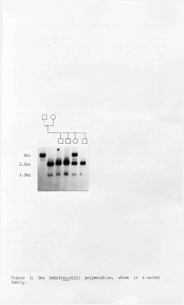
?
o—
4kb•
• •2.5kb
1.5kb m fe
Figure 3: The GMGX9(Hindlll) polymorphism, shown in a normalfamily.

3:4:2 The preliminary deletion screen with GMGX9, GMGXlO, GMGXlland GMGX12.
The panel which was used to screen for deletions at the DMD locus consisted of two individuals with complex phenotypes (BB and MJ, described below) and nine individuals with BMD or DMD with deletions of pERT87-8 and/or XJ1.1. These nine boys had been detected from amongst eighty seven boys with DMD or BMD prior to beginning this project (D. Wilcox, unpublished observations). The pedigrees of these nine individuals are illustrated in appendix 2A.
Scottish individuals are identified by both initials and pedigree numbers (indicated in parenthesis) in the text and by pedigree numbers in the figures and appendices. Other individuals are identified by their initials and/or place of origin eg"(Oxfo)"; Oxford, "(Card)”; Cardiff.
BB had chronic granulomatous disease, retinitis pigmentosa and the McCleod syndrome in association with DMD (Francke et al, 1985) whilst MJ (Oxfo) had DMD, congenital adrenal hypolplasia (AHC) and glycerol kinase deficiency (GK), (Davies et al, unpublishedobservations). AM (5996) and JR (5194) who both have AHC but not DMD or GK deficiency are also included in this panel.^AM (5996) but not JR is mentally retarded. Results for AM(5996) and JR (5194) will be presented in section 3:5:6.
GMGXlO was deleted in BB and MJ (Oxfo) and also in SS (and his cousin JD, 5099), (table 6 and appendix 2). GMGXll was onlydeleted in MJ (Oxfo) (table 6 and appendix 2B) and GMGX12 was notdeleted at all (table 6).
BB and SS (and his cousin JD, 5099) had large deletions(visible on FACS analysis, Wilcox et al, 1986) as did MJ (Oxfo) (A.Cooke, unpublished observations) which suggested that both GMGXlO and GMGXll might be further from "the DMD gene" than either pERT87 or XJ.
GMGX9 was not deleted in any of these individuals (appendix2b ) as predicted by its assignment to Xp22.3. It was, however,unexpectedly shown to detect deletions in individuals with X-linked ichthyosis. This became apparent whilst undertaking RFLP analysis in these families (sections 3:7:1-3) and greatly increased the value of GMGX9.
Page 83

C O 5 5 3 5 5 4 5 0 5 5 5a x 0 0 4 1 3 0 3 1 2 0 9r f 9 9 8 1 1 9 1 2 6 9 9d o 9 9 5 6 3 7 1 ^ 0 5 7 6
DH MJ BB SS JD SJ SB NJ SM PE AB AK JB AMTelomereGMGX12 + + + + + + + + + + + + + +D2 + + + + + + + + + + + + +99.6 + + + + + + + + + + + + +B24 — + + + + + + + + + + + + —C7 - + + + + + + + + + + + + -Ll-4 — + + + + + + + + + + + + —J66H1 — — + + + + + +■+ + + + + +GMGXll - - + + + + + + + + + + + +p20 - + + + + + + + + + + + +J-Bir - + - - + + + + + + + + +PERT87.30 +pERT87.15 +PERT87.8 +PERT87.1 - - - - - - - - + + + + +XJ 10.1 — — — — — — + + + + — — +XJ2.3 — — — — — — + + + — — — +X J 1.1XJl.2 - — — — — — + + + — — — +HIP25 +XJ5.1 — — — — — — + + + — + —JMD - + + + - + +J47 + - - - + + + + + - + +PERT84 - - - - - + + + + + + + + +754—11 — — — — + + + + + + + + +754-6 — — — — + + + + + + + + +754 — — — — — + + + + + + + + +GMGXlO - - - - + + + + + + + + +OTC + + + + + + + + + + + + +LI. 28 + + + + + + + + + + + + +p58.1 + + + + + + + + + + + + +Centromere
Table 6: Results of hybridisation of Xp probes to the DMD deletion panel.
(+/ hybridisation; deletion; blank/ not tested. See appendices 2 and 3 for autoradiographs. The position of GMGX12 is approximate.)

3:4:3 A summary of initial findings.In summary a library was constructed and sixteen X-specific
recombinants were defined, four of which mapped to Xp.GMGX9 was assigned to Xp22.3-pter and was the only probe to
detect a polymorphism.GMGXlO was mapped to the interval Xp21-cen and detected
deletions in three unrelated individuals with DMD and/or complex syndromes including DMD.
GMGXll and GMGX12 were assigned to Xp21-p22.3. GMGXll was deleted in an individual with DMD, GK defeciency and AHC, whilst GMGX12 did not detect deletions.
3:5 FURTHER CHARACTERISATION OF GMGXlO, GMGXll AND DMD DELETIONS.
3:5:1 Extension of the preliminary deletion screen to include other probes.
Of the nine deleted boys with BMD/DMD described above, three were deleted for XJl.l alone, three for pERT87-8 alone and three for both probes corresponding to 7% (six out of eighty seven) boys at each locus. All eighty seven boys were screened with XJ1.2, early in the project but no new deletions were found.
These nine individuals, together with BB, MJ (Oxfo) and AM ,(5996) were studied with twenty seven probes (listed in table 6) which spanned Xp21 from Xpll to Xp22 (figure 14). A composite of most of the autoradiographs obtained is presented in appendix 2C, whilst the results are tabulated in table 6.
These eleven deletions could be resolved into ten groups as follows (table 6) : MJ (Oxfo) was deleted from GMGXlO to J66H1, BB from GMGXlO to pERT87-30, SS (5099, and his cousin, JD) from GMGXlO to JBir, SB (5116) from JMD to pERT87-30, SJ (3485) from JMD to PERT87-15, NJ (5313) and SM (4907) from pERT87-l to pERT87-30, PE (5311) from pERT87-8 to pERT87-30, AB (0120) from J47 to XJ10.1, AK (5265) from Hip25 to XJ2.3 and JB (the only Becker deletion, 5097) from XJ5.1 to XJ2.3 Only NJ (5313) and SM (4907) were not resolved by these probes.
page 84

A further individual, DH (Card) (from P.Harper), with a"complex phenotype" (DMD/ GK/ AHC and mental retardation) was included belatedly and has not been completely characterised.DH (Card) had an extensive deletion including probes pERT84 andC7/B24 (table 6 and figure 4).
An attempt was made to relate the extent of the deletion to the severity in phenotype but no clear correlation was apparent, for example the deletion in JB (5097), with BMD, encompasses that of AK (5265), with DMD. This argued against a BMD-specific domain.
Only JB (5097) was classified as BMD. SS (5099) was severely mentally retarded (and his cousin JD less so). JD (5099) was included in the analysis, although he was not an independent mutation, because of this discrepancy. The other eight 'boys were not mentally retarded and did not have glycerol kinase defeciency or congenital adrenal hypoplasia.
Anomalous hybridisation was observed with probe J47 in MJ (Oxfo) (table 6 and appendix 2), which suggests that his deletion is not contiguous or that a complex rearrangement or inversion has occured prior to the generation of his deletion. Other anomalies appear in the flow karyotype of his lymphoblastoid cell line (A. Cooke, personal communication) but it is nevertheless difficult to explain how this might have arisen.
Page 85

uCmoM-lXo
OO OO< <CM LD£ £
dcQTJmifCU roLD CJCm
SO OO OOMm < <X CM LDO 3= 3=
DCQT5mi8
rouo
B24(Psti;
*
GMGXll(PstI)
j66HI(TaqI
Ll-4(TaqI
-
GMGXl2(TaqI)
2i—I oM—I ro X uo O
DCQM3mircU
OO OOc <cCM uo£ & oo■<
«■ ¥■ ?!
1 •
PERT84(EcoRI)
Figure 4: Results of hybridisation to Card(DH), Oxfo(MJ) and tosomatic cell hybrids W2A9 and W5A9.
(FC, female control; 5311, male with DMD only; A9,-mouse parent cell line. Details of Card(DH) and Oxfo(MJ) are given in the text whilst W2A9 and W5A9 are described in materials and methods).

3:5:2 Fine mapping of GMGXlOf GMGXll and GMGX12 arising from the extension of the deletion screen.
Based on the assumption that the deletions were contiguous, it was possible to deduce the approximate position of any new Xp21 probes isolated with respect to those already available (table 6 and figure 5).
GMGXlO was assigned to the region of overlap in the deletions of SS and JD (5099), BB and MJ (Oxfo), and excluded from the region of their overlap with other deletions e.g. SJ (3485). 754 and PERT84 both detected deletions only in these individuals. This localised GMGXlO between OTC and J47, (which flank 754 and pERT84), (table 6 and figure 5).
Two somatic cell hybrid lines, W2A9 and W5A9, (section 2:9) were used to further refine the localisation of GMGXlO. Since 754 was present in both these hybrids, the breakpoints in W5A9 and W2A9 were distal to 754. W2A9 but not W5A9 was deleted for pERT84 (figure 4) and thus the breakpoint in W2A9 was proximal to pERT84. GMGXlO was present in W2A9 which localised it proximal to pERT84, (figure 5).
GMGXll was deleted in MJ (Oxfo) (and DH (Card) (figure 4)). J66H1 was also deleted in these two individuals but not in other members of the deletion panel. This localisation was supported by the following observations. Firstly, as GMGXll was absent in W2A9, it mapped distal to 754. Secondly, GMGXll was not deleted in SS or JD (5099) and thus mapped distal to JBir (which is deleted in these cousins, see table 6). Thirdly, as GMGXll, but not Ll-4, was deleted in MJ, GMGXll mapped proximal to Ll-4. GMGXll was thus localised between JBir and Ll-4 but not orientated with respect to J66H1 (figure 5). Further evidence pertinent to the localisation of GMGXll (and of GMGXlO) will be presented in the discussion (section 4:2:1).
GMGX12 was absent in W2A9, placing it distal to 754 and present in DH (Card), (figure 4), placing it distal to B24/C7. GMGX12 was also present in AMIR2N which placed it proximal to the Xp22.3 breakpoint i.e. proximal to GMGX9 and STS (figure 5).
page 86

(L>H03
IDr-
<(N!S
oo£wa>
'Zt'
oo£wa
PQ»G
.—isjVOvo•“0
I
r-~O
* rCNPQ
vo(Tt<T\
zc nPS
<y>Xo
A
X
u
V
Xo
A
V
A
CN i—IXu
V
co•rH 4-J 0 i—I 0 TJ0-C-P
O4-J4-JDaco0H-G4-J•rH
CNr—IXoT3G03
X$u
ol—Ix
u4-4ogo*rl4-J03CO
‘ rHpH0uo
0g03aonc•HR 4a
03•rH >1 5-4 -Q 03 >1 G ,G-HS fH•H i—I rH 00 UHP4 U
• H •• 4-J LA 03
0 O V-l CO Gcno3•H GCm 03
CO4->G•Hoa030HX)
03•H5-1X!&
i—I05-4Gcn•H<4-4G
O-CCO
0V403
CO0X3O54cu
0CO0X!4->4-4OCO
S ~•H i— I4-J•H 0co v-t O G Qi CP
-H 0 M—I
^ H •HX 03 O 05-4 4-Ja 0 a» p03 4->co0 Gx : '—i eh h

3:5:3 Screening of all DMD/BMD boys for deletions with GMGX10, GMGXll and GMGX12.
EcoRI and HindiII digests were prepared from eighty seven unrelated DMD males, fifteen unrelated BMD males and a further male, SR (2161), aged ten, with either BMD or DMD. These were screened with GMGX10, GMGXll and GMGX12 in order to search for new deletions.
GMGX10 detected only those deletions which had already been described in SS and JD (5099), BB and MJ (Oxfo) (section 3:4:2). GMGXl2 detected no deletions. GMGXll detected sixteen deletions amongst the EcoRl digests, fifteen deletions amongst HindiII digests and altered fragment sizes in a further two patients, NR(7589) and PS (4033) (table 7). Thus GMGXll detected abnormalities in eighteen of one hundred and three (18%) boys. Autoradiographs showing hybridisation of GMGXll to HindiII filters of these boys are presented in appendix 3.
Differences were detected by GMGXll between EcoRl and Hindm digests in four boys. Two of these, SR (5561) and MT (0107), revealed a larger than normal Hindm fragment but were deleted for the EcoRl fragment detected by GMGXll. The third, DM (3328), was deleted for the HindIII fragment but revealed a larger than normal EcoRl fragment whilst the fourth, PS (4033), had two EcoRI fragments smaller than the expected size but was not deleted for the Hindm fragment (table 7 and appendix 3).
These altered bands were not due to plasmid contamination, partial digestion or degradation as normal band sizes were detected on sequential hybridisation of these filters with other probes (section 3:5:4). They may therefore represent rare polymorphisms, partial deletions, point mutations altering the restriction site in question or endpoints associated with other chromosomal aberrations such as inversions or duplications.
The endpoints of the deletion (or rearrangement) are most precisely defined for NR(7589) and PS(4033) who are not completely deleted for either EcoRI or HindiII fragments but differentiate
Page 87

between the two enzymes at the GMGXll locus.
3:5:4 Extension of the deletion analysis to include probes in the vicinity of GMGXll.
JBir and p20 (proximal to GMGXll) and J66H1 (distal to GMGXll) were used to examine the extent of the deletions detected by GMGXll and to search for further mutations. These probes were tested on the EcoRI and HindiII filters used in the screen for deletions with GMGX10, GMGXll and GMGX12.
p20 was tested on the Hindlll filters and detected eight deletions, five partial deletions and nine individuals with novel fragment sizes (including seven with two or more of the three Hindlll fragments detected in the majority of individuals, see table 7).
p20 demonstrates an Mspl polymorphism with constant bands of 1.5kb and l.Okb and alleles of 6kb and 3kb (table 3). Mspl digests were performed on all boys with deletions or alterations detected by GMGXll, JBir or p20. Complete deletion of p20 (i.e. deletion of both constant bands and alleles) was observed in JB(5534), AS (2672), SMcK (7501), AW (7866), GN (4947), PM (3623), EH and JL (5190), DMcC (1596), JS (3896) and DC (4051)), whilst the allelic bands ("Al" (3kb) or "A2" (6kb)) but not the "constant bands" were deleted in three families, (SR (2161), RW, (3468) and DW (3995)), (table 7). A small extra band was observed in MT(0107) and JB(4629). Examples of these results are illustrated in appendix 4.
The number of "deletions" detected by p20 differed according to the restriction enzyme (Mspl or Hindlll) used (table 7 and appendix 4). At least nine of the individuals tested exhibited enzyme specific differences (WB (3405), AW (7866), SMcK (7501), GN (4947), JC (4221), JB (4629), MB (0187), BP (3552) and AP (5110)). Some individuals deleted for the Mspl fragments of p20 but not Hindlll fragments may not have been detected as Mspl digests were only performed for those individuals already known to be deleted.
As p20 was derived from two sequences some 30-40kb apart, either fragment, both fragments or neither fragment might be altered or deleted in the vicinity of a restriction site dependent on the restriction enzyme used, thus accounting for the variation
Page 88

observed.It is worthy of note that six of the families which show an
altered pattern of hybridisation to p20 have been classified as BMD: 4629, 5190, 3468, 3995, 4002 and 4051. SR (2161) may represent a seventh BMD family with an altered pattern of hybridisation to p20 but his DMD/BMD status is still in question. These six families represent at least 40% BMD families in this study. Three of these have the same Hindm fragments (table 7), which are also shared by SR (2161) and DP (4786) with DMD. Examples of autoradiographs obtained with p20 are presented in appendix 4.
JBir was hybridised to Hindm filters and J66H1 to EcoRI filters. JBir detected deletions in SS and JD (5099), as described earlier (table 6), MT (5657) and IM (4180). JBir also detected an altered fragment size in JL(1406). These results are also presented in appendix 4.
J66H1 detected a fragment of altered size in MT(5310)(appendix 4). Sixteen individuals were observed to have a fragmentof similar size to that in MT(5310), in addition to the normal fragment. Ten of these individuals are deleted or have fragments of altered size for JBir, p20 or GMGXll and are amongst those listed in table 7. Mutations were not detected with p20, GMGXll or J66H1 amongst the other six affected males; JT (3568, BMD), AC (5113, DMD), RC (7500, DMD), ML (7502, DMD), SM (3997, DMD) and MP(7497, DMD). Autoradiographs showing hybridisation of J66HI to MT(5310), AC (5113) and RC (7500) can be found in appendix 4.
The significance of this finding with J66HI is not yetdetermined. Some of these individuals may have contiguous deletions with an endpoint detected by J66HI, but as some are additionally deleted for p20, but not GMGXll, (table 7), it seems more likely that the extra band represents a polymorphism rather than the endpoint of a mutation.
The pedigrees of individuals listed in table 7 are shown in appendix 5. Pedigree 5099 is shown in appendix 2.
Page 89

Name Ped.No JBir(H) p20(H) p20(M) GMGXll(H) GMGXll(R) J66(R)JB 5534 D + — _ +JB 0198 D + 123 12A2 — +WB 3405 D + 13H 12A1 — _JH 5098 D + 123 7 _ _ +LSR 5561 D + 123 12A2 H _ +LJS 5538 D + 7 12A1 — — +AS 2672 D + - - _ +MT 0107 D + 123 12A2L H — +LAW 7866 D + 123 - — _ +NR 7589 D + 123 12A2 H H +LNMcD 5540 D + 123 12A2 - — +FMcG 4045 D + 123 12A2 — _ +SMcK 7501 D + I - — — +LPS 4033 D + 123 12A1 + 2x L +GN 4947 D + L - - _ +LSM 5120 D + 123 12A2 — — +KM 5120 D + 123 12A2 - — +PM 3623 D + - - - _ +DM 3328 D + 123 12A2 - H +MT 5310 D + 123 12A1 + + LMT 5657 D - 123 12A2 + + +IM 4180 D - 123 12A2 + + +SS 5099 D - 123 12A2 + + +JD 5099 D - 123 12A2 + + +JL 1406 D H 123 12A1 + + +MJ Oxfo D - - - - — _JC 4221 D + 13H 12A2 + + +JB 4629 B + 123L 12A2L +H + +LMB 0187 D + 121 12A2 + 7 +LEH 5190 B + - - + + +JL 5190 B NT - - NT + +DMcC 1596 D + - - + + +SR 2161 B/D + 23 12 + + +BP 3552 D + 13H 12A1 + + +AP 5110 D + 13H 12A2 + + +JS 3896 D + - - + + +RW 3468 B + 23 12 + + +JH 3468 B + 23 NT + NT +RMCL 3468 B + 23 NT + NT +DW 3995 B + 23 12 + + +CS 3995 B + 23 NT + + +DC 4051 B + - - + NT +SB 5972 D + 123L NT + NT +CK 4002 B + 23 NT + + +DP 4786 D + 23 NT + + +L
Table 7: Deletions detected by JBir, p20, GMGXll or J66HI.
(+, positive signal; -, deletion*, NT,-' not tested; ?; unclear result.(H);Hindlli; (M),MspI; (R),EcoRI. 123, normal restriction fragment pattern with p20 (Hindlll); 12A1 or 12A2,- normal pattern with p20 (Mspl), 1 and 2 are constant bands, Al represents the 3.5kb allele and A2 the 6kb allele. H, higher than normal fragment; L, lower than normal fragment; I, novel p20 Hindlll fragment of intermediate size (between "2" and "3"). Sample autoradiographs are shown in appendices 3 and 4).

3:5:5 Contiguous gene syndromes involving DMD, ordering of loci inXp21 and the definition of a deletion in an individual with AHC.
BB was affected with DMD and also with chronic granulomatous disease (CYBB), retinitis pigmentosa type 3 (RP3) and the Mccleod syndrome (XK). BB was deleted from GMGX10 to pERT87.30. His deletion was very similar to that of SS (5099) which extended from GMGX10 to JBir inclusive (table 6). SS (5099) had DMD and mental retardation but none of the other disorders described in BB. SS's deletion extends more distally than BB's which suggests that CYBB, RP3 and XK are all proximal to DMD (figure 6).
DH (Card) and MJ (Oxfo) both suffered from DMD, GK and AHC. MJ(Oxfo) was deleted from GMGX10 to J66H1 whilst DH was deleted from 754 to B24/C7. SS (5099) had neither GK nor AHC and was deleted from GMGX10 to JBir. Direct comparison of the deletions in MJ (Oxfo) and SS (5099) assigns GK and AHC distal to JBir and proximal to Ll-4 (which is present in MJ (Oxfo)) (table 6 and figure 6).
AM (5996) was affected with AHC and was also severely mentally retarded. Thus, although he did not suffer from GK deficiency or DMD, he was a "good candidate" for a micro-deletion. AM (5996) was deleted for Cl (appendix 2C) and also for B24 and Ll-4 (appendix 2C and figure 7) but not for J66H1 or D2, or other Xp probes tested, (table 6 and figure 7).
As AM (5996) is not deleted for J66HI, his deletion places AHC distal to J66HI. Assuming that the deletions in MJ and AM (5996) are overlapping this deletion also suggests that AHC is distal to GK deficiency (figure 6).
The mother of AM (5996) , but not his grandmother, was mildly mentally retarded which suggested his mother (but not his grandmother) carried the deletion. His grandmother was found to be heterozygous for B24 confirming that she did not carry the deletion. (C7 was not informative and Ll-4 is not polymorphic).
DNA studies were performed on flanking markers and AM (5996) was shown to have inherited proximal markers from his grandfather (754 and J66H1) and the distal marker GMGX9 from his grandmother. Other distal markers tested (99.6, D2, 782 and RC8) were
Page 90

uninformative.Although the GMGX9 result suggests that the deletion in AM
(5996) might have occurred by recombination in a maternal gamete, GMGX9 (within Xp22.3) is not tightly linked to B24/C7 (within Xp21.2 -Xp21.3). It seems more probable that the deletion occurred in a grandpaternal gamete, which was passed to his daughter and then to AM. This would explain why AM's mother but not his grandmother was mildly mentally retarded.
JR (5194), with AHC, but not affected by DMD, GK or mental retardation, was tested for a sub-microscopic deletion in this region. JR (5194) was not deleted for Ll-4, B24 or C7 (figure 8) although AHC segregated with pERT87.1 (Msp I) and B24 (Mspl) in his family (figure 8).
3:5:6 Summary of deletion data.Thirty-six new mutations were detected with JBir, GMGXll and
p20 (table 7) and nine deletions with pERT87 and XJ (table 6). DNA analysis therefore detected mutations in a minimum of 45% of one hundred and three boys with DMD or BMD. Only MJ and DH, who had complex phenotypes and extensive deletions, were deleted for GMGXll, p20 and J66HI in addition to probes from the pERT87 and XJ loci. The thirty three deletions detected by GMGXll and p20 thus represent a discrete deletion-rich region.
18% deletions are detected by GMGXll and 22% by p20. There is considerable heterogeneity amongst these thirty three deletions (table 7) and only three individuals (JB (5534), AS (2672) and PM (3623)) are consistently deleted for both GMGXll and p20. Eight of the nine deletions at the pERT87 and XJ loci were also resolved in this study (table 6) which suggests that there is also considerable heterogeneity amongst deletions at these loci.
AM (5996) with AHC, but not GK or DMD, whose deletion includes Ll-4, B24 and C7, allows the ordering of the genes for DMD, GK andAHC in Xp21.
Page 91
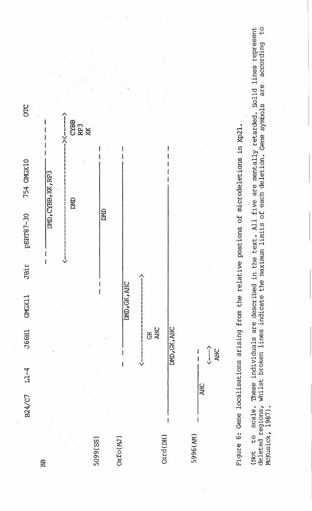
0 U C fO
o
oI— IXolDr-
ocoIr-~ooE-JXWCu
i4•rHCQ1-0
X
o
xuoCDf-o
' nJ1I
ma
CQS3U
AI CQI §32*i u n xVA! I
!I I I I I I I
I QIIIIIIIIIIIIIv
AU
Xo&Q
Xoo
V
o
Xo
Q
o
C Na>xc• HCQ C O • H -P 0 i—I 0■ 8O•iHe<4-1OCQCO•rH4->CQ0 a0>•rH4J 0 r-H 0 i - i •
0x:4-J
1U<44Cna•rHCQ•r4CCSCOCo*r44-)CCSCO
> i
CO
r "U
CNCQ 0G0O
CQCQ
CQCQ
a >a >oi d
§o44XO
33QT3V-i
5<x>CTim
<400i-iDcn•rHin

GF GM F M AM BGF GM F M AM B
-V:iv
! ■ / V Ji1*\ \ '
D2(PvulI)i
!GMGX9(HindiII)
Ll-4(EcoRI)
I
nj66HI(PstI)
IAM
Figure 7: Results of deletion analysis of AM (5996) and his family.
(GF ,-grandfather (I-.)? GM; grandmother (LlJ F,father (II-,); M,mother (II-); AM, (III-,); B, brother (III-). The grandmaternal_track, marked with an arrow in the Hindu! digest (probed with GMGX9) is degraded).
754(PstI)
B24(Mspl)

□
C7(EcoRV) Ll-4(EcoRI
B24(MspI) pERT87. KMspI)
Figure 8: Results of deletion analysis of JR (5194) and his family.

3:6:1 RFLP analysis of deletions at the DMD locus.
GMGX10, GMGXll and GMGX12 did not detect RFLPs and so linkage analysis was not performed. RFLPs defined by other probes were used to determine which deletions were new mutations and their origins.
The mothers of seven of the nine boys with deletions of pERT87 or XJ (table 6) were screened with polymorphic probes deleted in their sons, in order to determine their status with respect to the deletion. These results are summarised in table 8. The mother of SS (5099) was not studied as inheritance of his deletion from his grandmother had already been demonstrated (Wilcox et al, 1986). The mother of SM (4097) had died.
Heterozygosity was demonstrated in the mothers of NJ (5313) and SJ (3485). This generally implies absence of the deletion but may also indicate germ-line mosaicism. NJ's mother was heterozygous, for pERT87-30 (Bglll), pERT87-15 (BamHI), pERT87-8(Tagl) and pERT87-l (Mspl). SJ's mother was heterozygous for PERT87-1 (Mspl) and XJ5.1 (SphI). NJ (5313) and SJ (3485) are therefore likely to represent new mutations which most probably occurred in a maternal gamete, unless their mothers are germ-line mosaics. This will be considered further in the discussion.
The mothers of NJ (5313) and SJ (3485) were also analysed with flanking markers in order to determine which grandparent(s) the deleted chromosome had been derived from and to determine whether the deletion had arisen as a result of recombination. NJ failed to inherit his grandpaternal 99.6 (PstI), XJ2.3 (Tagl), XJl.l (Tagl), XJ1.2 (Bell), XJ5.l(Sphl), 754 (PstI) and 754-11 (Bglll) allelesand so appears to have inherited his grandmaternal X-chromosome without recombination. These results are presented in appendix 6.
SJ’s grandfather was dead but it can be inferred that SJ has inherited his grandfather's 99.6 and 754-11 alleles, and thus his grandfather's X-chromosome, with no evidence of recombination. This suggests that his grandmother was not a carrier as had been assumed, on the basis that his grandmother's brother (individual 11 in the pedigree reproduced in appendix 2) had been affected with DMD. The results for this family are presented in appendix 7.
Page 92

The mothers of AB (0120), AK (5265), JB (5097), PE (5311) and SB (5116) were not heterozygous for any of the probes deleted intheir sons (table 8) and may be homozygous or hemizygous at theseloci. Grandparental samples, which might assist in defining maternal carrier status were only available for AB (0120).
The mother of AB (0120) exhibited the alternative XJl.l, XJ1.2, XJ2.3 and XJ5.1 alleles from her own mother. Thus AB (0120) had inherited his deletion from both his mother and hisgrandmother. These results are presented in appendix 8.
The mothers of AK (5265), JB (5097), PE (5311) and SB (5116) are hemizygous or homozygous for three, four, three and eight loci respectively for which their sons are deleted (table 8).
If XJ1.2, XJl.l and XJ2.3- (which are in linkagedisequilibrium, Thompson et al, 1986) and pERT87-l and pERT87-8 (which are also in linkage disequilibrium, Lindlof et al, 1987) are each considered as one locus, MB, the mother of SB (5116) can be considered to be hemizygous or homozygous for five loci. These loci have heterozygote frequencies of 0.32-0.46, with a mean value of 0.42 (table 3). The theoretical probability that none of these five loci are polymorphic is approximately (0.58)~* or 6-7%. This is in reasonable agreement with the findings of Bakker et al. (1986) who found that 97% of females were heterozygous for at least one pERT87 or XJ RFLP.
This estimate, combined with a prior risk of 4p (Emery, 1976) that MB (5116) is a carrier and CK values which give odds of 1.6:1 that MB (5116) is a carrier, suggests that MB (5116) is a carrier (presumably deleted) with a probability of 0.99 and that SB (5116) does not represent a new mutation.
PE (5311) has an affected brother with Duchenne muscular dystrophy and mental retardation whilst JB (5097) has an uncle affected with Becker muscular dystrophy (ambulant until 14, died age thirty nine). These pedigrees are shown in appendix 2. It is thus unlikely that PE (5311) and JB (5097) represent new mutations and probable that the mothers of these boys do carry the deletion. However, as neither affected relative of these boys was available for study, it was not possible to confirm that DMD segregated with the deletion in these families.
Page 93

The sister of PE (5311") exhibited different XJ5.1 and pERT87-l alleles from her brother PE (5311). She was also heterozygous for pERT87-30 for which he was deleted. Thus she does not have the deletion. Similarly the sister of JB(5097) was not a deletion carrier as she was heterozygous for XJ1.2, for which JB (5097) was deleted. These results are presented in appendix 9.
As GMGXll was not polymorphic, family members of boys from nine families deleted for GMGXll (WB (3405), JH (5098), AS (2672), AW (7866), FMCG (4045), SMcK (7501), GN (4947), SM and KM (5120) and PM (3623) were investigated with two polymorphic markers which flanked GMGXll; J66H1 (PstI) and p20 (Mspl).
None of the mothers of these boys were doubly heterozygous for p20 and J66HI. Five of these boys (AS (2672), AW (7866), SMcK (7501), GN (4947) and PM (3623)) were also deleted for p20 (Mspl). None were deleted for J66HI(PstI), although J66HI detected an additional EcoRI band in both SMcK (7501) and GN (4947), (section 3:5:4). The mother of AW (7866) was heterozygous for p20 and thus he is likely to represent a new mutation. His sister is also heterozygous and thus does not carry the deletion. This result is also presented in appendix 9.
Time did not allow the extension of this analysis to include JBir and pERT87 proximally or B24/C7 distally or to analyse deletions subsequently revealed by p20.
3:6:2 Summary of RFLP analysis at the DMD locus.In summary, RFLPs, detected by DNA probes other than GMGX10,
GMGXll and GMGX12, demonstrated that the mothers of three boys with deletions (NJ (5313), SJ (3485) and AW (7866)) were heterozygous for deleted probes and and were thus unlikely to be carriers for the deletion. The mother and grandmother of a fourth boy AB(0120) were shown to carry the deletion. The mother of a fifth boy SB (5116) was implicated as a deletion carrier by virtue of absence of heterozygosity at eight loci deleted in SB (5116).
Page 94

M PED. S <-------------- PROBES------------ >
XJ5.1 XJ1.2 XJl.l XJ2.3 87.1 87.8 87.15 87.30
MJ 3485 SJ + • • • + • • •MB 5116 SB • • • • • • . • •MJ 5313 NJ / / / / + + + +
JE 5113 PE / / / / . / • • •MB 0120 AB • • • • / / / /IK 5265 AK / • • • / / / /JB 5097 JB / / / /
Table 8: Results of the screen for heterozygosity in the mothers of DMD boys with deletions.
(M>- mother; PED, pedigree number; S, son; +, heterozygous; ., hemizygous or homozygous; /, not applicable (son not deleted). 87.1, pERT87.l; 87.8, pERT87.8; 87.15, pERT87.15; 87.30, pERT87.30. Some of these results for families 0120, 5313, 3485, 5097 and 5113 are presented in appendices 6-9).

3:7 FURTHER CHARACTERISATION OF GMGX9 POLYMORPHISM AND DELETIONS AT THE STS LOCUS.
3:7:1 RFLP analysis with GMGX9.Linkage studies with GMGX9 were first performed in normal
three generation families with three or more sons - "Tom, Dick and Harry" familes (TDH familes) - which each provided three or more informative meioses. Eight out of ten mothers in these families were heterozygous. These results are illustrated schematically in appendix 10, together with autoradiographs from three TDH families. The 4kb allele is represented by an "A" and the 2.5/1.5kb allele by a "B" in these schematic diagrams. These results show that GMGX9 segregates as a mendelian trait.
GMGX9 was then applied to the study of five families segregating for XLI (steroid sulphatase deficiency). Eight obligate carrier females from five families (two from family 3673, three from family 3496 and one each from families 4024, 1348 and 2994), were screened with GMGX9 in order to select for heterozygotes. No heterozygotes were found.
However, one of three obligate carrier sisters, (JP, 3496) had the alternative allele to her two sisters (MJ and HR, figure 9B). This indicated inheritance of a null allele from their father, as these sisters shared an affected father and each had affected sons. DNA samples were available from nine affected males in family 3496 and GMGX9 was found to be deleted in all nine (figure 9C).
These five families were then studied in full. These resultsare also illustrated schematically in appendix 10. Affectedindividuals were consistently deleted in all but one family (4024),in which GMGX9 was also uninformative. Persistence of the deletion through three generations was demonstrated in family 3673 (figure10) and implicated by inheritance patterns in families 1348, 2994 and 3496 (appendix 10).
GMMGX9 results for families 1348, 2994, 3496, 3673 and 4024, (described above), for a sixth family segregating for XLI (1034) and for the TDH familes (appendix 10) were combined with those with D2, 782, Dic56 and Mic2 (D.Goudie) and Xg (P.Tippett) for
page 95

multipoint linkage analysis (Yates et al, 1987b). The results of this linkage analysis are summarised in the discussion.
An Italian family (Ital) segregating for X-linked ichthyosis was studied and shown to segregate for a deletion of GMGX9. This result is shown in appendix 11. Prediction or exclusion of carrier status is possible using GMGX9 in families which segregate for a deletion, providing that the parents of the individual under investigation have different alleles.
It is possible to predict that individuals Illy (Ital), and Illg (3496) who failed to inherit maternal alleles are carriers, and that Illy (3496), III9 (3496) and lll15 (3496), who are heterozygous for GMGX9, are not (appendix 11 and figure 9A). The accuracy of these predictions depends on the frequency of recombination between STS and GMGX9 and will be addressed in the discussion.
Page 96

MJ HR
JP
rChCD
rO§
mvOO'-J -m
CC
“On < K)<CD- O
-09
o <
K D n<
COO'!ro>1erO4-4O4-JCTCX£O4-1oco•rH-IDresCO *1—I T5•rHuJD>ix:4-1o
DJCOSiPhCTi0>i-i3Cn*1— I Cn (A:
Pedigree of
family 3496.
"A",4kb
allele-;"B",2.5/1.5kb allele;
deletion.
(B):
hybridisation
of GMGX9
to MJ
(112), HR
(114)
and JP
(116)
respectively.
(C):
hybridisation
of GMGX9
(upper panel) and pERT87.1
(lower panel) to
the entire family). Continued
over.

HM
o
43CtEXo
43C•rH
r-ooBwCb
COOiro>i i—i
•rHE034-1oX
OCXu4-4Oco•rHx03CO•rH43•rHuA>ix:4-1ogcoSJPdA03C•rH4JCouCDp3CO•rHCn
l~3 Os -PoX i-10OC CX 03Q Qio P04-1 5O Oi—iC —o•rHi—I4-J •03r-CO00•rH Eh43 Pd■rHWp Oj.QX c03—vPQ i—l-— 0G•03C QjO■rH pX 0CD ai—1aCD 343= '(Ti1 Xs us• *■oCDi—14-1CD oi—1i—1c03 o•rHA -pa 03in 0••rHX 43\ •rHL40 H•XJCN >_ *A03 ..— ✓—U#CDi—1 •CD >i—11-1i—1003 >•rHXJ XXC o0a— 0<C 0c p
40CD<y\ HHHro -—>1 PHi—1n•rHE 43 •03 c •—-4-1 0 >1i—14-4 •PO sM CO0 M 4-40 ■—-P 0cn Ph P•rH 33 •rH43 X0 G(X -—• 004• •HH 0< HH A~— X

3673
i ii ii iiiiii h i ii m h i h i h i1 1 2 1 2 3 4 4 5 6 7
GMGX9 (Hindlll)
Figure 10: Deletion analysis of family 3673 with GMGX9.
(A: Pedigree of family 3673. "A'V4kb allele; "B",-2.5/1.5kb allele;deletion. (B): hybridisation of GMGX9 to the entire family).

3:7:2 Deletion analysis with GMGX9 and other distal Xp probes.Males from twenty three independent "Glasgow" families
segregating for STS deficiency, including the six families described above, were screened with GMGX9 (table 9). Only four of these families (4024, 7664, 3000 and 3307) were not deleted forGMGX9. XLI segregated with the 4kb allele in families 4024 and 7664 and with the 2.5/1.5kb allele in families 3000 and 3307.
Eight of nine German families (from A. Gal) were deleted for GMGX9. XLI segregated with the 2.5/1.5 kb allele in the ninth family (2/85 Br). These results are illustrated schematically in appendix 12, together with an autoradiograph showing results in families 1/85 (Ln), 2/85 (Br) and 3/85 (Ls). Segregation of the deletion with XLI in family 5/85 (Ge) was first inferred by the failure of II2 to inherit the 2.5/1.5 kb allele from I5 and was confirmed by A. Gal, who showed GMGX9 to be deleted in an affected male (III2) - Two out of four Dutch families were also deleted for GMGX9 (appendix 12).
Seven of eight affected Italian individuals (from A. Ballabio) were deleted for GMGX9 (figure 11). The eigth individual, Ba, has the 4kb allele of GMGX9 and will be discussed more fully below.
Thus, thirty-six of forty-two males with XLI (i.e. 86%) were deleted for GMGX9. These forty-two males were also investigated (by A. Ballabio) with p422, situated 3' to the STS gene. Thirty-seven of them were deleted for p422 (A. Ballabio, personalcommunication). These included the thirty-six males detected by GMGX9 and also Ba, who was not deleted for GMGX9. The cloning of STS will be presented in the discussion.
The twenty-three "Glasgow" families were examined with other distal Xp probes and markers e.g. Xg (P. Tippett), Dic56, 782, Mic2 (pl9b), MlA, and STB14 to determine the extent of these deletions. (The location and details of these probes are presented in figure 14 and table 3). Die 56, 782 and pl9b results in families 1034, 1348, 2994, 3496, 3673 and 4024 were the work of D. Goudie.
p2a4 and the x-specific bands of three Xp-Yq homologous probes, GMGXXY3, GMGXY19 and STB14 were deleted in the nineteen individuals deleted for GMGX9. (All GMGXXY3 and GMGXY19 results
Page 97
referred to throughout this thesis were obtained by M. Mitchell). All affected individuals hybridised to 782 and Dic56 and to the X-specific sequence of MlA. The majority of affected males tested (i.e. ten/ fifteen) were Xg(a) +ve which demonstrates the interstitial nature of these deletions. This data is summarised in table 9. Examples of the autoradiographs obtained with these probes are illustrated in appendix 13.
Ba, who was deleted for p422 but not GMGX9 was tested with additional probes in distal xp. He was deleted for p2a4 (figure 11) and also for GMGXXY3, but not for STB14 or GMGXY19. Ba is included in table 9 for comparison. The other Italian patients were deleted for p2a4 (figure 11), GMGXXY3 and GMGXY19.
Thus, Ba had a deletion breakpoint close to or within the STS gene which divided probes into two groups; those within his deletion (p422, p2a4 and GMGXXY3) and those outwith his deletion (GMGX9, STB14 and GMGXY19). STSB14 is known to lie at the 5' end of STS but GMGX9 and GMGXY19 could lie to either side of the deletion. In common with all males with XLI, Ba was not deleted for the X-specific band of MlA.
Several distal Xp probes e.g. MlA, GMGXXY3, GMGXY19 and STB14 demonstrate homology to Yq whilst others e.g. pl9b and 29Ci demonstrate homology to Yp. STS sub-clone p2a4 (and also p422, A. Ballabio, personal communication) failed to demonstrate Y homology at normal stringency. GMGX9 also failed to detect any homology to the Y-chromosome (figure 12), even when hybridisation was performed under conditions of low stringency (30% formamide,42°C).
Page 98

PED.NO. STS 782 DIC56 XXY3 X9 XY19 p2a4 MlA Xg(a) pl9b/PSGlJM 3673 nil + + -
SC 2994 nil + + -
DG 1034 2.4% + + -
JA 3710 nil + + -
TR 4024 nil + + +TM 1348 nil + + -
RJ 3496 nil + + -
JD 0698 nil + + -
SL 7568 NT + + -
AB 4076 nil + + -
DC 3000 nil + + +DC 7474 nil + + -
FM 7685 NT + + -
GB 7664 nil NT + +NJ 7750 nil NT + -
EC 3307 nil + + +HL 7605 nil NT + -
RF 7304 nil NT + -
YM 8056 nil NT + -
CK 5626 nil NT + -
RM 4143 nil NT + -
WR 7672 nil NT + -
RH 8009 nil NT + -
AM 1729 nil + + -
JH 7079 nil + + -
Ba ITALY NT NT +
— - - + + +- - - + + X- - - + + X- - - + + X+ + + + + +- - - + + +- - - + + +- - - + + X- - - + + X- - - + - X+ + + + - X- - - + NT X- - - + NT ++ + + + - +- - - + - ++ + + + NT +- - - + NT X- - - + NT X- - - + NT +- - - + + +- - - + - X- - - + NT X
: — + NT X
+—
+ NT NT
Table 9: Screening of Scottish XLI males with distal Xp probes.
(Affected males are identified by their initials and pedigree numbers. AM (1729), JH (7079) and Ba are included for comparison. +, hybridisation (heterozygote for pl9b/pSGl);deletion; NT, not tested; x, not informative; XY3,GMGXY3;
X9,GMGX9; XY19,GMGXY19. PED.NO.,Pedigree number; STS,* STS levels^ measured in hair-roots (Ferguson-Smith & Aitken, 1982), using H-Dehydroepiandrosterone sulphate (DHEASCL) as substrate with the exceptions of 3673, 2994 and 1034 (measured infibroblasts) and 7474 (measured in white_ blood cells). The assay was a modification of that described by Shapiro et al (1977)).

M 1 2 3 Ba 5 6 7 XY Sa F L
GMGX9(ECORI)
p2a4(EcoRI)
Figure 11: Deletion analysis of Italian males.
(M,male control; 1-7,males with XLi; Sa, male with XLI and KAL; XY,male with XY translocation; F,female control; L,lambda marker. Ba (male 4) and Sa are further described in the text. This blot was provided by A. Ballabio).

Xi—ihoDU
2CM&H CTc<CN£r- _
CN W ^rH m < rH
- |« t - H
Figure 12: Hybridisation of GMGX9 to 3E7 (Y-chromosome specifichybrid).
(M,-male control; F,female control. Hybrids are as described in materials and methods. Unlabelled tracks represent further hybrids isolated within this department; lanes 11 and 12 contain only Xq sequences, lane 15 is a further mouse cell line).
3:7:3 Analysis of complex phenotypes associated with XLI.Sa was affected with both Kallmann's syndrome (KAL) and
X-linked ichthyosis (Ballabio et al, 1986) and was potentially of value in ordering disease phenotypes and gene probes in distal xp (compare this section with section 3:5:5). He was deleted for GMGX9 and p2a4 (figure 13), GMGXXY3 and p422 (Ballabio et al, 1987a) but not for Dic56 (Ballabio et al, 1986). Thus, his deletion was not distinguished from deletions associated with XLI alone.
Three individuals, RR (8050), SS(8012) and JG(8049) affected with KAL but not XLI were tested with 782, Dic56, GMGX9, p2a4, GMGXXY3, GMGXY19 and pl9b, all of which failed to demonstrate a deletion (appendix 14). These individuals represent sporadic cases of Kallmann's syndrome, however, and it is possible that they are not associated with an abnormality in this region of the X-chromosome.
3:7:4 Deletion analysis with GMGX9 and other probes in individuals with XY translocations.
AM (1729) and JH (7079) are males with XLI as a consequence of XY translocations. AM (1729) is deleted for xg. JH (7079) was Xg(a)-ve although his mother and grandparents were Xg(a)+ve. It is thus likely (but not proven) that JH (7079) is also deleted for Xg.
Both AM (1729) and JH (7079) are deleted for GMGX9 and p2a4 and for the X-specific bands of STB14, MlA and pl9B. Some of these results are illustrated in appendix 15. The position of MlA relative to GMGX9 is not known. AM (1729) and JH (7079) are also deleted for the X-specific bands of GMGXXY3 and GMGXY19. Neither individual is deleted for 782 or Dic56.
3:7:5 A brief summary of STS deletion data.In summary GMGX9 detects deletions in thirty-six of forty-two
(86%) families segregating for STS deficiency. These deletions are all detected by other probes including STS cDNA clones and appear very similar at the molecular level. Only one of thirty-one patients studied in detail revealed a difference between GMGX9 and STS cDNA clones. F u r t h e r m o r e the deletion in an individual with
Page 99

both KAL and XLI could not be resolved from that in deleted males with XLI. Two XY translocations also failed to separate GMGX9 and STS cDNA clones.
3:8 COMPARATIVE MAPPING
3:8:1 Comparative mapping with GMGXll.It was of interest to determine whether GMGXll was homologous
to DMD cDNA clones which detected deletions at high frequency or represented an additional subset of microdeletions. An Ecori blot was prepared with DNA from different mammals as follows: Lanes (1) human male, (2) human female, (3) male gorilla, (4) female gorilla, (5) male orang-utan (1), (6) male orang-utan (3), (7) malechimpanzee, (8) ram, (9) ewe, (10) dog, (11) bitch, (12) stallion, (13) mare and (14) bull, in order to establish whether this sequence was conserved between species.
GMGXll was conserved in primates (gorilla, orang-utang and chimpanzee) but not in other mammals (figure 13A). It thus seems improbable that GMGXll is expressed.
3:8:2 Comparative mapping with GMGX9.The high proportion of deletions detected by GMGX9 in STS
defecient individuals suggested that it might form part of the STS gene, it was thus of interest to determine if GMGX9 represented part of the coding sequence of STS. Two blots were prepared forcomparative mapping studies using GMGX9. The first consisted of primate DNA digested with HindiII as follows: Lanes (1) human male, (2) human female, (3) male gorilla, (4) female gorilla, (5) male orang-utan 1, (6) male orang-utan 3, (7) male chimpanzee and(8) male with XLI (RH, 8009). The second consisted of DNA from other mammals digested with HindiII as follows: Lane (1) human male, (2) human female, (3) ram, (4) ewe, (5) dog, (6) bitch, (7) stallion, (8) mare, (9) bull and (10) cow.
GMGX9 was conserved throughout primates (figure 13B) and also detects homology with bovine DNA (figure 13C). Weak homology tobovine DNA is common to many single copy sequences and does notimply that these sequences are expressed (Monaco et al., 1986).
Page 100

Comparative mapping would thus suggest that GMGX9 is not expressed. GMGX9 also detects differences between male and female gorilla which might represent a polymorphism in these species.
page 101

rH co
A) GMGXll(ECORI)
Cu So Cno gmg
CKooCO
*1
B) GMGX9(HindIII)
Figure 13: Hybridisation of GMGX9 and GMGXll to "conparativemapping" blots.
(M,male; F, female; GM,- gorilla male; GF-,gorilla female; 0M1, orang-utan male 1 (faint track); 0M3,orang-utan male 3; CM;chimpanzee male; R,:ram; E,ewe; D,dog; B,bitch; s, stallion; M;mare; Bu,bull; C,cow; RH (8009);male with XLI. (A) EcoRl filter hybridised with GMGXll. (B) Hindlll "primate" filter hybridised to GMGX9).

C) GMGX9(HindiII)
Figure 13 continued. (C) Hindlll "farm" blot hybridised to GMGX9 (faint hybridisation).

CHAPTER 4: DISCUSSION

4:1 THE ISOLATION OF Xp SPECIFIC PROBES.This section considers the likelihood of isolating DNA
sequences which lie within Xp21 or Xp22.3 by "random" selectionfrom an x-chromosome library, based on estimates of their size withrespect to the X-chromosome as a whole. Similar estimates are given for the isolation of sequences within the DMD gene, withindeletions in individuals with XLI and within the large Xp21 deletion described by Francke et al., (1985). Both random and targeted approaches to cloning are discussed with particular emphasis on X-chromosome libraries.
4:1:1 Predicted success of random cloning strategy.This aim of this project was to isolate Xp DNA sequences from
a chromosome specific library, in order to conduct an analysis of Xp, focussing on Xp21 and the analysis of DMD and on Xp22.3 and the analysis of STS deficiency, in particular.
The X-chromosome represents approximately 5% of the haploid genome or 150Mb, determined by both flow cytometry (Harris et al., 1986) and cytophotometric data (Mayall et al., 1984). Xp accounts for approximately one-third of the x-chromosome whilst Xp21 represents 20% of Xp (or lOMbp). The size of Xp22.3 has been estimated at 5Mb (or 10% of Xp), (Mondello et al., 1987).
On the basis of these approximations, it may be predicted that, without targeted selection (see below), one-third of X-linked clones would map to Xp, one in fifteen X-linked clones would map to Xp21 and one in thirty X-linked clones to Xp22.3 (table 10).
Of sixteen X-linked probes isolated at random from this library, four were Xp specific: GMGX9 (DXS237), GMGX10 (DXS238), GMGXll (DXS239) and GMGX12 (PXS240). GMGX10 (DXS238) andGMGXll (PXS239) were mapped to Xp21 and GMGX9 (DXS239) to Xp22.3. GMGX12 (PXS240) mapped between GMGXll(DXS239) and GMGX9(DXS237), (table 10).
DNA sequences have now been cloned from three hundred and five loci on the X chromosome (table 2). There is considerable variation in size between these sequences from less than Ikb to several over lOOkb. An (arbitary) average value of lOkb indicates that only 3Mb or one-fiftieth of the x-chromosome has been cloned.
page 103

Interval Probes isolated from this intervalxp 1/3 0.998 GMGX9, GMGX10, GMGXll, GMGX12Xp21 1/15 0.67 GMGX10, GMGXll
Xp22.3 1/30 0.42 GMGX9
"BB" deletion 1/25 0.48 GMGX10
"DMD" 1/75 0.19 GMGXll
"STS." 1/1000 to 1/75
0.015 to 0.19
GMGX9
Table 10: Predicted success of a random approach in cloningsequences within specific intervals of interest on Xp21.
(The probability, P, of isolating at least one probe in a given region is 1- [(l-p)n]), where p is the theoretical probability that any one probe will lie within that region (based on its size) and n is the number of X-specific probes isolated. This has been calculated for each category assuming n = sixteen, the number of X-linked probes isolated in this study. The "BB" deletion is described by Francke et al (1985)).

A minimal estimate of 2Mbp for the size of the Duchenne gene has recently been deduced by extrapolation of the mean intronrexon size ratio (Koenig et al., 1987). The smallest deletions visualised by flow cytometry (FACS) analysis at the STS locus are estimated at 2Mb (Cooke et al., in press) whilst the STS gene has been estimated to represent 140kb (Yen et al., 1987b). Cloning of these loci will be presented later.
These data predict that one in seventy-five probes assigned to the X-chromosome would lie within the DMD gene and between one in a thousand and one in seventy-five probes would be deleted in individuals with STS deficiency. Of sixteen X-linked probes derived from the library constructed in this project, GMGXll(DXS239) mapped within the DMD gene but outwith the deletion described by Francke et al. (1985), and GMGX9(DXS237) was deleted in 84% of individuals with steroid sulphatase deficiency, (including some with deletions not detected by FACS analysis), (table 10).
It can be seen that the probability of isolating one or more probes in each of the categories defined in table 10 (except that of STS deletions) was 0.19 or more, based on a group of sixteen probes isolated at random. Such calculations are only an approximation, but serve to demonstrate that attempts to isolate probes which mapped within Xp21, Xp22.3, the deletion described by Francke et al. (1985) or even the DMD gene were realistic. A more accurate determination of the minimum size of STS deletions, possibly by pulse field gel electrophoresis (PFGE), will be required to assess the probability of detecting deletions at this locus more accurately.
4:1:2 Random and targeted approaches to cloning.Many single copy Xp DNA sequences presently in common use were
derived from flow-sorted x-chromosome libraries by "random" selection for single copy sequences and were localised to Xp using somatic cell hybrids. One disadvantage of this approach, however, is that selection for single copy sequences results in an increased background of non-recombinants (self-ligated vector) and of non-human recombinants. Fuscoe (1987) used an oligonucleotide complementary to the insertion site of the vector to detect
Page 104

non-recombinants. This had the added advantage of detecting all recombinants, regardless of their status with respect to repeated sequences.
Flow cytometry can resolve all human chromosomes except chromosomes 10, 11 and 12 in individuals with appropriate karyotype anomalies (Harris et al., 1985). Libraries are usually constructed from the complete digestion of one to two million chromosomes (Fuscoe et al., 1986), although Kunkel et al. (1985b) reported a library constructed from partial digestion of ten million X-chromosomes. Complete digestion of human DNA will result in some DNA fragments with sizes outwith the acceptance range of the chosen vector which cannot be represented in the library. Representation may be slightly improved by construction of complementary libraries using different restriction enzymes. Inserts containing inverted repeat sequences are also resistant to cloning in recombination proficient hosts (Wyman, Wolfe & Botstein, 1985; Donlon et al., 1986) and may be rearranged or lost entirely.
Fuscoe et al., (1986) predicted that only 65% of a given chromosome is clonable in lambda Charon 21A, due to constraints on insert size. Lambda Charon 21A has a comparable insert acceptance range (l-9kb) to lambda NM1149 (0-llkb). The range of these vectors is more representative of the fragment distribution in digested genomic DNA than that of lambda gtWES (5-14kb) and results in fewer non-X recombinants (Hofker et al., 1985).
Sixteen of fifty inserts isolated from the x-chromosome library constructed in lambda phage NM1149 were X-specific. This was comparable with the representation of X-specific recombinants in the lambda phage Charon 21A library described by Hofker et al. (1985), (table 11). The proportion of non-recombinants in the NM1149 library (almost two-thirds of the plaques selected) was much higher than in the lambda Charon 21A library, however, despite selection for recombinants disrupted at the insert site which should have reduced this, and was comparable with that in lambda gtWES (Hofker et al., 1985 and table 11),.
Other Xp probes have been derived by cosmid cloning e.g. cX5(DXS148), (Hofker et al.r 1986b) or by "targeted approaches" based on knowledge of the location of a mutation. Cosmid clones are
Page 105

generally constructed using somatic cell hybrids. Selection for human inserts depends on hybridisation to human repeated sequences and thus repeated sequences are present in all recombinants obtained. Cosmids facilitate the characterisation of large contiguous regions of the genome but are less appropriate in the direct isolation of single copy sequences, as sub-clones (free of repeated sequences) must first be isolated.
Two "targeted approaches", applied to cloning sequences from the DMD locus, are described below. A targeted approach is most valuable when the target is represented by only a few clones. Fuscoe et al. (1986) state that five chromosome equivalents are required for a 0.99 probability of including any given (clonable) sequence.
One targeted approach, using the "phenol enhanced reassociation" technique, relied on a large Xp21 deletion in a male with chronic granulomatous disease, Duchenne muscular dystrophy, McLeod Syndrome and retinitis pigmentosa type 3 (Francke et al., 1985). Digested DNA from a normal male was reassociated with an excess of sheared DNA derived from the affected boy, and thus the proportion of clonable fragments from within his deletion was enhanced. Four of eighty-one clones (eighteen of which were X-specific), including pERT84(DXS142) and pERT87(PXSl64), were mapped within the deletion in this way (Kunkel et al., 1985a).
This deletion was estimated to represent 6000kb by flow cytometry (Wilcox et al., 1986), or three-fifths of Xp21. Thus, one in twenty-five randomly selected X-linked probes could be expected to map within this deletion. The targeted approach thus increased the proportion of clones within this deletion to a considerable extent. One of the sixteen X-linked clones isolated in this project, GMGX10(DXS238), mapped within this deletion (table 10).
A second targeted approach depended on a female with DMD and an X;autosome translocation, which disrupted both the ribosomal rNA (rRNA) gene cluster on chromosome 21 and the DMD gene (Verellen-Dumoulin et al., 1984). DNA clones from the rRNA locus (Wellauer and Dawid, 1979) were used to initiate "chromosome walks" (i.e. to continually select for overlapping sequences) in order to locate the junction fragment in somatic cell hybrids derived from
page 106

BACTERIOPHAGE VECTOR: gtWES Charon2lA NM1149
Insert sizes (kb):
Low copy clones:
"single copy clones":
Clones with inserts:
Human inserts:Of which:Repetitive:
X-linked:
Autosomal:
Not tested:
5-14 1-9 0-11
134 86 120
79 73 /45 70 50
26 69 27
15 13 7
3 22 16
2 26 4
6 8 /
Table 11: Comparison of the X-chromosome library with two others constructed in different vectors.
(After Hofker et al, (1985) The Hindlll libraries in bacteriophage lambda gtWES and Charon2lA are described in Hofker et al (1985). "Low copy clones" corespond to those selected after Benton-Davis type screening with total human DNA. "Single copy clones" correspond to those selected after a second round of screening by dot blot analysis. This was not performed for the NM1149 library).
each translocated chromosome (and free of additional acrocentric chromosomes with rRNA loci), (Worton et al., 1984). The first clone isolated in this manner was XJ1(DXS206), (Ray et al., 1985).
4:2 DUCHENNE MUSCULAR DYSTROPHY.Restriction fragment length polymorphisms were not detected by
GMGX10(DXS238), GMGXl1(DXS239) or GMGX12(PXS240). The emphasis in this project was thus to study deletions at the DMD locus, rather than to perform linkage analyses. This section reviews the contribution of pulse field gel electrophoresis (PFGE) in determining the limits of the DMD gene, cloning of the coirplete DMD cDNA and comparison of the genomic and cDNA maps are presented along with data which maps DMD, GK and AHC relative to each other. Deletion data is discussed in the light of other genomic and cDNA probes and correlated with that obtained by PFGE. Possible relationships between the size and localisation of deletions at the DMD locus and the severity of phenotype are presented. The problems of a causal relationship between deletions and DMD/BMD and of germ-line mosaicsm in deletion analysis are addressed. The origin of deletions and mechanisms by which deletions might arise are discussed. This section concludes with recent work which defines the DMD gene product, "dystrophin" and its implications for future work and therapy for DMD.
4:2:1 Physical mapping of the DMD gene.Pulse field gel electrophoresis (PFGE) can resolve DNA
fragments from 50kb to 2000kbp in size (Schwartz and Cantor, 1984). PFGE has complemented deletion analysis and linkage analysis in ordering Xp21 probes, and has defined maximum and minimum distances between many loci. Burmeister and Lehrach (1986), for example, predicted that XJ(DXS206) and pERT87(DXS164) could be no further than 300kb apart. Burghes et al. (1987) confirmed this by linking the two loci in a chromosome walk, resulting in a contiguousDXS206/DXS164 clone of 350kb.
PFGE has thus been of value in constructing a map of the DMD gene (Burmeister and Lehrach, 1986; van Ommen et al., 1986). The
Page 107

map positions of loci which lie proximal or distal to DMD are less well defined. There is conflicting evidence to support either C7 or B24 as the closest distal polymorphic marker for DMD. A male with DMD, GK and AHC and a deletion of C7 and Ll-4 but not B24 (van Ommen et al., 1986) suggests Cl is the closest distal marker whilst a recombination event places Cl distal to B24 (and further from DMD), (Norby and Scwhartz, 1987). PFGE data places Cl and B24 more than 2Mb from DMD and 500kb apart. PFGE fragments are shared by B24 and Cl and also by Ll-4 and B24 but not by Cl and Ll-4. Thissupports the order Xcen Ll-4— B24— Cl— pter (Burmeister etal., unpublished observations).
Interestingly, distances on the pulse field map for DMD failed to correspond to those of the genetic (recombination) map. Although 754 is deleted in several individuals affected solely with DMD or with DMD and mental retardation (e.g. in family 5099, (Wilcox et al., 1986) or the family described by Greenberg et al. (1987)), the genetic distance between 754 and DMD is large (approximately lOcM). van Ommen et al. (1986) infer from PFGE data that the physical distance between 754 and DMD can be little more than 1-2 Mbp and thus that, in this region, 1% recombination is comparable to 100-200kbp instead of the usual lOOOkb.
Wieacker et al. (1984) proposed that the genetic (recombination) and physical (cytogenetic) maps of Xp do not correspond and that genetically, the distal half of Xp may be twice as long as the proximal half (55-60cM v 25-30cM). Such disparity between physical and genetic maps is also observed in the pseudoautosomal region on the Y chromosome, (Rouyer et al., 1986), where it is attributed to an obligatory XY cross-over in a relatively small region.
GMGXll was mapped between JBir and J66HI by PFGE, (Burmeister et al., unpublished observations), thus orientating GMGXll with respect to J66HI. This localisation was confirmed using somatic cell hybrids with breakpoints between JBir and J66HI, derived from girls with X;autosome translocations (Boyd et al., unpublished observations). Wapenaar et al. (1987) assigned p20 to the same region, by PFGE.
Page 108

Figure 14: details are separated b;
21.121.2
21.3
22.1
2324
29Cipl9b, pSGl GMGX9,STBl4,p2a4Dic56732RC8PXUT23 D2 99.6B24,C7 LI-4
-58.111.23
• MlA
GMGX12
J66H1GMGXllp20JBirpERT87-30pERT87-15PERT87-8pERT87-lXJ10.1XJ2.3XJ1.1XJ1.2Hip25JMDJ47pERT84754-11754-6754GMGXlOOTC
25
26
27
28
Chromosomal localisation of Xp specific probes. (Probe s S S e S in table 3. The relative posrtions of probesr a comma are not known.).

4:2:2 Cloning of the DMD cDNA and comparison to the genomic map.Cloning of the DMD cDNA has also refined the map of the DMD
gene. The first candidate cDNA clones from the pERT87 locus were isolated on the basis of conservation of exons of a key developmental protein between mammalian species (man, cebus monkey, cow and hamster) rather than by more conventional approaches, such as hybridisation to Northern blots or to a cDNA library, (Monaco et al., 1986). One candidate clone, pERT87-25, which hybridised to a 14kb transcript, was expressed in fetal muscle, but not in human myoblast cells or in He-La transformed cells.
cDNA clones from the XJ region were derived by direct screening of an adult muscle cDNA library, constructed in the expression vector lambda gtll, (Burghes et al., 1987), with subclones from within the XJ region. Cross species homology was again observed.
The 5' and 3' ends of the DMD gene were obtained by searching for overlapping clones in a second fetal muscle library, (Koenig et al., 1987). The resulting 14kb cDNA was divided into subclones of approximately lkb and detected sixty-five X-specific Hindlll fragments. As there were five Hindlll sites within the 14kb cDNA itself, this indicated that the mRNA was transcribed from at least sixty exons.
Some fragments were ordered with respect to each other using •deletions and translocation breakpoints, whilst others were ordered by comparison of the restriction patterns obtained to those of DNA sequences derived from chromosome walks around defined Xp21 loci.
All probes from pERT84 to J66HI inclusive (figure 14), were shown to be intragenic, with at least one third of the DMD gene lying distal to J66. This indicated that GMGXll was intragenic. The first 6kb of DMD transcript were seen to correspond to a minimum of 800kb on the genomic map - from the first (most 5') exon within the PERT84(PXS142) locus to an exon within the JBir locus, distal to PERT87.
Since GMGXll is only conserved amongst primates (and not amongst other mammals), it is unlikely to be a coding sequence and may be situated in one of the introns associated with cDNA subclones 7 or 8 (Koenig et al., 1987) and/or Cf23a (Forrest
Page 109

et al., 1987a) or Cf56a (Forrest et al., 1987b). These cDNA clones detect a high frequency of deletions in the region between JBir and J66HI as observed for GMGXll. p20 may also be situated in one of these introns.
Relatively few reference points which might facilitate genomic mapping of the 3' end of the gene have been described. Extrapolation of the mean intron and exon sizes (35kb and 200bp respectively), however, predicts that the DMD locus extends over 2Mb and thus represents nearly 0.1% of the human genome (Koenig et al., 1987). This corresponds to the predictions of PFGE data(Burmeister et al., unpublished observations) and provides a partial explanation for the high frequency of recombination within the DMD gene itself, for heterogeneity in the site of the mutation and also for extreme variation in the severity of phenotype.
4:2:3 The ordering of Duchenne muscular dystrophy, glycerol kinase deficiency and congenital adrenal hypoplasia (AHC) and the characterisation of a deletion associated with AHC.
AM (5996) represents the first individual described with AHC and mental retardation (but without GK or DMD) with a micro-deletion of Xp21. His deletion could be visualisedmicroscopically, by FACS analysis and by molecular studies (Yates et al., 1987a). JR (5194) with AHC but not mental retardation, was also studied by cytogenetic, FACS and molecular analysis but nodeletion was detected.
The deletion in AM (5996) (which includes Ll-4, B24 and C7), overlaps with that of DH (Card) with DMD, GK and AHC (section 3:5:5). The deletion in MJ (Oxfo) also affected with DMD, GK andAHC, included probes GMGX10 to J66HI (within the DMD locus) but notLl-4. These three deletions placed AHC between J66HI and Ll-4.Furthermore as MJ (Oxfo), but not AM (5996), is additionally GK deficient, this suggested that AHC was at the distal end of the region of overlap. This supports the order DMD-GK-AHC-pter (figures 6 and 15).
Other molecular studies, concurrent with this study andreviewed by Francke et al. (1987), placed the AHC and GK genesbetween JBir (proximal to J66HI) and Ll-4. Chelly et al. (1987b)
Page 110

22.3
22.2
22.1
MIC2XGSTS'(CPDX) XAL
-OA RSAHCGKDMD/BMDCYBBRP3XKOTC■NDP
21.3
Figure 15: Regional localisation of genes on xp. (Gene symbols are according to McKusick, 1987. Genes in parenthesis are provisional localisations. Only those genes which are mentioned in the text are included).

placed GK and AHC between J66HI and Ll-4 in agreement with the above data.
As the deletion in KC (Francke, 1984) extends distally from OTC to exclude B24 and C7 (Francke, 1985), it must also include the genes for AHC and GK (figure 6). Thus KC must also be hemizygous for AHC and GK .
The distance between J66H1 (the most 3' probe known to lie within the DMD gene) and Ll-4 represents some 2Mb or more as estimated by PFGE (Burmeister et al., unpublished observations). There are no points of reference in this interval which might allow GK and AHC to be localised more precisely. Ll-4, which might provide an appropriate starting point for obtaining further probes, seems particularly resistant to chromosome walking and related cloning strategies (Kunkel et al., 1985b).
The deletion in AM (5996) was estimated by flow cytometry (FACS analysis) to represent some 2.3% of his X-chromosome or 3.4Mb (Yates et al., 1987a). Much of his deletion may lie outwith the gene itself, although Ll-4 and C7 are at least lOOOkb apart, (Burmeister et al., unpublished observations). The parents and grandparents of AM (5996) were also studied and his mother (who was mildly mentally retarded), but not his grandmother (of normal intelligence), was shown to be deleted by FACS analysis. This was supported by the finding that his grandmother was heterozygous for B24 (section 3:5:5).
Marlhens- et al. (1987) have recently described an individual with a microscopic deletion of Xp21.2 with both GK and AHC who was not deleted for JBir, Ll-4 or C7. This might be explained by the lack of suitable molecular markers in the 2Mb interval which represents the smallest region of overlap (SRO) of deleted individuals with AHC and GK deficiency.
Many boys with DMD have been assessed for the presence of glycerol in their urine as an indication of glycerol kinase deficiency. Clarke et al. (1986) and Chelly et al. (1987a) observed slightly elevated levels of glycerol in the urine of one of thirty, and five of three hundred and two, males with DMD, respectively. The increase was ten to one hundred fold less than that in males with both GK deficiency and AHC. Only one of the five individuals
Page 111

described by Chelly et al. (1987a) was shown to be glycerol kinase deficient serologically, it thus appears that GK is more closely associated with AHC than with DMD.
The uniqueness of AM's phenotype suggests that his proximal breakpoint separates the genes for GK and AHC, and could represent a valuable starting point in cloning the two genes. Polymorphic probes within his deletion might also represent suitable flanking markers for the study of DMD, which would be closer and possibly more polymorphic than either B24 or C7. It would thus be worthwhile to search or select for X-specific clones deleted in AM (5996).
4:2:4 Deletion analysis (genomic probes)In a preliminary screen, which was included in the
collaborative study of Kunkel & co-authors (1986), deletions were detected in nine of ninety (10%) Scottish boys with either DMD (seventy eight boys) or BMD (twelve boys) by pERT87-8 and/or XJl.l, (Wilcox et al., unpublished observations). These nine boys included one, JB (5097), with BMD.
That collaborative study involved twenty laboratories in the screening of one thousand three hundred and forty-six DMD and BMD males with probes from the DXS164 locus. Eighty-eight males (6.5%) were deleted for at least one of these probes, fifty-three of which were further investigated with additional pERT87 clones. Screening with PERT87-1, PERT87-8, pERT87-15 (and XJl.l) was advocated, as a means to determine the majority of deletions, (Kunkel & co-authors,1986).
Using this approach, Hart et al. (1986) detected fifteen deletions amongst one hundred and forty DMD families (10.7%) whilst Thomas et al. (1986a) detected nine deletions for at least one sub-clone in one hundred and seventeen unrelated individuals (8%) with DMD. Thomas et al. (1986a) also studied forty-eight BMD patients in whom they failed to detect a deletion.
One hundred and three Scottish DMD/BMD boys were screened for deletions with XJ1.2, GMGX10, GMGXll, GMGX12, p20, JBir and J66H1 in this study, and mutations were detected in a further thirty six (36%) individuals. No additional mutations were detected with XJ1.2, GMGXlO or GMGX12 whilst eighteen new mutations (18%) were
Page 112

defined using GMGXll and twenty-two (22%) with p20. Seven individuals had mutations detected by both GMGXll and p20. J66HI detected a fragment of altered size in one individual whilst JBir detected a further three mutations.
The thirty-three mutations detected by GMGXll and p20 represent a new and distinct deletion-rich region from that of pERT87/XJ (figure 16). jn this study, only MJ(Oxfo) and DH(Card), who have complex phenotypes, have deletions which encompass both pERT87/XJ and GMGXll/p20.
GMGXll and p20 both detect restriction fragments of altered size which may represent partial deletions, point mutations producing rare polymorphisms or one endpoint of an inversion or duplication. These alternatives may also explain the altered fragments detected by JBir in JL (1406) and by J66HI in MT(5310), and could be investigated by cloning the junction fragments in these boys.
The detection of altered fragment sizes and/or deletions with GMGXll and p20 is partly dependent on the restriction enzyme involved. Only three individuals were deleted for Hindlll and Mspl fragments of p20 as well as the EcoRI and Hindlll fragments of GMGXll. As p20 was derived from two sequences some 30-40kb apart (van Ommen et al, personal communication), either fragment could be independently altered or deleted in the immediate vicinity of a deletion, dependent on the restriction enzyme used. GMGXll, in contrast, consists of a unique fragment 1.2kb in length, which suggests that enzyme specific differences at this locus are small deletions or subtle changes in which at least one endpoint is well-defined.
J66H1 detected an additional EcoRI fragment, approximately 2kb in size (in addition to the "normal" EcoRI fragment of approximately 4kb) amongst sixteen males with DMD. The significance of this finding has not yet been determined, but could- be investigated in several ways. Firstly, the families of those boys with this extra band should be studied, to determine if it segregates as a mendelian trait. Secondly EcoRI digests of normal males should be screened for the presence of this fragment. Finally, it might also be valuable to screen other restriction
Page 113

enzyme digests of these sixteen males to determine if this represents a site specific mutation.
Den Dunnen et al. (1987) demonstrated deletions in 17% (eighteen of one hundred and four) DMD patients using probe JBir in addition to pERT87 and XJl.l. Bakker et al. (1987b) reported that XJ, pERT87 and JBir were deleted in 10% individuals with DMD, and that p20 was deleted in an additional 18% individuals from the same group, in good agreement with the 22% observed in this study. J66H1, which was very effective at detecting deletions by pulse field analysis (as described below, section 4:2:5), detected only four deletions amongst four hundred and fifty individuals by Southern analysis, (Ginjaar et al., 1987).
Amongst those genomic probes • which detect deletions by Southern analysis, p20 and GMGXll detect the most deletions (figure 16). Some cDNA clones, however, are able to detect deletions in more than 50% affected individuals (Forrest et al., 1987b) and this has increased the proportion of affected individuals with deletions detectable by Southern analysis to 70% (Forrest et al., 1987b). Deletions of cDNA probes are described below (section 4:2:6).
4:2:5 Deletion analysis (PFGE)Den Dunnen et al. (1987) were able to detect twenty deletions
and a duplication amongst thirty-nine DMD patients by PFGE as compared to only seven deletions by Southern analysis. They used partial Sfil digests and the probes XJl.l, pERT87 (sub-clone not specified), JBir and J66H1 (which span 2.2Mb using partial Sfil digests). Sixteen of these deletions (and one duplication) were detected with either JBir or J66H1. This predicted an especially deletion-rich region in the 950kb between JBir and J66H1.
The resolution of PFGE is greatest for large DNA fragments (outwith the "compression zone" of unresolvable fragments). Thus deletions as small as 30kb can be detected using probes as distant as 800 kb (van Ommen et al., 1986). GMGXll(DXS239) is about 300kb proximal to J66 and 400-500kb distal to JBir (Burmeister et al., unpublished observations). Thus deletions of more than 30kb at the DXS239 locus would be readily detected by PFGE analysis with either
Page 114

% DMD/BMD boys with deletions or altered fragment lengths.
0 2 4 6 8 10 12 14 16 18 20 22 24 26 %
XJl.l _____________£
pERT87.8_' _____________
JBir
p20
GMGXll
J66HI
Figure 16: Schematic representation of mutations described in thisstudy.
(PERT87-8 results were obtained by D.Wilcox. Figures are expressed as percentages of the total number of boys screened with each probe. Eighty seven individuals were studied for XJl.l and pERT87-8. One hundred and three individuals were studied with JBir, p20, GMGXll and J66HI. Those boys with an extra EcoRI J66HI fragment in addition to one of normal size are not included).

J66 or JBir. Additional deletions might also be detected by PFGE with GMGXll or p20 if these probes are situated far enough (several hundred kilobases) from a deletion rich region.
P20, cDNA sub-clones 7 and 8 (Koenig et al., 1987), and Cf23a and Cf56a (Forrest et al., 1987a, 1987b) are also situated between JBir and J66 and deletions of these loci are likely to correspond to many of those detected by PFGE with JBir or J66HI. The extent to which deletions detected by these probes overlap and the distances between them have not been established.
4:2:6 Deletion analysis (cDNA probes).Koenig et al. (1987) detected deletions in 50% (fifty three of
one hundred and four) DMD patients using subclones of the 14kb cDNA, approximately lkb in size. They noted (i) a non-random distribution of deletions, (ii) a slight increase in the proportion of deletions associated with sub-clone lb (which roughly corresponds to the XJ/ pERT87 junction on the genomic map), (iii) a large increase in the frequency of (small) deletions associated with sub-clones 7 and 8 (which lie within the region defined byJBir and J66 on the genomic map) and (iv) an absence of deletionsassociated with five of the six distal subclones tested. Forty-two out of fifty-three (80%) deletions detected were deleted for sub-clone lb (fifteen deletions) or sub-clone 8 (twenty seven deletions).
Forrest et al. (1987a, 1987b) described two distal cDNA clones Cf23a and Cf56a, which detect deletions in DMD and BMD patients. Cf23a extends distal to JBir and detects deletions in 28% individuals with DMD and 51% those with BMD. Cf56a, which lies 3'to Cf23a, detects deletions in 51% individuals with DMD and in 52%those with BMD. The extent to which these cDNA clones overlap with cDNA clones 7 and 8 (Kunkel et al., 1987) is yet to be defined.
Deletions were detected in 70% DMD/BMD families by Southern analysis using Cf23a and Cf56a, in conjunction with two further cDNA sub-clones, CalA which is distal to pERT87-30, (Cross et al., 1987) and Cf27 (in the vicinity of XJl.l and HIP25). These subclones represent twenty-seven exons from the 5' and central regions of the DMD gene.
Page 115

As cDNA deletions potentially define the critical domains of the DMD gene, cDNA clones which detect deletions at high frequency are likely to supercede random DNA clones in deletion screening.
4:2:7 Deletion/ phenotype correlationsThere is no clear correlation between the size or location of
deletions at the DMD/BMD locus and either the severity of phenotype or mental retardation. Three conclusions can be drawn from the data outlined below: (i) that no "Becker-specific" domain of the DMD gene exists, (ii) that the extent of the deletion is not a rigorous guide to the severity of phenotype and (iii) that deletions of certain probes e.g. GMGXll are more frequently associated with a severe phenotype than others e.g. p20 or Cf23a.
Assessment of intelligence quotient (IQ) in eight hundred and ten boys with DMD (summarised in Emery, Skinner & Holloway, 1979) suggests that about a third of such individuals have an IQ less than seventy-five. Mental retardation in association with BMD is not unknown (Emery and Skinner, 1976) but is not a common feature.
SS (5099), with the largest deletion in this study, and to a lesser extent JD, (his cousin who has the same deletion), were severely mentally retarded with short stature. Not all mentally retarded boys have such large deletions, however. Greenberg et al. (1987) described a family with a similar microscopic deletion, including 754, pERT87 and XJ, in which affected males had "normal growth parameters" and were described as "slow-learners". Probes which might distinguish between these two deletions (e.g. GMGX10) could prove of especial value in examining mental retardation in boys with DMD.
In this study, JB (5097) with a severe Becker phenotype was deleted for DNA probes from the XJ/DXS206 locus. JB was quite severely affected (wheei-chair bound at thirteen, presently aged twenty). He had an affected uncle who was wheel-chair bound at fourteen and died aged thirty nine. His deletion encompassed that of AK (5265) with a Duchenne phenotype.
Liechti-Gallati et al. (1987) have recently reported an XJ/PXS206 deletion (including XJl.l and XJ2.3) in a mildly affected BMD patient. Other deletions which result in a BMD phenotype, have
Page 116

been reported within the pERT87/DXS164 locus (Hodgson et al., 1986; Kunkel & co-authors, 1986; Hart et al.,1987). Cross et al. (1987) reported a patient with mild BMD whose deletion extends distally from pERT87-30 and includes J-Bir whilst Lindlof et al. (1987) recently reported a family with three BMD males deleted for XJ2.3 in addition to pERT87-l, pERT87-8 and pERT87-15 (but not PERT87-30).
Hart et al. (1987) described three BMD individuals with pERT87 deletions. One of these was only 6kb in size and was associated with a mild BMD phenotype. The other two deletions encompassed probes pERT87-l, pERT87-8 and .pERT87-15. One was at the severe end of the BMD spectrum (wheel-chair bound at sixteen years and two months) whilst the other was milder in phenotype (able to walk until thirty-eight years).
Four out of fifteen BMD families in this study had a deletion breakpoint within p20, whilst a further two BMD families had deletions of p20. p20 also detected deletions in sixteen DMDfamilies. Whilst one third of all alterations to the restriction fragment length pattern detected by p20 were associated with a BMDphenotype, all mutations detected by GMGXll, were associated with aDuchenne phenotype. This suggests that GMGXll, which lies 3' top20, may lie close to an essential region of the DMD gene.
Individuals with BMD have also been reported with deletions of exons from cDNA clones CalA (distal to pERT87-30, Cross et al., 1987), Cf23a and Cf56a, which lies 3' to Cf23a. Cf23a detected more BMD deletions than Cf56a, including all those detected by Cf56a. Cf56a detected twice as many DMD deletions as Cf23a. Most of the deletions detectable by both probes had an endpoint between two particular exons of Cf23a, which also represented an endpoint for 30% of DMD deletions, (Forrest et al., 1987a, 1987b).
Cf23a thus appears to detect mutations in a deletion-prone region, of which several result in a less severe phenotype, as observed for p20. Disruption or loss of these loci thus appears less deleterious than disruption or loss of GMGXll. Separate patient groups were tested for deletions with p20 and Cf23a, so it is not yet known if these two groups are partially-, completely- or non- overlapping.
page 117
Cross et al. (1987) reported a deletion of a minimum of nine exons from the DXS164 locus (normally transcribed in adult DMD mRNA) in a sixty-one year old patient with an exceptionally mild BMD phenotype. The most extreme example of failure to correlate phenotype with deletion size was that of a normal male who was deleted for at least two exons of the DMD cDNA, (Koh et al., 1987). The severity of phenotype may be governed by whether a deletion causes an in-frame mutation, resulting in a shortened protein product, or an out-of-frame mutation which might completely disrupt transcription of the gene. This would predict that disruption of the GMGXll domain normally prevents formation of the DMD gene product and that disruption of the p20 and Cf23a loci could give rise to a defective gene product.
The forty-five mutations detected at the DMD/BMD locus in this study are highly heterogeneous, representing at least twenty-five distinct groups. It is apparent that it will be necessary to precisely define many of these at the DNA sequence level before accurate correlation with phenotype can be assessed. It may eventually be possible to predict the extent to which a well-characterised deletion will disrupt protein formation, and thus to predict the severity of phenotype from a defined mutation.
In rare instances, a muscular dystrophy which is clinically indistinguishable from DMD is observed. This appears to be inherited as an autosomal disorder (McKusick number 25310), (Somer et al., 1985). In such instances Xp21 RFLP haplotypes will fail to segregate with a muscular dystrophy phenotype.
4:2:8 Causal nature of DMD deletionsAlthough it has generally been accepted that a deletion in the
PERT87 and/or XJ regions is causal to DMD/BMD, Koh et al. (1987) have recently reported a normal male with a deletion of DXS206 including XJl.l and XJ10.1 and at least two exons of the DMD gene. This boy's brother, with DMD, apparently has the same deletion. This dramatic difference in phenotype may be a consequence of subtle differences betwen the two deletions or alternatively to a compensatory factor present in the unaffected boy. Neither boy was deleted for XJ5.1 (or pERT87-42). Whatever the reasons for the
Page 118

difference in phenotype, this deletion, in a normal individual, has serious implications for prenatal diagnosis based on deletion analysis. As there is no reason to suppose that such an anomalyneed be restricted to this region, it would now seem appropriate toinclude unaffected male siblings of boys with DMD/BMD in anydeletion screening.
Similarly, but less dramatically, two affected brothers have been reported with classical BMD whose great-great uncle appears to have the same deletion and yet is much more mildly affected(Forrest et al., 1987a).
4:2:9 Germline Mosaicism and DMD.Recent reports (Lanman et al., 1987, Monaco et al., 1987 and
Bakker et al., 1987a) have described females who are heterozygous for a given RFLP at the DMD locus, but have two or more offspring deleted for this same probe.
Transmission of deletions by unaffected males has also been reported e.g. transmission of a JBir deletion at the DMD locus to two daughters and to subsequent affected offspring (Darras and Francke, 1987) and of a pERT87-15 deletion to at least two daughters and subsequent affected offspring (Bech-Hansen et al., 1987).
Three hypotheses have been put forward to explain this phenomenom (i) germline mosaicism (ii) translocation of the "deleted sequences" to an autosome in the parent and (iii) a "pre-mutation" or predisposition to a deletion which gives rise to the same deletion in multiple independent events (unlikely). The first hypothesis is favoured but the second could be formally excluded by in situ hybridisation.
Romeo et al., (1986) have similarly identified a BMD pedigree in which a deletion recurs. Their data is most readily explained if the grandmother represents a germ-line mosaic.
Germ-line mosaicism is not unique to DMD/BMD or to X-linked disorders. It was invoked to explain three sisters with the autosomal dominant disorder achondroplasia, born to normal parents (Fryns et al., 1983) and also an individual heterozygous for a rearranged Factor VIII (Hemophilia A) gene who transmitted a 40kb
page 119

factor VIII deletion to one daughter and an affected son, the normal X-chromosome to two sons and two daughters and the rearranged X-chromosome to a further daughter (Gitschier, 1986).
In the light of these observations it is difficult to be convinced that a sporadic deleted individual truly represents a new mutation. It is thus possible that the mothers of SJ (3485) and NJ (5313) (who are heterozygous at two and four loci respectively for which their sons are deleted) are germline mosaics and could give rise to subsequent affected males. They should therefore be offered prenatal diagnosis.
4:2:10 The origin of DMD mutations.The origin of the deleted chromosome has been demonstrated for
two individuals, NJ (5313) and SJ (3485) for whom grandparental samples were available. The mutation in these two individuals occured in a maternal gamete, derived from grandmaternal chromosome in NJ (5313) and the grandpaternal chromosome in SJ (3485), with no apparent recombination. AW (7866) also represents a new mutation which arose in a maternal gamete.
Two further deleted individuals for whom grandparental samples were available, SS (5099, Wilcox et al. 1986) and AB (0120) did not represent new mutations. Their deletions were apparent in their maternal grandmothers.
New mutations are generally believed to account for one-third of individuals with DMD, based on the static frequency of the disorder in the population and a calculation using Haldane's formula (Haldane, 1935), which assumes that mutation rates are equal in male and female gametes and that male fertility in DMD is effectively nil. (Only one DMD male with a child has been reportedto date, (Thompson, 1978)).
This is supported by segregation analysis performed on siblings of affected cases of DMD. Carrier analysis based on creatine kinase measurement has also suggested that about a third of boys with DMD represent new mutations (reviewed in Moser, 1984) although these results were unavoidably based on incomplete ascertainment. In contrast, a low proportion of new mutations (four non-carrier mothers/ twenty isolated cases) has been observed for
Page 120

Lesch-Nyhan disease (Francke et al., 1976).Witkowski et al. (1987) studied twenty seven three— generation
families, six of which represented families with deletions. The deletion arose in the grandpaternal X-chromosome in two of these "deletion" families and in the grandmaternal X-chromosome in a third, giving rise to carrier females. The mutation occurred in a maternal gamete in the remaining three "deletion" families as for SJ(3485) and NJ(5313).
In this study the mothers of four boys were homozygous or hemizygous at all polymorphic loci shown to be deleted in their sons, and thus it was not possible to determine whether these individuals carried the deletion. Hemizygosity at a given locus is often difficult to prove by Southern analysis. This question could be best addressed by PFGE. Within the limits of resolution of the system, any probe which is close to, but not within, the deletion, may detect an anomalous fragment in addition to the fragment of normal size (which acts as an internal control). Kenwrick et al. (1987) described a similar phenomenon in a female DMD patient with an X;1 translocation using probes pERT84 and HIP25. PFGE might also allow the detection of a rearrangement, such as an inversion, which might result in a predisposition to deletions.
4:2:11 Possible mechanisms for the high frequency of deletions and duplications at the DMD locus.
Deletions seem to account for a higher proportion of mutations in certain disorders than in others at both the microscopic and molecular levels. Molecular (sub-microscopic) deletions represent 18% (five in twenty eight) mutations in Lesch-Nyan disease (Yang et al., 1984) and 7% (one in fifteen) mutations in OTC deficiency (Rozen et al., 1985).
To a certain extent, the high proportion of deletions detected at the DMD locus (sections 4:2:4-4:2:6) is a consequence of its considerable size. Koenig et al. (1987) state that the incidence of deletions is no greater for any 200kb region of the DMD gene than for other disorders e.g. the FVIIlc locus (Youssoufian et al., 1987) or the HPRT locus (Yang et al., 1984) although certain regions of the DMD gene are indisputedly more deletion-prone than
Page 121

others (sections 4:2:4—4:2:6).The mechanism by which deletions arise at the DMD locus is not
known, although it has been proposed that deletion-rich regions might contain direct or indirect repeats and/or sequences with strong homology to others elsewhere within the gene. These may create a region susceptible to non-homologous pairing, thus increasing the likelihood of unequal crossing over at diplotene in meiosis (and resulting in both deletions and duplications). Alternatively direct and/or indirect repeats might allow stem and loop structures to form during replication, which could result in DNA loss by intra-chromosomal recombination.
Characterisation of the molecular structure of GMGXll, which is only 1.2kb in size, by DNA sequencing and/or detection of homologous sequences elsewhere in the gene (e.g. by screening an X-chromosome specific library at low stringency) may help to resolve these possibilities.
Duplications have been reported in association with DMD/BMD. Berteleson et al., (1986) described a male with a duplication encompassing all pERT-87 sub-clones. This duplication was determined by DNA dosage studies. Hu et al. (1987) identified duplications of one (or more) exon(s) in three patients (two with DMD and one with BMD) amongst one hundred and twenty individuals with either DMD or BMD. RFLP studies indicated that one tandem duplication had not arisen by unequal exchange between non-sister chromatids and may have resulted from sister chromatid exchange. Den Dunnen et al. (1987) used pulse field gel electrophoresis to demonstrate the presence of a duplication at the JBir locus in an affected individual and in his mother.
The breakpoint has been precisely defined in the t(X;21) female DMD patient described by Verellen-Dumoulin et al. (1984). No extensive homology was observed between the chromosomes from which the translocation arose, in the vicinity of the breakpoint (Bodrug et al., 1987).
Monaco et al. (1987) observed that six deletion endpoints in the DXS164 locus occured in single copy, repeated and long interspersed repeat ("LINE", Singer, 1982) sequence homology sequences, so that there would appear to be little sequence
Page 122

specificity for deletion breakpoints.It should be possible to gain insights into the means by which
deletions arise at deletion-prone loci by comparison of the deletion junctions in affected individuals with the sequence in normal males. The many small deletions associated with GMGXll and p20 should prove particularly amenable to such a molecular analysis.
4:2:12 The DMD gene product "dystrophin" and its implications for future research.
Isolation and cellular localisation of the DMD gene product, "dystrophin" have recently been described. Fusion proteins derived from two distinct mouse cardiac muscle cDNA clones were used to raise antibodies which defined the gene product "dystrophin". These mouse cDNA clones and others showed over 90% homology to the first 4.3kb of the human DMD cDNA at the level of both amino acid and DNA sequences (Koenig et al., 1987; Hoffman et al., 1987a).
Dystrophin is a 400kD protein representing 0.002% total protein in striated muscle, consistent with the low abundance of the DMD mRNA (0.01-0.001% total mRNA). Dystrophin is also present in cardiac muscle and at trace amounts in brain but was absent from muscle biopsies from two boys with DMD (Hoffman et al., 1987b).
The predicted structure of dystrophin was rich in alpha-helix, with significant homology to the actin binding site of alpha-actinin (Hammonds, 1987), suggesting a binding function for cell membranes or helical proteins. Recently, dystrophin has been shown to represent a minor constituent of triads, which are responsible for the release of calcium ions as a signal for myofibril contraction. DMD may thus result from aberrant calcium homeostasis, possibly due to the increased activity of calcium-dependent phospholipases (Hoffman et al., 1987c).
mdx, a muscular dystrophy mutation in the mouse which maps close to G6PD and HPRT, has a mild phenotype comparable to that of BMD in man. On the basis of its map position, it was initially proposed that mdx was homologous to EDMD, (Avner et al., 1987), which maps to Xq28-qter in man. However, cDNA clones from the DMD
Page 123

locus mapped in the same interval of the mouse X-chromosome as mdx (Brockdorff et al. 1987a, 1987b; Heilig et al., 1987), whilstdystrophin was not expressed in two mdx mutations (Hoffman et al., 1987b). This argued that mdx is the murine equivalent of DMD rather than EDMD and that mdx would represent an appropriate model for the study of DMD, despite its milder phenotype.
It should now be possible to determine defects at the protein level and to confirm proposals that DMD results from a complete absence of dystrophin whilst BMD is a consequence of a defective protein product, using material from muscle biopsies.
The key to an appropriate therapy for DMD may lie in determining why the disorder is so much more severe in man than in mouse. Hoffman et al. (1987b) suggest that fibrosis of muscle fibres in DIO, which is not observed in mdx, may prevent the regeneration of muscle fibres which occurs in mdx and account for the more severe phenotype. If this is so, agents which inhibit fibrosis may ameliorate DMD in man.
4:3 STEROID SULPHATASE (STS) DEFICIENCY.In this section, linkage analysis with GMGX9 and the cloning
of STS are summarised. The frequency of deletions detected with GMGX9 and other genomic probes are compared with those of cDNA clones, and possible mechanisms for the high deletion frequency observed at this locus are discussed. Flow cytometry (FACS) data, the absence of heterogeneity amongst these deletions, XY translocation breakpoints and the escape of STS from X-inactivation are presented. Finally, clinical applications of GMGX9 and possible applications of GMGX9 to future work are discussed.
4:3:1 Linkage studies with GMGX9 and the relationship of GMGX9 to STS cDNA clones.
GMGX9 was mapped to Xp22.3 by somatic cell hybrid mapping and more precisely localised to the vicinity of STS by virtue of co-segregation of GMGX9 deletions with steroid sulphatase deficiency (Gillard et al., 1987).
GMGX9 detected a high frequency polymorphism (PIC 0.44) and was included in a multi-point linkage analysis with distal Xp
Page 124

markers D2(DXS43), 782(PXS85), Dic56(DXS143) and Kg in both normal (TDH) families and families segregating for X-linked ichthyosis, (Yates et al., 1987b). A deletion of GMGX9 was considered as a third rare (null) allele for the purposes of this analysis. GMGX9(PXS237) was linked to STS with a maximum LOD score of 8.73 at 0 recombination (with confidence limits of 0-0.07).
In practice, since both GMGX9 and STS cDNA clones are deleted in at least 86% males with XLI, and two co-deleted loci will be linked at zero recombination regardless of the distance between them, the precise distance between STS and GMGX9 remains unknown. The maximum distance between STS and GMGX9 will correspond to the smallest deleted region these individuals have in common. This is below the limit of resolution of FACS' analysis, as some deletions cannot be visualised by this technique, i.e. less than about 2Mb, which is normally assumed to represent 2% recombination.
Thus, the maximum error in assuming that GMGX9 and STS are tightly linked is small, providing that the distance between these loci does not differ substantially in deleted and non-deleted individuals.
The multi-point analysis gave the most likely order as D2-22cM-782-10cM-Dic56-5cM- (STS/GMGX9) -13cM-Xg-Xpter. This order was supported by the deletion of Xg, STS and GMGX9 (but not Dic56) in two males (1729 and 7079) with XY translocations.
STS cDNA clones were isolated independently by several groups using polyclonal antibodies (raised to purified STS protein) to screen placental cDNA libraries (constructed in lambda phage expresssion vectors), (Yen et al., 1987a; Ballabio et al., 1987a; Conary et al., 1987; Bonifas et al., 1987).
The 2.7kb STS cDNA clone of Yen et al. (1987a) failed to hybridise to RNA from fibroblasts of an affected individual and cross-reacted with STS antibodies when cloned into the mammalian expression vector pMSG. The cDNA clone of Ballabio et al. (1987a) was localised to Xp22.3 by in situ hybridisation. STS cDNA clones detected genomic deletions in eight out of ten (Yen et al., 1987a), eight out of eight (Ballabio et al., 1987a), two out of three (Conary et al., 1987) and fourteen out of fifteen (Bonifas et al., 1987) patients with XLI, respectively. These results confirmed that
Page 125

these clones represented at least part of the STS gene. The 2.7kb mRNA transcript was sequenced and found to contain an open reading frame of 1542bp with a signal peptide of 22 amino acids and a coding region of 492 amino acids, (Yen et al., 1987a).
GMGX9 and the p422 and p2a4 STS cDNA clones (Ballabio et al., 1987a) demonstrated the same pattern of deletion in forty-four individuals with XLI with only one exception, yet comparative mapping suggested that GMGX9 was not expressed. GMGX9 might therefore represent an intron or a flanking marker.
4:3:2 Deletion analysis of males with XLI.GMGX9 detected deletions in thirty-seven of forty-four (86%)
unrelated individuals with XLI. Thirty-eight of these forty-four individuals were deleted for p422, including the thirty-seven deleted for GMGX9.
Thirty-one affected males, including the exceptional male,"Ba" were screened with GMGX9, STB14 (a genomic clone at the 5' end of the STS gene), p2a4 (an STS cDNA clone), GMGXXY3 and GMGXY19 (genomic probes independently isolated from X and Y chromosome libraries, (Mitchell et al., unpublished observations)). Four males were not deleted for any of these probes whilst twenty-six had deletions of (X-specific) bands of all five probes. Only one deletion, that of "Ba", could be resolved from the others, indicating that deletions at the STS locus represent a highly homogeneous group.
The STS gene was shown to span at least 140kb on the X-chromosome including at least ten exons, with introns from lOObp to over 20kb in size (Yen et al., 1987b). Twenty-one of a group of twenty-six males with XLI had entire gene (ten exon) deletions, whilst two had partial deletions. The first of these included all but the first exon whilst the second included at least 25kb and 4 exons, (Shapiro et al., 1987). Thus these individuals also represented a highly homogeneous group of deletions (table 12).
"Ba" was deleted for the X—specific bands of p2a4, p422 and GMGXY19 but not those of GMGX9, STB14 or GMGXXY3. These probes can be divided into two groups accordingly. STB14 is at the 5' end of this deletion but GMGX9 and GMGXXY3 may lie to either side of it.
Page 126

This exceptional deletion demonstrates the value of deletion breakpoints.
It is difficult to determine the relative order of these probes, although they are all known to lie distal to Dic56. None of these probes, with the notable exception of GMGX9, detects an RFLP and thus ordering of these probes will depend on PFGE (in X-hybrids or females) or on screening of further deletions.
4:3:3 XY homology of probes which detected deletions in males with XLI.
Neither p2a4 nor GMGX9 demonstrate any homology to the Y chromosome, yet GMGXXY3 and GMGXY19, STB14 (Ballabio et al., unpublished observations) and the 2.7kb STS cDNA clone (Yen et al., 1987a) all have homologues on Yq. Overlapping Y-specific genomic clones, detected with STS cDNA clones, have shown that Y homology is limited to the 5' end of the STS gene, and consists of two EcoRl fragments of 20kb and 1.5kb and an intervening intron of 15kb (Yen et al., 1987a, 1987b). Thus homologous sequences to STS on theY-chromosome represent a (non-functional) diverged gene or "pseudogene".
The Y-pseudogene was assigned to Yqll-Yqter using somatic cell hybrids (Yen et al., 1987a). Yq homologues have also been observed for other DNA probes in distal Xp e.g. M1A/DXS31 (Koenig et al., 1984), CRI-S-232 (Donnis-Keller et al., 1987) and GMGXXY3 andGMGXY19 (Mitchell, unpublished observations). Xg and MIC2X, however, which are analogous to STS in that they escape X-inactivation, have homologues on Yp.
Such observations suggest a peri-centric inversion on the Y chromosome, during its evolution from an ancestral sex-chromosome, which would result in an increased genetic distance between STS and the STS pseudo-gene and the transfer of the testes determining factor (TDF) close to the pairing region. It should be interesting to compare physical maps of the region around the X and Y homologues of STS and to assess the extent of their divergence.
Page 127

PROBE
AND
DELETION
STATUS
0•rH44UfCD -i
IG •OG0 04-> TD 0
OU
IrH 0 •rl44 Ha, §G -H
CQ O J-i Eh C CD
CU 0 44 T5 0
OU
0•rH4->H0O -i
04CN O 44a. c 0i— 1 0 0 44 <V 0
<T>><
80•rH4->aiG
OO 44G 0i—I 0 0 4-> 'V 0
OU2MoM8
O Cno o
X8
o o
CT\
O O
O
<T> 00
O O
<Ti
■s0 r—I4JOu03
>ir—I 0 4->
O O
OJ
00 CN
00
>1GSH0O
CN
CN
C/3 TD G 0 ■—I0x:44§
CN
CO
CN CN
§
EH2
g
EH2 Eh2
'5503D
O
&
S
CQ
<03D
CN
JQ
44 CUCU XI44030 G4H •H•rHG8 DQ
T5G -—»CQ 0 03*— - G
O»*- U ■iHf" -—- 4400 0a\ -C >i—i 44 U
•iH 0k. £ c/31— 1 XJ0 CU03 o44 O TS0 X3 0-p x:>1 03U •rH0 O' i— 1G 00 X3O <T\ 3U rH CUG•* 3__ rH "w”U 0-—- O44 •rH0 JQ0 0O' r—100 O rHCTi H 0rH •H CQ
0 •rH -C <0 0344 -QCU G•H rrjG 00 GX Ti •H
CU 044 44G -QX 0 O-w- 03•« CU 0
X SHX3 CU 0TJ0 0 CN44 CN0 0rH CUow -—■X3•rH X 44
N._--•rHw 50 2G Q wO U 44■— 1 rHu 3x: 03< 44 02 •H MQ £ 0U x:
03 Eh44i— 1 •3 G03 0
0 0. >44 Oh •rH03 CP0 0344 •
O' 044 00 UO CTl G2 rH 0
u" *. 0C_| MHi— 1 00 V-l

4:3:4 Deletion analysis of additional individuals with anomalies of distal Xp.
A number of individuals with anomalies of distal xp were investigated in an attempt to define new breakpoints which could distinguish betweeen the probes deleted in males with XLI and facilitate their ordering on distal Xp. The first of these, "Sa", had both Kallmann's syndrome (KAL) and XLI. His deletion proved to be no different from those of other individuals with XLI alone, by either molecular or FACS analysis.
KAL is thought to be proximal to STS, as several male individuals with 46,Yt(X;Y)(p22.3;qll.2) translocations(effectively deleted for all of distal xp) are affected with XLI but not KAL.
Three further patients with KAL but not XLI were analysed but were not deleted for any of the probes tested. This may be due to deletions or point mutations not detected by available probes in Xp22.3. Alternatively, KAL may not be associated with a mutation in this region of the X-chromosome in these individuals.
Perrin et al. (1976) described a family segregating for "X-linked ichthyosis, hypogonadotropic hypogonadism, mental retardation and anosmia" which failed to demonstrate linkage with Xg (maximum LOD score of -0.096 at theta = 0.40). This indicates a distinct mutation from that of the families described by Ballabioet al. (1986, 1987b) and an alternative localisation for KAL. STSlevels were not assessed in the family described by Perrin et al. (1976), however, so the diagnosis of XLI was not confirmed.
Two males with XY translocations, AM (1729) and JH (7079) were also studied. These boys were deleted for the (X-specific) bands of all probes deleted in males with XLI (GMGX9, GMGXXY3, GMGXY19, STB14 and p2a4) and also for MlA. MlA may be either distal or proximal to the other five deleted probes.
These results suggest that there are definite hotspots indistal Xp which give rise to deletions and XY translocations associated with XLI and that these form two homogeneous groups or that there are as yet insufficient DNA probes to distinguish between individuals in each group. These possibilities might be resolved by cloning several translocation and deletion junctions.
page 128

The majority of XX males have levels of STS activity characteristic of normal female individuals. At least three XX males have been reported to possess only a single dose of STS activity, (Pierella et al., 1981; Wieacker et al., 1983c). This may have arisen as a result of the loss of one STS locus through x-Y interchange. Such XX males would be hemizygous for GMGX9 and might represent novel breakpoints for inclusion in a a deletion panel.
Petit et al. (1987) described an X-specific polymorphic sequence, 68b, which was tightly linked to the pseudoautosomal region. They observed the loss of the paternal 68b allele in two of six XX males and possibly of a third (on dosage). Loss of the paternal Xg allele was demonstrated in one of these two XX males.
GMGX9 and STS are estimated to be 13cM proximal to xg (Yates et al., 1987b), which is itself X-specific and proximal to the pseudoautosomal region. Thus, loss of GMGX9 or STS in association with XY interchange might be predicted to occur more rarely than loss of either 68b or Xg.
Hindlll digests of seventeen XX males were analysed with GMGX9. Six XX males were heterozygotes. The remining individuals were appparently homozygous (by DNA dosage) for the 4kb allele and were consistent with the inheritance of one allele from each parent, (Jamieson and Affara, unpublished observations).
4:3:5 FACS analysis of deletionsFACS analysis was performed for individuals from sixteen
Scottish families, thirteen of which were deleted for GMGX9. Deletions were visualised in eight of thirteen families deleted for GMGX9 (Cooke et al., in press). The largest deletions detected in males with X-linked ichthyosis were those of AB (4076), MG(1034)and YM(9056) whose deletions correspond to 2.8% of the X chromosome i.e. 4Mbp or 8% of Xp. The deletion in AB (4076) could not be visualised microscopically using standard banding techniques. The smallest deletion was that of NJ (7750) which corresponded to 1.2-1.5% of his x-chromosome. The deletions in five affected males, deleted for GMGX9 were not visualised by FACS analysis which demonstrates that hitherto undetected heterogeneity exists amongst these deletions.
Page 129

The eight individuals with FACS deletions are not clinically impaired in any way (other than by their ichthyosis) and in particular are not mentally retarded. This implies that there are relatively few genes encoding vital functions in the deleted segment. The ichthyotic males who demonstrate deletions on FACS analysis also have normal fertility (e.g. AB (4076) is a grandfather), in contrast to males with XY translocations, which suggests that XY pairing is not impeded in any way.
STS, GMGX9, GMGXXY3 and GMGXY19 must lie within a region of less than 2Mb (the smallest deletion detected by FACS analysis) as all these probes were deleted in JM (3673) whose deletion could not be detected in this way. The STS gene spans at least 140kb on the X-chromosome or just under one tenth of this distance (Yen et al., 1987b).
4:3:7 Comments on the high deletion frequency at the STS locus and future work with GMGX9.
The knowledge of carrier status is of particular value to obstetricians as steroid sulphatase deficiency results in low estrogen levels and delayed onset of labour in pregnancy. Carrier status may be assessed by an assay of steroid sulphatase in hair-roots (Aitken, Perry & Ferguson-Smith, 1981). Levels in carrier females are usually considerably reduced. It is possible to use GMGX9 to acccurately predict carrier status in certain families e.g those segregating for a deletion in which the parents have differing alleles, or those for which GMGX9 is not deleted in affected individuals but for which the phase is known. Such studies are not possible with cDNA clones at the STS locus (Yen et al., 1987a; Ballabio et al., 1987a; Conary et al., 1987; Bonifas et al.1987) as no RFLPs have been described with these probes.
It was hoped to investigate the origin of new mutations using GMGX9 and additional DNA markers in distal Xp, in order to determine if deletions could arise in both male and female meioses.A new mutation arising from errors in female meiosis might, for example, be indicated by an isolated deleted individual whose mother was heterozygous for GMGX9. None of the affected individuals in this study represented new mutations, however, which precluded
Page 130

such investigations.Failure to determine new mutations at the STS locus may
indicate that these are rare occurences and that the majority of individuals with XLI are due to familial rather than sporadic mutations. This could account for the observation that deletions at the STS locus form a fairly homogeneous group which are only rarely separable by molecular analysis e.g. one of thirty-eight individuals (this study) and two of twenty-three individuals (Shapiro et al., 1987, Tablel2). Alternatively deletion homogeneity could be explained by the existence of (a) preferential hotspot(s) for deletions.
FACS analysis suggests that further heterogeneity does exist, but is not detected by the available probes. The apparent lack of new mutations might thus be due to ascertainment bias, especially as multiple^affected males within a family are a good indicator for the X-linked form of ichthyosis and families with multiply affected males are of greatest value for linkage analysis.
The extraordinarily high proportion of deletions detected amongst mutations at the STS locus, might be a direct result of the proximity of STS to the pairing segment and of illegitimate recombination between the X and Y chromosomes, although the mechanism by which this might occur remains unclear. Reciprocal translocation between the X and Y chromosomes can often be excluded due to inheritance of Xg(a), (which also demonstrates the interstitial nature of these deletions). Deletions might also occur as a result of unequal cross-over events in female meiosis although this process should also result in duplications at the GMGX9 locus (which have not yet been observed).
In order to investigate the mechanisms by which XY translocations and deletions occur at the STS locus it will be necessary to characterise the region around the existing probes in detail. PFGE electrophoresis may provide a suitable frame-work in which to construct a physical map of the STS locus or to size and to analyse deletions not detectable by FACS analysis, although additional DNA markers may first be required. It may thus prove worthwhile to expand the GMGX9 locus by chromosome walking or even by chromosome jumping (Collins and Weissman, 1984) to obtain probes
Page 131

several hundred kb from GMGX9. Additional DNA markers might also permit more deletions to be resolved by Southern analysis.
A further DNA probe, CRI—S—232, recently described in distal Xp is also highly polymorphic (PIC value of 0.9), (Donnis-Keller et al., 1987) and may also prove useful in the analysis of STS mutations. This probe has a Yq homologue (which is not polymorphic), is linked to Dic56 and is about 15cm from several pseudoautosomal loci, (Knowlton et al., 1987).
A screen of "sporadic" STS deficient patients and their families might prove profitable in terms of defining the origin of mutations, whilst the screening of additional STS deficient individuals (and in particular those with complex phenotypes associated with XLI) or of patients with translocation breakpoints in distal Xp, should help to define additional breakpoints and to build up a deletion map.
There is evidence to suggest that the signal for resistance to X-inactivation is a function of the genes themselves (and not of their environment) in mice. For example, Sxr, which is active on the mouse Y chromosome, becomes subject to inactivation after transposition to the pairing region, whilst Sts is active within the pairing segment, (Keitges and Gartler, 1986). Yen et al. (1987b) report no significant difference in methylation comparing the 5' and 3' ends of the human STS gene on the active and inactive X-chromosomes, consistent with its escape from X-inactivation.
It should also prove possible to define the regions responsible for the escape of STS from X-inactivation by molecular means, providing that these control regions exert their action in cis. STS is not essential for cell proliferation and may thus prove an excellent model for investigating x-inactivation by means of site selected mutagenesis.
page 132

4:4 GENE CLONING AND REVERSE GENETICSOf those genes assigned to Xp, only a small proportion have
been cloned including OTC, MIC2, and more recently CYBB (the chronic granulomatous disease gene), DMD and STS. OTC was cloned independently by two groups (Horwich et al., 1984 and Davies etal.,1985) using the principle of interspecies homology, following the isolation of the rat gene from a rat liver cDNA library enriched for OTC rnRNA (Horwich et al., 1983). MIC2 (Darling et al., 1986) and STS (Yen et al., 1987a; Ballabio et al., 1987a; Conary et al, 1987; Bonifas et al.,1987) were cloned by the "classical" approach which proceeds from the gene product to gene whilst CYBB (Royer-Pokora et al., 1986) and DMD (Monaco et al., 1986; Burghes et al., 1987) were cloned by "reverse genetics" which proceeds from the gene to the gene product (figure 17).
Glycerol kinase (GK), congenital adrenal hypoplasia (AHC) and Norries1 disease (NDP) have all been associated with molecular deletions of Xp and their positions are thus well defined. GK and AHC have been assigned to a 2Mbp region between J66H1 and Ll-4 and NDP to Xpll.3 close to Ll-28 (Gal et al., 1985a, 1986; de laChapelle et al., 1985). These three disorders are thus suitable candidates for the application of reverse genetics and should prove to be amongst the next Xp loci cloned.
Cloning of GK could be readily verified as glycerol kinase activity can be assayed in white blood cells. GK could also be cloned by classical methods. Isolated GK deficiency is not particularly deleterious, however,and cloning of this gene does not represent a clinical priority. AHC and NDP are far more serious conditions in which the primary defects are unknown. Cloning of these genes and of many others on Xp or elsewhere will thus only be possible by reverse genetics.
It is hoped that a complete map of Xp may eventually be constructed as part of a more ambitious plan to map the entire human genome with contiguous cosmid clones. These clones could prove an invaluable resource for reverse genetics and result in a rapid growth in the number of genes cloned, dependent solely on knowledge of their localisation. The precision of localisation should continue to improve as additional polymorphic loci are
Page 133

defined and it may be that linkage to a single band of a DNAfingerprint (Jeffreys et al., 1986) will prove sufficient to enable many genes to be cloned by reverse genetics.
This project has centred on obtaining DNA sequences from specific intervals of the X-chromosome (Xp21 and Xp22.3) and on their application to the study of DMD and STS or to complexphenotypes associated with either disorder. The principles involved are not specific to the X-chromosome, however, and could be applied to cloning sequences from any specific chromosome or chromosomalinterval and ultimately to cloning the genes for any humandisorders mapped to a specific interval.
page 134

Genetic disorder/ \Gene mapping Proteint *
exons cDNA\ /^ RNA (optional)
Figure 17: "Reverse genetics" and the classical approach to gene cloning.
(The classical approach proceedes clockwise fromrecognition of a disorder whilst a "reverse" approach proceedes anti-clockwise).

BIBLIOGRAPHY
page 135

BIBLIOGRAPHY.
Affara, N.A., Florentin, L., Morrison, N., Kwok, K., Mitchell, m ., Cooke, A., Jamieson, D., Glasgow, L., Meredith, L., Boyd, E. & Ferguson-Smith, M.A. (1986) Regional Assignment of Y-linked DNA probes by deletion mapping and their homology with X-chromosome and autosomal sequences. Nucleic Acids Research, 14, 5353-5373.
Aldridge, J., Kunkel, L., Bruns, G., Tantravahi, U., Lalande, M., Brewster, T., Moreau, E., Wilson, M., Bromley, W., Roderick, T. & Latt, S. (1984) A strategy to reveal high-frequency RFLPs along the X-chromosome. American Journal of Human Genetics, 36, 546-564.
Allerdice, P.W., Aveling, J.V., Eales, B.A., Lewis, M.J., McAlpine, P.J., Ross, J.B. & Simms, R.J. (1983) Familial t(X:Y)(p223qll) associated with short stature in 4 male and 5 female carriers, and with X-linked ichthyosis and hydrosis in 4 male carriers. American Journal of Human Genetics, 34, 124A
Amersham International (1985) Membrane transfer and aectection methods. Amersham international pic.
Avner, P., Amar, L., Arnaud, D., Hanauer, A. & Cambrou, J. (1987) Detailed ordering of markers localizing to the Xq26-Xqter region of the Human X chromosome by the use of an interspecific Mus spretus mouse cross. Proceedings of the National Academy of Science (USA), 84, 1629-1633.
Bakker, E.,Goor,N., Wrogemann, K., Kunkel, L.M., Fenton, W.A.,Majoor-Krakauer, D., Jahoda, M.G.J., van Ommen, G.J.B., Hofker,M.H., Mandel, J-L., Davies, K.E., Willard, W.F., Sandkuyl, L., Essen, A.J.V., Sachs, E.S. & Pearson, P.L. (1985) Prenataldiagnosis and carrier detection of Duchenne muscular dystrophy withclosely linked RFLPs. Lancet, i, 655-658.
page 136

Bakker, E., Bonten, E.J., de Lange, L.F., Veenema, H., Majoor-Krakauer, D., Hofker, m ., van Ommen, G.J.B. & Pearson, P.L. (1986) DNA probe analysis for carrier detection and prenatal diagnosis of Duchenne muscular dystrophy: a standard diagnostic procedure. Journal of Medical Genetics, 23, 573-580.
Bakker, E., van Broeckhaven, Ch., Bonten, E.J., van de Vooren, M.J., Veenema, H., van Hul, W., van Ommen, G.J.B., Vandenberghe, A. & Pearson, P.L. (1987a) Germline mosaicism and Duchenne muscular dystrophy deletions. Nature, 329, 554-556.
Bakker, E., Wapenaar, M.C., den Dunnen, J.T., Ginjar, H.B., Bonten, E.J., van Ommen, G.J.B. & Pearson, P.L. (1987b) Localisation of DMD deletion mutations relative to intragenic probes. Human Gene Mapping Nine (1987): Ninth International Human Gene MappingConference (Paris), A590.
Ballabio, A., Parenti, G., Napolitano, E., Di Natale, P. & Andria, G. (1985) Genetic complementation of steroid sulphatase after somatic cell hybridization of X-linked ichthyosis and multiple sulphatase deficiency. Human Genetics, 70, 315-317.
Ballabio, A., Parenti, G., Tippett, P., Mondello, C., Di Maio, S., Tenore, A. & Andria, G. (1986) X-linked ichthyosis, due to steroid sulphatase deficiency associated with Kallmann syndrome (hypogonaaotropic hypogonadism and anosmia):linkage relationships with Xg and cloned DNA sequences from the distal short arm of the X chromosome. Human Genetics, 72, 237-240.
Ballabio, A., Parenti, G., Carrozzo, R., Sebastio, G., Andria, G., Buckle, V., Fraser, N., Craig, I., Rocchi, M., Romeo, G., Jobsis, A.C. & Persico, M.G. (1987a) isolation and characterization of a steroid sulphatase CDNA clone: Genomic deletions in patients with X-chromosome-linked-ichthyosis. Proceedings of the National Academy, of Sciences (USA)/ 84, 4519-4523.
Ballabio, A., Sebastio, G., Carrozzo, R., Parenti, G., Piccinllo,
page 137
A., Persico, M.G. & Andria, G. (1987b) Deletions of the steroid sulphatase gene in "classical" x-linked ichthyosis and in x-linked ichthyosis associated with Kallmann syndrome. Human Genetics, 77, 338-341.
Bartley, J.A., Miller, D.K. & Hayford, J.T. (1982) Concordance of X-linked glycerol kinase deficiency with X-linked adrenal hypoplasia. Lancet, ii, 733-736.
Bech-Hansen, N.T., Starozik, D.M., Dimnik, L., Hoar, D.I. & Meschino, W. (1987) Interstitial deletion and male-gonadal mosaicism as the basis for Duchenne muscular dystrophy. American Journal of Human Genetics, 41, A273.
Becker, A. & Gold, M. (1975) Isolation of the bacteriophage lambda A-gene protein. Proceedings of the National Academy of Sciences (USA), 72, 581-585.
Bedin, M., Weil, D., Fournier, T., Cedard, L. & Frezal, J. (1981) Biochemical evidence for the non-inactivation of the steroid sulphatase locus in human placenta and fibroblasts. Human Genetics, 59, 256-258.
Bell, G.I., Selby, M.J. & Rutter, W.J. (1982) The highly polymorphic region near the human insulin gene is composed of simple tandenly repeating sequences. Nature, 295, 31-35.
Benton, W.D. & Davis, R.W. (1977) Screening lambda gt recombinant clones by hybridisation to single plaques in situ. Science, 196, 180-182.
Berteleson, C.J., Bartley, J.A., Monaco, A.P., Colletti-Feener, C., Fischbeck, K. & Kunkel, L.M. (1986) Localisation of Xp21 meiotic exchange points in DMD families. Journal of Medical Genetics, 23, 531-537.
Blattner, F.R., Williams, B.G., Blechl, A.E., Denniston-Thonpson,
page 138

K* t Faber, H.E., Furlong/ LA./ Grunwald, D.J., Keifer, D.O./ Moore, D.D., Sheldon, E.L. & Smithies, 0. (1977) Charon phages: safer derivatives of bacteriophage lambda for DNA cloning. Science, 196, 161-169.
Bodrug, S.E., Ray, p.N., Gonzalez, i.l., Schmickel, r .d ., Sylvester, J.E. & Worton, R.G. (1987) Molecular analysis of a constitutional X-autosome translocation in a female with muscular dystrophy. Science, 237, 1620-1624.
Bonifas, J.M., Morley, B.J., Oakey, R.B., Wai Kan, Y. & Epstein, E.H. Jr. (1987) Cloning of a cDNA for steroid sulfatase: Frequent occurrence of gene deletions in patients with recessive X chromosome-linked ichthyosis. Proceedings of the National Academy of Sciences (USA), 84, 9248-9251.
Boswinkel E., Walker, A. Hodgson, S., Benham, F., Bobrow, M., Davies, K., Dubowitz, V. & Granata, C. (1985) Linkage analysis using eight DNA polymorphisms along the length of the X chromosome locates the gene for Ernery-Dreifuss muscular dystrophy to distal Xq. Cytogenetics and Cell Genetics, 40, 586.
Botstein, D., White, R.L., Skolnick, M. & Davis, R.W. (1980) Construction of a genetic linkage map in man using restriction fragment length polymorphisms. American Journal of Human Genetics, 32, 314-331.
Boyd, Y. & Buckle, V. (1986) Cytogenetic heterogeneity of translocations associated with Duchenne muscular dystrophy. Clinical Genetics, 29, 108-115.
Boyd, Y., Buckle, V., Holt, S., Munro, E., Hunter, D. & Craig, I.(1986) Muscular dystrophy in girls with X;autosome translocations. Journal of Medical Genetics, 23, 484-490.
Brockdorff, N., Cross, G.S., Cavanna, J.S., Fisher, E.M.C., Lyon, M.F., Davies, K.E. & Brown, S.D.M. (1987a) The mapping of a cDNA
Page 139

from the human X linxed Duchenne muscular dystrophy gene to the mouse X-chromosome. Nature, 328, 166-168.
Brockdorff, N., Fisher, E.M.C., Cavanna, J.S., Lyon, M.F. & Brown, S.D.M. (1987b) Construction of a detailed molecular map of the mouse X cnromosome by microcloning and interspecific cross. The European Molecular Biology Organisation Journal, 6, 3291-3297.
Brown, J.A., Goss, S., Klinger,H.P., Miller,O.J., Ohno, s. & Siniscalo, M. Genetic constitution of the X and Y chromosomes. Baltimore conference (1975): Third International Workshop on Human Gene mapping. Birth Defects: Original Article Series, XII, 7, 1976, 54-59. New York, The National Foundation.
Burghes, A.H.M., Logan, C., Hu, X., Belfall, B., Worton, R.G., &Ray, P.N. (1987) A cDNA clone from the Duchenne /Becker muscular dystrophy gene. Nature, 328, 434-437.
Burmeister, M. & Lehrach, H. (1986) Long-range restriction map around the Duchenne muscular dystrophy gene. Nature, 324, 582-585.
Cameron, J.R., Phillipson, P. & Davis, R.W. (1977) Analysis of chromosomal integration and deletion of yeast plasmids. Nucleic Acids Research, 4, 1429-1448.
Chelly, j., Truchard, A., Glikmanas, G., Haioun, B., van Ommen, G.J., Jeanpierre, M., Gigenkrantz, H., Lambert, M., Hamard, G., Riviere, H., Marhlens, F., Dutrillaux, B., Harpey, J.P., Fardeau, M. & Kaplan, J.C. (1987a) The loci for glycerol kinase (GK), congenital adrenal hypoplasia (CAH) and DMD. Muscular dystrophy meeting at Versailles, March 1987,Abstract 2.
Chelly, j., Marhlens, F., van Ommen, G.J., Dutrillaux, B., Harpey, J.P., Fardeau, M. & Kaplan, J.C. (1987b) Mapping of glycerol kinase (GK) and congenital adrenal hypoplasia (AHC) between J66HI (in DMDlocus) and Ll (DXS68). Human Gene Mapping Nine (1987): NinthInternational Human Gene Mapping Conference, A625.
Page 140

Childs, B., Zinkham, W.H., Browne, E.A., Kimbro, E.L. & Torbert, J.V. (1958) A genetic study of a defect in glutathione metabolism of the erythrocyte. Bulletin of the Johns Hopkins Hospital, 102, 21-37.
Clarke, A., Roberts, S.H., Thomas, N.S.T., Whitfield, A., Williams, J.& Harper, P.S. (1986) Duchenne muscular dystrophy with adrenal insufficiency and glycerol kinase deficiency: High resolutioncytogenetic analysis with molecular, biochemical and clinical studies. Journal of Medical Genetics, 23, 501-508.
Cockayne, E.A. (1933) Inherited abnormalities of the skin and its appendages. London, Oxford University Press.
Collins, F.S. & Weissman, S.M. (1984) Directional cloning of DNA fragments at a large distance from an initial probe: Acircularization model. Proceedings of the National Academy of Science (USA), 81, 6812-6816.
Conary, J., Nauerth, A., Burns, G., Hasilik, A. & von Figura, K.(1986) Steroid sulphatase: Biosynthesis and processing in normal and mutant cells. European Journal of Biochemistry, 158, 71-76.
Conary, J.T., Lorkowski, G., Schmidt, B., Polhmnn, R., Nagal, G., Meyer, H.E., Krentler, C., Cully, J., Hasilik, A. & von Figura, K.(1987) Genetic heterogeneity of the sulfatase deficiency revealed with cDNA from human steroid sulfatase. Biochemical and Biophysical Research Communications, 144, 1010-1017.
Cooke, A.C., Gillard, E.F., Yates, J.W.R., Mitchell, M.J., Weir, D.M., Affara N.A. & Ferguson-Smith, M.A. X-chromosome deletions detectable by flow-cytometry in some patients with steroid sulphatase deficiency (X-linked ichthyosis). Human Genetics, in press.
Cooke, H.J., Brown, W.A. & Rappold, G.A. (1985) Hypervariable telomeric sequences from the human sex chromosomes are
Page 141

pseudoautosomal. Nature, 317, 687-692.
Cross,G.S., Speer, A. Rosenthal, A., Forrest,S., Smith, T.J., Edwards, Y., Flint, T., Hill, D. & Davies, K.E. (1987) Deletions of fetal and adult muscle cDNA in Duchenne and Becker muscular dystrophy patients. The European Molecular Biology Organisation Journal, 6, 3277-3283.
Curry, C.J.R., Lanman, J.T. Jr., Magenis, R.E., Brown, M.G.,Bergner, E.A. & Shapiro, L.J. (1982) X-linked chondrodysplasia punctata with ichthyosis: chromosomal localization to Xp. American Journal of Human Genetics, 34, 122A.
Curry, C.J.R., Magenis, R.E., Brown, M., Lanm, J.T., Tsai, T.,O'Lague, p., Goodfellow, P., Mohandas, T., Bergner, E.A. & Shapiro, L.J. (1984) Inherited chondrodysplasia punctata due to a deletion of the terminal short arm of an X chromosome. New England Journal of Medicine, 311, 1010-1015.
Darling, S.M., Banting, G.S., Pym, B., Wolfe, J. & Goodfellow, P.N.(1986). Cloning an expressed gene shared by the human sex chromosomes. Proceedings of the National Academy of Science (USA), 83, 135-139.
Darras, B.T. & Francke, U. (1987) A partial deletion of themuscular dystrophy gene transmitted twice by an unaffected male. Nature, 329, 556-558.
Davies, K.E. (1986) The elusive muscular dystrophy gene. Fifth Muscular dystrophy group workshop on the X chromosome and muscular dystrophies. Journal of Medical Genetics, 23, 482-483.
Davies, K.E., Pearson, P.L., Harper, P.S., Murray, J.M., O'Brien,T., Sarfazi, M. & Williamson, R. (1983) Linkage analysis of two cloned DNA sequences flanking the Duchenne muscular dystrophy locus on the short arm of the human x-chromosome. Nucleic Acids Research, 11, 2303-2312.
page 142

Davies, K.E., Briand, P., ionasecu, V., Ionasecu, G., Williamson, R., Brown, C•, Cavard, C. & Cathielineau, L. (1985). Gene for OTC* characterization and linkage to Duchenne muscular dystrophy. Nucleic Acids Research, 13, 155-165.
de la Cnapelle, A., Sankila, E.-M., Lindlof, M., Aula, P. & Norio, R. (1985) Norrie disease caused by a gene deletion allowing carrier detection and prenatal diagnosis. Clinical Genetics, 28, 317-320.
de Martinville, B., Kunkel, L.M., Bruns, G., Morel, F., Koenig, M., Mandel, J-L., Horwich, A., Latt, S.A., Gusella, J.F., Housmann, D. & Francke, U. (1985) Localisation of DNA sequences in region Xp21 of the human X chromosome. American Journal of Human Genetics, 37, 235-249.
den Dunnen, J.T., Bakker, E., Klein Breteler, E.G., Pearson, P.L. & van Ommen, G.J.B. (1987) Direct detection of more than 50% of the Duchenne muscular dystrophy mutations by field inversion gels. Nature, 329, 640-642.
Donlon, T.A., Lalande, M., Wyman, A., Bruns, G. & Latt, S.A. (1986) Isolation of molecular probes associated with the chromosome 15 instability in the Prader-Willi syndrome. Proceedings of the National Academy of Science (USA), 83, 4408-4412.
Donnis-Keller, H., Greeen, P., Helms, C., Carinhour, S.,Weiffenbach, B., Stephens, K., Keith, T.P., Bowden, D.W., Smith,D.R., Lander, E.S., Botstein, D., Akots, G., Rediker, K., Gravius, T., Brown, V.A., Rising, M.B., Parker, C., Powers, J.A., Watt,D.E., Kauffman, E.R., Bricker, A., Phipps, P., Muller-Kahle, H., Fulton T.R., Ng, S., Schumm, J.W., Braman, J.C., Knowlton, R.G., Barker, D.F., Croks, S.M., Lincoln, S.E., Daly, M.J. & Abrahamson, J. (1987) A genetic linkage map of the human genome. Cell, 51,319-337.
Dorkins, H., Junien, C., Mandel, J.L., Wrogemann, K., Moison, J.P., Old, J.M., Bundey, S., Schwartz, M., Carpenter, N., Hill, D.,
Page 143

Lindlof, M., de la Chapelle, A., Pearson, P.L. & Davies, K.E.(1985) Segregation analysis of a marker localised Xp21.2-Xp21.3 in Duchenne and Becker muscular dystrophies. Human Genetics, 71, 103-107.
Dubowitz, V. (1982) The female carrier of Duchenne muscular dystrophy. British Medical Journal, 284, 6327-6328.
Dubowitz, V. (1986) X;autosome translocations in females with Duchenne or Becker muscular dystrophy. Nature, 322, 291-292.
Duchenne, G.B. (1868) Recherches sur la paralysie musculaire pseudo-hypertrophigue ou paralysie myo-sclerosigue. Archives Generale de medicine 11: (6th series), 5-25, 179-209, 305-321,421-423, 552-588.
Dunger, D.B., Pembrey, M., Pearson, P., Whitfield, A., Davies, K.E., Lake, B., Williams, D. & Dillon, M.J.D. (1986) Deletion on the X chromosome detected by direct DNA analysis in one of two unrelated boys with glycerol kinase deficiency, adrenal hypoplasia, and Duchenne muscular dystrophy. Lancet, i, 585-587.
Emery, A.E.H. (1963) Clinical manifestations in two carriers of Duchenne muscular dystrophy. Lancet, i, 1126-1128.
Emery, A.E.H. (1964) Heriditary Myopathies. Clinical Orthopaedics and Related Research, 33, 164-173.
Emery, A.E.H. (1966) Genetic linkage between the loci for colour blindness and Duchenne type muscular dystrophy. Journal of Medical Genetics, 3, 92-95.
Emery, A.E.H. (1976) Methodology in Medical Genetics: Anintroduction to Statistical methods, Chapter 3. Edinburgh, Churchill Livingstone.
Emery, A.E.H. (1987) Duchenne muscular dystrophy. Oxford monographs
Page 144

on Medical Genetics, number fifteen. Oxford, Oxford Univerity Press.
Emery, A.E.H. & Dreifuss, F.E. (1966) Unusual type of benign X-linked muscular dystrophy. Journal of Neurology, Neurosurgery and Psychiatry, 29, 338-342.
Emery, A.E. & Skinner, R. (1976) Clinical studies in benign (Becker type) X-linked muscular dystrophy. Clinical Genetics, 10, 189-201.
Emery, A.E.H., Skinner, R. & Holloway, S. (1979) A Study of possible heterogeneity in Duchenne muscular dystrophy. Clinical Genetics, 15, 444-449.
Feinberg, A.P. & Vogelstein,B. (1983). A technique forradiolabelling DNA restriction endonuclease fragments to highspecific activity. Analytical Biochemistry, 132, 6-13.
Feinberg, A.P. & Vogelstein,B. (1984). A technique forradiolabelling DNA restriction endonuclease fragments to highspecific activity: Addendum. Analytical Biochemistry, 137, 267-268.
Ferguson-Smith, M.A. (1966) X-Y chromosomal interchange in the aetiology of true hermaphroditism and of true XX Klinefelter's syndrome. Lancet, i, 475-476.
Ferguson-Smith, M.A. & Aitken, D.A. (1982) The contribution of chromosome aberrations to the precision of human gene mapping. Cytogenetics and Cell Genetics, 32, 24-42.
Ferguson-Smith, M.A., Sanger, R., Tippett, P., Aitken, D.A. & Boyd,E. (1981) A familial t(X;Y) translocation which assigns the xg blood group locus to the region Xp22.3 -> pter. Human Gene Mapping 6 (1981): Sixth International Workshop on Human Gene Mapping. Birth Defects: Original Article Series 18, 2, 1982, 273-274. Whiteplains, March of Dimes Birth Defects Foundation.
page 145

Ferrrier, P., Bamater, F. & Klein, d . (1965) Muscular dystrophy (Duchenne) in a girl with Turner's syndrome. Journal of Medical Genetics, 2, 38-46.
Fialkow, P.J. (1970) X-chromosome inactivation and the Xg locus. American Journal of Human Genetics, 22, 460-463.
Fischbeck, K.H., Ritter, A.W., Tirschwell, D.L., Kunkel, L.M., Berteleson, C.J., Monaco, A. P., Hejtmancik, J.F., Boehm, c., Ionasecu, V., Ionasecu, R., Periack-Vance, M., Kandt, R. & Roses, A.D. (1986) Recombination with pERT87(DXSl64) in families with X-linked muscular dystrophy. Lancet, ii, 104.
Forrest, S.M., Cross, G.S., Speer, A., Gardner-Medwin, D., Burn, J. & Davies, K.E. (1987a) Preferential deletion of exons in Duchenne and Becker muscular dystrophies. Nature, 329, 638-640.
Forrest,S., Smith, T.J., Cross, G.S., Read, A.P., Thomas, N.S.T., Mountford, R.C, Harper, P.S., Geirson, R.T. & Davies, K.E. (1987b) Effective stategy for prenatal prediction of Duchenne and Becker muscular dystrophy. Lancet, ii, 1294-1297.
Francke, U., (1984) Random X-inactivation resulting in mosaicnullisomy of region Xp21.1-p21.3 associated with heterozygosity for ornithine transcarbamylase deficiency and for chronic granulomatous disease. Cytogenetics and Cell Genetics, 38, 298-307.
Francke, U., Felsenstein, J., Gartler, S.M., Migeon, B.R., Dancis, J., Seegmiller, J.E., Bakay, F. & Nyhan, W.L. (1976) The occurrence of new mutants in the X-linked recessive Lesch-Nyhan's disease. American Journal of Human Genetics, 28, 123-137.
Franke, U., Ochs, H.D., de Martinville, B., Giacolone, J., Lindgren, V., Disteche, R.A., Pagan, M.H., Hofker, M.H., Van Ommen,G.J.B., Pearson, P. & Wedgwood, R.J. (1985) Minor Xp chromosome deletion in a male associated with expression of Duchenne muscular dystrophy, Chronic granulomatous disease, Retinitis Pigmentosa and
page 146

McLeod syndrome. American Journal of Human Genetics, 37, 250-257
Francke, u., Harper, j.f., Darras, B.D., Cowan, j.m., McCabe,E.R.B., Kohlschutter, A., Seltzer, W.K., Saito, F., Goto, j., Harpey, J.-P. & Wise, J. E. (1987) Congenital adrenal hypoplasia, myopathy and glycerol kinase deficiency: molecular genetic evidence for deletions. American Journal of Human Genetics, 40, 212-227.
Fryns, J.P., Kleczkowska, A., Verresen, H. & Van den Berghe, H. (1983) Germinal mosaicism in achondroplasia: a family with 3affected siblings of normal parents. Clinical Genetics, 24, 156-158.
Fuscoe, J.C. (1987) Human chromosome-specific DNA libraries: use of an oligodeoxynucleotide probe to detect recombinants. Gene, 52, 291-296.
Fuscoe, J.C., Clark, L.M. & Van Dilla, M.A. (1986) Construction of fifteen human chromosome-specific libraries from flow-purified chromosomes. Cytogenetics and Cell Genetics, 43, 79-86.
Gal, A., Stolzenberger, C., Wienker T., Wieacker, P., Ropers,H.-H., Friedrich, U., Bleeker-Wagemakers, L., Pearson, P. & Warburg, M. (1985) Norrie's disease: close linkage wih genetic markers from the proximal short arm of the X chromosome. Clinical Genetics, 27, 282-283.
Gal, A., Wierenga, B., Smeets, D.F.C.M., Bleeker-Wagemakers, L. & Ropers, H.-H. (1986) Submicroscopic interstitial deletion of the X chromosome explains a complex genetic syndrome dominated by Norrie disease. Cytogenetics and Cell Genetics, 42, 219-224.
Gartier, s.M. & Riggs, A.D. (1983) Mammalian x-chromosome inactivation. Annual Review of Genetics, 17, 155-90.
Gates, r .r . (1946), Human Genetics, Volume 2, Muscular and neuromuscular anomalies, 955-1009. New York, The McMillan Company.
Page 147

Giannelli, F., Choo, K.H., Rees, D.J., Boyd, Y., Rizza, C.R. & Brownlee, G.C. (1983). Gene deletions in patients with haemophilia B and anti-factor IX antibodies. Nature 303, 181-182.
Gillard, E.F., Affara, N.A., Yates, J.R.W., Goudie, D.R., Lambert, J., Aitken, D.A. & Ferguson-Smith, M.A. (1987) Deletion of a DNA sequence in eight of nine families with X-linked ichthyosis (steroid sulphatase deficiency). Nucleic Acids Research, 15, 3977-3985.
Ginjaar,H.B., den Dunnen, J. Th., van Paassen, H.M.B., berteleson, C., Boyd, Y., Pearson, P.L. & van Ommen, G.J.B. (1987) Cosmid cloning, FIGE mapping, chromosome walking and RFLP study of a distal, intragenic dmd-deletion endpoint, expanding the dmd gene to1.8 million bp. Human Gene Mapping Nine (1987): Ninth International Human Gene Mapping Conference, A589.
Gitschier, J. (1986) An unusual pattern of inheritance of rearrangements in the Factor VIII gene in a family with hemophiliaA. American Journal of Medical Genetics, 39, A200.
Gitschier, J., Wood, W. I., Tuddenham, E.G.D., Shuman, M.A., Goralka, T.M., Chen, E.Y. & Lawn, R.M. (1985) Detection and sequence of mutations in the Factor VIII gene of haemophiliacs. Nature, 315, 427-430.
Goodfellow, P.N., Banting, G., Levy, R., Povey, S. & McMichael, A. (1980) A human X-linked antigen definined by a monoclonal antibody. Somatic Cell Genetics, 6, 77-783.
Goodfellow, P.N., Banting, G., Levy, R., Sheer, D., Ropers, H.-H., Caine, M.A., Ferguson-Smith, M.A., Povey, S. & Voss, R. (1983) Genetic evidence that a Y-linked gene in man is homologous to a gene on the x-chromosome. Nature, 302, 346-349.
Goodfellow, P., Pym, B., Mohandas, T. & Shapiro, L.J. (1984) The
page 148

cell surface antigen locus, MIC2X, escapes inactivation. AmericanJournal of Human Genetics, 36, 777-782.
Goodfellow, P.J., Darling, S.M., Thomas, N.S. & Goodfellow, P.N.(1986) A pseudoautosoma 1 gene in man. Science, 234, 740-743.
Gorman, J.G., Kire, J., Treacy, A.M. & Cahan, A. (1963) The application of -xga anti-serum to the question of red cell mosaicsm in female heterozygotes. Journal of Laboratory and Clinical Medicine, 61, 642-649.
Gowers, W.R. (1879) Pseudohypertrophic muscular paralysis. Churchill, London.
Greenberg, C.R., Hamerton, J.L., Nigli, M. & Wrogemann, K. (1987) DNA studies in a family with Duchenne muscular dystrophy and a deletion at Xp21. American Journal of Human Genetics, 41, 128-137.
Greig, D.N.H. (1977) Family in which Duchenne's muscular dystrophy and protan colour blindness are segregating. Journal of Medical Genetics, 14, 130-132.
Grzeschik, K.H., Allerdice, P.W., Grzeschik, A., Optiz, J.M., Miller, O.J. & Sialsco, M. (1972) Cytological Mapping of Human X-linked genes by use of somatic cell hybrids involving an X-autosome translocation. Proceedings of the National Academy of Sciences (USA), 69, 69-73.
Haldane, J.B.S. (1935) The rate of mutation of a spontaneous gene. Journal of Genetics, 31, 317-326.
Hammond, J., Howard, N.J., Brookwell, R., Purvis-Smith, S., Wilcken, B. & Hoogenraad, N. (1985) Proposed assignment of loci for X-linked adrenal hypoplasia and glycerol kinase genes. Lancet, i, 54.
Hammonds, R.G.Jr. (1987) Protein sequence of DMD gene is related to
page 149

act in binding domain of alpha-actinin. cell, 51, 1.
Hanahan, D. (1985). DNA cloning: A practical approach, editedGlover, D.M., Volume 1, Chapter 6: Techniques for Transformation ofE.Coli/ Oxford, irl press.
Harris, P., Boyd, E. & Ferguson-Smith, M.A. (1985) Optimising chromosome separation for the production of chromosome-specific DNA libraries by flow sorting. Human Genetics, 70, 59-65.
Harris, P., Boyd, E., Young, B.D. & Ferguson-Smith, M.A. (1986) Determination of the DNA content of human chromosomes by flow cytometry. Cytogenetics and Cell Genetics, 41, 14-21.
Hart, K., Cole, C., Walker, A., Hogson, S., Johnson, L., Dubowitz, V., Ray, p., Worton, R. & Bobrow, M. (1986) The screening of Duchenne muscular dystrophy patients for submicroscopic deletions. Journal of Medical Genetics, 23, 516-520.
Hart K.A., Hodgson, S., Walker, A., Cole, C.G., Johnson, L., Dubowitz, V. & Bobrow, M. (1987) : DNA deletions in mild and severe Becker dystrophy. Human Genetics, 75, 281-285.
Heilig, r. , Lemaire, C., Mandel, J.-L., Dandalo, L., Amar, L. & Avner, P. (1987) Localization of the region homologous to the Duchenne muscular dystrophy locus on the mouse X chromosome. Nature, 328, 168-170.
Hodgson, S., Hart, K., Walker, A., Cole, C. Johnson. L. & Bobrow, M. (1986a) DNA deletion in a boy with Becker muscular dystrophy. Lancet, i, 918.
Hodgson, S., Boswinkel, E., Cole, C., Walker, A., Dubowitz, M.V., Granata, C., Merlini, L. & Bobrow, M. (1986b) A linkage study of Ernery-Dreifuss muscular dystrophy. Human Genetics, 74, 409-416.
Hoffman, E.P., Monaco, A.P., Feener, C.C. & Kunkel, L.M. (1987a)
Page 150
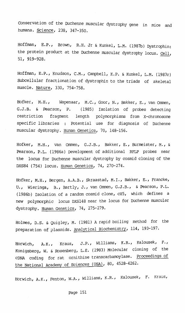
Conservation of the Duchenne muscular dystrophy gene in mice and humans. Science, 238, 347-350.
Hoffman, E.P., Brown, R.H. Jr & Kunkel, L.M. (1987b) Dystrophin: the protein product at the Duchenne muscular dystrophy locus. Cell, 51, 919-928.
Hoffman, E.P., Knudson, C.M., Campbell, K.P. & Kunkel, L.M. (1987c) Subcellular fractionation of dystrophin to the triads of skeletal muscle. Nature, 330, 754-758.
Hofker, M.H., Wapenaar, M.C., Goor, N., Bakker, E., van Ommen, G.J.B. & Pearson, P. (1985) Isolation of probes detecting restriction fragment length polymorphisms from x-chromosome specific libraries : Potential use for diagnosis of Duchennemuscular dystrophy. Human Genetics, 70, 148-156.
Hofker, M.H., van Ommen, G.J.B., Bakker, E., Burmeister, M., & Pearson, P.L. (1986a) Development of additional RFLP probes near the locus for Duchenne muscular dystrophy by cosmid cloning of the DXS84 (754) locus. Human Genetics, 74, 270-274.
Hofker, M.H., Bergen, A.A.B., Skraastad, M.I., Bakker, E., Francke, U., Wieringa, B., Bartly, J., van Ommen, G.J.B., & Pearson, P.L.(1986b) Isolation of a random cosmid clone, cX5, which defines a new polymorphic locus DXS148 near the locus for Duchenne muscular dystrophy. Human Genetics, 74, 275-279.
Holmes, D.S. & Quigley, M. (1981) A rapid boiling method for the preparation of plasmids. Analytical Biochemistry, 114, 193-197.
Horwich, A.K., Kraus, J.P., Williams, K.R., Kalousek, F., Konigsberg, W. & Rosenberg, L.E. (1983) Molecular cloning of the cDNA coding for rat ornithine transcarbamoylase. Proceedings of the National Academy of Sciences (USA), 80, 4528-4262.
Horwich, A.K., Fenton, W.A. , Williams, K.R., Kalousek, F. Kraus,
page 151

J.P., Doolittle, R.F., Konigsberg, w. & Rosenberg, l.E. (1984) Structure and expression of a complementary DNA for the nuclear coded precursor of ornithine transcarbamoylase. Science, 224, 1068-1074.
Hu, X-Y., Burgnes, A.B., Ray, P.N., Thompson, M. & Worton, R.G.(1987) Detection of internal gene duplications in Duchenne and Becker muscular dystrophies. American Journal of Human Genetics, 41, A294.
Jeffreys, A.J., Wilson, V. & Thein, S.L. (1985) Hypervariable 'minisatellite1 regions in human DNA. Nature, 314, 67-73.
Jeffreys, A.J., Wilson, V., Thein, S.L., Weatherall, D.J. & Ponder,B.A.J. (1986) DNA "Fingerprints" and segregation analysis of multiple markers in human pedigrees. American Journal of Human Genetics, 39, 11- 24.
Jobsis, A.G., van Duuren, C.Y., de Vries, G.P., Koppe, J.G., Rijken, Y., van Kempen, C.M.J. & de Groot, W.P. (1976) Trophoblast sulphatase deficiency associated with X-chromosomal ichthyosis. Nederlands Tijdschrift Voor Geneeskunde (Amsterdam), 120, 1980.
Keats, B.J.B., Morton, N.E., Rao, D.C. & Williams, W.R. (1979) A source book for linkage in man. Baltimore, Johns Hopkins University Press.
Keitges, E. & Gartler, S.M. (1986) Dosage of the Sts gene in the mouse. American Journal of Human Genetics, 39, 470-476.
Keitges, E., Rivest, M., Siniscalo, M. & Gartler, S.M. (1985) X-linkage of steroid sulphatase in the mouse is evidence for afunctional Y-linked allele. Nature, 315, 226-227.
Keitges, E., Schorderet, D.F. & Gartler, S.M. (1986) Linkage of the murine steroid sulphatase locus and the sex reversed mutation.American Journal of Human Genetics, 39, A118.
page 152

Kelly, R.G, Cozzarelli, n ., Deutscher, M.P., Lehrman, i.r . & Kornberg, A. (1970) Enzymatic synthesis of deoxyribonucleic acid: replication of duplex deoxyribonucleic acid by polymerase at a single strand break. Journal of Biological chemistry, 245, 39-45.
Kenwrick, S., Patterson, M., Spreer, A., Fischbeck, K. & Davies, K.(1987) Molecular analysis of the Duchenne muscular dystrophy region using pulse field gel electrophoresis. Cell, 48, 351-357.
Kerr, C. B. & Wells, R.S. (1965) Sex-linked ichthyosis. Annals of Human Genetics, 29, 33-50.
Kerr, C.B., Wells, R.S. & Sanger, R. (1964) X-linked ichthyosis and Xg groups. Lancet, ii, 1369-1370.
Kingston, H., Thomas, N.S.T., Pearson, P.L., Sarfazi, M. & Harper, P.S. (1983) Genetic linkage between Becker muscular dystrophy and a polymorphic DNA sequence on the short arm of the X chromosome. Journal of Medical Genetics, 20, 255-258.
Knowlton, R.G., Nelson, C., Brown, V.A., Page, D. & Donis-Keller,H.(1987) A highly polymorphic locus on Xp with homology to Yq sequences. Human Gene Mapping Nine (1987): Ninth International Human Gene Mapping Conference, A356.
Koenig, M., Camerino, G., Heilig, R. & Mandel, J.L. (1984) A DNA fragment from the human X chromosome short arm which detects a partially homologous sequence on the Y chromosome long arm. Nucleic Acids Research, 12, 4097-4109.
Koenig, M., Hoffman, E.P., Berteleson, C.J., Monaco, A.P., Feener,C. & Kunkel, L.M. (1987) Complete cloning of the Duchenne muscular dystrophy (DMD) cDNA and preliminary genomic organization of the DMD gene in normal and affected individuals. Cell, 50, 509-517.
Koh,J., Bartlett, R.J., Per lack-Vance, M.A., Speer, M.C., Yamakoa,
Page 153

L.H., Phillips, K., Hung, W.Y., Ray, P.N., Worton, R.G., Gilbert, J.R., Lee, J.E., Siddique, T., Kandt. R.S. & Roses, A.D. (1987) Inherited deletion at Duchenne dystrophy locus in normal male. Lancet, ii, 1154-1155.
Koppe, J.G., Marinkovic-Ilsen, A., Rijken, Y., De Groot, W.P. & Jobsis, A.C. (1978). X-linked ichthyosis. A sulphatase deficiency. Archives of Disease in Childhood, 53, 803-806.
Kostakov, St. (1934) Die progressive Muskeldystrophie, ihre Verebung und Glykokollbedandlung. Klin Wochens 13:219-221. Deutsche Archives fur Klinische Medizin, 176, 467-474.
Kunkel, L.M., Smith, K.D., Boyer, S.H., Borgaonker, D.S., Watchel, S.S., Miller, O.J., Breg, W.R., Jones, H.W. Jr. & Rary, J.M. (1977) Analysis of human Y chromosome-specific reiterated DNA in chromosome variants. Proceedings of the National Academy of Science (USA), 74, 1245-1249.
Kunkel, L.M., Monaco, P., Middlesworth, W., Ochs, H.D. & Latt, S.A. (1985a) Specific cloning of DNA fragments absent from DNA of a male with an X chromosome deletion. Proceedings of the National Academy of Science (USA), 82, 4778-4782.
Kunkel, L.M., Lalande, M., Monaco, A.P., Flint, A., Middlesworth, W. & Latt, S. (1985b) Construction of a human X-chromosome-enriched phage library which facilitates analysis of specific loci. Gene,33, 251-258.
Kunkel, L.M., & co-authors (1986) Analysis of deletions in DNA from patients with Becker and Duchenne muscular dystrophy. Nature, 322, 73-77.
Lange,K. & Boennke,M. (1982) How many polymorphic genes will it take to span the human genome? American Journal of Human Genetics,34, 842-845,
Page 154

Lanman, J.T., Periack-Vance, M.A., Bartlett, R.J., Chen, J.C., Yamaoka, L., Koh, J., Speer, M.C., Hung, W.Y., & Roses, A.D. (1987) Familial inheritance of a DXS164 deletion mutation from a heterozygous female. American Journal of Human Genetics, 41, 138-144.
Leder, P., Tiemeier, D. & Enquist, E. (1977) EK2 derivatives of bacteriophage lambda useful in cloning of DNA from higher organisms: The lambda gtWES system, Science, 196, 175-177.
Liechti-Gallati, S., Braga, S., Hirsiger, H. & Moser, H. (1987) Familial deletion in Becker type muscular dystrophy within the Xp21 region. Human Genetics, 267-268.
Lindenbaum, R.H., Clarke, G., Patel, C., Moncrieff, M. & Hughes, J.T. (1979) Muscular dystrophy in an X;1 translocation female suggests that Duchenne locus is on X chromosome short arm. Journal of Medical Genetics, 16, 389-392
Lindlof, M., Sistonen, P. & de la Chapelle, A. (1987) Linked polymorphic markers in the prediction of X-linked muscular dystrophy. Annals of Human Genetics, 51, 317-328.
Lyon, M.F. (1961) Gene action in the x-chromosome of the mouse (Mus musculus L.). Nature, 190, 372-373.
McKusick, V. (1986) Mendelian inheritance in man: Catalogues ofautosomal dominant, autosomal recessive and X-linked phenotypes, seveth edition, Baltimore, Johns Hopkins University Press.
McKusick, V. (1987) The human gene map, Baltimore, Johns HopkinsUniversity Press.
Maniatis, T., Fritsch, E.F. & Sambrook, J. (1982) Molecular cloning: A laboratory manual, New York, Cold Spring Harbour laboratory.
Page 155

Mann, J.D., Cahan, A., Gelb, A.G., Fisher, N., Hamper, j., Tippett,P., Sanger, R. & Race, R.(1962) A sex-linked blood group. Lancet, i, 8-10, '
Marcus, M., Trantavali, R., Dee, V.G., Miller, D.A. & Miller, O.J. (1976). Human—mouse cell hybrid with human multiple Y chromosomes. Nature, 262, 63- 65.
Marlhens, F., Chelly, J., Kaplan, J.C., Lefrancois, D., Harpey, J.P. & Dutrillaux, B. (1987) Familial deletion of Xp21.2 with glycerol kinase deficiency and congenital adrenal hypoplasia. Human Genetics, 77, 379-383.
Mattei, M.G., Mattei, J.F., Ayme, S. & Giraud, F. (1982) X-Autosome translocations: cytogenetic characteristics and their consequences. Human Genetics, 61, 295-309.
Mayall, B.H., Carrano, A.V., Moore, D.H., Ashworth, L.K., Bennett,D.E., & Mendelsohn, M.L. (1984) The DNA based human karyotype. Cytometry, 5, 376-385.
Maynard-Smith, S., Penrose, L.S., & Smith, C.A.B. (1961)Mathematical tables for Research workers in Human Genetics, Part II: Tables for use in linkage analysis, 47-71, London, Churchill.
Metaxotou, C., Ikkos, D., Panagiotopoulou, P., Alevizaki, M., Mavrou, A., Tsenghi, C. & Matsaniotis, N. (1983) A familial X/Y translocation in a boy with ichthyosis, hypogonadism and mental retardation. Clinical Genetics, 24, 380-383.
Meyer, W.J.III, Migeon B.R. & Migeon, C.J. (1975) Locus on human X-chromosome for dihydrotestosterone receptor and androgen insensitivity. Proceedings of the National Academy of Sciences (USA), 72, 1469-1472.
Middlesworth, W., Berteleson, C. & Kunkel, L.M. (1985) An RFLP detecting single copy x-chromosome fragment, dic56, from Xp22-Xpter
page 156

[HGM8 Assignment no. DXS143]. Nucleic Acids Research, 13, 5273.
Migeon, B.R., Shapiro. L.J., Norum, R.A., Mohandas, T., Axelman, j. & Dabora, R.C. (1982) Differential expression of the steroid sulfatase locus on the active and inactive human X-chromosome. Nature, 299, 838-840.
Mohandas,T., Shapiro, L.J., Sparkes, R.S. & Sparkes, M.C. (1979) Regional assignment of the steroid sulphatase-X-linked ichthyosis locus: Implications for a non-inactivated region on the short armof the human X-chromosome. Proceedings of the National Academy of Sciences (USA), 76, 5779-5783.
Mohandas, T., Sparkes, R.S., Hellkuhl, B., Grzeschik, K.H. & Shapiro, L.J. (1980) Expression of an x-linked gene from an inactive human X-chromosome in mouse-human hybrid cells: further evidence for the non-inactivation of the steroid sulphatase locus in man. Proceedings of the National Academy of Sciences (USA), 77, 6759-6763.
Mohandas, T., Sparkes, R.S. & Shapiro L.J. (1981) Reactivation of an inactive human X chromosome: Evidence for X-inactivation by DNA methylation, Science, 211, 393-396.
Mohandas, T., Sparkes, R.S. & Shapiro, L.J. (1982) Genetic evidence for the inactivation of a human autosomal locus attached to a inactive X chromosome. American Journal of Human Genetics, 34, 811-817.
Monaco, A.P., Berteleson, C.J., Middlesworth, W., Colletti, C.A., Aldridge, J., Fischbeck, K.H., Bartlett, R., Pericak-Vance, M.A., Roses, a .D. & Kunkel, L.M. (1985) Detection of deletions spanning the Duchenne muscular dystrophy locus using a tightly linked segment. Nature, 316, 842-845.
Monaco, A.P., Neve, R.L., Colletti-Feener, C., Berteleson, C.J., Kurnitt, D.M. & Kunkel, L.M., (1986) Isolation of candidate CDNAs
Page 157

for portions of the Duchenne muscular dystrophy gene. Nature, 323, 646-650. '
Monaco, A.P., Berteleson, C.J., Colletti-Feener, c. & Kunkel, L.M., (1987) Localization and cloning of Xp21 deletion breakpoints involved in muscular dystrophy. Human Genetics, 75, 221-227.
Mondello, C., Ropers, H.-H., Craig i.w., Tolley, E. & Goodfellow, P.N. (1987) Physical mapping of genes and sequences at the end of the human X chromosome short arm. Annals of Human Genetics, 51, 137-143.
Moser, H. (1984) Duchenne muscular dystrophy: Pathogenetic aspects and genetic prevention. Human Genetics, 66, 17-40.
Moser, H., & Emery, A.E. (1974) The manifesting carrier in Duchenne muscular dystrophy. Clinical Genetics, 5, 271-284.
Muller, C.R., Migl, B., Traupe, H. & Ropers, H.-H. (1980) X-linked steroid sulphatase: evidence for different gene-dosage in males and females. Human Genetics, 54, 197-199.
Murray, J.M., Davies, K.E., Harper, P.S., Meredith, L., Mueller, C.R. & Williamson, R. (1982) Linkage relationship of a cloned DNA sequence on the short arm of the X chromosome to Duchenne muscular dystrophy. Nature, 300, 69-71.
Nakamura, Y., Leppert, M., O'Connell, P., Wolff, R., Holm, T., Culver, M., Martin, C., Fujimoto, E., Hoff, M., Kumlin, E. & White,R. (1987) Variable Number of tandem repeat (VNTR) markers for human gene mapping. Science, 235, 1616-1622.
Neitzel, H. (1986) A routine method for the establishment of permanent growing lymphoblastoid cell lines. Human Genetics, 73, 320-326.
Noel, H., Beauregard, G., Potier, M., Bleau, G., Chapeldaine, A. &
page 158

Roberts, K.D. (1983) The target sizes of the in situ and solubilized forms of human placental steroid sulphatase as measured by radiation inactivation. Biochimica Biophysica Acta, 758, 88-90.
Norby, S. & Schwartz, M. (1987) Assignment of the locus order DXS28-DXS67-DMD as a spin-off from diagnostic marker analysis in a family with Duchenne muscular dystrophy. Clinical Genetics, 31, 192-197.
Old, J.M., Briand, P.L., Purvis-Smith, S., Howard, N. J., Wilcken, B., Hammond, J., Pearson, P., Cathelineau, L., Williamson, R. & Davies, K.E. (1985) Prenatal exclusion of ornithine transcarbamylase deficiency by direct gene analysis. Lancet, i, 73-75.
Pearson, P.L., Sanger, R. & Brown, J.A. (1974) Report of the committee on the genetic constitution of the X chromosome. Rotterdam Conferenceerence (1974): Second International Workshop on Human Gene mapping. Birth Defects: Original Article Series, XI, 3, 1975, 20-25. New York, The National Foundation.
Perrin, J.C.S., Idemoto, J.Y., Sotos, J.F., Maurer, W.F. & Steinberg, A.G. (1976) X-linked syndrome of congenital ichthyosis, hypogonadism, mental retardation and anosmia. Birth Defects Original Article Series, XII, 5, 267-274.
Petit, C., de la Chapelle, A. Levilliers, J., Castillo, S., Noel, B. & Weissenbach, J. (1987) An abnormal terminal X-Y interchange accounts for most but not all cases of XX maleness. Cell, 49, 595-602.
Pierella, P., Craig, I., Bobrow, M. & de la Chapelle, A. (1981) Steroid sulphatase levels in XX males including observations on two affected cousins. Human Genetics, 59, 87-88.
Race, R.R. and Sanger, R. (1975) Blood groups in Man, Sixth edition, chapter 29, Oxford, Blackford publications.
page 159

Ray, P.N., Belfall, B., Duff, C. Logan, C., Kean, V., Thompson, M.W., Sylvester, J.E., Gorski, J.L., Schmickel, R.D. & Worton, R.G.(1985) Cloning of the breakpoint of an X;21 translocation associated with Duchenne muscular dystrophy. Nature, 318, 672-675.
Read, A.P (1987) Update on the x-chromosome. Journal of Medical Genetics, 24, 188-189.
Renier, W.O., Nabben, F.A.E., Hustinx, T.W.J., Veerkamp, J.H., Otten, B.J., Ter Lak, H.J., Ter Haar, B.G.A. & Gabreels, F.J.M. (1983) Congenital adrenal hypoplasia, progressive muscular dystrophy and severe mental retardation in association with glycerol kinase deficiency in male sibs. Clinical Genetics, 24, 243-251.
Renwick, J.H. & Schulze, J. (1964) An anlysis on some data on the linkage between Xg and colourblindness in man. American Journal of Human Genetics, 16, 410-418.
Romeo, G., Roncuzzi, L., Ferlini, A., Pirozzi A. & Nobile, C.(1986) New mutations in Duchenne and Becker dystrophies. American Journal of Human Genetics, 39, A291.
Ropers, H.-H., Migl, B., Zimmer, J. & Muller, C.R. (1981) Steroid sulphatase activity in cultured fibroblasts of XX males. Cytogenetics and Cell Genetics, 30, 168-173.
Rouyer, F., Simmler, M.-C., Johnsson, C., Vergnaud, G., Cooke, H.J. & Weisssenbach, J. (1986) A gradient of sex-linkage in the pseudoautosomal region of the human sex chromosomes. Nature, 319, 291-295.
Royer-Pokora, B., Kunkel, L.M., Monaco, A.P., Goff, S.C., Newburger, P.E., Baehner, R.L., Cole, F.S., Curnutte, J.T. & Orkin, S.H. (1986) Cloning the gene for an inherited human disorder - chronic granulomatous disease - on the basis of its chromosomal
Page 160

location. Nature, 322, 32-38.
Rozen, R., Fox, J., Fenton, W.A., Horwich, A.L. & Rosenberg, L.E. (1985) Gene deletion and restriction length polymorph isms at the human ornithine transcarbamylase locus. Nature, 313, 815-817.
Saito, F., Goto,j., Kakinuma, H., Nakamura, F., Murayama, S., Nakano, I. & Tonomura, A. (1986) Inherited Xp21 deletion in a boy with complex glycerol kinase deficiency syndrome. Clinical Genetics, 29, 92-93.
Sanger, R., Tippett, P. & Gavin, J. (1971) The X-linked blood group system Xg: tests on unrelated people and families of Northern European Ancestry. Journal of Medical Genetics, 8, 427-433.
Schorderet, D.F., Keitges, E.A., Dobois, P.M. & Gartler, S.M.(1987) Inactivation and reactivation of the steroid sulphatase gene (Sts) in murine cell cultures. American Journal of Human Genetics, 41, A237.
Schwartz, D.C. & Cantor, C.R. (1984) Separation of yeast chromosome-sized DNAs by pulsed field gradient gel electrophoresis. Cell, 37, 67-75.
Shapiro, L.J.(1985) Steroid sulphatase deficiency and the genetics of the short arm of the human X chromosome. Advances in Human Genetics, 14, 331-381.
Shapiro L.J., Cousins, L., Fluharty, A.L., Stevens, R.L. & Kihara,H. (1977) Steroid sulphatase deficiency. Pediatric Research, 11, 894-897.
Shapiro L.J., Weiss, R., Webster, D. & France, J.T. (1978) X-linked ichthyosis due to steroid sulphatase deficiency. Lancet, i, 70 72.
Shapiro L.J., Mohandas, T., Weiss, R. & Romeo, G. (1979) Non-inactivation of an x-chromosome locus in man. Science, 204,
Page 161

1224-1226.
Shapiro, L.J., Yen, P.H., Marsh, B. & Mohandas, T. (1987) Frequent deletions at the steroid sulphatase locus. American Journal of Human Genetics, 41, A238.
Shows, T.B. & Brown, J.A. (1973) An (Xq-;9p+) translo.cation suggests the assignment of G6PD, HPRT and PGK to the long arm of the X-chromosome in somatic cell hybrids. New Haven Conference(1973): First International workshop on human genemapping. Birth Defects: Original Article Series, x, 3, 1974,146-149. New York, The National Foundation.
Shows, T.B. & Brown, J.A. (1974) Localisation of genes coding for PGK, HPRT and G6PD on the long arm of the X-chromosome in somatic cell hybrids. Rotterdam Conference (1974): Second International Workshop on Human Gene mapping. Birth Defects: Original ArticleSeries, XI, 3, 1975, 256-259. New York, The National Foundation.
Singer, M.F. (1982) SINEs and LINES: highly repeated short and long interspersed sequences in mammalian genomes. Cell, 28, 433-434.
Skinner,R., Smith C. & Emery A.E.H. 1974 Linkage between the loci for benign (Becker) X-borne muscular dystrophy and deutan colour blindness. Journal of Medical Genetics, 11, 317-20.
Skolnick, M.H. & Francke, U. (1981) Report of the committee on human gene mapping by recombinant DNA techniques. Human GeneMapping 6 (1981): Sixth International Workshop on Human GeneMapping. Birth Defects: Original Article Series 18, 2, 1982,194-204. White Plains, March of Dimes Birth Defects Foundation.
Smith, T.J., Wilson, L.J., Kenwrick, S.J., Forrest, S.M., Speer, A., Cotelle, C. & Davies, K.E. (1987) Isolation of a conserved sequence deleted in Duchenne muscular dystrophy patients. Nucleic Acids Research, 15, 2167-2174.
page 162

Solomon, i.l. & Schoen, E.J. (1971) Sex-linked ichthyosis in xo gonadal dysgenesis. Lancet, i, 1304-1305.
Somer, H., Voutilain, A., Knuutilla, S., Kaitila, I., Rapola, j. & Leinonen H. (1985). Duchenne-like muscular dystrophy in two sisters with normal karyotypes: evidence for autosomal recessiveinheritance. Clinical Genetics, 28, 151-156.
Southern, E.M. (1975) Detection of specific sequences among DNA fragments separated by gel electrophoresis. Journal of Molecular Biology, 98, 503-517.
Steckel, F., Hasilik, A. & von Figura, K. (1985) Multiple sulphatase deficiency: degradation of arysulfatase A and B afterendocytosis in fibroblasts. European Journal of Biochemistry, 151,147-152.
Stern, C. (1973) Principles of Human Genetics, 3rd edition, 484-485. San Franscisco, Freeman, W.H.
Sunohara, N., Sukuragawa, N., Sutoyoshi, E., Tanae, A. & Shapiro, l.J. (1986) A new syndrome of anosmia, ichthyosis, hypogonadism and various neurological manifestations with deficiency of steroid sulphatase and aryl sulphatase C. Annals of Neurology, 19, 174-181.
'Therman, E. & Patau, K. (1974) Abnormal x-chromosomes in man: origin, behaviour and effects. Humangenetik, 25, 1-16.
Thomas, N.S.T., Kunkel, L.M., Ray, P.N., Worton , R.G. & Harper, P.S. (1986a) Molecular deletion analysis in Duchenne muscular dystrophy. Journal of Medical Genetics, 23, 509-519.
Thomas, N.S.T., Williams, H., Elsas, L.J., Hopkins, L.C., Sarfarazi, M. & Harper, P.S. (1986b) Localisation of the gene for Emery-Dreifuss muscular dystrophy to the distal long arm of the X chromosome. Journal of Medical Genetics, 23, 596-598.
page 163

Thompson, C.E. (1978) Reproduction in Duchenne dystrophy, Neurology, 28, 1045-1047.
Thompson, M.W., Ray, P.N., Belfall, s., Duff, c., Logan, c., Oss,I. & Worton, R.G. (1986) Linkage analysis of polymorphisms within the DNA fragment XJ, cloned from the breakpoint of an X;21 translocation associated with X-linked muscular dystrophy. Journal of Medical Genetics, 23, 548-555.
Tiepolo, L., Zuffardi, 0., Fraccaro, M., di Natale, D., Gargantini, L., Muller, C.R. & Ropers, H.-H. (1980) Assignment by deletionmapping of the steroid sulphatase X-linked ichthyosis locus to Xp22.3. Human Genetics, 54, 205-206.
Vande Woude, G.F., Oskarsson, M., Enquist, L.W., Nomura, s. & Fischinger, P.J. (1979) Cloning of integrated Moloney sarcoma proviral DNA sequences in bacteriophage lambda. Proceedings of the Natinal Academy of Sciences (USA), 76, 4464-4468.
van Ommen, G.J.B., Verkerk, J.M.H., Hofker, M.H., Monaco, A.P., Kunkel, L.M., Ray, P., Worton, R.G., Wieringa, B., Bakker, E. & Pearson, P.L. (1986) A physical map of 4 million bp around the Duchenne muscular dsystrophy gene on the human X-chromosome. Cell, 47, 499-504.
Verellen-Dumoulin, C./ Freund, M., de Meyer, R., Laterre, Ch., Frederic, J., Thompson, M.W., Markovic, V.D. & Worton, R.W. (1984) Expression of an X-linked muscular dystrophy in a female due to a translocation involving Xp21 and non-random inactivation of the normal X-chromosome. Human Genetics, 67, 115-119.
Wahl, G,M., Stern, M. & Stark, G.R. (1979) Effecient transfer of large DNA fragments from agarose gels to diazobenzyloxymethyl-paper and rapid hybridisation by using dextran sulphate. Proceedings of the National Academy of Sciences (USA)/ 76, 3683-3687.
Walton, J.N. (1955) On the inheritance of muscular dystrophy (with
page 164

a note on the blood groups by Race, R. and a note on colour vision and linkage studies by Philip, u.). Annals of Human Genetics, 20, 1-38.
Walton, J.N. (1957) The inheritance of muscular dystrophy. Acta Genetica et Statistica Medica (Basel), 7, 318-320.
Wapenaar, M.C., Kievitis, T., Blonden, L.A.J., Bakker, E., den Dunnen, J.T., van Ommen, G.J.B. & Pearson, P.L. (1987) Hybrid cell-mediated cloning of a new intragenic sequence from the 3' region of the dmd gene. Human Gene Mapping Nine (1987): NinthInternational Human Gene Mapping Conference, A587.
Weatherall et al, D.J., Pembrey, M.E., Hall, E.G., Sanger, R., Tippett, P. & Gavin, J. (1970) Familial sideroblastic anaemia: Problem of Xg and x-chromosome inactivation. Lancet, ii, 744-748.
Wellauer, P.K. & Dawid, I.B. (1979) Isolation and sequence organization of human ribosomal DNA. Journal of Molecular Biology/ 128, 289-303.
Wieacker, P., Davies, K.E., Pearson, P.L. & Ropers, H.-H. (1983a) Carrier detection in Duchenne muscular dystrophy by use of cloned DNA sequences. Lancet, i, 1325-1326.
Wieacker, P., Davies, K.E., Mevorah, B. & Ropers, H.-H. (1983b) Linkage studies in a family with X-linked ichthyosis employing a cloned sequence from the distal short arm of the X chromosome. Human Genetics, 63, 113-116.
Wieacker, P., Voiculescu, J., Muller, C.R. & Ropers, H. H. (19 )An XX male with a single STS gene dose, rytoqenetics and CellGenetics, 35, 72-74.
Wieacker, P., Davies, K.E., Cooke, B.C., rearson, P.L., Williamson, R., Bhattacharya, S., Zinrer, J. & R°pers, H.-H. (1984) Toward a complete linkage nap of the human X chromosome: regional assignmen
Page 165

of 16 cloned single copy DNA sequences enploying a panel of somatic cell hybrids. American Journal of Human Genetics, 36, 265-276
Wieranga, B., Hustinx, T., Scheres, j., Renier, w. & ter Haar, B. (1985) Conplex glycerol kinase deficiency syndrome explained as a X-chromosomal deletion. Clinical Genetics, 27, 522-523.
Wilcox, D.E., Cooke, A., Colgan, J., Aitken, D.A., Sinclair, L., Glasgow, L., Stephenson, J.B.P. and Ferguson-Smith, M.A. (1986): Duchenne muscular dystrophy due to familial Xp21 deletion detectable by DNA analysis and flow cytometry. Human Genetics, 73, 175-180.
Willard, H.F., Skolnick, M.H., Pearson, P.L. & Mandel, J.-L. (1985) Report of the committee on human gene mapping by recombinant DNA techniques. Human Gene Mapping 8 (1985): Eight InternationalWorkshop on Human Gene Mapping. Cytogenetics and Cell Genetics, 40, 1-4, 360-489.
Wilson, E.B. (1911) The Sex chromosomes. Archiv Mikroskopische Anatomie Enwicklungs Geschichte, 77: 249-271.
Witkowski, J.A., Caskey, C.T., Hejtmancik, J.F., Baumbach, L., Gunnell, S. & Ward, P.A. (1987) Detection of new mutation at the DMD locus. American Journal of Human Genetics, 41, A325.
Worton, r .G. (1986) Reply to Dubowitz, V. "X;autosome translocations in females with Duchenne or Becker muscular dystrophy". Nature, 322, 292.
Worton, R.G., Duff, C. Sylvester, J.E. Schmickel, R.D. & Willard, H.F. (1984) Duchenne muscular dystrophy involving translocation of the dmd gene next to ribosomal RNA genes. Science, 224, 1447 1449.
Worton, R.G., Burghes, A.H.M., Logan, C., Belfall, B., Duff, C., Thompson, M.W. & Ray, P.N. (1987) The DXS206 locus is part of the Duchenne muscular dystrophy gene. Human Gene Mapping— Nine— (198 )_
page 166

Ninth International Human Gene Mapping Conference, A651
Wyman, A.R. & White,R. (1980) A highly polymorphic locus in human DNA. Proceedings of the National Academy of Sciences (USA), 77, 6754-6758.
Wyman, A.R., Wolfe, L.B. & Botstein, D. (1985) Propogation of some human DNA sequences in bacteriophage lambda vector requires mutant Escherichia coli hosts. Proceedings of the National Academy of Sciences (USA), 82, 2880-2884.
Yamamoto, K.R., Alberts, B.M., Benzinger, R., Lawhorne, L. & Treiber, G. (1970) Rapid bacteriophage sedimentation in the presence of polyethylene glycol and its application to large-scale virus purification. Virology, 40, 734-744.
Yang, T.P., Pragna, I.P., Chinault, A.C., Stout, J.T., Jackson, L.G., Hildebrand, B.M. & Caskey C.T. (1984) Molecular evidence for new mutation at the hprt locus in Lesch-Nyan patients. Nature, 310, 412-414.
Yates,J.R.W., Affara, N.A., Jamieson, D.J., Ferguson-Smith, M.A., Hausmanowa-Petrrusewicz, I., Zaremba, J., Borkowska, J., Johnstone, A. W. & Kelly, K. (1986) Emery-Dreifuss muscular dystrophy: localisation to Xq27.3->qter confirmed by linkage to the factor VIII gene. Journal of Medical Genetics, 23, 587-590.
Yates, J.R.W., Gillard, E.F., Cooke, A., Colgan J., Evans, J. & Ferguson-Smith, M.A. (1987a) Localisation of the gene for congenital adrenal hypoplasia. Journal of Medical Genetics, 24, 241.
Yates,J.R.W., Goudie, D.R., Gillard, E.F., Aitken, D.A., Affara, N.A., Clayton, J.F., Tippett, P.A. & Ferguson-Smith, M.A. (1987b) Multi-point linkage analysis of steroid sulfatase (X-linked ichthyosis) and distal Xp markers. Genomics, 1, 52-59.
page 167

Yen, P.H., Allen, E., Marsh, B., Mohandas, T., Wang, N., Taggart, R.T. & Shapiro, L.J. (1987a) Cloning and expression of steroid sulphatase cDNA and the frequent occurrence of deletions in STS deficiency: indications for X-Y interchange. Cell, 49, 443-454.
Yen,P.H., Marsh, B., Allen, E., Mohandas, & Shapiro, L.J. (1987b) Organization of the human steroid sulphatase gene and sequence homology betwen the X and Y chromosomes. American Journal of Human Genetics, 41, A248.
Youssoufian, H., Antonarakis, S., Aronis, S., Tsiftis, G., Phillips, D. & Kazazian, H.H.Jr. (1987) Characterization of five partial deletions of the factor VIII' gene. proceedings of the National Academy of Sciences (USA), 84, 3772-3776.
Page 168

APPENDICES.

F HO LN AM EH W2 A9 F Ho LN AM EH W2 A9
X23/GMGX8 X24/GMGX10
X30/GMGX19
X47/GMGX9
Appendix 1: Hybridisation of X-specific inserts to EcoRX somatic cell hybrid mapping panels (refer to table 4). Continued overleaf.
(F,female control. Hybrids are as described in materials and methods (and see inset). Probes are labelled with both the number of the picked plaque and the "GMGX" number assigned to it (see table 5) ).
Page 170

F Ho LN AM EH W2 A9 F Ho LN AM EH W2 A9
X53/GMGXY12
X38/GMGX20
X60/GMGX16X48/GMGX18
Appendix 1 (continued)
Page 171
X74/GMGX13

F Ho LN AM EH W2 A9 F HO LN AM EH W2 A9
I iiiilX66/GMGX11 X92/GMGX14
X110/GMGX23X94/GMGX15
X112/GMGX12
Appendix 1 (continued)Page 172

1
5C99
in
IV
5116I
n
i
5313
m 6 o ■NJ
n
m
i t ?
£ r T b
4907
SM
5311
9w i * 6PE
n
m
90120
n $ l )
AB
1
5265
1
5097
Appendix 2: Deletion studies in DMD/ BMD families based ondeletions of the pERT87 and XJ probes.
(A); Pedigrees of the nine boys with deletions detected by pERT87 and/ or XJ probes (refer to table 6).
Page V3

CQCQ
cn cn QO 2O'! lo LO ro [■"- O LO r- o LOcn i— 1 CO i—i o CN LO CT'i LM CTN
o O i— 1 ro i— I CN O X O'!LO lo LO ro LO O LO LO O LO
• • •■
GMGX9(PstI)
GMGXlO(Pstl)w m
GMGXll(Pstl)
Appendix 2: Deletion studies in DMD/ BMD families based ondeletions of the pERT87 and XJ probes.
(B); Hybridisation of GMGX9, GMGX10 and GMGXll to the deletion panel (refer to table 6).(M,male control. Other individuals are described in the text. These results are summarised in table 6).
Page V*

Ll-28 (Hindi)
Ll-4 (Hincll)
CQCQ
0303
<T\
Q
<T> LO m ro r- o inr-H 00 rH o C\| LO
O O 1-1 ro CTv r-1 fNJm m ro in O in
e'er!OL O
s£XO
LO<rO'!IT)
<TLO
754 (PstI)
Appendix 2: Deletion studies in DMD families based on deletions of the pERT87 and XJ probes. (C); Hybridisation of additional Xp probes to the deletion panel (refer to table 6). Continued overleaf.
(M,male control. Other individuals are described in the text. These results are summarised in table 6. Filters were PstI digests unless otherwise specified (in parenthesis).
Notes: The anomalous fragment detected by J47 in MJ is marked with an arrow. Anomalous fragment sizes detected by Ll-4 (Hinc II digests) in MJ and SJ (3485) may represent an RFLP, as a fragment of normal size was detected by Ll-4 in a Taql digest of MJ (refer to figure 6).
Page ^75

CQCQ
COCQ QnO'* MO lo ro r-~ o LO1-1 CO i— i o CM MO
o O rH ro cn 1— 1 CMLO LD LO ro lo o UO
sr-~ oU-lo XL.O O lo
PERT84
'
» •
XJ5.1
!
m m
XJ1.2
(J2.3
PERT87.1
Appendix 2C continued
Page 176
5194

CQCQ
CQCQ00oooLO
QT)asasoLD
LDco"sTCO
C)sCO r~~ o lD r-~ o MO1— 1 o CM MO oo 4-1 00ro as i— i CM o X ooLO o UQ 1-0 o ln
as
PERT87.30 * <Ul % •
JBir
Appendix 2C continued
Page 177

oM-lXoa'i cTioin
COwCT><X>o CQ LD CQ
coin
LOooro
t-" ro 1—1o i—i 1—1CTi ro roUO LDr-~a>oLO
LD O COCO CMCM i— 1LD O LD
754-11 (BamHI)
J47 (EcoRI)
JMD(PVUII)
XJlO.l(PstI)
J66HI (EcoRI)
Im m m
Appendix 2C continuedPage 178
5194

roLOLO
OOCT> LO r- I— Io CN 00 CMCD r~H CMro o
4Mi
•— - „_, ,— .Q EC ECO) W^ ^ •— --CTi 00 00 oO'* O'! CD cnO o i— iLO LO ro LO
Mi mm
CM CD O Oo ^^ r-H
ro ooCM CMcd ro ro ro
2 10 lo oLO
cdCTiLO
Appendix 3: Deletion studies in males with DMD/BMD with GMGXll.
(A); Hybridisation of GMGXll to Hindi 11 digests of all boys. (Individuals listed in table 7 are identified by their pedigree numbers).
Page 179

o CO CM1—1 00 LO1—1 ["- LOLO 00COi—I CM
COCOLOCOroLOLO
COCOOO00OLO
COCJV_00 CM LOoo r-o co CTiCM ooCOCTi00ro
S S& Qr-- 00 CO LOo CO CO (Ti1—1 00 CTCo oo r-~ oo
Appendix 3 continued.
Page

Oxfo(MJ)
Appendix 3 continued.
Page 18.1
4180

o> 0000 rg LD (j}r - r o
CO o r-CM rH LOoo OO LOoo LO LO
oco
JoCTiLD
LOLDLO
00OOLOLO
-— ' „__„CO coCO CJ<T\ 00 CNJ LO
oo r- COo f © CO ©LO V* CM 00LO o-© oCO rHOO O
Appendix 3: Deletion studies in males with DMD/BMD with GMGXll.
(B); Hybridisation of GMGXll to EcoRI digests of boys showing anomalous fragment sizes (refer to table 7). These are indicated by arrows (Individuals are identified by their pedigree numbers).
Page 182

r—I 03 r- o VOCN CN 00 03 03CN VO i—i 1—1 LON1 N1 o LD rHi—1 CNVO LO1—1 LDCN ro
o VO CO ln1— 1 03 vo 031—1 OO N* 03lO ro ro roLDO
jc>o03 I—I LD
N 1 rH r - Oro O CNLD LO 0 3 <— 1LD r - '- N 1 LO
**HB
CNOo LDON<roCNr-v oCN
03ULO0303ro
VOO'*03ro
I#M k - 8 **
Appendix 4: Deletion studies in males with DMD/BMD using p20, JBir and J66HI.(A); examples of hybridisation of p20 to Hindlll digests of boys with DMD/BMD (see table 7). (Boys are identified by their pedigree numbers. For those families in which more than one individual is affected, boys are further identified by their initials).
Page 183

H1rouoLOr-o 00roLOLO LOLO
m o r- om i— i LD 00o m LO 1— 1n* LO LO
CO Q l~3CO O-— * ■—03 03 LO o i— i 0303 03 o LM Osl CNO O N* X CN LOLO LO 1— 1 O
O'00
o03LO
LO r-H CN o03 LO LO i— iLO 1— 1 LO rHi— 1 CN m LO
LD03COro
j§COLOro
8LO0303ro
LOo
JC)o03 I—ILO
p20(Mspl)
Appendix 4: Deletion studies in males with DMD/BMD using p20, JBir and J66HI. (B); examples of hybridisation of p20 to Mspl digests of boys with DMD/BMD (see table 7).(Boys are identified by their pedigree numbers. For those families in which more than one individual is affected, boys are further identified by their initials. Note: the "doublet" observed with constant band 2 (1.5kb) was not seen in other Mspl digests probed with p20. This may be due to improved resolution of these particular gels (eg due to more prolonged electrophoresis) or to contamination in the batch of enzyme used for these digests. Track 1406 is partially digested).
Page I8*f

080
lined
0T£5
ZLte
luxed
(H3)06T5(HP)89fr£
8605
(QD6605
I
1
*!SfrOfr0frS5
83££
ZZ9Z IIttVi
90 1ZOOfr
T3
LT\c o
Q)CP04

00LO LOooLO0-
_________________
J66HI(ECORI
Appendix 4: Deletion studies in males with DMD/BMD using p20, JBir and J66HI. (D); exarrples of hybridisation of J66HI to EcoRI digests of boys with DMD/BMD (table 7).
(Boys are identified by their pedigree numbers. For those families in which more than one individual is affected, boys are further identified by their initials. Anomalous fragments are indicated by an arrow).
Page 186

553A-
m oil 6 6 6 ©JB
0198
II
in
3405
6 \h 6 6 6 6 ■ 6 6 (|) gin 6 6 u c5bWB
m
5098
6 □ 6 □ 6|T6 t5o 6 6
SR
5561
Appendix 5: Pedigrees of individuals with deletions or alteredrestriction fragment lengths detected by GMGXll/ p20/ JBir or J66HI. (Pedigrees are shown only for those individuals listed in table 7 and are presented in the same order as table _ 7. The pedigree for SS and JD (5099) is presented in appendix 2. The family history of MJ is not known). Continued overleaf.
Page 187

5538I
n
m
£2672
m 60107
tsck
m
7866
6 «AW
I INR
97589
p (j> 6 . 6 6 & j r 5540
NMcD
4045I
H
inFMcG
7501I
n
in
IV SMcK
4947
II
in
a 4033
6cTbbPS
Appendix 5 continued. Continued overleaf.
Page 188

6 - j > i 6
4 i*6<56 6 "sm m
I
X " ' X X 1
( S (3 ) * * 3
5120
PMin
3328
m
5310
I
5657
MT
1406
n
in
4180
I
n
4629
JB
0187
U
m
Appendix 5 continued. Continued overleaf.
Page 189

5190I
n
1
~ 6
21611
ffl
1596
DMcC
n
3552I
II
QI
II
01
^ ( S ’
5110
iTr □ (5T m Ik ’ u 6.AP
RW■L93468I DW 3995
1 m ■ □ 1cs
Appendix 5 continued.
page 190

I 40511=1 °1 o
1 cT1
lT 6“ i f x " I ■
^ 6 6 n q_cj>.
m 0 diSB m 6~ 6 m 6CK
16 6 6 6 *
n 6 6 x_iDP
r
Appendix 5 continued.Page “W

99.6 2287.30 3087.15 7.1/2.387.1 4.5
XJ2.3 7.8XJl.l 3.8XJ1.2 1.6XJ5.1 17754 9
754-11 6.3
oO
99.6 22 1387.30 30 887.15 9.4 7.1/2.387.1 4.5 1.8
XJ2.3 7.8 6.4XJl.l 3.8 3.1XJ1.2 1.6 2.0XJ5.1 17 24
754 9 11754-11 6.3 5.3
NJ
99.6 1387.30 null87.15 null87.1 null
XJ2.3 6.4XJl.l 3.1XJ1.2 2.0XJ5.1 24754 11
754-11 5.3
Appendix 6: RFLP analysis of the deletion in NJ (5313).
(A) Schematic representation of informative results.87.1, PERT87.1; 87.15, pERT87.15? 87.30, pERT87.30. The followingdigests were used 99.6, Pstl? pERT87.30, Bglli? pERT87.15, BamHi; PERT87.1, Mspl? XJ2.3, Tagi; XJl.l; Tag I; XJ1.2,- Bcli; XJ5.1, Sphi; 754, Pstl? 754-11; Bglli.
Page 192

GF M NJ
99.6(PstI)
GF M F NJ
XJl.2(BclI)
GF M NJ
XJ5.l(Sphl)
GF M NJ
pERT87.30(BglII)
GF M NJ
XJ2.3(TaqI)
GF M NJ
754(PstI)
GF M F NJ
PERT87.1(Mspl)
GF M NJ
f__
XJl.l (Tagl)
GF M NJ
754-ll(Bglll)
Appendix 6: RFLP analysis of the deletion in NJ (5313).
(B) Autoradiographs of results obtained. (pERT 87.15 results are not included. GF,grandfather; F,father; M,mother).
Page 193
(inferred)
99.6 13 87.30 30 87.15 6 87.1 4.5
XJ1.2 1.6 XJ5.1 2
754 9 754-11 6.3
SJ
o 99.6 22 2287.30 30 887.15 7.1/2.3 9.487.1 1.8 1.8
XJ1.2 1.6 2.0XJ5.1 17 17754 11 9
754-11 5.3 5.3
99.6 22 1387.30 30 3087.15 7.1/2.3 7.1/2.387.1 1.8 4.5XJ1.2 1.6 1.6XJ5.1 17 24754' 11 9
754-11 6.3 5.3
99.6 13 87.30 30 87.15 null87.1 null
XJ1.2 null XJ5.1 null 754 9
754-11 6.3
Appendix 7: RFLP analysis of the deletion in SJ (3485).
(A) Schematic representation of informative results. 87.1; pERT87.1*, 87.15; pERT87.15; 87.30, pERT87.30. The following digests were used 99.6/ Pstl; pERT87.30; Bglli; pERT87.15; BamHi; PERT87.1, Mspi; XJ1.2, Bell; XJ5.1; SphI? 754> Psti; 754-11; Bglli.
Page 19 -

GM M SJ GM M SJ GM M SJ
PERT87.30 (Bglli) PERT87.1
XJl.2(BelI) XJ5.1(SphI) 754(Pstl)
GM M SJ
754-ll(Bglli)
Appendix 7: RFLP analysis of the deletion in SJ (3485). (B)Autoradiographs of results obtained (pERT 87.15 results are not included. GM,grandmother; M,mother).
Page " 95

o *87.30 8 3087.1 1.8 4.5
XJ2.3 null 7.8 XJl.l null 3.8 XJ1.2 null 1.6 XJ5.1 null 24 754 11 9
754-11 5.3 6.3
k 87.30 8 3087.1 1.8 1.8
XJ2.3 null 6.4 XJl.l null 3.1 XJ1.2 null 2.0 XJ5.1 null 17 754 11 11
754-11 5.3 5.3
87.30 887.1 1.8
XJ2.3 nullXJl.l nullXJ1.2 nullXJ5.1 null754 11
754-11 5.3
(* phase inferred from AB)
Appendix 8: RFLP analysis of the deletion in AB (0120).
(A) Schematic representation of informative results.87.1, pERT87.1; 87.15/ pERT87.15; 87.30/ pERT87.30. The following digests were used pERT87.30, Bglli; pERT87.1/ Mspl; XJ2.3, Tagi; XJl.l, Tagi* XJ1.2, Bcli; XJ5.1; Sphi; 754, Psti; 754-11, Bglli.
Page 196

XJ2.3 (Tagi) XJl.l(Tagi) XJl.2(BclI)
754(Pstl) 754-ll(BglII)XJ5. KSphI)
Appendix 8: RFLP analysis of the deletion in AB (0120).
(B) Autoradiographs of results obtained (pERT 87.1 results are not included. GM/grandmother; M,mother; C;control female).
Page 197

U3COS Cn co r-~
r-
(A) XJl.2(BelI)(B) p20(Mspl)
coLO co U S CO colo coco lo O
(C) PERT87-1(Mspl) pERT87.30(BglII) XJ5.1(SphI)
Appendix 9: RFLP results for the sisters of JB (5097), PE (5311) and AW (7866).
(A) XJl.2 (Bell) results for the sister of JB (5097).
(B) p20 (Mspl) results for the sister of AW (7866).
(C) pERT87.1 (Mspl), pERT 87.30 (Bglli) and XJ5.1 (SphI) results for the sister of PE (5311).
(C;female control; F/father; M,mother; S,sister).
Page 98

00r-CMunoi—HCTiOO
inoCTiOO
c mr-c mto G 1
(i)
(ii) 3825(De)
G 1 2 3 4 G F 1 2 3
(iii) 3906(Ja)(iv) 5272(Ni)
Appendix 10: Results obtained with GMGX9 in TDH Scottish pedigrees segregating for XLI.
families and six
(A) (i) Hybridisation of GMGX9 to mothers from TDH families.(Heterozygous individuals are identified by their pedigree numbers).(ii) Hybridisation of GMGX9 to family 3825 (De). (iii) Hybridisation of GMGX9 to family 3906 (Ja). (iv) Hybridisation of GMGX9 to family 5272 (Ni).
(G;grandfather; F,father; 1-4, first to fourth sons respectively. The result for the second son in family 3825 (De) is not included).
Page 199

I B 9 5273 Gr
AB (|)
1 A A B B
I A 9 3822Ca i B 5 3904Tu
9n LJ AB
A B B
AB(|)
M rh p i p- B B A B
3825 DeI
AB'I
1
I B
n LJab
Q 3910 Ho
I
9B B B
3906 Ja
Q
1 B ABA
I B 9 5272 Ni
ab(|)
B B A
Appendix 10: Results obtained with GMGX9 in TDH families and six Scottish pedigrees segregating for XLI.
(B) Schematic illustration of results with GMGX9 in TDH families informative for GMGX9. ("A",4kb allele; "B",'2.5/1.5kb allele).
Page 200

I
II
H
3673
n
I m
2994
sc
i
IV
9 4024
A AC ) A H A ■ ■ ■ a6
Ap o (j) g * 9 3710
A | A TR
*6 ■ -■ JG
91034
■TiiDG
Appendix 10: Results obtained with GMGX9 in TDH families and sixScottish pedigrees segregating for XLI.
(C) Schematic illustration of results with GMGX9 in six of the Scottish families segregating for X-linked ichthyosis. ("A",4kb allele; "B", 2.5/1.5kb allele; deletion).
Page 201

m
UAL
n / b bQ a B( ) A
6 6 6 o i a S i i i- A - - - B
“ ( ^ “ V 11, 111! 111, n 7 n 6 III10III12IIT13II2 “ j
Appendix 11: Hybridisation of GMGX9 to the Italian family (ITAL).
(A) Schematic representation of the results obtained. ("A" 4kb allele; "B",-2.5/1.5kb allele; deletion).
(B) Autoradiograph of hybridisation with GMGX9. (This blot was provided by A. Ballabio.)
Page 202

IE
IV 1/85 Ln
S>AB
i . L I
iv
*9QM B H A ABC) ' AB
£6 6 2/85Br
g a (|) [p a < > g a(B) GD<55 1 tra-fi
n™1©3/85 Ls
Appendix 12: Deletion analysis with GMGX9 in German and Dutchfamilies segregating for XLI.
(A) Schematic representation of the segregation of GMGX9 in German families. ("A">4kb allele; "B",2.5/1.5kb allele; deletion).Continued overleaf.
Page 203

c^lII B M A
T B9
> CD 9O b 5^5 Ge
HI io 6 6 6/85 Sc
T 9 1A9T <> lv d c= 4 ± AL cS -1
in r b 7/85 Gu
ffl ■ ■ o 9/85 Do
IV " ■ 10/85 01
9. A
b
i k b ■ □ bMa
Appendix 12A continued.
Page 20A

1/85 2/85 3/85
c) pO QOB B 2 -
C 1 2 3 4 Cm r n m m
u t t B *
B)
i) GMGX9(HindiII)
GMGX9(HindiII)
(ii) GMGYlO(HindIII
Appendix 12: Deletion analysis with GMGX9 in German and Dutch families segregating for XLI.
(B) Deletion analysis with GMGX9 in four Dutch males with XLI. (C; male control).
(C) Hybridisation with GMGX9 and GMGY10 to members of three German families, (i) Hybridisation with GMGX9. (ii) Hybridisation of the same filter with a Y-specific probe GMGY10 (Affara et al, 1986).
Page 205

VOr-~OLO00vor-
[•'-r-~
■rt'-r-~
oooro
rn^m X
• Y
(i) STB14(HindiII)
VO or-~ oo o-st* ro r-^ lo o ^ o>r"~ oo o r-~ r-^ vo o or-" co r-~ o r-
Qlo in o o vc> voO r- r-~
oror-'
^ 5 Qvo vo mLD ino O oCO CO ^
• • • •
(ii) Dic56(BclI)
Appendix 13: Examples of hybridisations with distal Xp probes to males with XLI (refer to table 9). Continued overleaf.
(i) Hybridisation of STB14 to affected males and relatives from families 4076, 3000, 7474 and 7685. ((M),mother of affected male).
(ii) Hybridisation of Dic56 to the individuals shown in (i) above and also to a male with an XY translocation (7079) and individuals from families 7605, 7304, 8056 and 4076, which segregate for XLI. (C;female control; (M),mother of affected male; (D),daughter of affected male).
Page 206

Q Q— * ■— ✓
LO LO LO LOo o r- o-LO LO o oo- o 1
soo o' LO LO LO LO CT\ ro O CNLO LO o LO CN CN (N O o o-LO r- ro O LO LO LO O rH o LOo- o- o- 00 LO LO LO 00 ro O'
mw
(iii) p!9b(TaqI)
oo LO CO O' O ’ N* 00 LO 00 O'CTL ro CN OL OL o CTL ro CN O'! CTL OCTi o O ro LO ro Ch o O ro ■N* LO roCN i—I i— l ro O ro CN 1— 1 i— I ro O ro
X
(iv) MlA(EcoRI)
(v) p2a4(EcoRI)
Appendix 13 (continued): Examples of hybridisations with distal Xp probes to males with XLI (refer to table 9).
(iii) Hybridisation of pl9b to affected males from families 7605,4076, 7664, 7750, 7304, 8056, 5626, 8009, 4143, 3000. (C, femalecontrol; (M),mother of affected male; (GM),grandmother of affected male; (D)/daughter of affected male)
(iv) Hybridisation of MlA to affected males from families 2994,1034, 4024, 1348, 3496, 0698 and 3307.
(v) Hybridisation of p2a4 to the affected males indicated in (iv) above).
Page 207
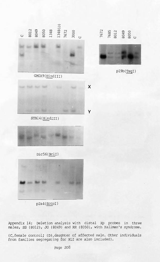
Q
GMGX9(HindiII)
STBl4(HindIII)
Dic56(BclI)
p2a4(EcoRI)
Appendix 14: Deletion analysis with distal Xp probes in threemales, SS (8012), JG (8049) and RR (8050), with Kallman's syndrome.
(C,female control; (D),daughter of affected male. Other individuals from families segregating for XLI are also included).
Page 208

CJ
CTi CTir- r-o oi"" r~-
GMGX9(HindiII)
cn SN--- *—ct CTiCN CN CNr~~ t-~ t-~i—i i—i i—1
UL|
<j\ a> <y\cn r~ r-r- o or-\ r- r-~
p!9b(PvuII)
CJ
a> o r-~ r- o o r- r~
•
■ ■ •
MIA(Hindlll)
,—_ „_„ '—- ___ ,—.. __s2 cn s cn 2-_ %_- CT> - ■— -_ '—■ ___01 CTi O'! cr\CN CN CN CN r-~ CN CN CN CN r-r~~- r-~ r- S U r~~ r- r- r-~i— i c—1 i— i i— i r- t— i i— i t— i i— i i"- r-- O
ft*m
m
x
GMGX9(HindIII)
CJ
OsCNr-~Scr>CNr-
cncCNr-
STBl4(HindIII)
p!9b(TagI
Appendix 15: Deletion analysis with distal Xp probes in two males, AM (1729) and JH (7079) with XY translocations.
(C,-female control; (M),mother; (F);father? (S),sister. A male with XLI from family 7474 is also included).
Page 2°9
[Gl a s g o w " ' UNIVERSITY
R h a b v