
Saccadic palsy after cardiac surgery: characteristics and pathogenesis
Upload
khangminh22Category
view
1download
0
1

Contact address
◼ Department of Plant Protection, Faculty of Agricultural Sciences and Food Industries, Science & Research Branch, Islamic Azad University, Tehran-Iran.
◼ P.O. Box: 14155/775, Postal Code: 1477893855 ◼ Branch website: www.srbiau.ac.ir◼ e-mail addresses:◼ [email protected]◼ [email protected]
2

Table of ContentsSubject Index for Part 2 of bacterial pathogenesis on plants
◼ Bacterial pathogenesis on plants - Pathogen derived molecules - Part 2◼ Secreted proteins:◼ Pathogenicity islands (PAIs and virulence genes)◼ Genes involved in pathogenicity and host specificity: Chromosome or Plasmid? ◼ Plasmid-borne traits◼ Insertion Sequences (IS)◼ Apoptosis (a form of programmed cell death, or cellular suicide)◼ Plant strategies for defence: ◼ Bacterial pathogenesis on plants - Plant derived molecules◼ R genes➢ Polygalactoronase inhibiting proteins(PGIP)➢ Anti-microbial proteins➢ Phytoalexins➢ Pathogenesis related proteins or PR proteins (PRs)➢ LAR or SAR◼ Plant defenses◼ Some of the most common types of plant resistance1. Innate resistance2. Preformed resistance 3. Acquired resistance
3

Table of Contents
◼ Host and non-host resistance◼ Mechanisms of general defenses➢ Passive (constitutive) defense (First line defence)➢ Active (inducible) defense (Second line defence)
1. Rapid responses implicated in resistance2. Slower responses implicated in resistance◼ Plant-bacteria interactions◼ Elicitor recognition (PAMPs or MAMPs/PRRs interactions)◼ Gene-for-gene hypothesis (Avr/R interactions)◼ Elicitors1. Direct receptor-elicitor interaction 2. Indirect effector interactions(Guard, Decoy and Integrated decoy models)◼ Cell signaling◼ Signal transduction: MP kinase activity Genes◼ Local resistance (HR…)◼ Systemic resistance (LAR or SAR)
4

Table of Contents◼ Bacterial pathogenesis on plants-Pathogenicity/virulence mechanisms of
important plant bacteria
◼ Pathogenicity of:
◼ Agrobacterium tumefaciens
◼ Erwinia amylovora
◼ Pectobacterium carotovorum
◼ Pectobacterium wasabiae
◼ Pantoea (ex.Erwinia) agglomerans pv. gypsophilae
◼ Pantoea (ex.Erwinia) agglomerans pv. betae
◼ Dickeya (ex. Erwinia) chrysanthemi
◼ Pseudomonas syringae
◼ P. savastanoi pv. savastanoi
◼ Ralstonia solanaceraum
◼ Xanthomonas spp.(Xac, Xcc, Xcv, Xoo and Xtt)
◼ Clavibacter michiganensis subsp. michiganensis
◼ Rhodococcus fascians
5

Table of Contents◼ Leifsonia xyli subsp. xyli
◼ Xylella fastidiosa
◼ Candidatus Liberibacter
◼ Spiroplasma citri◼ Phytoplasmas◼ General terms and abbreviations◼ Selected references
6

Bacterial pathogenesis on plantsPathogen derived molecules- Part 2
The path of bacterial plant pathogenesisMicrobial Strategies for Attack
◼ Pathogenicity islands(Pai)◼ Effectors:1. avr genes2. hrp/hrc genes3. Other virulence genes/proteins◼ Chromosome or plasmid-borne of pathogenicity genes◼ Delivery of effectors via secretion systems (Types III, IV and VI).
7

Pathogenicity islands (PAIs)Pathogenenicity islands are discrete genetic loci that encode factors which make a microbe more virulent
The genetic element, the "island of evil", within the genome of an organism that is responsible
for its capacity to cause disease.
Identification of PAIs is essential in understanding the development of disease and
the evolution of bacterial pathogenesis.
8

Pathogenicity Islands (PAIs)A distinct class of genomic islands acquired by microorganisms through horizontal gene transfer
◼ The islands of pathogenic bacteria affecting and causing diseases on plants, animals and humans.
9

Book citedPathogenicity islands and other mobile virulence elements
◼ Pathogenicity Islands And Other Mobile Virulence Elements
◼ James B. Kaper, Jörg Hinrich Hacker
◼ ASM Press,
◼ 1999
◼ Medical
◼ 352 pages.10

Book citedHorizontal Gene Transfer in the Evolution of Pathogenesis
◼ Horizontal Gene Transfer in the Evolution of Pathogenesis
◼ Part of Advances in Molecular and Cellular Microbiology.
◼ Michael Hensel and Herbert Schmid,
◼ Cambridge University Press, 2008
◼ 342 pages.11

Book citedPathogenicity island
◼ Pathogenicity Island
◼ Lambert M. Surhone, Mariam T. Tennoe, Susan F. Henssonow
◼ Betascript Publishing
◼ 2010
◼ 64 pages
12

Book citedE. coli: Molecular phylogeny and pathogenicity islands: Molecular phylogeny and pathogenicity islands of E. coli
◼ E. coli: Molecular phylogeny and pathogenicity islands: Molecular phylogeny and pathogenicity islands of E. coli.
◼ Mohammed sabri and Lamees Abdul-Razzak.
◼ LAP LAMBERT Academic Publishing
◼ 2012
◼ 168 pages.
13

Book citedPathogenicity islands and the evolution of pathogenic microbes
◼ Pathogenicity Islands and the Evolution of Pathogenic Microbes
◼ Volume 1
◼ James B Kaper & Jorg Hacker(Eds.)
◼ Springer
◼ 2013
◼ 444 pages.
14

Book citedPathogenicity Island
◼ Pathogenicity Island
◼ Jesse Russel, Ronald Cohn
◼ First published: 2013
◼ Published by Book on Demand, Miami
◼ 2015
◼ 76 pages.
15
Bookseller Image

Book citedBacterial Pathogenicity Islands
◼ Bacterial pathogenicity islands.
◼ Mahmoud Zaky and Nada Abousamra
◼ Publisher: LAP LAMBERT Academic Publishing
◼ 2016
◼ 188 pages.
16

Book citedAn Innovative Approach to Study Ralstonia solanacearum Pathogenicity
◼ An Innovative Approach to Study Ralstonia solanacearum Pathogenicity
◼ Niraj Singh
◼ Publisher: Scholars' Press
◼ 2020
◼ 68 pages.
17

Prokaryotic genomesDNA pools in the genomes of prokaryotesCore and Flexible gene pools
◼ Proposed model of the DNA pools in the genomes of prokaryotes.
◼ Most of the horizontally transferred DNA is part of the “flexible” gene pool.
◼ Some functions encoded by the pools are given in the lower part of the diagram.
◼ Turnover metabolism is a dynamic process. The cell is continuously degrading and synthesizing molecules.
18Hacker and Carniel,2001; Scortichini,2005
Model of the DNA pools in the genomes of prokaryotes. The DNA elements comprising the core as well as the flexible gene pools are presented in the circles. Functions encoded by the pools are given in the lower part of the diagram.
Core genome: the genes shared by all members of a pre-defined group of bacteria or archaea. Flexible genome may also be referred to as the accessory, variable, dispensable, auxiliary, noncore, adaptive or distributed genome.

Genome of prokaryotesGenomic islands(GISs)
19
◼ Genomic islands(GEIs) are clusters of genes within a bacterial genome that appear to have been acquired by horizontal gene transfer.
◼ GEIs often carry genes offering a selective advantage for host bacteria.
◼ To this date, several dozen genomic islands have been described and it is expected that many more await discovery.
◼ In 50% of the marine bacterial genomes analyzed the GIs accounted for approximately 3% of the genome length, with a maximum of 12%.
Genome size in Escherichia coli is 4.6 million base pairs (Mbp)=4600 KB.

Genome of prokaryotesGenomic islands(GISs)
20
◼ The distinct properties of GIs and that they allow bacterial organisms to evolve and adapt to different environments.
◼ The genes present in GIs are typically grouped to perform specific and advantageous functions in the bacteria.
◼ PAIs, for example, can cause major changes in the bacterial phenotype. Thus, they are the most studied Gis.
◼ GI number per genome was strongly and positively correlated with the total GI size.

Genome of prokaryotesSubgroup of genomic islands(GISs)
21
◼ The large number of sequenced genomes and analyses of genetic sequences have revealed that GIs are mosaics of genes formed by HGT.
◼ Genomic islands(GEIs) encoded some islands.
◼ These include:
1. Pathogenicity islands (PAIs)
2. Antimicrobial resistance islands (REIs)
3. Symbiosis islands
4. Pathogen/ecological fitness islands
Also, it can be of other functions like carbon and nitrogen sources utilization(metabolic islands).

Genome of prokaryotesGenomic islands(GISs)
22
◼ Gnomic island (GI) is usually used in microbiology, especially with regard to bacteria.
◼ A genomic island (GI) is part of a genome that has evidence of horizontal origins.
◼ These "islands" are characterized by:
1. their large size(>10 Kb),
2. their frequent association with tRNA-encoding genes, and
3. A different G+C content compared with the rest of the genome.
Wikipedia,2016

Genomic islandsGenomic islands encode different functionsGenomic or metabolic islands and smaller inserts
23
◼ Genomic islands(GIs):
1. Large segments (10-200 kb) of foreign DNA inserted into bacterial chromosomes are often referred to as genomic islands (GEIs).
2. Smaller inserts (1-10kb) have also been found and may be termed genomic islets.
◼ Genomic island and its subgroup PAIs are part of that flexible gene pool.
◼ Genomic elements with characteristics similar to PAI but lacking virulence genes are referred to as genomic or metabolic islands.
Van Sluys et al.,2002; Hentschel and Hacker,2001

Genomic islandsGenomic islands encode different functions
24
1. Many genomic islands (GEIs) are flanked by repeat structures and carry fragments of other mobile and accessory genetic elements, such as bacteriophages, plasmids and insertion sequence (IS) elements.
◼ These functional or cryptic genes encoding integrases or factors related to plasmid conjugation systems or phages involved in GEI transfer.
1. But some genomic islands can excise themselves spontaneously from the chromosome and can be transferred to other suitable recipients.
◼ A hypothetical 'life cycle' of GEIs includes the insertion of mobile genetic elements into the bacterial chromosome.
Dobrindt et al.,2004; Wikipedia,2016

25Dobrindt et al.,2004
Genomic islands encode
different functions.

Genomic islandsGenomic islands encode different functions
26Campbell and co-workers;..
◼ Genomic islands present in the majority of genomes of pathogenic as well as nonpathogenic bacteria and may encode many different functions.
◼ The size of genomic islands may vary from a few Kb to as many as 200 Kb.
1. A different G-C content compared with the rest of the chromosome; biased codon usage.
2. often mobile (genomic islands have been acquired by horizontal gene transfer, HGT).
◼ GIs encode accessory functions such as metabolic activities, antibiotic resistance, or properties involved in microbial fitness, symbiosis, or pathogenesis.

Genomic islandsCodon usage bias is a result of a balance of mutation and selection. The degree of codon usage bias for each gene/bacterium is different
◼ A large amount of varied ways, all amino acids (aa) are coded by two to six synonymous codons, except Met and Trp.
◼ This biased use of codons has been observed in all branches of life.
◼ The redundancy of the genetic code is due to multiple codons which encode the same amino acid.
◼ Codon bias is the result of long-term selection and is presumed to confer an evolutionary advantage.
◼ Codon usage bias is selected to optimize the speed of protein production.
27

Genomic islandsCodon usage bias is a result of a balance of mutation and selection. The degree of codon usage bias for each gene/bacterium is different
◼ A codon is a series of three nucleotides (a triplet) that encodes a specific amino acid residue in a polypeptide chain or for the termination of translation (stop codons).
◼ The overabundance in the number of codons allows many amino acids to be encoded by more than one codon.
◼ Because of such redundancy it is said that the genetic code is degenerate.
28
Due to the degeneracy of the genetic code, most amino acids can be encoded by
multiple synonymous codons. Synonymous codons naturally occur with different frequencies in different organisms.
Redundancy: the state of being not or no longer needed or useful.

Genomic islandsCodon usage bias: an important evolutionary feature in a genome
◼ Because a high amount of codon usage bias in bacteria is believed to be the result of selection for translational efficiency.
◼ Codon usage bias (CUB) is an important evolutionary feature in a genome which provides important information for studying organism evolution, gene function and exogenous gene expression.
29
Distant phylogenetic species usually has greater variations in codon usage bias.

Genome of prokaryotesCodon usage bias(CUB)
◼ Translation in the ribosome and tRNA structure
◼ Cartoon of the ribosome(green) during translation of a mRNA (blue) with a wobbling codon-anticodon base pair encoding a leucine amino acid.
◼ A site: aminoacyl-tRNA site; E site: exit site;
◼ P site: peptidyl-tRNA site.
30
Wobble: pairing of the tRNA anticodon with the mRNA codon proceeds from the 5' end of the codon. In this example, the double-ringed G can pair with either a single-ringed U or C. Each organism seems to prefer a different set
of codons over others; this phenomenon is called codon bias.

Genome of prokaryotesCodon usage bias(CUB)
◼ Several important variations of codon bias have recently been discovered, such as:
1. the existence of a ramp of rare,
2. slowly translated codons at the 5′ end of protein-coding sequences, and
3. the co-occurrence of certain codons.
◼ Apart from directly affecting general protein expression levels, it has been established that codon bias also influences:
1. protein folding, and
2. differential regulation of protein expression.
31Quax et al.,2015

Genome of prokaryotesCodon usage
◼ Different Types of Codon Bias
A. Frequency bias will result in effective protein production when the frequency of used codons matches the cellular tRNA population.
B. Co-occurrence bias enhances protein expression, presumably because of tRNA recharging in the vicinity of the translating ribosome.
C. Pair bias is probably selected because of more optimal interactions of tRNAs in the A site and the P site.
32Quax et al.,2015

Genome of prokaryotesSubgroup of genomic islands(GISs)Mobility genes, such as integrases (int), are frequently located at the beginning of the island, close to the tRNA locus or the respective attachment site
33
General features of GEIs. GEIs are relatively large segments of DNA whose nucleotide characteristics often differ from the rest of the chromosome. GEIs are often inserted at tRNA genes and flanked by direct repeat sequences (DR).
GEIs typically harbour genes encoding factors involved in genetic mobility, such as integrases, transposases and insertion sequences (IS). According to their gene content, GEIs can be described as pathogenicity, symbiosis, metabolic, fitness or resistance islands.
Juhaz et al.,2009

Genomic islandsFitness islands and saprophytic islands
◼ The fitness islands are classified into several subtypes based on the life style of the microbes that harbor them. E.g.
1. ecological islands: in which the islands contribute to the survival of the microbe in a environment.
2. Other fitness that are related with microbe-host interaction. These are:
➢ Saprophytic islands: in microorganism can persist as a saprophyte in a host.
➢ In many case, the fitness factor temporarily or prermanetely resides in the host either providing some benefits (symbiosis islands) or cause damage (pathogenicity islands).
34Safwat Mohammad,2014;..

Genomic islandsFitness islands
◼ Genomic islands increase bacterial fitness.
◼ The ability of a bacterial strain to infect a host, persist, proliferate and transfer to a new host in a specific niche is known as bacterial pathogen fitness.
◼ Fitness islands contribute to fitness and adaptation of the strains.
◼ A simultaneous acquisition of many genes by HGT that allow the bacteria to rapidly gain complex virulence functions and to exploit new environmental niches increasing bacterial fitness.
35Safwat Mohammad,2014

Genomic islandsOccurrence and significance of pathogenicity and fitness islands in environmental vibrios
◼ a–c Circular presentation of the second chromosome of three Vibrio spp.
◼ Track 1, forward coding sequences; track 2, reverse coding sequences; track 3, tRNA genes; track 4, red, pathogenicity islands, blue, genomic fitness islands; track 5, virulence and virulence-associated genes; track 6, genes involved in toxin–antitoxin systems; track 7, mobile genetic elements.
◼ Virulence and virulence-associated genes are numbered and are defined via the center text boxes.
36Kelin et al.,2018
Track

Genomic islandsFitness islands
◼ Fitness genes are associated with entry and survival in the host.
◼ In nonpathogenic bacteria, these DNA segments may act as fitness islands or ecological islands.
◼ These encodes for:
1. iron uptake systems.
2. an exopolysaccharide (EPS) that is also involved in biofilm formation.
3. determining the identity, origin and evolution of traits that contribute significantly to fitness in plant-associated bacteria.
37

Genomic islandsPathogenicity islands is a subclass of genomic islands
38
◼ Pathogenicity islands(PAIs) are a subset of genome islands.
◼ When the island contains pathogenicity genes it is called a pathogenicity island.
◼ PAIs are a distinct class of genomic islands which are also acquired by horizontal gene transfer.
Van Sluys et al.,2002; Hentschel and Hacker,2001

Genomic islandsPathogenicity islands is a subclass of genomic islands
39
◼ In the early 1980s large unstable chromosomal regions carrying virulence-associated genes were identified in uropathogenic E. coli (pathogen of urinary tract).
◼ Later, such large unstable chromosomal regions were designated pathogenicity islands (PAIs).
◼ Pathogenicity islands (PAIs), as termed in 1990.
◼ These are a distinct class of genomic islands.
◼ They are incorporated in the genome of pathogenic bacteria.
Schneider et al.,2011;..

Genomic islandsPathogenicity islands is a subclass of genomic islands
40
◼ Their instability and the high level sequence similarity of different (partial) islands suggest an exchange of PAIs between strains of the same or even different bacterial species by horizontal gene transfer (HGT).
◼ PAIs comprise large genomic regions (10-200 kilobases in size) that are present on the genomes of pathogenic strains but absent from the genomes of nonpathogenic members of the same or related species.
Schneider et al.,2011;..
Genome size in Escherichia coli is 4.6 million base pairs (Mbp)=4600 KB.

Pathogenicity Islands Distinct class of genomic islandsLarge, mosaic, genetic islands, found in all pathogenic bacteria
◼ The genomes of prokaryotes are highly diverse mosaic structures, contains:
1. conserved segments, and
2. various flexible regions (i.e. GIs).
◼ A core genome, with mostly homogeneous G+C content and codon usage,
◼ A flexible gene pool, with DNA with different G+C ratios, different codon usage and with mobile genetic elements.
41Mosaic mobile genetic elements enabling dynamic lateral gene flow.

Pathogenicity Islands Distinct class of genomic islandsLarge, mosaic, genetic islands, found in all pathogenic bacteria
◼ PAIs, a type of mobile genetic elements, may range from 10-200 kb and encode genes which contribute to the virulence of the respective pathogen.
◼ Pathogenicity islands carry genes encoding one or more virulence factors, including, but not limited to, adhesins, secretion systems (like type III secretion system), toxins, invasins, modulins, effectors, superantigens, iron uptake systems, o-antigen synthesis, serum resistance, immunoglobulin A proteases, apoptosis(programmed cell death), capsule synthesis, and plant tumorigenesis via A. tumefaciens.
42Wikipedia,2019

Pathogenicity Islands Distinct class of genomic islandsStable or unstable islands?
◼ PAIs are often unstable DNA regions.
◼ Like most definitions in biology, there are exceptions to each criteria, and despite their similar functional characteristics, PAIs are actually quite heterogeneous.
◼ Not all pathogenicity islands are genetically unstable, but each one shows an indication of foreign origin.
◼ These pieces of DNA are often missing in closely related, nonvirulent bacteria (Mecsas and Strauss,1996).
43Kaper and Hacker,1999

Pathogenicity Islands Distinct class of genomic islandsStable or unstable islands?
44Koga et al.,2014
◼ The presence of sequences associated with seven different PAIs, previously characterized in uropathogenic E. coli (UPEC), was determined:
◼ PAI I536, II536, IV536, ICFT073, IICFT073, IJ96 and IIJ96.
◼ The most prevalent pathogenicity island, in strains from hemoculture, were PAI IV536, described by many researchers as a stable island in enterobacteria.

Pathogenicity Islands Stable or unstable islands?Detection of PAI Markers
◼ Primers for detection of PAIs markers, phylogenetic analysis, virulence genes, and their respective virulence factors.
◼ The presence of sequences associated with seven different PAIs, previously characterized in uropathogenic E. coli (UPEC), was determined (PAI I536, II536, IV536, ICFT073, IICFT073, IJ96
and IIJ96). This PCR contained 1U Taq DNA polymerase (Invitrogen) in 2x PCR buffer (Invitrogen), 0.2mM of each dNTP, 2.5mM MgCl2, and 20 pmol/𝜇L of each primer. The program consisted of 94°C for 5 min, followed by 30 cycles of 94°C for 1min, 55°C for 1 min, and 72°C for 1 min, with a final extension step at 72°C for 10 min. The positive control used in the PCR was J96 (Koga et al.,2014).
45

Pathogenicity IslandCag type IV secretion system Helicobacter pylori
◼ H. pylori colonize the body and the antrum(near the stomach) of the human causing gastritis and complications such as gastric and duodenal ulcers, etc.
◼ The PAI is surrounded by direct repeat sequences (DR).
◼ It is divided into cagI and cagII by IS605 elements, the quantity of which may vary among different strains.
◼ H. pylori uses the Cag type IV secretion system to inject its effector protein CagA into gastric (intestinal)cells.
46Arévalo et al.,2009
The cagA gene is part of cagPAI, and CagA is the primary virulence factor
of H. pylori. Cag is split into a right segment (cagI) and a left segment (cagII) by a novel
insertion sequence (IS605).

Pathogenicity islandsChromosome or plasmid borne virulence genes
◼ The pathogenicity islands (PAIs) are located usually on:
1. bacterial chromosome, or
2. may be a part of a plasmid.
47
➢ Genome (conserved and flexible);➢ genomic islands (flexible);➢ PAIs (flexible).➢ Large segments (10-200 kb) are often referred to as islands and
segments less than 10 kb are called islets (a very small island).

The pan-genome of prokaryotesA core and flexible gene pools
◼ Even across genomes of the same species, prokaryotes exhibit remarkable flexibility in gene content.
◼ The pangenome is the entire gene set of all strains of a species. It includes genes present in all strains (core genome) and genes present only in some strains of a species (variable or accessory genome).
◼ The core genome represents the genes present in all strains of a species. It typically includes housekeeping genes for cell envelope or regulatory functions.
◼ The variable or accessory genome (also: flexible, dispensable genome) refers to genes not present in all strains of a species. These include genes present in two or more strains or even genes unique to a single strain only, for example, genes for strain specific adaptation such as antibiotic resistance.
48Matthias Scholz;..

The pan-genome of prokaryotesA core and flexible gene pools
◼ Core and accessory genomes of Escherichia coli and Staphylococcus aureus.
◼ The shared, core genome accounted for around 40% of the total gene pool in E. coliand it is interrupted by multiple variable regions unique to individual (or a few strains).
◼ Each circle represents the genome of a given strain, and the color scale indicates the number of orthologs found for each gene across the sequenced strains.
◼ The circles correspond to the genomes of (from outside to inside): E. coli (536, APEC O1, CFT073, K12 MG1655, O157 H7 EDL933, UTI89, W3110, O157 H7 Sakai); S. pyogenes (M1GAS, MGAS10750, MGAS2096, MGAS10270, MGAS6180, MGAS5005, MGAS10394, SSI-1).
49Mira et al.,2010

The pan-genome of prokaryotesA core and flexible gene poolsA core (genes shared among compared genomes) and a flexible gene pool (genes unique for each genome)
◼ A classic paper demonstrated this in three isolates of Escherichia coli that were found to have less than 40% of their genes in common.
◼ A considerable fraction (~40%) of accessory genomes harbours beneficial metabolic functions. i.e. metabolic adaptations underlying genome flexibility in prokaryotes.
◼ Indeed, this is the subject of an ongoing debate: do the majority of prokaryotic accessory genes have negligible or positive fitness effects, i.e. are they neutral or adaptive due to flexible genes?
50Polz et al.,2013; Akshit Goyal,2018;..
E. coli genome size ranging from 4.5 to 5.5 Mpb

The pan-genome of prokaryotesA core and flexible gene poolsA core (genes shared among compared genomes) and a flexible gene pool (genes unique for each genome)
◼ The bacterial pan-genome can be divided into the:
1. core (genes always occurring in any genome inside the pan-genome)
2. the shell (genes frequently occurring), and
3. cloud (rarely occurring genes).
◼ The size of the cloud and shell can be significantly larger than the core genome, reflecting the diversity (or lack thereof) of various types of bacteria in different ecological niches.
51Snipen and Ussery,2010;….
Pathogenic, parasitic and commensal species that are not routinely found in the environment could
have smaller clouds.
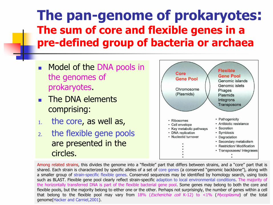
The pan-genome of prokaryotes:The sum of core and flexible genes in a pre-defined group of bacteria or archaea
◼ Model of the DNA pools in the genomes of prokaryotes.
◼ The DNA elements comprising:
1. the core, as well as,
2. the flexible gene pools are presented in the circles.
52
Among related strains, this divides the genome into a “flexible” part that differs between strains, and a “core” part that isshared. Each strain is characterized by specific alleles of a set of core genes (a conserved “genomic backbone”), along witha smaller group of strain-specific flexible genes. Conserved sequences may be identified by homology search, using toolssuch as BLAST. Flexible gene pool clearly reflect strain-specific adaption to local environmental conditions. The majority ofthe horizontally transferred DNA is part of the flexible bacterial gene pool. Some genes may belong to both the core andflexible pools, but the majority belong to either one or the other. Perhaps not surprisingly, the number of genes within a cellthat belong to the flexible pool may vary from 18% (Escherichia coli K-12) to <1% (Mycoplasma) of the totalgenome(Hacker and Carniel,2001).

The pan-genome of prokaryotesA core genome and flexible gene poolsGenomic islands
◼ Genomic islands can be introduced into the bacterial core genome through a variety of mobile elements:
1. plasmids,
2. Phages, and
3. Integrative and Conjugative Elements (ICEs) such as conjugative transpsons.
◼ Genomic islands are not colored, because most genomic islands are not mobile.
53Flannery,2011; Langille et al.,2010

Pathogenicity islandsGram-negative and Gram-positives bacteria
◼ The first GIs identified were termed pathogenicity islands.
◼ Those GEIs that carry virulence genes are called PAIs.
◼ Pathogenicity islands are found in:
1. Mainly in Gram-negative bacteria, and
2. In a few Gram-positives (cornyforms and actinomycetes).
3. PAI was not clearly identified in fastidious bacteria.
4. Usually absent from those non-pathogenic organisms.
54Dhillon et al.,2013;..

Microbial strategies for attackAtypical mechanisms of pathogenicitySugar catabolism in Spiroplasma
◼ The genome of the sieve tube-restricted mollicute Spiroplasmacitri is almost sequenced, and should neither contain genes for TTSS.
◼ In Spiroplasma citri,sugar catabolism, and more specificallyfructose utilization has been shown to be a key factor in pathogenicity.
55
Solid green: sieve tube; dashed green: sieve tube plates; light
pink: companion cell; dark pink: nucleus; yellow: nutrients

Microbial strategies for attackAtypical mechanisms of pathogenicitySugar catabolism in Spiroplasma
56
Solid green: sieve tube; dashed green: sieve tube plates; light pink: companion
cell; dark pink: nucleus; yellow: nutrients
Companion cell: Any of a number of specialized parenchymal cells adjacent to a sieve tube in the phloem of flowering plants, believed to regulate the flow of nutrients through the tube. Sucrose is a disaccharide consisting of one molecule of glucose linked with one molecule of fructose.
◼ Competition for fructose between the companion cell and the spiroplasmas in the sieve tubes, seems to result in impaired sucrose loading into the sieve tubes and non-balanced sugar distribution between:
1. Sink organs (young leaves and roots), and2. Source organs (leaves and stems).
See also Ca. Liberibacter asiaticus.

Pathogenicity islandsIn some fungiUstilago hordei
◼ Large ‘pathogenicity loci’, which share common features with bacterial PAIs have been identified in the pathogenic fungus, Ustilago hordei, the agent of covered smut of barley.
◼ HGT has also been identified in the pathogenic fungus.
57
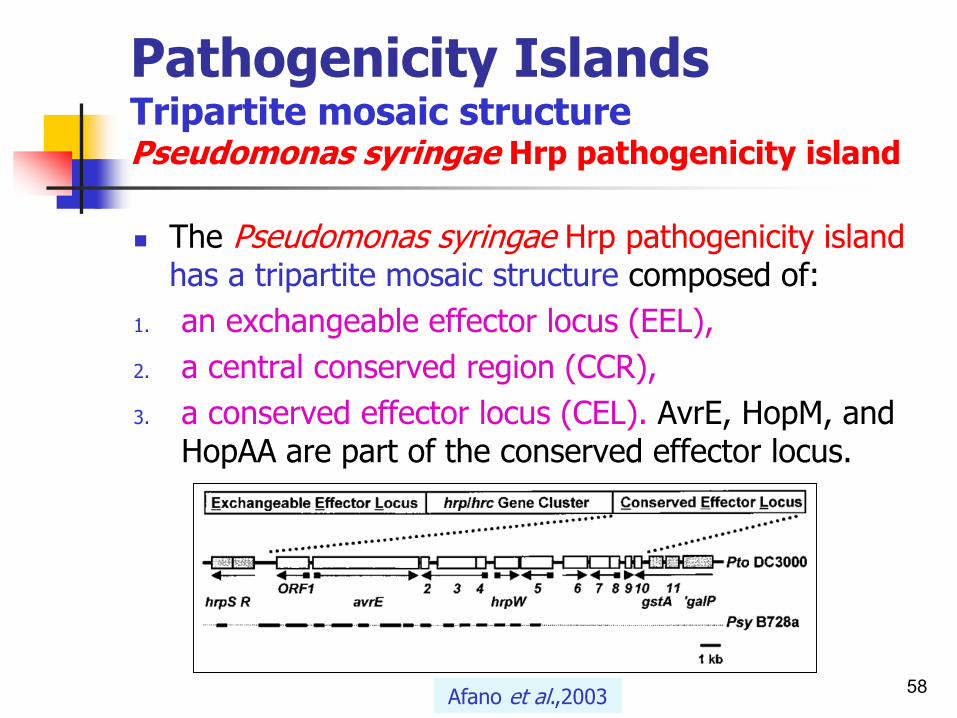
Pathogenicity Islands Tripartite mosaic structure Pseudomonas syringae Hrp pathogenicity island
◼ The Pseudomonas syringae Hrp pathogenicity islandhas a tripartite mosaic structure composed of:
1. an exchangeable effector locus (EEL),
2. a central conserved region (CCR),
3. a conserved effector locus (CEL). AvrE, HopM, and HopAA are part of the conserved effector locus.
58Afano et al.,2003

Pathogenicity Islands Mosaic structures
◼ The genomes of prokaryotes are highly diverse mosaic structures.
◼ During evolution, several genetic elements have been acquired independently at different time points and from different hosts.
◼ Accumulation of horizontally acquired elements at a certain location of the chromosome, and the same target structures (e.g. tRNA genes) served repeatedly for the integration of the various elements.
59Schmidt and Hensel,2004

Pathogenicity Islands Distinct class of genomic islandsLarge, mosaic, genetic islands, found in all pathogenic bacteria
60
◼ They can be transferred as a single unit to new bacterial cells, thus conferring virulence to formerly benign(not harmful) strain.
◼ They are transferred through horizontal gene transfer events such as transfer by:
1. a plasmid,
2. phage, or
3. conjugative transposon.
Boundless.com

Pathogenicity Islands Evolution of pathogens
61PAIDB v2.0
1. Pathogenicity islands (PAIs), and
2. Antimicrobial resistance islands (REIs) are key to the evolution of pathogens.
3. PAIs promote disease development, and
4. REIs give a fitness advantage to the host over multiple antimicrobial agents.

Microbial strategies for attackPathogenicity islands
62
◼ PAI are included in bacterial genome and occupy relatively large genomic regions.
◼ They are in range of 10 to 200 kb.
◼ PAIs carry genes encoding one or more virulence factors, including:
1. adherence factors,
2. toxins,
3. iron uptake systems,
4. invasion factors, and
5. Secretion systems (Types III, IV and VI).
T3SSs are typically located in pathogenicity islands (PAIs) and found to be transferred horizontally among bacterial species. T3SS is encoded by a
distinct cluster of genes, termed hrp/hrc (hypersensitive response and pathogenicity/conserved) genes in the plant pathogens.

Pathogenicity islandsMobile genetic elements Major virulence features encoded by pathogenicity islands
63Hacker and Kaper,2000
Diarrheagenic Escherichia coliUropathogenic (pathogen of urinary tract) E. coliVibrio cholerae
Adherence factors. E.g.
fimbriae
Uropathogenic Escherichia coliStaphylococcus aureus
Toxins
Uropathogenic E. coliShigella flexneriYersinia spp.
Iron uptake systems
Salmonella spp.Shigella spp.Listeria spp.
Invasins (enzymes), modulins(a family of protein toxins soluble in phenols), effectors
Pseudomonas syringaeErwinia spp.Yersinia spp.Salmonella spp.Shigella spp.
Type III secretion systems
Helicobacter pyloriAgrobacterium tumefaciens
Type IV secretion system

Pathogenicity islandsCollection of PAI loci which were reported in academic literatures
64PAIBD (Pathogenicity island database)
Species (strains) PAI name
Insertion site
Size GenBankAccession
Function
Pseudomonas syringae DC3000
Hrp-PAI tRNA-leu 52.5kb AF232004 Type III secretion system, Hop, Avr, hypersensitive response, mosaic structure
(exchangeable effector locus, hrp/hrcgene cluster, conserved effector locus)
Pseudomonas syringae 464
Hrp-PAI tRNA-leu 2.4kb AY147017 Type III secretion system, Hop, Avr, hypersensitive response, mosaic structure
(exchangeable effector locus, hrp/hrcgene cluster, conserved effector locus)
Pseudomonas cichorii Jan-83
S-PAI - 55.6kb DQ168848 Type III secretion system (10-kb-long insertion(avrE and avrF) in the middle of
the hrp/hrc cluster)
Pseudomonas viridiflava LP23.1a
S-PAI - 9.2kb AY597279 Type III secretion system (10-kb-long insertion(avrE and avrF) in the middle of
the hrp/hrc cluster)
Yersinia pseudotuberculosisIP 31758
YAPI tRNA-Phe 64.6kb NC_009708_P1 Type IV pilus
Rhodococcus equi103S
rpl - 8.9kb NC_014659_P1 Biogenesis of Flp-subfamily type IVb pili
Acinetobacterbaumannii AYE
AbaR1 ATPase 100.0kb CT025832 European clone I, β-lactams aminoglycosides, fluoroquinolones,
chlloramphenicol, tetracycline, rifampin resistance

Pathogenicity islandsMobile genetic elementsGroups of virulence factors encoded by PAI
65Schmidt and Hensel,2004
Group Examples of virulence factors PAI names
Iron uptake systems FyuA, aerobactin, Sit, Pit2ABCD HPI, SPI-1, PPI-1, SHI-2, 3, PAIICFT073,PAI III, IV536
Adhesins: A surface structures such as EPSs (xanthan or amylovorin)that mediate the binding of a bacterium to the host cell.
Type 4 pili, P-Pili, S- and P-fimbriae, Sap adhesin, Hek adhesin, AfaE-III, Iha, TcpA
Major PAI, PAI I, IICFT073, PAI I-IV536,PAI I, IIJ96PAI-IAL863, TAI, VPI-1
Pore-forming toxins Listeriolysin, alpha-hemolysin, RTX-like exotoxin
LIPI-1, PAI I536, PAI II536, O#28
Proteins causing apoptosis SipB SPI-1
Secreted lipases PlcA, PlcB, SmlC LIPI-1, LIPI-2
Secreted proteases EspC, SigA, Pic, ShetA1, Mop, BFT SHI-1, EspC PAI, VPI-1, BFPAI
O antigens GtrA, GtrB, Gtr SHI-O
Proteins transported by type I, III, IV, and V protein secretion systems
Alpha-hemolysin, EspI, EspC, SigA, Cag,Tir, EspB, G, F, Map, SptP, Sse, Ste, SopD, SopE, SopE2, PipB, SifA, SpiC, EspC, CagA
SHI-1, PAI I, II536, PAI I, PAI, IIJ96,LPA, EspC PAI, SHI-1, SPI-1, SPI-3,SPI-5, LEE, cag PAI
Antibiotic resistance phenotype Pse-1, FloR, AadA2, Sull, TetR, G SGI-1
Cytotoxin Associated Gene A (CagA)
Genomic islandsGenomic islands encode different functions Symbiosis islands
66
◼ An analogous genomic structure in rhizobia is termed a symbiosis island.
◼ The symbiosis island comprises 10% of the entire genome and is therefore larger(500 kb) than any PAI known to date.
◼ It has also been found in the bacterial symbiont(Mesorhizobium loti) of leguminous plants.
◼ The symbiosis island has an average GC content of 59.4% compared to the genomic average of 64%.
Genomic islandsGenomic islands encode different functionsResistance islands
◼ Resistance Islands (REIs) with genes responsible for antibiotic resistance:
◼ Another class of GIs, and are linked to pathogenesis by conferring simultaneous resistance to multiple antibiotics and facilitating the emergence of multidrug-resistant pathogens.
◼ Pseudomonas aeruginosa genomic island 1 (PAGI-1) is found in the majority of the clinical isolates.
◼ Acquired by horizontal gene transfer.
67

Genomic islandsGenomic islands encode different functionsMetabolic islands
◼ Islands with genes related to metabolism.◼ Some bacteria such as Salmonella senftenberg, E.
coli andYersinia spp. have this ability to colonize plant surfaces and tissues, to metabolize plant-derived carbon sources such as sucrose uptake.
◼ The genome of B. subtilis shows many interesting features including the genes involved in the metabolism of plant derived carbon compounds, such as opines and starch.
68Preston et al.,1998;..

Genomic islandsGenomic islands encode different functionsMetabolic islands
◼ They have a large metabolic versatility and are able to utilize numerous substrates as carbon and energy sources.
◼ Pseudomonas aeruginosa is well known as an opportunistic pathogen for plants, animals, and humans.
69Schmidt and Hensel,2004

Genomic islandsencode different functions
70Hentschel and Hacker,2001;..
Genomic islands encoded some islands
including antibiotic resistance functions (resistance island).
GEIs are often inserted at tRNA genes and flanked by
DR. The tRNA genes frequentlyassociated with PAIs. One of the
tRNA loci is Phe tRNA gene (phenylalanyl-tRNA synthetase), encode for integrase (int)(yellow box). Resistant island is locates
within the orfX gene.

Examples of genomic islandsGenomic islands encode different functions
71Hacker and Kaper,2000
Organism Property Type of island
Genetic feature Size (kb)
Mesorhizobium loti Nitrogen fixation Symbiosis Integrated plasmid 500
Pseudomonas putida ChlorocatecholDegradation
Degradation Integrated plasmid 105
Salmonella senftenberg Sucrose uptake Metabolism Conjugativetransposon
100
Staphylococcus aureus MecA protein Resistance Location onchromosome
52
Salmonella typhimuriumDT 104
Multiresistance Resistance Location onchromosome
14
Various bacteria Type III secretionType IV secretion
Secretion Location on chromosomeor plasmid
Variable
Various bacteria Iron uptake Fitness Location on chromosomeor plasmid
Variable
Genome sequencingBacterial genome database
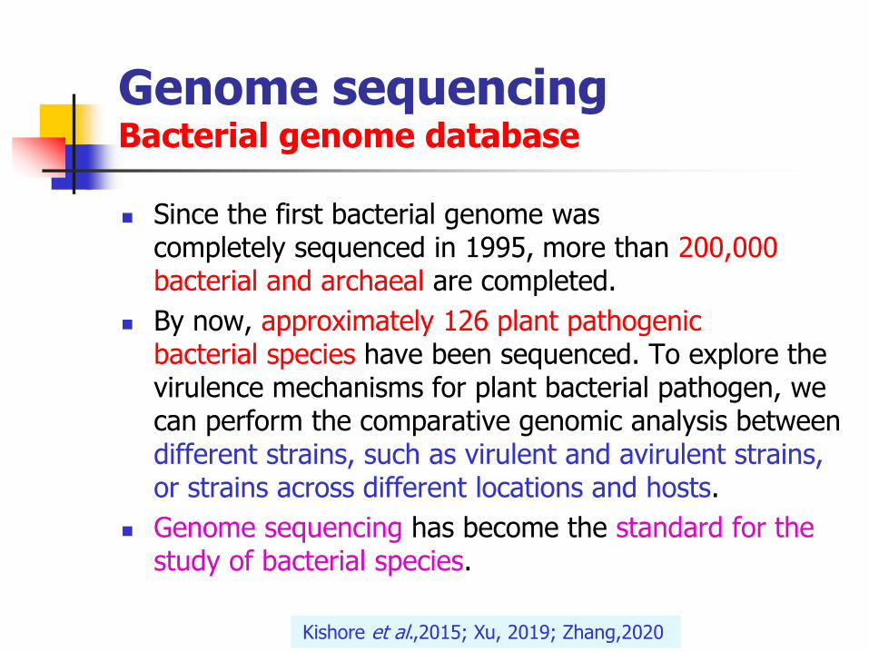
◼ Since the first bacterial genome was completely sequenced in 1995, more than 200,000 bacterial and archaeal are completed.
◼ By now, approximately 126 plant pathogenic bacterial species have been sequenced. To explore the virulence mechanisms for plant bacterial pathogen, we can perform the comparative genomic analysis between different strains, such as virulent and avirulent strains, or strains across different locations and hosts.
◼ Genome sequencing has become the standard for the study of bacterial species.
Kishore et al.,2015; Xu, 2019; Zhang,2020

Genome sequencingBacterial genome databasePAIDB-Pathogenicity Island Database
◼ PAIDB is a web-based user-friendly resource and has been widely used for detecting PAIs in newly sequenced genomes and mining virulence genes from metagenome.
73
Metagenomics is the study of the metagenome- the collective genome of microorganisms from an environmental sample, to provide information on the microbial diversity and ecology of a
specific environment.

Genome sequencingBacterial genome databasePAIDB-Pathogenicity Island Database
◼ The pathogenicity island database (PAIDB; http://www.gem.re.kr/paidb) is a comprehensive relational database of all the reported pathogenicity islands (PAIs) and potential PAI regions which were predicted by a method that combines feature-based analysis and similarity-based analysis.
◼ As of April 2006, PAIDB contains 112 types of PAIs and 889 GenBank accessions containing either partial or all PAI loci previously reported in the literature, which are present in 497 strains of pathogenic bacteria.
74Yoon et al.,2014
Genome sequencingBacterial genome databasePAIDB-Pathogenicity Island Database
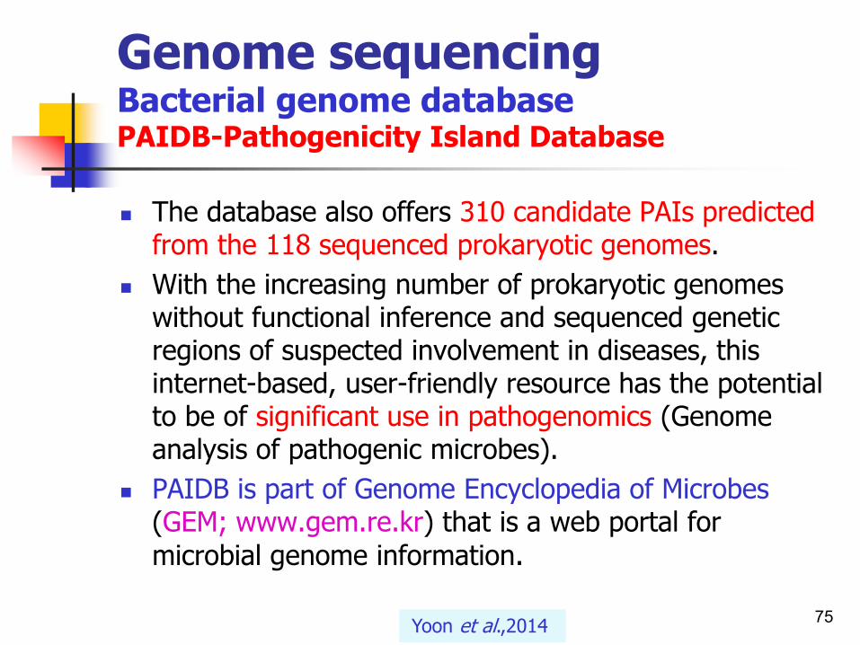
◼ The database also offers 310 candidate PAIs predicted from the 118 sequenced prokaryotic genomes.
◼ With the increasing number of prokaryotic genomes without functional inference and sequenced genetic regions of suspected involvement in diseases, this internet-based, user-friendly resource has the potential to be of significant use in pathogenomics (Genome analysis of pathogenic microbes).
◼ PAIDB is part of Genome Encyclopedia of Microbes (GEM; www.gem.re.kr) that is a web portal for
microbial genome information.
75Yoon et al.,2014

Genome sequencingBacterial genome databasePAIDB-Pathogenicity Island Database
◼ With the improved detection scheme, 2673 prokaryotic genomes were analyzed to locate potential Pathogenicity Islands (PAIs) and Resistance Islands(REIs).
◼ The update encompasses dramatic increase in:
1. database contents of genomes analyzed,
2. accuracy improvement of detection of candidate regions, and
3. functionality update of web application.
76http://www.gem.re.kr/paidb

Genome sequencingPathogenicity islandsBioinformatics soft wares
77Ali et al.,2013; Soares et al.,2012
◼ To date, several open source and commercial software packages are available for creation and visualization of genome maps in linear, circular, or in both forms.
◼ Examples:
◼ “GenomePlot” and “GenoMap” developed for generating genome maps (Atlas).
◼ A novel software suite designed for the prediction of pathogenicity islands is pathogenicity island prediction software, or PIPS.

Genome sequencingMain characteristics of genomic islands and possible functions
78Silva Filho et al.,2018

Genome sequencingBioinformatics soft waresSteps in dataset creation
79Silva Filho et al.,2018

Genome sequencingData of GIs, PAIS, and regions with DNA of bacteriophages curated in vitro from the reference organism Escherichia coli cft073
80Silva Filho et al.,2018

Genome sequencingThe hierarchical overview of computational methods for predicting genomic islands
81Lu and Leong,2016

Genome sequencingProcedure for identifying candidate PAIs and REIs in a sequenced genomeScreenshots of new functional features in PAIDB v2.0
82Yoon et al.,2014

Genome sequencing Summary of genome sequence projects of Enterobacter cloacae
83Liuet al.,2013
StrainE. cloacae subsp. cloacae ENHKU01
E. cloacae subsp. cloacae
ATCC13047
E. cloacaeEcWSU1
E. cloacae subsp. dissolvens SDM
Size 4.73 5.6 4.8 4.97
No. of Chromosome
1 1 1 1
No. of plasmid 0 2 1 0
GC content % 55.1 54.6 54.5 55.1
Total genes 4445 5639 4740 4646
Predicted CDS 4338 5518 4619 4542
No. of tRNAs 82 24 83 53
No. of rRNAoperons
8 8 8 3
Host Plant Human Plant Soil
Important feature
EndophyteHuman
opportunistic pathogen
Plant pathogen2,3-butanediol
production
Coding sequence or CDS: portion of a gene's DNA or RNA that codes for protein. While the ORF may contain introns as well.

Genome of prokaryotesGenomic islands(GISs)Bioinformatics platform for high school students
84
◼ We utilize a bioinformatics platform that is easy to adapt, integrate, and implement into the existing biology curriculum through:
1. student centered, and
2. activity-based teaching, and
3. learning methods tailored for high school students.
JO MARIE BACUSMO is a Lecturer in the Department of Microbiology and CellScience, University of Florida, Gainesville, FL 32611. JULIE BOKOR is Associate
Director of the Center for Precollegiate Education, University of Florida;e-mail: [email protected]. KATHY SAVAGE is a Chemistry and Bioscience
Teacher at Oviedo High School, Oviedo, FL 32765. VALÉRIE DE CRÉCYLAGARDis a Professor in the Department of Microbiology and Cell Science,
University of Florida; e-mail: [email protected].

Genome of prokaryotesGenomic islands(GISs)Implementation
◼ Lessons from this module were implemented in professional development programs for high school teachers.
◼ Afterward, a number of teachers expressed interest in integrating select lessons from the module in their classes.
◼ This module presents a gateway to embracing integration of bioinformatics-based tools in the high school curriculum.
◼ We hope that more projects like this will result in further progress toward seamless adoption of bioinformatics in various curricula.
85Bacusmo et al.,2019

Genome of prokaryotesGenomic islands(GISs)Bioinformatics
86
◼ Bioinformatics, the study of biological data using various computational techniques, is a very important aspect of biology, and its integration would greatly benefit current high school curricula.
1. We describe a bioinformatics-based module that introduces the application of genome comparison in the identification of “pathogenic islands.”
2. The module also introduces foundational concepts of
1. horizontal gene transfer and
2. the genetic basis of virulence, with a special focus on antibiotic resistance.
Bacusmo et al.,2019
A module is one of a set of parts from which some buildings are made.

Genome of prokaryotesGenomic islands(GISs)Bioinformatics
87
◼ One of the constant challenges in health care is the potential inefficacy of existing antimicrobial drugs against new and emerging pathogens.
◼ Most pathogens evolve at an accelerated rate, thus promoting diversification and rapid distribution of virulence genes.
◼ These facts place emphasis on the importance of studying the epidemiology of various virulence genes.
◼ One of the ways to approach this problem is through rapid identification of the virulence genes in pathogenic strains using bioinformatics tools such as genome comparison.
Bacusmo et al.,2019

Genome of prokaryotesGenomic islands(GISs)Bioinformatics
88
◼ Pathogens often contain “signatures” in their genomes where accumulation of virulence and antimicrobial resistance genes are concentrated into specific regions of the genome called “genomic islands.”
◼ Genome comparison, a bioinformatics tool, allows for easy identification of these genome signatures and provides information on the various virulence genes contained within the island.
◼ This leads to a targeted approach in counteracting the pathogen.
Bacusmo et al.,2019

Genome of prokaryotesGenomic islands(GISs)Bioinformatics
89Bacusmo et al.,2019
◼ Common virulence factors designated as either offensive or defensive features.
defensiveoffensive
Acid resistance(in Gram-negative enteric bacteria)
Flagella or ability to swim
Antibiotic resistanceAttachment
Capsule or protective coatingToxins
Secretion systems
Effector protein injection

Genome of prokaryotesGenomic islands(GISs)Bioinformatics
90Bacusmo et al.,2019
◼ Example of a pathogen prototype displaying common virulence factors (adapted from https://preview.tinyurl.com/yc89aw4g).

Genome of prokaryotesGenomic islands(GISs)Module Overview
91
◼ The “Pathogenic Islands” module consists of six lessons formatted into 50-minute sessions (Table 1).
◼ It was developed by a science teacher/science researcher team to align with Florida’s Next Generation Sunshine State Standards for Science.
◼ It also embraces three-dimensional learning as espoused by the National Research Council (2012), which calls for students to be actively engaged in the practices of science while exploring disciplinary core ideas and crosscutting concepts.
Bacusmo et al.,2019

Genome of prokaryotesGenomic islands(GISs)Module Overview
92
◼ The major learning goals of the module are as follows:
1. List and describe virulence genes that cause the different modes of pathogenicity.
2. Describe the various modes of gene transfer.
3. Demonstrate the concept of gene transfer applied in the process of selection and evolution.
4. Use genome comparison tools such as PATRIC and Island Viewer to identify genomic islands.
5. Apply knowledge of virulence genes and horizontal gene transfer to identify the source of virulence in pathogenic strains.
6. Perform independent research on a set of pathogens and present the location of pathogenic islands on the genome, identify the virulence genes contained in the pathogenic islands and their function, and provide a brief history on the epidemiology of the disease caused by the pathogen.
Bacusmo et al.,2019

Genome of prokaryotesModule OverviewLesson sequencing guide and summaries. The “Pathogenic Islands” module consists of six lessons formatted into 50-minute sessions and 24 students per class
93
Students take a pretest over the content presented in these six lessons. They then watch short videos of a pathogenic bacterium invading a host cell to identify the behaviors and biological mechanisms (virulence factors) exhibited by the bacterium that make it successful.
Lesson 1Day 1
Student groups build a bacterial prototype expressing virulence factors and then compare their prototype to those created by the other groups. Students assess the potential success of each prototype by voting for the most successful and least successful prototype and justifying their choices.
Lesson 2Day 2
Students watch a video on horizontal gene transfer and answer three questions. The teacher can choose to discuss the answers to these questions after the worksheet has been collected.
Lesson 3,Activity 1
Day 3
Students team up and play a teacher-directed game (“Pathogen Survivor”) demonstrating genome diversification via gene transfer, highlighting its impact on bacterial fitness and survival.
Lesson 3,Activity 1
The video “The Power of Comparative Genomics” (7:07; https://www.youtube.com/watch? v=mU9ROpm6d70) introduces comparative genomics as a tool to help scientists focus their research.
Lesson 4,Activity 1
Day 4
The video “Comparison of Genomes of Eight Enteroaggregative E. coli O104:H4 Isolates” (2:07; https://youtu.be/6VTxmnZQXgU) shows how comparative genomics facilitates identification of genomic islands that contribute to the pathogenicity of disease outbreak strains.
Lesson 4,Activity 2
Students complete video tutorials on the Pathosystems Resource Integration Center (PATRIC), a web-based comparative genomics tool.
Lesson 4,Activity 3
Students work in groups, using PATRIC to research virulent genes and disease outbreaks for an assigned bacterial species.
Lesson 5,Activity 1
Day 5
Students present their research to the class and are graded according to a rubric (Figure 4). Lesson 5,Activity 2
Day 6
Survivor curve being an actual representation of pathogen survival during preservation process.
Bacusmo et al.,2019

Genome of prokaryotesModule OverviewIslandViewer is a computational tool that integrates four different genomic island prediction methods: IslandPick, IslandPath-DIMOB, SIGI-HMM, and Islander. For more information about methods and analyses see here and in the FAQs.
◼ Select option Example and then press the Go to Genome button to browse the pre-computed genomic island predictions for publicly available genomes, including Islander predictions as well:
94

Genome of prokaryotesModule OverviewIslandViewer is a computational tool that integrates four different genomic island prediction methods: IslandPick, IslandPath-DIMOB, SIGI-HMM, and Islander. For more information about methods and analyses see here and in the FAQs.
95

Genome of prokaryotesModule OverviewIslandViewer 4 | An integrated interface for computational identification and visualization of genomic islands
96
Continue the column
Pectobacterium c. subsp. carotovorum PC1. complete genome.

Genome of prokaryotesModule OverviewPATRIC, the Pathosystems Resource Integration Center, provides integrated data and analysis tools to support biomedical research on bacterial infectious diseases
97

Genome of prokaryotesModule OverviewRubric used to assess student performance in the independent research and presentation
98Bacusmo et al.,2019
Rubric: a set of instructions, especially on an exam paper, usually printed in a different style or colour.

Pathogenicity islandsVariable numbers of pathogenicity islands
◼ A bacterium may have more than one pathogenicity islands (PAIs).
◼ One species of bacteria may have more than one PAI.
1. Salmonella has at least 5 PAIs.
2. E. coli at least 2 PAIs.
3. Frequently, PAIs encode type III and IV secretion systems which, by excretion of proteins, directly interfere with the functions of host cells.
99Wikipedia,2011

Pathogenicity islandsLocation of selected pathogenicity islands and phages of Gram-negative bacteria
◼ Chromosomes (blue lines) and pathogenicity islands (black triangles) are not drawn to scale.
◼ The presence of repeated sequences at the site of insertion, which is shown below the island, is indicated by short yellow lines.
100Groisman and Ochman,1996
UroPathogenic Escherichia coli (UPEC) causes urinary tract infections (UTIs).

Pathogenicity islandsSalmonella anterica
101

Genome sequencingGeneral structure and feature of PAI
102
1. found in a wide variety of bacterial pathogens;
2. occupy relatively large regions of the chromosome, ranging from 10-200 kb;
3. missing from related non-pathogens (e.g. E. coli);
4. often different G-C content; codon usage;
5. often mobile (spread easily), transferred horizontally, through plasmids or transposons.
6. often associated with tRNA loci on one side and direct repeat (DR) sequences on both sides.
7. encode functions necessary for pathogenesis.
The addition of a pathogenicity island to a non-invasive species can make the non-invasive species pathogenic.

Genome sequencingGeneral structure and feature of genomic islands
103
Schematic model of a genomic island of bacteria (upper part). The formerly transferred DNA block is linked to a tRNA gene and flanked by direct repeats (DR).
The guanine plus cytosine (G+C) content of the genomic island is different from that of the core genome (lower part).
Other abbreviations: int, integrase gene; abc, def and ghi, genes encoding specific functions; IS, insertion sequence element; bp, base pair.
Hacker and Carniel,2001

Pathogenicity islandsShared and unique genesGC-content of pathogenicity islands
◼ It is estimated that each bacterium has about 40% of its genome devoted to unique genes.
◼ The GC-content of pathogenicity islands often differs from that of the rest of the genome.
◼ The G+C content of pathogenicity islands often differs (below 40%) from that of the rest of the genome (overall G+C content of the organism).
104
Relatively low GC contents (below 40%) are assembled in a "pathogenicity island”.
The codon usage or codon preference of whole genomes is strongly biased. i.e. each individual genome uses a preferred set of codons.

Pathogenicity islandsUnstable DNA regions
◼ PAIs often represent unstable DNA regions.
◼ PAIs may:
1. move from one tRNA locus to another, or
2. be deleted.
◼ Deletions of PAIs may occur via:
1. the direct repeats (DRs) at their ends, or
2. via IS elements or other homologous sequences located on PAIs.
105Hacker and Kaper,2000
Relatively low GC contents (below 40%) are assembled in a "pathogenicity island”.

General structure of a PAItRNA genes may act as landmarks for the integration of foreign DNA, either of plasmids and phages. Direct repeats (DR) may save served as recognition sites for the integration of bacteriophages, and for enzymes involved in excision of mobile genetic
◼ The PAI boundaries are frequently determined by DRs (direct repeats), each DR is 16-20 bp long with perfect sequence repetition (triangle) at the ends of the pathogenicity island.
◼ DRs are used for insertion and deletion processes. int, integrase gene. IS, insertion sequence(simple transposon). The association of PAIs and tRNA loci led to the generation of PAIs by horizontal gene transfer.
106Relatively low GC contents (below 40%) are assembled in a "pathogenicity island”.
Schmidt and Hensel,2004
genome
Flexible gene pool

General structure of a PAISchematic view of a pathogenicity island with associated features. The PAI region has biased sequence composition
◼ The PAI regions are associated with:
1. virulence genes (four genes: vir1, vir2, vir3, and vir4),
2. phage-related genes (phag1 and phag2),
3. mobile genes (integrase gene and transposase gene),
4. hypothetic protein genes (proteins with unknown function) such as hypo1, hypo2, and hypo3,
5. insertion sequence elements,
6. direct repeats(DRs), and
7. tRNA gene.
107Che et al.,2014

General structure of a PAIIn methicillin-resistant Staphylococcus aureus
◼ Staphylococcus aureus genome contains core genome, accessory component, and foreign genes.
1. Core genome that constructs backbone of genome has main metabolic function. Core genome is highly conserved, and similarity of genes among isolates is ∼98–100%.
2. Accessory component that constructs 25% of S. aureusgenome contains mobile genetic elements (MGEs) such as transposons (Tn), chromosomal cassettes, pathogenicity islands (PIs), genomic islands, and prophages acquired horizontally between strain.
108Kırmusaoğlu,2017

◼ Mobile genetic elements (MGEs) carry virulence genes. These genes encode for varied virulence factors and toxins.
◼ MGEs in methicillin-resistant S. aureus (MRSA) contain the mecA gene causing methicillin resistance and affect biofilm phenotype of S. aureus. mecA gene expression is regulated by mecI and mecRI that are own regulators.
109Kırmusaoğlu,2017
General structure of a PAIIn methicillin-resistant Staphylococcus aureus

General structure of a PAISchematic view of a pathogenicity island with associated features. The PAI region has biased sequence composition
◼ Staphylococcus aureus genome contains core genome, accessory component, and foreign genes.
1. Core genome that constructs backbone of genome has main metabolic function. Core genome is highly conserved, and similarity of genes among isolates is ∼98–100%.
2. Accessory component that constructs 25% of S. aureus genome contains mobile genetic elements (MGEs) such as transposons (Tn), chromosomal cassettes, pathogenicity islands (PIs), genomic islands, and prophages acquired horizontally between strain. 110
Che et al.,2014

Pathogenicity islandsGeneral structure of PAIPathogenic vs. Nonpathogenic bacteria
◼ The thin bold line represents regions of the core genome; The box representgenes whose products contribute to virulence. The arrows indicate the presence of direct repeats (DRs) at the ends of the pathogenicity island.
◼ Abbreviations:
◼ DR, direct repeats; int, integrase gene; vir, virulence-associated gene; mob,mobility gene; ∆mob, pseudo-mobility gene. mob genes encode integrases, transposases, or other proteins involved in mobility of the prokaryotic genome.
111Hacker and Kaper,2000
Core Core
Mobility genes (mobs) encode integrases, transposases, and insertion sequence (IS)

Pathogenicity islandsGeneral structure of PAIA GI is often absent in closely related genomes
◼ The schematic representation of several GI-associated features. A GI is often absent in closely related genomes(1 and 2). It may also have atypical compositional characteristics compared with the core genome, such as lower GC content. The presence of several sequence elements is indicative of a GI: flanking conserved regions, DRs, insertion sequence (IS) elements and mobility-related genes encoding integrase and transposase..
112Lu and Leong,2016

General structure of PAIFunction of different elements
◼ Direct repeats (DRs):
◼ The PAI boundaries are frequently determined by DRs (direct repeats).
◼ Direct repeats (DR) that are presented as DNA sequences of 16 to 20 bases pairs (up to 130 bp).
◼ Might have served as recognition sites for the integration of bacteriophages, and their integration resulted in the duplication of the DR.
◼ Furthermore, DR act as recognition sequences for enzymes involved in excision of mobile genetic elements, thus contributing to the instability of a PAI flanked by DR.
113

General structure of PAIFunction of different elements
◼ Mobility genes (integrase gene and transposase gene):
◼ These genes are frequently located at the beginning of the island, act as rapid DNA rearrangements, and exchange.
◼ Site specific integration of pathogenicity islands is mediated by an integrase recombinase.
114
Integrase: Any enzyme that integrates foreign DNA into that of an infected cell.Transposon: A segment of DNA that can move to a different position within a genome. Transposons are involved in the intrabacterial movement of DNA. They may, however,
be transmitted to other bacteria by transfer of plasmid or chromosomal DNA.

General structure of PAIFunction of different elements
◼ Different functions of tRNA genes:
◼ The main function of tRNA molecules is to bind mRNA at the ribosome for protein synthesis.
◼ But some tRNAs have functions unrelated to translation.
◼ E.g. Some tRNA genes are frequently associated with bacterial pathogenicity islands.
◼ tRNA genes served repeatedly for the integration of foreign DNA.
115Pettersson,2009;..

General structure of PAIFunction of different elements
◼ tRNA genes:
◼ PAIs are often associated with transfer RNA (tRNA) genes.
◼ About 75% of the PAIs identified so far are associated with tRNA loci.
◼ The association of PAIs and tRNA loci may therefore reflect the generation of PAIs by horizontal gene transfer.
116Hacker and Kaper,2000

General structure of PAItRNA gene structure and organizationtRNA genes
A. A generalized tRNA operon is shown: A tRNA gene may be transcribed in many different contexts, including rRNA, mRNA, sRNA (bacterial small RNAs), and other tRNA genes.
B. The E. coli tufB operon: is shown as an example of an operon with both tRNA and protein coding genes.
C. The B. subtilis rrnB/trnB operon: is shown as an example of an rRNA operon encoding several tRNAs.
117
rrnB: an rRNA operons; Pand T indicate promoter and
terminator sequences, respectively.
Pettersson,2009
Two genes, tufA and tufB, located at 73 and 88 minutes of the Escherichia coli linkage map, code for the polypeptide chain
elongation factor EF-Tu. tufB is transcribed with four upstream tRNA genes, thrU, tyrU, glyT and thrT, into a
cotranscript of approximately 1800 nucleotides.

Pathogenicity islandsHorizontal gene transfer Pathogenicity islands
◼ Pathogenicity islands were first described in human pathogens of the species Eseherichia coil in the late 1980s, but have recently been found in the genomes of various pathogens of humans, animals, and plants.
◼ Thus, rapid alterations of phenotype can occur via horizontal gene transfer (HGT).
◼ It is possible to recognize genes that arose by lateral gene transfer by simply examining genome sequences.
118Kjemtrup et al.,2000; Buonaurio,2008;Van Sluys et al.,2002

Pathogenicity islandsHorizontal gene transfer Integration sites of PAI
1. Pathogenicity islands are discrete genetic units flanked by direct repeats, insertion sequences or tRNA genes, which act as sites for recombination into the DNA.
2. Cryptic mobility genes may also be present, indicating the provenance as transduction.
3. They are transferred through horizontal gene transfer events such as transfer by a plasmid, phage, or conjugative transposon.
119
Conjugative transposons such as Tn916 are transposon-like elements, different from well-studied transposons such as Tn5 and Tn10. Resistance
genes need not be carried on the conjugative transposon to be transferred. See Genetic file for details.

Pathogenicity islandsHorizontal gene transfer Integration sites of PAI
◼ Integration of PAI into the bacterial chromosome is a site-specific event.
1. Most PAI currently known have inserted at the 3′ end of tRNA loci.
2. Also, phage attachment sites frequently are located in this region.
3. However, certain genes, and infrequently intergenic regions in operons are used by PAI.
120Schmidt and Hensel,2004

Pathogenicity islandsHorizontal gene transfer Phage transduction and prophage integration are the major mechanisms of horizontal gene transfer
◼ High percentage of phage-related genes has been found in PAIs.
◼ In actuality, phage transduction and prophage integration are the major mechanisms of horizontal gene transfer in prokaryotes.
◼ The food pathogen E. coli O156:H7 strain Sakai has been discovered to contain around 16% prophage of its own total genome sequence.
121Che et al.,2014

Phage transduction and prophage integration are the major mechanisms of horizontal gene transfer Mobile genes such as integrases (int), are frequently located at the beginning of the island, act as rapid DNA rearrangements, and exchange
122Gal-Mor and Finlay,2006

Pathogenicity islandsHorizontal gene transfer
123

Pathogenicity islandsHorizontal gene transfer
◼ Frequently, PAIs encode type III and IV secretion systemswhich, by excretion of proteins, directly interfere with the functions of host cells.
124

Pathogenicity islandshrp-hrc regions are now designated pathogenicity islands (PAIs)
Plant pathogenic bacteria
◼ The hrp-hrc regions are now designated pathogenicity islands (PAIs) in various plant-pathogenic bacteria.
◼ The hrp cluster encodes a type III secretion system that is regulated by four proteins encoded on the hrp PAI.
◼ hrp and hrc genes are part of a PAI (located within pathogenic islands) that has been designated Hrp. e.g.
1. Hrp PAI of E. amylovora2. Hrp PAI island of Pseudomonas syringae, or3. Hrp PAIs of other Xanthomonas species (note: in Xanthomonas
the hrp gene cluster was previously known as a pathogenicity island but it is reported missing from some Xanthomonas spp.).
4. The hrp/hrc genes within the Hrp PAI core region was highly conserved.
125Kjemtrup et al.,2000; Buonaurio,2008, Ali Shah et al., 2019
Genome (conserved and flexible)/genomic islands (flexible)/PAIs(flexible)/hrp/hrc regions (conserved).

Pathogenicity islandsChromosome or plasmid-encoded PAIsPlant pathogenic bacteria
1. PAIs are located on bacterial chromosome. 2. PAIs have also been localized to mobilizable
plasmids.◼ Plasmid-encoded PAIs: ◼ Plasmid-encoded PAI was recently isolated from race
7 of Pseudomonas phaseolicola strain 1449B. ◼ Cured of a 154 kb plasmid, this strain is no longer
virulent on bean, demonstrating a virulence function for this plasmid.
◼ Chromosome-encoded PAIs:◼ hrp/hrc cluster of R. solanacearum does not seem to
be a PAI and that its effector genes are distributed randomly on both the plasmid and chromosome.
126Kjemtrup et al.,2000,…

Comparisons of Hrp PAIs of five Xanthomonas speciesNote: in Xanthomonas the hrp gene cluster was previously known as a pathogenicity island but it is reported missing from some Xanthomonas spp.
◼ The Hrp PAI core regions (highly conserved) and variable flanking regions of X. axonopodis pv. glycines, X. axonopodis pv.citri, X. campestris pv.campestris, X. campestris pv.vesicatoria, and X. oryzae pv.oryzae are represented.
◼ The direction of arrows indicate the transcription orientation.
◼ The Hrp PAI core region was composed of 20 genes, from hrcC to hpaB, and encoded a type III secretion system that was highly conserved (90% similarity) among xanthomonads.
127Kim et al.,2003

Pathogenicity islandsExamples of PAI of various pathogens. The topology of PAI of various pathogens is depicted to demonstrate different features of PAI. The functional classes of the genes are as indicated in the figure
128Schmidt and Hensel,2004
Lipoprotein serves as a gated channel for secretion of substrates to the cell surface.
Helicobacter pylori CagA(cytotoxin-associated gene A)

Pathogenicity islandsChromosome-encoded PAIHrp PAI islands of Pseudomonas syringae
129
◼ The conserved arrangement of hrp/hrc genes within the Hrp PAIs of three strains: Psy 61, Psy B728a, and Pto DC3000.
◼ Known regulatory genes are shaded.
◼ Arrows indicate the direction of transcription, with small boxes denoting the presence of a Hrp box.
◼ The triangle denotes the 3.6-kb insert with phage genes in the B728a hrp/hrc region.
Alfano et al.,2000

Pathogen-generated secreted proteinsPathogenicity-related genes/effectors
What determinants allow a bacterium to be a pathogen?
Effectors in plant–microbe interactions:22nd New Phytologist Symposium,2009

Pathogen-generated secreted proteinsType III secretion systems (TTSSs)Effectors of pathogens
◼ Effectors are pathogen molecules that:
1. manipulate host cell structure, and
2. function in order to cause/facilitate the formation of symptoms
3. often contribute quantitatively to pathogen aggressiveness, and are
4. dispensable for the pathogen life cycle.

Pathogen-generated secreted proteinsType III secretion systems (TTSSs)Effectors of pathogens
1. Toxins, including peptides and proteins, are typically secreted through a type I or II secretion system.
2. Effectors are translocated through a type III or IV secretion system,
3. DNA is only transferred through the type IV secretion system.
Alto and Orth,2012

Pathogen-generated secreted proteinsType III secretion systems (TTSSs)Effectors of pathogens
◼ The type III secretion system (T3SS) is one of the most remarkably versatile pathogenicity factors yet discovered.
◼ A number of Gram-negative bacterial plant and animal pathogens depend on type III secretion systems (TTSSs) to inject virulence effector proteins into host cells.
1. The ability to elicit the hypersensitive response (HR),
2. A programmed cell death (PCD) associated with plant defence, as well as
3. Their pathogenic ability.

Pathogen-generated secreted proteinsavr genes
◼ Pathogens possess avirulence genes whose products are involved with determining host specificity.
◼ The bacterial avirulence gene function is dependent on interactions with HR and pathogenicity (hrp) genes.
134The transcriptional activation of a number of bacterial
avirulence (avr) genes is controlled by Hrp regulatory proteins.
The gene-for-gene `quadratic check', as observed with
phytopathogenic bacteria.Ø=unicode name

Pathogen-generated secreted proteinshrp genes
◼ hrp genes
◼ hrp genes may be one of the most important groups of genes found in phytopathogenic bacteria in relationship to pathogenicity and host range.
◼ These genes/products are associated with induction of the hypersensitive response in plants.
135
The transcriptional activation of a number of bacterial avirulence (avr) genes is controlled by Hrp regulatory proteins.

PCR detection and identification of plant-pathogenic bacteriaBased on conserved sequences of the effectors
Species/subspecies Primer nameTarget DNA
Variant of PCR
protocol
Sample (treatment)
Synonyms/observations
P. savastanoi pv.phaseolicola
AVR1-F/AVR1-RLocus avrPphF
PHTE-F/PHTE-RLocus pthE
Conventional Bacteria (DNAextraction)
Toxigenic and nontaxigenic strains
X. citri subsp. citri(Pathotypes A, B and C)
J-pth1/J-pth2Pathotypes A, B and C
Strains pthA gene (involved invirulence)
J-RXg/J-RXc2Pathotype A strains
ITS region
Conventional Bacteria (DNAextraction)
X. axonopodis pv. citri
X.citri subsp. citriX. citri pv. aurantifolii
VM1/VM2VM3/VM4VM5/VM6
pthA gene familyKingsley forward/reverse
X. citri pv. citri Achromosome
Real-time(SBYR ®
Green MasterMix)
Bacteria, plant(DNA extraction)
X.citri subsp. citriThe X. citri pv.
aurantifoliiis not included in the
ISPP List.
P. stewartii subsp.stewartii
ES16/ES1G2c16S-23S rRNA/ITS region
HRP1d/HRP3rhrpS ORF
ConventionalBacteria (boiled or
alkaline lysis)
Rcommended in the EPPO protocol.
The use of primers to conserved sequences flanking these hot spots provides another approach to isolating effector genes.

Genes of type III secretion system (TTSS) in bacterial plant pathogens Pathogenicity-related genes
◼ Two major sets of pathogenicity-related genes:1. hrp genes2. avr genes◼ The other genes are:1. hrc2. dsp, The disease-specific (dsp) gene dspA/E of
Erwinia amylovora (see chaperon section)3. Pth genes4. Hpa genes5. Xop genes
The hpa (hrp-associated) genes encode harpin-like proteins.
Xop, Xanthomonas outer protein.
Pth, pathogenicity gene necessary to condition pathogenicity on a given host.

Pathogen-generated secreted proteinsEffectors
◼ At least 57 families of effectors, with each bacterial strain expressing about 15-30 effectors, have been identified in the bacterial pathogen Pseudomonas syringae alone.
◼ Effectors are produced by:
1. all the major species of pathogenic bacteria infecting plants and animals,
2. fungi,
3. Viruses, and
4. nematodes.
Surico,2013

Pathogen-generated secreted proteinsEffectors
◼ The proteins secreted by Hrp TTSSs have been given a variety of names including:
1. Hop (Hrp outer protein) in Pseudomonas and many other bacteria such as P. syringae, Erwinia, Pantoea spp., etc.
2. Xop (Xanthomonas outer protein).
3. Pop (Pseudomonas outer protein, which actually are R. solanacearum proteins based on its earlier genus name),
4. Avr (for avirulence).
◼ Hop, Xop and Pop proteins were originally identified based on their property of limiting the host range of the pathogen.
Espinosa and Alfano,2004

Genes of type III secretion system
(TTSS) in bacterial plant pathogens
and the predictedfunctions of
effectors.
Narayanasamy,2008

Some phytopathogen type III effectorsT3E activities and plant targets
T3E Species Activity Target
AvrB P. syringae pv. glycinea race 0 Induces phosphorylation RIN4/RAR1
AvrBs3 X. campestris pv. vesicatoria race 1 Transcription activator-like
Upa20(transcription factor)/Bs3
AvrPphB P. syringae pv. phaseolicola race 3 Cysteine protease cleaves the Arabidopsis proteinkinase PBS1
AvrPto P. syringae pv. tomato JL1065 Kinase inhibitor Pto/EFR/FLS2
AvrPtoB P. syringae pv. tomato DC3000 E3 ubiquitin ligase Fen(tomato kinase protein)
AvrRpm1 P. syringae pv. glycinea race 0 Induces phosphorylation RIN4 protein(regulated basal defense in Arabidopsis)
AvrRpt2 P. syringae pv. tomato T1 Cysteine protease RIN4 protein(regulated basal defense in Arabidopsis)
AvrXa27 X. oryzae pv. oryzae PXO99A Transcription activator-like
Rice R gene Xa27
AvrXv4 X. campestris pv. vesicatoria T3 DeSUMOylating cysteine protease
Unknown
GALA R. solanacearum GMI1000 F-box and LRR domains A. thaliana Skp1-like proteins
HopAI1 P. syringae pv. tomato DC3000 Phosphothreonine lyase MPK3/MPK6

Some phytopathogen type III effectorsT3E activities and plant targets
T3E Species Activity Target
12 P. syringae pv. tomato DC3000 Protein tyrosine phosphatase Unknown
HopI1 P. syringae pv. maculicola ES4326 J-domain protein (possible Hsp70 cochaperone)
Unknown
HopM1 P. syringae pv. tomato DC3000 Unknown AtMIN7 and other immunity associated Arabidopsis protein
HopU1 P. syringae pv. tomato DC3000 Mono-ADP-ribosyltransferase GRP7 and otherRNA-binding proteins
HsvB Pantoea agglomerans pv. betae4188
Transcriptional activator-like Unknown
HsvG Pantoea agglomerans pv. gypsophilae 824-1
Transcriptional activator-like Unknown
PthXo1 X. oryzae pv. oryzae PXO99A Transcriptional activator-like host gene Os8N3
PthXo6/7 X. oryzae pv. oryzae PXO99A Transcriptional activator-like OsTFX1 OsTFIIAg1
XopD X. campestris pv. vesicatoria 85-10 DeSUMOylating cysteine protease Unknown
Block et al.,2008

T3SS substrate effector proteins hop genes encode P. syringae T3SE familiesHopZ, HopF,…
◼ Effectors of P. syringae and other phytopathogenicbacteria are generally designated as Hop proteins (Hrp outer proteins, i.e. proteins that have the capacity to travel through the T3S system.
◼ Nearly 60 different type III effector families (T3SE families) encoded by hop genes have been identified in P. syringae.
1. The HopF T3SE family is broadly distributed among pathovars of P. syringae.
2. The HopZ T3SE family, is widely distributed among the P. syringae species complex.
143McCann and Guttman,2007;..

Phylogenetic trees for some Hrp outer
protein (Hop) families with three or more
members
Lindeberg et al.,2005
Nomenclature & Phylogeny of Hops in P. syringae:
The phylogenetic analyses were used to determine whetherhomology families should be
subdivided into Hop subgroupsbased on amino acid sequence
divergence.A homology family was
subdivided if it was found to have within-group amino
acid diversity <0.75 and between-group amino acid diversity
>0.75.
The horizontal line below each phylogeny indicated a genetic distance of 0.2.

Hrp outer protein (Hop):
A generic designation for P. syringae type III effectors
The phylogenetic distribution of effector
families in the Pseudomonas
syringae.
145Lindeberg et al.,2012

T3SS substrate effector proteins Xanthomonas AvrBs3 T3SE familyAvrXa7 and AvrXa10, AvrB6, and the Hax and Pth proteins
◼ The Xanthomonas AvrBs3 T3SE family, which also includes AvrXa7 and AvrXa10, AvrB6, and the Hax and Pth proteins, provides a very nice example of pathoadaptation.
◼ This entire family is characterized by the presence of a variable number of direct repeats in a central region of the gene.
◼ The repeats are highly conserved with the most consistent variation present at codons 12 and 13 of the individual repeat units.
◼ Deletion of one or more of these repeats results in a range of different phenotypes.
146McCann and Guttman,2007; White et al.,2000

Structure of avrBs3 gene productsXanthomonas AvrBs3 family-type III effectors Map of the phenotype gene in the avrBs3 family
(a) The central repeat domain(leucine rich) is represented by series of open boxes.
◼ NLS, nuclear (NLS) localization motif sequences A, B, and C, and
◼ AD/AAD, the transcriptional activation domain.
◼ The conserved restriction sites B, BamHI; P, PstI; and S, SphIare shown.
(b) Organization of 34-amino acid repeat units in selected members of the avrBs3 family.
◼ Each individual repeat unit is represented by the amino-acid residues at positions 12 and 13.
Family members differ in the number and the order of the repeats.
AvrBs3 gene product has 17.5 nearly identical repeats of this motif.
central repeat domainN C
White et al.,2000

Domain architecture of the AvrBs3 effectorXanthomonas campestris pathovars
◼ The amino acid sequence of the AVRBs3 protein is characterized by a central region consisting of 17.5 nearly identical repeats of 34 amino acids.
◼ The 17.5 repeats of AvrBs3 were found to be essential for recognition by the Bs3 resistance gene.
◼ The target box of AvrBs3 (17.5 repeats) is 19 bp long, differ mainly at positions 12 and 13.
Scholze & Boch,2010;…

Structure of avrRxv gene productsAlignment of the amino-terminal region of members of the AvrRxv family
◼ The alignment shows that the residues of the amino-terminal(N terminal) portion of the protein, which are the products of five genes of the avrRxv family, are conserved.
◼ The protein product of avrRxv has sequence relatedness to the virulence factor YopJ from Yersinia pseudotuberculosis.
White et al.,2000

T3SS substrate effector proteins T3SE evolutionVariability in T3SE (effectors) numbers in each strain of P. syringae
◼ The genome sequences have revealed a considerable variability in T3SE (effectors) numbers per strain and a large number of T3SE families.
◼ A comparison of P. syringae pv. tomato strainT1 against strain DC3000 found that the genomes were highly similar except for theT3SE repertoires, which had diverged significantly.
150Arnold and Jackson,2011

T3SS substrate effector proteins T3SE evolutionP. syringae pv. tomato strainT1 vs. Pst strain DC3000
1. Pst DC3000 is a pathogen of both tomato and Arabidopsis thaliana, whereas
2. Pst T1 is pathogenic on tomato but not on A. thaliana.
◼ So the differences in their respective T3SE repertoires may help to explain the different host range of these strains.
151Arnold and Jackson,2011

T3SS substrate effector proteins Coevolution of bacterium-plant interactionCoevolving molecular dialogs between effector repertoires and plant immune systems
◼ The T3SS substrate effector proteins (T3SE) are secreted via a pilus from the bacterial cell to inside a plant cell and are subject to considerable change as the pathogen and host co-evolve.
152Jackson,2009; Arnold and Jackson,2011;..

The zigzag model of plant-microbe interactionsCoevolution of bacterium-plant interactionPlant immune system
153Pritchard and Birch,2014
◼ Coevolutionary model of plant–pathogen interactions, called the ‘zigzag’ model, which encompasses two branches of the plant immune system:
1. The first branch recognizes conserved molecules (pathogen-associated or microbe-associated molecular patterns, PAMPs or MAMPs), and is now called pattern-triggered immunity (PTI).
2. The second branch recognizes and responds to virulence factors termed effectors that, in the model, serve to suppress PTI.
Coevolution of bacterium-plant interaction
The zigzag theory
◼ In the first level, the plant recognizes conserved pathogen-associated molecular patterns (PAMPs).
◼ Recognition at this level elicits a ‘general defence response’, which is weaker than the second level of active defence.
◼ This first level of recognition provides a type of basal immunity, PAMP-triggered immunity (PTI).
◼ This level of recognition was probably the first to evolve, since it responded to all microorganisms, harmful or not, that confronted the plant.
154Surico,2013
Pathogen-associated or microbe-associated molecular patterns (PAMPs or MAMPs) include molecules such as flagellin,
peptidoglycan, lipopolysaccharide, the elongation factor TU, etc.

Coevolution of bacterium-plant interaction
The zigzag theory
◼ The “zigzag model” illustrates the quantitative output of the plant immune system (from Jones and Dangl, 2006).
◼ PAMPs, pathogenesis-associated molecular patterns;
◼ PTI, PAMP-triggered immunity;
◼ ETS, effector-triggered susceptibility;
◼ ETI, effector-triggered immunity;
◼ Avr, avirulences;
◼ R, resistance proteins;
◼ HR, hypersensitive response.
◼ Plants use pattern recognition receptors (PRRs) present on their plasma membranes to recognize pathogen-associated molecular patterns (PAMPs) and this detection leads to PAMP-triggered immunity (PTI). (PAMP/PRR interaction).
155McCann and Guttman,2007;..
Coevolution of plant-pathogen interaction
The zigzag theory
◼ The zigzag theory (Jones and Dangl,2006) is a useful model for understanding the progression of the plant-pathogen co-evolution and mode of plant immunity.
156Arnold and Jackson,2011
PTI : PAMP-triggered immunity ETS : effector-triggered susceptibility ETI : effector-triggered immunity
Indel: An insertion or deletion occurring in a protein or nucleic acid sequence.

Coevolution of bacterium-plant interaction
The zigzag theory
◼ PTI is PAMP-triggered immunity.
◼ ETI (effector-triggered immunity) a plant resistance response that is activated upon recognition of enemy effectors by R genes (NB-LRRs).
◼ ETS is effector-triggered susceptibility in host.
◼ PTI is overcome by effector(T3SE) proteins leading to disease (ETS1);
◼ TheseT3SEs may then be recognized by the host resistance surveillance system leading to ETI.
◼ ETI is overcome by evasion of T3SE recognition enabling the pathogen to cause disease (ETS2).
157Arnold and Jackson,2011;M.M Pooggin
MAMPs or PAMPs: Microbial-associated or pathogen-associated molecular patterns.

Coevolution of bacterium-plant interaction
The zigzag theory
◼ A visual presentation of the arms race between pathogen and host according to Jones and D angl(2006).
◼ Here, a slightly modified version of that model is presented and as described in this review.
◼ MAMPs/PAMPs, Microbe/Pathogen associated molecular patterns;
◼ PTI, PAMP triggered immunity;
◼ ETS, Effector triggered susceptibility;
◼ ETI, Effector-triggered immunity ;
◼ RB, resistance breaker.
158De Ronde et al.,2014

Coevolution of bacterium-plant interaction
The zigzag theory
159Kliebenstein and Rowe,2008
Opportunity costs of a hypersensitive response. (A) The proposed zigzag model of plant disease resistance as previously described by Jones and Dangl,2006. Abbreviations are as previously classified in the published model. PTI is pathogen-triggered immunity. ETS is effector-triggered susceptibility. ETI is effector-triggered immunity.(B) The proposed ecological costs of having a ‘‘threshold for HR’’ with regards to necrotroph invasion. Toxin shows the potential direct st imulation of an HR by necrotrophic toxins. The arrow increasing the ‘‘threshold for HR’’ indicates the potential pressure upon plants to require a sufficiently high threshold as to limit necrotrophs capacity to induce the HR via toxins.

Coevolution of bacterium-plant interaction
The zigzag theoryPAMP-triggered immunity
160Ingo Hein
PTI, PAMP-triggered immunity.

Coevolution of bacterium-plant interaction
The zigzag theoryET-triggered immunity
161Ingo Hein
ETI is effector-triggered immunity. ETS is effector-triggered susceptibility.

Coevolution of bacterium-plant interaction
PAMP/PRR interactionPattern recognition receptors against pathogen-associated molecular patterns
◼ To perceive potential pathogens in their environment, plants use pattern recognition receptors (PRRs) present on their plasma membranes.
◼ PRRs recognize conserved microbial features called pathogen-associated molecular patterns (PAMPs) and this detection leads to PAMP-triggered immunity (PTI), which effectively prevents colonization of plant tissues by non-pathogens.
162Video Article presented by Chakravarthy et al.,2009

Coevolution of bacterium-plant interaction
PAMP/PRR interactionPattern recognition receptors against pathogen-associated molecular patterns
◼ The most well studied system in PTI is the FLS2-dependent pathway.
◼ FLS2 (Leu-rich repeat receptor kinase) is the PRR for flagellin recognizes.
◼ FLS2 recognizes PAMP flg22 (a component of bacterial flagellin) and led to PAMP/PRR interaction.
163Video Article presented by Chakravarthy et al.,2009

FLS2-dependent pathwayA Model for defense gene inductionBacterial FLS2-flg22 interaction(PAMP/PRR interaction)
◼ The FLS2, Leu-rich repeat receptor kinase (LRR-RK) is the PRR for flagellin.
◼ PMAPs elicit PAMP-triggered immunity(PTI) which in turn, produce:
◼ Reactive oxygen species(ROS),
◼ callose deposition,
◼ cell thickening, etc.
164From work in T. Boller’s lab;..
TF: Transcription factor
Flg22: PAMP
FLS2: PRR for flagellin

Coevolution of bacterium-plant interaction
The zigzag theoryFresh look at the model
◼ The system is divided into two compartments:
1. extracellular (external to the cell wall), and
2. intracellular (internal to the cell wall).
◼ In the extracellular compartment the local microbial population is drawn from a remote bulk population and is also ‘destroyed’, as indicated by the arrow pointing to the empty set symbol (ϕ).
◼ While local to the plant, the microbe produces two species:
1. pathogen-associated molecular patterns (PAMP), and
2. effector;
◼ Both species can be lost (e.g. by diffusion or destruction) in the extracellular compartment.
165Pritchard and Birch,2014

Coevolution of bacterium-plant interaction
The zigzag theoryFresh look at the model
166Pritchard and Birch,2014

Bacterial avr genes
Dual-acting genes
As long as one pair of avr /R genes matches in pathogen and host, an HR results, while in the
absence of this matching, disease is the outcome.
167

The avr genesVirulence or avirulence genes?
◼ The effectors specifically recognized by ‘matching’ resistance proteins (termed R proteins) are termed avirulence (AVR) proteins.
◼ This term (avr genes without virulence properties) was coined by H. H. Flor.
◼ An avirulence gene can be defined as:
◼ any gene that codifies(organize) factors that are recognized by specific genotypes (cultivars) of a host species that contains the corresponding resistant gene.
168Surico,2013

The avr genesAvirulence genes
◼ The first avirulence gene was isolated from Pseudomonas bacteria infecting soybean.
◼ Over 50 bacterial avirulence genes have been isolated and sequenced.
◼ More than 30 bacterial avr genes in pathovars of Pseudomonas syringae and Xanthomonas spp. have been described.
◼ They are:1. Chromosomal, or2. plasmid-borne.
169

Function of avr genes Mutation of avr genes
◼ Mutations in avr genes make the bacteriuminvisible to the host defense recognition system.
◼ Therefore, plant fails to develop an HR and the bacterium is able to cause disease.
◼ Because the lack of avr or R genes usually results in a compatible (disease) interaction.
170

Function of avr genes Requirements for Avr activity
◼ Since the Avr proteins are encoded by the avr genes, it is obvious that avr genes can alter the signaling of host defense systems in resistant host plants.
◼ Transcription of avr often under hrp regulation (i.e. part of hrp regulon).
◼ Expression of hrp type III secretion system often required.
◼ Secretion of harpin sometimes required.
◼ Host must have appropriate resistance gene.
171

The avr genesAvirulence genes
◼ An individual pathogen strain may have multiple avirulence genes, and the combination of avr genes within a particular strain specifies the physiologic race of the strain.
◼ Within a species or pathovar, avirulence genes hypothetically are the most recent genetic variations in the evolutionary adaptation process between the host and pathogen.
172Leach and White,1996

The avr genesAvirulence genes
◼ avrA, the first avirulence gene characterized, was cloned from a race 6 strain of the soybean pathogen Pseudomonas syringae pv. glycinea and, when transfered to other races of P. syringae pv. glycinea, conferred the ability to elicit a resistant response only on cultivars of soybean with the Rpg2 gene for resistance.
◼ This study utilized four heterologous avirulence genes avrA, avrD, avrE, and avrPto, which were identified on the basis of elicitation of the HR in soybean cultivars when introduced into P. syringae pv. glycinea.
173Leach and White,1996

Function of avr genes In plant pathogenic bacteria
◼ Some of avr genes which play a role in pathogenicity on susceptible plants are:
◼ avrBs2 from X. vesicatoria,
◼ avrRpm1 from P. syringae pv. maculicola,
◼ pthA from X. citri,
◼ avrBs3 family of avr genes in Xanthomonas campestris,
◼ avrA & avrE in Pseudomonas syringae pv. tomato,
◼ X. oryzae pv. oryzae has more than 12 avrBs3/pthA members, and two of them have been subcloned and well-characterized, avrXa7 and avrX10.
174

The avr genesBacterial avirulence genes
175Leach and White,1996

Bacterial avirulence genesBacterial avirulence genes with virulence or virulence-associated properties
Gene or protein
Organism possible function Related proteins or alleles
avrBs2 X. c pv. vesicatoria Cytoplasmic Agrocinopine synthase, glycerol-phosphodiesterase
avrXa7 X. o. pv. oryzae NLS, AD, DNA binding, nucleus
avrBs3 family
avrb6 X. c. pv. malvacearum Nucleus avrBs3 family
avrPto P. s. pv. tomato (T1) MYM, kinase binding, inner membrane
-
avrRpm1 P. s. pv. maculicula MYM, inner membrane?
AvrPpiA1 (P.s. pv. pisi)
avrE P. s. pv. tomato (PT23)E. amylovora
Unknown DspA (DspE) (E. amylovora )
avrF P. s. pv. syringaeE. amylovora
Chaperone DspB (DspF) (E. amylovora )
avrA P. s. pv. tomato (PT23) Cytoplasmic AvrA (P. s.pv. glycinea), AvrBs1
176White et al.,2000

The avr genesAn important features of avr genes
1. A virulence protein can become an avirulenceprotein if an R protein in the host recognizes it.
2. a virulence protein acts as virulence in the absence of a host R gene.
◼ An important features of avr genes:
1. the hrp dependency of all avr genes;
2. the discovery of functional nuclear localization signal (NLS) sequences on some of the genes;
3. the ability of many tested avr genes to elicit an HR when expressed inside resistant plant cells.
177Gabriel,1999

178
◼ Several avr genes, including avrPphF, avrD and avrPphC, were also found to be clustered in pAV520.
The avr genesavr genes in cluster
Mansfield,2009
Comparison of the plasmid‐borne pathogenicity island containing
effector genes in Psudomonas phaseolicola strains 1448A (race 6) and 1449B (race 7).

Bacterial avirulence genesModels for avirulence gene function in the elicitation of plant defense responses.
◼ In Model 1, the avr gene product (Avr) is the elicitor. The elicitor is exported from the cell via the Hrp apparatus and interacts directly with the plant receptor, which is likely the product of the corresponding resistance gene.
◼ In Model 2, Avr directs the synthesis of or modifies a metabolite or protein, which is the race-specific elicitor.
◼ In both Models 1 and 2, the receptor may span in the plant cell membrane, or may be cytoplasmic.
◼ In Model 3, Avr is the elicitor, but it is directly delivered to the cytoplasm of the plant cell via an attachment between the bacterial and plant cell, possibly containing Hrp proteins.
179Leach and White,1996

The avr genes of P. syringae pv. tomato Symptoms of the dual-acting avirulencegenes avrPto in compatible interaction
Tomato leaves infected with P. syringaepv. tomato T1 with avrPto. The virulence effect of avrPto was observed.
Tomato leaves infected with P. syringae pv. tomato T1 without
avrPto.
180White et al.,2000

Cloning of the avirulence geneavrPphF from P. phaseolicola race 5
◼ Cloning of the avirulencegene avrPphF from Pseudomonas phaseolicolarace 5.
1. Race 6 gives a susceptible water-soaked reaction, whereas
2. Race 5 gives a hypersensitive response (HR).
3. The HR was conferred on race 6 when transformed with a clone containing avrPphF (race 6+avrPphF).
Strange,2003 181

The avr genesStructure and functions of avirulence genes
1. A virulence protein can become an avirulenceprotein if an R protein in the host recognizes it.
2. a virulence protein acts as virulence in the absence of a host R gene.
◼ An important features of avr genes:
1. the hrp dependency of all avr genes;
2. the discovery of functional nuclear localization signal (NLS) sequences on some of the genes;
3. the ability of many tested avr genes to elicit an HR when expressed inside resistant plant cells.
182Gabriel,1999

The avr genesXanthomonas AvrBs3 family-type III effectorsStructure of avrBs3 gene products
◼ The structure of genes within the avrBs3 family is remarkable.
◼ Molecular characterization of several members of the avrBs3 family reveals that three key structures of the gene products:
1. A central region of tandem 33-35 residue repeats; 2. Three nuclear localization signal (NLS) sequences,
and3. An acidic activation domain (AAD) have roles in
avirulence and/or virulence.

◼ The central domain of these genes contains a series of 102 bp, directly repeated DNA sequences.
◼ Each avrBs3-homolog may contain different numbers of the 102-bp repeat. For example,
1. the avrBs3, avrBs3-2, and pthA genes contain 17.5 copies;
2. avrXa10 contains 15.5 copies, and
3. avrB6 contains 13.5 copies.
184See also TAL effectors (Transcription activator-like) effectors
and Plant Bacterial pathogensis-Part1.
The avr genesStructure of avrBs3 gene productsXanthomonas AvrBs3 family-type III effectors

◼ Minor differences occur within 102-bp repeats of a given gene, and most of the differences are located within a variable two-codon region (codons 12 and 13, Figure 2b
◼ Proteins of the AvrBs3 family consist of a central region of direct, nearly identical repeats, usually of 34 amino acids.
◼ Minor differences occur within 102-bp repeats of a given gene usually at amino acid positions 12 and 13 of each repeat.
185See also TAL effectors (Transcription activator-like) effectors.
The avr genesStructure of avrBs3 gene productsXanthomonas AvrBs3 family-type III effectors

The avr genesStructure of avrBs3 gene productsXanthomonas AvrBs3 family-type III effectors
◼ The number and order of the repeats determine:
1. the specificity of recognition in resistant plants, and
2. the virulence function in susceptible plants
186

Structure of avrBs3 gene productsXanthomonas AvrBs3 family-type III effectors Map of the phenotype gene in the avrBs3 family
(a) The central repeat domain(leucine rich) is represented by series of open boxes.
◼ NLS, nuclear (NLS) localization motif sequences A, B, and C, and
◼ AD/AAD, the transcriptional activation domain.
◼ The conserved restriction sites B, BamHI; P, PstI; and S, SphIare shown.
(b) Organization of 34-amino acid repeat units in selected members of the avrBs3 family.
◼ Each individual repeat unit is represented by the amino-acid residues at positions 12 and 13.
Family members differ in the number and the order of the repeats.
AvrBs3 gene product has 17.5 nearly identical repeats of this motif.
central repeat domainN C
White et al.,2000

Structure of avrBs3 gene productsMap of the phenotype gene in the avrBs3 family
◼ AvrBs3 contains:
1. A N-terminal type III secretion and translocation signal (T3S),
2. A repeat domain consisting of 17.5 repeat units (yellow ovals),
3. Two nuclear localization signals (NLS), and
4. A C-terminal acidic activation domain (AAD/AD).
◼ Repeat polymorphism occurs largely at residues 12 and 13, the repeat-variable diresidues (RVDs, uppercase boldface red letters).
Bogdanove et al.,2010
N-terminalC-terminal

AvrBs3 from X. vesicatoria Localization of avirulence proteins
◼ Structure of AvrBs3 from X. vesicatoria, the prototype of the AvrBs3 protein family.
◼ The central domain is composed of 17.5 nearly identical 34-amino-acid repeats.
◼ The C-terminus contains:
1. Two functional nuclear localization signals (NLS), and
2. An acidic transcriptional activation domain (AAD).
189
Arrival of AvrBs3 localizes to the plant cell nucleus. the two host proteins i.e.
importin α which, together with importin β, mediates nuclear protein
import.

AvrBs3Xanthomonas AvrBs3 family-type III effectorsLocalizes to the plant cell nucleus with intact NLS and the host cell α-importin protein
◼ The nuclear localization signals (NLS), probably provide the admission ticket for AvrBs3 to use the host’s protein traffic road into the nucleus.
◼ AvrBs3 interacts with pepper importin α which, together with importin β, mediates nuclear protein import.
190Büttner and Bonas,2002;..

Yeast two-hybrid technologyEvidence supporting gene for gene hypothesis
◼ The yeast two-hybrid approach has been widely used for library screening to identify and isolate genes encoding proteins that interact with a favorite protein.
◼ It has the capability of identifying and isolating desired clones in a relatively short time by allowing one to easily screen through tens of millions of clones.
191
◼ Yeast two-hybrid system: An experimental technique of detecting the protein-protein interactions in yeast cells.
◼ Two-hybrid system: The yeast or bacterial system that is employed for detecting specific protein-protein interaction; the protein of interest is used as a “bait” to “fishout” proteins that may bind to it (referred to as “prey”).

Function of avr genes Diversity and fitness
◼ Avirulence genes are:
1. extremely diverse,
2. often species or isolate/strain‐specific, and
3. rarely have matches in sequence databases.
◼ When avr genes submitted to resistant genes in the plants, pathogens can alter or delete their avirulenceproteins to avoid defence elicitation, at risk of a fitness cost associated with loss‐of‐function of those
effectors.
192

Models for avirulence gene function in the elicitation of plant defense responsesModels for avr +hrp gene function
◼ In Model 1, the avr gene product (Avr) is the elicitor. The elicitor is exported from the cell via the Hrp apparatus and interacts directly with the plant receptor, which is likely the product of the corresponding resistance gene.
◼ In Model 2, Avr directs the synthesis of or modifies a metabolite or protein, which is the race-specific elicitor.
◼ In both Models 1 and 2, the receptor may span in the plant cell membrane, or may be cytoplasmic.
◼ In Model 3, Avr is the elicitor, but it is directly delivered to the cytoplasm of the plant cell via an attachment between the bacterial and plant cell, possibly containing Hrp proteins.
193Leach and White,1996

Function of avr genes Genes conferring fitness in planta
FitnessGene
enhanced watersoaking in cottonavrb6
essential for growth in pepper and for homologues in alfalfaavrBs2
contributes to virulence in tomatoavrA
enhanced virulence and bacterial growth in tomatoavrE
essential for virulence in compatible arabidopsis acessionsavrRpm1
restores virulence of Pph strain RW60 in soybeanavrPphF
functions as avr gene with bean and cotton: pathogenicitywith citrus
pthA
restores virulence of Pph strain RW60 in bean cultivarsvirPphA
functions in aggressiveness/fitness in riceavrXa7
194Vivian and Arnold,2000

Function of avr genes Xoo avirulence genesSome mores functions
◼ Avr proteins are one of the type III effectors function as determinants of race-cultivar specificity.
◼ e.g. Since strain Xoo MAFF 311018 exhibits a high degree of host-specificity at the rice cultivar level, and we therefore expected it to contain a number of avirulence genes.
◼ Other functions are:1. Promoting virulence,2. Inhibiting host defense responses,3. Masking of other avr genes,4. Increasing access to host nutrients (colonization of
host plants).
195

Function of avr genes Enzymatic functions
1. In P. syringae
◼ Function as an enzyme that is involved in the synthesis of syringolides, which are able to elicit the hypersensitive response.
◼ avrD of P. syringae pv. tomato appears to encode an enzyme that makes small molecule elicitors.
◼ These elicitors are acyl glycosides call syringolides 1 and 2.
2. In X. campestris
◼ The AvrBs2 effector of X. campestris may also be an enzyme and act within plant cells.
196Glycoside= sugar + a small organic molecule

Structures of the syringolides
◼ Syringolides 1 and 2 are bacterial signal molecules (elicitors) produced by the avirulence gene D (avrD) of Pseudomonas syringae pv. tomato.
◼ These metabolites secosyrin 1 (4), secosyrin 2 (5), syributin 1 (10), and syributin 2 (11) were also isolated from P. syringae pathovars carrying the avirulence avrD gene and they are the major coproducts of the syringolides 1 (1) and 2 (2).
197Villalobos et al.,2009

The hrp genes
Dual-acting genes
Hypersensitive/disease responses
198

Bacterial chromosome25kb
hrp genes
hrp gene organization
◼ The hrp (pronounced harp) genes in plant-pathogenic bacteria
are present as clusters spanning roughly 25 kb.
◼ hrp genes are always clustered and are localized either:
1. on the bacterial chromosome, or
2. on a plasmid.
◼ In Pseudomonas syringae the hrp cluster is flanked on both sides by effector genes.
◼ Several other avr genes are also located in the regions flanking the large pathogenicity gene (hrp) cluster.
199

hrp gene organization
◼ Interestingly, the regions flanking the hrp gene cluster also contain insertion sequences and genes for a putative transposase and a tRNAArg.
◼ These features suggest that the hrp gene cluster of X. campestris pv. vesicatoria is part of a pathogenicity island.
200Noël et al.,2002

The hrp geneOrganization and functions
◼ hrp genes may be one of the most important groups of genes found in phytopathogenic bacteria in relationship to
1. Pathogenicity, and
2. host range.
201Lindgren,1997

hrp/hrc genesOrganization and functions
II. Secretion or translaocation of proteins:
◼ Translocates effector proteins (e.g. Harpins) into the plant cell.
◼ Harpins are glycine-rich and heat-stable proteins that are secreted through type III secretion system in Gram-negative plant-pathogenic bacteria.
202
I. Assembling Hrpsystem/Hrp pilus:◼ At least 9 hrp genes
(termed hrc for hrpconserved) are conserved which encode components of the TTS system.
◼ The TTSS pathway is encoded by:
1. hrp (HR and pathogenicity) and
2. hrc (HR and conserved) genes.

The hrp genesT3SSOrganization and functions
◼ T3SS is encoded by a distinct cluster of genes, termed hrp/hrc (hypersensitive response and pathogenicity/conserved) genes in the plant pathogens.
◼ hrp genes exist in clusters of about 20 genes,1. one of which codes for a constituent of an outer
membrane,2. whereas many others code for:➢ core secretion machinery(secretion systems),➢ regulatory genes, ➢ for harpins,➢ for the Hrp-pilin, which in some bacteria is required for
type III secretion to function, for avirulence (avr) genes, and so on.
203Agrios,2005; Prasannath,2013

The hrp genesT3SSOrganization and functions
◼ Gene clusters encoding T3SS are found in many Gram-negative bacterial pathogens of humans, animals, plants and plant growth-promoting rhizobacteria.
◼ Type III gene clusters are also present in Ralstonia, Erwinia, Xanthomonas and Pantoea plant pathogens.
◼ T3SS genes are usually located on chromonsomes.◼ Sometimes the T3SS genes are located on plasmids, for
examples:◼ the wilt pathogen Ralstonia solanacearum (the 2.09 Mb
megaplasmid) and ◼ the gall pathogen Erwinia herbicola (the 150 kb pPATH
plasmid).◼ It revealed that the T3SS gene cluster in R.
solanacearum is not a PAI origin.204Prasannath,2013

The hrp genesIn plant bacterial pathogensDifferent types of hrp clusters in G-ve bacteria
◼ Phylogenetic analysis suggests that various TTSSs can be organized into five groups:
1. the Ysc group, including the plasmid-borne Yersinia Ysc TTSS, the P. aeruginosa Psc TTSS, the Rhizobium Rsc TTSS, etc.
2. the Hrp1 group, including P. syringae and Erwinia TTSSs;
3. the Hrp2 group, including Xanthomonas and Ralstonia TTSSs and one of the two Burkholderia TTSSs;
4. the Inv/Mxi/Spa group, including the TTSS encoded by Salmonella pathogenicity island 1 (SPI-1), the other Burkholderia TTSS,
5. the Esa/Ssa group, including the TTSS of enteropathogenic E. coli (EPEC).
205He et al.,2004

Hrc proteins of plant pathogenic bacteriaHrcC operon/proteins
◼ The hrcC gene within the hrpC operon encodes an outer membrane component of the Hrp secretion system that is conserved in all type III protein secretion systems and is required for most pathogenic phenotypes and for secretion of the HrpZ harpin to the bacterial milieu.
◼ HrcC proteins, which are members of the outer membrane secretin family, of P. s. pv. tomato and syringae share 80% identity at the amino acid level.
206He et al.,2004

Hrc proteins of plant pathogenic bacteria and their animal pathogen and flagellar homologs
◼ The designations for Hrc (HR and conserved) homologs in various bacteria outside of the plant pathogen group.
207Alfano and Collmer,1997; He et al.,2004
Plant pathogenProtein
Yersiniaprotein
Salmonellaprotein
Shigellaprotein
Flagellarprotein(s)
HrcC YscC InvG MxiD
HrcJ YscJ PrgK MxiJ FliF
HrcN YscN SpaL Spa47 FliL
HrcQ YscQ SpaO Spa33 FliN, -Y
HrcR YscR SpaP Spa24 FliP
HrcS YscS SpaQ Spa9 FliQ
HrcT YscT SpaR Spa29 FliR
HrcU YscU SpaS Spa40 FlhB
HrcV LcrD InvA MxiA FlhA
Eight hrc genes are homologous to genes involved in flagellar assembly.

The hrp genesIn plant bacterial pathogensDifferent types of hrp clusters in G-ve bacteria
◼ Phylogenetic tree based on aligned amino acid sequences of HrcV/FlhA (HrcV (Xanthomonas, etc.)/flagellar protein) homologues.
◼ The numbers at each node are bootstrap confidence values from 500 replicate neighbor-joining trees.
◼ Plant pathogens are highlighted in green.
◼ The five TTSS groups are indicated on the right.
208He et al.,2004

◼ Thus, some of the hrp genes appear to be completely different.
◼ The arrangements of genes within some operons are characteristic of each group, and the regulatory systems are distinct.
209Alfano and Collmer,1997
The hrp genesIn plant bacterial pathogensDifferent types of hrp clusters in G-ve bacteria

The hrp genesIn plant bacterial pathogens
◼ The hrp gene cluster itself encodes and controlsthe secretion of a glycine-rich protein called harpin:
◼ HrpZ from P. syringae,◼ HrpN from E. amylovora, ◼ PopA from R. solanacearum.◼ In Erwinia amylovora, this protein was given the
name harpin and the corresponding gene designated hrpN.
◼ This was the first example of such a protein and gene identified from any bacterial species.
210

hrp/hrc genesChromosome or megaplasmid borne
◼ In all genomes that have been sequenced to date, the hrp/hrc genes are found clustered:
1. In a single region of the chromosome, or 2. On a 2.1-Mb megaplasmid in the case of R.
solanacearum, or3. A 150-kb plasmid in Pantoea agglomerans pv.
gypsophilae.
4. The hrp cluster of R. solanacearum is also appear to be chromosomal.
◼ These clusters of hrp/hrc genes are typically flanked by regions that contain different effector genes in different bacterial species or pathovars.
211Alfano and Collmer,2004

Hrp/hrc genesTTSSs and the proteins they secrete have been given a variety of names depending on the species of origin.
◼ Those hrp genes that are broadly conserved in:
1. Plant pathogenic bacteria (Pseudomonas, Erwinia, Ralstonia, Xanthomonas), and
2. Animal pathogenic bacteria (Yersinia, Salmonella, Shigella spp.).
◼ The TTSSs and the proteins they secrete have been given a variety of names depending on the species of origin.
◼ For example,
1. The TTSS from Yersinia spp. is called the Ysc system (Yop secretion), and
2. The effector proteins it secretes are Yops (Yersinia outer protein).
212

hrp gene clusterXanthomonas
◼ The Xanthomonas hrp gene cluster contains six operons (hrpA to hrpF, composed of 22 genes) and an additional two genes outside hrpA15.
◼ Comparison of the hrp cluster among Xoo, Xac and Xcc revealed that the gene orders were similar but the clusters were located in different regions on the genomes.
◼ One exception to their similarity was a region between hpaB (hrpE2) and hrpF, which varied in terms of its length and predicted genes.
213
The hpa (hrp-associated) genes encode Harpin-like proteinscontribute to pathogenicity and to the induction of HR in nonhost plants but
are not essential for the pathogenic interactions of bacteria with plants.

The hrp genesDifferent types of hrp clusters in G-ve bacteriaPseudomonas cannabina pv. alisalensis
◼ Schematic representation of the hrp/hrc gene cluster coding for the synthesis of the type III secretion system of Pseudomonas cannabina pv. alisalensis.
◼ The genes were arranged in five operons organized in two main blocks having convergent transcription:
1. the hrpRS, hrpZ and hrpC operons in one orientation, and
2. the hrpU and hrpJ in the opposite direction.
◼ The two blocks were separated by a hyper variable region with a very low level of conservation between closely taxonomically related bacteria.
214Sarris et al.,2013

Organization of the hrp/hrc gene cluster Xanthomonas vesicatoria
◼ The hrp region also contains so-called hrp-associated (hpa) genes, which are not essential for bacterial pathogenicity but contribute to the interaction with the host plant.
215Büttner and Bonas,2002

Organization of the hrp/hrc gene clusterP. syringae pv. syringae
216
◼ The gene designation employs the unified nomenclature for widely conserved hrp genes (hrc).
◼ Arrowheads indicate the direction of transcription for each operon.
◼ Genes encoding proteins predicted to be associated with the inner or outer membrane of the type III secretion system are stippled and hatched, respectively, but HrcJ may be associated with both membranes.

Organization of the hrp/hrc gene cluster Hrp secretion system of P. syringae
◼ The genomic organization of the hrp/hrc genes is presented in the lower section of the panel.
◼ HrcN has a conserved ATPase domain, which suggests that it may play a role in providing the energy needed to actively transport these proteinsinto the plant cell.
◼ Avr genes are often located in or adjacent to the hrp gene cluster.
217A unified nomenclature for P. syringae T3SS-secreted proteins was established. The
detailed naming system can be found in the website: www.pseudomonas-syringae.org.

218
◼ Several avr genes, including avrPphF, avrD and avrPphC, were also found to be clustered in pAV520.
The avr genesavr genes in cluster
Mansfield,2009
Comparison of the plasmid‐borne pathogenicity island containing
effector genes in Psudomonas phaseolicola strains 1448A (race 6) and 1449B (race 7).

The hrp genes HarpinsFunctions
219
◼ Harpin is a single protein was identified that:1. Elicited HR in non-host plants; 2. Necessary for pathogenesis. ◼ Mutations in hrp genes cause loss of the ability of the
bacteria to:1. Not elicit the HR in resistant plants; 2. Not cause disease symptoms.
◼ The mutants thus behave like non-pathogenic bacteria.

220

Harpin test in tobacco
221

Bioassay methodCompatible versus incompatible plant-pathogen interactions
◼ The difference between compatible and incompatible interactions can be visualized and quantitated with a simple bio-assay.
◼ To test the ability of a plant pathogen to infect specific plant species, the plants are infected with the bacteria.
◼ Following inoculation and an incubation period, plant tissue is disrupted and the bacteria extracted.
◼ The bacteria are spread onto petri dishes containing media optimal for the specific bacterial growth.
◼ After a period of growth, bacterial colony counts are taken.◼ The plates with bacteria from a compatible host show high colony counts (left)
demonstrating a compatible plant-pathogen interaction. ◼ The pathogen succeeds in infecting the host plant due to the lack of initiation of
defense mechanisms protecting the plant from invasion. ◼ The plates containing bacteria from an incompatible host plant show little or no
bacterial growth (right) demonstrating an incompatible plant-pathogen interaction.
◼ The non-host plant is able to recognize the bacteria as a signal to activate defense mechanisms that stop further infection.
◼ The harpin protein was identified based on its ability to elicit HR in an incompatible host plant.
222

Bioassay methodCompatible versus incompatible plant-pathogen interactions
◼ A schematic diagram comparing the differences between compatible and incompatible interactions of Erwinia amylovora with a host plant (apple) and a non-host plant (tobacco).
223

In vitro hrp genes expression
◼ hrp genes are generally only expressed in conditions like those found in the plant apoplastic fluid (low nutrients).
◼ That is, the bacterial pathogens do not express these genes until they find themselves in conditions that mimic the plant environment.
224

hrp genes Physical characteristics of harpins
1. 403 amino acids in length;
2. Approximately 44 kD in molecular mass(small);
3. Heat stable: 100°C for 10 minutes does not eliminate biological activity;
4. Rich in glycine but lacks cysteine;
5. Water soluble;
6. Acidic;
7. With no known enzymatic activity.
◼ GenBank accession number for the amino acid sequence: AAC31644.
225

Bacterial pth genes
Pathogenicity genes
226

Pth proteinsThe T3SS machinery
◼ A great many T3S dependent Avr proteins have been identified, probably because the resulting HR phenotype is so readily assayed, even on non-hosts.
◼ By contrast, only a relatively few T3S dependent Pth proteins have been identified and shown to be necessary to condition pathogenicity on a given host.
227
pthA, the first member of the Xanthomonas avr/pthA gene family to be recognized as being essential for pathogenicity, is required for the
production of necrotic cankers on all species of citrus attacked by X. citri.

Pth proteins
◼ Most of the members of this gene family were first isolated as avr genes, and without evidence of pthfunction.
◼ The exceptions are:
1. All the members involved in inducing citrus canker disease (pthA, pthB, pthC and pthW), and
2. Two involved in cotton blight (pthN and pthN2).
◼ In X. citri the single most important effector is PthA. Even in the absence of the pathogen, PthA induces canker-like symptoms when transiently expressed in plants.
228

Pth proteinsavr/pth gene families in xanthomonads
◼ In xanthomonads, three avr gene families:
1. avrBs1
2. avrBs2
3. avrBs3/pth, have been reported to date.
◼ Four avrBs3/pth-like genes are found in Xac, on two plasmids.
◼ avrBs3/pth family found in the genome of Xoo.
229

Pth proteins in X. citriCause hyperplastic canker symptoms
◼ pthA plays the most important role in pathogenicity and is present in all Xanthomonas that causes canker in citrus, often in multiple copies.
◼ The pthA, pthB, pthC and pthW homologues are fully isofunctional and they are absolutely required for the xanthomonad that carries them to cause hyperplastic canker symptoms.
230Brunings & Gabriel,2003

Pth proteins in X. citriCause hyperplastic canker symptomsCell division signalling molecule PthA
◼ pthA, is necessary for X. citri pv. citri A to cause citrus canker disease, and the insertion of pthA into other xanthomonads confers an ability to elicit cankers on citrus.
◼ X. citri induces cell divisions in citrus within 72 h of inoculation, presumably by injecting the cell division signalling molecule PthA.
231

Pth proteins in X. citriCause hyperplastic canker symptomsCell division signalling molecule PthA
◼ The two plasmids found in X. citri strain 306:◼ Predicted products of open reading frames are
indicated as follows:◼ Complete transposase or resolvase genes in red;◼ Truncated or rearranged transposase or resolvase
genes are red hatched,◼ pthA homologues and avr genes in orange;◼ Plasmid maintenance genes in grey;◼ T4S genes in blue; ◼ trwC in green; unknown genes are open and◼ Other genes in purple.
232

Pth proteins in X. citriCause hyperplastic canker symptomsCell division signalling molecule PthA
The two plasmids found in X. citri strain 306.233

Pth proteins in X. citriDNA constructions used in the transient expression assays
◼ pthA was cloned from pZit45 into pUC118 with a uracil DNA glycosylase (UDG) polymerase chain reaction (PCR) technique (Nisson et al.,1991) that allowed creation of multiple cloning sites at strategic locations.
◼ No cankers were elicited on citrus in transient expression assays in which another member of the gene family, avrb6, was substituted for pthA on the same vectors.
234
Duan et al.,1999

Pth proteins in X. citriCause hyperplastic canker symptomsCell division signalling molecule PthA
◼ Comparisons of canker symptoms on the citrus leaf surface and thin-sections through leaves.
A. Normal citrus leaf. B. Citrus leaf following particle
bombardment with pthA under control of a plant promoter,
C. Citrus leaf following artificial inoculation with X. citri.
▪ (N) the necrosis of the abaxial epidermal layer,
▪ (B) X. citri bacteria oozing from the artificial inoculation.
◼ Figure excerpted from Duan et al.,1999.
Brunings & Gabriel,2003 235

Pth proteins in X. malvacearum pthN and pthN2
◼ 10 genes belong to a large family of Xanthomonas avr/pth(avirulence/pathogenicity genes) were found in X. a.pv. malvacearum.
◼ Water-soaking symptoms caused by pthN and pthN2.
◼ Photo taken 5 days after inoculation.
236Chakrabarty et al.,1997

Pth proteins Mutation
◼ Mutation of the third important structure of the AvrBs3/PthA family, the AAD, results in loss of both avirulence and virulence.
◼ Also, mutations of either hrp genes or of pthAin X. citri abolish canker pathogenicity.
◼ When pthA is expressed by itself in citrus cells, visible cankers are formed within 10-14 days.
237
Bacterial hpa geneshpa (hrp-associated) genes
Harpin-like proteins
238

The hpahrp -associated genes
◼ The hpa (hrp-associated) genes contribute to:
1. Pathogenicity and to the induction of HR in nonhost plants, but
2. Are not essential for the pathogenic interactions of bacteria with plants.
239

The hpahrp -associated genes
◼ The hpa (hrp-associated) genes that contribute to, but are not essential for the pathogenic interaction of Xcv with host plants were identified by the analysis of nonpolar mutants in the hrp gene cluster.
◼ The gene hpaA encodes a protein needed for the HR and full pathogenicity.
◼ HpaA secreted by the TTSS, is localized to the plant cell nucleus when expressed in planta.
◼ In contrast, HpaB remains in the pathogen to accomplish its role in the control of type III protein export.
240Narayanasamy,2008

The hpahrp -associated genes
◼ The hrp and hrc genes tested were essential for full pathogenicity and for the induction of HR (Fig. 3A and B).
◼ The hpa mutants, which included hpaG, hpaC, and hpaF mutants, had significantly reduced virulence but induced HR in pepper plants (Fig. 3A and B).
◼ By contrast, the hpaH mutant showed significantly reduced virulence and did not induce HR in pepper plants (Fig. 3A and B).
◼ The wild-type strain 8ra and its hrp, hrc, and hpa mutants did not induce HR in tobacco plants(Fig. 3B).
241Kim et al.,2003

The hpahrp -associated genes
◼ Harpin-like proteins have been reported in all genera of phytopathogens that have a TTSS.
◼ These proteins include the Hpa1/HpaG family in Xanthomonas, which was recently shown to have HR elicitor activity.
◼ In Xac, the hrp cluster is composed of 25 genes extending from hpa2 and hpaF.
242Alfano and Collmer,2004;..

The hpahrp -associated genesEffect of HpaG on the ability to induce HR in tobacco leaves
◼ HpaG is an Hrp type III-secreted elicitor.
◼ Since HpaG has the features of harpins, which are found in P. syringae pathovars, Erwinia species, and Ralstonia solanacearum.
A. Comparison of HpaG activity with other known harpin HrpN and harpin-like XopA and Hpa1 (A).
B. Sites: 1, 50 mM Tris-HCl (pH 8.0); 2, 8ra(pLAFR3) (2 × 10⁸ CFU/ml); 3, 8ra(pLGX3) (2 × 10⁸ CFU/ml); 4, hrcUmutant 1-44(pLGX3) (2 × 10⁸ CFU/ml); 5,
HpaG (1 μM) in 50 mM Tris-HCl (pH 8.0); 6, hpaG mutant 70-1(pLGX3) (2 × 10⁸
CFU/ml); 7, 70-1(pLGXhpaG) (2 × 10⁸
CFU/ml) . 243Kim et al.,2003

The hpahrp -associated genes
◼ We sequenced an approximately 29-kb region from Xanthomonas axonopodis pv. glycinea that contained the Hrp type III secretion system, and we characterized the genes in this region by Tn3-gusmutagenesis and gene expression analyses.
◼ The PAI was composed of 9 hrp, 9 hrc, and 8 hpagenes with seven plant-inducible promoter boxes.
◼ The hpa (hrp-associated) genes are generally clustered in a chromosomal region that spans 20 to 30 kb.
244Kim et al.,2003

The hpahrp -associated genes
◼ Open arrows indicate the positions and orientations of the hrp, hrc, and hpagenes.
◼ Black rectangles above open arrows indicate the plant-inducible promoter (PIP) boxes.
◼ Vertical bars in the pGA16 map indicate the positions and orientations of the Tn3-gus insertions, and the major phenotypes of the mutants are represented below the restriction map.
◼ B, BamHI; E, EcoRI; H, HindIII; X, XbaI. Enzyme sites from the vector are shown in parentheses.
245Kim et al.,2003

The hpahrp -associated genes
◼ The hrp gene cluster in Xoo contains 27 genes extending from hpaZ to hpaF and the structure has similarity to those of other characterized Xanthomonas hrp gene clusters, exception being the hpaB (hrpE2)-hrpF region.
◼ Three novel genes were detected in the region between hpaB and hrpF.
◼ In addition, four tandem transposase homologs were present between hpaB and hrpF.
246Narayanasamy,2008

Bacterial xop genes
Specific Xanthomonas effectors
247

Xanthomonas outer proteinsXop effectors
◼ In plant pathogens, many virulence genes have been identified such as avr, hrp, hrc,… genes.
◼ New xop genes which encode Xanthomonas outer proteins (Xops) were also characterized.
◼ Xop proteins such as Xop A, XopB, and XopD were initially identified in Xanthomonas spp., and because of earlier genus name, these were designated as outer protein (Xop).
◼ Two more novel Xop proteins namely XopC and XopJ have been identified from X.campestris pv.vesicatoria.
◼ All these effectors were secreted by the TTS system and are regulated by hrpG gene.
248Noël et al.,2003;..

Xop (Xanthomonas outer protein)Xop effectors
Roden et al.,2004249

Xanthomonas outer proteinsXop effectors
◼ Three xop genes are Xanthomonas spp.- specific, whereas homologs for the rest are found in other phytopathogenic bacteria.
◼ In the genome sequence of X. campestris pv. vesicatoria 85-10, two new type III effector proteins, XopE1 and XopE2 belonging to the HopX family of Pseudomonas syringae were recently identified.
◼ XopF1 and XopF2 define an effector gene family in X.campestris pv.vesicatoria (Xcv).
◼ XopN contains a eukaryotic protein fold repeat and is required for full Xcv pathogenicity in pepper and tomato.
250Anne et al.,2004; Lin et al.,2010

Xanthomonas outer proteinsXopE2 effector
◼ The XopE2 effector protein of Xanthomonas campestris pv.vesicatoria (Xcv) is involved in virulence and in the suppression of the hypersensitive response.
◼ Employed the AFLP technique to investigate the diversity of X. campestris pv.vesicatoria isolated in Taiwan, and consequently a XopE2 homologue was identified in all fourteen Xcv strains that have been classified into two groups.
251Lin et al.,2010

Xanthomonas outer proteinsXopE2 effector
◼ Suppression of the HR by the XopE2 of Xcv Xvt122 or Xcv Xvt45.
◼ The Agrobacterium tumefaciens LBA4404 carrying pBI121 or xopE2 expressing plasmids was injected into the leaf of Nicotiana tabacum L. cv. Van-Hicks (A) and the infiltrated areas were encircled with black dashed line.
◼ The leaves was challenged with 5 × 10⁶ cfu/ml of P. syringae pv.
syringae 61 (Psy61) 24 h after the infiltration.
◼ The second infiltrated sites were encircled with red dashed lines and photographs were taken 6 days after the challenge inoculation.
252Lin et al.,2010

Xanthomonas outer proteinsXopE2 effector
◼ AFLP fingerprints of the Xanthomonas campestris pv. vesicatoria strains.
◼ The genomic DNA isolated from each of the bacteria was digested with EcoRI/MseI and then subjected to PCR using primers IR700/MseI-GA and IR700/Mse-GT.
◼ The six polymorphic bands isolated for sequence determination are marked with rectangles.
253Lin et al.,2010

Xanthomonas outer proteinsComparison between the xopC region from X. campestris pv.vesicatoria and corresponding regions from X. axonopodis pv. citri
254Noël et al.,2003
◼ Schematic overview of the X. campestris pv. vesicatoria (A) and X. axonopodis pv. citri (B) xopC regions.
◼ Black arrows indicate putative effector genes.
◼ Both genes have a significantly lower G+C content (54 and 47%, respectively) than the genomic average of 64%, indicating that they have been acquired by horizontal gene transfer.
The grey area represents colinear DNA regions (more than 85% identity on the DNA level).

Expression analysis of XopDX. campestris pv. vesicatoria (Xcv)
1. Strains 85* (XopD-HA)
2. 85* and
3. 85* ΔhrcV (XopD-HA) were incubated in MOKA rich medium (total extract, left) or secretion medium (supernatant, right).
◼ Total protein extracts (10-fold concentrated) and TCA-precipitated filtered supernatants (200-fold concentrated) were analyzed by
◼ Immunoblotting using anti-HA antibodies (upper panel) to detect the presence of XopD, or
◼ Anti-GroEL antibodies (lower panel) to show that bacterial lysis had not occurred.
255Canonne et al.,2010

XopDSUMOylation and deSUMOylation: wrestling with life's processes
256
◼ SUMOylation
◼ SUMO proteins (Small Ubiquitin-like Modifier) are small proteins, most are around 100 amino acids in length and 12 KDa in mass.
◼ SUMO proteins are covalently attached to and detached from other proteins in plant cells to modify their function.
◼ SUMOylation is a post-translational modification involved in various cellular processes, such as nuclear-cytosolic transport,transcriptional regulation, apoptosis, protein stability, response to stress, and progression through the cell cycle.
◼ deSUMOylation
◼ SUMO-specific proteases or desumoylating enzymes reverse SUMOylation. Modification by SUMOylation is rapidly reversed by the action of deSUMOylating enzymes.
Ubiquitin is a small regulatory protein that exists ubiquitously in all eukaryotic cells.

XopDSUMOylation and deSUMOylation: wrestling with life's processes
◼ SUMOylation and de-SUMOylation regulate a diverse spectrum of biological responses, from transcription, cell division, and signal transduction to carcinogenesis and viral/bacterial replication.
◼ Fig. shows SUMOylation dynamic process in human.
257Et,2009

XopDSUMOylation and deSUMOylation: wrestling with life's processes
◼ In plants, SUMOylation and deSUMOylation regulate a number of processes.
◼ For example
1. In abiotic stress responses,
2. Pathogen defense, and
3. Flower induction.
◼ SUMOylation itself stabilizes proteins.
◼ XopD of bacteria has protease activity. C-terminal cysteineprotease domain of XopD shows structural similarity with the yeast ubiquitin-like protease 1 (ULP1).
◼ XopD cysteine protease of pathogens removes SUMO groups from plant proteins and affects host transcription.
258

XopDSUMOylation and deSUMOylation
259
◼ The XopD activities lead to the suppression of:
◼ Defense- and senescence-associated genes resulting in:
1. Delayed disease symptoms and
2. Increased bacterial multiplication.

XopDSUMOylation and deSUMOylationMode of actions
260
◼ XopD, promote bacterial growth by targeting:
1. Plant transcription factors, and/or
2. Regulators.
◼ The helix-loop-helix (HLH) domain of XopD is necessary and sufficient to mediate these effects.
◼ EAR motif (ERF-associated amphiphillic repression) act as a transcriptional repressor.
◼ AvrBs3 induces the expression of a master regulator of cell size, upa20, which encodes a transcription factor containing a basic helix-loop-helix domain.

Model of the molecular function of the type III effectors XopD and AvrBs3
a) The protein structures of XopD
b) AvrBs3 are shown, and
c) The proposed mode of action of the effectors is illustrated.
◼ After translocation by the T3S system XopD is transported into the nucleus of the plant cell where it localizes to nuclear foci.
◼ Here, it binds to DNA unspecifically via a helix-loop-helix (HLH) domain.
◼ Via two EAR motifs, XopD might inhibit yet unidentified plant transcription factors (TFs).
◼ Furthermore, XopD possesses SUMO protease activity mediating deSUMOylation of yet unknown host plant target proteins.
261Kay et al.,2007

Model of the molecular function of the type III effectors XopD and AvrBs3
◼ The XopD activities lead to the suppression of defense- and senescence-associated genes resulting in delayed disease symptoms and increased bacterial multiplication.
◼ AvrBs3 dimerizes in the plant cell cytoplasm and interacts with importin α via its nuclear localization signals (NLSs).
◼ The protein complex is bound by importin β mediating nuclear import.
◼ Here, AvrBs3 binds to a specific DNA sequence, the UPA box, and activates transcription of more than 10 UPA genes.
◼ UPA20, one of the induced genes, is the key regulator of plant cell hypertrophy.
◼ In resistant pepper plants, activation of Bs3 leads to the HR.
262Kay and Bonas,2009

AAD, acidic activation domain; Pm, plasma membrane
Model of the molecular function of the type III
effectors XopD and AvrBs3 in Xanthomonas
Kay and Bonas,2009
The C terminus of XopD contains a small ubiquitin-like modifier
(SUMO) proteaseStructurally similar to yeast
ubiquitin-like protease 1.
263
EAR motif act as a transcriptional repressor and inhibit plant transcription factors (TFs).
Helix-loop-helix (HLH) domain essential for maximal DNA-binding.

Chromosome or Plasmid-borne of pathogenicity genes
Genes involved in pathogenicity and host specificity
264

Genes involved in pathogenicity and host specificityChromosome or Plasmid-borne?
◼ The genes involved in pathogenicity and host specificity comprise two main groups:
1. avirulence (avr) and virulence (vir) genes.◼ avr genes are evenly divided between:◼ Plasmid, and◼ Chromosomal locations. 1. The ‘harp’ (hrc/hrp) genes involved with a type
III protein secretion system (chromosomally encoded).
265

Genes involved in pathogenicity and host specificityChromosome or Plasmid-borne?
◼ Many plant pathogens, particularly the soft-rot pathogen Pcc and some pathovars of X. campestris, E. amylovora and P. syringae, secrete extracellularenzymes, which are specified by chromosomal genes.
◼ e.g. A majority of strains of P. syringae pv. maculicola examined, the genes were chromosomal.
◼ In Ralstonia solanacearum, these are located on a megaplasmid, pVir (previously designated pWi).
◼ In Pantoea pv. gypsophilae the hrp genes are on a 150 kb plasmid, pPATH.
266

267
Plasmids found in Gram-negative
phytopathogenic bacteria
Vivian et al.,2001

268
Plasmids found in Gram-negative
phytopathogenic bacteria
Vivian et al.,2001

269
Plasmids found in Gram-negative phytopathogenic bacteria
Vivian et al.,2001

General features of the Xanthomonasoryzae pv. oryzae genomeNo plasmid was detected in the Xoo genome
270Ochiai et al.,2005

The roles of plasmids in phytopathogenic bacteria
Plasmids as mobile arsenals?
271
PlasmidsPlasmid-borne phenotypes
◼ Plasmids are extra-chromosomal elements of finite size, usually stably inherited within a bacterial cell line and potentially capable of transfer between strains, species or genera.
◼ Plasmids are common residents of phytopathogenic bacteria. e.g.
◼ Burkholderia, Erwinia, Pantoea, Pseudomonas, Ralstonia, Xanthomonas, Agrobacterium.
◼ The main role of plasmids is in pathogenicity and host specificity.
◼ A range of plasmid-borne phenotypes, including:◼ Toxin, hormone and bacteriocin production,◼ Resistance to bactericides such as copper and antibiotics,
and to UV irradiation.
272

Sundin,2007
Plasmids from Erwinia, Pseudomonas, Xanthomonas, and Xylella in the size range 19-183 kb with complete sequences available
273

Sundin,2007
Functional analysis of open reading frames from phytopathogenic bacterial plasmids
274

Plasmid-borne genes for plant hormone production
Vivian et al.,2001275

Plasmid-borne bactericide resistance genes
Vivian et al.,2001 276

Plasmid-borne avr effector genes
Vivian et al.,2001 277

Effector genes otherthan avrBs3-likeChromosome or Plasmid-borne?
◼ * known to be plasmid-borne;
◼ ** known to be chromosomal and plasmid-borne in different strains.
◼ –: not available;
◼ na: not applicable;
◼ nd: not determined.
278Vivian and Arnold,2000
Role of plasmids in bacterial virulenceP. syringae
◼ Horizontal transfer of virulence genes has played an important role in the evolution and divergence of P. syringae into the pathovars observed today.
◼ Most strains of P. syringae analyzed, regardless of pathovar, contain at least one indigenous plasmid.
◼ Genetic studies, originally designed to examine the relationships of plasmids from different pathovars, revealed that the large majority of plasmids in P. syringae shared a major replication gene repA that encodes an essential replication protein.
Sundin,2007 279

Role of plasmids in bacterial virulenceP. savastanoi pv. phaseolicola
◼ Ps. savastanoi pv. phaseolicola, which comprises nine races, eight of which harbour large plasmids of about 150 kb.
◼ Curing of one of these plasmids, pAV511, from a race 7 strain resulted in the loss of virulence toward bean cultivars.
◼ The cured strains, however, continued to elicit an HR in bean, implying that the chromosomal type III system remained functional and was delivering some signal to the plant that resulted in the elicitation of a defence reaction.
280

Role of plasmids in bacterial virulenceP. syringae pv. eriobotryae
◼ Curing of an approximately 82 kb native plasmidby heat shock (32 °C) from a strain of Ps. syringae pv. eriobotryae, the causal agent of stem canker of loquat (Eriobotrya japonica) led to loss of pathogenicity on the host plant.
◼ A single gene, psvA, was capable of restoring pathogenicity.
281

Role of plasmids in bacterial virulenceXanthomonas spp.
◼ In contrast to P. syringae, plasmids are less well understood in Xanthomonas spp. beyond the knowledge that plasmids are carriers of important virulence/avirulence genes.
◼ However, six plasmid-borne effector genes that redundantly encode the ability to cause watersoaking in cotton were identified in Xanthomonas campestris pv.malvacearum.
◼ Plasmid-curing experiments have defined a role of indigenous plasmids in exopolysaccharide production and virulence also in Xanthomonas campestris pv. malvacearum.
Sundin,2007 282

Role of plasmids in bacterial virulenceXanthomonas malvacearum
◼ Gain and loss of genes on plasmids was shown to be high when compared to the chromosome (Sundin, 2007).
◼ Plasmids can potentially be lost during the purification procedure due to their:
◼ Dynamic ‘s nature and
◼ Low copy of plasmid numbers.
◼ Also plasmids are known to carry integrases andtransposases which facilitate:
1. Rapid DNA rearrangements, and
2. Exchange.283
Narra et al.,2011

Role of plasmids in bacterial virulenceInfluence of host leaf extracts and intercellular fluid on the plasmid numberXanthomonas malvacearum
◼ Laboratory subcultured isolates contained fewer plasmids (i.e. two plasmids of size 60 and 40 kb) presumably due to loss or undetectable low copy number during subculturing.
◼ When these isolates were grown in the presence of leaf extract and intercellular fluid obtained from cotton differentials, the number of plasmids increased and the plasmid profile resembled those of the natural isolates.
284Narra et al.,2011

Role of plasmids in bacterial virulenceInfluence of host leaf extracts and intercellular fluid on the plasmid numberXanthomonas malvacearum
◼ Based on known facts about genome rearrangement, we speculate this as an adaptation strategy for Xamto increase copy number of genes involved in pathogen aggressiveness which are otherwise present as single copy in bacterial chromosome and this possibly occurred by induction from host elicitors present in leaf extracts.
285Narra et al.,2011

Role of plasmids in bacterial virulenceInfluence of host leaf extracts and intercellular fluid on the plasmid numberXanthomonas malvacearum
◼ Influence of host intercellular fluid (ICF) and leaf extract from Gossypium hirsutum differentials with bacterial blight resistance genes (designated as B-genes) on the plasmid profile of Xanthomonas axonopodis pv. malvacearum races 27 and 32.
286Narra et al.,2011
LS-races: Laboratory subcultured races; +, plasmid detected; -, plasmid not detected.

Role of plasmids in bacterial virulenceInfluence of host leaf extracts and intercellular fluid on the plasmid numberXanthomonas malvacearum
◼ Leaf extract and ICF (host intercellular fluid) contain sugars, carbohydrates, proteins which support bacterial growth (Theobald et al., 2005).
◼ Furthermore, citric, malic, shikimic and quinic acids and glucanases and chitinases present in leaf extract and ICF may also act as signal molecules involved in virulence gene activation in the pathogen leading to infection (Cavalcanti et al.,2006; Li et al.,1998).
287Narra et al.,2011

Role of plasmids in bacterial virulenceInfluence of host leaf extracts and intercellular fluid on the plasmid numberXanthomonas malvacearum
◼ Rahme et al. 1992 observed an elevated hrp gene expression in Pseudomonas syringae pv. syringae grown in minimal media supplemented with high concentrations of citric acid (present in leaf extract).
◼ The present study provides a viable in vitro assay mimicking pathogen growth in a host plant.
288Narra et al.,2011

Role of plasmids in bacterial virulenceErwinia amylovora
◼ All strains of E. amylovora isolated from diseased tissues, with few exceptions, contain a nonconjugative plasmid termed the ubiquitous plasmid pEA29.
◼ pEA29 can be cured in the laboratory, and cured strains were reduced in virulence and exhibited thiamine auxotrophy.
◼ Thiamine biosynthesis is a requirement for the high-level expression of the the exopolysaccharide amylovoran biosynthetic operon during infection.
Sundin,2007 289

Circular representation of the genome of Erwinia amylovora and comparison with related genomes
◼ The two outer circles depict the genes of E. amylovora.
◼ The innermost circle represents genome epnates.
◼ The two plasmids inside the chromosomal diagram follow the same colour scheme as the two outer circles of the chromosome genome.
◼ The absence of a particular colour indicates the absence of an orthologue.
◼ The genes in blue have predicted orthologues in E. coli K12, whereas the genes in red do not.
◼ Loci coloured orange, yellow and purple are RNA genes.
Mansfield et al.,2012
Role of plasmids in bacterial virulenceRace change due to loss of a plasmid
◼ Race change is observed when a plant pathogen extends its host range as a result of the loss or inactivation of an avirulence determinant.
◼ Race change due to loss of a plasmid carrying avrPpiB was detected in Ps. syringae pv. pisi, while a similar plasmid loss from race 1 strains of X. campestris pv.vesicatoria involving avrBs3resulted in extension of host range.
291

Role of plasmids in bacterial virulenceRace differentiation in bacteria
◼ Race differentiation is not clear in the plant pathogenic bacteria:
◼ Agrobacterium tumefaciens
◼ Pseudomonas syringae pv. tomato
◼ Ralstonia solanacearum
◼ Xylella fastidiosa, whose genome sequencing
has been completed.
292

Role of plasmids in bacterial virulenceRace change due to loss of a plasmid
◼ In the pathogen Burkholderia cepacia,particular strains causing bulb-rot of onionproduce an endopolygalacturonase which was shown to be due to a gene, pehA ,located on
a 200 kb plasmid.
◼ Mobilization of this plasmid to non-soft rot strains conferred enhanced maceration of
onion.
293
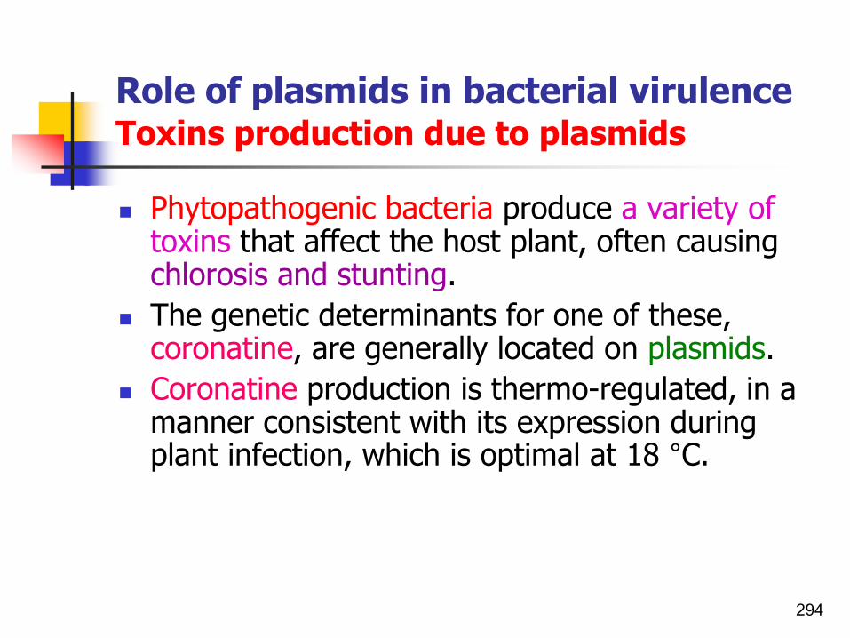
Role of plasmids in bacterial virulenceToxins production due to plasmids
◼ Phytopathogenic bacteria produce a variety of toxins that affect the host plant, often causing chlorosis and stunting.
◼ The genetic determinants for one of these, coronatine, are generally located on plasmids.
◼ Coronatine production is thermo-regulated, in a manner consistent with its expression during plant infection, which is optimal at 18 °C.
294

Role of plasmids in bacterial virulencePlasmid-borne cor genes in Ps. syringae
◼ In most strains of Ps. savastanoi pv. glycinea and Ps. syringae pv. tomato, coronatine production is specified by a 30 kb cor gene cluster located on large 90-100 kb plasmids.
◼ Coronatine is a virulence factor, enabling producer strains to form larger lesions and develop higher in planta populations; in this way it appears to confer a selective advantage in the natural habitat of these bacteria.
295

Role of plasmids in genetic diversityPlasmid content differences among X. fastidiosa strains
◼ Plasmids were in most of the X. fastidiosastrains.
◼ The grape strains having:
1. Similar chromosomal DNA,
2. Differed in plasmid DNA content.
◼ X. fastidiosa which causes citrus variegated chlorosis comprises a circular chromosome and two plasmids of 51,158 bp and 1,285 bp.
◼ Plasmid might be transferable between different X. fastidiosa strains.
296

Other plasmid-borne traitsEPS production
◼ As explained earlier, the plasmid pEa29 (previously described as a 28 kb plasmid) plays a role in the physiology or metabolism of E. amylovora.
◼ The control of extracellular polysaccharide (EPS) production has been associated with plasmid:
1. pEa29 in E. amylovora and with2. pJTPS1 in R. solanacearum. ◼ However, recent studies in R. solanacearum have
shown that high-level transcription of eps requires the products of at least seven regulatory genes that are chromosomally located.
◼ In both cases, these traits are associated with full virulence of the pathogen.
297

Other plasmid-borne traitsBacteriocin production
◼ Gram-negative and Gram-positive bacteria commonly harbor plasmid-borne genetic determinants of:
1. Bacteriocin production, and of
2. Host cell bacteriocin immunity.
◼ But some evidences (Chuang,1997) show that these genes are located on chromosomal DNA in P. carotovorum subsp. carotovorum strains.
298

Other plasmid-borne traitsProduction of a pink coloration
◼ Pantoea citrea causes a post-harvest disease of pineapple (called pink disease, which produces a pink coloration in canned pineapple due to the production of chromogenic 2,5-diketo gluconate from glucose.
◼ Although the genes gdhA and gdhB arechromosomal, plasmid pUCD5000 was required for full expression of the pink colour.
299

Insertion sequencesISs or IS elements
Occur in chromosomes of bacteria and plasmids
Insertion sequences (ISs) are approximately 1 kbp long jumping
genes found in prokaryotes.
300See also Bacterial Genetic File
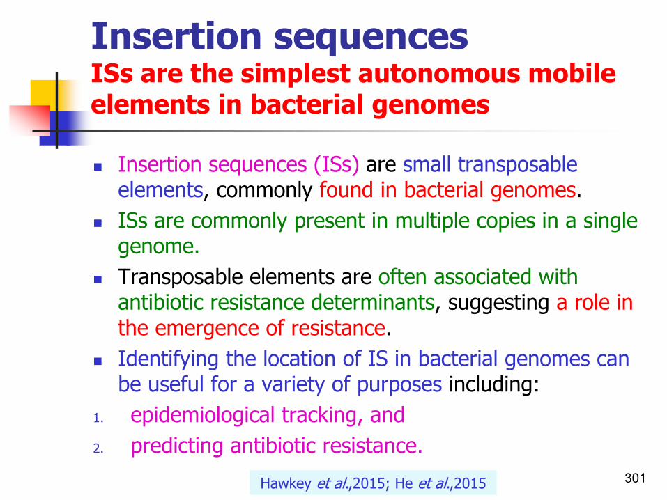
Insertion sequencesISs are the simplest autonomous mobile elements in bacterial genomes
◼ Insertion sequences (ISs) are small transposable elements, commonly found in bacterial genomes.
◼ ISs are commonly present in multiple copies in a single genome.
◼ Transposable elements are often associated with antibiotic resistance determinants, suggesting a role in the emergence of resistance.
◼ Identifying the location of IS in bacterial genomes can be useful for a variety of purposes including:
1. epidemiological tracking, and
2. predicting antibiotic resistance.
Hawkey et al.,2015; He et al.,2015 301

Insertion sequencesTwo major characteristics of ISs
◼ Insertion sequences have two major characteristics:
1. they are small relative to other transposable elements (generally around 700 to 2500 bp. in length) and
2. only code for proteins implicated in the transposition activity (they are thus different from other transposons, which also carry accessory genes such as antibiotic resistance genes).
Wikipedia,2015 302

Insertion sequencesDiagram illustrating the role of insertion sequences in a composite transposon
◼ ISs are typically composed of:
1. short terminal sequences, often arranged as inverted repeats (terminal inverted repeats [TIRs]) at their ends, and
2. at least one open reading frame (ORF) that encodes the transposase, the only protein essential formobility.
303Wikipedia,2015; He et al.,2015

ISMapper:Identifying insertion sequences in bacterial genomes from short read sequence data
◼ ISMapper, is a mapping-based tool for identification of the site and orientation of IS insertions in bacterial genomes, direct from paired-end short read data.
◼ ISMapper provides a rapid and robust method for identifying IS insertion sites direct from short read data, with a high degree of accuracy demonstrated across a wide range of bacteria.
304Hawkey et al.,2015

ISMapper:Workflow for ISMapper
305Hawkey et al.,2015

Insertion sequencesOn plasmid or chromosome
◼ Insertion sequence elements contribute to bacterial evolution in a number of ways.
◼ These sequences are mobile:
1. Within plasmids,
2. Between plasmids, and
3. Between plasmid and chromosome.
◼ IS elements can cause mutations by insertion within genes.
Sundin,2007 306

ISMapper:Identifying insertion sequences in bacterial genomes from short read sequence data
307Hawkey et al.,2015
ISAba1 positions detected in seven Acinetobacter baumannii genomes.
Issues can arise in the second mapping step of ISMapper if the IS insertion is next to a region that occurs more than once in the
reference genome.

ISMapper:Identifying insertion sequences in bacterial genomes from short read sequence data
308Hawkey et al.,2015
Analysis of IS6110 insertions in a diverse set of M. tuberculosis genomes.
ISAba1 detection rate as a function of read depth, for seven A. baumannii GC1
genomes sequenced with Illumina.

Insertion sequencesNumbers and numbering
◼ A particular insertion sequence may be named according to the form ISn, where n is a number.
◼ e.g.
◼ IS1, IS2, IS3, IS10, IS50, IS911, IS26, etc.
◼ The sequencing of Shigella genomes has revealed that a large number of insertion sequence (IS) elements (over 200 elements) reside in the genome.
309Wikipedia,2015;..

Insertion sequencesIS3 familyIn gram-negative and gram-positive bacteria
◼ The IS3 family is one of the largest families of IS elements, with members found in diverse bacterial groups including:
◼ Gram-negative rods and cocci,
◼ Gram-positive cocci,
◼ Mycobacteria, and
◼ Mycoplasmas.
Sundin,2007

Insertion SequencesIS3 family In some phytopathogenic bacteria
◼ Several IS elements encoded on phytopathogenic bacterial plasmids are members of the IS3 family including:
◼ IS476 from Xanthomonas campestris pvs. vesicatoriaand campestris,
◼ IS1389 from Xanthomonas campestris pv. amaranthicola,
◼ ISXac3 from Xanthomonas axonopodis pv. citri,
◼ ISPsy26 from P. syringae pv. tomato, and
◼ an unnamed IS element from pEA29in E. amylovora. Dendrogram based on alignment of the transposase
orfB gene frommembers of the IS3 family of insertion sequencesincluding several from phytopathogenic bacteria.Sundin,2007

An IS element: IS801In Pseudomonas syringae
312
Dendrogram based on alignment of the transposase gene from IS801 and
the related elements ISPsy3, IS91, and IS1294.
Sundin,2007
◼ The 1.5-kb element IS801 appears prominently in the evolution of certain P. syringae pathovars and appears to be absent in other pathovars.

Two IS elements:IS1405b and IS1405dIn R. solanacearum
A. A scheme of the subgroup-correlated insertion site of IS1405 with the position of each primer. The locations of IS1405b and IS1405d in 1.4 and 2.0-kb EcoRI fragments are specific to the A1 and B1 subgroup strains, respectively.
B. Agarose gel electrophoresis of PCR products from genomic DNAs of five representative A1 or B1 subgroup strains of R. solanacearum race 1 with PS-IS-F and PS-IS-RA1 or PS-IS-F and PS-IS-RB1 primer pairs, respectively.
◼ M, molecular size marker (1-kb DNA ladder, sizes (base pairs) are on the left. Lane N, a negative control.
313Lee et al.,2001

IS elementsIn Xanthomonas oryzae pv. oryzae
◼ Genome sequence of Xanthomonas oryzae pv.oryzaesuggests contribution of large numbers of effector genes and insertion sequences to its race diversity.
◼ Total of 611 ISs of 25 types were found in the genome of Xoo.
◼ Their ratio in relation to the whole genome was approximately 10%.
◼ This percentage was remarkably high compared with that of other sequenced plant-pathogenic which may be a characteristic feature of Xoo.
◼ ISs were located throughout the genome, and they were repeated tandemly in many loci.
◼ In some cases, multiple IS regions (IS islands) that encompassed about 30 kb were also present.
314

IS elements inXOO
315

Comparison of IS, proposed HrpX regulons and putative avr genes among the three Xanthomonas spp.
316

Bacterial pathogenesis on plantsPlant derived molecules
Plant Strategies for Defence1. R genes2. PGIPs3. Anti-microbial (antibacterial) proteins4. Phytoalexins5. Pathogenesis related proteins (PRs)
317

318

Plant derived molecules
◼ Resistance (R) genes: Leucine rich repeats.
◼ PGIPs: Polygalactoronase - inhibiting proteins which protect cell walls.
◼ Anti-microbial proteins: Phytoalexins.
◼ Pathogenesis related proteins (PR): Increase after infections (phenols, lignins, tannins,…).
◼ LAR (local acquired resistance) or SAR (systematic acquired resistance).
319

Resistance(R) genes
Leucine rich repeat regions
320

Plant resistance (R) genes
◼ Plants need to defend themselves against attack from viruses, microbes, invertebrates, and even other plants.
◼ Because plants lack a circulatory system, each plant cell must possess a preformed and/or inducible defense capability, so distinguishing plant defense from the vertebrate immune system.
321

Plant resistance (R) genesGeneral features of disease resistance genes
◼ The plant hosts, are not passive recipients of pathogens and have evolved complicated defense mechanisms that respond to effectors.
◼ Plants apparently express a large array of R genes in all living cells to detect the presence of pathogens.
◼ There will be many copies of each receptor on the surface of each plant cell.
◼ In compatible interactions, the plant sensors do not appear to be receptors for effectors but rather seem to monitor the cellular targets of the pathogen effectors.
322

Plant resistance (R) genesGeneral features of disease resistance genes
◼ Confer resistance to diverse pathogens such as virus, bacteria, fungus and insects.
◼ Induce a programmed cell death.
◼ Expression is constitutive.
◼ Belong to a large gene family.
◼ Arabidopsis genome sequence (125 Mb) encodes for ~150 NBS-LRR genes.
◼ Evolve in gene clusters.
323James et al., 2007

Plant resistance (R) genesGeneral features of disease resistance genes
◼ Basically, all these genes seem to encode components of receptor systems and form part of a signal transduction pathway, which triggers general defense reactions such as:
1. Reinforcement of the cell wall (cell wall thickening);
2. Synthesis of phytoalexins and oxidation of phenolic compounds;
3. Activation of defense-related genes, and4. The HR.
324

R genes in plant genome
◼ Resistance gene are often present as gene clusters of different specificities in the plant genome.
◼ Majority of these R genes show conserved DNA sequences and amino acid domains irrespective of whether they confer resistance to bacterial, fungal, viral, or nematode pathogens.
325

R genes common structure
◼ Though different classes of resistance genes exist, the Rgenes products have common structural features. i.e.
◼ Bipartite structure of:◼ Receptor domain, and ◼ Catalytic domain. ◼ Most R proteins contain:1. Leucine-rich repeats (LRRs),2. A central nucleotide-binding
site (NBS), and3. A variable amino-terminal
domain.
326
The Tir toll, leucine zipper domain can also be called coil-coil depending on origin. It has been suggested that TIR-NBS domain actually represents a
histidine-asparatic acid transfer domain.
a cytoplasmicTIR (Drosophila Toll/Interleukin-1 receptor) domain

Identification of resistance(R)
genes through genetics
◼ Plant breeders have successfully introduced disease resistance through introgression of foreign R-genes.
◼ However, pathogen races quickly evolve that lack the cognate avr genes.
◼ The adaptive ability of pathogens has limited the durability of most R-gene-based resistance.
◼ Sources of new R-genes are sought.◼ These may be introduced into commercial crop lines
through:1. Introgression (breeding), or by2. Genetic engineering. ◼ The latter requires molecular cloning of R genes (e.g. in
a wide host range cosmid cloning vector,pLARF1).
327

R genesPathogen specific genes
◼ Resistance genes which are pathogen specific are generally ineffective against other pathogens.
◼ Plants behave as a non-host for a bacterial pathogen, because it recognizes many of that bacteria's gene products as avirulent determinants.
◼ Therefore, many mutations are necessary to extend host range.
328
Avirulent= if the agent is avirulent it is a variant of a pathogen that does not cause severe disease. Nonvirulent= not virulent, harmless.

R genesPathogen specific genes
329
Race-specific R proteins
Race non-specific R proteins
RRS1 resistance to R. solanacearum; RS2 resistance to bacterial speck 2 (Xcv).

R genesAgainst necrotrophs
◼ Why are plant resistance genes that work against necrotrophs so rare?
◼ It has been suggested that necrotrophs are less dependent on suppression of plant defenses and may even benefit from induction of some defense pathways (Glazebrook,2005).
◼ This hypothesis remains to be broadly tested.
330Allen et al.,2009

R loci encoding for several specificities
◼ R genes are often found clustered on chromosomal loci.
◼ Any locus conferring race-specific resistance on a plant cultivar is called ‘specificity’.
◼ These specificities have been shown to exhibit a particular distribution within the plant genome.
◼ Many clusters of R loci encoding for several specificities have been found, especially in the case of resistances to biotrophic fungi.
331

R loci encoding for several specificities
◼ The non-random distribution of R loci in the plant genome is due to two types of genomic organization:
1. Allelic variation2. Chromosomal tandems and clusters of tightly linked
genes.
332
An allele is a variant form of a given gene. Sometimes, different alleles can result in different observable phenotypic
traits, such as different pigmentation.

R loci encoding for several specificitiesCf9 region of tomato plant and its different binding sites to perceive Avr9 of fungus Cladosporium fulvum
333

Kinds of R-genes
1. An enzymatic R-gene
e.g. Hm gene from maize and the HC-toxin from Cochliobolus(Helminthosporium) carbonum.
2. An antiviral R gene
e.g. N-gene (TMV/tobacco).
3. R-genes active against fungi
1. Cf9 (Cladosporium fulvum /tomato).
2. Pi-ta (Magnaporthe grisea /rice).
4. R genes against bacterial pathogens
a. Pto (Pseudomonas syringae /tomato).
b. Rps2, Rpm1 (Pseudomonas syringae /Arabidopsis).
c. Xa21 (Xanthomonas oryzae /rice). 334

Classes of resistance genesR-genes
◼ Some are composed predominantly of:
1. An LRR region, and
2. Transmembrane (TM) domain. ◼ Another is comprised of: 1. A LRR region, and2. A protein kinase domain.
335

Five major classes of R gene productsClasses of R proteinsBased upon their structural domains
336
◼ Class 1: Pto-Serine/Threonine Kinase
◼ Class 2: LRR-NBS-LZ/CC
◼ Class 3: LRR-NBS-TIR
◼ Class 4: Extracellular LRR (Cf genes)
◼ Class 5: Extracellular LRR-Serine thronine kinase
Martin et al.,2003
Class 1 differs from the other classes since it lacks leucine-rich repeats (LRR).

5 major classes of R proteinsLocalization of most R proteins
NB-LRR proteins are likely localized in the cytoplasm
337Dangl and Jones,2001
Class 2 Class 3 Class 4 Class 1
Prf
Class 5 Class 6

5 major classes of R proteins Localization of most R proteins
338Hammond-Kosack and Jones,1997

Five major classes of R gene productsBased upon their structural domainsPresence or absence of TM domain
◼ Class I: Consists of just one member, Pto from tomato.
◼ It has a possible membrane anchor site (myristoylation site) at its N-terminus and a serine-threonine kinase domain.
◼ No transmembrane (TM) domain for attachment to membranes and all are thought to be localized intracellularly.
◼ Class II: A large class of R proteins that have LRR-leucine
rich repeats, nucleotide binding site(NBS), LZ- leucine zipper or CC-coiled coil region and a signal peptide.
◼ e.g. flax-rust resistance gene L6.
◼ No transmembrane (TM) domain and serine-threoninekinase.
339

Pto gene from tomatoAvrPto is myristylated by the plant cell
◼ Protein myristoylation is a means by which cells anchor proteins into membranes.
340Bogdanove,2011

Five classes of R gene productsBased upon their structural domainsPresence or absence of TM domain
◼ Class III: Class 3 is similar to class 2 but instead of TZ or CC region it has a TIR domain at its N terminus, similar to Drosophila Toll protein and the mammalian interleukin-1 receptor (IL-R) protein. e.g. RPS2 from Arabidopsis.
◼ Also no TM.
◼ Class IV: Have transmembrane (TM), LRR and signal peptide domains. e.g. tomato Cf genes. It lacks the serine-threonine kinase domain.
◼ Class V: Xa21 of rice - Consists of just the Xa21 protein from rice. In addition to an extracellular LRR and a TM, it has a cytoplasmic serine/threonine kinase region and a signal peptide.
◼ Note the R proteins belonging to the first three classes lack transmembrane (TM) domains.
341

Five major classes of R genesLocalization of most R proteins
342

Features of the 5 major classes of R proteinsLocalization of most R proteins
343

The five major classes of plant
disease resistance genes
Martin et al.,2003

The five classes of cloned plant
disease resistance
genes
Hammond-Kosack and Jones,1997

Classes of resistance genesBased upon their structural domains
Takken and Joosten,2000 346

Class 6Some resistance genes do not fit into the five established classes of R gene products
◼ These (group 6) include several other proteins whose functions are not known
◼ In addition, the Hm1 toxin resistance gene is in this group:
Martin et al.,2003 347

Eight Classes ofresistance
genes
Hulbert et al.,2001

A complete list of isolated plant disease resistance genesWith indication of donor species, related disease and pathogen
Sanseverino et al.,2010

A complete list of isolated plant disease resistance genesWith indication of donor species, related disease and pathogen
Sanseverino et al.,2010

A complete list of isolated plant disease resistance genesWith indication of donor species, related disease and pathogen
Sanseverino et al.,2010
PRGdb is a web accessible open-source (http://www.prgdb.org) database that represents the first bioinformatic resource providing a comprehensive overview of resistance genes (R-genes) in plants.

Plant disease resistance genesPlant resistance gene database (PRGdb)
◼ The plant resistance gene database (PRGdb), is the first comprehensive bioinformatics resource dedicated to known and predicted plant disease resistance genes.
◼ It is a web accessible open-source (http://www.prgdb.org) database providing a comprehensive overview of resistance genes (R-genes) in plants.
◼ PRGdb holds more than 16000 known and putative R-genes belonging to 192 plant species challenged by 115 different pathogens and linked with useful biological information.
Sanseverino et al.,2010

Plant disease resistance genesPlant resistance gene database (PRGdb)
◼ The complete database includes:
1. A set of 73 manually curated reference R-genes,
2. 6308 putative R-genes collected from NCBI, and
3. 10463 computationally predicted putative R-genes.
Sanseverino et al.,2010

Sanseverino et al.,2010
Plant disease resistance genesPlant resistance gene database (PRGdb)

Plant resistance gene database (PRGdb) A schematic view of the PRG database showing the origin of dataset used and the sequences characterization
A. The manually curated dataset that contains 73 literature cited R-genes from 22 different plants.
B. The NCBI dataset containing 6308 sequences related to reference R-genes retrieved by the NCBI database.
C. The computationally predicted dataset using the DRAGO pipeline containing 10 463 putative R-genes.
D. Workflow of conserved domain analysis and sequence classification.
355Sanseverino et al.,2010

Plant disease resistance genesChromosomal organization of an R gene cluster
356Sophien Kamoun

Plant disease resistance genes Identification of resistance genes by the polymerase chain reaction
◼ There are a large number of sequences with similarity to R genes in plant genomes.
◼ PCR using degenerate primers designed to amplify sequences conserved between R genes has allowed identification of families of sequences from several plants.
357
Degenerate primers: These are actually mixtures of similar, but not identical primers. They may be convenient if the same gene is to be amplified from different
organisms, as the genes themselves are probably similar but not identical. Note: only a fraction of the degenerate 'primer' will actually work in the PCR
(See also Diagnosis-part 1 file)

Isolation and cloning of Pto homologue sequences from banana by degenerate PCRPto-like and receptor-like kinases in banana
◼ The main aims:
1. To isolate and characterize resistance gene homologues from banana.
2. To isolate and characterize BIBAC clones containing disease resistance gene homologues.
3. To generate a collection of BIBAC clones containing different classes of resistance gene homologues.
358James et al.,2007
A plant transformation-component binary bacterial artificial chromosome (BIBAC) library was constructed from Musa cv. Tuu Gia (AA).

Isolation and cloning of Pto homologue sequences from banana by degenerate PCRPto-like and receptor-like kinases in banana
359James et al.,2007

Isolation and cloning of Pto homologue sequences from banana by degenerate PCRPto-like and receptor-like kinases in banana
◼ Neighbour-joining tree based on the ClustalX alignment of the deduced amino acid sequence of 13 kinase-like sequences from banana, cv. ‘Tuu Gia’ (sequences in blue) and other plant protein kinases.
◼ The kinase catalytic domains (subdomains II to IX) were used to construct the phylogenetic tree.
◼ The number below the branches indicate the percentage of 1000 bootstrap replications supporting the particular nodes, and only those above 50% are shown.
◼ Sequences with blue circles correspond to banana Pto-like sequences and the sequence with a red circle corresponds to the tomato Pto resistance protein. 360James et al.,2007

Isolation and cloning of Pto homologue sequences from banana by degenerate PCRPto-like and receptor-like kinases in banana
◼ Towards the development of a collection of banana BIBAC clones containing disease resistance gene homologues.
361James et al.,2007

Common structural features of Rgenes productsThe structure of a NBS-LRR protein
362
Leucine-rich repeats (LRR): Segments of amino acids containing multiple copies of leucine present repeatedly in a protein; these proteins
are known as LRR proteins.

Major families of R proteins
363
◼ There are several different classes of R genes.◼ The major classes are the NBS-LRR genes.
◼ Within the NBS-LRR class of R genes are two subclasses:
1. One subclass has an amino-terminal Toll/Interleukin 1 receptor homology region (TIR). This includes the N resistance gene tobacco against TMV.
2. The other subclass does not contain a TIR and instead has a leucine zipper(LZ)region at its amino terminal.

Major families of R proteins
◼ NBS-LRR genes carries:1. Leucine-rich repeats
(LRRs), and2. Nucleotide-binding site
(NBS) domains.
◼ Leucine-rich repeats (LRRs, depicted in blue), have a major role in recognition specificity.
364McDowell and Woffenden,2003

Major classes of R proteinsPlant disease resistance proteins
◼ Most R genes contain Leucine- rich repeat motifs (LRR region), suggesting that plants may share common mechanisms for disease resistance to diverse pathogens.
365
Major classes of R proteins. The widely represented R-protein family (NB-LRR) also contains a nucleotide-binding site (NBS). NB-LRRs are localized in cytoplasm as membrane proteins and contain either a TIR domain (L6 , N) homologous to metazoan Toll-interleukin receptor or a putative coiled-coil (RPS2) domain at the
N-terminus. A recent modification identified in the TIR-NBS-LRR protein is an additional transcriptional factor WRKY.
Joshi and Nayak,2011

Major classes of R proteinsPlant disease resistance proteins
366
Alignment of the NBS domain of multiple NBS-LRR disease resistance proteins.The conserved NBS domains as determined by Meyers et al. (1999) are indicated.
Identical amino acids are shaded in black and conservative substitutions are shaded in gray. NBS = nucleotide-binding site; LRR = leucine-rich repeat.
Joshi and Nayak,2011

Classes of resistance geneCommon motifs
Protein motifs indicate that R genes are part of signal-transduction systems. 367

Classes of resistance geneShared structural domains among plant, insect, and mammalian defense and developmental pathway genes
368Baker et al.,1997
TIR, Toll–IL-1R homology domain; kinase, serine-threonine kinase; LZ, leucine zipper; Rel, Rel-related transcription factors (Dorsal and Dif of
Drosophila and NF-κB of mammals) and inhibitors (Cactus of Drosophila and IκB of mammals).

R gene functions
◼ R-gene-encoded proteins have to fulfill two tasks:1. To recognize a pathogen-derived signal,2. To initiate a coordinated plant defense
reaction.◼ In another word:a. Effector recognition- Enable plants to detect avr
gene specified pathogen molecules.b. Initiate signal transduction to activate defenses. c. Have the capacity to evolve new R gene specificities
rapidly.
369

Recognition and signal transduction functions of R genes
◼ Resistance gene products do not act alone in controlling defense reactions.
◼ Different regions of an Rgene product must function together to conduct both the:
1. Pathogen recognition, and
2. Signal transduction functions.
370

1. LRR domainStructure and functions
371
◼ Structure:◼ LRR domains are typically thought to be the major
determinant of specificity in R genes that carry them based on their known history in other proteins and the high levels of polymorphism between alleles in these domains.
◼ A short stretch of amino acid residues with leucine at every second or third position is repeated to form a flexible, solvent-exposed, parallel beta-sheet.
◼ Function:1. The LRRs are mainly involved in recognition, whereas2. the amino-terminal domain determines signalling
specificity.◼ Have a major role in recognition specificity.

R-gene products are postulated to have receptor and effector domains
◼ A variety of motifs suggest involvement in protein-protein interactions; these could be involved in ligand binding or effector functions.
◼ This hypothetical structure shows the thyrotropin receptor.
◼ In the consensus sequence for the β-strand, (x) any amino acid; (a) aliphatic residues.
372Thyrotropin: A tripeptide hormone. Aliphatic: A protein
side chain contains only carbon or hydrogen atoms.

The crystal structure of a protein containing LRRs
◼ Ribbon diagram of the crystal structure of a protein(ribonuclease inhibitor) that contains leucine-rich repeats (LRRs).
◼ Each LRR is an iterative(repeat) unit composed of:
1. A roughly parallel α helix (coil structures) joined by a short loop, and
2. A β sheet (inner surface).◼ Because the α helices pack less
tightly, the molecule forms a horseshoe-shaped structure.
373

2. NB-ARC domainStructure and functions
◼ The NBS forms part of NBARC-LRR domain that presumably functions as a molecular switches of plant defense.
◼ May affect R protein function through nucleotide binding or hydrolysis.
◼ In AvrPto/Pto interaction(Pst/tomato), the tomato NBARC-LRR protein Prf interacts with Pto Kinase to regulate specific plant immunity.
374

Model for the switch function of NBS-LRR proteins
◼ In the absence of a pathogen, an NBS-LRR protein is in the OFF-state (resting state), in which the LRR exerts its negative role by stabilizing the ADP-bound state.
◼ The presence of an elicitor (Avr) affects the LRR domain, which induces a conformational change in the NB-ARC domainthat allows the release of ADP.
◼ ATP binding subsequently triggers a second conformational change in the N-terminal effector domain, releasing its signalling potential.
◼ The ATPase activity of the protein attenuates the signalling response and returns the protein to its resting state.
Takken et al.,2006375

3. TIR domainStructure and functions
◼ Some NB-LRR proteins contain a TIR domain at N terminus.
1. Can play a role in pathogen recognition.
2. Implicated in signaling.
376
The Tir toll, leucine zipper(LZ) domain can also be called coil-coildepending on origin.

4. CC domainStructure and functions
◼ Other NB-LRR proteins contain a putative coiled-coildomain (CC) at the N-terminus.
◼ The CC structure is a repeated heptad sequence with interspersed hydrophobic amino acid residues, of which the leucine zipper is one example.
◼ This domain may be involved in signaling rather than recognition.
377
The Tir toll, leucine zipper(LZ) domain can also be called coil-coildepending on origin.

PtoThe first cloned R genePrf functions as an independent resistant gene
◼ Pto and Prf with cooperative function
◼ The Pto gene encodes a Ser/Thr kinase but has no apparent receptor domain i.e. LRR.
◼ It requires the presence of a linked NBS-LRR gene, prf, for activity.
◼ Tomato Prf is a member of the Leucine-Rich Repeat.
◼ Prf can also function as an independent host recognition determinant of bacterial infection.
◼ Pto is a resistance gene which is an argument against the guard hypothesis.
378See also decoy model in this file

Tomato R-genesPto and Prf with cooperative function
◼ Pto and Prf are clustered at a single genomic locus with four Pto homologs, which suggests cooperative function.
◼ Pto appears to signal by a conformational change rather than by phosphorylation of a downstream substrate(s).
◼ Prf on the other hand, is a signaling protein regulated by Pto.
◼ Pto and Prf act coincidentally in signal transduction.
379
Mucyn et al.,2006
See also NBARC-LRR proteins

Tomato R-genesPto and Prf with cooperative function
380
Pto gene has not LRR
Prf has LRR

381

PtoBThe second R gene
◼ A second avirulence gene product, AvrPtoB, interacts with Pto in tomato to trigger a resistance response.
◼ This response is the classical hypersensitive response (HR) that involves rapid, localized cell death.
◼ This is an example of programmed cell death (PCD)
in plants.
◼ There is an increasing amount of evidence that some bacterial avirulence products such as AvrPtoB can
promote virulence by interfering with the plant defense response.
382

R genes diverse pathogens encode proteins with leucine-rich repeats (LRRs )
383

Xa27 Confers resistance to a variety of Xoo strains
Zhaokaijun 384
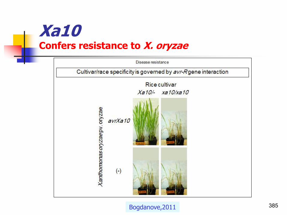
Xa10 Confers resistance to X. oryzae
385Bogdanove,2011

Bacterial pathogenesis on plantsavr/R interactions
Plant-Microbe Interactions
1. Elicitor recognition (PAMPs/PRRs interactions)2. Gene-for-gene hypothesis (Avr/R interactions)3. The guard hypothesis4. Signal transduction5. Mechanisms of general defences
386

Pathogens proteinsPAMPs/MAMPsGeneral elicitors derived from structural components of the bacterial cell
◼ A relatively recent concept.
◼ Plants can recognize certain broadly-conserved molecules associated with pathogens- PAMPs (pathogen-associated molecular patterns).
◼ Also known as MAMPs (Microbe-associated molecular patterns)
◼ Examples include:
1. Flagellin;
2. Elongation factor-Tu;
3. LPS;
4. etc.
Roger Innes. Tom Ashfield Laura Ong

Pathogens proteinsPAMPs/MAMPsGeneral elicitors derived from structural components of the bacterial cell
◼ The bacterial cell wall is an important source of PAMPs. e.g.
1. Peptidoglycans(PGNs) from both gram-positive and gram negative bacteria.
2. Lipopolysaccharide (LPS) is the principal component of the outer membrane of gram-negative bacteria.
3. Flagella(Flagellin) from nearly all bacteria.
4. The elongation factor Tu (EF-Tu) is one of the most abundant and conserved bacterial proteins which acts as a very potent bacterial PAMP.
5. Chitin
6. The bacterial siderophore pseudobactin is also a potential PAMP.
388Nicaise et al.,2009;..

Plant resistant genesKind of receptorsPAMPs/MAMPs vs. PRRs
◼ Receptor-like kinases (RLKs) and Receptor-like proteins (RLPs) play crucial roles in plant immunity, growth, and development.
◼ Plants deploy a large number of RLKs and RLPs as pattern recognition receptors (PRRs) that detect microbe- and host-derived molecular patterns as the first layer of inducible defense.
389

Plant resistant genesKind of receptorsPAMPs/MAMPs vs. PRRs
◼ Plants and animals recognize microbial invaders by detecting:
1. Pathogen genes(effectors), and
2. Pathogen associated molecular patterns (PAMPs) such as flagellin or LPS.
◼ There are several different classes of R genes.
◼ The major classes are:
1. The R genes, and
2. plant receptor-like kinases (RLKs) and Receptor-like proteins (RLPs) known as pattern recognition receptors or surface proteins (PRRs).
390

391
Name Family Ligand Plant
FLS2 LRR-RK flg22 Arabidopsis
FLS3 LRR-RK flgII-28 Tomato
EFR LRR-RK elf18 Arabidopsis
PEPR1/2 LRR-RK Peps Arabidopsis
XA21 LRR-RK RaxX21-sY Rice
DORN1 Lectin-RK eATP Arabidopsis
LORE Lectin-RK LPS Arabidopsis
WAK1 EGF-Like-RLK OGs Arabidopsis
XPS1 LRR-RK xup25 Arabidopsis
OsCERK1 LysM-RLK Chitin Rice
CEBiP LysM-RP Chitin Rice
LYM1/3 LysM-RP PGNs Arabidopsis
LYP4/6 LysM-RLP PGNs/chitin Rice
RLP23 LRR-RP nlp20 Arabidopsis
NbCSPR LRR-RP csp22 N. benthamiana
LeEix1 LRR RP Eix Tomato
LeEix2 LRR-RP Eix Tomato
ReMax/RLP1 LRR-RLP eMax Arabidopsis
Ve1 LRR-RLP Ave1 Tomato
Cf-2 LRR-RLP Avr2 Tomato
Cf-4 LRR-RLP Avr4 Tomato
Cf-4E LRR-RLP Avr4E Tomato
Cf-9 LRR-RLP Avr9 Tomato
Cf-5 LRR-RLP Unknown Tomato
RLP30 LRR-RLP SCFE1 Arabidopsis
ELR LRR-RLP Elicitin Potato
Breiden and Simon,2016
Known and Probable PRRs Involved in Plant Immunity.
Known PRRs refer to genetically confirmed
receptors whose binding to patterns has been
biochemically demonstrated, whereas probable PRRs refer to likely receptors with genetically confirmed
roles in pattern recognition, but for
which direct binding to patterns remains to be
shown.

Pathogens proteinsKind of receptors PAMPs/MAMPs vs. PRRs
◼ While some RLKs and RLPs are known to act as PRRsthat perceive danger signals,
◼ Others regulate
◼ plant growth and development,
◼ reproduction,
◼ symbiosis, and
◼ tolerance to abiotic stresses.
Breiden and Simon, 2016

Pathogens proteinsKind of receptors PAMPs/MAMPs vs. PRRs
◼ PAMPs carry out an essential function for fitness or survival.
◼ Plants recognize a wide range of bacterial PAMPs, most of which are derived from structural components of the bacterial cell(non specific).
◼ PAMPs are recognized by PRRs(pattern recognition receptors or surface proteins).
Nicaise et al.,2009

Pathogens proteins1.PAMPs-PRRs interactionsGeneral elicitors derived from structural components of the bacterial cell
◼ PAMPs and PRRs activate the plant basal defence responses.
◼ This detection leads to PAMP-triggered immunity (PTI), which effectively prevents colonization of plant tissues by non-pathogens.
◼ The most well studied system in PTI is the FLS2-dependent pathway.
◼ FLS2 recognizes the PAMP flg22 that is a component of bacterial flagellin.
394Nicaise et al.,2009; Chakravarthy et al.,2009
See also Zigzag theory

Plant resistant genesThe receptorsPlant receptor-like kinases (RLKs) proteins
◼ The transmembrane kinases are collectively named the receptor-like kinases(RLKs).
◼ Receptor-like kinases (RLKs) are surface localized, transmembrane receptors comprising a large family of well-studied kinases.
◼ RLKs participate in a diverse range of processes, including:
1. disease resistance,
2. regulation of development hormone perception, and
3. several interacting components upstream or downstream to the receptor protein kinases.
395

396
◼ RLKs possess three domains:
1. an N-terminal (extracellular) domain,
2. an intermediate transmembranedomain,
3. and a C-terminal kinase domain.
◼ The PRKs can be classified on the basis of their extracellular domains.
Shiu and Bleecker,2001

Plant R genesThe receptors
397Rouxel and Balesdent,2010

Gene-for-gene hypothesis2. Avr/R interactionsFlor concept
◼ The gene-for-gene hypothesis (Flor,1971; Keen,1990) explains host-pathogen recognition events.
◼ Plants with a particular resistance gene (R gene) recognize a pathogen-produced elicitor, a direct or indirect product of the corresponding avirulence (avr) gene.
398

Harold Henry Flor, 1900-1991 Noel Thomas Keen, 1940-2002
◼ He coined the term "elicitor" to describe the chemicals given off by invading organisms that elicited phytoalexin response in the host plant.
◼ This work led him to identify the single gene in P. syringae responsible for producing elicitors.
399
◼ In the 1940s, Flor developed the gene-for-gene concept to explain the genetic interactions between Melampsora lini and flax.
◼ This concept provided the underpinnings for research on the genetics of host-pathogen interactions for the next 70 years.
Pioneer leaders in the field of molecular plant pathology
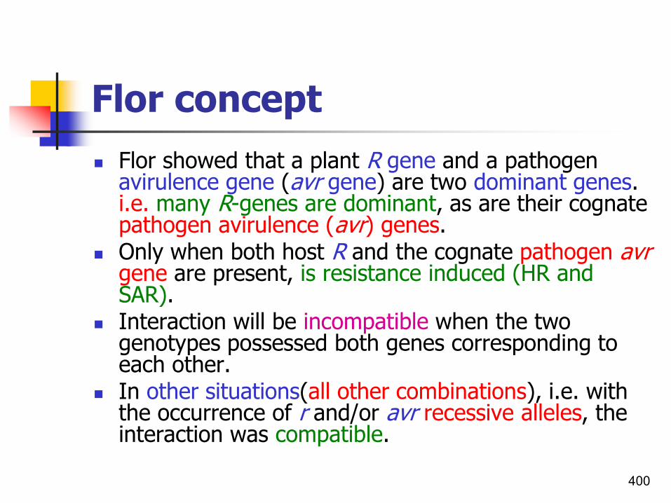
Flor concept
◼ Flor showed that a plant R gene and a pathogen avirulence gene (avr gene) are two dominant genes. i.e. many R-genes are dominant, as are their cognate pathogen avirulence (avr) genes.
◼ Only when both host R and the cognate pathogen avrgene are present, is resistance induced (HR and SAR).
◼ Interaction will be incompatible when the two genotypes possessed both genes corresponding to each other.
◼ In other situations(all other combinations), i.e. with the occurrence of r and/or avr recessive alleles, the interaction was compatible.
400

R genes
◼ At each locus, a diploid organism possesses two alleles located on homologous chromosomes, one inherited from each parent.
◼ These alleles may be
1. The same homozygous(two of the same alleles at a locus), or
2. Heterozygous (two of different alleles at a locus).
401

Gene-for-gene model
402
Virulence based on simple inheritance of asingle recessive gene for virulence.

Gene-for-gene model
403Warwick,2009

Gene-for-gene interaction between host and pathogen
404
Virulence based on simple inheritance of a single recessive gene for virulence.

Gene-for-gene modelHost-pathogen gene-for-gene specificity model
405Adapted from Lucas,1998

Gene-for-gene modelGene specific interactions
◼ Resistance is only inducedwhen a plant cultivar in possession of a specific resistance (R) generecognizes a pathogen race that contains:
a) The corresponding avirulence (avr) gene,
b) The absence of either the avirulence gene,
c) The resistance gene (R), ord) absent in both interacting
organisms which leads to lack of recognition by the host plant and the onset of disease.
406
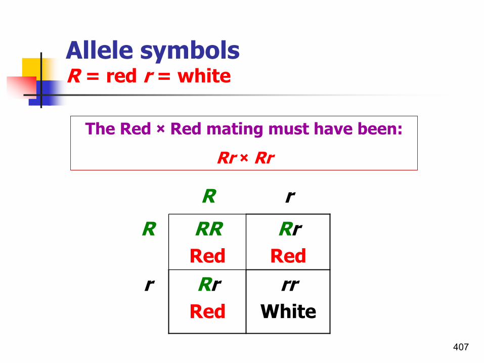
The Red × Red mating must have been:
Rr × Rr
R r
R RR
Red
Rr
Red
r Rr
Red
rr
White
Allele symbolsR = red r = white
407

R r
V R S
v S S
Pathogen Phenotype
HOST
Virulence based on simple inheritance of a
single recessive gene for virulence
Resistance determinedby single dominant gene
408

Resistance determined by single dominant gene
Pathogen Phenotype
HOST
Virulence based on simple inheritance of a
single recessive gene for virulence
R1R2 R1r2 r1R2 r1r2
V1V2 R R R S
V1v2 R R S S
v1V2 R S R S
v1v2 S S S S409

Elicitors
Introduction
410

DefinitionThe term elicitor
◼ A physical, chemical or biological stimulus that triggers defence responses in plants.
◼ Originally the term elicitor was used for molecules capable of inducing the production of phytoalexins, but it is now commonly used for compounds stimulating any type of plant defence.
◼ This broader definition of elicitors includes:◼ Both substances of pathogen origin (exogenous
elicitors) and compounds released from plants by the action of the pathogen (endogenous elicitors).
411

Classification of elicitors
◼ According Radman et al., 2003, elicitors are classified as:
◼ Physical or chemical;
◼ Biotic or abiotic;
◼ Complex or defined, depending on their origin and molecular structure.
412Angelova et al.,2006

Classification of elicitors
◼ Elicitors may be also classified into two groups:1. General elicitors: Including oligosaccharides,
glycopeptides, and peptides (flagellins).2. Race specific elicitors: While general elicitors
are able to trigger defence both in host and non-host plants, race specific elicitors induce defence responses leading to disease resistance only in specific host cultivars.
413

General elicitors Elicitors of plant defences
414

Elicitors of plant defences
◼ Plants are able to recognize pathogen-derived molecules; elicitors that trigger a number of induced defences in plants.
◼ Microbial elicitors
◼ Constitute a bewildering array of compounds including different:
1. Oligosaccharides,
2. Lipids,
3. Peptides, and
4. Proteins.
415

Elicitor of pathogens Signal compounds different form toxins
◼ Elicitors act as signal compounds at low concentrations, providing information for the plant to trigger defence, distinguishing elicitors from toxins, which may act:
1. Only at higher concentrations, and/or2. Affect the plant detrimentally without active
plant metabolism.
416

Elicitor of pathogensAvr-R interactions
◼ Plant recognition of pathogens is mediated by large families of highly polymorphic R genes.
◼ The products of these genes function to recognize directly or indirectly the products of pathogen-encoded avr genes.
417

Pathogens genesavr genes
◼ Many plant and animal pathogens are evolutionary cousins that have the ability and propensity to share genetic material.
◼ The greater evolutionary potential was seen in bacterial pathogens, due to their rapid generation time and ability to exchange genetic materialhorizontally.
◼ The pathogen avr genes are only present in certain strains of a given pathogen and show little similarity to each other.
418

Plant resistant genesR genesInteractions of bacterial avirulence products with R gene products
◼ Major gene resistance:◼ Confers a high level of resistance.
◼ If pathogens exist as distinct races, avirulence genes in the pathogen are matched by corresponding resistance genes in the plant (gene-for-gene complementarity).
◼ Minor gene resistance:◼ Confers a low level of resistance.
◼ In addition to race-specific resistance genes, plants also possess other, none race-specific, resistance genes which are equally effective against all races of the pathogen (minor gene resistance).
419

The R and Avr genes control pathogen recognition Simple Model:
The Avr gene encodes a factor (a specific ELICITOR) that directly or indirectly interacts with the product of an R gene
Pathogen
Avr Gene
Plant
R GeneELICITOR
Recognition of the elicitor triggers defense responsesincluding the hypersensitive
response

Elicitor recognitionAvr-R interactionsProposed models
421
◼ Different models were proposed for R protein recognition of Avr proteins, mainly:
1. Ligand-Receptor model
2. Guard model
3. Affinity enhancement model
◼ These can be explained as direct or indirect interactions.

Elicitor recognitionDirect elicitor – receptor interaction
1. Ligand-receptor interactions
422

Direct physical interactionLigand-receptor interactions
1. In ligand/receptor interactions, or
2. Elicitor-receptor interactions, or
3. avr gene and pathogen or plant surface-derived elicitors
◼ Serve as ligands for receptors (R genes)located in the plasma membrane or in the cytosol (cytoplasm).
423

Direct physical interactionLigand-receptor interactionsAVR/R and PAMP/PRR interactions
424
1. AVR/R or effectors interactions:
◼ Effectors are pathogen-secreted proteins that manipulate host cell functions.
◼ Avr recognition by plants has been coined effector-triggered immunity.
2. PAMP/PRR interactions:
◼ PAMPs define molecular motifs common to many pathogens.
◼ Pathogen-associated molecular pattern (PAMP) triggered immunity (innate immunity/PTI).

Direct physical interactionLigand-receptor interactions
◼ Direct interaction between Avr proteins and R proteins was indeed demonstrated in a few cases by the yeast two-hybrid system and the immuno-precipitation.
◼ Examples:
◼ Fungal Pita(Pi-ta)-AvrPita (AVR/R interaction);
◼ Bacterial RRS-1R- popP2 interaction (AVR/R interaction);
◼ Bacterial FLS2 -flg22 interaction(PAMP/PRR interaction).
425
◼ Yeast two-hybrid system: An experimental technique of detecting the protein-protein interactions in yeast cells.
◼ Two-hybrid system: The yeast or bacterial system that is employed for detecting specific protein-protein interaction; the protein of interest is used as a “bait” to “fishout” proteins that may bind to it (referred to as “prey”).

The direct physical interactionLigand-receptor interactionsAVR/R interactions
◼ Among different R genes, very few NB-LRR proteins (part of R proteins) have been found to directly
interact with Avr proteins.
◼ Indeed, direct binding of a few Avr-R combinations was found, consistent with a receptor-ligand mode of action.
◼ However, for a number of Avr-R combinations, physical interactions have not been observed, and perception is thought to be indirect.
426van der Hoorna Kamounb,2008

Direct receptor-elicitor interactionLigand-receptor interactionsPAMP/PRRs interactions
◼ PRRs (pattern recognition receptors) watch over and raise the alarm.
◼ Perception of PAMPs (flagella, LPS,..) by PRRs is common to all multicellular organisms and leads to an array of defense responses and redeployment of cellular energy in a fast, efficient, and multi response manner, which prevents further pathogen ingress.
◼ PAMP recognition leads to a chain of signaling events, broadly referred to as general defense responses in plants.
◼ PAMP perception also results in plant systemic acquired resistance (Mishina and Zeier,2007b).
427Nicaise et al.,2009

Direct receptor-elicitor interactionLigand-receptor interactionsPAMP/PRRs interactions
◼ The active peptides are present in a variety of pathogenic bacteria and perceived at the nanomolar level by a broad range of plant species.
◼ The peptide flg22 (corresponding to 22 amino acids localized in the conserved region of flagellin) elicits responses in most plant species and is as active as the full-length flagellin.
◼ The flg22 region is required for bacterial virulence and motility, consistent with the fact that PAMP mutation has a fitness cost for microbes.
◼ The conserved segment of bacterial flagellin was recently demonstrated to be a potent elicitor of defense gene expression.
◼ The recent finding that flg22, as well as flagellin, induces the hypersensitive response.
428Nicaise et al.,2009

Direct receptor-elicitor interactionLigand-receptor interactionsPAMP/PRRs interactions
◼ Rice (Oryza sativa) was reported to be insensitive to flg22 (Felix et al.,1999).
◼ However, recent results showed that flg22 is recognized by rice but that this response is weaker than with full-length flagellin (Takai et al.,2008).
429Nicaise et al.,2009

Direct receptor-elicitor interactionFlagellin as potent elicitor(PAMP)Model for flagellin signaling in Arabidopsis
1. Bacteria move on the plant surface and enter the plant via natural openings, wounds or hydathodes.
2. Bacteria multiply on the surface or in the intercellular space of the host plant.
3. Flagellin (released from bacterial flagellae) or flagellin fragments resembling flg22, are recognized directly or indirectly by the flagellin receptor at the plant plasma membrane (PM), a key part of which is FLS2.
4. Autophosphorylation of FLS2.
A sustained increase in cytosolic calcium which was specific in gene-for-gene interactions 430

Direct receptor-elicitor interaction RRS1-PopP2 direct or indirect effector interactions
◼ The products of two groups of avirulence genes with virulence effects appear to be targeted to specific organelles within the host cell.
◼ Two effectors, AvrBs3 from X. vesicatoria and PopP2 from R .solanacearum have recently been shown to be localized in plant nuclei by immunolocalization and monitoring GFP fusions, respectively.
◼ In the case of PopP2, the corresponding RRS1 resistance gene was co-localized in the nucleus only in the presence of PopP2.
431

a) Direct interaction.b) Importin α-mediated
interaction model: Leads to nuclear import of RRS1-R and activation of defense-related genes.
c) SUMO(a small ubiquitin-like modifier)-mediated interaction model: The interaction between RRS1-R and PopP2 is mediated by SUMO.
• SUMO protease activity affects host transcription.
◼ Imp , importin α;
◼ LRR, leucine-rich repeat domain;
◼ NB, nucleotide-binding site;
◼ NLS, nuclear localization signal;
◼ S, SUMO, A small ubiquitin-like modifier;
◼ TIR, homologous to Toll/interleukin-1-receptor;
◼ TTS, type-III secretion system;
◼ WRKYs, DNA-binding proteins, regulating gene expression for plant defense.
Direct receptor-elicitor interaction RRS1-PopP2 direct or indirect effector interactions

Elicitor recognitionIndirect interactions of AVR-R proteins
Guard, Decoy and Integrated decoy models
433Most recent slides in Bacterial Disease Management-Part II

Indirect interactions of AVR-R proteins Comparison of different Guard, Decoy and Integrated decoy models
1. In the guard model, NLR receptors perceive modifications in guardees that are introduced by microbial effector proteins.
2. Guardees often have a function in basal plant defense and they are therefore frequently targeted by effectors.
3. The decoy is a duplicated guardee without a function in plant immunity. Its sole role is to trap effectors, thereby activating the immune signaling cascade.
434Krattinger and Jeller,2016
NLR: A major class of plant immune receptors called nucleotide-binding domain and leucine-rich repeat
containing proteins.

Indirect interactions of AVR-R proteins Comparison of different Guard, Decoy and Integrated decoy models
◼ The guardee or decoy and the monitoring NLR are often encoded by different genes that likely bind to each other, but can dissociate.
◼ The integrated domain model involves a fusion between the decoy domain and the respective NLR, which together are encoded by one gene.
435Krattinger and Jeller,2016
NLR: A major class of plant immune receptors called nucleotide-binding domain and leucine-rich repeat containing proteins

Genetic tests to discriminate between the Guard and Decoy Models
◼ Plants lacking both the R protein and the presumed operative target (s) should be challenged with pathogens in the absence or presence of the guardee/decoy.
1. A differential pathogen growth supports the Guard Model, whereas
2. An unaffected pathogen growth supports the Decoy Model.
◼ The test of choice depends on the nature of the effector:
A. Effectors that promote positive effects on pathogen growth by manipulating their target should be present during the test to reveal target contributions.
B. Effectors that prevent negative effects on pathogen growth should be omitted to avoid them from suppressing a possible phenotype.
436van der Hoorn and Kamounb,2008

437
Comparisons of the Guard and Decoy Models
A) The classical Guard Model is contrasted with a B) modified Guard Model in which the effector targets multiple plant proteins and c) the Decoy Model.
Effectors are depicted in gray, operative effector targets in purple, guardee in green, decoy in blue, and the R protein in orange. Operative target: Host target that when manipulated by a
(.pathogen effector results in enhanced pathogen fitness (van der Hoorna Kamounb,2008

Genetic tests to discriminate between the Guard and Decoy Models
◼ Plants lacking both the R protein and the presumed operative target(s) should be challenged with pathogens in the absence or presence of the guardee/decoy.
◼ A differential pathogen growth supports the Guard Model, whereas an unaffected pathogen growth supports the Decoy Model. The test of choice depends on the nature of the effector.
A. Effectors that promote positive effects on pathogen growth by manipulating their target should be present during the test to reveal target contributions.
B. Effectors that prevent negative effects on pathogen growth should be omitted to avoid them from suppressing a possible phenotype.
438van der Hoorn and Kamounb,2008

Elicitor recognitionIndirect interactions of AVR-R proteins
1. Guard hypothesis
439

The Guard hypothesisEvidence against direct interaction
◼ In many (most?) cases direct interaction between R-gene and Avr gene products can not be observed.
◼ If there are only a few hundred R-genes, this seems insufficient to deal with the huge number of pathogens around leads to hypotheses that some R-genes recognise Avr gene products indirectly.
P. Balint-Kurti Lecture 2

The Guard hypothesisIndirect interactions of AVR-R proteins
◼ This Guard Model predicts that R proteins act by monitoring (guarding) the effector target and that modification of this target by the effector results in the activation of the R protein, which triggers disease resistance in the host (Van der Biezen and Jones,1998; Dangl and Jones,2001).
441van der Hoorna Kamounb,2008
Guardee: Effector target required for R protein function and with a function in host defense or susceptibility in the absence of its cognate R protein; effector
alteration of the guardee results in enhanced pathogen fitness in plants that lack the R protein and triggers innate immunity in plants that carry the R protein.

The Guard hypothesisIndirect interactions of AVR-R proteins
◼ According to the ‘guard model’, Avr product does not directly interact with an R-protein, but at least three players are needed to trigger resistance.
◼ The third player is called as the guardee(pathogenicity target proteins/target host proteins/host plant proteins) which act as a molecular glue between complementary Avr and R product is required.
442

The Guard hypothesisIndirect interactions of AVR-R proteins
443Gururani,2012
◼ The plant R proteins (guard) are associated with the endogenous host protein (guardee) which are common target proteins for the pathogens.
◼ The interaction of effector pathogen proteins with the host proteins, causes a change in their structure which is then recognized by the guard proteins.
◼ As a result, a pathogen response signaling cascade is triggered against the microbial evasion.
Guardee R gene

The Guard model and Ligand-Receptor modelDirect and indirect protein interactions
a) The plant cell does not express an R protein that is capable recognizing any virulence protein (disease).
b) R protein directly bind a virulence protein(Classic receptor-elicitor hypothesis).
c) The guard hypothesis, R protein(guard) detects a modified host protein (guardee, red star).
444
Virulence proteins (red) are translocated into plant clells via TTSS.
McDowell and Woffenden,2003

The Guard hypothesisRIN4, the guardee protein in ArabidopsisIn 5 rounds
Round 1 Round 2
Roger Innes. Tom Ashfield Laura Ong

The Guard hypothesisRIN4, the guardee protein in ArabidopsisIn 5 rounds
Round 4Round 3
Roger Innes. Tom Ashfield Laura Ong

The Guard hypothesisRIN4, the guardee protein in ArabidopsisIn 5 rounds
◼ So Rin4 is guarded by at least two R-genes in Arabidopsis:
1. RPM1(mediates AvrBrecognition), and
2. RPS2 (mediated AvrRpt2 recognition).
Roger Innes. Tom Ashfield Laura Ong
Round 5

Evidence for the guard hypothesisIndirect protein interactionsAvrPphB and RPS5+ PBS1 interactions
◼ RPS5 is a NBS-LRR protein from Arabidopsis.
◼ Recognizes AvrPphB from P. syringae.
◼ When RPS5 is expressed in tobacco it will only mediate HR in the presence of both AvrPphB and another Arabidopsis protein PBS1.
◼ RPS5 and PBS1 form complex in the cell.
◼ AvrPphB cleaves PBS1 triggering HR controlled by RPS5.
448P. Balint-Kurti Lecture 2

Evidence for the guard hypothesisIndirect protein interactionsAvrPphB and RPS5+ PBS1 interactions
449Mudgett,2005
◼ Model of AvrPphB cysteine protease action in resistant RPS5 Arabidopsis plants infected with Pseudomonas syringae pv. phaseolicola.

Elicitor recognitionIndirect interactions of AVR-R proteins
2. Decoy Model
450

Decoy ModelA new model for perception of plant pathogen effectors
◼ Over the past few years, new data on indirectly recognized effectors have emerged that are inconsistent with the original description of the Guard Model.
◼ The Guard Model for disease resistance postulates that plant resistance proteins act by monitoring (guarding) the target of their corresponding pathogen effector.
◼ We posit, however, that guarded effector targets are evolutionarily unstable in plant populations polymorphic for resistance (R) genes.
451van der Hoorn and Kamounb,2008

Decoy ModelA new model for perception of plant pathogen effectors
452
◼ A decoy protein whose sole purpose is to be an effector target and act as a “pathogen detector”.
◼ Once the guard interaction is set up, the guarded targets are subject to two opposing forces of selection.
Roger Innes. Tom Ashfield Laura Ong

Decoy ModelA new model for perception of plant pathogen effectors
◼ Depending on the absence or presence of the R gene, guarded effector targets are subject to opposing selection forces:
1. To evade manipulation by effectors (weaker interaction), and
2. To improve perception of effectors (stronger interaction).
◼ There is growing support for this Decoy Model from four diverse cases of effector perception involving Pto, Bs3, RCR3, and RIN4.
453van der Hoorn and Kamounb,2008

Decoy ModelA new model for perception of plant pathogen effectors
◼ It is now well documented that many pathogen effectors have multiple targets in the host and that classical guardee proteins are often dispensable for the virulence activities of effectors in plants lacking the R protein.
◼ New data on additional targets of AvrPto and AvrBs3 prompted proposals of the concept that some host targets of effectors act as decoys to detect pathogen effectors via R proteins (Zhou and Chai,2008; Zipfel and Rathjen,2008).
454van der Hoorn and Kamounb,2008

Decoy ModelA new model for perception of plant pathogen effectors
455
◼ Opposing selection forces are expected to operate on guarded effector targets in plants:
◼ With or
◼ Without the associated R protein.
1. In the absence of the R protein (green arrows), targets will be under selective pressure to reduce the interaction and evade manipulation (left).
2. In the presence of the R protein (red arrows), the guarded effector target will be under selective pressure to improve the interaction with the effectorand enhance pathogen perception (right).
Roger Innes. Tom Ashfield Laura Ong

Decoy ModelA new model for perception of plant pathogen effectors
◼ The figure represents protein complexes, but similar models can be drawn for non protein effector targets.
◼ A gene duplication of the effector target or the independent evolution of a target mimic would reduce the evolutionary constraints imposed on the guarded effector target, allowing it to specialize as a coreceptor (decoy) that regulates the activation of the R protein.
456Roger Innes. Tom Ashfield Laura Ong
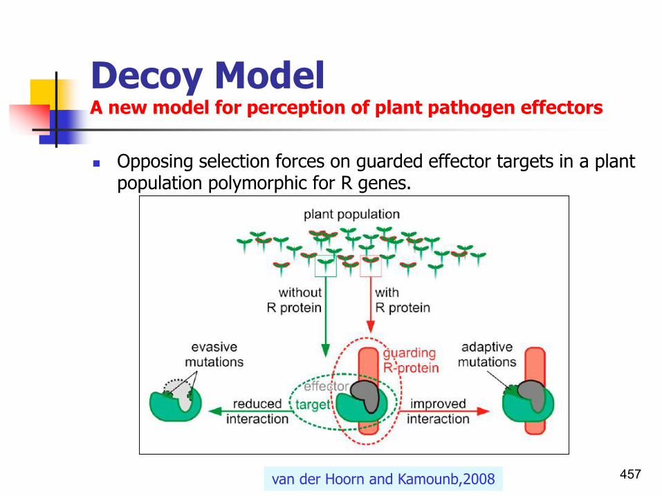
Decoy ModelA new model for perception of plant pathogen effectors
457
◼ Opposing selection forces on guarded effector targets in a plant population polymorphic for R genes.
van der Hoorn and Kamounb,2008

Decoy ModelFour cases supporting the decoy model
◼ Case 1: Pto
◼ P. syringae AvrPto/Tomato
◼ Case 2: pBS3
◼ Xanthomonas campestris pv.vesicatoria AvrBs3/Peper
◼ Case 3: RCR3
◼ The effector protein Avr2 of the fungus C. fulvum/Tomato
◼ Case 4: RIN4
◼ P. syringae effectors (AvrRpm1, AvrRpt2,and AvrB) and monitored by at least two R proteins (RPM1 and RPS2)/ Arabidopsis.
458van der Hoorn and Kamounb,2008

Decoy ModelFour cases supporting the decoy model
459
See original paper for a complete table
van der Hoorn and Kamounb,2008

Decoy ModelFour cases supporting the decoy modelCase 1: Pto
◼ Tomato Pto encodes a Ser/Thr kinase that confers resistance to P. syringae strains carrying avrPto, an interaction that also requires the nucleotide binding (NB)-LRR R protein Prf.
◼ AvrPto contributes to virulence on tomato, even in the absence of Pto.
◼ Function of Pto is kinase, whereas AvrPto act as Kinase inhibitor.
◼ Pto inhibition by AvrPto activates Prf.
◼ FLS2, a receptor-like kinase in PAMP triggered immunity is an operative virulence target of AvrPto.
◼ If it so, Pto itself is a decoy.
◼ Pto then would restrict rather than promote pathogen fitness in the absence of Prf.
460van der Hoorn and Kamounb,2008

Elicitor recognitionIndirect interactions of AVR-R proteins
3. Integrated decoy model
461

Indirect interactions of AVR-R proteins Integrated decoy model
◼ Pathogen effectors target host proteins for manipulation in order to promote infection.
◼ Indirect recognition of these effectors occurs when:
1. these target proteins are guarded by host NLR(leucine-rich repeat receptors) proteins, or
2. if duplicated target genes evolve into decoy proteins monitored by host NLRs.
3. Alternatively the decoy may be integrated into the structure of the receptor component of an NLR pair, allowing AVR recognition by direct binding.
462Cesari et al.,2014

Decoy Model Integrated decoy model
463Cesari et al.,2014

Indirect interactions of AVR-R proteins Integrated decoy model
◼ Recognition of pathogen effector proteins by plant NLR(leucine-rich repeat receptors) proteins often involves decoy proteins, which mimic effector targets.
◼ In certain examples such decoys are integrated as fusions into their cognate NLR.
◼ Two new studies have analyzed thousands of predicted NLRs to address how commonly plants use this ‘integrated decoy’ strategy.
464Moffett,2016

Indirect interactions of AVR-R proteins Integrated decoy model
465Moffett,2016

Indirect interactions of AVR-R proteins Integrated decoy model
466Morel,2009
The integrated decoy model proposes that domains of plant proteins (light green) normally targeted by pathogen effectors (orange circle)
to perturbate the cell and trigger susceptibility can be integrated in NLR immune receptors (dark green).

Plant defenses
The most common defences
467

Basal and R-gene-mediated defences in plants
◼ Plants lack mammalian-like adaptive immunity and therefore their various inducible defence responses are collectively referred to as ‘innate immunity’.
◼ Plant responses to pathogen attack can be differentiated into:
1. Basal, and
2. Resistant (R)-gene-mediated defences.
◼ These two defence responses can be distinguished experimentally by several assays, but they:
1. Share some similar features, and might even
2. Share some common molecular mechanisms.
468Abramovitch et al.,2006

Pathogen and nonpathogens Plants are universally resistant to nonpathogensBasal defence responses
1. Non pathogenic bacteria:◼ Plants are universally resistant to nonpathogens, such as:◼ Escherichia coli,◼ Pseudomonas fluorescens, ◼ TTSS-deficient phytopathogen mutants. ◼ Nonpathogenic bacteria elicit a defense response not involving
the HR, which is variously known as localized induced resistance, innate immunity, or basal resistance.
2. Pathogenic bacteria:◼ The basal defenses must be suppressed by TTSS effectors
before the interaction can progress to either:◼ Nonhost resistance, ◼ Host resistance (race-specific resistance).◼ Otherwise disease will occurs.
469

Basal defences and their suppression by type III effectors
◼ Delivery of effector proteins through the type III secretion system (T3SS) into plant cells is one strategy that is used by bacteria to suppress PRR-mediated defences.
◼ As many as 16 effectors have been identified that suppress basal defences.
470PRRs: Pattern recognition receptors or surface proteins

PAMPS In incompatible interaction both EBR and HR are induced. When induction of HR precedes completion of EBR then EBR is masked by HR
Courtesy to P.G. Ott 471
EBR: Early Basal (innate) resistance; LBR: Late Basal (innate) resistance

PAMPS In compatible interaction the virulent pathogen inducesEBR also, but it is blocked by the suppression of the EBR signal transduction pathway
◼ Because both EBR and HR are blocked the pathogen can multiply vigorously causing disease.
Courtesy to P.G. Ott 472

Plant disease resistance
◼ Plant disease resistance is a complex phenomenon that most commonly occurs in a non-specific manner, as a result of multiple genes conditioning the ability of the pathogen to cause disease and enabling the plant host to mount an effective defence response.
◼ However, plant disease resistance can also be induced in response to:
1. Particular pathogen races (race-specific resistance),2. Particular host plant cultivars (cultivar-specific
resistance), or3. Both a specific pathogen race and plant cultivar
interact (race-cultivar-specific or gene-for-gene resistance).
Angela Moncrieff,2003 473

Two levels of host defenses
◼ Non-specific plant disease resistance (general, non-host or basic resistance) is a response to all races of a particular pathogen, and occurs in all cultivars of a host plant species.
◼ In these basal defenses, HR is not elicited (basal defenses).
◼ Specific plant disease resistance is dependent upon the presence of:
1. A particular pathogen race
2. A particular host plant cultivar, or both.
◼ In this resistance, HR is elicited (HR defenses).
474

Specific plant disease resistance
◼ Specific plant disease resistance appears to be governed by a single gene or a small number of related genes.
◼ Genes conditioning host-pathogen specificity are found in particular subpopulations of the pathogen, plant host, or both interacting organisms.
◼ Specific plant disease resistance can be subdivided into three major categories on this basis.
475

Specific plant disease resistance
◼ Race-specific resistance:◼ Induced in response to only a particular race of pathogen,
but occurs in all cultivars of the host plant. ◼ Cultivar-specific resistance:◼ Activated only in a specific host plant cultivar, but in
reaction to all races of a pathogen species. ◼ Race-cultivar-specific (gene-for-gene) resistance:◼ This type of resistance is usually referred to as gene for
gene resistance, because in most cases it requires the presence of both:
◼ avr gene in the pathogen, and◼ One or more corresponding cultivar-specific R genes in the
host plant.
476

Specific plant disease resistanceR-gene activated resistance
◼ Generally, R-gene activated resistance includes tworelated yet spatially and temporally distinct forms:
◼ Local resistance:◼ Activated at the site of pathogen infection and often
accompanied with HR. ◼ This primary defense reaction effectively contains the
invading pathogen.◼ Systemic acquired resistance:◼ Shortly after or concomitant with local resistance, a
secondary defense reaction, i.e. SAR is induced in the uninfected tissues, which leads to a more long-lasting resistance throughout the whole plant against infection by a broad range of pathogens.
477

Specific plant disease resistanceRace-specific elicitors
478

The most common defences
479
◼ Some of the most common types of resistance which can be observed in nature or mentioned in the literature:
1. An innate resistance:◼ (Distinct from innate immunity) can potentially be
expressed by plants prior to any contact between them and their pathogens.
2. Preformed defences:◼ Non-host resistance and incompatible reaction during a
gene-for-gene relationship belong to the resistances that plants possess per se.
3. Acquired resistances:◼ Expressed only by plants and are non-specific. ◼ In contrast, ‘acquired immunity’ in animals is highly
specific.

Plant innate immune systems
Constitutive & Induced resistance
480

Innate immune systems A comparison of animal and plant immune system
◼ Plants do not have mammalian-like adaptive immunity (circulatory system) and therefore cannot rely on a specialized, proliferative immune system.
◼ Instead they show various inducible defence responses which are collectively referred to as ‘innate immunity’.
◼ Each plant cell has to be capable of defense, even though this defense is coordinated locally and systemically between cells.
◼ Despite this, a number of similarities have been found between the innate immune response of plants andhumans.
◼ There are a variety of types of resistance genes and mechanisms, some induced and some constitutive.
481

Innate immune systems A comparison of animal and plant immune systemMicrobe-associated molecular patterns (MAMPs)
◼ Plants and animals have evolved sensitive perception systems to recognize microbe-derived products, such as small molecules and proteins.
482Altenbach and Robatzek,2007

Innate immunity proteins PAMPs/MAMPs act as general microbial elicitors
◼ Plants perceive several general elicitors from both host and non-host pathogens.
◼ These elicitors are essential structures for pathogen survival and are for that reason conserved among pathogens.
◼ These conserved microbe-specific molecules, also referred to as Pathogen-associated molecular pattern (PAMP) or microbe-associated molecular patterns (MAMPs).
◼ General bacterial elicitors, like
◼ Lipopolysaccharides (LPS),
◼ Flagellin (Flg),
◼ Elongation factor Tu (EF-Tu),
◼ Cold shock protein (CSP),
◼ Peptidoglycan (PGN), and
◼ The enzyme superoxide dismutase (SodM).
◼ These are known to act as MAMPs and induce immune responses in plants or plant cells.
483Jackson,2009

Innate immunity proteins PAMPs/MAMPs sensed by PRRs
◼ PAMPs/MAMPs are sensed by a broad spectrum of host specie(both plant and animals).
◼ The corresponding PRRs for some of these bacterial elicitors(PAMPS/MAMPs) have, in recent years, been identified.
◼ MAMPs are recognized by cognate pattern recognition receptors (PRRs) or surface proteins that trigger immediate defense responses leading to basal or nonhost resistance.
484
Altenbach and Robatzek,2007
Basal resistance occur early <10 minutes after contact, whereas R-gene-mediated defences typically detectable later (2-3 hours).

Innate immune systems Reasons for failure of PRR proteins to detect microbial patterns
◼ Specialized pathogenic microbes are able to overcome host immunity by either:
1. Circumventing the detection of MAMPs (camouflage), or
2. Interfering with MAMP- or PAMP-triggered immunity (e.g., delaying, suppressing, or reprogramming host responses).
(See also bacterial sensing and zigzag theory).
◼ Te secretion of so-called effector molecules promotes pathogen virulence and leads to susceptibility and, ultimately, the expression of symptoms.
485Altenbach and Robatzek,2007

Innate immunity proteins Involvement of intracellular receptors to detect the effectors
◼ Receptors detecting microbial patterns(PAMPs/MAMPs) can be divided into surface and intracellular receptors.
◼ Surface receptors:
◼ All known PRRs(pattern recognition receptors) in plants are plasma membrane-resident proteins, allowing the perception of MAMPs to occur at the cell surface.
◼ Intracellular receptors:
◼ These are the nucleotide-binding site (NB) leucine-rich repeat (LRR) class of receptors(NB-LRR).
◼ These are closely related to animal NOD-like receptor.
◼ In plants, they appear to function exclusively in effector recognition.
486Altenbach and Robatzek,2007

Innate immune systems A comparison of animal and plant innate immunity proteins
◼ In animals an important family of PRRs is the cell-surface Toll-like receptors, which are characterized by an extracellular leucine-rich-repeat (LRR) domain and a cytoplasmic TIR(Drosophila Toll/Interleukin-1 receptor) domain.
◼ In pants the Leu-rich repeat receptor kinase (LRR-RK) FLS2 is the PRR for flagellin.
◼ It also intracellular resistance proteins have a C-terminal LRR domain, a NOD domain, and an N-terminal TIR domain.
487

Xiao,2006
Innate immune systemsSchematic overview of plant innate immunity
488

Innate immune systems Model depicting the activation of PRR-mediated basal defences and their suppression by type III effectors
◼ PAMP-induced transcriptional reprogramming showed rapid induction of genes that encodetranscription factors, proteins that are associated with:
1. Protein degradation,
2. Hormone-related proteins,
3. Phosphatases, and
4. Diverse protein kinases including a large number of LRR-receptor-like kinases (RLKs).
489

Innate immune systems Model depicting the activation of PRR-mediated basal defences and their suppression by type III effectors
490Abramovitch et al.,2006

Innate immune systems Subcellular redistribution and signaling of receptors
that mediate plant immunity
491Altenbach and Robatzek,2007

Innate immune systems The use of protoplasts to study innate immune responses
◼ Recently, Arabidopsis protoplasts have been used successfully to study plant innate immune responses triggered by pathogen-derived elicitors.
◼ Also use of plant protoplast transient expression system has facilitated the discovery and dissection of many signal transduction pathways in response to hormones, metabolites, and stresses.
◼ Signal transduction is defined as Reception, conversion and transmission of chemical message by a cell.
492

The use of protoplasts to study signal transduction pathways
493
◼ The use of protoplast transient assays to study early signaling events mediated by Avr and pathogen-associated molecular pattern.
◼ 35S is the constitutive promoter derived from cauliflower mosaic virus. HR, hypersensitive response.
Ronald,2007

Host and non-host resistance
Plant defence mechanisms
494

Host and non-host resistance
◼ Two types of plant resistance response can be distinguished:
1. Host or race/cultivar specific resistance:
◼ Gene recognition mediated by R and avr genes.
2. Nonhost/nonspecific resistance:
◼ Recognition of specific pathogen or plant cell wall derived signal molecules.
495Odjakova and Hadjiivanova,2001

Host and non-host resistanceSimilar biochemical processes involved in pathogen resistance
◼ Host resistance acts under the species level with some intraspecific biotypes being resistant and others susceptible.
◼ Nonhost resistance acts at the species level, with all intraspecific biotypes (cultivars) being resistant to the pathogen.
◼ Nonhost resistance exhibited against bacteria, fungi and oomycetes can be of two types:
◼ Type I nonhost resistance does not produce any visible symptoms whereas type II nonhost resistance results in a rapid hypersensitive response with cell death.
◼ Strong similarities exist between nonhost and gene-for-gene resistance responses but it is still not clear if the same mechanism is involved in producing these resistance responses.
Mysore and Ryu,2004 496

Non-host resistance
◼ Where the plant is never a host for a pathogen.
◼ Non-host concerns the resistance observed when a microorganism attacks a plant species which is never its host.
◼ The microorganism is called a "non-pathogen".
◼ The plant species is called a "non-host".
◼ The interaction is called a "non-host interaction".
497

General interactions of a pathogen with its host and nonhost plants
Agrios,2005
498

Non-host resistanceThe most common form of disease resistance
◼ Nonhost resistance represents one of the most promising defence mechanisms in developing durable resistance against plant pathogens, namely due to its effectivemenss against a broad range of pathogen species and its durability in nature.
499

Non-host resistance in plants
◼ Most plants are resistant to most pathogens, so disease is the exception.
◼ A pathogen that can not cause disease on a non-host plant is referred to as non-host pathogen.
◼ Plants that cannot be successfully attacked by a given pathogen are called non-hosts for that pathogen, and we say that they display non-host resistance.
500
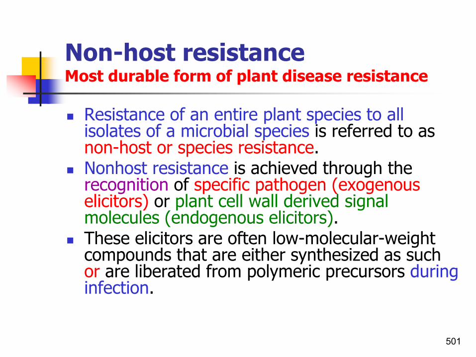
Non-host resistanceMost durable form of plant disease resistance
◼ Resistance of an entire plant species to all isolates of a microbial species is referred to as non-host or species resistance.
◼ Nonhost resistance is achieved through the recognition of specific pathogen (exogenous elicitors) or plant cell wall derived signal molecules (endogenous elicitors).
◼ These elicitors are often low-molecular-weight compounds that are either synthesized as such or are liberated from polymeric precursors during infection.
501

Exogenous/endogenous elicitors
◼ Exogenous elicitors:
◼ Pathogen-associated molecular patterns (PAMPs e.g. flagellin), conserved among several microbial species.
◼ Endogenous elicitors:
◼ Bacterial type III effectors in Arabidopsisprotoplasts (avirulence [Avr] or type III effectors) specific to some races of a pathogen species.
502

Non-host disease resistance
◼ Nonhost disease resistance is the most common form of disease resistance exhibited by plants against the majority of potentially pathogenic microorganisms.
◼ Recently, several components of nonhost disease resistance have been identified.
◼ Nonhost resistance exhibited against bacteria, fungiand oomycetes.
◼ Two types of nonhost resistance have been distinguished on the basis of interaction phenotypes:
1. Type I nonhost resistance
2. Type II nonhost resistance
503Mysore and Ryu,2003&04

Non-host disease resistance
1. Type I nonhost resistance occurs without apparent host cell death i.e. does not produce any visible symptoms.
2. Type II nonhost resistance is accompanied by HR, results in a rapid hypersensitive response with cell death.
◼ Strong similarities exist between nonhost and gene-for-gene resistance responses but it is still not clear if the same mechanism is involved in producing these resistance responses.
504Mysore and Ryu,2003&04

A model for type I and type II non-host resistance
◼ During type I nonhost resistance the nonhost pathogen is not able to overcome the preformed and general elicitor-induced plant defense responses such as cell wall thickening, phytoalexin accumulation, other plant secondary metabolites and papilla formation.
◼ Pathogenesis-related (PR) gene expression as a component of systemic acquired resistance (SAR) can be induced by general elicitors of the nonhost pathogen.
◼ During type II nonhost resistance the nonhost pathogen is able to overcome preformed and general elicitor-induced plant defense responses, probably by producing detoxifying enzymes.
◼ Specific pathogen elicitors are then recognized by the plant surveillance system and this triggers plant defense leading to a hypersensitive response (HR).
◼ PR gene expression and SAR are also induced during type II nonhost resistance.
505

A model for type I and type II non-host resistance
◼ Blue-colored NPs (nonhost pathogens) represent fungi or oomycetes and brown-colored NPs represent bacteria.
506

Defence mechanismsPassive and active defense mechanisms
◼ Passive (preformed) defence lines such as cell walls, wax layers and chemical barriers confer broad resistance to a wide variety of pathogens.
◼ If a pathogen overcomes this first line of defence, there is a second line, which is mounted by proteins encoded by specific resistance (R) genes.
◼ Active defence mechanisms(defense-related genes, PR proteins) that render the plant resistant.
507

Arabidopsis:Exhibiting both type I and type II non-host resistance
◼ Arabidopsis displays:
◼ Type I nonhost resistance against P. syringae pv. phaseolicola, and
◼ Type II nonhost resistance against P. syringae pv. tomato.
508

Characterization ofnon-host phenotypes in rice
509

Examples of type I and type II non-host resistance
510Mysore and Ryu,2004

Mechanisms of General DefensesDefense Mechanisms in Plants
1. Passive (constitutive) defense (First line defense)
2. Active (inducible) defense (Second line defense)
511

Constitutive defense always present
Induced defense
synthesized in response to challenge
Constitutive vs. Induced Defenses

Defense mechanismsPassive and active defense lines
◼ Passive defence lines such as cell walls, wax layers and chemical barriers confer broad resistance to a wide variety of pathogens.
◼ If a pathogen overcomes this first line of defense, there is a second line, which is mounted by proteins encoded by specific resistance (R) genes.
513

Plant defense strategiesPassive and active defense lines
◼ Plants exhibit a wide array of defense strategies against pathogen attack.
◼ The resistance against pathogens is performed by both:
1. Constitutive (continuous) defense mechanisms
2. Inducible defense mechanisms
514

Defense mechanisms in plantsPassive defense linesFirst line defense
◼ A. Passive (preformed, preexisting, constitutive defense mechanisms).
◼ First line defense includes:1. Physical barriers such as cell walls, wax layers;2. Chemical barriers barriers (secondary compounds)
such as phenols, quinones, antimicrobial proteins and peptides which confer broad resistance to a wide variety of pathogens.
◼ Secondary compounds are so-called because they do not play a role in photosynthesis, growth, or respiration.
515

Defense mechanisms in plantsActive defense linesSecond line defense
◼ B. Active defence mechanisms◼ Second line defense includes:◼ Hypersensitive response, phytoalexin synthesis,
lignification, synthesis of pathogenesis-related proteins, Hydroxyproline-rich glycoproteins and systemic acquired resistance (SAR).
◼ If a pathogen overcomes this first line of defence, there is a second line, which is mounted by proteins encoded by specific resistance (R) genes.
516
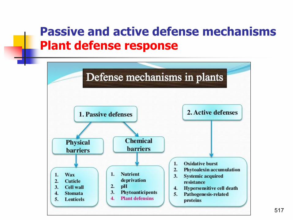
Passive and active defense mechanismsPlant defense response
517

Passive and active defense mechanismsPlant defense response
518Vaitkunas,2003

Constitutive Defence MechanismsThe preexisting defense system
◼ Passive defense mechanisms:
• Physical barriers
• Chemical barriers
519

Defense mechanisms in plantsFirst line defense Physical and chemical barriers
PHYSICAL DEFENSES
◼ Spines, thorns
◼ cutins
◼ waxes
◼ Suberins
SECONDARY METABOLITES
Phenolics Defense-related proteins
phenolic glycosides peroxidases
bound phenolics polyphenol oxidase
lignin? PAL
condensed tannins hydrolysable tannins
Terpenes N-containing
monoterpenes Alkaloids
diterpene acids Mustard oils
520Lecture27 Apr7

Constitutive defense mechanisms1. Physical barriers
521

Plants’ cuticles often vary with the climate in which they live
Cactus cuticle

Physical barriersWaxes
◼ Complex mixtures of long-chain lipids that are extremely hydrophobic.
◼ Waxes are synthesized by epidermal cells.
◼ Exuded through pores in the epidermal cell wall by an unknown mechanism.
Lecture27 Apr7

Physical barriersWax and suberin
◼ Also formed from fatty acids but has a different structure from cutin.
◼ A cell wall constituent.
◼ Often within roots.
◼ Can protect against pathogens and other damage.
◼ Older parts of roots more suberized.
◼ Endodermis has suberin side walls, water must pass through plasma membrane to get to stele.

Passive defence mechanismsSection of a cell wallA model of the interconnections among components cellulose, hemicellulose and pectin
525Wikipedia

Constitutive defence mechanisms2. Chemical barriers
◼ Low molecular weight compounds: e.g.Phenols and quinonesCyanogenic glucosidesSaponinsTerpenoidsStilbenesGlucosinolatesCyclic hydroxamic acids and related benzoxazolinone compounds
◼ High molecular weight compounds: e.g.TanninsAnti-microbial proteins and peptidesDefensinsLysozymeProteinase inhibitorsPolygalacturonase-inhibiting proteins (PGIPs)
526

Major groups of antimicrobial compounds from plants
◼ Phenolics and Polyphenols;
◼ Quinones: Aromatic rings with two ketone substitutions;
◼ Flavones, flavonoids, and flavonols;
◼ Tannins: A group of polymeric phenolic substances;
◼ Coumarins: Phenolic substances made of fused benzene and a-pyrone rings;
◼ Alkaloids;
◼ Terpenoids and Essential Oils;
◼ Lectins and Polypeptides e.g. Thionins.
527

Secondary metabolitesDo not play a role in photosynthesis, growth, or respiration, but protect the tissues from infections
◼ Secondary metabolites are not necessarily produced under all conditions, and in the vast majority of cases the function of these compounds and their benefit to the organism is not yet known.
◼ Some secondary metabolites are produced for easily appreciated reasons. e.g.
1. As toxic materials providing defense against predators.
2. As volatile attractants towards the same or other species.
3. As coloring agents to attract or warn other species.
528

Secondary metabolitesDo not play a role in photosynthesis, growth, or respiration, but protect the tissues from infections
529

Primary and secondary metabolite biosynthesis
530

Terpenes and TerpenoidsIsoprene-unitsConstituents of essential oils
◼ Terpenes: Class of >20,000 natural organic compounds containing carbon atoms in multiples of five (5).
◼ The name ‘terpene’ is derived from the Greek word ‘terebinth’.
◼ Terebinth is a type of pine tree from which terpene-containing resins are obtained.
◼ Terpenoids: Oxygen-containing terpenes (alcohols, ketones, aldehydes). These are derived from five-carbon isoprene units assembled and modified in thousands of ways.
◼ Terpenes and terpenoids are the primary constituents of the essential oils of many types of plants and flowers.
531Terpenoids are terpenes that contain oxygen.

Terpenes and TerpenoidsIsoprene-unitsConstituents of essential oils
◼ All terpenes are built up from units of isoprene-units linked in a “head-to-tail” fashion.
➢ Head-branched end of isoprene;
➢ Tail-unbranched end of isoprene.
532

Terpenes and TerpenoidsTerpenoids are terpenes that contain oxygenClassification of Terpenes
Isoprene unitsNo. of carbon atomsType of terpenes
one(consist of 1 isoprene unitsi.e. 5 carbons)
5hemiterpene
two10monoterpenes
three15sesquiterpenes
four20diterpenes
Five (consist of 5 isoprene unitsi.e. 25 carbons)
25sesterterpenes
six30triterpenes
eight40tetraterpenes
sesqui = one and a halftetra = four hemi = half
tri = threedi = two

Terpenes and TerpenoidsTerpenoids are terpenes that contain oxygenClassification of Terpenoids
Isoprene unitsNo. of carbon atomsType of terpenoids
oneone(consist of 1 isopreneunits i.e. 5 carbons)
C5hemiterpene
twoC10monoterpenoid
threeC15sesquiterpenoid
fourC20diterpenoid
fiveC25sesterterpenoid
sixC30triterpenoid
eightC40tetraterpenoid
sesqui = one and a halftetra = four hemi = half
tri = threedi = two

TerpenesProduced from the mevalonic acid pathway

TerpenesProduced from the mevalonic acid pathway

Terpene functions1. Growth and development
◼ Growth and development
1. Carotenoid pigments are tetraterpenes.
2. Chlorophyll side chain is diterpene.
3. Giberellins (hormones) are diterpenes.
4. Abscissic acid (hormone) is a sesquiterpene C15.
5. Sterols are triterpenes.
537

Terpene functions2. As defensive compounds
◼ As defensive compounds
◼ Toxins and feeding deterrents to pathogens, insects and mammals.
◼ Examples
◼ Resins of conifers are monoterpenes
◼ Essential oils -peppermint, limon, basil, sage, may be in glandular hairs on epidermis.
538
Non-volatile terpenes limonene.

Terpenes and TerpenoidsIsoprene units in terpenes are usually linked head to tail Dashed lines can be used to delineate the isoprene units comprising a terpene
539

Antimicrobial peptidesPlant-derived molecules High molecular weight compounds
◼ It is estimated that there are 250,000 to 500,000 species of plants on Earth.
◼ A relatively small percentage (1 to 10%) of these are used as foods by both humans and other animal species.
◼ It is possible that even more are used for medicinal purposes.
540

Antimicrobial peptidesPlant-derived molecules High molecular weight compounds
◼ Many plants possess antimicrobial peptides.
◼ These peptides constitutively (continuously)expressed or induced after infection.
◼ These families of peptides include:◼ Four-cysteine-type peptides◼ Hevein and knottin-type peptides◼ Lipid transfer proteins (LTPs)◼ Plant defensins◼ Ribosome-inactivating proteins (RIPs)◼ Thionins.
541

Antimicrobial peptides Plant defensins
◼ Defense-related, cysteine-rich, antimicrobial peptides present in the plasma membrane of host plant species.
◼ They constitute a group designated defensins capable of providing resistance against microbial plant pathogens.
◼ The first plant defensins that were demonstrated to possess antifungal activity were the two plant defensin isoforms Rs-AFP1 and Rs-AFP2 isolated from radish seed.
542

Plant defensins
◼ Some plant defensins as antibacterial peptides have also been reported to exert antibacterial activity such as:
◼ A Clitoria ternatea plant defensin (Ct-AMP1), which is active against Bacillus subtilis, and
◼ A potato tuber plant defensin, which inhibits:◼ Ralstonia solanacearum, and◼ Clavibacter michiganensis.
543

Antimicrobial peptidesThionins
◼ The toxic activity of thionins was apparently first described by Jago and Jago in 1885.
◼ Toxic effects against yeast, bacteria and fungi and mammals were discovered.
◼ e.g.◼ The Barley leaf thionins have repeatedly been shown to
have antimicrobial activities in vitro against different phytopathogenic bacteria and fungi.
◼ During the past several years, strong evidence has been obtained for a resistance
◼ Overexpression of different thionins has resulted in enhanced resistance against bacterial and fungal pathogens.
544

Transforming rice with a thionin gene against Burkholderia plantarii
◼ The effectiveness of transforming rice with a thionin gene from oats in protecting the plant from infection by Burkholderia plantarii:
◼ left, uninoculated controls;◼ centre, inoculated wild-type
plants, and◼ Right, inoculated
transformants.◼ Numbers below the pots are cfu
of the bacterium/mg fresh weight of plant material recovered from the plants.
545Iwai et al.,2002

Examples of primary amino acid sequences of natural antimicrobial peptides
StructurePeptide
Cyclic (LOVPFdLOVPFd) Gramicidin S
Cyclized I(C)LEdI(KOdIFHD)Dd-NH2Bacitracin
Cyclized isooctanoyl BTBB(BFdLBBT) Polymyxin B
VVC1AC2RRALC3LPRERRAGFC3RIRGRIHLC2 C1RR defensin (NP-1)
DHYNC1VSSGQC2LYSAC3PIFTKIQGTC2YRGKAKC1C3K Defensin 1
RRWC1FRVC2YRGFC2YRKC1R Crab tachyplesin
RLC1RIVVIRVC1R Cattle bactenecin
KWKFKKIEKMGRNIRDGIVKAGPAIEVIGSAKAI Silk moth cecropin A
ILPWKWPWWPWRR Cattle indolicidin
IXA1IULA1Z2PGA2KZ3GLAMGA3NMKZ4AZ5A4HA5SIHVUKBacterial nisin
Hancock and Chapple,1999 546

Peptide antibioticsAntimicrobial peptides
◼ Hundreds of peptide antibiotics have been described in the past half-century.
◼ These fall into two classes:1. Non-ribosomal peptides, such as the
gramicidins, polymyxins, bacitracins, glycopeptides, etc. These are largely produced by bacteria (also by fungi).
2. Ribosomal (natural) peptides, such as major component of the natural host defense molecules including AMPs which are produced by all species of life (including bacteria).
547

Anti-microbial peptides (AMP)Natural peptides
◼ Anti-microbial peptides (AMP) make up a part of the innate immunity of most organisms and are often involved in the immune system's first line of defense when faced with an invader.
◼ AMPs can be found in prokaryotic and eukaryotic organisms and in vertebrates and invertebrates(Reddy,2004).
◼ Antimicrobial peptides are so widespread that they are likely to play an important protective role.
548

Induced Defense MechanismsActive defense mechanisms
• Rapid responses implicated in resistance
• Slower responses implicated in resistance
• Oxidative burst, HR, SAR,..
549

Active defense mechanismsThe second defense line
◼ Plants synthesize a variety of compounds when exposed to biological stress viz., fungi, bacteria, viruses, insects and herbivores (Collinge and Slusarenko, 1987; Linthorst, 1991).
◼ The inducible defense responses include the production of:
1. reactive oxygen species (ROS),
2. phytoalexins,
3. cell wall components (callose, glycine or hydroxyproline-rich proteins), and
4. other specific group of proteins called pathogenesis-related (PR) proteins.
550

Active defence mechanisms of plantsSummarized diagrammatically
Strange,2003 551

Active defense mechanismsThe second defense line
1. Rapid responses implicated in resistance◼ The oxidative burst (ROIs)◼ The generation and role of nitric oxide (NO) in resistance◼ Callose synthesis and deposition2. Slower responses implicated in resistance◼ Hypersensitive response◼ Phytoalexins◼ Lignification◼ Suberization◼ Synthesis of hydroxyproline-rich glycoproteins (HRGPs)◼ Pathogenesis-related proteins (PRPs)◼ Systemic acquired resistance (SAR)◼ Induced systemic resistance (ISR)3. Elicitors of defence responses4. Elicitor perception5. Signalling
Increases in cytosolic calciumMitogen-activated protein kinases (MAP kinases)Salicylic acid (SA) JasmonatesEthyleneIntegration of signalling pathways 552

Active defence mechanismsAbiotic or biotic elicitors
◼ The triggers for these active defence mechanisms are termed elicitors.
◼ Recognition of pathogens (exogenous elicitors or endogenous elicitors) by host plants led to many metabolic and structural modifications in the host.
◼ The elicitors are either abiotic or biotic:1. Abiotic elicitors include physical insults such as
ultraviolet irradiation and partial freezing as well as a multitude of chemicals such as the salts of heavy metals and DNA intercalating compounds.
2. Biotic elicitors may be specific and are, in some cases, the products of avirulence genes of pathogens.
553

Active defence mechanismsAbiotic or biotic elicitors
◼ Flagellin from bacteria, and◼ Chitin and ergosterol from fungi are also known
characterized microbial elicitors.◼ Other miscellaneous elicitors are as:◼ Polysaccharides,◼ Enzymes, ◼ Fatty acids, ◼ Glycoproteins, and◼ Proteins.
554

Disease resistance process
◼ Physiological features such as:◼ K+/H+ exchange,◼ Rapid oxidative burst,◼ Hypersensitivity at the site of infection, ◼ Crosslinking of plant cell walls, ◼ Synthesis of antimicrobial compounds known as
phytoalexins, and ◼ Induction of pathogen-related proteins such as
chitinases and glucanases (Lamb et al., 1989) represent some of the multiple metabolism turnover leading to disease resistance.
555

Active Defense MechanismsA. Rapid responses implicated in resistance
Active resistance consists of many defence responses that interact over time and space.
Strange,2003 556

Active Defence MechanismsRapid responses implicated in resistancePlant peroxidases
◼ Another important event associated with plant defense mechanisms is a rapid and transient release of different reactive oxygen species (ROS), such as the superoxide anion radical (O2
-), hydroxyl radical (OH-) and hydrogen peroxide (H2O2).
◼ Of these ROS, production of hydrogen peroxide appears to occur early and is involved in:
1. A direct oxidative reaction with the pathogen;
2. Biosynthesis of phytoalexins;
3. Activating defense-related genes;
4. In the induction of acquired disease resistance.
557
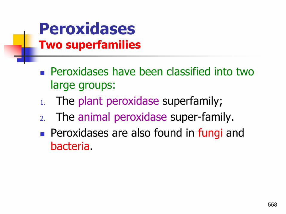
PeroxidasesTwo superfamilies
◼ Peroxidases have been classified into two large groups:
1. The plant peroxidase superfamily;
2. The animal peroxidase super-family.
◼ Peroxidases are also found in fungi and bacteria.
558

A diagrammatic representation of the structure of plant peroxidase
◼ Ten helices (A–J) and β-sheets (β1, β2, and β3) are represented by cylinders and parallel arrows, respectively.
◼ Reverse turns and coils that connect the helices and the β-sheets are shown as connecting lines.
◼ The proximal and distal heme-binding domains and the proximal and distal histidine residues (Hrp C numbering) are indicated.
◼ The rectangular box present between the proximal and distal heme-binding regions indicates the position of the central heme prosthetic group.
559

Phylogenetic analyses and comparisons of amino acid sequences from 62 plant
peroxidases
10 peroxidase gene clusters were recognized.

Oxidative burstToxic to pathogens
◼ H2O2 generated by wall-bound plant peroxidase or oxygenase during the oxidative burst, which occurs within minutes after pathogenic interaction, and free radicals, which are produced by extracellular peroxidase oxidative activity during cell wall fortification, are toxic to pathogens.
561
A free radical is an atom, molecule, or ion that has unpaired valence electrons.
These unpaired electrons make free radicals highly chemically reactive towards other substances, or even towards themselves: their molecules will often
spontaneously dimerize or polymerize if they come in contact with each other. E.g. the hydrocyl radical (HO•). Typically two free radicals combine to form a more
stable species, for example: 2Cl·→ Cl2.

Plant peroxidasesProduction and metabolic fate of various ROS (hydrogen peroxide, superoxide anion radical, singlet oxygen) in different plant cellular compartments
562

Oxidative burstToxic to pathogens and plant cellsEnzymatic scavenging systems, such as catalase (CAT), peroxidase (POD), and superoxide dismutase (SOD) scavenging ROS
◼ The production of reactive oxygen species (ROS) in plants occurs early in plant defense response to external stimuli.
◼ Superoxide (O2−) and hydrogen peroxide (H2O2) are the most
important reactive oxygen species, and they rapidly accumulate.
◼ High levels of ROS may damage the bodies of living organisms.
◼ To prevent oxidation burst due to ROS production, plants have evolved complex protective mechanisms for scavenging ROS.
◼ These include the enzymatic scavenging systems (antioxidant enzymes), such as catalase (CAT), peroxidase (POD), and superoxide dismutase (SOD).
◼ Catalase is a significant component of the cell protective mechanism against oxidative stress.
563Zhao et al.,2016

Plant peroxidasesCatalase-deficient plants(catalase -ve) allow early and late events in oxidative signalling pathways
564Mahamdi et al.,2010

Oxidative burstCatalase (CAT) assay
◼ Catalase activity was assayed as described by Zhang et al.,2008.
◼ The reaction mixture consisted of 1 ml of Tris-HCl buffer (pH 7.0), 0.1 ml of partially purified enzyme extract and 0.2 ml of H2O2.
◼ Absorbance was read at 240 nm for 2 min and the enzyme activity was expressed as ΔOD min-1
.
565Hegde et al.,2018

Oxidative burstReactive oxygen speciesPlant pathogenic bacteria
◼ Increased level of hydrogen peroxide(H2O2) was observed which resulted in a simultaneously increased resistance to P. carotovorum ssp. carotovorum.
◼ Similarly, the same gene in transgenic cabbage conferred resistance to X. campestris pv. campestris, which correlated with glucose oxidase activity in leaves.
◼ Increased hydrogen peroxide may also account for resistance to the soft rot-causing bacterial pathogenP. carotovorum subsp. atrosepticum in potato tubers.
566

Oxidative burstReactive oxygen species
567
◼ Most of plant species generate ROIs by means of NADPH oxidase (NADPH-dependent).
◼ NADPH(Nicotinamide adenine dinucleotide phosphate) oxidase is a membrane-bound enzyme complex which produces ROSs.
◼ NADPH itself is the reduced form of NADP:

Pathways for ROS production and scavenging in
plant cells
Hayat and Ahmad,2007
Oxidative burst
568

Oxidative burst
◼ Salicyclic acid has a pro-apoptotic effect during PCD by enhancing the effect of ROS.
◼ Also SA and ROS are signals for genetic-controlled defense reactions.
569

Nitric oxide (NO)
◼ Nitric oxide (NO) is an essential factor for the activation of innate immune responses in:
1. Insects,
2. Mammals (Nappi et al.,2000), and
3. A similar role has been suggested in plants(Delledonne et al.,1998).
570

Oxidative burst and nitric oxide
571
◼ During the oxidative burst, reactive oxygen intermediates (ROIs) such as O2, H2O2 and OH are generated which protect the plant in several ways:
1. They are toxic to microorganisms.2. This toxicity is thought to be potentiated by NO.3. In fact ROIs with NO cause the hypersensitive death
of host cells (HR).4. Hydrogen peroxide acts as a substrate for
lignifications, making cell walls more difficult to penetrate.
5. It is also involved in the induction of protective genes coding for pathogenesis-related proteins.

Reactive oxygen intermediates and nitric oxideROI and NO
◼ ROS and NO are often produced during the early stages of a plant resistance response.
572

Reactive oxygen intermediates and nitric oxideROI and NO
573ADPH is used in anabolic reactions, such as lipid and nucleic acid synthesis, which require NADPH as a reducing agent.

Nitric oxide (NO)An important signal molecule in plant–pathogen interactions
◼ NO plays a major role in:
1. Plant PCD elicited in response to certain types of pathogenic challenge.
2. The race-specific hypersensitive response (HR);
3. NO also acts in the regulation of non-specific, papilla-based resistance.
4. Possibly in systemic acquired resistance (SAR).
5. Equally, the potential roles of NO signalling/scavenging within the pathogen are being recognized.
574

Nitric oxide (NO) production
◼ Nitric oxide (NO) and reactive oxygen species are two key components in the induction of the hypersensitive response (HR) during plant defense against pathogen infection.
◼ Isolation of the NOS protein and/or cloning of the corresponding gene will greatly facilitate the understanding of NO synthesis and its role in plant defense.
◼ NO generation may be a general response to biotic stress in plants.
◼ e.g.1. The compatible bacteria (Pseudomonas syringae pv. pisi)
induced NOS activity significantly, suggesting that NO generation may also be a general response to biotic stress in plants.
2. Also maximum NOS activity was detected three hours before the onset of HR in pea leaves infiltrated with incompatible bacteria (Ralstonia solanacearum).
575

Major target molecules that shouldbe considered in NO measurementsNO and its derived nitrogen species
576Yamasaki et al.,2016
Major molecules that should be considered in NO measurement arepresented.
Note that each name follows the widely accepted one rather than the IUPAC systematic nomenclature.

An overview of nitric oxide (NO) chemistry
Four routes for NO generation
577Mur et al.,2006

The elicitation of NO production by Pseudomonas syringae pathovars in tobacco and Arabidopsis
C) Proposed model for hrp-dependent elicitation of NO by P. syringae pathovars.
578Mur et al.,2006

Active Defence MechanismsRapid responses implicated in resistanceCallose synthesis and deposition
◼ Callose is a β-1,3-linked glucan which is often synthesized and deposited as papillae – localized wall appositions – as an early response to wounding or pathogen attack.
◼ Callose synthesis is catalyzed by plasma membrane bound ß-glucan synthase (callose synthase).
◼ The speed of callose deposition was emphasized in work with French bean (Phaseolus vulgaris) and Xanthomonas campestris.
579

H2O2 generation in lettuce cell walls
◼ H2O2 generation in lettuce cell walls, in the vicinity of the incompatible bacteriumP. s. pv. phaseolicola.
◼ H2O2 was detected in cell walls (CW) 5 hr after inoculation with bacteria (b)as black deposits in the transmission electron image by staining with cesium chloride.
580

Papillae formation
◼ Papilla refers to the accumulation of material between the host plant's cell wall and cell membrane at the point of fungal penetration.
◼ A papilla is usually composed of silicon, lignin and proteins.
◼ Here papillae developed beneath the penetration peg (PP) and germinating spore (S) of an avirulent fungal isolate.
581

Callose deposition
◼ Callose deposition in Arabidopsis leaf mesophyllat the incompatible infection sites.
◼ Callose was detected by aniline blue staining and fluorescence under UV light (Parker et al.,1993).
582

Callose deposition
◼ Callose accumulation is a non-specific response of plants to pathogen attack and to wounds.
◼ Transmission electron micrograph of phytoplasmas (arrowed) in sieve tubes of the host plant.
◼ Sieve plates are filled by callose (C).
◼ Bar= 0.5 μm.
Weintraub and Jones,2010 583

Salicylic acid and callose synthesis
◼ A rapidity in the activation of callose synthase after spraying the Arabidopsis plant with salicyclic acid implies a possibility that SA affects callose deposition on the cell surface.
◼ A significance of SA-dependent callose deposition in plant resistance was substantiated in experiments with Pseudomonas syringae harboring the mutated CEL gene (DebRoy et al.,2004).
◼ This gene encodes bacterial effectors suppressing plant SA-dependent defense responses.
Hayat and Ahmad,2007584

Salicylic acid and callose synthesis
◼ Callose deposition in the leaf epidermis of Nicotiana glutinosa is stimulated by SA.
◼ Callose was determined by its fluorescence after staining with aniline blue.
Leaves treated with 1mM SA,
1 hour ago.
Untreated leaves
585

Active defence mechanisms of plantsB. Slower responses implicated in resistance
Strange,2003 586

Active Defence MechanismsSlower responses implicated in resistance
587
◼ Slower responses implicated in resistance include:1. The hypersensitive response,2. Phytoalexin synthesis,3. Lignification,4. Suberization,5. Cell wall components (callose, glycine or
hydroxyproline-rich proteins),6. The synthesis of pathogenesis-related proteins
(PRPs), and7. Systemic acquired resistance (SAR).◼ All these require gene transcription and protein
synthesis.

Active Defence MechanismsSlower responses implicated in resistance1. The hypersensitive response
◼ Hypersensitive response is being developed in the place of pathogen penetration into the neighboring cells.
◼ Rapidly developing local process produces signal molecules spreading along vascular system of a plant.
◼ In dependence of local events, the set of signal molecules e.g. SA is being organized, which in turn, forms this or that generalized response.
◼ In the plant cells, the salicylic acid (SA) is produced in considerable amounts and causes activation of SA-induced genes.
◼ The integrity of these events is named as systemic acquired resistance (SAR).
588

The hypersensitive response
◼ The gene of one of the key enzymes, PAL(phenylalanine ammonia-lyase), is activated:
1. In the course of hypersensitive response, but also
2. In response to various external stimuli.
◼ PAL takes part in the synthesis of SA, phytoalexins, and lignin monomers.
589

Gene-for-gene interaction between host and pathogenCombinations to remember
590
Combinations disease develops
Hypersensitive reaction
Non-compatible host species and non-pathogen - -
Compatible species and compatible pathogen + -
Resistant cultivar of compatible host species and incompatible pathogen
- +
Resistant cultivar of compatible host species and compatible pathogen
+ -
➢ Compatible (no recognition or defense response);
➢ Incompatible (recognition and defense response).

Gene-for-gene interaction between host and pathogenHypersensitivity vs. disease
One pair of loci
Pathogen genotypes
AA Aa aa
Host
genotypes
rr
Rr
RR
= R (incompatible) = S (compatible)
IRRI

Possible outcomes for the plant response to bacterial pathogens and non-pathogens
Figure right shows the reactions for compatible (no recognition or defense response), incompatible (recognition and defense response) and a nonpathogenic bacterium. AOll denotes a sustained generation of active oxygen that occurs 1.5 to 3 hr after inoculation. XR denotes a K+ efflux/H+ influx that occurs simultaneously with AOll in incompatible interactions. DR denotes the expression of a variety of defense-response genes.
592Alfano and Collmer,1996

Reactions of pepper to Xanthomonas campestris pv. vesicatoria : virulence, avirulenceand the null reaction to Hrp mutants
◼ The water-soaked lesions of the areas of the leaf inoculated with the virulent isolate and compare with the hypersensitive cell death caused by the avirulent race - the Hrp mutants are deficient in the type III secretory system and therefore cannot export effector molecules that cause either the susceptible reaction or the hypersensitive response.
Strange,2003 593

Hypersensitive vs. normosensitive reactions
◼ Hypersensitive reaction: Hypersensitive cell death is accompanied by the induction of multifaceted defense responses, including: production of active oxygen species and antimicrobial compounds (phytoalexins), rapid cross-linking of cell-wall proteins, local resistance andultimately, resistance to pathogens.
◼ Normosensitive reaction:
◼ The slow, normosensitive plant cell death does not effectively prevent pathogen multiplication or spread andis therefore not associated with local resistance.
594It is interesting that both hypersensitive and normosensitive cell death can lead to a systemic, broad-spectrum resistance response throughout the plant called SAR.

Hypersensitive vs. normosensitive reactions
1. A rapid, hypersensitive cell death localized at the site of infection during an incompatible interaction between a resistant plant and an avirulent pathogen, and
2. A slow, normosensitive plant cell death that spreads beyond the site of infection during some compatible interactions involving a susceptible plant and a virulent, necrogenic pathogen.
595He,1996;..

Active Defence MechanismsSlower responses implicated in resistance2. Phytoalexins synthesis
◼ Phytoalexins have been defined as low molecular weight antimicrobial compounds that are both synthesized by and accumulate in plants after exposure to microorganisms.
◼ Phytoalexins have been characterized from 31 plant families, mostly dicotyledonous, but they have also been isolated from such monocotyledonous plants as rice, sorghum, maize, wheat, barley, onions and lilies.
◼ Most phytoalexins are derived from one or other or a combination of three major biosynthetic pathways:
1. Shikimate2. Acetate-malonate3. Acetate-mevalonate
596

Phytoalexins, salicyclic acid and lignin synthesis
◼ The phenylpropanoid pathway is the source of a large number of compounds that are derived from the amino acid phenylalanine.
◼ Enzymes:
◼ PAL: Phenylalanine ammonia lyase;
◼ C4H: cinnamate 4-hydroxylase;
◼ CHS: chalcone synthase.
597Scheper & Zhong (eds.),2001
Phytoalexins are represented by members of the monoterpene, sesquiterpene, carboxylic.. Sesquiterpenoids
are defined as the group of 15 carbon compounds.

PhytoalexinsRepresentative phytoalexin structures
598Hammerschmidt,1999

Direct evidence for the importance of phytoalexins in plant defence
◼ Studies with cotton and the pathogen, Xanthomonas axonopodis pv. malvacearum, have shown that the phytoalexins 2,7-dihydroxycadalene, lancilene C and lancilene C-7-methyl ether accumulated to concentrations significantly higher than those required to inhibit the bacterium in vitro.
599

PhytoalexinsLarge differences in sensitivity of the pathogens
◼ Phytoalexins are non-specific biocides, affecting a wide range of organisms including bacteria, fungi, nematodes, higher animals and plants.
◼ Apart from Gram-positive bacteria being more sensitivethan Gram-negative bacteria few generalizations can be made about the relative sensitivity of groups of organisms.
◼ Large differences in sensitivity, however, may be found among pathogens which attack plants that accumulate phytoalexins, virulent strains usually tolerating higher concentrations than avirulent ones.
◼ Such differences are usually attributable to the superior ability of virulent strains to degrade the phytoalexin.
600

Active Defence MechanismsSlower responses implicated in resistance3. Lignification
◼ The synthesis of lignin is similar to that of many phytoalexins since it involves the derepression of the phenylpropanoid pathway.
◼ Lignin is a very complex and resistant structure and lignification is thought to contribute to resistance by increasing the mechanical force required for penetration, increasing the resistance of cell walls to degradation by enzymes of the pathogen and setting up impermeability barriers to the flow of nutrients and toxins.
◼ In addition, lignin precursors such as coniferyl alcohol and free radicals may be toxic to the pathogen per se.
601

Lignin synthesis
602semanticscholar.org

Active Defence MechanismsSlower responses implicated in resistance4. Suberization
◼ Like lignin, suberin is a constituent of healthy plant tissue and also like lignin, its synthesis may be enhanced by challenge with microorganisms.
◼ Suberization during wound-healing has long been associated with the resistance of potato tubers to infectious agents.
◼ Lulai and Corsini,1998 reported a study of the roles of the phenolic suberin in resistance to a bacterial and a fungal pathogen.
◼ They found that total resistance to infection by P. carotovorum subsp. carotovorum occurred 2-3 days after wounding when deposition of phenolics on the outer tangential wall of the first layer of cells was complete.
603

Active Defence MechanismsSlower responses implicated in resistance5. hydroxyproline-rich glycoproteins (HRGPs)
◼ Hydroxyproline-rich glycoproteins (HRGPs) are important structural components of plant cell walls and also accumulate in response to infection as an apparent defense mechanism.
1. In response to pathogens and their signals, plant cell walls become highly enriched in HRGPs.
2. As well as being constitutive, they are also inducedby wounding and infection.
◼ These are caused by a wide variety of fungi, bacteria(Pseudomonas, Xanthomonas), and other pathogens(viruses, nematodes).
◼ These are also increased during symbiosis upon interaction of plant roots with mycorrhizal fungi and in Rhizobium induced legume nodules.
604

Active Defence Mechanismshydroxyproline-rich glycoproteins (HRGPs)
◼ It participates in systemic acquired resistance (SAR) in that it is elicited by systemic signals and is involved in plant protection against diseases.
◼ HRGPs may confer resistance is by providing a template for lignin deposition in papillae.
605

Active Defence Mechanisms6. Pathogenesis-related proteins (PRPs)Background
◼ PRPs were discovered in 1970 in TMV-infected tobacco plants.
◼ Since the discovery of pathogenesis-related (PR) proteins nearly 40 years ago in tobacco plants, a great deal of research has focused on the isolation, characterization, and regulation of expression of this unique class of defense proteins in a variety of plants including rice.
Datta and Muthukrishnan,1999;.. 606
Note: Most PRs is their antifungal effects. Plants are exposed to large number of pathogenic fungi. Although plants do not have an immune
system, but they have a defense mechanism including antifungal activity.There are hundred’s of antifungal peptides & proteins known, with more
being discovered almost daily.

Active Defence MechanismsPathogenesis-related proteins (PRs/PRPs)
607Jiang et al.,2015
1. PR proteins are proteins encoded but not expressed in host plant in the absence of interaction with a pathogen.
2. PR proteins are strongly induced in response to wounding or infection by pathogens. E.g. under conditions of nonpathogenic origin, such as stress.
3. accumulate abundantly at the site of infection, and
4. contribute to systemic acquired resistance (SAR).
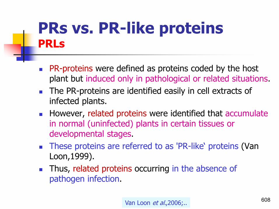
PRs vs. PR-like proteinsPRLs
◼ PR-proteins were defined as proteins coded by the host plant but induced only in pathological or related situations.
◼ The PR-proteins are identified easily in cell extracts of infected plants.
◼ However, related proteins were identified that accumulate in normal (uninfected) plants in certain tissues or developmental stages.
◼ These proteins are referred to as 'PR-like‘ proteins (Van Loon,1999).
◼ Thus, related proteins occurring in the absence of pathogen infection.
608Van Loon et al.,2006;..
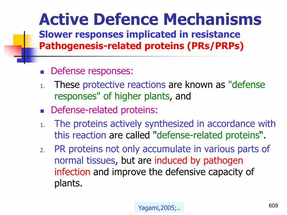
Active Defence MechanismsSlower responses implicated in resistancePathogenesis-related proteins (PRs/PRPs)
◼ Defense responses:
1. These protective reactions are known as "defense responses" of higher plants, and
◼ Defense-related proteins:
1. The proteins actively synthesized in accordance with this reaction are called "defense-related proteins“.
2. PR proteins not only accumulate in various parts of normal tissues, but are induced by pathogen infection and improve the defensive capacity of plants.
609Yagami,2005;..

PR proteinsApoplast and vacuoles
◼ The plant apoplast plays a critical role in the:
1. transport of water and nutrients and
2. interactions between plants and pathogens.
◼ Most of pathogenesis-related (PR) proteins are located in the apoplast.
◼ In general,
1. acidic PR proteins are located in the apoplast, and
2. basic PR proteins, in the vacuole.
610Naz et al.,2014; Enoki and Suzuki,2016

ApoplastPR proteins functions
◼ These proteins have been implicated in active defense, potentially restricting pathogen development and spread.
◼ Some PR proteins possess antimicrobial activity.
◼ Also they can directly affect pathogen integrity, and/or generate signal molecules through their enzymatic activity that act as elicitors to induce other plant defense related pathways.
◼ One response of plants to attempted infection by pathogens is to accumulate apoplastic proteins, including pathogenesis related (PR) proteins.
◼ The expression of defense or PR proteins in apoplast of plant
cells enhance plant resistance to pathogens.
611Naz et al.,2014; Enoki and Suzuki,2016

Family Proteins/properties Functions
PR-1 PR-1 a, PR-1 b, and PR-1 c 14-17kD Antifungal
PR-2 β-1,3-Glucanases 25-35kD Cleaves β-1,3-glucans
PR-3Chitinase types I, II, IV, V, VI, and VII
30kDEndochitinase
PR-4 Chitinase types I and II 13-19kDAntifungal and
chitinase
PR-5 Thaumatin-like proteins Antifungal
PR-6 Tomato proteinase inhibitor I 6-13kD Proteinase inhibitor
PR-7 Tomato endoproteinase P Endoproteinase
PR-8 Cucumber chitinase Chitinase III
PR-9 Tobacco lignin-forming peroxidase Peroxidase
PR-10Parsley “PR-1”
Bet v 1, Mal d 1, Api g 1, and Dau c 1 Ribonuclease-like
PR-11 Tobacco chitinase type V Chitinase
PR-12 Radish Rs-AFP3 Defensin
PR-13 Arabidopsis THI2.1 Thionin
PR-14 Lipid transfer proteinsShuttling of
phospholipids and fatty acids
PR-15 Barley OxOa Oxalate oxidase
PR-16 Barley OxOLP Oxalate-like oxidase
PR-17 Tobacco PRp27 Unknown
Yagami,2005; Sinha et al.,2014
Active Defence Mechanisms
Recognized and proposed families of
pathogenesis-related proteins.

Active Defence MechanismsSlower responses implicated in resistancePathogenesis-related proteins (PRs/PRPs)
613Yagami,2005; Jiang et al.,2015
◼ PR genes have been shown to be induced by:
◼ Various abiotic stresses, such as:
◼ treatments with NaCl, heat, cold, PEG, UV irradiation, and ozone.
◼ Some PR proteins have also been reported to accumulate under specific physiological conditions, such as:
◼ pollen development, leaf senescence, fruit development and ripening.

Active Defence MechanismsSlower responses implicated in resistancePathogenesis-related proteins (PRs/PRPs)
614Yagami,2005

Active Defence MechanismsSlower responses implicated in resistancePathogenesis-related proteins (PRs/PRPs)
615Yagami,2005

Active Defence MechanismsSlower responses implicated in resistancePathogenesis-related proteins (PRs/PRPs)
◼ These proteins are induced by the plants as a defense response system in stress conditions like microbial and insect infections, wounding, exposure to harsh chemicals, and atmospheric conditions.
◼ However, some plant tissues that are more exposed to environmental conditions like UV irradiation and insect or fungal attacks express these proteins constitutively.
616Sinha et al.,2014;..

Active Defence MechanismsPathogenesis-related proteins (PRs/PRPs)Functions
617See also the section ”systematic resistance”
1. An important common function of most PRs is their antifungal effects;
2. Some PRs also exhibit antibacterial, insecticidal or antiviral action.
3. Function as signals that spread “news” of the infection to nearby cells.
4. Infections also stimulate the cross-linking of molecules in the cell wall and the deposition of lignin, responses that set up a local barricade that slows spread of the pathogen to other parts of the plant.

Active Defence MechanismsPathogenesis-related proteins (PRs/PRPs)Functions
618Wikipedia,2016
4. Chitinase activity.
5. Peroxidase, ribonuclease and lysozyme activities.
6. Their hydrolytic, proteinase-inhibitory and membrane-permeabilizing ability.
6. They inactivate the proteins secreted by the parasites in the invaded plant tissues.

Active Defence MechanismsPathogenesis-related proteins (PRs/PRPs)Functions
619See also the section ”systematic resistance”
◼ A special class of PRs inducers are hormones.
◼ PR protein expression also responded to defense/stress signaling molecules, including:
◼ salicylic acid (SA),
◼ ethylene (ET),
◼ abscisic acid (ABA), and
◼ jasmonic acid (JA).
◼ Jasmonic acid (JA), which might be an early signal establishing systemic immunity.

Active Defence MechanismsPathogenesis-related proteins (PRPs)Classification of pathogenesis related proteins
◼ Pathogenesis-related (PR) proteins, classified into 17 families based on
1. amino acid sequences,2. serological reaction, 3. enzymatic activity,4. diverse structure, function and biological activity.◼ PR proteins share many biochemical properties that
render them easily distinguishable.◼ They have relatively low molecular weights, are
stably extractable at low pH, are highly resistant to proteases, and have extreme isoelectric points.
620Sinha et al.,2014;. Enoki and Suzuki,2016

Biological role of PR-proteins
◼ An important common feature of most PRs is:
1. Their antifungal effect;
2. Some PRs exhibited also antibacterial (e.g. PR-8, PR-10, PR-14 proteins), insecticidal, nematicidal, and
3. As recently shown antiviral action.
◼ Therefore:
1. PR proteins are generally used as ISR markers, but
2. no antiviral or antibacterial activity has yet been
reported for any PR protein.
621

GmPRP protein A novel PR protein/GmPRP gene was isolated from highly resistant soybean infected with Phytophthora sojae
622
◼ Antimicrobial activity ribonuclease activity and assays for the recombinant GmPRP protein.
◼ P. sojae race 1 was grown on carrot agar plates at 28°C. The sterile
filter paper discs were treated with 15 μg, 25 μg, or 35 μg of the renatured recombinant GmPRP protein. A control filter-paper disc containing boiled recombinant protein did not show an inhibitory effect.
◼ Two specific primers, GmPRP F: TTCAGCCTAAACGGAAGGAAGCCT and GmPRP R: TTGTCGTGAAGGCCTTATGGGATG), were used to determine the expression level of GmPRP.
Jiang et al.,2015

Multiple sequence alignment of β-1,3-glucanasesAmino acid sequence similarities
◼ Multiple sequence alignment of β-1,3-glucanases (PR-2 proteins)from tobacco, tomato, banana, and latex(Hev b 2).
◼ The identical residues are highlighted in grey.
◼ This protein shares 62.9% and 64.7% sequence identities with tobacco and tomato β-1,3-glucanases.
623Sinha et al.,2014

Pathogenesis-related proteins (PRPs)Biochemical, structural characteristics, cellular and tissue localization of PRs
◼ The 5 classical groups of PR proteins(PR-1,-2,- 3,-4,-5) has 2 subclasses:
1. A basic subclass found in plant cell vacuole.2. An acidic subclass found in the extracellular
space.◼ Thus PRs have dual cellular localization:1. Vacuolar, and2. Apoplastic space.◼ The apoplast being the main site of their
accumulation.
624Sreeremaya,2014

PR-proteinsand genesin wheat
Muthukrishnan et al.,2000
Classes:Expressed in: inducted by:

Pathogenesis-related proteins (PRPs)Biochemical, structural characteristics, cellular and tissue localization of PRs
626
◼ PR proteins are mostly resistant to proteases and most of them show considerable stability at low pH.
◼ PR proteins are distinguished by specific biochemical properties:
1. Low-molecular proteins (6-43 kDa), 2. Acidic proteins (acid-soluble; extractable and
stable at low pH (<3),3. Thermostable, and4. Highly resistant to proteases.

Pathogenesis-related proteins (PRPs)The nomenclature of the PRsPathogenesis-related genes(PR genes)
◼ PR expression is known to be regulated by signaling compounds such as ABA, ET, JA, and SA.
◼ The current nomenclature of the PRs serves as an example that is being extended to other plant species.
◼ e.g., Arabidopsis, barley, bean, cucumber, and maize.
◼ The families are numbered and the different members within each family are assigned lettersaccording to the order in which they are described.
◼ PR-genes(PR genes) are designated as ypr followed by the same suffix, in accordance with the recommendations of the Commission for Plant Gene Nomenclature. e.g. ypr1, ypr2, ypr3,etc.
627

Pathogenesis-related proteins (PRPs)The nomenclature of the PRsPathogenesis-related genes(PR genes)
Van Loon et al.,2006628
Gene Ypr2 (Gns2 or GLb) encodes for Glucanase, Ypr14(Ltp) encodes for Lipid-transfer protein (LTP4), etc. Among these PR proteins chitinases and β-1,3-glucanases are two important hydrolytic enzymes
that are abundant in many plant species after infection by different type of pathogens.

Pathogenesis-related proteins (PRPs)The nomenclature of the PRsPathogenesis-related genes(PR genes)
629
Families Type member Properties Gene symbol
PR-1* Tobacco PR-1a Unknown Ypr1
PR-2* Tobacco PR-2 β-1,3-glucanase Ypr2, [Gns2 (‘Glb’)]
PR-3* Tobacco P, Q Chitinase type I,II, IV,V,VI,VII Ypr3, Chia
PR-4 Tobacco ‘R’ Chitinase type I,II Ypr4, Chid
PR-5* Tobacco S Thaumatin-like Ypr5
PR-6 Tomato Inhibitor I Proteinase-inhibitor Ypr6, Pis (´Pin´)
PR-7 Tomato P69 Endoproteinase Ypr7
PR-8* Cucumber chitinase Chitinase type III Ypr8, Chib
PR-9 Tobacco “lignin-forming peroxidise” Peroxidase Ypr9, Prx
PR-10* Parsley “PR1” Ribonuclease-like Y pr10
PR-11 Tobacco “class V” chitinase Chitinase, type I Ypr11, Chic
PR-12 Radish Rs-AFP3 Defensin Ypr12
PR-13 Arabidopsis THI2.1 Thionin Ypr13, Thi
PR-14* Barley LTP4 Lipid-transfer protein Ypr14, Ltp
PR-15 Barley OxOa (germin) Oxalate oxidase Ypr15
PR-16 Barley OxOLP Oxalate oxidase-like Yrp16
PR-17 Tobacco PRp27 Unknown Yrp17*PRPs with allergenic potential. Fruit from some Rosaceae are reported to cause allergic reactions in certain individuals.
Maghuly et al.,2009

Characterization and functions of PR proteinsPR-1 antifungal with no known function
◼ PR-1 proteins were first found to be expressed in tobaccoin response to tobacco mosaic virus (TMV) infection having 14 to 17 kDa molecular weights.
◼ These widely distributed proteins of plant kingdom have antifungal activity at the micromolar level against a number of plant pathogenic fungi, but their mechanism of action is not known.
◼ PR-1 proteins accumulates to high levels after pathogen infection and are antifungal both in plants & in vitro.
◼ PR-1 proteins have been found in wheat, rice, maize, tobacco.
630
Trunk injection of potassium phosphites (PH), acibenzolar-S-methyl (ASM), resulted in the significant induction of PR-1, PR-2, and PR-8 protein genes in apple leaves against the bacterium E.
amylovora (A’cimovi’c et al.,2015).Note: PR-1 (anti-oomycete activity), PR-2 ( β-1,3-glucanase), and PR-8 (class III chitinase).

Characterization and functions of PR proteinsPR-1 antifungal with no known function
◼ PR-1 family contains the first discovered PRs and is also the most predominant.
◼ Despite extensive studies, no biochemical function is known for any of the PR-1 proteins (van Loon et al., 2006).
◼ PR-1 genes are induced by a plethora of different pathogens.
◼ It was reported that spraying tobacco plants with salicylic acid (SA) induced local resistance to TMV.
◼ Salicylate induces the expression of both acidic and basic PR-1 genes.
631Santén et al.,2007
Wheat cDNAs encode proteins PR-1.1 and PR-1.2.

Characterization and functions of PR proteinsPR-2 with glucanase activity
◼ The PR-2 proteins are divided into three classes based on amino acid sequence identity, primary structure, cellular localization, and mode of expression.
◼ The class I members with approximate size of 33 kDa are basic and localized in the cell vacuole and are found in tobacco, tomato, potato, and other plant species.
◼ Antifungal activity has been observed only in class I β-glucanases.
◼ The class II and class III proteins are acidic proteins with average molecular weights around 34 to 36 kDa secreted into the extracellular space.
632Sinha et al.,2014

Characterization and functions of PR proteinsPR-3 with chitinase activity
◼ Proteins of the PR-3 family are endochitinases, which hydrolyze β-1,4-linkages between N-acetylglucosamines of chitin.
◼ Chitin is not a natural component of plant cells but is present in most fungal cell walls and the insect cuticula.
◼ Among the seven different classes of chitinases, chitinases of classes I, II, and IV are grouped under PR-3 family proteins.
633Santén et al.,2007; Sinha et al.,2014

Characterization and functions of PR proteinsPR-4 with chitinase activity
◼ The less extensively studied proteins. Antifungal.
◼ The first PR-4 proteins to be described were from potato.
◼ PR-4 protein is known to be induced not only by pathogen attack, but by ethylene in Arabidopsis and peach plants.
◼ PR-4 proteins(chitin binding proteins) have been classified as endochitinases.
◼ It has conserved N-terminal cysteine-rich domain corresponding to the mature hevein, a small antifungal protein isolated from rubber plant (Hevea brasiliensis) latex.
634Guevara-Morato et al.,2010

Characterization and functions of PR proteinsPR-5 (TL proteins) with antifungal activity
◼ Proteins that belong to the PR-5 family are also known as thaumatin-like (TL) proteins.
◼ TL proteins have been isolated from A. thaliana, pumpkin, wheat ,rice.
◼ The highly stabilized structure of PR-5 are resistant to protease degradation.
◼ Mechanism is not known.
◼ These proteins they are fungicidal against a wide no: of plant & human pathogens.
◼ Osmotins, proteins induced by salt stress, also belong to the PR-5 family.
635Sreeremya,2014

Characterization and functions of PR proteinsPR-5 (TL proteins) with antifungal activity
◼ PR5 genes expression:
◼ The PR-5 group proteins are not normally detected in leaves of young healthy plants, but they rapidly accumulate to high levels in response to biotic or abiotic stress.
◼ The PR-5 proteins (TLPs) exhibit antifungal actionagainst many fungi, including P. infestans, a member of the class Oomycete.
◼ PR-5 family from maize seeds seemed consistent with a role in protection against phytophagous insects.
636

Characterization and functions of PR proteinsPR-6 with proteinase activity
◼ PR-6 are proteinase inhibitors implicated in defense against insects and other herbivores, micro-organisms, and nematodes.
◼ Pathogenic organisms colonizing plant tissues rely on a set of proteinases as part of their virulence factor.
◼ In parallel, plants have evolved genes encoding inhibitors (proteinase inhibitors) that inactivate some of these proteinases.
◼ Proteinase inhibitors (PIs) are highly stable defensive proteins of plant tissues that are induced in response to insect and pathogen attacks.
637van Loon and Strien,1999

Characterization and functions of PR proteinsPR-7 with endoproteinase activity
◼ PR-7 has so far been characterized only in tomato, where it is a major PR and acts as an endoproteinase.
◼ Because lysis of fungal cell walls often requires degradation of cell wall proteins in addition to hydrolysis of chitin and glucan, it seems reasonable to assume that PR-7 serves as an accessory to antifungal action.
638van Loon and Strien,1999

Characterization and functions of PR proteinsPRs 8, 9,10 and 11
◼ Chitinase activity was detected in PR-4, PR-8 and PR-11, PR-4 being referred to as chitin-binding proteins.
◼ PR-7(proteinase), PR-9(peroxidase), PR-10 (ribonuclease) and PR-8(with chitinase and lysozyme and activities) were among the other PR proteins.
◼ E.g. PR-8 can disrupt gram-positive bacteria due to lysozyme activity.
◼ PR-10 induced in hot pepper (Capsicum annuum) by incompatible interactions with TMVPo and Xanthomonas vesicatoria, was shown to function as a ribonuclease.
639

Characterization and functions of PR proteinsPRs 12, 13 and 14
◼ Additional families of PRs comprise the pathogen induced plant defensins (PR-12), thionins (PR-13) and Lipid transfer protein, LTPs (PR-14).
◼ All exhibit antifungal and antibacterial activity, exerting their effect at the level of the plasma membrane of the target micro-organism.
◼ PR-14 proteins are present in significant amounts in vascular tissue and in the outer cell layers of plants.
◼ PR-14 proteins are involved in plant defense against bacterial and fungal pathogeneses as well as under different environmental stresses such as drought, heat, cold, or salt. Plant lipid transfer proteins are responsible for the shuttling of phospholipids and other fatty acid groups between cell membranes.
640van Loon and Strien,1999; Sinha et al.,2014

Characterization and functions of PR proteinsPRs 15 and 16
◼ PR-15 and -16 are typical of monocots and comprise families of:
◼ germin like oxalate oxidases, and
◼ oxalate oxidase-like proteins with superoxide dismutase activity, respectively.
◼ These proteins generate hydrogen peroxide that can be toxic to different types of attackers or could directly or indirectly stimulate plant-defense responses.
◼ Germins and germin-like proteins (GLPs) have been classified as PR-15 and PR-16; PR-16 has been isolated from hot pepper during the resistance response to bacterial and viral infection.
641

Characterization and functions of PR proteinsPR17
◼ PR-17 proteins have been found as an additional family of PRs in infected tobacco, wheat, and barley and contain sequences resembling the active site of zinc metalloproteinases.
642van Loon et al.,2006
A putative novel family (PR-18) comprises fungus- and SA-inducible carbohydrate oxidases, as exemplified by proteins
with hydrogen peroxide-generating and antimicrobial properties from sunflower (but not yet confirmed).

Assay of induced enzymesAssay for peroxidaseXanthomonas oryzae pv. oryzae
◼ Induced systemic resistance activates the multiple defense mechanisms which include increased pathogenesis related (PR) proteins (peroxidase, chitinase, etc).
◼ Phenylalanine ammonia lyase (PAL) and peroxidase are the main enzymes involved in the phenyl-propanoid metabolism (Xue et al.,1998).
◼ The Xoo inoculated leaf portions of rice plants sprayed with leaf extracts of A. vasica (Malabar nut) or streptomycin or water were collected at various time intervals (0, 24, 48, 72, 96 and 164 h) after pathogen inoculation and were quickly frozen in liquid nitrogen and stored at -20°C.
643Govindappaet al.,2011

Assay of induced enzymesAssay for peroxidaseXanthomonas oryzae pv. oryzae
◼ Fresh plant leaves (1 g) were homogenized in 3 ml of 0.1 M sodium phosphate buffer (pH 7.0) with pre chilled mortar and pestle. The homogenate was centrifuged at 18,000 rpm at 5°C
for 15 min and used within 2 to 4 h.
◼ Supernatant was served as an enzyme source.
◼ To a spectrophotometric sample cuvette, 3 ml of buffer solution, 0.05 ml guaiacol solution, 0.1 ml enzyme extract and 0.03 ml H2O2 solution were added and mixed well.
◼ Absorbance was recorded at 470 nm using spectrophotometer.
◼ The enzyme activity was expressed as changes in absorbance (min-1g-1) of fresh weight (Hammerschmidt et al., 1982). As such, three replicates were maintained for each treatment.
644Govindappaet al.,2011

Assay of induced enzymesAssay for peroxidaseXanthomonas oryzae pv. oryzae
◼ Induction of peroxidase in rice leaves in response to A. vasica treatment against bacterial leaf blight.
645Govindappa et al.,2011

Active Defence MechanismsSlower responses implicated in resistance7. Systemic acquired resistance (SAR)
◼ The phrase systemic acquired resistance (SAR) describes the resistance which develops in plants at a distance from an initial local lesion.
◼ The phenomenon was first described by Chesters in the 1930s and by Ross (1966) in tobacco inoculated with tobacco mosaic virus.
646

Active Defence MechanismsSlower responses implicated in resistanceSystemic acquired resistance (SAR)
◼ Secondary response
◼ Systemic
◼ Broad-range resistance
◼ Leads to Pathogenesis-Related (PR) gene expression
◼ Signals: SA, JA, ethylene
647

Systemic acquired resistanceSAR
◼ Seven criteria have been suggested for distinguishing SAR from other mechanisms that reduce disease severity or incidence, and they are:
1. Absence of toxic effects of the inducing agent on the challenging pathogen;
2. suppression of the induced resistance by a previous application of specific inhibitors, such as actinomycin D which affect gene expression of the plant;
3. Necessity of a time interval between application of the inducer and the onset of protection;
4. Absence of a typical dose-response correlation known for toxic compounds;
5. Non-specificity of protection;6. Local as well as systemic protection, and7. Dependence on the plant genotype.
See also systematic resistance section 648

Active Defence MechanismsSlower responses implicated in resistance8. Induced systemic resistance (ISR)
◼ In contrast to SAR, the induction of ISR in some systems at least requires ethylene and jasmonic acid.
◼ ISR is claimed to differ from SAR in that it is not accompanied by the accumulation of PRPs and is independent of the accumulation of salicylic acid(Knoester et al.,1999).
◼ However, DeMeyer and Hofte (1997) showed that salicylic acid production was essential for induction of resistance in bean to Botrytis cinerea by a strain of Pseudomonas aeruginosa.
See also systematic resistance section649

Cell signallingSignal transduction
Avr/R proteins recognition & signaling
650

Science Signaling Online Issues
◼ Science Signaling is a weekly journal, publishing 51 issues a year, as well as an online resource and information management tool that enables experts and novices in cell signaling to find, organize, and utilize information relevant to processes of cellular regulation.
◼ Science Signaling should be useful to:
1. Scientists who specialize in signal transduction, as well as
2. The many scientists who need to follow and apply the current findings of this field even though their primary interest may not be the signal transduction mechanisms themselves.
651

Signal Transduction Protocols
◼ Signal Transduction Protocols
◼ Kendall and Hill (eds).
◼ 1995
◼ Humana Press Inc, Totowa, NJ.
◼ 299 pp.
652

Science Signaling Online Issues
653
Interactions between various molecules on the surfaces of
antigen-presenting cells (APCs) and T lymphocytes cause T cell
receptor (TCR) triggering.
Interactions between Xylellafastidiosa and their hosts affect
signaling pathways in the host.
A T cell is a type of lymphocyte which develops in the thymus gland and plays a central role in the immune response. T cells can be distinguished from
other lymphocytes by the presence of a T-cell receptor on the cell surface.

Signal transductionDefinition
◼ In biology, signal transduction is a mechanism that converts a mechanical/chemical stimulus to a cell into a specific cellular response.
◼ Signal transduction starts with a signal to a receptor, and ends with a change in cell function.
◼ Since signal transduction mechanisms are the natural control circuits that regulate biological systems, they provide potent targets for:
1. development of therapeutic agents to combat disease or otherwise
2. alter the behavior of biological systems. 654

Cell signalingIn bacteria
◼ In general, signal transduction systems in bacteria are very simple, consisting of only one or two proteins that regulate expression of various genes in response to changes in the environment.
◼ The navigation system, however, is significantly more complex and in some bacteria may involve dozens of different proteins.
655Penland,2010

Cell signalling in resistance
656

Cell signalling in resistance
657The elicitor, superoxide dismutase (SOD).Arachidonic acid (AA) is a fatty acid of the omega-6 class involved in cellular signaling.

Assay of superoxide dismutase (SOD) activity
◼ These are a collection of antioxidants that act to suppress or prevent the formation of free radicals or reactive species in cells.
◼ They are very fast in neutralizing any molecule with the potential of developing into a free radical or any free radical with the ability to induce the production of other radicals.
◼ Three key enzymes:
◼ Superoxide dismutase, catalase and glutathione peroxidase are top on the list.
◼ These enzymes respectively dismutate superoxide radical, breakdown hydrogen peroxides and hydroperoxides to harmless molecules (H2O2/alcohol and O2).
658Ighodaroab and Akinloye,2018

Assay of superoxide dismutase (SOD) activity
◼ Reactive oxygen species such as superoxide (O2.-), peroxides (ROOR), singlet oxygen, peroxynitrite (ONOO-), and hydroxyl radical (OH.) are generated by cellular processes such as respiration.
659John et al.,2007; Ighodaroab and Akinloye,2018

Assay of superoxide dismutase (SOD) activity
◼ SOD activity was measured by the method described by Dhindsa et al., 1981.
◼ 0.2 g of fresh leaf material was homogenized in 2.0 ml of extraction buffer with the help of precooled mortar and pestle.
◼ The homogenate was centrifuged at 15,000 rpm at 4°C and supernatant was stored at 4°C.
◼ SOD activity in the supernatant was assayed by its ability to inhibit photochemical reduction of nitroblue tetrazolium.
◼ The test tubes containing assay mixture (1.5 ml reaction buffer, 0.2 ml of methionine, 0.1 ml enzyme extract with equal amount of NaCO, NBT solution, riboflavin, EDTA and 1.0 ml DDW) were incubated in light under 15 W inflorescent lamps for 15 min, illuminated and nonilluminated reactions without supernatant served as calibration standard.
◼ Absorbance of samples along with the blank was read at 560 nm wavelength.
◼ One unit of enzyme activity was defined as the quantity of enzyme that reduced the absorbance reading of samples to 50% in comparison with tubes lacking enzymes (supernatant).
660John et al.,2007

Assay of superoxide dismutase (SOD) activity
◼ Superoxide dismutase: SOD activity increased in all treatments except at the highest concentration of CdSO4.7H2O (40 ppm), the activity declined even after 6 days.
◼ Mean enzymatic activity of SOD rose significantly (p<0.05) by 23% in 20 ppm treatment after four weeks.
◼ 30 ppm Cd treatment led to the increased enzymatic activity but after 3 weeks there was a decline of about 12% of mean activity, as compared to the control.
661John et al.,2007
Effect of different Cd concentrations on SOD activity with time CD at p>0.05 between
different cadmium sulfate(Cd) concentrations is 0.63. CD at p<0.05
between different time intervals (weeks) is 0.58.

The signal transduction of in both compatible and incompatible interactions Qualitatively similar, quantitatively different
◼ Recent studies have shown that susceptible plants also mount active defense responses against virulent pathogens, as shown by the induction of pathogenesis-related (PR) genes in the compatible interaction at a later time after infection (Reuber et al.,1998).
◼ That plant responses in compatible and incompatible interactions are qualitatively similar although quantitatively different, suggesting that signal transduction mechanisms involved in compatible and incompatible interactions are largely shared (Tao et al.,2003).
662

Course of signal transductionPAMPs/PRRs interactionsGeneral elicitors derived from structural components of the bacterial cell
◼ The bacterial cell wall is an important source of PAMPs.
◼ Some bacterial PAMP signals:
◼ Peptidoglycans(PGNs): found in both gram-positive and gram negative bacteria.
◼ Lipopolysaccharide (LPS): is the principal component of the outer membrane of gram-negative bacteria.
◼ Flagella(Flagellin) from nearly all bacteria.
◼ The elongation factor Tu (EF-Tu): a molecular switch in protein biosynthesis. Elongation factors are part of the mechanism that synthesizes new proteins by translation at the ribosome.
663Pattern recognition receptors (PRRs)Pathogen-associated molecular pattern(PAMP)

Course of signal transductionAvr/R interactions
◼ Specific recognition of the pathogen by the plant requires the presence of matching avr and R genes in the two species.
◼ As long as one pair of avr /R genes matches in pathogen and host, an HR results, while in the absence of this matching, disease is the outcome.
◼ Plant R genes encode proteins that both determine:
1. Recognition of specific Avr proteins
2. Initiate signal transduction pathways leading to complex defense responses.
664

Course of signal transductionSecretion systemsTTS systems
◼ TTSS effectors interact with host proteins both inside and outside the host cell to modulate the host response.
◼ This allows bacteria to precisely modulate host tissues and systems for the benefit of the pathogen.
◼ Some of these manipulations on host cell biological systems are:
1. Cytoskeletal structure, 2. Signal transduction,3. Cell cycle progression, 4. Programmed cell death.
665

Course of signal transductionPAMPs/PRRs vs. Avr/R interactions Major components of the signal transduction
666
(receptor-like kinase)

Course of signal transductionR genes/R proteins LRR domain and LZ and TIR domains
◼ The LRR domain is an obvious candidate for receptor function.
◼ LRRs can mediate protein-protein interactions.
◼ Also recognition specificity resides largely in the LRRs.
◼ Although the LZ and TIR domains could also have receptor role, it is more likely that these are involved in downstream signalling.
◼ Proteins containing LRR domains mediate interactions with other proteins playing a role in, for instance
1. signal transduction cascades, or
2. acting as receptors for peptide hormones.
667

R gene-mediated signaling pathways in Arabidopsis
◼ Genetic analysis of NB-LRR type R genes in Arabidopsis has shown that R proteins with an amino-terminal TIR domain predominantly signal through EDS1, whereas LZ-containing R proteins require NDR1and PBS2 to initiate defense responses.
668
EDS=Enhanced disease susceptibility. The Arabidopsis EDS1 and PAD4 genes encode lipase-like proteins that function in resistance (R) gene-mediated and basal plant disease resistance. EDS1 is essential for elaboration of the plant hypersensitive response, whereas EDS1 and PAD4 are both
required for accumulation of the plant defence-potentiating molecule, salicylic acid.

Course of signal transductionThe secondary signals
◼ Upon infection by pathogens, plants often exhibit increased production of:
1. Reactive oxygen species (ROS),2. Salicylic acid (SA),3. Ethylene, and4. Jasmonates.◼ These molecules can serve as secondary signals to activate
plant defense.◼ Recent results also indicate that nitric oxide is an important
secondary signal during plant defense.◼ Many of these secondary signals are well-known inducers for PR
gene expression.◼ For example, SA induces PR genes (encoding mostly acidic PR
proteins) that are normally activated during SAR.
669

Course of signal transductionDifferent pathways
◼ The plant defense responses triggered by many different R genes and via following pathways:
1. MAPK signaling cascades
2. The SA-dependent pathway
3. Cross-talk between SA-pathway and other pathways
Crosstalk or molecular dialogue can have different meanings. However, most biologists studying crosstalk would include a network of signal interactions in
which functional outcomes can be positive, negative, or neutral.
670

Signalling pathways A general scheme
◼ Binding of elicitor to the receptor activates a signal transduction pathway, which in turn activates two distinct responses:
1. Production of an oxidative burst;
2. Induction of many different defense-response genes.
671

Signalling pathwaysA general scheme indicating some of signalling pathways that are known to be triggered through resistance gene recognition of the pathogen elicitors
◼ MAPK cascade
◼ PLC= phospholipase C (a class of membrane-associated enzymes that cleave phospholipids just before the phosphate group);
◼ PA= phosphatidic acid(a part of common phospholipids, major constituents of cell membranes);
◼ NO= Nitric oxide (one of several oxides of nitrogen).
◼ The NADPH oxidase complex is a cluster of proteins that donate an electron from NADPH to molecular oxygen (O2) to produce superoxide (O2-). NADPH is a reducing agent used as lipid synthesis.
◼ Peroxynitrite an unstable structural isomer of nitrate.
Cleavage sites of phospholipases.
Phospholipase C enzymes cut just before the
phosphate attached to the R3 moiety

Signal pathways A general schemeA mitogen is a chemical substance that encourages a cell to commence cell division, triggering mitosis. A MAPK cascade in Arabidopsis innate immunity
◼ Recognition of the elicitor by its plasma membrane receptor stimulates transient influxes of H+
and Ca+, and effluxes of K+ and Cl-.
◼ These ion fluxes are prerequisite for the activation of specific MAP (mitogen-activated protein) kinases and for the generation of reactive oxygen intermediates (the oxidative burst).
◼ Phosphorylation and dephosphorylation of some proteins is also observed.
◼ Binding of the elicitor stimulates also the generation of jasmonic acid via a membrane associated phospholipase.
673

Signal pathways A general scheme
674Hammond-Kosack and Jones,1996

Signalling pathwaysActivated by LRR receptor
675

Crosstalk between SA-pathway and other pathways
◼ The plant defense network appears to be more complicated than originally envisioned.
◼ The known components which are implicated in regulating defense response and /or crosstalking with SA-dependent defense pathway:
◼ SA, ◼ H2O2, and ◼ Plant hormonal molecules, such as nitride oxide
(NO), JA, ET, abscisic acid (ABA) and brassinosteroid (BR).
676

CrosstalkSignals can be passed back and forth between different pathways
677Calmodulin(CaM) is a calcium-binding protein expressed in all eukaryotic cells.

Brassinosteroid (BR) The steroid hormoneThe BRI1 signaling pathway
◼ Brassinosteroids (lipophilic hormones) are a new class of plant hormones with a polyoxygenated steroid structure showing pronounced plant growth regulatory activity.
◼ (A) BRI1 is the receptor for brassinosteroids (BRs), which are crucial growth-promoting hormones in plants.
◼ Brassinosteroid (BR) perception by BRI1 triggers its autophosphorylation (P) and phosphorylation-dependent release of BKI1 and BSK proteins from the receptor, allowing basal signal transduction by BSKs to downstream factors and association of BRI1 with BAK1.
Vert,2008

Brassinosteroid (BR) The steroid hormoneThe BRI1 signaling pathway
◼ Transphosphorylation(A reaction involving transfer of a phosphoric group from one compound to another) of BAK1 leads, in turn, to full activation of BRI1 by further BRI1 phosphorylation and promotes enhanced BR responses.
◼ (B) P. syringae injects into the cell the AvrPto and AvrPtoB effectors,
which bind to co-receptor BAK1and prevent its flagellin-dependent association with FLS2 and other pattern recognition receptors (PRRs), leading to PAMP-triggered immunity (PTI) suppression.
Vert,2008

Signal Transduction Cell signaling
Universal signal transduction mechanisms
680

Cell signalingTwo basic characteristics
◼ Signal transduction (also known as cell signaling) is the transmission of molecular signals from a cell's exterior to its interior.
◼ Thus basic characteristics of cell signaling:
1. Cell must respond appropriately to external stimuli to survive.
2. Cells respond to stimuli via cell signaling.
681
Signals received by cells must be transmitted effectively into the cell to ensure an appropriate response. MAPKs are important signal
transducing enzymes. Mitogen-activated protein kinases (MAPKs) comprise a family of serine/threonine kinases act on both serine and
threonine and tyrosine-protein kinases act on tyrosine.

Cell signalingThree stages of signal transduction
682

Cell signalingThree stages of signal transductionLigands are signals that bind cell surface receptors
683

Cell signalingProtein kinases
◼ MAPKs are important signal transducing enzymes that are involved in transmitting signals from a wide variety of extracellular stimuli including those of growth factors, hormones, cytokines, and neurotransmitter.
◼ A kinase is a type of intracellular protein, an enzyme that activates other proteins by giving them a high energy phosphate group.
◼ These MAPKs are essentially operated through three-tiered consecutive phosphorylation events catalyzed by a MAPK kinase kinase(MAPKK), a MAPK kinase(MAPKK), and a MAPK.
◼ MAPKs lie in protein kinase cascades.
684

Cell signalingProtein phosphorylation Protein kinases are found in bacteria and plants
◼ Protein Kinases are enzymes that modify the function of other proteins by attaching phosphate groups to them(phosphorylation).
◼ The transfer of a phosphate group (usually ATP) to one or more amino acid residues in a protein substrate side chain.
◼ These changes can be reversed by a separate class of enzymes called phosphatases, that remove the phosphate groups.
685The key elements of an amino acid

Cell signalingProtein kinases
◼ In a kinase cascade,
1. one kinase can phosphorylate multiple kinases in the next step of the signaling pathway, and
2. each of them can phosphorylate many more kinases leading to the original signal of maybe one activated protein being turned into a huge, non-reversible chain reaction.
◼ Kinase cascades are used to amplify intracellular signals in many pathways, normally from a cell membrane receptor to the nucleus so a cell detects an external change and changes its gene expression in response.
686
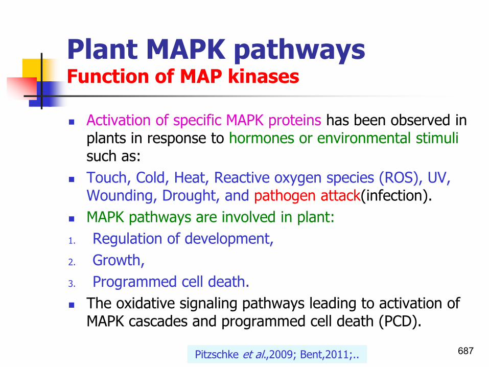
Plant MAPK pathways Function of MAP kinases
◼ Activation of specific MAPK proteins has been observed in plants in response to hormones or environmental stimuli such as:
◼ Touch, Cold, Heat, Reactive oxygen species (ROS), UV, Wounding, Drought, and pathogen attack(infection).
◼ MAPK pathways are involved in plant:
1. Regulation of development,
2. Growth,
3. Programmed cell death.
◼ The oxidative signaling pathways leading to activation of MAPK cascades and programmed cell death (PCD).
687Pitzschke et al.,2009; Bent,2011;..

Cell signalingMitogen-activated protein kinaseKinase cascades
◼ MAPKs are involved in:
◼ directing cellular responses to a diverse array of stimuli, such as mitogens, osmotic stress, heat shock and proinflammatory cytokines.
◼ They regulate cell functions including proliferation, gene expression, differentiation, mitosis, cell survival, and apoptosis.
688Mitogen: an agent that can induce mitosis.
MAPK pathways are signalling cascades differentially regulated by growth factors, mitogens, hormones and stress which
mediate cell growth, differentiation and survival.

Cell signalingMitogen-activated protein kinases (MAPK) Kinase cascades
◼ Mitogen-activated protein kinases (MAPK) are protein kinases that are specific to the amino acids serine, threonine, and tyrosine.
◼ MAP kinase cascades are a standard player in the signal transduction literature for diverse organisms including:
1. yeasts,
2. mammals, and
3. plants. ◼ Numerous studies have shown that plant MAPKs are
activated by:1. Abiotic stresses;2. Pathogens and pathogen-derived elicitors;3. Plant hormones. 689

Cell signalingFour types of protein kinases
◼ Protein kinases fall into four broad classes, characterized with respect to substrate specificity/chemical activity(phosphorylation).
1. Serine/threonine-protein kinases act on both serine and threonine;
2. Tyrosine-protein kinases act on tyrosine;
3. Dual specificity protein kinases (e.g. MEK -phosphorylates both threonine (Thr) and tyrosine (Tyr).
4. Still there are also protein kinases that phosphorylate other amino acids, including histidine kinases that phosphorylate histidine residues(in bacteria).
690Serine/threonine and tyrosine kinases were also detected in bacteria. Therefore, the bacteria also possess kinase cascades similar to those described in eukarya.

Cell signalingSix types of protein kinases
◼ Six classes of enzyme-linked receptors have thus far been identified:
1. Receptor tyrosine kinases phosphorylate specific tyrosines on a small set of intracellular signaling proteins.
2. Tyrosine-kinase-associated receptors associate with intracellular proteins that have tyrosine kinase activity.
3. Receptor like tyrosine phosphatases remove phosphate groups from tyrosinesof specific intracellular signaling proteins. (They are called “receptorlike” because the presumptive ligands have not yet been identified, and so their receptors function has not been directly demonstrated.)
4. Receptor serine/threonine kinases phosphorylate specific serines or threonineson associated latent gene regulatory proteins.
5. Receptor guanylyl cyclases directly catalyze the production of cyclic GMP in the cytosol.
6. Histidine-kinase-associated receptors activate a “two-component” signaling pathway in which the kinase phosphorylates itself on histidine and then immediately transfers the phosphate to a second intracellular signaling protein.
691Molecular Biology of the Cell. 4th edition

Cell signalingSerine/threonine-protein kinases act on both serine and threonine
692SRI International,2016

Cell signalingAmino acid phosphorylation is very important in intracellular signal transduction
693Kopchick,2015

Cell signalingAmino acid phosphorylation is very important in intracellular signal transduction
694Kopchick,2015
Result in activation or inactivation of the recipient protein!
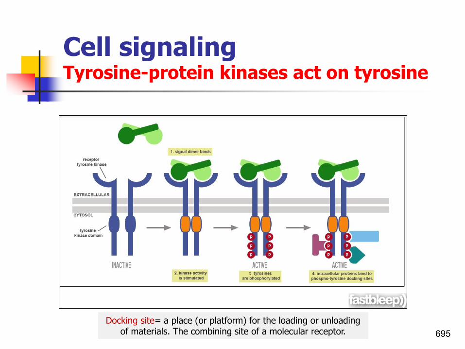
Cell signalingTyrosine-protein kinases act on tyrosine
695
Docking site= a place (or platform) for the loading or unloading of materials. The combining site of a molecular receptor.

Cell signalingFirst and second messengers
◼ In order to respond to changes in their immediate environment, cells must be able to receive and process signals that originate outside their borders.
◼ Individual cells often receive many signals simultaneously, and they then integrate the information they receive into a unified action plan.
◼ But cells aren't just targets.
◼ They also send out messages to other cells both near and far.
696www.nature.com

Cell signalingFirst and second messengers
◼ As the result of binding of first signal to the receptor, other molecules or second messengers are produced within the target cell.
◼ Second messengers relay the signal from one location to another (such as from plasma membrane to nucleus).
◼ Messenger molecules may be amino acids, peptides, proteins, fatty acids, lipids, nucleosides or nucleotides.
◼ Often a cascade of changes occur within the cell which results in a change in the cell’s function or identity.
◼ The signal transduction pathway can act to amplify the cellular response to an external signal.
697

Cell signalingFirst and second messengers
◼ FIRST MESSENGER:
◼ An extracellular substance (as the hormone epinephrine or the neurotransmitter serotonin) that binds to a cell-surface receptor and initiates intracellular activity.
◼ SECOND MESSENGER:
◼ An intracellular substance (as cyclic AMP) that mediates cell activity by relaying a signal from an extracellular molecule (as of a hormone or neurotransmitter) bound to the cell's surface.
698Merriam-webster.com
Proteins responsible for detecting stimuli are generally termed receptors.cAMP is a derivative of adenosine triphosphate (ATP) and used for
intracellular signal transduction in many different organisms.

Cell signalingSecond messengersCyclic AMP (cAMP) is an important second messenger
◼ Cyclic adenosine monophosphate(Cyclic AMP) is a second messenger important in many biological processes.
◼ It was the first second messenger ever discovered.
◼ Activation of receptors can trigger the synthesis of small molecules called second messengers, which initiate and coordinate intracellular signaling pathways.
◼ cAMP is synthesized from ATP by the enzyme adenylyl cyclase, which resides in the cell membrane.
◼ The activation of adenylyl cyclase can result in the manufacture of hundreds or even thousands of cAMP molecules.
699www.nature.com

Cell signalingThe second messengerscyclic AMP (cAMP)
◼ These cAMP molecules activate the enzyme protein kinase A (PKA), which then phosphorylates multiple protein substrates by attaching phosphate groups to them.
◼ Each step in the cascade further amplifies the initial signal, and the phosphorylation reactions mediate both short- and long-term responses in the cell
◼ How does cAMP stop signaling?
◼ It is degraded by the enzyme phosphodiesterase.
700www.nature.com

Cell signalingFirst and second messengers
1. Most cell signals are chemical in nature.
2. Some are mechanical stimuli.
◼ For example, prokaryotic organisms have sensors that detect nutrients and help them navigate toward food sources.
◼ In multicellular organisms, growth factors, hormones, neurotransmitters, and extracellular matrix components are some of the many types of chemical signals cells use.
◼ These substances can exert their effects locally, or they might travel over long distances.
701www.nature.com

Cell signalingFirst and second messengersIn mammals
◼ For instance, neurotransmitters are a class of short-range signaling molecules that travel across the tiny spaces between adjacent neurons or between neurons and muscle cells.
◼ Other signaling molecules must move much farther to reach their targets.
◼ One example is follicle-stimulating hormone, which travels from the mammalian brain to the ovary, where it triggers egg release.
702www.nature.com

Cell signalingFirst and second messengersIn humans
1. Most cell signals are chemical in nature.
2. Some are mechanical stimuli.
◼ Some cells also respond to mechanical stimuli.
◼ For example, sensory cells in the skin respond to the pressure of touch, whereas similar cells in the ear react to the movement of sound waves.
◼ In addition, specialized cells in the human vascular system detect changes in blood pressure —information that the body uses to maintain a consistent cardiac load.
703www.nature.com

Cell signalingInternal signaling pathwaysThree major classes of membrane receptors
◼ Receptors are generally transmembrane proteins, which bind to signaling molecules outside the cell and subsequently transmit the signal through a sequence of molecular switches to internal signaling pathways.
◼ Also amplifies the signals via signal transduction.
◼ Membrane receptors fall into three major classes:
1. G-protein-coupled receptors,
2. ion channel receptors, and
3. enzyme-linked receptors.
704
G Proteins are guanine-nucleotide binding proteins. Large numbers of G proteins provide diversity for signal transduction events.
Some bind potassium or calcium ion channels in neurotransmitters.Some activate kinases (enzymes that phosphorylate).

Cell signalingThree major classes of membrane receptors1. Ion channels
◼ Ion channels: Their role in signal generation is mainly centred on the Ca2+ signaling pathway, which has a large number of Ca2+ entry channels and internal Ca2+ release channels, both of which contribute to the generation of Ca2+ signals.
◼ There are a large number of K+ channels and many of these function in different aspects of cell signaling.
705Saludar,2011

Cell signalingThree major classes of membrane receptors2. G-protein-coupled receptors
◼ G-protein-coupled receptors: G-protein-coupled receptors (GPCRs) constitute a large and diverse family of proteinswhose primary function is to transduce extracellular stimuli into intracellular signals.
◼ Coupling with G-proteins, they are called seven-transmembrane receptors because they pass through the cell membrane seven times.
◼ The alpha subunit of the G-protein being the primary signaling molecule.
◼ The general function of the G alpha-s subunit (Gs) is to activate adenylate cyclase which in turn produces cyclic-AMP (cAMP), leading to the activation of cAMP-dependent protein kinases (often referred to collectively as Protein Kinase A).
706

Cell signalingThree major classes of membrane receptors3. Enzyme-linked receptors
◼ Enzyme-linked receptors: Like G-protein-linked receptors, enzyme-linked receptors are transmembrane proteins with their ligand-binding domain on the outer surface of the plasma membrane.
◼ Enzyme-linked receptors also known as a catalytic receptors, where the binding of an extracellular ligand causes enzymatic activity on the intracellular side. They possessing both enzymatic catalytic and receptor functions.
◼ Six classes of enzyme-linked receptors have thus far been identified such as receptor tyrosine kinase, receptor serine/threonine kinases, etc.
707Molecular Biology of the Cell. 4th edition

Cell signalingInternal signaling pathwaysThree major classes of membrane receptors
708Saludhttps://pharmaceuticalintelligence.com

Cell signalingInternal signaling pathwaysThree major classes of membrane receptors
709Saludar,2011

Cell signalingInternal signaling pathwaysFour classes of membrane receptors
710

Cell signalingInternal signaling pathwaysFour classes of membrane receptors
711

Cell signalingInternal signaling pathwaysFour classes of membrane receptors
712

Cell signalingSignal cascade stepsAmino acid phosphorylation and dephosphorylation is very important in intracellular signal transduction
◼ Within proteins, the amino acids serine, threonine, and tyrosine are especially common sites for phosphorylation.
◼ These phosphorylation reactions control the activity of many enzymes involved in intracellular signaling pathways.
◼ Specifically, the addition of phosphate groups causes a conformational change in the enzymes, which can either activate or inhibit the enzyme activity. Then, when appropriate, protein phosphatases remove the phosphate groups from the enzymes, thereby reversing the effect on enzymatic activity.
713Wikipedia,2017; Kopchick,2015; www.nature.com
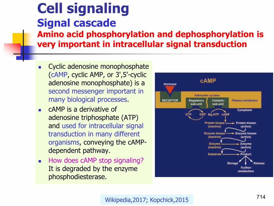
Cell signalingSignal cascadeAmino acid phosphorylation and dephosphorylation is very important in intracellular signal transduction
◼ Cyclic adenosine monophosphate (cAMP, cyclic AMP, or 3',5'-cyclic adenosine monophosphate) is a second messenger important in many biological processes.
◼ cAMP is a derivative of adenosine triphosphate (ATP) and used for intracellular signal transduction in many different organisms, conveying the cAMP-dependent pathway.
◼ How does cAMP stop signaling? It is degraded by the enzyme phosphodiesterase.
714Wikipedia,2017; Kopchick,2015

Cell signalingSignal cascadeEach step in the cascade further amplifies the initial signal
715Pearson Education,2009

Cell signalingSignal cascadeAn example of a signal transduction cascade involving cyclic AMP
◼ The binding of adrenaline to an adrenergic receptor initiates a cascade of reactions inside the cell.
◼ The signal transduction cascade begins when adenylyl cyclase, a membrane- bound enzyme, is activated by G-protein molecules associated with the adrenergic receptor.
◼ Adenylyl cyclase creates multiple cyclic AMP molecules, which fan out and activate protein kinases (PKA, in this example).
◼ Protein kinases can enter the nucleus and affect transcription.
716www.nature.com

Cell signalingProtein kinase function is evolutionarily conserved from Escherichia coli to humanEukaryotes vs. prokaryotes
◼ The eukaryotic protein kinases that directly phosphorylate proteins are divided into two major classes:
1. those that phosphorylate tyrosine; and
2. those that phosphorylate serine and threonine.
◼ Bacteria, whose cellular makeup is considered to be simplified and optimized for rapid bursts of growth, were usually thought not to possess such complicated kinase networks.
◼ The main sensing and signal transduction devices in bacteria are the so-called two-component systems, based on histidine/aspartate kinases (See QS in file1).
717Serine/threonine and tyrosine kinases were also detected in bacteria. Therefore, the bacteria also possess kinase cascades similar to those described in eukarya.

Cell signalingKinase cascadesDefinition
◼ Proteins responsible for detecting stimuli are generally termed receptors, although in some cases the term sensor is used.
◼ The changes elicited by ligand binding (or signal sensing) in a receptor give rise to a signaling cascade, which is a chain of biochemical events along a signaling pathway.
718

Cell signalingKinase cascadesMAP kinase
◼ Microtubule-associated protein (MAP):
◼ Any of the high molecular weight proteins that bind to microtubules, enhancing polymerization.
◼ Microtubule-associated protein (MAP) kinase:
◼ A protein kinase that is activated in response to cell stimulation by many different growth factors and that mediates:
1. cellular responses by phosphorylating specific transcription factors and
2. other target proteins.
719A MAPK cascade in Arabidopsis innate immunity.

Cell signalingSerine/threonine and tyrosine kinases in bacteria
◼ Very recently it was reported that some bacteria such as Bacillus subtilis also possess protein serine/threonine and tyrosine kinases which resemble eukaryal kinases in their capacity to phosphorylate multiple substrates.
◼ There is good evidence that most of the prokaryotic Ser/Thr kinases carry out phosphorylation of their target proteins within the bacterial-cell cytoplasm.
◼ E.g.
◼ The Streptomyces Ser/Thr protein kinase AfsK phosphorylates the global regulator AfsR, which controls the production of certain antibiotics.
720

Signaling processesSignal perception and defense activation
◼ The first process in signal transduction is:
1. Perception of an extracellular signal, and
2. Its transmission via the plasma membrane, resulting in accumulation of intracellular signaling molecules and
3. Induction of a phosphorylation/dephosphorylationcascade, a cue system for the activation of R gene expression.
721
Phosphorylation is the addition of a phosphate (PO4) group to a protein or other organic molecule.

Signal Transduction Phosphatases and phosphodiesteraseInactivation of MAP kinases
◼ How does cAMP stop signaling?
◼ It is degraded by the enzyme phosphodiesterase.
◼ Activity of MAP kinases is restricted by a number of protein phosphatases, which remove the phosphate groups that are added to specific serine or threonine residues of the kinase and are required to maintain the kinase in an active conformation.
722

Signal Transduction Kinases and Phosphatases
◼ Many enzymes are regulated by covalent attachment of phosphate, in ester linkage, to the side-chain hydroxyl group of a particular amino acid residue (serine, threonine or tyrosine).
◼ Kinases: Enzymes which phosphorylate (add a phosphate
group).
◼ Phosphatases: Enzymes which dephosphorylate (remove a phosphate group).
723Diwan,2008

Signaling processesPlant MAPK pathwaysPhosphorylations and dephosphorylations
◼ Plant defense also appears to be under negative control of various MAPK cascades (negative regulation).
◼ Effectors that enhance the protein's activity are referred to as positive regulation, and those that decrease the protein's activity are called negative regulation.
◼ Note that negative regulatory roles turn out positive.
724

Signal
Sensor
MAPKKK
MAPKK
MAPK
Primary Response Genes
Secondary Response Genes
The prototypical MAPK cascade
WRKY transcription factors1. Early defense genes
(FRK1);2. Late defense genes(PR)
Input
Output
FilterAmplify
Tena,2002
FRK1:Cell surface plant receptor-like protein kinase

Plant MAPK pathways Cascades in stress and hormonal signaling
726
NPR1 regulates PR gene
expression by interacting with
TGA TFs. Thus, TGA factors
affect the transcription of PR
genes.

Plant MAPK pathways MAPKs in Arabidopsis
◼ The Arabidopsis genome has revealed large gene families encoding MAPKs and their immediate upstream regulators, MAPKKs and MAPKKKs.
◼ There are many MAPK-encoding genes in plant genomes.
◼ For example, the Arabidopsis genome contains about 110 genes coding for putative MAPK pathway components:
1. 20 MAPK genes,
2. 10 MAPKKs, and
3. More than 80 MAPKKKs.
727Complexity of MAPK networks
20 x 10 x 80 = a lot of virtual combinations

Plant MAPK pathways Complexity of MAPK networksModel systems
◼ Mitogen-activated protein kinase (MAP kinases cascades) represent important and universal signal transduction mechanisms in diverse cellular responses in eukaryotic organisms.
◼ How to work with such a high number of possibilities?
1. a simplified biological system;
2. possibility to do hundreds of functional analyses quickly.
728Tena,2002

Plant MAPK pathwaysRelatedness of Arabidopsis MAPKs
◼ A large number of MAPKs act as positive players in defense signaling.
1. MPK4 negatively regulates biotic stress signalling,
2. MPK3 and MPK6 act as positive mediators of defence responses.
729Tena,2002

Plant MAPK pathways Characterization and functions of MAP kinases
◼ The best-characterized MAPKs are MPK3, MPK4 and MPK6, all of which are activated by a diversity of stimuli including:
1. Abiotic stresses
2. Flg22 peptide-mediated defense responses(PAMP)
3. The ethylene pathway
4. Pathogens
5. Auxin
6. H2O2
7. Oxidative stress.
730Pitzschke et al.,2009;…

Plant MAPK pathways MAPK cascades in Arabidopsis thalianaIn response to abiotic, biotic stresses, hormones & H2O2
731Innes,2001

Plant MAPK pathways Relatedness of Arabidopsis MAPKKs
732Tena,2002

Plant MAPK pathways Relatedness of Arabidopsis MAPKKKs
733

Structural heterogeneity
of plant MAPKKKs.
Tena,2002

Plant MAPK pathwaysSIPK family(salicylic acid-induced protein kinase)
◼ SIPK has emerged as a universal key component of the MAPK cascade.
◼ SIPKs involve in fine-tuned regulation of the plant responses to:
◼ Ozone, wounding, SA, and jasmonic acid (JA).
◼ SIPKs respond to multiple stresses with different functions under different biotic and abiotic stresses.
◼ For example,
1. AtMKK2–AtMPK6 is involved in cold and salt stresses;
2. AtMKK4/AtMKK5–AtMPK6 cascade is involved in ET(ethylene) signaling.
735Cho et al.,2008

Plant MAPK pathwaysSIPK family
◼ Over the past years, several plants MAPKs have been identified and characterized.
◼ Of these, rice SIPK (Salicylic acid (SA)-Induced Protein Kinase) and its orthologs in other plants are of particular interest.
◼ SIPK family includes:
◼ AtMPK6 in Arabidopsis,
◼ MsSIMK in alfalfa,
◼ NtSIPK in tobacco, and
◼ OsSIPK in rice.
736Cho et al.,2008

Signal pathways of orthologs of OsSIPK in Arabidopsis and in tobacco SIPK (Salicylic acid (SA)-Induced Protein Kinase)
737Cho et al.,2008

Systemic Acquired Resistance and Induced Systemic ResistancePR proteins
◼ Systemic acquired resistance(SAR):◼ Induced by the exposure of root or foliar tissues to
abiotic or biotic elicitors, is dependent of:1. the phytohormone salicylate (salicylic acid), and 2. associated with the accumulation of pathogenesis-
related (PR) proteins. ◼ Induced systemic resistance(ISR):◼ Induced by the exposure of roots to specific strains of
plant growth-promoting rhizobacteria, is dependent of 1. the phytohormones ethylene and jasmonate (jasmonic
acid), 2. independent of salicylate, and 3. is not associated with the accumulation of PR proteins.
738Vallad and Goodman,2004

Signal transduction pathway SAR vs ISR
◼ Signal transduction pathway of induced systemic resistance stimulated by Bacillus velezensis.
◼ NPR1: non-expressor of PR1;
◼ JA/ET: the jasmonic acid/ethylene signaling pathways;
◼ SA: Salicylic Acid.
739Rabbee et al.,2019

SAR/ISRA pictorial comparison of the two best characterized forms of induced resistance in plants, both which lead to similar phenotypic responses
740Vallad and Goodman,2004

SAR/ISRA pictorial comparison of the two best characterized forms of induced resistance in plants, both which lead to similar phenotypic responses
741

The plant R-avr gene interaction triggersa signal transduction pathway leading tothe HR (1) and SAR (2)
◼ SA was shown to move from the infected leaf into the other parts of the plant (up to 70% of labelled SA was found in the upper leaves).
742Baker,1997

Systemic Acquired Resistance SAR
◼ The graphic describe these defensive reactions in more detail than is possible here.
743Elmore, 2015

Transcription factorsTGAs and WRKY
TGACG motif‐binding protein (TGA) and
WRKY transcription factors activate many PR‐genes.
744

Transcription factors(TxF’s)Transcription factors involved in plant defense
TGA and WRKY transcription factors
◼ Some major families of transcription factors involved in plant defense:
1. TGA factors and TGA boxes belong basic leucinezipper containing domain proteins (bZIP),
2. amino-acid sequence WRKYGQK (WRKY)pronounced ‘worky’,
◼ TGACG motif‐binding protein (TGA) and WRKY
transcription factors for the activation of many PR‐genes.
745

Transcription factors(TxF’s)TGA and WRKY transcription factors
◼ For a plant this may be:
1. abiotic (non-living) stress such as the rising or setting sun, drought, or heat,
2. biotic (living) stress such as insects, viral or bacterial infection, or
3. any of a limitless number of other events.
◼ The job of coordinating the function of groups of genes falls to proteins called transcription factors (TxF’s).
746Monsanto Co.,2010

Transcription factorsSAR and priming TGA and WRKY transcription factors
747
◼ The attack of a pathogen leads to signals, which give to the plant an immediately protection and a long‐lasting immunity.
◼ The infected leaf generates callose for the fortification of cell walls, ROS and antimicrobial compounds to kill the bacteria, and defense‐related metabolism.
◼ These processes are part of the innate immunity.
◼ Principally, the metabolite SA is transport through the phloem to the other part of the plants activating the SAR, which is a preventive defense mechanism.
◼ This includes the monomerization of NPR1 that in turn activates PR‐genes, chromatin modification such as methylation and acetylation of histones and somatic recombination.
Naranjo‐Arcos and Bauer,2016

Eukaryotic transcription machinery Transcription factors(TxF’s)Difference between Prokaryotic vs Eukaryotic transcription
◼ The eukaryotic transcription machinery is complex.
◼ The main differences are:
1. Number and functions of RNA polymerase(s);
2. Number and functions of promoter elements;
3. Transcription and translation occurs simultaneously in prokaryotes and in eukaryotes the RNA is first transcribed in the nucleus and then translated in the cytoplasm.
4. RNAs from eukaryotes undergo post-transcriptional modifications including: capping, polyadenylation, and splicing. These events do not occur in prokaryotes.
748
Polyadenylation: addition of about 250 adenine residues to form a poly(A) tail to a messenger RNA. It is part of the process that produces mature messenger RNA (mRNA) for translation.

Eukaryotic transcription machinery Transcription factors(TxF’s)Difference between Prokaryotic vs Eukaryotic transcription
◼ The eukaryotic transcription machinery is complex.
◼ In prokaryotic cells there is only a single RNA polymerase that synthesizes with the help of sigma factor all classes of RNA:
1. mRNA,
2. rRNA,
3. tRNA.
◼ In eukaryotic cells, there are three different RNA polymerases that synthesize different classes of RNAs:
1. PolI transcribes rRNA (ribosomal RNA),
2. PolII mRNA (messenger RNA), and
3. PolIII tRNA (transfer RNA) and other small RNAs. 749

Eukaryotic transcription machinery Transcription factors(TxF’s)Difference between Prokaryotic vs Eukaryotic transcription
750

Eukaryotic transcription machinery Transcription factors(TxF’s)Difference between Prokaryotic vs Eukaryotic transcription
◼ Prokaryotes only contain three different promoter elements:
1. -10,
2. -35 promoters, and
3. upstream element.
◼ Eukaryotes contain many different promoter elements:
1. TATA box,
2. initiator elements,
3. downstream core promoter element,
4. CAAT box (start points of eukaryotic transcription units),
5. GC box(known for binding general transcription factors).
751

Eukaryotic transcription machinery Transcription factors(TxF’s)Difference between Prokaryotic vs Eukaryotic transcription
◼ mRNAs in prokaryotes tend to contain many different genes on a single mRNA meaning they are polycystronic.
◼ Eukaryotes contain mRNAs that are monocystronic. i.e. it contains the genetic information to translate only a single protein chain (polypeptide).
◼ Termination in prokaryotes is done by either rho-dependent or rho-independent mechanisms.
◼ In eukaryotes transcription is terminated by two elements: a poly(A) signal and a downstream terminator sequence.
752

Transcription factors(TxF’s)TxF’s are proteins that regulate gene expression Numbers in each cell
◼ Transcription factors are essential for the regulation of gene expression and are, as a consequence, found in all living organisms.
◼ The number of transcription factors found within an organism increases with genome size:
◼ The larger genomes tend to have more transcription factors per gene.
◼ Approximately 10% of genes in the genome code for transcription factors, which makes this family the single largest family of human proteins.
753Wikipedia,2016

Transcription factors(TxF’s)TxF’s are proteins that regulate gene expression How do transcription factors work?
◼ A transcription factor (sometimes called a sequence-specific DNA-binding factor) is a protein that binds to specific DNA sequences, thereby controlling the rate of transcription of genetic information from DNA to messenger RNA.
◼ Transcription factors perform this function alone or with other proteins in a complex, by:
1. promoting (as an activator), or
2. blocking (as a repressor) the recruitment (hiring) of RNA polymerase to specific genes.
754Wikipedia,2016

Transcription factors(TxF’s)How do transcription factors work?Genes turned on and off
◼ In order to allow coordinated gene function, a particular TxF may bind to multiple genes, and each gene may be controlled by multiple TxF’s.
◼ Further– recall that each TxF is itself a protein, and TxF’s often regulate other TxF’s.
◼ TxF’s form complex networks that may control from one to many thousands of genes (multiple protein complexes) in response to conditions inside or outside of the cell.
755Monsanto Co.,2010

Transcription factors(TxF’s)How do transcription factors work?
756Monsanto Co.,2010

757
Transcription factors(TxF’s) Control of gene/mRNA/protein activityNucleus vs. mitochondria in Eukaryotes
CS 6463: An overview of Molecular Biology

Transcription factors(TxF’s)TxF’s are proteins that regulate gene expression 1. General transcription factors in eukaryotes
◼ In eukaryotes, an important class of transcription factors called general transcription factors (GTFs) are necessary for transcription to occur.
◼ Many of these GTFs don't actually bind DNA but are part of the large transcription pre-initiation complex that interacts with RNA polymerase directly.
◼ The most common GTFs are:
◼ TFIIA, TFIIB, TFIID (see also TATA binding protein), TFIIE, TFIIF, and TFIIH.
758Wikipedia,2016

Transcription and Gene Expression Protein-protein interactions2. Transcription factors involved in plant defense
◼ Some major families of transcription factors involved in plant defense:
1. TGA factors and TGA boxes belong basic leucine zipper containing domain proteins (bZIP),
2. amino-acid sequence WRKYGQK (WRKY) pronounced ‘worky’,
3. myelocytomatosis related proteins (MYC),
4. myeloblastosis related proteins (MYB),
5. The AP2/EREBP family of plant transcription factors,
6. no apical meristem (NAM),
7. Arabidopsis transcription activation factor (ATAF),
8. cup-shaped cotyledon (CUC).759Alves et al.,2014

Transcription and Gene Expression Protein-protein interactionsTranscription factors involved in plant defense
760
◼ The two major transcription factors involved
in plant defense:
1. TGAs, the basic leucine zipper containing domain proteins (bZIP),
2. WRKYs, amino-acid sequence WRKYGQK (WRKY) pronounced ‘worky’.
Leucine zipper motifs are a dimerization domain of the bZIPclass of eukaryotic transcription factors. The two leucine zippers in effect form a Y-shaped structure, in which the zippers comprise the stem, and the two basic regions
bifurcate symmetrically to form the arms that bind to DNA. This is known as the bZIP structural motif.

Transcription and Gene Expression Protein-protein interactionsTranscription factors involved in plant defense
761
◼ In the nucleus, NPR1 can interact with different transcription factors (TFs), such as:
1. TGAs, and
2. WRKYs,
◼ to regulate the expression of downstream genes, like PR-1.

Transcription and Gene Expression Plant transcription factors 1. TGA transcription factors and TGA boxes
◼ Salicylic acid (SA) is a simple phenolic compound distributed in a wide range of plant taxa.
◼ In response to pathogen attack, salicylic acid induces the expression of pathogenesis-related (PR) genes throughout the plant.
◼ TGA factors are believed to regulate this systemic induction because they bind to the as-1 cis element present in the promoters of PR genes, and because different TGA factors interact with the NPR protein, which is necessary for PR gene induction but does not bind to DNA by itself.
762Jackoby et al.,2002

Transcription and Gene Expression Plant transcription factors TGA transcription factors and TGA boxes
◼ Salicylic acid (SA) is a simple phenolic compound distributed in a wide range of plant taxa.
◼ Defense pathways in plants are strongly associated with four compounds:
1. SA,
2. ethylene (ET),
3. jasmonic acid (JA), and
4. abscisic acid(ABA).
763Kumar and Almomin,2018

Transcription and Gene Expression Plant transcription factors TGA transcription factors and TGA boxes
◼ SA is synthesized in plants from cinnamate and isochorismate and its involvement in the regulation of plant defense against specific types of pathogens.
764Kumar and Almomin,2018

Transcription and Gene Expression TGA transcription factors and TGA boxesRegulation of defense gene expression by NPR1
◼ NPR1 protein is a receptor for the plant defense hormone salicylic acid (SA).
◼ SA controls NPR1 function by regulating its protein level in the nucleus.
◼ Downstream of SA, upstream of PR genes.
1. npr1 mutants are susceptible to various pathogens.
2. Overexpression of NPR1 generates broad-spectrum resistance.
765Wisuwat Songnuan;..

Transcription and Gene Expression Plant transcription factors TGA transcription factors and TGA boxes
◼ Of the other compounds, jasmonic acid (JA) plays a vital role in defense against insects and herbivores.
◼ Further complexity results from the significant overlap and crosstalk between the defense responses mediated by SA and those mediated by JA.
766Kumar and Almomin,2018; Van der Does et al.,2013

Transcription and Gene Expression TGA transcription factors (TFs) and TGA boxesRegulation of defense gene expression by NPR1
◼ NPR1 gene encodes a transcription coactivator (NPR1) that plays a major role in the mechanisms regulating plant defense response.
◼ NPR1 plays a role in other defense-signaling pathways.
◼ NPR1 protein is a receptor for the plant defense hormone salicylic acid (SA).
767McDowell and Woffenden,2003

Transcription and Gene Expression Plant transcription factors TGA transcription factors and TGA boxes
768Kumar and Almomin,2018

Transcription and Gene Expression bZip plant transcription factors TGA factors and TGA boxes
769Zhang et al.,2018
Schematic illustrations of transcription factor in regulating defense gene expression. PAMP perception stimulates the induction of MKK, and then MPK forms a complex with WRKY.
Phosphorylation of MKS1 by MPK4 and dissociation of the MKS1-WRKY25/33 complex from MPK4. This leads to the latter complex binds the promoter region of PAD3, which is required for the
synthesis of antimicrobial camalexin. Effectors perception stimulates the induction of hormone signal and manipulated ERF, TGA, and MYB TFs to regulate R genes expression directly or indirectly. Some
transcription factors could regulate reciprocally by binding to the promoter correspondingly.

Transcription and Gene Expression Plant transcription factors TGA factors and TGA boxes
◼ TGA factors and TGA boxes:
1. belong to the family of bZip plant transcription factors, and
2. involved in regulation of defense gene expression by NPR1.
◼ The NPR1 protein is a receptor for the plant defense hormone salicylic acid.
◼ Salicylic acid (SA) is a plant immune signal produced after pathogen challenge to induce systemic acquired resistance.
770
In plants, basic region/leucine zipper motif (bZIP) transcription factors regulate processes including pathogen defence, light and
stress signalling, seed maturation and flower development.

Transcription and Gene Expression bZIP transcription factors in ArabidopsisTGA factors and TGA boxes
◼ There are 10 TGA transcription factors in Arabidopsis of which seven (TGA1–TGA7) have been characterized with respect to their interaction with NPR1.
◼ These seven TGAs can be further divided into three subgroups based on sequence homology:
1. group I: TGA1 and TGA4;2. group 2: TGA2, TGA5, and TGA6; and3. group III TGA3 and TGA7.
771Kesarwani et al.,2007

Transcription and Gene Expression bZip plant transcription factors TGA factors and TGA boxes
◼ Ten groups of bZIPs (A to S) with a similar basic region and additional conserved Motifs.
◼ Group D genes participate in defence against pathogens and development.
◼ Their involvement in defence mechanisms comes from work on the TGA factors (group D) in tobacco and Arabidopsis.
TGA factors (group D)
Jackoby et al.,2002

Transcription and Gene Expression TGA factors and TGA boxesInvolved in regulation of defense gene expression by NPR1
◼ The Arabidopsis genome sequence contains 75 distinct members of the bZIP family.
◼ basic region/leucine zipper (bZIP) TFs have:
1. a basic region that binds DNA, and
2. a leucine zipper dimerization motif.
773Jackoby et al.,2002;Wikipedia
bZIP dimer is bound to DNA fragment -each alpha helix represents a monomer.

Transcription and Gene Expression Regulation of defense gene expression by NPR1Structural and function of NPR1
◼ TGA transcription factors are implicated as regulators of pathogenesis-related (PR) genes.
◼ Induction of plant systemic acquired resistance (SAR) correlates with the expression of pathogenesis-related (PR) genes.
774Wisuwat Songnuan
BTB ARD
S SNLS
nim 1-2 npr 1-1npr 1-2

Transcription and Gene Expression Plant transcription factors 2. WRKY factors and W box
◼ WRKY transcription factors(pronounced ‘worky’) are one of the largest families of transcriptional regulators found mostly in plants (green algae and land plants).
◼ The family has expanded during the evolution of plants and in turn, co-evolving of land plants with their adapted pathogens.
◼ The W box is a common binding site for WRKY transcription factors.
775Rushton et al.,2010; Bakshi and Oelmüller,2014
W box is a deoxyribonucleic acid (DNA) cis-regulatory element sequence, (T)TGAC(C/T), which is recognized by
the family of WRKY transcription factors.

Transcription and Gene Expression Plant transcription factors WRKY factors
◼ WRKY transcription factors have diverse biological functions in plants, but most notably are key players in plant responses to biotic and abiotic stresses.
◼ A (WKRY) gene that confers resistance or tolerance to multiple stresses would be highly useful for breeding.
◼ However, WRKY genes can also have opposite effects on abiotic and biotic stress tolerance since complex interactions among signaling networks can lead to both synergistic and antagonistic effects on regulation of plant responses to different stresses.
776Bai et al.,2018

Transcription and Gene Expression Role of WRKY proteins in both abiotic and biotic stresses
777Banerjee and Roychoudhury,2015
In tomato there are 83 WRKY genes identified.

Transcription and Gene Expression Plant transcription factors Three groups of WRKY transcription factors
◼ WRKY transcription factors (WRKYs) are a large family of transcriptional regulators, which are defined by the highly conserved WRKY domain (the WRKYGQK motif at the end of the N-terminal and a zinc-finger-like motif at the C-terminus.
◼ WRKYs are categorized into three groups:
◼ Group I (with two WRKY domains);
◼ Group II (with one WRKY domain) contain the zinc-finger-like motif C2–H2 (C–X4-5–C–X22-23–H–X1–H);
◼ Group III contains one WRKY domain and a C2–HC zinc-finger-like motif (C–X7–C–X23–H–X1–C).
778Bai et al.,2018

Transcription and Gene Expression Plant transcription factors Three subgroups of WRKY Group II
◼ Based on the primary amino acid sequences, Group II can be further divided into three subgroups (IIb, IId and IIe).
779Bai et al.,2018

Transcription and Gene Expression Plant transcription factors WRKY factors and W box
◼ The involvements of Group I and III tomato SlWRKY genes and their homologs (highlighted in different colors) in plant responses to biotic and abiotic stresses.
780Bai et al.,2018

Transcription and Gene Expression Plant transcription factors WRKY factors and W box
◼ Specific WRKY proteins like AtWRKY18, AtWRKY40, and AtWRKY60 have been depicted as mediators of cross talk between plant responses against abiotic and biotic stresses.
◼ It has been reported that these proteins get accumulated in response to SA and JA during biotic stress as well as ABA during abiotic stress responses.
781Banerjee and Roychoudhury,2015

Transcription and Gene Expression The mitogen-activated protein kinase (MAPK) pathway induces the activity of OsWRKY30 during drought stress
◼ The mitogen-activated protein kinase (MAPK) pathway induces the activity of OsWRKY30 during drought stress.
◼ Stress signals are sensed via a transmembrane receptor, which with the help of some unknown molecules and adaptor proteins activates the MPK/MAPK pathway.
◼ This leads to the phosphorylation and activation of the MPK3.
◼ MPK3 phosphorylates the target Ser residue in the SP motif of OsWRKY30 and activates the same.
◼ The activated WRKY protein then undergoes a conformational change which favourably allows it to bind to the W-box of its target gene to induce transcription.
◼ The protein product encoded by the target gene probably helps the plant system in combating the drought stress.
782Banerjee and Roychoudhury,2015

Transcription and Gene Expression The involvement of two transcription factors WRKY54 and WRKY70 in developmental senescence, osmotic stress as well as plant defense responses
783Li,2014
The involvement of two transcription factors WRKY54 and WRKY70 in developmental senescence, osmotic stress as well as plant defense responses. The arrows indicate induction or positive modulation; the blunt-end arrows indicate block or suppression. SA, salicylic acid; JA, jasmonic acid; ABA, abscisic acid; ROS, reactive oxygen species.

Transcription and Gene Expression The structurally related proteins AtWRKY18, AtWRKY40, and AtWRKY60 have provided some hint towards the occurrence of such crosstalk
◼ Specific WRKY proteins like AtWRKY18, AtWRKY40, and AtWRKY60 have been depicted as mediators of crosstalk between plant responses against abiotic and biotic stresses.
◼ It has been reported that these proteins get accumulated in response to SA and JA during biotic stress as well as ABA during abiotic stress responses.
784Banerjee and Roychoudhury,2015

785Finatto et al.,2018
Overview of the most recent reports concerning WRKY roles in plant defense, including the relationships with MAPKs, abscisic acid(ABA) signaling, and responses to
biotic and abiotic stresses.

WRKY proteinsWRKY domain /W box
◼ The WRKY domain(DNA binding domain) has almost invariant WRKY amino acid sequence at the N-terminus
◼ The invariant core of the W box is essential for function and WRKY binding.
a) The WRKY domain consensus for each WRKY subfamily in higher plants:
◼ The WRKY motif is highlighted in yellow and the cysteines and histidines that form the zinc finger are shown in blue.
◼ The four β-strands are shown in red.
b) Two views of a spacefill structural model of the C-terminalWRKY domain from AtWRKY4.

Transcription and Gene Expression Plant transcription factors WRKY factors and W box
787Finatto et al.,2018
Regulatory mechanism of WRKY transcription factors. The signaling pathway starts with an environmental stimulus through ABA signaling or
by trans-regulation, which comprise direct transcription factor activation. The trans regulation can occur with a WRKY member, in case of presence of a W-box, or
with other TFs from different families. An auto-regulation mechanism can occur, or regulation by promoter binding of a different WRKY member. In some cases, the
phosphorylation by an MAPK cascade is a determinant process for the correct WRKY protein function. When activated, WRKY TFs can regulate a different WRKY by binding its W-box sequence, or regulate some other responsive gene conferring tolerance, resistance,
or sensibility toward the environmental stimuli.

Transcription and Gene Expression Plant transcription factors WRKY factors and W box
◼ The W box is a common binding site for WRKY transcription factors, proteins that have been shown to be involved in regulation of early defense-response gene expression.
◼ WRKYs can act as:
1. transcriptional activators, or
2. repressors.
788

Transcription and Gene Expression Plant transcription factors WRKY factors and W box
◼ E.g.
1. WRKY25 and -33 are involved in biotic and abiotic stress responses; they function as negative regulators after P. syringae infection, but
2. As positive regulators in conferring resistance against NaCl, cold, and heat and oxygenic stress.
789

Transcription and Gene Expression Plant transcription factors Functions of WRKY TFs
◼ WRKY transcription factors have diverse biological functions in:
1. plant disease resistance,
2. abiotic stress responses,
3. nutrient deprivation,
4. senescence,
5. Seed, and
6. trichome development, embryogenesis, as well as additional developmental and hormone-controlled processes.
790Bakshi and Oelmüller,2014

Transcription and Gene Expression Plant transcription factors Functions of WRKY TFs
791

Transcription and Gene Expression Functions of WRKY TFsThe role of WRKYs and their genes in abiotic stress (A)
◼ A schematic representation of the mechanism by which AtWRKY40, AtWRKY18 and AtWRKY60 regulate abscisic acid(ABA) responses.
◼ Eexpression of ABA‐responsive genes such as ABF4, ABI4, ABI5, DREB1A,MYB2 and RAB18 is induced and ABA responses occur.
◼ The dotted line denotes de‐repression of gene expression as the WRKY TFs are removed from the nucleus.
792Rushton et al.,2012

Transcription and Gene Expression Functions of WRKY TFsThe role of WRKYs and their genes in biotic and abiotic stresses
◼ Abiotic stress:
◼ The role of WRKYs and their genes in abiotic stress (A), biotic stress (B), the cross-talk between biotic and abiotic stress (C), metabolism (D), hormone signaling (E), and epigenetic control (F).
◼ The gray square boxes with the number refer to the individual WRKY proteins. WRKY genes are in green. The crosses demonstrate inhibition of expression. A P in a yellow star refers to phosphorylation. Ub, ubiquitin.
793Bakshi and Oelmüller,2014
Ubiquitin is a small regulatory protein that exists (ubiquitously) in all eukaryotic cells.

Transcription and Gene Expression Functions of WRKY TFsThe role of WRKYs and their genes in biotic and abiotic stresses
◼ Biotic stress: pathogens, PAMPs and elicitors:
◼ The role of WRKYs and their genes in abiotic stress (A), biotic stress (B), the cross-talk between biotic and abiotic stress (C), metabolism(D), hormone signaling (E), and epigenetic control (F).
◼ The gray square boxes with the number refer to the individual WRKY proteins. WRKY genes are in green. The crosses demonstrate inhibiton of expression. A P in a yellow star refers to phosphorylation. Ub, ubiquitin.
794Bakshi and Oelmüller,2014
Ubiquitin is a small regulatory protein that exists (ubiquitously) in all eukaryotic cells.

WRKY transcription factorsMTI or PTI: MAMP or PAMP-triggered immunity The effect of WRKY29

WRKY transcription factorsInvolved in defense-related signal transduction pathways
PAMP or effector triggered immunity
RLK: Plant receptor-like protein kinases. Plant receptor-like kinases (RLKs) are transmembrane proteins.

Transcription and Gene Expression WRKY genes/W box in Arabidopsis genome+TGA factors and TGA boxes
1. Early defense genes (FRK1);
2. Late defense genes(PR).◼ Several genes coding for
WRKY factors are early induced by SA and pathogens, and some of them do not require NPR1 for their induction.
◼ SA and ROS are signals for genetic-controlled defense reactions.
◼ Interestingly, WRKY factors are activated by MAPK and ROS.

WRKY proteinsSpecies phylogeny and numbers of WRKY genes in each species
◼ The WRKY gene family plays a unique role in plant stress tolerance.
◼ Quinoa is a cultivated crop worldwide that is known for its high stress tolerance.
◼ Using a genome-wide search method, we identified 1226 WRKY genes in:
◼ 15 plant species,
◼ seven animal species, and
◼ seven fungi species.
798Yue et al.,2019

Transcription and Gene Expression WRKY factors and W boxThe different groups of WRKY Domains in flowering plants
799Wikipedia,2019

The major types of host-pathogen interactions
Pathogenicity/virulence mechanisms of some important plant bacteria
800

Pathogenicity of:Gram-negative plant pathogenic bacteria
801
Virulence mechanisms of Gram-positive plant pathogenic bacteria

Pathogenicity of:Agrobacterium tumefaciens
Crown Gall Disease
802

Cell and chromosomeAgrobacterium tumefaciens
Collins Johnson 803

How does Agrobacterium cause galls on plants?Chromosome or plasmid?
804

How does Agrobacterium cause galls on plants?Chromosome or plasmid?
805

A. tumefaciens infection processQuorum-sensing cross talk between A. tumefaciens and its host plant
◼ The transfer and integration of bacterial T-DNA into plant cells result in tumorogenesis, leading to crown gall disease in plants.
◼ The tumor cells directed by the bacterial DNA produce and release opines, which are metabolized by the A. tumefaciens present in the soil around the plant roots.
◼ The conjugal transfer of the Ti plasmid among the rapidly proliferating A. tumefaciens in soil is regulated by bacterial quorum sensing as well as by plant-produced opines.
806González and Keshavan,2009

A. tumefaciens infection processQuorum-sensing cross talk between A. tumefaciens and its host plant
807

Agrobacterium infection process
808

A. tumefaciens infection process
◼ At least 3 genetic components are required for tumorigenesis:I. T-DNA; II. A series of vir (virulence) genes that direct the infection
process. III. The virulence (vir) region located on the Ti plasmid (~20 kb
long). IV. A suite of chromosomal virulence (chv) genes, involved in
bacterial chemotaxis toward-and attachment to the wounded plant cell wall. A variety of bacterial polysaccharides (e.g. cyclic glucans synthesized by chvA and chvB), which bind host polysaccharides.
809

Virulence mechanism Agrobacterium tumefaciens
◼ During infection, a copy of the T-DNA is transferred into the host cell and is stably incorporated into the plant genome.
◼ The integrated DNA (T-DNA) contains genes for auxin and cytokinin synthesis and their presence explains the ability of explants of tumours to grow in tissue culture without auxin or cytokinin supplements.
◼ T-DNA also contains genes for the synthesis of arginine derivatives, known as opines, which may be catabolized by the bacterium but are unavailable to the plant.
810

Virulence mechanism Agrobacterium tumefaciens
◼ Thus the first of these is the Vir region, which contains genes that are expressed in the bacterium.
◼ These genes are required for T-DNA transfer to plant cells, in addition to affecting the efficiency of this transfer.
◼ The second main region is the T region which is transferred to the plant cell, where it, then, becomes integrated into one of the host chromosomes as T-DNA.
◼ The T-DNA:
1. encodes for the production of the plant hormones auxinand cytokinin,
2. responsible for tumor formation, in addition to a specific amino-acid derivative called an opine.
811

Virulence mechanism Agrobacterium tumefaciens
◼ In the case of Agrobacterium tumefaciens, the QS system is closely associated with the pTi plasmid that harbors a single AHL synthase gene, traI.
◼ TraI is responsible for the synthesis of N-(3-oxo-octanoyl)-L-homoserine lactone (3-oxo-C8-HSL).
812

Virulence mechanism Agrobacterium tumefaciens
813Coval et al.,1999

EPSAgrobacterium tumefaciens
◼ Agrobacterium tumefaciens produces an acidic EPS succinoglycan, like R. meliloti.
◼ No particular phenotype could be correlated with A. tumefaciens mutants deficient in succinoglycan synthesis.
◼ Reuhs et al.,1997 have reported the attachment of Agrobacterium tumefaciens to carrot cells and arabidopsis wound sites is correlated with the presence of a cell-associated, acidic polysaccharide.
814Vanneste,2000

Chemo-taxies toward acetosyringoneAcetosyringone and its derivatives are specific phenolics

Cell-cell recognitionPolar attachment of A. tumefaciens to pea cells
816

Opines
◼ Octopine and nopaline, compounds that are found in crown gall tumours.
◼ Genes for their synthesis are present in the T-DNA that is transferred from Agrobacterium tumefaciensto the plant.
Strange,2003 817

Structure of tumor-inducing plasmidTi plamidDelimited by short imperfect repeat border sequences
An example of Selfish Genes. A. tumefaciens genetically engineers plants to make specialized food for it.
818

Genetic map of the TL transferred DNA (T-DNA) of an octopine type Ti plasmidAgrobacterium tumefaciens
◼ The arrowheads in the box represent the left and right borders of the T-DNA, and they indicate the direction of T-DNA transfer, from right to left.
◼ iaaH (indoleacetamide hydrolase), iaa M (tryptophan monooxygenase),ipt (isopentenyl transferase), ops (octopine secretion), tml (tumor morphology large), ocs (octopine synthase).
◼ Arrows indicate the direction and length of T-DNA encoded mRNAs produced by transformed plant cells.
819Tzfira and Citovsky,2008

Transfer of T-DNA into a plant cellT-DNA transfer is mediated by the pilus T, belonging to the type IV secretion system
820

Transfer of T-DNA into a plant cellUsed in plant transformations
821

Transfer of T-DNA into a plant cellUsed in plant transformations
822

A model for T-complex cellular transportAgrobacterium tumefaciens
823Tzfira and Citovsky,2008

T-DNA transfer from Agrobacterium to plant cell
824

Industrial usePlant transformation, construction of genetically-modified plants
Bio-engineering plants with Agrobacterium
AmyC 825

Gene silencingTurning off gene
◼ Gene silencing: Interrupting or suppressing the activity of desired gene (s), resulting in the loss of coordination for production of specific proteins.
◼ The term gene silencing is generally used to describe the "switching off" of a gene by a mechanism other than genetic modification.
◼ That is, a gene which would be expressed (turned on) under normal circumstances is switched off by machinery in the cell.
826

Gene silencing as a way of controlling few bacterial diseasesCandidate genes for plant transformation in order to enhance resistance
◼ Introduction of additional copies of genes in order to increase their expression often, paradoxically, results in decreased expression.
◼ One promising application of this technology is in the control of crown gall.
◼ Here it is proposed that transformation of plants with the genes that the pathogen uses for the production of IAA and cytokinins would give durable resistancesince these genes are responsible for the unorganized growth of tumours in infected plants.
827

Control of crown gall by genes silencingTransgenes with bacterial sequences suppress bacterial replication
◼ Control of crown gall by silencing (turning off) of the genes used by the bacterium for the synthesis of IAA and cytokinins:
a) tomato transgene;
b) tomato control;
c) Arabidopsis transgene;
d) Arabidopsis control
Strange,2003 828

Pathogenicity of:Erwinia amylovora
Fire blight Disease
829

Fire blightHarpin (HrpN) originally isolated from this bacterium
830

E. amylovoraA unique and fascinating bacterium
◼ E. amylovora is a unique and fascinating bacterium which is closely related to many important human and animal pathogens such as E. coli, Salmonella,Shigella, and Yersinia.
◼ Genome sequence has also revealed that E. amylovora is an ideal pathogen to study both the evolution and molecular mechanisms of pathogenesis, as it has evolved specifically to facilitate disease and survival in different environmental conditions, as well as different hosts.
◼ Interestingly, E. amylovora not only shares similar type III effectors with animal and human pathogenYersinia and Salmonella, but also has common effectors with plant pathogen P. syringae.
831Zhao,2007

Major pathogenicity factors
◼ Known virulence genes, including:1. hrp/hrc components of the type III secretion
system;2. The major effector gene dspE ;3. Type II secretion;4. Levansucrase (lsc).◼ Regulators of levansucrase and amylovoran
biosynthesis, were upregulated during pear tissue infection.
◼ Levan is not strictly necessary for the pathogenicity of E. amylovora (Kube et al., 2010).
832

Major pathogenicity factors
◼ Known virulence factors previously identified in E. carotovora and Pseudomonas syringae were identified for the first time in E. amylovora and included:
1. HecA hemagglutinin family adhesion, 2. Peh polygalacturonase,3. New effector Hop PtoCEA, and4. Membrane-bound lytic murein transglycosylase
MltEEA. ◼ Therefore E. amylovora utilizes a variety of strategies
during plant infection and to overcome the stressful and poor nutritional environment of its plant hosts.
833

Major pathogenicity factorsSugar metabolism
◼ Sorbitol another factor that influences virulence in E. amylovora is the metabolism of sugars.
◼ Rosaceous plants contain sorbitol and sucrose as storage and transport carbohydrates, and the distribution of these carbohydrates is dependent on the environmental conditions, species, and plant tissue.
◼ An operon, srl, necessary for sorbitol metabolism, has been identified in E. amylovora.
◼ This operon consists of six genes: three are required for sorbitol uptake (srlA, srlB, and srlE), one encodes a protein that converts sorbitol to fructose (srlD), and two other genes are regulatory (srlM and srlR).
834Palacio-Bielsa et al.,2012

Contribution of secreted and cytoplasmic control proteins from plant pathogenic bacteria to virulenceE. amylovora
Büttner and He,2009 835

Hypothetical model of the Hrp protein secretion apparatus and possible destinations of Hrp-transported proteins of Erwinia amylovora
◼ Hrc proteins are thought to be core proteins that constitute the Hrp apparatus. HrpJ is a putative contact sensor that may be a part of the secretion complex. HrpAis a component of the Hrp pilus, which may form a conduit through which effector proteins are secreted or may make close contact between the bacterium and a plant cell.
◼ Harpins may function at the plant cell wall or may assist virulence effector proteins to get into plant cytoplasm or nucleus.
◼ Virulence proteins, including Ave-like proteins, may travel the secretion pathway to be destined for the plant targets.
◼ DspE is a Hrp-secreted pathogenicity protein of E. amylovora that is functionally homologous to AvrE of Pseudomonas syringae.
◼ IM, inner membrane; OM, outer membrane; CW, plant cell wall; PM, plant plasma membrane; NM, nuclear membrane; R, plant resistance gene.
Vanneste,2000

Role in pathogenicity of desferrioxamine E, the majorsiderophore of E. amylovora
◼ Compounds of the desferrioxamines (DFO family), of which DFO Eappeared to be predominantly produced, were identified in various strains of E. amylovora
Vanneste,2000 837

Model showing potential roles of DFO in plant host–Erwinia amylovora interactions
◼ Infection by E. amylovora of plant tissues causes electrolyte leakage and accumulation of active oxygen species, including the superoxide radical anion and hydrogen peroxide (O2 and H2O2).
◼ They act as antimicrobial agents directly or indirectly through the production of hydroxyl radicals (OH) generated by the Fenton reaction during the Haber–Weiss cycle catalysed by iron.
◼ Iron complexation by DFO interrupts the chain radical reactions, thereby protecting bacterial cells and possibly plant cells from extensive oxidative damage.
◼ In addition, depending on the ratios of Fe(III) and O2 to DFO, DFO could react with O2 to generate the DFO nitroxide radical, which might activate the plant defence mechanism. Vanneste,2000 838

Pathogenicity of:Pectobacterium carotovorum
Soft rot, blackleg, or stem rot diseases
839

Bacterial soft rot of vegetables
840

Major pathogenicity factorsDegradative enzymes
◼ P. carotovorum subsp. carotovorum is a phytopathogenic enterobacterium responsible for the soft rot, blackleg, or stem rot of a number of economically important crops.
◼ The disease is characterized by extensive maceration of the affected tissue caused by a variety of plant cell wall-degrading enzymes secreted by the pathogen.
◼ The major pathogenicity determinants are an arsenal of extracellular pectinases, including several pectate lyase isozymes, pectin lyase, pectin methylesterase, and pectin polygalacturonase.
◼ In addition, a range of other degradative enzymes, such as cellulase and protease, are secreted through few secretion systems.
841

Major pathogenicity factorsOther virulence factors
◼ The main virulence determinants of Pectobacterium i.e. pectolytic enzymes are secreted through the type II secretion system.
◼ Although these enzymes are required for development of symptoms, many other virulence genes have been shown to contribute to Pectobacterium pathogenicity, including:
◼ The type III secretion system (T3SS) genes,◼ The cfa gene cluster, and the◼ Type IV secretion system genes.◼ Some Pectobacterium strains lacking the T3SS (isolated
from diseased potato tubers) are still virulent in potato tubers and stems.
842Kim et al.,2009

Major pathogenicity factorsPectobacterium spp.
◼ Many Gram-negative pathogenic bacteria secrete virulence proteins, known as effectors, through the T3SS into host cells.
◼ Once inside host cells, the effectors manipulate host defenses and promote bacterial growth.
◼ Unlike many other gram negative plant pathogens, Pectobacterium does not require the T3SS for pathogenicity.
◼ Rather, this secretion system makes a small, but measurable, contribution to the early stages of P. carotovorum growth in leaves of the model plant Arabidopsis thaliana and contributes to the virulence of P. atrosepticum on potato.
Kim et al.,2009 843

Major pathogenicity factorsPectobacterium spp.T3SS gene cluster
◼ Pectobacterium isolates unable to elicit an HR did not contain the genes for the Pectobacterium T3SS.
A. The P. carotovorum WPP14 T3SS is indicated by open arrows, and the border genes, hecB, and ECA2075 are indicated by filled arrows.
B. Genomic DNA from isolates unable to elicit an HR did not hybridize to DNA amplified from the P. carotovorum WPP14 T3SS gene cluster, including:
◼ hrpL, hrpN, hrpW, dspE, dspF, hrpD, hrpE, hrpF, hrpG, hrcC, hrpT, hrpV, hrpB, and hrcJ.
844Kim et al.,2009

Major pathogenicity factorsPectobacterium wasabiae
845
◼ Novel and known virulence and fitness related traits in the potato pathogenic Pectobacterium wasabiae strains SCC3193 and WPP163 (II).
◼ These strains share many traits that are rare or absent from other Pectobacterium strains and P. wasabiae type strain CFBP 3304T.
◼ Such traits are for example SAMT, FerE, Cdi, LPS loci composition, aerobactin and T4 pili.
Nykyri et al.,2013

Pathogenicity of:Pantoea agglomerans pv. gypsophilaePantoea agglomerans pv. betaePantoea stewartii subsp. stewartii
Gall and wilt diseases
846

Major pathogenicity factorsPathogenicity associated plasmid pATHIAA and cytokinin
◼ Gall formation by the bacterium P. agglomerans (ex. Erwinia herbicola) pv. gypsophilae on Gypsophila paniculata is conferred by the pathogenicity associated plasmid pATH and a similar plasmid is found in P. agglomerans (ex. Erwinia herbicola) pv. betae which confers pathogenicity for beet (Beta vulgaris) as well as Gypsophila paniculata.
◼ Many virulence genes are harboured by the plasmids including a cluster that specify IAA and cytokinin production.
◼ One of these, etz, is homologous to other cytokinin biosynthesis genes that have been described.
◼ The mutant did not produce cytokinins, induced only small galls in Gypsophila cuttings and was almost completely symptomless in a whole plant assay.
847

Gall formation on Gypsophila paniculataPantoea agglomerans pv. gypsophilae (left labelled Ehg) and by pv. betae (right labelled Ehg)
◼ More recently, the importance of both IAA and cytokinins in gall formation was demonstrated by PCR using primer pairs that were based on IAA or cytokinin biosynthetic genes.
◼ The primers were specificto gall-forming strains of E. herbicola and distinguished them from other gall-forming bacteria.
Strange,2003 848

Stewart’s wilt of cornAn endoglucanase is required for full virulence in sweet corn
◼ In silico analysis of the P. stewartii subsp. stewartii genome revealed several open reading frames (ORF) encoding putative host cell-wall-degrading enzymes (CWDE), including:
◼ Two cellulases,
◼ Two xylanases,
◼ One polygalacturonase, and
◼ One endoglucanase (EGase) (ASAP database) (Glasner et al.,2006), indicating that P. stewartii subsp. stewartii may be capable of host cell wall degradation.
849Mohammadi et al.,2012
Photo by Melodie Putnam,2000

Stewart’s wilt of cornAn endoglucanase is required for full virulence in sweet corn
◼ Pantoea stewartii subsp. stewartii produces an Endoglucanase that is required for full virulence in sweet corn.
◼ The gene encodes an endoglucanase (EGase) was designated engY.
◼ EngY contributes to movement in the xylem and disease severity during the wilting phase of Stewart’s wilt but is not required for water-soaked lesion formation.
850Mohammadi et al.,2012

Pathogenicity of:Dickeya (ex. Erwinia) chrysanthemi
Soft rot diseases in a wide range of
plant species
851

Main virulence-determinantsPectinasesPectate lyases and pectin methylestrase
◼ Symptoms result from the disorganization of the plant cell wall caused by a set of extracellular enzymes such as pectinases(mainly pectate lyases), cellulase, and proteases.
◼ Among these degradative enzymes, pectate lyasesplay a predominant role in plant tissue maceration.
◼ The production of pectate lyases, the main virulence determinant, is modulated by a complex network involving several regulatory proteins.
◼ One of these regulators, PecS, also controls the synthesis of a blue pigment identified as indigoidine.
Reverchon et al.,2002852

Indigoidine productionProtect the bacteria from oxidative burst
◼ Mutants impaired in indigoidine productionwere unable to cause systemic invasion of potted Saintpaulia ionantha.
◼ Moreover, indigoidine production conferred an increased resistance to oxidative stress, indicating that indigoidine may protect the bacteria against the reactive oxygen species generated during the plant defense response.
853

Indigoidine productionContributes to aggressiveness
◼ The wild type produced systemic lesions in 8 of 12 plants inoculated after 14 days, whereas only 1 systemic response was observed with the indA mutant.
◼ These results indicate that indigoidine contributes to aggressiveness.
854
Development of symptoms caused by D . chrysanthemi 3937 (wild type) and
its indA, pecS, and indA-pecSderivatives on Saintpaulia ionantha.
Reverchon et al.,2002

Pathogenicity of:Pseudomonas syringae
Epiphytic, pathogenic and ice nucleus bacterium
855

The evolution of Pseudomonas syringae host specificity and type III effector repertoires
◼ The host specificity of the c. 50 pathovars of P. syringae for different plant species is a particularly striking aspect of this variability that has been investigated for more than 45 years but is still poorly understood.
Lindeberg et al.,2009 856

The evolution of Pseudomonas syringae host specificity and type III effector repertoires
◼ Pseudomonas syringae and many other Proteobacteria causing diseases in plants and animals inject effector proteins into host cells via the type III secretion system (T3SS).
◼ The diversity of effector proteins (highly polymorphic nature of effector repertoires) is one of the main factors in limiting host range among Pseudomonas syringae pathovars.
◼ Over 30 effectors are likely to be delivered by Pseudomonas syringae pv.tomato.
857

Contribution of secreted and cytoplasmic control proteins from plant pathogenic bacteria to virulence
P. syringae pv.tomato
Büttner and He,2009 858

Comparison of the complete genome sequences of P. s. pv. syringae B728a(right) and P. s. pv. tomato DC3000(left)
859Feil et al.,2005;..

Comparison of the complete genome sequences of P. s. pv. syringae B728a(right) and P. s. pv. tomato DC3000(left)
◼ The Pss B728a genome is composed of one circular chromosome of 6,093,698 bp.
◼ Pss B728a shares 4273 genes with Pst DC3000 and has 976 genes with no counterparts in Pst DC3000.
◼ The vast majority of genes on the two plasmids of Pst DC3000 are not present in the Pss B728a genome.
◼ Among the 976 genes of Pss B728a with no counterpart in Pst DC3000 are those encoding for syringopeptin(SP), syringomycin (SR), indole acetic acid biosynthesis, arginine degradation, and production of ice nuclei.
860Feil et al.,2005

Important virulence-associated systems
◼ P. syringae uses an impressive variety of virulence-associated systems during the course of its host interactions (host colonization and pathogenesis).
◼ These systems produce:
1. Toxins,
2. Ice nucleation proteins,
3. Antimicrobial resistance, and
4. Secreted effectors.
◼ The best-studied virulence-associated factors are the effector proteins secreted through the type III secretion system.
Guttman,2005 861

Specific ice genesThe bacterial phenotype is due to a protein product of a single gene
◼ In spite of many studies being done, the location of ice nucleation gene whether in chromosomal or plasmid DNA has not been identified.
◼ Plasmids have been introduced into host Escherichia coli, converting the E. coli phenotypically from Ina-
(no ice-nucleating activity) to Ina+.
◼ The ice-nucleation gene for P. viridiflava KUIN-2 is in the plasmid DNA.
862Ohgama et al.,2014;…

Ice nucleation and anti-freeze proteins
P. syringae
◼ As the most ice nucleation active bacterial species, P. syringae is responsible for inciting frost injury to frost sensitive plants that can supercool and avoid damaging ice formation if not colonized by ice nucleation active bacteria.
◼ We present here a genomic comparison between strains:
1. Pseudomonas syringae pv. syringae (Pss B728a), and
2. Pseudomonas syringae pv. tomato DC3000 (PstDC3000) as well as between
3. these strains and P. aeruginosa and P. putida, two additional Pseudomonads recently sequenced.
863Feil et al.,2005

Ice nucleation and anti-freeze proteinsPss B728a exhibits several traits such as ice nucleation activity and that are lacking in many other strains of P. syringae including Pst DC3000
◼ Pss B728a (produce both INPs and AFPs) and Pst DC3000(unable to produce either INPs and AFPs) are two economically important species of plant pathogenic bacteria differ in host range and apparent patterns of interaction with plants.
◼ Pss B728a also has an unlinked gene encoding for an anti-freeze protein (Psyr0931).
◼ Anti-freeze proteins are secreted into the medium, where they inhibit the growth of external ice by adsorbing onto the ice surface and lowering the temperature at which it can grow.
◼ The putative antifreeze gene is found in an operon with 2 glycosyltransferase genes (Psyr0929-0930).
864Feil et al.,2005;..

Ice nucleation and anti-freeze proteins
P. syringae
◼ Neighboring region of the Pss ice nucleating gene (Psyr1608, Bacterial ice-nucleation proteins octamer repeat) compared to conserved neighborhood regions in other Pseudomonads.
◼ Regions are aligned to Psyr1606 (ABC transporter, periplasmic substrate-binding protein, marked in red) of Pss.
◼ Notice the differences occurring in the region between the Bacterial regulatory protein (GntR, marked in brown) and the coding region for Aspartyl aminopeptidase (marked in pink).
865Feil et al.,2005

Overview of P. syringae plant interactions with virulence factors
866
Motility favors bacterial survival and entry into leaves but flagellin is also recoqnized by means of FLS2 receptor-like
kinase as an elicitor of general defenses.

Overview of P. syringae plant interactions with virulence factors
◼ Host-pathogen factors associated with defense and disease are separately grouped on the left and right of the diagram, respectively.
◼ The central pathogenic process is the injection of multiple effector proteins into plant cells by the TTSS, which is depicted as the brown structure traversing the bacterial inner and outer membranes, plant cell wall, and plasma membrane.
◼ The effectors may suppress defenses and promote nutrient and water accumulation in the apoplast unless any one of them is detected by a resistance (R) gene-encoded sentinel, in which case strong defenses associated with the hypersensitive response (HR) are triggered.
◼ Motility favors bacterial epiphytic survival and entry into leaves, but flagellin is also recognized by means of the FLS2 receptor-like kinase as an elicitor of general defenses, which are not as strong as HR-associated defenses and may be suppressed by Hrp effectors.
◼ Exclusion or tolerance of reactive oxygen species (ROS) and other antimicrobials is likely promoted by extracellular polysaccharides (EPS), scavengers, and ABC exporters.
◼ Favoring virulence are:
◼ type IV pili (Tfp) and possibly adhesins, coronatine synthesis and regulation, plant cell-wall-degrading enzymes (CWDE) and other variously secreted proteins, iron-scavenging siderophores (Sid), indoleacetic acid (IAA), and probably numerous ABC transporters and other nutrient uptake systems.
867Buell et al.,2003

ATP-binding casssette transportersSubfamilies
◼ There are many known ABC transporters present in prokaryotes:◼ Prokaryotic subfamilies
◼ Importers (e.g.): ◼ Carbohydrate Uptake Transporter-1 (CUT1) ◼ Polar Amino Acid Uptake Transporter (PAAT) ◼ Peptide/Opine/Nickel Uptake Transporter (PepT) ◼ Thiamin Uptake Transporter (ThiT) ◼ Siderophore-Fe3+ Uptake Transporter (SIUT)◼ Lipid Exporter (LipidE)◼ Molybdate Uptake Transporter (MolT)
◼ Exporters (e.g.):◼ Capsular Polysaccharide Exporter (CPSE) ◼ Teichoic Acid Exporter (TAE): An enzyme found in Gram-positive bacteria that exports teichoic
acid.◼ β-Glucan Exporter (GlucanE) ◼ Protein-1 Exporter (Prot1E) ◼ Protein-2 Exporter (Prot2E) ◼ Peptide-1 Exporter (Pep1E) ◼ Peptide-2 Exporter (Pep2E) ◼ Peptide-3 Exporter (Pep3E) ◼ Drug/Siderophore Exporter-3 (DrugE3)
868Wikipedia,2008

Major pathogenicity factors of the bacterium
5,763 genes (orfs)
◼ 298 genes thought to encode functions involved in virulence. ◼ This explains why mutations that block virulence are usually
found to only occur in the hrp secretion genes.
◼ Catagories:1. 31 protein secreted by the type III hrp system; these are called
HOP proteins for hrp outer protein and avirulence proteins.2. Siderophore biosynthetic genes (sid.) – iron acquisition.
3. Coronatine (COR) toxin biosynthesis;4. Indole-3-acetic acid (IAA) biosynthesis genes.5. Adhesins for adhesion to plant cells during colonization.
6. Extracellular polysaccharide(EPS) synthesis.7. Tolerance to reactive oxygen species and heavy metals.8. Degradative enzymes - e.g. pectin enzyme and cellulase.
869
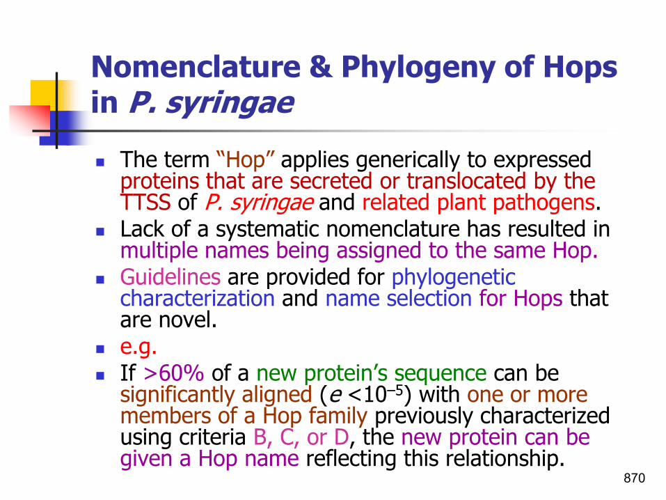
Nomenclature & Phylogeny of Hops in P. syringae
◼ The term “Hop” applies generically to expressed proteins that are secreted or translocated by the TTSS of P. syringae and related plant pathogens.
◼ Lack of a systematic nomenclature has resulted in multiple names being assigned to the same Hop.
◼ Guidelines are provided for phylogenetic characterization and name selection for Hops that are novel.
◼ e.g.◼ If >60% of a new protein’s sequence can be
significantly aligned (e <10–5) with one or more members of a Hop family previously characterized using criteria B, C, or D, the new protein can be given a Hop name reflecting this relationship.
870
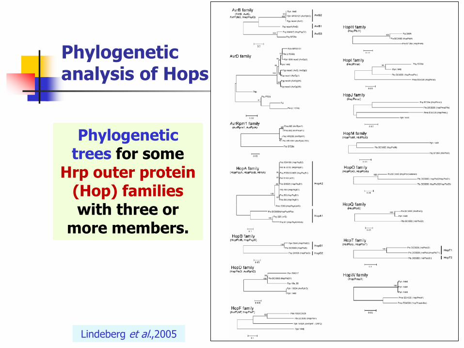
Phylogenetic trees for some
Hrp outer protein (Hop) families with three or
more members.
Phylogenetic analysis of Hops
Lindeberg et al.,2005

Type IV secretion systemMediate attachment of various P. syringae pathovars to the leaves
◼ Type IV pili(TFP) have been shown to mediate attachment of various P. syringae pathovars to the leaves of both susceptible and non-susceptible plants.
◼ P. syringae tfp mutants show significantly reduced adhesion to plant tissues and the ability to generate symptoms in host plants when spray inoculated also correlates with the presence of tfp.
◼ Taken together these results indicate that tfp-mediated adhesion of P. syringae phytopathogens to plant tissues is an important factor in the disease process.
Whitchurch,2006 872

Other genes involved in symptom development GacS/GacA:The two-component regulatory system
◼ Genes in the hrp regulon are not directly required for symptom development by P. syringae.
◼ Unlike hrp genes, the requirement for gacS (global activator sensor kinase) in P. syringae appears to be pathovar specific.
◼ Evidence that GacS (sensor kinase) and GacA (response regulator) are partners of a two-component system was first obtained genetically in P. syringae pv. syringae and subsequently confirmed for in a wide variety of Gram-negative bacteria such as enteric bacteria and fluorescent pseudomonads.
873

GacS/GacAThe two-component regulatory system
◼ The two-component regulatory system comprised of:
1. the sensor kinase, GacS(formerly lemA), and
2. its response regulator, GacA.
◼ GacS/GacA is involved in regulation of secondary metabolism and many other aspects of bacterial physiology.
◼ The sensor kinase GacS, initially called LemA, was first described in Pseudomonas syringae pv. syringaestrain B728a.
874Heeb and Hass,2001

GacS/GacAThe two-component regulatory systemSmall regulatory RNAs (RsmY and RsmZ)
◼ The GacS/GacA two-component system, a global regulator of the expression of bacterial products by turning on the transcription of small RNAs.
◼ These small RNAs act as antagonists of the unique RNA-binding protein RsmA, which negatively controls the expression of QS-related genes and several extracellular products.
◼ GacA was first described in P. fluorescens biocontrol strain CHA0 as a global activator of antibiotic and cyanide production.
875Zaho et al.,2014; Heeb and Hass,2001

GacS/GacAThe two-component regulatory systemSmall regulatory RNAs (RsmY and RsmZ)
◼ Small regulatory RNAs (RsmY and RsmZ) serving as early responders, can promote the expression of dependent genes (e.g. lasR) to boost the synthesis of intracellular enzymes and coordinate instant cooperative behavior in bacterial cells (QS).
◼ These early responders, acting as a rheostat to finely modulate bacterial cooperation, which may be quickly activated under environment threats, but peter off when critical QS dependent genes are fully functional for cooperation
876Zaho et al.,2014

GacS/GacAThe two-component regulatory systemSmall regulatory RNAs (RsmY and RsmZ)
877

GacS/GacA two-component systemModel of the signal transduction pathway mediated by the GacS/GacA two-component system
1. GacS protein sensesabiotic (e.g., pH, temperature, or osmolarity) or biotic (bacteria) signals and activates, via a phosphorelay mechanism.
2. The GacA transcription regulator, in turn triggers the expression of regulatory genes such as QS-related genes and other target genes.
878
N and C terminus; HTH, helix-turn-helix motif
Heeb and Hass,2001

GacS/GacA two-component systemCooperation activation was delayed due to the presence of lasR mutant
A. Genes as early responders regulate quorum-sensing and control bacterial cooperation in Pseudomonas aeruginosastrain PAO1.
B. In the absence of the central regulatory QS gene lasR, the expression of dependent genes was impaired along with the dramatically increased expression of rsmYand rsmZ compared with the WT PAO1.
879Zaho et al.,2014

GacS/GacAGacS/GacA two-component system controls regulation of secondary metabolism and many other aspects of bacterial physiology
880Chatterjee et al.,2003; Heeb and Hass,2001
◼ The GacS/GacA system controls the production of secondary metabolites and extracellular enzymes involved in pathogenicity to plants and animals, biocontrol of soilborne plant diseases, ecological fitness, QS, pigmentation, motility and tolerance to stress.

GacS/GacAGacS/GacA two-component system controls regulation of secondary metabolism and many other aspects of bacterial physiology
881
◼ gacS and gacA mutants of strain B728a are affected in production of:
1. Extracellular proteases
2. Acyl homoserine lactone(QS)
3. Alginate
4. Toxins, and
5. Swarming behavior on soft agar medium.

Bacteria in the leaf ecosystemWith emphasis on Pseudomonas s. phaseolicola-A pathogen, ice Nucleus, and epiphyte
Hirano & Upper,2000882
The role of ethylene in the pathogenicity process of P. syringae pathovars
◼ Ethylene production is important in the pathogenicity process of P. syringae pathovars.
◼ Ethylene may play a role in symptom development, because it can cause chlorosis, abscission, and senescence and thus may be important in the predisposition of plant tissue to disease.
883

Determination of ethylene and bacterial growth in vitroBy gas chromatography
◼ For the routine determination of ethylene productionby P. syringae pathovars, 1 ml of an overnight culture in synthetic 5a medium was transferred in a sterile 5-ml syringe sealed with a rubber cap and incubated on a rotary shaker at 90 rpm and room temperature (22 to 248C) for 2 h.
◼ After incubation, gas samples (1 ml) were withdrawn with a gas-tight syringe (Hamilton) and ethylene was determined with a gas chromatograph equipped with an active alumina column and a flame ionization detector.
884Weingart and Völksch,1997
Determination of ethylene and bacterial growth in vitroBy spectrophotometery
◼ Ethylene production in vitro and bacterial growth curves were obtained from 60-ml shaking cultures of 5a medium in 250-ml flasks (140 rpm, 288C).
◼ Ethylene was determined every 3 h as described above, and production was expressed in nanoliters per hour per cell.
◼ Bacterial growth was estimated with a spectrophotometer (578 nm).
885Weingart and Völksch,1997

Determination of ethylene and bacterial growth in plantaOn Petri plates
◼ Fifteen discs (7 mm in diameter) were excised with a cork borer from infected leaves at designated intervals, transferred into 3-ml syringes (five discs per syringe) sealed with rubber caps, and incubated at room temperature (22 to 248C) for 6 h.
◼ A 1-ml gas sample was taken from each syringe, and ethylene production was determined as described earlier.
◼ After we determined the ethylene concentrations, the bacterial populations in the leaf discs were monitored.
◼ The five discs of a syringe were macerated together in 5-ml isotonic NaCl. Appropriate serial 10-fold dilutions were plated onto King’s medium B.
◼ Plates were incubated at 28° C, and colonies from three replicate plates were counted after 3 to 4 days. The amount of ethylene produced was expressed in nanoliters per hour per square centimeter.
Weingart and Völksch,1997 886

P. syringae pathovarsPathovars tested for their ability to produce ethylene
◼ Of six P. syringae pv. pisistrains, five produced ethylene.
◼ All strains of P. s. pv. glycinea isolated from soybean plants of various regions and all strains of P. syringae pv. phaseolicolaisolated from kudzu produced ethylene
◼ However, strains of P. s . pv. phaseolicola isolated from beans and all other tested pathovars failed to producedetectable amounts of ethylene.
887Weingart and Völksch,1997

Pathogenicity of:Pseudomonas savastanoi pv. savastanoi
Olive knot Disease
888

Major pathogenicity factors
of the bacterium
◼ Involvement of plasmid deoxyribonucleic acid in indoleacetic acid synthesis.
◼ Disease symptoms consist of tumorous outgrowths induced in the plant by bacterial production of indole-3-acetic acid (IAA).
◼ Synthesis of IAA occurs by the following reactions: ◼ L-tryptophan leads to indoleacetamide leads to
indoleacetic acid, catalyzed by tryptophan 2-monooxygenase and indole acetamide hydrolase, respectively.
◼ Whereas the enzymology of IAA synthesis is well characterized, nothing is known about the genetics of the system.
889

Pathogenicity of:Ralstonia solanaceraum
Bacterial wilt disease
890

An Innovative Approach to Study R. solanacearum Pathogenicity
◼ An Innovative Approach to Study RalstoniasolanacearumPathogenicity
◼ Niraj Singh
◼ Scholars' Press
◼ March 27, 2020
◼ 68 pages.
891

Infection and colonization of plantsRalstonia solanacearum
892

EPS I: An important pathogenicity determinant in
Ralstonia solanacearum
◼ An important pathogenicity determinant produced by R. solanacearum is the exopolysaccharide (EPS I), a high-molecular-mass acidic extracellular polysaccharide.
◼ It is a heterogeneous polymer containing a trimeric repeat unit of:
1. N acetyl galactosamine,
2. 2-N-acetyl-2-deoxy-L-galacturonic acid, and
3. 2- N-acetyl-4-N- (3-hydroxybutanoyl)-2-4-6-trideoxy-D-glucose (Orgambide et al.,1991).
◼ Genes coding for the biosynthetic pathway for EPSI are encoded by the 20kb eps operon (Schell,2000).
893Gonzalez,2010

Protein secretion systemsR. solanacearum
◼ R. solanacearum displays a remarkable ability for protein secretion since more than 100 proteins can be identified in the cell-free supernatant of wild-type R. solanacearum cultured in minimal medium (Poueymiro & Genin 2009).
◼ Analysis of the R. solanacearum GMI1000 genome sequence revealed that all the major secretion pathways described in Gram-negative bacteria are present in the model strain GMI1000.
◼ However, the nature of the substrates for most of these pathways remains unknown and/or their contribution to pathogenicity is undetermined.
894Gonzalez,2010

Protein secretion pathways in R. solanacearum GMI1000
895Poueymiro
and Genin,2009

Genetic organization of the Hrp gene cluster of 3 plant pathogenic bacteria
◼ Red box indicates the hrp gene cluster.
◼ The head arrows with numbers indicate the hrp transcription units and the complete arrows indicate the different genes.
◼ Inside the red box of R. solanacearum Hrp gene cluster there is hrpB transcription regulator which is homologous to hrpX in Xanthomonas sp.
896Gonzalez,2010

Regulation network of virulence functions in Ralstonia solanacearumPhcA, a global regulator controlling phenotypic conversion
◼ At the center of the regulation network is PhcA (Phc, phenotype conversion), a LysR family transcriptional regulator which directly or through intermediary regulatory genes, coordinates the expression of several processes.
◼ It simultaneously activates diverse virulence genes such as those of:
◼ EPS biosynthesis, plant cell wall degrading enzymes as Pme and Egl exoproteins, swimming motility or Type IV pili(TFP), and represses others such as hrp genes and those related to production of polygalacturonases, siderophores, and motility.
897Gonzalez,2010

Regulation network of virulence functions in Ralstonia solanacearumPhcA, a global regulator controlling phenotypic conversion
898Gonzalez,2010

Pathogenicity of:Xanthomonas spp.Xac, Xcc, Xam, Xcv,Xoo and Xtt
Blight, rot, streak and canker diseases
899

Major pathogenicity determinants in Xanthomonas
◼ Molecular biology studies have characterized several key pathogenicity determinants of Xcc.
➢ Motility and attachment to plant,
➢ EPS and plant cell wall degrading enzymes.
◼ The synthesis of these 2 last is regulated by a quorum sensing system via rpf genes.
➢ Type III effectors (T3Es). These are delivered into plant cells via the type III secretion system (T3SS), which is encoded by the hrp [hypersensitive response (HR) and pathogenicity] regulon.
◼ Type III secretion system (T3SS), is regulated by 2 master regulatory genes hrpG and hrpX.
900Bogdanove,2011;..

Major pathogenicity determinants in Xanthomonas
Bogdanove,2011 901

902Buttner and Bonas,2010

Secretion systems Xanthomonas spp.
◼ Protein secretion systems play an important role in the interaction of pathogens with their host.
◼ Xanthomonas spp. contain genes for all known protein transport systems in Gram-negative bacteria, i.e.
◼ The Sec,
◼ Signal recognition particle, and
◼ TAT pathways;
◼ Type I, type II, type III, and type IV secretion systems of different types,
◼ Type V autotransporters,
◼ Two-partner secretion systems, and
◼ A type VI secretion system.
◼ However, for most Xanthomonas secretion systems the substrates and their importance for bacterial virulence are unknown.
Kay and Bonas,2009 903

Type I-V secretion systems in Gram-negative bacteria
904

905
Type II-IV secretion systems in Xanthomonas
spp.
Buttner and Bonas,2010

TTSSThe Hrp pathway
◼ Pathogenicity of Xanthomonas and most other Gram-negative phytopathogenic bacteria depends on a conserved type III secretion (T3S) system which injects more than 25 different effector proteins into the plant cell.
◼ Extensive studies in the last years on the molecular mechanisms of type III effector function revealed that effector proteins with enzymatic functions seem to play important roles in the interaction of Xanthomonas with its host plants, for example, the SUMO protease XopD.
◼ In addition, xanthomonads express a unique class of type III effectors to pursue another strategy.
906Kay and Bonas,2009

TTSSType III secretion system
◼ Xanthomonas species can infect most types of plants.
◼ Inject proteins into plant cells with the T3SS.
◼ Proteins travel to plant nucleus, bind to DNA, and turn on specific plant genes that make the plant susceptible to infection.
907Lecture 23 bacti3-10

Secretion systems Known type III effectors of Xanthomonas and their (putative) function
Kay and Bonas,2009 908

avr gene families in xanthomonadsXanthomonas axonopodis pv. malvacearum
◼ There are at least 19 well-described races of the pathogen Xanthomonas axonopodis pv. malvacearum.
◼ 10 avr genes were cloned and characterized from a single North American strain of X. axonopodis pv. malvacearum, XcmH.
◼ These ten avr genes belong to a large family of Xanthomonas avr/pth (avirulence (avr)/pathogenicity (pth) gene family.
◼ Most of these avr genes genetically “recognize”multiple R gene loci in an avr gene-for-R genes (plural R genes) manner.
909

Large family of Xanthomonas avr/pth genesXanthomonas axonopodis pv. malvacearumpthN and pthN2
◼ 10 genes belong to a large family of Xanthomonas avr/pth(avirulence/pathogenicity genes) were found in X. a.pv. malvacearum.
◼ Water-soaking symptoms caused by pthN andpthN2.
◼ Photo taken 5 days after inoculation.
910Chakrabarty et al.,1997

Time sequence of physiological events involved in the cotton HR to Xanthomonas citri pv. malvacearum
911Jalloul et al.,2015
Following penetration of leaves by the pathogen (figs. 1 and 3), bacterial cells
injected the effectors within the host nuclei. When recognized by host R proteins,
several specific mechanisms were activated through signaling pathways.
Appearance of HR symptoms in infected areas from t= 24h (9)

Time sequence of physiological events involved in the cotton HR to Xanthomonas citri pv. malvacearum
◼ Genes of the ERF IX3 group are transcribed in parallel to the production of ROS (t = 3h; 2: localization of H2O2 in resistant leaves).
◼ Accumulation of SA culminated after the burst (t= 6h) and before activation of a 9-or 13-Loxgene (t= 9h), transcription of ERF genes and synthesis of OPDA/JA (t= 12h).
◼ Production of flavonoids (orange stained in infected areas; 4) and total peroxidase activity (5: electron microscopic immunolocalization of peroxidase close to the bacteria) were also detected at the same time.
◼ Callose deposition contributed to stop the bacterial growth (7: green line) as compared to growth in susceptible plants (7: blue line), and preceded collapse of cells (6: condensation of the cytoplasm of HR cells).
◼ Appearance of HR symptoms in infected areas from t= 24h (9) occurred following dramatic increase in 9-Lox activity and strong accumulation of sesquiterpene phytoalexins (8: green fluorescence).
912Jalloul et al.,2015

Model of T3S in the plant pathogenic bacterium X. campestris pv.vesicatoriaT3S chaperones
◼ In X. campestris pv. vesicatoria, translocation of effector proteins is differentially regulated by the global T3S chaperone HpaB, which specifically promotes the translocation of a certain class of effector proteins (Büttner et al.,2006).
◼ The activity of HpaB is presumably controlled by thesecreted regulator HpaA that binds to HpaB in the bacterialcytoplasm and allows secretion of extracellular components ofthe T3SS.
Büttner and He,2009 913

Model of the molecular function of the type III effectors XopD and AvrBs3Xanthomonas
◼ In the next slide the protein structures of:
a) XopD and
b) AvrBs3 are shown, and
c) The proposed mode of action of the effectors is illustrated.
◼ XopD:
◼ After translocation by the T3S system XopD is transported into the nucleus of the plant cell where it localizes to nuclear foci.
◼ Here, it binds to DNA unspecifically via a helix-loop-helix (HLH) domain.
◼ Via two EAR motifs, XopD might inhibit yet unidentified plant transcription factors (TFs).
◼ Furthermore, XopD possesses SUMO protease activity.
◼ The latter XopD activities lead to the suppression of:
◼ Defense- and senescence-associated genes resulting in delayed disease symptoms and increased bacterial multiplication.
Kay and Bonas,2009 914

Model of the molecular function of the type III effectors XopD and AvrBs3Xanthomonas
◼ AvrBs3:
◼ AvrBs3 dimerizes in the plant cell cytoplasm and interacts with importin α via its nuclear localization signals (NLSs).
◼ The protein complex is bound by importin β mediating nuclear import.
◼ Here, AvrBs3 binds to a specific DNA sequence, the UPA box, and activates transcription of more than 10 UPA genes.
◼ UPA20, one of the induced genes, is the key regulator of plant cell hypertrophy.
◼ In resistant pepper plants, activation of Bs3 leads to the HR.
915Kay and Bonas,2009

Structure of upa20An transcription factor and master regulator of cell enlargement, stimulates cell growth
916

The AvrBs3 familyStructure and functions of avrBs3 proteins
◼ The activities of several type III effector proteins ultimately result in a change of plant gene expression.
◼ One of these effectors is AvrBs3 family.
◼ So far AvrBs3 family only was identified in Xanthomonas spp. and Ralstonia solanacearum.
◼ This transcription factor mimic plant transcriptional activators and manipulate the plant transcriptome.
917Kay and Bonas,2009

The AvrBs3 familyStructure and functions of avrBs3 proteins
◼ Three key structures of the gene products are:1. A central repeat region;2. Three nuclear localization signal (NLS) sequences; and3. An acidic activation domain (AAD) have roles in avirulence and/or
virulence.◼ The central repeat region, which is composed of several nearly
identical 34 aa-directly repeated sequences, is essential for virulence and host cultivar specificity.
◼ The repeat region is leucine rich, and at the end of the repeats there is an imperfect leucine zipper suggesting a potential role in protein-protein interactions.
◼ The NLS are necessary for both avirulence and virulence functions, and to localize the Avr protein into the plant cell nucleus.
918
NLS: nuclear localisation signals;* AD or ADD: transcription activation domain
Vivian and Arnold,2000

AvrBs3Arrival-AvrBs3 localizes to the plant cell nucleus
◼ The exact mechanism of nuclear transport of the Avr proteins is not known, however, AvrBs3 from Xanthomonas vesicatoria, the bacterial spot pathogen of pepper, binds to importin α from pepper in yeast two-hybrid assays via the second NLS.
◼ Importin α is a subunit of a heterodimeric NLS receptor complex that binds to the NLS of a substrate in the cytoplasm and then is associated with importin β, which mediates transfer of the substrate into the nucleus.
◼ Thus, it is possible that AvrBs3 and other family members localize to the nucleus via importin α/β nuclear transport.
AvrBs3 interacts with pepper host cell protein α-importin for the nuclear
localization.Importin α which, together with importin
β, mediates nuclear protein import.

The AvrBs3 family known as TAL effectors (TALEs) in X. vesicatoria
◼ Xanthomonas spp. produce type III effectors (T3Es) that can modulate virulence in host plants; T3Es are delivered into plant cells via the type III secretion system (T3SS).
◼ These T3Es are categorized into:
1. transcription activator-like effectors (TALEs), and
2. non-TALEs, which are commonly known as Xanthomonas outer proteins or Xops.
920Ali Shah et al.,2019
The number of tal genes in Xanthomonas spp. is highly variable; for example, strains of X. translucens pv. translucens, pv. undulosa, and pv. cerealis were
shown to harbor eight, seven and two tal genes, respectively.

Pathogenicity of AvrBs3known as TAL effectors (TALEs) in X. vesicatoria
Zhaokaijun 921
TAL effector

The TALE family Discovery of TALE effectorsFirst found in Xanthomonas vesicatoria
◼ A new paradigm for protein-DNA interaction has been discovered in bacterial pathogens of plants (Boch et al.,2009; Moscou and Bogdanove,2009).
◼ TALEs (transcription activator like effectors) has been shaped by 20 years of research, beginning with the discovery of the AvrBs3 protein of the pepper and tomato pathogen X. campestris pv.vesicatoria (Xcv) (Bonas et al.,1989)
922Bogdanove et al.,2010; Christianet al.,2010

The TALE family Discovery of TALE effectorsNow found in many Xanthomonads
◼ TALEs has now be found in several but not all pathogenic members of the genus Xanthomonas.
◼ Individual Xanthomonas strains may have as few as one or as many as several dozen TALEs.
◼ Xanthomonas uses TAL effectors to manipulate gene function in plants in ways that benefit the pathogen.
923Bogdanove et al.,2010
Closely related proteins have been found in several biovars of Ralstonia solanacearum; some of these are delivered into host cells,
but whether transcriptional activators is not yet known.they act as

The TALE family Possible applications
924Bogdanove et al.,2010; Horvath,2011
◼ Xanthomonsds utilize these proteins that function as eukaryotic transcription activators to effect the host environment.
◼ Transcription activator like (TAL) effectors use a novel protein motif with an extremely simple DNA recognition code.
◼ The understanding of this code enables a number of highly useful tools, including targeted gene activation or repression, engineering of genomes to add, remove, or alter genes, and site-specific gene insertion.

AvrBs3The founder of the TALE family Structural features of TALEs
◼ Structural features of TALEs and matching binding sites (UPT boxes) exemplified by AvrBs3, the founder of the TALE family, and the corresponding pepper Bs3 and UPA20 promoters.
◼ TALEs recognize plant DNA sequences through a central repeat domain.
925Bogdanove et al.,2010

AvrBs3The founder of the TALE family Structural features of TALEs
◼ AvrBs3 contains a N-terminal type III secretion and translocation signal (T3S), a repeat domain consisting of 17.5 repeat units (yellow ovals), two nuclear localization signals (NLS) and a C-terminal acidic activation domain (AAD/AD).
◼ Repeat polymorphism occurs largely at residues 12 and 13, the repeat-variable diresidues (RVDs, uppercase boldface red letters).
926Bogdanove et al.,2010

The TALE familyTALEs function
◼ TALEs function as:
1. Virulence factors (led to plant disease),
2. Plant-recognized ‘avirulence’ factors (led to resistance), or
3. Both.
◼ Interaction with plants has selected TALEs that activate host genes that facilitate bacterial colonization and spread.
927Bogdanove et al.,2010

The TALE familyTALEs function
◼ TALEs enter the nucleus, bind effector-specific DNA sequences, and transcriptionally activate gene expression.
◼ Typically, activation of target genes increases plant susceptibility to pathogen colonization, but in some cases, it triggers plant defense.
◼ TALE binding to DNA is mediated by a central region of these proteins that contains as many as 30 tandem repeats of a 33- to 35-amino-acid-sequence motif.
928Christian et al.,2010

Pathogenicity or resistance The TALE family TAL (transcription activator-like) effectors
929Bogdanove et al.,2010

Pathogenicity or resistance The TALE familyTALE roles in plant disease and resistance
◼ 1. Disease incidence:
◼ Upon injection via the type III secretion system (T3SS) TALEs can be detected in the host cytoplasm as exemplified by the tomato NB-LRR type resistance (R) protein Bs4, which is capable of detecting the full-length TALE AvrBs4 as well as deletion derivatives that lack nuclear localization signals.
◼ Upon interaction with importin α (impα) TALEs translocate to the nucleus, bind to matching UPT boxes, and transcriptionally activate matching host susceptibility (S) genes such as:
◼ UPA20, Os8N3, OsTFIIAγ1, or OsTFX1, resulting in disease.
930Bogdanove et al.,2010

Pathogenicity or resistance The TALE familyTALE roles in plant disease and resistance
◼ 2. Plant resistance:
◼ Activation of S genes can be suppressed either by:
◼ A variation in the RNA II polymerase complex (red shapes) such as xa5 (brown shape), or
◼ By mutations in the individual UPT boxes (xa13 mutation).
◼ Plants have also evolved activation traps for resistance in which executor R genes are under transcriptional control of UPT boxes that would normally be present in S gene promoters.
931Bogdanove et al.,2010

Pathogenicity or resistance TALE roles in plant disease and resistance
Zhaokaijun 932

XopDSUMOylation and deSUMOylation
◼ In plants, SUMOylation and deSUMOylation regulate a number of processes, for example, in abiotic stress responses, pathogen defense, and flower induction.
◼ SUMOylation itself stabilizes proteins.
◼ XopD SUMO protease affects host transcription.
◼ In Xanthomonas, the XopD protein has a modular structure and shows different biochemical activities (Figure 1a).
◼ XopD possesses SUMO protease activity mediating deSUMOylation of yet unknown target proteins.
◼ The XopD activities lead to the suppression of:
◼ Defense- and senescence-associated genes resulting in:
1. Delayed disease symptoms, and
2. Increased bacterial multiplication.
933
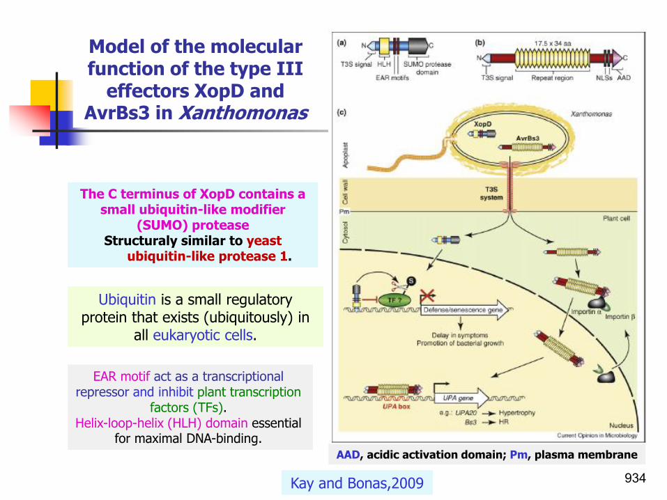
AAD, acidic activation domain; Pm, plasma membrane
Model of the molecular function of the type III
effectors XopD and AvrBs3 in Xanthomonas
Kay and Bonas,2009
The C terminus of XopD contains a small ubiquitin-like modifier
(SUMO) proteaseStructuraly similar to yeast
ubiquitin-like protease 1.
934
EAR motif act as a transcriptional repressor and inhibit plant transcription
factors (TFs).Helix-loop-helix (HLH) domain essential
for maximal DNA-binding.
Ubiquitin is a small regulatory protein that exists (ubiquitously) in
all eukaryotic cells.

Secretion systemsType II secretion systems
◼ In addition to TTSS, pvs. campestris and citri encode type II secretion systems too.
◼ e.g. X. campestris pv. campestris is equipped with two sets of type II secretion systems (xps and xcs).
◼ From 11 annotated genes related to the xps system, 6 genes i.e. xpsD, xpsE, xpsF, xpsK, xpsL, and xpsM plus gene (eng Xca ,encoding exocellular cellulase) were identified in pathogenicity-deficient mutants.
◼ None the genes related to the xcs system were found in pathogenicity-deficient mutants.
◼ Therefore it was concluded xcs system may not playan essential role in Xcc pathogenesis.
935

The genetic determinants of the infection process of X. citri subsp. citri
Wang,2012936

PathogenicityCitrus canker strains containing pthApathogenicity genes
◼ Citrus canker disease (Xanthomonas axonopodis pv.citri ) would be considered a host specific diseasecaused by strains containing pthA pathogenicity genes that are differentially adapted to Citrus spp.
◼ Citrus bacterial spot disease (X. axonopodis pv. citristrain type E which later the bacterium reclassified as X. axonopodis pv. citrumelo) is a disease caused by a diversity of strains with differential aggressiveness that represent a heterogeneous group, while the citrus canker group displays a higher homologyamong its members (Egel et al.,1991).
938

PathogenicitypthA -C
◼ Gene pthA is essential for Xac to elicit hyperplasia (cell divisions or cankers) on citrus, and pthA confers this ability to various X. axonopodis strains (for example, pathovars alfalfae and citrumelo).
◼ Functionally homologous genes (pthB and pthC) have also been identified and cloned from X. axonopodis pv. aurantifolii pathotype B and pathotype C, respectively.
◼ Mutations of genes encoding either type III secretion system encoded by hrp genes or the effector molecule, pth A/B/C, abolish pathogenicity of canker bacteria.
939

PathogenicityDifference in virulence mechanisms between X. citri spp. citri and X. axonopodis pv. citrumelo
◼ X. citri spp. citri
◼ 22 T3SS effectors
◼ PthA and its homologs are missing. Expression of PthA in citrumelo strain causes typical canker symptoms.
◼ Deficient in pectate lyase
◼ LPS 22 genes
◼ X. axonopodis pv. citrumelo
◼ 25 T3SS effectors
◼ PthA and its homologs (PthA1, PthA2, PthA3, and PthA4)
◼ Pectate lyase
◼ LPS 17 genes
940Wang,2012

PathogenicityComparison of the pecate lyase production by X. axonopodis pv. citri str. 306, X. axonopodis pv. citrumelo str. F1 and X. campestris pv. vesicatoria str. 85-10
◼ Pecate lyase production: pitting can be seen in the Hildebrand’s agar medium.
941Wang,2012
X. axonopodis pv. citriX. axonopodis pv.
citrumelo X. vesicatoria

Pathogenicity of:Xanthomonas oryzae pv.oryzae
Leaf blight of rice
942

Genome Sequence ofXanthomonas oryzae pv.oryzae
◼ The bacterium is a model organism for the analysis of plant-pathogen interaction, because more than 30 races differing in virulence and 25 resistance genes in rice have been reported to date.
◼ The size of the genome was 4,940,217 bp, in a single circular chromosome.
◼ The genome structure of Xoo MAFF 311018 was characterized by large numbers of effector (avr) genes of the avrBs3/pth family and insertion sequences (ISs).
◼ RFLP analysis of diverse strains using ISXo1 as a probesuggests that the prevalence of mobile elements in this species, which can bring about genome inversions and rearrangement, may have played a major role in generating the high degree of genetic diversity and race differentiation characteristic of this pathogen.
943

General features of the Xanthomonasoryzae pv.oryzae genomeNo plasmid was detected in the course of genome assembly
◼ The assembled sequence was consistent with a single, 4 941 439 bp, circular chromosome.
◼ No autonomous plasmids were apparent.
944Lee et al.,2005

Graphic representation of the chromosomeXanthomonas oryzae pv. oryzae
◼ Circular genome map of X.oryzaepv. oryzae str. KACC10331.
◼ Overall structure of the X.oryzae pv. oryzae genome. The putative origin of replication is at 0 kb.
◼ The outer scale indicates the coordinates (in base pair).
◼ Red symbols (character R) are positions of rRNA and blue symbols (character T) are tRNAs.
◼ The distribution of genes is shown on the first two rings within the scale.
◼ The next circle (green) shows G + C content and central circle (blue/red) shows GC-skew value.
945Lee et al.,2005

The role of proteinase in Pathogenicity of XOO
◼ The role of an extracellular proteinase has been studied in Xanthomonas campestris pv. oryzae by transposon mutagenesis.
◼ The populations of a proteinase-defective mutant in rice plants were 10- to 100-fold smaller than those observed for the parental strain, suggesting an active role for proteinase in this disease.
◼ Similarly, a proteinase-deficient mutant of Xanthomonas campestris pv. campestris, the black rot pathogen, which lacked proteinase activity, showed considerable loss of virulence in pathogenicity tests when bacteria were introduced into mature turnip leaves through cut vein endings.
946

Pathogenicity of:Xanthomonas translucens
947
• Three main clusters:
1. one consisting of pv. cerealis,
2. one consisting of pvs. undulosa and translucens, and
3. a third consisting of pvs. arrhenatheri, graminis, phlei, and poae.

Host rangeXanthomonads
◼ Xanthomonas spp. are subdivided into pathovars on the basis of:
1. host specificity and
2. can also be divided into two main phylogenetic groups based on sequence analysis of 16S rDNA, gyrB, dnaK, rpoD, and fyuA.
Ali Shah et al., 2019

949
Diseases caused by Xanthomonas translucens pathovars
On Arrhenatherum elatiusXanthomonas translucens pv. arrhenatheri1
OnTriticum spp.; Avena spp.Xanthomonas translucens pv. cerealis2
Leaf spot of Timoty grass; Lolium spp.Xanthomonas translucens pv. graminis3
On Phleum praterseXanthomonas translucens pv. phlei4
On Phleum praterse ; Agropyron sp.; Bromus sp.
Xanthomonas translucens pv. phleipratensis5
Dieback of pistachio Xanthomonas translucens pv. pistaciae6
Bacterial wilt of Poa (Poa trivialis)Xanthomonas translucens pv. poae7
Leaf & stem spotting of Secale (Secalecereale)
Xanthomonas translucens pv. secalis8
Leaf streak and black chaff of small grains (barley, rye, wheat,..)
Xanthomonas translucens pv. translucens9
Leaf streaks of wheat & barleyXanthomonas translucens pv. undulosa10
Compiled from various references including ISPP LISTS, 2004; Bergey’s Manuals, Janse,2006 and Euzeby,2010; Bull et al.,2010& 2012.

Virulence factorsXanthomonads
◼ The number of tal genes in strains of X. translucens pv. translucens, pv. undulosa, and pv. cerealis were shown to harbor eight, seven and two tal genes, respectively.
Ali Shah et al., 2019

Graphic representation of the chromosomeXanthomonas translucens pv. cerealis
◼ Circular representation of the X. translucens pv. cerealis strain NXtc01 genome starting from gyrB.
◼ Inner to outermost rings illustrate GC skew (G − C)/(G + C), GC contents, tal genes (orange), IS elements (green), tRNA (blue), rRNA (orange) and ncRNA (black) genes, reverse COGs (cluster of orthologous groups), forward COGs, chromosome (green), and coordinates (Mb).
◼ The T3SS genes cluster is indicated as black rectangle on chromosome.
951Ali Shah et al., 2019

Comparison of X. translucens pv. cerealis genomes
◼ Comparison of X. translucens pv. cerealis genomes from strains NXtc01 and CFBP 2541, which were isolated from Triticum aestivum in Xinjiang, China and Bromus inermis in the United States, respectively.
952Ali Shah et al., 2019

Pathogenicity of:Gram-positive plant pathogenic bacteria
953
Virulence mechanisms of Gram-positive plant pathogenic bacteria

Virulence mechanisms of Gram-positive plant pathogenic bacteria
◼ Secreted proteins that act inside the plant cell are central to microbial pathogenesis.
◼ The lack of a type III protein secretion system (TTSS) in Firmicutes and Actinobacteria immediately raises questions about delivery of virulence proteins across the plant cell wall and membrane.
◼ In Plant pathogenic Actinobacteria:
◼ The genera Streptomyces and Rhodococcus have very wide host ranges.
◼ Whereas, the genera Clavibacter and Leifsonia are host-specific at the species or subspecies level.
Hogenhout and Loria,2008

Virulence mechanisms of Gram-positive plant pathogenic bacteriaHorizontal gene transfer: a short cut to virulence
◼ Actinobacteria and Firmicutes have evolved exclusive strategies for plant pathogenicity independent of Proteobacteria.
◼ There is also abundant evidence for acquisition of virulence genes through horizontal gene transfer and there are several examples of homologous virulence genes in multiple genera.
Hogenhout and Loria,2008

Virulence mechanisms of Gram-positive plant pathogenic bacteriaHorizontal gene transfer: a short cut to virulence
Hogenhout and Loria,2008
◼ Genome sequences reveal the importance of lateral gene transfer (HGT) in evolution of virulence in both Actinobacteria and Firmicutes.
◼ The pat-1 gene, which is required for virulence in Clavibacter michiganensis subsp. michiganensis is plasmid borne.
◼ The tomA gene in Cmm lies on a 129-kb region with a lower G+C content than the rest of the genome.
◼ This region also encodes several serine proteases that are required for virulence.

Virulence mechanisms of Gram-positive plant pathogenic bacteriaHorizontal gene transfer: a short cut to virulence
Hogenhout and Loria,2008
◼ Phytoplasmas can have two to four plasmids that vary in Size.
◼ Plasmids of spiroplasmas are also involved in insect transmission.
◼ These plasmids harbour adhesins and components of type IV translocation systems.

Pathogenicity ofClavibacter michiganensis subsp.michiganensis
Canker of tomato
958

Three mechanisms explain the wilting disease caused by theClavibacter michiganensis
◼ Pathogenicity determinant is plasmid- borne endo-b-1,4-glucanase.
◼ Three mechanisms have been proposed to explain the wilting disease caused by the vascular phytopathogen C. michiganensis subsp.michiganensis:
1. Mechanical plugging of xylem vessels2. Toxic action of exopolysaccharides (EPS)3. Enzymatic attack on plant tissue (e.g.
polygalacturonase).
959

Three mechanisms explain the wilting disease caused by theClavibacter michiganensis
◼ In several studies, plugging of xylem vessels by high titer colonization and production of high molecular weight EPS could be ruled out as the primary reasonfor the onset of plant wilting.
◼ In early stages of infection, Cmm colonizes the xylem vessels, while colonization of the phloem, pith, and cortex of the upper parts of the plant is only observed at later stages.
◼ Wilting may therefore result from an enzymatic attack on xylem vessels and adjacent parenchymatic cells, allowing a lateral spread of Cmm within the host plant, along with the release of nutrients necessary for bacterial proliferation and colonization.
960

Pathogenicity factors Clavibacter michiganensis subsp.michiganensis
◼ In the genus Clavibacter, production of extracellular enzymes such as endoglucanase, pectin methylesterase, polygalacturonase, and xylanase has been reported and endoglucanase activity has been implicated as a pathogenicity factor in recent reports.
◼ Endo-β-1,4-glucanases are members of the family A1 cellulases.
961

Pathogenicity factors of the Clavibacter michiganensis subsp. michiganensis
◼ The phytopathogenic bacterium Clavibacter michiganensis subsp.michiganensis NCPPB382, which causes bacterial wilt and canker of tomato, harbors two plasmids, pCM1 (27.35 kb) and pCM2 (72 kb), encoding genes involved in virulence.
◼ In the present study, we demonstrate that the cel A gene encoding an endoglucanase on pCM1is required for the development of disease symptoms on infected tomato plants.
962

pCM1 function and mapping of the gene celA Clavibacter michiganensis subsp. michiganensis
◼ The region of pCM1 carrying the endoglucanase gene celAwas mapped by deletion analysis and complementation.
◼ The celA gene encodes CelA, a protein of 78 kDa (746 amino acids).
◼ CelA plays a major role in pathogenicity. It was shown previously that in Cmm the loss of plasmids pCM1 and pCM2 results in the avirulent endophytic, endoglucanase-negative Cmm, lacking wilt-inducing ability.
◼ Reduced virulence was observed for the partially curedstrain, carrying only pCM1 (not pCM2).
◼ The mutant strian also produced endoglucanase, but caused wilting of tomato plants with a delay of 6 days (compared to wild type).
963

Map of plasmid pCM1Clavibacter michiganensis subsp. michiganensis
◼ 3.2-kb BglII restriction fragment B1 of plasmid pCM1 carries an endoglucanase gene(indicated by black box and arrow).
◼ Regions essential for plasmid replication and stability, indicated as a:
◼ Bold-lined, or
◼ Thin-lined open box and labeled.
964
Restriction enzyme recognition sites not unique to pCM1, indicated by asterisks.

Plate assays for CMCase (endoglucanase+) detection
◼ Endoglucanase (CMCase) activity was detected on M9CMC agar plates containing carboxymethylcellulose (CMC) (Meletzus et al., 1993).
◼ The plates were stained with 0.1% (wt/vol) congo red for 15 to 30 min and finally bleached with QR-buffer or NaCl solution (2 M NaCl, 50 mM Trizma base, pH 8.5).
965
The NaCl solution elutes the dye in the clearing zone where the cellulose has been degraded into simple
sugars by the enzymatic activity.

SDS-PAGE analysisStaining the CelA
◼ Sodium dodecyl sulfate-polyacrylamide gel electrophoresis (SDS-PAGE) of CelA.
◼ Concentrated culture supernatant of Strain :
◼ CMM101(pCM1) (lane 2), ◼ Strain CMM100(pHJ1200) (lane 3),
and◼ A Pre-stained molecular weight
marker (lane 1) were subjected to SDS- PAGE and stained with Congo red.
◼ The three active CMCase (endoglucanase+) species with 75, 54, and 43 kDa are indicated.
966

Pathogenicity of:Rhodococcus fascians
Fasciation, leafy gall diseases
967

Rhodococcus fascians
◼ R. fascians can be a pathogen of plants, both angiosperm or gymnosperm.
◼ Infected plants show typical symptoms, such as:
◼ Leaf deformation,
◼ Witches’ broom, and
◼ leaf gall.
◼ The development of these symptoms depends on:
◼ Plant's cultivar,
◼ Plant's age, and
◼ Bacterial strain.
968

Rhodococcus fasciansLeafy gall and fasciation diseases
◼ Rhodococcus fascians is the cause of malformations in a wide variety of host plants, the most severe being a leafy gall and shoot proliferations.
◼ Agrobacterium caused crown gall-like tumors and R. fascians produced leafy galls and shoot proliferations.
◼ Interestingly, we recovered R. fascians much less frequently than we did Agrobacterium.
969
Rhodococcus fasciansLeafy gall and fasciation diseases
◼ Both Rhodococcus fascians and A. tumefaciens are known to infect herbaceous and woody plants.
◼ Both bacteria have a wide host range (over 60 species for R. fascians, and hundreds for A. tumefaciens).
◼ In addition, R. fascians infects monocots as well as dicots, unlike A. tumefaciens, which infects only dicots.
970

Isolation methods
◼ On most of the plants we received for diagnosis we were making dual isolations, using one method for Agrobacterium and a different method for Rhodococcus fascians.
Putnam and Miller 971

Rhodococcus fascians vs.Agrobacteria infections
◼ Leafy galls and shoot proliferations are quite different than the galls caused by the crown gall bacterium, A. tumefaciens.
◼ Infection with A. tumefacienscauses swelling of tissue into tumors or galls on stems or roots, but these galls do not differentiate into buds or stems.
◼ In contrast, leafy galls caused by R. fascians are well differentiated into easily recognized plant parts.
Shoot proliferation of Iberis
Crown gall at the base of a hibiscus
972

Genes that control virulenceRole of phytohormones during infection
◼ Virulence in R. fascians is controlled by genes on a plasmid (strains lacking that plasmid are not virulent) and on the chromosome.
◼ Strains that are cured of the plasmid are non-pathogenic.
◼ Using deletion mutations, it was possible to identify three loci the plasmid: fas, att, and hyp, and one locus on the chromosome, vic.
◼ Rhodococcus fascians carrying the fas-1 gene, which codes for an isopentyl transferase, the key enzyme in cytokinin biosynthesis, was found to be associated to stems and flower stalks symptoms.
973

R. fascians vs. Agrobacteria infections
◼ Unlike A. tumefaciens , presence of R. fascians on the infected plant is necessary, not only for the initiation of infection, but also for its maintenance.
◼ During the infection, R. fascians usually stays outside vegetal tissues, near a junction or cavity of a plant's cell walls, maybe to avoid environmental stresses.
974

Pathogenicity of:Leifsonia xyli subsp. xyli
Ratoon stunt disease of sugarcane
975

Leifsonia xyli subsp.xyliRatoon stunting disease
◼ No general tissue disorganization results from this colonization, although the xylem may be blocked with a mucilaginous substance probably produced by the host.
◼ This blockage is reported to reduce sap flow up to 34% (Teakle et al.,1978).
◼ Infection with L. xyli subsp. xyli results in reduced cane diameter and plant stunting, symptoms that are often misinterpreted as general growth defects due to poor crop management.
◼ Ratoon stunting disease is regarded as the main disease of sugarcane worldwide.
◼ Yield losses of both cane and sugar per unit area may approach 50% in highly susceptible genotypes.
◼ Annual losses were estimated at US $11 million in Australia and US$36 million in Florida.
976Monteiro-Vitorello et al.,2004

The circular genome ofLeifsonia xyli subsp. xyli
◼ The genome sequence of Leifsonia xyli subsp. xyli, which causes ratoon stunting disease and affects sugarcaneworldwide, was determined.
◼ The analysis also revealed 307 predicted pseudogenes or noncoding DNA sequence, which is more than any bacterial plant pathogen sequenced to date.
◼ Many of these pseudogenes, if functional, would likely be involved in the degradation of plant heteropolysaccharides, uptake of free sugars, and synthesis of amino acids.
◼ The large number of pseudogenes found in Leifsonia xyli subsp. xyli CTCB07 in the context of plant pathogenic bacteria suggests an ongoing process of genome decay.
977Monteiro-Vitorello et al.,2004

Pseudogene counts in various organismsBacteria
◼ Leifsonia xyli subsp. xyli
◼ Xylella fastidiosa
◼ Xanthomonas axonopodis pv. citri
◼ Xanthomonas campestris pv. campestris
◼ Agrobacterium tumefaciens
◼ Ralstonia solanacearum
◼ Mycobacterium leprae
◼ Rickettsia prowasekii
◼ Sinorhizobium meliloti
◼ Streptomyces coelicolor
◼ Pseudomonas syringae
978Monteiro-Vitorello et al.,2004

The circular genome of
Leifsonia xyli subsp.xyli
Monteiro-Vitorello et al.,2004

The circular genome ofLeifsonia xyli subsp.xyli
◼ The outer scale corresponds to coordinates in base pairs.◼ The first (outer) circle indicates Tnp insertions. ◼ The second indicates the positions of insertion sequences. ◼ The third and fourth circles represent predicted coding
regions color-coded by gene category of the plus and minus strands, respectively.
◼ Genes on the fifth and sixth circles are pseudogenes (plus and minus strands).
◼ Seventh circle, genomic islands as defined by three criteria: codon bias, GC composition, and dinucleotide signatures and
◼ Eighth circle (innermost), G+C content.
980Monteiro-Vitorello et al.,2004

The analysis of the genome sequence of L. xyli subsp. XyliLateral gene transfer and virulence
◼ The limited number of pathogenicity genes appears to explain why L. xyli subsp. Xyli may be regarded as the near-perfect pathogen (Metzler et al. 1997), since it is able to reach high bacterial titers in the host without causing significant symptoms.
◼ Some of the predicted pathogenicity genes appear to have been acquired by lateral transfer and include genes for cellulase, pectinase, wilt-inducing protein, lysozyme, and desaturase.
◼ The presence of the latter may contribute to stunting, since it is likely involved in the synthesis of abscisic acid, a hormone that arrests growth.
981Monteiro-Vitorello et al.,2004

Pathogenicity of:Xylella fastidiosa
Scorch Disease
982

Genome of sequencing & pathogenicity factorsLateral gene transfer and virulence
◼ The genome consists of a 52.7 per cent GC-rich 2 679 305-base-pair (bp) circular chromosome and two plasmids of 51 158 bp and 1285 bp.
◼ There are 2904 predicted coding regions and putative functions have been assigned to almost half of these.
◼ Those concerned with pathogenicity and virulence involve toxins, antibiotics and ion sequestration systems.
◼ Orthologues of genes encoding some of these proteins have been identified in animal and human pathogens, suggesting that they are constituents of pathogenicity islands.
Strange,2003 983

Genome of sequencing and pathogenicity factorsLateral gene transfer and virulence
◼ These regions of the genome of pathogens are conserved and independent of the host and therefore imply horizontal gene transfer.
◼ This is corroborated by the finding of at least 83 genes in the genome of X. fastidiosa that are bacteriophage-derived and which include virulence-associated genes from other bacteria.
Strange,2003 984

Genome sequencingCVC strain of X. fastidiosa
◼ The CVC strain of X. fastidiosa is the first plant pathogenic bacterium, the genome of which has been sequenced.
◼ 2,679,305 bp, 2782 ORFs, coding regions 88%.
◼ Two plasmid (51,158 bp and 1,285 bp).
◼ Do not have type III secretion system.
◼ Has a reduced genome.
985

Genomic comparison between X. fastidiosa(citrus stain) and X. axonopodis pv. citri
◼ X. axonopodis pv.citri (XAC) has two copies of a type II secretion system, a large number of cell wall–degrading enzymes and sugar transporters, a complete energy metabolism, a whole set of avirulence genes associated with a type III secretion system, and a complete flagellar and chemotatic system.
◼ By contrast, X. fastidiosa (citrus stain XF-9a5a) possesses more genes involved with type IV pili biosynthesis than does X. axonopodis pv. citri (XAC), contains genes encoding for production of colicins, and has 4 copies of Type I restriction/modification system while XAC has only one.
◼ XF-9a5c does not have a T3SS.
986Moreira et al.,2004

General genome features of the three major citrus bacterial pathogens X. fastidiosa, X. citri pv. citri and Ca. Liberibacter asiaticus’
987Vojnov et al.,2010

Secretion systemXylella fastidiosa
◼ Xylella fastidiosa does not carry a type III secretion system, and it is therefore assumed that this pathogen does not translocate effectors into plant cells for the elicitation of a host response.
◼ This hypothesis is supported by the fact that, in the xylem vessels, there is only fibre and dead cells, and the pathogen is introduced into this tissue by its vector, the sharpshooter leafhopper (Homoptera, Cicadellidae).
◼ However, X. fastidiosa has active type I and type II secretion systems, which could be associated with the efflux pump and the secretion of hydrolytic enzymes, respectively, allowing lateral movement of the bacterium through pit membranes and the digestion of plant cell walls.
Mansfield et al.,2012

Mechanism of disease developmentFastidian gum and twitching motility
◼ The fastidian gum(EPS) may be linked directly to the pathogenicity of this bacterium.
◼ The EPS could be involved in the formation of a biofilm required for the attachment and survival of the bacteria in the two hydrodynamically turbulent environments it is found: the xylem vessels and the sucking pumps of insect vectors.
◼ Terminal fimbriae (also called type IV pili) are important for biofilm formation.
◼ Terminal fimbriae aid in a type of incremental movement called "twitching motility" which enables bacterial cells to move against the xylem stream.
989

Mechanism of disease developmentFastidian gum and twitching motility
990
Genetic map of the X. campestris gum (A) and rpf (B) operons compared to the region of the homologous genes in the X. fastidiosa. .genome
De silva et al.,2001

Mechanism of disease developmentFastidian gum and enzymes, toxins
◼ X. fastidiosa has 22 genes encoding regulatory proteins and enzymes involved in the synthesis of an EPS similar to xanthan gum, the EPS produced by X. campestris.
◼ The X. fastidiosa genome encodes for proteins involved in cell to cell interaction, degradation of plant cell walls, synthesis of toxins, and plant pathogenicity.
991

Mechanism of disease developmentStructure of the repeating unit of xanthan gum (A) from X. campestris, and the proposed structure of fastidian gum (B) from X. fastidiosa
◼ The arrows point towards the reducing end of each repeat.
◼ Glc, Dglucose; GlcA, D-glucuronic acid; Man, D-mannose; Ac, acetyl ester; Pyr, acetyl-linked pyruvic acid.
◼ Some external mannoses of xanthan gum may contain an acetyl instead of a pyruvyl substituent.
992De silva et al.,2001

Biofilm of Xylella fastidiosa blocking the xylem vessels of sweet orange tree
◼ Biofilms (A, B) are important for this pathogen to survive in environments with high turbulence, differential pressure and poor nutrient availability, such as xylem vessels and insect foreguts.
Mansfield et al.,2012
Photographs in (A) by E.W. Kitajima (Escola Superior de Agricultura Luis de Queiróz, USP, Piracicaba, SP, Brazil) and in (B) by J.O. Lima (Citrulima Viveiros, São João da Boa Vista, SP, Brazil) and Marcos A. Machado.

Mechanism of disease development
Close-up of a bacterial cell in the bordered pit of a vessel element
Cells of Xylella fastidiosa within the xylem tissue of a pin oak petiole
Close-up of individual bacterial cellsBiofilm of Xylella fastidiosa on Chard2 medium
994

Biochemical processes involved in
Xylella fastidiosa pathogenicity
◼ A comprehensive view of the biochemical processes involved in Xylella fastidiosa pathogenicity and survival in the host xylem.
◼ Transporters are indicated as follows:
◼ Cylinders, channels; ovals, secondary carriers, including the MFS family; paired dumbbells, secondary carriers for drug extrusion; triple dumbbells, ABC transporters(Type I secretion system); bulb-like icon, F-type ATP synthase; squares, other transporters.
◼ Icons with two arrows represent symporters and antiporters:
◼ (H+ or Na+ porters, unless noted otherwise). 2,5DDOL, 2,5-dichloro-2,5-cyclohexadiene-1,4-dol; EPS, exopolysaccharides; MATE, multi-antimicrobial extrusion family of transporters multidrug efflux gene (XF2686); MFS, major facilitator superfamily of transporters; Pbp, b-lactamase-like penicillin-binding protein (XF1621); RND, resistance-nodulation-cell division superfamily of transporters; ROS, reactive oxygen species.
995

Biochemical processes involved in Xylella fastidiosa pathogenicity and survival in the host xylem
996

Biological processes involved in the lifestyle of X. fastidiosaXanthomonadins and xanthan gum
◼ Although there is no experimental evidence for xanthomonadins and DF production in X. fastidiosa (XF-9a5c), its genome has a cluster of genes that are similar to those present in the three species of Xanthomonas.
◼ DF is required for production of xanthan gum and xanthomonadins.
◼ The xanthan gum gene cluster in different strains of Xanthomonas is well conserved, with 98% identity.
◼ However, when compared with XF-9a5c genes, similarity decreases to values between 65% and 83%.
◼ The loss and degeneration of genes involved in xanthomonadin synthesis might be a consequence of Xylella adaptation to the xylem.
997

Biological processes involved in the lifestyle of X. fastidiosa
◼ Although the two bacteria have many genes in common, a series of XAC genes are absent or reduced in numbers in XF-9a5c.
◼ Among these overrepresented genes inXAC are: 12 groups of genes related to virulence, cellular growth, and adaptation.
◼ There are genes involved in the following:(a) the type II secretion system, (b) cell wall–degrading enzymes, (c) sugar transporters,(d) energy and general metabolism
components, (e) oxide-reduction enzymes, (f) iron acquisition, (g) type III secretion system components, (h) flagella and the chemotactic system, (i) regulators of pathogenicity factors genes, (j) xanthomonadin biosynthesis, (k) gum genes, and(l) transcriptional factors.
See also X. citri 998

Endophytic bacterial populationsTheir interaction with Xylella fastidiosa in occurrence and
intensity of CVC disease symptoms
◼ Methylobacterium is the most frequently found genus associated to Xfp, and there is a positive association with the occurrence and intensity of symptoms of citrus variegated chlorosis, CVC.
◼ This interaction of Methylobacterium spp. with Xfpmay occur by Methylobacterium spp. synthesis of pathological factors, such as siderophores and their utilization by Xfp (Simionato et al.,2006).
◼ The ability of X. fastidiosa to use siderophoresproduced by endophytic bacteria as source of iron was confirmed.
999Lacava et al.,2008

Pathogenicity of:Candidatus Liberibacter (BLO)
Citrus Huanglongbing (HLB, greening)
1000

Candidatus Liberibacter (BLO)The most serous disease of citrusGreening disease
◼ Typical symptoms of greening disease on leaves of infected citrus trees include reduced plant height, pale yellowing of leaves, blotchy mottle and/or variegated chlorosis of leaves.
◼ Infected leaves can become upright, followed by leaf drop at the laminar abscission zone or petiole abscission and twig dieback at later stages (Bové 2006).
1001Wang,2012

Candidatus Liberibacter (BLO)Huanglongbing (HLB) or “citrus greening”Stages of infection in the Citrus fruits
◼ The analysis of host responses in both leaves and fruits is important to understand the mechanisms of the fruit disorder and tree decline induced by the disease.
A. Fruits of apparently healthy plants;
B. Asymtomatic fruits of infected plants;
C. Symtomatic fruits of infected.
1002Martinelli et al.,2012

1003Wang,2012

General feature of 'candidatusliberibacter asiaticus’ genome
Lin et al.,2011

Fruit metabolism and regulatory pathways in CaLas-infected fruits. Genes, pathways, and cell functions that were differentially expressed are indicated with a square (red for up-regulated, green for down-regulated). Significantly differentially regulated pathways in gene set enrichment analysis are indicated in yellow
Martinelli et al.,2012

Differentially regulated pathways (up- and down regulated) involved in HLB response in the symptomatic fruit as determined using Pathexpress
Martinelli et al.,2012
PathExpress: a web-based tool to identify relevant pathways in gene expression data.

Citrus green disrupts phloem and cause starch accumulation
1007Wang,2012

Microbial strategies for attack Sugar catabolism in Ca. Liberibacter asiaticus
◼ HLB strongly affected pathways involved in source-sink communication, including sucrose and starch metabolism and hormone synthesis and signaling.
◼ It is important to note that elevated glucose and fructose has been demonstrated in citrus leaves infected with Candidatus Liberibacter asiaticus (CaLas).
◼ Increased glucose and fructose and decreased sucrose in fruit cells was demonstrated.
1008Martinelli et al.,2012
Solid green: sieve tube; dashed green: sieve tube plates; light pink: companion
cell; dark pink: nucleus; yellow: nutrients

Callose accumulates in HLB infected leaves
1009Wang,2012

Pathogenesis‐associated proteinsFrom the causal agent of citrus greening disease
◼ Of the 153 disease-state specific proteins identified, 74 were plant proteins, 24 were from Liberibacter, while the remaining 55 proteins were of bacterial origin other than Liberibacter.
◼ Of those proteins, 77%, 25%, and 29% of the plant, bacterial, and Liberibacter proteins are of unknown function.
1010Triplett,2012

Pathogenicity of:Spiroplasma citri
Stubborn disease
1011

Virulence mechanisms of Gram-positive plant pathogenic bacteriaMechanisms of spiroplasma pathogenicity
◼ Spiralin is the most abundant protein at the surface of the plant pathogenic mollicute Spiroplasma citriand hence might play a role in the interactions of the spiroplasma with its host plant and/or its insect vector.
Duret et al.,2003

Virulence mechanisms of Gram-positive plant pathogenic bacteriaSpiroplasma citri
◼ S. citri symptom development is linked to carbohydrate utilization preference in the phloem.
◼ Fructose uptake through the phosphoenolpyruvate:fructose phosphotransferase system (fructose PTS) by S. citri induces severe symptoms, whereas
◼ Glucose uptake through the glucose PTS does not.
◼ It is likely that in S. citri infected plants the activity of invertase, which converts sucrose to fructose and glucose in phloem companion cells, increases in response to low fructose concentration.
◼ This, in turn, leads to abnormally high glucose concentrations in the phloem, resulting in the chlorosis and internode shortening typical of S. citri infected plants.
Andre et al.,2005; Hogenhout and Loria,2008

Pathogenicity of:Phytoplasmas
Yellow diseases
1014

Membrane proteins and secreted proteins
◼ Since phytoplasmas are cell-wall-less bacteria and reside intracellularly within the host cell, the membrane proteins or secreted proteins of phytoplasmas seem to function directly in the cytoplasm of the host plant and insect cells.
◼ Therefore, in order to understand the phytoplasma-host interactions, it is important to identify the functions of membrane proteins or secreted proteins encoded in phytoplasma genomes.
◼ In the next slides, recent reports regarding the secretion system and the membrane protein of phytoplasmas will be discussed.
Weintraub and Jones,2010 1015

PhytoplasmasCandidate virulence factors
◼ It was hypothesized that some phytoplasmas secrete a variety of effector (virulence) proteins that interfere with plant development, leading to more phloem network for phytoplasma replication.
◼ There are several studies providing evidence that phytoplasma-infected plants are more attractive to insect vectors.
◼ It appears that phytoplasmas manipulate the production of plant volatiles and secondary metabolites to attract insect vectors.
Weintraub and Jones,2010 1016

Pathogenicity and virulence Phytoplasmas
◼ Very little is known about phytoplasma virulence.
◼ Energy metabolism is certainly a key topic for understanding phytoplasma biology and pathogenesis.
◼ It has been reported that altered levels of oxygen and carbon dioxide affect phytoplasma abundance in Oenothera leaf tip cultures.
Bertaccini and Duduk,2009

PhytoplasmasPossible factors involved in disease development
◼ Several factors which may account for disease development include:
◼ Strain virulence, strain interference, phytoplasma concentration, toxins, plant hormone imbalance and attachment of phytoplasmas to host cell membrane.
◼ Also, a number of other putative pathogenicity factors are known from the complete genome sequences of the few phytoplasmas that have been determined to date.
◼ Sequence comparisons of the entire genomes of several phytoplasmas, including strains within a given taxon that differ greatly in aggressiveness, will provide insights into the largely unknown phytoplasma pathology.
1018Weintraub and Jones,2010

Pathogenicity and virulencePhytoplasmas
◼ Phytoplasmas do not possess characterized sugar PTSs but have ATP-binding cassette (ABC) transporters for uptake of maltose, trehalose, sucrose and palatinose.
◼ The utilization of one or more of these sugars in the phloem may be responsible for induction of symptoms in phytoplasma-infected plants.
◼ However, the phyllody, witches’ broom, virescence and bolting are probably induced through other mechanisms.
◼ Since phytoplasma genomes lack identifiable plant hormone biosynthesis pathways, one or more of the various secreted virulence proteins are probably responsible for symptom induction.
Hogenhout and Loria,2008

Phytoplasma candidate virulence factorsSchematic illustration of the possible functions of phytoplasma effectors
◼ Effectors perturb the development, immune response, volatile production and secondary metabolism of the plant host and the immune response of insects.
◼ They can also aid phytoplasma navigation through the various cell layers of insect hosts.
◼ The overall effect is that phytoplasma fitness is enhanced through manipulation of plant and insect hosts and insect-plant interaction.
1020

Phytoplasma candidate virulence factorsSecretion systems
◼ Phytoplasmas do not encode type-III or type-IV secretion systems (T4SS).
◼ These two secretion systems typically are found in Gram-negative bacteria but absent from Gram-positive bacteria and the mollicutes (see the next slide too).
◼ Furthermore, phytoplasmas do not have pili/pili-like structures.
◼ This is in contrast to spiroplasmas, which have pili-like structures (Ammar et al.,2004).
Weintraub and Jones,2010 1021

Secretion systemsPilus-like structures
◼ The bacterial type-IV secretion system (T4SS) is another important secretion system of plant and animal pathogens.
◼ T4SS comprises a large family of translocation systems as a pilus-like structure and mediates the transfer of DNA and protein substrates across the cell envelope to bacterial or eukaryotic cells, generally through a process requiring direct cell-to-cell contact.
◼ Although it was suggested that the T4SS is widely distributed among Gram-negative and -positive, yet phytoplasmas do not have either pili or T4SS.
1022Weintraub and Jones,2010

Phytoplasma candidate virulence factorsSecretion systems
◼ Phytoplasmas mostly depend on the SecA-dependent system for secretion of proteins.
◼ Sec system is essential for cell viability.
◼ In Bacillus subtilis, the Sec pathway is thought to be the most important transport pathways.
◼ However, there is another secretion system i.e. YidC which function separately from the Sec system.
◼ It seems YidC is specifically used for insertion of membrane proteins(Pf3 coat protein) and not for the translocation of exported proteins.
◼ Because YidC is an essential protein in E. coli, it also might have an important role in phytoplasmas.
◼ Based to many data it is assumed that effector proteins are probably secreted into the extracellular environment of phytoplasmas as they have to interact with host components.
Weintraub and Jones,2010 1023

Phytoplasma candidate virulence factorsSecA-dependent system
◼ Proteins secreted via the SecA-dependent pathway in prokaryotes and eukaryotes typically have an N-terminal signal peptide (SP) that can be between 20 and 50 amino acids in length and consists of a consecutive stretch of positive, hydrophobic and polar amino acids.
◼ The SP is cleaved during the protein export process across the bacterial cell wall, leading to the presence of a mature protein without SP in the extracellular environment.
◼ Computer software, such as SignalP has been developed for recognition of the SP and cleavage sites of SPs in proteins.
◼ This software was successfully used to identify candidate effector proteins in the genome of ‘Ca. Phytoplasma asteris’ AY-WB(aster yellows).
Weintraub and Jones,2010 1024

Identification of phytoplasma virulence factors (effector proteins) using bioinformatics
◼ Bioinformatics pipeline used for the identification of ‘Ca. Phytoplasma asteris’ AY-WB effectors.
◼ The SignalP software led to the detection of 20 secreted proteins that remain attach to the cell membrane after secretion and 56 proteins that appear to be released into the extracellular environment after secretion.
◼ The 20 proteins were characterized based on the presence of additional transmembrane (TM) regions in addition to the SP.
◼ Both antigenic membrane protein (AMP) and SAP11 have confirmed interactions with host components.
1025Weintraub and Jones,2010

Phytoplasma candidate virulence factorsSAP11 a candidate effector protein
◼ One of the AY-WB candidate effector proteins, named SAP11, also contains a nuclear localization signal (NLS).
◼ Since bacteria do not have nuclei, it is likely that SAP11 targets the nuclei of plant or insect cells.
◼ Agrobacterium-mediated transient expression assays in Nicotiana benthamiana leaves showed that SAP11 tagged at the N-terminus to green fluorescent protein (GFP) or yellow fluorescent protein (YFP) accumulates in plant cell nuclei and is dependent on an intact NLS and the host cell protein α-importin for the nuclear localization.
◼ The abundant presence of SAP11 in leafhopper salivary gland cells and canaliculi points to a possible role of effector proteins in improving plant-insect interactions.
Weintraub and Jones,2010 1026

Identification of phytoplasma virulence factors (effector proteins) using bioinformatics
Weintraub and Jones,2010 1027

General terms and abbreviations
◼ Aggressiveness: Virulent forms of pathogen that cause differing degrees of symptom severity.
◼ Alleles: An allele is a variant form of a given gene. A variant of the DNA sequence at a given locus is called an allele or variants of a gene found in the normal population.
◼ As individuals carry two copies of each gene, one on each pair of chromosomes, they may have identical(homozygous) or different (heterozygous) alleles.
◼ Allosteric: Change in the shape and biological function of a protein.◼ Binding site: A region on a protein, DNA, or RNA to which specific other
molecules such as ions or proteins form a chemical bond. A more specific type of binding site is the transcription factor binding site, present on DNA.
◼ Biolistic: This term has been coined from the words “biologic” and “ballistic”; refers to process involving the use of pellets coated with the desired genes that are fired from a gun into seeds or plant tissues in order to get plants expressing these transgenes.
◼ cAMP or cyclic AMP: Cyclic nucleotides which act as second messengers. Second messengers are molecules that relay signals received at receptors on the cell surface.
◼ cDNA: A duplex DNA sequence complementary to an RNA molecule of interest, carried in a cloning vector.
1028

General terms and abbreviations
◼ Cofactor: A nonprotein component of enzymes is called cofactor.◼ Coenzyme: If the cofactor is organic, then it is called a coenzyme.
◼ Cytosol: Unstructured aqueous phase of cytoplasm excluding organelles, membranes and insoluble cytoskeletal components. cytoskeleton offers structural support for the cell.
◼ Downregulation of transcription: Repression, or suppression decrease the rate of gene transcription.
◼ Downstream:◼ 1. Location of a motif or domain in a gene nearer the 3’ end of the sequence
than a reference site; gene sequences are read from the amino (NH2) terminal, also called the 5’ end, to the carboxy (C) terminal or 3’ end.
◼ 2. Later reactions in a biochemical cascade or pathway.
◼ Effectors: These molecules can manipulate host cell structure and function, thereby facilitating infection (virulence factors/toxins) and/or triggering defense responses (avirulence factors/elicitors).
◼ Flanking region: The DNA sequences extending on either side of a specific locus or gene.
1029
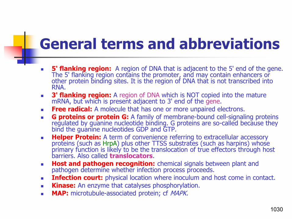
General terms and abbreviations
◼ 5' flanking region: A region of DNA that is adjacent to the 5' end of the gene. The 5' flanking region contains the promoter, and may contain enhancers or other protein binding sites. It is the region of DNA that is not transcribed into RNA.
◼ 3' flanking region: A region of DNA which is NOT copied into the mature mRNA, but which is present adjacent to 3' end of the gene.
◼ Free radical: A molecule that has one or more unpaired electrons.◼ G proteins or protein G: A family of membrane-bound cell-signaling proteins
regulated by guanine nucleotide binding. G proteins are so-called because they bind the guanine nucleotides GDP and GTP.
◼ Helper Protein: A term of convenience referring to extracellular accessory proteins (such as HrpA) plus other TTSS substrates (such as harpins) whose primary function is likely to be the translocation of true effectors through host barriers. Also called translocators.
◼ Host and pathogen recognition: chemical signals between plant and pathogen determine whether infection process proceeds.
◼ Infection court: physical location where inoculum and host come in contact.◼ Kinase: An enzyme that catalyses phosphorylation.◼ MAP: microtubule-associated protein; cf MAPK.
1030

General terms and abbreviations
◼ Mr: relative molecular mass, no units; an Mr of 1000 is represented by 1K; Mr is numerically equivalent to daltons (1K = 1kDa).
◼ Operon: a unit of contiguous genes transcribed as a singletranscript under the control of a regulatory gene or genes; common in bacteria but rare in eukaryotes.
◼ Open Reading frame (ORF): A DNA or RNA sequence lying between a start codon and stop codons which is capable of transcription(can be translated into a polypeptide).
◼ Orthologous/orthologue/orthology: Genes in different species that are homologous (similar) because they are derived from a common ancestral gene(during speciation).
◼ PKA: cAMP-dependent protein kinase or protein kinase A.◼ Promoter: A region of a DNA molecule to which an RNA polymerase binds and
initiates transcription.◼ Regulon: A system of more than one operon controlled by a single regulatory
protein. e.g. genes in the hrp regulon.◼ Replicon: DNA replicated under the control of a single replicator.
◼ Signal transduction: Reception, conversion and transmission of “chemical message” by a cell.
1031

General terms and abbreviations
◼ MAPK: mitogen-activated protein kinase; originally known as microtubule-associated protein kinase (see MAP) because microtubule-associated protein had been used in the assay in which MAPK was discovered. Its name was changed when it was subsequently found to be activated by several messengers, many of them mitogens.
◼ Mitogen: an agent that can induce mitosis.◼ Signaling: Communication established between and within cells of an
organism.◼ Sessile bacteria: Bacteria living within a biofilm.◼ Transphosphorylation: The exchange of phosphate groups between organic
phosphates, without their going through the stage of inorganic phosphates. ◼ Transcription: Copying of DNA by RNA polymerase into messenger RNA. ◼ Tanscription factor: A protein that participates in gene transcription often by
binding to a specific DNA sequence.
◼ Transposons: are small segments of DNA that can move from one region of a chromosome to another region of the same chromosome or to a different chromosome or a plasmid.
◼ Upregulation of transcription: Activation, or promotion increase the rate of gene transcription.
1032

Selected References
◼ Alfano, J. R. and A. Collmer. 2004. Type III secretion system effector proteins: Double Agents in Bacterial Disease and Plant Defense. Annu. Rev. Phytopathol. 42:385–414.
◼ Bacteria male sense. 2006. Chaotic Web Development.◼ Bacterial Pathogenesis 1- AGRO 426/BIOL 421.◼ Baker et al.,1997. Signaling in plant-microbe interactions. Science 276: 726-733.◼ Bauer, 2006. Eukaryotes Deal with Bacterial Quorum Sensing. American Society
for Microbiology.
◼ Belgian Society for Microbiology.2010. Molecular dialogue in host‐parasite interaction. House of the Academies. Abstractbook,110 pp.
◼ Bostock,R. M. 2005. Model signaling in induced resistance. Annu. Rev. Phytopathol. 2005. 43:545-80.
◼ Cao, H., Baldini R.L, Rahme L.G. 2001. Common mechanisms for pathogens of plants and animals. Annu Rev Phytopathol. 39:259–84.
◼ Cha et al., 1998. Production of acyl-homoserine lactone quorum-sensing signals by Gram-negative plant-associated bacteria. MPMI. 11: 1119-1129.
◼ Cain, D., H. Hanks, M. Weis, C. Bottoms, and J. Lawson. 2017. Microbiology Laboratory Manual B2420. Collin County Community College District, McKinney, TX. 133 pp.
1033

Selected References
◼ Edreva, A.2005. Pathogenesis-related proteins: Research progress in the last 15 years. GEN. APPL. PLANT PHYSIOLOGY 31(1-2), 105-124.
◼ Dickinson, M. & J. Beyon. 2003. Molecular Plant Pathology. Sheffield AP,CRC Press.
◼ Gachomo, E.M. et al., 2003. The molecular initiation and subsequent acquisition of disease resistance in plants.
◼ El-Sharoud, Walid. 2008. Bacterial Physiology- A Molecular Approach. 371 pp., Springer.
◼ Ferluga, S. and Venturi, V. 2009. OryR is a LuxR-family protein involved in interkingdom signaling between pathogenic Xanthomonas oryzae pv. oryzaeand rice. J Bacteriol. 191, 890-897.
◼ Guttman, D.S. 2003. Plants as models for the study of human pathogenesis. Biotechnology Advances 22 (2004) 363-382.
◼ Huguet, E. 2003. Bacterial pathogensis. In: Molecular Plant Pathology. Annual Plant Reviews Vol.4, Eds. M. Dickinson & J. Beyon.
◼ Hammond-Kosack. Kim E. and J.D. G. Jones. 1997. Plant disease resistance genes. Annu. Rev. Plant Physiol. Plant Mol. Biol. 1997. 48:575-607.
◼ Henderson et al., 2004. Type V Protein Secretion Pathway: the Autotransporter Story. Microbiol Mol Biol Rev. 68(4): 692–744.
◼ Jahr et al.,2000. The Endo-b-1,4-glucanase CelA of Clavibacter michiganensissubsp. michiganensis is a pathogenicity determinant required for induction of bacterial wilt of tomato. MPMI Vol. 13:703–714.
1034

Selected References
◼ Kawata et al., 2003. Genetic engineering for disease resistance in rice (Oryzasativa L.) using antimicrobial peptides. JARQ 37 (2), 71 – 76.
◼ Kirankumar et al., 2004. Nonhost resistance: how much do we know? TRENDS in Plant Science Vol.9, No.2.
◼ Klement, Z., Ott, P. G. et al., 2005. Basal (innate-) resistance of plants to bacterial infections- a possible means for biological control. in Proceedings of the 1st International Symposium on Biological Control of Bacterial Diseases (eds. W. Zeller & C. Ullrich) Darmstadt-Germany.
◼ Kuehn, MJ and NC Kesty. 2005. "Bacterial outer membrane vesicles and the host-pathogen interaction“. Genes Dev. 19(22):2645-55.
◼ Lecture 15. htm.◼ Lyon, G. D. 2002. Plant/pathogen interactions at the cellular level.
A summary and model.◼ Microbial Genomic Sequencing Perspectives of the American Phytopathological
Society (Revised 2003). ◼ Michelmore, R.W and Meyers, B.C .1998. Clusters of resistance genes in plants
evolve by divergent selection and a birth-and-death process. Genome Research: 8 , 1113-1130.
◼ Plant-Microbe Interactions: Plant Resistance Genes.
◼ Pierson III, L. S. 2000. Bacterial Signaling: Identification of N-Acyl-HomoserineLactone-Producing Bacteria. APSnet Education.
1035

Selected References
▪ Prescott,2006. Procaryotic cell structure and function, chapter 3, pp. 39-78.
▪ Proteins- Structure and Functions. htm. ◼ Puhler, A., Arlat, M. Becker, A., Gottfert, M., Morrissey, J.P., O'Gara, F. 2004.
What can bacterial genome research teach us about bacteria-plant interactions? Current Opinion in Plant Biology 7, 137-147.
◼ Ronald P. C. (Ed.) 2006. Plant-Pathogen Interactions: Methods and Protocols. Humana Press Inc.,248 pp.
• Razmi, J. 2006. PhD seminar on Plant Disease Resistance Genes. • Scheper,T. and J.J Zhong (ed.), 2001. Plant cells. Springer. pp.218.◼ Sági, L. 2000. Engineering resistance to diseases caused by bacteria.
In Diseases of Banana, Abaca and Enset Jones D. (eds.) CAB International (Wallingford, U.K.), pp. 474-482.
◼ Simpson. A. J. G. 2000. The genome sequence of the plant pathogen Xylella fastidiosa. Nature 406:151-9.
◼ Sapers, G.M., J. R. Gorny and A. E. Yousef (eds.).2006. Microbiology of Fruits and Vegetables. CRC Press, 634pp.
◼ Strange, R. N. 2003. Introduction to Plant Pathology. 497 pp. John Wiley & Sons Ltd.
◼ Thanassi, D.G.; Stathopoulos, C.; Karkal, A.; Li, H. 2005. Protein secretion in the absence of ATP: the autotransporter, two-partner secretion and chaperone/usher pathways of Gram-negative bacteria (Review). Molecular Membrane Biology 22 (1): 63-72.
1036

Selected References
◼ Van der Plank, J.E.1982. Host-pathogen interactions in plant disease. New York, USA: Academic Press.
◼ Vidaver, A.K. and P.A. Lambrecht 2004. Bacteria as plant pathogens. The Plant Health Instructor. DOI: 10.1094/PHI-I-2004-0809-01
• Vivian, A., J. Murillo and R. W. Jackson, 2001. The roles of plasmids in phytopathogenic bacteria: mobile arsenals? Microbiology, 147, 763-780.
◼ West, S. A., S. P. Diggle, A. Buckling, A. Gardner and A. S. Griffin.2007. The Social Lives of Microbes. Annu. Rev. Ecol. Evol. Syst. 38: 53-77.
◼ Whiteley, M., K. M., Lee, P. E. Greenberg & U. Muh. 2005. Quorum sensing signaling in bacteria. United States Patent 6855513.
◼ Xiao, S. 2006. Current perspectives on molecular mechanisms of plant disease resistance. Floriculture, Ornamental and Plant Biotechnology. Volume III, Global Science Books, UK, 317-333.
1037
Copyright © 2022 FDOKUMEN