Pathogenesis of FGF23-Related Hypophosphatemic Diseases ...
14
Citation: Nakanishi, T.; Michigami, T. Pathogenesis of FGF23-Related Hypophosphatemic Diseases Including X-linked Hypophosphatemia. Endocrines 2022, 3, 303–316. https://doi.org/10.3390/ endocrines3020025 Academic Editors: Yukihiro Hasegawa and Seiji Fukumoto Received: 12 May 2022 Accepted: 27 May 2022 Published: 2 June 2022 Publisher’s Note: MDPI stays neutral with regard to jurisdictional claims in published maps and institutional affil- iations. Copyright: © 2022 by the authors. Licensee MDPI, Basel, Switzerland. This article is an open access article distributed under the terms and conditions of the Creative Commons Attribution (CC BY) license (https:// creativecommons.org/licenses/by/ 4.0/). Review Pathogenesis of FGF23-Related Hypophosphatemic Diseases Including X-linked Hypophosphatemia Tatsuro Nakanishi 1,2 and Toshimi Michigami 1, * 1 Department of Bone and Mineral Research, Research Institute, Osaka Women’s and Children’s Hospital, Izumi 594-1101, Japan; [email protected] 2 Department of Pediatrics, Osaka University Graduate School of Medicine, Suita 565-0871, Japan * Correspondence: [email protected]; Tel.: +81-725-56-1220 Abstract: Since phosphate is indispensable for skeletal mineralization, chronic hypophosphatemia causes rickets and osteomalacia. Fibroblast growth factor 23 (FGF23), which is mainly produced by osteocytes in bone, functions as the central regulator of phosphate metabolism by increasing the renal excretion of phosphate and suppressing the production of 1,25-dihydroxyvitamin D. The excessive action of FGF23 results in hypophosphatemic diseases, which include a number of genetic disorders such as X-linked hypophosphatemic rickets (XLH) and tumor-induced osteomalacia (TIO). Phosphate-regulating gene homologous to endopeptidase on the X chromosome (PHEX), dentin matrix protein 1 (DMP1), ectonucleotide pyrophosphatase phosphodiesterase-1, and family with sequence similarity 20c, the inactivating variants of which are responsible for FGF23-related hereditary rickets/osteomalacia, are highly expressed in osteocytes, similar to FGF23, suggesting that they are local negative regulators of FGF23. Autosomal dominant hypophosphatemic rickets (ADHR) is caused by cleavage-resistant variants of FGF23, and iron deficiency increases serum levels of FGF23 and the manifestation of symptoms in ADHR. Enhanced FGF receptor (FGFR) signaling in osteocytes is suggested to be involved in the overproduction of FGF23 in XLH and autosomal recessive hypophosphatemic rickets type 1, which are caused by the inactivation of PHEX and DMP1, respectively. TIO is caused by the overproduction of FGF23 by phosphaturic tumors, which are often positive for FGFR. FGF23-related hypophosphatemia may also be associated with McCune-Albright syndrome, linear sebaceous nevus syndrome, and the intravenous administration of iron. This review summarizes current knowledge on the pathogenesis of FGF23-related hypophosphatemic diseases. Keywords: phosphate; fibroblast growth factor 23; osteocytes; rickets; osteomalacia 1. Introduction Phosphorus is an essential nutrient that mediates the majority of biological processes, including the integrity of cell membranes, the maintenance and inheritance of genetic infor- mation, energy metabolism, and the regulation of protein function by phosphorylation [1]. In vertebrates, phosphorus also contributes to skeletal mineralization as a constituent of hydroxyapatite (calcium-phosphate crystals). In the human adult body, approximately 90% of total phosphorus is stored in bone, while the remainder is present in the soft tissues and less than 1% in extracellular fluid [2]. In serum, the majority of phosphorus exists as free ions of inorganic phosphate (Pi), such as HPO 4 2- and H 2 PO 4 - , at a ratio of 4:1 at physiological pH [3]. Serum levels of Pi are influenced by age, dietary intake, serum pH, and so on. Since phosphate is indispensable for the formation of hydroxyapatite, its chronic deficiency or wasting leads to impaired skeletal mineralization, namely, rickets in children and osteomalacia in adults [4]. At the beginning of this century, fibroblast growth factor 23 (FGF23) was identified as the molecule responsible for autosomal dominant hypophos- phatemic rickets (ADHR) and tumor-induced hypophosphatemic osteomalacia (TIO) [5,6]. Endocrines 2022, 3, 303–316. https://doi.org/10.3390/endocrines3020025 https://www.mdpi.com/journal/endocrines
-
Upload
khangminh22 -
Category
Documents
-
view
0 -
download
0
Transcript of Pathogenesis of FGF23-Related Hypophosphatemic Diseases ...

Citation: Nakanishi, T.; Michigami, T.
Pathogenesis of FGF23-Related
Hypophosphatemic Diseases
Including X-linked
Hypophosphatemia. Endocrines 2022,
3, 303–316. https://doi.org/10.3390/
endocrines3020025
Academic Editors: Yukihiro
Hasegawa and Seiji Fukumoto
Received: 12 May 2022
Accepted: 27 May 2022
Published: 2 June 2022
Publisher’s Note: MDPI stays neutral
with regard to jurisdictional claims in
published maps and institutional affil-
iations.
Copyright: © 2022 by the authors.
Licensee MDPI, Basel, Switzerland.
This article is an open access article
distributed under the terms and
conditions of the Creative Commons
Attribution (CC BY) license (https://
creativecommons.org/licenses/by/
4.0/).
Review
Pathogenesis of FGF23-Related Hypophosphatemic DiseasesIncluding X-linked HypophosphatemiaTatsuro Nakanishi 1,2 and Toshimi Michigami 1,*
1 Department of Bone and Mineral Research, Research Institute, Osaka Women’s and Children’s Hospital,Izumi 594-1101, Japan; [email protected]
2 Department of Pediatrics, Osaka University Graduate School of Medicine, Suita 565-0871, Japan* Correspondence: [email protected]; Tel.: +81-725-56-1220
Abstract: Since phosphate is indispensable for skeletal mineralization, chronic hypophosphatemiacauses rickets and osteomalacia. Fibroblast growth factor 23 (FGF23), which is mainly producedby osteocytes in bone, functions as the central regulator of phosphate metabolism by increasingthe renal excretion of phosphate and suppressing the production of 1,25-dihydroxyvitamin D. Theexcessive action of FGF23 results in hypophosphatemic diseases, which include a number of geneticdisorders such as X-linked hypophosphatemic rickets (XLH) and tumor-induced osteomalacia (TIO).Phosphate-regulating gene homologous to endopeptidase on the X chromosome (PHEX), dentinmatrix protein 1 (DMP1), ectonucleotide pyrophosphatase phosphodiesterase-1, and family withsequence similarity 20c, the inactivating variants of which are responsible for FGF23-related hereditaryrickets/osteomalacia, are highly expressed in osteocytes, similar to FGF23, suggesting that theyare local negative regulators of FGF23. Autosomal dominant hypophosphatemic rickets (ADHR)is caused by cleavage-resistant variants of FGF23, and iron deficiency increases serum levels ofFGF23 and the manifestation of symptoms in ADHR. Enhanced FGF receptor (FGFR) signalingin osteocytes is suggested to be involved in the overproduction of FGF23 in XLH and autosomalrecessive hypophosphatemic rickets type 1, which are caused by the inactivation of PHEX and DMP1,respectively. TIO is caused by the overproduction of FGF23 by phosphaturic tumors, which are oftenpositive for FGFR. FGF23-related hypophosphatemia may also be associated with McCune-Albrightsyndrome, linear sebaceous nevus syndrome, and the intravenous administration of iron. This reviewsummarizes current knowledge on the pathogenesis of FGF23-related hypophosphatemic diseases.
Keywords: phosphate; fibroblast growth factor 23; osteocytes; rickets; osteomalacia
1. Introduction
Phosphorus is an essential nutrient that mediates the majority of biological processes,including the integrity of cell membranes, the maintenance and inheritance of genetic infor-mation, energy metabolism, and the regulation of protein function by phosphorylation [1].In vertebrates, phosphorus also contributes to skeletal mineralization as a constituent ofhydroxyapatite (calcium-phosphate crystals). In the human adult body, approximately 90%of total phosphorus is stored in bone, while the remainder is present in the soft tissuesand less than 1% in extracellular fluid [2]. In serum, the majority of phosphorus existsas free ions of inorganic phosphate (Pi), such as HPO4
2− and H2PO4−, at a ratio of 4:1 at
physiological pH [3]. Serum levels of Pi are influenced by age, dietary intake, serum pH,and so on.
Since phosphate is indispensable for the formation of hydroxyapatite, its chronicdeficiency or wasting leads to impaired skeletal mineralization, namely, rickets in childrenand osteomalacia in adults [4]. At the beginning of this century, fibroblast growth factor23 (FGF23) was identified as the molecule responsible for autosomal dominant hypophos-phatemic rickets (ADHR) and tumor-induced hypophosphatemic osteomalacia (TIO) [5,6].
Endocrines 2022, 3, 303–316. https://doi.org/10.3390/endocrines3020025 https://www.mdpi.com/journal/endocrines

Endocrines 2022, 3 304
Since then, our understanding of the mechanisms underlying phosphate metabolism andthe pathogenesis of various related diseases has increased. This review discusses cur-rent concepts on the role of FGF23 in phosphate homeostasis and the pathogenesis ofFGF23-related hypophosphatemic diseases.
2. Phosphorus Homeostasis
In mammals, the phosphate balance in postnatal life is maintained by its intestinalabsorption, renal excretion, and accumulation in and release from bone and soft tissue [1].The insufficient absorption, excess renal excretion, or excessive shift of Pi to bone or softtissue may cause hypophosphatemia [7]. Since phosphate is indispensable for skeletalmineralization, chronic hypophosphatemia leads to rickets and osteomalacia.
Pi absorption in the intestines is mediated by two processes: a passive, paracellulardiffusion process and an active, transcellular transport process. The latter is mediated bythe type IIb Na+/Pi co-transporter (NaPi-IIb), which is encoded by the SLC34A2 gene inhumans [8]. The expression of NaPi-IIb in the intestines is up-regulated by the low dietaryintake of Pi and 1,25-dihydroxyvitamin D (1,25(OH)2D), an active metabolite of vitaminD [9,10]. A dietary deficiency of phosphate is rare because the majority of foods containlarge amounts of phosphate. However, the insufficient action of vitamin D decreases theabsorption of Pi in the intestines.
In the kidneys, the majority of Pi filtered in the glomeruli is mainly reabsorbed bytype IIa and IIc Na+/Pi co-transporters (NaPi-IIa and NaPi-IIc) localized at the brushborder membrane (BBM) of the proximal tubules [11–13]. NaPi-IIa and NaPi-IIc are en-coded by the SLC34A1 and SLC34A3 genes, respectively, in humans. Inactivating variantsin the SLC34A3 gene cause hereditary hypophosphatemic rickets with hypercalciuria(HHRH) [14]. HHRH is characterized by renal Pi wasting, hypophosphatemia, increasedlevels of serum 1,25(OH)2D, and secondary hypocalciuria. On the other hand, inactivatingvariants in the SLC34A1 gene have been shown to cause Fanconi renotubular syndrome2, infantile hypercalciuria 2, and nephrolithiasis/osteoporosis associated with hypophos-phatemia [1].
Endocrine factors including 1,25(OH)2D, parathyroid hormone (PTH), and FGF23play critical roles in phosphate homeostasis. As described above, 1,25(OH)2D increases theintestinal absorption of Pi by up-regulating NaPi-IIb [10]. PTH increases the renal excretionof Pi by decreasing the amounts of NaPi-IIa and NaPi-IIc that localize in the BBM [15–17].FGF23 functions as the central regulator of phosphate metabolism, as described in thefollowing section.
3. Roles of FGF23 in Mineral Metabolism
FGF23 is a 32-kDa protein that consists of 251 amino acids including a 24-amino acidsignal peptide at its N terminus [6]. It is mainly produced by osteoblast lineage cells,particularly osteocytes, which terminally differentiate from osteoblasts and are embeddedwithin the bone matrix [18]. In adult bone, osteocytes make up more than 90% to 95% of allbone cells. Although the location and inaccessibility of osteocytes in the mineralized bonematrix delayed our understanding of their cellular functions, studies conducted in the lastfew decades have revealed their roles in bone homeostasis. Osteocytes are essential forcontrolling the bone mass in postnatal life by sensing mechanical strain and regulating theformation and resorption of bone [19]. They also produce sclerostin, an inhibitor of Wnt/β-catenin signaling. Mechanical strain reduces the production of sclerostin by osteocytes,which in turn enhances Wnt/β-catenin signaling and increases bone formation [20]. Inaddition, mouse studies suggested that the receptor activator of nuclear factor kappa Bligand produced by osteocytes controls the postnatal bone mass [21]. The expression ofFGF23 by osteocytes indicates that they also play critical roles in phosphate homeostasis.
FGF23 belongs to the FGF19 subfamily together with FGF19 and FGF21, based ontheir unique features among FGFs [22]. Members of the FGF19 family exert their effects ondistant target organs and tissues in an endocrine manner, and their low binding affinity
Endocrines 2022, 3 305
to heparin/heparan sulfate has been suggested to confer endocrine functions due to lessbinding to heparan sulfate surrounding their producing cells and their entry into thecirculation [22]. Members of the FGF19 subfamily require the single-pass transmembraneprotein Klotho, αKlotho for FGF23, and βKlotho for FGF19 and FGF21, for their signaltransduction through an FGF receptor (FGFR) [22–24]. A previous study demonstratedthat the N-terminal domain of FGF23 bound to FGFR while its C-terminal domain boundto αKlotho [22].
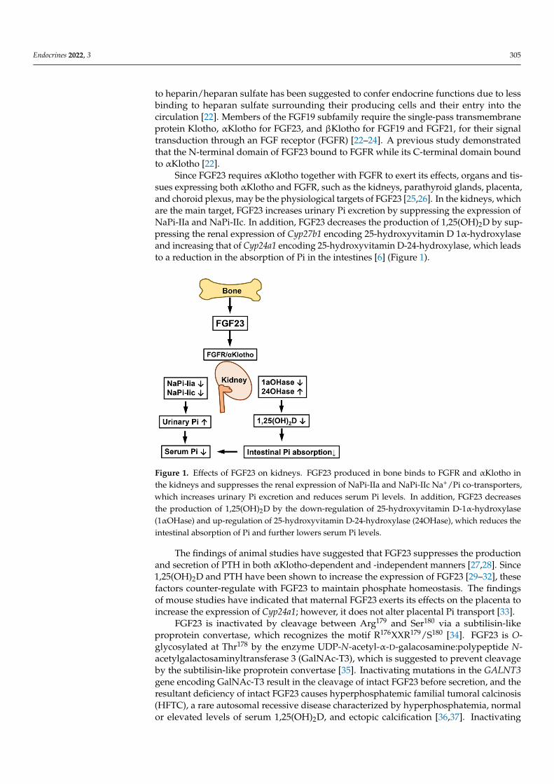
Since FGF23 requires αKlotho together with FGFR to exert its effects, organs and tis-sues expressing both αKlotho and FGFR, such as the kidneys, parathyroid glands, placenta,and choroid plexus, may be the physiological targets of FGF23 [25,26]. In the kidneys, whichare the main target, FGF23 increases urinary Pi excretion by suppressing the expression ofNaPi-IIa and NaPi-IIc. In addition, FGF23 decreases the production of 1,25(OH)2D by sup-pressing the renal expression of Cyp27b1 encoding 25-hydroxyvitamin D 1α-hydroxylaseand increasing that of Cyp24a1 encoding 25-hydroxyvitamin D-24-hydroxylase, which leadsto a reduction in the absorption of Pi in the intestines [6] (Figure 1).
Endocrines 2022, 3, FOR PEER REVIEW 3
FGF23 belongs to the FGF19 subfamily together with FGF19 and FGF21, based on their unique features among FGFs [22]. Members of the FGF19 family exert their effects on distant target organs and tissues in an endocrine manner, and their low binding affinity to heparin/heparan sulfate has been suggested to confer endocrine functions due to less binding to heparan sulfate surrounding their producing cells and their entry into the cir-culation [22]. Members of the FGF19 subfamily require the single-pass transmembrane protein Klotho, αKlotho for FGF23, and βKlotho for FGF19 and FGF21, for their signal transduction through an FGF receptor (FGFR) [22–24]. A previous study demonstrated that the N-terminal domain of FGF23 bound to FGFR while its C-terminal domain bound to αKlotho [22].
Since FGF23 requires αKlotho together with FGFR to exert its effects, organs and tis-sues expressing both αKlotho and FGFR, such as the kidneys, parathyroid glands, pla-centa, and choroid plexus, may be the physiological targets of FGF23 [25,26]. In the kid-neys, which are the main target, FGF23 increases urinary Pi excretion by suppressing the expression of NaPi-IIa and NaPi-IIc. In addition, FGF23 decreases the production of 1,25(OH)2D by suppressing the renal expression of Cyp27b1 encoding 25-hydroxyvitamin D 1α-hydroxylase and increasing that of Cyp24a1 encoding 25-hydroxyvitamin D-24-hy-droxylase, which leads to a reduction in the absorption of Pi in the intestines [6] (Figure 1).
Figure 1. Effects of FGF23 on kidneys. FGF23 produced in bone binds to FGFR and αKlotho in the kidneys and suppresses the renal expression of NaPi-IIa and NaPi-IIc Na+/Pi co-transporters, which increases urinary Pi excretion and reduces serum Pi levels. In addition, FGF23 decreases the pro-duction of 1,25(OH)2D by the down-regulation of 25-hydroxyvitamin D-1α-hydroxylase (1αOHase) and up-regulation of 25-hydroxyvitamin D-24-hydroxylase (24OHase), which reduces the intestinal absorption of Pi and further lowers serum Pi levels.
The findings of animal studies have suggested that FGF23 suppresses the production and secretion of PTH in both αKlotho-dependent and -independent manners [27,28]. Since 1,25(OH)2D and PTH have been shown to increase the expression of FGF23 [29–32], these factors counter-regulate with FGF23 to maintain phosphate homeostasis. The findings of mouse studies have indicated that maternal FGF23 exerts its effects on the placenta to increase the expression of Cyp24a1; however, it does not alter placental Pi transport [33].
FGF23 is inactivated by cleavage between Arg179 and Ser180 via a subtilisin-like pro-protein convertase, which recognizes the motif R176XXR179/S180 [34]. FGF23 is O-glycosyl-ated at Thr178 by the enzyme UDP-N-acetyl-α-D-galacosamine:polypeptide N-acetylgalac-tosaminyltransferase 3 (GalNAc-T3), which is suggested to prevent cleavage by the sub-tilisin-like proprotein convertase [35]. Inactivating mutations in the GALNT3 gene
Figure 1. Effects of FGF23 on kidneys. FGF23 produced in bone binds to FGFR and αKlotho inthe kidneys and suppresses the renal expression of NaPi-IIa and NaPi-IIc Na+/Pi co-transporters,which increases urinary Pi excretion and reduces serum Pi levels. In addition, FGF23 decreasesthe production of 1,25(OH)2D by the down-regulation of 25-hydroxyvitamin D-1α-hydroxylase(1αOHase) and up-regulation of 25-hydroxyvitamin D-24-hydroxylase (24OHase), which reduces theintestinal absorption of Pi and further lowers serum Pi levels.
The findings of animal studies have suggested that FGF23 suppresses the productionand secretion of PTH in both αKlotho-dependent and -independent manners [27,28]. Since1,25(OH)2D and PTH have been shown to increase the expression of FGF23 [29–32], thesefactors counter-regulate with FGF23 to maintain phosphate homeostasis. The findingsof mouse studies have indicated that maternal FGF23 exerts its effects on the placenta toincrease the expression of Cyp24a1; however, it does not alter placental Pi transport [33].
FGF23 is inactivated by cleavage between Arg179 and Ser180 via a subtilisin-likeproprotein convertase, which recognizes the motif R176XXR179/S180 [34]. FGF23 is O-glycosylated at Thr178 by the enzyme UDP-N-acetyl-α-D-galacosamine:polypeptide N-acetylgalactosaminyltransferase 3 (GalNAc-T3), which is suggested to prevent cleavageby the subtilisin-like proprotein convertase [35]. Inactivating mutations in the GALNT3gene encoding GalNAc-T3 result in the cleavage of intact FGF23 before secretion, and theresultant deficiency of intact FGF23 causes hyperphosphatemic familial tumoral calcinosis(HFTC), a rare autosomal recessive disease characterized by hyperphosphatemia, normalor elevated levels of serum 1,25(OH)2D, and ectopic calcification [36,37]. Inactivating

Endocrines 2022, 3 306
mutations in the FGF23 gene itself and the KLOTHO gene encoding αKlotho were alsofound to be responsible for HFTC [38–40].
Both Fgf23-knockout mice and Klotho-deficient mice exhibit hyperphosphatemia withincreased renal Pi reabsorption and elevated serum 1,25(OH)2D levels, which resemblethe features in patients with HFTC [25,41]. These mutant mice also show severe growthretardation with abnormal skeletal phenotype and short lifespan. Interestingly, Fgf23-knockout mice display a marked accumulation of osteoid despite hyperphosphatemia [41].Although mechanisms for this counterintuitive observation remain unclear, it may suggestthe effects of FGF23 to be independent of phosphate metabolism.
4. Pathogenesis of FGF23-Related Hypophosphatemic Diseases4.1. FGF23-Related Hypophosphatemic Diseases
Since the FGF23-αKlotho axis plays a central role in phosphate homeostasis, its dis-ruption causes hyperphosphatemic conditions, as described in the previous section. Onthe other hand, the excessive action of FGF23 underlies various hypophosphatemic dis-eases, which are characterized by urinary phosphate wasting, hypophosphatemia, andinappropriately low levels of serum 1,25(OH)2D [42,43]. Phosphate is indispensable forskeletal mineralization; therefore, chronic hypophosphatemia due to excessive FGF23 leadsto rickets in children and osteomalacia in adults. The impaired production of 1,25(OH)2Dcontributes to the resistance of FGF23-related hypophosphatemic rickets/osteomalacia tonative vitamin D. FGF23-related hypophosphatemic rickets/osteomalacia include variousconditions such as genetic diseases (Tables 1 and 2). The following sections will describethe pathogenesis of each condition.
Table 1. FGF23-related hypophosphatemic diseases and their causes.
Disorder Causes Incidences
ADHR Activating variants of FGF23 Very rare
XLH Inactivating variants of PHEX 1:20,000 live birth
ARHR1 Inactivating variants of DMP1 Very rare
ARHR2 Inactivating variants of ENPP1 Very rare
Raine syndrome Inactivating variants of FAM20C Very rare
Osteoglophonic dysplasia Activating variants of FGFR1 Very rare
Jansen’s metaphyseal chondrodysplasia Activating variants of PTH1R Very rare
TIO Phosphaturic mesenchymal tumor, FN1-FGFR1,FN1-FGF1 fusion genes
Most frequent among the acquireddisorders
McCune-Albright Syndrome GNAS somatic activating variants in bone lesions Very rare
Linear sebaceous nevus syndrome KRAS/HRAS/NRAS somatic activating variantsin skin/bone lesions Very rare
I.V. administration of iron preparations Saccharated ferric oxide, iron polymaltose Very rare
4.2. Autosomal Dominant Hypophosphatemic Rickets (ADHR)
ADHR (OMIM #193100) is caused by missense variants in the FGF23 gene. Since theresponsible variants occur at Arg176 or Arg179 within the RXXR/S motif recognized bysubtilisin-like proprotein convertase, the resultant mutant FGF23 protein is resistant tocleavage-mediated inactivation [5]. However, this disease shows incomplete penetrance.Patients with ADHR variants do not always have high serum levels of intact FGF23, and thedisease may occur with an early or delayed onset and variable expressivity [44]. Late-onsetADHR primarily manifests in post-pubertal women who are prone to iron deficiency [45].Serum iron levels were previously shown to negatively correlate with serum levels ofboth the C-terminal fragment of FGF23 and intact FGF23 in ADHR patients; however,serum iron levels also negatively correlated with C-terminal FGF23 levels in healthy controlsubjects, whereas no relationship was observed with intact FGF23 levels [45]. Farrow et al.generated FGF23-knock-in mice carrying the R176Q ADHR point mutation (ADHR mice)

Endocrines 2022, 3 307
and found that a low-iron diet increased bone FGF23 mRNA levels and the serum levelsof both intact and C-terminal FGF23 with hypophosphatemia in ADHR mice. On theother hand, wild-type mice fed the low-iron diet showed normal serum levels of intactFGF23 and phosphate, but an elevated level of the C-terminal fragment of FGF23 [46].Furthermore, the chelation of iron up-regulated the expression of Fgf23 in a culturedosteoblastic cell line, which involved hypoxia-inducible factor 1α. Collectively, thesefindings suggest that iron deficiency increases the expression of Fgf23 in bone and alsothat the FGF23 protein is cleaved in iron deficiency to maintain normal serum levels ofFGF23 and normophosphatemia in control subjects, whereas the cleavage resistance ofmutant FGF23 leads to the accumulation of intact FGF23 and hypophosphatemia in ADHRsubjects [46].
Table 2. Clinical features of FGF23-related hypophosphatemic diseases.
Disorder Clinical Features
ADHR Renal Pi wasting, hypophosphatemia, low or normal serum 1,25(OH)2D levels,rickets/osteomalacia, incomplete penetrance, often delayed onset
XLH Renal Pi wasting, hypophosphatemia, low or normal serum 1,25(OH)2D levels,rickets/osteomalacia, short stature, bone deformity, dental problems, enthesopathy
ARHR1 Renal Pi wasting, hypophosphatemia, low or normal serum 1,25(OH)2D levels,rickets/osteomalacia, short stature, bone deformity, dental problems, enthesopathy
ARHR2 Renal Pi wasting, hypophosphatemia, low or normal serum 1,25(OH)2D levels,rickets/osteomalacia, short stature, bone deformity, dental problems, enthesopathy
Raine syndromeOsteosclerosis of early onset, poor life prognosis in most cases, characteristic face, renal Pi
wasting, hypophosphatemia, low or normal serum 1,25(OH)2D levels, rickets/osteomalacia,short stature, bone deformity, dental problems, enthesopathy
Osteoglophonic dysplasia Multiple radiolucent areas in metaphysis, rhizomelic short stature, characteristic face, renal Piwasting, hypophosphatemia, low or normal serum 1,25(OH)2D levels
Jansen’s metaphysealchondrodysplasia
Short-limbed short stature, usually hypercalcemia, skull sclerosis, renal Pi wasting,hypophosphatemia, low or normal serum 1,25(OH)2D levels
TIO Renal Pi wasting, hypophosphatemia, low or normal serum 1,25(OH)2D levels,rickets/osteomalacia, improved by tumor resection
McCune-Albright Syndrome Fibrous dysplasia, café-au-lait skin pigmentation, endocrinologic abnormalities, renal Piwasting, hypophosphatemia, low or normal serum 1,25(OH)2D levels
Linear sebaceous nevus syndrome Linear sebaceous nevus, abnormalities in central nervous system, ocular anomalies, bonedefects, renal Pi wasting, hypophosphatemia, low or normal serum 1,25(OH)2D levels
I.V. administration of ironpreparations
Renal Pi wasting, hypophosphatemia, low or normal serum 1,25(OH)2D levels,rickets/osteomalacia, improved by discontinuation of the responsible drugs
4.3. X-Linked Hypophosphatemic Rickets (XLH)
XLH (OMIM #307800) is the most common form of hereditary hypophosphatemicrickets. Patients with XLH have elevated serum levels of FGF23, which result in urinaryPi wasting, hypophosphatemia, and inappropriately low levels of 1,25(OH)2D [47]. XLHwas initially called vitamin D-resistant rickets because of a poor response to treatmentwith native vitamin D at dosages that cure vitamin D-deficient rickets. XLH is caused byinactivating variants in the phosphate-regulating gene homologous to endopeptidase on the Xchromosome (PHEX) located at Xp22.1, showing X-linked dominant inheritance [48]. Similarto FGF23, PHEX is expressed in osteoblast lineage cells and is more highly expressed inosteocytes [18,49]. Although its structure suggests that the product of PHEX functions as acell surface-bound, zinc-dependent protease, its physiological substrates remain elusive.In hypophosphatemic Hyp mice, which harbor a large deletion in the Phex gene and arewidely used as a model for XLH, the expression of Fgf23 in osteocytes was found to be

Endocrines 2022, 3 308
increased [18,50]; however, FGF23 did not serve as a substrate for PHEX [51]. Therefore,the regulation of FGF23 by PHEX may be indirect and involve other molecule(s).
Since PHEX is highly expressed in osteocytes, in an attempt to clarify abnormalitiesin Phex-deficient osteocytes, we previously compared the gene expression profiles ofosteoblasts and osteocytes in Hyp mice and wild-type littermates [18]. As osteoblasts matureinto osteocytes, the expression of dentin matrix protein 1 (Dmp1) and family with sequencesimilarity 20c (Fam20c), which are responsible for autosomal recessive hypophosphatemictype 1 (ARHR1) and Raine syndrome (RNS), respectively, increased in both Hyp andwild-type cells, and these genes were up-regulated in Hyp cells, similar to Fgf23 [18].These findings indicated the critical roles of osteocytes in phosphate homeostasis andalso suggested complex abnormalities in Phex-deficient osteocytes. We found that theexpression of the genes encoding canonical FGF ligands (Fgf1 and Fgf2), their receptors(Fgfr1-3), and early growth response 1, which is a target for FGFR activation, was also up-regulated in Hyp osteocytes, indicating enhanced FGFR signaling [18]. Furthermore, Martinet al. suggested enhanced FGFR signaling in Hyp bone [52], and Xiao et al. demonstratedthat the conditional deletion of Fgfr1 in osteocytes and mature osteoblasts partially restoredthe overproduction of FGF23 and ameliorated hypophosphatemia and rickets [53]. Theregulation of FGF23 production by FGFR signaling is also supported by osteoglophonicdysplasia, which is a rare skeletal dysplasia caused by activating mutations in FGFR1 thatis frequently associated with elevated serum FGF23 levels and hypophosphatemia [54].
Enhanced FGFR signaling in Phex-deficient osteocytes is of interest based on previ-ous findings suggesting that FGFR plays a critical role in the transduction of signalingevoked by increased extracellular Pi [55–57]. In various cell types, treatment with highextracellular Pi activated FGFR for the regulation of gene expression. In an osteoblastic cellline, treatment with an FGFR inhibitor abolished the up-regulation of Dmp1 by increasedextracellular Pi [56]. In HEK293 cells, the knockdown of FGFR1 diminished the Pi-inducedphosphorylation of ERK1/2 [55]. More recently, the activation of FGFR1 by extracellular Piwas shown to increase the expression of Galnt3 in bone, leading to an elevated serum levelof FGF23 in mice [57]. Collectively, these findings suggest that FGFR1 is involved in thesensing of Pi availability. In consideration of this role of FGFR and enhanced FGFR signal-ing in the osteocytes of Hyp mice, abnormal Pi sensing may be involved in the pathogenesisof XLH.
Matrix extracellular phosphoglycoprotein (MEPE) is a member of the SIBLING (smallintegrin-binding ligand, N-linked glycoproteins) family, and was initially cloned fromthe tumor of a patient with TIO [58]. A genome-wide association study proposed MEPEas a factor influencing bone mineral density in humans [59]. MEPE contains an acidicserine-aspartate rich MEPE-associated motif (ASARM) consisting of 23 residues at the Cterminus. ASARM peptides released from MEPE by cathepsin-mediated cleavage havebeen shown to inhibit mineralization. Rowe et al. demonstrated that PHEX bound tothe ASARM motif in MEPE and the released ASARM peptide and its serum levels wereelevated in Hyp mice [60,61]. The ASARM motif is also present in other SIBLING proteins,such as DMP1 and osteopontin, and PHEX may bind to these proteins at these motifs [58].A previous study implicated ASARM peptides released from these SIBLING proteins indefective mineralization in XLH [62].
Growth retardation is often observed in patients with XLH: however, the underlyingmechanisms have not yet been elucidated in detail. A study published by Fuente et al.demonstrated marked alterations in the structure, dynamics, and maturation of growthplate cartilage in growth-retarded young Hyp mice [63]. In the growth plates of Hyp mice,both proliferation and apoptosis rates of chondrocytes were reduced, and the hypertrophyand maturation of chondrocytes were severely disturbed. The spatial organization of thechondro-osseous junction and the primary spongiosa trabeculae were markedly deformed.These alterations in the growth plates might be the mechanisms for the growth retardationin Hyp mice. The authors also found an enhanced activation of the extracellular signal-regulated kinase (ERK)1/2 signaling pathway in the Hyp growth plates, implying an

Endocrines 2022, 3 309
involvement of FGF23 in these abnormalities [63]. Reduction in caspase-mediated apoptosisof hypertrophic chondrocytes was also reported in rachitic mice with low-phosphate diet-induced hypophosphatemia as well as in Hyp mice, which suggests that hypophosphatemiaimpairs apoptosis of hypertrophic chondrocytes, leading to rickets [64].
Although chondrocytes do not express αKlotho, which is required for FGF23 toactivate its downstream signaling pathways at physiological concentrations, soluble formsof αKlotho are present in serum and cerebrospinal fluid [65] and have been implicated inthe regulation of FGF23 signaling in cells without the transmembrane form of αKlotho [66].We previously demonstrated that FGF23 suppressed the linear growth of mouse metatarsalcartilage in cultures in the presence of soluble αKlotho by decreasing the proliferation ofchondrocytes, which suggests that suppressed chondrocyte proliferation by FGF23 plays acausative role in the growth retardation associated with XLH [67].
Since the placenta expresses FGFR1 and αKlotho, high levels of FGF23 in pregnantwomen with XLH may affect their fetuses. We previously investigated this issue usingpregnant Hyp female mice [33]. Hyp and wild-type female mice were mated with wild-typemale mice, and the pregnant mothers and their male fetuses were subjected to analyses.FGF23 levels were higher in Hyp mothers than in wild-type mothers. Hyp fetuses andwild-type fetuses were obtained from mating between Hyp females and wild-type males.FGF23 levels in Hyp fetuses were approximately 20-fold higher than in their mothers,while wild-type fetuses from Hyp mothers had low levels of FGF23, as did fetuses fromwild-type mothers, suggesting that FGF23 does not cross the placenta [33]. The expressionof Cyp24a1 was higher in the placentas of fetuses from Hyp mothers than in those of fetusesfrom wild-type mothers, which resulted in decreased levels of plasma 25-hydroxyvitaminD in fetuses from Hyp mothers. Therefore, increased levels of circulating FGF23 in Hypmothers may exert direct effects on the placenta during pregnancy and alter fetal vitaminD metabolism via the regulation of Cyp24a1 expression [33]. Further studies are needed toclarify whether similar phenomena occur with pregnancy in human patients with XLH.
The enthesis is a tissue that forms at the site of insertion of a tendon to bone andconsists of a bony eminence, mineralized fibrocartilage, unmineralized fibrocartilage, and atendon. It optimizes the transfer of mechanical force from muscle to bone, which is requiredfor efficient movements [68]. Enthesopathy is a pathological change at the insertion oftendons and ligaments. Mineralizing enthesopathy is one of the complications of XLH andother types of FGF23-related hypophosphatemia and accounts for a high morbidity ratein adult patients [69]. Karaplis et al. previously reported that a transgenic mouse modeloverexpressing a secreted form of the human FGF23[p.R176Q] variant, which is resistant tocleavage, displayed mineralizing enthesopathy of the Achilles and planar facial insertions,suggesting the involvement of FGF23 in the development of mineralizing enthesopathy [70].More recently, Liu et al. investigated the cellular and molecular mechanisms involved in thedevelopment of mineralizing enthesopathy in Hyp mice and reported that Achilles tendonentheses of Hyp mice showed the expansion of hypertrophic-appearing chondrogeniccells. In comparison with the entheses of wild-type mice, Hyp entheses exhibited theexpansion of cells expressing the chondrogenic marker gene Sox9 and enhanced bonemorphogenetic protein and Indian hedgehog signaling pathways, both of which playcritical roles in chondrocyte differentiation [71]. Although oral phosphate salts and activevitamin D metabolites are administered as conventional medical treatments for XLH tocorrect their deficiencies, it does not prevent or ameliorate enthesopathies [70]. Burosumab,a humanized monoclonal neutralizing antibody to FGF23, has recently become available asa new treatment for XLH [47]. In Japan, burosumab has been approved for the treatment ofall types of FGF23-related hypophosphatemic rickets/osteomalacia. In pediatric patientswith XLH, improvements in the severity of rickets and biochemical parameters were greaterin patients treated with burosumab than in those who continued conventional therapy [72].Further studies are needed to clarify the effects of burosumab on the prevention andtreatment of enthesopathies.

Endocrines 2022, 3 310
4.4. Autosomal Recessive Hypophosphatemic Rickets Type 1 (ARHR1)
ARHR1 (OMIM #241520) is caused by inactivating variants of the DMP1 gene [73,74].DMP1 is an extracellular matrix protein belonging to the SIBLING family and is highlyexpressed in osteocytes as well as in dentin. Patients with ARHR1 manifest elevated FGF23levels, hypophosphatemia, inappropriately low 1,25(OH)2D levels, and skeletal hypomin-eralization, similar to patients with XLH. Dmp1-null mice reproduced the phenotype ofARHR1 and exhibited defective osteocyte maturation and the up-regulated expression ofFgf23 in osteocytes [73,74]. Although the pathogenesis of ARHR1 remains largely unknown,the findings of studies using Phex-deficient Hyp mice and Dmp1-null mice suggest that theoverproduction of FGF23 is attributable to enhanced FGFR signaling in bone in both mousemodels [52].
4.5. Autosomal Recessive Hypophosphatemic Rickets Type 2 (ARHR2)
ARHR2 (OMIM #613312) also belongs to FGF23-related hypophosphatemic ricketsand is caused by inactivating variants in the ectonucleotide pyrophosphatase phosphodiesterase-1(ENPP1) gene [75,76]. ENPP1 encodes an enzyme that produces pyrophosphate (PPi), apotent inhibitor of mineralization, and inactivating variants in ENPP1 are also responsiblefor hypermineralization disorders, such as generalized arterial calcification in infancy [77].The ectoenzyme tissue non-specific alkaline phosphatase (TNSALP) facilitates skeletalmineralization by degrading PPi to produce Pi. Although PPi may regulate the productionof FGF23, patients with hypophosphatasia, which is caused by inactivating variants inTNSALP, had normal levels of FGF23 despite elevated extracellular levels of PPi [78].Therefore, the mechanisms by which ENPP1 deficiency results in the overproduction ofFGF23 remain unclear. Since inactivating variants in ENPP1 cause conditions characterizedby ectopic calcification and FGF23-related hypophosphatemia, a close relationship mayexist between ectopic calcification and the overproduction of FGF23.
4.6. Raine Syndrome (RNS)
FAM20C, also known as DMP4, encodes a kinase that phosphorylates various secretedproteins. The proteins phosphorylated by FAM20C include FGF23 and members of theSIBLING family, such as DMP1, osteopontin, and MEPE [34,79]. Inactivating variants inthe FAM20C gene are responsible for RNS (OMIM #259775). RNS is an autosomal recessivedisease that is characterized by craniofacial malformation, osteosclerotic bone dysplasia,and a poor prognosis [80]. Surviving patients with mild RNS manifest hypophosphatemiadue to elevated levels of FGF23 and dental anomalies [81,82]. Fam20c-null mice exhibitedelevated levels of serum FGF23, hypophosphatemia, and dental anomalies [83]. Thesemice also showed low expression levels of Dmp1 in osteocytes, which suggested that thedown-regulated expression of DMP1 plays a causal role in the overproduction of FGF23in RNS [83]. However, the overexpression of Dmp1 failed to rescue the defects in Fam20c-null mice [84]. A previous study reported that FAM20C phosphorylated FGF23 on Ser180,which inhibited the O-glycosylation of FGF23 on Thr178 by GalNAc-T3 and acceleratedcleavage [34]. Therefore, inactivating variants in FAM20C may increase FGF23 levels byinhibiting its cleavage.
4.7. Tumor-Induced Osteomalacia (TIO)
TIO is a rare paraneoplastic syndrome characterized by urinary phosphate wasting,hypophosphatemia, and osteomalacia. Responsible tumors are generally benign, slow-growing phosphaturic mesenchymal tumors (PMT) [85]. The overproduction of FGF23 bytumors was previously shown to enhance the renal excretion of Pi and induce hypophos-phatemia, low 1,25(OH)2D levels, and osteomalacia, which were cured by the surgicalremoval of the responsible tumor [6,86]. Lee et al. identified the fusion genes Fibronectin 1(FN1)-FGFR1 and FN1-FGF1 in subgroups of PMT and showed that immunoreactivity forFGFR1 was positive in 82% of PMT [87,88]. These findings suggest the involvement of theFGF/FGFR signaling pathway in the development of PMT.

Endocrines 2022, 3 311
4.8. Other Causes of FGF23-Related Hypophosphatemia
McCune-Albright syndrome (MAS, OMIM #174800) is characterized by polyostoticfibrous dysplasia, café-au-lait skin pigmentation, and precocious puberty, and is caused bya somatic activating variant in GNAS1 encoding the subunit of the stimulatory G protein.MAS is clinically heterogeneous and may manifest various endocrinological abnormalities.Some patients with MAS exhibit hypophosphatemia, which results from the overproductionof FGF23 by abnormal skeletal progenitor cells in the bone lesions of fibrous dysplasia [89].Serum levels of FGF23 in MAS patients correlate with disease activity [89], and significanthypophosphatemia only occurs in patients with a severe disease burden. A previous studysuggested that the ratio of the C-terminal fragment of FGF23 to intact FGF23 was elevatedby accelerated cleavage in the bone lesions of fibrous dysplasia [90].
Linear sebaceous nevus syndrome, also called Schimmelpenning-Feuerstein-Mims(SFM) syndrome (OMIM #163200), is characterized by congenital linear nevus sebaceousand abnormalities in neuroectodermal organs and is caused by somatic variants in RASgenes, including KRAS, HRAS, and NRAS, which are detectable in skin lesions [91,92].Hypophosphatemia due to elevated levels of FGF23 is rarely associated with SFM syndrome.Lim et al. suggested that the source of FGF23 in SFM syndrome was bone lesions carryingRAS variants rather than skin lesions [93].
Osteoglophonic dysplasia (OMIM #166250) is a rare autosomal dominant diseasecharacterized by rhizomelic dwarfism, non-ossifying bone lesions, craniosynostosis, andface abnormalities, and is caused by activating variants in the FGFR1 gene. As discussedearlier, this disease may be associated with FGF23-related hypophosphatemia, indicatingthe involvement of FGFR1 in the regulation of FGF23 production [54].
Jansen’s metaphyseal chondrodysplasia (OMIM #156400) is an autosomal dominantdisease caused by an activating variant in the PTH type 1 receptor (PTH1R) gene [94].Previous studies reported that FGF23-related hypophosphatemia may be associated withJansen’s metaphyseal chondrodysplasia [95]. This finding suggests that PTH signalingstimulates FGF23 production, which is also supported by the findings of several in vivoand in vitro studies [31,32,96].
FGF23-related hypophosphatemic rickets/osteomalacia may also be associated withthe intravenous administration of saccharated ferric oxide or iron polymaltose [97,98]. Themechanisms by which these drugs cause the overproduction of FGF23 remain unclear; how-ever, their discontinuance rapidly restores elevated FGF23 levels and hypophosphatemia.
5. Conclusions
FGF23-related hypophosphatemia is characterized by urinary Pi wasting, hypophos-phatemia, and inappropriately low levels of 1,25(OH)2D, and includes various types ofhereditary rickets/osteomalacia, such as XLH, and acquired diseases, including TIO. Themolecules responsible for hereditary rickets/osteomalacia are highly expressed by os-teocytes, indicating that these cells play a central role in phosphate homeostasis. Sinceinactivating variants of PHEX, DMP1, ENPP1, and FAM20C lead to the overproductionof FGF23, these molecules appear to function as negative regulators of FGF23. Althoughthe mechanisms underlying the overproduction of FGF23 remain unclear in most FGF23-related hypophosphatemic diseases, enhanced FGFR signaling may be involved in theoverproduction of FGF23 in XLH and ARHR1 as well as in TIO. Since FGFR1 is suggestedto be involved in Pi sensing, abnormalities in Pi sensing may play a role in the pathogenesisof these diseases.
Author Contributions: Conceptualization, T.M.; writing—original draft preparation, T.N. and T.M.;writing—review and editing, T.N. and T.M.; supervision, T.M.; funding acquisition, T.M. All authorshave read and agreed to the published version of the manuscript.
Funding: The preparation of this review was partly supported by a grant from the Japan Society forthe Promotion of Science (JSPS KAKENHI Grant Number 21K07835) to T.M.
Conflicts of Interest: The authors declare no conflict of interest.

Endocrines 2022, 3 312
References1. Michigami, T.; Kawai, M.; Yamazaki, M.; Ozono, K. Phosphate as a Signaling Molecule and Its Sensing Mechanism. Physiol. Rev.
2018, 98, 2317–2348. [CrossRef]2. Mitchell, H.; Hamilton, T.; Steggerda, F.; Bean, H. The chemical composition of the adult human body and its bearing on the
biochemistry of growth. J. Biol. Chem. 1945, 158, 625–637. [CrossRef]3. Peters, J.P.; Wakeman, A.M.; Lee, C. Total Acid-Base Eqiilibrium of plasma in health and disease: XI. Hypochloremia and Total
Salt Deficiency in Nephritis. J. Clin. Investig. 1929, 6, 551–575. [CrossRef] [PubMed]4. Michigami, T.; Ozono, K. Roles of Phosphate in Skeleton. Front. Endocrinol. 2019, 10, 180. [CrossRef] [PubMed]5. ADHR-CONSORTIUM. Autosomal dominant hypophosphataemic rickets is associated with mutations in FGF23. Nat. Genet.
2000, 26, 345–348. [CrossRef] [PubMed]6. Shimada, T.; Mizutani, S.; Muto, T.; Yoneya, T.; Hino, R.; Takeda, S.; Takeuchi, Y.; Fujita, T.; Fukumoto, S.; Yamashita, T. Cloning
and characterization of FGF23 as a causative factor of tumor-induced osteomalacia. Proc. Natl. Acad. Sci. USA 2001, 98, 6500–6505.[CrossRef] [PubMed]
7. Gaasbeek, A.; Meinders, A.E. Hypophosphatemia: An update on its etiology and treatment. Am. J. Med. 2005, 118, 1094–1101.[CrossRef] [PubMed]
8. Sabbagh, Y.; O’Brien, S.P.; Song, W.; Boulanger, J.H.; Stockmann, A.; Arbeeny, C.; Schiavi, S.C. Intestinal npt2b plays a major rolein phosphate absorption and homeostasis. J. Am. Soc. Nephrol. 2009, 20, 2348–2358. [CrossRef]
9. Hattenhauer, O.; Traebert, M.; Murer, H.; Biber, J. Regulation of small intestinal Na-P(i) type IIb cotransporter by dietaryphosphate intake. Am. J. Physiol. 1999, 277, G756–G762. [CrossRef]
10. Capuano, P.; Radanovic, T.; Wagner, C.A.; Bacic, D.; Kato, S.; Uchiyama, Y.; St-Arnoud, R.; Murer, H.; Biber, J. Intestinal and renaladaptation to a low-Pi diet of type II NaPi cotransporters in vitamin D receptor- and 1alphaOHase-deficient mice. Am. J. Physiol.Cell Physiol. 2005, 288, C429–C434. [CrossRef]
11. Hernando, N.; Gagnon, K.; Lederer, E. Phosphate Transport in Epithelial and Nonepithelial Tissue. Physiol. Rev. 2021, 101, 1–35.[CrossRef]
12. Beck, L.; Karaplis, A.C.; Amizuka, N.; Hewson, A.S.; Ozawa, H.; Tenenhouse, H.S. Targeted inactivation of Npt2 in mice leadsto severe renal phosphate wasting, hypercalciuria, and skeletal abnormalities. Proc. Natl. Acad. Sci. USA 1998, 95, 5372–5377.[CrossRef]
13. Segawa, H.; Kaneko, I.; Takahashi, A.; Kuwahata, M.; Ito, M.; Ohkido, I.; Tatsumi, S.; Miyamoto, K. Growth-related renal type IINa/Pi cotransporter. J. Biol. Chem. 2002, 277, 19665–19672. [CrossRef]
14. Bergwitz, C.; Roslin, N.M.; Tieder, M.; Loredo-Osti, J.C.; Bastepe, M.; Abu-Zahra, H.; Frappier, D.; Burkett, K.; Carpenter, T.O.;Anderson, D.; et al. SLC34A3 mutations in patients with hereditary hypophosphatemic rickets with hypercalciuria predict akey role for the sodium-phosphate cotransporter NaPi-IIc in maintaining phosphate homeostasis. Am. J. Hum. Genet. 2006, 78,179–192. [CrossRef]
15. Bacic, D.; Lehir, M.; Biber, J.; Kaissling, B.; Murer, H.; Wagner, C.A. The renal Na+/phosphate cotransporter NaPi-IIa is internalizedvia the receptor-mediated endocytic route in response to parathyroid hormone. Kidney Int. 2006, 69, 495–503. [CrossRef]
16. Picard, N.; Capuano, P.; Stange, G.; Mihailova, M.; Kaissling, B.; Murer, H.; Biber, J.; Wagner, C.A. Acute parathyroid hormonedifferentially regulates renal brush border membrane phosphate cotransporters. Pflugers. Arch. 2010, 460, 677–687. [CrossRef]
17. Segawa, H.; Yamanaka, S.; Onitsuka, A.; Tomoe, Y.; Kuwahata, M.; Ito, M.; Taketani, Y.; Miyamoto, K. Parathyroid hormone-dependent endocytosis of renal type IIc Na-Pi cotransporter. Am. J. Physiol. Renal Physiol. 2007, 292, F395–F403. [CrossRef]
18. Miyagawa, K.; Yamazaki, M.; Kawai, M.; Nishino, J.; Koshimizu, T.; Ohata, Y.; Tachikawa, K.; Mikuni-Takagaki, Y.; Kogo, M.;Ozono, K.; et al. Dysregulated gene expression in the primary osteoblasts and osteocytes isolated from hypophosphatemic Hypmice. PLoS ONE 2014, 9, e93840.
19. Bonewald, L.F. The amazing osteocyte. J. Bone Miner. Res. 2011, 26, 229–238. [CrossRef]20. Robling, A.G.; Niziolek, P.J.; Baldridge, L.A.; Condon, K.W.; Allen, M.R.; Alam, I.; Mantila, S.M.; Gluhak-Heinrich, J.; Bellido,
T.M.; Harris, S.E.; et al. Mechanical stimulation of bone in vivo reduces osteocyte expression of Sost/sclerostin. J. Biol. Chem.2008, 283, 5866–5875. [CrossRef]
21. Nakashima, T.; Hayashi, M.; Fukunaga, T.; Kurata, K.; Oh-Hora, M.; Feng, J.Q.; Bonewald, L.F.; Kodama, T.; Wutz, A.; Wagner,E.F.; et al. Evidence for osteocyte regulation of bone homeostasis through RANKL expression. Nat. Med. 2011, 17, 1231–1234.[CrossRef] [PubMed]
22. Goetz, R.; Beenken, A.; Ibrahimi, O.A.; Kalinina, J.; Olsen, S.K.; Eliseenkova, A.V.; Xu, C.; Neubert, T.A.; Zhang, F.; Linhardt, R.J.;et al. Molecular insights into the klotho-dependent, endocrine mode of action of fibroblast growth factor 19 subfamily members.Mol. Cell. Biol. 2007, 27, 3417–3428. [CrossRef] [PubMed]
23. Kurosu, H.; Ogawa, Y.; Miyoshi, M.; Yamamoto, M.; Nandi, A.; Rosenblatt, K.P.; Baum, M.G.; Schiavi, S.; Hu, M.C.; Moe, O.W.;et al. Regulation of fibroblast growth factor-23 signaling by klotho. J. Biol. Chem. 2006, 281, 6120–6123. [CrossRef] [PubMed]
24. Urakawa, I.; Yamazaki, Y.; Shimada, T.; Iijima, K.; Hasegawa, H.; Okawa, K.; Fujita, T.; Fukumoto, S.; Yamashita, T. Klothoconverts canonical FGF receptor into a specific receptor for FGF23. Nature 2006, 444, 770–774. [CrossRef]
25. Kuro-o, M.; Matsumura, Y.; Aizawa, H.; Kawaguchi, H.; Suga, T.; Utsugi, T.; Ohyama, Y.; Kurabayashi, M.; Kaname, T.; Kume, E.;et al. Mutation of the mouse klotho gene leads to a syndrome resembling ageing. Nature 1997, 390, 45–51. [CrossRef]

Endocrines 2022, 3 313
26. Stubbs, J.R.; Liu, S.; Tang, W.; Zhou, J.; Wang, Y.; Yao, X.; Quarles, L.D. Role of hyperphosphatemia and 1,25-dihydroxyvitaminD in vascular calcification and mortality in fibroblastic growth factor 23 null mice. J. Am. Soc. Nephrol. 2007, 18, 2116–2124.[CrossRef]
27. Ben-Dov, I.Z.; Galitzer, H.; Lavi-Moshayoff, V.; Goetz, R.; Kuro-o, M.; Mohammadi, M.; Sirkis, R.; Naveh-Many, T.; Silver, J. Theparathyroid is a target organ for FGF23 in rats. J. Clin. Investig. 2007, 117, 4003–4008. [CrossRef]
28. Olauson, H.; Lindberg, K.; Amin, R.; Jia, T.; Wernerson, A.; Andersson, G.; Larsson, T.E. Targeted deletion of Klotho in kidneydistal tubule disrupts mineral metabolism. J. Am. Soc. Nephrol. 2012, 23, 1641–1651. [CrossRef]
29. Liu, S.; Tang, W.; Zhou, J.; Stubbs, J.R.; Luo, Q.; Pi, M.; Quarles, L.D. Fibroblast growth factor 23 is a counter-regulatoryphosphaturic hormone for vitamin D. J. Am. Soc. Nephrol. 2006, 17, 1305–1315. [CrossRef]
30. Haussler, M.R.; Livingston, S.; Sabir, Z.L.; Haussler, C.A.; Jurutka, P.W. Vitamin D Receptor Mediates a Myriad of BiologicalActions Dependent on Its 1,25-Dihydroxyvitamin D Ligand: Distinct Regulatory Themes Revealed by Induction of Klotho andFibroblast Growth Factor-23. JBMR Plus 2021, 5, e10432. [CrossRef]
31. Kawata, T.; Imanishi, Y.; Kobayashi, K.; Miki, T.; Arnold, A.; Inaba, M.; Nishizawa, Y. Parathyroid hormone regulates fibroblastgrowth factor-23 in a mouse model of primary hyperparathyroidism. J. Am. Soc. Nephrol. 2007, 18, 2683–2688. [CrossRef]
32. Rhee, Y.; Bivi, N.; Farrow, E.; Lezcano, V.; Plotkin, L.I.; White, K.E.; Bellido, T. Parathyroid hormone receptor signaling inosteocytes increases the expression of fibroblast growth factor-23 in vitro and in vivo. Bone 2011, 49, 636–643. [CrossRef]
33. Ohata, Y.; Yamazaki, M.; Kawai, M.; Tsugawa, N.; Tachikawa, K.; Koinuma, T.; Miyagawa, K.; Kimoto, A.; Nakayama, M.; Namba,N.; et al. Elevated fibroblast growth factor 23 exerts its effects on placenta and regulates vitamin D metabolism in pregnancy ofHyp mice. J. Bone Miner. Res. 2014, 29, 1627–1638. [CrossRef]
34. Tagliabracci, V.S.; Engel, J.L.; Wiley, S.E.; Xiao, J.; Gonzalez, D.J.; Nidumanda Appaiah, H.; Koller, A.; Nizet, V.; White, K.E.; Dixon,J.E. Dynamic regulation of FGF23 by Fam20C phosphorylation, GalNAc-T3 glycosylation, and furin proteolysis. Proc. Natl. Acad.Sci. USA 2014, 111, 5520–5525. [CrossRef]
35. Kato, K.; Jeanneau, C.; Tarp, M.A.; Benet-Pages, A.; Lorenz-Depiereux, B.; Bennett, E.P.; Mandel, U.; Strom, T.M.; Clausen,H. Polypeptide GalNAc-transferase T3 and familial tumoral calcinosis. Secretion of fibroblast growth factor 23 requires O-glycosylation. J. Biol. Chem. 2006, 281, 18370–18377. [CrossRef]
36. Topaz, O.; Shurman, D.L.; Bergman, R.; Indelman, M.; Ratajczak, P.; Mizrachi, M.; Khamaysi, Z.; Behar, D.; Petronius, D.;Friedman, V.; et al. Mutations in GALNT3, encoding a protein involved in O-linked glycosylation, cause familial tumoralcalcinosis. Nat. Genet. 2004, 36, 579–581. [CrossRef]
37. Ichikawa, S.; Baujat, G.; Seyahi, A.; Garoufali, A.G.; Imel, E.A.; Padgett, L.R.; Austin, A.M.; Sorenson, A.H.; Pejin, Z.; Topouchian,V.; et al. Clinical variability of familial tumoral calcinosis caused by novel GALNT3 mutations. Am. J. Med. Genet. A 2010, 152A,896–903. [CrossRef]
38. Benet-Pages, A.; Orlik, P.; Strom, T.M.; Lorenz-Depiereux, B. An FGF23 missense mutation causes familial tumoral calcinosis withhyperphosphatemia. Hum. Mol. Genet. 2005, 14, 385–390. [CrossRef]
39. Araya, K.; Fukumoto, S.; Backenroth, R.; Takeuchi, Y.; Nakayama, K.; Ito, N.; Yoshii, N.; Yamazaki, Y.; Yamashita, T.; Silver, J.;et al. A novel mutation in fibroblast growth factor 23 gene as a cause of tumoral calcinosis. J. Clin. Endocrinol. Metab. 2005, 90,5523–5527. [CrossRef]
40. Ichikawa, S.; Imel, E.A.; Kreiter, M.L.; Yu, X.; Mackenzie, D.S.; Sorenson, A.H.; Goetz, R.; Mohammadi, M.; White, K.E.; Econs, M.J.A homozygous missense mutation in human KLOTHO causes severe tumoral calcinosis. J. Clin. Investig. 2007, 117, 2684–2691.[CrossRef]
41. Shimada, T.; Kakitani, M.; Yamazaki, Y.; Hasegawa, H.; Takeuchi, Y.; Fujita, T.; Fukumoto, S.; Tomizuka, K.; Yamashita, T. Targetedablation of Fgf23 demonstrates an essential physiological role of FGF23 in phosphate and vitamin D metabolism. J. Clin. Investig.2004, 113, 561–568. [CrossRef] [PubMed]
42. Fukumoto, S.; Ozono, K.; Michigami, T.; Minagawa, M.; Okazaki, R.; Sugimoto, T.; Takeuchi, Y.; Matsumoto, T. Pathogenesis anddiagnostic criteria for rickets and osteomalacia-proposal by an expert panel supported by the Ministry of Health, Labour andWelfare, Japan, the Japanese Society for Bone and Mineral Research, and the Japan Endocrine Society. J. Bone Miner. Metab. 2015,33, 467–473. [CrossRef] [PubMed]
43. Fukumoto, S. FGF23-related hypophosphatemic rickets/osteomalacia: Diagnosis and new treatment. J. Mol. Endocrinol. 2021, 66,R57–R65. [CrossRef] [PubMed]
44. Imel, E.A.; Hui, S.L.; Econs, M.J. FGF23 concentrations vary with disease status in autosomal dominant hypophosphatemicrickets. J. Bone Miner. Res. 2007, 22, 520–526. [CrossRef]
45. Imel, E.A.; Peacock, M.; Gray, A.K.; Padgett, L.R.; Hui, S.L.; Econs, M.J. Iron modifies plasma FGF23 differently in autosomaldominant hypophosphatemic rickets and healthy humans. J. Clin. Endocrinol. Metab. 2011, 96, 3541–3549. [CrossRef]
46. Farrow, E.G.; Yu, X.; Summers, L.J.; Davis, S.I.; Fleet, J.C.; Allen, M.R.; Robling, A.G.; Stayrook, K.R.; Jideonwo, V.; Magers, M.J.;et al. Iron deficiency drives an autosomal dominant hypophosphatemic rickets (ADHR) phenotype in fibroblast growth factor-23(Fgf23) knock-in mice. Proc. Natl. Acad. Sci. USA 2011, 108, E1146–E1155. [CrossRef]
47. Haffner, D.; Emma, F.; Eastwood, D.M.; Duplan, M.B.; Bacchetta, J.; Schnabel, D.; Wicart, P.; Bockenhauer, D.; Santos, F.;Levtchenko, E.; et al. Clinical practice recommendations for the diagnosis and management of X-linked hypophosphataemia. Nat.Rev. Nephrol. 2019, 15, 435–455. [CrossRef]

Endocrines 2022, 3 314
48. HYP-CONSORTIUM. A gene (PEX) with homologies to endopeptidases is mutated in patients with X-linked hypophosphatemicrickets. The HYP Consortium. Nat. Genet. 1995, 11, 130–136. [CrossRef]
49. Beck, L.; Soumounou, Y.; Martel, J.; Krishnamurthy, G.; Gauthier, C.; Goodyer, C.G.; Tenenhouse, H.S. Pex/PEX tissue distributionand evidence for a deletion in the 3′ region of the Pex gene in X-linked hypophosphatemic mice. J. Clin. Investig. 1997, 99,1200–1209. [CrossRef]
50. Liu, S.; Zhou, J.; Tang, W.; Jiang, X.; Rowe, D.W.; Quarles, L.D. Pathogenic role of Fgf23 in Hyp mice. Am. J. Physiol. Endocrinol.Metab. 2006, 291, E38–E49. [CrossRef]
51. Benet-Pages, A.; Lorenz-Depiereux, B.; Zischka, H.; White, K.E.; Econs, M.J.; Strom, T.M. FGF23 is processed by proproteinconvertases but not by PHEX. Bone 2004, 35, 455–462. [CrossRef]
52. Martin, A.; Liu, S.; David, V.; Li, H.; Karydis, A.; Feng, J.Q.; Quarles, L.D. Bone proteins PHEX and DMP1 regulate fibroblasticgrowth factor Fgf23 expression in osteocytes through a common pathway involving FGF receptor (FGFR) signaling. FASEB J.2011, 25, 2551–2562. [CrossRef]
53. Xiao, Z.; Huang, J.; Cao, L.; Liang, Y.; Han, X.; Quarles, L.D. Osteocyte-specific deletion of Fgfr1 suppresses FGF23. PLoS ONE2014, 9, e104154. [CrossRef]
54. White, K.E.; Cabral, J.M.; Davis, S.I.; Fishburn, T.; Evans, W.E.; Ichikawa, S.; Fields, J.; Yu, X.; Shaw, N.J.; McLellan, N.J.; et al.Mutations that cause osteoglophonic dysplasia define novel roles for FGFR1 in bone elongation. Am. J. Hum. Genet. 2005, 76,361–367. [CrossRef]
55. Yamazaki, M.; Ozono, K.; Okada, T.; Tachikawa, K.; Kondou, H.; Ohata, Y.; Michigami, T. Both FGF23 and extracellular phosphateactivate Raf/MEK/ERK pathway via FGF receptors in HEK293 cells. J. Cell. Biochem. 2010, 111, 1210–1221. [CrossRef]
56. Nishino, J.; Yamazaki, M.; Kawai, M.; Tachikawa, K.; Yamamoto, K.; Miyagawa, K.; Kogo, M.; Ozono, K.; Michigami, T.Extracellular Phosphate Induces the Expression of Dentin Matrix Protein 1 Through the FGF Receptor in Osteoblasts. J. Cell.Biochem. 2017, 118, 1151–1163. [CrossRef]
57. Takashi, Y.; Kosako, H.; Sawatsubashi, S.; Kinoshita, Y.; Ito, N.; Tsoumpra, M.K.; Nangaku, M.; Abe, M.; Matsuhisa, M.; Kato,S.; et al. Activation of unliganded FGF receptor by extracellular phosphate potentiates proteolytic protection of FGF23 by itsO-glycosylation. Proc. Natl. Acad. Sci. USA 2019, 116, 11418–11427. [CrossRef]
58. Rowe, P.S. The chicken or the egg: PHEX, FGF23 and SIBLINGs unscrambled. Cell Biochem. Funct. 2012, 30, 355–375. [CrossRef]59. Hsu, Y.H.; Kiel, D.P. Clinical review: Genome-wide association studies of skeletal phenotypes: What we have learned and where
we are headed. J. Clin. Endocrinol. Metab. 2012, 97, E1958–E1977. [CrossRef]60. Bresler, D.; Bruder, J.; Mohnike, K.; Fraser, W.D.; Rowe, P.S. Serum MEPE-ASARM-peptides are elevated in X-linked rickets
(HYP): Implications for phosphaturia and rickets. J. Endocrinol. 2004, 183, R1–R9. [CrossRef]61. Rowe, P.S.; Garrett, I.R.; Schwarz, P.M.; Carnes, D.L.; Lafer, E.M.; Mundy, G.R.; Gutierrez, G.E. Surface plasmon resonance (SPR)
confirms that MEPE binds to PHEX via the MEPE-ASARM motif: A model for impaired mineralization in X-linked rickets (HYP).Bone 2005, 36, 33–46. [CrossRef] [PubMed]
62. Martin, A.; David, V.; Laurence, J.S.; Schwarz, P.M.; Lafer, E.M.; Hedge, A.M.; Rowe, P.S. Degradation of MEPE, DMP1, andrelease of SIBLING ASARM-peptides (minhibins): ASARM-peptide(s) are directly responsible for defective mineralization inHYP. Endocrinology 2008, 149, 1757–1772. [CrossRef] [PubMed]
63. Fuente, R.; Gil-Pena, H.; Claramunt-Taberner, D.; Hernandez-Frias, O.; Fernandez-Iglesias, A.; Hermida-Prado, F.; Anes-Gonzalez,G.; Rubio-Aliaga, I.; Lopez, J.M.; Santos, F. Marked alterations in the structure, dynamics and maturation of growth plate likelyexplain growth retardation and bone deformities of young Hyp mice. Bone 2018, 116, 187–195. [CrossRef] [PubMed]
64. Sabbagh, Y.; Carpenter, T.O.; Demay, M.B. Hypophosphatemia leads to rickets by impairing caspase-mediated apoptosis ofhypertrophic chondrocytes. Proc. Natl. Acad. Sci. USA 2005, 102, 9637–9642. [CrossRef]
65. Imura, A.; Iwano, A.; Tohyama, O.; Tsuji, Y.; Nozaki, K.; Hashimoto, N.; Fujimori, T.; Nabeshima, Y. Secreted Klotho protein insera and CSF: Implication for post-translational cleavage in release of Klotho protein from cell membrane. FEBS Lett. 2004, 565,143–147. [CrossRef]
66. Shalhoub, V.; Ward, S.C.; Sun, B.; Stevens, J.; Renshaw, L.; Hawkins, N.; Richards, W.G. Fibroblast growth factor 23 (FGF23) andalpha-klotho stimulate osteoblastic MC3T3.E1 cell proliferation and inhibit mineralization. Calcif. Tissue Int. 2011, 89, 140–150.[CrossRef]
67. Kawai, M.; Kinoshita, S.; Kimoto, A.; Hasegawa, Y.; Miyagawa, K.; Yamazaki, M.; Ohata, Y.; Ozono, K.; Michigami, T. FGF23suppresses chondrocyte proliferation in the presence of soluble alpha-Klotho both in vitro and in vivo. J. Biol. Chem. 2013, 288,2414–2427. [CrossRef]
68. Zelzer, E.; Blitz, E.; Killian, M.L.; Thomopoulos, S. Tendon-to-bone attachment: From development to maturity. Birth Defects Res.Part C Embryo Today 2014, 102, 101–112. [CrossRef]
69. Carpenter, T.O.; Imel, E.A.; Holm, I.A.; Jan de Beur, S.M.; Insogna, K.L. A clinician’s guide to X-linked hypophosphatemia. J. BoneMiner. Res. 2011, 26, 1381–1388. [CrossRef]
70. Karaplis, A.C.; Bai, X.; Falet, J.P.; Macica, C.M. Mineralizing enthesopathy is a common feature of renal phosphate-wastingdisorders attributed to FGF23 and is exacerbated by standard therapy in hyp mice. Endocrinology 2012, 153, 5906–5917. [CrossRef]
71. Liu, E.S.; Martins, J.S.; Zhang, W.; Demay, M.B. Molecular analysis of enthesopathy in a mouse model of hypophosphatemicrickets. Development 2018, 145, dev163519. [CrossRef]

Endocrines 2022, 3 315
72. Imel, E.A.; Glorieux, F.H.; Whyte, M.P.; Munns, C.F.; Ward, L.M.; Nilsson, O.; Simmons, J.H.; Padidela, R.; Namba, N.; Cheong, H.I.;et al. Burosumab versus conventional therapy in children with X-linked hypophosphataemia: A randomised, active-controlled,open-label, phase 3 trial. Lancet 2019, 393, 2416–2427. [CrossRef]
73. Feng, J.Q.; Ward, L.M.; Liu, S.; Lu, Y.; Xie, Y.; Yuan, B.; Yu, X.; Rauch, F.; Davis, S.I.; Zhang, S.; et al. Loss of DMP1 causes ricketsand osteomalacia and identifies a role for osteocytes in mineral metabolism. Nat. Genet. 2006, 38, 1310–1315. [CrossRef]
74. Lorenz-Depiereux, B.; Bastepe, M.; Benet-Pages, A.; Amyere, M.; Wagenstaller, J.; Muller-Barth, U.; Badenhoop, K.; Kaiser, S.M.;Rittmaster, R.S.; Shlossberg, A.H.; et al. DMP1 mutations in autosomal recessive hypophosphatemia implicate a bone matrixprotein in the regulation of phosphate homeostasis. Nat. Genet. 2006, 38, 1248–1250. [CrossRef]
75. Lorenz-Depiereux, B.; Schnabel, D.; Tiosano, D.; Hausler, G.; Strom, T.M. Loss-of-function ENPP1 mutations cause bothgeneralized arterial calcification of infancy and autosomal-recessive hypophosphatemic rickets. Am. J. Hum. Genet. 2010, 86,267–272. [CrossRef]
76. Levy-Litan, V.; Hershkovitz, E.; Avizov, L.; Leventhal, N.; Bercovich, D.; Chalifa-Caspi, V.; Manor, E.; Buriakovsky, S.; Hadad, Y.;Goding, J.; et al. Autosomal-recessive hypophosphatemic rickets is associated with an inactivation mutation in the ENPP1 gene.Am. J. Hum. Genet. 2010, 86, 273–278. [CrossRef]
77. Rutsch, F.; Ruf, N.; Vaingankar, S.; Toliat, M.R.; Suk, A.; Hohne, W.; Schauer, G.; Lehmann, M.; Roscioli, T.; Schnabel, D.; et al.Mutations in ENPP1 are associated with ‘idiopathic’ infantile arterial calcification. Nat. Genet. 2003, 34, 379–381. [CrossRef]
78. Linglart, A.; Biosse-Duplan, M. Hypophosphatasia. Curr. Osteoporos. Rep. 2016, 14, 95–105. [CrossRef]79. Tagliabracci, V.S.; Engel, J.L.; Wen, J.; Wiley, S.E.; Worby, C.A.; Kinch, L.N.; Xiao, J.; Grishin, N.V.; Dixon, J.E. Secreted kinase
phosphorylates extracellular proteins that regulate biomineralization. Science 2012, 336, 1150–1153. [CrossRef]80. Simpson, M.A.; Hsu, R.; Keir, L.S.; Hao, J.; Sivapalan, G.; Ernst, L.M.; Zackai, E.H.; Al-Gazali, L.I.; Hulskamp, G.; Kingston, H.M.;
et al. Mutations in FAM20C are associated with lethal osteosclerotic bone dysplasia (Raine syndrome), highlighting a crucialmolecule in bone development. Am. J. Hum. Genet. 2007, 81, 906–912. [CrossRef]
81. Rafaelsen, S.H.; Raeder, H.; Fagerheim, A.K.; Knappskog, P.; Carpenter, T.O.; Johansson, S.; Bjerknes, R. Exome sequencingreveals FAM20c mutations associated with fibroblast growth factor 23-related hypophosphatemia, dental anomalies, and ectopiccalcification. J. Bone Miner. Res. 2013, 28, 1378–1385. [CrossRef]
82. Takeyari, S.; Yamamoto, T.; Kinoshita, Y.; Fukumoto, S.; Glorieux, F.H.; Michigami, T.; Hasegawa, K.; Kitaoka, T.; Kubota, T.;Imanishi, Y.; et al. Hypophosphatemic osteomalacia and bone sclerosis caused by a novel homozygous mutation of the FAM20Cgene in an elderly man with a mild variant of Raine syndrome. Bone 2014, 67, 56–62. [CrossRef]
83. Wang, X.; Wang, S.; Li, C.; Gao, T.; Liu, Y.; Rangiani, A.; Sun, Y.; Hao, J.; George, A.; Lu, Y.; et al. Inactivation of a novel FGF23regulator, FAM20C, leads to hypophosphatemic rickets in mice. PLoS Genet. 2012, 8, e1002708. [CrossRef]
84. Wang, X.; Wang, J.; Yuan, B.; Lu, Y.; Feng, J.Q.; Qin, C. Overexpression of Dmp1 fails to rescue the bone and dentin defects inFam20C knockout mice. Connect. Tissue Res. 2014, 55, 299–303. [CrossRef]
85. Rendina, D.; Abate, V.; Cacace, G.; Elia, L.; De Filippo, G.; Del Vecchio, S.; Galletti, F.; Cuocolo, A.; Strazzullo, P. Tumor inducedosteomalacia: A systematic review and individual patient’s data analysis. J. Clin. Endocrinol. Metab. 2022, dgac253. [CrossRef]
86. Takeuchi, Y.; Suzuki, H.; Ogura, S.; Imai, R.; Yamazaki, Y.; Yamashita, T.; Miyamoto, Y.; Okazaki, H.; Nakamura, K.; Nakahara, K.;et al. Venous sampling for fibroblast growth factor-23 confirms preoperative diagnosis of tumor-induced osteomalacia. J. Clin.Endocrinol. Metab. 2004, 89, 3979–3982. [CrossRef] [PubMed]
87. Lee, J.C.; Jeng, Y.M.; Su, S.Y.; Wu, C.T.; Tsai, K.S.; Lee, C.H.; Lin, C.Y.; Carter, J.M.; Huang, J.W.; Chen, S.H.; et al. Identificationof a novel FN1-FGFR1 genetic fusion as a frequent event in phosphaturic mesenchymal tumour. J. Pathol. 2015, 235, 539–545.[CrossRef]
88. Lee, J.C.; Su, S.Y.; Changou, C.A.; Yang, R.S.; Tsai, K.S.; Collins, M.T.; Orwoll, E.S.; Lin, C.Y.; Chen, S.H.; Shih, S.R.; et al.Characterization of FN1-FGFR1 and novel FN1-FGF1 fusion genes in a large series of phosphaturic mesenchymal tumors. Mod.Pathol. 2016, 29, 1335–1346. [CrossRef]
89. Riminucci, M.; Collins, M.T.; Fedarko, N.S.; Cherman, N.; Corsi, A.; White, K.E.; Waguespack, S.; Gupta, A.; Hannon, T.; Econs,M.J.; et al. FGF-23 in fibrous dysplasia of bone and its relationship to renal phosphate wasting. J. Clin. Investig. 2003, 112, 683–692.[CrossRef]
90. Bhattacharyya, N.; Wiench, M.; Dumitrescu, C.; Connolly, B.M.; Bugge, T.H.; Patel, H.V.; Gafni, R.I.; Cherman, N.; Cho, M.; Hager,G.L.; et al. Mechanism of FGF23 processing in fibrous dysplasia. J. Bone Miner. Res. 2012, 27, 1132–1141. [CrossRef]
91. Groesser, L.; Herschberger, E.; Ruetten, A.; Ruivenkamp, C.; Lopriore, E.; Zutt, M.; Langmann, T.; Singer, S.; Klingseisen, L.;Schneider-Brachert, W.; et al. Postzygotic HRAS and KRAS mutations cause nevus sebaceous and Schimmelpenning syndrome.Nat. Genet. 2012, 44, 783–787. [CrossRef] [PubMed]
92. Lim, Y.H.; Ovejero, D.; Sugarman, J.S.; Deklotz, C.M.; Maruri, A.; Eichenfield, L.F.; Kelley, P.K.; Juppner, H.; Gottschalk, M.; Tifft,C.J.; et al. Multilineage somatic activating mutations in HRAS and NRAS cause mosaic cutaneous and skeletal lesions, elevatedFGF23 and hypophosphatemia. Hum. Mol. Genet. 2014, 23, 397–407. [CrossRef] [PubMed]
93. Zweifler, L.E.; Ao, M.; Yadav, M.; Kuss, P.; Narisawa, S.; Kolli, T.N.; Wimer, H.F.; Farquharson, C.; Somerman, M.J.; Millan, J.L.;et al. Role of PHOSPHO1 in Periodontal Development and Function. J. Dent. Res. 2016, 95, 742–751. [CrossRef] [PubMed]
94. Schipani, E.; Kruse, K.; Jüppner, H. A constitutively active mutant PTH-PTHrP receptor in Jansen-type metaphyseal chondrodys-plasia. Science 1995, 268, 98–100. [CrossRef]

Endocrines 2022, 3 316
95. Brown, W.W.; Jüppner, H.; Langman, C.B.; Price, H.; Farrow, E.G.; White, K.E.; McCormick, K.L. Hypophosphatemia withelevations in serum fibroblast growth factor 23 in a child with Jansen’s metaphyseal chondrodysplasia. J. Clin. Endocrinol. Metab.2009, 94, 17–20. [CrossRef]
96. Lavi-Moshayoff, V.; Wasserman, G.; Meir, T.; Silver, J.; Naveh-Many, T. PTH increases FGF23 gene expression and mediates thehigh-FGF23 levels of experimental kidney failure: A bone parathyroid feedback loop. Am. J. Physiol. Renal Physiol. 2010, 299,F882–F889. [CrossRef]
97. Shimizu, Y.; Tada, Y.; Yamauchi, M.; Okamoto, T.; Suzuki, H.; Ito, N.; Fukumoto, S.; Sugimoto, T.; Fujita, T. Hypophosphatemiainduced by intravenous administration of saccharated ferric oxide: Another form of FGF23-related hypophosphatemia. Bone2009, 45, 814–816. [CrossRef]
98. Schouten, B.J.; Hunt, P.J.; Livesey, J.H.; Frampton, C.M.; Soule, S.G. FGF23 elevation and hypophosphatemia after intravenousiron polymaltose: A prospective study. J. Clin. Endocrinol. Metab. 2009, 94, 2332–2337. [CrossRef]