Telomere biology and age-related diseases - De Gruyter
13
Clin Chem Lab Med 2018; 56(8): 1210–1222 Review Markus Herrmann*, Irene Pusceddu, Winfried März and Wolfgang Herrmann Telomere biology and age-related diseases https://doi.org/10.1515/cclm-2017-0870 Received September 26, 2017; accepted January 30, 2018; previously published online March 1, 2018 Abstract: Telomeres are the protective end caps of chromo- somes and shorten with every cell division. Telomere length has been proposed as a biomarker of biological age and a risk factor for age-related diseases. Epidemiologic studies show an association between leukocyte telomere length (LTL) and mortality. There is solid evidence that links LTL with cardiovascular disease. Short telomeres promote atherosclerosis and impair the repair of vascular lesions. Alzheimer’s disease patients have also a reduced LTL. Tel- omeres measured in tumor tissue from breast, colon and prostate are shorter than in healthy tissue from the same organ and the same patient. In healthy tissue directly adjacent to these tumors, telomeres are also shorter than in cells that are more distant from the cancerous lesion. A reduced telomere length in cancer tissue from breast, colon and prostate is associated with an advanced disease state at diagnosis, faster disease progression and poorer survival. By contrast, results regarding LTL and cancer are inconsist- ent. Furthermore, the majority of studies did not find sig- nificant associations between LTL, bone mineral density (BMD) and osteoporosis. The present manuscript gives an overview about our current understanding of telomere biol- ogy and reviews existing knowledge regarding the relation- ship between telomere length and age-related diseases. Keywords: age-related diseases; biological age; senes- cence; telomere length; telomeres. Introduction Biological age describes the functional status of the body relative to its chronological age. For over half a century researchers have tried to estimate the biological age of individuals using test batteries [1–4]. These test batteries assess a range of physiological, psychological and cogni- tive functions. Today, blood biomarkers are increasingly used as they provide complementary information that help to predict an individual’s biological age better. Deterioration of genomic integrity and genomic insta- bility are key aspects of aging [5, 6]. Telomeres represent the protective end caps of telomeres that are of critical importance for genomic integrity and stability. The con- tinuous shortening of telomeres with increasing age has attracted a lot of interest in recent years [7]. Telomere length has been considered a ‘biological clock’ that is useful for the establishment of an individual’s biological age [8]. In addition, the measurement of telomere length may help to estimate the risk for age-related disease. The present review provides an overview about the structure and function of telomeres, the relationship between tel- omere length and age-related diseases, and analytical aspects. Telomeres – structure and function Telomeres, from Greek telos ‘end’ and meros ‘part’, are nucleoprotein structures located at both ends of a chro- mosome. They are composed by a non-coding, repeti- tive DNA sequence and associated proteins that form the shelterin complex. Telomeric DNA is double-stranded for most of its length with a short, G-rich, single-stranded overhang at the 3′-OH end. With the help of the shel- terin proteins telomeric DNA folds into a complex three- dimensional structure, which is important for telomere function [7, 8]. Telomeres are evolutionary conserved and were first identified in 1938 by Hermann Müller. Subsequently, the Nobel-prize winners Müller and McClintock identified the protective function of telomeres [9, 10]. The first human telomeres were isolated in 1988 by Moyzis et al. [11]. *Corresponding author: Prof. Dr. med. habil. Markus Herrmann, Department of Clinical Pathology, Bolzano Hospital, Lorenz-Boehler- Str. 5, 39100 Bolzano, Italy, E-mail: [email protected]; and Clinical Institiute for Medical and Chemical Laboratory Diagnostics, Medical University of Graz, Auenbruggerplatz 15/1 8036 Graz, Austria Irene Pusceddu: Laboratory of Clinical Pathology, Hospital of Bolzano, Bolzano, Italy Winfried März: Medical Clinic V (Nephrology, Hypertensiology, Rheumatology, Endocrinology, Diabetology), Medical Faculty of Mannheim, University of Heidelberg, Mannheim, Germany; Clinical Institute of Medical and Chemical Laboratory Diagnostics, Medical University of Graz, Graz, Austria; and Synlab Academy, Synlab Holding Deutschland GmbH, Mannheim, Germany Wolfgang Herrmann: Department of Clinical Chemistry, University of Saarland, Homburg, Germany
-
Upload
khangminh22 -
Category
Documents
-
view
0 -
download
0
Transcript of Telomere biology and age-related diseases - De Gruyter
Clin Chem Lab Med 2018; 56(8): 1210–1222
Review
Markus Herrmann*, Irene Pusceddu, Winfried März and Wolfgang Herrmann
Telomere biology and age-related diseaseshttps://doi.org/10.1515/cclm-2017-0870Received September 26, 2017; accepted January 30, 2018; previously published online March 1, 2018
Abstract: Telomeres are the protective end caps of chromo-somes and shorten with every cell division. Telomere length has been proposed as a biomarker of biological age and a risk factor for age-related diseases. Epidemiologic studies show an association between leukocyte telomere length (LTL) and mortality. There is solid evidence that links LTL with cardiovascular disease. Short telomeres promote atherosclerosis and impair the repair of vascular lesions. Alzheimer’s disease patients have also a reduced LTL. Tel-omeres measured in tumor tissue from breast, colon and prostate are shorter than in healthy tissue from the same organ and the same patient. In healthy tissue directly adjacent to these tumors, telomeres are also shorter than in cells that are more distant from the cancerous lesion. A reduced telomere length in cancer tissue from breast, colon and prostate is associated with an advanced disease state at diagnosis, faster disease progression and poorer survival. By contrast, results regarding LTL and cancer are inconsist-ent. Furthermore, the majority of studies did not find sig-nificant associations between LTL, bone mineral density (BMD) and osteoporosis. The present manuscript gives an overview about our current understanding of telomere biol-ogy and reviews existing knowledge regarding the relation-ship between telomere length and age-related diseases.
Keywords: age-related diseases; biological age; senes-cence; telomere length; telomeres.
Introduction
Biological age describes the functional status of the body relative to its chronological age. For over half a century researchers have tried to estimate the biological age of individuals using test batteries [1–4]. These test batteries assess a range of physiological, psychological and cogni-tive functions. Today, blood biomarkers are increasingly used as they provide complementary information that help to predict an individual’s biological age better.
Deterioration of genomic integrity and genomic insta-bility are key aspects of aging [5, 6]. Telomeres represent the protective end caps of telomeres that are of critical importance for genomic integrity and stability. The con-tinuous shortening of telomeres with increasing age has attracted a lot of interest in recent years [7]. Telomere length has been considered a ‘biological clock’ that is useful for the establishment of an individual’s biological age [8]. In addition, the measurement of telomere length may help to estimate the risk for age-related disease. The present review provides an overview about the structure and function of telomeres, the relationship between tel-omere length and age-related diseases, and analytical aspects.
Telomeres – structure and functionTelomeres, from Greek telos ‘end’ and meros ‘part’, are nucleoprotein structures located at both ends of a chro-mosome. They are composed by a non-coding, repeti-tive DNA sequence and associated proteins that form the shelterin complex. Telomeric DNA is double-stranded for most of its length with a short, G-rich, single-stranded overhang at the 3′-OH end. With the help of the shel-terin proteins telomeric DNA folds into a complex three-dimensional structure, which is important for telomere function [7, 8].
Telomeres are evolutionary conserved and were first identified in 1938 by Hermann Müller. Subsequently, the Nobel-prize winners Müller and McClintock identified the protective function of telomeres [9, 10]. The first human telomeres were isolated in 1988 by Moyzis et al. [11].
*Corresponding author: Prof. Dr. med. habil. Markus Herrmann, Department of Clinical Pathology, Bolzano Hospital, Lorenz- Boehler-Str. 5, 39100 Bolzano, Italy, E-mail: [email protected]; and Clinical Institiute for Medical and Chemical Laboratory Diagnostics, Medical University of Graz, Auenbruggerplatz 15/1 8036 Graz, AustriaIrene Pusceddu: Laboratory of Clinical Pathology, Hospital of Bolzano, Bolzano, ItalyWinfried März: Medical Clinic V (Nephrology, Hypertensiology, Rheumatology, Endocrinology, Diabetology), Medical Faculty of Mannheim, University of Heidelberg, Mannheim, Germany; Clinical Institute of Medical and Chemical Laboratory Diagnostics, Medical University of Graz, Graz, Austria; and Synlab Academy, Synlab Holding Deutschland GmbH, Mannheim, GermanyWolfgang Herrmann: Department of Clinical Chemistry, University of Saarland, Homburg, Germany

Herrmann et al.: Telomere biology and age-related diseases 1211
Milestones in the discovery of telomere biology are shown in Figure 1.
In humans, telomeres span several kilobase pairs (kbp) and are composed by thousands of repeats of the hexanucleotide TTAGGG [7, 8] (Figure 2). They have two pivotal functions. On one side telomeres are involved in the protection of chromosomal ends, as they prevent unwanted recombination and degradation. On the other side, telomeres play an important role during DNA repli-cation as they prevent the loss of coding DNA. The three-dimensional folding of telomeres protects the free 3′-OH end of each DNA strand from being recognized as a double strand break (DSB). Dysfunctional telomeres activate the DNA damage response (DDR) cascade and drive cells into senescence [8, 13, 14]. It has been shown that the removal of telomeres from yeast chromosomes causes a dramatic loss of DNA [15].
The proteins of the shelterin-complex are involved in the control of telomere length by regulating the access of telomerase to the G-strand overhang and by protecting it from degradation [13, 14]. In addition, with the help of shelterin proteins end-to-end fusions of chromosomes are
prevented. The shelterin complex is composed of six tel-omere-specific proteins: TRF1 (Telomeric Repeat Binding Factor 1), TRF2, RAP1 (Repressor/Activator Protein 1), TIN2 (TRF1 Interacting Nuclear Factor 2), POT1 (Protector of Tel-omeres 1) und TPP1 (POT1-TIN2 organizing protein) [16]. With the help of the shelterin complex the 3′G rich single-stranded DNA overhang at the very end of all telomeres can fold back and anneal with double-stranded telomeric DNA forming a loop structure known as T-loop (Figure 2). The closed configuration of the T-loop hides the free 3′-OH end at the very end of each DNA-strand thus avoiding it from being recognized as a DSB [7, 8]. Because of its shape and function the T-loop can be seen as a protective cap that defines the natural end of a chromosome. The thymi-dine- and guanine-rich hexanucleotide of human telom-eres is believed to form a quadruple helix [7, 8]. First, the 3′-OH strand overhang pairs with itself by forming abnor-mal GG-double bonds. The resulting double strand pairs again with itself forming a quadruple helix in which the guanine residues participate in Hoogsten bonds [7, 8].
Every division of a somatic cell is accompanied by a loss of 50–200 bp of telomeric DNA resulting in a
Muller discoveredtelomeres in flies
and coined the term‘telos’ end and
‘meros’ part
1938 1953
Watson and Crick discovered the double helix of
DNA
2013
16,200 peer-reviewed
scientific articlespublished on
telomere biologyand their
relationship to aging and everymajor disease
Drosophila melanogaster
1940
McClintockdiscovered
telomeres in Zeamays
Zea Mays
1961
Hayflickdiscovered the
limited replicative potential of cells
1971 1972
Olovnikovproposed the
‘telomere theory’: somatic cells cannot fully
replicate their telomeres
1978
Blackburn discovered the sequence of telomeres
1985 ∞
Blackburn discovered the
telomerase enzyme
Tetrahymenathermophila
2009
Blackburn, Greider and Szostakreceived the Nobel Prize for the discovery of how chromosomes are protected by telomeres and
the enzyme telomerase
Fibroblasts in culture
Watson and Crick
Watson describedthe ‘end
replicationproblem’
3′
3′
5′
3′5′
5′
3′5′
T T A G G G
Figure 1: Milestones in telomere biology discovery.

1212 Herrmann et al.: Telomere biology and age-related diseases
continuous shortening of telomeres. The incomplete DNA replication at the 5′-end of all newly synthesized daugh-ter strands is due to a phenomenon called end-replication problem [7, 8, 17]. DNA replication always occurs in 5′ → 3′ direction and is catalyzed by DNA polymerase. However, the enzyme can only add nucleotides to a free 3′-OH end
of an existing nucleotide. At the very beginning of a DNA strand there is no such 3′-OH group to which DNA poly-merase can add nucleotides. Therefore, RNA primers that provide a 3′-OH group are needed to start DNA synthesis. Once replication is complete, primers are removed and the resulting gaps are filled with DNA. However, at the 5′-end
Figure 2: Telomere structure.(A) Telomeres are composed by a double strand region of –TTAGGG– repetitions and by a single strand region called G-strand overhang. Two protein complexes are bound to telomeres, the telomere repeat binding factor 1 (TRF1) complex and the telomere repeat binding factor 2 (TRF2) complex. (B) The G-strand overhand can fold back and invades the double strand region leading to the formation of T-loop and D-loop structures. The resulting 3D conformation protects the 3′OH end of the chromosome. (C) Composition of the two main telomere-associated protein complexes. The TRF1 complex is involved in telomere length control, whereas the TRF2 complex functions as protective end cap of telomeres. Modified from Pusceddu et al. [12].
Herrmann et al.: Telomere biology and age-related diseases 1213
of each daughter strand, the space formerly occupied by the primer cannot be filled with DNA, as a free 3′-OH group is lacking [17, 18].
In the absence of telomeres, there would be inevitable loss of genetic information from the leading strand with every mitosis. As telomeres are a non-coding sequence, but are composed by repetitive elements, telomere shortening avoids the loss of genetic information. The consequence of telomere shortening is that somatic cells can undergo only a defined number of divisions before telomeres become critically short and lose their protective properties [7, 13, 14]. When a critical telomere length of approximately 4 kbp is reached, cells cannot divide anymore and undergo senescence or apoptosis. Consequently, telomere shorten-ing limits the life span of cells and is an effective tumor suppressor mechanism. When cells with critically short telomeres continue to divide, such as in some cancer cells, chromosomes become instable. Because of the physiologic telomere shortening in somatic cells telomere length can be considered as a mitotic or molecular clock [19].
TelomeraseIn contrast to most somatic cells, hematopoietic stem cells, keratinocytes in the basal layer of the epidermis, uterine endometrial cells, germ cells and various tumors avoid telomere shortening by activation of telomerase [20, 21]. Telomerase is a ribonucleoprotein with reverse transcrip-tion activity, which adds de novo telomere hexanucleotide repeats to the chromosomal ends [7, 13, 14]. Telomerase contains a highly conserved reverse transcriptase (human telomerase reverse transcriptase, hTERT), an associated template RNA (telomerase RNA component, TERC) and a key auxiliary protein known as Dyskerin. Inducing telom-erase activity in primary human fibroblasts by retroviral gene transfer is sufficient to counteract telomere erosion and to prevent cells from entering senescence [7, 13, 14]. The resulting maintenance of telomere length immortal-izes most human cell types. In some mammalian cancer cells and immortalized cell lines, telomeres are extended in a telomerase-independent manner, called ALT (Alterna-tive Lengthening of Telomeres). It has been suggested that ALT mechanisms rely on the homologous DNA recombina-tion between telomeric sequences [7, 13, 14].
For many years telomeres were viewed as transcrip-tionally inert. However, transcription of the C-strand of telomeres by RNA polymerase II produces long UUAGGG-containing transcripts, known as TERRA (telomeric repeat-containing RNA) that seems to have structural functions [7, 8].
Epigenetics – DNA and histone methylationIn addition to the 3D conformational status and the shel-terin complex, a third mechanism known as epigenetics, regulates the function of telomeres. Epigenetics include DNA methylation, predominantly at CpG islands and histone modifications, such as methylation and acety-lation. In humans, three main DNA methyltransferases (DNMTs) are responsible of the methylation status of DNA. DNMT1 functions as a maintenance DNMT that copies parental strand DNA methylation onto the daugh-ter strand after replication. DNMT3a and DNMT3b func-tion as de novo DNMTs. Human telomere repeats cannot be methylated because they lack CpG sequences, which are the substrates for DNMTs. However, the subtelom-eric region contains a high proportion of CpG dinucleo-tides, which are heavily methylated. For example, DNMT knockdown mice models are characterized by hypo-methylation of subtelomeres and abnormal elongation of telomeres. Reintroduction of DNMT3a and DNMT3b in these cells results in the re-methylation of subtelomeric domains and decreased telomere recombination [22–24]. In addition, methylation is not equally distributed throughout the genome indicating that DNA methyla-tion regulates distinct biologic functions. Furthermore, telomeres and subtelomeres are bound by nucleosomes that are enriched in trimethylated histone 3 Lysine 9 (H3K9). With the help of Histone-Methyltransferases (HMT) and Histone-Demethylases (HDM) histones can be methylated and demethylated at lysine and arginine residues. Cells that lack the HMT are characterized by decreased levels of histone trimethylation and by aber-rant telomere elongation. Moreover, it has been shown that progressive telomere shortening leads to epigenetic changes, both in telomeric and subtelomeric regions [22–24]. However, most of these studies were performed in mice models and the real effect in humans needs to be further elucidated.
Telomere length – influencing factorsIt is well established that telomeres shorten with age. This phenomenon is the result of the end-replication problem [25]. Mean LTL at birth is 11 kbp and declines to less then 4 kbp in elderly individuals. A lot of factors have been related to LTL, such as gender, physical activity, smoking,
1214 Herrmann et al.: Telomere biology and age-related diseases
body mass index (BMI), alcohol consumption, hormone replacement therapy, dietary antioxidants, vitamins, trace elements, chronic inflammation, socioeconomic status, perceived stress levels, and paternal age [25–41]. For example, telomeres are longer in women, as the consequence of higher estrogen levels, that increase tel-omerase activity and have antioxidant effects. Similarly, psychological stress affects cellular aging through oxida-tive stress and telomerase activity. Highly stressed women are characterized by lower telomerase activity and higher oxidative stress compared to women with a low stress level [39–41]. In addition, regular physical activity has also been associated with decreased levels of oxidative stress and inflammation, which affect telomere shortening. For example, moderate and vigorous-intensity activity were associated with increased LTL, and telomerase activity was higher among trained athletes and after 3-month of lifestyle intervention, which included the association of moderate aerobic exercise [41].
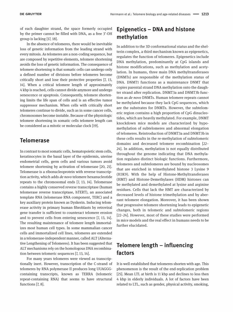
Finally, several nutritional factors like vitamins (including folate, nicotinamide, vitamin A, B12, D, C and E), minerals (including magnesium, zinc and iron) and other bioactive dietary components (like omega-3 fatty acid, polyphenols and curcumin), are able to directly or indirectly influence LTL trough several mechanisms. Recent studies have shown consistent associations between LTL and the availability of B and D vitamins [12, 27–38]. Preliminary results from own studies demonstrate that serum folate and its metabolites correlate with LTL
(Figure 3) [29]. In agreement with this observation also HCY, a functional marker of folate and vitamin B12 avail-ability, is consistently correlated with LTL (Figure 3). Anti-oxidant activity, DNA methylation and prevention of DNA damage are the most important mechanisms trough which these nutritional factors slow down telomere attrition [12, 27]. In summary, a healthy lifestyle with a diet rich in fruits and vegetables combined with exercise, lower BMI and no smoking is associated with longer telomeres.
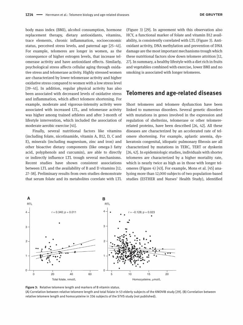
Telomeres and age-related diseasesShort telomeres and telomere dysfunction have been linked to numerous disorders. Several genetic disorders with mutations in genes involved in the expression and regulation of shelterins, telomerase or other telomere-related proteins, have been described [26, 42]. All these diseases are characterized by an accelerated rate of tel-omere shortening. For example, aplastic anemia, dys-keratosis congenital, idiopatic pulmonary fibrosis are all characterized by mutations in TERC, TERT or dyskerin [26, 42]. In epidemiologic studies, individuals with shorter telomeres are characterized by a higher mortality rate, which is nearly twice as high as in those with longer tel-omeres (Figure 4) [43]. For example, Mons et al. [45] ana-lyzing more than 12,000 subjects of two population-based studies (ESTHER and Nurses’ Health Study), identified
4
3
2
1
0
4
3
2
1
00 20 40
Total folate, nmol/L
A BRTL RTL
r = 0.340; p = 0.011 r = –0.128; p = 0.023
Homocysteine, µmol/L
60 5 10 15 20
Figure 3: Relative telomere length and markers of B vitamin status.(A) Correlation between relative telomere length and total folate in 53 elderly subjects of the KNOVIB study [29]. (B) Correlation between relative telomere length and homocysteine in 336 subjects of the STVS study (not published).
Herrmann et al.: Telomere biology and age-related diseases 1215
that subjects with shorter telomeres (1st quintile) were characterized by higher hazard ratio for all-cause mortal-ity (1.66, 95%CI 1.09–2.53, p = 0.018) compared to those with higher telomere length (5th quintile). In addition, a reduced LTL may indicate an existing or an elevated risk for future age-related disease, such as cardiovascular disease (CVD), type 2 diabetes mellitus (T2DM), neurode-generative diseases, osteoporosis and premature aging syndromes [42, 43, 46–49].
Osteoporosis
Similar to many other tissues, telomeres shorten with age in bone (osteoblasts) and mesenchymal stem cells (MSCs). In addition, premature aging syndromes, such as Werner syndrome (Wrn) and congenital dyskeratosis, are characterized by telomere dysfunction and osteopo-rosis. Because of these facts it has been hypothesized that telomere shortening might contribute to the aging of bone. A first large scale epidemiologic from Valdes et al. [44] showed a significant correlation between LTL and bone mineral density (BMD) at the spine and forearm. In this study, longer telomeres were associated with a reduced risk of clinical osteoporosis. The study included 2150 unselected women from a population-based twin cohort (age range 18–79 years). In women over the age of 50, the presence of osteoporosis was associated with shorter telomeres. The difference in LTL between women with and without osteoporosis was equivalent to five years of telomeric aging (Figure 4). In a small prospective observational study in elderly men (age range 71–86 years) LTL correlated with bone loss
over time at different distal forearm sites [50]. Animal studies in genetically modified mice suggest that repli-cative aging of osteoblast precursors promotes bone loss and the occurrence of senile osteoporosis [51]. However, several other human studies have not been able to show significant relationships between LTL, BMD and osteo-porosis [52–54]. Sanders et al. [54] studied 2750 commu-nity-dwelling women from the health, aging, and body composition study for 7 years without finding a signifi-cant relationship between LTL, BMD, bone loss, preva-lent and incident fractures. These results are supported by a similar study in 1867 community-dwelling Chinese women that did not find significant relationships between LTL, BMD and bone loss [53]. Furthermore, in vitro studies with human osteoblasts and the meas-urement of LTL from osteoporotic and control subjects do not support the concept of premature cellular aging and accelerated telomere shortening in patients with senile osteoporosis [55]. In conclusion, existing human data are inconsistent and the majority of studies did not find associations between LTL, BMD and osteoporosis.
CVD
CVD is among the most frequent age-related diseases and the number one cause of death. Several studies have shown that a high rate of telomere attrition is associated with an elevated risk of coronary artery disease, myocar-dial infarction, heart failure and stroke [47]. The large scale prospective WOSCOPS study demonstrated a 44% increase in coronary artery events during 5.5 years of fol-low-up in individuals in the first quartile of LTL (shortest
Decile of ∆LTL Sites with osteoporosis
A B C
Lengthening
Mor
talit
y, %
TR
F le
ngth
, kb
Kap
lan-
Mei
er s
urvi
val e
stim
ates
Shortening
Survival time, days
Shortened (–1012 to –169)
Shortened (114 to 1245)Maintained (–168 to 113 bp)
Figure 4: Telomere length, mortality and age-related disease.(A) Mortality by decile of 5-year change in telomere length (p for trend <0.001) [43]. (B) Telomere restriction fragment (TRF) length among healthy women aged 50 and older with clinical osteoporosis at one and two or more sites. Means and standard errors are adjusted for age, smoking status, body mass index, HRT and menopausal status [44]. (C) Survival in 608 patients with coronary heart disease, stratified by change in telomere length during the previous 5 years (overall log rank test p < 0.0001) [43].
1216 Herrmann et al.: Telomere biology and age-related diseases
telomeres) compared to individuals in the fourth quar-tile (longest telomeres) [56]. Individuals with early myo-cardial infarction have also been found with a reduced LTL compared to age matched healthy controls [57]. The LTL of patients who suffered a myocardial infarction is comparable to control individuals that are approximately 10 years older [57, 58]. GWAS studies have identified seven SNPs that are responsible for interindividual vari-ations in LTL. The presence of these alleles is associated with an increased CVD risk [59]. In a recent meta-analysis Codd et al. [59] calculated a 21% increase in CVD risk per one standard deviation reduction in LTL. LTL has also the potential to predict mortality in CVD patients [60]. In addition, LTL is related to the severity of CVD and disease progression.
Chronic inflammation and oxidative stress are the principal drivers in atherosclerosis. They cause acceler-ated telomere loss per cell replication and premature cel-lular senescence in endothelial cells, vascular smooth muscle cells (VSMC) and blood leukocytes [61]. The reduction of telomere length in VSMCs in human athero-sclerotic plaques is correlated with the severity of the disease. In addition, VSMCs in atherosclerotic plaques show signs of oxidative DNA damage and express typical senescence markers such as senescence-associated galactosidase, cyclin-dependent kinase inhibitors p16 and p21, decreased expression of cyclin D and cyclin E, and hypophosphorylation of the retinoblastoma protein [62]. Senescent VSMCs possess a limited proliferative capacity and an increased amount of matrix-degrading enzymes, which promote the thinning of fibrous caps and plaque rupture [63]. In agreement with this finding it has been shown that in patients with acute coronary syndrome short leukocyte telomeres are associated with the presence of highly unstable atherosclerotic plaques and an increased proinflammatory activity [64]. In an animal model long telomeres have been protec-tive against age-dependent cardiac disease caused by NOTCH1 haploinsufficiency [65].
Accelerated telomere shortening does not only promote atherosclerosis, it also impairs the repair of vas-cular lesions. Bone marrow-derived mononuclear cells are important progenitors during the re-endothelialization process of blood vessels. Functional exhaustion and an impaired proliferative capacity of endothelial progenitor cells due to accelerated telomere shortening contributes to delayed re-endothelialization after vascular injury and stent implantation [66].
Common risk factors for CVD, such as hypertension and T2DM, are also related to LTL. Reduced telomere length together with telomere uncapping was found
in patients with hypertension [67, 68]. The risk of being diagnosed with T2DM is also higher in individuals with shortened telomeres [48]. Furthermore, telomere shorten-ing seems to be associated with the progression of T2DM and the number of diabetic complications, such as retin-opathy, nephropathy, neuropathy and peripheral vascular disease.
Human and animal studies also suggest a critical role of telomerase in cardiovascular aging. TERT protein and telomerase activity are present in cardiomyocytes [69] and blood vessels [70]. Low TERT activity reduces vascular protection and promotes senescence, which ultimately leads to vascular dysfunction. Experimental studies have shown that eNOS and TERT interact with each other. Moreover, statins also influence TERT expression and tel-omerase activity in the vascular wall and in cells of the immune system [71].
Alzheimer’s disease (AD)
AD is the most common neurodegenerative disease associated with aging. Several studies showed shorter telomeres in blood leukocytes of AD patients than in controls [72–76]. A recent meta-analysis of 13 studies demonstrated a significant difference in LTL between 860 AD patients and 2022 controls. The authors con-cluded that there is consistent evidence of shorter tel-omeres in AD patients [74]. Panossian et al. [75] showed that the telomere length of T cells, but not of B cells or monocytes, correlate with AD disease status, measured by mini mental status exam (MMSE). Telomere length in T cells inversely correlated with serum levels of TNF-α (a clinical marker of disease status), with the proportion of CD8 + T cells lacking expression of the CD28 co-stimula-tory molecule, and with apoptosis. Interestingly, Lukens et al. [73] found similar telomere length in the cerebel-lum of AD patients and controls despite a significant difference in leukocyte TL. A recent study from Tedone et al. [76] suggests that telomere length in PBMCs may be helpful in predicting disease progression. Late onset AD patients with slow disease progression had shorter telomeres than those with fast disease progression or healthy elderly controls. In addition, AD patients with fast disease progression showed an impaired response to stimulation by amyloid ß and IL-10.
In contrast to AD, Parkinson’s disease (PD), another age-related neurodegenerative disease, is not consistently related to telomere length. Forero et al. [74] analyzed eight studies and did not find a significant difference in LTL between 956 PD patients and 1284 controls.

Herrmann et al.: Telomere biology and age-related diseases 1217
Cancer
Cancer is also considered an age-related disease, as its risk increases with age. Several studies have investigated the relationship between telomere length and cancer risk or prognosis [77–79]. Telomeres measured in tumor tissue from breast [80, 81], colon [82] and prostate [83] appear to be shorter than in healthy tissue from the same organ and the same patient. In healthy tissue directly adjacent to these tumors, telomeres are also shorter than in cells that are more distant from the cancerous lesion [84]. There is good evidence that a reduced telomere length in cancer tissue from breast colon and prostate is associated with an advanced disease state at diagnosis, faster disease progression and poorer survival [81, 85]. Another observation is that patients with the highest variation of telomere length between individual cells have the highest risk of death. Although these findings are interesting the measurement of telomere length in tumor tissue harbors substantial practical limitations that prevent its use as a biomarker in clinical practice. In order to overcome these issues LTL has been proposed as a surrogate marker for telomere shortening in the entire body. A number of studies found an association between accelerated telomere shortening in leukocytes and an increased risk of incident cancer. For example, in the normative Aging Study serial telomere measurements were performed in nearly 800 cancer free individuals. The annual telomere shortening rate in subjects that developed cancer during the follow-up was double as high as in individuals without cancer [86]. In the 10-year prospective Bruneck study patients with short telom-eres at baseline had substantially higher risk for inci-dent cancer and cancer specific mortality [46]. However, the results of prospective studies are inconsistent. For example, in a very large population study from Norway with >47,000 participants short telomeres were associ-ated with incident cancers [87]. This effect disappeared after adjustment for potential confounders. There are also studies that found significantly longer telomeres in blood leukocytes from cancer patients when compared to cancer free subjects [88]. Meta-analyses might help to better understand the data from existing studies, but results are inconclusive. A meta-analysis of retrospec-tive studies shows an association between short telom-eres and an increased risk for most solid organ tumors [89]. By contrast, a recent meta-analysis of prospective studies does not confirm this result [90].
The controversial results might be explained by the induction of telomerase at a specific point in time during tumor genesis. The Normative Aging Study has shown a
faster decline of LTL in cancer patients than in cancer free subjects [86]. However, as cancer diagnosis comes closer, the shortening of telomeres slows down and ultimately they become longer again. It has been suggested that criti-cally short telomeres contribute to cancer initiation which then, in turn, activates telomerase.
Although there is substantial evidence for an asso-ciation between LTL and age-related diseases, neither a conclusive causative link nor a predictable associa-tion can be established. Longitudinal studies as well as assessment of other markers of telomere biology are needed to further clarify the role of telomeres and tel-omerase in aging and the development of age-related diseases.
Leukocyte telomere as a surrogate marker for telomere shortening in the entire bodyMeasurement of telomere length in tumor tissue harbors substantial practical limitations that prevent its use as a biomarker in clinical practice. The most important issue is that the analysis can only be performed after a tumor has been diagnosed. Furthermore, an invasive biopsy is necessary to obtain a sample. Because of the invasive sample collection, serial measurements are not feasible. In order to overcome these obstacles, the measurement of LTL in blood leukocytes has been proposed as a surrogate marker. In blood leukocytes, telomeres shorten with an average annual rate of 30–35 bp. As this places leukocytes in the middle of all tissue types, it has been proposed that LTL might be a suitable surrogate marker for telomere shortening in the entire body.
LTL is often used in clinical studies. However, doubts remain to what extent this parameter reflects the situa-tion in other organs and tissues. Until today, only few studies have explored the relationship between telomere length in different cell types and results are not so clear [91–95]. For example, Dlouha et al. [91] measured tel-omere length in 12 different human tissues (peripheral blood leukocytes, liver, kidney, heart, spleen, brain, skin, triceps, tongue mucosa, intercostal skeletal muscle, subcutaneous fat and abdominal fat) from 12 cadavers. Telomere length was significantly higher in blood com-pared to liver, brain, muscles, skin, spleen and mucosa but was not different compared to adipose and renal tissue. In addition, the largest telomere length variability was observed in leukocytes and kidney, and the smallest
1218 Herrmann et al.: Telomere biology and age-related diseases
telomere length variability was observed in brain, heart and skin [91]. In another study, Friedrich et al. [93] reported a linear relationships between telomere length measured in blood, skin and synovia of nine elderly patients. In this study, significantly shorter telomeres were found in leukocytes compared to skin and synovia [93]. However, in this study, age of patients ranged from 73 to 95 years. Telomere length from leukocytes and from skin correlated negatively with age [93]. Richard et al. [94], who analyzed telomere length in aortic biopsies and paired blood leukocyte from 20 patients, identified that shorter blood telomeres reflect shorter aortic telom-eres, indicating that telomere attrition in blood leuko-cytes is indicative of similar changes in the vascular well. Altogether these findings could reflect the expected life span of different cell types. One aspect to consider is that senescent cells are eliminated by apoptosis so that only a few or no senescent cells persist in vivo. On the other site, the stem cell compartment of a tissue is enriched in cells with longer telomeres, as they express telomerase and therefore avoid telomere shortening. Indeed, longer telomeres and higher telomerase activity were found in several stem cell compartments, including hair follicle, skin, small intestine, cornea and testis [95]. Finally, it has also been speculated that a common genetically con-trolled mechanism regulates telomere length differently in various tissues [93].
In summary, available data suggest that telomere length is correlated between different tissues in the same individual and that peripheral blood telomere length could be used as a surrogate marker of telomere length in other organs [91–95].
Analytical aspectsTelomeres can be measured in all solid organ tissues and blood leukocytes. Although the analysis of telomere length in the tissue of interest is most representative, the invasive sample collection procedure and other practi-cal aspects often render this approach not feasible for routine use. Because of the simple and non-invasive spec-imen collection, blood leukocytes are the most widely used matrix for clinical and research purposes. Telomere length can be measured with a range of different methods [95–103]. Generally, methods that use isolated DNA have to be distinguished from those using intact cells. South-ern blotting and quantitative-polymerase chain reaction (Q-PCR) are the analytical principals that are applied for the analysis in isolated DNA. The Southern blot
technique was the first method that has been developed and is still considered the gold standard for the meas-urement of telomere length [96]. In the first step, DNA is digested by endonucleases that do not cut within the telomeric TTAGGG sequence. Subsequently, the digested DNA is separated by gel electrophoresis. Telomeres are visualized by labeled DNA probes that are complemen-tary to the telomere sequence. The longer the telomere, the more probes can bind, resulting in a higher signal intensity [96]. The obtained signal represents the average telomere length of all chromosomes from cells in the sample. It provides an absolute value of telomere length expressed in base pair, requires a substantial quantity of DNA (i.e. micrograms), is time consuming and is not suit-able for large population-based studies. However, as the analytical principle of this hybridization-based method, it implies a decreasing signal as telomere become shorter. There is a threshold telomere length below which no detectable signal will be generated. The inability to detect very short telomeres limits the utility of this method in studies of cellular aging, where short telomeres are of particular interest [96]. The Q-PCR-based assay requires the amplification of the telomeric region and of a single copy gene (SCG) [97, 98]. Telomere length is quantified as the amount of telomeric sequence compared to SCG and is expressed as a relative telomere length (RTL) or telomere/SCG (T/S) ratio [97, 98]. As the Southern blot method, it is not possible to evaluate telomere length of individual cells or chromosomes. However, the Q-PCR-based technique is fast, highly sensitive and requires low amounts of DNA (i.e. nanograms). Because of these characteristics, it is often chosen for large population-based studies [97, 98]. At the cellular level, telomere length can be evaluated by quantitative fluorescence in situ hybridization (Q-FISH) [100] or by flow cytometry (flow-FISH) [101]. Q-FISH can be combined with a conventional FISH, allowing the eval-uation of chromosome-specific telomere length. Similarly to the Southern blot-based method described above, Q-FISH is also a hybridization technique, which implies the existence of a threshold telomere length below which no detectable signal is produced. As Q-FISH is performed on cells in metaphase, the analysis is limited to cells that proliferate in vitro. Consequently, not all cells are suitable to be analyzed with this method. The confocal telomere Q-FISH method (Telomapping) allows the quantification of telomere length in tissue samples or biopsies [95].
More work is needed for the harmonization of tel-omere length assessment between laboratories, includ-ing the development of a common standard reference, of internal and external quality control programs and of ref-erence ranges stratified for age and gender.
Herrmann et al.: Telomere biology and age-related diseases 1219
ConclusionsTelomeres and telomerase play an important role during the process of aging. Short telomeres in various tissues and in blood leukocytes are associated with age-related diseases, such as CVD, Alzheimer’s disease and cancer. Although a number of studies have proposed the measurement of LTL as a predictor of incident age-related diseases, disease progression and mortality, it is still far from being used in clinical practice. Ana-lytical issues (lack of standardization) and pathophysi-ological aspects that are insufficiently understood are the main obstacles that impede the widespread use of telomere length measurement. More work is needed for the harmonization of telomere length assessment between laboratories, including the development of a common standard reference, of internal and external quality control programs and of reference ranges strati-fied for age and gender. In addition, a range of preana-lytical aspects need to be clarified. For example, under what circumstances the measurement of LTL should be ordered? When should the blood sample be collected? So far, the evaluation of telomere length will be reason-able only in research programs, clinical trials, longitudi-nal and cross-sectional clinical studies.
Author contributions: All the authors have accepted responsibility for the entire content of this submitted manuscript and approved submission.Research funding: None declared.Employment or leadership: None declared.Honorarium: None declared.Competing interests: The funding organization(s) played no role in the study design; in the collection, analysis, and interpretation of data; in the writing of the report; or in the decision to submit the report for publication.
References1. Benjamin H. Biological versus chronologic age. J Gerontol
1947;2:217–27.2. Ries W, Sauer I, Junker B, Poething D, Schwerdtner U.
Methodological problems in the determination of biological age. Z Gesamte Inn Med 1976;31:109–13.
3. Comfort A. Test battery to measure aging rate in men. Lancet 1969;2:1411–5.
4. Heikkinen E, Kiiskinen A, Kaeyhty B, Rimpela M, Vuori I. Assess-ment of biological age. Methodological study of two Finnish populations. Gerontologia 1974;20:33–43.
5. Vijg J, Suh Y. Genome instability and aging. Annu Rev Physiol 2013;75:645–68.
6. Lombard DB, Chua KF, Mostoslavsky R, Franco S, Gostissa M, Alt FW. DNA repair, genome stability, and aging. Cell 2005;120:497–512.
7. Blasco MA. Telomere length, stem cells and aging. Nat Chem Biol 2007;3:640–9.
8. Blasco MA. Telomeres and human disease: ageing, cancer and beyond. Nat Rev Genet 2005;6:611–22.
9. Müller HJ. The remaking of chromosomes. Collecting Net 1938;8:182–98.
10. McClintock, B. The stability of broken ends of chromosomes in Zea mays. Genetics 1941:26;234–82.
11. Moyzis RK, Buckingham JM, Cram LS, Dani M, Deaven L, Jones MD, et al. A highly conserved repetitive DNA sequence, (TTAGGG)n, present at the telomeres of human chromosomes. Proc Natl Acad Sci USA 1988;85:6622–6.
12. Pusceddu I, Farrell CJ, Di Pierro AM, Jani E, Herrmann W, Herrmann M. The role of telomeres and vitamin D in cellular aging and age-related diseases. Clin Chem Lab Med 2015;53:1661–78.
13. Blackburn EH. Telomeres and telomerase: the means to the end(Nobel-lecture). Angew Chem Int Ed Engl 2010;49:7405–21.
14. Blackburn EH. Switching and signaling at the telomere. Cell 2001;106:661–73.
15. Sandell LL, Zakian VA. Loss of a yeast telomere: arrest, recovery, and chromosome loss. Cell 1999;75:729–39.
16. de Lange T. Shelterin: the protein complex that shapes and safeguards human telomeres. Genes Dev 2005;19:2100–10.
17. Olovnikov AM. A theory of marginotomy. J Theor Biol 1973;41:181–90.
18. Watson JD. Origin of concatemeric T7 DNA. Nat New Biol 1972;239:197–201.
19. Allsopp RC, Vazri H, Patterson C, Goldestein S, Younglai EV, Futcher AB, et al. Telomere length predicts replicative capacity of human fibroblasts. Proc Natl Acad Sci USA 1992;89: 10114–8.
20. Counter CM, Avilion AA, LeFeuvre CE, Stewart NG, Greider CW, Harley CB, et al. Telomere shortening associated with chromo-some instability is arrested in immortal cells which express telomerase activity. EMBO J 1992;11:1921–9.
21. Shay JW, Van Der Haegen BA, Ying Y, Wright WE. The frequency of immortalization of human fibroblasts and mammary epithe-lial cells transfected with SV40 large T-antigen. Exp Cell Res 1993;209:45–52.
22. Blasco MA. Mice with bad ends: mouse models for the study of telomeres and telomerase in cancer and aging. EMBO J 2005;24:1095–103.
23. Blasco MA. The epigenetic regulation of mammalian telomeres. Nat Rev Genet 2007;8:299–309.
24. Gonzalo S, Jaco I, Fraga MF, Li E, Esteller M, Blasco MA. Nat Cell Biol 2006;8:416–24.
25. Cawthon RM, Smith KR, O’Brien E, Sivatchenko A, Kerber RA. Association between telomere length in blood and mortality in people aged 60 years or older. Lancet 2003;361:393–5.
26. Armanios M. Telomeres and age-related disease: how telomere biology informs clinical paradigms. J Clin Invest 2013;123: 996–1002.
27. Paul L. Diet, nutrition and telomere length. J Nutr Biochem 2011;22:895–901.
28. Pusceddu I, Herrmann M, Kirsch SH, Werner C, Huebner U, Bodis M, et al. Prospective study of telomere length and LINE-1
1220 Herrmann et al.: Telomere biology and age-related diseases
methylation in peripheral blood cells: the role of B vitamins sup-plementation. Eur J Nutr 2016;55:1863–73.
29. Pusceddu I, Herrmann M, Kirsch SH, Werner C, Huebner U, Bodis M, et al. One-carbon metabolites and telomere length in a prospective and randomized study of B- and/or D-vitamin sup-plementation. Eur J Nutr 2017;56:1887–98.
30. Richards JB, Valdes AM, Gardner JP, Paximadas D, Kimura M, Nessa A, et al. Higher serum vitamin D concentrations are associated with longer leukocyte telomere length in women. Am J Clin Nutr 2007;86:1420–5.
31. Borras M, Panizo S, Sarro F, Valdivielso JM, Fernandez E. Assessment of the potential role of active vitamin D treatment in telomere length: a case-control study in hemodialysis patients. Clin Ther 2012;34:849–56.
32. Zhu H, Guo D, Li K, Pedersen-White J, Stallmann-Jorgensen IS, Huang Y, et al. Increased telomerase activity and vitamin D sup-plementation in overweight African Americans. Int J Obes (Lond) 2012;36:805–9.
33. Hoffecker BM, Raffield LM, Kamen DL, Nowling TK. Systemic lupus erythematosus and vitamin D deficiency are associated with shorter telomere length among African Americans: a case-control study. PLoS One 2013;8:e63725.
34. Liu JJ, Prescott J, Giovannucci E, Hankinson SE, Rosner B, Han J, et al. Plasma vitamin D biomarkers and leukocyte telomere length. Am J Epidemiol 2013;177:1411–7.
35. Richards JB, Valdes AM, Gardner JP, Kato BS, Siva A, Kimura M, et al. Homocyteine levels and leukocyte telomere length. Atherosclerosis 2008;200:271–7.
36. Paul L, Cattaneo M, D’Angelo A, Sampietro F, Fermo I, Razzari C, et al. Telomere length in peripheral blood mononuclear cells is associated with folate status in men. J Nutr 2009;139:1273–8.
37. Liu JJ, Prescott J, Giovannucci E, Hankinson SE, Rosner B, De Vivo I. One-carbon metabolism factors and leukocyte telomere length. Am J Clin Nutr 2013;97:794–9.
38. Paul L, Jacques PF, Aviv A, Vasan RS, D’Agostino RB, Levy D, et al. High plasma folate is negatively associated with leukocyte telomere length in the Framingham offspring cohort. Eur J Nutr 2015;54:235–41.
39. Werner C, Fürster T, Widmann T, Pöss J, Roggia C, Hanhoun M, et al. Physical exercise prevents cellular senescence in circulating leukocytes and in the vessel wall. Circulation 2009;120:2438–47.
40. Epel ES, Balckburn EH, Lin J, Dhabhar FS, Adler NE, Morrow JD, et al. Accelerated telomere shortening in response to life stress. Proc Natl Acad Sci USA 2004;101:17312–5.
41. Ornish D, Lin J, Daubenmier J, Weidnwer G, Epel E, Kemp C, et al. Increased telomerase activity and comprehensive lifestyle changes: a pilot study. Lancet Oncol 2008;9:1048–57.
42. Armanios M, Blackburn EH. The telomere syndromes. Nat Rev Genet 2012;13:693–704.
43. Goglin SE, Farzaneh-Far R, Epel ES, Lin J, Blackburn EH, Whooley MA. Change in leukocyte telomere length predicts mortality in patients with stable coronary heart; disease from the heart and soul study. PLoS One 2016;11:e0160748.
44. Valdes AM, Richards JB, Gardner JP, Swaminathan R, Kimura M, Xiaobin L, et al. Telomere length in leukocytes correlates with bone mineral density and is shorter in women with osteoporo-sis. Osteoporos Int 2007;18:1203–10.
45. Mons U, Muezzinler A, Schoettker B, Dieffenbach AK, Butterbach K, Schick M, et al. Leukocyte telomere length and
all-cause mortality, cardiovascular disease, and cancer mortal-ity: results from individual-participant-data meta-analysis of 2 large prospective cohort studies. Am J Epidemiol 2017;185: 1317–26.
46. Willeit P, Willeit J, Mayr A, Weger S, Oberhollenzer F, Brandstätter A, et al. Telomere length and risk of incident cancer and cancer mortality. J Am Med Assoc 2010;304:69–75.
47. Willeit P, Willeit J, Brandstätter A, Ehrlenbach S, Mayr A, Gasperi A, et al. Cellular aging reflected by leukocyte telomere length predicts advanced atherosclerosis and cardiovascular disease risk. Arterioscler Thromb Vasc Biol 2010;30:1649–56.
48. Testa R, Olivieri F, Sirolla C, Spazzafumo L, Rippo MR, Marra M, et al. Leukocyte telomere length is associated with complica-tions of type 2 diabetes mellitus. Diabet Med 2011;28:1388–94.
49. Zhao J, Miao K, Wang H, Ding H, Wang DW. Association between telomere length and type 2 diabetes mellitus: a meta-analysis. PLoS One 2013;8:e79993.
50. Bekaert S, Van Pottelbergh I, De Meyer T, Zmierczak H, Kaufman JM, Van Oostveldt P, et al. Telomere length versus hormonal and bone mineral status in healthy elderly men. Mech Ageing Dev 2005;126:1115–22.
51. Saeed H, Abdallah BM, Ditzel N, Catala-Lehnen P, Qiu W, Amling M, et al. Telomerase-deficient mice exhibit bone loss owing to defects in osteoblasts and increased osteoclastogen-esis by inflammatory microenvironment. J Bone Miner Res 2011;26:1494–505.
52. Pignolo RJ, Suda RK, McMillan EA, Shen J, Lee SH, Choi Y, et al. Defects in telomere maintenance molecules impair osteo-blast differentiation and promote osteoporosis. Aging Cell 2008;7:23–31.
53. Tang NL, Woo J, Suen EW, Liao CD, Leung JC, Leung PC. The effect of telomere length, a marker of biological aging, on bone mineral density in elderly population. Osteoporos Int 2010;21:89–97.
54. Sanders JL, Cauley JA, Boudreau RM, Zmuda JM, Strotmeyer ES, Opresko PL, et al. Leukocyte telomere length is not associated with BMD, osteoporosis, or fracture in older adults: results from the health, aging and body composition study. J Bone Miner Res 2009;24:1531–6.
55. Kveiborg M, Kassem M, Langdahl B, Eriksen EF, Clark BF, Rattan SI. Telomere shortening during aging of human osteo-blasts in vitro and leukocytes in vivo: lack of excessive telomere loss in osteoporotic patients. Mech Ageing Dev 1999;106: 261–71.
56. Brouilette SW, Moore JS, McMahon AD, Thompson JR, Ford I, Shepherd J, et al. Telomere length, risk of coronary heart disease, and statin treatment in the West of Scotland Primary Prevention Study: a nested case-control study. Lancet 2007;369:107–14.
57. Brouilette S, Singh RK, Thompson JR, Goodall AH, Samani NJ. White cell telomere length and risk of premature myocardial infarction. Arterioscler Thromb Vasc Biol 2003;23:842–6.
58. Haycock PC, Heydon EE, Kaptoge S, Butterworth AS, Thompson A, Willeit P. Leucocyte telomere length and risk of cardiovas-cular disease: systematic review and meta-analysis. Br Med J 2014;349:g4227.
59. Codd V, Nelson CP, Albrecht E, Mangino M, Deelen J, Buxton JL, et al. Identification of seven loci affecting mean tel-omere length and their association with disease. Nat Genet 2013;45:422–7.
Herrmann et al.: Telomere biology and age-related diseases 1221
60. Carty CL, Kooperberg C, Liu J, Herndon M, Assimes T, Hou L, et al. Leukocyte telomere length and risks of incident coronary heart disease and mortality in a racially diverse population of postmenopausal women. Arterioscler Thromb Vasc Biol 2015;35:2225–31.
61. Yeh JK, Wang CY. Telomeres and telomerase in cardiovascular diseases. Genes 2016;7:58.
62. Matthews C. VSMCs undergo telomere-based senescence in human atherosclerosis: effects of telomerase and oxidative stress. Circ Res 2006;99:156–64.
63. Gorenne I. Vascular smooth muscle cell senescence in athero-sclerosis. Cardiovasc Res 2006;72:9–17.
64. Calvert PA, Liew TV, Gorenne I, Clarke M, Costopoulos C, Obaid DR, et al. Leukocyte telomere length is associated with high-risk plaques on virtual histology intravascular ultrasound and increased proinflammatory activity. Arterioscler Thromb Vasc Biol 2011;31:2157–64.
65. Theodoris CV, Mourkioti F, Huang Y, Ranade SS, Liu L, Blau HM, et al. Long telomeres protect against age-dependent cardiac disease caused by Notch1 haploinsufficiency. J Clin Invest 2017;127:1683–8.
66. Armstrong EJ, Xing L, Zhang J, Zheng Y, Shunk KA, Yeh RW, et al. Association between leukocyte telomere length and drug-elut-ing stent strut coverage by optical coherence tomography. J Am Coll Cardiol 2012;59:2218–9.
67. Jeanclos E, Schork NJ, Kyvik KO, Kimura M, Skurnick JH, Aviv A. Telomere length inversely correlates with pulse pressure and is highly familial. Hypertension 2000;36:195–200.
68. Morgan RG, Ives SJ, Walker AE, Cawthon RM, Andtbacka RH, Noyes D, et al. Role of arterial telomere dysfunction in hyperten-sion: relative contributions of telomere shortening and telomere uncapping. J Hypertens 2014;32:1293–9.
69. Richardson GD, Breault D, Horrocks G, Cormack S, Hole N, Owens WA. Telomerase expression in the mammalian heart. FASEB J 2012;26:4832–40.
70. Haendeler J. Antioxidants inhibit nuclear export of telomer-ase reverse transcriptase and delay replicative senescence of endothelial cells. Circ Res 2004;94:768–75.
71. Boccardi V, Barbieri M, Rizzo MR, Marfella R, Esposito A, Marano L, et al. A new pleiotropic effect of statins in elderly: modulation of telomerase activity. FASEB J 2013;27:3879–85.
72. Thomas P, O’Callaghan NJ, Fenech M. Telomere length in white blood cells, buccal cells and brain tissue and its varia-tion with ageing and Alzheimer’s disease. Mech Ageing Dev 2008;129:183–90.
73. Lukens JN, Van Deerlin V, Clark CM, Xie SX, Johnson FB. Co mparisons of telomere lengths in peripheral blood and cer-ebellum in Alzheimer’s disease. Alzheimers Dement 2009;5: 463–9.
74. Forero DA, Gonzalez-Giraldo Y, Lopez-Quintero C, Castro-Vega LJ, Barreto GE, Perry G. Meta-analysis of telomere length in Alzhei-mer’s Disease. J Gerontol A Biol Sci Med Sci 2016;71:1069–73.
75. Panossian LA, Porter VR, Valenzuela HF, Zhu X, Reback E, Ma sterman D, et al. Telomere shortening in T cells correlates with Alzheimer’s disease status. Neurobiol Aging 2003;24:77–84.
76. Tedone E, Arosio B, Colombo F, Ferri E, Asselineau D, Piette F, et al. Leukocyte telomere length in Alzheimer’s disease patients with a different rate of progression. J Alzheimers Dis 2015;46:761–9.
77. Svenson U, Roos G. Telomere length as a biological marker in malignancy. Biochim Biophys Acta 2009;1792:317–23.
78. Hou L, Zhang X, Gawron AJ, Liu J. Surrogate tissue telomere length and cancer risk: shorter or longer? Cancer Lett 2012;319:130–5.
79. Wu X, Amos CI, Zhu Y, Zhao H, Grossman BH, Shay JW, et al. Telomere dysfunction: a potential cancer predisposition factor. J Natl Cancer Inst 2003;95:1211–8.
80. Odagiri E, Kanada N, Jibiki K, Demura R, Aikawa E, Demura H. Reduction of telomeric length and c-erbB-2 gene amplification in human breast cancer, fibroadenoma, and gynecomastia. Relationship to histologic grade and clinical parameters. Cancer 1994;73:2978–84.
81. Fordyce CA, Heaphy CM, Bisoffi M, Wyaco JL, Joste NE, Ma ngalik A, et al. Telomere content corretates with stage and prognosis in breast cancer. Breast Cancer Res Treat 2006;99:193–202.
82. Nakamura K, Furugori E, Esaki Y, Arai T, Sawabe M, Okayasu I, et al. Correlation of telomere lengths in normal and cancer t issue in the large bowel. Cancer Lett 2000;158:179–84.
83. Heaphy CM, Fleet TM, Treat EG, Lee SJ, Smith AY, Davis MS, et al. Organ-wide telomeric status in diseased and disease-free prostatic tissues. Prostate 2010;70:1471–9.
84. Heaphy CM, Gaonkar G, Peskoe SB, Joshu CE, De Marzo AM, Lucia MS, et al. Prostate stromal cell telomere shortening is associated with risk of prostate cancer in the placebo arm of the Prostate Cancer Prevention Trial. Prostate 2015;75:1160–6.
85. Valls C, Pinol C, Rene JM, Buenestado J, Vinas J. Telomere length is a prognostic factor for overall survival in colorectal cancer. Colorectal Dis 2011;13:1265–72.
86. Hou L, Joyce BT, Gao T, Liu L, Zheng Y, Penedo FJ, et al. Blood t elomere length attrition and cancer development in the Normative Aging Study Cohort. EBioMedicine 2015;2:591–6.
87. Weischer M, Nordestgaard BG, Cawthon RM, Freiberg JJ, Tybjaerg-Hansen A, Bojesen SE. Short telomere length, cancer survival, and cancer risk in 47102 individuals. J Natl Cancer Inst 2013;105:459–68.
88. Julin B, Shui I, Heaphy CM, Joshu CE, Meeker AK, Giovannucci E, et al. Circulating leukocyte telomere length and risk of overall and aggressive prostate cancer. Br J Cancer 2015;112:769–76.
89. Wentzensen IM, Mirabello L, Pfeiffer RM, Savage SA. The asso-ciation of telomere length and cancer: a meta-analysis. Cancer Epidemiol Biomarkers Prev 2011;20:1238–50.
90. Zhang X, Zhao Q, Zhu W, Xie SH, Zhong LX, Cai YY, et al. The association of telomere length in peripheral blood cells with cancer risk: a systemic review and meta-analysis of prospective studies. Cancer Epidemiol Biomarkers Prev 2017;26: 1381–90.
91. Dlouha D, Maluskova J, Kralova Lesna I, Lanska V, Hubacek JA. Comparison of the relative telomere length measured in leukocytes and eleven different human tissues. Physiol Res 2014;63:343–50.
92. Takubo K, Aida J, Izumiyama-Shimomura N, Ishikawa N, Sawabe M, Kurabayashi R, et al. Changes of telomere length with aging. Geriatr Gerontol Int 2010;10(Suppl 1):S197–206.
93. Friedrich U, Griese E, Schwab M, Fritz P, Thon K, Klotz U. Telomere length in different tissues of elderly patients. Mech Ageing Dev 2000;119:89–99.
94. Richard W, Wilson W, Herbert KE, Mistry Y, Stevens SE, Patel HR, et al. Blood leucocyte telomere DNA content predicts vascular
1222 Herrmann et al.: Telomere biology and age-related diseases
telomere DNA content in humans with and without vascular disease. Euro Heart J 2008;29:2689–94.
95. Flores I, Canela A, Vera E, Tejera A, Cotsarelis G, Blasco AM. The longest telomeres: a general signature of adult stem cell compartments. Genes Dev 2008;22:654–67.
96. Baird DM. New developments in telomere length analysis. Exp Gerontol 2005;40:363–8.
97. Cawthon RM. Telomere measurement by quantitative PCR. Nucleic Acids Res 2002;30:2–6.
98. Cawthon RM. Telomere length measurement by a novel mono-chrome multiplex quantitative PCR method. Nucleic Acids Res 2009;37:1–7.
99. O’Callaghan NJ, Fenech M. A quantitative PCR method for meas-uring absolute telomere length. Biol Proced Online 2011;13:1–13.
100. Poon SS, Lansdrop PM. Quantitative fluorescence in situ hybridization (Q-FISH). Curr Protoc Cell Biol 2001;12:1–21.
101. Canela A, Vera E, Klatt P, Blasco MA. High- troughput telomere length quantification by FISH and its applica-tion to human population studies. Proc Natl Acad Sci USA 2007;104:5300–5.
102. Martin-Ruiz CM, Baired D, Roger L, Boukamp P, Krunic D, Cawthon R et al. Reproducibility of telomere length assess-ment: an international collaborative study. Int J Epidemiol 2015;44:1673–83.
103. Nielsen BR, Linneberg A, Bendix L, Harboe M, Christensen K, Schwarz P. Association between leukocyte telomere length and bone mineral density in women 25–93 years of age. Exp Gerontol 2015;66:25–31.