Meiotic Telomere Protein Ndj1p Is Required for Meiosis-specific Telomere Distribution, Bouquet...
12
The Rockefeller University Press, 0021-9525/2000/10/95/12 $5.00 The Journal of Cell Biology, Volume 151, Number 1, October 2, 2000 95–106 http://www.jcb.org/cgi/content/full/151/1/95 95 Meiotic Telomere Protein Ndj1p Is Required for Meiosis-specific Telomere Distribution, Bouquet Formation and Efficient Homologue Pairing Edgar Trelles-Sticken,* Michael E. Dresser, ‡ and Harry Scherthan* § *Department of Human Biology and Genetics, University of Kaiserslautern, D-67653 Kaiserslautern, Germany; ‡ Program in Molecular and Cell Biology, Oklahoma Medical Research Foundation, Oklahoma City, Oklahoma 73104; and § Department of Cell Biology, University of Kaiserslautern, D-67653 Kaiserslautern, Germany Abstract. We have investigated the requirements for NDJ1 in meiotic telomere redistribution and clustering in synchronized cultures of Saccharomyces cerevisiae. On induction of wild-type meiosis, telomeres disperse from premeiotic aggregates over the nuclear periphery, and then cluster near the spindle pole body (bouquet arrangement) before dispersing again. In ndj1D meio- cytes, telomeres are scattered throughout the nucleus and fail to form perinuclear meiosis-specific distribu- tion patterns, suggesting that Ndj1p may function to tether meiotic telomeres to the nuclear periphery. Since ndj1D meiocytes fail to cluster their telomeres at any prophase stage, Ndj1p is the first protein shown to be required for bouquet formation in a synaptic organism. Analysis of homologue pairing by two-color fluores- cence in situ hybridization with cosmid probes to re- gions on III, IX, and XI revealed that disruption of bouquet formation is associated with a significant delay (.2 h) of homologue pairing. An increased and persis- tent fraction of ndj1D meiocytes with Zip1p polycom- plexes suggests that chromosome polarization is impor- tant for synapsis progression. Thus, our observations support the hypothesis that meiotic telomere clustering contributes to efficient homologue alignment and syn- aptic pairing. Under naturally occurring conditions, bouquet formation may allow for rapid sporulation and confer a selective advantage. Key words: bouquet fluorescence in situ hybridization • ndj1 • chromosome pairing • meiosis • telomeres Introduction The ends of eukaryotic chromosomes (telomeres) consist of simple DNA repeats associated with proteins and play important roles in replication, malignant transformation, and cellular aging (for reviews, see Blackburn, 1991; Zakian, 1995; de Lange, 1998a; Shore, 1998). Besides the vital functions telomeres perform in vegetative cells, they have been implicated as key players in the chromosome pairing process during meiosis (Moses, 1968; Loidl, 1990; Dernburg et al., 1995; Roeder, 1997; de Lange, 1998b; Zickler and Kleckner, 1998). The pairing of homologues and their recombination during meiotic prophase is critical for reduction of DNA content from diploid to haploid be- fore fertilization (von Wettstein et al., 1984; Hawley, 1988). In many organisms, the premeiotic (vegetative) telo- mere distribution is altered during the onset of prophase I such that telomeres attach to the inner nuclear membrane, and then move along it to cluster in a limited nuclear enve- lope sector at the onset of zygotene (von Wettstein et al., 1984). This bouquet formation is thought to contribute to homologue alignment and pairing during first meiotic prophase (e.g., Therman and Sarto, 1977; Fussell, 1987; Scherthan, 1997; Zickler and Kleckner, 1998). In Schizosaccharomyces pombe, loss of the telomere protein taz1(1) has been shown to abrogate meiotic telo- mere clustering and to virtually eliminate recombination and ordered disjunction of chromosomes at metaphase I in this asynaptic organism (Cooper et al., 1998; Nimmo et al., 1998). Furthermore, abrogation of nuclear movements during and after telomere clustering impairs chromosome pairing at meiosis (Yamamoto et al., 1999). An essential role for telomeres in the reductional division is underlined by the observation that deletion of telomeres and the re- sulting rearrangement of linear chromosomes into rings is compatible with vegetative growth, but obstructs meiosis and hence sexual reproduction (Naito et al., 1998; Ish- ikawa and Naito, 1999). Numerous cytogenetic studies have shown that a con- gregation of telomeres occurs in the vicinity of the cen- trosome or spindle pole body (SPB), 1 during the lepto- tene/zygotene transition (bouquet formation; for review, Address correspondence to Harry Scherthan, Dept. of Cell Biology, Uni- versity of Kaiserslautern, P.O. Box 3049, D-67653 Kaiserslautern, Ger- many. Tel.: 49 631 205 3313. Fax: 49 631 205 2878. E-mail: scherth @rhrk.uni-kl.de 1 Abbreviations used in this paper: cos, cosmid; FISH, fluorescence in situ hybridization; HA, hemagglutinin; IF, immunofluorescence; SPB, spindle pole body. on December 22, 2013 jcb.rupress.org Downloaded from Published October 2, 2000
-
Upload
independent -
Category
Documents
-
view
1 -
download
0
Transcript of Meiotic Telomere Protein Ndj1p Is Required for Meiosis-specific Telomere Distribution, Bouquet...

The Rockefeller University Press, 0021-9525/2000/10/95/12 $5.00The Journal of Cell Biology, Volume 151, Number 1, October 2, 2000 95–106http://www.jcb.org/cgi/content/full/151/1/95 95
Meiotic Telomere Protein Ndj1p Is Required for Meiosis-specific Telomere Distribution, Bouquet Formation and Efficient Homologue Pairing
Edgar Trelles-Sticken,* Michael E. Dresser,
‡
and Harry Scherthan*
§
*Department of Human Biology and Genetics, University of Kaiserslautern, D-67653 Kaiserslautern, Germany;
‡
Program in Molecular and Cell Biology, Oklahoma Medical Research Foundation, Oklahoma City, Oklahoma 73104; and
§
Department of Cell Biology, University of Kaiserslautern, D-67653 Kaiserslautern, Germany
Abstract.
We have investigated the requirements for
NDJ1
in meiotic telomere redistribution and clusteringin synchronized cultures of
Saccharomyces cerevisiae
.On induction of wild-type meiosis, telomeres dispersefrom premeiotic aggregates over the nuclear periphery,and then cluster near the spindle pole body (bouquet
arrangement) before dispersing again. In
ndj1
D
meio-cytes, telomeres are scattered throughout the nucleusand fail to form perinuclear meiosis-specific distribu-tion patterns, suggesting that Ndj1p may function totether meiotic telomeres to the nuclear periphery. Since
ndj1
D
meiocytes fail to cluster their telomeres at anyprophase stage, Ndj1p is the first protein shown to berequired for bouquet formation in a synaptic organism.Analysis of homologue pairing by two-color fluores-
cence in situ hybridization with cosmid probes to re-gions on
III, IX,
and
XI
revealed that disruption ofbouquet formation is associated with a significant delay
(
.
2 h) of homologue pairing. An increased and persis-tent fraction of
ndj1
D
meiocytes with Zip1p polycom-plexes suggests that chromosome polarization is impor-tant for synapsis progression. Thus, our observationssupport the hypothesis that meiotic telomere clusteringcontributes to efficient homologue alignment and syn-aptic pairing. Under naturally occurring conditions,bouquet formation may allow for rapid sporulation andconfer a selective advantage.
Key words: bouquet fluorescence in situ hybridization •
ndj1
• chromosome pairing • meiosis • telomeres
Introduction
The ends of eukaryotic chromosomes (telomeres) consistof simple DNA repeats associated with proteins and playimportant roles in replication, malignant transformation,and cellular aging (for reviews, see Blackburn, 1991;Zakian, 1995; de Lange, 1998a; Shore, 1998). Besides thevital functions telomeres perform in vegetative cells, theyhave been implicated as key players in the chromosomepairing process during meiosis (Moses, 1968; Loidl, 1990;Dernburg et al., 1995; Roeder, 1997; de Lange, 1998b;Zickler and Kleckner, 1998). The pairing of homologuesand their recombination during meiotic prophase is criticalfor reduction of DNA content from diploid to haploid be-fore fertilization (von Wettstein et al., 1984; Hawley,
1988). In many organisms, the premeiotic (vegetative) telo-mere distribution is altered during the onset of prophase Isuch that telomeres attach to the inner nuclear membrane,and then move along it to cluster in a limited nuclear enve-lope sector at the onset of zygotene (von Wettstein et al.,
1984). This bouquet formation is thought to contributeto homologue alignment and pairing during first meiotic
prophase (e.g., Therman and Sarto, 1977; Fussell, 1987;Scherthan, 1997; Zickler and Kleckner, 1998).
In
Schizosaccharomyces pombe
, loss of the telomere
protein taz1(
1
) has been shown to abrogate meiotic telo-mere clustering and to virtually eliminate recombinationand ordered disjunction of chromosomes at metaphase I inthis asynaptic organism (Cooper et al., 1998; Nimmo et al.,1998). Furthermore, abrogation of nuclear movementsduring and after telomere clustering impairs chromosomepairing at meiosis (Yamamoto et al., 1999). An essentialrole for telomeres in the reductional division is underlinedby the observation that deletion of telomeres and the re-sulting rearrangement of linear chromosomes into rings iscompatible with vegetative growth, but obstructs meiosisand hence sexual reproduction (Naito et al., 1998; Ish-ikawa and Naito, 1999).
Numerous cytogenetic studies have shown that a con-gregation of telomeres occurs in the vicinity of the cen-
trosome or spindle pole body (SPB),
1
during the lepto-tene/zygotene transition (bouquet formation; for review,
Address correspondence to Harry Scherthan, Dept. of Cell Biology, Uni-versity of Kaiserslautern, P.O. Box 3049, D-67653 Kaiserslautern, Ger-many. Tel.: 49 631 205 3313. Fax: 49 631 205 2878. E-mail: [email protected]
1
Abbreviations used in this paper:
cos, cosmid; FISH, fluorescence in situhybridization; HA, hemagglutinin; IF, immunofluorescence; SPB, spindlepole body.
on Decem
ber 22, 2013jcb.rupress.org
Dow
nloaded from
Published October 2, 2000

The Journal of Cell Biology, Volume 151, 2000 96
see Bélar, 1928; Dernburg et al., 1995; Hiraoka, 1998). Ge-netic and cytological analyses of the meiotic prophase of
Saccharomyces cerevisiae
have suggested that the forma-tion of a chromosomal bouquet in the synaptic meiosis ofbudding yeast (Dresser and Giroux, 1988; Trelles-Stickenet al., 1999) occurs independent of homologous recombi-nation and synapsis (Rockmill and Roeder, 1998; Trelles-Sticken et al., 1999). In the vegetative (premeiotic) yeastnucleus, telomeres associate in a few aggregates at the nu-clear periphery (Klein et al., 1992; Gotta et al., 1996;Laroche et al., 1998), while centromeres form a single clus-ter near the SPB (Hayashi et al., 1998; Jin et al., 1998).During the leptotene/zygotene equivalent stage of yeastmeiosis, centromeres disperse from the SPB (Hayashi etal., 1998; Jin et al., 1998) and telomeres cluster de novo atthis location (Trelles-Sticken et al., 1999). Hence, the bou-quet stage of yeast resembles the classical bouquet ar-rangement formed during prophase I of multicellular eu-karyotes (Gelei, 1921; Moens, 1974; Scherthan et al., 1996;Bass et al., 1997).
Genetic analysis of disomic
S
.
cerevisiae
strains has indi-cated that telomeres play an important role for homologuesearch and alignment in synaptic meiosis (Rockmill andRoeder, 1998). The only telomeric protein that has beenshown to be involved exclusively in meiosis of buddingyeast is Ndj1p/Tam1p (Chua and Roeder, 1997; Conrad etal., 1997).
NDJ1
is expressed specifically after induction ofmeiosis. Ndj1p-deficient cells show reduced efficacy in ho-mologue disjunction, increased occurrence of nonrecombi-nant chromosomes (a defect in crossover interference),and a delayed progression through meiotic prophase(Chua and Roeder, 1997; Conrad et al., 1997). Further-more, spread meiocytes show a disordered distribution oftelomeric Rap1p (Conrad et al., 1997). In the presentstudy, we investigate the extent to which bouquet forma-tion, telomere positioning, and chromosome pairing areaffected in
ndj1
D
meiosis and cytologically identify Ndj1pas the first telomeric protein demonstrated to be requiredfor bouquet formation in a synaptic organism.
Materials and Methods
Yeast Strains
To exploit the relatively synchronous progress of SK1 meiosis (Padmore etal., 1991; Trelles-Sticken et al., 1999), the
NDJ1
locus was disrupted in the
S
.
cerevisiae
SK1 strain (Kane and Roth, 1974) background by replacingthe sequence coding for amino acids 13–252 with the KanMX4 cassette(Wach et al., 1994). SK1 yeast transformants resistant to G418 were iso-lated by A. Goldman and M. Lichten and were assigned the strain nameMDY1490 (MATa ura3 lys2 leu2 ho::LYS2 ndj1::KanMX6) and MDY1493 (MAT
a
ura3 lys2 trp1 ho::LYS2 ndj1::KanMX6). These strains weremated and diploids were selected on the appropriate media (trp
2
;leu
2
).Wild-type SK1 haploids (MDY 1484: MATa ura3 lys2 leu2 ho::LYS2;MDY 1487: MAT
a
ura3 lys2 trp1 arg4 ho::LYS2) were mated to result in adiploid wild-type control strain. These strains were used in all fluorescencein situ hybridization (FISH) experiments.
Immunolocalization of telomeres by immunofluorescence to hemagglu-tinin (HA)-tagged Ndj1p was done in a diploid strain derived fromMDY431 and MDY 433 (Dresser et al., 1997).
Cell Culture and Preparation
For nuclear preparation, cultures were grown in presporulation medium toa density of 2
3
10
7
cells/ml, and then transferred to sporulation medium(2% KAc) at a density of 4
3
10
7
cells/ml (Roth and Halvorson, 1969). Al-
iquots from the sporulating cultures were obtained during time course ex-periments at induction of meiosis (transfer to sporulation medium
5
0min) and from 180 to 420 min at 10- or 20-min intervals. Aliquots were im-mediately transferred to tubes on ice containing 1/10 vol acid-free 37%formaldehyde (Merck). After 30 min, cells were removed from fixative,washed with 1
3
SSC and spheroplasted with Zymolyase 100T (100
m
g/ml;Seikagaku) in 0.8 M sorbitol, 2% KAc, and 10 mM dithiothreitol. Sphero-plasting was terminated by adding 10 vol ice-cold 1 M sorbitol. To allowfor the delay in meiotic prophase progression in
ndj1
D
, we additionallysampled at 5, 6, and 7 h. Spheroplasts were subdivided in two aliquots andsubjected either to nuclear spreading (Loidl et al., 1998) or to preparationof structurally preserved nuclei. The latter were obtained as described byTrelles-Sticken et al., (1999) and subjected to FISH.
DNA Probes and Labeling
A composite pancentromeric DNA probe was used to delineate all yeastcentromeres (Jin et al., 1998). One plasmid containing a conserved corefragment of the subtelomeric X element and one containing the Y
9
ele-ment (Louis et al., 1994) were used to probe all yeast telomeres (Gotta etal., 1996; Trelles-Sticken et al., 1999). The following cosmid probes (Fig.1) were used to determine the pairing of homologous chromosome re-gions: the telomere-adjacent region on the right arm of
XI
was probedwith a cosmid (cos) located at 628.5–665.8 (cos
l
;
pUKG066). The smallerchromosomes
IX
and
III
were tagged with cosmid probes hybridizing in-ternally on the left arm of chromosomes
IX
(cos
p
; ATCC 70895) and atHML near the left telomere of chromosomes
III
(cos
m
; ATCC 70884).An internal chromosomes
XI
cosmid (pEKG151, cos
f
; Thierry et al.,1995) mapping 231.8–264.9 on the left arm of chromosomes
XI
was usedto monitor meiotic pairing at a telomere-distant chromosomal region(Trelles-Sticken et al., 1999). Chromosome condensation was measuredby determining the distance between cosmid
b
(pUKG 040) and cos
h
(pEKG 011) on the left arm of
XI
(Fig. 1; Thierry et al., 1995). Chromo-some morphology was monitored by FISH with a painting probe for chro-mosomes
XI
(Trelles-Sticken et al., 1999), which covers 338 of the 666 kbplarge chromosomes
XI
(Dujon et al., 1994). Probes were labeled either with dig-11-dUTP (Roche Biochemicals) or
with biotin-14-dCTP (Life Technologies) using a nick translation kit, ac-cording to the supplier’s instructions (Life Technologies).
Fluorescence In Situ Hybridization
All preparations were subjected to two-color FISH as described previ-ously (Scherthan et al., 1992; Trelles-Sticken et al., 1999). The hybridiza-tion solution contained various differentially labeled probe combinations.It contained the yeast pan-telomere probe, which delineates all telomeresin the SK1 strain (2n
5
32) investigated (Jin et al., 1998) and one of thefollowing probes: (a) chromosome-specific cosmid probes (not shown),(b) a chromosomes
XI
paint probe, and (c) a pan-centromeric DNAprobe (see Jin et al., 1998). Analysis of pairing of homologous regions wasdone by two-color FISH to spread nuclei using pairwise combinations ofdifferentially labeled cosmid probes. Immunofluorescent detection of hy-brid molecules was carried out with Avidin-FITC (Sigma-Aldrich) andrhodamine-conjugated sheep anti–dig Fab fragments (Roche Biochemi-cals) (for details, see Scherthan et al., 1992). Before microscopic inspec-tion, preparations were embedded in antifade medium (Vector Labo-ratories) containing 0.5
m
g/ml DAPI (4
9
6-diamidino-2-phenylindole) asDNA-specific counterstain.
Immunostaining
A polyclonal antiserum against a
S
.
cerevisiae
spindle pole body compo-nent (Tub4; Marschall et al., 1996) was used to stain the SPB in conjunc-tion with telomeres (for details, see Trelles-Sticken et al., 1999). A rabbitantiserum against Zip1p transverse filament protein (Sym et al., 1993; agift of S. Roeder, Yale University, New Haven, CT) of the yeast synap-tonemal complex was applied to identify nuclei with synapsis in progress.Ndj1p was stained in freshly prepared, mildly spread nuclei obtained froma strain that expresses HA-tagged Ndj1p (Conrad et al., 1997) using amonoclonal anti-HA–tag antibody (Biotec Santa Cruz) and secondaryanti–mouse Cy3-conjugated antibodies (Dianova).
Rap1p immunostaining of wild-type and
ndj1
D
diploid cells in theMDY strain background (Dresser et al., 1994) was performed as follows:wild-type and mutant cells were harvested at 4, 7, and 12 h after the shiftinto sporulation, fixed, spheroplasted, adhered to poly-
L
-lysine–coatedcoverslips, and then prepared for immunolabeling by blocking with PBS/
on Decem
ber 22, 2013jcb.rupress.org
Dow
nloaded from
Published October 2, 2000

Trelles-Sticken et al.
NDJ1 Is Required for Bouquet Formation in Yeast Meiosis
97
0.05% Tween 20/4% nonfat dry milk. Preparations of three-dimensionallypreserved cells were labeled with a rabbit anti–Rap1p serum (gift of J.Berman, University of Minnesota, St. Paul, MN) diluted 1/10,000, fol-lowed by Oregon green-labeled anti–rabbit secondary antibodies (Jack-son ImmunoResearch Laboratories), and then mounted with Citifluoranti-fade solution (Ted Pella, Inc.) containing 0.5
m
g/ml DAPI under acoverslip.
Microscopic Evaluation
Preparations were evaluated using an epifluorescence microscope (Ax-ioskop; Carl Zeiss, Inc.) equipped with a double band-pass filter for simul-taneous excitation of red and green fluorescence, and single band-pass fil-ters for excitation of red, green, and blue (Chroma Technologies). Toallow for the evaluation of a large number of nuclei from the time courseexperiments, investigation of structurally preserved in situ
–
hybridized nu-clei was performed by careful focusing through the nuclei using a 100
3
plan-neofluoar lens (Carl Zeiss, Inc.). Signal patterns in spread nucleiwere investigated with the same set up. Digital images were obtained us-ing a cooled grey-scale CCD camera (Hamamatsu) controlled by the ISISfluorescence image analysis system (MetaSystems). More than 100 nucleiwere scored for each time point and probe combination. Fluorescence sig-nal patterns were analyzed in nuclei with an undisrupted, homogeneousappearance in the DAPI-image.
Confocal Microscopy and Quantitative 3-D Distribution Analysis of Telomeres
Three-dimensionally preserved, Rap1p-stained nuclei were subjected to3-D microscopy using a Meridian Ultima Z laser-scanning confocal micro-scope equipped with lasers to image DAPI and fluorescein. Simultaneoustwo-color scans were made of 10 randomly selected meiotic nuclei fromeach time point, without regard to the distribution of Rap1p signal, at apixel and slice step-size of 0.1
m
m. An aperture of 100
m
m was used withshort exposure times to minimize fluorescence fading. Appropriate wave-length scans were made of four fluorescent point source beads (MolecularProbes, Inc.) using the same settings. To avoid any influence of nonran-dom telomere clustering that occurs during the bouquet stage, nuclei withless than five signal spots upon visual inspection were excluded from scor-ing. Averaged point source images were used to deconvolve the experi-mental images using the freeware
xcosm
v2.1 EM routine (Conchello etal., 1994), mainly to reduce noise in the images, before image analysis.
Image analysis employed Pascal macros running Image (The NationalInstitutes of Health, Bethesda, MD) and free-standing C
11
routines writ-ten by M.E. Dresser. To measure the distances between the Rap1p spotsand the DAPI-defined nuclear periphery: (a) the nuclear periphery wasdefined by thresholding the DAPI images over a range of different values,where increasingly large values exclude an increasing number of Rap1pspots from the inclusion volume, (b) the coordinates of each Rap1p spotwere defined as lying at the center of each distinct signal spot (scored man-ually using Image and blinded with respect to the location of the nuclearperiphery), and (c) the distance between each spot and the threshold-defined periphery was determined by a program that scores the radius ofthe smallest sphere, centered on the spot, which intersects an “exterior”voxel for spots included in the volume, or which intersects an “interior”voxel for excluded spots; if adjacent, the distance is defined as 0.
To unequivocally determine the relation of the Rap1-FITC signals withthe nuclear periphery, we determined the nearest distance from Rap1-FITC signal centers and the nuclear boundary. The latter was determinedfrom the optical section of the DAPI-stained nucleus from the same imageplane. All signal spots analyzed were requested to lie with their center inthe equatorial plane (within
6
0.1
m
m of best focus, as determined fromthe confocal image stacks of sectioned nuclei). The distance of signal grav-ity centers to the nearest edge of the nucleus was measured using a dedi-cated program (M.E. Dresser, unpublished data).
Results
Induction of meiosis leads to a dramatic reorganization ofyeast nuclear architecture. Centromeres that are tightlyclustered at the SPB during vegetative growth dispersethroughout the nuclear volume (Hayashi et al., 1998;Trelles-Sticken et al., 1999), while telomeres accumulatede novo at this location (Trelles-Sticken et al., 1999). Todetermine whether this dramatic reorganization of theearly meiotic nucleus requires the Ndj1 protein (Chua andRoeder 1997; Conrad et al., 1997), an
NDJ1
null-mutation(
ndj1
D
::KanMX4) was introduced into the SK1 strainbackground because the high synchrony in this strainbackground facilitates bouquet analysis (Trelles-Sticken etal., 1999). Telomere clustering, centromere distribution,and chromosome pairing were studied by FISH with telo-mere-, centromere-, and chromosome-specific probes in de-tergent spread and in three-dimensionally preserved nucleifrom meiotic time-course experiments of diploid wild-typeand
ndj1
D
strains. Additionally, using anti–Rap1p signalsas marker for meiotic telomeres (Klein et al., 1992; Conradet al., 1997), telomere positioning with respect to the nu-clear periphery was examined by confocal microscopy inthe MDY strain background. All experiments analyzeddisplayed sporulation rates
$
85%.
Dissolution of Premeiotic Centromere and Telomere Clusters Does Not Require Ndj1p
It has been shown that the nuclear organization of vegeta-tive/premeiotic yeast cells is dominated by centromereclustering at the SPB (Fig. 2, a and b), which resolves dur-ing the onset of meiotic prophase (Fig. 2, c and d) (Ha-yashi et al., 1998; Jin et al., 1998). When we determinedcentromere distribution by FISH to nuclei of SK1 strains,it was found that the frequency of nuclei with one cen-tromere cluster diminished at similar rates after inductionof meiosis, both in wild-type and in
ndj1
D
meiotic timecourses (Fig. 3, and not shown). This suggests that dissolu-tion of the centromere cluster is not affected by the ab-sence of Ndj1p.
Premeiotic (vegetative) yeast nuclei usually contain few(two to eight) perinuclear telomere clusters (Klein et al.,1992; Gotta et al., 1996) that are resolved at the onset ofmeiotic prophase (Fig. 2; Hayashi et al., 1998; Trelles-Sticken et al., 1999). To monitor whether the absence ofNdj1p influences dissolution of vegetative telomere clus-ters, we determined the fraction of spread nuclei with twoto eight telomere clusters at transfer to sporulation me-dium (0 min) and from 180–420 min, and in undisruptednuclei at 0 and 180–480 min. Nuclei were also sampled at530 and 590 min, following the shift into meiosis. It wasfound that the frequency of spreads with premeiotic telo-mere distribution (we consider nuclear topology at
t
5
0 to
Figure 1. Physical map of chromosomes III, IX, and XI showingthe location of the respective cosmid probes used for pairinganalysis by FISH (see Materials and Methods for clone num-bers).
on Decem
ber 22, 2013jcb.rupress.org
Dow
nloaded from
Published October 2, 2000

The Journal of Cell Biology, Volume 151, 2000 98
represent premeiotic/vegetative nuclear organization) di-minished at similar rates in wild-type and
ndj1
D
meioses(Fig. 3). Similar results were obtained in repeated timecourses and with undisrupted nuclei (not shown). Thesedata suggest the dissolution of premeiotic nuclear archi-
tecture during the onset of sporulation occurs at wild-typerates in the absence of Ndj1p. To further determinewhether the
ndj1
D
mutation affects telomere distributionin vegetative nuclei, we compared telomere FISH signalnumbers in 20 randomly selected nuclear spreads obtainedduring logarithmic vegetative growth in YPD. Spreadingof vegetative nuclei dissociates the few clusters seen in in-tact nuclei to a mean of 22
6
5 (SD) and to 21
6
3 telo-mere signal spots/nucleoid in wild-type and
ndj1
D
cells, re-spectively. Similar telomere signal numbers in spreadpremeiotic wild-type and
ndj1
D
cells suggest that telomeredistribution in vegetative cells is not affected by the
ndj1
D
mutation, which contrasts with the situation in meiocytes(see below).
NDJ1 Is Required for Meiosis-specific Telomere Distribution
The induction of meiosis leads to the repositioning of telo-meres over the nuclear periphery seen as peripheral dis-persion of RAP1p-GFP foci in live cells (Hayashi et al.,1998) and by telomere FISH signals in fixed nuclei(Trelles-Sticken et al., 1999). To determine whether thetelomere distribution patterns obtained by FISH are rep-resentative for meiocytes and to see whether dispersion ofmeiotic telomeres over the nuclear periphery occurs dur-ing sporulation of our strains, we determined the telomeredistribution patterns in spread and undisrupted nucleifrom time-course experiments after induction of meiosisby FISH with the XY
9
telomere probe and performed im-munofluorescence (IF) staining of HA-tagged Ndj1p inspread nuclei at
t
5
240 min.
Figure 2. FISH with a pan-centromere probe (fluorescein, green)and a pan-telomere probe (rhodamine, red) to undisrupted dip-loid nuclei (DAPI, blue) of wild-type and ndj1D SK1 cells. (a)Vegetative wild-type nucleus before induction of meiosis (t 5 0min) displays clustered centromeres that form a single green sig-nal, while telomeres form four perinuclear clusters. (b) Premei-otic ndj1D nucleus with one centromere cluster and three largetelomere clusters at the opposite pole of the nucleus, thereby re-sembling a Rabl orientation. (c) WT and (d) ndj1D meiocyte nu-clei from two later time points (200 and 260 min, respectively)displaying centromere and telomere signals distributed through-out the nuclei. Few centromere signals are dissociated from thecentromere cluster in nucleus (c), while in nucleus (d) exhibitsdispersed centromere signals. Bar, 5 mm.
Figure 4. (a–d) Double immu-nolabeling of SPB components(fluorescein, green) and of mei-otic telomeres with antibod-ies against HA-tagged Ndj1p(rhodamine, red) reveals meio-sis-specific telomere distributionpatterns in mildly spread diploidwild-type SK1 nuclei. (a) Pe-ripheral rim-like distribution oftelomeres during early meiosis(140 min). (b and c) Meiocyteswith telomeres accumulated atthe SPB (bouquet arrange-ment). (d) Meiocyte nucleusfrom a later time point (240min), which shows an SPB anddispersed telomere signals. (e–h)Mildly spread meiocyte nucleifrom an independent FISH ex-periment with the XY9 repeatprobe (see Materials and Meth-ods) reveals telomere patternssimilar to the ones obtained byNdj1p IF. (e) Rim-like telomeredistribution. (f and g) Bouquetnuclei with clustered telomere sig-
nals. (h) Advanced meiocyte from a later time point displays a scattered telomere distribution. Bar, 5 mm (applies to all details). The in-set shows colocalization of Ndj1-HA IF signals (red) and XY9 telo-FISH signals (green) in a wild-type pachytene nucleus. Most of theNdj1-HA and XY9 signals show significant overlap at chromosome ends (e.g., arrowheads). Ndj1 fluorescence is often seen beyond thetelo-FISH signals and/or extends between telomere signals. Fewer IF signals in the Ndj1 channel may relate to loss of some epitopesduring the FISH procedure.
on Decem
ber 22, 2013jcb.rupress.org
Dow
nloaded from
Published October 2, 2000

Trelles-Sticken et al. NDJ1 Is Required for Bouquet Formation in Yeast Meiosis 99
Ndj1p is expressed only during meiosis (Chua andRoeder 1997; Conrad et al., 1997); therefore, the distribu-tion patterns detected with this probe exclusively repre-sent meiotic telomere arrangements. The signal patternsobtained by Ndj1p IF matched the telomere distributionpatterns revealed by telo-FISH (Fig. 4). Costaining experi-ments revealed colocalization of HA-tagged Ndj1p andtelomere-FISH signals, with the Ndj1-HA signals often ex-tending beyond the XY9 FISH signals (Fig. 4, inset). Thiscould relate to the abundance of Ndj1p at chromosomeends during meiosis and/or to slight swelling of theepitope-bearing chromatin during the FISH procedure.The lack of IF at some telo-FISH signals may relate to lossof protein during the denaturation and hybridization pro-cedure. In any case, we observed that meiotic telomeres,as marked by FISH as well as with anti–HA-Ndj1p, doadopt a peripheral dispersed arrangement in early meio-cytes, which is seen as a rim-like fluorescent signal in mildspreads (Fig. 4, a and e) and at the equatorial focus planeof undisrupted nuclei (not shown). At later time points,telomeres congregate to form a single large signal cluster(bouquet arrangement; Fig. 4, f and g) at the SPB (Fig. 4, band c). This clustering is resolved as cells enter pachytene
with telomere signals, again becoming dispersed (Fig. 4 d)(Trelles-Sticken et al., 1999).
Nuclei with a rim-like peripheral telomere signal distri-bution (Fig. 4 a) are rarely encountered at the induction ofsporulation, but increase early after induction of meiosis inthe wild type (Fig. 5). In contrast, the frequency of spreadndj1D meiocytes with such a preleptotene-like perinucleartelomere distribution (see Scherthan et al., 1996) essen-tially remained at a vegetative background level duringsporulation, well below the level seen in the wild type (Fig.5). Similar observations were obtained in time courses ofundisrupted ndj1D nuclei (not shown).
Perinuclear Distribution of Telomeres Is Altered in ndj1D Meiocytes
To test whether the ndj1D mutation affects the meiosis-specific three-dimensional telomere distribution (i.e., pe-ripheral location of dispersed telomeres in the meiocytenucleus), we performed Rap1p staining with undisruptednuclei and investigated the three-dimensional spot distri-bution by laser scanning microscopy and digital imageanalysis. Telomeric Rap1p signal spots and the corre-
Figure 5. Analysis of occurrenceof meiosis-specific telomere dis-tribution patterns during meiotictime courses (minutes) of wild-type (solid symbols) and ndj1D(open symbols, dotted lines) SK1strains. Frequency of diploid nu-clei (%) with telomeric FISH sig-nals showing a rim-like distribu-tion along the nuclear periphery(telos rim-like; Fig. 4 e) and ofbouquet nuclei with a telomerecluster (bouquet; Fig. 4, f and g).The frequency of nuclei withrim-like telomere distributionand with a bouquet arrangementis significantly reduced in ndj1Dmeiosis. Wild-type signal pat-terns are displayed until 360 minonly, since meiotic divisions(anaphases) appeared after 300min in the wild-type culture andcomplicated the signal analysis.
Figure 3. FISH analysis of the frequencies ofvegetative/premeiotic centromere and telomeredistribution patterns; i.e., one centromere cluster(cen cluster) and two to eight telomere clusters(two to eight telo signals) (Fig. 2, a and b), inspread ndj1D and wild-type SK1 nuclei at trans-fer to sporulation medium (0 min) and subse-quent time points (minutes) in sporulation. Thefrequency (%) of nuclei with this hallmark ofvegetative/premeiotic nuclear architecture dropssimilarly in ndj1D (ndj1) and wild-type (WT)meiotic time courses. Since meiotic divisions ap-peared at t 5 300 min in the wild-type cultureand complicated analysis, FISH analysis in thewild type was only conducted until 320 min.
on Decem
ber 22, 2013jcb.rupress.org
Dow
nloaded from
Published October 2, 2000
The Journal of Cell Biology, Volume 151, 2000 100
sponding DAPI-stained nuclei were obtained by light opti-cal serial sectioning (Fig. 6). Because nuclei in meioticprophase are nonspherical and nuclear pore staining failedto reveal the typical rim staining seen in mitotic interphase(not shown), we determined the nuclear boundary in lightoptical sections of DAPI-stained nuclei. DAPI highly se-lectively stains DNA and renders the nuclear boundary ata high contrast. The number and relation of Rap1p signalcenters to the nearest sector of the nuclear boundary wascomputed from 30 nuclei of uninucleate cells, 10 each at 4,7, and 12 h after induction of meiosis from wild type andndj1D. Bouquet nuclei, which rarely exceed 5–10% at anytime point in this strain background, were excluded fromthis analysis (see Materials and Methods). The 30 prophasenuclei from wild-type and ndj1D cells were analyzed as asingle class, where totals of 519 and of 729 Rap1p spotswere scored, respectively. The difference in the number oftotal spots is consistent with the smaller number of largerRap1p signals seen in spread meiotic prophase nuclei ofwild type, compared with ndj1D cells (Conrad et al., 1997).
The relation of the Rap1p telomere signals to the nu-clear periphery was determined in the light optical sectionof the equator of each nucleus. The distance from Rap1/FITC signal centers to the nearest nuclear boundary of theDAPI image was calculated by a dedicated program. Thistwo-dimensional procedure accounts for the small dimen-sions of the yeast nucleus and its irregular shape at meiosis(see Zickler and Olson, 1975), which precludes applying adistribution analysis of signal spots from a geometricallydefined center. The data obtained are displayed in a bargraph according to frequency of signals at distance units ofmultiples of 0.2 mm from the nearest nuclear boundarysegment (Fig. 7). In wild-type cells, the vast majority oftelomeric spots were found near the nuclear periphery(Figs. 6 and 7). A deviation from this peripheral telomericRap1p distribution in wild-type meiocyte nuclei was ap-parent in ndj1D nuclei. In the latter, a significant fractionof signals (97.5% level by G test) located remote from thenuclear boundary (i.e., 34% of signal spots were located0.6–1 mm from the nearest nuclear edge), while in the wildtype only 18% of spots were found in this more internalnuclear compartment (Fig. 7). These results suggest thatthe perinuclear localization of meiotic telomeres is dis-rupted in ndj1D meiosis.
Ndj1p Is Essential for Formation of the Bouquet
In the SK1 background, we observed that the premeiotictelomere distribution is resolved in diploid ndj1D cells atwild-type rates. However, a rim-like perinuclear telomeredistribution was detected only at insignificant rates (Fig.5). This prompted us to also determine the frequency ofmeiocytes with a single telomere-FISH signal cluster, atelomere distribution diagnostic for a chromosomal bou-quet (Fig. 4, f and g) that transiently forms at the lepto-tene/zygotene transition during prophase I of S. cerevisiae(Trelles-Sticken et al., 1999). Induction of meiosis in thewild type led to a 22-fold increase in the frequency ofmildly spread nuclei with a single telomere cluster overpremeiotic background at t 5 240 min (Fig. 5). In undis-rupted nuclei, a 5.3-fold increase at 260 min (not shown)was noted, which is well in agreement with earlier observa-
tions in undisrupted wild-type SK1 meiocytes (Trelles-Sticken et al., 1999).
A severe deviation from the wild-type situation was ob-served in ndj1D meiosis. The frequency of nuclei with asingle telomere cluster never exceeded premeiotic levels,even in time courses with prolonged duration and in time-course experiments where we performed FISH to intactmeiocyte nuclei (Fig. 5, and not shown). The defect intelomere clustering was also evident when telomere FISHsignal numbers were compared in 20 randomly selectedwild-type and mutant nuclei. Spread ndj1D meiocytes (ob-tained 300 min after induction of meiosis) contained a sig-nificantly increased spot number/nucleus as comparedwith wild-type meiocytes that were sampled at 200 min tocompensate for the delay in mutant prophase I (Fig. 8).Similar results were obtained using anti–Rap1p IF andconfocal microscopy (data not shown).
Since formation of a true bouquet involves telomereclustering at the SPB during the leptotene/zygotene transi-tion stage (Trelles-Sticken et al., 1999), we determinedwhether SPB/telomere cluster association is absent inndj1D meiocytes. To this end, we simultaneously immu-nostained for spindle pole body and telomeres by FISH inintact nuclei at 240 min in wild-type and mutant meiosis.We failed to detect a physical association of the SPB signaland XY9 telomeric signal accumulations seen in a fewndj1D nuclei (not shown). This and the constant and negli-gible frequency of nuclei with a single telomere-FISH sig-nal cluster over mutant time courses corroborates thatbouquet formation is impaired in ndj1D meiosis. To fur-ther investigate whether telomere clustering is defectiveduring the leptotene/zygotene transition stage, we immu-nostained spread meiocytes with antibodies to Zip1p, a
Figure 7. Spatial analysis of Rap1p telomere signal distributionwith respect to the nearest nuclear boundary segment. The fre-quency (%) of occurrence of Rap1/FITC signal centers situatedat particular distances (given in 0.2-mm intervals) from the near-est nuclear periphery segment as determined at the equatorialplane from the corresponding confocal DAPI images. The valueswere derived from 146 signal spots for ndj1D (ndj1), and 86 forwild type (WT). In the wild type, most signal spot centers locatein the immediate vicinity of the nuclear periphery (open bars).An altered spatial distribution of telomeric Rap1 signals in ndj1Dnuclei is reflected by a higher portion of telomeric signals beingmore distant from the nuclear border (solid bars).
on Decem
ber 22, 2013jcb.rupress.org
Dow
nloaded from
Published October 2, 2000

Trelles-Sticken et al. NDJ1 Is Required for Bouquet Formation in Yeast Meiosis 101
component of transverse filaments of the synaptonemalcomplex (Sym et al., 1993) and determined the telomeredistribution by FISH in SK1 meiocytes, obtained at t 5 200min with an incomplete speckled Zip1p distribution, whichrepresent nuclei with synapsis in progress. It was foundthat telomere signals were scattered throughout mildlyspread ndj1D nuclei (n 5 168; obtained at 300 min) with afragmented Zip1p distribution (zygotene-equivalent stage;Fig. 9, a–c), while in the wild type 20% of zygotene cells(n 5 166) contained a single telomere cluster (see alsoTrelles-Sticken et al., 1999). These data suggest that truebouquet formation is disrupted in ndj1D meiosis.
Zip1p IF disclosed furthermore that ndj1D nuclei con-tained an unusually high frequency of intensely stainingZip1-positive structures that have been called “polycom-plexes” (Sym and Roeder, 1995). At 210 min after induc-tion of meiosis, 81% of ndj1D nuclei contained predomi-nantly one Zip1 polycomplex (PC) in the form of abrightly stained rod (Fig. 9, a and b), while at this timepoint only 28% of wild-type nuclei contained a PC. This
represents an approximately threefold increase of PCs inthe mutant. At 330 min, 60% of ndj1D meiocytes still con-tained PCs, while these were absent in the wild type. Inyeast meiosis, formation of Zip1 polycomplexes has beenobserved in a variety of conditions and strain backgrounds(see Sym and Roeder, 1995). An increased frequency ofPCs is often seen in recombination mutants, which gener-ally show defects in synapsis (e.g., Alani et al., 1990; Loidlet al., 1994; Sym and Roeder 1995; Grushcow et al., 1999).Taken together, these data suggest that normal synapticprogression requires Ndj1p and bouquet formation.
Chromosome Pairing Is Impeded in ndj1D Meiosis
Since a mutant with disrupted bouquet formation offersthe possibility to test for the impact of meiotic telomereclustering for the homologue pairing process, we investi-gated homologue pairing by FISH with cosmid probes toregions on chromosmes III, IX, and XI (Fig. 1) duringwild-type and mutant sporulation. While chromosomes XIrepresents a large chromosome that is capable spanningthe entire nuclear volume, IX is of intermediate size, andIII is among the smallest yeast chromosomes. Due to theclustering of vegetative centromeres at the spindle polebody (Hayashi et al., 1998, Jin et al., 1998) and other func-tions, the yeast nucleus displays a substantial amount ofpremeiotic homologue association (Loidl et al., 1994;
Figure 9. Costaining of telomeres by FISH (rhodamine, red) andZip1p by IF (FITC, green) in mildly spread ndj1D meiocyte nu-clei (DAPI, blue). (a and b) Spread nuclei (obtained at 210 minafter induction of meiosis) display Zip1-signal stretches and aZip1 polycomplex (bright green oblong, arrow), while telomereFISH signals are dispersed. (c) Nucleus with dispersed telomeresbut without Zip1 polycomplex from a later time point (330 min).Bar, 5 mm.
Figure 10. Two-color FISH with cosmid probes m (III, green)and p (IX, red) to spread nuclei of SK1 meiocytes. Nuclei weretaken at t 5 180 min. (a) Spread nucleus with cosmid signalsapart. (b) Meiocyte with both pairs of homologous cosmid signalspaired. Therefore, the signals appear enlarged. Bar, 5 mm.
Figure 6. Confocal light optical sections at the equatorial planesof (a) a wild-type (taken at 240 min) and (b) an ndj1D nucleus(taken at 470 min) showing telomere-associated Rap1 signals(FITC, yellow) and DNA fluorescence (DAPI, blue). In the wild-type nucleus (a), telomeric Rap1p signals are confined to the nu-clear periphery. The ndj1D meiocyte nuclear section (b) showstelomeric Rap1p signals at the periphery and within the nuclearinterior. Bar, 2 mm.
Figure 8. Analysis of telomere FISH signal numbers in 20 ran-domly selected, mildly spread nuclei of diploid SK1 wild-type andndj1D meiocytes. Nuclei were obtained 200 and 300 min after in-duction of meiosis in the wild type and ndj1D, respectively, to al-low for a compensation of the delay in mutant prophase I. Rank-ing according to increasing numbers of telomere FISH signals/nucleoid reveals that ndj1D meiocytes display significantly largertelomere signal numbers/nucleus.
on Decem
ber 22, 2013jcb.rupress.org
Dow
nloaded from
Published October 2, 2000
The Journal of Cell Biology, Volume 151, 2000 102
Weiner and Kleckner, 1994; Burgess et al., 1999). Pairinginteractions are disrupted during premeiotic S-phase, andthen return at meiosis-specific levels that well exceed veg-etative levels (Loidl et al., 1994; Weiner and Kleckner1994; Nag et al., 1995; Trelles-Sticken et al., 1999). In thepresent investigation, pairing of homologous chromosomeregions was determined by FISH of two differentially la-beled cosmid probes to two-dimensional nuclear spreadsthat yields two signals pairs in the same focal plane.Spreading has been shown to enhance cytological resolu-tion in the unfavorably small yeast nucleus and to dissoci-ate weak chromosome interactions while leaving stable in-teractions intact (see Weiner and Kleckner, 1994; Nag etal., 1995; Jin et al., 1998). We regarded homologous re-gions paired when cosmid signals of the same color weretouching each other or formed an enlarged confluent sig-nal (Fig. 10 b). To ensure saturated target regions, only nu-cleoids containing strong signals of both colors were ana-lyzed. Two-color FISH with cosmid pairs f/l and m/p (seeFigs. 1 and 10) was performed on nuclear spreads obtainedfrom meiotic time courses of ndj1D and wild-type cells. Ininitial experiments, we determined the frequencies of as-sociations between heterologously colored signals, whichprovides a measure for accidental signal contacts. Bothwild-type and mutant time courses displayed nearly identi-cal frequencies of heterologous signal associations at 0, 3,5, and 7 h in sporulation. Specifically, at all time points, themean fraction of successfully hybridized spreads showingheterologous signal contacts for cosmid combinationsf/l and m/p was 8.5% (range 7.3–8.9) and 9.3% (range9–10.2), respectively. These values are well in agreement
with estimations reported by others (see Burgess et al.,1999). To adjust for accidental contacts between cosmidsignals, which may be influenced by a number of parame-ters (see above), we used centromere distant probes andfurthermore subtracted 50% of the obtained fractionshowing heterologous contacts from the obtained pairingvalues, since in two cosmid FISH experiments a signal hasa two- to fourfold higher probability to be associated witha heterologous than with a homologous signal. It should benoted that the results obtained with and without such acorrection remained essentially the same (not shown).
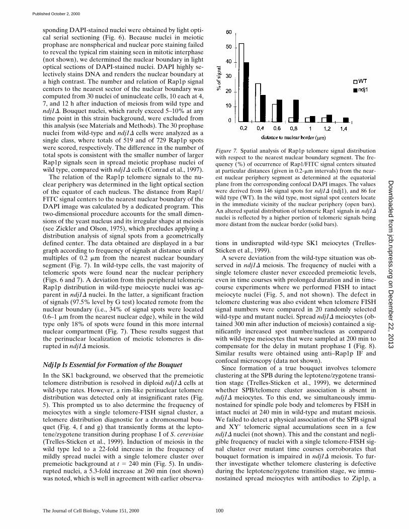
In wild-type and ndj1D time courses, the fraction of nu-cleoids with FISH signals homologously paired increasedwell above premeiotic values (Fig. 11). In ndj1D meiosis, allhomologous regions investigated reached nearly wild-typelevels of homologous signal pairing, but with a 2–3-h delay(Fig. 11). This slowed progression of homologous pairing ismirrored by a 2-h delay in the appearance of anaphases Iand II in ndj1D meiosis as compared with the wild type,where these first appeared at 300 and 320 min, respectively(not shown, consistent with earlier reports). In wild-typemeiosis, the homologous region near the left telomere ofthe small chromosomes III paired most rapidly (Table I).However, in the absence of Ndj1p and bouquet formation,the telomeric region of chromosomes III showed the mostprominent retardation of homologous pairing, expressed asthe difference in time required in wild-type and ndj1D mei-osis to reach peak values in signal pairing (Dt 5 160 min,Fig. 11). Pairing of the telomeric region of the right arm ofthe large chromosomes XI (cos l), in contrast, showed onlya delay of 100 min in ndj1D meiosis. Pairing of large chro-
Figure 11. FISH analysis of ho-mologue pairing during meiotictime courses of wild-type andndj1D SK1 strains. Cosmidprobe combinations m/p and f/lwere hybridized to spread prep-arations obtained at the respec-tive time points (minutes). Pair-ing values were obtained bydetermining the fraction of nu-clei containing cosmid signals ofthe same color that touched eachother or showed an enlarged co-alesced signal. More than 200FISH signal pairs were scoredper time point, probe combina-tion, and strain. Values werecorrected for accidental heterol-ogous contacts by subtracting5% for cosmid combination m/pand 4% for probe combinationl/f (see text). (A) Frequencies ofnuclei with paired cos f signals(XI internal; Fig. 1) in wild-type(WT) and ndj1D (ndj1) timecourses. (B) Frequencies (%) ofpaired cos l (XI, right telo) inwild-type and ndj1D time
courses. (D) Frequencies of nuclei with paired cos p signals (IX, right arm) in wild-type and ndj1D time courses. (C) Frequencies of nu-clei with paired cos m signals (III, HML) in wild-type and ndj1D time course. At all loci probed, the frequencies of nuclei with paired sig-nals increase more gradually in the absence of Ndj1p, reaching nearly wild-type frequencies with a 2–3-h delay. In the wild type, signalpatterns are displayed only until 320 min, since meiotic divisions (anaphases) appeared after 300 min and complicated the signal analysis.
on Decem
ber 22, 2013jcb.rupress.org
Dow
nloaded from
Published October 2, 2000

Trelles-Sticken et al. NDJ1 Is Required for Bouquet Formation in Yeast Meiosis 103
mosomes may thus be less dependent on a catalytic actionof telomere clustering on homologue pairing, since thesemay span the entire nucleus and therefore have a higherprobability for chance encounters with their homologues inthe absence of bouquet formation.
Condensation of Chromosome Territories Occurs in the Absence of Ndj1p
Previous chromosome painting studies have shown thatyeast chromosomes extend, pair, and condense during thecourse of meiotic prophase (Scherthan et al., 1992; Trelles-Sticken et al., 1999). To test whether the ndj1D mutationinfluences this morphological change of yeast chromo-somes during meiosis, we painted chromosomes XI in con-junction with telomere FISH in wild-type and ndj1D cellsat 180, 240, 360, and 420 min after induction of meiosis.Nuclei with all aspects of meiotic chromosome morphol-ogy were detected in wild-type and ndj1D cells (Fig. 12).The painting of yeast chromosomes also allows chromatincondensation to be assessed. The fraction of uninucleatedmeiocytes that exhibit clear and compact FISH signalsrepresents the fraction of cells in which chromosomes arecondensed (pachytene; Scherthan et al., 1992; Loidl et al.,1994; Nag et al., 1995). The frequency of condensed XIpachytene bivalents was determined in uninucleate mu-tant and wild-type SK1 meiocytes (Fig. 12 d). In ndj1Dmeiosis, chromatin condensation was found to be signifi-cantly delayed with respect to wild type (Fig. 13), althoughchromosome condensation per se is not affected by the
ndj1D mutation. Thus, chromosome condensation was fur-ther monitored by FISH with differentially labeled cosmidprobes b and l in uninucleated meiocytes obtained at t 5240 min in wild type and t 5 360 min in ndj1D timecourses. Cosmids b and l map to the proximal and distalpart of the left (long) arm of XI (Fig. 1). The distance be-tween the centers of the paired and differentially coloredcosmid signals on pachytene bivalents was determined in30 randomly selected spread nuclei. The mean distanceobserved between the two cosmid signals was 1.1 6 0.4 mm(6SD) in the wild type and 1.0 6 0.4 mm in ndj1D nuclei.The difference between the two data sets did not differ sig-nificantly (P 5 0.23 at 0.01; Student’s t test). This furthersuggests that chromosome condensation is not affected bythe absence of Ndj1p.
Discussion
Ndj1p May Be Required for Peripheral Localization of Meiotic Telomeres
Chromosome topology of the vegetatively growing yeast isdominated by tightly clustered centromeres next to thespindle body (Hayashi et al., 1998; Jin et al., 1998) and bytelomeres forming a few aggregates at the nuclear periph-ery (Klein et al., 1992; Gotta et al., 1996). Upon inductionof meiosis, nuclear topology is reorganized. Centromeresbecome dispersed throughout the nucleus, and telomeresspread over the nuclear periphery (Hayashi et al., 1998),except during an intermediate stage when telomeres forma tight cluster in the vicinity of the spindle pole body; i.e.,at the bouquet stage (Trelles-Sticken et al., 1999).
Two of the early steps in the meiotic nuclear reorganiza-tion, dispersion of centromeres and telomeres, apparentlyare unaffected by deletion of NDJ1. However, this closeproximity of dispersed telomeres to the nuclear periphery,which is evident in wild-type cells (Hayashi et al., 1998,and this report) does appear defective in ndj1D. Duringearliest meiotic prophase, telomeres dissociate from thevegetative aggregates and line the nuclear periphery (Fig.14), giving raise to a rim-like staining in z20% of wild-type nuclei, which resembles the telomere distribution
Table I. Retardation of Homologue Pairing if ndj1D Meiosis
Cosmid probeMaximal signal
pairing in WT at t Maximal signal pairing
in ndj1D meiosis at t Dt
min min min
Cos m (III HML) 240 400 160Cos p (IX, right arm) 300 400 100Cos f (XI, interstitial) 280 400 120Cos l (XI, right telo) 280 380 100
Time taken in wild-type and ndj1D meiosis to reach peak values in homologousFISH-signal pairing (minutes). Dt, time difference between the peak homologuepairing values for a given cosmid probe in wild-type and ndj1D meiotic timecourses.
Figure 12. Representative images of FISH signalpatterns obtained with a painting probe for chro-mosomes XI (red) and the pan-telomere probe(green) in mildly spread nuclei from meiotic timecourses of diploid wild type (wt) and ndj1D(ndj1) SK1 strains (a, 0 min; b and c, 260 min; d,380 min). Images are aligned according to meio-sis-specific changes in chromosome morphologyduring prophase I (see Trelles-Sticken et al.,1999). (a) Premeiotic wild-type and ndj1D nuclei(0 min) display separated variably shaped chro-mosomes XI territories and several telomereclusters. (b, wt) WT meiocyte nucleus with ex-tended chromosome XI signal tracks and a rim-like distribution of telomere signals. (b, ndj1)Meiocyte with scattered telomere signals, while
XI signal tracks extend across the nucleus and touch at one end. (c, wt) Nucleus with one large telomere signal cluster (bouquet ar-rangement). Chromosomes XI form one outstretched signal track along the telomere cluster. (c, ndj1) ndj1D meiocyte with scatteredtelomeres. Painted chromosomes XI are seen as a single extended signal track. (d) Wild-type and mutant pachytene nuclei both exhibita condensed XI bivalent and scattered telomeres. Bar, 5 mm.
on Decem
ber 22, 2013jcb.rupress.org
Dow
nloaded from
Published October 2, 2000

The Journal of Cell Biology, Volume 151, 2000 104
seen in the late preleptotene/leptotene stage of mamma-lian prophase I (Scherthan et al., 1996). In mammals, theswitch from premeiotic to meiotic telomere distribution isa two-step process, with telomeres first attaching at scat-tered points over the nuclear envelope, and then congre-gating in the bouquet to a limited sector of the nuclearenvelope (Rasmussen and Holm, 1978; Boiko, 1983;Scherthan et al., 1996). In ndj1D meiosis of yeast, FISHand IF analyses of telomere distribution in undisruptedand mildly spread nuclei revealed a paucity of nuclei withtelomeres lining the nuclear periphery and a concomitantincrease in the frequency of nuclei with telomeres scat-tered throughout the nuclear lumen, as inferred fromthree-dimensional Rap1p analysis. These observations in-dicate that the absence of Ndj1p impairs the assembly of ameiosis-specific perinuclear telomere topology (Fig. 14)after dissolution of the few vegetative telomere clusters
(see Gilson et al., 1993). Furthermore, these observationsindicate that localization of telomeres to the nuclear pe-riphery requires Ndj1p, in accord with speculation basedon an earlier genetic analysis of NDJ1/TAM1-deficientcells (Chua and Roeder, 1997). Localization of the ends ofyeast synaptonemal complexes to the nuclear envelopehas been demonstrated by electron microscopy (Byers andGoetsch, 1975; Zickler and Olson, 1975; Moens and Ash-ton, 1985) and a similar analysis of ndj1D is underway (Z.Zhang, M.N. Conrad, and M.E. Dresser).
Bouquet Formation Facilitates Homologue Interaction
Distribution of recombination events (crossover interfer-ence), chromosome segregation, and distributive disjunc-tion (Chua and Roeder, 1997; Conrad et al., 1997) are allaffected by deletion of NDJ1 but, for the major part, thedefects are relatively mild and may arise from a telomere-related delay in meiotic prophase (Chua and Roeder,1997). The delay in onset and completion of synapsis(Conrad et al., 1997) potentially is due to a defect in someaspect of chromosome pairing that normally brings homo-logues into close apposition for the initiation of homosyn-apsis. FISH in conjunction with Zip1p immunostainingshowed that telomeres fail to cluster during the leptotene/zygotene-equivalent stages, when bouquet formation nor-mally occurs (Dernburg et al., 1995; Zickler and Kleckner,1998). Moreover, the meiotic SPB failed to show an associ-ation with a telomere accumulation occasionally seen inNdj1-deficient nuclei. This suggests that loss of bouquetformation is the underlying defect of the retarded synapsisprogression and chromosome pairing in the ndj1D mutant(see below).
When we investigated homologue pairing at four re-gions on three chromosomes by two-color cosmid FISH, ahighly significant reduction in chromosome pairing wasnoted at time points in mutant meiosis when, in the wildtype, signal pairing had reached its maximum. Prolongedtime courses in the ndj1D mutant showed that there is a
Figure 13. Frequency (%) of uninucleate meiocytes with con-densed chromosome XI bivalent (Fig. 12 d) at given time points(minutes) in wild-type (WT) and ndj1D (ndj1) meiosis in the SK1background. In ndj1D meiosis, the frequency of uninucleatedmeiocytes with a condensed XI bivalent increased more gradu-ally than in the wild type.
Figure 14. Scheme showing thesequential steps of centromereand telomere redistribution thatoccur during earliest meioticprophase of yeast. It is based onthe observations made in thisand earlier reports (see text).(a) In the premeiotic nucleus,telomeres (black) and cen-tromeres (gray) form severaltelomere and one centromerecluster at the nuclear periphery.(b) In the wild type, induction ofmeiosis leads to dissolution ofthe centromere cluster and peri-nuclear telomere clusters. Cen-tromeres become dispersedthroughout the nuclear lumen,while telomeres disperse overthe nuclear periphery. (c) Bou-quet stage: telomeres tightly
cluster at the spindle pole body, while centromeres are dispersed. (d) While premeiotic telomere topology is not affected, the early stepsof meiotic telomere redistribution appear defective in the absence of Ndj1p, which leads to scattering of both telomeres and cen-tromeres throughout the ndj1D meiocyte nucleus.
on Decem
ber 22, 2013jcb.rupress.org
Dow
nloaded from
Published October 2, 2000

Trelles-Sticken et al. NDJ1 Is Required for Bouquet Formation in Yeast Meiosis 105
2–3-h delay in chromosome pairing. A similar delay wasnoted for the compaction of chromosomes XI bivalents,but, ultimately, chromosome condensation as measured bycosmid FISH appeared normal. When we compared thetime required for the homologous regions assayed to reachmeiosis-specific values in wild-type and ndj1D meiosis, itappeared that the pairing of the telomeric regions of therelatively small chromosomes III was most severely de-layed. In Ndj1p-deficient meiosis, the left telomere regionon chromosomes III took 60 min longer to reach peak-pairing values than the telomeric region of the right arm ofthe large chromosomes XI and on chromosomes IX. Thismay indicate that smaller chromosomes are more depen-dent on the action of telomere clustering to instigate ho-mologue alignment and pairing. However, after a delay ofz2–3 h, all homologous regions investigated reached ap-proximately wild-type pairing values, which shows thattelomere-independent routes of homologue pairing exist.Altogether, it appears that bouquet formation is not re-quired for homologue recognition and synapsis per se, butmediates an important catalytic action on homologue pair-ing, which shortens the duration of sporulation, and thusmay, in turn, confer selective advantages in a natural envi-ronment. This conclusion is in agreement with long-stand-ing hypotheses that telomere clustering during the bou-quet stage facilitates homologue interactions by aligningchromosome ends (for reviews, see Rhoades, 1961; Moses,1968; Scherthan, 1997; de Lange, 1998b; Zickler andKleckner, 1998).
Furthermore, there is supporting experimental evidencefrom S. pombe (Chikashige et al., 1997) where, in analogyto the situation in budding yeast ndj1D mutants, absenceof the telomere protein Taz1p(1) abrogates telomereclustering and leads to reduced recombination and in-creased meiotic nondisjunction in fission yeast (Cooper etal., 1998; Nimmo et al., 1998). Thus, the primary, impor-tant consequence of deletion of NDJ1 is likely to be thefailure of bouquet formation, with the subsequent defectsarising from distorted telomere distribution and the ab-sence of bouquet-facilitated homologue alignment andpairing, as suggested based on genetic data from disomicsynaptic meiosis (Rockmill and Roeder, 1998). The ndj1D-related delay may result from overloading of the homol-ogy testing machinery due to disordered chromosome dis-tribution in the absence of meiotic telomere clustering.Dissolution of premeiotic/vegetative centromere and telo-mere aggregates at the onset of meiosis may still createsufficient chromosome movement in the Ndj1p-deficientprophase nucleus for portions of chromosomes to makecontact and, once partially aligned, to commence with ho-mosynapsis. Particularly, larger-sized chromosomes maybenefit from an increased probability of encountering aportion of its homologue and to initiate stable pairing ear-lier than smaller ones. Synapsis, which may initiate at afew interstitial sites (Dresser and Giroux, 1988; Rockmillet al., 1995; Chua and Roeder, 1997; Trelles-Sticken et al.,1999) seems to proceed at a slower rate in the ndj1D mu-tant, since Zip1p polycomplexes resolve more gradually.Jumbled telomere localization and nonsynchronized ho-mologue pairing may be the cause for defective crossoverinterference in the absence of Ndj1p (see Chua andRoeder, 1997). However, at the present state, it cannot be
excluded that the ndj1D phenotypes result from other sofar unknown effects besides the telomeric ones.
An interesting possibility is that Ndj1p may link meiotictelomeres to motor proteins at the nuclear envelope, theaction of which is thought to bring about telomere aggre-gation during the bouquet stage (see Bascom-Slack andDawson, 1997; Zickler and Kleckner, 1998). Future re-search will have to show whether perinuclear filaments,like the Mlp proteins (Strambio-de-Castillia et al., 1999),which are involved in tethering vegetative telomeres tonuclear pore complexes (Galy et al., 2000), also play a rolein telomere clustering during meiotic prophase.
The authors thank J. Berman, B. Dujon, E.J. Louis, A. Goldman, M. Lich-ten, G.S. Roeder, J.V. Kilmartin for help with the materials, and M.N.Conrad for critical comments on an earlier of the manuscript.
This work was supported by a grant of the Deutsche Forschungsge-meinschaft (Sche 350/8-2) to H. Scherthan and National Science Founda-tion grant MCB-9507089 to M.E. Dresser and M.N. Conrad (OklahomaMedical Research Foundation).
Submitted: 2 May 2000Revised: 22 August 2000Accepted: 23 August 2000
References
Alani, E., R. Padmore, and N. Kleckner. 1990. Analysis of wild-type and rad50mutants of yeast suggests an intimate relationship between meiotic chromo-some synapsis and recombination. Cell. 61:419–436.
Bass, H.W., W.F. Marshall, J.W. Sedat, D.A. Agardand, and W.Z. Cande. 1997.Telomeres cluster de novo before the initiation of synapsis: a three-dimen-sional spatial analysis of telomere positions before and during meioticprophase. J. Cell Biol. 137:5–18.
Bélar, K. 1928. Chromosomenreduktion. In Handbuch der Vererbungswissen-schaft, die Cytologischen Grundlagen der Vererbung. E. Baur and M. Hart-mann, editors. Geb. Borntraeger, Berlin. 168–201.
Bascom-Slack, C.A., and D.S. Dawson. 1997. The yeast motor protein, Kar3p,is essential for meiosis I. J. Cell Biol. 139:459–467.
Blackburn, E.H. 1991. Structure and function of telomeres. Nature. 350:569–573.Burgess, S.M., N. Kleckner, and B.M. Weiner. 1999. Somatic pairing of ho-
mologs in budding yeast: existence and modulation. Genes Dev. 13:1627–1641.Byers, B., and L. Goetsch. 1975. Electron microscopic observations on the mei-
otic karyotype of diploid and tetraploid Saccharomyces cerevisiae. Proc.Natl. Acad. Sci. USA. 72:5056–5060.
Boiko, M. 1983. Human meiosis VIII. Chromosome pairing and formation ofthe synaptonemal complex in Oocytes. Carlsberg Res. Commun. 48:457–483.
Chikashige, Y., D.-Q. Ding, Y. Imai, M. Yamamoto, T. Haraguchi, and Y.Hiraoka. 1997. Meiotic nuclear reorganization: switching the position of cen-tromeres and telomeres in the fission yeast Schizosaccharomyces pombe.EMBO (Eur. Mol. Biol. Organ.) J. 16:193–202.
Chua, P.R., and G.S. Roeder. 1997. Tam1, a telomere-associated meiotic pro-tein, functions in chromosome synapsis and crossover interference. GenesDev. 11:1786–1800.
Conrad, M.N., A.M. Dominguez, and M.E. Dresser. 1997. Ndj1p, a meiotic telo-mere protein required for normal chromosome synapsis and segregation inyeast. Science. 276:1252–1255.
Conchello, J.A., J.J. Kim, and E.W. Hansen. 1994. Enhanced three-dimensionalreconstruction from confocal scanning microscope images. II. Depth dis-crimination versus signal-to-noise ratio in partially confocal images. Appl.Opt. 33:3740–3750.
Cooper, J.P., Y. Watanabe, and P. Nurse. 1998. Fission yeast Taz1 protein is re-quired for meiotic telomere clustering and recombination. Nature. 392:828–831.
de Lange, T. 1998a. Telomeres and senescence: ending the debate. Science. 279:334–335.
de Lange, T. 1998b. Ending up with the right partner. Nature. 392:753–754.Dernburg, A.F., J.W. Sedat, W.Z. Cande, and H.W. Bass. 1995. The cytology of
telomeres. In Telomeres, E.H. Blackburn and C.W. Greider, editors. ColdSpring Harbor Laboratory Press, Cold Spring Harbor, NY. 295–338.
Dresser, M.E., and C.N. Giroux. 1988. Meiotic chromosome behavior in spreadpreparations of yeast. J. Cell Biol. 106:567–573.
Dresser, M.E., D.J. Ewing, S.N. Harwell, D. Coody, and M.N. Conrad. 1994.Nonhomologous synapsis and reduced crossing over in a heterozygous para-centric inversion in Saccharomyces cerrevisiae. Genetics. 138:633–647.
Dresser, M.E., D.J. Ewing, M.N. Conrad, A.M. Dominguez, R. Barstead, H.Jiang, and T. Kodadek. 1997. DMC1 functions in a Saccharomyces cerevisiaemeiotic pathway that is largely independent of the RAD51 pathway. Genet-
on Decem
ber 22, 2013jcb.rupress.org
Dow
nloaded from
Published October 2, 2000

The Journal of Cell Biology, Volume 151, 2000 106
ics. 147:533–544.Dujon, B., D. Alexandraki, B. André, W. Ansorge, V. Baladron, J.P. Ballesta,
A. Banrevi, P.A. Bolle, M. Bolotin-Fukuhara, P. Bossier, et al. 1994. Com-plete DNA sequence of yeast chromosome XI. Nature. 369:371–378.
Fussell, C.P. 1987. The Rab1 orientation: a prelude to synapsis. In Meiosis. P.B.Moens, editor. Academic Press, Inc., Orlando, FL. 275–299.
Galy, V., J.C. Olivo-Marin, H. Scherthan, V. Doye, N. Rascalou, and U. Nehr-bass. 2000. Nuclear pore complexes in the organization of silent telomericchromatin. Nature. 403:108–112.
Gelei, J. 1921. Weitere Studien über die Oogenese des Dendrocoelum lacteum.III. Die Konjugation der Chromosomen in der Literatur und meine Be-funde. Arch. Zellforsch. 16:300–365.
Gilson, E.T., T. Laroche, and S.M. Gasser. 1993. Teloeres and the functional ar-chitecture of the nucleus. Trends Cell Biol. 3:128–134.
Gotta, M., T. Laroche, A. Formenton, L. Maillet, H. Scherthan, and S.M. Gas-ser. 1996. The clustering of telomeres and colocalization with Rap1, Sir3,and Sir4 proteins in wild-type Saccharomyces cerevisiae. J. Cell Biol. 134:1349–1363.
Grushcow, J.M., T.M. Holzen, K.J. Park, T. Weinert, M. Lichten, and D.K.Bishop. 1999. Saccharomyces cerevisiae checkpoint genes MEC1, RAD17and RAD24 are required for normal meiotic recombination partner choice.Genetics. 153:607–620.
Hawley, R.S. 1988. Exchange and chromosomal segregation in eukaryotes. In Ge-netic Recombination. R. Kucherlapati and G.R. Smimth, editors. AmericanSociety of Microbiology, Washington, D.C. 497–527
Hayashi, A., H. Ogawa, K. Kohno, S.M. Gasser, and Y. Hiraoka. 1998. Meioticbehaviors of chromosomes and microtubules in budding yeast: relocalization ofcentromeres and telomeres during meiotic prophase. Genes Cells. 3:587–601.
Hiraoka, Y. 1998. Meiotic telomeres: a matchmaker for homologous chromo-somes. Genes Cells. 3:405–413.
Ishikawa, F., and T. Naito. 1999. Why do we have linear chromosomes? A mat-ter of Adam and Eve. Mutat. Res. 434:99–107.
Jin, Q.-W., E. Trelles-Sticken, H. Scherthan, and J. Loidl. 1998. Yeast nucleidisplay prominent centromere clustering that is reduced in nondividing cellsand in meiotic prophase. J. Cell Biol. 141:21–29.
Kane, S.M., and R. Roth. 1974. Carbohydrate metabolism during ascospore de-velopment in yeast. J. Bacteriol. 118:8–14.
Klein, F., T. Laroche, M.E. Cardenas, J.F.-X. Hofmann, D. Schweizer, and S.M.Gasser. 1992. Localization of RAP1 and topoisomerase II in nuclei and mei-otic chromosomes of yeast. J. Cell Biol. 117:935–948.
Laroche, T., S.G. Martin, M. Gotta, H.C. Gorham, F.E. Pryde, E.J. Louis, andS.M. Gasser. 1998. Mutation of yeast Ku genes disrupts the subnuclear orga-nization of telomeres. Curr. Biol. 8:653–656.
Loidl, J. 1990. The initiation of meiotic chromosome pairing: the cytologicalview. Genome. 33:759–778.
Loidl, J., F. Klein, and H. Scherthan. 1994. Homologous pairing is reduced butnot abolished in asynaptic mutants of yeast. J. Cell Biol. 125:1191–1200.
Loidl, J., F. Klein, and J. Engebrecht. 1998. Genetic and morphological ap-proaches for the analysis of meiotic chromosomes in yeast. In Nuclear Struc-ture and Function. M. Berrios, editor. Academic Press, Inc., San Diego, CA.257–285.
Louis, E.J., E.S. Naumova, A. Lee, G. Naumov, and J. Haber. 1994. The chro-mosome end in yeast: its mosaic nature and influence on recombination dy-namics. Genetics. 136:789–802.
Marschall, L.G., R.L. Jeng, J. Mulholland, and T. Stearns. 1996. Analysis ofTub4p, a yeast gamma-tubulin-like protein: implications for microtubule-organizing center function. J. Cell Biol. 134:443–454.
Moens, P.B., and M.L. Ashton. 1985. Synaptonemal complexes of normal andmutant yeast chromosomes (Saccharomyces cerevisiae). Chromosoma. 91:113–120.
Moens, P.B. 1974. Quantitative electron microscopy of chromosome organiza-tion at meiotic prophase. Cold Spring Harbor Symp. Quant. Biol. 38:99–107.
Moses, M.J. 1968. Synaptinemal complex. Annu. Rev. Genet. 2:363–412.Nag, D., H. Scherthan, B. Rockmill, J. Bhargava, and G.S. Roeder. 1995. Het-
eroduplex DNA formation and homolog pairing in yeast meiotic mutants.Genetics. 141:75–86.
Naito, T., A. Matsuura, and F. Ishikawa. 1998. Circular chromosome formation
in a fission yeast mutant defective in two ATM homologues. Nat. Genet. 20:203–206.
Nimmo, E.R., A.L. Pidoux, P.E. Perry, and R.C. Allshire. 1998. Defective mei-osis in telomere-silencing mutants of Schizosaccharomyces pombe. Nature.392:825–828.
Padmore, R., L. Cao, and N. Kleckner. 1991. Temporal comparison of recombi-nation and synaptonemal complex formation during meiosis in S. cerevisiae.Cell. 66:1239–1256.
Rhoades, M.M. 1961. Meioses. In The Cell: Biochemistry, Physiology, and Mor-phology. Vol. 3. J. Brachet and A.E. Mirsky, editors. Academic Press, NewYork, NY. 1–75.
Rockmill, B., M. Sym, H. Scherthan, and G.S. Roeder. 1995. Two RecA ho-mologs facilitate meiotic chromosome synapsis by establishing connectionsbetween homologous chromosomes. Genes Dev. 9:2684–2695.
Rockmill, B., and G.S. Roeder. 1998. Telomere-mediated chromosome pairingduring meiosis in budding yeast. Genes Dev. 12:2574–2586.
Roeder, G.S. 1997. Meiotic chromosomes: it takes two to tango. Genes Dev. 11:2600–2621.
Rasmussen, S.W., and P.B. Holm. 1978. Human Meiosis II. Chromosome pair-ing and recombination nodules in human spermatocytes. Carlsberg Res.Commun. 43:275–327.
Roth, R., and H.O. Halvorson. 1969. Sporulation of yeast harvested during log-arithmic growth. J. Bacteriol. 98:831–832.
Scherthan, H., J. Loidl, T. Schuster, and D. Schweizer. 1992. Meiotic chromo-some condensation and pairing in Saccharomyces cerevisiae studied by chro-mosome painting. Chromosoma. 101:590–595.
Scherthan, H., S. Weich, H. Schwegler, M. Härle, C. Heyting, and T. Cremer.1996. Centromere and telomere movements during early meiotic prophaseof mouse and man are associated with the onset of chromosome pairing. J.Cell Biol. 134:1109–1125.
Scherthan, H. 1997. Chromosome behaviour in earliest meiotic prophase. InChromosomes Today. Vol. 12. H. Gill, J.S. Parker, and M. Puertas, editors.Chapman and Hall, London, UK. 217–248.
Shore, D. 1998. Telomeres–unsticky ends. Science. 281:1818–1819.Strambio-de-Castillia, C., G. Blobel, and M.P. Rout. 1999. Proteins connecting
the nuclear pore complex with the nuclear interior. J. Cell Biol. 144:839–855.Sym, M., J.A. Engebrecht, and G.S. Roeder. 1993. ZIP1 is a synaptonemal com-
plex protein required for meiotic chromosome synapsis. Cell. 72:365–378.Sym, M., and G.S. Roeder. 1995. Zip1-induced changes in synaptonemal com-
plex structure and polycomplex assembly. J. Cell Biol. 128:455–466.Therman, E., and G.E. Sarto. 1977. Premeiotic and early meiotic stages in the
pollen mother cells of eremus and in human embrionic oocytes. Hum. Genet.35:137–151.
Thierry, A., L. Gaillon, F. Galibert, and B. Dujon. 1995. Construction of a com-plete genomic library of Saccharomyces cerevisiae and physical mapping ofchromosome XI at 3.7 kb resolution. Yeast. 11:121–135.
Trelles-Sticken, E., J. Loidl, and H. Scherthan. 1999. Bouquet formation inbudding yeast: initiation of recombination is not required for meiotic telo-mere clustering. J. Cell Sci. 112:651–658.
von Wettstein, D., S.W. Rasmussen, and P.B. Holm. 1984. The synaptonemalcomplex in genetic segregation. Annu. Rev. Genet. 18:331–413.
Wach, A., A. Brachat, R. Pohlmann, and P. Philippsen. 1994. New heterologousmodules for classical or PCR-based gene disruptions in Saccharomyces cere-visiae. Yeast. 10:1793–1808.
Weiner, B.M., and N. Kleckner. 1994. Chromosome pairing via multiple inter-stitial interactions before and during meiosis in yeast. Cell. 77:977–991.
Yamamoto, A., R.R. West, J.R. McIntosh, and Y. Hiraoka. 1999. A cytoplas-mic dynein heavy chain is required for oscillatory nuclear movement of mei-otic prophase and efficient meiotic recombination in fission yeast. J. CellBiol. 145:1233–1249.
Zakian, V.A. 1995. Saccharomyces telomeres: function, structure and replica-tion. In Telomeres. E.H. Blackburn and C.W. Greider, editors. Cold SpringHarbor Laboratory Press, Cold Spring Harbor, NY. 107–138.
Zickler, D., and L.W. Olson. 1975. The synaptonemal complex and the spindleplaque during meiosis in yeast. Chromosoma. 50:1–23.
Zickler, D., and N. Kleckner. 1998. The leptotene-zygotene transition of meio-sis. Annu. Rev. Genet. 32:619–697.
on Decem
ber 22, 2013jcb.rupress.org
Dow
nloaded from
Published October 2, 2000