Association of telomere length with authentic pluripotency of ES/iPS cells
Upload
independentCategory
view
2download
0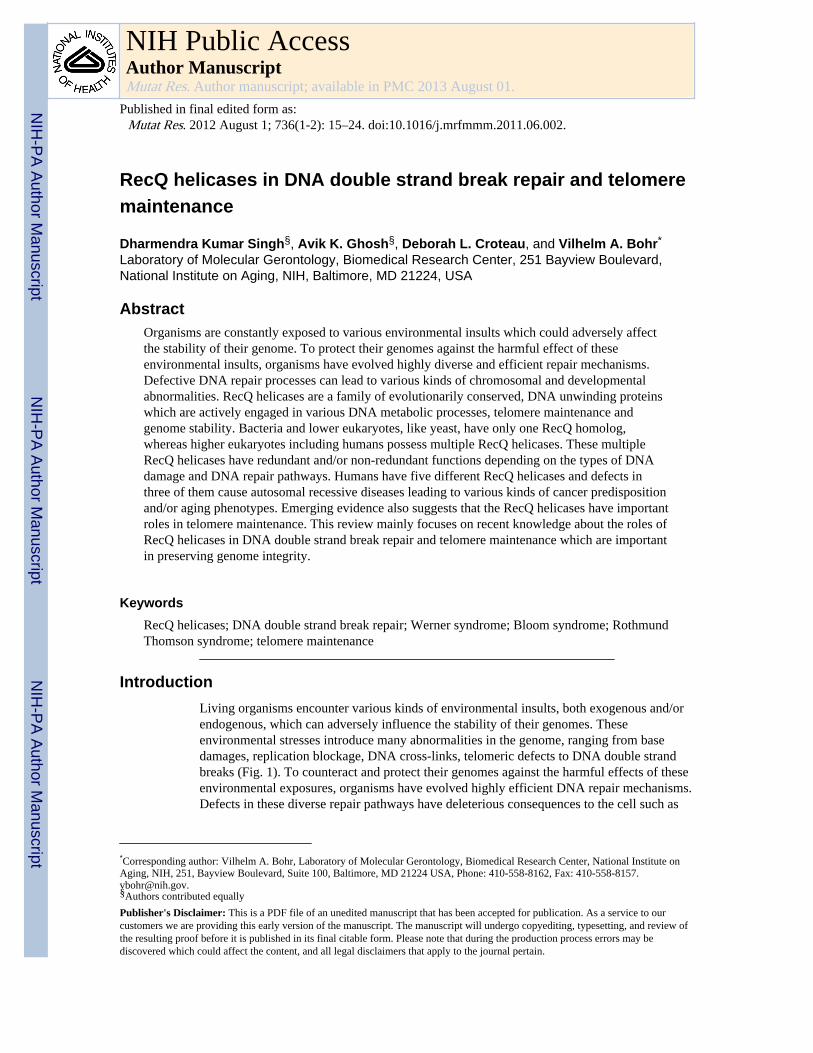
RecQ helicases in DNA double strand break repair and telomeremaintenance
Dharmendra Kumar Singh§, Avik K. Ghosh§, Deborah L. Croteau, and Vilhelm A. Bohr*
Laboratory of Molecular Gerontology, Biomedical Research Center, 251 Bayview Boulevard,National Institute on Aging, NIH, Baltimore, MD 21224, USA
AbstractOrganisms are constantly exposed to various environmental insults which could adversely affectthe stability of their genome. To protect their genomes against the harmful effect of theseenvironmental insults, organisms have evolved highly diverse and efficient repair mechanisms.Defective DNA repair processes can lead to various kinds of chromosomal and developmentalabnormalities. RecQ helicases are a family of evolutionarily conserved, DNA unwinding proteinswhich are actively engaged in various DNA metabolic processes, telomere maintenance andgenome stability. Bacteria and lower eukaryotes, like yeast, have only one RecQ homolog,whereas higher eukaryotes including humans possess multiple RecQ helicases. These multipleRecQ helicases have redundant and/or non-redundant functions depending on the types of DNAdamage and DNA repair pathways. Humans have five different RecQ helicases and defects inthree of them cause autosomal recessive diseases leading to various kinds of cancer predispositionand/or aging phenotypes. Emerging evidence also suggests that the RecQ helicases have importantroles in telomere maintenance. This review mainly focuses on recent knowledge about the roles ofRecQ helicases in DNA double strand break repair and telomere maintenance which are importantin preserving genome integrity.
KeywordsRecQ helicases; DNA double strand break repair; Werner syndrome; Bloom syndrome; RothmundThomson syndrome; telomere maintenance
IntroductionLiving organisms encounter various kinds of environmental insults, both exogenous and/orendogenous, which can adversely influence the stability of their genomes. Theseenvironmental stresses introduce many abnormalities in the genome, ranging from basedamages, replication blockage, DNA cross-links, telomeric defects to DNA double strandbreaks (Fig. 1). To counteract and protect their genomes against the harmful effects of theseenvironmental exposures, organisms have evolved highly efficient DNA repair mechanisms.Defects in these diverse repair pathways have deleterious consequences to the cell such as
*Corresponding author: Vilhelm A. Bohr, Laboratory of Molecular Gerontology, Biomedical Research Center, National Institute onAging, NIH, 251, Bayview Boulevard, Suite 100, Baltimore, MD 21224 USA, Phone: 410-558-8162, Fax: [email protected].§Authors contributed equally
Publisher's Disclaimer: This is a PDF file of an unedited manuscript that has been accepted for publication. As a service to ourcustomers we are providing this early version of the manuscript. The manuscript will undergo copyediting, typesetting, and review ofthe resulting proof before it is published in its final citable form. Please note that during the production process errors may bediscovered which could affect the content, and all legal disclaimers that apply to the journal pertain.
NIH Public AccessAuthor ManuscriptMutat Res. Author manuscript; available in PMC 2013 August 01.
Published in final edited form as:Mutat Res. 2012 August 1; 736(1-2): 15–24. doi:10.1016/j.mrfmmm.2011.06.002.
NIH
-PA Author Manuscript
NIH
-PA Author Manuscript
NIH
-PA Author Manuscript

chromosomal or developmental defects, or various kinds of cancers/aging phenotypes. Onesuch group of repair proteins that are actively engaged in various aspects of DNAmetabolism is the RecQ helicases. The RecQ helicases are evolutionarily conserved, DNAunwinding proteins, which help in the maintenance of genome integrity by participating inmany DNA metabolic processes, DNA repair pathways and transcription which aresummarized in Fig. 1 [1–3].
The RecQ helicase are conserved from prokaryotes to higher eukaryotes. Lower organismssuch as bacteria and yeast possess only one RecQ homolog, whereas higher eukaryotesincluding mammals possess multiple forms of RecQ helicases. These multiple RecQhelicases either possesses unique and/or overlapping functions depending on the types ofDNA damage and repair pathways. Therefore, it is likely that during the evolutionaryprocess the functions of the RecQ helicases have diversified to adapt to the changingenvironment and complexity of genomes. The hallmark feature of all the members of theRecQ helicase family is the conserved helicase domain which is crucial for their functions.Five RecQ homologs have been found in humans and mice namely, RECQL1, BLM, WRN,RECQL4 and RECQL5. Defects in three of these have been associated with rare geneticdisorders characterized by genome instability, multiple cancer predispositions and/or apremature aging phenotype. Werner syndrome (WS) is caused by defects in WRN (except ina few cases when clinically indistinguishable WS is caused by defects in lamins), Bloomsyndrome (BS) is due to defects in BLM, and Rothmund Thomson (RTS), RAPADILINOand Baller Gerold (BGS) syndromes are associated with defects in RECQL4. The other twomembers, RECQL1 and RECQL5, have not yet been linked to any disease phenotype, butstudies in humans and mice have suggested their important roles in genome stability [4].
RecQ helicases are involved in base excision repair (BER), DNA double strand break repair(DSBR), intra-strand cross link repair (ICL), recovery of stalled replication forks, andtelomere processivity and stability (Fig. 1) [3–6]. One finding suggests the involvement ofRECQL4 in NER pathway by its interaction with XPA, a key protein involved in NERpathway [6]. However, the involvement of RecQ helicases in NER is still obscure. Recentstudies in Xenopus as well as in humans also indicated that one of the RecQ helicasemembers, RECQL4, is an important component of the DNA replication machinery and is apart of the DNA replication initiation complex [7–9]. Another RecQ helicase, RECQL5,interacts with RNA pol II, suggesting its involvement in transcription [10, 11]. Therefore,RecQ helicases play diversified roles in genome stability and have been called the“guardians of the genome”. This review mainly focuses on important functions of RecQhelicases in DNA double strand break (DSB) repair and telomere processing which arecrucial for maintaining genome stability.
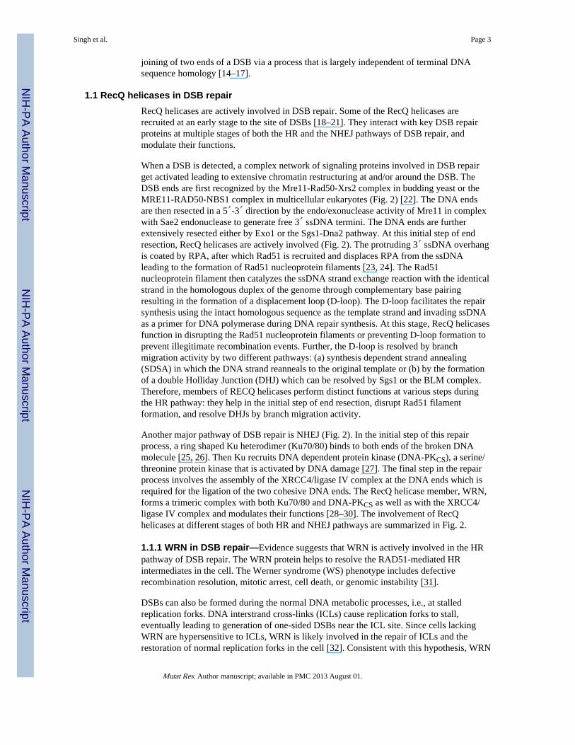
1. DNA double strand break repairDSBs are very potent and deleterious forms of DNA damage in the genome, and if leftunrepaired they can cause cell cycle arrest, mutagenesis, gross chromosomalrearrangements, cell death and tumorigenesis. DSBs can arise spontaneously during normalDNA metabolism or when cells are exposed to DNA damaging agents or ionizing radiations.In higher eukaryotes, DSBs are mostly repaired by two distinct pathways i.e., homologousrecombination (HR) and non-homologous end joining (NHEJ) [12]. The different steps ofboth of these pathways and proteins that interact with RecQ helicases are summarized inFig. 2. The HR pathway is preferential in the late S-G2 phase, whereas NHEJ mainly plays adominant role in the G1 to early S-phase of the cell cycle [13]. The HR pathway is a highfidelity repair mechanism which requires homologous sequences primarily from the sisterchromatids. In contrast, the NHEJ pathway is an error prone mechanism involving the
Singh et al. Page 2
Mutat Res. Author manuscript; available in PMC 2013 August 01.
NIH
-PA Author Manuscript
NIH
-PA Author Manuscript
NIH
-PA Author Manuscript
joining of two ends of a DSB via a process that is largely independent of terminal DNAsequence homology [14–17].
1.1 RecQ helicases in DSB repairRecQ helicases are actively involved in DSB repair. Some of the RecQ helicases arerecruited at an early stage to the site of DSBs [18–21]. They interact with key DSB repairproteins at multiple stages of both the HR and the NHEJ pathways of DSB repair, andmodulate their functions.
When a DSB is detected, a complex network of signaling proteins involved in DSB repairget activated leading to extensive chromatin restructuring at and/or around the DSB. TheDSB ends are first recognized by the Mre11-Rad50-Xrs2 complex in budding yeast or theMRE11-RAD50-NBS1 complex in multicellular eukaryotes (Fig. 2) [22]. The DNA endsare then resected in a 5′-3′ direction by the endo/exonuclease activity of Mre11 in complexwith Sae2 endonuclease to generate free 3′ ssDNA termini. The DNA ends are furtherextensively resected either by Exo1 or the Sgs1-Dna2 pathway. At this initial step of endresection, RecQ helicases are actively involved (Fig. 2). The protruding 3′ ssDNA overhangis coated by RPA, after which Rad51 is recruited and displaces RPA from the ssDNAleading to the formation of Rad51 nucleoprotein filaments [23, 24]. The Rad51nucleoprotein filament then catalyzes the ssDNA strand exchange reaction with the identicalstrand in the homologous duplex of the genome through complementary base pairingresulting in the formation of a displacement loop (D-loop). The D-loop facilitates the repairsynthesis using the intact homologous sequence as the template strand and invading ssDNAas a primer for DNA polymerase during DNA repair synthesis. At this stage, RecQ helicasesfunction in disrupting the Rad51 nucleoprotein filaments or preventing D-loop formation toprevent illegitimate recombination events. Further, the D-loop is resolved by branchmigration activity by two different pathways: (a) synthesis dependent strand annealing(SDSA) in which the DNA strand reanneals to the original template or (b) by the formationof a double Holliday Junction (DHJ) which can be resolved by Sgs1 or the BLM complex.Therefore, members of RECQ helicases perform distinct functions at various steps duringthe HR pathway: they help in the initial step of end resection, disrupt Rad51 filamentformation, and resolve DHJs by branch migration activity.
Another major pathway of DSB repair is NHEJ (Fig. 2). In the initial step of this repairprocess, a ring shaped Ku heterodimer (Ku70/80) binds to both ends of the broken DNAmolecule [25, 26]. Then Ku recruits DNA dependent protein kinase (DNA-PKCS), a serine/threonine protein kinase that is activated by DNA damage [27]. The final step in the repairprocess involves the assembly of the XRCC4/ligase IV complex at the DNA ends which isrequired for the ligation of the two cohesive DNA ends. The RecQ helicase member, WRN,forms a trimeric complex with both Ku70/80 and DNA-PKCS as well as with the XRCC4/ligase IV complex and modulates their functions [28–30]. The involvement of RecQhelicases at different stages of both HR and NHEJ pathways are summarized in Fig. 2.
1.1.1 WRN in DSB repair—Evidence suggests that WRN is actively involved in the HRpathway of DSB repair. The WRN protein helps to resolve the RAD51-mediated HRintermediates in the cell. The Werner syndrome (WS) phenotype includes defectiverecombination resolution, mitotic arrest, cell death, or genomic instability [31].
DSBs can also be formed during the normal DNA metabolic processes, i.e., at stalledreplication forks. DNA interstrand cross-links (ICLs) cause replication forks to stall,eventually leading to generation of one-sided DSBs near the ICL site. Since cells lackingWRN are hypersensitive to ICLs, WRN is likely involved in the repair of ICLs and therestoration of normal replication forks in the cell [32]. Consistent with this hypothesis, WRN
Singh et al. Page 3
Mutat Res. Author manuscript; available in PMC 2013 August 01.
NIH
-PA Author Manuscript
NIH
-PA Author Manuscript
NIH
-PA Author Manuscript

relocates to the sites of arrested replication induced by ICLs, where it physically andfunctionally interacts with RAD52 [33]. Cheng et al. showed that WRN cooperatesphysically and functionally with BRCA1 in cellular response to ICLs. The BRCA1/BARD1complex associates with WRN and stimulates WRN helicase activity on forked and Hollidayjunction (HJ) substrates which is required to process the DNA containing ICLs [34].Moreover, WRN also cooperates with the Mre11 complex both in vivo and in vitro via itsassociation with Nbs1 [35]. Otterlei et al. also showed that WRN participates in amultiprotein complex containing RAD51, RAD54, RAD54B and ATR in cells wherereplication has been arrested by ICLs. These findings suggest that WRN plays a significantrole in the recombination step of ICL repair [36].
Although the trigger(s) recruiting WRN to stalled replication sites are largely unknown,available evidence suggests that the involvement of WRN in response to replication stress isATM/ATR-dependent [37]. In addition, several lines of evidence support the view thatWRN might play an upstream role in response to DSBs at replication forks [38, 39]. WRN isrequired for activation of ATM as well as phosphorylation of downstream ATM substratesin cells with collapsed replication forks [39]. A recent finding by Ammazzalorso et al.,suggests that both ATM and ATR differentially regulate WRN to prevent accumulation ofDSBs at stalled replication forks. WRN is directly phosphorylated by ATR and thesuppression of ATR-mediated phosphorylation of WRN prevents proper accumulation ofWRN in nuclear foci and co-localization with RPA, and causes breakage of stalled forks[40]. Contrary to this, inhibition of ATM kinase activity leads to retention of WRN innuclear foci and impaired recruitment of RAD51 recombinase resulting in reduced viabilityafter fork collapse [40].
WRN plays an important role in resolving Holiday junctions (HJs), which are formed as arecombination intermediate during HR. WRN could actively unwind HJ structures [41–43].Using an isogenic WS cell line expressing a nuclear targeted bacterial HJ endonuclease,RusA, Rodriguez-Lopez et al. showed that HJ resolution by RusA restores DNA replicationcapacity in primary WS fibroblasts and enhances their proliferation. Also, RusA expressionrescued hypersensitivity of WS fibroblasts cells to camptothecin, which induces formationof a DSB and fork collapse [44]. Thus, WRN is important in vivo in preventingaccumulation of HJs. WRN also promotes the ATP-dependent translocation of HJs whichare consistent with the model in which WRN prevents aberrant recombination events at sitesof stalled replication forks by dissociating recombination intermediates [41, 44].
DSBs are also repaired by NHEJ pathway. Studies have shown extensive deletions at nonhomologous ends of the linear plasmids with incompatible ends when introduced into WScells. Thus, it was predicted that WRN might suppress extensive nucleotide loss duringNHEJ and prevent aberrant DNA repair potentially by stabilizing the broken DNA ends orby direct competition with other helicases or exonucleases [45]. The WRN protein interactswith the Ku 70/80 complex, and in turn the Ku complex stimulates the WRN exonucleaseactivity but does not affect the helicase activity [28, 29]. WRN also interacts with DNA-PKCS in a Ku70/80 dependent manner. WRN protein is a target for DNA-PKphosphorylation in vitro and in vivo, which is important in regulating the different catalyticactivities of WRN [46]. Thus, WRN, Ku70/80 and DNA-PKCS form a trimeric complex insolution. Further, WRN displaces DNA-PKCS from the DNA, raising the possibility of adirect involvement of WRN in DNA end processing [29]. Studies by Kusumoto et al., haveshown that WRN physically and functionally interacts with the NHEJ factor XRCC4-DNAligase IV complex (X4L4) which stimulates WRN exonuclease activity and not WRNhelicase activity. Further, X4L4 is able to ligate a substrate processed by WRN exonuclease,suggesting the functional importance of this interaction [30]. These results suggest thatWRN plays a role in regulating different stages of NHEJ pathway of DSB repair.
Singh et al. Page 4
Mutat Res. Author manuscript; available in PMC 2013 August 01.
NIH
-PA Author Manuscript
NIH
-PA Author Manuscript
NIH
-PA Author Manuscript

1.1.2 BLM in DSB repair—Cellular investigations of the cells with Bloom syndromeshow an elevated level of several types of chromosomal aberrations, including breaks,quadriradials and translocations. The prominent feature of BS cells is highly elevated levelsof the frequency of sister chromatid exchanges (SCEs) [47, 48]. BLM plays important rolesat multiple steps of DSB repair, i.e., promotes strand resection step in complex with Exo1,disrupts Rad51 mediated nucleoprotein filament formation, and at later stages helps indissolution of HJs by branch migration activity. Consistent with its roles in HR, BLMphysically interacts with HR proteins Exo1, RAD51 and Rad51D, as well as with severalother proteins involved in DNA repair such as Mus81, MLH1, MSH6, RPA and ATM [49–53].
BLM interacts with human exonuclease1 hExo1, and stimulates its nucleolytic activitywhich is involved in extensive resection of DSB ends at the initiation step of DSB repair byHR. Then the DNA ends resected by hExo1 and BLM are utilized by RAD51 to promotehomologous recombination [54] RAD51 binds to the ssDNA resected ends of DSBs to forma nucleoprotein filament, followed by strand invasion leading to the formation of adisplacement loop (D-loop). Studies have revealed two novel pro- and anti-recombinationactivities of the human BLM helicase at different stages [55]. In the early phase of HR,BLM disrupts the RAD51-ssDNA filament by dislodging human RAD51 protein fromssDNA in an ATPase dependent manner, thus preventing the formation of a D-loop [55].Further, BLM may also act downstream of D-loop formation [56, 57]. HR can proceeddownstream by several pathways. Two pathways, synthesis-dependent strand-annealing(SDSA) and double Holliday junction (DHJ) dissolution, result exclusively in the formationof non-crossover products. BLM has been implicated in affecting both of these processes[57, 58]. In one case, a D-loop may eventually be converted to a DHJ, and is then processedby dissolution of the HJ. However, SDSA requires the dissociation of a D-loop allowingcomplementary 3′ ssDNA tails of the broken chromosome to anneal and be ligatedfollowing DNA repair gap filling. Bugreev et al. showed that BLM may promote SDSA byfacilitating D-loop mediated DNA repair synthesis [55, 58].
Studies have also shown that BLM interacts physically and functionally with the type IAtopoisomerase Topo IIIα, both localize to PML nuclear bodies and catalyzes a novelreaction in the resolution of recombination intermediates involving DHJ [59, 60]. Thisreaction gives rise exclusively to non-cross-over products which fits very well with the roleof BLM as a suppressor of SCEs. The BLM-Topo IIIαpair is tightly associated with a thirdprotein called BLAP75. Attenuation of BLAP75 levels by RNA interference destabilizesboth BLM and Topo III α[61]. Biochemical analyses have revealed specific and directinteractions of BLAP75 with BLM and Topo IIIαand a strong enhancement of the BLM-Topo IIIα-mediated DHJ dissolution reaction by this novel protein [62, 63]. BLAP75 inconjunction with Topo IIIα greatly enhances the HJ unwinding activity of BLM. Thisfunctional interaction is highly specific, as the BLAP75-Topo IIIα pair has no effect oneither WRN or Escherichia coli RecQ helicase activity, nor can E. coli Top3 substitute forTopo IIIα in the enhancement of the BLM helicase activity [64].
The evidence suggests that BLM also plays an important role in the repair of stalled orcollapsed replication fork during the S-phase of the cell cycle. BS cells exhibit abnormalformation of replication intermediates formation, delayed Okazaki fragment maturation anda hypersensitivity to various inhibitors of replication [65, 66]. In response to hydroxyurea-induced replicative stress, BLM localizes to repair centers at collapsed replication forkswhich are dependent on ATM and ATR [67]. One potential role of BLM at the stalledreplication fork is to promote the fork regression. As a result of HR-mediated restart/repairof a damaged replication fork, sister chromatids become covalently linked by HJs, whichneed to be resolved prior to mitosis. BLM is able to both bind and branch migrate synthetic
Singh et al. Page 5
Mutat Res. Author manuscript; available in PMC 2013 August 01.
NIH
-PA Author Manuscript
NIH
-PA Author Manuscript
NIH
-PA Author Manuscript

HJ [68]. It has been shown in S. cerevisiae that loss of Sgs1 results in the accumulation ofHR-dependent replication intermediates that resembles HJs [69] suggesting that BLM mightfunction in resolving HJs in a TopIII α and BLAP75 dependent manner [68, 70].
1.1.3 RECQL4 in DSB repair—The cytological investigations of the cells derived fromRTS patients as well as the RECQL4 knockout mice show genomic instability andchromosomal abnormalities such as trisomy, aneuploidy and chromosomal rearrangements,suggesting a defect in the sister chromatid separations during recombination [71–75].Similar kinds of chromosomal abnormalities have also been seen in Drosophila RECQL4knockout cells [76]. These cellular features suggest a role for RECQL4 gene in preventingtumorigenesis and maintaining genome integrity in humans.
Recent studies have suggested the active involvement of RECQL4 protein in the repair ofDSBs. In Drosophila, RECQL4 knockout cells are hypersensitive to ionizing radiation [76].Although primary fibroblasts from RTS patient show modest sensitivity towards ionizingradiations (IR), the patient derived RTS cells show deficiency in efficient repair of DSBsand accumulate higher levels of 53BP1 foci compared to normal fibroblasts [20]. Directrecruitment studies using laser confocal microscopy revealed that RECQL4 is efficientlyrecruited to the DSB sites and furthermore it showed that RECQL4 displayed distinctkinetics compared to WRN and BLM proteins [20]. After etoposide treatment, RECQL4 hasalso been shown to form complexes with Rad51 which is crucial for the repair of DSBs bythe HR pathway [77]. In an another study, in Xenopus, RECQL4 has been shown to beloaded onto the chromatin adjacent to Ku heterodimer binding sites in response to DSBssuggesting its possible involvement in the NHEJ pathway [78]. This emerging evidencesuggests the possible involvement of RECQL4 in both the HR and NHEJ pathways of DSBrepair. However, the detailed biochemical mechanism of RECQL4’s involvement in DSBrepair is still not known. A possibility is that similar to WRN and BLM helicases, RECQL4could function in resolving aberrant DNA structures formed during DNA replication andrecombination repair processes and facilitate the loading of other repair factors at the site ofthe DSB.
1.1.4 RECQL1 in DSB repair—Studies have shown that the RECQL1 depleted humancells are sensitive to IR or camptothecin and the cells show a high level of spontaneous γ-H2AX foci and elevated SCE, indicating an accumulation of DSBs. This is alsocorroborated by the fact that RECQL1 physically interacts with RAD51 suggesting itsprominent roles in the HR pathway [79]. Consistent with the role of RECQL1 in HR,RECQL1 also possess ATP-dependent HJ branch migration activity [80]. Biochemically,RECQL1 promotes both the three-stranded and four-stranded branch migration [81]. Aspecific feature of RECQ1-catalyzed branch migration is a strong preference towards the3′→5′ polarity in both the three and four-stranded reactions, which distinguishes RECQ1from other known branch migration proteins such as BLM helicase and RAD54 which showno significant preference in branch migration directionality. This unique 3′ →5′ branchmigration activity allows RECQL1 to specifically disrupt recombination intermediates (D-loops) formed by invasion of tailed DNA with the 5′-protruding ends. These D-loops, incontrast to the D-loops formed by invasion of tailed DNA, with the 3′-protruding endscannot be readily extended by DNA polymerase and therefore may represent illegitimaterecombination intermediates during DSB repair. Thus, RECQL1 branch migration activitymay prevent accumulation of these unconventional and potentially toxic intermediates invivo.
Another possible function of RECQL1 could be in a HR dependent restart of collapsedreplication forks. DSBs with 5′-protruding ends can be generated when incoming replicationforks encounter nicks on the DNA template strand or they may also be produced by
Singh et al. Page 6
Mutat Res. Author manuscript; available in PMC 2013 August 01.
NIH
-PA Author Manuscript
NIH
-PA Author Manuscript
NIH
-PA Author Manuscript

endonucleolytic cleavage of stalled replication forks. In both scenarios, invasion of non-processed 5′-tailed DNA into homologous duplex DNA may result in a failed replicationrestart. In such cases, RECQL1 may specifically dissociate non-productive 5′-invadedloops, allowing processing of 5′-tailed DNA ends into 3′-tailed ends by exonucleasesfollowed by their invasion into homologous dsDNA by RAD51.
1.1.5 RECQL5 in DSB repair—Recent studies in mice have shown that deletion ofRECQL5 results in increased susceptibility to cancer. RECQL5 deleted cells exhibitelevated frequencies of spontaneous DSBs and are prone to the accumulation of grosschromosomal rearrangements in response to replication stress [82]. Biochemically, humanRECQL5 physically interacts with RAD51, and catalyzes the disruption of RAD51 mediatedpresynaptic filament formation [83]. These results suggest an anti-recombinogenic propertyof RECQL5 and the involvement of RECQL5 in minimizing chromosomal rearrangementsand tumorigenesis by suppressing the accumulation of DSBs and attenuating HR [82]. Thisis similar to the BLM function in suppressing HR by inhibiting the Rad51 presyntheticfilament (discussed above). However, BLM in addition to providing a presynaptic disruptivefunction also acts to resolve a late HR intermediate in favor of gene conversions [55].Importantly, both mechanisms are required for tumor suppression. RECQL5 also interactsfunctionally with the MRN complex and RECQL5 specifically inhibits the 3′-->5′exonuclease activity of Mre11. Moreover, the MRN complex is required for the recruitmentof RECQL5 to sites of DNA damage to regulate DNA repair [21].
2.1 Telomere: Structure and MaintenanceTelomere maintenance is another very important aspect for the preservation of genomestability. The RecQ helicases plays very significant roles in replication, recombination andrepair at the telomere.
The “end replication problem”, caused by the unidirectional nature of DNA polymerases,restricts the enzymes involved in DNA replication process from continuing the DNAsynthesis to the ends of the chromosome [84–86]. Eukaryotic cells evolved a unique solutionto the end replication problem by creating a special structure known as the telomere [87].Telomeres are situated at the ends of each eukaryotic linear chromosomes and preventchromosome termini from being recognized as broken DNA ends (i.e., DSBs). In mammals,telomeres are composed of double-stranded tandem repeat sequences, followed by a single-stranded short 3′-overhang and telomere-associated proteins. Telomeres normally exist in aloop structure which is packaged together into either three-stranded DNA displacementloops, (D-loops) or telomere-loops (T-loops). [88]. Disruption of the D-loop and subsequentexposure of the 3′-overhang represent an uncapped state of telomeres. Uncapped telomeresare recognized by many DNA damage response proteins, including ATM, γ-H2AX, 53BP1,MDC1, and NBS1, form telomere dysfunction-induced foci (TIF), and can induce cell cyclearrest, senescence, or apoptosis [89–91]. Telomere attrition is frequently associated withaging [92] and premature aging syndromes [93]. Several factors, including telomerase, theshelterin complex, and telomere structures are critical for telomere maintenance.
Telomerase is a crucial component of telomeres that maintains the telomere length. Itcontains two core components, telomerase reverse transcriptase (TERT in mammals or Est2in S. cerevisiae) and telomerase RNA. Telomerase is recruited to the 3′ telomeric overhangafter DNA replication where it extends the telomeric repeat using its integral telomeraseRNA as a template. Although telomerase activity is essential in preventing replication-dependent telomere loss in highly proliferative and cancer cells, most human somatic cellspossess low or undetectable telomerase activity. This results in replication-associated
Singh et al. Page 7
Mutat Res. Author manuscript; available in PMC 2013 August 01.
NIH
-PA Author Manuscript
NIH
-PA Author Manuscript
NIH
-PA Author Manuscript

telomere shortening and progressive restriction of the replicative potential of cells grown inculture [94].
The telomere nucleoprotein complex, the shelterin complex, includes telomere-specificbinding proteins and their associated proteins [95]. In mammals, this complex includesTRF1 and TRF2, proteins that bind to the dsDNA telomeric region, and a protein that bindsto the ssDNA telomeric overhang, POT1, as well as their associated proteins TIN2, TPP1.The telomere protein complex controls telomere length in cis by modulating the action oftelomerase at the ends of individual telomeres. Telomeres that are severely or completelystripped of the protective telomere protein complex evoke a DNA damage response [96–98].Specifically, uncapped telomeres also become the substrates for HR or NHEJ repair, thusleading to inappropriate and deleterious chromosome end fusion events.
2.2 RecQ helicases in telomere maintenanceRecQ helicases, especially WRN, are known to play significant roles in proper maintenanceof telomeres. A strong argument for the function of WRN in telomere processing is that theWRN and TERT deficient mice, during late generations, show clinical features resemblinghuman WRN patients or premature aging [99]. Mice deficient in WRN alone have nophenotype. In vivo gene specific repair studies have shown that the extent and rate oftelomeric repair is lower in WS patients [100]. This notion is further supported byaccelerated telomere loss displayed in WS cells [101]. Another well studied RecQ helicase,BLM, was detected at the telomeres of ALT cells by a mass spectrometry study and hasbeen implicated in telomere maintenance by several research groups [93, 102–104].Recently, we have found a novel role of RECQL4 in telomere metabolism (Avik Ghosh etal., submitted for publication). Additionally, RECQL5 knockout mice show no increasedsusceptibility to cancer till late in life, indicating a possible role of RECQL5 in telomeremaintenance. Here, we will discuss the roles for WRN and other RecQ helicases in someimportant processes at telomeres. Roles of RecQ helicases in telomere replication and repairare shown in Fig. 3.
2.2.1 Telomeric DNA Replication—RecQ helicases have important functions inresolving potential impediments in telomeric DNA replication that can stall or block thereplication forks (see Fig. 3). Early evidence of the involvement of WRN in replicationcomes from the fact that WS cells are very sensitive to agents that cause replication forkblock and show an extended S-phase [105]. WRN was found in telomeres of human cellsand this association increases after replication stress indicating a role for WRN in telomericDNA replication. However, experiments showed that WRN was associated with only 5% oftelomeric foci in S-phase fibroblasts suggesting that it might not participate in the generaltelomeric replication process [93]. It is more likely that WRN is recruited to replicatingtelomeres in response to replication stress. Supporting this hypothesis, WRN suppressestelomeric instability caused by chromium (VI) induced DNA replication stress [106]. TheCO-FISH studies by Crabbe et al. also suggest that the requirement of WRN as a part of analternative mechanism to resolve relatively rare, but lethal, events during telomerereplication [107]. Recent studies from the De Lange group suggest that the recruitment ofBLM by TRF1 is an important step in telomere replication and is required to repress thefragile-telomere phenotype [104]. We have recently found that RECQL4 also associateswith telomeres during S phase and depletion of this protein results in telomere replicationdefects including fragile telomeres and telomere sister chromatid exchanges (Avik Ghosh etal., submitted for publication).
Replication fork terminations within telomeres are particularly damaging due to the absenceof any replication origins resulting in vast stretches of unreplicated telomeric DNA.
Singh et al. Page 8
Mutat Res. Author manuscript; available in PMC 2013 August 01.
NIH
-PA Author Manuscript
NIH
-PA Author Manuscript
NIH
-PA Author Manuscript
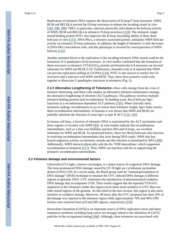
Replication of telomeric DNA requires the dissociation of D-loop/T-loop structures. WRN,BLM and RECQL4 unwind the D-loop structures to release the invading strand in vitro[103, 108, 109]. TRF2, in particular, interacts physically and enhances the helicase activityof WRN, BLM and RECQL4 at telomeric D-loop structures [110]. The telomeric singlestrand binding protein POT1 also improves the D-loop unwinding ability of these threehelicases in vitro [111]. DNA-PKcs, a telomere associated protein, stimulates WRN helicaseactivity on telomeric D-loop substrates. In addition, the length of telomeric G-tails decreasesin DNA-PKcs knockdown cells, and this phenotype is reversed by overexpression of WRNhelicase [112].
Another potential block to the replication of the invading telomeric DNA strand could be theformation of G-quadruplex (G4) structures. In vitro studies confirmed that the formation ofthese structures in telomeric (TTAGGG)n strands and bimolecular G4 structures are favoredsubstrates for WRN and BLM [113]. Furthermore, Kamath-Loeb et al reported that WRNcan prevent replication stalling at G4 DNA [114]. POT1 is also known to resolve the G4structures and it interacts with WRN and BLM. Thus, these three proteins could worktogether to dissociate G-quadruplex structures in telomeres (Fig. 3).
2.2.2 Alternative Lengthening of Telomeres—Rare cells emerge from the crisis oftelomere shortening, and these cells employ an alternative telomere maintenance strategy,the alternative lengthening of telomere (ALT) pathways. This pathway involves multipletelomere binding proteins and recombination. In budding yeast, RecQ helicase Sgs1functions in a recombination dependent ALT pathway [115]. When critically short,telomeres undergo recombination to try to restore their telomeric length, Sgs1 helps resolvethese recombination intermediates. In humans it was shown that WRN and BLM canpartially substitute the function of yeast Sgs1 in type II ALT [115, 116].
In human cell lines, a fraction of telomeric DNA is maintained by the ALT mechanism andthese regions co-localize with WRN [93]. In vitro studies indicate that recombinationintermediates, such as a four-way Holliday junction (HJ) and D-loops, are excellentsubstrates for WRN and BLM. As mentioned before, these two RecQ helicases also functionin resolving recombination intermediates that arise during DNA repair. WRN also hasbranch migration activity on telomeric strands and this function is stimulated by RPA [109].Additionally, WRN interacts physically with the Ku 70/80 heterodimer, which suppressesrecombination at telomeres [117]. Thus, WRN can function with Ku in suppressing thetelomeric recombination intermediates.
2.3 Telomere damage and environmental factorsUltraviolet (UV) light, a known carcinogen, is a major source of exogenous DNA damage.The most pronounced DNA damage caused by UV-B light are cyclobutane pyrimidinedimers (CPD) [118]. In a recent study, the Brash group used an “immunoprecipitation ofDNA damage” (IPoD) technique to measure the UVC induced DNA damage at differentregions of genomic DNA. UVC minimizes the introduction of photosensitized oxidativeDNA damage that accompanies UVB. Their results suggest that the repeated TTAGGGsequences in the telomeres render this region seven times more sensitive to UVC than twoother tested regions of the genome. As described in the next section, this region is also moresensitive to oxidative damage. Moreover, 48 hours after the UVC treatment less than 10% ofthe damage was repaired in the telomere region while approximately 70% and 40% CPDlesions were removed from p53 and 28S regions, respectively [119].
Hexavalent chromium (Cr(VI)) is an important source of DNA replication stress and manyrespiratory problems including lung cancer are strongly linked to the inhalation of Cr(VI)particles in the occupational setting [106]. Although, short telomeres are associated with
Singh et al. Page 9
Mutat Res. Author manuscript; available in PMC 2013 August 01.
NIH
-PA Author Manuscript
NIH
-PA Author Manuscript
NIH
-PA Author Manuscript

increased risk for lung cancer, Cr(VI) exposure does not significantly alter mean telomerelengths [120–122]. However, in cells Cr (IV) is reduced to Cr (III) which reacts with DNAand produces a wide array of lesions. Oxygen radical mediated oxidative DNA damagecould be caused by Cr(III)-complexes targeting the guanine (G) tracts in telomeres as hot-spot for oxidative DNA damage. Moreover, in vitro DNA polymerase arrest induced by Cr(VI) treatment is most potent at templates with G runs [123, 124]. Recent studies haveshown that telomeric abnormalities such as sister telomere loss and sister telomere fusion iselevated in Cr (VI) treated lung cancer cells and human skin fibroblasts. Interestingly,expression of telomerase was able to protect the human skin fibroblasts from theseabnormalities.
2.3.1 Repair of oxidative damage—Telomeric DNA contains repetitive guanine tripletsand thus is hypersensitive to DNA damage induced by oxidative stress [125, 126]. Thetelomeric region (TTAGGG) of genomic DNA isolated from mouse kidney contains moreFpg-sensitive oxidatively damaged sites than in a minisatellite (TG) region [127]. In vitroanalysis has shown that telomeric DNA is prone to oxidative damage because of the tripleguanines (G). Guanine has the lowest oxidation potential among the nucleobases and theGGG sequence has even lower oxidation potential. Hence, telomeric DNA is susceptible tooxidative damage and can contain lesions like 8-oxoguanine (8-oxodG). Numerous studieshave indicated the association between oxidative damage and telomere shortening [128,129]. Oxidative damage is repaired by the base excision repair (BER) process. WRN isbelieved to take part in BER and physically interacts with several proteins involved in BER[5]. Oxidative damage can result in base lesions and DSBs in telomeres and WRN is alsoimplicated in the DSB repair process. Recently, we have observed that WRN and BLMinteract with the in vitro D-loop structures containing 8-oxodG lesion and unwind thesesubstrates more efficiently than the undamaged D-loops [108]. However, still a great deal ofwork needs to be done to assess the exact role of WRN and other RecQ helicases in therepair of oxidative damage in telomeres.
Conclusions and future perspectivesThe RecQ helicases are important for the preservation of genome stability. The presence ofmultiple forms of RecQ helicases in higher eukaryotes could be an adaptive feature toensure proper surveillance of the genome against the harmful effects of variousenvironmental insults. The RecQ helicases have redundant and/or non-redundant functionsdepending on type of DNA damage and DNA repair pathways. Therefore, in the future itwould be very interesting to understand how these RecQ helicases cooperate amongthemselves and with other DNA repair proteins.
AcknowledgmentsWe would like to thank Drs. Venkateswarlu Popuri and Haritha Vallabhaneni for critical reading of the manuscript.This work was in part supported by funds from the Intramural Program of the National Institute on Aging, NIH.
Reference List1. Bernstein KA, Gangloff S, Rothstein R. The RecQ DNA helicases in DNA repair. Annu Rev Genet.
2010; 44:393–417. [PubMed: 21047263]
2. Chu WK, Hickson ID. RecQ helicases: multifunctional genome caretakers. Nat Rev Cancer. 2009;9:644–654. [PubMed: 19657341]
3. Bohr VA. Rising from the RecQ-age: the role of human RecQ helicases in genome maintenance.Trends Biochem Sci. 2008; 33:609–620. [PubMed: 18926708]
4. Singh DK, Ahn B, Bohr VA. Roles of RECQ helicases in recombination based DNA repair,genomic stability and aging. Biogerontology. 2009; 10:235–252. [PubMed: 19083132]
Singh et al. Page 10
Mutat Res. Author manuscript; available in PMC 2013 August 01.
NIH
-PA Author Manuscript
NIH
-PA Author Manuscript
NIH
-PA Author Manuscript

5. Rossi ML, Ghosh AK, Bohr VA. Roles of Werner syndrome protein in protection of genomeintegrity. DNA Repair (Amst). 2010; 9:331–344. [PubMed: 20075015]
6. Fan W, Luo J. RecQ4 facilitates UV light-induced DNA damage repair through interaction withnucleotide excision repair factor xeroderma pigmentosum group A (XPA). J Biol Chem. 2008;283:29037–29044. [PubMed: 18693251]
7. Sangrithi MN, Bernal JA, Madine M, Philpott A, Lee J, Dunphy WG, Venkitaraman AR. Initiationof DNA replication requires the RECQL4 protein mutated in Rothmund-Thomson syndrome. Cell.2005; 121:887–898. [PubMed: 15960976]
8. Xu X, Rochette PJ, Feyissa EA, Su TV, Liu Y. MCM10 mediates RECQ4 association withMCM2-7 helicase complex during DNA replication. EMBO J. 2009; 28:3005–3014. [PubMed:19696745]
9. Im JS, Ki SH, Farina A, Jung DS, Hurwitz J, Lee JK. Assembly of the Cdc45-Mcm2-7-GINScomplex in human cells requires the Ctf4/And-1, RecQL4, and Mcm10 proteins. Proc Natl Acad SciU S A. 2009; 106:15628–15632. [PubMed: 19805216]
10. Aygun O, Svejstrup J, Liu Y. A RECQ5-RNA polymerase II association identified by targetedproteomic analysis of human chromatin. Proc Natl Acad Sci U S A. 2008; 105:8580–8584.[PubMed: 18562274]
11. Izumikawa K, Yanagida M, Hayano T, Tachikawa H, Komatsu W, Shimamoto A, Futami K,Furuichi Y, Shinkawa T, Yamauchi Y, Isobe T, Takahashi N. Association of human DNA helicaseRecQ5beta with RNA polymerase II and its possible role in transcription. Biochem J. 2008;413:505–516. [PubMed: 18419580]
12. Shrivastav M, De Haro LP, Nickoloff JA. Regulation of DNA double-strand break repair pathwaychoice. Cell Res. 2008; 18:134–147. [PubMed: 18157161]
13. Takata M, Sasaki MS, Sonoda E, Morrison C, Hashimoto M, Utsumi H, Yamaguchi-Iwai Y,Shinohara A, Takeda S. Homologous recombination and non-homologous end-joining pathways ofDNA double-strand break repair have overlapping roles in the maintenance of chromosomalintegrity in vertebrate cells. EMBO J. 1998; 17:5497–5508. [PubMed: 9736627]
14. Lieber MR, Ma Y, Pannicke U, Schwarz K. Mechanism and regulation of human non-homologousDNA end-joining. Nat Rev Mol Cell Biol. 2003; 4:712–720. [PubMed: 14506474]
15. Hefferin ML, Tomkinson AE. Mechanism of DNA double-strand break repair by non-homologousend joining. DNA Repair (Amst). 2005; 4:639–648. [PubMed: 15907771]
16. Liang F, Romanienko PJ, Weaver DT, Jeggo PA, Jasin M. Chromosomal double-strand breakrepair in Ku80-deficient cells. Proc Natl Acad Sci U S A. 1996; 93:8929–8933. [PubMed:8799130]
17. Thompson LH, Schild D. Recombinational DNA repair and human disease. Mutat Res. 2002;509:49–78. [PubMed: 12427531]
18. Lan L, Nakajima S, Komatsu K, Nussenzweig A, Shimamoto A, Oshima J, Yasui A. Accumulationof Werner protein at DNA double-strand breaks in human cells. J Cell Sci. 2005; 118:4153–4162.[PubMed: 16141234]
19. Karmakar P, Seki M, Kanamori M, Hashiguchi K, Ohtsuki M, Murata E, Inoue E, Tada S, Lan L,Yasui A, Enomoto T. BLM is an early responder to DNA double-strand breaks. Biochem BiophysRes Commun. 2006; 348:62–69. [PubMed: 16876111]
20. Singh DK, Karmakar P, Aamann M, Schurman SH, May A, Croteau DL, Burks L, Plon SE, BohrVA. The involvement of human RECQL4 in DNA double-strand break repair. Aging Cell. 2010;9:358–371. [PubMed: 20222902]
21. Zheng L, Kanagaraj R, Mihaljevic B, Schwendener S, Sartori AA, Gerrits B, Shevelev I, JanscakP. MRE11 complex links RECQ5 helicase to sites of DNA damage. Nucleic Acids Res. 2009;37:2645–2657. [PubMed: 19270065]
22. Lisby M, Rothstein R. Choreography of recombination proteins during the DNA damage response.DNA Repair (Amst). 2009; 8:1068–1076. [PubMed: 19473884]
23. Sung P, Krejci L, Van KS, Sehorn MG. Rad51 recombinase and recombination mediators. J BiolChem. 2003; 278:42729–42732. [PubMed: 12912992]
24. West SC. Molecular views of recombination proteins and their control. Nat Rev Mol Cell Biol.2003; 4:435–445. [PubMed: 12778123]
Singh et al. Page 11
Mutat Res. Author manuscript; available in PMC 2013 August 01.
NIH
-PA Author Manuscript
NIH
-PA Author Manuscript
NIH
-PA Author Manuscript

25. Kanaar R, Hoeijmakers JH, van G. Molecular mechanisms of DNA double strand break repair.Trends Cell Biol. 1998; 8:483–489. [PubMed: 9861670]
26. Critchlow SE, Jackson SP. DNA end-joining: from yeast to man. Trends Biochem Sci. 1998;23:394–398. [PubMed: 9810228]
27. Hartley KO, Gell D, Smith GC, Zhang H, Divecha N, Connelly MA, Admon A, Lees-Miller SP,Anderson CW, Jackson SP. DNA-dependent protein kinase catalytic subunit: a relative ofphosphatidylinositol 3-kinase and the ataxia telangiectasia gene product. Cell. 1995; 82:849–856.[PubMed: 7671312]
28. Cooper MP, Machwe A, Orren DK, Brosh RM, Ramsden D, Bohr VA. Ku complex interacts withand stimulates the Werner protein. Genes Dev. 2000; 14:907–912. [PubMed: 10783163]
29. Li B, Comai L. Displacement of DNA-PKcs from DNA ends by the Werner syndrome protein.Nucleic Acids Res. 2002; 30:3653–3661. [PubMed: 12202749]
30. Kusumoto R, Dawut L, Marchetti C, Wan LJ, Vindigni A, Ramsden D, Bohr VA. Werner proteincooperates with the XRCC4-DNA ligase IV complex in end-processing. Biochemistry. 2008;47:7548–7556. [PubMed: 18558713]
31. Saintigny Y, Makienko K, Swanson C, Emond MJ, Monnat RJ Jr. Homologous recombinationresolution defect in werner syndrome. Mol Cell Biol. 2002; 22:6971–6978. [PubMed: 12242278]
32. Pichierri P, Rosselli F. The DNA crosslink-induced S-phase checkpoint depends on ATR-CHK1and ATR-NBS1-FANCD2 pathways. EMBO J. 2004; 23:1178–1187. [PubMed: 14988723]
33. Baynton K, Otterlei M, Bjoras M, von KC, Bohr VA, Seeberg E. WRN interacts physically andfunctionally with the recombination mediator protein RAD52. J Biol Chem. 2003; 278:36476–36486. [PubMed: 12750383]
34. Cheng WH, Kusumoto R, Opresko PL, Sui X, Huang S, Nicolette ML, Paull TT, Campisi J,Seidman M, Bohr VA. Collaboration of Werner syndrome protein and BRCA1 in cellularresponses to DNA interstrand cross-links. Nucleic Acids Res. 2006; 34:2751–2760. [PubMed:16714450]
35. Cheng WH, von KC, Opresko PL, Arthur LM, Komatsu K, Seidman MM, Carney JP, Bohr VA.Linkage between Werner syndrome protein and the Mre11 complex via Nbs1. J Biol Chem. 2004;279:21169–21176. [PubMed: 15026416]
36. Otterlei M, Bruheim P, Ahn B, Bussen W, Karmakar P, Baynton K, Bohr VA. Werner syndromeprotein participates in a complex with RAD51, RAD54, RAD54B and ATR in response to ICL-induced replication arrest. J Cell Sci. 2006; 119:5137–5146. [PubMed: 17118963]
37. Pichierri P, Rosselli F, Franchitto A. Werner’s syndrome protein is phosphorylated in an ATR/ATM-dependent manner following replication arrest and DNA damage induced during the S phaseof the cell cycle. Oncogene. 2003; 22:1491–1500. [PubMed: 12629512]
38. Lee SJ, Gartner A, Hyun M, Ahn B, Koo HS. The Caenorhabditis elegans Werner syndromeprotein functions upstream of ATR and ATM in response to DNA replication inhibition anddouble-strand DNA breaks. PLoS Genet. 2010; 6:e1000801. [PubMed: 20062519]
39. Cheng WH, Muftic D, Muftuoglu M, Dawut L, Morris C, Helleday T, Shiloh Y, Bohr VA. WRN isrequired for ATM activation and the S-phase checkpoint in response to interstrand cross-link-induced DNA double-strand breaks. Mol Biol Cell. 2008; 19:3923–3933. [PubMed: 18596239]
40. Ammazzalorso F, Pirzio LM, Bignami M, Franchitto A, Pichierri P. ATR and ATM differentlyregulate WRN to prevent DSBs at stalled replication forks and promote replication fork recovery.EMBO J. 2010; 29:3156–3169. [PubMed: 20802463]
41. Constantinou A, Tarsounas M, Karow JK, Brosh RM, Bohr VA, Hickson ID, West SC. Werner’ssyndrome protein (WRN) migrates Holliday junctions and co-localizes with RPA upon replicationarrest. EMBO Rep. 2000; 1:80–84. [PubMed: 11256630]
42. Shen J, Loeb LA. Unwinding the molecular basis of the Werner syndrome. Mech Ageing Dev.2001; 122:921–944. [PubMed: 11348659]
43. Lee JW, Harrigan J, Opresko PL, Bohr VA. Pathways and functions of the Werner syndromeprotein. Mech Ageing Dev. 2005; 126:79–86. [PubMed: 15610765]
44. Rodriguez-Lopez AM, Whitby MC, Borer CM, Bachler MA, Cox LS. Correction of proliferationand drug sensitivity defects in the progeroid Werner’s Syndrome by Holliday junction resolution.Rejuvenation Res. 2007; 10:27–40. [PubMed: 17378750]
Singh et al. Page 12
Mutat Res. Author manuscript; available in PMC 2013 August 01.
NIH
-PA Author Manuscript
NIH
-PA Author Manuscript
NIH
-PA Author Manuscript

45. Oshima J, Huang S, Pae C, Campisi J, Schiestl RH. Lack of WRN results in extensive deletion atnonhomologous joining ends. Cancer Res. 2002; 62:547–551. [PubMed: 11809708]
46. Karmakar P, Piotrowski J, Brosh RM Jr, Sommers JA, Miller SP, Cheng WH, Snowden CM,Ramsden DA, Bohr VA. Werner protein is a target of DNA-dependent protein kinase in vivo andin vitro, and its catalytic activities are regulated by phosphorylation. J Biol Chem. 2002;277:18291–18302. [PubMed: 11889123]
47. Chaganti RS, Schonberg S, German J. A manyfold increase in sister chromatid exchanges inBloom’s syndrome lymphocytes. Proc Natl Acad Sci U S A. 1974; 71:4508–4512. [PubMed:4140506]
48. German J. Bloom’s syndrome. Dermatol Clin. 1995; 13:7–18. [PubMed: 7712653]
49. Wu L, Davies SL, Levitt NC, Hickson ID. Potential role for the BLM helicase in recombinationalrepair via a conserved interaction with RAD51. J Biol Chem. 2001; 276:19375–19381. [PubMed:11278509]
50. Braybrooke JP, Li JL, Wu L, Caple F, Benson FE, Hickson ID. Functional interaction between theBloom’s syndrome helicase and the RAD51 paralog, RAD51L3 (RAD51D). J Biol Chem. 2003;278:48357–48366. [PubMed: 12975363]
51. Beamish H, Kedar P, Kaneko H, Chen P, Fukao T, Peng C, Beresten S, Gueven N, Purdie D, Lees-Miller S, Ellis N, Kondo N, Lavin MF. Functional link between BLM defective in Bloom’ssyndrome and the ataxia-telangiectasia-mutated protein, ATM. J Biol Chem. 2002; 277:30515–30523. [PubMed: 12034743]
52. Sharma S, Doherty KM, Brosh RM Jr. Mechanisms of RecQ helicases in pathways of DNAmetabolism and maintenance of genomic stability. Biochem J. 2006; 398:319–337. [PubMed:16925525]
53. Pedrazzi G, Bachrati CZ, Selak N, Studer I, Petkovic M, Hickson ID, Jiricny J, Stagljar I. TheBloom’s syndrome helicase interacts directly with the human DNA mismatch repair proteinhMSH6. Biol Chem. 2003; 384:1155–1164. [PubMed: 12974384]
54. Nimonkar AV, Ozsoy AZ, Genschel J, Modrich P, Kowalczykowski SC. Human exonuclease 1and BLM helicase interact to resect DNA and initiate DNA repair. Proc Natl Acad Sci U S A.2008; 105:16906–16911. [PubMed: 18971343]
55. Bugreev DV, Yu X, Egelman EH, Mazin AV. Novel pro- and anti-recombination activities of theBloom’s syndrome helicase. Genes Dev. 2007; 21:3085–3094. [PubMed: 18003860]
56. Wu L, Hickson ID. DNA helicases required for homologous recombination and repair of damagedreplication forks. Annu Rev Genet. 2006; 40:279–306. [PubMed: 16856806]
57. Wu L, Hickson ID. The Bloom’s syndrome helicase suppresses crossing over during homologousrecombination. Nature. 2003; 426:870–874. [PubMed: 14685245]
58. Adams MD, McVey M, Sekelsky JJ. Drosophila BLM in double-strand break repair by synthesis-dependent strand annealing. Science. 2003; 299:265–267. [PubMed: 12522255]
59. Hu P, Beresten SF, van Brabant AJ, Ye TZ, Pandolfi PP, Johnson FB, Guarente L, Ellis NA.Evidence for BLM and Topoisomerase IIIalpha interaction in genomic stability. Hum Mol Genet.2001; 10:1287–1298. [PubMed: 11406610]
60. Wu L, Davies SL, North PS, Goulaouic H, Riou JF, Turley H, Gatter KC, Hickson ID. TheBloom’s syndrome gene product interacts with topoisomerase III. J Biol Chem. 2000; 275:9636–9644. [PubMed: 10734115]
61. Yin J, Sobeck A, Xu C, Meetei AR, Hoatlin M, Li L, Wang W. BLAP75, an essential componentof Bloom’s syndrome protein complexes that maintain genome integrity. EMBO J. 2005;24:1465–1476. [PubMed: 15775963]
62. Wu L, Bachrati CZ, Ou J, Xu C, Yin J, Chang M, Wang W, Li L, Brown GW, Hickson ID.BLAP75/RMI1 promotes the BLM-dependent dissolution of homologous recombinationintermediates. Proc Natl Acad Sci U S A. 2006; 103:4068–4073. [PubMed: 16537486]
63. Raynard S, Bussen W, Sung P. A double Holliday junction dissolvasome comprising BLM,topoisomerase IIIalpha, and BLAP75. J Biol Chem. 2006; 281:13861–13864. [PubMed:16595695]
Singh et al. Page 13
Mutat Res. Author manuscript; available in PMC 2013 August 01.
NIH
-PA Author Manuscript
NIH
-PA Author Manuscript
NIH
-PA Author Manuscript

64. Bussen W, Raynard S, Busygina V, Singh AK, Sung P. Holliday junction processing activity of theBLM-Topo IIIalpha-BLAP75 complex. J Biol Chem. 2007; 282:31484–31492. [PubMed:17728255]
65. Davies SL, North PS, Dart A, Lakin ND, Hickson ID. Phosphorylation of the Bloom’s syndromehelicase and its role in recovery from S-phase arrest. Mol Cell Biol. 2004; 24:1279–1291.[PubMed: 14729972]
66. Lonn U, Lonn S, Nylen U, Winblad G, German J. An abnormal profile of DNA replicationintermediates in Bloom’s syndrome. Cancer Res. 1990; 50:3141–3145. [PubMed: 2110504]
67. Davalos AR, Kaminker P, Hansen RK, Campisi J. ATR and ATM-dependent movement of BLMhelicase during replication stress ensures optimal ATM activation and 53BP1 focus formation.Cell Cycle. 2004; 3:1579–1586. [PubMed: 15539948]
68. Karow JK, Constantinou A, Li JL, West SC, Hickson ID. The Bloom’s syndrome gene productpromotes branch migration of holliday junctions. Proc Natl Acad Sci U S A. 2000; 97:6504–6508.[PubMed: 10823897]
69. Liberi G, Maffioletti G, Lucca C, Chiolo I, Baryshnikova A, Cotta-Ramusino C, Lopes M,Pellicioli A, Haber JE, Foiani M. Rad51-dependent DNA structures accumulate at damagedreplication forks in sgs1 mutants defective in the yeast ortholog of BLM RecQ helicase. GenesDev. 2005; 19:339–350. [PubMed: 15687257]
70. Johnson FB, Lombard DB, Neff NF, Mastrangelo MA, Dewolf W, Ellis NA, Marciniak RA, YinY, Jaenisch R, Guarente L. Association of the Bloom syndrome protein with topoisomeraseIIIalpha in somatic and meiotic cells. Cancer Res. 2000; 60:1162–1167. [PubMed: 10728666]
71. Vennos EM, Collins M, James WD. Rothmund-Thomson syndrome: review of the world literature.J Am Acad Dermatol. 1992; 27:750–762. [PubMed: 1430398]
72. Der K, McGill VJJ, Vekemans M, Kopelman HR. Clonal lines of aneuploid cells in Rothmund-Thomson syndrome. Am J Med Genet. 1990; 37:336–339. [PubMed: 2260560]
73. Durand F, Castorina P, Morant C, Delobel B, Barouk E, Modiano P. [Rothmund-Thomsonsyndrome, trisomy 8 mosaicism and RECQ4 gene mutation]. Ann Dermatol Venereol. 2002;129:892–895. [PubMed: 12218919]
74. Anbari KK, Ierardi-Curto LA, Silber JS, Asada N, Spinner N, Zackai EH, Belasco J, MorrissetteJD, Dormans JP. Two primary osteosarcomas in a patient with Rothmund-Thomson syndrome.Clin Orthop Relat Res. 2000:213–223. [PubMed: 10986997]
75. Mann MB, Hodges CA, Barnes E, Vogel H, Hassold TJ, Luo G. Defective sister-chromatidcohesion, aneuploidy and cancer predisposition in a mouse model of type II Rothmund-Thomsonsyndrome. Hum Mol Genet. 2005; 14:813–825. [PubMed: 15703196]
76. Wu J, Capp C, Feng L, Hsieh TS. Drosophila homologue of the Rothmund-Thomson syndromegene: essential function in DNA replication during development. Dev Biol. 2008; 323:130–142.[PubMed: 18755177]
77. Petkovic M, Dietschy T, Freire R, Jiao R, Stagljar I. The human Rothmund-Thomson syndromegene product, RECQL4, localizes to distinct nuclear foci that coincide with proteins involved inthe maintenance of genome stability. J Cell Sci. 2005; 118:4261–4269. [PubMed: 16141230]
78. Kumata Y, Tada S, Yamanada Y, Tsuyama T, Kobayashi T, Dong YP, Ikegami K, Murofushi H,Seki M, Enomoto T. Possible involvement of RecQL4 in the repair of double-strand DNA breaksin Xenopus egg extracts. Biochim Biophys Acta. 2007; 1773:556–564. [PubMed: 17320201]
79. Sharma S, Brosh RM Jr. Human RECQ1 is a DNA damage responsive protein required forgenotoxic stress resistance and suppression of sister chromatid exchanges. PLoS One. 2007;2:e1297. [PubMed: 18074021]
80. LeRoy G, Carroll R, Kyin S, Seki M, Cole MD. Identification of RecQL1 as a Holliday junctionprocessing enzyme in human cell lines. Nucleic Acids Res. 2005; 33:6251–6257. [PubMed:16260474]
81. Bugreev DV, Brosh RM Jr, Mazin AV. RECQ1 possesses DNA branch migration activity. J BiolChem. 2008; 283:20231–20242. [PubMed: 18495662]
82. Hu Y, Raynard S, Sehorn MG, Lu X, Bussen W, Zheng L, Stark JM, Barnes EL, Chi P, Janscak P,Jasin M, Vogel H, Sung P, Luo G. RECQL5/Recql5 helicase regulates homologous recombination
Singh et al. Page 14
Mutat Res. Author manuscript; available in PMC 2013 August 01.
NIH
-PA Author Manuscript
NIH
-PA Author Manuscript
NIH
-PA Author Manuscript

and suppresses tumor formation via disruption of Rad51 presynaptic filaments. Genes Dev. 2007;21:3073–3084. [PubMed: 18003859]
83. Schwendener S, Raynard S, Paliwal S, Cheng A, Kanagaraj R, Shevelev I, Stark JM, Sung P,Janscak P. Physical interaction of RECQ5 helicase with RAD51 facilitates its anti-recombinaseactivity. J Biol Chem. 2010; 285:15739–15745. [PubMed: 20348101]
84. OLOVNIKO AM. Principle of Marginotomy in Template Synthesis of Polynucleotides. DokladyAkademii Nauk Sssr. 1971; 201:1496. [PubMed: 5158754]
85. Levy MZ, Allsopp RC, Futcher AB, Greider CW, Harley CB. Telomere End-Replication Problemand Cell Aging. Journal of Molecular Biology. 1992; 225:951–960. [PubMed: 1613801]
86. Watson JD. Origin of Concatemeric T7 Dna. Nature-New Biology. 1972; 239:197. [PubMed:4507727]
87. Blackburn EH, Szostak JW. The Molecular-Structure of Centromeres and Telomeres. AnnualReview of Biochemistry. 1984; 53:163–194.
88. Griffith JD, Comeau L, Rosenfield S, Stansel RM, Bianchi A, Moss H, de Lange T. Mammaliantelomeres end in a large duplex loop. Cell. 1999; 97:503–514. [PubMed: 10338214]
89. de Lange T. Shelterin: the protein complex that shapes and safeguards human telomeres. Genes &Development. 2005; 19:2100–2110. [PubMed: 16166375]
90. di Fagagna FD, Reaper PM, Clay-Farrace L, Fiegler H, Carr P, von Zglinicki T, Saretzki G, CarterNP, Jackson SP. A DNA damage checkpoint response in telomere-initiated senescence. Nature.2003; 426:194–198. [PubMed: 14608368]
91. di Fagagna FD, Teo SH, Jackson SP. Functional links between telomeres and proteins of the DNA-damage response. Genes & Development. 2004; 18:1781–1799. [PubMed: 15289453]
92. Harley CB, Futcher AB, Greider CW. Telomeres Shorten During Aging of Human Fibroblasts.Nature. 1990; 345:458–460. [PubMed: 2342578]
93. Opresko PL. Telomere ResQue and preservation--roles for the Werner syndrome protein and otherRecQ helicases. Mech Ageing Dev. 2008; 129:79–90. [PubMed: 18054793]
94. Greider CW, Blackburn EH. Telomeres, telomerase and cancer. Scientific American. 1996;274:92–97. [PubMed: 8560215]
95. de Lange T. Protection of mammalian telomeres. Oncogene. 2002; 21:532–540. [PubMed:11850778]
96. Karlseder J, Smogorzewska A, de Lange T. Senescence induced by altered telomere state, nottelomere loss. Science. 2002; 295:2446–2449. [PubMed: 11923537]
97. Hockemeyer D, Sfeir AJ, Shay JW, Wright WE, de Lange T. POT1 protects telomeres from atransient DNA damage response and determines how human chromosomes end. Embo Journal.2005; 24:2667–2678. [PubMed: 15973431]
98. Wu L, Multani AS, He H, Cosme-Blanco W, Deng Y, Deng JM, Bachilo O, Pathak S, Tahara H,Bailey SM, Deng YB, Behringer RR, Chang S. Pot1 deficiency initiates DNA damage checkpointactivation and aberrant homologous recombination at telomeres. Cell. 2006; 126:49–62. [PubMed:16839876]
99. Chang S, Multani AS, Cabrera NG, Naylor ML, Laud P, Lombard D, Pathak S, Guarente L,DePinho RA. Essential role of limiting telomeres in the pathogenesis of Werner syndrome. NatureGenetics. 2004; 36:877–882. [PubMed: 15235603]
100. Kruk PA, Rampino NJ, Bohr VA. Dna-Damage and Repair in Telomeres - Relation to Aging.Proceedings of the National Academy of Sciences of the United States of America. 1995;92:258–262. [PubMed: 7816828]
101. Crabbe L, Jauch A, Naeger CM, Holtgreve-Grez H, Karlseder J. Telomere dysfunction as a causeof genomic instability in Werner syndrome. Proceedings of the National Academy of Sciences ofthe United States of America. 2007; 104:2205–2210. [PubMed: 17284601]
102. Dejardin J, Kingston RE. Purification of proteins associated with specific genomic Loci. Cell.2009; 136:175–186. [PubMed: 19135898]
103. Opresko PL, Otterlei M, Graakjaer J, Bruheim P, Dawut L, Kolvraa S, May A, Seidman MM,Bohr VA. The Werner syndrome helicase and exonuclease cooperate to resolve telomeric Dloops in a manner regulated by TRF1 and TRF2. Mol Cell. 2004; 14:763–774. [PubMed:15200954]
Singh et al. Page 15
Mutat Res. Author manuscript; available in PMC 2013 August 01.
NIH
-PA Author Manuscript
NIH
-PA Author Manuscript
NIH
-PA Author Manuscript

104. Sfeir A, Kosiyatrakul ST, Hockemeyer D, MacRae SL, Karlseder J, Schildkraut CL, de LT.Mammalian telomeres resemble fragile sites and require TRF1 for efficient replication. Cell.2009; 138:90–103. [PubMed: 19596237]
105. Opresko PL, Cheng WH, von Kobbe C, Harrigan JA, Bohr VA. Werner syndrome and thefunction of the Werner protein; what they can teach us about the molecular aging process.Carcinogenesis. 2003; 24:791–802. [PubMed: 12771022]
106. Liu FJ, Barchowsky A, Opresko PL. The Werner syndrome protein suppresses telomericinstability caused by chromium (VI) induced DNA replication stress. PLoS One. 2010; 5:e11152.[PubMed: 20585393]
107. Crabbe L, Verdun RE, Haggblom CI, Karlseder J. Defective telomere lagging strand synthesis incells lacking WRN helicase activity. Science. 2004; 306:1951–1953. [PubMed: 15591207]
108. Ghosh A, Rossi ML, Aulds J, Croteau D, Bohr VA. Telomeric D-loops containing 8-oxo-2′-deoxyguanosine are preferred substrates for Werner and Bloom syndrome helicases and arebound by POT1. J Biol Chem. 2009; 284:31074–31084. [PubMed: 19734539]
109. Sowd G, Lei M, Opresko PL. Mechanism and substrate specificity of telomeric protein POT1stimulation of the Werner syndrome helicase. Nucleic Acids Res. 2008; 36:4242–4256.[PubMed: 18583366]
110. Opresko PL, von Kobbe C, Laine JP, Harrigan J, Hickson ID, Bohr VA. Telomere-bindingprotein TRF2 binds to and stimulates the Werner and Bloom syndrome helicases. Journal ofBiological Chemistry. 2002; 277:41110–41119. [PubMed: 12181313]
111. Opresko PL, Mason PA, Podell ER, Lei M, Hickson ID, Cech TR, Bohr VA. POT1 stimulatesRecQ helicases WRN and BLM to unwind telomeric DNA substrates. Journal of BiologicalChemistry. 2005; 280:32069–32080. [PubMed: 16030011]
112. Kusumoto-Matsuo R, Opresko PL, Ramsden D, Tahara H, Bohr VA. Cooperation of DNA-PKcsand WRN helicase in the maintenance of telomeric D-loops. Aging (Albany NY). 2010; 2:274–284. [PubMed: 20519774]
113. Mohaghegh P, Karow JK, Brosh RM Jr, Bohr VA, Hickson ID. The Bloom’s and Werner’ssyndrome proteins are DNA structure-specific helicases. Nucleic Acids Res. 2001; 29:2843–2849. [PubMed: 11433031]
114. Kamath-Loeb AS, Loeb LA, Johansson E, Burgers PMJ, Fry M. Interactions between the Wernersyndrome helicase and DNA polymerase delta specifically facilitate copying of tetraplex andhairpin structures of the d(CGG)(n) trinucleotide repeat sequence. Journal of BiologicalChemistry. 2001; 276:16439–16446. [PubMed: 11279038]
115. Cohen H, Sinclair DA. Recombination-mediated lengthening of terminal telomeric repeatsrequires the Sgs1 DNA helicase. Proc Natl Acad Sci U S A. 2001; 98:3174–3179. [PubMed:11248051]
116. Mandell JG, Goodrich KJ, Bahler J, Cech TR. Expression of a RecQ helicase homolog affectsprogression through crisis in fission yeast lacking telomerase. J Biol Chem. 2005; 280:5249–5257. [PubMed: 15591066]
117. Karmakar P, Snowden CM, Ramsden DA, Bohr VA. Ku heterodimer binds to both ends of theWerner protein and functional interaction occurs at the Werner N-terminus. Nucleic Acids Res.2002; 30:3583–3591. [PubMed: 12177300]
118. Cadet J, Sage E, Douki T. Ultraviolet radiation-mediated damage to cellular DNA. Mutat Res.2005; 571:3–17. [PubMed: 15748634]
119. Rochette PJ, Brash DE. Human telomeres are hypersensitive to UV-induced DNA Damage andrefractory to repair. PLoS Genet. 2010; 6:e1000926. [PubMed: 20442874]
120. Glaviano A, Nayak V, Cabuy E, Baird DM, Yin Z, Newson R, Ladon D, Rubio MA, SlijepcevicP, Lyng F, Mothersill C, Case CP. Effects of hTERT on metal ion-induced genomic instability.Oncogene. 2006; 25:3424–3435. [PubMed: 16449970]
121. Glaviano A, Mothersill C, Case CP, Rubio MA, Newson R, Lyng F. Effects of hTERT ongenomic instability caused by either metal or radiation or combined exposure. Mutagenesis.2009; 24:25–33. [PubMed: 18776173]
Singh et al. Page 16
Mutat Res. Author manuscript; available in PMC 2013 August 01.
NIH
-PA Author Manuscript
NIH
-PA Author Manuscript
NIH
-PA Author Manuscript

122. Wu X, Amos CI, Zhu Y, Zhao H, Grossman BH, Shay JW, Luo S, Hong WK, Spitz MR.Telomere dysfunction: a potential cancer predisposition factor. J Natl Cancer Inst. 2003;95:1211–1218. [PubMed: 12928346]
123. Bridgewater LC, Manning FC, Patierno SR. Arrest of replication by mammalian DNApolymerases alpha and beta caused by chromium-DNA lesions. Mol Carcinog. 1998; 23:201–206. [PubMed: 9869448]
124. Xu J, Bubley GJ, Detrick B, Blankenship LJ, Patierno SR. Chromium(VI) treatment of normalhuman lung cells results in guanine-specific DNA polymerase arrest, DNA-DNA cross-links andS-phase blockade of cell cycle. Carcinogenesis. 1996; 17:1511–1517. [PubMed: 8706257]
125. Henle ES, Han Z, Tang N, Rai P, Luo Y, Linn S. Sequence-specific DNA cleavage by Fe2+-mediated fenton reactions has possible biological implications. J Biol Chem. 1999; 274:962–971.[PubMed: 9873038]
126. Petersen S, Saretzki G, von ZT. Preferential accumulation of single-stranded regions in telomeresof human fibroblasts. Exp Cell Res. 1998; 239:152–160. [PubMed: 9511733]
127. Rhee DB, Ghosh A, Lu J, Bohr VA, Liu Y. Factors that influence telomeric oxidative basedamage and repair by DNA glycosylase OGG1. DNA Repair (Amst). 2011; 10:34–44. [PubMed:20951653]
128. Newman JPA, Banerjee B, Fang WR, Poonepalli A, Balakrishnan L, Low GKM, BhattacharjeeRN, Akira S, Jayapal M, Melendez AJ, Baskar R, Lee HW, Hande MP. Short dysfunctionaltelomeres impair the repair of arsenite-induced oxidative damage in mouse cells. Journal ofCellular Physiology. 2008; 214:796–809. [PubMed: 17849448]
129. Satoh M, Ishikawa Y, Takahashi Y, Itoh T, Minami Y, Nakamura M. Association betweenoxidative DNA damage and telomere shortening in circulating endothelial progenitor cellsobtained from metabolic syndrome patients with coronary artery disease. Atherosclerosis. 2008;198:347–353. [PubMed: 17983621]
Singh et al. Page 17
Mutat Res. Author manuscript; available in PMC 2013 August 01.
NIH
-PA Author Manuscript
NIH
-PA Author Manuscript
NIH
-PA Author Manuscript

Fig. 1.Involvement of RecQ helicases in maintenance of genomic integrity. Schematic diagramsummarizing the different kinds of DNA lesions caused by various environmental factors.The association of different RecQ helicases (RecQ1, WRN, BLM, RecQL4, and RecQL5)with replication, base excision repair (BER), double strand break repair (DSBR),transcription, and telomere maintenance are shown by arrows.Abbreviations: Gox, 8-oxoG; AP-apurinic or apyrimidinic; CPD-cyclobutane pyrimidinedimmer.
Singh et al. Page 18
Mutat Res. Author manuscript; available in PMC 2013 August 01.
NIH
-PA Author Manuscript
NIH
-PA Author Manuscript
NIH
-PA Author Manuscript

Fig. 2.RecQ helicases are involved in multiple steps of the DNA double strand break repairpathways. The members of the RecQ helicases interacts with various key proteins involvedin different steps of both the homologous recombination (HR) pathway and the non-homologous end-joining (NHEJ) pathway of DSB repair (see text for details).
Singh et al. Page 19
Mutat Res. Author manuscript; available in PMC 2013 August 01.
NIH
-PA Author Manuscript
NIH
-PA Author Manuscript
NIH
-PA Author Manuscript

Fig. 3.Role of different RecQ helicases in replication, recombination, and repair processes attelomeres (see text for details)
Singh et al. Page 20
Mutat Res. Author manuscript; available in PMC 2013 August 01.
NIH
-PA Author Manuscript
NIH
-PA Author Manuscript
NIH
-PA Author Manuscript
Copyright © 2022 FDOKUMEN