Positional stability of single double-strand breaks in mammalian cells
15
Positional stability of single double-strand breaks in mammalian cells Evi Soutoglou 1 , Jonas F. Dorn 2 , Kundan Sengupta 3 , Maria Jasin 4 , Andre Nussenzweig 5 , Thomas Ried 3 , Gaudenz Danuser 2 , and Tom Misteli 16 1National Cancer Institute, NIH, Bethesda, MD 20892, USA. 2The Scripps Research Institute, La Jolla, CA 92037, USA. 3Genetics Branch, National Cancer Institute, NIH, Bethesda, MD 20892, USA. 4Memorial Sloan Kettering Cancer Center, New York, NY 10021, USA. 5Experimental Immunology Branch, National Cancer Institute, NIH, Bethesda, MD 20892, USA. Abstract Formation of cancerous translocations requires the illegitimate joining of chromosomes containing double-strand breaks (DSBs). It is unknown how broken chromosome ends find their translocation partners within the cell nucleus. Here, we have visualized and quantitatively analysed the dynamics of single DSBs in living mammalian cells. We demonstrate that broken ends are positionally stable and unable to roam the cell nucleus. Immobilization of broken chromosome ends requires the DNA- end binding protein Ku80, but is independent of DNA repair factors, H2AX, the MRN complex and the cohesion complex. DSBs preferentially undergo translocations with neighbouring chromosomes and loss of local positional constraint correlates with elevated genomic instability. These results support a contact-first model in which chromosome translocations predominantly form among spatially proximal DSBs. DSBs occur frequently in the genome through the action of DNA-damaging agents or during genome replication 1,2 . DSBs are hazardous to the cell because failure to properly repair them may lead to tumorigenic trans-locations 3 . How broken ends of different chromosomes meet in the cell nucleus to eventually form a translocation is poorly understood 4 . Two hypotheses have been put forth: the ‘contact-first’ model proposes that interactions between breaks on distinct chromosomes can only take place when the breaks are created in chromatid fibres that colocalize at the time of DNA damage 5 . In contrast, the ‘breakage-first’ hypothesis postulates that breaks formed at distant locations are able to scan the nuclear space for potential partners and come together to produce translocations 6 . The two models make divergent predictions as to the dynamic behaviour of broken chromosome ends. In the breakage-first model, single DSBs are required to undergo large-scale motions within the cell nucleus and must be able to roam the nuclear space in search of appropriate interaction partners. In the contact-first model, only limited local positional motion of DSBs is expected. The available experimental data is contradictory: in mammalian cells, induction of extensive chromosome damage using ultra- soft X-rays 7 , laser microirradiation 8 or γ-irradiation 8 indicates that damaged DNA is largely stationary. In contrast, α-particle irradiation leads to large-scale motion and clustering of 6Correspondence should be addressed to T.M. (e-mail: [email protected]). AUTHOR CONTRIBUTIONS E.S. and T.M. designed the study, E.S., J.F.D. and K.S. performed the experiments, J.M., A.N. and T.R. provided reagents and advice, and E.S. and T.M. wrote the manuscript. COMPETING FINANCIAL INTERESTS The authors declare no competing financial interests. Note: Supplementary Information is available on the Nature Cell Biology website. NIH Public Access Author Manuscript Nat Cell Biol. Author manuscript; available in PMC 2008 July 3. Published in final edited form as: Nat Cell Biol. 2007 June ; 9(6): 675–682. NIH-PA Author Manuscript NIH-PA Author Manuscript NIH-PA Author Manuscript
-
Upload
independent -
Category
Documents
-
view
2 -
download
0
Transcript of Positional stability of single double-strand breaks in mammalian cells

Positional stability of single double-strand breaks in mammaliancells
Evi Soutoglou1, Jonas F. Dorn2, Kundan Sengupta3, Maria Jasin4, Andre Nussenzweig5,Thomas Ried3, Gaudenz Danuser2, and Tom Misteli16
1National Cancer Institute, NIH, Bethesda, MD 20892, USA.
2The Scripps Research Institute, La Jolla, CA 92037, USA.
3Genetics Branch, National Cancer Institute, NIH, Bethesda, MD 20892, USA.
4Memorial Sloan Kettering Cancer Center, New York, NY 10021, USA.
5Experimental Immunology Branch, National Cancer Institute, NIH, Bethesda, MD 20892, USA.
AbstractFormation of cancerous translocations requires the illegitimate joining of chromosomes containingdouble-strand breaks (DSBs). It is unknown how broken chromosome ends find their translocationpartners within the cell nucleus. Here, we have visualized and quantitatively analysed the dynamicsof single DSBs in living mammalian cells. We demonstrate that broken ends are positionally stableand unable to roam the cell nucleus. Immobilization of broken chromosome ends requires the DNA-end binding protein Ku80, but is independent of DNA repair factors, H2AX, the MRN complex andthe cohesion complex. DSBs preferentially undergo translocations with neighbouring chromosomesand loss of local positional constraint correlates with elevated genomic instability. These resultssupport a contact-first model in which chromosome translocations predominantly form amongspatially proximal DSBs.
DSBs occur frequently in the genome through the action of DNA-damaging agents or duringgenome replication1,2. DSBs are hazardous to the cell because failure to properly repair themmay lead to tumorigenic trans-locations3. How broken ends of different chromosomes meetin the cell nucleus to eventually form a translocation is poorly understood4. Two hypotheseshave been put forth: the ‘contact-first’ model proposes that interactions between breaks ondistinct chromosomes can only take place when the breaks are created in chromatid fibres thatcolocalize at the time of DNA damage5. In contrast, the ‘breakage-first’ hypothesis postulatesthat breaks formed at distant locations are able to scan the nuclear space for potential partnersand come together to produce translocations6. The two models make divergent predictions asto the dynamic behaviour of broken chromosome ends. In the breakage-first model, singleDSBs are required to undergo large-scale motions within the cell nucleus and must be able toroam the nuclear space in search of appropriate interaction partners. In the contact-first model,only limited local positional motion of DSBs is expected. The available experimental data iscontradictory: in mammalian cells, induction of extensive chromosome damage using ultra-soft X-rays7, laser microirradiation8 or γ-irradiation8 indicates that damaged DNA is largelystationary. In contrast, α-particle irradiation leads to large-scale motion and clustering of
6Correspondence should be addressed to T.M. (e-mail: [email protected]).AUTHOR CONTRIBUTIONS E.S. and T.M. designed the study, E.S., J.F.D. and K.S. performed the experiments, J.M., A.N. and T.R.provided reagents and advice, and E.S. and T.M. wrote the manuscript.COMPETING FINANCIAL INTERESTS The authors declare no competing financial interests.Note: Supplementary Information is available on the Nature Cell Biology website.
NIH Public AccessAuthor ManuscriptNat Cell Biol. Author manuscript; available in PMC 2008 July 3.
Published in final edited form as:Nat Cell Biol. 2007 June ; 9(6): 675–682.
NIH
-PA Author Manuscript
NIH
-PA Author Manuscript
NIH
-PA Author Manuscript
damaged sites6. In addition, observations in Saccharomyces cerevisiae have suggested thatalthough broken chromosome ends do not physically separate9–11, unrepaired loci are able tosearch the nuclear space for appropriate translocation partners and are recruited into commonrepair foci after damage, suggesting that they are able to undergo long range movements12,13.
To directly study the dynamics of single DSBs in living mammalian cells in vivo, we developeda cell system in which we can induce a DSB at a defined genomic site and follow the fate ofeach of the two damaged DNA ends in real time. NIH3T3 stable cell lines (NIH2/4) weregenerated containing a copy of the unique 18 nucleotide ISceI restriction site flanked by anarray of 256 copies of the lac-repressor binding site and by 96 copies of the tetracyclineresponse element (L-ISceI-T array; Fig. 1a)14. The lac- and tet-arrays were visualizedsimultaneously by expression of CFP–lac-repressor and YFP–tet-repressor, respectively (Fig.1b). To temporarily control the induction of a DSB at the L-ISceI-T array in a single livingcell, we took advantage of glucocorticoid-receptor chimeras that translocate from thecytoplasm to the nucleus on binding to the synthetic ligand triamcinolone acetonide (Fig. 1a)15. A chimera between the ISceI restriction endonuclease and the ligand-binding domain ofthe glucocorticoid receptor in frame with monomeric RFP (ISceI–GR) is cytoplasmic in theabsence of triamcinolone acetonide and no DSBs were detected when cells were stained forphosphorylated H2AX (γ-H2AX; Fig. 1c). As expected, when triamcinolone acetonide wasadded ISceI–GR translocated to the nucleus within 2 min and lead to rapid induction of a DSBat the array, as judged by detection of a single nuclear γ-H2AX focus (Fig. 1c). More than 75%of cells showed γ-H2AX accumulation at the array within 5 min and the percentage of cellswith γ-H2AX staining at the array was 85–90% after 15 min (Fig. 1d). The effect of ISceI–GRwas specific for the array as no additional sites of γ-H2AX accumulation were detected.Recruitment of repair factors occurred with kinetics similar to those reported in fixed and livingcells on induction of DNA damage by laser microirradiation16. The repair factor MDC1accumulated with similar kinetics to γ-H2AX, followed by 53BP1, which associated with thearray within 5 min in 30% of cells and within 15 min in 75% of cells (Fig. 1d). Concomitantstaining of 35–40% of cells with the single strand binding protein RPA indicated that at leasta fraction of cleaved ends was resected during that time (see Supplementary Information, Fig.S1). Cleavage kinetics were confirmed directly by ligation-mediated PCR using primersflanking the ISceI site17 (Fig. 1e). Cleavage occurred rapidly and reached a steady-state plateauof ~50% after 30 min (Fig. 1e).
To directly analyse the positional motion of broken chromosome ends, the YFP–tet and CFP–lac-tagged regions flanking the ISceI site were simultaneously visualized using multicolour invivo imaging (Fig. 2 and see Methods). To determine to what degree broken chromosome endsundergo global movements within the nucleus, complete three-dimensional stacks of the twosignals were acquired every 30 s for up to 1 h after the addition of triamcinolone acetonide,and the movement of the CFP and YFP arrays was tracked in three-dimensional space (seeMethods and Supplementary Information, Materials). Qualitative analysis of the position ofthe broken chromosome ends within the cell nucleus indicated extremely limited motion of thetagged ends and no significant change in the spatial location of either tag occurred (Fig. 2a).The CFP and YFP signals did not clearly separate, although changes in their degree of overlapwere apparent during the time course (Fig. 2a and see below). The positional stability was notdue to light damage, nor was it an artifact of the experimental system due to binding of the lacand tet repressors to the array as unbroken chromosome loci under-went similar local motion,as previously observed in other experimental systems (data not shown)18–20.
To exclude the possibility that separation of DSBs occurs at later times after breakage, cellpopulations (n = 100) were scored for separation of the CFP–lac and YFP–tet labels at timesup to 24 h after triamcinolone acetonide addition (Fig. 2b). No significant separation of the
Soutoglou et al. Page 2
Nat Cell Biol. Author manuscript; available in PMC 2008 July 3.
NIH
-PA Author Manuscript
NIH
-PA Author Manuscript
NIH
-PA Author Manuscript
tags was observed, indicating that a sustained or repeated DSB does not lead to loss of positionalstability. Furthermore, positional stability was not dependent on cell-cycle stage as noseparation of tagged chromosome ends was detected in cells arrested in G0–G1, or at varioustimes after release of cells from a G1–S-phase arrest (see Supplementary Information, Fig. S2).
As multiple DSBs in S. cerevisiae have been reported to coalesce into shared repairfactories13, the dynamics of broken chromosome ends were analysed in cells with multiplelac–ISceI–tet-arrays on distinct chromosomes. Similarly to single DSBs, the broken DNA endswere positionally stable (Fig. 2c). No coalescence was observed at times up to 24 h afterbreakage, even between arrays separated by less than 400 nm (data not shown). We concludethat DSBs are positionally immobile within the mammalian cell nucleus.
To assess the local diffusional motion of each DNA end at the site of damage in detail, thethree-dimensional position of the CFP and YFP tags was determined by high-resolutionpositional tracking with sub-pixel precision (see Supplementary Information, Materials). As aquantitative indicator of breakage, the distance between the fitted sub-pixel positions of theCFP tag and the YFP tag was measured (Fig. 2d, e). In more than 60% of cells (n = 15) thedistance between the tags was on average at least doubled from 100 nm to 220 nm after theaddition of triamcinolone acetonide (Fig. 2d, e). In contrast, no substantial increase in distancewas detected in the absence of triamcinolone acetonide (Fig. 2d, e). To further support thedistance analysis we evaluated the relative motion of the tags in ten-frame sliding windowsand calculated a disjointedness probability (PD) defined by the tag separation combined withthe relative speed of tag motion (see Supplementary Information, Materials). In more than 45%of cells (n = 15) PD was >95% after the addition of trimcinolone acetonide for 30 min (Fig.2d, e) and more than 75% of cells had PD of >50%. In contrast, no cells had PD of >95% inthe absence of triamcinolone acetonide and only 35% of cells reached a PD >50%. These datademonstrate that the local separation of broken DNA ends increases when a chromosomebreaks.
To address the molecular basis of the positional stability of broken chromosome ends withinthe nuclear space, the repair factors Ku80, H2AX, NBS1 or the cohesin subunit SMC1 wereeliminated from NIH2/4 cells using specific RNA interference (RNAi)21–26. Depletion of thetargeted repair factors was confirmed by western blotting and immunocytochemistry 48 h aftershort interfering RNA (siRNA) trans-fection (see Supplementary Information, Fig. S3 and datanot shown). When cells lacking any one of these factors were imaged using time-lapsemicroscopy no physical separation of broken chromosome ends was evident at times up to 2h after induction of DSBs (see Supplementary Information, Fig. S4). Similar results wereobtained in H2AX−/− mouse embryonic fibroblasts (see Supplementary Information, Fig. S5).
To determine whether the absence of repair factors leads to loss of the spatial proximity ofbroken ends over longer periods of time, cell populations lacking H2AX, NBS1, SMC1 orKu80 were scored for separation of the CFP–lac and YFP–tet labels 24 h after DSB induction(Fig. 3a, b). Despite the fact that H2AX is thought to anchor or align chromosomal ends21,23 no loss of colocalization of the two signals was observed in the absence of H2AX either inRNAi knockdown cells or in H2AX−/− cells (Fig. 3b and see Supplementary Information, Fig.S5). Similarly, depletion of NBS1 or SMC1 did not impact on the proximity of the broken ends(Fig. 3b). NBS1 did not accumulate at ISceI induced DSBs in H2AX−/− cells 27 (seeSupplementary Information, Fig. S5c), further supporting the notion that NBS1 is not requiredfor positional stability. Furthermore, knockdown of the other two components of the Mre11–Rad50–Nbs1 complex did not lead to separation of cleaved ends (Fig. 3b and seeSupplementary Information, Fig. S3). In striking contrast, a significant increase in thepopulation with clearly separated broken ends (separated by >500 nm) was observed in cellslacking Ku80 (Fig. 3a, b). Separation did not require mitosis as similar numbers of separated
Soutoglou et al. Page 3
Nat Cell Biol. Author manuscript; available in PMC 2008 July 3.
NIH
-PA Author Manuscript
NIH
-PA Author Manuscript
NIH
-PA Author Manuscript
ends were present in cells which were prevented from entering mitosis by G1–S and G2–Marrest by olomoucine (Fig. 3b). Separation of the signals was mostly due to the presence ofbroken, unrepaired chromosome ends, as 82% of chromosomes with aberrant fluorescent insitu hybridization (FISH) signals (22 out of 100) in metaphase spreads from Ku80-depletedcells contained the ISceI-array break site at the end of a chromosome 3 fragment (Fig. 3c, d).Eighteen percent of signals were on another chromosome, suggestive of a translocation (Fig.3d and see below).
To determine whether the separation of broken ends on loss of Ku80 was related to the dynamicbehaviour of DSBs, we analysed the local motion of broken ends in Ku80-knockdown cellsusing our three-dimensional tracking analysis (Fig. 3e, f). Although the average distancebetween the CFP and YFP tags did not increase further than in control cells (Fig. 3e and seeSupplementary Information, Fig. S4), a significant increase in the ability to locally diffuse wasevident on loss of Ku80 (Fig. 3f). Although broken DNA ends in control cells moved on averageat 50 nm min−1, in the absence of Ku80 this speed was more than 80 nm min−1 (P <10−5; Fig.3f). The mobility of broken ends in H2AX or NBS1-depleted cells was identical to that incontrol cells containing a DSB, further suggesting that these factors do not affect the motionof DSBs (Fig. 3f). We conclude that Ku80 contributes to constraining the local motion ofbroken chromosome ends.
The observed global positional stability of broken chromosome ends supports the contact-firstmodel. A key prediction of this model is that translocation partners are in close spatial proximitybefore undergoing a rearrangement4. We directly tested this prediction by analysing therelative spatial positioning of the ISceI array and its preferential translocation partner. Usingspectral karyotyping (SKY) analysis of Ku80-knockdown metaphase cells, a recurrenttranslocation (2 out of 20 metaphases) was identified between the dicentric chromosome 3,containing the ISceI array, and a t(8:17) present in the parental cell line giving rise to t(dic3,8, 17) (Fig. 4a). No other translocations involving the dicentric chromosome 3 were observed.As predicted by the contact-first model, visualization of the relative positions of thetranslocation partners in the interphase nucleus demonstrated that dicentric chromosome 3 andt(8:17) associated significantly more frequently with each other than a non-translocatingcontrol chromosome (Fig. 4b). A cluster of at least one copy of chromosomes 3, 8 and 17 wasdetected in 63% of cells, and chromosomes 3 and 8 were in close spatial proximity in 72% ofcells (Fig. 4a, b). In contrast, chromosome 19, which was never observed translocating withdicentric chromosome 3, associated in only 40% of cells with chromosome 3, despite thepresence of four copies of chromosome 19 in the parental cells (P <0.001; Fig. 4a, b). Togetherwith the observed positional immobility of single DSBs, these results strongly suggest thattranslocations preferentially occur among spatially proximal regions of the genome.
We have developed an experimental system that allows study of single broken chromosomeends in living cells, under physiological conditions, in real time. Using this system wedemonstrate that distinct broken chromosome ends are positionally stable, exhibit only small-scale local motion and undergo illegitimate joining with chromosomes in their spatialproximity. This behaviour is distinct from that observed in S. cerevisiae, where brokenchromosome ends located on distinct chromosomes undergo long-range motion and coalesceinto shared repair factories13. The seemingly higher mobility of broken ends in S. cerevisiaeis in line with fundamental differences in higher-order genome organization compared withmammalian cells, whereby S. cerevisiae loci are able to explore relatively larger fractions ofthe nucleus because of the dramatically smaller-sized nuclei in yeast. In addition, the yeastgenome seems to lack functionally confined chromosome territories of the type observed inmammalian cells12. The positional stability of specific DSBs observed here agrees with thebehaviour of X-ray, UV- and γ-irradiated genome regions in mammalian cells7,8, but is incontrast with the large-scale motion of bulk damaged-DNA regions reported after α-particle
Soutoglou et al. Page 4
Nat Cell Biol. Author manuscript; available in PMC 2008 July 3.
NIH
-PA Author Manuscript
NIH
-PA Author Manuscript
NIH
-PA Author Manuscript

irradiation6. Given our observations in a more physiological setting, it must be considered thatpreviously observed large-scale motions may be the consequence of extensive DNA damageand do not reflect a physiological response.
High-precision tracking of tagged broken chromosome ends shows that broken chromosomeends separate slightly from each other, but their long-range motion is severely constrained.End separation did not merely reflect decondensation of the local chromatin around a DSB,but represents a true migration of the ends away from each other. The local mobility of brokenends was dependent on the presence of Ku80, extending into living cells the hypothesis, basedon structural observations, that Ku80 forms an asymmetric ring around the two broken endsand functions to align broken chromosome termini at the site of repair24. Future studies willaddress what other factors or nuclear structures contribute to constraining the motion of brokenends and how their dynamic properties affect repair efficiency.
Our observations have direct implications for understanding how translocations form in vivoand they support the contact-first theory that translocations generally occur betweenchromosomes that are in spatial proximity at the time of breakage. These results are consistentwith the emerging notion that non-random higher-order spatial organization of chromosomescontributes to determining the formation of recurrent translocations4. In support of thishypothesis, several frequent translocation partners including Myc–Igh, Bcl–Abl and RAR–PML have been found to be preferentially positioned in close spatial proximity relative to eachother in normal cells before formation of translocations4. Our observation of spatial proximityof a preferred translocation partner after induction of a defined DSB confirms and extendsthose correlative studies. The formation of chromosomal translocations through misjoining ofproximally positioned genome regions is also in agreement with the observed correlationbetween the degree of intermingling of neighbouring chromosomes and translocationfrequencies28. The observation of positional immobility of broken chromosome ends explainswhy these proximally positioned gene loci are frequent partners in chromosomal translocationsand suggests that the non-random spatial positioning of genomes is a significant contributorto determining translocation frequencies.
METHODSCell culture and transfections
To generate the NIH2/4 stable cell line or the H2AX−/− lac–ISceI–tet containing stable cellsline, NIH 3T3 and H2AX−/− MEFs cells were transfected using Lipofectamine 2000 with thelac–ISceI–tet plasmid and the pTRE2hyg vector (BD Bioscience, San Jose, CA) at a ratio of10:1. The cells were selected with 400 µg ml-1 hygromycin (BD Biosciences) for 2 weeks.Stable lines were retained in the same concentration of selection medium. siRNAs targetedagainst NBS1, Ku80, H2AX, SMC1, MRE11 and RAD50 (Dharmacon smart pools; Dharmacon,Lafayette, CO; see Supplementary Information, Methods for sequences) were transfected usingLipofectamine 2000 and 48 h later the cells were further transfected with the appropriate DNAplasmids using the Nucleofector kit for immortalized cell lines (Amaxa, Gaithersburg, MD).Transfection with the RFP–ISceI–GR chimera was performed in cells cultured in DMEMsupplemented with 10% stripped serum (Tet system approved; Clontech, Mountain View, CO)and the concentration of triamcinolone acetonide (Sigma, St Louis, MO) added was 10−7 M.
Immunofluorescence and confocal microscopyIndirect immunofluorescence microscopy was performed as previously described29. Afterfixation in 4% buffered paraformaldehyde in PBS for 10 min at room temperature, cells wereimmunolabelled using specific antibodies against γH2AX (anti-phospho H2AX S139, cloneJBW103; Upstate, Charlottesville, VA), MDC1 (ref. 30), H2AX (Novus, Littleton, CO),
Soutoglou et al. Page 5
Nat Cell Biol. Author manuscript; available in PMC 2008 July 3.
NIH
-PA Author Manuscript
NIH
-PA Author Manuscript
NIH
-PA Author Manuscript
53BP1 (NB100-304A-1, Novus), Ku80 (M-20, sc-1485; Santa Cruz, Santa Cruz, CA), NBS1(ref. 27), SMC1 (100-2004, Novus), RAD51 (PC130, Oncogen, Cambridge, MA), MRE11(ref. 31), RPA (LabVision, Freemont, CA) and RAD50 (MA1-23269; ABR, Princeton, NJ).Cells were mounted using Vectashield and observed on a LSM 510 META confocalmicroscope using a 100× 1.4NA objective.
Ligation-mediated (LM)-PCRGenomic DNA (1 µg) purified from NIH2/4 cells was ligated with 100 pmol of an asymmetricadaptor (GCATCACTACGATGTAGGATG and CATCCTACATCGTAGTGATGCTTAT)at 15 °C for 14 h. PCR was performed with the specific adaptor primer LM–ISCE:CATCCTACATCGTAGTGATGC and a primer from the lac repeats Lac-R:TGTGGAATTGTGAGGGGATA. The PCR products were normalized to a standard curvecreated by the LM-PCR products from known ratios of naked DNA cleaved by ISceI in vitroto uncut DNA (100%, 50/50%, 25/75%, 10/90%, 1/99%), as previously described17. In vivocleavage frequencies were determined by normalizing for transfection efficiency.
Cell-cycle synchronizationNIH2/4 cells were synchronized at the G1–S boundary by double-thymidine block and in G0–G1 by serum starvation. For thymidine block, cells were incubated in 2 mM thymidine for 14h, washed extensively with PBS and released for 8 h followed by a second thymidine blockfor 14 h and released. For serum starvation, cells were plated in medium containing 0.1% serumfor 72 h. Transfection was performed, when indicated, before the second thymidine block and48 h after the serum deprivation. To prevent cells from progressing to mitosis, NIH2/4 cellswere arrested in G1 and G2 by treatement with 100 µm olomoucine (Calbiochem, San Diego,CA) for 48 h. Cell-cycle arrest was confirmed by FACS analysis. Transfection was performed24 h after the addition of olomoucine.
BrdU incorporation assayNIH2/4 cells on coverslips in G0–G1 or released from G1–S arrest were incubated with 1000×BrdU (Amersham, Piscataway, NJ) for 30 mins and were fixed in 4% PFA. Coverslips weresubsequently denatured in 70% formamide with 2× SSC at pH 7 for 2 min at 80 °C followedby immunofluorescence microscopy with the anti-BrdU antibody (Axyll, Bethesda, MD).
Chromosome paint and FISH in metaphase spreadsNIH2/4 cells were incubated in colcemid (KaryoMAX; Gibco, Carlsbad, CA) for 3 h, swollenin pre-warmed 50 mM KCl for 30 min at room temperature, fixed in methanol:acetic acid (3:1),and air dried on slides overnight. Dual colour FISH was performed with a chromosome-painting probe specific for mouse chromosome 3 and lac–ISceI–tet plasmid DNA. The latterwas isolated and labelled with biotin–dUTP after standard nick translation. The chromosome-painting probe for mouse chromosome 3 was labelled with spectrum orange. The chromosome-painting probe and the FISH probe were coprecipitated in 50% formamide, 20% Dextransulphate and 2× SSC, and hybridized to slides containing metaphase spreads for 48 h at 37 °C. For detection, slides were washed three times in 50% formamide in 2× SSC at 45 °C, threetimes in 0.1× SSC at 60 °C, and incubated for 45 min at 37 °C with avidin–FITC (1:200) dilutedin 4× SSC, 0.1% Tween20. The slides were washed three times in 4× SSC, 0.1% Tween20 at45 °C, stained with DAPI for 5 min and preserved in mounting media. The imaging was doneon an upright Nikon Eclipse E800 using a 100× Apo 1. 4NA objective.
Time-lapse microscopyLive-cell imaging was performed using a TE300 Nikon Eclipse optical sectioning microscopewith a 100× 1.4NA objective, a Cool Snap Camera (Photometrics, Tuscon, AZ) and CFP
Soutoglou et al. Page 6
Nat Cell Biol. Author manuscript; available in PMC 2008 July 3.
NIH
-PA Author Manuscript
NIH
-PA Author Manuscript
NIH
-PA Author Manuscript
(430/25, 470/30) and YFP (500/20, 535/30) filter sets (86002v2; Chroma Technology,Rockingham, VT). Three-dimensional image stacks of 16 optical slices separated by 0.3 µmwere collected every 30 s for 1–2 h, controlled by Metamorph software. Exposure time was100–500 ms (using 10% ND filter) for each channel and each frame. Three-dimensional time-lapse sequences were analysed with custom-written spot-tracking software, as describedpreviously32.
Supplementary MaterialRefer to Web version on PubMed Central for supplementary material.
ACKNOWLEDGEMENTS
We are grateful to: T. Karpova and M. Kruhlak for help with the microscopy; E. Martinez for providing reagents; S.Mabon for technical assistance; E. Brunet for the help with LMPCR; T. Voss, K. Meaburn and all members of theMisteli laboratory for discussions. Imaging was performed at the National Cancer Institute (NCI) FluorescenceImaging Facility. E.S. was supported by a fellowship from the Human Frontiers Science Program (HFSP). J.D. is afellow of the Roche Research Foundation. This research was supported in part by the Intramural Research Programof the National Institutes of Health (NIH), NCI, Center for Cancer Research and by the NIH grant GM 68956.
References1. Kanaar R, Hoeijmakers JH, van Gent DC. Molecular mechanisms of DNA double strand break repair.
Trends Cell Biol 1998;8:483–489. [PubMed: 9861670]2. Khanna KK, Jackson SP. DNA double-strand breaks: signaling, repair and the cancer connection.
Nature Genet 2001;27:247–254. [PubMed: 11242102]3. Elliott B, Jasin M. Double-strand breaks and translocations in cancer. Cell. Mol. Life Sci 2002;59:373–
385. [PubMed: 11915950]4. Meaburn KJ, Misteli T, Soutoglou E. Spatial genome organization in the formation of chromosomal
translocations. Semin. Cancer Biol 2007;17:80–90. [PubMed: 17137790]5. Nikiforova MN, et al. Proximity of chromosomal loci that participate in radiation-induced
rearrangements in human cells. Science 2000;290:138–141. [PubMed: 11021799]6. Aten JA, et al. Dynamics of DNA double-strand breaks revealed by clustering of damaged chromosome
domains. Science 2004;303:92–95. [PubMed: 14704429]7. Nelms BE, Maser RS, MacKay JF, Lagally MG, Petrini JH. In situ visualization of DNA double-strand
break repair in human fibroblasts. Science 1998;280:590–592. [PubMed: 9554850]8. Kruhlak MJ, et al. Changes in chromatin structure and mobility in living cells at sites of DNA double-
strand breaks. J. Cell Biol 2006;172:823–834. [PubMed: 16520385]9. Lisby M, Antunez de Mayolo A, Mortensen UH, Rothstein R. Cell cycle-regulated centers of DNA
double-strand break repair. Cell Cycle 2003;2:479–483. [PubMed: 12963848]10. Lobachev K, Vitriol E, Stemple J, Resnick MA, Bloom K. Chromosome frag-mentation after
induction of a double-strand break is an active process prevented by the RMX repair complex. Curr.Biol 2004;14:2107–2112. [PubMed: 15589152]
11. Kaye JA, et al. DNA breaks promote genomic instability by impeding proper chromo-somesegregation. Curr. Biol 2004;14:2096–2106. [PubMed: 15589151]
12. Haber JE, Leung WY. Lack of chromosome territoriality in yeast: promiscuous rejoining of brokenchromosome ends. Proc. Natl Acad. Sci. USA 1996;93:13949–13954. [PubMed: 8943041]
13. Lisby M, Mortensen UH, Rothstein R. Colocalization of multiple DNA double-strand breaks at asingle Rad52 repair centre. Nature Cell Biol 2003;5:572–577. [PubMed: 12766777]
14. Rouet P, Smih F, Jasin M. Introduction of double-strand breaks into the genome of mouse cells byexpression of a rare-cutting endonuclease. Mol. Cell Biol 1994;14:8096–8106. [PubMed: 7969147]
15. Martinez ED, Rayasam GV, Dull AB, Walker DA, Hager GL. An estrogen receptor chimera sensesligands by nuclear translocation. J. Steroid Biochem. Mol. Biol 2005;97:307–321. [PubMed:16162406]
Soutoglou et al. Page 7
Nat Cell Biol. Author manuscript; available in PMC 2008 July 3.
NIH
-PA Author Manuscript
NIH
-PA Author Manuscript
NIH
-PA Author Manuscript

16. Bekker-Jensen S, Lukas C, Melander F, Bartek J, Lukas J. Dynamic assembly and sustained retentionof 53BP1 at the sites of DNA damage are controlled by Mdc1/NFBD1. J. Cell Biol 2005;170:201–211. [PubMed: 16009723]
17. Villalobos MJ. Detection of DNA double-strand breaks and chromosome translocations usingligation-mediated PCR and inverse PCR. Methods Mol. Biol 2006;314:109–121. [PubMed:16673878]
18. Vazquez J, Belmont AS, Sedat JW. Multiple regimes of constrained chromosome motion are regulatedin the interphase Drosophila nucleus. Curr. Biol 2001;11:1227–1239. [PubMed: 11525737]
19. Gerlich D, et al. Global chromosome positions are transmitted through mitosis in mammalian cells.Cell 2003;112:751–764. [PubMed: 12654243]
20. Bornfleth H, Edelmann P, Zink D, Cremer T, Cremer C. Quantitative motion analysis ofsubchromosomal foci in living cells using four-dimensional microscopy. Biophys. J 1999;77:2871–2886. [PubMed: 10545385]
21. Celeste A, et al. H2AX haploinsufficiency modifies genomic stability and tumor susceptibility. Cell2003;114:371–383. [PubMed: 12914701]
22. Bassing CH, et al. Histone H2AX: a dosage-dependent suppressor of oncogenic translocations andtumors. Cell 2003;114:359–370. [PubMed: 12914700]
23. Franco S, et al. H2AX prevents DNA breaks from progressing to chromosome breaks andtranslocations. Mol. Cell 2006;21:201–214. [PubMed: 16427010]
24. Downs JA, Jackson SP. A means to a DNA end: the many roles of Ku. Nature Rev. Mol. Cell Biol2004;5:367–378. [PubMed: 15122350]
25. Bassing CH, Alt FW. H2AX may function as an anchor to hold broken chromosomal DNA ends inclose proximity. Cell Cycle 2004;3:149–153. [PubMed: 14712078]
26. Wyman C, Kanaar R. Chromosome organization: reaching out to embrace new models. Curr. Biol2002;12:R446–R448. [PubMed: 12121633]
27. Celeste A, et al. Histone H2AX phosphorylation is dispensable for the initial recognition of DNAbreaks. Nature Cell Biol 2003;5:675–679. [PubMed: 12792649]
28. Branco MR, Pombo A. Intermingling of chromosome territories in interphase suggests role intranslocations and transcription-dependent associations. PLoS Biol 2006;4:e138. [PubMed:16623600]
29. Soutoglou E, et al. The nuclear import of TAF10 is regulated by one of its three histone fold domain-containing interaction partners. Mol. Cell Biol 2005;25:4092–4104. [PubMed: 15870280]
30. Lee AC, Fernandez-Capetillo O, Pisupati V, Jackson SP, Nussenzweig A. Specific association ofmouse MDC1/NFBD1 with NBS1 at sites of DNA-damage. Cell Cycle 2005;4:177–182. [PubMed:15611643]
31. Difilippantonio S, et al. Role of Nbs1 in the activation of the Atm kinase revealed in humanized mousemodels. Nature Cell Biol 2005;7:675–685. [PubMed: 15965469]
32. Thomann D, Dorn J, Sorger PK, Danuser G. Automatic fluorescent tag localization II: Improvementin super-resolution by relative tracking. J. Microsc 2003;211:230–248. [PubMed: 12950472]
Soutoglou et al. Page 8
Nat Cell Biol. Author manuscript; available in PMC 2008 July 3.
NIH
-PA Author Manuscript
NIH
-PA Author Manuscript
NIH
-PA Author Manuscript

Figure 1.An experimental system to visualize single broken DNA ends. (a) Schematic representationof the L–ISceI–T array system. DSBs are triggered by treatment of cells with the steroid ligandtriamcinolone acetonide (TA), resulting in redistribution of a RFP–ISceI–GR fusion protein(red) from the cytoplasm to the nucleus. (b) Visualization of the lac and the tet arrays in NIH2/4cells after transient transfection of CFP–lacR and YFP–tetR. (c) Kinetics of DSBs andrecruitment of DNA repair factors. Immmunofluorescence microscopy of NIH2/4 cellstransiently transfected with CFP–lacR and RFP–ISceI–GR in the absence or presence oftriamcinolone acetonide. Cells were stained with the indicated antibodies. On addition oftriamcinolone acetonide, RFP–ISceI–GR translocates to the nucleus. In the absence of
Soutoglou et al. Page 9
Nat Cell Biol. Author manuscript; available in PMC 2008 July 3.
NIH
-PA Author Manuscript
NIH
-PA Author Manuscript
NIH
-PA Author Manuscript
triamcinolone acetonide, RFP–ISceI–GR is cytoplasmic and no DSB recruitment of repairfactors occurs. Arrowheads indicate the array. (d) Quantitative analysis of H2AXphosphorylation and recruitment kinetics of MDC1 and 53BP1 after addition of triamcinoloneacetonide. Values represent averages ± s.d. (n = 100) from three independent experiments.(e) Ligation-mediated PCR in NIH2/4 cells at the indicated times after the addition oftriamcinolone acetonide. NT, non-transfected cells. Cleavage levels were determined bycomparison with a standard curve created by LM-PCR products from known ratios of nakedDNA cleaved by ISceI in vitro to uncut DNA. Percentage of cells containing cleaved arrayswas determined by normalizing for RFP–ISceI–GR and HA–ISceI transfection efficiencies.The scale bars in b and c represent 5 µm.
Soutoglou et al. Page 10
Nat Cell Biol. Author manuscript; available in PMC 2008 July 3.
NIH
-PA Author Manuscript
NIH
-PA Author Manuscript
NIH
-PA Author Manuscript

Figure 2.Analysis of the positional and local movement of broken DNA ends. (a) Representative timepoints of a time-lapse series 60 min after the addition of triamcinolone acetonide in a cellcontaining a single array. Each time point is a colour-combined maximum projection of three-dimensional stacks recorded in the CFP (red) and YFP (green) channels. The relative positionof the two signals locally fluctuates over time (lower panel). (b) Visualization of CFP and YFParrays several hours after the addition of triamcinolone acetonide. Arrowheads indicate the L–ISceI–T array. The percentage indicates cells with colocalized tags (n = 100 for each timepoint).(c) Time-lapse series 60 min after the addition of triamcinolone acetonide in a cell containingmultiple arrays. Each time point is a colour-combined maximum projection of three-
Soutoglou et al. Page 11
Nat Cell Biol. Author manuscript; available in PMC 2008 July 3.
NIH
-PA Author Manuscript
NIH
-PA Author Manuscript
NIH
-PA Author Manuscript

dimensional stacks recorded in the CFP (red) and YFP (green) channels. (d) Representativetrajectories of CFP–YFP tag separation in the absence or presence of triamcinolone acetonide(blue line). Below the trajectories, coloured bars indicate the disjointedness probability (PD)for every 10-min window (red, PD <75%; yellow, 75 ≤ PD ≤ 95%; green, PD >95%). (e)Distribution of tag separation for control cells and cells with DSB. In the presence of DSBs,separation increases from ~0.1 µm to ~0.22 µm (P <10−5). Boxes indicate boundaries of the25th percentile to the 75th percentile. The red line indicates the median of the data. Error barsindicate the spread of the data. Outliers are marked by red crosses, and are defined as datapoints that are further away than 1.5 times the width of the box. P values were obtained by t-test and represent pairwise comparisons of the indicated sample means. The scale bars represent1 µm in a and c, and 2 µm in b. The dashed lines in a and c indicate the outline of the cells.
Soutoglou et al. Page 12
Nat Cell Biol. Author manuscript; available in PMC 2008 July 3.
NIH
-PA Author Manuscript
NIH
-PA Author Manuscript
NIH
-PA Author Manuscript

Figure 3.Separation of broken DNA ends in the absence of Ku80. (a) Localization of CFP and YFParrays in the presence and absence of Ku80. Confocal microscopy in control and in Ku80-depleted NIH2/4 cells. Two examples of separated CFP and YFP arrays with different distancesare presented. Separation was defined as a tag distance of >500 nm. The scale bars indicate 5µm in a and c. Arrow indicates ISceI array. (b) Quantification of separated CFP and YFPsignals in NIH2/4 cells depleted of the indicated repair factors or in controls cells. Valuesrepresent averages ± s.d. (n = 200) from three independent experiments. (c) Partial metaphasespreads of control or Ku80-depleted NIH2/4 cells 24 h after overexpression of HA–ISceI.Localization of the array at the end of a single chromosome 3 indicates the presence of achromosome break. Arrowheads indicate the L–ISceI–T array. Probes against the entire arraywere used and the Lac and Tet portions of the array can not be distinguished. (d) Quantification
Soutoglou et al. Page 13
Nat Cell Biol. Author manuscript; available in PMC 2008 July 3.
NIH
-PA Author Manuscript
NIH
-PA Author Manuscript
NIH
-PA Author Manuscript

of L–ISceI–T array signals on broken or translocated chromosomes. One hundred metaphaseswere analysed per sample. (e) Representative CFP–YFP tag separation trajectory in Ku80-depleted NIH2/4 cells. Below the trajectories, coloured bars indicate the disjointnessprobability (PD) for every 10-min window (red, PD <75%; yellow, 75 ≤ PD ≤ 95%; green,PD >95%). (f) Distribution of relative fluorescent tag mobility. Boxes indicate boundaries ofthe 25th percentile to the 75th percentile. The red line indicates the median of the data. Errorbars indicate the spread of the data. Outliers are marked by red crosses, and are defined as datapoints that are further away than 1.5 times the width of the box. P values were obtained by t-test and represent pairwise comparisons of the indicated sample means.
Soutoglou et al. Page 14
Nat Cell Biol. Author manuscript; available in PMC 2008 July 3.
NIH
-PA Author Manuscript
NIH
-PA Author Manuscript
NIH
-PA Author Manuscript
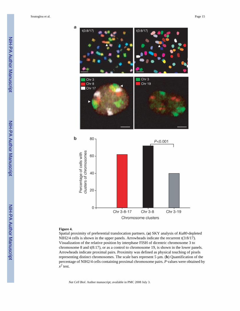
Figure 4.Spatial proximity of preferential translocation partners. (a) SKY analysis of Ku80-depletedNIH2/4 cells is shown in the upper panels. Arrowheads indicate the recurrent t(3:8/17).Visualization of the relative position by interphase FISH of dicentric chromosome 3 tochromosome 8 and t(8:17), or as a control to chromosome 19, is shown in the lower panels.Arrowheads indicate proximal pairs. Proximity was defined as physical touching of pixelsrepresenting distinct chromosomes. The scale bars represent 5 µm. (b) Quantification of thepercentage of NIH2/4 cells containing proximal chromosome pairs. P values were obtained byx2 test.
Soutoglou et al. Page 15
Nat Cell Biol. Author manuscript; available in PMC 2008 July 3.
NIH
-PA Author Manuscript
NIH
-PA Author Manuscript
NIH
-PA Author Manuscript