Dietary polyphenols protect against N -nitrosamines and benzo(a)pyrene-induced DNA damage (strand...
12
Maria Eugenia Delgado Ana Isabel Haza Nu ´ria Arranz Almudena Garcı ´a Paloma Morales Dietary polyphenols protect against N-nitrosamines and benzo(a)pyrene- induced DNA damage (strand breaks and oxidized purines/pyrimidines) in HepG2 human hepatoma cells Received: 11 January 2008 Accepted: 10 October 2008 Published online: 30 October 2008 M.E. Delgado A.I. Haza N. Arranz A. Garcı ´a P. Morales (&) Depto. de Nutricio ´n, Bromatologı ´a y Tecnologı ´a de los Alimentos Facultad de Veterinaria Universidad Complutense de Madrid 28040 Madrid, Spain Tel.: +34-91/394-3747 Fax: +34-91/394-3743 E-Mail: [email protected] j Abstract Background Dietary polyphenols have been reported to have a variety of biological ac- tions, including anticarcinogenic and antioxidant activities. Aim of the study In the present study we investigated the protective effect of dietary polyphenols against N- nitrosodimethylamine (NDMA), N-nitrosopyrrolidine (NPYR) and benzo(a)pyrene (BaP)-induced DNA damage (strand breaks and oxidized purines/pyrimidines) in HepG2 cells. Methods Human hepatocellular carcinoma (HepG2) cells, which retain many special- ized liver functions and drug metabolizing enzyme activities, were used as in vitro model for human hepatocytes. NDMA, NPYR and BaP were employed to induce DNA damage. DNA dam- age (strand breaks, oxidized pyrimidines and oxidized purines) was evaluated by the alkaline single cell gel electrophoresis or comet assay. Results None of the polyphenols concentrations tested in presence or absence of Fpg (formamidopyrimidine-DNA gly- cosylase), or Endo III (Endonu- clease III) caused DNA damage per se. Increasing concentrations of BaP (25–100 lM) induced a significant increase of DNA strand breaks, Fpg and Endo III sensitive sites in a dose dependent manner. Myricetin and quercetin decreased DNA strand breaks and oxidized pyrimidines induced by NDMA, but not oxidized purines. How- ever, both flavonoids reduced oxidized pyrimidines and purines induced by NPYR. DNA strand breaks induced by NPYR were prevented by quercetin, but not by myricetin. BaP-induced DNA strand breaks and oxidized pyrimidines were strongly reduced by myricetin and quercetin, respectively. While oxidized pur- ines induced by BaP were reduced by quercetin, myricetin had no protective effect. (+)-Catechin and ())-epicatechin reduced DNA strand breaks, oxidized pyrimi- dines and oxidized purines in- duced by NDMA. DNA strand breaks, and oxidized purines in- duced by NPYR were also pre- vented by (+)-catechin and ())- epicatechin, while the maximum reduction of oxidized pyrimidines was found by (+)-catechin and ())-epicatechin at 10 lM. (+)- Catechin and ())-epicatechin de- ORIGINAL CONTRIBUTION Eur J Nutr (2008) 47:479–490 DOI 10.1007/s00394-008-0751-6 EJN 751
-
Upload
independent -
Category
Documents
-
view
0 -
download
0
Transcript of Dietary polyphenols protect against N -nitrosamines and benzo(a)pyrene-induced DNA damage (strand...

Maria Eugenia DelgadoAna Isabel HazaNuria ArranzAlmudena GarcıaPaloma Morales
Dietary polyphenols protect againstN-nitrosamines and benzo(a)pyrene-induced DNA damage (strand breaksand oxidized purines/pyrimidines)in HepG2 human hepatoma cells
Received: 11 January 2008Accepted: 10 October 2008Published online: 30 October 2008
M.E. Delgado Æ A.I. Haza Æ N. ArranzA. Garcıa Æ P. Morales (&)Depto. de Nutricion, Bromatologıa yTecnologıa de los AlimentosFacultad de VeterinariaUniversidad Complutense de Madrid28040 Madrid, SpainTel.: +34-91/394-3747Fax: +34-91/394-3743E-Mail: [email protected]
j Abstract Background Dietarypolyphenols have been reported tohave a variety of biological ac-tions, including anticarcinogenicand antioxidant activities. Aim ofthe study In the present study weinvestigated the protective effectof dietary polyphenols against N-nitrosodimethylamine (NDMA),N-nitrosopyrrolidine (NPYR) andbenzo(a)pyrene (BaP)-inducedDNA damage (strand breaks andoxidized purines/pyrimidines) inHepG2 cells. Methods Humanhepatocellular carcinoma (HepG2)cells, which retain many special-ized liver functions and drugmetabolizing enzyme activities,were used as in vitro model forhuman hepatocytes. NDMA,NPYR and BaP were employed toinduce DNA damage. DNA dam-age (strand breaks, oxidizedpyrimidines and oxidized purines)was evaluated by the alkalinesingle cell gel electrophoresis orcomet assay. Results None of thepolyphenols concentrations testedin presence or absence of Fpg(formamidopyrimidine-DNA gly-cosylase), or Endo III (Endonu-clease III) caused DNA damageper se. Increasing concentrations
of BaP (25–100 lM) induced asignificant increase of DNA strandbreaks, Fpg and Endo III sensitivesites in a dose dependent manner.Myricetin and quercetin decreasedDNA strand breaks and oxidizedpyrimidines induced by NDMA,but not oxidized purines. How-ever, both flavonoids reducedoxidized pyrimidines and purinesinduced by NPYR. DNA strandbreaks induced by NPYR wereprevented by quercetin, but not bymyricetin. BaP-induced DNAstrand breaks and oxidizedpyrimidines were strongly reducedby myricetin and quercetin,respectively. While oxidized pur-ines induced by BaP were reducedby quercetin, myricetin had noprotective effect. (+)-Catechin and())-epicatechin reduced DNAstrand breaks, oxidized pyrimi-dines and oxidized purines in-duced by NDMA. DNA strandbreaks, and oxidized purines in-duced by NPYR were also pre-vented by (+)-catechin and ())-epicatechin, while the maximumreduction of oxidized pyrimidineswas found by (+)-catechin and())-epicatechin at 10 lM. (+)-Catechin and ())-epicatechin de-
ORIGINAL CONTRIBUTIONEur J Nutr (2008) 47:479–490DOI 10.1007/s00394-008-0751-6
EJN
751

creased also DNA strand breaksand oxidized pyrimidines but notoxidized purines induced by BaP.Conclusions Our results clearlyindicate that polyphenols protecthuman derived cells against DNA
strand breaks and oxidative DNAdamage effects of NDMA, NPYRor BaP, three carcinogenic com-pounds which occur in the envi-ronment.
j Key words dietary polyphe-nols – N-nitrosamines –benzo(a)pyrene – DNA damage –comet assay
Introduction
Polycyclic aromatic hydrocarbons (PAH) and N-nitrosamines are mutagenic and carcinogenic com-pounds widely present in the environment. PAH areexogenous factors exposed to humans via inhalationof industrial and automobile emissions, cigarettesmoke, and ingesting pyrolysed food products.Benzo(a)pyrene (BaP), an important PAH, is a car-cinogen that undergoes metabolic activation throughCYP1A1, CYP1A2, and CYP1B1. Thus, BaP is a po-tent systemic and local carcinogen known to induceskin, lung, and stomach tumours in animal models[36]. Humans are exposed to N-nitrosamines fromoccupational and environmental sources andthrough in vivo formation of ingested precursoramines and nitrosating agents. These compoundshave been detected in a wide variety of matrices suchas bacon, ham, frankfurters, sausages, cheese, beer,rubber, ground water, smoked tobacco, cosmetics,etc [9]. N-Nitrosodimethylamine (NDMA) is themost commonly encountered volatile N-nitrosaminein food samples and it is a potent liver, lung andkidney carcinogen [33]. N-Nitrosopyrrolidine(NPYR) induced mainly liver tumours in rats and isa weak pulmonary carcinogen in mice [10]. N-nitrosamines require metabolic activation to exerttheir carcinogenic effects through cytochrome P450(CYP2E1) [39]. The reactive intermediates of nitro-samine metabolism also have the ability to alkylatenucleophilic sites of DNA [34] resulting in alkalilabile adducts, which can lead to the formation ofabasic sites and DNA strand breaks. N-Nitrosaminesand PAH are focus of special interest due to theirincreasing appreciation as potential human carcino-gens. However, in balance, there are also many otherdietary agents which have been found to be benefi-cial in promoting good health and reducing the risksof some of these agents [31].
There was convincing evidence to suggest that fruitand vegetable intake was inversely associated withcancers of the lung, stomach, mouth, pharynx,esophagus and colon [38]. Over the past two decades,plant polyphenols increasingly attracted researchers,food manufacturers, as well as consumers due to theirantioxidant properties. Phenolic compounds, abun-
dantly present in fruits, vegetables, and beveragessuch as tea and red wine, are a large group of com-pounds with a similar chemical structure [40]. Theycould influence several important biological functionsby their free-radical scavenging ability, signal trans-duction pathway, stimulating apoptosis, inhibitinginflammation and inhibiting proliferation in humancancer cell lines [30]. Since in the Mediterraneanbasin, fresh fruit and vegetables as well as wine figureprominently in the general diet, in this study has beenevaluated the protective effect of some dietary poly-phenols, such as, (+)-catechin; (+)-trans-3,3¢,4¢,5,7-pentahydroxyflavane, (-)-epicatechin; ())-cis-3,3¢,4¢,5,7-pentahydroxy- flavane, quercetin; 3,3¢,4¢,5,7-pentahydroxyflavone and myricetin; 3,3¢,4¢,5,5¢,7-hexahydroxyflavone. These compounds are suggestedto act as chemopreventive compounds against lung,stomach, colorectal, epithelial, kidney, uterus, andovary cancer [25].
The single cell gel electrophoresis (SCGE) or Co-met assay has been considered as a useful tool forinvestigating DNA damage and DNA repair in mam-malian cells due to oxidative damage [5]. In thisstudy, the Comet assay was modified to permit thedetection of oxidized bases by including a step inwhich DNA is digested with formamidopyrimidine-DNA glycosylase (Fpg) or endonuclease III (Endo III)to uncover oxidized purines and pyrimidines,respectively. The effects of polyphenols compoundson the formation of Fpg sites or Endo III sites wereexamined.
The study of the reactions of polyphenolic con-stituents of the diet with mutagenic nitrosating spe-cies or benzo(a)pyrene is an area of great promisein cancer preventive strategies. Thus, the aim ofthis study was to evaluate the protective effect bydietary polyphenols against N-nitrosodimethylamine(NDMA), N-nitrosopyrrolidine (NPYR) and ben-zo(a)pyrene (BaP)-induced DNA damage (strandbreaks and oxidized purines/pyrimidines) in humanhepatoma cells (HepG2), using the single-cell gelelectrophoresis (SCGE) assay. These cells retain manyspecialized liver functions and drug metabolizingenzyme activities, comparable with human hepato-cytes, such as P450-mixed function oxidases, epoxidehydrolase, glucuronlytransferase, and glutathionetransferase [7].
480 European Journal of Nutrition (2008) Vol. 47, Number 8� Steinkopff Verlag 2008

Materials and methods
j Chemicals
N-nitrosodimethylamine (NDMA), N-nitrosopyrroli-dine (NPYR), benzo(a)pyrene (BaP), quercetin, my-ricetin, (+)-catechin, and ())-epicatechin, dimethylsulfoxide (DMSO) and low melting point agarose(LMP) were purchased from Sigma–Aldrich (St Louis,MO). Formamidopyrimidine-DNA glycosylase (Fpg)and Endonuclease III (Endo III) were obtained fromTrevigen Inc. (Gaithersburg, MD). All other chemicalsand solvents were of the highest grade commerciallyavailable. N-nitrosamines, benzo(a)pyrene, and poly-phenols were dissolved in sterile DMSO. The stocksolutions were stored deep frozen ()80�C).
j HepG2 cells
Human hepatocellular carcinoma (HepG2) cells werepurchased from Biology Investigation Center Collec-tion (BIC, Madrid, Spain). Only cells of passage 10–17were used in the experiments. The cells were culturedas monolayer in Dulbecco’s Modified Eagle Mediumsupplemented with 10% v/v heat inactivated foetalcalf serum, 50 U/ml penicillin and 50 lg/ml strepto-mycin and 1% v/v L-glutamine. Culture medium andsupplements required for the growth of the cells werepurchased from Gibco Laboratories (Life Technolo-gies, Inc., Gaithersburg, MD 20884-9980). Cell cul-tures were incubated at 37�C and 100% humidity in a5% CO2 atmosphere.
j Analysis of DNA damage induced bybenzo(a)pyrene or polyphenols in the AlkalineComet assay
Cell viability was routinely determined by the MTT[3-(4,5-dimethylthiazol-2-yl)-2,5-diphenyltetrazoliumbromide] assay in order to select non-toxic concen-trations of chemicals. The SCGE assay was carried outaccording to the protocol of Olive et al. [32] withminor modifications. Induction of DNA damage(strand breaks and oxidative DNA damage) by NPYRand NDMA has been previously evaluated [1]. Resultsobtained indicated that cells treated with NPYR andNDMA, after conversion to nucleoids, and incubatedwith Fpg had significant increases in DNA damage,the lowest effective concentrations being 5 and27 mM, respectively. However, when HepG2 cellswere treated with NPYR and NDMA and withoutenzymes the concentration of NPYR and NDMA re-quired to cause a significant increase in DNA damage
was 50 and 135 mM, respectively. For this reason thisconcentration range was used in subsequent studies.
Briefly, HepG2 cells were plated on to multiwellsystems at a density of 1.5 x 10⁵ cells/ml culturemedium. 24 h after seeding, cells were exposed toBaP, (25–100 lM), or to quercetin (1–10 lM), or tomyricetin, (1–10 lM), or to (+)-catechin (10–50 lM)or to ())-epicatechin (10–50 lM) or to the solvent foranother 24h at 37�C and 5% CO2. The solvent con-centration in the incubation medium never exceeded0.1%. After incubation, 10 ll of a suspension 1 x 10⁵
cells were mixed with 70 ll of LMP agarose type VII(0.75% concentration in PBS), distributed on slidesthat had been pre-coated with LMP agarose type VII(0.30% concentration in PBS), and left to set on an icetray. Three slides were prepared for each concentra-tion of the compound tested, one slide for control andthe others slides to be treated with Fpg or Endo III.After solidification, the cells were lysed in darknessfor 1 h in a high salt alkaline buffer (2.5 M NaCl,0.1 M EDTA, 0.01 M Tris, 1% Triton X-100, pH 10).The slides were then equilibrated 3 · 5 min in en-zyme buffer (0.04 M HEPES, 0.1 M KCl, 0.5 mMEDTA, 0.2 mg/ml BSA, pH 8). After this time, slideswere incubated with 30 ll of Fpg or Endo III at 1 lg/ml in enzyme buffer for 30 min at 37�C in a humiddark chamber. Control slides were incubated with30 ll enzyme buffer only. Following enzyme treat-ment, the slides were placed in electrophoresis buffer(0.3 M NaOH, 1 mM EDTA, pH 13, cooled in arefrigerator) in darkness for 40 min. Electrophoresiswas performed in a cold-storage room, in darkness, ina Bio-Rad subcell GT unit containing the same buffer,for 30 min at 25 V. After electrophoresis, the slideswere neutralized using 0.4 M Tris pH 7.5 and fixed inmethanol. Subsequently, the DNA was stained withethidium bromide (10 lg/ml) in Tris acetate EDTA(TAE 1X) during 5 min and examined in a fluores-cence microscope (OLYMPUS BH-2) connected to acomputerized image analysis system (Comet Score1.0). Olive tail moment (OTM) as defined by Oliveet al. [32] was determined and expressed as arbitraryunits (AU). OTM = I · L, where I is the fractionalamount of DNA in the comet tail (% DNA in the tail)and L is the distance from the centre of the comethead to the centre of tail distribution.
j Analysis of DNA damage induced by a simultaneoustreatment of N-nitrosamines or benzo(a)pyreneand polyphenols in the Alkaline Comet assay
HepG2 cells were plated on to multiwell systems at adensity of 1.5 x 10⁵ cells/ml culture medium. 24 hafter seeding, the corresponding polyphenols con-centrations were added to the wells and plates were
M.E. Delgado et al. 481Dietary polyphenols against mutagens

incubated for 24 h at 37�C and 5% CO2. After incu-bation, cells were simultaneously treated with: (1)NPYR (50 mM without enzymes and 5 mM with EndoIII or FPg) [1] and quercetin (0.1–5 lM) or myricetin(0.1–5 lM) or (+)-catechin (10–50 lM) or ())-epi-catechin (10–50 lM). Or (2) NDMA (135 mM withoutenzymes and 27 mM with Endo III or Fpg) [1] andquercetin (0.1–5 lM) or myricetin (0.1–5 lM) or (+)-catechin (10–50 lM) or ())-epicatechin (10–50 lM).Or (3) BaP (50 lM with and without enzymes) andquercetin (1–10 lM) or myricetin (1–10 lM) or (+)-catechin (10–50 lM) or ())-epicatechin (10–50 lM).Then plates were incubated for another 24 h at 37�Cand 5% CO2. After the treatments, the cells wereprocessed as described above.
j Statistical analysis
Images of 50 randomly selected cells per concen-tration were evaluated and the test was carried outthree times. The reported OTM is the mean ± stan-dard deviation (SD) of three independent experi-ments. Thus, we compare three means of OTM fromthree different experiments. Cultures withoutN-nitrosamines or BaP or polyphenols were consid-ered as negative controls. In all experiments thefollowing negative controls have been included: C0,cells treated with solvents; C1, cells treated withoutenzymes; C2, cells incubated with Endo III; C3, cellsincubated with Fpg. Induction of DNA damage by N-nitrosamines or BaP was defined as 100% of geno-toxicity. The Student’s t test was used for statistical
comparison between simultaneous treatments andcontrols, and differences were considered significantat P £ 0.05.
Results
j DNA damage induction by benzo(a)pyrene andpolyphenols
No cytotoxicity has been previously found at theconcentrations of polyphenols tested (data notshown). Cell viability was always above 80% of con-trol viability. None of the polyphenols (quercetin 0.1–10 lM, myricetin 0.1–10 lM, (+)-catechin 10–50 lMand ())-epicatechin 10–50 lM) concentrationstested in presence or absence of Fpg or Endo III,caused DNA damage per se (Fig. 1). For this reasonthis concentration range was used in subsequentstudies.
As shown in Fig. 2, increasing concentrations ofBaP (25–100 lM) induced a significant increase ofDNA strand breaks, Fpg sensitive sites and Endo IIIsensitive sites in a dose dependent manner. Resultsrevealed that HepG2 cells treated with a concentrationof 25 lM BaP slightly increased the DNA strandbreaks (Fig. 2a1, b1), whereas the Fpg sensitive sites(Fig. 2a3, b3) and the Endo III sensitive sites weremarkedly increased (Fig. 2a2, b2) in comparison withtheir respective controls. The maximum increase ofDNA strand breaks, Fpg sensitive sites and Endo IIIsensitive sites were exhibited by 100 lM BaP. Insubsequent simultaneous treatments with BaP and
0
2
4
6
0.1 1 50
2
4
6
0
2
4
6
0
2
4
6
Concentration of quercetin (µM)Concentration of myricetin (µM)a
c
b
d
OTM
OTM
Concentration of (-)-epicatechin (µM)Concentration of (+)-catechin (µM)
OTM
OTM
C0 C0 0.1 1 5
10 25 50C0 C0 10 25 50
Fig. 1 Induction of DNA strandbreaks and oxidized purines/pirimidines by a myricetin, bquercetin, c (+)-catechin and d ())-epicatechin on human HepG2 cellsincubated without enzymes(diagonally shaded bar), with Endo III(lightly shaded bar) or Fpg (darklyshaded bar). C0 HepG2 cells without(+)-catechin, ())-epicatechin,myricetin or quercetin and incubatedwithout enzymes (diagonally shadedbar), with Endo III (lightly shadedbar) or Fpg (darkly shaded bar)
482 European Journal of Nutrition (2008) Vol. 47, Number 8� Steinkopff Verlag 2008

polyphenols, the HepG2 cells were incubated with theconcentration of 50 lM BaP for 24 h.
j DNA damage induction by a simultaneoustreatment of N-nitrosamines or benzo(a)pyreneand polyphenols in the Alkaline Comet assay
The effect of myricetin on NDMA-induced DNAdamage in HepG2 cells is shown in Fig. 3.1 Myricetindecreased DNA strand breaks (Fig. 3, a1) and theformation of Endo III sensitive sites (Fig. 3, a2) in-duced by NDMA, but not the formation of Fpg sen-sitive sites (Fig. 3, a3). The maximum reduction ofEndo III sensitive sites (32% inhibition comparedwith the control) was observed at the lowest concen-tration (0.1 lM). Myricetin reduced the formation of
Endo III sensitive sites (1–5 lM, 16–19%) and theformation of Fpg sensitive sites (0.1–5 lM, 30–43%)induced by NPYR (Fig. 3, b2, b3), but not DNA strandbreaks (all the concentrations increased the genotoxiceffect of NPYR) (Fig. 3, b1). Protective effect of my-ricetin on BaP-induced DNA damage is shown in Fig.3, c1, c2 and c3. Myricetin (5–10 lM) weakly reducedDNA strand breaks (20–5%) and the formation ofEndo III sensitive sites induced by BaP, but not theformation of Fpg sensitive sites (concentrations of 5–10 lM increased the oxidative DNA damage inducedby BaP). The maximum inhibition of Endo III sensi-tive sites (75%) induced by BaP was found at thelowest concentration (1 lM).
The effect of quercetin on NDMA-induced DNAdamage in HepG2 cells is shown in Fig. 3.2. None ofthe quercetin concentrations tested reduced the for-
20
0
40
60
80
% T
ail D
NA *** ***
***
0
10
20
30
40
50
***
OTM ***
***
0
10
20
30
40
50 1 2
Concentration of BaP (µM)a b
a b
a b
***
OTM
***
***
C0 C1 25 50 100
Concentration of BaP (µM)
C0 C1 25 50 100
0
10
20
30
40
50
OTM
*** ***
***
0
20
40
60
80
% T
ail D
NA
***
******
0
20
40
60
80
Concentration of BaP (µM)
C0 C1 C2 25 50 100
Concentration of BaP (µM)
C0 C1 C2 25 50 100
Concentration of BaP (µM)
C0 C1 C3 25 50 100
Concentration of BaP (µM)
C0 C1 C3 25 50 100
***
% T
ail D
NA
***
***
Fig. 2 Effect of BaP-induced DNAstrand breaks and oxidative damagein human HepG2 cells expressed as aOTM in AU, and b % Tail DNA. (1)(C0 ) control cells treated withsolvent. (C1) control cells treatedwithout enzymes. HepG2 cellstreated with BaP and incubatedwithout enzymes (shaded with dots).(2) (C2) control cells treated withEndo III. HepG2 cells treated withBaP and incubated with Endo III(shaded with dots). 3. (C3) controlcells treated with Fpg. HepG2 cellstreated with BaP and incubated withFpg (diagonally shaded bar)
M.E. Delgado et al. 483Dietary polyphenols against mutagens

mation of Fpg sensitive sites induced by NDMA (Fig.3, a3). On the contrary, concentrations of 1–5 lMincreased the oxidative DNA damage induced byNDMA. Quercetin inhibited the formation of Endo IIIsensitive sites induced by NDMA (Fig. 3, a2) at all theconcentrations tested (0.1–5 lM, 26%). However,quercetin (0.1–5 lM) reduced the formation of Fpgsensitive sites (18–27%) induced by NPYR (Fig. 3, b3).DNA strand breaks (0.1–1 lM, 2–10%) and the for-mation of Endo III sensitive sites (1–5 lM, 18%) in-duced by NPYR were also prevented by quercetin(Fig. 3, b1, b2). Protection afforded by quercetintowards BaP-induced DNA damage occurred in adose-dependent manner. Quercetin induced a dose-
dependent protective effect towards DNA strandbreaks (Fig. 3, c1). At 1 lM, BaP genotoxicity wasreduced by 6%, and the reduction reached 27% at10 lM. The formation of Fpg sensitive sites (Fig. 3,c3) was reduced by 8–14% (5–10 lM) and the maxi-mum inhibition of the formation of Endo III sensitivesites (Fig. 3, c2) was observed at 1 lM (82%).
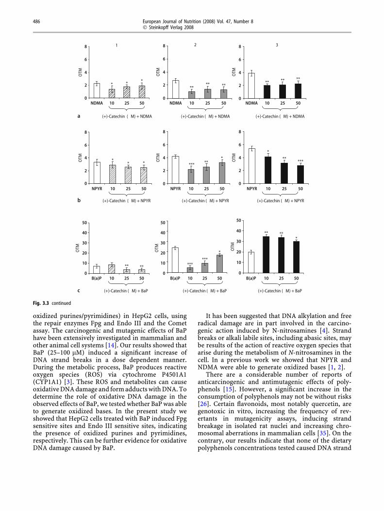
Figure 3.3 shows the protective effect of (+)-cate-chin on NDMA-induced DNA damage in HepG2 cells.(+)-Catechin at the lowest concentration (10 lM)showed the maximum reduction of DNA strandbreaks (38%), the formation of Endo III (62%) andFpg sensitive sites (48%) induced by NDMA (Fig. 3,a1–a3). DNA strand breaks (12–24%) induced by
0
2
4
6
8
NDMA 0.1 1 5
Myricetin (µM) + NDMAa
b
c
NDMA 0.1 1 5
Myricetin (µM) + NDMA
NDMA 0.1 1 5
Myricetin (µM) + NDMA
NPYR 0.1 1 5
Myricetin (µM) + NPYR
NPYR 0.1 1 5
Myricetin (µM) + NPYR
NPYR 0.1 1 5
Myricetin (µM) + NPYR
B(a)P 1 5 10
Myricetin (µM) + BaP
B(a)P 1 5 10
Myricetin (µM) + BaP
B(a)P 1 5 10
Myricetin (µM) + BaP
OTM
0
2
4
6
8
OTM
***
*
0
2
4
6
8
OTM
**
* *
0
10
20
30
40
50
OTM
0
10
20
30
40
50
*****
OTM
*
0
2
4
6
8
OTM
0
2
4
6
8
OTM
0
10
20
30
40
50
OTM
0
2
4
6
8
OTM** **
*****
***
1 2 3
Fig. 3.1 Effect of 3.1 Myricetin, 3.2 Quercetin, 3.3 (+)-Catechin and 3.4 ())-Epicatechin on a NDMA, b NPYR and c BaP-induced DNA damage in humanHepG2 cells. OTM median values in control cells without enzymes andincubated with Endo III or Fpg were 1.47 ± 0.37, 1.85 ± 0.43, 2.11 ± 0.40 AU,respectively. (1) Cells treated with NDMA (135 mM) or NPYR (50 mM) or BaP(50 lM) and incubated without enzymes (lightly shaded bar). Cells treated withNDMA (135 mM) or NPYR (50 mM) or BaP (50 lM) and polyphenol andincubated without enzymes (shaded with dots). (2) Cells treated with NDMA
(27 mM) or NPYR (5 mM) or BaP (50 lM) and incubated with Endo III (darklyshaded bar). Cells treated with NDMA (27 mM) or NPYR (5 mM) or BaP (50 lM)and polyphenol and incubated with Endo III (unfilled bar). (3) Cells treated withNDMA (27 mM) or NPYR (5 mM) or BaP (50 lM) and incubated with Fpg(diagonally filled bar). Cells treated with NDMA (27 mM) or NPYR (5 mM) orBaP (50 lM) and polyphenol and incubated with Fpg (unfilled bar). Asterisksindicate significant difference from control ***P £ 0.001, **P £ 0.01,*P £ 0.05
484 European Journal of Nutrition (2008) Vol. 47, Number 8� Steinkopff Verlag 2008

NPYR (Fig. 3, b1) and the formation of Fpg sensitivesites (24–48%) was also prevented by (+)-catechin(10–50 lM, Fig. 3, b3). However, the maximuminhibition of formation of Endo III sensitive sites(47%) was observed at the lowest concentration(10 lM). Figure 3, c1, c2 and c3 show the effect of (+)-catechin on BaP-induced DNA damage. (+)-Catechindecreased DNA strand breaks (25–50 lM, 49%, Fig. 3,c1) and the formation of Endo III sensitive sites, butnot the formation of Fpg sensitive sites (all the con-centrations increased oxidative DNA damage inducedby BaP, Fig. 3, c3). The maximum inhibition of EndoIII sensitive sites (82%) was found at 10 lM (Fig. 3,c2).
Figure 3.4 shows the protective effect of ())-epi-catechin on NDMA-induced DNA damage in HepG2cells. ())-Epicatechin at the lowest concentration(10 lM) showed the maximum reduction of DNAstrand breaks (34%), the formation of Endo III (55%)and Fpg sensitive sites (40%) induced by NDMA (Fig.3, a1–a3). Protection afforded by ())-epicatechin to-
wards NPYR-induced DNA damage is showed on Fig.3, b1–b3.
())-Epicatechin at the lowest concentration(10 lM) showed the maximum reduction of DNAstrand breaks (34%), the formation of Endo III (34%)and Fpg sensitive sites (50%) induced by NPYR.However, (-)-epicatechin (10–50 lM) reduced DNAstrand breaks induced by BaP (Fig. 3, c1) in a dosedependent manner (27–39%). The maximum inhibi-tion of the formation of Endo III sensitive sites (63%,Fig. 3, c2) was found at the lowest concentration(10 lM). The formation of Fpg sensitive sites (Fig. 3,c3) was weakly reduced (5–10%) at 25–50 lM.
Discussion
In the present study we evaluate the protective effect ofdietary polyphenols against N-nitrosodimethylamine(NDMA), N-nitrosopyrrolidine (NPYR) or benzo(a)-pyrene (BaP)-induced DNA damage (strand breaks and
0
2
4
6
8
OTM **
0
2
4
6
8
OTM
* * ***
0
10
20
30
40
50
OTM
0
2
4
6
8
OTM
0
2
4
6
8
OTM
0
10
20
30
40
50
OTM
0
2
4
6
8
OTM
0
2
4
6
8
OTM
0
10
20
30
40
50
OTM
***
* ***
*** *****
**
1 2 3
NDMA 0.1 1 5
Quercetin (µM) + NDMAa
b
c
NDMA 0.1 1 5
Quercetin (µM) + NDMA
NDMA 0.1 1 5
Quercetin (µM) + NDMA
NPYR 0.1 1 5
Quercetin (µM) + NPYR
NPYR 0.1 1 5
Quercetin (µM) + NPYR
NPYR 0.1 1 5
Quercetin (µM) + NPYR
B(a)P 1 5 10
Quercetin (µM) + BaP
B(a)P 1 5 10
Quercetin (µM) + BaP
B(a)P 1 5 10
Quercetin (µM) + BaP
Fig. 3.2 continued
M.E. Delgado et al. 485Dietary polyphenols against mutagens
oxidized purines/pyrimidines) in HepG2 cells, usingthe repair enzymes Fpg and Endo III and the Cometassay. The carcinogenic and mutagenic effects of BaPhave been extensively investigated in mammalian andother animal cell systems [14]. Our results showed thatBaP (25–100 lM) induced a significant increase ofDNA strand breaks in a dose dependent manner.During the metabolic process, BaP produces reactiveoxygen species (ROS) via cytochrome P4501A1(CYP1A1) [3]. These ROS and metabolites can causeoxidative DNA damage and form adducts with DNA. Todetermine the role of oxidative DNA damage in theobserved effects of BaP, we tested whether BaP was ableto generate oxidized bases. In the present study weshowed that HepG2 cells treated with BaP induced Fpgsensitive sites and Endo III sensitive sites, indicatingthe presence of oxidized purines and pyrimidines,respectively. This can be further evidence for oxidativeDNA damage caused by BaP.
It has been suggested that DNA alkylation and freeradical damage are in part involved in the carcino-genic action induced by N-nitrosamines [4]. Strandbreaks or alkali labile sites, including abasic sites, maybe results of the action of reactive oxygen species thatarise during the metabolism of N-nitrosamines in thecell. In a previous work we showed that NPYR andNDMA were able to generate oxidized bases [1, 2].
There are a considerable number of reports ofanticarcinogenic and antimutagenic effects of poly-phenols [15]. However, a significant increase in theconsumption of polyphenols may not be without risks[26]. Certain flavonoids, most notably quercetin, aregenotoxic in vitro, increasing the frequency of rev-ertants in mutagenicity assays, inducing strandbreakage in isolated rat nuclei and increasing chro-mosomal aberrations in mammalian cells [35]. On thecontrary, our results indicate that none of the dietarypolyphenols concentrations tested caused DNA strand
***
**
*
** **** ** ** **
******
*****
*
** ** ******
*
** ***
1 2 3
0
2
4
6
8
OTM
0
2
4
6
8
OTM
0
10
20
30
40
50
OTM
0
2
4
6
8
OTM
0
2
4
6
8
OTM
0
10
20
30
40
50
OTM
0
2
4
6
8
OTM
0
2
4
6
8
OTM
0
10
20
30
40
50
OTM
NDMA 10 25 50 NDMA 10 25 50 NDMA 10 25 50
(+)-Catechin (µM) + NDMAa
b
c
(+)-Catechin (µM) + NDMA (+)-Catechin (µM) + NDMA
NPYR 10 25 50 NPYR 10 25 50 NPYR 10 25 50
(+)-Catechin (µM) + NPYR (+)-Catechin (µM) + NPYR (+)-Catechin (µM) + NPYR
B(a)P 10 25 50
(+)-Catechin (µM) + BaP
B(a)P 10 25 50
(+)-Catechin (µM) + BaP
B(a)P 10 25 50
(+)-Catechin (µM) + BaP
Fig. 3.3 continued
486 European Journal of Nutrition (2008) Vol. 47, Number 8� Steinkopff Verlag 2008

breaks, or oxidized purine or pyrimidine bases per se.These results are in part in agreement with previousreports, which have revealed that some polyphenolssuch as (+)-catechin, or ())-epicatechin, were notmutagenic in the Salmonella/microsomal mutagenic-ity assay using tester strain TA102, while quercetinwas found mutagenic [27].
Myricetin (3,3¢,4¢,5,5¢,7-hexahydroxyflavone) andquercetin (3,3¢,4¢,5,7-pentahydroxyflavone) are two ofthe most frequently studied flavonoids in the class offlavonols [16]. In the present study, quercetin pro-tected against DNA strand breaks and oxidizedpyrimidines induced by BaP, NDMA, and NPYR,while myricetin protected against DNA strand breaksand oxidized pyrimidines induced by BaP and NDMAand towards oxidized pyrimidines and purines in-duced by NPYR. This variable effect against NPYR issurprising given the similar chemical structure ofquercetin and myricetin. This indicates that only anadditional hydroxyl group at position 5¢ in thechemical structure of myricetin would significantlyaffect the biological activity against NPYR. In addi-
tion, the protective effect of quercetin and myricetinwas higher towards BaP than against NDMA andNPYR. Thus, the presence and number of polyhydr-oxyl groups in the chemical structure and/or the kindof mutagen used to induce DNA damage has animportant role in the protective effect. Makena andChung [27] also reported that the position andnumber of hydroxyl groups are crucial in the inhibi-tory effects of polyphenols.
Catechins are also found in a variety of plantsand are present in particularly high amounts in tealeaves, such as (+)-catechin and ())-epicatechin. Inthe present study (+)-catechin; (+)-trans-3,3¢,4¢,5,7-pentahydroxyflavane, and ())-epicatechin; ())-cis-3,3¢,4¢,5,7-pentahydroxy- flavane, reduced DNAstrand breaks induced by BaP, NDMA and NPYR.(+)-Catechin and ())-epicatechin also protectedagainst oxidized purines and pyrimidines induced byNDMA and NPYR, however they only protectedagainst oxidized pyrimidines induced by BaP. Yenet al. [41] reported that ())-epicatechin had protectiveeffect against BaP-induced DNA damage in Chang
* *****
***
*** *** *
**
* ****
****
*
*****
**
1 2 3
0
2
4
6
8
OTM
0
2
4
6
8
OTM
0
10
20
30
40
50
OTM
0
2
4
6
8
OTM
0
2
4
6
8
OTM
0
10
20
30
40
50
OTM
0
2
4
6
8
OTM
0
2
4
6
8
OTM
0
10
20
30
40
50
OTM
NDMA 10 25 50 NDMA 10 25 50 NDMA 10 25 50
(-)-Epicatechin (µM) + NDMAa
b
c
(-)-Epicatechin (µM) + NDMA (-)-Epicatechin (µM) + NDMA
NPYR 10 25 50 NPYR 10 25 50 NPYR 10 25 50
(-)-Epicatechin (µM) + NPYR (-)-Epicatechin (µM) + NPYR (-)-Epicatechin (µM) + NPYR
B(a)P 10 25 50 B(a)P 10 25 50 B(a)P 10 25 50
(-)-Epicatechin (µM) + BaP (-)-Epicatechin (µM) + BaP (-)-Epicatechin (µM) + BaP
Fig. 3.4 continued
M.E. Delgado et al. 487Dietary polyphenols against mutagens

liver cells at concentrations of 10–100 lM. However,Dhawan et al. [8] showed that (+)-catechin and ())-epicatechin and their gallate esters, cannot affordprotection against DNA damage induced in humanlymphocytes by heterocyclic amine (Trp-P-2). Ourresults also indicate that (+)-catechin, with similarchemical structure to ())-epicatechin, showed ahigher protective effect than ())-epicatechin againstDNA damage induced by BaP, or NDMA, while thesecompounds exhibited a similar level of protectionagainst NPYR. The spatial arrangement of substitu-ents on (+)-catechin and ())-epicatechin could bemore important than the backbone alone in the pro-tective effect and/or the mutagen tested.
Taken together our results, (+)-catechin and ())-epicatechin showed higher protection against DNAstrand breaks induced by BaP, NDMA and NPYR thanmyricetin or quercetin. In addition, they also pro-tected against oxidized purines and pyrimidines in-duced by NDMA, NPYR and BaP (with the exceptionof (+)-catechin that increased oxidative DNA damageinduced by BaP). Quercetin and myricetin were themost active flavonoids against oxidized pyrimidinesinduced by BaP, but not against oxidized purines. Onthe contrary, they increased oxidative DNA damageinduced by BaP. This effect could be attributed to theexcess of reactive oxygen species (ROS) produced byBaP. They might cause irreparable oxidative DNAdamage [20]. In addition, phenolic compounds haveboth antioxidant and prooxidant effects depending onthe experimental conditions [17]. Therefore, althoughthere is a general structure-activity relationship [21]that shows that some subclasses of flavonoid can bemore potent antimutagens, these structural consid-erations can change depending on the substitutionpattern of the molecule and/or the kind of compoundused to induce DNA damage.
The protective effect of dietary polyphenols testedin this study, in part, could be due to the free radicalscavenging efficiency of these compounds. Flavonoidscan inhibit oxidative damage to DNA and therefore,prevent or reduce cellular oxidative stress [22].According to Cotelle et al. [6], the protective effect ofquercetin and myricetin may be associated with thepresence of two or three hydroxyl groups in the B ringof its molecule. Ricardo da Silva et al. [34] also re-ported that (+)-catechin and ())-epicatechin scav-enge superoxide and hydroxyl radicals. Anotherpossibility is that flavonoids, as metal-ion-chelatingagents [37], could have chelated iron ions present inthe cells and hence depressed the Fenton reaction.Morel et al. [28] investigated the antioxidant and ironchelating activities of (+)-catechin, and quercetinusing cell cultures. They showed that both com-
pounds could chelate iron in vitro, and that (+)-cat-echin was more potent antioxidant than quercetin,which can explain, in part, the protective effect ofthese flavonoids in the oxidative DNA damage-in-duced by BaP, NDMA or NPYR.
Other mechanisms proposed to explain this protec-tive effect might also include inhibition of phase Imetabolizing enzymes, such as cytochrome P450 (CYP),which metabolically activates procarcinogens to reactiveintermediates that trigger carcinogenesis [11]. Flavo-noid interactions with CYPs have been reviewed recently[18]. Hatch et al. [19] reported that polyphenols coulddiminish the production of the active metabolites of thexenobiotic through downregulation of the relevant en-zymes, and/or directly interfere with DNA adduct for-mation. Finally, another mechanism claimed to beresponsible for the protective effect of flavonoids is theinduction of phase II metabolizing enzymes such asglutathione S-transferase (GST), NAD(P)H:quinineoxidoreductase (NQO) and UDP-glucuronyltransferase(UGT) by which carcinogens are detoxified and there-fore more readily eliminated from the body [12].
All the polyphenol used in our work were in lMconcentrations and several studies on bioavailabilityof flavonoids, indicate that they are poorly bioavail-able and reach only nanomolar to low micromolarconcentrations in plasma [23]. Moreover, since con-jugation is a biologic defense mechanism serving theobjective of inactivating xenobiotics as well asendogenous bioactive compounds and rendering wa-ter-soluble to facilitate their excretion, the presence offree polyphenols as unconjugated compounds fromthe circulation or the urine following absorption isalmost irrelevant [13]. This does not preclude thepossibility that flavonoids may accumulate in tissueswhere they might exert local antioxidant effects orthat very low concentrations of flavonoids maymodulate cell signalling, gene regulation, angiogene-sis, and other biological processes by non-antioxidantmechanisms, which may explain the purported healthbenefits of flavonoids [29].
In conclusion, our results clearly indicate thatpolyphenols protect human derived cells against DNAstrand breaks and oxidative DNA damage effects ofBaP, NDMA and NPYR, three carcinogenic com-pounds which occur in the environment. Furtherinvestigation is in progress to evaluate and betterunderstand the mechanisms by which polyphenolsexert their protective effects.
j Acknowledgments This work has been supported by GrantALI2005-01517 from the Ministerio de Educacion y Ciencia (Spain)and by Grant 910177 from the Comunidad de Madrid and theUniversidad Complutense (UCM). M.E. Delgado and A. Garcıa arerecipients of Fellowships from the Ministerio de Educacion yCiencia and Universidad Complutense (UCM), Spain.
488 European Journal of Nutrition (2008) Vol. 47, Number 8� Steinkopff Verlag 2008

References
1. Arranz N, Haza AI, Garcıa A, Moller L,Rafter J, Morales P (2007) Protectiveeffects of organosulfur compounds to-wards N-nitrosamine-induced DNAdamage in the single-cell gel electro-phoresis (SCGE)/HepG2 assay. FoodChem Toxicol 45:1662–1669
2. Arranz N, Haza AI, Garcıa A, Rafter J,Morales P (2007) Protective effect of-vitamin C towards N-nitrosamine-in-duced DNA damage in the single-cellgel electrophoresis (SCGE)/HepG2 as-say. Toxicol in vitro 21:1311–1317
3. Burczynski ME, Penning TM (2000)Genotoxic polycyclic aromatic hydro-carbon ortho-quinones generated byaldo-keto reductases induce CYP1A1via nuclear translocation of the arylhydrocarbon receptor. Cancer Res 60:908–915
4. Bartsch H, Hietanen E, Malaveille C(1989) Carcinogenic nitrosamines: freeradical aspects of their action. FreeRadical Bio Med 7:637–644
5. Collins AR, Duthie SJ, Dobson VL(1993) Direct enzymic detection ofendogenous oxidative base damage inhuman lymphocyte DNA. Carcinogen-esis 14:1733–1735
6. Cotelle N, Bernier J, Catteau J, Pom-mery J, Wallet J, Gaydou EM (1996)Antioxidant properties of hydroxy-flavones. Free Radical Bio Med 20:35–43
7. Doostdar H, Demoz A, Burke MD,Melvin WT, Grant MH (1990) Varia-tion in drug-metabolizing enzymeactivities during the growth of humanHepG2 hepatoma cells. Xenobiotica20:435–441
8. Dhawan A, Anderson D, de Pascual-Teresa S, Santos-Buelga C, CliffordMN, Ioannides C (2002) Evaluation ofthe antigenotoxic potential of mono-meric and dimeric flavanols, and blacktea polyphenols against heterocyclicamine-induced DNA damage in humanlymphocytes using the Comet assay.Mutat Res 515:39–56
9. Filho PJS, Rios A, Valcarcel M, Cara-mao EB (2003) Development of a newmethod for the determination ofnitrosamines by micellar electrokineticcapillary chromatography. Water Res37:3837–3842
10. Gray R, Peto R, Branton P, Grasso P(1991) Chronic nitrosamine ingestionin 1,040 rodents—the effect of thechoice of Nitrosamine, the speciesstudied, and the age of starting expo-sure. Cancer Res 51:6470–6491
11. Galati G, O’Brien PJ (2004) Potentialtoxicity of flavonoids and other dietaryphenolics: significance for their che-mopreventive and anticancer proper-ties. Free Radical Bio Med 37:287–303
12. Galati G, Teng S, Moridani MY, ChanTS, O’Brien PJ (2000) Cancer chemo-prevention and apoptosis mechanismsinduced by dietary polyphenolics.Drug Metabol Drug Interact 17:311–349
13. Goldberg DM, Yan J, Soleas GJ (2003)Absorption of three wine-relatedpolyphenols in three different matricesby healthy subjects. Clin Biochem 36:79–87
14. Harvey RG (1991) Polycyclic aromatichydrocarbons: chemistry and carcino-genicity. In: Coombs M (ed) Cam-bridge University Press, Cambridge
15. Heo MY, Yu KS, Kim KH, Kim HP, AuWW (1992) Anticlastogenic effect offlavonoids against mutagen-inducedmicronuclei in mice. Mutat Res 284:243–249
16. Hertog MGL, Feskens EJM, KromhoutD, Hertog MGL, Hollman PCH, HertogMGL, Katan MB (1993) Dietary anti-oxidant flavonoids and risk of coro-nary heart disease: the Zutphen ElderlyStudy. Lancet 342:1007–1011
17. Hanasaki Y, Ogawa S, Fukui S (1994)The correlation between active oxygenscavenging and antioxidative effects offlavonoids. Free Radical Bio Med16:845–850
18. Hodek P, Trefil P, Stiborova M (2002)Flavonoids-potent and versatile bio-logically active compounds interactingwith cytochromes P450. Chem BiolInteract 139:1–21
19. Hatch FT, Lightstone FC, Colvin ME(2000) Quantitative structure-activityrelationship of flavonoids for inhibi-tion of heterocyclic amine mutagenic-ity. Environ Mol Mutagen 35:279–299
20. Johnson MK, Loo G (2000) Effects ofepigallocatechin gallate and quercetinon oxidative damage to cellular DNA.Mutat Res 459:211–218
21. Lopez-Lazaro M (2002) Flavonoids asanticancer agents: structure–activityrelationship Study. Curr Med Chem2:691–714
22. Lipkin M, Reddy B, Newmark H,Lamprecht SA (1999) Dietary factors inhuman colorectal cancer. Annu RevNutr 19:545–586
23. Lotito SB, Frei B (2006) Consumptionof flavonoid-rich foods and increasedplasma antioxidant capacity in hu-mans: cause, consequence, or epiphe-nomenon? Free radical Bio Med 41:1727–1746
24. Murdok DJL, Barnett YA, Barnett CR(2004) DNA damage and cytotoxicityin pancreatic b-cells expressing humanCYP2E. Biochem Pharmacol 68:523–530
25. Maggiolini M, Recchia AG, BonofiglioD, Catalano S, Vivacqua A, Carpino A,Rago V, Rossi R, Ando S (2005) Thered wine phenolics piceatannol andmyricetin act as agonists for estrogenreceptor alpha in human breast cancercells. J Mol Endocrinol 35:269–281
26. Mennen LI, Walker R, Bennetau-Pelis-sero C, Scalbert A (2005) Risks andsafety of polyphenol consumption. AmJ Clin Nutr 81:326S–329
27. Makena P, Chung K (2007) Effects ofvarious plant polyphenols on bladdercarcinogen benzidine-induced muta-genicity. Food Chem Toxicol 45:1899–1909
28. Morel I, Lescoat G, Cogrel P, Sergent O,Pasdeloup N, Brissot P, Cillard P, Cil-lard J (1993) Antioxidant and iron-chelating activities of the flavonoidscatechin, quercetin and diosmetin oniron-loaded rat hepatocyte cultures.Biochem Pharmacol 45:13–19
29. Manach C, Williamson G, Morand C,Scalbert A, Remesy C (2005) Bioavail-ability and bioefficiency of polyphenolsin humans. I. Review of 97 bioavail-ability studies. Am J Clin Nutr 81:230S–242S
30. Neuhouser ML (2004) Review: dietaryflavonoids and cancer risk: evidencefrom human population studies. NutrCancer 50:1–7
31. Ow YY, Stupans I (2003) Gallic acidand gallic acid derivatives: effects ondrug metabolizing enzymes. Curr DrugMetab 4:241–248
32. Olive PL, Wlodek D, Durand RE, Ban-ath JP (1992) Factors influencing DNAmigration from individual cells sub-jected to gel electrophoresis. Exp CellRes 198:259–267
33. Preussmann R, Stewart BW (1984) N-Nitroso carcinogens. In: Searle CE (ed)Chemical carcinogenesis. ACS Mono-graph, Washington DC, pp 643–828
34. Ricardo da Silva JM, Rigaud J, Chey-nier V, Cheminat A, Moutounet M(1991) Procyanidin dimers and trimersfrom grape seeds. Phytochemistry 30:1259–1264
35. Sahu SC, Washington MC (1991) Ef-fects of antioxidants on quercetin-in-duced nuclear DNA damage and lipidperoxidation. Cancer Lett 60:259–264
M.E. Delgado et al. 489Dietary polyphenols against mutagens

36. Ueng Y, Shyu C, Liu T, Oda Y, Liao J,Chen C (2001) Protective effects ofbaicalein and wogonin against ben-zo[a]pyrene- and aflatoxin B1-inducedgenotoxicities. Biochem Pharmacol 62:1653–1660
37. Williams RJ, Spencer JPE, Rice-EvansC (2004) Flavonoids: antioxidants orsignalling molecules? Free Radical BioMed 36:838–849
38. World Cancer Research Fund (1997)Food, nutrition and the prevention ofcancer: a global perspective. Nutrition15:523–526
39. Yang CS, Smith T, Ishizaki H, Hong JY(1991) Enzyme mechanism in themetabolism of nitrosamines. In: O’NeilIK, Chen J, Bartsh H (eds) Relevance tohuman cancer of nitroso compounds,tobacco smoke and mycotoxins. IARC,Lyon, pp 567–573
40. Yao LH, Jiang YM, Shi J, Tomas-Barberan FA, Datta N, Singanusong R,Chen SS (2004) Flavonoids in food andtheir health benefits. Plant Foods HumNutr 59:113–122
41. Yen GC, Ju JW, Wu CH (2004) Modu-lation of tea and tea polyphenols onbenzo(a)pyrene-induced DNA damagein Chang liver cells. Free Radical Res38:193–200
490 European Journal of Nutrition (2008) Vol. 47, Number 8� Steinkopff Verlag 2008