Gap Junction Gating Sensitivity to Physiological Internal Calcium Regardless of pH in Novikoff...
11
Biophysical Journal Volume 65 November 1993 2002-2012 Gap Junction Gating Sensitivity to Physiological Internal Calcium Regardless of pH in Novikoff Hepatoma Cells Ahmed Lazrak, and Camillo Peracchia Department of Physiology, University of Rochester, Rochester, New York 14642-8642, USA ABSTRACT Gap junction conductance (Gj) and channel gating sensitivity to voltage, Ca2+, H+, and heptanol were studied by double whole-cell clamp in Novikoff hepatoma cell pairs. Channel gating was observed at transjunctional voltages (Vj) > +50 mV. The cells readily uncoupled with 1 mM 1 -heptanol. With heptanol, single (gap junctional) channel events with unitary conductances (m/) of 46 and 97 pS were detected. Both Ca2+-loading (EGTA-Ca) and acidifying (100% C02) solutions caused uncoupling. However, C02 was effective when Ca2+, was buffered with EGTA (a H+-sensitive Ca-buffer) but not with BAPTA (1,2-bis(2-aminophenoxy)ethane-N,N,N',N'-tetraacetic acid) (a H+-insensitive Ca-buffer), suggesting a Ca2+-mediated H+ ef- fect on gap junctions. This was tested by monitoring the Gj decay at different pCa, values (9, 6.9, 6.3, 6, and 5.5; 1 mM BAPTA) and pHi values (7.2 or 6. 1, 10 mM 4-(2-hydroxyethyl)-1 -piperazineethanesulfonic acid and 2-(N-morpholino)ethansulphonic acid, respectively). With pCa >- 6.9 (pH 7.2 or 6.1), Gj decreased to 10-70% of initial values in -40 min, following single exponential decays (T = -28 min). With pCai 6-6.3 (pH 7.2 or 6.1), Gj decreased to 10-25% of initial values in 15 min (T = -5 min); the Student t gave a P = 0.0178. With pCa 5.5 the cells uncoupled in less than 1 min (T = -20 s). Low pH, affected neither time course nor shape of Gj decay at any pCal tested. The data indicate that these gap junctions are sensitive to [Ca2+]1 in the physiological range (<500 nM) and that low pH,, without an increase in [Ca2+],, neither decreases Gj nor increases channel sensitivity to Ca2+. INTRODUCTION Direct cell-to-cell communication is mediated by membrane channels accessible to small cytosolic molecules (reviewed in Loewenstein, 1981; Peracchia, 1980, 1987; Deleze, 1987; Ramon and Rivera, 1987). Each channel is formed by the extracellular interaction of two oligomers (connexons), each composed of six identical proteins (connexins) radially ar- ranged around the pore. Several groups have succeeded in monitoring single chan- nel activity in intact cells (Veenstra and DeHaan, 1986, 1988; Burt and Spray, 1988; Rook et al., 1988; Rudisuli and Weingart, 1991; Lal and Arnsdrof, 1992; Anumonwo et al., 1992). Channel conductance (,yj) varies depending on the preparation and the gap junctional protein (connexin, Cx) expressed. In cells expressing Cx43, 'yj usually ranges from 45 to 64 pS, but smaller (25-30 pS) and larger (75 and 160 pS) conductances have also been reported (Veenstra and DeHaan, 1988; Veenstra, 1991a). The channels are equipped with gates that close in re- sponse to voltage and certain cytosolic changes. Channel gating and consequential functional uncoupling between neighboring cells is generally believed to be primarily a safety mechanism for protecting cells from injured neighbors (Deleze, 1965, 1970). However, evidence for a channel gat- ing sensitivity ranging from low micromolar to high nano- molar [Ca21], (Noma and Tsuboi, 1987; Veenstra and DeHaan, 1988; Peracchia, 1990a; Lazrak and Peracchia, 1992) suggests that gap junction permeability may change Received for publication 1 February 1993 and in final form 9August 1993. Address reprint requests to C. Peracchia. X 1993 by the Biophysical Society 0006-3495/93/11/2002/11 $2.00 during normal cellular activities. This indicates that the gap junction channel is likely to be an active participant of physi- ological regulatory mechanisms. Several uncoupling agents such as Ca2", H+, voltage, cAMP, general anesthetics, unsaturated fatty acids, phos- phorylation by kinase C, etc., have been identified (Spray et al., 1979, 1991; Loewenstein, 1981; Peracchia, 1980, 1987; Rose and Rick, 1978; Deleze and Herve, 1983; Deleze, 1987; Obaid et al., 1983; Ramon and Rivera, 1987; Verselis and Bargiello, 1991), but the gating mechanisms are still unclear. Most cells are sensitive to transjunctional voltage (V), Ca2+ , H'i and general anesthetics, while a few are sensitive to cAMP, fatty acids, kinase C and changes in mem- brane potential (Vi-0). Although H+ and Ca2+ affect gating in all the cells tested, there is no proof that they act on the channel gates independently from each other. The role of Ca2+ in coupling regulation, first proposed by Loewenstein (1966) for gland cells of insect embryos, fol- lowing an earlier observation in cardiac myocytes (Deleze, 1965), was later confirmed with simultaneous monitoring of [Ca2+]i and electrical coupling (Rose and Loewenstein, 1976), and through Ca 2+-injection experiments (De Mello, 1975; Deleze and Loewenstein, 1976). The role of H+ was first proposed by Turin and Warner (1977, 1980) in am- phibian embryonic cells simultaneously monitored for pHi and electrical coupling. Spray et al. (1981a) supported the H+ hypothesis for gap junction regulation by showing that channel conductance of amphibian embryonic cells is a sen- sitive function of pHi; this lead them to propose that junc- tional conductance changes are a direct effect of protons on channel protein. However, a variety of conflicting data have been reported (Ramon and Rivera, 1987; Peracchia, 1987, 1991; Pressler, 1989). Although there is evidence that Ca2+ 2002
-
Upload
ua-birmingham -
Category
Documents
-
view
4 -
download
0
Transcript of Gap Junction Gating Sensitivity to Physiological Internal Calcium Regardless of pH in Novikoff...
Biophysical Journal Volume 65 November 1993 2002-2012
Gap Junction Gating Sensitivity to Physiological Internal CalciumRegardless of pH in Novikoff Hepatoma Cells
Ahmed Lazrak, and Camillo PeracchiaDepartment of Physiology, University of Rochester, Rochester, New York 14642-8642, USA
ABSTRACT Gap junction conductance (Gj) and channel gating sensitivity to voltage, Ca2+, H+, and heptanol were studiedby double whole-cell clamp in Novikoff hepatoma cell pairs. Channel gating was observed at transjunctional voltages (Vj) >+50 mV. The cells readily uncoupled with 1 mM 1 -heptanol. With heptanol, single (gap junctional) channel events with unitaryconductances (m/) of 46 and 97 pS were detected. Both Ca2+-loading (EGTA-Ca) and acidifying (100% C02) solutions causeduncoupling. However, C02 was effective when Ca2+, was buffered with EGTA (a H+-sensitive Ca-buffer) but not with BAPTA(1,2-bis(2-aminophenoxy)ethane-N,N,N',N'-tetraacetic acid) (a H+-insensitive Ca-buffer), suggesting a Ca2+-mediated H+ ef-fect on gap junctions. This was tested by monitoring the Gj decay at different pCa, values (9, 6.9, 6.3, 6, and 5.5; 1 mM BAPTA)and pHi values (7.2 or 6. 1, 10 mM 4-(2-hydroxyethyl)-1 -piperazineethanesulfonic acid and 2-(N-morpholino)ethansulphonic acid,respectively). With pCa >- 6.9 (pH 7.2 or 6.1), Gj decreased to 10-70% of initial values in -40 min, following single exponentialdecays (T = -28 min). With pCai 6-6.3 (pH 7.2 or 6.1), Gj decreased to 10-25% of initial values in 15 min (T = -5 min); theStudent t gave a P = 0.0178. With pCa 5.5 the cells uncoupled in less than 1 min (T = -20 s). Low pH, affected neither timecourse nor shape of Gj decay at any pCal tested. The data indicate that these gap junctions are sensitive to [Ca2+]1 in thephysiological range (<500 nM) and that low pH,, without an increase in [Ca2+],, neither decreases Gj nor increases channelsensitivity to Ca2+.
INTRODUCTION
Direct cell-to-cell communication is mediated by membranechannels accessible to small cytosolic molecules (reviewedin Loewenstein, 1981; Peracchia, 1980, 1987; Deleze, 1987;Ramon and Rivera, 1987). Each channel is formed by theextracellular interaction of two oligomers (connexons), eachcomposed of six identical proteins (connexins) radially ar-ranged around the pore.
Several groups have succeeded in monitoring single chan-nel activity in intact cells (Veenstra and DeHaan, 1986, 1988;Burt and Spray, 1988; Rook et al., 1988; Rudisuli andWeingart, 1991; Lal and Arnsdrof, 1992; Anumonwo et al.,1992). Channel conductance (,yj) varies depending on thepreparation and the gap junctional protein (connexin, Cx)expressed. In cells expressing Cx43, 'yj usually ranges from45 to 64 pS, but smaller (25-30 pS) and larger (75 and 160pS) conductances have also been reported (Veenstra andDeHaan, 1988; Veenstra, 1991a).The channels are equipped with gates that close in re-
sponse to voltage and certain cytosolic changes. Channelgating and consequential functional uncoupling betweenneighboring cells is generally believed to be primarily asafety mechanism for protecting cells from injured neighbors(Deleze, 1965, 1970). However, evidence for a channel gat-ing sensitivity ranging from low micromolar to high nano-molar [Ca21], (Noma and Tsuboi, 1987; Veenstra andDeHaan, 1988; Peracchia, 1990a; Lazrak and Peracchia,1992) suggests that gap junction permeability may change
Received for publication 1 February 1993 and in finalform 9August 1993.Address reprint requests to C. Peracchia.X 1993 by the Biophysical Society0006-3495/93/11/2002/11 $2.00
during normal cellular activities. This indicates that the gapjunction channel is likely to be an active participant of physi-ological regulatory mechanisms.
Several uncoupling agents such as Ca2", H+, voltage,cAMP, general anesthetics, unsaturated fatty acids, phos-phorylation by kinase C, etc., have been identified (Sprayet al., 1979, 1991; Loewenstein, 1981; Peracchia, 1980,1987; Rose and Rick, 1978; Deleze and Herve, 1983; Deleze,1987; Obaid et al., 1983; Ramon and Rivera, 1987; Verselisand Bargiello, 1991), but the gating mechanisms are stillunclear. Most cells are sensitive to transjunctional voltage(V), Ca2+ , H'i and general anesthetics, while a few aresensitive to cAMP, fatty acids, kinase C and changes in mem-brane potential (Vi-0). Although H+ and Ca2+ affect gatingin all the cells tested, there is no proof that they act on thechannel gates independently from each other.The role of Ca2+ in coupling regulation, first proposed by
Loewenstein (1966) for gland cells of insect embryos, fol-lowing an earlier observation in cardiac myocytes (Deleze,1965), was later confirmed with simultaneous monitoring of[Ca2+]i and electrical coupling (Rose and Loewenstein,1976), and through Ca2+-injection experiments (De Mello,1975; Deleze and Loewenstein, 1976). The role of H+ wasfirst proposed by Turin and Warner (1977, 1980) in am-phibian embryonic cells simultaneously monitored for pHiand electrical coupling. Spray et al. (1981a) supported theH+ hypothesis for gap junction regulation by showing thatchannel conductance of amphibian embryonic cells is a sen-sitive function of pHi; this lead them to propose that junc-tional conductance changes are a direct effect of protons onchannel protein. However, a variety of conflicting data havebeen reported (Ramon and Rivera, 1987; Peracchia, 1987,1991; Pressler, 1989). Although there is evidence that Ca2+
2002
Calcium Gating of Gap Junction Channels
affects the channels independently from H+, it is unclearwhether H+ acts independently from Ca2" (Peracchia,1990a).To define in detail gap junction gating sensitivity to Ca2"
and H+, the present study has tested the effects of internalsolutions well buffered to various pH values and [Ca21] val-ues on the junctional conductance (Gj) of Novikoff hepatomacell-pairs, and the effects of acidification on Gj of cells in-ternally buffered for Ca21 with either EGTA (a H+-sensitiveCa-buffer) or BAPTA (a fast and H+-insensitive Ca-buffer).Data on voltage dependence and single channel conductancewere also obtained. A preliminary account of this study hasbeen published (Lazrak and Peracchia, 1992).
METHODS
Cell culture and medium
The Novikoff hepatoma cell line N1-S1-67 (CRL 1604, American TypeCulture Collection, Rockville, MD) was kindly provided by Dr. Ross G.Johnson (University of Minnesota, St. Paul, MN). These cells grow in sus-pension in a sealed Erlenmeyer flask at 37°C and do not need to be providedwith a 5% CO2 atmosphere, as monolayer cultures, because they generatethe CO2 needed for their development.
Cell suspensions were grown in S-210 medium (GIBCO BRL, Life Tech-nol. Inc., Gaithersburg, MD), supplemented with (in millimolar): NaHCO350, Pluronic FC 68 (Sigma Chemical Co., St. Louis, MO; added to preventcell/glass adhesion), HEPES (4-(2-hydroxyethyl)-1-piperazineethane-sulfonic acid) 10 (pH 7.2) and 1% penicillin-streptomycin. The mediumwas filtered, sterilized, and stored at 4°C. Just before use, newborn calfserum (GIBCO) and glutamine (GIBCO or Sigma; diluted from 200 mMstocks) were added to the medium to final concentrations of 10% and 1%,respectively.
Prior to the renewal of the cell culture, the cell clumps were dissociatedinto individual cells by a 5-min trypsinization at 37°C (2.5% trypsin;Sigma). The cells in suspension were counted and diluted to 3 X 10⁵ to 4X 10⁴ (Johnson et al., 1974). For best results and yield stability the cellswere diluted daily.
Experimental solutions
Before each experiment, 20- to 24-h-old cells were suspended in a standardexternal salt solution (SES, Table 1) at 22 + 1.5°C. In an attempt to increasethe intracellular Ca2" by external treatments, cell pairs were superfused withan EGTA-Ca solution (Table 1), a modified SES containing 0.5 mM EGTAand 2.3 mM CaCl2 ([Ca21]. = 1.8 mM). In these conditions the EGTA-Cacomplex is electroneutral and diffuses into the cells, where it dissociatescausing an increase in [Ca21], (Mullins and Brinley, 1975). For CO2-acidification experiments cell pairs were perfused with SES bubbled with100% C02; the cells were whole-cell clamped with pipettes containingstandard internal solution (SIS; Table 2) buffered for Ca21 with 0.5 mM of
TABLE 1 External solutions
SES EGTA-Ca
mM; pH = 7.2CaCl2 1.8 2.3EGTA 0.5HEPES 10 10MgCl2 2 2KCI 2.7 2.7NaCl 145 145Glucose 5.5 5.5
either EGTA (SIS-1, Table 2) or BAPTA (1,2-bis(2-aminophenoxy)ethane-N,N,N',N'-tetraacetic acid; SIS-2, Table 2).
For testing the effects of different pHi and pCai, the internal solution wasbuffered to pH 7.2 or 6.1 with HEPES (pKa = 7.5 at 25°C) or MES(2-[N-morpholino]ethansulphonic acid, pKa = 6.1 at 25°C), respectively,and [Ca21], was buffered to pCa 9, 6.9, 6.3, 6, or 5.5 with 1 mM BAPTA(Table 2). Ca2' buffers (solutions A-G, Table 2) were calculated using thefollowing absolute dissociation constants: BAPTA-Ca KD = 107.15 nM,BAPTA-H KD1 = 436.5 nM, BAPTA-H KD2 = 339 nM, and BAPTA-MgKD = 0.01 M, at 22°C and 100 mM ionic strength.
ElectrophysiologyCell suspensions were transferred onto a polystyrene dish (LUX 5221;Nunc Inc., Naperville, IL), and the cells were allowed to settle at thebottom of the dish for 30 min at 37°C before the dish was mounted onthe stage of a Diaphot inverted microscope (Nikon Inc., Garden City,NY) for electrophysiology. The cells were continuously superfused at 0.5ml/min (T = 22 + 1.5°C).
All the experiments were performed using the standard double whole-cellpatch-clamp procedure (White et al., 1985; Neyton and Trautmann, 1985;Weingart, 1986). Briefly, patch pipettes were made from capillary tubing(Kimax-51; Kimble Glass Inc., Langhorn, PA) with a vertical puller (700B; David Kopf Instr., Tujunga, CA). For single channel recording the pi-pettes were coated with Silgard (Dow Corning Corp., Midland, MI) andfire-polished thereafter, while in all the other experiments the pipettes werepulled just before use. The resistance of pipettes filled with SIS-1 or SIS-2(Table 2) ranged from 3 to 6 MQl.
Each pipette was connected to a separate patch clamp amplifier (EPC-7;List Electronics, distributed by Medical Systems Corp., Greenvale, NY).The pipettes were lowered to the cells by two hydraulic micromanipulators(Narishige, distributed by Medical Systems Corp., Greenvale, NY) and thesteps involved in the formation of the giga-seals and whole-cell configu-ration were followed on an oscilloscope screen (Model V-355; Hitachi Den-shi America, Ltd., Woodbury, NY). The head stage of each amplifier wasconnected to the cell interior through a series resistance (Rp1 and Rp2). Thepipette resistance (Rp1 and Rp2), increased two to four times following mem-brane rupture and establishment of whole-cell configuration (10-20 MQi);70-90% of this resistance was compensated by the amplifiers. The initialresistance was less than 10% of the parallel combination of plasma mem-brane (Rm 1 Gfl) and seal (R. 2 1 Gfl) resistances.
Pulse generation and data acquisition were performed by means of anIBM-AT compatible computer equipped with Pclamp software (Axons in-struments, Inc., Foster City, CA) and A/D-D/A interface (Labmaster TL-1;Axons instruments, Inc.). During acquisition the current traces were filteredat 3 kHz, and current-voltage (I-V) curves were plotted (Laserjet III;Hewlett-Packard, Rockville, MD). Current traces displaying single channelevents were filtered at 3 kHz during acquisition, and some traces weredigitized at 100-200 Hz during analysis. Amplitude of transitions in junc-tional current (I) were measured, counted, and plotted as current-amplitudehistograms. The results from several records (as those shown in Fig. 3) weresummed, appended to the same ASCII file, and plotted as a histogram usingFetchan and Pstat software (Pclamp, Axon Instruments).
For studying the electrical properties of the junctional membrane, thecells were initially voltage-clamped to the same holding potential (VH= -40mV), so that no junctional current would flow at rest (Ij = 0 pA). A Vjgradient was created by imposing a voltage step (V1) to cell 1, while main-taining V2 at -40 mV; thus, Vj = V1. The negative feedback current (12),injected by the clamp amplifier in cell 2 for maintaining V2 constant, wasused for calculating Gj, as it is identical in magnitude to the junctionalcurrent (I), but opposite in sign (Ij = -I2); using the Ohm's law: Gj = Ij/Vj.
RESULTS
Novikoff hepatoma cell culture
This cell line was established in 1959 by Swim (unpublisheddata) from the ascitic fluid of a Novikoff hepatoma, a chemi-
Lazrak and Peracchia 2003
Volume 65 November 1993
TABLE 2 Internal solutions
SIS-1 SIS-2 A B C D E F G
mMCaC12 0.5 0.3 0.81 0.6 0.9 0.75 0.98BAPTA 0.5 1 1 1 1 1 1 1EGTA 0.5HEPES 10 10 10 10 10 10MES 10 10 10MgC12 0.5 0.5 0.5 0.5 0.5 0.5 0.5 0.5 0.5KCI 145 145 145 145 145 145 145 145 145NaCI 6 6 6 6 6 6 6 6 6ATP 5 3 3 3 3 3 3 3 3pCa 9 9 6.9 6.9 6.3 6.3 6 6 5.5pH 7.2 7.2 7.2 6.1 7.2 6.1 7.2 6.1 7.2
cally induced rat liver tumor (Novikoff, 1957) and lateradapted for culture by Plageman and Swim (1966). In spiteof their name, these cells are not believed to originate fromhepatocytes, but rather from Kupffer or endothelial cells(Meyer et al., 1991). When cultured at 37°C, the cell popu-lation doubles in about 12 h, following an initial exponentialgrowth that reaches a logarithmic phase in 12-14 h. Duringgrowth, the cells form clumps of various size, but there is noneed of protease treatment for obtaining cell pairs, as 10-20% of the clumps are formed of two cells of equal size. InSES (Table 1) the cells are 12-13 ,um in diameter.
Basic electrical properties of Novikoff hepatomacells
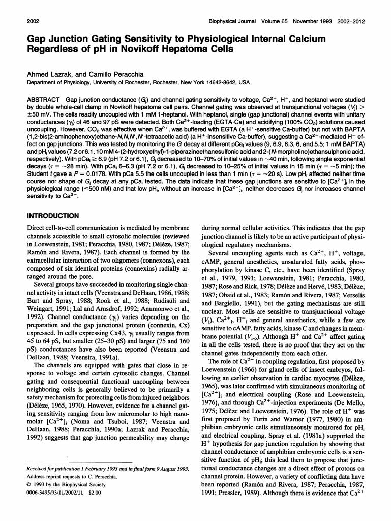
The Novikoff hepatoma cell line is suitable for studying thejunctional membrane properties by double whole-cell clampfor a number of reasons: the small cell size (12-13 ,um), forwhich fast exchange between pipette solution and cell inte-rior takes place; the high resistance of the plasma membrane(1.1 ± 0.4 GQ), for which series resistances cause only smallerrors in Gj calculations (less than 10%); the simple culturingprocedure; and the high yield of cell pair formation. Mem-brane potential (Vm), measured in current-clamp mode inindividual cells bathed in SES just after the establishment ofwhole-cell configuration, averaged -50 ± 13.5 mV (n = 13).Nonjunctional membrane resistance (Rm), determined insingle cell in voltage clamp mode, ranged from 0.75 to 1.2Gfl. Membrane capacitance, determined from the amplitudeof the capacitive current, was 7.35 ± 2.55 pF (n = 13). Gjranged from less than 1 to 200 nS, with a mean value of 53.55+ 17.62 nS (n = 84), and was relatively insensitive to Vm(Fig. 1 A).
Voltage dependence of the junctional membrane
For studying voltage dependence ofjunctional conductance,voltage steps of 10 mV (± 100 mV maximum) and 0.4-1-sduration were applied to either cell of the pair, while main-taining the other at holding potential (VH = -40 mV). Forlimiting the error induced by the remaining uncompensatedseries resistances, the Vj-Ij relationship was analyzed only incell pairs coupled with Gj ' 8 nS. In these cell pairs, at Vjvalues greater than ± 50 mV two symmetrical levels of I
were observed (Fig. 1 B): an instantaneous (Ij imSt) and asteady state (Ij J. The voltage dependence of Gj was de-termined by measuring both Ij levels and plotting them withrespect to Vj (Fig. 1 C). Ij inst, measured during the first 5-10ms after the application of the voltage pulse, increased lin-early as a function of Vj (Fig. 1, B and C). In contrast, Ii ss,measured at the end ofeach pulse, was linear only at Vj valuesof ± 50 mV, and started decreasing (deviating from linearity)at higher Vj values (Fig. 1, B and C). The slope of thecurves (Fig. 1 C) is the junctional conductance Gj; in thisexample, Gj inst was 5.77 ± 0.19 nS (mean + SE, opensquares) and Gj 5.40 ± 0.22 nS (mean ± SE, filledsquares) in the + 50 mV range; the latter decreased to2.8 ± 0.4 nS (mean ± SE) when Vj reached ± 100 mV.To illustrate the relationship between Gj rs and Vj, the nor-
malized Gj (n = 7) was plotted with respect to Vj (Fig. 1 D).Normalized Gj values approached 1 at Vj values as high as± 60 mV and decreased to a minimum of 0.44 at ± 100 mV.The curve was fitted to a two-state Boltzmann's distributionof the form:
(Gj ss-Gj min)/(Gj max -Gj ss) = exp[-A(V -V-)],where V0 is the Vj value at which the voltage-sensitive con-ductance is half of the maximal value, Gj ,, is the experi-mentally derived normalized Gj value, Gj max is the theo-retical maximum normalized Gj, Gjmin is the theoreticalminimum normalized Gj, andA is a constant expressing thestrength of the interaction of the channel with Vj (Harris et al.,1981; Spray et al., 1981b). The constant A is equivalent tonq/kT, where n is the equivalent number of electron chargesq that act as the gating mechanism by sensing changes in Vj,while k and T represent Boltzmann's constant and tempera-ture (K). For this plot, the Boltzman's parameters (Fig. 1 C)were: Gj max = 0.987, Gj min = 0.42, n = 1.7 (A = 0.067),and V0 = -70 mV, for negative values, and V0 = + 72.5 mV,for positive values. The cells used for the Boltzmann fit hada Gj of 5.344 ± 2.61 (mean ± SD, n = 7) at Vj = 20 mV.
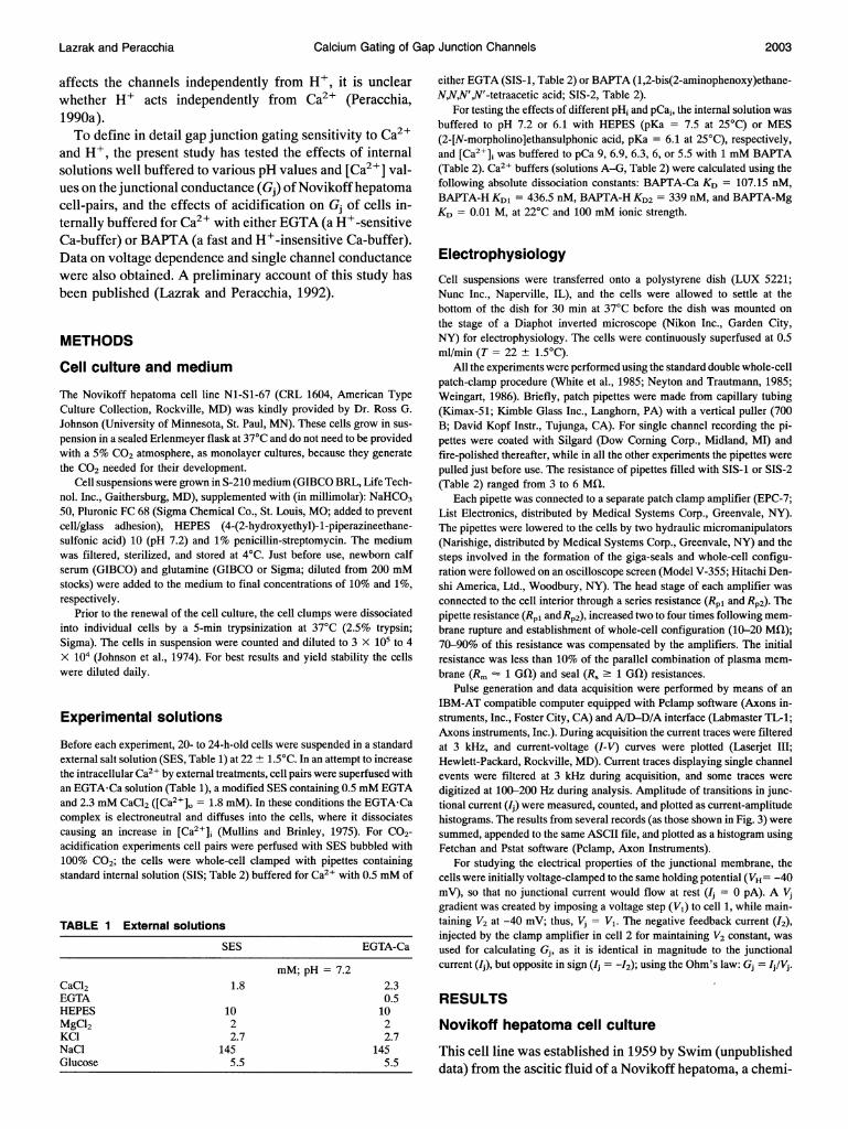
Effects of heptanol on junctional conductance
Superfusions with 1 mM 1-heptanol in SES were used toreduce the junctional conductance to a sufficiently low levelfor detecting single channel activity in gap junctions. Theuncoupling effect of heptanol is shown in Fig. 2. Junctional
Biophysical Journal2004
Calcium Gating of Gap Junction Channels
A
co_
uz
10 r
8
6
-100 -50 0 50 100
Holding potential (mV)
D
_s
-4
I.,
1.2
1.0
0.8
0.6
0.4
0.2-150 -100 -50 0 50
Vj (mV)
100 150
FIGURE 1 (A) Relationship between junctional conductance (Gj 7.382 0.790 nS; mean SD, n = 5) and holding potential (VH). Note that Gj is
insensitive to membrane potential, at least in the ± 80 mV range. The junctional current was determined by imposing to either cell a voltage step of 10mV and 650-ms duration. (B) Illustration of a family of Ij traces generated by pulsing one cell of the pair every 10 s to different Vj values (± 100 mV, 820ms, 10 mV increments), from a holding potential (VH) of -40 mV. With Vj values < + 50 mV, Ij is relatively constant throughout the duration of the pulse.In contrast, with Vj values> ± 50 mV, Ij undergoes a time- and voltage-dependent relaxation from its largest instantaneous value (Ij inst) toward a lowersteady state value (Ij ss). (C) Current-voltage relationship for both instantaneous (hollow squares) and steady state (filled squares) junctional currents. TheIj/Vj relationship is linear and has a slope of 5.77 ± 0.19 nS (mean ± SE) for Ij inst and 5.40 ± 0.22 (mean ± SE) for Ij1, over the ± 50 mV Vj range, whileit deviates from linearity and starts decreasing at greater Vj values. Similar results were obtained in six other cell pairs in which Gj ranged from less than1 to -8 nS. (D) Boltzmann fit of normalized Gj averaged from experiments similar to that shown in B. The cells had a Gj of 5.344 ± 2.61 (mean + SD,n = 7) at Vj = 20 mV. Gj decreases as a function of Vj, reaching its halfmaximum value at transjunctional voltages (Vj = V0). The parameters of the Boltzmannfit are Gmax = 0.987, Gmin = 0.42, n = 1.70, V0 = +72.5 mV for positive voltages, and VO = -70 mV for negative voltages.
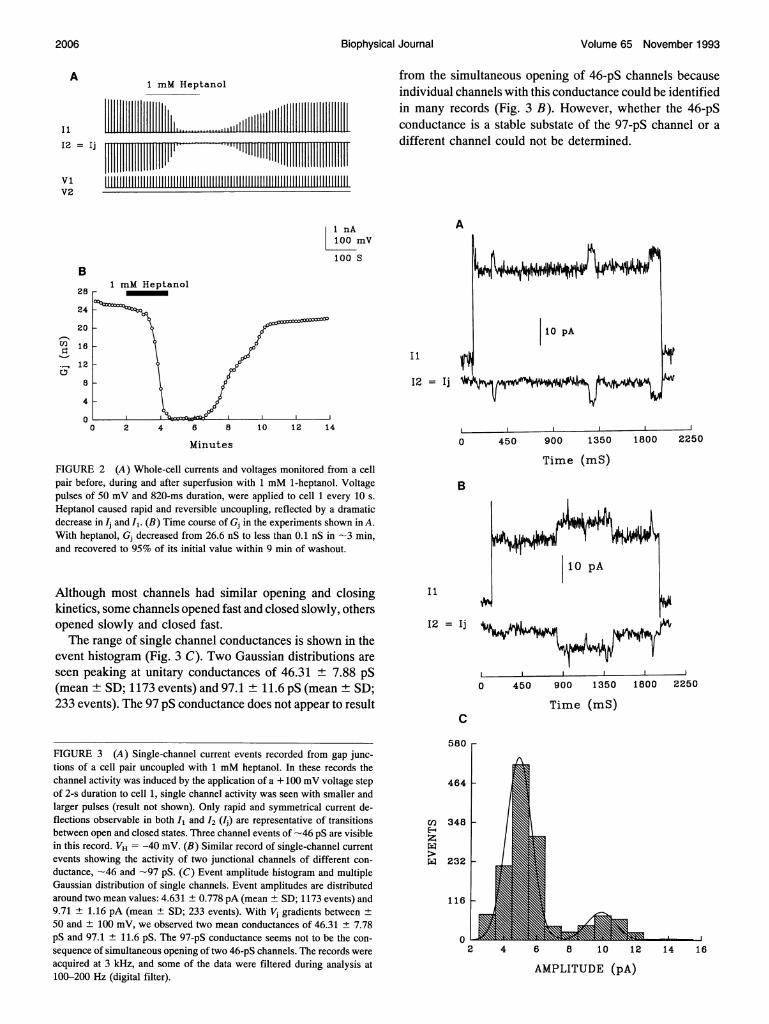
currents were visualized by applying a voltage pulse of +50mV (820-ms duration) every 10 s. Heptanol caused a drasticdecrease in Ij (Fig. 2A), as Gj decreased from 26.6 nS to lessthan 0.1 nS (Fig. 2 B) within 2.5 min. The uncoupling was
almost completely reversible upon return to standard saline,such that IJ and Gj recovered to -90% of their initial valuesin -10 min.
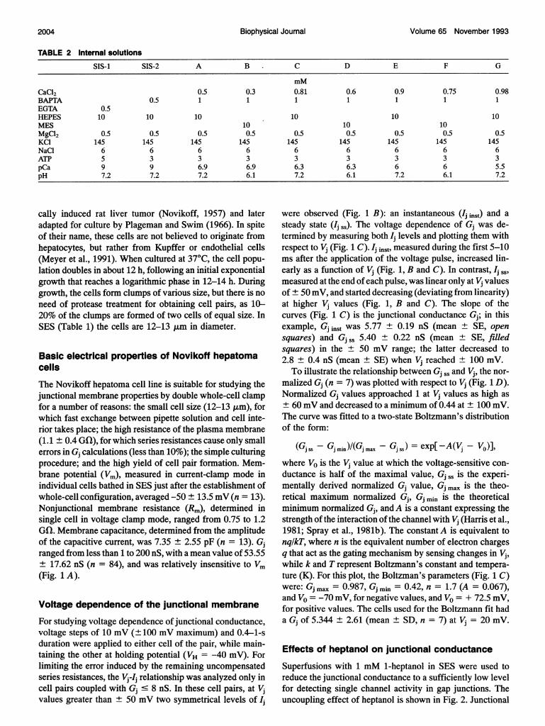
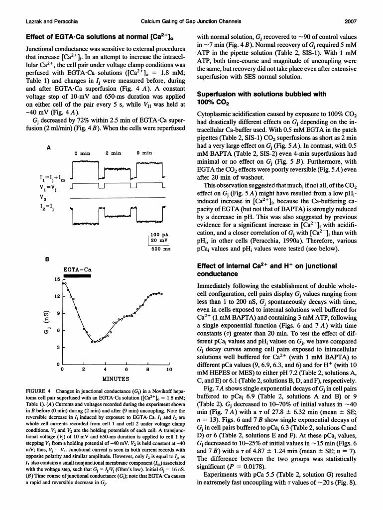
Single channel activity
Single channel events were clearly observed when transjunc-tional voltage steps of 100 mV (2-4-s duration) were appliedto cell pairs uncoupled with 1 mM 1-heptanol (Gj values '
0.5 nS), but single channel events were also seen with trans-junctional voltage lower than 100 mV. Channel activity ismanifested by steplike current transitions that occur simul-taneously in both traces and have the same amplitude butopposite polarity (Fig. 3,A and B). Different records showedeither one (Fig. 3A) or more (Fig. 3B) gap junction channels.During acquisition the data were sampled at 3 kHz. Recordsexhibiting discrete single current steps, as in Fig. 3, were
selected for examining the time domain of the signals. Chan-nel open time ranged from 10 to 550 ms (mean = 168 ms);possible voltage dependence of channel open time could notbe determined. The speed of channel opening and closingranged from <2 to 20 ms, varying among cell pairs.
B
Ij (pA)
C
140 pA
200 mS
Lazrak and Peracchia 2005
Volume 65 November 1993
1 mM Heptanol
...III .....111 111111111111111M ,, *1nin inirii n ii in iniiiif
from the simultaneous opening of 46-pS channels becauseindividual channels with this conductance could be identifiedin many records (Fig. 3 B). However, whether the 46-pSconductance is a stable substate of the 97-pS channel or adifferent channel could not be determined.
V2 II I IIII 1 I I I III1 11 1111111 11V2
B28
24
20
16
12
8
4
0
1 nA100 mV
100 S
1 mM Heptanol
I1
I2 = Ij
0 2 4 6 8 10 12 14
Minutes
FIGURE 2 (A) Whole-cell currents and voltages monitored from a cellpair before, during and after superfusion with 1 mM 1-heptanol. Voltagepulses of 50 mV and 820-ms duration, were applied to cell 1 every 10 s.
Heptanol caused rapid and reversible uncoupling, reflected by a dramaticdecrease in Ij and I,. (B) Time course of Gj in the experiments shown in A.With heptanol, Gj decreased from 26.6 nS to less than 0.1 nS in -3 min,and recovered to 95% of its initial value within 9 min of washout.
Although most channels had similar opening and closingkinetics, some channels opened fast and closed slowly, othersopened slowly and closed fast.The range of single channel conductances is shown in the
event histogram (Fig. 3 C). Two Gaussian distributions are
seen peaking at unitary conductances of 46.31 ± 7.88 pS(mean + SD; 1173 events) and 97.1 ± 11.6 pS (mean + SD;233 events). The 97 pS conductance does not appear to result
A
0 450 900 1350 1800 2250
Time (mS)
B
Ii
I2 = Ij
0
C
450 900 1350
Time (mS)
1800 2250
FIGURE 3 (A) Single-channel current events recorded from gap junc-tions of a cell pair uncoupled with 1 mM heptanol. In these records thechannel activity was induced by the application of a + 100 mV voltage stepof 2-s duration to cell 1, single channel activity was seen with smaller andlarger pulses (result not shown). Only rapid and symmetrical current de-flections observable in both I, and I2 (I) are representative of transitionsbetween open and closed states. Three channel events of -46 pS are visiblein this record. VH = -40 mV. (B) Similar record of single-channel currentevents showing the activity of two junctional channels of different con-ductance, -46 and -97 pS. (C) Event amplitude histogram and multipleGaussian distribution of single channels. Event amplitudes are distributedaround two mean values: 4.631 ± 0.778 pA (mean ± SD; 1173 events) and9.71 ± 1.16 pA (mean ± SD; 233 events). With Vj gradients between +50 and ± 100 mV, we observed two mean conductances of 46.31 + 7.78pS and 97.1 ± 11.6 pS. The 97-pS conductance seems not to be the con-sequence of simultaneous opening of two 46-pS channels. The records wereacquired at 3 kHz, and some of the data were filtered during analysis at100-200 Hz (digital filter).
580 r
464
VI
Ez;Ew
348 F
232
116 _
0
2 4 6 8 10 12 14 16
AMPLITUDE (pA)
A
Ii
I2 = Ii
1-1
ul*_
&
2006 Biophysical Journal
Calcium Gating of Gap Junction Channels
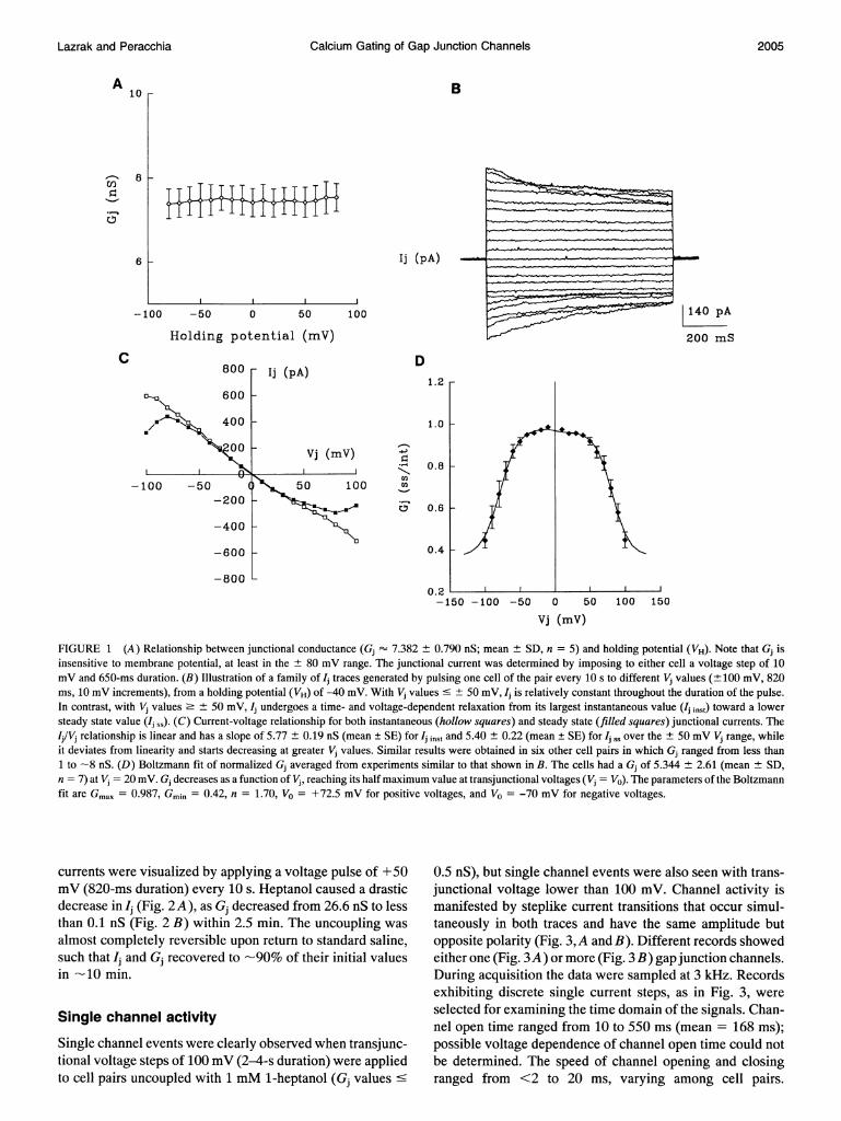
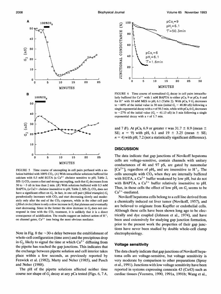
Effect of EGTA'Ca solutions at normal [Ca2+]0Junctional conductance was sensitive to external proceduresthat increase [Ca2+],. In an attempt to increase the intracel-lular Ca2 , the cell pair under voltage clamp conditions wasperfused with EGTA-Ca solutions ([Ca2+]0 = 1.8 mM;Table 1) and changes in Ij were measured before, duringand after EGTA-Ca superfusion (Fig. 4 A). A constantvoltage step of 10-mV and 650-ms duration was appliedon either cell of the pair every 5 s, while VH was held at-40 mV (Fig. 4 A).
Gj decreased by 72% within 2.5 min of EGTA-Ca super-fusion (2 ml/min) (Fig. 4 B). When the cells were reperfused
A
I =I+IMV =ViVl=j
V2I2 =Ij
0 min 2 min 9 min
LWL*"
lo 1- lM
100 pA20 mV
500 ms
B
EGTA-Ca
12
_s9
6
3
0
FIGURE 4 Cltoma cell pair si
Table 1). (A) Clin B before (0 n
reversible decrewhole cell currnconditions. V1 a
tional voltage ('stepping V1 fronmV; thus, Vj =opposite polarit3I1 also contains;with the voltage(B) Time course
a rapid and reve
with normal solution, Gj recovered to -90 of control valuesin -7 min (Fig. 4 B). Normal recovery of Gj required 5 mMATP in the pipette solution (Table 2, SIS-1). With 1 mMATP, both time-course and magnitude of uncoupling werethe same, but recovery did not take place even after extensivesuperfusion with SES normal solution.
Superfusion with solutions bubbled with100% CO2
Cytoplasmic acidification caused by exposure to 100% CO2had drastically different effects on Gj depending on the in-tracellular Ca-buffer used. With 0.5 mM EGTA in the patchpipettes (Table 2, SIS-1) CO2 superfusions as short as 2 minhad a very large effect on Gj (Fig. 5 A). In contrast, with 0.5mM BAPTA (Table 2, SIS-2) even 4-min superfusions hadminimal or no effect on Gj (Fig. 5 B). Furthermore, withEGTA the CO2 effects were poorly reversible (Fig. 5A) evenafter 20 min of washout.
This observation suggested that much, ifnot all, of the CO2effect on Gj (Fig. 5 A) might have resulted from a low pHi-induced increase in [Ca21]i, because the Ca-buffering ca-pacity ofEGTA (but not that ofBAPTA) is strongly reducedby a decrease in pH. This was also suggested by previousevidence for a significant increase in [Ca21], with acidifi-cation, and a closer correlation of Gj with [Ca21], than withpHi, in other cells (Peracchia, 1990a). Therefore, variouspCai values and pHi values were tested (see below).
Effect of internal Ca2+ and H+ on junctionalconductance
Immediately following the establishment of double whole-cell configuration, cell pairs display Gj values ranging fromless than 1 to 200 nS, Gj spontaneously decays with time,even in cells exposed to internal solutions well buffered forCa2+ (1 mM BAPTA) and containing 3mM ATh, followinga single exponential function (Figs. 6 and 7 A) with time
o \° constants (T) greater than 20 min. To test the effect of dif-ferent pCai values and pHi values on Gj, we have comparedGj decay curves among cell pairs exposed to intracellularsolutions well buffered for Ca2+ (with 1 mM BAPTA) to
I different pCa values (9, 6.9, 6.3, and 6) and for H+ (with 100 2 4 6 8 10 mM HEPES or MES) to either pH 7.2 (Table 2, solutions A,
MINUTES C, and E) or 6.1 (Table 2, solutions B, D, and F), respectively.
hanges in junctional conductance (G) in a Novikoff hepa- Fig. 7A shows single exponential decays of Gj in cell pairsuperfused with an EGTA Ca solution ([Ca2+]. = 1.8 mM; buffered to pCai 6.9 (Table 2, solutions A and B) or 9urrents and voltages recorded during the experiment shown (Table 2). Gj decreased to 10-70% of initial values in -40nin) during (2 min) and after (9 min) uncoupling. Note the min (Fig. 7 A) with a T of 27.8 ± 6.32 min (mean ± SE;base in Ij induced by exposure to EGTA-Ca. ' and '2 are n = 13). Figs. 6 and 7 B show single exponential decays ofents recorded from cell 1 and cell 2 under voltage clamp Gj in cellnd V2 are the holding potentials of each cell. A transjunc- pairs buffered to pCa; 6.3 (Table 2, solutions C andVj) of 10 mV and 650-ms duration is applied to cell 1 by D) or 6 (Table 2, solutions E and F). At these pCai values,n a holding potential of -40 mV. V2 is held constant at -40 Gj decreased to 10-25% of initial values in - 15 min (Figs. 6V1. Junctional current is seen in both current records with and 7 B) with a T of 4.87 ± 1.24 min (mean + SE; n = 7).y and similar amplitude. However, only I2 is equal to I as The difference between the two groups was statisticallya small nonjunctional membrane component (Im) associatedstep, such that Gj = Ij/Vj (Ohm's law). Initial Gj = 16 nS. sigificant (P = 0.0178).ofjunctional conductance (Gj); note that EGTA-Ca causes Experiments with pCa 5.5 (Table 2, solution G) resulted
ersible decrease in Gj. in extremely fast uncoupling with T values of -20 s (Fig. 8).
Lazrak and Peracchia 2007
41 'A
W" 0 w*10"044,m
Volume 65 November 1993
1 00%CO2
0
0
z0
E-
0
0
z0
Ez
100
80
60
40
20
00 5 10 15 20 25
MINUTES
100%C02
4 =~~~~~~~~~s~
pca.=9pHj=6. 1
T=50.3min
0 5 10 15
MINUTES
20 25 30
FIGURE 6 Time course of normalized Gj decay in cell pairs intracellu-larly buffered for Ca2l with 1 mM BAPTA to either pCai 9 or pCai 6 andfor H+ with 10 mM MES to pHi 6.1 (Table 2). With pCai 9 Gj decreasesto -60% of the initial value in 30 min (initial Gj = 48.80 nS) following asingle exponential decay with a T of 50.3 min, while with pCai 6 Gj decreasesto -27% of the initial value (Gj = 41.13 nS) in 5 min following a singleexponential decay with a T of 1.7 min.
and 7 B). At pCai 6.9 or greater T was 31.7 ± 8.9 (mean ±SE; n = 9) with pHi 6.1 and 19 ± 3.25 (mean ± SE;n = 4) with pHi 7.2 (not a statistically significant difference).
DISCUSSION
0 5 10 15 20 25 The data indicate that gap junctions of Novikoff hepatomacells are voltage-sensitive, contain channels with unitary
MINUTES conductances of 46 and 97 pS, are gated by nanomolar
Time course of uncoupling in cell pairs perfused with a so- [Ca2+]i regardless of pHi, and are insensitive to H'i. The1 with 100% CO2. (A) With intracellular solutions buffered for cells uncouple with CO2 when they are internally buffered0.5 mM EGTA (a Ca21 chelator sensitive to pH; Table 2, with EGTA, a Ca2+ buffer weakened by low pH, but do notiuses a fast and strong uncoupling, such that Gj decreases from with BAPTA, a Ca2+ buffer relatively insensitive to pH.in less than 2 min. (B) With solutions buffered with 0.5 mM
Ta2+ chelator insensitive to pH; Table 2, SIS-2), CO2 does not Thus, in these cells the effect of low pH2 on G seems to becant effect on Gj. In fact, in one cell pair (filled triangles) Gj Ca -mediated.increases with CO2 and start decreasing slowly and moder- Novikoff hepatoma cells belong to a cell line derived fromer the end of the CO2 exposure, while in the other cell pair a chemically induced rat liver tumor (Novikoff, 1957), and) there is only a slow increase in Gj that plateaus and eventuallyJ ~~~~~~arebelieved to originate from Kupffer or endothelial cells.ig. Since in the former the slow decrease in Gj does not cor-ne with the CO2 treatment, it is unlikely that it is a direct Although these cells have been shown long ago to be elecof acidification. The results suggest an indirect action of H+i trically and dye coupled (Johnson et al., 1974), and havetes, Ca21 ions being the most obvious mediator. been used extensively for studying gap junction formation,
prior to the present work the properties of their gap junc-tions have never been studied by double whole-cell clamp
8 the -30-s delay between the establishment of electrophysiology.whole-cell configuration (time zero) and the precipitous dropin Gj, likely to signal the time at which Ca2' diffusing fromthe pipette has reached the gap junctions. This indicates thatthe exchange between pipette solution and cell interior takesplace within a few seconds, as previously reported byFenwick et al. (1982), Marty and Neher (1985), and Puschand Neher (1988).The pH of the pipette solutions affected neither time
course nor shape of Gj decay at any pCa tested (Figs. 6, 7 A,
Voltage sensitivity
The data clearly indicate that gap junctions of Novikoff hepa-toma cells are voltage-sensitive, but voltage sensitivity isvery moderate by comparison to other preparations (Sprayet al., 1991). Junctions with low voltage sensitivity have beenreported in systems expressing connexin 43 (Cx43) such as
cardiac tissues (Veenstra, 1990, 1991a, 1991b; Wang et al.,
A
50
40
'-" 30
*- 20
10
0
B
50
40
20
10 V
a
FIGURE 5lution bubbledcalcium withSIS-1) CO2 ca50 to -5 nS iBAPTA, (a Chave a signifi4paradoxicallyately only aft((filled circles)start decreasirrespond in tirconsequence(on channel ga
Note in Fig
2008 Biophysical Journal

Calcium Gating of Gap Junction Channels
A
100
80
60
40
20
0
B
100
80
60
40
20
0
pCai 6.9-9
lines: pHi=6.1dashed lines: pHi=7.2
100 r
80 F
_s-1
U2
_/
*_
60
* *
-r7= 21 s
40
20 F20 25
MINUTES
pCa1 6-6.3
lines: pHi=6.1dashed lines: pHi=7.2
0 5 10 15 20 25 30
MINUTES
FIGURE 7 Single exponential decays of normalizedtance in cell pairs intracellularly buffered to pCa, 6.9-9 (SIS-2, A and B) and to 6.3-6 (B; Table 2, solutions C-or 6.1. Note that the acidic pipette solution affects neitshape of Gj decay, while the higher the [Ca21], the fastimean initial Gj was 50.06 ± 20.5 nS (mean + SD) forA, and 68.63 ± 35.9 nS (mean ± SD) for those shownthat the time constant (T) of the exponential decay of Cinitial junctional conductance. Paradoxically, in mostsidual Gj was higher at pHi 6.1 than at pH 7.2.
1992; Lal and Arnsdorf, 1992; Anumonwo e
epithelial cells (Cooper et al., 1989) and c
with Cx43, such as Xenopus oocytes (Swensand SKHepl cells (Moreno et al., 1992). Sinchepatoma cells are believed to express Cx41992; Starich and Johnson, personal commreasonable to believe that the low voltage slated to the nature of the connexin expressed
Voltage sensitivity was greater in poorly c(
increased in well coupled cell as coupling sp
teriorated with time. This could be due to a (
resistance. Because of uncompensated resiswith the junctional resistance, Vj could be reclevel at which voltage-dependent gating be(Rook et al., 1988; Wilders and Jongsma, 1cin our study the analysis of the voltage sensiijunction was limited to the cell pairs poor
ranging from <1 to -8 nS).
0 1 2 3 4 5
MINUTES
FIGURE 8 Time course of uncoupling of a cell pair intracellularly buff-ered to pCa, 5.5 and pH, 7.2 (Table 2, solution G). Initial Gj = 85.9 nS. Theuncoupling followed a single exponential decay (solid line) with a T of 21s. Note the abrupt decrease in junctional conductance (filled circles) thatoccurs in less than 1 min after the rupture of the membrane patches. Thisdemonstrates the fast exchange between pipette solution and cytosol.
Single channel conductance
35 40 45 In order to observe single channel events the heptanol treat-ment was selected among other uncoupling methods, such as
junctional conduc-
exposure to CO2 or addition of calcium to the pipettes, be-
A; Table 2, solution cause with heptanol the channel activity of the nonjunctionalF), at either pH, 7.2 membrane is considerably reduced and nonjunctional mem-her time course nor brane resistance is high. This reduces the noise level ander the Gj decay. The minimizes the error in measuring current from single junc-the plots shown in tional channels.in B This indicates Single channel activity was observed when nearly all of
expsnerientsthe the channels were closed by heptanol in response to a voltagegradient across the junction. The two major unitary conduc-tances observed (46 and 97 pS) are similar to those reportedfor most cells expressing Cx43 (Burt and Spray, 1988; Rook
t al., 1992) lens et al., 1988; Rudisuli and Weingart, 1991; Lal and Arnsdorf,sells transfected 1992; Anumonwo et al., 1992), the only possible exceptionson et al., 1989) being the embryonic chick heart cells, where two conduc--e also Novikoff tance levels (165 and 75 pS) were reported (Veenstra and3 (Meyer et al., DeHaan, 1986, 1988). Our results, showing low voltage de-iunication), it is pendence and a similar range of the single channel conduc-,ensitivity is re- tances, together with the immunoblot data of Meyer et al.by this cell line. (1992), suggest that Novikoff hepatoma cells express theoupled cells and same connexin (Cx43) of the adult rat heart (Beyer et al.,,ontaneously de- 1987). However, whether small amounts of other connexinschange in series are coexpressed with Cx43 cannot be entirely excluded. Pre-tances in series sent evidence does not indicate that the 97-pS conductanceluced below the represents the simultaneous opening of two 46-pS channels,comes apparent because current steps of this size were often observed rising)92). Therefore, individually from baseline, suggesting that they result fromtivity of the gap the opening of individual channels. More work will berly coupled (Gj needed to determine whether the 46-pS conductance is a
stable substate of the 97-pS channel or another channel.
z
Ez
0C.-)cJ
z0
z0
_
z
0C-)
C.)
z
z
E-
z
Lazrak and Peracchia 2009
li
Volume 65 November 1993
Sensitivity of the junction to Ca2l and HI
The sensitivity of cell coupling to acidification has beentested for years in many systems using different approaches.In cell pairs, Gj was found to decrease in cells exposed to100% Co2, when the pipette solutions were buffered forcalcium with EGTA (Burt and Spray, 1988). However,EGTA is not as reliable as BAPTA as intracellular Ca-bufferfor several reasons. First, the Ca-EGTA affinity constantdrops by two orders of magnitude with a decrease in pH from7 to 6 (KCa-aff = 1.8 X 10⁶ and 1.8 X 10⁴, at pH 7 and 6,respectively), while that of Ca-BAPTA decreases onlyslightly with the same pH drop (KCa-aff = 7 X 10⁶ and1.8 X 106, at pH 7 and 6, respectively). Second, calculationsof the gradient of [Ca21] in the vicinity of a channel poreshow thatEGTA solutions are completely ineffective in buff-ering [Ca21] within macromolecular distances from the pore,while faster buffers such as BAPTA are quite effective(Marty and Neher, 1985; Adler et al., 1991; Stern, 1992).This is consistent with our data showing a dependence ofCO2 uncoupling efficiency on the Ca-buffer used (EGTAversus BAPTA), and indicates that acidification might un-couple cell pairs via an increase in cytosolic Ca2+ which isnot adequately buffered by EGTA. Indeed, one would expectthe opposite result if H+, rather than Ca2 , were acting onthe channel's gating mechanism, because, under the condi-tions of cytoplasmic acidification by 100% CO2, EGTAwould bind H+ and release Ca2+ (for each Ca2' released twoprotons would bind); thus, [Ca2+]i would increase more (and[H+]i would increase less) than with BAPTA. In fact, withlowered pHi EGTA turns out to be a source of Ca2' ratherthan a Ca2+ chelator.The uncoupling effect of superfusions with Ca-EGTA so-
lutions at normal [Ca2+]o confirms that Ca2+, independentlyaffects coupling in Novikoff cells. EGTA-Ca21 solutionswere previously found to simultaneously increase [Ca2+], to>1 ,uM and junctional electrical resistance in crayfish axonsstudied with current clamp and Ca2+-sensitive microelec-trodes (Peracchia, unpublished data). EGTA Ca2+ solutionscause an increase in [Ca2+]i because the electroneutralEGTA Ca2' diffuses passively through membranes (Mullinsand Brinley, 1975) and once in the cell readily dissociates,increasing [Ca2+]i. Evidence for the need of 5 mM ATP inthe patch pipette for Gj recovery indicates that recouplingrequires energy-dependent mechanisms likely to includeCa2+ extrusion and sequestration.The interpretation of a Ca2+i-mediated low pHi effect on
coupling is supported by data on cell pairs exposed to dif-ferent pHi values and pCai values. In these experiments wehave assumed that the ionic composition of the junctionalenvironment reaches a rapid equilibrium with the pipette so-lution. This is suggested by the very short delay (-30 s)between the establishment of whole-cell configuration (timezero) and the precipitous drop in Gj occurring with pipettesolution of pCa 5.5 (Fig. 8). The delay is likely to representthe time required for the diffusion of Ca2" from the pipetteinterior to the junctional channels. Previous studies have
clearly demonstrated that the whole cell patch-clamp pro-cedure enables one to control the ionic composition of thecytosol. The diffusional equilibrium between patch pipettesolution and cytosol is reached within seconds for ions andother small molecules, especially in cells as small as theNovikoff hepatoma cells (12-15 p,m of diameter), as pre-viously reported for Na+ (15 s) by Fenwick et al. (1982) andMarty and Neher (1983), and for K+ (15 s) by Marty andNeher (1985), in bovine chromaffin cells. The kinetics of thediffusional exchange between small cells and patch pipetteshas been studied extensively by Push and Neher (1988) usingcompounds ranging in molecular weight from 23 to 150,000daltons. Their data demonstrated a fast exchange of smallcompounds between pipettes and cytosol (especially forsmall ions such as Na+ and K+), and the existence of arelationship between the diffusional rate of a given moleculeand various parameters such as molecular weight, pipette-cell assembly and pipette electrical resistivity. For insuringfast exchange between cytosol and pipette solution and lowaccess resistance, in our study we always used pipettes withrelatively large opening, ranging from 0.5 to -2 p,m (3-6MfQ resistance when filled with SIS). In any event, no onecan be absolutely sure about the exact ionic composition ofthe region near the junctional channels, and even Ca2 , mea-surements with fura-2 or indo-1 are inadequate in providingthe necessary high resolution data on [Ca2+]i at the channelenvironment.The dramatic decrease in time constant of uncoupling with
a change in pCai from 6.9 to 6.3, regardless of pHi, indicatesthat the channel gates are sensitive to nanomolar changes into[Ca2+] and are insensitive to pHi, at least in the range be-tween 7.2 and 6.1. Lowered pHi does not seem to changechannel sensitivity to Ca2' as well, but paradoxically mayin fact prevent some channels from closing. In the absenceof an increase in [Ca2+]i, acidification may inhibit themechanism causing spontaneous uncoupling, as with internalsolutions well buffered for Ca21 (BAPTA) and H+ (HEPESor MES) both the time constant of the spontaneous Gj decayand the residual junctional conductance were greater at pHi6.1 than at pHi 7.2. Our data on lack of synergism betweenCa2' and H+ in reducing Gj contrast with previous evidenceon cardiac cells (Burt, 1987; White et al., 1990), but areconsistent with the recent data on crayfish axons subjectedto cytoplasmic acidification while monitoring junctional re-sistance (Rj) and either [Ca21], or [H+]i (Peracchia, 1990a).In this study the curves describing the time course of changesin [H+]i and Rj were found to differ significantly from eachother in shape and peak time, and fast and slow changes in[H+]i of similar magnitude had markedly different effects onRj; in contrast, changes in [Ca2+]i and Rj matched well witheach other (Peracchia, 1990a). The increase in [Ca21], withacidification resulted from Ca2' release from caffeine andryanodine-sensitive stores (Peracchia, 1990b). As in thecrayfish study, in Novikoff cells Ca2' appears to affect thechannel gates at nanomolar concentrations.
Early data seemed to support a direct effect of H+ onjunctional conductance (Turin and Warner 1977, 1980;
2010 Biophysical Joumal
Lazrak and Peracchia Calcium Gating of Gap Junction Channels 2011
Spray et al., 1981a; Spray and Bennett, 1985), but someinconsistencies were apparent (Peracchia, 1987, 1991;Ramon and Rivera, 1987). In the heart, a pHi of 6.6 had onlya small effect on junctional conductance (Reber and Wein-gart, 1982), H+ affected healing-over only at pH <5 (DeMello, 1983) and internal longitudinal resistance correlatedpoorly with [He]i (Pressler, 1989). In crayfish, low pH so-lutions did not change junctional conductance in internallyperfused axons (Johnston and Ramon, 1981; Arellano et al.,1986), and several inconsistencies between pHi and electricalcoupling were reported in cells of insect salivary glands(Rose and Rick, 1978).
In the past, the [Ca2+]i effective on coupling has been amatter of controversy. While only [Ca2+]i as high as 40-400,uM seemed to be effective in some systems (Oliveira-Castroand Loewenstein, 1971; Spray et al., 1982), low micromolar(Deleze and Loewenstein, 1976; Rose and Loewenstein,1976; Weingart, 1977; Dahl and Isenberg, 1980; Neyton andTrautmann, 1986; Maurer and Weingart, 1987; Veenstra andDeHaan, 1988) to nanomolar (Noma and Tsuboi, 1987;Peracchia, 1990a) concentrations were found effective inothers. In our study, pCa 6.3 (500 nM) affected Gj, while pCa6.9 (126 nM) did not, suggesting that [Ca2+]i at which mostCa2+-driven physiological functions are activated may alsoaffect cell coupling. This indicates that Ca2+ is a physiologi-cal regulator of cell coupling and that gap junctions may playa role in a variety of Ca2+-mediated cellular activities. In-deed, preliminary data show a simultaneous increase in[Ca21], to -500 nM, decrease in Gj by -30% and activationof K+(Ca2+) in Novikoff hepatoma cells in which a releaseof IP3 was induced by ATP activation of purinergic receptors(Lazrak et al., 1993).
In conclusion, Novikoff hepatoma cells are well coupledby moderately voltage-sensitive gap junctions with singlechannel conductances of -46 and -97 pS. The gap junctionsare sensitive to [Ca21], in the nanomolar range, and H',seems neither to decrease Gj nor to increase channel sensi-tivity to Ca2+1.
We thank Dr. Ross G. Johnson for providing Novikoff hepatoma cells andhelpful advise on cell culturing procedures, and Drs. Peter Shrager and TedBegenisich for helpful criticism of the manuscript.The study was supported by the National Institutes of Health, grantGM20113.
REFERENCESAdler, E. M., G. J. Augustine, S. N. Duffy, and M. P. Charlton. 1991. Alien
intracellular calcium chelators attenuate neurotransmitter release at thesquid giant synapse. J. Neurosci. 11:1496-1507.
Anumonwo, J. M., H. Z. Wang, E. Trabka-Janik, B. Dunham, R. D. Veen-stra, M. Delmar, and J. Jalife. 1992. Gap junctional channels in adultmammalian sinus nodal cells. Immunolocalization and electrophysiology.Circ. Res. 71:229-239.
Arellano, R. O., F. Ram6n, A. Rivera, and G. A. Zampighi. 1986. Loweringof pH does not directly affect the junctional resistance of crayfish lateralaxons. J. Membr. Bio. 94:293-299.
Beyer, E. C., Paul D. L., and D. A. Goodenough. 1987. Connexin 43: aprotein from rat heart homologous to a gap junction protein from liver.J. Cell Biol. 105:2621-2629.
Burt, J. M. 1987. Block of intercellular communication: interaction of in-tracellular H+ and Ca2". Am. J. Physiol. 253:C607-C612.
Burt, J. M., and D. C. Spray. 1988. Single-channel events and gating be-havior of the cardiac gap junction channel. Proc. Natl. Acad. Sci. USA.85:3431-3434.
Cooper, K., J. L. Rae, and P. Gates. 1989. Membrane and junctional prop-erties of dissociated frog lens epithelial cells. J. Membr. Biol. 111:215-227.
Dahl, G., and G. Isenberg. 1980. Decoupling of heart muscle cells: corre-lation with increased cytoplasmic calcium activity and with changes ofnexus ultrastructure. J. Membr. Biol. 53:63-75.
De Mello, W. C. 1975. Effect of intracellular injection of calcium and stron-tium on cell communication in heart. J. Physiol. (Lond.). 250:231-245.
De Mello, W. C. 1983. The influence of pH on the healing-over of mam-malian cardiac muscle. J. Physiol. (Lond.). 339:299-307.
Deleze, J. 1965. Calcium ions and the healing-over in heart fibers. In Elec-trophysiology of the Heart. B. Taccardi, and C. Marchetti, editors.Pergamon Press, Elmsford, New York. 147-148.
Deleze, J. 1970. The recovery of the resting potential and input resistancein sheep heart injured by knife or laser. J. Physiol. (Lond.). 208:547-562.
Deleze, J. 1987. Cell-to-cell communication in the heart: structure-functioncorrelations. Experientia (Basel). 43:1068-1075.
Deleze, J., and J. C. Herve. 1983. Effects of several uncouplers of cell-to-cellcommunication on gap junction morphology in mammalian heart.J. Membr. Bio. 76:203-215.
Deleze, J., and W. R. Loewenstein. 1976. Permeability of a cell junctionduring intracellular injection of divalent cations. J. Membr. Bio. 28:71-86.
Fenwick, E. M., A. Marty, and E. Neher. 1982. A patch-clamp study ofbovine chromaffin cells and of their sensitivity to acetylcholine.J. Physiol. (Lond.) 331:577-597.
Harris, A. L., D. C. Spray, and M. V. Bennett. 1981. Kinetic properties ofa voltage-dependent junctional conductance. J. Gen. Physiol. 77:95-117.
Johnson, R., M. Hammer, J. Sheridan, and J.-P. Revel. 1974. Gap junctionformation between reaggregated Novikoff hepatoma cells. Proc. Natl.Acad. Sci. USA. 71:4536-4540.
Johnston, M. F., and F. Ram6n. 1981. Electrotonic coupling in internallyperfused crayfish segmented axons. J. Physiol. (Lond.). 317:509-518.
Lal, R., and M. F. Arnsdorf. 1992. Voltage-dependent gating and single-channel conductance of adult mammalian atrial gap junctions. Circ. Res.71:737-743.
Lazrak, A., and C. Peracchia. 1992. Gap junctions of Novikoff hepatomacells are Ca2l-sensitive but pH-insensitive. Mol. Bio. Cell 3:A295.
Lazrak, A., A. Peres, S. Giovannardi, and C. Peracchia. 1993. Partial un-coupling, increase in calcium and activation of K+(Ca) channels withATP-induced stimulation of purinergic receptors linked to IP3 turnover.Proceedings of the 1993 International Meeting on Gap Junctions.Hiroshima, Japan. August. 55.
Loewenstein, W. R. 1966. Permeability of membrane junctions. Ann. N. Y.Acad. Sci. 137:441-472.
Loewenstein, W. R. 1981. Junctional intercellular communication: the cell-to-cell membrane channel. Physiol. Rev. 61:829-913.
Marty, A., and E. Neher. 1983. Tight-seal whole-cell recording. In Single-Channel Recording. B. Sakman, and E. Neher, editors. Plenum Press,New York. 107-122.
Marty, A., and E. Neher. 1985. Potassium channels in cultured bovine ad-renal chromaffin cells. J. Physiol. (Lond.). 367:117-141.
Maurer, P., and R. Weingart. 1987. Cell pairs isolated from adult guinea pigand rat hearts: effects of [Ca2+]i on nexal membrane resistance. PflugersArch. Eur. J. Physiol. 409:394-402.
Meyer, R. A., P. D. Lampe, B. Malewicz, W. J. Baumann, and R. G.Johnson. 1991. Enhanced gap junction formation with LDL and apoli-poprotein B. Exp. Cell Res. 196:72-81.
Meyer, R. A., D. W. Laird, J. P. Revel, and R. G. Johnson. 1992. Inhibitionof gap junction and adherens junction assembly by connexin and A-CAMantibodies. J. Cell BioL 119:179-189.
Moreno, A. P., G. I. Fishman, and D. C. Spray. 1992. Phosphorylation shiftsunitary conductance and modifies voltage dependent kinetics of humanconnexin43 gap junction channels. Biophys. J. 62:51-53.
Mullins, L. J., and F. J. Brinley, Jr.. 1975. Sensitivity of calcium efflux from
2012 Biophysical Journal Volume 65 November 1993
squid axons to changes in membrane potential. J. Gen. Physiol. 65:135-152.
Neyton, J., and A. Trautmann. 1985. Single-channel currents of an inter-cellular junction. Nature (Londc). 317:331-335.
Neyton, J., and A. Trautmann. 1986. Acetylcholine modulation of the con-ductance of intercellular junctions between rat lacrimal cells. J. Physiol.(Lond.). 377:283.
Noma, A., and N. Tsuboi. 1987. Dependence of junctional conductance onproton, calcium and magnesium ions in cardiac paired cells of guinea-pig.J. Physiol. (Lond.) 382:193-211.
Novikoff, A. B. 1957. A transplantable rat liver tumor induced by 4-dimethylaminoazobenzene. Cancer Res. 17:1010-1027.
Obaid, A. L., S. J. Socolar, and B. Rose. 1983. Cell-to-cell channels withtwo independently regulated gates in series: analysis of junctional con-ductance modulation by membrane potential, calcium, and pH. J. Membr.Bio. 73:69-89.
Oliveira-Castro, G. M., and W. R. Loewenstein. 1971. Junctional membranepermeability: effects of divalent cations. J. Membr. Biol. 5:51-77.
Peracchia, C. 1980. Structural correlates of gap junction permeation. Int.Rev. Cytol. 66:81-146.
Peracchia, C. 1987. Permeability and regulation of gap junction channels incells and in artificial lipid bilayers. In Cell-to-cell Communication. W. C.De Mello, editor. Plenum Press, New York. 65-102.
Peracchia, C. 1990a. Increase in gap junction resistance with acidificationin crayfish septate axons is closely related to changes in intracellularcalcium but not hydrogen ion concentration. J. Membr. Biol. 113:75-92.
Peracchia, C. 1990b. Effects of caffeine and ryanodine on low pHi-inducedchanges in gap junction conductance and calcium concentration in cray-fish septate axons. J. Membr. Bio. 117:79-89.
Peracchia, C. 1991. Possible involvement of caffeine- and ryanodine-sensitive calcium stores in low pH-induced regulation of gap junctionchannels. In Biophysics of Gap Junction Channels. C. Peracchia, editor.CRC Press, Inc., Boca Raton, Florida. 13-28.
Plagemann, P. G., and H. E. Swim. 1966. Replication of mengovirus.I. Effect on synthesis of macromolecules by host cell. J. Bacteriol. 91:2317-2326.
Pressler, M. L. 1989. Intracellular pH and cell-to-cell transmission in sheepPurkinje fibers. Biophys. J. 55:53-65.
Pusch, M., and E. Neher. 1988. Rates of diffusional exchange between smallcells and a measuring patch pipette. Pflugers Arch. Eur. J. Physiol. 411:204-211.
Ram6n, F., and A. Rivera. 1987. Gap junction channel modulation: a physi-ological viewpoint. Prog. Biophys. Mol. Bio. 48:127-153.
Reber, W. R., and R. Weingart. 1982. Ungulate cardiac Purkinje fibres: theinfluence of intracellular pH on the electrical cell-to-cell coupling.J. PhysioL (Lond.). 328:87-104.
Rook, M. B., H. J. Jongsma, and A. C. van Ginneken. 1988. Properties ofsingle gap junctional channels between isolated neonatal rat heart cells.Am. J. Physiol. 255:H770-H782.
Rose, B., and W. R. Loewenstein. 1976. Permeability of a cell junction andthe local cytoplasmic free ionized calcium concentration: a study withaequorin. J. Membr. Bio. 28:87-119.
Rose, B., and R. Rick. 1978. Intracellular pH, intracellular Ca, andjunctionalcell-cell coupling. J. Membr. Bio. 44:377-415.
Rildisuli, A., and R. Weingart. 1989. Electrical properties of gap junctionchannels in guinea-pig ventricular cell pairs revealed by exposure to hep-tanol. Pflugers Arch. Eur. J. Physiol. 415:12-21.
Rudisuli, A., and R. Weingart. 1991. Gap junctions in adult ventricularmuscle. In Biophysics of Gap Junction Channels. C. Peracchia, editor.CRC Press, Inc., Boca Raton, FL. 43-56.
Spray, D. C., A. L. Harris, and M. V. Bennett. 1979. Voltage dependenceof junctional conductance in early amphibian embryos. Science (Wash.DC). 204:432-434.
Spray, D. C., A. L. Harris, and M. V. Bennett. 1981a. Gap junctional con-ductance is a simple and sensitive function of intracellular pH. Science(Wash. DC). 211:712-715.
Spray, D. C., A. L. Harris, and M. V. Bennett. 1981b. Equilibrium propertiesof a voltage-dependent junctional conductance. J. Gen. Physiol. 77:77-93.
Spray, D. C., J. H. Stern, A. L. Harris, and M. V. Bennett. 1982. Gapjunctional conductance: comparison of sensitivities to H and Ca ions.Proc. Natl. Acad. Sci. USA. 79:441-445.
Spray, D. C., and M. V. L. Bennett. 1985. Physiology and pharmacologyof gap junctions. Annu. Rev. Physiol. 47:281-303.
Spray, D. C., M. V. L. Bennett, A. C. Campos de Carvalho, B. Eghbali, A.P. Moreno, and V. Verselis. 1991. Transjunctional voltage dependenceof junction channels. In Biophysics of Gap Junction Channels. C. Per-acchia, editor. CRC Press, Inc., Boca Raton, Florida. 97-116.
Stern, M. D. 1992. Buffering of calcium in the vicinity of a channel pore.Cell Calcium. 13:183-192.
Swenson, K. I., J. R. Jordan, E. C. Beyer, and D. L. Paul. 1989. Formationof gap junctions by expression of connexins in Xenopus oocyte pairs.Cell. 57:145-155.
Turin, L., and A. E. Warner. 1977. Carbon dioxide reversibly abolishes ioniccommunication between cells of early amphibian embryo. Nature(Lond.). 270:56-57.
Turin, L., and A. E. Warner. 1980. Intracellular pH in early Xenopus em-bryos: its effect on current flow between blastomeres. J. Physiol. (Lond.).300:489-504.
Veenstra, R. D., and R. L. DeHaan. 1986. Measurement of single channelcurrents from cardiac gap junctions. Science (Wash. DC). 233:972-974.
Veenstra, R. D., and R. L. DeHaan. 1988. Cardiac gap junction channelactivity in embryonic chick ventricle cells. Am. J. Physiol. 254:H170-H180.
Veenstra, R. D. 1990. Voltage-dependent gating of gap junction channelsin embryonic chick ventricular cell pairs. Am. J. Physiol. 258:C662-C672.
Veenstra, R. D. 1991a. Comparative physiology of cardiac gap junctionchannels. In Biophysics of Gap Junction Channels. C. Peracchia, editor.CRC Press, Inc., Boca Raton, Florida. 131-144.
Veenstra, R. D. 1991b. Developmental changes in regulation of embryonicchick heart gap junctions. J. Membr. Bio. 119:253-265.
Verselis, V. K., and T. A. Bargiello. 1991. Dual voltage control in a Dro-sophila gap junction channel. In Biophysics of Gap Junction Channels.C. Peracchia, editor. CRC Press, Inc., Boca Raton, Florida. 117-129.
Wang, H.-Z., J. Li, L. F. Lemanski, and R. D. Veenstra. 1992. Gating ofmammalian cardiac gap junction channels by transjunctional voltage.Biophys. J. 63:139-151.
Weingart, R. 1977. Action of ouabain on intercellular coupling andconduction-velocity in mammalian ventricular muscle. J. Physiol.(Lond.). 264:341-365.
Weingart, R. 1986. Electrical properties of the nexal membrane studied inrat ventricular cell pairs. J. Physiol. (Lond.). 370:267-284.
White, R. L., D. C. Spray, A. C. Campos de Carvalho, B. A. Wittenberg,and M. V. Bennett. 1985. Some electrical and pharmacological propertiesof gap junctions between adult ventricular myocytes. Am. J. Physiol.249:C447-C455.
White, R. L., J. E. Doeller, V. K. Verselis, and B. A. Wittenberg. 1990. Gapjunctional conductance between pairs of ventricular myocytes is modu-lated synergistically by H' and Ca2 . J. Gen. Physiol. 95:1061-1075.
Wilders, R., and H. J. Jongsma. 1992. Limitations of the dual voltage clampmethod in assaying conductance and kinetics of gap junction channels.Biophys. J. 63:942-953.