Transfer of the human NaI symporter gene enhances iodide uptake in hepatoma cells
10
Transfer of the Human NaI Symporter Gene Enhances Iodide Uptake in Hepatoma Cells Uwe Haberkorn, Markus Henze, Annette Altmann, Shiming Jiang, Iris Morr, Miriam Mahmut, Peter Peschke, Wolfgang Ku ¨bler, Ju ¨rgen Debus, and Michael Eisenhut Department of Nuclear Medicine, University of Heidelberg, Heidelberg; and Clinical Cooperation Units of Nuclear Medicine and Radiation Therapy, German Cancer Research Center, Heidelberg, Germany The characteristic feature of thyroid cells of taking up iodide enables benign thyroid diseases and differentiated thyroid car- cinoma to be successfully treated with radioiodide therapy. The transport of iodide across the cell membrane is mediated by the human NaI symporter (hNIS). We therefore investigated whether the accumulation of iodide may be induced by the retroviral transfer of the hNIS gene in nonthyroid tumor cells. Methods: With use of a bicistronic retroviral vector for the transfer of the hNIS coding sequence and the hygromycin resistance gene, rat Morris hepatoma (MH3924A) cells were infected with retroviral particles and 32 hNIS-expressing cell lines were generated by hygromycin selection. After incubation of the genetically modi- fied and wild-type hepatoma cells and the rat thyroid cell line FRTL5 with Na 125 I, the uptake and efflux of iodide were deter- mined. In addition, the iodide distribution in rats bearing wild- type and genetically modified hepatomas was monitored. Re- sults: Genetically modified MH3924A cell lines accumulated up to 235 times more iodide than did noninfected hepatoma cells. The maximal iodide uptake in the cells was observed after 60 min incubation time. Competition experiments in the presence of sodium perchlorate revealed a dose-dependent decrease of iodide uptake (87%–92%). Moreover, carbonyl cyanide p-triflu- oromethoxyphenylhydrazone led to a loss of accumulated I 2 (32%), whereas 4,49-diisothiocyano-2,29-disulfonic acid stil- bene increased the I 2 uptake into the cells (22%). However, a rapid efflux of the radioactivity (80%) was observed during the first 10 min after 125 I 2 -containing medium had been replaced by nonradioactive medium. In rats, the hNIS-expressing tumors accumulated six times more iodide than did the contralateral wild-type tumor as monitored by scintigraphy. The ex vivo quantitation of the iodide content performed 1 h after tracer administration in 1g of tumor tissue revealed a 17-fold higher iodide accumulation in the genetically modified tumors. In ac- cordance with the in vitro data, we also observed a rapid efflux of radioactivity from the tumor in vivo. Conclusion: The trans- duction of the hNIS gene per se is sufficient to induce 125 I 2 transport in Morris hepatoma cells in vitro and in vivo. With regard to a therapeutic application, however, additional condi- tions need to be defined that inhibit the iodide efflux from the tumor cells. Key Words: human NaI transporter; gene therapy; iodide up- take; hepatoma J Nucl Med 2001; 42:317–325 T he characteristic feature of thyroid follicular cells of accumulating iodide enables benign thyroid diseases and differentiated thyroid carcinoma to be successfully treated with radioiodide therapy. The complex process of iodide trapping in the thyroid tissue is initiated by the active transport of iodide and sodium ions into follicular cells, as mediated by the NaI symporter gene product. The energy- dependent transport proceeds against an electrochemical gradient, is coupled to the action of Na 1 /K 1 –adenosine triphosphatase, and is stimulated by thyroid-stimulating hormone (1–5). Recently, several experimental and clinical studies focused on expression of the NaI symporter (hNIS) gene in normal tissues and thyroid carcinomas of humans and animals, and the reduced radioiodide concentrating activity that is often observed in these tumors was attributed to a decreased expression of the NaI symporter gene (6 –12). In patients with thyroid carcinomas, the reduced hNIS gene expression was shown to be associated with a downregu- lated expression of the thyroid peroxidase gene, thyroglob- ulin gene, and thyroid-stimulating hormone receptor gene (6 –12). Moreover, a suggestion was made that the proba- bility of thyroid carcinomas to take up iodide might be predicted by the immunologic quantitation of the hNIS expression (9). The transcriptional downregulation of hNIS gene expres- sion in thyroid carcinoma was supposed to be caused by methylation of the DNA sequence in critical regulatory regions and was reversed by the application of chemical demethylation (12). In some thyroid carcinoma cell lines, at least the hNIS mRNA expression or even the iodide trans- port was restored by treatment with 5-azacytidine or sodium butyrate, which induce demethylation of hNIS DNA in the untranslated region within the first exon (12). In follicular thyroid carcinoma cell lines, treatment with retinoic acid increased the hNIS expression, whereas no effect were observed in anaplastic thyroid carcinoma cells (13). Received Apr. 19, 2000; revision accepted Aug. 14, 2000. For correspondence or reprints contact: Uwe Haberkorn, MD, Department of Nuclear Medicine, University of Heidelberg, Im Neuenheimer Feld 400, FRG-69120 Heidelberg, Germany. TRANSFER OF HNIS GENE IN HEPATOMA CELLS • Haberkorn et al. 317 by on February 22, 2016. For personal use only. jnm.snmjournals.org Downloaded from
-
Upload
independent -
Category
Documents
-
view
4 -
download
0
Transcript of Transfer of the human NaI symporter gene enhances iodide uptake in hepatoma cells

Transfer of the Human NaI Symporter GeneEnhances Iodide Uptake in Hepatoma CellsUwe Haberkorn, Markus Henze, Annette Altmann, Shiming Jiang, Iris Morr, Miriam Mahmut, Peter Peschke,Wolfgang Kubler, Jurgen Debus, and Michael Eisenhut
Department of Nuclear Medicine, University of Heidelberg, Heidelberg; and Clinical Cooperation Units of Nuclear Medicine andRadiation Therapy, German Cancer Research Center, Heidelberg, Germany
The characteristic feature of thyroid cells of taking up iodideenables benign thyroid diseases and differentiated thyroid car-cinoma to be successfully treated with radioiodide therapy. Thetransport of iodide across the cell membrane is mediated by thehuman NaI symporter (hNIS). We therefore investigated whetherthe accumulation of iodide may be induced by the retroviraltransfer of the hNIS gene in nonthyroid tumor cells. Methods:With use of a bicistronic retroviral vector for the transfer of thehNIS coding sequence and the hygromycin resistance gene, ratMorris hepatoma (MH3924A) cells were infected with retroviralparticles and 32 hNIS-expressing cell lines were generated byhygromycin selection. After incubation of the genetically modi-fied and wild-type hepatoma cells and the rat thyroid cell lineFRTL5 with Na125I, the uptake and efflux of iodide were deter-mined. In addition, the iodide distribution in rats bearing wild-type and genetically modified hepatomas was monitored. Re-sults: Genetically modified MH3924A cell lines accumulated upto 235 times more iodide than did noninfected hepatoma cells.The maximal iodide uptake in the cells was observed after 60min incubation time. Competition experiments in the presenceof sodium perchlorate revealed a dose-dependent decrease ofiodide uptake (87%–92%). Moreover, carbonyl cyanide p-triflu-oromethoxyphenylhydrazone led to a loss of accumulated I2
(32%), whereas 4,49-diisothiocyano-2,29-disulfonic acid stil-bene increased the I2 uptake into the cells (22%). However, arapid efflux of the radioactivity (80%) was observed during thefirst 10 min after 125I2-containing medium had been replaced bynonradioactive medium. In rats, the hNIS-expressing tumorsaccumulated six times more iodide than did the contralateralwild-type tumor as monitored by scintigraphy. The ex vivoquantitation of the iodide content performed 1 h after traceradministration in 1g of tumor tissue revealed a 17-fold higheriodide accumulation in the genetically modified tumors. In ac-cordance with the in vitro data, we also observed a rapid effluxof radioactivity from the tumor in vivo. Conclusion: The trans-duction of the hNIS gene per se is sufficient to induce 125I2
transport in Morris hepatoma cells in vitro and in vivo. Withregard to a therapeutic application, however, additional condi-tions need to be defined that inhibit the iodide efflux from thetumor cells.
Key Words: human NaI transporter; gene therapy; iodide up-take; hepatoma
J Nucl Med 2001; 42:317–325
The characteristic feature of thyroid follicular cells ofaccumulating iodide enables benign thyroid diseases anddifferentiated thyroid carcinoma to be successfully treatedwith radioiodide therapy. The complex process of iodidetrapping in the thyroid tissue is initiated by the activetransport of iodide and sodium ions into follicular cells, asmediated by the NaI symporter gene product. The energy-dependent transport proceeds against an electrochemicalgradient, is coupled to the action of Na1/K1–adenosinetriphosphatase, and is stimulated by thyroid-stimulatinghormone (1–5). Recently, several experimental and clinicalstudies focused on expression of the NaI symporter (hNIS)gene in normal tissues and thyroid carcinomas of humansand animals, and the reduced radioiodide concentratingactivity that is often observed in these tumors was attributedto a decreased expression of the NaI symporter gene (6–12).In patients with thyroid carcinomas, the reduced hNIS geneexpression was shown to be associated with a downregu-lated expression of the thyroid peroxidase gene, thyroglob-ulin gene, and thyroid-stimulating hormone receptor gene(6–12). Moreover, a suggestion was made that the proba-bility of thyroid carcinomas to take up iodide might bepredicted by the immunologic quantitation of the hNISexpression (9).
The transcriptional downregulation of hNIS gene expres-sion in thyroid carcinoma was supposed to be caused bymethylation of the DNA sequence in critical regulatoryregions and was reversed by the application of chemicaldemethylation (12). In some thyroid carcinoma cell lines, atleast the hNIS mRNA expression or even the iodide trans-port was restored by treatment with 5-azacytidine or sodiumbutyrate, which induce demethylation of hNIS DNA in theuntranslated region within the first exon (12). In follicularthyroid carcinoma cell lines, treatment with retinoic acidincreased the hNIS expression, whereas no effect wereobserved in anaplastic thyroid carcinoma cells (13).
Received Apr. 19, 2000; revision accepted Aug. 14, 2000.For correspondence or reprints contact: Uwe Haberkorn, MD, Department
of Nuclear Medicine, University of Heidelberg, Im Neuenheimer Feld 400,FRG-69120 Heidelberg, Germany.
TRANSFER OF HNIS GENE IN HEPATOMA CELLS • Haberkorn et al. 317
by on February 22, 2016. For personal use only. jnm.snmjournals.org Downloaded from

Using the rat Morris hepatoma cell line MH3924A asan in vitro–in vivo system, we investigated the effect ofthe retroviral transfer of the hNIS gene on iodide accu-mulation in carcinoma cells. This type of gene therapycan resolve the problem of inefficient transduction en-countered with currently used viral vectors by the accu-mulation of radioactive isotopes withb emission. In thiscase, centers of trapping in the tumor can create a crossfire of b particles, thereby efficiently killing both thetransduced and the nontransduced tumor cells. We reporthere that the transduction of the hNIS coding sequence issufficient to induce iodide uptake, which, however, isassociated with rapid efflux.
MATERIALS AND METHODS
Isolation of hNIS Gene and Construction of BicistronicRetroviral Vector for Transfer of hNIS and HygromycinGenes
A nested polymerase chain reaction (PCR) was performed toisolate the hNIS coding sequence from a human thyroid cDNAlibrary (human thyroid gland QUICK-Clone cDNA; Clontech,Palo Alto, CA). Using the primers hNISoutfor (88–107) andhNISoutrev (2382–2403; the numbers refer to the nucleotide po-sitions in the published sequence (6)), a PCR was performed in30 cycles at the following conditions: 2.5 U Taq DNA (Boehr-inger Mannheim, Mannheim, Germany), deoxyribonucleosidetriphosphate at a concentration of 200mmol/L, primer at a con-centration of 1mmol/L, 1.5 mmol/L MgCl2, and 10% dimethylsulfoxide in a total reaction volume of 50mL. The denaturationwas done at 95°C for 1 min, the annealing at 58°C for 1 min, andthe primer extension at 74°C for 2 min. Using the first PCRproduct as template and the primers hNISintfor (151–170) andhNISintrev (2363–2387), a second PCR at almost identical condi-tions was performed. During this PCR, 35 cycles were done at anannealing temperature of 66°C, and a 10-min final extension timewas done at 74°C.
The PCR product was cloned into pCRScript (Stratagene, Hei-delberg, Germany). The sequencing according to Sanger revealedthree silent mutations at nucleotides 828 (C to T), 1,194 (G to A),and 1,200 (C to T) and, at nucleotide 323, a change C by Tresulting in an amino acid replacement of A by V.
For transfer of the hNIS gene, a bicistronic retroviral vectorbased on the M48nlslacZ vector (14; obtained from O. Danos) wasconstructed: the hNIS gene and the hygromycin resistance genethat was taken from pIRES1hyg (Clontech) were cloned behind theelongation factor 1a (EF1a) promoter taken from the pShootervector pEF/myc/cyto (Invitrogen; Groningen, The Netherlands)(Fig. 1). To ensure simultaneous expression of the genes codingfor the hNIS and for the hygromycin resistance and stabilization ofthe mRNA, a synthetic intron and an internal ribosomal entry sitefrom encephalomyocarditis virus were inserted between the genes.
Cell Culture, Retroviral Infection, and Generation ofRecombinant Cell Lines
The rat Morris hepatoma cell line MH3924A and the transientpackaging cell line BOSC23 (15) used for the production ofecotropic retroviral particles were cultured in RPMI1640 medium(Gibco BRL, Eggenstein, Germany) supplemented with 292mg/mL glutamine, 100,000 IU/L penicillin, 100 mg/L streptomy-
cin, and 20% fetal calf serum. The FRTL5 cell line, an iodide-concentrating rat thyroid cell line, was maintained in Ham’s F12medium modified by insulin (10mg/mL), hydrocortisone (10 nU),transferrin (5mg/mL), somatostatin (10 ng/mL), glycyl-histidyl-lysin (10 ng/mL), 5% fetal calf serum, and thyrotropin (1 U/100mL). The cell lines were cultured at 37°C, in an atmosphere of95% air and 5% CO2.
For transient packaging of the retroviral DNA, a lipofection ofBOSC23 cells was done. After 2 d, the medium was centrifuged toremove detached BOSC23 cells and was used for the infection ofthe MH3924A cells in the presence of 8mg/mL polybrene over-night. The cells were treated with 425mg/mL hygromycin for 3 wkuntil resistant cell lines were established.
For Northern blot analysis, total cellular RNA was isolated withthe acid guanidinium thiocyanate phenol chloroform method (16).Five-microgram aliquots of RNA were electrophoresed in 1%agarose-formaldehyde gels and blotted using nylon membranes(Hybond N; Amersham, Braunschweig, Germany) in 203 salinesodium citrate buffer. The hybridization was performed with thecDNA probes for the hNIS and withb-actin (17) according to themethod described by Church and Gilbert (18). The probes werelabeled with32P-deoxycytidine triphosphate by use of a randomprimer labeling system (Gibco BRL, Eggenstein, Germany). Afterhybridization, an autoradiography was done with X-OMAT ARfilms (Eastman Kodak Company, Rochester, NY) for 4 h (b-actin)or 24 h (hNIS).
Measurement and Modulation of 125I Uptake and EffluxThe iodide uptake was determined in triplicates in a modified
procedure described by Weiss et al. (2). In the presence of 74 kBq (2mCi) Na125I (Amersham, Buchler, Germany), of 625.3 MBq/mg (16.9mCi/mg) specific activity, 3.7 GBq (100 mCi)/mL radioactive con-centration, and 99.3% radiochemical purity, wild-type MH3924Atumor cells and recombinant cell lines were incubated for 4 h. Afterbeing washed twice with ice-cold phosphate-buffered saline, the cellswere lysed with sodium hydroxide on ice. Alternatively, the cell lysiswas performed in absolute ethanol and revealed the same uptakevalues. Thereafter, cell lysis with sodium hydroxide was used for thefollowing experiments. Using an automated NaI(Tl) well counter(Cobra II; Canberra Packard, Meriden, CT) the radioactivity wasmeasured in cell lysates and in the medium. The viable cell numberwas determined in a counter (Coulter Electronics, Dunstable, UK) andby trypan blue staining (more than 94% viable cells). Using thecounter, a median cell volume of 1.78 fL (between 1.38 and 2.23 fL),with no significant difference for the genetically modified and wild-type MH3924A cells, was calculated.
To determine the iodide uptake in relation to the incubationtime, the recombinant cell line hNIShyg10 and wild-typeMH3924A cells were cultured with 74 kBq (2mCi) Na125I for 1,
FIGURE 1. Structure of recombinant retroviral vectorbased on Moloney murine leukemia virus. This vector is de-signed to transfer human hNIS and hygromycin resistance(hyg) activity by use of internal ribosomal entry site (IRES)from encephalomyocarditis virus. Synthetic intron (IVS) isinserted to stabilize mRNA. Expression of genes is regu-lated by elongation factor 1a gene (EF1a) promoter.LTR 5 long terminal repeat.
318 THE JOURNAL OF NUCLEAR MEDICINE • Vol. 42 • No. 2 • February 2001
by on February 22, 2016. For personal use only. jnm.snmjournals.org Downloaded from

2, 5, 10, and 30 min or 1, 2, and 4 h. Washing and counting wereperformed as described.
For the modulation of the iodide uptake, hNIS-expressing andwild-type cells were incubated for 1 h in Na125I medium (74 kBq[2 mCi]) or Na125I medium supplemented with 10 or 50mmol/Lsodium perchlorate (Sigma, St. Louis, MO), 100 or 300mmol/L ofthe anion channel blocker 4,49-diisothiocyano-2,29-disulfonic acidstilbene (DIDS; Sigma), or 10mmol/L of the proton conductorcarbonyl cyanide p-trifluoromethoxyphenylhydrazone (FCCP;Sigma) according to Weiss et al. (2). Thereafter, the cells werewashed, lysed, and counted as described.
To determine the125I efflux, recombinant and wild-type cellswere incubated for 1 h in the presence or absence of 300mmol/LDIDS with medium containing 74 kBq (2mCi) Na125I. After thecells had been washed twice, fresh nonradioactive medium wasadded. The cells were again incubated for 2, 4, 6, 8, 10, 12, 16, or20 min and immediately lysed as described.
Measurement of 131I Uptake in Tumor Tissue of RatsThe experiments were performed in compliance with German laws
relating to the conduct of animal experimentation. Four3 106 tumorcells were transplanted subcutaneously into the right (hNIShyg10) orleft (wild-type MH3924A) thigh of male young adult ACI rats weigh-ing 200–250 g. For imaging studies, which were performed undergeneral gaseous anesthesia (40% O2:60% N2O:1% halothane), onlyanimals bearing tumors with a minimum size of 15 mm in diameterwere accepted. Immediately after injection of 200mL 131I in 0.9%NaCl (14.8 MBq [400mCi]) into the lateral tail vein of the rats, adynamic scintigraphic image was taken for the following 60 min toevaluate radioactivity in the blood circulation, early tumor uptake, andthe resulting body distribution of131I using a 25.4-cm (10-in.) scin-tillation camera (Searle-Siemens, Erlangen, Germany). The time-dependent relative accumulation of radioactivity in different regionsof interest—for example, the heart, the liver, the tumor, the bladder,and the whole animal—was monitored in three animals 1, 2, 4 and24 h after injection. To evaluate biologic stability of the transfectedtransporter genes, the experiment was repeated 1 wk later underidentical conditions. The absolute amount of radioactivity (percentageinjected dose per gram of wet tissue) was determined in another fouranimals, which were killed 1 h after injection to analyze the organsusing a calibrated ionization chamber (Capintec, Inc., Ramsey, NJ).
RESULTS
Generation of hNIS-Expressing MH3924A Cell LinesAfter infection of MH3924A cells with recombinant ret-
roviruses and selection with hygromycin, 32 resistant celllines were established. Fourteen of these lines were used forfurther experiments. Northern blot analysis revealed hNIStranscription in the cell lines hNIShyg1 to hNIShyg14. Toinvestigate the functional activity of the recombinant hNISgene product in the recombinant MH3924A cell line, iodideuptake experiments were performed.
Na125I Uptake and Efflux in Recombinant MH3924A CellLines
After the incubation with Na125I for 4 h, the hNIS-ex-pressing hepatoma cell lines accumulated significantly moreradiotracer than did the wild-type hepatoma cells or FRTL5cells. The125I2 uptake of the FRTL5 cells was measured tobe 50-fold higher than that of the wild-type hepatoma cells.
However, with respect to the wild-type counterparts, be-tween 84- and 235-fold more iodide was transported into therecombinant hepatoma cell lines (Fig. 2). The intracellularradioactivity was calculated to be up to 11.5% (cell linehNIShyg10) of the total radioactivity in cell lysate andmedium. Given a median cell volume of 1.78 fL, an up to105-fold higher concentration of radioactive iodide was ob-served in the cells than in the medium. The hNIShyg10 cellspresented the maximal125I2 uptake and were therefore usedfor the following experiments. In Figure 3, the125I2 uptakein the hNIShyg10 cells in relation to the time of incubationwith Na125I is shown. The initial uptake of iodide wasdependent on incubation time, and cells accumulated125I2
maximally after 1 h incubation. The radioactivity measuredafter 2 and 4 h125I2 incubation was at a plateau, implicatingsteady-state uptake.
Figure 4 presents the effect of DIDS, FCCP, and sodiumperchlorate on Na125I uptake in the hNIShyg10 cell line. Inthe presence of 10 or 50mmol/L sodium perchlorate, weobserved a dose-dependent inhibition (87% and 92%, re-spectively) of iodide accumulation in the hNIShyg10 cells.The addition of the proton conductor FCCP led to the lossof 32% of the accumulated I2. The anion channel blockerDIDS had an opposite effect, as it increased the I2 uptakeinto the cells to 22% at a concentration of 300mmol/L butnot 100mmol/L.
In the experiment presented in Figure 5, the iodide uptakewas allowed to proceed for 1 h, at which time the steady-state level of accumulation was achieved. After the mediumhad been replaced by nonradioactive medium, the amountof 125I2 in the hNIShyg10 cell lysates was determined as afunction of time. Up to 77% of the cellular radioactivity wasreleased into the medium after 10 min, and 90% efflux wasobserved after 20 min, indicating that the radiotracer wasnot trapped in the recombinant hepatoma cells. The additionof 300mmol/L DIDS to the culture led to an increase in theinitial 125I2 uptake; however, a prolonged intracellular re-tention of the tracer was not achieved.
131I Uptake in Morris Hepatoma in RatsTo investigate the iodide uptake in hNIS-expressing or
-nonexpressing Morris hepatoma in vivo, hNIShyg10 cellsor wild-type MH3924A cells were transplanted into theright and left thigh of male young adult ACI rats. After theanimals had been injected with131I2, a dynamic scinti-graphic image was obtained to evaluate radioactivity in theblood circulation, the tumor tissue, and different organs.Consistent with the data obtained from the in vitro studies,the hNIS-expressing tumor tissue significantly accumulated131I2
, leading to scintigraphic visualization, whereas onlylow iodide uptake was observed in the wild-type hepatoma(Fig. 6). In the genetically modified tumor, the tracer accu-mulation increased to a maximum level during the first hourafter administration, followed by a decrease of the intratu-moral radioactivity (Fig. 7; Tables 1 and 2). Table 1 pre-sents the ex vivo quantitation of the131I2 uptake per gram of
TRANSFER OF HNIS GENE IN HEPATOMA CELLS • Haberkorn et al. 319
by on February 22, 2016. For personal use only. jnm.snmjournals.org Downloaded from

FIGURE 2. Iodide uptake in FRTL5cells, wild-type Morris hepatoma cells,and hNIS-expressing Morris hepatomacells after 4 h incubation with Na125I. Dataare mean values and SD (n 5 3).
FIGURE 3. Time dependence of 125I2
uptake in wild-type Morris hepatomacells and in hNIS-expressing cell linehNIShyg10. Data are mean values andSD (n 5 3).
320 THE JOURNAL OF NUCLEAR MEDICINE • Vol. 42 • No. 2 • February 2001
by on February 22, 2016. For personal use only. jnm.snmjournals.org Downloaded from

FIGURE 4. Effect of DIDS, FCCP, andsodium perchlorate on 125I2 uptake inhNIShyg10 cell line after 1 h incubation.Data are mean values and SD (n 5 3).
FIGURE 5. Iodide efflux from hNIShyg10cells after 1 h incubation with Na125I withand without 300 mmol/L DIDS. Data aremean values and SD (n 5 3).
TRANSFER OF HNIS GENE IN HEPATOMA CELLS • Haberkorn et al. 321
by on February 22, 2016. For personal use only. jnm.snmjournals.org Downloaded from
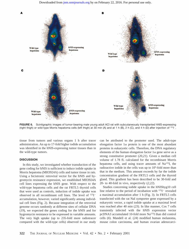
tissue from tumors and various organs 1 h after traceradministration. An up to 17-fold higher iodide accumulationwas identified in the hNIS-expressing tumor tissues than inthe wild-type tumors.
DISCUSSION
In this study, we investigated whether transduction of thegene coding for hNIS is sufficient to induce iodide uptake inMorris hepatoma (MH3924A) cells and tumor tissue in rats.Using a bicistronic retroviral vector for the hNIS and hy-gromycin resistance expression, we established MH3924Acell lines expressing the hNIS gene. With respect to thewild-type hepatoma cells and the rat FRTL5 thyroid cellsthat were used as controls, induction of iodide uptake wasobserved in all recombinant cell lines. The level of125I2
accumulation, however, varied significantly among individ-ual cell lines (Fig. 2). Because integration of the retroviralgenome occurs randomly at different sites of cellular DNA(19), we expected the genes coding for the hNIS and forhygromycin resistance to be expressed in variable amounts.The very high uptake (up to 235-fold more radiotracercompared with the wild-type cells) observed in our study
can be attributed to the promoter used. The adult-typeelongation factor 1a protein is one of the most abundantproteins in eukaryotic cells. Therefore, the DNA regulatoryelements of the human elongation factor 1a gene serve as astrong constitutive promoter (20,21). Given a median cellvolume of 1.78 fL calculated for the recombinant Morrishepatoma cells, and using tracer amounts of Na125I, theradioactive iodide in the cells was up to 105-fold more thanthat in the medium. This amount exceeds by far the iodideconcentration gradient of the FRTL5 cells and the thyroidgland. This gradient has been described to be 30-fold and20- to 40-fold in vivo, respectively (2,22).
Studies concerning iodide uptake in the hNIShyg10 cellline relative to the period of incubation with125I2 revealeda maximal accumulation after 1 h (Fig. 3). In FRTL5 cellstransfected with the rat NaI symporter gene expressed by aeukaryotic vector, a rapid iodide uptake at a maximal levelwas reached after 40 min (23). In like manner, Cos 7 cellstransiently infected with the hNIS expression vectorpcDNA3 accumulated 10-fold more Na125I than did controlcells (6). Mandell et al. (24) modified human melanoma,mouse colon carcinoma, and human ovarian adenocarci-
FIGURE 6. Scintigraphic images of tumor-bearing male young adult ACI rat with subcutaneously transplanted hNIS-expressing(right thigh) or wild-type Morris hepatoma cells (left thigh) at 30 min (A) and at 1 h (B), 2 h (C), and 4 h (D) after injection of 131I2.
322 THE JOURNAL OF NUCLEAR MEDICINE • Vol. 42 • No. 2 • February 2001
by on February 22, 2016. For personal use only. jnm.snmjournals.org Downloaded from

noma cells with a retroviral vector bearing the rat NaIsymporter gene and also observed an up to 35-fold increasein iodide uptake.
Anions such as SCN2 and ClO42 competitively inhibit
the activity of hNIS, and the proton conductor FCCP hasbeen described as preventing I2 uptake (2). To evaluatewhether the iodide accumulation was specifically inducedby the functional activity of the hNIS gene product, iodide
uptake was determined in the hNIShyg10 cells in the pres-ence of sodium perchlorate, the anion channel blockerDIDS, or the proton conductor FCCP. Iodide uptake wasdecreased in the presence of FCCP and also in a dose-dependent manner in the presence of sodium perchlorate,indicating that a functional hNIS is expressed in the genet-ically modified cell lines (Fig. 4). In contrast, the potenterythrocyte anion channel blocker DIDS, which has beendescribed as stimulating the initial influx of iodide in the ratthyroid cell line FRTL5 (2) and in porcine thyroid cells (5),
TABLE 1Iodide Activity Concentration 1 Hour After Injection of
14.8 MBq (400 mCi) 131I
TissueConcentration
(kBq/g of tissue) Weight (g)
HNIS-expressing tumor 532.8 6 88.8 1.1 6 0.4Wild-type tumor 29.6 6 14.8 2.5 6 1.3Thyroid 3700 6 1184 0.05 6 0.02Blood 77.1 6 11.1 4.4 6 1.4Liver 37.0 6 7.4 7.7 6 0.9Stomach 995.3 6 469.9 3.4 6 1.6Kidney 40.7 6 11.1 1.8 6 0.1Small bowel 51.8 6 7.4 6.6 6 2.1Bladder 758.5 6 247.9 1.4 6 0.4Whole animal 66.6 6 7.4 239.7 6 24.4
Data are mean values and SD (n 5 4). Weight represents weightof tissue sample measured, including blood.
TABLE 2Scintigraphic Uptake in hNIS-Expressing Tumors, Wild-
Type Tumors, and Thyroid of Tumor-Bearing Rats
Time (min) hNIS Wild type Thyroid n
5 1.4 6 0.2 1.0 6 0.1 0.50 6 0.01 710 2.1 6 0.4 1.1 6 0.2 0.51 6 0.02 715 2.4 6 0.4 1.0 6 0.1 0.54 6 0.04 720 2.5 6 0.5 0.99 6 0.1 0.54 6 0.04 725 2.6 6 0.5 0.95 6 0.1 0.55 6 0.06 730 2.7 6 0.6 0.9 6 0.1 0.56 6 0.06 765 2.8 6 0.6 0.8 6 0.1 0.79 6 0.05 7
120 2.7 6 1.2 0.57 6 0.1 1.10 6 0.09 3240 2.4 6 0.9 0.57 6 0.1 3.77 6 0.65 3
1445 0.97 6 0.2 0.40 6 0.05 14.28 6 1.12 3
Data are percentage of uptake in whole animal.
FIGURE 7. 131I2 uptake (impulses [Imp]/pixel) in hNIS-expressing and wild-typeMorris hepatomas at different times afterradiotracer application in rats. Data aremean values and SD (n 5 3).
TRANSFER OF HNIS GENE IN HEPATOMA CELLS • Haberkorn et al. 323
by on February 22, 2016. For personal use only. jnm.snmjournals.org Downloaded from

caused an increased uptake of 22% in the hNIShyg10 cellline.
The failure of thyroid tissues to accumulate radioiodide isusually associated with reduced hNIS gene expression in thecells. Some thyroid carcinomas and cell lines, however, failto concentrate iodide although the hNIS gene is activelytranscribed (12,25), implying that iodide accumulation isadditionally influenced by factors distinct from expressionof the hNIS gene. Besides transport into the cell, trapping ofthe radioiodide is necessary for therapeutic efficiency. Toevaluate the amount of iodide trapped in tumor cells, effluxexperiments were performed on the hNIShyg10 cell line.After the 125I2-containing medium had been replaced bynonradioactive culture medium, a rapid efflux was observedwithin the next 10 min, indicating that no organification ofthe radioactive iodide had occurred (Fig. 5). Recently, io-dide efflux was also described for NaI symporter–express-ing FRTL-Tc cells, although the efflux was slower than inthe wild-type FRTL-Tc cell line, in which the mechanismremains unknown (23,24).
Analogous to the cell culture system, maximal iodideuptake into the genetically modified hepatoma wasachieved 1 h after the rats had received injections of131I2.Although the hNIS protein favors iodide transport intothe cells rather than efflux, the radioactivity continuouslydisappeared from the hNIS-expressing tumors and fromdifferent organs of the body, except the thyroid gland, inwhich an increasing iodide uptake was seen with timebecause of iodide trapping (Figs. 6 and 7; Tables 1 and2). Moreover, we calculated a shorter biologic131I2 half-life in hNIS-expressing tumors (14.56 4.8 h) than inwild-type tumors (156 4.9 h) within the first 24 h aftertracer administration. We suggest that the decrease inintracellular iodide concentration is proportional to thetotal iodide clearance of the body. Consequently, weexpect the exposure time of genetically modified tumorcells to 131I2radiation to be too short for therapeuticrelevance unless iodide is organified. Recently, selectivekilling of up to 64% of cells was observed in rat NaIsymporter transduced tumor cells in vitro (24), but thiseffect was not reproduced in vivo (23). In rat FRTLtumors, a radiation dose of 4 Gy was calculated when adose of approximately 37 MBq (1 mCi)131I had beeninjected, with an effective half-life of 6 h (23). UsingMIRDOSE3 (27), we calculated a cumulated activity of1,147.7 MBq3 s and 20,080 MBq3 s for the wild-typeand the hNIS-expressing tumors, respectively. This ac-tivity resulted in an absorbed dose of 35 mGy (wild-typetumor) and 592 mGy (hNIS-expressing tumor) after ad-ministration of 14.8 MBq (0.4 mCi)131I. Previously, thehalf-life of radioiodide in human sera of patients on aniodine-depletion regimen was found to be 20 h (23,26),indicating that a higher radiation dose in human geneti-cally modified tumors may be achieved by a longer iodidecirculation time.
CONCLUSION
In conclusion, transduction of the hNIS gene inducesaccumulation of iodide in non–I2-concentrating hepatomacells. However, the iodide is not organified in the geneti-cally modified cells, because the high influx is followed bya rapid efflux in vitro and in vivo. With regard to a genetherapeutic approach based on transduction of the hNISgene, studies on pharmacologic modulations interferingwith iodide efflux from cells are currently being performed.On the other hand, transduction of the hNIS gene using abicistronic vector may serve as a simple reporter system forvisualization of other genes. In this respect, hNIS expres-sion and the application of99mTc-pertechnate may be usedto monitor the expression of a second therapeutic gene intumors (28).
ACKNOWLEDGMENTS
The authors thank Reiner Kuhnlein for help with theanimal experiments. This study was supported by a grantfrom the Tumorzentrum Heidelberg/Mannheim, Heidel-burg, Germany.
REFERENCES
1. Marcocci C, Cohen JL, Grollman EF. Effect of actinomycin D on iodide transportin FRTL-5 thyroid cells.Endocrinology.1984;115:2123–2132.
2. Weiss SJ, Philp NJ, Grollman EF. Iodide transport in a continuous line ofcultured cells from rat thyroid.Endocrinology.1984;114:1090–1098.
3. Paire A, Bernier-Valentin F, Selmi-Ruby S, Rousset B. Characterization of the ratthyroid iodide transporter using anti-peptide antibodies.J Biol Chem.1997;272:18245–18249.
4. Nakamura Y, Ohtaki S, Yamazaki I. Molecular mechanism of iodide transport bythyroid plasmalemmal vesicles: cooperative sodium activation and asymmetricalaffinities for the ions on the outside and inside of the vesicles. J Biochem.1988;104:544–549.
5. Nakamura Y, Kotani T, Ohtaki S. Transcellular iodide transport and iodination onthe apical plasma membrane by monolayer porcine thyroid cells cultured oncollagen-coated fibers.J Endocrinol.1990;126:275–281
6. Smanik PA, Liu Q, Furminger TL, et al. Cloning of the human sodium iodidesymporter.Biochem Biophys Res Commun.1996;226:339–345.
7. Cho JY, Sagartz JE, Capen CC, Mazzaferri EL, Jhiang SM. Early cellularabnormalities induced by RET/PTC1 oncogene in thyroid-targeted transgenicmice.Oncogene.1999;18:3659–3665.
8. Arturi F, Russo D, Schlumberger M, et al. Iodide symporter gene expression inhuman thyroid tumors.J Clin Endocrinol Metab.1998;83:2493–2496.
9. Caillou B, Troalen F, Baudin E, et al. Na1/I2 symporter distribution in humanthyroid tissues: an immunohistochemical study.J Clin Endocrinol Metab.1998;83:4102–4106.
10. Ryu KY, Senokozlieff ME, Smanik PA, et al. Development of reverse transcrip-tion-competitive polymerase chain reaction method to quantitate the expressionlevels of human sodium iodide symporter.Thyroid.1999;9:405–409.
11. Lazar V, Bidart JM, Caillou B, et al. Expression of the Na1/I2 symporter gene inhuman thyroid tumors: a comparison study with other thyroid-specific genes.J Clin Endocrinol Metab.1999;84:3228–3234.
12. Venkataraman GM, Yatin M, Marcinek R, Ain KB. Restoration of iodide uptakein dedifferentiated thyroid carcinoma: relationship to human Na1/I2 symportergene methylation status.J Clin Endocrinol Metab.1999;84:2449–2457.
13. Schmutzler C, Winzer R, Meissner-Weigl J, Kohrle J. Retinoic acid increasessodium/iodide symporter mRNA levels in human thyroid cancer cell lines andsuppresses expression of functional symporter in nontransformed FRTL-5 ratthyroid cells.Biochem Biophys Res Commun.1997;240:832–838.
14. Moullier P, Marechal V, Danos O, Heard JM. Continuous systemic secretion ofa lysosomal enzyme by genetically modified mouse skin fibroblasts.Transplan-tation. 1993;56:427–432.
15. Pear WS, Nolan GP, Scott ML, Baltimorre D. Production of high titer helper-freeretroviruses by transient transfection.Proc Natl Acad Sci USA.1993;90:8392–8396.
324 THE JOURNAL OF NUCLEAR MEDICINE • Vol. 42 • No. 2 • February 2001
by on February 22, 2016. For personal use only. jnm.snmjournals.org Downloaded from

16. Chomczynski P, Sacchi N. Single-step method of RNA isolation by acid guani-dinium thiocyanate-phenol-chloroform extraction.Anal Biochem.1987;162:156–159.
17. Cleveland DW, Lopata MA, MacDonald RJ, Cowan NJ, Rutter WJ, KirschnerMW. Number and evolutionary conservation of D- and E-tubulin and cytoplas-mic E- and F-actin genes using specific cloned cDNA probes.Cell. 1980;20:95–105.
18. Church GM, Gilbert W. Genomic sequencing.Proc Natl Acad Sci USA.1984;81:1991–1995.
19. Goff SP. Genetics of retroviral integration.Ann Rev Genet.1992;26:527–544.20. Mizushima S, Nagata S. pEF-BOS, a powerful mammalian expression vector.
Nucleic Acids Res.1990;18:5322.21. Wakabayashi-Ito N, Nagata S. Characterization of the regulatory elements in the
promoter of the human elongation factor-1a gene.J Biol Chem.1994;269:29831–29837.
22. Kaminsky SM, Levy O, Salvador C, Dai G, Carrasco N. Na1I2 symport activity
is present in membrane vesicles from thyrotropin-deprived non-I2 transportingcultured thyroid cells.Proc Natl Acad Sci USA.1994;91:3789–3793.
23. Shimura H, Haraguchi K, Miyazaki A, Endo T, Onaya T. Iodide uptake andexperimental131I therapy in transplanted undifferentiated thyroid cancer cellsexpressing the Na1/I2 symporter gene.Endocrinology.1997;138:4493–4496.
24. Mandell RB, Mandell LZ, Link CJ. Radioisotope concentrator gene therapy usingthe sodium/iodide symporter gene.Cancer Res.1999;59:661–668.
25. Venkataraman GM, Yatin M, Ain KB. Cloning of the human sodium-iodidesymporter promoter and characterization in a differentiated human thyroid cellline, KAT-50. Thyroid.1998;8:63–69.
26. Maruca J, Santner S, Miller K, Santen RJ. Prolonged iodine clearance with adepletion regimen for thyroid carcinoma.J Nucl Med.1984;25:1089–1093.
27. Stabin M. MIRDOSE: personal computer software for internal dose assessmentin nuclear medicine.J Nucl Med.1996;37:538–546.
28. Gambhir SS, Barrio JR, Herschman HR, Phelps ME. Assays for noninvasiveimaging of reporter gene expression.Nucl Med Biol.1999;26:481–490.
TRANSFER OF HNIS GENE IN HEPATOMA CELLS • Haberkorn et al. 325
by on February 22, 2016. For personal use only. jnm.snmjournals.org Downloaded from

2001;42:317-325.J Nucl Med. Kübler, Jürgen Debus and Michael EisenhutUwe Haberkorn, Markus Henze, Annette Altmann, Shiming Jiang, Iris Morr, Miriam Mahmut, Peter Peschke, Wolfgang CellsTransfer of the Human NaI Symporter Gene Enhances Iodide Uptake in Hepatoma
http://jnm.snmjournals.org/content/42/2/317This article and updated information are available at:
http://jnm.snmjournals.org/site/subscriptions/online.xhtml
Information about subscriptions to JNM can be found at:
http://jnm.snmjournals.org/site/misc/permission.xhtmlInformation about reproducing figures, tables, or other portions of this article can be found online at:
(Print ISSN: 0161-5505, Online ISSN: 2159-662X)1850 Samuel Morse Drive, Reston, VA 20190.SNMMI | Society of Nuclear Medicine and Molecular Imaging
is published monthly.The Journal of Nuclear Medicine
© Copyright 2001 SNMMI; all rights reserved.
by on February 22, 2016. For personal use only. jnm.snmjournals.org Downloaded from