Discrete scintillator coupled mercuric iodide photodetector arrays for breast imaging
7
1127 IEEE TRANSACTIONS ON NUCLEAR SCIENCE, VOL. 44, NO 3, JUNE 1997 DISCRETE SCINTILLATOR COUPLED MERCURIC IODIDE PHOTODETECTOR ARRAYS FOR BREAST IMAGING Martin P. Tornai, Student Member, IEEE, Bradley E. Patt,* Member, IEEE, Jan S. Iwanczyk,* Member, IEEE, Craig S. Levin, Member, IEEE, and Edward J. Hoffman, Senior Member, IEEE Division of Nuclear Medicine & Biophysics, Department of Molecular & Medical Pharmacology, UCLA School of Medicine, Los Angeles, CA 90095 *Advanced Detectors Inc., 1220-A Avenida Acaso, Camarillo, CA 93012 ABSTRACT Multi-element (4x4) imaging arrays with high resolution collimators, size matched to discrete CsI(T1) scintillator arrays and mercuric iodide photodetector arrays (Hg12 PDA) were developed as prototypes for larger 16x16 element arrays for breast imaging. The compact nature of the arrays allows detector positioning in close proximity to the breast to elimi- nate activity not in the line-of-sight of the collimator, thus reducing image background. Short collimators, size matched to 51.5x1.5 mm2 scintillators show a factor of 2 and 3.4 improvement in spatial resolution and efficiency, respectively, compared to high resolution collimated gamma cameras for the anticipated compressed breast geometries. Monte Carlo simu- lations, confirmed by measurements, demonstrated that scintil- lator length played a greater role in efficiency and photo- fraction for 140 keV gammas than cross sectional area, which affects intrinsic spatial resolution. Simulations also demon- strated that an increase in the ratio of scintillator area to length corresponds to an improvement in light collection. Electronic noise was below 40 e- RMS indicating that detector resolution was not noise limited. The high quantum efficiency and spectral match of prototype unity gain Hg12 PDAs coupled to lxlx2.5 mm3 and 2x2~4 mm3 CsI(T1) scintillators demon- strated energy resolutions of 9.4% and 8.8% FWHM at 140 keV, respectively, without the spectral tailing observed in standard high-Z, compound semiconductor detectors. Line spread function measurements matched the scintillator size and pitch, and small, complex phantoms were easily imaged. I. INTRODUCTION Standard X-ray mammography is accepted as the best means of screening for non-palpable breast cancer. However, signatures of breast cancer, such as microcalcifications or masses that are seen for most malignant lesions may also be associated with benign processes. Thus, while the sensitivity of mammography is on the order of 85%, its specificity is limited to only 20-30%, and only about 30% of the biopsies based on mammographic findings are positive [ 1,2]. Recent imaging studies with radiopharmaceuticals (e.g. 99mTc-Sestamibi; 140 keV gamma rays) and standard gamma cameras have shown uptake in tumors, apparently in propor- tion to the malignancy of the tumor [3-81. Specifically, the rapid uptake mechanism of Sestamibi fixes the compound in place and minimizes redistribution. Recent reports on detec- tion of breast tumors using Sestamibi, all give sensitivities and specificities in the neighborhood of 90% [3-lo]. Equally encouraging results have also been reported for 99mTc-Methy- lene Diphosphonate with a sensitivity of 92% and a specificity of 95% [ 111. Whereas these previous studies showed uptake of -4:l tumor to background in breasts, a recent standard scintillation camera scintimammography study along with excised breast tissue demonstrated uptake of 6.1 to I tumor to fatty breast tissue [8]. The excised samples included smaller masses (< 1 cm3) missed with scintimammography. These studies not only corroborate measurements of the excellent uptake achievable with Sestamibi, but they also demonstrate the need for a compact, high resolution and sensitivity nuclear emission imaging device which might detect these otherwise missed, smaller lesions. In this work, we investigate a novel imaging detector array prototype composed of high efficiency CsI(T1) scintillators coupled to high quantum efficiency and low noise solid state mercuric iodide (HgI2) photodetector arrays (PDAs). The major issues considered include the possibility for improved energy and spatial resolution compared with standard gamma cameras, as well as improved detector efficiency and photo- fraction compared with intrinsic semiconductor detectors. In general, scintillators facilitate higher gamma stopping efficien- cies compared to high-Z compound semiconductor detectors of equivalent stopping thicknesses without the low energy spectral tailing observed due to poor charge collection. Another advantage of this scintillator-photodetector com- bination is the higher achievable photofraction which is degraded by the rowkolumn coincidence criteria employed with intrinsic HgI, semiconductordetector arrays [ 121. In addition, the camera's size should be small enough that it could be used in conjunction with mammography breast compression fixtures allowing it to optimally image the breast. The angle would be chosen to image the breast and the axillary lymph nodes while minimizing the amount of heart or liver activity in the field-of-view (FOV), resulting in furthcr improvements in resolution and signal-to-noise over standard gamma cameras. In a larger version, this camera combined with the appropriate radiotracer could be used as the primary screening test for those patients with dense breasts or other conditions that would compromise the usefulness of standard X-ray mammography. The parameters investigated include the limiting effects of the collimator, the feasibility of using small, discrete scintillation detectors coupled to solid state photodetectors, and the combined effects of these components on gamma detection efficiency and sensitivity, energy and spatial resolution, as well as imaging ability. 11. OPTIMIZATION OF DETECTOR DESIGN There are two primary innovations in the approach of this work. First, the collimator hole openings exactly match the detector sizes, and the collimator septal thickness corresponds with the inter-crystal spacing in order to maximize the signal for any given detector element in the array. Second, the use of high efficiency and high intrinsic spatial resolution detectors 0018-9499/97$10.00 0 1997 IEEE Authorized licensed use limited to: Stanford University. Downloaded on June 01,2010 at 21:34:06 UTC from IEEE Xplore. Restrictions apply.
-
Upload
independent -
Category
Documents
-
view
0 -
download
0
Transcript of Discrete scintillator coupled mercuric iodide photodetector arrays for breast imaging
1127 IEEE TRANSACTIONS ON NUCLEAR SCIENCE, VOL. 44, NO 3, JUNE 1997
DISCRETE SCINTILLATOR COUPLED MERCURIC IODIDE PHOTODETECTOR ARRAYS FOR BREAST IMAGING
Martin P. Tornai, Student Member, IEEE, Bradley E. Patt,* Member, IEEE, Jan S. Iwanczyk,* Member, IEEE, Craig S. Levin, Member, IEEE, and Edward J. Hoffman, Senior Member, IEEE
Division of Nuclear Medicine & Biophysics, Department of Molecular & Medical Pharmacology, UCLA School of Medicine, Los Angeles, CA 90095
*Advanced Detectors Inc., 1220-A Avenida Acaso, Camarillo, CA 93012
ABSTRACT
Multi-element (4x4) imaging arrays with high resolution collimators, size matched to discrete CsI(T1) scintillator arrays and mercuric iodide photodetector arrays (Hg12 PDA) were developed as prototypes for larger 16x16 element arrays for breast imaging. The compact nature of the arrays allows detector positioning in close proximity to the breast to elimi- nate activity not in the line-of-sight of the collimator, thus reducing image background. Short collimators, size matched to 51.5x1.5 mm2 scintillators show a factor of 2 and 3.4 improvement in spatial resolution and efficiency, respectively, compared to high resolution collimated gamma cameras for the anticipated compressed breast geometries. Monte Carlo simu- lations, confirmed by measurements, demonstrated that scintil- lator length played a greater role in efficiency and photo- fraction for 140 keV gammas than cross sectional area, which affects intrinsic spatial resolution. Simulations also demon- strated that an increase in the ratio of scintillator area to length corresponds to an improvement in light collection. Electronic noise was below 40 e- RMS indicating that detector resolution was not noise limited. The high quantum efficiency and spectral match of prototype unity gain Hg12 PDAs coupled to lxlx2.5 mm3 and 2 x 2 ~ 4 mm3 CsI(T1) scintillators demon- strated energy resolutions of 9.4% and 8.8% FWHM at 140 keV, respectively, without the spectral tailing observed in standard high-Z, compound semiconductor detectors. Line spread function measurements matched the scintillator size and pitch, and small, complex phantoms were easily imaged.
I. INTRODUCTION
Standard X-ray mammography is accepted as the best means of screening for non-palpable breast cancer. However, signatures of breast cancer, such as microcalcifications or masses that are seen for most malignant lesions may also be associated with benign processes. Thus, while the sensitivity of mammography is on the order of 85%, its specificity is limited to only 20-30%, and only about 30% of the biopsies based on mammographic findings are positive [ 1,2].
Recent imaging studies with radiopharmaceuticals (e.g. 99mTc-Sestamibi; 140 keV gamma rays) and standard gamma cameras have shown uptake in tumors, apparently in propor- tion to the malignancy of the tumor [3-81. Specifically, the rapid uptake mechanism of Sestamibi fixes the compound in place and minimizes redistribution. Recent reports on detec- tion of breast tumors using Sestamibi, all give sensitivities and specificities in the neighborhood of 90% [3-lo]. Equally encouraging results have also been reported for 99mTc-Methy- lene Diphosphonate with a sensitivity of 92% and a specificity of 95% [ 111. Whereas these previous studies showed uptake of -4:l tumor to background in breasts, a recent standard
scintillation camera scintimammography study along with excised breast tissue demonstrated uptake of 6.1 to I tumor to fatty breast tissue [8]. The excised samples included smaller masses (< 1 cm3) missed with scintimammography. These studies not only corroborate measurements of the excellent uptake achievable with Sestamibi, but they also demonstrate the need for a compact, high resolution and sensitivity nuclear emission imaging device which might detect these otherwise missed, smaller lesions.
In this work, we investigate a novel imaging detector array prototype composed of high efficiency CsI(T1) scintillators coupled to high quantum efficiency and low noise solid state mercuric iodide (HgI2) photodetector arrays (PDAs). The major issues considered include the possibility for improved energy and spatial resolution compared with standard gamma cameras, as well as improved detector efficiency and photo- fraction compared with intrinsic semiconductor detectors. In general, scintillators facilitate higher gamma stopping efficien- cies compared to high-Z compound semiconductor detectors of equivalent stopping thicknesses without the low energy spectral tailing observed due to poor charge collection. Another advantage of this scintillator-photodetector com- bination is the higher achievable photofraction which is degraded by the rowkolumn coincidence criteria employed with intrinsic HgI, semiconductor detector arrays [ 121.
In addition, the camera's size should be small enough that it could be used in conjunction with mammography breast compression fixtures allowing it to optimally image the breast. The angle would be chosen to image the breast and the axillary lymph nodes while minimizing the amount of heart or liver activity in the field-of-view (FOV), resulting in furthcr improvements in resolution and signal-to-noise over standard gamma cameras. In a larger version, this camera combined with the appropriate radiotracer could be used as the primary screening test for those patients with dense breasts or other conditions that would compromise the usefulness of standard X-ray mammography. The parameters investigated include the limiting effects of the collimator, the feasibility of using small, discrete scintillation detectors coupled to solid state photodetectors, and the combined effects of these components on gamma detection efficiency and sensitivity, energy and spatial resolution, as well as imaging ability.
11. OPTIMIZATION OF DETECTOR DESIGN
There are two primary innovations in the approach of this work. First, the collimator hole openings exactly match the detector sizes, and the collimator septal thickness corresponds with the inter-crystal spacing in order to maximize the signal for any given detector element in the array. Second, the use of high efficiency and high intrinsic spatial resolution detectors
0018-9499/97$10.00 0 1997 IEEE
Authorized licensed use limited to: Stanford University. Downloaded on June 01,2010 at 21:34:06 UTC from IEEE Xplore. Restrictions apply.

1128
0 2 4 6 8 10 SYSTEM SPATIAL RESOLUTION (mm)
FIGURE 1. Geometric efficiency vs system spatial resolution calculated for various square hole collimator lengths with a source at 1 cm distance from collimator face; shaded region represents region of improved SNR.
in close proximity to the partially compressed breast affords a transition from collimator limited camera resolution, which is characteristic of Anger camera systems, to detector limited camera resolution. Standard gamma cameras, whose primary intent is to image objects at depth, are primarily limited by the collimator characteristics. On the other hand, in an application specific device for high resolution imaging, rela- tively shallow object imaging will be limited by the intrinsic detector properties.
A. Collimator Effects on Signal-to-Noise In single photon imaging, the collimator limits the overall
system spatial resolution and sensitivity [ 131. One important reason for choosing a matched collimator opening to detector size is that the point source geometric efficiency for a parallel hole collimator with 52 mm x 2 mm square detectors is better than that for a standard commercially available hgh resolution (HiRes) gamma camera (hexagonal, 1.5 mm hole diameter, 4 cm length) currently used in scintimammography. For ex- ample, a design with a 2.5 cm long collimator size matched to a 1.5 mm square crystal has 3.4 times better efficiency perfor- mance than a standard %Res collimator, along with superior spatial resolution (Fig. 1). Thus, an efficiency gain is due to the larger exposed area of each detector crystal with the collimator openings size matched to the detectors. The col- limator septa overlap the intercrystal reflector (dead) spaces.
These collimator parameters affect the time required to collect adequate events from breast lesions with various radiotracer uptake values (10 min, 2.1 tumor / background [3]), with higher efficiencies leading to shorter imaging times. Improved geometric efficiency will lead to (1) improved statistics for the same imaging time, or (2) reduced imaging time, or (3) reduced patient dose.
Spatial resolution is also important in this application due to the necessity of discerning small tumors (<1 cm diameter). However, improving spatial resolution by increasing colli- mator length penalizes efficiency (Fig. 1). For Anger camera systems used to image relatively distant objects, system resolution degradations are limited by the collimator optics [ 131. For the proposed high intrinsic resolution system to image objects in close proximity, the system resolution will be limited by the intrinsic spatial resolution rather than the collimator. Thus, shorter more efficient collimators can be utilized without a resolution degradation. The expected perfor-
mance for the proposed breast imaging camera geometry lies in a region that is always superior to HiRes nuclear medicine gamma cameras (Fig. I), which are currently used as the standard for scintimammography imaging [3-71. The factors that yield the improved signal-to-noise ratio (SNR) for this system include (1) improved spatial resolution compared to the standard HiRes system, (2) improved geometric efficiency, and (3) reduced background due to both the vantage point of the camera, which will eliminate the body (e.g. heart, liver) from background, and the use of partial compression, and also (4) the improved scatter rejection techniques due to the excellent energy resolution (see ZZLA.).
The compact nature of our imaging device, due to the anticipated short collimator, small scintillators (see ZZ. B.), flat Hg12 PDA (see ZZ.C.), and thin ASIC readout electronics in contact with the detector will facilitate close detector to source proximity, such as from a medio-lateral or latero-medial vantage point of the breast. With partial breast compression, maximum tissue thicknesses of 5 cm can be anticipated, and our imaging device can be positioned to get opposing views of a breast to take advantage of the higher achievable resolution at short distances, and ensure that the detector would not be more than 2.5 cm from any lesion. Standard gamma cameras cannot easily get either superior-inferior or lateral opposing views due to physical limitations or interference from the head, stomach or contra-lateral chest wall, which may also contribute radioactive shine into the detector. The physical distance poses a serious problem for opposing views of the same breast with a standard gamma camera which is overcome by our design.
A conservative estimate of the background reduction is a factor of 2, due to partial breast compression (bringing the breast thickness to 4 cm), latero-medial positioning of the camera, and the improved scatter rejection. With uptake values for 99mTc-Sestamibi measured with standard cameras at 22 tumor to background, the background reduced image con- trast will nominally improve by 33%. These factors alone will improve the image quality significantly [ 141.
Most importantly the improved spatial resolution will enable visualization of smaller tumors (-2x2 mm2) than previously achievable with the HiRes gamma camera. For example, for large tumors (>2x2 cm2), both cameras will resolve the tumors and recover approximately 100% of the activity distribution in the image. However, for a tumor at 2.5 cm depth in the breast, the HiRes gamma camera has a resolution of 4.9 mm; thus for a relatively small 0.4x0.4 cm2 tumor [3] the activity recovery in the FOV is only -40%, which severely limits it's detectability. Due to the improved spatial resolution of our proposed device (3.4 mm for this depth) along with closer proximity, the activity recovery will be improved by at least 56% (improvement is in tumor visual- ization, i.e. image contrast).
B. Simulations to Optimize Small Detectors Thick scintillators offer higher gamma stopping efficien-
cies without the low energy spectral tailing observed in high-Z compound semiconductor detectors of equivalent stopping thicknesses. Thus, several factors were considered when choosing parallel-piped CsI(T1) scintillators for use with the HgI, PDA. CsI(T1) has a better mass absorption coefficient at 140 keV than NaI(T1) (4.47 cm-' and 2.98 cm-', respectively).
Authorized licensed use limited to: Stanford University. Downloaded on June 01,2010 at 21:34:06 UTC from IEEE Xplore. Restrictions apply.

1129
z 0 80
c. 0
60 i_: E
0 2 4 6 X 1 0 1 2 1 4 SCINTILLATOR LENGTH (mm)
FIGURE 2. (A) Simulation results for total gamma efficiency for 57Co gamma rays in small CsI(T1) crystals. (B) Simulated and measured photofraction results with same irradiation conditions.
CsI(T1) has a peak visible emission wavelength of 530 nm which corresponds well to the absorption maximum of 560 nm in the Hg12 photodetector, and also produces -35% more photons than NaI(T1) for equivalent energy deposition. Short parallel-piped crystals offer equivalent light collection com- pared with cylindrical crystals of similar dimensions, degrading with longer (r2 cm) lengths [15], and are also more easily manufactured in large quantities.
The QE of the HgIz PD is near 100% from 350 to 560 nm [ 161, and is slightly degraded by the optical contact (see ZZ.C.). Mercuric iodide detectors coupled to CsI(T1) scintillators have now demonstrated perhaps the best energy resolution at room temperature ever attained by any scintillator / photodetector pair (4.58% FWHM at 662 keV for a 0.5 in. diameter photo- detector [ 171). Thus, the emission-absorption wavelength match and light output properties of the scintillator are well suited for use with HgI2 PDAs, and the superior stopping efficiencies facilitate the use of smaller crystals. Monte Carlo simulations and measurements were utilized to help determine the effects of scintillator crystal size and surface treatment for detectors in the imaging array.
With a gamma-ray multiple scattering Monte Carlo code [ 181, several small CsI crystals from 1 x 1 ~ 2 mm3 up to 3 x 3 ~ 12 mm3 were modeled. The resultant energy deposition and position information was further utilized in an optical tracking Monte Carlo code [ 191 which modeled surface treatments on the scintillation crystals coupled to the HgI2 photodetectors.
The simulation results from flood field gamma interactions with 57C0 (122 keV (88%) and 136 keV (12%)) in various sized individual parallel-piped CsI(TI) scintillators indicate that for 25 mm crystal lengths, the efficiency for various discrete scintillators was S O % (Fig. 2A). Note that for increasing crystal length, the rate of improving efficiency is dramatic up
8 4000- e - Y 3 8 0 0 - 9 - 0 U 3600- cn z
8 3400:
E 3200- z 4 - 3 3000-
0 8 10 SCINTILLATOR LENGTH (mm)
Scintillalor
1 x I x 2 5 null2 0 I x l x 4 r r n '
0 2 4 6 8 11) SCINTILLATOR AREA (mm*)
FIGURE 3. Simulation results and measurements of 57Co irrad- iated small CsI(T1) crystals coupled to Hg12 PDAs demonstrating effects of crystal geometry on light collection (A) and effects on resolution (B). Simulations assumed 98% reflectivity and 70% photodetector QE.
to about 6 mm in length beyond which the rate of improve- ment roles off and then only slowly improves with thickness. The trends for photofraction are similar to the total gamma efficiency trends (Fig. 2B), and these results indicate that the dominant interactions in these small crystals is photoelectric. Measured photofraction values (defined as the integral counts from 105-160 keV about the photopeak divided by the total detected events) for two crystal sizes (see ZZZ.A.) agree well with the simulation results.
The optimal surface treatment as determined by the com- bined simulations of a 2 x 2 ~ 6 mm3 crystal were consequently used in all further simulations. With the distal surface from the photodetector ground and sides of the scintillator specu- larly polished, the best light collection was achieved (-75% collection), corresponding with results of other work [ 15,201.
The optical simulation results indicate that light collection from a parallel-piped CsI(T1) scintillator increases with dec- reasing length, and more slowly increases with increasing surface area (Fig. 3A). With an absolute charge calibration of the Hg12 photodetector array measured with 55Fe (5.9 keV X- ray) direct detection, the measured light collection values are within 515% of the predicted values. These results also cor- respond with other data for similar simulations and mea- surements [ 15,201. The measured FWHM energy resolutions with 57C0 irradiation were better than some of the Gaussian fit photopeak simulation results (Figs. 3B and 5). While the simulation assumptions may not have accurately predicted the measured results (see ZIIA.), the trends in improving energy resolution are consistent with the light collection characteris- tics. We are nonetheless encouraged that the preliminary measurements are indeed better than the predicted values in our
Authorized licensed use limited to: Stanford University. Downloaded on June 01,2010 at 21:34:06 UTC from IEEE Xplore. Restrictions apply.

1130
effort to optimize the imaging system.
C. Scintillator and Photodetector Arrays Segmented CsI(T1) scintillators with sixteeil segments each
cither 1x1 mm2 in area with 2.5 or 4 mm lengths, or 2 x 2 ~ 4 mm3 were procured from Hilger Analytical Ltd. The segments are arranged in a two dimensional 4x4 pattern and separated by Ti02 doped epoxy of 0.2 mm or 0.5 mm thickness.
The fabricated Hg12 PDAs have 16-pixels each with either 1 xl mm2 pixels or 2x2 mm2 pixels with gaps corresponding to the crystal separation. The pixels are defined through the geometry of the metal contact deposition on the back side of the 560 pm HgI2 wafer.
The front side contact utilized electrically conductive ultra- thin metal contacts which are optically transparent, and non- reactive with HgI2. By carefully applying the evaporation in a vacuum bell jar, uniform metalization layers with trans- missions up to 70% from 400 to >lo00 nm were achieved with sheet resistance of 4 0 0 Wsquare for thin layers. These transmission properties are good for CsI(T1) light but do degi-ade the overall detector QE to about 70%.
D. Photodetector Noise Considerations One of the main considerations for unity gain solid state
photodetectors is the noise level. By using a particular con- struction, the Hg12 photodetector technology can circumvent the well known problem associated with hole trapping in high-Z compound semiconductors. Optical photons penetrate only a very shallow region beneath the entrance electrode, and with negative entrance electrode bias, only the electrons transit the device. The charge collection becomes essentially single carrier (electron) dominated. With electron transit times (Ztrsnslt - 5 x sec) almost 10 to 100 times faster than elec- tron trapping times ( T ~ ~ ~ ~ ~ ~ ~ ~ = 10-5-10-6 sec) in Hg12, almost complete charge collection is expected.
For all spectral and imaging measurements, each pixel from the PDA (16 total) is coupled to a customized, low noise, charge sensitive, resistor feedback preamplifier (Advan- ced Detectors). Photopeak broadening due to electronic noise was determined by comparing the 55Fe direct X-ray interac- tions in the Hg12 detector with the photo response from CsI(T1). This approach relates easily measurable material constants by a scale factor which is inversely proportional to the efficiency for light generation and collection. The "direct" noise line-width is a combination of the noise power terms which includes series, parallel, and excess (lyand generation- recombination) noise components and was previously calcu- lated [21,22]. The total noise contributions are in excellent agreement with measured pulser data on a fully biased detector (Fig. 4A). A minimum linewidth was achieved with -6 psec shaping time. The average pulser width with full bias on the detector for the 16 channels was about 368 eV and 400 eV FWHM for the 1x1 mm2 and 2x2 mm2 uixels, resuectivelv,
500
-Encess Noise - 100 z s % a z
10
3 0 1 1 0
SHAPING TIME @sec)
50
- - ~ f Llecmnic NOW - -A - Stauslsal Nose
2
5 2 ij
; lo
2
1
10 ion loon ENERGY (keV)
FIGURE 4. (A) Theoretical noise calculations for a 2x2 mm2 element as a function of shaping tiine compared with measured pulser data (B) Mcasured resolution values and component contributions to the overall spectral linewidth, measured with 12 psec, triangular shaping.
up to 12 psec, triangular shaping). This may be due to contri- butions of longer CsI(T1) scintillator decay components rather than incomplete electron charge collection since much longer shaping tirnes were used compared to the T ~ ~ ~ ~ ~ ~ ~ . This is similar to other results of CsI(T1) scintillators coupled to PIN diode detectors [15].
The overall spectral linewidth corresponds to (Fig. 4B): (1) the electronic noise; (2) the intrinsic noise in the scintillator, which is a function of the scintillator material and photon energy; (3) the statistical spreading (light transfer and collec- tion efficiency, and detector QE). The degradative effects of these factors are assumed to add in quadrature to the total resolution. The factors are measured parameters, except for the scintillator noise which is calculated according to [23]. The dominant factor in the spectral linewidth is the scintillator For less efficient light collection, as from long crystals (ct. Fig. 3), the dominant factor varies as a function of energy, with electronic noise dominating at low energies, and intrinsic scintillator noise dominating at high energies. The crossover point at which these contributions are equal has been found empirically to be about 60-100 keV (Fig. 4B). Thus for 99mTc tracer studies (140 keV), lowering the electronic noise IS
crucial and was satisfactorily achieved.
111. IMAGING DETECTOR CHARACTERISTICS corresponding to 37 e- and 41 e- RMS at 6 psec shading time. The 57C0 photopeak resolution and electronic noise (pulser
method) were also measured for the 2 x 2 ~ 4 1nm3 scintillator crystals as a function of shaping time using a representative A. Detector Responses
Authorized licensed use limited to: Stanford University. Downloaded on June 01,2010 at 21:34:06 UTC from IEEE Xplore. Restrictions apply.

1131
300
-SimulaUon T o , 10.1% FWHM 250
in b
> w i5 200
150 p! .J d 3 100 8
50
0 0 50 100 150 200
ENERGY (keV)
3 0 0 ~ " " ~ " " ~ " " ~ " " ' 1 (B) 0 Measurement
50 100 li0 2M ENERGY (keV)
FIGURE 5. Measured spectral responses compared with simulations with 4.8 keV noise for (A) lxlx2.5 mm3 crystals and (B) 2 x 2 ~ 4 mm3 crystals coupled to 1x1 mm2 and 2x2 mm2 photo- detectors.
angular shaping (12 psec) amplifier feeding a standard MCA. The response of each of the sixteen individual pixels with the 2x2 mm2 PDA with the 2 x 2 ~ 4 mm3 CsI(T1) crystals was measured with 241Am (59 keV) and 57C0 (122 keV) collimated sources. The average energy resolution for all 16 pixels was 13.55% f 0.86% FWHM at 59 keV, and 10.49% f 0.64% FWHM at 122 keV. At 140 keV, these values scale to <lo% FWHM which is comparable to the best values achieved with standard gamma cameras.
The spectral characteristics for pixels from both the 1xlx2.5 mm3 and 2 x 2 ~ 4 mm3 crystals agree well with high statistics Monte Carlo simulation results (Fig. 5). Note, however, that the simulations did not accurately account for the measured escape peaks or Compton continuum. The mea- sured energy resolutions of the 122 keV photopeaks are 10.1% and 9.4% FWHM for the 1 mm and 2 mm pixels, respec- tively. The energy resolutions of the simulations range from 8.2% - 10.6% and 8.7% - 12.4% FWHM for the 1xlx2.5 mm3 and 2 x 2 ~ 4 mm3 scintillators, respectively, depending on the 2.5 - 4.8 keV noise contribution. The noise values were derived from the directlphoto calibration (see ZZB.).
The measured photofractions (integral counts from 105 to 160 keV divided by the integral counts from 20 to 160 keV) were 54.4% and 79.1% for the 1 mm and 2 mm crystals, respectively (Figs. 2 and 5). The fraction of counts integrated over the region including the Pb K,, Kp, and Cs and I escape peaks are 26% and 12% for the 1 mm and 2 mm crystals, respectively, which is a function of the increased efficiency of the larger volume scintillator.
0 1 2 3 4 5 6 SOURCE POSITION (mm)
FIGURE 6. LSF response function for a single inner column of the lxlx2.5 mm3 CsI(T1) crystals coupled to the HgI2 PDA.
B. Detector Spatial Responses
The spatial response and imaging measurements were made with the low noise preamplifier outputs coupled to CAMAC programmable shaping amplifiers with variable shaping times, variable gain, and SCAs for each channel which provided a logic signal for events above a minimum threshold (Advanced Detectors). A 4.8 psec shaping time was used with indivi- dually balanced pixel gains, and the amplifier outputs were digitized with a peak sensing CAMAC ADC (16 channel Phillips 7164). The logic signals from the SCA outputs were linearly ORed in a double gating (long blocking gate, short ADC trigger gate with leading edge discrimination) CAMAC module with NIM level output (Advanced Detectors) which triggered the ADCs. The data acquisition and control was with LabVIEWTM (National Instruments) on an 150 MHz PentiumTM based PC (Gateway). All imaging data were nor- malized by measured flood field values to account for differ- ences in crystal efficiencies and gains.
The intrinsic line spread function (LSF) response was measured for the 1x1x2.5 mm3 CsI(T1) elements coupled to the 1x1 mm2 pixels on the PDA. A 0.3 mm Pb collimated slit source of 57C0 was stepped in 0.2 mm increments along the vertical direction of the 4x4 square array. The LSF res- ponse was found to correspond with the crystal dimensions (Fig. 6) with a 0.98 f 0.10 mm mean FWHM resolution for the 1 mm crystals, and a mean centroid spacing of 1.26 f 0.0 1 mm which corresponds to the detector pitch of 1.25 mm. The measured values were derived from Gaussian fits to the data.
C. Preliminary Imaging Results Some images were acquired with 57Co irradiation through a
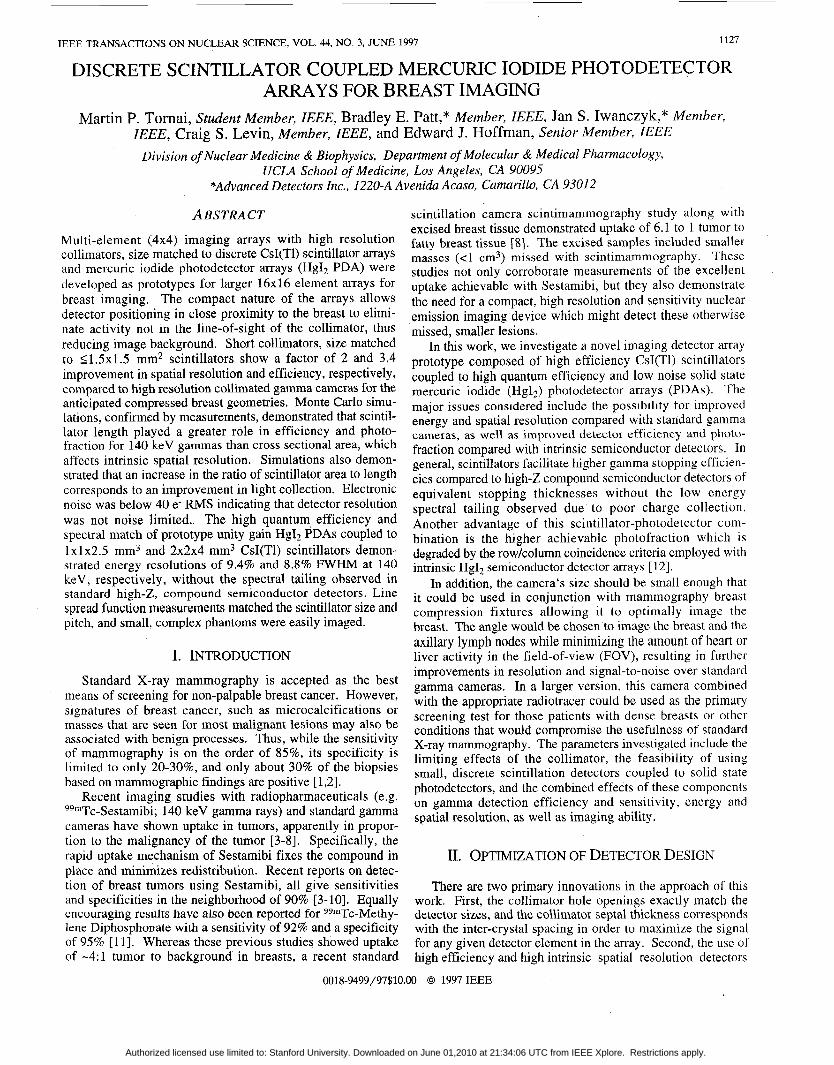
Pb transmission phantom with a complex hole pattern (Fig. 7) . Since the phantom was larger than the FOV of the 4x4 element 1xlx2.5 mm3 CsI(T1) crystal array, multiple samples were collected and digitally spliced together. To eliminate pixellation artifacts resulting from imaging the small phantom (Fig. 7B), half-pitch offset, measurements were collected and interleaved with the single step data (Fig. 7C). This inter- leaving measurement demonstrates the integrity and response linearity of the small imaging array, while improving the sampling of the source distribution. In addition, the subsam- pled data was linearly interpolated to smooth the noise. These
Authorized licensed use limited to: Stanford University. Downloaded on June 01,2010 at 21:34:06 UTC from IEEE Xplore. Restrictions apply.
1132
L5'8 1.2 mm pixels 0.6 mm pixels 0.3 mm pixels
FIGURE 7. (A) Pb transmission phantom with 1.0 mm $ holes, (B) single stepped acquisition of (A), (C) subsampled image of (A), and (D) linear interpolation of (C). Images were acquired with the lxlx2.5 mm3 CsI(T1) crystals on the 1x1 mm2 PDA.
results indicate that the imaging device has a high degree of linearity and can distinguish complex source distributions with high resolution. Tumor sources in breasts and especially the axilla are expected to have spherical distributions potentially with spiculations or extensions into healthy tissue.
IV. CONCLUSIONS Significant advancement has been made in the development
of two dimensional Hg12 PDA technology by incorporating previously developed and novel principles for the optimization of small photodetector array structures. Mercuric iodide PDA prototypes with 16 pixels each 1x1 mm2 with 0.2 mm gaps, and 2x2 m2 with 0.5 mm gaps defined through the geometry of the metalized contact deposition on the Hg12 wafer were fabricated. The 16 element PDAs were evaluated with seg- mented CsI(TI) scintillators of various volumes and pitch corresponding to the electrode patterning on the PDAs. In addition, extensive computer simulations of energy deposition and scintillation light transport both confirmed many of our measurements, and guided us towards more optimal confi- gurations which may surpass the already high performance of these preliminary studies.
The importance of collimators in single photon imaging is that the collimator is the limiting factor for the overall system resolution and sensitivity. Because of this limitation, we have investigated the optimization of the collimator parameters and compared the expected performance with the standard HiRes gamma camera for the specific application of high resolution and sensitivity scintimammography. Moreover, it was de- monstrated that the high intrinsic spatial resolution detectors coupled to relatively short, size matched collimators could achieve better signal-to-noise characteristics than their standard Anger camera counterparts. This was achieved with the high intrinsic detector resolution and obtainable close proximity to the breast lesions, which is otherwise not possible with standard gamma camera systems.
The device itself included sufficient scintillator thickness for at least l /e photoelectric attenuation in the scintillators. The segmented scintillators were nearly optimized for light collection while spectrally matched to the photodetector. Theoretical calculations of the expected spectral resolution and spatial resolution were made and compared favorably with the measured results. The noise performance of the 1 mm2 and 4
mm2 photodetectors were characterized to be -40 e- rms. The spatial resolution corresponded to the scintillator size. Pixel energy resolutions of the measured 122 keV photopeaks were 10.1% and 9.4% FWHM for the 1x1x2.5 mm3 and 2 x 2 ~ 4 mm3 crystals coupled to the appropriately sized PDA, respectively. The simulated energy resolutions ranged from 8.2% - 10.65% and 8.7% - 12.4% for the 1xlx2.5 mm3 and 2 x 2 ~ 4 mm3 scintillators, respectively, depending on the pred- icted noise contribution, and corroborated the measurements.
Moreover, the first images of a complex phantom were obtained with an assembled 4x4 element device. On the basis of the simulated and measured performance of the scintillator I HgIz PDA combination, we expect that a larger FOV camera will: (1) detect lesions -5 times smaller than previously achievable (2 - 3 mm range); (2) shorten the imaging proce- dure with the breast under partial compression to under 5 minutes using currently published dose; and (3) improve the statistical quality of the obtained images.
ACKNOWLEDGMENTS The authors thank Fanny Riquelme, Jim Menjivar, Ryszard Szczebiot, Richard Koziol, George Maculewicz and David Marsh for their expert assistance on various parts of this work. This work was funded in part by NCI grants lR43-CA68845- 01, 1RO1-CA6 1037-01, 1R43-CA69988-01, 2R44-CA6 1403- 02, DOE contract DE-FC03-87-ER60615, and University of California IRB-0225.
REFERENCES CJ Baines, et al. 1986. Sensitivity and Specificity of First Screen Mammography in the Canadian National Breast Screening Study: A Preliminary Report from Five Centres. Radiology. 160:295-298. L Tabar, PB Dean. 1982. Mammographic Parenchymal Patterns: Risk Indicator for Breast Cancer? J. Am. Med.
I Khalkhali, et al. 1995. Scintimammography: The Complementary Role of Tc-99m Sestamibi Prone Breast Imaging for the Diagnosis of Breast Carcinoma. Radiology. 196:421-426. C Aktolun, H Bayhan, M Kir. 1992. Clinical Experience with Tc-99m MIBI Imaging in Patients with Malignant Tumors: Preliminary Results and Comparison with TI- 201. Clin. Nucl. Med. 17:171-176. F Scopinaro, et al. 1994. Tc-99m Sestamibi: An Indicator of Breast Cancer Invasiveness. Eur. J . Nucl. Med. 21:984-987. C-H Kao, S-J Wang, S-H Yeh. 1994. Tc-99m MIBI Uptake in Breast Carcinoma and Axillary Lymph Node Metastases. Clin. Nucl. Med. 19:898-900. I Khalkhali, I Mena, L Diggles. 1994. Review of Imaging Techniques for the Diagnosis of Breast Cancer: A New Role of Prone Scintimammography Using Tc- 99m Sestamibi. Eur. J. Nucl. Med. 21:357-362. J Maublant, et al. 1996. Tc-99m-Sestamibi Uptake in Breast Tumor and Associated Lymph Nodes. J. Nucl. Med. 37:922-925. I Khalkhali, et al. 1995. Tc-99m-Sestamibi Scintimam- mography of Breast Lesions: Clinical and Pathological Follow-Up. J. Nucl. Med. 36: 1784-1789.
ASSOC. 247:185-189.
Authorized licensed use limited to: Stanford University. Downloaded on June 01,2010 at 21:34:06 UTC from IEEE Xplore. Restrictions apply.

1133
[lo] R Taillefer, A Robdoux, R Lambert, S Turpin, J Laperriere. 1995. Tc-99m-Sestamibi Prone Scintimam- mography to Detect Primary Breast Cancer and Axillary Node Involvement. J. Nucl. Med. 36:1758-1765.
[ l l ] S Piccolo, et al. 1995. Tc-99m-Methylene Diphos- phonate Scintimammography to Image Primary Breast Cancer. J. Nucl. Med. 36:7 18-724.
[I21 BE Patt, JS Iwanczyk, MP Tornai, CS Levin, EJ Hoffman. 1995. Development of a Mercuric Iodide De- tector Array for Medical Imaging Applications. Nucl. Instr. Meth. A366: 173- 182.
[ 131 BMW Tsui. Collimator Design, Properties and Charac- teristics. In The Scintillation Camera. Ed. GH Simmons. The Society of Nuclear Medicine, Inc. New York. 1988. Ch. 2.
[14] BMW Tsui, CE Metz, RN Beck. 1983. Optimum Detector Spatial Resolution for Discriminating Between Tumor Uptake Distributions in Scintigraphy. Phys. Med. Biol. 28:775-788.
[15] AJ Bird, T Carter, AJ Dean, D Ramsden, BM Swinyard. 1993. The Optimisation of Small CsI(T1) Gamma-Ray Detectors. IEEE Trans. Nucl. Sci. NS-40:395-399.
[16] JM Markakis, AY Cheng. 1989. Comparison of Trans- parent Conducting Electrodcs on Mercuric Iodide Photo- cells. Nucl. Instr. Meth. A283:236-239.
[17] YJ Wang, BE Patt, JS Iwanczyk, SR Cherry, Y Shao. 1995. High Efficiency CsI(Tl)/Hg12 Gamma Ray Spec- trometers. IEEE Trans. Nucl. Sci. NS-42:601-605.
[18] Y Shao, SR Cherry, S Siegel, RW Silverman. 1996. A Study of Intercrystal Scatter in Small Scintillator Arrays Designed for High Resolution PET Imaging. IEEE Trans. Nucl. Sei. NS-43: 1938- 1944.
[19] GF Knoll, TF Knoll, TM Henderson. 1988. Light Col- lection in Scintillation Detector Composites for Neutron Detection. IEEE Trans. Nucl. Sci. NS-35:872-875.
[20] SR Cherry, et al. 1995. Collection of Scintillation Light from Small BGO Crystals. IEEE Trans Nucl. Sci. NS-
[21] JS Iwanczyk, BE Patt. In Semiconductors fo r Room Temperature Nuclear Detector Applications. Vol43. Eds. TE Schlesinger, RB James. Academic Press. 1995. pp.
[22] BE Patt, et al. 1996. Mercuric Iodide Photodetector Arrays for Gamma-Ray Imaging. Nucl. Instr. Meth.
[23] JM Markakis. 1988. Mercuric Iodide Photodetector- Cesium Iodide Scintillator Gamma Ray Spectrometers. IEEE Trans. Nucl. Sei. NS-35(1):356-359.
42:1058-1063.
615-624.
A380:295-300.
Authorized licensed use limited to: Stanford University. Downloaded on June 01,2010 at 21:34:06 UTC from IEEE Xplore. Restrictions apply.