Schizophrenia diagnosis and anterior hippocampal volume make separate contributions to sensory...
21
Schizophrenia diagnosis and anterior hippocampal volume make separate contributions to sensory gating Robert J. Thoma a,b,c , Faith M. Hanlon a,b,c , Helen Petropoulos d , Gregory A. Miller a,e,f,g , Sandra N. Moses h , Ashley Smith b , Lauren Parks b , S. Laura Lundy b , Natalie M. Sanchez b , Aaron Jones b , Mingxiong Huang i,j , Michael P. Weisend c,k , and Jose M. CaÑive a,l a Department of Psychiatry, University of New Mexico, Albuquerque, New Mexico, USA b Department of Psychology, University of New Mexico, Albuquerque, New Mexico, USA c MIND Research Network, Albuquerque, New Mexico, USA d Neuroimaging Research Group, Department of Radiology, University of Washington, Seattle, Washington, USA e Department of Psychology, University of Illinois, Urbana-Champaign, Urbana, Illinois, USA f Department of Psychiatry, University of Illinois, Urbana-Champaign, Urbana, Illinois, USA g Beckman Institute Biomedical Imaging Center, University of Illinois-Urbana-Champaign, Urbana, Illinois, USA h Rotman Research Institute, Toronto, Ontario, Canada i San Diego VA Health Care System, San Diego, California, USA j Department of Radiology, University of California, San Diego, San Diego, California, USA k Department of Radiology, University of New Mexico, Albuquerque, New Mexico, USA l New Mexico Veterans Affairs Health Care System, Psychiatry Research Program, Albuquerque, New Mexico, USA Abstract Impaired P50 gating is thought to reflect a core deficit in schizophrenia, but the relevant neural network is not well understood. The present study used EEG and MEG to assess sensory gating and volumetric MRI to measure hippocampal volume to investigate relationships between them in 22 normal controls and 22 patients with schizophrenia. In the schizophrenia group, anterior but not posterior hippocampal volume was smaller, and both the P50 and M50 gating ratios were larger (worse) than in controls. Independent of group, left-hemisphere M50 gating ratio correlated negatively with left anterior hippocampal volume, and right-hemisphere M50 gating ratio correlated negatively with right anterior hippocampal volume. Schizophrenia diagnosis predicted M50 gating independent of hippocampal volume. These results are consistent with the finding that hippocampus is a critical part of a fronto-temporal circuit involved in auditory gating. Copyright © 2008 Society for Psychophysiological Research Address reprint requests to: Robert J. Thoma, Ph.D., Center for Neuropsychological Services, MSC 09 5030, University of New Mexico, 2400 Tucker NE, Albuquerque, NM 87131-0001; e-mail: [email protected]; or Jose M. Cañive, M.D., New Mexico VA Health Care System, VAMC/116A, Psychiatry Research Program, 1501 San Pedro NE, Albuquerque, NM 87108, USA; e-mail: [email protected] . NIH Public Access Author Manuscript Psychophysiology. Author manuscript; available in PMC 2009 December 6. Published in final edited form as: Psychophysiology. 2008 November ; 45(6): 926–935. doi:10.1111/j.1469-8986.2008.00692.x. NIH-PA Author Manuscript NIH-PA Author Manuscript NIH-PA Author Manuscript
-
Upload
independent -
Category
Documents
-
view
0 -
download
0
Transcript of Schizophrenia diagnosis and anterior hippocampal volume make separate contributions to sensory...

Schizophrenia diagnosis and anterior hippocampal volume makeseparate contributions to sensory gating
Robert J. Thomaa,b,c, Faith M. Hanlona,b,c, Helen Petropoulosd, Gregory A. Millera,e,f,g,Sandra N. Mosesh, Ashley Smithb, Lauren Parksb, S. Laura Lundyb, Natalie M. Sanchezb,Aaron Jonesb, Mingxiong Huangi,j, Michael P. Weisendc,k, and Jose M. CaÑivea,laDepartment of Psychiatry, University of New Mexico, Albuquerque, New Mexico, USAbDepartment of Psychology, University of New Mexico, Albuquerque, New Mexico, USAcMIND Research Network, Albuquerque, New Mexico, USAdNeuroimaging Research Group, Department of Radiology, University of Washington, Seattle,Washington, USAeDepartment of Psychology, University of Illinois, Urbana-Champaign, Urbana, Illinois, USAfDepartment of Psychiatry, University of Illinois, Urbana-Champaign, Urbana, Illinois, USAgBeckman Institute Biomedical Imaging Center, University of Illinois-Urbana-Champaign, Urbana,Illinois, USAhRotman Research Institute, Toronto, Ontario, CanadaiSan Diego VA Health Care System, San Diego, California, USAjDepartment of Radiology, University of California, San Diego, San Diego, California, USAkDepartment of Radiology, University of New Mexico, Albuquerque, New Mexico, USAlNew Mexico Veterans Affairs Health Care System, Psychiatry Research Program, Albuquerque,New Mexico, USA
AbstractImpaired P50 gating is thought to reflect a core deficit in schizophrenia, but the relevant neuralnetwork is not well understood. The present study used EEG and MEG to assess sensory gating andvolumetric MRI to measure hippocampal volume to investigate relationships between them in 22normal controls and 22 patients with schizophrenia. In the schizophrenia group, anterior but notposterior hippocampal volume was smaller, and both the P50 and M50 gating ratios were larger(worse) than in controls. Independent of group, left-hemisphere M50 gating ratio correlatednegatively with left anterior hippocampal volume, and right-hemisphere M50 gating ratio correlatednegatively with right anterior hippocampal volume. Schizophrenia diagnosis predicted M50 gatingindependent of hippocampal volume. These results are consistent with the finding that hippocampusis a critical part of a fronto-temporal circuit involved in auditory gating.
Copyright © 2008 Society for Psychophysiological ResearchAddress reprint requests to: Robert J. Thoma, Ph.D., Center for Neuropsychological Services, MSC 09 5030, University of New Mexico,2400 Tucker NE, Albuquerque, NM 87131-0001; e-mail: [email protected]; or Jose M. Cañive, M.D., New Mexico VA HealthCare System, VAMC/116A, Psychiatry Research Program, 1501 San Pedro NE, Albuquerque, NM 87108, USA; e-mail:[email protected] .
NIH Public AccessAuthor ManuscriptPsychophysiology. Author manuscript; available in PMC 2009 December 6.
Published in final edited form as:Psychophysiology. 2008 November ; 45(6): 926–935. doi:10.1111/j.1469-8986.2008.00692.x.
NIH
-PA Author Manuscript
NIH
-PA Author Manuscript
NIH
-PA Author Manuscript

DescriptorsMagnetic resonance imaging (MRI); Hippocampus; Schizophrenia; Sensory gating;Electroencephalography (EEG); Magnetoencephalography (MEG)
Hippocampal structure and function are abnormal in schizophrenia (for recent reviews, seeGoldman & Mitchell, 2004; Harrison, 2004). The most conspicuous functional abnormalitiesassociated with hippocampal damage are deficits in learning and memory (especially relationalmnemonic ability; Cohen & Eichenbaum, 1993; Moses, Cole, Driscoll, & Ryan, 2005; Rudy& Sutherland, 1995; Rudy & Sutherland, 1989). However, hippocampus is also known tosubserve cognition in other ways. For example, there is evidence that hippocampus serves aspart of neural networks involved in novelty detection (Knight, 1996; Moses & Ryan, 2006),in orienting attention toward novel auditory stimuli (Knight, 1996), and in novel targetdetection in tasks such as dichotic listening (Pollmann, Lepsien, Hugdahl, & von Cramon,2004) and the oddball paradigm (Crottaz-Herbette, Lau, Glover, & Menon, 2005; Paller,McCarthy, Roessler, Allison, & Wood, 1992; Tesche, Karhu, & Tissari, 1996). Hippocampusis also known to be critical for the gating of sensory responses to stimuli. It has been wellestablished that hippocampus is involved in prepulse inhibition (PPI) of startle, sometimesknown as sensorimotor gating (Bast & Feldon, 2003; Caine, Geyer, & Swerdlow, 1992;Swerdlow et al., 2001; Zhang, Bast, & Feldon, 2002a, 2002b). However, whether and howhippocampus is involved in gating of the auditory event-related brain potential (ERP) P50component in a paired-click paradigm is an open question.
Impaired P50 sensory gating has been described as the most robust physiological finding inschizophrenia research (Bramon, Rabe-Hesketh, Sham, Murray, & Frangou, 2004; Heinrichs,2004) and is associated with sensory overload and disruption of higher-order cognitiveprocessing. Gating is typically assessed in a paired-click paradigm, in which two identical clickstimuli are played in succession and the P50 response following each is measured. Normally,individuals show a reduced P50 response following the second click, and this reduction of thesecond response is called sensory gating. Through the simultaneous collection ofelectroencephalographic (EEG) and magnetoencephalographic (MEG) data, deficits in bothP50 (EEG) and left-hemisphere M50 (MEG) gating have been found in schizophrenia patients(Hanlon et al., 2005; Huang et al., 2003; Thoma et al., 2003). Adler et al. (1998) proposed thatP50 gating is hippocampal dependent. Studies assessing the rat P50 analog, N40, have shownhippocampal involvement in gating (Adler, Rose, & Freedman, 1986; Bickford-Wimer et al.,1990), and human studies have documented a relationship between hippocampal volume andthe presence of a P50 ERP sensory gating deficit in schizophrenia (Waldo et al., 1994) and intraumatic brain injury (Arciniegas et al., 2001). However, because P50 is traditionallymeasured at a single EEG electrode (Cz), little information is available about hemisphericgating differences and the role the hippocampus may play in them. MEG source localizationrenders it possible to assess auditory gating in terms of separate right- and left-hemisphere M50dipoles localizing to superior temporal gyrus (STG; Hanlon et al., 2005; Thoma et al., 2003,2004, 2005). Research in our laboratory has shown that ERP P50 is primarily a function ofneural generators in right- and left-hemisphere STG, best assessed as MEG source dipoles(Edgar et al., 2003; Huang et al., 2003). Presumably, assessing sensory gating in terms of theneural generators instead of a more general scalp ERP measure allows more direct assessmentof gating in terms of neural function. M50 sensory gating, assessed during a traditional paired-click paradigm, has resulted in new findings validating sensory gating as a marker ofinformation processing in schizophrenia. For example, left-hemisphere M50 gating iscorrelated with neuropsychological impairment (Thoma et al., 2003) and positive symptoms(Ricker et al., 2004) in schizophrenia, whereas right-hemisphere M50 gating predicts negative
Thoma et al. Page 2
Psychophysiology. Author manuscript; available in PMC 2009 December 6.
NIH
-PA Author Manuscript
NIH
-PA Author Manuscript
NIH
-PA Author Manuscript

symptoms (Thoma et al., 2005). Further, Hanlon et al. found relationships between M50 andM100 gating within but not across hemispheres, suggesting an intrahemispheric gating circuit.
P50/M50 is dependent on a larger, distributed network, the design of which is integrated withlearning and memory systems (Adler et al., 1998; Grunwald et al., 2003). In the animal model,N40 can be robustly recorded from the surface of the skull (Adler et al., 1986, 1998; Bickford-Wimer et al., 1990). Importantly, the rat and mouse N40 component behaves analogously tothe human P50 in gating studies and shows “normal” gating in a non-stressed situation (Adleret al., 1986, 1998; Bickford-Wimer et al., 1990; Freedman et al., 1994), with the amplitude ofthe response to the second stimulus in the pair about 33% of the amplitude of the response tothe first stimulus. When depth electrodes are used, there is a polarity reversal of N40 as theelectrode passes through hippocampal CA3, indicating that hippocampus is one probablesource of the scalp waveform (Bickford-Wimer et al., 1990). These researchers have shownthat inhibitory gating in the animal model depends on an intact cholinergic pathway from theseptum, interfacing with nicotinic alpha-7 receptors on GABA-b interneurons in hippocampalCA3 (Frazier, Buhler, Weiner, & Dunwiddie, 1998a, Frazier, Rollins, et al., 1998b; Hershman,Freedman, & Bickford, 1995; Miller & Freedman, 1995).
Hippocampal involvement in gating during the paired-click paradigm has also beeninvestigated in research with human subjects. An intracranial study, using hippocampal depthelectrodes and subdural strip and grid electrodes in epilepsy patients, documented relativelysynchronous gating of 50-ms activity in prefrontal cortex (PFC) and STG sources, followedby sensory gating in hippocampus around 250 ms (Boutros et al., 2005; Grunwald et al.,2003). Hippocampal abnormality in schizophrenia has been associated with abnormal gatingin functional imaging studies, and Huang et al. (2003) implicated abnormal gamma-bandactivity, possibly generated in hippocampus, as a correlate of impaired gating in schizophrenia.Some neural generators of gating have been well defined (i.e., gating of the response in STG;Edgar et al., 2003; Hanlon et al., 2005; Huang et al., 2003; Thoma et al., 2003, 2004, 2005),but the role of hippocampus in sensory gating remains unclear.
The present study addressed the question of hippocampal involvement in the schizophreniasensory gating deficit, specifically a relationship between hemisphere-specific M50 auditorysensory gating ratios and hippocampal volume. It was predicted that in schizophrenia patients(1) P50 and left-hemisphere M50 gating would be impaired, (2) hippocampal volume wouldbe smaller, and (3) hippocampal volume would correlate with the extent of P50 and M50 gatingimpairment. Further, (4) it was predicted that this effect would be most robust withinhemispheres. That is, left-hemisphere gating would correlate more with left-hemispherehippocampal volume, and right-hemisphere gating would correlate more with right-hemispherehippocampal volume.
MethodsParticipants
Twenty-two schizophrenia and 22 healthy control participants matched on age, education, andgender participated in this study (see Table 1). Group membership was determined with theStructured Clinical Interview for DSM-IV, Clinician Version (SCID-CV; First, Spitzer,Gibbon, & Williams, 1997). No participant had a history of head injury, neurological disorder,or severe medical illness.
The schizophrenia group consisted of volunteers, some referrals, who were relatively stableoutpatients well known to their providers. They met criteria for clinical stability, treatmentwith the same antipsychotic medications for at least 3 months and no inpatient stays during thepast year. Absence of other current psychiatric disorders was determined via SCID-CV. One
Thoma et al. Page 3
Psychophysiology. Author manuscript; available in PMC 2009 December 6.
NIH
-PA Author Manuscript
NIH
-PA Author Manuscript
NIH
-PA Author Manuscript

schizophrenia subject was removed from the sample due to implausibly high S2 amplituderesulting in an extreme value for gating ratio (S2/S1 = 5.47, more than 3 standard deviationsabove the group mean value). All patients with schizophrenia were taking antipsychoticmedications, either haloperidol (n = 4), olanzapine (n = 5), risperidone (n = 5), clozapine (n =6), or quetiapine (n = 2).
The control group consisted of healthy volunteers recruited via advertisements in localnewspapers. Control participants were screened with the SCID-CV for the presence of Axis Iand Axis II psychopathology by a licensed clinical psychologist (Thoma) or a psychologygraduate student under his direct supervision. During the clinical interview, potential controlparticipants were asked if they had a first-degree relative with schizophrenia or other psychoticdisorder and if so were not included in the study.
Institutional Review Board approval was obtained before the experiment commenced, andvolunteers were informed that they could leave the study at any time. Appropriate informedconsent was obtained from all participants.
Magnetic Resonance ImagesAll structural magnetic resonance images (MRI) were collected with a 1.5 Tesla Picker EdgeImager at the New Mexico Veterans Affairs Health Care System Magnetic Source ImagingCenter. A three-dimensional gradient echo pulse sequence was used to acquire sagittal sectionwith the following parameters: TR = 15 ms; TE = 4.4 ms; FOV = 256 mm; 192 × 256 matrix,flip angle = 25°; slice thickness = 1.5 mm, no gap.
Structural MRI AnalysisMRIs were first resliced into coronal images 1.0 mm thick. The skull was then stripped fromeach MRI using Brain Extraction Tool software (BET: fMRIB Image Analysis Group, Oxford,UK). Intracranial volume was calculated from the mask produced from this program. Imageswere then segmented using an automated k-means clustering segmentation algorithm, and thevolumes of gray matter (GM; not including cerebellum), white matter (WM; not includingcerebellum), cerebrospinal fluid (CSF), and total intracranial brain volume (ICV; includingcerebellum) were determined by the number of pixels in each of their respective clusters(Petropoulos, Sibbitt, & Brooks, 1999). Pixels that could not be assigned exclusively to GMor CSF were considered partial volume (PV). The number of PV pixels was divided in halfand then added to the GM for a final count.
The k-means algorithm segmented GM from WM to assist raters in selecting the hippocampus.As a result, hippocampal white matter was excluded from the overall hippocampal volumemeasurement. Two independent raters used Segmentation And Visualization for ResearchAdvancement (SAVRA; Petropoulos et al., 1999) interactive software to conduct thevolumetric assessment of hippocampus using the already k-means segmented coronal T1-weighted images. SAVRA allows the user to select a segmented brain section (Figure 1),magnify the area of interest, and remove sections of segmented data from the volumetricanalysis, thus allowing specification of brain structures and easy quantification of the selectionvia a pixel counting algorithm.
Raters followed the anatomical guidelines of Watson et al. (1992) to measure hippocampalvolume, except for the posterior hippocampal definition. Watson et al. defined the mostposterior slice as where the crux of the fornix separates from the hippocampus. This method,however, eliminates the most posterior slices of the hippocampus. Consequently, the mostposterior slice was defined in this study as the slice where the hippocampus connects laterallyto the lateral ventricle and medially to the midline. Hippocampal volume was determined for
Thoma et al. Page 4
Psychophysiology. Author manuscript; available in PMC 2009 December 6.
NIH
-PA Author Manuscript
NIH
-PA Author Manuscript
NIH
-PA Author Manuscript

total, right, left, anterior (anterior nine slices), and posterior (posterior nine slices) regions(Maguire et al., 2000). Total hippocampal volume was the sum of the measurements collectedfor the images available, around 40 (1.0-mm) slices. The mean of the measurements from thetwo raters was used. Interrater reliability between two raters was established in a subset of 20hippocampi (alpha = .82).
MEG Data CollectionSubjects were asked to refrain from smoking for at least 1 h before the examination. To ensurecompliance, subjects were asked to report to the facility an hour before recordings commenced.During that time, subjects were familiarized with the equipment and procedures and wereprepared for the data recording session. MEG data were recorded in a magnetically andelectrically shielded room using a whole-cortex, 122-channel biomagnetometer system(Elekta-NeuroMag, Helsinki, Finland). At the start of each test session, subjects were fittedwith an electrode cap to which three small coils were attached. These coils providedspecification of the position and orientation of the MEG sensors relative to the head. NineteenEEG channels (referenced to right mastoid and re-referenced off-line to linked mastoids;Miller, Lutzenberger, & Elbert, 1991) and a bipolar oblique electrooculogram (EOG) channelwere recorded simultaneously with MEG. Electrode impedances were maintained below 10kΩ. Because it was important that the subject’s head remain fixed in the same place in thedewar across the recording session, a number of precautions were taken to ensure head stability.Patients were comfortably seated in a reclining, padded, nonmagnetic chair. Foam wedges wereinserted between the subjects’ face and the inside of the dewar. In addition, a Velcro straprunning under the subject’s chin and a knee bolster under the subject’s legs were used to ensureimmobility and comfort. The MRI data described above were also used for MEG localizationpurposes.
Paired-Click DesignStimuli were presented in pairs (S1, S2) with a 500-ms interstimulus interval and an intertrialinterval between S1 onsets that varied randomly in 1-s steps between 8 and 12 s. Stimuli werebinaural 3-ms clicks, created and delivered using NeuroStim software and Etymotic earphonesplaced in the subject’s ear canal. Before data collection, each subject’s hearing threshold wasdetermined, and peak click intensity was set 30 dB above threshold. To minimize motion-related artifact produced by the plastic tubes connected to the Etymotic earphones, the tubeswere taped to the subject’s face and ear. Uncontaminated click-pair epochs (N = 150) werecollected during approximately 30 min, with some variability in number of trials presenteddepending on the number of epochs rejected online because of artifact. Epochs were rejectedif peak-to-peak signal strength exceeded 150 µV in any EOG or EEG channel or 3000 fT inany MEG channel. EEG and MEG data were digitized at 500 Hz per channel for 1000 msbeginning 100 ms before S1.
EEG Data AnalysisFilters for EEG data analysis were designed to approximate those described by Adler et al.(1982) in order to enable a comparison of present results with those of that group’s manysensory gating studies. For P50 peak scoring, cross-trial average ERPs were digitally filteredusing Chebyshev Type 2 IIR filters with a 4-point moving-average and a recursive high-passfilter (A = 0.85; Coppola, 1979). Filter parameters were as follows: Fstop = 1 Hz, Fpass = 4Hz for the high-pass and Fstop = 60 Hz, Fpass = 55 Hz for the low-pass. The ripple for thepass band (4–55 Hz) was less than 1 dB, and signal loss for the stop bands (0.1 and 0.60 Hz)was more than 60 dB. Each filter was applied twice, in the forward and reverse direction, toincrease roll-off and to preserve waveform latency by avoiding the introduction of a phaseshift. The prestimulus baseline (− 100 to − 10 ms) was removed.
Thoma et al. Page 5
Psychophysiology. Author manuscript; available in PMC 2009 December 6.
NIH
-PA Author Manuscript
NIH
-PA Author Manuscript
NIH
-PA Author Manuscript
P50 was defined as the most positive peak at Cz between 40 and 80 ms after S1 onset. If twoequal-amplitude peaks were present, the later peak was scored, but this was unnecessary in thepresent sample. Amplitude was measured relative to the immediately preceding negativity.Thus, S1 amplitude was calculated as the difference between the positive peak and immediatelypreceding negative trough. Following Judd, McAdams, Budnick, and Braff (1992), this troughdid not have a latency of less than 30 ms following onset of S1 (i.e., the trough search is stoppedif a horizontal tangent line is not encountered by 30 ms poststimulus, and the 30-ms point isthen used as the start of the P50 component). For the S2 score, the most positive point at Czfollowing S2 onset within 10 ms of the latency of the S1 P50 peak was selected. This rangeensured that the same response was measured for S1 and S2. If there was no peak in that range,the amplitude was scored as zero. As with S1, the S2 score was calculated relative to theimmediately preceding negative trough. P50 gating ratio was calculated as S2 amplitudedivided by S1 amplitude.
MEG Source LocalizationTo coregister MEG and MRI data, three anatomical landmarks (nasion and right and leftpreauriculars) were measured for each subject by using the Probe Position IdentificationSystem (Polhemus; Colchester, VT) before data collection. The same three points wereidentified in the subject’s MRI, and a subject-specific transformation matrix that involvedrotation and translation between the MEG and MRI coordinate systems was used. To increasethe reliability of the MEG–MRI coregistration, approximately 50 points from across the scalpwere digitized with the Polhemus system, colocalized to the three anatomical landmarks, andstored as part of the MEG data file. In a two-step procedure, first the three standard landmarkswere initially fitted to each individual’s 3D MRI head and, second, the 50 additional pointswere used to fine tune coregistation, allowing additional points of reference from scalp, face,and ears.
A 5–55-Hz bandpass filter and a − 100 to − 10 ms baseline adjustment were applied to eachsubject’s cross-trial-averaged MEG data. A spherical MEG head model (Cardenas, Gerson, &Fein, 1993) was used for dipole source modeling in which a sphere is fitted to the inner surfaceof the skull (obtained from each subject’s structural MRI). The inverse problem of MEG sourcelocalization involves reconstructing the sources for a given magnetic field distributionmeasured by an array of MEG sensors. The equivalent current dipole model was adopted, withthe assumption that the neuronal sources were focal. Dipolar sources were identified in leftand right hemisphere for S1 M100 responses to each click (a dipole oriented downward peaking80–120 ms poststimulus). M50 was defined as the first upward-oriented dipole occurringbefore M100, 40–80 ms poststimulus. Determination of the strength, location, and peak latencyof the M50 source in the left and right hemispheres was accomplished by fitting each dipoleseparately over the left and right hemispheres using subsets of 34 planar gradiometers overeach temporal lobe. For modeling S1 M50, 4 ms of data before the M50 peak and 4 ms of datafollowing the M50 peak were selected. Equivalent current dipoles were then determinedseparately for each hemisphere by using the aforementioned source localization routines. Onlyequivalent current dipoles with goodness-of-fit values (a measure of the correlation betweencalculated and measured signal) exceeding 75% for S1 were accepted. Peak strength of thesource over the 8-ms period was then determined. S2 M50 was identified by using a procedureoutlined by Cardenas et al. (1993), in which the location of the S2 dipole was assumed to bethe same as that of the S1 dipole, and the strength of S2 M50 was determined over the same8-ms period. M50 suppression for each hemisphere was expressed as a ratio similar to that ofP50: S2 dipole peak strength divided by S1 dipole peak strength.
Thoma et al. Page 6
Psychophysiology. Author manuscript; available in PMC 2009 December 6.
NIH
-PA Author Manuscript
NIH
-PA Author Manuscript
NIH
-PA Author Manuscript

ResultsP50 Sensory Gating Ratio
A t test was used to test for group differences in ERP P50 sensory gating ratios. Theschizophrenia group (M = .56, SD = .36) was found to have larger P50 sensory gating ratiothan the control group (M = .34, SD = .18), t(42) = 2.351, p = .024. S1 and S2 P50 latency andamplitude did not differ by group.
M50 Sensory Gating RatioFor every subject, two source dipoles associated with M50 reliably localized to bilateralauditory cortex in left and right STG (see Figure 2). A Group (controls and patients) ×Hemisphere (right and left STG) ANOVA indicated that gating ratios for the schizophreniagroup (M = .67, SD = .19) were larger than those for the control group (M = .44, SD = .17), F(1,41) = 15.116, p < .001 (see Figure 3). There was no hemisphere or Group X Hemisphereeffect.
M50 Amplitude and LatencyA Group × Hemisphere × Stimulus (S1 and S2) ANOVA testing M50 amplitude (nAm)confirmed overall gating, with S2 smaller than S1, F(1,41) = 147.027, p < .001. A Group ×Stimulus interaction, F(1,41) = 9.815, p = .003, indicated that the group difference in gatingwas carried entirely by S2:S1 amplitude and was the same for the control (M = 14.34 nAm,SD = 6.64) and schizophrenia (M = 14.21 nAm, SD = 6.69) groups, whereas controls’ S2 (M= 6.54 nAm, SD = 4.40) was smaller than patients’ S2 (M = 9.52, SD = 8.22). No other effectsapproached significance. For M50 latency (ms), no main effects or interactions approachedsignificance.
Hippocampal VolumeA Group × Hemisphere × Region ANOVA for hippocampal volume found a main effect forRegion, with anterior hippocampus larger than posterior hippocampus, F(1,41) = 229.702, p< .001. Table 2 provides simple-effects tests exploring a Group × Region interaction, F(1,41)= 4.29, p = .045, showing that groups differed only in anterior hippocampus (patients smaller).Left anterior and left posterior hippocampal volumes were slightly smaller than thecorresponding regions in the right hemisphere, with marginal significance, F(1,42) = 3.936,p = .054. Hemisphere × Region, Group × Hemisphere, and Group × Hemisphere × Regioninteractions were not significant.
A second ANOVA was used to rule out the possibility that group differences in overall ICVaccount for the difference in hippocampus volume. There was no group difference in ICV (seeTable 2). When ICV was added to the Group × Hemisphere × Region ANOVA as a covariate(appropriate because the groups did not differ on the covariate; Miller & Chapman, 2001),anterior hippocampal volume remained smaller in the schizophrenia group, F(1,40) = 4.416,p = .024.
P50 Gating and Hippocampal Volume RelationshipsHierarchical regressions were done separately for the two hemispheres, each investigatingwhether there were relationships between P50 gating ratio and hippocampal volume. P50gating was first regressed on left anterior hippocampal volume, R2 = .145, p = .013. Whenadded second to the model, group did not account for additional variance. When added thirdto the model, the Group × Left Anterior Hippocampal Volume interaction term did not accountfor additional variance.
Thoma et al. Page 7
Psychophysiology. Author manuscript; available in PMC 2009 December 6.
NIH
-PA Author Manuscript
NIH
-PA Author Manuscript
NIH
-PA Author Manuscript
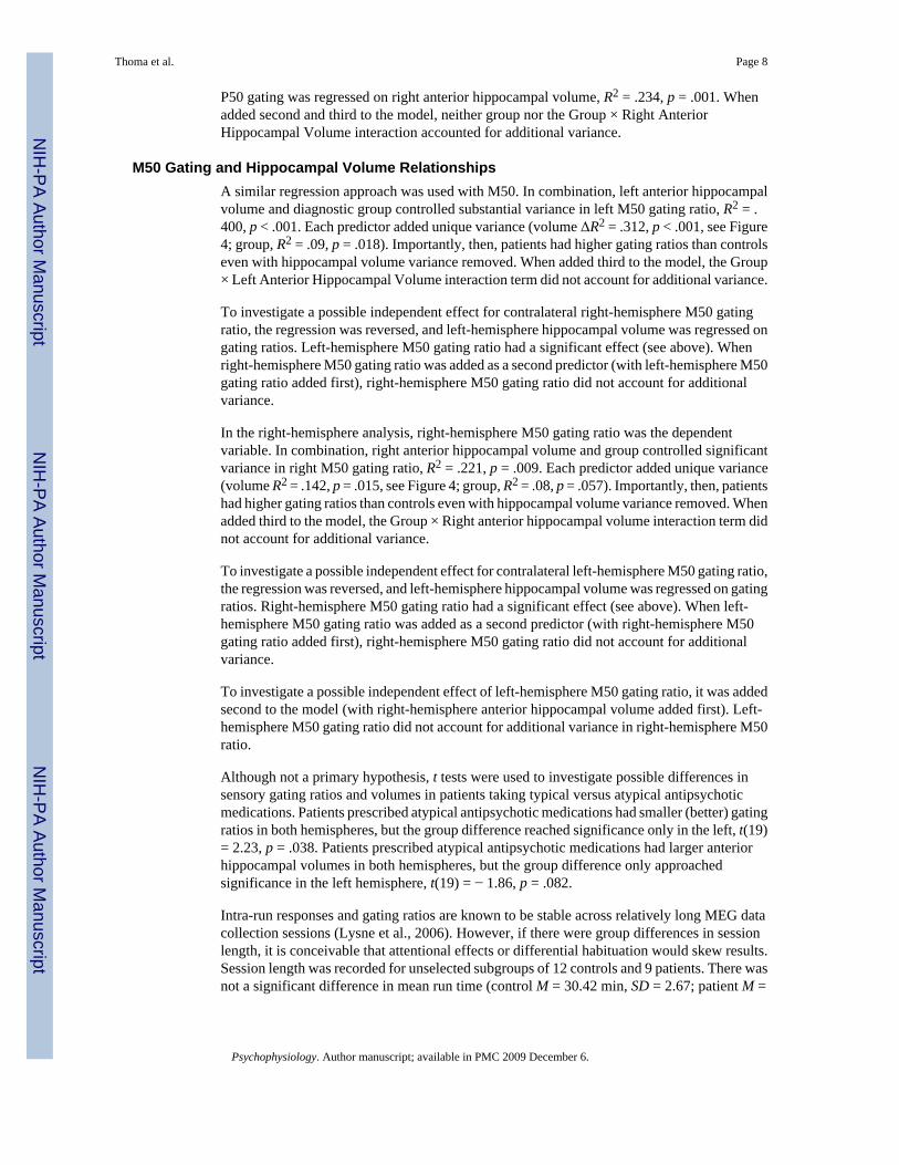
P50 gating was regressed on right anterior hippocampal volume, R2 = .234, p = .001. Whenadded second and third to the model, neither group nor the Group × Right AnteriorHippocampal Volume interaction accounted for additional variance.
M50 Gating and Hippocampal Volume RelationshipsA similar regression approach was used with M50. In combination, left anterior hippocampalvolume and diagnostic group controlled substantial variance in left M50 gating ratio, R2 = .400, p < .001. Each predictor added unique variance (volume ΔR2 = .312, p < .001, see Figure4; group, R2 = .09, p = .018). Importantly, then, patients had higher gating ratios than controlseven with hippocampal volume variance removed. When added third to the model, the Group× Left Anterior Hippocampal Volume interaction term did not account for additional variance.
To investigate a possible independent effect for contralateral right-hemisphere M50 gatingratio, the regression was reversed, and left-hemisphere hippocampal volume was regressed ongating ratios. Left-hemisphere M50 gating ratio had a significant effect (see above). Whenright-hemisphere M50 gating ratio was added as a second predictor (with left-hemisphere M50gating ratio added first), right-hemisphere M50 gating ratio did not account for additionalvariance.
In the right-hemisphere analysis, right-hemisphere M50 gating ratio was the dependentvariable. In combination, right anterior hippocampal volume and group controlled significantvariance in right M50 gating ratio, R2 = .221, p = .009. Each predictor added unique variance(volume R2 = .142, p = .015, see Figure 4; group, R2 = .08, p = .057). Importantly, then, patientshad higher gating ratios than controls even with hippocampal volume variance removed. Whenadded third to the model, the Group × Right anterior hippocampal volume interaction term didnot account for additional variance.
To investigate a possible independent effect for contralateral left-hemisphere M50 gating ratio,the regression was reversed, and left-hemisphere hippocampal volume was regressed on gatingratios. Right-hemisphere M50 gating ratio had a significant effect (see above). When left-hemisphere M50 gating ratio was added as a second predictor (with right-hemisphere M50gating ratio added first), right-hemisphere M50 gating ratio did not account for additionalvariance.
To investigate a possible independent effect of left-hemisphere M50 gating ratio, it was addedsecond to the model (with right-hemisphere anterior hippocampal volume added first). Left-hemisphere M50 gating ratio did not account for additional variance in right-hemisphere M50ratio.
Although not a primary hypothesis, t tests were used to investigate possible differences insensory gating ratios and volumes in patients taking typical versus atypical antipsychoticmedications. Patients prescribed atypical antipsychotic medications had smaller (better) gatingratios in both hemispheres, but the group difference reached significance only in the left, t(19)= 2.23, p = .038. Patients prescribed atypical antipsychotic medications had larger anteriorhippocampal volumes in both hemispheres, but the group difference only approachedsignificance in the left hemisphere, t(19) = − 1.86, p = .082.
Intra-run responses and gating ratios are known to be stable across relatively long MEG datacollection sessions (Lysne et al., 2006). However, if there were group differences in sessionlength, it is conceivable that attentional effects or differential habituation would skew results.Session length was recorded for unselected subgroups of 12 controls and 9 patients. There wasnot a significant difference in mean run time (control M = 30.42 min, SD = 2.67; patient M =
Thoma et al. Page 8
Psychophysiology. Author manuscript; available in PMC 2009 December 6.
NIH
-PA Author Manuscript
NIH
-PA Author Manuscript
NIH
-PA Author Manuscript

33.11 min, SD = 4.76), t(19) = − 1.65, p = .115, indicating comparable session lengths andnumber of trials in schizophrenia and control subjects.
DiscussionThere was a sensory gating deficit in the schizophrenia group, whether assessed with P50, left-hemisphere M50, or right-hemisphere M50. Huang et al. (2003) demonstrated that the ERPP50 in a paired-click paradigm is largely comprised of signal generated by two sourceslocalizing to left and right STG auditory regions. Considering gating in terms of lateralizedsources, it has been shown that left-hemisphere M50 gating was related to attention andworking memory deficits in schizophrenia (Thoma et al., 2003), and that right-hemisphereM50 gating was associated with schizophrenia negative symptoms (Thoma et al., 2005).Measurement of gating in terms of individual neural generators affords greater specificity ofmeasurement and has resulted in the discovery of relationships between gating and clinicalvariables that were not evident in single-electrode ERP studies. In the present study, thisapproach allowed investigation of structure–function relationships between hippocampalvolume and lateralized gating ratios within hemispheres.
It is thought that gating is dependent on a fronto-temporal network comprised of prefrontal,medial frontal, STG, and medial temporal cortex (Grunwald et al., 2003). Based on anassumption that the extent of hippocampal impairment in schizophrenia could be assessed interms of reduced hippocampal volume, the present hypothesis was that reduced size ofhippocampus in that group would reduce the functional capacity of the entire network—andthat reduced function would be measurable as poorer gating. Indeed, gating accounted forsignificant variance in ipsilateral anterior hippocampal volume, not just for patients but for thecontrol group. That is, smaller anterior hippocampal volume occurred with higher M50 gatingratios for both groups. This effect was hemisphere specific: left-hemisphere M50 gatingaccounted for variance only in left-hemisphere anterior hippocampal volume, and right-hemisphere M50 gating accounted for variance only in right-hemisphere hippocampal volume.
Waldo et al. (1994) demonstrated an indirect association between hippocampal volume andP50 gating in schizophrenia. Although no correlations were reported, a group of schizophrenia-spectrum patients with reduced hippocampal volume also had impaired gating. The improvedspecificity of measurement provided by dense-array MEG revealed the presence of positivecorrelations between hippocampal volumes and functional gating measures in the present data.This effect was found specifically for anterior hippocampus, whereas Waldo et al. did notdistinguish subhippocampal regions, working only with whole hippocampal volumes. Thereis considerable evidence based on animal studies demonstrating the importance ofhippocampus for sensory gating. For example, it has been well established that hippocampusis involved in PPI of startle, a more comprehensively studied gating paradigm than thatassociated with P50 (Bast & Feldon, 2003; Caine et al., 1992; Perlstein, Fiorito, Simons, &Graham, 1993; Perlstein, Simons, & Graham, 2001; Swerdlow et al., 2001; Zhang, Bast, &Feldon, 2002a, 2002b). There is strong evidence that ventral hippocampus plays a role in thegating of the startle response in rats (Caine et al., 1992; Swerdlow et al., 2001, 2004). Ratventral hippocampus is analogous to anterior hippocampus in humans. There is also evidenceof entorhinal cortex involvement in PPI (Goto, Ueki, Iso, & Morita, 2002; Swerdlow et al.,2001), which led these researchers to suggest that polymodal sensory information, exchangedby entorhinal cortex and ventral hippocampal CA3, is vital for any gating effect to occur.Neurochemical challenge studies also support the activities of these regions in the activity ofa gating circuit. For example, Swerdlow et al. (1995) found increased sensitivity to thedisruptive effects of apomorphine on PPI in rats with either medial PFC (mPFC) or ventralhippocampal lesions. Alternately, chemical stimulation of the ventral hippocampal-mPFCcircuit disrupts PPI (Shoemaker et al., 2005). Entorhinal cortex has extensive, direct
Thoma et al. Page 9
Psychophysiology. Author manuscript; available in PMC 2009 December 6.
NIH
-PA Author Manuscript
NIH
-PA Author Manuscript
NIH
-PA Author Manuscript
connections to mPFC. Entorhinal cortex may serve to integrate information from frontal cortexinto ventral hippocampus. In the clinical literature, increasing evidence has demonstrated arobust link between sensory gating and the presence and extent of hippocampal nicotinicreceptor functioning in schizophrenia (particularly in hippocampal CA3; Martin, Kem, &Freedman, 2004), all of which suggests that proper nicotinic receptor functioning is critical fornormal sensory gating. The present results suggest that overall anterior hippocampal volumemay reflect the quality and extent of hippocampal nicotinic receptor functioning.
That there is distinct hippocampal variance associated with gating was not surprising, but thatpatients with schizophrenia had higher gating ratios than controls, even with the hippocampalvariance removed, indicates that significant schizophrenia variance remains to be accountedfor in terms of brain structure or function. Based on this finding, further research is warrantedto investigate other candidate brain regions that are known to be functionally impaired inschizophrenia. For example, intrahemispheric gating ratio has been linked to reducedschizophrenia STG cortical thickness (Thoma et al., 2004), and recent depth-electrode researchin patients with epilepsy has suggested that medial frontal and sensory-motor cortex are alsocritical for gating (Kurthen et al., 2007).
There has been some evidence that anterior hippocampus is involved in novelty detection andencoding, whereas posterior hippocampus is more involved with familiarity (Strange, Fletcher,Henson, Friston, & Dolan, 1999; Zeineh, Engel, Thompson, & Bookheimer, 2003). However,it is not clear what the functional implications of having a small anterior hippocampus are innormal healthy control subjects. There has been considerable research in patients withschizophrenia suggesting that reduced hippocampus is an integral characteristic of the disorder(Csernansky et al., 1998; DeLisi, Dauphinais, & Gershon, 1988; Hanlon et al., 2005; Rossi etal., 1994; Shenton et al., 1992; Suddath et al., 1989, 1990). Reduced hippocampal volume isassociated with abnormality on neuropsychological tests typically associated with impairedfronto-temporal function (Bilder & Degreef, 1991; Szeszko et al., 2002; Weinberger, Berman,Suddath, & Torrey, 1992). It has been suggested that the effect is primarily due to reductionin anterior hippocampal volume (Pegues, Rogers, Amend, Vinogradov, & Deicken, 2003). Inpresent data, there was no difference in posterior hippocampal volume, accounting forambiguity in earlier findings of reduced size of overall hippocampus (Szeszko et al., 2002).Szeszko et al. (2002) suggested that these neuropsychological findings make sense if oneconsiders the extensive feedfoward and feedback pathways between hippocampus and PFC,further documenting a fronto-temporal disconnection in schizophrenia. Auditory gating hasalso been associated with neuropsychological measures typically associated with frontal lobefunction (Thoma et al., 2003; Yee & White, 2001). Thus, a fronto-temporal disconnection mayalso account for the present results.
The present patient group was too small for strong conclusions about how medication typeinteracts with hippocampal volume and sensory gating. Typical antipsychotic medicationsappear to enlarge thalamic and striatal brain structures but have not yet been shown to haveany direct effect on hippocampus volume or the volume of other cortical features (Flashman& Green, 2004). Atypical antipsychotic medications are thought to improve sensory gatingratios (Adler et al., 2004). In present data, patients on typical antipsychotic medications (n =4) had smaller hippocampi and poorer left hemisphere sensory gating ratios than patients onatypical medications (n = 18). This was not a treatment study, and patients were acceptedregardless of the type of antipsychotic prescribed by their doctor. Thus, it may be that smallerhippocampus is a marker for a subcategory of patients with schizophrenia that have a betterresponse to typical medications.
Present findings demonstrate, first, that smaller anterior hippocampus volume relates to P50and intrahemispheric M50 sensory gating. Contrary to prediction, this relationship was true
Thoma et al. Page 10
Psychophysiology. Author manuscript; available in PMC 2009 December 6.
NIH
-PA Author Manuscript
NIH
-PA Author Manuscript
NIH
-PA Author Manuscript

for both groups. Thus, the second main finding of the present study is that the P50/M50 gatingdeficit in schizophrenia is not merely an artifact of diagnostic group differences in hippocampalvolume: Diagnosis and hippocampal volume make distinct contributions to gating. Third, atleast a portion of the relevant circuitry is intrahemispheric.
Normal reduction in the response to a second stimulus in a gating experiment is related tostimulus redundancy, and the gating effect is diminished or disappears entirely when the pairedstimuli are dissimilar (Boutros, Belger, Campbell, D’Souza, & Krystal, 1999). Wepropose that,for successful gating to occur, at minimum, a healthy mPFC–hippocampal network isnecessary, with mPFC involved in attentional control associated with stimulus perception andhippocampus involved in the modulation and preservation of stimulus traces. Future researchmay involve hippocampal lesion studies to investigate the necessity of hippocampus forintrahemispheric gating, investigation of relationships between responses in stimulus noveltyparadigms associated with anterior hippocampal function and those involving gating, andfurther investigation of the role for frontal cortical regions in gating.
AcknowledgmentsWe thank Jessica Irwin, Fernando Torres, Robin Douglas, Kim Paulson, Ira Driscoll, and Laura Rowland for adviceand assistance with all stages of this research. This project was funded by a NARSAD Young Investigator Award andNIAAA grant 1 K23 AA016544-01 to Dr. Thoma, an NIMH grant R01 MH-65304 to Dr. Cañive, and grants from theMental Illness and Neuroscience Discovery (MIND) Institute to Drs. Thoma and Cañive. Helen Petropoulos wassupported in part by National Institutes of Health grants NS039123, AG16418, NS35708, NS41390, and HD41237.
REFERENCESAdler LE, Olincy A, Cawthra EM, McRae KA, Harris JG, Nagamoto HT, et al. Varied effects of atypical
neuroleptics on P50 auditory gating in schizophrenia patients. American Journal of Psychiatry2004;161:1822–1828. [PubMed: 15465979]
Adler LE, Olincy A, Waldo M, Harris JG, Griffith J, Stevens K, et al. Schizophrenia, sensory gating, andnicotinic receptors. Schizophrenia Bulletin 1998;24:189–202. [PubMed: 9613620]
Adler LE, Pachtmen E, Franks R, Pecevich M, Waldo MC, Freedman R. Neurophysiological evidencefor a defect in neuronal mechanisms involved in sensory gating in schizophrenia. Biological Psychiatry1982;17:639–654. [PubMed: 7104417]
Adler LE, Rose G, Freedman R. Neurophysiological studies of sensory gating in rats: Effects ofamphetamine, phencyclidine, and haloperidol. Biological Psychiatry 1986;21:787–798. [PubMed:3730461]
Arciniegas DB, Topkoff JL, Rojas DC, Sheeder J, Teale P, Young DA, et al. Reduced hippocampalvolume in association with p50 nonsuppression following traumatic brain injury. Journal ofNeuropsychiatry and Clinical Neuroscience 2001;13:213–221.
Bast T, Feldon J. Hippocampal modulation of sensorimotor processes. Progress in Neurobiology2003;70:319–345. [PubMed: 12963091]
Bickford-Wimer PC, Nagamoto H, Johnson R, Adler LE, Egan M, Rose GM, et al. Auditory sensorygating in hippocampal neurons: A model system in the rat. Biological Psychiatry 1990;27:183–192.[PubMed: 2294981]
Bilder, RM.; Degreef, G. Morphologic markers of neurodevelopmental paths to schizophrenia. In:Mednick, SA.; Canon, TD.; Barr, CE.; LaFosse, JM., editors. Developmental neuropathology ofschizophrenia. New York: Plenum; 1991. p. 167-190.
Boutros NN, Belger A, Campbell D, D’Souza C, Krystal J. Comparison of four components of sensorygating in schizophrenia and normal subjects: A preliminary report. Psychiatry Research 1999;88:119–130. [PubMed: 10622348]
Boutros NN, Trautner P, Rosburg T, Korzyukov O, Grunwald T, Schaller C, et al. Sensory gating in thehuman hippocampal and rhinal regions. Clinical Neurophysiology 2005;116:1967–1974. [PubMed:16000257]
Thoma et al. Page 11
Psychophysiology. Author manuscript; available in PMC 2009 December 6.
NIH
-PA Author Manuscript
NIH
-PA Author Manuscript
NIH
-PA Author Manuscript

Bramon E, Rabe-Hesketh S, Sham P, Murray RM, Frangou S. Meta-analysis of the P300 and P50waveforms in schizophrenia. Schizophrenia Research 2004;70:315–329. [PubMed: 15329307]
Caine SB, Geyer MA, Swerdlow NR. Hippocampal modulation of acoustic startle and prepulse inhibitionin the rat. Pharmacology Biochemistry and Behavior 1992;43:1201–1208.
Cardenas VA, Gerson J, Fein G. The reliability of P50 suppression as measured by the conditioningtesting ratio is vastly improved by dipole modeling. Biological Psychiatry 1993;33:335–344.[PubMed: 8471691]
Cohen, NJ.; Eichenbaum, H. Memory, amnesia, and the hippocampal system. Cambridge, MA: MITPress; 1993.
Coppola R. Isolating low frequency activity EEG spectrum analysis. Electroencephalography andClinical Neurophysiology 1979;46:224–226. [PubMed: 86431]
Crottaz-Herbette S, Lau KM, Glover GH, Menon V. Hippocampal involvement in detection of deviantauditory and visual stimuli. Hippocampus 2005;15:132–139. [PubMed: 15390157]
Csernansky JG, Joshi S, Wang L, Haller JW, Gado M, Miller JP, et al. Hippocampal morphometry inschizophrenia by high dimensional brain mapping. Proceedings of the National Academy of Sciences,USA 1998;95:11406–11411.
DeLisi LE, Dauphinais ID, Gershon ES. Perinatal complications and reduced size of brain limbicstructures in familial schizophrenia. Schizophrenia Bulletin 1988;14:185–191. [PubMed: 3201176]
Edgar JC, Huang MX, Weisend M, Sherwood A, Miller GA, Adler LE, et al. Interpreting abnormality:An EEG and MEG study of P50 and the auditory paired-stimulus paradigm. Biological Psychology2003;65:1–20. [PubMed: 14638286]
First, MB.; Spitzer, RL.; Gibbon, M.; Williams, JBW. Structured Clinical Interview for DSM-IV Axis IDisorders (SCID), clinician version. Washington, DC: American Psychiatric Press; 1997.
Flashman LA, Green MF. Review of cognition and brain structure in schizophrenia: Profiles, longitudinalcourse, and effects of treatment. Psychiatric Clinics of North America 2004;27:1–18. [PubMed:15062627]
Frazier CJ, Buhler AV, Weiner JL, Dunwiddie TV. Synaptic potentials mediated via alpha-bungarotoxin-sensitive nicotinic acetylcholine receptors in rat hippocampal interneurons. Journal of Neuroscience1998a;18:8228–8235. [PubMed: 9763468]
Frazier CJ, Rollins YD, Breese CR, Leonard S, Freedman R, Dunwiddie TV. Acetylcholine activates analpha-bungarotoxin-sensitive nicotinic current in rat hippocampal interneurons, but not pyramidalcells. Journal of Neuroscience 1998b;18:1187–1195. [PubMed: 9454829]
Freedman R, Adler LE, Bickford P, Byerley W, Coon H, Cullum CM, et al. Schizophrenia and nicotinicreceptors. Harvard Review of Psychiatry 1994;2:179–192. [PubMed: 9384901]
Goldman MB, Mitchell CP. What is the functional significance of hippocampal pathology inschizophrenia? Schizophrenia Bulletin 2004;30:367–392. [PubMed: 15279054]
Goto K, Ueki A, Iso H, Morita Y. Reduced prepulse inhibition in rats with entorhinal cortex lesions.Behavioral Brain Research 2002;134:201–207.
Grunwald T, Boutros NN, Pezer N, von Oertzen J, Fernandez G, Schaller C, et al. Neuronal substratesof sensory gating within the human brain. Biological Psychiatry 2003;53:511–519. [PubMed:12644356]
Hanlon FM, Miller GA, Thoma RJ, Irwin J, Jones A, Moses SN, et al. Distinct M50 and M100 auditorygating deficits in schizophrenia. Psychophysiology 2005;42:417–427. [PubMed: 16008770]
Harrison PJ. The hippocampus in schizophrenia: A review of the neuropathological evidence and itspathophysiological implications. Psychopharmacology 2004;174:151–162. [PubMed: 15205886]
Heinrichs RW. Meta-analysis and the science of schizophrenia: Variant evidence or evidence of variants?Neuroscience and Biobehavioral Reviews 2004;28:379–394. [PubMed: 15341034]
Hershman KM, Freedman R, Bickford PC. Gaba(B) antagonists diminish the inhibitory gating of auditoryresponse in the rat hippocampus. Neuroscience Letters 1995;190:133–136. [PubMed: 7644122]
Huang MX, Edgar JC, Thoma RJ, Hanlon FM, Moses SN, Lee RR, et al. Predicting EEG responses usingMEG sources in superior temporal gyrus reveals source asynchrony in patients with schizophrenia.Clinical Neurophysiology 2003;114:835–850. [PubMed: 12738429]
Thoma et al. Page 12
Psychophysiology. Author manuscript; available in PMC 2009 December 6.
NIH
-PA Author Manuscript
NIH
-PA Author Manuscript
NIH
-PA Author Manuscript

Judd LL, McAdams L, Budnick B, Braff DL. Sensory gating deficits in schizophrenia—New results.American Journal of Psychiatry 1992;149:488–493. [PubMed: 1554034]
Knight R. Contribution of human hippocampal region to novelty detection. Nature 1996;383:256–259.[PubMed: 8805701]
Kurthen M, Trautner P, Rosburg T, Grunwald T, Deitl T, Kuhn K-U, et al. Towards a functionaltopography of sensory gating areas: Invasive P50 recording and electrical stimulation mapping inepilepsy surgery candidates. Psychiatry Research/Neuroimaging 2007;155(2):121–133.
Lysne, PA.; Montano, R.; Hanlon, FM.; Bantz, R.; Lundy, L.; Euler, M., et al. Intra-run stability of theM50 auditory gating response in a paired-click paradigm. Poster session presented at the annualBioMag Meeting; Vancouver, British Columbia, Canada. 2006 Aug.
Maguire EA, Gadian DG, Johnsrude IS, Good CD, Ashburner J, Frackowiak RS, et al. Navigation-relatedstructural change in the hippocampi of taxi drivers. Proceedings of the National Academy of Sciences,USA 2000;97:4398–4403.
Martin LF, Kem WR, Freedman R. Alpha-7 nicotinic receptor agonists: Potential new candidates for thetreatment of schizophrenia. Psychopharmacology 2004;174(1):54–64. [PubMed: 15205879]
Miller CL, Freedman R. The activity of hippocampal interneurons and pyramidal cells during the responseof the hippocampus to repeated auditory-stimuli. Neuroscience 1995;69:371–381. [PubMed:8552235]
Miller GA, Chapman JP. Misunderstanding analysis of covariance. Journal of Abnormal Psychology2001;110:40–48. [PubMed: 11261398]
Miller GA, Lutzenberger W, Elbert T. The linked-reference issue in EEG and ERP recording. Journal ofPsychophysiology 1991;5:273–276.
Moses SN, Cole C, Driscoll I, Ryan JD. Differential contributions of hippocampus, amygdala andperirhinal cortex to recognition of novel objects, contextual stimuli and stimulus relationships. BrainResearch Bulletin 2005;67:62–76. [PubMed: 16140164]
Moses SN, Ryan JD. A comparison and evaluation of the predictions of relational and conjunctiveaccounts of hippocampal function. Hippocampus 2006;16:43–65. [PubMed: 16270317]
Paller KA, McCarthy G, Roessler E, Allison T, Wood CC. Potentials-evoked in human and monkeymedial temporallobe during auditory and visual oddball paradigms. Electroencephalography andClinical Neurophysiology 1992;84:269–279. [PubMed: 1375886]
Pegues MP, Rogers LJ, Amend D, Vinogradov S, Deicken RF. Anterior hippocampal volume reductionin male patients with schizophrenia. Schizophrenia Research 2003;60:105–115. [PubMed:12591575]
Perlstein W, Fiorito E, Simons RF, Graham FK. Lead stimulation effects on reflex blink, exogenous brainpotentials, and loudness judgments. Psychophysioloqy 1993;30:347–358.
Perlstein WP, Simons RF, Graham FK. Prepulse effects as a function of cortical projection system.Biological Psychology 2001;56:83–111. [PubMed: 11334698]
Petropoulos H, Sibbitt WL, Brooks WM. Automated T2 quantitation in neuropsychiatric lupuserythematosus: A marker of active disease. Journal of Magnetic Resonance Imaging 1999;9:39–43.[PubMed: 10030648]
Pollmann S, Lepsien J, Hugdahl K, von Cramon DY. Auditory target detection in dichotic listeninginvolves the orbitofrontal and hippocampal paralimbic belts. Cerebral Cortex 2004;14:903–913.[PubMed: 15054061]
Ricker, DA.; Thoma, RJ.; Hanlon, FM.; Moses, SN.; Edgar, CJ.; Irwin, JM., et al. Auditory sensory gatingdeficit and negative symptoms in schizophrenia. Poster session presented at the biannual meeting ofthe International Conference on Schizophrenia Research; Colorado Springs, CO. 2004 Apr.
Rossi A, Stratta P, Mancini F, Gallucci M, Mattei P, Core L, et al. Magnetic resonance imaging findingsof amygdala-anterior hippocampus shrinkage in male patients with schizophrenia. PsychiatryResearch 1994;52:43–53. [PubMed: 8047621]
Rudy JW, Sutherland RJ. The hippocampal-formation is necessary for rats to learn and rememberconfigural discriminations. Behavioural Brain Research 1989;34:97–109. [PubMed: 2765175]
Rudy JW, Sutherland RJ. Configural association theory and the hippocampal formation: An appraisaland reconfiguration. Hippocampus 1995;5:375–389. [PubMed: 8773252]
Thoma et al. Page 13
Psychophysiology. Author manuscript; available in PMC 2009 December 6.
NIH
-PA Author Manuscript
NIH
-PA Author Manuscript
NIH
-PA Author Manuscript

Shenton ME, Kikinis R, Jolesz FA, Pollak SD, LeMay M, Wible CG, et al. Abnormalities of the lefttemporal lobe and thought disorder in schizophrenia: A quantitative magnetic resonance imagingstudy. New England Journal of Medicine 1992;327:604–612. [PubMed: 1640954]
Shoemaker JM, Saint Marie RL, Bongiovanni MJ, Neary AC, Tochen LS, Swerdlow NR. Prefrontal D1and ventral hippocampal N-methyl-D-aspartate regulation of startle gating in rats. Neuroscience2005;135:385–394. [PubMed: 16125865]
Strange BA, Fletcher PC, Henson RN, Friston KJ, Dolan RJ. Segregating the functions of humanhippocampus. Proceedings of the National Academy of Sciences, USA 1999;96:4034–4039.
Suddath RL, Casanova MF, Goldberg TE, Daniel DG, Kelsoe JR Jr, Weinberger DR. Temporal lobepathology in schizophrenia: A quantitative magnetic resonance imaging study. American Journal ofPsychiatry 1989;146:464–472. [PubMed: 2929746]
Suddath RL, Christison GW, Torrey EF, Casanova MF, Weinberger DR. Anatomical abnormalities inthe brains of monozygotic twins discordant for schizophrenia. New England Journal of Medicine1990;322:789–794. [PubMed: 2308615]
Swerdlow NR, Hanlon FM, Henning L, Kim YK, Gaudet I, Halim ND. Regulation of sensorimotor gatingin rats by hippocampal NMDA: Anatomical localization. Brain Research 2001;898:195–203.[PubMed: 11306005]
Swerdlow NR, Lipska BK, Weinberger DR, Braff DL, Jaskiw GE, Geyer MA. Increased sensitivity tothe sensori-motor gating-disruptive effects of apomorphine after lesions of medial prefrontal cortexor ventral hippocampus in adult rats. Psychopharmacology 1995;122:27–34. [PubMed: 8711061]
Swerdlow NR, Shoemaker JM, Noh HR, Ma L, Gaudet I, Munson M, et al. The ventral hippocampalregulation of prepulse inhibition and its disruption by apomorphine in rats are not mediated via thefornix. Neuroscience 2004;123:675–685. [PubMed: 14706779]
Szeszko PR, Strous RD, Goldman RS, Ashtari M, Knuth KH, Lieberman JA, et al. Neuropsychologicalcorrelates of hippocampal volumes in patients experiencing a first episode of schizophrenia.American Journal of Psychiatry 2002;159:217–226. [PubMed: 11823263]
Tesche CD, Karhu J, Tissari SO. Non-invasive detection of neuronal population activity in humanhippocampus. Cognitive Brain Research 1996;4:39–47. [PubMed: 8813411]
Thoma RJ, Hanlon FM, Moses SN, Edgar JC, Huang M, Irwin J, et al. Lateralization of auditory sensorygating and neuropsychological dysfunction in schizophrenia. American Journal of Psychiatry2003;160:1595–1605. [PubMed: 12944333]
Thoma RJ, Hanlon F, Moses S, Ricker D, Huang M, Edgar C, et al. M50 sensory gating predicts negativesymptoms in schizophrenia. Schizophrenia Research 2005;73:311–318. [PubMed: 15653276]
Thoma RJ, Hanlon FM, Sanchez N, Weisend MP, Huang M, Jones A, et al. Auditory sensory gatingdeficit and cortical thickness in schizophrenia. Neurology and Clinical Neurophysiology 2004;62:1–7.
Waldo MC, Cawthra E, Adler LE, Dubester S, Staunton M, Nagamoto H, et al. Auditory sensory gating,hippocampal volume, and catecholamine metabolism in schizophrenics and their siblings.Schizophrenia Research 1994;12:93–106. [PubMed: 8043530]
Watson C, Andermann F, Gloor P, Jones-Gotman M, Peters T, Evans A, et al. Anatomic basis ofamygdaloid and hippocampal volume measurement by magnetic resonance imaging. Neurology1992;42:1743–1750. [PubMed: 1513464]
Weinberger DR, Berman KF, Suddath R, Torrey EF. Evidence of dysfunction of a prefrontal-limbicnetwork in schizophrenia: A magnetic resonance imaging and regional cerebral blood flow study ofdiscordant monozygotic twins. American Journal of Psychiatry 1992;149:890–897. [PubMed:1609867]
Yee CM, White PM. Experimental modification of P50 suppression. Psychophysiology 2001;38:531–539. [PubMed: 11352142]
Zeineh MM, Engel SA, Thompson PM, Bookheimer SY. Dynamics of hippocampus during encodingand retrieval of face-name pairs. Science 2003;299:577–580. [PubMed: 12543980]
Zhang WN, Bast T, Feldon J. Effects of hippocampal N-methyl-D-aspartate infusion on locomotoractivity and prepulse inhibition: Differences between the dorsal and ventral hippocampus. BehavioralNeuroscience 2002a;116:72–84. [PubMed: 11895185]
Thoma et al. Page 14
Psychophysiology. Author manuscript; available in PMC 2009 December 6.
NIH
-PA Author Manuscript
NIH
-PA Author Manuscript
NIH
-PA Author Manuscript

Zhang WN, Bast T, Feldon J. Prepulse inhibition in rats with temporary inhibition/inactivation of ventralor dorsal hippocampus. Pharmacology, Biochemistry, Behavior 2002b;73:929–940.
Thoma et al. Page 15
Psychophysiology. Author manuscript; available in PMC 2009 December 6.
NIH
-PA Author Manuscript
NIH
-PA Author Manuscript
NIH
-PA Author Manuscript

Figure 1.SAVRA (Segmentation And Visualization for Research Advancement) software made severalviews of brain tissue available simultaneously for each slice to enhance users’ judgmentregarding tissue selection.
Thoma et al. Page 16
Psychophysiology. Author manuscript; available in PMC 2009 December 6.
NIH
-PA Author Manuscript
NIH
-PA Author Manuscript
NIH
-PA Author Manuscript

Figure 2.A: An example of the magnetic field pattern and cortical localization for left and right M50equivalent current dipoles. For all subjects, the M50 response localized bilaterally to auditorycortex. B: An example of left and right hemisphere equivalent current dipole waveforms for arepresentative subject from the control group and one subject from the schizophrenia group.
Thoma et al. Page 17
Psychophysiology. Author manuscript; available in PMC 2009 December 6.
NIH
-PA Author Manuscript
NIH
-PA Author Manuscript
NIH
-PA Author Manuscript

Figure 3.M50 sensory gating is expressed as a ratio of S2/S1. M50 sensory gating was impairedbilaterally in the schizophrenia group (error bars = 1 SD).
Thoma et al. Page 18
Psychophysiology. Author manuscript; available in PMC 2009 December 6.
NIH
-PA Author Manuscript
NIH
-PA Author Manuscript
NIH
-PA Author Manuscript
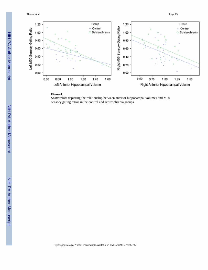
Figure 4.Scatterplots depicting the relationship between anterior hippocampal volumes and M50sensory gating ratios in the control and schizophrenia groups.
Thoma et al. Page 19
Psychophysiology. Author manuscript; available in PMC 2009 December 6.
NIH
-PA Author Manuscript
NIH
-PA Author Manuscript
NIH
-PA Author Manuscript

NIH
-PA Author Manuscript
NIH
-PA Author Manuscript
NIH
-PA Author Manuscript
Thoma et al. Page 20
Table 1
Demographics
Control Schizophrenia t (p)
Age (years) 41.55 39.38 0.52 (p = .61)Sex Male = 16 Male = 17 1.03 (p = .31)
Female = 6 Female = 5Education (years) 13.56 12.47 1.47 (p = .15)
Psychophysiology. Author manuscript; available in PMC 2009 December 6.

NIH
-PA Author Manuscript
NIH
-PA Author Manuscript
NIH
-PA Author Manuscript
Thoma et al. Page 21Ta
ble
2
Gro
up C
ompa
rison
of V
olum
e of
Hip
poca
mpu
s, IC
V, G
M, W
M, a
nd C
SF in
Cub
ic C
entim
eter
s
Mea
sure
men
tC
ontr
ol m
ean
(SD
)Sc
hizo
phre
nia
mea
n (S
D)
FP
valu
e
Ant
erio
r hip
poca
mpu
s1.
93 (0
.53)
1.61
(0.3
7)5.
28.0
27Po
ster
ior h
ippo
cam
pus
0.70
(0.2
4)0.
72 (0
.28)
0.03
4.8
54To
tal h
ippo
cam
pus
7.17
(1.0
4)6.
62 (1
.07)
2.83
.100
Intra
cran
ial v
olum
e15
14.4
0 (1
44.5
5)14
65.6
8 (1
50.7
1)0.
395
.552
Gra
y m
atte
r53
9.05
(59.
82)
512.
76 (7
6.56
)0.
713
.403
Whi
te m
atte
r55
6.28
(73.
88)
545.
09 (7
1.31
)0.
209
.650
Cer
ebro
spin
al fl
uid
181.
39 (3
8.00
)19
0.55
2 (3
5.08
)1.
914
.173
Psychophysiology. Author manuscript; available in PMC 2009 December 6.