Structural determinants of gating in the TRPV1 channel
8
Structural determinants of gating in the TRPV1 channel He ´ctor Salazar 1,4 , Andre ´s Jara-Oseguera 2,4 , Enrique Herna ´ndez-Garcı ´a 1,4 , Itzel Llorente 1 , Imilla I Arias-Olguı ´n 1 , Manuel Soriano-Garcı ´a 3 , Leo ´n D Islas 2 & Tamara Rosenbaum 1 Transient receptor potential vanilloid 1 (TRPV1) channels mediate several types of physiological responses. Despite the importance of these channels in pain detection and inflammation, little is known about how their structural components convert different types of stimuli into channel activity. To localize the activation gate of these channels, we inserted cysteines along the S6 segment of mutant TRPV1 channels and assessed their accessibility to thiol-modifying agents. We show that access to the pore of TRPV1 is gated by S6 in response to both capsaicin binding and increases in temperature, that the pore-forming S6 segments are helical structures and that two constrictions are present in the pore: one that impedes the access of large molecules and the other that hampers the access of smaller ions and constitutes an activation gate of these channels. Transient receptor potential (TRP) channels participate in a wide variety of physiological processes, including detection of sensory stimuli, osmoregulation, intracellular calcium homeostasis, chemo- taxis and cell proliferation, among others 1 . A member of the TRP family, the TRPV1 nonselective cation channel, is expressed in sensory neurons and in several other tissues 1–3 , where it functions as an integrator of noxious chemical and physical stimuli 4–9 . TRPV1 has been linked to processes mediating inflammatory and neuropathic pain 8,10 , a finding that has led to a recent surge in research aimed at designing TRPV1-targeted pain-relieving therapies 11,12 . Despite the clear physiological relevance of TRPV1 and of TRP channels in general, little structural information is currently available 13–17 . Sequence homology of TRPV1 to ion channels of known structure suggests a subunit topology similar to that of voltage-activated potassium (Kv) channels, with each subunit formed by six transmembrane domains (S1–S6), intracellular N and C termini, and a pore domain formed by S5, S6 and the loop between them. Nonetheless, the topology of the pore and the location of the activation gate of TRPV1 or of any other TRP channel remain unknown. Therefore, we set out to establish the location of the molecular determinants of channel gating to obtain insight into the structural properties of the pore of TRPV1 channels. Using the substituted cysteine accessibility method (SCAM) to scan the entire S6 segment of the rat TRPV1, we have made the following discoveries, which constitute the first molecular characterization of the activation gate of a TRP channel. First, the finding that the accessi- bility of the introduced cysteines is state-dependent indicated the presence of two intracellular constrictions: the most intracellular one located at Leu681, which closes the ion conduction pathway for large molecules (B6A ˚ ), and one located at Tyr671, which obstructs the conduction pathway for small permeating ions (B2A ˚ ) and constitutes the activation gate of TRPV1. An important result is that the region we define as the activation gate of TRPV1 gates the channel in response to both capsaicin and heat, revealing the physical point of convergence of these two pathways of activation. Second, use of our recently engineered cysteine-less TRPV1 (18 TRPV1) 4 has allowed us to study the formation of disulfide bonds between cysteines introduced at key S6 locations, unequivocally confirming proximity among the residues involved in gated access of ions in these channels. Third, we have observed a marked periodicity in our accessibility data that is consistent with an a-helical structure for the S6. Our results allow us to propose a structural homology model that neatly incorporates our observations and provides a structural basis to explain gating in TRPV1, which will prove useful in understanding gating of other TRP channels. RESULTS TRPV1-C157A as a tool for SCAM analysis Our previous experiments using blocking ions of known dimensions suggested the existence of gated access to the pore of TRPV1 channels from the intracellular side 18 , as observed in Kv channels. We sought to identify the existence of such an activation gate by probing the S6 transmembrane domain using SCAM to insert cysteines into the TRPV1-C157A background. Figure 1a, above, shows the sequence of the putative S6 region of the TRPV1 channel. TRPV1-C157A yields adequate expression levels and is unrespon- sive to cysteine-modifying compounds such as methanethiosulfonate (MTS) reagents, as no shift in the response to capsaicin or changes in channel conductance were observed after exposure to these molecules 4 (Figs. 1b,c). Notably, TRPV1-C157A resembles wild-type TRPV1 in its core biophysical properties, such as voltage-dependence, single- channel conductance (data not shown) and its response to capsaicin 4 (Supplementary Fig. 1). Received 15 January; accepted 4 June; published online 28 June 2009; doi:10.1038/nsmb.1633 1 Departamento de Biofı´sica, Instituto de Fisiologı´a Celular, 2 Departamento de Fisiologı´a, Facultad de Medicina and 3 Departamento de Bioquı´mica, Instituto de Quı´mica, Universidad Nacional Auto ´noma de Me ´xico, Me ´xico, D.F., Me ´xico. 4 These authors contributed equally to this work. Correspondence should be addressed to T.R. ([email protected]). 704 VOLUME 16 NUMBER 7 JULY 2009 NATURE STRUCTURAL & MOLECULAR BIOLOGY ARTICLES © 2009 Nature America, Inc. All rights reserved. © 2009 Nature America, Inc. All rights reserved.
-
Upload
independent -
Category
Documents
-
view
2 -
download
0
Transcript of Structural determinants of gating in the TRPV1 channel
Structural determinants of gating in the TRPV1 channelHector Salazar1,4, Andres Jara-Oseguera2,4, Enrique Hernandez-Garcıa1,4, Itzel Llorente1,Imilla I Arias-Olguın1, Manuel Soriano-Garcıa3, Leon D Islas2 & Tamara Rosenbaum1
Transient receptor potential vanilloid 1 (TRPV1) channels mediate several types of physiological responses. Despite theimportance of these channels in pain detection and inflammation, little is known about how their structural components convertdifferent types of stimuli into channel activity. To localize the activation gate of these channels, we inserted cysteines along theS6 segment of mutant TRPV1 channels and assessed their accessibility to thiol-modifying agents. We show that access to the poreof TRPV1 is gated by S6 in response to both capsaicin binding and increases in temperature, that the pore-forming S6 segmentsare helical structures and that two constrictions are present in the pore: one that impedes the access of large molecules andthe other that hampers the access of smaller ions and constitutes an activation gate of these channels.
Transient receptor potential (TRP) channels participate in a widevariety of physiological processes, including detection of sensorystimuli, osmoregulation, intracellular calcium homeostasis, chemo-taxis and cell proliferation, among others1. A member of the TRPfamily, the TRPV1 nonselective cation channel, is expressed in sensoryneurons and in several other tissues1–3, where it functions as anintegrator of noxious chemical and physical stimuli4–9. TRPV1 hasbeen linked to processes mediating inflammatory and neuropathicpain8,10, a finding that has led to a recent surge in research aimed atdesigning TRPV1-targeted pain-relieving therapies11,12.
Despite the clear physiological relevance of TRPV1 and of TRP channelsin general, little structural information is currently available13–17. Sequencehomology of TRPV1 to ion channels of known structure suggests asubunit topology similar to that of voltage-activated potassium (Kv)channels, with each subunit formed by six transmembrane domains(S1–S6), intracellular N and C termini, and a pore domain formed byS5, S6 and the loop between them. Nonetheless, the topology of the poreand the location of the activation gate of TRPV1 or of any other TRPchannel remain unknown. Therefore, we set out to establish the locationof the molecular determinants of channel gating to obtain insight intothe structural properties of the pore of TRPV1 channels.
Using the substituted cysteine accessibility method (SCAM) to scanthe entire S6 segment of the rat TRPV1, we have made the followingdiscoveries, which constitute the first molecular characterization of theactivation gate of a TRP channel. First, the finding that the accessi-bility of the introduced cysteines is state-dependent indicated thepresence of two intracellular constrictions: the most intracellularone located at Leu681, which closes the ion conduction pathway forlarge molecules (B6 A), and one located at Tyr671, which obstructsthe conduction pathway for small permeating ions (B2 A) andconstitutes the activation gate of TRPV1. An important result is
that the region we define as the activation gate of TRPV1 gates thechannel in response to both capsaicin and heat, revealing the physicalpoint of convergence of these two pathways of activation.
Second, use of our recently engineered cysteine-less TRPV1(18�TRPV1)4 has allowed us to study the formation of disulfidebonds between cysteines introduced at key S6 locations, unequivocallyconfirming proximity among the residues involved in gated access ofions in these channels. Third, we have observed a marked periodicityin our accessibility data that is consistent with an a-helical structurefor the S6. Our results allow us to propose a structural homologymodel that neatly incorporates our observations and provides astructural basis to explain gating in TRPV1, which will prove usefulin understanding gating of other TRP channels.
RESULTSTRPV1-C157A as a tool for SCAM analysisOur previous experiments using blocking ions of known dimensionssuggested the existence of gated access to the pore of TRPV1 channelsfrom the intracellular side18, as observed in Kv channels. We sought toidentify the existence of such an activation gate by probing the S6transmembrane domain using SCAM to insert cysteines into theTRPV1-C157A background. Figure 1a, above, shows the sequenceof the putative S6 region of the TRPV1 channel.
TRPV1-C157A yields adequate expression levels and is unrespon-sive to cysteine-modifying compounds such as methanethiosulfonate(MTS) reagents, as no shift in the response to capsaicin or changes inchannel conductance were observed after exposure to these molecules4
(Figs. 1b,c). Notably, TRPV1-C157A resembles wild-type TRPV1 inits core biophysical properties, such as voltage-dependence, single-channel conductance (data not shown) and its response to capsaicin4
(Supplementary Fig. 1).
Received 15 January; accepted 4 June; published online 28 June 2009; doi:10.1038/nsmb.1633
1Departamento de Biofısica, Instituto de Fisiologıa Celular, 2Departamento de Fisiologıa, Facultad de Medicina and 3Departamento de Bioquımica, Instituto deQuımica, Universidad Nacional Autonoma de Mexico, Mexico, D.F., Mexico. 4These authors contributed equally to this work. Correspondence should be addressedto T.R. ([email protected]).
70 4 VOLUME 16 NUMBER 7 JULY 2009 NATURE STRUCTURAL & MOLECULAR BIOLOGY
ART IC L E S
©20
09 N
atu
re A
mer
ica,
Inc.
All
rig
hts
res
erve
d.
©20
09 N
atu
re A
mer
ica,
Inc.
All
rig
hts
res
erve
d.
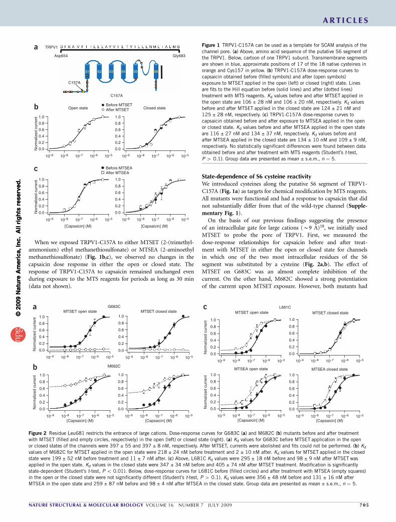
When we exposed TRPV1-C157A to either MTSET (2-(trimethyl-ammonium) ethyl methanethiosulfonate) or MTSEA (2-aminoethylmethanethiosulfonate) (Fig. 1b,c), we observed no changes in thecapsaicin dose response in either the open or closed state. Theresponse of TRPV1-C157A to capsaicin remained unchanged evenduring exposure to the MTS reagents for periods as long as 30 min(data not shown).
State-dependence of S6 cysteine reactivityWe introduced cysteines along the putative S6 segment of TRPV1-C157A (Fig. 1a) as targets for chemical modification by MTS reagents.All mutants were functional and had a response to capsaicin that didnot substantially differ from that of the wild-type channel (Supple-mentary Fig. 1).
On the basis of our previous findings suggesting the presenceof an intracellular gate for large cations (B9 A)18, we initially usedMTSET to probe the pore of TRPV1. First, we measured thedose-response relationships for capsaicin before and after treat-ment with MTSET in either the open or closed state for channelsin which one of the two most intracellular residues of the S6segment was substituted by a cysteine (Fig. 2a,b). The effect ofMTSET on G683C was an almost complete inhibition of thecurrent. On the other hand, M682C showed a strong potentiationof the current upon MTSET exposure. However, both mutants had
b
c
Open state Closed state
[Capsaicin] (M) [Capsaicin] (M)
10–9
0.0
0.2
0.4
Nor
mal
ized
cur
rent
Nor
mal
ized
cur
rent
0.6
0.8
1.0
10–8 10–7 10–6 10–5 10–9
0.0
0.2
0.4
0.6
0.8
1.0
10–8 10–7 10–6 10–5
10–9
0.0
0.2
0.4
0.6
0.8
1.0
10–8 10–7 10–6 10–5
a TRPV1
Asp654
C157A
C157A
S1 S2 S3 S4 S5 S6
Gly683
Before MTSETAfter MTSET
Before MTSEAAfter MTSEA
10–9
0.0
0.2
0.4
0.6
0.8
1.0
10–8 10–7 10–6 10–5
Figure 1 TRPV1-C157A can be used as a template for SCAM analysis of the
channel pore. (a) Above, amino acid sequence of the putative S6 segment of
the TRPV1. Below, cartoon of one TRPV1 subunit. Transmembrane segments
are shown in blue, approximate positions of 17 of the 18 native cysteines in
orange and Cys157 in yellow. (b) TRPV1-C157A dose-response curves to
capsaicin obtained before (filled symbols) and after (open symbols)
exposure to MTSET applied in the open (left) or closed (right) state. Lines
are fits to the Hill equation before (solid lines) and after (dotted lines)
treatment with MTS reagents. Kd values before and after MTSET applied in
the open state are 106 ± 28 nM and 106 ± 20 nM, respectively. Kd values
before and after MTSET applied in the closed state are 124 ± 21 nM and
125 ± 28 nM, respectively. (c) TRPV1-C157A dose-response curves to
capsaicin obtained before and after exposure to MTSEA applied in the open
or closed state. Kd values before and after MTSEA applied in the open state
are 116 ± 27 nM and 134 ± 37 nM, respectively. Kd values before andafter MTSEA applied in the closed state are 134 ± 10 nM and 109 ± 9 nM,
respectively. No statistically significant differences were found between data
obtained before and after treatment with MTS reagents (Student’s t-test,
P 4 0.1). Group data are presented as mean ± s.e.m., n ¼ 5.
10–9
0.0
0.2
0.4
Nor
mal
ized
cur
rent
0.6
0.8
1.0
10–8 10–7 10–6 10–5
10–9
0.0
0.2
0.4
Nor
mal
ized
cur
rent
0.6
0.8
1.0
10–8 10–7 10–6 10–5
10–9
0.0
0.2
0.4
0.6
0.8
1.0
10–8 10–7 10–6 10–5
10–9
0.0
0.2
0.4
0.6
0.8
1.0
10–8 10–7
[Capsaicin] (M) [Capsaicin] (M)
MTSET open state MTSET closed state
10–6 10–5
M682C
G683Ca
b
L681Cc
10–9
0.0
0.2
0.4
Nor
mal
ized
cur
rent
0.6
0.8
1.0
10–8 10–7 10–6 10–5
MTSET open state
10–9
0.0
0.2
0.4
0.6
0.8
1.0
10–8 10–7 10–6 10–5
MTSET closed state
10–510–9
0.0
0.2
0.4
Nor
mal
ized
cur
rent
0.6
0.8
1.0
10–8 10–7 10–6
[Capsaicin] (M)
MTSEA open state
10–9
0.0
0.2
0.4
0.6
0.8
1.0
10–8 10–7
[Capsaicin] (M)
MTSEA closed state
10–6 10–5
Figure 2 Residue Leu681 restricts the entrance of large cations. Dose-response curves for G683C (a) and M682C (b) mutants before and after treatment
with MTSET (filled and empty circles, respectively) in the open (left) or closed state (right). (a) Kd values for G683C before MTSET application in the open
or closed states of the channels were 397 ± 55 and 397 ± 8 nM, respectively. After MTSET, currents were abolished and fits could not be performed. (b) Kd
values of M682C for MTSET applied in the open state were 218 ± 24 nM before treatment and 2 ± 10 nM after. Kd values for MTSET applied in the closed
state were 199 ± 52 nM before treatment and 11 ± 7 nM after. (c) Above, L681C Kd values were 295 ± 18 nM before and 98 ± 9 nM after MTSET was
applied in the open state. Kd values in the closed state were 347 ± 34 nM before and 405 ± 74 nM after MTSET treatment. Modification is significantly
state-dependent (Student’s t-test, P o 0.01). Below, dose-response curves for L681C before (filled circles) and after treatment with MTSEA (empty squares)
in the open or the closed state were not significantly different (Student’s t-test, P 4 0.1). Kd values were 356 ± 48 nM before and 131 ± 16 nM after
MTSEA in the open state and 259 ± 87 nM before and 98 ± 4 nM after MTSEA in the closed state. Group data are presented as mean ± s.e.m., n ¼ 5.
ART IC L E S
NATURE STRUCTURAL & MOLECULAR BIOLOGY VOLUME 16 NUMBER 7 JULY 2009 7 0 5
©20
09 N
atu
re A
mer
ica,
Inc.
All
rig
hts
res
erve
d.
©20
09 N
atu
re A
mer
ica,
Inc.
All
rig
hts
res
erve
d.
a state-independent response, indicating that there is no change inaccessibility for MTSET during gating in this region.
The mutant L681C, corresponding to the next residue in S6,showed a notable response: MTSET had a potentiating effect whenapplied in the open state but had no effect at all in the closed state(Fig. 2c, top). This state-dependent behavior suggests that theaccessibility of L681C to MTSET may be hindered by a constrictionof the pore in the closed state. To further validate this hypothesis, weperformed the same experiment with MTSEA, which has a smallerdiameter (3.6 A)19, and found that MTSEA also had a potentiating butstate-independent effect (Fig. 2c, below). These data suggest thataccess to Leu681 is restricted for cations the size of MTSET but notfor molecules the size of MTSEA, suggesting the presence of an intra-cellular constriction located near this residue that controls the accessfor large cations. Nonetheless, in contrast to MTSET, which ispositively charged at any pH, MTSEA is neutral at pH 7, allowing itto access residues in the protein through pathways other than thepore20. Moreover, MTSEA is still a large molecule when comparedwith ions such as Na+ (0.97 A), Ca2+ (0.94 A) or even K+ (1.33 A)21,22.For this reason, MTSEA does not constitute an adequate reagentfor probing the pore for state-dependent accessibility of smallerions than MTSET. We decided to perform SCAM analysis of S6using Ag+ because it reacts effectively with cysteines, it is nearlythe same size of the permeant ions Na+ and K+ and it has beenpreviously used as a probe for the accessibility of small ions inKv channels21–23.
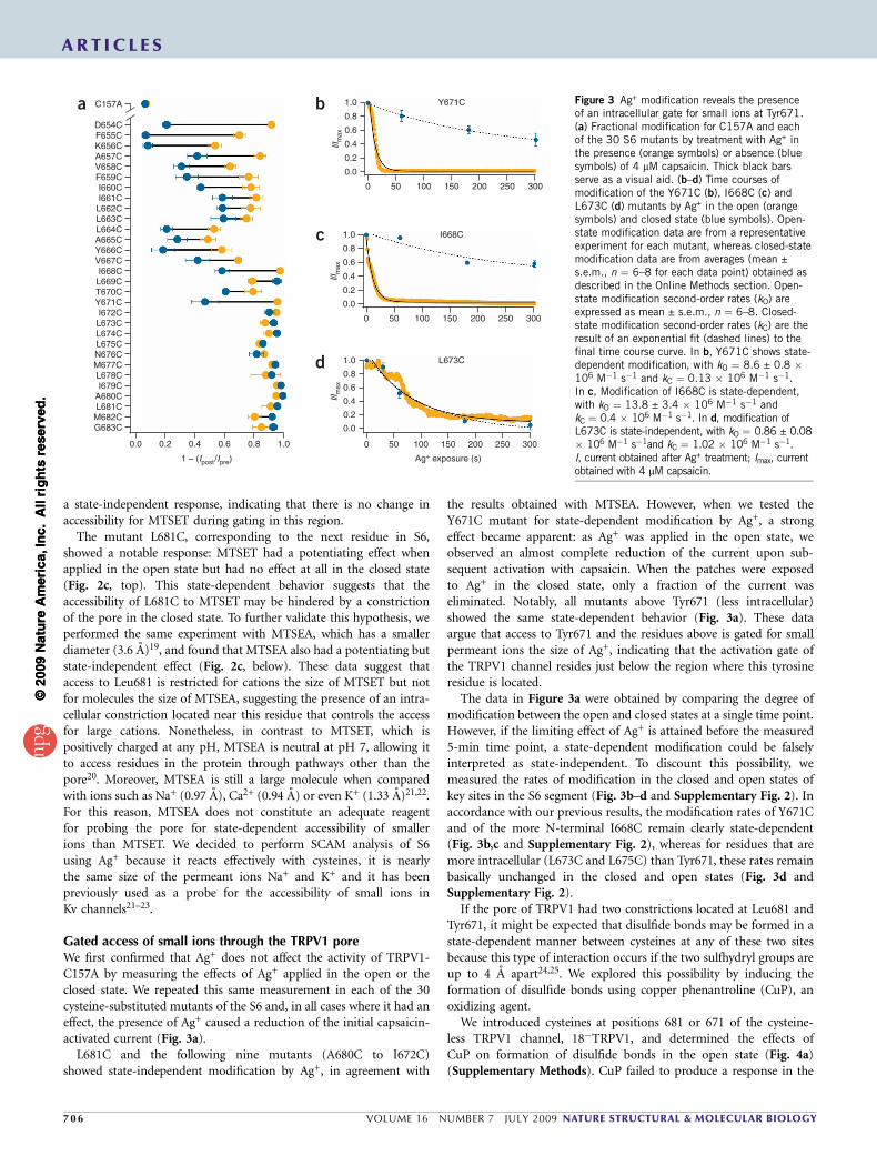
Gated access of small ions through the TRPV1 poreWe first confirmed that Ag+ does not affect the activity of TRPV1-C157A by measuring the effects of Ag+ applied in the open or theclosed state. We repeated this same measurement in each of the 30cysteine-substituted mutants of the S6 and, in all cases where it had aneffect, the presence of Ag+ caused a reduction of the initial capsaicin-activated current (Fig. 3a).
L681C and the following nine mutants (A680C to I672C)showed state-independent modification by Ag+, in agreement with
the results obtained with MTSEA. However, when we tested theY671C mutant for state-dependent modification by Ag+, a strongeffect became apparent: as Ag+ was applied in the open state, weobserved an almost complete reduction of the current upon sub-sequent activation with capsaicin. When the patches were exposedto Ag+ in the closed state, only a fraction of the current waseliminated. Notably, all mutants above Tyr671 (less intracellular)showed the same state-dependent behavior (Fig. 3a). These dataargue that access to Tyr671 and the residues above is gated for smallpermeant ions the size of Ag+, indicating that the activation gate ofthe TRPV1 channel resides just below the region where this tyrosineresidue is located.
The data in Figure 3a were obtained by comparing the degree ofmodification between the open and closed states at a single time point.However, if the limiting effect of Ag+ is attained before the measured5-min time point, a state-dependent modification could be falselyinterpreted as state-independent. To discount this possibility, wemeasured the rates of modification in the closed and open states ofkey sites in the S6 segment (Fig. 3b–d and Supplementary Fig. 2). Inaccordance with our previous results, the modification rates of Y671Cand of the more N-terminal I668C remain clearly state-dependent(Fig. 3b,c and Supplementary Fig. 2), whereas for residues that aremore intracellular (L673C and L675C) than Tyr671, these rates remainbasically unchanged in the closed and open states (Fig. 3d andSupplementary Fig. 2).
If the pore of TRPV1 had two constrictions located at Leu681 andTyr671, it might be expected that disulfide bonds may be formed in astate-dependent manner between cysteines at any of these two sitesbecause this type of interaction occurs if the two sulfhydryl groups areup to 4 A apart24,25. We explored this possibility by inducing theformation of disulfide bonds using copper phenantroline (CuP), anoxidizing agent.
We introduced cysteines at positions 681 or 671 of the cysteine-less TRPV1 channel, 18�TRPV1, and determined the effects ofCuP on formation of disulfide bonds in the open state (Fig. 4a)(Supplementary Methods). CuP failed to produce a response in the
0.0
G683CM682CL681CA680CI679CL678CM677CN676CL675CL674CL673CI672C
Y671CT670CL669CI668C
V667CY666CA665CL664CL663CL662CI661CI660C
F659CV658CA657CK656CF655CD654C
C157A
0.2 0.4
1 – (Ipost/Ipre)
0.6 0.8 1.0
a
0.0
0 50 100 150
Y671C
200 250 300
0.2
0.4I/Im
ax 0.6
0.8
1.0b
0.0
0 50 100 150 200 250 300
0.2
0.4I/Im
ax 0.6
0.8
1.0 I668Cc
0.0
0 50 100 150
Ag+ exposure (s)
200 250 300
0.2
0.4I/Im
ax 0.6
0.8
1.0 L673Cd
Figure 3 Ag+ modification reveals the presenceof an intracellular gate for small ions at Tyr671.(a) Fractional modification for C157A and eachof the 30 S6 mutants by treatment with Ag+ inthe presence (orange symbols) or absence (bluesymbols) of 4 mM capsaicin. Thick black barsserve as a visual aid. (b–d) Time courses ofmodification of the Y671C (b), I668C (c) andL673C (d) mutants by Ag+ in the open (orangesymbols) and closed state (blue symbols). Open-state modification data are from a representativeexperiment for each mutant, whereas closed-statemodification data are from averages (mean ±s.e.m., n ¼ 6–8 for each data point) obtained asdescribed in the Online Methods section. Open-state modification second-order rates (kO) areexpressed as mean ± s.e.m., n ¼ 6–8. Closed-state modification second-order rates (kC) are theresult of an exponential fit (dashed lines) to thefinal time course curve. In b, Y671C shows state-dependent modification, with kO ¼ 8.6 ± 0.8 �106 M�1 s�1 and kC ¼ 0.13 � 106 M�1 s�1.In c, Modification of I668C is state-dependent,with kO ¼ 13.8 ± 3.4 � 106 M�1 s�1 andkC ¼ 0.4 � 106 M�1 s�1. In d, modification ofL673C is state-independent, with kO ¼ 0.86 ± 0.08� 106 M�1 s�1and kC ¼ 1.02 � 106 M�1 s�1.I, current obtained after Ag+ treatment; Imax, currentobtained with 4 mM capsaicin.
ART IC L E S
70 6 VOLUME 16 NUMBER 7 JULY 2009 NATURE STRUCTURAL & MOLECULAR BIOLOGY
©20
09 N
atu
re A
mer
ica,
Inc.
All
rig
hts
res
erve
d.
©20
09 N
atu
re A
mer
ica,
Inc.
All
rig
hts
res
erve
d.
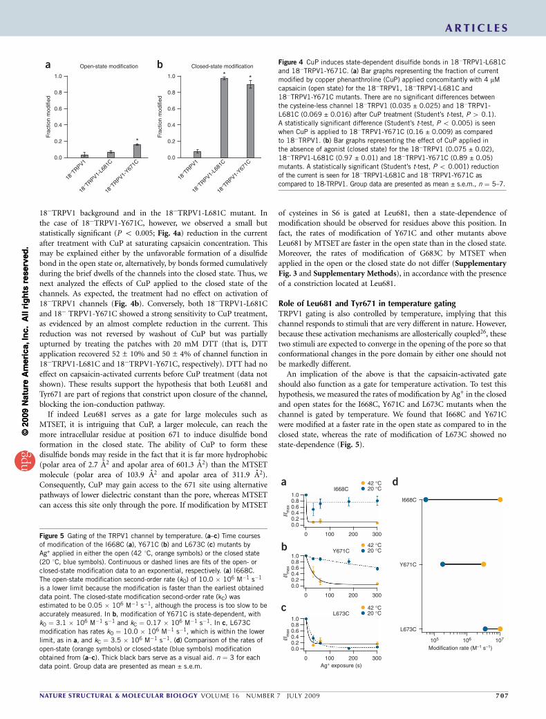
18�TRPV1 background and in the 18�TRPV1-L681C mutant. Inthe case of 18�TRPV1-Y671C, however, we observed a small butstatistically significant (P o 0.005; Fig. 4a) reduction in the currentafter treatment with CuP at saturating capsaicin concentration. Thismay be explained either by the unfavorable formation of a disulfidebond in the open state or, alternatively, by bonds formed cumulativelyduring the brief dwells of the channels into the closed state. Thus, wenext analyzed the effects of CuP applied to the closed state of thechannels. As expected, the treatment had no effect on activation of18�TRPV1 channels (Fig. 4b). Conversely, both 18�TRPV1-L681Cand 18� TRPV1-Y671C showed a strong sensitivity to CuP treatment,as evidenced by an almost complete reduction in the current. Thisreduction was not reversed by washout of CuP but was partiallyupturned by treating the patches with 20 mM DTT (that is, DTTapplication recovered 52 ± 10% and 50 ± 4% of channel function in18�TRPV1-L681C and 18�TRPV1-Y671C, respectively). DTT had noeffect on capsaicin-activated currents before CuP treatment (data notshown). These results support the hypothesis that both Leu681 andTyr671 are part of regions that constrict upon closure of the channel,blocking the ion-conduction pathway.
If indeed Leu681 serves as a gate for large molecules such asMTSET, it is intriguing that CuP, a larger molecule, can reach themore intracellular residue at position 671 to induce disulfide bondformation in the closed state. The ability of CuP to form thesedisulfide bonds may reside in the fact that it is far more hydrophobic(polar area of 2.7 A2 and apolar area of 601.3 A2) than the MTSETmolecule (polar area of 103.9 A2 and apolar area of 311.9 A2).Consequently, CuP may gain access to the 671 site using alternativepathways of lower dielectric constant than the pore, whereas MTSETcan access this site only through the pore. If modification by MTSET
of cysteines in S6 is gated at Leu681, then a state-dependence ofmodification should be observed for residues above this position. Infact, the rates of modification of Y671C and other mutants aboveLeu681 by MTSET are faster in the open state than in the closed state.Moreover, the rates of modification of G683C by MTSET whenapplied in the open or the closed state do not differ (SupplementaryFig. 3 and Supplementary Methods), in accordance with the presenceof a constriction located at Leu681.
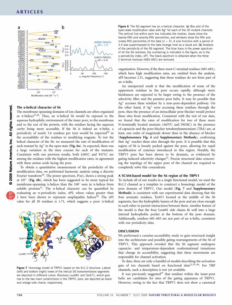
Role of Leu681 and Tyr671 in temperature gatingTRPV1 gating is also controlled by temperature, implying that thischannel responds to stimuli that are very different in nature. However,because these activation mechanisms are allosterically coupled26, thesetwo stimuli are expected to converge in the opening of the pore so thatconformational changes in the pore domain by either one should notbe markedly different.
An implication of the above is that the capsaicin-activated gateshould also function as a gate for temperature activation. To test thishypothesis, we measured the rates of modification by Ag+ in the closedand open states for the I668C, Y671C and L673C mutants when thechannel is gated by temperature. We found that I668C and Y671Cwere modified at a faster rate in the open state as compared to in theclosed state, whereas the rate of modification of L673C showed nostate-dependence (Fig. 5).
1.0
0.8
0.6
0.4
Frac
tion
mod
ified
0.2
0.0
18– TRPV1
18– TRPV1-
L681
C
Open-state modification
18– TRPV1-
Y671C
*
1.0
0.8
0.6
0.4
Frac
tion
mod
ified
0.2
0.0
18– TRPV1
18– TRPV1-
L681
C
Closed-state modification
* *
18– TRPV1-
Y671C
a b Figure 4 CuP induces state-dependent disulfide bonds in 18�TRPV1-L681C
and 18�TRPV1-Y671C. (a) Bar graphs representing the fraction of current
modified by copper phenanthroline (CuP) applied concomitantly with 4 mM
capsaicin (open state) for the 18�TRPV1, 18�TRPV1-L681C and
18�TRPV1-Y671C mutants. There are no significant differences between
the cysteine-less channel 18�TRPV1 (0.035 ± 0.025) and 18�TRPV1-
L681C (0.069 ± 0.016) after CuP treatment (Student’s t-test, P 4 0.1).
A statistically significant difference (Student’s t-test, P o 0.005) is seen
when CuP is applied to 18�TRPV1-Y671C (0.16 ± 0.009) as compared
to 18�TRPV1. (b) Bar graphs representing the effect of CuP applied in
the absence of agonist (closed state) for the 18�TRPV1 (0.075 ± 0.02),
18�TRPV1-L681C (0.97 ± 0.01) and 18�TRPV1-Y671C (0.89 ± 0.05)
mutants. A statistically significant (Student’s t-test, P o 0.001) reduction
of the current is seen for 18�TRPV1-L681C and 18�TRPV1-Y671C as
compared to 18-TRPV1. Group data are presented as mean ± s.e.m., n ¼ 5–7.
L673C
105 106
Modification rate (M–1 s–1)107
Y671C
I668C
d1.00.80.60.4I/I
max
0.20.0
0 100 200 300
1.00.80.60.4I/I
max
0.20.0
0 100Ag+ exposure (s)
L673C
I668C
200 300
42 °C20 °C
42 °C20 °C
a
1.00.80.60.4I/I
max
0.20.0
0 100 200 300
Y671C42 °C20 °Cb
c
Figure 5 Gating of the TRPV1 channel by temperature. (a–c) Time courses
of modification of the I668C (a), Y671C (b) and L673C (c) mutants by
Ag+ applied in either the open (42 1C, orange symbols) or the closed state
(20 1C, blue symbols). Continuous or dashed lines are fits of the open- or
closed-state modification data to an exponential, respectively. (a) I668C.
The open-state modification second-order rate (kO) of 10.0 � 106 M�1 s�1
is a lower limit because the modification is faster than the earliest obtained
data point. The closed-state modification second-order rate (kC) wasestimated to be 0.05 � 106 M�1 s�1, although the process is too slow to be
accurately measured. In b, modification of Y671C is state-dependent, with
kO ¼ 3.1 � 106 M�1 s�1 and kC ¼ 0.17 � 106 M�1 s�1. In c, L673C
modification has rates kO ¼ 10.0 � 106 M�1 s�1, which is within the lower
limit, as in a, and kC ¼ 3.5 � 106 M�1 s�1. (d) Comparison of the rates of
open-state (orange symbols) or closed-state (blue symbols) modification
obtained from (a–c). Thick black bars serve as a visual aid. n ¼ 3 for each
data point. Group data are presented as mean ± s.e.m.
ART IC L E S
NATURE STRUCTURAL & MOLECULAR BIOLOGY VOLUME 16 NUMBER 7 JULY 2009 7 0 7
©20
09 N
atu
re A
mer
ica,
Inc.
All
rig
hts
res
erve
d.
©20
09 N
atu
re A
mer
ica,
Inc.
All
rig
hts
res
erve
d.
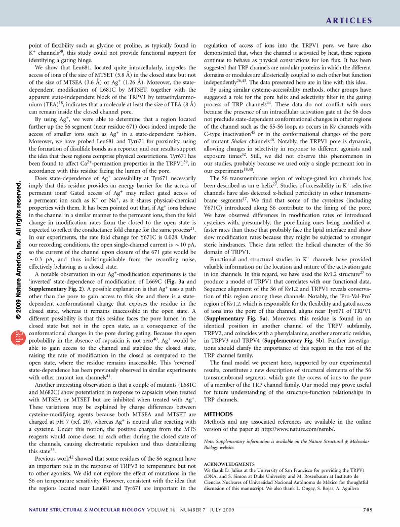
The a-helical character of S6The membrane-spanning domains of ion channels are often organizedas a-helices27,28. Thus, an a-helical S6 would be exposed to theaqueous hydrophilic environment of the inner pore, to the membraneand to the rest of the protein, with the residues facing the aqueouscavity being more accessible. If the S6 is indeed an a-helix, aperiodicity of nearly 3.6 residues per turn would be expected29 inthe accessibility of the residues to modifying reagents. To test thehelical character of the S6, we measured the rate of modification ofeach mutant by Ag+ in the open state (Fig. 6a). As expected, there wasa large variation in the time courses for each of the mutants.Consistent with our previous results, both L681C and Y671C areamong the residues with the highest modification rates, in agreementwith these amino acids facing the pore.
To obtain a quantitative measurement of the periodicity of themodification data, we performed harmonic analysis using a discreteFourier transform30. The power spectrum, P(j), shows a strong peakat 1071 (Fig. 6b), which has been suggested to be more typical formembrane-spanning a-helices than the 1001 seen in a-helices fromsoluble proteins31. The a-helical character can be quantified byevaluating an a-periodicity index, aPI, where values greater than2 have been shown to represent amphipathic helices30. The aPIvalue for all 30 residues is 1.71, which suggests a poor a-helical
organization. However, if the three most C-terminal residues (683–681),which have high modification rates, are omitted from the analysis,aPI becomes 2.31, suggesting that those residues do not form part ofan a-helix.
An unexpected result is that the modification of some of theuppermost residues in the pore occurs rapidly, although sterichindrances are expected to be larger owing to the presence of theselectivity filter and the putative pore helix. It may be possible thatAg+ accesses these residues by a non–pore-dependent pathway. Onthe other hand, if Ag+ were accessing these residues through thepore, then the presence of an intracellular pore blocker would protectthese sites from modification. Consistent with the rest of our data,we found that the rates of modification for two of these moreN-terminally located mutants (A657C and D654C) in the presenceof capsaicin and the pore-blocker tetrabutylammonium (TBA) are, atleast, one order of magnitude slower than in the absence of blocker(Supplementary Fig. 4 and Supplementary Methods), confirmingthat Ag+ reaches these sites through the pore. It is possible that thisregion of S6 is loosely packed against the pore, allowing for rapidmodification of cysteines introduced in this region. Notably, theTRPV1 pore has been shown to be dynamic, as evidenced bygating-induced selectivity changes32. Precise structural data concern-ing the topology of the upper pore of the channel are required tocompletely solve this conundrum.
A SCAM-based model for the S6 region of the TRPV1To include all of our results in a single functional model, we used theKv1.2 channel as a template to construct a homology model of thepore domain of TRPV1. Our model (Fig. 7 and SupplementaryMethods) is consistent with our experimental data showing that thefour aromatic residues, Tyr671 located at the middle of the S6segments, face the hydrophilic lumen of the pore and are close enoughto each other to permit interactions between them. Another feature ofthe model is that the four Leu681 side chains fit well into a largeinternal hydrophobic pocket at the bottom of the pore domain.Additionally, residues 681–683 are not part of an a-helix, consistentwith our periodicity data.
DISCUSSIONWe performed a cysteine-accessibility study to gain structural insightinto the architecture and possible gating rearrangements of the S6 ofTRPV1. This approach revealed that the S6 segment undergoescapsaicin- and temperature-dependent conformational transitionsthat change its accessibility, suggesting that these movements areresponsible for channel activation.
To date, there are only a handful of models describing the activationgate of ion channels based on functional data21,33–36. For TRPchannels, such a description is not yet available.
It was previously suggested37 that residues within the inner-porehelix are candidates for sites of the gating apparatus of TRPV1.However, owing to the fact that TRPV1 does not show a canonical
G683C
106
Modification rate (M–1 s–1)107
M682CL681CA680CI679CL678CM677CN676CL675CL674CL673CI672C
Y671CT670CL669CI668C
V667CY666CA665CL664CL663CL662CI661CI660C
F659CV658CA657CK656CF655CD654Ca b
0.50.40.3
P(�)
P(�)
0.2
0.00.1
0.6
0.00.20.4
0 50ϕ, angular frequency (°)
(L681C-D654C);αPI = 2.31
(G683C-D654C);αPI = 1.71
100 150
Figure 6 The S6 segment has an a-helical character. (a) Box plot of the
open-state modification rates with Ag+ for each of the 30 mutant channels.
The vertical line within each box indicates the median, boxes show the
twenty-fifth and seventy-fifth percentiles, and whiskers show the fifth and
ninety-fifth percentiles of the data (n ¼ 5). A sine function with a period of
3.4 was superimposed to the data (orange line) as a visual aid. (b) Analysis
of the periodicity of the S6 segment. The blue trace is the power spectrum
of all the S6 residues; the numbering is indicated in the figure, as is the
a-periodicity index, aPI. The black spectrum is obtained when the three
C-terminal residues (683–681) are removed.
Leu681
Tyr671
90°
Figure 7 Homology model of TRPV1 based on the Kv1.2 structure. Lateral
(left) and bottom (right) views of the helical S6 transmembrane segments
are depicted in different colors. Residues Leu681 and Tyr671, which give
rise to the two main constrictions in the TRPV1 pore, are depicted as black
and orange side chains, respectively.
ART IC L E S
70 8 VOLUME 16 NUMBER 7 JULY 2009 NATURE STRUCTURAL & MOLECULAR BIOLOGY
©20
09 N
atu
re A
mer
ica,
Inc.
All
rig
hts
res
erve
d.
©20
09 N
atu
re A
mer
ica,
Inc.
All
rig
hts
res
erve
d.
point of flexibility such as glycine or proline, as typically found inK+ channels38, this study could not provide functional support foridentifying a gating hinge.
We show that Leu681, located quite intracellularly, impedes theaccess of ions of the size of MTSET (5.8 A) in the closed state but notof the size of MTSEA (3.6 A) or Ag+ (1.26 A). Moreover, the state-dependent modification of L681C by MTSET, together with theapparent state-independent block of the TRPV1 by tetraethylammo-nium (TEA)18, indicates that a molecule at least the size of TEA (8 A)can remain inside the closed channel pore.
By using Ag+, we were able to determine that a region locatedfurther up the S6 segment (near residue 671) does indeed impede theaccess of smaller ions such as Ag+ in a state-dependent fashion.Moreover, we have probed Leu681 and Tyr671 for proximity, usingthe formation of disulfide bonds as a reporter, and our results supportthe idea that these regions comprise physical constrictions. Tyr671 hasbeen found to affect Ca2+-permeation properties in the TRPV139, inaccordance with this residue facing the lumen of the pore.
Does state-dependence of Ag+ accessibility at Tyr671 necessarilyimply that this residue provides an energy barrier for the access ofpermeant ions? Gated access of Ag+ may reflect gated access ofa permeant ion such as K+ or Na+, as it shares physical-chemicalproperties with them. It has been pointed out that, if Ag+ ions behavein the channel in a similar manner to the permeant ions, then the foldchange in modification rates from the closed to the open state isexpected to reflect the conductance fold change for the same process21.In our experiments, the rate fold change for Y671C is 0.028. Underour recording conditions, the open single-channel current is B10 pA,so the current of the channel upon closure of the 671 gate would beB0.3 pA, and thus indistinguishable from the recording noise,effectively behaving as a closed state.
A notable observation in our Ag+-modification experiments is the‘inverted’ state-dependence of modification of L669C (Fig. 3a andSupplementary Fig. 2). A possible explanation is that Ag+ uses a pathother than the pore to gain access to this site and there is a state-dependent conformational change that exposes the residue in theclosed state, whereas it remains inaccessible in the open state. Adifferent possibility is that this residue faces the pore lumen in theclosed state but not in the open state, as a consequence of theconformational changes in the pore during gating. Because the openprobability in the absence of capsaicin is not zero40, Ag+ would beable to gain access to the channel and stabilize the closed state,raising the rate of modification in the closed as compared to theopen state, where the residue remains inaccessible. This ‘reversed’state-dependence has been previously observed in similar experimentswith other mutant ion channels41.
Another interesting observation is that a couple of mutants (L681Cand M682C) show potentiation in response to capsaicin when treatedwith MTSEA or MTSET but are inhibited when treated with Ag+.These variations may be explained by charge differences betweencysteine-modifying agents because both MTSEA and MTSET arecharged at pH 7 (ref. 20), whereas Ag+ is neutral after reacting witha cysteine. Under this notion, the positive charges from the MTSreagents would come closer to each other during the closed state ofthe channels, causing electrostatic repulsion and thus destabilizingthis state35.
Previous work42 showed that some residues of the S6 segment havean important role in the response of TRPV3 to temperature but notto other agonists. We did not explore the effect of mutations in theS6 on temperature sensitivity. However, consistent with the idea thatthe regions located near Leu681 and Tyr671 are important in the
regulation of access of ions into the TRPV1 pore, we have alsodemonstrated that, when the channel is activated by heat, these regionscontinue to behave as physical constrictions for ion flux. It has beensuggested that TRP channels are modular proteins in which the differentdomains or modules are allosterically coupled to each other but functionindependently26,43. The data presented here are in line with this idea.
By using similar cysteine-accessibility methods, other groups havesuggested a role for the pore helix and selectivity filter in the gatingprocess of TRP channels44. These data do not conflict with oursbecause the presence of an intracellular activation gate at the S6 doesnot preclude state-dependent conformational changes in other regionsof the channel such as the S5-S6 loop, as occurs in Kv channels withC-type inactivation45 or in the conformational changes of the poreof mutant Shaker channels46. Notably, the TRPV1 pore is dynamic,allowing changes in selectivity in response to different agonists andexposure times32. Still, we did not observe this phenomenon inour studies, probably because we used only a single permeant ion inour experiments18,40.
The S6 transmembrane region of voltage-gated ion channels hasbeen described as an a-helix27. Studies of accessibility in K+-selectivechannels have also detected a-helical periodicity in other transmem-brane segments47. We find that some of the cysteines (includingY671C) introduced along S6 contribute to the lining of the pore.We have observed differences in modification rates of introducedcysteines with, presumably, the pore-lining ones being modified atfaster rates than those that probably face the lipid interface and showslow modification rates because they might be subjected to strongersteric hindrances. These data reflect the helical character of the S6domain of TRPV1.
Functional and structural studies in K+ channels have providedvaluable information on the location and nature of the activation gatein ion channels. In this regard, we have used the Kv1.2 structure27 toproduce a model of TRPV1 that correlates with our functional data.Sequence alignment of the S6 of Kv1.2 and TRPV1 reveals conserva-tion of this region among these channels. Notably, the ‘Pro-Val-Pro’region of Kv1.2, which is responsible for the flexibility and gated accessof ions into the pore of this channel, aligns near Tyr671 of TRPV1(Supplementary Fig. 5a). Moreover, this residue is found in anidentical position in another channel of the TRPV subfamily,TRPV2, and coincides with a phenylalanine, another aromatic residue,in TRPV3 and TRPV4 (Supplementary Fig. 5b). Further investiga-tions should clarify the importance of this region in the rest of theTRP channel family.
The final model we present here, supported by our experimentalresults, constitutes a new description of structural elements of the S6transmembranal segment, which gate the access of ions to the poreof a member of the TRP channel family. Our model may prove usefulfor future understanding of the structure-function relationships inTRP channels.
METHODSMethods and any associated references are available in the onlineversion of the paper at http://www.nature.com/nsmb/.
Note: Supplementary information is available on the Nature Structural & MolecularBiology website.
ACKNOWLEDGMENTSWe thank D. Julius at the University of San Francisco for providing the TRPV1cDNA, and S. Simon at Duke University and M. Rosenbaum at Instituto deCiencias Nucleares of Universidad Nacional Autonoma de Mexico for thoughtfuldiscussion of this manuscript. We also thank L. Ongay, S. Rojas, A. Aguilera
ART IC L E S
NATURE STRUCTURAL & MOLECULAR BIOLOGY VOLUME 16 NUMBER 7 JULY 2009 7 0 9
©20
09 N
atu
re A
mer
ica,
Inc.
All
rig
hts
res
erve
d.
©20
09 N
atu
re A
mer
ica,
Inc.
All
rig
hts
res
erve
d.

Jimenez, J. Barbosa, A.M. Escalante and F. Sierra at Instituto de FisiologıaCelular, Universidad Nacional Autonoma de Mexico, for expert technicalsupport. This work was supported by grants from La Direccion General deAsuntos del Personal Academico (DGAPA)-Universidad Nacional Autonoma deMexico (UNAM) IN200308-3 to T.R. and IN202006–3 to L.D.I., and CONACyTNo. 58038 to T.R. and No. 48990 to L.D.I. This study was performed in partialfulfillment of the requirements for the Doctorate degree in Biomedical Sciencesof H.S. at the UNAM.
AUTHOR CONTRIBUTIONSH.S., A.J.-O., E.H.-G. and T.R. performed experiments; I.L. did molecular biologyand cell culture; M.S.-G. and I.I.A.-O. performed molecular modeling; L.D.I. andT.R. designed experiments, analyzed data and wrote the paper; all authorsdiscussed the data.
Published online at http://www.nature.com/nsmb/
Reprints and permissions information is available online at http://npg.nature.com/
reprintsandpermissions/
1. Clapham, D.E., Runnels, L.W. & Strubing, C. The TRP ion channel family. Nat. Rev.Neurosci. 2, 387–396 (2001).
2. Garcıa-Martınez, C., Morenilla-Palao, C., Planells-Cases, R., Merino, J.M. & Ferrer-Montiel, A. Identification of an aspartic residue in the P-loop of the vanilloid receptorthat modulates pore properties. J. Biol. Chem. 275, 32552–32558 (2000).
3. Montell, C. The TRP superfamily of cation channels. Sci. STKE 2005, re3 (2005).4. Salazar, H. et al. A single N-terminal cysteine in TRPV1 determines activation by
pungent compounds from onion and garlic. Nat. Neurosci. 11, 255–261 (2008).5. Macpherson, L.J. et al. The pungency of garlic: activation of TRPA1 and TRPV1 in
response to allicin. Curr. Biol. 15, 929–934 (2005).6. Clapham, D.E. TRP channels as cellular sensors. Nature 426, 517–524 (2003).7. Montell, C. Thermosensation, hot findings make TRPNs very cool. Curr. Biol. 13,
476–478 (2003).8. Caterina, M.J. & Julius, D. The vanilloid receptor: a molecular gateway to the pain
pathway. Annu. Rev. Neurosci. 24, 487–517 (2001).9. Julius, D. & Basbaum, A.I. Molecular mechanisms of nociception. Nature 413,
203–210 (2001).10. Huang, J., Zhang, X. & McNaughton, P.A. Inflammatory pain: the cellular basis of heat
hyperalgesia. Curr. Neuropharmacol. 4, 197–206 (2006).11. Jara-Oseguera, A.S., S.A. & Rosenbaum, T. TRPV1: on the road to pain relief. Curr.
Mol. Pharmacol. 1, 255–269 (2008).12. Szallasi, A., Cortright, D.N., Blum, C.A. & Eid, S.R. The vanilloid receptor TRPV1: 10
years from channel cloning to antagonist proof-of-concept. Nat. Rev. Drug Discov. 6,357–372 (2007).
13. Gaudet, R. A primer on ankyrin repeat function in TRP channels and beyond. Mol.Biosyst. 4, 372–379 (2008).
14. Lishko, P.V., Procko, E., Jin, X., Phelps, C.B. & Gaudet, R. The ankyrin repeats of TRPV1bind multiple ligands and modulate channel sensitivity. Neuron 54, 905–918 (2007).
15. McCleverty, C.J., Koesema, E., Patapoutian, A., Lesley, S.A. & Kreusch, A. Crystalstructure of the human TRPV2 channel ankyrin repeat domain. Protein Sci. 15,2201–2206 (2006).
16. Jin, X., Touhey, J. & Gaudet, R. Structure of the N-terminal ankyrin repeat domain ofthe TRPV2 ion channel. J. Biol. Chem. 281, 25006–25010 (2006).
17. Moiseenkova-Bell, V.Y., Stanciu, L.A., Serysheva, I.I., Tobe, B.J. & Wensel, T.G.Structure of TRPV1 channel revealed by electron cryomicroscopy. Proc. Natl. Acad.Sci. USA 105, 7451–7455 (2008).
18. Jara-Oseguera, A., Llorente, I., Rosenbaum, T. & Islas, L.D. Properties of the inner poreregion of TRPV1 channels revealed by block with quaternary ammoniums. J. Gen.Physiol. 132, 547–562 (2008).
19. Dodier, Y. et al. Outer pore topology of the ECaC-TRPV5 channel by cysteine scanmutagenesis. J. Biol. Chem. 279, 6853–6862 (2004).
20. Holmgren, M., Liu, Y., Xu, Y. & Yellen, G. On the use of thiol-modifying agents todetermine channel topology. Neuropharmacology 35, 797–804 (1996).
21. del Camino, D. & Yellen, G. Tight steric closure at the intracellular activation gate of avoltage-gated K+ channel. Neuron 32, 649–656 (2001).
22. Greenwood, A. & Earnshaw, N. Chemistry of the Elements 2nd edn. 1341 (Butter-worth-Heinemann, Oxford, 1997).
23. Lu, Q. & Miller, C. Silver as a probe of pore-forming residues in a potassium channel.Science 268, 304–307 (1995).
24. Careaga, C.L. & Falke, J.J. Thermal motions of surface a-helices in the D-galactosechemosensory receptor. Detection by disulfide trapping. J. Mol. Biol. 226,1219–1235 (1992).
25. Kobashi, K. Catalytic oxidation of sulfhydryl groups by o-phenanthroline coppercomplex. Biochim. Biophys. Acta 158, 239–245 (1968).
26. Latorre, R., Brauchi, S., Orta, G., Zaelzer, C. & Vargas, G. ThermoTRPchannels as modular proteins with allosteric gating. Cell Calcium 42, 427–438(2007).
27. Long, S.B., Campbell, E.B. & Mackinnon, R. Crystal structure of a mammalianvoltage-dependent Shaker family K+ channel. Science 309, 897–903 (2005).
28. Doyle, D.A. et al. The structure of the potassium channel: molecular basis of K+
conduction and selectivity. Science 280, 69–77 (1998).29. Eisenberg, D. The discovery of the a-helix and b-sheet, the principal structural features
of proteins. Proc. Natl. Acad. Sci. USA 100, 11207–11210 (2003).30. Cornette, J.L. et al. Hydrophobicity scales and computational techniques for detecting
amphipathic structures in proteins. J. Mol. Biol. 195, 659–685 (1987).31. Komiya, H., Yeates, T.O., Rees, D.C., Allen, J.P. & Feher, G. Structure of the reaction
center from Rhodobacter sphaeroides R-26 and 2.4.1: symmetry relations andsequence comparisons between different species. Proc. Natl. Acad. Sci. USA 85,9012–9016 (1988).
32. Chung, M.K. & Caterina, M.J. TRP channel knockout mice lose their cool. Neuron 54,345–347 (2007).
33. Ding, S., Ingleby, L., Ahern, C.A. & Horn, R. Investigating the putative glycine hinge inShaker potassium channel. J. Gen. Physiol. 126, 213–226 (2005).
34. Contreras, J.E., Srikumar, D. & Holmgren, M. Gating at the selectivity filter incyclic nucleotide-gated channels. Proc. Natl. Acad. Sci. USA 105, 3310–3314(2008).
35. Flynn, G.E. & Zagotta, W.N. A cysteine scan of the inner vestibule of cyclic nucleotide-gated channels reveals architecture and rearrangement of the pore. J. Gen. Physiol.121, 563–582 (2003).
36. Li, M., Chang, T.H., Silberberg, S.D. & Swartz, K.J. Gating the pore of P2X receptorchannels. Nat. Neurosci. 11, 883–887 (2008).
37. Susankova, K., Ettrich, R., Vyklicky, L., Teisinger, J. & Vlachova, V. Contribution of theputative inner-pore region to the gating of the transient receptor potential vanilloidsubtype 1 channel (TRPV1). J. Neurosci. 27, 7578–7585 (2007).
38. Magidovich, E. & Yifrach, O. Conserved gating hinge in ligand- and voltage-dependentK+ channels. Biochemistry 43, 13242–13247 (2004).
39. Mohapatra, D.P., Wang, S.Y., Wang, G.K. & Nau, C. A tyrosine residue in TM6 of thevanilloid receptor TRPV1 involved in desensitization and calcium permeability ofcapsaicin-activated currents. Mol. Cell. Neurosci. 23, 314–324 (2003).
40. Oseguera, A.J., Islas, L.D., Garcia-Villegas, R. & Rosenbaum, T. On the mechanism ofTBA block of the TRPV1 channel. Biophys. J. 92, 3901–3914 (2007).
41. Liu, Y., Holmgren, M., Jurman, M.E. & Yellen, G. Gated access to the pore of a voltage-dependent K+ channel. Neuron 19, 175–184 (1997).
42. Grandl, J. et al. Pore region of TRPV3 ion channel is specifically required for heatactivation. Nat. Neurosci. 11, 1007–1013 (2008).
43. Brauchi, S., Orio, P. & Latorre, R. Clues to understanding cold sensation: thermo-dynamics and electrophysiological analysis of the cold receptor TRPM8. Proc. Natl.Acad. Sci. USA 101, 15494–15499 (2004).
44. Yeh, B.I., Kim, Y.K., Jabbar, W. & Huang, C.L. Conformational changes of pore helixcoupled to gating of TRPV5 by protons. EMBO J. 24, 3224–3234 (2005).
45. Yellen, G., Sodickson, D., Chen, T.Y. & Jurman, M.E. An engineered cysteine in theexternal mouth of a K+ channel allows inactivation to be modulated by metal binding.Biophys. J. 66, 1068–1075 (1994).
46. Zheng, J. & Sigworth, F.J. Selectivity changes during activation of mutant Shakerpotassium channels. J. Gen. Physiol. 110, 101–117 (1997).
47. Cortes, D.M., Cuello, L.G. & Perozo, E. Molecular architecture of full-length KcsA: roleof cytoplasmic domains in ion permeation and activation gating. J. Gen. Physiol. 117,165–180 (2001).
ART IC L E S
71 0 VOLUME 16 NUMBER 7 JULY 2009 NATURE STRUCTURAL & MOLECULAR BIOLOGY
©20
09 N
atu
re A
mer
ica,
Inc.
All
rig
hts
res
erve
d.
©20
09 N
atu
re A
mer
ica,
Inc.
All
rig
hts
res
erve
d.

ONLINE METHODSMolecular biology. We defined a putative S6 region for the rat TRPV1 channel
from residues Gly683 to Asp654, based on previous37,48 and our own amino
acid alignments. We individually substituted each of the 30 S6 residues for a
cysteine in the rTRPV1-C157A background4, unless otherwise stated in the
text, and carried out mutagenesis according to described PCR-based methods49.
Electrophysiology and channel expression. We transfected HEK293 cells
expressing large T antigen with wild-type or mutant rTRPV1-pCDNA3 and
pIRES-GFP (BD Biosciences) with lipofectamine (Invitrogen) as described40.
We carried out electrophysiological recordings using an EPC10 amplifier
(HEKA Elektronik GMBH) and acquired and analyzed data with PULSE
software (HEKA Elektronik) and IgorPro (Wavemetrics), respectively. Record-
ings were performed in the inside-out configuration of the patch-clamp
technique at 20 1C. Symmetrical solutions consisted of 130 mM NaCl, 3 mM
HEPES and 1 mM EDTA (pH 7.2 with NaOH) for recording except when Ag+
was applied. We obtained all chemicals from Sigma unless otherwise stated.
Solutions were changed with an RSC-200 rapid solution changer (Molecular
Kinetics). Patch pipettes had a resistance of 2–4 MO. Currents were low-pass
filtered at 2 kHz and sampled at 10 kHz.
We prepared capsaicin stock solutions (4 mM) in ethanol and diluted them
to the desired concentration in recording solution. MTSET-chloride and
MTSEA-chloride (Toronto Research Chemicals) stock solutions (100 mM)
were prepared in water and immediately frozen at �70 1C. We diluted aliquots
in recording solution to a final concentration of 2 mM before each experiment
with either a saturating concentration of capsaicin (4 mM) for open-state
modification or in the absence of capsaicin for closed-state modification.
We measured dose-response curves at 120 mV as the steady-state current.
This was normalized to the response to 4 mM capsaicin. Capsaicin was washed
off and the MTS reagents applied either in the presence or absence of capsaicin
for 5 min and the dose-response was remeasured. We obtained the Kd from fits
to the Hill equation:
I
Imax¼ Capsaicin½ �n
Capsaicin½ �n +Knd
where n is the Hill coefficient and [Capsaicin] is the agonist concentration.
Leak subtraction was performed by subtracting the initial current trace in the
absence of capsaicin from the traces obtained in the presence of the agonist.
For experiments using Ag+, we followed the method described34,35. We
prepared a stock of AgNO3 (10 mM) in water before each experiment,
protected it from light and diluted it in an intracellular solution consisting
of 130 mM NaNO3, 3 mM HEPES and 1 mM EDTA (pH 7.2 with NaOH).
Using the MaxChelator software (http://maxchelator.stanford.edu), we calcu-
lated the free concentration of Ag+ at 1 mM EDTA to be 100 nM. We measured
the initial current in the absence of capsaicin at 120 mV, measured the current
with 4 mM capsaicin and then either applied Ag+ for 5 min in the presence of
the agonist for open-state modification, or we washed the capsaicin off and
then applied Ag+ for 5 min in the closed state. Finally, we assessed the effects of
modification using 4 mM capsaicin to activate the remaining currents. The
fraction of current modified, Fm, was calculated from:
Fm ¼ 1 � Ipost
Ipre;
where Ipost is the current after Ag+ treatment and Ipre is the current before
treatment.
We measured modification rates in the open state by applying a train of
100-ms pulses at 120 mV from a holding potential of 0 mV and a frequency of
1 Hz in the presence of Ag+ and 4 mM capsaicin, and we constructed time
courses of modification in the closed state by measuring data points at a
single exposure time for each patch. Several patches were averaged to obtain the
whole curve. We calculated the second-order modification rate constants as the
inverse of the product of the time constant obtained from fits of an exponential
to the data and the modifier concentration.
For heat-activation experiments, we used a temperature-controlling recording
chamber (Bipolar Temperature Controller TC-202, Medical Systems Corp). We
used a thermistor (Warner Instruments) to monitor bath temperature and
carried out the experiments by measuring initial currents at 42 1C. For open-
state modification experiments, we applied a solution containing Ag+ to the
membrane patch at 42 1C for 0.5 min, 1 min, 3 min or 5 min. We washed out
Ag+ and remeasured current at 42 1C. For closed-state modification, the temper-
ature was lowered to 20 1C and Ag+ was then applied for 0.5 min, 1 min, 3 min
or 5 min. Then, we remeasured the current at 42 1C to evaluate the degree of
modification in the closed state and constructed time courses of modification by
plotting the data at different exposure times, as mentioned above.
Periodicity analysis. We evaluated periodicity of the S6 segment using the rate
of Ag+ modification of cysteines as an index, and we carried out the analysis
using discrete Fourier transform methods30,31,50 as follows. If rk is the rate of
modification of the k-th residue and r is the mean value of r, the power
spectrum P(j) for all the n – 1 residues is given by:
AðjÞ ¼Xn�1
k¼0
ðrk � rÞcosðk2pj=360Þ
BðjÞ ¼Xn�1
k¼0
ðrk � rÞsinðk2pj=360Þ
PðjÞ ¼ AðjÞ½ �2 + BðjÞ½ �2
Here j is the angle, in degrees, subtended between each pair of amino acids.
For a typical a-helix, the value of P(j) is expected to be maximal at 1001,
which corresponds to 3.6-amino-acids per turn of the a-helix. An index of the
a-helical character is the a-periodic index (aPI), which is defined as30:
aPI ¼1
30�� � R 120�
90� PðjÞdj1
180�� � R 180�
0� PðjÞdj
A value greater than 2 is typical of a segment with a high likelihood of being an
amphipathic a-helix30.
Statistical analysis. Groups of data are presented as mean ± s.e.m. We
performed statistical comparisons with the Student’s t-test. A value of P o0.05 was considered statistically significant.
48. Brauchi, S. et al. Dissection of the components for PIP2 activation and thermosensa-tion in TRP channels. Proc. Natl. Acad. Sci. USA 104, 10246–10251 (2007).
49. Rosenbaum, T. & Gordon, S.E. Dissecting intersubunit contacts in cyclic nucleotide-gated ion channels. Neuron 33, 703–713 (2002).
50. Li-Smerin, Y. & Swartz, K.J. Helical structure of the COOH terminus of S3 and itscontribution to the gating modifier toxin receptor in voltage-gated ion channels. J. Gen.Physiol. 117, 205–218 (2001).
NATURE STRUCTURAL & MOLECULAR BIOLOGY doi:10.1038/nsmb.1633
©20
09 N
atu
re A
mer
ica,
Inc.
All
rig
hts
res
erve
d.
©20
09 N
atu
re A
mer
ica,
Inc.
All
rig
hts
res
erve
d.