Direct Esthetic Composite Restorations in Anterior Teeth - MDPI

Anterior vaginal wall length and degree of anterior compartmentprolapse seen on dynamic MRI
Yvonne Hsu,Department of Obstetrics and Gynecology, L4000 Women's Hospital, 1500 E Medical Center Drive,Ann Arbor, MI 48109−0276, USA e-mail: [email protected]
Luyun Chen,Department of Biomedical Engineering, L4000 Women's Hospital, 1500 E Medical Center Drive,Ann Arbor, MI, USA
Aimee Summers,Department of Obstetrics and Gynecology, L4000 Women's Hospital, 1500 E Medical Center Drive,Ann Arbor, MI 48109−0276, USA
James A. Ashton-Miller, andDepartment of Biomedical Engineering, L4000 Women's Hospital, 1500 E Medical Center Drive,Ann Arbor, MI, USA
James O. L. DeLanceyDepartment of Obstetrics and Gynecology, L4000 Women's Hospital, 1500 E Medical Center Drive,Ann Arbor, MI 48109−0276, USA
AbstractThe objective of the study was to determine the relationship between midsagittal vaginal wallgeometric parameters and the degree of anterior vaginal prolapse. We have previously presented dataindicating that about half of anterior wall descent can be explained by the degree of apical descentpresent (Summers et al., Am J Obstet Gynecol, 194:1438−1443, 2006). This led us to examinewhether other midsagittal vaginal geometric parameters are associated with anterior wall descent.Magnetic resonance (MR) scans of 145 women from the prior study were suitable for analysis aftereight were excluded because of inadequate visibility of the anterior vaginal wall. Subjects had beenselected from a study of pelvic organ prolapse that included women with and without prolapse. Allpatients underwent supine dynamic MR scans in the midsagittal plane. Anterior vaginal wall length,location of distal vaginal wall point, and the area under the midsagittal profile of the anterior vaginalwall were measured during maximal Valsalva. A linear regression model was used to examine howmuch of the variance in cystocele size could be explained by these vaginal parameters. When bothapical descent and vaginal length were considered in the linear regression model, 77% (R2=0.77,p<0.001) of the variation in anterior wall descent was explained. Distal vaginal point and a measureanterior wall shape, the area under the profile of the anterior vaginal wall, added little to the model.Increasing vaginal length was positively correlated with greater degrees of anterior vaginal prolapseduring maximal Valsalva (R2=0.30, p<0.01) determining 30% of the variation in anterior wall decent.Greater degrees of anterior vaginal prolapse are associated with a longer vaginal wall. Linearregression modeling suggests that 77% of anterior wall descent can be explained by apical descentand midsagittal anterior vaginal wall length.
Correspondence to: Yvonne Hsu.
NIH Public AccessAuthor ManuscriptInt Urogynecol J Pelvic Floor Dysfunct. Author manuscript; available in PMC 2008 April 7.
Published in final edited form as:Int Urogynecol J Pelvic Floor Dysfunct. 2008 January ; 19(1): 137–142. doi:10.1007/s00192-007-0405-x.
NIH
-PA Author Manuscript
NIH
-PA Author Manuscript
NIH
-PA Author Manuscript

KeywordsPelvic organ prolapse; Anterior prolapse; Cystocele; Apical descent; Vaginal length; Dynamic MRI
IntroductionThe anterior vaginal prolapse is the most common form of pelvic organ prolapse [2] as wellas the site with the highest rates of persistent and recurrent support defects [3]. Blinded follow-up in a randomized prospective study of surgical techniques to correct anterior vaginal prolapseat an institution known for excellence in urogynecologic surgery found that only 30−45% ofpatients had satisfactory or optimal results at 24 months [4]. The high rate of suboptimaloutcomes and the inability to predict which patients will fail surgery suggest a lack ofknowledge regarding the disease mechanism of anterior wall prolapse.
Dynamic magnetic resonance imaging (MRI) in the midsagittal plane has given us the abilityto study the deformation of the anterior vaginal wall during Valsalva. Previous analysis usingthis approach found that about half (R2=0.53) of anterior wall descent can be explained by thedegree of apical descent present [1]. The fact that apical descent only explains half of anteriorwall descent raises the question of what other changes are involved. This led us to investigatethe relationship of other midsagittal geometric factors, such as anterior vaginal wall length,displacement of the distal vagina, and anterior vaginal wall shape, to the degree of anteriorwall descent. We hypothesized that these three factors would also predict a significantproportion of the variance in anterior wall descent.
Materials and methodsA secondary analysis was performed on the sample described in our original report [1]. In brief,we recruited women representing varying degrees of pelvic organ support as part of an on-going study at the University of Michigan in Ann Arbor, MI. The original study is a case–control study where controls with vaginal points at least 1 cm above the hymen were matchedto cases with prolapse at least 1 cm beyond the hymen [5]. Pelvic support was determined usingthe pelvic organ prolapse quantification system on clinical exam. Women were selected fromthis pool to include women in whom the uterus was in situ, the cervix, bladder, and urethrawere visible in the dynamic sagittal images, and a correct Valsalva maneuver was performedas judged by the movement of the abdominal wall and intestinal contents in a caudal direction.Women were recruited from the Urogynecology clinic at the University of Michigan and fromadvertisements. Women were excluded if they had a prior operation for pelvic organ prolapseor urinary incontinence. This resulted in a sample of 153 women for the original analysis. Therelationship of the apex, as measured by the cervical os location, and the anterior wall descent,as measured by the most dependent bladder point, was available in these women from the priorstudy [1]. Magnetic resonance (MR) scans of 145 women from the prior study were suitablefor this study with good visualization of the anterior vaginal wall in the dynamic images (mean±SD: age=53.3±12.5 years, parity=2.7±1.8, body mass index=26.4±4.5 kg/m2). Eight scansfrom the original study were excluded because the complete vaginal wall could not beadequately seen in sufficient detail for accurate tracing. Although the original study includedboth cases and controls, our analysis concerned the relationship between cervix location,bladder descent, and vaginal length and did not depend on group status.
MRI was performed on a 1.5-T system (Signa, General Electric, Milwaukee, WI) using a four-channel torso-phased array coil with the subject in the supine position. Before starting theexamination, the patient was instructed in regards to the straining maneuvers to be performedduring the examination, starting from minimal to maximal straining. For dynamic imaging, a
Hsu et al. Page 2
Int Urogynecol J Pelvic Floor Dysfunct. Author manuscript; available in PMC 2008 April 7.
NIH
-PA Author Manuscript
NIH
-PA Author Manuscript
NIH
-PA Author Manuscript
multiphase, single level image of the pelvis in the midsagittal plane was obtained approximatelyevery second for 23−27 s using a T2-weighted single-shot fast spin-echo sequence (time atrest=1,300 ms, time of excitation=60 ms, slice thickness=6 mm, field of view=32−36 cm,matrix=256×160, one excitation and half-Fourier acquisition). The time needed to acquire eachof the images was determined by the patients’ weight and was approximately a second. A setof 20 successive images were acquired in 23−27 s during rest and a graded Valsalva effort asfollows: The operator instructed the subject to hold her breath in inspiration and initiated thescan and after 5 s of imaging during rest, the operator instructed the subject to strain minimallyfor 5 s, moderately for 5 s, and maximally for 5 s, then to breath normally and relax for another5−7 s before ending the acquisition. Usually, three images were acquired at rest duringsuspended inspiration, 12 during the graded Valsalva effort, and five during post-Valsalvarelaxation and normal breathing. These images were placed in a cine-loop using RadPix(Version 3.15, Weadock Software, LLC, Ann Arbor, MI) for display as a movie clip to evaluatethe dynamics of prolapse.
The image at maximal Valsalva effort was selected for analysis (Fig. 1). A local coordinatesystem was created using the inferior pubic point as the origin, the sacrococcygeal inferiorpubic point (SCIPP) line as the x-axis, and a line perpendicular to the SCIPP line through theinferior pubic point as the y-axis (Fig. 1). The location of the external cervical os and the mostdependent bladder point was previously determined using this coordinate system [1]. Degreeof anterior wall and apical descent were determined by the location of the most caudal bladderpoint and external cervical os, respectively.
To account for variability in patient size, we normalized all the subjects’ pelvises to a SCIPPlength of 10.5 cm, which was the mean SCIPP length for our sample. The anterior vaginal wallwas traced from the anterior vaginal fornix to the external urinary meatus. A program wascreated using MatLab (the Mathwork, version 7.01 R14) to measure the length of the vaginaltracings and the SCIPP line. The location of the most distal vaginal point (the ventral end ofthe vaginal length tracing) and the area under the midsagittal profile of the anterior vaginalwall were also analyzed using MatLab. Finally, the area under the midsagittal anterior wallprofile, from its proximal end to its distal end, was measured as the absolute area between theprofile and a straight line connecting the two ends of the vaginal wall tracing (Fig. 2). Thisparameter was a measure of the complexity of the line shape of the vaginal wall tracing. Becausethese area calculations are a measure of vaginal wall shape rather than a quantification of actualarea, non-normalized values were used.
A linear regression model was performed with the dependent variable being prolapse size asmeasured by the most dependent bladder point and independent variables being the threegeometric parameters. We calculated the Pearson correlation coefficient for the regression bycomparing the previously obtained most dependent bladder point and the above vaginal wallparameters at maximum Valsalva: p<0.05 was considered significant.
ResultsLinear regression (Table 1) showed that 77% (R2=0.77, p< 0.01) of midsagittal anterior walldescent can be explained by the combination of apical position and vaginal length. Theexclusion of the five patients from the original study increased the correlation betweencystocele and apical position from R2=0.53 to 0.60. Increasing vaginal length was positivelycorrelated with greater degrees of anterior vaginal prolapse during maximal Valsalva (R2=0.30,p< 0.01; Fig. 3). Thus, 30% (R2=0.30) of the variation in anterior wall descent was determinedby vaginal wall length alone. The area under the vaginal wall profile and the distal urethrallocation were also investigated for regression modeling but were found not to contribute muchto the degree of anterior wall descent.
Hsu et al. Page 3
Int Urogynecol J Pelvic Floor Dysfunct. Author manuscript; available in PMC 2008 April 7.
NIH
-PA Author Manuscript
NIH
-PA Author Manuscript
NIH
-PA Author Manuscript

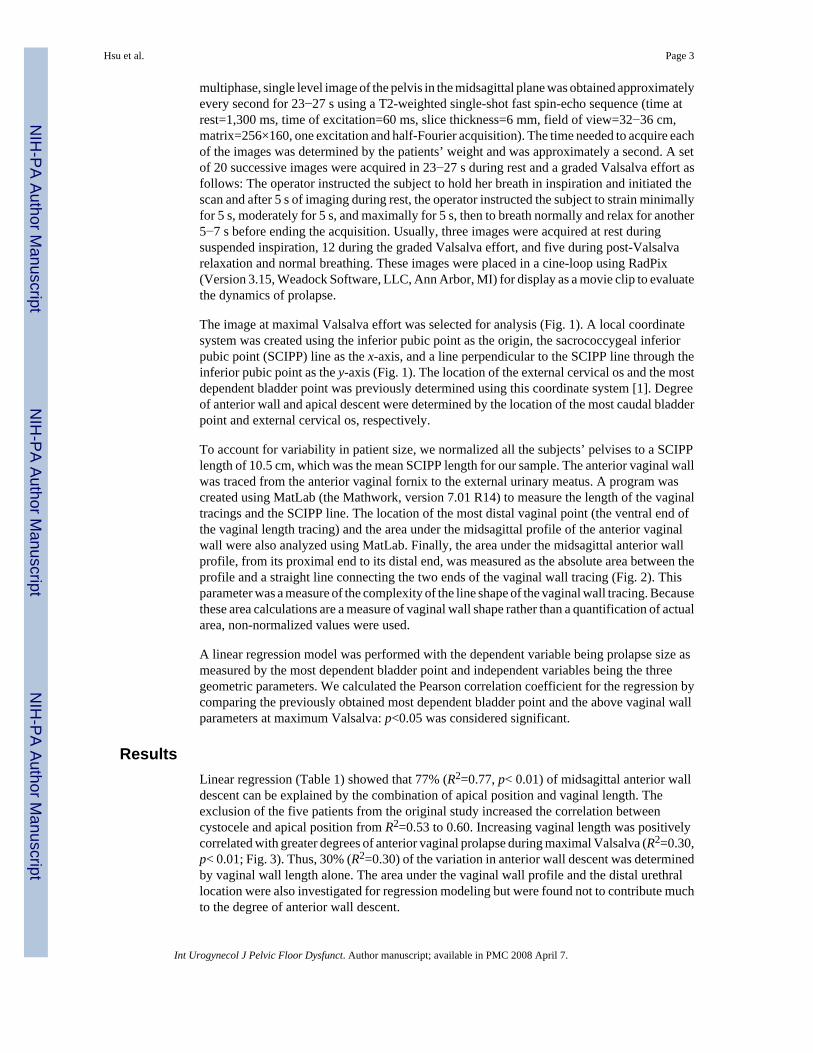
As would be expected from the correlation found between apical descent and anterior walldescent, there were subjects who had uterine descent and large cystoceles (Fig. 4a). However,there were also subjects with relatively well-supported apical compartments who had largecystoceles with long anterior vaginal walls (Fig. 4b). Other morphologic variants existed aswell where there were examples of patients with cystoceles with normal and short vaginal walllengths (Fig. 4c, d, respectively).
The relationship between apical descent and vaginal wall length was examined to see if apicaldescent resulted in longer vaginal length. The correlation was R2=0.36, p=0.02.
DiscussionLinear regression modeling shows that 77% of anterior wall descent can be explained by bothvaginal length and apical descent measured on midsagittal dynamic MR images. A positivecorrelation was found between anterior wall descent and anterior vaginal wall length with 30%of variation in anterior wall descent being explained by the length of the vaginal wall. Theserelationships suggest that the disease mechanism of anterior wall prolapse is likelymultifactoral resulting in different morphologic variants of cystocele. Anterior vaginal supporthas been simplified to a trapezoidal vaginal wall with connections at the top and laterally [6].Our previous work supports the hypothesis that defects of the apical support are related tocystocele formation [1]. The weak correlation between apical descent and vaginal length andthe strong correlation between vaginal length and cystocele size suggest that alterations in theactual vaginal wall itself may be involved in cystocele development.
Vaginal wall characteristics have been previously evaluated. The fraction of smooth musclein the anterior vaginal wall shows great variability among individuals [7], and its contractilityis significantly decreased in women with pelvic organ prolapse [8,9]. Patients with prolapsehave also been found to have alterations in collagen ratios and turn over of collagen [10–12].Findings regarding the collagen content of these tissues have been inconsistent with somestudies showing no change [13,14], and others increased content [15]. Comparison of 3Dvaginal models of patients with and without prolapse showed similar vaginal anterior–posteriorthickness between groups suggesting that the vaginal wall of patients with prolapse is notattenuated [16]. The shear strength of vaginal wall specimens was lower in patients with pelvicfloor dysfunction than normal controls [17], and great variability in the tensile and bendingstrength of samples has also been observed [18].
The vaginal apex has also been examined as the site of failure. One study reported that collagencontent was similar between prolapse patients and controls at the vaginal apex but was lowerin the parametrial tissue [19]. The resiliency of the apical support structures such as theuterosacral ligaments has also been found to be significantly reduced among women withsymptomatic uterovaginal prolapse [20].
Dynamic MRI has been used for the evaluation and staging of pelvic organ prolapse [21–23].Ozasa et al. [24] also used midsagittal dynamic MRI to asses changes in pelvic support inwomen with prolapse. They qualitatively compared descent using the relationship of a best-fitlevator line and the pubic bone, however, did not perform quantitative measurements. Thecurrent study has the advantage of quantifying midsagittal changes that occur during Valsalva.These quantifications deepen our understanding of anterior compartment prolapsemechanisms.
There are several limitations of this study. The dynamic sequence used is limited to amidsagittal view, thereby not allowing the study of changes that may occur in the lateral vaginalwall, which have been observed in anterior compartment prolapse [25]. Therefore, we cannotcomment on the presence or effects of paravaginal defects. In addition, these sequences were
Hsu et al. Page 4
Int Urogynecol J Pelvic Floor Dysfunct. Author manuscript; available in PMC 2008 April 7.
NIH
-PA Author Manuscript
NIH
-PA Author Manuscript
NIH
-PA Author Manuscript

acquired in the supine position, which may limit the descent of the pelvic structures. Weminimized this limitation by careful coaching the woman to achieve the maximal prolapse andreviewed all the dynamic video clips to make sure that Valsalva was performed correctly. Inthe past, researchers have compared supine dynamic MRI with those performed in a sittingposition in an open configuration scanner and found supine scans during straining to becomparable in documenting pelvic floor movement [26,27]. An additional limitation isrepresented by the example in Fig. 4d. This woman has significant anterior wall prolapse andapical descent but yet has a short vaginal length. Although clinically, this woman does not havea shortened wall, she appears to on dynamic MRI because the anterior wall is folded in on itselflike an accordion.
The association of increased midline vaginal length in anterior compartment prolapse providesobjective data to be considered in the clinical thinking and treatment of cystoceles.Traditionally, anterior vaginal wall prolapse is attributed to either lateral detachment of theanterior vaginal wall at the pelvic side wall, referred to as a displacement “cystocele,” or as acentral failure of the vaginal wall itself that results in a distension “cystocele.” Whether lengthchanges are due to a paravaginal defect, midline defect, or are simply due to stretching of thevaginal wall once a cystocele becomes present will require further research. The associationseen in this study nevertheless raises interesting clinical questions. For example, do traditionalside-to-side plications address excess vaginal length associated with anterior compartmentprolapse?
AcknowledgmentsWe gratefully acknowledge the funding from NIH grant NICHD R01 HD038665 as well as investigator support from5K12RR017607-04 and the Office for Research on Women's Health and NICHD SCOR on Sex and Gender FactorsAffecting Women's Health P50 HD044406. This information was presented at the American Urogynecology Societymeeting in October 2006.
References1. Summers A, Winkel LA, Hussain HK, DeLancey JOL. The relationship between anterior and apical
compartment support. Am J Obstet Gynecol 2006;194:1438–1443. [PubMed: 16579933]2. Hendrix SL, Clark A, Nygaard I, Aragaki A, Barnabei V, McTiernan A. Pelvic organ prolapse in the
Women's Health Initiative: gravity and gravidity. Am J Obstet Gynecol 2002;186(6):1160–1166.[PubMed: 12066091]
3. Shull BL, Bachofen C, Coates KW, Kuehl TJ. A transvaginal approach to repair of apical and otherassociated sites of pelvic organ prolapse with uterosacral ligaments. Am J Obstet Gynecol2000;183:1365–1373. [PubMed: 11120498]
4. Weber AM, Walters MD, Piedmonte MR, Ballard LA. Anterior colporrhaphy: a randomized trial ofthree surgical techniques. Am J Obstet Gynecol 2001;185:1299–1304. [PubMed: 11744900]
5. DeLancey JO, Morgan DM, Fenner DE, Kearney R, Guire K, Miller JM, Hussain H, Umek W, HsuY, Ashton-Miller JA. Comparison of levator ani muscle defects and function in women with andwithout pelvic organ prolapse. Obstet Gynecol 2007;109:295–302. [PubMed: 17267827]
6. DeLancey JOL. Fascial and muscular abnormalities in women with urethral hypermobility and anteriorvaginal wall prolapse. Am J Obstet Gynecol 2002;187:93–98. [PubMed: 12114894]
7. Morgan DM, Iyengar J, DeLancey JO. A technique to evaluate the thickness and density of nonvascularsmooth muscle in the suburethral fibromuscular layer. Am J Obstet Gynecol 2003;188(5):1183–1185.[PubMed: 12748472]
8. Boreham MK, Wai CY, Miller RT, Schaffer JI, Word RA. Morphometric analysis of smooth musclein the anterior vaginal wall of women with pelvic organ prolapse. Am J Obstet Gynecol 2002;187(1):56–63. [PubMed: 12114889]
Hsu et al. Page 5
Int Urogynecol J Pelvic Floor Dysfunct. Author manuscript; available in PMC 2008 April 7.
NIH
-PA Author Manuscript
NIH
-PA Author Manuscript
NIH
-PA Author Manuscript

9. Boreham MK, Wai CY, Miller RT, Schaffer JI, Word RA. Morphometric properties of the posteriorvaginal wall in women with pelvic organ prolapse. Am J Obstet Gynecol 2002;187(6):1501–1508.[PubMed: 12501053]
10. Chen BH, Wen Y, Li H, Polan ML. Collagen metabolism and turnover in women with stress urinaryincontinence and pelvic prolapse. Int Urogynecol J Pelvic Floor Dysfunct 2002;13(2):80–87.[PubMed: 12054187]
11. Moalli PA, Talarico LC, Sung VW, Klingensmith WL, Shand SH, Meyn LA, Watkins SC. Impactof menopause on collagen subtypes in the arcus tendineous fasciae pelvis. Am J Obstet Gynecol2004;190(3):620–627. [PubMed: 15041990]
12. Norton P, Boyd C, Deak S. Collagen synthesis in women with genital prolapse or stress urinaryincontinence. Neurourol Urodyn 1992;11:300–301.
13. Liapis A, Bakas P, Pafiti A, Hassiakos D, Frangos-Plemenos M, Creatsas G. Changes in the quantityof collagen type I in women with genuine stress incontinence. Urol Res 2000;28(5):323–326.[PubMed: 11127711]
14. Liapis A, Bakas P, Pafiti A, Frangos-Plemenos M, Arnoyannaki N, Creatsas G. Changes of collagentype III in female patients with genuine stress incontinence and pelvic floor prolapse. Eur J ObstetGynecol Reprod Biol 2001;97(1):76–79. [PubMed: 11435014]
15. Kokcu A, Yanik F, Cetinkaya M, Alper T, Kandemir B, Malatyalioglu E. Histopathological evaluationof the connective tissue of the vaginal fascia and the uterine ligaments in women with and withoutpelvic relaxation. Arch Gynecol Obstet 2002;266(2):75–78. [PubMed: 12049299]
16. Hsu Y, Chen L, DeLancey JOL, Ashton-Miller JA. Vaginal thickness, cross-sectional area, andperimeter in women with and those without prolapse. Obstet Gynecol 2005;105(5):1012–1017.[PubMed: 15863538]
17. Kondo A, Narushima M, Yoshikawa Y, Hayashi H. Pelvic fascia strength in women with stress urinaryincontinence in comparison with those who are continent. Neurourol Urodyn 1994;13(5):507–513.[PubMed: 7833968]
18. Cosson M, Lambaudie E, Boukerrou M, Lobry P, Crepin G, Ego A. A biomechanical study of thestrength of vaginal tissues. Results on 16 post-menopausal patients presenting with genital prolapse.Eur J Obstet Gynecol Reprod Biol 2004;112(2):201–205. [PubMed: 14746960]
19. Takano CC, Girao MJ, Sartori MG, Castro RA, Arruda RM, Simoes MJ, et al. Analysis of collagenin parametrium and vaginal apex of women with and without uterine prolapse. Int Urogynecol J2002;13(6):342–345.
20. Reay Jones NH, Healy JC, King LJ, Saini S, Shousha S, Allen-Mersh TG. Pelvic connective tissueresilience decreases with vaginal delivery, menopause and uterine prolapse. Br J Surg 2003;90(4):466–472. [PubMed: 12673750]
21. Etlik O, Arslan H, Odabasi O, Odabasi H, Harman M, Celebi H, et al. The role of the MR-fluoroscopyin the diagnosis and staging of the pelvic organ prolapse. Eur J Radiol 2005;53:136–141. [PubMed:15607865]
22. Lienemann A, Anthuber C, Baron A, Kohz P, Reiser M. Dynamic MR colpocystorectographyassessing pelvic-floor descent. Eur Radiol 1997;7(8):1309–1317. [PubMed: 9377520]
23. Singh K, Reid WM, Berger LA. Assessment and grading of pelvic organ prolapse by use of dynamicmagnetic resonance imaging. Am J Obstet Gynecol 2001;185(1):71–77. [PubMed: 11483907]
24. Ozasa H, Mori T, Togashi K. Study of uterine prolapse by magnetic resonance imaging:Topographical changes involving the levator ani muscle and the vagina. Gynecol Obstet Invest1992;34:43–48. [PubMed: 1526530]
25. Richardson AC, Edmonds PB, Williams NL. Treatment of stress urinary incontinence due toparavaginal fascial defect. Obstet Gynecol 1981;57:357. [PubMed: 7465150]
26. Fielding JR, Griffiths DJ, Versi E, Mulkern RV, Lee MLT, Jolesz FA. MR imaging of pelvic floorcontinence mechanisms in the supine and sitting positions. Am J Roentgenol 1998;171:1607–1610.[PubMed: 9843296]
27. Bertsschinger KM, Hetzer FH, Roos JE, Treiber K, Marincek B, Hilfiker PR. Dynamic MR imagingof the pelvic floor performed with patient sitting in an open-magnet unit versus with patient supinein a closed-magnet unit. Radiology 2002;223(2):501–508. [PubMed: 11997560]
Hsu et al. Page 6
Int Urogynecol J Pelvic Floor Dysfunct. Author manuscript; available in PMC 2008 April 7.
NIH
-PA Author Manuscript
NIH
-PA Author Manuscript
NIH
-PA Author Manuscript

Fig. 1.MR image at maximal Valsalva showing anterior vaginal wall tracing (solid black line), distalvaginal wall (x), bladder location (triangle), cervical os location (circle), and coordinate system(dotted lines)
Hsu et al. Page 7
Int Urogynecol J Pelvic Floor Dysfunct. Author manuscript; available in PMC 2008 April 7.
NIH
-PA Author Manuscript
NIH
-PA Author Manuscript
NIH
-PA Author Manuscript

Fig. 2.Area under the anterior vaginal wall profile. Pubic bone, sacrum, and coccyx are traced. Theanterior vaginal wall tracings are shown. A line (small dotted) is used to connect the two endsof the vaginal wall tracing to create an area (shown in gray). Examples aligned using horizontallarge dotted reference line. a Minimal descent of most caudal bladder point (triangle). bGreater descent of the bladder point with longer vaginal wall length and larger area. c Similarvaginal wall length to B but smaller area and lesser descent of bladder point
Hsu et al. Page 8
Int Urogynecol J Pelvic Floor Dysfunct. Author manuscript; available in PMC 2008 April 7.
NIH
-PA Author Manuscript
NIH
-PA Author Manuscript
NIH
-PA Author Manuscript

Fig. 3.Vaginal length during Valsalva and distance of the most caudal bladder point below normal.There is a linear correlation y=0.69x−0.85, R2=0.30
Hsu et al. Page 9
Int Urogynecol J Pelvic Floor Dysfunct. Author manuscript; available in PMC 2008 April 7.
NIH
-PA Author Manuscript
NIH
-PA Author Manuscript
NIH
-PA Author Manuscript
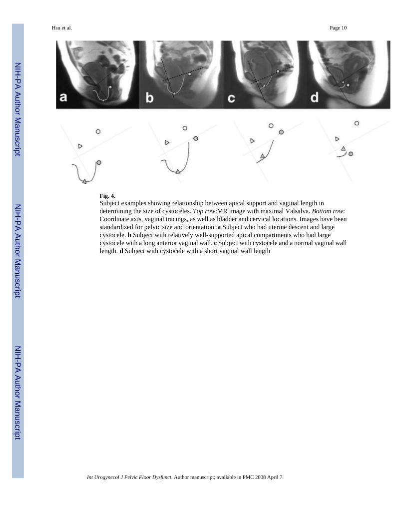
Fig. 4.Subject examples showing relationship between apical support and vaginal length indetermining the size of cystoceles. Top row:MR image with maximal Valsalva. Bottom row:Coordinate axis, vaginal tracings, as well as bladder and cervical locations. Images have beenstandardized for pelvic size and orientation. a Subject who had uterine descent and largecystocele. b Subject with relatively well-supported apical compartments who had largecystocele with a long anterior vaginal wall. c Subject with cystocele and a normal vaginal walllength. d Subject with cystocele with a short vaginal wall length
Hsu et al. Page 10
Int Urogynecol J Pelvic Floor Dysfunct. Author manuscript; available in PMC 2008 April 7.
NIH
-PA Author Manuscript
NIH
-PA Author Manuscript
NIH
-PA Author Manuscript

NIH
-PA Author Manuscript
NIH
-PA Author Manuscript
NIH
-PA Author Manuscript
Hsu et al. Page 11
Table 1Linear regression modeling
Cumulative R2 R2 added p value
Apical 0.60 − <0.001
+Vaginal length 0.77 0.17 <0.001
+Distal point 0.78 0.01 <0.001
+Area under vaginal profile 0.81 0.03 <0.001
Int Urogynecol J Pelvic Floor Dysfunct. Author manuscript; available in PMC 2008 April 7.
Copyright © 2022 FDOKUMEN