Dual energy CT imaging for preclinical and clinical radiotherapy
Upload
khangminh22Category
view
2download
0
UNIVERSIDADE DA BEIRA INTERIOR Ciências da Saúde
Preclinical performance of vaginal semisolid products: technological and safety evaluations
assuming physiologic parameters
Rita Solange Monteiro Machado
Thesis for Doctoral Degree in
Pharmaceutical Sciences (3rd cycle of studies)
Supervisor: Professor José António Martinez Souto de Oliveira, PhD Co-supervisor: Professor Rita Manuela Palmeira de Oliveira, PhD
Covilhã, January 2018

ii

iii
The experimental work presented in this thesis was carried out at the Health Sciences Research
Center laboratories, University of Beira Interior (CICS-UBI) and, at Labfit – Health Products
Research and Development, Lda facilities, under the supervision of Professor José António
Martinez Souto de Oliveira and Professor Rita Manuela Palmeira de Oliveira.

iv

v
The work herein presented was supported by FCT, the Portuguese Foundation for Science and
Technology, and Labfit – Health Products Research and Development, Lda. in a collaborative
academia-company project (SFRH/BDE/111544/2015). Additionally, CICS-UBI was supported by
FEDER funds through the POCI - COMPETE 2020 - Operational Programme Competitiveness and
Internationalization in Axis I - Strengthening research, technological development and
innovation (Project POCI-01-0145-FEDER-007491), National Funds by FCT - Foundation for
Science and Technology (Project UID/Multi /00709/2013).

vi

vii
To my Family - À minha Família

viii

ix
“Tenho em mim todos os sonhos do mundo.”
Fernando Pessoa

x

xi
Agradecimentos
Depois de percorrido este caminho, que com certeza não faria sozinha, resta-me agradecer
a todos aqueles que me apoiaram e inspiraram a lutar pelos meus objetivos.
Aos meus orientadores,
agradeço a oportunidade que me deram de integrar este projeto. Posso agora dizer que a
caminhada não foi sempre fácil, mas foi certamente um desafio gratificante. Ao Prof. Doutor
José Martinez de Oliveira agradeço a simpatia, o entusiasmo e o seu notável contributo e
orientação científica. À Prof. Doutora Rita Palmeira de Oliveira, pela sua constante boa
disposição e otimismo, pela disponibilidade e ensinamentos transmitidos. Acima de tudo,
agradeço a ambos, as ferramentas do pensar e comunicar ciência que me foram incutidas ao
longo destes anos.
À Labfit-HPRD, Lda,
e a todos os seus colaboradores, pelo apoio técnico e científico, e pelo simpático acolhimento
e amizade durante as fases em que o trabalho laboratorial foi lá desenvolvido. Em especial
agradeço à Prof. Doutora Ana Palmeira de Oliveira que acompanhou de perto este trabalho,
pela sua disponibilidade e contributo científico.
Ao CICS-UBI,
pelo acolhimento laboratorial e por ser a minha casa desde que comecei na investigação
científica. Agradeço aos professores, investigadores, colegas e funcionários. Todos deram um
pequeno contributo no desenvolvimento deste trabalho.
À Prof. Doutora Adriana Santos,
pela oportunidade que tive de colaborar nas suas aulas práticas. Agradeço os seus contributos
científicos, e esclarecimentos no decorrer destes anos.
À Prof. Doutora Luiza Breitenfeld,
pela disponibilidade e pelo contributo científico que prestou na fase final deste trabalho.
À Dr. Catarina Ferreira,
pela simpatia, disponibilidade e profissionalismo e sobretudo pela ajuda na histologia e
microscopia.
À Oviger, SA,
pelo fornecimento do material biológico, essencial para a concretização deste trabalho.

xii
Aos meus pais,
pelo exemplo que são para mim, pelo amor e carinho, pelo apoio incondicional e força para
seguir em frente, sem vocês nada teria conseguido.
À minha irmã,
e colega de casa durante estes últimos anos, pela amizade, companheirismo, pelo o que
caminhámos juntas até agora e todo o caminho que temos pela frente, para toda a vida.
À minha avó,
por todo o amor, pelos conselhos e recomendações, pelo carinho e boa disposição.
À restante minha família,
madrinha, padrinho, tias e tios, primas e primos, pelo vosso carinho e apoio incondicional.
Ao Artur,
por teres entrado na minha vida nesta fase e mesmo assim teres ficado. Pelos nossos momentos,
pelos mimos, pelo conforto, pelo apoio, pelo companheirismo e acima de tudo, pelo amor.
Aos meus Amigos,
que o destino colocou no meu caminho e nunca mais tirou. Sou grata pela vossa amizade e por
tudo o que já vivemos e viveremos juntos. Esta caminhada, apesar das dificuldades, tornou-se
mais fácil e gratificante com a amizade que nos uniu.
xiii
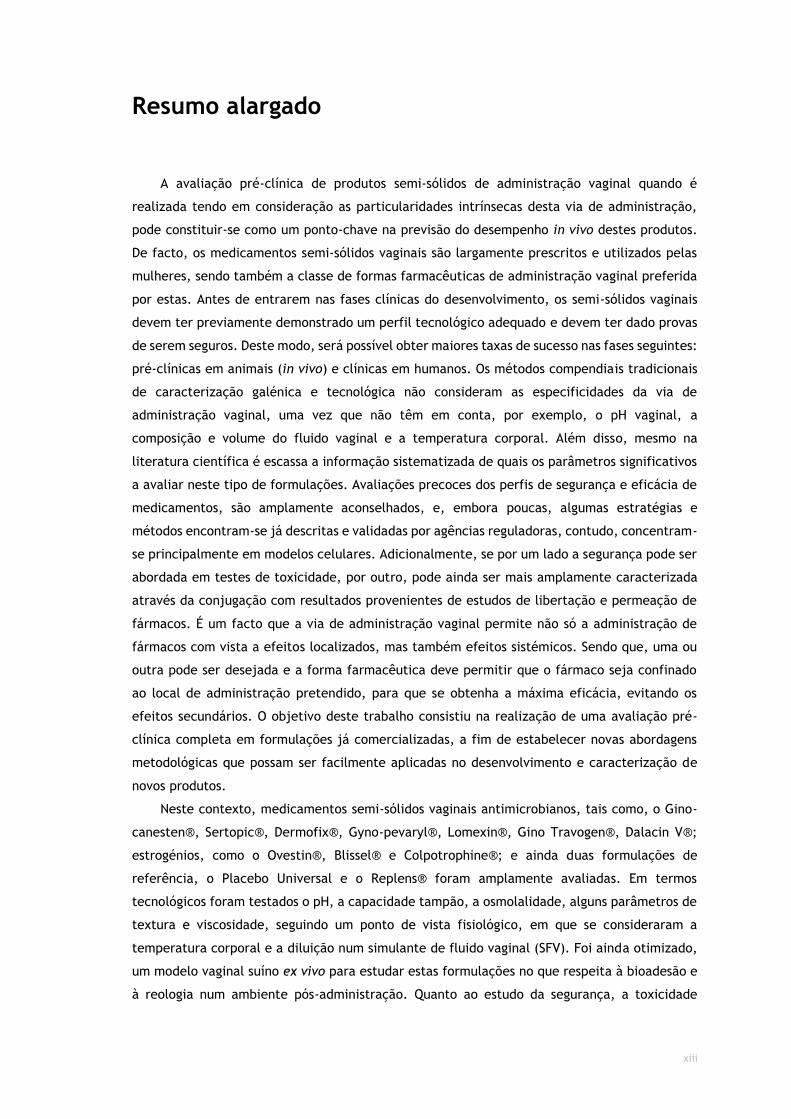
Resumo alargado
A avaliação pré-clínica de produtos semi-sólidos de administração vaginal quando é
realizada tendo em consideração as particularidades intrínsecas desta via de administração,
pode constituir-se como um ponto-chave na previsão do desempenho in vivo destes produtos.
De facto, os medicamentos semi-sólidos vaginais são largamente prescritos e utilizados pelas
mulheres, sendo também a classe de formas farmacêuticas de administração vaginal preferida
por estas. Antes de entrarem nas fases clínicas do desenvolvimento, os semi-sólidos vaginais
devem ter previamente demonstrado um perfil tecnológico adequado e devem ter dado provas
de serem seguros. Deste modo, será possível obter maiores taxas de sucesso nas fases seguintes:
pré-clínicas em animais (in vivo) e clínicas em humanos. Os métodos compendiais tradicionais
de caracterização galénica e tecnológica não consideram as especificidades da via de
administração vaginal, uma vez que não têm em conta, por exemplo, o pH vaginal, a
composição e volume do fluido vaginal e a temperatura corporal. Além disso, mesmo na
literatura científica é escassa a informação sistematizada de quais os parâmetros significativos
a avaliar neste tipo de formulações. Avaliações precoces dos perfis de segurança e eficácia de
medicamentos, são amplamente aconselhados, e, embora poucas, algumas estratégias e
métodos encontram-se já descritas e validadas por agências reguladoras, contudo, concentram-
se principalmente em modelos celulares. Adicionalmente, se por um lado a segurança pode ser
abordada em testes de toxicidade, por outro, pode ainda ser mais amplamente caracterizada
através da conjugação com resultados provenientes de estudos de libertação e permeação de
fármacos. É um facto que a via de administração vaginal permite não só a administração de
fármacos com vista a efeitos localizados, mas também efeitos sistémicos. Sendo que, uma ou
outra pode ser desejada e a forma farmacêutica deve permitir que o fármaco seja confinado
ao local de administração pretendido, para que se obtenha a máxima eficácia, evitando os
efeitos secundários. O objetivo deste trabalho consistiu na realização de uma avaliação pré-
clínica completa em formulações já comercializadas, a fim de estabelecer novas abordagens
metodológicas que possam ser facilmente aplicadas no desenvolvimento e caracterização de
novos produtos.
Neste contexto, medicamentos semi-sólidos vaginais antimicrobianos, tais como, o Gino-
canesten®, Sertopic®, Dermofix®, Gyno-pevaryl®, Lomexin®, Gino Travogen®, Dalacin V®;
estrogénios, como o Ovestin®, Blissel® e Colpotrophine®; e ainda duas formulações de
referência, o Placebo Universal e o Replens® foram amplamente avaliadas. Em termos
tecnológicos foram testados o pH, a capacidade tampão, a osmolalidade, alguns parâmetros de
textura e viscosidade, seguindo um ponto de vista fisiológico, em que se consideraram a
temperatura corporal e a diluição num simulante de fluido vaginal (SFV). Foi ainda otimizado,
um modelo vaginal suíno ex vivo para estudar estas formulações no que respeita à bioadesão e
à reologia num ambiente pós-administração. Quanto ao estudo da segurança, a toxicidade
xiv
celular foi avaliada nas linhas celulares: VK2 E6/E7 (vaginais), HeLa (cervicais) e HEC-1A
(uterinas), e para tal, foram realizados os ensaios de viabilidade que utilizaram o MTT (brometo
de 3-(4,5-dimetiltiazol-2-il)-2,5-difenil tetrazólio) e o NRU (captação de vermelho neutro).
Além disso, tecidos epiteliais ex vivo, foram excisados a partir da vagina suína, e nestes,
também foi aplicado o ensaio do MTT e, ainda uma análise histológica. Para incluir uma
avaliação ainda mais avançada da toxicidade in vitro, os semi-sólidos vaginais foram testados
num modelo organotípico: HET-CAM (ensaio que é realizado na membrana corio-alantóide de
embriões de galinhas Henn) e que se encontra em validação para substituir o teste in vivo em
animais para testes de irritação ocular. Deste modo, foi transposto este ensaio para a avaliação
da irritação vaginal. Um método de quantificação de HPLC-DAD (Cromatografia Líquida de Alta
Pressão acoplada a um detetor de díodos) para as moléculas presentes nos semi-sólidos vaginais
incluídos neste trabalho, foi desenvolvido e validado de acordo com os requisitos das normas
da FDA (Food and Drug Administration - EUA), da EMA (Agência Europeia de Medicamentos) e
da ICH (Conferência Internacional para a Harmonização). Depois disso, este método foi aplicado
na quantificação de fármacos nos estudos de libertação in vitro e nas experiências de
permeação de fármacos ex vivo. Estas duas técnicas foram realizadas utilizando células de
difusão dinâmicas verticais de Franz, tendo todas as configurações experimentais sido
especificamente otimizadas para as moléculas em estudo (estriol, clotrimazol, econazole,
isoconazol, sertaconazol e fenticonazol).
Quanto às características tecnológicas, as formulações antimicrobianas apresentaram
menor pH do que os estrogénios tópicos. A capacidade tampão no SFV conduziu a melhores
previsões do que acontece in vivo. Após diluição em volumes fisiológicos de SFV, a osmolalidade
da maioria dos produtos esteve conforme o recomendado pela Organização Mundial da Saúde
(OMS), claramente dependente da composição. Os antimicrobianos apresentaram
comportamentos de textura semelhantes entre eles, enquanto os estrogénios tópicos variaram
entre eles, no que respeita aos parâmetros de textura. Com efeito, neste grupo encontrava-se
uma formulação de base polimérica em gel (Blissel®), sendo que os restantes, eram cremes
(Ovestin® e Colpotrophine). Regra geral, para todos os produtos observou-se uma ligeira
diminuição da viscosidade após aplicação de fatores fisiológicos, como a diluição em SFV e a
temperatura (37ºC), mas os seus comportamentos pseudoplásticos mantiveram-se. No entanto,
cada formulação demonstrou ter o seu próprio perfil reológico, possivelmente conduzido pela
sua composição, tanto qualitativa como quantitativa. Contudo, a viscosidade das formulações
foi mais elevada quando testada num modelo de administração ex vivo, comparando com a
viscosidade obtida nos ensaios em SFV a 37ºC. Este dado releva, que podem existir outros
fatores determinantes na viscosidade adquirida após a administração de formulações semi-
sólidas. Quanto aos modelos in vitro, as células VK2 E6/E7 apresentaram viabilidades
relativamente superiores às células HeLa e HEC-1A nas concentrações de produto testadas. Os
resultados de viabilidade no tecido foram muito superiores aos obtidos para nos modelos
celulares. Isto revela que este modelo poderá ser mais robusto e mais próximo do que acontece
in vivo. Em todos os modelos, as formulações antimicrobianas mostraram viabilidades
xv
dependentes da concentração. Ao contrário das formulações que contém estrogénios, que
apresentaram perfis relacionados com os efeitos celulares estrogénicos reconhecidos (ou seja,
foram dependentes da dosagem da formulação e da concentração testada). Os produtos de
referência apresentaram os perfis de viabilidade mais estáveis e mais elevados em todas as
concentrações. Nos estudos de permeação ex vivo, foi questionada a existência de diferenças
na utilização de secções de tecido vaginal caudal ou cranial. Não obstante, em termos de
permeação de fármacos, não foram encontradas extensas diferenças significativas entre estes
dois tipos de epitélio, mas a vagina caudal parece ser mais adequada para experiências de
permeação vaginal, uma vez que conduz a resultados mais reprodutíveis e consistentes. De
facto, a região mais cranial da vagina suína apresenta, histologicamente, um epitélio mais fino,
e anatomicamente, uma superfície que contém mais rugas. O que conduz a desvios-padrão
entre experiências mais elevados. Além disso, observou-se que a permeação de fármacos não
é apenas dependente da libertação do fármaco a partir formulação. A sua afinidade pela
formulação e/ou pelo tecido, é determinante para a sua permeação até à câmara recetora.
Concluindo, a avaliação pré-clínica integrada para semi-sólidos vaginais, proposta nesta
tese, representa uma abordagem importante a ter em consideração, no desenvolvimento de
novos produtos, uma vez que pode reduzir custos com o desenvolvimento de novas formulações,
prevendo precocemente a sua eficácia in vivo e os seus perfis de segurança. Estas metodologias
têm, assim, um grande potencial não só para serem aplicadas na indústria cosmética,
farmacêutica e de dispositivos médicos, mas também pode ser aplicada em investigação mais
fundamental ao nível da academia.
Palavras-chave
Administração vaginal de fármacos, semi-sólidos vaginais, avaliação pré-clínica, caracterização
tecnológica, in vitro, ex vivo, segurança, permeação de fármacos, libertação de fármacos

xvi
xvii
Abstract
Vaginal semisolid products preclinical evaluations, when performed considering the
particularities of the target organ, may represent key tools to predict in vivo performance.
Before heading to clinical phases, vaginal semisolids must demonstrate to have an adequate
technological and safety profile, in order to achieve higher success rates in the human testing
stage. Traditional characterization methods currently used for vaginal semisolids do not
undertake an integrative approach, since they do not address, for example, vaginal pH, fluid
and temperature. Moreover, early safety assessment methods are largely described and
validated not only on scientific literature, but also by regulatory agencies, although they are
still mainly focused on cellular-based models. This safety profile of products can be further
improved by combining toxicity testing, with drug release and permeation studies. Indeed, the
vaginal administration route allows for local and systemic delivery of drugs, depending on the
therapeutic purpose. Consequently, the drug should be confined to the chosen location of
administration, to obtain maximum efficacy while avoiding side effects. The aim of this work
was to develop a full set of assessment methods for characterization of vaginal semisolid
products. Commercialized formulations were used to establish new methodological approaches
that could be applied in new products development and characterization.
Therefore, antimicrobials, Gino-canesten®, Sertopic®, Dermofix®, Gyno-pevaryl®,
Lomexin®, Gino Travogen®, Dalacin V®; oestrogens, Ovestin®, Blissel® and Colpotrophine®;
and, two reference formulations, Universal Placebo and Replens® were extensively evaluated.
Technologically, they were tested in terms of pH, pH-buffering capacity, osmolality, textural
parameters and viscosity, using a physiologic standpoint that considered the body temperature
and dilution in a physiologic volume of vaginal fluid simulant (VFS); and even an ex vivo porcine
model to infer bioadhesion and rheology on an after-administration environment. In terms of
safety investigation, cellular toxicity was disclosed on VK2 E6/E7, HeLa and HEC-1A cell-lines,
using MTT (3-(4,5-dimethylthiazol-2-yl)-2,5-diphenyltetrazolium bromide) and NRU (Neutral
Red Uptake) assays. Tissue explants, collected from the ex vivo porcine vaginal model, were
also tested concerning toxicity, through MTT and histological analysis. Moreover, to include an
advanced in vitro toxicity evaluation, the HET-CAM (Hen's Egg Test – Chorioallantoic
Membrane), already in validation for eye irritation testing, was applied to vaginal irritation. A
HPLC-DAD (High Performance Liquid Chromatography with Diode Array Detector) quantification
method for the molecules present in semisolids included in this work, was developed and
validated according to FDA (Food and Drug Administration - USA), EMA (European Medicines
Agency) and ICH (International Conference for Harmonization) requirements. Further, this
method was applied in drug quantification on in vitro drug release and ex vivo drug permeation
experiments. These two techniques were performed using dynamic vertical Franz diffusion
cells, having all experimental setting being specifically designed and optimized concerning the
xviii
molecules in study (estriol, clotrimazole, econazole, isoconazole, sertaconazole and
fenticonazole).
Concerning technological characteristics, antimicrobial formulations exhibited lower pH
than topical oestrogens. Buffering capacity in a vaginal fluid simulant conducted to better
predictions of what happens in vivo. Characterization was performed also for those less acidic
products to assess their ability to gain physiologic pH after mixing with simulated vaginal fluids.
Products osmolality after dilution in VFS were below the upper limit advised by the World
Health Organization (WHO). The antimicrobials had similar textural behaviours, while topical
oestrogens varied in textural parameters. A slight decrease in viscosity was observed after
application of dilution and temperature factors, showing the influence of the surrogate vaginal
environment, while maintaining their pseudoplastic behaviour. However, each formulation had
its own profile, possibly driven by their composition. Formulations’ viscosity was higher when
tested using the ex vivo administration model than when only diluted in VFS at 37ºC. Concerning
the in vitro models, VK2 E6/E7, presented relatively higher viabilities than HeLa and HEC-1A
cells over the tested product concentrations. Tissue viability results were much higher than
those obtained for the in vitro cellular models, revealing that this model could be more robust
and closer to the in vivo situation. Across models, antimicrobials showed concentration-
dependent viabilities. While oestrogens presented odd profiles, depending on the formulation
and concentration tested. Reference products led to the most stable and higher viability
profiles across concentrations. On ex vivo permeation studies we have investigated if there
were differences in performing ex vivo permeation studies using the porcine vaginal model,
when collecting a proximal or a distal tissue within the vaginal tube. No extensive significant
differences between these tissues were found, but the caudal vagina could be more suitable
for vaginal permeation experiments since it conducted to more reproducible and consistent
results. Furthermore, it was observed that drug permeation is not directly dependent on drug
release from the formulation.
To sum up, the conduction of this integrative preclinical assessment for vaginal semisolids
can be a valuable approach in new products development or characterisation, since it could
optimize cost-efficiency of new formulations development by predicting in vivo efficacy and
safety profiles. In addition, these methodologies have great potential not only to be applied in
cosmetics, medical devices and medicines industry, but also in academical research.
Keywords
Vagina drug delivery, vaginal semisolids, preclinical evaluation, technological characterization,
in vitro, ex vivo, safety, drug permeation, drug release

xix
Thesis overview
This doctoral thesis is structured in eight chapters and appendices, herein summarized:
CHAPTER I consists on a brief introduction over the vaginal environment, the vaginal drug
delivery systems focusing on semisolids and the characterization methods applied for these
fomulations. At the end of this chapter the main objectives of this thesis are presented.
CHAPTER II provides a methodologic background for vaginal semisolid formulations, and,
consequently, proposes adaptations to traditional characterization methods, based on results
obtained experimentally.
CHAPTER III comprises the development of in vitro and ex vivo models. A preclinical
safety characterization was accomplished through cell-based and organ-based testing.
CHAPTER IV embraces the topic of drug release and permeation. Primarily, a
chromatographic quantification method for all drugs included in this study was validated.
CHAPTER V discusses and integrates all the results obtained presented in the previous
chapters.
CHAPTER VI describes the contributions that the results of this thesis had for enhancing
the competitiveness of the hosting company, Labfit-HPRD, Lda.
CHAPTER VII presents the conclusions of this thesis and future remarks.
CHAPTER VIII details the bibliographic references used to construct this thesis.
APPENDICES contain information that, despite being important for the development of
this work, were not essential in the main manuscript. They are composed of: A) a published
review article comprising methodologies for vaginal drug permeation; B and C) two manuscripts
in the format that was submitted concerning in vitro and ex vivo toxicity evaluations; D)
supplementary material that was gathered to support the experiementsal setup of Chapter IV.

xx
xxi
List of Publications
Publications in Peer-Reviewed International Scientific Journals
included in this thesis
Machado RM, Agonia AS, Borges L, Palmeira-de-Oliveira A, Martinez-de-Oliveira J, Palmeira-
de-Oliveira R, “Vaginal drug delivery: in vitro release and ex vivo permeation of six active
pharmaceutical ingredients from commercial semisolids, quantified by a sole HPLC-DAD
validated method”
(submitted manuscript)
Palmeira-de-Oliveira R, Machado RM, Palmeira-de-Oliveira A, Martinez-de-Oliveira J, “Testing
vaginal irritation with the HET-CAM assay: an in vitro alternative to the in vivo method”
(accepted manuscript)
Machado RM, Palmeira-de-Oliveira A, Breitenfeld L, Martinez-de-Oliveira J, Palmeira-de-
Oliveira R, “Optimization and application of in vitro and ex vivo models for vaginal semisolids
safety evaluation”
(submitted manuscript)
Machado RM, Palmeira-de-Oliveira A, Martinez-de-Oliveira J, Palmeira-de-Oliveira R, “Vaginal
semisolid products: Technological performance considering physiologic parameters”. European
Journal of Pharmaceutical Sciences. 2017; doi: 10.1016/j.ejps.2017.09.009
Machado RM, Palmeira-de-Oliveira A, Silva Gaspar C, Martinez-de-Oliveira J, Palmeira-de-
Oliveira R, “Studies and methodologies on vaginal drug permeation”, Advanced Drug Delivery
Reviews. Special Issue: Vaginal Drug Delivery. 2015; 92:14-26; doi: 10.1016/j.addr.2015.02.003

xxii
Other publications during the PhD Course
Machado RM, Palmeira-de-Oliveira R, Agonia AS, Martinez-de-Oliveira J, Palmeira-de-Oliveira
A, “Development and characterization of a new Vaginal Estriol Cream (VEC) for vaginal atrophy
– a biocompatible and bioadhesive approach”
(submitted manuscript)
Palmeira-de-Oliveira R, Palmeira-de-Oliveira A, Machado RM, Elvas AR, Amorim I, Gartner F,
Martinez-de-Oliveira J, Amaral MH, Breitenfeld L, “Porcine vaginal model for drug permeation:
new insights”
(submitted manuscript)
Machado RM, Palmeira-de-Oliveira A, Martinez-de-Oliveira J, Palmeira-de-Oliveira R,
“Development and preliminary characterization of a new vaginal dosage form: a vaginal sheet”
(submitted manuscript)
Palmeira-de-Oliveira R, Macedo M, Machado RM, Pacheco AF, Palmeira-de-Oliveira A, Martinez-
de-Oliveira J, Duarte P, “Pharmaceutical Compounding in Portuguese Community Pharmacies:
Characterization and Future Perspectives”, International Journal of Pharmaceutical
Compounding, 2016; 20(2):114-122
Cunha AR, Machado RM, Palmeira-de-Oliveira A, Martinez-de-Oliveira J, das Neves J, Palmeira-
de-Oliveira R, “Characterization of Commercially Available Vaginal Lubricants: A Safety
Perspective”. Pharmaceutics. 2014; 6(3):530-542; doi: 10.3390/pharmaceutics6030530
Machado RM, Palmeira-de-Oliveira A, Martinez-de-Oliveira J, Palmeira-de-Oliveira R. “Vaginal
Films for Drug Delivery”, Journal of Pharmaceutical Sciences. 2013; 102(7):2069-2081; doi:
10.1002/jps.23577
Palmeira-de-Oliveira A, Silva BM, Machado RM, Palmeira-de-Oliveira R, Martinez-de-Oliveira J,
Salgueiro L, “Potencial terapêutico de extractos de plantas em infecções genitais”. Revista de
Fitoterapia. 2012; 12 (2): 135-144

xxiii
Oral communications presented during the PhD Course
Machado RM*, Palmeira-de-Oliveira A, Martinez-de-Oliveira J, Palmeira-de-Oliveira R, “The
porcine ex vivo model for vaginal drug permeation: being more selective”, XII Annual CICS
Symposium, Covilhã, Portugal (2017)
Machado RM*, Palmeira-de-Oliveira A, Martinez-de-Oliveira J, Palmeira-de-Oliveira R, “Pre-
validation of an ex vivo model for vaginal formulations safety evaluation”, II International
Congress in Health Sciences Research towards innovation and entrepreneurship: Trends in
Biotechnology for Biomedical Applications, Covilhã, Portugal (2017)
Machado RM*, Palmeira-de-Oliveira A, Martinez-de-Oliveira J, Palmeira-de-Oliveira R, “Design
of innovative vaginal semisolid formulations: physiologic characterization methods as a
starting point”, EUPAT 8 - EUFEPS, University College Cork, Ireland (2016)
Machado RM*, Palmeira-de-Oliveira A, Martinez-de-Oliveira J, Palmeira-de-Oliveira R,
“Physiologic characterization as a starting point towards the design of innovative vaginal
products”, XI Annual CICS Symposium, Covilhã, Portugal (2016)
Machado RM*, “1 slide, 3 minutes – Your PhD thesis”, University of Beira Interior, Covilhã,
Portugal (2015)
Machado RM*, “Professional Pathways on Pharmaceutical Sciences – Research”, UBIPharma
Career Opportunities Day, Covilhã, Portugal (2015)
Palmeira-de-Oliveira R*, Palmeira-de-Oliveira A, Machado RM, et al., “Development and
Characterization of Vaginal Probiotic Films”, Pharmabiotics Conference, Paris, France (2014)
Machado RM*, Palmeira-de-Oliveira R, Gaspar C, et al., “Development and characterization of
a new Estriol Cream for vaginal atrophy”, IX Annual CICS Symposium, Covilhã, Portugal (2014)
Cunha AR*, Palmeira-de-Oliveira A, Machado RM, et al. “Technological characterization of
commercially available vaginal products: a safety perspective”, IX Annual CICS Symposium,
Covilhã, Portugal (2014)

xxiv
Poster communications presented during the PhD Course
Machado RM, Palmeira-de-Oliveira A*, Martinez-de-Oliveira J, Palmeira-de-Oliveira R,
“Formulation, technological characterization and in vitro toxicity of a new vaginal estriol
cream (VEC), 2nd ISIDOG CONGRESS, Vienna, Austria (2017)
Machado RM, Palmeira-de-Oliveira A*, Martinez-de-Oliveira J, Palmeira-de-Oliveira R, “Vaginal
semisolid products: technological performance and in vitro evaluation under a safety
perspective”, 2nd ISIDOG CONGRESS, Vienna, Austria (2017)
Machado RM*, Palmeira-de-Oliveira A, Martinez-de-Oliveira J, Palmeira-de-Oliveira R,
“Predicting vaginal drug delivery: an ex vivo model for rheological behaviour evaluation after
administration”, XII Annual CICS Symposium, Covilhã, Portugal (2017)
Machado RM*, Palmeira-de-Oliveira A, Martinez-de-Oliveira J, Palmeira-de-Oliveira R,
“Preclinical evaluation of vaginal semisolid products: development, optimization and analysis
of methods considering physiologic parameters”, Encontro Ciência 2017, Lisbon, Portugal
(2017)
Machado RM*, Palmeira-de-Oliveira A, Martinez-de-Oliveira J, Palmeira-de-Oliveira R,
“Optimization and application of in vitro and ex vivo models for vaginal semisolids safety
evaluation”, II International Congress in Health Sciences Research towards innovation and
entrepreneurship: Trends in Biotechnology for Biomedical Applications, Covilhã, Portugal
(2017)
Machado RM*, Palmeira-de-Oliveira A, Martinez-de-Oliveira J, Palmeira-de-Oliveira R, “Design
of innovative vaginal semisolid formulations: physiologic characterization methods as a
starting point”, EUPAT 8 - EUFEPS, University College Cork, Ireland (2016)
Batista J, Machado RM*, Palmeira-de-Oliveira R, et al. “The anti-Candida biofilm effect of
Clotrimazole and Trypsin combination tested upon an ex vivo vaginal model”, XI Annual CICS
Symposium, Covilhã, Portugal (2016)
Machado RM*, Palmeira-de-Oliveira A, Costa A, et al., “Physiologic approaches in
characterization methods for vaginal products towards drug delivery optimization, safety and
women´s compliance”, 11th BioBarriers, Saarland University, Germany (2016)
Machado RM*, Palmeira-de-Oliveira R, Gaspar C, et al., “Cytotoxicity assessment using a
commercial in vitro reconstructed human vaginal epithelial model versus an experimental ex
vivo vaginal tissue porcine model”, II Iberian Toxicology Journeys, Covilhã, Portugal (2014)
xxv
Table of contents
CHAPTER I – INTRODUCTORY OVERVIEW .................................................................. 1
I.1. Background ................................................................................................ 3
I.1.1. Historical perspective .............................................................................. 3
I.1.2. Vaginal administration route ...................................................................... 4
I.2. Vagina: anatomy, histology and physiology .......................................................... 5
I.3. Vaginal dosage forms ..................................................................................... 7
I.3.1. Traditionally used in therapeutics ............................................................... 7
I.3.2. Main evolutions on vaginal therapeutical strategies .......................................... 7
I.3.3. Less used/advanced dosage forms ............................................................... 9
I.3.4. “Mind the gap!” Marketed products vs R&D products ..................................... 11
I.4. Women’s acceptability for vaginal products ....................................................... 13
I.5. Early safety assessment ............................................................................... 15
I.5.1. Technological characterization ................................................................. 15
I.5.1.1. pH and acid-buffering capacity ............................................................ 16
I.5.1.2. Osmolality ..................................................................................... 16
I.5.1.3. Texture ......................................................................................... 17
I.5.1.4. Bioadhesion.................................................................................... 17
I.5.1.5. Viscosity........................................................................................ 19
I.5.2. Alternative non-animal methods for safety characterization ............................. 19
I.5.2.1. In vitro cell-based assays ................................................................... 20
I.5.2.2. Ex vivo tissue-based assays ................................................................. 21
I.5.2.3. Ex vivo organ-based assays ................................................................. 21
I.5.3. Drug performance: from in vitro release to ex vivo permeation ......................... 21
I.5.3.1. Drug absorption from the vagina .......................................................... 22
I.5.3.2. Vaginal drug permeation methodologies ................................................. 23
I.5.3.2.1. In vitro models ............................................................................. 24
I.5.3.2.2. Ex vivo models ............................................................................. 25
I.5.3.2.3. In vivo models .............................................................................. 27
I.5.3.2.4. Release and permeation systems ....................................................... 28
I.5.3.2.4.1. Franz cells ................................................................................ 29
I.5.3.3. In vitro drug release ......................................................................... 30
I.5.3.4. Quantification methods development and validation ................................. 31
I.6. Aims of this thesis ...................................................................................... 33
CHAPTER II – TECHNOLOGICAL CHARACTERIZATION: CHEMICAL PHYSICAL AND PHYSIOLOGICAL
METHODOLOGICAL INSIGHTS .............................................................................. 35
II.1. GENERAL CONSIDERATIONS ................................................................................. 37
xxvi
II.1.1. Objectives ............................................................................................ 40
II.2. EXPERIMENTAL ............................................................................................. 41
II.2.1. Materials and Methods .............................................................................. 43
II.2.1.1. Tested products ................................................................................ 43
II.2.1.2. Materials ......................................................................................... 43
II.2.1.3. Organoleptic characteristics ................................................................. 43
II.2.1.4. pH and buffering capacity .................................................................... 45
II.2.1.5. Osmolality ....................................................................................... 45
II.2.1.6. Texture: Firmness and Adhesiveness ....................................................... 45
II.2.1.7. Bioadhesion ..................................................................................... 46
II.2.1.8. Viscosity ......................................................................................... 46
II.2.1.9. Data processing and statistical analysis ................................................... 47
II.2.2. Results and Discussion .............................................................................. 48
II.2.2.1. Organoleptic characteristics ................................................................. 48
II.2.2.2. pH and buffering capacity .................................................................... 48
II.2.2.3. Osmolality ....................................................................................... 52
II.2.2.4. Firmness and Adhesiveness .................................................................. 53
II.2.2.5. Bioadhesion ..................................................................................... 55
II.2.2.6. Viscosity ......................................................................................... 57
II.2.3. Conclusions ........................................................................................... 63
CHAPTER III – PRECLINICAL SAFETY CHARACTERIZATION: CELL-BASED AND ORGAN-BASED (IN
VITRO AND EX VIVO) TESTING ............................................................................. 65
III.1. GENERAL CONSIDERATIONS ................................................................................ 67
III1.1. Objectives ............................................................................................ 71
III.2. EXPERIMENTAL ............................................................................................ 73
III.2.1. Materials and Methods ............................................................................. 75
III.2.1.1. Tested Formulations .......................................................................... 75
III.2.1.2. Materials ........................................................................................ 75
III.2.1.3. Epithelial cells ................................................................................. 75
III.2.1.4. Vaginal tissues explants ...................................................................... 76
III.2.1.5. Testing products preparation ............................................................... 76
III.2.1.6. Cellular toxicity ............................................................................... 77
III.2.1.6.1. MTT assay .................................................................................. 77
III.2.1.6.2. NRU assay .................................................................................. 77
III.2.1.7. Tissue toxicity ................................................................................. 78
III.2.1.7.1. Method optimization .................................................................... 78
III.2.1.7.2. MTT assay .................................................................................. 78
III.2.1.7.3. Histological analysis ..................................................................... 78
III.2.1.8. Data processing and statistical analysis ................................................... 79

xxvii
III.2.2. Results ................................................................................................ 80
III.2.2.1. Cellular toxicity ............................................................................... 80
III.2.2.2. Tissue toxicity ................................................................................. 82
III.2.2.2.1 Method optimization ...................................................................... 82
III.2.2.2.2. MTT assay .................................................................................. 83
III.2.2.2.3. Histological analysis ..................................................................... 83
III.2.2.3. Models comparison ............................................................................ 85
III.2.3. Discussion ............................................................................................ 88
III.2.4. Conclusions .......................................................................................... 93
III.3. EXPERIMENTAL ............................................................................................ 95
III.3.1. Materials and Methods ............................................................................. 97
III.3.1.1. Chemicals and testing products ............................................................ 97
III.3.1.1.1 Eggs and incubation conditions ......................................................... 97
III.3.1.1.2. HET-CAM assay ............................................................................ 97
III.3.2. Results ................................................................................................ 99
III.3.3. Discussion ........................................................................................... 103
III.3.4. Conclusions ......................................................................................... 106
CHAPTER IV – PRODUCT PERFORMANCE AND DRUG DELIVERY: IN VITRO RELEASE AND EX VIVO
PERMEATION ................................................................................................. 107
IV.1. GENERAL CONSIDERATIONS ............................................................................... 109
IV.1.1. Objectives........................................................................................... 111
IV.2. EXPERIMENTAL ........................................................................................... 113
IV.2.1. Materials and Methods ............................................................................ 115
IV.2.1.1. Chemicals ...................................................................................... 115
IV.2.1.2. Testing formulations ......................................................................... 115
IV.2.1.3. Quantification method ...................................................................... 115
IV.2.1.3.1. Instrument and chromatographic conditions ....................................... 115
IV.2.1.3.2. Preparation of stock solutions, calibration standards and quality control samples
115
IV.2.1.3.3. Method validation ....................................................................... 116
IV.2.1.4. Solubility testing ............................................................................. 117
IV.2.1.5. In vitro drug release ......................................................................... 118
IV.2.1.6. Ex vivo drug permeation .................................................................... 118
IV.2.1.7. Data processing and statistical analysis .................................................. 119
IV.2.2. Results and Discussion ............................................................................ 120
IV.2.2.1. Quantification method validation ......................................................... 120
IV.2.2.2. Drug solubility ................................................................................ 124
IV.2.2.3. In vitro drug release ......................................................................... 125
IV.2.2.4. Ex vivo drug permeation .................................................................... 126
xxviii
IV.2.3. Conclusions ......................................................................................... 132
CHAPTER V – GENERAL DISCUSSION .................................................................... 133
CHAPTER VI – CONTRIBUTIONS FOR ENHANCING THE COMPETITIVENESS OF THE HOSTING
COMPANY (LABFIT-HPRD,LDA) ........................................................................ 141
CHAPTER VII – CONCLUSIONS AND FUTURE REMARKS .............................................. 145
CHAPTER VIII - REFERENCES ............................................................................. 149
CHAPTER IX - APPENDICES ............................................................................... 179
APPENDIX A ..................................................................................................... 181
APPENDIX B ..................................................................................................... 197
APPENDIX C ..................................................................................................... 233
APPENDIX D .................................................................................................... 255

xxix
List of Figures
CHAPTER I
Figure I.1: Schematic representation of nonkeratinized stratified squamous vaginal tissue, also
representing local and systemic drug delivery39. Adapted from: Schnell, Citologia y
Microbiologia de la vagina40. ................................................................................. 5
Figure I. 2: Comparison between H&E-stained vaginal epithelium of: human (a), rabbit (b),
rhesus monkey (c), pig (d), mouse (e), EpiVaginal from MatTek Corporation (Ashland, MA, USA)
(f) and HVE — Human vaginal Epithelium from SkinEthic (Nice, France) (g). Reproduced from42
with kind permission from FRAME. ....................................................................... 27
Figure I. 3: Schematic representation of a Franz cell. (A) donor compartment; (B) membrane;
(C) sampling port; (D) stirbar; (E) water outlet; (F) water inlet. ................................... 29
CHAPTER II
Figure II. 1: (A) Relevant and (B) Absolute pH-buffering capacity expressed as NaOH meq for
the vaginal products included in this study. For Gino-Canesten®, Colpotrophine® and Control
in NS, the addition was made with HCl, since their pH were higher than 5. Results are means
and bars represent standard deviations (n=3). NS=Normal saline; VFS=Vaginal Fluid Simulant;
NS (HCl)=Normal saline tritrated with HCl. * represents statistically different from the NS
control and ** represents statistically different from the VFS control (one way-ANOVA, p < 0.05).
.................................................................................................................. 51
Figure II. 2: Viscosity represented as Shear Stress (Pa) vs Shear Rate (Pa) demonstrating
thixotropic behaviour for (A) antimicrobials and Replens® gel; (B) topical oestrogens; and, (C)
Low viscosity formulations. Results represent the mean of 3 independent determinations. .. 58
Figure II. 3: Viscosity (as Shear stress (Pa)) comparisons for direct measurements and diluted
measurements at temperatures of 25ºC and 37ºC. Within formulations the same Shear rate (1/s)
was considered. Results correspond to the mean and bars to the standard deviation of 3
determinations. Viscosity comparisons were assessed only within formulation. * represents
statistically different from direct viscosity; γ represents statistically different from the dilution
at 25ºC and σ represents statistically different from the dilution at 37ºC (two way-ANOVA, p <
0.05, Tukey’s multiple comparisons test). .............................................................. 59
CHAPTER III
Figure III. 1: Cellular viability profiles for all the testing formulations at dilutions from 0.1 to
20% (w/v). Results for the NRU and the MTT assay. Viabilities are represented as percentage of
the control treated only with culture media. Results are means and bars represent standard
deviations from 2 independent experiments in which each condition was tested in triplicate
xxx
(total n=6). * represents NO statistical difference from the control (two-way ANOVA, Dunnett’s
multiple comparisons test, p < 0.05). .................................................................... 81
Figure III. 2: Tissue viability profiles for all the testing formulations at dilutions from 0.1 to
20% (w/v). Viabilities as percentage of the control tissue treated only with culture media.
Results are means and bars represent standard deviations from 2 experiments in which each
condition was tested in triplicate (total n=6). * represents NO statistical difference from the
control (two-way ANOVA, Dunnett’s multiple comparisons test, p < 0.05). ...................... 84
Figure III. 3: Impact of the tested formulations on the porcine ex vivo vaginal epithelium after
24 h of exposure. Histological images are representative of the higher concentration tested
(20%), i.e. the worst-case scenario for the dilutions tested for tissue toxicity. H&E staining.
Magnification 100x........................................................................................... 85
Figure III. 4: Irritation Scores (IS) for N-9 (nonoxynol-9) according to categories A and B. Results
are presented as mean values and bars as standard deviation (n=3). * denotes statistical
difference between the two scales IS (A) and IS (B) (two way-ANOVA, p < 0.05, Sidak’s multiple
comparisons test). ........................................................................................... 99
Figure III. 5: Irritation Scores (IS) for therapeutic vaginal products (a) and vaginal lubricants
(b) according to the categories A and B. Results are presented as mean values and bars as
standard deviation (n=3). * denotes statistical difference between the two scales IS (A) and IS
(B) (two way-ANOVA, p < 0.05, Sidak’s multiple comparisons test). .............................. 100
CHAPTER IV
Figure IV. 1: Representative chromatogram of the quantification method in the full validation
process, HPLC-DAD, measured at 210 nm (standard sample from the calibration curve at 50
µg/ml). The six molecules were identified and quantified as separated peaks in a single run.
................................................................................................................. 122
Figure IV. 2: in vitro drug release (µg/cm2) profiles of Ovestin® (EST), Blissel® (EST), Gino-
Canesten® (CLT), Lomexin® (FEN) and Gino Travogen® (ISO) as function of square root of time
(h). The release rate was inferred by the slope obtained for each profile. Results are means
and bars represent standard deviations (n=6). ........................................................ 126
Figure IV. 3: Histological images of the porcine ex vivo model used in the permeation
experiments. Tissues collected at the end of the experiments (t48h) where no formulation was
applied are shown of the middle and right column. The left column shows specimens right-after
tissue preparation (t0h). ................................................................................... 127
Figure IV. 4: ex vivo permeation (µg/cm2) profiles of Ovestin® (EST), Gino-Canesten® (CLT),
Gino Travogen® (ISO), Gyno-Pevaryl® (ECO), Sertopic® (SER) and Dermofix® (SER) as function
of time (h). Results are means and bars represent standard deviations (n=3 for each tissue
type). * represents statistically different values between caudal and cranial vagina (two-way
ANOVA, p < 0.05, Tukey’s multiple comparisons test). .............................................. 128
xxxi
List of Tables
CHAPTER I
Table I. 1: Advantages and disadvantages of models applied for predicting vaginal drug
permeation. Adapted from Costin et al. (2011)42. ..................................................... 23
CHAPTER II
Table II. 1: Studied formulations general characteristics (information provided by the
manufacturer). ............................................................................................... 44
Table II. 2: pH and osmolality studies of vaginal products included in this study. Results are
means ± SD (standard deviation) (n=3). * represents statistically different from the respective
dilution media; ᴂ represents statistically different between dilutions with VFS and the undiluted
formulation; ᵜ represents statistically different between dilutions with VFSm and the undiluted
formulation (two-way ANOVA, p < 0.05). ................................................................ 50
Table II. 3: Mechanical (adhesiveness (N.mm) and firmness(N)) and bioadhesive parameters
(work of adhesion (N.mm), peak force-adhesiveness (N) and debounding distance (mm))
determined for the products in study. Results are means ± SD (standard deviation) (n=3). *
represents statistically different from the control (one-way ANOVA, p < 0.05). ................ 55
CHAPTER III
Table III. 1: Optimization and pre-validation studies on tissues viability and toxicity. The
viability study corresponds to a negative control (tissues plus culture media) and the toxicity
study was performed using SDS 5%, recognized to have a toxic effect on epithelial vaginal tissue.
*Denotes significance on One-way ANOVA Dunnett’s multiple comparisons test (p < 0.05). ... 83
Table III. 2: Half-maximal Toxic Concentrations (% - TC50) and confidence intervals (95%)
calculated for cellular and tissue models using three different cell lines (HeLa, HEC-1A and VK2
E6/E7) and porcine vaginal explants. Within the cellular models, MTT and NRU assay were
issued, as for tissues only MTT assay was performed. ................................................ 87
Table III. 3: Irritancy classification. Classification on the in vitro HET-CAM assay concerning
Irritation Score (IS) analysis methods A and B. ......................................................... 98
Table III. 4: Irritation potential. Irritation scores, IS (A) and (B) determined for vaginal semisolid
medicines and lubricants. ................................................................................. 102
xxxii
CHAPTER IV
Table IV. 1: Chemical characteristics of the drugs included in this study. Adpated from
PubChem365, Drug Bank366, and ChEMBL from the European Bioinformatics Institute367. ...... 112
Table IV. 2: Calibration parameters for CLT, ECO, EST, FEN, ISO and SER after the full validation
and partial validations for the receptor media used on in vitro drug release and ex vivo
permeation studies. ........................................................................................ 121
Table IV. 3: Stability of CLT, ECO, EST, FEN, ISO and SER in a methanolic solution considered
in the full validation process. Results are presented as percentual variation from t0 samples.
................................................................................................................. 121
Table IV. 4: Within and Between Run precision (% CV) and accuracy (% bias) obtained for CLT,
ECO, EST, FEN, ISO and SER at concentrations of the Lower Limit of Quantification (QC1) and
at the low (QC2), middle (QC3) and high (QC4) concentrations representative of the calibration
range. ......................................................................................................... 123
Table IV. 5: Solubility (µg/mL) for CLT, ECO, EST, FEN, ISO and SER in solutions tested as
possible receptor media for in vitro drug release and ex vivo permeation studies. The receptor
media selected to be used in further experiments were marked with *. ......................... 124
Table IV. 6: Drug recovery from the formulation (%), ex vivo permeation and in vitro release
parameters. Papp (apparent permeability coefficient); Jss (steady-state flux); NC (<LLOQ) (Not
calculable because concentrations were below the lower limit of quantification); * represents
statistically different from the histological tissue in comparison (One-way ANOVA, Tukey's
multiple comparisons test, p < 0.05). n=3 tissues in ex vivo permeation experiments and n=6 in
in vitro drug release studies. When drug quantification was <LLOQ, a 0 µg/mL concentration
was asssumed. ............................................................................................... 131

xxxiii
List of Abbreviations
A
ABC Absolute pH-Buffering Capacity
ATCC American Type Culture Collection
B
BV Bacterial Vaginosis
C
CI Confidence Interval
CLT Clotrimazole
CV Coefficient of Variation
D
DAD Diode Array Detector
DMEM Dulbecco’s Modified Eagle’s Medium
DMSO Dimethyl Sulfoxide
E
ECO Econazole
EDTA Ethylenediaminetetraacetic Acid
e.g. exempli gratia meaning “for example”
EMA European Medicines Agency
EU European Union
ECVAM European Centre for the Validation of Alternative Methods
F
FBS Foetal Bovine Serum
FDA Food and Drug Administration
FEN Fenticonazole
Fmax Maximum Force
H
HBSS Hanks’ Balanced Saline Solution
HET-CAM Hen's Egg Test-Chorioallantoic Membrane
HPLC High Pressure Liquid Chromatography
I
ICH International Conference on Harmonisation
ICCVAM Interagency Coordinating Committee on the Validation of Alternative
Methods

xxxiv
i.e. id est meaning “that is”
ISO Isoconazole; International Organization for Standardization
L
LLOQ Lower Limit of Quantification
LOD Limit of Detection
M
MTT 3-[4,5-Dimethylthiazol-2-yl]-2,5-diphbenyltetrazolium bromide
N
NS Normal Saline (NaCl 0.9%)
O
OECD Organisation for Economic Co-operation and Development
OTC Over-The-Counter
P
PBS Phosphate Puffer Solution
Q
QC Quality Control
R
RPMI Roswell Park Memorial Institute
RBC Relevant pH-Buffering Capacity
S
SD Standard-Deviation
SER Sertaconazole
spp. Species
U
USA United States of America
UV Ultraviolet
V
VFS Vaginal Fluid Simulant
VFSm Modified Vaginal Fluid Simulant
VVC Vulvovaginal Candidosis
W
Wad Work of Adhesion
WHO World Health Organization

xxxv

xxxvi

CHAPTER I
1
CHAPTER I
INTRODUCTORY OVERVIEW

INTRODUCTORY OVERVIEW
2
The content of this chapter is partially published in:
Machado RM, Palmeira-de-Oliveira A, Silva Gaspar C, Martinez-de-Oliveira J, Palmeira-de-
Oliveira R, “Studies and methodologies on vaginal drug permeation”, Advanced Drug Delivery
Reviews. Special Issue: Vaginal Drug Delivery. 2015; 92:14-26.
doi: 10.1016/j.addr.2015.02.003
CHAPTER I
3
I.1. Background
Researchers are now devoted to find new forms or to re-discover safer and more effective
alternative routes for the administration of drugs that are poorly absorbed orally or suffer
precociously metabolization1–3. The vaginal route has been considered of great interest for drug
delivery, since it enables both local and systemic drug delivery4,5, allowing for the absorption
of peptide and other macromolecules, and even micro and nanoparticles6–8.
The vaginal route provides different advantages over the oral one but it is not deprived of
inconvenients9,10. Its large surface area, rich blood supply, ability to bypass hepatic first-
passage, avoidance of gastrointestinal side effects, and relatively high permeability to drugs
with a wide range of molecular weights drugs are some of its physiological characteristics that
contribute to its pharmacokinetic advantages11–14. However, drug absorption through the vagina
may be affected by variations of epithelial thickness and by changings in the vaginal milieu
composition, that occur as a consequence of age dependent physiological conditions or sexual
intercourse; leakage and the self-cleaning action of the vaginal tract may result in low drug
bioavailability5,15. Furthermore, general disadvantages of vaginal drug delivery include its
obvious gender specificity, cultural background limitations and personal hygienic care
interference12,14.
Vaginal drug delivery systems include solutions, semisolids (creams, ointments and gels)
and solid formulations (tampons, capsules, pessaries, suppositories, films, sponges, powders
and special controlled release devices like the intravaginal ring) as well as other types of
formulations such as aerosols and particulate systems integrated in adequate drug delivery
systems16. Efficacy of drug delivery systems will rely on their ability to promote adequate drug
concentrations at the targeted site of action, being it local or distant. When a systemic effect
is the objective through this route, drugs must be transported across the epithelium to gain
access to dermal vessels and the systemic circulation17. On the other hand, when a local effect
is the goal, as is the case for some antimicrobials, microbicides and contraceptive effects,
retention of the drug at the surface of the vagina is desirable18–21.
Concerning all these particularities added to women’s preference and acceptability
patterns for vaginal semisolid formulations22,23, a safety- and suitability-driven study can
conduct to a better characterization of the already marketed products in the perspective of
developing new products.
I.1.1. Historical perspective
The first records of vaginal administration of preparations date back to Ancient Egypt. In
the Kahun Papyrus, the oldest of medical papyri (ca. 1850 B.C.), refers to vaginal
“preparations”, which contained natural substances such as mud, frankincense, oil, malachite,
ass urine, myrrh, crocodile dung, honey, and sour milk, intended to be used in female genitalia
for specific vaginal conditions and contraception24. Some decades after, the Ramesseum
Papyrus (ca. 1700 B.C.), the Ebers Papyrus (ca. 1550 B.C.), and the Greater Berlin Papyrus (ca.
INTRODUCTORY OVERVIEW
4
1300 B.C.) also described drug formulations to be administered in the vagina. The
administration of preparations through the vaginal route was historically succeeded by other
civilizations such as Greece, Rome, the Arabic and Oriental cultures, Renaissance and
throughout Modern Era until nowadays25,26.
It is believed that primarily the vaginal route of administration was consistently been used
for local applications. However, it was only in 20th century that its recruitment for systemic
delivery was scientifically hypothesized and studied. Firstly, Macht reported the vaginal
absorption of morphine, atrophine and potassium iodide27. In the early twenties, an obstetric
surgeon from Westminster and West London Hospitals, conducted an experimental study to
determine the vagina’s aptitude to absorb some substances. In this study, Robison Drummond
concluded that potassium iodide and sodium salicylate solutions were rapidly absorbed and
found in urine an hour after being placed in the vagina. Also quinine, cane sugar and phenol
red were clinically found to be absorbed and excreted by the kidneys28.
Since then, several drugs have been approved for vaginal administration by medicines’
authorities all over the world, the majority being proposed for the treatment of local
conditions. In fact, the vaginal is presently considered a well-established alternative
administration route to formulations intended both for local or systemic delivery4.
I.1.2. Vaginal administration route
Vaginal drug delivery represents an important approach for the treatment of both local
and systemic diseases1,4,5. As already stated, the vaginal route has several advantages due to
its large surface area, rich blood supply, avoidance of the hepatic first-pass effect, relatively
high permeability to many drugs, and allowance of self-insertion12. These particular
characteristics of the vagina provide significant potential for the delivery of a wide range of
bioactive compounds, including peptides and proteins, and offer an alternative to the
parenteral route of administration1. In the case of local disease or disorder, using a vaginal
product frequently avoids drug absorption in systemically relevant amounts and thus prevents
side effects. The vaginal route may be of particular importance in the case of drugs undergoing
extensive hepatic metabolism since it avoids the hepatic first-pass effect and allows reducing
the amount of administered drugs (e.g. estrogens).
On the other hand, several drawbacks, including cultural background, personal hygiene,
gender specificity, local irritation, and influence on sexual intercourse, need to be addressed
during the design of a vaginal formulation5,29. Inconsistent drug absorption behavior may also
be a concern due to the physiological variability observed during different stages of women
development and hormonal status (e.g. childhood, pre- or post-menopausal, pregnancy).
Ideally, vaginal drug delivery systems should not interfere with vaginal physiology and daily
life, while allowing obtaining high drug bioavailability (either local or systemic) with little
variability and low incidence of side effects.

CHAPTER I
5
I.2. Vagina: anatomy, histology and physiology
The vagina is described as an expandable, longitudinally S-shaped, fibromuscular,
collapsed canal showing at transverse cross-section an H configuration, with the anterior and
posterior walls contacting each other in current conditions. It extends from the cervix of the
uterus to the vestibule2,30,31, presenting approximately 7-10 cm in length, more than 4 cm in
width and 150-200 µm in thickness. The posterior wall is longer than the anterior, a
consequence of the asymmetrical position of the cervix at the vaginal vault32–35.
Though it normally does not have secreting glands, the vagina is usually referred as a
mucosa. In fact despite not having a secreting role, the vaginal epithelial surface is actually
coated by a thin layer of fluid that includes endometrial, cervical and vestibular secretions,
tissue transudate, residues of urine and products of cellular autolysis36,37. Additionally, the
composition of the vaginal fluid varies according to age, menstrual cycle phase and health
status condition32,38. Generally, it is considered that around 0.50-0.75 g of vaginal fluid are
present in the vaginal cavity of a healthy reproductive aged women, representing a total daily
production of 6 g37. For instance, the vaginal pH is acidic (3.5-4.5) in healthy women during the
reproductive age but it fluctuates along the different stages of the menstrual cycle and it is
also dependent on coitus frequency, the amount of cervical mucus present in the vagina, the
amount of vaginal transudate which also varies along the vagina (being higher close to the
cervix and lower at the anterior fornix)2,37. The maintenance of the pH is accomplished by lactic
acid bacteria, mainly Lactobacillus spp., microorganisms that metabolize into lactic acid the
mono and di-saccharides that result from the autolytic breakage of vaginal cells glycogen32.
Figure I.1: Schematic representation of nonkeratinized stratified squamous vaginal tissue, also representing local and systemic drug delivery39. Adapted from: Schnell, Citologia y Microbiologia de la vagina40.

INTRODUCTORY OVERVIEW
6
The vaginal wall consists of various cell layers: nonkeratinized stratified squamous
epithelium, lamina propria, muscular layer and tunica adventitia covering only their proximal
segments (Figure I.1). The lamina propria is constituted of connective tissue rich in blood and
lymphatic vessels draining to the internal iliac vein, explaining why the absorbed products do
avoid the hepatic circulation as an initial passage31. The vaginal cell turnover is estimated to
replace 10-15 layers in a week41. The non-keratinized stratified squamous epithelium, settled
on glycogen containing keratinocytes but also integrating other cell types (such as macrophages
and Langerhans’ cells), is grounded on the lamina propria, that includes elastic fibers,
polymorphonuclear leukocytes and occasional lymph nodules42. The vaginal epithelial cells are
disposed according to different stages of differentiation, identifiable through different keratins
expression, such as K10 and K13, being the differential expression arrangement function of the
cell location within the epithelium43. Numerous folds and microrridges called “ruggae” are
present in the epithelium, largely increasing the vagina’s surface area and providing
distensibility7.
The vaginal innervation depends on two types of sources: a peripheral one providing a
highly sensible lower quarter segment and an autonomic fiber network in the upper tract, which
is more sensitive to stretch than to touch or pain. This explains why women do not feel
discomfort when using continuous intravaginal drug delivery systems7.
Several conditions influence vaginal physiology: hormonal balance, pregnancy, pH,
microflora and age, being the last one the best current biomarker for epithelium layer
thickness, enzyme concentrations and vaginal fluid production44. Variation in these vaginal
characteristics changes influence drug permeation as it depends mainly on the superficial layer
characteristics, such as thickness, cell tightness, and lipids composition and organization in the
intercellular space34,45,46.

CHAPTER I
7
I.3. Vaginal dosage forms
I.3.1. Traditionally used in therapeutics
According to Portuguese Decree-Law no 176/2006 of 30th August, corresponding to the
Medicines Statute, a pharmaceutical dosage form is defined as "the final state that active
substances present after undergoing the necessary pharmaceutical operations in order to
facilitate its administration and get the most desired therapeutic effect". Furthermore, the
European Pharmacopoeia 8.0 states that vaginal preparations are liquid preparations, semisolid
or solid intended to be administered vaginally, usually to a local action. They contain one or
more active substances into an appropriate excipient. Containers intended for the packaging
of vaginal preparations must meet the specified requirements. Various categories of vaginal
preparations are defined: pessaries; vaginal tablets; vaginal capsules; vaginal solutions,
emulsions and suspensions; tablets for vaginal solutions and suspensions; semisolid vaginal
preparations; vaginal foams and medicated vaginal tampons47.
In terms of production, it is stated that, during development, it must be demonstrated
that the nominal contents can be withdrawn from the container of liquid and
semisolid vaginal preparations presented in single-dose containers. In the manufacturing,
packaging, storage and distribution of vaginal preparations, suitable measures are taken to
ensure their microbial quality. Uniformity of dosage unit, uniformity of content, uniformity of
mass and dissolution or disintegration profile constitute the specified testing for quality control
of these dosage forms47.
Generally, the most commonly administered dosage forms by the vaginal route are ovules,
tablets and semisolids (in which category, creams). They are used as astringent drugs,
antimicrobials (antibacterials, antiprotozoals, antifungals, antivirals), keratoplastics, as wound
healers, spermicides, prostaglandins and steroids48. However, these traditional commercial
preparations have shown to have a short retention time due to the vaginal depuration
mechanism, which results in discomfort and runoff feeling; are uncomfortable to apply; often
require multiple daily administrations in order to obtain the desired therapeutic concentration
and do not provide a uniform distribution of the drug5,49–51. These characteristics generally
entail a decrease in the acceptability of women in relation to these dosage forms, and so,
leading to less reliability on the treatment.
I.3.2. Main evolutions on vaginal therapeutical strategies
The vaginal route is now considered an option for several therapeutic strategies. Hormones
and antibiotics have been largely included in vaginal dosage forms but recently other
therapeutic purposes have also been explored, such as prevention of infection and
immunization. Also, the possibility of systemically delivering through the vaginal route,
molecules presenting high molecular weight, such as calcitonin and insulin, has been
explored52,53.

INTRODUCTORY OVERVIEW
8
The vaginal route is being increasingly used for hormone administration as it exhibits the
great advantage of preventing gastrointestinal side effects and the hepatic first-pass effect.
This is clearly useful for molecules that undergo a high degree of hepatic metabolism such as
natural estrogens. While vaginal administration of estriol is indicated for atrophic vaginitis
treatment in postmenopausal women, ethinylestradiol and etonogestrel are included in a
vaginal ring for combined contraception. Vaginal progesterone is very effective in hormone
replacement treatment (HRT) for postmenopausal women, in assisted reproductive
technologies protocols and in supporting early and pre-term pregnancy54,55. In pre-term labor
prevention, indomethacin administered intravaginally has also been found useful and even
advantageous when compared to its oral administration56. The vaginal route is also considered
the best option when the opposite aim is intended: labor induction. Vaginal misoprostol or
dinoprostone are widely used for this purpose57.
Over the last years, hormonal contraception has been achieved mainly by the oral or
transdermal routes. However, the introduction of oral hormone pills into the vagina was shown
to have relatively good efficacy and acceptability rates, opening ways for vaginal hormonal
contraception58,59. Nonetheless, it was the introduction of the vaginal ring which mainly
promoted the vaginal route for hormonal contraception60. Vaginal rings for steroids release
have also been proposed when both local and systemic effects are intended, as is the case for
estradiol to treat atrophic conditions including vaginitis61,62. Moreover, distinct applications
may emerge in the future for vaginal rings. For instance, it may contribute for the development
of a new delivery approach for spermicides that are classically administered through semisolid
dosage forms and sponges, and even microbicides. New spermicides molecules have also been
developed and classic dosage forms have been proposed63,64.
Regarding vaginal antimicrobial treatments, therapeutic strategies for the most common
infections, namely bacterial vaginosis (BV) and vulvovaginal candidosis (VVC), include drug
products for vaginal application. Clindamycin cream and metronidazole gel are two available
therapeutic options to treat BV65. However, due to increasing bacterial resistance and
infection-related complications, acid-buffering gels and vitamin C tablets for vaginal
application have been proposed, alone or combined with oral therapy66–68. Additionally,
recognized antimicrobial molecules such as fenticonazole, garenoxacin and rifaximin have been
re-visited and their possible topical application for BV treatment considered69–71. Additionally,
classical local therapies, namely with gentian violet solution and boric acid vaginal capsules,
have been revised72,73.
Vaginal administration of natural products to control and eradicate genital infections is
very popular among women and arises as a possible alternative to overcome antibiotic
resistance. Distinct plant extracts and essential oils have been proposed as valuable therapeutic
alternatives for both BV and VVC, and have been studied in vitro and in animal models74–76.
Immunomodulation through the vaginal route is another important investigation field
encouraging scientific work. Intravaginal administration of vaccines was shown to promote local
immunoglobulin production, standing-up as a valuable route for sexually transmitted diseases
CHAPTER I
9
prevention77,78. Also, a combined an anti-allergic and antifungal therapy based on oral cetirizine
and fluconazole was tested and showed to be helpful in women suffering from recurrent VVC
with persistent pruritus79. Vaginal application of these two drugs may arise as a possible
therapy.
Local therapy is also very common in human papillomavirus (HPV) lesions, as systemic
therapy is highly ineffective80. Podophyllin and podophyllotoxin, trichloroacetic acid solution
and 5-fluorouracil have been used for vulvar infections. However, they must be very carefully
used when applied in the vagina so that applications are restricted to diseased tissue81.
Recently, new possibilities for the management of HPV infection have been proposed such as
cidofovir, lopinavir, polyphenon E and an extract from green tea (Camellia sinensis)82–84.
Immune response modifier molecules, such as immunostimulatory oligonucleotides and
resiquimod, are also anticipated as a valuable vaginal topical therapeutic strategy to treat HVS
genital infections and reduce the frequency of recurrences85,86.
Topical administration of protective microorganisms, especially Lactobacillus spp., has
been proposed to restore the vaginal microbiota after an insult and as an alternative or
coadjuvant treatment for urogenital infections87,88. Clinical trials have shown vaginal probiotics
formulations to be safe and having high rates of acceptability but data on efficacy of these
formulations are still controversial, mostly related to limitations such as small samples studies
and lack of product stability89,90.
I.3.3. Less used/advanced dosage forms
The major challenge in vaginal dosage forms design is the ability to fulfill functional
criteria such as product dispersion throughout the vagina, prolonged residence time, adequate
physicochemical interaction with vaginal content, release profile of active ingredients and
effects on targets91.
Bioadhesion has been stated as a target characteristic for the design of new vaginal
products since it is defined as the attachment of synthetic and natural macromolecules to a
biological tissue and mucoadhesion is considered a particular case of bioadhesion when the
biological tissue is covered by mucus92. Mucoadhesive polymers have been explored in the
development of either semisolid and solid vaginal drug delivery systems in order to circumvent
some of the outlined limitations of traditional dosage forms while gathering user compliance
and improving therapeutic outcomes93. Moreover some polymers have shown microbicidal
activity on their own, as is the case for kappa carrageenan (absorption inhibitor, in vitro),
carbomers (interruption of HIV cell binding, in vivo), cellulose acetate phthalate (inactivation
of HIV and HSV, in vivo), and polystyrene sulfonate (activity against HIV and HSV, in vitro)1.
Herein this study, and regarding the pharmacopeia background, tablets, pessaries,
capsules, gels, creams and solutions are considered as the traditional dosage forms for vaginal
administration26. In addition, over the last decades, new vaginal drug delivery systems (such as
rings, films, sponges and diaphragms or cervical caps) have gained popularity among
pharmaceutical developers, clinicians and users, and are now being the focus of intense study.

INTRODUCTORY OVERVIEW
10
Vaginal rings are doughnut-shaped polymeric dosage forms that have been initially
developed back in the 1970s for the delivery of hormones for contraceptive purposes94. Vaginal
rings have adequate flexibility and dimensions in order to allow comfortable insertion and
retention in the vagina. Different cross-sectional configurations have been proposed presenting
diverse properties, particularly related with their ability to provide controlled drug release,
and requiring different manufacture processing26. One of the main advantages of vaginal rings
is the possibility to release one or more drugs in a sustained fashion for up to one year,
abbreviating problems such as compliance or daily fluctuation of drug levels, as in the case of
oral contraceptives. Also, rings retain their shape throughout the time of application and can
be removed in case of need95. In present days, one vaginal ring releasing a combination of
hormones (etonogestrel/ethinyl estradiol) is commercially available worldwide for
contraception purposes (Nuvaring®/Circlet®, Organon currently Merck Sharp & Dohme) while
two others containing estradiol (base or acetate) are used for hormonal replacement therapy
in post-menopausal women (Estring®, Pfizer and Femring®, Warner Chilcott). Currently,
vaginal rings are being developed for the delivery of antiretroviral drugs to be used as
microbicides and non-hormonal contraceptives96,97.
The progress in polymer science has provided thin films with the flexibility to meet the
demands and requirements for use as drug delivery systems49. Vaginal films are polymeric drug
delivery systems usually square in shape, with soft and homogeneous surfaces that can be
applied without the use of an applicator98. These dosage forms are thin strips of polymeric
water-soluble substances which disperse/dissolve when placed in the vaginal cavity to release
the active pharmaceutical ingredients99. Films are designed to rapidly disperse or dissolve in
contact with fluids. These desirable features are expected to facilitate insertion, gather user’s
acceptability and compliance, and result in the immediate dispersion with formation of a
bioadhesive vaginal coating that could be retained in the vagina for prolonged periods of
time91,100. Vaginal films have mostly been approved as spermicides, although they present
inferior contraceptive success than hormonal methods, condoms and intrauterine devices.
Several vaginal film formulations are under investigation with diverse therapeutic goals being
contraceptives and microbicides the most explored examples among them10,50,51. Vaginal
sponges, are formulated similarly to vaginal films. Usually they are impregnated with one or
more drugs in order to be inserted in the vagina for a limited amount of time, usually up to 24
h. Their application is essentially as non-hormonal contraceptives but other uses have also been
proposed (e.g. in the treatment of bacterial vaginitis). One commercially available example is
the Today® sponge (Almatica Pharma, Inc.) which comprises a soft polyurethane sponge
(7.6×3.8 cm) impregnated with one gram of the spermicide nonoxynol-963,101.
Other types of vaginal barrier devices, namely diaphragms or cervical caps, have also been
modified in order to deliver spermicides or microbicides102,103. Drugs may be impregnated in the
matrix of the device, or applied into a specific reservoir or simply placed on top of the device
prior to insertion. Other authors proposed dosage forms includings vaginal patches similar to
those used on the skin but with adequate size for treating small mucosal areas, namely localized

CHAPTER I
11
cervical neoplastic lesions105. Finally, vaginal inserts containing dinoprostone have been long
used in the clinics for cervical ripening or labor induction (Propess®, Ferring, and Cervidil®,
Forest Laboratories)106.
The relatively low accessibility to the vaginal canal may justify the use of applicators in
order to correctly administer a dosage form. While in the case of foams, semisolids and liquids
this is mandatory (namely when deep insertion is required), for solid systems their use may be
optional or even unnecessary. Special applicator tips can also be attached to a tube containing
a liquid, semisolid or foam in order to be inserted in the vagina. In all cases, applicators (or
applicator tips) may present different designs according to specific needs or women’s
preferences and so they have been also the subject of studies and innovation104. In general,
these medical devices should be made of non-toxic materials (e.g. polypropylene,
polyethylene), allow comfortable administration to the desired site within the vagina (e.g.
placement near the cervix, allow optimal distribution throughout the mucosa), and avoid any
damage to the mucosa. Also, affordability may be an important question particularly when
considering products intended to be used in low-resource settings.
I.3.4. “Mind the gap!” Marketed products vs R&D products
Different interesting new approaches have been proposed over the last years in order to
improve vaginal drug delivery. New drug delivery systems have been researched to respond to
the challenges posed by the vaginal anatomy and physiology. One interesting strategy has been
the development of stimuli-sensitive systems, which can modify their behavior (e.g. drug
release rate, physical state) once the environmental conditions change. A straightforward
approach is to take advantage of the increase in temperature upon vaginal administration.
Different thermosensitive formulations based on poloxamers have been proposed for vaginal
drug delivery. These systems are liquid at room temperature but become gels at near body
temperature. This allows for easy administration and intravaginal distribution typical of a liquid
but with enhanced retention due to gelation. Further, thermosensitive systems usually comprise
mucoadhesive polymers which strengthen their ability to reside intravaginally107–109.
Modified drug release is also typically observed for this type of systems. Alongside
temperature, systems that can respond to changes in vaginal pH can be an interesting mean to
modulate drug release. For instance, Gupta et al. developed a thermo- and pH-sensitive system
based on a terpolymer of N-isopropyl acrylamide, acrylic acid, and butyl methacrylate which
allows adequate administration, distribution and retention due to its thermosensitive nature110.
The use of microparticles has been proposed for drugs intended to be administered by the
vaginal route. Main advocated advantages for these systems are the possibility of obtaining
control over drug release, provide enhanced retention by using mucoadhesive polymers and
protect the drug payload51,111,112. Typical processing of developed microparticles into tablets or
gels has been proposed in order to allow administration. Further, larger starch-based particles
(pellets) have been proposed by Vervaet and collaborators, which showed to provide an
interesting approach for complete distribution and prolonged retention of drugs intravaginally,

INTRODUCTORY OVERVIEW
12
as assessed in both sheep and humans113,114. Nanotechnology-based solutions for improving
vaginal drug delivery have been increasingly proposed over the last years, particularly to be
used in microbicide development115. Developed systems include liposomes and proliposomes,
niosomes, polymeric nanoparticles, and solid lipid nanoparticles116–122. Claimed advantages of
nanosystems include the ability to allow protecting sensible drug payloads (e.g. peptides,
proteins and genetic material), improving solubility, obtaining controlled/sustained drug
release, and achieving mucosal penetration and targeting special cell types (e.g. HIV-target
cells)115.
Alongside the use of probiotics for prevention and therapeutic purposes, the vaginal
microbiota provides interesting opportunities to vaginal drug delivery. Indeed, engineered
commensal bacteria may provide an interesting live “platform” for the vaginal delivery of
active substances. In particular, bacteria producing antiviral compounds have been tested in
order to prevent the sexual transmission of HIV and for vaccine development123–125.
Although these formulation strategies may represent great advances in the field of vaginal
products, their translation into the market is not yet fully achieved. The incorporation of these
strategies into traditional or modern dosage forms may provide women with vaginal products
characterized by improved performance regarding efficacy and safety. However, it shall not be
forgotten that effectiveness is highly dependent on therapeutic adherence. So, women’s
preferences shall be considered along the product development process.
CHAPTER I
13
I.4. Women’s acceptability for vaginal products
Acceptability of a vaginal product represents a major factor for effectiveness since it
clearly influences correct and consistent use, especially when long-term use is required. Most
available data on this topic have been obtained from clinical trials, experiences with the use
of surrogates (in the case of microbicides) and interviews assessing women’s attitudes upon
product demonstration (particularly willingness to use or to recommend the product),mainly in
the context of contraceptives and microbicides, where small samples are frequently
evaluated126–129.
In clinical practice, however, the major use of vaginal products is related to vaginal
infections, which represent the most frequent reason for women to seek medical
consultation130. Several surveys have estimated that more than 70% of adult women have had a
vaginal problem and have used vaginal products to treat infections23,131,132.
The use of traditional vaginal dosage forms has been associated with insertion difficulties
and low residence time resulting in leakage and discomfort. Improvement of vaginal dosage
forms has been achieved by either developing formulations with increased residence time
(mainly by using bioadhesive polymers) or new delivery systems such as rings and films14,26,93.
The development of more appropriate vaginal products must, obviously, consider women’s
preferences, and these may, in turn, depend on their age, socio-economic status and cultural
backgrounds133 and on the type of product they would need to use. For products used for
contraception or to treat vaginal infections, opinions collected from reproductive aged women
in particular shall be searched, while for hormonal replacement therapy the opinion of older
women shall be more adequate. Also, women’s fears and misconceptions on the safety of these
products shall be addressed by prescribers and educational strategies shall be adopted to assure
compliance and therapeutic success. Additionally, misconceptions and fears related with the
use of vaginal products are assessed and results are compared with data available from previous
studies conducted across other populations131.
A large scale online survey was conducted to assess Portuguese women’s experiences,
preferences and general perspectives on the vaginal route for drug delivery and on vaginal
products. Women’s preferences on the physical properties and mode of insertion of vaginal
products were analysed according to age groups. Women preferred vaginal products to be
odourless and colourless gels, creams and ointments composed by drugs/excipients of natural
origin and applied by means of an applicator. Although, the majority of women would prefer
not to insert any product in the vagina, intention to use for themselves and recommendation
to use for others was associated with previous experiences with vaginal products. In this
population, general concerns and misconceptions related to use of vaginal products were rare.
Overall, semisolids were the most used and preferred vaginal products, while vaginal rings were
highly acceptable for women who had previously used them. Although they considered the
vaginal route to be more efficient and safe, many women felt it to be less appealing than the
oral route, particularly due to comfort issues.

INTRODUCTORY OVERVIEW
14
Semisolid formulations are the ones preferred by women although they might conduct to
some extent of discomfort if not properly formulated. These systems are easy to use, have good
acceptability and provide relatively inexpensive options for drug therapy. However, leakage,
messiness and discomfort during application are recognized as important limitations. In the
particular case of leakage, night administration is usually recommended. Vaginal creams
present the possibility of easily dissolve both hydrophobic and hydrophilic drugs even in the
same formulation. Their main application has been focused on the delivery of hormones and
antimicrobials134,135. In the case of vaginal gels, main advocated advantages are their ability to
provide high bioavailability, biocompatibility and spreadability136. Also, the use of polymeric
gelling agents usually provides mucoadhesive properties to these dosage forms which can
increase vaginal retention and reduce leakage. Mostly hydrophilic in nature, vaginal gels are
generally easy to use, inexpensive and highly accepted by women, being usually associated with
a refreshing effect due to its high-water content. Aqueous and non-aqueous vaginal gels have
been the main dosage form used for developing microbicides137,138. One common challenging
problem in drug formulation is the low solubility of most active substances. In the particular
case of the vaginal route, the limited amount of fluid present in the vagina and relatively low
amount/size of a product that can be administered intra-vaginally may enhance solubility
issues. In order to obviate such problems, formulations comprising the use of cyclodextrins139,
microparticles140 or multiple emulsions141 have been successfully used. However, a great deal
of work is still required in the field.
This type of formulation is already largely commercialized for vaginal administration, and
simultaneously, scientific research is introducing new excipients and techniques to its early
stages of development, production and characterization.
CHAPTER I
15
I.5. Early safety assessment
Ideally, vaginal drug delivery systems should not interfere with vaginal physiology and
women’s daily life, while allowing obtaining high drug bioavailability with little variability and
low incidence of side effects17–21.
Over the last decades, efforts have been made to conduct investigation to a level of high
comprehension of vaginal epithelium mechanisms6. The understanding and application of
several techniques to predict drug toxicity to and through the vaginal epithelium contributes
to the successful selection of drugs and appropriate formulations in the early stages of
development42. Thus, it is of great importance to implement accurate and reproducible
methods to predict drug toxicity. The in vitro and ex vivo approaches should be privileged, and
preferably an optimal in vitro/in vivo correlation should be established. Some of these methods
and techniques are widely available and they should be used to address biocompatibility and
irritation potential inearly stages of product development142. The strategy of patient-driven
product development is also currently reflected in policies such as the biopharmaceutics risk
assessment roadmap (BioRAM) which aims to optimize drug product development and
performance by using therapy-driven target drug delivery profiles as a framework to achieve
the desired therapeutic outcome. Simultaneously, clinical relevance is directly correlated to
early formulation development143,144. Biopharmaceutic tools are used to identify and address
potential challenges to optimize the drug product for patient benefit. Recently, a Scoring Grid
was developed to facilitate optimization of clinical performance of drug products (easier
addressing of critical questions and decision making)145.
In the field of vaginal administration, preclinical development published innovation has
been largely devoted to microbicide formulations. However, the same safety/risk assessments
could be transposed to other vaginal product categories. In fact, preclinical development, i.e.
nonclinical access to toxicological and pharmacokinetics data, is not only determinant and
prospective for clinical development, but also, could be optimized in order to reduce general
financial costs and overall time of product development before commercialization146–150.
I.5.1. Technological characterization
One of the serious problems associated with the formulation and manufacture of topical–
mucosal preparations is the establishment of reliable techniques for their characterization,
mainly because of the complexity of their physical structure151. Ultimate acceptability and
clinical efficacy of such preparations require them to possess optimal mechanical properties
(ease of removal from the container, spreadability on the substrate), rheological properties
(viscosity, elasticity, thixotropy, flowability), and other desired properties such as bioadhesion,
desired drug release, and absorption152.
Some of the technological characteristics of vaginal products are also directly related with
their safety when applied as is the case for pH and osmolality. Hyperosmolar vaginal
formulations have already been associated with mucosal damage and local side effects153,154.
INTRODUCTORY OVERVIEW
16
Moreover, formulations capable of lowering the normal vaginal pH may cause vaginal mucosa
damage, while alkaline products may potentially lead to decreased levels of protective
Lactobacilli155,156. Of course, achieving all such properties remains challenging. Several
formulations have been developed or are currently under development satisfying these
recommendations157,158. Since these two physical characteristics can be determined just upon
formulations preparation by using quick and easy-to-use methodologies, they represent a
double advantage for product characterization thus predicting irritation potential. Alongside,
several vaginal lubricants have already been characterized in terms of pH, acid-buffering
capacity, osmolality and cytotoxicity in order to check for vaginal epithelial irritation
prediction, since the World Health Organization (WHO) published an advisory note stating that
osmolality over 1200mOsmol/kg could denote potential irritation159.
I.5.1.1. pH and acid-buffering capacity
It is well established that deviations from the normal vaginal pH in the healthy adult (3.5–
4.5) are considered as potentially deleterious for the vaginal epithelium160. The acidic pH in
the vaginal environment contributes to its normal physiology, microbiota, and a balanced
immune response. Vaginal products should be compatible with vaginal pH and, ideally, maintain
it or even help in its reestablishment (e.g., in cases of bacterial vaginitis or menopausal
women)161. Even if the consequences of the administration of vaginal formulations presenting
undesirable pH is not readily assessable, it is well known that increased vaginal pH is associated
with the presence or favouring of bacterial vaginosis, trichomonosis or mixed infections162.
Outcomes of low pH are even less understood, but animal data suggest that values of three or
less are unacceptable for human use155. Furthermore, apart from being pH compatible, vaginal
products should allow the maintainance of the vaginal acidic environment and oppose pH-
raising/decreasing events. Indeed, the use of acid-buffering gels has been proposed for the
reestablishment of pH in cases of infection66,157 or menopausal atrophy163.
A study of pH and acid-buffering capacity of diverse marketed vaginal lubricants has
already been performed within our workgroup159 and it concluded that most of the lubricants
presented pH and/or osmolality values outside the ranges recommended by the WHO. With this
study it was made clear the need for further characterization in order to fully understand the
potential hazard profile of the vaginal products.
I.5.1.2. Osmolality
Vaginal products’ safety goes beyond chemical toxicity to include physical parameters such
as osmolality164. If a formulation is excessively hyperosmotic there is the potential to cause
irritation leading to an inflammatory response. Each dissolved ingredient in a topical
formulation contributes to the final product osmolality, so the effect of the overall composition
of the formulation must be considered in addition to the impact of each individual ingredient.
In general, the loss of cell viability is possibly correlated to the gels’ osmolality; the higher the
solute concentration, the greater the dilution that is needed to maintain viability159,164.
CHAPTER I
17
The World Health Organization (WHO), in collaboration with the United Nations Population
Fund (UNFPA) and Family Health International (FHI360), recently issued an “Advisory Note” on
the technical requirements of vaginal lubricants, namely when used in addition to condoms.
Osmolality has been highlighted, and specific recommendations have been proposed: values of
380mOsmol/kg or lower are desirable (hypo- and isosmolal), but values as high as
1200mOsmol/kg have been considered acceptable on an interim basis165. Available pre-clinical
and clinical data support that hyperosmolal vaginal products may be related to safety issues
166, as well as detrimental effects on sperm motility, viability and chromatin167,168.
I.5.1.3. Texture
Textural analysis is performed in semisolid formulations to understand the mechanical
characteristics of the formulation169,170. One of the tests that can be performed to access
textural properties is the penetration test, where an analytical probe is depressed into the
sample at a defined rate to a desired depth. From the resultant force–distance curve, the
mechanical parameters of firmness and adhesiveness may be derived. Firmness is defined as
the force necessary to attain a given deformation and adhesiveness is defined as the work
necessary to overcome the attractive forces between the surfaces of the sample and the surface
of the probe with which the sample comes into contact171.
I.5.1.4. Bioadhesion
Mucoadhesion, herein referred as bioadhesion since the vaginal epithelium is not
considered to be a mucosa172. Bioadhesive properties allow better contact of the formulation
with the vaginal surface and longer residence times. The mechanisms of bioadhesion involve
firstly a contact stage, where hydration, wetting and spreading are the most important steps,
and subsequently a consolidation stage, that involves the strengthening of polymer–mucin joint,
thanks to the inter-penetration of the polymer chains into the mucus layer and the occurrence
of polymer–mucin bonding (mainly weak van der Waals and hydrogen bonds or electrostatic
interactions)172,173. Maximum force of detachment (Fmax) (directly measured) and the work of
adhesion (Wad) (calculated as the area under the force vs displacement curve) are the
parameters used to evaluate the bioadhesive potential. The reliability of tensile method is
strictly dependent on the failure in the interfacial (mucin/polymer) region: in particular it is
difficult to distinguish where the failure of the bioadhesive joint occurs and whether the
cohesive nature of the sample (failure within the polymer layer) or the strengthening of the
mucus layer (failure within the mucus layer) plays the major role173.
The natural mild slope of the vaginal canal, in association with its self-cleansing
mechanisms (e.g. fluid secretion) and possible mechanical stress (e.g. during penile
penetration), contributes to the expulsion of products placed in the vagina. Another important
issue impacting the mucoadhesion phenomenon is related to the variability of the vaginal fluid
during the menstrual cycle, vaginal practices (e.g. douching) or sexual intercourse. Vaginal
fluid can undergo either quantitative or qualitative changes, namely in pH, mucin content and
INTRODUCTORY OVERVIEW
18
rheology. These factors influence the interaction of mucoadhesiveness with mucin, namely by
changing the conformation and properties of the network formed by mucin within the vaginal
fluid174.
Mucoadhesive dosage forms or delivery systems can contribute to prolonged in situ
residence, resulting in advantageous features such as fewer applications needed, reduced
vaginal leakage, and intimate contact between drugs and the mucosal tissue. Different dosage
forms discussed above have been formulated as mucoadhesive, namely tablets, suppositories,
creams, and gels98. Indeed, one of the first enthusiastic reports on a specific mucoadhesive
vaginal gel dates back to the 1990s by Robinson and Bologna175. The mucoadhesive properties
of the proposed gel, currently commercialized as Replens® (Lil' Drug Store Products, Inc.), were
attributed to the inclusion of an acrylate polymer, polycarbophil (1-3%). Since then, these
polymers have been used as classical mucoadhesive and gelling agents for the formulation of
various commercially available vaginal gels136. More recently, Garg et al. proposed a new
mucoadhesive gel, ACIDFORM which was shown to present enhanced in vitro mucoadhesive
properties when compared to various commercial gels157.
The common strategy for increasing mucoadhesiveness of vaginal dosage forms has been
to use well known mucoadhesive polymers such as polyacrylates, chitosan, cellulose
derivatives, hyaluronic acid and derivatives, pectin, starch, and several natural gums, among
other93. Acidic polymers such as polyacrylates present the additional feature of allowing
buffering the vaginal pH at its desirable normal values, and thus potentially contribute to an
healthy vagina176. As for chitosan, its intrinsic ability to interact with intercellular tight
junctions and inhibit proteolytic enzymes provides additional mechanisms to promote the
vaginal absorption and peptide/protein protection from degradation, respectively177. In recent
years, thiolated polymers have also been tested for designing vaginal dosage forms and with
improved mucoadhesive performance when compared to their non-thiolated counterparts178.
Even if substantial success has been achieved, much of the rationale behind the choice of
mucoadhesive polymers for vaginal formulation derives from studies intended to evaluate these
excipients for use in other mucosal routes179.The mucoadhesive potential of polymers and
derived dosage forms is also dependent on the specificities of the mucosal environment and its
evaluation should take this into account. For instance, in vitro experimental settings relevant
to the vaginal physiology, namely pH values, have been shown to significantly influence the
mucoadhesive performance of vaginal semisolid formulations180. This need for mimicking the
vaginal environment led to the development of different specific in vitro/ex vivo experimental
protocols for evaluating the mucoadhesive potential of vaginal dosage forms. Proposed
techniques generally involve measuring the forces involved in the detachment of a formulation
from a synthetic model or natural mucosa. Alongside, imaging techniques have been used to
evaluate mucoadhesiveness in vivo20.

CHAPTER I
19
I.5.1.5. Viscosity
The evaluation of rheological properties for the gel type dosage forms would be important
for predicting their behaviour in vivo. The flow properties of semisolid vaginal dosage forms
might be used to predict the spreading and coating of the formulations over the vaginal
epithelia181. Viscosity could dictate the residence time in the genitourinary tract of these
formulations hence it is decreased, owing to the self-cleansing action of the vagina and the
dilution with vaginal fluids, and even environmental temperature182. Viscosity should be
characterized not only directly on the formulations but also considering the dilution on the
vaginal fluids and the vaginal temperature157,182.
I.5.2. Alternative non-animal methods for safety characterization
Predicting product toxicity on the vaginal epithelium contributes to earlier and successful
selection of drugs and appropriate formulations in the early stages of development42. Thus, it
is of great importance to implement accurate and reproducible methods to predict drug
toxicity. The in vitro and ex vivo approaches should be privileged, and preferably an optimal
in vitro/in vivo correlation should be established. Furthermore, these strategies would rather
be more useful in early stages of product development, by addressing preliminary
biocompatibility and irritation potential142. Still, the standard method for assessing vaginal
mucosal irritation is the in vivo rabbit vaginal irritation test, to what concerns medical devices
and drug products42,183.
The cosmetic industry under the supervision of regulatory agencies (FDA, ECVAM, OECD)
has already been evaluating and adopting alternative in vitro methods for eye irritation,
phototoxicity, skin irritation, sensitisation and corrosion184. However, presently cosmetic and
pharma industries are still not focused on validating standardised tissue models or cellular
assays for vaginal irritation assessment, since this particular application represents yet a small
part of business.
Preclinical safety non-animal testing includes the assessment of toxicity in cell- and tissue-
based assays (including assays with epithelial cell lines, peripheral blood mononuclear cells,
primary epithelial cells, and cervical explants). Furthermore, while the toxicity profiles of
candidate drugs are generally assessed on in vitro screening assays (cell-based assays, tissue
explants), this is not the case for excipients. Selection of excipients is mainly based on their
functionality and, if available, on data resulted from previous use in vaginal formulations, as
can be found in the FDA list of inactive ingredients185.
Therefore, as long as there is no safety certainty before heading to in vivo
experimentation, potential safety issues detected in in vitro/ex vivo assays should be seriously
considered. The international efforts to identify vaginal biomarkers, including cytokines and
chemokines, are the way forward to correlate in vitro and in vivo safety testing more
properly186–189, along with validated methods that could predict vaginal toxicity. Ideally, a
drug/formulation candidate should not damage or interfere with the epithelial integrity and
INTRODUCTORY OVERVIEW
20
function. To foresee this, rigorous preclinical testing with various in vitro assays covering cell
toxicity, along with the evaluation of changes in epithelial integrity and immunological
functioning of epithelial cells should be required190.
I.5.2.1. In vitro cell-based assays
Cell-based assays are generally used to quickly screen collections of compounds in order
to determine if they have effects on cell proliferation or show direct cytotoxic effects that lead
to cell death191. Cell-based assays also are widely used for measuring receptor binding and a
variety of signal transduction events that may involve the expression of genetic reporters,
trafficking of cellular components, or monitoring organelle function. Actually, a wide variety
of methods able to estimate the number of viable eukaryotic cells are available. This procedure
is usually conducted on a multi-well format and data is easily recorded using a plate reader (by
spectrophotometry or fluorimetry). As a general picture, the methods mainly applied include:
reduction/uptake assays, protease markers, and ATP detection192. The reduction/uptake, and
protease activity assays evaluate the general metabolism or an enzymatic activity as a marker
of viable cells. All of these assays require incubation of a reagent with a population of viable
cells to convert a substrate to a coloured or fluorescent product that can be detected with a
plate reader. Under most standard culture conditions, incubation of the substrate with viable
cells will result in generating a signal that is proportional to the number of viable cells present.
When cells die, they rapidly lose the ability to convert the substrate to product. That difference
provides the basis for many of the commonly used cell viability assays. The ATP detection assay
is different because the addition of the selected reagent immediately ruptures the cells, thus
there is no need of further incubation period with the testing substances.
Cytotoxicity assays constitute a gold standard at in vitro preclinical evaluations of
chemicals in cultured cells. The MTT reduction assay, included in the tetrazolium reduction
group, is one of the most common applied. MTT (3-[4,5-dimethylthiazol-2-yl]-2,5-
diphenyltetrazolium bromide) is a water soluble yellow tetrazolium salt, which is converted to
an insoluble purple formazan by cleavage of the tetrazolium ring by succinate dehydrogenase
within the mitochondria. This product is impermeable to cell membranes, and consequently it
accumulates in metabolic viable cells191. Another commonly applied cytotoxicity method is the
Neutral Red Uptake (NRU) assay. It is also a quantitative colorimetric method, based on the
ability of viable cells to incorporate and bind (uptake) the red dye within the lysosomes193,194.
Although used extensively as convenient and rapid measures of cell viability, all these methods
should be carried out with some caution. As a general remark, these assays have in common
with other cell culture procedures, limitations concerning the test substance characteristics
(volatile, unstable or explosive in water, low solubility)195.
Actually, this kind of assays are already approved in normatives such as ISO (10993-5),
which standardizes in vitro evaluation of medical devices, including the ones for vaginal
application, recurring to rodent cell-lines testing196.
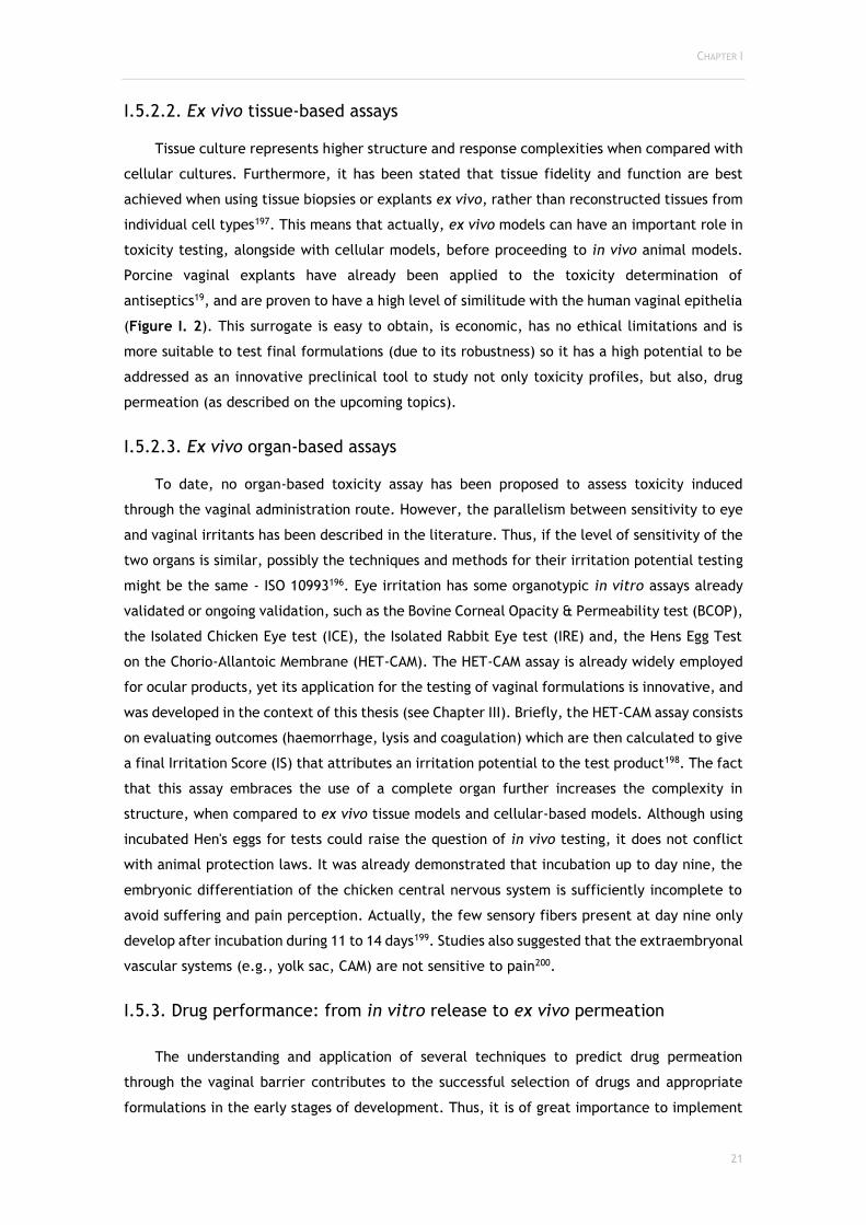
CHAPTER I
21
I.5.2.2. Ex vivo tissue-based assays
Tissue culture represents higher structure and response complexities when compared with
cellular cultures. Furthermore, it has been stated that tissue fidelity and function are best
achieved when using tissue biopsies or explants ex vivo, rather than reconstructed tissues from
individual cell types197. This means that actually, ex vivo models can have an important role in
toxicity testing, alongside with cellular models, before proceeding to in vivo animal models.
Porcine vaginal explants have already been applied to the toxicity determination of
antiseptics19, and are proven to have a high level of similitude with the human vaginal epithelia
(Figure I. 2). This surrogate is easy to obtain, is economic, has no ethical limitations and is
more suitable to test final formulations (due to its robustness) so it has a high potential to be
addressed as an innovative preclinical tool to study not only toxicity profiles, but also, drug
permeation (as described on the upcoming topics).
I.5.2.3. Ex vivo organ-based assays
To date, no organ-based toxicity assay has been proposed to assess toxicity induced
through the vaginal administration route. However, the parallelism between sensitivity to eye
and vaginal irritants has been described in the literature. Thus, if the level of sensitivity of the
two organs is similar, possibly the techniques and methods for their irritation potential testing
might be the same - ISO 10993196. Eye irritation has some organotypic in vitro assays already
validated or ongoing validation, such as the Bovine Corneal Opacity & Permeability test (BCOP),
the Isolated Chicken Eye test (ICE), the Isolated Rabbit Eye test (IRE) and, the Hens Egg Test
on the Chorio-Allantoic Membrane (HET-CAM). The HET-CAM assay is already widely employed
for ocular products, yet its application for the testing of vaginal formulations is innovative, and
was developed in the context of this thesis (see Chapter III). Briefly, the HET-CAM assay consists
on evaluating outcomes (haemorrhage, lysis and coagulation) which are then calculated to give
a final Irritation Score (IS) that attributes an irritation potential to the test product198. The fact
that this assay embraces the use of a complete organ further increases the complexity in
structure, when compared to ex vivo tissue models and cellular-based models. Although using
incubated Hen's eggs for tests could raise the question of in vivo testing, it does not conflict
with animal protection laws. It was already demonstrated that incubation up to day nine, the
embryonic differentiation of the chicken central nervous system is sufficiently incomplete to
avoid suffering and pain perception. Actually, the few sensory fibers present at day nine only
develop after incubation during 11 to 14 days199. Studies also suggested that the extraembryonal
vascular systems (e.g., yolk sac, CAM) are not sensitive to pain200.
I.5.3. Drug performance: from in vitro release to ex vivo permeation
The understanding and application of several techniques to predict drug permeation
through the vaginal barrier contributes to the successful selection of drugs and appropriate
formulations in the early stages of development. Thus, it is of great importance to implement

INTRODUCTORY OVERVIEW
22
accurate and reproducible methods to predict drug permeation. Tissues explants (ex vivo),
being more representative in structure and presenting more viability, shall be preferred over
in vitro cellular methodologies. Standardization and validation of methodologies to be used by
different research groups will be obviously valuable for uniform interpretation and
extrapolation of test results. Moreover, these tests may be particularly important for the
development and characterization of new products intended to reach the market since they
provide vital information for marketing authorization purposes.
I.5.3.1. Drug absorption from the vagina
Drug dissolution in the vaginal fluid and epithelial penetration are the two key steps for a
drug to be absorbed through the vagina. As a result all factors associated with vaginal physiology
and formulation profile will greatly influence the success of drug delivery to the target6,7. Even
though the vagina is not a real mucosa, drug transport is accomplished in a multi-way
mechanism similar to the other biological membranes. Drug absorption can occur passively or
actively. Passive mechanisms include the transcellular route, through the cells’ membrane, and
the paracelullar route, representing a diffusion process through intercellular fluid and tight
junctions201,202. Tight junctions and other intracellular junctions (adherens junctions and
desmossomes) are present in the vaginal and cervical epithelium having a higher expression in
the endocervix. Although the uppermost layers of the ectocervical and vaginal epithelia are
devoid of tight junctions, these and other intercellular junctions have been identified right
beneath the most apical epithelium layers. The study of these junctions is particularly
important to understand, for example, the invasion of microbes and drug permeation. Tight
junctions are composed of transmembrane proteins (occludin, claudins and junctional adhesion
molecules - JAMs) which contact across the intercellular space and create a seal to restrict
paracellular diffusion of molecules across the epithelial sheet. Furthermore, tight junctions
have a structural role in epithelial polarization by limiting the mobility of membrane-bound
molecules between the apical and basolateral domains of the plasma membrane of each
epithelial cell. In general, tight junctions are responsible for sealing the epithelial barrier as
well as the selective passage of small ions and fluid201. The active transporters generate
gradients across the barriers, being the mechanisms either a primary transcellular process or a
secondary one. While the latter is indirectly coupled to adenosine triphosphate (ATP) energy,
the primary active transport directly utilizes ATP during the transport cycle203. Nevertheless,
previous studies on vaginal permeation of drugs show that most of the active substances
permeate the vagina through diffusion mechanisms17,98. Lipophilic substances, such as steroids,
are mainly absorbed by the transcellular route32, in contrast to hydrophilic drugs which follow
the paracellular diffusion mechanism17. Low molecular weight lipophilic drugs are more likely
to be better absorbed than larger molecules or even hydrophilic drugs, independently of their
molecular weight5.
Among several factors that do affect drug absorption from the vagina, both vaginal fluid
amount and composition interfere in drug dissolution before transport. While an excessive fluid
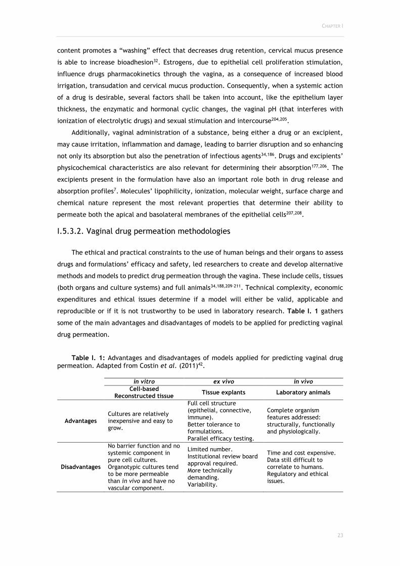
CHAPTER I
23
content promotes a “washing” effect that decreases drug retention, cervical mucus presence
is able to increase bioadhesion32. Estrogens, due to epithelial cell proliferation stimulation,
influence drugs pharmacokinetics through the vagina, as a consequence of increased blood
irrigation, transudation and cervical mucus production. Consequently, when a systemic action
of a drug is desirable, several factors shall be taken into account, like the epithelium layer
thickness, the enzymatic and hormonal cyclic changes, the vaginal pH (that interferes with
ionization of electrolytic drugs) and sexual stimulation and intercourse204,205.
Additionally, vaginal administration of a substance, being either a drug or an excipient,
may cause irritation, inflammation and damage, leading to barrier disruption and so enhancing
not only its absorption but also the penetration of infectious agents34,186. Drugs and excipients’
physicochemical characteristics are also relevant for determining their absorption177,206. The
excipients present in the formulation have also an important role both in drug release and
absorption profiles7. Molecules’ lipophilicity, ionization, molecular weight, surface charge and
chemical nature represent the most relevant properties that determine their ability to
permeate both the apical and basolateral membranes of the epithelial cells207,208.
I.5.3.2. Vaginal drug permeation methodologies
The ethical and practical constraints to the use of human beings and their organs to assess
drugs and formulations’ efficacy and safety, led researchers to create and develop alternative
methods and models to predict drug permeation through the vagina. These include cells, tissues
(both organs and culture systems) and full animals34,188,209–211. Technical complexity, economic
expenditures and ethical issues determine if a model will either be valid, applicable and
reproducible or if it is not trustworthy to be used in laboratory research. Table I. 1 gathers
some of the main advantages and disadvantages of models to be applied for predicting vaginal
drug permeation.
Table I. 1: Advantages and disadvantages of models applied for predicting vaginal drug permeation. Adapted from Costin et al. (2011)42.
in vitro ex vivo in vivo
Cell-based Reconstructed tissue
Tissue explants Laboratory animals
Advantages Cultures are relatively inexpensive and easy to grow.
Full cell structure (epithelial, connective, immune). Better tolerance to formulations. Parallel efficacy testing.
Complete organism features addressed: structurally, functionally and physiologically.
Disadvantages
No barrier function and no systemic component in pure cell cultures. Organotypic cultures tend to be more permeable than in vivo and have no vascular component.
Limited number. Institutional review board approval required. More technically demanding. Variability.
Time and cost expensive. Data still difficult to correlate to humans. Regulatory and ethical issues.

INTRODUCTORY OVERVIEW
24
I.5.3.2.1. In vitro models
Over the last decades, cell-based in vitro models for drug permeation prediction allowed
the use of simple, economic, reproducible and ethically accepted methodologies in the earlier
stages of drug development. These models use both immortalized cells and primary cell cultures
that are able to proliferate in monolayers, simulating the intended epithelium, both structurally
and functionally208. Cell culture models can be easily manipulated, in terms of culture
conditions and parameters, and represent good drug absorption predictors. Human cell lines or
primary cell cultures circumvent problems related to the use of animal tissue on in vitro
models212.
The main limitation of in vitro models, result from the impossibility of integrating cell
external factors (diseases, age, hepatic and renal functions, environmental characteristics)
that are imperative for the whole organism function. Although, extrapolation of in vitro results
to humans must be done very carefully208, the in vitro-in vivo correlation represents an
important tool for prediction of in vivo pharmacokinetics. Clark et al. in 2011 tried to compare
results obtained in vitro with those in vivo for a preclinical evaluation of a microbicide in the
form of a micronized and non-micronized drug. However, in vivo pharmacokinetic results (in
rabbits) failed to corroborate the higher permeability for the micronized drug obtained with
reconstructed tissue in vitro studies208.
Drug physicochemical and biopharmaceutical properties as well as physiological and
environmental organism characteristics should also be considered. Among them are drug
solubility, acid dissociation constant (pKa), drug permeation, octanol-water partition
coefficient (logP) and the environment fluid pH213. Therefore, in vitro permeability studies also
play a key role in determining a formulation strategy in order to assist solubility, dissolution
and stability214.
In the 90’s Gorodeski et al. developed a method based on the harvesting of cervical-vaginal
cells collected from women, aged from 22 to 49 years, undergoing hysterectomy. Initially, a
primary cell culture of human ectocervical epithelial cells was obtained (hECE)215, that included
histologically normal cells only. This technique was applied to permeation mechanistic studies.
To this purpose, the cells were grown on collagen-coated ceramic-based filters and
differentiated into a 5-12 cell layers to form a squamous stratified epithelium, mimicking the
biological characteristic human cervical-vaginal epithelium216,217. When these cells were
compared to a cervical cell line (CaSki), the latter ones showed to be simpler to maintain and
a better alternative to the primary cultures obtained from hysterectomy specimens. In this
study, it was also evidenced the epithelial nature of CaSki cells and their improved
differentiation when grown on filter support218. Although they are easier to grow, maintain and
test, cell line cultures correspond to immortalised cells and consequently may not mimic real
cell behaviour. From this point of view, primary cell cultures should be valued since they
represent a more reliable biologic model.

CHAPTER I
25
When different cellular models were developed and compared, hECE, the primary culture,
was settled as a model for ectocervical epithelium, while ECE16-1 and CaSki, both immortalised
cell lines, expressed phenotypic characteristics of squamous metaplastic cervical epithelium
and endocervical epithelium respectively219. Later on, CaSki endocervical cell line was shown
to be able to form cell monolayers and even bi/trilayers, depending on cell seeding density,
and was proven to be useful for transport mechanisms studies220–222. These experiments resulted
in interesting findings about the importance of tight junctions and their influence on
transepithelial electrical resistance (TEER)217 and the increase of TEER by seminal fluid221. They
also contributed to improve the knowledge about the modulation of vaginal permeability to
pyranine (used as a model drug for paracellular transport) by factors such as estrogen
stimulation and aging (due to changes in the resistance of both the lateral intercellular space
and the intercellular tight junctions)223–225, or extracellular ATP and Ca2+ (due to their effects
at the tight junctions level)218,226.
Permeability studies using either primary cultures or cell lines of vaginal/cervical
epithelial cells have been described in the literature and are herein discussed223,225,227–229. Cell
lines represent more standardized models than primary cultures, since they are ready to use
without rising important inter laboratory variability results and interpretations. However, cells
permeability is higher than explants. As it has not been evidenced the difference in
permeability between primary cell cultures and cell lines but, this topic deserves to be studied
and explored, for example using the TEER measurement. The simplicity of these in vitro models
can lead to limitations in terms of in vitro-in vivo data correlation when compared to methods
based on human vaginal and cervical tissue explants. The importance of establishing these
correlations justifies the need to proceed with tests that better mimetize the corresponding in
vivo mechanisms, which may be made by using reconstructed tissue, tissue models (ex vivo)
and animals (in vivo).
Regarding tests using reconstructed tissue, the Epivaginal® model (MatTek Corporation,
Ashland, United States of America), is a commercial vaginal-ectocervical (VEC) tissue-like
model widely used for microbicides testing. It consists of a 3D-culture of non-transformed
human vaginal-ectocervical epithelial cells grown on polycarbonate cell culture tissue inserts.
The VEC tissue model has proven to be highly reproducible and represents a non-animal method
to assess the irritation effect of contraceptives, microbicides, and vaginal-care products190,211.
Despite being largely used for studying prevention of pathogen transmission230,231 and tissue
toxicity190, the complexity of the multi-layer structure makes it a promising option for
permeability studies. Several other authors have already used EpiVaginal® as model to predict
vaginal permeability to chemicals232,233.
I.5.3.2.2. Ex vivo models
Ex vivo models have been used for predicting drug permeation through the vaginal barrier
by using either human or animal vaginal tissue. Ex vivo tissues allow not only for drug
permeation assays but also enables histological analysis to assess epithelial differences before

INTRODUCTORY OVERVIEW
26
and after drug application/permeation234. While some authors may classify these ex vivo
experiments as in vitro experiments, the two concepts are considered to be different for the
purpose of this thesis. Laboratorial reconstructed tissues are herein classified as in vitro assays,
as they are grown in artificial milieu, while ex vivo assays require tissue excision from animals
or humans, and their use in conditions that preserve their original biological and physical
characteristics for a certain period.
Fresh human cervical explants are clinically obtained from women undergoing planned
hysterectomies. In the USA some repositories provide fresh samples to research institutes and
industries, nevertheless it is doubtful that the samples available are sufficient to support
extensive experiments, such as the toxicological ones42. Researchers are concerned with the
difficulty to obtain and maintain human explant specimens at the laboratory. To overcome
these difficulties, freezing of specimens has been considered and no negative influences in
permeability parameters were found for this tissue conservation method. In fact, no statistical
differences were found between fresh and frozen tissue permeability results when the later
was collected 1 hour after the surgical excision, transported in PBS or culture media at 5ºC,
frozen with methanol/dry ice or liquid nitrogen, and kept frozen at -80ºC17. Previous studies
conducted by van der Bijl et al. showed that freezing at -85ºC had no effect on tritiated water
permeation through vaginal tissue, when this has been kept in a transport fluid, and transferred
to laboratory within 1 hour, snap-frozen in liquid nitrogen and stored at -85ºC for up to 6
months235–239. Although, permeability studies may not be influenced by tissue freezing-thawing,
the impact on vaginal irritation is not clear, since cellular viability might be affected42,240.
Meanwhile, the difficulties encountered with the human tissue samples impelled the demand
for other alternatives, such as animal tissues.
Vaginal tissue obtained from different animal species such as rodents, rabbits, monkeys,
cows and sheep have been used for permeability studies241–245. Vaginal absorption may be
significantly different when comparing animal and human models. Bovine vaginal tissue was
used to test drugs permeation, as surrogate for human tissue due to anatomical and
physiological similarities245. Nonetheless, bovine epithelium exhibits significant histological
differences when compared with human vaginal epithelium and is not widely applied. In
contrast, porcine tissue represents the animal model most commonly applied for this purpose.
Porcine ex vivo tissue specimens are convenient, since they are simple to handle, inexpensive
to obtain and easy to work comparing to the whole animal. Human and porcine vaginal tissues
present substantial histological similarities34,240 (stratified squamous epithelium supported by
connective tissue - Figure I. 2). Additionally, porcine vagina can be easily accessed through
local slaughterhouses.
Although, in general, porcine vaginal tissue seems a good ex vivo permeability model for
human vaginal tissue extrapolation it is of great importance to validate a permeability study
concerning other tissues or in vitro tests. It has already been shown that for hydrophilic
molecules, (water and vasopressin, for example), the porcine vaginal tissue is an accurate in
CHAPTER I
27
vitro permeability model of human tissue, however for more lipophilic molecules (such as
oxytocin) the flux through porcine vaginal tissue was 53% higher than the corresponding
estimated value through the human vaginal tissue246. Ex vivo tissues are mainly involved in drug
permeation testing by using one of three established techniques: Franz cell systems, flow
through diffusion cell and Ussing chambers.
Figure I. 2: Comparison between H&E-stained vaginal epithelium of: human (a), rabbit (b), rhesus monkey (c), pig (d), mouse (e), EpiVaginal from MatTek Corporation (Ashland, MA, USA) (f) and HVE — Human vaginal Epithelium from SkinEthic (Nice, France) (g). Reproduced from42 with kind permission from FRAME.
I.5.3.2.3. In vivo models
In vivo models represent the most complete approach to achieve experimental data.
However, they are associated with ethical issues, require high cost expenditures and are time
consuming. Vaginal permeability testing is scarce on in vivo studies, although it could be of
great interest especially for systemic drug delivery systems, provided that it is previously
validated, ethic, safe and have clear benefit to new drug and formulation development.
Acartürk et al. used normal and ovariectomized rabbits to mimetize different reproductive
physiological status, particularly post-menopausal human vagina. Explants of vaginal tissue
from these animals were further used for comparative in vivo/ ex vivo vaginal permeability and
enzymatic activity studies247. This model was also applied in the study of nonoxynol-9 as
spermicide248. More recently, the pig model has also been used for in vivo experiments. Female
large white pigs were used for the determination of zidovudine and polystyrene sulfonate in
INTRODUCTORY OVERVIEW
28
plasma and vaginal tissue, after the application of an intravaginal bioadhesive polymeric device
for up to 28 days. At the end of this period, vaginal tissue was collected for histological analysis,
and for substances extraction. Both zidovudine and polystyrene sulfonate were found in low
concentrations in plasma, indicating the high retention in the vagina. Additionally no
histopathological toxicity was evidenced for this intravaginal bioadhesive polymeric device249.
I.5.3.2.4. Release and permeation systems
The United States Pharmacopeia (USP) clearly defines the performance tests for topical
drug products, including vaginal administration products, which are focused on the assessment
of in vitro drug release. It is defined that this test must be performed in vertical diffusion cell
(VDC) systems, which by their simplicity can provide reliable and reproducible measurements
of drug release from semisolid formulations. Franz cells represent the most often used VDC, on
the top of which 200-400 mg of the testing formulation is placed, over the membrane to be
studied. The application site can be variable in size, but it has typically 15 mm in diameter.
For vaginal drug delivery systems the assay temperature must be kept at 37±1ºC. Samples of
the recipient fluid are collected up to 4 - 5 hours, and the volume withdrawn is replaced with
fresh medium. Sink conditions must be assured, meaning that the receptor medium must have
a high capacity to dissolve the drug, and the receptor media should not exceed 10% of the
concentration of the standard at the end of the test250. Moreover, FDA's Guidance for Industry
on Scale Up and Post Approval Changes for Semisolid (SUPAC-SS) dosage forms describes the
release rate studies using the vertical diffusion cell (Franz cell) procedure and requires in vitro
release rate comparison between prechange and postchange products for approval of SUPAC-
related changes251.
Although USP recommendations point towards the use of synthetic, inert and highly
permeable membranes in the context of these drug release studies, the interest in performing
in vitro permeability studies using biological membranes is evident. Biological
tissues/membranes are more similar to the in vivo conditions and may provide information on
bioavailability of the drug. However, they are also more complex, and the obtained data can
be more difficult to interpret and discuss. Generally, the difficulties to be encountered are
related to tissue preparation and system setting-up. The membrane preparation depends on
tissue provenience. Since human tissues are difficult to obtain, frozen and thawed samples are
frequently used following previous studies that demonstrated that this procedure does not
affect tissue permeability238,252. Permeation systems intended to study vaginal drug release
from vaginal dosage forms include Franz cell systems, flow through diffusion cells and Ussing
chambers.
The Organisation for Economic Co-operation and Development (OECD) is an
intergovernmental institution which harmonises policies in developed countries and has issued
several publications concerning the safety assessment of chemicals and chemical preparations.
Since, for example, vaginal lubricants can be classified as medical devices or even hygiene
(cosmetic) products, for marketing purposes, (i.e. they are not medicines, and so do not
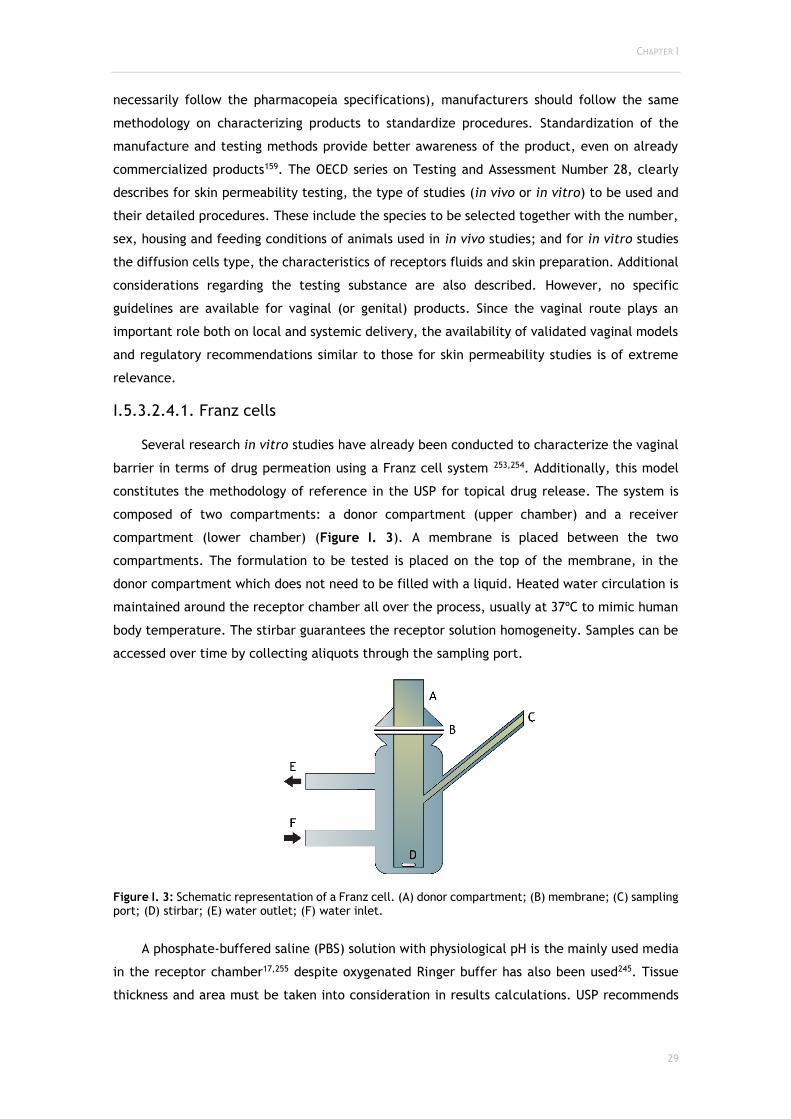
CHAPTER I
29
necessarily follow the pharmacopeia specifications), manufacturers should follow the same
methodology on characterizing products to standardize procedures. Standardization of the
manufacture and testing methods provide better awareness of the product, even on already
commercialized products159. The OECD series on Testing and Assessment Number 28, clearly
describes for skin permeability testing, the type of studies (in vivo or in vitro) to be used and
their detailed procedures. These include the species to be selected together with the number,
sex, housing and feeding conditions of animals used in in vivo studies; and for in vitro studies
the diffusion cells type, the characteristics of receptors fluids and skin preparation. Additional
considerations regarding the testing substance are also described. However, no specific
guidelines are available for vaginal (or genital) products. Since the vaginal route plays an
important role both on local and systemic delivery, the availability of validated vaginal models
and regulatory recommendations similar to those for skin permeability studies is of extreme
relevance.
I.5.3.2.4.1. Franz cells
Several research in vitro studies have already been conducted to characterize the vaginal
barrier in terms of drug permeation using a Franz cell system 253,254. Additionally, this model
constitutes the methodology of reference in the USP for topical drug release. The system is
composed of two compartments: a donor compartment (upper chamber) and a receiver
compartment (lower chamber) (Figure I. 3). A membrane is placed between the two
compartments. The formulation to be tested is placed on the top of the membrane, in the
donor compartment which does not need to be filled with a liquid. Heated water circulation is
maintained around the receptor chamber all over the process, usually at 37ºC to mimic human
body temperature. The stirbar guarantees the receptor solution homogeneity. Samples can be
accessed over time by collecting aliquots through the sampling port.
Figure I. 3: Schematic representation of a Franz cell. (A) donor compartment; (B) membrane; (C) sampling port; (D) stirbar; (E) water outlet; (F) water inlet.
A phosphate-buffered saline (PBS) solution with physiological pH is the mainly used media
in the receptor chamber17,255 despite oxygenated Ringer buffer has also been used245. Tissue
thickness and area must be taken into consideration in results calculations. USP recommends
INTRODUCTORY OVERVIEW
30
the use of synthetic and inert membranes in which the product to test is placed, letting the
system to saturate for 30 minutes (equilibration period). Aliquots from the receptor
compartment are collected at predefined time intervals. The aliquots volume withdrawn must
be replaced with the same volume of receptor solution to ensure sink conditions. Afterwards,
aliquots should be analysed through diverse analytical techniques for identification of drug in
the collected sample. Membrane/tissue should also be checked for histological modifications
after the permeability study. Steady-state flux (JSS) and apparent permeability coefficient (Papp)
can be calculated based on the Fick’s First Law of Diffusion235.
The permeability chamber design may be responsible for results’ variability. Classic Franz
cell system used in permeation studies can lead to stasis and/or accumulation of perfusate in
the receptor chamber, which can be overcome by using a diffusion flow-through system254.
Concerning the experimental conditions, temperature stands as an essential parameter to be
controlled since drug diffusion depends on temperature. Also, the origin of tissue and the
experiment duration must be optimized to get the best profit of permeability studies34.
I.5.3.3. In vitro drug release
Dissolution testing in the pharmaceutical industry has been employed as a fundamental
tool both in formulation design and quality control251,256–261. It was initially developed for
immediate release but then it was extended to controlled/modified release and other dosage
forms. Recently, dissolution testing was widened to a variety of "novel" or "special" dosage
forms such as suspensions, orally disintegrating tablets, chewable tablets, chewing gums,
transdermal patches, semisolid topical preparations, suppositories, implants and injectable
microparticulate formulations, and liposomes261. In terms of terminology, for non-oral dosage
forms (topical and transdermal delivery systems, and suppositories), the test is referred as
"drug release" or as "in vitro release". It is worth noting that in general, an in vitro
dissolution/release test is expected for each novel/special dosage form, regardless of whether
the intended effect is systemic or non-systemic (eg, topical semisolid dosage forms)261.
The selected membrane should provide an inert holding surface for the test formulation,
but not a barrier, so that the drug release would reflect the vehicle properties and not the
membrane rate-limiting properties.
In vitro release is one of several standard methods which can be used to characterize
performance characteristics of a finished topical dosage form, i.e., semisolids such as creams,
gels, and ointments. Important changes in the characteristics of a drug product formula or of
the thermodynamic properties of the drug(s) it contains should show up as a difference in drug
release262. Release is theoretically proportional to the square root of time (√t) when the
formulation in question is in control of the liberation process because the release is from a
receding boundary251.
The drug in the dissolved or suspended state in the vehicle has to diffuse and be released
by the vehicle, becoming theoretically available to penetrate into different epithelia. For
quality control purposes, the method can be employed as performance test designed to
CHAPTER I
31
demonstrate that the dosage forms were manufactured according to specifications and all
critical manufacturing steps assuring batch-to-batch sameness. Secondly, in research and
development the focus of release testing is shifted to other purpose. The vehicle composition
and design strongly influence the product performance and the speed of the drug release and
ability to permeate biological membranes263.
I.5.3.4. Quantification methods development and validation
To accomplish drug quantification for the abovementioned methods, a bioanalytical tool
should be conveniently developed, optimised and validated. Bioanalysis includes the set of
analytical techniques and procedures applied in the characterisation and quantitative
determination of drugs in various matrices264.
Firstly, the analytical conditions must be optimized, in order to a proper validation could
consequently be performed. Only this step of validation can provide accurate, precise and
reproducible data throughout study-sample analysis. The method validation is the systematic
process of defining the analytical requirements, confirming that the method under
consideration has performance capabilities consistent with the demands of its intended
application265.
High Pressure Liquid Chromatography (HPLC) represents nowadays the standard
quantitative methodology, being able to separate and quantify mixtures of compounds with low
and high molecular weight, as well as different polarities and acid-base properties in a variety
of matrices266. The development of a bioanalytical method is unique and specific for each drug
candidate and it depends, among various factors, on the analyte physicochemical features (e.g.
molecular weight, pKa and lipophilicity), nature of the matrix, sensitivity required and the
available instrumentation. Chromatographic analysis intends the separation of compounds in a
sample with good resolution, without interferences and within a reasonable running time. In
general, the HPLC separation of the analytes is accomplished based on the differences in
affinity of the compounds between two phases: one mobile and the other stationary. Since the
majority of the drugs and metabolites are polar in nature, the reversed-phase chromatography
is the most commonly applied method in which the mobile phase is polar, composed of a
mixture of water/buffer and organic solvents (acetonitrile or methanol); and the stationary
phase is non-polar, consisting of hydrocarbon chains chemically attached to the silica base
packing of the column (e.g. C18 and C8). Hence, while polar compounds get eluted firstly, the
non-polar ones are retained in the column for a longer period of time and therefore, the
separation of the analytes is sorted according to their increasing molecular hydrophobicity.
Furthermore, depending on the number and polarity of the compounds to be resolved or
separated, the mode of elution can be stipulated as isocratic or gradient267. Additionally, buffer
solution could be needed when an analyte is ionic or ionisable under reversed-phase conditions
(pH values within the range 2-8)266. The flow rate and temperature of analysis may likewise
have quite significant effects on the analytes’ resolution and retention time. The choice of the
most convenient system for the detection of the eluting components is dependent on the
INTRODUCTORY OVERVIEW
32
analytes’ properties. Different types of detectors, such as ultraviolet (UV), fluorescence and
mass spectrometry, are available to be used in HPLC analysis. Even though, in general terms,
either fluorescence or mass spectrometry provide better sensitivity and selectivity than UV
detection, but the latter is cheaper, more easily accessible and presents a broad spectrum of
application268.
Owing to the undeniable importance of method validation in whole field of bioanalysis, it
was felt the need to standardise the overall validation parameters and acceptance criteria by
globally establishing appropriate guiding principles. Since the 90s, several workgroups have
addressed this issue, and published relevant reports264 which subsequently, served to establish
the general recommendations on bioanalytical method validation269. Also, the European
Medicines Agency (EMA), published in 2011 a similar document to facilitate regulatory issues
on the European countries270. Furthermore, the International Conference for Harmonization has
also contributed to this area of validation by publishing the topic Q2 (R1) “Validation of
Analytical Procedures: text and methodology”.
All three agencies recommend that the method validation should conduct a deep
investigation concerning parameters such as selectivity, linearity, precision, accuracy, limit of
quantification and limit of detection, recovery and stability; and even state the limits of
acceptation for each parameters.
Concerning the method validation, it comprises three different levels whose application
depends on the circumstances in which the method was developed and how it is going to be
used: full validation, partial validation and cross validation269. A full validation is required when
the method is implemented for the first time or a new drug/metabolite is included. However,
in situations where minor alterations are made to a previously validated method, such as
transfer between laboratories or analysts, changes in equipment, in species within matrix, in
matrix within species, in sample processing procedures, in storage conditions, etc., a full
validation may not be necessary and therefore, a partial validation can be performed ranging
from as little as one intra-assay accuracy or precision determination to an almost full
validation269. A cross validation consists of a comparison of the validation parameters when two
or more bioanalytical methods are used to generate data within the same or across different
studies conducted in distinct laboratories.
CHAPTER I
33
I.6. Aims of this thesis
Although there are several formulations commercially available for vaginal use, both for
therapeutic purposes and for cosmetic/hygienic or medical devices actions, preclinical data on
its potential to cause acute irritation and toxicity is not widely addressed. The global aim of
this thesis is to establish methods, from simple technological characterization to complex ones,
such as ex vivo permeation, that could be predictive at early stages of formulations
development of their in vivo performance. To achieve this, a collection of ten semisolid vaginal
medicines, globally available, was included in this study. Furthermore, two formulations, that
do not contain active substances, but are widely described on the literature, were used as
controls.
Specifically, it is intended to:
□ Develop, improve and apply a complete technological characterization methodology
for vaginal semisolids, from a safety and performance perspective, considering their
pharmacotechnical profile;
□ Optimize cell-based and organ-based (in vitro and ex vivo) models to test the
formulations toxicity, while applying techniques capable of predicting their acute
irritant potential;
□ Perform drug delivery studies, including in vitro release and ex vivo permeation of the
active substances included in the formulations to refine experimental setups and
generate data for comparison with other products.
Moreover, during the deployment of this work, the potential for knowledge transfer for the
pharmaceutical industry was also a core objective. Since this thesis is part of a university-
company partnership, the methods herein explored are expected to fit the processes of new
products development in-house or as services provided to the pharmaceutical, medical devices
and cosmetic industries.

INTRODUCTORY OVERVIEW
34

CHAPTER II
35
CHAPTER II
TECHNOLOGICAL CHARACTERIZATION:
CHEMICAL, PHYSICAL AND PHYSIOLOGICAL
METHODOLOGICAL INSIGHTS

TECHNOLOGICAL CHARACTERIZATION
36
The content of this chapter is partially published in:
Machado RM, Palmeira-de-Oliveira A, Martinez-de-Oliveira J, Palmeira-de-Oliveira R, “Vaginal
semisolid products: Technological performance considering physiologic parameters”. European
Journal of Pharmaceutical Sciences. 2017, doi: 10.1016/j.ejps.2017.09.009

CHAPTER II
37
II.1. GENERAL
CONSIDERATIONS

TECHNOLOGICAL CHARACTERIZATION
38
CHAPTER II
39
Several formulations for vaginal administration are available in the market as medicines
or OTC (Over-the-Counter) products, some of them classified as medical devices or cosmetics.
However, especially for the later ones, there are few data on their potential to cause acute
irritation and toxicity, or their suitability is poorly supported, particularly because preclinical
assays cannot be performed on animals and clinical trials are not usually considered.
Among the semisolid vaginal dosage forms creams and gels have gathered particular
interest among researchers engaged in developing vaginal formulations16. They have been
pointed as the most preferred dosage forms among women although leakage has been
frequently referred as their major drawback. These formulations may require multiple daily
administrations in order to obtain the desired therapeutic concentration and to provide a
uniform distribution of the drug5,49–51.
To overcome this limitation, major research in this field has been focused on bioadhesive
polymers or novel dosage forms, but by now only limited evolution has been found in
commercial vaginal products. Those strategies are, however, expected to increase residence
time, improving both efficacy and safety of the new delivery systems.
Technological properties of vaginal semisolids have been considered to correlate with their
in vivo performance regarding safety and efficacy. This is the case for the ability of formulations
to maintain the normal vaginal pH271, to be isosmolal159 and to improve the retention through
adhesion measurements173. Not only should researchers aim for better products but also to
develop selective tools (i.e. characterization methods) to characterize the products more
precisely151, and so, better predict their performance. Also, new methods should address
physiologic featurings in perspective of earlier cognizance of in vivo possible effects. Ultimate
acceptability and clinical efficacy of such preparations require that they possess optimal
mechanical properties (ease of removal from the container, spreadability over the substrate),
rheological properties (viscosity, elasticity, thixotropy, flowability), and other desired
properties such as bioadhesion, desired drug release, and absorption152. Nevertheless, still very
little attention has been given to the influence of the vaginal environment on formulation
performance. Actually, formulations testing is generally carried without considering the effect
of physiologic parameters such as temperature and solvency in vaginal fluids. It is well known
that these variables can impact on formulations properties, especially, pH, osmolality, rheology
and ultimately bioadhesion. For example, Aka-Any-Grah et al. have performed characterization
studies using dilutions in simulated vaginal fluid to determine final rheological profiles and
bioadhesion in early steps of formulations development and concluded that this tool is supposed
to predict more accurately the in vivo behaviour109.
This chapter pretends to characterize several products already marketed for vaginal
administration, using well established methods and also proposing new methods and approaches
to gather primary information of the formulations behaviour when in contact to physiologic
vaginal fluid.

TECHNOLOGICAL CHARACTERIZATION
40
II.1.1. Objectives
The objective of this part of the work is to develop and apply a characterization
methodology for vaginal semisolid formulations in view of safety and performance considering
physiological parameters. Specifically, it is intended to:
□ Determine the organoleptic characteristics of the formulations in study;
□ Perform pH and acid-buffering capacity assays on vaginal fluid simulant;
□ Disclose formulations osmolality, and even its osmolality after a physiologic dilution;
□ Evaluate formulations textural characteristics through firmness and adhesiveness;
□ Characterize formulations viscosity directly and diluted in physiologic fluids;
□ Perform bioadhesion evaluations on a mechanical ex vivo model.

CHAPTER II
41
II.2. EXPERIMENTAL

TECHNOLOGICAL CHARACTERIZATION
42

CHAPTER II
43
II.2.1. Materials and Methods
II.2.1.1. Tested products
The semisolid products tested in this study are classified as medicines and were acquired
in Portuguese community pharmacies, being also commercialized across the EU and USA (Table
II. 1). Some of these are Over-the-counter (OTC) products, not requiring a medical prescription
to be dispensed at pharmacies (Gino-Canesten® and Gyno-Pevaryl®). The majority of these
dosage forms are creams, except Blissel® that is a gel. All products are intended for vaginal
use except Colpotrophine®, which is recommended by the manufacturer for vulvar application.
Universal placebo gel99,158,233,272 and Replens®14,93,166,173 were included, since they represent
widely studied formulations. The comprehensive and attentive review on vaginal products
classification showed us that there is misunderstanding between vaginal and intravaginal
products. We assume that when a product is intended for vaginal application it means
intravaginal administration, and not for external/vulvar application, like the case of
Colpotrophine®. This term inaccuracy is specially noted in the case of vaginal solutions, which
should fit up for intravaginal irrigations, and that in the majority of the cases are indicated for
external washes.
II.2.1.2. Materials
Vaginal fluid simulant (VFS) was prepared as described by Owen and Katz in 1999: sodium
chloride 3.51 g (JT Baker, United States of America), potassium hydroxide 1.4 g (VWR Prolabo,
France), calcium hydroxide 0.22 g (Acros Organics, United States of America), Bovine Serum
Albumin 0.018 g (Sigma, Germany), lactic acid 2.00 g (Sigma, Germany), acetic acid 1.00 g
(Fischer Scientific, United States of America), glycerol 0.16 g (Acofarma, Spain), urea 0.4 g
(VWR Prolabo, France) and glucose 5.00 g (VWR Prolabo, France) were added to 1 L of milliQ
water and stirred mechanically until complete dissolution37. The pH of the mixture was then
adjusted to 4.5 using hydrochloric acid, and the final volume was adjusted to 1 L. All other
chemicals and reagents were of analytic grade or equivalent. For the modified VFS (VFSm) a
1.5% (w/w) concentration of porcine gastric mucin type II (Sigma, Germany) was used273.
II.2.1.3. Organoleptic characteristics
The organoleptic characteristics studied were: general aspect, homogeneity, colour,
odour, feel to touch.

TECHNOLOGICAL CHARACTERIZATION
44
Table II. 1: Studied formulations general characteristics (information provided by the manufacturer).
Therapeutic action
Trade name/labelled dosage form
API Excipients
Marketing Authorization
Holder (in Portugal)
Antifungals
Gino-Canesten®, vaginal cream
Clotrimazole 10 mg/g
Benzyl alcohol, Cetyl palmitate, Cetostearyl alcohol, Purified water, Polysorbate 60, Sorbitan monostearate, Octyldodecanol
Bayer Portugal, S.A.
Sertopic®, vaginal cream
Sertaconazole 2 g/ 100 g
Ethyleneglycol and polyethyleneglycol, Stearate palmite, Saturated glycerides polyglycolized, Glycerol isostearate, Liquid paraffin, Methylparaben, Sorbic acid
Ferrer Portugal, S.A.
Dermofix®, vaginal cream
Sertaconazole 20 mg/g
Tefose 63, Labrafil, Peceol, Liquid paraffin, Nipagin, Sorbic acid
Azevedos Laboratories – Pharmaceutical Insustry, S.A.
Gyno-Pevaryl®, vaginal cream
Econazole 10 mg/g
Stearate pegoxol 7, Liquid paraffin, Oleic macrogolglycerides, Butylhydroxyanisole (E320), Benzoic acid, Purified water
Johnson & Johnson, Lda.
Lomexin®, vaginal cream
Fenticonazole 20 mg/g
Propylene glycol, Hydrogenated lanolina, Sweet almond oil, Polyglycol esters of fatty acids, Cetyl alcohol, Glyceryl monostearate, Sodium EDTA, Purified water
Recordati Ireland Ltd.
Gino Travogen®, vaginal cream
Isoconazole 10 mg/g
Polysorbate 60, Sorbitan stearate, Cetostearyl alcohol, Thick paraffin, White Vaseline, Purified water
Bayer Portugal, S.A.
Antibacterials Dalacin V®, vaginal cream
Clindamycin 20 mg/g
Propylene glycol, Cetostearyl alcohol, Liquid paraffin, Sorbitan stearate, Benzyl alcohol, Cetyl palmitate, Stearic acid, Polysorbate 60, Purified water
Pfizer Laboratories, Lda.
Oestrogens
Ovestin®, vaginal cream
Estriol 1 mg/g
Octyldodecanol, Glycerol, Cetyl alcohol Stearyl alcohol, Polysorbate 60, Sorbitan stearate, Lactic acid, Chlorhexidine hydrochloride, Sodium hydroxide, Purified water, Synthetic spermaceti
Aspen Pharma Trading Ltd.
Blissel®, vaginal gel
Estriol 50 µg/g
Glicerol (E 422), p-hydroxybenzoic acid methyl ester, p-hydroxybenzoic acidpropyl ester, Policarbophil, Carbomer, Sodium hydroxide, Hydrochloric acid, Purified water
ITF Medivida, Pharmaceutical Products, Unip. Lda.
Colpotrophine®, vaginal cream
Promestriene 1 g/ 100 g
p-hydroxybenzoic acid methyl ester, p-hydroxybenzoic acid propyl ester, Mono- and diglycerides of saturated fatty acids, Polyglycol ether of saturated fatty alcohols, Oleic acid decyl ester, Triglycerides of capric and caprylic acids, Glycerol, Purified water
Teva Pharma – Pharmaceutical Products, Lda.
Controls (Reference products)
Universal placebo, vaginal gel
Not applicable
Purified water, Hydroxyethylcellulose, Sodium chloride, Sobic acid, Caramel color, Sodium hydroxide
Not applicable
Replens® Not applicable
Purified water, Policarbophil, Paraffin oil, Glycerin, Palm oil hydrogenated, Carbomer homopolymer type B, Sorbic acid, Sodium hydroxide
Lil’Drug Store Products, Inc. (in France)
CHAPTER II
45
II.2.1.4. pH and buffering capacity
For the pH determination, the probe (for viscous products, InLab Viscous, Mettler Toledo,
USA) was directly immersed on the formulation (Seven Compact, Mettler Toledo, USA). The
pH-buffering capacity was accomplished by titration with NaOH 1 N added in increments of 20
µL to the dispersion of formulations in Normal Saline – NS (NaCl 0.9%) or VFS until pH≥9159. The
relevant and absolute buffering capacity were determined using the best fit linear model -
Curve Expert Version 1.4 (Copyright 2013, Daniel G. Hyams). The absolute buffering capacity
was calculated and defined as the amount of sodium hydroxide needed to change by one unit
the initial pH value. The relevant pH-buffering capacity was calculated as the amount of sodium
hydroxide required to reach a pH value of 5 (higher than the normal vaginal pH of reproductive
aged women)157,274. For products which had initial pH higher than 5, the reverse buffering
capacity was performed by adding 20 µL of HCl 1 N until pH was lower than 3.
II.2.1.5. Osmolality
The osmolality was determined in triplicate using a freezing point osmometer (Osmomat
3000, Gonotec, Germany), as previously described159,166, on a 50 µL aliquot. The standardization
was performed using three standards: distilled water (zero point), NaCl 300 mOsm/Kg and NaCl
850 mOsm/Kg, commercially available from the equipment manufacturer. Furthermore,
osmolality was determined in a mixture with vaginal fluid simulants. An amount of each product
corresponding to the daily dose (measured with the proprietary applicator) was diluted in 0.75
mL of VFS or VFSm. This procedure was established to estimate the osmolality of the product
when put in contact with vaginal fluid simulants, since 0.75 mL is the estimated mean volume
of fluid present in the vagina at any moment37,49,109.
II.2.1.6. Texture: Firmness and Adhesiveness
Texture analysis included adhesiveness (N.mm) and firmness (N) determinations using a
texturometer (TAXT Plus, Stable Micro Systems, United Kingdom). These two parameters were
determined in the same run, using a cylindrical probe with a diameter of 10 mm (P10)169,170 in
compression mode and «return to start test». The maximum positive force (N) to penetrate the
formulation for 5 mm was registered and corresponds to the formulation firmness (pre-test
speed, test speed and post-speed: 3 mm/s). Force measurements required to detach the probe
from the formulation during the returning movement allowed for the calculation of the work
of adhesion, which is herein described as adhesiveness. Measurements were performed, in
triplicate, at room temperature, complying with the laboratorial and equipment manufacturer
protocol.
TECHNOLOGICAL CHARACTERIZATION
46
II.2.1.7. Bioadhesion
Formulation adhesiveness to the biological substrate (porcine vaginal tissue) was accessed
using the texturometer (TAXT Plus, Stable Micro Systems, United Kingdom). The method
employed consisted on a mechanical approach to bioadhesion since it is based on the evaluation
of tensile strength of the interfacial layer formed between the formulation and the vaginal
epithelium173. The vaginal epithelia was excised from porcine vaginal tubes (obtained from
approximately 6 months’ year old animals, kindly conceded from a local slaughterhouse). The
vaginal tubes were cut longitudinally, washed with Hank's Balanced Salt Solution (HBSS) pH 4.2,
wrapped in aluminium foil, and preserved in an air tight bag at -20ºC. For the experiment,
vaginas were thawed at room temperature in HBSS. The epithelium samples were fixed using a
mucoadhesion rig which was placed on the equipment’s base. The whole system composed of
the mucoadhesion rig with the tissue and the probe with the formulation was kept at 37±0.5 ºC
in an oven. The tissue was hydrated with 50 µL of VFSm, since mucin is the protein most likely
to be responsible for bioadhesion. A double-sided adhesive tape was used to attach a small
piece of cellulose acetate membrane to the probe, where 30 mg of formulation were adsorbed
(the formulation was weighted directly on the probe). Cellulose acetate membrane without
formulation was used as control. The software was used in adhesive mode. The pre-test speed
was 0.5 mm/s with a trigger force of 0.02942 N to allow for sensitive detection of the tissue.
Post-test speed was 0.1 mm/s100. The contact/hold time was 3 min, and the force applied was
2.5 N275. The force of detachment was recorded as well as the graphical negative area,
representative of the work of adhesion (N.mm) necessary to unbind the two surfaces. One-way
ANOVA statistical test with multiple comparisons was applied to denote differences (p < 0.05)
between the control and formulations (GraphPad Prism 6.0).
II.2.1.8. Viscosity
Viscosity was assessed using a cone-plate rheometer (Brookfield DV-3T, Brookfield, USA).
Viscosity measurements were performed at room temperature (25ºC) and at vaginal physiologic
temperature (37ºC), using plain formulations and after diluting in VFS. Cone spindles used were
CPA-52Z and CPA-40Z (Brookfield, USA), both required a 0.5 g or 0.5 mL sample, and had 3°
and 8°, and 1.2 cm and 2.4cm, cone angle and radius, respectively. To assess the thixotropic
behaviour of formulations, a range of test speeds between 5 and 200 RPM was established
(torque 10-100%). Tests were performed in triplicate during 1 min, and the formulation was
left to rest for 1 min between measurements276. Formulations’ dilution to physiologic
assemblies were performed as described on the osmolality section.
Furthermore, to study rheological modifications following application, an ex vivo model
for administration was developed using porcine vaginas. For this purpose, a daily dose of all the
formulations was placed, with the help of the proprietary applicator, onto an excised vagina
obtained from a local slaughterhouse. The organ was isolated from the upper part of the
remaining reproductive system, placed on a tray, and 0.75 mL of VFS were inserted into the

CHAPTER II
47
vagina. Then, the trays were placed in an incubator at 37ºC with mild agitation (50 RPM). After
3 h the trays were removed and the vaginas were opened longitudinally for observation and
collection of the formulation which was further assessed concerning viscosity.
II.2.1.9. Data processing and statistical analysis
Data was analysed to produce arithmetic means with standard deviations (SD) using
Microsoft Excel. Analysis of variance (ANOVA) was performed to determine the significance of
the difference between sets of data (p < 0.05) using GraphPad Prism 6.0 (Graph Pad Software,
USA).

TECHNOLOGICAL CHARACTERIZATION
48
II.2.2. Results and Discussion
II.2.2.1. Organoleptic characteristics
Organoleptic characteristics were determined taking into account both users’ perspective
and formulation suitability for vaginal administration. Thus, evaluated parameters comprised
colour, odour, texture and homogeneity. As previously described women prefer vaginal
products presented as semisolids, odourless and colourless, either being gels, creams or
ointments277,278. Also, women would rather privilege natural origins for drugs/excipients, and
application by means of an applicator22. In terms of pharmaceutical dosage form the majority
of the studied products are labelled as vaginal creams, except for Blissel® and Replens® that
are presented as gels. These gels are both colourless. None of the formulations showed strong
odours. Also, all of them had a soft texture and were homogeneous, being in accordance with
female users expected preferences277. All products, except Colpotrophine®, are marketed with
disposable applicators, in order to allow for a more comfortable application. These
characteristics are also in accordance with women’s expectations reported in previous studies
for determination of the ideal organoleptic characteristics for microbicides using personal
interviews128 and focus groups279. Regarding the clinicians’ perspectives, it has been published
that both general practitioners and gynaecologists believe that vaginal products for self-
administration are valuable, but consider that more pharmaceutical counselling should be
provided in view of increasing therapies’ compliance and efficacy280. Likewise, in recent years,
clinicians and patients’ preferences were studied on the different oral and vaginal therapeutic
options for vaginal fungal infections, concluding that given the therapeutic efficacy and
equivalence of the individual antifungal agents as well as route of administration, treatment
selection should be driven by the patient’s personal preferences281.
II.2.2.2. pH and buffering capacity
The pH of the formulations is an important parameter since it must be compatible with
the vaginal pH which is normally 3.5-4.5, but may vary according to specific vaginal conditions
(hormonal stimulation, menstrual cycle phase, presence or absence of infections)15. However,
as the pH values of vaginal products do not allow by their own for a complete prediction of
their safety, it is more relevant to assess the ability of these formulations to actually change
the physiological pH once administered, by other words the pH-buffering capacity. This
parameter, especially when determined in VFS, contributes to a better understanding of what
will happen in vivo regarding pH changes after the formulation is applied in the vagina159.
Table II. 2 shows the results of pH determinations for the vaginal formulations enrolled in
this study. Antifungals in general had low pH, except for Gino-Canesten® that has a pH of 5.89.
This fact might be due to poor solubility and stability of clotrimazole in acidic
solutions/formulations282, since it is as weak base with a pKa of 6.9283. Additionally, it should
CHAPTER II
49
be considered that fungal infections, which are mainly caused by Candida spp., tend not to
affect the normal vaginal pH284. On the other hand, bacterial infections, the most common
being vaginal bacteriosis (VB), are characterized by pH increase to 5.00-6.00. Dalacin V® has a
pH in the lower limit of the physiologic range (3.54±0.006), which can contribute to reduce the
high vaginal pH value present in VB. Since antimicrobial products are intended to an occasional
administration (vaginal infections are isolated conditions and usually do not require prolonged
therapies) it may be acceptable that they can have pH values out of the physiological range.
On the other hand, products as Ovestin® and Blissel®, that are intended to be used for
prolonged therapies, should exhibit pH values compatible with the normal vaginal pH for safety
issues. In fact, we confirmed that both presented pH values within those considered as
physiological (3.92±0.010 and 4.79±0.046, respectively). Since these two products are
prescribed for chronic application this characteristic may help the formulation to have an
adequate impact in the vaginal milieu. Moreover, vaginal atrophy in menopause is characterized
by vaginal irritation and discomfort, so pH changes due to products application (either acidic
or basic) are expected to increase those symptoms. Colpotrophine® has a manufacturer
specification for vulvar application other than the vaginal, so its pH is not expected to be
comprised within the vaginal range (pH=7.26±0.040), but fitting the pH range recommended
for skin applications: 4.0 to 7.0285. As shown in Table II. 2, after the dilution in VFS for the pH-
buffering capacity assay, formulations acquired the simulant pH (4.21), except for Lomexin®,
Ovestin®, Blissel®, Colpotrophine® and Replens®, which resulted in pH in statistically different
from the control (one-way ANOVA p < 0.05). On the other hand, dilution with the NS control,
did not affect markedly the formulations pH (data not shown).
To evaluate formulations pH-buffering capacities two different endpoints were
considered: the relevant buffering capacity (RBC) and the absolute buffering capacity (ABC).
While the relevant buffering capacity represents the ability of a formulation to overcome a pH
of 5, meaning that it has left the physiologic interval for the vaginal environment, the absolute
buffering capacity represents the ability for a formulation to change 1 pH point from its natural
pH159. Figure II. 1. A represents the relevant buffering capacities for all products included in
this study. Gino-Canesten®, Colpotrophine® and the NS control are represented in white, since
they had initial pH higher than 5 and were titrated with HCl 1 N instead of NaOH 1 N, until
pH≤3.00. These calcutations were performed equaly to the other formulations. Although, these
two formulations had pH above the physiologic limit, they had low buffering capacities (both
RBC and ABC), which means they can easily reach the normal vaginal pH interval.

TECHNOLOGICAL CHARACTERIZATION
50
Table II. 2: pH and osmolality studies of vaginal products included in this study. Results are means ± SD (standard deviation) (n=3). * represents statistically different from the respective dilution media; ᵆ represents statistically different between dilutions with VFS and the undiluted formulation; ᵜ represents statistically different between dilutions with VFSm and the undiluted formulation (two-way ANOVA, p < 0.05).
Direct
pH Diluted pH
(VFS) Direct osmolality
Diluted osmolality (VFS)
Diluted osmolality (VFSm)
pH±SD pH±SD Osmolality
±SD (mOsmol/kg)
Osmolality±SD (mOsmol/kg)
Osmolality±SD (mOsmol/kg)
Gino-Canesten® 5.89±0.07 4.22±0.01 144±3 165±2* 163±1*
Sertopic® 2.71±0.01 4.21±0.01 300±2 272±7* 284±1*
Dermofix® 2.70±0.02 4.19±0.03 248±4 228±2 240±2*
Gyno-Pevaryl® 2.74±0.03 4.07±0.01 340±8 264±3* ᵆ 266±2* ᵜ
Lomexin® 3.57±0.01 4.02±0.05* 1446±20 1210±16* ᵆ 1253±18* ᵜ
Gino Travogen® 3.56±0.04 4.16±0.00 43±2 75±1* 57±1*
Dalacin V® 3.54±0.01 4.19±0.01 1681±10 1223±8* ᵆ 1288±21* ᵜ
Ovestin® 3.92±0.01 3.95±0.03* 3332±60 938±7* ᵆ 1061±27* ᵜ
Blissel® 4.79±0.05 4.32±0.02* 1537±6 920±12* ᵆ 1042±18* ᵜ
Colpotrophine® 7.26±0.04 6.76±0.04* 1723±20 396±4* ᵆ 472±47* ᵜ
Universal placebo 4.51±0.04 4.19±0.02 339±2 306±2* 324±5*
Replens® 2.81±0.02 4.12±0.00* 2350±81 1859±22* 1903±17*
Dilution media 4.21±0.01 212±1 254±1
As expected, all products showed higher buffering capacity after being mixed with VFS due
to the intrinsic effect of this fluid (lighter grey control, Figure II. 1. A and B), when comparing
with the capacity of the normal saline solution.
Universal placebo has revealed little buffering capacity even on VFS (similar to the results
obtained for this control), probably meaning that its application will not modify the vaginal pH.
Dalacin V®, despite having a pH value compatible with the vaginal physiological pH, did not
show a favourable RBC on VFS. Although it would be important to retain a low pH in the
presence of VB (its main therapeutic indication). Blissel®, unlike Ovestin® labelled for the same
therapeutic purpose, had a high RBC, meaning that it is able to maintain the physiological pH
in less acidic conditions.
The ABC values for Gino-Canesten®, Sertopic®, Ovestin®, Colpotrophine® and
Universal placebo were not statistically different from the control on the NS assay. As for the
assays in VFS no differences from the control were found for Sertopic®, Gino Travogen®,
Dalacin V®, Ovestin® and Universal Placebo. Overall, buffering capacity is probably related to
the presence of acidic polymers, as seen for Blissel®159. Also, formulations which had pH out of
the vaginal physiologic range (Gino-Canesten®, Sertopic®, Dermofix®, Gyno-Pevaryl® and
Colpotrophine®), had relatively low capacity to maintain their own pH, which means that by
mixing with acidic vaginal fluids they could change to a value closer to the physiological range
although not being expected to correct the abnormal vaginal pH associated with the target
problem. It is well established that deviations from the normal vaginal pH in the healthy adult
(3.5–4.5) are considered as potentially deleterious for the vaginal epithelium160. Classically, the

CHAPTER II
51
acidic pH in the vaginal environment is believed to contribute to the normal physiology, to
favour microbiota, and to promote a balanced immune response. Vaginal products should be
compatible with vaginal pH and, ideally, maintain it or even help in its reestablishment (e.g.,
in cases of bacterial vaginosis or post-menopausal vaginal atrophy)161. Even if the consequences
of the administration of vaginal formulations presenting undesirable pH is not readily
assessable, it is well known that increased vaginal pH is associated with the presence or favours
bacterial vaginosis, trichomoniosis or mixed infections162.
Figure II. 1: (A) Relevant and (B) Absolute pH-buffering capacity expressed as NaOH meq for the vaginal products included in this study. For Gino-Canesten®, Colpotrophine® and Control in NS, the addition was made with HCl, since their pH were higher than 5. Results are means and bars represent standard deviations (n=3). NS=Normal saline; VFS=Vaginal Fluid Simulant; NS (HCl)=Normal saline tritrated with HCl. * represents statistically different from the NS control and ** represents statistically different from the VFS control (one way-ANOVA, p < 0.05).
TECHNOLOGICAL CHARACTERIZATION
52
Outcomes of low pH are even less understood, but animal data suggest that values of three
or less are generally unacceptable for human use155. Furthermore, apart from being pH
compatible, vaginal products should allow the maintenance of the vaginal acidic environment
and oppose pH-raising/decreasing events. Indeed, the use of acid-buffering gels has been
proposed for the reestablishment of pH in cases of infection66,157 or menopausal atrophy163. A
study of pH and buffering capacity of diverse marketed vaginal lubricants has already been
performed within our workgroup159 and we concluded that most of the lubricants presented pH
and/or osmolality values outside the ranges recommended by the WHO (World Health
Organization). This later study made clear the need for further characterization in order to
fully understand the potential hazard profile of the vaginal products.
II.2.2.3. Osmolality
Osmolality was determined directly on the formulations, but also after dilution on the
correspondent volume of fluid in amounts equal to those expected to be present in the vagina
(0.75 mL), using normal VFS and the VFSm (containing mucin) (Table II. 2). The osmolality of
the control VFS (212 mOsmol/Kg) was in accordance to previous results found in the
literature286. Although being slightly hyposmolal (considering isosmolal around 300
mOsmol/Kg), this value is similar to the physiological one (260-290 mOsmol/Kg)165. However, it
is expected that a broader interval of osmolality for vaginal administration will be well
tolerated, as for ocular delivery, formulations between 260-480 mOsmol/Kg, results in no
irritation, although the osmolality of the lacrimal fluid normally ranges between 310-350
mOsm/Kg287.
Osmolality measurements on plain formulations revealed that Lomexin®, Dalacin V®,
Ovestin®, Blissel® and Colpotrophine® had higher osmolality than the higher limit
recommended by the WHO for lubricants (<1200 mOsmol/kg)165. On the other hand, Gino-
Canesten®, Sertopic®, Dermofix®, Gyno-Pevaryl®, Gino Travogen® and Universal placebo are
in accordance to the recommended value, 380 mOsmol/kg. The high osmolalities might be due
to the presence of high levels of glycerin and/or propylene glycol in the formulations’
composition. Indeed, the WHO recommends that glycerin and propylene glycol concentrations
should not exceed 9.9% (w/w) and 8.3% (w/w), respectively159. However, after mixing on both
normal VFS and modified VFS, the final osmolalities were in accordance to this criteria.
For the VFS mixtures, all osmolalities were affected by the dilution, being statistically
different from the control (media itself), two-way ANOVA, p < 0.05; except for Dermofix®,
which already had an osmolality similar to the media. In respect to the mixtures on VFSm only
Sertopic®, Dermofix® and Gyno-Pevaryl® were statistically affected by the dilution (two-way
ANOVA, p < 0.05). When comparing VFS diluted with undiluted formulations’ osmolalities, there
is a significant difference in almost all formulations except for Gino-Canesten®, Sertopic®,
Dermofix®, Gino Travogen® and Universal Placebo. This behaviour was already expected, since
they were mixed with a fluid with similar tonicity (VFS=212 mOsmol/kg), not affecting the final
dilution. While comparing undiluted formulations with the diluted ones in VFSm there are few

CHAPTER II
53
differences. The resulting osmolality was only statistically different for Gyno-Pevaryl®,
Lomexin®, Dalacin V®, Ovestin®, Blissel® and Colpotrophine®. These results show that, while
there are few statistical differences between diluted and undiluted formulations whatever the
dilution media is, it might be of special interest to consider the evaluation of osmolality in a
physiological dilution. This dilution clearly indicates the potential irritation that might be
associated with the product, which is not deducted when measurements are made directly on
the formulation. So, for future determinations a complete osmolality assessment should not
only comprise the direct measurement but also the dilution in the VFS, the most used by
research groups. The osmolality of the resulting dilution has the capacity to early predict the
in vivo formulation behaviour, representing an initial strategy for safety foresee.
WHO in collaboration with the United Nations Population Fund (UNFPA) and Family Health
International (FHI360), recently issued an “Advisory Note” on the technical requirements of
vaginal lubricants, namely when used in addition to condoms. Osmolality has been highlighted,
and specific recommendations have been proposed: values of 380 mOsmol/kg or lower are
desirable (hypo- and isosmolal), but values as high as 1200 mOsmol/kg have been considered
acceptable on an interim basis165. Available pre-clinical and clinical data support that
hyperosmolal vaginal products may be related to safety issues166, as well as detrimental effects
on sperm motility, viability and chromatin quality167,168. Vaginal products’ safety goes beyond
chemical toxicity to include physical parameters such as osmolality164. If a formulation is
excessively hyperosmotic there is the potential to cause irritation leading to an inflammatory
response. Each dissolved ingredient in a topical formulation contributes to the final product
osmolality, so the effect of the overall composition of the formulation must be considered in
addition to the impact of each individual ingredient. In general, the loss of cell viability is
possibly correlated to the gels’ osmolality; the higher the solute concentration, the greater the
dilution that is needed to maintain viability159,164.
On the other hand, hypotonic formulations can conduct to increased fluid absorption,
leading to higher permeation rates of drugs and nanoparticles through the vaginal epithelial
surface288. Hypotonic formulations toxic effects (especially irritant) in vaginal administration
have not been largely investigated. Ensign et al. hypothesized the administration of hypotonic
solutions and found that hypotonic formulations markedly increased the rate at which small
molecule drugs and muco-inert nanoparticles reached the vaginal epithelial in vivo in mice.
Furthermore, using a mouse model of vaginal genital herpes (HSV-2) infection, these
researchers found that hypotonic delivery of free drug led to improved immediate protection,
however, diminishing longer-term protection.
II.2.2.4. Firmness and Adhesiveness
Textural characteristics of vaginal formulations are important not only in view of
effectivity, but also in what concerns to patient compliance289. Conventional semisolids for
vaginal administration are reported to suffer from relatively low patient acceptability and poor
vaginal retention and so, the development of new vaginal semisolids requires a fundamental

TECHNOLOGICAL CHARACTERIZATION
54
understanding of their rheological and textural properties within the vaginal cavity290. Several
works have reported the use of firmness and adhesiveness determinations in view of texture
characterization of vaginal products169,170. Also, the measurement of the work of syringeability
has been applied for a vaginal applicator model in order to determine expelling capacity of
applicators290.
Firmness and adhesiveness were determined as physic-mechanical characteristics of the
formulations and were studied in view of a correlation between these two parameters.
However, as shown by Table II. 3, it is not evident that the higher the adhesiveness the
lower/higher the firmness. In fact, these to characteristics do not to have any correlation
neither on the Spearman non-parametric test (r=0.2657) nor on the Pearson parametric test
(r=0.0939), with confidence level of 95%. Higher adhesiveness conducted to low firmness, such
as the case of Universal placebo and even Blissel®, but not statistically supported. These two
formulations are the only ones with a polymeric composition and this behaviour may be due to
this type of excipients (hydroxyethylcellulose and a combination of polycarbophil/carbomer,
respectively). Antifungals had almost the same textural behaviour: medium adhesiveness
(0.400-0.600 N.mm, approximately) and medium to high firmness (0.070-0.300 N,
approximately). Dalacin V®, the only antibacterial product enrolled in the study, presented
similar results to the antifungals. This might mean that this texture is adequate to short-term
therapies, and also easy to filling-in the applicator and posterior administration. Actually,
several excipients are common to this group of formulations (antimicrobials), such as, liquid
paraffin, cetyl palmitate, propylene glycol and cetostearyl alcohol, although not being known
their quantitative composition. Topical oestrogens had different textures among them. While
Blissel® showed to be averagely adhesive, it is the less firm of all formulations, meaning that
it could have a good spreadability over the vaginal epithelium. On the other hand, Ovestin®
despite being a little more adhesive, it is also more firm and this could lead to lower
spreadability over epithelium. Although more adhesive in the mechanic test, Ovestin® was less
bioadhesive than Blissel®, when a physiologic feature was endorsed. This might be due to the
combination of polycarbophil/carbomer on Blissel®, a recognized derivate highly adhesive
acrylic acid93.
Formulation characteristics, including viscosity, elasticity, and rheology, are the most
important factors in the development and final behaviour of semisolid formulations. Also,
temperature and site of application are of extreme importance for formulations
spreadability291. To assess the spreadability of a topical or a mucosal semisolid preparation, the
important factors to consider include hardness or firmness of the formulation, the rate and
time of shear produced upon smearing, and the temperature of the target site. The rate of
spreading also depends on the viscosity of the formulation, the rate of evaporation of the
solvent, and the rate of increase in viscosity with concentration that results from
evaporation292.

CHAPTER II
55
Table II. 3: Mechanical (adhesiveness (N.mm) and firmness(N)) and bioadhesive parameters (work of adhesion (N.mm), peak force-adhesiveness (N) and debounding distance (mm)) determined for the products in study. Results are means ± SD (standard deviation) (n=3). * represents statistically different from the control (one-way ANOVA, p < 0.05).
Formulations Adhesiveness (N.mm) ±SD
Firmness (N) ±SD
Work of Adhesion
(N.mm)±SD
Peak Force-Adhesiveness
(N)±SD
Debounding Distance (mm)±SD
Gino-Canesten® 0.531±0.050 0.206±0.007 0.022±0.001 0.033±0.015 2.160±0.044
Sertopic® 0.401±0.071 0.123±0.010 0.019±0.006 0.020±0.000 2.337±0.452
Dermofix® 0.438±0.070 0.131±0.008 0.028±0.008 0.020±0.000 3.167±1.024
Gyno-Pevaryl® 0.500±0.032 0.073±0.001 0.061±0.008* 0.060±0.020 2.613±0.431
Lomexin® 0.563±0.041 0.260±0.021 0.070±0.023* 0.117±0.050 2.547±0.573
Gino Travogen® 0.407±0.047 0.134±0.007 0.032±0.008 0.037±0.006 2.280±0.137
Dalacin V® 0.364±0.009 0.124±0.006 0.020±0.004 0.033±0.006 2.070±0.140
Ovestin® 0.827±0.055 0.204±0.009 0.021±0.003 0.030±0.017 2.027±0.387
Blissel® 0.638±0.022 0.077±0.002 0.041±0.001* 0.057±0.029 2.087±0.137
Colpotrophine® 0.390±0.011 0.092±0.002 0.026±0.011 0.037±0.015 2.117±0.156
Universal placebo
1.198±0.008 0.078±0.001 0.110±0.021* 0.140±0.069 2.930±0.543
Replens® 0.181±0.026 0.065±0.014 0.047±0.002* 0.070±0.017 2.147±0.199
Control 0.015±0.002 0.027±0.012 2.010±0.111
II.2.2.5. Bioadhesion
Textural analysis is essential for product characterization. However, studies on
formulation mechanical behaviour on a biologic perspective should also be addressed.
Bioadhesion represents the ability of a formulation to adhere to a biological surface, in this
case, the vaginal epithelium.
The one-way ANOVA (p < 0.05) statistics determined that, regarding Work of Adhesion
(herein considered as bioadhesion), only Gyno-Pevaryl®, Lomexin®, Blissel®, Replens® and
Universal placebo were different from the control (performed without any formulation) (Table
II. 3). The Work of Adhesion was compared with the pure textural parameters and it was found
a moderate to strong uphill positive linear correlation with Adhesiveness (Pearson, CI 95%,
r=0.6233). This can represent a valuable information, since it means that bioadhesion could be
predicted by adhesiveness, a test that does not require the use of biological surrogates and can
easily be performed in earlier stages of product development. These results were obtained with
an experimental setup that considers the physiological temperature in order to reflect the
formulations behaviour in a more physiological condition.
Mucoadhesion (herein referred as bioadhesion since the vaginal epithelium is not
considered as a mucosa) represents an attractive interaction that involves a pharmaceutical
dosage form and either secreted mucus or a mucosal/epithelial membrane172. Bioadhesive
properties allow better contact of the formulation with the vaginal surface and longer residence
times. In most cases, bioadhesion is imparted to a formulation by the employment of polymeric
excipients. The mechanisms of bioadhesion involve, firstly, a contact stage, hydration, wetting
and spreading (which are the most important steps), and subsequently a consolidation stage,
that involves the strengthening of polymer–mucin joint, thanks to the inter-penetration of the
polymer chains into the mucus layer and the occurrence of polymer–mucin bonding (mainly

TECHNOLOGICAL CHARACTERIZATION
56
weak van der Waals and hydrogen bonds or electrostatic interactions)172,173. Maximum force of
detachment (Fmax) (directly measured) and the work of adhesion (Wad) (calculated as the area
under the curve force vs displacement) were the parameters used to evaluate the bioadhesive
potential. The reliability of a tensile method is strictly dependent on the failure in the
interfacial (mucin/polymer) region: in particular it is difficult to distinguish where the failure
of the bioadhesive joint occurs and if the cohesive nature of the sample (failure within the
polymer layer) or the strengthening of the mucus layer (failure within the mucus layer) plays
the major role173.
The natural mild slope of the vaginal canal, in association with its self-cleansing
mechanisms (e.g. fluid transudation) and possible mechanical stress (e.g. during penile
penetration), contributes to the expulsion of products placed in the vagina. Another important
issue impacting the bioadhesion phenomenon is related to the variability of the vaginal fluid
with the menstrual cycle and hygiene practices (e.g. douching). Vaginal fluid can undergo either
quantitative or qualitative changes, namely in pH, mucin content and rheology. These factors
influence the interaction of bioadhesive formulations with mucin, namely by changing the
conformation and properties of the network formed by mucin within the vaginal fluid174.
Bioadhesive dosage forms or delivery systems can contribute to prolonged in situ
residence, resulting in advantageous features such as fewer applications needed, reduced
vaginal leakage, and intimate contact between drugs and the epithelial tissue. Different dosage
forms have been formulated as bioadhesive namely tablets, suppositories, creams, and gels98.
Indeed, one of the first enthusiast reports on a specific bioadhesive vaginal gel dates back to
the 90s by Robinson and Bologna175. The bioadhesive properties of the proposed gel, currently
commercialized as Replens® (Lil' Drug Store Products, Inc.), were attributed to the inclusion of
an acrylate polymer, polycarbophil (1-3%). Since then, these polymers have been used as
classical bioadhesive and gelling agents for the formulation of various commercially available
vaginal gels136. After that, Garg et al. proposed a new mucoadhesive gel, ACIDFORM which was
shown to present enhanced in vitro mucoadhesive properties when compared to various
commercial gels157. It is composed by acidic substances (lactic acid, citric acid and potassium
bitartrate), a preservative (benzoic acid), gelling agents (alginic acid and xanthan gum), a
humectant (glycerin), sodium hydroxide and water293.
The common strategy for increasing bioadhesion of vaginal dosage forms, especially among
research published papers, has been to use well known bioadhesive polymers such as
polyacrylates, chitosans, cellulose derivatives, hyaluronic acid and derivatives, pectin, starch,
and several natural gums, among others93. Acidic polymers such as polyacrylates present the
additional feature of contributing for the acidic pH-buffering of the vaginal milieu within the
desirable normal range, and thus potentially contributing to a healthy vagina176. Regarding
chitosan, its intrinsic ability to interact with intercellular tight junctions and to inhibit
proteolytic enzymes provides additional mechanisms for promoting the vaginal absorption and
peptide/protein protection from degradation, respectively76. In recent years, thiolated
polymers have also been tested for designing vaginal dosage forms with improved bioadhesive
CHAPTER II
57
performance when compared to their non-thiolated counterparts178. Even if substantial success
has been achieved, much of the rationale behind the choice of bioadhesive polymers for vaginal
formulation derives from studies intended to evaluate these excipients for use in other mucosal
routes179. Also, a formulation to deliver controlled doses of progesterone based on
cyclomethicone was prepared as a silicone-water emulsion, with great potential to be
bioadhesive, even after dilution on VFS294. The bioadhesive potential of polymers and derived
dosage forms is also dependent on the specificities of the mucosal environment, and its
evaluation should take this into account. For instances, in vitro experimental settings relevant
to the vaginal physiology, namely pH values, have been shown to significantly influence the
bioadhesive performance of vaginal semisolid formulations180. This need for mimicking the
vaginal environment led to the development of different specific in vitro/ex vivo experimental
protocols for evaluating the bioadhesive potential of vaginal dosage forms173. Proposed
techniques generally involve measuring the forces involved in the detachment of a formulation
from a model synthetic or natural mucosa. Alongside, imaging techniques have been used to
evaluate bioadhesion in vivo20. Several methodologies have been applied to determine
bioadhesion173. However, the one herein described gave reproducible results, skipping their
major difficulty when working with biological surrogates. Furthermore, our work reflects the
usage of a standardized equipment and method. Finally, the assembly of this methodology
conducted to the establishment of a correlation between parameters determined in the same
equipment (adhesiveness and bioadhesion).
II.2.2.6. Viscosity
Viscosity, i.e. rheological properties are of great interest given the effect these may have
on drug release properties and passive outflow between epithelial surfaces. Moreover,
rheological properties are of primary interest because they have been shown to largely govern
the ease of application and dispersion of semisolids290, which will obviously influence the ability
of the formulation to coat the vaginal cavity therefore providing efficacy.
Thixotropic profiles were outlined at room temperature (25ºC) directly over the
formulations in study (Figure II. 2). Sertopic®, Dermofix®, Blissel® and Universal placebo had
lower viscosities, and consequently lower shear rates were necessary to obtain an acceptable
torque.

TECHNOLOGICAL CHARACTERIZATION
58
Figure II. 2: Viscosity represented as Shear Stress (Pa) vs Shear Rate (Pa) demonstrating thixotropic behaviour for (A) antimicrobials and Replens® gel; (B) topical oestrogens; and, (C) Low viscosity formulations. Results represent the mean of 3 independent determinations.
Concerning Figure II.2.A it is clear that all antimicrobials have a thixotropic textural
behaviour (Non-Newtonian - pseudo plastic), which is not so marked on the Replens®
formulation. Sertopic® and Dermofix®, although having lower viscosities show also this time-
dependent behaviour. Thixotropic materials become more fluid as shear rates decreases, short
after an increasing shear rate testing. On the contrary, Blissel® and Universal placebo showed
not to be affected by time-dependent viscosity determinations (Figure II. 2.B).
Viscosities were also determined after mixture with the VFS, at 25ºC, 37ºC and considering
the administration in an ex vivo model (Figure II. 3). These dilutions and temperature
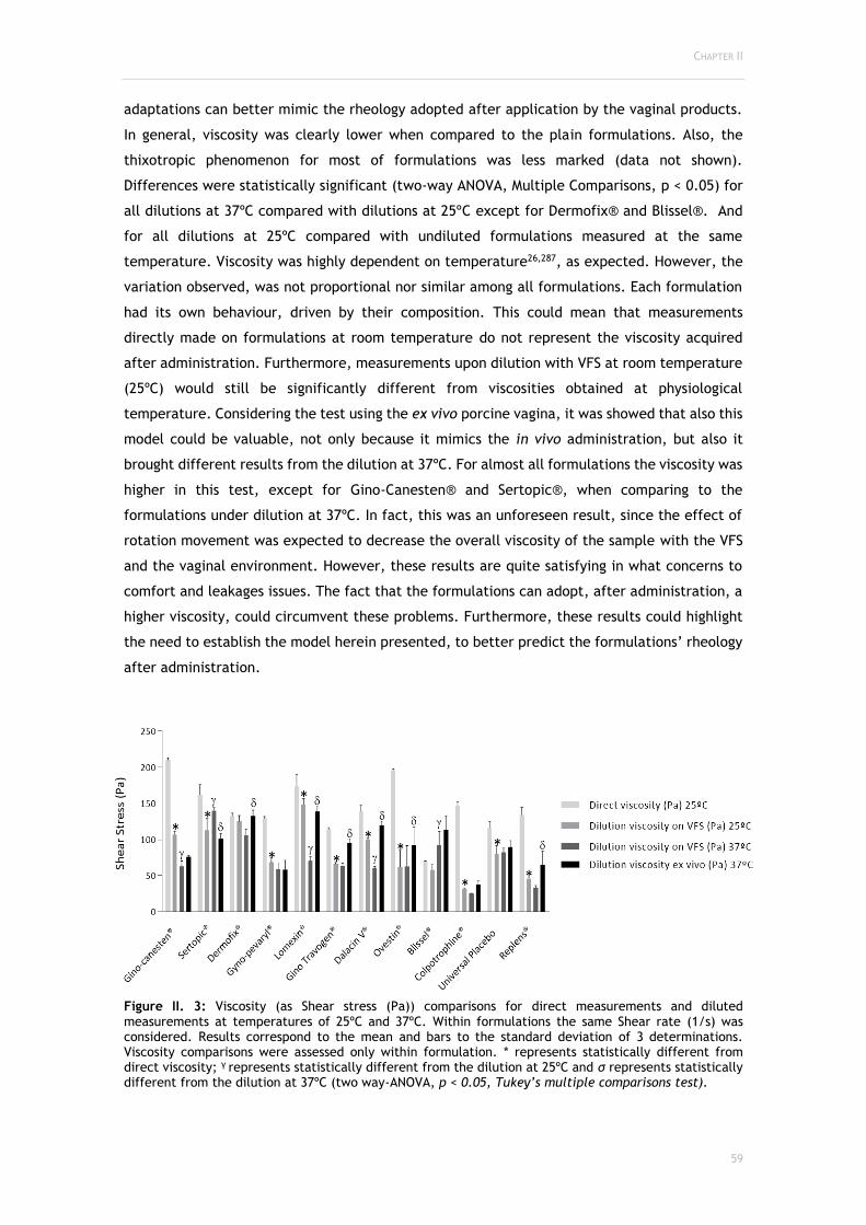
CHAPTER II
59
adaptations can better mimic the rheology adopted after application by the vaginal products.
In general, viscosity was clearly lower when compared to the plain formulations. Also, the
thixotropic phenomenon for most of formulations was less marked (data not shown).
Differences were statistically significant (two-way ANOVA, Multiple Comparisons, p < 0.05) for
all dilutions at 37ºC compared with dilutions at 25ºC except for Dermofix® and Blissel®. And
for all dilutions at 25ºC compared with undiluted formulations measured at the same
temperature. Viscosity was highly dependent on temperature26,287, as expected. However, the
variation observed, was not proportional nor similar among all formulations. Each formulation
had its own behaviour, driven by their composition. This could mean that measurements
directly made on formulations at room temperature do not represent the viscosity acquired
after administration. Furthermore, measurements upon dilution with VFS at room temperature
(25ºC) would still be significantly different from viscosities obtained at physiological
temperature. Considering the test using the ex vivo porcine vagina, it was showed that also this
model could be valuable, not only because it mimics the in vivo administration, but also it
brought different results from the dilution at 37ºC. For almost all formulations the viscosity was
higher in this test, except for Gino-Canesten® and Sertopic®, when comparing to the
formulations under dilution at 37ºC. In fact, this was an unforeseen result, since the effect of
rotation movement was expected to decrease the overall viscosity of the sample with the VFS
and the vaginal environment. However, these results are quite satisfying in what concerns to
comfort and leakages issues. The fact that the formulations can adopt, after administration, a
higher viscosity, could circumvent these problems. Furthermore, these results could highlight
the need to establish the model herein presented, to better predict the formulations’ rheology
after administration.
Figure II. 3: Viscosity (as Shear stress (Pa)) comparisons for direct measurements and diluted measurements at temperatures of 25ºC and 37ºC. Within formulations the same Shear rate (1/s) was considered. Results correspond to the mean and bars to the standard deviation of 3 determinations. Viscosity comparisons were assessed only within formulation. * represents statistically different from direct viscosity; γ represents statistically different from the dilution at 25ºC and σ represents statistically different from the dilution at 37ºC (two way-ANOVA, p < 0.05, Tukey’s multiple comparisons test).

TECHNOLOGICAL CHARACTERIZATION
60
Aka-Any-Grah et al., have also reported major differences among formulations developed
with hydroxypropylmethyl cellulose (HPMC) and pluronics F127/F68, especially on the gelling
temperature (before and after dilution in VFS), rheological properties and even ex vivo
adhesion182. This data suggests that approximations to vaginal physiological conditions are
determinant to foresee in vivo rheology of the formulations after administration181. Viscosity
can dictate the ability of the formulation to disperse in vivo, as well as the residence time in
the genitourinary tract of these formulations, hence it is decreased owing to the self-cleansing
action of the vagina and the dilution with vaginal fluids, and even environmental
temperature182.
Katz et al. disclosed the rheological properties of Advantage-S® and Replens® at body and
room temperature over a range of physiologically relevant shear rates. It was found that their
rheological behaviour was different among temperatures and miscibility with vaginal fluid
simulant was also affected295.
Lai et al. (2008) studied the effect of small dilutions (10-30%) in vaginal fluid and semen
simulants on K-Y Jelly®, Replens® and Carraguard®, using a cone-plate rheometer, and data
was fitted to power-law, Carreau, or Herschel-Bulkley models. Rheological parameters from
these fits were input to models of coating flow due squeezing, and the simulated area coated
output from these models was used to compare the responses of the different formulations to
the two diluents for varying degrees of dilution. There were differences in the responses of the
three materials to dilution; even small dilutions altered the rank order of vaginal coating rates
compared to the undiluted formulations286.
Later on, Henderson et al. (2007) used an optical imaging technique to compare human
intravaginal coating distributions of Conceptrol® and Advantage®. It was concluded that the
results were consistent with those predicted through mechanistic coating theory, using gel
rheological data as input296. Furthermore, in 2008, Mauck et al. studied the vaginal distribution
of Replens® and K-Y Jelly® in vivo in women. Time, ambulation, parity and body mass index
were factors considered for vaginal spreading. Imaging was achieved by magnetic resonance
imaging, gamma scintigraphy and with a fiberoptic probe. Results showed that the initial
application of the gel resulted in two thirds of maximum coverage possible, both in linear
extent along the vaginal axis and in surface area covered. Over the next 45 min, spreading
increased to about three quarters of the maximum possible. Ambulation generally increased
linear spreading. Effects of parity and body mass index were similar on most measures of gel
spreading, with nulligravid women tending toward greater spread than parous women and
women of high body mass, usually showing somewhat greater spread than women of normal
weight. Differences between the two gels were not seen when all conditions of application
were considered together297.
Recently, Katz et al. described the fundamental principles of mass transport, highlighting
the diffusion and convection of drugs in the vaginal environment. Several mathematical
predictive models can be used to this, although having some variability when compared to in
vivo behaviour. These models can illustrate drug concentration distribution (pharmacokinetics)
CHAPTER II
61
and effectiveness (pharmacodynamics). Modelling can be used to compare vaginal drug
distributions after different gel dosage regimens, comparing the effect of vaginal fluid and the
consequences of changes in gel viscosity due to aging. It could also be helpful in comparing
drug distribution after the application of different dosage forms. Ultimately, the modelling
approach is used to compare vaginal drug distribution across species with differing vaginal
dimensions298.
Anwar et al. have investigated the interplay between vaginal tissue elasticity and the
yield-stress of non-Newtonian fluids during a microbicide deployment. Yield stress is the applied
stress one must exceed in order to make a structured fluid flow, being the “force” implied on
the formulation at the beginning of a viscosity determination. Within this research work this
group has developed a mathematical model of tissue deformation driven by spreading of
microbicidal gels based on thin film lubrication approximation and demonstrated the effect of
tissue elasticity and fluid yield-stress on the spreading dynamics. It was concluded that both
elasticity of tissue and yield-stress rheology of gel are strong determinants for the coating
behaviour299.
In another study, Kieweg et al. performed experimental and numerical studies on
microbicidal gel deployment under constant squeezing force and concluded that squeezing
force, gel consistency, shear-thinning behaviour and yield stress are strong determinants of the
coating performance of gels300–302. Szeri et al. developed a mathematical model involving wall
elasticity to demonstrate the effect of compliant vaginal wall on the deployment of new
formulations303.
Vaginal gels should be highly elastic, even after dilution, as these properties govern drug
release and leakage. Furthermore, pseudoplasticity would offer stress-induced viscosity
depression and hence ensure ease of application304. This is difficult to achieve using single
polymer gels. Therefore, gels offering greater clinical promise may be achieved through the
combination of mucoadhesive and gel structuring polymers within a binary or higher polymer
platform305,306.
Omar et al. in 2014 proposed an universal vaginal applicator able to homogenously
distribute the formulations over the entire vaginal and cervical epithelia. The internal
distribution was investigated using pelvic magnetic resonance imaging (MRI) in a group of
women which used six different vaginal gels and creams. Comparisons were made against the
conventional applicators. The universal applicator showed to have good potential to reach
uniform coverage of vagina and cervix, and enhance women protection against sexually
transmitted diseases104.
Despite of comfort and administration issues, semisolid formulations distribution is directly
related to therapeutic efficacy. The use of a gel with a low viscosity would facilitate spreading
and hence contact with the vaginal epithelium. However, a low viscosity gel would be expected
to have a limited residence time due to the inability of the gel to resist dilution from vaginal
fluids and semen, except if it can exhibit a trigger gelling behaviour98,173. Moreover, a low
viscosity gel would be unable to “absorb” in vivo stresses without causing destruction of

TECHNOLOGICAL CHARACTERIZATION
62
polymer gel entanglements and thus would be expected to leak rapidly. Conversely, a highly
elastic gel would offer greater resistance to dilution and to in vivo stresses; however,
application and intravaginal spreading would be limited. Therefore, optimal clinical
performance may only be achieved when the elastic-viscous balance is carefully controlled304.
The performance of a vaginal gel can be evaluated with respect to a number of properties,
including spreadability, coating and retention by using in vitro, ex vivo and in vivo
methodologies307.

CHAPTER II
63
II.2.3. Conclusions
In view of designing new formulations for vaginal administration driven by safety and
efficacy rationals; biological criteria should be addressed from the early steps of development
to accelerate the whole process. The present study showed that a great number of commercial
therapeutic vaginal formulations currently used did not present ideal technological
characteristics when tested under a physiological perspective. Furthermore, pH buffering
capacity, osmolality and viscosity determined using these methodological adaptations were
considered focal points to be addressed during products’ development. While great effort has
been made in the development of innovative vaginal gels in the field of microbicides,
antimicrobials and oestrogens formulations have not been the focus of attention in the last
years. Nevertheless, they represent the most widely and frequently prescribed all over the
world for acute and chronic conditions, respectively. Polymer-based strategies could be applied
to re-formulate products already marketed in order to overcome problems of leakage and
discomfort, and improve efficacy. The adaptation of these formulations and the use of the
methodologic adaptations proposed in this work may optimize cost-efficiency of new and
renewed formulations development by predicting efficacy and safety profiles at early stages of
product development.

TECHNOLOGICAL CHARACTERIZATION
64

CHAPTER III
65
CHAPTER III
PRECLINICAL SAFETY CHARACTERIZATION:
CELL-BASED AND ORGAN-BASED
(IN VITRO AND EX VIVO) TESTING

PRECLINICAL SAFETY
66
The content of this chapter is partially published in:
Machado RM, Palmeira-de-Oliveira A, Breitenfeld L, Martinez-de-Oliveira J, Palmeira-de-
Oliveira R, “Optimization and application of in vitro ande x vivo models for vaginal semisolids
safety evaluation”.
(submitted manuscript)
Palmeira-de-Oliveira R, Machado RM, Palmeira-de-Oliveira A, Martinez-de-Oliveira J, “Testing
vaginal irritation with the HET-CAM assay: an in vitro alternative to the in vivo method”
(accepted manuscript)

CHAPTER III
67
III.1. GENERAL
CONSIDERATIONS

PRECLINICAL SAFETY
68
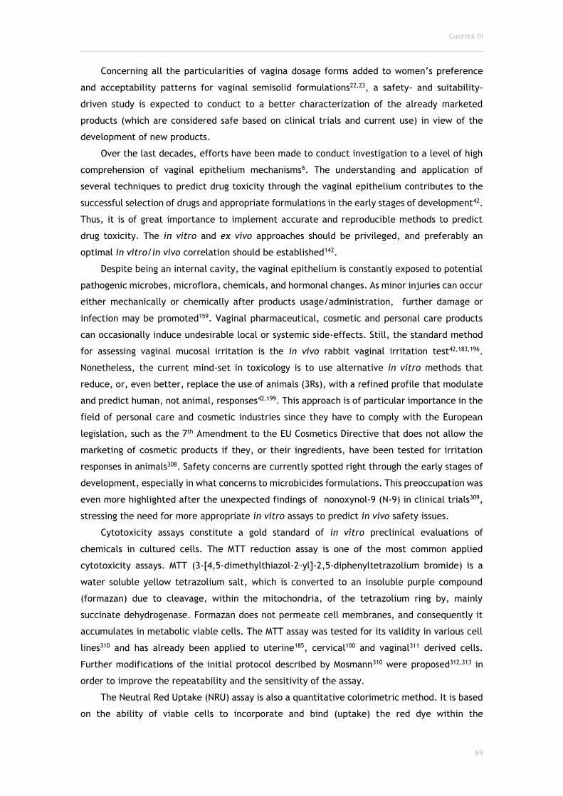
CHAPTER III
69
Concerning all the particularities of vagina dosage forms added to women’s preference
and acceptability patterns for vaginal semisolid formulations22,23, a safety- and suitability-
driven study is expected to conduct to a better characterization of the already marketed
products (which are considered safe based on clinical trials and current use) in view of the
development of new products.
Over the last decades, efforts have been made to conduct investigation to a level of high
comprehension of vaginal epithelium mechanisms6. The understanding and application of
several techniques to predict drug toxicity through the vaginal epithelium contributes to the
successful selection of drugs and appropriate formulations in the early stages of development42.
Thus, it is of great importance to implement accurate and reproducible methods to predict
drug toxicity. The in vitro and ex vivo approaches should be privileged, and preferably an
optimal in vitro/in vivo correlation should be established142.
Despite being an internal cavity, the vaginal epithelium is constantly exposed to potential
pathogenic microbes, microflora, chemicals, and hormonal changes. As minor injuries can occur
either mechanically or chemically after products usage/administration, further damage or
infection may be promoted159. Vaginal pharmaceutical, cosmetic and personal care products
can occasionally induce undesirable local or systemic side-effects. Still, the standard method
for assessing vaginal mucosal irritation is the in vivo rabbit vaginal irritation test42,183,196.
Nonetheless, the current mind-set in toxicology is to use alternative in vitro methods that
reduce, or, even better, replace the use of animals (3Rs), with a refined profile that modulate
and predict human, not animal, responses42,199. This approach is of particular importance in the
field of personal care and cosmetic industries since they have to comply with the European
legislation, such as the 7th Amendment to the EU Cosmetics Directive that does not allow the
marketing of cosmetic products if they, or their ingredients, have been tested for irritation
responses in animals308. Safety concerns are currently spotted right through the early stages of
development, especially in what concerns to microbicides formulations. This preoccupation was
even more highlighted after the unexpected findings of nonoxynol-9 (N-9) in clinical trials309,
stressing the need for more appropriate in vitro assays to predict in vivo safety issues.
Cytotoxicity assays constitute a gold standard of in vitro preclinical evaluations of
chemicals in cultured cells. The MTT reduction assay is one of the most common applied
cytotoxicity assays. MTT (3-[4,5-dimethylthiazol-2-yl]-2,5-diphenyltetrazolium bromide) is a
water soluble yellow tetrazolium salt, which is converted to an insoluble purple compound
(formazan) due to cleavage, within the mitochondria, of the tetrazolium ring by, mainly
succinate dehydrogenase. Formazan does not permeate cell membranes, and consequently it
accumulates in metabolic viable cells. The MTT assay was tested for its validity in various cell
lines310 and has already been applied to uterine185, cervical100 and vaginal311 derived cells.
Further modifications of the initial protocol described by Mosmann310 were proposed312,313 in
order to improve the repeatability and the sensitivity of the assay.
The Neutral Red Uptake (NRU) assay is also a quantitative colorimetric method. It is based
on the ability of viable cells to incorporate and bind (uptake) the red dye within the

PRECLINICAL SAFETY
70
lysosomes193–195. Although they are used extensively as convenient and rapid measurements of
cell viability, all these methods should be carried out with some caution. In fact, an increase
in NR uptake can be induced by lysosomal swelling agents such as weak alkaline substances and
osmotic swelling agents. Regarding the MTT assay, some reducing agents and respiratory chain
inhibitors could affect the MTT formazan formation. The MTT assay was also found to be
significantly influenced by a number of parameters such as medium pH, D-glucose concentration
in culture media, and cellular concentration of pyridine nucleotides314. Additionally, the NRU
assay present some advantages over the MTT assay. The former procedure is more sensitive and
readily quantifiable. It is at least two times cheaper, presents less interferences, and does not
use an unstable tetrazolium salt195. As a general remark, these assays have, in common with
other cell culture procedures, limitations concerning the substance chemical characteristics,
say to be volatile, unstable or explosive in water, only very partially soluble, colourant and
chemical variably incompatible with the test substance191,195.
In vitro cellular and ex vivo tissue models, although being widely applied for toxicity
testing, are still far from the complexity of an organ-based model. Furthermore, only specific
biomarkers used in these methods, such as interleukins, could provide a predictive response of
the irritant potential of vaginal drugs/products. Additionally, these models require the use of
appropriate products dilutions due to models’ sensitivity. That is why researchers and
regulatory agencies are working together to find, optimize and validate the most complex in
vitro models (organ-based), with the higher similitude with the in vivo models.
Topical toxicity has been a main topic within European Union Reference Laboratory for
Alternatives to Animal Testing (EURL-ECVAM). Having also a regulatory responsibility, the EU
and associated laboratories, have been working on developing and validating toxicity test
methods for eye irritation, phototoxicity, skin corrosion, irritation and sensitisation315.
Furthermore, several in vitro Test Guidelines are already provided or being developed by the
Organisation for Economic Co-operation and Development (OECD)316. In vitro studies have been
valued and recommended as a first screening for toxicity evaluation, in detriment to in vivo
models.
The assessment of irritability and toxicity of products for vaginal administration does not
yet include in vitro methods validated and recognized by both the referred authorities.
Currently, there are numerous medicines and medical devices marketed or being in the
development phase for vaginal administration2,4,5,7. The parallelism between sensitivity to eye
and vaginal irritants is addressed in the ISO 10993 guideline since eye irritant products are
included in the exclusion criteria for vaginal irritation test, being considered irritant to the
vagina317. As a general assumption, maximum effectiveness with the least of adverse effects is
always desired for vaginal formulations. Since these products are mostly for self-administration
and should conceive maximum comfort both at the moment of application and during the time
of usage, effects such as local irritation become especially important to be evaluated. Although
various techniques for the characterization of vaginal formulations have been described on the
literature99,153,157,159, most have been applied only to developing formulations with no
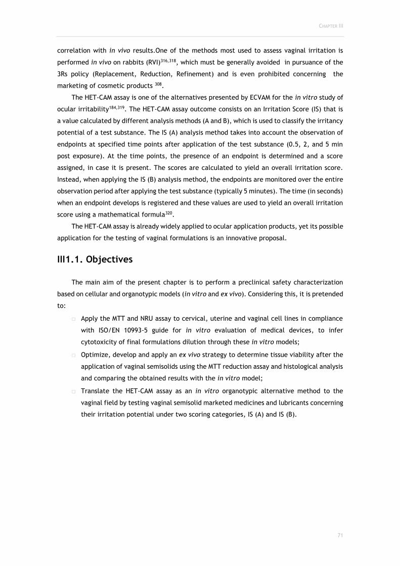
CHAPTER III
71
correlation with in vivo results.One of the methods most used to assess vaginal irritation is
performed in vivo on rabbits (RVI)316,318, which must be generally avoided in pursuance of the
3Rs policy (Replacement, Reduction, Refinement) and is even prohibited concerning the
marketing of cosmetic products 308.
The HET-CAM assay is one of the alternatives presented by ECVAM for the in vitro study of
ocular irritability184,319. The HET-CAM assay outcome consists on an Irritation Score (IS) that is
a value calculated by different analysis methods (A and B), which is used to classify the irritancy
potential of a test substance. The IS (A) analysis method takes into account the observation of
endpoints at specified time points after application of the test substance (0.5, 2, and 5 min
post exposure). At the time points, the presence of an endpoint is determined and a score
assigned, in case it is present. The scores are calculated to yield an overall irritation score.
Instead, when applying the IS (B) analysis method, the endpoints are monitored over the entire
observation period after applying the test substance (typically 5 minutes). The time (in seconds)
when an endpoint develops is registered and these values are used to yield an overall irritation
score using a mathematical formula320.
The HET-CAM assay is already widely applied to ocular application products, yet its possible
application for the testing of vaginal formulations is an innovative proposal.
III1.1. Objectives
The main aim of the present chapter is to perform a preclinical safety characterization
based on cellular and organotypic models (in vitro and ex vivo). Considering this, it is pretended
to:
□ Apply the MTT and NRU assay to cervical, uterine and vaginal cell lines in compliance
with ISO/EN 10993-5 guide for in vitro evaluation of medical devices, to infer
cytotoxicity of final formulations dilution through these in vitro models;
□ Optimize, develop and apply an ex vivo strategy to determine tissue viability after the
application of vaginal semisolids using the MTT reduction assay and histological analysis
and comparing the obtained results with the in vitro model;
□ Translate the HET-CAM assay as an in vitro organotypic alternative method to the
vaginal field by testing vaginal semisolid marketed medicines and lubricants concerning
their irritation potential under two scoring categories, IS (A) and IS (B).

PRECLINICAL SAFETY
72

CHAPTER III
73
III.2. EXPERIMENTAL

PRECLINICAL SAFETY
74

CHAPTER III
75
III.2.1. Materials and Methods
III.2.1.1. Tested Formulations
Ten different semisolid vaginal products that are commercially available in Europe and the
USA were included in this study: Gino-Canesten® (Bayer, Portugal), Sertopic® (Ferrer,
Portugal), Dermofix® (Azevedos Laboratories, Portugal), Gyno-Pevaryl® (Johnson & Johnson,
Portugal), Lomexin® (Jaba Recordati, Portugal), Gino Travogen® (Bayer, Portugal), Dalacin V®
(Pfizer Laboratories, Portugal), Ovestin® (Aspen Pharma, Portugal), Blissel® (ITF Medivida,
Portugal), Colpotrophine® (Teva Pharma, Portugal). Replens® (Laboratoires Majorelle,
France)14,93,158,166,173,272 and Universal Placebo99,233,321,322 were used as reference products, since
their toxicity profile is largely described on the literature. Universal Placebo was prepared
according to Tien et al.158 by dissolving of 2.7g of Hydroxyethyl-cellulose (2000cP) in 96.3g of
water containing 0.85g of sodium chloride and 0.1g of sorbic acid. The final pH was adjusted
to 4.4 by adding sodium hydroxide, and the gel was stored at 2–8ºC. General characteristics of
the studied products are listed in Table II. 1. All products, except placebo and Replens®, are
classified as medicines, whilst comprising an active pharmaceutical ingredient (API).
III.2.1.2. Materials
Reagents used include Roswell Park Memorial Institute-1640 RPMI (RPMI-1640, Sigma,
Germany), Dulbecco's Modified Eagle Medium F12 (DMEM F12, Gibco, USA), Keratinocyte-serum
free medium (Gibco, USA), human recombinant epithelial growth factor (hrEGF, Gibco, USA),
bovine pituitary extract (BPE, Gibco, USA), 3-(4,5-dimethylthiazol-2-yl)-2,5-
diphenyltetrazolium bromide (MTT, Amresco, USA), Foetal Bovine Serum (FBS, Merck,
Germany), Penicillin and Streptomycin (SP, Sigma, Germany), Phosphate Buffer Solution (PBS,
Sigma, Germany), Dimethyl Sulfoxide (DMSO, Fisher Chemical, United Kingdom), Triton X100
(Fisher Chemical, United Kingdom), Sodium Dodecyl Sulphate (SDS, Acros Organics, Belgium),
Nonoxynol-9/Tergitol (N9, Sigma, Germany), Neutral Red (NR, Acros Organics, Belgium),
Ethanol (Manuel Vieira e Cª, Portugal), Glacial Acetic Acid (ChemLab, Belgium),
Hydroxyethylcellulose (HEC; Natrosol 250 HX, Ashland Inc., USA), Sorbic Acid (Sigma–Aldrich,
Germany) and Sodium Chloride (Merck, Germany). All used chemicals and reagents were of
analytic grade or equivalent.
III.2.1.3. Epithelial cells
The cell lines HEC-1A, HeLa and VK2 E6/E7 were obtained from the American Type Culture
Collection (ATCC-LGC Promochem, Teddington, United Kingdom). The uterine HEC-1A cells,
originated from a line of human endometrial adenocarcinoma323, were cultured in RPMI 1640
supplemented with 100 U/mL penicillin, 100 µg/mL streptomycin, and 10% FBS, further referred
to as RPMI complete medium (Passages 33-38). HeLa cell line is also epithelial, derived from

PRECLINICAL SAFETY
76
human cervical adenocarcinoma324. These cells were cultured in DMEM F12 medium
supplemented with penicillin (100 U/mL), streptomycin (100 µg/mL), and 10% FBS and is further
referred to as DMEM complete medium (Passages 64-68). VK2 E6/E7 cell line is also an epithelial
one, derived from human vagina (HPV-16 E6/E7 transformed)325. VK2 E6/E7 cells were cultured
in Keratinocyte-serum free medium (Keratinocyte-SFM) added of 0.1 ng/mL human recombinant
epithelial growth factor (hrEGF), 0.05 mg/mL bovine pituitary extract (BPE) and calcium
chloride 44.1 mg/L (Passages 4-6).
III.2.1.4. Vaginal tissues explants
Complete porcine genitalia, from approximately 6 month-year-old animals, were collected
from a local slaughterhouse, transported under refrigeration to the lab and processed within
3h of animal death. The vaginal tubes were separated from the surrounding organs using scissors
and tweezers. The vagina was cut longitudinally and the vestibule (caudal vagina) excised. The
vestibule was selected since we found (unpublished data) to be the proximal vagina
histologically more similar to the human one. Tissues were then washed on a prewarmed saline
solution (NaCl 0.9%, 37ºC). This section of tissue was then thickness normalized by using a
manual dermatome (Watson Skin Graft Knife, BBraun, Germany) equipped with Aesculap blades
(BA718Rm, BBraun, Germany). Then, epithelial sheets were placed upon aluminium foil
(contact made by the basolateral side) and punched to sections of ø=3mm using a biopsy punch
(Stiefel, GSK, United Kingdom). These sections were maintained on a warmed saline solution
until use in the MTT assay. Tissues thickness was accessed to maintain homogeneity between
samples. For this purpose, a digital micrometer (Vogel, Germany) was used by placing the tissue
between two microscopy slides, measuring total thickness (µm) and then subtracting the
thickness of the slides to the total measured thickness.
III.2.1.5. Testing products preparation
Testing products were diluted to 20, 5, 1, 0.4, 0.2, 0.1% (w/v), in medium without
supplementation but containing 0.5% (v/v) of DMSO to ensure proper solubility of formulations.
DMSO was selected based on the recommendations of the ISO guide196. To assure that this
substance was not itself cytotoxic, controls with the used concentration in culture media were
performed. A negative control was included in all experiments (cells/tissues only with
media/DMSO) and also positive controls for toxicity were included (SDS 5% (w/v), Triton X-100
1% (v/v) and N9 2% (v/v)). Formulations were left in contact with the testing platform (cell or
tissue) for 24 hours.
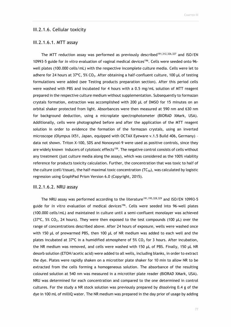
CHAPTER III
77
III.2.1.6. Cellular toxicity
III.2.1.6.1. MTT assay
The MTT reduction assay was performed as previously described191,312,326,327 and ISO/EN
10993-5 guide for in vitro evaluation of vaginal medical devices196. Cells were seeded onto 96-
well plates (100.000 cells/mL) with the respective incomplete culture media. Cells were let to
adhere for 24 hours at 37ºC, 5% CO2. After obtaining a half-confluent culture, 100 µL of testing
formulations were added (see Testing products preparation section). After this period cells
were washed with PBS and incubated for 4 hours with a 0.5 mg/mL solution of MTT reagent
prepared in the respective culture medium without supplementation. Subsequently to formazan
crystals formation, extraction was accomplished with 200 µL of DMSO for 15 minutes on an
orbital shaker protected from light. Absorbances were then measured at 590 nm and 630 nm
for background deduction, using a microplate spectrophotometer (BIORAD XMark, USA).
Additionally, cells were photographed before and after the application of the MTT reagent
solution in order to evidence the formation of the formazan crystals, using an inverted
microscope (Olympus IX51, Japan, equipped with OCTAX Eyeware v.1.5 Build 406, Germany) –
data not shown. Triton X-100, SDS and Nonoxynol-9 were used as positive controls, since they
are widely known inducers of cytotoxic effects159. The negative control consists of cells without
any treatment (just culture media along the assay), which was considered as the 100% viability
reference for products toxicity calculation. Further, the concentration that was toxic to half of
the culture (cell/tissue), the half-maximal toxic concentration (TC50), was calculated by logistic
regression using GraphPad Prism Version 6.0 (Copyright, 2015).
III.2.1.6.2. NRU assay
The NRU assay was performed according to the literature191,195,328,329 and ISO/EN 10993-5
guide for in vitro evaluation of medical devices196. Cells were seeded into 96-well plates
(100.000 cells/mL) and maintained in culture until a semi-confluent monolayer was achieved
(37ºC, 5% CO2, 24 hours). They were then exposed to the test compounds (100 µL) over the
range of concentrations described above. After 24 hours of exposure, wells were washed once
with 150 µL of prewarmed PBS, then 100 µL of NR medium was added to each well and the
plates incubated at 37ºC in a humidified atmosphere of 5% CO2 for 3 hours. After incubation,
the NR medium was removed, and cells were washed with 150 μL of PBS. Finally, 150 μL NR
desorb solution (ETOH/acetic acid) were added to all wells, including blanks, in order to extract
the dye. Plates were rapidly shaken on a microtiter plate shaker for 10 min to allow NR to be
extracted from the cells forming a homogeneous solution. The absorbance of the resulting
coloured solution at 540 nm was measured in a microtiter plate reader (BIORAD XMark, USA).
NRU was determined for each concentration and compared to the one determined in control
cultures. For the study a NR stock solution was previously prepared by dissolving 0.4 g of the
dye in 100 mL of milliQ water. The NR medium was prepared in the day prior of usage by adding
PRECLINICAL SAFETY
78
1 mL of NR stock solution to 79 mL of culture media, followed by incubation overnight at 37ºC
and filtration through a 0.2 µm filter before adding to the cells, in order to be free of crystals.
The NR desorb/extraction solution is composed of 1% glacial acetic acid solution, 50% ethanol
and 49% water.
III.2.1.7. Tissue toxicity
III.2.1.7.1. Method optimization
Tissue explants were firstly tested for homogeneity and response to MTT assay. Therefore,
tissues were submitted to a preliminary experiment, using different animals and applying the
viability test on fresh and frozen tissues. Frozen explants were obtained as described (section
“Vaginal tissue explants”) but tissue preparation occur after defrosting; upon reception from
the slaughterhouse the vaginas were opened longitudinally (from vulva to cervix), washed in a
HBSS solution pH 4.2 and frozen at -20ºC, wrapped in aluminium foil, and stored in airtight
bags. For viability studies (fresh tissue: 46 animals, at least n=2 for each animal, 6 independent
experiments; frozen tissue: 12 animals, n=3, one experiment), tissues were left in culture for
24 hours and then the MTT assay was performed. For toxicity studies (only performed on fresh
tissue: 23 animals, at least n=2 for each animal, 5 independent experiments) tissues were put
in contact with SDS 5% (w/v) during 24 hours, and then the MTT assay has begun. SDS 5% is
generally used as a positive control in tissue toxicity assays and recognized to have a toxic
effect even on epithelial vaginal tissue330.
III.2.1.7.2. MTT assay
Tissue explants were placed in 96-well flat-bottomed tissue culture plates and treated
with the testing formulations diluted on RPMI media without supplementation at 37ºC, 5% CO2
during 24 hours, n=6. Also, negative controls were included for each animal. SDS 5% (w/v),
Triton X-100 1% (v/v) and N9 2% (v/v) were used as positive controls. Subsequently, tissues
were washed twice with prewarmed PBS. MTT solution at a final concentration of 0.5 mg/mL
was added to each well and incubated 1 hour at 37ºC, 5% CO2. Tissues were then transferred
into new plates and extracted with 200 µL of isopropanol during 1.5 hours at room temperature
under gentle shaking. Finally, tissues were discarded for plate reading at 590 nm against the
background at 630 nm (BIORAD XMark, USA). The tissue viability was calculated as percentage
from the negative control (tissue with no formulation).
III.2.1.7.3. Histological analysis
One tissue per formulation, at its highest concentration, 20% (w/v), was reserved for
histological analysis. Tissues were fixed in a balanced 10% formalin solution. Subsequently,
fixed tissues were run in a set of ethanol solutions of increasing concentrations for dehydration
before embedding in paraffin. Tissue blocks were cut to have about 3 µm thickness and stained

CHAPTER III
79
with haematoxylin and eosin. Slides were observed and microphotographs were taken using a
Zeiss microscope (Axiolmager A1, Zeiss, Germany) equipped with a digital camera (Axiocam,
Zeiss, Germany).
III.2.1.8. Data processing and statistical analysis
Data was analysed to produce arithmetic means with standard deviations (SD) using
Microsoft Excel. Analysis of variance (ANOVA) and Dunnett's multiple comparisons test were
performed to determine the significance of the difference between sets of data (p < 0.05).
Logistic regression analysis was performed using GraphPad Prism 6.0 (Graph Pad Software, USA)
to calculate the half-maximal toxic concentration (TC50) with a 95% confidence interval in
toxicity assays.
PRECLINICAL SAFETY
80
III.2.2. Results
III.2.2.1. Cellular toxicity
Cytotoxicity was accessed by means of the MTT reduction assay and NRU performed upon 3
different cell lines (HeLa, HEC-1A and VK2 E6/E7). Results are mean of 2 independent
experiments, total n=6. All assays, were tracked microscopically for cell integrity, density and
morphology, as stated in the respective protocol196. Cell cultures acquired half-confluency and
were able to reduce MTT in the mitochondria, and to integrate NR in the lysosomes. Results
are expressed as viability percentage of the negative control (cells treated only with media).
For all experiments, positive controls were used to assure the occurrence of induced toxic
effects. In the NRU assay viabilities for HeLa and HEC-1A cells, of the positive controls (Triton
X100 1%, SDS 5% and N9 2%) were: 2.75±0.26; 3.62±0.278; 3.79±0.30; 17.23±0.91; 29.75±9.85;
19.10±1.82%, respectively. Standard deviations for negative controls (cells with media) were
10.93 and 16.30% for HeLa and HEC-1A, respectively. The results for NRU assay using the VK2
E6/E7 are not showed because they were inconclusive. In the MTT assay viabilities for HeLa,
HEC-1A and VK2 E6/E7 cells, positive controls had viabilities of (Triton X100 1%, SDS 5% and N9
2%): 2.75±0.26; 3.62±0.28; 3.79±0.30; 5.82±0.34; 5.86±0.322; 6.54±0.50%, 26.52±1.45%;
13.35±3.93; and 22.12±2.17, respectively. Standard deviations for negative controls were
10.93, 11.51 and 14.63% for HeLa and HEC-1A and VK2 E6/E7, respectively. In Figure III. 1, all
profiles for cytotoxicity are represented both for the NRU and the MTT assay. As a general
picture, HEC-1A and VK2 E6/E7 cell lines conducted to higher viabilities than HeLa cells, being
higher in the NRU assay. Antimicrobials showed a linear decrease of viabilities along with the
increasing of products’ concentration, as expected. Furthermore, this behaviour occurred
consistently on the three cell lines and within the two assays, exception made for antimicrobials
studied in HEC-1A using the NRU assay, in which viabilities do not seem to be affected by the
dilution factor.
The oestrogen-containing formulations, Ovestin® and Colpotrophine®, presented the
higher extents of cell toxicity, independently of the tested concentrations (just a slight increase
was observed in the 0.1% concentration). Profiles for the same cell line were concordant
between the assays. Blissel® presented an odd profile, having a high increase on viability at
5%, which is reduced at intermediate concentrations, and then at low concentrations conducts
again to high viabilities. This behaviour might be related with the Blissel® polymeric
composition (polycarbophil/carbomer) which, in the lower concentrations promote lower
toxicity and then promotes somehow a negative effect on cell proliferation at intermediate
concentrations (actually this dilution could promote a better diffusion of estriol). Concerning
the reference products, low cytotoxic profile was observed, as expected. Universal Placebo gel
led to most stable profile of viabilities across concentrations. Every concentration conducted
to viabilities over 50% of cell viability.

CHAPTER III
81
Figure III. 1: Cellular viability profiles for all the testing formulations at dilutions from 0.1 to 20% (w/v). Results for the NRU and the MTT assay. Viabilities are represented as percentage of the control treated only with culture media. Results are means and bars represent standard deviations from 2 independent experiments in which each condition was tested in triplicate (total n=6). * represents NO statistical difference from the control (two-way ANOVA, Dunnett’s multiple comparisons test, p < 0.05).
PRECLINICAL SAFETY
82
On the other hand, Replens® only showed to reach high viabilities at concentrations lower
than 5%. This fact may be related with low pH presented by this formulation, around 2.8, which
together with a high pH-buffering capacity and high osmolality, could explain the high level of
cytotoxicity presented331.
Regarding VK2 E6/E7 cells, there are no studies available on the literature presenting
results in the NRU assay, despite these cells being largely used to study vaginal toxicity
mechanisms through other methods. In fact, we found irregular results with these cells on the
NRU method (n=3, 2 independent experiments) and this could be due to an increased NR uptake
induced by lysosomal swelling agents such as weakly basic substances and by osmotic swelling
agents such as polyols, as was demonstrated in previous studies332,333. Taking this into account,
the lysosomal swelling may lead to an underestimation of the cytotoxicity when the NR assay
is used195,314.
III.2.2.2. Tissue toxicity
III.2.2.2.1 Method optimization
Optimization experiments were performed to assure that after collection and cutting
procedures, tissues would have enough viability to be used in a toxicity assay such as the MTT
reduction assay. Also, this step resulted in better handling of the surgical instruments and
consequently more precise cutting technique for the operator. Results for viability and toxicity
studies are shown on Table III. 1. Tissue viability was performed either on fresh and frozen
tissue. As expected, frozen tissues returned low absorbances, which are not adequate to a final
MTT assay, since this would have been the maximum absorbance to obtain, and could not
distinguish significant differences between formulations. For this reason, performing such
toxicity studies in frozen-thawed tissues is not possible. Furthermore, the performance of a
toxic substance (SDS) confirmed that chemicals can induced negative responses in ex vivo
tissues. One-way ANOVA with Dunnett’s multiple comparisons test was applied to assess
differences between the fresh tissue, frozen tissue and tissue treated with SDS, (p < 0.05 was
accepted as denoting significance). On the fresh tissue experiment it was clear that high
absorbances can be reached using an ex vivo model, i.e. suitable for toxicity testing of
formulations. Moreover, an acceptable variation coefficient can be held with our tissue
preparation technique and culture procedure (24.39%). After measuring tissues thickness (6
animals, n=6, 3 independent experiments) the variation coefficient was calculated to be
15.12%.
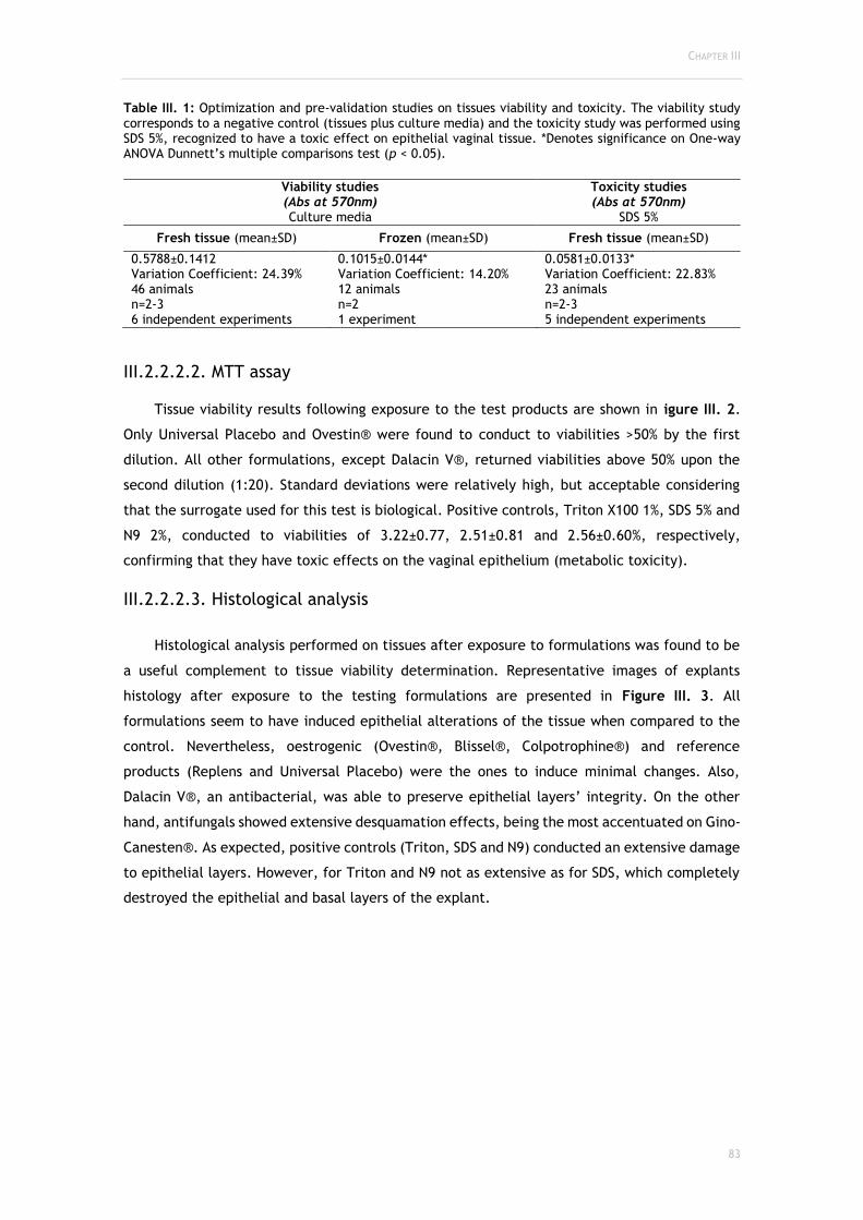
CHAPTER III
83
Table III. 1: Optimization and pre-validation studies on tissues viability and toxicity. The viability study corresponds to a negative control (tissues plus culture media) and the toxicity study was performed using SDS 5%, recognized to have a toxic effect on epithelial vaginal tissue. *Denotes significance on One-way ANOVA Dunnett’s multiple comparisons test (p < 0.05).
Viability studies (Abs at 570nm) Culture media
Toxicity studies (Abs at 570nm)
SDS 5%
Fresh tissue (mean±SD) Frozen (mean±SD) Fresh tissue (mean±SD)
0.5788±0.1412 Variation Coefficient: 24.39%
0.1015±0.0144* Variation Coefficient: 14.20%
0.0581±0.0133* Variation Coefficient: 22.83%
46 animals n=2-3 6 independent experiments
12 animals n=2 1 experiment
23 animals n=2-3 5 independent experiments
III.2.2.2.2. MTT assay
Tissue viability results following exposure to the test products are shown in igure III. 2.
Only Universal Placebo and Ovestin® were found to conduct to viabilities >50% by the first
dilution. All other formulations, except Dalacin V®, returned viabilities above 50% upon the
second dilution (1:20). Standard deviations were relatively high, but acceptable considering
that the surrogate used for this test is biological. Positive controls, Triton X100 1%, SDS 5% and
N9 2%, conducted to viabilities of 3.22±0.77, 2.51±0.81 and 2.56±0.60%, respectively,
confirming that they have toxic effects on the vaginal epithelium (metabolic toxicity).
III.2.2.2.3. Histological analysis
Histological analysis performed on tissues after exposure to formulations was found to be
a useful complement to tissue viability determination. Representative images of explants
histology after exposure to the testing formulations are presented in Figure III. 3. All
formulations seem to have induced epithelial alterations of the tissue when compared to the
control. Nevertheless, oestrogenic (Ovestin®, Blissel®, Colpotrophine®) and reference
products (Replens and Universal Placebo) were the ones to induce minimal changes. Also,
Dalacin V®, an antibacterial, was able to preserve epithelial layers’ integrity. On the other
hand, antifungals showed extensive desquamation effects, being the most accentuated on Gino-
Canesten®. As expected, positive controls (Triton, SDS and N9) conducted an extensive damage
to epithelial layers. However, for Triton and N9 not as extensive as for SDS, which completely
destroyed the epithelial and basal layers of the explant.
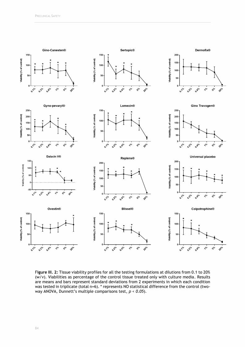
PRECLINICAL SAFETY
84
Figure III. 2: Tissue viability profiles for all the testing formulations at dilutions from 0.1 to 20% (w/v). Viabilities as percentage of the control tissue treated only with culture media. Results are means and bars represent standard deviations from 2 experiments in which each condition was tested in triplicate (total n=6). * represents NO statistical difference from the control (two-way ANOVA, Dunnett’s multiple comparisons test, p < 0.05).

CHAPTER III
85
Figure III. 3: Impact of the tested formulations on the porcine ex vivo vaginal epithelium after 24 h of exposure. Histological images are representative of the higher concentration tested (20%), i.e. the worst-case scenario for the dilutions tested for tissue toxicity. H&E staining. Magnification 100x.
III.2.2.3. Models comparison
Calculated half-maximal toxic concentrations (TC50) either for cellular models and
tissue models are represented in Table III.2 The ex vivo porcine vaginal tissue model, was the
one that led to the calculation of the TC50 for almost all products. This means, on one hand that
the formulations concentration in test is suitable to the test method, and on the other hand,
that this method is the less sensitive in comparison to the remaining. Nevertheless, for Gino-
Canesten® and Gino Travogen® the TC50 confidence interval was very wide, which could be
related with the high slopes and SDs that are shown on the toxicity profiles for these products
between concentrations 5 and 20%. The three cell lines were chosen because they were
representative of three different epithelia that are present in the vaginal cavity, and, by testing
these three cellular types we are addressing a complete toxicological profile of the formulation
after administration by the vaginal route. Concerning the MTT assay, the most sensitive cell
line was the HeLa (cervical cells), while VK2 E6/E7 (vaginal cells) was found to be the more
robust. Since it is expected that cellular models are more sensitive to toxic effects than tissue
models (due to the more complex and organized structure of the latter) and that the tissue, in
turn, is more related to the in vivo environment, VK2 E6/E7 may provide more reliable results
of the vaginal toxicity of formulations when performing the MTT assay. Neutral Red uptake
assay should be performed to confirm previous results from the MTT assay, gathering the

PRECLINICAL SAFETY
86
maximum data for preclinical safety characterization of formulations, or to circumvent
problems due to chemical incompatibility. When possible, final formulations, that were
selected to proceed for further preclinical and clinical stages, should be tested under an ex
vivo model, being the technique herein present a valuable alternative test method.
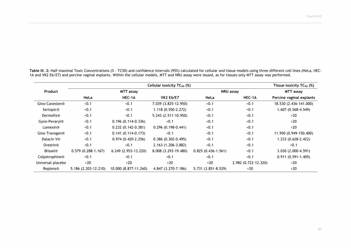
CHAPTER III
87
Table III. 2: Half-maximal Toxic Concentrations (% - TC50) and confidence intervals (95%) calculated for cellular and tissue models using three different cell lines (HeLa, HEC-1A and VK2 E6/E7) and porcine vaginal explants. Within the cellular models, MTT and NRU assay were issued, as for tissues only MTT assay was performed.
Product
Cellular toxicity TC50 (%) Tissue toxicity TC50 (%)
MTT assay NRU assay MTT assay
HeLa HEC-1A VK2 E6/E7 HeLa HEC-1A Porcine vaginal explants
Gino-Canesten® <0.1 <0.1 7.039 (3.825-12.950) <0.1 <0.1 18.530 (2.436-141.000)
Sertopic® <0.1 <0.1 1.118 (0.550-2.272) <0.1 <0.1 1.607 (0.568-4.549)
Dermofix® <0.1 <0.1 5.243 (2.511-10.950) <0.1 <0.1 >20
Gyno-Pevaryl® <0.1 0.196 (0.114-0.336) <0.1 <0.1 <0.1 >20
Lomexin® <0.1 0.232 (0.142-0.381) 0.296 (0.198-0.441) <0.1 <0.1 >20
Gino Travogen® <0.1 0.141 (0.114-0.173) <0.1 <0.1 <0.1 11.950 (0.949-150.400)
Dalacin V® <0.1 0.974 (0.420-2.256) 0.386 (0.302-0.495) <0.1 <0.1 1.233 (0.628-2.422)
Ovestin® <0.1 <0.1 2.163 (1.206-3.882) <0.1 <0.1 <0.1
Blissel® 0.579 (0.288-1.167) 6.249 (2.953-13.220) 8.008 (3.293-19.480) 0.825 (0.436-1.561) <0.1 3.030 (2.000-4.591)
Colpotrophine® <0.1 <0.1 <0.1 <0.1 <0.1 0.911 (0.591-1.405)
Universal placebo >20 >20 >20 >20 2.982 (0.722-12.320) >20
Replens® 5.186 (2.203-12.210) 10.000 (8.877-11.260) 4.847 (3.270-7.186) 5.731 (3.851-8.529) >20 >20
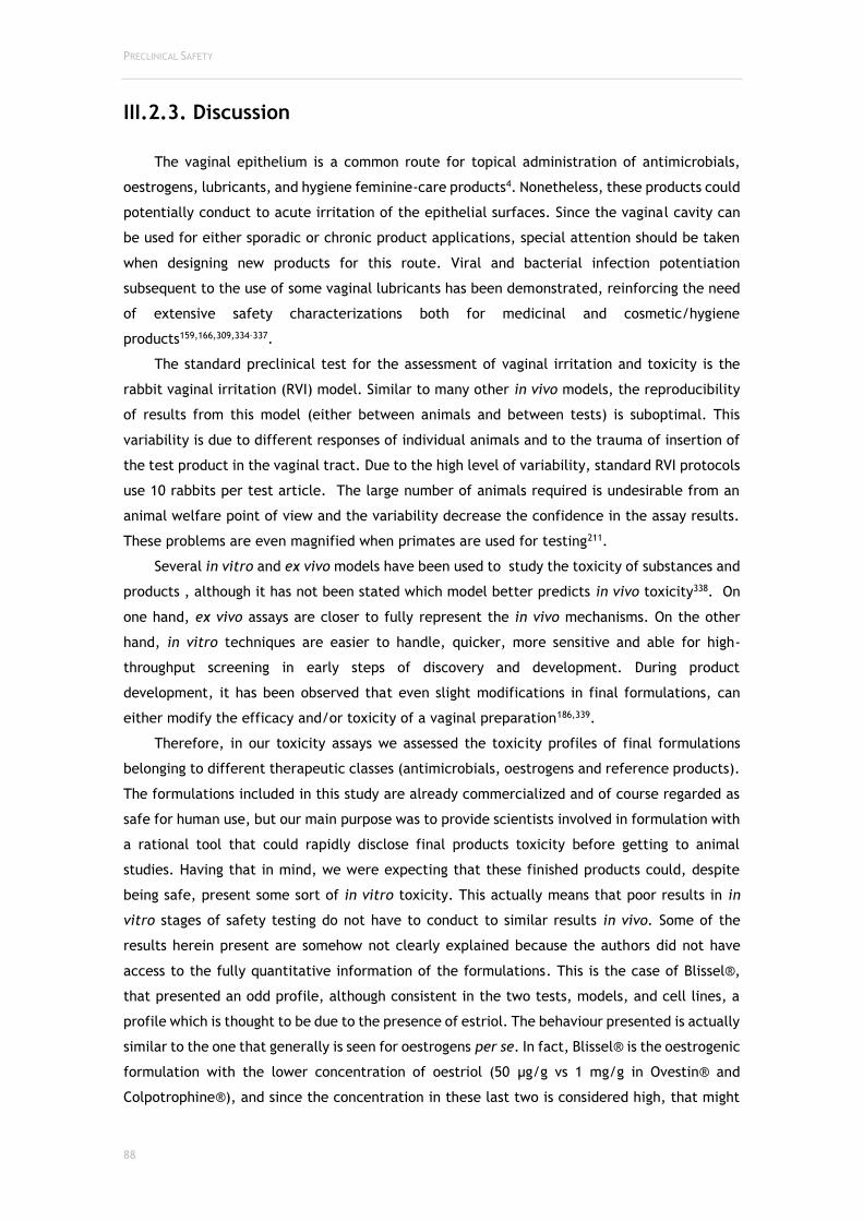
PRECLINICAL SAFETY
88
III.2.3. Discussion
The vaginal epithelium is a common route for topical administration of antimicrobials,
oestrogens, lubricants, and hygiene feminine-care products4. Nonetheless, these products could
potentially conduct to acute irritation of the epithelial surfaces. Since the vaginal cavity can
be used for either sporadic or chronic product applications, special attention should be taken
when designing new products for this route. Viral and bacterial infection potentiation
subsequent to the use of some vaginal lubricants has been demonstrated, reinforcing the need
of extensive safety characterizations both for medicinal and cosmetic/hygiene
products159,166,309,334–337.
The standard preclinical test for the assessment of vaginal irritation and toxicity is the
rabbit vaginal irritation (RVI) model. Similar to many other in vivo models, the reproducibility
of results from this model (either between animals and between tests) is suboptimal. This
variability is due to different responses of individual animals and to the trauma of insertion of
the test product in the vaginal tract. Due to the high level of variability, standard RVI protocols
use 10 rabbits per test article. The large number of animals required is undesirable from an
animal welfare point of view and the variability decrease the confidence in the assay results.
These problems are even magnified when primates are used for testing211.
Several in vitro and ex vivo models have been used to study the toxicity of substances and
products , although it has not been stated which model better predicts in vivo toxicity338. On
one hand, ex vivo assays are closer to fully represent the in vivo mechanisms. On the other
hand, in vitro techniques are easier to handle, quicker, more sensitive and able for high-
throughput screening in early steps of discovery and development. During product
development, it has been observed that even slight modifications in final formulations, can
either modify the efficacy and/or toxicity of a vaginal preparation186,339.
Therefore, in our toxicity assays we assessed the toxicity profiles of final formulations
belonging to different therapeutic classes (antimicrobials, oestrogens and reference products).
The formulations included in this study are already commercialized and of course regarded as
safe for human use, but our main purpose was to provide scientists involved in formulation with
a rational tool that could rapidly disclose final products toxicity before getting to animal
studies. Having that in mind, we were expecting that these finished products could, despite
being safe, present some sort of in vitro toxicity. This actually means that poor results in in
vitro stages of safety testing do not have to conduct to similar results in vivo. Some of the
results herein present are somehow not clearly explained because the authors did not have
access to the fully quantitative information of the formulations. This is the case of Blissel®,
that presented an odd profile, although consistent in the two tests, models, and cell lines, a
profile which is thought to be due to the presence of estriol. The behaviour presented is actually
similar to the one that generally is seen for oestrogens per se. In fact, Blissel® is the oestrogenic
formulation with the lower concentration of oestriol (50 µg/g vs 1 mg/g in Ovestin® and
Colpotrophine®), and since the concentration in these last two is considered high, that might
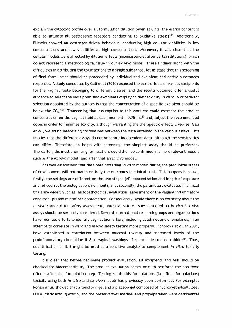
CHAPTER III
89
explain the cytotoxic profile over all formulation dilution (even at 0.1%, the estriol content is
able to saturate all oestrogenic receptors conducting to oxidative stress)340. Additionally,
Blissel® showed an oestrogen-driven behaviour, conducting high cellular viabilities in low
concentrations and low viabilities at high concentrations. Moreover, it was clear that the
cellular models were affected by dilution effects (inconsistencies after certain dilutions), which
do not represent a methodological issue in our ex vivo model. These findings along with the
difficulties in attributing the toxic actions to a single substance, let us state that this screening
of final formulation should be proceeded by individualized excipient and active substances
responses. A study conducted by Gali et al (2010) exposed the toxic effects of various excipients
for the vaginal route belonging to different classes, and the results obtained offer a useful
guidance to select the most promising excipients displaying their toxicity in vitro. A criteria for
selection appointed by the authors is that the concentration of a specific excipient should be
below the CC50185. Transposing that assumption to this work we could estimate the product
concentration on the vaginal fluid at each moment - 0.75 mL37 and, adjust the recommended
doses in order to minimize toxicity, although warranting the therapeutic effect. Likewise, Gali
et al., we found interesting correlations between the data obtained in the various assays. This
implies that the different assays do not generate independent data, although the sensitivities
can differ. Therefore, to begin with screening, the simplest assay should be preferred.
Thereafter, the most promising formulations could then be confirmed in a more relevant model,
such as the ex vivo model, and after that an in vivo model.
It is well established that data obtained using in vitro models during the preclinical stages
of development will not match entirely the outcomes in clinical trials. This happens because,
firstly, the settings are different on the two stages (API concentration and length of exposure
and, of course, the biological environment), and, secondly, the parameters evaluated in clinical
trials are wider. Such as, histopathological evaluation, assessment of the vaginal inflammatory
condition, pH and microflora appreciation. Consequently, while there is no certainty about the
in vivo standard for safety assessment, potential safety issues detected on in vitro/ex vivo
assays should be seriously considered. Several international research groups and organizations
have reunited efforts to identify vaginal biomarkers, including cytokines and chemokines, in an
attempt to correlate in vitro and in vivo safety testing more properly. Fichorova et al. in 2001,
have established a correlation between mucosal toxicity and increased levels of the
proinflammatory chemokine IL-8 in vaginal washings of spermicide-treated rabbits341. Thus,
quantification of IL-8 might be used as a sensitive analyte to complement in vitro toxicity
testing.
It is clear that before beginning product evaluation, all excipients and APIs should be
checked for biocompatibility. The product evaluation comes next to reinforce the non-toxic
effects after the formulation step. Testing semisolids formulations (i.e. final formulations)
toxicity using both in vitro and ex vivo models has previously been performed. For example,
Rohan et al. showed that a tenofovir gel and a placebo gel composed of hydroxyethylcellulose,
EDTA, citric acid, glycerin, and the preservatives methyl- and propylparaben were detrimental
PRECLINICAL SAFETY
90
toward epithelial cells and explants, causing a reduced viability and epithelial layer integrity
of HEC-1A and Caco-2 cells and cervical tissue explants342. Similar observations were made by
Dezzutti et al., assessing the toxicity and epithelial layer integrity of HEC-1A and Caco-2 cell
lines after exposure to PRO-2000 gel, UC-781 gel, and the placebo gels methylcellulose and
Vena Gel343. Furthermore, our workgroup has already tested several vaginal lubricants
commercially available, under a safety perspective, concerning cytotoxicity, pH and
osmolality159. Earlier a similar study has been performed by Dezzuti et al. on 10 aqueous-based
lubricants to test not only cellular toxicity and damage to epithelial monolayers, but also their
toxicity upon human explants epithelium. Main findings of toxicity to the microflora were
attributed to preservatives contained in the formulations. Alongside, it was concluded that
epithelial trauma alone would not be sufficient to increase the HIV-1 transmission, having
inflammatory response an important role in this337. Similarly, to the study herein presented
these authors refer that they had some difficulties in performing the toxicity tests in the
cellular models, because, when testing creams (which are composed of an oily phase and an
aqueous phase) it was difficult to solubilize and wash the cells during the tests. Actually, we
have circumvented this problem by using 0.5% (v/v) of DMSO in the final formulation dilution
with culture media to prevent phase separation of these type of products in test. Furthermore,
with handling practice and constant microscopy monitoring, we were able to check for cell
detachment after washing steps (which we concluded that do not happened). Still, it was also
observed that washing steps were even easier when performing the tests on the ex vivo model.
In 2006, MatTek™ Corporation, an American enterprise specialized in developing and
producing organotypic models, i.e. reconstructed tissue models for drug/product testing
concerning several routes of administration, published an in vitro vaginal human tissue model
with low variation coefficients intra and inter-batch (<10 and <15%, respectively), being an
innovative and improved toxicological test system211. In comparison to in vivo models, this
system (EpiVaginal™) reduces the testing duration, allows for high-throughput screening, and
complies with the “3Rs” policy leading to the decrease of number of animals used to screen
feminine-care products. Besides, it could be applied to vaginal drug delivery assays, bacterial
adhesion and omics344. EpiVaginal™ is available as an epithelial tissue (grown from normal
human vaginal epithelial cells) and as a full thickness tissue (including epithelial cells and a
fibroblast-containing lamina propria). These two tissues could also include immune-competent
dendritic cells for inflammatory studies. Another company providing researchers with this type
of tools in Europe is EpiSkin™, producing HVE (reconstructed human vaginal epithelium) that is
composed of A431 cells (derived from a vulvar epidermoid carcinoma). It is well established
that these tools are clearly advantageous when in comparison with other in vitro and ex vivo
models, not only in terms of sensibility and reproducibility, but also in terms of technology,
quality control and even technical assistance. Nevertheless, they have main applicability to
industry screening platforms, rather than academic research, because of the economical
expenditure. That is a key point to understand why ex vivo tissues could represent valuable
tools for scientific research, when early stages of the preclinical development of

CHAPTER III
91
drugs/products is the focus. Moreover, the ex vivo models take into consideration an
interindividual variability that is not present in manufactured reconstructed models, and that
in fact, is closer to the variability encountered further on in vivo studies.
Despite having a full cell structure (epithelial, connective, immune) and better tolerance
to formulations, tissue explants also have some drawbacks like variability, and the fact that
they are technically more demanding. Also, ex vivo tissue is not entirely representative of the
in vivo situation due to lack of tissue regeneration, lack of immune cells recruitment, and
independence from hormones337,342. If human explants are used, they can also be of limited
number and require an Institutional review board approval217.
In our study, a good overlapping profile could be found between the cellular and the tissue
model, although cellular testing was hypersensitive, which conducted to lower viabilities,
turning impossible the calculation of TC50. Indeed, the ability to calculate TC50 for almost all
formulation in the ex vivo model indicates that this model is valuable to assure direct
comparisons, even allowing the use of more concentrated dilutions of formulations, if
experimentally possible.
The ex vivo porcine tissue model herein presented extends the use of ex vivo porcine tissue
as a surrogate for human vaginal tissue. There is a great deal of data that validate the porcine
model of vaginal mucosa in terms of structure, function and reactivity in comparison to human
tissue. Both have stratified squamous epithelium supported by connective tissue. The use of
small, ex vivo, specimens provides convenience, efficiency, and high throughput for
screening34,39. Samples of porcine tissue are inexpensive to obtain (in abundance on
slaughterhouses) and handling is easy when compared to the use of whole animals34,42. Ex vivo
porcine tissues have been largely tested for drug permeation. But, despite appearing to be a
good ex vivo permeability model for human vaginal tissue extrapolation it has already been
shown that its reliability may vary upon substances chemical characteristics. For example, for
hydrophilic molecules, (water and vasopressin, for example), the porcine vaginal tissue is an
accurate in vitro permeability model of human tissue, while for more lipophilic molecules (such
as oxytocin) the flux could be higher than the corresponding estimated value for human
tissue246. Another limitation related to the ex vivo vaginal model is the fact that some authors
use cervical and uterine explants, to resemble the vaginal epithelia185,197,342,345.This may be due
to the difficulty in accessing human vaginal epithelium and/or also based on the assumption
that vaginal formulations shall be safe not only to the vagina but also to the uterus. The porcine
model can relatively circumvent this problem of accessing specimens for testing and provide a
better similitude with the human vagina itself. Added to that, tissue collection and culture
procedures had not been standardized and optimized, before this research. Previous studies of
ours have been focused on disclosing the likeliness of porcine vaginal tissue and the human
tissue, and even disclosing which anatomical region of the porcine vagina is more appropriate
to collect in order to comply with human similarity.
The tetrazolium-based MTT assay has long been regarded as the gold standard of
cytotoxicity assays as it is highly sensitive and has been miniaturised for use as a high-

PRECLINICAL SAFETY
92
throughput screening assay. The first use of the MTT assay upon tissue explants goes back to
the 90´s, when it was applied to different types of tissues, for instance buccal mucosa346,347.
This method is also the one recommended by the manufacturers in the EpiVaginal™ and HVE™
reconstructed models. Recently, Van Tonder et al. (2015), conducted a study comparing several
toxicity assays, including the MTT and NRU assays in MDA-MB-231, MCF-7 and MCF-12A cell lines.
Results indicated that the NRU showed one of the smallest variability across the linear range,
while the largest variation was observed for the MTT assay. This implies that this assay would
more accurately detect small changes in cell number than the MTT assay. Furthermore, the
MTT assay was the one to have more interferences with test products348. Although seeming less
sensitive and with higher number of interferents, MTT has a large historical use, and researchers
are familiar with the methodology. Furthermore, standard documents like those from the
“International Organization for Standardization”, specifically the 10993-5 normative,
recommend this procedure for cytotoxic assessment of medical devices196. Moreover, it has a
successful history of application in tissues.

CHAPTER III
93
III.2.4. Conclusions
In vitro and ex vivo models have been applied to assess the safety profiles for drugs,
excipients and final formulations in order to early preview toxicity issues that might arise in
the ensuing steps of product development. Herein we presented some preclinical toxicity
results obtained for a panel of vaginal semisolid formulations, already regarded as safe for
human use. Furthermore, we have not only optimized a strategy to perform in vitro cellular
toxicity assays using semisolid aqueous and non-aqueous formulations; but also, developed a
collecting technique for vaginal ex vivo porcine tissues. The later showed to be a possible
strategy for ex vivo toxicity testing, since it demonstrated acceptable reproducibility and
sensitivity, with relatively low variability. The inclusion of this method in preclinical safety
assessments may optimize cost-efficiency of new formulations development by predicting
efficacy and safety profiles at early stages of product development. It could not only be applied
to the pharmaceutical industry and research, but also to the cosmetic, hygiene, and medical
devices industries. Moreover, it could further be applied to the development of primary cell
cultures, as a surrogate for permeation and metabolic studies; and to originate co-cultures with
microorganisms.

PRECLINICAL SAFETY
94

CHAPTER III
95
III.3. EXPERIMENTAL

PRECLINICAL SAFETY
96

CHAPTER III
97
III.3.1. Materials and Methods
III.3.1.1. Chemicals and testing products
For the preparation of the assay controls, the following chemicals were used: sodium
chloride (NaCl, JT Baker, USA), sodium dodecyl sulphate (SDS, Acros Organics, Belgium),
nonoxynol-9/tergitol (N-9, Sigma, Germany), sodium hydroxide (NaOH, VWR Prolabo, Germany)
and type I water (Millipore, Merck, USA).
The testing products included in this study were vaginal semisolids, available in the
international market, intended both as therapeutics for several pathological conditions, and as
lubricants for sexual and menopausal discomfort. The ten different therapeutic products were
those tested in the previous chapters Table II. 1. The lubricants tested were: Fillergyn® gel
(BSDpharma), Geliofil® Classic gel (Laboratoires Effik), GelSea® gel (LDPSA), Ginix® gel (ISUS),
Ginix® Plus gel (ISUS), Hyalo Gyn® gel (Fidia Farmaceutici), K-Y® Jelly (Johnson & Johnson),
Phyto Soya® gel (Arkopharma Laboratoires Pharmaceutiques), Velastisa® Intim VG moisturizer
gel cream (ISDIN) and Vidermina® gel (Istituto Ganassini). Both Replens® (Laboratoires
Majorelle) and Universal Placebo158 were used as controls, since their toxicity profiles are
largely described on the literature14,93,153,158,206,233,272,321,322. Universal Placebo was prepared as
described on experimental section of Chapter II. To evaluate the method sensitivity to vaginal
irritants, nonoxynol-9 was used in concentrations ranging from 0.001 to 100% (v/v).
III.3.1.1.1 Eggs and incubation conditions
The test system consists on Leghorn white hen's white eggs, fresh (not more than 7 days
old), clean, weighing 45 to 65 g. When the eggs arrived to the lab, they were checked for
damages in the shell: damaged eggs were discharged while the others were incubated, at
37.8±0.3ºC in an atmosphere with a relative humidity of 58±2% and under automatic rotation,
during 8 days (Corti AF-50 and Copele 30652, Spain). By the eighth day, eggs were inspected
using a LED light to confirm the embryo formation. Non-embryonated eggs were discarded and
the others were incubated for 1 one more day in the same conditions but without egg rotation.
III.3.1.1.2. HET-CAM assay
The assay was conducted according to the ICCVAM - Recommended test method (NIH
Publication No. 10-7553 – 2010). At day 9, the eggs were taken out of incubator and were placed
on an appropriate support with the larger part facing up. The shell was opened with the help
of a scalpel and tweezers and the chorioallantonic membrane (CAM) was exposed. This
membrane was then hydrated with NaCl 0.9% (w/v) for a maximum of 30 min. Afterwards, the
solution was aspirated and the membrane was peeled off without damaging the blood vessels.
For each product 0.3 mL were applied on the membrane and 3 eggs per products were used.
The irritant effect of these products was evaluated by monitoring the appearance of three
endpoints in the CAM, for 5 minutes: haemorrhage (vessel bleeding), lysis (vessels
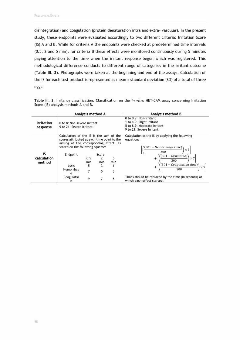
PRECLINICAL SAFETY
98
disintegration) and coagulation (protein denaturation intra and extra- vascular). In the present
study, these endpoints were evaluated accordingly to two different criteria: Irritation Score
(IS) A and B. While for criteria A the endpoints were checked at predetermined time intervals
(0.5; 2 and 5 min), for criteria B these effects were monitored continuously during 5 minutes
paying attention to the time when the irritant response begun which was registered. This
methodological difference conducts to different range of categories in the irritant outcome
(Table III. 3). Photographs were taken at the beginning and end of the assays. Calculation of
the IS for each test product is represented as mean ± standard deviation (SD) of a total of three
eggs.
Table III. 3: Irritancy classification. Classification on the in vitro HET-CAM assay concerning Irritation Score (IS) analysis methods A and B.
Analysis method A Analysis method B
Irritation response
0 to 8: Non-severe irritant 9 to 21: Severe Irritant
0 to 0.9: Non-irritant 1 to 4.9: Slight Irritant 5 to 8.9: Moderate Irritant 9 to 21: Severe Irritant
IS calculation
method
Calculation of the IS is the sum of the scores attributed at each time point to the arising of the corresponding effect, as stated on the following squeme:
Endpoint Score
0.5 min
2 min
5 min
Lysis 5 3 1 Hemorrhag
e 7 5 3
Coagulation
9 7 5
Calculation of the IS by applying the following equation:
[((301 − 𝐻𝑒𝑚𝑜𝑟𝑟ℎ𝑎𝑔𝑒 𝑡𝑖𝑚𝑒)
300) × 5]
+ [((301 − 𝐿𝑦𝑠𝑖𝑠 𝑡𝑖𝑚𝑒)
300) × 7]
+ [((301 − 𝐶𝑜𝑎𝑔𝑢𝑙𝑎𝑡𝑖𝑜𝑛 𝑡𝑖𝑚𝑒)
300) × 9]
Times should be replaced by the time (in seconds) at which each effect started.
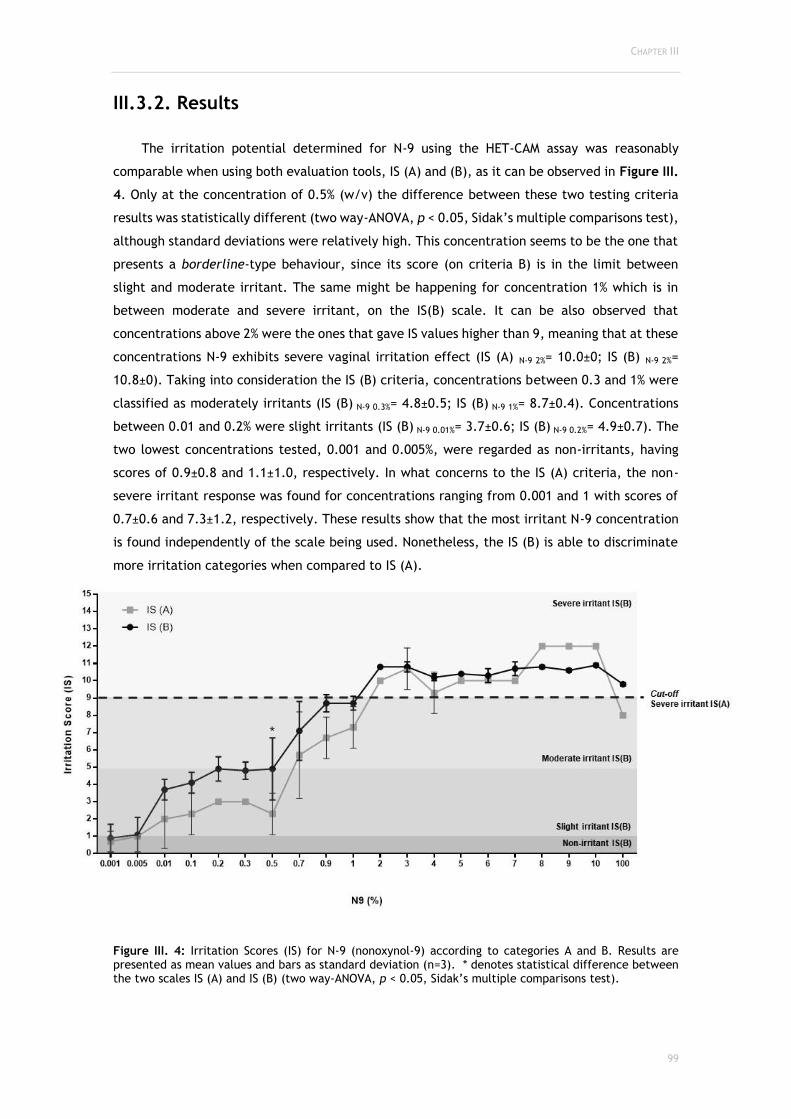
CHAPTER III
99
III.3.2. Results
The irritation potential determined for N-9 using the HET-CAM assay was reasonably
comparable when using both evaluation tools, IS (A) and (B), as it can be observed in Figure III.
4. Only at the concentration of 0.5% (w/v) the difference between these two testing criteria
results was statistically different (two way-ANOVA, p < 0.05, Sidak’s multiple comparisons test),
although standard deviations were relatively high. This concentration seems to be the one that
presents a borderline-type behaviour, since its score (on criteria B) is in the limit between
slight and moderate irritant. The same might be happening for concentration 1% which is in
between moderate and severe irritant, on the IS(B) scale. It can be also observed that
concentrations above 2% were the ones that gave IS values higher than 9, meaning that at these
concentrations N-9 exhibits severe vaginal irritation effect (IS (A) N-9 2%= 10.0±0; IS (B) N-9 2%=
10.8±0). Taking into consideration the IS (B) criteria, concentrations between 0.3 and 1% were
classified as moderately irritants (IS (B) N-9 0.3%= 4.8±0.5; IS (B) N-9 1%= 8.7±0.4). Concentrations
between 0.01 and 0.2% were slight irritants (IS (B) N-9 0.01%= 3.7±0.6; IS (B) N-9 0.2%= 4.9±0.7). The
two lowest concentrations tested, 0.001 and 0.005%, were regarded as non-irritants, having
scores of 0.9±0.8 and 1.1±1.0, respectively. In what concerns to the IS (A) criteria, the non-
severe irritant response was found for concentrations ranging from 0.001 and 1 with scores of
0.7±0.6 and 7.3±1.2, respectively. These results show that the most irritant N-9 concentration
is found independently of the scale being used. Nonetheless, the IS (B) is able to discriminate
more irritation categories when compared to IS (A).
Figure III. 4: Irritation Scores (IS) for N-9 (nonoxynol-9) according to categories A and B. Results are presented as mean values and bars as standard deviation (n=3). * denotes statistical difference between the two scales IS (A) and IS (B) (two way-ANOVA, p < 0.05, Sidak’s multiple comparisons test).
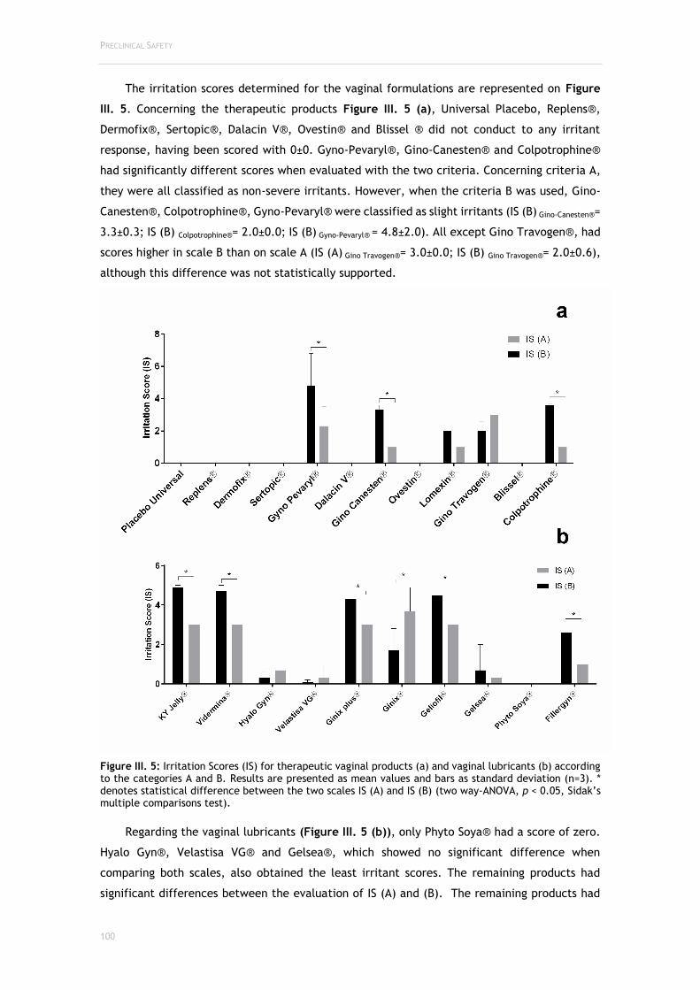
PRECLINICAL SAFETY
100
The irritation scores determined for the vaginal formulations are represented on Figure
III. 5. Concerning the therapeutic products Figure III. 5 (a), Universal Placebo, Replens®,
Dermofix®, Sertopic®, Dalacin V®, Ovestin® and Blissel ® did not conduct to any irritant
response, having been scored with 0±0. Gyno-Pevaryl®, Gino-Canesten® and Colpotrophine®
had significantly different scores when evaluated with the two criteria. Concerning criteria A,
they were all classified as non-severe irritants. However, when the criteria B was used, Gino-
Canesten®, Colpotrophine®, Gyno-Pevaryl® were classified as slight irritants (IS (B) Gino-Canesten®=
3.3±0.3; IS (B) Colpotrophine®= 2.0±0.0; IS (B) Gyno-Pevaryl® = 4.8±2.0). All except Gino Travogen®, had
scores higher in scale B than on scale A (IS (A) Gino Travogen®= 3.0±0.0; IS (B) Gino Travogen®= 2.0±0.6),
although this difference was not statistically supported.
Figure III. 5: Irritation Scores (IS) for therapeutic vaginal products (a) and vaginal lubricants (b) according to the categories A and B. Results are presented as mean values and bars as standard deviation (n=3). * denotes statistical difference between the two scales IS (A) and IS (B) (two way-ANOVA, p < 0.05, Sidak’s multiple comparisons test).
Regarding the vaginal lubricants (Figure III. 5 (b)), only Phyto Soya® had a score of zero.
Hyalo Gyn®, Velastisa VG® and Gelsea®, which showed no significant difference when
comparing both scales, also obtained the least irritant scores. The remaining products had
significant differences between the evaluation of IS (A) and (B). The remaining products had

CHAPTER III
101
significant differences between the evaluation of IS (A) and (B). All lubricants were classified
as non-severe irritants by the IS (A) irritation criteria with Ginix® reaching the highest score in
this scale (3.7±1.2). When the IS (B) was applied Hyalo Gyn® and Velastisa VG® were classified
as non-irritants and all the others were classified as slight irritants. For all but Ginix®, the score
obtained in scale B was higher than the one obtained in A. As it can be seen on Figure III. 5
(b), when comparing both scales, they also obtained the least irritant scores. The remaining
products had significant differences between the evaluation of IS (A) and (B). Table III. 4 details
the score obtained and the classification attributed to all products tested in this study.
Furthermore, a picture from the test end-time (5 min) is also shown for products and controls.
As it can be observed, some products did not present the same classification when comparing
the two scales (borderline behaviour). In the case of medicines, Gyno-Pevaryl®, Gino-
Canesten®, Lomexin®, Gino Travogen® and Colpotrophine®, were the ones to present a
classification of slight irritants on IS (B) and non-severe irritants on IS (A). Regarding lubricants,
K-Y Jelly®, Vidermina®, Ginix Plus®, Geliofil® and Fillergyn®, presented this behaviour.
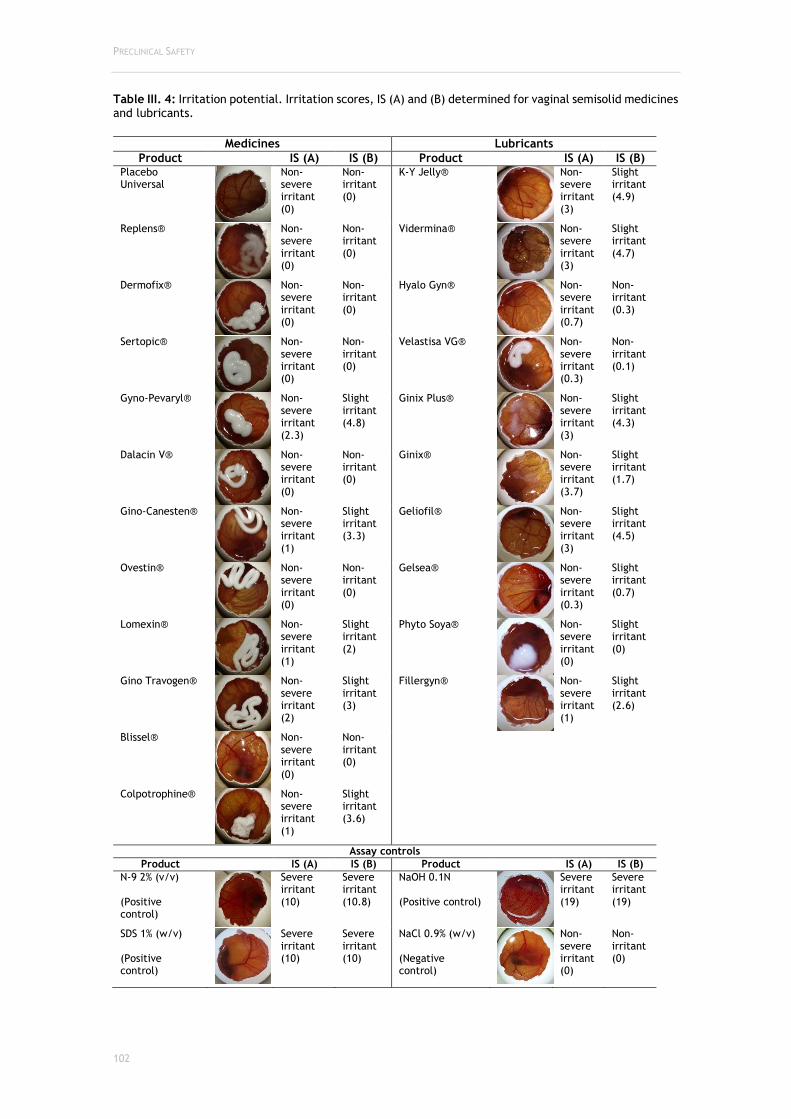
PRECLINICAL SAFETY
102
Table III. 4: Irritation potential. Irritation scores, IS (A) and (B) determined for vaginal semisolid medicines and lubricants.
Medicines Lubricants
Product IS (A) IS (B) Product IS (A) IS (B) Placebo Universal
Non-severe irritant (0)
Non-irritant (0)
K-Y Jelly®
Non-severe irritant (3)
Slight irritant (4.9)
Replens®
Non-severe irritant (0)
Non-irritant (0)
Vidermina®
Non-severe irritant (3)
Slight irritant (4.7)
Dermofix®
Non-severe irritant (0)
Non-irritant (0)
Hyalo Gyn®
Non-severe irritant (0.7)
Non-irritant (0.3)
Sertopic®
Non-severe irritant (0)
Non-irritant (0)
Velastisa VG®
Non-severe irritant (0.3)
Non-irritant (0.1)
Gyno-Pevaryl®
Non-severe irritant (2.3)
Slight irritant (4.8)
Ginix Plus®
Non-severe irritant (3)
Slight irritant (4.3)
Dalacin V®
Non-severe irritant (0)
Non-irritant (0)
Ginix®
Non-severe irritant (3.7)
Slight irritant (1.7)
Gino-Canesten®
Non-severe irritant (1)
Slight irritant (3.3)
Geliofil®
Non-severe irritant (3)
Slight irritant (4.5)
Ovestin®
Non-severe irritant (0)
Non-irritant (0)
Gelsea®
Non-severe irritant (0.3)
Slight irritant (0.7)
Lomexin®
Non-severe irritant (1)
Slight irritant (2)
Phyto Soya®
Non-severe irritant (0)
Slight irritant (0)
Gino Travogen®
Non-severe irritant (2)
Slight irritant (3)
Fillergyn®
Non-severe irritant (1)
Slight irritant (2.6)
Blissel®
Non-severe irritant (0)
Non-irritant (0)
Colpotrophine®
Non-severe irritant (1)
Slight irritant (3.6)
Assay controls
Product IS (A) IS (B) Product IS (A) IS (B)
N-9 2% (v/v) (Positive control)
Severe irritant (10)
Severe irritant (10.8)
NaOH 0.1N (Positive control)
Severe irritant (19)
Severe irritant (19)
SDS 1% (w/v) (Positive control)
Severe irritant (10)
Severe irritant (10)
NaCl 0.9% (w/v) (Negative control)
Non-severe irritant (0)
Non-irritant (0)
CHAPTER III
103
III.3.3. Discussion
Nowadays, the increasing number of new chemicals and products introduced into the
market coming from several industries (cosmetic, pharmaceutical and medical devices) has
generated the need to validate in vitro techniques able to screen potential irritation effects at
the early stages of development.
The Hen's Egg Test, or Hühner-Embryonen-Test (HET), firstly presented by Luepke, back in
the 80’s, was developed to be a rapid, sensitive and inexpensive toxicity test able to provide
information on embryotoxicity, teratogenicity, systemic, metabolic and immunopathological
effects349. This method was designed to be applied in mucous-membrane irritation testing, and
the tool provided for score and classification was analogous to the Draize test. Since then, it
has been confirmed that a good correlation between these two tests, i.e. in vitro/in vivo
correlation, for plenty of chemicals do exist. The HET-CAM assay cannot totally replace the
irritation tests in mammals, but can largely decrease the number of animals used, and limit the
pain and injury inflicted to animals during experiments349.
The application of the HET-CAM assay to vaginal irritation testing comes in line with the
referred compliance with the 3R’s policy. In the vaginal products field, there is no organotypic
irritation test approved neither on the EU, nor the USA. In fact, there are some cellular and
tissue models available for toxicity testing, however mainly comprise techniques for specific
metabolic pathways, histological analysis and inflammatory response42,195,330,350. Our research
group is focused in developing strategies for preclinical safety characterization of vaginal
products using cellular and ex vivo tissues assays (paper submitted for publication). The usage
of HET-CAM assay in vaginal products testing comes to widen the safety assessment portfolio
that can be applied to test substances or products in a quicker and more effective way on the
first step of preclinical safety testing. In this study, several semisolid vaginal medicines and
lubricant products were tested. Moreover, two analytic methods of the IS were applied. It was
concluded that when testing N-9, a pure substance, there was no statistical difference when
applying one analytic method or another. Actually, the concentration found to be the one that
could trigger severe irritant effects, 2% (w/v), was the same that was shown to generate severe
toxicity in clinical trials, being less safe than thought at preclinical evaluations309. In that case,
N-9 was being studied as a spermicide, but, because of its surfactant nature, it ended up on
being irritant, and even promoting the transmission of HIV infection351–353. Having this episode
in mind, the need for more appropriate in vitro assays to predict in vivo safety issues, is even
more highlightened. In our study, the surfactant nature of N-9 may be responsible for the fact
that when increasing abruptly its concentration (up to the pure substance) no higher rate of
irritation was observed (U.S. Public Health Service, Department of Health and Human Services,
2006). Universal placebo, on the other hand, was herein included because it is a vaginal gel
that was wittingly design to be a control formulation in clinical trials of vaginal microbicides.
Its safety profile, already confirmed by clinical trials, was once more confirmed in this study,
having an IS of zero on the two scoring grids (A and B).
PRECLINICAL SAFETY
104
Concerning semisolid vaginal medicines, no severe irritant responses were observed, and
this outcome was already expected. They are already commercialized and since they are
classified by the competent authorities as medicines, they were not only subjected to
preclinical evaluations, but also to extensive clinical trials. Moreover, the products herein
classified as non-severe irritants (IS (A)) and slight to moderate irritants (IS (B)), such as Gyno-
Pevaryl®, Gino-Canesten®, Colpotrophine®, Gino Travogen® and Lomexin® were also tested
by our research group using two in vitro cytotoxicity tests: 3-(4,5-dimethylthiazol-2-yl)-2,5-
diphenyltetrazolium bromide (MTT) and neutral red uptake (NRU) into uterine (HEC-1A),
cervical (HeLa) and vaginal (VK2 E6/E7) cell lines; and, an ex vivo porcine vaginal model using
the MTT reduction assay and histological analysis; having also demonstrated preoccupant
toxicity profiles. In fact, Gino-Canesten® had one major impact on the vaginal ex vivo epithelia
when histological analysis was performed (paper submitted for publication). Having this in
mind, and concerning our cytotoxicity results, it was also herein demonstrated that these
products could be considered borderline products, which means they have different
classifications when using one scale or another. Moreover, Gino Travogen® and Colpotrophine®,
were also the ones that presented higher toxicities on a toxicity ex vivo model that we
developed. This data coincidence might corroborate the need to perform several in vitro tests
for a complete toxicity evaluation, since the combined results might be stronger predictors for
in vivo toxicity, as the current mind-set in toxicology states354. Furthermore, we also performed
some technological characterization of the same products, having safety in perspective.
Accordingly, we determined the osmolality, since it represents a predictor for irritation153 and
found that Lomexin® and Colpothrophine® were highly hyperosmolal (1446±20 and 1723±20
mOsmol/kg, respectively), above the upper limit recommended by the WHO (1200
mOsmol/kg)165,331. In fact, isosmolality is well known to be an important technological
parameter for safety of both ocular and vaginal products. Previously, we performed a similar
study on vaginal lubricants and found that Geliofil®, Vidermina®, K-Y® Jelly, the ones that
showed higher IS in the HET-CAM assay, had extremely high osmolalities (3582±11; 3707±16 and
3631±13 mOsmol/kg, respectively). Besides, Ginix and Ginix Plus were also the ones that
showed higher toxic profiles on HeLa cervical cells using the lactate dehydrogenase (LDH)
colorimetric cytotoxicity assay159. Once again, K-Y® Jelly and Ginix Plus®, that were considered
as HET-CAM borderline products, had previously demonstrated higher cytotoxicities. These
findings, strengths the hypothesis that products osmolality and cellular and tissue toxicity could
be highly predictive of irritation potential and, it can further suggest that these techniques
(HET-CAM, osmolality, cell/tissue metabolic toxicity) should be applied concomitantly for a
more robust clinical irritation response preview.
The application of the two scoring analytic methods, IS (A) and (B), confirmed that IS (B),
although being more difficult to perform and also requiring a more qualified operator, can
conduct to better classification output. Also, it leads, generally, to higher IS which means it
could be scoring false positive irritants, rather than false negatives. In a safety perspective,
this should be regarded as valuable in comparison to IS (A).
CHAPTER III
105
The suitability of the HET-CAM assay for vaginal irritation testing was demonstrated with
this study, despite more assays and controls are needed to be enrolled to complete the
validation process as well as inter-laboratory testing in order to confirm its reproducibility.
Additionally, the improvements that were tested in the past for the HET-CAM applied to testing
cosmetic ingredients eye toxicity, like an additional histological analysis355 and the combination
of two softwares (ImageJ® and Adobe Photoshop®) that allows live monitoring of the assay,
reducing the subjectivity in the endpoints evaluation356 shall be considered in this approach in
order to assure more accurate results.
The HET-CAM assay has already been applied to test the irritation potential in other
epithelia. In 1999, Lönnroth et al tested eight polymeric products to be used as dental
restorative materials357. Later, on 2007 other research group inquired the irritative potential
of dental adhesive agents, and answered this question by performing the HET-CAM assay358.
Furthermore, it has been applied to testing of skin irritation, for the anti-inflammatory
response of plasma to treat chronic skin wounds359, and also, to evaluate the irritation potential
of topical antiseptics360. The HET-CAM has also been applied to test medicines for ocular
application, for example compounded fluconazole and voriconazole eye drops prepared in an
hospital pharmacy department to disclose potential eye irritation361.
Although using incubated Hen's eggs for tests could represent a borderline case between
in vivo and in vitro systems, it does not conflict with ethical and legal obligations especially
animal protection laws. It was already demonstrated that incubation up to day nine, the
embryonic differentiation of the chicken central nervous system is sufficiently incomplete to
avoid suffering and pain perception. Actually, the few sensory fibers present at day nine only
develop after incubation during 11 to 14 days199. Studies also suggested that the extraembryonal
vascular systems (e.g., yolk sac, CAM) are not sensitive to pain200. Therefore, this test method
can reduce the number of animals subjected to testing and reduce the pain and suffering of
rabbits by their exclusion from the testing of corrosives and severe irritants (U.S. Public Health
Service, Department of Health and Human Services (2006)).
Until now no single in vitro test has emerged as being completely acceptable for full
replacement of in vivo tests. However, the Hen’s Egg Test Chorioallantoic Membrane has gained
regulatory acceptance in various countries to classify severe eye irritants, and has potential to
be applied to other mucosal/epithelial substrates such as the vaginal epithelia.
PRECLINICAL SAFETY
106
III.3.4. Conclusions
The HET-CAM assay was successfully applied to vaginal irritation testing. This strategy
represents an innovative approach for the preclinical safety assessment of vaginal products,
being them classified as medicines, cosmetics, hygiene products or medical devices. The
application of the two scoring analytic methods, IS (A) and (B), confirmed that IS (B) can
conduct to better classification output and preferably should be chosen. The studied vaginal
formulations, comprising medicines and lubricants, showed, as expected, low potential for
irritation. N-9 was considered as a severe irritant above 2% (w/v) concentrations, which
corroborates clinical data from the literature, envisaging a possible in vitro/in vivo correlation.
Comparisons with previous studies by our workgroup confirmed that HET-CAM can predict
and/or confirm toxic profiles for products also tested for osmolality and cellular/tissue toxicity.
Ideally, an integrative methodology should be designed to embrace all these preclinical tests,
for a better in vivo safety preview. Although still requiring further validation, the HET-CAM
assay seems an ideal prospect for vaginal irritancy potential in vitro studies.

CHAPTER IV
107
CHAPTER IV
PRODUCT PERFORMANCE AND DRUG DELIVERY:
IN VITRO RELEASE AND EX VIVO PERMEATION
DRUG RELEASE AND PERMEATION
108
The content of this chapter is partially published in:
Machado RM, Palmeira-de-Oliveira A, Ferreira AS, Borges L, Martinez-de-Oliveira J, Palmeira-
de-Oliveira R, “Vaginal drug delivery: in vitro release and ex vivo permeation of six active
pharmaceutical ingredientes from comercial semisolids, quantified by a sole HPLC-DAD
validated method”.
(submitted manuscript)

CHAPTER IV
109
IV.1. GENERAL
CONSIDERATIONS

DRUG RELEASE AND PERMEATION
110
CHAPTER IV
111
The vaginal route of administration enables both local and systemic drug delivery4,5. Safety
and efficacy of drug delivery systems will rely on their ability to promote adequate drug
concentrations at the targeted site of action, being it located on the vaginal epithelia itself, or
in a distant location. Two crucial steps contribute to this achievement: first, the drug release
from the formulation, and, second, the ability to permeate the biological barrier17.
Drug release, or in vitro drug release, is one of the standard methods that can be used to
characterize the performance of a finished topical dosage form, i.e., semisolids, such as
creams, gels, and ointments. It could represent a valuable tool for both the initial formulation
design but also as quality control step, since important changes in the characteristics of a drug
product formula can result in differences regarding drug release rate251,262.
Furthermore, studies on drug permeation through biological barriers are important to infer
on the ability of the drug to exert systemic effects. In the context of local therapy these studies
are important for safety characterization since the objective is to limit the activity to the
vaginal wall, surface or contents. On the other hand, when a systemic effect is desired,
permeation studies are mandatory. The in vitro models developed to predict drug permeation
not only provide preliminary information on absorption rates and efficacy, but also help
investigating and understanding the pathogenesis of various microbiological diseases208,362,363.
So, applicable and reproducible methods are important to provide comprehensiveness about
mechanisms of permeation, absorption and mode of action of active substances for vaginal
application34 and also to characterize vaginal drug delivery systems39. The ex vivo porcine
vaginal model has already been described for vaginal permeation studies34,364.
IV.1.1. Objectives
The aims of the study comprised in this chapter are to:
□ Fully validate a HPLC-DAD method for the quantification of six active pharmaceutical
ingredients (API) (Table IV.1) present in a representative group of vaginal semisolid
formulations, commercialized internationally;
□ Disclose the in vitro release and ex vivo permeation of these drugs through
experimental settings that consider the particularities of the vaginal environment.
For each one of these techniques, a partial validation of the quantification method was
performed. Release and permeation studies were accomplished in static vertical Franz diffusion
cells, and concerning the latest, the porcine model was applied through a technique previously
developed by our workgroup.

DRUG RELEASE AND PERMEATION
112
Table IV. 1: Chemical characteristics of the drugs included in this study. Adpated from PubChem365, Drug Bank366, and ChEMBL from the European Bioinformatics Institute367.
Molecule Structure MW (g/mol) logP pKa
Clotrimazole (CLT)
C22H17ClN2
CAS number: 23593-75-1
344.837 6.1 6.6
Sertaconazole Nitrate (SER)
C20H16Cl3N3O4S
CAS number: 99592-39-9
500.775 6.2 6.8
Econazole Nitrate (ECO)
C18H16Cl3N3O4
CAS number: 24169-02-6
444.693 5.4 6.8
Fenticonazole Nitrate (FEN)
C24H21Cl2N3O4S
CAS number: 73151-29-8
518.409 6.4 6.7
Isoconazole Nitrate (ISO)
C18H15Cl4N3O4
CAS number: 24168-96-5
479.135 6.0 6.8
Estriol (EST)
C18H24O3
CAS number: 50-27-1
288.387 2.5 10.5

CHAPTER IV
113
IV.2. EXPERIMENTAL

DRUG RELEASE AND PERMEATION
114
CHAPTER IV
115
IV.2.1. Materials and Methods
IV.2.1.1. Chemicals
Clotrimazole (CLT - TCI, Japan), sertaconazole nitrate (SER - EDQM, France), econazole
nitrate (ECO - TCI, Japan), fenticonazole nitrate (FEN - TCI, Japan), isoconazole nitrate (ISO -
TCI, Japan) and estriol (EST - TCI, Japan) were purchased as analytical standards for HPLC.
Methanol (Fisher Chemical, United Kingdom) was of HPLC-grade. Polyethylene glycol 400 (PEG
400, Merck, Germany), propylene glycol (Acros, USA), poloxamer 407 Pluracare® F127 Prill
(BASF, Germany, ethanol (Aga, Portugal), dipotassium hydrogen phosphate (VWR Prolabo, USA),
phosphoric acid (VWR Prolabo, USA) and sodium chloride (JT Baker, United States of America)
were of analytical grade. Methanol (Fisher Scientific, United Kingdom) was HPLC gradient
grade, an ultrapure water system ((Millipore Corporation, USA) was used to purify water to 18.2
MOhm.cm2. All other chemicals and solvents were of analytical grade or equivalent.
IV.2.1.2. Testing formulations
The semisolid products tested in this study are classified as medicines and were acquired
in Portuguese community pharmacies, being also commercialized across the EU and USA.
Namely, they were: Gino-Canesten®, Sertopic®, Dermofix®, Gyno-Pevaryl®, Lomexin®, Gino
Travogen®, Ovestin® and Blissel®. Specific characteristics of these products are listed in Table
II. 1.
IV.2.1.3. Quantification method
IV.2.1.3.1. Instrument and chromatographic conditions
Drugs were quantified using a High Pressure Liquid Chromatograph (HPLC, LC-2010AHT
Shimadzu, Japan) equipped with a Diode Array Detector (SPD-M20A) set at 210 nm. All
instrumental components were controlled by Labsolutions software (Shimadzu, Japan). Method
optimization and development was achieved by consulting the molecules’ chemical
characteristics (Table IV.1). The mobile phase was methanol:25 mM dipotassium hydrogen
phosphate pH 7.5 (75:25, %v/v) set at a 1.5 mL/min flow rate. The injection volume was 20 µL.
An MZ ZORBAX ODS-C18 (4.6 x 250 mm, 5 µm) column at 30ºC was used for analytical separation.
A Phenomenex C18 security cartrige (4 x 3 mm) was attached before the column. Peak areas
were used for quantifications. The aqueous mobile phase was filtered through a 0.22 µm filter
(Nyalaflo, PALL Life Sciences, USA) and degassed by ultrasound during 10 minutes before use.
IV.2.1.3.2. Preparation of stock solutions, calibration standards and quality
control samples
A stock solution for each individual molecule was prepared in methanol (1 mg/mL).
Subsequently, working standard solutions (100 and 300 µg/mL) were obtained through stock

DRUG RELEASE AND PERMEATION
116
standard dilution. A combined solution of all the drugs was prepared at 1 mg/mL and 20 µg/mL.
Then, eight standard solutions (250; 100; 50; 25, 10; 5; 2 and 1 µg/mL) were obtained by
independent dilutions from the working standard mixtures in methanol (for the main method),
and in the receptor media used for drug release and permeation (for partial validations). The
stock, working and combining solutions were stored under refrigeration (4ºC) and protected
from light. Quality control (QC) samples, were prepared from independent stock solutions, to
represent four concentration levels: QC1, at the lower limit of quantification (LLOQ) of the
respective drug; QC2, at three times de LLOQ; QC3, at an intermediate concentration – 125
µg/mL; and QC4 at a high level – 200 µg/mL.
IV.2.1.3.3. Method validation
The international recommendations for bioanalytical method validation from Food and
Drug Administration (FDA, 2011), European Medicines Agency (EMA, 2012) and International
Conference for Harmonization (ICH, 2005) were followed to validate the method herein
developed270,368,369. Accordingly, the following parameters were considered: selectivity,
linearity, precision, accuracy and limit of quantification. A full validation was performed for
the standards in methanol (three independent days – between-run – with a daily calibration
curve and a set of QCs, and a day of within-run study with a daily curve and a set QCs, n=5),
and partial validations were performed for the receptor media selected for the drug release
and drug permeation experiments (daily calibration curve and a set of QCs, n=3).
Selectivity
Selectivity was studied by running different blank samples, including the final matrices
(receptor media), and by comparing these chromatograms with the ones obtained for drug
samples.
Linearity
Linearity was determined by calculating a regression line from the peak area vs. nominal
concentration plot for the standard solutions (250; 100; 50; 25; 10; 5; 2 and 1 µg/mL) using
linear least squares methodology, calculation of the correlation coefficient (r2), back
calculation of the concentration of the individual calibration standards and by analysis of the
respective response factors.
Precision and Accuracy
Method inter-batch and intra-batch precision and accuracy was accessed by analysing QC
samples on three consecutive days at the four concentration levels (QC1-4), representative of
the calibration range. Furthermore, the intra-batch precision and accuracy were assessed
analysing five sets of the QC samples in a single day. Taking into account the acceptance criteria
defined by the bioanalytical method validation guidelines, the intra- and inter-batch precision

CHAPTER IV
117
(expressed as percentage of CV) must be ≤15% (or 20% in the LLOQ) and intra- and inter-batch
accuracy (expressed as percentage of bias) must be within ±15% (or ±20% in the LLOQ).
Limits of quantification (LLOQ)
The lower limit of quantification (LLOQ) is defined as the lowest concentration of the
calibration curve, which can be quantified with adequate inter/intra-batch precision and
accuracy. The precision, expressed as percentage of coefficient of variation (%CV), cannot
exceed 20%; whereas accuracy, expressed by the deviation from nominal concentration value
(bias), must be within ±20%.
Stability
Drugs stability of QCs 2 and 4 was assessed at 24 h and 7 days when stored at 4ºC.
IV.2.1.4. Solubility testing
Solubility studies were carried out for all drugs to determine the solubility in different
possible receptor media. Receptor media for both in vitro release and ex vivo permeation
studies were chosen taking into consideration the drug solubility and respecting the need for
maintaining sink conditions all over the assays. For sink conditions maintenance, it is accepted
that the solvent volume shall be 10 times greater than the volume of the saturated solution of
the targeted chemical contained in the dosage form to be tested. Thus during release and
permeation testing, sink conditions are mandatory, otherwise the drug concentration could
get close to the saturation point, limiting the dissolution rate370,371. The method performed was
adapted from Gupta et al. (2011)372.
For each media two pH were tested: 4.2 and 7.4, respectively for use in the in vitro release
and ex vivo permeation studies. The pH of 4.2 represents the vaginal pH of healthy reproductive
aged women, thus representing the pH condition for drug release. The pH of 7.4 represents the
systemic circulation, so it was used in the permeation experiments. The receptor media tested
were: 40% ethanol:60% PBS; 0.5% poloxamer:PBS; 1% poloxamer:PBS and PBS. For CLT, 40%
propylene glycol:60% PBS; 60% propylene glycol:40% PBS and 40% PEG 400:60% PBS were
evaluated. Solubility testing in these media was performed by suspending a drug amount (~1
mg/mL) that was calculated as high enough to respect sink conditions (experimental setup
calculations for preditive concentrations on the receptor chamber after permeation
experiments are available on APPENDIX D). Samples were incubated in a 37ºC water bath at 80
RPM during 24 hours. After 24 hours, the samples were centrifuged at 10.000 RPM for 10
minutes. The supernatants were collected and filtered through 0.22 µm PTFE filters (VWR, USA)
and injected into HPLC for analysis.
DRUG RELEASE AND PERMEATION
118
IV.2.1.5. In vitro drug release
The in vitro drug release studies were performed in Static Franz Diffusion Vertical Cells
(PermeGear, GmbH Analysesysteme, Germany). The receptor chamber was filled with the
respective media up to 15 mL and magnetic stirring was maintained at 500 RPM. The contact
area was 0.64 cm2 and on the donor chamber app. 300mg of each formulation were applied
(weighted on an analytical balance on pre-filled syringes, before and after application on the
chamber to determine the exact amount dispensed and available for diffusion). The interface
between the two chambers was an inert membrane (Supor®-450, PALL Life Sciences, USA) and
the total experiment time was 8 h, with samples (200 µL) being collected every 30 min. The
volume collected was replaced by pre-heated fresh media. Samples were immediately
quantified by HPLC-DAD. The amount of drug released per unit area (µg/cm2) was plotted
against the square root of time with the slope representing the release rate.
IV.2.1.6. Ex vivo drug permeation
Ex vivo drug permeation experiments were performed not only to disclose the drug profile
of permeation but also to compare caudal and cranial porcine vaginal epithelium permeability.
The porcine vaginal tubes were kindly provided by a local slaughterhouse and processed in the
lab within 4 h after the sacrifice. The tubes were incised longitudinally in order to expose the
vaginal epithelium. Then, the tissues were washed in HBSS at pH 7.4, pre-warmed at 37ºC,
wrapped individually in aluminium foil and packed in plastic bags to be frozen at -20ºC for up
to 6 months.
Before the permeation study, tissues were thawed in a saline solution (NaCl 0.9%) at room
temperature. After defrosting, they were manually dermatomed in order to standardize tissue
thickness to 300-400 µm (BBraun equipped with Aesculap blades, Germany). Tissue thickness
was confirmed using a micrometer (Vogel, Germany). Tissue diameter was adjusted using a
surgical scalp in order to fit the Franz cell diameter. Specimens from the caudal and cranial
vagina were tested to assess differences between the two in terms of drug permeation (n=3 for
each one, 3 different animals). A Static Franz Diffusion Vertical Cell equipment (PermeGear,
GmbH Analysesysteme, Germany) was used to assembly 6 cells in the same experimental set.
The experiment was extended for 48 h, and 200 µL aliquots were collected every 4 h being
immediately replaced by fresh receptor media pre-heated at 37ºC. These collected aliquots
were directly analysed by HPLC-DAD. At the end of the experiment, the donor chamber was
extracted with 25 mL of methanol and diluted ten times in methanol. Tissues were also
collected and extracted by a 30 min sonication with 2 mL of methanol. These samples were
filtered with PFTE pore 0.45 µm, before analytical quantification. Results obtained from this
quantification were used to calculate the mass balance according to the OCDE guideline,
considering the total amount of drug recovered (from the receptor compartment, the tissue
and the donor compartment) and the initial amount of drug placed in the donor compartment,
which should be within 100% ± 20%.

CHAPTER IV
119
Tissue integrity after the permeation experiment was confirmed by histology and through
a trypan blue based technique373. Therefore, control tissues (blank experiments), n=4 per tissue
type (caudal and cranial), were left in Franz cell with no formulation during the experimental
time defined, 48 h. After that time, one tissue was collected and fixed in a balanced 10%
formalin solution and stained with haematoxylin and eosin. Slides were observed and
microphotographs were taken using a Zeiss microscope (Axiolmager A1, Zeiss, Germany)
equipped with a digital camera (Axiocam, Zeiss, Germany). The remaining tissues were used
for integrity validation with trypan blue. 100 µL of a trypan blue stock solution (0.3 mg/mL)
were applied on the apical side of the membrane and let to permeate. After 15 min, apical and
basolateral solutions (i.e. donor and receptor solutions) were collected and absorbances read
at 590 nm. Receptor solution without the application of trypan blue was used as a control.
The apparent permeability coefficient (Papp) and steady-state flux (Jss) were calculated
based on the Fick's First Law of Diffusion and were used as equators between the two
anatomically different epithelia. Papp was calculated from the drug levels measured in the
receptor compartment according to the equation: 𝑃𝑎𝑝𝑝(𝑐𝑚. 𝑠−1) =𝑄
𝐴𝐶𝑡 . Where Q is the total
amount of permeated drug (μg), A is the diffusion area (cm2), C the initial concentration of
drug in the donor compartment (μg/cm3), and t the total time of the experiment (s).
Additionally, the coefficient Q/At represents the steady-state flux (Jss) of drug across the
epithelial layer374. Graphical representations of the amount of drug that permeated both
membranes were plotted as µg/cm2 vs h.
IV.2.1.7. Data processing and statistical analysis
Data was analysed to produce arithmetic means with standard deviations (SD) using
Microsoft Excel. Linear regression was also accomplished with Excel. Analysis of variance
(ANOVA) was performed to determine the significance of the difference between sets of data
(p < 0.05) using GraphPad Prism 6.0 (Graph Pad Software, USA).
DRUG RELEASE AND PERMEATION
120
IV.2.2. Results and Discussion
IV.2.2.1. Quantification method validation
The analytical separation of CLT, ECO, EST, FEN, ISO and SER was successfully achieved using
the chromatographic conditions previously described. These analytical conditions conducted to
a total run time of 50 minutes. However, on the assays where the method was to be applied
(drug release and drug permeation) the run time was adjusted to each molecule. The
compounds elution order was: EST, CLT, ECO, ISO, SER and FEN. A representative chromatogram
of a standard run (50 µg/mL) is shown on Figure IV. 1. A good resolution between analytical
peaks was obtained. Also, peak shapes were acceptable, even on the partial validations (data
not shown). Calibration parameters obtained for the three analytical methods (one full
validation and two partial validations) are described on Table IV.2.
Actually, we performed the full validation method, in which the molecules were directly
dissolved in methanol, and considered this as our standard method because we had several
studies to perform within this media (methanol), such as the stability testing, the drug recovery
testing from the semisolids and further, the receptor chambers and tissues resulted from the
permeation experiments. Since this analytical method has showed to be highly reliable in the
full validation process, we proceeded with partial validation for the remaining matrices used
on the in vitro release studies and ex vivo permeation. As shown through the calibration
parameters (Table IV. 2) we were able to achieve linear behaviours for all analyses in the 3
medias (r2≥0.9943, generally over 0.98 is well accepted by regulatory authorities) in the ranges
defined. Although some ranges might seem wide, it is important to bear in mind that the
method shall allow for the quantification of either small concentrations in samples from in vitro
and ex vivo studies, and high concentrations obtained from product extractions. The LLOQs,
which correspond to QC1s, were experimentally defined and its accuracy and precision were
in accordance to the 20% acceptable variation from their nominal concentrations269.
Furthermore, the others QCs maintained these parameters bellow 15%.
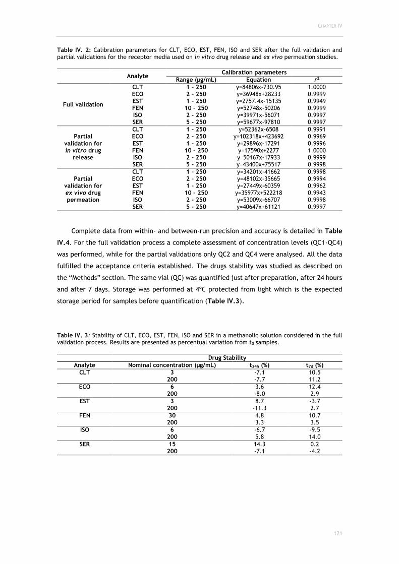
CHAPTER IV
121
Table IV. 2: Calibration parameters for CLT, ECO, EST, FEN, ISO and SER after the full validation and partial validations for the receptor media used on in vitro drug release and ex vivo permeation studies.
Analyte
Calibration parameters
Range (µg/mL) Equation r2
Full validation
CLT 1 – 250 y=84806x-730.95 1.0000 ECO 2 – 250 y=36948x+28233 0.9999 EST 1 – 250 y=2757.4x-15135 0.9949 FEN 10 – 250 y=52748x-50206 0.9999 ISO 2 - 250 y=39971x-56071 0.9997 SER 5 - 250 y=59677x-97810 0.9997
Partial validation for in vitro drug
release
CLT 1 – 250 y=52362x-6508 0.9991 ECO 2 – 250 y=102318x+423692 0.9969 EST 1 – 250 y=29896x-17291 0.9996 FEN 10 – 250 y=17590x+2277 1.0000 ISO 2 - 250 y=50167x-17933 0.9999 SER 5 - 250 y=43400x+75517 0.9998
Partial validation for ex vivo drug permeation
CLT 1 – 250 y=34201x-41662 0.9998 ECO 2 – 250 y=48102x-35665 0.9994 EST 1 – 250 y=27449x-60359 0.9962 FEN 10 – 250 y=35977x+522218 0.9943 ISO 2 - 250 y=53009x-66707 0.9998 SER 5 - 250 y=40647x+61121 0.9997
Complete data from within- and between-run precision and accuracy is detailed in Table
IV.4. For the full validation process a complete assessment of concentration levels (QC1-QC4)
was performed, while for the partial validations only QC2 and QC4 were analysed. All the data
fulfilled the acceptance criteria established. The drugs stability was studied as described on
the “Methods” section. The same vial (QC) was quantified just after preparation, after 24 hours
and after 7 days. Storage was performed at 4ºC protected from light which is the expected
storage period for samples before quantification (Table IV.3).
Table IV. 3: Stability of CLT, ECO, EST, FEN, ISO and SER in a methanolic solution considered in the full validation process. Results are presented as percentual variation from t0 samples.
Drug Stability
Analyte Nominal concentration (µg/mL) t24h (%) t7d (%)
CLT
3 -7.1 10.5 200 -7.7 11.2
ECO
6 3.6 12.4 200 -8.0 2.9
EST
3 8.7 -3.7 200 -11.3 2.7
FEN
30 4.8 10.7 200 3.3 3.5
ISO
6 -6.7 -9.5 200 5.8 14.0
SER
15 14.3 0.2 200 -7.1 -4.2

DRUG RELEASE AND PERMEATION
122
Figure IV. 1: Representative chromatogram of the quantification method in the full validation process, HPLC-DAD, measured at 210 nm (standard sample from the calibration curve at 50 µg/ml). The six molecules were identified and quantified as separated peaks in a single run.
To our best knowledge, the analytical method herein presented is the first one to include
five antifungal molecules and an oestrogen. These are the most representative APIs included in
currently marketed vaginal semisolids. Although in this study these APIs were quantified alone
in each drug release or permeation study, this method allows for further applications such as
the development of new products containing combinations of these drugs. Indeed, despite the
topical treatment of vaginal infections has been shown to be effective in most cases, the
microbial resistance to chemotherapeutics and the difficulties in managing infection
recurrences sustain the need for more effective local treatments132. Actually, there is a
continuous need for new analytical methodologies that shall be quicker, more sensitive,
accurate and precise, that allow for drug quantification in biological samples, and that also
could support the various stages of drug discovery and development375,376. Our technique was
easily translated to biological simulants, showing that it is promising for application in, for
example, cell culture medias (in vitro) and even biological samples collected on in vivo studies.
Actually, the complexity and quality of bioanalytical assays increases with the stage of
drug/product development. Moreover, the validation criteria from international guidelines are
not usually fully applied in the early stages of drug discovery and development375,376. This could
be the reason why there are few HPLC validated methodologies described on the scientific
literature regarding in vitro release and ex vivo permeation282. This work shows a robust
technique that is able to provide results with a high reliability and that complies with the
international standards from the US Food and Drug Administration and the European Medicines
Agency270,368.
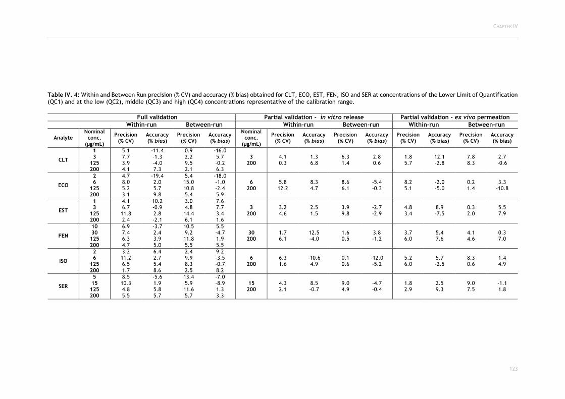
CHAPTER IV
123
Table IV. 4: Within and Between Run precision (% CV) and accuracy (% bias) obtained for CLT, ECO, EST, FEN, ISO and SER at concentrations of the Lower Limit of Quantification (QC1) and at the low (QC2), middle (QC3) and high (QC4) concentrations representative of the calibration range.
Full validation Partial validation - in vitro release Partial validation - ex vivo permeation
Within-run Between-run Within-run Between-run Within-run Between-run
Analyte Nominal
conc. (µg/mL)
Precision (% CV)
Accuracy (% bias)
Precision (% CV)
Accuracy (% bias)
Nominal conc.
(µg/mL)
Precision (% CV)
Accuracy (% bias)
Precision (% CV)
Accuracy (% bias)
Precision (% CV)
Accuracy (% bias)
Precision (% CV)
Accuracy (% bias)
CLT
1 5.1 -11.4 0.9 -16.0 3 7.7 -1.3 2.2 5.7 3 4.1 1.3 6.3 2.8 1.8 12.1 7.8 2.7
125 3.9 -4.0 9.5 -0.2 200 0.3 6.8 1.4 0.6 5.7 -2.8 8.3 -0.6 200 4.1 7.3 2.1 6.3
ECO
2 4.7 -19.4 5.4 -18.0 6 8.0 2.0 15.0 -1.0 6 5.8 8.3 8.6 -5.4 8.2 -2.0 0.2 3.3
125 5.2 5.7 10.8 -2.4 200 12.2 4.7 6.1 -0.3 5.1 -5.0 1.4 -10.8 200 3.1 9.8 5.4 5.9
EST
1 4.1 10.2 3.0 7.6 3 6.7 -0.9 4.8 7.7 3 3.2 2.5 3.9 -2.7 4.8 8.9 0.3 5.5
125 11.8 2.8 14.4 3.4 200 4.6 1.5 9.8 -2.9 3.4 -7.5 2.0 7.9 200 2.4 -2.1 6.1 1.6
FEN
10 6.9 -3.7 10.5 5.5 30 7.4 2.4 9.2 -4.7 30 1.7 12.5 1.6 3.8 3.7 5.4 4.1 0.3 125 6.3 3.9 11.8 1.9 200 6.1 -4.0 0.5 -1.2 6.0 7.6 4.6 7.0 200 4.7 5.0 5.5 5.5
ISO
2 3.2 6.4 2.4 9.2 6 11.2 2.7 9.9 -3.5 6 6.3 -10.6 0.1 -12.0 5.2 5.7 8.3 1.4
125 6.5 5.4 8.3 -0.7 200 1.6 4.9 0.6 -5.2 6.0 -2.5 0.6 4.9 200 1.7 8.6 2.5 8.2
SER
5 8.5 -5.6 13.4 -7.0 15 10.3 1.9 5.9 -8.9 15 4.3 8.5 9.0 -4.7 1.8 2.5 9.0 -1.1 125 4.8 5.8 11.6 1.3 200 2.1 -0.7 4.9 -0.4 2.9 9.3 7.5 1.8 200 5.5 5.7 5.7 3.3

DRUG RELEASE AND PERMEATION
124
IV.2.2.2. Drug solubility
Drug solubility was determined to assure that sink conditions could be maintained during
the assays of drug release and drug permeation377. Therefore, we should obtain high solubility
in the simplest media, that also could, in some extent, include some physiologic featuring’s. A
phosphate buffered solution was thought as being the most representative. However, this
solution had to be mixed with other solvents to increase drugs solubility. Then, the pHs were
differentiated for in vitro release and ex vivo permeation, since the first is expected to occur
in the vaginal cavity, where the normal pH is 4.2, and the second is expected to occur over the
vaginal epithelia where the interface to the systemic circulation is present. Table IV. 5
demonstrates the solubility obtained for the drugs included in this study. CLT was studied in
different media because it was difficult to solubilize in the first ones (data not shown). For in
vitro release studies the media selected were a mixture of ethanol and phosphate buffer
solution (EtOH:PBS, 40:60) at pH 4.2 for drugs ECO, EST, FEN, ISO and SER. CLT in vitro release
was performed using PEG 400 : PBS (40:60) at pH 4.2. All ex vivo permeation experiments
required the use of EtOH:PBS (40:60) at pH 7.4, except CLT which was performed with a media
containing PEG 400.
Table IV. 5: Solubility (µg/mL) for CLT, ECO, EST, FEN, ISO and SER in solutions tested as possible receptor media for in vitro drug release and ex vivo permeation studies. The receptor media selected to be used in further experiments were marked with *.
Drug solubility (µg/mL)
Drug pH 4.2 pH 7.4
PBS EtOH:PBS (40:60)
PLX 0.2%:PBS
PBS EtOH:PBS (40:60)
PLX 0.2%:PBS
ECO 386.1 967.5* 664.6 13.1 561.0* 75.7 EST 37.2 >1000* 44.4 39.7 >1000* 45.9 FEN <10 µg/mL
(LLOQ) 789.7* 93.7 <10 µg/mL
(LLOQ) 223.6* 10.3
ISO 178.5 971.5* 68.2 14.3 >1000* 22.0 SER 19.9 784.5* 30.8 <5 µg/mL
(LLOQ) 659.0* 59.8
pH 4.2 pH 7.4 PBS PEG 400:PBS
(40:60) PLX
0.5%:PBS PBS PEG400:PBS
(40:60) PLX
0.5%:PBS
CLT 17.0 282.1* 0.7 0.6 764.8* 4.8
This study was essential to assure that molecules would have enough solubility in receptor
media to be able to diffuse and to cross the membranes (inert or biological) and achieve the
receptor chamber. Actually, this was a limitation found during literature review, since data on
drug solubility is frequently not reported in published studies even in those that conclude that
the drugs did not transpose the membranes. On the contrary, several other authors addressed
this issue139,282,283,372 although, there was no sufficient data available regarding solubility of the
drugs included in this study. Therefore, this full assessment of solubility was mandatory for this
study.
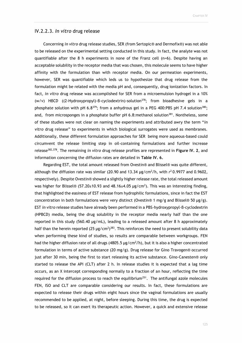
CHAPTER IV
125
IV.2.2.3. In vitro drug release
Concerning in vitro drug release studies, SER (from Sertopic® and Dermofix®) was not able
to be released on the experimental setting conducted in this study. In fact, the analyte was not
quantifiable after the 8 h experiments in none of the Franz cell (n=6). Despite having an
acceptable solubility in the receptor media that was chosen, this molecule seems to have higher
affinity with the formulation than with receptor media. On our permeation experiments,
however, SER was quantifiable which leds us to hypothesize that drug release from the
formulation might be related with the media pH and, consequently, drug ionization factors. In
fact, in vitro drug release was accomplished for SER from a microemulsion hydrogel in a 10%
(w/v) HβCD ((2-Hydroxypropyl)-β-cyclodextrin)-solution378; from bioadhesive gels in a
phosphate solution with pH 6.8379; from a anhydrous gel in a PEG 400:PBS pH 7.4 solution380;
and, from microsponges in a phosphate buffer pH 6.8:methanol solution381. Nontheless, some
of these studies were not clear on naming the experiments and attributed awry the term “in
vitro drug release” to experiments in which biological surrogates were used as membranes.
Additionally, these different formulation approaches for SER being more aqueous-based could
circumvent the release limiting step in oil-containing formulations and further increase
release382,378. The remaining in vitro drug release profiles are represented in Figure IV. 2, and
information concerning the diffusion rates are detailed in Table IV. 6.
Regarding EST, the total amount released from Ovestin® and Blissel® was quite different,
although the diffusion rate was similar (20.90 and 13.34 µg/cm2√h, with r2 0.9977 and 0.9602,
respectively). Despite Ovestin® showed a slightly higher release rate, the total released amount
was higher for Blissel® (57.20±10.93 and 48.16±4.05 µg/cm2). This was an interesting finding,
that highlighted the easiness of EST release from hydrophilic formulations, since in fact the EST
concentration in both formulations were very distinct (Ovestin® 1 mg/g and Blissel® 50 µg/g).
EST in vitro release studies have already been performed in a PBS-hydroxypropyl-β-cyclodextrin
(HPBCD) media, being the drug solubility in the receptor media nearly half than the one
reported in this study (560.40 µg/mL), leading to a released amount after 8 h approximately
half than the herein reported (25 µg/cm2)383. This reinforces the need to present solubility data
when performing these kind of studies, so results are comparable between workgroups. FEN
had the higher diffusion rate of all drugs (4805.5 µg/cm2√h), but it is also a higher concentrated
formulation in terms of active substance (20 mg/g). Drug release for Gino Travogen® occurred
just after 30 min, being the first to start releasing its active substance. Gino-Canesten® only
started to release the API (CLT) after 2 h. In release studies it is expected that a lag time
occurs, as an X intercept corresponding normally to a fraction of an hour, reflecting the time
required for the diffusion process to reach the equilibrium251. The antifungal azole molecules
FEN, ISO and CLT are comparable considering our results. In fact, these formulations are
expected to release their drugs within eight hours since the vaginal formulations are usually
recommended to be applied, at night, before sleeping. During this time, the drug is expected
to be released, so it can exert its therapeutic action. However, a quick and extensive release

DRUG RELEASE AND PERMEATION
126
can also trigger side effects, and azolic antifungals are well-known by their concentration-
dependent toxicity281.
Figure IV. 2: in vitro drug release (µg/cm2) profiles of Ovestin® (EST), Blissel® (EST), Gino-Canesten® (CLT), Lomexin® (FEN) and Gino Travogen® (ISO) as function of square root of time (h). The release rate was inferred by the slope obtained for each profile. Results are means and bars represent standard deviations (n=6).
It was our option to conduct the fully assessment of in vitro release during 8 h and to
present the total profiles instead of limited points for tracing the linear profile, as
recommended by the FDA-SUPAC document (Scale-up and Post-Approval Change)251. Actually,
it was our intention to see the complete release profiles, since what we are evaluating is the
correspondence of release and our proposed ex vivo permeation method. Although working on
an extended time period, at the end of the experiments drug content in the receptor chamber
did not exceeded 30% of the amount placed on the donor chamber, which would indicate
excessive depletion of drug from the donor compartment and would be translated as a
curvature in the upper part of the plots. These observations confirm that the experimental
setting corresponded to an “infinite dose”.
IV.2.2.4. Ex vivo drug permeation
Ex vivo drug permeation was tested using a porcine vaginal ex vivo model. Porcine vaginal
epithelium ex vivo has been chosen for membrane model in vaginal permeation experiments
since the pig has been already cited as remarkably similar with human in terms of anatomy,
physiology, metabolism and histology. Furthermore, previous studies have reported that an
excellent correlation was found between human and porcine vaginal tissues34,237,246,364.
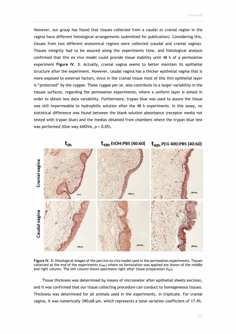
CHAPTER IV
127
However, our group has found that tissues collected from a caudal or cranial region in the
vagina have different histological arrangements (submitted for publication). Considering this,
tissues from two different anatomical regions were collected (caudal and cranial vagina).
Tissues integrity had to be assured along the experiments time, and histological analysis
confirmed that this ex vivo model could provide tissue stability until 48 h of a permeation
experiment Figure IV. 3. Actually, cranial vagina seems to better maintain its epithelial
structure after the experiment. However, caudal vagina has a thicker epithelial vagina that is
more exposed to external factors, since in the cranial tissue most of this thin epithelial layer
is “protected” by the ruggae. These ruggae per se, also contribute to a larger variability in the
tissues surfaces, regarding the permeation experiments, where a uniform layer is aimed in
order to obtain less data variability. Furthermore, trypan blue was used to assure the tissue
was still impermeable to hydrophilic solution after the 48 h experiments. In this assay, no
statistical difference was found between the blank solution absorbance (receptor media not
tested with trypan blue) and the medias obtained from chambers where the trypan blue test
was performed (One-way ANOVA, p < 0.05).
Figure IV. 3: Histological images of the porcine ex vivo model used in the permeation experiments. Tissues collected at the end of the experiments (t48h) where no formulation was applied are shown of the middle and right column. The left column shows specimens right-after tissue preparation (t0h).
Tissue thickness was determined by means of micrometer after epithelial sheets excision,
and it was confirmed that our tissue collecting procedure can conduct to homogeneous tissues.
Thickness was determined for all animals used in the experiments, in triplicate. For cranial
vagina, it was numerically 390±68 µm, which represents a total variation coefficient of 17.4%.
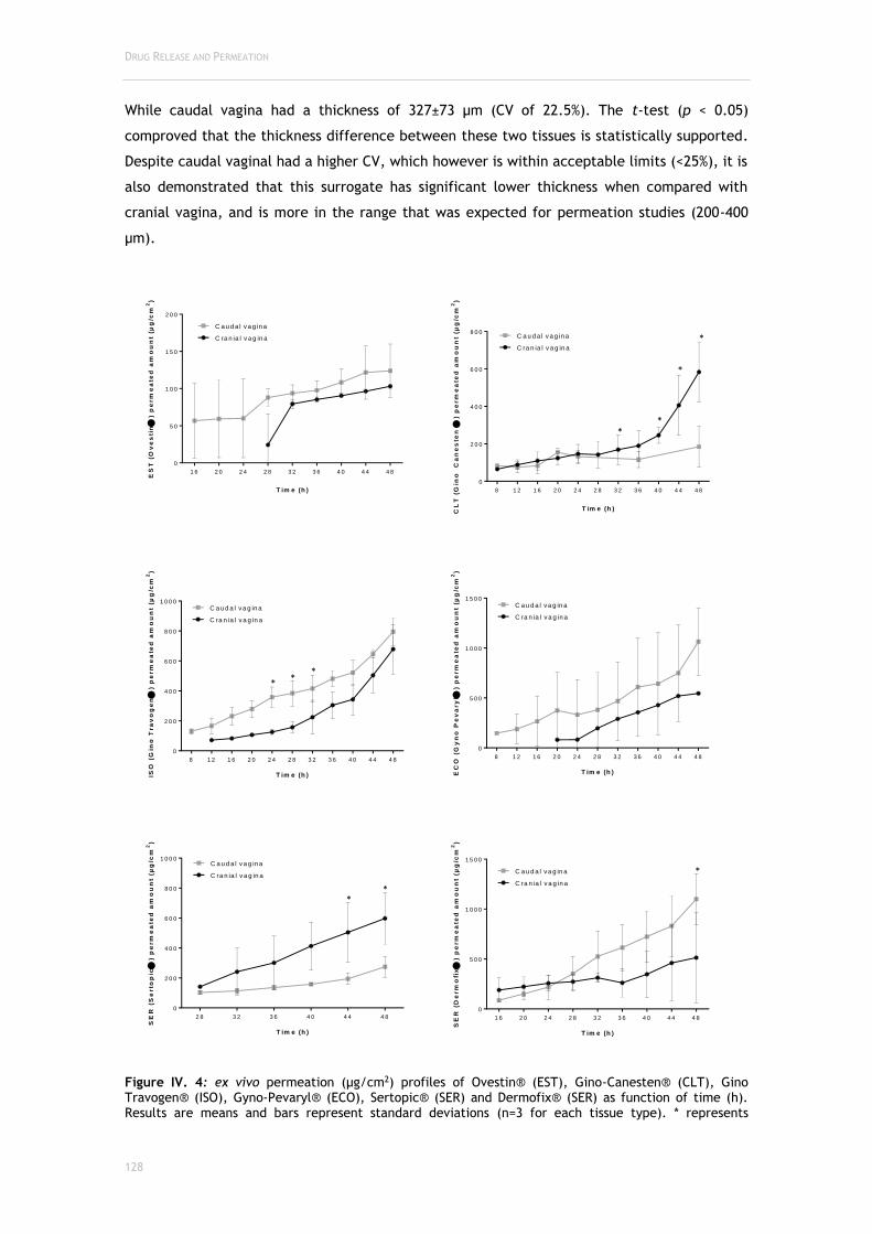
DRUG RELEASE AND PERMEATION
128
While caudal vagina had a thickness of 327±73 µm (CV of 22.5%). The t-test (p < 0.05)
comproved that the thickness difference between these two tissues is statistically supported.
Despite caudal vaginal had a higher CV, which however is within acceptable limits (<25%), it is
also demonstrated that this surrogate has significant lower thickness when compared with
cranial vagina, and is more in the range that was expected for permeation studies (200-400
µm).
1 6 2 0 2 4 2 8 3 2 3 6 4 0 4 4 4 8
0
5 0
1 0 0
1 5 0
2 0 0
T im e (h )
ES
T (
Ov
es
tin
) p
erm
ea
ted
am
ou
nt
(µg
/cm
2)
C ra n ia l v a g in a
C a u d a l v a g in a
8 1 2 1 6 2 0 2 4 2 8 3 2 3 6 4 0 4 4 4 8
0
2 0 0
4 0 0
6 0 0
8 0 0
T im e (h )
CL
T (
Gin
o
Ca
ne
ste
n
) p
erm
ea
ted
am
ou
nt
(µg
/cm
2)
C ra n ia l v a g in a
C a u d a l v a g in a
8 1 2 1 6 2 0 2 4 2 8 3 2 3 6 4 0 4 4 4 8
0
2 0 0
4 0 0
6 0 0
8 0 0
1 0 0 0
T im e (h )
C ra n ia l v a g in a
C a u d a l v a g in a
ISO
(G
ino
Tra
vo
ge
n
) p
erm
ea
ted
am
ou
nt
(µg
/cm
2)
8 1 2 1 6 2 0 2 4 2 8 3 2 3 6 4 0 4 4 4 8
0
5 0 0
1 0 0 0
1 5 0 0
T im e (h )EC
O (
Gy
no
Pe
va
ry
l )
pe
rm
ea
ted
am
ou
nt
(µg
/cm
2)
C ra n ia l v a g in a
C a u d a l v a g in a
2 8 3 2 3 6 4 0 4 4 4 8
0
2 0 0
4 0 0
6 0 0
8 0 0
1 0 0 0
T im e (h )
SE
R (
Se
rto
pic
) p
erm
ea
ted
am
ou
nt
(µg
/cm
2)
C ra n ia l v a g in a
C a u d a l v a g in a
1 6 2 0 2 4 2 8 3 2 3 6 4 0 4 4 4 8
0
5 0 0
1 0 0 0
1 5 0 0
T im e (h )
SE
R (
De
rm
ofi
x
) p
erm
ea
ted
am
ou
nt
(µg
/cm
2)
C ra n ia l v a g in a
C a u d a l v a g in a
Figure IV. 4: ex vivo permeation (µg/cm2) profiles of Ovestin® (EST), Gino-Canesten® (CLT), Gino Travogen® (ISO), Gyno-Pevaryl® (ECO), Sertopic® (SER) and Dermofix® (SER) as function of time (h). Results are means and bars represent standard deviations (n=3 for each tissue type). * represents
* *
*
*
* * *
* *
*

CHAPTER IV
129
statistically different values between caudal and cranial vagina (two-way ANOVA, p < 0.05, Tukey’s multiple comparisons test).
Permeation parameters were calculated using the profiles obtained from Figure IV. 4, and
are detailed on Table IV. 5.
The only drug that pointedly accumulated on tissues was EST from Blissel® (22.7±3.7% on
caudal and 27.1±3.7% on cranial vagina). This active substance did not surpass the epithelial
barrier, however it had affinity to the biological substract, which is expected since estriol is
used in vaginal atrophic menopausal symptoms. This connexion with the tissue could be helpful
to regenerate the epithelial layer largely injured during menopause44,384,385. On the contrary,
EST contained in Ovestin® was not quantifiable in tissues, although approx. 20% of the applied
drug permeated the membranes (28.9±2.4 and 21.0±2.4% in caudal and cranial vagina,
respectively). FEN, despite its higher diffusion rate, it did not permeate the membranes and
neither largely accumulated in tissues (1.2±0.3 and 0.9±0.3% on caudal and cranial vagina,
respectively). This could be explained by the fact that the biological surrogate is actually a
limiting step for fenticonazole.
Only after 8 h of experiment, CLT was quantifiable on the receptor chambers. This drug,
along with ECO and SER (coming from Sertopic®) were the only ones to have significant
differences between the Jss calculated for the two types of tissues. In all experiments, except
Gino Canesten® and Sertopic®, the caudal vagina seemed to be more permeable (despite
statistical differences were not always found). When comparing these results with the ones
obtained in the release studies it is clear that permeation only occurs (or is detectable) mainly
after the 8 h. This further reinforces the idea that vaginal semisolids should be designed to be
applied before bed, so they can have a residence time enough to allow drug release, and then
the release amount is able cross the biological barriers and exert their therapeutic action, if
the desired action is systemic. If it is local, the night administration enhances the formulations
contact with the vaginal lining, favouring the local therapy (during day time is more probable
to leak). Experiments with vaginal excised tissue to assess vaginal drug permeation for the
drugs also included in this study are not reported on the literature. Only a formulation
containing clotrimazole loaded into nanostructured lipid carriers for the treatment of fungal
vaginal infections was studied, however PBS alone was used as receptor medium and CLT
solubility was not considered255. The scarce literature on the subject and the fact that we are
dealing with commercial formulations, from which we do not have full information on
excipients quantitative composition, could represent a difficulty to discuss and integrate our
new findings on the scientific community. Nonetheless, it is truly not our objective to evaluate
the products herein tested. These products were chosen, exactly because we are certain that
they are largely studied, and having been through clinical trials, and also being marketed, some
of them for so long, assures that they are safe, effective and can be used as model products
for testing new methodologies, as the ones proposed in this study, and in others already
published by our group331. It was not found a significant difference between using caudal or
cranial vagina in ex vivo vaginal porcine permeation studies, which on one hand could mean

DRUG RELEASE AND PERMEATION
130
that any of these tissues are representative of the vaginal epithelia, or on the other hand could
indicate that both tissues portions should be studied. In fact, as it can be seen on Figure IV. 4,
although significant differences were not extensive concerning the tissues and formulations
included in this study, it was clear that vestibule (caudal) could allow is generally more
permeable considering the timeframe used for permeation, and the permeated total drug
amounts (Table IV. 5). Also, they were shown to be more consistent (lower SDs). Indeed, cranial
vagina tissue has more ruggae on its constitution and this could explain the higher deviations
between samples (tissue irregularities could congregate thicker and thinner parts). So, as a
general appointment and taking into consideration the reasons appointed above, we conclude
that caudal vagina epithelium should be preferred over the cranial, not only because it has
more histological similitudes with the human vaginal epithelium but also because it promotes
higher permeation rates (representing a “safer” strategy to study permeation since it will hardly
result in false negative outcomes) and more consistent results.

CHAPTER IV
131
Table IV. 6: Drug recovery from the formulation (%), ex vivo permeation and in vitro release parameters. Papp (apparent permeability coefficient); Jss (steady-state flux); NC (<LLOQ) (Not calculable because concentrations were below the lower limit of quantification); * represents statistically different from the histological tissue in comparison (One-way ANOVA, Tukey's multiple comparisons test, p < 0.05). n=3 tissues in ex vivo permeation experiments and n=6 in in vitro drug release studies. When drug quantification was <LLOQ, a 0 µg/mL concentration was asssumed.
Recovery from
formulation (% of
drug)
ex vivo permeation in vitro release
Caudal vagina Cranial vagina Total
permeated amount (% of drug)
Donor Chamber
(% of drug)
Tissue (% of drug)
Mass balance
(%) Papp (cm/s)
Jss (µg/cm2
min)
Total permeated amount (% of drug)
Donor Chamber
(% of drug)
Tissue (% of drug)
Mass balance
(%) Papp (cm/s)
Jss
(µg/cm2min)
Diffusion rate
(µg/cm2
√h)
r2
CLT (Gino
Canesten®)
95.8 3.2±2.0 88.7±7.9 5.5±8.4 97.5±12.0 2.943x10⁻⁷
±1.808x10⁻⁷ 1.07x10⁻³
±6.20x10⁻⁴ 10.0±3.5 79.3±1.1 5.3±0.5 92.2±3.5
9.077x10⁻⁷
±3.208x10⁻⁷ 3.38x10⁻³
±9.30x10-4* 25.1 0.9862
ECO (Gyno-
Pevaryl®)
98.4 17.6±1.2 77.1±12.7 2.9±0.4 97.5±13.4 1.602x10⁻⁶
±4.621x10⁻⁷ 6.16x10⁻³ ±1.94x10⁻³
9.8±1.2 96.1±12.7 0.4±0.4 106.3±13.4 8.931x10⁻⁷
±1.066x10⁻⁷ 3.16x10⁻³
±7.027x10⁻⁵* NC (<LLOQ)
EST (Ovestin®)
104.4 28.9±2.4 70.3±2.5 <1
µg/mL (LLOQ)
99.3±0.1 2.363x10⁻⁷
±5.971x10⁻⁷ 7.20x10⁻⁴ ±2.10x10⁻⁴
21.0±2.4 78.3±2.5 <1 µg/mL (LLOQ)
99.3±0.1 1.979x10⁻⁶
±1.900x10⁻⁷ 6.00x10⁻⁴
±1.452x10⁻⁵ 20.9 0.9977
EST (Blissel®)
87.2 <1 µg/mL (LLOQ)
60.3±1.5 22.7±3.7 82.9±4.4 NC (<1 µg/mL - LLOQ) <1 µg/mL (LLOQ)
69.5±1.5 27.1±3.7 96.5±4.4 NC (<1 µg/mL - LLOQ) 13.3 0.9602
FEN (Lomexin®)
118.5 <10 µg/mL
(LLOQ) 87.0±3.6 1.2±0.3 88.2±3.9 NC (<10 µg/mL - LLOQ)
<10 µg/mL (LLOQ)
94.0±3.6 0.9±0.3 94.9±3.9 NC (<10 µg/mL - LLOQ) 4805.5 0.9422
ISO (Gino
Travogen®)
88.9 14.4+1.7 102.2±0.7 1.5±0.4 118.1±1.0 1.315x10⁻⁶
±1.555x10⁻⁷ 4.60x10⁻³ ±5.30x10⁻⁴
12.0±3.3 97.5±8.2 0.6±0.3 110.0±7.7 1.118x10⁻⁶
±3.093x10⁻⁷ 3.92x10⁻³ ±9.60x10⁻⁴
55.6 0.9891
SER (Dermofix®)
82.4 22.3±7.5 74.5±14.2 0.4±0.4 97.2±19.5 2.027x10⁻⁶
±9.462x10⁻⁷ 6.37x10⁻³ ±1.47x10⁻³
8.8±7.5 96.7±14.2
0.3±0.4
105.9±19.5
7.975x10⁻⁷
±6.871x10⁻⁷ 2.97x10⁻³ ±2.61x10⁻³
NC (<5 µg/mL - LLOQ)
SER (Sertopic®)
104.6 4.6±3.0 90.7±5.0 0.6±0.1 96.0±17.7 4.227x10⁻⁷
±1.232x10⁻⁷ 1.59x10⁻³ ±4.00x10⁻⁴
9.5±3.0 98.9±15.0 0.4±0.1 108.7±17.7 8.604x10⁻⁷ ±2.723x10⁻⁷
3.46x10⁻³
±9.90x10⁻⁴* NC (<5 µg/mL - LLOQ)

DRUG RELEASE AND PERMEATION
132
IV.2.3. Conclusions
In vitro drug release and ex vivo drug permeation studies represent tools that shall support
researcher to predict drug performance in vivo at early stages of product development. In this
study, we were able to validate a quantification methodology using HPLC-DAD and applying this
to different matrices, not losing quality and resolution in partial validations. Furthermore, we
managed to assess the in vitro release profiles of eight vaginal semisolids, as well as their
permeation profiles, having previously studied the drugs solubility in order to maintain sink
conditions. Furthermore, following previous results of our group, we have investigated if there
were differences in performing ex vivo permeation studies using the porcine vaginal model,
when collecting a proximal or a distal tissue within the vaginal tube. No extensive significant
differences between these tissues were found, but we concluded that the caudal vaginal could
be more suitable for vaginal permeation experiments since it conducts to more reproducible
and consistent results. The inclusion of this methodological approach in preclinical stages of
products development, could optimize cost-efficiency of new formulations development
process by predicting in vivo efficacy and safety profiles.

CHAPTER V
133
CHAPTER V
GENERAL DISCUSSION

GENERAL DISCUSSION
134
CHAPTER V
135
Laboratorial research is, currently, particularly dedicated to discover or optimize former
strategies for drug delivery1–3, and the vaginal route of administration has been considered an
excellent alternative since it enables both local and systemic delivery4–8. Vaginal drug delivery
is accomplished through several dosage forms that are as simple as powders, and as complex
as the intravaginal ring, designed for the controlled delivery of hormones16. Drug delivery
efficacy mainly relies on the dosage form ability to promote adequate drug concentration at
the targeted action site17–21. Several studies have concluded that women prefer and better
accept and use, among vaginal formulations, the semisolids, preferably, gels22,23.
In fact, gels might represent the vaginal dosage forms that has been investigated the most
in recent years. They can be developed to be stimuli-sensitive systems, which can modify their
behaviour once the environmental conditions change107–110. Specifically, aqueous-based gels not
only, generally, provide better safety profiles, but also, do represent flexible vehicles that can
be improved by means of polymer science and nanotechnology51,111,112,115–122. Since vaginal
semisolids are usually committed with discomfort issues the use of strategies like
bioadhesion93,173 is valuable to avoid interferences with women’s daily life, while allowing
higher drug bioavailability with smaller absorption variations, and so lower incidence of side
effects17–21.
Under another perspective of vaginal products development, methodologies improvement
to better characterize new or re-newed formulations are needed. As several efforts have been
made to conduct investigation to better understand the vaginal epithelial mechanisms6,42 here
involved, this may benefit characterization methods, that if perfectly aligned with the target
epithelia knowledge, can contribute to more successful product development, along with high
levels of innovation in the product itself. Indeed, these optimized characterization processes
are not only essential in the first steps of development, but are also extremely useful for
monitoring batches in quality control or to assess the impact of formulation changes in the
industrial context. Thus, it is of great importance to implement accurate and reproducible
vaginal semisolids performance evaluation.
Recently, a strategy of patient-driven product development is also currently reflected in
policies such as the biopharmaceutics risk assessment roadmap (BioRAM), which intends to
optimize drug product development and performance by using therapy-driven target drug
delivery profiles as a framework to achieve the desired therapeutic outcome. Simultaneously,
clinical relevance is directly correlated to early formulation143–145. That means that a full
assessment of a final formulation candidate should be performed during its global development,
rather, than first selecting the active substance, and then step-by-step increase formulation
complexity with intermediate characterization. In this context, jumping to a screening of final
complete formulations candidates ahead of the final process development is clearly cost and
time-saving.
That was exactly the strategy applied in this thesis work, nevertheless, not for
formulations development properly, but in improving the characterization methods. While
recent publications on innovative vaginal products are largely devoted to microbicide

GENERAL DISCUSSION
136
formulations, antimicrobials, specifically, antifungals, and oestrogens are no doubt the most
prevalent in use due to the clinical relevance of vaginal disturbances, as vaginal infections, and
post-menopause atrophy related symptoms. This means that engagement and safety/risk
assessments similar to those related to microbicides design should be applied to other vaginal
product categories146–150.
In view of optimizing methods while gathering experimental data from widely used vaginal
products, the research work of this thesis started with the selection of vaginal semisolid
formulations that were currently commercialized in Portugal (but also representative of the
international market, i.e. EU and USA), and therefore, to which easy access and wide interest
on the obtained results were assured. Moreover, they should fill three product categories:
antimicrobials, oestrogens and products with reported experimental work on
technological/safety characterization. The importance of characterizing antimicrobial
preparations is obvious due to the prevalence and recurrence rate of vaginal infections. These
facts, conduct to difficulties in the clinical setting, once antimicrobial drugs resistance is an
inevitable issue132. So, in one hand these drug products represent the most often used by
reproductive-aged women and on the other hand they actually represent an opportunity for
improvement regarding some technological properties, since most of these formulations are
actually classical formulations. Oestrogens were added to this evaluation setting, since they
represent another important area of vaginal semisolids usage, as local therapies for atrophic
symptoms mainly after menopause384,386,387. In addition, two largely described and studied
products, Universal Placebo and Replens® were included in this study to serve as
reference/control products158,321. They are well-established as safe and/or technologically-
advanced vaginal semisolids, being these the reasons why they have been selected to serve in
this work as waiving products, in order to guarantee that the employed methods where being
optimized in the right direction (the Universal Placebo should present a safe profile that
correlates with the findings of clinical trials involving microbicides, while Replens should
present high acid-buffering capacity and bioadhesion). To sum up, the products included in the
present set of studies were: Gino-Canesten®, Sertopic®, Dermofix®, Gyno-pevaryl®,
Lomexin®, Gino Travogen®, Dalacin V®, Ovestin®, Blissel®, Colpotrophine®, Universal Placebo
and Replens®.
Vaginal semisolids evaluation itself began with their technological characterization, which
included insights from the chemical, the physical and the physiological point of view. This part
of the work series consisted on improving traditional characterization methods by using
physiological parameters in order to construct predictive tools to characterize close to ideal
vaginal semisolid formulation whatever target it may have (as long as it is defined). Products
were characterized in terms of: pH and buffering capacity in a vaginal fluid simulant (VFS);
osmolality - directly and upon dilution in VFS; textural parameters (firmness, adhesiveness and
bioadhesion) using vaginal ex vivo porcine epithelium; and viscosity (including VFS dilution at
37ºC and after administration on an ex vivo model).
CHAPTER V
137
The pH values of vaginal products, by itself, does not allow for a full prediction of pH
compatibility with the vaginal environment, in view of its safety. It is more relevant to further
assess the ability of these formulations to actually change the physiological pH once
administered (pH-buffering capacity) by correcting altered pH or maintain it within the
assumed healthy range. This parameter, especially when determined in VFS, contributed to a
better understanding of what will happen in vivo regarding pH changes after the formulation is
applied into the vagina159. For this reason, it is not only important to understand the ability of
acidic products to maintain this pH after contacting with successive amounts of an alkaline
solution (representing a less acidic vaginal fluid), but also to assess the resistance offered by
less acidic products to gain lower pH after mixing with acidic solutions which mimic the vaginal
fluid with normal pH.
The results obtained concerning osmolality showed that it might be of special interest to
consider its measurement also in physiological dilutions. The proposed test using normal
simulant (considering composition and the amount that is believed to be present in the vagina
to dilute the applied dose) clearly indicated that, although plain products might have
osmolalities above the recommended limits165 (1200 mOsmol/kg maximum as proposed by the
WHO for vaginal lubricants), the resulting osmolality of the dilution in physiological fluids could
be compliant with the recommendation. Furthermore, it was clear that the extent of reduction
on osmolality for hyperosmolar products was clearly dependent on the product composition
since, while for some products only a discreet effect was noted (e.g. Lomexin® osmolality
reduced from 1446 mOsm/Kg – direct- to 1210 mOsm/Kg in VFS), for other products osmolality
upon dilution in VFS represented less than 30% of the directly measured osmolality (e.g.
Colpotrophine® and Ovestin®). So, for future osmolality determinations a complete assessment
should not only comprise the direct measurement but also the one made after dilution in the
estimated physiologic amounts of VFS. Moreover, it would be important to consider specific
disease related characteristics of the vaginal environment that may affect the outcome of this
dilution, such as the increased volume or altered (less acidic) pH of vaginal discharge that
characterizes bacterial vaginosis and that may affect the performance of antibacterial
products. Interestingly, the osmolality of the resulting dilution has the capacity to early predict
the in vivo formulation behaviour, representing an initial strategy for safety foresee.
The textural parameters studied were firmness and adhesiveness. Their assessment was
accomplished by a physic-mechanical method and the objective was to find a correlation
between these two parameters, which however, we did not found. Bioadhesion, on its turn,
expressed as a mechanical measurement having the vagina as the attachment structure, was
found to have a moderate to strong uphill positive linear correlation with adhesiveness
determined by pure mechanical methods. This meant that bioadhesion could be predicted by
adhesiveness, a test that does not require the use of biological surrogates and can easily and
quickly be performed in earlier stages of product development.
Viscosity was, as expected, found to be highly dependent on temperature26,287. However,
the variation observed, was nor proportional nor similar among all formulations. Each

GENERAL DISCUSSION
138
formulation showed its own behaviour, once more driven by their specific composition. This
could mean that measurements directly made on formulations at room temperature do not
represent the viscosity that they acquire after administration, and that those should only be
performed for comparative studies. These results sustain that the selection of prototypes
intended to proceed in the development process should be based on measurements that mimic
the vaginal environment. The test using the ex vivo porcine vagina as an organic administration
model brought valuable information, with results that were different from those of the dilution
at 37ºC, supporting its inclusion as a model that better predicts the formulations’ rheology after
administration.
After the technological profiling of a formulation has been concluded, a preclinical safety
assessment is required, in order to complete its characterization, confirming the previous tests
that had already been done under a safety perspective. This approach included in vitro cellular
models, an ex vivo tissue model and an in vitro organotypic model.
Considering the cellular models, two in vitro cytotoxicity tests were performed: MTT and
NRU upon uterine (HEC-1A), cervical (HeLa) and vaginal (VK2 E6/E7) cell lines, in compliance
with ISO/EN 10993-5 guide for in vitro evaluation of vaginal medical devices. The vaginal cell
line VK2 E6/E7 showed to be the one that gave the more accurate calculation of TC50 (half-
maximal Toxic Concentration) among the three cellular lines on the MTT assay. In fact, this is
the most representative cellular model for vaginal products testing, although it was not
sensitive to the NRU assay.
Likewise, we have developed and optimized a strategy to determine tissue viability using
an ex vivo porcine vaginal model by using the MTT reduction assay and histological analysis185.
We showed that it is possible to collect tissues from the porcine model with approximately 15%
variability in thickness, and the first optimization step resulted in variation coefficients lower
than 25%, when testing negative and positive controls. The inclusion of the proposed ex vivo
toxicity model in preclinical safety assessment can lead to a better simulation to the in vivo
conditions. This model, has proven to be reliable and it has low economical expenditures when
compared with the 3D reconstructed tissues available on the market. That is a key point why
these ex vivo tissues could represent valuable tools for scientific research, when early stages
of the preclinical development of drugs/products is the focus. Moreover, the ex vivo models
take into consideration an inter-individual variability that is not present in manufactured
reconstructed models, and that in fact, is closer to the variability encountered further on in
vivo studies.
Safety assessment was further proposed to be performed though an organotypic in vitro
model adapted from the HET-CAM assay, undergoing full validation for eye irritation testing198.
In this assessment, vaginal semisolid medicines and lubricants currently marketed were tested
along with the Universal Placebo formulation which safety has been clinically shown.
Nonoxynol-9 (N-9), a known vaginal irritant, was enrolled as positive control (concentrations
ranging from 0.001 to 100% (v/v)). The assay was conducted according to the ICCVAM -
Recommended test method (NIH Publication No. 10-7553 – 2010). The studied vaginal
CHAPTER V
139
formulations showed low potential for irritation. N-9 was classified as a severe irritant at
concentrations above 2%, which corroborates clinical data from the literature, envisaging a
possible in vitro/in vivo correlation. IS (B) was considered as having a better classification
output. Although still requiring further validation, the HET-CAM assay showed to be sensitive
enough to differentiate vaginal products according to irritancy potential.
Finally, in vitro drug release and ex vivo permeation studies were performed under
efficacy and safety perspectives. To accomplish this task, firstly a validation of an analytical
method (HPLC-DAD) was performed to quantify six different molecules present in the vaginal
semisolid products. Secondly, in vitro drug release studies were performed to characterize drug
diffusion through the formulation, to evaluate if it was a limiting step for permeation. Ex vivo
drug permeation studies were performed using two histologically distinct tissues collected from
different regions of the porcine vagina (caudal and cranial portions). Permeability comparisons
between these two tissues were assessed. The validation of the HPLC-DAD method was
successfully accomplished according to FDA, EMA and ICH requirements. Receptor media for
both experiments were selected based on solubility studies to assure sink conditions.
Permeation through the vaginal tissue was found to be dependent on the type of formulation
tested and not directly defined by the release rate, although it was a consequence of it. Caudal
vagina was slightly more permeable than the cranial portion although statistical differences
were not evident for all studied products. Results deviations were more pronounced with the
cranial vagina due to the presence of extensive ruggae in it. Therefore, although ex vivo drug
permeation through different regions of the porcine vagina did not vary extensively, the use of
caudal vagina for these studies may yield more reproducible results.
All over the experimental work not only the characterization methods were optimized but
also important experimental data were gathered from commercial products widely available
and considered as safe. These data will be essential to allow for comparisons with new products
under development on the basis of “non-inferiority” considering each of these characteristics.
The results obtained with this thesis are particularly valuable for the company where the
research work took part and that supported it; however, once the papers herein presented are
published, they would be applicable worldwide, being expected that these results will be
largely accepted by the scientific community. The application and performance of these
methodological approaches will certainly benefit several workgroups, not only the ones working
on vaginal semisolids products, but also other vaginal dosage forms. Furthermore, the
optimized procedures conducted concerning the ex vivo porcine model, were demonstrated as
interessant and with high potential to be submitted as a proposal, for alternative methods to
laboratory animals, to the respective regulamentory authorities.
Summarizing, the objectives defined at the beginning of this thesis were fully
accomplished. Now, a complete preclinical performance profiling of vaginal semisolid products,
based on a set of methods ranging from simple technological characterization to complex
studies like drug ex vivo permeation, is defined and available. All the methods here described,

GENERAL DISCUSSION
140
alone or combined, do certainly represent valuable tools to be applied in new products
development and characterization both in industrial and academical environments.

CHAPTER VI
141
CHAPTER VI
CONTRIBUTIONS FOR ENHANCING THE
COMPETITIVENESS OF THE HOSTING COMPANY
(LABFIT-HPRD, LDA)

CONTRIBUTIONS FOR LABFIT
142

CHAPTER VI
143
Labfit is an R&D company dedicated to the development of innovative pharmaceuticals
classifiable as medicines, medical devices or cosmetics, as well as, the provision of R&D services
in the health area, thus working with a high level of technology, not only in fundamental
research, but also on applied and translational research. Furthermore, part of the work
developed by Labfit is specialized in topical application products with a special focus in the
area of gynaecology.
Therefore, the objectives of this thesis were completely aligned with the company’s
strategy and contributed for Labfit’s expertise on topical products characterization. Indeed, all
the research hither presented contributed for enhancing the competitiveness of Labfit as these
results were transferred from the academia to the company as:
□ A complete technological characterization service. A full assessment of vaginal
semisolids galenic and technological characterization method was systematized. From
simple assays, such as organoleptic characteristics, pH and osmolality determination,
to others more complex, like pH buffering capacity, texture and viscosity. Moreover, a
physiological approach has been applied to these methods, not only because they were
discussed taking into account the healthy vaginal environment and the in vivo
behaviour, but also, because actually some assays were concomitantly performed using
a vaginal fluid simulant and an ex vivo porcine model;
□ An integrative in vitro and ex vivo safety characterization service. This was optimized
considering the current normatives and consists on alternative method to experimental
animals that includes two different cellular and tissue toxicity method and an
organotypic method for vaginal irritation testing which broadens the scope of
application of the methods previously performed at Labfit;
□ An HPLC validated method for quantification of the most representative active
substances present in vaginal semisolid formulations. The method was validated
according to FDA, EMA and ICH requirements and has great potential to be applied in
Labfit as services for product characterization in this diversity of molecules;
□ A drug permeation test that combines both in vitro and ex vivo models allowing a full
access to vaginal drug delivery either intended to be local or systemic;
□ The combination of the results obtained with this work contributed to start the
construction of an automatic in-line database. This will be used by Labfit during product
development or optimization and during interpretation of results from products
received as samples for characterization, helping to evaluate their technological
properties and safety.
The scientific knowledge together with the laboratory skills obtained with this work were
translated into written procedures and, further qualification of researchers for these methods
being converted in new or more accurate services provided by Labfit, as well as, an
international recognition of this company as an expert on topical pharmaceutical products for
vaginal application.

CONTRIBUTIONS FOR LABFIT
144

CHAPTER VII
145
CHAPTER VII
CONCLUSIONS AND FUTURE REMARKS

CONCLUSIONS AND FUTURE REMARKS
146
CHAPTER VII
147
Optimized preclinical performance assessment of vaginal semisolid products seems to
represent a key point in new products development. Improvements in methodologies turn them
time saving, cost-efficient and very helpful in the decision-making process. The assumption
that physiological parameters (composition and amount of fluids, body temperature and even
movements) should be considered, can further expedite this process while bringing assessment
close to real life conditions.
The work developed during this thesis supports the concept that a primary technological
characterization under a physiological perspective is essential. It was highlighted that pH
buffering capacity, osmolality and viscosity determined using the proposed methodological
adaptations were considered focal points to be addressed during products’ development. The
importance of giving attention to vaginal antimicrobial and oestrogenic formulations at the
same level of that payed to microbicides, was stressed. Another point evidenced by this
investigation was that polymer-based strategies could be useful in re-formulating products in
order to overcome problems of leakage and discomfort, and improve efficacy. Since polymeric
formulations demonstrated more favourable characteristics regarding the vaginal epithelia.
Concerning preclinical safety assessment, three strategies have been combined in this
thesis. First, in vitro cellular models were used to assess toxicity using conventional and
standardized methods. Second, an ex vivo tissue model was established and adequately
optimized to improve the complexity level and robustness of toxicity assessment over the first
method. Third, an organ-based in vitro method, already in validation by regulatory agencies
for testing irritation in other biological surrogates, was effectively transposed to vaginal
irritation testing. These set of methods can not only be applied to the pharmaceutical industry
and research, but also to the cosmetic, hygiene, and medical devices industries. Moreover, the
ex vivo model herein proposed, can further be applied to the development of primary cell
cultures, as a surrogate for permeation and metabolic studies, and even to evolve to more
complex systems, including co-cultures with microorganisms.
The multi-drug HPLC-DAD quantification method developed and validated in view of this
work has demonstrated a great potential of applications, including the study of different types
of vaginal products with distinct active substances but also, as it is able to quantify six different
molecules in just one run, it would be valuable for quantification of drugs in new developing
products that contain combinations of these active substances. Moreover, it could be applied
not only on drug release and permeation, but also could be useful for drug quantification in
formulations under testing, for example, in stability studies or quality control after production.
Product performance on the developed in vitro drug release and ex vivo drug permeation
studies, demonstrated a great potential to predict drug performance in vivo at early stages of
product development. This combined methodology conducted to a full assessment of drug
release and permeation profiles in eight different vaginal semisolids. Once again, in drug
permeation, the porcine ex vivo model has confirmed to be an efficient, manageable and
applicable tool in several testing methods. Furthermore, a precise study conducted over
CONCLUSIONS AND FUTURE REMARKS
148
anatomical and histological featuring’s of this ex vivo model, have determined that it is
essential to assign which type of tissue is collect from the porcine vagina.
Following this methodological definition and optimization that have allowed for the
objective characterization of a representative group of currently marketed semisolids, this tool
may be used to assess the individual influences and contributions of those excipients frequently
used in vaginal formulations for the overall performance of the product, regarding each of the
parameters under study, by using a single, well studied base (such as the Universal Placebo).
The database that has been prepared in the current project will then be enlarged with data
from excipients and will become a powerful tool to assist the rational design or optimization of
vaginal products towards fulfilling the vaginal semisolids market considering clinical needs and
women’s preferences.
To sum up, the objectives proposed in the beginning of this thesis were accomplished, and
now, a complete assessment on preclinical performance of vaginal semisolid products has been
described and applied to formulations that are currently marketed worldwide. This assessment
being based on technological and safety evaluations that assume physiologic parameters is
worth to be applied in new products development and characterization, either on academia or
on an industrial environment.
The here established set of assessment methods will from now on be adopted by Labfit in
research as well in current testing services.

CHAPTER VII
149
CHAPTER VIII
REFERENCES

CONCLUSIONS AND FUTURE REMARKS
150

CHAPTER VIII
151
1. Ndesendo, V. M. K. et al. A review of current intravaginal drug delivery approaches
employed for the prophylaxis of HIV/AIDS and prevention of sexually transmitted
infections. AAPS PharmSciTech 9, 505–20 (2008).
2. Woolfson, A. D., Malcolm, R. K. & Gallagher, R. Drug delivery by the intravaginal route.
Crit Rev Ther Drug Carr. Syst 17, 509–55 (2000).
3. Srikrishna, S. & Cardozo, L. The vagina as a route for drug delivery: a review. Int
Urogynecol J 24, 537–43 (2013).
4. Alexander, N. J. et al. Why consider vaginal drug administration? Fertil Steril 82, 1–12
(2004).
5. Hussain, A. & Ahsan, F. The vagina as a route for systemic drug delivery. J Control
Release 103, 301–13 (2005).
6. Wu, S. J. & Robinson, J. R. Vaginal epithelial models. Pharm Biotechnol 8, 409–24
(1996).
7. Choudhury, A., Das, S. & Kar, M. A review on novelty and potentiality of vaginal drug
delivery. Int J PharmTech Res 3, 1033–1044 (2011).
8. Wong, T. W., Dhanawat, M. & Rathbone, M. J. Vaginal drug delivery: strategies and
concerns in polymeric nanoparticle development. Expert Opin Drug Deliv 1–16 (2014).
9. Katz, D. F. & Dunmire, E. N. Cervical mucus: Problems and opportunities for drug
delivery via the vagina and cervix. Adv Drug Deliv Rev 11, 385–401 (1993).
10. Hani, U., Bhat, R., Sisodiya, R. & Gurumallappa Shivakumar, H. Novel Vaginal Drug
Delivery Systems: A Review. Curr Drug Ther. 5, 95–104 (2010).
11. Katz, D. F., Dunmire, E. N., Henderson, M. H., Owen, D. H. & Plenys, A. M. Applications
of biomedical engineering in reproductive biomedicine: sensing and drug delivery to the
lower female reproductive tract. in Proceedings of the 19th Annual International
Conference of the IEEE Engineering in Medicine and Biology Society. ‘Magnificent
Milestones and Emerging Opportunities in Medical Engineering’ (Cat. No.97CH36136) 6,
2656–2658 (IEEE, 1997).
12. Vermani, K. & Garg, S. The scope and potential of vaginal drug delivery. Pharm Sci
Technolo Today 3, 359–364 (2000).
13. Weber, J., Desai, K. & Darbyshire, J. The development of vaginal microbicides for the
prevention of HIV transmission. PLoS Med. 2, e142 (2005).
14. Acartürk, F. Mucoadhesive vaginal drug delivery systems. Recent Pat Drug Deliv Formul
3, 193–205 (2009).
15. das Neves, J., Palmeira-de-Oliveira, R., Palmeira-de-Oliveira, A., Rodrigues, F. &
Sarmento, B. in Mucoadhesive Materials and Drug Delivery Systems (ed. Khutoryanskiy,
V. V.) 99–131 (Wiley, 2014).
REFERENCES
152
16. Allen, L. V, Popovich, N. G. & Ansel, H. C. Ansel ’ s Pharmaceutical Dosage Forms and
Drug Delivery Systems. (Williams & Wilkins, 2011).
17. Sassi, A. B., McCullough, K. D., Cost, M. R., Hillier, S. L. & Rohan, L. C. Permeability of
tritiated water through human cervical and vaginal tissue. J Pharm Sci 93, 2009–16
(2004).
18. Ensign, L. M., Cone, R. & Hanes, J. Nanoparticle-based drug delivery to the vagina: A
review. J Control Release (2014).
19. Anderson, M. J., Scholz, M. T., Parks, P. J. & Peterson, M. L. Ex vivo porcine vaginal
mucosal model of infection for determining effectiveness and toxicity of antiseptics. J
Appl Microbiol 115, 679–88 (2013).
20. Chatterton, B. E., Penglis, S., Kovacs, J. C., Presnell, B. & Hunt, B. Retention and
distribution of two 99mTc-DTPA labelled vaginal dosage forms. Int J Pharm 271, 137–
143 (2004).
21. Dukhin, S. S. & Labib, M. E. Convective diffusion of nanoparticles from the epithelial
barrier toward regional lymph nodes. Adv Colloid Interface Sci 199–200, 23–43 (2013).
22. Palmeira-de-Oliveira, R. et al. What do portuguese women prefer regarding vaginal
products? Results from a cross-sectional web-based survey. Pharmaceutics 6, 543–56
(2014).
23. Palmeira-de-Oliveira, R. et al. Women’s experiences, preferences and perceptions
regarding vaginal products: Results from a cross-sectional web-based survey in Portugal.
Eur. J. Contracept. Reprod. Health Care 1–13 (2014).
24. O’Dowd MJ. The History of Medications for Women: Materia Medica Woman. (2001).
25. O’Dowd MJ & Philipp EE. The History of Obstetrics and Gynecology. (2000).
26. das Neves, J., Amaral, M. H. & Bahia, M. F. Vaginal Drug Delivery. Pharm. Manuf. Handb.
Prod. Process. 809–878 (2008).
27. DI, M. The absorption of drugs and poisons through the vagina. J Pharmacol Pathol 10,
509–522 (1918).
28. Drummond Robinson, G. Absorption from the Human Vagina. BJOG An Int. J. Obstet.
Gynaecol. 32, 496–504 (1925).
29. Nappi, R. E., Liekens, G. & Brandenburg, U. Attitudes, perceptions and knowledge about
the vagina: the International Vagina Dialogue Survey. Contraception 73, 493–500 (2006).
30. Funt, M. I., Thompson, J. D. & Birch, H. Normal vaginal axis. South Med J 71, 1534–5,
1552 (1978).
31. van De Graff, K. Human anatomy. (McGraw-Hill, 2001).
32. Brannon-Peppas, L. Novel vaginal drug release applications. Adv Drug Deliv Rev 11, 169–
CHAPTER VIII
153
177 (1993).
33. Pendergrass, P. B., Reeves, C. A., Belovicz, M. W., Molter, D. J. & White, J. H. The
shape and dimensions of the human vagina as seen in three-dimensional vinyl
polysiloxane casts. Gynecol Obs. Invest 42, 178–82 (1996).
34. Squier, C. & Mantz, M. Porcine vagina ex vivo as a model for studying permeability and
pathogenesis in mucosa. J Pharm Sci 97, 9–21 (2008).
35. Richter, K. & Frick, H. Anatomy of the visceral fascia of the pelvis from the didactic
viewpoint. Geburtshilfe Frauenheilkd 45, 282–7 (1985).
36. Carlström, K., Pschera, H. & Lunell, N. O. Serum levels of oestrogens, progesterone,
follicle-stimulating hormone and sex-hormone-binding globulin during simultaneous
vaginal administration of 17 beta-oestradiol and progesterone in the pre- and post-
menopause. Maturitas 10, 307–16 (1988).
37. Owen, D. H. & Katz, D. F. A vaginal fluid simulant. Contraception 59, 91–5 (1999).
38. Bulla, R. et al. Mannose-binding lectin is produced by vaginal epithelial cells and its
level in the vaginal fluid is influenced by progesterone. Mol Immunol 48, 281–6 (2010).
39. Machado, R. M., Palmeira-de-Oliveira, A., Gaspar, C., Martinez-de-Oliveira, J. &
Palmeira-de-Oliveira, R. Studies and methodologies on vaginal drug permeation. Adv.
Drug Deliv. Rev. 92, 14–26 (2015).
40. D. Schnell, J. Citologia y Microbiologia de la Vagina. (1975).
41. Paavonen, J. Physiology and ecology of the vagina. Scand J Infect Dis Suppl 40, 31–5
(1983).
42. Costin, G., Raabe, H. & Priston, R. Vaginal irritation models: the current status of
available alternative and in vitro tests. Altern Lab Anim 39, 317–337 (2011).
43. Schaller, G., Lengyel, E., Pantel, K., Hardt, W. & Mischke, D. Keratin expression reveals
mosaic differentiation in vaginal epithelium. Am. Am J Obs. Gynecol 169, 1603–1607
(1993).
44. Semmens, J. P., Tsai, C. C., Semmens, E. C. & Loadholt, C. B. Effects of estrogen
therapy on vaginal physiology during menopause. Obs. Gynecol 66, 15–8 (1985).
45. Law, S., Wertz, P. W., Swartzendruber, D. C. & Squier, C. A. Regional variation in
content, composition and organization of porcine epithelial barrier lipids revealed by
thin-layer chromatography and transmission electron microscopy. Arch Oral Biol 40,
1085–1091 (1995).
46. Thompson, I. O. ., van der Bijl, P., van Wyk, C. . & van Eyk, A. . A comparative light-
microscopic, electron-microscopic and chemical study of human vaginal and buccal
epithelium. Arch Oral Biol 46, 1091–1098 (2001).
47. European Pharmacopeia 8.0. European Pharmacopeia 8.0.
REFERENCES
154
48. Prista, L. N., Morgado, R. & Sousa Lobo, J. Tecnologia Farmacêutica. I volume.
(Fundação Calouste Gulbenkian, 2008).
49. Baloglu, E., Senyigit, Z. A., Karavana, S. Y. & Bernkop-Schnürch, A. Strategies to prolong
the intravaginal residence time of drug delivery systems. J. Pharm. Pharm. Sci. 12, 312–
336 (2009).
50. Rohan, L. C. & Sassi, A. B. Vaginal drug delivery systems for HIV prevention. AAPS J. 11,
78–87 (2009).
51. Yoo, J.-W., Acharya, G. & Lee, C. H. In vivo evaluation of vaginal films for mucosal
delivery of nitric oxide. Biomaterials 30, 3978–85 (2009).
52. Nakada, Y., Miyake, M. & Awata, N. Some factors affecting the vaginal absorption of
human calcitonin in rats. Int. J. Pharm. 89, 169–175 (1993).
53. Richardson, J. L. & Illum, L. (D) Routes of delivery: Case studies. Adv. Drug Deliv. Rev.
8, 341–366 (1992).
54. Warren, M. P., Biller, B. M. K. & Shangold, M. M. A new clinical option for hormone
replacement therapy in women with secondary amenorrhea: Effects of cyclic
administration of progesterone from the sustained-release vaginal gel Crinone (4% and
8%) on endometrial morphologic features and withdrawal bleedi. Am. J. Obstet.
Gynecol. 180, 42–48 (1999).
55. Szekeres-Bartho, J., Wilczynski, J. R., Basta, P. & Kalinka, J. Role of progesterone and
progestin therapy in threatened abortion and preterm labour. Front. Biosci. 13, 1981–
90 (2008).
56. Abramov, Y. et al. Indomethacin for preterm labor: a randomized comparison of vaginal
and rectal-oral routes. Obstet. Gynecol. 95, 482–6 (2000).
57. Toppozada, M. K., Anwar, M. Y. M., Hassan, H. A. & El-Gazaerly, W. S. Oral or vaginal
misoprostol for induction of labor. Int. J. Gynecol. Obstet. 56, 135–139 (1997).
58. Coutinho, E. M. et al. Comparative study on the efficacy, acceptability, and side effects
of a contraceptive pill administered by the oral and the vaginal route: An international
multicenter clinical trial. Clin. Pharmacol. Ther. 54, 540–545 (1993).
59. Coutinho, E. M. et al. Comparative study on intermittent versus continuous use of a
contraceptive pill administered by vaginal route. Contraception 51, 355–358 (1995).
60. Harwood, B. & Mishell, D. R. Contraceptive vaginal rings. Semin. Reprod. Med. 19, 381–
90 (2001).
61. Palacios, S., Castelo-Branco, C., Cancelo, M. J. & Vázquez, F. Low-dose, vaginally
administered estrogens may enhance local benefits of systemic therapy in the treatment
of urogenital atrophy in postmenopausal women on hormone therapy. Maturitas 50, 98–
104 (2005).
CHAPTER VIII
155
62. Smith, P. Estrogens and the urogenital tract. Studies on steroid hormone receptors and
a clinical study on a new estradiol-releasing vaginal ring. Acta Obstet. Gynecol. Scand.
Suppl. 157, 1–26 (1993).
63. Kuyoh, M. A. et al. Sponge versus diaphragm for contraception. Cochrane database Syst.
Rev. 5, CD003172 (2013).
64. Jain, R. K. et al. In vitro testing of rationally designed spermicides for selectively
targeting human sperm in vagina to ensure safe contraception. Hum. Reprod. 24, 590–
601 (2009).
65. Joesoef, M. R., Schmid, G. P. & Hillier, S. L. Bacterial vaginosis: review of treatment
options and potential clinical indications for therapy. Clin. Infect. Dis. 28 Suppl 1, S57-
65 (1999).
66. Simoes, J. A. et al. A pilot clinical trial comparing an acid-buffering formulation
(ACIDFORM gel) with metronidazole gel for the treatment of symptomatic bacterial
vaginosis. Br. J. Clin. Pharmacol. 61, 211–7 (2006).
67. Petersen, E. E. & Magnani, P. Efficacy and safety of vitamin C vaginal tablets in the
treatment of non-specific vaginitis. A randomised, double blind, placebo-controlled
study. Eur. J. Obstet. Gynecol. Reprod. Biol. 117, 70–5 (2004).
68. Fiorilli, A., Molteni, B. & Milani, M. Successful treatment of bacterial vaginosis with a
policarbophil-carbopol acidic vaginal gel: results from a randomised double-blind,
placebo-controlled trial. Eur. J. Obstet. Gynecol. Reprod. Biol. 120, 202–5 (2005).
69. Jones, B. M., Geary, I., Lee, M. E. & Duerden, B. I. Comparison of the in vitro activities
of fenticonazole, other imidazoles, metronidazole, and tetracycline against organisms
associated with bacterial vaginosis and skin infections. Antimicrob. Agents Chemother.
33, 970–2 (1989).
70. Goldstein, E. J. C. et al. In vitro activities of Garenoxacin (BMS 284756) against 108
clinical isolates of Gardnerella vaginalis. Antimicrob. Agents Chemother. 46, 3995–6
(2002).
71. Hoover, W. W. et al. Antimicrobial activity and spectrum of rifaximin, a new topical
rifamycin derivative. Diagn. Microbiol. Infect. Dis. 16, 111–118 (1993).
72. Gomes-de-Elvas, A. R. et al. In vitro assessment of gentian violet anti-candida activity.
Gynecol. Obstet. Invest. 74, 120–4 (2012).
73. Iavazzo, C., Gkegkes, I. D., Zarkada, I. M. & Falagas, M. E. Boric acid for recurrent
vulvovaginal candidiasis: the clinical evidence. J. Womens. Health (Larchmt). 20, 1245–
55 (2011).
74. Hammer, K. A., Carson, C. F. & Riley, T. V. Antimicrobial activity of essential oils and
other plant extracts. J. Appl. Microbiol. 86, 985–990 (1999).
REFERENCES
156
75. Simbar, M., Azarbad, Z., Mojab, F. & Majd, H. A. A comparative study of the therapeutic
effects of the Zataria multiflora vaginal cream and metronidazole vaginal gel on
bacterial vaginosis. Phytomedicine 15, 1025–31 (2008).
76. Palmeira-de-Oliveira, A. et al. Anti-Candida activity of essential oils. Mini Rev. Med.
Chem. 9, 1292–1305 (2009).
77. Kozlowski, P. A., Cu-Uvin, S., Neutra, M. R. & Flanigan, T. P. Comparison of the oral,
rectal, and vaginal immunization routes for induction of antibodies in rectal and genital
tract secretions of women. Infect. Immun. 65, 1387–94 (1997).
78. Johansson, E. L., Wassén, L., Holmgren, J., Jertborn, M. & Rudin, A. Nasal and vaginal
vaccinations have differential effects on antibody responses in vaginal and cervical
secretions in humans. Infect. Immun. 69, 7481–6 (2001).
79. Neves, N. A., Carvalho, L. P., Lopes, A. C. V, Cruz, A. & Carvalho, E. M. Successful
treatment of refractory recurrent vaginal candidiasis with cetirizine plus fluconazole. J.
Low. Genit. Tract Dis. 9, 167–70 (2005).
80. Stanley, M. A. Genital human papillomavirus infections: current and prospective
therapies. J. Gen. Virol. 93, 681–91 (2012).
81. Scheinfeld, N. & Lehman, D. S. An evidence-based review of medical and surgical
treatments of genital warts. Dermatol. Online J. 12, 5 (2006).
82. Snoeck, R. et al. Phase II double-blind, placebo-controlled study of the safety and
efficacy of cidofovir topical gel for the treatment of patients with human papillomavirus
infection. Clin. Infect. Dis. 33, 597–602 (2001).
83. Batman, G. et al. Lopinavir up-regulates expression of the antiviral protein ribonuclease
L in human papillomavirus-positive cervical carcinoma cells. Antivir. Ther. 16, 515–25
(2011).
84. Tatti, S. et al. Polyphenon E: a new treatment for external anogenital warts. Br. J.
Dermatol. 162, 176–84 (2010).
85. Pyles, R. B. et al. Use of Immunostimulatory Sequence-Containing Oligonucleotides as
Topical Therapy for Genital Herpes Simplex Virus Type 2 Infection. J. Virol. 76, 11387–
11396 (2002).
86. Spruance, S. L., Tyring, S. K., Smith, M. H. & Meng, T. C. Application of a topical immune
response modifier, resiquimod gel, to modify the recurrence rate of recurrent genital
herpes: a pilot study. J. Infect. Dis. 184, 196–200 (2001).
87. Larsson, P.-G., Stray-Pedersen, B., Ryttig, K. R. & Larsen, S. Human lactobacilli as
supplementation of clindamycin to patients with bacterial vaginosis reduce the
recurrence rate; a 6-month, double-blind, randomized, placebo-controlled study. BMC
Womens. Health 8, 3 (2008).

CHAPTER VIII
157
88. Anukam, K. C. et al. Clinical study comparing probiotic Lactobacillus GR-1 and RC-14
with metronidazole vaginal gel to treat symptomatic bacterial vaginosis. Microbes
Infect. 8, 2772–6 (2006).
89. Marrazzo, J. M. et al. Women’s satisfaction with an intravaginal Lactobacillus capsule
for the treatment of bacterial vaginosis. J. Womens. Health (Larchmt). 15, 1053–60
(2006).
90. Barrons, R. & Tassone, D. Use of Lactobacillus probiotics for bacterial genitourinary
infections in women: a review. Clin. Ther. 30, 453–68 (2008).
91. Garg, S. et al. Development and characterization of bioadhesive vaginal films of sodium
polystyrene sulfonate (PSS), a novel contraceptive antimicrobial agent. Pharm. Res. 22,
584–95 (2005).
92. Woodley, J. New Possibilities for Drug Administration ? 40, 77–84 (2001).
93. Valenta, C. The use of mucoadhesive polymers in vaginal delivery. Adv Drug Deliv Rev
57, 1692–712 (2005).
94. Mishell, D. R., Talas, M., Parlow, A. F. & Moyer, D. L. Contraception by means of a
silastic vaginal ring impregnated with medroxyprogesterone acetate. Am. J. Obstet.
Gynecol. 107, 100–7 (1970).
95. van den Heuvel, M. W., van Bragt, A. J. M., Alnabawy, A. K. M. & Kaptein, M. C. J.
Comparison of ethinylestradiol pharmacokinetics in three hormonal contraceptive
formulations: the vaginal ring, the transdermal patch and an oral contraceptive.
Contraception 72, 168–74 (2005).
96. Han, Y. A., Singh, M. & Saxena, B. B. Development of vaginal rings for sustained release
of nonhormonal contraceptives and anti-HIV agents. Contraception 76, 132–8 (2007).
97. Malcolm, R. K., Edwards, K.-L., Kiser, P., Romano, J. & Smith, T. J. Advances in
microbicide vaginal rings. Antiviral Res. 88 Suppl 1, S30-9 (2010).
98. das Neves, J., Palmeira-de-Oliveira, R., Palmeira-de-Oliveira, Ana Rodrigues, F. &
Sarmento, B. Current Perspectives on Vaginal Drug Delivery.
99. Garg, S. et al. Advances in development, scale-up and manufacturing of microbicide
gels, films, and tablets. Antivir. Res 88 Suppl 1, S19-29 (2010).
100. Dobaria, N. B., Badhan, A. C. & Mashru, R. C. A novel itraconazole bioadhesive film for
vaginal delivery: design, optimization, and physicodynamic characterization. AAPS
PharmSciTech 10, 951–9 (2009).
101. Rossi, S. et al. Development of sponge-like dressings for mucosal/transmucosal drug
delivery into vaginal cavity. Pharm Dev Technol 17, 219–226 (2010).
102. Shihata, A. New FDA-approved woman-controlled, latex-free barrier contraceptive
device ‘FemCap’. Int. Congr. Ser. 1271, 303–306 (2004).

REFERENCES
158
103. Shihata, A. A. & Brody, S. A. HIV prevention by enhancing compliance of Tenofovir
microbicide. Using a Novel delivery system. HIV AIDS Rev. 9, 105–108 (2010).
104. Omar, R. F. et al. Universal Vaginal Applicator for the Uniform Distribution of Vaginal
Gel and Cream Formulations: A Magnetic Resonance Imaging Study. J. Obstet. Gynaecol.
Canada 36, 42–50 (2014).
105. Woolfson, A. D., McCafferty, D. F., McCarron, P. A. & Price, J. H. A bioadhesive patch
cervical drug delivery system for the administration of 5-fluorouracil to cervical tissue.
J. Control. Release 35, 49–58 (1995).
106. Mozurkewich, E. L. et al. Methods of induction of labour: a systematic review. BMC
Pregnancy Childbirth 11, 84 (2011).
107. Yun Chang, J. et al. Prolonged antifungal effects of clotrimazole-containing
mucoadhesive thermosensitive gels on vaginitis. J. Control. Release 82, 39–50 (2002).
108. Pagliarussi, R. S., Bastos, J. K. & Freitas, L. A. P. Fluid bed drying of guarana (Paullinia
cupana HBK) extract: effect of process factors on caffeine content. AAPS PharmSciTech
7, E54 (2006).
109. Aka-Any-Grah, A. et al. Formulation of mucoadhesive vaginal hydrogels insensitive to
dilution with vaginal fluids. Eur. J. Pharm. Biopharm. 76, 296–303 (2010).
110. Gupta, K. M. et al. Temperature and pH sensitive hydrogels: an approach towards smart
semen-triggered vaginal microbicidal vehicles. J. Pharm. Sci. 96, 670–81 (2007).
111. Karasulu, H. Y., Taneri, F., Sanal, E., Güneri, T. & Ertan, G. Sustained release
bioadhesive effervescent ketoconazole microcapsules tabletted for vaginal delivery. J.
Microencapsul. 19, 357–62 (2002).
112. Chatterjee, A., Kumar, L., Bhowmik, B. B. & Gupta, A. Microparticulated anti-HIV
vaginal gel: in vitro-in vivo drug release and vaginal irritation study. Pharm. Dev.
Technol. 16, 466–73 (2011).
113. Poelvoorde, N. et al. In vivo evaluation of the vaginal distribution and retention of a
multi-particulate pellet formulation. Eur. J. Pharm. Biopharm. Off. J.
Arbeitsgemeinschaft fu ̈r Pharm. Verfahrenstechnik e.V 73, 280–4 (2009).
114. Mehta, S. et al. Vaginal distribution and retention of a multiparticulate drug delivery
system, assessed by gamma scintigraphy and magnetic resonance imaging. Int. J. Pharm.
426, 44–53 (2012).
115. das Neves, J., Amiji, M. M., Bahia, M. F. & Sarmento, B. Nanotechnology-based systems
for the treatment and prevention of HIV/AIDS. Adv. Drug Deliv. Rev. 62, 458–477 (2010).
116. Ning, M., Guo, Y., Pan, H., Chen, X. & Gu, Z. Preparation, in vitro and in vivo evaluation
of liposomal/niosomal gel delivery systems for clotrimazole. Drug Dev Ind Pharm 31,
375–83 (2005).
CHAPTER VIII
159
117. Ning, M.-Y., Guo, Y.-Z., Pan, H.-Z., Yu, H.-M. & Gu, Z.-W. Preparation and evaluation
of proliposomes containing clotrimazole. Chem. Pharm. Bull. (Tokyo). 53, 620–4 (2005).
118. Caron, M. et al. Protective properties of non-nucleoside reverse transcriptase inhibitor
(MC1220) incorporated into liposome against intravaginal challenge of Rhesus macaques
with RT-SHIV. Virology 405, 225–33 (2010).
119. Cu, Y., Booth, C. J. & Saltzman, W. M. In vivo distribution of surface-modified PLGA
nanoparticles following intravaginal delivery. J. Control. Release 156, 258–64 (2011).
120. Woodrow, K. A. et al. Intravaginal gene silencing using biodegradable polymer
nanoparticles densely loaded with small-interfering RNA. Nat. Mater. 8, 526–33 (2009).
121. das Neves, J. et al. Polymeric nanoparticles affect the intracellular delivery,
antiretroviral activity and cytotoxicity of the microbicide drug candidate dapivirine.
Pharm. Res. 29, 1468–84 (2012).
122. Alukda, D., Sturgis, T. & Youan, B.-B. C. Formulation of tenofovir-loaded functionalized
solid lipid nanoparticles intended for HIV prevention. J. Pharm. Sci. 100, 3345–56
(2011).
123. Rao, S. et al. Toward a live microbial microbicide for HIV: commensal bacteria secreting
an HIV fusion inhibitor peptide. Proc. Natl. Acad. Sci. U. S. A. 102, 11993–8 (2005).
124. Vangelista, L. et al. Engineering of Lactobacillus jensenii to secrete RANTES and a CCR5
antagonist analogue as live HIV-1 blockers. Antimicrob. Agents Chemother. 54, 2994–
3001 (2010).
125. Yao, X.-Y., Yuan, M.-M. & Li, D.-J. Molecular adjuvant C3d3 improved the anti-hCGbeta
humoral immune response in vaginal inoculation with live recombinant Lactobacillus
expressing hCGbeta-C3d3 fusion protein. Vaccine 25, 6129–39 (2007).
126. Novák, A., de la Loge, C., Abetz, L. & van der Meulen, E. . The combined contraceptive
vaginal ring, NuvaRing®: an international study of user acceptability. Contraception 67,
187–194 (2003).
127. Morrow, K. et al. The acceptability of an investigational vaginal microbicide, PRO 2000
Gel, among women in a phase I clinical trial. J. Womens. Health (Larchmt). 12, 655–66
(2003).
128. Hardy, E., de Pádua, K. S., Osis, M. J. D., Jiménez, A. L. & Zaneveld, L. J. . Women’s
preferences for vaginal antimicrobial contraceptives IV. Contraception 58, 251–255
(1998).
129. Rice, V. M. et al. Cultural differences in acceptability of a vaginal microbicide: a
comparison between potential users from Nashville, Tennessee, USA, and Kafue and
Mumbwa, Zambia. HIV. AIDS. (Auckl). 4, 73–80 (2012).
130. Donders, G. G. G. Definition and classification of abnormal vaginal flora. Best Pract.

REFERENCES
160
Res. Clin. Obstet. Gynaecol. 21, 355–73 (2007).
131. Nappi, R. E., Liekens, G. & Brandenburg, U. Attitudes, perceptions and knowledge about
the vagina: the International Vagina Dialogue Survey. Contraception 73, 493–500 (2006).
132. Palmeira-de-Oliveira, R., Palmeira-de-Oliveira, A. & Martinez-de-Oliveira, J. New
strategies for local treatment of vaginal infections. Adv. Drug Deliv. Rev. 92, 105–22
(2015).
133. Hardy, E., de Pádua, K. S., Jiménez, A. L. & Zaneveld, L. J. . Women’s preferences for
vaginal antimicrobial contraceptives II. Contraception 58, 239–244 (1998).
134. Carr, B., Reape, K. Z. & Fedon, S. M. Effect of synthetic conjugated estrogens, a vaginal
cream on vaginal maturation index. Fertil. Steril. 92, S16 (2009).
135. Palacio, A. del et al. Double-blind randomized dose-finding study in acute vulvovaginal
candidosis. Comparison of flutrimazole site-releaseR cream (1, 2 and 4%) with placebo
site-releaseR vaginal cream. Mycoses 43, 355–365 (2000).
136. das Neves, J. & Bahia, M. F. Gels as vaginal drug delivery systems. Int. J. Pharm. 318,
1–14 (2006).
137. Forbes, C. J. et al. Non-aqueous silicone elastomer gels as a vaginal microbicide delivery
system for the HIV-1 entry inhibitor maraviroc. J. Control. Release 156, 161–9 (2011).
138. Veselinovic, M., Neff, C. P., Mulder, L. R. & Akkina, R. Topical gel formulation of broadly
neutralizing anti-HIV-1 monoclonal antibody VRC01 confers protection against HIV-1
vaginal challenge in a humanized mouse model. Virology 432, 505–10 (2012).
139. Bilensoy, E., Rouf, M. A., Vural, I., Sen, M. & Hincal, A. A. Mucoadhesive,
thermosensitive, prolonged-release vaginal gel for clotrimazole:beta-cyclodextrin
complex. AAPS PharmSciTech 7, E38 (2006).
140. Albertini, B. et al. Polymer-lipid based mucoadhesive microspheres prepared by spray-
congealing for the vaginal delivery of econazole nitrate. Eur. J. Pharm. Sci. 36, 591–601
(2009).
141. Tedajo, G. . et al. In vitro microbicidal activity of W/O/W multiple emulsion for vaginal
administration. Int. J. Antimicrob. Agents 20, 50–56 (2002).
142. Ardolino, L. I., Meloni, M., Brugali, G., Corsini, E. & Galli, C. L. Preclinical Evaluation of
Tolerability of a Selective, Bacteriostatic, Locally Active Vaginal Formulation. Curr.
Ther. Res. Clin. Exp. 83, 13–21 (2016).
143. Selen, A. & Ph, D. Biopharmaceutics and Quality by Design – Perspectives from FDA.
(2011).
144. Selen, A. et al. The biopharmaceutics risk assessment roadmap for optimizing clinical
drug product performance. J. Pharm. Sci. 103, 3377–3397 (2014).
145. Dickinson, P., Kesisoglou, F. & Flanagan, T. Optimizing Clinical Drug Product

CHAPTER VIII
161
Performance: Applying Biopharmaceutics Risk Assessment Roadmap (BioRAM) and the
BioRAM Scoring Grid. J. Pharm. Sci. 105, 3243-55 (2016).
146. Lard-Whiteford, S. L. et al. Recommendations for the Nonclinical Development of
Topical Microbicides for Prevention of HIV Transmission: An Update. Acquir. Immune.
Defic. Syndr. 1, 541-52 (2004).
147. Lackman-Smith, C. S. et al. Safety and anti-HIV assessments of natural vaginal cleansing
products in an established topical microbicides in vitro testing algorithm. AIDS Res.
Ther. 7, 22 (2010).
148. Fernández-Romero, J. A., Teleshova, N., Zydowsky, T. M. & Robbiani, M. Preclinical
assessments of vaginal microbicide candidate safety and efficacy ☆. Adv. Drug Deliv.
Rev. 92, 27–38 (2014).
149. Morrow, K. M. et al. Designing preclinical perceptibility measures to evaluate topical
vaginal gel formulations: relating user sensory perceptions and experiences to
formulation properties. AIDS Res. Hum. Retroviruses 30, 78–91 (2014).
150. Cox Gad, S. Preclinical Development Handbook - ADME and Biopharmaceutical
Properties.
151. Tamburic, S. A comparison of electrical and rheological techniques for the
characterisation of creams. Int. J. Pharm. 137, 243–248 (1996).
152. Jones, D. S., Woolfson, A. D. & Brown, A. F. Textural, viscoelastic and mucoadhesive
properties of pharmaceutical gels composed of cellulose polymers. Int. J. Pharm. 151,
223–233 (1997).
153. Adriaens, E. & Remon, J. P. Mucosal irritation potential of personal lubricants relates to
product osmolality as detected by the slug mucosal irritation assay. Sex Transm Dis. 35,
512–6 (2008).
154. Lacey, C. J. et al. Unacceptable side-effects associated with a hyperosmolar vaginal
microbicide in a phase 1 trial. Int J STD AIDS 21, 714–7 (2010).
155. Kaminsky, M. & Willigan, D. A. pH and the potential irritancy of douche formulations to
the vaginal mucosa of the albino rabbit and rat. Food Chem. Toxicol. 20, 193–196 (1982).
156. Graver, M. A. & Wade, J. J. Growth and Acidification by Vaginal Lactobacilli in Anaerobic
Liquid Medium Over the pH Range 5.5 – 8.0. J. Bacteriol. Parasitol. 1 (2010).
157. Garg, S. et al. Properties of a new acid-buffering bioadhesive vaginal formulation
(ACIDFORM). Contraception 64, 67–75 (2001).
158. Tien, D. et al. In Vitro and in Vivo Characterization of a Potential Universal Placebo
Designed for Use in Vaginal Microbicide Clinical Trials. AIDS Res. Hum. Retroviruses 21,
845–853 (2005).
159. Cunha, A. R. et al. Characterization of commercially available vaginal lubricants: a

REFERENCES
162
safety perspective. Pharmaceutics 6, 530–42 (2014).
160. das Neves, J., Palmeira-de-Oliveira, R., Palmeira-de-Oliveira, A., Rodrigues, F. &
Sarmento, B. in Mucoadhesive Materials and Drug Delivery Systems (ed. Khutoryanskiy,
V.) 99–133 (Wiley, 2014).
161. Wu, J. P., Fielding, S. L. & Fiscella, K. The effect of polycarbophil gel (Replens) on
bacterial vaginosis: a pilot study. Eur. J. Obstet. Gynecol. Reprod. Biol. 130, 132–6
(2007).
162. Sobel, J. D. Vaginitis. N. Engl. J. Med. 337, 1896–903 (1997).
163. Sinha, a & Ewies, a a a. Non-hormonal topical treatment of vulvovaginal atrophy: an
up-to-date overview. Climacteric 16, 305–12 (2013).
164. Dezzutti, C. S. et al. Is wetter better? An evaluation of over-the-counter personal
lubricants for safety and anti-HIV-1 activity. PLoS One 7, e48328 (2012).
165. Use and procurement of additional lubricants for male and female condoms:
WHO/UNFPA/FHI360 Advisory note. (2012).
166. Adriaens, E. & Remon, J. P. Mucosal irritation potential of personal lubricants relates to
product osmolality as detected by the slug mucosal irritation assay. Sex. Transm. Dis.
35, 512–6 (2008).
167. Kutteh, W. H., Chao, C. H., Ritter, J. O. & Byrd, W. Vaginal lubricants for the infertile
couple: effect on sperm activity. Int. J. Fertil. Menopausal Stud. 41, 400–4 (1996).
168. Agarwal, A., Deepinder, F., Cocuzza, M., Short, R. A. & Evenson, D. P. Effect of vaginal
lubricants on sperm motility and chromatin integrity: a prospective comparative study.
Fertil. Steril. 89, 375–9 (2008).
169. Almeida, I. F. & Bahia, M. F. Evaluation of the physical stability of two oleogels. Int. J.
Pharm. 327, 73–7 (2006).
170. Jones, D. S., Woolfson, A. D. & Brown, A. F. Textural analysis and flow rheometry of
novel, bioadhesive antimicrobial oral gels. Pharm. Res. 14, 450–7 (1997).
171. Bourne, M. C. Texture profile analysis. Food Technol 2–72 (1978).
172. Shaikh, R., Raj Singh, T. R., Garland, M. J., Woolfson, A. D. & Donnelly, R. F.
Mucoadhesive drug delivery systems. J. Pharm. Bioallied Sci. 3, 89–100 (2011).
173. Caramella, C. M., Rossi, S., Ferrari, F., Bonferoni, M. C. & Sandri, G. Mucoadhesive and
thermogelling systems for vaginal drug delivery. Adv. Drug Deliv. Rev. 92, 39–52 (2015).
174. das Neves, J., Bahia, M. F., Amiji, M. M. & Sarmento, B. Mucoadhesive nanomedicines:
characterization and modulation of mucoadhesion at the nanoscale. Expert Opin. Drug
Deliv. 8, 1085–104 (2011).
175. Robinson, J. R. & Bologna, W. J. Vaginal and reproductive system treatments using a
CHAPTER VIII
163
bioadhesive polymer. J. Control. Release 28, 87–94 (1994).
176. Milani, M., Molteni, B. & Silvani, I. Effect on vaginal pH of a polycarbophil vaginal gel
compared with an acidic douche in women with suspected bacterial vaginosis: a
randomized, controlled study. Curr. Ther. Res. 61, 781–788 (2000).
177. Sandri, G. et al. Assessment of chitosan derivatives as buccal and vaginal penetration
enhancers. Eur J Pharm Sci 21, 351–9 (2004).
178. Cevher, E., Sensoy, D., Taha, M. A. M. & Araman, A. Effect of thiolated polymers to
textural and mucoadhesive properties of vaginal gel formulations prepared with
polycarbophil and chitosan. AAPS PharmSciTech 9, 953–65 (2008).
179. Grabovac, V., Guggi, D. & Bernkopschnurch, a. Comparison of the mucoadhesive
properties of various polymers. Adv. Drug Deliv. Rev. 57, 1713–1723 (2005).
180. das Neves, J., Amaral, M. H. & Bahia, M. F. Performance of an in vitro mucoadhesion
testing method for vaginal semisolids: Influence of different testing conditions and
instrumental parameters. Eur. J. Pharm. Biopharm. 69, 622–632 (2008).
181. Chang, J. Y., Oh, Y.-K., Choi, H., Kim, Y. B. & Kim, C.-K. Rheological evaluation of
thermosensitive and mucoadhesive vaginal gels in physiological conditions. Int. J.
Pharm. 241, 155–163 (2002).
182. Aka-Any-Grah, A. et al. Formulation of mucoadhesive vaginal hydrogels insensitive to
dilution with vaginal fluids. Eur. J. Pharm. Biopharm. Off. J. Arbeitsgemeinschaft fu ̈r
Pharm. Verfahrenstechnik e.V 76, 296–303 (2010).
183. FDA. Guidance for development of vaginal contraceptive drugs. (1997)
184. European Union Reference Laboratory for alternatives to animal testing (EURL-ECVAM).
Topical Toxicity - Validation and Regulatory Acceptance (2016). Available at:
https://eurl-ecvam.jrc.ec.europa.eu/validation-regulatory-acceptance/topical-
toxicity/. (Accessed: 31st October 2016)
185. Gali, Y. et al. In vitro evaluation of viability, integrity, and inflammation in genital
epithelia upon exposure to pharmaceutical excipients and candidate microbicides.
Antimicrob Agents Chemother 54, 5105–14 (2010).
186. Gali, Y. et al. In vitro evaluation of viability, integrity, and inflammation in genital
epithelia upon exposure to pharmaceutical excipients and candidate microbicides.
Antimicrob Agents Chemother 54, 5105–5014 (2010).
187. Doncel, G. F., Chandra, N. & Fichorova, R. N. Preclinical assessment of the
proinflammatory potential of microbicide candidates. J. Acquir. Immune Defic. Syndr.
37 Suppl 3, S174-80 (2004).
188. Catalone, B. J. et al. Mouse model of cervicovaginal toxicity and inflammation for
preclinical evaluation of topical vaginal microbicides. Antimicrob Agents Chemother 48,
REFERENCES
164
1837–47 (2004).
189. Osborn, L. et al. Direct expression cloning of vascular cell adhesion molecule 1, a
cytokine-induced endothelial protein that binds to lymphocytes. Cell 59, 1203–11
(1989).
190. Ayehunie, S. et al. Development of an in vitro alternative assay method for vaginal
irritation. Toxicology 279, 130–8 (2011).
191. Freshney, R. I. Culture of Animal Cells Set. Med. Clin. (Barc). 82, 608 (2000).
192. Riss, T. L., Niles, A. L. & Minor, L. Cell Viability Assays Assay Guidance Manual. Assay
Guid. Man. 1–23 (2004).
193. Borenfreund, E., Babich, H. & Martin-Alguacil, N. Rapid chemosensitivity assay with
human normal and tumor cells in vitro. In Vitro Cell. Dev. Biol. 26, 1030–4 (1990).
194. Fotakis, G. & Timbrell, J. A. In vitro cytotoxicity assays: Comparison of LDH, neutral
red, MTT and protein assay in hepatoma cell lines following exposure to cadmium
chloride. Toxicol. Lett. 160, 171–177 (2006).
195. Repetto, G., del Peso, A. & Zurita, J. L. Neutral red uptake assay for the estimation of
cell viability/cytotoxicity. Nat. Protoc. 3, 1125–31 (2008).
196. ISO/EN10993-5. B. Int. Stand. ISO 10993-5 Biol. Eval. Med. devices - Part 5 Tests
Cytotox. Vitr. methods 3 Ed, 42 (2009).
197. Merbah, M. et al. Cervico-vaginal tissue ex vivo as a model to study early events in HIV-
1 infection. Am. J. Reprod. Immunol. 65, 268–78 (2011).
198. Spielmann, H. in In Vitro Toxicity Testing Protocols 199–204 (Humana Press, 1995).
199. Liebsch, M. et al. Alternatives to animal testing: current status and future perspectives.
(2011). doi:10.1007/s00204-011-0718-x
200. Spielmann, H. in In Vitro Toxicity Testing Protocols 199–204 (Humana Press, 1995).
doi:10.1385/0-89603-282-5:199
201. Blaskewicz, C. D., Pudney, J. & Anderson, D. J. Structure and function of intercellular
junctions in human cervical and vaginal mucosal epithelia. Biol Reprod 85, 97–104
(2011).
202. Sugano, K. et al. Coexistence of passive and carrier-mediated processes in drug
transport. Nat Rev Drug Discov 9, 597–614 (2010).
203. Martinez, M. N. & Amidon, G. L. A mechanistic approach to understanding the factors
affecting drug absorption: a review of fundamentals. J Clin Pharmacol 42, 620–43
(2002).
204. Gorodeski, G. I. Estrogen biphasic regulation of paracellular permeability of cultured
human vaginal-cervical epithelia. J Clin Endocrinol Metab 86, 4233–43 (2001).
CHAPTER VIII
165
205. Gorodeski, G. I. & Haens, G. Nitric oxide regulation of permeability in human cervical
and vaginal epithelial cells and in human endothelial cells. Curr Pharm Des 9, 411–8
(2003).
206. Garg, N. K., Mangal, S., Khambete, H., Sharma, P. K. & Tyagi, R. K. Mucosal delivery of
vaccines: role of mucoadhesive/biodegradable polymers. Recent Pat Drug Deliv Formul
4, 114–28 (2010).
207. Pelkonen, O., Boobis, A. R. & Gundert-Remy, U. In vitro prediction of gastrointestinal
absorption and bioavailability: an experts’ meeting report. Eur J Clin Pharmacol 57,
621–9 (2001).
208. Sarmento, B. et al. Cell-based in vitro models for predicting drug permeability. Expert
Opin Drug Metab Toxicol 8, 607–21 (2012).
209. Krebs, F. C. et al. Comparative in vitro sensitivities of human immune cell lines, vaginal
and cervical Krebs, F. C., Miller, S. R., Catalone, B. J., Fichorova, R., Anderson, D.,
Malamud, D., … Wigdahl, B. (2002). Comparative in vitro sensitivities of human immune
cell lines. Antimicrob Agents Chemother 46, 2292–8 (2002).
210. D’Cruz, O. J., Erbeck, D. & Uckun, F. M. A study of the potential of the pig as a model
for the vaginal irritancy of benzalkonium chloride in comparison to the nonirritant
microbicide PHI-443 and the spermicide vanadocene dithiocarbamate. Toxicol Pathol
33, 465–76 (2005).
211. Ayehunie, S. et al. Organotypic human vaginal-ectocervical tissue model for irritation
studies of spermicides, microbicides, and feminine-care products. Toxicol Vitr. 20, 689–
98 (2006).
212. Polli, J. E. In vitro studies are sometimes better than conventional human
pharmacokinetic in vivo studies in assessing bioequivalence of immediate-release solid
oral dosage forms. AAPS J 10, 289–99 (2008).
213. Siepmann, J., Peppas, N., Lu, Y., Kim, S. & Park, K. In vitro–in vivo correlation:
Perspectives on model development. Int J Pharm 418, 142–148 (2011).
214. Shah, A. K. & Agnihotri, S. A. Recent advances and novel strategies in pre-clinical
formulation development: An overview. J Control Release 156, 281–296 (2011).
215. Gorodeski, G. I., Eckert, R. L., Utian, W. H. & Rorke, E. A. Maintenance of in vivo-like
keratin expression, sex steroid responsiveness, and estrogen receptor expression in
cultured human ectocervical epithelial cells. Endocrinology 126, 399–406 (1990).
216. Gorodeski, G. I. et al. Human uterine cervical epithelial cells grown on permeable
support-a new model for the study of differentiation. Differentiation 56, 107–18 (1994).
217. Gorodeski, G. I. The cultured human cervical epithelium: a new model for studying
paracellular transport. J Soc Gynecol Investig 3, 267–80 (1996).
REFERENCES
166
218. Gorodeski, G. I., Jin, W. & Hopfer, U. Extracellular Ca2+ directly regulates tight
junctional permeability in the human cervical cell line CaSki. Am J Physiol 272, C511-
24 (1997).
219. Gorodeski, G. I. et al. Characterization of Paracellular Permeability in Cultured Human
Cervical Epithelium: Regulation by Extracellular Adenosine Triphosphate. J Clin
Endocrinol Metab 1, 225–233 (1994).
220. Gorodeski, G. I. Estrogen increases the permeability of the cultured human cervical
epithelium by modulating cell deformability. Am J Physiol Cell Physiol 275, 888–899
(1998).
221. Gorodeski, G. I. & Goldfarb, J. Seminal fluid factor increases the resistance of the tight
junctional complex of cultured human cervical epithelium CaSki cells. Fertil Steril 69,
309–17 (1998).
222. Gorodeski, G. I. & Whittembury, J. A novel fluorescence chamber for the determination
of volume changes in human CaSki cell cultures attached on filters. Cell Biochem Biophys
29, 307–31 (1998).
223. Gorodeski, G. I. Vaginal-cervical epithelial permeability decreases after menopause.
Fertil Steril 76, 753–61 (2001).
224. Gorodeski, G. I. Aging and estrogen effects on transcervical-transvaginal epithelial
permeability. J Clin Endocrinol Metab 90, 345–51 (2005).
225. Gorodeski, G. I. Estrogen modulation of epithelial permeability in cervical-vaginal cells
of premenopausal and postmenopausal women. Menopause 14, 1012–9 (2007).
226. Gorodeski, G. I. & Hopfer, U. Regulation of the paracellular permeability of cultured
human cervical epithelium by a nucleotide receptor. J Soc Gynecol Investig 2, 716–20
(1995).
227. van der Bijl, P. & van Eyk, A. D. Comparative in vitro permeability of human vaginal,
small intestinal and colonic mucosa. Int J Pharm 261, 147–152 (2003).
228. van der Bijl, P., van Eyk, a D. & Thompson, I. O. Penetration of human vaginal and
buccal mucosa by 4.4-kd and 12-kd fluorescein-isothiocyanate-labeled dextrans. Oral
Surg Oral Med Oral Pathol Oral Radiol Endod 85, 686–91 (1998).
229. van Eyk, A. D. & van der Bijl, P. The culture of human buccal and vaginal epithelial cells
for permeability studies. SADJ 53, 497–503 (1998).
230. Abdel-Motal, U. et al. Anti-gp120 minibody gene transfer to female genital epithelial
cells protects against HIV-1 virus challenge in vitro. PLoS One 6, e26473 (2011).
231. Lagenaur, L. A. et al. Prevention of vaginal SHIV transmission in macaques by a live
recombinant Lactobacillus. Mucosal Immunol 4, 648–57 (2011).
232. Fatakdawala, H. & Uhland, S. A. Hydrogen peroxide mediated transvaginal drug delivery.

CHAPTER VIII
167
Int J Pharm 409, 121–7 (2011).
233. Clark, M. R., McCormick, T. J., Doncel, G. F. & Friend, D. R. Preclinical evaluation of
UC781 microbicide vaginal drug delivery. Drug Deliv Transl Res 1, 175–182 (2011).
234. Davis, C. C., Kremer, M. J., Schlievert, P. M. & Squier, C. A. Penetration of toxic shock
syndrome toxin-1 across porcine vaginal mucosa ex vivo: permeability characteristics,
toxin distribution, and tissue damage. Am J Obs. Gynecol 189, 1785–91 (2003).
235. van der Bijl, P., Thompson, I. & Squier, C. Comparative permeability of human vaginal
and buccal mucosa to water. Eur J Oral Sci 105, 571–575 (1997).
236. van der Bijl, P., Van Eyk, A. & Thompson, I. Permeation of 17 Beta-estradiol through
human vaginal and buccal mucosa. Oral Surg Oral Med Oral Pathol Oral Radiol Endod
85, 393–398 (1998).
237. van der Bijl, P., de Blois, A. M., van Eyk, A. D. & Thompson, I. O. Permeability of vaginal
mucosa to water at normal and elevated temperatures. SADJ 55, 206–10 (2000).
238. van Der Bijl, P. Effect of freezing on the permeability of human buccal and vaginal
mucosa. AAPS PharmSciTech 12, 587–592 (1998).
239. van der Bijl, P., van Eyk, A., Thompson, I. & Stander, I. Diffusion rates of vasopressin
through human vaginal and buccal mucosa. Eur J Oral Sci 106, 958–962 (1998).
240. Palmeira de Oliveira R. et al. Vaginal porcine model for drug permeation: new insights.
(manuscript in preparation).
241. Hsu, C. C., Park, J. Y., Ho, N. F., Higuchi, W. I. & Fox, J. L. Topical vaginal drug delivery
I: effect of the estrous cycle on vaginal membrane permeability and diffusivity of
vidarabine in mice. J Pharm Sci 72, 674–80 (1983).
242. Hwang, S. et al. Systems approach to vaginal delivery of drugs II: In situ vaginal
absorption of unbranched aliphatic alcohols. J Pharm Sci 65, 1574–8 (1976).
243. Ho, N. F. et al. Systems approach to vaginal delivery of drugs III: Simulation studies
interfacing steroid release from silicone matrix and vaginal absorption in rabbits. J
Pharm Sci 65, 1578–85 (1976).
244. Lee, C. H. & Chien, Y. W. In vitro permeation study of a mucoadhesive drug delivery
system for controlled delivery of nonoxynol-9. Pharm Dev Technol 1, 135–45 (1996).
245. Berginc, K., Skalko-Basnet, N., Basnet, P. & Kristl, A. Development and evaluation of an
in vitro vaginal model for assessment of drug’s biopharmaceutical properties: curcumin.
AAPS PharmSciTech 13, 1045–53 (2012).
246. van Eyk, A. D. & van der Bijl, P. Porcine vaginal mucosa as an in vitro permeability model
for human vaginal mucosa. Int J Pharm 305, 105–11 (2005).
247. Acartürk, F. & Robinson, J. R. Vaginal permeability and enzymatic activity studies in
normal and ovariectomized rabbits. Pharm Res 13, 779–83 (1996).
REFERENCES
168
248. Acartürk, F. & Robinson, J. R. Effect of the spermicide, nonoxynol 9, on vaginal
permeability in normal and ovariectomized rabbits. Pharm Res 13, 950–1 (1996).
249. Ndesendo, V. M. K. et al. In vivo evaluation of the release of zidovudine and polystyrene
sulfonate from a dual intravaginal bioadhesive polymeric device in the pig model. J
Pharm Sci 100, 1416–35 (2011).
250. US Pharmacopeial Convention (2009).
251. FDA. Guidance for Industry Dissolution Testing of Immediate. Evaluation 4, 15–22 (1997).
252. McCarron, P. A. et al. Phototoxicity of 5-aminolevulinic acid in the HeLa cell line as an
indicative measure of photodynamic effect after topical administration to gynecological
lesions of intraepithelial form. Pharm Res 21, 1871–9 (2004).
253. Corbo, D. C., Liu, J. C. & Chien, Y. W. Characterization of the barrier properties of
mucosal membranes. J Pharm Sci 79, 202–6 (1990).
254. Squier, C. A., Kremer, M. & Wertz, P. W. Continuous flow mucosal cells for measuring
the in-vitro permeability of small tissue samples. J Pharm Sci 86, 82–4 (1997).
255. Ravani, L. et al. Clotrimazole-loaded nanostructured lipid carrier hydrogels: thermal
analysis and in vitro studies. Int J Pharm 454, 695–702 (2013).
256. Thakker, K. D. & Chern, W. H. Development and validation of in vitro release tests for
semisolid dosage forms???case study. Dissolution Technol. 10, 10–15 (2003).
257. Higuchi, T. Physical Chemical Analysis of percutaneous absorption process from creams
and ointments. J. Soc. Cosmet. Chem. (1959).
258. Higuchi, W. I. Analysis of Data on the Medicament Release from Ointments. J. Pharm.
Sci. 51, 802–804 (1962).
259. Corbo, M. et al. Utility of in vitro release testing methods for semi-solid formulations in
which the active is fully solubilized In vitro release of tretinoin from an O/W cream.
Pharm. Res. 10 Suppl (1994).
260. Shah, V. P., Elkins, J. S. & Williams, R. L. Evaluation of the Test System Used for In Vitro
Release of Drugs for Topical Dermatological Drug Products. Pharm. Dev. Technol. 4, 377–
385 (1999).
261. Siewert, M., Dressman, J., Brown, C. K. & Shah, V. P. FIP/AAPS guidelines for
dissolution/in vitro release testing of novel/special dosage forms. Pharm. Ind. 65, 129–
134 (2003).
262. <1088> in Vitro and in Vivo Evaluation of Dosage Forms. USP Convention 37, 834–843
(2014).
263. Marangon, a, Bock, U. & Haltner, E. In vitro release testing for semisolid formulations.
Across Barriers 32–39 (2009).
CHAPTER VIII
169
264. Shah, V. P. et al. Bioanalytical method validation-a revisit with a decade of progress.
Pharm. Res. 17, 1551–7 (2000).
265. Chandran, S. & Singh, R. S. P. Comparison of various international guidelines for
analytical method validation. Pharmazie 62, 4–14 (2007).
266. Nováková, L. & Vlčková, H. A review of current trends and advances in modern bio-
analytical methods: Chromatography and sample preparation. Anal. Chim. Acta 656, 8–
35 (2009).
267. Kupiec, T. Quality-control analytical methods: high-performance liquid
chromatography. Int. J. Pharm. Compd. 8, 223–7 (2004).
268. Sahu, P. K. et al. An overview of experimental designs in HPLC method development
and validation. J. Pharm. Biomed. Anal. (2017).
269. FDA, F. and D. A. & Food and Drug Administration. Guidance for Industry: Bioanalytical
method validation. U.S. Department of Health and Human Services (2001).
270. EMA. Guideline on bioanalytical method validation. EMEA, Comm. Med. Prod. Hum. Use
44, 1–23 (2011).
271. Garg, S. et al. Properties of a new acid-buffering bioadhesive vaginal formulation
(ACIDFORM). Contraception 64, 67–75 (2001).
272. Schwartz, J. L. et al. Fourteen-day safety and acceptability study of the universal
placebo gel. Contraception 75, 136–41 (2007).
273. das Neves, J. et al. In vitro and ex vivo evaluation of polymeric nanoparticles for vaginal
and rectal delivery of the anti-HIV drug dapivirine. Mol Pharm. 10, 2793–807 (2013).
274. Haineault, C. Thermoreversible Gel Formulation Containing Sodium Lauryl Sulfate as a
Potential Contraceptive Device. Biol. Reprod. 69, 687–694 (2003).
275. Bonferoni, M. C. et al. Chitosan gels for the vaginal delivery of lactic acid: relevance of
formulation parameters to mucoadhesion and release mechanisms. AAPS PharmSciTech
7, 104 (2006).
276. Prista, L. N. ., Alves, A. C. & Morgado, R. Tecnologia Farmacêutica. Fundação Calouste
Gulbenkian (1996).
277. Palmeira-de-Oliveira, R. et al. Women’s experiences, preferences and perceptions
regarding vaginal products: Results from a cross-sectional web-based survey in Portugal.
Eur. J. Contracept. Reprod. Health Care 20, 259–71 (2015).
278. Hardy, E., Jimé Nez, A. L., De Pá Dua, K. S. & Zaneveld, L. J. D. Women’s Preferences
for Vaginal Antimicrobial Contraceptives III. Contraception 58, 245–249 (1998).
279. van den Berg, J. J. et al. ‘Set it and Forget it’: Women’s Perceptions and Opinions of
Long-Acting Topical Vaginal Gels. AIDS Behav. 18, 862–870 (2013).

REFERENCES
170
280. Sihvo, S., Ahonen, R., Mikander, H. & Hemminki, E. Self-medication with vaginal
antifungal drugs: physicians’ experiences and women’s utilization patterns. Fam. Pract.
17, 145–149 (2000).
281. Sobel, J. Factors involved in patient choice of oral or vaginal treatment for vulvovaginal
candidiasis. Patient Prefer. Adherence Volume 8, 31 (2013).
282. Bachhav, Y. G. & Patravale, V. B. Microemulsion-based vaginal gel of clotrimazole:
formulation, in vitro evaluation, and stability studies. AAPS PharmSciTech 10, 476–81
(2009).
283. Hashem, F. M., Shaker, D. S., Ghorab, M. K., Nasr, M. & Ismail, A. Formulation,
characterization, and clinical evaluation of microemulsion containing clotrimazole for
topical delivery. AAPS PharmSciTech 12, 879–86 (2011).
284. Sobel, J. D. Vulvovaginal candidosis. Lancet (London, England) 369, 1961–71 (2007).
285. Lambers, H., Piessens, S., Bloem, A., Pronk, H. & Finkel, P. Natural skin surface pH is
on average below 5, which is beneficial for its resident flora. Int. J. Cosmet. Sci. 28,
359–70 (2006).
286. Lai, B. E. et al. Dilution of Microbicide Gels With Vaginal Fluid and Semen Simulants:
Effect on Rheological Properties and Coating Flow. (2008).
287. Gad, S. C. Pharmaceutical Manufacturing Handbook: Production and Processes.
Pharmaceutical Manufacturing Handbook: Production and Processes (2007).
288. Ensign, L. M., Hoen, T. E., Maisel, K., Cone, R. A. & Hanes, J. S. Enhanced vaginal drug
delivery through the use of hypotonic formulations that induce fluid uptake.
Biomaterials 34, 6922–9 (2013).
289. Mahan, E. D., Zaveri, T., Ziegler, G. R. & Hayes, J. E. Relationships between perceptual
attributes and rheology in over-the-counter vaginal products: a potential tool for
microbicide development. PLoS One 9, e105614 (2014).
290. Andrews, G. P. et al. Characterization of the rheological, mucoadhesive, and drug
release properties of highly structured gel platforms for intravaginal drug delivery.
Biomacromolecules 10, 2427–35 (2009).
291. Garg, A., Aggarwal, D., Garg, S. & Singla, A. K. Spreading of semisolid formulations.
Pharm. Technol. 84–105 (2002).
292. Barry, B. W. & Grace, A. J. Sensory testing of spreadability; investigation of the
rheological conditions operative during application of topical preparations. J. Pharm.
Pharmacol. 23, 253S (1971).
293. Bayer, L. L. & Jensen, J. T. ACIDFORM: a review of the evidence. Contraception 90, 11–
18 (2014).
294. Campaña-Seoane, M., Peleteiro, A., Laguna, R. & Otero-Espinar, F. J. Bioadhesive
CHAPTER VIII
171
emulsions for control release of progesterone resistant to vaginal fluids clearance. Int.
J. Pharm. 477, 495–505 (2014).
295. Owen, D. H., Peters, J. J. & Katz, D. F. Comparison of the rheological properties of
Advantage-S and Replens. Contraception 64, 393–396 (2001).
296. Henderson, M. H. et al. Optical imaging and analysis of human vaginal coating by drug
delivery gels. Contraception 75, 142–151 (2007).
297. Mauck, C. K. et al. Vaginal distribution of Replens® and K-Y® Jelly using three imaging
techniques. Contraception 77, 195–204 (2008).
298. Katz, D. F., Yuan, A. & Gao, Y. Vaginal drug distribution modeling. Adv. Drug Deliv. Rev.
92, 2–13 (2015).
299. Anwar, M. R., Camarda, K. V. & Kieweg, S. L. Mathematical model of microbicidal flow
dynamics and optimization of rheological properties for intra-vaginal drug delivery: Role
of tissue mechanics and fluid rheology. J. Biomech. 48, 1625–1630 (2015).
300. Kieweg, S. L. et al. Gravity‐induced coating flows of vaginal gel formulations: In vitro
experimental analysis. J. Pharm. Sci. 93, 2941–2952 (2004).
301. Kieweg, S. L. & Katz, D. F. Interpreting Properties of Microbicide Drug Delivery
Gels:Analyzing Deployment Kinetics due to Squeezing. J. Pharm. Sci. 96, 835–850 (2007).
302. Kieweg, S. L. & Katz, D. F. Squeezing Flows of Vaginal Gel Formulations Relevant to
Microbicide Drug Delivery. J. Biomech. Eng. 128, 540 (2006).
303. Szeri, A. J., Park, S. C., Verguet, S., Weiss, A. & Katz, D. F. A model of transluminal
flow of an anti-HIV microbicide vehicle: Combined elastic squeezing and gravitational
sliding. Phys. Fluids (1994). 20, 83101 (2008).
304. Yu, T., Malcolm, K., Woolfson, D., Jones, D. S. & Andrews, G. P. Vaginal gel drug delivery
systems: understanding rheological characteristics and performance. Expert Opin. Drug
Deliv. 8, 1309–1322 (2011).
305. Jones, D. S. et al. Rheological, mechanical and mucoadhesive properties of
thermoresponsive, bioadhesive binary mixtures composed of poloxamer 407 and
carbopol 974P designed as platforms for implantable drug delivery systems for use in
the oral cavity. Int. J. Pharm. 372, 49–58 (2009).
306. Perioli, L. et al. Chitosan and a modified chitosan as agents to improve performances of
mucoadhesive vaginal gels. Colloids Surf. B. Biointerfaces 66, 141–5 (2008).
307. Mahalingam, A. et al. Design of a semisolid vaginal microbicide gel by relating
composition to properties and performance. Pharm. Res. 27, 2478–91 (2010).
308. 7th Amendement to the EU Cosmetics Directive (2013).
309. Van Damme, L. et al. Effectiveness of COL-1492, a nonoxynol-9 vaginal gel, on HIV-1
transmission in female sex workers: a randomised controlled trial. Lancet (London,
REFERENCES
172
England) 360, 971–7 (2002).
310. Mosmann, T. Rapid colorimetric assay for cellular growth and survival: application to
proliferation and cytotoxicity assays. J. Immunol. Methods 65, 55–63 (1983).
311. Fichorova, R. N., Rheinwald, J. G. & Anderson, D. J. Generation of papillomavirus-
immortalized cell lines from normal human ectocervical, endocervical, and vaginal
epithelium that maintain expression of tissue-specific differentiation proteins. Biol
Reprod 57, 847–55 (1997).
312. Denizot, F. & Lang, R. Rapid colorimetric assay for cell growth and survival.
Modifications to the tetrazolium dye procedure giving improved sensitivity and
reliability. J. Immunol. Methods 89, 271–7 (1986).
313. Hansen, M. B., Nielsen, S. E. & Berg, K. Re-examination and further development of a
precise and rapid dye method for measuring cell growth/cell kill. J. Immunol. Methods
119, 203–10 (1989).
314. Chiba, K., Kawakami, K. & Tohyama, K. Simultaneous evaluation of cell viability by
neutral red, MTT and crystal violet staining assays of the same cells. Toxicol. In Vitro
12, 251–8 (1998).
315. EU. European Union Reference Laboratory for Alternatives to Animal Testing (EURL-
ECVAM). Available at: https://eurl-ecvam.jrc.ec.europa.eu/validation-regulatory-
acceptance/topical-toxicity/. (Accessed: 4th September 2017)
316. OECD. Test Guideline number 405: Acute eye irritation/corrosion. Available at:
http://www.oecd.org/chemicalsafety/testing/oecdguidelinesforthetestingofchemicals
.htm. (Accessed: 4th September 2017)
317. ISO, International Standardization Organization (2010).
318. Eckstein, P., Jackson, M. C., Millman, N. & Sobrero, A. J. Comparison of vaginal
tolerance tests of spermicidal preparations in rabbits and monkeys. J Reprod Fertil. 20,
85–93 (1969).
319. Guidance Document - ESAC Peer Review Organotypic in vitro assays to identify severe
eye irritants – ICCVAM-NICEATM Retrospective Evaluation. (2007).
320. US Health Services, Current Status of In Vitro Test Methods for Identifying Ocular
Corrosives and Severe Irritants : Hen’ s Egg Test - Chorioallantoic Membrane Test
Method. Public Health (2006).
321. Bygdeman, M. & Swahn, M. L. Replens versus dienoestrol cream in the symptomatic
treatment of vaginal atrophy in postmenopausal women. Maturitas 23, 259–263 (1996).
322. Nachtigall, L. E. Comparative study: Replens versus local estrogen in menopausal
women. Fertil. Steril. 61, 178–80 (1994).
323. Kuramoto, H., Tamura, S. & Notake, Y. Establishment of a cell line of human
CHAPTER VIII
173
endometrial adenocarcinoma in vitro. Am. J. Obstet. Gynecol. 114, 1012–9 (1972).
324. Jones, H. W. Record of the first physician to see Henrietta Lacks at the Johns Hopkins
Hospital: History of the beginning of the HeLa cell line. Am. J. Obstet. Gynecol. 176,
s227–s228 (1997).
325. Fichorova, R. N., Rheinwald, J. G. & Anderson, D. J. Generation of papillomavirus-
immortalized cell lines from normal human ectocervical, endocervical, and vaginal
epithelium that maintain expression of tissue-specific differentiation proteins. Biol
Reprod 57, 847–55 (1997).
326. Plumb, J. A., Milroy, R. & Kaye, S. B. Effects of the pH dependence of 3-(4,5-
dimethylthiazol-2-yl)-2,5-diphenyl-tetrazolium bromide-formazan absorption on
chemosensitivity determined by a novel tetrazolium-based assay. Cancer Res. 49, 4435–
40 (1989).
327. Berridge, M. V, Herst, P. M. & Tan, A. S. Tetrazolium dyes as tools in cell biology: new
insights into their cellular reduction. Biotechnol. Annu. Rev. 11, 127–52 (2005).
328. OECD. In Vitro 3T3 NRU phototoxicity test (2004).
329. Yang, X. et al. Preliminary study on neutral red uptake assay as an alternative method
for eye irritation test. Aatex 14, 509–514 (2008).
330. Gali, Y. et al. In vitro evaluation of viability, integrity, and inflammation in genital
epithelia upon exposure to pharmaceutical excipients and candidate microbicides.
Antimicrob. Agents Chemother. 54, 5105–5114 (2010).
331. Machado, R. M. Vaginal semisolid products: technological performance considering
physiologic parameters. Eur J Pharm Sci (2017).
332. Ohkuma, S. & Poole, B. Cytoplasmic vacuolation of mouse peritoneal macrophages and
the uptake into lysosomes of weakly basic substances. J. Cell Biol. 90, 656–64 (1981).
333. Olivier, P., Testard, P., Marzin, D. & Abbott, D. Effect of high polyol concentrations on
the neutral red absorption assay and tetrazolium-MTT test of rat hepatocytes in primary
culture. Toxicol. In Vitro 9, 133–8 (1995).
334. Maguire, R. A., Bergman, N. & Phillips, D. M. Comparison of microbicides for efficacy in
protecting mice against vaginal challenge with herpes simplex virus type 2, cytotoxicity,
antibacterial properties, and sperm immobilization. Sex. Transm. Dis. 28, 259–65
(2001).
335. Moench, T. R., Mumper, R. J., Hoen, T. E., Sun, M. & Cone, R. A. Microbicide excipients
can greatly increase susceptibility to genital herpes transmission in the mouse. BMC
Infect. Dis. 10, 331 (2010).
336. Begay, O. et al. Identification of personal lubricants that can cause rectal epithelial cell
damage and enhance HIV type 1 replication in vitro. AIDS Res. Hum. Retroviruses 27,

REFERENCES
174
1019–24 (2011).
337. Dezzutti, C. S. et al. Is Wetter Better? An Evaluation of Over-the-Counter Personal
Lubricants for Safety and Anti-HIV-1 Activity. PLoS One 7, (2012).
338. Gali, Y. et al. Development of an in vitro dual-chamber model of the female genital
tract as a screening tool for epithelial toxicity. J. Virol. Methods 165, 186–97 (2010).
339. Goeman, J. et al. Frequent use of menfegol spermicidal vaginal foaming tablets
associated with a high incidence of genital lesions. J. Infect. Dis. 171, 1611–4 (1995).
340. Bhat, H. K., Calaf, G., Hei, T. K., Loya, T. & Vadgama, J. V. Critical role of oxidative
stress in estrogen-induced carcinogenesis. Proc. Natl. Acad. Sci. 100, 3913–3918 (2003).
341. Fichorova, R. N., Tucker, L. D. & Anderson, D. J. The molecular basis of nonoxynol-9-
induced vaginal inflammation and its possible relevance to human immunodeficiency
virus type 1 transmission. J Infect Dis 184, 418–28 (2001).
342. Rohan, L. C. et al. In vitro and ex vivo testing of tenofovir shows it is effective as an
HIV-1 microbicide. PLoS One 5, e9310 (2010).
343. Dezzutti, C. S. et al. In vitro comparison of topical microbicides for prevention of human
immunodeficiency virus type 1 transmission. Antimicrob. Agents Chemother. 48, 3834–
44 (2004).
344. Garg S., Tambwekar K.R., Vermani K. et al., Compendium of Pharmaceutical Excipients
for Vaginal Formulations. Pharm. Technol. Drug Deliv. (2001).
345. Cummins, J. E. et al. Preclinical testing of candidate topical microbicides for anti-
human immunodeficiency virus type 1 activity and tissue toxicity in a human cervical
explant culture. Antimicrob. Agents Chemother. 51, 1770–9 (2007).
346. Imbert, D. & Cullander, C. Buccal mucosa in vitro experiments. J. Control. Release 58,
39–50 (1999).
347. Radosevich, J. A., Haines, G. K., Elseth, K. M., Shambaugh, G. E. & Maker, V. K. A new
method for the detection of viable cells in tissue sections using 3-[4,5-dimethylthiazol-
2-yl]-2,5-diphenyltetrazolium bromide (MTT): an application in the assessment of tissue
damage by surgical instruments. Virchows Arch. B. Cell Pathol. Incl. Mol. Pathol. 63,
345–50 (1993).
348. van Tonder, A., Joubert, A. M. & Cromarty, A. D. Limitations of the 3-(4,5-
dimethylthiazol-2-yl)-2,5-diphenyl-2H-tetrazolium bromide (MTT) assay when compared
to three commonly used cell enumeration assays. BMC Res. Notes 8, 47 (2015).
349. Luepke, N. P. Hen’s egg chorioallantoic membrane test for irritation potential. Food
Chem. Toxicol. 23, 287–91 (1985).
350. Fichorova, R. N. et al. Interleukin (IL)-1, IL-6, and IL-8 predict mucosal toxicity of vaginal
microbicidal contraceptives. Biol. Reprod. 71, 761–9 (2004).

CHAPTER VIII
175
351. Dayal, M. B., Wheeler, J., Williams, C. J. & Barnhart, K. T. Disruption of the upper
female reproductive tract epithelium by nonoxynol-9. Contraception 68, 273–9 (2003).
352. Phillips, D. M., Taylor, C. L., Zacharopoulos, V. R. & Maguire, R. A. Nonoxynol-9 causes
rapid exfoliation of sheets of rectal epithelium. Contraception 62, 149–54 (2000).
353. Stafford, M. K. et al. Safety study of nonoxynol-9 as a vaginal microbicide: evidence of
adverse effects. J. Acquir. Immune Defic. Syndr. Hum. Retrovirol. 17, 327–31 (1998).
354. Samuel, G. O. et al. Guidance on assessing the methodological and reporting quality of
toxicologically relevant studies: A scoping review. Environ. Int. (2016).
355. Djabari, Z., Bauza, E., Dal Farra, C. & Domloge, N. The HET-CAM test combined with
histological studies for better evaluation of active ingredient innocuity. Int. J. Tissue
React. 24, 117–21 (2002).
356. McKenzie, B., Kay, G., Matthews, K. H., Knott, R. M. & Cairns, D. The hen’s egg
chorioallantoic membrane (HET-CAM) test to predict the ophthalmic irritation potential
of a cysteamine-containing gel: Quantification using Photoshop® and ImageJ. Int. J.
Pharm. 490, 1–8 (2015).
357. Lönnroth, E.-C., Dahl, J. & Shahnavaz, H. Evaluating the Potential Occupational Hazard
of Handling Dental Polymer Products Using the HET-CAM Technique. Int. J. Occup. Saf.
Ergon. 5, 43–57 (1999).
358. Dahl, J. E. Potential of dental adhesives to induce mucosal irritation evaluated by the
HET–CAM method. Acta Odontol. Scand. 65, 275–283 (2007).
359. Bender, C. et al. The modified HET-CAM as a model for the assessment of the
inflammatory response to tissue tolerable plasma. Toxicol. Vitr. 25, 530–537 (2011).
360. Marquardt, C. et al. Evaluation of the tissue toxicity of antiseptics by the hen’s egg test
on the chorioallantoic membrane (HETCAM). Eur. J. Med. Res. 15, 204–9 (2010).
361. Fernández-Ferreiro, A. et al. Análisis de la toxicidad ocular de los colirios de voriconazol
y fluconazol con HET-CAM. Farm. Hosp. 38 (2014).
362. Krebs, F. C. et al. Comparative in vitro sensitivities of human immune cell lines, vaginal
and cervical epithelial cell lines, and primary cells to candidate microbicides nonoxynol
9, C31G, and sodium dodecyl sulfate. Antimicrob Agents Chemother 46, 2292–8 (2002).
363. Grammen, C., Augustijns, P. & Brouwers, J. In vitro profiling of the vaginal permeation
potential of anti-HIV microbicides and the influence of formulation excipients. Antivir.
Res 96, 226–233 (2012).
364. Davis, C. C. et al. Use of porcine vaginal tissue ex-vivo to model environmental effects
on vaginal mucosa to toxic shock syndrome toxin-1. Toxicol Appl Pharmacol 274, 240–8
(2014).
365. PubChem. Available at: https://pubchem.ncbi.nlm.nih.gov/. (Accessed: 29th June
REFERENCES
176
2016).
366. DrugBank. Available at: https://www.drugbank.ca/. (Accessed: 29th June 2016)
367. European Molecular Biology Laboratory - European Bioinformatics Institute. Available at:
https://www.ebi.ac.uk/chembldb/. (Accessed: 29th June 2016)
368. FDA, Guidance for Industry: Bioanalytical method validation. (2001).
369. ICH. Topic Q2 (R1) Validation of Analytical Procedures : Text and Methodology. (2005).
370. Liu, R. Water-Insoluble Drug Formulation. CRC Press. (2008).
371. Ghosh, T. K. & Jasti, B. R. Theory and practice of contemporary pharmaceutics. CRC
Press. (2005).
372. Gupta, J. Design and Development of an in Vitro Assay for Evaluation of Solid Vaginal
Dosage Forms. Pharmacol. & Pharm. 2, 289–298 (2011).
373. Moazed, B. & Hiebert, L. M. An in vitro study with an ussing chamber showing that
unfractionated heparin crosses rat gastric mucosa. J. Pharmacol. Exp. Ther. 322, 299–
305 (2007).
374. Machado, R. M., Palmeira-de-Oliveira, A., Gaspar, C., Martinez-de-Oliveira, J. &
Palmeira-de-Oliveira, R. Studies and methodologies on vaginal drug permeation. Adv.
Drug Deliv. Rev. 92, 14–26 (2015).
375. Pandey, S., Pandey, P., Tiwari, G. & Tiwari, R. Bioanalysis in drug discovery and
development. Pharm. Methods 1, 14–24 (2010).
376. Korfmacher, W. A., Korfmacher & A., W. in Encyclopedia of Drug Metabolism and
Interactions (John Wiley & Sons, Inc., 2012).
377. Phillips, D. J., Pygall, S. R., Cooper, V. B. & Mann, J. C. Overcoming sink limitations in
dissolution testing: a review of traditional methods and the potential utility of biphasic
systems. J Pharm Pharmacol 64, 1549–59 (2012).
378. Sahoo, S., Pani, N. R. & Sahoo, S. K. Effect of microemulsion in topical sertaconazole
hydrogel: in vitro and in vivo study. Drug Deliv. 23, 338–345 (2016).
379. Pande, V., Patel, S., Patil, V. & Sonawane, R. Design expert assisted formulation of
topical bioadhesive gel of sertaconazole nitrate. Adv. Pharm. Bull. 4, 121–30 (2014).
380. Manian, M., Madrasi, K., Chaturvedula, A. & Banga, A. K. Investigation of the dermal
absorption and irritation potential of sertaconazole nitrate anhydrous gel.
Pharmaceutics 8, (2016).
381. Pande, V. V, Kadnor, N. A., Kadam, R. N. & Upadhye, S. A. Fabrication and
Characterization of Sertaconazole Nitrate Microsponge as a Topical Drug Delivery
System. Indian J. Pharm. Sci. 77, 675–80 (2015).
382. Sahoo, S., Pani, N. R. & Sahoo, S. K. Microemulsion based topical hydrogel of

CHAPTER VIII
177
sertaconazole: Formulation, characterization and evaluation. Colloids Surfaces B
Biointerfaces 120, 193–199 (2014).
383. Polonini, H. C. et al. Transdermal formulations containing human sexual steroids:
development and validation of methods an in vitro drug release. Quim. Nova 37, 1158–
1164 (2014).
384. Rahn, D. D. et al. Vaginal estrogen for genitourinary syndrome of menopause: a
systematic review. Obstet. Gynecol. 124, 1147–56 (2014).
385. Al-Safi, Z. A. & Santoro, N. Menopausal hormone therapy and menopausal symptoms.
Fertil. Steril. 101, 905–915 (2014).
386. Panay, N. Genitourinary syndrome of the menopause-dawn of a new era? Climacteric 18
Suppl 1, 13–7 (2015).
387. Shifren, J. L. & Gass, M. L. S. The North American Menopause Society recommendations
for clinical care of midlife women. Menopause 21, 1038–62 (2014).

REFERENCES
178

179
CHAPTER IX
APPENDICES

180

181
APPENDIX A

182
Review article that contributed to CHAPTERS I and IV in the published format:
Machado RM, Palmeira-de-Oliveira A, Silva Gaspar C, Martinez-de-Oliveira J, Palmeira-de-
Oliveira R, “Studies and methodologies on vaginal drug permeation”, Advanced Drug Delivery
Reviews. Special Issue: Vaginal Drug Delivery. 2015; 92:14-26.
doi: 10.1016/j.addr.2015.02.003
183

184
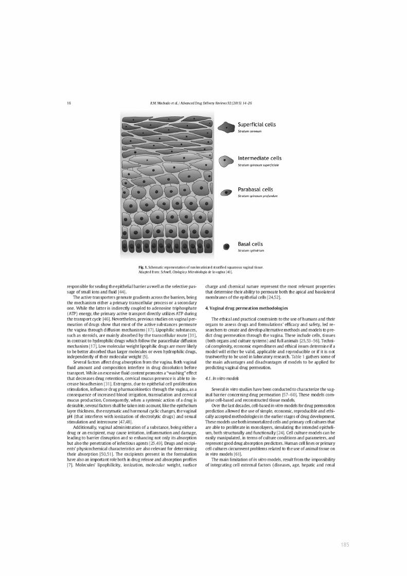
185

186

187

188
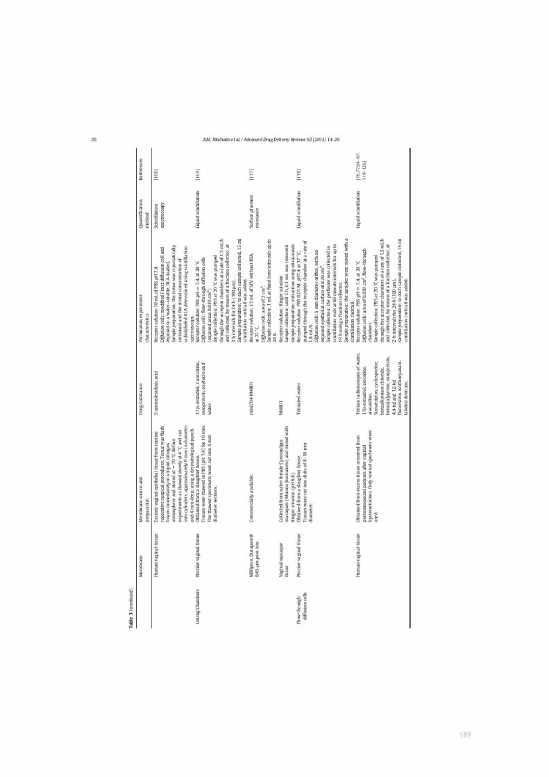
189

190
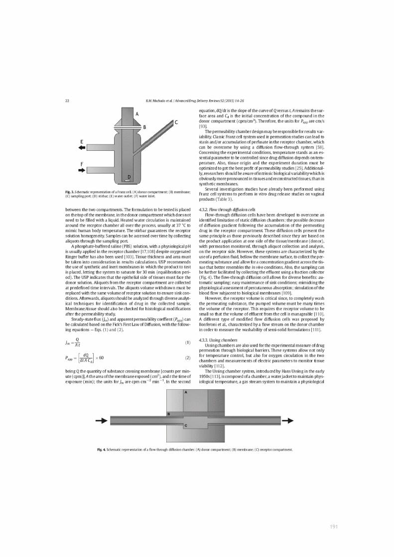
191

192
193

194

195

196

197
APPENDIX B

198
Research article that contributed to the first experimental part of CHAPTER III in the format that
has been submitted.
199
Research article
Optimization and application of in vitro and ex vivo
models for vaginal semisolids safety evaluation
Rita Monteiro Machadoa,b, Ana Palmeira-de-Oliveiraa,b, Luiza Breitenfelda, José
Martinez-de-Oliveiraa,d, Rita Palmeira-de-Oliveiraa,b,c
a CICS, UBI — Health Sciences Research Center, Faculty of Health Sciences, University of Beira Interior, Avenida Infante
D. Henrique, 6200-506 Covilhã, Portugal;
b Labfit, HPRD — Health Products Research and Development, Lda, Edifício UBIMEDICAL, Estrada Municipal 506, 6200-
284, Covilhã, Portugal;
c Pharmacy Department, Centro Hospitalar Cova da Beira EPE, Quinta do Alvito, 6200-251, Covilhã, Portugal;
d Child and Woman's Health Department, Centro Hospitalar Cova da Beira EPE, Quinta do Alvito, 6200-251, Covilhã,
Portugal
Corresponding author: Rita Palmeira-de-Oliveira ([email protected])
Rita Monteiro Machado ([email protected])
Ana Palmeira-de-Oliveira ([email protected])
Luiza Breitenfeld ([email protected])
José Martinez-de-Oliveira ([email protected])
200
Abstract
Vaginal semisolids are widely used both for clinical and intimate hygiene goals. Preclinical
safety assessment includes cytotoxicity assays upon cell lines to predict potential epithelial
irritation, corrosion and sensitization. Furthermore, recent work on tissue explants have
highlighted its potential for application on ex vivo models. In the present study, we studied
traditional and renewed methods for toxicity assessment of vaginal semisolids using
pharmaceutical products which are used in clinical practice as antimicrobials (Gino-Canesten®,
Sertopic®, Dermofix®, Gyno-pevaryl®, Lomexin®, Gino Travogen®, Dalacin V®) and containing
oestrogens (Ovestin®, Blissel®, Colpotrophine®), and reference formulations (Replens®,
Universal Placebo). To achieve our outcome, two in vitro cytotoxicity tests were performed: 3-
(4,5-dimethylthiazol-2-yl)-2,5-diphenyltetrazolium bromide (MTT) and neutral red uptake
(NRU) into uterine (HEC-1A), cervical (HeLa) and vaginal (VK2 E6/E7) cell lines, in compliance
with ISO/EN 10993-5 guide for in vitro evaluation of vaginal medical devices. Likewise, we have
developed and optimized a strategy to determine tissue viability using an ex vivo porcine
vaginal model by using the MTT reduction assay and histological analysis. The vaginal cell line
VK2 E6/E7 conducted to the more accurate calculation of TC50 (half-maximal Toxic
Concentration) among the three cellular lines on the MTT assay. In fact, this is the most
representative cellular model for vaginal products testing, although it is not sensitive to the
NRU assay. It was possible to collect tissues from the porcine model with approx. 15% variability
in thickness, and a first optimization step conducted to variation coefficients lower than 25%
when testing negative and positive controls. The inclusion of the ex vivo toxicity model in
preclinical safety assessment can lead to a better approximation to the in vivo situation.
Furthermore, the application of these models can improve cost-efficiency in early steps of
product development.
Keywords: vaginal drug delivery; cytotoxicity; in vitro model; ex vivo vaginal model

201
Introduction
The vagina is a promising route of administration when local and/or systemic drug delivery is
intended (Alexander et al., 2004; Hussain and Ahsan, 2005; Srikrishna and Cardozo, 2013;
Woolfson et al., 2000). The vaginal route comprises diverse pharmacokinetic advantages due
to its large surface area, rich blood supply, avoidance of the hepatic first-pass effect and
relative high permeability to many drugs (Vermani and Garg, 2000). Nevertheless, several
drawbacks, including leakage, inconsistent drug absorption, influence on sexual intercourse
and local irritation, need to be addressed during the early stages of product development
(Hussain and Ahsan, 2005; Nappi et al., 2006). Ideally, vaginal drug delivery systems should not
interfere with either vaginal physiology or women’s daily life, while allowing obtaining high
drug bioavailability with little variability and low incidence of side effects (Anderson et al.,
2013; Chatterton et al., 2004; Dukhin and Labib, 2013; Ensign et al., 2014; Sassi et al., 2004).
Vaginal drug delivery systems include liquid, semisolid and solid formulations (Allen et al.,
2011). Semisolids (creams, ointments, gels) have been reported as the most preferred dosage
form for self-use by women concerning HIV prophylaxis (Hardy et al., 1998a, 1998b, 1998c,
n.d.; Woodsong and Holt, 2015). Concerning all these particularities added to women’s
preference and acceptability patterns for vaginal semisolid formulations (Palmeira-de-Oliveira
et al., 2014a, 2014b), a safety- and suitability-driven study is expected to conduct to a better
characterization of the already marketed products (which are considered safe based on clinical
trials and current use) in view of the development of new products. Furthermore, the
improvement of traditional characterization methods by considering physiological parameters
that are going to be present after vaginal administration, can provide predictive tools to better
characterize new vaginal semisolid formulations and optimize their acceptability and even
enhance cost-efficiency at early stages of product development (Machado, 2017).
Over the last decades, efforts have been made to conduct investigation to a level of high
comprehension of vaginal epithelium mechanisms (Wu and Robinson, 1996). The understanding
and application of several techniques to predict drug toxicity through the vaginal epithelium
contributes to the successful selection of drugs and appropriate formulations in the early stages
of development (Costin et al., 2011). Thus, it is of great importance to implement accurate
and reproducible methods to predict drug toxicity. The in vitro and ex vivo approaches should
be privileged, and preferably an optimal in vitro/in vivo correlation should be established
(Ardolino et al., 2016). The strategy of patient-driven product development is also reflected
in recent policies such as the biopharmaceutics risk assessment roadmap (BioRAM), which aims
to optimize drug product development and performance right from the early stages (Dickinson
et al., 2016; Selen et al., 2014; Selen and Ph, 2011).
In the field of vaginal administration, research and innovation has been largely focused on
microbicide formulations. However, the same safety/risk assessments should be transposed to
other vaginal product categories. In fact, preclinical development, i.e. nonclinical access to
202
toxicological and pharmacokinetics data could be optimized in order to reduce general financial
costs and overall time of product development before commercialization (Cox Gad; Fernández-
Romero et al., 2014; Lackman-Smith et al., 2010; Lard-Whiteford et al.; Morrow et al., 2014).
Despite being an internal cavity, the vaginal epithelium is constantly exposed to potential
pathogenic microbes, microflora, chemicals, and hormonal changes. As minor injuries can occur
either mechanically or chemically after products usage/administration, further damage or
infection may be promoted (Cunha et al., 2014). Vaginal pharmaceutical, cosmetic and personal
care products can occasionally induce undesirable local or systemic side-effects. Still, the
standard method for assessing vaginal mucosal irritation is the in vivo rabbit vaginal irritation
test (Costin et al., 2011; “Guidance for development of vaginal contraceptive drugs”)
(ISO/EN10993-5, 2009). Nonetheless, the current mind-set in toxicology is to use alternative in
vitro methods that reduce, or, even better, replace the use of animals (3Rs), with a refined
profile that modulate and predict human, not animal, responses (Costin et al., 2011; Liebsch
et al., 2011). This approach is of particular importance in the field of personal care and
cosmetic industries since they have to comply with the European legislation, such as the 7th
Amendment to the EU Cosmetics Directive that does not allow the marketing of cosmetic
products if they, or their ingredients, have been tested for irritation responses in animals (“7th
Amendement to the EU Cosmetics Directive” 2013). Safety concerns are currently spotted right
through the early stages of development, especially in what concerns to microbicides
formulations. This preoccupation was even more highlighted after the unexpected findings of
nonoxynol-9 (N-9) in clinical trials (Van Damme et al., 2002), stressing the need for more
appropriate in vitro assays to predict in vivo safety issues.
The cosmetic industry, under the supervision of regulatory agencies (FDA, ECVAM, OECD), has
already been evaluating and adopting alternative in vitro methods for eye irritation,
phototoxicity, and skin irritation, sensitisation, and corrosion (“European Union Reference
Laboratory for alternatives to animal testing (EURL-ECVAM),” 2016). However, up to now the
cosmetic and pharma industry are still not focused on validating standardised tissue models or
cellular assays for vaginal irritation assessment, since this application still represents only a
small part of business.
Cytotoxicity assays constitute a gold standard of in vitro preclinical evaluations of chemicals in
cultured cells. The MTT reduction assay is one of the most common applied cytotoxicity assays.
MTT (3-[4,5-dimethylthiazol-2-yl]-2,5-diphenyltetrazolium bromide) is a water soluble yellow
tetrazolium salt, which is converted to an insoluble purple compound (formazan) due to
cleavage, within the mitochondria, of the tetrazolium ring by, mainly succinate,
dehydrogenase. Formazan does not permeate cell membranes, and consequently it
accumulates in metabolic viable cells. The MTT assay was tested for its validity in various cell
lines (Mosmann, 1983) and has already been applied to uterine (Youssef Gali et al., 2010c),
cervical (Dobaria et al., 2009) and vaginal (Fichorova et al., 1997a) derived cells. Further
modifications of the initial protocol described by Mosmann (Mosmann, 1983) were proposed
203
(Denizot and Lang, 1986; Hansen et al., 1989) in order to improve the repeatability and the
sensitivity of the assay.
The Neutral Red Uptake (NRU) assay is also a quantitative colorimetric method. It is based on
the ability of viable cells to incorporate and bind (uptake) the red dye within the lysosomes
(Borenfreund et al., 1990; Fotakis and Timbrell, 2006; Repetto et al., 2008). Although they are
used extensively as convenient and rapid measurements of cell viability, all these methods
should be carried out with some caution. In fact, an increase in NR uptake can be induced by
lysosomal swelling agents such as weak alkaline substances and osmotic swelling agents.
Regarding the MTT assay, some reducing agents and respiratory chain inhibitors could affect
the MTT formazan formation. The MTT assay was also found to be significantly influenced by a
number of parameters such as medium pH, D-glucose concentration in culture media, and
cellular concentration of pyridine nucleotides (Chiba et al., 1998). Additionally, the NRU assay
present some advantages over the MTT assay. The former procedure is more sensitive and
readily quantifiable. It is at least two times cheaper, presents less interferences, and does not
use an unstable tetrazolium salt (Repetto et al., 2008). As a general remark, these assays have,
in common with other cell culture procedures, limitations concerning the substance chemical
characteristics, say to be volatile, unstable or explosive in water, only very partially soluble,
colourant and chemical variably incompatible with the test substance (Freshney, 2000; Repetto
et al., 2008).
In the present study, these two cytotoxicity tests, MTT and NRU assay were applied to cervical,
uterine and vaginal cell lines in compliance to ISO/EN 10993-5 guide for in vitro evaluation of
medical devices (ISO/EN10993-5, 2009) (which recommends these tests upon BALB/3T3 clone
A31 and NCTC clone 929 cell lines, which are prevenient from mouse muscular fibroblasts, not
representing a human surrogate) to infer cytotoxicity of final formulations dilution upon in vitro
models. Furthermore, we have developed a strategy to determine tissue viability upon an ex
vivo porcine vaginal model using the MTT reduction assay and histological analysis.

204
Materials and Methods
Tested Formulations
Ten different semisolid vaginal products that are commercially available in Europe and the USA
were included in this study: Gino-Canesten® (Bayer, Portugal), Sertopic® (Ferrer, Portugal),
Dermofix® (Azevedos Laboratories, Portugal), Gyno-pevaryl® (Johnson & Johnson, Portugal),
Lomexin® (Jaba Recordati, Portugal), Gino Travogen® (Bayer, Portugal), Dalacin V® (Pfizer
Laboratories, Portugal), Ovestin® (Aspen Pharma, Portugal), Blissel® (ITF Medivida, Portugal),
Colpotrophine® (Teva Pharma, Portugal). Replens® (Laboratoires Majorelle, France) (Acartürk,
2009; Adriaens and Remon, 2008; Caramella et al., 2015; Schwartz et al., 2007; Tien et al.,
2005; Valenta, 2005) and Universal Placebo (Bygdeman and Swahn, 1996; Clark et al., 2011;
Garg et al., 2010; Nachtigall, 1994) were used as reference products, since their toxicity profile
is largely described on the literature. Universal Placebo was prepared according to Tien et al.
(Tien et al., 2005) by dissolving of 2.7g of Hydroxyethyl-cellulose (2000cP) in 96.3g of water
containing 0.85g of sodium chloride and 0.1g of sorbic acid. The final pH was adjusted to 4.4
by adding sodium hydroxide, and the gel was stored at 2–8ºC. General characteristics of the
studied products are listed in Table 1. All products, except placebo and Replens®, are classified
as medicines, whilst comprising an active pharmaceutical ingredient (API). Nevertheless, some
do not require a medical prescription to be dispensed at pharmacies, which is the case of Gino-
Canesten® and Gyno-Pevaryl®.
Materials
Reagents used include Roswell Park Memorial Institute-1640 RPMI (RPMI-1640, Sigma,
Germany), Dulbecco's Modified Eagle Medium F12 (DMEM F12, Gibco, USA), Keratinocyte-serum
free medium (Gibco, USA), human recombinant epithelial growth factor (hrEGF, Gibco, USA),
bovine pituitary extract (BPE, Gibco, USA), 3-(4,5-dimethylthiazol-2-yl)-2,5-
diphenyltetrazolium bromide (MTT, Amresco, USA), Foetal Bovine Serum (FBS, Merck,
Germany), Penicillin and Streptomycin (SP, Sigma, Germany), Phosphate Buffer Solution (PBS,
Sigma, Germany), Dimethyl Sulfoxide (DMSO, Fisher Chemical, United Kingdom), Triton X100
(Fisher Chemical, United Kingdom), Sodium Dodecyl Sulphate (SDS, Acros Organics, Belgium),
Nonoxynol-9/Tergitol (N9, Sigma, Germany), Neutral Red (NR, Acros Organics, Belgium),
Ethanol (Manuel Vieira e Cª, Portugal), Glacial Acetic Acid (ChemLab, Belgium),
Hydroxyethylcellulose (HEC; Natrosol 250 HX, Ashland Inc., USA), Sorbic Acid (Sigma–Aldrich,
Germany) and Sodium Chloride (Merck, Germany). All used chemicals and reagents were of
analytic grade or equivalent.
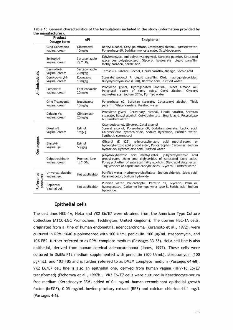
205
Table 1: General characteristics of the formulations included in the study (information provided by the manufacturer).
Product
Dosage form API Excipients
Anti
mic
robia
ls
Gino-Canesten® vaginal cream
Clotrimazol 10mg/g
Benzyl alcohol, Cetyl palmitate, Cetostearyl alcohol, Purified water, Polysorbate 60, Sorbitan monostearate, Octyldodecanol
Sertopic® vaginal cream
Sertaconazole 2g/100g
Ethyleneglycol and polyethyleneglycol, Stearate palmite, Saturated glycerides polyglycolized, Glycerol isostearate, Liquid paraffin, Methylparaben, Sorbic acid
Dermofix® vaginal cream
Sertaconazole 20mg/g
Tefose 63, Labrafil, Peceol, Liquid paraffin, Nipagin, Sorbic acid
Gyno-pevaryl® vaginal cream
Econazole 10mg/g
Stearate pegoxol 7, Liquid paraffin, Oleic macrogolglycerides, Butylhydroxyanisole (E320), Benzoic acid, Purified water
Lomexin® vaginal cream
Fenticonazole 20mg/g
Propylene glycol, Hydrogenated lanolina, Sweet almond oil, Polyglycol esters of fatty acids, Cetyl alcohol, Glyceryl monostearate, Sodium EDTA, Purified water
Gino Travogen® vaginal cream
Isoconazole 10mg/g
Polysorbate 60, Sorbitan stearate, Cetostearyl alcohol, Thick paraffin, White Vaseline, Purified water
Dalacin V® vaginal cream
Clindamycin 20mg/g
Propylene glycol, Cetostearyl alcohol, Liquid paraffin, Sorbitan stearate, Benzyl alcohol, Cetyl palmitate, Stearic acid, Polysorbate 60, Purified water
Oest
rogens
Ovestin® vaginal cream
Estriol 1mg/g
Octyldodecanol, Glycerol, Cetyl alcohol Stearyl alcohol, Polysorbate 60, Sorbitan stearate, Lactic acid, Chlorhexidine hydrochloride, Sodium hydroxide, Purified water, Synthetic spermaceti
Blissel® vaginal gel
Estriol 50µg/g
Glicerol (E 422), p-hydroxybenzoic acid methyl ester, p-hydroxybenzoic acid propyl ester, Policarbophil, Carbomer, Sodium hydroxide, Hydrochloric acid, Purified water
Colpotrophine® vaginal cream
Promestriene 1g/100g
p-hydroxybenzoic acid methyl ester, p-hydroxybenzoic acid propyl ester, Mono and diglycerides of saturated fatty acids, Polyglycol ether of saturated fatty alcohols, Oleic acid decyl ester, Triglycerides of capric and caprylic acids, Glycerol, Purified water
Refe
rence
pro
ducts
Universal placebo vaginal gel
Not applicable Purified water, Hydroxyethylcellulose, Sodium chloride, Sobic acid, Caramel color, Sodium hydroxide
Replens® Vaginal gel
Not applicable Purified water, Policarbophil, Paraffin oil, Glycerin, Palm oil hydrogenated, Carbomer homopolymer type B, Sorbic acid, Sodium hydroxide
Epithelial cells
The cell lines HEC-1A, HeLa and VK2 E6/E7 were obtained from the American Type Culture
Collection (ATCC-LGC Promochem, Teddington, United Kingdom). The uterine HEC-1A cells,
originated from a line of human endometrial adenocarcinoma (Kuramoto et al., 1972), were
cultured in RPMI 1640 supplemented with 100 U/mL penicillin, 100 µg/mL streptomycin, and
10% FBS, further referred to as RPMI complete medium (Passages 33-38). HeLa cell line is also
epithelial, derived from human cervical adenocarcinoma (Jones, 1997). These cells were
cultured in DMEM F12 medium supplemented with penicillin (100 U/mL), streptomycin (100
µg/mL), and 10% FBS and is further referred to as DMEM complete medium (Passages 64-68).
VK2 E6/E7 cell line is also an epithelial one, derived from human vagina (HPV-16 E6/E7
transformed) (Fichorova et al., 1997b). VK2 E6/E7 cells were cultured in Keratinocyte-serum
free medium (Keratinocyte-SFM) added of 0.1 ng/mL human recombinant epithelial growth
factor (hrEGF), 0.05 mg/mL bovine pituitary extract (BPE) and calcium chloride 44.1 mg/L
(Passages 4-6).
206
Vaginal tissues explants
Complete porcine genitalia, from approximately 6 month-year-old animals, were collected from
a local slaughterhouse, transported under refrigeration to the lab and processed within 3h of
animal death. The vaginal tubes were separated from the surrounding organs using scissors and
tweezers. The vagina was cut longitudinally and the vestibule (caudal vagina) excised. The
vestibule was selected since we found (unpublished data) to be the proximal vagina
histologically more similar to the human one. Tissues were then washed on a prewarmed saline
solution (NaCl 0.9%, 37ºC). This section of tissue was then thickness normalized by using a
manual dermatome (Watson Skin Graft Knife, BBraun, Germany) equipped with Aesculap blades
(BA718Rm, BBraun, Germany). Then, epithelial sheets were placed upon aluminium foil
(contact made by the basolateral side) and punched to sections of ø=3mm using a biopsy punch
(Stiefel, GSK, United Kingdom). These sections were maintained on a warmed saline solution
until use in the MTT assay. Tissues thickness was accessed to maintain homogeneity between
samples. For this purpose, a digital micrometer (Vogel, Germany) was used by placing the tissue
between two microscopy slides, measuring total thickness (µm) and then subtracting the
thickness of the slides to the total measured thickness.
207
Testing products preparation
Testing products were diluted to 20, 5, 1, 0.4, 0.2, 0.1% (w/v), in medium without
supplementation but containing 0.5% (v/v) of DMSO to ensure proper solubility of formulations.
DMSO was selected based on the recommendations of the ISO guide (ISO/EN10993-5, 2009). To
assure that this substance was not itself cytotoxic, controls with the used concentration in
culture media were performed. A negative control was included in all experiments (cells/tissues
only with media/DMSO) and also positive controls for toxicity were included (SDS 5% (w/v),
Triton X-100 1% (v/v) and N9 2% (v/v)). Formulations were left in contact with the testing
platform (cell or tissue) for 24 hours.
Cellular toxicity
MTT assay
The MTT reduction assay was performed as previously described (Berridge et al., 2005; Denizot
and Lang, 1986; Freshney, 2000; Plumb et al., 1989) and ISO/EN 10993-5 guide for in vitro
evaluation of vaginal medical devices (ISO/EN10993-5, 2009). Cells were seeded onto 96-well
plates (100.000 cells/mL) with the respective incomplete culture media. Cells were let to
adhere for 24 hours at 37ºC, 5% CO2. After obtaining a half-confluent culture, 100 µL of testing
formulations were added (see Testing products preparation section). After this period cells
were washed with PBS and incubated for 4 hours with a 0.5mg/mL solution of MTT reagent
prepared in the respective culture medium without supplementation. Subsequently to formazan
crystals formation, extraction was accomplished with 200 µL of DMSO for 15 minutes on an
orbital shaker protected from light. Absorbances were then measured at 590 nm and 630 nm
for background deduction, using a microplate spectrophotometer (BIORAD XMark, USA).
Additionally, cells were photographed before and after the application of the MTT reagent
solution in order to evidence the formation of the formazan crystals, using an inverted
microscope (Olympus IX51, Japan, equipped with OCTAX Eyeware v.1.5 Build 406, Germany) –
data not shown. Triton X-100, SDS and Nonoxynol-9 were used as positive controls, since they
are widely known inducers of cytotoxic effects (Cunha et al., 2014). The negative control
consists of cells without any treatment (just culture media along the assay), which was
considered as the 100% viability reference for products toxicity calculation. Further, the
concentration that was toxic to half of the culture (cell/tissue), the half-maximal toxic
concentration (TC50), was calculated by logistic regression using GraphPad Prism Version 6.0
(Copyright, 2015).
NRU assay
The NRU assay was performed according to the literature (Freshney, 2000; Oecd, 2004; Repetto
et al., 2008; Yang et al., 2008) and ISO/EN 10993-5 guide for in vitro evaluation of medical
devices (ISO/EN10993-5, 2009). Cells were seeded into 96-well plates (100.000 cells/mL) and
maintained in culture until a semi-confluent monolayer was achieved (37ºC, 5% CO2, 24 hours).

208
They were then exposed to the test compounds (100 µL) over the range of concentrations
described above. After 24 hours of exposure, wells were washed once with 150 µL of prewarmed
PBS, then 100 µL of NR medium was added to each well and the plates incubated at 37ºC in a
humidified atmosphere of 5% CO2 for 3 hours. After incubation, the NR medium was removed,
and cells were washed with 150 μL of PBS. Finally, 150 μL NR desorb solution (ETOH/acetic
acid) were added to all wells, including blanks, in order to extract the dye. Plates were rapidly
shaken on a microtiter plate shaker for 10 min to allow NR to be extracted from the cells
forming a homogeneous solution. The absorbance of the resulting coloured solution at 540 nm
was measured in a microtiter plate reader (BIORAD XMark, USA). NRU was determined for each
concentration and compared to the one determined in control cultures. For the study a NR
stock solution was previously prepared by dissolving 0.4 g of the dye in 100 mL of milliQ water.
The NR medium was prepared in the day prior of usage by adding 1 mL of NR stock solution to
79 mL of culture media, followed by incubation overnight at 37ºC and filtration through a 0.2
µm filter before adding to the cells, in order to be free of crystals. The NR desorb/extraction
solution is composed of 1% glacial acetic acid solution, 50% ethanol and 49 % water.
Tissue toxicity
Method optimization
Tissue explants were firstly tested for homogeneity and response to MTT assay. Therefore,
tissues were submitted to a preliminary experiment, using different animals and applying the
viability test on fresh and frozen tissues. Frozen explants were obtained as described (section
“Vaginal tissue explants”) but tissue preparation occur after defrosting; upon reception from
the slaughterhouse the vaginas were opened longitudinally (from vulva to cervix), washed in a
HBSS solution pH 4.2 and frozen at -20ºC, wrapped in aluminium foil, and stored in airtight
bags. For viability studies (fresh tissue: 46 animals, at least n=2 for each animal, 6 independent
experiments; frozen tissue: 12 animals, n=3, one experiment), tissues were left in culture for
24 hours and then the MTT assay was performed. For toxicity studies (only performed on fresh
tissue: 23 animals, at least n=2 for each animal, 5 independent experiments) tissues were put
in contact with SDS 5% (w/v) during 24 hours, and then the MTT assay has begun. SDS 5% is
generally used as a positive control in tissue toxicity assays and recognized to have a toxic
effect even on epithelial vaginal tissue (Youssef Gali et al., 2010b).
MTT assay
Tissue explants were placed in 96-well flat-bottomed tissue culture plates and treated with the
testing formulations diluted on RPMI media without supplementation at 37ºC, 5% CO2 during 24
hours, n=6. Also, negative controls were included for each animal. SDS 5% (w/v), Triton X-100
1% (v/v) and N9 2% (v/v) were used as positive controls. Subsequently, tissues were washed
twice with prewarmed PBS. MTT solution at a final concentration of 0.5 mg/mL was added to
each well and incubated 1 hour at 37ºC, 5% CO2. Tissues were then transferred into new plates
and extracted with 200 µL of isopropanol during 1.5 hours at room temperature under gentle

209
shaking. Finally, tissues were discarded for plate reading at 590 nm against the background at
630 nm (BIORAD XMark, USA). The tissue viability was calculated as percentage from the
negative control (tissue with no formulation).
Histological analysis
One tissue per formulation, at its highest concentration, 20% (w/v), was reserved for
histological analysis. Tissues were fixed in a balanced 10% formalin solution. Subsequently,
fixed tissues were run in a set of ethanol solutions of increasing concentrations for dehydration
before embedding in paraffin. Tissue blocks were cut to have about 3 µm thickness and stained
with haematoxylin and eosin. Slides were observed and microphotographs were taken using a
Zeiss microscope (Axiolmager A1, Zeiss, Germany) equipped with a digital camera (Axiocam,
Zeiss, Germany).
Data processing and statistical analysis
Data was analysed to produce arithmetic means with standard deviations (SD) using Microsoft
Excel. Analysis of variance (ANOVA) and Dunnett's multiple comparisons test were performed
to determine the significance of the difference between sets of data (p < 0.05). Logistic
regression analysis was performed using GraphPad Prism software to calculate the half-maximal
toxic concentration (TC50) in toxicity assays.
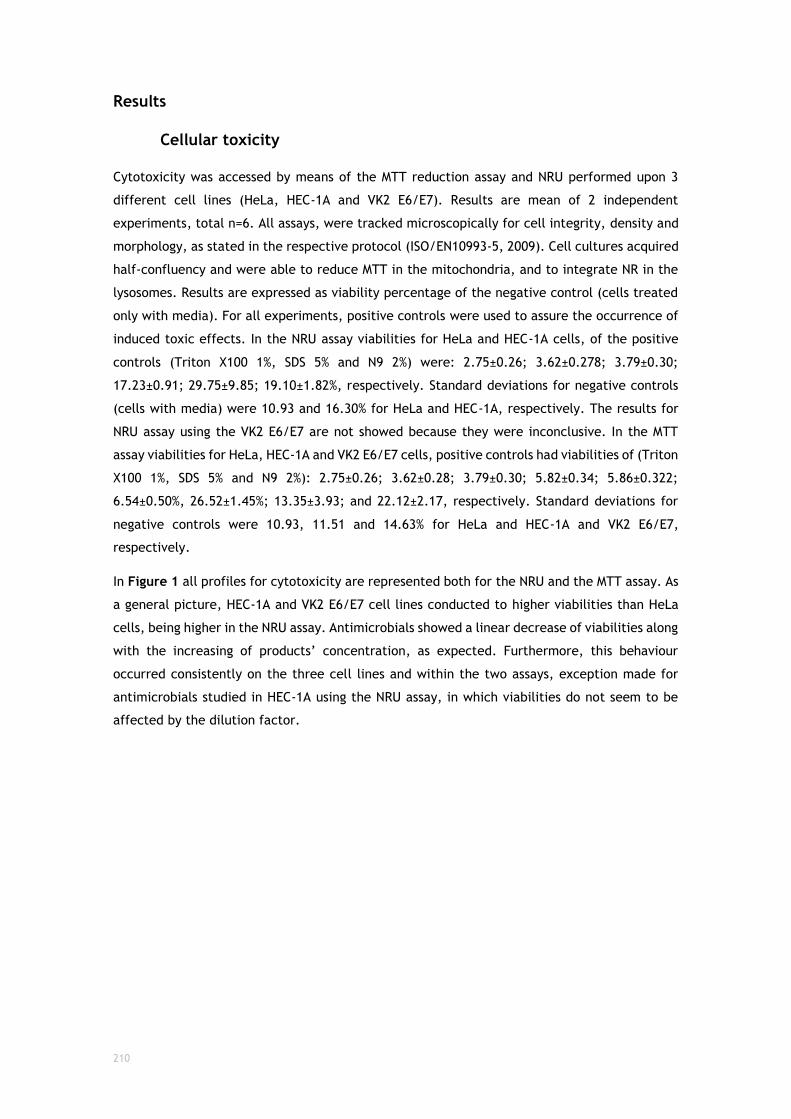
210
Results
Cellular toxicity
Cytotoxicity was accessed by means of the MTT reduction assay and NRU performed upon 3
different cell lines (HeLa, HEC-1A and VK2 E6/E7). Results are mean of 2 independent
experiments, total n=6. All assays, were tracked microscopically for cell integrity, density and
morphology, as stated in the respective protocol (ISO/EN10993-5, 2009). Cell cultures acquired
half-confluency and were able to reduce MTT in the mitochondria, and to integrate NR in the
lysosomes. Results are expressed as viability percentage of the negative control (cells treated
only with media). For all experiments, positive controls were used to assure the occurrence of
induced toxic effects. In the NRU assay viabilities for HeLa and HEC-1A cells, of the positive
controls (Triton X100 1%, SDS 5% and N9 2%) were: 2.75±0.26; 3.62±0.278; 3.79±0.30;
17.23±0.91; 29.75±9.85; 19.10±1.82%, respectively. Standard deviations for negative controls
(cells with media) were 10.93 and 16.30% for HeLa and HEC-1A, respectively. The results for
NRU assay using the VK2 E6/E7 are not showed because they were inconclusive. In the MTT
assay viabilities for HeLa, HEC-1A and VK2 E6/E7 cells, positive controls had viabilities of (Triton
X100 1%, SDS 5% and N9 2%): 2.75±0.26; 3.62±0.28; 3.79±0.30; 5.82±0.34; 5.86±0.322;
6.54±0.50%, 26.52±1.45%; 13.35±3.93; and 22.12±2.17, respectively. Standard deviations for
negative controls were 10.93, 11.51 and 14.63% for HeLa and HEC-1A and VK2 E6/E7,
respectively.
In Figure 1 all profiles for cytotoxicity are represented both for the NRU and the MTT assay. As
a general picture, HEC-1A and VK2 E6/E7 cell lines conducted to higher viabilities than HeLa
cells, being higher in the NRU assay. Antimicrobials showed a linear decrease of viabilities along
with the increasing of products’ concentration, as expected. Furthermore, this behaviour
occurred consistently on the three cell lines and within the two assays, exception made for
antimicrobials studied in HEC-1A using the NRU assay, in which viabilities do not seem to be
affected by the dilution factor.
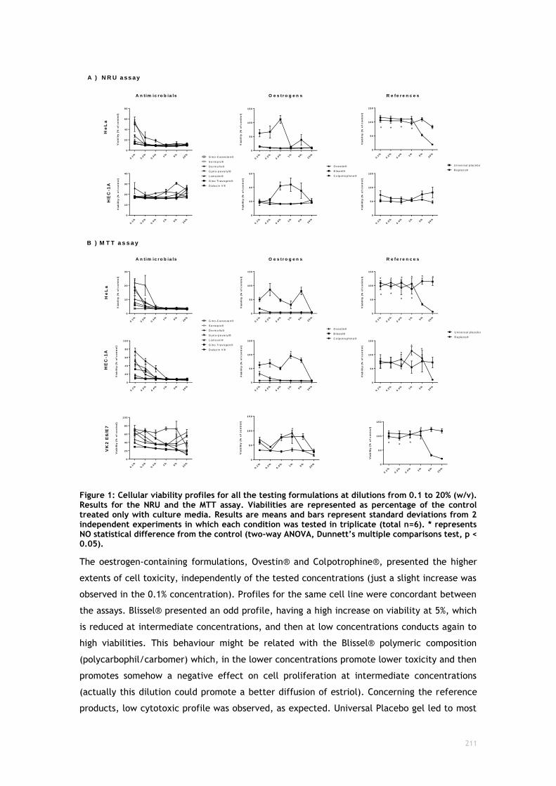
211
0.1
%
0.2
%
0.4
%1%
5%
20%
0
2 0
4 0
6 0
8 0
Via
bil
ity
(%
of
co
ntr
ol)
D a la c in V ®
G in o T ra v o g e n ®
L o m e x in ®
G y n o -p e v a ry l®
D e rm o fix ®
S e rto p ic ®
G in o -C a n e s te n ®0.1
%
0.2
%
0.4
%1%
5%
20%
0
5 0
1 0 0
1 5 0
Via
bil
ity
(%
of
co
ntr
ol)
C o lp o tro p h in e ®
B lis s e l®
O v e s tin ®
0.1
%
0.2
%
0.4
%1%
5%
20%
0
5 0
1 0 0
1 5 0
Via
bil
ity
(%
of
co
ntr
ol)
R e p le n s®
U n iv e rs a l p la c e b o
0.1
%
0.2
%
0.4
%1%
5%
20%
0
2 0
4 0
6 0
Via
bil
ity
(%
of
co
ntr
ol)
0.1
%
0.2
%
0.4
%1%
5%
20%
0
1 0
2 0
3 0
4 0
Via
bil
ity
(%
of
co
ntr
ol)
0.1
%
0.2
%
0.4
%1%
5%
20%
0
5 0
1 0 0
1 5 0
Via
bil
ity
(%
of
co
ntr
ol)
A n tim ic r o b ia ls
He
La
HE
C-1
A
O e s t r o g e n s R e f e r e n c e s
0.1
%
0.2
%
0.4
%1%
5%
20%
0
1 0
2 0
3 0
Via
bil
ity
(%
of
co
ntr
ol)
D a la c in V ®
G in o T ra v o g e n ®
L o m e x in ®
G y n o -p e v a ry l®
D e rm o fix ®
S e rto p ic ®
G in o -C a n e s te n ® 0.1
%
0.2
%
0.4
%1%
5%
20%
0
5 0
1 0 0
1 5 0
Via
bil
ity
(%
of
co
ntr
ol)
C o lp o tro p h in e ®
B lis s e l®
O v e s tin ®
0.1
%
0.2
%
0.4
%1%
5%
20%
0
5 0
1 0 0
1 5 0
Via
bil
ity
(%
of
co
ntr
ol)
R e p le n s®
U n iv e rs a l p la c e b o
0.1
%
0.2
%
0.4
%1%
5%
20%
0
2 0
4 0
6 0
8 0
1 0 0
Via
bil
ity
(%
of
co
ntr
ol)
0.1
%
0.2
%
0.4
%1%
5%
20%
0
5 0
1 0 0
1 5 0
Via
bil
ity
(%
of
co
ntr
ol)
0.1
%
0.2
%
0.4
%1%
5%
20%
0
5 0
1 0 0
1 5 0
Via
bil
ity
(%
of
co
ntr
ol)
A ) N R U a ss ay
B ) M T T a s s a y
A n tim ic r o b ia ls O e s t r o g e n s R e f e r e n c e s
He
La
HE
C-1
A
0.1
%
0.2
%
0.4
%1%
5%
20%
0
2 0
4 0
6 0
8 0
1 0 0
Via
bil
ity
(%
of
co
ntr
ol)
VK
2 E
6/E
7
0.1
%
0.2
%
0.4
%1%
5%
20%
0
5 0
1 0 0
1 5 0
Via
bil
ity
(%
of
co
ntr
ol)
0.1
%
0.2
%
0.4
%1%
5%
20%
0
5 0
1 0 0
1 5 0
Via
bil
ity
(%
of
co
ntr
ol)
* * * *
**
**
*
**
*
* * *
**
* * * * * *
Figure 1: Cellular viability profiles for all the testing formulations at dilutions from 0.1 to 20% (w/v). Results for the NRU and the MTT assay. Viabilities are represented as percentage of the control treated only with culture media. Results are means and bars represent standard deviations from 2 independent experiments in which each condition was tested in triplicate (total n=6). * represents NO statistical difference from the control (two-way ANOVA, Dunnett’s multiple comparisons test, p < 0.05).
The oestrogen-containing formulations, Ovestin® and Colpotrophine®, presented the higher
extents of cell toxicity, independently of the tested concentrations (just a slight increase was
observed in the 0.1% concentration). Profiles for the same cell line were concordant between
the assays. Blissel® presented an odd profile, having a high increase on viability at 5%, which
is reduced at intermediate concentrations, and then at low concentrations conducts again to
high viabilities. This behaviour might be related with the Blissel® polymeric composition
(polycarbophil/carbomer) which, in the lower concentrations promote lower toxicity and then
promotes somehow a negative effect on cell proliferation at intermediate concentrations
(actually this dilution could promote a better diffusion of estriol). Concerning the reference
products, low cytotoxic profile was observed, as expected. Universal Placebo gel led to most
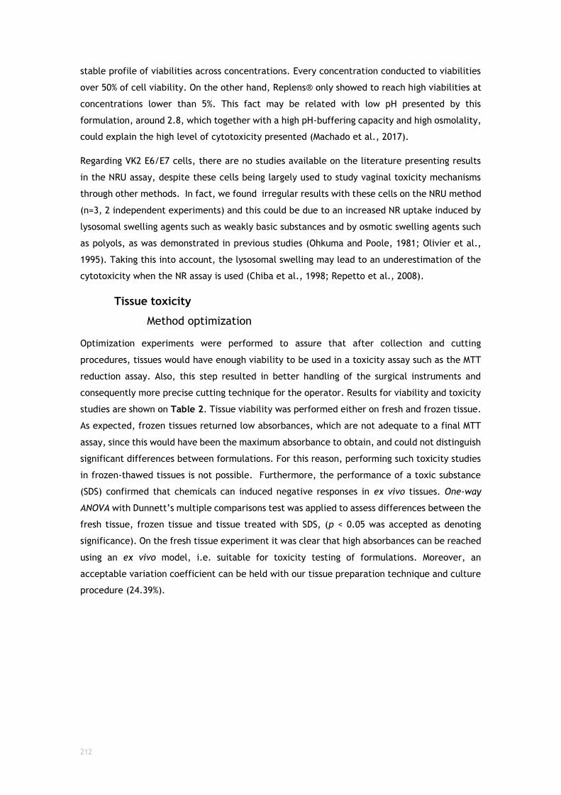
212
stable profile of viabilities across concentrations. Every concentration conducted to viabilities
over 50% of cell viability. On the other hand, Replens® only showed to reach high viabilities at
concentrations lower than 5%. This fact may be related with low pH presented by this
formulation, around 2.8, which together with a high pH-buffering capacity and high osmolality,
could explain the high level of cytotoxicity presented (Machado et al., 2017).
Regarding VK2 E6/E7 cells, there are no studies available on the literature presenting results
in the NRU assay, despite these cells being largely used to study vaginal toxicity mechanisms
through other methods. In fact, we found irregular results with these cells on the NRU method
(n=3, 2 independent experiments) and this could be due to an increased NR uptake induced by
lysosomal swelling agents such as weakly basic substances and by osmotic swelling agents such
as polyols, as was demonstrated in previous studies (Ohkuma and Poole, 1981; Olivier et al.,
1995). Taking this into account, the lysosomal swelling may lead to an underestimation of the
cytotoxicity when the NR assay is used (Chiba et al., 1998; Repetto et al., 2008).
Tissue toxicity
Method optimization
Optimization experiments were performed to assure that after collection and cutting
procedures, tissues would have enough viability to be used in a toxicity assay such as the MTT
reduction assay. Also, this step resulted in better handling of the surgical instruments and
consequently more precise cutting technique for the operator. Results for viability and toxicity
studies are shown on Table 2. Tissue viability was performed either on fresh and frozen tissue.
As expected, frozen tissues returned low absorbances, which are not adequate to a final MTT
assay, since this would have been the maximum absorbance to obtain, and could not distinguish
significant differences between formulations. For this reason, performing such toxicity studies
in frozen-thawed tissues is not possible. Furthermore, the performance of a toxic substance
(SDS) confirmed that chemicals can induced negative responses in ex vivo tissues. One-way
ANOVA with Dunnett’s multiple comparisons test was applied to assess differences between the
fresh tissue, frozen tissue and tissue treated with SDS, (p < 0.05 was accepted as denoting
significance). On the fresh tissue experiment it was clear that high absorbances can be reached
using an ex vivo model, i.e. suitable for toxicity testing of formulations. Moreover, an
acceptable variation coefficient can be held with our tissue preparation technique and culture
procedure (24.39%).

213
Table 2: Optimization and pre-validation studies on tissues viability and toxicity. The viability study corresponds to a negative control (tissues plus culture media) and the toxicity study was performed using SDS 5%, recognized to have a toxic effect on epithelial vaginal tissue. *Denotes significance on One-way ANOVA Dunnett’s multiple comparisons test (p < 0.05).
Viability studies (Abs at 570nm)
Culture media
Toxicity studies (Abs at 570nm)
SDS 5%
Fresh tissue (mean±SD) Frozen (mean±SD) Fresh tissue (mean±SD)
0.5788±0.1412
Variation Coefficient: 24.39%
0.1015±0.0144*
Variation Coefficient: 14.20%
0.0581±0.0133*
Variation Coefficient: 22.83%
46 animals
n=2-3
6 independent experiments
12 animals
n=2
1 experiment
23 animals
n=2-3
5 independent experiments
After measuring tissues thickness (6 animals, n=6, 3 independent experiments) the variation
coefficient was calculated to be 15.12%.
MTT assay
Tissue viability results following exposure to the test products are shown in Figure 2. Only
Universal Placebo and Ovestin® were found to conduct to viabilities >50% by the first dilution.
All other formulations, except Dalacin V®, returned viabilities above 50% upon the second
dilution (1:20). Standard deviations were relatively high, but acceptable considering that the
surrogate used for this test is biological. Positive controls, Triton X100 1%, SDS 5% and N9 2%,
conducted to viabilities of 3.22±0.77, 2.51±0.81 and 2.56±0.60 %, respectively, confirming that
they have toxic effects on the vaginal epithelium (metabolic toxicity).
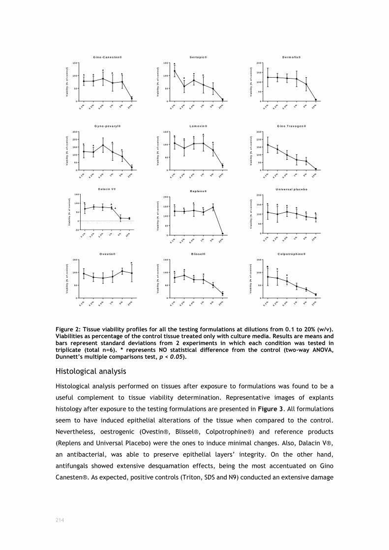
214
0.1
%
0.2
%
0.4
%1%
5%
20%
0
5 0
1 0 0
1 5 0
G in o -C a n e s te n ®
Via
bil
ity
(%
of
co
ntr
ol)
0.1
%
0.2
%
0.4
%1%
5%
20%
0
5 0
1 0 0
1 5 0
S e r to p ic ®
Via
bil
ity
(%
of
co
ntr
ol)
0.1
%
0.2
%
0.4
%1%
5%
20%
0
5 0
1 0 0
1 5 0
2 0 0
D e rm o fix ®
Via
bil
ity
(%
of
co
ntr
ol)
0.1
%
0.2
%
0.4
%1%
5%
20%
0
5 0
1 0 0
1 5 0
2 0 0
2 5 0
G y n o -p e v a ry l®
Via
bil
ity
(%
of
co
ntr
ol)
0.1
%
0.2
%
0.4
%1%
5%
20%
0
5 0
1 0 0
1 5 0
L o m e x in ®
Via
bil
ity
(%
of
co
ntr
ol)
0.1
%
0.2
%
0.4
%1%
5%
20%
0
5 0
1 0 0
1 5 0
2 0 0
2 5 0
G in o T r a v o g e n ®
Via
bil
ity
(%
of
co
ntr
ol)
0.1
%
0.2
%
0.4
%1%
5%
20%
-5 0
0
5 0
1 0 0
1 5 0
D a la c in V ®
Via
bil
ity
(%
of
co
ntr
ol)
0.1
%
0.2
%
0.4
%1%
5%
20%
0
5 0
1 0 0
1 5 0
2 0 0
R e p le n s ®
Via
bil
ity
(%
of
co
ntr
ol)
0.1
%
0.2
%
0.4
%1%
5%
20%
0
5 0
1 0 0
1 5 0
2 0 0
U n iv e rs a l p la c e b o
Via
bil
ity
(%
of
co
ntr
ol)
0.1
%
0.2
%
0.4
%1%
5%
20%
0
5 0
1 0 0
1 5 0
O v e s t in ®
Via
bil
ity
(%
of
co
ntr
ol)
0.1
%
0.2
%
0.4
%1%
5%
20%
0
5 0
1 0 0
1 5 0
B lis s e l®
Via
bil
ity
(%
of
co
ntr
ol)
0.1
%
0.2
%
0.4
%1%
5%
20%
0
5 0
1 0 0
1 5 0
C o lp o tro p h in e ®
Via
bil
ity
(%
of
co
ntr
ol)
* *
* *
***
*
*
* **
**
*
*
***
***
* **
*
*
**
**
***
*
* **
* *
Figure 2: Tissue viability profiles for all the testing formulations at dilutions from 0.1 to 20% (w/v). Viabilities as percentage of the control tissue treated only with culture media. Results are means and bars represent standard deviations from 2 experiments in which each condition was tested in triplicate (total n=6). * represents NO statistical difference from the control (two-way ANOVA, Dunnett’s multiple comparisons test, p < 0.05).
Histological analysis
Histological analysis performed on tissues after exposure to formulations was found to be a
useful complement to tissue viability determination. Representative images of explants
histology after exposure to the testing formulations are presented in Figure 3. All formulations
seem to have induced epithelial alterations of the tissue when compared to the control.
Nevertheless, oestrogenic (Ovestin®, Blissel®, Colpotrophine®) and reference products
(Replens and Universal Placebo) were the ones to induce minimal changes. Also, Dalacin V®,
an antibacterial, was able to preserve epithelial layers’ integrity. On the other hand,
antifungals showed extensive desquamation effects, being the most accentuated on Gino
Canesten®. As expected, positive controls (Triton, SDS and N9) conducted an extensive damage
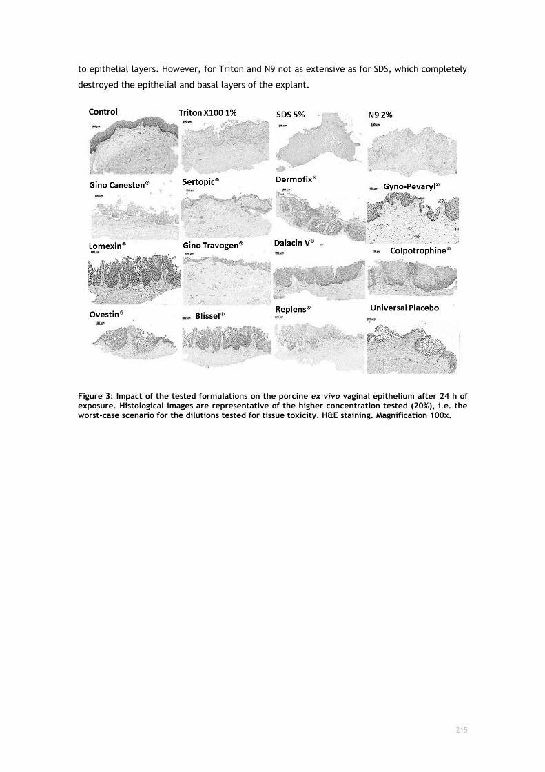
215
to epithelial layers. However, for Triton and N9 not as extensive as for SDS, which completely
destroyed the epithelial and basal layers of the explant.
Figure 3: Impact of the tested formulations on the porcine ex vivo vaginal epithelium after 24 h of exposure. Histological images are representative of the higher concentration tested (20%), i.e. the worst-case scenario for the dilutions tested for tissue toxicity. H&E staining. Magnification 100x.

216
Models comparison
Calculated half-maximal toxic concentrations (TC50) either for cellular models and tissue
models are represented in Table 3. The ex vivo porcine vaginal tissue model, was the one that
led to the calculation of the TC50 for almost all products. This means, on one hand that the
formulations concentration in test is suitable to the test method, and on the other hand, that
this method is the less sensitive in comparison to the remaining. Nevertheless, for Gino-
Canesten® and Gino Travogen® the TC50 confidence interval was very wide, which could be
related with the high slopes and SDs that are shown on the toxicity profiles for these products
between concentrations 5 and 20%. The three cell lines were chosen because they were
representative of three different epithelia that are present in the vaginal cavity, and, by testing
these three cellular types we are addressing a complete toxicological profile of the formulation
after administration by the vaginal route. Concerning the MTT assay, the most sensitive cell
line was the HeLa (cervical cells), while VK2 E6/E7 (vaginal cells) was found to be the more
robust. Since it is expected that cellular models are more sensitive to toxic effects than tissue
models (due to the more complex and organized structure of the latter) and that the tissue, in
turn, is more related to the in vivo environment, VK2 E6/E7 may provide more reliable results
of the vaginal toxicity of formulations when performing the MTT assay. Neutral Red uptake
assay should be performed to confirm previous results from the MTT assay, gathering the
maximum data for preclinical safety characterization of formulations, or to circumvent
problems due to chemical incompatibility. When possible, final formulations, that were
selected to proceed for further preclinical and clinical stages, should be tested under an ex
vivo model, being the technique herein present a valuable alternative test method.

217
Table 3: Half-maximal Toxic Concentrations (% - TC50) calculated for cellular and tissue models using three different cell lines (HeLa, HEC-1A and VK2 E6/E7) and porcine vaginal explants. Within the cellular models, MTT and NRU assay were issued, as for tissues only MTT assay was performed.
Product
Cellular toxicity TC50 (%) Tissue toxicity TC50 (%)
MTT assay NRU assay MTT assay
HeLa HEC-1A VK2 E6/E7 HeLa HEC-1A Porcine vaginal explants
Gino-Canesten® <0.1 <0.1 7.039 (3.825-12.950) <0.1 <0.1 18.530 (2.436-141.000)
Sertopic® <0.1 <0.1 1.118 (0.550-2.272) <0.1 <0.1 1.607 (0.568-4.549)
Dermofix® <0.1 <0.1 5.243 (2.511-10.950) <0.1 <0.1 >20
Gyno-pevaryl® <0.1 0.196 (0.114-0.336) <0.1 <0.1 <0.1 >20
Lomexin® <0.1 0.232 (0.142-0.381) 0.296 (0.198-0.441) <0.1 <0.1 >20
Gino Travogen® <0.1 0.141 (0.114-0.173) <0.1 <0.1 <0.1 11.950 (0.949-150.400)
Dalacin V® <0.1 0.974 (0.420-2.256) 0.386 (0.302-0.495) <0.1 <0.1 1.233 (0.628-2.422)
Ovestin® <0.1 <0.1 2.163 (1.206-3.882) <0.1 <0.1 <0.1
Blissel® 0.579 (0.288-1.167) 6.249 (2.953-13.220) 8.008 (3.293-19.480) 0.825 (0.436-1.561) <0.1 3.030 (2.000-4.591)
Colpotrophine® <0.1 <0.1 <0.1 <0.1 <0.1 0.911 (0.591-1.405)
Universal placebo >20 >20 >20 >20 2.982 (0.722-12.320) >20
Replens® 5.186 (2.203-12.210) 10.000 (8.877-11.260) 4.847 (3.270-7.186) 5.731 (3.851-8.529) >20 >20
218
Discussion
The vaginal epithelium is a common route for topical administration of antimicrobials,
oestrogens, lubricants, and hygiene feminine-care products (Alexander et al., 2004).
Nonetheless, these products could potentially conduct to acute irritation of the epithelial
surfaces. Since the vaginal cavity can be used for either sporadic or chronic product
applications, special attention should be taken when designing new products for this route.
Viral and bacterial infection potentiation subsequent to the use of some vaginal lubricants has
been demonstrated, reinforcing the need of extensive safety characterizations both for
medicinal and cosmetic/hygiene products (Adriaens and Remon, 2008; Begay et al., 2011;
Cunha et al., 2014; Dezzutti et al., 2012; Maguire et al., 2001; Moench et al., 2010; Van Damme
et al., 2002).
The standard preclinical test for the assessment of vaginal irritation and toxicity is the rabbit
vaginal irritation (RVI) model. Similar to many other in vivo models, the reproducibility of
results from this model (either between animals and between tests) is suboptimal. This
variability is due to different responses of individual animals and to the trauma of insertion of
the test product in the vaginal tract. Due to the high level of variability, standard RVI protocols
use 10 rabbits per test article. The large number of animals required is undesirable from an
animal welfare point of view and the variability decrease the confidence in the assay results.
These problems are even magnified when primates are used for testing (Ayehunie et al., 2006).
Several in vitro and ex vivo models have been used to study the toxicity of substances and
products , although it has not been stated which model better predicts in vivo toxicity (Youssef
Gali et al., 2010a). On one hand, ex vivo assays are closer to fully represent the in vivo
mechanisms. On the other hand, in vitro techniques are easier to handle, quicker, more
sensitive and able for high-throughput screening in early steps of discovery and development.
During product development, it has been observed that even slight modifications in final
formulations, can either modify the efficacy and/or toxicity of a vaginal preparation (Y Gali et
al., 2010; Goeman et al., 1995).
Therefore, in our toxicity assays we assessed the toxicity profiles of final formulations belonging
to different therapeutic classes (antimicrobials, oestrogens and reference products). The
formulations included in this study are already commercialized and of course regarded as safe
for human use, but our main purpose was to provide scientists involved in formulation with a
rational tool that could rapidly disclose final products toxicity before getting to animal studies.
Having that in mind, we were expecting that these finished products could, despite being safe,
present some sort of in vitro toxicity. This actually means that poor results in in vitro stages of
safety testing do not have to conduct to similar results in vivo. Some of the results herein
present are somehow not clearly explained because the authors did not have access to the fully
quantitative information of the formulations. This is the case of Blissel®, that presented an
odd profile, although consistent in the two tests, models, and cell lines, a profile which is
219
thought to be due to the presence of estriol. The behaviour presented is actually similar to the
one that generally is seen for oestrogens per se. In fact, Blissel® is the oestrogenic formulation
with the lower concentration of oestriol (50µg/g vs 1mg/g in Ovestin® and Colpotrophine®),
and since the concentration in these last two is considered high, that might explain the
cytotoxic profile over all formulation dilution (even at 0.1%, the estriol content is able to
saturate all oestrogenic receptors conducting to oxidative stress) (Bhat et al., 2003).
Additionally, Blissel® showed an oestrogen-driven behaviour, conducting high cellular
viabilities in low concentrations and low viabilities at high concentrations. Moreover, it was
clear that the cellular models were affected by dilution effects (inconsistencies after certain
dilutions), which do not represent a methodological issue in our ex vivo model. These findings
along with the difficulties in attributing the toxic actions to a singles substance, let us state
that this screening of final formulation should be proceeded by individualized excipient and
active substances responses. A study conducted by Gali et al (2010) exposed the toxic effects
of various excipients for the vaginal route belonging to different classes, and the results
obtained offer a useful guidance to select the most promising excipients displaying their toxicity
in vitro. A criteria for selection appointed by the authors is that the concentration of a specific
excipient should be below the CC50 (Youssef Gali et al., 2010c). Transposing that assumption to
this work we could estimate the product concentration on the vaginal fluid at each moment -
0.75 mL (Owen and Katz, 1999) and, adjust the recommended doses in order to minimize
toxicity, although warranting the therapeutic effect. Likewise, Gali et al., we found interesting
correlations between the data obtained in the various assays. This implies that the different
assays do not generate independent data, although the sensitivities can differ. Therefore, to
begin with screening, the simplest assay should be preferred. Thereafter, the most promising
formulations could then be confirmed in a more relevant model, such as the ex vivo model, and
after that an in vivo model.
It is well established that data obtained using in vitro models during the preclinical stages of
development will not match entirely the outcomes in clinical trials. This happens because,
firstly, the settings are different on the two stages (API concentration and length of exposure
and, of course, the biological environment), and, secondly, the parameters evaluated in clinical
trials are wider. Such as, histopathological evaluation, assessment of the vaginal inflammatory
condition, pH and microflora appreciation. Consequently, while there is no certainty about the
in vivo standard for safety assessment, potential safety issues detected on in vitro/ex vivo
assays should be seriously considered. Several international research groups and organizations
have reunited efforts to identify vaginal biomarkers, including cytokines and chemokines, in an
attempt to correlate in vitro and in vivo safety testing more properly. Fichorova et al. in 2001,
have established a correlation between mucosal toxicity and increased levels of the
proinflammatory chemokine IL-8 in vaginal washings of spermicide-treated rabbits (Fichorova
et al., 2001). Thus, quantification of IL-8 might be used as a sensitive analyte to complement
in vitro toxicity testing.
220
It is clear that before beginning product evaluation, all excipients and APIs should be checked
for biocompatibility. The product evaluation comes next to reinforce the non-toxic effects after
the formulation step. Testing semisolids formulations (i.e. final formulations) toxicity using
both in vitro and ex vivo models has previously been performed. For example, Rohan et al.
showed that a tenofovir gel and a placebo gel composed of hydroxyethylcellulose, EDTA, citric
acid, glycerin, and the preservatives methyl- and propylparaben were detrimental toward
epithelial cells and explants, causing a reduced viability and epithelial layer integrity of HEC-
1A and Caco-2 cells and cervical tissue explants (Rohan et al., 2010). Similar observations were
made by Dezzutti et al., assessing the toxicity and epithelial layer integrity of HEC-1A and Caco-
2 cell lines after exposure to PRO-2000 gel, UC-781 gel, and the placebo gels methylcellulose
and Vena Gel (Dezzutti et al., 2004). Furthermore, our workgroup has already tested several
vaginal lubricants commercially available, under a safety perspective, concerning cytotoxicity,
pH and osmolality (Cunha et al., 2014). Earlier a similar study has been performed by Dezzuti
et al. on 10 aqueous-based lubricants to test not only cellular toxicity and damage to epithelial
monolayers, but also their toxicity upon human explants epithelium. Main findings of toxicity
to the microflora were attributed to preservatives contained in the formulations. Alongside, it
was concluded that epithelial trauma alone would not be sufficient to increase the HIV-1
transmission, having inflammatory response an important role in this (Dezzutti et al., 2012).
Similarly, to the study herein presented these authors refer that they had some difficulties in
performing the toxicity tests in the cellular models, because, when testing creams (which are
composed of an oily phase and an aqueous phase) it was difficult to solubilize and wash the
cells during the tests. Actually, we have circumvented this problem by using 0.5% (v/v) of DMSO
in the final formulation dilution with culture media to prevent phase separation of these type
of products in test. Furthermore, with handling practice and constant microscopy monitoring,
we were able to check for cell detachment after washing steps (which we concluded that do
not happened). Still, it was also observed that washing steps were even easier when performing
the tests on the ex vivo model.
In 2006, MatTek™ Corporation, an American enterprise specialized in developing and producing
organotypic models, i.e. reconstructed tissue models for drug/product testing concerning
several routes of administration, published an in vitro vaginal human tissue model with low
variation coefficients intra and inter-batch (<10 and <15%, respectively), being an innovative
and improved toxicological test system (Ayehunie et al., 2006). In comparison to in vivo models,
this system (EpiVaginal™) reduces the testing duration, allows for high-throughput screening,
and complies with the “3Rs” policy leading to the decrease of number of animals used to screen
feminine-care products. Besides, it could be applied to vaginal drug delivery assays, bacterial
adhesion and omics (Sanjay Garg, Kaustubh R. Tambwekar, Kavita Vermani and Chaman L. Kaul,
2001). EpiVaginal™ is available as an epithelial tissue (grown from normal human vaginal
epithelial cells) and as a full thickness tissue (including epithelial cells and a fibroblast-
containing lamina propria). These two tissues could also include immune-competent dendritic
cells for inflammatory studies. Another company providing researchers with this type of tools

221
in Europe is EpiSkin™, producing HVE (reconstructed human vaginal epithelium) that is
composed of A431 cells (derived from a vulvar epidermoid carcinoma). It is well established
that these tools are clearly advantageous when in comparison with other in vitro and ex vivo
models, not only in terms of sensibility and reproducibility, but also in terms of technology,
quality control and even technical assistance. Nevertheless, they have main applicability to
industry screening platforms, rather than academic research, because of the economical
expenditure. That is a key point why ex vivo tissues could represent valuable tools for scientific
research, when early stages of the preclinical development of drugs/products is the focus.
Moreover, the ex vivo models take into consideration an interindividual variability that is not
present in manufactured reconstructed models, and that in fact, is closer to the variability
encountered further on in vivo studies.
Despite having a full cell structure (epithelial, connective, immune) and better tolerance to
formulations, tissue explants also have some drawbacks like variability, and the fact that they
are technically more demanding. Also, ex vivo tissue is not entirely representative of the in
vivo situation due to lack of tissue regeneration, lack of immune cells recruitment, and
independence from hormones (Dezzutti et al., 2012; Rohan et al., 2010). If human explants are
used, they can also be of limited number and require an Institutional review board approval
(Gorodeski, 1996).
In our study, a good overlapping profile could be found between the cellular and the tissue
model, although cellular testing was hypersensitive, which conducted to lower viabilities, and
even making impossible the calculation of TC50. Indeed, the ability to calculate TC50 for almost
all formulation in the ex vivo model indicates that this model is valuable to assure direct
comparisons, even allowing the use of more concentrated dilutions of formulations, if
experimentally possible.
The ex vivo porcine tissue model herein presented extends the use of ex vivo porcine tissue as
a surrogate for human vaginal tissue. There is a great deal of data that validate the porcine
model of vaginal mucosa in terms of structure, function and reactivity in comparison to human
tissue. Both have stratified squamous epithelium supported by connective tissue. The use of
small, ex vivo, specimens provides convenience, efficiency, and high throughput for screening
(Machado et al., 2015; Squier and Mantz, 2008). Samples of porcine tissue are inexpensive to
obtain (in abundance on slaughterhouses) and handling is easy when compared to the use of
whole animals (Costin et al., 2011; Squier and Mantz, 2008). Ex vivo porcine tissues have been
largely tested for drug permeation. But, despite appearing to be a good ex vivo permeability
model for human vaginal tissue extrapolation it has already been shown that its reliability may
vary upon substances chemical characteristics. For example, for hydrophilic molecules, (water
and vasopressin, for example), the porcine vaginal tissue is an accurate in vitro permeability
model of human tissue, while for more lipophilic molecules (such as oxytocin) the flux could be
higher than the corresponding estimated value for human tissue (van Eyk and van der Bijl,
2005). Another limitation related to the ex vivo vaginal model is the fact that some authors

222
use cervical and uterine explants, to resemble the vaginal epithelia (Cummins et al., 2007;
Youssef Gali et al., 2010c; Merbah et al., 2011; Rohan et al., 2010).This may be due to the
difficulty in accessing human vaginal epithelium and/or also based on the assumption that
vaginal formulations shall be safe not only to the vagina but also to the uterus. The porcine
model can relatively circumvent this problem of accessing specimens for testing and provide a
better similitude with the human vagina itself. Added to that, tissue collection and culture
procedures had not been standardized and optimized, before this research. Previous studies of
ours have been focused on disclosing the likeliness of porcine vaginal tissue and the human
tissue, and even disclosing which anatomical region of the porcine vagina is more appropriate
to collect in order to comply with human similarity (in preparation for publication).
The tetrazolium-based MTT assay has long been regarded as the gold standard of cytotoxicity
assays as it is highly sensitive and has been miniaturised for use as a high-throughput screening
assay. The first use of the MTT assay upon tissue explants goes back to the 90´s, when it was
applicated to different types of tissues, for instance buccal mucosa (Imbert and Cullander,
1999; Radosevich et al., 1993). This method is also the one recommended by the manufacturers
in the EpiVaginal™ and HVE™ reconstructed models. Recently, Van Tonder et al. in 2015,
conducted a study comparing several toxicity assays, including the MTT and NRU assays in MDA-
MB-231, MCF-7 and MCF-12A cell lines. Results indicated that the NRU showed one of the
smallest variability across the linear range, while the largest variation was observed for the
MTT assay. This implies that this assay would more accurately detect small changes in cell
number than the MTT assay. Furthermore, the MTT assay was the one to have more
interferences with test products (van Tonder et al., 2015). Although seeming less sensitive and
with higher number of interferents, MTT has a large historical use, and researchers are familiar
with the methodology. Furthermore, standard documents like those from the “International
Organization for Standardization”, specifically the (ISO/EN10993-5, 2009) recommend this
procedure for cytotoxic assessment of medical devices. Moreover, it has a successful history
of application in tissues.
223
Conclusions
In vitro and ex vivo models have been applied to assess the safety profiles for drugs, excipients
and final formulations in order to early preview toxicity issues that might arise in the ensuing
steps of product development. Herein we presented some preclinical toxicity results obtained
for a panel of vaginal semisolid formulations, already regarded as safe for human use.
Furthermore, we have not only optimized a strategy to perform in vitro cellular toxicity assays
using semisolid aqueous and non-aqueous formulations; but also, developed a collecting
technique for vaginal ex vivo porcine tissues. The later showed to be a possible strategy for ex
vivo toxicity testing, since it demonstrated high reproducibility and sensitivity, with low
variability. The inclusion of this method in preclinical safety assessments may optimize cost-
efficiency of new formulations development by predicting efficacy and safety profiles at early
stages of product development. It could not only be applied to the pharmaceutical industry and
research, but also to the cosmetic, hygiene, and medical devices industries. Moreover, it could
further be applied to the development of primary cell cultures, as a surrogate for permeation
and metabolic studies; and to originate co-cultures with microorganisms.

224
Acknowledgements
This work was supported by FEDER funds through the POCI - COMPETE 2020 - Operational
Programme Competitiveness and Internationalisation in Axis I - Strengthening research,
technological development and innovation (Project POCI-01-0145-FEDER-007491) and National
Funds by FCT - Foundation for Science and Technology (Project UID/Multi/00709/2013).
Financial support was also provided by Labfit, HPRD, Lda. and FCT - Foundation for Science and
Technology through a PhD fellowship (Grant Reference SFRH/BDE/111544/2015).
Finally, the authors thank the slaughterhouse for kindly providing the porcine tissues.
225
References
7th Amendement to the EU Cosmetics Directive, 2013.
Acartürk, F., 2009. Mucoadhesive vaginal drug delivery systems. Recent Pat Drug Deliv Formul
3, 193–205.
Adriaens, E., Remon, J.P., 2008. Mucosal irritation potential of personal lubricants relates to
product osmolality as detected by the slug mucosal irritation assay. Sex. Transm. Dis. 35,
512–6.
Alexander, N.J., Baker, E., Kaptein, M., Karck, U., Miller, L., Zampaglione, E., 2004. Why
consider vaginal drug administration? Fertil Steril 82, 1–12.
Allen, L. V, Popovich, N.G., Ansel, H.C., 2011. Ansel ’ s Pharmaceutical Dosage Forms and Drug
Delivery Systems., 9th ed. Williams & Wilkins, Philadelphia.
Anderson, M.J., Scholz, M.T., Parks, P.J., Peterson, M.L., 2013. Ex vivo porcine vaginal mucosal
model of infection for determining effectiveness and toxicity of antiseptics. J Appl
Microbiol 115, 679–88.
Ardolino, L.I., Meloni, M., Brugali, G., Corsini, E., Galli, C.L., 2016. Preclinical Evaluation of
Tolerability of a Selective, Bacteriostatic, Locally Active Vaginal Formulation. Curr. Ther.
Res. Clin. Exp. 83, 13–21.
Ayehunie, S., Cannon, C., Lamore, S., Kubilus, J., Anderson, D., Pudney, J., Klausner, M., 2006.
Organotypic human vaginal-ectocervical tissue model for irritation studies of spermicides,
microbicides, and feminine-care products. Toxicol Vitr. 20, 689–98.
Begay, O., Jean-Pierre, N., Abraham, C.J., Chudolij, A., Seidor, S., Rodriguez, A., Ford, B.E.,
Henderson, M., Katz, D., Zydowsky, T., Robbiani, M., Fernández-Romero, J.A., 2011.
Identification of personal lubricants that can cause rectal epithelial cell damage and
enhance HIV type 1 replication in vitro. AIDS Res. Hum. Retroviruses 27, 1019–24.
Berridge, M. V, Herst, P.M., Tan, A.S., 2005. Tetrazolium dyes as tools in cell biology: new
insights into their cellular reduction. Biotechnol. Annu. Rev. 11, 127–52.
Bhat, H.K., Calaf, G., Hei, T.K., Loya, T., Vadgama, J. V., 2003. Critical role of oxidative stress
in estrogen-induced carcinogenesis. Proc. Natl. Acad. Sci. 100, 3913–3918.
Borenfreund, E., Babich, H., Martin-Alguacil, N., 1990. Rapid chemosensitivity assay with
human normal and tumor cells in vitro. In Vitro Cell. Dev. Biol. 26, 1030–4.
Bygdeman, M., Swahn, M.L., 1996. Replens versus dienoestrol cream in the symptomatic
226
treatment of vaginal atrophy in postmenopausal women. Maturitas 23, 259–263.
Caramella, C.M., Rossi, S., Ferrari, F., Bonferoni, M.C., Sandri, G., 2015. Mucoadhesive and
thermogelling systems for vaginal drug delivery. Adv. Drug Deliv. Rev. 92, 39–52.
Chatterton, B.E., Penglis, S., Kovacs, J.C., Presnell, B., Hunt, B., 2004. Retention and
distribution of two 99mTc-DTPA labelled vaginal dosage forms. Int J Pharm 271, 137–143.
Chiba, K., Kawakami, K., Tohyama, K., 1998. Simultaneous evaluation of cell viability by
neutral red, MTT and crystal violet staining assays of the same cells. Toxicol. In Vitro 12,
251–8.
Clark, M.R., McCormick, T.J., Doncel, G.F., Friend, D.R., 2011. Preclinical evaluation of UC781
microbicide vaginal drug delivery. Drug Deliv Transl Res 1, 175–182.
Costin, G., Raabe, H., Priston, R., 2011. Vaginal irritation models: the current status of
available alternative and in vitro tests. Altern Lab Anim 39, 317–337.
Cox Gad, S., n.d. PRECLINICAL DEVELOPMENT HANDBOOK ADME and Biopharmaceutical
Properties.
Cummins, J.E., Guarner, J., Flowers, L., Guenthner, P.C., Bartlett, J., Morken, T., Grohskopf,
L.A., Paxton, L., Dezzutti, C.S., 2007. Preclinical testing of candidate topical microbicides
for anti-human immunodeficiency virus type 1 activity and tissue toxicity in a human
cervical explant culture. Antimicrob. Agents Chemother. 51, 1770–9.
Cunha, A.R., Machado, R.M., Palmeira-de-Oliveira, A., Martinez-de-Oliveira, J., das Neves, J.,
Palmeira-de-Oliveira, R., 2014. Characterization of commercially available vaginal
lubricants: a safety perspective. Pharmaceutics 6, 530–42.
Denizot, F., Lang, R., 1986. Rapid colorimetric assay for cell growth and survival. Modifications
to the tetrazolium dye procedure giving improved sensitivity and reliability. J. Immunol.
Methods 89, 271–7.
Dezzutti, C.S., Brown, E.R., Moncla, B., Russo, J., Cost, M., Wang, L., Uranker, K., Kunjara Na
Ayudhya, R.P., Pryke, K., Pickett, J., LeBlanc, M.A., Rohan, L.C., 2012. Is Wetter Better?
An Evaluation of Over-the-Counter Personal Lubricants for Safety and Anti-HIV-1 Activity.
PLoS One 7.
Dezzutti, C.S., James, V.N., Ramos, A., Sullivan, S.T., Siddig, A., Bush, T.J., Grohskopf, L.A.,
Paxton, L., Subbarao, S., Hart, C.E., 2004. In vitro comparison of topical microbicides for
prevention of human immunodeficiency virus type 1 transmission. Antimicrob. Agents
Chemother. 48, 3834–44.

227
Dickinson, P., Kesisoglou, F., Flanagan, T., 2016. Optimizing Clinical Drug Product
Performance: Applying Biopharmaceutics Risk Assessment Roadmap (BioRAM) and the
BioRAM Scoring Grid. J. 1–13.
Dobaria, N.B., Badhan, A.C., Mashru, R.C., 2009. A novel itraconazole bioadhesive film for
vaginal delivery: design, optimization, and physicodynamic characterization. AAPS
PharmSciTech 10, 951–9.
Dukhin, S.S., Labib, M.E., 2013. Convective diffusion of nanoparticles from the epithelial
barrier toward regional lymph nodes. Adv Colloid Interface Sci 199–200, 23–43.
Ensign, L.M., Cone, R., Hanes, J., 2014. Nanoparticle-based drug delivery to the vagina: A
review. J Control Release.
European Union Reference Laboratory for alternatives to animal testing (EURL-ECVAM) [WWW
Document], 2016. . Top. Toxic. - Valid. Regul. Accept. URL https://eurl-
ecvam.jrc.ec.europa.eu/validation-regulatory-acceptance/topical-toxicity/ (accessed
10.31.16).
Fernández-Romero, J.A., Teleshova, N., Zydowsky, T.M., Robbiani, M., 2014. Preclinical
assessments of vaginal microbicide candidate safety and efficacy ☆. Adv. Drug Deliv. Rev.
92, 27–38.
Fichorova, R.N., Rheinwald, J.G., Anderson, D.J., 1997a. Generation of papillomavirus-
immortalized cell lines from normal human ectocervical, endocervical, and vaginal
epithelium that maintain expression of tissue-specific differentiation proteins. Biol
Reprod 57, 847–55.
Fichorova, R.N., Rheinwald, J.G., Anderson, D.J., 1997b. Generation of papillomavirus-
immortalized cell lines from normal human ectocervical, endocervical, and vaginal
epithelium that maintain expression of tissue-specific differentiation proteins. Biol
Reprod 57, 847–55.
Fichorova, R.N., Tucker, L.D., Anderson, D.J., 2001. The molecular basis of nonoxynol-9-
induced vaginal inflammation and its possible relevance to human immunodeficiency virus
type 1 transmission. J Infect Dis 184, 418–28.
Fotakis, G., Timbrell, J.A., 2006. In vitro cytotoxicity assays: Comparison of LDH, neutral red,
MTT and protein assay in hepatoma cell lines following exposure to cadmium chloride.
Toxicol. Lett. 160, 171–177.
Freshney, R.I., 2000. Culture of Animal Cells Set. Med. Clin. (Barc). 82, 608.
228
Gali, Y., Ariën, K.K., Praet, M., Van den Bergh, R., Temmerman, M., Delezay, O., Vanham, G.,
2010a. Development of an in vitro dual-chamber model of the female genital tract as a
screening tool for epithelial toxicity. J. Virol. Methods 165, 186–97.
Gali, Y., Delezay, O., Brouwers, J., Addad, N., Augustijns, P., Bourlet, T., 2010. In vitro
evaluation of viability, integrity, and inflammation in genital epithelia upon exposure to
pharmaceutical excipients and candidate microbicides. Antimicrob Agents Chemother 54,
5105–5014.
Gali, Y., Delezay, O., Brouwers, J., Addad, N., Augustijns, P., Bourlet, T., Hamzeh-Cognasse,
H., Ari??n, K.K., Pozzetto, B., Vanham, G., 2010b. In vitro evaluation of viability,
integrity, and inflammation in genital epithelia upon exposure to pharmaceutical
excipients and candidate microbicides. Antimicrob. Agents Chemother. 54, 5105–5114.
Gali, Y., Delezay, O., Brouwers, J., Addad, N., Augustijns, P., Bourlet, T., Hamzeh-Cognasse,
H., Ariën, K.K., Pozzetto, B., Vanham, G., 2010c. In vitro evaluation of viability, integrity,
and inflammation in genital epithelia upon exposure to pharmaceutical excipients and
candidate microbicides. Antimicrob Agents Chemother 54, 5105–14.
Garg, S., Goldman, D., Krumme, M., Rohan, L.C., Smoot, S., Friend, D.R., 2010. Advances in
development, scale-up and manufacturing of microbicide gels, films, and tablets. Antivir.
Res 88 Suppl 1, S19-29.
Goeman, J., Ndoye, I., Sakho, L.M., Mboup, S., Piot, P., Karam, M., Belsey, E., Lange, J.M.,
Laga, M., Perriëns, J.H., 1995. Frequent use of menfegol spermicidal vaginal foaming
tablets associated with a high incidence of genital lesions. J. Infect. Dis. 171, 1611–4.
Gorodeski, G.I., 1996. The cultured human cervical epithelium: a new model for studying
paracellular transport. J Soc Gynecol Investig 3, 267–80.
Guidance for development of vaginal contraceptive drugs.
Hansen, M.B., Nielsen, S.E., Berg, K., 1989. Re-examination and further development of a
precise and rapid dye method for measuring cell growth/cell kill. J. Immunol. Methods
119, 203–10.
Hardy, E., de Pádua, K.S., Jiménez, A.L., Zaneveld, L.J.., 1998a. Women’s preferences for
vaginal antimicrobial contraceptives II. Contraception 58, 239–244.
Hardy, E., de Pádua, K.S., Osis, M.J.D., Jiménez, A.L., Zaneveld, L.J.., 1998b. Women’s
preferences for vaginal antimicrobial contraceptives IV. Contraception 58, 251–255.
Hardy, E., Hebling, E.M., Sousa, M.H., Almeida, A.F., Amaral, E., n.d. Delivery of microbicides

229
to the vagina: difficulties reported with the use of three devices, adherence to use and
preferences.
Hardy, E., Jimé Nez, A.L., De Pá Dua, K.S., Zaneveld, L.J.D., 1998c. Women’s Preferences for
Vaginal Antimicrobial Contraceptives III. Contraception 58, 245–249.
Hussain, A., Ahsan, F., 2005. The vagina as a route for systemic drug delivery. J Control Release
103, 301–13.
Imbert, D., Cullander, C., 1999. Buccal mucosa in vitro experiments. J. Control. Release 58,
39–50.
ISO/EN10993-5, 2009. B. Int. Stand. ISO 10993-5 Biol. Eval. Med. devices - Part 5 Tests Cytotox.
Vitr. methods 3 Ed, 42.
Jones, H.W., 1997. Record of the first physician to see Henrietta Lacks at the Johns Hopkins
Hospital: History of the beginning of the HeLa cell line. Am. J. Obstet. Gynecol. 176, s227–
s228.
Kuramoto, H., Tamura, S., Notake, Y., 1972. Establishment of a cell line of human endometrial
adenocarcinoma in vitro. Am. J. Obstet. Gynecol. 114, 1012–9.
Lackman-Smith, C.S., Snyder, B.A., Marotte, K.M., Osterling, M.C., Mankowski, M.K., Jones,
M., Nieves-Duran, L., Richardson-Harman, N., Cummins, J.E., Sanders-Beer, B.E.,
Sanders-Beer, B.E., 2010. Safety and anti-HIV assessments of natural vaginal cleansing
products in an established topical microbicides in vitro testing algorithm. AIDS Res. Ther.
7, 22.
Lard-Whiteford, S.L., Matecka, D., O ’rear, J.J., Yuen, I.S., Litterst, C., Reichelderfer, P., n.d.
Recommendations for the Nonclinical Development of Topical Microbicides for Prevention
of HIV Transmission: An Update.
Liebsch, M., Grune, B., Seiler, A., Butzke, D., Oelgeschläger, M., Pirow, R., Adler, S., Riebeling,
C., Luch, A., 2011. Alternatives to animal testing: current status and future perspectives.
Machado, R.M., 2017. Vaginal semisolid products: technological performance considering
physiologic parameters. Eur J Pharm Sci.
Machado, R.M., Palmeira-de-Oliveira, A., Gaspar, C., Martinez-de-Oliveira, J., Palmeira-de-
Oliveira, R., 2015. Studies and methodologies on vaginal drug permeation. Adv. Drug
Deliv. Rev. 92, 14–26.
Maguire, R.A., Bergman, N., Phillips, D.M., 2001. Comparison of microbicides for efficacy in
protecting mice against vaginal challenge with herpes simplex virus type 2, cytotoxicity,
230
antibacterial properties, and sperm immobilization. Sex. Transm. Dis. 28, 259–65.
Merbah, M., Introini, A., Fitzgerald, W., Grivel, J.-C., Lisco, A., Vanpouille, C., Margolis, L.,
2011. Cervico-vaginal tissue ex vivo as a model to study early events in HIV-1 infection.
Am. J. Reprod. Immunol. 65, 268–78.
Moench, T.R., Mumper, R.J., Hoen, T.E., Sun, M., Cone, R.A., 2010. Microbicide excipients can
greatly increase susceptibility to genital herpes transmission in the mouse. BMC Infect.
Dis. 10, 331.
Morrow, K.M., Fava, J.L., Rosen, R.K., Vargas, S., Shaw, J.G., Kojic, E.M., Kiser, P.F., Friend,
D.R., Katz, D.F., Project Link Study Team, 2014. Designing preclinical perceptibility
measures to evaluate topical vaginal gel formulations: relating user sensory perceptions
and experiences to formulation properties. AIDS Res. Hum. Retroviruses 30, 78–91.
Mosmann, T., 1983. Rapid colorimetric assay for cellular growth and survival: application to
proliferation and cytotoxicity assays. J. Immunol. Methods 65, 55–63.
Nachtigall, L.E., 1994. Comparative study: Replens versus local estrogen in menopausal women.
Fertil. Steril. 61, 178–80.
Nappi, R.E., Liekens, G., Brandenburg, U., 2006. Attitudes, perceptions and knowledge about
the vagina: the International Vagina Dialogue Survey. Contraception 73, 493–500.
OECD, 2004. In Vitro 3T3 NRU phototoxicity test. Test 1–15.
Ohkuma, S., Poole, B., 1981. Cytoplasmic vacuolation of mouse peritoneal macrophages and
the uptake into lysosomes of weakly basic substances. J. Cell Biol. 90, 656–64.
Olivier, P., Testard, P., Marzin, D., Abbott, D., 1995. Effect of high polyol concentrations on
the neutral red absorption assay and tetrazolium-MTT test of rat hepatocytes in primary
culture. Toxicol. In Vitro 9, 133–8.
Owen, D.H., Katz, D.F., 1999. A vaginal fluid simulant. Contraception 59, 91–5.
Palmeira-de-Oliveira, R., Duarte, P., Palmeira-de-Oliveira, A., das Neves, J., Amaral, M.H.,
Breitenfeld, L., Martinez-de-Oliveira, J., 2014a. What do portuguese women prefer
regarding vaginal products? Results from a cross-sectional web-based survey.
Pharmaceutics 6, 543–56.
Palmeira-de-Oliveira, R., Duarte, P., Palmeira-de-Oliveira, A., das Neves, J., Amaral, M.H.,
Breitenfeld, L., Martinez-de-Oliveira, J., 2014b. Women’s experiences, preferences and
perceptions regarding vaginal products: Results from a cross-sectional web-based survey
in Portugal. Eur. J. Contracept. Reprod. Health Care 1–13.
231
Plumb, J.A., Milroy, R., Kaye, S.B., 1989. Effects of the pH dependence of 3-(4,5-
dimethylthiazol-2-yl)-2,5-diphenyl-tetrazolium bromide-formazan absorption on
chemosensitivity determined by a novel tetrazolium-based assay. Cancer Res. 49, 4435–
40.
Radosevich, J.A., Haines, G.K., Elseth, K.M., Shambaugh, G.E., Maker, V.K., 1993. A new
method for the detection of viable cells in tissue sections using 3-[4,5-dimethylthiazol-2-
yl]-2,5-diphenyltetrazolium bromide (MTT): an application in the assessment of tissue
damage by surgical instruments. Virchows Arch. B. Cell Pathol. Incl. Mol. Pathol. 63, 345–
50.
Repetto, G., del Peso, A., Zurita, J.L., 2008. Neutral red uptake assay for the estimation of
cell viability/cytotoxicity. Nat. Protoc. 3, 1125–31.
Rohan, L.C., Moncla, B.J., Kunjara Na Ayudhya, R.P., Cost, M., Huang, Y., Gai, F., Billitto, N.,
Lynam, J.D., Pryke, K., Graebing, P., Hopkins, N., Rooney, J.F., Friend, D., Dezzutti,
C.S., 2010. In vitro and ex vivo testing of tenofovir shows it is effective as an HIV-1
microbicide. PLoS One 5, e9310.
Sanjay Garg, Kaustubh R. Tambwekar, Kavita Vermani, A.G., Chaman L. Kaul, and L.J.D.Z.,
2001. Compendium of Pharmaceutical Excipients for Vaginal Formulations. Pharm.
Technol. Drug Deliv.
Sassi, A.B., McCullough, K.D., Cost, M.R., Hillier, S.L., Rohan, L.C., 2004. Permeability of
tritiated water through human cervical and vaginal tissue. J Pharm Sci 93, 2009–16.
Schwartz, J.L., Ballagh, S.A., Kwok, C., Mauck, C.K., Weiner, D.H., Rencher, W.F., Callahan,
M.M., 2007. Fourteen-day safety and acceptability study of the universal placebo gel.
Contraception 75, 136–41.
Selen, A., Dickinson, P.A., Müllertz, A., Crison, J.R., Mistry, H.B., Cruañes, M.T., Martinez,
M.N., Lennernäs, H., Wigal, T.L., Swinney, D.C., Polli, J.E., Serajuddin, A.T.M., Cook,
J.A., Dressman, J.B., 2014. The biopharmaceutics risk assessment roadmap for optimizing
clinical drug product performance. J. Pharm. Sci. 103, 3377–3397.
Selen, A., Ph, D., 2011. Biopharmaceutics and Quality by Design – Perspectives from FDA.
Squier, C., Mantz, M., 2008. Porcine vagina ex vivo as a model for studying permeability and
pathogenesis in mucosa. J Pharm Sci 97, 9–21.
Srikrishna, S., Cardozo, L., 2013. The vagina as a route for drug delivery: a review. Int
Urogynecol J 24, 537–43.
232
Tien, D., Schnaare, R.L., Kang, F., Cohl, G., McCormick, T.J., Moench, T.R., Doncel, G.,
Watson, K., Buckheit, R.W., Lewis, M.G., Schwartz, J., Douville, K., Romano, J.W., 2005.
In Vitro and in Vivo Characterization of a Potential Universal Placebo Designed for Use in
Vaginal Microbicide Clinical Trials. AIDS Res. Hum. Retroviruses 21, 845–853.
Valenta, C., 2005. The use of mucoadhesive polymers in vaginal delivery. Adv Drug Deliv Rev
57, 1692–712.
Van Damme, L., Ramjee, G., Alary, M., Vuylsteke, B., Chandeying, V., Rees, H.,
Sirivongrangson, P., Mukenge-Tshibaka, L., Ettiègne-Traoré, V., Uaheowitchai, C., Karim,
S.S.A., Mâsse, B., Perriëns, J., Laga, M., COL-1492 Study Group, 2002. Effectiveness of
COL-1492, a nonoxynol-9 vaginal gel, on HIV-1 transmission in female sex workers: a
randomised controlled trial. Lancet (London, England) 360, 971–7.
van Eyk, A.D., van der Bijl, P., 2005. Porcine vaginal mucosa as an in vitro permeability model
for human vaginal mucosa. Int J Pharm 305, 105–11.
van Tonder, A., Joubert, A.M., Cromarty, A.D., 2015. Limitations of the 3-(4,5-dimethylthiazol-
2-yl)-2,5-diphenyl-2H-tetrazolium bromide (MTT) assay when compared to three
commonly used cell enumeration assays. BMC Res. Notes 8, 47.
Vermani, K., Garg, S., 2000. The scope and potential of vaginal drug delivery. Pharm Sci
Technolo Today 3, 359–364.
Woodsong, C., Holt, J.D.S., 2015. Acceptability and preferences for vaginal dosage forms
intended for prevention of HIV or HIV and pregnancy ☆. Adv. Drug Deliv. Rev. 92, 146–
154.
Woolfson, A.D., Malcolm, R.K., Gallagher, R., 2000. Drug delivery by the intravaginal route.
Crit Rev Ther Drug Carr. Syst 17, 509–55.
Wu, S.J., Robinson, J.R., 1996. Vaginal epithelial models. Pharm Biotechnol 8, 409–24.
Yang, X., Zhang, W., Yang, Y., Xiong, X., Xie, X., Tan, X., 2008. Preliminary study on neutral
red uptake assay as an alternative method for eye irritation test. Altex 14, 509–514.

233
APPENDIX C

234
Research article that contributed to the second experimental part of CHAPTER III in the format
that has been submitted.
235
Research Article
“Testing vaginal irritation with the HET-CAM assay: an
in vitro alternative to the in vivo method”
Rita Palmeira-de-Oliveiraa,b,c, Rita Monteiro Machadoa,b, José Martinez-de-Oliveiraa,d, Ana
Palmeira-de-Oliveiraa,b
a CICS, UBI — Health Sciences Research Center, Faculty of Health Sciences, University of Beira Interior, Avenida Infante
D. Henrique, 6200-506 Covilhã, Portugal;
b Labfit, HPRD — Health Products Research and Development, Lda, Edifício UBIMEDICAL, Estrada Municipal 506, 6200-
284, Covilhã, Portugal;
c Pharmacy Department, Centro Hospitalar Cova da Beira EPE, Quinta do Alvito, 6200-251, Covilhã, Portugal;
d Child and Woman's Health Department, Centro Hospitalar Cova da Beira EPE, Quinta do Alvito, 6200-251, Covilhã,
Portugal
Corresponding author: Rita Palmeira-de-Oliveira ([email protected])
Rita Monteiro Machado ([email protected])
Ana Palmeira-de-Oliveira ([email protected])
José Martinez-de-Oliveira ([email protected])
236
Summary
The HET-CAM (Hen's Egg Test-Chorioallantoic Membrane) assay is an in vitro alternative to the
in vivo Draize Rabbit Eye test which mimics vascular changes in the chorioallantoic membrane.
It consists on a qualitative method to assess the irritancy potential of chemicals. The CAM
responds to injury with an inflammatory process similar to the one observed in the conjunctival
tissue of a rabbit’s eye. Regarding topical toxicity assessment of medical devices, it has been
stated that, the sensitivity of eye and vaginal epithelium is similar (ISO 10993-5). Consequently,
the aim of this work was to apply the HET-CAM assay to test the irritancy potential of vaginal
formulations. Vaginal semisolid medicines and lubricants currently marketed were tested along
with the Universal Placebo formulation which safety has been clinically shown. Nonoxynol-9 (N-
9), a known vaginal irritant, was enrolled as positive control (concentrations ranging from 0.001
to 100% (v/v)). The assay was conducted according to the ICCVAM - Recommended test method
(NIH Publication No. 10-7553 – 2010). Formulations were then classified according to its
irritation score (IS), using the analysis method (A) and (B). The studied vaginal formulations
showed low potential for irritation. N-9 was classified as a severe irritant at concentrations
above 2%, which corroborates clinical data from the literature, envisaging a possible in vitro/in
vivo correlation. IS (B) was considered as a better classification output. Although still requiring
further validation, the HET-CAM assay seems an ideal prospect for vaginal irritancy potential in
vitro studies.
Keywords: vaginal irritation, HET-CAM, in vitro
237
1. Introduction
Topical toxicity has been a main topic within European Union Reference Laboratory for
Alternatives to Animal Testing (EURL-ECVAM). Having also a regulatory responsibility, the EU
and associated laboratories, have been working on developing and validating toxicity test
methods for eye irritation, phototoxicity, skin corrosion, irritation and sensitisation (European
Commission). Furthermore, several in vitro Test Guidelines are already provided or being
developed by the Organisation for Economic Co-operation and Development (OECD). In vitro
studies have been valued and recommended as a first screening for toxicity evaluation, in
detriment to in vivo models.
The assessment of irritability and toxicity of products for vaginal administration does not yet
include in vitro methods validated and recognized by both the referred authorities. Currently,
there are numerous medicines and medical devices marketed or being in the development
phase for vaginal administration. (Woolfson et al., 2000; Alexander et al., 2004; Hussain and
Ahsan, 2005; Choudhury et al., 2011). The parallelism between sensitivity to eye and vaginal
irritants is addressed in the ISO 10993 guideline since eye irritant products are included in the
exclusion criteria for vaginal irritation test, being considered irritant to the vagina (ISO 10993-
5, 2009). The HET-CAM assay is already widely applied to ocular application products, yet its
possible application for the testing of vaginal formulations is an innovative proposal.
As a general assumption, maximum effectiveness with the least amount of adverse effects is
always desired for vaginal formulations. Since these products are mostly for self-administration
and should offer maximum comfort both at the moment of application and during the time of
usage, effects such as local irritation become especially important to be evaluated. One of the
methods most used to assess vaginal irritation is performed in vivo on rabbits (RVI) (OECD;
Eckstein et al., 1969). However, the European Commission and REACH (Registration, Evaluation
and Authorization of Chemicals), have joined efforts in pursuance of the 3Rs policy
(Replacement, Reduction, Refinement) and do prohibit marketing of cosmetic products tested
in animals (European Commission, 2003). The HET-CAM assay is one of the alternatives
presented by ECVAM for the in vitro study of ocular irritability (ICCVAM, 2007; European
Commission). Although various techniques for the characterization of vaginal formulations have
been described on the literature (Garg et al., 2001, 2010; Adriaens and Remon, 2008; Cunha et
al., 2014), most have been applied only to developing formulations with no correlation with in
vivo results. The HET-CAM assay outcome consists on an Irritation Score (IS) that is a value
calculated by different analysis methods (A and B), which is used to classify the irritancy
potential of a test substance. The IS (A) analysis method takes into account the observation of
endpoints at specified time points after application of the test substance (0.5, 2, and 5 min
post exposure). At the time points, the presence of an endpoint is determined and a score
assigned, in case it is present. The scores are calculated to yield an overall irritation score.
Instead, when applying the IS (B) analysis method, the endpoints are monitored over the entire

238
observation period after applying the test substance (typically 5 minutes). The time (in seconds)
when an endpoint develops is registered and these values are used to yield an overall irritation
score using a mathematical formula (U.S. Public Health Service, Department of Health and
Human Services, 2006).
In this study, our aim was to evaluate the possibility to use the HET-CAM assay as an in vitro
alternative method to the in vivo vaginal irritation test. Vaginal semisolid medicines and
lubricants were examined for their irritation potential considering two scoring categories, IS
(A) and IS (B). Additionally, nonoxynol-9, a well-known vaginal irritant substance, was studied
from concentrations ranging from 0.001 to 100% (w/v).

239
2. Materials and Methods
2.1. Chemicals and testing products
For the preparation of the assay controls, the following chemicals were used: sodium chloride
(NaCl, JT Baker, USA), sodium dodecyl sulphate (SDS, Acros Organics, Belgium), nonoxynol-
9/tergitol (N-9, Sigma, Germany), sodium hydroxide (NaOH, VWR Prolabo, Germany) and type
I water (Millipore, Merck, USA).
The testing products included in this study were vaginal semisolids, available in the
international market, intended both as therapeutics for several pathological conditions, and as
lubricants for sexual and menopausal discomfort. The ten different therapeutic products were:
Gino-Canesten® (Bayer), Sertopic® (Ferrer), Dermofix® (Azevedos Laboratories), Gyno-
pevaryl® (Johnson & Johnson), Lomexin® (Jaba Recordati), Gino Travogen® (Bayer), Dalacin
V® (Pfizer Laboratories), Ovestin® (Aspen Pharma), Blissel® (ITF Medivida), Colpotrophine®
(Teva Pharma). The lubricants tested were: Fillergyn® gel (BSDpharma), Geliofil® Classic gel
(Laboratoires Effik), GelSea® gel (LDPSA), Ginix® gel (ISUS), Ginix® Plus gel (ISUS), Hyalo Gyn®
gel (Fidia Farmaceutici), K-Y® Jelly (Johnson & Johnson), Phyto Soya® gel (Arkopharma
Laboratoires Pharmaceutiques), Velastisa® Intim VG moisturizer gel cream (ISDIN) and
Vidermina® gel (Istituto Ganassini). Both Replens® (Laboratoires Majorelle) and Universal
Placebo (Tien et al., 2005) were used as controls, since their toxicity profiles are largely
described on the literature (Nachtigall, 1994; Bygdeman and Swahn, 1996; Tien et al., 2005;
Valenta, 2005; Schwartz et al., 2007; Adriaens and Remon, 2008; Acartürk, 2009; Garg et al.,
2010; Clark et al., 2011; Caramella et al., 2015). Universal Placebo was prepared by dissolving
of 2.7g of Hydroxyethyl-cellulose (2000cP) in 96.3g of water containing 0.85g of sodium chloride
and 0.1g of sorbic acid. The final pH was adjusted to 4.4 by adding sodium hydroxide, and the
gel was stored at 2–8ºC. To evaluate the method sensitivity to vaginal irritants, nonoxynol-9
was used in concentrations ranging from 0.001 to 100% (v/v).
2.2. Eggs and incubation conditions
The test system consists on Leghorn white hen's white eggs, fresh (not more than 7 days old),
clean, weighing 45 to 65 g. When the eggs arrived to the lab, they were checked for damages
in the shell: damaged eggs were discharged while the others were incubated, at 37.8±0.3ºC in
an atmosphere with a relative humidity of 58±2% and under automatic rotation, during 8 days
(Corti AF-50 and Copele 30652, Spain). By the eighth day, eggs were inspected using a LED light
to confirm the embryo formation. Non-embryonated eggs were discarded and the others were
incubated for 1 one more day in the same conditions but without egg rotation.
2.3. HET-CAM assay
The assay was conducted according to the ICCVAM - Recommended test method (NIH Publication
No. 10-7553 – 2010). At day 9, the eggs were taken out of incubator and were placed on an

240
appropriate support with the larger part facing up. The shell was opened with the help of a
scalpel and tweezers and the chorioallantonic membrane (CAM) was exposed. This membrane
was then hydrated with NaCl 0.9% (w/v) for a maximum of 30 min. Afterwards the solution was
aspirated and the membrane was peeled off without damaging the blood vessels. For each
product 0.3 mL were applied on the membrane and 3 eggs per products were used. The irritant
effect of these products was evaluated by monitoring the appearance of three endpoints in the
CAM, for 5 minutes: haemorrhage (vessel bleeding), lysis (vessels disintegration) and
coagulation (protein denaturation intra and extra- vascular). In the present study, these
endpoints were evaluated accordingly to two different criteria: Irritation Score (IS) A and B.
While for criteria A the endpoints were checked at predetermined time intervals (0.5; 2 and 5
min), for criteria B these effects were monitored continuously during 5 minutes paying
attention to the time when the irritant response begun which was registered. This
methodological difference conducts to different range of categories in the irritant outcome
(see Table 1). Photographs were taken at the beginning and end of the assays. Calculation of
the IS for each test product is represented as mean ± standard deviation (SD) of a total of three
eggs.
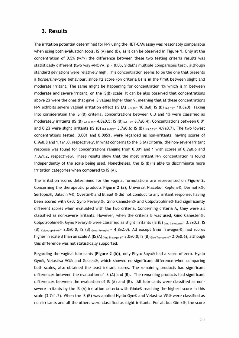
241
3. Results
The irritation potential determined for N-9 using the HET-CAM assay was reasonably comparable
when using both evaluation tools, IS (A) and (B), as it can be observed in Figure 1. Only at the
concentration of 0.5% (w/v) the difference between these two testing criteria results was
statistically different (two way-ANOVA, p < 0.05, Sidak’s multiple comparisons test), although
standard deviations were relatively high. This concentration seems to be the one that presents
a borderline-type behaviour, since its score (on criteria B) is in the limit between slight and
moderate irritant. The same might be happening for concentration 1% which is in between
moderate and severe irritant, on the IS(B) scale. It can be also observed that concentrations
above 2% were the ones that gave IS values higher than 9, meaning that at these concentrations
N-9 exhibits severe vaginal irritation effect (IS (A) N-9 2%= 10.0±0; IS (B) N-9 2%= 10.8±0). Taking
into consideration the IS (B) criteria, concentrations between 0.3 and 1% were classified as
moderately irritants (IS (B) N-9 0.3%= 4.8±0.5; IS (B) N-9 1%= 8.7±0.4). Concentrations between 0.01
and 0.2% were slight irritants (IS (B) N-9 0.01%= 3.7±0.6; IS (B) N-9 0.2%= 4.9±0.7). The two lowest
concentrations tested, 0.001 and 0.005%, were regarded as non-irritants, having scores of
0.9±0.8 and 1.1±1.0, respectively. In what concerns to the IS (A) criteria, the non-severe irritant
response was found for concentrations ranging from 0.001 and 1 with scores of 0.7±0.6 and
7.3±1.2, respectively. These results show that the most irritant N-9 concentration is found
independently of the scale being used. Nonetheless, the IS (B) is able to discriminate more
irritation categories when compared to IS (A).
The irritation scores determined for the vaginal formulations are represented on Figure 2.
Concerning the therapeutic products Figure 2 (a), Universal Placebo, Replens®, Dermofix®,
Sertopic®, Dalacin V®, Ovestin® and Blissel ® did not conduct to any irritant response, having
been scored with 0±0. Gyno Pevaryl®, Gino Canesten® and Colpotrophine® had significantly
different scores when evaluated with the two criteria. Concerning criteria A, they were all
classified as non-severe irritants. However, when the criteria B was used, Gino Canesten®,
Colpotrophine®, Gyno Pevaryl® were classified as slight irritants (IS (B) Gino Canesten®= 3.3±0.3; IS
(B) Colpotrophine®= 2.0±0.0; IS (B) Gyno Pevaryl® = 4.8±2.0). All except Gino Travogen®, had scores
higher in scale B than on scale A (IS (A) Gino Travogen®= 3.0±0.0; IS (B) Gino Travogen®= 2.0±0.6), although
this difference was not statistically supported.
Regarding the vaginal lubricants (Figure 2 (b)), only Phyto Soya® had a score of zero. Hyalo
Gyn®, Velastisa VG® and Gelsea®, which showed no significant difference when comparing
both scales, also obtained the least irritant scores. The remaining products had significant
differences between the evaluation of IS (A) and (B). The remaining products had significant
differences between the evaluation of IS (A) and (B). All lubricants were classified as non-
severe irritants by the IS (A) irritation criteria with Ginix® reaching the highest score in this
scale (3.7±1.2). When the IS (B) was applied Hyalo Gyn® and Velastisa VG® were classified as
non-irritants and all the others were classified as slight irritants. For all but Ginix®, the score

242
obtained in scale B was higher than the one obtained in A. As it can be seen on Figure 2 (b),
when comparing both scales, they also obtained the least irritant scores. The remaining
products had significant differences between the evaluation of IS (A) and (B). Table 2 details
the score obtained and the classification attributed to all products tested in this study.
Furthermore, a picture from the test end-time (5 min) is also shown for products and controls.
As it can be observed, some products did not present the same classification when comparing
the two scales (borderline behaviour). In the case of medicines, Gyno Pevaryl®, Gino
Canesten®, Lomexin®, Gino Travogen® and Colpotrophine®, were the ones to present a
classification of slight irritants on IS (B) and non-severe irritants on IS (A). Regarding lubricants,
KY Jelly®, Vidermina®, Ginix Plus®, Geliofil® and Fillergyn®, presented this behaviour.

243
4. Discussion
Nowadays, the increasingly number of new chemicals and products introduced into the market
coming from several industries (cosmetic, pharmaceutical and medical devices) has generated
a need to validate in vitro techniques able to screen potential irritation effects at the early
stages of development.
The Hen's Egg Test, or Hühner-Embryonen-Test (HET), firstly presented by Luepke, back in the
80’s, was developed to be a rapid, sensitive and inexpensive toxicity test able to provide
information on embryotoxicity, teratogenicity, systemic, metabolic and immunopathological
effects (Luepke, 1985). This method was designed to be applied in mucous-membrane irritation
testing, and the tool provided for score and classification was analogous to the Draize test.
Since then, it has been confirmed that a good correlation between these two tests, i.e. in
vitro/in vivo correlation, for plenty of chemicals do exist. The HET-CAM assay cannot totally
replace the irritation tests in mammals, but can largely decrease the number of animals used,
and limit the pain and injury inflicted to animals during experiments (Luepke, 1985).
The application of the HET-CAM assay to vaginal irritation testing comes in line with the
referred compliance with the 3R’s policy. In the vaginal products field, there is no organotypic
irritation test approved neither on the EU, nor the USA. In fact, there are some cellular and
tissue models available for toxicity testing, however mainly comprise techniques for specific
metabolic pathways, histological analysis and inflammatory response (Fichorova et al., 2004;
Repetto et al., 2008; Gali et al., 2010; Costin et al., 2011). Our research group is focused in
developing strategies for preclinical safety characterization of vaginal products using cellular
and ex vivo tissues assays (paper submitted for publication). The usage of HET-CAM assay in
vaginal products testing comes to widen the safety assessment portfolio that can be applied to
test substances or products in a quicker and more effective way on the first step of preclinical
safety testing. In this study, several semisolid vaginal medicines and lubricant products were
tested. Moreover, two analytic methods of the IS were applied. It was concluded that when
testing N-9, a pure substance, there was no statistical difference when applying one analytic
method or another. Actually, the concentration found to be the one that could trigger severe
irritant effects, 2% (w/v), was the same that was shown to generate severe toxicity in clinical
trials, being less safe than thought at preclinical evaluations (Van Damme et al., 2002). In that
case, N-9 was being studied as a spermicide, but, because of its surfactant nature, it ended up
on being irritant, and even promoting the transmission of HIV infection (Stafford et al., 1998;
Phillips et al., 2000; Dayal et al., 2003). Having this episode in mind, the need for more
appropriate in vitro assays to predict in vivo safety issues, is even more highlightened. In our
study, the surfactant nature of N-9 may be responsible for the fact that when increasing
abruptly its concentration (up to the pure substance) no higher rate of irritation was observed
(U.S. Public Health Service, Department of Health and Human Services, 2006). Universal
placebo, on the other hand, was herein included because it is a vaginal gel that was wittingly

244
design to be a control formulation in clinical trials of vaginal microbicides (Tien et al., 2005;
Schwartz et al., 2007). Its safety profile, already confirmed by clinical trials, was once more
confirmed in this study, having an IS of zero on the two scoring grids (A and B).
Concerning semisolid vaginal medicines, no severe irritant responses were observed, and this
outcome was already expected. They are already commercialized and since they are classified
by the competent authorities as medicines, they were not only subjected to preclinical
evaluations, but also to extensive clinical trials. Moreover, the products herein classified as
non-severe irritants (IS (A)) and slight to moderate irritants (IS (B)), such as Gyno Pevaryl®,
Gino Canesten®, Colpotrophine®, Gino Travogen® and Lomexin® were also tested by our
research group using two in vitro cytotoxicity tests: 3-(4,5-dimethylthiazol-2-yl)-2,5-
diphenyltetrazolium bromide (MTT) and neutral red uptake (NRU) into uterine (HEC-1A),
cervical (HeLa) and vaginal (VK2 E6/E7) cell lines; and, an ex vivo porcine vaginal model using
the MTT reduction assay and histological analysis; having also demonstrated preoccupant
toxicity profiles. In fact, Gino Canesten® had one major impact on the vaginal ex vivo epithelia
when histological analysis was performed (paper submitted for publication). Having this in
mind, and concerning our cytotoxicity results, it was also herein demonstrated that these
products could be considered borderline products, which means they have different
classifications when using one scale or another. Moreover, Gino Travogen® and Colpotrophine®,
were also the ones that presented higher toxicities on a toxicity ex vivo model that we
developed. This data coincidence might corroborate the need to perform several in vitro tests
for a complete toxicity evaluation, since the combined results might be stronger predictors for
in vivo toxicity, as the current mind-set in toxicology states (Samuel et al., 2016). Furthermore,
we also performed some technological characterization of the same products, having safety in
perspective. Accordingly, we determined the osmolality, since it represents a predictor for
irritation (Adriaens and Remon, 2008) and found that Lomexin® and Colpothrophine® were
highly hyperosmolal (1446±20 and 1723±20 mOsmol/kg, respectively), above the upper limit
recommended by the WHO (1200 mOsmol/kg) (World Health Organization, 2012; Machado,
2017). In fact, isosmolality is well known to be an important technological parameter for safety
of both ocular and vaginal products. Previously, we performed a similar study on vaginal
lubricants and found that Geliofil®, Vidermina®, K-Y® Jelly, the ones that showed higher IS in
the HET-CAM assay, had extremely high osmolalities (3582±11; 3707±16 and 3631±13
mOsmol/kg, respectively). Besides, Ginix and Ginix plus were also the ones that showed higher
toxic profiles on HeLa cervical cells using the lactate dehydrogenase (LDH) colorimetric
cytotoxicity assay (Cunha et al., 2014). Once again, K-Y® Jelly and Ginix Plus®, that were
considered as HET-CAM borderline products, had previously demonstrated higher cytotoxicities.
These findings, strengths the hypothesis that products osmolality and cellular and tissue toxicity
could be highly predictive of irritation potential and, it can further suggest that these
techniques (HET-CAM, osmolality, cell/tissue metabolic toxicity) should be applied
concomitantly for a more robust clinical irritation response preview.
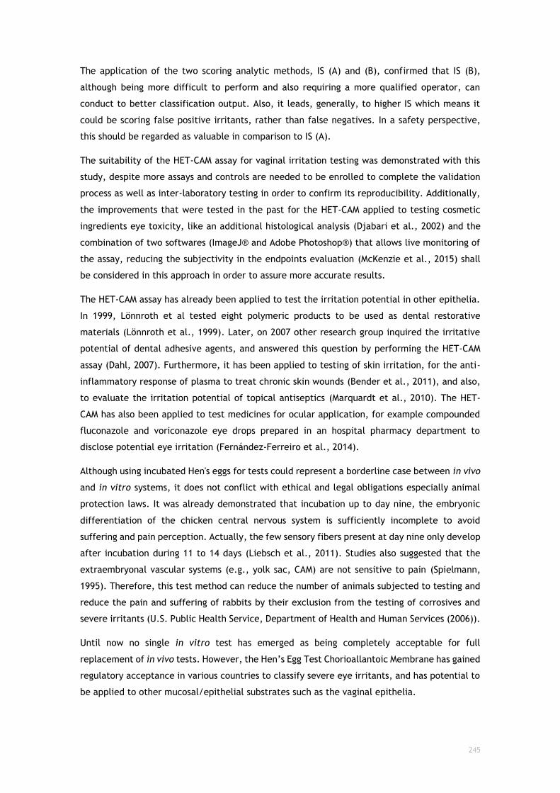
245
The application of the two scoring analytic methods, IS (A) and (B), confirmed that IS (B),
although being more difficult to perform and also requiring a more qualified operator, can
conduct to better classification output. Also, it leads, generally, to higher IS which means it
could be scoring false positive irritants, rather than false negatives. In a safety perspective,
this should be regarded as valuable in comparison to IS (A).
The suitability of the HET-CAM assay for vaginal irritation testing was demonstrated with this
study, despite more assays and controls are needed to be enrolled to complete the validation
process as well as inter-laboratory testing in order to confirm its reproducibility. Additionally,
the improvements that were tested in the past for the HET-CAM applied to testing cosmetic
ingredients eye toxicity, like an additional histological analysis (Djabari et al., 2002) and the
combination of two softwares (ImageJ® and Adobe Photoshop®) that allows live monitoring of
the assay, reducing the subjectivity in the endpoints evaluation (McKenzie et al., 2015) shall
be considered in this approach in order to assure more accurate results.
The HET-CAM assay has already been applied to test the irritation potential in other epithelia.
In 1999, Lönnroth et al tested eight polymeric products to be used as dental restorative
materials (Lönnroth et al., 1999). Later, on 2007 other research group inquired the irritative
potential of dental adhesive agents, and answered this question by performing the HET-CAM
assay (Dahl, 2007). Furthermore, it has been applied to testing of skin irritation, for the anti-
inflammatory response of plasma to treat chronic skin wounds (Bender et al., 2011), and also,
to evaluate the irritation potential of topical antiseptics (Marquardt et al., 2010). The HET-
CAM has also been applied to test medicines for ocular application, for example compounded
fluconazole and voriconazole eye drops prepared in an hospital pharmacy department to
disclose potential eye irritation (Fernández-Ferreiro et al., 2014).
Although using incubated Hen's eggs for tests could represent a borderline case between in vivo
and in vitro systems, it does not conflict with ethical and legal obligations especially animal
protection laws. It was already demonstrated that incubation up to day nine, the embryonic
differentiation of the chicken central nervous system is sufficiently incomplete to avoid
suffering and pain perception. Actually, the few sensory fibers present at day nine only develop
after incubation during 11 to 14 days (Liebsch et al., 2011). Studies also suggested that the
extraembryonal vascular systems (e.g., yolk sac, CAM) are not sensitive to pain (Spielmann,
1995). Therefore, this test method can reduce the number of animals subjected to testing and
reduce the pain and suffering of rabbits by their exclusion from the testing of corrosives and
severe irritants (U.S. Public Health Service, Department of Health and Human Services (2006)).
Until now no single in vitro test has emerged as being completely acceptable for full
replacement of in vivo tests. However, the Hen’s Egg Test Chorioallantoic Membrane has gained
regulatory acceptance in various countries to classify severe eye irritants, and has potential to
be applied to other mucosal/epithelial substrates such as the vaginal epithelia.
246
5. Conclusions
The HET-CAM assay was successfully applied to vaginal irritation testing. This strategy
represents an innovative approach for the preclinical safety assessment of vaginal products,
being them classified as medicines, cosmetics, hygiene products or medical devices. The
application of the two scoring analytic methods, IS (A) and (B), confirmed that IS (B) can
conduct to better classification output and preferably should be chosen. The studied vaginal
formulations, comprising medicines and lubricants, showed, as expected, low potential for
irritation. N-9 was considered as a severe irritant above 2% (w/v) concentrations, which
corroborates clinical data from the literature, envisaging a possible in vitro/in vivo correlation.
Comparisons with previous studies by our workgroup confirmed that HET-CAM can predict
and/or confirm toxic profiles for products also tested for osmolality and cellular/tissue toxicity.
Ideally, an integrative methodology should be designed to embrace all these preclinical tests,
for a better in vivo safety preview. Although still requiring further validation, the HET-CAM
assay seems an ideal prospect for vaginal irritancy potential in vitro studies.
247
6. References
Acartürk, F. (2009). Mucoadhesive vaginal drug delivery systems. Recent Pat Drug Deliv Formul
3, 193–205.
Adriaens, E., and J.P. Remon. (2008). Mucosal irritation potential of personal lubricants relates
to product osmolality as detected by the slug mucosal irritation assay. Sex Transm Dis 35, 512–
6.
Alexander, N.J., E. Baker, M. Kaptein, et al. (2004). Why consider vaginal drug administration?
Fertil Steril 82, 1–12.
European Commission (2003). Regulation (EC) of No 2003/15 of the European parliament and of
the council 27 February 2003 concerning the 7th Amendment to the Cosmetics Directive.
European Commission. European Union Reference Laboratory for alternatives to animal testing
(EURL-ECVAM). Topical Toxicity - Validation and Regulatory Acceptance. Available at:
https://eurl-ecvam.jrc.ec.europa.eu/validation-regulatory-acceptance/topical-toxicity/
[Accessed October 31, 2016].
ICCVAM (2007). GUIDANCE DOCUMENT - ESAC PEER REVIEW Organotypic in vitro assays to
identify severe eye irritants – ICCVAM-NICEATM Retrospective Evaluation. Available at:
https://eurl-ecvam.jrc.ec.europa.eu/validation-regulatory-acceptance/docs-eye-
irritation/Annex I. Guidance document_ESAC PRP Organotypics-finalcleared.pdf [Accessed
August 23, 2017].
World Health Organization (2012). Use and procurement of additional lubricants for male and
female condoms: WHO/UNFPA/FHI360 Advisory note.
Bender, C., L.-I. Partecke, E. Kindel, et al. (2011). The modified HET-CAM as a model for the
assessment of the inflammatory response to tissue tolerable plasma. Toxicol In Vitro 25, 530–
537.
Bygdeman, M., and M.L. Swahn. (1996). Replens versus dienoestrol cream in the symptomatic
treatment of vaginal atrophy in postmenopausal women. Maturitas 23, 259–263.
Caramella, C.M., S. Rossi, F. Ferrari, et al. (2015). Mucoadhesive and thermogelling systems
for vaginal drug delivery. Adv Drug Deliv Rev 92, 39–52.
Choudhury, A., S. Das, and M. Kar. (2011). A review on novelty and potentiality of vaginal drug
delivery. Int J PharmTech Res 3, 1033–1044.
248
Clark, M.R., T.J. McCormick, G.F. Doncel, et al. (2011). Preclinical evaluation of UC781
microbicide vaginal drug delivery. Drug Deliv and Transl Res 1, 175–182.
Costin, G., H. Raabe, and R. Priston. (2011). Vaginal irritation models: the current status of
available alternative and in vitro tests. Altern Lab Anim 39, 317–337.
Cunha, A.R., Machado R.M., Palmeira-de-Oliveira A., et al. (2014). Characterization of
commercially available vaginal lubricants: a safety perspective. Pharmaceutics 6, 530–42.
Dahl, J.E. (2007). Potential of dental adhesives to induce mucosal irritation evaluated by the
HET–CAM method. Acta Odontol Scand 65, 275–283.
Van Damme, L., G. Ramjee, M. Alary, et al. (2002). Effectiveness of COL-1492, a nonoxynol-9
vaginal gel, on HIV-1 transmission in female sex workers: a randomised controlled trial. Lancet
360, 971–7.
Dayal, M.B., J. Wheeler, C.J. Williams, et al. (2003). Disruption of the upper female
reproductive tract epithelium by nonoxynol-9. Contraception 68, 273–9.
Djabari, Z., E. Bauza, C. Dal Farra, et al. (2002). The HET-CAM test combined with histological
studies for better evaluation of active ingredient innocuity. Int J Tissue React 24, 117–21.
Eckstein, P., M.C. Jackson, N. Millman, et al. (1969). Comparison of vaginal tolerance tests of
spermicidal preparations in rabbits and monkeys. J Reprod Fertil 20, 85–93.
Fernández-Ferreiro, A., M. González Barcia, M. Gil Martínez, et al. (2014). Análisis de la
toxicidad ocular de los colirios de voriconazol y fluconazol con HET-CAM. Farm Hosp 38, 300-
304.
Fichorova, R.N., M. Bajpai, N. Chandra, et al. (2004). Interleukin (IL)-1, IL-6, and IL-8 predict
mucosal toxicity of vaginal microbicidal contraceptives. Biol Reprod 71, 761–9.
Gali, Y., O. Delezay, J. Brouwers, et al. (2010). In vitro evaluation of viability, integrity, and
inflammation in genital epithelia upon exposure to pharmaceutical excipients and candidate
microbicides. Antimicrob Agents Chemother 54, 5105–5114.
Garg, S., R.A. Anderson, C.J. Chany, et al. (2001). Properties of a new acid-buffering
bioadhesive vaginal formulation (ACIDFORM). Contraception 64, 67–75.
Garg, S., D. Goldman, M. Krumme, et al. (2010). Advances in development, scale-up and
manufacturing of microbicide gels, films, and tablets. Antiviral Res 88 Suppl 1, S19-29.
Hussain, A., and F. Ahsan. (2005). The vagina as a route for systemic drug delivery. J Control
Release 103, 301–13.

249
ISO 10993-5. International Standard (2009). Biological evaluation of medical devices — Part 5:
Tests for in vitro cytotoxicity.
Liebsch, M., B. Grune, A. Seiler, et al. (2011). Alternatives to animal testing: current status
and future perspectives. Arch Toxicol 85, 841-58.
Lönnroth, E.-C., J. Dahl, and H. Shahnavaz. (1999). Evaluating the Potential Occupational
Hazard of Handling Dental Polymer Products Using the HET-CAM Technique. Int J Occup Saf
Ergon 5, 43–57.
Luepke, N.P. (1985). Hen’s egg chorioallantoic membrane test for irritation potential. Food
Chem Toxicol 23, 287–91.
Machado R.M., Palmeira-de-Oliveira A., Martinez-de-Oliveira J., Palmeira-de-Oliveira R.,
(2017). Vaginal semisolid products: technological performance considering physiologic
parameters. Eur J Pharm Sci 109, 556-568.
Marquardt, C., E. Matuschek, E. Bölke, et al. (2010). Evaluation of the tissue toxicity of
antiseptics by the hen’s egg test on the chorioallantoic membrane (HETCAM). Eur J Med Res
15, 204–9.
McKenzie, B., G. Kay, K.H. Matthews, et al. (2015). The hen’s egg chorioallantoic membrane
(HET-CAM) test to predict the ophthalmic irritation potential of a cysteamine-containing gel:
Quantification using Photoshop® and ImageJ. Int J Pharm 490, 1–8.
Nachtigall, L.E. (1994). Comparative study: Replens versus local estrogen in menopausal
women. Fertil Steril 61, 178–80.
Organisation for Economic Co-operation and Development. OECD Test Guideline 405. Available
at:
http://www.oecd.org/chemicalsafety/testing/oecdguidelinesforthetestingofchemicals.htm
[Accessed September 4, 2017].
Phillips, D.M., C.L. Taylor, V.R. Zacharopoulos, et al. (2000). Nonoxynol-9 causes rapid
exfoliation of sheets of rectal epithelium. Contraception 62, 149–54.
Repetto, G., A. del Peso, and J.L. Zurita. (2008). Neutral red uptake assay for the estimation
of cell viability/cytotoxicity. Nat Protoc 3, 1125–31.
Samuel, G.O., S. Hoffmann, R.A. Wright, et al. (2016). Guidance on assessing the
methodological and reporting quality of toxicologically relevant studies: A scoping review.
Environ Int 92, 630-646.
250
Schwartz, J.L., S.A. Ballagh, C. Kwok, et al. (2007). Fourteen-day safety and acceptability study
of the universal placebo gel. Contraception 75, 136–41.
U.S. Public Health Service, Department of Health and Human Services (2006). NIH Publication
No: 06-4515. Current Status of In Vitro Test Methods for Identifying Ocular Corrosives and
Severe Irritants: Hen’ s Egg Test - Chorioallantoic Membrane Test Method.
Spielmann H. (1995) HET-CAM Test. In: O’Hare S., Atterwill C.K. (eds) In Vitro Toxicity Testing
Protocols. Methods in Molecular Biology™, 43, 199–204 Humana Press
Stafford, M.K., H. Ward, A. Flanagan, et al. (1998). Safety study of nonoxynol-9 as a vaginal
microbicide: evidence of adverse effects. J Acquir Immune Defic Syndr Hum Retrovirol 17, 327–
31.
Tien, D., R.L. Schnaare, F. Kang, et al. (2005). In Vitro and in Vivo Characterization of a
Potential Universal Placebo Designed for Use in Vaginal Microbicide Clinical Trials. AIDS Res
Hum Retroviruses 21, 845–853.
Valenta, C. (2005). The use of mucoadhesive polymers in vaginal delivery. Adv Drug Deliv Rev
57, 1692–712.
Woolfson, A.D., R.K. Malcolm, and R. Gallagher. (2000). Drug delivery by the intravaginal route.
Crit Rev Ther Drug Carrier Syst 17, 509–55.
251
Acknowledgements
This work was supported by FEDER funds through the POCI - COMPETE 2020 - Operational
Programme Competitiveness and Internationalisation in Axis I - Strengthening research,
Technological development and innovation (Project POCI-01-0145-FEDER-007491) and National
Funds by FCT - Foundation for Science and Technology (Project UID/Multi/00709/2013).
Financial support was also provided by Labfit, HPRD, Lda. and FCT - Foundation for Science and
Technology through a PhD fellowship (Grant Reference SFRH/BDE/111544/2015).
Conflict of interest
The authors declare that they have no conflicts of interest.
Correspondence to
Rita Palmeira-de-Oliveira
Labfit – Health Products and Research and Development, Lda
Edifício UBIMEDICAL Estrada Municipal
506 6200-284 Covilhã.
Phone: +351 275 241 271
e-mail: [email protected]
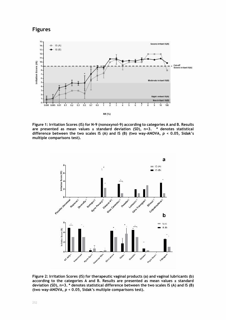
252
Figures
Figure 1: Irritation Scores (IS) for N-9 (nonoxynol-9) according to categories A and B. Results are presented as mean values ± standard deviation (SD), n=3. * denotes statistical difference between the two scales IS (A) and IS (B) (two way-ANOVA, p < 0.05, Sidak’s multiple comparisons test).
Figure 2: Irritation Scores (IS) for therapeutic vaginal products (a) and vaginal lubricants (b) according to the categories A and B. Results are presented as mean values ± standard deviation (SD), n=3. * denotes statistical difference between the two scales IS (A) and IS (B) (two way-ANOVA, p < 0.05, Sidak’s multiple comparisons test).
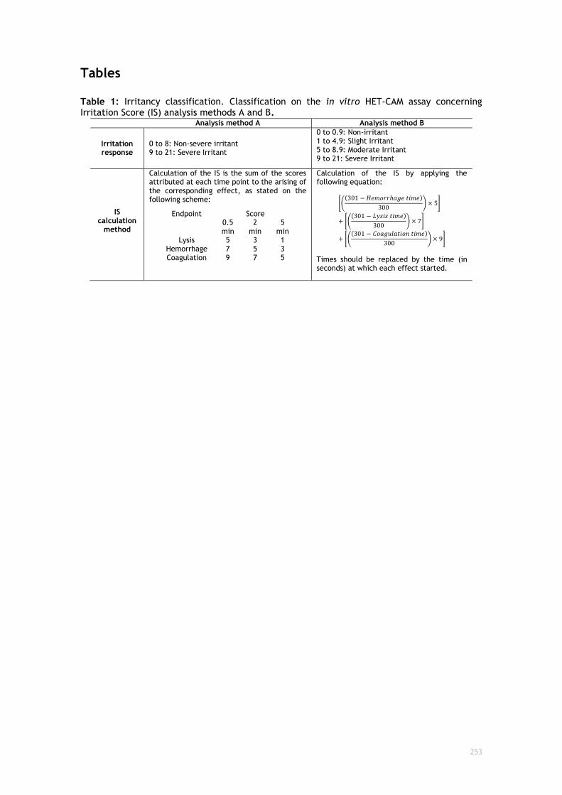
253
Tables
Table 1: Irritancy classification. Classification on the in vitro HET-CAM assay concerning Irritation Score (IS) analysis methods A and B.
Analysis method A Analysis method B
Irritation response
0 to 8: Non-severe irritant 9 to 21: Severe Irritant
0 to 0.9: Non-irritant 1 to 4.9: Slight Irritant 5 to 8.9: Moderate Irritant 9 to 21: Severe Irritant
IS calculation
method
Calculation of the IS is the sum of the scores attributed at each time point to the arising of the corresponding effect, as stated on the following scheme:
Endpoint Score
0.5 min
2 min
5 min
Lysis 5 3 1 Hemorrhage 7 5 3 Coagulation 9 7 5
Calculation of the IS by applying the following equation:
[((301 − 𝐻𝑒𝑚𝑜𝑟𝑟ℎ𝑎𝑔𝑒 𝑡𝑖𝑚𝑒)
300) × 5]
+ [((301 − 𝐿𝑦𝑠𝑖𝑠 𝑡𝑖𝑚𝑒)
300) × 7]
+ [((301 − 𝐶𝑜𝑎𝑔𝑢𝑙𝑎𝑡𝑖𝑜𝑛 𝑡𝑖𝑚𝑒)
300) × 9]
Times should be replaced by the time (in seconds) at which each effect started.
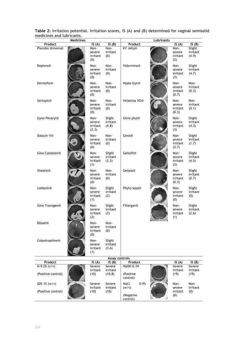
254
Table 2: Irritation potential. Irritation scores, IS (A) and (B) determined for vaginal semisolid medicines and lubricants.
Medicines Lubricants
Product IS (A) IS (B) Product IS (A) IS (B)
Placebo Universal
Non-severe irritant (0)
Non-irritant (0)
KY Jelly®
Non-severe irritant (3)
Slight irritant (4.9)
Replens®
Non-severe irritant (0)
Non-irritant (0)
Vidermina®
Non-severe irritant (3)
Slight irritant (4.7)
Dermofix®
Non-severe irritant (0)
Non-irritant (0)
Hyalo Gyn®
Non-severe irritant (0.7)
Non-irritant (0.3)
Sertopic®
Non-severe irritant (0)
Non-irritant (0)
Velastisa VG®
Non-severe irritant (0.3)
Non-irritant (0.1)
Gyno Pevaryl®
Non-severe irritant (2.3)
Slight irritant (4.8)
Ginix plus®
Non-severe irritant (3)
Slight irritant (4.3)
Dalacin V®
Non-severe irritant (0)
Non-irritant (0)
Ginix®
Non-severe irritant (3.7)
Slight irritant (1.7)
Gino Canesten®
Non-severe irritant (1)
Slight irritant (3.3)
Geliofil®
Non-severe irritant (3)
Slight irritant (4.5)
Ovestin®
Non-severe irritant (0)
Non-irritant (0)
Gelsea®
Non-severe irritant (0.3)
Slight irritant (0.7)
Lomexin®
Non-severe irritant (1)
Slight irritant (2)
Phyto soya®
Non-severe irritant (0)
Slight irritant (0)
Gino Travogen®
Non-severe irritant (2)
Slight irritant (3)
Fillergyn®
Non-severe irritant (1)
Slight irritant (2.6)
Blissel®
Non-severe irritant (0)
Non-irritant (0)
Colpotrophine®
Non-severe irritant (1)
Slight irritant (3.6)
Assay controls
Product IS (A) IS (B) Product IS (A) IS (B)
N-9 2% (v/v) (Positive control)
Severe irritant (10)
Severe irritant (10.8)
NaOH 0.1N (Positive control)
Severe irritant (19)
Severe irritant (19)
SDS 1% (w/v) (Positive control)
Severe irritant (10)
Severe irritant (10)
NaCl 0.9% (w/v) (Negative control)
Non-severe irritant (0)
Non-irritant (0)

255
APPENDIX D

256
Literature review tables that supported the design of the experimental setup of CHAPTER IV:
□ The first table details already published methods for drug quantification using HPLC-
DAD.
□ The second demonstrates the theoretical predictive calculations for molecules concentration in the receptor chambers.
□ The third table shows a comparison for receptor media for in vitro drug release and ex
vivo permeation experiments already published on the literature.
Bibliographic references used in this literature review are listed bellow the respective table.

257
HPLC-DAD methods for the permeation products
Molecule HPLC-DAD method Permeation method Concentration expected to permeate Reference
CLO
TR
IMA
ZO
L
10m
g/g
Waters HPLC-DAD Detection:233nm Colum: stainless steel C-18 reverse-phase column packed with 5µm particles (Waters, XTerra RP-18, 3.9mm × 150mm) Mobile phase: methanol/water (75:25, v/v) Flow rate: 0.5mL/min Injection volume: 50µL.
Franz diffusion cells: To evaluate CTZ permeation through the skin, samples of pig mucosa were mounted on standard Franz diffusion cells (area=1.0±0.1cm2) and the receptor compartment (volume 11mL) was filled with PBS. After equilibration, 1mL of CTZ-NLC or CTZ-NLC-gel was added to the donor compartment. The receptor compartment was stirred at 250rpm in a thermostatic water bath at a temperature of 37ºC. At fixed time intervals up to 8h, 100µL of the receptor phase were withdrawn and replaced with an equal amount of fresh PBS. The samples were filtered with 0.20µm hydrophilic PVDF. Tissues extraction: the mucosa was rinsed twice with ultrapure and stored at −19◦C for 1h. Tissues were cut in vertical slices and collected in glass vials. CTZ penetrated into slices was extracted with 2 mL of methanol under overnight stirring with magnetic bars. The obtained samples were filtered with 0.20µm.
Loaded concentration: CTZ 1mg/mL and it is not expected to permeate. Toxicity: hepatic and can induce resistance especially in patients with a compromised immune system (Liu et al., 2010; Tettenborn, 1974). The results showed that no CTZ was found in the receptor medium even after 8 h of diffusion. The results showed that only 0.33±0.06% of the CTZ applied by CTZ-NLC was found within the mucosa (0.0033mg/mL).
(Ravani et al. 2013)
Beckman HPLC Colum: Beckman C18 5µm, 4.6mmx25cm. Mobile phase: methanol and 0.1 M phosphate buffer (pH 3) (27:73). Flow rate: 1mL/min. Detection: 254nm. This method was validated in terms of specificity, linearity and reproducibility. The limit of quantification was 0.1µg/mL. Recovery: 90.1-94.2%
Franz diffusion cells: receptor compartment had a volume of 7.2 mL and an effective diffusion area of 0.785 cm2 (ø=0.5cm). The receptor compartment was filled with a PBS solution containing 10% (v/v) dioxane which was constantly stirred with a magnetic bar (37°±1°C). Frozen rabbit vaginal mucosa. Samples of 200µl were withdrawn from the receptor at specified time intervals and replaced with an equal volume of fresh receptor solution. In addition, the vaginal mucosa was cleaned on both sides and the drug accumulated in the mucosa was extracted with 100% ethanol and subjected to five sonication cycles of 30 min each in an ultrasound bath. The concentration of drug in the receiver at various times and that extracted from the mucosa was determined by HPLC. Control solution was 1% clotrimazole (w/v) PEG 400.
Loaded concentration: 1.5 mg of drug Solubilisation enhancers: 0.1%M citrate phosphate buffer (pH5.5) containing 10% Tween-80; 0.1M acetate buffer, pH6 and organic dioxane (35%v/v); PBS (pH7.4) solution with 10% dioxane.
(Ninga·b et al. 2005)
Other HPLC methods (without the permeation application)
Waters 1500 Series HPLC Colum: C18 reversed octadecyl phase 250mm × 4.6mm, 5m. Mobile phase: ACN-MeOH-0.15% phosphoric acid pH 7.5 (15:60:25V/V/V) Flow rate: 1.0mL/min. Detection: 270nm Injection volume: 50 µL Temperature: 25ºC (Sehati et al. 2015)
Shimadzu LC-2010C system Stock solutions of individual chemicals (active ingredient and internal standard) were prepared in acetonitrile. The reference standard solutions were prepared diluting the respective stock solutions to the final concentrations of CLO (500mg/L), ibuprofen (100mg/L), IM (5mg/L) and CDM (5mg/L) The stock solution of internal standard (ibuprofen)was prepared dissolving 500mg in 100 mL of acetonitrile All solutions were at 4ºC. Colum: Zorbax SB-Phenyl column (75mm×4.6 mm, 3.5µm) Mobile phase: acetonitrile and water in ratio 65:35 (v/v) and pH=3.5 (adjusted with 85% phosphoric acid) Flow rate: 0.5 mL/min IBU was used as the internal standard Injection volume: 5µl Detection: 210 nm at ambient temperature Run time: 6 min (Hájková et al. 2007)
Merck Hitachi HPLC Colum: C18 stationary phase (250x4.0m) bonded onto 5μm silica gel manufactured by Merck Flow rate: 1.5 mL/min Injection volume: 5μl Detection: temperature ambient and 254nm Standard stock solution: 0.5 mg/mL of clotrimazole Mobile phase: Acetate buffer with a pH of 6.8 was prepared by dissolving 25.0g ammonium acetate in 1000 mL of distilled deionised water. Diluent was prepared by mixing 990 mL of methanol and 10 mL of acetic acid. (Manassra et al. 2010)
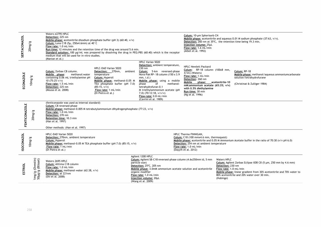
258
SER
TA
CO
NA
ZO
L
20m
g/g
Waters e2795 HPLC Detection: 225 nm Mobile phase: acetonitrile:disodium phosphate buffer (pH 3) (60:40, v/v) Colum: Luna C18 (5µ, 250x4.6mm) at 40°C Flow rate: 1.0 mL/min Run time: 12 minutes and the retention time of the drug was around 5.6 min. Standard solution: 100 µg/mL was prepared by dissolving the drug in PEG:PBS (60:40) which is the receptor medium that will be used for in vitro studies. (Manian et al.)
Colum: 10-μm Spherisorb CN Mobile phase: acetonitrile and aqueous 0.01 M sodium phosphate (37:63, v/v). Detection: 260 nm at 35ºC, the retention time being 19.3 min. Injection volume: 25µL Flow rate: 1.6 mL/min (Albet et al. 1992)
EC
ON
AZO
LE
10m
g/g
Colum: Zorbax C8 column. Mobile phase: methanol:water containing 0.06 mL triethylamine pH 10 (75:25 v/v) Flow rate: 1.5 mL/min Detection: 225 nm (Mousa et al. 2008)
HPLC-DAD Varian 5020 Detection: 270nm, ambient temperature Colum: Hypersil Mobile phase: methanol-0.05 M TEA phosphate buffer (pH 7.0) (85:15, v/v) Flow rate: 1 mL/min (Di Pietra et al.)
HPLC Varian 5020 Detection: ambient temperature, 230 nm Colum: 5-km reversed-phase Nova-Pak RP- 18 column (150 x 3.9 mm, i.d.) Mobile phase: using a mobile phase of methanol-tetrahydrofuran-0.1 M triethylammonium acetate (pH 7.0) (70:12:18, v/v/v) Flow-rate: 0.8 mL/min (Cavrini et al. 1989)
HPLC Hewlett Packard Colum: RP-18 column (100x8 mm, 4/im) (Waters) Flow rate: 1 mL/min Detection: 260 nm Mobile phase: acetonitrile-10 mM:ammonium acetate (65:35; v/v) with 0.5% diethylamine Run time: 30 min (Ng et al. 1996)
Colum: RP-18 Mobile phase: methanol/aqueous ammoniumcarbonate solution/tetrahydrofurane (Christinat & Zulliger 1984)
FEN
TIC
ON
AZO
LE
20m
g/g
(fenticonazole was used as internal standard) Colum: C8 reversed-phase Mobile phase: methanol-0.005 M tetrabutylammonium dihydrogenphosphate (77:23, v/v) Flow rate: 1.0 mL/min Detection: 270 nm Retention time: 10.3 min (Han et al. 1997) Other methods: (Han et al. 1997)
ISO
CO
NA
ZO
L
10m
g/g
HPLC-DAD Varian 5020 Detection: 270nm, ambient temperature Colum: Hypersil Mobile phase: methanol-0.05 M TEA phosphate buffer (pH 7.0) (85:15, v/v) Flow rate: 1 mL/min (Di Pietra et al.)
HPLC Thermo FINNIGAN, Colum: C18 (100 mmx4.6 mm, thermoquest) Mobile phase: acetonitrile and 0.05 M Ammonium Acetate buffer in the ratio of 70:30 (v/v pH 6.0) Detection: 254 nm at ambient temperature Flow rate: 1.0 mL/min (Dayyih et al. 2012)
EST
RIO
L
1m
g/g (
Ovest
in)
50µg/g (
Bliss
el)
Waters 2695 HPLC Colum: Alltima C18 column Flow rate: 1.0 mL/min Mobile phase: methanol–water (62:38, v/v) Detection: at 221nm (Shi et al. 2008)
Agilent 1200 HPLC Colum: Agilent SB-C18 reversed-phase column (4.6x250mm id, 5 mm particle size) Detection: 25ºC, 205 nm Mobile phase: 3.0mM ammonium acetate solution and acetonitrile organic modifier Flow rate: 1.0 mL/min Injection volume: 20µL (Wang et al. 2009)
Waters HPLC Colum: Agilent Zorbax Eclipse XDB C8 (5 μm, 250 mm by 4.6 mm) Detection: 230 nm Flow rate: 1.0-mL/min Mobile phase: linear gradient from 30% acetonitrile and 70% water to 80% acetonitrile and 20% water over 30 min. (Hubinge)
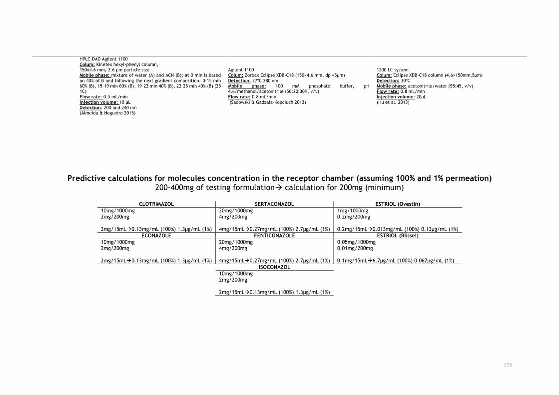
259
Predictive calculations for molecules concentration in the receptor chamber (assuming 100% and 1% permeation) 200-400mg of testing formulation calculation for 200mg (minimum)
CLOTRIMAZOL SERTACONAZOL ESTRIOL (Ovestin)
10mg/1000mg 2mg/200mg 2mg/15mL0.13mg/mL (100%) 1.3µg/mL (1%)
20mg/1000mg 4mg/200mg 4mg/15mL0.27mg/mL (100%) 2.7µg/mL (1%)
1mg/1000mg 0.2mg/200mg 0.2mg/15mL0.013mg/mL (100%) 0.13µg/mL (1%)
ECONAZOLE FENTICONAZOLE ESTRIOL (Blissel)
10mg/1000mg 2mg/200mg 2mg/15mL0.13mg/mL (100%) 1.3µg/mL (1%)
20mg/1000mg 4mg/200mg 4mg/15mL0.27mg/mL (100%) 2.7µg/mL (1%)
0.05mg/1000mg 0.01mg/200mg 0.1mg/15mL6.7µg/mL (100%) 0.067µg/mL (1%)
ISOCONAZOL
10mg/1000mg 2mg/200mg 2mg/15mL0.13mg/mL (100%) 1.3µg/mL (1%)
HPLC–DAD Agilent 1100 Colum: Kinetex hexyl-phenyl column, 150x4.6 mm, 2.6 μm particle size Mobile phase: mixture of water (A) and ACN (B); at 0 min is based on 40% of B and following the next gradient composition: 0–15 min 60% (B), 15–19 min 60% (B), 19–22 min 40% (B), 22–25 min 40% (B) (25 1C) Flow rate: 0.5 mL/min Injection volume: 10 μL Detection: 200 and 240 nm (Almeida & Nogueira 2015)
Agilent 1100 Colum: Zorbax Eclipse XDB-C18 (150×4.6 mm, dp =5µm) Detection: 27ºC 280 nm Mobile phase: 100 mM phosphate buffer, pH 4.6/methanol/acetonitrile (50:20:30%, v/v) Flow rate: 0.8 mL/min (Sadowski & Gadzała-Kopciuch 2013)
1200 LC system Colum: Eclipse XDB-C18 column (4.6×150mm,5µm) Detection: 30ºC Mobile phase: acetonitrile/water (55:45, v/v) Flow rate: 0.8 mL/min Injection volume: 20µL (Hu et al. 2013)
260
References: Albet, C. et al., 1992. Determination of sertaconazole nitrate, a new imidazole antifungal, by high-performance liquid chromatography. Journal of Pharmaceutical and Biomedical Analysis, 10(2-
3), pp.205–211.
Almeida, C. & Nogueira, J.M.F., 2015. Determination of steroid sex hormones in real matrices by bar adsorptive microextraction (BAμE). Talanta, 136, pp.145–154.
Cavrini, V., Di Pietra, A.M. & Gatti, R., 1989. Analysis of miconazole and econazole in pharmaceutical formulations by derivative UV spectroscopy and liquid chromatography (HPLC). Journal of
Pharmaceutical and Biomedical Analysis, 7(12), pp.1535–1543.
Christinat, R. & Zulliger, H.W., 1984. Stability indicating HPLC-method for the determination of econazole nitrate in cream and lotion formulations. Arzneimittel-Forschung, 34(5), pp.551–3.
Dayyih, W.A. et al., 2012. Development and validation of HPLC methos for some azoles in pharmaceutical preparations, 3(10), pp.3686–3692.
Hájková, R. et al., 2007. Development and validation of HPLC method for determination of clotrimazole and its two degradation products in spray formulation. Talanta, 73(3), pp.483–489.
Han, K.S. et al., 1997. Determination of a new proton pump inhibitor, YH1885, in human plasma and urine by high-performance liquid chromatography. Journal of chromatography. B, Biomedical
sciences and applications, 696(2), pp.312–6.
Hu, Y. et al., 2013. Enrichment of steroid hormones in water with porous and hydrophobic polymer-based SPE followed by HPLC-UV determination. Journal of Separation Science, 36(20), pp.3321–
3329.
Hubinger, J.C., Determination of estriol, estradiol, estrone, and progesterone in cosmetic products. Journal of cosmetic science, 66(2), pp.113–28.
Manassra, A. et al., 2010. Simultaneous HPLC Analysis of Betamethasone and Clotrimazole in Cream Formulations. Pharmaceutica Analytica Acta, 1(2), pp.1–3.
Manian, M., Knaack, J.S. & Banga, A.K., Development and Validation of a Reverse Phase-HPLC Method for Sertaconazole Nitrate, p.997.
Mousa, B.A. et al., 2008. Stability indicating methods for the determination of some anti-fungal agents using densitometric and RP-HPLC methods. Chemical & pharmaceutical bulletin, 56(2),
pp.143–149.
Ng, T.K. et al., 1996. Rapid high performance liquid chromatographic assay for antifungal agents in human sera. The Journal of antimicrobial chemotherapy, 37(3), pp.465–72.
Ninga·b, M. et al., 2005. Preparation and in vitro evaluation of liposomal/niosomal delivery systems for antifungal drug clotrimazole. Indian Journal of Experimental Biology VoL, 43, pp.150–157.
Di Pietra, A.M. et al., Journal of pharmaceutical and biomedical analysis, 10(10-12), pp.873–9.
Ravani, L. et al., 2013. Clotrimazole-loaded nanostructured lipid carrier hydrogels: thermal analysis and in vitro studies. Int J Pharm, 454(2), pp.695–702.
Sadowski, R. & Gadzała-Kopciuch, R., 2013. Isolation and determination of estrogens in water samples by solid-phase extraction using molecularly imprinted polymers and HPLC. Journal of
Separation Science, 36(14), pp.2299–2305.
Sehati, N. et al., 2015. Application of hollow fiber membrane mediated with titanium dioxide nanowire/reduced graphene oxide nanocomposite in preconcentration of clotrimazole and tylosin.
Journal of Chromatography A, 1420, pp.46–53.
Shi, Y.Q. et al., 2008. Establishment of an HPLC identification system for detection of counterfeit steroidal drugs. Journal of Pharmaceutical and Biomedical Analysis, 46(4), pp.663–669.
Wang, X. et al., 2009. [Simultaneous determination of seven sexual hormones in essential oil by high performance liquid chromatography with diode array and fluorescence detectors]. Se pu =
Chinese journal of chromatography / Zhongguo hua xue hui, 27(3), pp.328–32.
Ye, Y.R. et al., 2004. Simultaneous determination of tretinoin and clindamycin phosphate and their degradation products in topical formulations by reverse phase HPLC. Journal of separation
science, 27(1-2), pp.71–7.
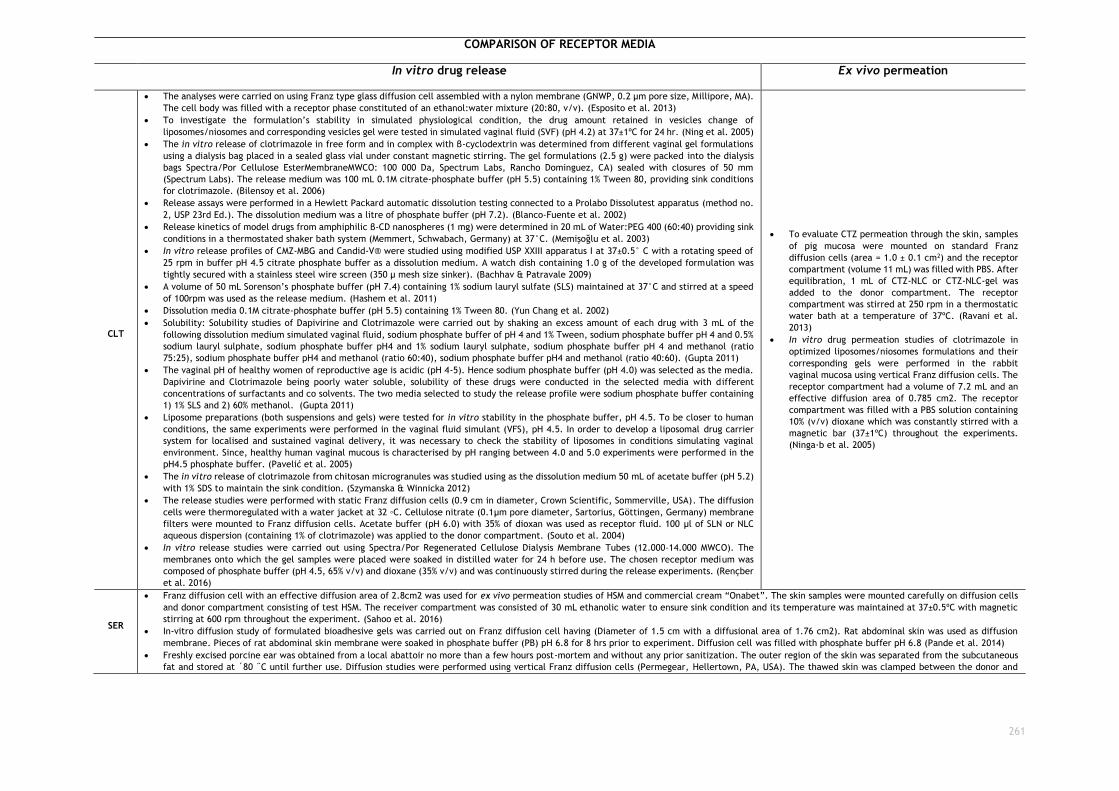
261
COMPARISON OF RECEPTOR MEDIA
In vitro drug release Ex vivo permeation
CLT
The analyses were carried on using Franz type glass diffusion cell assembled with a nylon membrane (GNWP, 0.2 μm pore size, Millipore, MA).
The cell body was filled with a receptor phase constituted of an ethanol:water mixture (20:80, v/v). (Esposito et al. 2013)
To investigate the formulation’s stability in simulated physiological condition, the drug amount retained in vesicles change of
liposomes/niosomes and corresponding vesicles gel were tested in simulated vaginal fluid (SVF) (pH 4.2) at 37±1ºC for 24 hr. (Ning et al. 2005)
The in vitro release of clotrimazole in free form and in complex with β-cyclodextrin was determined from different vaginal gel formulations
using a dialysis bag placed in a sealed glass vial under constant magnetic stirring. The gel formulations (2.5 g) were packed into the dialysis
bags Spectra/Por Cellulose EsterMembraneMWCO: 100 000 Da, Spectrum Labs, Rancho Dominguez, CA) sealed with closures of 50 mm
(Spectrum Labs). The release medium was 100 mL 0.1M citrate-phosphate buffer (pH 5.5) containing 1% Tween 80, providing sink conditions
for clotrimazole. (Bilensoy et al. 2006)
Release assays were performed in a Hewlett Packard automatic dissolution testing connected to a Prolabo Dissolutest apparatus (method no.
2, USP 23rd Ed.). The dissolution medium was a litre of phosphate buffer (pH 7.2). (Blanco-Fuente et al. 2002)
Release kinetics of model drugs from amphiphilic β-CD nanospheres (1 mg) were determined in 20 mL of Water:PEG 400 (60:40) providing sink
conditions in a thermostated shaker bath system (Memmert, Schwabach, Germany) at 37°C. (Memişoğlu et al. 2003)
In vitro release profiles of CMZ-MBG and Candid-V® were studied using modified USP XXIII apparatus I at 37±0.5° C with a rotating speed of
25 rpm in buffer pH 4.5 citrate phosphate buffer as a dissolution medium. A watch dish containing 1.0 g of the developed formulation was
tightly secured with a stainless steel wire screen (350 µ mesh size sinker). (Bachhav & Patravale 2009)
A volume of 50 mL Sorenson’s phosphate buffer (pH 7.4) containing 1% sodium lauryl sulfate (SLS) maintained at 37°C and stirred at a speed
of 100rpm was used as the release medium. (Hashem et al. 2011)
Dissolution media 0.1M citrate-phosphate buffer (pH 5.5) containing 1% Tween 80. (Yun Chang et al. 2002)
Solubility: Solubility studies of Dapivirine and Clotrimazole were carried out by shaking an excess amount of each drug with 3 mL of the
following dissolution medium simulated vaginal fluid, sodium phosphate buffer of pH 4 and 1% Tween, sodium phosphate buffer pH 4 and 0.5%
sodium lauryl sulphate, sodium phosphate buffer pH4 and 1% sodium lauryl sulphate, sodium phosphate buffer pH 4 and methanol (ratio
75:25), sodium phosphate buffer pH4 and methanol (ratio 60:40), sodium phosphate buffer pH4 and methanol (ratio 40:60). (Gupta 2011)
The vaginal pH of healthy women of reproductive age is acidic (pH 4-5). Hence sodium phosphate buffer (pH 4.0) was selected as the media.
Dapivirine and Clotrimazole being poorly water soluble, solubility of these drugs were conducted in the selected media with different
concentrations of surfactants and co solvents. The two media selected to study the release profile were sodium phosphate buffer containing
1) 1% SLS and 2) 60% methanol. (Gupta 2011)
Liposome preparations (both suspensions and gels) were tested for in vitro stability in the phosphate buffer, pH 4.5. To be closer to human
conditions, the same experiments were performed in the vaginal fluid simulant (VFS), pH 4.5. In order to develop a liposomal drug carrier
system for localised and sustained vaginal delivery, it was necessary to check the stability of liposomes in conditions simulating vaginal
environment. Since, healthy human vaginal mucous is characterised by pH ranging between 4.0 and 5.0 experiments were performed in the
pH4.5 phosphate buffer. (Pavelić et al. 2005)
The in vitro release of clotrimazole from chitosan microgranules was studied using as the dissolution medium 50 mL of acetate buffer (pH 5.2)
with 1% SDS to maintain the sink condition. (Szymanska & Winnicka 2012)
The release studies were performed with static Franz diffusion cells (0.9 cm in diameter, Crown Scientific, Sommerville, USA). The diffusion
cells were thermoregulated with a water jacket at 32 ◦C. Cellulose nitrate (0.1µm pore diameter, Sartorius, Göttingen, Germany) membrane
filters were mounted to Franz diffusion cells. Acetate buffer (pH 6.0) with 35% of dioxan was used as receptor fluid. 100 µl of SLN or NLC
aqueous dispersion (containing 1% of clotrimazole) was applied to the donor compartment. (Souto et al. 2004)
In vitro release studies were carried out using Spectra/Por Regenerated Cellulose Dialysis Membrane Tubes (12.000–14.000 MWCO). The
membranes onto which the gel samples were placed were soaked in distilled water for 24 h before use. The chosen receptor medium was
composed of phosphate buffer (pH 4.5, 65% v/v) and dioxane (35% v/v) and was continuously stirred during the release experiments. (Rençber
et al. 2016)
To evaluate CTZ permeation through the skin, samples
of pig mucosa were mounted on standard Franz
diffusion cells (area = 1.0 ± 0.1 cm2) and the receptor
compartment (volume 11 mL) was filled with PBS. After
equilibration, 1 mL of CTZ-NLC or CTZ-NLC-gel was
added to the donor compartment. The receptor
compartment was stirred at 250 rpm in a thermostatic
water bath at a temperature of 37ºC. (Ravani et al.
2013)
In vitro drug permeation studies of clotrimazole in
optimized liposomes/niosomes formulations and their
corresponding gels were performed in the rabbit
vaginal mucosa using vertical Franz diffusion cells. The
receptor compartment had a volume of 7.2 mL and an
effective diffusion area of 0.785 cm2. The receptor
compartment was filled with a PBS solution containing
10% (v/v) dioxane which was constantly stirred with a
magnetic bar (37±1ºC) throughout the experiments.
(Ninga·b et al. 2005)
SER
Franz diffusion cell with an effective diffusion area of 2.8cm2 was used for ex vivo permeation studies of HSM and commercial cream “Onabet”. The skin samples were mounted carefully on diffusion cells
and donor compartment consisting of test HSM. The receiver compartment was consisted of 30 mL ethanolic water to ensure sink condition and its temperature was maintained at 37±0.5ºC with magnetic
stirring at 600 rpm throughout the experiment. (Sahoo et al. 2016)
In-vitro diffusion study of formulated bioadhesive gels was carried out on Franz diffusion cell having (Diameter of 1.5 cm with a diffusional area of 1.76 cm2). Rat abdominal skin was used as diffusion
membrane. Pieces of rat abdominal skin membrane were soaked in phosphate buffer (PB) pH 6.8 for 8 hrs prior to experiment. Diffusion cell was filled with phosphate buffer pH 6.8 (Pande et al. 2014)
Freshly excised porcine ear was obtained from a local abattoir no more than a few hours post-mortem and without any prior sanitization. The outer region of the skin was separated from the subcutaneous
fat and stored at ´80 ˝C until further use. Diffusion studies were performed using vertical Franz diffusion cells (Permegear, Hellertown, PA, USA). The thawed skin was clamped between the donor and

262
receiver compartments of the diffusion cells. The temperature of the water bath was maintained at 37ºC in order to ensure a skin surface temperature of 32ºC. The receiver compartment contained PEG
400:1X PBS (phosphate buffered saline, pH 7.4) (60:40 v/v) which ensured sufficient solubility of the drug in order to maintain sink conditions.(Manian et al. 2016)
The release rate of sertaconazole mucoadhesive vaginal tablet (n=3) was determined using The United States Pharmacopoeia (USP) XXIV dissolution testing apparatus I (basket method) in 500mL of phosphate
buffer pH 4.0 as the dissolution medium. (Patel & Patel 2012a)
The in vitro drug release was, performed in sink conditions, by means of a Franz diffusion cell, diameter 20 mm, with water jacketed receptor chamber (15 mL) and a donor chamber thermostated at
37±0.5ºC. The receptor solution was constantly stirred by magnetic stirrer. (Patel & Patel 2012b).
The release rate of sertaconazole effervescent mucoadhesive vaginal tablet (n=3) was determined using The United States Pharmacopoeia (USP) XXIV dissolution testing apparatus I (basket method) in
500mL of phosphate buffer pH 4.0 as the dissolution medium. (Anita Patel et al. n.d.)
The in vitro release of microsponges containing SN from the gel formulation was studied through cellophane membrane using Franz diffusion cell apparatus. The diffusion medium was freshly prepared
phosphate buffer pH 6.8 and methanol (60:40) ratio was taken. (Pande et al. 2015)
ECO
In vitro dissolution tests of pure drug and of the prevalent size fraction of microparticles were performed using a basket apparatus (Erweka
DT600) rotating at 50rpm. As dissolution medium, 900mL of SVF were used at a temperature of 37ºC. (Albertini et al. 2009)
The in vitro release studies of MN and EN from different chitosan gel bases were carried out using Spectra/Por Regenerated Cellulose Dialysis
Membrane Tubes (12.000–14.000 MWCO). Membranes were soaked in distilled water for 24 hours before the experiment. Phosphate buffer (pH
4.5) with dioxane (65:35, V/V) was used as receptor medium. (Senyiğit et al. 2014)
The drug release from eye gels and ocuserts, in phosphate buffer solution of pH 7.4 was carried out, using the dialysis method22. Spectra /
Pore® dialysis membrane (12000–14000 molecular weight cutoffs) was soaked in phosphate buffer pH 7.4 for 24 hrs before the experiment.
The membrane was stretched over the open end of 3 cm diameter glass tube and was made water tight by a rubber band. Two grams of each
formulation were accurately weighed and thoroughly spreaded on the membrane. To each tube, 1.5 mL of the buffer solution pH 7.4 was
added (Abd El-Gawad et al. 2016)
In-vitro release studies were carried out using Spectra/Por regenerated cellulose dialysis membrane tubes. The membranes onto which the
gel samples were placed were soaked in distilled water for 24 h before use. The chosen receptor medium was composed of phosphate buffer
(pH 4.5, 65% v/v) and dioxane (35% v/v), and was continuously stirred during the release experiments.(Baloglu et al. 2011)
The in vitro release of MEG formulations were studied using cellophane membrane using modified apparatus. The dissolution medium used
was phosphate buffer, freshly prepared (pH 7.4).(Jaya Raja Kumar et al. 2015)
No references
FEN No references No references
ISO
The British Pharmacopoeia dissolution apparatus was used for the determination of release rates in this work. Each suppository was placed in
a flask containing 150 mL Sorensen's phosphate buffer solution (pH 7.8). (Asikoglu et al. 1995)
In vitro release profiles of isoconazole nitrate from topical dosage forms (n = 3) were carried out using Franz-type vertical diffusion cell
containing phosphate buffered (0,01M) pH 7.4 (7.0 mL) as acceptor phase and synthetic membrane (cellulose acetate with diffusion area of
1.77cm2). (Amaral et al.)
No references
263
References: Abd El-Gawad, A.E.-G.H. et al., 2016. Improvement of the Ocular Bioavailability of Econazole Nitrate upon Complexation with Cyclodextrins. AAPS PharmSciTech, pp.1–15.
Albertini, B. et al., 2009. Polymer-lipid based mucoadhesive microspheres prepared by spray-congealing for the vaginal delivery of econazole nitrate. European journal of pharmaceutical sciences :
official journal of the European Federation for Pharmaceutical Sciences, 36(4–5), pp.591–601.
Amaral, H., Campos, P.M. & Bentley, M.L., Assessment of Pharmaceutical Equivalence of Generic and Reference Topical Isoconazole Nitrate Products Based on In Vitro Release Studies Using F1 (
Differences ) and F2 ( Similarities ) Factors. Poster, 1, p.100.
Anita Patel, Khushbu Patel & Jayvadan Patel, Development and Evaluation of Mucoadhesive Vaginal Tablet of Sertaconazole for Vaginal Candidiasis.
Asikoglu, M., Ertan, G. & Cosar, G., 1995. The release of isoconazole nitrate from different suppository bases: in- vitro dissolution, physicochemical and microbiological studies.
J.Pharm.Pharmacol., 47(9), pp.713–716.
Bachhav, Y.G. & Patravale, V.B., 2009. Microemulsion-based vaginal gel of clotrimazole: formulation, in vitro evaluation, and stability studies. AAPS PharmSciTech, 10(2), pp.476–81.
Baloglu, E. et al., 2011. In-situ gel formulations of econazole nitrate: preparation and in-vitro and in-vivo evaluation. Journal of Pharmacy and Pharmacology, 63(10), pp.1274–1282.
Bilensoy, E. et al., 2006. Mucoadhesive, Thermosensitive, Prolonged-Release Vaginal Gel for Clotrimazole:β-Cyclodextrin Complex.
Blanco-Fuente, H. et al., 2002. Use of beta-cyclodextrins to prevent modifications of the properties of carbopol hydrogels due to carbopol-drug interactions. Chemical & pharmaceutical bulletin,
50(1), pp.40–6.
Brandão, M.A.F., Ferreira, A.O. & Ramos, C., 2014. Evaluation of Percutaneous Absorption Performance for Human Female Sexual Steroids into Pentravan Cream. International Journal of
Pharmaceutical Compounding, 18(4), pp.332–340.
Esposito, E. et al., 2013. Clotrimazole nanoparticle gel for mucosal administration. Materials Science and Engineering: C, 33(1), pp.411–418.
Gupta, J., 2011. Design and Development of an in Vitro Assay for Evaluation of Solid Vaginal Dosage Forms. Pharmacology & Pharmacy, 2(4), pp.289–298.
Hashem, F.M. et al., 2011. Formulation, characterization, and clinical evaluation of microemulsion containing clotrimazole for topical delivery. AAPS PharmSciTech, 12(3), pp.879–86.
Jaya Raja Kumar, Selvadurai Muralidharan & Vijayan, 2015. Development and pharmacological evaluations of econazole nitrate microsperes enriched gel. Der Pharmacia Lettre, 7(3), pp.257–265.
Manian, M. et al., 2016. Investigation of the dermal absorption and irritation potential of sertaconazole nitrate anhydrous gel. Pharmaceutics, 8(3).
Memişoğlu, E. et al., 2003. Direct formation of nanospheres from amphiphilic beta-cyclodextrin inclusion complexes. Pharmaceutical research, 20(1), pp.117–125.
Ning, M.-Y. et al., 2005. Preparation and evaluation of proliposomes containing clotrimazole. Chemical & pharmaceutical bulletin, 53(6), pp.620–4.
Ninga·b, M. et al., 2005. Preparation and in vitro evaluation of liposomal/niosomal delivery systems for antifungal drug clotrimazole. Indian Journal of Experimental Biology VoL, 43, pp.150–157.
EST
The experiments were designed to mimic the normal “in-use” conditions in humans. These were conducted in volumetric 7-mL static vertical
diffusion cells with automatic sampling (Microette Plus, Model 60-205—40; Hanson Research, Chatsworth, California). The skin discs were
positioned between the donor and the receptor compartments with the stratum corneum (SC) uppermost, ensuring that all air under the skin
was removed. The available diffusion area was 1.86 cm2 and a clamp was used to hold the compartments together. The receptor chamber
was filled with PBS pH 7.4 + 0.5% HPCD, stirred at 600 rpm and maintained at 32ºC ± 2ºC during the whole experiment. (Brandão et al. 2014)
In vitro release tests were performed in 7-mL static vertical diffusion cells with automatic sampling (Microette Plus®, Hanson Research, USA).
The donor compartment contained the hormone formulations (n = 6 for each formulation), and the receptor compartment was filled with the
respective receptor medium, ensuring that air under the artificial membrane was completely eliminated. Polysulfone membrane disc filters,
25 mm diameter (Tuffryn®, Pall Corporation, USA), were rinsed to remove any additives that prevented drug release from the formulations
and were positioned between the cell compartments. An infinite dose (300 mg) of each formulation was applied to the membrane surface
using a calibrated positive displacement pipette Pos-D MR-110 (Rainin, USA), which prevented solvent evaporation and reduced any
compositional change. The emulsions were then carefully spread to achieve complete uniform coverage, with a diffusion area of 1.86 cm2,
with the compartments held together using a clamp. The receptor medium was continuously mixed using a magnetic stirring bar (300 rpm, 32
± 2 ºC during the entire measurement), except during the sample collecting period. Aliquots (1 mL) were withdrawn at regular time intervals
(0.5, 1, 2, 3, 4, 8, 12, 16, 20, and 24 h), collected into HPLC vials, and immediately replaced with the receptor medium at the same
temperature. The hormone concentrations were correspondingly corrected for the replenishments. (Polonini et al. 2014)
No references
264
Pande, V. et al., 2014. Design expert assisted formulation of topical bioadhesive gel of sertaconazole nitrate. Advanced pharmaceutical bulletin, 4(2), pp.121–30.
Pande, V. V et al., 2015. Fabrication and Characterization of Sertaconazole Nitrate Microsponge as a Topical Drug Delivery System. Indian journal of pharmaceutical sciences, 77(6), pp.675–80.
Patel, A. & Patel, J., 2012a. Design, development and in vitro evaluation of sertaconazole mucoadhesive vaginal tablet. Der Pharmacia Lettre, 4(2), pp.418–427.
Patel, A. & Patel, J., 2012b. Mucoadhesive Microemulsion Based Prolonged Release Vaginal Gel for Anti-Fungal Drug. J. PharmTech Res. American Journal of PharmTech Research, 2(24).
Pavelić, Ž., Škalko-Basnet, N. & Jalšenjak, I., 2005. Characterisation and in vitro evaluation of bioadhesive liposome gels for local therapy of vaginitis. International Journal of Pharmaceutics,
301(1), pp.140–148.
Polonini, H.C. et al., 2014. Transdermal formulations containing human sexual steroids: development and validation of methods an in vitro drug release. Química Nova, 37(7), pp.1158–1164.
Ravani, L. et al., 2013. Clotrimazole-loaded nanostructured lipid carrier hydrogels: thermal analysis and in vitro studies. Int J Pharm, 454(2), pp.695–702.
Rençber, S. et al., 2016. Mucoadhesive in situ gel formulation for vaginal delivery of clotrimazole: formulation, preparation, and in vitro/in vivo evaluation. Pharmaceutical Development and
Technology, pp.1–11.
Sahoo, S., Pani, N.R. & Sahoo, S.K., 2016. Effect of microemulsion in topical sertaconazole hydrogel: in vitro and in vivo study. Drug Delivery, 23(1), pp.338–345.
Sahoo, S., Pani, N.R. & Sahoo, S.K., 2014. Microemulsion based topical hydrogel of sertaconazole: Formulation, characterization and evaluation. Colloids and Surfaces B: Biointerfaces, 120,
pp.193–199.
Senyiğit, Z.A. et al., 2014. Evaluation of chitosan based vaginal bioadhesive gel formulations for antifungal drugs. Acta pharmaceutica (Zagreb, Croatia), 64(2), pp.139–56.
Souto, E.B. et al., 2004. Development of a controlled release formulation based on SLN and NLC for topical clotrimazole delivery. International Journal of Pharmaceutics, 278, pp.71–77.
Szymanska, E. & Winnicka, K., 2012. Preparation and in vitro evaluation of chitosan microgranules with clotrimazole. Acta Poloniae Pharmaceutica Drug Research, 69(3), pp.509–513.
Yun Chang, J. et al., 2002. Prolonged antifungal effects of clotrimazole-containing mucoadhesive thermosensitive gels on vaginitis. Journal of Controlled Release, 82(1), pp.39–50.
Copyright © 2022 FDOKUMEN