1 The surgical management of uterine prolapse and the role of ...
259
1 The surgical management of uterine prolapse and the role of mesh Matthew Izett-Kay UCL Thesis submitted for Doctor of Medicine (Research) Supervisors: Mr Arvind Vashisht and Professor Usha Menon
-
Upload
khangminh22 -
Category
Documents
-
view
0 -
download
0
Transcript of 1 The surgical management of uterine prolapse and the role of ...

1
The surgical management of uterine prolapse and the role of mesh
Matthew Izett-Kay
UCL
Thesis submitted for Doctor of Medicine (Research)
Supervisors: Mr Arvind Vashisht and Professor Usha Menon

2
Declaration
I, Matthew Izett-Kay confirm that the work presented in this thesis is my own. Where information has been derived from other sources, I confirm that this has been indicated in the thesis.
3
Acknowledgements
I would like to express my profound gratitude to Mr Arvind Vashisht for
providing me with the opportunity to undertake this MD and for his endless
support. He has provided inspiration, a critical eye, encouragement, his
personal time, and academic and personal mentorship that have and will
continue to extend far beyond the work contained within this thesis. I also am
greatly indebted to Professor Usha Menon for her time and generosity in
finding me research space, providing me with a ‘research family’, and being a
role model who proves that one can be kind, approachable, and an academic
leader of international excellence at the same time.
I would also like to thank the following; Anthony Kupelian for his mentorship,
friendship, and meticulous eye; Rufus Cartwright for teaching me what a
‘researcher’ and ‘research’ is really about; Kate Welford, Ann Lyons, Rajvinder
Khasriya, Virginia Rivers-Bulkeley, Katy Megson, Alfred Cutner, Cathi Smith,
and Yunita Gurung for providing me with professional support, knowledge, and
a great amount of enjoyment while undertaking work in the clinical
environment at UCLH; Andy Ryan, Chloe Karpinskyj, and Aleksandra Gentry-
Maharaj for being research role models; Belinda Rahman for her mentorship
and qualitative research advice; Stacy Bryan and Katie O’Donoghue for the
‘research student support team’; Simon Jackson and Natalia Price for allowing
me to collaborate with them; Katie Lumb and Dana Aldabeeb for their
contribution as MSc/iBSc colleagues; Sioban Sen Gupta and Anna David for
their support with negotiating the MD process and working within the EGA
IfWH; as well as my parents and siblings for their support and love.
And finally, my wife Polly. For listening to endless practice presentations, her
patience, kindness, and unwavering belief in me, thank you.

4
Abstract
Background
Uterine prolapse is a common condition that impairs quality of life. Vaginal
hysterectomy with apical suspension is the standard treatment, yet associated
with a high risk of recurrent prolapse. Laparoscopic sacrohysteropexy offers
an alternative approach, resuspending the uterus utilising non-absorbable
mesh. However, supporting evidence is low quality and mesh use is
controversial. Predicting postoperative bladder function remains challenging,
and patients’ postoperative health concerns remains unexplored within
academic literature.
Aim
Determine the safety and efficacy of laparoscopic sacrohysteropexy.
Understand the role of urodynamic studies for bladder dysfunction. Explore
women’s health concerns following the procedure.
Methods
Cross-sectional study to determine the incidence of mesh associated
complications. Randomised controlled trial comparing vaginal hysterectomy to
laparoscopic sacrohysteropexy. Retrospective cohort study to compare
preoperative urodynamic diagnoses to postoperative bladder symptoms.
Thematic analysis exploring health concerns in women following laparoscopic
sacrohysteropexy.
Results
Following laparoscopic sacrohysteropexy, the incidence of reoperation for
mesh associated complications is 0.4% of from a cohort of 1,121 women at an
average four years postoperatively. The randomised controlled trial with 101
participants showed a non-significant trend towards a lower rate of apical
reoperation following sacrohysteropexy as compared to vaginal hysterectomy
(6.1% versus 17.2% p = 0.17 ) at seven years. Only a preoperative
urodynamic diagnosis of voiding dysfunction is significantly associated with
5
such symptoms postoperatively. The principal focus for women following the
procedure are their pelvic floor symptoms and associated quality of life.
Conclusion
Laparoscopic sacrohysteropexy appears to be associated with a low risk of
mesh associated complications requiring reoperation. It may confer
anatomical and recurrent prolapse associated benefits as compared to vaginal
hysterectomy. Preoperative urodynamic diagnoses appear to correlate poorly
with postoperative bladder function, yet diagnosing stress incontinence may
alter surgical management. Despite ongoing media coverage and debate
about mesh, this is not the focus of women who have had mesh augmented
surgery.
6
Impact statement
Pelvic organ prolapse is found in over 40% of postmenopausal women and for
many it is associated with an adverse impact on quality of life. Following failed
conservative therapies, one in ten women will undergo surgery in their lifetime
to alleviate prolapse symptoms. The most common surgical treatment for this
in the UK is vaginal hysterectomy with apical suspension, despite the fact that
many women would prefer to keep their womb and an appreciable risk of the
procedure failing over time. Laparoscopic sacrohysteropexy provides an
alternative surgical option, utilising keyhole surgery and a mesh implant to
restore normal pelvic anatomy. It allows for uterine preservation as well as
possibly conferring other benefits.
Despite being an option for over ten years and approved by NICE, gaps remain
in our understanding of the safety and efficacy of laparoscopic
sacrohysteropexy. This is particularly pertinent given concerns and
controversies that have evolved more recently over the use of non-absorbable
mesh. In terms of investigations, we do not know whether preoperative
bladder tests help surgical planning and predict postoperative bladder
symptoms for both asymptomatic women, and those troubled by bladder
problems associated with their prolapse. Central to all of this remains the
patient perspective; despite much research in the field of prolapse generally,
women’s voices and their health concerns remain almost undocumented
within the medical literature.
This research has utilised a number of different research methodologies to
answer some of these gaps in knowledge. A large study with over 1,121
women confirmed that when it comes to complications associated with mesh,
they appear infrequently and less that 1 in 200 women require surgical
removal of mesh, considerably lower than other mesh augmented operations.
A randomised controlled trial with long term follow-up appears to suggest the
procedure is at least as affective as vaginal hysterectomy and may confer
advantages such as better maintaining normal pelvic anatomy. Studying
preoperative bladder tests, known as urodynamics, has shown they appear to
7
have little utility in predicting women’s postoperative bladder function. A large
study of over 700 women’s comments has explored in depth their concerns
about their health following mesh augmented prolapse surgery and highlighted
their ongoing concerns surrounding their pelvic floor symptoms.
These studies appear to suggest that laparoscopic sacrohysteropexy is safe,
yet further work is needed on a larger scale involving risk registries and
population data. There is a need for more randomised studies to better
understand the merits of the procedure compared to the alternatives. It would
appear that preoperative bladder testing need not be routine, although this
requires further scrutiny. Finally, health concerns amongst women have been
explored and documented in a way never before undertaken for women who
have undergone mesh augmented surgery. This will hopefully encourage
similar research in the future and ensure alignment with the World Health
Organisation and wider calls to place patients at the centre of healthcare
systems, ensuring that the field of prolapse surgery is guided by the women it
aims to help.
8
Table of Contents
Background and literature review ........................................ 16
1.1 Introduction ........................................................................................ 16
1.2 Thesis aims ....................................................................................... 18
1.3 Pelvic organ prolapse........................................................................ 19
1.3.1 Definition ..................................................................................... 19
1.3.2 Anatomy ...................................................................................... 20
1.3.3 Aetiology ..................................................................................... 23
1.3.4 Epidemiology .............................................................................. 26
1.3.5 Symptoms ................................................................................... 28
1.4 Investigation of pelvic organ prolapse .............................................. 32
1.4.1 Examination ................................................................................ 32
1.4.2 Patient reported outcome measures (PROMs) ......................... 34
1.4.3 Other diagnostic tests ................................................................. 36
1.5 Management of pelvic organ prolapse ............................................. 38
1.5.1 Non-surgical interventions for pelvic organ prolapse ................ 38
1.5.2 Surgical interventions for pelvic organ prolapse........................ 40
1.6 Mesh sacrohysteropexy .................................................................... 54
1.6.1 Technique ................................................................................... 54
1.6.2 Review of evidence .................................................................... 58
1.7 Gynaecological mesh ........................................................................ 73
1.7.1 Development and uses............................................................... 73
1.7.2 Biocompatibility and biophysical profile of mesh ....................... 74
1.7.3 Mesh complications .................................................................... 76
1.7.4 Managing adverse events attributed to mesh ........................... 80
1.8 The patient voice ............................................................................... 82
1.9 Summary of thesis questions ............................................................ 84
A cross-sectional study of the safety and efficacy of laparoscopic mesh sacrohysteropexy....................................................... 85
2.1 Introduction ........................................................................................ 85
2.2 Objectives .......................................................................................... 86
2.3 Methodology ...................................................................................... 86
2.3.1 Study design ............................................................................... 86
2.3.2 Outcome measures .................................................................... 88
2.3.3 Recruitment ................................................................................ 90
2.3.4 Data collection ............................................................................ 92

9
2.3.5 Power and statistical analysis .................................................... 92
2.3.6 Ethical approval .......................................................................... 92
2.4 Results ............................................................................................... 93
2.4.1 Mesh complications requiring reoperation ................................. 93
2.4.2 Mesh associated complications ................................................. 99
2.4.3 Reoperation for POP and SUI .................................................... 99
2.4.4 Further results............................................................................. 99
2.5 Discussion ....................................................................................... 101
Vaginal hysterectomy versus laparoscopic sacrohysteropexy for the treatment of uterine prolapse: a randomised controlled trial. ............................................................................................ 108
3.1 Introduction ...................................................................................... 108
3.2 Objectives ........................................................................................ 110
3.3 Methodology .................................................................................... 110
3.3.1 Study design ............................................................................. 110
3.3.2 Outcome measures .................................................................. 112
3.3.3 Recruitment .............................................................................. 113
3.3.4 Data collection .......................................................................... 114
3.3.5 Power and statistical analysis .................................................. 118
3.3.6 Ethical approval ........................................................................ 118
3.4 Results ............................................................................................. 119
3.4.1 Rate of apical reoperation ........................................................ 119
3.4.2 Subjective measures ................................................................ 120
3.4.3 Objective measures .................................................................. 120
3.5 Discussion ....................................................................................... 126
Prognostic role of urodynamic studies in patients undergoing laparoscopic sacrohysteropexy. ......................................... 132
4.1 Introduction ...................................................................................... 132
4.2 Objectives ........................................................................................ 135
4.3 Methodology .................................................................................... 135
4.3.1 Study design ............................................................................. 135
4.3.2 Outcome measures .................................................................. 137
4.3.3 Recruitment .............................................................................. 137
4.3.4 Data collection .......................................................................... 137
4.3.5 Power and statistical analysis .................................................. 138
4.3.6 Ethical approval ........................................................................ 138
4.4 Results ............................................................................................. 138
4.5 Discussion ....................................................................................... 143
10
A thematic analysis of the comments of women following laparoscopic mesh sacrohysteropexy..................................................... 149
5.1 Introduction ...................................................................................... 149
5.2 Objectives ........................................................................................ 151
5.3 Methodology .................................................................................... 151
5.3.1 Study design ............................................................................. 151
5.3.2 Outcome measures .................................................................. 152
5.3.3 Recruitment .............................................................................. 153
5.3.4 Data collection .......................................................................... 153
5.3.5 Thematic analysis methodology ............................................... 154
5.3.6 Ethical approval ........................................................................ 155
5.4 Results ............................................................................................. 158
5.4.1 Codes and themes ................................................................... 158
5.4.2 Key findings .............................................................................. 167
5.5 Discussion ....................................................................................... 168
Conclusion ............................................................................ 171
6.1 Introduction ...................................................................................... 171
6.2 Principal findings ............................................................................. 171
6.3 Summary ......................................................................................... 172
References............................................................................. 175
Appendix ................................................................................ 204
Appendix 1. Patient Questionnaire for study in Chapter 2....................... 204
Appendix 2. Favourable REC letter for study in Chapter 2...................... 208
Appendix 3. HRA approval letter for study in Chapter 2 .......................... 209
Appendix 4. PIS for study in Chapter 2 .................................................... 210
Appendix 5. Patient information sheet for study in Chapter 2 ................. 211
Appendix 6. Telephone script for study in Chapter 2 ............................... 219
Appendix 7. PIS for study in Chapter 3. ................................................... 223
Appendix 8. PROMS utilised for study in Chapter 3 ................................ 227
Appendix 9. REC approval for study in Chapter 3 ................................... 238
Appendix 10. Codes and sub codes. ........................................................ 240

11
Table of Figures
Figure 1.1 Sagittal cross-section of female pelvis ......................................... 22
Figure 1.2 Levator ani muscle complex ......................................................... 22
Figure 1.3 Schematic representation of POP-Q . .......................................... 33
Figure 2.1 Flow chart of participant recruitment. ........................................... 95
Figure 2.2 Length of follow-up (months) ....................................................... 95
Figure 2.3 Kaplan-Meier survival analysis with mesh removal surgery as
failure variable................................................................................................. 96
Figure 3.1 CONSORT diagram .................................................................... 124
Figure 3.2 Kaplan-Meier survivorship using primary outcome as failure
variable .......................................................................................................... 124
Figure 5.1 Process of inductive thematic analysis for this study ................. 157
Figure 5.2 Study flow diagram...................................................................... 164
Figure 5.3 Codes and themes. ..................................................................... 165
Figure 5.4 Frequency of codes..................................................................... 166

12
Table of Tables
Table 1. 1 Symptoms of POP based on IUGA terminology [2] ..................... 31
Table 1. 2 Symptoms associated with POP based on IUGA terminology [2].
......................................................................................................................... 31
Table 1. 3 Baden-Walker classification of POP [99]. .................................... 33
Table 1.4 Evidence base for PROMS used for POP [86]. ............................ 35
Table 1. 5 Summary of evidence for key studies of interventions for uterine
prolapse. ......................................................................................................... 50
Table 1. 6 Evolution of uterine sparing prolapse surgery. ............................. 57
Table 1. 7 Summary of evidence for sacrohysteropexy augmented by synthetic
implant ............................................................................................................. 59
Table 1. 8 Summary of LSH complications from Jefferis et al (507 women).
[200]. ............................................................................................................... 68
Table 1. 9 Mesh complication terminology according to IUGA [224]. ........... 78
Table 2.1 Study schedule ............................................................................... 91
Table 2.2 Case details of patient reported mesh complications. .................. 97
Table 2.3 Patient reported events leading to mesh complication. ................ 98
Table 2.4 Subsequent procedures for POP and SUI. ................................. 100
Table 3. 1 Study flow chart ........................................................................... 117
Table 3.2 Baseline demographic data at initial recruitment and for those with
7-year follow-up. ........................................................................................... 122
Table 3.3 Comparison demographics between those who attended 7-year
follow-up and those who did not ................................................................... 123
Table 3.4 Re-treatment for POP at seven years ......................................... 124
Table 3.5 Reoperation rates from case note review for all women enrolled in
study. ............................................................................................................. 125
Table 3.6 Outcome data ............................................................................... 125
Table 4.1 Baseline demographics ................................................................ 141
Table 4.2 Preoperative symptom status and urodynamic diagnoses ......... 141
Table 4.4 ICIQ-UI scores pre and postoperatively ...................................... 142
13
Table 4.5 Urodynamic diagnosis and short-term postoperative LUTS status
....................................................................................................................... 142
Table 4.6 Comparison of UDS diagnosis versus whole cohort for short-term
postoperative LUTS status ........................................................................... 143
Table 4.7 Comparison of UDS diagnosis versus whole cohort for PGI-I .... 143
List of abbreviations
AE – Adverse event
ATFP – Arcus tendinous fascia pelvis
BSUG – British Society of Urogynaecology
CARE - Colpopexy and Urinary Reduction Efforts trial
CI – Confidence interval
DO – Detrusor overactivity
EBL – Estimated blood loss
FBR - Foreign body response
FDA - US Food and Drug Agency
HES – Hospital episodes statistics
HRA - Health Research Authority
ICI – International Consultation on Incontinence
ICIQ-VS – International Consultation on Incontinence Questionnaire Vaginal
Symptoms
ICIQ-FLUTS - International Consultation on Incontinence Questionnaire-
Female Lower Urinary Tract Symptoms.
ICS – International Continence Society
IUGA – International Urogynecological Association
LAM – Levator ani muscles
LSH – Laparoscopic sacrohysteropexy
LUTS – Lower urinary tract symptoms
MRI – Magnetic resonance imaging
MUI – Mixed urinary incontinence
MUS - Mid-urethral sling
NHS – National Health Service

14
NICE – National Institute for Health and Care Excellence
NIHR - National Institute for Health Research
OAB – Overactive bladder
ODS – Obstructed defecation syndrome
OPCS - Operating procedure codes
OR – Odds ratio
PFDI – Pelvic Floor Distress Inventory
PFMT – Pelvic floor muscle therapy
PGI-I – Patient Global Impression of Improvement
PHVP – Post hysterectomy vault prolapse
PIL – Patient information leaflet
PISQ-12 - Pelvic Organ Prolapse/Urinary Incontinence Sexual Questionnaire
- 12
POP- Pelvic organ prolapse
POP-Q – Pelvic Organ Prolapse Quantification
POP-SS - Pelvic Organ Prolapse Symptom Score
PROM – Patient reported outcome measure
PROSPECT - PROlapse Surgery: Pragmatic Evaluation and randomised
Controlled Trials
QoL – Quality of life
RCOG - Royal College of Obstetrics and Gynaecology
RCT - Randomised controlled trial
REC - Research ethics committee
RR – Relative risk
SAE – Serious adverse event
SAID - Systemic Autoimmune Inflammatory Disorders
SCP – Sacrocolpopexy
SF-36 – Short Form 36 questionnaire
SNP - Single nucleotide polymorphisms
SUI – Stress urinary incontinence
TVL – Total vaginal length
TVT - Tension-free Vaginal Tape
UDS – Urodynamic studies

15
UK – United Kingdom
USI – Urodynamic stress incontinence
UUI – Urinary urge incontinence
μm – Micrometre
VD – Voiding dysfunction
VH – Vaginal hysterectomy
VUE - Vault or Uterine prolapse surgery Evaluation study
WHI – Women’s Health Initiative
WHO – World Health Organisation

16
Background and literature review
1.1 Introduction
Pelvic organ prolapse (POP) is a highly prevalent condition reported to affect
40% of women over the age of 45 [1]. The term refers to the downward
displacement of the pelvic organs, namely the uterus, bladder, and/or bowels
into the vagina [2]. Uterine prolapse specifically is reportedly found in over
14% of postmenopausal women on clinical examination [3]. Following failed
conservative measures such as pelvic muscle floor therapy (PFMT) and the
use of vaginal pessaries, some women will undergo surgery for which there is
a reported lifetime risk of between 11% and 19% [4].
In the United Kingdom (UK), as elsewhere, the preferred surgical intervention
for uterine prolapse is vaginal hysterectomy (VH) with a concurrent apical
suspension procedure such as McCall’s culdoplasty [5]. However, the majority
of women would prefer uterine sparing alternatives if given the option and
some studies suggest that up to 60% of women have such a preference [6, 7].
An additional shortcoming of VH is that it is associated with a high risk of
recurrent vault prolapse, known as post hysterectomy vault prolapse (PHVP),
rates of reoperation for this are between 4.6% and 18% [8, 9]. More generally,
such ‘native tissue repairs’ have been reported to be associated with
reoperation rates as high as 30%, due to the use of suboptimal tissue and
fascia that contributed to the prolapse in the first place [10].
Such limitations with native tissue repairs and hysterectomy has led to
increasing rates of uterine sparing procedures in the UK according to the latest
published Hospital Episodes Statistics (HES) data [11] . One such procedure
is mesh augmented laparoscopic sacrohysteropexy (LSH). This procedure
utilises a polypropylene mesh to suspend the uterus to the sacrum, restoring
the uterovaginal axis, returning the uterus to its normal position in the pelvis,
and reducing the vaginal prolapse. Subject to a recent meta-analysis it may
confer advantages such as higher apical suspension, longer vaginal length,

17
lower blood loss and quicker recovery when compared to VH as well as the
theoretical advantage of reduced risk of recurrence [12].
However, evidence for the use of LSH has some significant shortcomings. The
meta-analysis highlighted that most data come from single centre studies with
short term follow-up [12]. Additionally, the use of mesh in surgery and in pelvic
floor reconstructive surgery particularly, has been subject to significant
scrutiny on a global scale due to adverse events (AEs) associated with some
applications of mesh [13]. This has led to several national and international
reports into its use and safety [14-17]. Therefore, as with all mesh augmented
pelvic floor procedures, there is a need to re-evaluate the safety and efficacy
of mesh augmented LSH to provide reassurance to patients, clinicians and
regulatory bodies. With any assessment of medical interventions and
healthcare systems, the focus should remain on patients, with the patient voice
being at the heart of future regulatory and clinical decision making., Such a
patient centred approach is advocated by multiple organisations, including the
World Health Organisation (WHO) [18, 19].
In order to assess the safety and efficacy of LSH, there is a need for high
quality research to address specific questions within these domains. The
available evidence for LSH has been reviewed in Chapter 1.6.2. With respect
to safety, to date there are no published studies designed to investigate the
incidence of mesh associated complications following LSH. This information
is critical in order to provide reassurance to both patients who have had the
operation previously as well as for those women considering such a
procedure, in order to allow for quality preoperative counselling. With respect
to efficacy, the available data for reoperation rates for recurrent prolapse is
predominantly derived from single-centre studies with short-term follow up.
The published literature does not allow for adequate comparison between LSH
and VH, which is the standard surgical intervention in the UK for uterine
prolapse. Such a comparison is critical for patients when they are undertaking
surgical decision-making and considering various options.

18
For the many women troubled by lower urinary tract symptoms (LUTS) in
addition to their prolapse, the role of urodynamic studies (UDS) to assess
bladder function prior to LSH remains unexplored within the literature. This
sits in contrast to other forms of prolapse surgery and is important to allow for
surgical planning and accurate counselling with respect to outcomes.
Finally, and most importantly, the patient voice remains almost absent from
research into mesh and pelvic floor reconstructive surgery. There is a need to
understand the sorts of health concerns faced by women who have had the
operation in light of the controversies surrounding mesh, so that clinicians and
regulators can make balanced, pragmatic, and evidence-based decisions
about the future role of mesh in pelvic floor reconstructive surgery for women
with uterine prolapse.
It is these four key areas of the safety and efficacy of LSH, the role of UDS
prior to the procedure, and the patient perspective of having a laparoscopic
mesh augmented uterine-sparing that need further study and are addressed
by the research presented in this thesis.
1.2 Thesis aims
These studies aim to better understand the role of laparoscopic mesh
sacrohysteropexy for the treatment of uterine prolapse, with a particular focus
on safety in light of recent controversies surrounding the use of synthetic non-
absorbable mesh in pelvic floor reconstructive surgery. In order to do this, the
chapters enclosed in the thesis have the following specific aims:
• Chapter 2: Determine the safety and efficacy of laparoscopic mesh
sacrohysteropexy with a focus on mesh associated complications.
• Chapter 3: To compare the efficacy of vaginal hysterectomy and
laparoscopic sacrohysteropexy for the treatment of uterine prolapse.

19
• Chapter 4: Explore whether preoperative urodynamic studies can predict
postoperative bladder symptoms for women undergoing laparoscopic
mesh sacrohysteropexy.
• Chapter 5: To understand health related issues in women who have had
mesh augmented prolapse surgery.
1.3 Pelvic organ prolapse
1.3.1 Definition
The organs of interest pertaining to POP that are located within the female
pelvis include the bladder, genital tract (uterus and vagina), rectum as well as
small and large bowel. Prolapse is loosely defined in medical terms as the
slipping forward or down of an anatomical structure in relation to its normal
position. In the specific case of POP, it is defined by the International
Urogynecological Association (IUGA) in the terminology guidance as “The
descent of one or more of the anterior vaginal wall, posterior vaginal wall, the
uterus (cervix) or the apex of the vagina (vaginal vault or cuff scar after
hysterectomy)” [2]. While urethral and rectal prolapse are also strictly forms
of POP, clinical vernacular and the IUGA terminology document associate the
term specifically to vaginal prolapse, and therefore within this thesis the term
pelvic organ prolapse should be taken as reference to vaginal prolapse unless
specified otherwise.
Key to considering the presence of anatomical POP in women, is that it should
be correlated with the presence of symptoms, a point noted within the
terminology document. Prolapse symptoms are varied but are defined by
IUGA as “the departure from normal sensation, structure or function,
experienced by the woman in reference to the position of her pelvic organs
“[2]. Commonly these symptoms include the feeling of a vaginal bulge (or
alternatively ‘lump’ or the sensation of ‘something coming down’ or ‘falling
out’), pelvic pressure, backache as well as a host of vaginal, lower urinary tract

20
and anorectal symptoms associated with vaginal prolapse, as discussed in
Chapter 1.3.5. When defining prolapse with respect to clinical examination, a
variety of classification systems may be used, and these are discussed in
Chapter 1.4.1.
1.3.2 Anatomy
There are a number of structures, familiarisation with which are essential prior
to discussing POP in more detail. The main organs involved include the
urethra and bladder, uterus and cervix, small bowel and rectum, and the
vagina as shown in sagittal cross-section in
Figure 1.1. With reference to vaginal prolapse, there are three compartments
that are considered, the anterior, apical and posterior compartments. Anterior
wall prolapse is also referred to as a cystocele as the bladder sits adjacent to
the anterior wall. Prolapse of the middle compartment is referred to as apical
prolapse which encompasses uterine prolapse, or for women without a uterus,
vault prolapse. The posterior vaginal wall is adjacent to the rectum in the distal
two thirds and therefore POP here is termed a rectocele. The proximal or
cranial most third of the vagina, when prolapsed, is referred to as an
enterocele as the rectovaginal fascia is not present at this site and therefore it
is commonly small bowel on the peritoneal side of the vaginal wall.
There are two principal mechanisms that maintain the normal anatomical
position of the pelvic organs with reference to the vagina, and these are the
endopelvic fascia (with associated ligaments) and the levator ani muscle
(LAM) complex. Compromise of these two mechanisms from a host of risk
factors leads to POP, although there is debate as to which of these is most
critical in the role of providing this support. Support of the vagina with respect
to the pelvic organs may be divided into what is commonly referred to as the
three ‘levels’ of support, first proposed following cadaveric studies by John
DeLancey in 1992 [20]:
Level I – The cervix and upper third of the vagina are attached to the pelvic
sidewalls by the uterosacral ligaments and cardinal ligaments.

21
Level II – The middle third of the vagina is attached laterally by the arcus
tendinous fascia pelvis (ATFP), part of the endopelvic fascia, with a similar
structure at the posterior vagina as part of the rectovaginal fascia.
Level III – The distal third of the vagina is fused to adjacent structures
including the LAM complex and perineal body.
Magnetic resonance imaging (MRI) studies have shown alterations in length
and axis of cardinal ligaments and in the axis of uterosacral ligaments in
women with prolapse, supporting the hypothesis of level I support [21].
Cadaveric studies have clearly defined the structure of the ATFP that forms
level II support, a condensed fascia formed from the endopelvic fascia of the
pubococcygeus and iliococcygeus muscles [22]. The anterior most proximal
third attaches to the pubic bone and the anterior vagina wall, the middle third
to the anterolateral vagina as well as fascia of the LAM and rectovaginal
septum, with the posterior segment attached at the ischial spine, and
commonly found to be detached in parous women [22]. There is clear
evidence from MRI studies illustrating the concept of level II support, these
have shown defects in the ATFP to be associated with prolapse [23].
The role of the LAM complex that forms level III support has been recognised
with respect to injury and POP since 1907 [24]. Pubococcygeus, iliococcygeus
and puborectalis sit as a ‘hammock’ within the bony pelvis, forming the LAM
and supporting the pelvic organs that sit cranial to these muscles. It is through
this hammock that the urethra and vagina pass, in what is known as the
urogenital hiatus, shown in
Figure 1.2. Defects in the LAM, specifically puborectalis, have shown to be
associated with POP particularly of the anterior and apical compartments [25].
This has been very well studied through the use of 3D and 4D ultrasound,
corroborating earlier cadaveric and MRI studies [26].

22

23
Figure 1.1 Sagittal cross-section of female pelvis
Figure 1.2 Levator ani muscle complex

24
1.3.3 Aetiology
The risk factors for POP are principally any that disrupt the two vaginal support
mechanisms outlined within Chapter 0. These can be divided into genetic and
environmental determinants. From the genetic perspective, a family history of
POP is an independent risk factor for the development of prolapse, with an
eight times higher risk as compared to those women without such a family
history [27]. Using studies comparing nulliparous and parous sisters,
Buchsbaum et al. have further supported the link of a familial predisposition
towards the development of POP [28]. Twin studies have compared
monozygotic and dizygotic twins to provide population data supporting a
genetic link, and more recently, genome wide studies have identified both
chromosomal loci and polymorphisms associated with the development of
POP [29-31]. Such genetic risk factors have been implicated in abnormal
extracellular matrix remodelling and impaired elastic fibre formation,
compromising the biophysical properties of the tissue structures responsible
for supporting the pelvic organs [32, 33]. Chief among the components of the
extracellular matrix is collagen, which comprises 70-80% of structures such as
the cardinal ligaments (Level 1 support) and this is thought to be the main
determinant of biomechanical strength [34]. Studies have repeatedly shown
that for those women with prolapse, there are alterations to total collagen
volume and proportions of various collagen sub-types as compared to non-
prolapse controls, genetic variation in collagen metabolism may therefore
explain the familial aggregation seen with prolapse [35, 36].
At least six single nucleotide polymorphisms (SNPs) have been identified as
being associated with POP, located within the genes ZFAT and COL18A1,
both of which have a role in soft tissue development and maintenance [30].
ZFAT has a regulatory role in immune regulation and apoptosis, potentially
affecting muscle and connective tissue development within the pelvic floor.
The COL18A1 gene is a precursor of collagen XVIII and is likely to play a role
in structural formation of the basement membrane, the key component of the
extracellular matrix, a structure that provides the framework for tissues such
as those supporting the pelvic organs [30]. Other implicated genes include

25
the LOXL1 and fibulin-5 genes, mutations of which in mice are associated with
altered assembly of the elastic tissue fibres known to be associated with the
development of POP, and HOXA11, which in mice has been identified as
being involved in development of the uterosacral ligament, an integral
structure providing level I support [32, 37, 38].
A 2014 systematic review identified some 21 studies looking at the genetic
epidemiology of POP which involved 10 candidate genes Collagen type 1
alpha 1 (COL1A1), collagen type 3 alpha 1 (COL3A1), laminin gamma-
1 (LAMC1), matrix metalloproteinase 9 (MMP9), matrix metalloproteinases 1
and 3 (MMP1 and 3), lysyl oxidase-like 1 (LOXL1), estrogen receptor
alpha (ERα), estrogen receptor beta (ERβ), and progesterone receptor (PGR)
[39]. The paper undertook a meta-analysis that identified COL3A1 rs1800255
genotype AA as being associated with POP (odds ratio, 4.79; 95% confidence
interval, 1.91–11.98; P = .001) as compared to the reference genotype.
Individual studies also found an association with POP. estrogen receptor
alpha (ER-α) rs2228480 GA, COL3A1 exon 31, chromosome 9q21
(heterogeneity logarithm of the odds score 3.41).
Chief among the environmental risk factors is pregnancy and childbirth, linked
by high-quality epidemiological studies assessing both the presence of
symptoms and risk of prolapse surgery [40-44]. Compared to nulliparous
women, population studies have shown that those who have delivered one
child are four times more likely to develop POP requiring hospital admission
and this rises to over eight times after having two children [45]. Increasing
birthweight, instrumental delivery, foetal malposition and length of labour have
all been identified in these studies as further risk factors. It must be noted
however, that POP is observed in nulliparous women as well as those
delivering by caesarean section only, and therefore other risk factors do need
consideration [46, 47].
26
The pathophysiology of POP related to pregnancy is due to compromise of the
previously outlined support mechanisms, as a result of either tissue changes
occurring in pregnancy or direct trauma. Hormonally mediated tissue changes
are essential for the musculoskeletal and pelvic floor adaptations of the state
of pregnancy and to allow for vaginal delivery. Progesterone and relaxin have
both been implicated in these physiological changes [48, 49]. Histological
studies in pregnancy have clearly illustrated a reduction in collagen and
alteration in collagen structure, affecting the resistance of tissues to
compressive forces and reducing elastic recoil, biomechanical pressures that
are integral to the normal function of the pelvic floor [50]. For some women,
following the stretching involved with delivery, such changes may not revert
after pregnancy [51]. These tissue changes are likely to affect level I and II
support, i.e. fascia and ligaments, although the link between pregnancy and
compromise of level I and II support specifically are not clearly illustrated by
evidence, and this area deserves further study albeit beyond the scope of this
thesis.
Compromise of level III support, the musculature of the pelvic floor, during
delivery is probably the most well studied. To allow for passage of the foetal
head, the levator hiatus distends between 25% and 245% [52]. For many
women the hiatus remains enlarged postpartum, which has been shown to be
associated with increasing risk and severity of POP [53]. Some of this
distension is the result of muscle and fascial trauma rather than simple
stretching. Ultrasound studies have consistently illustrated postpartum
avulsion of the LAM in 30-40% of women delivering vaginally for the first time
[54, 55]. The presence and magnitude of LAM injury has been shown to
correlate with existence and severity of clinically detected POP [56, 57]. A
study by Rostaminia et al. found that in women with stage III prolapse, that is
the prolapse more than 1cm beyond the hymen, all participants had some form
of LAM defect [56]. Second degree perineal lacerations at delivery which are
those involving the transverse perineal and bulbocavernosus muscles, occur
in over a third of women delivering for the first time [58]. Whether repaired or
left to heal by secondary intention, such trauma is likely to impair their function

27
in maintaining perineal support for the vagina and pelvic organs. A link
between intrapartum trauma at vaginal delivery leading to denervation of the
pelvic floor muscles is also well established [59-61]. However, reinnervation
does occur and the exact correlation with POP remains unclear.
Outside of pregnancy, alterations in the tissues that form the structures
integral to type I and II support have been investigated. Specifically, reduced
total collagen, altered collagen sub-type ratios and changes in elastin
metabolism have all been linked with the presence of POP [62, 63]. This is
further corroborated by the strong link between Marfan and Ehlers-Danlos
syndromes, joint hypermobility, and an increased risk of developing prolapse
[64, 65]. It may be that some women have an inherent tissue morphology that
predisposes them to POP, yet remains subclinical with respect to other
manifestations of connective tissues disorders.
The role of hormones with respect to developing POP remains unclear.
Several studies have failed to find a significant correlation between systemic
hormonal status and prolapse [45, 66]. Some evidence suggests that in fact
it may be changes in hormone receptor expression that are more to blame [66,
67]. Yet both the prevalence and incidence of POP increases with advancing
age, particularly highest in the fifth and sixth decade of life [3]. This could be
the result of the cumulative effect of multiple risk factors in addition to those
already outlined, including obesity, constipation, chronic pelvic floor stress
(such as a physically intensive job), and pelvic surgery, coupled with the
progressive tissue degeneration that occurs with ageing [3, 45, 66, 68-70].
1.3.4 Epidemiology
Determining the true prevalence of POP is confounded by the wide range of
diagnostic criteria. Studies may use a subjective diagnosis on the basis of
symptoms, however this requires the use of a validated measure and raises
the question as to which symptom most accurately predicts the anatomical
presence of POP. Objective diagnosis by examination may include

28
asymptomatic women and those with a variant of normal laxity; a range of cut-
offs and methods for classification exist, as outlined in Chapter 1.4.1.
Regardless, POP is a very common condition with 40% of women over the
age of 45 reporting having had symptoms of prolapse [1]. In the largest study
involving an objective diagnosis of POP by clinical examination, the Women’s
Health Initiative (WHI) Hormone Replacement Therapy Clinical Trial with
27,342 women aged 50-79, found some form of prolapse in 40% of the study
participants [3]. Cystocele was the most common and found in 34% of women,
with uterine prolapse the condition studied within this thesis, found in over
14%. Another study utilising clinical examination and undertaken in Sweden,
found a lower rate of uterine prolapse at 5%, however the overall prevalence
of POP was also lower at 31% [71].
The impact of POP has been shown to be associated with decreased body
image and quality of life (QoL) [72]. In addition to removing the symptoms of
prolapse, improvement in QoL remains one of the principal aims of surgery for
POP [73]. The lifetime risk of such surgery is likely to be between 11% and
19% [4]. A study based on Scottish database figures found a 12.2% risk of
surgery by the age of 80, with 19% of these women requiring reoperation for
prolapse [74]. A study using UK HES data has estimated that 25% of
hysterectomies are for POP, and the annual rate of admission for POP
procedures is 1.13 per 1000 women at a cost of €81,028,828 (2005) in
England alone [75]. This rate of surgical intervention places the UK very close
to the median value found in a study of 15 Organization for Economic Co-
operation and Development (OECD) countries analysed in 2012 (median rate
1.38/1000), although the range in this study was 0.51 - 2.55/1000 [76]. This
suggests wide variations in surgical practice between the countries studied.
Despite already large numbers of procedures, with an ageing population rates
of surgery for prolapse are likely to increase significantly over the coming
decades, justifying the scrutiny of the surgical interventions such as LSH that
are likely to be undertaken in higher numbers [77].

29
Corroborating the case for genetic predisposition towards prolapse are data
from large studies that have shown ethnic variations in the incidence of POP.
Whilst studied, the role of ethnicity remains relatively poorly understood.
Studies are confounded by socio-economic, cultural, and healthcare system
factors that make differentiation between the incidence of anatomical
prolapse, bother by prolapse, and healthcare seeking attitudes for prolapse
difficult and full exploration of these are beyond the scope of this thesis. From
an anatomical perspective, White Caucasian and Hispanic women appear to
be at higher risk as compared to those of African and Caribbean descent [3,
78, 79]. Caucasians appear to be more prone to posterior compartment
prolapse as compared to east Asian women who more commonly have uterine
prolapse [80]. From a symptom bother point of view, it appears Hispanic and
Native American women are more bothered by stage 2 prolapse as compared
to non-hispanic white women [81]. As with anatomical prolapse studies,
women of both White and Latin background appear much more likely to have
symptomatic prolapse that those of African-American ethnicity [47, 82]. A
study comparing white, black, and Hispanic American women found that
Hispanic ethnicity, and younger age, were associated with treatment-seeking
behaviour for prolapse, whilst socioeconomic status does not appear to be
[83].
1.3.5 Symptoms
The symptoms of POP are defined by IUGA as ‘A departure from normal
sensation, structure or function, experienced by the women in reference to the
position of her pelvic organs’ [2]. They clarify that ‘Symptoms are generally
worse in situations when gravity might make the prolapse worse (e.g. after
long period of standing or exercise) and better when gravity is not a factor.’.
Table 1. 1 illustrates the definitions of the various symptoms reported by
women with vaginal prolapse based on the IUGA terminology document by
Haylen et al [2]. Several studies have shown these symptoms to negatively
affect body image, QoL and a woman's ability to perform day to day activities
[72, 84, 85].

30
The International Consultation on Incontinence Questionnaire Vaginal
Symptoms module (ICIQ-VS) is a patient reported outcome measure (PROM)
symptom questionnaire for use in assessing symptom of POP, with a Grade A
recommendation from the International Consultation on Incontinence (ICI)
[86]. Validation studies of this questionnaire have shown that the most
common reported symptoms of POP include a ‘dropping down feeling’ and a
‘lump felt inside’ [87]. These symptoms have been used by other researchers
to constitute the subjective patient reported presence of prolapse [88].
Another study, using a questionnaire without Grade A recommendation has
shown that ‘visualisation of a bulge’ and ‘impairment of sex life’ most closely
correlated with increasing severity of POP, while ‘lower abdominal pressure;
and ‘pelvic discomfort when standing’ are most frequently reported in the
urogynaecology cohort [89]. While it is important to recognise that women with
POP often have a range of pelvic floor symptoms, generally the primary aim
of intervention for prolapse should be to correct the complaints detailed above.
Prolapse associated symptoms include bladder, bowel and sexual
dysfunction. A 2016 terminology document published jointly between IUGA
and International Continence Society (ICS) recognises that POP is often
associated with these non-prolapse symptoms, as a result of anatomical
distortion of adjacent organs, and these are detailed further in Table 1. 2 [2].
Bladder symptoms are particularly common in the cohort of women with
prolapse. A large study of women with POP using the validated Pelvic Floor
Distress Inventory (PFDI) PROM found that 96% of women with POP had
some complaint of LUTS [90]. From a cohort of 336 women, 72% (n=242) had
mixed urinary incontinence (MUI), 24% (n=80) had urinary urgency only and
<1% (n=1) had stress-only symptoms. Of the 242 women with MUI, 57%
(n=137) reported stress predominant MUI and 43% (n=105) reported urge-
predominant MUI. These findings have been corroborated by a large cross
sectional study of over 905 women undergoing treatment for POP [91]. The
relationship between POP and bowel and sexual dysfunction while common,
are beyond the scope of this thesis.

31
When considering surgery, patients and clinicians will reflect on the role of
prolapse surgery not just on prolapse and prolapse symptoms, but also with
respect to concurrent symptoms such as LUTS, and the potential implications
of POP surgery on these symptoms. So-called ‘occult’ or new onset stress
urinary incontinence (SUI) following prolapse surgery is one example of this.
The phenomenon is attributed to correction of any anatomical urethral kinking
(which may have been preventing the presence of urinary leakage due to a
weakened sphincter mechanism) associated with prolapse, and has an
incidence of between 11% and 20% [92-94]. A large body of work has looked
at preventing this through the use of concurrent incontinence procedures at
the time of surgery for POP [95-98]. The Colpopexy and Urinary Reduction
Efforts (CARE) trial, the largest and longest study using a concomitant
colposuspension to prevent SUI at the time of treatment of vault prolapse,
showed that continence is maintained in the long-term with lower rates of SUI
at seven years as compared to those who undergo an isolated vault procedure
(probability of failure 0.62 after urethropexy versus 0.77 after colpopexy alone,
treatment difference -0.153; 95% CI -0.268 to -0.030) [96]. The implication of
POP surgery on LUTS is discussed and explored in more depth in Chapter 4.

32
Table 1. 1 Symptoms of POP based on IUGA terminology [2]
Symptom Definition
Vaginal bulging Complaint of a ‘‘bulge’’, lump or ‘‘something coming
down’’ or ‘‘falling out’’ through the vaginal introitus. The
woman may state she can either feel the bulge by direct
palpation or see it, perhaps aided with a mirror.
Pelvic pressure Complaint of increased heaviness or dragging (pain or
discomfort) in the suprapubic area and/or pelvis
Bleeding,
discharge,
infection
Complaint of abnormal vaginal bleeding, discharge or
infection which may be related to ulceration of the prolapse.
Splinting /
digitation
Complaint of the need to digitally replace the prolapse or to
otherwise apply manual pressure, e.g. to the vagina, perineum
or perianal area (splinting), or rectally (digitation) to assist
voiding or defecation.
Low backache Complaint of low, sacral (or ‘‘menstrual-like’’) backache
associated temporally with vaginal POP and relieved when
prolapse is reduced.
Table 1. 2 Symptoms associated with POP based on IUGA terminology [2].
System Symptom
Potential prolapse-related symptoms
Vaginal prolapse Bulge, visualisation, pelvic pressure, sacral backache
Urinary tract Frequency, recurrent UTI, incomplete emptying/retention,
slow stream
Ano-rectal Incomplete defaecation, digitation/splinting, rectal urgency,
post-defaecatory soiling
Sexual Dyspareunia, vaginal laxity
Other possible associated symptoms
Urinary
incontinence
Stress, urge, postural, nocturnal, coital
Bladder storage Urgency, nocturia

33
1.4 Investigation of pelvic organ prolapse
1.4.1 Examination
History and clinical examination remains the cornerstone of assessment for
POP. This may be done in the dorsal lithotomy or Sims (left lateral) position.
Following visualisation of the external genitalia, the labia are parted and
visualisation of the introitus, with and without Valsalva are undertaken,
assessing for prolapse. Manual examination, often facilitated with the use of a
Sims speculum is then undertaken. This enables the clinician to assess the
three compartments outlined in Chapter 1.3.2. Traditionally, prolapse of the
three compartments was subjectively categorised as ‘mild’, ‘moderate’ or
‘severe’, presenting great difficulty in standardising classification and with
significant inter-observer variability. This led to the development of two
classification systems in routine use. The Baden-Walker system was
developed in 1972, followed by the advent of the comprehensive and research
oriented Pelvic Organ Prolapse Quantification (POP-Q) tool developed by the
ICS [99, 100]. The pelvic floor muscles should also be digitally assessed by
vaginal examination to determine tone, as a marker of functional capacity as
first described by Kegel in 1948 [101]. The strength can then be quantified
using the Modified Oxford Grading System, although the reliability and
reproducibility of this mode of assessment has been challenged [102, 103].
For the Baden-Walker system, the hymen provides a landmark as outlined in
Table 1. 3 , with classification of prolapse within each of the three
compartments from Stage 0 – IV. The POP-Q system has been widely
adopted and has been validated showing good inter- and intra-observer
reliability [104, 105]. It allows a more meaningful, quantified and objective
assessment of not just the various vaginal compartments, but also the genital
hiatus and perineal body, as well as vaginal length. Using the hymen as a
reference point, various landmarks are identified within the vagina, with their
lowest position to the nearest 0.5 centimetres on Valsalva documented. Those
above the hymen are demarcated by a ‘-‘ and those beyond the hymen with a
34
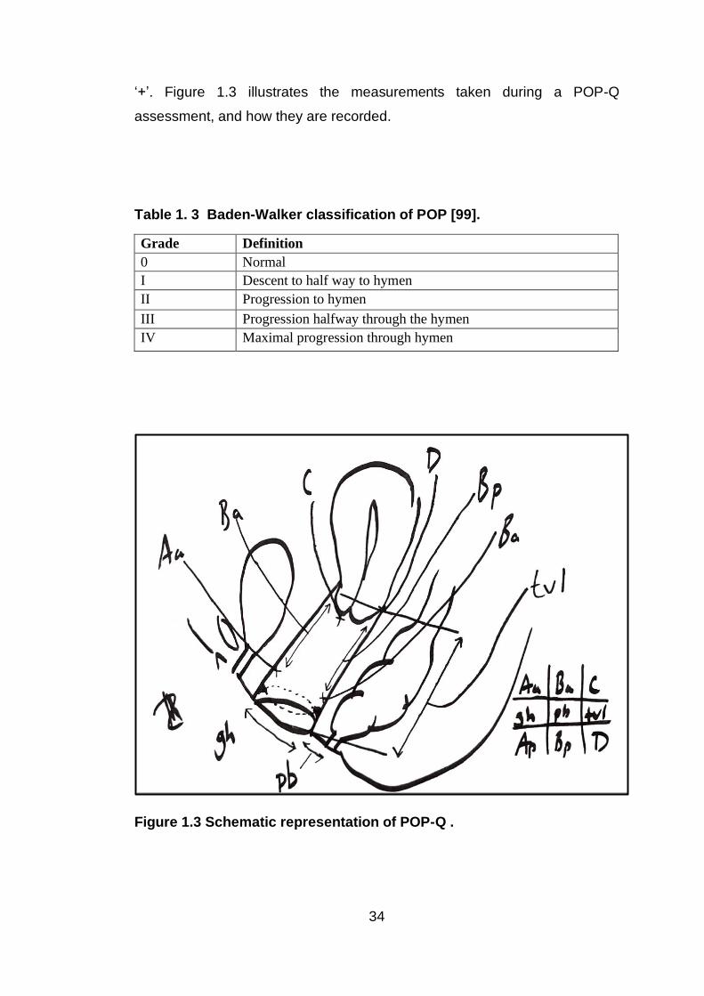
‘+’. Figure 1.3 illustrates the measurements taken during a POP-Q
assessment, and how they are recorded.
Table 1. 3 Baden-Walker classification of POP [99].
Grade Definition
0 Normal
I Descent to half way to hymen
II Progression to hymen
III Progression halfway through the hymen
IV Maximal progression through hymen
Figure 1.3 Schematic representation of POP-Q .
35
1.4.2 Patient reported outcome measures
Patient reported outcome measures (PROMs) are questionnaire-based tools
that enable a more objective assessment of symptom status, among other
measures [106]. They are defined as ‘a series of questions that patients are
asked in order to gauge their views on their own health’ [107]. They may be
generic such as the EQ-5D™ or system specific such as the ICIQ-VS, and
their utility varies. They may be used by healthcare systems to measure
efficiency, quality of care and cost efficacy, by researchers to measure efficacy
of an intervention both in terms of QoL and symptom status, and finally on an
individual patient basis to help target consultations, identify and quantity
symptoms by screening or to measure changes in health related QoL and
symptoms following an intervention [107]. Another way of framing these
distinctions within the field of PROMs are economical, clinical and humanistic
driven measures [108].
With respect to the assessment of POP, particularly in the research setting,
the PROMs available have been appraised by ICI, with a number of such
questionnaires being supported by Grade A evidence, shown in Table 1.4 [86].
The rationale for the choice of PROMs utilised within some of the studies
contained within this thesis are explained within the individual methodology
sections (Chapters 2.3.2, 3.3.2, 4.3.2, and 5.3.2). It must be noted, that for
some studies, the use of a validated PROM is not always possible. In such
situations some form of validation study is helpful, although not always
feasible, particularly in the case of rare outcomes, such as those detailed
within Chapter 2.3.2.
Patient Global Impression in Improvement (PGI-I) is another PROM that has
been utilised to assess outcomes after pelvic floor surgery. Rather than
focussing on specific symptoms and their quantification of various component
parts, such global indices offer a simpler and easier measure that is
transferable across different conditions and research settings [109]. A
shortcoming of such a measure is that it is difficult to ascertain which
component of patients symptom and health complex leads to a specific score

36
[110]. Yet they have been shown to have good repeatability in individuals and
offer a unique measure of patient perception of outcome [110, 111]. The PGI-
I measure has been used for incontinence, as well as having been validated
for women with prolapse [109, 112]. Srikrishna et al. undertook this by
assessing construct validity against other validated measures such as POP-Q
and validated QoL scores (prolapse quality of life - pQoL) [112].
Table 1.4 Evidence base for PROMS used for POP [86].
Grade of
supporting
evidence
Patient reported outcome measure
A Pelvic Floor Distress Inventory (PFDI)
Pelvic Floor Impact Questionnaire (PFIQ)
Prolapse Quality of Life Questionnaire (P-QoL)
Pelvic Organ Prolapse Urinary Incontinence Sexual Questionnaire
Pelvic Organ Prolapse Urinary Incontinence Sexual
Questionnaire –IUGA Revised (PISQ-IR)
ICIQ vaginal symptoms questionnaire (ICIQ-VS)
B The Austrian Pelvic Floor Questionnaire (AFPQ)
Pelvic Floor Symptom Bother Questionnaire (PFBQ)
Electronic Personal Assessment Questionnaire-Pelvic Floor (ePAQ-
PF)
C Pelvic Floor Dysfunction Questionnaire
Danish Prolapse Questionnaire
37
1.4.3 Other diagnostic tests
With respect to the assessment of POP, there a range of imaging modalities
exist to augment clinical examination, yet it must be noted that vaginal
prolapse remains a clinical diagnosis. The use of ultrasound assessment to
quantify POP and assess the pelvic floor muscles has been relatively well
studied [113]. However, there is no consensus on the standardisation of
assessment, and a paucity of data to illustrate an advantages over and above
clinical assessment. On this basis, the latest recommendations from the ICS
advise against the routine use of ultrasound for the assessment of POP [86].
Magnetic resonance imaging has also entered into relatively routine clinical
practice, predominantly for those with recurrent POP, those complaining of
POP in the absence of positive clinical findings, and in women with concurrent
defaecatory dysfunction [114]. It has also been used along with computer
modelling to further the understanding of mechanisms that underpin the
anatomical and functional support of the pelvic organs, as well as failure of
these mechanisms and pathology related to POP [115-117]. As with
ultrasound, the evidence to support more widespread use of MRI is lacking,
leading to a relatively recent recommendation against routine use [86].
In addition to diagnostic tests for POP, there may be a role for investigation of
common concurrent symptoms of bladder and bowel dysfunction, or pelvic
floor muscle tone and neurological function. For example, it is well recognised
that more than 50% of women with POP have symptoms of obstructive
defaecation syndrome (ODS) such as a feeling of straining and incomplete
emptying [118, 119]. Imaging of these symptoms in the form of dynamic
proctography, along with vaginal, bladder , and intestinal contrast medium, has
been recommended by a recent consensus statement from the European
Society of Coloproctology [120]. Traditionally such patients would be assessed
through the use of fluoroscopic defaecography, utilising plain film ionising
radiation in conjunction with a radio-opaque enema, to study ano-rectal
physiology during defaecation. However this involves radiation, global pelvic
floor assessment is limited, and detailed soft tissue imaging as well as that of

38
other pelvic organs is limited by the nature of the test, there are also
recognised issues with over-diagnosis in health volunteers and significant
inter-observer variability [121, 122]. The use of MRI has been the next
evolution in pelvic floor assessment, particularly in those with ODS. It allows
higher anatomical detail, the ability to more easily assess other compartments
and surrounding structures in a dynamic fashion, and the ability to identify
enteroele and levator ani herniation in a superior fashion to fluoroscopic
imaging [123, 124]. MRI proctography has been shown to have utility in
determining the underlying pathology behind such symptoms, potentially
guiding conservative therapies and informing the surgical approach to pelvic
floor reconstruction [125, 126]. More recent comparative data suggest both
modalities have specific advantages and that ultrasonographic techniques
may confer patient acceptability advantages to both approaches [127].
As outlined in Section 1.3.5, there is a high rate of concurrent LUTS in patients
with POP. To guide the assessment and preoperative decision making of
these symptoms, there has been an attempt to understand bladder function
more objectively and various authors have explored the role of invasive
preoperative UDS [128-130]. This involves the placement of catheters and
transducers into the bladder and/or other body cavity (commonly the rectum)
to allow a direct assessment of lower urinary tract function by assessment of
physiological parameters, namely pressure changes within the bladder and
patient symptoms [131]. The practicalities of urodynamic testing and
parameters measured are discussed in more detail as part of the methodology
discussion within Chapter 4.3.1.
Analysis of data from the CARE trial found wide variation in the prognostic
value of office as well as invasive bladder function testing in patients
undergoing prolapse surgery [132]. Reflecting concerns about the utility of
such preoperative UDS, for women undergoing surgery for POP and reporting
SUI, the last decade has seen their routine use amongst UK
urogynaecologists fall from 70% to 9% [5]. Yet there remains wide variation
in practice, the same survey study showed that 36% of special interest

39
urogynaecologists would perform routine preoperative urodynamics for any
women with uterovaginal prolapse, versus 13% amongst subspecialists and
9% amongst generalist gynaecologists. The latest guidance from NICE
manifests the lack of evidence to support a consensus in practice, suggesting
there is a role for such investigations [133]:
‘Consider investigating the following symptoms in women with pelvic organ
prolapse:
• Urinary symptoms that are bothersome and for which surgical
intervention is an option.’
To date, no studies have been undertaken to determine the role UDS may
have in predicting postoperative bladder function for those undergoing LSH.
1.5 Management of pelvic organ prolapse
The management of POP is generally considered to utilise an escalating step-
wise approach from the least to most invasive interventions. These are
therefore presented and discussed in this order. For ease of comparison of
these interventions, Table 1. 5 presents the features of the key studies
referenced within this thesis, with the exception of laparoscopic mesh
sacrohysteropexy, for which the primary studies are presented in Table 1. 7.
1.5.1 Non-surgical interventions for pelvic organ prolapse
The first line intervention for the treatment of POP is PFMT, a form of physical
therapy which is more broadly defined as ‘the exposure to training load or a
work stress, of appropriate intensity to produce a noticeable or measurable
training effect’ [134]. Such exercises target increased muscle volume and
structural support, as well as preventing organ descent on straining [135].
While there is significant heterogeneity amongst included studies with respect
to the specific nature of the PFMT regimens used, the protocol from the large
POPPY study by Hagen et al. best illustrates the specifics of a PFMT regimen
in the context of UK physiotherapy practice [136]. The study involved a one

40
to one physiotherapy programme with appointments at 0, 2, 6, 11, and 16
weeks, with individualised modifications of the home regimen according to
patient needs. The ultimate aim was for women to achieve ten times ten-
second pelvic floor muscles holds and up to 50 fast contractions, three time
per day, augmented by the use of a diary. The study found that after 12
months, 76% of participants required no further treatment and 57% of women
reported their prolapse symptoms to be better, although 70% still reported the
feeling of something coming down in the previous 4 weeks. A randomised
study by Wiergersma et al. similarly found that at three months, 57% of
patients undergoing PFMT reported improvement in symptoms [137].
The routine and first line use of PFMT is supported in the UK by
recommendations from the National Institute for Health and Care excellence
(NICE) [133]. A large systematic review and meta-analysis of 13 studies with
2,340 women found that PFMT was associated with subjective improvement
in prolapse symptoms as well as objective improvements in severity of POP
[138]. These findings were similar to an older Cochrane review that concluded
PFMT may improve prolapse stage and muscle function [139]. Both papers
also reported that exercises were associated with improvements in concurrent
bladder dysfunction.
A variety of other physical therapy interventions have been subject to varying
degrees of academic study. These include biofeedback, vaginal manometry
and electrical stimulation. There is limited consensus on how these
interventions may be used, and therefore more extensive discussion of these
conservative modalities is beyond the scope of this thesis.
For women who are unable or unwilling to undertake PFMT, or for whom it has
failed, vaginal pessaries offer an additional non-surgical alternative. They are
‘devices inserted into the vagina to provide structural support to one or more
of the descending vaginal compartments’ [2]. There are numerous types of
pessaries in use which either support the vagina, or act as a space-filling
device. Ring pessaries are widely used as first line due to ease of use, a wide

41
range of sizes and generally lower procurement costs [140]. With a few
adverse events and widely available, 87% of urogynaecologists in the UK
routinely use pessaries to manage POP [141]. It is normal UK practice for
women to attend on a biannual basis for review and change of pessary. The
rationale for this is to provide the opportunity for vaginal examination to
exclude vaginal erosion, however the practice is without evidence base and
utilises significant healthcare resources. Self-management in the UK
population appears viable, in keeping with practice in other countries [142].
The TOPSY trial is a large randomised trial currently recruiting to investigate
the feasibility, acceptability, and efficacy of self-management in British women
[143].
While the use of pessaries for POP forms part of IUGA and NICE guidance,
there is limited evidence supporting their efficacy and a 2013 Cochrane review
found only one randomised trial looking at their efficacy, reporting a success
rate in the region of 60% [144, 145]. Data from a number of large
observational studies illustrate similar efficacy of between 41% and 68%, up
to a maximum of 12 months [84, 146, 147]. This appears to drop to as low as
28% over time, based on a study with five year follow-up [148]. With a
significant proportion of women reporting ongoing symptoms following either
PFMT or pessary use, progression to surgical intervention is therefore
relatively common.
1.5.2 Surgical interventions for pelvic organ prolapse
There are three predominant, compartment-dependent classifications of POP.
This thesis focuses on uterine prolapse; that is descent of the middle
compartment in women with a uterus, and therefore the surgical options
discussed are limited to those applicable to this form of POP. The choice of
surgical procedure undertaken generally tends to depend on the training and
preferences of the operating surgeon, in combination with the woman’s
specific desires. Approaches may involve hysterectomy or uterine
conservation, an abdominal or vaginal approach, and either native tissue or
mesh augmented surgery. As the procedure subject to investigation within

42
this thesis, LSH is discussed in more detail separately from the other
procedures, in Chapter 1.6.
Vaginal hysterectomy remains the preferred surgical procedure in UK practice
for the treatment of uterine prolapse, favoured by 75% of respondents to a
recent British Society of Urogynaecology (BSUG) survey [5]. The majority of
clinicians undertake a concurrent fixation of the vault to the uterosacral
ligaments, followed by a McCall culdoplasty and then sacrospinous ligament
fixation, in an attempt to reduce the risk of PHVP. This is a practice
recommended in the UK by the Royal College of Obstetrics and Gynaecology
(RCOG) [149]. Sacrohysteropexy remains the second most commonly
preferred procedure (10%), followed by sacrospinous hysteropexy (8%), then
subtotal hysterectomy with sacrocervicopexy (4%), and finally a Manchester
repair (2%) [5]. Similar surgical practices have been noted amongst clinicians
in the Antipodes [150]. Colpocleisis or vaginal obliterative equivalents also
exist, for those unable to undergo invasive surgery and with no intention of
ever having penetrative vaginal intercourse.
Vaginal Hysterectomy and apical suspension
Despite being a longstanding procedure at the disposal of gynaecologists, the
efficacy of VH in treating POP is difficult to determine. There are few
randomised studies of the procedure, significant heterogeneity amongst
outcome measures, and many cohort studies have not used validated
PROMs. This is compounded by the range of practices with respect to vault
suspension. The latest guidance from NICE quotes that at one year following
VH; 35% of women remain symptomatic for POP and there is a 1-10% risk of
recurrent POP [151]. However, the evidence to support these estimated
failure rates is of low quality.
The largest randomised study involving VH and apical suspension was the
OPTIMAL trial with outcome data at five years, however it also included
women with vault prolapse, randomising women with apical prolapse
undergoing vaginal repair to either uterosacral ligament suspension (ULS) or
43
sacrospinous ligament fixation (SSLF) [8]. At five-years the failure rates in
both arms were high, 11.9% of women after ULS and 8.1% of women after
SSLF had undergone retreatment for POP. The ‘surgical failure’ rate based
the study’s primary outcome which was defined as the patient reporting
reoperation for POP, recurrent symptoms of POP or being found to have
moderate prolapse of any of the three compartments, was 61.5% after ULS
and 70.3% after SSLF. Unfortunately, subgroup analysis was not undertaken
of those who underwent VH versus those who had a procedure for PHVP.
More recently, the Vault or Uterine prolapse surgery Evaluation (VUE) study
has reported its initial 12 month follow-up, and one arm within this study looked
at the management of uterine prolapse [152]. The comparative data from this
study is discussed in detail in Chapter 3. In this large multicentre RCT with
563 women in the uterine prolapse arm, 238 women underwent a VH with an
apical suspension procedure and follow up data at 12 months was reported.
The primary outcome was based on symptoms of prolapse from the Pelvic
Organ Prolapse Symptom Score (POP-SS) PROM, and following VH 79% of
women continue to have some form of prolapse symptom, of which 28.9%
reported an ongoing feeling of ‘something coming down’. This would equate
to a cure rate of just 21% with respect to any prolapse symptoms as a marker
of surgical success. On clinical examination, 34.1% of women continued to
have stage 2b prolapse (leading edge at or 1cm beyond the hymen) or more,
and 4.5% of women underwent subsequent surgery within one year of which
1.2% underwent a subsequent apical procedure.
A large observational cohort study of 94 women with follow-up eight years after
VH found that 90% (n=84) remained asymptomatic for POP, 90% (n=85)
required no subsequent apical support procedure and 95% (n=67) of the 70
women who were examined had no significant prolapse (Grade 1 or lower)
[153]. Patients were simply asked about the presence of ‘prolapse’ symptoms
for this study, as opposed to using a validated symptom questionnaire, which
may explain the high rates of symptom relief as compared to more rigorously
designed studies. A Finnish study reported that 83% (n=105) of women were

44
asymptomatic for symptoms of a bulge at 12 months using a non-validated
questionnaire, authors also reported significant improvements in validated
generic measures of health associated QoL [154]. A large study from Sweden
found that 81% (n=620) of women remained asymptomatic for prolapse at six
months postoperatively, however a non-validated PROM was used and
‘symptoms of heaviness or pressure’ was used as a marker for symptomatic
POP [155].
A randomised controlled trial (RCT) comparing vaginal and abdominal
hysterectomy for prolapse, with 41 women in the VH arm, found that at one-
year postoperatively, only 12% of patients visited a doctor with symptoms of
prolapse [156]. This would suggest that 88% of patients were cured of
prolapse. The same group published eight year follow up, reporting that 87%
of patients reported improved prolapse symptoms compared to before primary
surgery, although this was only published in the form of a conference abstract,
and therefore should be interpreted with caution [157]. Finally, a recent large
registry study from Denmark published data for 4045 women at a median
follow up period of 34 months (range 1 to 90 months) [158]. Following VH, the
risk of reoperation for prolapse at five years was 11%, corroborating the 85-
90% efficacy rates based on reoperation reported within the other studies that
have been highlighted.
Further discussion of comparative studies between LSH and VH are presented
in Chapter 1.6.2. To summarise the data for VH alone, there are few
randomised studies, with significant heterogeneity of outcome measures for
those that do exist. Higher quality studies appear to suggest significant rates
of ongoing symptoms of POP following surgery and many data are derived
from single cohort studies, often retrospective in nature. This evidence would
appear to suggest while VH with apical suspension remains in clinical practice
the ‘gold standard’, it may be associated with failure rates of at least 30% when
this is defined as recurrent symptoms or anatomical prolapse. Generally,
short-term reoperation rates are at least 10%, although the quality of evidence
underpinning this remains low. This points to a need for further research of

45
VH, but also justifies the consideration of alternative surgical procedures for
uterine prolapse, such as LSH, as a valid and worthy area in need of clinical
research.
Sacrospinous hysteropexy
Another procedure used to treat uterine prolapse is sacrospinous hysteropexy,
which in contrast to hysterectomy involves conservation of the uterus. This
involves uterine suspension to the sacrospinous ligaments, usually through a
vaginal approach utilising the right sided ligament and was first described in a
case series of five patients in 1989 [159]. The largest procedure specific
randomised study by Detollenaere et al. had over 103 women in the
hysteropexy arm [160]. They reported the procedure was noninferior to VH,
finding that at 12 months none of the women had symptomatic apical POP. A
meta-analysis by Kapoor et al. has also compared the two procedures [161].
They reported no significant difference in either the primary outcome of apical
failure (six studies, 651 women, 25/293 vs 17/358, odds ratio (OR) 2.08; 95%
confidence interval (CI) 0.76–5.68), nor in the risk of repeat surgery for
prolapse (five studies, 581 women, 10/251 vs 12/330, OR 0.99; 95% CI 0.41–
2.37). Various laparoscopic approaches have also been described and one
cohort study with a median 12 month follow up reported an 80% cure rate in
43 women [162].
An observational study of 99 women who underwent a mesh augmented
variant of sacrospinous hysteropexy, using a branded device inserted
vaginally (Uphold®; Boston Scientific Corporation), reported a one year cure
rate of 96% using a composite outcome of anatomical POP and the presence
of symptoms [163]. However as detailed in Chapter 1.7, the availability of
such transvaginal mesh augmented approaches for POP are now relatively
limited.
In contrast to the positive findings outlined in previous studies, the recent
large Danish registry by Husby et al. reported the failure rate of sacrospinous
hysteropexy at a median of 43 months (range 1-90) in 416 women to be 30%,

46
comparing poorly with reoperation rates of 7% and 11% after Manchester
procedures and VH respectively [158]. There are other limitations and
considerations when using sacrospinous hysteropexy including issues with
buttock pain, vaginal length, risk of further endometrial pathology, and the
implications of uterine volume [161].
Abdominal hysterectomy and vault suspension
A further surgical alternative for uterine prolapse is abdominal hysterectomy,
either laparoscopic or open, with concurrent mesh cervicopexy or
sacrocolpopexy (SCP), which both involve suspension of the residual vaginal
apex to the sacral promontory using mesh. The predominant issue with this
intervention is the high rate of mesh erosion into the vagina. A systematic
review by Jia et al. reported the overall risk of such a mesh associated
complication of between 4.3% and 10.5% following hysterectomy and
concurrent SCP [164]. In that review, authors considered four studies with a
total of 311 women. All were low quality; Braun et al. reported an RCT in
abstract form only, with 23 women undergoing hysterectomy and SCP versus
24 who underwent VH [165]. While no patients had POP at 33 months after
hysterectomy and SCP, which compared favourably to the 4.2% (n=1)
incidence of POP in the VH group, 4.3% (n=1) were found to have a mesh
erosion. This same group have subsequently published an RCT with 12
month follow-up following randomisation to either SCP or high uterosacral
vault suspension as a vaginal alternative [166]. This study recruited both
hysterectomised and non-hysterectomised women, and in the SCP arm 21.4%
(n=12) underwent only SCP, and 78.5% (n=44) underwent subtotal
hysterectomy and SCP. The only two mesh erosions were in previously
hysterectomised women, rather than those that underwent the procedure
concurrently. The risk of intraoperative complications was similar between the
two groups (1.9% vs. 3.6%, p=0.58). The risk of reoperation for POP was
much lower in the SCP group as compared to the hysterectomy and vault
suspension group (5.6% vs 17%, p=0.04). The authors did not undertake
subgroup analysis to compare those with or without concurrent hysterectomy.

47
Another study in the Jia et al. systematic review was by Constantini et al,
comparing hysterectomy with SCP to abdominal sacrohysteropexy, and
therefore this paper is discussed in Chapter 1.6.2 [167]. These authors
reported no failures at 51 months in this non-randomised comparative study
with 39 women in the SCP arm. A further included paper was a large case
series of 101 women by Wu et al. with 15 months follow-up [168]. Again, the
focus of this study was mesh erosions, reporting a 6.9% risk presenting at an
average of 8 months postoperatively. Other case series have reported mesh
erosion rates of 0%-6.5% and success rates of 92% to 100%, however many
have focused on mesh associated complications rather than efficacy with
respect to POP [169-171].
Finally, the systematic review included a paper by Griffis et al. that was a
retrospective study focused on mesh associated complications [172]. They
reported overall erosion rates of 8.2%; 10.5% for those undergoing total
hysterectomy and concurrent SCP compared to 3.6% following subtotal
hysterectomy and cervicopexy. This suggests preservation of the cervix may
confer a benefit with respect to safety, however there were only 88 women in
this study, 28 of whom were in the cervicopexy group and the average follow
up was only 13 months. A study by Crane et al. of robotic assisted
sacrocoplopexy also explored a possible advantage to utilising a subtotal
hysterectomy technique [173]. They reported a mesh erosion rate of 3.8%
(3/79) following total hysterectomy and SCP, and 0% (0/33) following subtotal
hysterectomy and cervicopexy. However, this was a single centre
retrospective study with follow-up limited to six weeks. On the basis of these
studies, the safety advantages of the subtotal approach should be interpreted
with caution.
Manchester-Fothergill procedure
A further vaginal approach to managing uterine prolapse surgically is the
Manchester-Fothergill procedure, first described in 1888 and then modified in

48
1915 [174]. It involves amputation of the cervix, with plication of the
uterosacral ligaments and fixation to the remaining cervico-uterine body. The
procedure was commonly used for many years; however, it fell out of routine
use over the last two decades. This has resulted in there being limited
available efficacy data, and the ICS to deem the procedure as having a limited
role in contemporary practice [86]. A retrospective case series of 203 women,
with case note review at a median 3.6 years postoperatively reported the risk
of recurrent uterine prolapse to be as low as 3.9% [175]. Another retrospective
study comparing the procedure to VH found that in the fifty patients who had
follow up at one year, none had recurrent prolapse on clinical examination of
the middle compartment [176]. However, 58% of the women had stage 2
prolapse of one other compartment, and there was no mention of the number
of women symptomatic for POP.
With concerns about mesh use, and increasing trends towards a desire for
uterine sparing approaches, there is renewed clinical interest in the procedure.
The previously mentioned large Danish registry study by Husby et al
compared the procedure to sacrospinous hysteropexy and VH, concluding that
it had comparatively low reoperation rates and the authors called for further
study [158]. At one year, 3% of the 2,786 women who underwent a
Manchester Fothergill procedure had undergone a subsequent operation for
prolapse which was comparable to the reoperation rate following VH.
However, at 5 years this rate was 7%, considerably lower than the 11%
reoperation rate found following VH at the same length of follow-up.
Colpocleisis
Colpocleisis, meaning ‘vaginal closure’, is an obliterative rocedure generally
reserved for elderly women who have been deemed unfit for major surgery
due to anaesthetic risks and comorbidity. Sexual intercourse is not possible
following the procedure, restricting the appropriateness of the procedure to a
specific patient group. Multiple cohort studies have shown high rates of
satisfaction and improved QoL, with low rates of surgical regret [177, 178]. A
large multicentre study with 153 women at one-year follow up reported that
49
95% of patients reported being ‘satisfied’ or ‘very satisfied’[179]. A prospective
cohort study with unmatched controls undergoing vaginal reconstructive
surgery found no difference between the groups with respect to bladder, bowel
and prolapse symptoms, and improved QoL in both groups using the validated
Short Form – 36 (SF-36) PROM [180]. A review article from 2006 summarised
the available published literature and reported that colpocleisis was associated
with a near 100% success rate, albeit from a group of historical studies with
highly heterogeneous definitions of success [181]. The authors concluded that
the procedure was associated with a 4% risk of major complications
attributable directly to the surgery, such as pyelonephritis and haemorrhage,
and a 1% risk of death. However, this cohort would tend to be high risk due
to patient selection and therefore the AE profile must be interpreted with
caution and needs further prospective study.
Vaginal mesh kits
Following the success of mesh kits for the treatment of SUI, there was a
proliferation of branded devices that utilised mesh to augment apical
suspension and colporrhaphy procedures. However, many of these devices
have been subsequently withdrawn from the market and in UK practice they
are not available. The data to support these devices was scanty and the
shortcomings of the FDA regulatory process that led to their approval is
discussed in Chapter 1.7.1, as well as having been discussed in a systematic
database review by Heneghan et al. [182].
Maher et al. performed a systematic review for the Cochrane collaborative and
identified six randomised controlled trials have been undertaken comparing
native tissue versus mesh augmented devices for the treatment of apical
prolapse [183]. Four of these studies looked at the Prolift™ device (Ethicon
Inc., Somerville, NJ, USA), a monofilament polypropylene weave product. The
randomised studies of these devices are small with between 32 and 94
patients in the mesh augmented arms [184, 185]. Most only had twelve
months follow-up and the systematic review concluded there was no
advantage over native tissue with respect to either objective anatomical

50
outcomes, reoperation or validated symptom scores [183]. However, mesh
erosion rates averaged at 18%, with reoperation rates for mesh erosion at
9.5%.
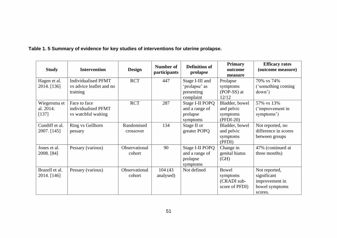
51
Table 1. 5 Summary of evidence for key studies of interventions for uterine prolapse.
Study Intervention Design Number of
participants
Definition of
prolapse
Primary
outcome
measure
Efficacy rates
(outcome measure)
Hagen et al.
2014. [136]
Individualised PFMT
vs advice leaflet and no
training
RCT 447 Stage I-III and
‘prolapse’ as
presenting
complaint
Prolapse
symptoms
(POP-SS) at
12/12
70% vs 74%
(‘something coming
down’)
Wiegersma et
al. 2014.
[137]
Face to face
individualised PFMT
vs watchful waiting
RCT 287 Stage I-II POPQ
and a range of
prolapse
symptoms
Bladder, bowel
and pelvic
symptoms
(PFDI-20)
57% vs 13%
(‘improvement in
symptoms’)
Cundiff et al.
2007. [145]
Ring vs Gellhorn
pessary
Randomised
crossover
134 Stage II or
greater POPQ
Bladder, bowel
and pelvic
symptoms
(PFDI)
Not reported, no
difference in scores
between groups
Jones et al.
2008. [84]
Pessary (various) Observational
cohort
90 Stage I-II POPQ
and a range of
prolapse
symptoms
Change in
genital hiatus
(GH)
47% (continued at
three months)
Brazell et al.
2014. [146]
Pessary (various) Observational
cohort
104 (43
analysed)
Not defined Bowel
symptoms
(CRADI sub-
score of PFDI)
Not reported,
significant
improvement in
bowel symptoms
scores.

52
Abdool et al.
2011. [147]
Pessary (various) vs
surgery
Cohort study
(retrospective)
181 Not defined Pelvic floors
symptoms
(SPS-Q)
65% vs 69%
(‘awareness of a
lump’)
Lone et al.
2015. [148]
Same cohort as Abdool et al. 2011.
Jelovsek et al.
2018. [8]
USL vs SSF (with or
without VH)
RCT 244 Stage II or
greater POPQ
Failure was any
of 1)POPQ C
descent >1/3
TVL,
2)prolapse
beyond the
hymen,
3)prolapse
symptoms or
4)further
intervention for
prolapse
38.5% vs 29.7% (as
per primary outcome
measure)
Hemming et
al. 2020.
[152]
VH vs uterine sparing
(various)
RCT 563 ‘required
surgery’
POPSS 21% vs 22.6% (‘Any
prolapse symptoms’)
Prodigalidad
et al. 2012.
[153]
VH Cohort study
(retrospective)
94 Not defined Not defined 89.4% (‘sensation of
prolapse’)
Humalajarvi
et al. 2014.
[154]
Hysterectomy (various)
for those with and
without POP
Cohort study 322 (102
with POP)
Stage II or
greater POPQ
Non-validated
questionnaire
83.3% (‘sense of
bulging’)
Pakbaz et al.
2009. [155]
VH Population
cross-sectional
study
682 Not defined Perioperative
outcomes and
81.3% (‘sensation of
vaginal heaviness of
pressure’)

53
non-validated
questionnaire
Roovers et al.
2003. [156]
VH vs open
sacrocolpohysteropexy
RCT 82 Not defined UDI 95% vs 95% (apical
prolapse </= Stage II)
Husby et al.
2019. [158]
VH vs vaginal
hysteropexy vs MFP
Population
cross-sectional
study
7247 Not defined Reoperation for
apical prolapse
11% vs 30% vs 7%
(apical re-operation)
Detollenaere
et al. 2015.
[160]
Sacrospinous
hysteropexy vs VH
RCT 208 Stage II or
greater POPQ
Stage II or
greater POPQ
point c (apex)
100% vs 96%
(‘symptom of bulge’
or reoperation)
Maher et al.
2001. [162]
Laparoscopic suture
hysteropexy
Cohort study 43 ‘symptomatic
uterine prolapse’
‘symptoms of
prolapse’
81% (primary
outcome measure)
Jirschele et
al. 2015.
[163]
Mesh augmented
vaginal hysteropexy
Cohort study 99 Stage II or
greater POPQ
Composite
POPQ Ba and C
and ‘feeling of
bulge in vaginal
area’
97% (primary
outcome measure)
Rondini et al.
2015. [166]
Vaginal high
uterosacral vault
suspension vs SCP with
or without
hysterectomy
RCT 110 Stage II or
greater POPQ
Stage II or
greater POPQ
point c (apex)
82% vs 100% (apical
prolapse </= Stage II
POPQ)
Marinkovic.
2008. [169]
TAH with concomitant
SCP
Cohort study
(retrospective)
64 Stage II or
greater POPQ
Mesh erosion 95% (any Stage II or
greater POPQ)
Karaman et
al. 2015.
[175]
MFP Cohort study
(retrospective)
24 Stage I-III
uterine prolapse
Not defined 96% (any Stage II or
greater POPQ)

54
De Boer et al.
2009. [176]
MFP vs VH Cohort study
(retrospective)
98 Not defined Not defined 100% vs 96%
(recurrent apical
prolapse)
Hullfish et al.
2007. [177]
Colpocleisis Cohort study 40 Not defined Goal attainment
(non-validated)
95% (‘satisfied’ /
‘very satisfied’)
Vij et al.
2014. [178]
Colpocleisis Cohort study
(retrospective)
34 Not defined P-QoL 92% (recurrent
clinical prolapse)
Fitzgerald et
al. 2008.
[179]
Colpocleisis Cohort study 132 Stage III or
greater POPQ
Not defined 73% (any Stage II or
greater POPQ)
Sokol et al.
2012. [184]
Mesh augmented
colpopexy device vs
anterior colporrhaphy
RCT 65 Stage II or
greater POPQ
Stage II or
greater POPQ
37.5% vs 30.3%
(primary outcome
measure)
Silveira et al.
2015. [185]
Mesh augmented
colpopexy device vs
colporrhaphy
RCT 184 Stage III or
greater POPQ
Stage II or
greater POPQ
92.1% vs 80.3%
(primary outcome
measure)

55
1.6 Mesh sacrohysteropexy
This procedure preserves the uterus. The reasons for women electing for
uterine conservation to treat uterine prolapse are varied; it may be because
of a patient desire for uterine conservation, other personal reasons or a wish
to maintain the potential for further pregnancies. That being said, women are
generally encouraged to defer surgery until their family is complete due to the
additional adverse defects of a pregnancy on the pelvic floor and compromised
of surgical repair. Alternatively, it may be the surgeon’s preferred procedure
due to potential advantages such as anatomical outcomes outlined in Chapter
1.6.2. Regardless, in the UK patients are routinely offered a number of surgical
choices with varying levels of supportive evidence with respect to safety and
efficacy, in keeping with best practice and the latest guidance from NICE [133].
Contraindications to sacrohysteropexy include uterine or endometrial
pathology such as cervical dysplasia, abnormal uterine bleeding, large uterine
fibroids or fibroids located where peritoneal dissection occurs, and certain
uterine anomalies.
1.6.1 Technique
While techniques continue to evolve, there are broadly two approaches to
LSH. Some authors advocate the use of a sheet of mesh, which following
dissection of the rectovaginal fascia is attached to the posterior vaginal wall
and posterior cervix [186]. However, one of the principal advantages of an
abdominal approach is the opportunity to avoid the placement of mesh in close
proximity to, or on, the vaginal wall. Such an approach is known to be
associated with a risk of vaginal erosion as described in further detail in
Chapter 1.7.3. Therefore, the technique studied within this thesis is known
as the ‘wrap round’ hysteropexy, the details of which are explored further
below. The theoretical advantage of the ‘wrap round’ approach is a reduced
or negligible risk of vaginal mesh erosion due to the site of its placement. The
distinction between these approaches is important, and therefore this is
highlighted when discussing outcome data below.
56
The contemporary LSH is the result of evolution over the last 150 years,
illustrated in Table 1. 6 . Howard A. Kelly first described a number of cases of
uterine suspension in 1887 in a procedure termed hysterorrhaphy, whereby
the uterus was fixed to the anterior abdominal wall utilising peritoneum [187].
The earliest sacral fixation was described by Arthure and Savage in 1956,
using an abdominal approach and fixing the uterus by the posterior uterine
corpus or fundus [188]. In 1979, SK Chaudhuri described the use of the
external oblique muscle to fashion a sling for the uterus and in 1993, Andrew
Farkas published results of the first use of a synthetic material as a fixation
device, utilising Goretex mesh to secure the uterus to the sacrum [189, 190].
The closest precursor to the technique studied within this thesis was that
described by Cutner et al. in 2007, where a mersilene tape was used as the
synthetic material for uterine suspension [191]. However, there was then a
move away from heavy mesh materials due to high rates of AEs, towards
lighter macroporous materials such as Prolene [14]. The approach utilised by
centres involved in the studies described within this thesis is the laparoscopic
‘wrap round’ mesh sacrohysteropexy. The technique was formally described
by Price et al. in 2010 [192].
Laparoscopic sacrohysteropexy is performed with the patient supine in
lithotomy, with standard skin preparation, surgical draping, catheterisation and
insertion of laparoscopic ports in accordance with recommendations from the
RCOG [193]. Generally, this involves 11mm umbilical and suprapubic ports,
with two 5-mm lateral ports. The peritoneum over the sacral promontory is
opened, facilitated by graspers and scissors or an energy device. This incision
is extended in a caudal direction into the pelvis, toward the rectum, following
careful identification of the ureters bilaterally. This peritoneal relaxing incision
runs medial to the right ureter, allowing it to retract further medially and away
from the operative site. The peritoneum is also mobilised at the level of the
uterosacral ligaments. At the level of the cervico-uterine junction the broad
ligament is opened bilaterally through an avascular space, using diathermy
and scissor dissection to create windows within the ligament to allow for mesh
57
suspension. The vesico-uterine peritoneum is then incised to allow for 2-3 cm
of bladder dissection caudally away from, and off the uterine corpus.
A sheet of ‘type 1’ monofilament macroporous non-absorbable mesh is cut to
provide a bifurcated implant. The two arms are passed through the broad
ligament windows bilaterally and secured to the anterior cervix with
approximately five non-dissolvable polyester 2-0 sutures. The uterus is
elevated via the long arm of the mesh with moderate tension aiming to achieve
adequate elevation to lift the uterus to approximately 8 cm above the introitus.
The mesh is secured to the anterior longitudinal ligaments over the sacral
promontory, with 5mm helical fasteners. To prevent bowel adhesions to the
mesh, the peritoneal edges are then re-opposed using either interrupted or
continuous sutures, at both the vesico-uterine dissection site as well as the
incision from the sacral promontory to the Pouch of Douglas. A vaginal
examination is then undertaken to assess the need for concurrent anterior or
posterior colporrhaphy in those patients who have been consented for such
an approach.
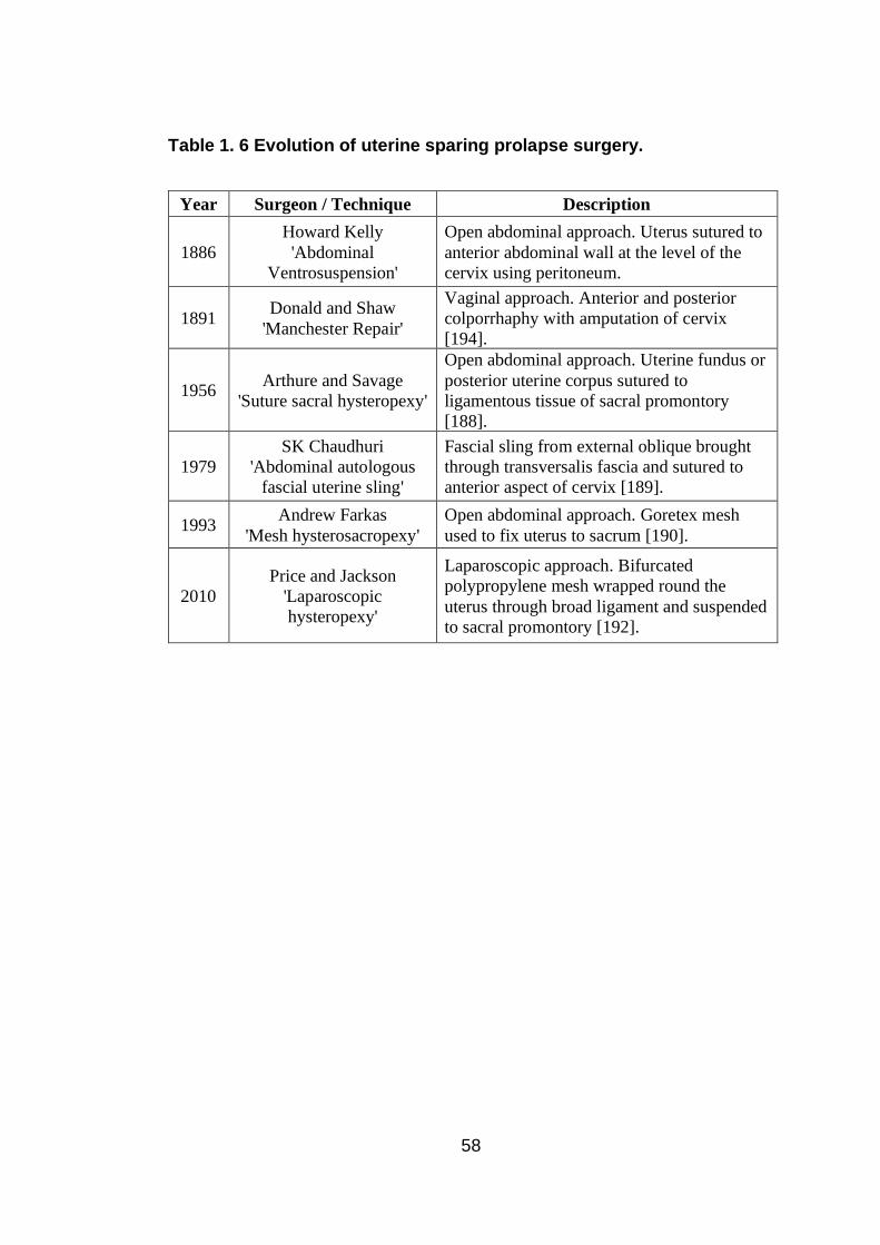
58
Table 1. 6 Evolution of uterine sparing prolapse surgery.
Year Surgeon / Technique Description
1886
Howard Kelly
'Abdominal
Ventrosuspension'
Open abdominal approach. Uterus sutured to
anterior abdominal wall at the level of the
cervix using peritoneum.
1891 Donald and Shaw
'Manchester Repair'
Vaginal approach. Anterior and posterior
colporrhaphy with amputation of cervix
[194].
1956 Arthure and Savage
'Suture sacral hysteropexy'
Open abdominal approach. Uterine fundus or
posterior uterine corpus sutured to
ligamentous tissue of sacral promontory
[188].
1979
SK Chaudhuri
'Abdominal autologous
fascial uterine sling'
Fascial sling from external oblique brought
through transversalis fascia and sutured to
anterior aspect of cervix [189].
1993 Andrew Farkas
'Mesh hysterosacropexy'
Open abdominal approach. Goretex mesh
used to fix uterus to sacrum [190].
2010
Price and Jackson
'Laparoscopic
hysteropexy'
Laparoscopic approach. Bifurcated
polypropylene mesh wrapped round the
uterus through broad ligament and suspended
to sacral promontory [192].

59
1.6.2 Review of evidence
There is relatively limited high-quality data underpinning the use of LSH,
particularly in the long term. Table 1.7 provides a summary of the peer-
reviewed and published RCTs and cohort studies available that have
assessed the outcomes following this type of surgery. These studies have
been summarised in a number of systematic reviews, with a recent meta-
analysis undertaken in 2018 by Meriwether et al [164, 183, 195, 196]. To date
only one procedure specific randomised study of LSH has been published,
reported by Rahmanou et al. in 2015 [197]. The recent VUE study compared
VH to uterine sparing procedures including LSH, however over half of women
underwent a vaginal hysteropexy and therefore this study is appraised in more
detail within Chapter 3.1[152]. Due to the procedure specificity of this thesis,
studies of uterine sparing prolapse procedures that do not involve mesh and
suspension to the sacral promontory have not been included.

60
Table 1. 7 Summary of evidence for sacrohysteropexy augmented by synthetic implant
Study Design Number of
participants
Follow-up
(months) Findings Comments
Daniels S et al. 2019.
(laparoscopic) [198]
Case series 138
1.5
(55%
attended
12/12 FU)
Subjective cure: 84%
Objective cure: 98% (1.5/12)
and 80% (12/12)
Eight women reported mesh
erosions, however authors
state this was attributable to
‘vaginally placed mesh’.
Subjective cure – heterogenous
follow up time. Non-validated
measure.
Objective cure – patients with stage
0 POP.
24/134 underwent concurrent
anterior vaginal mesh placement,
16/34 underwent concurrent
posterior vaginal mesh placement.
Lone F et al. 2018
(laparoscopic) [199]
Parallel cohort
study
125 (44 LSH,
81 VH)
12 months
(50%
further 24
months)
Subjective cure: Not reported
No difference in ICIQ-VS
scores between two groups.
Objective cure: Not reported.
No difference in point C at
3/12.
Reoperation rate (24/12):
3.6% after VH and 0% after
LSH.
No mesh complications.
Subjective cure: ICIQ VS and ICIQ
UI.
Objective cure: POP-Q at 3/12 and
reoperation rates.
Pandeva et al. 2017.
(laparoscopic –
sheet.) [186]
Case series. 144 (159
notes
reviewed)
48 Subjective cure: 81% for PGI-
I and 76% for P-QoL.
Objective cure: 95%
Reoperation rate 8%.
No mesh complications.
Subjective cure - PGI-I ‘very much
better’ or ‘much better’ or P-QoL
‘not at all’.
Objective cure - POP-Q point C =/<
0.

61
Jefferis et al. 2017.
(laparoscopic) [200]
Case series 507 (3/12 FU
for 441
patients)
3 SH Subjective cure: 94%
SH Objective cure: N/A, only
change in point C.
Reoperation for apical
prolapse: 2.8%.
0.7% risk bowel adhesions to
unperitonised mesh. No mesh
erosions.
No mesh erosions.
Subjective cure – PGI-I ‘very much
better’ or ‘much better’.
Objective cure - POP-Q.
Surgical technique altered through
series to prevent bowel adhesions
to mesh.
Kupelian et al. 2016.
(laparoscopic) [201]
Retrospective
cohort
110 31 Subjective cure: 3/12 - 94%
(PGI-I)
31/12 – 81% (asymptomatic at
telephone interview) and 96%
(PGI-I)
Objective cure: 3/12 - 98%
31/12 - 95% (5% reoperation
rate for POP)
No mesh associated
complications.
Subjective cure – PGI-I, non
validated telephone questioning.
Objective cure – POP-Q
81 women only at the 31/12
telephone follow up. Telephone
followed standardised questioning
based on validated PROM.
Grimminck et al.
2016.
(robotic) [202]
Case series 100 50 women =
60 months
50 women =
12 months)
60/12 SH Subjective cure:
N/A%
60/12 SH Objective cure: 81%
12/12 SH Subjective cure:
92% (1.5/12 only)
12/12 SH Objective cure: 98%
Mesh erosions: 4%
Subjective cure at 6 weeks - the
absence of ‘feeling a ball’
according to UDI. Objective cure -
POP-Q >2.

62
Gutman et al. 2016.
(laparoscopic) [203]
Parallel cohort
study
150 (74 lap
SH vs 75
VH)
12 SH Subjective cure: 100%
SH Objective cure: 90%
Primary outcome composite
cure rate for SH was 72%.
No difference in primary
outcome, PFDI or PGI-I data
between the two groups.
Mesh exposure: 2.7%
Primary outcome was ‘surgical
success’ = Objective cure - No
anterior or posterior wall POP
beyond hymen and point C above
mid vagina (TVL/2), all according
to POP-Q, AND
Subjective cure - Negative answer
to PFDI-20, being the absence of a
bulge.
Paek et al. 2016.
(robotic,
laparoscopic and
open) [204]
Non-randomised
comparative
study.
Retrospective.
111 (11
robotic SH,
43 lap SH
and 57 open
SH)
30 Lap SH Subjective cure: 93%
Lap SH Objective cure: 95%
(versus 100% for robotic and
91% subj. and 98% obj. for
open)
4.7% risk of reoperation for
POP after LSH.
No mesh erosions.
Subjective cure - non-validated
questionnaire.
Objective cure - POP-Q C >2.
Gracia et al. 2015.
(laparoscopic) [205]
Non-randomised
comparative
study.
45 (SH = 15,
STH – 30)
12 SH Subjective cure: 100%
SH Objective cure: 53%
100% patients reported PGI-I
improved / cured.
No mesh associated
complications.
Subjective cure - absence of
prolapse symptoms according to
question 35 of EPIQ.
Objective cure - POP-Q C <2.
Rahmanou et al.
2015.
(Laparoscopic) [197]
RCT (Lap SH vs
VH)
101 (Lap SH
= 50, VH =
50, one VH
aborted intra-
12 SH Subjective cure: 82%
SH Objective cure: 94%
Apical reoperation, cervical
elevation and TVL all
Subjective cure - PGI-I prolapse
‘very much better or much better’.
Objective cure ‘not requiring
further apical reoperation’.

63
op due to
anatomy).
favoured SH, with no
difference in ICIQ-VS or PGI-
I scores when comparing SH
and VH.
No mesh associated
complications.
Api et al. 2014.
(laparoscopic) [206]
Case series 33 6 Subjective cure: 96.9%
Objective cure: 93.9%
Mesh complications not
reported.
Subjective cure - non-validated
measure.
Objective cure – non-validated.
Rahmanou et al.
2014. (laparoscopic)
[207]
Case series 140
24 Subjective cure: 89%
Objective cure: Not reported.
9% risk of reoperation for
prolapse.
2% risk bowel adhesions to
unperitonised mesh. No mesh
erosions.
Subjective cure - PGI-I prolapse
‘very much better or much better’
Lee et al. 2013.
(robotic) [208]
Case series 15 11 Subjective cure: 80%
Objective cure: 93%
No mesh associated
complications.
Subjective cure - non-validated
Objective cure - Grade 0 Baden-
Walker.
Khursheed et al.
2013.
(open) [209]
Case series 33 6 Subjective cure: Not reported.
Objective: No recurrences
reported.
Authors did not state method of pre
and postoperative assessment of
POP and symptoms status.

64
Mesh complications not
reported.
Cvach et al. 2012.
(open) [210]
Case series with
unmatched
controls
(hysterectomy
with sacropexy).
20 17 Subjective cure: 89%
Objective cure: 100% (uterine
prolapse), 39% if all
compartments considered.
No mesh associated
complications in SH group.
Subjective cure -PGI-I
Objective cure - POP-Q
Mourik et al. 2012.
(165) 1 122-127.
(robotic) [211]
Case series 50 16 Subjective cure: 88%
Objective cure: 98%
No mesh associated
complications.
Subjective cure - non-validated
Objective cure - < Grade 2 Baden-
Walker.
Bojahr et al. 2012.
Jul;16(3):428.
(laparoscopic) [212]
Case series 19
9.3 Subjective cure: Not reported
Objective cure – 90%,
reoperation rate 6.7%
Mesh complications not
reported
Objective cure – unspecified
presence of uterine POP.
Retrospective series comparing
these women to SCP cohort.
Costantini et al.
2011. (mixed open
and laparoscopic)
[213]
Case series. 55 63.8 Subjective cure: Not reported
Objective cure: 100% (uterine
prolapse). 7.7% had >/= stage
2 cystocele and 5.7% had >/=
stage 2 rectocele. No
reoperations for POP.
Mesh erosion 3.6%
Prolapse symptoms were not
reported.
Objective cure – Cervix >6cm
above hymenal plane and < grade 2
POP in other compartments.
Moiety et al. 2010.
(open) [214]
Case series 33 6
Subjective cure: 81.8%
Objective cure: 94%
Subjective cure - non-validated
Objective cure - presence of POP-
Q > Stage 1.

65
Mesh complications not
reported.
Price et al. 2010
(laparoscopic) [192]
Case series 51 2.5 Subjective cure: Not reported.
Objective cure: 98%
Mean reduction ICIQ VS -39.
Mesh associated
complications: 3.9% (bowel
adhesions on to exposed
mesh)
Assessment of subjective
symptoms- ICIQ VS scores.
Objective cure - POP-Q C <-2
Jeon et al. 2008.
(route unspecified)
[215]
Retrospective
cohort with
unmatched
comparison to
hysterectomy with
mesh
sacrocolpopexy
and hysterectomy
with mesh
uterosacrocardinal
colpopexy
35 36 Subjective cure: Not reported.
Objective cure: 100%
Mesh complications not
reported.
Did not report on prolapse
symptoms.
Objective cure – POP-Q
Demirci et al. 2006.
(open) [216]
Case series 20 25 Subjective cure: Not reported.
Objective cure: 95%
Mesh complications not
reported.
Subjective cure - validated PROMs
to report reduced POP symptoms,
but no comparator group.
Objective cure – not specified.
Bai et al. 2005.
(route unspecified,
appears open) [217]
Retrospective
cohort
10 12 Subjective cure: Not reported
Objective cure: 100%
No mesh associated
complications.
Subjective cure with respect to
prolapse symptoms not reported.
Baden walker for objective cure.
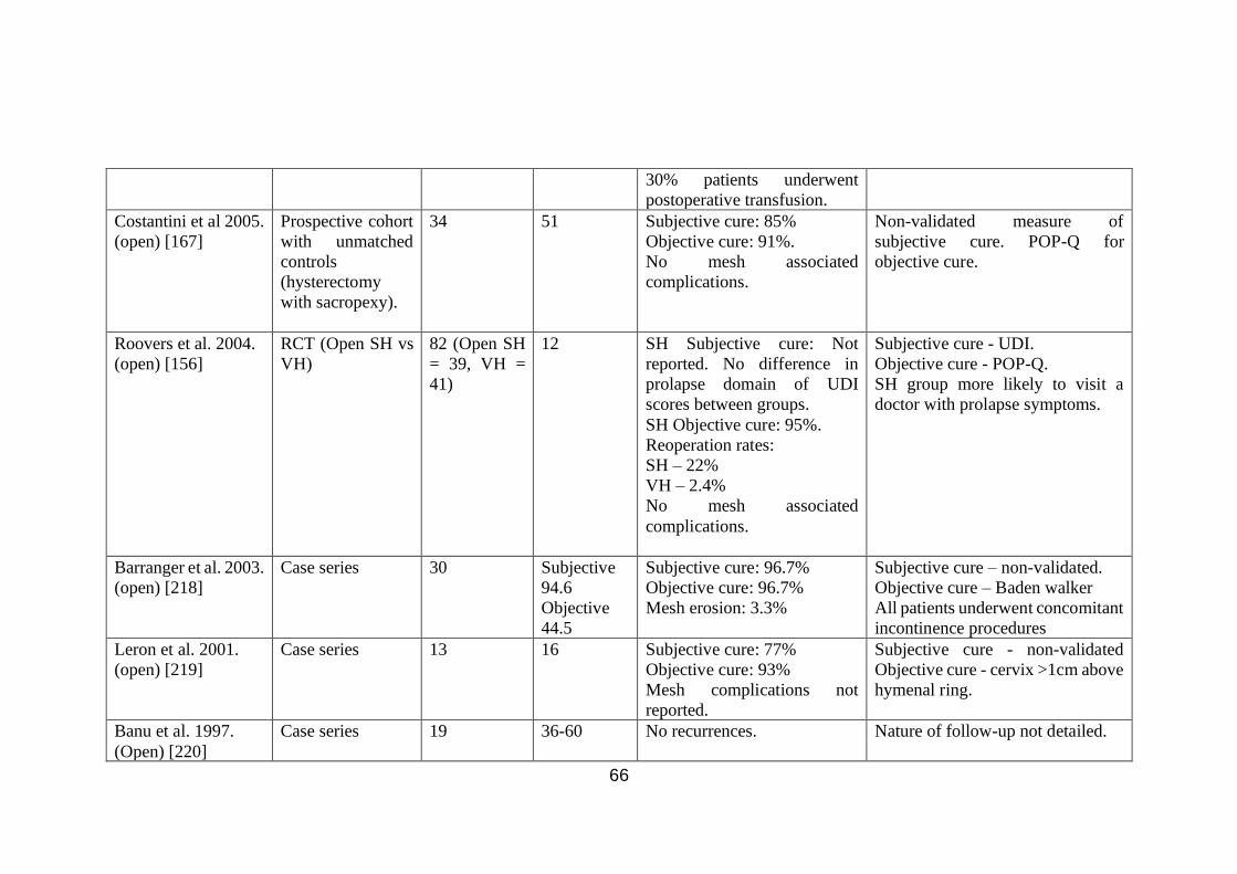
66
30% patients underwent
postoperative transfusion.
Costantini et al 2005.
(open) [167]
Prospective cohort
with unmatched
controls
(hysterectomy
with sacropexy).
34 51 Subjective cure: 85%
Objective cure: 91%.
No mesh associated
complications.
Non-validated measure of
subjective cure. POP-Q for
objective cure.
Roovers et al. 2004.
(open) [156]
RCT (Open SH vs
VH)
82 (Open SH
= 39, VH =
41)
12 SH Subjective cure: Not
reported. No difference in
prolapse domain of UDI
scores between groups.
SH Objective cure: 95%.
Reoperation rates:
SH – 22%
VH – 2.4%
No mesh associated
complications.
Subjective cure - UDI.
Objective cure - POP-Q.
SH group more likely to visit a
doctor with prolapse symptoms.
Barranger et al. 2003.
(open) [218]
Case series 30 Subjective
94.6
Objective
44.5
Subjective cure: 96.7%
Objective cure: 96.7%
Mesh erosion: 3.3%
Subjective cure – non-validated.
Objective cure – Baden walker
All patients underwent concomitant
incontinence procedures
Leron et al. 2001.
(open) [219]
Case series 13 16 Subjective cure: 77%
Objective cure: 93%
Mesh complications not
reported.
Subjective cure - non-validated
Objective cure - cervix >1cm above
hymenal ring.
Banu et al. 1997.
(Open) [220]
Case series 19 36-60 No recurrences. Nature of follow-up not detailed.

67
Mesh complications not
reported.
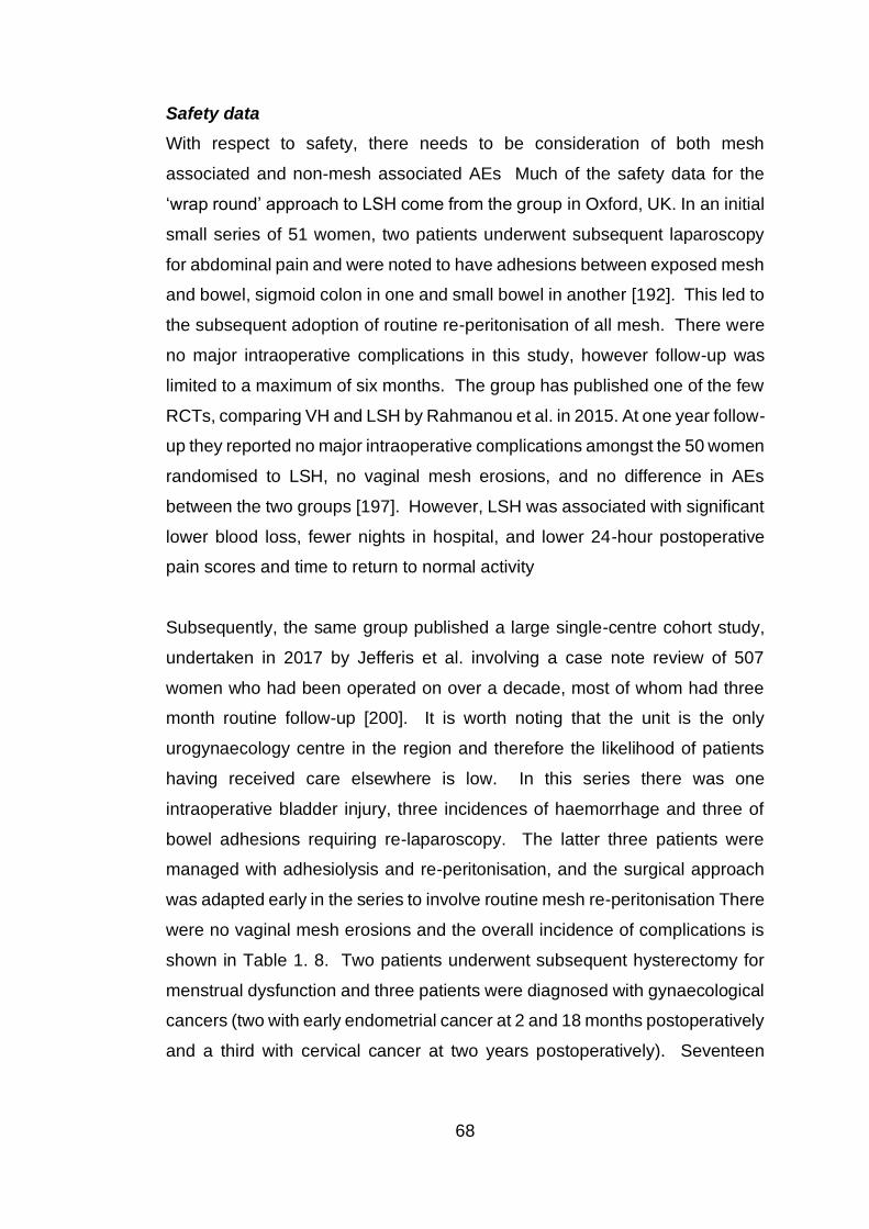
68
Safety data
With respect to safety, there needs to be consideration of both mesh
associated and non-mesh associated AEs Much of the safety data for the
‘wrap round’ approach to LSH come from the group in Oxford, UK. In an initial
small series of 51 women, two patients underwent subsequent laparoscopy
for abdominal pain and were noted to have adhesions between exposed mesh
and bowel, sigmoid colon in one and small bowel in another [192]. This led to
the subsequent adoption of routine re-peritonisation of all mesh. There were
no major intraoperative complications in this study, however follow-up was
limited to a maximum of six months. The group has published one of the few
RCTs, comparing VH and LSH by Rahmanou et al. in 2015. At one year follow-
up they reported no major intraoperative complications amongst the 50 women
randomised to LSH, no vaginal mesh erosions, and no difference in AEs
between the two groups [197]. However, LSH was associated with significant
lower blood loss, fewer nights in hospital, and lower 24-hour postoperative
pain scores and time to return to normal activity
Subsequently, the same group published a large single-centre cohort study,
undertaken in 2017 by Jefferis et al. involving a case note review of 507
women who had been operated on over a decade, most of whom had three
month routine follow-up [200]. It is worth noting that the unit is the only
urogynaecology centre in the region and therefore the likelihood of patients
having received care elsewhere is low. In this series there was one
intraoperative bladder injury, three incidences of haemorrhage and three of
bowel adhesions requiring re-laparoscopy. The latter three patients were
managed with adhesiolysis and re-peritonisation, and the surgical approach
was adapted early in the series to involve routine mesh re-peritonisation There
were no vaginal mesh erosions and the overall incidence of complications is
shown in Table 1. 8. Two patients underwent subsequent hysterectomy for
menstrual dysfunction and three patients were diagnosed with gynaecological
cancers (two with early endometrial cancer at 2 and 18 months postoperatively
and a third with cervical cancer at two years postoperatively). Seventeen
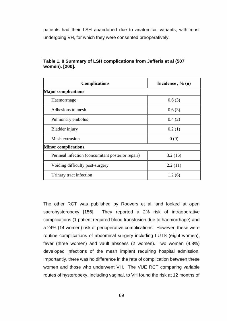
69
patients had their LSH abandoned due to anatomical variants, with most
undergoing VH, for which they were consented preoperatively.
Table 1. 8 Summary of LSH complications from Jefferis et al (507 women). [200].
Complications Incidence , % (n)
Major complications
Haemorrhage 0.6 (3)
Adhesions to mesh 0.6 (3)
Pulmonary embolus 0.4 (2)
Bladder injury 0.2 (1)
Mesh extrusion 0 (0)
Minor complications
Perineal infection (concomitant posterior repair) 3.2 (16)
Voiding difficulty post-surgery 2.2 (11)
Urinary tract infection 1.2 (6)
The other RCT was published by Roovers et al, and looked at open
sacrohysteropexy [156]. They reported a 2% risk of intraoperative
complications (1 patient required blood transfusion due to haemorrhage) and
a 24% (14 women) risk of perioperative complications. However, these were
routine complications of abdominal surgery including LUTS (eight women),
fever (three women) and vault abscess (2 women). Two women (4.8%)
developed infections of the mesh implant requiring hospital admission.
Importantly, there was no difference in the rate of complication between these
women and those who underwent VH. The VUE RCT comparing variable
routes of hysteropexy, including vaginal, to VH found the risk at 12 months of

70
a mesh erosion to be 0.4%, they did not specify whether this was an abdominal
or vaginal approach to hysteropexy [152].
The remaining safety date come from case series. Daniel et al. reports a
relatively modest complication profile with a 2.2% risk of wound infections,
2.9% risk of vaginal infection, 2.9% risk of pain and 0.7% risk of bleeding [198].
However, follow-up was limited and non-validated as shown in Table 1.7.
Authors also undertook concurrent vaginal placement of mesh and reported
that eight women (5.8%) developed vaginal mesh erosions. Lone et al. have
undertaken a parallel cohort study with 125 patients (44 LSH and 81 VH) [199].
There was no difference in the rate of AEs in the two study arms. Following
LSH there were two readmissions, one for a haematoma (2.3%) and one for a
UTI (2.3%) and two intraoperative bowel serosal injuries (4.6%). Importantly,
there were no mesh associated AEs at two years follow-up. Another recent
and large series by Pandova et al. reviewed the notes of 159 patients of whom
101 (64%) had at least two years follow-up [186]. They reported two cases of
bowel obstruction that required repeat surgery giving a risk of 1.3%. However,
it is not clear how they screened for the risk of complications and no
comprehensive review of case notes and direct questioning appears to have
been undertaken.
In a slightly smaller series utilising the ‘wrap round’ approach by Kupelian et
al., authors systematically reviewed case notes and contacted patients [201].
This study reports two serious adverse events (SAEs) including a thermal
bowel injury that was repaired intraoperatively and a small bowel obstruction
due to an exposed suture, repaired laparoscopically giving an overall risk of
SAE of 0.9%. Both patients had prolonged inpatient stays but no long-term
disability. There were no cases of vaginal mesh erosions. Nightingale et al.
undertook a similar cross-sectional study, inviting patients who had undergone
either LSH or laparoscopic SCP to attend follow-up at a median of 6 years
(range 1-9 years) [221]. Of the 112 patients included, 88 women (79%) had a
SCP for vault prolapse, which involves vaginal wall placement of mesh, and
22% of women undergoing both procedures had a concurrent ventral mesh
rectopexy, thus making comparison with aforementioned studies of LSH

71
difficult. Authors reported one case of small bowel obstruction related to a
LSH, no cases of vaginal mesh extrusion, and three patient reported
abdominal pain but it was not clear if these were following SCP or LSH.
In summary, for the ‘wrap round’ approach to hysteropexy, there does not
appear to be a risk of vaginal mesh erosions from the data that are available
[197, 200, 201]. However, failing to re-peritonise appears to carry a risk of
small bowel adhesions and associated complications. Alternative approaches
appear to carry a 0-4% risk of vaginal mesh erosion, with several case series
reporting the incidence to be between 2% and 4% as illustrated in Table 1.7
[186, 202, 203, 213].
Efficacy data
The UK’s NICE guidance conclude that there is adequate efficacy data
supporting mesh hysteropexy, provided adequate clinical governance and
audit procedures [222]. As previously noted, supporting data are limited, and
the meta-analysis by Meriwether et al. and a systematic review by Nair et al.
both report difficulties with formal statistical analysis owing to the
heterogeneity of outcome measures and surgical techniques [195, 223].
Nonetheless, the reviews demonstrate high rates of objective and subjective
success, associated with excellent patient satisfaction rates, exceeding 90%.
To date there is one non-randomised prospective controlled study and one
RCT comparing VH, the current ‘gold standard’ intervention, to ‘wrap round’
LSH. In both of these studies, by Lone et al. and Rahmanou et al. respectively,
the data demonstrated no difference in patient-reported prolapse symptoms at
one and two years after surgery when comparing LSH to VH [197, 199].
However, Rahmanou et al reported LSH was associated with a higher apical
suspension of point C (-5.4 vs -4.3, p<0.001) and a longer total vaginal length
(TVL) (8.35 vs 6.5, p<0.001), a findings supported by a recent meta-analysis
[195]. The implications of this with respect to long-term objective and
subjective outcomes remain uncertain. The risk of reoperation for apical
prolapse was lower in the LSH group (4% vs 8%), although this was not

72
statistically significant and the objective cure rate of the LSH arm was 90% at
one-year, on the basis of reoperation for POP [197]. Eighty-two percent of
women rated their symptoms as ‘much better’ or ‘very much better’ after LSH,
there was no difference between the PGI-I and ICIQ-VS between the two
groups.
In the large cohort study by Jefferis et al., the objective cure rate based on
reoperation for POP was 87.7% with a mean elevation of POP-Q point C of
7.9 cm [200]. Fourteen women (2.8%) underwent a subsequent apical
procedure and for ten of these women, this was simply a laparoscopic plication
of the in-situ hysteropexy mesh. A further 36 women (7.1%) underwent
colporrhaphy following LSH. With respect to PGI-I data, available for 404
patients, 93.8% (n=367) described their prolapse as ‘much better’ or ‘very
much better’. Findings within this study should be interpreted with caution,
follow-up was non-blinded, limited to routine short-term three month follow-up,
and the retrospective cohort study design presents inherent limitations with
respect to interpretation.
Kupelian et al. reported outcomes at three months and then by telephone
follow-up at variable timeframes with a median of 2.6 years [201]. The rate of
concurrent vaginal repair in this cohort was 11% (n=12), which is significantly
lower than the 54% (n=276) reported in the paper Jefferis et al. [200]. Overall,
the objective cure rate based on reoperation for POP over the study period
was 95%. At the three-month clinical review with clinical assessment of
prolapse, the median point C was -7 and 98% of patients had an apical
compartment prolapse stage 1 or less. However, many women had ongoing
prolapse of either the anterior wall prolapse (41% stage 2 or higher) or the
posterior wall (25% stage 2 or higher). Of the 81 women with telephone follow-
up, 96% were satisfied with the surgery, reporting their prolapse as ‘much
better’ or ‘very much better’ and 81% were asymptomatic for POP based on a
screening question from the ICIQ-VS. Limitations of this study include short
term follow-up, the absence of pre-stated outcome measures, and non-blinded
outcome assessors meaning efficacy should be interpreted with caution.

73
The most recent case series of 138 women reported by Daniels et al. reported
a subjective cure of 84%, although no validated measure was used, and an
objective cure rate of 80% at twelve months[198]. In the parallel cohort study
by Lone et al., while the subjective and objective cure rates were not reported,
none of the LSH patients had undergone reoperation for POP at two years,
comparing favourably to the 3.6% risk of reoperation for POP in the VH arm
[199]. It must be noted that there was significant baseline and demographic
heterogeneity between the two study arms which makes meaningful
comparison of efficacy between LSH and VH very difficult. Pandeva et al.
reported a subjective cure rate of 81% based on PGI-I and objective cure rate
of 95% based on POP-Q point C, however 8% of patients underwent
reoperation for POP over the four years [186]. This study was a retrospective
cohort study limiting its ability to be used for comparison to prospectively
designed studies of other interventions. A long term study looking at robotic
LSH reported 81% objective cure at five years in 50 women [202]. Small
number, non-blinded follow-up, and the lack of a comparator group for this
study provide challenges for it’s utility.
In summary, for the ‘wrap round’ approach to LSH there appears to be a
subjective cure rate in the medium term of between 82% and 96% [197, 200,
201]. These three principal studies show objective cure rates of between 87%
and 96% based on reoperation for POP. Alternative surgical approaches to
LSH appear to carry an 81-84% subjective cure rates when followed up
beyond the immediate postoperative visit using validated measures, although
one paper by Gutman et al. reported 100% cure, with no patients reporting a
symptomatic bulge or lump on a validated PROM [186, 198, 202, 203].
Objective cure rates from these same papers varies between 80 and 97%,
with Pandeva et al. reporting no reoperations for POP [186].

74
1.7 Gynaecological mesh
1.7.1 Development and uses
The term ‘mesh’ applies to a range of materials and devices and is defined by
IUGA and ICS as ‘a (prosthetic) network fabric or structure; open spaces or
interstices between the strands of the net’ [224]. It is important that a distinction
is made from the term graft, which is typically refers to a product derived from
biological tissue. The evolution of surgical mesh was born out of a need to
augment the repair of abdominal hernias. As materials sciences developed a
range of synthetic products, a number of materials were used before the
advent of plastics. Most mesh materials are now based on the plastic
polypropylene, which was first used to repair inguinal hernias in the 1960s
[225]. The mesh augmentation of native tissue hernia repair appeared to
translate well into the field of pelvic floor reconstructive surgery, which shared
the aetiological basis of weak tissues and trauma, and high rates of
recurrence. Many of the subsequent mesh augmented pelvic floor procedures
were an evolution of established techniques that utilised suture or native
tissues.
The preliminary application of mesh to pelvic floor reconstruction was for the
treatment of SUI. This was based on a technique first described in 1907 by
Von Giordino who is credited with undertaking the earliest pubovaginal
incontinence sling, utilising gracilis muscle grafted around the urethra [226].
The first use of a synthetic material to do this was published in 1965, and
following further evolution the US Food and Drug Agency (FDA) approved the
ProteGen™ sling to treat SUI in 1996 [182, 227]. Concurrently, work by Petros
and Ulmsten in the 1980’s on what was later termed the integral theory, led to
the development of what became the most popular mid-urethral sling (MUS),
the Tension-free Vaginal Tape (TVT) [228]. The basis for the integral theory
is that pelvic floor disorders result from a laxity of the vagina and/or the
supporting ligaments resulting from connective tissue weakness [229]. The
initial work on a synthetic MUS to treat urethral laxity leading to SUI involved
mersilene, however this was found to be associated with a 14% risk of mesh

75
erosion. The technique was subsequently refined and the use of
polypropylene adopted and this lead to approval by the FDA in 1998 [230].
Subsequent to approval, several large studies built a substantial body of
evidence showing equivalent efficacy to colposuspension whilst avoiding
laparotomy, and this led to the MUS becoming the ‘gold standard’ treatment
for SUI [231, 232].
The evolution of mesh use in LSH has been highlighted Chapter 1.6.1. Other
applications of mesh for the surgical treatment of POP include abdominal
mesh placement for post hysterectomy vault prolapse in the form of SCP, a
procedure that has been well studied with over 25 years of published data and
supported by high quality evidence and national guidelines in the UK [133,
183]. Mesh has also been utilised through a transvaginal application, either as
a custom cut mesh inlay cut from a standard mesh sheet by the operating
surgeon or through commercially available mesh devices. Large numbers of
these devices were approved by the FDA, often via the 501k process, a
regulatory system subsequently subject to considerable criticism [13, 182] .
While some data do suggest superior anatomical outcomes from transvaginal
placement as compared to native tissue, more recent studies have shown
higher than acceptable rates of mesh associated adverse events, a finding
highlighted in a Cochrane review [233, 234]. This has led to a temporary
suspension of vaginal mesh in the UK, and loss of approval or severe
restrictions in other locations such as the United States and Australia [17, 235,
236].
1.7.2 Biocompatibility and biophysical profile of mesh
In contemporary gynaecological surgery, the most commonly used material is
polypropylene due to it being inert, economical, and easily tailored [237, 238].
As with all implants, the material’s biocompatibility and biophysical profile
determine clinical utility, patient outcomes and the risk of complications.
Biocompatibility is defined as ‘the ability of a material to perform with an
appropriate host response in a specific application’ and this is mediated by
host immunological response and the degree of tissue integration [237, 239].

76
The foreign body response (FBR) is the term used to refer to the interplay
between these processes, and varies according to the composition of the
implant and individual immunogenic response [240].
The pathophysiology of mesh erosion or extrusion remains unclear but is likely
to be multifactorial. There are some data from animal studies to suggest there
is a local implant mediated immune response that leads to either subclinical
or indeed overt infection before evolving on to erosion or extrusion [241].
Mesh materials are categorised according pore size using the Amid
classification [242]. The pore size has been shown to influence the interaction
between small pathogens and host immunological cells. For example,
leucocytes average between 9–15 micrometre (μm) whilst a typical bacterium
is 2 μm [242]. Small pore size may therefore inhibit clearance of pathogens
within the mesh due to reduced permeability for immune cells [243]. Finally,
the heavyweight mesh materials have been found to be less biocompatible
and it is therefore lightweight and macroporous meshes that are favoured for
implants [14].
Aside from local immune response to the implant, there is also the impact that
mesh may have on surrounding tissues and structures. Studies in animal
models have shown the presence of mesh results in reduced biomechanical
tissue compliance leading to stiffness, which may translate clinically into
symptoms such as dyspareunia [244]. Fibroblast deposition and granuloma
formation associated with the FBR will result in reduced tissue elasticity and
alter the tissue adaptation to physical forces. In the case of transvaginal mesh,
histological studies have shown that mesh leads to alterations in the structure
and function of vaginal smooth muscle which may be implicated in the
development of some of the adverse events attributed to mesh [245]. The
process of excess fibroblast deposition and adaptation to the extracellular
matrix may also result in a process known as ‘stress shielding’ where the
forces exerted on tissue are altered [246]. Tissues are then shielded from
dynamic forces, leading to thinning and atrophy and potentially in the case of
mesh causing an erosion or extrusion [247].
77
In addition to the material composition itself, it is likely that there are
intraoperative variables that also alter the biological response. The technique
of mesh placement and its anatomical location influences the likelihood of
erosion. Ovine models have demonstrated there to be a greater risk of mesh
erosion associated with the placement of transvaginal mesh as compared to
that placed abdominally [248]. Histological examination of the mesh explants
used in these studies demonstrate the FBR to be more pronounced after
vaginal placement, perhaps explaining clinical findings in human studies of
higher rates of mesh erosion seen with vaginal rather than abdominal
placement. The volume of implanted mesh also affects the risk of mesh
erosion, with both animal models and human studies showing smaller mesh
volumes reduce the risk of erosion [249, 250].
There have been concerns raised over the potential for mesh to induce
malignant changes in tissues. However, a comprehensive review of five
decades worth of data during which millions of implanted polypropylene mesh
devices have been used, did not demonstrate a link to carcinogenesis [251].
1.7.3 Mesh complications
When considering mesh-specific complications, it is important to do so within
the context of AEs associated with native tissue surgery, or indeed no
treatment at all. And whilst the presence of mesh outside its intended
anatomical location is a clearly defined complication, the evidence linking
mesh implants to some of the other adverse events is less clear. Recognised
complications associated with mesh-augmented surgery include vaginal mesh
erosion (or exposure), extrusion, pain (affecting the abdomen, pelvis, groins,
vulva or vagina, and lower limbs), dyspareunia, hispareunia (pain for the
partner during sexual intercourse), infection, urinary voiding dysfunction,
functional bladder and bowel symptoms, and treatment failure or recurrence
[252-254]. Further associated complications include reports of neuromuscular
problems, vaginal scarring and implant shrinkage. Psychological sequelae as
a result of these physical problems are also common.

78
The incidence of mesh associated AEs following pelvic floor reconstructive
surgery varies according the type of mesh, volume of mesh and location of
insertion amongst a variety of other variables. The highest risk use appears
to be transvaginal mesh for POP, estimated to carry a 12% risk of vaginal
mesh erosion and 8% risk of reoperation for mesh AEs, according to a
systematic review [234]. Such rates are corroborated by the largest
randomised study of transvaginal mesh for POP, the PROlapse Surgery:
Pragmatic Evaluation and randomised Controlled Trials (PROSPECT) which
was published in 2017 [233]. In this study by Glazener et al., it was reported
that the risk of non-mesh associated AEs was 7%, and of any mesh
complication it was 7%, as compared to <1% for those have a standard native
tissue prolapse repair. Twenty-three women (5%) required surgical removal
and eight women (2%) were managed conservatively.
For procedures still undertaken in the UK, SCP for the treatment of PHVP has
been shown to carry a 7.1% risk of mesh erosion and a 4.7% risk of
reoperation for such complications according to one of the largest trials, the
CARE study [96]. However, this study was undertaken a number of years ago
and participants underwent implantation of a variety of mesh materials
including those that are now recognised to be associated with high rates of
erosion. Despite being a high-quality study with good follow-up it could be
argued that such complications rates should not be translated as the incidence
associated with lighter polypropylene meshes now in use. More recently, a
systematic review looking at SCP suggested the risk of mesh exposure was
in the order or 3% and this is the incidence quoted in much of the literature
available to clinicians and patients [183]. While not advocated by NICE,
hysterectomy and concurrent SCP or cervicopexy is undertaken in many
countries and a Cochrane review quotes the mesh exposure rate is between
4.3% and 10.5% [164].
Transvaginal mesh for SUI remains subject to a pause in the UK, but was
included in the most recent NICE guidance published in 2019 [133]. A large
study of hospital coding statistics in the UK with an eight-year follow-up found

79
the risk of readmission for removal or repair of such mid urethral slings to be
3.4%, in a population of over 68,000 women [255]. The Cochrane review of
MUS for SUI reported a 2% incidence of vaginal mesh erosion, it is worth
noting that the authors acknowledged the lack of long term follow-up studies
contributing to this figure [256]. The risk of mesh associated AEs following
LSH is discussed in Chapter 1.6.2.
A coding system has been developed by IUGA for the classification of mesh
complications to standardise the reporting of adverse events associated with
mesh surgery [224]. The organisation has also standardised terminology as
shown in Table 1. 9.
Table 1. 9 Mesh complication terminology according to IUGA [224].
Contraction: Shrinkage or reduction in size.
Prominence:
Parts that protrude beyond the surface (e.g. due to wrinkling or
folding with no epithelial separation).
Separation: Physically disconnected, for example, vaginal epithelium.
Exposure:
A condition of displaying, revealing, exhibiting, or making
accessible (e.g., vaginal mesh visualized through separated vaginal
epithelium)
Extrusion: Passage gradually out of a body structure or tissue (e.g., a loop of
tape protruding into the vaginal cavity)
Compromise: Bring into danger.
Perforation: Abnormal opening into a hollow organ or viscus.
Dehiscence: A bursting open, splitting, or gaping along natural or sutured lines.
Sinus tract
formation:
(Localized) formation of a fistulous tract towards vagina or skin,
where there is no visible implant material in the vaginal lumen or
overlying skin.

80
One of the most common symptoms that appears to be experienced by those
reporting a mesh complication is pain. Amongst patients reporting adverse
events to the FDA, 38% of complaints reported pain or dyspareunia, and 89%
of women reporting complications to the Scottish review reported pain [15,
254]. However, it is important to note that few studies of gynaecological mesh
have used validated pain measures pre- and postoperatively. It is therefore
difficult to confidently determine the incidence of new-onset pain and more
difficult still to attribute this pain to the mesh itself. From the data that is
available, it would appear that the incidence of new postoperative pain in
following a procedure using gynaecological mesh is between 0% and 15%
[257, 258]. The underlying aetiology of the pain is also difficult to ascertain. It
may be that pain is generated as a result of contraction of the mesh over time,
causing tethering of fascia and muscles [259]. Pelvic floor hypertonia has also
been reported in association with the transvaginal placement of mesh [260]. It
is likely that chronic pain symptoms are multifactorial in origin, potentially
driven by peripheral nervous system stimulation from the implant at initial
insertion. This may then lead to the evolution of chronic pelvic pain after
surgery involving peripheral and central nervous system ‘centralisation’, with
central changes implicated in the development of regional somatic and
visceral pain symptoms. Referred pain believed to be a result of afferent
neurons from various anatomic sites converging together, with higher centres
being unable to distinguish between these distinct inputs, has also been
reported following the insertion of mesh [261].
An additional complication is extrusion or exposure of mesh and this may be
asymptomatic or conversely lead to discomfort, discharge and/or pain. The
aetiology of pain remains poorly understood particularly in the absence of
erosion. For those patients with chronic pain who are affected by mesh
erosion or extrusion there is emerging evidence supporting the role of
excisional surgery [262, 263]. In the absence of such exposure or extrusion,
there is a paucity of evidence to direct the management of pain that is
attributed to the presence of mesh. Until enough data are available to
formulate clear evidence-based pathways, multidisciplinary decision-making

81
with particular close involvement of pain specialists and careful counselling
are advocated [15, 16]. More recently a systematic review has attempted to
provide a management framework for mesh complications affecting patients
and this is discussed in further detail below [262].
There have been anecdotal cases published to support and association
between the presence of Systemic Autoimmune Inflammatory Disorders
(SAID) [264]. These include conditions such as include fibromyalgia,
rheumatoid arthritis and lupus, amongst many other conditions. It is important
to note that the background incidence of SAID in the female population
undergoing mesh procedures is high. A large study that utilised a state-wide
registry in the United States by Chughtai et al, estimated that 41% of women
undergoing mesh-augmented surgery had an pre-existing diagnosis of an
SAID at the time of their surgical procedure [265]. The authors found that at
six-years follow up, there was no difference in rates of SAID amongst 2000
patients who had undergone mesh augmented gynaecological procedures
when compared to matched cohorts who underwent screening colposcopy or
VH without mesh. Another recent study that compared 30,000 patients with
hernia mesh to 70,000 control subjects also failed to illustrate an increased
incidence of SAID in the mesh group [266]. These high-quality data provide
some reassurance that mesh is not associated with SAID in the medium term,
however ongoing epidemiological data are worthy of scrutiny particular with
longer term study of any potential causation.
1.7.4 Managing adverse events attributed to mesh
The management of AEs that are related to the use of gynaecological mesh
continues to be considered on a case by case basis, often guided by a local
multidisciplinary team. As an attempt to achieve high-quality care, 24 units in
the UK identified themselves as having the necessary expertise to assess and
manage mesh complications. All centres have a multidisciplinary team
comprising of a gynaecologist, pain management specialist, urologist and
colorectal surgeon, in addition to clinical nurse specialists and pelvic floor

82
physiotherapists. However, there remains a lack guidance as to who should
be referred to specialist centres. In the primary or secondary care setting,
patients with signs or symptoms of erosion or extrusion should be referred to
a specialist unit, as well as those not alleviated by conservative interventions.
Further management is dictated by the route of mesh insertion, mesh type,
and the nature of the complication. More recently the National Health Service
(NHS) has commenced further centralisation of mesh services through
specialised commissioning, the provision of a tariff, and clear criteria that are
likely to continue to enhance the care provided for these women in need.
With respect to an evidence base, Cundiff et al. have systematically appraised
the published evidence with respect to the management of AEs attributed to
gynaecological mesh, in an attempt to develop an evidence-based algorithm
for the treatment such complications [262]. It is important to note that at
present there is a lack of high-quality evidence to guide clinicians as to whether
to pursue expectant or conservative management or whether to offer partial
and/or complete mesh excision. There is a particular difficulty in cases where
there is an absence of convincing evidence of extrusion into or injury of
adjacent organs, for example in the context of pain. From a surgical
perspective, mesh excision is technically demanding and therefore it is
important that patients are aware of a paucity of supportive evidence before
embarking on surgery. There is some limited evidence to suggest that for pain
attributed to adhesions between bowel and exposed mesh, adhesiolysis and
endoscopic re-peritonealisation may be successful [200]. For exposed vaginal
mesh, authors have advocated expectant and office management with positive
short-term results [203]. The management of adverse events associated with
the use of mesh during sacrohysteropexy was not reviewed by Cundiff et al.
The lack of evidence to support decision making for mesh complications
following sacrohysteropexy makes the need to establish the safety of the
procedure all the more important, justifying one of the principal aims of this
thesis as outlined in Chapter 1.2.

83
1.8 The patient voice
A theme found to recur in both the UK Mesh Oversight Group Report and the
Scottish Report was the repeated failure of clinicians and regulators to
recognise the existence of AEs due to mesh. Many women reported feeling as
if their problems were dismissed or not taken seriously. To combat this issue,
the Scottish Report has outlined the need for multi-disciplinary working,
patient-centred care, the collection of long-term safety and efficacy data, and
the development of care pathways [267]. But perhaps the concept of patient-
centred care should be taken one step further. These report findings resonate
even more strongly when one considers the wider move to encourage more
routine integration of a ‘patient voice’ into healthcare systems, a concept
promoted by health organisations internationally including the WHO [18, 19,
268]. Indeed, within academic research there have been calls to involve
women more in women’s health research [269]. One could consider whether
more routine involvement of a patient voice into every component of the
healthcare system, from bench research to study design or protocol writing to
product development, would have lessened the issues that have developed
from the use of gynaecological mesh?
The ‘patient voice’ is not a clearly defined concept, but has been described by
the Association of Medical Research Charities as ‘All activities that seek the
views of patients and their families in the diagnosis, management and
treatment of diseases and long-term conditions. This also encapsulates the
views of care givers, the wider public and healthcare professionals’ [270].
Given the size and complexity of healthcare systems, there are numerous
stages at which identifying a patient voice is important. The importance of
developing this voice and putting patients at the centre of healthcare systems
is noted by the WHO, such that empowering and engaging people is their
primary strategic goal as part of developing patient-centred healthcare
systems [18]. Central to this is the role of patient satisfaction and the routine
use of PROMs as a measure of healthcare quality. Moving from healthcare
systems more broadly to health and medical research, the UK’s National
Institute for Health Research (NIHR) produces standards for public
84
involvement to improve health and social care research [271]. Indeed, most
funding bodies for medical research now require active patient and public
involvement (PPI) in research design.
So what role has the patient voice had with respect to controversies
surrounding gynaecological mesh? As outlined, patients were active
participants in both of the UK governmental reviews into mesh [15, 16]. The
English Mesh Working Group contained associate members sourced from
patient groups for women who reported complications associated with the use
of mesh, however, the report did not include data from these or any other
women. In contrast, the Scottish Independent Review makes some attempt
to capture the experiences of patients affected by mesh. However, the authors
highlight the fact that there is a predominance of those who have had
complications, and a mesh complications patient group was closely involved
with the generation of the report and submitted the bulk of the patient
experiences that were detailed. It is worth noting that the Scottish report
makes mention of a complete absence of qualitative research exploring
women’s experiences, providing compelling justification for the aims of this
thesis and the study detailed in Chapter 5. Finally, there is the independent
review by Baroness Cumberledge, published in 2018 [236]. It set out to make
recommendations on improving the ability of UK healthcare systems to
respond to safety concerns about clinical interventions. The review aimed to
‘listen to those who have suffered harm’, adding ‘their voices, their
experiences and views will be at the heart of our Review.’ This approach raises
issues further outlined in Chapter 5.1.
It also worth noting that the report raised a number of governance issues to
be addressed. These include the need for high vigilance scrutiny of mesh
augmented procedures, that is, strict governance controls, audit, informed
consent, patient awareness of mesh issues and limited data, shared decision
making, non-mesh alternatives and the need for a medical device registry.
While many of these conditions have been met, there is yet to be an approved
device registry within UK urogynaecological practice.

85
1.9 Summary of thesis questions
To summarise, uterine prolapse is a common condition for which mesh
augmented LSH is a recognised surgical intervention. Yet evidence to support
the procedure is sparse and generally of low-quality. Several key questions
remain unanswered, particularly in light of the controversies surrounding the
use of mesh. First among these, is to establish the incidence of mesh
associated complications following LSH and this is addressed through the use
of a large cross-sectional study in Chapter 2. Once safety of the procedure is
assessed, consideration of its efficacy in comparison to VH, the current UK
‘gold standard’ needs undertaking. The results of a long-term RCT are
therefore presented in Chapter 3. Given the high incidence of co-existing
LUTS, the thesis then tries to address whether preoperative UDS confer any
prognostic benefit for women considering surgery, using a cohort study as
presented in Chapter 4. To conclude, in an attempt to address the unanswered
issue of the patient perspective within the context of surgical management of
POP and the mesh controversy, Chapter 5 utilises qualitative research
methodologies to try and give a ‘patient voice’ from a large cohort of women
who have previously undergone mesh augmented LSH.

86
A cross-sectional study of the safety and efficacy of laparoscopic
mesh sacrohysteropexy
2.1 Introduction
As outlined, mesh augmented LSH offers a uterine sparing alternative to VH.
The ‘wrap round’ technique investigated within this doctoral studies has
previously been described within the literature, and explicitly detailed in
Chapter 1.6.1 of this thesis [272]. Published studies of the technique have
reported high satisfaction, and low rates of reoperation and complications in
both the short and mid-term [200, 201]. An RCT with short term follow-up found
this approach confers advantages as compared to VH with respect to higher
apical suspension and longer TVL, as well as lower blood loss, hospital stay,
pain scores, and return to normal activity [197]. A systematic review by
Meriwether et al., has shown that a uterine-preserving abdominal approach
confers benefit in terms of reduced blood loss and operating time as compared
to vaginal approaches [12]. More detailed discussion of the available evidence
with respect to LSH has been presented in Chapter 1.6.2 of this thesis.
The fact that most evidence comes from single-centre studies with short term
follow-up, as highlighted by Meriwether et al., is particularly pertinent given the
concerns regarding mesh associated complications as discussed in Chapter
1.7.3. However, uterine preservation may confer the benefit of a lower risk of
mesh erosion than that seen in other abdominal approaches to prolapse. For
example, following subtotal hysterectomy and cervicopexy, the reported mesh
exposure rate is between 4.3% and 10.5% [164]. Given these controversies
surrounding mesh use, there is a need for higher-quality data to support
patient’s and clinician’s decision making, as well as regulators and care
commissioners. Specifically, there is a need for longer term follow-up studies
of mesh associated complications and rates of reoperation to determine the
safety and efficacy of the procedure. To date, no study has been designed
with the intention of determining the incidence of mesh associated
complications following LSH.

87
The aim of this study is to determine the safety and efficacy mesh augmented
LSH, with a focus on mesh associated complications through the use of patient
reported data.
2.2 Objectives
The primary objective was to assess the safety of LSH by determining the
incidence of patient reported mesh associated complications requiring
reoperation.
Secondary objectives included assessment of the:
• Timing of mesh associated complication requiring reoperation.
• Symptoms leading to diagnosis of mesh associated complication requiring
reoperation.
• Incidence of reoperation rates for recurrent POP.
• Incidence of reoperation rates for new onset SUI.
• Incidence of newly diagnosed SAID.
• Incidence of chronic pain service usage for pain attributed to mesh
associated complication.
• Patient satisfaction with LSH.
• Improvement in POP symptoms following LSH.
2.3 Methodology
2.3.1 Study design
This was a multicentre, cross-sectional study of women who underwent
mesh augmented LSH. Potential participants were identified from the
surgical databases of five consultant surgeons based at two tertiary
urogynaecology centres with an expertise in laparoscopic
urogynaecology, with specific recruitment details outlined in Chapter 2.3.3.
The two centres were University College London Hospitals in London, UK

88
and The John Radcliffe Hospital, Oxford University Hospitals, Oxford UK.
The study adhered to the following inclusion and exclusion criteria, so as
to ensure that only those with mesh complications or treatment failures
attributable to the LSH were identified.
Inclusion criteria:
We included all women with adequate spoken English over the age of 18,
who underwent LSH at one of our participating centres.
Exclusion criteria:
We did not contact or include any patients who were identified as deceased
on our hospital databases and also excluded any patient who was identified
as ever having undergone a trans-vaginal mesh augmented prolapse
operation, or concurrent mesh rectopexy.
Potential participants were sent a questionnaire designed to appropriately
capture the outcomes outlined in Chapter 2.3.2. The questionnaire is
contained within Appendix 1. Patient Questionnaire for study in Chapter 2. Due
to the rare incidence of many of the study’s main outcomes, it was not
possible to validate the questionnaire prior to commencing this study. The
questionnaire items were assessed among the study team for face validity,
before piloting at one site. We designed the questionnaire to attempt to
capture the outcome measures that have been outlined below. We were
not able to test for reliability due to the rarity of the outcome measure.
Following consensus development by the senior authors, we then piloted
the questionnaire on the Urogynecological post-operative population. This
involved completion by women who met out inclusion criteria who then
provided verbal or written feedback regarding comprehensibility that led to
editing and the development of the final version used within the study.
Response options for participants included postal response with a prepaid
envelope or to request a telephone questionnaire. Telephone interviews
for the questionnaire were carried out according to a telephone script

89
following verbal consent. Alternatively, they could submit responses using
the REDCap electronic data capture tools hosted University College
London [273, 274]. REDCap (Research Electronic Data Capture) is a
secure, web-based software platform designed to support data capture for
research studies, providing 1) an intuitive interface for validated data
capture; 2) audit trails for tracking data manipulation and export
procedures; 3) automated export procedures for seamless data
downloads to common statistical packages; and 4) procedures for data
integration and interoperability with external sources. Potential
participants who had not responded to the first postal contact within eight
weeks were sent a second questionnaire in order to increase our response
rate. Participants were also asked for consent for the study team to
contact clinicians who managed any mesh complications, to obtain further
details for analysis.
2.3.2 Outcome measures
The outcome measures were chosen by the study authors to represent
objective measures of mesh associated complications, reoperation rates,
satisfaction and efficacy related to LSH. To answer our study objective, the
following patient reported outcome measures were chosen:
Primary Outcome
• Mesh associated complication requiring removal of the hysteropexy
mesh.
Secondary Outcomes
Mesh associated complications:
• The categorisation of complication in accordance with the IUGA
classification system for graft complications, including [224]:
o Categorisation of site.
o Type of pain associated with complication.
o Timing of onset of symptoms associated with complication.
• A new diagnosis of SAID condition subsequent to LSH.
90
• Being under the care or previously under the care of chronic pain
services subsequent to LSH.
Non-mesh associated outcomes:
• Occurrence of further prolapse surgery.
• Type of further prolapse surgery.
• Occurrence of subsequent continence procedure.
• Type of subsequent continence procedure.
• PGI-I of prolapse symptoms.
• Friends and family test.
Tertiary outcomes
For those patients who reported a mesh complication requiring removal of
the hysteropexy mesh, the following were obtained from a review of the
patient case notes: past medical history, surgical history, body mass index
(BMI), type of mesh implant, grade of operator (for primary
sacrohysteropexy), intraoperative findings (for primary sacrohysteropexy),
investigations prior to mesh removal surgery, management prior to mesh
removal surgery, indication for mesh removal surgery, intraoperative
findings (at time of mesh removal surgery) and clinical outcomes following
mesh removal surgery.
The primary outcome measure was chosen as it was determined by the
study collaborators to be the most objective measure of associated
complication. While other complications such as pain, discharge and
mesh erosion are important, as outlined within Chapter 1.7.3, we felt that
the subjective nature of these would make analysis of patient reported data
unreliable.
The secondary outcomes were divided into those addressing mesh
associated complications and therefore safety of LSH, and then those that
focused more on efficacy. Collecting specific data on location of the
complication, type of pain, and timing of symptoms allowed for
91
classification in accordance with IUGA, allowing for comparison the data
on mesh complications reported in the literature. As outlined, autoimmune
disease has been associated with the use of synthetic mesh and allowing
patients to select condition they had diagnosed subsequent to the
operation appeared a feasible method for addressing this outcome. Pain
is a highly subjective symptom and it was felt the use of chronic pain
services was the only meaningful marker of chronic pain as validated pain
scores had not been collected preoperatively.
The principle measure of efficacy of LSH chosen was reoperation for
subsequent prolapse, as a lack of preoperative data and an inability to
clinically assess participants precluded any other objective measure. We
also included the specific operation to determine the classification of
recurrent POP. The friends and family test, which asks ‘Would you
recommend keyhole prolapse surgery to a friend?’ is widely used as a
measure of success for QoL interventions and was therefore selected
[275]. The PGI-I in prolapse symptoms has been validated specifically for
determining the efficacy of surgical intervention for POP and was therefore
also included [112]. Finally, the presence of subsequent surgery for SUI,
and type of surgery was felt an important outcome to guide the counselling
of future patients considering the procedure and therefore also included.
2.3.3 Recruitment
Potential participants were identified from the surgical databases of five
consultant urogynaecologists. Database searches utilised operating
procedure codes (OPCS) Y75.2 (laparoscopic approach to abdominal
cavity) or T43.9 (unspecified diagnostic endoscopic examination of
peritoneum) in combination with Q54.1 (suspension of uterus NEC), Q54.4
(suspension of uterus using mesh) or Q54.5 (sacrohysteropexy).
Following implementation of the inclusion and exclusion criteria, all
potential participants were then allocated a unique research identification
number (research ID). They were contacted via post and provided with an
invitation letter (Appendix 4. PIS for study in Chapter 2) with a link to the

92
secure online questionnaire platform, as well as email, telephone and
postal contact details should they prefer a telephone consultation.
Additionally, they were sent a patient information leaflet (PIL) (Appendix 5.
Patient information sheet for study in Chapter 2), and a paper questionnaire
(Appendix 1. Patient Questionnaire for study in Chapter 2) and a pre-paid
return envelope. With respect to consent, all participants responding by
post were assumed to have given implied consent. Those responding
online or via a telephone questionnaire underwent completion of a formal
consent form prior to completing the study questionnaire.
Potential participants who did not responded within 8 weeks of the first
postal questionnaire received one further postal contact with all of the
documents outlined previously. Those who have not responded after 8
weeks following the second postal contact were not contacted again. The
study schedule is outlined in Table 2.1.
Table 2.1 Study schedule
0-8 weeks
Enrolled sites
- Identify patients who have undergone wrap
round LSH
- Potential participants provided by post with a
secure online questionnaire option, pre-
addressed envelope and paper questionnaire or
option of a telephone call back.
8-16 weeks
All sites
-Responders unique ID passed to enrolled sites
from primary site.
-Non-responders identified by enrolled sites and
contacted by telephone by clinical care team to
complete questionnaire via telephone.
16-18 weeks
Primary Site
-Data collated on central secure database by
primary study site.
18-22 weeks
Quality Control
-Contact with any clinical care teams outside of
the enrolled centres who have diagnosed or
managed mesh related complications to cross-
check participants responses.

93
2.3.4 Data collection
All postal responses were returned to the thesis author (MI), and with the help
of an MSc student (Dana Aldabeeb) these were transferred on to the
REDcap® database. Data were entered using only the allocated research ID,
therefore allowing for the handling and analysis of anonymised data. The
paper copies were kept in accordance with the research protocol and ethical
approval requirements for this study. Online responses were generated
automatically and those requesting telephone questionnaires were called and
underwent the scripted telephone questionnaires with responses entered into
REDcap® directly by MI.
For those participants that reported a mesh associated complications requiring
mesh removal surgery and consented to the collection of further data, their
hospital notes were requested from their recruitment site, or alternatively from
the clinician identified as having managed the complication. All of the details
outlined in the tertiary outcome measures listed in Chapter 2.3.2 were then
collected and stored as anonymised data within an excel spreadsheet under
the participants research ID.
2.3.5 Power and statistical analysis
Due to the paucity of previous research into the primary outcome measure, it
was not possible to power this analysis as part of the study protocol. Available
data were analysed using descriptive statistic with frequencies expressed as
percentages. Survival analyses for mesh excision and reoperation for POP as
the failure variables were undertaken using the Kaplan-Meier method. These
analyses were performed using Stata/SE 15® (StataCorp LLC., Texas, USA).
2.3.6 Ethical approval
This study was sponsored by University College London. The study
protocol received a favourable research ethics committee (REC) opinion
from the London - City & East REC on 11/05/2018 (reference 18/LO/0637)
and was registered with the UK’s Health Research Authority (HRA) and),

94
receiving favourable HRA approval on the same date. Letters from the
REC and HRA are contained within Appendix 2. Favourable REC letter
andAppendix 3. HRA approval letter
2.4 Results
Database searches identified 1779 women who had undergone laparoscopic
mesh sacrohysteropexy. Thirteen women were identified as deceased,
leaving 1766 potential study participants. Following two rounds of postal
questionnaires, eight weeks apart, we received 1121 responses giving a
response proportion of 63.5%. A further twenty questionnaires were returned
but not included within the analysis as 16 of these potential participants had
moved, three had died and seven declined participation. A flow chart of our
study recruitment is shown in Figure 2.1. The average age of the study
participants at the time of surgery was 58 years (range 24-86 years). The
median length of follow up from the primary hysteropexy to response was 46
months (range 2-141 months), as showing in Figure 2.2.
2.4.1 Mesh complications requiring reoperation
For our primary outcome, which was ‘patient reported mesh complication
requiring removal of hysteropexy mesh’, the incidence was 0.4% (4/1121) over
this study period. We undertook a Kaplan-Meier survival analysis, with this
primary outcome as the failure variable to allow for graphical representation of
the development of this event over our study period and this is shown in Figure
2.3. For comparison with the published literature, we calculated that this was
equivalent to 0.86 mesh removal operations per 1000-person years of follow
up. All women underwent a reoperation for a mesh complications within four
years of the sacrohysteropexy.
All women who responded to our questionnaire and reported reoperation for
excision of mesh consented to the use of their medical notes to ascertain
further details. This led to the exclusion of two women from the analysis of the
primary outcome as both women in fact had their hysteropexy mesh left in situ

95
as this was not the aetiology of their complication. One patient who reported
the removal of the sacrohysteropexy mesh had undergone a concurrent
insertion of a synthetic mid urethral sling for SUI at the time of the
sacrohysteropexy. Approximately 18 months postoperatively she presented
to one of the participating centres with vaginal pain and dyspareunia. On
clinical examination she was found to have a small exposure of sub-urethral
mesh from the mid-urethral sling. Following failed conservative and then
outpatient management, she underwent a partial excision of the sub-urethral
portion of the tape from a vaginal approach and the hysteropexy mesh was
left in situ. She underwent routine follow up for three months postoperatively
and was subsequently discharged as asymptomatic.
A second study participant developed abdominal pain within four weeks of her
prolapse surgery. Following failed expectant management, she underwent
both pelvic ultrasound and MRI and was found to have hematometra. She
opted to undergo a laparoscopic subtotal hysterectomy. The mesh that was
placed during the hysteropexy was left in situ and used to undertake a stump
cervicopexy. She was subsequently discharged from follow-up. Participants
reporting mesh excision surgery all consented to review of their medical notes,
allowing a more detailed understanding of mesh attributed complications and
these are shown in Table 2.2.
Participants who reported undergoing mesh removal surgery for a mesh
associated complication were then asked to report what the symptoms they
had noticed prior to diagnosis. The full details of this are shown in Table 2.3.
This included two participants who reported pain and a further two who
reported bladder symptoms. Three women reported the symptoms associated
with the mesh complication as having commenced within 12 months of the
LSH.
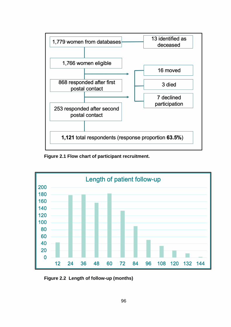
96
Figure 2.1 Flow chart of participant recruitment.
Figure 2.2 Length of follow-up (months)

97
Figure 2.3 Kaplan-Meier survival analysis with mesh removal surgery as failure variable.

98
Table 2.2 Case details of patient reported mesh complications.
Age at
surgery
Time from
sacrohysteropexy to
mesh removal
surgery (months)
Case details
1 54 - Year of surgery: 2008. No hospital notes available for further analysis.
2 68 46
Year of surgery: 2013. Implant: Unknown. Grade of surgeon: Consultant. Sacrohysteropexy details:
Unremarkable. Indication for mesh removal: Acute small bowel obstruction. Preoperative imaging: CT – small
bowel obstruction. Conservative management: N/A. Operation and approach: Midline laparotomy, partial small
bowel resection, partial excision of hysteropexy mesh with re-peritonealisation. Intraoperative findings:
Suspected SBO due to mesh, small 1-2cm exposure of mesh at broad ligament – excised, and mesh end
peritonealised, Additional non-tensioned peritonealised mesh at sacral aspect of implant was excised and
peritonealised. Recovery. Chronic abdominal pain, noted adhesions – managed conservatively, ventral hernia
repaired with mesh. Clavien-Dindo: IIIb
3 64 18
Year of surgery: 2014. Implant: Prolite Mesh, Atrium Medical, 5cm x 30.5cm. Grade of Surgeon: SST.
Sacrohysteropexy details: Diverticular disease noted. Indication for mesh removal: Lower abdominal and back
pain, recurrent prolapse. Preoperative imaging: None. Conservative Management: Plication of mesh. Mesh
removal surgery: Laparoscopic complete resection of mesh and ProTack, laparoscopically assisted vaginal
hysterectomy, anterior colporrhaphy. Mesh removal surgery findings: Elongated cervix at -1, cystocele +2.
Recovery: Uncomplicated, seen at 3/12 and discharged with no issues. Clavien-Dindo: IIIb
4 40 24
Year of surgery: 2016. Implant: Prolene, Ethicon, 15cm x 15cm. Grade of surgeon: Subspecialty trainee.
Sacrohysteropexy details: Unremarkable. Indication for mesh removal: Abdominal and vaginal pain,
dyspareunia. Preoperative imaging: MRI and laparoscopy with EUA, both normal. Conservative management:
PFMT, Paracetamol, Amitriptyline. Operation and approach: Total laparoscopic hysterectomy with complete
removal of mesh and ProTack. Intraoperative findings: Unremarkable. Recovery. Ongoing vaginal ‘soreness’,
discharged at three months. Clavien-Dindo: IIIb

99
Table 2.3 Patient reported events leading to mesh complication.
Reason for mesh removal
Asymptomatic -
Pain on examination -
Pain during sex -
Pain during physical/daily activities, % (n) 25 (1)
Pain unrelated to above, % (n) 25 (1)
Vaginal discharge -
Bladder symptoms, % (n) 50 (2)
Bowel symptoms -
Timeframe from operation to symptoms of mesh complication
< 48 hours -
49 hours to 2 months, % (n) 25 (1)
3 months to 12 months, % (n) 50 (2)
> 12 months, % (n) 25 (1)
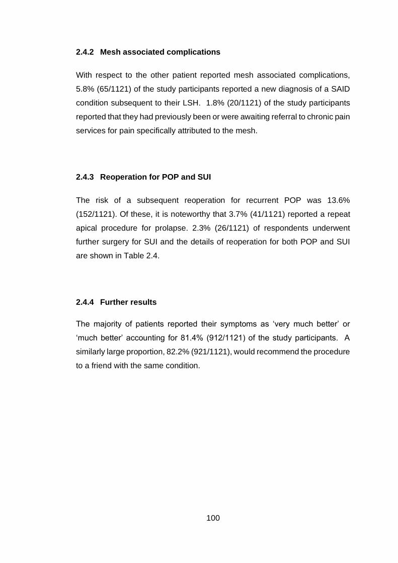
100
2.4.2 Mesh associated complications
With respect to the other patient reported mesh associated complications,
5.8% (65/1121) of the study participants reported a new diagnosis of a SAID
condition subsequent to their LSH. 1.8% (20/1121) of the study participants
reported that they had previously been or were awaiting referral to chronic pain
services for pain specifically attributed to the mesh.
2.4.3 Reoperation for POP and SUI
The risk of a subsequent reoperation for recurrent POP was 13.6%
(152/1121). Of these, it is noteworthy that 3.7% (41/1121) reported a repeat
apical procedure for prolapse. 2.3% (26/1121) of respondents underwent
further surgery for SUI and the details of reoperation for both POP and SUI
are shown in Table 2.4.
2.4.4 Further results
The majority of patients reported their symptoms as ‘very much better’ or
‘much better’ accounting for 81.4% (912/1121) of the study participants. A
similarly large proportion, 82.2% (921/1121), would recommend the procedure
to a friend with the same condition.
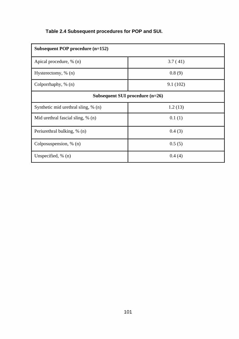
101
Table 2.4 Subsequent procedures for POP and SUI.
Subsequent POP procedure (n=152)
Apical procedure, % (n) 3.7 ( 41)
Hysterectomy, % (n) 0.8 (9)
Colporrhaphy, % (n) 9.1 (102)
Subsequent SUI procedure (n=26)
Synthetic mid urethral sling, % (n) 1.2 (13)
Mid urethral fascial sling, % (n) 0.1 (1)
Periurethral bulking, % (n) 0.4 (3)
Colposuspension, % (n) 0.5 (5)
Unspecified, % (n) 0.4 (4)

102
2.5 Discussion
Principal findings
The principal finding from this study is the low rate of reoperation for mesh
associated complications. At a median follow-up of nearly four years, the
incidence was 0.4% of from a large cohort of 1,121 participants who
underwent laparoscopic mesh sacrohysteropexy. The risk of other mesh
associated complications that were looked at within this study, namely chronic
pain and SAID were also low, as was the reoperation rate for POP or SUI. It
would appear most women continued, at nearly four years, to report ongoing
improved prolapse symptoms with the majority recommending the procedure
to a friend.
Research in context
It is of course essential to consider these findings of this study within the
context of the available evidence, outlined in more detail in Chapter 1.6.2. In
this study we report the largest reported cohort of women who have undergone
mesh augmented uterine preserving prolapse surgery, and arguably the
largest and longest cohort study of any mesh augmented surgery to date.
Comparing our results with the data reported in the literature with respect to
reoperation for mesh associated complications is difficult due to the
heterogeneity of reporting such events. There are some longer term data from
cohort studies that have reported mesh erosion rates as high as 4% following
robotic assisted sacrohysteropexy, and 5% following open sacrohysteropexy
[202, 204]. Both studies describe an alternative placement of the mesh than
that used within our study, involving pelvic dissection down to the vagina,
therefore potentially explaining the higher mesh complication rate than that
seen in our results. Also of note was that neither of these studies reported
whether or not these erosions were managed operatively versus
conservatively and therefore these are not analogous enough outcomes to
allow for meaningful comparison.

103
The most comprehensive investigation of LSH available is the systematic
review by Meriwether et al., and this did not report any cases of mesh erosion
or reoperation following abdominal mesh sacrohysteropexy [12]. In the
analysis comparing LSH to hysterectomy with SCP, six studies reported mesh
complications as an outcome [167, 205, 210, 212, 217, 276]. In only three of
these studies did this outcome actually occur and they were all seen in the
hysterectomy with concurrent SCP groups, where the risk of reoperation for a
mesh complication was between 2-3% [205, 212, 276]. Another analysis
within this review compared LSH to VH reported on two studies. One, by
Roovers et al. published data as an abstract only, and did not report on mesh
complications [277]. The other by Rahmanou et al. reported one year data of
an RCT and no mesh erosions or reoperations for mesh associated
complications occurred in either group [197]. The VUE study reported one
case of mesh erosion after hysteropexy, but did not specify whether it was a
vaginal or abdominal approach, nor whether a concurrent mesh continence
procedure was performed [152].
The incidence of reoperation for a mesh associated complication in our cohort
compares very favourably with that found in the literature for other mesh
augmented pelvic floor procedures. Following SCP for PHVP, this figure is in
the region of 5% at a median of seven years postoperatively [96]. After
subtotal hysterectomy and cervicopexy, the reported mesh exposure rate is
between 4.3% and 10.5% [164]. For the synthetic MUS, the most commonly
performed mesh augmented pelvic floor procedure, the largest available
database study has estimated the risk of reoperation for a mesh associated
complications to be in the region of 2.4% at eight years [255].
With respect to the other mesh associated complications it would appear that
the risk of utilising chronic pain services in our cohort appears relatively low.
While pain is exceptionally understudied within the literature on pelvic floor
surgery generally, after VH for example, the risk of chronic pain is in the order
of 25% [278]. Recent data suggest that chronic pain and so called central
sensitivity syndromes predispose patient to report poorer outcomes after
prolapse surgery and such syndromes appear to be prevalent in the

104
urogynaecological population [279]. When looking at newly diagnosed SAID
conditions, the 5.8% risk subsequent to surgery appears relatively high. A
study by Chughtai et al. has reported the largest and most methodologically
robust available data that looked at the association between gynaecological
mesh and autoimmune disease [265]. They reported the risk of developing
such a condition to be 2.8%, and there was no difference between women who
underwent mesh augmented procedures and those in a matched cohort
undergoing non-mesh augmented gynaecological procedures Most
importantly, over 40% of women undergoing mesh augmented POP surgery
in this matched cohort study had pre-existing diagnoses of SAID conditions.
They noted the extensive literature that supports the high risk of developing
any SAID condition following a pre-existing diagnosis. On this basis they
excluded women with pre-existing SAID, which we were not able to do due to
limitations within our methodology. One would therefore argue that our
findings should be interpreted with caution.
The risk of undergoing an apical prolapse operation for PHVP is in the order
of 6% to 8% following VH [280]. Our findings of a 3.7% risk of undergoing
such a procedure for recurrent POP therefore compares favourably. One
could argue that this supports a role for mesh augmentation in pelvic floor
surgery for POP. More specifically that for apical prolapse, mesh offers some
superiority over native tissue repair. The overall 13.6% risk of reoperation for
any form of POP in our cohort is equivalent to that reported in a recent large
registry study from Denmark that reported a reoperation rate of 11% at five
years [158]. Other studies have reported the risk of reoperation to be as high
as 30% after native tissue surgery generally [10]. The Danish study reported
a risk of reoperation for POP following suture hysteropexy of 30%, again
corroborating the conclusion that mesh confers an advantage over native
tissues and the use of suture.
The 9% risk of subsequent colporrhaphy for our study participants may reflect
the surgical practice seen within our participating centres. As a general rule,
concurrent anterior and posterior colporrhaphy for prolapse above the
hymenal ring is avoided. This is because there is evidence to suggest that

105
such prolapse is less likely to be symptomatic, and may be considered a
normal finding [88]. Furthermore, vaginal surgery may lead to scar tissue and
nerve disruption resulting in issues such a dyspareunia [281, 282]. There is
therefore an acceptance of the potential for reoperation due to anterior or
posterior wall prolapse. Reoperation for SUI is a well-recognised risk following
surgery for POP and the incidence of this in our study is comparable to the 2%
risk found in large national studies [283]. The PGI-I prolapse has been
validated as a measure for POP surgery, and we report high rates of
improvement in prolapse symptoms [112].
Strengths and limitations
There are three principal strengths of this study including the high number of
study participants, long-term follow up and patient reported nature of our data.
The value of outcomes that are patient reported cannot be understated given
the current climate with respect to synthetic mesh and pelvic floor surgery.
Both anecdotally and within the literature concerns have been raised about
the potential breakdown in trust between patients and their clinicians following
these recent controversies [284]. The fact that study participants were
operated on by five surgeons based at two centres also reduces the risk of
reporting bias seen with single centre single surgeon cohort studies. While
large national database studies and hospital coding data are often considered
to be of high-quality, one must also consider that these data are often prone
to coding errors and highly dependent on clinicians and non-clinicians alike to
provide comprehensive and accurate data. This provides further support of our
methodology of reporting patient generated outcomes as opposed to other
such data.
There are some key methodological limitations inherent within our study
design. Given the rare nature of our primary outcome measure we were not
able to use a validated patient reported outcome measure questionnaire, nor
undertake validation testing ourselves. Furthermore, to date there are no
consensus agreed outcome measures for use within studies of mesh

106
associated complications, despite ongoing work within the field of women’s
health as part of the CROWN initiative [285]. We therefore settled for a
discrete primary outcome that was measurable, rather than one based on
evidence of consensus agreement. The data in its presented format poses a
risk of omitted variable bias due to the lack of covariate analysis. This was due
to the sparsity of baseline data held about the included participants, so as to
make analysis about multiple variables futile. Therefore, analysis without cox
model adjustment is potentially mis-leading but was felt a pragmatic
compromise given the available data [286].
It must be noted that our participants underwent their procedures in centres
with specific expertise in laparoscopic pelvic floor surgery and undertaken by
tertiary level urogynaecologists. Therefore, the research setting and findings
may not be applicable to a more generalist setting and secondary care
centres. As with all forms of questionnaire studies, there is the potential for
recall bias of participants. Individual may not recall undergoing a subsequent
surgical procedure or the nature of such surgery. This also applies to the use
of chronic pain services, and subsequent SAID diagnoses. The sensitivity of
our questionnaire for these outcomes is not validated which may make
comparison with other published literature of limited value. Finally,
interpretation of our results requires consideration of the potential for response
bias. We have no way to determine whether the responders are
representative of the whole cohort of potential participants, or whether those
greatly troubled by mesh complications potentially opted not to participate or
conversely were most keen to respond. It is also not possible to account for
the potential adverse influence from women’s concerns about mesh and
recent media coverage. This may have impacted our subjective outcomes of
PGI-I prolapse and the ‘friends and family’ test.
Clinical implications
The findings from our study have potentially important clinical implications
given our large cohort, the long-term follow up and arguably most importantly,
107
the patient reported nature of our data. Most notable, is the fact that the
majority of patients would recommend laparoscopic mesh sacrohysteropexy
to friends or family, and this is even despite the well-publicised controversies
surrounding the use of mesh in urogynaecology. Combining this with the low
risk of reoperation for mesh complications and low rates of chronic pain service
use, the data should provide reassurance to both clinician, patients and
regulatory bodies as to both the safety and efficacy of LSH. It could therefore
be proposed that the procedure should continue to be offered as a choice to
women for the surgical treatment of uterine prolapse.
Research implications
There is however, a need for ongoing clinical research looking at the role of
mesh augmented prolapse surgery, and specifically sacrohysteropexy. While
the hierarchy of evidence-based medicine tends to favour large, prospective,
multicentre studies, when it comes to surgical interventions these are
notoriously difficult for undertake. Controversies surrounding mesh use in
pelvic floor reconstructive surgery are likely to make such an undertaking very
difficult in the future. Large database studies are helpful, yet their quality is
highly dependent on accurate data from clinicians or hospital coders, as well
as engagement. Therefore, studies such as that presented within this chapter
provide a pragmatic and valuable evidence base for those involved in surgical
correction of pelvic floor disorders
Conclusions
The data presented provide a pragmatic insight into the safety and efficacy of
laparoscopic mesh sacrohysteropexy. The large, patient reported dataset
must be considered within the wider context of other forms of available
evidence. We report a low incidence of mesh complications requiring removal
of hysteropexy mesh of 0.4% at nearly four years. This provides adequate
reassurance that laparoscopic mesh sacrohysteropexy should continue to be
offered with the appropriate clinician training, decision-making processes,
consent, audit and governance.

108
This chapter has been published as a peer reviewed manuscript: Izett-Kay,
M.L., Aldabeeb, D., Kupelian, A.S., Cartwright, R., Cutner, A.S., Jackson, S.,
Price, N. and Vashisht, A., 2020. Long-term mesh complications and
reoperation after laparoscopic mesh sacrohysteropexy: a cross-sectional
study. International Urogynecology Journal, pp.1-8 [287].

109
Vaginal hysterectomy versus laparoscopic sacrohysteropexy for the
treatment of uterine prolapse: a randomised controlled trial.
3.1 Introduction
In UK practice, VH remains the preferred surgical option for urogynaecologists
treating uterine prolapse, with 75% of respondents to a BSUG survey
indicating that this was their favoured procedure [5]. Half of these survey
respondents offer LSH, the second most preferred procedure, and this is
supported by NICE guidance [133]. Yet the important question of LSH
efficacy as compared to VH remains unanswered. Whilst for women with
uterine prolapse and a strong desire for uterine preservation, the clinical
decision-making process is relatively straightforward; surgical options include
either a mesh LSH or Manchester-Fothergill procedure. Suture hysteropexy
is not supported by larger population studies due to the high risk of recurrence
[158]. For the many women who have no preference regarding uterine
preservation, there is a dilemma; should they opt for VH or mesh LSH, and
what are the relative merits of both with respect to outcomes and safety?
The reason for consideration of mesh LSH lies with the several disadvantages
associated with VH. Firstly, removal of the uterus does not address the
pathophysiological factors that have led to uterovaginal prolapse, principally
the deficiencies in level one support provided by the uterosacral and cardinal
ligaments [20]. It is therefore unsurprising that the risk of PHVP is relatively
common, with some studies reporting that it affects up to 40% of women after
hysterectomy for POP [288, 289]. In accordance with guidance from the
RCOG it is routine practice to undertake a concurrent apical suspension
procedure at the time of VH [149]. However, the risk of vault prolapse remains,
with reoperation rates of between 4.6% and 18% [8, 9]. Other data suggest
that this figure is at least 8%, and following native tissue repair of prolapse
more generally, the risk of reoperation for POP may be as high as 30% [10,
290]. Secondly, is the potential for new onset symptoms associated with
surgical disruption of tissues. There is some evidence hysterectomy is
110
associated with pelvic neuropathy that in combination with trauma to adjacent
structures can lead to urinary incontinence, bladder dysfunction and changes
in sexual function [291-294]. Another consideration is morbidity associated
with more invasive surgery, a Cochrane review has suggested subtotal
hysterectomy may offer advantages over total hysterectomy such as a lower
risk of postoperative pyrexia and voiding dysfunction, and in theory this may
follow for hysterectomy versus hysteropexy [295]. Indeed the systematic
review by Meriwether et al. that compared VH to hysteropexy pointed to
shorter hospital stays, lower blood loss, quicker return to activity and lower
pain scores in patients that underwent hysteropexy [195]. It must be noted
however that only two RCTs contributed to this analysis and one of them
utilised an open abdominal approach to hysteropexy.
More recently the VUE trial tried to answer the question of uterine preservation
versus VH [152]. A previously discussed this was a multicentre RCT with 563
participants and authors reported no difference between the two intervention
groups with respect to validated symptoms scores, the primary outcome, nor
with rates of SAEs or overall stage of prolapse. They did find that more women
would recommend VH. Yet uterine preservation was undertaken utilising
various techniques including vaginal sacrospinous fixation, colpocleisis, and
both open and laparoscopic approaches to abdominal suture hysteropexy and
abdominal mesh hysteropexy. Many of those having uterine preservation
procedures failed to receive the benefits of LSH, namely a minimally invasive
approach, good access to the pelvis allowing for higher uterine suspension
and the potential longevity associated with mesh use.
The evidence presented in Chapter 1.6.2 suggests LSH has a comparable
adverse event profile to other routinely used mesh augmented procedures
such as SCP and therefore should be deemed appropriately safe. Given
current prolapse surgery trends surgery, ageing populations, changing patient
desires, and the high failure rate associated with VH, the merits of abdominal
hysteropexy deserve further scrutiny. In this study, we aim to compare
outcomes between VH with apical suspension versus LSH, reporting the long-

111
term outcomes at seven years in participants of the study previously reported
by Rahmanou et al. [197].
3.2 Objectives
The primary objective of this study was to compare the efficacy of VH and
mesh augmented LSH by determining the risk of reoperation for apical
prolapse between the two groups.
Secondary objectives included comparing the two groups through the
assessment of:
• The overall risk of reoperation for any type of POP.
• The incidence of mesh associated complications such as mesh
exposure, mesh removal or chronic pain.
• Patient impression of the improvement in POP symptoms.
• The anatomical presence of POP.
• The presence of vaginal symptoms including POP symptoms.
• The presence of LUTS.
• Sexual function.
3.3 Methodology
3.3.1 Study design
This study was a single-centre RCT of VH versus mesh augmented LSH for
the treatment of uterine prolapse undertaken at the John Radcliffe Hospital,
Oxford, UK from May 2009 to September 2012. Potential participants were
identified from the urogynaecology unit as women requesting surgical
treatment for grade 2-4 uterine prolapse. The study adhered to the following
inclusion and exclusion criteria.

112
Inclusion criteria:
Women diagnosed with symptomatic uterine prolapse (grade 2-4 uterine
descent according to the Baden-Walker system) aged 18 years or above.
They had to have completed their family with no desire for future
childbearing and if of reproductive age had to be willing to ensure that she
or her partner would use effective contraception during the study and for 3
months thereafter.
Exclusion criteria:
Included the following:
• Desire to maintain fertility, pregnant, lactating or planning pregnancy
during the course of the study.
• Having significant renal or hepatic impairment.
• Having participated in another research study involving an
investigational product in the previous 12 weeks.
• Having abnormal cervical cytology, abnormal uterine bleeding or
evidence of uterine disease.
• Having any other significant comorbidity which in the opinion of the
operating surgeon, may either put the participant at risk of another
surgical intervention, or may influence the result of the study, or the
participant’s ability to participate in the study.
With respect to our surgical intervention, both LSH and VH were combined
with anterior and/or posterior repair on the basis of intraoperative assessment
and judgment at the time of surgery by the senior operating surgeon. Generally
anterior or posterior wall prolapse above the hymen was left, unless explicitly
planned preoperatively following patient wishes. For LSH, the technique
utilised is described in detail within Chapter 1.6.1. Briefly this involves the use
of a bifurcated polypropylene mesh (PRO-Lite™, Atrium Medical Corporation,
Hudson, NH, USA) wrapped around the cervix through broad ligament
windows and secured anteriorly with non-absorbable sutures (Ethibond
Excel™; Ethicon) that is then secured to the sacral promontory with a helical
fastener (Protack™; United States Surgical, Tyco Healthcare, Norwalk, CT,

113
USA). For VH, the uterosacral ligaments were reattached to the vaginal vault
following hysterectomy with re-absorbable sutures (Vicryl 1; Ethicon,
Somerville, NJ, USA). This is referred to as a modified McCall’s culdoplasty
and was undertaken as it is the most common technique employed in the UK
[5]. For those participants with procidentia, additional vault support was
undertaken by employing a sacrospinous fixation. This utilised re-absorbable
sutures (PDS II 0; Ethicon), again mirroring common UK practice and
complying with RCOG recommendations [149].
3.3.2 Outcome measures
The outcome measures were based on objective patient reported data, clinical
examination and the results from ICS Grade A PROMS ICIQ-VS, ICIQ-FLUTS
and PISQ-IR which are contained in Appendix 8. To answer our study
objective, the following outcomes were selected:
Primary outcome:
• Subsequent operation for apical prolapse within the study period.
Secondary outcomes:
Objective measures:
• Reoperation for any form of POP.
• Presence of POP according to the POP-Q.
• Mesh associated complications including patient reported removal of
mesh, mesh erosion or chronic pain attributed to the use of mesh.
Subjective measures:
• Satisfaction with surgery according to PGI-I in prolapse symptoms.
• Prolapse symptoms measured by Question 5 of the ICIS-VS.
• Vaginal symptoms measured by ICIQ-VS
• Sexual function measured by ICIQ VS sexual matters and Pelvic Organ
Prolapse/Urinary Incontinence Sexual Questionnaire – 12 (PISQ-12.)
• Vaginal symptom related QoL measure by ICIQ VS quality of life

114
• LUTS measured by International Consultation on Incontinence
Questionnaire-Female Lower Urinary Tract Symptoms (ICIQ-FLUTS)
Tertiary outcomes:
• Blood loss (millilitres)
• Operating time (minutes)
• Postoperative complications
• Hospital stay (days)
3.3.3 Recruitment
Potential participants were women who were referred to the Urogynaecology
department of the John Radcliffe Hospital, Oxford University with symptoms
of prolapse. They were seen as part of the routine clinical care pathway and
underwent a clinical history, vaginal examination and assessment of prolapse
using POP-Q followed by a discussion about the conservative and surgical
management of prolapse. Those potential participants who met the inclusion
and exclusion criteria set out in Chapter 3.3.1 were then given a PIL (Appendix
7) the study and offered the opportunity to participate. Those women who
agreed were then consented for participation at a later date on re-attendance
to the outpatient clinic or at the time of surgical consent taking.
Randomisation was conducted in the urogynaecology clinic at the John
Radcliffe Hospital. Sealed randomisation envelopes were kept secure and
unopened by the investigators. Each subject was assigned a study number
and then randomised through the use of sealed envelopes placing participants
into groups A or B (group A = laparoscopic sacrohysteropexy, group B = VH).
Randomisation occurred sequentially as each subject entered the study.
The data presented within this thesis comes from the seven-year follow-up of
participants of this study. This was not part of the original study protocol and
was carried out after approval for a substantial amendment as detailed in
Chapter 3.3.6. All study participants were contacted by telephone and invited
115
for a study visit in clinic as detailed below and those who attended completed
written consent. Telephone verbal consent was obtained for those who were
happy to undertake a telephone history and send postal questionnaires.
Following two attempts at telephone contact all study participants were
contacted by post with a PIL, questionnaires for PGI-I, ICIQ-VS, ICIQ-FLUTS,
PISQ-12, and a study questionnaire asking about subsequent operation or
mesh associated complications. These questionnaires were chosen as they all
have Grade A evidence to support their use [86]. Return of these
questionnaires was taken as implied consent and a case note review of these
participants was undertaken.
3.3.4 Data collection
There were three principle data collection points, as outlined in the study
schedule shown in Table . The following visits were undertaken as part of the
study plan, with relevant data collection as highlighted.
1st visit (baseline assessment):
Potential participants attending urogynaecology clinic at John Radcliffe Hospital
as part of routine clinical care pathway.
• Demographic data
• Clinical history, abdominopelvic examination including POP-Q
• Completion of ICIQ-VS
• Checked against inclusion and exclusion criteria
• Given PIL
2nd visit:
• Surgical consent.
• Study consent.
• Randomisation.
3rd visit:
Admission to hospital for surgery
116
• Intraoperative data
• Short-term complications
4th visit:
Eight-week postoperative visit as part of routine clinical care
5th visit:
Twelve months postoperative in urogynaecology outpatient clinic.
• Routine clinical history covering postoperative recovery, vaginal,
urinary, bowel and sexual symptoms, any subsequent prolapse
operations and review of case notes.
• PGI-I prolapse symptoms.
• ICIQ-VS
• POP-Q
6th visit:
Not part of original study protocol and subject to a substantial amendment. At
a minimum of seven years postoperatively, study participants were contacted
by telephone and offered the following:
Attendance in clinic
• Routine clinical history covering postoperative recovery, vagina,
urinary, bowel and sexual symptoms, any subsequent prolapse
operations and complications related to mesh including removal of
mesh, mesh erosions or pain attributed to the mesh and review of case
notes.
• PGI-I prolapse symptoms.
• ICIQ-VS
• ICIQ-FLUTS
• PISQ-12
• POP-Q
Those patients declining a clinical review were offered a telephone or postal
consultation with one of the clinical care team to undertake:

117
• Routine clinical history covering postoperative recovery, vagina,
urinary, bowel and sexual symptoms, any subsequent prolapse
operations and complications related to mesh including removal of
mesh, mesh erosions or pain attributed to the mesh and review of case
notes.
• PGI-I prolapse symptoms.
• ICIQ-VS
• ICIQ-FLUTS
• PISQ-12
Following two attempts to contact participants by telephone (voice messages
were not left), a single postal contact was made using the cover letter to allow
for postal completion of questionnaires. Contact details of the clinical care
team were enclosed within the postal contact to allow participants to request
a follow-up appointment if this was their preference. Those participants that
responded underwent:
• Review of case notes.
• PGI-I prolapse symptoms.
• ICIQ-VS
• ICIQ-FLUTS
• PISQ-12

118
Table 3. 1 Study flow chart
Intervention or procedure
Time Details
Participant identification in gynae clinic: clinical
assessment, Questionnaire assessment,
recruitment and randomisation. (potential
participants will be given up to three months to
think).
20-40
min
Mr SR Jackson and his team of
co−investigators will conduct
interviews and procedures that will
take place at the John Radcliffe
Hospital Oxford and Pembury
Hospital Tunbridge Wells.
Admission for the prolapse operation, operation
itself and postoperative recovery.
2-5
days
Mr SR Jackson and his team of
co−investigators will conduct
interviews and procedures that will
take place at the John Radcliffe
Hospital Oxford and Pembury
Hospital Tunbridge Wells.
Follow up at 8 weeks, post operation for clinical
assessment in gynaecology clinic. (this is the
follow−up procedure that all our patients
(patients not participating in any trial) normally
receive).
20 min Mr SR Jackson and his team of
co−investigators will conduct
interviews and procedures that will
take place at the John Radcliffe
Hospital Oxford and Pembury
Hospital Tunbridge Wells.
Postal follow up at 6 months post operation for
Questionnaire assessment. (this is the follow−up
procedure that all our patients (patients not
participating in any trial) normally receive).
20 min Mr SR Jackson and his team of
co−investigators will conduct
interviews and procedures that will
take place at the John Radcliffe
Hospital Oxford and Pembury
Hospital Tunbridge Wells.
Follow up at 12 months post operation for
clinical assessment in gynaecology clinic. (this is
the follow−up procedure that all our patients
(patients not participating in any trial) normally
receive).
20 min Mr SR Jackson and his team of
co−investigators will conduct
interviews and procedures that will
take place at the John Radcliffe
Hospital Oxford and Pembury
Hospital Tunbridge Wells.
Follow up at 72 months post operation for
clinical assessment in gynaecology clinic. (this is
follow-up as part of the study)
20 min Mr SR Jackson and his team of
co−investigators will conduct
interviews and procedures that will
take place at the John Radcliffe
Hospital Oxford and Pembury
Hospital Tunbridge Wells.
119
3.3.5 Power and statistical analysis
This RCT was originally conducted as a pilot study and therefore a power
calculation was not undertaken. A sample size could be calculated assuming
an 85% cure rate following LSH, based on previously discussed literature
utilising an anatomical outcome, with a 5% difference in cure rate being
deemed as clinically significant. To detect this level of difference with 80%
power would require 62 patients in each arm of the trail. Therefore, for
adequate power the total study sample size would have been 124 women.
This power calculation did not account for loss to follow-up and it’s secondary use
as part of this study would not be considered to be statistically robust. It was not
used to calculate a sample size for seven-year follow-up.
Data for the whole study population were subject to descriptive statistics. For
the primary outcome of a dichotomous variable of either having had
reoperation for apical prolapse or not, a chi-squared test was used. This was
used for all other dichotomous variables, parametric data were subject to a
student t-test and non-parametric data such as PROM scores were subject to
a Mann-Whitney U test, all requiring a significance level set at p<0.05. These
were all analysed on an intention to treat basis. Kaplan Meier survival
analyses were also undertaken using the primary outcome as a failure
variable. Statistical analysis was undertaken with Stata/SE 15® (StataCorp
LLC., Texas, USA).
3.3.6 Ethical approval
This study was originally approved by the National Research Ethics
Committee (reference number: 09/H0606/28). In order to undertake the
seven-year follow-up that was not part of the original protocol a substantial
amendment was made. This was approved along with Health Research
Agency approval on 08/01/2019 by the South Central – Oxford C REC, as
detailed in Appendix 9.
120
3.4 Results
Over the recruitment period 481 women were seen with stage 2-4
symptomatic uterine prolapse and invited to participate. Many declined
participation due to a particular preference for one of the surgical options. One
hundred and thirty-two women were recruited, however 31 of these women
later withdrew due to a desire for a specific surgical procedure. A further
patient randomised to LSH had an intraoperative conversion to VH due to a
low bifurcation of the aorta precluding safe access to the sacral promontory
for access. The one-year follow-up of 79 study participants has previously
been reported, with no significant difference in the primary outcome of
reoperation for apical surgery [197].
A summary of the demographics between the LSH and VH groups at
recruitment and for those with seven-year follow-up is shown in Table .
Following telephone and postal contact, 62 women (62%) provided long-term
outcome data. Comparison of baseline demographics and characteristics
between those that attended for long-term follow-up and those lost to follow-
up, is shown in Table 3.3 C. It is notable that those who did not attend the
long-term follow-up had worse preoperative ICIQ-VS scores than those who
did participate (37 vs. 32, p = 0.04). The enrolment, allocation, follow-up and
analysis of patient is outlined within Error! Reference source not found..
With a minimum of seven years, the mean length of follow-up was 100 months
(range 84-119 months).
3.4.1 Rate of apical reoperation
For the primary outcome of reoperation for apical prolapse, 6.1% of
participants underwent such a procedure following LSH and 17.2% following
VH, however the difference was not statistically significant (relative risk (RR)
0.34, 95% CI 0.07 – 1.68, p = 0.17). A Kaplan-Meier survival analysis graph
comparing the two groups based on the primary outcome is shown in Figure
3.2. The nature of the surgical procedures undertaken for all forms of recurrent

121
POP are shown in Table 3 and the results from case notes review of the entire
cohort are shown in Table 3.5 Reoperation rates from case note review for all
women enrolled in study.
3.4.2 Subjective measures
There was a statistically significant difference in the postoperative composite
ICIQ-VS scores when comparing those women who had VH and LSH, in
favour of VH (6 and 9 respectively, p = 0.04). However, given the difference in
preoperative ICIQ-VS scores between those that attended 7-year follow-up
and those that did not, this should be interpreted with caution. Comparison of
the mean change in ICIQ-VS scores between those undergoing VH versus
those undergoing LSH were not statistically significant, illustrated in Table 3.6
. Data presented in this table also illustrate that there was no difference
between the two groups with respect to the other PROM scores including the
seven-year postoperative ICIQ-VS SM and QoL subscales, the composite
ICIQ-FLUTS score as well as filling, voiding and incontinence subscales, and
the PISQ-12. Likewise, when analysing the likelihood of ‘awareness of a lump
of bulge coming down in the vagina’ (Q5 ICIQ-VS), a symptom determined by
consensus to be an accepted marker of symptomatic prolapse, there was no
difference between the two groups (31% following VH and 45.5% following
LSH; RR 0.68, 95% CI 0.35 to 1.32, p = 0.24) [2]. The likelihood of patients
reporting their prolapse symptoms as ‘very much better’ or ‘much better’ was
86% after VH and 76% after LSH (p = 0.29).
3.4.3 Objective measures
There were no reported cases of mesh removal surgery, mesh erosion or
chronic pain attributed to the mesh in the LSH group. With respect to POP-Q
parameters as shown in Table 3.6 , data would appear to support LSH with
statistically significant higher apical suspension (POP-Q point C -5 vs -4.25, p
= 0.02) and longer TVL (9cm vs 6cm, p = <0.001). If POP-Q point C </= -2 is
used as an anatomical discriminator of surgical success, there is no difference
between the two groups (84.6% after VH, 81.2% after LSH, p = 0.73), likewise
if a more generous cut-off C </= 0 is used as a discriminator (92.3% after VH,

122
90.1% after LS, p=0.82). As reported in the one-year follow-up study there
were no major intraoperative complications in either group. It appeared that
total operating time was shorted in those having VH by a mean difference of
11.4 minutes (p < 0.001). However estimated blood loss (EBL), length of
hospital stay, pain scores and time returning to normal activity all favoured
LSH.

123
Table 3.2 Baseline demographic data at initial recruitment and for those with 7-year follow-up.
Baseline characteristics LSH VH p value
n = 51 n = 50
Mean age, years (range) 63.9 (44–83) 65.5 (36–80) 0.136
Mean BMI, kg/m2 (range) 25.9 (20–36) 27.5 (19–37) 0.068
Median parity (range) 2 (1–5) 2 (1–6) 0.165
Stages of prolapse before surgery
Uterine descent stage 2–4, 51(100) 50 (100) 1
Anterior prolapse (Ba)
Stage 0–1 3 (6) 14 (7) 0.185
Stage 2–4 47 (94)) 43 (86) 0.185
Posterior prolapse (Bp)
Stage 0–1 13 (26) 6 (12) 0.076
Stage 2–4 37 (74) 44 (88) 0.076
Concomitant urogynaecology
procedures
Posterior colpoperineorrhaphy, 33 (66) 45 (90) 0.004
Anterior colporrhaphy 18 (36) 38 (76) <0.001
Any pelvic floor repair 41 (82) 49 (98) 0.008
Tension-free vaginal tape 2 (4) 4 (8) 0.402
Sacrospinous fixation 0 3 (6) 0.08
For those with 7-year follow up
n = 33 n = 29
Mean age, years (range) 64.1 (51-83) 66.2 (49-81) 0.20
Mean BMI, kg/m2 (range) 25.9 (20-36) 26.9 (19-35) 0.24
Median parity (range) 2 (1-5) 2 (1-4) 0.42
Preoperative ICIQ VS
31.58 +/-
12.61 32 +/- 12.15 0.92
Preoperative ICIQ VS SM
28.63 +/-
15.16
25.27 +/-
20.52 0.49
Preoperative ICIQ VS QOL 6.58 +/- 2.54 7.74 +/- 2.30 0.08
Length follow-up, months (range) 99 (84-119) 95 (86-114) 0.39*
POP-Q parameters
Ba 1 +/- 2.20 1 +/- 2.51 0.61
C 1 +/- 2.65 1 +/- 3.45 0.21
Bp 0 +/- 2.60 0 +/- 2.45 0.51
GH 5 +/- 0.72 5 +/- 0.86 0.002
TVL 8 +/- 0.80 8 +/- 1.24 0.95
Continuous data are listed as mean +/- SD (Mann Whitney U test), except parity which
is median and interquartile range or n (percentage). Mann-Whitney use for test of
significance with the exception of *, which was calculated with student t-test.

124
Table 3.3 Comparison demographics between those who attended 7-year follow-up and those who did not
No 7-year follow-
up
7-year follow-
up
p
value
n = 39 n = 62
Age, years 63.95 +/- 9.81 65.11 +/- 6.69 0.88
BMI, kg/m2 27.24 +/- 4.21 26.39 +/- 19.4 0. 29
Parity 2 (1) 2 (1) 0. 95
Preoperative ICIQ VS 37.11 +/- 10.12 31.77 +/- 12.31 0.04
Preoperative ICIQ VS SM 29.42 +/- 17.02 27.52 +/- 16.89 0.67
Preoperative ICIQ VS QoL 7.81 +/- 1.51 7.05 +/- 2.49 0.19
POP-Q parameters
Ba 1 +/- 2.42 1 +/- 2.32 0.62
C 2 +/- 2.55 2 +/- 3.02 0.79
Bp 0 +/- 2.36 0 +/- 2.53 0.36
GH 5 +/- 0.75 5 +/- 0.84 0.85
TVL 8 +/- 0.65 8 +/- 1.01 0.80
Continuous data are listed as mean +/- SD (Mann Whitney U test), except parity which
is median and interquartile range.

125
Figure 3.1 CONSORT diagram
Figure 3.2 Kaplan-Meier survivorship using primary outcome as failure variable
Table 3.4 Re-treatment for POP at seven years
Categorical data are listed as n (percentage) with chi-square testing.
Follow-up data LSH (n=33) VH (n=29) p value
Subsequent treatment for POP, % 9 (27.3) 7 (24.1) 0.78
Recurrent apical POP (reoperated apex or
C>/= -1), % 2 (6.1) 5 (17.2) 0.17
Subsequent surgery for POP, % 6 (18.2) 6 (20.7) 0.80
Apical
LSH, % 2 (6.1) - 0.17
SCP, % - 5 (17.2)
Colporrhaphy
Anterior colporrhaphy, % 2 (6.1) - -
Anterior and posterior colporrhaphy, % 2 (6.1) - -
Posterior colporrhaphy, % - 1 (3.4) -
PFMT, % 2 (6.1) 1 (3.4) 0.63
Pessary, % 1 (3) - 0.34

126
Table 3.5 Reoperation rates from case note review for all women enrolled in study.
Categorical data are listed as n (percentage) with chi-square testing.
Table 3.6 Outcome data
LSH (n=33) VH (n=29) p value
Change in ICIQ VS, mean -22.39 +/- 13.06 -24.91+/- 14.05 0.59
Postoperative ICIQ VS SM, mean 7.42 +/- 13.15 1.28 +/- 3.40 0.42
Postoperative ICIQ VS QOL, mean 1.42 +/- 1.98 1.03 +/- 1.72 0.43
Positive response to ICIQ VS Q5, % 15 (45.5) 9 (31) 0.24*
POP-Q
Ba (cm) -1 +/- 1.69 -0.5 +/- 1.70 0.99
C (cm) -5 +/- 2.58 -4.25 +/- 2.92 0.02
Bp (cm) -2 +/- 1.68 -2 +/- 0.54 -
GH (cm) 3 +/- 0.88 3 +/- 0.88 0.97
TVL (cm) 9 +/- 3.0 6 +/- 1.20 <0.01
PGI-I (1-2), % 25 (75.8) 25 (86.2) 0.30*
ICIQ FLUTS , mean 9.42 +/- 5.95 9.53 +/- 5.97 0.97
ICIQ FLUTS_F , mean 3.39 +/- 1.97 3.86+/- 2.08 0.46
ICIQ FLUTS_V , mean 1.70 +/- 1.94 1.76 +/- 1.57 0.53
ICIQ FLUTS_I , mean 4.33 +/- 4.26 3.89 +/- 3.42 0.88
PISQ-IR, mean 16.67 +/- 3.67 13.75 +/- 5.72 0.28
Change in ICIQ VS, mean -22.39 +/- 13.06 -24.91+/- 14.05 0.59
All values are mean +/- standard deviation with Mann-Whitney use for test of
significance with the exception of POPQ with median values. * = dichotomous
outcome of either positive or negative response to ICIQ VS Question 5 and yes or no
to PGI-I 1 and 2, where chi-square test was used.
Follow-up data LSH (n=51) VH (n=50) p value
Subsequent treatment for POP, % 14 (27.5) 11 (22) 0.53
Subsequent surgery for POP, % 9 (17.6) 10 (20) 0.76
Apical
LSH, % 3 (5.8)
0.13 SCP, % 9 (1.8)
VH, % 1 (2)
Colporrhaphy, % 5 (9.8) 1 (2)
PFMT, % 3 (0.6) 1 (2) 0.31
Pessary, % 2 (3.9) - 0.15

127
3.5 Discussion
Principle findings
Our data show that for the primary outcome, there appears to be a trend
towards a lower rate of apical reoperation following LSH as compared to VH,
although this was not statistically significant. This may represent the fact that
this study is underpowered as discussed in Chapter 3.3.5. This outcome is
important as apical reoperation in the form of LSC and SSF are relatively
higher morbidity interventions than simple colporrhaphy [86]. The data
suggest that at over 7 years, the objective success rate based on apical
reoperation would be 83% after VH and 94% after LSH. It is also noteworthy
that the POP-Q parameters of TVL and point C also significantly favour LSH,
as increased vaginal length and apical support are both features of normal
vaginal anatomy and therefore markers of optimal surgical correction. The
perioperative data would also seem to favour LSH. However, prolapse is
widely considered to be a QoL condition patient reported outcomes may be
considered the gold standard marker of surgical efficacy [296]. There was no
difference between the intervention arms in any of the validated PROM
outcomes with the exception of composite ICIQ-VS scores, which is
confounded by significant differences in preoperative ICIQ-VS scores between
those lost to follow-up and those attending.
Research in context
These data must be considered within the context of previously reported
randomised and observational cohort studies, although this is difficult due to
the heterogeneity of reported outcome measures and current lack of
consensus for core outcome measures when studying pelvic floor disorders
[285]. The most recent meta-analysis to compare the same two interventions
investigated within this chapter is that by Meriwether et al. [195]. It must be
noted only two studies contributed to this part of the review and one of these
was the data from the 12-month pilot study by Rahmanou et al, with the same
participants presented within this thesis [197]. Additionally, the two studies
were not subject to a meta-analysis. Roovers et al. reported that QoL
questionnaires, LUTS, mobility and postoperative pain favoured VH, with no
128
significant advantages to sacrohysteropexy, although this was done with an
open approach [156, 277]. The findings by Rahmanou et al. unsurprisingly
largely mirror our own, with no difference in the apical reoperation rate
principally; elevation of POP-Q point C, TVL, EBL, and hospital length of stay
favouring LSH, while theatre time and rate of concurrent colporrhaphy
favoured VH.
More recently the results of a long anticipated high-level multicentre RCT trial,
the VUE study have been reported, with 12 month follow-up for women
randomised to either VH or hysteropexy in the uterine prolapse arm [152]. In
total 563 women were randomised to either uterine sparing surgery or VH, with
238 women undergoing VH with an apical suspension procedure providing
data at 12 months, and 230 women undergoing uterine preservation. It must
be noted that most of the uterine preservation procedures were undertaken
with a vaginal approach, and only 69 women (24.7%) underwent an abdominal
approach, of which 66 (23.6%) were LSH; data for the individual procedures
was not provided and therefore direct comparison with our own study is
difficult. There was no significant difference in the primary outcome, prolapse
symptoms based on POP-SS at twelve months between the two groups (POP-
SS scores 4.2 following VH, versus 4.2 following uterine preservation mean
difference (MD) 0.05, CI 0.91 to 0.81, p=0.91); nor in prolapse associated QoL.
Many patients in both groups had an ongoing feeling of something coming
down (30.7% and 28.9% respectively), comparing favourably to our own
longer-term symptom status results. Significantly more women would
recommend VH to a friend as compared to uterine preservation (95% vs
88.3%, odds ratio (OR) 0.39, CI 0.18 – 0.83, p 0.01). Finally, there was no
significant difference between reoperation rates for prolapse (3.3% following
VH, 6.1% following uterine preservation, OR 2.01 CI 0.81 to 4.95, p = 0.120),
and while lower than our own rates, these are twelve-month data so
comparison is not possible. There was no difference in rates of SAEs; with
one mesh exposure in the uterine preservation group, no details are provided
as the nature of this and it may be related to vaginally placed mesh or a
synthetic MUS which were both undertaken as concurrent procedures within

129
the study. There are some significant shortcomings to the VUE study; the
group failed to reach their recruitment target to meet their power calculation,
there were a large number of recruitment centres and many of these would
not be high volume centres for LSH procedures, and 50% of their hysteropexy
procedures were undertaken with a vaginal approach which fails to make use
of a principle advantage of abdominal hysteropexy, which is access to the
upper pelvis to allow for higher uterine suspension.
The non-randomised prospective comparative study comparing LSH and VH
by Lone et al. found no difference in reoperation rates between the two groups,
POP-Q point C, or in subjective outcomes [199]. There were differences in
POP-Q parameters Ba, Bp and GH favouring LSH, however small numbers at
follow-up and significant differences between the baseline characteristics of
the two cohorts make meaningful comparison difficult.
It would appear that our findings at 7 years mirror the short term RCT and
observational study findings previously reported by our group [197, 207]. If
the primary outcome is used as the definition of objective failure, our long-term
cure rate of 94% is similar to the findings from the largest, medium-term cohort
studies by Pandeva et al. and Kupelian et al., who reported rates of 95% at 48
months and 98% at 3 months respectively [186, 201]. Pandeva et al used
POP-Q point C of = / < 0 as a cut off for objective success and Kupelian et al.
used POP-Q point C of = / < -2, an evidence-based discriminator for
symptomatic prolapse based on work by Dietz et al. [88]. As outlined in our
results, sub-analyses of our data show comparable success rates using these
same outcome measures, with no significant difference between the two
intervention arms. Daniels et al. reported an 80% cure rate based on POP-Q
point C of = / < 0 in 138 women at 12 months, against which our 93% cure rate
using this outcome measure for the LSH cohort compares favourably [198].
A recent RCT reported an apical reoperation rate of 3.9% following VH and
<1% following sacrospinous hysteropexy at five years, although there was no
difference between these two groups with reference to anatomical outcomes
130
[297]. Although the reoperation rates in this RCT are lower than those within
our own study, the risk of recurrent apical prolapse appears to be similar. The
RCT by Roovers et al. found a 22% reoperation rate at 12 months following
LSH, to which the findings in this thesis compare favourably [156]. The latest
Cochrane review reported a reoperation rate for POP following vaginal surgery
for apical prolapse of 9.3%, albeit with a heterogenous group of procedure
within the meta-analysis [183]. A recent large population study of 7,247
patients at a median of five years reported reoperation rates of 30%, 7% and
11% after sacrospinous hysteropexy, Manchester repair and VH respectively
[158]. The largest series reporting on PHPV would suggest a reoperation rate
of between 6% and 11.6%, again suggesting the interventions in both
treatment arms are at least comparable [280, 290].
Assessing the rate of symptomatic cure in our study with that reported in the
literature is also confounded by the heterogeneity of outcome measures,
failure to report subjective data or the use of unvalidated measures.
Subjective cure rates after LSH in large cohort studies with mid-term follow-up
are reportedly between 80% and 84% [186, 198, 201]. Some of these studies
have also used PGI-I and therefore report a more favourable subjective cure
rate than that we have presented, however our data are longer term which
may lead to increasing rates of prolapse and therefore lower reported
subjective cure and it is noteworthy that there was no difference between the
groups. The only other RCT comparing the same two interventions, by
Roovers et al. did not report subjective cure rates. They reported no difference
in the validated symptom measures, however those undergoing hysteropexy
were more likely to consult a doctor with prolapse symptoms, a finding
mirrored in the same group’s five-year data that was presented as an abstract
only and therefore not subject full peer-review [156, 277]. The Cochrane
meta-analysis reported ‘awareness of prolapse’ based on validated
questionnaires and provided a risk of 13.7% at two years following vaginal
surgery, however as stated this group included a number of procedures some
of which utilising mesh, making it difficult to compare to our figure of 45.5%

131
and 31% (after LSH and VH respectively) based on a positive response to
ICIQ-VS Question 5 [183].
Strengths and limitations
The principal strengths of our study include the use of randomisation with
comparable baseline demographics in both intervention arms, adherence to a
pre-stated primary outcome, and long-term follow-up. Additionally, much of
the data is patient reported and gathered through the use of validated
measures. This will allow for future inclusion in meta-analyses. This is
important as this study was not adequately powered for the primary outcome
measure. Additional shortcomings include the large loss to follow-up, common
in long-term randomised studies of surgical interventions. The choice of a
dichotomous outcome also poses a challenge. Emergent data has shown
prolapse to be a dynamic state, both pre and post operatively, and therefore
the use of such discrete outcomes may challenge the clinical utility of the
findings of this study[298]. Finally, follow-up observations were undertaken by
the thesis student and were not blinded to patient’s primary intervention,
potentially introducing bias with respect to outcomes such as POP-Q.
Clinical implications
The implications of the findings within this chapter on clinical practice should
not be overstated. The lack of power and resulting inability to detect a
statistically significant difference in the primary outcome may lead one to
conclude that the findings should not alter future clinical practice. However,
they do have utility in framing the discussions around surgical decision making
for patients considering options for surgery for uterine prolapse. That is, in
keeping with earlier studies and observational data that there may be some
advantages to LSH as outlined. Women worried about the risk of needing
another significant operation or wanting to maintain vaginal parameters as
close to anatomically normal as possible may opt for LSH. However, they can
be reassured as the presented data suggest it unlikely there is any large
difference between the two options and their functional outcomes are likely to
be good regardless of choice of intervention.

132
Research implications
There are two predominant issues that need addressing within the academic
literature with respect to the choice of surgical intervention for uterine
prolapse. Firstly, is a consensus on core outcomes for pelvic floor disorders,
this is currently being addressed via the CROWN initiative [285]. The other is
the need for further randomised studies to allow for meaningful comparison of
surgical interventions for this form of POP. With changes in the landscape with
respect to mesh use, many of the procedures that were investigated by the
studies included in the Maher 2016 meta-analysis are not fit for contemporary
pelvic floor surgery due to risk of mesh associated complications. With the
current options on the table for patients in UK practice, this need for quality
prospective data is pressing. Data from the VUE study presents its own issues
as discussed [297]. Our data as well as the VUE studies and future
prospective work are likely to form part of further meta-analysis.

133
Prognostic role of urodynamic studies in patients undergoing
laparoscopic sacrohysteropexy.
4.1 Introduction
As outlined within Chapter 1.3.5 there is a significant correlation between the
presence of POP and concurrent LUTS, and in the case of SUI the conditions
may even share aetiology [299]. However, the relationship is complex and
poorly understood. Surgical correction of POP is not necessarily associated
with resolution of LUTS; indeed up to 20% of women who are continent
preoperatively go on to develop SUI and 30% develop postoperative
overactive bladder (OAB) [93, 95]. Conversely, a large study by Lensen et al.
showed that 39% of women with preoperative SUI reported cure by POP
surgery alone, without the use of a concurrent incontinence procedure, and
42% of those pre-existing urinary urge incontinence (UUI) reported cure [300].
There are therefore three questions that deserve consideration. Firstly, what
is the likelihood that new onset LUTS develop following surgery for POP?
Secondly, what is the likelihood that a patient can expect concurrent LUTS to
resolve following surgery for POP? And finally, would preoperative diagnostics
such as UDS facilitate the prediction of postoperative bladder function, and
therefore might they have a role in counselling patients and allowing for
consideration to undertaking concurrent incontinence procedures?
Specifically, as part of this thesis the need to be considered within the context
of the use of LSH.
The most well studied relationship between surgery for POP and LUTS has
been the role of continence procedures for SUI in women undergoing surgery
for prolapse. A number of studies have compared isolated POP surgery versus
that with concurrent incontinence surgery in cohorts both with and without
preoperative incontinence [98, 301-306]. A meta-analysis in 2014 by Van der
Ploeg et al. considered outcomes for preoperatively asymptomatic women and
the pooled data suggested there was an advantage to concurrent incontinence

134
procedures, with significantly lower rates of patient reported postoperative SUI
(9% [32/373] versus 20% [81/399] in five studies, RR 0.5, 95% CI 0.2–1.4)
[97]. In those with pre-existing stress incontinence, the results were
conflicting; one study by Borstad et al. that found lower rates of incontinence
in those who had a concurrent MUS, whereas Constantini et al. found no such
difference [98, 301]. These studies were part of a pooled analysis that suggest
there was no significant difference in rates of objective SUI postoperatively
between those that did and did not have concurrent incontinence procedures
[97]. One of the largest included studies, the CARE trial, found that while there
was no significant difference in objective SUI rates between the two groups
based on a positive cough stress test, all other measures of SUI were lower at
two years in the group that received a concurrent Burch colposuspension
[302]. Such variations highlight study heterogeneity with respect to using
subjective measures such as PROMs, versus objective testing in the form of
standardised office stress test or UDS, as well as a lack of consensus on the
role of concurrent incontinence procedures. While the literature has utilised a
range of prolapse procedures including colporrhaphy, VH, and vault
suspension procedures, to date none have considered sacrohysteropexy.
For those with preoperative OAB symptoms, another LUTS complex, surgical
correction of prolapse has been shown to be associated with an improvement
of symptoms [129, 130]. Digesu et al. reported that 70% of patients with OAB
reported resolution of urgency after anterior colporrhaphy and 60% of those
with frequency had an objective reduction of daytime urinary frequency
episodes to normal (</= 8 times per day) [130]. The authors also reported that
following surgery for prolapse, 50% of those with preoperative detrusor
overactivity (DO), a common cause of OAB, had no such findings on repeat
postoperative studies. While prolapse surgery alone may resolve such
symptoms, undertaking incontinence procedures may cause or exacerbate
OAB with no good evidence to support a method of predicting which patients
are likely to develop such symptoms [307, 308].

135
Objective assessment of LUTS can be undertaken utilising UDS, a general
term defined by the ICS as ‘measurements that assess the function and
dysfunction of the lower urinary tract by any appropriate measure’ [131]. Within
UK clinical practice the term UDS commonly refers to a combination of ICS
defined cystometry ‘continuous fluid filling of the bladder via a transurethral
catheter, at least with intravesical and abdominal pressure measurement and
display of detrusor pressure, including cough (stress) testing’, in combination
with uroflowmetry ‘a test producing .. the flow rate of the external urinary
stream as .. millilitres per second’. Therefore, UDS within this thesis and as
undertaken within the present study was the combination of these two
investigations correlated with patient symptoms during the filling, storage and
emptying phases as part of routine clinical practice. While much debate exists
with respect to their clinical utility across a myriad of clinical situations, their
use is recommended by NICE for those with urinary urge incontinence, voiding
dysfunction, apical and anterior prolapse and following previous incontinence
surgery [133].
According to the latest international consensus documents, the utility of UDS
prior to prolapse surgery has not been determined [86]. A retrospective study
by Jha et al. has examined the utility of UDS for women undergoing
colporrhaphy with or without VH, and found that it altered surgical
management in 7% of cases and overall management for 33% of patients
[128]. Another small study (n=77) found that UDS offered no value over basic
office evaluation for SUI in patients undergoing a vaginal repair[309]. More
recently, a literature review and consensus document from the International
Consultation on Incontinence Research Society (ICI-RS) with respect to SUI,
did not advocate routine use of urodynamics [310]. The most recent NICE
guidance in the UK, advocates ‘consideration of investigating urinary
symptoms that are bothersome and for which surgical intervention in an option’
in women with prolapse, suggesting it may have a role within the context of
concurrent incontinence surgery, and even potentially for those who may be
candidates for intravesical botulinum [133]. It would therefore follow that the

136
role of UDS as a routine diagnostic test for those women considering uterine
sparing surgery deserves further examination.
The aim of this study was to explore postoperative LUTS following LSH and
determine whether preoperative UDS can predict postoperative bladder
symptoms.
4.2 Objectives
The primary objective of this study was to determine the likelihood of resolution
of LUTS symptoms associated with preoperative UDS findings, as well as the
risk of de novo postoperative LUTS.
4.3 Methodology
4.3.1 Study design
This was a single-centre, retrospective cohort study of women who underwent
invasive UDS prior to LSH between May 2010 at October 2018 at University
College London Hospital (UCLH). Cases were identified from a search of the
hospital surgical database according to the following inclusion and exclusion
criteria:
Inclusion criteria:
Women who underwent LSH for the treatment of uterine prolapse, who had
undergone invasive UDS prior to their operation.
Exclusion criteria:
Any patients for whom care records did not include a documented
preoperative symptom status and those who did not complete at least one
follow-up visit.
The electronic patient records of each case of hysteropexy identified from the
surgical databases were screened against the inclusion and exclusion criteria

137
listed above. Those that fulfilled these criteria were then subject to a detailed
electronic patient record review to collect demographic and outcome data as
listed in Section 4.3.2.
Following decision to undergo surgery for prolapse, patients were referred for
UDS. Urodynamics in the unit were performed by a certified professional who
was either a trained urogynaecologist or urogynaecology nurse specialist, in
accordance with ICS guidance [131]. Indications for preoperative
urodynamics did not follow a standardised protocol and the decision to
undertake the test was at the discretion of the clinician responsible for each
patient, and the cohort therefore included patients asymptomatic of LUTS. For
the testing of ‘occult’ SUI, prolapse reduction is generally undertaken with
speculum upward displacement of the uterus while avoiding urethral
compression. For the purpose of statistical analysis, the UDS diagnoses used
were normal, DO, urodynamic stress incontinence (USI) and voiding
dysfunction (VD). Where patients had more than one diagnosis, the first
diagnosis listed by the clinician was used as their UDS diagnosis as this would
generally correlate with their most bothersome symptom.
Laparoscopic sacrohysteropexy was undertaken in keeping with the technique
described in Chapter 1.6.1. For those patients with bothersome SUI and USI
on UDS a clinical discussion would have been undertaken regarding the
possibility of undergoing a concurrent incontinence procedures. Choice of
intervention was led by individualised counselling between patient and
clinician, with patients offered a range of incontinence procedures.
Historically, this would have involved stating the synthetic MUS sling as the
lowest risk and most efficacious intervention, however over the study period
there was a shift towards a less directed counselling in accordance with more
recent national UK NICE guidance, and therefore options would more
generally include a synthetic MUS, peri-urethral bulking or laparoscopic
colposuspension, with patients desiring alternative procedures being referred
onwards to urology colleagues [133]. Postoperative review was routinely
undertaken via telephone at 6 weeks, followed by a face to face at 3 months

138
with the use of validated PROMs. Patient reported LUTS were determined by
a positive patient reporting to routine screening questions at the time of
consultation and not by any validated form of PROM.
4.3.2 Outcome measures
Primary Outcome
• Postoperative patient reported presence of normal bladder function,
SUI, OAB, UUI or voiding dysfunction.
Secondary Outcomes
• Concurrent incontinence procedure at the time of prolapse operation.
• Composite postoperative ICIQ-UI scores.
• Requirement for subsequent continence procedure.
• PGI-I in bladder symptoms.
• Reported change in SUI and UUI.
4.3.3 Recruitment
As this was a retrospective cohort study, participants were not prospectively
recruited.
4.3.4 Data collection
Data were collected from electronic patient records with the help of Anthony
Kupelian (Consultant Urogynaecologist at UCLH) and Folakemi Oladinni
(Medical Student). Urodynamic data were obtained from standardised
urodynamic proformae completed by the clinician at the time of investigation.
Data was stored on a secure database in accordance with NHS data
protection regulations.

139
4.3.5 Power and statistical analysis
Available data were analysed using descriptive statistic with frequencies
expressed as percentages. Wilcoxon signed-rank matched pairs test was
used to compare pre and post-operative PROM scores. The Chi2 test was
used to compare all other dichotomous variables. Data were analysed using
StataSE 15 (Stata Corp, TX, USA).
4.3.6 Ethical approval
Ethical approval was not required as determined through the use of the UK’s
Health Research Authority (HRA) decision tool. Local approval was obtained
for this work as a service evaluation.
4.4 Results
Seventy women were identified as having undergone UDS prior to LSH from
the database, with a median follow-up of nine months (range 2-79). Baseline
demographic details and previous surgery are shown in Table 4.1. With
respect to previous prolapse surgery, two had undergone a previous LSH with
or without colporrhaphy, one a sacrospinous fixation with posterior
colporrhaphy, one a posterior colporrhaphy, one an anterior and posterior
colporrhaphy, and one had an unspecified history of prolapse surgery. All
three of the women who reported previous continence procedures had
undergone colposuspension.
The correlation between preoperative symptom status and urodynamic
diagnosis is shown in Table 4.2, illustrating that OAB was the most common
preoperatively reported LUTS (74.3%, n=52). However, most women with
OAB had a UDS diagnosis of USI (69.2%, n=36), which was also the most
common UDS diagnosis for women with preoperative symptoms of SUI and
VD. Most asymptomatic women had normal UDS (75%, n=6). The table also
illustrates the frequency of the various UDS diagnoses in the cohort. The most

140
common of these was USI found in 61% of women (n=43) of which 39%
underwent a concurrent incontinence procedure.
The absolute risk of the various LUTS postoperatively are illustrated in Table
4.4, with the majority of women being asymptomatic (52.9%). The likelihood
of developing specific LUTS as compared to the whole cohort for each of the
individual UDS diagnoses is shown in Table 4.5. Only a diagnosis of VD on
UDS was predictive of specific postoperative LUTS, with these patients having
a statistically significant higher likelihood of postoperative VD compared to the
group as a whole (25% versus 1%, p= 0.01). When compared to the entire
cohort, a preoperative UDS diagnoses of normal did not predict an
asymptomatic postoperative status, USI did not predict postoperative SUI, nor
was DO more likely to result in women having postoperative OAB, illustrated
in Table 4.5. Similarly, when compared to those with normal UDS
preoperatively, USI on UDS did not result in a significantly higher likelihood of
SUI (31.8% vs. 38.5%, p = 0.65), nor did those with DO report higher rates of
postoperative OAB (22.2% vs 15.4% p = 0.68).
The majority of women with normal, DO or VD diagnoses on UDS reported
being asymptomatic postoperatively (Table 4.4). Likewise, the majority of
patients with preoperative USI (64.7%) who underwent a concurrent
incontinence procedure also reported being asymptomatic. However, amongst
those with USI who did not undergo a concurrent incontinence procedure, only
33.3% reported being free from LUTS postoperatively and therefore only
43.1% of the USI cohort as a whole were asymptomatic postoperatively.
Undergoing a concurrent incontinence procedure with preoperative USI, was
associated with a significantly lower risk of postoperative SUI compared to
those who did not undergo such a procedure (11.8% vs 44.4%, p=0.02).
Interestingly, there was also a trend towards a lower rate of OAB symptoms in
the concurrent incontinence procedure group, however this was not
statistically significant (23.5% vs 44.5%, p = 0.16).
141
There was a statistically significant improvement in median ICIQ-UI scores for
the whole cohort (preoperative 8 vs. postoperative 4, p = 0.006). However,
when comparison was made with pre- and postoperative scores for each of
the individual UDS diagnosis (normal, USI, DO or VD) there was no statistically
significant difference in average scores, although there was a clear trend
towards lower postoperative scores, as shown in Table 4.3. With respect to
PGI-I in incontinence symptoms, as shown in Table 4.6, only those with a
preoperative diagnosis of USI who underwent concurrent incontinence
procedures were more likely to report being ‘very much better’ or ‘much better’
as compared to the cohort as a whole. No other preoperative UDS diagnosis
was associated with a higher likelihood of reporting such an improvement of
symptoms as compared to the cohort as a whole, or compared to those with
normal urodynamic studies. Only one patient underwent an incontinence
procedure subsequent to their LSH. They had preoperative USI and had not
undergone a concurrent incontinence procedure. Due to the small numbers
no statistical analysis was possible.

142
Table 4.1 Baseline demographics
Demographic data (n=70)
Mean age, years (range) 58 (20 – 75)
Mean BMI, kg/m2 (range) 27 (20 – 42)
Median parity (range) 2 (0 – 6)
Mean follow-up, months
(range) 9 (2 – 79)
Previous POP surgery, n (%) 6 (8.6)
Previous SUI surgery, n (%) 3 (4.3)
Table 4.2 Preoperative symptom status and urodynamic diagnoses
All data are presented as number (percentage)
Preoperative symptoms Preoperative urodynamic diagnosis
Normal USI DO VD
Asymptomatic 8 (11.4) 6 (75) 1 (12.5) 0 1 (12.5)
Stress urinary
incontinence 44 (62.9) 4 (9.1) 33 (75) 9 (20.5) 5 (11.4)
Overactive bladder
(urgency or urge
incontinence)
52 (74.3) 6 (11.5) 36 (69.2) 11 (21.2) 10 (19.2)
Voiding dysfunction 24 (34.3) 4 (16.7) 15 (62.5) 6 (25) 6 (25)
Primary preoperative urodynamic diagnosis
Normal 13 (19)
Urodynamic stress incontinence 43 (61)
Detrusor overactivity 9 (13)
Voiding dysfunction 4 (7)

143
Table 4.3 ICIQ-UI scores pre and postoperatively
Average
preoperative
ICIQ UI score
Average
postoperative
ICIQ UI score
Significance test
(p)
Entire cohort 8 4 0.006
UDS diagnosis
Normal 2 0 0.425
DO 5 3 0.109
VD 11 0 0.317
USI 11 4 0.061
USI without
concomitant
procedure
10 4 0.115
USI +
concomitant
procedure
16 5 0.317
Continuous data are listed as mean, Mann Whitney U test for significance
Table 4.4 Urodynamic diagnosis and short-term postoperative LUTS
status
Short-term postoperative LUTS status
Asymptomatic SUI OAB VD
Entire cohort 37 (52.9) 19 (27.1) 21 (30) 1 (1.4)
Primary UDS diagnosis
Normal (n=13) 7 (53.8) 5 (38.5) 2 (15.4) 0
Urodynamic stress
incontinence
(n=44)
All USI 19 (43.1) 14 (31.8) 16 (36.4) 1 (2.3)
USI + concurrent
incontinence
procedure
11 (64.7) 2 (11.8) 4 (23.5) 0
USI without
concurrent
incontinence
procedure
9 (33.3) 12 (44.4) 12 (44.4) 1 (4.3)
Detrusor
overactivity (n=9)
7 (77.8) 0 2 (22.2) 0
Voiding
dysfunction (n=4)
75 (3 (75) 0 0 25 (1)
All data are presented as number (percentage)

144
Table 4.5 Comparison of UDS diagnosis versus whole cohort for short-term postoperative LUTS status
Postoperative LUTS status
UDS diagnosis Asymptomatic SUI OAB VD p-value
Normal 7 (53.8) - - - 0.95
USI - 14 (31.8) - - 0.59
USI +
concurrent - 2 (11.8) - - 0.18
USI w/out
concurrent - 12 (44.4) - - 0.10
DO - - 2 (22.2) - 0.62
VD - - - 1 (25) 0.01
All data are presented as number (percentage), Chi 2 test of significance
Table 4.6 Comparison of UDS diagnosis versus whole cohort for PGI-I
PGI-I incontinence
PGI-I 1/2 p-value
Entire cohort 67.4 -
UDS diagnosis
Normal 66.1 0.65
USI 70.4 0.79
USI + concurrent 100 0.02
USI w/out concurrent 50 0.21
DO 100 0.12
VD 33.3 0.22
All data are presented as percentage, Chi 2 test of significance
4.5 Discussion
Principle findings
The principal finding from this study is that specific preoperative UDS
diagnoses for women undergoing LSH correlate poorly with their postoperative
LUTS status. Only a preoperative UDS diagnosis of VD was predictive of the
145
outcome, with 25% of this group reported postoperative VD. However, as only
4 women had this diagnosis, despite the statistical significance, this result
should be interpreted with caution. No statistically significant association was
found between any other UDS diagnosis and the associated postoperative
LUTS complex.
There are however findings that provide information to support patient
counselling. The majority of patients (52.9%, n=37) were asymptomatic of
LUTS following LSH and the procedure significantly improves LUTS overall on
the basis of the validated ICIQ UI scores. Such an improvement is particularly
notable for those patients with preoperative DO and VD for whom 77% and
75% of patients respectively were asymptomatic postoperatively. For those
with USI, there was a significantly lower rate of postoperative SUI in those who
underwent a concurrent incontinence procedure versus those that did not
(11.8% vs 44.4%). There were also superior PGI-I scores in the concurrent
incontinence procedure group as compared to the cohort as a whole, a trend
not seen with other groups, and this was statistically significant. These
findings should be considered within the context of risks and controversies
associated with such incontinence procedures, discussed below.
Research in context
The inability to find an association between preoperative urodynamic
diagnosis and associated postoperative symptom status, with the exception of
VD could be argued to corroborate the general findings in medical literature
that have lead to recent NICE and ICIQ-RS recommendation against the
routine use of UDS for women undergoing POP surgery [133, 310]. Indeed
Jha et al. found that UDS altered surgical plans in less than 7% of patients
undergoing surgery for POP, and altered management more generally for only
a third [128].
There are two probable explanations for this poor correlation; firstly, would be
that the majority of patients have resolution of their LUTS following surgery for
POP. A study by Iliano et al. reported resolution of most UDS confirmed

146
bladder dysfunction following SCP [311]. Indeed, in our study there was a
statistically significant improvement in ICIQ-UI scores for the entire cohort,
mirroring the findings of other studies [300].
A small cohort study by Basu et al. investigating UDS, showed that prolapse
surgery resolves OAB for the majority of patients, even if DO persists [312].
And for those with VD, symptoms are improved by the correction of urethral
kinking associated with the surgical repair of POP, with Iliano et al. illustrating
this with objective postoperative bladder studies following SCP [311, 313].
This same paper found that most patients with preoperative DO had resolution
following apical correction. Such findings are mirrored in our own data with
the majority of patients with DO and VD being asymptomatic postoperatively.
The second explanation for consideration is the onset of de-novo symptoms.
At least one in ten women develop new OAB symptoms after surgery for POP
according to the Cochrane review by Maher et al. [183]. A meta-analysis that
included five studies of women having surgery for POP who were
asymptomatic for SUI reported a 20% incidence of postoperative SUI [97].
This may explain the lack of predictive value of UDS for our USI subgroup as
the entire cohort had high rates of postoperative SUI, in keeping with other
observational studies [93, 314]. There is also the limitation of undertaking
statistical analyses with such a small cohort.
For those with USI, one must not only consider this subgroup as a whole, but
also the two distinct groups of those who did and did not undergo concurrent
incontinence procedures. None of these three subgroups had significantly
different rates of postoperative SUI as compared to the entire study cohort.
Yes those who underwent a concurrent incontinence procedure had
significantly better PGI-I scores, and when compared to directly to those who
did not undergo concurrent procedures, significantly lower rates of
postoperative patient reported SUI. The role of concurrent continence
procedures to prevent incontinence after prolapse surgery has been studied
[95-98]. The CARE trial which is the largest and longest study using a
concomitant colposuspension at the time of treatment of vault prolapse

147
showed that continence is maintained in the long-term, with lower rates of
subjective SUI at seven years as compared to those who undergo an isolated
vault procedure, a finding mirrored by our own data [96]. A meta-analysis
undertaken by Van der ploeg et al., also found lower rates of patient reported
SUI following concurrent incontinence operations [97]. Interestingly, when
looking at objective SUI testing postoperatively such a benefit was not so
clear, with significant heterogeneity amongst study findings. Authors noted
the counterintuitive findings in studies by Constantini et al. and Brubaker et al.
which failed to find lower rates of objective SUI in those that underwent
concurrent incontinence procedures as compared to those that did not [302,
303]. One must also consider the limitations of incontinence procedures. They
can be associated with mesh associated complications sometimes requiring
reoperation, exacerbation of OAB symptoms, voiding dysfunction and
worsening of posterior wall POP [97, 255]. Our study does not account for any
of these issues; although, no significant AEs were noted in the women that
underwent concurrent procedures, and they were not subject to a higher risk
of postoperative OAB.
Strengths and limitations
Strengths of our study include a novel study area, in which UDS were
undertaken by experienced clinicians in a cohort of women with a range of
preoperative LUTS and UDS diagnoses prior to LSH. This may provide helpful
information, particularly with respect to counselling women specifically with
regards to LSH and LUTS as this area has not been previously studied.
Dichotomous UDS variables combined with validated symptom scores and
routine clinical questioning lend weight to our findings. Specific limitations
include our tertiary setting as well as our selected patient cohort. A well-
designed study would either have integrated routine UDS for all patients
undergoing surgery for POP, i.e. the study population; some in our cohort
underwent UDS because of the significant LUTS from which they suffered,
and it is likely that asymptomatic and less severe LUTS patients are
underrepresented in our group. There was not a standardised clinical pathway
on the role of UDS and therefore the study population is not reflective of the

148
population in which findings would be implemented. For our primary outcome,
we did not use a validated questionnaire to determine symptom status which
impacts the reliability of symptom reporting. Retrospective studies have
inherent bias in terms of symptom reporting and most of our reported
outcomes come from short-term follow-up. Our small numbers also make
meaningful analysis difficult. A larger cohort would allow for comparison of the
various subgroups to each other as opposed to just the entire cohort, or
perhaps with an asymptomatic control cohort. A larger study population would
also reduce the bias introduced by the possible over representation of SUI in
our cohort.
Clinical implications
To answer the three questions raised in Chapter 4.1. Firstly, we have
identified the absolute risk of postoperative LUTS following LSH as shown in
Table 4.4, specifically noting that the majority of patients are asymptomatic
(52.9%), while postoperative SUI and OAB remain common (27.1% and 30%
respectively). This provides helpful information when counselling women who
are undergoing LSH, although our cohort is unrepresentative given many had
a clinical indication for UDS. Secondly, we can give further information about
the likelihood of LUTS resolution following surgery for POP, more specifically
that the majority of those with DO and VD are asymptomatic after LSH (77.8
and 75% respectively). However, with respect to our third and final aim to
determine the predictive value of UDS for postoperative LUTS, our data have
failed to show that any specific UDS diagnosis is more predictive of
postoperative LUTS as compared to the cohort as a whole from a statistical
perspective. While limitations are outlined above, one could not confidently
estimate the likelihood of symptom resolution or de-novo symptoms from a
UDS diagnosis on the basis of our study. That being said, for those with USI,
our data present a strong argument for consideration of a concurrent
incontinence procedure at the time of LSH and providing patients with the
option to consider such an approach.
Research implications
149
As detailed, this study corroborates many findings within the literature. There
remain two important research questions to be addressed. Most importantly is
the role of concurrent incontinence procedures for women undergoing LSH,
utilising such an approach appears to be supported by our study. Given the
current climate with respect to the synthetic MUS, a prospective controlled
study looking at both safety and efficacy of concurrent incontinence
procedures such as colposuspension or periurethral bulking at the time of
LSH, as well as other apical procedures, is warranted. Secondly, a
prospective study utilising validated symptom PROMS in combination with
routine UDS would likely provide a definitive answer to the clinical utility of
UDS in predicting postoperative LUTS, this remains important for women
considering surgery and our study has failed to reach a meaningful conclusion
in this respect.

150
A thematic analysis of the comments of women following
laparoscopic mesh sacrohysteropexy
5.1 Introduction
The significant scrutiny of mesh augmented pelvic floor surgery over the last
decade has been clearly illustrated previously in this thesis, most notably
within Chapter 1.7.3. There appeared to be a revolution in pelvic floor surgery
following the introduction of the tension-free vaginal tape in 1996, which was
succeeded by a proliferation of mesh augmented devices [182]. Subsequent
to this it became apparent that some applications of synthetic mesh, principally
the placement of vaginal mesh for POP, led to unacceptably high rates of
adverse event associated with the implantation of the various products and
devices [13]. In the UK, this has resulted in a temporary suspension of vaginal
mesh for both POP as well as SUI and in some countries such as Australia,
vaginal mesh for POP has been banned outright [17, 284]. This leaves
women, specifically those with pelvic floor disorders, with an uncertain future
with respect to the therapeutic options available to them and their role within
the regulatory process moving forward.
Various factors lead to the development of mesh augmented pelvic floor
procedures. The principle shortcomings of native tissue repair include high
rates of recurrent prolapse after surgery for POP, and higher morbidity
associated with colposuspension particularly when undertaken as an open
operation compared to alternatives such as the TVT [10, 315]. Tied into
discussions around surgical options for POP and SUI include the all-important
patient choice. The majority of women would prefer a uterine sparing
approach if given the option [6, 7]. Laparoscopic mesh sacrohysteropexy
allows for uterine preservation and offers the potential for higher apical
suspension, stronger fixation with promising short and mid-term data outlined
within Chapter 1.6.2 [200, 201]. A recent meta-analysis has shown
perioperative and anatomical advantages as compared to various vaginal
approaches [12].

151
There were two particular issues identified in the various reports into
gynaecological mesh pertaining to the development of this study [14-16, 254].
Many noted systemic delays in the recognition of mesh associated
complications and there have been calls for a greater emphasis on the patient
voice within healthcare systems. In the UK, Baroness Cumberledge’s
independent review commissioned in 2018 has made recommendations on
improving UK healthcare systems’ ability to respond to safety concerns about
clinical interventions [236]. It aims to ‘listen to those who have suffered harm’,
adding ‘their voices, their experiences and views will be at the heart of our
Review.’ Yet within the academic literature, there is little research exploring
the patient perspective following mesh augmented pelvic floor surgery.
Qualitative research methodologies such as thematic analysis have previously
been used to study women’s perspectives of other health issues subject to
controversy, such as termination care, postpartum pelvic floor health, and
decision making for pelvic floor disorders [316-318]. The technique allows for
a rigorous and systematic exploration of narrative-type data, unrestrained by
the pre-determined outcomes used in quantitative methodologies. Adopting
what is known as an interpretivist approach can provide meaningful insights,
rather than a focus on simply the frequent or common emerging themes [319].
Alternative forms of qualitative research methodology can also be considered
when considering commentary of questionnaire free-space data. Interpretative
phenomenological analysis is one such methodology, however, this tends to
lends itself to interview style studies with small numbers of participants [320].
This allows for a more in-depth examination of those individuals with an
idiographic focus, trying to understand how individuals process and frame their
thoughts and experiences. Given the number of participants and relative
superficial nature of much of the data, it was felt to be inappropriate. Grounded
theory is very similar to thematic analysis and was considered as an
appropriate framework to use to allow for construction of hypotheses through
inductive reasoning[321]. Liaising with a qualitative researcher (Dr Belinda

152
Rahman, see acknowledgements) led to the consensus that given the relative
inexperience of the key researchers, that thematic analysis allowed for more
flexibility and a less prescriptive approach to analysis.
The aim of the present study is to understand health related issues in women
who have had mesh augmented prolapse surgery, in light of the current
controversies around mesh use.
5.2 Objectives
Our primary objective was to analyse the comments of women mesh
augmented prolapse surgery for the treatment of uterine prolapse.
Secondary objectives included:
Establish themes describing the data.
Explore health concerns within the data provided.
5.3 Methodology
5.3.1 Study design
Data analysed within this chapter were obtained as part of the same study
protocol described within Chapter 2 We undertook a multicentre cohort
study of women who had previously undergone laparoscopic mesh
sacrohysteropexy. In addition to the quantitative data related to mesh
associated complications, the questionnaire provided an opportunity for
respondents to fill in a tree-text response to the following question:
“Are there any comments or further information you would like to provide to
the research team about the operation, your recovery and/or current
symptoms with regards to general health and prolapse?”
This provided data for qualitative study using an inductive approach to
thematic analysis, described in further detail in Chapter 5.3.5. Questionnaire
items were assessed for face validity among the study team and piloted at one
153
site prior to commencing the study. The potential participants were identified
from the surgical databases of five consultant surgeons who were based at
two tertiary urogynaecology centres with an expertise in laparoscopic
urogynaecology. The specific recruitment details outlined in Chapter 5.3.3.
The two centres were University College London Hospitals in London, UK and
The John Radcliffe Hospital, Oxford University Hospitals, Oxford UK. The
study adhered to the following inclusion and exclusion criteria, the rationale for
which was determined by the quantitative component of the protocol as
discussed in Chapter 2.
Inclusion criteria:
We included all women with adequate spoken English over the age of 18,
who underwent laparoscopic mesh sacrohysteropexy at one of our
participating centres.
Exclusion criteria:
We did not contact or include any patients who were identified as deceased
on our hospital databases and also excluded any patient who was identified
as ever having undergone a trans-vaginal mesh augmented prolapse
operation, or concurrent mesh rectopexy. All potential participants were sent
the questionnaire that is contained within Appendix 1. Patient Questionnaire for
study in Chapter 2. Response options for participants included postal response
with a prepaid envelope, online via a secure database (REDcap®) or to
request a telephone questionnaire. Telephone interviews for the
questionnaire were carried out according to a telephone script following verbal
consent. Potential participants who had not responded to the first postal
contact within eight weeks were sent a second questionnaire in order to
increase our response rate.
5.3.2 Outcome measures
The principle outcome measures were the themes developed following
thematic analysis.
154
5.3.3 Recruitment
Potential participants were identified from the surgical databases of five
consultant urogynaecologists. Database searches utilised operating
procedure codes (OPCS) Y75.2 (laparoscopic approach to abdominal
cavity) or T43.9 (unspecified diagnostic endoscopic examination of
peritoneum) in combination with Q54.1 (suspension of uterus NEC), Q54.4
(suspension of uterus using mesh) or Q54.5 (sacrohysteropexy).
Following implementation of the inclusion and exclusion criteria, all
potential participants were then allocated a unique research identification
number (research ID). They were contacted via post and provided with an
invitation letter (Appendix 4. PIS for study in Chapter 2) with a link to the
secure online questionnaire platform, as well as email, telephone and
postal contact details should they prefer a telephone consultation.
Additionally, they were sent a PIS (Appendix 5. Patient information sheet for
study in Chapter 2), a paper questionnaire (Appendix 1. Patient Questionnaire
for study in Chapter 2), and a pre-paid return envelope. With respect to
consent, all participants responding by post were assumed to have given
implied consent. Those responding online or via a telephone questionnaire
underwent completion of a formal consent form prior to completing the
study questionnaire.
Potential participants who did not responded within 8 weeks of the first
postal questionnaire received one further postal contact with all of the
documents outlined previously. Those who had not responded after 8
weeks following the second postal contact were not contacted again.
5.3.4 Data collection
All postal responses were returned to the thesis author MI, and with the help
of an BSc student, Catharine Lumb (CL) these were transcribed to a data
processing software (Word Excel). Data were entered using only the
allocated research ID, therefore allowing for the handling and analysis of
anonymised data. The paper copies were kept in accordance with the

155
research protocol and ethical approval requirements for this study. Online
responses were generated automatically and those requesting telephone
questionnaires were called and underwent the scripted telephone
questionnaires with responses entered into a secure REDcap® directly by MI,
and then transferred as anonymised data to Word Excel. These data were
then transferred to Vivo® software that is designed for qualitative analysis of
data.
5.3.5 Thematic analysis methodology
Qualitative research allows for the study of data that would not normally be
subjected to rigorous analysis by quantitative approaches [322]. There are a
large range of qualitative methodologies, with many nuances and complexities
between the various techniques, highlighted by Holloway and Todres [323].
They note that a foundation skill seen with many approaches is the
development of themes from data. Some qualitative approaches are highly
prescriptive and based on epistemological and theoretical approaches to
analysis, such as conversation analysis [324].
Other techniques such as the thematic analysis approach proposed by Braun
and Clarke in 2006, offer a more generalised approach that can be
implemented on a variety of data sources free from a predetermined
theoretical mindset [325]. This method was chosen for our analysis because
of its flexibility, as well as dynamic potential, meaning themes could be
developed and adapted in accordance with findings. Using the analysis in this
way developed a ‘patient voice’, bearing witness to women’s health issues and
concerns. This lived experience of one’s own body is described by medical
anthropologists as embodiment and counters the dualistic nature of typical
western medical practices [326].
Steps of the analysis, in accordance with Braun and Clarke are shown in
Figure 5.1. We utilised an inductive approach to thematic analysis, meaning
we did not have any pre-existing codes or framework into which we placed our
datasets, an approach advocated by Frith and Gleeson [327]. Instead we
156
developed the codes where the findings within the data drove the development
of subsequent codes and sub-codes, with no pre-conceived intentions with
respect to subsequent themes, and allowing for detailed coding. This attempts
to remove researcher’s theoretical interests or pre-conceptions of the topic
area. Independent of each other, familiarisation with the data and transcription
was undertaken by MI and CL. We then developed a broad set of codes to
describe and summarise the comments, discussed in more detail within
Chapter 5.4.1. Final codes and sub-codes were agreed upon, and applied to
the data using N Vivo®. The codes were then mapped out and collated into a
broad set of themes that summarise the comments. These themes were then
reviewed, consolidated, assessed against the available codes, before being
clearly defined into a set of core themes. During the process of recoding, it
was possible to analyse the data and the significance of the comments in a
more focused way, observing links between the various themes. These links
were formally recognised and mapped out using N Vivo®. Finally,
interpretation and development of the report was then undertaken as
described by Braun and Clarke.
5.3.6 Ethical approval
This study protocol was registered with the Health Research Authority (HRA)
in the UK and received a favourable research ethics committee (REC) opinion
from the London - City & East Research Ethics Committee on 11/05/2018
(REC reference 18/LO/0637). It received favourable HRA approval on the
same date (Appendix 2).

157

158
Figure 5.1 Process of inductive thematic analysis for this study

159
5.4 Results
We identified 1,766 potential participants and following two rounds of postal
contact, 1,121 women responded (response proportion 63.5%), of whom 752
(67.1%) gave a free-text response, shown in Figure 5.1. We did note that due
to the limited space provided within the paper questionnaire for respondents
to write down their comments, a number of women decided to return written
or typed more detailed accounts. For Step 1, MI and CL read through all of the
comments as paper or REDcap® responses and uploaded them into an excel
spreadsheet. This was then transferred to N Vivo®.
5.4.1 Codes and themes
For Step 2, MI and CL independently went through and coded the data in N
Vivo® coding. They met twice to compare evolving codes and then re-coded
the data with a total of 189 sub-codes devised to accurately described the
data, it was felt that these fell within 29 separate codes. The subsequent
charting stage led to Step 3 and 4, where MI and CL met to discuss themes
and review and consolidate them leading to the creation of 6 core themes to
encompass all codes. Codes and themes are illustrated in , with sub-codes
listed in Appendix 1.
Our six core themes are outlined and defined below for Step 5, along with
illustrative examples that helped shape the analysis.
Pelvic floor symptoms, health status and treatment success
The majority of comments that were left by the study participants related in
some way to pelvic floor symptoms and any impact these may have had on
day to day living. These comments were consistent with the current
understanding of the impact of pelvic floor dysfunction on QoL and the
improvements seen in this following successful surgical treatment. Following
coding and analysis of these comments, three overlapping themes emerged.
These were pelvic floor symptoms (263 total codes, 14.0% of the total 1877

160
codes), health status (461 total codes, 24.6% of the 1877 total codes) and
treatment success (562 total codes, 29.9% of the 1877 codes). Many
participants made mention of the resolution of symptoms and correlated these
with benefits in psychological wellbeing, overall health status, as well as
general and health related QoL (69 women, 9.2% of respondents). Other
women simply chose to discuss the presence of current symptoms or changes
in symptoms seen following surgery. They commented on how they had
responded to, coped with and felt about these ongoing symptoms. While it is
important not to undertake overt quantitative analysis of data when performing
thematic analysis, it is helpful from a clinical perspective to recognise the
frequency of the various codes pertaining to pelvic floor symptoms. This is
shown in Figure 5.4 and the predominance of the codes falling within these
first three themes is clearly illustrated.
A number of participants remarked that their surgery successful in resolving
their prolapse symptoms (37 women, 4.9% of respondents).
“I have had absolutely no health problems following the operation. It was a
total success. I had the surgery because of a prolapse that could not continue
to be managed with a ring pessary. The surgery transformed my quality of life”
Study ID 163. Aged 72, 77 months since surgery.
Women frequently recognised that surgery was not a cure all and discussed
changes in health status and QoL following the treatment.
“This surgery has enabled me to take up exercising again, and has very much
improved my day to day life. While not 100% better, the improvement is
dramatic and I am glad I did not need a hysterectomy”
Study ID 1131. Aged 39, 22 months since surgery.
Interestingly, despite ongoing pelvic floor dysfunction, many women appeared
satisfied with their treatment (55 women, 7.3% of respondents).
“I've been very pleased with the outcome of my surgery. I do still have some
urine leakage and take 2mg tolterodine tartrate twice a day, but it's easily
managed”
161
Study ID 517. Aged 66, 62 months since surgery.
For those women with what appeared to be significant ongoing symptoms
often voiced regret at the choice of surgical intervention (20 women, 2.7% of
respondents). Associated with this, they often mentioned their thoughts
surround preoperative counselling and information giving.
“I am still suffering since my laparoscopic sacrohysteropexy. I wish I had a
hysterectomy”
Study ID 309. Aged 45, 93 months since surgery.
Mesh
This core theme incorporated the variety of opinions expressed by
respondents regarding the use of surgical mesh, and was particularly focused
on participants’ concerns about the safety of mesh usage (123 total codes,
6.6% of the total 1877 codes).
A handful of women expressed concern at the fact that they had undergone a
mesh augmented procedure (18 women, 2.4% of respondents). Women
considered themselves fortunate that they had not experienced mesh
associated complications, but expressed anxieties about future risks (31
women, 4.1% of respondents).
“I haven’t had any problems after surgery but I am worried I might have
problems in the future, as I have heard it has gone wrong for a lot of women”
Study ID 904. Aged 66, 38 months since surgery.
It appeared that negative comments regarding mesh tended to be broadly
directed at those who had developed mesh and the regulatory process, rather
than clinicians.
“What research was carried out on this vaginal mesh?”
Study ID 910. Aged 76, 37 months since surgery.
A number of women voiced the fact that they felt there was a lack of easily
available information, stating they would be unsure of whom to contact and
162
where to seek help should a complication arise (11 women, 1.5% of
respondents).
“It would really help to know what the procedure would be, should I begin to
experience these painful side effects in the future”
Study ID 891. Aged 59, 36 months since surgery.
Pain
The theme of pain was defined by any reference to acute or chronic pain,
regardless of the anatomical location of such pain (227 total codes, 12.0% of
the total 1877 codes). Most of the comments about pain referred to the impact
of these symptoms on QoL (27 women, 3.6% of respondents) and
psychological wellbeing (8 women, 1.1% of respondents). It appeared that
pain symptoms had a greater influence over QoL as compared to pelvic floor
symptoms, with more sub-codes linking it to other themes. In contrast to the
tendency to downplay the impact of ongoing pelvic floor symptoms, the
presence or prospect of pain appeared to impact women’s perceptions of
surgical success. It was noted that in spite of these issues, the majority of
women appeared to conclude they found their pain manageable (22 women,
2.9% of respondents).
“Lower back pain and heaviness in the vaginal area. I don't undertake any
heavy lifting now”
Study ID 829. Aged 63, 42 months since surgery.
While some participants directly queried an association between the presence
of mesh and pain (25 women, 3.3% of respondents), it often appeared linked
to concerns about how the mesh had been placed (6 women, 0.8% of
respondents) or the recurrence of prolapse (4 women, 0.5% of respondents).
“I am in pain every day since the operation. I feel that this is because of the
mesh but visits to a doctor and consultant have not confirmed, but not
diagnosed anything else”
Study ID 1253. Aged 54, 42 months since surgery.

163
“I have experienced intermittent back pain for short periods which gave me
concerns that the problem was connected to the attachment to the spine site”
Study ID 913
Care
The theme ‘care’ incorporated comments made about participant’s perception
of the care received from healthcare professionals, as well as references to
aspects such as information giving and postoperative recovery (346 total
codes, 18.4% of the total 1877 codes). There were widespread and regular
positive comments with respect to the clinicians and multidisciplinary team
involved in participants’ care. This was frequently associated with affirmation
of the care pathway and counselling process.
“Pre-op, operation and post-care was excellent and made such a difference to
my quality of life”
Study ID 645. Aged 71, 54 months since surgery.
Recovery time was often noted to be more painful and longer than patients’
recollection of the information given to them during the preoperative
counselling, often taking many months (18 women, 2.4% of respondents).
“I experienced a great deal of pain immediately after my op and my recovery
took much longer than suggested so I think expectations should be adjusted
when advising women of possible”
Study ID 1688. Aged 59, 26 months since surgery.
Some women stated that they would have preferred to have had more
preoperative information about alternatives and the potential adverse events
associated with mesh use (17 women, 2.3% of respondents). Participants
appeared to use the study as an opportunity to seek reassurance from
researchers, having developed concerns about mesh from the media.
“… with all the talk in the press of painful and negative results of
sacrohysteropexies, I worry sometimes. Will this apply to me one day? Will my

164
body reject the mesh one day? Are the little 'niggles' I have - probably due to
ageing - related instead to the mesh inside me?”
Study ID 67. Aged 68, 93 months since surgery.

165
Figure 5.2 Study flow diagram.

166
.
Figure 5.3 Codes and themes.
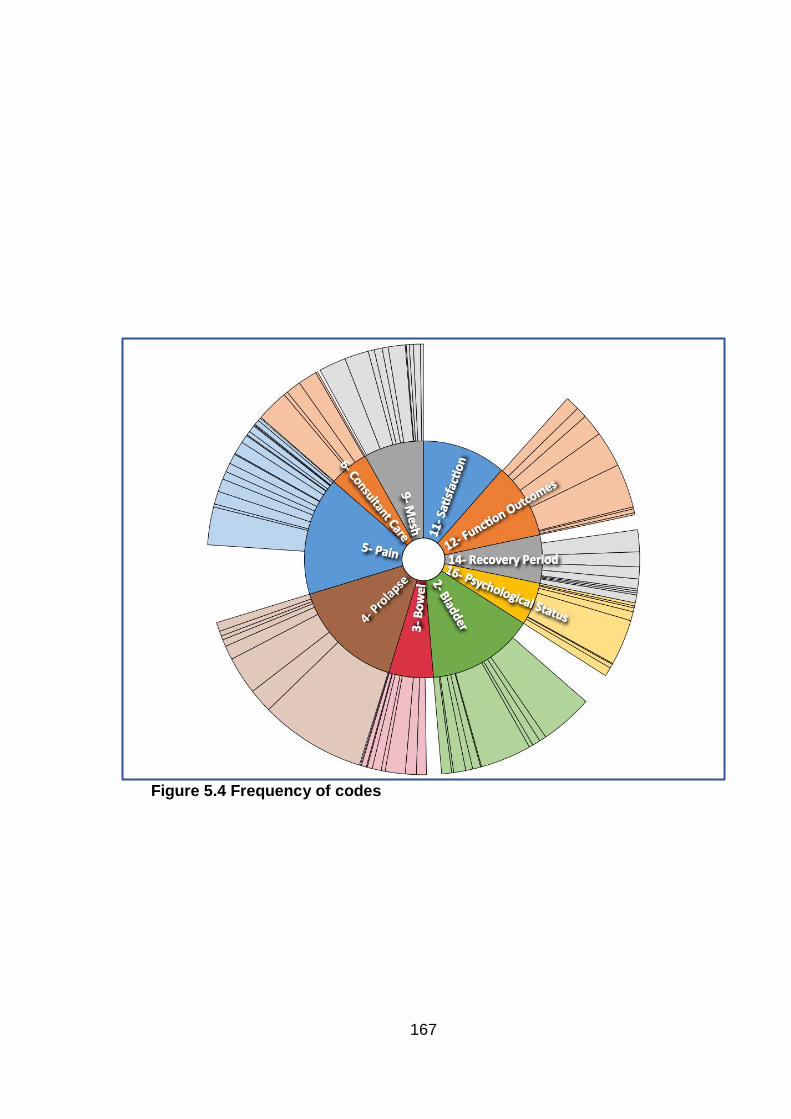
167
Figure 5.4 Frequency of codes
168
5.4.2 Key findings
Step 6 involved analysis of the themes, we found this dataset to provide a
unique insight into the general comments and health-related issues
experienced by women following mesh augmented prolapse surgery. It is
essential to consider these within the context of the current controversies and
media attention surrounding the use of surgical synthetic mesh in
gynaecological surgery [13]. From the six core themes that emerges from our
analysis there are three important conclusions that can be drawn. Firstly, is
that despite the ongoing media coverage and public conversation surrounding
mesh and mesh associated complications, the principal focus of women are
their pelvic floor symptoms and associated QoL. There was a frequent
crossover of sub-codes and codes between the themes pelvic floor symptoms,
health status and treatment success corroborating the established relationship
between prolapse and QoL in the literature [296, 328]. It appeared that women
determine the success of surgery based on an improvement in their pelvic floor
symptoms and any associated improvement in the ability to function in daily
life, rather than by the side effects of surgery or ongoing symptoms. This
illustrates the nuanced and highly considered interpretation by women of their
own personal treatment success, and an understanding that surgery may not
be a panacea for the many symptoms associated with pelvic floor dysfunction,
a finding noted in similar studies [329].
Secondly, analysis uncovered that many women have concerns about the use
of mesh following their pelvic floor surgery. It appears that these concerns
often exist in the absence of symptoms attributable to mesh associated
complications and in women who report a successful surgical outcome. They
voiced concerns about the development and subsequent regulation of mesh
devices their use in pelvic floor surgery, an issue highlighted in the medical
literature [330]. Anxiety about the potential to develop mesh associated
complications in the future appears to be relatively common. Women also
commented on a need for high quality pre- and postoperative information
resources, a subject recognised in previous qualitative studies of prolapse
[329, 331].

169
Finally, there was regular reference to pain, either associated with other pelvic
floor symptoms, attributed to surgery or the use of mesh. It is clear that chronic
pain has huge implications for those women that suffer from it. It must
however be recognised that has been associated with many forms of
gynaecological surgery and has a multifactorial aetiology [278]. While most
women clarified that they found their pain manageable, the frequent reference
to pain symptoms in women who have had prolapse surgery raises questions
as to the relationship between pelvic floor symptoms, reconstructive surgery
and pain.
5.5 Discussion
Principal findings
The analysis and interpretation of these data provide several key
considerations for women’s health researchers and the wider medical
community. The proportion of women that chose to respond and then also to
provide free-text responses in our study illustrates that women value research
participation. This highlights the importance of placing the patient voice at the
centre of medical research, and routine integration into the wider healthcare
infrastructure, a concept promoted by health organisations internationally
including the World Health Organisation [18, 19, 268].
Clinical implications and research implications
With respect to the implications for the future, one could advocate three
courses of action. Firstly, is that the controversies that have mired the use of
mesh should not distract clinicians caring for women with pelvic floor
dysfunction from the fact that prolapse, bladder and bowel symptoms, as well
as sexual dysfunction, remain their predominant health concerns as illustrated
by our first three themes. Secondly, is the recognition that coverage of these
controversies has created a public health issue in the form of widespread
concern. There are therefore two issues that need to be addressed. One is
the provision of evidence-based and high-quality information resources to

170
provide reassurance for the many women who we found to be actively
concerned about having had mesh augmented surgery. This means that high
quality research into mesh associated complications needs to be undertaken
with rapid translation into patient friendly information sources. The other factor
to address is that women affected by potential mesh complications need
accessible care that is delivered to a high standard. This will require
coordinated and regulated centres of excellence delivering evidence-based
care where with appropriate governance and care pathways. In the UK this is
taking the form of specialised commissioning. Finally, there is a need for
further research into the relationship between pelvic floor dysfunction, pelvic
floor surgery and pain. Such a relationship has not been adequately explored
in the literature to date, with many studies of mesh augmented procedures
failing to collect data on pain in a validated way.
Other issues raised cover aspects such as information giving and regrets over
decisions surrounding surgery. This highlights the need for routine and
comprehensive patient information resources and counselling, reaffirming the
latest NICE guidance that emphasise conservative management and a
framework for shared decision making [133]. The final consideration is that
there remains widespread appreciation for the care provided to women and a
value of the patient clinician relationship. Sometimes lost amongst the more
critical headlines found within the field, this could provide reassurance for
clinicians. It also frames the importance of such a relationship both for
individuals and on a wider national level.
Strengths and limitations
The strengths of this study include the use of an inductive approach to
thematic analysis. This allowed for a wide-ranging documentation and
examination of women’s comments. While open to subjectivity and
interpretation, this methodology does allow for a more detailed understanding
of key issues that is not afforded by more prescriptive quantitative techniques.
It therefore delivers an open, patient reported dataset, rather than a pre-
171
defined and categorised responses found with more quantitative
methodologies,
The data come from one of the largest available studies of women who have
undergone mesh augmented prolapse surgery and are therefore more likely
to be representative of this cohort of women. This sits in stark contrast to the
comments and experiences presented in the Cumberledge report and in
national reviews. These reports have actively solicited comments from those
who report mesh associated complications, providing a sample that is likely to
be unrepresentative of most women’s experiences [15, 236]. Our data provide
a more balanced commentary on this form of surgery, studied in a
methodological and systematic process.
The key limitation of this study is the use of a questionnaire. This introduces
inherent methodological biases, and it is difficult to ascertain how
representative respondents are of the wider patient group, and how reflective
their recollections of areas such as symptom status are of their true
preoperative state. Future work could benefit from both pre and postoperative
data collection with more in-depth interviews to explore some of the themes
identified in this study. Despite attempts to provide as balanced analysis of
comments as possible, qualitative researchers bring their own subconscious
bias in how they interpret data and draw conclusions from this.
This chapter has been published as a peer-reviewed manuscript: Izett‐Kay,
M.L., Lumb, C., Cartwright, R., Kupelian, A.S., Cutner, A.S., Jackson, S.,
Price, N. and Vashisht, A., “What research was carried out on this vaginal
mesh?” Health related concerns in women following mesh augmented
prolapse surgery: a thematic analysis. BJOG: An International Journal of
Obstetrics & Gynaecology [332].
172
Conclusion
6.1 Introduction
This thesis describes the role of LSH for the treatment of uterine prolapse. For
many women this common condition leads to the need for corrective surgery,
and for the majority of these patients VH is the surgical intervention of choice.
Yet there remain significant disadvantages associated with this ‘gold standard’
procedure. Laparoscopic mesh sacrohysteropexy may offer a solution to some
of these shortcomings, yet is supported by a paucity of evidence and much of
that which exists is of low quality. The need for more data is particularly
noteworthy given the concerns and global scrutiny of non-absorbable mesh
use in gynaecological surgery. This thesis therefore set out to answer four
principal aims. Firstly, to establish the safety of LSH with particular reference
to mesh associated complications. Secondly, to compare LSH to the current
gold standard of VH. Thirdly, to explore the role of UDS in predicting
postoperative bladder function. Fourthly, and perhaps most importantly, to
contextualise these three chapters by framing them with the patient
perspective through the use of qualitative research methodologies assessing
the health concerns of women following LSH.
6.2 Principal findings
The principal findings from the data presented within the various chapters are
as follows:
Chapter 2
A low rate of reoperation for mesh associated complications following LSH. At
a median follow-up of nearly four years, the incidence was 0.4% of from a
large cohort of 1,121 study participants.
Chapter 3
A trend towards a lower rate of apical reoperation following LSH as compared
to VH, although this was not statistically significant. At seven years, the

173
objective success rate based on apical reoperation was 83% after VH and
94% after LSH.
Chapter 4
While UDS diagnoses appear to correlate poorly with postoperative bladder
function in women undergoing LSH, they may facilitate patient counselling with
respect to risk of de-novo symptoms, resolution of LUTS, and consideration of
concurrent incontinence procedures.
Chapter 5
Despite ongoing media coverage and public conversation surrounding mesh,
the principal focus for women is their pelvic floor symptoms and associated
QoL. There are concerns about the use of mesh following their pelvic floor
surgery, and there was regular reference to pain, associated with other pelvic
floor symptoms as well as attributed to the surgery and use of mesh
6.3 Summary
The data presented in Chapter 2 provide a pragmatic insight into the safety
and efficacy of LSH. The large, patient reported dataset must be considered
within the wider context of other forms of available evidence supporting LSH.
The principle finding was a low incidence of mesh complications requiring
removal of hysteropexy mesh of 0.4% at nearly four years. This provides
adequate reassurance that laparoscopic mesh sacrohysteropexy should
continue to be offered with the appropriate clinician training, decision-making
processes, consent, audit and governance.
Findings from Chapter 3 illustrate that LSH and VH both offer safe
interventions for the common condition of uterine POP. With reasonably low
rates of reoperation and good symptomatic resolution across the range of
pelvic floor disorders, women can be confident that neither surgery appears
significantly inferior. Given a trend towards differences in apical reoperation
rates and advantages with respect to TVL and POP-Q point C, some patients

174
may opt to choose LSH over VH, however this study does not definitively
support such an advantage. Indeed, with composite IVIQ-VS scores favouring
VH the choice of surgical procedure for women continues to need the support
of a rigorous consent processes and the field of urogynaecology needs further
action towards quality RCTs.
In Chapter 4, the clinical utility of UDS for women undergoing LSH has been
appraised. The only significant correlation between UDS diagnosis and
postoperative LUTS is for those with VD for whom there remains a statistically
significant yet low risk of ongoing VD following surgery. For women with USI,
undergoing a concurrent continence surgery is associated with a significantly
lower risk of postoperative SUI and improved incontinence, supporting an
argument for considering such procedures at the time of LSH. Overall, most
patients are asymptomatic after surgery regardless of diagnosis, reflected by
a significant reduction in validated PROM scores and an asymptomatic
symptom status particularly in those with DO and VD. Translation of the
findings into clinical practice is limited by retrospective data and small
numbers, however further research is required to better understand the
prognostic role of UDS to aid patient counselling as well as the role of
concurrent continence procedures and LSH.
Finally, Chapter 5 presents the first comprehensive and systematic study of
comments from women who have undergone mesh augmented surgery for
prolapse. These comments must be contextualised with respect to the current
media coverage that has surrounded the use of mesh in pelvic floor surgery.
It is clear that the main priority for women remain their pelvic floor symptoms
regardless of the treatment controversies. Clinicians who work within the field
of pelvic floor medicine should remain alert to this fact and shape their care to
match patient needs. There is a requirement for high quality research into
mesh associated complications and further exploration of links between pain
and pelvic floor symptoms and treatments. There is also a need for
contemporary as well as evidence-based information resources for patients.
With widespread calls for the voices of women to be put at the centre of the
175
regulatory process and clinical decision making, this study shows that it is
possible. Furthermore, it illustrates that a truly representative patient voice
gives valuable hitherto unheard insights, far beyond messages captured by
the medical and mainstream media headlines.
To summarise, LSH appears to be safe from the perspective of mesh
associated complications, yet large database studies that have been used to
study other mesh augmented procedures are needed. These, along with high-
quality RCTs with long-term follow-up designed to consider both safety and
efficacy should be able to establish the relative merits of LSH, particularly in
comparison to VH. It would appear that there may be advantages to LSH, but
these are not proven by the RCT data presented. Given the frequent co-
existence of LUTS, UDS deserve further prospective scrutiny but do not
generally appear to offer significant prognostic value. Pelvic floor symptoms
remain women’s predominant concern, despite concerns about mesh; further
qualitative studies to firmly establish patient priorities may help to shape future
regulatory frameworks, commissioning, as well as the design of quantitative
studies.

176
References
1. Slieker-ten Hove, M.C.P., et al., The prevalence of pelvic organ prolapse symptoms and signs and their relation with bladder and bowel disorders in a general female population. International Urogynecology Journal, 2009. 20(9): p. 1037-1045.
2. Haylen, B.T., et al., An International Urogynecological Association (IUGA)/International Continence Society (ICS) joint report on the terminology for female pelvic organ prolapse (POP). International urogynecology journal, 2016. 27(2): p. 165-194.
3. Hendrix, S.L., et al., Pelvic organ prolapse in the Women's Health Initiative: gravity and gravidity. American journal of obstetrics and gynecology, 2002. 186(6): p. 1160-1166.
4. Smith, F.J., et al., Lifetime risk of undergoing surgery for pelvic organ prolapse. Obstetrics & Gynecology, 2010. 116(5): p. 1096-1100.
5. Jha, S., A. Cutner, and P. Moran, The UK national prolapse survey: 10 years on. International urogynecology journal, 2018. 29(6): p. 795-801.
6. Frick, A.C., et al., Attitudes toward hysterectomy in women undergoing evaluation for uterovaginal prolapse. Female pelvic medicine & reconstructive surgery, 2013. 19(2): p. 103-109.
7. Korbly, N.B., et al., Patient preferences for uterine preservation and hysterectomy in women with pelvic organ prolapse. American journal of obstetrics and gynecology, 2013. 209(5): p. 470. e1-470. e6.
8. Jelovsek, J.E., et al., Effect of uterosacral ligament suspension vs sacrospinous ligament fixation with or without perioperative behavioral therapy for pelvic organ vaginal prolapse on surgical outcomes and prolapse symptoms at 5 years in the OPTIMAL randomized clinical trial. Jama, 2018. 319(15): p. 1554-1565.
9. Edenfield, A.L., et al., Vaginal prolapse recurrence after uterosacral ligament suspension in normal-weight compared with overweight and obese women. Obstetrics & Gynecology, 2013. 121(3): p. 554-559.
10. Olsen, A.L., et al., Epidemiology of surgically managed pelvic organ prolapse and urinary incontinence. Obstetrics & Gynecology, 1997. 89(4): p. 501-506.

177
11. Zacche, M.M., S. Mukhopadhyay, and I. Giarenis, Trends in prolapse surgery in England. International urogynecology journal, 2018. 29(11): p. 1689-1695.
12. Meriwether, K.V., et al., Uterine preservation vs hysterectomy in pelvic organ prolapse surgery: a systematic review with meta-analysis and clinical practice guidelines. American journal of obstetrics and gynecology, 2018. 219(2): p. 129-146. e2.
13. Gornall, J., How mesh became a four letter word. Bmj, 2018. 363: p. k4137.
14. The safety of surgical meshes used in urogynaecological surgery. 2015, SCENIHR, European Commission: https://ec.europa.eu/health/scientific_committees/consultations/public_consultations/scenihr_consultation_27_en.
15. The Scottish independent review of the use, safety and efficacy of transvaginal mesh implants in the treatment of stress urinary incontinence and pelvic organ prolapse in women: final report. 2017, Scottish Government: www.gov.scot/Publications/2017/03/3336/3.
16. Mesh Oversight Group Report. 2017, NHS England: https://www.england.nhs.uk/publication/mesh-oversight-group-report/.
17. TGA undertakes regulatory actions after review into urogynaecological surgical mesh implants. 2018, Therapeutic Goods Administration, Australian Department of Health: https://www.tga.gov.au/alert/tga-actions-after-review-urogynaecological-surgical-mesh-implants.
18. Organization, W.H., WHO global strategy on people-centred and integrated health services: interim report. 2015, World Health Organization.
19. England, N., Involving people in their own care 2018 2018. 20. DeLancey, J.O., Anatomie aspects of vaginal eversion after
hysterectomy. American journal of obstetrics and gynecology, 1992. 166(6): p. 1717-1728.
21. Luo, J., et al., Using stress MRI to analyze the 3D changes in apical ligament geometry from rest to maximal Valsalva: a pilot study. International urogynecology journal, 2014. 25(2): p. 197-203.
22. Albright, T.S., et al., Arcus tendineus fascia pelvis: a further understanding. American journal of obstetrics and gynecology, 2005. 193(3): p. 677-681.
23. Larson, K.A., et al., 3D analysis of cystoceles using magnetic resonance imaging assessing midline, paravaginal, and

178
apical defects. International urogynecology journal, 2012. 23(3): p. 285-293.
24. Halban, J., Anatomie und aetiologie der genitalprolapse beim weibe. 1907: Braumüller.
25. DeLancey, J.O., et al., Comparison of levator ani muscle defects and function in women with and without pelvic organ prolapse. Obstetrics & Gynecology, 2007. 109(2): p. 295-302.
26. Dietz, H.P. and A.B. Steensma, The prevalence of major abnormalities of the levator ani in urogynaecological patients. BJOG: An International Journal of Obstetrics & Gynaecology, 2006. 113(2): p. 225-230.
27. Mothes, A., et al., Risk index for pelvic organ prolapse based on established individual risk factors. Archives of gynecology and obstetrics, 2016. 293(3): p. 617-624.
28. Buchsbaum, G.M., et al., Pelvic organ prolapse in nulliparous women and their parous sisters. Obstetrics & Gynecology, 2006. 108(6): p. 1388-1393.
29. Altman, D., et al., Genetic influence on stress urinary incontinence and pelvic organ prolapse. European urology, 2008. 54(4): p. 918-923.
30. Allen-Brady, K., et al., Identification of six loci associated with pelvic organ prolapse using genome-wide association analysis. Obstetrics and gynecology, 2011. 118(6): p. 1345.
31. Allen-Brady, K., et al., Evidence for pelvic organ prolapse predisposition genes on chromosomes 10 and 17. American journal of obstetrics and gynecology, 2015. 212(6): p. 771. e1-771. e7.
32. Chen, B. and J. Yeh, Alterations in connective tissue metabolism in stress incontinence and prolapse. The Journal of urology, 2011. 186(5): p. 1768-1772.
33. YAMAMOTO, K., et al., Decrease in elastin gene expression and protein synthesis in fibroblasts derived from cardinal ligaments of patients with prolapsus uteri. Cell biology international, 1997. 21(9): p. 605-611.
34. Ewies, A.A., F. Al‐Azzawi, and J. Thompson, Changes in
extracellular matrix proteins in the cardinal ligaments of post‐menopausal women with or without prolapse: a computerized immunohistomorphometric analysis. Human Reproduction, 2003. 18(10): p. 2189-2195.
35. Vulic, M., et al., Difference in expression of collagen type I and matrix metalloproteinase-1 in uterosacral ligaments of women with and without pelvic organ prolapse. European
179
Journal of Obstetrics & Gynecology and Reproductive Biology, 2011. 155(2): p. 225-228.
36. Yucel, N., et al., Immunohistochemical analysis of connective tissue in patients with pelvic organ prolapse. Journal of molecular histology, 2013. 44(1): p. 97-102.
37. Connell, K.A., et al., HOXA11 is critical for development and maintenance of uterosacral ligaments and deficient in pelvic prolapse. The Journal of clinical investigation, 2008. 118(3): p. 1050-1055.
38. Drewes, P.G., et al., Pelvic organ prolapse in fibulin-5 knockout mice: pregnancy-induced changes in elastic fiber homeostasis in mouse vagina. The American journal of pathology, 2007. 170(2): p. 578-589.
39. Ward, R.M., et al., Genetic epidemiology of pelvic organ prolapse: a systematic review. American journal of obstetrics and gynecology, 2014. 211(4): p. 326-335.
40. MacLennan, A.H., et al., The prevalence of pelvic floor disorders and their relationship to gender, age, parity and mode of delivery. BJOG: An International Journal of Obstetrics & Gynaecology, 2000. 107(12): p. 1460-1470.
41. Leijonhufvud, Å., et al., Risks of stress urinary incontinence and pelvic organ prolapse surgery in relation to mode of childbirth. American journal of obstetrics and gynecology, 2011. 204(1): p. 70. e1-70. e7.
42. Diez-Itza, I., et al., Influence of mode of delivery on pelvic organ support 6 months postpartum. Gynecologic and obstetric investigation, 2011. 72(2): p. 123-129.
43. Larsson, C., K. Källen, and E. Andolf, Cesarean section and risk of pelvic organ prolapse: a nested case-control study. American journal of obstetrics and gynecology, 2009. 200(3): p. 243. e1-243. e4.
44. Gyhagen, M., et al., Prevalence and risk factors for pelvic organ prolapse 20 years after childbirth: a national cohort study in singleton primiparae after vaginal or caesarean delivery. BJOG: An International Journal of Obstetrics & Gynaecology, 2013. 120(2): p. 152-160.
45. Mant, J., R. Painter, and M. Vessey, Epidemiology of genital prolapse: observations from the Oxford Family Planning Association Study. BJOG: An International Journal of Obstetrics & Gynaecology, 1997. 104(5): p. 579-585.
46. Harris, R.L., et al., Urinary incontinence and pelvic organ prolapse in nulliparous women. Obstetrics & Gynecology, 1998. 92(6): p. 951-954.

180
47. Rortveit, G., et al., Symptomatic pelvic organ prolapse: prevalence and risk factors in a population-based, racially diverse cohort. Obstetrics & Gynecology, 2007. 109(6): p. 1396-1403.
48. Miodrag, A., C.M. Castleden, and T.R. Vallance, Sex hormones and the female urinary tract. Drugs, 1988. 36(4): p. 491-504.
49. WAHL, L.M., R.J. BLANDAU, and R.C. PAGE, Effect of hormones on collagen metabolism and collagenase activity in the pubic symphysis ligament of the guinea pig. Endocrinology, 1977. 100(2): p. 571-579.
50. Lavin, J., et al., The effect of the first pregnancy on the connective tissue of the rectus sheath. NEUROUROLOGY AND URODYNAMICS, 1997. 16: p. 27-27.
51. Landon, C., et al., Mechanical properties of fascia during pregnancy: a possible factor in the development of stress incontinence of urine. Contemp Rev Obstet Gynaecol, 1990. 2: p. 40-46.
52. Svabik, K., K. Shek, and H. Dietz, How much does the levator hiatus have to stretch during childbirth? BJOG: An International Journal of Obstetrics & Gynaecology, 2009. 116(12): p. 1657-1662.
53. Delancey, J.O. and W.W. Hurd, Size of the urogenital hiatus in the levator ani muscles in normal women and women with pelvic organ prolapse. Obstetrics & Gynecology, 1998. 91(3): p. 364-368.
54. Dietz, H.P. and V. Lanzarone, Levator trauma after vaginal delivery. Obstetrics & Gynecology, 2005. 106(4): p. 707-712.
55. Albrich, S., et al., Impact of mode of delivery on levator
morphology: a prospective observational study with three‐dimensional ultrasound early in the postpartum period. BJOG: An International Journal of Obstetrics & Gynaecology, 2012. 119(1): p. 51-61.
56. Rostaminia, G., et al., Levator ani deficiency and pelvic organ prolapse severity. Obstetrics & Gynecology, 2013. 121(5): p. 1017-1024.
57. Dietz, H. and J. Simpson, Levator trauma is associated with pelvic organ prolapse. BJOG: An International Journal of Obstetrics & Gynaecology, 2008. 115(8): p. 979-984.
58. Smith, L.A., et al., Incidence of and risk factors for perineal trauma: a prospective observational study. BMC pregnancy and childbirth, 2013. 13(1): p. 59.

181
59. Allen, R., et al., Pelvic floor damage and childbirth: a neurophysiological study. BJOG: An International Journal of Obstetrics & Gynaecology, 1990. 97(9): p. 770-779.
60. Busacchi, P., et al., Abnormalities of somatic peptide-containing nerves supplying the pelvic floor of women with genitourinary prolapse and stress urinary incontinence. Urology, 2004. 63(3): p. 591-595.
61. Marshall, K., D. Walsh, and G. Baxter, The effect of a first vaginal delivery on the integrity of the pelvic ‘oor musculature. Clinical rehabilitation, 2002. 16(7): p. 795-799.
62. Takano, C., et al., Analysis of collagen in parametrium and vaginal apex of women with and without uterine prolapse. International Urogynecology Journal, 2002. 13(6): p. 342-345.
63. Moalli, P.A., et al., Remodeling of vaginal connective tissue in patients with prolapse. Obstetrics & Gynecology, 2005. 106(5): p. 953-963.
64. Al-Rawi, Z. and Z. Al-Rawi, Joint hypermobility in women with genital prolapse. The Lancet, 1982. 319(8287): p. 1439-1441.
65. Carley, M.E. and J. Schaffer, Urinary incontinence and pelvic organ prolapse in women with Marfan or Ehlers-Danlos syndrome. American journal of obstetrics and gynecology, 2000. 182(5): p. 1021-1023.
66. Chiaffarino, F., et al., Reproductive factors, family history, occupation and risk of urogenital prolapse. European journal of obstetrics & gynecology and Reproductive Biology, 1999. 82(1): p. 63-67.
67. Ewies, A.A., J. Thompson, and F. Al‐Azzawi, Changes in gonadal steroid receptors in the cardinal ligaments of prolapsed uteri: immunohistomorphometric data. Human Reproduction, 2004. 19(7): p. 1622-1628.
68. Swift, S.E., S.B. Tate, and J. Nicholas, Correlation of symptoms with degree of pelvic organ support in a general population of women: what is pelvic organ prolapse? American journal of obstetrics and gynecology, 2003. 189(2): p. 372-377.
69. Spence‐Jones, C., et al., Bowel dysfunction: a pathogenic factor in uterovaginal prolapse and urinary stress incontinence. BJOG: An International Journal of Obstetrics & Gynaecology, 1994. 101(2): p. 147-152.
70. Woodman, P.J., et al., Prevalence of severe pelvic organ prolapse in relation to job description and socioeconomic
182
status: a multicenter cross-sectional study. International Urogynecology Journal, 2006. 17(4): p. 340-345.
71. Samuelsson, E.C., et al., Signs of genital prolapse in a Swedish population of women 20 to 59 years of age and possible related factors. American journal of obstetrics and gynecology, 1999. 180(2): p. 299-305.
72. Jelovsek, J.E. and M.D. Barber, Women seeking treatment for advanced pelvic organ prolapse have decreased body image and quality of life. American journal of obstetrics and gynecology, 2006. 194(5): p. 1455-1461.
73. Doaee, M., et al., Management of pelvic organ prolapse and quality of life: a systematic review and meta-analysis. International urogynecology journal, 2014. 25(2): p. 153-163.
74. Abdel-Fattah, M., et al., Primary and repeat surgical treatment for female pelvic organ prolapse and incontinence in parous women in the UK: a register linkage study. BMJ open, 2011. 1(2): p. e000206.
75. Subramanian, D., et al., Rate, type, and cost of pelvic organ prolapse surgery in Germany, France, and England. European Journal of Obstetrics & Gynecology and Reproductive Biology, 2009. 144(2): p. 177-181.
76. Haya, N., et al., Prolapse and continence surgery in countries of the Organization for Economic Cooperation and Development in 2012. American journal of obstetrics and gynecology, 2015. 212(6): p. 755. e1-755. e27.
77. Wu, J.M., et al., Predicting the number of women who will undergo incontinence and prolapse surgery, 2010 to 2050. American journal of obstetrics and gynecology, 2011. 205(3): p. 230. e1-230. e5.
78. Kim, S., M.-A. Harvey, and S. Johnston, A review of the epidemiology and pathophysiology of pelvic floor dysfunction: do racial differences matter? Journal of Obstetrics and Gynaecology Canada, 2005. 27(3): p. 251-259.
79. Dietz, H., Do Asian women have less pelvic organ mobility than Caucasians? International Urogynecology Journal, 2003. 14(4): p. 250-253.
80. Cheung, R.Y., et al., Pelvic organ prolapse in Caucasian and East Asian women: a comparative study. Ultrasound in Obstetrics & Gynecology, 2019. 53(4): p. 541-545.
81. Dunivan, G.C., et al., Ethnicity and variations of pelvic organ prolapse bother. International urogynecology journal, 2014. 25(1): p. 53-59.

183
82. Whitcomb, E.L., et al., Racial differences in pelvic organ prolapse. Obstetrics and gynecology, 2009. 114(6): p. 1271.
83. Brazell, H.D., D.M. O'Sullivan, and P.K. Tulikangas, Socioeconomic status and race as predictors of treatment-seeking behavior for pelvic organ prolapse. American journal of obstetrics and gynecology, 2013. 209(5): p. 476. e1-476. e5.
84. Jones, K., et al., Effect of pessary use on genital hiatus measurements in women with pelvic organ prolapse. Obstetrics & Gynecology, 2008. 112(3): p. 630-636.
85. Lowder, J.L., et al., Body image perceptions in women with pelvic organ prolapse: a qualitative study. American journal of obstetrics and gynecology, 2011. 204(5): p. 441. e1-441. e5.
86. Abrams, P., Cardozo, L, Wagg, A, Wein, A. (Eds), Incontinence 6th Edition (2017). ICI-ICS. 2017, Bristol UK: International Continence Society.
87. Price, N., et al., Development and psychometric evaluation of
the ICIQ Vaginal Symptoms Questionnaire: the ICIQ‐VS. BJOG: An International Journal of Obstetrics & Gynaecology, 2006. 113(6): p. 700-712.
88. Dietz, H. and K. Mann, What is clinically relevant prolapse? An attempt at defining cutoffs for the clinical assessment of pelvic organ descent. International urogynecology journal, 2014. 25(4): p. 451-455.
89. Ellerkmann, R.M., et al., Correlation of symptoms with location and severity of pelvic organ prolapse. American journal of obstetrics and gynecology, 2001. 185(6): p. 1332-1338.
90. Lowder, J.L., et al., Lower urinary tract symptoms in women with pelvic organ prolapse. International urogynecology journal, 2010. 21(6): p. 665-672.
91. Ghetti, C., et al., Pelvic organ descent and symptoms of pelvic floor disorders. American journal of obstetrics and gynecology, 2005. 193(1): p. 53-57.
92. Richardson, D.A., A.E. Bent, and D.R. Ostergard, The effect of uterovaginal prolapse on urethrovesical pressure dynamics. American journal of obstetrics and gynecology, 1983. 146(8): p. 901-905.
93. Borstad, E. and T. Rud, The Risk Of Developing Urinary
Stress‐Incontinence After Vaginal Repair In Continent
Women: A Clinical And Urodynamic Follow‐Up Study. Acta
184
obstetricia et gynecologica Scandinavica, 1989. 68(6): p. 545-549.
94. STANTON, S.L., et al., Clinical and urodynamic effects of anterior colporrhaphy and vaginal hysterectomy for prolapse with and without incontinence. BJOG: An International Journal of Obstetrics & Gynaecology, 1982. 89(6): p. 459-463.
95. Brubaker, L., et al., Abdominal sacrocolpopexy with Burch colposuspension to reduce urinary stress incontinence. New England Journal of Medicine, 2006. 354(15): p. 1557-1566.
96. Nygaard, I., et al., Long-term outcomes following abdominal sacrocolpopexy for pelvic organ prolapse. Jama, 2013. 309(19): p. 2016-2024.
97. Van der Ploeg, J., et al., Prolapse surgery with or without stress incontinence surgery for pelvic organ prolapse: a
systematic review and meta‐analysis of randomised trials. BJOG: An International Journal of Obstetrics & Gynaecology, 2014. 121(5): p. 537-547.
98. Costantini, E., et al., Pelvic organ prolapse repair with and without concomitant burch colposuspension in incontinent women: a randomised controlled trial with at least 5-year followup. Obstetrics and gynecology international, 2012. 2012.
99. BADEN, W.F. and T.A. WALKED, Genesis of the vaginal profile: a correlated classification of vaginal relaxation. Clinical obstetrics and gynecology, 1972. 15(4): p. 1048-1054.
100. Bump, R.C., et al., The standardization of terminology of female pelvic organ prolapse and pelvic floor dysfunction. American journal of obstetrics and gynecology, 1996. 175(1): p. 10-17.
101. Kegel, A.H., Progressive resistance exercise in the functional restoration of the perineal muscles. American journal of obstetrics and gynecology, 1948. 56(2): p. 238-248.
102. Laycock, J., Incontinence. Pelvic floor re-education. Nursing, 1991. 4(39): p. 15-17.
103. Bø, K. and H.B. Finckenhagen, Vaginal palpation of pelvic
floor muscle strength: Inter‐test reproducibility and comparison between palpation and vaginal squeeze pressure. Acta obstetricia et gynecologica Scandinavica, 2001. 80(10): p. 883-887.
185
104. Athanasiou, S., et al., Validation of the ICS proposed pelvic organ prolapse descriptive system. Neurourology and Urodynamics, 1995. 14: p. 4-4.
105. Hall, A.F., et al., Interobserver and intraobserver reliability of the proposed International Continence Society, Society of Gynecologic Surgeons, and American Urogynecologic Society pelvic organ prolapse classification system. American journal of obstetrics and gynecology, 1996. 175(6): p. 1467-1471.
106. Nelson, E.C., et al., Patient reported outcome measures in practice. Bmj, 2015. 350: p. g7818.
107. Devlin, N.J. and J. Appleby, Getting the most out of PROMS. Putting health outcomes at the heart of NHS decision-making. London: The King's Fund, 2010.
108. Kozma, C., C. Reeder, and R. Schulz, Economic, clinical, and humanistic outcomes: a planning model for pharmacoeconomic research. Clinical therapeutics, 1993. 15(6): p. 1121-32; discussion 1120.
109. Yalcin, I. and R.C. Bump, Validation of two global impression questionnaires for incontinence. American journal of obstetrics and gynecology, 2003. 189(1): p. 98-101.
110. Feinstein, A.R., Clinimetrics. 1987: Yale University Press. 111. Gill, T.M. and A.R. Feinstein, A critical appraisal of the quality
of quality-of-life measurements. Jama, 1994. 272(8): p. 619-626.
112. Srikrishna, S., D. Robinson, and L. Cardozo, Validation of the Patient Global Impression of Improvement (PGI-I) for urogenital prolapse. International urogynecology journal, 2010. 21(5): p. 523-528.
113. Dietz, H., Ultrasound imaging of the pelvic floor. Part I: two‐dimensional aspects. Ultrasound in Obstetrics and Gynecology, 2004. 23(1): p. 80-92.
114. Rizk, D.E., J. Czechowski, and L. Ekelund, Dynamic assessment of pelvic floor and bony pelvis morphologic condition with the use of magnetic resonance imaging in a multiethnic, nulliparous, and healthy female population. American journal of obstetrics and gynecology, 2004. 191(1): p. 83-89.
115. Fielding, J.R., et al., MR-based three-dimensional modeling of the normal pelvic floor in women: quantification of muscle mass. American Journal of Roentgenology, 2000. 174(3): p. 657-660.

186
116. Margulies, R.U., et al., Appearance of the levator ani muscle subdivisions in magnetic resonance images. Obstetrics and gynecology, 2006. 107(5): p. 1064.
117. Zijta, F., et al., Evaluation of the female pelvic floor in pelvic organ prolapse using 3.0-Tesla diffusion tensor imaging and fibre tractography. European radiology, 2012. 22(12): p. 2806-2813.
118. Raza-Khan, F., et al., Prevalence of bowel symptoms in women with pelvic floor disorders. International urogynecology journal, 2010. 21(8): p. 933-938.
119. Jelovsek, J.E., et al., Functional bowel and anorectal disorders in patients with pelvic organ prolapse and incontinence. American journal of obstetrics and gynecology, 2005. 193(6): p. 2105-2111.
120. Picciariello, A., et al., Obstructed defaecation syndrome: European consensus guidelines on the surgical management. British Journal of Surgery, 2021.
121. Müller-Lissner, S., et al., Interobserver agreement in defecography--an international study. Zeitschrift fur Gastroenterologie, 1998. 36(4): p. 273-279.
122. Shorvon, P., et al., Defecography in normal volunteers: results and implications. Gut, 1989. 30(12): p. 1737-1749.
123. Kaufman, H.S., et al., Dynamic pelvic magnetic resonance imaging and cystocolpoproctography alter surgical management of pelvic floor disorders. Diseases of the colon & rectum, 2001. 44(11): p. 1575-1583.
124. Lienemann, A., et al., Diagnosing enteroceles using dynamic magnetic resonance imaging. Diseases of the colon & rectum, 2000. 43(2): p. 205-212.
125. Kelvin, F.M., et al., Female pelvic organ prolapse: a comparison of triphasic dynamic MR imaging and triphasic fluoroscopic cystocolpoproctography. American Journal of Roentgenology, 2000. 174(1): p. 81-88.
126. Goodrich, M.A., et al., Magnetic resonance imaging of pelvic floor relaxation: dynamic analysis and evaluation of patients before and after surgical repair. Obstetrics and gynecology, 1993. 82(6): p. 883-891.
127. van Gruting, I.M., et al., Accuracy of four imaging techniques for diagnosis of posterior pelvic floor disorders. Obstetrics & Gynecology, 2017. 130(5): p. 1017-1024.
128. Jha, S., et al., Does preoperative urodynamics change the management of prolapse? Journal of Obstetrics and Gynaecology, 2008. 28(3): p. 320-322.

187
129. Roovers, J., et al., Does urodynamic investigation improve outcome in patients undergoing prolapse surgery? Neurourology and Urodynamics: Official Journal of the International Continence Society, 2007. 26(2): p. 170-175.
130. Digesu, G.A., et al., Do overactive bladder symptoms improve after repair of anterior vaginal wall prolapse? International Urogynecology Journal, 2007. 18(12): p. 1439-1443.
131. Rosier, P.F., et al., International continence society good urodynamic practices and terms 2016: urodynamics,
uroflowmetry, cystometry, and pressure‐flow study. Neurourology and urodynamics, 2017. 36(5): p. 1243-1260.
132. Visco, A.G., et al., The role of preoperative urodynamic testing in stress-continent women undergoing sacrocolpopexy: the Colpopexy and Urinary Reduction Efforts (CARE) randomized surgical trial. International Urogynecology Journal, 2008. 19(5): p. 607.
133. Urinary incontinence and pelvic organ prolapse in women: management [NG 123]. 2019, National Institute for Health and Care Excellence.
134. Ästrand, P.-O. and K. Rodahl, Textbook of work physiology. Physiological basis of exercise. 3rd edn McGraw-Hill, New York, 1986.
135. Bø, K., Pelvic floor muscle exercise for the treatment of stress urinary incontinence: an exercise physiology perspective. International Urogynecology Journal, 1995. 6(5): p. 282-291.
136. Hagen, S., et al., Individualised pelvic floor muscle training in women with pelvic organ prolapse (POPPY): a multicentre randomised controlled trial. The Lancet, 2014. 383(9919): p. 796-806.
137. Wiegersma, M., et al., Effect of pelvic floor muscle training compared with watchful waiting in older women with symptomatic mild pelvic organ prolapse: randomised controlled trial in primary care. Bmj, 2014. 349: p. g7378.
138. Li, C., Y. Gong, and B. Wang, The efficacy of pelvic floor muscle training for pelvic organ prolapse: a systematic review and meta-analysis. International urogynecology journal, 2016. 27(7): p. 981-992.
139. Hagen, S. and D. Stark, Conservative prevention and management of pelvic organ prolapse in women. Cochrane Database of Systematic Reviews, 2011(12).
140. Oliver, R., R. Thakar, and A.H. Sultan, The history and usage of the vaginal pessary: a review. European journal of

188
obstetrics & gynecology and reproductive biology, 2011. 156(2): p. 125-130.
141. Gorti, M., G. Hudelist, and A. Simons, Evaluation of vaginal pessary management: a UK-based survey. Journal of Obstetrics and Gynaecology, 2009. 29(2): p. 129-131.
142. Kearney, R. and C. Brown, Self-management of vaginal pessaries for pelvic organ prolapse. BMJ Open Quality, 2014. 3(1): p. u206180. w2533.
143. Bugge, C., et al., The TOPSY pessary self-management intervention for pelvic organ prolapse: a study protocol for the process evaluation. Trials, 2020. 21(1): p. 1-9.
144. Bugge, C., et al., Pessaries (mechanical devices) for pelvic organ prolapse in women. Cochrane Database of Systematic Reviews, 2013(2).
145. Cundiff, G.W., et al., The PESSRI study: symptom relief outcomes of a randomized crossover trial of the ring and Gellhorn pessaries. American journal of obstetrics and gynecology, 2007. 196(4): p. 405. e1-405. e8.
146. Brazell, H.D., et al., The impact of pessary use on bowel symptoms: one-year outcomes. Female pelvic medicine & reconstructive surgery, 2014. 20(2): p. 95-98.
147. Abdool, Z., et al., Prospective evaluation of outcome of vaginal pessaries versus surgery in women with symptomatic pelvic organ prolapse. International urogynecology journal, 2011. 22(3): p. 273-278.
148. Lone, F., R. Thakar, and A.H. Sultan, One-year prospective comparison of vaginal pessaries and surgery for pelvic organ prolapse using the validated ICIQ-VS and ICIQ-UI (SF) questionnaires. International urogynecology journal, 2015. 26(9): p. 1305-1312.
149. Urogynaecology, R.C.o.O.a.G.a.B.S.o., Post-hysterectomy vaginal vault prolapse. GTG No. 96. 2015, RCOG: London.
150. Miller, B.J., et al., Recent trends in the management of pelvic organ prolapse in Australia and New Zealand. Australian and New Zealand Journal of Obstetrics and Gynaecology, 2019. 59(1): p. 117-122.
151. Surgery for uterine prolapse- Patient decision aid. 2019, National Institute for Health and Care Excellence (NICE).
152. Hemming, C., et al., Surgical interventions for uterine prolapse and for vault prolapse: the two VUE RCTs. Health Technology Assessment (Winchester, England), 2020. 24(13): p. 1.

189
153. Prodigalidad, L.T., et al., Long‐term results of prolapse recurrence and functional outcome after vaginal hysterectomy. International Journal of Gynecology & Obstetrics, 2013. 120(1): p. 57-60.
154. Humalajärvi, N., et al., Quality of life and pelvic floor dysfunction symptoms after hysterectomy with or without pelvic organ prolapse. European Journal of Obstetrics & Gynecology and Reproductive Biology, 2014. 182: p. 16-21.
155. Pakbaz, M., I. Mogren, and M. Löfgren, Outcomes of vaginal hysterectomy for uterovaginal prolapse: a population-based, retrospective, cross-sectional study of patient perceptions of results including sexual activity, urinary symptoms, and provided care. BMC women's health, 2009. 9(1): p. 9.
156. Roovers, J.P.W., et al., A randomised controlled trial comparing abdominal and vaginal prolapse surgery: effects on urogenital function. BJOG: An International Journal of Obstetrics & Gynaecology, 2004. 111(1): p. 50-56.
157. Roovers, J., et al. Long term follow-up of a randomized controlled trial comparing abdominal and vaginal surgical correction of uterine prolapse. in International Urogynecology Journal. 2008. SPRINGER LONDON LTD 236 GRAYS INN RD, 6TH FLOOR, LONDON WC1X 8HL, ENGLAND.
158. Husby, K.R., et al., Surgical treatment of primary uterine prolapse: a comparison of vaginal native tissue surgical techniques. International urogynecology journal, 2019: p. 1-7.
159. Richardson, D., R. Scotti, and D. Ostergard, Surgical management of uterine prolapse in young women. The Journal of reproductive medicine, 1989. 34(6): p. 388-392.
160. Detollenaere, R.J., et al., Sacrospinous hysteropexy versus vaginal hysterectomy with suspension of the uterosacral ligaments in women with uterine prolapse stage 2 or higher: multicentre randomised non-inferiority trial. Bmj, 2015. 351: p. h3717.
161. Kapoor, S., et al., Sacrospinous hysteropexy: review and meta-analysis of outcomes. International urogynecology journal, 2017. 28(9): p. 1285-1294.
162. Maher, C.F., M.P. Carey, and C.J. Murray, Laparoscopic suture hysteropexy for uterine prolapse. 2001, Elsevier.
163. Jirschele, K., et al., A multicenter, prospective trial to evaluate mesh-augmented sacrospinous hysteropexy for uterovaginal prolapse. International urogynecology journal, 2015. 26(5): p. 743-748.

190
164. Jia, X., et al., Systematic review of the efficacy and safety of using mesh in surgery for uterine or vaginal vault prolapse. International urogynecology journal, 2010. 21(11): p. 1413-1431.
165. Braun, H., et al. Prospective randomised study to compare colposacropexy and Mayo McCall technique in the correction of severe genital central prolapse. in INTERNATIONAL UROGYNECOLOGY JOURNAL. 2007. SPRINGER LONDON LTD ASHBOURNE HOUSE, THE GUILDWAY, OLD PORTSMOUTH ROAD ….
166. Rondini, C., et al., High uterosacral vault suspension vs sacrocolpopexy for treating apical defects: a randomized controlled trial with twelve months follow-up. International urogynecology journal, 2015. 26(8): p. 1131-1138.
167. Costantini, E., et al., Uterus preservation in surgical correction of urogenital prolapse. European urology, 2005. 48(4): p. 642-649.
168. Wu, J.M., et al., Mesh erosion in abdominal sacral colpopexy with and without concomitant hysterectomy. American journal of obstetrics and gynecology, 2006. 194(5): p. 1418-1422.
169. Marinkovic, S.P., Will hysterectomy at the time of sacrocolpopexy increase the rate of polypropylene mesh erosion? International Urogynecology Journal, 2008. 19(2): p. 199-203.
170. Costantini, E., et al., Managing mesh erosion after abdominal pelvic organ prolapse repair: ten years’ experience in a single center. Urologia internationalis, 2011. 86(4): p. 419-423.
171. Akyol, A., et al., Additional surgical risk factors and patient characteristics for mesh erosion after abdominal sacrocolpopexy. Journal of Obstetrics and Gynaecology Research, 2014. 40(5): p. 1368-1374.
172. Griffis, K., et al., Mesh erosion and abdominal sacrocolpopexy: a comparison of prior, total, and supracervical hysterectomy. Female Pelvic Medicine & Reconstructive Surgery, 2006. 12(1): p. 25-30.
173. Crane, A.K., et al., Short-term mesh exposure after robotic sacrocolpopexy with and without concomitant hysterectomy. Southern medical journal, 2014. 107(10): p. 603-606.
174. Manchester, W.F., Anterior colporrhaphy and its combination with amputation of the cervix as a single operation. BJOG: An International Journal of Obstetrics & Gynaecology, 1915.
27(3‐5): p. 146-147.

191
175. Karaman, E., et al., The outcome of Manchester–Fotergill operation for uterine decensus repair: a single center experience. Archives of gynecology and obstetrics, 2014. 290(2): p. 309-314.
176. de Boer, T.A., et al., The effectiveness of surgical correction of uterine prolapse: cervical amputation with uterosacral ligament plication (modified Manchester) versus vaginal hysterectomy with high uterosacral ligament plication. International Urogynecology Journal, 2009. 20(11): p. 1313.
177. Hullfish, K.L., V.E. Bovbjerg, and W.D. Steers, Colpocleisis for pelvic organ prolapse: patient goals, quality of life, and satisfaction. Obstetrics & Gynecology, 2007. 110(2): p. 341-345.
178. Vij, M., et al., Long-term follow-up after colpocleisis: regret, bowel, and bladder function. International urogynecology journal, 2014. 25(6): p. 811-815.
179. Fitzgerald, M., et al., Pelvic support, pelvic symptoms, and patient satisfaction after colpocleisis. International Urogynecology Journal, 2008. 19(12): p. 1603-1609.
180. Barber, M.D., et al., Quality of life after surgery for genital prolapse in elderly women: obliterative and reconstructive surgery. International Urogynecology Journal, 2007. 18(7): p. 799-806.
181. FitzGerald, M.P., et al., Colpocleisis: a review. International Urogynecology Journal, 2006. 17(3): p. 261-271.
182. Heneghan, C.J., et al., Trials of transvaginal mesh devices for pelvic organ prolapse: a systematic database review of the US FDA approval process. BMJ Open, 2017. 7(12): p. e017125.
183. Maher, C., et al., Surgery for women with apical vaginal prolapse. The Cochrane Library, 2016.
184. Sokol, A.I., et al., One-year objective and functional outcomes of a randomized clinical trial of vaginal mesh for prolapse. American journal of obstetrics and gynecology, 2012. 206(1): p. 86. e1-86. e9.
185. Da Silveira, S.D.R.B., et al., Multicenter, randomized trial comparing native vaginal tissue repair and synthetic mesh repair for genital prolapse surgical treatment. International urogynecology journal, 2015. 26(3): p. 335-342.
186. Pandeva, I., M. Mistry, and A. Fayyad, Efficacy and pregnancy outcomes of laparoscopic single sheet mesh sacrohysteropexy. Neurourology and urodynamics, 2017. 36(3): p. 787-793.

192
187. Kelly, H.A., Hysterorrhaphy. 1887: William Wood & Company.
188. Arthure, H.G. and D. Savage, Uterine prolapse and prolapse of the vaginal vault treated by sacral hysteropexy. BJOG: An International Journal of Obstetrics & Gynaecology, 1957. 64(3): p. 355-360.
189. Chaudhuri, S., The place of sling operations in treating genital prolapse in young women. International Journal of Gynecology & Obstetrics, 1979. 16(4): p. 314-320.
190. Farkas, A., J. Shepherd, and C. Woodhouse, Hysterosacropexy for uterine prolapse with associated urinary tract abnormalities. Journal of Obstetrics and Gynaecology, 1993. 13(5): p. 358-360.
191. Cutner, A., R. Kearney, and A. Vashisht, Laparoscopic uterine sling suspension: a new technique of uterine suspension in women desiring surgical management of uterine prolapse with uterine conservation. BJOG: An International Journal of Obstetrics & Gynaecology, 2007. 114(9): p. 1159-1162.
192. Price, N., A. Slack, and S. Jackson, Laparoscopic hysteropexy: the initial results of a uterine suspension procedure for uterovaginal prolapse. BJOG: An International Journal of Obstetrics & Gynaecology, 2010. 117(1): p. 62-68.
193. Preventing entry-related gynaecological laparoscopic injuries. 2008, Royal College of Obstetricians and Gynaecologists (RCOG).
194. Shaw, W.F., The treatment of prolapsus uteri, with special reference to the Manchester operation of colporrhaphy. American Journal of Obstetrics and Gynecology, 1933. 26(5): p. 667-686.
195. Meriwether, K.V., et al., Uterine preservation versus hysterectomy in pelvic organ prolapse surgery: A systematic review with meta-analysis and clinical practice guidelines. American Journal of Obstetrics and Gynecology, 2018.
196. de Oliveira, S.A., et al., Hysteropreservation versus hysterectomy in the surgical treatment of uterine prolapse: systematic review and meta-analysis. International urogynecology journal, 2017. 28(11): p. 1617-1630.
197. Rahmanou, P., N. Price, and S.R. Jackson, Laparoscopic hysteropexy versus vaginal hysterectomy for the treatment of uterovaginal prolapse: a prospective randomized pilot study. International urogynecology journal, 2015. 26(11): p. 1687-1694.

193
198. Daniels, S., et al., Success rates and outcomes of laparoscopic mesh sacrohysteropexy. Australian and New Zealand Journal of Obstetrics and Gynaecology, 2019.
199. Lone, F., T. Curnow, and S.A. Thomas, Laparoscopic sacrohysteropexy versus vaginal hysterectomy for uterovaginal prolapse using validated questionnaires: 2-year prospective study. International urogynecology journal, 2018. 29(1): p. 71-79.
200. Jefferis, H., N. Price, and S. Jackson, Laparoscopic hysteropexy: 10 years’ experience. International urogynecology journal, 2017. 28(8): p. 1241-1248.
201. Kupelian, A.S., et al., Laparoscopic wrap round mesh sacrohysteropexy for the management of apical prolapse. International urogynecology journal, 2016. 27(12): p. 1889-1897.
202. Grimminck, K., et al., Long-term follow-up and quality of life after robot assisted sacrohysteropexy. European Journal of Obstetrics & Gynecology and Reproductive Biology, 2016. 206: p. 27-31.
203. Gutman, R.E., et al., Vaginal and laparoscopic mesh hysteropexy for uterovaginal prolapse: a parallel cohort study. American Journal of Obstetrics & Gynecology, 2017. 216(1): p. 38. e1-38. e11.
204. Paek, J., et al., Robotic or laparoscopic sacrohysteropexy versus open sacrohysteropexy for uterus preservation in pelvic organ prolapse. International urogynecology journal, 2016. 27(4): p. 593-599.
205. Gracia, M., et al., Comparison between laparoscopic sacral hysteropexy and subtotal hysterectomy plus cervicopexy in pelvic organ prolapse: a pilot study. Neurourology and urodynamics, 2015. 34(7): p. 654-658.
206. Api, M., et al., Laparoscopic Sacral Uteropexy with Cravat Technique--Experience and Results. International braz j urol, 2014. 40(4): p. 526-532.
207. Rahmanou, P., et al., Laparoscopic hysteropexy: 1-to 4-year follow-up of women postoperatively. International urogynecology journal, 2014. 25(1): p. 131-138.
208. Lee, T., et al., Uterine sparing robotic-assisted laparoscopic sacrohysteropexy for pelvic organ prolapse: safety and feasibility. Journal of endourology, 2013. 27(9): p. 1131-1136.
209. Khursheed, F., C.M. Das, and A. Ghouri, Abdominal sacrohysteropexy-a conservative surgical treatment of
194
uterine prolapse. Journal of Ayub Medical College Abbottabad, 2013. 25(3-4): p. 41-43.
210. Cvach, K., R. Geoffrion, and G.W. Cundiff, Abdominal sacral hysteropexy: a pilot study comparing sacral hysteropexy to sacral colpopexy with hysterectomy. Female pelvic medicine & reconstructive surgery, 2012. 18(5): p. 286-290.
211. Mourik, S.L., J.E. Martens, and M. Aktas, Uterine preservation in pelvic organ prolapse using robot assisted laparoscopic sacrohysteropexy: quality of life and technique. European Journal of Obstetrics & Gynecology and Reproductive Biology, 2012. 165(1): p. 122-127.
212. Bojahr, B., et al., Laparoscopic sacropexy: a retrospective analysis of perioperative complications and anatomical outcomes. JSLS: Journal of the Society of Laparoendoscopic Surgeons, 2012. 16(3): p. 428.
213. Costantini, E., et al., Five-year outcome of uterus sparing surgery for pelvic organ prolapse repair: a single-center experience. International urogynecology journal, 2011. 22(3): p. 287-292.
214. Moiety, F.M.S., et al., Abdominal sacrohysteropexy for uterovaginal prolapse: a prospective study on 33 cases. Archives of gynecology and obstetrics, 2010. 281(4): p. 631-636.
215. Jeon, M.J., et al., Is hysterectomy or the use of graft necessary for the reconstructive surgery for uterine prolapse? International Urogynecology Journal, 2008. 19(3): p. 351-355.
216. Demirci, F., et al., Abdominal sacrohysteropexy in young women with uterovaginal prolapse: results of 20 cases. The Journal of reproductive medicine, 2006. 51(7): p. 539-543.
217. Bai, S.W., et al., A comparison of different pelvic reconstruction surgeries using mesh for pelvic organ prolapse patients. Yonsei medical journal, 2005. 46(1): p. 112-118.
218. Barranger, E., X. Fritel, and A. Pigne, Abdominal sacrohysteropexy in young women with uterovaginal prolapse: long-term follow-up. American journal of obstetrics and gynecology, 2003. 189(5): p. 1245-1250.
219. Leron, E. and S.L. Stanton, Sacrohysteropexy with synthetic mesh for the management of uterovaginal prolapse. BJOG: An International Journal of Obstetrics & Gynaecology, 2001. 108(6): p. 629-633.

195
220. Banu, L., Synthetic sling for genital prolapse in young women. International Journal of Gynecology & Obstetrics, 1997. 57(1): p. 57-64.
221. Nightingale, G. and C. Phillips, Long-term safety and efficacy of laparoscopically placed mesh for apical prolapse. International Urogynecology Journal, 2021. 32(4): p. 871-877.
222. National Institute for Health and Care Excellence, Uterine suspension using mesh (including sacrohysteropexy) to repair uterine prolapse IPG 584, NICE, Editor. 2017.
223. Nair, R., K. Nikolopoulos, and L. Claydon, Clinical outcomes in women undergoing laparoscopic hysteropexy: A systematic review. European Journal of Obstetrics and Gynecology and Reproductive Biology, 2017. 208: p. 71-80.
224. Haylen, B.T., et al., An International Urogynecological Association (IUGA)/International Continence Society (ICS) joint terminology and classification of the complications related directly to the insertion of prostheses (meshes, implants, tapes) and grafts in female pelvic floor surgery. Neurourology and Urodynamics, 2011. 30(1): p. 2-12.
225. Usher, F., Hernia repair with knitted polypropylene mesh. Surgery, gynecology & obstetrics, 1963. 117: p. 239-240.
226. Stanton, S.L. and E.A. Tanagho, Surgery of female incontinence. 2012: Springer Science & Business Media.
227. Zoedler, D. and H. Boeminghaus, On indication and technique of suspension plastic surgery. Zeitschrift fur Urologie und Nephrologie, 1965. 58(7): p. 459.
228. Petros, P.E.P. and U.I. Ulmsten, The combined intravaginal sling and tuck operation. An ambulatory procedure for cure of stress and urge incontinence. Acta Obstetricia et Gynecologica Scandinavica, 1990. 69(S153): p. 53-59.
229. Petros, P., The integral system. Central European journal of urology, 2011. 64(3): p. 110.
230. Ulmsten, U., et al., An ambulatory surgical procedure under local anesthesia for treatment of female urinary incontinence. International Urogynecology Journal, 1996. 7(2): p. 81-86.
231. Petros, P., Creating a gold standard surgical device: scientific discoveries leading to TVT and beyond. International urogynecology journal, 2015. 26(4): p. 471-476.
232. Ward, K. and P. Hilton, Tension‐free vaginal tape versus colposuspension for primary urodynamic stress
incontinence: 5‐year follow up. BJOG: An International
196
Journal of Obstetrics & Gynaecology, 2008. 115(2): p. 226-233.
233. Glazener, C.M., et al., Mesh, graft, or standard repair for women having primary transvaginal anterior or posterior compartment prolapse surgery: two parallel-group, multicentre, randomised, controlled trials (PROSPECT). Lancet, 2017. 389(10067): p. 381-392.
234. Maher, C., et al., Transvaginal mesh or grafts compared with native tissue repair for vaginal prolapse. The Cochrane Library, 2016.
235. Administration, U.S.F.a.D., FDA strengthens requirements for surgical mesh for the transvaginal repair of pelvic organ prolapse to address safety risks, FDA, Editor. 2016: https://www.fda.gov/NewsEvents/Newsroom/PressAnnouncements/ucm479732.htm.
236. Cumberledge, First Do No Harm – The report of the Independent Medicines and Medical Devices Safety Review, IMMDS, Editor. 2020: London.
237. Kelly, M., et al., In vivo response to polypropylene following implantation in animal models: a review of biocompatibility. International urogynecology journal, 2017. 28(2): p. 171-180.
238. Deprest, J., et al., The biology behind fascial defects and the use of implants in pelvic organ prolapse repair. International Urogynecology Journal, 2006. 17(1): p. 16-25.
239. Williams, D.F., Definitions in biomaterials: proceedings of a consensus conference of the European Society for Biomaterials, Chester, England, March 3-5, 1986. Vol. 4. 1987: Elsevier Science Limited.
240. Anderson, J.M., A. Rodriguez, and D.T. Chang. Foreign body reaction to biomaterials. in Seminars in immunology. 2008. Elsevier.
241. Klinge, U., et al., Impact of polymer pore size on the interface scar formation in a rat model. Journal of Surgical Research, 2002. 103(2): p. 208-214.
242. Amid, P., Classification of biomaterials and their related complications in abdominal wall hernia surgery. Hernia, 1997. 1(1): p. 15-21.
243. Orenstein, S.B., et al., Comparative analysis of histopathologic effects of synthetic meshes based on material, weight, and pore size in mice. Journal of Surgical Research, 2012. 176(2): p. 423-429.
244. Feola, A., et al., Deterioration in biomechanical properties of
the vagina following implantation of a high‐stiffness prolapse

197
mesh. BJOG: An International Journal of Obstetrics & Gynaecology, 2013. 120(2): p. 224-232.
245. Jallah, Z., et al., The impact of prolapse mesh on vaginal smooth muscle structure and function. BJOG: An International Journal of Obstetrics & Gynaecology, 2016. 123(7): p. 1076-1085.
246. Maeda, E., et al., Effects of stress shielding and subsequent restressing on mechanical properties of regenerated and residual tissues in rabbit patellar tendon after resection of its central one-third. Journal of biomechanics, 2009. 42(11): p. 1592-1597.
247. Liang, R., et al., Vaginal degeneration following implantation of synthetic mesh with increased stiffness. BJOG: An International Journal of Obstetrics & Gynaecology, 2013. 120(2): p. 233-243.
248. Endo, M., et al., Cross-linked xenogenic collagen implantation in the sheep model for vaginal surgery. Gynecological surgery, 2015. 12(2): p. 113-122.
249. Pierce, L.M., et al., Long-term histologic response to synthetic and biologic graft materials implanted in the vagina and abdomen of a rabbit model. American Journal of Obstetrics & Gynecology, 2009. 200(5): p. 546. e1-546. e8.
250. Manodoro, S., et al., Graft‐related complications and biaxial tensiometry following experimental vaginal implantation of flat mesh of variable dimensions. BJOG: An International Journal of Obstetrics & Gynaecology, 2013. 120(2): p. 244-250.
251. Moalli, P., et al., Polypropylene mesh: evidence for lack of carcinogenicity. International urogynecology journal, 2014. 25(5): p. 573-576.
252. Brubaker, L., partner dyspareunia (hispareunia). International Urogynecology Journal, 2006. 17(4): p. 311-311.
253. Caveney, M., et al., Short-term complications associated with the use of transvaginal mesh in pelvic floor reconstructive surgery: Results from a multi-institutional prospectively maintained dataset. Neurourol Urodyn, 2017. 36(8): p. 2044-2048.
254. FDA safety communication: update on serious complications associated with transvaginal placement of surgical mesh for pelvic organ prolapse, in July. 2011, US Food Drug Administration. p. 2011.

198
255. Keltie, K., et al., Complications following vaginal mesh procedures for stress urinary incontinence: an 8 year study of 92,246 women. Scientific reports, 2017. 7(1): p. 12015.
256. Ford, A.A., et al., Mid‐urethral sling operations for stress urinary incontinence in women. The Cochrane Library, 2017.
257. Geller, E.J., et al., Incidence and Risk Factors for Pelvic Pain After Mesh Implant Surgery for the Treatment of Pelvic Floor Disorders. J Minim Invasive Gynecol, 2017. 24(1): p. 67-73.
258. Maher, C., et al., Surgical management of pelvic organ prolapse in women. Cochrane database of systematic reviews, 2013(4).
259. Feiner, B. and C. Maher, Vaginal mesh contraction: definition, clinical presentation, and management. Obstet Gynecol, 2010. 115(2 Pt 1): p. 325-30.
260. Lin, L.L., et al., Dyspareunia and chronic pelvic pain after polypropylene mesh augmentation for transvaginal repair of anterior vaginal wall prolapse. Int Urogynecol J Pelvic Floor Dysfunct, 2007. 18(6): p. 675-8.
261. Engeler, D., et al., EAU guidelines on chronic pelvic pain. 2012. Google Scholar, 2015.
262. Cundiff, G.W., et al., Foundation for an Evidence Informed Algorithm for Treating Pelvic Floor Mesh Complications: A Review. BJOG: An International Journal of Obstetrics & Gynaecology, 2018.
263. Goodall, E.J., et al., Outcomes after laparoscopic removal of retropubic midurethral slings for chronic pain. International urogynecology journal, 2019. 30(8): p. 1323-1328.
264. Tervaert, J.W.C., Autoinflammatory/autoimmunity syndrome induced by adjuvants (ASIA; Shoenfeld's syndrome): A new flame. Autoimmunity reviews, 2018.
265. Chughtai, B., et al., Is vaginal mesh a stimulus of autoimmune disease? American Journal of Obstetrics & Gynecology, 2017. 216(5): p. 495. e1-495. e7.
266. Chughtai, B., et al., Hernia repair with polypropylene mesh is not associated with an increased risk of autoimmune disease in adult men. Hernia, 2017. 21(4): p. 637-642.
267. National Institute for Health and Care Excellence, Transvaginal mesh repair of anterior or posterior vaginal wall prolapse (IPG 599). NICE, 2017.
268. America, C.o.Q.o.H.C.i. and I.o.M. Staff, Crossing the quality chasm: A new health system for the 21st century. 2001: National Academies Press.

199
269. Moss, N., et al., Involving pregnant women, mothers and members of the public to improve the quality of women's health research. BJOG, 2016.
270. Charities, A.o.M.R. Turning up the volume on the ‘patient voice’. 2016 [cited 2020; Available from: https://www.amrc.org.uk/blog/turning-up-the-volume-on-the-patient-voice.
271. Research, N.I.f.H., UK Standards for public involevement. Better public involvement for better health and social care research, N.C.C. Facility, Editor. 2019.
272. Rahmanou, P., N. Price, and S. Jackson, Laparoscopic hysteropexy: a novel technique for uterine preservation surgery. International urogynecology journal, 2014. 25(1): p. 139-140.
273. Harris, P.A., et al., The REDCap consortium: Building an international community of software platform partners. Journal of biomedical informatics, 2019. 95: p. 103208.
274. Harris, P.A., et al., Research electronic data capture (REDCap)—a metadata-driven methodology and workflow process for providing translational research informatics support. Journal of biomedical informatics, 2009. 42(2): p. 377-381.
275. NHS, NHS England and NHS Improvement guidance: Using the Friends and Family Test to improve patient experience, N. England, Editor. 2019.
276. Pan, K., et al., Laparoscopic sacral hysteropexy versus laparoscopic sacrocolpopexy with hysterectomy for pelvic organ prolapse. International urogynecology journal, 2016. 27(1): p. 93-101.
277. Roovers J, B.E., Schagen van Leeuwen JH, Scholten P, Van der Vaart H, Long term follow-up of a randomized controlled trial comparing abdominal and vaginal surgical correction of uterine prolapse. Int Urogynecol J, 2008. 18(18): p. (Suppl 1):S1-166.
278. Brandsborg, B., et al., Chronic pain after hysterectomy. Acta Anaesthesiologica Scandinavica, 2008. 52(3): p. 327-331.
279. Vij, M., et al., Do patients with central sensitivity syndromes have poor subjective outcomes despite anatomical cure from pelvic organ prolapse surgery? International Urogynecology Journal, 2021: p. 1-7.
280. Aigmueller, T., et al., An estimation of the frequency of surgery for posthysterectomy vault prolapse. International urogynecology journal, 2010. 21(3): p. 299-302.

200
281. Helström, L. and B. Nilsson, Impact of vaginal surgery on sexuality and quality of life in women with urinary incontinence or genital descensus. Acta obstetricia et gynecologica Scandinavica, 2005. 84(1): p. 79-84.
282. Kahn, M.A. and S.L. Stanton, Posterior colporrhaphy: its effects on bowel and sexual function. BJOG: An International Journal of Obstetrics & Gynaecology, 1997. 104(1): p. 82-86.
283. Altman, D., et al., Hysterectomy and risk of stress-urinary-incontinence surgery: nationwide cohort study. The Lancet, 2007. 370(9597): p. 1494-1499.
284. Independent Medicines and Medical Devices Safety Review. [cited 2019; Available from: http://www.immdsreview.org.uk/.
285. Dwyer, P., P. Riss, and B. Haylen, Does it matter what we measure? Core outcomes, the IUJ and the CROWN and COMET initiatives. 2014, Springer.
286. Moons, K.G., Criteria for scientific evaluation of novel markers: a perspective. Clinical Chemistry, 2010. 56(4): p. 537-541.
287. Izett-Kay, M.L., et al., Long-term mesh complications and reoperation after laparoscopic mesh sacrohysteropexy: a cross-sectional study. International Urogynecology Journal, 2020: p. 1-8.
288. Barrington, J. and G. Edwards, Posthysterectomy vault prolapse. International Urogynecology Journal, 2000. 11(4): p. 241-245.
289. Toozs‐Hobson, P., K. Boos, and L. Cardozo, Management of vaginal vault prolapse. BJOG: An International Journal of Obstetrics & Gynaecology, 1998. 105(1): p. 13-17.
290. Marchionni, M., et al., True incidence of vaginal vault prolapse. Thirteen years of experience. The Journal of reproductive medicine, 1999. 44(8): p. 679-684.
291. Kjerulff, K.H., et al., Urinary incontinence and hysterectomy in a large prospective cohort study in American women. The Journal of urology, 2002. 167(5): p. 2088-2092.
292. Nesbitt, J.R., Uterine preservation in the surgical management of genuine stress urinary incontinence associated with uterovaginal prolapse. Surgery, gynecology & obstetrics, 1989. 168(2): p. 143-147.
293. Brown, J.S., et al., Hysterectomy and urinary incontinence: a systematic review. The Lancet, 2000. 356(9229): p. 535-539.
294. Flory, N., F. Bissonnette, and Y.M. Binik, Psychosocial effects of hysterectomy: literature review. Journal of psychosomatic research, 2005. 59(3): p. 117-129.

201
295. Lethaby, A., A. Mukhopadhyay, and R. Naik, Total versus subtotal hysterectomy for benign gynaecological conditions. Cochrane Database of Systematic Reviews, 2012(4).
296. Digesu, G.A., et al., The relationship of vaginal prolapse severity tosymptoms and quality of life. BJOG: An International Journal of Obstetrics & Gynaecology, 2005. 112(7): p. 971-976.
297. Schulten, S.F., et al., Sacrospinous hysteropexy versus vaginal hysterectomy with uterosacral ligament suspension in women with uterine prolapse stage 2 or higher: observational follow-up of a multicentre randomised trial. bmj, 2019. 366: p. l5149.
298. Jelovsek, J.E., et al., Success and failure are dynamic, recurrent event states after surgical treatment for pelvic organ prolapse. American journal of obstetrics and gynecology, 2021. 224(4): p. 362. e1-362. e11.
299. Chen, B., Y. Wen, and M.L. Polan, Elastolytic activity in women with stress urinary incontinence and pelvic organ prolapse. Neurourology and Urodynamics: Official Journal of the International Continence Society, 2004. 23(2): p. 119-126.
300. Lensen, E.J., et al., Urinary incontinence after surgery for pelvic organ prolapse. Neurourology and urodynamics, 2013. 32(5): p. 455-459.
301. Borstad, E., et al., Surgical strategies for women with pelvic organ prolapse and urinary stress incontinence. International urogynecology journal, 2010. 21(2): p. 179-186.
302. Brubaker, L., et al., Two-year outcomes after sacrocolpopexy with and without burch to prevent stress urinary incontinence. Obstetrics and gynecology, 2008. 112(1): p. 49.
303. Costantini, E., et al., Pelvic organ prolapse repair with and without prophylactic concomitant Burch colposuspension in continent women: a randomized, controlled trial with 8-year followup. The Journal of urology, 2011. 185(6): p. 2236-2240.
304. Liapis, A., et al., The use of the pessary test in preoperative assessment of women with severe genital prolapse. European Journal of Obstetrics & Gynecology and Reproductive Biology, 2011. 155(1): p. 110-113.
305. Schierlitz, L., et al. A prospective randomised controlled trial comparing vaginal prolapse repair with and without tensionfree vaginal tape (TVT) in women with severe genital prolapse and occult stress incontinence: long term follow up. in INTERNATIONAL UROGYNECOLOGY JOURNAL. 2010.
202
SPRINGER LONDON LTD 236 GRAYS INN RD, 6TH FLOOR, LONDON WC1X 8HL, ENGLAND.
306. Wei, J.T., et al., A midurethral sling to reduce incontinence after vaginal prolapse repair. New England Journal of Medicine, 2012. 366(25): p. 2358-2367.
307. Liang, C.-C., et al., Pessary test to predict postoperative urinary incontinence in women undergoing hysterectomy for prolapse. Obstetrics & Gynecology, 2004. 104(4): p. 795-800.
308. De Tayrac, R., et al., Combined genital prolapse repair
reinforced with a polypropylene mesh and tension‐free vaginal tape in women with genital prolapse and stress urinary incontinence: a retrospective case–control study with
short‐term follow‐up. Acta obstetricia et gynecologica Scandinavica, 2004. 83(10): p. 950-954.
309. van der Ploeg, J.M., et al., The predictive value of demonstrable stress incontinence during basic office evaluation and urodynamics in women without symptomatic urinary incontinence undergoing vaginal prolapse surgery. Neurourology and urodynamics, 2018. 37(3): p. 1011-1018.
310. Khullar, V., et al., Under what circumstances should stress incontinence surgery be performed at the same time as
prolapse surgery? ICI‐RS 2015. Neurourology and urodynamics, 2017. 36(4): p. 909-914.
311. Illiano, E., et al., Urodynamic findings and functional outcomes after laparoscopic sacrocolpopexy for symptomatic pelvic organ prolapse. International urogynecology journal, 2019. 30(4): p. 589-594.
312. Basu, M. and J. Duckett, Effect of prolapse repair on voiding and the relationship to overactive bladder and detrusor overactivity. International Urogynecology Journal, 2009. 20(5): p. 499-504.
313. Romanzi, L.J., D.C. CHAIKIN, and J.G. Blaivas, The effect of genital prolapse on voiding. The Journal of urology, 1999. 161(2): p. 581-586.
314. Ennemoser, S., et al., Clinical relevance of occult stress urinary incontinence (OSUI) following vaginal prolapse surgery: long-term follow-up. International urogynecology journal, 2012. 23(7): p. 851-855.
315. Morling, J.R., et al., Adverse events after first, single, mesh and non-mesh surgical procedures for stress urinary incontinence and pelvic organ prolapse in Scotland, 1997–

203
2016: a population-based cohort study. The Lancet, 2017. 389(10069): p. 629-640.
316. Basu, M., B. Wise, and J. Duckett, A qualitative study of women’s preferences for treatment of pelvic floor disorders. BJOG: An International Journal of Obstetrics & Gynaecology, 2011. 118(3): p. 338-344.
317. Heller, R., et al., Barriers to accessing termination of pregnancy in a remote and rural setting: a qualitative study. BJOG: An International Journal of Obstetrics & Gynaecology, 2016. 123(10): p. 1684-1691.
318. Nygaard, I.E., et al., Physical and cultural determinants of postpartum pelvic floor support and symptoms following vaginal delivery: a protocol for a mixed-methods prospective cohort study. BMJ open, 2017. 7(1): p. e014252.
319. Guest, G., K.M. MacQueen, and E.E. Namey, Applied thematic analysis. 2011: Sage Publications.
320. Smith, J.A., Beyond the divide between cognition and discourse: Using interpretative phenomenological analysis in health psychology. Psychology and health, 1996. 11(2): p. 261-271.
321. Charmaz, K., Grounded theory: Objectivist and constructivist methods. Handbook of qualitative research, 2000. 2: p. 509-535.
322. Saldana, J., The Coding Manual for Qualitative Researchers. 2014, London: SAGE.
323. Holloway, I. and L. Todres, The status of method: flexibility, consistency and coherence. Qualitative research, 2003. 3(3): p. 345-357.
324. HUTCHBY, I.W. and H. Wooff, R. 1998: Conversation Analysis: Principles, Practices and Applications. Oxford.
325. Braun, V. and V. Clarke, Using thematic analysis in psychology. Qualitative research in psychology, 2006. 3(2): p. 77-101.
326. Hahn, R.A. and A. Kleinman, Belief as pathogen, belief as medicine:" Voodoo death" and the" placebo phenomenon" in anthropological perspective. Medical Anthropology Quarterly, 1983. 14(4): p. 3-19.
327. Frith, H. and K. Gleeson, Clothing and embodiment: Men managing body image and appearance. Psychology of Men & Masculinity, 2004. 5(1): p. 40.
328. Pakbaz, M., et al., 'A hidden disorder until the pieces fall into place'-a qualitative study of vaginal prolapse. BMC women's health, 2010. 10(1): p. 18.

204
329. Baskayne, K., et al., Women's expectations of prolapse surgery: A retrospective qualitative study. Neurourology and urodynamics, 2014. 33(1): p. 85-89.
330. Heneghan, C., et al., Transvaginal mesh failure: lessons for regulation of implantable devices. Bmj, 2017. 359: p. j5515.
331. Abhyankar, P., et al., Women’s experiences of receiving care for pelvic organ prolapse: a qualitative study. BMC women's health, 2019. 19(1): p. 45.
332. ML, I.-K., K. AS, and C. AS, 'What Research Was Carried Out on This Vaginal Mesh?'Health-related Concerns in Women Following Mesh-Augmented Prolapse Surgery: A Thematic Analysis. BJOG: an international journal of obstetrics and gynaecology.

205
Appendix
Appendix 1. Patient Questionnaire for study in Chapter 2

206

207

208

209
Appendix 2. Favourable REC letter for study in Chapter 2

210
Appendix 3. HRA approval letter for study in Chapter 2

211
Appendix 4. PIS for study in Chapter 2

212
Appendix 5. Patient information sheet for study in Chapter 2
Patient Information Sheet – Part 1
Short Title: Complications after laparoscopic sacrohysteropexy
Complications after laparoscopic sacrohysteropexy with a focus on
mesh, a multi-centre retrospective cohort study.
We would like to invite you to complete this questionnaire for our research
study. This study is being carried out by a medical doctor undertaking a post-
graduate research degree. Before you decide to complete the questionnaire,
it is important that you to understand why the research is being done and what
it would involve for you. Talk to others about the study if you wish.
Part 1 tells you the purpose of this study and what will happen to you if you
take part.
Part 2 gives you more detailed information about the conduct of the study.
Please contact us if there is anything that is not clear. Take time to decide
whether or not you wish to take part.
What is the purpose of the project?
The purpose of this study is to determine the rate of complications, particularly
mesh related complications for women who have undergone keyhole prolapse
surgery (laparoscopic sacrohysteropexy). Prolapse is a common condition
where the womb or vaginal walls drop down from their normal position.

213
Laparoscopic sacrohysteropexy is an operation to treat prolapse of the womb
that does not require the womb to be removed. Laparoscopic
sacrohysteropexy involves restoring the womb to its normal position using a
sling made of mesh. Placement of the mesh in this respect is similar to that
used to repair hernias, this has shown to be safe.
Mesh used in other types of women’s pelvic surgery has been shown in some
cases to be associated with complications. This is when the mesh is placed
using a vaginal approach in contrast to the abdominal approach using for
laparoscopic sacrohysteropexy. Numerous studies have shown laparoscopic
sacrohysteropexy to be safe, but no one has followed women up over a long
time. We are therefore trying to contact women who have had the procedure
to work out if they have had complications related to the mesh.
The results will help to inform patients, doctors and healthcare regulators as
to whether laparoscopic sacrohysteropexy should continue to be offered to
patients.
Why have I been invited?
You have been invited as you previously underwent laparoscopic
sacrohysteropexy, the key-hole operation to repair vaginal prolapse.
Do I have to take part?
No. It is up to you if you decide whether or not to take part. It is up to you to
decide to respond to the questionnaire. We will describe the study in this
information sheet. If you agree to take part, we will ask you to respond to the
enclosed questionnaire. This will not affect the standard of care you receive
should you seek medical attention in the future.

214
What will happen to me if I take part?
If you decide to take part in the project we ask that you kindly follow the online
link provided, return the postal questionnaire or leave a message for a
telephone call. If you agree to participate we will also obtain information
regarding you operation details and any subsequent problems from your
hospital notes. If we do not hear from you we will attempt to contact you by
telephone to ensure you received the letter and offer a telephone version.
Your responses will be entered into an anonymised and secure database as
outlined in part 2. Should your responses need further clarification, the student
researcher of a member of your clinical care team may contact you in relation
to this study and your responses only. This would be within 8 weeks of
receiving your response.
Other studies
Responding to our questionnaire should not impact any other studies you may
be involved in.
What will I have to do?
You will have the choice of ONE of the following three options:
Online; Go to www.prolapse.com, insert your research ID (contained in cover
letter), undertake the consent form and complete questionnaire. Or
Complete the postal questionnaire and return in the pre-paid envelope.
Or
Email [email protected] or telephone 07887959926 (leaving a message) with
your name, date of birth and telephone number and we will call you back
and/or carry out a telephone consultation.

215
What are the possible disadvantages and risks of taking part?
Completing the questionnaire will take approximately five minutes. For those
patients who have had a complication, they may find it challenging to fill out
the questionnaire. If completing the questionnaire raises issues that you feel
need further addressing, please contact the team at either UCH or OUH who
undertook your surgery, alternatively you can email the research team at
What are the possible benefits of taking part?
There is no direct benefit to you. However, you will enable women considering
the operation in the future to have accurate information with regards to the
likelihood of a mesh related complication.
What if there is a problem?
Any complaint about the way you have been dealt with during the study will be
addressed. The detailed information concerning this is given in Part 2 of this
information sheet. If you have any concerns or complaints you should contact
your study doctor in the first instance.
Will my taking part in the study be kept confidential?
Yes. We will follow ethical and legal practice and all information about you will
be handled in confidence. The details are included in Part 2.
Contact Details
Your Doctor: (to be personalised by the clinical care team at each site)

216
Name: Tel. Number:
Your Research Doctor:
Name Dr Matthew Izett Tel. Number: 07887959926
This completes Part 1 of the Information Sheet.
If the information in Part 1 has interested you and you are considering
participation, please read the additional information in Part 2 before making
any decision
Participant Information Sheet – Part 2
What will happen if I don’t want to carry on with the study?
If you change your mind after completing the questionnaire and would prefer
not to have your response included in our study, you can contact us on the
details above. Your care will not be affected and only data you have provided
that does not contain personal information will be included. All other
responses and data will be destroyed in accordance with NHS data protection
practices.
What if there is a problem?
If you have a concern about any aspect of this study, you should ask to speak
to the student researcher, Dr. Matthew Izett who will do their best to answer
your questions. If you remain unhappy and wish to complain formally, you can
do this through the NHS Complaints Procedure. Details can be obtained from
217
the Patient Advice Liaison Services (PALS) at [either UCH or OUH They can
be contacted by:
UCH
Telephone: 020 3447 3042
Address: PALS, Ground Floor Atrium, University College Hospital, 235 Euston
Road, London NW1 2BU
email: [email protected]
OUH
Telephone: 01865 221473
Address: PALS, John Radcliffe Hospital, Headley Way, Headington, Oxford
OX3 9DU
email: [email protected]
Will my taking part in this study be kept confidential?
If you agree to participate in this study, the records obtained while you are in
this study as well as related health records will remain strictly confidential at
all times. The information will be held securely on paper and electronically at
the hospital site managing this research under the provisions of the 1998 Data
Protection Act. Your name will not be passed to anyone else outside the
research team or the Sponsor (UCL), who is not involved in the trial. You will
be allocated a trial number, which will be used as a code to identify you on all
trial forms. In addition to your hospital site, patient information will be kept on
a secure database designed to hold information of this type for research.
Your records will be available to people authorised to work on the trial but may
also need to be made available to people authorised by the Sponsor, which is

218
the organisation responsible for ensuring that the study is carried out correctly.
By responding to the questionnaire you agree to this access for the current
study and any further research that may be conducted in relation to it, even if
you withdraw from the current study.
If you withdraw consent from further study treatment, unless you object, your
data will remain on file and will be included in the final study analysis
In line with the regulations, at the end of the study your data will be securely
archived for a minimum of 20 years. Arrangements for confidential destruction
will then be made.
What will happen to the results of the research?
The results of the study will be published in scientific journals so that other
researchers working on improving treatments for women with prolapse will
benefit from our knowledge. Additionally, it will be included in the student
researchers thesis. You will not be identified of any publications but we shall
provide you with a report of the results of the trial. If you would like the results,
please let us know at the end of the questionnaire.
Who is organising and funding the research?
This study is organised by the Department of Urogynaecology and Pelvic Floor
Unit at University College Hospital London. The study is being sponsored by
University College London. Oxford University Hospital is also involved in this
study.
Who has reviewed the study?
All research in the NHS is looked at by independent group of people, called a
Research Ethics Committee, to protect your interests. This study has been
reviewed and approved by the XXX Research Ethics Committee.

219
If you decide you would like to take part then please log in online and after
completing the online consent, complete the questionnaire. Alternatively,
return the postal questionnaire or telephone/email for a call back.
You can have more time to think this over if you are at all unsure.
Thank you for taking the time to read this information sheet and to consider
this study.

220
Appendix 6. Telephone script for study in Chapter 2
Telephone script for clinicians contacting patients as part of IRAS study
235195
‘Complications after laparoscopic sacrohysteropexy’
Please use the following scripted prompts to format your telephone contact
with potential participants. Prior to telephone contact ensure you have the
patients name and date of birth as well as address to confirm identity.
Additionally you will be required to be logged on to the ‘safe haven’ database
so as to allow direct data entry.
1) Clinician: ‘Hello, I am Dr. ______(full name)________ from ___(hospital
site)______.’ ‘Can I confirm I am speaking to _______(patient
full name)______’
2) Following confirmation of correct potential participant
Clinician: ‘Is now a convenient time to discuss a questionnaire
regarding your previous prolapse surgery, it should take around fifteen
minutes?’
3) If the potential participant would prefer a phone call at a more
convenient time, log this within the call log and arrange to call back.
4) If the potential participant would prefer not to be contacted again
Record in the call log and document within the study database that the patient
has declined participation. Patient should not be contacted again.
5) Following confirmation of convenience:

221
Clinician: ‘Can I just take your date of birth or address to confirm I
have the correct person. We are currently undertaking an important
study into complications following the key-hole prolapse surgery that
you had. We have sent you a questionnaire in the post and wanted to
see if you would be able to complete this or would prefer to do it over
the telephone’
6) If potential participant wants to complete the postal questionnaire
Clinician: ‘Please take a moment to have a look at the information
we have given you and feel free to contact us if you have any further
questions. We would really appreciate you taking the time to complete
the questionnaire on paper or through the web-link provided. This will
provide important information for patients, their doctors and regulators
as to what happened to patients who have had the operation. Thank
you for your time’
Continued overleaf
7) If potential participant wants to complete the questionnaire over the
telephone
Clinician: ‘Prior to going through the questions, I have to explain a little bit
about the study:
‘Clinician should refer to the patient information sheet (PIS) and
read to the patient the questions and answers within the PIS.’
8) Following this:
Clinician: ‘Now that I have explained to you the details of the study, are
you happy to be involved within the study?’
9) If potential participant declines:
222
Clinician: ‘Are there any specific issues that I could clarify that would
make you feel comfortable to participate?’
Clarify as appropriate. If unable to answer the query, record within the call log
and refer to the CI.
Clinician: ‘I am sorry I am unable to answer that today. I will notify
the Chief Investigator of this query, would you be happy for me to
contact you once I have clarified this?’
10) If potential participant does not want to participate:
Clinician: ‘Thank you for taking the time to talk to me today. Please
understand that not participating will not influence any care you have in
the future.’
Record in the call log and document within the study database that the patient
has declined participation. Patient should not be contacted again.
11) If potential participant does want to participate:
Clinician: ‘Please can you confirm that you have understood the
study as I have explained from our information leaflet?’
Patient must answer yes/no
‘Also that you are happy for your details and responses to be
stored within our
secure research database? Patient must answer yes/no
‘Additionally that we may need to contact any named clinicians
who have dealt with complications as a result of your operation?
Patient must answer yes/no
Continued overleaf
12) If the potential participant does not consent:
Clinician: ‘Thank you for taking the time to talk to me today. Please
understand that not participating will not influence any care you have in
the future.’

223
Record in the call log and document within the study database that the patient
has declined participation. Patient should not be contacted again.
13) If potential participant consents to participation, record this on the
safe haven and proceed
Clinician: ‘I am going to go through a questionnaire with you.
Please feel free to ask me to repeat or clarify any questions that you do
not understand’
‘Clinician should refer to questionnaire and read to the patient
the questions. Responses should be entered directly into the
safe haven.’
14) Following completion of questionnaire
Clinician: ‘Thank you for taking the time to talk to me today. Should you
have any further questions, please feel free to contact us on the details
provided within the postal pack. We appreciate you taking the time to help
with this project.’

224
Appendix 7. PIS for study in Chapter 3.
Mr. Simon Jackson John Radcliffe Hospital/Gynaecology Women's Centre Headley Way, Headington Oxford, OX3 9DU Switchboard: 0300 3047777 01865 221626 8am-6pm Monday - Friday
Participant Information Sheet – Additional Follow Up ‘Laparoscopic hysteropexy versus vaginal hysterectomy for prolapse’
You have been invited to attend for an additional study visit as you have participated in the above research study. This additional study visit is a change to the study pathway that you originally agreed to. Before you decide, it is important that you understand why the research is being done and what it would involve for you. Please take time to read this information and discuss it with others if you wish. If there is anything that is not clear, or if you would like more information please ask us. Participation in this additional study visit is entirely voluntary.
What is the purpose of the study? To understand what happens in the long term to women who have prolapse operations with regards to the prolapse coming back, symptoms of prolapse and bladder symptoms. Why have I been invited? You have previously participated in the above study. Due to increased regulatory and media interest in operations such as the one you had for your prolapse, investigators involved in the study have recently decided to invite study participants for a further visit, six years following their operation. We want to reassure you that we continue to undertake this operation with regulatory approval, and the available evidence suggests it is safe to do so. We are hoping to see you again to gain further information, not because we anticipate you having a problem. Most women who have had this operation do not have any problems related to the mesh.

225
Do I have to take part? No, you do not have to attend this study visit. You do not have to provide
a reason for this, it will not affect any future care and you may withdraw at any time. What will happen if I take part?
You will have two options. One will involve attendance at the clinic at the John Radcliffe Hospital in Oxford, where you will be seen by one of the doctors in the Urogynaecology Department. You will have a pelvic examination to assess for the presence of prolapse and complete questionnaires as you have done previously. This will take approximately 30 minutes. Alternatively, you can opt to have a telephone consultation only to complete the questionnaires, taking approximately 30 minutes. What should I consider? You will need to consider whether or not you are happy to return to the John Radcliffe Hospital, to see one of the members of the gynaecology team. This will involve seeing a doctor, answering questionnaires and undergoing a routine pelvic assessment. Alternatively, you may opt to speak to one of the doctors over the phone to complete questionnaires. If anything is found at the clinical visit or telephone follow-up that requires gynaecological input, you will be assessed by a member of the clinical team and a routine treatment pathway commenced. Non-gynaecological issues, not related to you surgery, will need to be addressed by your general practitioner. Are there any disadvantages from attending?
This will require a visit to the hospital which may be inconvenient. Additionally, some people find an internal examination or completing questionnaires distressing. You will be seen by a gynaecologist at this visit who is familiar with these types of visits and will provide a sensitive study visit. What are the possible benefits from taking part?
You will have the opportunity to exclude any problems related to your previous surgery and discuss your pelvic floor health with a gynaecologist. Will my GP be notified?
No, we will not be informing your GP routinely as we are not altering your care. Should you raise any problems at your visit, then we will inform your GP as part of routine care. Will I be reimbursed for taking part?
No, unfortunately we are not in a position to provide reimbursement. What will happen to my data?
Collected data will include responses to questionnaires and examination results as part of your study visit, as well as a review of your medical records. This data will be compared to responses that you have previously supplied as part of the study. All information from your visit will be kept confidential in accordance with NHS practices and according to the study documentation as provided previously.
We will be using information from the questionnaires you complete, the results
of your examination and your medical records, in order to undertake this study.
Research is a task that we perform in the public interest. Oxford University
Hospitals NHS Foundation Trust, as sponsor, is the data controller. This

226
means that we, as Oxford University Hospitals NHS Foundation Trust
researchers, are responsible for looking after your information and using it
properly. We will use the minimum personally-identifiable information possible.
We will keep identifiable information about you for 6-12 months after the study
has finished. We will store the anonymised research data and any research
documents with personal information, such as consent forms, securely at the
Oxford University Hospitals NHS Foundation Trust for three years after the
end of the study.
The Oxford University Hospital will use your personal information to [give
contact you about the research study, and make sure that relevant information
about the study is recorded for your care, and to oversee the quality of the
study. They will keep identifiable information about you from this study for
three yearsafter the study has finished.
Your rights to access, change, or move your personal information may be limited, as we need to manage your information in specific ways in order for the research to be reliable and accurate. Can also include: Further information about your rights with respect to your personal data is available at https://www.ouh.nhs.uk/privacy/default.aspx
You can find out more about how we use your information by contacting
Responsible members of the Oxford University Hospitals NHS Foundation Trust may be given access to data for monitoring and/or audit of the study to ensure that the research is complying with applicable regulations. What happens if I don’t want to continue with the study?
Participation is entirely voluntary and you may change your mind at any stage. Withdrawal will not affect your care. Your clinical care team will be able to see you outside of the study visit if there are any issues that need medical attention. What will happen to the results of this study?
You will not be identified in any report or publication. The results will be published in a research journal. What if there is a problem?
If you wish to complain or have any concerns about any aspect of the way you have been approached or treated during the course of this study, you should contact Mr Simon Jackson on 01865 221626. You will also be able to contact the Patient Advice and Liaison Service (PALS) in the first instance (01865 221473).
There are no special compensation arrangements. Oxford University Hospitals NHS Foundation Trust will provide indemnity for this study. If you are harmed due to someone’s negligence, then you may have grounds for legal action but you may have to pay for it.
227
NHS bodies are legally liable for the negligent acts and omissions of their employees. If you are harmed whilst taking part in a clinical trial as a result of negligence on the part of a member of the study team this liability cover would apply.
Non-negligent harm is not covered by the NHS indemnity scheme. The Oxford University Hospitals NHS Foundation Trust, therefore, cannot agree in advance to pay compensation in these circumstances. In exceptional circumstances an ex-gratia payment may be offered.
Who is organising and funding this study?
This study is organised and sponsored by Oxford University Hospital NHS Foundation Trust, there is no funding of this study.
Who has reviewed this study?
All research in the NHS is looked at by an independent group of people, called a Research Ethics Committee, to protect participants’ interests. This study has been reviewed and given favourable opinion by _______________Research Ethics Committee.
Further Information Please contact Dr Matthew Izett – [email protected]. Thank you for considering to continue participation in this study.

228
Appendix 8. PROMS utilised for study in Chapter 3.

229

230

231

232

233

234

235

236

237

238

239
Appendix 9. REC approval for study in Chapter 3

240

241
Appendix 10. Codes and sub codes.
Name of codes and sub-codes Frequency of code
1- Patient concerns 59
1a- safety of mesh 17
1b- further surgery 1
1c- the future 14
1d- worries 17
1e- anxieties 5
1f- inadequate information 3
1g- uncertainty 1
1h- Not concerned 1
10- Information giving 36
10a- leaflets 2
10b- inadequate 17

242
Name of codes and sub-codes Frequency of code
10c- patient involvement 0
10d- not reflective of individual care
journey 3
10e- the use of mesh 3
10f- seeking reassurance 6
10j - Offer of alternatives 2
10k-Informed choice 3
10l - positive about it 1
11- Satisfied with outcome 142
12- Functional outcomes 126
12a- ability to exercise 14
12b- inability to exercise 7
12c- restrictions 18
12d- enhanced 34

243
Name of codes and sub-codes Frequency of code
12e- activities of daily living 43
12f- work-related positive 2
12g- work-related negative 4
12h- social engagement 2
13- Follow-up 32
13a- inadequate follow up 10
13b- uncertainty 2
13c- length of follow up 0
13d- comprehensive follow up 1
13e- mesh safety 0
13f- ongoing 8
13h- desired 8
13i barriers to secondary care 1

244
Name of codes and sub-codes Frequency of code
14- Recovery period 80
14a- longer than expected 19
14b- shorter than expected 11
14c- as expected 12
14d- more painful than expected 10
14e- length of stay 2
14f- information leaflets 0
14f- use of pain relief 2
14g- use of laxatives 0
14h - Issues with 2
14i - normal or uneventful 8
15- Relationships - interpersonal 5
15a- positive effect 0

245
Name of codes and sub-codes Frequency of code
15b- negative effect 3
15c- relationship breakdown 1
15d- inability to start new
relationships 1
15e- relationship status unchanged 0
16- Psychological status 78
16a- self image 3
16b- anxiety 2
16c- anger 4
16d- improved 9
16e- happiness 46
16f- depression 1
16g- regret 0
16h- concerns 4

246
Name of codes and sub-codes Frequency of code
16i- sad 9
16j- coping 0
17- Further medical management 37
17a- repeat surgery prolapse 12
17b- repeat surgery incontinence 0
17c- mesh removal 1
17d- bowel complications 3
17e- bladder complications 0
17f- desired 4
17g- planned 11
17h- successful 0
17i- unsuccessful 1
17j- undesired 5

247
Name of codes and sub-codes Frequency of code
18- Comments about study 11
18a- positive 2
18b- negative 1
18c- interested 1
18d- gratitude 1
19- Emphasising importance 25
19a- capitalisation 1
19b- exclamation 14
19c- repetition] 0
19d- positive 5
19e- negative 3
19f- provided further information 1
2- Bladder symptoms 179

248
Name of codes and sub-codes Frequency of code
2a- stress incontinence 43
2b- voiding dysfunction 7
2c- incomplete emptying 7
2d- altered sensation 3
2e- leakage 48
2f- painful bladder 1
2g- catheters 0
2h- UTI 8
2i- control 7
2j- use of pads 12
2k- Improved 2
2l - OAB 10
20- Other medical conditions 43

249
Name of codes and sub-codes Frequency of code
20a- autoimmune conditions 1
20b- chronic pain 0
20c- mental health 0
20d- predating surgery 6
20e- subsequent to surgery 21
20f- attributed to mesh 1
20g - Not attributed to mesh 10
20h - Weight gain 1
21- Left blank 0 (we could not code for this as the software did not allow it)
22- Physiotherapy 43
22a- ongoing 17
22b- preoperative physio 0
22c- postoperative physio 9

250
Name of codes and sub-codes Frequency of code
22d- positive 12
22e- negative 3
22f- access 0
23- Ongoing use of medication 18
23a- laxatives 6
23b- pain management OTC 2
23c- pain management prescribed 2
23d- bladder medication 3
23e- bowel medication 2
23f- intolerance to medication 0
23g- adverse events from
medication 0
24- Stated no further comments 21
25- Other 20

251
Name of codes and sub-codes Frequency of code
25a - Regret 1
26 - Pregnancy 1
27-Menstrual Dysfunction 2
28 Quality of life 6
28a - Negative 2
28b - Positive 4
29 - Asking questions 1
3- Bowel symptoms 79
3a- incontinence 10
3b- rectocele 11
3c- constipation 20
3d- laxatives 3
3e- pain when opening bowels 9
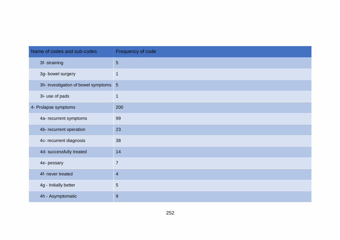
252
Name of codes and sub-codes Frequency of code
3f- straining 5
3g- bowel surgery 1
3h- investigation of bowel symptoms 5
3i- use of pads 1
4- Prolapse symptoms 200
4a- recurrent symptoms 99
4b- recurrent operation 23
4c- recurrent diagnosis 38
4d- successfully treated 14
4e- pessary 7
4f- never treated 4
4g - Initially better 5
4h - Asymptomatic 9

253
Name of codes and sub-codes Frequency of code
5- Pain symptoms 201
5a- back 35
5b- bladder 3
5c- vaginal 11
5d- chronic 13
5e- acute 8
5f- resolved 0
5g- pain management 9
5h- hip and sacral 7
5i- menopausal-like symptoms 1
5j- mesh associated pain 13
5k- non mesh associated pain 8
5l- pain leading to functional or
psychological sequelae 1

254
Name of codes and sub-codes Frequency of code
5m- preventing sex 4
5n-Pelvic 7
5o - Lower Limb 1
5p -Dysmenorrhoea 1
5q - None 5
5r - Dyspareunia 3
6- Consultant care 71
6a- positive comments 30
6b- negative comments 4
6c- gratitude 15
6d- thankfulness 19
6e- consultant acknowledgement of
outcome 2
7- General Care 70

255
Name of codes and sub-codes Frequency of code
7a- negative comments about MDT 2
7b- positive comments about MDT 30
7c- post-operative care 7
7d- pre-operative investigations 1
7e- satisfaction 22
7f- dissatisfaction 7
7g- administrative 1
8- Sexual Function 36
8a- dyspareunia 16
8b- obstruction 2
8c- positive disruption 2
8d- negative disruption 12
8e- frequency 0
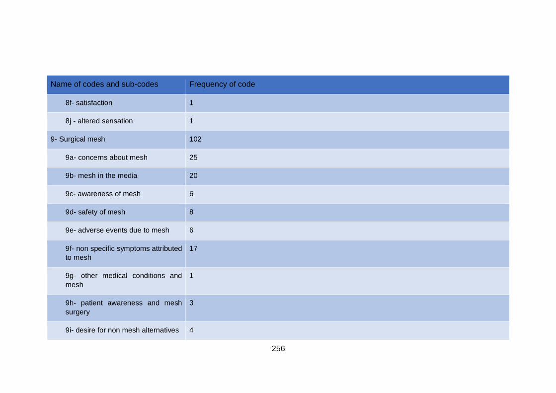
256
Name of codes and sub-codes Frequency of code
8f- satisfaction 1
8j - altered sensation 1
9- Surgical mesh 102
9a- concerns about mesh 25
9b- mesh in the media 20
9c- awareness of mesh 6
9d- safety of mesh 8
9e- adverse events due to mesh 6
9f- non specific symptoms attributed
to mesh 17
9g- other medical conditions and
mesh 1
9h- patient awareness and mesh
surgery 3
9i- desire for non mesh alternatives 4

257
Name of codes and sub-codes Frequency of code
9j- mesh regret 6
9k - Mesh Controversy 3
Miscellaneous non-coded sub-codes
Cosmesis 0
Abdomen 1
Disappointment 8
Recognised different mesh types 1
Hysterectomy avoidance 3
No problems reported 13
Non-specific health problem 4
Outcome 41
Outcome negative 14
Outcome positive 27
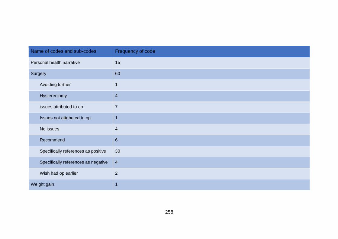
258
Name of codes and sub-codes Frequency of code
Personal health narrative 15
Surgery 60
Avoiding further 1
Hysterectomy 4
issues attributed to op 7
Issues not attributed to op 1
No issues 4
Recommend 6
Specifically references as positive 30
Specifically references as negative 4
Wish had op earlier 2
Weight gain 1

259