Effect of omega-3 fatty acids on the telomere length - De Gruyter
Upload
independentCategory
view
2download
0
doi: 10.1098/rsif.2006.0148, 73-904 2007 J. R. Soc. Interface
C.J Proctor, D.A Lydall, R.J Boys, C.S Gillespie, D.P Shanley, D.J Wilkinson and T.B.L Kirkwood budding yeastModelling the checkpoint response to telomere uncapping in
Referenceshttp://rsif.royalsocietypublishing.org/content/4/12/73.full.html#ref-list-1
This article cites 37 articles, 19 of which can be accessed free
Email alerting service hereright-hand corner of the article or click Receive free email alerts when new articles cite this article - sign up in the box at the top
http://rsif.royalsocietypublishing.org/subscriptions go to: J. R. Soc. InterfaceTo subscribe to
This journal is © 2007 The Royal Society
on May 14, 2011rsif.royalsocietypublishing.orgDownloaded from

on May 14, 2011rsif.royalsocietypublishing.orgDownloaded from
*Author for c
Received 27 JAccepted 14 J
Modelling the checkpoint response totelomere uncapping in budding yeast
C. J. Proctor1,3,*, D. A. Lydall1,3, R. J. Boys2,3, C. S. Gillespie2,3,
D. P. Shanley1,3, D. J. Wilkinson2,3 and T. B. L. Kirkwood1,3
1Institute for Ageing and Health, and School of Clinical Medical Sciences-Gerontology,2School of Mathematics & Statistics, and 3Centre for Integrated Systems Biology of Ageing
and Nutrition, Newcastle University, Newcastle upon Tyne NE4 6BE, UK
One of the DNA damage-response mechanisms in budding yeast is temporary cell-cyclearrest while DNA repair takes place. The DNA damage response requires the coordinatedinteraction between DNA repair and checkpoint pathways. Telomeres of budding yeast arecapped by the Cdc13 complex. In the temperature-sensitive cdc13-1 strain, telomeres areunprotected over a specific temperature range leading to activation of the DNA damageresponse and subsequently cell-cycle arrest. Inactivation of cdc13-1 results in the generationof long regions of single-stranded DNA (ssDNA) and is affected by the activity of variouscheckpoint proteins and nucleases.
This paper describes a mathematical model of how uncapped telomeres in budding yeastinitiate the checkpoint pathway leading to cell-cycle arrest. The model was encoded in theSystems Biology Markup Language (SBML) and simulated using the stochastic simulationsystem Biology of Ageing e-Science Integration and Simulation (BASIS). Each simulationfollows the time course of one mother cell keeping track of the number of cell divisions, thelevel of activity of each of the checkpoint proteins, the activity of nucleases and the amount ofssDNA generated. The model can be used to carry out a variety of in silico experiments inwhich different genes are knocked out and the results of simulation are compared toexperimental data. Possible extensions to the model are also discussed.
Keywords: systems biology; modelling; stochastic; telomere; checkpoint; yeast
1. INTRODUCTION
Telomeres are repetitive sequences of DNA situated atthe ends of linear chromosomes. They require protec-tion from being recognized as double-strand breaks toprevent activation of the DNA damage response. Inbudding yeast, the telomere-binding protein Cdc13provides a protective cap at the ends. In the absence ofCdc13, checkpoint proteins bind to the telomeres,resulting in cell-cycle arrest (Weinert & Hartwell1988; Lydall 2003), accumulation of single-strandedDNA (ssDNA) on the 30 strands (Garvik et al. 1995)and recombination (Grandin et al. 2001a). A mutantform of Cdc13, cdc13-1, has a temperature-dependantresponse and is only able to cap telomeres at a range oftemperatures, with the maximum ‘permissive tempera-ture’ being 258C (Garvik et al. 1995). At highertemperatures, cdc13-1 is unable to function, thecheckpoint pathway is activated and the cells stopgrowing. Hence, a temperature in this range is referredto as a ‘restrictive temperature’. A useful way to studythe checkpoint response in budding yeast is tointroduce the cdc13-1 mutation and to initially culturethe cells at a permissive temperature (e.g. 238C) and
orrespondence ([email protected]).
une 2006uly 2006 73
then switch to a restrictive temperature (e.g. 368C).Throughout this paper, we will use the terms permiss-ive temperature and restrictive temperature to refer tocdc13-1 strains being cultured at 23 and 368C,respectively. More detail of the experimental frame-work can be found in Zubko et al. (2004).
The end of a telomere in budding yeast consists of avery short region of ssDNA (approx. 12 nt) which istermed an ‘overhang’ (Larrivee et al. 2004). Thisoverhang is usually protected by telomere-bindingproteins to prevent it from being seen as DNA damage,which would otherwise activate a DNA damageresponse. At the restrictive temperature, cdc13-1 cellspossess a small amount of DNA damage, as a result ofoverhangs becoming exposed, which activates theRad9-and Rad24-dependent checkpoint pathway. This initialdamage is amplified to larger single-stranded regionsnear telomeres. The generation of ssDNA is a necessaryrequirement for the full activation of the checkpointresponse. There is evidence that theMrx complex, Exo1and the Rad24 group regulate or encode nucleases,which are responsible for generating ssDNA at telo-meres (Lydall 2003). Exo1 regulates ssDNA levelswhen telomere capping is defective. For example,Exo1 is essential for ssDNA generation in yku70Dmutants and contributes to ssDNA in cdc13-1 mutants
J. R. Soc. Interface (2007) 4, 73–90
doi:10.1098/rsif.2006.0148
Published online 31 August 2006
This journal is q 2006 The Royal Society

17 3
1
ExoX Rad24
23
45
Rad9
Rad53
Dun1
Chk1
Mec1
Ddc2
Pds1
Exo1
Rad9 a
aa
b
Figure 1. A model for the interaction between checkpointpathways and nucleases at cdc13-1-induced damage (Repro-duced with permission from Jia et al. (2004), CopyrightGenetics Society of America.). Rad9 inhibits ssDNA pro-duction at telomeres of cdc13-1 strains by two routes, a and b.One route, a, depends on Mec1, Rad9 and Rad53 and targetsExo1. The other route, b, is dependent on Rad9, independentof Mec1 and Rad53 and targets ExoX.
74 Modelling the checkpoint response C. J. Proctor et al.
on May 14, 2011rsif.royalsocietypublishing.orgDownloaded from
(Maringele & Lydall 2002). In cdc13-1 rad9D mutants,the ssDNA accumulates more rapidly than in cdc13-1mutants, whereas in cdc13-1 rad24D mutants, thessDNA accumulates much more slowly than in cdc13-1 cells (Lydall & Weinert 1995). It has been found thatssDNA levels are high in cdc13-1 rad9D exo1D strains,low in cdc13-1 exo1D strains and low in cdc13-1 rad53Dexo1D and cdc13-1 mec1D exo1D strains (Jia et al. 2004;Zubko et al. 2004). This suggests that Rad9 inhibits anunidentified nuclease called ExoX, and that thisinhibition does not requireMec1 or Rad53 (see figure 1).
In order to understand precisely how this importantyet complex system of pathways is regulated, there is aclear role for mathematical models. These can help bothto reveal gaps and inconsistencies in current knowledgeand also to conduct in silico experiments (simulations)that can help in the efficient planning of wet experi-mental studies. A model for the interaction ofcheckpoint pathways and nucleases at cdc13-1-induceddamage has been proposed by Jia et al. (2004). Themodel from their paper has been reproduced in figure 1.The Rad17, Mec3 and Dcd1 complex is a hetero-trimeric proliforating cell nuclear antigen (PCNA)-type ring, which is loaded onto damaged telomeres byanother complex consisting of Rad24 and Rfc2–5subunits, which together form a clamp–clamp loadersystem (Majka & Burgers 2003). Once loaded, theRad17 complex activates ExoX, which generatesssDNA at the telomeres. Mec1 and Ddc2 bind ssDNAindependent of the Rad17 complex. Mec1 is essential forphosphorylation and activation of Rad9, which in turnactivates two parallel pathways of cell-cycle arrest. Onepathway is via the kinase Chk1 and the other pathwayis via the kinase Rad53 (Gardner et al. 1999; Sanchezet al. 1999). Rad9 also inhibits the activity of ExoX
J. R. Soc. Interface (2007)
(Zubko et al. 2004). Exo1 activity is not dependent onRad24 or Rad17, but its activity is inhibited by Rad53.It has been proposed that Rad9 mediates interactionsbetween the upstream kinase, Mec1 and two paralleldownstream kinases, Rad53 and Chk1 (Gilbert et al.2001; Schwartz et al. 2002; Blankley & Lydall 2004).
In a study of yeast checkpoint genes, it was foundthat there was a correlation between the amount ofssDNA and cell death in several single and doublemutant strains (Lydall & Weinert 1995). However,more recently it has been found that mec1D, rad53Dand exo1D mutations each suppress the rapid loss inviability of cdc13-1 rad9D mutants, but ssDNA stillaccumulates (Jia et al. 2004). They suggest that ssDNAis only cytotoxic if Mec1, Rad53 and Exo1 convert itinto a lethal lesion.
The biochemical mechanisms underlying the DNAdamage response are complex and many hypotheseshave been put forward to suggest how DNA repair andcheckpoint pathways interact. This has motivated us todevelop a mathematical model of the system. To buildthe model, it was necessary to specify precisely eachelement of the network and its interaction with otherelements (see §2.1 for details).
2. THE MODEL
2.1. Elements of the model
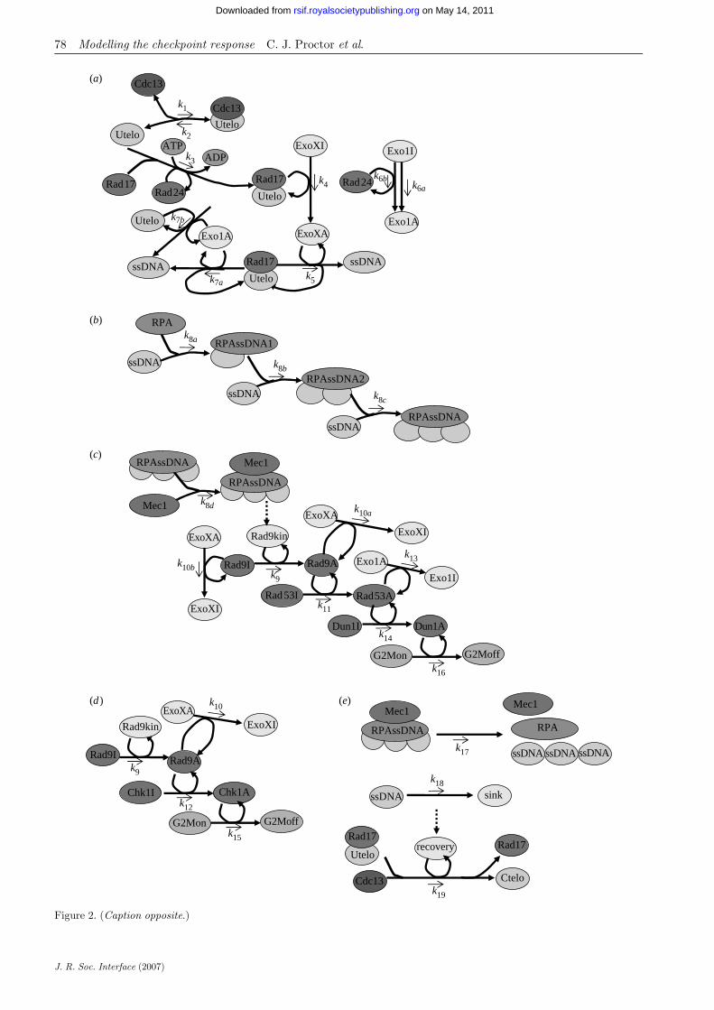
The paper describes a core model representing the wild-type from which a number of variants were created torepresent a number of experimental knockouts. The setof models, all contain the same network, but thevariants have different sets of parameter values orinitial conditions. The differences are typically small,but lead to significant variance in phenotype. We usethe term ‘species’ to denote a biochemical entity in themodel and interactions between species are referred toas reactions. These terms will be used throughout thispaper. The first step in building the model is to define allthe species and specify their initial amounts. A completelist for the wild-type variant is given in table 1 and eachof the species and all reactions in the model aredescribed later in detail. A complete list of the reactionsand their parameters (for the wild-type variant) is givenin table 2. We use mass action stochastic kinetics for therate laws (see Wilkinson 2006 for further details). Wealso use event structures in our model, which allowsspecies amounts and parameter values to be changedonce a particular condition is true. These are detailed inthe text and summarized in table 3. Figure 2 showsthe reaction network in the model.
2.1.1. Capped telomeres, uncapped telomeres andCdc13. Budding yeast have 16 chromosomes andhence 32 telomeres in G1, but during S phase, theDNA is replicated resulting in 64 telomeres. To avoidunnecessary complexity, we have omitted detail ofDNA replication from this model and assumed that thenumber of telomeres is constant. The main purpose ofthis model is to examine the pathways that lead toG2/M arrest and so we have chosen 64 telomeres.Telomeres can be in either a capped or uncapped state

Table 1. Initial conditions for wild-type model.
species descriptionsystematic name(for genes)a database termb
initial valuec
(no. of molecules)
Ctelo capped telomere n.a.d GO:0000781 64Utelo uncapped telomere n.a. GO:0000781 0Rad17Utelo uncapped telomere bound
by Rad17n.a. GO:0000781 0
Cdc13 Cdc13 protein complex YDL220C S000002379 300Rad17 Rad17 protein complex YOR368W S000005895 70Rad24 Rad24 protein complex YER173W S000000975 70RPA replication protein A YAR007C, YNL312W,
YJL173CGO:0005662 4000
Mec1 Mec1/Ddc2 complex YBR136W, YDR499W S000000340, S000002907 4000ssDNA single-stranded DNA
(10 nt)n.a. n.a. 0
RPAssDNA ssDNA bound by RPA n.a. n.a. 0Mec1RPAssDNA RPAssDNA bound by
Mec1 complexn.a. n.a. 0
ExoXI inactive ExoX unidentified n.a. 70ExoXA active ExoX unidentified n.a. 0Exo1I inactive Exo1 YOR033C S000005559 670Exo1A active Exo1 YOR033C S000005559 0Rad9I inactive Rad9 YDR217C S000002625 20Rad9A active Rad9 YDR217C S000002625 0Rad53I inactive Rad53 YPL153C S000006074 6900Rad53A active Rad53 YPL153C S000006074 0Chk1I inactive Chk1 YBR274W S000000478 60Chk1A active Chk1 YBR274W S000000478 0Dun1I inactive Dun1 YDL101C S000002259 3000Dun1A active Dun1 YDL101C S000002259 0ATP adenosine triphosphate n.a. CHEBI:15422 10 000ADP adenosine diphosphate n.a. CHEBI:16761 1000recovery dummy species n.a. n.a. 0sink dummy species n.a. n.a. 0
a Systematic names (or open reading frame (ORF)-name) correspond to the stretch of DNA of the sequenced gene.b Database terms starting with: (i) GO are taken from the Gene Ontology database (www.geneontology.org); (ii) S are takenfrom the SaccharomycesGenome Database (www.yeastgenome.org); and (iii) CHEBI are taken from the Chemical Entities ofBiological Interest database (www/ebi.ac.uk/chebi/).c Initial amounts of proteins are taken from the yeast GFP fusion database (http://yeastgfp.ucsf.edu).d n.a., not applicable.
Modelling the checkpoint response C. J. Proctor et al. 75
on May 14, 2011rsif.royalsocietypublishing.orgDownloaded from
and hence we have two species to represent telomeres,which we call Utelo and Ctelo for uncapped andcapped telomeres, respectively. We assume that thessDNA overhangs at the telomere ends are capped byCdc13. The dissociation rate (the time required for halfthe protein–DNA complex to dissociate) is approxi-mately 30 min (Lin et al. 2001). Cdc13 has a strongbinding affinity to single-stranded telomeric DNA witha dissociation constant of ca 10K7 M (Lin et al. 2001).Hence, there is equilibrium between uncapped telo-meres (Utelo) and those bound by Cdc13.
UteloCCdc13#Cdc13KUtelo:
To model this equilibrium, we set up two reactions,
UteloCCdc13 $$%k1
Ctelo;
Ctelo $$%k 2
UteloCCdc13;
where Ctelo represents the pool of telomeres bound byCdc13.
The first reaction represents capping and the secondreaction uncapping. The capping reaction is a second-order reaction since there are two reactants and is given
J. R. Soc. Interface (2007)
by k1[#Utelo][#Cdc13], where ‘#’ represents thenumber of molecules. The uncapping reaction is afirst-order reaction and is given by k2[#Ctelo]. Thevalue of k2 can be calculated using information on thetime taken for half of the boundCdc13 to dissociate fromthe telomere given earlier. Since the biological half-life isgiven by t1/2ZKln(0.5)/degradation rate, it followsthat k 2ZKðlnð0:5Þ=ð30!60ÞÞsK1Z3:85!10K4 sK1. Thedissociation constant kd is 10K7 MZ110 molecules pernucleus, assuming that the volume of a yeast nucleus isca 1.8 mm3 (1.8!10K9 ml; http://biochemie.web.med.uni-muenchen.de/Yeast_Biol/). Since kd Zk2/k1, thenk1Z3:6!10K6 moleculeK1 sK1. We assume that all thetelomeres are in a capped state initially, but there is adynamic process of uncapping and recapping, hence atany point in time, there is a very low number ofuncapped telomeres. Telomeres remain in an uncappedstate only very transiently, and signalling proteins donot bind while they are in this transient state.
2.1.2. Binding of uncapped telomeres: Rad17 andRad24. If a telomere remains uncapped, then the

Table 2. Reactions for wild-type model.
reaction kinetic rate law reactants products parameter valuea
capping k1[Utelo][Cdc13] Utelo, Cdc13 Ctelo k1 5.0!10K4 molK1 sK1
uncapping k2[Ctelo] Ctelo Utelo, Cdc13 k2 3.85!10K4 sK1
Rad17 binding k3½Utelo�½Rad17�½Rad24�½ATP�=ð5000C ½ATP�Þ Utelo, Rad17, Rad24,ATP
Rad17Utelo, Rad24,ADP
k3 1.5!10K8 molK2 sK1
ExoX activation k4[ExoXI][Rad17Utelo] ExoXI, Rad17Utelo ExoXA, Rad17Utelo k4 0.01 molK1 sK1
ExoX activity k5[Rad17Utelo][ExoXA] Rad17Utelo, ExoXA Rad17Utelo, ExoX-A,ssDNA
k5 3.0!10K4 molK1 sK1
Exo1 activation k6a[Exo1I] Exo1I Exo1A k6a 5.0!10K5 sK1
Exo1 Rad24-dependent activation k6b [Exo1I][Rad24] Exo1I, Rad24 Exo1A, Rad24 k6b 5.0!10K4 molK1 sK1
Rad17-independent Exo1 activity k7a [Utelo][Exo1A] Utelo, Exo1A Utelo, Exo1A, ssDNA k7a 3.0!10K5 molK1 sK1
Rad17-dependent Exo1 activity k7b [Rad17Utelo][Exo1A] Rad17Utelo, Exo1A Rad17Utelo, Exo1A,ssDNA
k7b 3.0!10K5 molK1 sK1
RPA binding1 k8a [ssDNA][RPA] ssDNA, RPA RPAssDNA1 k8a 0.001 molK1 sK1
RPA binding 2 k8b [ssDNA][RPAssDNA1] ssDNA, RPAssDNA1 RPAssDNA2 k8b 100.0 molK1 sK1
RPA binding k8c [ssDNA][RPAssDNA2] ssDNA, RPAssDNA2 RPAssDNA k8c 100.0 molK1 sK1
Mec1 binding k8d[RPAssDNA][Mec1] RPAssDNA, Mec1 RPAssDNAMec1 k8d 0.004 molK1 sK1
Rad9 activation k9 [Rad9Kin][Rad9I] Rad9Kin, Rad9I Rad9Kin, Rad9A k9 100.0 molK1 sK1
ExoX inhibition k10a[Rad9A][ExoXA] Rad9A, ExoXA Rad9A, ExoXI k10a 0.05 molK1 sK1
ExoX Mec1-independent inhibition k10b[Rad9A][ExoXA] Rad9I, ExoXA Rad9I, ExoXI k10b 0.05 molK1 sK1
Rad53 activation k11 [Rad9A][Rad53I] Rad9A, Rad53I Rad9A, Rad53A k11 10K5 molK1 sK1
Chk1 activation k12 [Rad9A][Chk1I] Rad9A, Chk1I Rad9A, Chk1A k12 1.7!10K4 molK1 sK1
Exo1 inhibition k13 [Rad53A][Exo1A] Rad53A,,Exo1A Rad53A,Exo1I k13 1.0 molK1 sK1
Dun1 activation k14 [Rad53A][Dun1I] Rad53A Dun1I Rad53A, Dun1A k14 3.3!10K6 molK1 sK1
Chk1 cell arrest k15 [Chk1A][G2Mon] Chk1A, G2Mon Chk1A G2Moff k15 0.2 molK1 sK1
Dun1 cell arrest k16 [Dun1A][G2Mon] Dun1A, G2Mon Dun1A, G2Moff k16 0.1 molK1 sK1
release of Mec1 and RPA in S phase k17a [Mec1RPAssDNA][S] Mec1RPAssDNA, S 3ssDNA Mec1, RPA, S k17a 0.05 molK1 sK1
release of Mec1 and RPA in G2/Marrest
k17b [Mec1RPAssDNA][G2Moff] Mec1RPAssDNA,G2Moff
3ssDNA Mec1, RPA,G2Moff
k17b 0.05 molK1 sK1
removal of ssDNA in S phase k18a [ssDNA][S] ssDNA, S S k18a 0.001 molK1 sK1
removal of ssDNA in G2/M arrest k18b [ssDNA][G2Moff] ssDNA, G2Moff G2Moff k18b 1!10K5 molK1 sK1
recovery k19 [Rad17Utelo][Cdc13][recovery] Rad17Utelo, Cdc13,recovery
Ctelo, Rad17, recovery k19 0.001 molK2 sK1
a mol, number of molecules.
76
Modellin
gthecheckp
ointresp
onse
C.J.Procto
ret
al.
J.R.Soc.
Interfa
ce(2007)
on May 14, 2011
rsif.royalsocietypublishing.orgD
ownloaded from

Table
3.Summary
ofevents
usedin
allmodels.
event
trigger
speciesaffected
parameters
affected
reactionsaffected
Rad9kinase
activation
Mec1RPAssDNAO800
Rad9Kin
none
Rad9activation
ssDNA
removal
Mec1RPAssDNA
CRPAssDNACssDNA!1
recovery
none
recovery
G2/M
recoverycompleted
Rad17UteloZ
Z0&&
G2Z
Z1
G2Moff,G2Mon,recovery,ExoXA,
ExoXI,Exo1A,Exo1I,Rad9A,Rad9I,
Rad53A,Rad53I,Chk1A,Chk1I,
Dun1A,Dun1I
none
allreactionsinvolvingExoX,Exo1,Rad9,
Rad53,Chk1,Dun1.Recoveryreaction
stops
Sphase
recoverycompleted
Rad17UteloZ
Z0
recovery
none
recovery
celldeath
aMec1RPAssDNA
CRPAssDNACssDNAO2000
none
k aliveb
allreactionsstop
aOnly
inmodelswithcelldeath.
bk a
liveisaparameter
whichisincluded
inallthekineticrate
laws(e.g.k a
live!
k 2[Ctelo]).Initially,k a
liveZ1andso
rate
lawsare
unaffectedbythisparameter.W
hen
thecriticalthreshold
oftotalsingle-stranded
DNA
isreached
k aliveissetto
0,andso
allthereactionratesare
equalto
0.
Modelling the checkpoint response C. J. Proctor et al. 77
J. R. Soc. Interface (2007)
on May 14, 2011rsif.royalsocietypublishing.orgDownloaded from
Rad17 complex is loaded onto the telomere end by theRad24 clamp loader complex. This loading requiresATP and can be represented by the biochemicalreaction
UteloCRad17CRad24CATP $$%k 3
Rad17UteloCRad24CADP;
where Rad17Utelo represents the binding of Rad17 tothe telomere end. For simplicity, we have omitteddetails of Mec3 and Ddc1 in the Rad17 complex andomitted the Rfc2–5 subunits in the Rad24 complex,since they are unimportant for this model. Since thereare four reactants in the Rad17-binding reaction, thestochastic rate law is a fourth-order reaction. Careneeds to be taken when considering higher orderreactions. For example, it is not realistic that thereaction rate would increase indefinitely with anincrease in ATP levels; doubling the level of ATP inthe cell would not necessarily double the reaction rate.Instead, we would expect that the reaction rate wouldincrease asymptotically with ATP levels. Therefore, weuse the rate law k3[#Utelo][#Rad17][#Rad24][#ATP]/(5000C[#ATP]).
The term in the denominator ensures that thereaction rate increases approximately linearly withATP when ATP levels are low (less than ca 2000), butfor high levels of ATP (greater than 5000), the increasein the reaction rate starts to level off with increasingATP levels. The value for k3 was chosen by making aninitial estimate and then simulating the model andcomparing output to the experimental data. Furtherdetails of how we chose parameter values can be foundin §2.3.
When Cdc13 is present, this reaction is very unlikelyto occur as the binding strength of Rad17 compared toCdc13 is extremely low. However, in the absence ofCdc13, this reaction will eventually take place and anirreversible set of reactions is initiated leading to thegeneration of ssDNA and cell-cycle arrest unless thepathway is interrupted downstream. We chose a verylow value for k3, to ensure that the probability of atelomere, which is only transiently uncapped, bindingto Rad17 is very low.
2.1.3. Nuclease activity: ExoX, Exo1 and ssDNA. ExoXis recruited to telomeres bound by Rad17, and degradesone of the strands to leave a long stretch of ssDNA. Thisreaction can only occur if ExoX is in its active state.The binding of telomeres by Rad17 (which in turnrequires Rad24) is required for ExoX activation. Thiscan be represented simply by the following reactions:
ExoXICRad17Utelo $$%k4
ExoXACRad17Utelo
Rad17UteloCExoXA $$%k5
Rad17UteloCExoXACssDNA;
where ExoXI represents the inactive form of ExoX andExoXA represents activated ExoX. We assume thateach time this reaction takes place, 10 nt ssDNA areproduced and we set one unit of ssDNA equal to 10 nt.The reaction can continue to take place until ExoX isinactivated. It has been observed that ca 8 kb ssDNA
k8c
RPAssDNA2
RPAssDNA
k8a
k8b
RPAssDNA1
ssDNA
RPA
ssDNA
ssDNA
ExoXI
ExoXA
k4
k5
ssDNA
Exo1I
Exo1A
Rad24
Exo1A
ssDNA
Utelo
k6a
k6b
k3
Rad17Utelo
Rad17
ATPADP
Rad24
k2Utelo
Cdc13
Utelo
k1 Cdc13
Utelo
Rad17
k7a
k7b
Rad9I
Rad9kin
Rad9A
ExoXAExoXI
Chk1I Chk1A
G2Mon G2Moff
Rad9A
Rad53I
ExoXA
ExoXI
Exo1A
Exo1I
Rad53A
Dun1I Dun1Ak14
G2Mon G2Moff
k9
k10
k9
k12
k15
k11
k16
k13
Rad9kin
Rad9I
ExoXA
ExoXI
RPAssDNA
Mec1
k8d k10a
k10b
Mec1
RPAssDNA
RPAssDNA
Mec1
ssDNA
Mec1
RPA
k17 ssDNA ssDNA
ssDNA sink
k18
k19
recoveryUtelo
Rad17
Cdc13 Ctelo
Rad17
(a)
(b)
(c)
(d ) (e)
Figure 2. (Caption opposite.)
78 Modelling the checkpoint response C. J. Proctor et al.
J. R. Soc. Interface (2007)
on May 14, 2011rsif.royalsocietypublishing.orgDownloaded from

Figure 2. (Opposite.) Network diagram showing the checkpoint response to uncapped telomeres. (a) Activation of ExoX andExo1. ExoX requires Rad24 and Rad17 binding for its activation. Exo1 is activated independent of Rad24 and Rad17, although itmay also act on telomeres bound by Rad17 and be activated in a Rad24-dependent manner. (b) Binding of single-stranded DNA(ssDNA) by RPA. Each molecule of RPA requires three units of ssDNA to bind. (c) Activation of checkpoint response viaRad53/Dun1 pathway. Activation of this pathway leads to inhibition of nuclease activity. Dashed line indicates an event, wherea threshold level of Mec1RPAssDNA activates a kinase (Rad9Kin) which activates Rad9. (d ) Activation of checkpoint responsevia Chk1 pathway does not affect Exo1. (e) Recovery can take place during S phase or G2/M arrest after single-stranded DNAhas been removed. The dashed line indicates an event. When the level of ssDNA is equal to 0, the dummy species ‘recovery’ is setto 1 and the recapping reaction can then occur. See §2.1.6 for more details.
Modelling the checkpoint response C. J. Proctor et al. 79
on May 14, 2011rsif.royalsocietypublishing.orgDownloaded from
are generated in 1 h in cdc13-1 strains (Jia et al. 2004;Zubko et al. 2004). Exo1 degrades not only unprotectedtelomeres, independent of ExoX and Rad17, but alsotelomeres bound by Rad17. Exo1 activation does notrequire Rad24. Since cdc13-1 rad24Dmutants have lowlevels of ssDNA (Lydall & Weinert 1995), we assumethat activation of Exo1 is much lower than that ofExoX. There are very low levels of ssDNA in the cdc13-1rad24D strain, which must be due to Rad24-independentactivation and subsequent activity ofExo1.However, thedifference between the amounts of ssDNA in cdc13-1exo1D compared to cdc13-1 mutants is large (Zubkoet al. 2004), suggesting that Exo1 activity is higherwhen Rad24 is present. Therefore, we also include anadditional reaction of Rad24-dependent activation ofExo1, although this was not in the model proposed byJia et al. (2004) (see figure 1). The reactions for nucleaseactivity are as follows:
Exo1I $$%k6a
Exo1A;
Exo1ICRad24 $$%k6b
Exo1ACRad24;
UteloCExo1A $$%k7a
UteloCExo1ACssDNA;
Rad17UteloCExo1A $$%k7b
Rad17UteloCExo1ACssDNA:
2.1.4. Binding of ssDNA: replication protein A andMec1. Replication protein A (RPA) binds to ssDNAwith every molecule of RPA binding to 30 nt ssDNA(Zou & Elledge 2003). As each unit of ssDNA isequivalent to 10 nt, we assume that one molecule ofRPA binds to three units of ssDNA. Mec1 in complexwith Ddc2 then binds to RPA (Zou & Elledge 2003).Thus, binding of ssDNA can be represented by thefollowing two reactions:
3ssDNACRPA $$%k8a
RPAssDNA;
RPAssDNACMec1 $$%k8b
Mec1RPAssDNA:
Since three units of ssDNA are required for the RPA-binding reaction, the mass action stochastic kinetic ratelaw is given by k8a[#RPA][#ssDNA][#ssDNA-1][#ssDNA-2]/6. However, this rate law is only valid ifthe ssDNA units are independent. This is certainly notthe case, and the rate law should be a linear and not acubic function of the total amount of ssDNA. Therefore,we model RPA binding as a series of steps. To do this,we introduce two dummy species RPAssDNA1 andRPAssDNA2 for the intermediate steps. In the firststep, RPA binds to one unit of ssDNA to formRPAssDNA1. The rate of this reaction depends linearlyon the total amount of ssDNA. In the next step,RPAssDNA1 binds to the second unit of ssDNA to form
J. R. Soc. Interface (2007)
RPAssDNA2 and finally RPAssDNA2 binds to thethird unit of ssDNA to form RPAssDNA. Once the firststep has happened, the next two steps follow quicklyby setting the rate constants to a high value (k8bZk8cZ100). The full set of reactions for RPA and Mec1binding are
ssDNACRPA $$%k8a
RPAssDNA1;
ssDNACRPAssDNA1 $$%k8b
RPAssDNA2;
ssDNACRPAssDNA2 $$%k8c
RPAssDNA;
RPAssDNACMec1 $$%k8d
Mec1RPAssDNA:
2.1.5. Checkpoint activation: Rad9, Rad53, Chk1 andDun1. A very large threshold of ca 10 kb ssDNA isrequired for keeping checkpoints active in order tomaintain cell-cycle arrest (Vaze et al. 2002; Zubko et al.2004; Maringele & Lydall 2005). To prevent activationof Rad9 when only low levels of ssDNA are present, wemodelled this activation as a two-step process. First, weused an event structure in the model to activate akinase that activates Rad9 (called ‘Rad9Kin’), if thetotal amount of ssDNA within the cell exceeded 8 kb.The rate for the Rad9-activation reaction is equal to k9[#Rad9I ][# Rad9Kin] and will be equal to 0 as long asRad9KinZ0. Once Rad9Kin is activated (so thatRad9KinZ1), the Rad9-activation reaction can pro-ceed. The event and reaction representing Rad9activation are as follows:
If Mec1RPAssDNAO800; Rad9KinZ 1;
Rad9KinCRad9I $$%k9
Rad9KinCRad9A:
Activated Rad9 activates both Rad53 and Chk1.Rad53 in turn, inhibits the activity of Exo1 andactivates Dun1. Rad9 also inhibits ExoX. This inhi-bition does not require Rad9 to be activated by Mec1and so it is represented by two reactions, one withRad9I and the other with Rad9A
Rad9ACExoXA $$%k10a
Rad9ACExoXI;
Rad9ICExoXA $$%k10b
Rad9ICExoXI;
Rad9ACRad53I $$%k11
Rad9ACRad53A;
Rad9ACChk1I $$%k12
Rad9ACChk1A;
Rad53ACExo1A $$%k13
Rad53ACExo1I;
Rad53ACDun1I $$%k14
Rad53ACDun1A:

30
40
50
60
r of
cap
ped
telo
mer
es
k1=3.6 × 10⁻⁶
k1=5 × 10⁻⁴
80 Modelling the checkpoint response C. J. Proctor et al.
on May 14, 2011rsif.royalsocietypublishing.orgDownloaded from
Activation of Chk1 and Dun1 results in G2/M arrest
Chk1ACG2Mon $$%k15
Chk1ACG2Moff;
Dun1ACG2Mon $$%k16
Dun1ACG2Moff;
where G2Mon and G2Moff are dummy species, whichcontrol whether the cell cycle can progress from G2 toM. If G2MonZ1 and G2MoffZ0, then the abovereactions can occur. If G2MonZ0 and G2MoffZ1,then the reactions cannot take place (see appendix A fordetails of modelling the cell cycle).
12108642
0
0
10
20
time (h)
num
be
Figure 3. Model predictions of the number of cappedtelomeres in a wild-type cell when k1Z3.6!10K6 or k1Z0.0005. The output is for one simulation over a period of 12 h.
2.1.6. Recovery. The ssDNA may be removed during Sphase by DNA replication. It is also possible thatssDNA is removed during G2/M arrest. When all thessDNAs have been removed, the uncapped telomerescan be recapped by Cdc13 and recovery takes place.After recovery, all the checkpoint proteins are turnedoff and cell division can take place. This procedure ismodelled by a series of reactions and events
Mec1RPAssDNACS $$%k17a
3ssDNACMec1CRPACS;
Mec1RPAssDNACG2Moff $$%k17b
3ssDNACMec1
CRPACG2Moff;
ssDNACS $$%k18a
S;
ssDNACG2Moff $$%k18b
G2Moff:
If Mec1RPAssDNACRPAssDNACssDNAZZ0, thenrecoveryZ1 (where ‘recovery’ is a dummy species inthe model).If recoveryZ1, then the following reaction can proceed:
Rad17UteloCCdc13Crecovery $$%k19
CteloCRad17
Crecovery:
If Rad17UteloZ0 (i.e. all telomeres have beenrecapped), then G2Moff is set to 0, G2Mon is set to 1,recovery is set to 0, and all other checkpoint proteinsare reset to their initial conditions. A network diagramof the full system is shown in figure 2.
2.2. The cell cycle
We also include a series of reactions to represent thedifferent stages of the cell cycle (see appendix A). Weassume that ssDNA can be removed by DNA replica-tion when the cell is in S phase and also to a lesserextent when cells are in G2/M arrest. Although it isunclear whether DNA replication can take place inG2/M arrest, it has been observed that the level ofssDNA does decrease in cells, which are arrested (Jiaet al. 2004; Zubko et al. 2004). When budding yeastdivides, the mother cell produces a small bud whichthen grows. When mitosis and cytokinesis arecomplete, the bud detaches to form the new daughtercell. This budding leaves a scar on the surface of thecell. Therefore, it is possible to count the number of celldivisions that a particular yeast cell has undergone bycounting the number of budscars. We imitate thisprocess in our model and keep track of the number ofdivisions by the introduction of a dummy species,
J. R. Soc. Interface (2007)
budscar, which increases by one, every time the cellpasses from M to G1 phase. The reaction and equationfor the budscar species are shown in appendix A.
2.3. Initial conditions and parameter values
Before a model can be simulated, the initial amount ofeach species must be specified.
We obtained values for the majority of the initialamounts of proteins by consulting the yeast greenfluorescent protein (GFP) fusion localization database(http://yeastgfp.ucsf.edu; Huh et al. 2003). We assumethat initially all the checkpoint proteins are in theirinactive state. In the G2 phase of the cell cycle, buddingyeast contains 32 chromosomes and so have 64telomeres, which we assume are all initially bound byCdc13. We do not include detail of DNA replication inour model and so assume that the total number oftelomeres is constant. Table 1 lists all the species, theirrespective initial amounts, the systematic names of allgenes and the database terms so that each entity can beidentified in public accessible bioinformatics databases.
The rate constants for each of the reactions are listedin table 2. Note that the value of k1 was set at 5!10K4,although we had calculated it to be 3.6!10K6 frompublished data (Lin et al. 2001), a 100- fold difference.This is because with our original value of k1, we foundthat the binding of Cdc13 to telomeres was not strongenough to keep them in a capped state (figure 3). Ourmodel suggests that the binding of Cdc13 to telomeresmust be stronger in vivo and it is probably the case thatthe binding affinity of Cdc13 is increased when it is incomplex with Stn1 and Ten1 (Grandin et al. 2001b). Atpresent, there are no experimental data to support this,but it seems a reasonable assumption. Therefore, weincreased the value of k1 until we obtained results inwhich telomeres only become uncapped transiently asin figure 3 and chose k1Z0.0005 as our default value.Figure 4 shows the model predictions for the kinetics ofuncapping for the cdc13-1 strain when placed at the

121086420
0
10
20
30
40
50
60
time (h)
num
ber
of c
appe
d te
lom
eres
Figure 4. Model predictions for the kinetics of uncapping inthe cdc13-1 strain at the restrictive temperature (with thedefault parameters). The output is for one simulation over aperiod of 12 h.
Modelling the checkpoint response C. J. Proctor et al. 81
on May 14, 2011rsif.royalsocietypublishing.orgDownloaded from
restrictive temperature using the default parameters.The only other kinetic data currently available forparameterizing the model are rates of single strandproduction in cdc13-1 mutants at high temperatures,where a rate of 8 kb hK1 has been observed (Jia et al.2004; Zubko et al. 2004). The actual rates for thedifferent nucleases are unknown, but they were chosento reflect the suggested relative difference; therefore, weassumed that ExoX activity is 10 times faster thanExo1 activity.
We created an initial computer representation of themodel using estimates for parameter values takendirectly from evidence in the literature, where avail-able. In some cases, parameter values started as initialbest guesses. This initial model was then simulated andthe results compared to experimental data. Theparameter values were then adjusted to ensure thatthe model complied with that data. We are confidentthat the adjustments made to parameters were either toparameters for which we did not have specific data orthat the data was derived from in vitro experiments,which do not necessarily correspond to the in vivoconditions of the culture experiments.
We coded the models in the systems biology markuplanguage (SBML) level 2 (Hucka et al. 2003). SBML is away of representing biochemical networks and is one ofthe standards used by the modelling community. It hasbeen evolving since mid-2000 through the efforts of aninternational group of software developers and users.The models were developed using MATHSBML (Shapiroet al. 2004). The final model for the wild-type cell wasencoded using SBML shorthand and then convertedinto full SBML (Wilkinson 2006). The model was thenimported into the biology of ageing e-science inte-gration and simulation (BASIS) system (www.basis.ncl.ac.uk) and the models for mutant strains werecreated by making simple modifications to theoriginal model.
J. R. Soc. Interface (2007)
To simulate the model, we used a stochasticsimulator based on the Gillespie algorithm (Gillespie1977), since many of the processes being modelled occurat random times. This means that different outcomesmay result from the same set of initial conditions. Inaddition, some of the species in our model must beconsidered as discrete units rather than continuousvariables. For example, species representing the num-ber of capped and uncapped telomeres must be a smallinteger and so it is not appropriate to use a determi-nistic simulator for this model. However, we also triedrunning simulations with a deterministic simulator tosee if any parts of the model could be successfullymodelled this way (see §3.3). The stochasticsimulations were carried out on a Linux Beowulf clusterand the results were stored in a database. All themodels and results in this paper are obtainable from theBASIS website (www.basis.ncl.ac.uk). For furtherdetails, see Gillespie et al. (2006a,b). The model forthe wild-type strain, where cell death is included, is alsoavailable from the Biomodels database(ID:MODEL8679489165) at http://www.ebi.ac.uk/biomodels/ (Le Novere et al. 2006).
We did not carry out a full sensitivity analysis for themodel parameters, since this would have been very timeconsuming due to the number of parameters and thefact that we used a stochastic simulator. However,many of the model parameters were chosen after manytrials with different values and so it is possible to discussthe effects of changing these. The checkpoint response issensitive to changes by an order of magnitude to thevalues of the parameters for the rate of capping (k1) andRad17 binding to uncapped telomeres (k3). A 10-folddecrease in k1 or a 10-fold increase in k3 leads to an over-activation of the checkpoint response. The parametersinvolved in nuclease activity and removal of ssDNAwere confined within fairly limited ranges in order toobtain rates of ssDNA production observed in cdc13-1mutant strains. The parameters for cell-cycle arrest (k15and k16) could be increased with less effect on the modelpredictions, but lowering the values led to an unsus-tained cell-cycle arrest, which could easily be overcomeby Cdk activity. The rate of Rad9 activation (k9) wasthe most important parameter in the checkpointpathway and affected not only how quickly the cellresponded once the threshold level of ssDNA wasreached, but also how quickly nuclease activity wasinhibited. The other parameters in the checkpointresponse had no significant effect on the modelpredictions for the rate of ssDNA production or thenumber of divisions before cell-cycle arrest or celldeath. For example, a 10- fold increase or decrease onthe rate of Mec1 binding (parameter k8d) did not changethe rate of ssDNA production, the total level of ssDNAor the number of divisions before cell-cycle arrest.
3. RESULTS
3.1. Wild-type cells
The model predicts that capped telomeres may becometransiently uncapped, but are then recapped. At anytime, there is usually only one uncapped telomere at the

num
ber
of c
ell d
ivis
ions
121086420
2
0
4
6
8
10
time (h)
Figure 5. Model predictions for the number of divisionsobtained in a wild-type cell. The output is for threesimulations over a period of 12 h.
100
50
0
100
50
med
ical
nuc
lear
div
isio
n (%
)m
edic
al n
ucle
ar d
ivis
ion
(%)
late
nuc
lear
div
isio
n (%
)la
te n
ucle
ar d
ivis
ion
(%)
0
80 160 2400
80
time at 36°C (min)time at 36°C (min)
160 240
exo1D RAD+
exo1D rad24Dexo1D rad9D
exo1D RAD+
exo1D rad24Dexo1D rad9D
RAD+
RAD rad24Drad9D
rad24Drad9D
cdc13-1 RAD+ cdc13-1 exo1
(a)
(c) (d )
(b)
82 Modelling the checkpoint response C. J. Proctor et al.
on May 14, 2011rsif.royalsocietypublishing.orgDownloaded from
most, but on occasions, two or three telomeres may beuncapped simultaneously (figure 3). However, they donot remain uncapped long enough for Rad17 to bind, sovery little ssDNA is produced (data not shown).Figure 5 shows the model predictions for the numberof divisions obtained in three different simulation runs.Since we have used a stochastic simulator, each runproduces a slightly different output. The cells divide ca7–9 times in a period of 12 h, which gives an averagedivision time of ca 1.5 h. This corresponds to thedivision time obtained when wild-type cells are grownat the restrictive temperature.
cdc13-1 rad9 cdc13-1 rad9 exo1
cdc13-1 rad24 cdc13-1 rad9 rad24 exo1
CDC+ RAD+
(g)
(e) ( f )
Figure 6. Growth of wild-type and cdc13-1 mutant strains(reproduced with permission from Zubko et al. (2004); Copy-right Genetics Society of America). Yeast strains were releasedfrom G1 arrest and allowed to form microcolonies for 15 h at368C (restrictive temperature) before being photographed at200! magnification. Cell numbers within microcolonies wereestimated from the photographs shown and are given, alongwith their standard deviations, for each strain: (a) cdc13-1RAD+(2); (b) cdc13-1 exo1 (8G2); (c) cdc13-1 rad9 (18G5);(d) cdc13-1 rad9 ex01 (54G17); (e) cdc13-1 rad24 (43G22);( f ) cdc13-1 rad9 rad24 exo1 (113G34); (g) wild type.
3.2. cdc13-1 mutant strains3.2.1. Cell divisions. We used our model to simulatecdc13-1 mutant strains at the restrictive temperatureby setting the initial amount of Cdc13Z0, CteloZ0 andUteloZ64. We can also examine the effects of knockingout different genes for checkpoint proteins andnucleases. For example, to simulate a cdc13-1 rad9Dstrain, we set the initial amount of Rad9IZ0 (seetable 4). Our model predicts that cdc13-1 cells areunable to divide, although occasionally they may divideonce before cell-cycle arrest occurs (data not shown).This is due to the uncapped telomeres being bound byRad17, which then initiates a series of events leading tocell-cycle arrest. Both Rad9 and Rad24 are required forcell-cycle arrest to occur. Our model predicts thatcdc13-1 exo1D double mutants enter cell-cycle arrestafter a few divisions as ssDNA can still be producedthrough the activity of ExoX and so Rad9 is activated,and in turn, the other checkpoint proteins areactivated. The later activation of the checkpointresponse is due to the slower generation of ssDNAwithout the activity of Exo1. Figure 6 shows data fromZubko et al. (2004) for various yeast strains cultured atthe restrictive temperature. The cdc13-1 mutants areunable to divide at the restrictive temperature, but
J. R. Soc. Interface (2007)

10
5
0
15
20(a)
(b)
ssD
NA
(kb
) pe
r ce
ll15
20
er c
ell
cdc13-1
cdc13-1 exo1
cdc13-1 rad9
cdc13-1 rad9 exo1
Modelling the checkpoint response C. J. Proctor et al. 83
on May 14, 2011rsif.royalsocietypublishing.orgDownloaded from
knocking out Rad24, Rad9 or Exo1 restores celldivision, hence microcolonies form. Both cdc13-1rad9D and cdc13-1 exo1D double mutants at therestrictive temperature have very small microcolonies,although we know that knocking out Rad9 wouldprevent cell-cycle arrest (figure 6). The cells for cdc13-1exo1D strain are larger than in the cdc13-1 rad9Dstrain, which indicates that cells initially arrest andthen eventually escape this arrest and divide a fewtimes before cell death takes place. The cdc13-1 strainsaccumulate large amounts of ssDNA, since nucleaseactivity is intact and this is probably the cause of celldeath. If we compare the model output with experi-mental data, we see good agreement apart from thecdc13-1 rad9Dmutant. Ourmodel predicts that cdc13-1rad9D cells keep dividing, but experimental data showthat these cells only form very small microcolonies(figure 6). This is because we had not included thepossibility of cell death in the model. To take cell deathinto account, we modified the model (see §3.2.3). Ourmodel also does not fit the observation of cell-cycle arrestfollowed by escape for the cdc13-1 exo1D strain as wehave not included this mechanism in the model. Weoutline a possible extension to include this escapemechanism in §4.
0
10
5
0
ssD
NA
(kb
) p
50 100 150 200 250time (min)
cdc13-1 rad24cdc13-1 rad9 rad24
cdc13-1 rad24 exo1cdc13-1 rad9 rad24 exo1
Figure 7. Model predictions for the amount of ssDNAgenerated in wild-type and cdc13-1 mutant strains. Theoutput is for one simulation for each strain over a period of250 min.
3.2.2. Generation of ssDNA. Zubko et al. (2004) showedthat large amounts of ssDNA are produced in cdc13-1mutants at the restrictive temperature, but thatdeleting Exo1 considerably reduced these levels. Delet-ing Rad24 also reduced the amount of ssDNA, butdeleting Rad9 had the opposite effect (see Zubko et al.2004). Therefore, we plotted the level of ssDNA atdifferent time points for each of the different mutantstrains and compared our results to Zubko et al.’s(2004) data.
Our models predict that initially large amounts ofssDNA are generated in cdc13-1 strains (figure 7), butthis stabilizes when cells enter G2/M arrest, as nucleaseactivity is inhibited by Rad9 and Rad53. Large amountsof ssDNA are generated in cdc13-1 rad9D doublemutants, since Rad9 is required to inhibit nucleaseactivity. In contrast, only small amounts of ssDNA aregenerated in cdc13-1 rad24D doublemutants. This is dueto the fact that in our model Rad24 is required to loadRad17 onto uncapped telomeres and so activate ExoX.Our model predicts that much lower levels of ssDNA aregenerated in cdc13-1 exo1D double mutants than in thecdc13-1 strain. This low level of ssDNA is due to theresidual activity of ExoX,which has not been completelyinhibited by Rad9. The generation of ssDNA in thecdc13-1 rad24D strain is due to the Rad24-independentactivityofExo1andthedifferencebetween theamountofssDNA in the cdc13-1 strain and the cdc13-1 exo1Dstrain reflects the total activity of Exo1. Since experi-mental data show that this difference is larger than theamount of ssDNAgenerated in the cdc13-1 rad24D strain(Zubko et al. 2004), we suggest that there is also someadditional Rad24-independent activation of Exo1 and sohave included this in themodel.Note that ourmodel doesnot distinguish betweendifferent regions of the telomeresand that themodel output represents the total amount of
J. R. Soc. Interface (2007)
ssDNA for all telomeres. To examine the effects ofnuclease activity at different regions of the telomere, wewould need to modify the model to include a separatespecies for each telomere and separate reactions fordifferent parts of the telomere.
We also looked at the variability of the amount ofssDNA produced over repeated simulations. We foundthat the variation from run to run was very small andeven with 10 simulations, the upper and the lower 95%confidence interval for the mean was always within 2%of the mean. Therefore, it was not necessary to carryout many repeated simulations.
3.2.3. Extending the model to include cell death. Whencdc13-1 rad9D cells are cultured at the restrictivetemperature, it has been found that despite the lack ofRad9, which is required to activate the checkpointpathway, these cells only divide for a few hours andthen die (Zubko et al. 2004). The death of cells is due tothe large amounts of ssDNA generated in thesemutants. Therefore, we modified our model to allow

10(a)
(b)
8
6
4
2
0
num
ber
of c
ell d
ivis
ions
10
8
6
4
2
0
0
num
ber
of c
ell d
ivis
ions
cdc13-1 exo1
cdc13-1 rad9 exo1
cdc13-1, cdc131 rad9
2 4 6 8 10 12time (h)
cdc13-1 rad24
cdc13-1 rad24 exo1cdc13-1 rad9 rad24cdc13-1 rad9 rad24 exo1
Figure 8. Model predictions for the number of divisionsobtained by wild-type and cdc13-1mutant strains if a criticalthreshold of 20 kb ssDNA triggers cell death. The output is forone simulation for each strain over a period of 12 h.
100 200 300 400 500 600 700
120(a)
(b)
100
80
60
40
20
10
8
6
4
2
0
0
0
0
2 4 6 8 10 12
ssD
NA
(kb
) pe
r ce
ll
cdc13-1 rad9
cdc131-1 rad9 exo1
cdc13-1
cdc13-1 exo1
time (h)
time (min)
num
ber
of c
ell d
ivis
ions
cdc131-1 rad9 exo1
cdc13-1 exo1
cdc131-1 rad9
cdc13-1
Figure 9. Model predictions for (a) the amount of ssDNA percell and (b) the number of cell divisions if a critical thresholdof 120 kb ssDNA triggers cell death. The output is for onesimulation for each strain over a period of 12 h.
84 Modelling the checkpoint response C. J. Proctor et al.
on May 14, 2011rsif.royalsocietypublishing.orgDownloaded from
for the possibility of cell death if the total ssDNA for thecell reached a critical threshold of 20 kb. There iscurrently no data on the amount of ssDNA required totrigger cell death, but it has been observed that there isca 15 kb ssDNA present in cdc13-1 exo1D strains(Zubko et al. 2004). Hence, assumed that the thresholdmust be greater than this and initially chose 20 kb. Theresults for this model are in figure 8 and the model nowpredicts that the cdc13-1 rad9D cells do not divide sincethe critical threshold of ssDNA for cell death is reachedbefore it is time to divide (figure 9a). Therefore, weincreased the critical threshold for cell death to 120 kband this allows the cdc13-1 rad9D cells to divide two tothree times before cell death (figure 9b). These resultsagree much better with the experimental data, since thecdc13-1 rad9D exo1D cells are able to form largermicrocolonies than both cdc13-1 rad9D cells andcdc13-1 exo1D cells (figure 6).
However, with this increased threshold, the modelpredicts that cdc13-1 rad9D exo1D cells are able to keepdividing as in the absence of Exo1, the rate of single
J. R. Soc. Interface (2007)
strand production is lower and since these cells aredividing, ssDNA can be removed during S phase. Thisaccounts for the zig-zag line in figure 9a. Since the rateof ssDNA removal depends on the level of ssDNA, therecomes a point at which there is a balance between therate of generation and removal of DNA. For the cdc13-1rad9D exo1D cells, this balance occurs somewherebetween 60 and 80 kb. This suggests that the thresholdfor cell death might not be as high as 120 kb, butsomewhere in the region of 60–80 kb. Therefore, we didfurther simulations with a critical threshold of 70 kb. Inthis case, the model predicts that cdc13-1 rad9D exo1Dcells divide 6–7 times, cdc13-1 exo1D cells divide 3–5times, cdc13-1 rad9D cells divide just once and cdc13-1cells do not divide at all (data not shown).
Simulations were also carried out for rad53D andmec1D mutant strains. We found lower rates of ssDNAproduction in both cdc13-1 rad53D exo1D and cdc13-1

Modelling the checkpoint response C. J. Proctor et al. 85
on May 14, 2011rsif.royalsocietypublishing.orgDownloaded from
mec1Dexo1D strains than in cdc13-exo1Dstrains,which agrees with experimental data (Jia et al. 2004)This ssDNA was the result of ExoX activity, which wasnot completely inhibited by Rad9. Our modelspredicted that the rate of ssDNA production was lessthan 1 kb hK1 on an average for both the strains (datanot shown).
Table 4 summarizes all the knockout experimentsperformed and compares the model predictions with theexperimental data. Where agreement is not close, asuggestion for modifying the model has been made.
3.3. Deterministic versus stochastic simulation
We also used a deterministic simulator to run the modelto examine whether any parts of the model could besuccessfully modelled using an ordinary-differential-equation (ODE) framework. We found that the gener-ation of ssDNA could be modelled in this way, and theoutput for the amount of ssDNA is very similar usingeither a deterministic or stochastic simulator. This isbecause the variation in the amount of ssDNA for anumber of stochastic simulations was small and typicallylarge numbers were involved. However, information waslost for other parts of the model when using adeterministic simulator. For example, in the wild-typemodel, the deterministic output for the number of cappedand uncapped telomeres gave a steady state of 63.85 and0.15, respectively, which reflects the average number ofcapped and uncapped telomeres at any point in time,whereas output from the stochastic simulator show theexact numbers of capped and uncapped telomeres at anytimepoint. Similarly, thenumberof divisions obtainedbycdc13-1 mutant strains in a stochastic simulation wasgenerally low integers and showed some variability,whereas the deterministic output could only show theaverage value. The advantage of using deterministicmodels is the greater speed of simulation and so it isworthconsidering theusageof ahybridapproach,where parts ofthe model are simulated using a deterministic simulatorand other parts (e.g. low-copy number species) aresimulated with a stochastic simulator. There are alsohybrid simulators being developed, which combine exactstochastic with approximate stochastic algorithms.Further details of hybrid simulation can be found inKiehl et al. (2004) and Wilkinson (2006).
4. DISCUSSION
We have developed a mathematical model of the yeastcheckpoint response, which has been successfully usedto test ideas about the checkpoint response to telomereuncapping in budding yeast. The model was modifiedand used to predict the behaviour of cdc13-1 cells at therestrictive temperature, and in addition, we carried outsimulations, where one or more genes were deleted. Wecompared simulation output to experimental data ofthe amount of ssDNA generated over time and thenumber of cell divisions obtained for the differentmutant strains. Generally, we found good agreementwith our model predictions and the schema in figure 1.We found that it was necessary to include cell death inthe model and that the model output depends on the
J. R. Soc. Interface (2007)
assumption made about the critical threshold of ssDNAfor cell death. This has been useful to discover andsuggests that further experimental work is needed todetermine this threshold. To explain the experimentalobservation that cdc13-1 exo1D initially stops dividingand then escapes from cell-cycle arrest, it will benecessary to extend the model to include themechanism of adaptation (discussed later).
Themodels have been kept as simple as possible whileretaining enough biological realism and provide thestarting point for further investigation. They wereencoded in SBML, hence they can easily be extendedat any point in the network. For example, it is a fairlyeasy task to add in further proteins that are involved inthe network such as the MRX complex (Mre11, Rad50and Xrs2), which may be involved in processingtelomeric ends and in telomere capping (Larrivee et al.2004; Foster et al. 2006). Another protein complex,important at telomeres, is the Yku70/Yku80 hetero-dimer, which has a similar role to Cdc13 in bindingtelomere ends and protecting them from repair andcheckpoint pathways (Maringele & Lydall 2002). Animportant kinase which we have not yet included in themodel is the Tel1 kinase. Takata et al. (2004) show thatTel1 and Mec1 are recruited reciprocally to telomeresduring the cell cycle.Tel1 is recruited to telomereswith arepressive structure and is needed to prevent telomeresfrom fusing through non-homologous end joining incollaboration with telomerase. Mec1 associates prefer-entially with shortened telomeres during replication.Our model could be modified to investigate this process.
It might also be desirable to add in further detailsabout some of the proteins that are already in themodel. For example, Rad9 functions as an 859 kDacomplex in undamaged cells, but undergoes a confor-mational change in the presence of DNA damage(Gilbert et al. 2001). This change involves loss ofmass and hyperphosphorylation and requires theessential chaperones, Ssa1 and Ssa2 (Gilbert et al.2003). Another example is Mec1, which not only isrecruited to damaged DNA, but also resides at thetelomeres even in the absence of damage (Takata et al.2004). This might affect the kinetics of RPA recruit-ment to ssDNA and therefore it requires furtherinvestigation. Interestingly, a recent study shows thatthe cell-cycle kinase Cdk1 is required for the recruit-ment of ssDNA-binding complex, RPA and that it isalso involved in degradation, which occurs at double-stranded DNA breaks (Ira et al. 2004). There is nodirect evidence that it is involved in the degradation ofssDNA at telomeres, but it would be interesting to seehow adding the requirement for Cdk1 to allow RPA-binding affects the model predictions.
Further details of the telomere end protectionpathway could also be incorporated into the model. Forexample, a recent study by Verdun et al. (2005) foundthat telomeres are unprotected during the G2 phase ofthe cell cycle and that a localized DNA damage responseat telomeres after replication is essential for recruitingthe processing machinery that promotes formation of achromosome end protection complex. Since our modelincorporates the cell cycle, it would be fairly straightfor-ward to add this extra detail to the model.

Table 4. Summary table of knockout simulations for cdc13-1 strains (model with cell death).
gene(s) knockedout
initial conditionsspecific for knockout
behaviour predictedby model experimental results
comparison ofpredictions withdata
suggestedimprovement to model(where required) references
rad9 Rad9IZ0 0–1 divisionsa ca 20 kb ssDNAafter 1 h
3–5 divisionsb very high levelsof ssDNA
model predicts lessdivisions thanobserved
increase threshold of ssDNAfor cell death (see figure 9)
Zubko et al. (2004)
exo1 Exo1IZ0 1–5 divisions ca 3 kb ssDNAafter 4 h
1–4 divisions low levels ofssDNA
good agreement Add mechanism of adaptation Zubko et al. (2004)
rad9, exo1 Rad9IZ0, Exo1IZ0 1–4 divisions ca 20 kb ssDNAafter 6 h
2–7 divisions high levels ofssDNA
model predicts lessdivisions thanobserved
increase threshold of ssDNAfor cell death (see figure 9)
Zubko et al. (2004)
rad24 Rad24Z0 3–5 divisions ca 6 kb ssDNAafter 6 h
0–7 divisions low levels ofssDNA
good agreement n.a.c Zubko et al. (2004)
rad24, exo1 Rad24Z0, Exo1IZ0 no cell arrest no ssDNA no cell arrest very low levels ofssDNA
good agreement n.a. Zubko et al. (2004)
rad9, rad24 Rad9IZ0, Rad24Z0 no cell arrest ca 6 kb after 6 h no cell arrest low levels ofssDNA
good agreement n.a. Zubko et al. (2004)
rad9, rad24, exo1 Rad9IZ0, Rad24Z0,Exo1IZ0
no cell arrest no ssDNA no cell arrest very low levels ofssDNA
good agreement n.a. Zubko et al. (2004)
rad53, exo1 Rad53IZ0 Exo1IZ0 ca 3 kb ssDNA after 4 h low levels of ssDNA good agreement n.a. Jia et al. (2004)mec1, exo1 Mec1Z0 Exo1IZ0 ca 3 kb ssDNA after 4 h low levels of ssDNA good agreement n.a. Jia et al. (2004)
a Predicted number of divisions in a 15 h period.b Number of divisions per cell in a 15 h period, estimated from the mean and standard deviation of colony size as given in Zubko et al. (2004).c n.a., not applicable.
86
Modellin
gthecheckp
ointresp
onse
C.J.Procto
ret
al.
J.R.Soc.
Interfa
ce(2007)
on May 14, 2011
rsif.royalsocietypublishing.orgD
ownloaded from

Modelling the checkpoint response C. J. Proctor et al. 87
on May 14, 2011rsif.royalsocietypublishing.orgDownloaded from
A less straightforward, but by no means impossible,task is to replace the pool of telomeres with individualtelomeres to allow the investigation of telomeredynamics. The easiest way to do this would be to havean array or a set of telomere species in the model.Currently, SBMLdoes not supportmodels with arrays orsets, but proposals have been put forward to incorporatethis feature into SBML level 3 (www.sbml.org). Themodel could then be used to investigate the action oftelomerase and the possibility that either abrupt short-ening or lengtheningmay cause cell-cycle arrest (Ijpma&Greider 2003; Viscardi et al. 2003). To examine theeffects of nuclease activity at different regions of thetelomere (Zubko et al. 2004), we would need additionallyto add separate reactions for different parts of thetelomere and keep track of the amount of ssDNAacquired in the different regions. This would be veryinteresting to do and themodel could then be used to testthe hypotheses that different nucleases are important atdifferent regions of the telomeres (Zubko et al. 2004).
We could also modify our model so that it is possibleto carry out simulations, which involve changing thetemperature of cdc13-1mutant strains. A simple way todo this is to add two variables to the model to representthe permissive and restrictive temperature, e.g.Temp23 and Temp36. At the permissive temperature,Temp23 and Temp36 are set to 1 and 0, respectively,and at the restrictive temperature, the settings arereversed. The addition of Temp23 to the kinetic law ofthe capping reaction would ensure that this reactioncould only proceed at the permissive temperature (ifTemp23Z0, then the rate of the reaction would also be0 and so unable to take place). The temperature couldbe switched at certain time points during the course ofthe simulation to mimic the experimental procedure ofmoving cells from the permissive temperature to therestrictive temperature and vice versa.
Experimental data shows that the function of cdc13-1is totally disrupted at 368C (Garvik et al. 1995). As thetemperature is lowered, cdc13-1 function is graduallyrestored so that there is a positive correlation betweenfunction and decreasing temperature (Zubko et al.2004). We could model this scenario by having a speciesin the model to represent the temperature, e.g. Temp,and to choose a kinetic law so that the rate of the cappingreaction depends on the value of Temp. The value ofTemp could be changed at different time points duringthe simulation to mimic the experimental procedure ofswitching temperatures.
The cells for cdc13-1 exo1D strain are larger than inthe cdc13-1 rad9D strain, which indicates that cellsinitially arrest and then eventually escape this arrestand divide an average of three times before cell deathtakes place (figure 6; Zubko et al. 2004). Our modelpredicts that the cdc13-1 exo1D strain divide four orfive times and then arrest. Therefore, it is desirable toadd the possibility of escape from cell arrest into themodel. It is known that escape (also known asadaptation) from cell arrest occurs even though thedamage and the signal for damage persists and that thetime for escape is ca 8–15 h (Toczyski et al. 1997;Gardner et al. 1999; Pellicioli et al. 2001). Studies haveshown that escape requires the proteins Cdc5 and
J. R. Soc. Interface (2007)
casein kinase II (Toczyski et al. 1997) and that Rad53kinase activity and Chk1 phosphorylation largelydisappear at the time that cells escape arrest, providedfunctional Cdc5 is present (Pellicioli et al. 2001). Cdc5is a target of Rad53, and phosphorylation of Cdc5 isrequired for the completion of anaphase (Sanchez et al.1999). Pellicioli et al. (2001) suggest that Cdc5 acts in afeedback loop to turn off the checkpoint kinase cascade,but at present we do not know the exact mechanism ofhow this is achieved. Trying to model this scenariowould help to clarify the details of this pathway.
The models in this paper have followed the fate of oneindividual cell. It would be interesting to extend themodels to follow an entire colony. This would be muchmore computer intensive to do, but should become easierin the future as computing power continues to increase.The addition of arrays or sets into SBML would alsomake it much easier to code population-based models.
Finally, the models developed in this paper weremotivated by hypotheses put forward as a result ofexperimental work. Our models do not preclude thepossibility that other hypotheses may also be able toexplain the experimental results. Therefore, westrongly encourage the interested reader to eithermake their own modifications to the models or to sendus their suggestions so that other ideas can be tested.
This work was funded by BBSRC, MRC, EPSRC, DTI andUnilever plc. D.L. is also funded by the Wellcome Trust. Wethank three anonymous referees for their very helpfulsuggestions.
APPENDIX A. MODELLING THE CELL CYCLE
We model the cell cycle by having a species for each ofits stages: G1, S, G2 and M.
Initially G1Z1 and SZG2ZMZ0, which meansthat the cell is in G1 phase. When the cell goes into Sphase, G1Z0, SZ1 and so on.
Each phase of the cycle is governed by the growth ofa cyclin, which must reach a sufficient level before it willbind and activate a protein kinase Cdk. When the Cdkis activated, it signals for the transcription of genesnecessary for the transition into the next stage of thecell cycle. When these genes have been transcribed, thecycle can proceed to the next stage. Cyclins arecontinually degraded, but only synthesized during thephase to which they are related. This can be modelledby a set of biochemical reactions and an event, whichtriggers the activation of the Cdk when the cyclinreaches a threshold level. The reactions and event forthe G2 to M phase transition are listed below (all theother reactions are easily written down by replacing G2with M and M with G1 to get the reactions for the M toG1 transition and so on).
G2$$%kc1
G2CG2cyclin;
IfG2cyclinO100; thenG2CdkAZ1;G2CdkIZ0;
G2MoffCG2CG2CdkA$$%kc2
G2MonCG2CG2CdkA;
G2cyclin$$%kc3
sink;
G2CG2CdkACG2Mon$$%kc4
MCG2CdkICG2Moff:

Table A1. Identifiers and initial conditions for cell cycle.
species descriptionsystematic namea
(for genes) database termbinitial value(no. of molecules)
G1 G1 phase n.a.c GO:0051318 1S S n.a. GO:0000084 0G2 G2 n.a. GO:0051319 0M M n.a. GO:0000279 0G1cyclin G1 cyclin YMR199W S000004812 0Scyclin S cyclin YPR120C S000006324 0G2cyclin G2 cyclin YPR119W S000006323 0Mcyclin M cyclin YGR108W S000003340 0G1CdkI inactive Cdk YBR160W S000000364 0G1CdkA active Cdk YBR160W S000000364 0SCdkI inactive Cdk YBR160W S000000364 0SCdkA active Cdk YBR160W S000000364 0G2CdkI inactive Cdk YBR160W S000000364 0G2CdkA active Cdk YBR160W S000000364 0MCdkI inactive Cdk YBR160W S000000364 0MCdkA active Cdk YBR160W S000000364 0G1Soff G1 to S transition off n.a. GO:0000082 1G1Son G1 to S transition on n.a. GO:0000082 0SG2off S to G2 transition off n.a. GO:0000115 1SG2on S to G2 transition on n.a. GO:0000115 0G2Moff G2 to M transition off n.a. GO:0031572 1G2Mon G2 to M transition on n.a. GO:0000086 0MG1off M to G1 transition off n.a. GO:0000087 1MG1on M to G1 transition on n.a. GO:0000087 0budscar scar on membrane for
each divisionn.a. n.a. 0
a Systematic names (or ORF-name) corresponds to stretch of DNA of the sequenced gene.b Database terms starting with: (i) GO are taken from the Gene Ontology database (www.geneontology.org); (ii) S are takenfrom the Saccharomyces Genome Database (www.yeastgenome.org).c n.a., not applicable.
Table A2. Reactions for the cell cycle.
reaction kinetic rate law reactants products parameter valuea
G1 cyclin synthesis kc1[G1] G1 G1, G1cyclin kc1 0.16 sK1
S cyclin synthesis kc1[S] S S, Scyclin kc1 0.16 sK1
G2 cyclin synthesis kc1[G2] G2 G2, G2cyclin kc1 0.16 sK1
M cyclin synthesis kc1[M] M M, Mcyclin kc1 0.16 sK1
G1 to S genes on kc2[G1][G1Soff][G1CdkA] G1, G1Soff,G1CdkA
G1, G1Son,G1CdkA
kc2 0.01 molK2 sK1
S to G2 genes on kc2[S][SG2off][SCdkA] S, SG2off, SCdkA S, SG2on, SCdkA kc2 0.01 molK2 sK1
G2 to M genes on kc2[G2][G2Moff][G2CdkA] G2, G2Moff,G2CdkA
G2, G2Mon,G2CdkA
kc2 0.01 molK2 sK1
M to G1 genes on kc2[M][MG1off][MCdkA] M, MG1off,MCdkA
M, MG1on,MCdkA
kc2 0.01 molK2 sK1
G1 cyclindegradation
kc3 [G1cyclin] G1cyclin sink kc3 0.0012 sK1
S cyclin degradation kc3[Scyclin] Scyclin sink kc3 0.0012 sK1
G2 cyclindegradation
kc3[G2cyclin] G2cyclin sink kc3 0.0012 sK1
M cyclin degradation kc3[Mcyclin] Mcyclin sink kc3 0.0012 sK1
G1 to S progression kc4[G1][G1CdkA][G1Son] G1, G1CdkA,G1Son
S, G1CdkI, G1Soff kc4 0.01 molK2 sK1
S to G2 progression kc4[S][SCdkA][SG2on] S, SCdkA, SG2on G2, SCdkI, SG2off kc4 0.01 molK2 sK1
G2 to M progression kc4[G2][G2CdkA][G2Mon] G2, G2CdkA,G2Mon
M, G2CdkI,G2Moff
kc4 0.01 molK2 sK1
M to G1 progression kc4[M][MCdkA][MG1on] M, MCdkA,MG1on
G1, MCdkI,MG1off, buds-car
kc4 0.01 molK2 sK1
a molZnumber of molecules.
88 Modelling the checkpoint response C. J. Proctor et al.
J. R. Soc. Interface (2007)
on May 14, 2011rsif.royalsocietypublishing.orgDownloaded from

Table A3. List of events affecting cell cycle.
event trigger species affected reactions affected
G1Cdk activation G1cyclinO100 G1CdkA, G1CdkI G1 to S genes on, G1 to S progressionSCdk activation ScyclinO100 SCdkA, SCdkI S to G2 genes on, S to G2 progressionG2Cdk activation G2cyclinO100 G2CdkA, G2CdkI G2 to M genes on, G2 to M progressionMCdk activation McyclinO100 MCdkA, MCdkI M to G1 genes on, M to G1 progression
G1 S
G2M
G1cyclin
Scyclin
Mcyclin
sink
sink
sink
sink
G1CdkA G1CdkI
SCdkA
SCdkI
G2cyclin
G2CdkAG2CdkI
MCdkA
MCdkI
G1Soff
SG2off
G2MonG2Moff
MG1on
MG1off
Budscar
G1Son
SG2on
Figure A1. Network diagram of the cell-cycle model. G1CdkA and G1CdkI represent the active and inactive G1 Cdk,respectively, and similarly for the other Cdks. The dashed lines connecting the cyclins to the Cdks indicate events. When a cyclinreaches a level of 100, the respective Cdk is activated.
Modelling the checkpoint response C. J. Proctor et al. 89
on May 14, 2011rsif.royalsocietypublishing.orgDownloaded from
G2CdkI represents the inactive kinase and G2CdkArepresents the active form. The first line is the reactionfor the synthesis of the G2 cyclin; the second line givesthe event which triggers activation of the G2 Cdk; thethird line is the reaction for the transcription of genesrequired for G2 to M phase transition; the fourth line isthe reaction for cyclin degradation and the last line is thereaction which results in the transition from G2 to Mphase. The parameter values chosen were kc1Z0.16,kc2Z0.01, kc3Z0.0012 and kc4Z0.01.With these values,each phase lasts ca 20 min. Initially, all the Cdks are intheir inactive state (G2CdkIZ1, G2CdkAZ0, etc.), allcell-cycle genes are off (G2MoffZ1, G2MonZ0, etc.)and the level of each cyclin is 0.
To keep track of the number of cell divisions made byan individual cell, a dummy species called budscar wasintroduced which increased by 1 every time the cellcycle progressed from the M phase to G1. This wasrepresented by the following reaction:
MCMG1onCMCdkA $$%kc4
G1CMG1offCMCdkI
Cbudscar:
J. R. Soc. Interface (2007)
The rate law for this equation is kc4[#M][#MG1on][#MCdkA] and since the species M, MG1on andMCdkA can only take the value 1 or 0, the reactioncan only proceed when all of M, MG1on and MCdkAare equal to 1.
A complete list of the species used in modelling thecell cycle, the initial amounts, the systematic namesfor the genes and database terms are given in table A1.A complete list of reactions and events are given intables A2 and A3, respectively, and figure A1 shows thecomplete network for the cell-cycle model.
REFERENCES
Blankley, R. T. & Lydall, D. 2004 A domain of Rad9specifically required for activation of Chk1 in buddingyeast. J. Cell Sci. 117, 601–608. (doi:10.1242/jcs.00907)
Foster, S. S., Zubko, M. K., Guillard, S. & Lydall, D. 2006MRX protects telomeric DNA at uncapped telomeres ofbudding yeast cdc13-1 mutants. DNA Repair 5, 840–851.
Gardner, R., Putnam, C. W. & Weinert, T. 1999 RAD53,DUN1 and PDS1 define two parallel G(2)/M checkpointpathways in budding yeast. EMBO J. 18, 3173–3185.(doi:10.1093/emboj/18.11.3173)

90 Modelling the checkpoint response C. J. Proctor et al.
on May 14, 2011rsif.royalsocietypublishing.orgDownloaded from
Garvik, B., Carson, M. & Hartwell, L. 1995 Single-strandedDNA arising at telomeres in cdc13mutants may constitutea specific signal for the Rad9 checkpoint. Mol. Cell. Biol.15, 6128–6138.
Gilbert, C. S., Green, C. M. & Lowndes, N. F. 2001 BuddingyeastRad9 is anATP-dependentRad53 activatingmachine.Mol. Cell 8, 129–136. (doi:10.1016/S1097-2765(01)00267-2)
Gilbert, C. S., van den Bosch, M., Green, C. M., Vialard,J. E., Grenon, M., Erdjument-Bromage, H., Tempst, P. &Lowndes, N. F. 2003 The budding yeast Rad9 checkpointcomplex: chaperone proteins are required for its function.EMBO Rep. 4, 1–6. (doi:10.1038/sj.embor.embor935)
Gillespie, D. T. 1977 Exact stochastic simulation of coupledchemical reactions. J. Phys. Chem. 31, 2340–2361. (doi:10.1021/j100540a008)
Gillespie, C. S., Wilkinson, D. J., Proctor, C. J., Shanley,D. P., Boys, R. J. & Kirkwood, T. B. L. 2006aTools for theSBML community. Bioinformatics 22, 628–629. (doi:10.1093/bioinformatics/btk042)
Gillespie, C. S., Wilkinson, D. J., Shanley, D. P., Proctor,C. J., Boys, R. J. & Kirkwood, T. B. L. 2006b BASIS:an internet resource for network modelling. J. Integr.Bioinformatics 3(2), 26. See http://journal.imbio.de/index.php?paper_id=26.
Grandin, N., Damon, C. & Charbonneau, M. 2001a Cdc13prevents telomere uncapping and Rad50-dependent hom-ologous recombination. EMBO J. 20, 6127–6139. (doi:10.1093/emboj/20.21.6127)
Grandin, N., Damon, C. & Charbonneau, M. 2001b Ten1functions in telomere end protection and length regulationin association with Stn1 and Cdc13. EMBO J. 20,1173–1183. (doi:10.1093/emboj/20.5.1173)
Hucka, M., Finney, A., Sauro, H. M., Bolouri, H., Doyle, J. C.& Kitano, H. 2003 The systems biology markup language(SBML): a medium for representation and exchange ofbiochemical network models. Bioinformatics 19, 524–531.(doi:10.1093/bioinformatics/btg015)
Huh,W. K., Falvo, J. V., Gerke, L. C., Carroll, A. S., Howson,R. W., Weissman, J. S. & O’Shea, E. K. 2003 Globalanalysis of protein localization in budding yeast. Nature425, 686–691. (doi:10.1038/nature02026)
Ijpma, A. S. & Greider, C. W. 2003 Short telomeres induce aDNA damage response in Saccharomyces cerevisiae. Mol.Biol. Cell 14, 987–1001. (doi:10.1091/mbc.02-04-0057)
Ira, G. et al. 2004 DNA end resection, homologous recombina-tion and DNA damage checkpoint activation requireCDK1. Nature 431, 1011–1017.
Jia, X. D., Weinert, T. & Lydall, D. 2004 Mec1 and Rad53inhibit formation of single-stranded DNA at telomeres ofSaccharomyces cerevisiae cdc13-1 mutants. Genetics 166,753–764. (doi:10.1534/genetics.166.2.753)
Kiehl, T. R., Mattheyses, R. M. & Simmons, M. K. 2004Hybrid simulation of cellular behavior. Bioinformatics 20,316–322. (doi:10.1093/bioinformatics/btg409)
Larrivee, M., LeBel, C. & Wellinger, R. J. 2004 Thegeneration of proper constitutive G-tails on yeast telomeresis dependent on the MRX complex. Genes Dev. 18,1391–1396. (doi:10.1101/gad.1199404)
Le Novere, N. et al. 2006 Biomodels database: a free,centralized database of curated, published, quantitativekinetic models of biochemical and cellular systems.NucleicAcids Res. 34, D689–D691. (doi:10.1093/nar/gkj092)
Lin, Y. C., Hsu, C. L., Shih, J. W. & Lin, J. J. 2001Specific binding of single-stranded telomeric DNA byCdc13p of Saccharomyces cerevisiae. J. Biol. Chem. 276,24 588–24 593. (doi:10.1074/jbc.M101642200)
Lydall, D. 2003 Hiding at the ends of yeast chromosomes:telomeres, nucleases and checkpoint pathways. J. Cell Sci.116, 4057–4065. (doi:10.1242/jcs.00765)
J. R. Soc. Interface (2007)
Lydall, D. &Weinert, T. 1995 Yeast checkpoint genes in DNAdamage processing: implications for repair and arrest.Science 270, 1488–1491.
Majka, J. & Burgers, P. M. J. 2003 Yeast Rad17/Mec3/Cdc1:a sliding clamp for the DNA damage checkpoint. Proc.Natl Acad. Sci. USA 100, 2249–2254. (doi:10.1073/pnas.0437148100)
Maringele, L. & Lydall, D. 2002 EXO1-dependent single-stranded DNA at telomeres activates subsets of DNAdamage and spindle checkpoint pathways in budding yeastyku70 Delta mutants. Genes Dev. 16, 1919–1933. (doi:10.1101/gad.225102)
Maringele, L. & Lydall, D. 2005 The PAL-mechanism ofchromosome maintenance-causes and consequences. CellCycle 4, 747–751.
Pellicioli, A., Lee, S. B., Lucca, C., Foiani, M. & Haber, J. E.2001 Regulation of Saccharomyces Rad53 checkpointkinase during adaptation from DNA damage-inducedG2/M arrest. Mol. Cell 7, 293–300. (doi:10.1016/S1097-2765(01)00177-0)
Sanchez, Y., Bachant, J., Wang, H., Hu, F. H., Liu, D.,Tetzlaff, M. & Elledge, S. J. 1999 Control of the DNAdamage checkpoint by Chk1 and Rad53 protein kinasesthrough distinct mechanisms. Science 286, 1166–1171.(doi:10.1126/science.286.5442.1166)
Schwartz, M. F., Duong, J. K., Sun, Z., Morrow, J. S.,Pradhan, D. & Stern, D. F. 2002 Rad9 phosphorylationsites couple Rad53 to the Saccharomyces cerevisiae DNAdamage checkpoint. Mol. Cell 9, 1055–1065. (doi:10.1016/S1097-2765(02)00532-4)
Shapiro, B. E., Hucka, M., Finney, A. & Doyle, J. 2004MathSBML: a package for manipulating SBML-basedbiological models. Bioinformatics 20, 2829–2831. (doi:10.1093/bioinformatics/bth271)
Takata, H., Kanoh, Y., Gunge, N., Shirahige, K. &Matsuura,A. 2004 Reciprocal association of the budding yeastATM-related proteins Tel1 and Mec1 with telomeresin vivo. Mol. Cell 14, 515–522. (doi:10.1016/S1097-2765(04)00262-X)
Toczyski, D. P., Galgoczy, D. J. & Hartwell, L. H. 1997 CDC5and CKII control adaptation to the yeast DNA damagecheckpoint. Cell 90, 1097–1106. (doi:10.1016/S0092-8674(00)80375-X)
Vaze, M. B., Pellicioli, A., Lee, S. E., Ira, G., Liberi, G.,Arbel-Eden, A., Foiani, M. & Haber, J. E. 2002 Recoveryfrom checkpoint-mediated arrest after repair of a double-strand break requires Srs2 helicase. Mol. Cell 10, 373–385.(doi:10.1016/S1097-2765(02)00593-2)
Verdun, R. E., Crabbe, L., Haggblom, C. & Karlseder, J. 2005Functional human telomeres are recognized as DNAdamage in G2 of the cell cycle. Mol. Cell 20, 551–561.(doi:10.1016/j.molcel.2005.09.024)
Viscardi, V., Baroni, E., Romano, M., Lucchini, G. &Longhese, M. P. 2003 Sudden telomere lengtheningtriggers a Rad53-dependent checkpoint in Saccharomycescerevisiae. Mol. Biol. Cell 14, 3126–3143. (doi:10.1091/mbc.E02-11-0719)
Weinert, T. A. & Hartwell, L. H. 1988 The Rad9 gene controlsthe cell cycle response to DNA damage in Saccharomycescerevisiae. Science 241, 317–322.
Wilkinson, D. J. 2006 Stochastic modelling for systemsbiology. Boca Raton, FL: Chapman & Hall/CRC Press.
Zou, L. & Elledge, S. J. 2003 Sensing DNA damage throughATRIP recognition of RPA-ssDNA complexes. Science300, 1542–1548. (doi:10.1126/science.1083430)
Zubko, M. K., Guillard, S. & Lydall, D. 2004 Exo1 and Rad24differentially regulate generation of ssDNA at telomeres ofSaccharomyces cerevisiae cdc13-1 mutants. Genetics 168,103–115. (doi:10.1534/genetics.104.027904)
Copyright © 2022 FDOKUMEN