1 Spindle assembly checkpoint is sufficient for complete ...
23
1 Spindle assembly checkpoint is sufficient for complete Cdc20 sequestering in mitotic control Bashar Ibrahim Bio System Analysis Group, Friedrich-Schiller-University Jena, and Jena Centre for Bioinformatics (JCB), 07743 Jena, Germany Email: [email protected] Abstract The spindle checkpoint assembly (SAC) ensures genome fidelity by temporarily delaying anaphase onset, until all chromosomes are properly attached to the mitotic spindle. The SAC delays mitotic progression by preventing activation of the ubiquitin ligase anaphase-promoting complex (APC/C) or cyclosome; whose activation by Cdc20 is required for sister-chromatid separation marking the transition into anaphase. The mitotic checkpoint complex (MCC), which contains Cdc20 as a subunit, binds stably to the APC/C. Compelling evidence by Izawa and Pines (Nature 2014; 10.1038/nature13911) indicates that the MCC can inhibit a second Cdc20 that has already bound and activated the APC/C. Whether or not MCC per se is sufficient to fully sequester Cdc20 and inhibit APC/C remains unclear. Here, a dynamic model for SAC regulation in which the MCC binds a second Cdc20 was constructed. This model is compared to the MCC, and the MCC-and-BubR1 (dual inhibition of APC) core model variants and subsequently validated with experimental data from the literature. By using ordinary nonlinear differential equations and spatial simulations, it is shown that the SAC works sufficiently to fully sequester Cdc20 and completely inhibit APC/C activity. This study highlights the principle that a systems biology approach is vital for molecular biology and could also be used for creating hypotheses to design future experiments. Keywords: Mathematical biology, Spindle assembly checkpoint; anaphase promoting complex, MCC, Cdc20, systems biology
-
Upload
khangminh22 -
Category
Documents
-
view
1 -
download
0
Transcript of 1 Spindle assembly checkpoint is sufficient for complete ...
1
Spindle assembly checkpoint is sufficient for complete Cdc20 sequestering in
mitotic control
Bashar Ibrahim
Bio System Analysis Group, Friedrich-Schiller-University Jena, and Jena Centre for Bioinformatics (JCB),
07743 Jena, Germany
Email: [email protected]
Abstract
The spindle checkpoint assembly (SAC) ensures genome fidelity by temporarily
delaying anaphase onset, until all chromosomes are properly attached to the mitotic
spindle. The SAC delays mitotic progression by preventing activation of the ubiquitin
ligase anaphase-promoting complex (APC/C) or cyclosome; whose activation by
Cdc20 is required for sister-chromatid separation marking the transition into
anaphase. The mitotic checkpoint complex (MCC), which contains Cdc20 as a
subunit, binds stably to the APC/C. Compelling evidence by Izawa and Pines
(Nature 2014; 10.1038/nature13911) indicates that the MCC can inhibit a second
Cdc20 that has already bound and activated the APC/C. Whether or not MCC per se
is sufficient to fully sequester Cdc20 and inhibit APC/C remains unclear. Here, a
dynamic model for SAC regulation in which the MCC binds a second Cdc20 was
constructed. This model is compared to the MCC, and the MCC-and-BubR1 (dual
inhibition of APC) core model variants and subsequently validated with experimental
data from the literature. By using ordinary nonlinear differential equations and spatial
simulations, it is shown that the SAC works sufficiently to fully sequester Cdc20 and
completely inhibit APC/C activity. This study highlights the principle that a systems
biology approach is vital for molecular biology and could also be used for creating
hypotheses to design future experiments.
Keywords: Mathematical biology, Spindle assembly checkpoint; anaphase
promoting complex, MCC, Cdc20, systems biology

2
Introduction
Faithful DNA segregation, prior to cell division at mitosis, is vital for maintaining genomic
integrity. Eukaryotic cells have evolved a conserved surveillance control mechanism for DNA
segregation called the Spindle Assembly Checkpoint (SAC; [1]). The SAC monitors the
existence of chromatids that are not yet attached correctly to the mitotic spindle and delays the
onset of anaphase until all chromosomes have made amphitelic tight bipolar attachments to the
mitotic spindle. A dysfunction in the SAC can lead to aneuploidy [2] and furthermore its reliable
function is important for tumor suppression [3-4].
SAC acts by inhibiting the anaphase-promoting complex (APC/C or APC), a ubiquitin
ligase, presumably through sequestering the ACP-activator Cdc20 (cf. Fig. 1A). APC activity is
inhibited by the Mitotic Checkpoint Complex (MCC), which consists of the four checkpoint
proteins Mad2, BubR1, Bub3, and Cdc20 [5]. A key MCC component is Mad2, a small protein
that can adopt two conformations: ‘open’ inactive form (O-Mad2) and ‘closed’ active form (C-
Mad2) [6-7]. C-Mad2 only forms when Mad2 binds to its kinetochore receptor, Mad1, or its
checkpoint target Cdc20. The resulting C-Mad2-Cdc20 then binds to the BubR1–Bub3 complex,
forming the MCC, which can then stably bind to the APC [5,8-9].
Furthermore, BubR1 has been suggested to interact with APC [10]. The complex Cdc20:C-
Mad2 can also bind to the APC and form an inactive complex [11]. Another inhibitor, called the
mitotic checkpoint factor 2 (MCF2), is associated with APC merely in the checkpoint arrested
state but its composition is not known [12]. Recently and based on computational modeling, it
has been shown that MCC alone is insufficient for fully inhibiting Cdc20 and APC. The same
study has shown that cooperation between MCC and BubR1 is required to fully inhibit APC
activity [13]. Very recent compelling evidence indicates that the MCC can inhibit a second
Cdc20 that has already bound and activated the APC [14]. This data can enhance and
elaborate on potential predictions from an integrative systems biology prospective.

3
So far, modelling of the SAC has helped to pinpoint advantages and problems of putative
regulatory mechanisms [15-31]. These models can serve as a basis to integrate further findings
and evaluate novel hypothesis related to checkpoint architecture and regulation. SAC models
either consider few interacting elements using ordinary differential equations [18,21] or partial
differential equations [15-17,29-30]; or conceive many interacting elements [20,22]. Other
models use unconventional modelling approaches like Rule-Based modelling in space [26-
27,29,32].
In this study a dynamical model for SAC activation and maintenance was constructed. This
model considered all components of APC regulation in human cells in three variants: the MCC
basic model variant, the MCC-BubR1 and the MCC that binds a second Cdc20 model variant.
These models are validated with experimental data from the literature. A wide range of
parameter values have been tested to find critical values of the APC binding rate. Simple
mathematical analysis and computer simulations have helped to show that the MCC model
variant in which MCC binds a second Cdc20 is sufficient to fully sequester Cdc20 and
eventually completely inhibit APC activity.
Materials and Methods
Model assumptions
Some reactions can depend on the attachment status of the kinetochores, so all reactions can
be classified by whether they are unaffected (“uncontrolled”), turned off (“off-controlled”) or
turned on (“on-controlled”) upon microtubule attachment. Only reactions involving kinetochore
localized species can be controlled. For example, formation of Mad1:C-Mad2:O-Mad2*
(Reaction 2) can only take place as long as the kinetochores are unattached. In this model, if
the kinetochore is unattached, u is set to u = 1, otherwise u = 0 [22-23]. Note that mass-action-
kinetics is used for all reactions. Mad1:Mad2 is considered to be a preformed complex and the

4
complex formation is not considered. It should be noted that this complex is a tetrameric 2:2
Mad1:Mad2 and not a monomer complex. From a mathematical point of view, considering the
complex as a species would not make any difference in this case as long as there is one model.
All previous mathematical models have considered the similar assumption to the template
model (e.g., [18,20,23], see R1-R3).
For the spatial simulations, the mitotic cell is assumed as a 3D-ball with radius R. The last
unattached kinetochore is a 2-sphere with radius r in the center of the cell (Table 1). A lattice
based model was used, which implies that the reaction volume of the mitotic cell is segmented
into equal compartments. The initial concentrations of all freely diffuse species like Cdc20 and
O-Mad2 are distributed randomly over all compartments of the mitotic cell. Localized species
like Mad1:C-Mad2 and Mad1:C-Mad2:Mad2* are present at the kinetochore, their initial amount
is located on the surface of the modeled 2-sphere. In order to observe a more accurate spatial
behavior of the model variants, any symmetrical restrictions were not considered. All boundary
conditions are reflective in order that the amount of particles is conserved.
Numerical simulation of ODEs system
The reaction rules are converted into sets of time dependent nonlinear ordinary differential
equations (ODEs) by computing dS/dt = Nv(S) with state vector S, flux vector v(S) and
stoichiometric matrix N. The actual initial amounts for reaction species are taken from literature
(cf. Table 1). The kinetic rate constants (kon and koff) are also taken from literature as far as they
are known. In the other cases, representative values that exemplified a whole physiologically
possible range were selected. A summary of all simulation parameters is given in Table 1. Also
parameter scans were used to determine the critical and ideal rate values. In a typical
simulation run, all reaction partners were initialized according to Table 1 and the ODEs were
numerically solved until steady state was reached before attachment (using u = 1). After
attachment, switching u to 0, the equations are again simulated, until steady state is reached.
5
The implementation and simulations code are written based on MATLAB (Mathworks, Natick,
MA).
Spatial simulation of PDEs system
Adding a second spatial-derivative as a diffusion term and a first-derivative as a convection term
transforms the system of ODEs in coupled partial differential equations (PDEs) known as
a reaction-diffusion-convection system (see for details [29]).
Partial differential equations resulting from the reaction-diffusion-convection system were solved
numerically using the open access Virtual Cell software [33]. The simulations are conducted
using 3D geometries. Each dimension is divided into 51 parts, which results in 132.651
compartments in total. All parameters are set up consistent with the model assumptions. The
system of PDEs with boundary and initial conditions is solved using the “Fully implicit finite
volume with variable time-step” method. This method employs Sundials stiff solver CVODE for
time stepping (method of lines) [33]. The derivations, necessary for diffusion and convection,
are computed numerically. The human system is simulated for 1000 s which is sufficient to
reach steady state, with a maximum time-step of 0.1 s and an absolute and relative tolerance of
1.0 × 10⁻⁷. One simulation run takes between 1 and 10hs, dependent on the parameter-set. The
time dependent concentration plots add up the amount of every species over all compartments
and are generated with MATLAB (Mathworks, Natick, MA).
Results
Biochemical background of the model
The reaction network of the SAC activation and maintenance mechanism (Fig. 1) can be divided
into three main parts: Mad2-activation template, MCC formation, and APC inhibition.

6
The essential component of the SAC-network is a kinetochore-bound template complex made
up from Mad1 and C-Mad2. This template complex recruits O-Mad2 and stabilizes an
intermediate conformation (O-Mad2*) which can bind Cdc20 efficiently and switches to closed
conformation upon Cdc20-binding [6,34-35] (the biochemical equations of are described by
reaction (R1-R3), Fig. 1, and reaction scheme). The C-Mad2-Cdc20 complexes formed by this
mechanism, which has been given the name “template-model” [34], can further associate with
the two proteins BubR1 (homologue of budding yeast Mad3) and Bub3 to form the tetrameric
mitotic checkpoint complex (MCC; [5,36-37]). Another trimeric complex Bub3:BubR1:Cdc20 can
form faster in the presence of unattached chromosomes [38] and it may be that MCC forms as
an intermediate complex from which O-Mad2 rapidly dissociates [38-40]. The MCC and
Bub3:BubR1:Cdc20 formations are described by the reaction (R4-R5, see chemical reaction
scheme, below).
The APC is believed to be inhibited in multiple ways. Complexes of APC together with either
Cdc20:C-Mad2 [11,41], Bub3:BubR1[10], Bub3:BubR1:Cdc20[10,41], MCC [5,8-9] or MCF2 [12]
have been found to be inactive [12,38-40,42-43]. Recent work based on a systems biology
approach, has shown that the MCC-BubR1 alone is able to reproduce both wild-type as well as
mutation experiments of SAC mechanism. Hence these reactions, described by the reaction
(R6-R7) (see chemical reaction scheme, below), are included. Free Cdc20 binds to and thereby
activates the APC (R8) which promotes degradation of securin, which leads to cohesion
cleavage by now active separase [44-46] (cf. Fig. 1B).
The following four model variants are considered: First is the core MCC model which consists of
reactions (R1-R5, and R7, see also [20]). The second variant is the MCC-BubR1 model which
consists of reactions (R1-R7, see also [13]). These model variants serve as the basic and
reference models to compare with. The third and fourth model variants are the extension of the
basic model variants with the addition of the MCC’s ability to bind a second Cdc20 (R9, see also
[14]).

7
Chemical reaction scheme
The SAC mechanism consists of 9 biochemical reaction equations describing the dynamics of
the following 14 species: Mad1:C-Mad2, O-Mad2, Mad1:C-Mad2:O-Mad2*, Cdc20, Cdc20:C-
Mad2, Bub3:BubR1, MCC, Bub3:BubR1:Cdc20, APC, MCC:APC, APC:BubR1:Bub3, APC:
Cdc20:BubR1:Bub3, APC:Cdc20:MCC, and APC:Cdc20 (see also Fig. 1A).
Cdc20 + O-Mad2 Cdc20:C-Mad2k1
k-1
Mad1:C-Mad2 + O-Mad2 Mad1:C-Mad2:Mad2*k2. u
k-2
Mad1:C-Mad2:Mad2* + Cdc20 Mad1:C-Mad2 + Cdc20:C-Mad2k3. u
k-3
Cdc20 + BubR1:Bub3 Cdc20:BubR1:Bub3k5
k-5
k-6
APC + Cdc20 APC:Cdc20k8
k-8
APC + BubR1:Bub3 APC:BubR1:Bub3k7. u
k-7
MCC + APC MCC:APCk6. u
(R1)
(R2)
(R3)
(R5)
(R8)
(R7)
(R6)
APC:Cdc20 + MCC APC:Cdc20:MCCk9. u
k-9(R9)
Cdc20:C-Mad2 + BubR1:Bub3 MCCk4 . u
k-4(R4)
Time dependant dynamics of SAC regulation
The SAC models, where either MCC is the exclusive inhibitor of APC [20] and both MCC
together with BubR1, have been previously analyzed [13]. These models (see R1-R6, and R8;
and R1-R8, respectively) are used in this study to build upon and to compare with the other two

8
new model variants that mainly involve the ability of the MCC to bind a second Cdc20 that is
already bound to APC [14]. In these two new model variants, in addition to the basic models
variants (above), MCC binds a second Cdc20 that is already bound to APC (R9 is added).
Simulation results of these four model variants as non-linear ODEs are shown in Fig. 2. As for
the Dynamics of free APC, all variants behave qualitatively similarly (Fig.1 left column). For the
two new variants where MCC is able to bind a second Cdc20, slow MCC-APC binding rate is
sufficient for fully APC:Cdc20 inhibition. Cdc20 sequestration reached about 95% with the MCC
model variant that binds a second Cdc20 (Fig.1C middle column).
To validate all model variants, different mutations (deletion and over-expression) of the species
involved were tested (Table 2). There are many experimental studies reported in the literature
where deletion and also overexpression in different organisms of any of the core components,
Mad2 [11,34,47-52], BubR1 [53-57], and Cdc20 [58-62], resulted in SAC failures, such as failed
or successful mitotic arrest. These experiments may help in validating all model variants and
additionally discriminating between them. The experiments from literature are listed in Table 2.
In the simulations, the respective initial concentration was set to zero for the deletions, and 100
fold higher concentrations for over-expression. The desired proper wild type functioning,
APC:Cdc20 concentration should be very low (zero) before the attachment, and should increase
quickly after attachment. Cells failing to arrest meant a very high level of APC:Cdc20 and low
sequestration level of Cdc20. Arrested cells meant a very low level of APC:Cdc20 and full
sequestration of Cdc20. The simulations show that all model variants are able to fully reproduce
all known experimental findings for specific MCC-APC binding rate range (Fig. 3, Table 2). The
simulations additionally indicate that the ideal MCC-APC binding rate for mutant type is 104-105
M-1s-1(Table 2).
Together, WT simulations of the four model variants were qualitatively similar. However, the
variants where MCC binds a second Cdc20 did not require a high MCC-APC binding rate.

9
Additionally, the Cdc20 sequestration level is higher for the variant where MCC-binds a second
Cdc20.
All model variants provided an ideal SAC functioning and were able to reproduce all
experimental findings based on ODEs where only time but no space is included.
Spatial dynamics of SAC regulation
Mathematical studies have recently shown that spatial properties such as diffusion and active
transportation can play important roles in SAC activity and maintenance [17,23,29-30]. Lohel et
al. [23] extended previously existing models of the SAC, to enable a detailed analysis of the
kinetic consequences of localization. They found that the binding kinetics and stoichiometry are
limiting factors for the overall dynamics of the SAC. Therefore, 3D space simulation was
considered and spatial properties like diffusion and active transportation was tested with a range
of kinetic reaction rates for APC binding. The environmental, diffusion and convection
parameters are listed in Table1. For numerical simulation and geometry details see Materials
and Methods.
The spatial simulation was run for each model variants four times. Three times as a reaction-
diffusion system (Materials and Methods) for different MCC-APC binding rates; low rate 106 M-
1s-1 (Fig. 4, blue line), moderate rate 108 M-1s-1 (Fig. 4, black lines) or high rate 1010 M-1s-1 (Fig.
4, red lines). Additionally, the simulation was run a fourth time to consider an active
transportation for Mad2 as suggested by [29] as a reaction-diffusion-convection systems with a
moderate MCC binding rate (see green dot lines in Fig. 4). Figure 4 depicts the wild type
behavior of the average APC:Cdc20 concentration over time. All models should,in principle, be
able to reproduce the desired behavior that is a very low level of APC:Cdc20. The MCC core
model was able to reproduce the desired behavior only with a high MCC-APC binding rate or
when convection is presented (Fig. 4A). These rates however are very high compared to the

10
known SAC binding rate (e.g. Mad2-Cdc20 or Mad1-Mad2 [35,63]). The MCC-BubR1 core
model variant was able to reproduce the desired behavior with any parameter set. However, the
APC:Cdc20 was not fully inhibited (reached 90% of APC level, Fig. 4B). The new variants, in
which MCC is able to bind a second Cdc20, were able to reproduce the desired behavior for any
given parameter set and additionally were able to fully inhibit APC:Cdc20 activity (Fig. 4C-D).
Only the MCC-BubR1 model variants that included additional Cdc20 binding, were able to reach
steady state very fast (in about minute) while all other variants needed at least 3 minutes (Fig.
4A-D).
The level of free APC as well as free Cdc20 in each of the model variants was examined and
these results are shown in Figure 5. The core models (MCC and MCC with BubR1) were able to
sequester only 50% of Cdc20 amount (Fig.5A-B yellow lines). The new variants that included a
second binding of Cdc20 were able to fully sequester Cdc20 together with the APC (Fig.5C-D,
yellow and rose lines). However, the MCC-BubR1 variant that has additional Cdc20 binding was
able to fully sequester both APC and Cdc20 after few seconds of the simulation (Fig.5D).
Taken together, the MCC-BubR1 model variant is able to capture ideal SAC behavior while not
requiring very high binding rates or convection properties. Secondary Cdc20 binding [14]
enhances SAC functioning.
Discussion
Building on the investigation [20] of different models for Cdc20:Mad2 complex formation, the
mathematical description of the SAC model have been enhanced by those reaction equations
which describe additional Cdc20 sequestration by MCC, as reported recently [14]. A major role
is played by the MCC and the BubR1, which in turn blocks APC activity. Four SAC model
variants were analyzed; distinguishing the APC binding partners MCC or MCC and BubR1, and
additionally the MCC’s ability to bind a second Cdc20 that is already bound to APC. The latter

11
succeeded to describe the correct metaphase to anaphase switching and also the ability to
complete Cdc20 sequestering and APC inhibition. The calculations are in full agreement with
the recent findings [14]. The model also indicated the value for the MCC-APC binding via a
parameter scan (Fig. 6) and additionally favored the variant where both the MCC and BubR1
bind APC and additionally where the MCC binds a second Cdc20.
Computational modelling is a very important tool to elucidate how elaborate systems work. So
far, mathematical models have helped to elucidate the kinetochore structure and with that the
mitotic checkpoint mechanism [15-16,18-19,21,23-24,26-27,29,64]. These models mostly focus
on either a minimal spatial model of SAC [15-16,23,29-30], namely the template model, or a
detailed model excluding spatial effects [18-22]; consequently, previous models ignore the
spatial and temporal regulation of multiple APC inhibition for SAC activity. In this work, both the
approaches of using ODEs and PDEs were combined and this has enhanced the most detailed
model available in the literature [22]. This work has also been confirmed by the very recent i
experimental findings that the MCC binds a second Cdc20 [14].
In order to accelerate the pace of cell biology knowledge, systems analysis should be
developed to link computational models of biological networks to experimental data in tight
rounds of analysis and synthesis in an integrative systems biology framework. It is anticipated
that such an approach for the SAC mechanism will serve as a basis to design experiments and
evaluate novel hypotheses related to mitotic checkpoint control.
Acknowledgment
The author would like to thank Fouzia Ahmad for proofreading the manuscript.This work was
supported by the European Commission HIERATIC Grant 062098/14.
Conflict of Interest
The author declares no conflict of interest.

12
References
1. Minshull J, Sun H, Tonks NK, Murray AW (1994) A MAP kinase-dependent spindle
assembly checkpoint in Xenopus egg extracts. Cell 79: 475-486.
2. Suijkerbuijk SJ, Kops GJ (2008) Preventing aneuploidy: the contribution of mitotic checkpoint
proteins. Biochim Biophys Acta 1786: 24-31.
3. Holland AJ, Cleveland DW (2009) Boveri revisited: chromosomal instability, aneuploidy and
tumorigenesis. Nat Rev Mol Cell Biol 10: 478-487.
4. Morais da Silva S, Moutinho-Santos T, Sunkel CE (2013) A tumor suppressor role of the
Bub3 spindle checkpoint protein after apoptosis inhibition. J Cell Biol 201: 385-393.
5. Sudakin V, Chan GK, Yen TJ (2001) Checkpoint inhibition of the APC/C in HeLa cells is
mediated by a complex of BUBR1, BUB3, CDC20, and MAD2. The Journal of cell
biology 154: 925-936.
6. Luo X, Tang Z, Xia G, Wassmann K, Matsumoto T, et al. (2004) The Mad2 spindle
checkpoint protein has two distinct natively folded states. Nat Struct Mol Biol 11: 338-
345.
7. Mapelli M, Musacchio A (2007) MAD contortions: conformational dimerization boosts
spindle checkpoint signaling. Curr Opin Struct Biol 17: 716-725.
8. Herzog F, Primorac I, Dube P, Lenart P, Sander B, et al. (2009) Structure of the anaphase-
promoting complex/cyclosome interacting with a mitotic checkpoint complex. Science
323: 1477-1481.
9. Hein JB, Nilsson J (2014) Stable MCC binding to the APC/C is required for a functional
spindle assembly checkpoint. EMBO Rep 15: 264-272.
10. Han JS, Holland AJ, Fachinetti D, Kulukian A, Cetin B, et al. (2013) Catalytic assembly of
the mitotic checkpoint inhibitor BubR1-Cdc20 by a Mad2-induced functional switch in
Cdc20. Mol Cell 51: 92-104.
11. Fang G, Yu H, Kirschner MW (1998) The checkpoint protein MAD2 and the mitotic
regulator CDC20 form a ternary complex with the anaphase-promoting complex to
control anaphase initiation. Genes Dev 12: 1871-1883.
12. Eytan E, Braunstein I, Ganoth D, Teichner A, Hittle JC, et al. (2008) Two different mitotic
checkpoint inhibitors of the anaphase-promoting complex/cyclosome antagonize the
action of the activator Cdc20. Proc Natl Acad Sci U S A 105: 9181-9185.
13. Ibrahim B (2015) Systems biology modeling of five pathways for regulation and potent
inhibition of the anaphase-promoting complex (APC/C): pivotal roles for MCC and
BubR1. OMICS: A Journal of Integrative Biology in press.
14. Izawa D, Pines J (2014) The mitotic checkpoint complex binds a second CDC20 to inhibit
active APC/C. Nature.
15. Doncic A, Ben-Jacob E, Barkai N (2005) Evaluating putative mechanisms of the mitotic
spindle checkpoint. Proc Natl Acad Sci U S A 102: 6332-6337.
16. Sear RP, Howard M (2006) Modeling dual pathways for the metazoan spindle assembly
checkpoint. Proc Natl Acad Sci U S A 103: 16758-16763.
17. Mistry HB, MacCallum DE, Jackson RC, Chaplain MA, Davidson FA (2008) Modeling the
temporal evolution of the spindle assembly checkpoint and role of Aurora B kinase. Proc
Natl Acad Sci U S A 105: 20215-20220.

13
18. Simonetta M, Manzoni R, Mosca R, Mapelli M, Massimiliano L, et al. (2009) The influence
of catalysis on mad2 activation dynamics. PLoS Biol 7: e10.
19. Ibrahim B, Dittrich P, Diekmann S, Schmitt E (2007) Stochastic effects in a compartmental
model for mitotic checkpoint regulation. Journal of Integrative Bioinformatics 4.
20. Ibrahim B, Diekmann S, Schmitt E, Dittrich P (2008) In-silico modeling of the mitotic
spindle assembly checkpoint. PLoS One 3: e1555.
21. Ibrahim B, Dittrich P, Diekmann S, Schmitt E (2008) Mad2 binding is not sufficient for
complete Cdc20 sequestering in mitotic transition control (an in silico study). Biophys
Chem 134: 93-100.
22. Ibrahim B, Schmitt E, Dittrich P, Diekmann S (2009) In silico study of kinetochore control,
amplification, and inhibition effects in MCC assembly. Biosystems 95: 35-50.
23. Lohel M, Ibrahim B, Diekmann S, Dittrich P (2009) The role of localization in the operation
of the mitotic spindle assembly checkpoint. Cell Cycle 8: 2650-2660.
24. Kreyssig P, Escuela G, Reynaert B, Veloz T, Ibrahim B, et al. (2012) Cycles and the
qualitative evolution of chemical systems. PLoS One 7: e45772.
25. Ibrahim B, Henze R, Gruenert G, Egbert M, Huwald J, et al. (2013) Spatial rule-based
modeling: a method and its application to the human mitotic kinetochore. Cells 2: 506-
544.
26. Ibrahim B, Henze R, Gruenert G, Egbert M, Huwald J, et al. (2013) Spatial Rule-Based
Modeling: A Method and Its Application to the Human Mitotic Kinetochore. Cells 2:
506-544.
27. Tschernyschkow S, Herda S, Gruenert G, Doring V, Gorlich D, et al. (2013) Rule-based
modeling and simulations of the inner kinetochore structure. Prog Biophys Mol Biol 113:
33-45.
28. Ibrahim B (2014) Modeling potent pathways for APC/C regulation. PLoS One under review.
29. Ibrahim B, Henze R (2014) Active Transport Can Greatly Enhance Cdc20:Mad2 Formation.
International Journal of Molecular Sciences 15: 19074-19091.
30. Chen J, Liu J (2014) Spatial-temporal model for silencing of the mitotic spindle assembly
checkpoint. Nat Commun 5: 4795.
31. Ibrahim B (2015) Toward a systems-level view of mitotic checkpoints. Progress in
Biophysics and Molecular Biology in press.
32. Gruenert G, Ibrahim B, Lenser T, Lohel M, Hinze T, et al. (2010) Rule-based spatial
modeling with diffusing, geometrically constrained molecules. BMC Bioinformatics 11:
307.
33. Loew LM, Schaff JC (2001) The Virtual Cell: a software environment for computational cell
biology. Trends Biotechnol 19: 401-406.
34. De Antoni A, Pearson CG, Cimini D, Canman JC, Sala V, et al. (2005) The Mad1/Mad2
complex as a template for Mad2 activation in the spindle assembly checkpoint. Curr Biol
15: 214-225.
35. Vink M, Simonetta M, Transidico P, Ferrari K, Mapelli M, et al. (2006) In vitro FRAP
identifies the minimal requirements for Mad2 kinetochore dynamics. Curr Biol 16: 755-
766.
36. Kulukian A, Han JS, Cleveland DW (2009) Unattached kinetochores catalyze production of
an anaphase inhibitor that requires a Mad2 template to prime Cdc20 for BubR1 binding.
Dev Cell 16: 105-117.

14
37. Chao WC, Kulkarni K, Zhang Z, Kong EH, Barford D (2012) Structure of the mitotic
checkpoint complex. Nature 484: 208-213.
38. Kulukian A, Han JS, Cleveland DW (2009) Unattached Kinetochores Catalyze Production of
an Anaphase Inhibitor that Requires a Mad2 Template to Prime Cdc20 for BubR1
Binding. Developmental Cell 16: 105-117.
39. Malureanu LA, Jeganathan KB, Hamada M, Wasilewski L, Davenport J, et al. (2009) BubR1
N Terminus Acts as a Soluble Inhibitor of Cyclin B Degradation by APC/C-Cdc20 in
Interphase. Developmental Cell 16: 118-131.
40. Medema RH (2009) Relaying the Checkpoint Signal from Kinetochore to APC/C.
Developmental Cell 16: 6-8.
41. Fang G (2002) Checkpoint protein BubR1 acts synergistically with Mad2 to inhibit
anaphase-promoting complex. Mol Biol Cell 13: 755-766.
42. Herzog F, Primorac I, Dube P, Lenart P, Sander B, et al. (2009) Structure of the anaphase-
promoting complex/cyclosome interacting with a mitotic checkpoint complex. Science
323: 1477-1481.
43. Sudakin V, Chan GKT, Yen TJ (2001) Checkpoint inhibition of the APC/C in HeLa cells is
mediated by a complex of BUBR1, BUB3, CDC20, and MAD2. Journal of Cell Biology
154: 925-936.
44. Izawa D, Pines J (2011) How APC/C-Cdc20 changes its substrate specificity in mitosis. Nat
Cell Biol 13: 223-233.
45. Izawa D, Pines J (2012) Mad2 and the APC/C compete for the same site on Cdc20 to ensure
proper chromosome segregation. J Cell Biol 199: 27-37.
46. Rattani A, Vinod PK, Godwin J, Tachibana-Konwalski K, Wolna M, et al. (2014)
Dependency of the spindle assembly checkpoint on Cdk1 renders the anaphase transition
irreversible. Curr Biol 24: 630-637.
47. Nezi L, Rancati G, De Antoni A, Pasqualato S, Piatti S, et al. (2006) Accumulation of Mad2-
Cdc20 complex during spindle checkpoint activation requires binding of open and closed
conformers of Mad2 in Saccharomyces cerevisiae. J Cell Biol 174: 39-51.
48. Dobles M, Liberal V, Scott ML, Benezra R, Sorger PK (2000) Chromosome missegregation
and apoptosis in mice lacking the mitotic checkpoint protein Mad2. Cell 101: 635-645.
49. Michel LS, Liberal V, Chatterjee A, Kirchwegger R, Pasche B, et al. (2001) MAD2 haplo-
insufficiency causes premature anaphase and chromosome instability in mammalian cells.
Nature 409: 355-359.
50. Nath S, Moghe M, Chowdhury A, Godbole K, Godbole G, et al. (2012) Is germline
transmission of MAD2 gene deletion associated with human fetal loss? Mol Hum Reprod
18: 554-562.
51. He X, Patterson TE, Sazer S (1997) The Schizosaccharomyces pombe spindle checkpoint
protein mad2p blocks anaphase and genetically interacts with the anaphase-promoting
complex. Proc Natl Acad Sci U S A 94: 7965-7970.
52. Kabeche L, Compton DA (2012) Checkpoint-independent stabilization of kinetochore-
microtubule attachments by Mad2 in human cells. Curr Biol 22: 638-644.
53. Davenport J, Harris LD, Goorha R (2006) Spindle checkpoint function requires Mad2-
dependent Cdc20 binding to the Mad3 homology domain of BubR1. Exp Cell Res 312:
1831-1842.
54. Harris L, Davenport J, Neale G, Goorha R (2005) The mitotic checkpoint gene BubR1 has
two distinct functions in mitosis. Exp Cell Res 308: 85-100.

15
55. Chan GK, Jablonski SA, Sudakin V, Hittle JC, Yen TJ (1999) Human BUBR1 is a mitotic
checkpoint kinase that monitors CENP-E functions at kinetochores and binds the
cyclosome/APC. J Cell Biol 146: 941-954.
56. Ouyang B, Knauf JA, Ain K, Nacev B, Fagin JA (2002) Mechanisms of aneuploidy in
thyroid cancer cell lines and tissues: evidence for mitotic checkpoint dysfunction without
mutations in BUB1 and BUBR1. Clin Endocrinol (Oxf) 56: 341-350.
57. Yamamoto Y, Matsuyama H, Chochi Y, Okuda M, Kawauchi S, et al. (2007) Overexpression
of BUBR1 is associated with chromosomal instability in bladder cancer. Cancer Genet
Cytogenet 174: 42-47.
58. Zhang Y, Lees E (2001) Identification of an overlapping binding domain on Cdc20 for Mad2
and anaphase-promoting complex: model for spindle checkpoint regulation. Mol Cell
Biol 21: 5190-5199.
59. Mondal G, Baral RN, Roychoudhury S (2006) A new Mad2-interacting domain of Cdc20 is
critical for the function of Mad2-Cdc20 complex in the spindle assembly checkpoint.
Biochem J 396: 243-253.
60. Shirayama M, Toth A, Galova M, Nasmyth K (1999) APC(Cdc20) promotes exit from
mitosis by destroying the anaphase inhibitor Pds1 and cyclin Clb5. Nature 402: 203-207.
61. Hwang LH, Lau LF, Smith DL, Mistrot CA, Hardwick KG, et al. (1998) Budding yeast
Cdc20: a target of the spindle checkpoint. Science 279: 1041-1044.
62. Mondal G, Sengupta S, Panda CK, Gollin SM, Saunders WS, et al. (2007) Overexpression of
Cdc20 leads to impairment of the spindle assembly checkpoint and aneuploidization in
oral cancer. Carcinogenesis 28: 81-92.
63. Howell BJ, Hoffman DB, Fang G, Murray AW, Salmon ED (2000) Visualization of Mad2
dynamics at kinetochores, along spindle fibers, and at spindle poles in living cells. J Cell
Biol 150: 1233-1250.
64. Kreyssig P, Wozar C, Peter S, Veloz T, Ibrahim B, et al. (2014) Effects of small particle
numbers on long-term behaviour in discrete biochemical systems. Bioinformatics 30:
i475-481.
65. Musacchio A, Salmon ED (2007) The spindle-assembly checkpoint in space and time. Nat
Rev Mol Cell Biol 8: 379-393.
66. Stegmeier F, Rape M, Draviam VM, Nalepa G, Sowa ME, et al. (2007) Anaphase initiation
is regulated by antagonistic ubiquitination and deubiquitination activities. Nature 446:
876-881.
67. Burton JL, Solomon MJ (2007) Mad3p, a pseudosubstrate inhibitor of APCCdc20 in the
spindle assembly checkpoint. Genes Dev 21: 655-667.
68. Wang Z, Shah JV, Berns MW, Cleveland DW (2006) In vivo quantitative studies of dynamic
intracellular processes using fluorescence correlation spectroscopy. Biophys J 91: 343-
351.
69. Howell BJ, Moree B, Farrar EM, Stewart S, Fang G, et al. (2004) Spindle checkpoint protein
dynamics at kinetochores in living cells. Curr Biol 14: 953-964.
70. Cherry LM, Faulkner AJ, Grossberg LA, Balczon R (1989) Kinetochore size variation in
mammalian chromosomes: an image analysis study with evolutionary implications. J Cell
Sci 92 ( Pt 2): 281-289.
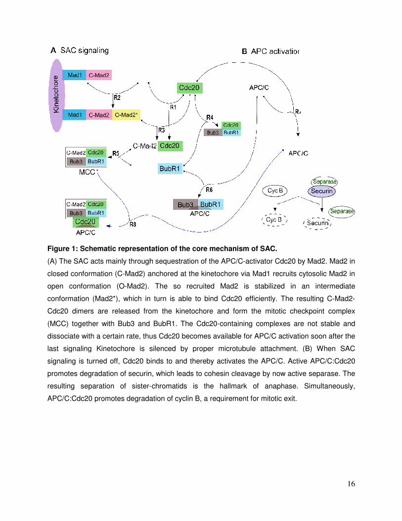
Figure 1: Schematic representation of the core mechanism of
(A) The SAC acts mainly through sequestration of the APC/C
closed conformation (C-Mad2) anchored at the kinetochore via Mad1 recruits cytosolic Mad2 in
open conformation (O-Mad2). The so recruited Mad2 is stabilized in an intermediate
conformation (Mad2*), which in turn is able to bind Cdc20 efficiently. The resulting C
Cdc20 dimers are released from the kinetochore and form the mitotic checkpoint complex
(MCC) together with Bub3 and BubR1. The Cdc20
dissociate with a certain rate, thus Cdc20 becomes available for APC/C activation soon after the
last signaling Kinetochore is silenced by proper microtubule attachment. (B) When SAC
signaling is turned off, Cdc20 binds to and thereby activates the APC/C. Act
promotes degradation of securin, which leads to cohesin cleavage by now active separase. The
resulting separation of sister-
APC/C:Cdc20 promotes degradation of cyclin B, a requirement
Schematic representation of the core mechanism of SAC.
through sequestration of the APC/C-activator Cdc20 by Mad2. Mad2 in
Mad2) anchored at the kinetochore via Mad1 recruits cytosolic Mad2 in
Mad2). The so recruited Mad2 is stabilized in an intermediate
(Mad2*), which in turn is able to bind Cdc20 efficiently. The resulting C
Cdc20 dimers are released from the kinetochore and form the mitotic checkpoint complex
(MCC) together with Bub3 and BubR1. The Cdc20-containing complexes are not stable and
sociate with a certain rate, thus Cdc20 becomes available for APC/C activation soon after the
last signaling Kinetochore is silenced by proper microtubule attachment. (B) When SAC
signaling is turned off, Cdc20 binds to and thereby activates the APC/C. Active APC/C
promotes degradation of securin, which leads to cohesin cleavage by now active separase. The
-chromatids is the hallmark of anaphase.
Cdc20 promotes degradation of cyclin B, a requirement for mitotic exit.
16
activator Cdc20 by Mad2. Mad2 in
Mad2) anchored at the kinetochore via Mad1 recruits cytosolic Mad2 in
Mad2). The so recruited Mad2 is stabilized in an intermediate
(Mad2*), which in turn is able to bind Cdc20 efficiently. The resulting C-Mad2-
Cdc20 dimers are released from the kinetochore and form the mitotic checkpoint complex
containing complexes are not stable and
sociate with a certain rate, thus Cdc20 becomes available for APC/C activation soon after the
last signaling Kinetochore is silenced by proper microtubule attachment. (B) When SAC
ive APC/C:Cdc20
promotes degradation of securin, which leads to cohesin cleavage by now active separase. The
chromatids is the hallmark of anaphase. Simultaneously,

Figure 2: Dynamical behavior of The columns from left to right showattachment occurs at t = 2000s). All results are presented for (MCC binding to APC). Parameters setting are according to Table 1. (left column) in all model variants is similar where its value at any given time is less than 30% of its initial concentration. The APvery similar, shows fast recovery and only with high MCC binding rate to APC shows fast inhibition for APC:Cdc20 activitymodel variants except the MCC-are able to sequester about 80% of the free Cdc20 only with low MCCMCC model variant that binds second Cdc20 the free Cdc20 and independent of MCC
Dynamical behavior of core SAC components concentration versus timeto right show the APC, Cdc20, and APC:Cdc20 concentration (spindle
attachment occurs at t = 2000s). All results are presented for different values of the rate k(MCC binding to APC). Parameters setting are according to Table 1. Free APC concentration (left column) in all model variants is similar where its value at any given time is less than 30% of its initial concentration. The APC:Cdc20 dynamics (right column) in all model variants is
shows fast recovery and only with high MCC binding rate to APC shows fast for APC:Cdc20 activity. Cdc20 sequestration is depicted in the middle column
-model variant that binds second Cdc20 (c.f. Panels A, B and D) are able to sequester about 80% of the free Cdc20 only with low MCC-APC binding rate. The MCC model variant that binds second Cdc20 (c.f. Panel C) is able to sequester athe free Cdc20 and independent of MCC-APC binding rate.
17
concentration versus time. :Cdc20 concentration (spindle different values of the rate k5
Free APC concentration (left column) in all model variants is similar where its value at any given time is less than 30% of
C:Cdc20 dynamics (right column) in all model variants is also shows fast recovery and only with high MCC binding rate to APC shows fast
middle column. All (c.f. Panels A, B and D) APC binding rate. The
is able to sequester around 95% of

Figure 3: Simulation of Mad2, BubR1, and Cdc20 mutations for each model variant.
For deletion we set the respective initial concentration to zero, and for over
higher. Towards proper wild type functioning, APC:Cdc20 concentration should be very low
(zero) before the attachment, and should increase quickly after attachment. Deletion of Mad2 or
BubR1 or an overexpression of Cdc20 leads to inability of the cell to arres
simulation, the concentration of APC:Cdc20 keeps high. Overexpression of Mad2 or BubR1 or
deleting Cdc20 results in arresting the cell, that is, the concentration of APC:Cdc20 is very low
or zero. Each row represents the mutation simulati
parameter rate for APC binding. Spindle attachment occurs at t = 2000s (switching parameter u
from 1 to 0). All parameters setting are according to Table 1. See text for more details.
Figure 3: Simulation of Mad2, BubR1, and Cdc20 mutations for each model variant.
For deletion we set the respective initial concentration to zero, and for over-expression 100folds
er. Towards proper wild type functioning, APC:Cdc20 concentration should be very low
(zero) before the attachment, and should increase quickly after attachment. Deletion of Mad2 or
BubR1 or an overexpression of Cdc20 leads to inability of the cell to arres
simulation, the concentration of APC:Cdc20 keeps high. Overexpression of Mad2 or BubR1 or
deleting Cdc20 results in arresting the cell, that is, the concentration of APC:Cdc20 is very low
or zero. Each row represents the mutation simulations of a model variant and a range of
parameter rate for APC binding. Spindle attachment occurs at t = 2000s (switching parameter u
from 1 to 0). All parameters setting are according to Table 1. See text for more details.
18
Figure 3: Simulation of Mad2, BubR1, and Cdc20 mutations for each model variant.
expression 100folds
er. Towards proper wild type functioning, APC:Cdc20 concentration should be very low
(zero) before the attachment, and should increase quickly after attachment. Deletion of Mad2 or
BubR1 or an overexpression of Cdc20 leads to inability of the cell to arrest, that is in the
simulation, the concentration of APC:Cdc20 keeps high. Overexpression of Mad2 or BubR1 or
deleting Cdc20 results in arresting the cell, that is, the concentration of APC:Cdc20 is very low
ons of a model variant and a range of
parameter rate for APC binding. Spindle attachment occurs at t = 2000s (switching parameter u
from 1 to 0). All parameters setting are according to Table 1. See text for more details.

Figure 4: Spatial simulation of concentrations over time for APC:Cdc20 for different values of the APC/C binding rates (kthe different APC/C binding rates, 10represent the simulations when Mad2 convection is included. (A) Outcome of the simulated MCC core modelabout 5 min to reach steady stateAPC:Cdc20 is 90% inhibited only with high MCCincluded. (B) Outcome of the simulated MCCIt takes about 3 min to reach steady state MCC model that binds second Cdc20 (Reactions (1)3 min to reach steady state for any parameter set. (D) Outcomemodel that binds second Cdc20 (Reactions (1)steady state for any parameter set.
Spatial simulation of SAC model variants. The figures show the total APC:Cdc20 with different parameter sets. All results are presented
for different values of the APC/C binding rates (k5, k6 and k8). Blue, black and red lines refer to APC/C binding rates, 106 M-1s-1, 108 M-1s-1 and 1010 M-1s-1 respectively. Dotted lines
represent the simulations when Mad2 convection is included. ) Outcome of the simulated MCC core model (Reactions (1)-(5), and (7); cf. Table 1). It takes
min to reach steady state except for the low rate value which takes 10 minutesAPC:Cdc20 is 90% inhibited only with high MCC-APC binding rate or when convection is
. (B) Outcome of the simulated MCC-BubR1 core model (Reactions (1)min to reach steady state for any parameter set. (C) Outcome of the simulated
MCC model that binds second Cdc20 (Reactions (1)-(5), (7),and (8); cf. Table 1). It takes about 3 min to reach steady state for any parameter set. (D) Outcome of the simulated MCCmodel that binds second Cdc20 (Reactions (1)-(8); cf. Table 1). It takes about 1.5 min to reach steady state for any parameter set.
19
The figures show the total All results are presented
Blue, black and red lines refer to respectively. Dotted lines
Table 1). It takes except for the low rate value which takes 10 minutes.
APC binding rate or when convection is BubR1 core model (Reactions (1)-(7); cf. Table 1).
for any parameter set. (C) Outcome of the simulated (5), (7),and (8); cf. Table 1). It takes about
of the simulated MCC-BubR1 (8); cf. Table 1). It takes about 1.5 min to reach

Figure 5: Spatial simulation of APC and Cdc20 dynamics.
concentrations over time for free APC and free Cdc20. All results are presented for 10
value of the APC/C binding rates (k
BubR1 model variant that binds second Cdc2
achieved.
Figure 5: Spatial simulation of APC and Cdc20 dynamics. The figures show the total
concentrations over time for free APC and free Cdc20. All results are presented for 10
value of the APC/C binding rates (k5, k6 and k8). We can clearly see that only for the MCC
BubR1 model variant that binds second Cdc20, complete APC and Cdc20 sequestration is
20
The figures show the total
concentrations over time for free APC and free Cdc20. All results are presented for 108 M-1s-1
). We can clearly see that only for the MCC-
0, complete APC and Cdc20 sequestration is

Figure 6: Sensitivities of the steady state concentrations of APC:Cdc20 associatcoefficients (k5 ) and (k7). Both parameters were varied in a range from 1.This analysis has been repeated for each model variants respectively). Each model variant was simulated 121 times, and each simulation run until steady state reached. Panel A and B (core model variants)C and D (where MCC binds a second Cdc20). The scan of the new model variants, Panel C and D, indicate that the MCC-APC binding rate must be at least 10Cdc20 binding rate must not exceed
Sensitivities of the steady state concentrations of APC:Cdc20 associatcoefficients (k5 ) and (k7). Both parameters were varied in a range from 100 M.This analysis has been repeated for each model variants (panels, A, B, C, and D,
Each model variant was simulated 121 times, and each simulation run until steady state reached. Panel A and B (core model variants) are very similar. The same C and D (where MCC binds a second Cdc20). The scan of the new model variants, Panel C and
APC binding rate must be at least 105 M-1s-1 and meanwhile APCCdc20 binding rate must not exceed 106 M-1s-1.
21
Sensitivities of the steady state concentrations of APC:Cdc20 associated with rate M-1s-1 to 1011 M-1s-
, B, C, and D, Each model variant was simulated 121 times, and each simulation run until steady
are very similar. The same is true for panel C and D (where MCC binds a second Cdc20). The scan of the new model variants, Panel C and
and meanwhile APC-

22
Table 1: Model parameters
Parameters Remarks
Rate constants k1 1 x 10³ M-1s-1 [21,65]
k2 2 x 10⁵ M-1s-1 [35,63]
k3 1 x 10⁷ M-1s-1 [21]
k4 2 x 10⁴ M-1s-1 [21,29]
k5 103-109 M-1s-1 [20,29]
k6 103-109 M-1s-1 This study
k7 5 x 10⁶ M-1s-1 [20,22]
k8 103-109 M-1s-1 This study
k-1 1 x 10⁻² s-1 [21]
k-2 2 x 10⁻¹ s-1 [63]
k-3 0 s-1 [21]
k-4 2 x 10⁻² s-1 [21,29]
k-5 1 x 10⁻¹ s-1 [29]
k-6 1 x 10⁻² s-1 This study
k-7 1 x 10⁻¹ s-1 [20,22]
k-8 8 x 10⁻² s-1 This study
Initial amount Cdc20 0.22 µM [41,66]
O-Mad2 0.15 µM [63]
Mad1:C-Mad2 0.05 µM [34]
BubR1:Bub3 0.13 µM [20,41,67]
APC 0.09 µM [66]
Other species start from zero
Diffusion constants Cdc20 19.5 µm
2s
−1 [68]
O-Mad2 5 µm2s
−1 [29]
Mad1:C-Mad2 0 µm2s
−1 [29]
Mad1:C-Mad2:Mad2* 0 µm2s
−1 [29]
Bub3:BubR1 4 µm2s
−1 [16,69]
APC 1.8 µm2s
−1 [68]
Other species diffusion coefficients are calculated
from D�� =��∗ �
�� �
, where DA
and DB are the diffusion coefficient for A and B, respectively.
This study
Convection constant O-Mad2 10 µms
−1 [29]
Environment radius of the kinetochore 0.1 µm [70] radius of the cell 10 µm [29]

23
Table 2: In-silico mutation experiments for validation
Species
Exp
.
Experimental effects Effects in the model variants
MCC core
MCC-BubR1 core
MCC extended
MCC-BubR1 extended
BubR1 D SAC dysfunction [53-56] Failed to arrest
Failed to arrest
Failed to arrest
Failed to arrest
BubR1 O Chromosomal instability [57] Arrested k5 = 10
5
Arrested
Arrested k5 >= 10
4 Arrested
Mad2 D Cells are unable to arrest and impaired SAC (e.g., [11,47-50])
Failed to arrest
Failed to arrest k5 = 10
6 Failed to arrest k5 = 10
4 Failed to arrest k5 = 10
4
Mad2 O Activates the SAC and blocks mitosis and stabilizes microtubule attachment [34,51-52]
Arrested
Arrested
Arrested
Arrested
Cdc20 D Cells arrested in metaphase [58-60] Arrested k5 = 10
5 Arrested
Arrested
Arrested
Cdc20 O Impairment SAC and allows cells with a depolymerized spindle or damaged DNA to leave mitosis [61-62].
Failed to arrest
Failed to arrest
Failed to arrest k5 =< 10
4
Failed to arrest k5 =< 10
4
D refers to deletion or knockdown experiment, and O refers to an over-expression experiment. Failed to arrest means very high level of [APC:Cdc20] and low sequestration level of Cdc20. Arrested means very low level of [APC:Cdc20] and fully sequestration of Cdc20. Green means fully consistent with experiments and capture the desire behaviour. Yellow means consistent with experiments but required specific MCC-APC binding rate (see text for details)